Feeds:
Posts
Comments

Archive for the ‘Proteomics’ Category

The Metabolic View of Epigenetic Expression

Writer and Curator: Larry H Bernstein, MD, FCAP

Introduction

This is the fifth contribution to a series of articles on cancer, genomics, and metabolism.   I begin this after reading an article by Stephen Williams “War on Cancer May Need to Refocus Says Cancer Expert on NPR”, and after listening to NPR “On the Media”. This is an unplanned experience, perhaps partly related to an Op-Ed in the New York Times two days before by Angelina Jolie Pittman.  Taking her article prior to pre-emptive breast surgery for the BRCA1 mutation two years ago and her salpingo-oophorectomy at age 39 years with her family history, and her adoption of several children even prior to her marriage to Brad Pitt, reveals an unusual self-knowledge as well as perspective on the disease risk balanced with her maternal instincts.  I sense (but don’t know) that she had a good knowledge not stated about the estrogen sensitivity of breast cancer for some years, and balanced that knowledge in her life decisions.

Tracing the history of cancer and the Lyndon Johnson initiated “War on Cancer” the initiative is presented as misguided.  Moreover, the imbalance is posed aas focused overly on genomics, and there is an imbalnced in the attention to the types of cancer, bladder cancer (urothelial) receiving too little attention. However, the events that drive this are complex, and not surprising.  The funding is driven partly by media attention (a film star or President’s wife) and not to be overlooked, watch where the money flows.  People who have the ability to donate and also have a family experience will give, regardless of the statistics because it is 100 percent in their eyes.

Insofar as the scientific endeavor goes, young scientists are committed to a successful research career, and they also need funding, so they have to balance the risk of success and failure in the choice of problems they choose to work on.  But until the 20th century, the biological sciences were largely descriptive. The emergence of a “Molecular Biology” is a unique 20th century development. The work of Pathology – pioneered by Rokitansky, Virchow, and to an extent also the anatomist/surgeon John Harvey – was observational science.  The description of Hodgkin’s lymphoma was observational, and it was a breakthrough in medicine.

With the emergence of genomics from biochemistry and genetics in molecular biology (biology at the subcellular level), a part of medicine that was well founded became an afterthought.  After all, after many years of the history of medicine and pathology, it is well known that cancers are not only a dysmetabolism of cellular replication and cellular regulation, but cancers have a natural history related to organ system, tissue specificity, sex, and age of occurrence. This should be well known to the experienced practitioner, but not necessarily to the basic researcher with no little clinical exposure.  Consequently, it was quite remarkable to me to find that the truly amazing biochemist who gave a “Harvey Lecture” at Harvard on the pyridine nucleotide transhydrogenases, and who shared in the discovery of Coenzyme A, had made the observation that organs that are primarily involved with synthetic activity -adrenal, pituitary, and thyroid, testis, ovary, breast (most notably) – have a more benign course than those of stomach, colon, pancreas, melanoma, hematopoietic, and sarcomas. The liver is highly synthetic, but doesn’t fit so nicely because of the role in detoxification and the large role in glucose and fat catabolism.  Further, this was at a time that we knew nothing about the cell death pathway and cellular repair, and how is it in concert with cell proliferation.

The first important reasoning about cancer metabolism was opened by Otto Warburg in the late 1920s.  I have  little reason to doubt his influence on Nathan Kaplan, who used the terms DPN(+/H) and TPN(+/H), disregarding the terms NAD(+/H) and NADP(+/H), although I was told it was because of the synthesis of the pyridine nucleotide adducts for study (APDPN, etc.).

In a recent article, I had an interesting response from Jose ES Rosalino:

In mRNA Translation and Energy Metabolism in Cancer

Topisirovic and N. Sonenberg – Cold Spring Harbor Symposia on Quantitative Biology, Volume LXXVI – http://dx.doi.org:/10.1101/sqb.2011.76.010785

“A prominent feature of cancer cells is the use of aerobic glycolysis under conditions in which oxygen levels are sufficient to support energy production in the mitochondria (Jones and Thompson 2009; Cairns et al. 2010). This phenomenon, named the “Warburg effect,” after its discoverer Otto Warburg, is thought to fuel the biosynthetic requirements of the neoplastic growth (Warburg 1956; Koppenol et al. 2011) and has recently been acknowledged as one of the hallmarks of cancer (Hanahan and Weinberg 2011). mRNA translation is the most energy-demanding process in the cell (Buttgereit and Brand 1995). Again, the use of aerobic glycolysis expression has being twisted.”

To understand my critical observation consider this: Aerobic glycolysis is the carbon flow that goes from Glucose to CO2 and water (includes Krebs cycle and respiratory chain for the restoration of NAD, FAD etc.

Anerobic glyclysis is the carbon flow that goes from glucose to lactate. It uses conversion of pyruvate to lactate to regenerate NAD.

“Pasteur effect” is an expression coined by Warburg it refers to the reduction in the carbon flow from glucose when oxygen is offered to yeasts. The major reason for that is in general terms, derived from the fact that carbon flow is regulated by several cell requirements but majorly by the ATP needs of the cell. Therefore, as ATP is generated 10 more efficiently in aerobiosis than under anaerobiosis, less carbon flow is required under aerobiosis than under anaerobiosis to maintain ATP levels. Warburg, after searching for the same regulatory mechanism in normal and cancer cells for comparison found that transformed cell continued their large flow of glucose carbons to lactate despite of the presence of oxygen.

So, it is wrong to describe that aerobic glycolysis continues in the presence of oxygen. It is what it is expected to occur. The wrong thing is that anaerobic glycolysis continues under aerobiosis.

In our discussion of transcription and cell regulatory processes, we have already encountered a substantial amount of “enzymology” that drives what is referred to as “epigenetics”.  Enzymatic reactions are involved almost everywhere we look at the processes involved in RNA nontranscriptional affairs.

Enzyme catalysis

Pyruvate carboxylase is critical for non–small-cell lung cancer proliferation
K Sellers,…, TW-M Fan
J Clin Invest. Jan 2015; xx
http://dx.doi.org:/10.1172/JCI72873

Anabolic biosynthesis requires precursors supplied by the Krebs cycle, which in turn requires anaplerosis to replenish precursor intermediates. The major anaplerotic sources are pyruvate and glutamine, which require the activity of pyruvate carboxylase (PC) and glutaminase 1 (GLS1), respectively. Due to their rapid proliferation, cancer cells have increased anabolic and energy demands; however, different cancer cell types exhibit differential requirements for PC- and GLS-mediated pathways for anaplerosis and cell proliferation. Here, we infused patients with early-stage non–small-cell lung cancer (NSCLC) with uniformly 13C-labeled glucose before tissue resection and determined that the cancerous tissues in these patients had enhanced PC activity. Freshly resected paired lung tissue slices cultured in 13C6-glucose or 13C5, 15N2-glutamine tracers confirmed selective activation of PC over GLS in NSCLC. Compared with noncancerous tissues, PC expression was greatly enhanced in cancerous tissues, whereas GLS1 expression showed no trend. Moreover, immunohistochemical analysis of paired lung tissues showed PC overexpression in cancer cells rather than in stromal cells of tumor tissues. PC knockdown induced multinucleation, decreased cell proliferation and colony formation in human NSCLC cells, and reduced tumor growth in a mouse xenograft model. Growth inhibition was accompanied by perturbed Krebs cycle activity, inhibition of lipid and nucleotide biosynthesis, and altered glutathione homeostasis. These findings indicate that PC-mediated anaplerosis in early stage NSCLC is required for tumor survival and proliferation.

Accelerated glycolysis under aerobic conditions (the “Warburg effect”) has been a hallmark of cancer for many decades (1). It is now recognized that cancer cells must undergo many other metabolic reprograming changes (2) to meet the increased anabolic and energetic demands of proliferation (3, 4). It is also becoming clear that different cancer types may utilize a variety of metabolic adaptations that are context dependent, commensurate with the notion that altered metabolism is a hallmark of cancer (12). Enhanced glucose uptake and aerobic glycolysis generates both energy (i.e., ATP) and molecular precursors for the biosynthesis of complex carbohydrates, sugar nucleotides, lipids, proteins, and nucleic acids. However, increased glycolysis alone is insufficient to meet the total metabolic demands of proliferating cancer cells. The Krebs cycle is also a source of energy via the oxidation of pyruvate, fatty acids, and amino acids such as glutamine. Moreover, several Krebs cycle intermediates are essential for anabolic and glutathione metabolism, including citrate, oxaloacetate, and α-ketoglutarate (Figure 1A).

Figure 1. PC is activated in human NSCLC tumors. (A) PC and GLS1 catalyze the major anaplerotic inputs (blue) into the Krebs cycle to support the anabolic demand for biosynthesis (green). Also shown is the fate of 13C from 13C6-glucose through glycolysis and into the Krebs cycle via PC (red).
(B) Representative Western blots of PC and GLS1 protein expression levels in human NC lung (N) and NSCLC (C) tissues. (C) Pairwise PC and GLS1 expression (n = 86) was normalized to α-tubulin and plotted as the log10 ratio of CA/NC tissues. For PC, nearly all log ratios were positive (82 of 86), with a clustering in the 0.5–1 range (i.e., typically 3- to 10-fold higher expression in the tumor tissue; Wilcoxon test, P < 0.0001). In contrast, GLS1 expression was nearly evenly distributed between positive and negative log10 ratios and showed no statistically significant difference between the CA and NC tissues (Wilcoxon test, P = 0.213). Horizontal bar represents the median. (D) In vivo PC activity was enhanced in CA tissue compared with that in paired NC lung tissues (n = 34) resected from the same human patients given 13C6-glucose 2.5–3 hours before tumor resection. PC activity was inferred from the enrichment of 13C3-citrate (Cit+3), 13C5-Cit (Cit+5), 13C3-malate (Mal+3), and 13C3-aspartate (Asp+3) as determined by GC-MS. *P < 0.05 and **P < 0.01 by paired Student t test. Error bars represent the SEM.

Continued functioning of the Krebs cycle requires the replenishment of intermediates that are diverted for anabolic uses or glutathione synthesis. This replenishment process, or anaplerosis, is accomplished via 2 major pathways: glutaminolysis (deamidation of glutamine via glutaminase [GLS] plus transamination of glutamate to α-ketoglutarate) and carboxylation of pyruvate to oxaloacetate via ATP-dependent pyruvate carboxylase (PC) (EC 6.4.1.1) (refs. 3, 20, 21, and Figure 1A). The relative importance of these pathways is likely to depend on the nature of the cancer and its specific metabolic adaptations, including those to the microenvironment (20, 22). For example, glutaminolysis was shown to be activated in the glioma cell line SF188, while PC activity was absent, despite the high PC activity present in normal astrocytes. However, SF188 cells use PC to compensate for GLS1 suppression or glutamine restriction (20), and PC, rather than GLS1, was shown to be the major anaplerotic input to the Krebs cycle in primary glioma xenografts in mice. It is also unclear as to the relative importance of PC and GLS1 in other cancer cell types or, most relevantly, in human tumor tissues in situ. Our preliminary evidence from 5 non–small-cell lung cancer (NSCLC) patients indicated that PC expression and activity are upregulated in cancerous (CA) compared with paired noncancerous (NC) lung tissues (21), although it was unclear whether PC activation applies to a larger NSCLC cohort or whether PC expression was associated with the cancer and/or stromal cells

Here, we have greatly extended our previous findings (21) in a larger cohort (n = 86) by assessing glutaminase 1 (GLS1) status and analyzing in detail the biochemical and phenotypic consequences of PC suppression in NSCLC. We found PC activity and protein expression levels to be, on average, respectively, 100% and 5- to 10-fold higher in cancerous (CA) lung tissues than in paired NC lung tissues resected from NSCLC patients, whereas GLS1 expression showed no significant trend. We have also applied stable isotope–resolved metabolomic (SIRM) analysis to paired freshly resected CA and NC lung tissue slices in culture (analogous to the Warburg slices; ref. 25) using either [U-13C] glucose or [U-13C,15N] glutamine as tracers. This novel method provided information about tumor metabolic pathways and dynamics without the complication of whole-body metabolism in vivo.

PC expression and activity, but not glutaminase expression, are significantly enhanced in early stages of malignant NSCLC tumors. PC protein expression was significantly higher in primary NSCLC tumors than in paired adjacent NC lung tissues (n = 86, P < 0.0001, Wilcoxon test) (Figure 1, B and C). The median PC expression was 7-fold higher in the tumor, and the most probable (modal) overexpression in the tumor was approximately 3-fold higher (see Supple-mental Table 1; supplemental material available online with this article; http://dx.doi.org:/10.1172/JCI72873DS1). We found that PC expression was also higher in the tumor tissue compared with that detected in the NC tissue in 82 of 86 patients. In contrast, GLS1 expression was not significantly different between the tumor and NC tissues (P = 0.213, Wilcoxon test) (Figure 1C and Supplemental Table 1). The 13C3-Asp produced from 13C6-glucose (Figure 1A) infused into NSCLC patients was determined by gas chromatography–mass spectrometry (GC-MS) to estimate in vivo PC activity. A bolus injection of 10 g 13C6-glucose in 50 ml saline led to an average of 44% 13C enrichment in the plasma glucose immediately after infusion (Supplemental Table 2). Because the labeled glucose was absorbed by various tissues over the approximately 2.5 hours between infusion and tumor resection, plasma glucose enrichment dropped to 17% (Supplemental Table 2). The labeled glucose in both CA and NC lung tissues was metabolized to labeled lactate, but this occurred to a much greater extent in the CA tissues (Supplemental Figure 1A), which indicates accelerated glycolysis in these tissues.

Fresh tissue (Warburg) slices confirm enhanced PC and Krebs cycle activity in NSCLC. To further assess PC activity relative to GLS1 activity in human lung tissues, thin (<1 mm thick) slices of paired CA and NC lung tissues freshly resected from 13 human NSCLC patients were cultured in 13C6-glucose or 13C5,15N2-glutamine for 24 hours. These tissues maintain biochemical activity and histological integrity for at least 24 hours under culture conditions (Figure 2A, Supplemental Figure 2, A and B, and ref. 26). When the tissues were incubated with 13C6-glucose, CA slices showed a significantly greater percentage of enrichment in glycolytic 13C3-lactate (3 in Figure 2B) than did the NC slices, indicative of the Warburg effect. In addition, the CA tissues had significantly higher fractions of 13C4-, 13C5-, and 13C6-citrate (4, 5, and 6 of citrate, respectively, in Figure 2B) than did the NC tissues. These isotopologs require the combined action of PDH, PC, and multiple turns of the Krebs cycle (Figure 2C). Consistent with the labeled citrate data, the increase in the percentage of enrichment of 13C3-, 13C4-, and 13C5-glutamate (3, 4, and 5 of glutamate, respectively, in Figure 2B) in the CA tissues indicates enhanced Krebs cycle and PC activity.

Figure 2. Ex vivo CA lung tissue slices have enhanced oxidation of glucose through glycolysis and the Krebs cycle with and without PC input compared with that of paired NC lung slices. Thin slices of CA and NC lung tissues freshly resected from 13 human NSCLC patients were incubated with 13C6-glucose for 24 hours as described in the Methods. The percentage of enrichment of lactate, citrate, glutamate, and aspartate was determined by GC-MS. (A) 1H{13C} HSQC NMR showed an increase in labeled lactate, glutamate, and aspartate. In addition, CA tissues had elevated 13C abundance in the ribose moiety of the adenine-containing nucleotides (1′-AXP), indicating that the tissues were viable and had enhanced capacity for nucleotide synthesis. (B) CA tissue slices (n = 13) showed increased glucose metabolism through glycolysis based on the increased percentage of enrichment of 13C3-lactate (“3”), and through the Krebs cycle based on the increased percentage of enrichment of 13C4–6-citrate (“4–6”) and 13C3–5-glutamate (“3–5”) (see 13C fate tracing in C). *P < 0.05 and **P < 0.01 by paired Student’s t test. Error bars represent the SEM. (C) An atom-resolved map illustrates how PC, PDH, and 2 turns of the Krebs cycle activity produced the 13C isotopologs of citrate and glutamate in B, whose enrichment were significantly enhanced in CA tissue slices.

Figure 4. PC suppression via shRNA inhibits proliferation and tumorigenicity of human NSCLC cell lines in vitro and in vivo. Proliferation and colony-formation assays were initiated 1 week after transduction and selection with puromycin. A549 xenograft in NSG mice was performed 8 days after transduction. *P < 0.01, **P < 0.001, ***P < 0.0001, and ****P < 0.00001 by Student t test, assuming unequal variances. Error bars represent the SEM. (A) NSCLC cells lines were transduced with shPC55 or shEV. Proliferation assays (n = 6) revealed substantial growth inhibition induced by PC knockdown in all 5 cell lines after a relatively long latency period. (B) Colony-formation assays indicated that PC knockdown reduced the capacity of A549 and PC9 cells to form colonies in soft agar (n = 3). (C) Tumor xenografts from shPC55-transduced A549 cells showed a 2-fold slower growth rate than did control shEV tumors (P < 0.001 by the unpaired Welch version of the t test). Tumor size was calculated as πab/4, where a and b are the x,y diameters. Each point represents an average of 6 mice. The solid lines are the nonlinear regression fits to the equation: size = a + bt2, as described in the Methods. (D) The extent of PC knockdown in the mouse xenografts (n = 6) was lesser than that in cell cultures, leading to less attenuation of PC expression (30%–60% of control) and growth inhibition. In addition, PC expression in the excised tumors correlated with the individual growth rates, as determined by Pearson’s correlation coefficient.

Fatty acyl synthesis from 13C5-glutamine (“Even” in Figure 6B) via glutaminolysis and the Krebs cycle was greatly attenuated in PC-suppressed cells. Taken together, these results suggest that PC knockdown severely inhibits lipid production by blocking the biosynthesis of fatty acyl components but not the glucose-derived glycerol backbone. This is consistent with decreased Krebs cycle activity (Figure 5), which in turn curtails citrate export from the mitochondria to supply the fatty acid precursor acetyl CoA in the cytoplasm.

Figure 5. PC knockdown perturbs glucose and glutamine flux through the Krebs cycle. 13C Isotopolog concentrations were determined by GC-MS (n = 3). Values represent the averages of triplicates, with standard errors. *P < 0.05, **P < 0.01, ***P < 0.001, and ****P < 0.0001 by Student’s t test, assuming unequal variances. The experiments were repeated 3 times. (A) A549 cells were transduced with shPC55 for 10 days before incubation with 13C6-glucose for 24 hours. As expected, the 13C isotopologs of Krebs cycle metabolites produced via PC and Krebs cycle activity were depleted in PC-deficient cells (tracked by blue dots in the atom-resolved map and blue circles in the bar graphs; see also Figure 2C). In addition, 13C6-glucose metabolism via PDH was also perturbed (indicated by red dots and circles). (B) Treatment of PC-knockdown cells with 13C5,15N2-glutamine revealed that anaplerotic input via GLS did not compensate for the loss of PC activity, since GLS activity was attenuated, as inferred from the activity markers (indicated by red dots and circles). Decarboxylation of glutamine-derived malate by malic enzyme (ME) and reentry of glutamine-derived pyruvate into the Krebs cycle via PC or PDH (shown in blue and green, respectively) were also attenuated. Purple diamonds denote 15N; black diamonds denote 14N.

Figure 6. PC suppression hinders Krebs cycle–fueled biosynthesis. (A) 13C atom–resolved pyrimidine biosynthesis from 13C6-glucose and 13C5-glutamine is depicted with a 13C5-ribose moiety (red dots) produced via the pentose phosphate pathway (PPP) and 13C1-3  uracil ring (blue dots) derived from  13C2-4-aspartate produced via the Krebs cycle or the combined action of ME and PC (blue dots). A549 cells transduced with shPC55 or shEV were incubated with 13C6-glucose or 13C5-glutamine for 24 hours. Fractional enrichment of UTP and CTP isotopologs from FT-ICR-MS analysis of polar cell extracts showed reduced enrichment of 13C6-glucose–derived 13C5-ribose (the “5” isotopolog) and 13C6-glucose– or 13C5-glutamine–derived 13C1-3-pyrimidine rings (the “6–8” or “1–3” isotopologs, highlighted by dashed green rectangles; for the “6–8” isotopologs, 5 13Cs arose from ribose and 1–3 13Cs from the ring) (10, 45). These data suggest that PC knockdown inhibits de novo pyrimidine biosynthesis from both glucose and glutamine. (B) Glucose and glutamine carbons enter fatty acids via citrate. FT-ICR-MS analysis of labeled lipids from the nonpolar cell extracts showed that PC knockdown severely inhibited the incorporation of glucose and glutamine carbons into the fatty acyl chains (even) and fatty acyl chains plus glycerol backbone (odd >3) of phosphatidylcholine lipids. However, synthesis of the 13C3-glycerol backbone (the “3” isotopolog) or its precursor 13C3-α-glycerol-3-phosphate (αG3P, m+3) from 13C6-glucose was enhanced rather than inhibited by PC knockdown. These data suggest that PC suppression specifically hinders fatty acid synthesis in A549 cells. Values represent the averages of triplicates (n = 3), with standard errors. *P < 0.05, **P < 0.01,  and ***P < 0.001 by Student’s t test, assuming unequal variances.

De novo glutathione synthesis was analyzed by 1H{13C} HSQC NMR. Glutathione synthesis from both glucose and glutamine was suppressed by PC knockdown (Supplemental Figure 9, A and B). Reduced de novo synthesis led to a large decrease in the total level of reduced glutathione (GSH; Supplemental Figure 12, A and B). At the same time, PC-knockdown cells accumulated slightly more oxidized GSH (GSSG; Supplemental Figure 12, A and B), leading to a significantly reduced GSH/GSSG ratio both in cell culture and in vivo (Supplemental Figure 12C). To determine whether this perturbation of glutathione homeostasis compromises the ability of PC-suppressed cells to handle oxidative stress, we measured ROS production by DCFDA fluorescence. PC-knockdown cells had over 70% more basal ROS than did control cells (0 mM H2O2; Supplemental Figure 12D). When cells were exposed to increasing concentrations of H2O2, the knockdown cells were less able to quench ROS, as they produced up to 300% more ROS than did control cells (Supplemental Figure 12D). However, N-acetylcysteine (NAC) at 10 mM did not rescue the growth of PC-knockdown cells, suggesting that such a growth effect is not simply related to an inability to regenerate GSH from GSSG. Altogether, these results show that PC suppression compromises anaplerotic input into the Krebs cycle, which in turn reduces the activity of the Krebs cycle, while limiting the ability of A549 cells to synthesize nucleotides, lipids, and glutathione. These downstream effects of PC knockdown were also evident when comparing the metabolism of shPC55-transduced A549 cells against that of A549 cells transduced with a scrambled vector (shScr) (Supplemental Figure 13), which suggests that they are on-target effects of PC knockdown.

  1. Warburg O. On the origin of cancer cells. Science. 1956;123(3191):309–314. 2. Dang CV, Semenza GL. Oncogenic alterations of metabolism. Trends Biochem Sci. 1999; 24(2):68–72.
    3. Fan TW, et al. Rhabdomyosarcoma cells show an energy producing anabolic metabolic phenotype compared with primary myocytes. Mol Cancer. 2008;7:79.
    4. Vander Heiden MG, Cantley LC, Thompson CB. Understanding the Warburg effect: the metabolic requirements of cell proliferation. Science. 2009;324(5930):1029–1033.
    10. Le A, et al. Glucose-independent glutamine metabolism via TCA cycling for proliferation and survival in B cells. Cell Metab. 2012;15(1):110–121
    20. Cheng T, et al. Pyruvate carboxylase is required for glutamine-independent growth of tumor cells. Proc Natl Acad Sci U S A. 2011;108(21):8674–8679.
    21. Fan TW, et al. Altered regulation of metabolic pathways in human lung cancer discerned by (13) C stable isotope-resolved metabolomics (SIRM). Mol Cancer. 2009;8:41.
    22. Marin-Valencia I, et al. Analysis of tumor metabolism reveals mitochondrial glucose oxidation in genetically diverse human glioblastomas in the mouse brain in vivo. Cell Metab. 2012;15(6):827–837.
    25. Warburg O. Versuche an überlebendem Carcinomgewebe (Methoden). Biochem Zeitschr. 1923;142:317–333.
    45. Lorkiewicz P, Higashi RM, Lane AN, Fan TW. High information throughput analysis of nucleotides and their isotopically enriched isotopologues by direct-infusion FTICR-MS. Metabolomics. 2012;8(5):930–939.

In vivo HIF-mediated reductive carboxylation is regulated by citrate levels and sensitizes VHL-deficient cells to glutamine deprivation.
Gameiro PA, Yang J, Metelo AM,…, Stephanopoulos G, Iliopoulos O.
Cell Metab. 2013 Mar 5; 17(3):372-85.
http://dx.doi.org:/10.1016/j.cmet.2013.02.002

Hypoxic and VHL-deficient cells use glutamine to generate citrate and lipids through reductive carboxylation (RC) of α-ketoglutarate. To gain insights into the role of HIF and the molecular mechanisms underlying RC, we took advantage of a panel of disease-associated VHL mutants and showed that HIF expression is necessary and sufficient for the induction of RC in human renal cell carcinoma (RCC) cells. HIF expression drastically reduced intracellular citrate levels. Feeding VHL-deficient RCC cells with acetate or citrate or knocking down PDK-1 and ACLY restored citrate levels and suppressed RC. These data suggest that HIF-induced low intracellular citrate levels promote the reductive flux by mass action to maintain lipogenesis. Using [(1-13)C]glutamine, we demonstrated in vivo RC activity in VHL-deficient tumors growing as xenografts in mice. Lastly, HIF rendered VHL-deficient cells sensitive to glutamine deprivation in vitro, and systemic administration of glutaminase inhibitors suppressed the growth of RCC cells as mice xenografts.

Cancer cells undergo fundamental changes in their metabolism to support rapid growth, adapt to limited nutrient resources, and compete for these supplies with surrounding normal cells. One of the metabolic hallmarks of cancer is the activation of glycolysis and lactate production even in the presence of adequate oxygen. This is termed the Warburg effect, and efforts in cancer biology have revealed some of the molecular mechanisms responsible for this phenotype (Cairns et al., 2011). More recently, 13C isotopic studies have elucidated the complementary switch of glutamine metabolism that supports efficient carbon utilization for anabolism and growth (DeBerardinis and Cheng, 2010). Acetyl-CoA is a central biosynthetic precursor for lipid synthesis, being generated from glucose-derived citrate in well-oxygenated cells (Hatzivassiliou et al., 2005). Warburg-like cells, and those exposed to hypoxia, divert glucose to lactate, raising the question of how the tricarboxylic acid (TCA) cycle is supplied with acetyl-CoA to support lipogenesis. We and others demonstrated, using 13C isotopic tracers, that cells under hypoxic conditions or defective mitochondria primarily utilize glutamine to generate citrate and lipids through reductive carboxylation (RC) of α-ketoglutarate by isocitrate dehydrogenase 1 (IDH1) or 2 (IDH2) (Filipp et al., 2012; Metallo et al., 2012; Mullen et al., 2012; Wise et al., 2011).

The transcription factors hypoxia inducible factors 1α and 2α (HIF-1α, HIF-2α) have been established as master regulators of the hypoxic program and tumor phenotype (Gordan and Simon, 2007; Semenza, 2010). In addition to tumor-associated hypoxia, HIF can be directly activated by cancer-associated mutations. The von Hippel-Lindau (VHL) tumor suppressor is inactivated in the majority of sporadic clear-cell renal carcinomas (RCC), with VHL-deficient RCC cells exhibiting constitutive HIF-1α and/or HIF-2α activity irrespective of oxygen availability (Kim and Kaelin, 2003). Previously, we showed that VHL-deficient cells also relied on RC for lipid synthesis even under normoxia. Moreover, metabolic profiling of two isogenic clones that differ in pVHL expression (WT8 and PRC3) suggested that reintroduction of wild-type VHL can restore glucose utilization for lipogenesis (Metallo et al., 2012). The VHL tumor suppressor protein (pVHL) has been reported to have several functions other than the well-studied targeting of HIF. Specifically, it has been reported that pVHL regulates the large subunit of RNA polymerase (Pol) II (Mikhaylova et al., 2008), p53 (Roe et al., 2006), and the Wnt signaling regulator Jade-1. VHL has also been implicated in regulation of NF-κB signaling, tubulin polymerization, cilia biogenesis, and proper assembly of extracellular fibronectin (Chitalia et al., 2008; Kim and Kaelin, 2003; Ohh et al., 1998; Thoma et al., 2007; Yang et al., 2007). Hypoxia inactivates the α-ketoglutarate-dependent HIF prolyl hydroxylases, leading to stabilization of HIF. In addition to this well-established function, oxygen tension regulates a larger family of α-ketoglutarate-dependent cellular oxygenases, leading to posttranslational modification of several substrates, among which are chromatin modifiers (Melvin and Rocha, 2012). It is therefore conceivable that the effect of hypoxia on RC that was reported previously may be mediated by signaling mechanisms independent of the disruption of the pVHL-HIF interaction. Here we

  • demonstrate that HIF is necessary and sufficient for RC,
  • provide insights into the molecular mechanisms that link HIF to RC,
  • detected RC activity in vivo in human VHL-deficient RCC cells growing as tumors in nude mice,
  • provide evidence that the reductive phenotype of VHL-deficient cells renders them sensitive to glutamine restriction in vitro, and
  • show that inhibition of glutaminase suppresses growth of VHL-deficient cells in nude mice.

These observations lay the ground for metabolism-based therapeutic strategies for targeting HIF-driven tumors (such as RCC) and possibly the hypoxic compartment of solid tumors in general.

HIF Inactivation Is Necessary for Downregulation of Reductive Carboxylation by pVHL

(A) Expression of HIF-1 α, HIF-2α, and their target protein GLUT1 in UMRC2-derived cell lines, as indicated.

(B) Carbon atom transition map: the fate of [1-13C1] and [5-13C1]glutamine used to trace reductive carboxylation in this work (carbon atoms are represented by circles). The [1-13C1] (green circle) and [5-13C1] (red circle) glutamine-derived isotopic labels are retained during the reductive TCA cycle (bold red pathway). Metabolites containing the acetyl-CoA carbon skeleton are highlighted by dashed circles.

(C) Relative contribution of reductive carboxylation.

(D and E) Relative contribution of glucose oxidation to the carbons of indicated metabolites (D) and citrate (E). Student’s t test compared VHL-reconstituted to vector-only or to VHL mutants (Y98N/Y112N). Error bars represent SEM. Pyr, pyruvate; Lac, lactate; AcCoA, acetyl-CoA, Cit, citrate; IsoCit, isocitrate; Akg, α-ketoglutarate; Suc, succinate; Fum, fumarate; Mal, malate; OAA, oxaloacetate; Asp, aspartate; Glu, glutamate; PDH, pyruvate dehydrogenase; ME, malic enzyme; IDH, isocitrate dehydrogenase enzymes; ACO, aconitase enzymes; ACLY, ATP-citrate lyase; GLS, glutaminase.

To test the effect of HIF activation on the overall glutamine incorporation in the TCA cycle, we labeled an isogenic pair of VHL-deficient and VHL-reconstituted UMRC2 cells with [U-13C5]glutamine, which generates M4 fumarate, M4 malate, M4 aspartate, and M4 citrate isotopomers through glutamine oxidation. As seen in Figure S1B, VHL-deficient/VHL-positive UMRC2 cells exhibit similar enrichment of M4 fumarate, M4 malate, and M4 asparate (but not citrate) showing that VHL-deficient cells upregulate reductive carboxylation without compromising oxidative metabolism from glutamine. Next, we tested whether HIF inactivation by pVHL is necessary to regulate the reductive utilization of glutamine for lipogenesis. To this end, we traced the relative incorporation of [U-13C6]glucose or [5-13C1]glutamine into palmitate. Labeled carbon derived from [5-13C1]glutamine can be incorporated into fatty acids exclusively through RC, and the labeled carbon cannot be transferred to palmitate through the oxidative TCA cycle (Figure 1B, red carbons). Tracer incorporation from [5-13C1]glutamine occurs in the one carbon (C1) of acetyl-CoA, which results in labeling of palmitate at M1, M2, M3, M4, M5, M6, M7, and M8 mass isotopomers. In contrast, lipogenic acetyl-CoA molecules originating from [U-13C6]glucose are fully labeled, and the labeled palmitate is represented by M2, M4, M6, M8, M10, M12, M14, and M16 mass isotopomers. VHL-deficient control cells and cells expressing pVHL type 2B mutants exhibited high palmitate labeling from the [5-13C1]glutamine; conversely, reintroduction of wild-type or type 2C pVHL mutant (L188V) resulted in high labeling from [U-13C6]glucose (Figures 2A and 2B, box inserts highlight the heavier mass isotopomers).

hif-inactivation-is-necessary-for-downregulation-of-reductive-carboxylation-by-pvhl

hif-inactivation-is-necessary-for-downregulation-of-reductive-carboxylation-by-pvhl

Figure 2.  HIF Inactivation Is Necessary for Downregulation of Reductive Lipogenesis by pVHL

Next, to determine the specific contribution from glucose oxidation or glutamine reduction to lipogenic acetyl-CoA, we performed isotopomer spectral analysis (ISA) of palmitate labeling patterns. ISA indicates that wild-type pVHL or pVHL L188V mutant-reconstituted UMRC2 cells relied mainly on glucose oxidation to produce lipogenic acetyl-CoA, while UMRC2 cells reconstituted with a pVHL mutant defective in HIF inactivation (Y112N or Y98N) primarily employed RC. Upon disruption of the pVHL-HIF interaction, glutamine becomes the preferred substrate for lipogenesis, supplying 70%–80% of the lipogenic acetyl-CoA (Figure 2C). This is not a cell-line-specific phenomenon, but it applies to VHL-deficient human RCC cells in general; the same changes are observed in 786-O cells reconstituted with wild-type pVHL or mutant pVHL or infected with vector only as control (Figure S2). Type 2A pVHL mutants (Y112H, which retain partial HIF binding) confer an intermediate reductive phenotype between wild-type VHL (which inactivates HIF) and type 2B pVHL mutants (which are totally defective in HIF regulation) as seen in Figures 1 and ​and 2.2. Taken together, these data demonstrate that the ability of pVHL to regulate reductive carboxylation and lipogenesis from glutamine tracks genetically with its ability to bind and degrade HIF, at least in RCC cells.

HIF Is Sufficient to Induce RC from Glutamine in RCC Cells

To test the hypothesis that HIF-2α is sufficient to promote RC from glutamine, we expressed a pVHL-insensitive HIF-2α mutant (HIF-2α P405A/P531A, marked as HIF-2α P-A) in VHL-reconstituted 786-O cells (Figure 3). HIF-2α P-A is constitutively expressed in this polyclonal cell population, despite the reintroduction of wild-type VHL, reflecting a pseudohypoxia condition (Figure 3A). We confirmed that this mutant is transcriptionally active by assaying for the expression of its targets genes GLUT1, LDHA, HK1, EGLN, HIG2, and VEGF (Figures 3B and S3A). As shown in Figure 3C, reintroduction of wild-type VHLinto 786-O cells suppressed RC, whereas the expression of the constitutively active HIF-2α mutant was sufficient to stimulate this reaction, restoring the M1 enrichment of TCA cycle metabolites observed in VHL-deficient 786-O cells. Expression of HIF-2α P-A also led to a concomitant decrease in glucose oxidation, corroborating the metabolic alterations observed in glutamine metabolism (Figures 3D and 3E). Additional evidence of the HIF2α-regulation on the reductive phenotype was obtained with [U-13C5]glutamine, which generates M5 citrate, M3 fumarate, M3 malate, and M3 aspartate through RC (Figure 3F).

Our current work showed that HIF-2α is sufficient to induce the reductive program in RCC cells that express only the HIF-2α paralog, while mouse NEK cells appeared to use HIF-1α preferentially to promote RC. Together with the evidence that HIF-1α and HIF-2α may have opposite roles in tumor growth, it is possible that the cellular context dictates which paralog activates RC. It is also possible that HIF-2α adopts the RC regulatory function of HIF-1α upon deletion of the latter in RCC cells. Further studies are warranted in understanding the relative role of HIF-α paralogs in regulating RC in different cell types.

Finally, the selective sensitivity to glutaminase inhibitors exhibited by VHL-deficient cells, together with the observed RC activity in vivo, strongly suggests that reductive glutamine metabolism may fuel tumor growth. Investigating whether the reductive flux correlates with tumor hypoxia and/or contributes to the actual cell survival under low oxygen conditions is warranted. Together, our findings underscore the biological significance of reductive carboxylation in VHL-deficient RCC cells. Targeting this metabolic signature of HIF may open viable therapeutic opportunities for the treatment of hypoxic and VHL-deficient tumors.

Elevated levels of 14-3-3 proteins, serotonin, gamma enolase and pyruvate kinase identified in clinical samples from patients diagnosed with colorectal cancer
Dowling P, Hughes DJ, Larkin AM, Meiller J, …, Clynes M
Clin Chim Acta. 2015 Feb 20;441:133-41.
http://dx.doi.org:/10.1016/j.cca.2014.12.005.

Highlights

  • Identification of a number of significant proteins and metabolites in CRC patients
  • 14-3-3 proteins, serotonin, gamma enolase and pyruvate kinase all significant
  • Intense staining for 14-3-3 epsilon in tissue specimens from CRC patients
  • Tissue 14-3-3 epsilon levels concordant with abundance in the circulation
  • Biomolecules provide insight into the biology associated with tumor development

Background: Colorectal cancer (CRC), a heterogeneous disease that is common in both men and women, continues to be one of the predominant cancers worldwide. Lifestyle, diet, environmental factors and gene defects all contribute towards CRC development risk. Therefore, the identification of novel biomarkers to aid in the management of CRC is crucial. The aim of the present study was to identify candidate biomarkers for CRC, and to develop a better understanding of their role in tumorogenesis. Methods: In this study, both plasma and tissue samples from patients diagnosed with CRC, together with non-malignant and normal controls were examined using mass spectrometry based proteomics and metabolomics approaches.
Results: It was established that the level of several biomolecules, including serotonin, gamma enolase, pyruvate kinase and members of the 14-3-3 family of proteins, showed statistically significant changes when comparing malignant versus non-malignant patient samples, with a distinct pattern emerging mirroring cancer cell energy production. Conclusion: The diagnosis and management of CRC could be enhanced by the discovery and validation of new candidate biomarkers, as found in this study, aimed at facilitating early detection and/or patient stratification together with providing information on the complex behavior of cancer cells.

Table 2 – List of proteins found to show statistically significant differences between control (n=10) and CRC (n=16; 8 stage III/8 stage IV) patient plasma samples fractionated using Proteominer beads. Information provided in the table includes accession number, discovery platform used, protein description, the number of unique peptides for quantitation, a mascot score for protein identification (confidence number), ANOVA p-values(≥0.05), fold change in protein abundance (≥2-fold) and highest/lowest mean change.

Table 3 – List of metabolites found to show statistically significant differences between control (n=8) and CRC (n=16; 8 stage III/8 stage IV) patient plasma samples. Included in the table is the Human Metabolome Database (HMDB) entry, platform used to analyse the biochemicals, biochemical name, ANOVA p-values (≥0.05), fold-change and highest/lowest mean change.

Fig.1. Box and whisker plots for: (A) M2-PK, (B) gamma enolase, (C) 14-3-3 (pan) and (D) serotonin. ELISA analysisofM2-PK, gamma enolase, serotonin and 14-3-3 (pan) in plasma samples from control (n = 20), polyps (n = 10), adenoma (n = 10), stage I/II CRC (n= 20) and stage III/IV (n= 20)patients. The figures show statistically significant p-value for various comparisons between the different sample groups. This ELISA measurement for 14-3-3 detects all known isoforms of mammalian 14-3-3 proteins (β/α, γ, ε, η, ζ/δ, θ/τ and σ).

Role of lipid peroxidation derived 4-hydroxynonenal (4-HNE) in cancer- Focusing on mitochondria
Huiqin Zhonga, Huiyong Yin
Redox Biol Apr 2015; 4: 193–199

Oxidative stress-induced lipid peroxidation has been associated with human physiology and diseases including cancer. Overwhelming data suggest that reactive lipid mediators generated from this process, such as 4-hydroxynonenal (4-HNE), are biomarkers for oxidative stress and important players for mediating a number of signaling pathways. The biological effects of 4-HNE are primarily due to covalent modification of important biomolecules including proteins, DNA, and phospholipids containing amino group. In this review, we summarize recent progress on the role of 4-HNE in pathogenesis of cancer and focus on the involvement of mitochondria: generation of 4-HNE from oxidation of mitochondria-specific phospholipid cardiolipin; covalent modification of mitochondrial proteins, lipids, and DNA; potential therapeutic strategies for targeting mitochondrial ROS generation, lipid peroxidation, and 4-HNE.

Reactive oxygen species (ROS), such as superoxide anion, hydrogen peroxide, hydroxyl radicals, singlet oxygen, and lipid peroxyl radicals, are ubiquitous and considered as byproducts of aerobic life [1]. Most of these chemically reactive molecules are short-lived and react with surrounding molecules at the site of formation while some of the more stable molecules diffuse and cause damages far away from their sites of generation. Overproduction of these ROS, termed oxidative stress, may provoke oxidation of polyunsaturated fatty acids (PUFAs) in cellular membranes through free radical chain reactions and form lipid hydroperoxides as primary products [2]; some of these primary oxidation products may decompose and lead to the formation of reactive lipid electrophiles. Among these lipid peroxidation (LPO) products, 4-hydroxy-2-nonenals (4-HNE) represents one of the most bioactive and well-studied lipid alkenals [3]. 4-HNE can modulate a number of signaling processes mainly through forming covalent adducts with nucleophilic functional groups in proteins, nucleic acids, and membrane lipids. These properties have been extensively summarized in some excellent reviews [4], [5], [6], [7], [8], [9] and [10].

Conclusions

Lipid peroxidation-derived 4-HNE is a prototypical reactive lipid electrophile that readily forms covalent adducts with nucleophilic functional groups in macromolecule such as proteins, DNA, and lipids (Fig. 3). A body of work have shown that generation of 4-HNE macromolecule adducts plays important pathological roles in cancer through interactions with mitochondria. First of all, mitochondria are one of the most important cellular sites of 4-HNE production, presumably from oxidation of abundant PUFA-containing lipids, such as L4CL. Emerging evidence suggest that this process play a critical role in apoptosis. Secondly, in response to the toxicity of 4-HNE, mitochondria have developed a number of defense mechanisms to convert 4-HNE to less reactive chemical species and minimize its toxic effects. Thirdly, 4-HNE macromolecule adducts in mitochondria are involved in the cancer initiation and progression by modulating mitochondrial function and metabolic reprogramming. 4-HNE protein adducts have been widely studied but the mtDNA modification by lipid electrophiles has yet to emerge. The biological consequence of PE modification remains to be defined, especially in the context of cancer. Last but not the least, manipulation of mitochondrial ROS generation, lipid peroxidation, and production of lipid electrophiles may be a viable approach for cancer prevention and treatment.

K.J. Davies. Oxidative stress, antioxidant defenses, and damage removal, repair, and replacement systems. IUBMB Life, 50 (4–5) (2000): 279–289. http://dx.doi.org/10.1080/713803728.1132732

Shoeb, N.H. Ansari, S.K. Srivastava, K.V. Ramana. 4-hydroxynonenal in the pathogenesis and progression of human diseases. Current Medicinal Chemistry, 21 (2) (2014):230–237 http://dx.doi.org/10.2174/09298673113209990181 23848536

J.D. West, L.J. Marnett. Endogenous reactive intermediates as modulators of cell signaling and cell death. Chemical Research in Toxicology, 19 (2)(2006): 173–194 http://dx.doi.org/10.1021/tx050321u.16485894

Barrera, S. Pizzimenti,…, A. Lepore, et al. Role of 4-hydroxynonenal-protein adducts in human diseases. Antioxidants & Redox Signaling (2014) http://dx.doi.org/10.1089/ars.2014.6166 25365742

J.R. Roede, D.P. Jones. Reactive species and mitochondrial dysfunction: mechanistic significance of 4-hydroxynonenal. Environmental and Molecular Mutagenesis, 51 (5) (2010):380–390 http://dx.doi.org/10.1002/em.20553 20544880

Guéraud, M. Atalay, N. Bresgen, …, I. Jouanin, W. Siems, K. Uchida. Chemistry and biochemistry of lipid peroxidation products. Free Radical Research, 44 (10) (2010): 1098–1124 http://dx.doi.org/10.3109/10715762.2010.498477.20836659

Z.H. Chen, E. Niki. 4-hydroxynonenal (4-HNE) has been widely accepted as an inducer of oxidative stress. Is this the whole truth about it or can 4-HNE also exert protective effects? IUBMB Life, 58 (5–6) (2006): 372–373. http://dx.doi.org/10.1080/15216540600686896 16754333

Aldini, M. Carini, K.-J. Yeum, G. Vistoli. Novel molecular approaches for improving enzymatic and nonenzymatic detoxification of 4-hydroxynonenal: toward the discovery of a novel class of bioactive compounds. Free Radical Biology and Medicine, 69 (0) (2014): 145–156 http://dx.doi.org/10.1016/j.freeradbiomed.2014.01.017 24456906

Fig. 2.   Catabolism of 4-HNE in mitochondria. ROS induced lipid peroxidation in IMM and OMM (outer membrane of mitochondria) leads to 4-HNE formation. In matrix, 4-HNE conjugation with GSH produces glutathionyl-HNE (GS-HNE); this process occurs spontaneously or can be catalyzed by GSTs. 4-HNE is reduced to 1,4-dihydroxy-2-nonene (DHN) catalyzed ADH or AKRs. ALDH2 catalyzes the oxidation of 4-HNE to form 4-hydroxy-2-nonenoic acid (HNA).

Role of 4-hydroxynonenal in cancer focusing on mitochondria

Role of 4-hydroxynonenal in cancer focusing on mitochondria

http://ars.els-cdn.com/content/image/1-s2.0-S2213231714001359-gr2.jpg

Role of 4-hydroxynonenal in cancer focusing on mitochondria

http://ars.els-cdn.com/content/image/1-s2.0-S2213231714001359-gr3.jpg

Fig. 3. A schematic view of 4-HNE macromolecule adducts in cancer cell. 4-HNE macromolecule adducts are involved in cancer initiation, progression, metabolic reprogramming, and cell death. 4-HNE (depicted as a zigzag line) is produced through ROS-induced lipid peroxidation of mitochondrial and plasma membranes. Biological consequences of 4-HNE adduction:

  1. reducing membrane integrity;
  2. affecting protein function in cytosol;
  3. causing nuclear and mitochondrial DNA damage;
  4. inhibiting ETC activity;
  5. activating UCPs activity;
  6. reducing TCA activity;
  7. inhibiting ALDH2 activity.

DNA methylation paradigm shift: 15-lipoxygenase-1 upregulation in prostatic intraepithelial neoplasia and prostate cancer by atypical promoter hypermethylation.
Kelavkar UP1, Harya NS, … , Chandran U, Dhir R, O’Keefe DS.
Prostaglandins Other Lipid Mediat. 2007 Jan; 82(1-4):185-97

Fifteen (15)-lipoxygenase type 1 (15-LO-1, ALOX15), a highly regulated, tissue- and cell-type-specific lipid-peroxidating enzyme has several functions ranging from physiological membrane remodeling, pathogenesis of atherosclerosis, inflammation and carcinogenesis. Several of our findings support a possible role for 15-LO-1 in prostate cancer (PCa) tumorigenesis. In the present study, we identified a CpG island in the 15-LO-1 promoter and demonstrate that the methylation status of a specific CpG within this island region is associated with transcriptional activation or repression of the 15-LO-1 gene. High levels of 15-LO-1 expression was exclusively correlated with one of the CpG dinucleotides within the 15-LO-1 promoter in all examined PCa cell-lines expressing 15-LO-1 mRNA. We examined the methylation status of this specific CpG in microdissected high grade prostatic intraepithelial neoplasia (HGPIN), PCa, metastatic human prostate tissues, normal prostate cell lines and human donor (normal) prostates. Methylation of this CpG correlated with HGPIN, PCa and metastatic human prostate tissues, while this CpG was unmethylated in all of the normal prostate cell lines and human donor (normal) prostates that either did not display or had minimal basal 15-LO-1 expression. Immunohistochemistry for 15-LO-1 was performed in prostates from PCa patients with Gleason scores 6, 7 [(4+3) and (3+4)], >7 with metastasis, (8-10) and 5 normal (donor) individual males. Quantitative reverse transcription-polymerase chain reaction (qRT-PCR) was used to detect 15-LO-1 in PrEC, RWPE-1, BPH-1, DU-145, LAPC-4, LNCaP, MDAPCa2b and PC-3 cell lines. The specific methylated CpG dinucleotide within the CpG island of the 15-LO-1 promoter was identified by bisulfite sequencing from these cell lines. The methylation status was determined by COBRA analyses of one specific CpG dinucleotide within the 15-LO-1 promoter in these cell lines and in prostates from patients and normal individuals. Fifteen-LO-1, GSTPi and beta-actin mRNA expression in BPH-1, LNCaP and MDAPCa2b cell lines with or without 5-aza-2′-deoxycytidine (5-aza-dC) and trichostatin-A (TSA) treatment were investigated by qRT-PCR. Complete or partial methylation of 15-LO-1 promoter was observed in all PCa patients but the normal donor prostates showed significantly less or no methylation. Exposure of LNCAP and MDAPCa2b cell lines to 5-aza-dC and TSA resulted in the downregulation of 15-LO-1 gene expression. Our results demonstrate that 15-LO-1 promoter methylation is frequently present in PCa patients and identify a new role for epigenetic phenomenon in PCa wherein hypermethylation of the 15-LO-1 promoter leads to the upregulation of 15-LO-1 expression and enzyme activity contributes to PCa initiation and progression.

Transcriptional regulation of 15-lipoxygenase expression by promoter methylation.
Liu C1, Xu D, Sjöberg J, Forsell P, Björkholm M, Claesson H
Exp Cell Res. 2004 Jul 1; 297(1):61-7.

15-Lipoxygenase type 1 (15-LO), a lipid-peroxidating enzyme implicated in physiological membrane remodeling and the pathogenesis of atherosclerosis, inflammation, and carcinogenesis, is highly regulated and expressed in a tissue- and cell-type-specific fashion. It is known that interleukins (IL) 4 and 13 play important roles in transactivating the 15-LO gene. However, the fact that they only exert such effects on a few types of cells suggests additional mechanism(s) for the profile control of 15-LO expression. In the present study, we demonstrate that hyper- and hypomethylation of CpG islands in the 15-LO promoter region is intimately associated with the transcriptional repression and activation of the 15-LO gene, respectively. The 15-LO promoter was exclusively methylated in all examined cells incapable of expressing 15-LO (certain solid tumor and human lymphoma cell lines and human T lymphocytes) while unmethylated in 15-LO-competent cells (the human airway epithelial cell line A549 and human monocytes) where 15-LO expression is IL4-inducible. Inhibition of DNA methylation in L428 lymphoma cells restores IL4 inducibility to 15-LO expression. Consistent with this, the unmethylated 15-LO promoter reporter construct exhibited threefold higher activity in A549 cells compared to its methylated counterpart. Taken together, demethylation of the 15-LO promoter is a prerequisite for the gene transactivation, which contributes to tissue- and cell-type-specific regulation of 15-LO expression.

mechanism of the lipoxygenase reaction

Radical mechanism of the lipoxygenase reaction pattabhiraman

Radical mechanism of the lipoxygenase reaction pattabhiraman

http://edoc.hu-berlin.de/dissertationen/pattabhiraman-shankaranarayanan-2003-11-03/HTML/pattabhiraman_html_705b7fbd.png

Position determinants of lipoxygenase reaction pattabhiraman

Position determinants of lipoxygenase reaction pattabhiraman

http://edoc.hu-berlin.de/dissertationen/pattabhiraman-shankaranarayanan-2003-11-03/HTML/pattabhiraman_html_m3642741b.jpg

Position determinants of lipoxygenase reaction

This suggests that the space inside the active site cavity plays an important role in the positional specificity (Borngräber et al., 1999). The reverse process on 12-LOX works equally well (Suzuki et al., 1994; Watanabe and Haeggstrom, 1993). However, conversion to 5-LOX by mutagenesis has not been successful. The positional determinant residues on 15-LOX were mutated to those of 5-LOX but the enzyme was inactive (Sloane et al., 1990). 15-LOX possess the ability to oxygenate 15-HpETE to form 5, 15-diHpETE. Methylation of carboxy end of the substrate increased the activity significantly. This phenomenon was hypothesised to be due to an inverse orientation of the substrate at the active site. In this case the caroboxy end may slide into the cavity as suggested by experiments with modified [page 6↓]substrates and site directed mutagenesis (Schwarz et al., 1998; Walther et al., 2001). Thus, the determinant of positional specificity is not only the volume but also the orientation of the substrate in the active site.

The N-terminal domain of the enzyme does not play a major role in the dioxygenation reaction of 12/15 lipoxygenase. N-terminal domain truncations did not impair the lipoxygenase activity. The ability of the enzyme to bind to membranes, however, is impaired in the mutants (point and truncations) of the N-ternimal domain without significant alterations to the catalytic activity (Walther et al., 2002). Mutation to Trp 181, which is localised in the catalytic domain, also impaired membrane binding function. This suggests that the C-terminal domain is responsible for the catalytic activity and a concerted action of N-terminal and C-terminal domain was necessary for effective membrane binding.

Metabolomic studies

New paradigms for metabolic modeling of human cells

Mardinoglu A, Nielsen J
Curr Opin Biotechnol. 2015 Jan 2; 34C:91-97.
http://dx.doi.org:/10.1016/j.copbio.2014

integration of genetic and biochemical knowledge

integration of genetic and biochemical knowledge

http://ars.els-cdn.com/content/image/1-s2.0-S0958166914002286-fx1.jpg

Highlights

  • We presented the timeline of generation and evaluation of global reconstructions of human metabolism.
  • We reviewed the generation of the context specific GEMs through the use of human generic GEMs.
  • We discussed the generation of multi-tissue GEMs in the context of whole-body metabolism.
  • We finally discussed the integration of GEMs with other biological networks.

Abnormalities in cellular functions are associated with the progression of human diseases, often resulting in metabolic reprogramming. GEnome-scale metabolic Models (GEMs) have enabled studying global metabolic reprogramming in connection with disease development in a systematic manner. Here we review recent work on reconstruction of GEMs for human cell/tissue types and cancer, and the use of GEMs for identification of metabolic changes occurring in response to disease development. We further discuss how GEMs can be used for the development of efficient therapeutic strategies. Finally, challenges in integration of cell/tissue models for simulation of whole body functions as well as integration of GEMs with other biological networks for generating complete cell/tissue models are presented.

http://ars.els-cdn.com/content/image/1-s2.0-S0958166914002286-gr2.sml

Inter- and intra-tumor profiling of multi-regional colon cancer and metastasis
Kogita A, Yoshioka Y, …, Nakai T, Okuno K, Nishio K
Biochem Biophys Res Commun. 2015 Feb 27; 458(1):52-6.
http://dx.doi.org:/10.1016/j.bbrc.2015.01.064

Highlights

  • Mutation profiling of tumors of multi-regional colon cancers using targeted sequencing.
  • Formalin-fixed paraffin embedded samples were available for next-generation sequencing.
  • Different clones existed in primary tumors and metastatic tumors.
  • Muti-clonalities between intra- and inter-tumors.

Intra- and inter-tumor heterogeneity may hinder personalized molecular-target treatment that depends on the somatic mutation profiles. We performed mutation profiling of formalin-fixed paraffin embedded tumors of multi-regional colon cancer and characterized the consequences of intra- and inter-tumor heterogeneity and metastasis using targeted re-sequencing. We performed targeted re-sequencing on multiple spatially separated samples obtained from multi-regional primary colon carcinoma and associated metastatic sites in two patients using next-generation sequencing. In Patient 1 with four primary tumors (P1-1, P1-2, P1-3, and P1-4) and one liver metastasis (H1), mutually exclusive pattern of mutations was observed in four primary tumors. Mutations in primary tumors were identified in three regions; KARS (G13D) and APC (R876*) in P1-2, TP53 (A161S) in P1-3, and KRAS (G12D), PIK3CA (Q546R), and ERBB4 (T272A) in P1-4. Similar combinatorial mutations were observed between P1-4 and H1. The ERBB4 (T272A) mutation observed in P1-4, however, disappeared in H1. In Patient 2 with two primary tumors (P2-1 and P2-2) and one liver metastasis (H2), mutually exclusive pattern of mutations were observed in two primary tumors. We identified mutations; KRAS (G12V), SMAD4 (N129K, R445*, and G508D), TP53 (R175H), and FGFR3 (R805W) in P2-1, and NRAS (Q61K) and FBXW7 (R425C) in P2-2. Similar combinatorial mutations were observed between P2-1 and H2. The SMAD4 (N129K and G508D) mutations observed in P2-1, however, were nor detected in H2. These results suggested that different clones existed in primary tumors and metastatic tumor in Patient 1 and 2 likely originated from P1-4 and P2-1, respectively. In conclusion, we detected the muti-clonalities between intra- and inter-tumors based on mutational profiling in multi-regional colon cancer using next-generation sequencing. Primary region from which metastasis originated could be speculated by mutation profile. Characterization of inter- and inter-tumor heterogeneity can lead to underestimation of the tumor genomics landscape and treatment strategy of personal medicine.

Fig.1. Treatment timelines for the two patients. A) Patient 1 (a 55-year-old man) had multifocal sigmoid colon cancers, and all of which were surgically resected in their entirety (P1-1, P1-2, P1-3, and P1-4). The patient received adjuvant chemotherapy (8 courses of XELOX). Eight months later, a single liver metastasis (H1) was detected, and the patients received neoadjuvant treatment of XELOX plus bevacizumab. Thereafter, he received a partial hepatectomy. B) Patient 2 (an 84-year-old woman) had cecal and sigmoid colon cancers (P2-1 and P2-2, respectively) with a single liver metastasis (H2). She received a subtotal colectomy and subsegmental hepatectomy.

Fig. 2. Schematic representation of intra-tumor heterogeneity in two patients. A) In patient 1, primary tumor (P1-4) contains two or more subclones. The clone without the ERBB4 (T272A) mutation created the liver metastasis. B) In patient 2, primary tumor (P2-1) contains two or more subclones. The clone without the SMAD4 (N129K and G508D) mutation created the liver metastasis.

Loss of Raf-1 Kinase Inhibitor Protein Expression Is Associated With Tumor Progression and Metastasis in Colorectal Cancer

Parham MinooInti ZlobecKristi BakerLuigi TornilloLuigi TerraccianoJeremy R. Jass, and Alessandro Lugli
American Journal of Clinical Pathology, 127, 820-827
http://dx.doi.org:/10.1309/5D7MM22DAVGDT1R8(2007)

Raf-1 kinase inhibitor protein (RKIP) is known as a critical down-regulator of the mitogen-activated protein kinase signaling pathway and a potential molecular determinant of malignant metastasis. The aim of this study was to determine the prognostic significance of RKIP expression in colorectal cancer (CRC). Immunohistochemical staining for RKIP was performed on a tissue microarray comprising 1,197 mismatch repair (MMR)-proficient and 141 MMR-deficient CRCs. The association of RKIP with clinicopathologic features was analyzed. Loss of cytoplasmic RKIP was associated with distant metastasis (P = .038), higher N stage (P = .032), vascular invasion (P = .01), and worse survival (P = .001) in the MMR-proficient group. In MMR-deficient CRCs, loss of cytoplasmic RKIP was associated with distant metastasis (P = .043) and independently predicted worse survival (P = .004). Methylation analysis of 28 cases showed that loss of RKIP expression is unlikely to be due to promoter methylation.

Raf-1 kinase inhibitor protein (RKIP) is a ubiquitously expressed and highly conserved protein that belongs to the phosphatidylethanolamine-binding protein family.1,2 RKIP is present in the cytoplasm and at the cell membrane3 and appears to have multiple biologic functions that implicate spermatogenesis, neural development, cardiac function, and membrane biogenesis.4-6 RKIP has also been shown to have a role in the regulation of multiple signaling pathways. Originally, RKIP was identified as a phospholipid-binding protein and, subsequently, as an interacting partner of Raf-1 kinase that blocks mitogen-activated protein kinase (MAPK) initiated by Raf-1.7 Initial studies showed that RKIP achieves this role by competitive interference with the binding of MEK to Raf-1.8 Recently, RKIP was shown to inhibit activation of Raf-1 by blocking phosphorylation of Raf-1 by p21-activated kinase and Src family kinases.9 It has also been suggested that RKIP could be involved in regulation of apoptosis by modulating the NF-κB pathway10 and in regulation of the spindle checkpoint via Aurora B.11 RKIP has also been implicated in tumor biology. In breast and prostate cancers, ectopic expression of RKIP sensitized cells to chemotherapeutic-induced apoptosis, and reduced expression of RKIP led to resistance to chemotherapy.12 A link between RKIP and cancer was first established in prostate cancer, with RKIP showing reduced expression in prostate cancer cells and the lowest expression levels in metastatic cells, suggesting that RKIP expression is inversely associated with the invasiveness of prostate cancer.13 Restoration of RKIP expression in metastatic prostate cancer cells inhibited invasiveness of the cells in vitro and in vivo in spontaneous lung metastasis but not the growth of the primary tumor in a murine model.13

Clinicopathologic Parameters The clinicopathologic data for 1,420 patients included T stage (T1, T2, T3, and T4), N stage (N0, N1, and N2), tumor grade (G1, G2, and G3), vascular invasion (presence or absence), and survival. The distribution of these features has been described previously.18-20 For 478 patients, information on local recurrence and distant metastasis was also available.

Methylation of RKIP Methylation of RKIP promoter was examined by methylation-specific polymerase chain reaction (PCR) using an AmpliTaq Gold kit (Roche, Branchburg, NJ) as described previously.25 The primers for amplification of the unmethylated sequence were 5′-TTTAGTGATATTTTTTGAGATATGA-3′ and 3′-CACTCCCTAACCTCTAATTAACCAA-5′ and for the methylated reaction were 5′-TTTAGCGATATTTTTTGAGATACGA-3′ and 3′-GCTCCCTAACCTCTAATTAACCG- 5′. The conditions for amplification were 10 minutes at 95°C followed by 39 cycles of denaturing at 95°C for 30 seconds, annealing at 52°C for 30 seconds, and 30 seconds of extension at 72°C. The PCR products were subjected to electrophoresis on 8% acrylamide gels and visualized by SYBR gold nucleic acid gel stain (Molecular Probes, Eugene, OR). CpGenome Universal Methylated DNA (Chemicon, Temecula, CA) was used as a positive control sample for methylation. Randomization of MMR-Proficient CRCs The 1,197 MMR-proficient CRCs were randomly assigned into 2 groups consisting of 599 (group 1) and 598 (group 2) cases and matched for sex, tumor location, T stage, N stage, tumor grade, vascular invasion, and survival ❚Table 1❚. Immunohistochemical cutoff scores for RKIP expression were determined for group 1, and the association of RKIP expression and T stage, N stage, tumor grade, vascular invasion, local recurrence, distant metastasis, and 10-year survival were studied in group 2.

❚Table 1❚ Characteristics of the Randomized Mismatch Repair–Proficient Subgroups of Colorectal Cancer Cases*

Variable p
Group Gp 1 (n=599) Gp 2 (n=598) 0.235
Sex M F M F
288 (48.3) 308

(51.7)

287

(48.2)

308

(51.8)

0.82
Tumor location Right-sided 417 (70.6) 417 (71.2) Left-sided 174 (29.4) 169 (28.8)
T1 T2 T3 T4
T stage 25 (4.3) 35 (6.0) 92(15.8) 97(16.7) 375(64.2)
365(62.8)
92(15.8)
84(14.5)
0.514
N stage N0 N1 N2
289(50.7) 154(27.0) 154(26.9) 127(22.3) 120(21.0) 0.847
Tumor grade G1 G2 G3
14 (2.4) 13 (2.2) 503(86.7) 507(86.7) 63 (10.9) 65 (11.1) 0.969
Vascular invasion Presence 412 (70.9) 422 (72.1) Absence 169 (29.1) 163 (27.9) 0.643
Median survival, mo 68.0 (57.0-91.0) 76.0 (62.0-88.0) 0.59

(95% confidence interval) * Data are given as number (percentage) unless otherwise indicated.
Data were not available for all cases; percentages are based on the number of cases available for the variable, not the total number of cases in the group. Cases were assigned into groups matched for all variables listed. †
The χ2 test was used for sex, tumor location, T stage, N stage, tumor grade, and vascular invasion and log-rank test for survival analysis. P > .05 indicates that there is no difference between groups 1 and 2.
Breast and prostate cancer: more similar than different

Gail P. Risbridger1, Ian D. Davis2, Stephen N. Birrell3 & Wayne D. Tilley3
Nature Reviews Cancer 10, 205-212 (March 2010)
http://dx.doi.org:/10.1038/nrc2795

Breast cancer and prostate cancer are the two most common invasive cancers in women and men, respectively. Although these cancers arise in organs that are different in terms of anatomy and physiological function both organs require gonadal steroids for their development, and tumours that arise from them are typically hormone-dependent and have remarkable underlying biological similarities. Many of the recent advances in understanding the pathophysiology of breast and prostate cancers have paved the way for new treatment strategies. In this Opinion article we discuss some key issues common to breast and prostate cancer and how new insights into these cancers could improve patient outcomes.

Emerging field of metabolomics. Big promise for cancer biomarker identification and drug discovery
Patel S, Ahmed S.
J Pharm Biomed Anal. 2015 Mar 25; 107C:63-74.
http://DX.doi.ORG:/10.1016/j.jpba.2014.12.020

Highlights

  • Mass spectrometry, nuclear magnetic resonance and chemometrics have enabled cancer biomarker discovery.
  • Metabolomics can non-invasively identify biomarkers for diagnosis, prognosis and treatment of cancer.
  • All major types of cancers and their biomarkers discovered by metabolomics have been discussed.
  • This review sheds light on the pitfalls and potentials of metabolomics with respect to oncology.

Most cancers are lethal and metabolic alterations are considered a hallmark of this deadly disease. Genomics and proteomics have contributed vastly to understand cancer biology. Still there are missing links as downstream to them molecular divergence occurs. Metabolomics, the omic science that furnishes a dynamic portrait of metabolic profile is expected to bridge these gaps and boost cancer research. Metabolites being the end products are more stable than mRNAs or proteins. Previous studies have shown the efficacy of metabolomics in identifying biomarkers associated with diagnosis, prognosis and treatment of cancer. Metabolites are highly informative about the functional status of the biological system, owing to their proximity to organismal phenotypes. Scores of publications have reported about high-throughput data generation by cutting-edge analytic platforms (mass spectrometry and nuclear magnetic resonance). Further sophisticated statistical softwares (chemometrics) have enabled meaningful information extraction from the metabolomic data. Metabolomics studies have demonstrated the perturbation in glycolysis, tricarboxylic acid cycle, choline and fatty acid metabolism as traits of cancer cells. This review discusses the latest progress in this field, the future trends and the deficiencies to be surmounted for optimally implementation in oncology. The authors scoured through the most recent, high-impact papers archived in Pubmed, ScienceDirect, Wiley and Springer databases to compile this review to pique the interest of researchers towards cancer metabolomics.

Table.  Novel Cancer Markers Identified by Metabolomics

Quantitative analysis of acetyl-CoA production in hypoxic cancer cells reveals substantial contribution from acetate
Jurre J Kamphorst, Michelle K Chung, Jing Fan and Joshua D Rabinowitz
Cancer & Metabolism 2014, 2:23
http://dx.doi.org:/10.1186/2049-3002-2-23

Background: Cell growth requires fatty acids for membrane synthesis. Fatty acids are assembled from 2-carbon units in the form of acetyl-CoA (AcCoA). In nutrient and oxygen replete conditions, acetyl-CoA is predominantly derived from glucose. In hypoxia, however, flux from glucose to acetyl-CoA decreases, and the fractional contribution of glutamine to acetyl-CoA increases. The significance of other acetyl-CoA sources, however, has not been rigorously evaluated. Here we investigate quantitatively, using 13C-tracers and mass spectrometry, the sources of acetyl-CoA in hypoxia. Results: In normoxic conditions, cultured cells produced more than 90% of acetyl-CoA from glucose and glutamine-derived carbon. In hypoxic cells, this contribution dropped, ranging across cell lines from 50% to 80%. Thus, under hypoxia, one or more additional substrates significantly contribute to acetyl-CoA production. 13C-tracer experiments revealed that neither amino acids nor fatty acids are the primary source of this acetyl-CoA. Instead, the main additional source is acetate. A large contribution from acetate occurs despite it being present in the medium at a low concentration (50–500 μM). Conclusions: Acetate is an important source of acetyl-CoA in hypoxia. Inhibition of acetate metabolism may impair tumor growth.

Cancer cells have genetic mutations that drive proliferation. Such proliferation creates a continuous demand for structural components to produce daughter cells [13]. This includes demand for fatty acids for lipid membranes. Cancer cells can obtain fatty acids both through uptake from extracellular sources and through de novo synthesis, with the latter as a major route by which non-essential fatty acids are acquired in many cancer types [4,5].

The first fatty acid to be produced by de novo fatty acid synthesis is palmitate. The enzyme fatty acid synthase (FAS) makes palmitate by catalyzing the ligation and reduction of 8-acetyl (2-carbon) units donated by cytosolic acetyl-CoA. This 16-carbon fatty acid palmitate is then incorporated into structural lipids or subjected to additional elongation (again using acetyl-CoA) and desaturation reactions to produce the diversity of fatty acids required by the cell.

Acetyl-CoA sits at the interface between central carbon and fatty acid metabolism. In well-oxygenated conditions with abundant nutrients, its 2-carbon acetyl unit is largely produced from glucose. First, pyruvate dehydrogenase produces acetyl-CoA from glucose-derived pyruvate in the mitochondrion, followed by ligation of the acetyl group to oxaloacetate to produce citrate. Citrate is then transported into the cytosol and cytosolic acetyl-CoA produced by ATP citrate lyase.

In hypoxia, flux from glucose to acetyl-CoA is impaired. Low oxygen leads to the stabilization of the HIF1 complex, blocking pyruvate dehydrogenase (PDH) activity via activation of HIF1-responsive pyruvate dehydrogenase kinase 1 (PDK1) [6,7]. As a result, the glucose-derived carbon is shunted towards lactate rather than being used for generating acetyl-CoA, affecting carbon availability for fatty acid synthesis.

To understand how proliferating cells rearrange metabolism to maintain fatty acid synthesis under hypoxia, multiple studies focused on the role of glutamine as an alternative carbon donor[810]. The observation that citrate M+5 labeling from U-13C-glutamine increased in hypoxia led to the hypothesis that reductive carboxylation of glutamine-derived α-ketoglutarate enables hypoxic cells to maintain citrate and acetyl-CoA production. As was noted later, though, dropping citrate levels in hypoxic cells make the α-ketoglutarate to citrate conversion more reversible and an alternative explanation of the extensive citrate and fatty acid labeling from glutamine in hypoxia is isotope exchange without a net reductive flux [11]. Instead, we and others found that hypoxic cells can at least in part bypass the need for acetyl-CoA for fatty acid synthesis by scavenging serum fatty acids [12,13].

In addition to increased serum fatty acid scavenging, we observed a large fraction of fatty acid carbon (20%–50% depending on the cell line) in hypoxic cells not coming from either glucose or glutamine. Here, we used 13C-tracers and mass spectrometry to quantify the contribution from various carbon sources to acetyl-CoA and hence identify this unknown source. We found only a minor contribution of non-glutamine amino acids and of fatty acids to acetyl-CoA in hypoxia. Instead, acetate is the major previously unaccounted for carbon donor. Thus, acetate assimilation is a route by which hypoxic cells can maintain lipogenesis and thus proliferation.

Figure 1. Percentage 13C-labeling of cytosolic acetyl-CoA can be quantified from palmitate labeling. (A) Increasing 13C2-acetyl-CoA labeling shifts palmitate labeling pattern to the right. 13C2-acetyl-CoA labeling can be quantified by determining a best fit between observed palmitate labeling and computed binomial distributions (shown on right-hand side) from varying fractions of acetyl-CoA (AcCoA) labeling. (B) Steady-state palmitate labeling from U-13C-glucose and U-13C-glutamine in MDA-MB-468 cells. (C) Percentage acetyl-CoA production from glucose and glutamine. For (B) and (C), data are means ± SD of n = 3.

Fraction palmitate M + x = (16/x)(p)x (1−p)(16−x)

We applied this approach to MDA-MB-468 cells grown in medium containing U-13C-glucose and U-13C-glutamine. The resulting steady-state palmitate labeling patterns showed multiple heavily 13C-labeled forms as well as a remaining unlabeled M0 peak (Figure 1B). The M0-labeled form results from scavenging of unlabeled serum fatty acids and can be disregarded for the purpose of determining AcCoA labeling. From the remaining labeling distribution, we calculated 87% AcCoA labeling from glucose and 6% from glutamine, with 93% collectively accounted for by these two major carbon sources (Additional file 1: Figure S1). Similar results were also obtained for HeLa and A549 cells (Figure 1C)

Figure 2. Acetyl-CoA labeling from 13C-glucose and 13C-glutamine decreases in hypoxia. (A) Steady-state palmitate labeling from U-13C-glucose and U-13C-glutamine in normoxic and hypoxic (1% O2) conditions. (B) Percentage acetyl-CoA production from glucose and glutamine in hypoxia. (C) One or more additional carbon donors contribute substantially to acetyl-CoA production in hypoxia. Abbreviations: Gluc, glucose; Gln, glutamine. Data are means ± SD of n = 3.

Figure 3.  Amino acids (other than glutamine) and fatty acids are not major sources of cytosolic acetyl-CoA in hypoxia. (A) Palmitate labeling in hypoxic (1% O2) MDA-MB-468 cells, grown for 48 h in medium where branched chain amino acids plus lysine and threonine were substituted with their respective U-13C-labeled forms. (B) Same conditions, except that glucose and glutamine only or glucose and all amino acids, were substituted with the U-13C-labeled forms. (C) Palmitate labeling in hypoxic (1% O2) MDA-MB-468 cells, grown in medium supplemented with 20 μM U-13C-palmitate for 48 h. Data are means ± SD of n = 3.

Acetate is the main additional AcCoA carbon source in hypoxia

We next investigated if hypoxic cells could activate acetate to AcCoA. Although we used dialyzed serum in our experiments and acetate is not a component of DMEM, we contemplated the possibility that trace levels could still be present or that acetate is produced as a catabolic intermediate from other sources (for example from protein de-acetylation). We cultured MDA-MB-468 cells in 1% O2 in DMEM containing U-13C-glucose and U-13C-glutamine and added increasing amounts of U-13C-acetate (Figure 4A). AcCoA labeling rose considerably with increasing U-13C-acetate concentrations, from approximately 50% to 86% with 500 μM U-13C-acetate. No significant increase in labeling of AcCoA was observed in normoxic cells following incubation with U-13C-acetate. Thus, acetate selectively contributes to AcCoA in hypoxia.

Figure 4.  The main additional AcCoA source in hypoxia is acetate. (A) Percentage 13C2-acetyl-CoA labeling quantified from palmitate labeling in hypoxic (1% O2) and normoxic MDA-MB-468 cells grown in medium with U-13C-glucose and U-13C-glutamine and additionally supplemented with indicated concentrations of U-13C-acetate. (B) Acetate concentrations in fresh 10% DFBS, DMEM, and DMEM with 10% DFBS. (C) Percentage 13C2-acetyl-CoA labeling for hypoxic (1% O2) HeLa and A549 cells. For (A) and (C), data are means ± SD of n ≥ 2. For (B), data are means ± SEM of n = 3.

Tumors require a constant supply of fatty acids to sustain cellular replication. It is thought that most cancers derive a considerable fraction of the non-essential fatty acids through de novo synthesis. This requires AcCoA with its 2-carbon acetyl group acting as the carbon donor. In nutrient replete and well-oxygenated conditions, AcCoA is predominantly made from glucose. However, tumor cells often experience hypoxia, causing limited entry of glucose-carbon into the TCA cycle. This in turn affects AcCoA production, and it has been proposed that hypoxic cells can compensate by increasing AcCoA production from glutamine-derived carbon in a pathway involving reductive carboxylation of α-ketoglutarate [810].

Irrespective of the precise net contribution of acetate in hypoxia, a remarkable aspect is that a significant contribution occurs based only on contaminating acetate (~300 μM) in the culturing medium. This is considerably less than glucose (25 mM) or glutamine (4 mM). Acetate concentrations in the plasma of human subjects have been reported in the range of 50 to 650 μM [2225], and therefore, significant acetate conversion to AcCoA may occur in human tumors. This is supported by clinical observations that 11C-acetate PET can be used to image tumors, in particular those where conventional FDG-PET typically fails [26]. Our results indicate that 11C-acetate PET could be particularly important in notoriously hypoxic tumors, such as pancreatic cancer. Preliminary results provide evidence in this direction [27].

Finally, as our measurements of fatty acid labeling reflect specifically cytosolic AcCoA, it is likely that the cytosolic acetyl-CoA synthetase ACSS2 plays an important role in the observed acetate assimilation. Accordingly, inhibition of ACSS2 merits investigation as a potential therapeutic approach.

In hypoxic cultured cancer cells, one-quarter to one-half of cytosolic acetyl-CoA is not derived from glucose, glutamine, or other amino acids. A major additional acetyl-CoA source is acetate. Low concentrations of acetate (e.g., 50–650 μM) are found in the human plasma and also occur as contaminants in typical tissue culture media. These amounts are avidly incorporated into cellular acetyl-CoA selectively in hypoxia. Thus, 11C-acetate PET imaging may be useful for probing hypoxic tumors or tumor regions. Moreover, inhibiting acetate assimilation by targeting acetyl-CoA synthetases (e.g., ACSS2) may impair tumor growth.

Differential metabolomic analysis of the potential antiproliferative mechanism of olive leaf extract on the JIMT-1 breast cancer cell line
Barrajón-Catalán E, Taamalli A, Quirantes-Piné R, …, Micol V, Zarrouk M
J Pharm Biomed Anal. 2015 Feb; 105:156-62.
http://dx.doi.org:/10.1016/j.jpba.2014.11.048

A new differential metabolomic approach has been developed to identify the phenolic cellular metabolites derived from breast cancer cells treated with a supercritical fluid extracted (SFE) olive leaf extract. The SFE extract was previously shown to have significant antiproliferative activity relative to several other olive leaf extracts examined in the same model. Upon SFE extract incubation of JIMT-1 human breast cancer cells, major metabolites were identified by using HPLC coupled to electrospray ionization quadrupole-time-of-flight mass spectrometry (ESI-Q-TOF-MS). After treatment, diosmetin was the most abundant intracellular metabolite, and it was accompanied by minor quantities of apigenin and luteolin. To identify the putative antiproliferative mechanism, the major metabolites and the complete extract were assayed for cell cycle, MAPK and PI3K proliferation pathways modulation. Incubation with only luteolin showed a significant effect in cell survival. Luteolin induced apoptosis, whereas the whole olive leaf extract incubation led to a significant cell cycle arrest at the G1 phase. The antiproliferative activity of both pure luteolin and olive leaf extract was mediated by the inactivation of the MAPK-proliferation pathway at the extracellular signal-related kinase (ERK1/2). However, the flavone concentration of the olive leaf extract did not fully explain the strong antiproliferative activity of the extract. Therefore, the effects of other compounds in the extract, probably at the membrane level, must be considered. The potential synergistic effects of the extract also deserve further attention. Our differential metabolomics approach identified the putative intracellular metabolites from a botanical extract that have antiproliferative effects, and this metabolomics approach can be expanded to other herbal extracts or pharmacological complex mixtures.

Pancreatic cancer early detection. Expanding higher-risk group with clinical and metabolomics parameters
Shiro Urayama
World J Gastroenterol. 2015 Feb 14; 21(6): 1707–1717.
http://dx.doi.org:/10.3748/wjg.v21.i6.1707

Pancreatic ductal adenocarcinoma (PDAC) is the fourth and fifth leading cause of cancer death for each gender in developed countries. With lack of effective treatment and screening scheme available for the general population, the mortality rate is expected to increase over the next several decades in contrast to the other major malignancies such as lung, breast, prostate and colorectal cancers. Endoscopic ultrasound, with its highest level of detection capacity of smaller pancreatic lesions, is the commonly employed and preferred clinical imaging-based PDAC detection method. Various molecular biomarkers have been investigated for characterization of the disease, but none are shown to be useful or validated for clinical utilization for early detection. As seen from studies of a small subset of familial or genetically high-risk PDAC groups, the higher yield and utility of imaging-based screening methods are demonstrated for these groups. Multiple recent studies on the unique cancer metabolism including PDAC, demonstrate the potential for utility of the metabolites as the discriminant markers for this disease. In order to generate an early PDAC detection screening strategy available for a wider population, we propose to expand the population of higher risk PDAC group with combination clinical and metabolomics parameters.

Core tip: This is a summary of current pancreatic cancer cohort early detection studies and a potential approach being considered for future application. This is an area that requires heightened efforts as lack of effective treatment and screening scheme for wider population is leading this particular disease to be the second lethal cancer by 2030.

Currently, pancreatic ductal adenocarcinoma (PDAC) is the fourth major cause of cancer mortality in the United States[1]. It is predicted that 46420 new cases and 39590 deaths would result from pancreatic cancer in the United States in 2014[2]. Worldwide, there were 277668 new cases and 266029 deaths from this cancer in 2008[3]. In comparison to other major malignancies such as breast, colon, lung and prostate cancers with their respective 89%, 64%, 16%, 99% 5-year survival rate, PDAC at 6% is conspicuously low[2]. For PDAC, the only curative option is surgical resection, which is applicable in only 10%-15% of patients due to the common discovery of late stage at diagnosis[4]. In fact, PDAC is notorious for late stage discovery as evidenced by the low percentage of localized disease at diagnosis, compared to other malignancies: breast (61%), colon (40%), lung (16%), ovarian (19%), prostate (91%), and pancreatic cancer (7%) [5]. With the existing effective screening methods, the decreasing trends of cancer death rate are seen in major malignancies such as breast, prostate and colorectal cancer. In contrast, it is estimated that PDAC is expected to be surfacing as the second leading cause of cancer death by 2030[6].

With the distinct contribution of late-stage discovery and general lack of effective medical therapy, a critical approach in reversing the poor outcome of pancreatic cancer is to develop an early detection scheme for the tumor. In support of this, we see the trend that despite the poor prognosis of the disease, for those who have undergone curative resection with negative margins, the 5-year survival rate is 22% in contrast to 2% for the advanced-stage with distant metastasis[7,8]. An earlier diagnosis with tumor less than 2 cm (T1) is associated with a better 5-year survival of 58% compared to 17% for stage IIB PDAC[9]. Ariyama et al. [10] reported complete survival of 79 patients with less than 1 cm tumors after surgical resection. Furthermore, as a recent report indicates, the estimated time from the transformation to pre-metastatic growths of pancreatic cancer is approximately 15 years[11]; there is a wide potential window of opportunity to apply developing technologies in early detection of this cancer.

Current screening programs have demonstrated that the EUS evaluation can detect premalignant lesions and early cancers in certain small subset of high-risk groups. However, as the overwhelming majority of PDAC cases involve patients who develop the disease sporadically without a recognized genetic abnormality, the application of this modality for PDAC detection screening is very limited for the general adult population.

Select population based approach

Identification of a higher-PDAC-risk group: As the prevalence of PDAC in the general United States population over the age 55 is approximately 68 per 100000, a candidate discriminant test with a specificity of 98% and a sensitivity of 100% would generate 1999 false-positive test results and 68 true-positives[74]. Thus, relying on a single determinant for distinguishing the PDAC early-stage cases from the general population would necessitate a highly accurate test with a specificity of greater than 99%. More practical approach, then, would be to begin with a subset of population with a higher prevalence, and in conjunction with novel surrogate markers to curtail the at-risk subset, we could begin to identify the group with significantly increased PDAC risk for whom the endoscopic/imaging-based screening strategy could be applied.

An initial approach in selection of the screening population is to utilize selective clinical parameters that could be used to curtail the subset of the general population at increased PDAC risk. For instance, based on the epidemiological evidence, such clinical parameters include hyperglycemia or diabetes, which are noted in 50%-80% of pancreatic cancer patients [7579]. Though not encompassing all PDAC patients, this subset includes a much larger proportion of PDAC patients for whom we may select further for screening. Similarly, patients with a history of chronic pancreatitis or obesity are reported to have increased PDAC risk during their lifetime[8085].

With the recent advancement in the technology and resumed interest in the cancer-associated metabolic abnormality [89,90], application of metabolomics in the cancer field has attracted more attention [91]. Cancer-related metabolic reprogramming, Warburg effect, has been known since nearly a century ago in association with various solid tumors including PDAC [92], as cancer cells undergo energetically inefficient glycolysis even in the presence of oxygen in the environment (aerobic glycolysis)[93]. A number of common cancer mutations including Akt1, HIF (hypoxia-inducible factor), and p53 have been shown to support the Warburg effect through glycolysis and down-regulation of metabolite flux through the Krebs cycle [94101]. In PDAC, increased phosphorylation or activation of Akt1 has also been reported (illuminating on the importance of enzyme functionality)[102] as well as involvement of HIF1 in the tumor growth via effects on glycolytic process [103,104] and membrane-bound glycoprotein (MUC17) regulation [105] – reflective of activation of metabolic pathways. Further evidences of loss-of-function genetic mutations in key mitochondrial metabolic enzymes such as succinate dehydrogenase and fumarate hydratase, isocitrate dehydrogenase, phosphoglycerate dehydrogenase support carcinogenesis and the Warburg effect [106110]. Other important alternative pathways in cancer metabolism such as glutaminolysis and pyruvate kinase isoform suppression have been shown to accumulate respective upstream intermediates and reduction of associated end products such as NADPH, ribose-5-phosphate and nucleic acids [111-116]. As such, various groups have reported metabolomics biomarker applications for different cancers [117,118].

As a major organ involved in metabolic regulation in a healthy individual, pancreatic disorder such as malignancy is anticipated to influence the normal metabolism, presenting further rationale and interest in elucidating the implication of malignant transformation and PDAC development. Proteomic analysis of the pancreatic cancer cells demonstrated alteration in proteins involved in metabolic pathways including increased expression of glycolytic and reduced Krebs cycle enzymes, and accumulation of key proteins involved in glutamine metabolism, in support of Warburg effect. These in turn play significant role in nucleotide and amino acid biosynthesis required for sustaining the proliferating cancer cells[119]. Applications of sensitive mass spectrometric techniques in metabolomics study of PDAC detection biomarkers have led to identification of a set of small molecules or metabolites (or biochemical intermediates) that are potent discriminants of developing PDAC and the controls (See Figure ​1  as an example of metabolomics based analysis, allowing segregation of PDAC from benign cases). Recent reports from our group as well as others have demonstrated that specific candidate metabolites consisting of amino acids, bile acids, and a number of lipids and fatty acids – suspected to be reflective of tumor proliferation as well as many systemic response yet to be determined – were identified as potential discriminant for blood-based PDAC biomarkers[120-123]. As a further supporting data, elucidation of lipids and fatty acids as discriminant factors from PDAC and benign lesions from the cancer tissue and adjacent normal tissue has been reported recently[124].

metabolomics based analysis for PDC WJG-21-1707-g001

metabolomics based analysis for PDC WJG-21-1707-g001

http://www.ncbi.nlm.nih.gov/pmc/articles/PMC4323446/bin/WJG-21-1707-g001.gif

Figure 1 Example of metabolomics based analysis, allowing segregation of pancreatic ductal adenocarcinoma from benign cases. Heat map illustration of discriminant capability of a metabolite set derived from gas chromatography and liquid chromatography/mass spectrometry …

By virtue of simultaneously depicting the multiple metabolite levels, metabolomics approach reveals various biochemical pathways that are uniquely involved in malignant conditions and has led to findings such as abnormalities of glycine and its mitochondrial biosynthetic pathway, as a potential therapeutic target in certain cancers[125]. Moreover, in combination with other systems biology approaches such as transcriptomics and proteomics, further refinement in characterization of cancer development and therapeutic targets as well as identification of potential biomarkers could be realized for PDAC. Since many enzymes in a metabolic network determine metabolites’ level and nonlinear quantitative relationship from the genes to the proteome and metabolome levels exist, a metabolome cannot be easily decomposed to a specific single marker, which will designate the cancer state[126]. Thus, in order to delineate a pathological state such as PDAC, multiple metabolomic features might be required for accurate depiction of a developing cancer. Future studies are anticipated to incorporate cancer systems’ biological knowledge, including metabolomics, for optimal designation of PDAC biomarkers, which would be utilized in conjunction with a clinical-parameter-derived population subset for establishing the PDAC screening population. Subsequently, further validation studies for the PDAC biomarkers need to be performed.

Current imaging-based detection and diagnostic methods for PDAC is effectively providing answers to clinical questions raised for patients with signs or symptoms of suspected pancreatic lesions. However, the endoscopic/imaging-based screening schemes are currently limited in applications to early PDAC detection in asymptomatic patients, aside from a small group of known genetically high-risk groups. There is a high demand for developing a method of selecting distinct subsets among the general population for implementing the endoscopic/imaging screening test effectively. Application of combinations of clinical risk parameters/factors with the developing molecular biomarkers from translational science such as metabolomics analysis brings hopes of providing us with early PDAC detection markers, and developing effective early detection screening scheme for the patients in the near future.

Serum metabolomic profiles evaluated after surgery may identify patients with estrogen receptor negative early breast cancer at increased risk of disease recurrence
Tenori L, Oakman C, Morris PG, …, Luchinat C, Di Leo A.
Mol Oncol. 2015 Jan; 9(1):128-39.
http://dx.doi.org:/10.1016/j.molonc.2014.07.012

Purpose: Metabolomics is a global study of metabolites in biological samples. In this study we explored whether serum metabolomic spectra could distinguish between early and metastatic breast cancer patients and predict disease relapse. Methods: Serum samples were analysed from women with metastatic (n = 95) and predominantly oestrogen receptor (ER) negative early stage (n = 80) breast cancer using high resolution nuclear magnetic resonance spectroscopy. Multivariate statistics and a Random Forest classifier were used to create a prognostic model for disease relapse in early patients.
Results: In the early breast cancer training set (n = 40), metabolomics correctly distinguished between early and metastatic disease in 83.7% of cases. A prognostic risk model predicted relapse with 90% sensitivity (95% CI 74.9-94.8%), 67% specificity (95% CI 63.0-73.4%) and 73% predictive accuracy (95% CI 70.6-74.8%). These results were reproduced in an independent early breast cancer set (n = 40), with 82% sensitivity, 72% specificity and 75% predictive accuracy. Disease relapse was associated with significantly lower levels of histidine (p = 0.0003) and higher levels of glucose (p = 0.01), and lipids (p = 0.0003), compared with patients with no relapse.
Conclusions: The performance of a serum metabolomic prognostic model for disease relapse in individuals with ER-negative early stage breast cancer is promising. A confirmation study is ongoing to better define the potential of metabolomics as a host and tumour-derived prognostic tool.

Figure 1 e Clusterization of serum metabolomic profiles. Discrimination between metastatic (green, n [ 95) and early (red, n [ 40) breast cancer patients using the random forest classifier. (a) CPMG; (b) NOESY1D; (c) Diffusion.

Figure 2 e Training set. Comparison between metabolomic classification and actual relapse. The receiver operator curves (ROC) and the area under the curve (AUC) scores are presented for CPMG, NOESY1D and Diffusion.

Figure 3 e Validation set. Comparison between CPMG random forest risk score metabolomic classification and actual relapse The receiver operator curve (ROC) and the area under the curve (AUC) score are presented for the CPMG analysis.

Figure 4 e Discriminant metabolites. Discriminant metabolites (p < 0.05) between profiles from early (green, n [ 80) and metastatic (red, n [ 95) breast cancer patients. Box and whisker plots: horizontal line within the box [ mean; bottom and top lines of the box [ 25th and 75th percentiles, respectively; bottom and top whiskers [ 5th and 95th percentiles, respectively. Median values (arbitrary units) are provided in the associated table, along with raw p values and p values adjusted for multiple testing. pts: patients.

Transparency in metabolic network reconstruction enables scalable biological discovery
Benjamin D Heavner, Nathan D Price
Current Opinion in Biotechnology, Aug 2015; 34: 105–109
Highlights

  • Assembling a network reconstruction can reveal knowledge gaps.
  • Building a functional metabolic model enables testable prediction.
  • Recent work has found that most models contain the same reactions.
  • Reconstruction and functional model building should be explicitly separated.

Reconstructing metabolic pathways has long been a focus of active research. Now, draft models can be generated from genomic annotation and used to simulate metabolic fluxes of mass and energy at the whole-cell scale. This approach has led to an explosion in the number of functional metabolic network models. However, more models have not led to expanded coverage of metabolic reactions known to occur in the biosphere. Thus, there exists opportunity to reconsider the process of reconstruction and model derivation to better support the less-scalable investigative processes of biocuration and experimentation. Realizing this opportunity to improve our knowledge of metabolism requires developing new tools that make reconstructions more useful by highlighting metabolic network knowledge limitations to guide future research.

metabolic network reconstruction

metabolic network reconstruction

http://ars.els-cdn.com/content/image/1-s2.0-S0958166914002250-fx1.jpg

Mapping metabolic pathways has been a focus of significant scientific efforts dating from the emergence of biochemistry as a distinct scientific field in the late 19th century [1]. This endeavor remains an important effort for at least two compelling reasons. First, cataloguing and characterizing the full range of metabolic processes across species (which because of genomics are being discovered at an incredible pace) is a fundamentally important step towards a complete understanding of our ecological environment. Second, mapping metabolic pathways in organisms — many of which can be found with specialized properties shaped by their environment — facilitates metabolic engineering to advance nascent industrial biotechnology efforts ranging from augmenting/replacing petroleum-derived chemical precursors or fuels to biopharmaceutical production [2]. However, despite laudable efforts to enable high-throughput ‘genomic enzymology’ [3•], the traditional biochemical approaches of enzyme expression, purification, and characterization remain time-intensive, capital-intensive, and labor-intensive, and have not expanded in scale like our ability to identify and characterize life genomically. Characterizing new metabolic function is further hampered by the challenge of cultivating environmental isolates in laboratory conditions [4]. Fortunately, recent efforts to leverage genome functional annotation and established knowledge of biochemistry have enabled the computational assembly of ‘draft metabolic reconstructions’ [5], which are parts lists of metabolic network components. In this context, a reconstruction is not just the information embodied in the stoichiometric matrix describing metabolic network structure, but also the associated metadata and annotation that entails an organism-specific knowledge base. Such a reconstruction can serve as the basis for making functional models amenable to mathematical simulation. Thus, a reconstruction is a bottom-up assembly of biochemical information, and a model can serve as a framework for integrating top-down information (for example, model constraints can be generated from statistically inferred gene regulatory networks [6]). Such computational approaches are significantly faster and less expensive than biochemical characterization [7]. They are also providing new resources facilitate cultivation of novel environmental isolates [8], and the scope of draft metabolic network coverage across the biome has increased much faster than wet lab characterization. If the distinction between reconstruction and model formulation can be strengthened and supported through software implementation, there is great opportunity for using both tasks to further advance rapid discovery of biological function.

The iterative process of manual curation of a draft metabolic network reconstruction to assemble a higher confidence compendium of organism-specific metabolism (a process termed ‘biocuration’ [9 and 10]) remains time-intensive and labor-intensive. Biocuration of metabolic reconstructions currently advances on a decadal time scale [11 and 12]. Thus, much research effort has focused instead on developing techniques for rapid development of models that are amenable to simulation [13 and 14]. Thousands of models have been derived from automatically assembled draft reconstructions [15], but most of these models consist of highly conserved portions of metabolism since they are propagated primarily via orthology. Though the number of models is large, they do not reflect the true diversity of cellular metabolic capabilities across different organisms [16•]. Applying the rapid and scalable process of draft network reconstruction to support and accelerate the less-scalable processes of biocuration and in vitro or in vivo experimentation remains an unrealized opportunity. The path forward should focus on increased emphasis on transparently documenting the reconstruction process and developing tools to highlight, rather than obscure, knowledge limitations that ultimately cause limitations to model predictive accuracy.

More explicit annotation of metabolic network reconstruction and model derivation steps can help direct research efforts

Testing implicit hypotheses arising from reconstruction assembly provides one opportunity for guiding experimental efforts. However, the very act of identifying ambiguous information in the literature should also be exploited to contribute to experimental efforts, independent of the choices a researcher makes in assembling a reconstruction. Preliminary steps to facilitate large-scale computational identification of biological uncertainty have been made, such as the development of the Evidence Ontology [18]. However, realizing the potential for using reconstruction assembly to highlight experimental opportunities will require a broader shift to emphasize the limits of our knowledge, rather than only the predictive power of a model that can be derived from a reconstruction. Computational reconstruction of metabolic networks provides two distinct opportunities for guiding experimental efforts even before a mathematically computable model is derived from the assembled knowledge: highlighting areas of uncertainty in the current knowledge of an organism, and introducing hypotheses of metabolic function as choices are made throughout biocuration efforts.

The subsequent process of deriving a mathematically computable model from a reconstruction provides additional opportunities for scalable hypothesis generation that could be exploited to inform experimental efforts. While stoichiometrically constrained models derived from reconstructions are ‘parameter-light’ when compared to dynamic enzyme kinetic models, they are not really ‘parameter free’ [19]. As modelers derive a model from an assembled reconstruction, they must make choices. And, like the ambiguities and choices that are made and should be highlighted in assembling a reconstruction, highlighting the choices made in deriving a model provides further opportunity for scalable hypothesis generation. Examples of choices that often arise in deriving a functional model include adding intracellular transport reactions, filling network gaps, or trimming network dead ends to improve network connectivity [20]. Researchers seeking to conduct Flux Balance Analysis (FBA) [21] or similar approaches must formulate an objective function, can include testable parameters such as ATP maintenance requirements, and can compare model predictions to designated reference phenotype observations. Each of these model-building and tuning activities presents opportunities to rapidly develop and prioritize new hypotheses of metabolic function.

The effort to computationally reconstruct biochemical knowledge to compile organism-specific reconstructions, and to derive computable models from these reconstructions, is a relatively young field of research with abundant opportunity for facilitating biological discovery of metabolic function. Judgment is required in assembling a reconstruction, and there should be careful consideration of the fact that judgment calls represent an implicit hypothesis. Making these hypotheses more explicit would help guide subsequent investigation. Bernhard Palsson and colleagues call for ‘an open discussion to define the minimal quality criteria for a genome scale reconstruction’ [16•] — an effort we fully support. We believe that such a beneficial ‘minimal quality criteria’ should be guided by the goals of reproducibility and transparency, including those aspects that can help to guide discovery of novel gene functions.
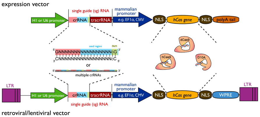
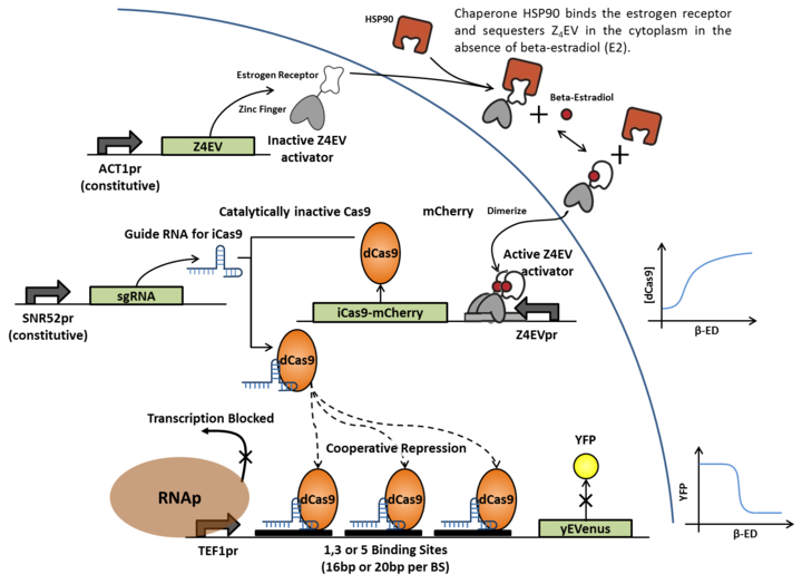
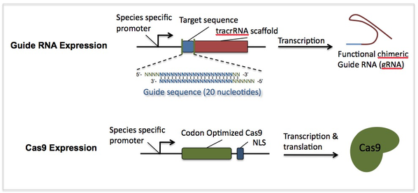
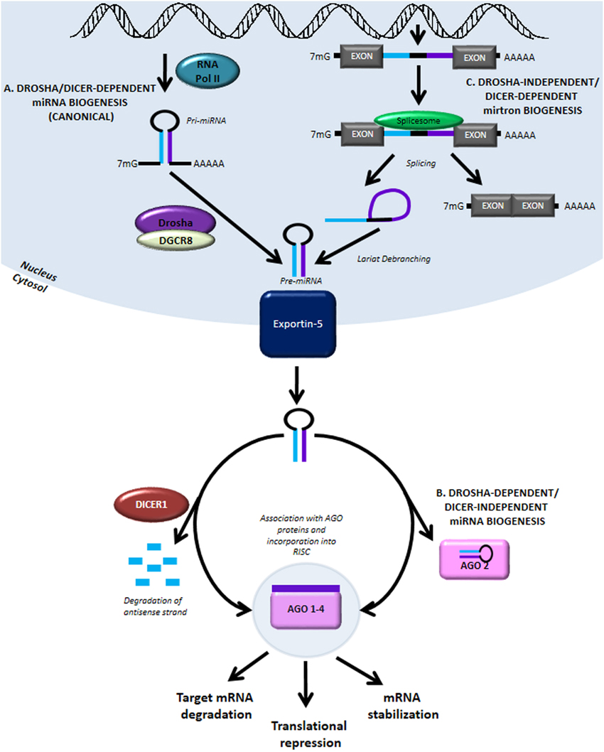
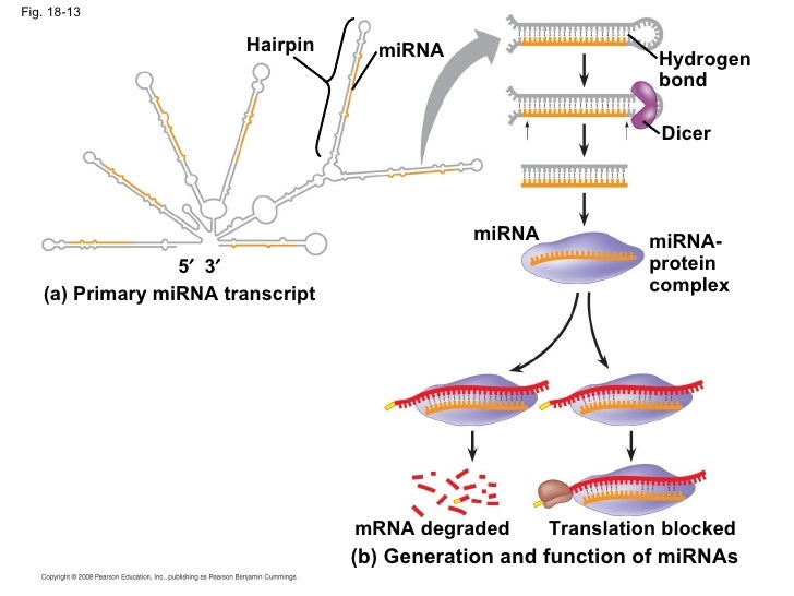
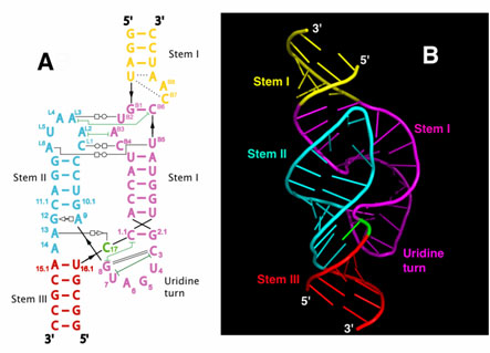
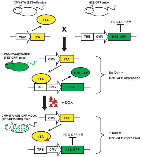
Read Full Post »

CRISPR/Cas9: Contributions on Endoribonuclease Structure and Function, Role in Immunity and Applications in Genome Engineering

Writer and Curator:Larry H Bernstein, MD, FCAP 

2.2.25

2.2.25   CRISPR/Cas9: Contributions on Endoribonuclease Structure and Function, Role in Immunity and Applications in Genome Engineering, Volume 2 (Volume Two: Latest in Genomics Methodologies for Therapeutics: Gene Editing, NGS and BioInformatics, Simulations and the Genome Ontology), Part 2: CRISPR for Gene Editing and DNA Repair

This is the fourth contribution to a series on transcriptional control and cellular remodeling. The previous dealt with RNAs – mRNA, miRNA, RNAi, siRNA, shRNA, small RNAs, lncRNAs, DICER, SLICER, RISC, recombination, and related processes.  It is clear that the classical model was limited, is history, and could not predict a large universe encompassing DNA, RNA, transcription, translation, signaling, proteins, protein conformation, mRNA-miRNA interactions, protein-protein interactions, inter- and intracellular interactions, and cellular remodeling.

Cutting it close: CRISPR-associated endoribonuclease structure and function
Hochstrasser ML and Doudna JA
Trends in Biochemical Sciences, Jan 2015; 40(1):58-66
http://dx.doi.org/10.1016/j.tibs.2014.10.007

RNAi pathways in eukaryotes, as in archaea, possess an adaptive immune system consisting of repetitive genetic elements known as clustered regularly clustered interspersed short palindromic repeats (CRISPERS) and CRISPR-associated (cas) proteins. CRISPR-cas systems require small RNAs for sequence-specific detection and degradation of complex nucleic acids. Cas 5 and cas 6 enzymes have evolved to specifically recognize and process CRISPR-derived transcripts to function as small RNAs used as guides by interference complexes.

Figure 1. Overview of CRISPR RNA (crRNA) processing and comparison between CRISPR–Cas interference systems. There are three main pathways of CRISPR adaptive immunity (Types I–III) and several subtypes, each typified by a different set of Cas proteins. The first stage of the CRISPR–Cas system is acquisition, in which a foreign DNA sequence is incorporated into the host CRISPR locus. Next, the entire repeat-spacer array is transcribed into a long precursor crRNA (pre-crRNA). A single cleavage within each repeat sequence generates shorter, mature crRNAs. Some crRNAs undergo an additional trimming step. The enzymes responsible for catalysis and exact mode of crRNA processing differ in each system. The crRNA is loaded into an interference complex where it serves as a guide for targeting invasive DNA, or in Type III-B systems, RNA.

Figure 2. Fundamental structural features of CRISPR endoRNases. (A) Topology diagram of a typical Cas6 C-terminal RRM fold with key structural features labeled. (B) Two views of Thermus thermophilus Cas6e (PDB: 2Y8W) colored as in (A). For clarity, the N-terminal RRM fold has been omitted in the left panel. (C) Comparison of structures of Cas6 and Cas5c enzymes associated with different CRISPR subtypes (in parentheses), highlighting shared structural elements, colored as in (A) and (B), with the Cas5 ‘thumb’ in black (PDB: 4ILL, 2XLK, 3UFC, 4F3M). Note that no active site residues are shown for Pyrococcus furiosus Cas6-3nc because this protein is non-catalytic.

Figure 3. Structure and sequence-specific RNA binding by Cas6 enzymes. (A) First two images: Thermus thermophilus Cas6A in the apo form and bound to its product CRISPR RNA (crRNA) (PDB: apo – 4C97, product-bound – 4C8Z). Second two images: electrostatic surface potential rendering of the same enzyme in two views with the first eight nucleotides of the Pyrococcus furiosus crRNA 30 handle (PDB: 3PKM) modeled onto the structure based on alignment of the two proteins, as in Niewoehner et al. [30]. For simplicity, only one subunit of the non-crystallographic dimer is shown. (B) Pseudomonas aeruginosa Cas6f bound to its cognate RNA (PDB: 2XLK). Close-up views highlight the active site and sequence-specific interactions by the groove-binding element. (C) Sulfolobus solfataricus Cas6-1A bound to its pre-crRNA substrate (PDB: 4ILL). The active site and sequence-specific contacts made by the glycine-rich loop are shown in detail. For simplicity, only one subunit of the SsoCas6-1A dimer is shown.

CRISPRs (clustered regularly interspaced short palindromic repeats) are DNA loci containing short repetitions of base sequences. Each repetition is followed by short segments of “spacer DNA” from previous exposures to a virus.[2]

CRISPRs are found in approximately 40% of sequenced bacteria genomes and 90% of sequenced archaea.[3][4]

CRISPRs are often associated with cas genes that code for proteins related to CRISPRs. The CRISPR/Cas system is a prokaryotic immune system that confers resistance to foreign genetic elements such as plasmids and phages[5][6] and provides a form of acquired immunity. CRISPR spacers recognize and cut these exogenous genetic elements in a manner analogous to RNAi in eukaryotic organisms.[2]

Since 2013, the CRISPR/Cas system has been used for gene editing (adding, disrupting or changing the sequence of specific genes) and gene regulation in species throughout the tree of life.[7] By delivering the Cas9 protein and appropriate guide RNAs into a cell, the organism’s genome can be cut at any desired location.

It may be possible to use CRISPR to build RNA-guided gene drives capable of altering the genomes of entire populations.

Gene-editing predecessors

In the early 2000s, researchers developed zinc finger nucleases, synthetic proteins whose DNA-binding domains enable them to cut DNA at specific spots. Later, synthetic nucleases called TALENs provided an easier way to target specific DNA and were predicted to surpass zinc fingers. They both depend on making custom proteins for each DNA target, a more cumbersome procedure than guide RNAs. CRISPRs are more efficient and can target more genes than these earlier techniques.

Repeats and spacers

CRISPR loci range in size from 24 to 48 base pairs. They usually show some dyad symmetry, implying the formation of a secondary structure such as a hairpin, but are not truly palindromic. Repeats are separated by spacers of similar length. Some CRISPR spacer sequences exactly match sequences from plasmids and phages, although some spacers match the prokaryote’s genome (self-targeting spacers). New spacers can be added rapidly in response to phage infection.

http://en.wikipedia.org/wiki/CRISPR

http://upload.wikimedia.org/wikipedia/commons/thumb/5/5f/Crispr.png/1024px-Crispr.png

http://www.frontiersin.org/files/Articles/58953/fgene-04-00193-r2/image_m/fgene-04-00193-g001.jpg

http://2013.igem.org/wiki/images/thumb/c/c0/CRISPR_Cooperativity_2.png/720px-CRISPR_Cooperativity_2.png

http://img.scoop.it/2Y0f1M2hXSr35d9-xn4WVTl72eJkfbmt4t8yenImKBVvK0kTmF0xjctABnaLJIm9

A CRISPR CASe for high-throughput silencing

A CRISPR CASe for high-throughput silencing

dual gRNA vector

dual gRNA vector

Genome editing with RNA-guided Cas9 nuclease in Zebrafish embryos

Genome editing with RNA-guided Cas9 nuclease in Zebrafish embryos

The role of CRISPR–Cas systems in adaptive immunity and beyond

Barrangue R
Current Opinion in Immunology 2015; 32:36–41
http://dx.doi.org/10.1016/j.coi.2014.12.008

CRISPR–Cas immune systems. CRISPR-encoded immunization and interference. In the adaptation stage, exogenous DNA is sampled and a novel spacer is integrated into the CRISPR locus; in the expression stage, the CRISPR array is transcribed and processed into small interfering CRISPR RNAs (crRNAs) that guide Cas endonucleases towards target complementary DNA in the interference stage.

Cas-mediated DNA targeting and cleavage. The Cas9 endonuclease forms a ribonucleoprotein complex in combination with the dual guide RNA (crRNA and tracrRNA), and the target dsDNA. First, the Cas9:guide RNA complex binds to proto-spacer adjacent motif (PAM) and drives the formation of an R-loop in the target DNA for genesis of a double stranded break using the RuvC and HNH nickase domains. The former primarily involves the recognition (REC) Cas9 lobe (top), and the latter is primarily driven by the nuclease (NUC) lobe (bottom). Cas-mediated targeting can aim at phage DNA for antiviral resistance (cleaved viral DNA cannot replicate), plasmid DNA to preclude the uptake and dissemination of plasmids (cleaved plasmid DNA cannot replicate), and chromosomal DNA for genome editing (insertion of mutations using endogenous DNA repair systems at the site of cleavage) or transcriptional control (dCas9 binding blocks RNA polymerase).

CRISPR-Cas9 Knockin Mice for Genome Editing and Cancer Modeling
Cell Oct 9, 2014; 159:440–455
http://dx.doi.org/10.1016/j.cell.2014.09.014

Figure2. Ex Vivo Genome Editing of Primary Immune Cells Derived from Constitutive Cas9-Expressing Mice (A) Schematic of ex vivo genome editing experimental flow. (B)Flow cytometry histogram of bone marrow cells from constitutive Cas9-expressing (green) and wild-type (blue) mice, showing Cas9-P2A-EGFP expressiononlyinCas9mice.Dataareplottedasa percentage of the total number of cells. (C) sgRNA design for targeting the mouse Myd88 locus. (D) sgRNA design for targeting the mouse A20 locus. (E) Myd88 indel analysis of constitutive Cas9expressing DCs transduced with either a Myd88targeting sgRNA (sgMyd88-1 and sgMyd88-2) or controls (CTR, average of four control sgRNAs), showing indel formation only in Myd88-targeted cells. Data are plotted as the percent of Illumina sequencing reads containing indels at the target site. Mutations are categorized as frameshift (fs, yellow bar) or non-frameshift (nfs, orange bar). (F) A20 indel analysis of constitutive Cas9-expressing DCs transduced with either an A20-targeting sgRNA (sgA20-1) or controls (CTR, average of four control sgRNAs), showing indel formation only in A20-targeted cells. Data are plotted as the percent of Illumina sequencing reads containing indels at the target site. Mutations are categorized as frameshift (fs, yellow bar) or non-frameshift (nfs, orange bar). (G) Myd88 mRNA quantification of constitutive Cas9-expressing DCs transduced with either Myd88-targeting sgRNA (sgMyd88-1 or sgMyd882) or controls (CTR, average of six control sgRNAs), showing reduced expression only in Myd88-targeted cells. Data are plotted as Myd88 mRNA levels from Nanostring nCounter analysis. (H) Immunoblot of constitutive Cas9-expressing DCs transduced with either Myd88-targeting sgRNA (sgMyd88-1 or sgMyd88-2) or controls (four control sgRNAs), showing depletion of MyD88 protein only in Myd88-targeted cells. b-actin was used as a loading control. (*) Overexposed, repeated-measurement. (I) Nanostring nCounter analysis of constitutive Cas9-expressing DCs transduced with either Myd88-targeting sgRNA (sgMyd88-1 or sgMyd882) or shRNA (shMyd88), A20-targeting sgRNA (sgA20-1 or sgA20-2), or shRNA (shA20), showing analtered LPS response.(Inset)Theclustershowing the highest difference between Myd88- and A20 targeting sgRNAs, including key inflammatory genes(IL1a,IL1b,Cxcl1,Tnf,etc.).(Red)High;(blue) low; (white) unchanged; based on fold change relative to measurements with six control sgRNAs. See also Figure S2.

Figure 5. In Vivo Tumor Formation in AAV9-KPL-Injected Mice (A) Lung mCT images of Cre-dependent Cas9 mice injected with either AAV9-KPL or AAV9-sgLacZ 2 months posttransduction, showing tumor formation (indicated by the arrowhead) only in AAV9-KPL injected mice. (B)LungmCT3DrenderingofCre-dependent Cas9 mice injected with AAV9-KPL 2 months posttransduction, showing tumor formation (indicated by a yellow oval). (C) Major tumor burden quantification of Cre-dependent Cas9 mice injected with either AAV9-KPL or AAV9-sgLacZ, showing significant tumor burden in AAV9KPL-injected mice. Data are plotted as mean ± SEM. **p < 0.005. (D) Representative lung H&E images of Cre-dependent Cas9 mice injected with either AAV9-KPL or AAV9-sgLacZ 9 weeks posttransduction, showing heterogeneous tumor formation in AAV9-KPL-injected mice. Arrowheads highlight a representative subset of tumors within the lungs of AAV9-KPL injected mice.

Development and Applications of CRISPR-Cas9 for Genome Engineering
Zhu PD, Lander ES, Zhang F
Cell Jun 5, 2014; 157:1262-1278
http://dx.doi.org/10.1016/j.cell.2014.05.010

Figure 1. Applications of Genome Engineering Genetic and epigenetic control of cells with genome engineering technologies is enabling a broad range of applications from basic biology to biotechnology and medicine. (Clockwise from top) Causal genetic mutations or epigenetic variants associated with altered biological function or disease phenotypes can nowberapidlyandefficientlyrecapitulated inanimalorcellularmodels (Animal models, Genetic variation). Manipulating biological circuits couldalso facilitate the generation of useful synthetic materials, such as algae-derived, silicabased diatoms for oral drug delivery (Materials). Additionally, precise genetic engineering of important agricultural crops could confer resistance to environmental deprivation or pathogenic infection, improving food security while avoiding the introduction of foreign DNA (Food). Sustainable and cost-effective biofuels are attractive sources for renewable energy, which could be achieved by creating efficient metabolic pathways for ethanol production in algae or corn (Fuel). Direct in vivo correction of genetic or epigenetic defects in somatic tissue would be permanent genetic solutions that address the root cause of genetically encoded disorders (Gene surgery). Finally, engineering cells to optimize high yield generation of drug precursors in bacterial factories could significantly reduce the cost and accessibility of useful therapeutics (Drug development).

Figure 2. Genome Editing Technologies Exploit Endogenous DNA Repair Machinery (A) DNA double-strand breaks (DSBs) are typically repaired by nonhomologous end-joining (NHEJ) or homology-directed repair (HDR). In the errorprone NHEJ pathway, Ku heterodimers bind to DSB ends and serve as a molecular scaffold for associated repair proteins. Indels are introduced when the complementary strands undergo end resection and misaligned repair due to microhomology, eventually leading to frameshift mutations and gene knockout. Alternatively, Rad51 proteins may bind DSB ends during the initial phase of HDR, recruiting accessory factors that direct genomic recombination with homology arms on an exogenous repair template. Bypassing the matching sister chromatid facilitates the introduction of precise gene modifications. (B) Zinc finger (ZF) proteins and transcription activator-like effectors (TALEs) are naturally occurring DNA-binding domains that can be modularly assembled to target specific sequences. ZF and TALE domains each recognize 3 and 1 bp of DNA, respectively. Such DNA-binding proteins can be fused to the FokI endonuclease to generate programmable site-specific nucleases. (C) The Cas9 nuclease from the microbial CRISPR adaptive immune system is localized to specific DNA sequences via the guide sequence on its guide RNA (red), directly base-pairing with the DNA target. Binding of a protospacer-adjacent motif (PAM, blue) downstream of the target locus helps to direct Cas9-mediated DSBs.

Figure 3. Key Studies Characterizing and Engineering CRISPR Systems Cas9 has also been referred to as Cas5, Csx12, and Csn1 in literature prior to 2012. For clarity, we exclusively adopt the Cas9 nomenclature throughout this Review. CRISPR, clustered regularly interspaced short palindromic repeats; Cas, CRISPR-associated; crRNA, CRISPR RNA; DSB, double-strand break; tracrRNA, trans-activating CRISPR RNA.

Figure 4. Natural Mechanisms of Microbial CRISPR Systems in Adaptive Immunity Following invasion of the cell by foreign genetic elements from bacteriophages or plasmids (step 1: phage infection), certain CRISPR-associated (Cas) enzymes acquire spacers from the exogenous protospacer sequences and install them into the CRISPR locus within the prokaryotic genome (step 2: spacer acquisition). These spacers are segregated between direct repeats that allow the CRISPR system to mediate self and nonself recognition. The CRISPR array is a noncoding RNA transcript that is enzymatically maturated through distinct pathways that are unique to each type of CRISPR system (step 3: crRNA biogenesis and processing). In types I and III CRISPR, the pre-crRNA transcript is cleaved within the repeats by CRISPR-associated ribonucleases, releasing multiple small crRNAs. Type III crRNA intermediates are further processed at the 30 end by yet-to-be-identified RNases to produce the fully mature transcript. In type II CRISPR, an associated trans-activating CRISPR RNA (tracrRNA) hybridizes with the direct repeats, forming an RNA duplex that is cleaved and processed by endogenous RNase III and other unknown nucleases. Maturated crRNAs from type I and III CRISPR systems are then loaded onto effector protein complexes for target recognition and degradation. In type II systems, crRNA-tracrRNA hybrids complex with Cas9 to mediate interference. Both type I and III CRISPR systems use multiprotein interference modules to facilitate target recognition. In type I CRISPR, the Cascade complex is loaded with a crRNA molecule, constituting a catalytically inert surveillance complex that recognizes target DNA. The Cas3 nuclease is then recruited to the Cascade-bound R loop, mediating target degradation. In type III CRISPR, crRNAs associate either with Csm or Cmr complexes that bind and cleave DNA and RNA substrates, respectively. In contrast, the type II system requires only the Cas9 nuclease to degrade DNA matching its dual guide RNA consisting of a crRNA-tracrRNA hybrid.

Figure 5. Structural and Metagenomic Diversity of Cas9 Orthologs (A) Crystal structure of Streptococcus pyogenes Cas9 in complex with guide RNA and target DNA. (B) Canonical CRISPR locus organization from type II CRISPR systems, which can be classified into IIA-IIC based on their cas gene clusters. Whereas type IIC CRISPR loci contain the minimal set of cas9, cas1, and cas2, IIA and IIB retain their signature csn2 and cas4 genes, respectively. (C) Histogram displaying length distribution of known Cas9 orthologs as described in UniProt, HAMAP protein family profile MF_01480. (D) Phylogenetic tree displaying the microbial origin of Cas9 nucleases from the type II CRISPR immune system. Taxonomic information was derived from greengenes 16S rRNA gene sequence alignment, and the tree was visualized using the Interactive Tree of Life tool (iTol). (E) Four Cas9 orthologs from families IIA, IIB, and IIC were aligned by ClustalW (BLOSUM). Domain alignment is based on the Streptococcus pyogenes Cas9, whereas residues highlighted in red indicate highly conserved catalytic residues within the RuvC I and HNH nuclease domains.

Figure 6. Applications ofCas9 as aGenome Engineering Platform (A) The Cas9 nuclease cleaves DNA via its RuvC and HNHnucleasedomains,eachofwhichnicks a DNA strand to generate blunt-end DSBs. Either catalytic domain can be inactivated to generate nickase mutants that cause single-strand DNA breaks. (B) Two Cas9 nickase complexes with appropriatelyspacedtargetsitescanmimictargetedDSBs viacooperative nicks, doubling thelengthof target recognition without sacrificing cleavage efficiency. (C) Expression plasmids encoding the Cas9 gene and a short sgRNA cassette driven by the U6 RNA polymerase III promoter can be directly transfected into cell lines of interest. (D) Purified Cas9 protein and in vitro transcribed sgRNA can be microinjected into fertilized zygotes for rapid generation of transgenic animal models. (E) For somatic genetic modification, high-titer viral vectors encoding CRISPR reagents can be transduced into tissues or cells of interest. (F) Genome-scale functional screening can be facilitated by mass synthesis and delivery of guide RNA libraries. (G) Catalytically dead Cas9 (dCas9) can be converted into a general DNA-binding domain and fused to functional effectors such as transcriptional activators or epigenetic enzymes. The modularity of targeting and flexible choice of functional domains enable rapid expansion of the Cas9 toolbox. (H) Cas9 coupled to fluorescent reporters facilitates live imaging of DNA loci for illuminating the dynamics of genome architecture. (I) Reconstituting split fragments of Cas9 via chemical or optical induction of heterodimer domains, such as the cib1/cry2 system from Arabidopsis, confers temporal control of dynamic cellular processes.

Characterization and Optimization of the CRISPR/Cas System for Applications in Genome Engineering
http://nrs.harvard.edu/urn-3:HUL.InstRepos:12407619

Two important advances in the last several decades have propelled our understanding of molecular processes far beyond descriptions of biology at macroscopic levels and fundamentally altered the way that we comprehend organisms, tissues, and cells. First, growing hand in hand with the exponential expansion of computing power, the development of genome sequencing technology, enabling high resolution mapping of DNA sequences, has allowed us to define, down to the nucleotide level, differences between multiple species, members of a species, and within an individual, between classes of cells, as well as diseased and malignant cells. At this point, our ability to make sense of this wealth of genomic information is only limited by our ability to make ever-more precise cellular and genomic alterations to which we may ascribe a phenotypic change. To achieve this, we have concurrently created tools that have allowed us to query the functions of genes and genetic variations from scales large to small by means of first random and then targeted mutagenesis, followed by increasingly refined means of manipulating either the genome directly or the activity of the genes themselves at the level of RNA or protein.

The ability to precisely manipulate the genome in a targeted manner is fundamental to driving both basic science research and development of medical therapeutics. Until recently, this has been primarily achieved through coupling of a nuclease domain with customizable protein modules that recognize DNA in a sequence-specific manner such as zinc finger or transcription activator-like effector domains. Though these approaches have allowed unprecedented precision in manipulating the genome, in practice they have been limited by the reproducibility, predictability, and specificity of targeted cleavage, all of which are partially attributable to the nature of protein-mediated DNA sequence recognition. It has been recently shown that the microbial CRISPR-Cas system can be adapted for eukaryotic genome editing. Cas9, an RNA guided DNA endonuclease, is directed by a 20-nt guide sequence via Watson-Crick base-pairing to its genomic target. Cas9 subsequently induces a double-stranded DNA break that results in targeted gene disruption through non-homologous end-joining repair or gene replacement via homologous recombination. Finally, the RNA guide and protein nuclease dual component system allows simultaneous delivery of multiple guide RNAs (sgRNA) to achieve multiplex genome editing with ease and efficiency.

The potential effects of off-target genomic modification represent a significant caveat to genome editing approaches in both research and therapeutic applications. Prior work from our lab and others has shown that Cas9 can tolerate some degree of mismatch with the guide RNA to target DNA base pairing. To increase substrate specificity, we devised a technique that uses a Cas9 nickase mutant with appropriately paired guide RNAs to efficiently inducing double-stranded breaks via simultaneous nicks on both strands of target DNA. As single-stranded nicks are repaired with high fidelity, targeted genome modification only occurs when the two opposite-strand nicks are closely spaced. This double nickase approach allows for marked reduction of off-target genome modification while maintaining robust on-target cleavage efficiency, making a significant step towards addressing one of the primary concerns regarding the use of genome editing technologies.

The ability to multiplex genome engineering by simply co-delivering multiple sgRNAs is a versatile property unique to the CRISPR-Cas system. While co-transfection of multiple guides is readily feasible in tissue culture, many in vivo and therapeutic applications would benefit from a compact, single vector system that would allow robust and reproducible multiplex editing. To achieve this, we first generated and functionally validated alternate sgRNA architectures to characterize the structure-function relationship of the Cas9 protein with the sgRNA in DNA recognition and cleavage. We then applied this knowledge towards the development and optimization of a tandem synthetic guide RNA (tsgRNA) scaffold that allows for a single promoter to drive expression of a single RNA transcript encoding two sgRNAs, which are subsequently processed into individual active sgRNAs.

A programmable genome editing tool fundamentally consists of two key elements: a DNA recognition domain conferring target specificity and a nuclease domain, ideally without any sequence specificity on its own. A key breakthrough came with the observation that the restriction enzyme FokI has molecularly distinct binding and cleavage domains, and that swapping of recognition domains could alter FokI targeting specificity. Prior to this realization, zinc fingers were discovered as a class of protein motifs in X. laevi, and found to be frequently occurring in mammalian cells as transcription factors where bind DNA in a modular, sequence specific manner. Each individual module of a Cys2-His2 zinc finger domain, the most commonly used ZF-type domain in genome engineering applications, contains approximately 30 amino acids that fold to interact with 3-bp of DNA.

With the creation of custom zinc-finger arrays capable of targeting any DNA sequence, either through stringing together of pre-defined modules with known, predicted 3bp-binding affinity or selection-based protocols with randomized ZF array libraries to account and optimize for inter-modular interactions, the pairing of the DNA-targeting ZF and FokI nuclease components created a new class of zinc finger nucleases (ZFNs) that quickly proved to be an adaptable and efficient method for targeting specific genomic loci in a variety of model organisms. While zinc finger technology can in theory target any specific genomic sequence, the difficulty of accurately predicting protein conformational folding and DNA-protein interactions prior to array assembly can make ZFN construction a somewhat tedious and costly process involving a substantial validation phase prior to practical use.

More recently, an analogous, simpler alternative was developed following the deciphering of the DNA recognition patterns of another class of proteins: the transcription activator-like effector proteins (TALEs). First observed in the rice pathogen Xanthomonas, these proteins consisted of naturally occurring modular arrays of 33-35 amino acid domains, each interacting with a single base pair. Although the single base discrimination of TALE modules compared to 3bp recognition in ZF domains provides greater ease and flexibility in designing TALE arrays to genomic targets, the inherently repetitive nature of TALE repeats posed a technical challenge that required the development of new assembly methodologies. Even so, given the modular separation of DNA recognition activity from nuclease or other effector domains, TALE-derived proteins have been able to quickly co-opt existing technology generated by the studies involving ZF proteins to similarly demonstrate effective genome editing capabilities in a wide variety of model organisms and systems.

One of the major limitations of the aforementioned genome-engineering technologies is their intrinsic dependence on protein-DNA interactions to drive specificity. As such, even after following rational design or thorough in vitro selection processes, it is necessary to perform extensive in vitro validation as protein activity and affinity may vary depending on the specific context in unpredictable ways. Practically, these factors necessitate the construction of multiple sets of TALENs or ZFNs for each locus targeted and, as a consequence, make high-throughput screening applications less tractable.

Although not directly manipulating the genome, the use of small-interfering RNAs (siRNA) to modulate gene expression represents a powerful alternative technology that is not bound by many of the short-comings of these existing genome editing technologies and revolutionized our ability to functionally interrogate the genome. The foundational observation was first made in C. elegans, that the introduction of double-stranded RNA into a cell results in potent post- transcriptional silencing of gene or genes carrying sequences complementary to the exogenous sequence. There are a number of key features that made the RNAi approach particularly tractable and drove its widespread and rapid adoption in basic science research.

  1. RNAi is an extremely efficient method of gene silencing. It is not uncommon to achieve greater than 85% gene knockdown, which, while not complete, is often more than sufficient for inducing a phenotype by which to assess gene function.
  2. siRNA targeting is mediated by predictable Watson-Crick base-pairing. This has allowed the elucidation of design parameters to both maximize on-target silencing and minimize off-target effects. Additionally, the relative ease of designing and creating siRNA constructs allows for rapid prototyping and validation of new targets.
  3. the mechanism of siRNA action takes advantage of a highly-conserved endogenous pathway for processing small RNAs, which minimizes the amount of material that needs to be delivered for adequate effect.

This has had a number of key impacts including but not limited to the possibility of multiplexed delivery to silence more than a single gene at a time or to target a single gene with multiple siRNAs to maximize knock-down, as well as the generation of large siRNA libraries allowing the development of high-throughput screening methodologies for rapid phenotyping in different contexts. The efficacy, predictability, and generalizability of RNAi technologies provided it with enough compelling qualities to become a truly disruptive technology in the field of genome engineering.

Re-purposing the bacterial CRISPR/Cas system for genome editing

The RNA-guided CRISPR (clustered regularly interspaced short palindromic repeats) endonuclease system was first observed in E. coli in 1987 by its striking eponymous genomic structure evolved as an adaptive immune system, bacteria and archaea use a set of CRISPR- associated (Cas) genes to incorporate exogenous material into the CRISPR locus, and subsequently transcribe them as RNA templates for targeted destruction of the mobile elements at either DNA or RNA level.

Three types of CRISPR systems have been identified to date, differing in their targets as well as mechanisms of action. Type I and III CRISPR systems employ an ensemble of Cas gene to carryout RNA processing, recognition of target, and cleavage33,34. By contrast, the type II CRISPRCas system makes use of a single endonuclease, Cas9, to locate and cleave target DNA. Cas9 is guided by a pair non-coding RNAs, a guide-bearing and variable crRNA and a required auxiliary transactivating crRNA (tracrRNA). The crRNA contains a 20-nt guide sequence, also known as a spacer, that determines target specificity by via Watson-Crick base-pairing with target DNA, followed by the invariant “direct repeat” portion that base-pairs with the “antirepeat” portion of the tracrRNA to form an RNA duplex. In the native bacterial system, multiple crRNAs are co-transcribed as a pre-crRNA array before being processed down to individual units for directing Cas9 against various targets. In the CRISPR-Cas system derived from Streptococcus pyogenes, the target DNA sequence always precedes a 5’-NGG protospacer adjacent motif (PAM), which can differ depending on the CRISPR system.

The S. pyogenes CRISPR-Cas system was the first to be reconstituted in mammalian cells through the heterologous expression of human codon-optimized Cas9 and the two RNA components. By altering the the 20-nt guide sequence within the sgRNA, Cas9 can be redirected toward any target bearing an appropriate PAM. Furthermore, elements from the crRNA and tracrRNA can be artificially linked to create a chimeric, single guide RNA (sgRNA), further simplifying the system for eukaryotic gene targeting.

At an overall structural level, Cas9 contains two nuclease domains, HNH and RuvC, each of which cleaves one strand of the target DNA. A mutation in either one of its catalytic domains converts Cas9 nuclease into a nickase, which has shown to induce single-stranded breaks for high-fidelity HDR applications, potentially ameliorating unwanted indel mutations from off target DSBs. Finally, a catalytically inactive or dead Cas9 (dCas9) with mutations in both DNA-cleaving catalytic residues can serve as an RNA-guided DNA-binding scaffold for localizing target effector domains that gene expression at the transcriptional level.

Engineering synthetic TALE and CRISPR-Cas9 transcription factors for regulating gene expression
Methods 2014; 69:188-197
http://dx.doi.org/10.1016/j.ymeth.2014.06.014

Fig.1. TheTAL effectorDNA-binding domain.(A) Through a DNA–protein interaction, each TALE repeatbinds one bp of DNA.TheTALE repeat is shown in blue, and the repeat variable di residue (RVD) at the 12th and 13th position are shown in green and red, respectively. (B) TALEs can be linked in tandem to recognize virtually any DNA sequence. The desired string of TALEs is then fused to an effector domain to induce a specific action at a predetermined DNA sequence. Crystal structure adapted from [60].

Fig. 2. The CRISPR/Cas9 DNA-binding domain. The Cas9 protein forms a complex with the gRNA, which recognizes a specific 20 bp DNA target sequence, known as the protospacer. A short sequence directly downstream from the protospacer, the protospacer adjacent motif (PAM),is requiredfor Cas9-mediated cleavage. ThePAM sequence is highly variable between different organisms (Table 2). With only two amino acid substitutions (D10A and H840A), Cas9 endonuclease activity can been eliminated while maintaining its RNA-guided DNA-binding activity. This deactivated Cas9 (dCas9) functions as a modular DNA-binding domain, similar to TALEs. RNA-guided transcriptional activators and repressors have been created by fusing dCas9 with different effector domains.

Fig.3. Golden gate assembly ofTALEs.Golden Gate assembly makesuse of type IIS restriction enzymes, including BsaI, BsmBI,and Esp3I, that cleave outside their recognition sequence to create unique overhangs. Therefore it is possible to digest and ligate multiple inserts into a destination plasmid with a single restriction enzyme in a single reaction. In step 1, single RVDs are excised from module plasmids and ligated into the desired array plasmid (sample overhangs are shown). This platform allows for construction ofup to 10RVDsinto each array plasmid. Importantly,the array plasmids confer spectinomycin resistance (SpecR) rather than tetracycline resistance (TetR). This ensures that only successfully assembled array plasmids are propagated. In step 2, the array plasmids and the last repeat (LR) plasmid are assembled in a second Golden Gate reaction to obtain the final desired TALE construct. Similar to step 1, in step 2 the final backbone vector confers ampicillin resistance (AmpR), rather than spectinomycin or tetracycline resistance, to ensure that only successfully assembled vectors are propagated. Replacement of the b-galactosidase expression cassette (LacZ) in the final step allows for blue-white screening of successful ligations. Figure adapted from [37].

Fig. 4. Custom gRNA cloning. The most common gRNA cloning methods make use of the BbsI type IIS restriction enzyme that cleaves outside its recognition sequence to create unique overhangs. Single stranded oligonucleotides containing each protospacer are annealed to create overhangs that are compatible with the BbsI sites in the destination vector. Upon ligation, the protospacer is inserted directly following the human U6 promoter and in front of the remainder of the chimeric gRNA sequence. The underlined G indicates the transcriptional start site.

The CRISPR/Cas9 gRNA Targeting System

The recent discovery of the CRISPR/Cas9 sysem has provided researchers an invaluable tool to target and modify any genomic sequence with high levels of efficacy and specificity. The system, consisting of a nuclease (Cas9) and a DNA-directed guide RNA (gRNA), allows for sequence-specific cleavage of target sequence containing a protospacer adaptor motif “NGG”. By changing the gRNA target sequence, virtually any gene sequence upstream of a PAM motif can be targeted by the CRISPR/Cas9 system, enabling the possibility of systematic targeting of sequences on a genomic scale. The most successful gene targeting using the CRISPR/Cas9 system is through expression of multiple gRNAs to guide the enzyme complex to several locations within the target gene to be cut or nicked.

The scalability of the Multiplex gRNA Cloning Kit allows for simultaneous cloning of two or more gRNAs at once into a single vector. This enables researchers to perform more advanced CRISPR/Cas9 techniques such as tandem double-nicking (4 gRNAs total) to remove defined genomic segments using Cas9 Nickase with significantly decreased chances for off-target effects.

The cloning of four gRNAs will require the researcher to perform three separate PCR reactions with separate primer pairs and blocks. Once the correct size amplicons are generated and gel-purified, they can be mixed at equimolar ratios (1:1:1) based on their concentrations and used as inserts in the subsequent fusion reaction with a suitable linearized destination vector.

https://www.systembio.com/downloads/Multiplex-gRNA-Cas9-system_ver5.pdf

https://www.systembio.com/images/How-quad-plex-cloning-works.jpg

https://www.addgene.org/static/data/easy-thumbnails/filer_public/cms/filer_public/7a/b2/7ab294b8-7f7a-4c30-8650-dbb520e2beb4/grna-and-cas9_1.jpg__600x277_q85_subsampling-2_upscale.png

Generation and utility of genetically humanized mouse models
Scheer N, Snaith M, Wolf CR, and Seibler J
Drug Discov Today 2013; 18(23/24):1200-1210
http://dx.doi.org/10.1016/j.drudis.2013.07.007

Applications of genetically humanized mouse models
Type of humanized mouse model Applications
Proteins involved in drug metabolismand disposition Drug–drug interaction studies
Identification and safety assessmentof human metabolites

Assessment of drug bioavailability

and clearance
PKPD modelling

Proteins of the immune and hematopoietic system Studying infectious diseases
Vaccine development and testingStudying autoimmune disorders, ..

involving the immune system

Discovery and testing of antibodies

for therapeutic use

Supported engraftment of human

cells in mice

Proteins involved in pathogeninfection Studying human infectiousdiseases
Aneuploidies or chromosomalre-arrangements Studying human hereditarydiseases
Drug targets Efficacy testing
Human regulatory elements Studying human gene expressionand regulation
Human proto-oncogenes ortumor suppressor genes Cancerogenicity testing

Components of the major pathway for drug metabolism and disposition.

Ligand-dependent activation of the xenobiotic receptors PXR, CAR, PPARa and AHR leads to a translocation to the nucleus and, together with their respective heterodimerization partners retinoic X receptor (RXR) and aryl hydrocarbon receptor nuclear translocator (ARNT), to binding of corresponding response elements and an induction of target genes.

Identifying Drug-Target Selectivity of Small-Molecule CRM1/XPO1 Inhibitors by CRISPR-Cas9 Genome Editing
JE Neggers, et al.
Chemistry & Biology , Jan 22, 2015; 22:107–116
http://dx.doi.org/10.1016/j.chembiol.2014.11.015

Figure 1. Generation of a Mutant XPO1C528S Cell Line Using CRISPR/Cas9 Genome Editing and Homologous Recombination (A) A schematic presentation of the two SINE compounds KPT-185 and KPT-330. (B) Schematic overview of the CRISPR/Cas9induced homologous recombination of human XPO1. Exons are represented by open thick arrows. The blue arrow indicates the sgRNA target site, and small arrowheads beneath the exons indicate forward and reverse PCR A or sequencing primersB.Thesiteofrecombinationis enlarged, and the location of the double strand break (scissors and arrow) is shown. Both the WT XPO1 and donor mutant template sequences are shown at the bottom (magenta, PAM motif; bold, cysteine 528 codon; red, template mutations; underlined, sgRNA sequence). (C) Sequencing chromatogram of genomic DNA of the XPO1 region around the targeted cysteine codon (in bold) from XPO1C528S cells (clone 6).See also Figure S1 and Table S1. (D) Partial protein sequence of XPO1 in WT and mutant XPO1C528S cells (clone 6). Residue 528 of XPO1 is shown in bold. (E) Sequencing chromatogram of the mRNA from XPO1C528S cells (clone 6) in the XPO1 region around the targeted cysteine codon. (F) Visualization of XPO1 protein expression in WT and mutant XPO1C528S cells (clone 6) by immunoblot with b-tubulin as loading control. (G) Relative comparison of XPO1 mRNA expression levels quantified with a probe specific to exon 2 of XPO1 (unpaired student’s t test p value, <0.0001). GAPDH and b-actin were used as internal controls. (H) Relative comparison of mean XPO1 protein expression in WT and XPO1C528S cells (clone 6) as measured by immunofluorescence staining and quantified by confocal fluorescence microscopy (unpaired student’s t test p value, <0.0001). Error bars indicate the 95% confidence interval.

Repurposing CRISPR as an RNA-Guided platform for sequence-specific control of gene expression
LS Qi. MH Larson, LA Gilbert, JA Duoda, et al.
Cell Feb 28, 2013; 152:1173–1183
http://dx.doi.org/10.1016/j.cell.2013.02.022

Figure 1. Design of the CRISPR Interference System (A)Theminimalinterferencesystemconsistsofasingleproteinandadesigned sgRNA chimera. The sgRNA chimera consists of three domains (boxed region): a 20 nt complementary region for specific DNA binding, a 42 nt hairpin for Cas9 binding (Cas9 handle), and a 40 nt transcription terminator derived from S. pyogenes. The wild-type Cas9 protein contains the nuclease activity. The dCas9 protein is defective in nuclease activity. (B) The wild-type Cas9 protein binds to the sgRNA and forms a protein-RNA complex. The complex binds to specific DNA targets by Watson-Crick base pairing between the sgRNA and the DNA target. In the case of wild-type Cas9, the DNA will be cleaved due to the nuclease activity of the Cas9 protein. We hypothesize that the dCas9 is still able to form a complex with the sgRNA and bind to specific DNA target. When the targeting occurs on the protein-coding region, it could block RNA polymerase and transcript elongation. See also Figure S1

Figure 2. CRISPRi Effectively Silences Transcription Elongation and Initiation (A) The CRISPRi system consists of an inducible Cas9 protein and a designed sgRNA chimera. The dCas9 contains mutations of the RuvC1 and HNH nuclease domains. The sgRNA chimera contains three functional domains, as described in Figure 1. (B) Sequence of designed sgRNA (NT1) and the DNA target. NT1 targets the nontemplate DNA strand of the mRFP-coding region. Only the region surrounding the base-pairing motif (20 nt) is shown. Base-pairing nucleotides are shown in orange, and the dCas9-binding hairpin is in blue. The PAM sequence is shown in red. (C) CRISPRi blocks transcription elongation in a strand-specific manner. A synthetic fluorescence-based reporter system containing an mRFP-coding gene is inserted into the E.coli MG1655 genome (then sfA locus). Six sgRNAs that bind to either the template DNA strand or the nontemplate DNA strand are coexpressed with the dCas9 protein, with their effects on the target mRFP measured by in vivo fluorescence assay. Only sgRNAs that bind to the nontemplate DNA strand showed silencing (10- to 300-fold). The control shows fluorescence of the cells with dCas9 protein but without the sgRNA. (D) CRISPRi blocks transcription initiation. Five sgRNAs are designed to bind to different regions around an E.coli promoter (J23119). The transcription start site is labeled as +1. The dotted oval shows the initial RNAP complex that covers a 75 bp region from 55 to +20. Only sgRNAs targeting regions inside of the initial RNAP complex show repression (P1–P4). Unlike transcription elongation block, silencing is independent of the targeted DNA strand. (E) CRISPRi regulation is reversible. Both dCas9 and sgRNA (NT1) are under the control of an aTc-inducible promoter. Cell culture was maintained during exponential phase. At timeT=0, 1mM of a Tc was supplemented to cells with OD=0.001. Repression of target mRFP starts within 10min.The fluorescence signal decays in a way that is consistent with cell growth, suggesting that the decay is due to cell division. In 240 min, the fluorescence reaches the fully repressed level. At T= 370 min, a T cis washed away from the growth media, and cells are diluted back to OD = 0.001. Fluorescence starts to increase after 50 min and takes about 300 min to rise to the same level as the positive control. Positive control: always without the inducer; negative control: always with 1 mM aTc inducer. Fluorescence results in (C)–(E) represent average and SEM of at least three biological replicates. See also Figures S2 and S3.

Figure 3. CRISPRi Functions by Blocking Transcription Elongation (A) FLAG-tagged RNAP molecules were immunoprecipitated, and the associated nascent mRNA transcripts were sequenced. (Top) Sequencing results of the nascent
mRFP transcript in cells without sgRNA. (Bottom) Results in cells with sgRNA. In the presence of sgRNA, a strong transcriptional pause is observed 19 bp upstream of the target site, after which the number of sequencing reads drops precipitously. (B) A proposed CRISPRi mechanism based on physical collision between RNAP and dCas9-sgRNA. The distance from the center of RNAP to its front edge is ~19 bp, which matches well with our measured distance between the transcription pause site and 30 of sgRNA base-pairing region. The paused RNAP aborts transcription elongation upon encountering the dCas9-sgRNA roadblock.

Figure 4. Targeting Specificity of the CRISPRi System (A) Genome-scale mRNA sequencing (RNA-seq) confirms that CRISPRi targeting has no off-target effects. The sgRNA NT1 that bindsto the mRFP coding region is used. The dCas9, mRFP, and sfGFP genes are highlighted. (B)Multiple sgRNAs can independently silence two fluorescent protein reporters in the same cell. Each sgRNA specifically represses its cognate gene,but not the other gene. When both sgRNAs are present, both genes are silenced. Error bars represent SEM from at least three biological replicates. (C) Microscopic images for using two sgRNAs to control two fluorescent proteins. (Top) Bright-field images of the E. coli cells; (middle) RFP channel; (bottom) GFP channel. Coexpression of one sgRNA and dCas9 only silences the cognate fluorescent protein, but not the other. The knockdown effect is strong, as almost no fluorescence is observed from cells with certain fluorescent protein silenced. Scale bar, 10 mm. Control shows cells without any fluorescent protein reporters.

In vitro and in vivo growth suppression of human papillomavirus 16-positive cervical cancer cells by CRISPR-Cas9
S Zhen, L Hua, et al.
BBRC 2014; 450:1422-1426
http://dx.doi.org/10.1016/j.bbrc.2014.07.014

Fig. 4. Suppression of in vivo growth of SiHa cells in BALB/c nude mice by CRISPR/ Cas9. (A) In vivo tumor growth curves of CRISPR/Cas9 systems-treated SiHa cells. The mean tumor volumes ± SD (bars) are shown at the times that tumor measurements were made (n = 6). (B) Tumor weight 10 weeks after inoculation. All tumors were excised and weighted.

One-Step Generation of Mice Carrying Mutations in Multiple Genes by CRISPR-Cas-mediated genome engineering
Wang H, Yang H, et al.
Cell  2013; 153:910-918
http://dx.doi.org/10.1016/j.cell.2013.04.025

Figure 2. Single- and Double-Gene Targeting In Vivo by Injection into Fertilized Eggs (A) Genotyping of Tet1 single-targeted mice. (B) Upper: genotyping of Tet2 single-targeted mice. RFLP analysis; lower: Southern blot analysis. (C) The sequence of both alleles of targeted gene in Tet1 biallelic mutant mouse 2 and Tet2 biallelic mutant mouse 4. (D) Genotyping of Tet1/Tet2 double-mutant mice. Analysis of mice 1 to 12 is shown. Upper: RFLP analysis; lower: southern blot analysis. The Tet1 locus is displayed on the left and the Tet2 locus on the right. (E) The sequence of four mutant alleles from double-mutant mouse 9 and 10. PAM sequences are labeled in red. (F) Three-week-old double-mutant mice. All RFLP and Southern digestions and probes are the same as those used in Figure 1. See also Figures S2 and S3.

The impact of CRISPR–Cas9 on target identification and validation
JD Moore
Drug Discov Develop 2015
http://dx.doi.org/10.1016/j.drudis.2014.12.016

Gene editing with Cas9. (a) Knock-out generation via Cas9 and a single synthetic guide (sg)RNA. sgRNAs form Watson–Crick base pairs with target sequences recruiting the wild-type Cas9 nuclease. Cas9 generates double stranded breaks that are typically repaired by the imprecise NHEJ mechanism resulting in small insertions or deletions, most of which generate frameshift mutations. Transient expression of sgRNA plus Cas9 leads to editing of 2–25% of alleles. Derivative clones are analysed to find examples where Gene editing with Cas9. (a) Knock-out generation via Cas9 and a single synthetic guide (sg)RNA. sgRNAs form Watson–Crick base pairs with target sequences recruiting the wild-type Cas9 nuclease. Cas9 generates double stranded breaks that are typically repaired by the imprecise NHEJ mechanism resulting in small insertions or deletions, most of which generate frameshift mutations. Transient expression of sgRNA plus Cas9 leads to editing of 2–25% of alleles. Derivative clones are analysed to find examples where both alleles have been repaired with frame shift mutations. (b) Knock-out generation via Cas9 and a pair of sgRNAs. When wild-type Cas9 is expressed with a pair of sgRNAs targeting sites in the same region of a gene, simultaneous dual double-stranded breaks will be introduced in a fraction of cells. Repair via NHEJ will tend to delete the intervening sequence. (c) Knock-in generation using sgRNAs, donor DNA and either wild-type Cas9 or the Cas9-D10A nickase mutant. Wild-type Cas9 generates double stranded breaks that can be repaired by NHEJ generating indels or by homology-directed repair (HDR) leading to knock-in of mutations present on homology templates. The Cas9-D10A nickase mutant only generates single stranded breaks, which are not a substrate for the NHEJ pathway. However, these can be processed by HDR leading to the introduction of knock-in mutations. (d) Using the Cas9D10A nickase mutant to enhance the specificity of gene editing. The sgRNA shown in red also recruits Cas9 to partially mismatched off-target sites where wild-type Cas9 can efficiently introduce double stranded breaks leading to editing of an off-target exon.  More…

Repurposing CRISPR-Cas9 for in situ functional assays
Malina A, Mills JR, …, Pelletier J.
Genes & Development 2015; 27:2602–2614
http://www.genesdev.org/cgi/doi/10.1101/gad.227132.113

Figure 1. Genome editing of a TLR locus in 293Tcells using an engineered all-in-one type II CRISPR system. (A) Schematic diagram of LeGO-based lentivirus (pLC) constructs driving expression of Cas9 and sgRNAs. (B) Predicted secondary structure (http://rna.tbi. univie.ac.at/cgi-bin/RNAfold.cgi) of sgRNA showing alignment of trigger sequence with target and PAM. The first nucleotide of the trigger sequence is forcibly a G, since the sgRNA is expressed from the murine U6 promoter. (C) Schematic of TLR with the position and nucleotide sequence of the TLR trigger, PAM, and stop codon shown. (D) A genomically integrated TLR is efficiently targeted by pLC-TLR. Quantitation of 293T TLR cells transfected with the indicated Cas9/sgRNA expression constructs and, where indicated, in combination with D20 eGFP. (E) Immunoblot showing expression and subcellular localization of Cas9 from the experiment presented in D. (C) Cytoplasmic fraction; (M) membrane fraction; (N) nuclear fraction. Blots were probed with the antibodies indicated below each panel. (F) Lentiviral-mediated NHEJ and HDR in 293T TLR cells. Cells were infected with lentivirus expressing Cas9 and the corresponding sgRNA and analyzed by flow cytometry 6 d later. The D20 eGFP donor plasmid was introduced by transfection 1 d prior to transduction with the Cas9/sgRNA lentiviral construct.

Figure 2. Cas9-mediated editing of Trp53 in Arf[1]/[1] MEFs leads to Nutlin-3a resistance. (A) Schematic diagram of the pQ-based retroviral constructs driving expression of Cas9, GFP, and sgRNAs (pQCiG). (B) Flow cytometric analysis of Arf[1]/[1] and p53[1]/[1] MEFs transduced with QCiG-Rosa, QCiG-p53, or MLP-p53.1224 retroviruses, cultured 3 d later in the presence of vehicle or 10 mM Nutlin-3a for 24 h, and then allowed to recover for 4 d. (C) Colony formation assay of infected Arf[1]/[1] and p53[1]/[1] MEFs with QCiG-Rosa, QCiGp53, or MLP-p53.1224. Five-thousand cells were seeded, exposed to 10 mM Nutlin-3a for 24 h, and allowed to recover for 12 d in the absence of drug, at which point they were stained with crystal violet. (D) SURVEYOR assay of DNA isolated from QCiG-p53- and QCiG-Rosa-infected Arf[1]/[1] MEFs exposed to 10 mM Nutlin-3a for 24 h and allowed to recover for 4 d. The arrowhead denotes the expected SURVEYOR cleavage products. (E) Immunoblot documenting Cas9 and p53 expression in QCiG- and MLP-infected MEFs. The asterisk denotes the position of a prominent p53 truncated product

Figure 3. Cas9-mediated editing of Trp53 in Arf[1]/[1]Em-myc lymphomas is positively selected for following DXR treatment in vivo. (A) Schematic diagram of in vivo fitness assay. (B) Kaplan-Meier analysis of tumor-free survival of mice injected with Rosa26 or Trp53 Cas9 targeted Arf[1]/[1]Em-myc and p53[1]/[1]Em-myc lymphomas following treatment with DXR. (C) Detection of GFP in tumors arising from QCiG-p53-infected Arf[1]/[1]Em-myc lymphomas following exposure to DXR and analyzed 3 d later. White arrows denote GFP fluorescence in lymph nodes originating from the presence of QCiG-p53 in the resulting tumors. (D) FACS analysis of the indicated Cas9 targeted Em-myc lymphomas analyzed before injection into mice (input), from tumors arising in vivo (pre-DXR), and from tumors for which the host had received DXR treatment (post-DXR). (E) SURVEYOR assay of DNA from QCiG-p53- and QCiG-Rosa-infected Arf-/-Em-myc lymphomas isolated from mice prior to DXR treatment. (F) Immunoblot showing long-term Cas9, p53, and GFP expression in QCiG-Rosa and QCiG-p53 Arf-/-Em-Myc lymphomas in vivo. Samples are from three separate tumors isolated prior to (pre-DXR) or following (post-DXR) DXR treatment. In the case of post-DXR samples for QCiG-Rosa Arf-/-Em-myc lymphomas, tumors were harvested after relapse (~10 d after post-DXR treatment). The asterisk highlights a truncated p53 protein arising in the Cas9 edited samples.

Figure 4. Analysis of indels at the Trp53 locus and at predicted off-target sites in Arf-/-MEFs and Arf-/-Em-myc tumors edited with Rosa26 and Trp53 sgRNAs. (A) Total count and location of insertions and deletions in exon 7 of Trp53 in Arf-/-Em-myc cells prior to injection, post-implantation, and post-DXR treatment, respectively. The vertical dashed line represents the predicted Cas9 cleavage site. (B) Frequency of mutant reads obtained following sequencing of Trp53 exon 7 from the indicated cells and tumors. T-1, T-2, and T-3 represent three independent tumors. (C, top panel) Sequence alignment of the trigger site in the Trp53 and Trp53 pseudogene. Differences are highlighted in green. (Bottom panel) Pie charts illustrating the proportion of mutated sequence reads at Trp53 (left) and the Trp53 pseudogene (right) relative to wild-type sequences (wt; blue). DNA was isolated from samples of Arf-/-Em-myc lymphoma cells infected with QCiG-Rosa-infected (top), QCiG-p53-infected (middle), or QCiG-p53-infected cells that were exposed to 10 mM Nutlin-3a for 3 d followed by a 10-d recovery period (bottom). (D) Prediction of genomic sequences showing sequences complementary to the first 13 perfectly matched nucleotides 59 to the PAM of the Trp53 trigger sequence with all possible combinations of PAM. The trigger sequence is shown in blue, PAM is in red, and flanking nucleotides are in black. The genomic location is shown at the right. (E) Percent mutant reads at the indicated genomic locus in Rosa26- and Trp53-modified Arf-/- MEFs. The total read count for each amplified region ranged from ;11,000 to 15,000 (sample #8), ;18,000 to 23,000 (sample #7), and ;20,000 to 53,000 (all others). Read counts for locus #2 are absent, since the barcode that had been used in the preparation of that sample could not be deciphered from the output of reads.

TALE nucleases- tailored genome engineering made easy
Mussolino C, Cathomen T
Current Opinion in Biotechnology 2012; 23:644–650
http://dx.doi.10.1016/j.copbio.2012.01.013

Generation of customized TALENs by ‘Golden Gate’ cloning. Dependent on the user-defined target sequence, the respective repeat units with desired specificities can be assembled using a two-step ‘Golden Gate’ cloning protocol. A TALEN monomer is generated by incorporating the TALE designer array in a TALEN backbone, which contains an N-terminal NLS, the ‘0 repeat’ binding to the 50-T nucleotide, the 17.5 ‘half-repeat’, and the terminal FokI cleavage domain (N).

TALEN or Cas9 – Rapid, efficient and specific choices for genome modifications
Wei C, Liu J, et al.
J Genetics and Genomics 40 (2013) 281e289
http://dx.doi.org/10.1016/j.jgg.2013.03.013

Fig. 1. Schematic principles of TALEN- and CRISPR/Cas9-mediated genomic modifications. A: a single TALEN consists of an N-terminal domain including a nuclear localization signal (NLS, blue); a central domain typically composed of tandem TALE repeats (green) for the recognition of a specific DNA sequence; and a C-terminal domain of the functional endonuclease Fok I (black). Each TALE repeat comprises of a 34-amino-acid unit that differs at the position of 12th and 13th amino acids: NG (recognizing T), NI (recognizing A), HD (recognizing C), or NN (recognizing G) (color boxes). B: double-strand breaks (DSBs) that are resulted from the cut by dimeric Fok I can be repaired either by non-homologous end joining (NHEJ) to yield indels or by homologous recombination (HR) with available homologous donor templates. The red star indicates where indels occur. C: the CRISPR/Cas9 system consists of a group of CRISPR-associated (Cas) genes (arrows with the direction to the right) and a CRISPR locus that contains an array of repeats (dark diamonds) e spacer (color boxes) sequences. All repeats are the same in sequence and all spacers are different and complementary to their target DNA sequences. The tracrRNA (trans-activating crRNA, arrow on the most left) can help to produce the crRNA (CRISPR RNA). D: the Cas9 protein (blue) binds to crRNA (orange) and tracrRNA (purple) to form a ribonucleoprotein complex. The crRNA sequence guides this complex to a complementary sequence in the target DNA (black). Then the HNH and RuvC domains of Cas9 nick the complementary and non-complementary strands, respectively, making a DSB. PAM: protospacer adjacent motif NGG (yellow box). gRNA: guiding RNA. NCC is a complementary motif of the PAM motif (NGG).

Table 1 Comparison of TALEN- and CRISPR/Cas9-mediated genomic modifications e principles and applications

TALEN CRIPR/Cas9
Target-binding principle Protein-DNA specific recognition Watson Crick complementary rule
Working mode TALE specifically recognizes the target DNA and dimeric Fok I makes the DSB, which is repaired by NHEJ or HR Guide RNA specifically recognizes the target DNA and Cas9 makes the DSB, which is repaired by NHEJ or HR
Essential components TALE-Fok I fusion protein Guide RNA and Cas9
Off-target effects Minor effects Not determined
Efficiency High but variable High but variable
Target site availability No restriction PAM (NGG) motif restriction
Work in pair/dimmer Yes No
Inheritability in animals Yes Not determined
3D structure Yes Yes
Time to construct 5-7 d 1-3 d
Origin discovery Plant pathogen E. coli

Genome-Scale CRISPR-Mediated Control of Gene Repression and Activation
Gilbert LA, et al.
Cell, Oct 23, 2014; 159: 647–661
http://dx.doi.org/10.1016/j.cell.2014.09.029

Figure 1. A Tiling sgRNA Screen Defines Rules for CRISPRi Activity at Endogenous Genes in Human Cells (A) Massively parallel determination of growth or toxin-resistance phenotypes caused by sgRNAs in mammalian cells expressing dCas9 or dCas9 fusion constructs. (B) UCSC genome browser tracks showing the genomic organization, GC content, and repetitive elements around the TSS of a representative gene, VPS54, across a 10 kb window targeted by the tiling sgRNA library. sgRNA ricin-resistance phenotypes (as Z scores, see Figure S1 and Experimental Procedures) in dCas9 and dCas9-KRAB expressing K562 cells are depicted in black on the top and bottom, respectively. See also Figure S2A for more examples. (C) Sliding-window analysis of all 49 genes targeted in a tiling sgRNA library. Green line: median sgRNA activity in a defined window for all genes. Orange region: observed average window of maximum CRISPRi activity. Data displayed as a phenotype signed Z score, excluding all guides longer than 22 bp. (D) CRISPRi activity for all 49 genes in defined windows relative to the TSS of each gene. (E) Ricin-resistance phenotypes, comparing CRISPRi sgRNAs selected by our rules to RNAi, for genes previously established to cause ricin-resistance phenotypes when knocked down by RNAi. Mean ± SD phenotype-signed Z score of 100 sets of 10 randomly subsampled sgRNAs or shRNAs. See also Figure S2F

Figure 2. CRISPRi Activity is Highly Sensitive to Mismatches Between the sgRNA and DNA sequence On- and off-target activity of dCas9, dCas9-KRAB and Cas9 for sgRNAs with a varying number and position of mismatches. Off-target activity of sgRNAs with mismatches is displayed as percent of the on-target activity for the corresponding sgRNA without mismatches. Asterisk indicates sgRNAs with three, four, or five mismatches randomly distributed across region 3 of the sgRNA sequence. Data are displayed for each mismatch position as the mean of all sgRNAs with that mismatch; see Figure S3 for individual sgRNA activities. sgRNAs were included in the analysis only if the fully matched guide was highly active (phenotype-signed Z score R 4); n = 5 for dCas9, n = 11 for dCas9-KRAB, and n = 10 for Cas9.

Figure 3. A Tiling sgRNA Screen Defines Rules for CRISPRa Activity at Endogenous Genes in Human Cells (A) A schematic of the dCas9-SunTag + scFV-VP64 + sgRNA system for CRISPRa. (B)ActivityofsgRNAsinK562cellsstablyexpressingeachcomponentofCRISPRa,asafunctionofthedistanceofthesgRNAsitetotheTSSofthetargetedgene (Phenotype-signed Z scores; therefore, negative values represent opposite results than from knockdown). Top, sgRNAs targeting VPS54; Bottom, slidingwindow analysis of all 49 genes targeted by our tiling library in green. Green line, median activity; orange, window of maximal activity. Guides longer than 22 bp were excluded. See also Figure S4. (C)CRISPRaphenotypesandCRISPRi(dCas9-KRAB)phenotypesareanticorrelated forselect genes.Foreachgene,aMann-Whitneypvalueiscalculatedusing CRISPRi/a sgRNA activity relative to a negative control distribution for 24 subsampled sgRNAs. Mean ± SD p value of 100 randomly subsampled sets is displayed. (D) CRISPRi knockdown and CRISPRa activation of the same gene can have opposing effects on ricin resistance in both primary screens and single sgRNA validation experiments (mean ± SD of 3 replicates). (E) Modulation of expression levels for 3 genes by CRISPRi and CRISPRa as quantified by qPCR plotted against the ricin-resistance phenotype (mean ± SD of 3 replicates) measured for each sgRNA.

Figure 4. Genome-Scale CRISPRi and CRISPRa Screens Reveal Genes Controlling Cell Growth (A) sgRNA phenotypes from a genome-scale CRISPRi screen for growth in human K562 cells (black). Three classes of negative control sgRNAs are color-coded: nontargeting sgRNAs (gray), sgRNAs targeting Y-chromosomal genes (green) and sgRNAs targeting olfactory genes (orange). (B) Coexpression of sgRNAs and dCas9-KRAB or dCas9-SunTag + scFV-VP64 is not toxic in K562 cell lines over 16 days. (C) Gene set enrichment analysis (GSEA) for hits from the CRISPRi screen. A histogram of gene distribution is shown under the GSEA curve. (D) CRISPRi versus CRISPRa gene phenotypes for genome-scale growth screens (black). For the 50 genes in the CRISPRa screen with the most negative growth phenotype, each gene was annotated and labeled based on evidence of activity as a tumor suppressor (orange), developmental transcription factor (green), or in regulation of the centrosome (purple). Two additional CRISPRi hit genes that are discussed in the text are labeled in red. See Table S4 for annotations and references. (E) GSEA for hits from the CRISPRa growth screen. A histogram of gene distribution is shown under the GSEA curve.

Figure 5. CRISPRi Gene Silencing Is Inducible, Reversible, and Nontoxic (A) Expression construct encoding an inducible KRAB-dCas9 fusion protein. (B) Western blot analysis of inducible KRAB-dCas9 in the absence, presence, and after washout of doxycycline. (C) Relative RAB1A expression levels (as quantified by qPCR) in inducible CRISPRi K562 cells transduced with RAB1A-targeting sgRNAs in the absence, presence, and after washout of doxycycline. Mean ± standard error of technical replicates (n = 2) normalized to control cells (assayed in the presence of doxycycline) from the day 2 time point. (D) Competitive growth assays performed with inducible CRISPRi K562 cells transduced with the indicated sgRNAs in the presence and absence of doxycycline. Data are represented as the mean ± SD of replicates (n = 3). See also Figure S5G. (E) A CRISPRi sublibrary screen for effects on cell growth was performed with inducible CRISPRi K562 cells in the presence and absence of doxycycline. (F) Cumulative growth curves from the sublibrary screen represented in (E) show no bulk changes to growth caused by induction of KRAB-dCas9. Mean ± SD of replicate infections each screened in duplicate.

Figure 6. Genome-Scale CRISPRi and CRISPRa Screens Reveal Known and New Pathways and Complexes Governing the Response to a Cholera-Diphtheria Fusion Toxin (A) Model for CTx-DTA binding, retrograde trafficking, retrotranslocation, and cellular toxicity. (B) Overview of top hit genes detected by the CTx-DTA screen. Dark red and blue circles: Top 50 sensitizing and protective hits, respectively. Light red and blue circles: further hits that fall into the same protein complexes or pathways as top 50 hits. Circle area is proportional to phenotype strength. White stars denote genes identified in a previous haploid mutagenesis screen (Guimaraes et al., 2011). See also Figure S6 for hit gene names. (C) CRISPRi and CRISPRa hits in sphingolipid metabolism. Display as in (B), except that the left and right sides of each circle represent the phenotypes in the CRISPRi and CRISPRa screens, respectively.

Figure 7. CRISPRi Strongly Represses Gene Expression of Both Protein-Coding and Noncoding Genes, Resulting in Reproducible Phenotypes (A–C) Cells expressing a negative control sgRNA or an sgRNA targeting SEL1L or B4GALNT1 were incubated with cholera toxin and fractionated to quantify cholera toxin present in the cytosolic and membrane fractions by western blot. B4GALNT1 repression blocks toxin uptake, whereas SEL1L repression prevents toxin retrotranslocation from the membrane fraction to the cytosol. (D) Validation of CTx-DTA screen phenotypes with single sgRNA retest experiments. Data are represented as the mean ± SD of replicates (n = 3). (E) CRISPRi knockdown of 13 hit genes (28 sgRNAs; sgRNAs correspond to 7D) identified in the CTx-DTA screen was quantified by qPCR. The gray shaded region denotes sgRNAs showing at least 90% knockdown for each gene. Data are normalized to a negative control sgRNA (NC). (F) CRISPRi knockdown of 6 lncRNA genes was quantified by qPCR. Two to three sgRNAs computationally predicted to target each gene were cloned and transduced into K562 cells expressing dCas9-KRAB. Data are normalized to a negative control sgRNA (NC). (G) K562 cells expressing dCas9-KRAB were transduced with either a nontargeting sgRNA or an sgRNA targeting the XIST locus (sgXIST-1). The cells were then stained with DAPI and an RNA FISH probe for the XIST transcript. Two hundred nonapoptotic interphase cells in each condition were scored for XIST RNA coating. XIST is undetectable in cells transduced with sgXIST-1. Scale bar, 5 mm

Other related articles published on this topic in this Open Access Online Scientific Journal include the following:

Advances in Gene Editing Technology: New Gene Therapy Options in Personalized Medicine

Curators: Stephen J Williams, PhD and Aviva Lev-Ari, PhD, RN

http://pharmaceuticalintelligence.com/2015/03/16/advances-in-gene-editing-technology-new-gene-therapy-options-in-personalized-medicine/

Read Full Post »

RNAi – On Transcription and Metabolic Control

Writer and Curator: Larry H Bernstein, MD, FCAP


RNAi

This is the third contribution to a series on transcription and metabolic control. It reveals the enormous complexity in this emerging research.


mRNA, small RNAs, long RNAs, RNAi and DicAR

Aberrant mRNA translation in cancer pathogenesis
Pier Paolo Pandolfi
Oncogene (2004) 23, 3134–3137
http://dx.doi.org:/10.1038/sj.onc.1207618

As the molecular processes that control mRNA translation and ribosome biogenesis in the eukaryotic cell are extremely complex and multilayered, their deregulation can in principle occur at multiple levels, leading to both disease and cancer pathogenesis. For a long time, it was speculated that disruption of these processes may participate in tumorigenesis, but this notion was, until recently, solely supported by correlative studies. Strong genetic support is now being accrued, while new molecular links between tumor-suppressive and oncogenic pathways and the control of protein synthetic machinery are being unraveled. The importance of aberrant protein synthesis in tumorigenesis is further underscored by the discovery that compounds such as Rapamycin, known to modulate signaling pathways regulatory of this process, are effective anticancer drugs. A number of fundamental questions remain to be addressed and a number of novel ones emerge as this exciting field evolves.


mRNA Translation and Energy Metabolism in Cancer
I. Topisirovic and N. Sonenberg
Cold Spring Harbor Symposia on Quantitative Biology, Volume LXXVI
http://dx.doi.org:/10.1101/sqb.2011.76.010785

A prominent feature of cancer cells is the use of aerobic glycolysis under conditions in which oxygen levels are sufficient to support energy production in the mitochondria (Jones and Thompson 2009; Cairns et al. 2010). This phenomenon, named the “Warburg effect,” after its discoverer Otto Warburg, is thought to fuel the biosynthetic requirements of the neoplastic growth (Warburg 1956; Koppenol et al. 2011) and has recently been acknowledged as one of the hallmarks of cancer (Hanahan and Weinberg 2011). mRNA translation is the most energy-demanding process in the cell (Buttgereit and Brand 1995).In mammalian cells it consumes >20% of cellular ATP, not considering the energy that is required for the biosynthesis of the components of the translational machinery (e.g., ribosome biogenesis; Buttgereit and Brand 1995). Control of mRNA translation plays a pivotal role in the regulation of gene expression (Sonenberg and Hinnebusch 2009). In fact, a recent study demonstrated that mammalian proteome is mostly governed at the mRNA translation level (Schwanhausser et al. 2011). Malfunction of mRNA translation critically contributes to human disease, including diabetes, heart disease, blood disorders, and, most notably, cancer (Fig. 1; Crozier et al. 2006; Narla and Ebert 2010; Silvera et al. 2010; Spriggs et al. 2010). The first account of changes in the translational apparatus in cancer dates back to 1896, showing enlarged and irregularly shaped nucleoli that are the site of ribosome biogenesis (Pianese 1896). Rapidly proliferating cancer cells have more ribosomes than normal cells.

Figure 1. Dysregulated mRNA translation plays a pivotal role in cancer. Malignant cells are characterized by enlarged nucleoli and a larger number of ribosomes than their normal counterparts. Mutations and/or altered expression of ribosomal proteins (e.g., RPS19, RPS 24), rRNA-modifying enzymes (e.g., dyskerin), translation initiation factors (e.g., eIF4E), or the initiator tRNA (tRNAiMet) result in malignant transformation. Signaling pathways whose dysfunction is frequent in cancer (e.g., MAPK, PI3K/AKT) affect mRNA translation. Perturbations in the translatome result in aberrant cellular growth, proliferation, and survival characteristic of tumorigenesis.


In stark contrast to normal cells, in cancer cells ribosomal biogenesis is uncoupled from cell proliferation (Stanners et al. 1979). Accordingly, cancer cells exhibit abnormally high rates of protein synthesis (Silvera et al. 2010). That ribosomal dysfunction plays a central role in cancer is further corroborated by the findings that genetic alterations, which encompass the components of the ribosome machinery (i.e., “ribosomopathies”), are characterized by elevated cancer risk (Narla and Ebert 2010).

mRNA translation is the most energy-consuming process in the cell and strongly correlates with cellular metabolic activity. Translation and energy metabolism play important roles in homeostatic cell growth and proliferation, and when dysregulated lead to cancer. eIF4E is a key regulator of translation, which promotes oncogenesis by selectively enhancing translation of a subset of tumor-promoting mRNAs (e.g., cyclins and c-myc). PI3K/AKT and mitogen-activated protein kinase (MAPK) pathways, which are strongly implicated in cancer etiology, exert a number of their biological effects by modulating translation. The PI3K/AKT pathway regulates eIF4E function by inactivating the inhibitory 4E-BPs via mTORC1, whereas MAPKs activate MAP kinase signal-integrating kinases 1 and 2, which phosphorylate eIF4E. In addition, AMP-activated protein kinase, which is a central sensor of the cellular energy balance, impairs translation by inhibiting mTORC1. Thus, eIF4E plays a major role in mediating the effects of PI3K/AKT, MAPK, and cellular energetics on mRNA translation.Figure 2. eIF4E is regulated by multiple mechanisms. The expression of eIF4E is regulated by several transcription factors (e.g., c-myc, hnRNPK, p53) and adenine-uracil-rich element binding proteins (i.e., HuR and AUF1). eIF4E is suppressed by 4E-BPs, which are regulated by mTORC1. MAP kinase signal integrating kinases 1 and 2 (MNKs) phosphorylate eIF4E.


Figure 3. Ras/MAPK and PI3K/AKT/mTORC1 regulate the activity of eIF4E. Various stimuli activate phosphoinositide-3-kinase (PI3K) through the receptor tyrosine kinases (RTKs). Upon activation, PI3K converts phosphatidylinositol 4,5-bisphosphate (PIP2) into phosphatidylinositol-3,4,5-triphosphate (PIP3). This reaction is reversed by PTEN. Phosphoinositide-dependent protein kinase 1 (PDK1) and AKT bind to PIP3 via their pleckstrin homology domains, which allows for the phosphorylation and activation of AKT by PDK1. In addition, the mammalian target of rapamycin complex 2 (mTORC2) modulates the activity of AKT by phosphorylating its hydrophobic motif. AKT phosphorylates tuberous sclerosis complex 2 (TSC2) at multiple sites, which results in its inhibition and consequent activation of Ras homolog enriched in brain (Rheb), which is a small GTPase that activates mTORC1. mTORC1 phosphorylates 4E-BPs leading to their dissociation from eIF4E. In addition to the PI3K/AKT pathway, the activity of mTORC1 is regulated by the serine/threonine kinase 11/LKB1/AMP-kinase (LKB1/AMPK) pathway, regulated in development and DNA damage response 1 (REDD1) and Rag GTPases in response to the changes in cellular energy balance, oxygen and amino acid availability, respectively. Ras and the MAPK pathways are activated by various stimuli through receptor tyrosine kinases (RTKs). In addition the MAPK pathway isactivatedthrough theGprotein–coupled receptors(GPCRs) and byproteinkinaseC (PKC;notshown).TheMAPK pathways encompass an initial GTPase-regulated kinase (MAPKKK), which activates an effector kinase (MAPK) via an intermediate kinase (MAPKK). In response to stimuli such as growth factors, hormones, and phorbol-esters, Ras GTPase stimulates Raf kinase (MAPKKK), which activates extracellular signal-regulated kinases 1 and 2 (ERK 1 and 2) via extracellular signal-regulated kinase activator kinases MEK1 and 2 (MAPKK). Cellular stresses, including osmotic shock, inflammatory cytokines, and UV light, activate p38 MAPKs via multiple mechanisms including Rac kinase (MAPKKK) and MKK3 and 6 (MAPKK). p38 MAPK and ERK activate the MAPK signal–integrating kinases 1 and 2 (MNK1/2), which phosphorylate eIF4E. Additional abbreviations are provided in the text.


Cancer Exosomes Perform Cell-Independent MicroRNA Biogenesis and Promote Tumorigenesis
Cancer Cell Nov, 2014; 26: 707–721.
http://dx.doi.org/10.1016/j.ccell.2014.09.005

Breast cancer cells secrete exosomes with specific capacity for cell-independent miRNA biogenesis, while normal cellderivedexosomes lack thisability. Exosomes derivedfrom cancer cellsand serum frompatients withbreast cancer contain the RISC loading complex proteins, Dicer, TRBP, and AGO2, which process pre-miRNAs into mature miRNAs. Cancer exosomes alter the transcriptome of target cells in a Dicer-dependent manner, which stimulate nontumorigenic epithelial cells to form tumors.This study identifies a mechanism whereby cancer cells impart an oncogenic field effect by manipulating the surrounding cells via exosomes. Presence of Dicer in exosomes may serve as biomarker for detection of cancer.


Dicers at RISC. The Mechanism of RNAi

Marcel Tijsterman and Ronald H.A. Plasterk
Cell, Apr 2014; 117:1–4

Figure 1. Model for RNA Silencing in Drosophila In an ordered biochemical pathway, miRNAs (left panel) and siRNAs (right panel) are processed from double-stranded precursor molecules by Dcr-1and Dcr-2, respectively, and stay attached to Dicer-containing complexes, which assemble into RISC. The degree of complementarity between the RNA silencing molecule (in red) and its cognate target determines the fate of the mRNA: blocked translation or immediate destruction.

Argonaute2 Cleaves the Anti-Guide Strand of siRNA during RISC Activation
Cell 2005; 123:621-629
http://www.cell.com/cgi/content/full/123/4/621/DC1/
Dicing and slicing- The core machinery of the RNA interference pathway
Scott C Hammond
FEBS Letters 579 (2005) 5822–5829
http://dx.doi.org:/10.1016/j.febslet.2005.08.079

Fig. 1. Domain organization of RNaseIII gene family. Three classes of RNaseIII genes are shown. The PAZ domain in Dm-Dicer-2 contains mutations in several residues required for RNA binding and may not be functional.

Fig. 2. Model for Dicer catalysis. The PAZ domain binds the 2 nt 30 overhang of a dsRNA terminus. The RNaseIII domains form a pseudo-dimer. Each domain hydrolyzes one strand of the substrate. The binding site of the dsRBD is not defined. The function of the helicase domain is not known.

Fig. 3. Biogenesis pathway of microRNAs. MicroRNA genes are transcribed by RNA polymerase II. The primary transcript is referred to as ‘‘primicroRNA’’. Drosha processing occurs in the nucleus. The resulting precursor, ‘‘pre-microRNA’’, is exported to the cytoplasm for Dicer processing. In a coordinated manner, the mature microRNA is transferred to RISC and unwound by a helicase. mRNA targets that duplex in the Slicer scissile site are cleaved and degraded, if the microRNA is loaded into an Ago2 RISC. Mismatched targets are translationally suppressed. All Ago family members are believed to function in translational suppression.

Fig. 4. Model for Slicer catalysis. The siRNA guide strand is bound at the 50 end by the PIWI domain and at the 30 end by the PAZ domain. The 50 phosphate is coordinated by conserved basic residues. mRNA targets are initially bound by the seed region of the siRNA and pairing is extended to the 30 end. The RNaseH fold hydrolyzes the target in a cation dependent manner. Slicer cleavage is measured from the 50 end of the siRNA. Product is released by an unknown mechanism and the enzyme recycles.


RNA interference (RNAi) is a biological process in which RNA molecules inhibit gene expression, typically by causing the destruction of specific mRNA molecules. Historically, it was known by other names, including co-suppression, post transcriptional gene silencing (PTGS), and quelling. Only after these apparently unrelated processes were fully understood did it become clear that they all described the RNAi phenomenon. Andrew Fire and Craig C. Mello shared the 2006 Nobel Prize in Physiology or Medicine for their work on RNA interference in the nematode worm Caenorhabditis elegans, which they published in 1998.


Two types of small ribonucleic acid (RNA) molecules – microRNA (miRNA) and small interfering RNA (siRNA) – are central to RNA interference. RNAs are the direct products of genes, and these small RNAs can bind to other specific messenger RNA (mRNA) molecules and either increase or decrease their activity, for example by preventing an mRNA from producing a protein. RNA interference has an important role in defending cells against parasitic nucleotide sequences – viruses and transposons. It also influences development.


The RNAi pathway is found in many eukaryotes, including animals, and is initiated by the enzyme Dicer, which cleaves long double-stranded RNA (dsRNA) molecules into short double stranded fragments of ~20 nucleotide siRNAs. Each siRNA is unwound into two single-stranded RNAs (ssRNAs), the passenger strand and the guide strand. The passenger strand is degraded and the guide strand is incorporated into the RNA-induced silencing complex (RISC). The most well-studied outcome is post-transcriptional gene silencing, which occurs when the guide strand pairs with a complementary sequence in a messenger RNA molecule and induces cleavage by Argonaute, the catalytic component of the RISC complex. In some organisms, this process spreads systemically, despite the initially limited molar concentrations of siRNA.
http://en.wikipedia.org/wiki/RNA_interference


http://upload.wikimedia.org/wikipedia/commons/thumb/e/e4/ShRNA_Lentivirus.svg/481px-ShRNA_Lentivirus.svg.png


http://www.frontiersin.org/files/Articles/66078/fnmol-06-00040-HTML/image_m/fnmol-06-00040-g001.jpg
http://dx.doi.org:/10.3389/fnmol.2013.00040

The enzyme dicer trims double stranded RNA, to form small interfering RNA or microRNA. These processed RNAs are incorporated into the RNA-induced silencing.
MiRNA biogenesis and function. (A) The canonical miRNA biogenesis pathway is Drosha- and Dicer-dependent. It begins with RNA Pol II-mediated transcription..


Dicer Promotes Transcription Termination

Dicer Promotes Transcription Termination

Dicer Promotes Transcription Termination at Sites of Replication Stress to Maintain Genome Stability
Cell Oct 2014; 159(3): 572–583
http://dx.doi.org/10.1016/j.cell.2014.09.031

http://www.cell.com/cms/attachment/2019646604/2039684570/fx1.jpg


18-13 miRNA- protein complex ap-chap-18-pp-42-728

18-13 miRNA- protein complex ap-chap-18-pp-42-728

18-13 miRNA- protein complex (a) Primary miRNA transcript Translation blocked Hydrogen bond (b) Generation and function of miRNAs Hairpin miRNA miRNA Dicer …

http://image.slidesharecdn.com/ap-chap-18-pp-1229097198123780-1/95/ap-chap-18-pp-42-728.jpg?cb=1229090143


Identification and characterization of small RNAs involved in RNA silencing
FEBS Letters 579 (2005) 5830–5840
http://dx.doi.org:/10.1016/j.febslet.2005.08.009

Fig. 1. Small RNA cloning procedure. Outline of the small RNA cloning procedure. RNA is dephosphorylated (step 1) for joining the 30 adapter by T4 RNA ligase 1 in the presence of ATP (step 2). The use of a chemically adenylated adapter and truncated form of T4 RNA ligase 2 (Rnl2) allows eliminating the dephosphorylation step (step 4). If the RNA was dephosphorylated, it is re-phosphorylated (step 3) prior to 50 adapter ligation with T4 RNA ligase 1 and ATP (step 5). After 50 adapter ligation, a standard reverse transcription is performed (step 6). Alternatively, after 30 adapter ligation, the RNA is used directly for reverse transcription simultaneously with 50 adaptor joining (step 7). In this case, the property of reverse transcriptase to add non-templated cytidine residues at the 50 end of synthesized DNA is used to facilitate template switch of the reverse transcriptase to the 30 guanosine residues of the 50 adapter (SMART technology, Invitrogen). Abbreviations: P and OH indicate phosphate and hydroxyl ends of the RNA; App indicates 50 chemically adenylated adapter; L, 30 blocking group; CIP, calf alkaline phosphatase and PNK, polynucleotide kinase.


Transcriptional regulatory functions of nuclear long noncoding RNAs
Trends in Genetics, Aug 2014; 30(8):348-356
http://dx.doi.org/10.1016/j.tig.2014.06.001

Cis-acting lncRNAEnhancer-associated lncRNAIntergenic lncRNA

lncRNA

Promoter-associated lncRNA

Proximity transfer

Trans-acting lncRNA


Functional interactions among microRNAs and long noncoding RNAs
Sem Cell Dev Biol 2014; 34:9-14
http://dx.doi.org/10.1016/j.semcdb.2014.05.015
Genome-wide application of RNAi to the discovery of potential drug targets
FEBS Letters 579 (2005) 5988–599
http://dx.doi.org://10.1016/j.febslet.2005.08.015

Fig. 1. Schematic representation of gene silencing by an shRNA-expression vector. The shRNA is processed by Dicer. The processed siRNA enters the RNA-induced silencing complex (RISC), where it targets mRNA for degradation.

Fig. 2. Schematic representation of a transcription system for production of siRNA

Fig. 3. (A) Schematic representation of the proposed siRNA-expression system. Three or four C to U or A to G mutations are introduced into the sense strand. (B) Schematic representation of the discovery of a novel gene using an siRNA library.


Imperfect centered miRNA binding sites are common and can mediate repression of target mRNAs
Martin et al. Genome Biology 2014, 15:R51 http://genomebiology.com/2014/15/3/R51


Table 1 Number of inferred targets for each miRNA tested

miRNA Probes Transcripts Genes
miR-10a 2,206 5,963 1,887
miR-10a-iso 1,648 1,468 4,211
miR-10b 1,588 3,940 1,365
miR-10b-iso 963 2,235 889
miR-17-5p 1,223 2,862 1,137
miR-17-5p-iso 1,656 3,731 1,461
miR-182 2,261 6,423 2,008
miR-182-iso 1,569 4,316 1,444
miR-23b 2,248 5,383 1,990
miR-27a 2,334 5,310 2,069

Probes: number of probes significantly enriched in pull-downs compared to controls (5% FDR). Transcripts: number of transcripts to which those probes map exactly. Genes: number of genes from which those transcripts originate

Figure 2 Biotin pull-downs identify bone fide miRNA targets. (A) Volcano plot showing the significance of the difference in expression between the miR-17-5p pull-down and the mock-transfected control, for all transcripts expressed in HEK293T cells. Both targets predicted by TargetScan or validated previously via luciferase assay were significantly enriched in the pull-down compared to the controls. (B) Results from luciferase assays on previously untested targets predicted using TargetScan and uncovered using the biotin pull-down. The plot indicates mean luciferase activity from either the empty plasmid or from pMIR containing a miRNA binding site in the 3′ UTR, relative to a negative control. Asterisks indicate a significant reduction in luciferase activity (one-sided t-test; P<0.05) and error bars the standard error of the mean over three replicates. (C-E) Targets identified through PAR-CLIP or through miRNA over-expression studies show greater enrichment in the pull-down. Cumulative distribution of log fold-change in the pull-down for transcripts identified as targets by the indicated miRNA over-expression study or not. Red, canonical transcripts found to be miR-17-5p targets in the indicated study (Table S5 in Additional file 1); black, all other canonical transcripts; p, one-sided P-value from Kolmogorov-Smirnov test for a difference in distributions. (F) To confirm that our results were dependent on RISC association, cells were transfected with either single or double-stranded synthetic miRNAs, then subjected to AGO2 immunoprecipitation. The biotin pull-down was performed in the AGO2-enriched and AGO2-depleted fractions. (G-H) Quantitative RT-PCR revealed that, with double-stranded (ds) miRNA (G), four out of five known targets were enriched relative to input mRNA (*P≤0.05, **P<0.01, ***P<0.001) in the AGO2-enriched but not in the AGO2-depleted fractions, but this enrichment was not seen for the cells transfected with a single-stranded (ss) miRNA (H). The numbers on the x-axis correspond to those in Figure 2F. Error bars represent the standard error of mean (sem).

Figure 5 IsomiRs and canonical miRNAs target many of the same transcripts.

Hammerhead ribozymes in therapeutic target discovery and validation
Drug Disc Today 2009; 14(15/16): 776-783
http://dx.doi.org/10.1016/j.drudis.2009.05.003

Figure 1. Features of hammerhead ribozymes. A generic diagram of a hammerhead ribozyme bound to its target substrate: NUH is the cleavage triplet on target sequence, stems I and III are sites of the specific interactions between ribozyme and target, stem II is the structural element connecting separate parts of the catalytic core. Arrows represent the cleavage site, numbering system according to Hertel et al. [60].

hammerhead ribozyme

hammerhead ribozyme

https://www-ssrl.slac.stanford.edu/research/highlights_archive/ribozyme_fig1.jpg


Figure 1  Schematic (A) and ribbon (B) diagrams depicting the crystal structure of the full-length hammerhead ribozyme. The sequence and secondary structure


TABLE 1 Typical examples of successful applications of hammerhead ribozymes. Most of the data are derived from [10] and [11], the others are expressly specified.

  • Growth factors, receptors, transduction elements
  • Oncogenes, protoncogenes, fusion genes
  • Apoptosis, survival factors, drug resistance
  • Transcription factors
  • Extracellular matrix, matrix modulating factors
  • Circulating factors
  • Viral genome, viral genes

Figure 2.Target–ribozyme interactions. (a) As cheme of ribozyme binding to full substrate. The calculated energy of this binding ensures the formation of a stable complex. At the denaturating temperature, Tm, will allow this complex to survive to biological conditions. Conversely, after cleavage, binding energies calculated on single, (b) and (c), ribozyme arms are very low and no longer stable. These properties will ensure both the efficient release of cleavage fragments and the prevention of binding to unrelated targets. RNAs complementary to one binding arm only will not be bound or cleaved by the hammerhead catalytic sequence.

Figure 3. ‘Chemical omics’ approach. According to this target discovery strategy: (1) a first round of ‘omic’ study (proteomic, genomic, metabolomic, …) will enable the discovery of a set of (2) putative markers. A series of hammerhead ribozymes will then be prepared in order to target each marker. (4) A second ‘omic’ study round will be performed on (3) knocked down samples obtained after ribozymes administration. (5) A new series of markers will then be produced. An expanding analytical process of this type may be further repeated. Finally, a robust bioinformatic algorithm will make it possible to connect the different markers and draw new hypothetical links and pathways.


miRNA

ADAR Enzyme and miRNA Story
Sara Tomaselli, Barbara Bonamassa, Anna Alisi, et al.
Int. J. Mol. Sci. 2013, 14, 22796-22816;
http://dx.doi.org:/10.3390/ijms141122796

Adenosine deaminase acting on RNA (ADAR) enzymes convert adenosine (A) to inosine (I) in double-stranded (ds) RNAs. Since Inosine is read as Guanosine, the biological consequence of ADAR enzyme activity is an A/G conversion within RNA molecules. A-to-I editing events can occur on both coding and non-coding RNAs, including microRNAs (miRNAs), which are small regulatory RNAs of ~20–23 nucleotides that regulate several cell processes by annealing to target mRNAs and inhibiting their translation. Both miRNA precursors and mature miRNAs undergo A-to-I RNA editing, affecting the miRNA maturation process and activity. ADARs can also edit 3′ UTR of mRNAs, further increasing the interplay between mRNA targets and miRNAs. In this review, we provide a general overview of the ADAR enzymes and their mechanisms of action as well as miRNA processing and function. We then review the more recent findings about the impact of ADAR-mediated activity on the miRNA pathway in terms of biogenesis, target recognition, and gene expression regulation.

Figure 1. Structure of ADAR family proteins: ADAR1, ADAR2, and ADAR3. The ADAR enzymes contain a C-terminal conserved catalytic deaminase domain (DM), two or three dsRBDs in the N-terminal portion. ADAR1 full-length protein also contains a N-terminal Zα domain with a nuclear export signal (NES) and a Zβ domain, while ADAR3 has a  R-domain. A nuclear localization signal is also indicated.


Comprehensive modeling of microRNA targets predicts functional non-conserved and non-canonical sites
Doron Betel, Anjali Koppal, Phaedra Agius, Chris Sander, Christina Leslie
Genome Biology 2010, 11:R90 http://genomebiology.com/2010/11/8/R90

microRNAs are a class of small regulatory RNAs that are involved in post-transcriptional gene silencing. These small (approximately 22 nucleotide) single-strand RNAs guide a gene silencing complex to an mRNA by complementary base pairing, mostly at the 3′ untranslated region (3′ UTR). The association of the RNAinduced silencing complex (RISC) to the conjugate mRNA results in silencing the gene either by translational repression or by degradation of the mRNA. Reliable microRNA target prediction is an important and still unsolved computational challenge, hampered both by insufficient knowledge of microRNA biology as well as the limited number of experimentally validated targets.

mirSVR is a new machine learning method for ranking microRNA target sites by a down-regulation score. The algorithm trains a regression model on sequence and contextual features extracted from miRanda-predicted target sites. In a large-scale evaluation, miRanda-mirSVR is competitive with other target prediction methods in identifying target genes and predicting the extent of their downregulation at the mRNA or protein levels. Importantly, the method identifies a significant number of experimentally determined non-canonical and non-conserved sites.
Human RISC – MicroRNA Biogenesis and Posttranscriptional Gene Silencing
Cell 2005; 123:631-640
http://dx.doi.org:/10.1016/j.cell.2005.10.022
Development of microRNA therapeutics
Eva van Rooij & Sakari Kauppinen
EMBO Mol Med (2014) 6: 851–864
http://dx.doi.org:/10.15252/emmm.20110089

MicroRNAs (miRNAs) play key regulatory roles in diverse biological processes and are frequently dysregulated in human diseases. Thus, miRNAs have emerged as a class of promising targets for therapeutic intervention. Here, we describe the current strategies for therapeutic modulation of miRNAs and provide an update on the development of miRNA-based therapeutics for the treatment of cancer, cardiovascular disease and hepatitis C virus (HCV) infection.

Figure 1. miRNA biogenesis and modulation of miRNA activity by miRNA mimics and antimiR oligonucleotides. MiRNA genes are transcribed by RNA polymerase II from intergenic, intronic or polycistronic loci to long primary miRNA transcripts (pri-miRNAs) and processed in the nucleus by the Drosha–DGCR8 complex to approximately 70 nt pre-miRNA hairpin structures. The most common alternative miRNA biogenesis pathway involves short intronic hairpins, termed mirtrons, that are spliced and debranched to form pre-miRNA hairpins. Pre-miRNAs are exported into the cytoplasm and then cleaved by the Dicer–TRBP complex to imperfect miRNA: miRNA* duplexes about 22 nucleotides in length. In the cytoplasm, miRNA duplexes are incorporated into Argonaute-containing miRNA induced silencing complex (miRISC), followed by unwinding of the duplex and retention of the mature miRNA strand in miRISC, while the complementary strand is released and degraded. The mature miRNA functions as a guide molecule for miRISC by directing it to partially complementary sites in the target mRNAs, resulting in translational repression and/or mRNA degradation. Currently, two strategies are employed to modulate miRNA activity: restoring the function of a miRNA using double-stranded miRNA mimics, and inhibition of miRNA function using single-stranded anti-miR oligonucleotides.

Figure 2. Design of chemically modified miRNA modulators. (A) Structures of chemical modifications used in miRNA modulators. A number of different sugar modifications are used to increase the duplex melting temperature (Tm) of anti-miR oligonucleotides. The20-O-methyl(20-O-Me), 20-O-methoxyethyl(20-MOE )and 20-fluoro(20-F) nucleotides are modified at the 20 position of the sugar moiety, whereas locked nucleic acid (LNA) is a bicyclic RNA analogue in which the ribose is locked in a C30-endo conformation by introduction of a 20-O,40-C methylene bridge. To increase nuclease resistance and enhance the pharmacokinetic properties, most anti-miR oligonucleotides harbor phosphorothioate (PS) backbone linkages, in which sulfur replaces one of the non-bridging oxygen atoms in the phosphate group. In morpholino oligomers, a six-membered morpholine ring replaces the sugar moiety. Morpholinos are uncharged and exhibit a slight increase in binding affinity to their cognate miRNAs. PNA oligomers are uncharged oligonucleotide analogues, in which the sugar–phosphate backbone has been replaced by a peptide-like backbone consisting of N-(2-aminoethyl)-glycine units. (B) An example of a synthetic double-stranded miRNA mimic described in this review. One way to therapeutically mimic a miRNA is by using synthetic RNA duplexes that harbor chemical modifications for improved stability and cellular uptake. In such constructs, the antisense (guide) strand is identical to the miRNA of interest, while the sense (passenger) strand is modified and can be linked to a molecule, such as cholesterol, for enhanced cellular uptake. The sense strand contains chemical modifications to prevent mi-RISC loading. Several mismatches can be introduced to prevent this strand from functioning as an anti-miR, while it is further left unmodified to ensure rapid degradation.The20-F modification helps to protect the antisense strand against exonucleases, hence making the guide strand more stable, while it does not interfere with mi-RISC loading. (C) Design of chemically modified anti-miR oligonucleotides described in this review. Antagomirs are30 cholesterol-conjugated,20-O-Me oligonucleotides fully complementary to the mature miRNA sequence with several PS moieties to increase their in vivo stability. The use of unconjugated 20-F/MOE-, 20-MOE- or LNA-modified anti-miR oligonucleotides harboring a complete PS backbone represents another approach for inhibition of miRNA function in vivo. The high duplex melting temperature of LNA-modified oligonucleotides allows efficient miRNA inhibition using truncated, high-affinity 15–16-nucleotide LNA/DNA anti-miR oligonucleotides targeting the 50 region of the mature miRNA. Furthermore, the high binding affinity of fully LNA-modified 8-mer PS oligonucleotides, designated as tiny LNAs, facilitates simultaneous inhibition of entire miRNA seed families by targeting the shared seed sequence.

Human MicroRNA Targets
Bino John, Anton J. Enright, Alexei Aravin, Thomas Tuschl,.., Debora S. Mark
PLoS Biol 2004; 2(11): e363  http://www.plosbiology.org

More than ten years after the discovery of the first miRNA gene, lin-4 (Chalfie et al. 1981; Lee et al. 1993), we know that miRNA genes constitute about 1%–2% of the known genes in eukaryotes. Investigation of miRNA expression combined with genetic and molecular studies in Caenorhabditis elegans, Drosophila melanogaster, and Arabidopsis thaliana have identified the biological functions of several miRNAs (recent review, Bartel 2004). In C. elegans, lin-4 and let-7 were first discovered as key regulators of developmental timing in early larval developmental transitions (Ambros 2000; Abrahante et al. 2003; Lin et al. 2003; Vella et al. 2004). More recently lsy-6 was shown to determine the left–right asymmetry of chemoreceptor expression (Johnston and Hobert 2003). In D. melanogaster, miR-14 has a role in apoptosis and fat metabolism (Xu et al. 2003) and the bantam miRNA targets the gene hid involved in apoptosis and growth control (Brennecke et al. 2003).

MicroRNAs (miRNAs) interact with target mRNAs at specific sites to induce cleavage of the message or inhibit translation. The specific function of most mammalian miRNAs is unknown. We have predicted target sites on the 39 untranslated regions of human gene transcripts for all currently known 218 mammalian miRNAs to facilitate focused experiments. We report about 2,000 human genes with miRNA target sites conserved in mammals and about 250 human genes conserved as targets between mammals and fish. The prediction algorithm optimizes sequence complementarity using position-specific rules and relies on strict requirements of interspecies conservation. Experimental support for the validity of the method comes from known targets and from strong enrichment of predicted targets in mRNAs associated with the fragile X mental retardation protein in mammals. This is consistent with the hypothesis that miRNAs act as sequence-specific adaptors in the interaction of ribonuclear particles with translationally regulated messages. Overrepresented groups of targets include mRNAs coding for transcription factors, components of the miRNA machinery, and other proteins involved in translational regulation, as well as components of the ubiquitin machinery, representing novel feedback loops in gene regulation. Detailed information about target genes, target processes, and open-source software for target prediction (miRanda) is available at http://www.microrna.org. Our analysis suggests that miRNA genes, which are about 1% of all human genes, regulate protein production for 10% or more of all human genes.

Figure 1. Target Prediction Pipeline for miRNA Targets in Vertebrates The mammalian (human, mouse, and rat) and fish (zebra and fugu) 39 UTRs were first scanned for miRNA target sites using position specific rules of sequence complementarity. Next, aligned UTRs of orthologous genes were used to check for conservation of miRNA– target relationships (‘‘target conservation’’) between mammalian genomes and, separately, between fish genomes. The main results (bottom) are the conserved mammalian and conserved fish targets, for each miRNA,as well as a smaller set of super-conserved vertebrate targets.   http://dx.doi.org:/10.1371/journal.pbio.0020363.g00
Figure 2. Distribution of Transcripts with Cooperativity of Target Sites and Estimated Number of False Positives Each bar reflects the number of human transcripts with a given number of target sites on their UTR. Estimated rate of false positives(e.g., 39%for2 targets) is given by the number of target sites predicted using shuffled miRNAs processed in a way identical to real miRNAs, including the use of interspecies conservation filter. http://dx.doi.org:/10.1371/journal.pbio.0020363.g002

Conserved Seed Pairing, Often improved an-Flanked by Adenosines, Indicates Thousands of Human Genes are MicroRNA Targets
Cell, Jan 2005; 120: 15–20
http://dx.doi.org:/10.1016/j.cell.2004.12.035

Integrated analysis of microRNA and mRNA expression. adding biological significance to microRNA target predictions.
Maarten van Iterson, Sander Bervoets, Emile J. de Meijer, et al.
Nucleic Acids Research, 2013; 41(15), e146
http://dx.doi.org:/10.1093/nar/gkt525

Current microRNA target predictions are based on sequence information and empirically derived rules but do not make use of the expression of microRNAs and their targets. This study aimed to improve microRNA target predictions in a given biological context, using in silico predictions, microRNA and mRNA expression. We used target prediction tools to produce lists of predicted targets and used a gene set test designed to detect consistent effects of microRNAs on the joint expression of multiple targets. In a single test, association between microRNA expression and target gene set expression as well as the contribution of the individual target genes on the association are determined. The strongest negatively associated mRNAs as measured by the test were prioritized. We applied our integration method to a well-defined muscle differentiation model. Validation of our predictions in C2C12 cells confirmed predicted targets of known as well as novel muscle-related microRNAs. We further studied associations between microRNA–mRNA pairs in human prostate cancer, finding some pairs that have been recently experimentally validated by others. Using the same study, we showed the advantages of the global test over Pearson correlation and lasso. We conclude that our integrated approach successfully identifies regulated microRNAs and their targets.

Long non-coding RNA and microRNAs might act in regulating the expression of BARD1 mRNAs
Int J Biol & Cell Biol 2014; 54:356-367
http://dx.doi.org/10.1016/j.biocel.2014.06.018


Passenger-Strand Cleavage Facilitates Assembly of siRNA into Ago2-Containing RNAi Enzyme Complexes
Cell 2006; 123:607-620
http://dx.doi.org:/10.1016/j.cell.2006.08.044


RNAi- RISC Gets Loaded
Cell 2005; 123:543-553
http://dx.doi.org:/10.1016/j.cell.2005.11.006
RNAi- The Nuts and Bolts of the RISC Machine
Cell 2005; 122:17-20
http://dx.doi.org:/10.1016/j.cell.2005.06.023
Structural domains in RNAi
FEBS Letters 579 (2005) 5841–5849
http://dx.doi.org:/10.1016/j.febslet.2005.07.072

Fig. 1. A ‘‘Domain-centric’’ view of RNAi. (A) The conserved pathways of RNA silencing. The domain structure of each protein in (hypothetical) interaction with its RNA is shown. For clarity, the second column lists domains in order N- to C-terminal. Figures are not to scale. In brief, Drosha, an RNase III enzyme, and its obligate binding partner, Pasha recognize pri-mRNA loops, and cut these into 70 nt hairpin pre-miRNAs. Dicer utilizes a PAZ domain to sense the 30 2-nt overhang created, and further processes these, and dsRNAs into miRNAs and siRNAs. Argonaute binds the 50 end of guide RNAs via its PIWI domain, and the 30 end via a PAZ domain, yielding RISCs that effect RNA silencing through several mechanisms. A Viral protein, VP19 can suppress RNA silencing by sequestering siRNAs. (B) A summary of known siRNA structural biology. Listed by domain are solved structures, their protein/organism of origin, and ligands, where applicable. Also shown are PDB codes.

Fig. 2. Novel modes of RNA recognition. (A) A typical dsRBD: Xenopus binding protein A (1DI2). A RNA helix is modeled pink, and the protein is rendered in transparent electrostatic contours (blue is basic, red acidic). Note the interaction of helices along the major groove, and the position of helix 1. A second dsRBD protein is visible, in the lower right. (B) A dsRBD, Saccharomyces Rnt1P (1T4L), recognizes hairpin loops. A novel third helix (top) pushes helix one into the loop of a hairpin RNA. (C) 30-OH recognition by PAZ. Human Eif2c1 (1SI3) bound to RNA (pink) is shown. PAZ is green, with transparent electrostatic surface plot. The OB-fold (nucleotide binding fold) and the insertion domain are labeled. Note the glove-and-thumb like cleft they form, that the 30-OH is inserted into. A basic groove (blue) the RNA binds along outside the cleft is visible. (D) A close-up view of PAZ, as in C (surface not-transparent, slightly rotated). See white arrows for orientation, and location of 30-OH binding site. RNA is shown red in sticks. The terminal –OH is barely visible, buried in a cleft. It and the carbon it bonds have been colored yellow for clarity. (E) The PIWI domain (2BGG). Note the insertion of the 50P red (labeled) into the binding site. Its complimentary strand (pink) is not annealed to it, and the 30 overhang and first complimentary bases sit on the protein surface. (F) An enlarged view of (E), with protein in slate and RNA modeled as red sticks. The coordinated magnesium is a grey sphere, which is coordinated by the terminal carboxylate of the protein, protein side chains, and RNA phosphate oxygens. The 50 base stacks against a conserved Tyr. Several other sidechain contacts are shown.

Fig. 3. Argonaute/RISC. (A) P. furiosus Argonaute (PDB 1Z26). A color-guided key to the domains is presented. PAZ sits over the PIWI/N/MID bowl and active site. The liganding atoms for the catalytic metal are depicted as yellow balls for clarity. The tungstate binding site (50P surrogate) is shown as tan spheres. (B) A guide strand channel. Looking down from the PAZ domain towards the active site, Z-sections are clipped off. Colors of domains are as in the key in (A). Wrapping down along a basic cleft from the PAZ 30OH binding site (approximate position labeled), a RNA binding groove passes the active site (yellow), and runs down to the 50P binding site (tan balls). A second cleft running perpendicular to this one at its entry may accommodate target strand RNA. For more detail, and models of siRNA placed into the grooves, see [27,29].

Fig. 4. VP19 sequestration of siRNA. (A) CIRV VP19 (1RPU, RNA removed). Two monomers (blue and cyan) form an 8 strand, concave b-sheet with bracketing helices at the ends. (B) Tombus viral VP19 bound to siRNA (1 monomer shown). RNA strands are modeled as sticks, with one strand pink and one red. The bracketing helix places two tryptophans in position to stack over the terminal RNA bases. On the b-sheet surface, and Arg and a Lys interact with the phosphate backbone, and at the center of the RNA binding surface, a number of Ser and Thr mediate an extensive hydrogen bond network. Both the Trp brackets and RNA binding by an extended b-sheet are unique.


Small RNA asymmetry in RNAi- Function in RISC assembly and gene regulation
FEBS Letters 579 (2005) 5850–5857
http://dx.doi.org:/10.1016/j.febslet.2005.08.071


The role of the oncofetal IGF2 mRNA-binding protein 3 (IGF2BP3) in cancer
Seminars in Cancer Biol 2014; 29:3-12
http://dx.doi.org/10.1016/j.semcancer.2014.07.006

Table 1 – Target mRNAs of IGF2BP3.

Target cis-Element Regulation
CD44 3’ -utr Control of mRNA stability
IGF2 5’ -utr Translational control
H19 ncRNA Unknown
ACTB 3’ -utr Unknown
MYC CRD Unknown
CD164 Unknown Control of mRNA stability
MMP9 Unknown Control of mRNA stability
ABCG2 Unknown Unknown
PDPN 3’ -utr Control of mRNA stability
HMGA2 3’ -utr Protection from miR directed degradation
CCND1 3’ -utr translational control
CCND3 3’ -utr translational control
CCNG1 3’ -utr translationalcontrol


Targeting glucose uptake with siRNA-based nanomedicine for cancer therapy
Biomaterials 2015; 51:1-11
http://dx.doi.org/10.1016/j.biomaterials.2015.01.068
The therapeutic potential of RNA interference
FEBS Letters 579 (2005) 5996–6007
http://dx.doi.og:/10.1016/j.febslet.2005.08.004

Table 1 Companies developing RNAi therapeutics that includes cancer

Company name Primary areas of interest
Atugen AG Metabolic disease; cancer ocular disease; skin disease
Benitec Australia Limited Hepatitis C virus; HIV/AIDS; cancer; diabetes/obesity
Calando Pharmaceuticals Nanoparticle technology
Genta Incorporated Cancer
Intradigm Corporation Cancer; SARS; arthritis
Sirna Therapeutics, Inc. AMD; Hepatitis C virus; asthma; diabetes; cancer; Huntington s disease; hearing loss


The Noncoding RNA Revolution—Trashing Old Rules to Forge New Ones
Cell 2014; 157:77-94
http://dx.doi.org/10.1016/j.cell.2014.03.008

Figure 1. Noncoding RNAs Function in Diverse Contexts Noncoding RNAs function in all domains of life, regulating gene expression from transcription to splicing to translation and contributing to genome organization and stability. Self-splicing RNAs, ribosomes, and riboswitches function in both eukaryotes and bacteria. Archaea (not shown) also utilize ncRNA systems including ribosomes, riboswitches, snoRNPs, and CRISPR. Orange strands, ncRNA performing the action indicated; red strands, the RNA acted upon by the ncRNA. Blue strands, DNA. Triangle, small-molecule metabolite bound by a riboswitch. Ovals indicate protein components of an RNP, such as the spliceosome (white oval), ribosome (two purple subunits), or other RNPs (yellow ovals). Because of the importance of RNA structure in these ncRNAs, some structures are shown but they are not meant to be realistic.


miRNAs and cancer targeting

Table 1 of targets

miRNA Cancer type reference
NA GI cancer Current status of miRNA-targeting therapeutics and preclinical studies against gastroenterological carcinoma
NA Renal cell Differential expression profiling of microRNAs and their potential involvement in renal cell carcinoma pathogenesis
NA urothelial
cancer
A microRNA expression ratio defining the invasive phenotype in bladder tumors
miR-31 breast A Pleiotropically Acting MicroRNA, miR-31, inhibits breast cancer growth
miR-512-3p NSCLC Inhibition of RAC1-GEF DOCK3 by miR-512-3p contributes to suppression of metastasis in non-small cell lung cancer
miR-495 gastric Methylation-associated silencing of miR-495 inhibit the migration and invasion of human gastric cancer cells
microRNA-218 prostate microRNA-218 inhibits prostate cancer cell growth and promotes apoptosis by repressing TPD52 expression
MicroRNA-373 cervical cancer MicroRNA-373 functions as an oncogene and targets YOD1 gene in cervical cancer
miR-25 NSCLC miR-25 modulates NSCLC cell radio-sensitivity – inhibiting BTG2 expression
miR-92a cervical cancer miR-92a. upregulated in cervical cancer & promotes cell proliferation and invasion by targeting FBXW7
MiR-153 NSCLC MiR-153 inhibits migration and invasion of human non-small-cell lung cancer by targeting ADAM19
miR-203 melanoma miR-203 inhibits melanoma invasive and proliferative abilities by targeting the polycomb group gene BMI1
miR-204-5p Papillary thyroid miR-204-5p suppresses cell proliferation by inhibiting IGFBP5 in papillary thyroid carcinoma
miR-342-3p Hepato-cellular miR-342-3p affects hepatocellular carcinoma cell proliferation via regulating NF-κB pathway
miR-1271 NSCLC miR-1271 promotes non-small-cell lung cancer cell proliferation and invasion via targeting HOXA5
miR-203 pancreas Pancreatic cancer derived exosomes regulate the expression of TLR4 in dendritic cells via miR-203
miR-203 metastatic SCC Rewiring of an Epithelial Differentiation Factor, miR-203, to Inhibit Human SCC Metastasis
miR-204 RCC TRPM3 and miR-204 Establish a Regulatory Circuit that Controls Oncogenic Autophagy in Clear Cell Renal Cell Carcinoma
NA urologic MicroRNAs and cancer. Current and future perspectives in urologic oncology
NA RCC MicroRNAs and their target gene networks in renal cell carcinoma
NA osteoSA MicroRNAs in osteosarcoma
NA urologic MicroRNA in Prostate, Bladder, and Kidney Cancer
NA urologic Micro-RNA profiling in kidney and bladder cancers


Current status of miRNA-targeting therapeutics and preclinical studies against gastroenterological carcinoma
Shibata et al. Molecular and Cellular Therapies 2013, 1:5 http://www.molcelltherapies.com/content/1/1/5

Differential expression profiling of microRNAs and their potential involvement in renal cell carcinoma pathogenesis
Clinical Biochemistry 43 (2010) 150–158
http://dx.doi.org:/10.1016/j.clinbiochem.2009.07.020

A microRNA expression ratio defining the invasive phenotype in bladder tumors
Urologic Oncology: Seminars and Original Investigations 28 (2010) 39–48
http://dx.doi.org:/10.1016/j.urolonc.2008.06.006

A Pleiotropically Acting MicroRNA, miR-31, inhibits breast cancer growth
Cell 137, 1032–1046, June 12, 2009
http://dx.doi.org:/10.1016/j.cell.2009.03.047

Inhibition of RAC1-GEF DOCK3 by miR-512-3p contributes to suppression of metastasis in non-small cell lung cancer
Intl JBiochem & Cell Biol 2015; 61:103-114
http://dx.doi.org/10.1016/j.biocel.2015.02.005

Methylation-associated silencing of miR-495 inhibit the migration and invasion of human gastric cancer cells by directly targeting PRL-3
Biochem Biochem Res Commun 2014; 456:344-350
http://dx.doi.org/10.1016/j.bbrc.2014.11.083

microRNA-218 inhibits prostate cancer cell growth and promotes apoptosis by repressing TPD52 expression
Biochem Biophys Res Commun 2015; 456:804-809
http://dx.doi.org/10.1016/j.bbrc.2014.12.026

MicroRNA-373 functions as an oncogene and targets YOD1 gene in cervical cancer
BBRC 2015; xx:1-6
http://dx.doi.org/10.1016/j.bbrc.2015.02.138

miR-25 modulates NSCLC cell radio-sensitivity – inhibiting BTG2 expression
BBRC 2015; 457:235-241
http://dx.doi.org/10.1016/j.bbrc.2014.12.094

miR-92a. upregulated in cervical cancer & promotes cell proliferation and invasion by targeting FBXW7
BBRC 2015; 458:63-69
http://dx.doi.org/10.1016/j.bbrc.2015.01.066

MiR-153 inhibits migration and invasion of human non-small-cell lung cancer by targeting ADAM19
BBRC 2015; 456:381-385
http://dx.doi.org/10.1016/j.bbrc.2014.11.093

miR-203 inhibits melanoma invasive and proliferative abilities by targeting the polycomb group gene BMI1
BBMC 2015; 456: 361-366
http://dx.doi.org/10.1016/j.bbrc.2014.11.087

miR-204-5p suppresses cell proliferation by inhibiting IGFBP5 in papillary thyroid carcinoma
BBRC 2015; 457:621-627
http://dx.doi.org/10.1016/j.bbrc.2015.01.037

miR-342-3p affects hepatocellular carcinoma cell proliferation via regulating NF-κB pathway
BBRC 2015; 457:370-377
http://dx.doi.org/10.1016/j.bbrc.2014.12.119

miR-1271 promotes non-small-cell lung cancer cell proliferation and invasion via targeting HOXA5
BBRC 2015; 458:714-719
http://dx.doi.org/10.1016/j.bbrc.2015.02.033

Pancreatic cancer derived exosomes regulate the expression of TLR4 in dendritic cells via miR-203
Cell Immunol 2014; 292:65-69
http://dx.doi.org/10.1016/j.cellimm.2014.09.004

Rewiring of an Epithelial Differentiation Factor, miR-203, to Inhibit Human Squamous Cell Carcinoma Metastasis
Cell Reports 2014; 9:104-117
http://dx.doi.org/10.1016/j.celrep.2014.08.062

TRPM3 and miR-204 Establish a Regulatory Circuit that Controls Oncogenic Autophagy in Clear Cell Renal Cell Carcinoma
Cancer Cell Nov 10, 2014; 26: 738–753
http://dx.doi.org/10.1016/j.ccell.2014.09.015

MicroRNA in Prostate, Bladder, and Kidney Cancer
Eur Urol 2011; 59:671-681
http://dx.doi.org/10.1016/j.eururo.2011.01.044

Micro-RNA profiling in kidney and bladder cancers
Urologic Oncology: Seminars and Original Investigations 2007; 25:387–392
http://dx.doi.org:/10.1016/j.urolonc.2007.01.019

MicroRNAs and cancer. Current and future perspectives in urologic oncology
Urologic Oncology: Seminars and Original Investigations 2010; 28:4–13
http://dx.doi.org:/10.1016/j.urolonc.2008.10.021

MicroRNAs and their target gene networks in renal cell carcinoma
BBRC 2011; 405:153-156
http://dx.doi.org/10.1016/j.bbrc.2011.01.019

MicroRNAs in osteosarcoma
Clin Chim Acta 2015; 444:9-17
http://dx.doi.org/10.1016/j.cca.2015.01.025


Table 2. miRNA cancer therapeutics


  • miRNA and mRNA cancer signatures determined by analysis of expression levels in large cohorts of patients
    | PNAS | Nov 19, 2013; 110(47): 19160–19165
    http://www.pnas.org/cgi/doi/10.1073/pnas.1316991110The study of mRNA and microRNA (miRNA) expression profiles of cells and tissue has become a major tool for therapeutic development. The results of such experiments are expected to change the methods used in the diagnosis and prognosis of disease. We introduce surprisal analysis, an information-theoretic approach grounded in thermodynamics, to compactly transform the information acquired from microarray studies into applicable knowledge about the cancer phenotypic state. The analysis of mRNA and miRNA expression data from ovarian serous carcinoma, prostate adenocarcinoma, breast invasive carcinoma, and lung adenocarcinoma cancer patients and organ specific control patients identifies cancer-specific signatures. We experimentally examine these signatures and their respective networks as possible therapeutic targets for cancer in single cell experiments.


RNA editing is vital to provide the RNA and protein complexity to regulate the gene expression. Correct RNA editing maintains the cell function and organism development. Imbalance of the RNA editing machinery may lead to diseases and cancers. Recently,RNA editing has been recognized as a target for drug discovery although few studies targeting RNA editing for disease and cancer therapy were reported in the field of natural products. Therefore, RNA  editing may be a potential target for therapeutic natural products


Aberrant microRNA (miRNA) expression is implicated in tumorigenesis. The underlying mechanisms are unclear because the regulations of each miRNA on potentially hundreds of mRNAs are sample specific.


We describe a novel approach to infer Probabilistic Mi RNA–mRNA  Interaction Signature (‘ProMISe’) from a single pair of miRNA–mRNA expression profile. Our model considers mRNA and miRNA competition as a probabilistic function of the expressed seeds (matches). To demonstrate ProMISe, we extensively exploited The Cancer Genome Atlas data. As a target predictor, ProMISe identifies more confidence/validated targets than other methods. Importantly, ProMISe confers higher cancer diagnostic power than using expression profiles alone.

Gene set enrichment analysis on averaged ProMISe uniquely revealed respective target enrichments of oncomirs miR-21 and 145 in glioblastoma and ovarian cancers. Moreover, comparing matched breast (BRCA) and thyroid (THCA) tumor/normal samples uncovered thousands of tumor-related interactions. For example, ProMISe– BRCA network involves miR-155/183/21, which exhibits higher ProMISe coupled with coherently higher miRNA expression and lower target expression; oncomirs miR-221/222 in the ProMISe–THCA network engage with many downregulated target genes. Together, our probabilistic approach of integrating expression and sequence scores establishes a functional link between the aberrant miRNA and mRNA expression, which was previously under-appreciated due to the methodological differences.


Read Full Post »

2.0 Genomics and Epigenetics: Genetic Errors and Methodologies – Cancer and Other Diseases

Writer and Curator: Larry H Bernstein, MD, FCAP

This is the second article in a series concerning genomic expression, The first of which was concerned with the expanded technologies in use for study of genomic expression.  This portion will also cover more of genetic errors as well as methodologies, but not all examples are in the realm of cancer.

I shall start with a New York Times editorial on July 24, 2015 by Angelina Jolie Pitt on her experience with BRCA1 gene and her family history.  It is very instructive on how she worked through her experience.

http://www.nytimes.com/2015/03/24/opinion/angelina-jolie-pitt-diary-of-a-surgery.html?

Two years ago she was found to have a positive test for BRCA1, carrying an 87 percent risk for breast cancer and a 50 percent risk for ovarian cancer.  At that time she had a preventive mastectomy.  The decision was not easy, but it also brought into consideration that her mother and grandmother both died of breast cancer.  She did not have an oophorectomy at that time because on considering the advice of medical experts, she would have been left with no estrogen support. She wanted to delay her early vegetative senescence.  She has reached the age of 39 years and on the advice of medical expert opinion, she proceeded with salpingo-oophorectomy, at age 39 years, a decade before  her  mother had developed cancer.  But her delay was to allow her to recover and adjust emotionally to her ongoing situation, with a remaining risk for ovarian cancer.

She tested negative for CA-1251-5 at this time prior to surgery. But the CA-125 test could well be negative with early onset ovarian cancer. It may be considered a better test for following treatment than for early diagnosis. Her choice was to sacrifice early menopause to the ability to live through her childrens’ childhood development.  This was a well thought out decision.  In addition, there were abnormal inflammatory markers that were not specific for cancer rsik, but were worth taking into account.  The procedure itself was simpler than the mastectomy.

23op-ed-thumbStandard

http://static01.nyt.com/images/2015/03/23/opinion/23op-ed/23op-ed-master315.jpg

2.1  CA-125 and Ovarian Cancer

2.1.1  lmmunoradiometric Assay of CA 125 in Effusions: Comparison with Carcinoembryonic Antigen

Marguerite M. Pinto, MD,‘ Larry H. Bernstein, MD,* Dennis A. Brogan, MPH, MT

and Elaine Criscuolo, CT(ASCP) CMIACS

The levels of CA 125 antigen were measured in 167 effusions from 150 patients using radioimmunoassay, and the results compared with the levels of carcinoembryonic antigen (CEA) in the fluids. The results indicate that an elevated fluid CA 125 level (>14,000 U/ml-68,000 U/ml) and a negative fluid CEA level (4 ng/ml) is suggestive of serous and endometrioid carcinoma of ovary, and adenocarcinoma of the endometrium and fallopian tube. Alternatively, an elevated fluid CEA level (14 ng/ml-600 ng/ml) and a negative CA 125 level (20-5000 U/ml) is seen in metastatic carcinomas of breast, lung, gastrointestinal tract, and mucinous ystadenocarcinoma. Lymphomas, melanomas, and benign effusions are negative for both antigens. The combined use of CEA and CA 125 antigen in fluids is useful in the differential diagnosis of adenocarcinoma of unknown primary. Cancer 59:218-222, 1987.

2.1.2 CA-125 in fine-needle aspirates of solid tumors: comparison with cytologic diagnosis and carcinoembryonic antigen (CEA) assay.

Marguerite M. Pinto, S Kotta

Diagnostic Cytopathology 03/1996; 14(2):121-5.
http://dx.doi.org:/10.1002/(SICI)1097-0339(199603)14:2<121::AID-DC4>3.0.CO;2-M

One hundred and twenty-two fine needle aspirates (FNA) from female patients were studied to determine whether CA-125 assay contributed to cytologic diagnosis and CEA assay. Cytologic examination was done on Papanicolaou-stained smears and cell blocks, CEA by EIA (Abbott Laboratory, > 5 ng/ml cutoff) and CA-125 by RIA (Abbott Laboratory, North Chicago, IL, > 66 mu/ml cutoff). Final diagnosis were correlated with histologic diagnosis when available, clinical, radiologic studies, and follow-up. Results: 29 benign, 93 malignant. Sensitivities and specificities: cytology, 91%, 100%; CEA: 59%, 86%; CA-125, 50%, 55%. CEA plus cytology sensitivity, 97%. CA-125 content was highest in endometrial/ovarian carcinoma (39,899 mu/ml) and < 5,000 mu/ml in other tumors and benign FNA in contrast to CEA which showed highest levels in carcinomas of colon, pancreas, and lung (> 280 ng/ml). While elevated CEA enhances the sensitivity of cytologic diagnosis of carcinomas of the colon, pancreas, and lung, low CEA and high CA-125 content supports an ovarian/endometrial primary.

2.1.3  Diagnostic efficiency of carcinoembryonic antigen and CA125 in the cytological evaluation of effusions.

Pinto MM, Bernstein LH, Rudolph RA, Brogan DA, Rosman M.
Arch Pathol Lab Med. 1992 Jun; 116(6):626-31.

In our previous study, the combination of the concentrations of carcinoembryonic antigen (CEA) and CA125 and the findings from cytological examination in 189 benign and malignant pleural and peritoneal effusions was useful in the diagnosis/classification of malignant effusions. Sensitivity of CEA (level, greater than 5 ng/mL) was 68%; specificity was 99% for the diagnosis of malignant effusions secondary to carcinoma of the lung, breast, gastrointestinal tract, and mucinous carcinoma of the ovary. Sensitivity of CA125 (level, greater than 5000 U/mL) was 85%; specificity was 96% for the diagnosis of malignant effusions in carcinoma of the ovary, fallopian tube, and endometrium. We now expanded the study to include 840 pleural and peritoneal effusions (benign, n = 520; malignant, n = 320) and analyzed the data by the statistical method of Rudolph and colleagues. Based on new cutoff values, ie, CEA level at 6.3 ng/mL and CA125 level at 3652 U/mL, the sensitivities for detection of malignant effusions secondary to carcinomas of the lung, breast, and gastrointestinal tract and mucinous carcinoma of the ovary varied between 75% and 100%; specificity was 98%. Sensitivity of CA125 for detection of malignant effusions from müllerian epithelial carcinoma was 71%; specificity was 99%. The elevated CEA fluid level alone helped to diagnose malignant effusions of the gastrointestinal tract in 54%, breast in 19%, and lung in 16%. The high CA125 fluid level was predictive of müllerian epithelial carcinoma. Adjunctive use of CEA and CA125 levels in fluid enhances the sensitivity of cytological diagnosis and may be predictive of the primary site in patients who present with carcinoma of an unknown primary source.

2.2 Carcinoembryonic antigen in diagnostics

2.2.1 Carcinoembryonic antigen content in fine needle aspirates of the lung. A diagnostic adjunct to cytology.

Pinto MM1, Ha DJ.
Acta Cytol. 1992 May-Jun; 36(3):277-82

Carcinoembryonic Antigen (CEA) was measured in 59 consecutive fine needle aspirates (FNAs) of the lung from 58 patients to determine if the CEA content would enhance the sensitivity of the cytologic diagnosis. Twenty-eight males and 30 females with tumors 1-40 cm in diameter were studied. Final diagnoses were correlated with the clinical history, radiologic studies, tissue (when available) and follow-up. Image-guided FNAs were performed by radiologists using a 22-gauge Chiba needle and 20-mL syringe with one to four passes per specimen. Cytologic examination included rapid assessment in the radiology suite and a final diagnosis in 24 hours. CEA was measured by enzyme immunoassay using monoclonal antibody. Nine benign aspirates and 50 malignant aspirates were diagnosed. The sensitivity of cytology was 86% and specificity, 100%. Using 5 ng/mL as the cutoff, the sensitivity of CEA for malignant aspirates was 50% and specificity, 90%. The combined sensitivity of CEA and cytology was 95%. The mean CEA in malignant aspirates was 131 ng/mL and in benign aspirates, 2.41. The highest mean CEA was seen in adenocarcinoma, 402.6 ng/mL. Lower CEA content was seen in epidermoid carcinoma (58.6 ng/mL), large cell carcinoma (8.09), oat cell carcinoma, metastatic carcinoma of the kidney and breast, thymoma and lymphoma (each less than 1 ng/mL). Elevated CEA alone was diagnostic in two aspirates of bronchioloalveolar carcinoma; carcinoma with an unknown primary source, three; and large cell carcinoma, one. The adjunctive use of CEA in FNAs of the lung enhances the sensitivity of the cytologic diagnosis.

2.2.2  Relationship between serum CA125 half life and survival in ovarian cancer

Table
Gupta and Lis Journal of Ovarian Research 2009 2:13
http://dx.doi.org:/10.1186/1757-2215-2-13

First Author, Year, Study Place Data Collection Study
Design
Sample
Size
RR/HR, (95% CI),
P-Value
Riedinger JM, 2006, France 1988 to
1996
R 553 2.04 (1.58-2.63), < 0.0001
Gadducci A, 2004, Italy 1996 to2002 R 71 3.11 (1.22-7.98), 0.0181
Munstedt K, 1997, Germany 1987 to1994 R 85 0.6184
Gadducci A, 1995, Italy 1986 to1992 R 225 2.13 (1.23-3.68), 0.0073
Rosman M, 1994, Connecticut 1985 to
1989
R 51 3.6 (1.8-7.4), < 0.001
Yedema C A, 1993, Netherlands 1984 to
1990
R 60 9.17 (1.49-56.3), 0.01
Hawkins RE, 1989, London NA P 29 3.7 (0.7-20.1), 0.001;27.8 (4.0-193), 0.001

1CA125 half-life was independent prognostic indicator for survival
2FIGO stage, tumor grade, residual disease, CA125
http://www.ovarianresearch.com/content/2/1/13/table/T6

3.3.0      DNA double strand breaks

2.3.1.  Collaboration and competition – DNA double-strand break repair pathways

Kass EM, Jasin M
FEBS Letters 2010; 584:3703-3708
http://dx.doi.org:/jfebslet.2010.07.057

DNA double-strand breaks occur in replication and exogenous sources pose risk to genome stability. There are two pathways to repair.  They are non-homologous end joining and homologous recombination. Both pathways cooperate and compete at double-strand break sites.

2.3.2 DNA Double-Strand Break Repair Inhibitors as Cancer Therapeutics

Srivastava M, Rashavan SC
Chem & Biol 2015 Jan; pp17-29
http://dx.doi.org:/10.1016/jchembiol.2014.11.013

Homologous recombination and non-homologous end joining are the two major repair pathways expressed in eukaryotes.  For double-strand breaks, and the DSB repair gene is vulnerable to chemotherapy and radiation therapy, accounting for treatment resistance. Therefore, targeting DSB repair is attractive. Blocking the residual repair using inhibitors can potentiate treatment.

2.3.3  Animation published in DNA Repair: Helleday T, Lo J, van Gent DC, Engelward BP. DNA double-strand break repair: From mechanistic understanding to cancer treatment. DNA Repair. (14 Mar 2007)
2.3.3.1 http://web.mit.edu/engelward-lab/animations/DSBR.html

2.3.3.2 https://www.youtube.com/watch?v=eg8rpYFsqCA

2.3.4 Homology-dependent double strand break repair. Oxford Academic (Oxford University Press)

https://www.youtube.com/watch?v=86JCMM5kb2A

2.4.0 Managing DNA data sets

2.4.1 Bionimbus –  a cloud for managing, analyzing and sharing large genomics datasets

The Bionimbus Protected Data Cloud (PDC) is a collaboration between the Open Science Data Cloud (OSDC) and the IGSB (IGSB,) the Center for Research Informatics (CRI), the Institute for Translational Medicine (ITM), and the University of Chicago Comprehensive Cancer Center (UCCCC). The PDC allows users authorized by NIH to compute over human genomic data from dbGaP in a secure compliant fashion. Currently, selected datasets from the The Cancer Genome Atlas (TCGA) are available in the PDC.

https://bionimbus-pdc.opensciencedatacloud.org/

2.4.1.2 Accounting for uncertainty in DNA sequencing data

O’Rawe JA, Ferson S, Lyon GJ
Trends in Genetics 2015 Feb; 31(2):61-66
http://dx.doi.org:/10.101/jtig.2014.12.002

This article reviews uncertainty in quantification in DNA sequency applications and sources of error propagation, and it proposes methods to account for errors and uncertainties.

2.5.0 Linking Traits to Mechanisms and UPR response/proteostasis

2.5.1 Stress-Independent Activation of XBP1s and/or ATF6 Reveals –Three Linking traits based on their shared molecular mechanisms

Shoulders MD, Ryno LM, Genereux JC,…Wiseman BL
Cell Reports 2013 Apr; 3, pp 1279-1292
http://dx.doi.org:/10.1016/j.celrep.2013.03.024

The unfolded protein response (UPR) maintains ER proteostasis through the transcription factors XP1s and ATF6. This study measured orthogonal small molecule-mediated activation of transcription factors nXP1s and/or ATF6 using transcriptomics and quantitative proteomics. The finding is that three ER proteostasis environmants differentially influence

  1. Folding
  2. Traffiking, and
  3. Degradation of destabilized ER client proteins

Without affecting endogenous proteome. The proteostasis network is remodeled with the potential for selective restoration of the aberrant ER proteostasis.

2.5.2 Biological and chemical approaches to diseases of proteostasis deficiency.

Powers ET, Morimoto RI, Dillin A, Kelly JW, Balch WE
Annu Rev Biochem. 2009; 78:959-91.
http://dx.doi.org:/10.1146/annurev.biochem.052308.114844

Many diseases appear to be caused by the misregulation of protein maintenance. Such diseases of protein homeostasis, or “proteostasis,” include loss-of-function diseases (cystic fibrosis) and gain-of-toxic-function diseases (Alzheimer’s, Parkinson’s, and Huntington’s disease). Proteostasis is maintained by the proteostasis network, which comprises pathways that control protein synthesis, folding, trafficking, aggregation, disaggregation, and degradation. The decreased ability of the proteostasis network to cope with inherited misfolding-prone proteins, aging, and/or metabolic/environmental stress appears to trigger or exacerbate proteostasis diseases. Herein, we review recent evidence supporting the principle that proteostasis is influenced both by an adjustable proteostasis network capacity and protein folding energetics, which together determine the balance between folding efficiency, misfolding, protein degradation, and aggregation. We review how small molecules can enhance proteostasis by binding to and stabilizing specific proteins (pharmacologic chaperones) or by increasing the proteostasis network capacity (proteostasis regulators). We propose that such therapeutic strategies, including combination therapies, represent a new approach for treating a range of diverse human maladies.

2.5.3 Extracellular Chaperones and Proteostasis

Amy R. Wyatt, Justin J. Yerbury, Heath Ecroyd, and Mark R. Wilson
Annual Review of Biochemistry 2013 Jun; 82: 295-322
http://dx.doi.org:/10.1146/annurev-biochem-072711-163904

There exists a family of currently untreatable, serious human diseases that arise from the inappropriate misfolding and aggregation of extracellular proteins. At present our understanding of mechanisms that operate to maintain proteostasis in extracellular body fluids is limited, but it has significantly advanced with the discovery of a small but growing family of constitutively secreted extracellular chaperones. The available evidence strongly suggests that these chaperones act as both sensors and disposal mediators of misfolded proteins in extracellular fluids, thereby normally protecting us from disease pathologies. It is critically important to further increase our understanding of the mechanisms that operate to effect extracellular proteostasis, as this is essential knowledge upon which to base the development of effective therapies for some of the world’s most debilitating, costly, and intractable diseases.

http://www.proteostasis.com/our-technology/proteostasis-network.html

proteostasis model

http://www.proteostasis.com/images/stories/technology/illustration1.gif

2.6.0 Transcription

2.6.1 Looping Back to Leap Forward. Transcription Enters a New Era

Levine M, Cattoglio C, Tijan R
Cell 2014 Mar; 157: 13-22.
http://dx.doi.org:/10.1016/j.cell.2014.02.009

Organism complexity is not in gene number, but lies in gene regulation. The human genbome contains hundreds of thousands of enhancers, and genes are embedded in a milieu of enhancers . Proliferation of cis-regulatory DNAs is accompanied by complexity and functional diversity of transcription machinery recognizing distal enhancers and promotors, and high-order spatial organization. This article reviews the dynamic communication of remote enhancers with target promoters.

2.6.2 Activating gene expression in mammalian cells with promoter-targeted duplex RNAs.

Janowski BA, Younger ST, Hardy DB, Ram R, Huffman KE, Corey DR.
Nat Chem Biol. 2007 Mar; 3(3):166-73
http://dx.doi.org:/10.1038/nchembio860

The ability to selectively activate or inhibit gene expression is fundamental to understanding complex cellular systems and developing therapeutics. Recent studies have demonstrated that duplex RNAs complementary to promoters within chromosomal DNA are potent gene silencing agents in mammalian cells. Here we report that chromosome-targeted RNAs also activate gene expression. We have identified multiple duplex RNAs complementary to the progesterone receptor (PR) promoter that increase expression of PR protein and RNA after transfection into cultured T47D or MCF7 human breast cancer cells. Upregulation of PR protein reduced expression of the downstream gene encoding cyclooygenase 2 but did not change concentrations of estrogen receptor, which demonstrates that activating RNAs can predictably manipulate physiologically relevant cellular pathways. Activation decreased over time and was sequence specific. Chromatin immunoprecipitation assays indicated that activation is accompanied by reduced acetylation at histones H3K9 and H3K14 and by increased di- and trimethylation at histone H3K4. These data show that, like proteins, hormones and small molecules, small duplex RNAs interact at promoters and can activate or repress gene expression.
2.6.3 Tight control of gene expression in mammalian cells by tetracycline-responsive promoters.

M Gossen and H Bujard
Proc Natl Acad Sci U S A. 1992 Jun 15; 89(12): 5547–5551.

Control elements of the tetracycline-resistance operon encoded in Tn10 of Escherichia coli have been utilized to establish a highly efficient regulatory system in mammalian cells. By fusing the tet repressor with the activating domain of virion protein 16 of herpes simplex virus, a tetracycline-controlled transactivator (tTA) was generated that is constitutively expressed in HeLa cells. This transactivator stimulates transcription from a minimal promoter sequence derived from the human cytomegalovirus promoter IE combined with tet operator sequences. Upon integration of a luciferase gene controlled by a tTA-dependent promoter into a tTA-producing HeLa cell line, high levels of luciferase expression were monitored. These activities are sensitive to tetracycline. Depending on the concentration of the antibiotic in the culture medium (0-1 microgram/ml), the luciferase activity can be regulated over up to five orders of magnitude. Thus, the system not only allows differential control of the activity of an individual gene in mammalian cells but also is suitable for creation of “on/off” situations for such genes in a reversible way.

Diagrams of two regulatable gene expression systems.

Diagrams of two regulatable gene expression systems.

http://www.intechopen.com/source/html/16788/media/image5.jpeg

schematic-representation-of-transgenic-mouse-breeding-scheme-h2b-gfp-mice-should-not-express-gfp-in-the-absence-of-a-tetracycline-regulatable-transactivator

schematic-representation-of-transgenic-mouse-breeding-scheme-h2b-gfp-mice-should-not-express-gfp-in-the-absence-of-a-tetracycline-regulatable-transactivator

http://openi.nlm.nih.gov/imgs/512/321/2408727/2408727_pone.0002357.g001.png

2.7.0 Epigenetics and Cancer

2.7.1 Epigenetics and cancer metabolism

Johnson C, Warmoes MO, Shen X, Locasale JW
Cancer Letters 2015;  356:309-314.
http://dx.doi.org:/10.1016/j.canlet.2013.09.043

Cancer is characterized by adaptive metabolic changes for proliferation and survival of the neoplastic cell, which is accompanied by dysfunctional metabolic enzyme changes in a specific nutrient supplied environment. The oncogenic change uses epigenetic level enzymes that catalyze posttranslational modifications of the DNA/histone expression with metabolites including cofactors and substrates for reactions. This interaction of epigenetics and metabolism provides new insights for anti-cancer therapy.

2.7.2 Cancer Epigenetics. From Mechanism to Therapy

Dawson MA, Konzarides T
Cell 2012 Jul; 150:12-27
http://dx.doi.org:/10.1016/j.cell.2012.06.013

Carcinogenesis requires all of the following:

  • DNA methylation
  • Histone modification
  • Nucleosome remodeling
  • RNA mediated targeting

This article reviews basic principles of epigenetic pathways that are dysregulated in carcinogenesis.

2.7.4 A subway review of cancer pathways

Hahn WC, Weinberg RA
Nature Reviews: Cancer
http://www.nature.com/nrc/poster/subpathways/index.html

Cancer arises from the stepwise accumulation of genetic changes that confer upon an incipient neoplastic cell the properties of unlimited, self-sufficient growth and resistance to normal homeostatic regulatory mechanisms. Advances in human genetics and molecular and cellular biology have identified a collection of cell phenotypes � the main destinations in the subway map below � that are required for malignant transformation1. Specific molecular pathways (subway lines) are responsible for programming these behaviours. Although the connections between cancer-cell wiring and function remain incompletely explored and specified � hence the many lines under construction � the broad outlines of the molecular circuitry of the cancer cell can now be sketched. Further advances in understanding these pathways and their interconnections will accelerate the development of molecularly targeted therapies that promise to change the practice of oncology.

cancer subway map

cancer subway map

http://www.nature.com/nrc/poster/subpathways/images/map.gif

Subway map designed by Claudia Bentley.

Read Full Post »

Vaccine for Heart Disease

Writer and Curator: Larry, MD, FCAP 


Introduction

Research investigators at Wayne State University in collaboration with La Jolla Institute for Allergy and Immunology (LJAI) are developing a T-cell peptide-based vaccine for cardiovascular disease, specifically, to reduce immune-based inflammatory plaques in arteries.  The scientists published their findings in the December 2013 issue of Frontiers in Immunology, titled “Atheroprotective vaccination with MCH-II restricted peptides from Apo B-100.”  These experiments show proof of concept for the development of an autoantigen-specific vaccine for reducing the amount of atherosclerotic plaques in mice.
The published work was done in the laboratory of Klaus Ley, M.D., a prominent vascular biolist of LIAI based on the discovery by Harley Tse, Ph.D., Professor of immunology and microbiology at Wayne Stae University School of Medicine, and Wayne State’s Cardiovascular Research Institute with Michael Shae, Ph.D., adjunct assistant professor of immunology and microbiology.Shaw and Tse are the first to demonstrate that two T-cell epitopes of the autoantigen apoB100 are deeply involved in the development of the disease. The discovery is reported in J Immunol Clin Res Apr-Jun, 2014; 2: “Identification of two immunogenic T cell epitopes of ApoB100 and their Autoimmune Implications.”


Atheroprotective Vaccination with MHC-II Restricted Peptides from ApoB-100.

Tse K, Gonen A, Sidney J, Ouyang H, Witztum JL, Sette A, Tse H, Ley K
Front Immunol. 2013 Dec 27; 4:493.
http://dx.doi.org:/10.3389/fimmu.2013.00493 eCollection 2013.

BACKGROUND:  Subsets of CD4(+) T-cells have been proposed to serve differential roles in the development of atherosclerosis. Some T-cell types are atherogenic (T-helper type 1), while others are thought to be protective (regulatory T-cells). Lineage commitment toward one type of helper T-cell versus another is strongly influenced by the inflammatory context in which antigens are recognized. Immunization of atherosclerosis-prone mice with low-density lipoprotein (LDL) or its oxidized derivative (ox-LDL) is known to be atheroprotective. However, the antigen specificity of the T-cells induced by vaccination and the mechanism of protection are not known.

METHODS: Identification of two peptide fragments (ApoB3501-3516 and ApoB978-993) from murine ApoB-100 was facilitated using I-Ab prediction models, and their binding to I-Ab determined. Utilizing a vaccination scheme based on complete and incomplete Freund’s adjuvant (CFA and IFA) [1 × CFA + 4 × IFA], we immunized Apoe(-/-)mice with ApoB3501-3516 or ApoB978-993 emulsified in CFA once and subsequently boosted in IFA four times over 15 weeks. Spleens, lymph nodes, and aortas were harvested and evaluated by flow cytometry and real time RT-PCR. Total atherosclerotic plaque burden was determined by aortic pinning and by aortic root histology.

RESULTS:  Mice immunized with ApoB3501-3516 or ApoB978-993 demonstrated 40% reduction in overall plaque burden when compared to adjuvant-only control mice. Aortic root frozen sections from ApoB3501-3516 immunized mice showed a >60% reduction in aortic sinus plaque development. Aortas from both ApoB3501-3516 and ApoB978-993 immunized mice contained significantly more mRNA for IL-10. Both antigen-specific IgG1 and IgG2c titers were elevated in ApoB3501-3516 or ApoB978-993 immunized mice, suggesting helper T-cell immune activity after immunization.

CONCLUSION: Our data show that MHC Class II restricted ApoB-100 peptides can be atheroprotective, potentially through a mechanism involving elevated IL-10.

Atherosclerosis is decreased in ApoB3501–3516 and ApoB978–993

http://www.ncbi.nlm.nih.gov/pmc/articles/PMC3873602/bin/fimmu-04-00493-g001.jpg

Atherosclerosis is decreased in ApoB3501–3516 and ApoB978–993-treated mice compared to controls. (A) Vaccination schedule: 8-week-old female Apoe−/− mice were immunized once with either PBS or peptide in CFA, then boosted four more times with PBS or peptide in IFA. WD was maintained for 13 weeks. Mice were sacrificed and organs harvested at 23 weeks of age. (B,C) Results of aortic pinning analysis after Sudan IV staining are shown with representative photographs. N = 12–15 in each group, *p < 0.05 when compared to 1× CFA + 4× IFA group. (D) Representative aortic root staining sections after ORO staining, counter-stained with hematoxylin. (E) Plaque area from aortic roots stained from each group. Lesion sizes from 30 to 40 μm distal to start of the aortic valve were averaged per group. N = 5 in each group, *p < 0.05 when compared to 1× CFA + 1× IFA control group.


Inhibition of T cell response to native low density lipoprotein reduces atherosclerosis

Andreas Hermansson, DFJ Ketelhuth, D Strodthoff, M Wurm, E. Hansson, et al.
J. Exp. Med. Mar 2015; 207(5): 1081-1093
http://www.jem.org/cgi/doi/10.1084/jem.20092243

Atherosclerosis is a chronic inflammatory disease in which lipoproteins accumulate, eliciting an inflammatory response in the arterial wall. Adaptive immune responses that engage clonally expanded T cell populations contribute to this process, as do innate immune responses that are mounted by macrophages and other cells. Several studies have suggested that components of low-density lipoprotein (LDL) particles trigger vascular inflammation (Tabas et al., 2007; Hartvigsen et al., 2009).

As a consequence of oxidation, the double bonds of fatty acid residues in phospholipids, cholesteryl esters, and triglycerides are cleaved, thus generating reactive aldehydes and truncated lipids (Esterbauer et al., 1990). Among the latter, modified phospholipids, such as lysophosphatidylcholine and oxidized 1-palmitoyl-2-arachidonyl-sn-glycero-3-phosphocholine (ox-PAPC), induce endothelial cells, macrophages, and B1-type B cells to initiate innate immune responses, effecting adhesion molecule expression, chemokine production, and secretion of natural antibodies containing germline IgM sequences (Leitinger et al., 1997; Binder et al., 2004; Gharavi et al., 2007).

Immune responses to oxidized low-density lipoprotein (oxLDL) are proposed to be important in atherosclerosis. To identify the mechanisms of recognition that govern T cell responses to LDL particles, we generated T cell hybridomas from human ApoB100 transgenic (huB100tg) mice that were immunized with human oxLDL. Surprisingly, none of the hybridomas responded to oxidized LDL, only to native LDL and the purified LDL apolipoprotein ApoB100.

However, sera from immunized mice contained IgG antibodies to oxLDL, suggesting that T cell responses to native ApoB100 help B cells making antibodies to oxLDL. ApoB100 responding CD4+ T cell hybridomas were MHC class II–restricted and expressed a single T cell receptor (TCR) variable (V)  chain, TRBV31, with different V chains. Immunization of huB100tgxLdlr/ mice with a TRBV31-derived peptide induced anti-TRBV31 antibodies that blocked T cell recognition of ApoB100. This treatment significantly reduced atherosclerosis by 65%, with a concomitant reduction of macrophage infiltration and MHC class II expression in lesions. In conclusion, CD4+ T cells recognize epitopes on native ApoB100 protein, this response is associated with a limited set of clonotypic TCRs, and blocking TCR-dependent antigen recognition by these T cells protects against atherosclerosis.


Impact of multiple antigenic epitopes from ApoB100, hHSP60 and Chlamydophila pneumoniae on atherosclerotic lesion development in Apobtm2SgyLdlrtm1HerJ mice

Xinjie Lu, Min Xia, V Endresz, I Faludi, A Szabo, et al.
Atherosclerosis Nov 2012; 225(1): 56–68
http://www.sciencedirect.com.scopeesprx.elsevier.com/science/article/pii/S0021915012004935
http://dx.doi.org:/10.1016/j.atherosclerosis.2012.07.021

Highlights

► We produced 5 constructs using dendroaspin as a scaffold for immunization study. ► All constructs have the effect on lesion reduction. ► Modulation in atherosclerosis-related autoimmunity appears by Tregs.

Atherosclerosis is increasingly recognized as a complex chronic inflammatory disease of the arterial walls [1], [2] and [3], as evidenced by the presence of inflammatory cells, activated immune cells and cytokines in lesions, all of which indicate involvement of the immune system. Atherosclerotic plaques are known to contain macrophage-derived foam cells in which macrophages interact with T-cells to produce a wide array of cytokines that can exert both pro- and anti-inflammatory effects.


Antibodies against aldehyde-modified ApoB100, a major constituent of low-density lipoprotein, reduce atherosclerosis in mice expressing human ApoB100, suggesting an immunogenic role of ApoB100. Antibodies against epitopes of the human heat shock protein 60 (hHSP60) molecule (hHSP60153–163: AELKKQSKPVT and hHSP60303-312: PGFGDNRKNQ) are present in atherosclerotic patients and share considerable homology with human cytomegalovirus (HCMV)-derived protein (immediate early protein UL122) and Porphyromonas gingivalis microbial HSP60. Sequence homology between microbial HSP60 and hHSP60 has been suggested to result in immunological cross-reactivity, which may play a role in atherogenesis. Titers of Cpn antibodies are not always positively associated with the Cpn organism in atheroma; however, these antibodies might exert cross-reactivity to non-Cpn antigens.

Immunization of mice with a single construct containing multiple epitopes derived from ApoB100, hHSP60 and Cpn was more effective in reducing early atherosclerotic lesions through the induction of a specific Treg-cell response than was the construct containing either mono- or bi-epitopes. This approach offers attractive opportunities for the design of protein-based, multivalent vaccines against atherosclerosis.


Immunization with a combination of ApoB and HSP60 epitopes significantly reduces early atherosclerotic lesion in Apobtm2SgyLdlrtm1Her/J mice

Xinjie Lu, Daxin Chen, Valeria Endreszb, Min Xia, Ildiko Faludi, et. al.
Atherosclerosis 212 (2010) 472–480
http://dx.doi.org:/10.1016/j.atherosclerosis.2010.06.007

Objective: HSP60 is emerging as an immune-dominant target of autoantibodies in atherosclerosis and recent studies have revealed oxLDL as a key antigen in the development of atherosclerosis. In this study, we assay whether immunizing Apobtm2SgyLdlrtm1Her/J mice with a combination of ApoB and human HSP60 peptides has an additive effect on athero-protection compared to ApoB or HSP60 peptides applied alone by following atherosclerotic lesion development. Methods and results: In this study, 2 weeks after the first immunization, Apobtm2SgyLdlrtm1Her/J mice were placed on a high-fat diet for 8 weeks followed by 2 weeks on a normal diet allowing the mice to adapt to the environment before sacrifice. High levels of ApoB and HSP60 antibodies were detectable in week 2 and week 12 following the first immunization with KLH-conjugated ApoB and HSP60 peptides either individually or in combination. Histological analyses demonstrated that mice immunized with both, ApoB and HSP60 peptides, showed the most significant reduction in atherosclerotic lesions (41.3%; p < 0.001) compared to a reduction of 14.7% (p < 0.05) and 21.1% (p < 0.01) in mice immunized with ApoB or HSP60 peptides, respectively; control mice were immunized with either PBS or adjuvant alone. These results

were further supported by significant differences in the cellular and humoral immune responses between test animals. Conclusions: Immunization with a combination of ApoB and HSP60 peptide antigens significantly reduced early atherosclerotic lesions in the Apobtm2SgyLdlrtm1Her/J mouse model of atherosclerosis. This approach offers promise as a novel strategy for developing anti-atherosclerotic agents.


Chlamydophila (Chlamydia) pneumoniae infection promotes vascular smooth muscle cell adhesion and migration through IQ domain GTPase-activating protein 1

Lijun Zhang, Xiankui Li, Lijun Zhang, Beibei Wang, Tengteng Zhang, Jing Ye
Microb Pathogen 2012; 53(5–6): 207–213
http://dx.doi.org:/10.1016/j.micpath.2012.07.005

Highlights

► C. pneumoniae infection increases the adhesion of vascular smooth muscle cells. ► C. pneumoniae infection promotes the migration of vascular smooth muscle cells. ► IQGAP1 expression was increased in the infected vascular smooth muscle cells. ► Depletion of IQGAP1 inhibits the infection-induced cell adhesion and migration.

The mechanisms for Chlamydophila (Chlamydia) pneumoniae (C. pneumoniae) infection-induced atherosclerosis are still unclear. Cell adhesion has important roles in vascular smooth muscle cell (VSMC) migration required in the development of atherosclerosis. However, it is still unknown whether IQ domain GTPase-activating protein 1 (IQGAP1) plays pivotal roles in C. pneumoniae infection-induced the adhesion and migration of rat primary VSMCs. Accordingly, in this study, we demonstrated that rat primary VSMC adhesion (P < 0.001) and migration (P < 0.01) measured by cell adhesion assay and Transwell assay, respectively, were significantly enhanced after C. pneumoniae infection. Reverse transcription-polymerase chain reaction analysis revealed that the mRNA expression levels of IQGAP1 in the infected rat primary VSMCs were found to increase gradually to reach a peak and then decrease gradually to a level similar to the control. We further showed that the increases in rat primary VSMC adhesion to Matrigel (P < 0.001) and migration (P < 0.01) caused by C. pneumoniae infection were markedly inhibited after IQGAP1 knockdown by a pool of four short hairpin RNAs. Taken together, our results suggest that C. pneumoniae infection may promote the adhesion and migration of VSMCs possibly by upregulating the IQGAP1 expression.


Rosiglitazone negatively regulates c-Jun N-terminal kinase and toll-like receptor 4 proinflammatory signalling during initiation of experimental aortic aneurysms

Grisha Pirianov, Evelyn Torsney, Franklyn Howe, Gillian W. Cockerill
Atherosclerosis 2012; 225(1): 69–75
http://dx.doi.org:/10.1016/j.atherosclerosis.2012.07.034

Highlights

► Rosiglitazone has a marked effect on both aneurysm rupture and development. ► Rosiglitazone modulates inflammation by blocking TLR4/JNK signalling. ► Specific antagonists of JNK and TLR4 may be therapeutic for aneurysms.

Development and rupture of aortic aneurysms (AA) is a complex process involving inflammation, cell death, tissue and matrix remodelling. The thiazolidinediones (TZDs) including Rosiglitazone (RGZ) are a family of drugs which act as agonists of the nuclear peroxisome proliferator-activated receptors and have a broad spectrum of effects on a number of biological processes in the cardiovascular system. In our previous study we have demonstrated that RGZ has a marked effect on both aneurysm rupture and development, however, the precise mechanism of this is unknown.

Methods and results  In the present study, we examined possible targets of RGZ action in the early stages of Angiotensin II-induced AA in apolipoprotein E-deficient mice. For this purpose we employed immunoblotting, ELISA and antibody array approaches. We found that RGZ significantly inhibited c-Jun N-terminal kinase (JNK) phosphorylation and down-regulated toll-like receptor 4 (TLR4) expression at the site of lesion formation in response to Angiotensin II infusion in the initiation stage (6–72 h) of experimental AA development. Importantly, this effect was also associated with a decrease of CD4 antigen and reduction in production of TLR4/JNK-dependant proinflammatory chemokines MCP-1 and MIP-1α.  Conclusion These data suggest that RGZ can modulate inflammatory processes by blocking TLR4/JNK signalling in initiation stages of AA development.


Atheroprotective immunization with malondialdehyde-modified LDL is hapten specific and dependent on advanced MDA adducts: implications for development of an atheroprotective vaccine.

Gonen A, Hansen LF, Turner WW, Montano EN, Que X,…, Hartvigsen K.
J Lipid Res. 2014 Oct;55(10):2137-55.
http://dx.doi.org:/10.1194/jlr.M053256.  Epub 2014 Aug 20.

Immunization with homologous malondialdehyde (MDA)-modified LDL (MDA-LDL) leads to atheroprotection in experimental models supporting the concept that a vaccine to oxidation-specific epitopes (OSEs) of oxidized LDL could limit atherogenesis. However, modification of human LDL with OSE to use as an immunogen would be impractical for generalized use. Furthermore, when MDA is used to modify LDL, a wide variety of related MDA adducts are formed, both simple and more complex. To define the relevant epitopes that would reproduce the atheroprotective effects of immunization with MDA-LDL, we sought to determine the responsible immunodominant and atheroprotective adducts. We now demonstrate that fluorescent adducts of MDA involving the condensation of two or more MDA molecules with lysine to form malondialdehyde-acetaldehyde (MAA)-type adducts generate immunodominant epitopes that lead to atheroprotective responses. We further demonstrate that a T helper (Th) 2-biased hapten-specific humoral and cellular response is sufficient, and thus, MAA-modified homologous albumin is an equally effective immunogen. We further show that such Th2-biased humoral responses per se are not atheroprotective if they do not target relevant antigens. These data demonstrate the feasibility of development of a small-molecule immunogen that could stimulate MAA-specific immune responses, which could be used to develop a vaccine approach to retard or prevent atherogenesis.


Low density lipoprotein oxidation and atherogenesis: from experimental models to clinical studies.

Napoli C
G Ital Cardiol. 1997 Dec; 27(12):1302-14.

Oxidative modifications of low-density lipoproteins (LDL) (“oxidation hypothesis”) appears to be the pathophysiologic mechanism implicated in early atherogenesis. Oxidized LDL (ox-LDL) may also induce several pro-atherogenic mechanisms, such as the regulation of vascular tone, by interfering with nitric oxide, the stimulation of cytokines and chemotactic factors (MCP-1, M-CSF, VCAM-1, etc.) and transcription factors (AP1 and NFk beta). These phenomena complicate the spectrum of direct and indirect actions of ox-LDL. The immunogenicity of ox-LDL was used to generate monoclonal antibodies against many epitopes of ox-LDL, such as malondialdehyde-lysine (MDA-2) or 4-hydroxynonenal-lysine (NA59). These antibodies showed the occurrence of ox-LDL in vivo. Another issue is the role of the humoral and cellular immune system in atherogenesis, in particular whether the immune response to ox-LDL enhances or reduces early atherogenesis. Moreover, the induction of autoantibodies against ox-LDL and the recognition by “natural” antibodies, and the use of the antigens to screen human sera may serve as a marker of atherosclerosis. In this review, we have stressed the importance of methodologic approach in the assessment of LDL-oxidation and the fact that lipoprotein (a) may also undergo oxidative modifications. Several clinical conditions are associated with increased rate of LDL-oxidation. Recently, we have observed the presence of LDL oxidation-specific epitopes in human fetal aortas. Antioxidants studies in primary prevention of atherosclerosis have produced contradictory results. This may be explained in part by the selection of patients who had advanced lesions and were often smokers. New trails suggest that antioxidants be administered early in children. Lastly, antioxidant studies in the secondary prevention of coronary heart disease (CHAOS, WACS, and HOPE) show clear evidence of the benefits of antioxidants in reducing new cardiovascular events.


Summary:

Atheroprotective Vaccine

Tech ID: 19640 / UC Case 2006-250-0
http://www.ucop.edu//ncd/12343.html

Atherosclerosis is a chronic inflammatory disease and immunological mechanisms are of central importance. It is known that oxidized LDL and its oxidized moieties were a major class of immunodominant epitopes within the atherosclerotic plaque. Oxidation of LDL leads to the generation of a variety of oxidized lipids and oxidized lipid-apo-B adducts.

Technology Description

UC San Deigo researchers proposed that an immunization strategy could be used to inhibit the progression of atherosclerosis by showing that immunization of rabbits and/or mice (and ultimately humans) with MDA-LDL could inhibit atherosclerosis. To develop a safe vaccine for human use would require the identification of the specific immunogenic oxidation-specific epitope(s) that provides the atheroprotective immunity. Until now, the mechanism of the protection, that is, the immunodominant epitope(s) has not yet been determined.

UC San Diego researchers have been able to identify a small group of MDA-derived adducts which are immunodominant and atheroprotective in mice following immunization. The invention described here has the potential to provide an antigen to formulate a wholly synthetic vaccine to inhibit  the development of atherosclerosis in man. Furthermore, in vivo levels of the adducts, and the autoantibodies recognizing them, may be used as diagnostic tools in patients with cardiovascular and other inflammatory diseases.

State Of Development

Mice have been immunized with the adducts resulting in atheroprotection. Techniques are currently being developed for a totally synthetic immunogen suitable for human clinical studies. Assays are also being developed.

Intellectual Property Info

A patent application has been filed on this technology.

Read Full Post »

Announcing Four e-Books from BioMed e-Series: Series B,C,D,E: Genomics, Cancer, Metabolomics, Physiology & Therapeutics

Editor-in-Chief: Aviva Lev-Ari, PhD, RN

http://pharmaceuticalintelligence.com/biomed-e-books/


onepage series BCDE covers


About Larry H Bernstein, MD, FCAP

http://pharmaceuticalintelligence.com/2014/10/16/editorial-publication-of-articles-in-e-books-by-leaders-in-pharmaceutical-business-intelligence-contributions-of-larry-h-bernstein-md-fcap/

About Stephen J Williams, PhD

http://pharmaceuticalintelligence.com/contributors-biographies/senior-editors/stephen-j-williams-phd-pharmacology-bsc-toxicology-expert-author-writer/

About Aviva Lev-Ari, PhD, RN, BioMed e-Series, Editor-in-Chief

http://pharmaceuticalintelligence.com/founder/editorial-publication-of-articles-in-e-books-by-leaders-in-pharmaceutical-business-intelligence/

About Tilda Barliya, PhD

http://pharmaceuticalintelligence.com/contributors-biographies/senior-editors/tilda-barliya-phd-expert-author-writer-research-category-owner-nanotechnology-for-drug-delivery/

About Ritu Saxena, PhD

http://pharmaceuticalintelligence.com/contributors-biographies/experts-authors-writers-eaws/ritu-saxena/

Read Full Post »

Voluntary and Involuntary S- Insufficiency

Writer and Curator: Larry H Bernstein, MD, FCAP 

Transthyretin and the Stressful Condition

Introduction

This article is written among a series of articles concerned with stress, obesity, diet and exercise, as well as altitude and deep water diving for extended periods, and their effects.  There is a reason that I focus on transthyretin (TTR), although much can be said about micronutients and vitamins, and fat soluble vitamins in particular, and iron intake during pregnancy.    While the importance of vitamins and iron are well accepted, the metabolic basis for their activities is not fully understood.  In the case of a single amino acid, methionine, it is hugely important because of the role it plays in sulfur metabolism, the sulfhydryl group being essential for coenzyme A, cytochrome c, and for disulfide bonds.  The distribution of sulfur, like the distribution of iodine, is not uniform across geographic regions.  In addition, the content of sulfur found in plant sources is not comparable to that in animal protein.  There have been previous articles at this site on TTR, amyloid and sepsis.

Transthyretin and Lean Body Mass in Stable and Stressed State

http://pharmaceuticalintelligence.com/2013/12/01/transthyretin-and-lean-body-mass-in-stable-and-stressed-state/

A Second Look at the Transthyretin Nutrition Inflammatory Conundrum

http://pharmaceuticalintelligence.com/2012/12/03/a-second-look-at-the-transthyretin-nutrition-inflammatory-conundrum/

Stabilizers that prevent transthyretin-mediated cardiomyocyte amyloidotic toxicity

http://pharmaceuticalintelligence.com/2013/12/02/stabilizers-that-prevent-transthyretin-mediated-cardiomyocyte-amyloidotic-toxicity/

Thyroid Function and Disorders

http://pharmaceuticalintelligence.com/2015/02/05/thyroid-function-and-disorders/

Proteomics, Metabolomics, Signaling Pathways, and Cell Regulation: a Compilation of Articles in the Journal http://pharmaceuticalintelligence.com

http://pharmaceuticalintelligence.com/2014/09/01/compilation-of-references-in-leaders-in-pharmaceutical-intelligence-about-proteomics-metabolomics-signaling-pathways-and-cell-regulation-2/

Malnutrition in India, high newborn death rate and stunting of children age under five years

http://pharmaceuticalintelligence.com/2014/07/15/malnutrition-in-india-high-newborn-death-rate-and-stunting-of-children-age-under-five-years/

Vegan Diet is Sulfur Deficient and Heart Unhealthy

http://pharmaceuticalintelligence.com/2013/11/17/vegan-diet-is-sulfur-deficient-and-heart-unhealthy/

How Methionine Imbalance with Sulfur-Insufficiency Leads to Hyperhomocysteinemia

http://pharmaceuticalintelligence.com/2013/04/04/sulfur-deficiency-leads_to_hyperhomocysteinemia/

Amyloidosis with Cardiomyopathy

http://pharmaceuticalintelligence.com/2013/03/31/amyloidosis-with-cardiomyopathy/

Advances in Separations Technology for the “OMICs” and Clarification of Therapeutic Targets

http://pharmaceuticalintelligence.com/2012/10/22/advances-in-separations-technology-for-the-omics-and-clarification-of-therapeutic-targets/

Sepsis, Multi-organ Dysfunction Syndrome, and Septic Shock: A Conundrum of Signaling Pathways Cascading Out of Control

http://pharmaceuticalintelligence.com/2012/10/13/sepsis-multi-organ-dysfunction-syndrome-and-septic-shock-a-conundrum-of-signaling-pathways-cascading-out-of-control/

Automated Inferential Diagnosis of SIRS, sepsis, septic shock

http://pharmaceuticalintelligence.com/2012/08/01/automated-inferential-diagnosis-of-sirs-sepsis-septic-shock/

Transthyretin and the Systemic Inflammatory Response 

Transthyretin has been widely used as a biomarker for identifying protein-energy malnutrition (PEM) and for monitoring the improvement of nutritional status after implementing a nutritional intervention by enteral feeding or by parenteral infusion. This has occurred because transthyretin (TTR) has a rapid removal from the circulation in 48 hours and it is readily measured by immunometric assay. Nevertheless, concerns have been raised about the use of TTR in the ICU setting, which prompts a review of the actual benefit of using this test in a number of settings. TTR is easily followed in the underweight and the high risk populations in an ambulatory setting, which has a significant background risk of chronic diseases.  It is sensitive to the systemic inflammatory response syndrom (SIRS), and needs to be understood in the context of acute illness to be used effectively. There are a number of physiologic changes associated with SIRS and the injury/repair process that will affect TTR and will be put in context in this review. The most important point is that in the context of an ICU setting, the contribution of TTR is significant in a complex milieu.  copyright @ Bentham Publishers Ltd. 2009.

Transthyretin as a marker to predict outcome in critically ill patients.
Arun Devakonda, Liziamma George, Suhail Raoof, Adebayo Esan, Anthony Saleh, Larry H. Bernstein.
Clin Biochem Oct 2008; 41(14-15): 1126-1130

A determination of TTR level is an objective method od measuring protein catabolic loss of severly ill patients and numerous studies show that TTR levels correlate with patient outcomes of non-critically ill patients. We evaluated whether TTR level correlates with the prevalence of PEM in the ICUand evaluated serum TTR level as an indicator of the effectiveness of nutrition support and the prognosis in critically ill patients.

TTR showed excellent concordance with patients classified with PEM or at high malnutrition risk, and followed for 7 days, it is a measure of the metabolic burden. TTR levels did not respond early to nutrition support because of the delayed return to anabolic status. It is particularly helpful in removing interpretation bias, and it is an excellent measure of the systemic inflammatory response concurrent with a preexisting state of chronic inanition.

 The Stressful Condition as a Nutritionally Dependent Adaptive Dichotomy

Yves Ingenbleek and Larry Bernstein
Nutrition 1999;15(4):305-320 PII S0899-9007(99)00009-X

The injured body manifests a cascade of cytokine-induced metabolic events aimed at developing defense mechanisms and tissue repair. Rising concentrations of counterregulatory hormones work in concert with cytokines to generate overall insulin and insulin-like growth factor 1 (IGF-1), postreceptor resistance and energy requirements grounded on lipid dependency. Dalient features are self-sustained hypercortisolemia persisting as long as cytokines are oversecreted and down-regulation of the hypothalamo-pituitary-thyroid axis stabilized at low basal levels. Inhibition of thyroxine 5’deiodinating activity (5’DA) accounts for the depressed T3 values associated with the sparing of both N and energy-consuming processes. Both the liver and damaged territories adapt to stressful signals along up-regulated pathways disconnected from the central and peripheral control systems. Cytokines stimulate 5’DA and suppress the synthesis of TTR, causing the drop of retinol-binding protein (RBP) and the leakage of increased amounts of T4 and retinol in free form. TTR and RBP thus work as prohormonal reservoirs of precursor molecules which need to be converted into bioactive derivatives (T3 and retinoic acids) to reach transcriptional efficiency. The converting steps (5’DA and cellular retinol-binding protein-1) are activated to T4 and retinol, themselves operating as limiting factors to positive feedback loops. …The suicidal behavior of TBG, CBG, and IGFBP-3 allows the occurrence of peak endocrine and mitogenic influences at the site of inflammation. The production rate of TTR by the liver is the main determinant of both the hepatic release and blood transport of holoRBP, which explains why poor nutritional status concomitantly impairs thyroid- and retinoid-dependent acute phase responses, hindering the stressed body to appropriately face the survival crisis.  …
abbreviations: TBG, thyroxine-binding globulain; CBG, cortisol-binding globulin; IGFBP-3, insulin growth factor binding protein-3; TTR, transthyretin; RBP, retionol-binding protein.

Why Should Plasma Transthyretin Become a Routine Screening Tool in Elderly Persons? 

Yves Ingenbleek.
J Nutrition, Health & Aging 2009.

The homotetrameric TTR molecule (55 kDa as MM) was first identified in cerebrospinal fluid (CSF).  The initial name of prealbumin (PA)  was assigned based on the electrophoretic migration anodal to albumin. PA was soon recognized as a specific binding protein for thyroid hormone. and also of plasma retinol through the mediation of the small retinol-binding protein (RBP, 21 kDa as MM), which has a circulating half-life half that of TTR (24 h vs 48 h).

There exist at least 3 goos reasons why TTR should become a routine medical screening test in elderly persons.  The first id grounded on the assessment of protein nutritional status that is frequently compromized and may become a life threatening condition.  TTR was proposed as a marker of protein-energy malnutrition (PEM) in 1972. As a result of protein and energy deprivation, TTR hepatic synthesis is suppressed whereas all plasma indispensable amino acids (IAAs) manifest declining trends with the sole exception of methionine (Met) whose concentration usually remains unmodified. By comparison with ALB and transferrin (TF) plasma values, TTR did reveal a much higher degree of reactivity to changes in protein status that has been attributed to its shorter biological half-life and to its unusual tryptophan richness. The predictive ability of outcome offered by TTR is independent of that provided by ALB and TF. Uncomplicated PEM primarily affects the size of body nitrogen (N) pools, allowing reduced protein syntheses to levels compatible with survival.  These adaptiver changes are faithfully identified by the serial measurement of TTR whose reliability has never been disputed in protein-depleted states. On the contrary, the nutritional relevance of TTR has been controverted in acute and chronic inflammatory conditions due to the cytokine-induced transcriptional blockade of liver synthesis which is an obligatory step occurring independently from the prevailing nutritional status. Although PEM and stress ful disorders refer to distinct pathogenic mechanisms, their combined inhibitory effects on TTR liber production fueled a long-lasting strife regarding a poor specificity.  Recent body compositional studies have contributed to disentagling these intermingled morbidities, showing that evolutionary patterns displayed by plasma TTR are closely correlated with the fluctuations of lean body mass (LBM).

The second reason follows from advances describing the unexpected relationship established between TTR and homocysteine (Hcy), a S-containing AA not found in customary diets but resulting from the endogenous transmethylation of dietary methionine.  Hcy may be recycled to Met along a remethylation pathway (RM) or irreversibly degraded throughout the transsulfuration (TS) cascade to relase sulfaturia as end-product. Hcy is thus situated at the crossrad of RM and TS pathways which are in equilibrium keeping plasma Met values unaltered.  Three dietary water soluble B viatamins are implicated in the regulation of the Hcy-Met cycle. Folates (vit B9) are the most powerful agent, working as a supplier of the methyl group required for the RM process whereas cobalamines (vit B12) and pyridoxine (vit B6) operate as cofactors of Met-synthase and cystathionine-β-synthase.  Met synthase promotes the RM pathway whereas the rate-limiting CβS governs the TS degradative cascade. Dietary deficiency in any of the 3 vitamins may upregulate Hcy plasma values, an acquied biochemiucal anomaly increasingly encountered in aged populations.

The third reason refers to recent and fascinating data recorded in neurobiology and emphasizing the specific properties of TTR in the prevention of brain deterioration. TTR participates directly in the maintenance of memory and normal cognitive processes during the aging process by acting on the retinoid signaling pathway.  Moreover, TTR may bind amyloid β peptide in vitro, preventing its transformation into toxic amyloid fibrils and amyloid plaques.  TTR works as a limiting factor for the plasma transport of retinoid, which in turn operates as a limiting determinant of both physiologically active retinoic acid (RA) derivatives, implying that any fluctuation in protein status might well entail corresponding  alterations in cellular bioavailability of retinoid compounds.  Under normal aging circumstances, the concentration of retinoid compounds declines in cerebral tissues together with the downregulation of RA receptor expression. In animal models, depletion of RAs causes the deposition of amyloid-β peptides, favoring the formation of amyloid plaques.

Prealbumin and Nutritional Evaluation

Larry Bernstein, Walter Pleban
Nutrition Apr 1996; 12(4):255-259.
http://nutritionjrnl.com/article/S0899-9007(96)90852-7

We compressed 16-test-pattern classes of albumin (ALB), cholesterol (CHOL), and total protein (TPR) in 545 chemistry profiles to 4 classes by conveerting decision values to a number code to separate malnourished (1 or 2) from nonmalnourished (NM)(0) patients using as cutoff values for NM (0), mild (1), and moderate (2): ALB 35, 27 g/L; TPR 63, 53 g/L; CHOL 3.9, 2.8 mmol/L; and BUN 9.3, 3.6 mmol/L. The BUN was found to have  to have too low an S-value to make a contribution to the compressed classification. The cutoff values for classifying the data were assigned prior to statistical analysis, after examining information in the structured data. The data was obtained by a natural experiment in which the test profiles routinely done by the laboratory were randomly extracted. The analysis identifies the values used that best classify the data and are not dependent on distributional assumptions. The data were converted to 0, 1, or 2 as outcomes, to create a ternary truth table (eaxch row in nnn, the n value is 0 to 2). This allows for 3(81) possible patterns, without the inclusion of prealbumin (TTR). The emerging system has much fewer patterns in the information-rich truth table formed (a purposeful, far from random event). We added TTR, coded, and examined the data from 129 patients. The classes are a compressed truth table of n-coded patterns with outcomes of 0, 1, or 2 with protein-energy malnutrition (PEM) increasing from an all-0 to all-2 pattern.  Pattern class (F=154), PAB (F=35), ALB (F=56), and CHOL (F=18) were different across PEM class and predicted PEM class (R-sq. = 0.7864, F=119, p < E-5). Kruskall-Wallis analysis of class by ranks was significant for pattern class E-18), TTR (6.1E-15) ALB (E-16), CHOL (9E-10), and TPR (5E-13). The medians and standard error (SEM) for TTR, ALB, and CHOL of four TTR classes (NM, mild, mod, severe) are: TTR = 209, 8.7; 159, 9.3; 137, 10.4; 72, 11.1 mg/L. ALB – 36, 0.7; 30.5, 0.8; 25.0, 0.8; 24.5, 0.8 g/L. CHOL = 4.43, 0.17; 4.04, 0.20; 3.11, 0.21; 2.54, 0.22 mmol/L. TTR and CHOL values show the effect of nutrition support on TTR and CHOL in PEM. Moderately malnourished patients receiving nutrition support have TTR values in the normal range at 137 mg/L and at 159 mg/L when the ALB is at 25 g/L or at 30.5 g/L.

An Informational Approach to Likelihood of Malnutrition 

Larry Bernstein, Thomas Shaw-Stiffel, Lisa Zarney, Walter Pleban.
Nutrition Nov 1996;12(11):772-776.  PII: S0899-9007(96)00222-5.
http://dx.doi.org:/nutritionjrnl.com/article/S0899-9007(96)00222-5

Unidentified protein-energy malnutrition (PEM) is associated with comorbidities and increased hospital length of stay. We developed a model for identifying severe metabolic stress and likelihood of malnutrition using test patterns of albumin (ALB), cholesterol (CHOL), and total protein (TP) in 545 chemistry profiles…They were compressed to four pattern classes. ALB (F=170), CHOL (F = 21), and TP (F = 5.6) predicted PEM class (R-SQ = 0.806, F= 214; p < E^-6), but pattern class was the best predictor (R-SQ = 0.900, F= 1200, p< E^-10). Ktuskal-Wallis analysis of class by ranks was significant for pattern class (E^18), ALB (E^-18), CHOL (E^-14), TP (@E^-16). The means and SEM for tests in the three PEM classes (mild, mod, severe) were; ALB – 35.7, 0.8; 30.9, 0.5; 24.2, 0.5 g/L. CHOL – 3.93, 0.26; 3.98, 0.16; 3.03, 0.18 µmol/L, and TP – 68.8, 1.7; 60.0, 1.0; 50.6, 1.1 g/L. We classified patients at risk of malnutrition using truth table comprehension.

Downsizing of Lean Body Mass is a Key Determinant of Alzheimer’s Disease

Yves Ingenbleek, Larry Bernstein
J Alzheimer’s Dis 2015; 44: 745-754.
http://dx.doi.org:/10.3233/JAD-141950

Lean body mass (LBM) encompasses all metabolically active organs distributed into visceral and structural tissue compartments and collecting the bulk of N and K stores of the human body. Transthyretin (TTR)  is a plasma protein mainly secreted by the liver within a trimolecular TTR-RBP-retinol complex revealing from birth to old age strikingly similar evolutionary patterns with LBM in health and disease. TTR is also synthesized by the choroid plexus along distinct regulatory pathways. Chronic dietary methionine (Met) deprivation or cytokine-induced inflammatory disorders generates LBM downsizing following differentiated physiopathological processes. Met-restricted regimens downregulate the transsulfuration cascade causing upstream elevation of homocysteine (Hcy) safeguarding Met homeostasis and downstream drop of hydrogen sulfide (H2S) impairing anti-oxidative capacities. Elderly persons constitute a vulnerable population group exposed to increasing Hcy burden and declining H2S protection, notably in plant-eating communities or in the course of inflammatory illnesses. Appropriate correction of defective protein status and eradication of inflammatory processes may restore an appropriate LBM size allowing the hepatic production of the retinol circulating complex to resume, in contrast with the refractory choroidal TTR secretory process. As a result of improved health status, augmented concentrations of plasma-derived TTR and retinol may reach the cerebrospinal fluid and dismantle senile amyloid plaques, contributing to the prevention or the delay of the onset of neurodegenerative events in elderly subjects at risk of Alzheimer’s disease.

Amyloidogenic and non-amyloidogenic transthyretin variants interact differently with human cardiomyocytes: insights into early events of non-fibrillar tissue damage

Pallavi Manral and Natalia Reixach
Biosci.Rep.(2015)/35/art:e00172 http://dx.doi.org:/10.1042/BSR20140155

TTR (transthyretin) amyloidosis are diseases characterized by the aggregation and extracellular deposition of the normally soluble plasma protein TTR. Ex vivo and tissue culture studies suggest that tissue damage precedes TTR fibril deposition, indicating that early events in the amyloidogenic cascade have an impact on disease development. We used a human cardiomyocyte tissue culture model system to define these events. We previously described that the amyloidogenic V122I TTR variant is cytotoxic to human cardiac cells, whereas the naturally occurring, stable and non-amyloidogenic T119M TTR variant is not. We show that most of the V122I TTR interacting with the cells is extracellular and this interaction is mediated by a membraneprotein(s). In contrast, most of the non-amyloidogenic T119M TTR associated with the cells is intracellular where it undergoes lysosomal degradation. The TTR internalization process is highly dependent on membrane cholesterol content. Using a fluorescent labelled V122I TTR variant that has the same aggregation and cytotoxic potential as the native V122I TTR, we determined that its association with human cardiomyocytes is saturable with a KD near 650nM. Only amyloidogenic V122I TTR compete with fluorescent V122I force ll-binding sites. Finally, incubation of the human cardiomyocytes with V122I TTR but not with T119M TTR, generates superoxide species and activates caspase3/7. In summary, our results show that the interaction of the amyloidogenic V122I TTR is distinct from that of a non-amyloidogenic TTR variant and is characterized by its retention at the cell membrane, where it initiates the cytotoxic cascade.

Emerging roles for retinoids in regeneration and differentiation in normal and disease states

Lorraine J. Gudas
Biochimica et Biophysica Acta 1821 (2012) 213–221
http://dx.doi.org:/10.1016/j.bbalip.2011.08.002

The vitamin (retinol) metabolite, all-transretinoic acid (RA), is a signaling molecule that plays key roles in the development of the body plan and induces the differentiation of many types of cells. In this review the physiological and pathophysiological roles of retinoids (retinol and related metabolites) in mature animals are discussed. Both in the developing embryo and in the adult, RA signaling via combinatorial Hoxgene expression is important for cell positional memory. The genes that require RA for the maturation/differentiation of T cells are only beginning to be cataloged, but it is clear that retinoids play a major role in expression of key genes in the immune system. An exciting, recent publication in regeneration research shows that ALDH1a2(RALDH2), which is the rate-limiting enzyme in the production of RA from retinaldehyde, is highly induced shortly after amputation in the regenerating heart, adult fin, and larval fin in zebrafish. Thus, local generation of RA presumably plays a key role in fin formation during both embryogenesis and in fin regeneration. HIV transgenic mice and human patients with HIV-associated kidney disease exhibit a profound reduction in the level of RARβ protein in the glomeruli, and HIV transgenic mice show reduced retinol dehydrogenase levels, concomitant with a greater than 3-fold reduction in endogenous RA levels in the glomeruli. Levels of endogenous retinoids (those synthesized from retinol within cells) are altered in many different diseases in the lung, kidney, and central nervous system, contributing to pathophysiology.

The Membrane Receptor for Plasma Retinol-Binding Protein, A New Type of Cell-Surface Receptor

Hui Sun and Riki Kawaguchi
Intl Review Cell and Molec Biol, 2011; 288:Chap 1. Pp 1:34
http://dx.doi.org:/10.1016/B978-0-12-386041-5.00001-7

Vitamin A is essential for diverse aspects of life ranging from embryogenesis to the proper functioning of most adul torgans. Its derivatives (retinoids) have potent biological activities such as regulating cell growth and differentiation. Plasma retinol-binding protein (RBP) is the specific vitamin A carrier protein in the blood that binds to vitamin A with high affinity and delivers it to target organs. A large amount of evidence has accumulated over the past decades supporting the existence of a cell-surface receptor for RBP that mediates cellular vitamin A uptake. Using an unbiased strategy, this specific cell-surface RBP receptor has been identified as STRA6, a multi-transmembrane domain protein with previously unknown function. STRA6 is not homologous to any protein of known function and represents a new type of cell-surface receptor. Consistent with the diverse functions of vitamin A, STRA6 is widely expressed in embryonic development and in adult organ systems. Mutations in human STRA6 are associated with severe pathological phenotypes in many organs
such as the eye, brain, heart, and lung. STRA6 binds to RBP with high affinity and mediates vitamin A uptake into cells. This review summarizes the history of the RBP receptor research, its expression in the context of known functions of vitamin A in distinct human organs, structure/function analysis of this new type of membrane receptor, pertinent questions regarding its very existence, and its potential implication in treating human diseases.

Choroid plexus dysfunction impairs beta-amyloid clearance in a triple transgenic mouse model of Alzheimer’s disease

Ibrahim González-Marrero, Lydia Giménez-Llort, Conrad E. Johanson, et al.
Front Cell Neurosc  Feb2015; 9(17): 1-10
http://dx.doi.org:/10.3389/fncel.2015.00017

Compromised secretory function of choroid plexus (CP) and defective cerebrospinal fluid (CSF) production, along with accumulation of beta-amyloid (Aβ) peptides at the blood-CSF barrier (BCSFB), contribute to complications of Alzheimer’s disease (AD). The AD triple transgenic mouse model (3xTg-AD) at 16 month-old mimics critical hallmarks of the human disease: β-amyloid (Aβ) plaques and neurofibrillary tangles (NFT) with a temporal-and regional-specific profile. Currently, little is known about transport and metabolic responses by CP to the disrupted homeostasis of CNS Aβ in AD. This study analyzed the effects of highly-expressed AD-linked human transgenes (APP, PS1 and tau) on lateral ventricle CP function. Confocal imaging and immunohistochemistry revealed an increase only of Aβ42 isoform in epithelial cytosol and in stroma surrounding choroidal capillaries; this buildup may reflect insufficient clearance transport from CSF to blood. Still, there was increased expression, presumably compensatory, of the choroidal Aβ transporters: the low density lipoprotein receptor-related protein1 (LRP1) and the receptor for advanced glycation end product (RAGE). A thickening of the epithelial basal membrane and greater collagen-IV deposition occurred around capillaries in CP, probably curtailing solute exchanges. Moreover, there was attenuated expression of epithelial aquaporin-1 and transthyretin(TTR) protein compared to Non-Tg mice. Collectively these findings indicate CP dysfunction hypothetically linked to increasing Aβ burden resulting in less efficient ion transport, concurrently with reduced production of CSF (less sink action on brain Aβ) and diminished secretion of TTR (less neuroprotection against cortical Aβ toxicity). The putative effects of a disabled CP-CSF system on CNS functions are discussed in the context of AD.

Endoplasmic reticulum: The unfolded protein response is tangled In neurodegeneration

Jeroen J.M. Hoozemans, Wiep Scheper
Intl J Biochem & Cell Biology 44 (2012) 1295–1298
http://dx.doi.org/10.1016/j.biocel.2012.04.023

Organelle facts•The ER is involved in the folding and maturation ofmembrane-bound and secreted proteins.•The ER exerts protein quality control to ensure correct folding and to detect and remove misfolded proteins.•Disturbance of ER homeostasis leads to protein misfolding and induces the UPR.•Activation of the UPR is aimed to restore proteostasis via an intricate transcriptional and (post)translational signaling network.•In neurodegenerative diseases classified as tauopathies the activation of the UPR coincides with the pathogenic accumulation of the microtubule associated protein tau.•The involvement of the UPR in tauopathies makes it a potential therapeutic target.

The endoplasmic reticulum (ER) is involved in the folding and maturation of membrane-bound and secreted proteins. Disturbed homeostasis in the ER can lead to accumulation of misfolded proteins, which trigger a stress response called the unfolded protein response (UPR). In neurodegenerative diseases that are classified as tauopathies, activation of the UPR coincides with the pathogenic accumulation of the microtubule associated protein tau. Several lines of evidence indicate that UPR activation contributes to increased levels of phosphorylated tau, a prerequisite for the formation of tau aggregates. Increased understanding of the crosstalk between signaling pathways involved in protein quality control in the ERand tau phosphorylation will support the development of new therapeutic targets that promote neuronal survival.

Chemical and/or biological therapeutic strategies to ameliorate protein misfolding diseases

Derrick Sek Tong Ong and Jeffery W Kelly
Current Opin Cell Biol 2011; 23:231–238
http://dx.doi.org:/10.1016/j.ceb.2010.11.002

Inheriting a mutant misfolding-prone protein that cannot be efficiently folded in a given cell type(s) results in a spectrum of human loss-of-function misfolding diseases. The inability of the biological protein maturation pathways to adapt to a specific misfolding-prone protein also contributes to pathology. Chemical and biological therapeutic strategies are presented that restore protein homeostasis, or proteostasis, either by enhancing the biological capacity of the proteostasis network or through small molecule stabilization of a specific misfolding-prone protein. Herein, we review the recent literature on therapeutic strategies to ameliorate protein misfolding diseases that function through either of these mechanisms, or a combination thereof, and provide our perspective on the promise of alleviating protein misfolding diseases by taking advantage of proteostasis adaptation.

Read Full Post »

Diet and Diabetes

Writer and Curator: Larry H Bernstein, MD, FCAP 


Bile acid signaling in lipid metabolism: Metabolomic and lipidomic analysis of lipid and bile acid markers linked to anti-obesity and anti-diabetes in mice

Yunpeng Qi, Changtao Jiang, Jie Cheng, Kristopher W. Krausz, et al.

Biochimica et Biophysica Acta 1851 (2015) 19–29

http://dx.doi.org/10.1016/j.bbalip.2014.04.008

Bile acid synthesis is the major pathway for catabolism of cholesterol. Cholesterol 7α-hydroxylase (CYP7A1) is the rate-limiting enzyme in the bile acid biosynthetic pathway in the liver and plays an important role in regulating lipid, glucose and energy metabolism. Transgenic mice overexpressing CYP7A1 (CYP7A1-tg mice) were resistant to high fat diet (HFD)-induced obesity, fatty liver, and diabetes. However the mechanism of resistance to HFD-induced obesity of CYP7A1-tg mice has not been determined. In this study, metabolomic and lipidomic profiles of CYP7A1-tg mice were analyzed to explore the metabolic alterations in CYP7A1-tg mice that govern the protection against obesity and insulin resistance by using ultra-performance liquid chromatography-coupled with electrospray ionization quadrupole time-of-flight mass spectrometry combined with multivariate analyses. Lipidomics analysis identified seven lipid markers including lysophosphatidylcholines, phosphatidylcholines, sphingomyelins and ceramides that were significantly decreased in serum of HFD-fed CYP7A1-tgmice.Metabolomics analysis identified 13metabolites in bile acid synthesis including taurochenodeoxy-cholic acid, taurodeoxycholic acid, tauroursodeoxycholic acid, taurocholic acid, and tauro-β-muricholic acid (T-β-MCA) that differed between CYP7A1-tg and wild-type mice. Notably, T-β-MCA, an antagonist of the farnesoid X receptor (FXR) was significantly increased in intestine of CYP7A1-tg mice. This study suggests that reducing 12α-hydroxylated bile acids and increasing intestinal T-β-MCA may reduce high fat diet-induced increase of phospholipids, sphingomyelins and ceramides, and ameliorate diabetes and obesity. This article is part of a Special Issue entitled Linking transcription to physiology in lipidomics.

Bile acid synthesis is the major pathway for catabolism of cholesterol to bile acids. In the liver, cholesterol 7α-hydroxylase (CYP7A1) is the first and rate-limiting enzyme of the bile acid biosynthetic pathway producing two primary bile acids, cholic acid (CA, 3α, 7α, 12α-OH) and chenodeoxycholic acid (CDCA, 3α, 7α-OH) in humans. Sterol-12α hydroxylase (CYP8B1) catalyzes the synthesis of CA. In mice, CDCA is converted to α-muricholic acid (α-MCA: 3α, 6β, 7α-OH) and β-muricholic acid (β-MCA: 3α, 6β, 7β-OH). Bile acids are conjugated to taurine or glycine, secreted into the bile and stored in the gallbladder. After a meal, bile acids are released into the gastrointestinal tract. In the intestine, conjugated bile acids are first de-conjugated and then 7α-dehydroxylase activity in the gut flora converts CA to deoxycholic acid (DCA: 3α, 12α), and CDCA to lithocholic acid (LCA: 3α), two major secondary bile acids in humans.

In humans, most bile acids are glycine or taurine-conjugated and CA, CDCA and DCA are the most abundant bile acids. In mice, most bile acids are taurine-conjugated and CA and α- and β-MCAs are the most abundant bile acids. Bile acids facilitate absorption of dietary fats, steroids, and lipid soluble vitamins into enterocytes and are transported via portal circulation to the liver for metabolism and distribution to other tissues and organs. About 95% of bile acids are reabsorbed in the ileum and transported to the liver to inhibit CYP7A1 and bile acid synthesis. Enterohepatic circulation of bile acids provides a negative feedback mechanism to maintain bile acid homeostasis. Alteration of bile acid synthesis, secretion and transport causes cholestatic liver diseases, gallstone diseases, fatty liver disease, diabetes and obesity.

 Bile acid synthesis


Bile acid synthesis. In the classic bile acid synthesis pathway, cholesterol is converted to cholic acid (CA, 3α, 7α, 12α) and chenodeoxycholic acid (CDCA, 3α, 7α). CYP7A1 is the rate-limiting enzyme and CYP8B1 catalyzes the synthesis of CA. In mouse liver, CDCA is converted to α-muricholic acid (α-MCA, 3α, 6β, 7α) and β-MCA (3α, 6β, 7β). Most bile acids in mice are taurine (T)-conjugated and secreted into bile. In the intestine, gut bacteria de-conjugate bile acids and then remove the 7α-hydroxyl group from CA and CDCA to form secondary bile acids deoxycholic acid (DCA, 3α, 12α) and lithocholic acid (LCA, 3α), respectively. T-α-MCA and T-β-MCA are converted to T-hyodeoxycholic acid (THDCA, 3α, 6α), T-ursodeoxycholic acid (TUDCA, 3α, 7β), T-hyocholic acid (THCA, 3α, 6α, 7α) and T-murideoxycholic acid (TMDCA, 3α, 6β). These secondary bile acids are reabsorbed and circulated to liver to contribute to the bile acid pool. Secondary bile acids ω-MCA (3α, 6α, 7β) and LCA are excreted into feces.

Two FXR-dependent mechanisms are known to inhibit bile acid synthesis.  In the liver bile acid-activated FXR induces a negative receptor small heterodimer partner (SHP) to inhibit trans-activation activity of hepatic nuclear factor 4α(HNF4α) and liver receptor homologue-1 (LRH-1) that bind to the bile acid response element in the CYP7A1 and CYP8B1 gene promoters (Fig. 2, Pathway 1). In the intestine, bile acids activate FXR to induce fibroblast growth factor (mouse FGF15, or human FGF19), which activates hepatic FGF receptor 4 (FGFR4) and cJun N-terminal kinase 1/2 (JNK1/2) and extracellular-regulated kinase (ERK1/2) signaling of mitogen-activated protein kinase (MAPK) pathways to inhibit trans-activation of CYP7A1/CYP8B1 gene by HNF4α (Pathway 2). Several FXR-independent cell-signaling pathways have been reported and are shown as Pathway 3 (Fig. 2). Conjugated bile acids are known to activate several protein kinase Cs (PKC) and growth factor receptors, epidermal growth factor receptor (EGFR), and insulin receptor (IR) signaling to inhibit CYP7A1/CYP8B1 and bile acid synthesis via activating the ERK1/2, p38 and JNK1/2 pathways.


Bile acid signaling pathways. Bile acids activate FXR, TGR5 and cell signaling pathways to inhibit CYP7A1 and CYP8B1 gene transcription.

1) Hepatic FXR/SHP pathway: bile acid activated-FXR induces SHP, which inhibits HNF4α and LRH-1 trans-activation of CYP7A1 and CYP8B1 gene transcription in hepatocytes. Bile acid response element binds HNF4α and LRH-1.

2) Intestinal FXR/FGF19/FGFR4 pathway: in the intestine, FXR induces FGF15 (mouse)/FGF19 (human), which is secreted into portal circulation to activate FGF receptor 4 (FGFR4) in hepatocytes. FGFR4 signaling stimulates JNK1/2 and ERK1/2 pathways of MAPK signaling to inhibit CYP7A1 gene transcription by phosphorylation and inhibition of HNF4α binding activity.

3) FXR-independent signaling pathways: Conjugated bile acids activate PKCs,which activate the MAPK pathways to inhibit CYP7A1. Bile acids also activate insulin receptor (IR) signaling IRS/PI3K/PDK1/AKT, possibly via activation of epidermal growth factor receptor (EGFR) signaling, MAPKs (MEK, MEKK), to inhibit CYP7A1 gene transcription. The secondary bile acid TLCA activates TGR5 signaling in Kupffer cells. TGR5 signaling may regulate CYP7A1 by an unknown mechanism. TCA activates sphingosine-1-phosphate (S1P) receptor 2 (S1PR2), which may activate AKT and ERK1/2 to inhibit CYP7A1. S1P kinase 1 (Sphk1) phosphorylates sphingosine (Sph) to S-1-P, which activates S1PR2. On the other hand, nuclear SphK2 interacts with and inhibits histone deacetylase (HDAC1/2) and may induce CYP7A1. The role of S1P, SphK2, and S1PR2 signaling in regulation of bile acid synthesis is not known.


When challenged with an HFD, CYP7A1-tg mice had lower body fat mass and higher lean mass compared to wild-type mice. As a platform for comprehensive and quantitative description of the set of lipid species, lipidomics was used to investigate the mechanism of this phenotype. By use of an unsupervised PCA model with the cumulative R2X 0.677 for serum and 0.593 for liver, CYP7A1-tg and wild-type mice were clearly separated based on the scores plot (Supplementary Fig. S2), indicating that these two groups have distinct lipidomic profiles. Supervised PLS-DA models were then established to maximize the difference of metabolic profiles between CYP7A1-tg and wild-type groups as well as to facilitate the screening of lipid marker metabolites (Fig. 3).

PLS-DA analysis of CYP7A1-tg and wild-type (WT)mice challenged with HFD. Based on the score plots, distinct lipidomic profiles of male CYP7A1-tg and wild-type groups were shown for serum (A) and liver samples (B). Based on the loading plots (C for serum and D for liver) the most significant ions that led to the separation between CYP7A1-tg and wild-type groups were obtained and identified as follows: 1. LPC16:0; 2. LPC18:0; 3. LPC18:1; 4. LPC 18:2; 5. PC16:0-20:4; 6. PC16:0-22:6; 7. SM16:0. (not shown)

Fig. 5. OPLS-DA highlighted thirteen markers in bile acid pathway that contribute significantly to the clustering of CYP7A1-tg and wild-type (WT) mice. Ileum bile acids are shown. (not shown)

(A) In the score plot, female CYP7A1-tg andWTmicewere well separated;

(B) using a statistically significant thresholds of variable confidence approximately 0.75 in the S-plot, a number of ions were screened out as potential markers, which were later identified as 13 bile acid metabolites, including α-MCA, TCA, CDCA, and TCDCA etc.

Our recent study of CYP7A1-tg mice revealed that increased CYP7A1 expression and enlarged bile acid pool resulted in significant improvement of lipid homeostasis and resistance to high-fed diet-induced hepatic steatosis, insulin resistance, and obesity in CYP7A1-tg mice. In this study, metabolomics and lipidomics were employed to characterize the metabolic profiles of CYP7A1-tg mice and to provide new insights into the critical role of bile acids in regulation of lipid metabolism and metabolic diseases. Lipidomics analysis of serum lipid profiles of high fat diet-fed CYP7A1-tg identified 7 lipidomic markers that were reduced in CYP7A1-tg mice compared to wild type mice. Metabolomics analysis identified 13 bile acid metabolites that were altered in CYP7A1-tg mice. In CYP7A1-tg mice, TCA and TDCA were reduced, whereas T-β-MCA was increased in the intestine compared to that of wild type mice. The decrease of serum LPC, PC, SM and CER, and 12α-hydroxylated bile acids, and increase of T-β-MCA may contribute to the resistance to diet-induced obesity and diabetes in CYP7A1-tg mice (Fig. 8).

The present metabolomics and lipidomics analysis revealed that even upon challenging with HFD, CYP7A1-tg mice had reduced lipid levels including LPC, PC, SM and CER. Metabolomics studies of human steatotic liver tissues and HFD-fed mice showed that serum and liver LPC and PC and other lipids levels were increased compared with non-steatotic livers, suggesting altered lipid metabolism contributes to non-alcoholic fatty liver disease (NAFLD). In HFD-fed CYP7A1-tg mice, reduced serum PC, LPC, SM and CER levels suggest a role for bile acids in maintaining phospholipid homeostasis to prevent NAFLD. SMs are important membrane phospholipids that interact with cholesterol in membrane rafts and regulate cholesterol distribution and homeostasis. A role for SM and CER in the pathogenesis of insulin resistance, diabetes and obesity and development of atherosclerosis has been reported. CER has a wide range of biological functions in cellular signaling such as activating protein kinase C and c-Jun N-terminal kinase (JNK), induction of β-cell apoptosis and insulin resistance. CER increases reactive oxidizing species and activates the NF-κB pathway, which induces proinflammatory cytokines, diabetes and insulin resistance. CER is synthesized from serine and palmitoyl-CoA or hydrolysis of SM by acid sphingomyelinase (ASM). HFD is known to increase CER and SM in liver. The present observation of decreased SM and CER levels in HFD-fed CYP7A1-tg mice indicated that bile acids might reduce HFD-induced increase of SM and CER. DCA activates an ASM to convert SM to CER, and Asm−/− hepatocytes are resistant to DCA induction of CER and activation of the JNK pathway [65]. In CYP7A1-tg mice, enlarged bile acid pool inhibits CYP8B1 and reduces CA and DCA levels. Thus, decreasing DCA may reduce ASM activity and SM and CER levels, and contribute to reducing inflammation and improving insulin sensitivity in CYP7A1-tg mice. It has been reported recently that in diabetic patients, serum 12α-hydroxylated bile acids are increased and correlated to insulin resistance [66].

Fig. 8. Mechanisms of anti-diabetic and anti-obesity function of bile acids in CYP7A1-tg mice. In CYP7A1-tg mice, overexpressing CYP7A1 increases bile acid pool size and reduces cholic acid by inhibiting CYP8B1. Lipidomics analysis revealed decreased serum LPC, PC, SM and CER. These lipidomic markers are increased in hepatic steatosis and NAFLD. Bile acids may reduce LPC, PC, SM and CER levels and protect against high fat diet-induced insulin resistance and obesity in CYP7A1-tgmice. Metabolomics analysis showed decreased intestinal TCA and TDCA and increased intestinal T-β-MCA in CYP7A1-tgmice.High fat diets are known to increase CA synthesis and intestinal inflammation. It is proposed that decreasing CA and  DCA synthesis may increase intestinal T-β-MCA,which antagonizes FXR signaling to increase bile acid synthesis and prevent high fat diet-induced insulin resistance and obesity. (not shown)

In conclusion,metabolomics and lipidomicswere employed to characterize the metabolic profiles of CYP7A1-tg mice, aiming to provide new insights into the mechanism of bile acid signaling in regulation of lipid metabolism and maintain lipid homeostasis. A number of lipid and bile acid markers were unveiled in this study. Decreasing of lipid markers, especially SM and CER may explain the improved insulin sensitivity and obesity in CYP7A1-tg mice. Furthermore, this study uncovered that enlarged bile acid pool size and altered bile acid composition may reduce de-conjugation by gut microbiota and increase tauroconjugated muricholic acids, which partially inhibit intestinal FXR signaling without affecting hepatic FXR signaling. This study is significant in applying metabolomics for diagnosis of lipid biomarkers for fatty liver diseases, obesity and diabetes. Increasing CYP7A1 activity and bile acid synthesis coupled to decreasing CYP8B1 and 12α-hydroxylated bile acids may be a therapeutic strategy for treating diabetes and obesity.


Bile acids are nutrient signaling hormones

Huiping Zhou, Phillip B. Hylemon
Steroids 86 (2014) 62–68
http://dx.doi.org/10.1016/j.steroids.2014.04.016

Bile salts play crucial roles in allowing the gastrointestinal system to digest, transport and metabolize nutrients. They function as nutrient signaling hormones by activating specific nuclear receptors (FXR, PXR, Vitamin D) and G-protein coupled receptors [TGR5, sphingosine-1 phosphate receptor 2 (S1PR2), muscarinic receptors]. Bile acids and insulin appear to collaborate in regulating the metabolism of nutrients in the liver. They both activate the AKT and ERK1/2 signaling pathways. Bile acid induction of the FXR-a target gene, small heterodimer partner (SHP), is highly dependent on the activation PKCf, a branch of the insulin signaling pathway. SHP is an important regulator of glucose and lipid metabolism in the liver. One might hypothesize that chronic low grade inflammation which is associated with insulin resistance, may inhibit bile acid signaling and disrupt lipid metabolism. The disruption of these signaling pathways may increase the risk of fatty liver and non-alcoholic fatty liver disease (NAFLD). Finally, conjugated bile acids appear to promote cholangiocarcinoma growth via the activation of S1PR2.


In the past, bile salts were considered to be just detergent molecules that were required for the solubilization of cholesterol in the gall bladder, promoting the digestion of dietary lipids and stimulating the absorption of lipids, cholesterol and fat-soluble vitamins in the intestines. Bile salts were also known to stimulate bile flow, promote cholesterol secretion from the liver, and have antibacterial properties. However, in 1999, three independent laboratories reported that bile acids were natural ligands for the farnesoid X receptor (FXR-α) . The recognition that bile acids activated specific nuclear receptors started a renaissance in the field of bile acid research. Since 1999, bile acids have been reported to activate other nuclear receptors (pregnane X receptor, vitamin D receptor), G protein coupled receptors [TGR5, sphingosine-1-phosphate receptor 2 (S1PR2), muscarinic receptor 2 (M2)] and cell signaling pathways (JNK1/2, AKT, and ERK1/2). Deoxycholic acid (DCA), a secondary bile acid, has also been reported to activate the epidermal growth factor receptor (EGFR). It is now clear that bile acids function as hormones or nutrient signaling molecules that help to regulate glucose, lipid, lipoprotein, and energy metabolism as well as inflammatory responses.

Bile acids are synthesized from cholesterol in liver hepatocytes, conjugated to either glycine or taurine and actively secreted via ABC transporters on the canalicular membrane into biliary bile. Conjugated bile acids are often referred to as bile salts. Bile acid synthesis represents a major output pathway of cholesterol from the body. Bile acids are actively secreted from hepatocytes via the bile salt export protein (BSEP, ABCB11) along with phospholipids by ABCB4 and cholesterol by ABCG5/ABCG8 in a fairly constant ratio under normal conditions. Bile acids are detergent molecules and form mixed micelles with cholesterol and phospholipids, which help to keep cholesterol in solution in the gall bladder. Eating stimulates the gall bladder to contract, emptying its contents into the small intestines. Bile salts are crucial for the solubilization and absorption of cholesterol and lipids as well as lipid soluble vitamins (A, D, E, and K). They activate pancreatic enzymes and form mixed micelles with lipids in the small intestines, promoting their absorption. Bile acids are efficiently recovered from the intestines, primarily the ileum, by the apical sodium dependent transporter (ASBT). Bile acids are secreted from ileocytes, on the basolateral side, by the organic solute OSTα/OSTβ transporter. Secondary bile acids, formed by 7α-dehydroxylation of primary bile acids by anaerobic gut bacteria, can be passively absorbed from the large bowel or secreted in the feces. Absorbed bile acids return to the liver via the portal blood where they are actively transported into hepatocytes primarily via the sodium taurocholate cotransporting polypeptide (NTCP, SLC10A1). Bile acids are again actively secreted from the hepatocytes into the bile, stimulating bile flow and the secretion of cholesterol and phospholipids. Bile acids undergo enterohepatic circulation several times each day (Fig. 1). During their enterohepatic circulation approximately 500–600 mg/day are lost via fecal excretion and must be replaced by new bile acid synthesis in the liver. The bile acid pool size is tightly regulated as excess bile acids can be highly toxic to mammalian cells.

Enterohepatic circulation of bile acids


Enterohepatic circulation of bile acids. Bile acids are synthesized and conjugated mainly to glycine or taurine in hepatocytes. Bile acids travel to the gall bladder for storage during the fasting state. During digestion, bile acids travel to the duodenum via the common bile duct. 95% of the bile acids delivered to the duodenum are absorbed back into blood within the ileum and circulate back to the liver through the portal vein. 5% of bile acids are lost in feces.

There are two pathways of bile acid synthesis in the liver, the neutral pathway and the acidic pathway (Fig. 2). The neutral pathway is believed to be the major pathway of bile acid synthesis in humans under normal physiological conditions. The neutral pathway is initiated by cholesterol 7α-hydroxylase (CYP7A1), which is the rate-limiting step in this biochemical pathway. CYP7A1 is a cytochrome P450 monooxygenase, and the gene encoding this enzyme is highly regulated by a feed-back repressive mechanism involving the FXR-dependent induction of fibroblast growth factor 15/19 (FGF15/19) by bile acids in the intestines. FGF15/19 binds to the fibroblast growth factor receptor 4 (FGFR4)/β-Klotho complex in hepatocytes activating both the JNK1/2 and ERK1/2 signaling cascades. Activation of the JNK1/2 pathway has been reported to down-regulate CYP7A1 mRNA in hepatocytes. FGFR4 and β-Klotho mice have increased levels of CYP7A1 and upregulated bile acid synthesis. Moreover, treatment of FXR mice with a specific FXR agonist failed to repress CYP7A1 in the liver. These results support an important role of FGF15, synthesized in the intestines by activation of FXR, in the regulation of CYP7A1 and bile acid synthesis in the liver. CYP7A1 has also been reported to be down-regulated by glucagon and pro-inflammatory cytokines and up-regulated by glucose and insulin during the postprandial period.

Fig. 2. (not shown) Biosynthetic pathways of bile acids. Two major pathways are involved in bile acid synthesis. The neutral (or classic) pathway is controlled by cholesterol 7α-hydroxylase (CYP7A1) in the endoplasmic reticulum. The acidic (or alternative) pathway is controlled by sterol 27-hydroxylase (CYP27A1) in mitochondria. The sterol 12α-hydroxylase (CYP8B1) is required to synthesis of cholic acid (CA). The oxysterol 7α-hydroxylase (CYP7B1) is involved in the formation of chenodeoxycholic acid (CDCA) in acidic pathway. The neutral pathway is also able to form CDCA by CYP27A1.

The neutral pathway of bile acid synthesis produces both cholic acid (CA) and chenodeoxycholic acid (CDCA) (Fig. 2). The ratio of CA and CDCA is primarily determined by the activity of sterol 12α-hydroxylase (CYP8B1). The gene encoding CYP8B1 is also highly regulated by bile acids. Bile acids induce the gene encoding small heterodimer partner (SHP) in the liver via activation of the farnesoid X receptor (FXR-α). SHP is an orphan nuclear receptor without a DNA binding domain. It interacts with several transcription factors, including hepatocyte nuclear factor 4 (HNF4α) and liver-related homolog-1 (LRH-1), and acts as a dominant negative protein to inhibit transcription. In this regard, a liver specific knockout of LRH-1 completely abolished the expression of CYP8B1, but had little effect on CYP7A1. These results suggest that the interaction of SHP with LRH-1, caused by bile acids, may be the key regulator of hepatic CYP8B1 and the ratio of CA/CDCA. The acidic or alternative pathway of bile acid synthesis is initiated in the inner membrane of mitochondria by sterol 27-hydroxylase (CYP27A1). This enzyme also has low sterol 25-hydroxylase activity. CYP27A1 is capable of further oxidizing the 27-hydroxy group to a carboxylic acid. Unlike, CYP7A1, CYP27A1 is widely expressed in various tissues in the body where it may produce regulatory oxysterols. Even though CYP27A1 is the initial enzyme in the acidic pathway of bile acid synthesis, it may not be the rate limiting step. The inner mitochondrial membrane is very low in cholesterol content. Hence, cholesterol transport into the mitochondria appears to be the rate limiting step.

The acidic pathway of bile acid synthesis is now being viewed as an important pathway for generating regulatory oxysterols. For example, 25-hydroxy-cholesterol and 27-hydroxycholesterol are natural ligands for the liver X receptor (LXR), which is involved in regulating cholesterol and lipid metabolism. Moreover, recent studies report that 25-hydroxycholesterol, formed by CYP27A1, can be converted into 5-cholesten-3β-25-diol-3-sulfate in the liver. The sulfated 25-hydroxycholesterol is a regulator of inflammatory responses, lipid metabolism and cell proliferation, and is located in the liver. Recent evidence suggests that sulfated 25-hydroxycholesterol is a ligand for peroxisome proliferator-activated receptor gamma (PPARc), which is a major regulator of inflammation and lipid metabolism. The 7α-hydroxylation of oxysterols is catalyzed by oxysterol 7α-hydroxylase (CYP7B1). This biotransformation allows some of these oxysterols to be converted to bile acids. Finally, oxysterols generated in extrahepatic tissues can be transported to the liver and metabolized into bile acids.

Bile acids can activate several different nuclear receptors (FXR, PXR and Vitamin D) and GPCRs (TGR5, S1PR2, and [M2] Muscarinic receptor). The ability of different bile acids to activate FXR-α occurs in the following order CDCA > LCA = DCA > CA; for the pregnane X receptor (PXR) LCA > DCA > CA and the vitamin D receptor, 3-oxo-LCA > LCA > DCA > CA. LCA is the best activator of PXR and the vitamin D receptor which correlates with the hydrophobicity and toxicity of this bile acid toward mammalian cells. Activation of PXR and the vitamin D receptor induces genes encoding enzymes which metabolize LCA into a more hydrophilic and less toxic metabolite. These nuclear receptors appear to function in the protection of cells from hydrophobic bile acids. In contrast, FXR-α appears to play a much more extensive role in the body by regulating bile acid synthesis, transport, and enterohepatic circulation. Moreover, FXR-α also participates in the regulation of glucose, lipoprotein and lipid metabolism in the liver as well as a suppressor of inflammation in the liver and intestines.

TGR5, also referred to as membrane-type bile acid receptor (MBAR), was the first GPCR to be reported to be activated by bile acids in the order LCA > DCA > CDCA > CA. TGR5 is a Gas type receptor which activates adenyl cyclase activity increasing the rate of the synthesis of c-AMP. TGR5 is widely expressed in human tissues, including: intestinal neuroendocrine cells, gall bladder, spleen, brown adipose tissue, macrophages and cholangiocytes, but not hepatocytes. TGR5 may play a role in various physiological processes in the body. TGR5 appears to be important in regulating energy metabolism. It has been postulated that bile acids may activate TGR5 in brown adipose tissue, activating type 2-iodothyroxine deiodinase and leading to increased levels of thyroid hormone and stimulation of energy metabolism. Moreover, TGR5 has been reported to promote the release of glucagon-like peptide-1 release from neuroendocrine cells, which increases insulin release in the pancreas. These results suggest that TGR5 may play a role in glucose homeostasis in the body. TGR5 is a potential target for drug development for treating type 2 diabetes and other metabolic disorders.

Interrelationship between sphingosine 1-phosphate receptor 2 and the insulin signaling pathway


Interrelationship between sphingosine 1-phosphate receptor 2 and the insulin signaling pathway in regulating hepatic nutrient metabolism. S1PR2, sphingosine 1-phosphate receptor 2; Src, Src Kinase; EGFR, epidermal growth factor receptor; PPARa, peroxisome proliferator-activated receptor alpha; NTCP, Na+/taurocholate cotransporting polypeptide; BSEP, bile salt export pump; PC, phosphotidylcholine; PECK, phosphoenolpyruvate carboxykinase; G6Pase, glucose-6-phosphatase; PDK1, phosphoinositide-dependent protein kinase 1; AKT, protein kinase B; SREBP, sterol regulatory element-binding protein; PKCf, protein kinase C zeta; FXR, farnesoid X receptor; SHP, small heterodimeric partner; MDR3, phospholipid transporter (ABCB4); GSK3b, glycogen synthase kinase 3 beta.


Both unconjugated and conjugated bile acids activate the insulin signaling (AKT) and ERK1/2 pathways in hepatocytes. Interesting, insulin and bile acids both activated glycogen synthase activity to a similar extent in primary rat hepatocytes. Moreover, the addition of both insulin and bile acids to the culture medium resulted in an additive effect on activation of glycogen synthase activity in primary hepatocytes. Infusion of taurocholate (TCA) into the chronic bile fistula rat rapidly activated the AKT and ERK1/2 signaling pathway and glycogen synthase activity. In addition, there was a rapid down-regulation of the gluconeogenic genes, PEP carboxykinase (PEPCK) and glucose-6-phophatase (G-6-Pase) and a marked up-regulation of SHP mRNA in these sample livers. These results suggest that TCA functions much like insulin to regulate hepatic glucose metabolism both in vitro and in vivo.

It has been reported that PKCζ phosphorylates FXR-α and may allow for its activation of target gene expression. In contrast, phosphorylation of FXR-α by AMPK inhibits the ability of FXR to induce target genes. PKCζ has been reported to be important for the translocation of the bile acid transporters NTCP (SLC10A1) and BSEP (ABC B11) to the basolateral and canalicular membranes, respectively. Finally, it has been recently reported that PKCζ phosphorylates SHP allowing both to translocate to the nucleus and down-regulate genes via epigenetic mechanisms. In total, these results all suggest that the insulin signaling pathway is an important regulator of FXR-α activation and bile acid signaling in the liver.

The activation of the insulin signaling pathway and FXR-α appear to collaborate in the coordinate regulation of glucose, bile acid and lipid metabolism in the liver. SHP, an FXR target gene, is an important pleotropic regulator of multiple metabolic pathways in the liver (Fig. 3). The S1PR2 appears to be an important regulator of hepatic lipid metabolism as S1PR2 mice rapidly (2 weeks) develop overt fatty livers on a high fat diet as compared to wild type mice (unpublished data). It is well established that inflammation and the synthesis of inflammatory cytokines i.e. TNFα inhibit insulin signaling by activation of the JNK1/2 signaling pathway, which phosphorylates insulin receptor substrate 1. Inflammation is believed to be an important factor in the development of type 2 diabetes and fatty liver disease. A Western diet is correlated with low grade chronic inflammation and insulin resistance. Inhibition of the insulin signaling pathway may decrease the ability of bile acids to activate FXR-α, induce SHP and other FXR target genes, leading to an increased risk of fatty liver and non-alcoholic fatty liver disease (NAFLD).

There appears to be extensive interplay between bile salts and insulin signaling in the regulation of nutrient metabolism in both the intestines and liver. Bile salts play a key role in the solubilization and absorption of nutrients from the intestines. The absorption of nutrients stimulates the secretion of insulin from the pancreas. Moreover, bile acids may also stimulate the secretion of insulin by activating TGR5 in intestinal neuroendocrine cells resulting in the secretion of glucagon-like peptide-1. In the liver, bile salts and insulin both activate the AKT and ERK1/2 signaling pathways which yields a stronger signal than either alone. The activation of PKCζ, a branch of the insulin signaling pathway, is required for the optimal induction of FXR target genes and the regulation of the cellular location of bile acid transporters


Fruit and vegetable consumption and risk of type 2 diabetes mellitus: A dose-response meta-analysis of prospective cohort studies

  1. Wu, D. Zhang, X. Jiang, W. Jiang
    Nutrition, Metabolism & Cardiovascular Diseases (2015) 25, 140-147
    http://dx.doi.org/10.1016/j.numecd.2014.10.004

Background and aims: We conducted a dose-response meta-analysis to summarize the evidence from prospective cohort studies regarding the association of fruit and vegetable consumption with risk of type 2 diabetes mellitus (T2DM). Methods and results: Pertinent studies were identified by searching Embase and PubMed through June 2014. Study-specific results were pooled using a random-effect model. The dose-response relationship was assessed by the restricted cubic spline model and the multivariate random-effect meta-regression. We standardized all data using a standard portion size of 106 g. The Relative Risk (95% confidence interval) [RR (95% CI)] of T2DM was 0.99 (0.98-1.00) for every 1 serving/day increment in fruit and vegetable (FV) (P < 0.18), 0.98 (0.95-1.01) for vegetable (P < 0.12), and 0.99 (0.97-1.00) for fruit (P < 0.05). The RR (95%CI) of T2DM was 0.99 (0.97-1.01), 0.98 (0.96-1.01), 0.97 (0.93-1.01), 0.96 (0.92-1.01), 0.96 (0.91-1.01) and 0.96 (0.91-1.01) for 1, 2, 3, 4, 5 and 6 servings/day of FV (P for non-linearity < 0.44). The T2DM risk was 0.96 (0.95-0.99), 0.94 (0.90-0.98), 0.94 (0.89-0.98), 0.96 (0.91-1.01), 0.98 (0.92-1.05) and 1.00 (0.93-1.08) for 1, 2, 3, 4, 5 and 6 servings/day of vegetable (P for non-linearity < 0.01). The T2DM risk was 0.95 (0.93-0.97), 0.91 (0.89-0.94), 0.88 (0.85-0.92), 0.92 (0.88-0.96) and 0.96 (0.92-1.01) for 0.5, 1, 2, 3 and 4 servings/day of fruit (P for non-linearity < 0.01). Conclusions: Two-three servings/day of vegetable and 2 servings/day of fruit conferred a lower risk of T2DM than other levels of vegetable and fruit consumption, respectively.

dose-response analysis between total fruit and vegetable consumption and risk of type 2 diabetes mellitus


The dose-response analysis between total fruit and vegetable consumption and risk of type 2 diabetes mellitus. The solid line and the long dash line represent the estimated relative risk and its 95% confidence interval.


Healthy behaviours and 10-year incidence of diabetes: A population cohort study

G.H. Long , I. Johansson , O. Rolandsson , …, E. Fhärm, L.Weinehall, et al.
Preventive Medicine 71 (2015) 121–127
http://dx.doi.org/10.1016/j.ypmed.2014.12.013

Objective. To examine the association between meeting behavioral goals and diabetes incidence over 10 years in a large, representative Swedish population. Methods. Population-based prospective cohort study of 32,120 individuals aged 35 to 55 years participating in a health promotion intervention in Västerbotten County, Sweden (1990 to 2013). Participants underwent an oral glucose tolerance test, clinical measures, and completed diet and activity questionnaires. Poisson regression quantified the association between achieving six behavioral goals at baseline – body mass index (BMI) < 25 kg/m2, moderate physical activity, non-smoker, fat intake  < 30% of energy, fibre intake ≥15 g/4184 kJ and alcohol intake ≤ 20 g/day – and diabetes incidence over 10 years. Results. Median interquartile range (IQR) follow-up time was 9.9 (0.3) years; 2211 individuals (7%) developed diabetes. Only 4.4% of participants met all 6 goals (n = 1245) and compared to these individuals, participants meeting 0/1 goals had a 3.74 times higher diabetes incidence (95% confidence interval (CI) = 2.50 to 5.59), adjusting for sex, age, calendar period, education, family history of diabetes, history of myocardial infarction and long-term illness. If everyone achieved at least four behavioral goals, 14.1% (95% CI: 11.7 to 16.5%) of incident diabetes cases might be avoided. Conclusion. Interventions promoting the achievement of behavioral goals in the general population could significantly reduce diabetes incidence.


Long term nutritional intake and the risk for non-alcoholic fatty liver disease (NAFLD): A population based study

Shira Zelber-Sagi, Dorit Nitzan-Kaluski, Rebecca Goldsmith, et al.
Journal of Hepatology 47 (2007) 711–717
http://dx.doi.org:/10.1016/j.jhep.2007.06.020

Background/Aims: Weight loss is considered therapeutic for patients with NAFLD. However, there is no epidemiological evidence that dietary habits are associated with NAFLD. Dietary patterns associated with primary NAFLD were investigated. Methods: A cross-sectional study of a sub-sample (n = 375) of the Israeli National Health and Nutrition Survey. Exclusion criteria were any known etiology for secondary NAFLD. Participants underwent an abdominal ultrasound, biochemical tests, dietary and anthropometric evaluations. A semi-quantitative food-frequency questionnaire was administered. Results: After exclusion, 349 volunteers (52.7% male, mean age 50.7 ± 10.4, 30.9% primary NAFLD) were included. The NAFLD group consumed almost twice the amount of soft drinks (P = 0.03) and 27% more meat (P < 0.001). In contrast, the NAFLD group consumed somewhat less fish rich in omega-3 (P = 0.056). Adjusting for age, gender, BMI and total calories, intake of soft drinks and meat was significantly associated with an increased risk for NAFLD (OR = 1.45, 1.13–1.85 95% CI and OR = 1.37, 1.04–1.83 95% CI, respectively). Conclusions: NAFLD patients have a higher intake of soft drinks and meat and a tendency towards a lower intake of fish rich in omega-3. Moreover, a higher intake of soft drinks and meat is associated with an increased risk of NAFLD, independently of age, gender, BMI and total calories.


The association between types of eating behavior and dispositional mindfulness in adults with diabetes. Results from Diabetes MILES. The Netherlands

Sanne R. Tak, Christel Hendrieckx, Giesje Nefs, Ivan Nyklícek, et al.
Appetite 87 (2015) 288–295
http://dx.doi.org/10.1016/j.appet.2015.01.006

Although healthy food choices are important in the management of diabetes, making dietary adaptations is often challenging. Previous research has shown that people with type 2 diabetes are less likely to benefit from dietary advice if they tend to eat in response to emotions or external cues. Since high levels of dispositional mindfulness have been associated with greater awareness of healthy dietary practices in students and in the general population, it is relevant to study the association between dispositional mindfulness and eating behavior in people with type 1 or 2 diabetes. We analyzed data from Diabetes MILES – The Netherlands, a national observational survey in which 634 adults with type 1 or 2 diabetes completed the Dutch Eating Behavior Questionnaire (to assess restrained, external and emotional eating behavior) and the Five Facet Mindfulness Questionnaire-Short Form (to assess dispositional mindfulness), in addition to other psychosocial measures. After controlling for potential confounders, including  demographics, clinical variables and emotional distress, hierarchical linear regression analyses showed that higher levels of dispositional mindfulness were associated with eating behaviors that were more restrained (β = 0.10) and less external (β = −0.11) and emotional (β = −0.20). The mindfulness subscale ‘acting with awareness’ was the strongest predictor of both external and emotional eating behavior, whereas for emotional eating, ‘describing’ and ‘being non-judgmental’ were also predictive. These findings suggest that there is an association between dispositional mindfulness and eating behavior in adults with type 1 or 2 diabetes. Since mindfulness interventions increase levels of dispositional mindfulness, future studies could examine if these interventions are also effective in helping people with diabetes to reduce emotional or external eating behavior, and to improve the quality of their diet.


Soft drink consumption is associated with fatty liver disease independent of metabolic syndrome

Ali Abid, Ola Taha, William Nseir, Raymond Farah, Maria Grosovski, Nimer Assy
Journal of Hepatology 51 (2009) 918–924
http://dx.doi.org:/10.1016/j.jhep.2009.05.033

Background/Aims: The independent role of soft drink consumption in non-alcoholic fatty liver disease (NAFLD) patients remains unclear. We aimed to assess the association between consumption of soft drinks and fatty liver in patients with or without metabolic syndrome. Methods: We recruited 31 patients (age: 43 ± 12 years) with NAFLD and risk factors for metabolic syndrome, 29 patients with NAFLD and without risk factors for metabolic syndrome, and 30 gender- and age-matched individuals without NAFLD. The degree of fatty infiltration was measured by ultrasound. Data on physical activity and intake of food and soft drinks were collected during two 7-day periods over 6 months using a food questionnaire. Insulin resistance, inflammation, and oxidant–antioxidant markers were measured.
Results: We found that 80% of patients with NAFLD had excessive intake of  soft drink beverages (>500 cm3/day) compared to 17% of healthy controls (p < 0.001). The NAFLD group consumed five times more carbohydrates from soft drinks compared to healthy controls (40% vs. 8%, p < 0.001). Seven percent of patients consumed one soft drink per day, 55% consumed two or three soft drinks per day, and 38% consumed more than four soft drinks per day for most days and for the 6-month period. The most common soft drinks were Coca-Cola (regular: 32%; diet: 21%) followed by fruit juices (47%). Patients with NAFLD with metabolic syndrome had similar malonyldialdehyde, paraoxonase, and C-reactive protein (CRP) levels but higher homeostasis model assessment (HOMA) and higher ferritin than NAFLD patients without metabolic syndrome (HOMA: 8.3 ± 8 vs. 3.7 ± 3.7 mg/dL, p < 0.001; ferritin: 186 ± 192 vs. 87 ± 84 mg/dL, p < 0.01). Logistic regression analysis showed that soft drink consumption is a strong predictor of fatty liver (odds ratio: 2.0; p < 0.04) independent of metabolic syndrome and CRP level. Conclusions: NAFLD patients display higher soft drink consumption independent of metabolic syndrome diagnosis. These findings might optimize NAFLD risk stratification.


Dietary predictors of arterial stiffness in a cohort with type 1 and type 2 diabetes

K.S. Petersen, J.B. Keogh, P.J. Meikle, M.L. Garg, P.M. Clifton
Atherosclerosis 238 (2015) 175-181
http://dx.doi.org/10.1016/j.atherosclerosis.2014.12.012

Objective: To determine the dietary predictors of central blood pressure, augmentation index and pulse wave velocity (PWV) in subjects with type 1 and type 2 diabetes. Methods: Participants were diagnosed with type 1 or type 2 diabetes and had PWV and/or pulse wave analysis performed. Dietary intake was measured using the Dietary Questionnaire for Epidemiological Studies Version 2 Food Frequency Questionnaire. Serum lipid species and carotenoids were measured, using liquid chromatography electrospray ionization- tandem mass spectrometry and high performance liquid chromatography, as biomarkers  of dairy and vegetable intake, respectively. Associations were determined using linear regression adjusted for potential confounders. Results: PWV (n = 95) was inversely associated with reduced fat dairy intake (β = -0.01; 95% CI -0.02, -0.01; p = 0 < 0.05) in particular yoghurt consumption (β = 0.04; 95% CI -0.09, -0.01; p = 0 < 0.05) after multivariate adjustment. Total vegetable consumption was negatively associated with PWV in the whole cohort after full adjustment (β =0.04; 95% CI -0.07, -0.01; p < 0.05). Individual lipid species, particularly those containing 14:0, 15:0, 16:0, 17:0 and 17:1 fatty acids, known to be of ruminant origin, in lysophosphatidylcholine, cholesterol ester, diacylglycerol, phosphatidylcholine, sphingomyelin and triacylglycerol classes were positively associated with intake of full fat dairy, after adjustment for multiple comparisons. However, there was no association between serum lipid species and PWV. There were no dietary predictors of central blood pressure or augmentation index after multivariate adjustment. Conclusion: In this cohort of subjects with diabetes reduced fat dairy intake and vegetable consumption were inversely associated with PWV. The lack of a relationship between serum lipid species and PWV suggests that the fatty acid composition of dairy may not explain the beneficial effect.

In this cohort with type 1 and type 2 diabetes there was an inverse association between reduced fat dairy intake, in particular yoghurt consumption, and PWV, which persisted after multivariate adjustment. Serum lipid species, known to be of ruminant origin, were positively associated with full fat dairy consumption; however there was no association between these lipid species and PWV. In addition, higher vegetable intake was also associated with lower PWV. There were no dietary predictors of central blood pressure or augmentation index identified in this cohort.

In this study there was no relationship between augmentation index and PWV, which has been previously reported. Augmentation index is not a direct measure of arterial stiffness and is influenced by the timing and magnitude of the wave reflection. In contrast, PWV is a robust measure of arterial stiffness as it is determined by measuring the velocity of the waveform between the carotid and femoral arteries. Previously, it has been shown that in a population with diabetes PWV was elevated compared with healthy controls, however augmentation index was not different. Lacy et al.  concluded that augmentation index is not a reliable measure of arterial stiffness in people with diabetes. This may explain why we did not see an association between augmentation index and dietary intake, despite seeing correlations with PWV.


Curcumin ameliorates diabetic nephropathy by inhibiting the activation of the SphK1-S1P signaling pathway

Juan Huang, Kaipeng Huang, Tian Lan, Xi Xie, .., Peiqing Liu, Heqing Huang
Molecular and Cellular Endocrinology 365 (2013) 231–240
http://dx.doi.org/10.1016/j.mce.2012.10.024

Curcumin, a major polyphenol from the golden spice Curcuma longa commonly known as turmeric, has been recently discovered to have renoprotective effects on diabetic nephropathy (DN). However, the mechanisms underlying these effects remain unclear. We previously demonstrated that the sphingosine kinase 1-sphingosine 1-phosphate (SphK1-S1P) signaling pathway plays a pivotal role in the pathogenesis of DN. This study aims to investigate whether the renoprotective effects of curcumin on DN are associated with its inhibitory effects on the SphK1-S1P signaling pathway. Our results demonstrated that the expression and activity of SphK1 and the production of S1P were significantly down-regulated by curcumin in diabetic rat kidneys and glomerular mesangial cells (GMCs) exposed to high glucose (HG). Simultaneously, SphK1-S1P-mediated fibronectin (FN) and transforming growth factor-beta 1 (TGF-b1) overproduction were inhibited. In addition, curcumin dose dependently reduced SphK1 expression and activity in GMCs transfected with SphKWT and significantly suppressed the increase in SphK1-mediated FN levels. Furthermore, curcumin inhibited the DNA-binding activity of activator protein 1 (AP-1), and c-Jun small interference RNA (c-Jun-siRNA) reversed the HG-induced up-regulation of SphK1. These findings suggested that down-regulation of the SphK1-S1P pathway is probably a novel mechanism by which curcumin improves the progression of DN. Inhibiting AP-1 activation is one of the therapeutic targets of curcumin to modulate the SphK1-S1P signaling pathway, thereby preventing diabetic renal fibrosis.

The creation of the STZ-induced DN model relies on the level and continuous cycle of high blood glucose in vivo. Long-term hyperglycemia induces significant structural changes in the kidney, including glomerular hypertrophy, GBM thickening, and later glomerulosclerosis and tubulointerstitial fibrosis, leading to microalbuminuria and elevated Cr levels. These effects usually occur at around 8–12 weeks after diabetes formation. In the current study, the experimental diabetic model was induced by a single intraperitoneal injection of STZ (60 mg/kg). When the experiment was terminated at 12 weeks, FBG, KW/BW, BUN, Cr, and UP 24 h were significantly increased and body weight was remarkably decreased in the STZ-induced diabetic rats compared with those in the normal control group. Furthermore, PAS staining of the kidneys revealed the induction of glomerular hypertrophy, mesangial matrix expansion, and increased regional adhesion of the glomerular tuft to the Bowman’s capsule in the diabetic rats. This finding indicated the emergence of the diabetic renal injury model characterized by renal hypertrophy, glomerulus damage, and renal dysfunction. As the limited water solubility of curcumin, various methods such as heat treatment, mild alkali and sodium carboxymethyl cellulose are used to increase the solubility of curcumin before administration. Based on our previous study, we employed 1% sodium carboxymethyl cellulose as the vehicle to solubilize curcumin. Compared with the diabetic group, curcumin treatment slightly reduced FBG level and significantly decreased KW/BW, BUN, Cr, and UP 24 h. Moreover, curcumin remarkably improved glomerular pathological changes in the diabetic kidneys. Consistent with previous studies, the current results demonstrated that curcumin prominently ameliorated renal function and renal parenchymal alterations in the diabetic renal injury model. Previous studies revealed that the amelioration of renal dysfunction in diabetes by curcumin was partly related to its function in inhibiting inflammatory injury. Based on these findings, the current experiment further explored whether the renoprotective effects of curcumin are associated with the regulation of the SphK1-S1P signaling pathway.

S1P is a polar sphingolipid metabolite acting as an extracellular mediator and an intracellular second messenger. Ample evidence proves that S1P participates in cell growth, proliferation, migration, adhesion, molecule expression, and angiogenesis. The formation of S1P is catalyzed by SphK1. Recently, the SphK1-S1P signaling pathway has gained considerable attention because of its potential involvement in the progression of DN. Hyperglycemia, AGE, and oxidative stress can activate SphK1 and can increase the intracellular level of S1P. Geoffroy et al. (2004) reported that the treatment of cells with low AGE concentration increases SphK activity and S1P production, thereby and S1P content were significantly increased simultaneously with the up-regulated expression of FN and TGF-β1 (mRNA and protein) in the diabetic rat kidneys. These findings indicated the activation of the SphK1-S1P signaling pathway and the appearance of pathological alterations, including ECM accumulation. After curcumin treatment for 12 weeks, elevations of the said indexes were significantly inhibited. HG remarkably activated the SphK1-S1P signaling pathway and increased FN and TGF-β1 expressions in GMCs. Curcumin dramatically suppressed the SphK1-S1P pathway as well as FN and TGF-β1 levels in a dose-dependent manner. Overall, these results indicated that curcumin ameliorated the pathogenic progression of DN by inhibiting the activation of the SphK1-S1P signaling pathway, resulting in the down-regulation of TGF-β1 and the subsequent reduction of ECM accumulation.

SphK1 expression is mediated by a novel AP-1 element located within the first intron of the human SphK1 gene. AP-1 sites are also found in rat SphK1 promoter from NCBI. Numerous studies indicated that curcumin can inhibit the activity of AP-1 and is widely used as an AP-1 inhibitor. Therefore, further elucidating the link between the inhibition of the SphK1-S1P signaling pathway by curcumin and the suppression of AP-1 activity is important. The data showed that treatment with c-Jun-siRNA significantly down-regulated the basal levels of SphK1 expression. Thus, inhibiting AP-1 activity is one of the therapeutic targets of curcumin in modulating the SphK1-S1P signaling pathway, thereby inhibiting diabetic renal fibrosis.

In summary, curcumin inhibited SphK1 expression and activity, reduced S1P content, and effectively inhibited increased FN and TGF-β1 expressions mediated by the SphK1-S1P signaling pathway. Moreover, the inhibitory effect of curcumin on SphK1-S1P was independent of its hypoglycemic and anti-oxidant roles and might be closely related to the inhibition of AP-1 activity. Our findings suggested that the SphK1-S1P pathway might be a novel mechanism by which curcumin attenuates renal fibrosis and ameliorates DN. In addition, the present study provides further experimental evidence for the clinical application and new drug exploration of curcumin.


Antidiabetic Activity of Hydroalcoholic Extracts of Nardostachys jatamansi in Alloxan-induced Diabetic Rats

  1. A. Aleem, B. Syed Asad, Tasneem Mohammed, et al.
    British Journal of Medicine & Medical Research 4(28): 4665-4673, 2014

A review of literature indicates that diabetes mellitus was fairly well known and well conceived as an entity in India with complications like angiopathy, retinopathy, nephropathy, and causing neurological disorders. The antidiabetic study was carried out to estimate the anti-hyperglycemic potential of Nardostachys Jatamansi rhizome’s hydroalcoholic extracts in alloxan induced diabetic rats over a period of two weeks. The hydroalcoholic extract HAE1 at a dose (500mg/kg) exhibited significant antihyperglycemic activity than extract HAE2 at a dose (500mg/kg) in diabetic rats. The hydroalcoholic extracts showed improvement in different parameters associated with diabetes, like body weight, lipid profile and biochemical parameters. Extracts also showed improvement in regeneration of β-cells of pancreas in diabetic rats. Histopath-ological studies strengthen the healing of pancreas by hydroalcoholic extracts (HAE1& HAE2) of Nardostachys Jatamansi, as a probable mechanism of their antidiabetic activity.
Metabolic syndrome and serum carotenoids : findings of a cross-sectional study in Queensland, Australia

Coyne, T, Ibiebele, T,… McClintock, C and Shaw, J
Brit J Nutrition: Int J Nutr Sci 2009; 102(11). pp. 1668-1677
Several components of the metabolic syndrome, particularly diabetes and cardiovascular disease, are known to be oxidative stress-related conditions and there is research to suggest that antioxidant nutrients may play a protective role in these conditions. Carotenoids are compounds derived primarily from plants and several have been shown to be potent antioxidant nutrients. The aim of this study was to examine the associations between metabolic syndrome status and major serum carotenoids in adult Australians. Data on the presence of the metabolic syndrome, based on International Diabetes Federation criteria, were collected from 1523 adults aged 25 years and over in six randomly selected urban centers in Queensland, Australia, using a cross sectional study design. Weight, height, BMI, waist circumference, blood  pressure, fasting and 2-hour blood glucose and  lipids were determined, as well as five serum carotenoids. Mean serum alpha-carotene, beta-carotene and the sum of the five carotenoid concentrations were significantly lower (p<0.05) in persons with the metabolic syndrome (after adjusting for age, sex, education, BMI status, alcohol intake, smoking, physical activity status and vitamin/mineral use) than persons without the syndrome. Alpha, beta and total carotenoids also decreased significantly (p<0.05) with increased number of components of the metabolic syndrome, after adjusting for these confounders. These differences were significant among former smokers and non-smokers, but not in current smokers. Low concentrations of serum alpha-carotene, beta carotene and the sum of five carotenoids appear to be associated with metabolic syndrome status. Additional research, particularly longitudinal studies, may help to determine if these associations are causally related to the metabolic syndrome, or are a result of the pathologies of the syndrome.

Although there is no universal definition of the metabolic syndrome, it is generally described as a constellation of pathologies or anthropometric conditions, which include central obesity, glucose intolerance, lipid abnormalities, and hypertension. It is, however, universally accepted that the presence of the metabolic syndrome is associated with increased risk of type 2 diabetes and cardiovascular disease. The prevalence of the metabolic syndrome in developed countries varies widely depending upon definitions used and age ranges included, but is estimated to be 24% among adults 20 years and over in the US. Given the impending worldwide epidemic of obesity, diabetes and cardiovascular disease, strategies aimed at greater understanding of the pathology of the syndrome, as well as strategies aimed at preventing or treating persons with the syndrome are urgently required.

Few studies have investigated associations of antioxidant nutrients and the metabolic syndrome. Ford and colleagues reported lower levels of several carotenoids and vitamins C and E among those with metabolic syndrome present compared with those without the syndrome in the Third National Health and Nutrition Examination Survey. Sugiura et al.  suggested that several carotenoids may exert a protective effect against the development of the metabolic syndrome, especially among current smokers. Confirming these findings in another population may add strength to these associations.

Our study showed significantly lower concentrations of β-carotene, α-carotene and the sum of the five carotenoids among those with the metabolic syndrome present compared to those without. We also found decreasing concentrations of all the carotenoids tested as the number of the metabolic syndrome components increased. These findings are consistent with data reported by Ford et al. from the third 262 National Health and Nutrition Examination Survey (NHANES III). In the NHANES III study, significantly lower concentrations of all the carotenoids, except lycopene, were found among persons with the metabolic syndrome compared with those without, after adjusting for  confounding factors similar to those in our study.


Related references in Pharmaceutical Intelligence:

EU approves Lilly diabetes drug Trulicity, dulaglutide

http://pharmaceuticalintelligence.com/2014/11/26/eu-approves-lilly-diabetes-drug-trulicity-dulaglutide/

Metformin, thyroid-pituitary axis, diabetes mellitus, and metabolism

http://pharmaceuticalintelligence.com/2014/09/28/metformin-thyroid-pituitary-axis-diabetes-mellitus-and-metabolism/

Study suggests consuming whey protein before meals could help improve blood glucose control in people with diabetes

http://pharmaceuticalintelligence.com/2014/07/12/study-suggests-consuming-whey-protein-before-meals-could-help-improve-blood-glucose-control-in-people-with-diabetes/

It may take guts to cure diabetes: Human GI cells retrained to produce insulin

http://pharmaceuticalintelligence.com/2014/07/02/it-may-take-guts-to-cure-diabetes-human-gi-cells-retrained-to-produce-insulin/

Discovery of Imigliptin, a Novel Selective DPP-4 Inhibitor for the Treatment of Type 2 Diabetes

http://pharmaceuticalintelligence.com/2014/06/25/discovery-of-imigliptin-a-novel-selective-dpp-4-inhibitor-for-the-treatment-of-type-2-diabetes/

Endothelial Cell Dysfunction plays a role in the Pathogenesis of Alzheimer’s Disease, Atherosclerosis, Diabetes, and Pulmonary Hypertension: New Research @Cleveland Clinic

Reporter: Aviva Lev-Ari, PhD, RN

http://pharmaceuticalintelligence.com/2014/06/02/endothelial-cell-dysfunction-plays-a-role-in-the-pathogenesis-of-alzheimers-disease-atherosclerosis-diabetes-and-pulmonary-hypertension-new-research-cleveland-clinic/

Molecule as a Switchpoint discovered @ETH: Catalyst for Adult-onset diabetes (DM2) Decoded

Reporter: Aviva Lev-Ari, PhD,RN

http://pharmaceuticalintelligence.com/2014/05/06/molecule-as-a-switchpoint-discovered-etz-catalyst-for-adult-onset-diabetes-dm2-decoded/

Use of Exenatide Once Weekly in Patients with Type 2 Diabetes: The Pathophysiological and Pharmacological Rationale

Reporter: Aviva Lev-Ari, PhD, RN

http://pharmaceuticalintelligence.com/2014/02/20/use-of-exenatide-once-weekly-in-patients-with-type-2-diabetes-the-pathophysiological-and-pharmacological-rationale/

Daily Sugar Intake: Diet Soft Drinks – Weight Gain and Diabetes

Reporter: Aviva Lev-Ari, PhD, RN

http://pharmaceuticalintelligence.com/2014/01/21/daily-sugar-intake-diet-soft-drinks-weight-gain-and-diabetes/

Cardiovascular Risk Reduction in Diabetes in Sub-Saharan Africa

http://pharmaceuticalintelligence.com/2014/01/13/cardiovascular-risk-reduction-in-diabetes-in-sub-saharan-africa/

Developments in the Genomics and Proteomics of Type 2 Diabetes Mellitus and Treatment Targets

http://pharmaceuticalintelligence.com/2013/12/08/developments-in-the-genomics-and-proteomics-of-type-2-diabetes-mellitus-and-treatment-targets/

Human Stem Cells Elucidate the Mechanisms of Beta-Cell Failure in Diabetes

http://pharmaceuticalintelligence.com/2013/12/05/human-stem-cells-elucidate-the-mechanisms-of-beta-cell-failure-in-diabetes/

Brown Fat Stem Cells for Treating Diabetes and Obesity

http://pharmaceuticalintelligence.com/2013/12/02/brown-fat-stem-cells-for-treating-diabetes-and-obesity/

Tackling diabetes treatment from a different angle

http://pharmaceuticalintelligence.com/2013/11/13/tackling-diabetes-treatment-from-a-different-angle/

Exercising with diabetes – Statesman Journal

Scoop.it – Cardiovascular Disease: PHARMACO-THERAPY

http://pharmaceuticalintelligence.com/2013/10/21/exercising-with-diabetes-statesman-journal/

Protein Target for Controlling Diabetes, Fractalkine: Mediator cell-to-cell Adhesion though CX3CR1 Receptor, Released from cells Stimulate Insulin Secretion

Reporter: Aviva Lev-Ari, PhD, RN

http://pharmaceuticalintelligence.com/2013/04/16/protein-target-for-controlling-diabetes-fractalkine-mediator-cell-to-cell-adhesion-though-cx3cr1-receptor-released-from-cells-stimulate-insulin-secretion/

Peroxisome proliferator-activated receptor (PPAR-gamma) Receptors Activation: PPARγ transrepression for Angiogenesis in Cardiovascular Disease and PPARγ transactivation for Treatment of Diabetes

The research is presented in the following three parts. References for each part are at the end.
Aviva Lev-Ari, PhD, RN
PART I:             Genetics and Biochemistry of Peroxisome proliferator-activated receptor

Reporter: Aviva Lev-Ari, PhD, RN

PART II:             Peroxisome proliferator-activated receptors as stimulants of angiogenesis in cardiovascular disease and diabetes

Reporter: Aviva Lev-Ari, PhD, RN

PART III:            PPAR-gamma Role in Activation of eNOS: The Cardiovascular Benefit

Author and Curator: Aviva Lev-Ari, PhD, RN

http://pharmaceuticalintelligence.com/2012/11/13/peroxisome-proliferator-activated-receptor-ppar-gamma-receptors-activation-ppar%CE%B3-transrepression-for-angiogenesis-in-cardiovascular-disease-and-ppar%CE%B3-transactivation-for-treatment-of-dia/

Commentary on Dr. Baker’s post “Junk DNA codes for valuable miRNAs: non-coding DNA controls Diabetes”   Author and Curator: Ritu Saxena, Ph.D.

http://pharmaceuticalintelligence.com/2012/10/03/commentary-on-dr-bakers-post-junk-dna-codes-for-valuable-mirnas-non-coding-dna-controls-diabetes/

Junk DNA codes for valuable miRNAs: non-coding DNA controls Diabetes

Author: Margaret Baker, PhD, Registered Patent Agent

http://pharmaceuticalintelligence.com/2012/09/24/junk-dna-codes-for-valuable-mirnas/

Prevention of Type 2 Diabetes: Is Bariatric Surgery the Solution?

Reporter: Aviva Lev-Ari, PhD, RN

http://pharmaceuticalintelligence.com/2012/08/23/prevention-of-type-2-diabetes-is-bariatric-surgery-the-solution/

Therapeutic Targets for Diabetes and Related Metabolic Disorders

Reporter: Aviva Lev-Ari, PhD, RN

http://pharmaceuticalintelligence.com/2012/08/20/therapeutic-targets-for-diabetes-and-related-metabolic-disorders/

Pathophysiology of GLP-1 in Type 2 Diabetes

Reporter: Aviva Lev-Ari, PhD, RN    By Mark Abrahams, MD

http://pharmaceuticalintelligence.com/2012/08/17/pathophysiology-of-glp-1-in-type-2-diabetes/

Mitochondrial Mechanisms of Disease in Diabetes Mellitus

Reporter: Aviva Lev-Ari, PhD, RN   By Mark Abrahams, MD

http://pharmaceuticalintelligence.com/2012/08/01/mitochondrial-mechanisms-of-disease-in-diabetes-mellitus/

Action of Hormones on the Circulation

http://pharmaceuticalintelligence.com/2015/02/17/action-of-hormones-on-the-circulation/

Gastrointestinal Endocrinology

http://pharmaceuticalintelligence.com/2015/02/10/gastrointestinal-endocrinology/

Pancreatic Islets

http://pharmaceuticalintelligence.com/2015/02/08/pancreatic-islets/

Pituitary Neuroendocrine Axis

http://pharmaceuticalintelligence.com/2015/02/04/pituitary-neuroendocrine-axis/

Natural Products Chemistry

http://pharmaceuticalintelligence.com/2015/01/27/natural-products-chemistry/

Outline of Medical Discoveries between 1880 and 1980

http://pharmaceuticalintelligence.com/2014/12/03/outline-of-medical-discoveries-between-1880-and-1980/

Summary and Perspectives: Impairments in Pathological States: Endocrine Disorders, Stress Hypermetabolism and Cancer

http://pharmaceuticalintelligence.com/2014/11/09/summary-and-perspectives-impairments-in-pathological-states-endocrine-disorders-stress-hypermetabolism-cancer/

Summary of Metabolomics

http://pharmaceuticalintelligence.com/2014/11/08/summary-of-metabolomics/

Summary of Signaling and Signaling Pathways

http://pharmaceuticalintelligence.com/2014/11/01/summary-of-signaling-and-signaling-pathways/

Complex Models of Signaling: Therapeutic Implications

http://pharmaceuticalintelligence.com/2014/10/31/complex-models-of-signaling-therapeutic-implications/

more…


Read Full Post »

Diet and Cholesterol

Writer and Curator: Larry H. Bernstein, MD, FCAP 


Introduction

We are all familiar with the conundrum of diet and cholesterol.  As previously described, cholesterol is made by the liver. It is the backbone for the synthesis of sex hormones, corticosteroids, bile, and vitamin D. It is also under regulatory control, and that is not fully worked out, but it has health consequences. The liver is a synthetic organ that is involved with glycolysis, gluconeogenesis, cholesterol synthesis, and unlike the heart and skeletal muscles – which are energy transducers – the liver is anabolic, largely dependent on NADPH.  The mitochondria, which are associated with aerobic metabolism, respiration, are also rich in the liver.  The other part of this story is the utilization of lipids synthesized by the liver in the vascular endothelium.  The vascular endothelium takes up and utilizes/transforms cholesterol, which is involved in the degenerative development of pathogenic plaque.  Plaque is associated with vascular rigidity, rupture and hemorrhage, essential in myocardial inmfarction. What about steroid hormones?  There is some evidence that sex hormone differences may be a factor in coronary vascular disease and cardiac dysfunction.  The evidence that exercise is beneficial is well established, but acute coronary events can occur during exercise.  WE need food, and food is at the center of the discussion – diet and cholesterol.  The utilization of food varies regionally, and is dependent on habitat.  But it is also strongly influence by culture.  We explore this further in what follows.

A high fat, high cholesterol diet leads to changes in metabolite patterns in pigs – A metabolomic study

Jianghao Sun, Maria Monagas, Saebyeol Jang, Aleksey Molokin, et al.
Food Chemistry 173 (2015) 171–178
http://dx.doi.org/10.1016/j.foodchem.2014.09.161

Non-targeted metabolite profiling can identify biological markers of dietary exposure that lead to a better understanding of interactions between diet and health. In this study, pigs were used as an animal model to discover changes in metabolic profiles between regular basal and high fat/high cholesterol diets. Extracts of plasma, fecal and urine samples from pigs fed high fat or basal regular diets for 11 weeks were analysed using ultra-high performance liquid chromatography with high-resolution mass spectrometry (UHPLC–HRMS) and chemometric analysis. Cloud plots from XCMS online were used for class separation of the most discriminatory metabolites. The major metabolites contributing to the discrimination were identified as bile acids (BAs), lipid metabolites, fatty acids, amino acids and phosphatidic acid (PAs), phosphatidylglycerol (PGs), glycerophospholipids (PI), phosphatidylcholines (PCs) and tripeptides. These results suggest the developed approach can be used to identify biomarkers associated with specific feeding diets and possible metabolic disorders related to diet.

Nutritional metabolomics is a rapidly developing sub-branch of metabolomics, used to profile small-molecules to support integration of diet and nutrition in complex bio-systems research. Recently, the concept of ‘‘food metabolome’’ was introduced and defined as all metabolites derived from food products. Chemical components in foods are absorbed either directly or after digestion, undergo extensive metabolic modification in the gastrointestinal tract and liver and then appear in the urine and feces as final metabolic products. It is well known that diet has a close relationship with the long-term health and well-being of individuals. Hence, investigation of the ‘‘food metabolome’’ in biological samples, after feeding specific diets, has the potential to give objective information about the short- and long-term dietary intake of individuals, and to identify potential biomarkers of certain dietary patterns. Previous studies have identified potential biomarkers after consumption of specific fruits, vegetables, cocoa, and juices. More metabolites were revealed by using metabolomic approaches compared with the detection of pre-defined chemicals found in those foods.

Eating a high-fat and high cholesterol diet is strongly associated with conditions of obesity, diabetes and metabolic syndrome, that are increasingly recognized as worldwide health concerns. For example, a high fat diet is a major risk factor for childhood obesity, cardiovascular diseases and hyperlipidemia. Little is known on the extent to which changes in nutrient content of the human diet elicit changes in metabolic profiles. There are several reports of metabolomic profiling studies on plasma, serum, urine and liver from high fat-diet induced obese mice, rats and humans. Several potential biomarkers of obesity and related diseases, including lysophosphatidylcholines (lysoPCs), fatty acids and branched-amino acids (BCAAs) have been reported.

To model the metabolite response to diet in humans, pigs were fed a high fat diet for 11 weeks and the metabolite profiles in plasma, urine and feces were analyzed. Non-targeted ultra high performance liquid chromatography tandem with high resolution mass spectrometry (UHPLC–MS) was utilized for metabolomics profiling. Bile acids (BAs), lipid metabolites, fatty acids, amino acids and phosphatidic acid (PAs), phosphatidylglycerol (PGs), glycerophospholipids (PI), phosphatidylcholines (PCs), tripeptides and isoflavone conjugates were found to be the final dietary metabolites that differentiated pigs fed a high-fat and high cholesterol diet versus a basal diet. The results of this study illustrate the capacity of this metabolomic profiling approach to identify new metabolites and to recognize different metabolic patterns associated with diet.

Body weight, cholesterol and triglycerides were measured for all the pigs studied. There was no significant body weight gain between pigs fed diet A and diet B after 11 weeks of treatment. The serum cholesterol and triglyceride levels were significantly higher in pigs fed with diet B compared with the control group at the end of experiment.

Plasma, urine and fecal samples were analyzed in both positive and negative ionization mode. To obtain reliable and high-quality metabolomic data, a pooled sample was used as a quality control (QC) sample to monitor the run. The QC sample (a composite of equal volume from 10 real samples) was processed as real samples and placed in the sample queue to monitor the stability of the system. All the samples were submitted in random for analysis. The quantitative variation of the ion features across the QC samples was less than 15%. The ion features from each possible metabolite were annotated by XCMS online to confirm the possible fragment ions, isotopic ions and possible adduct ions. The reproducibility of the chromatography was determined by the retention time variation profiles that were generated by XCMS. The retention time deviation was less than 0.3 min for plasma samples, less than 0.3 min for fecal samples, and less than 0.2 min for urine samples, respectively. On the basis of these results of data quality assessment, the differences between the test samples from different pigs proved more likely to reflect varied metabolite profiles rather than analytical variation. The multivariate analysis results from the QC sample showed the deviation of the analytical system was acceptable.
Good separation can be observed between pigs on the two diets, which is also reflected in the goodness of prediction (Q2), of 0.64 using data from the positive ionization mode. For negative ionization mode data, better separation appears with a Q2of 0.73.

Cloud plot is a new multidimensional data visualization method for global metabolomic data (Patti et al., 2013). Data characteristics, such as the p-value, fold change, retention time, mass-to-charge ratio and signal intensity of features, can be presented simultaneously using the cloud plot. In this study, the cloud plot was used to illustrate the ion features causing the group separation. In Fig. 2 and 82 features with p < 0.05 and fold change >2, including visualisation of the p-value, the directional fold change, the retention time and the mass to charge ratio of features, are shown. Also, the total ion chromato-grams for each sample were shown. The upper panel in (2A) shows the chromatograms of plasma samples from pigs fed the high fat diet, while the lower panel shows the chromatograms of samples from pigs fed the regular diet. Features whose intensity is increased are shown in green, whereas features whose intensity is decreased are shown in pink (2A). The size of each bubble corresponds to the log fold change of the feature: the larger the bubble, the larger the fold changes. The statistical significance of the fold change, as calculated by a Welch t-test with unequal variances, is represented by the intensity of the feature’s color where features with low p-values are brighter compared to features with high p-values. The Y coordinate for each feature corresponds to the mass-to-charge ratio of the compound, as determined by mass spectrometry. Each feature is also color coded, such as features that are shown with a black outline have database hits in METLIN, whereas features shown without a black outline do not have any database hits.

From the cloud plot (Fig. 2A), 82 discriminating ion features from positive data and 48 discriminating ions features from negative data were considered as of great importance for class separation. After filtering out the fragment ions, isotope annotations, and adduct ions, thirty-one metabolites were tentatively assigned using a Metlin library search (Table S4).

Among the assigned metabolites detected, five of the highest abundant metabolites were identified as bile acid and bile acid conjugates (Fig. 2B). This series of compounds shared the following characteristics; the unconjugated bile acids showed [M-H] ion as base peak in the negative mode.

The characteristic consistent with bile acid hyodeoxycholic acid (HDCA) was confirmed with a reference standard. For the conjugated bile acids (usually with glycine and taurine), the [M-H] and [M+H]+ are always observed as the base peaks. For example, the ion feature m/z 448.3065 at 21.18 min was identified as chenodeoxycholic acid glycine conjugate. The neutral loss of 62 amu (H2O + CO2) was considered as a characteristic fragmentation pathway for bile acid glycine conjugates. This above mentioned characteristic can easily identify a series of bile acids compounds. The five metabolite ions detected in plasma were significantly different between pigs fed the high fat diet (Fig. 2B, red bars) and regular diet (Fig. 2B, blue bars) for 11 weeks, and were identified as chenodeoxycholic acid glycine conjugate, tauroursodeoxycholic acid, hyodeoxycholic acid, deoxycholic acid glycine conjugate and glycocholic acid; chenodeoxycholic acid glycine and hyodeoxycholic acid.

Figures 1-4 , not shown.
Fig 1. The PCA score plot of plasma (A) (+)ESI data with all the ion features; (B) (+)ESI data with selected ion features; (C) (-)ESI data with all ion features; (D) (-)ESI data with selected ion features. Samples were taken from pigs fed diet A (BS, blue) and diet B (HF, red). (For interpretation of the references to color in this figure legend, the reader is referred to the web version of this article.)

Fig 2. Cloud plot showing 82 discriminatory ion features (negative ion data) in plasma, and (B) box-plot of data set of the five most abundant bile acids identified in plasma (negative ion data) samples.

Fig. 3. PCA score plot of fecal samples from pigs fed diet A (BS, blue) and diet B (HF, red) (A) week 0, (B) week 2, (C) week 4 (D) week 6, (E) week 11 for distal samples (F) week 11 for proximal colon samples. (For interpretation of the references to color in this figure legend, the reader is referred to the web version of this article.)

Fig. 4. PCA and PLS-DA score plot of urine samples from (+)ESI-data (A and C) and (-)ESI-data (B and D) taken at the end of the study (week 11) from pigs fed diet A (BS, blue) and diet B (HF, red). (For interpretation of the references to color in this figure legend, the reader is referred to the web version of this article.)

Plasma, fecal and urine metabolites from pigs fed either a high fat or regular diet were investigated using a UHPLC–HRMS based metabolomic approach. Their metabolic profiles were compared by multivariate statistical analysis.
Diet is logically believed to have a close relationship with metabolic profiles. Feeding a high fat and high cholesterol diet to pigs for 11 weeks resulted in
an increase in bile acids and their derivatives in plasma, fecal and urine samples, though at this stage, there was no significant weight gain observed.

In a previous study, a significantly higher level of muricholic acid, but not cholic acid, was found in pigs fed a high fat diet. The gut microbiota of these pigs were altered by diet and considered to regulate bile acid metabolism by reducing the levels of tauro-beta-muricholic acid. In our study, the unconjugated bile acids, hyodeoxycholic acid and deoxycholic acid were found to be significantly higher in the fecal samples of pigs fed a high-fat diet.

Chenodeoxycholic acid glycine was 8.6 times higher in pigs fed a high fat and high cholesterol diet compared to those fed a regular diet. These results confirm that feeding a high fat and high cholesterol diet leads to a changing metabolomic pattern over time, represented by excretion of certain bile acids in the feces. We also found that several metabolites associated with lipid metabolism were increased in the feces of pigs fed the high-fat diet. Feeding the high fat diet to pigs for 11 weeks did not induce any overt expression of disease, except for significantly higher levels of circulating cholesterol and triglycerides in the blood. It is likely, however, that longer periods of feeding would increase expression of metabolic syndrome disorders and features of cardiovascular disease in pigs, as have been previously demonstrated. Products of lipid metabolism that changed early in the dietary treatment could be useful as biomarkers. This may be important because the composition of the fats in the diet, used in this study, was complex and from multiple sources including lard, soybean oil and coconut oil.

In summary, a number of metabolite differences were detected in the plasma, urine and feces of pigs fed a high fat and high cholesterol diet versus a regular diet that significantly increased over time. PCA showed a clear separation of metabolites in all biological samples tested from pigs fed the different diets. This methodology could be used to associate metabolic profiles with early markers of disease expression or the responsiveness of metabolic profiles to alterations in the diet. The ability to identify metabolites from bio-fluids, feces, and tissues that change with alterations in the diet has the potential to identify new biomarkers and to better understand mechanisms related to diet and health.

Amino acid, mineral, and polyphenolic profiles of black vinegar, and its lipid lowering and antioxidant effects in vivo

Chung-Hsi Chou, Cheng-Wei Liu, Deng-Jye Yang, Yi-Hsieng S Wuf, Yi-Chen Chen
Food Chemistry 168 (2015) 63–69
http://dx.doi.org/10.1016/j.foodchem.2014.07.035

Black vinegar (BV) contains abundant essential and hydrophobic amino acids, and polyphenolic contents, especially catechin and chlorogenic acid via chemical analyses. K and Mg are the major minerals in BV, and Ca, Fe, Mn, and Se are also measured. After a 9-week experiment, high-fat/cholesterol-diet (HFCD) fed hamsters had higher (p < 0.05) weight gains, relative visceral-fat sizes, serum/liver lipids, and serum cardiac indices than low-fat/cholesterol diet (LFCD) fed ones, but BV supplementation decreased (p < 0.05) them which may resulted from the higher (p < 0.05) fecal TAG and TC contents. Serum ALT value, and hepatic thiobarbituric acid reactive substances (TBARS), and hepatic TNF-α and IL-1β contents in HFCD-fed hamsters were reduced (p < 0.05) by supplementing BV due to increased (p < 0.05) hepatic glutathione (GSH) and trolox equivalent antioxidant capacity (TEAC) levels, and catalase (CAT) and glutathione peroxidase (GPx) activities. Taken together, the component profiles of BV contributed the lipid lowering and antioxidant effects on HFCD fed hamsters.

World Health Organization (WHO) reported that more than 1.4 billion adults were overweight (WHO, 2013). As we know, imbalanced fat or excess energy intake is one of the most important environmental factors resulted in not only increased serum/liver lipids but also oxidative stress, further leading cardiovascular disorders and inflammatory responses. Food scientists strive to improve serum lipid profile and increase serum antioxidant capacity via  medical foods or functional supplementation.

Vinegar is not only used as an acidic seasoning but also is shown to have some beneficial effects, such as digestive, appetite stimulation, antioxidant, exhaustion recovering effects, lipid lowering effects, and regulations of blood pressure. Polyphenols exist in several food categories, such as vegetable, fruits, tea, wine, juice, and vinegar that have effects against lipid peroxidation, hypertension, hyperlipidemia, inflammation, DNA damage, and. Black vinegar (BV) (Kurosu) is produced from unpolished rice with rice germ and bran through a stationary surface fermentation and contains higher amounts of amino acids and organic acids than other vinegars. Black vinegar is also characterised as a health food rather than only an acidic seasoning because it was reported to own a DPPH radical scavenging ability and decrease the adipocyte size in rat models. Moreover, the extract of BV shows the highest radical scavenging activity in a DPPH radical system than rice, grain, apple, and wine vinegars. The extract suppresses increased lipid peroxidation in mouse skin treated with 12-o-tetradecanoylphorbol-13-acetate.

This study focused on the nutritional compositions in BV, and the in-vivo lipid lowering and antioxidant effects. First, the amino acid, mineral, and polyphenolic profile of BV were identified. Hypolipidemic hamsters induced by a high-fat/cholesterol diet (HFCD) were orally administered with different doses of BV. Serum lipid profile and liver damage indices liver and fecal lipid contents, as well as hepatic antioxidant capacities [thiobarbituric acid reactive substances (TBARS), glutathione (GSH), trolox equivalent antioxidant capacity (TEAC), and activities of superoxide dismutase (SOD), catalase (CAT), and glutathione peroxidase (GPx)] and hepatic cytokine levels were assayed to demonstrated physiological functions of BV.

Higher serum AST, ALT, and free fatty acids, as well as hepatic cholesterol, triacylglycerol, MDA, hydroperoxide, and cytokine (IL-1β and TNF-α) levels were easily observed in a high-fat-consumption rodent. Several reports indicated some amino acids antioxidant activities in vitro and in vivo. Acidic amino acids, such as Asp and Glu and hydrophobic amino acids, such as Ile, Leu, and Val display high antioxidant properties. Recently, an in vivo study indicated that a pepsin hydrolyzation significantly enhanced Asp, Glu, Leu, and Val contents in chicken livers; meanwhile, chicken-liver hydrolysates showed an antioxidant capacity in brain and liver of D-galactose treated mice. In addition, it was also reported that Mg and Se play important roles in SOD and GPx activities, respectively. Uzun and Kalender (2013) used chlorpyrifos, an organophosphorus insecticide, to induce hepatotoxic and hematologic changes in rats, but they observed that catechin can attenuate the chlorpyrifos-induced hepatotoxicity by increasing GPx and glutathione-S-transferase activities and decreasing MDA contents. Meanwhile, chlorogenic acid elevated SOD, CAT, and GPx activities with concomitantly decreased lipid peroxidation of liver and kidney in streptozotocin-nicotinamide induced type-2 diabetic rats. Hence, it is reasonable to assume that increased antioxidant capacities and decreased damage in livers of HFCD fed hamsters supplemented with BV should be highly related to the components, i.e. amino acid profile, mineral profile, and polyphenol contents, as well as the lowered liver lipid accumulations.

In analyses of amino acids, minerals and polyphenols, BV contained abundant essential amino acids and hydrophobic amino acids. Mg, K, Ca, Fe, Mn, and Se were measured in BV where K and Mg were major. Gallic acid, catechin, chlorogenic acid, p-hydroxybezoic acid, p-cumeric acid, ferulic acid, and sinapic acid were also identified in BV where catechin and chlorogenic acid were the majorities. Meanwhile, the lipid-lowering and antioxidant effects of BV were also investigated via a hamster model. BV supplementation apparently decreased weight gain (g and %), relative size of visceral fat, serum/liver TC levels, serum cardiac index, and hepatic TBARS values and damage indices (serum ALT and hepatic TNF-α and IL-1β) but increased fecal lipid contents and hepatic antioxidant capacities (GSH level, TEAC level, CAT activity, and GPx activity) in HFCD fed hamsters. To sum up, those benefits could be attributed to a synergetic effect of compounds in BV.

Analysis of pecan nut (Carya illinoinensis) unsaponifiable fraction – Effect of ripening stage on phytosterols and phytostanols composition

Intidhar Bouali, Hajer Trabelsi, Wahid Herchi, Lucy Martine, et al.
Food Chemistry 164 (2014) 309–316
http://dx.doi.org/10.1016/j.foodchem.2014.05.029

Changes in 4-desmethylsterol, 4-monomethylsterol, 4,4-dimethylsterol and phytostanol composition were quantitatively and qualitatively investigated during the ripening of three varieties of Tunisian grown pecan nuts. These components have many health benefits, especially in lowering LDL-cholesterol and preventing heart disease. The phytosterol composition of whole pecan kernel was quantified by Gas Chromatography–Flame Ionization Detection (GC–FID) and identified by Gas Chromatography–Mass Spectrometry (GC–MS). Fifteen phytosterols and one phytostanol were quantified. The greatest amount of phytosterols (2852.5 mg/100 g of oil) was detected in Mahan variety at 20 weeks after the flowering date (WAFD). Moore had the highest level of phytostanols (7.3 mg/100 g of oil) at 20 WAFD. Phytosterol and phytostanol contents showed a steep decrease during pecan nut development. Results from the quantitative characterization of pecan nut oils revealed that β-sitosterol, D5-avenasterol, and campesterol were the most abundant phytosterol compounds at all ripening stages.

Association between HMW adiponectin, HMW-total adiponectin ratio and early-onset coronary artery disease in Chinese population

Ying Wang, Aihua Zheng, Yunsheng Yan, Fei Song, et al.
Atherosclerosis 235 (2014) 392-397
http://dx.doi.org/10.1016/j.atherosclerosis.2014.05.910

Objective: Adiponectin is an adipose-secreting protein that shows atheroprotective property and has inverse relation with coronary artery disease (CAD). High-molecular weight (HMW) adiponectin is reported as the active form of adiponectin. In the present study, we aimed to investigate the association between total adiponectin, HMW adiponectin, HMW-total adiponectin ratio and the severity of coronary atherosclerosis, and to compare their evaluative power for the risk of CAD. Methods: Serum levels of total and HMW adiponectin were measured in 382 early-onset CAD (EOCAD) patients and 305 matched controls undergoing coronary angiography by enzyme-linked immunosorbent assay (ELISA). Gensini score was used to evaluate the severity of coronary atherosclerosis. Results: CAD onset age was positively correlated with HMW adiponectin (r = 0.383, P < 0.001) and HMW-total adiponectin ratio (r = 0.429, P < 0.001) in EOCAD patients. Total and HMW adiponectin and HMW-total adiponectin ratio were all inversely correlated with Gensini score (r=0.417, r=0.637, r=0.578, respectively; all P < 0.001). Multivariate binary logistic regression analysis demonstrated that HMW adiponectin and HMW-total adiponectin ratio were both inversely correlated with the risk of CAD (P < 0.05). ROC analysis indicated that areas under the ROC curves of HMW adiponectin and HMW-total adiponectin ratio were larger than that of total adiponectin (P < 0.05). Conclusions: Adiponectin is cardioprotective against coronary atherosclerosis onset in EOCAD patients. HMW adiponectin and HMW-total adiponectin ratio show stronger negative associations with the severity of coronary atherosclerosis than total adiponectin does. HMW adiponectin and HMW-total adiponectin ratio are effective biomarkers for the risk of CAD in Chinese population.

Gender and age were well matched between patients and controls. EOCAD patients were tended to have a history of diabetes or hypertension, more current smoking, and more use of lipid lowering drugs. Levels of total cholesterol, LDL-c, FPG, HbA1c and triglycerides were significantly higher in the patients than in controls, while HDL-cholesterol, total adiponectin, HMW adiponectin, and HMW-total adiponectin ratio were significantly lower in the patients. EOCAD patients developed different degrees of coronary atherosclerosis, and had significantly higher levels of high-sensitivity CRP and larger circumferences of waist and hip than controls.

Spearman correlation coefficients between selected cardiovascular risk factors, Gensini score and adiponectin were significant. Total and HMW adiponectin and HMW-total adiponectin ratio were all inversely correlated with Gensini score, BMI and pack years of cigarette smoking. Total and HMW adiponectin were negatively associated with triglycerides and circumference of waist and hip. LDL-cholesterol and high-sensitivity CRP were inversely correlated with HMW adiponectin and HMW-total adiponectin ratio, while HDL-cholesterol and age were positively correlated with them. FPG was only inversely associated with HMW-total adiponectin ratio.

All participants were divided into four groups according to their Gensini score, group A (control, n = 305), group B (<20, n = 154), group C (20-40, n = 121) and group D (>40, n = 105). With the increasing of Gensini score, a stepwise downward trend was observed in levels of total and HMW adiponectin and HMW-total adiponectin ratio (P < 0.001). Specifically, total adiponectin of four groups were 1.58 (0.61-4.36) mg/ml, 1.21 (0.70-2.83) mg/ml, 1.00 (0.73-1.88) mg/ml, and 0.76 (0.37-1.19) mg/ml, respectively. Except group A with B and group B with C, the differences of pairwise comparisons among all the other groups were statistically significant (all P < 0.05). HMW adiponectin of four groups were 0.91 (0.39-3.26) mg/ml, 0.55 (0.32-1.49) mg/ml, 0.46 (0.21-0.876) mg/ml, and 0.23 (0.14-0.39) mg/ml, respectively. The differences of pairwise comparisons among all the other groups were statistically significant (all P < 0.05) except group B with C. HMW-total adiponectin ratio of four groups were 0.58 (0.31-0.81), 0.47 (0.26-0.69), 0.41 (0.24-0.57), and 0.36 (0.21-0.42), respectively. The differences of pairwise comparisons among all the other groups were statistically significant (all P < 0.05) except group B with C. In the model of multivariate binary logistic regression analysis, after adjustment for conventional cardiovascular risk factors, HMW adiponectin (OR = 0.234, P < 0.011) and HMW-total adiponectin ratio (OR = 0.138, P < 0.005) remained inversely correlated with the risk of CAD, while no significant association was observed between total adiponectin and CAD

Areas under the ROC curves were compared pairwise to identify the diagnostic power for CAD among total adiponectin, HMW adiponectin, and HMW-total adiponectin ratio. HMW adiponectin and HMW-total adiponectin ratio showed greater capability for identifying CAD than total adiponectin did (0.797 vs. 0.674, 0.806 vs. 0.674; respectively, all P < 0.05); however, no significant difference was observed between HMW and HMW-total ratio (P > 0.05).

Associations between total adiponectin, HMW adiponectin, HMW-total adiponectin ratio and the severity of coronary atherosclerosis

Associations between total adiponectin, HMW adiponectin, HMW-total adiponectin ratio and the severity of coronary atherosclerosis in EOCAD patients (evaluated by Gensini score). *P < 0.05; **P < 0.001; ***P < 0.005 by Mann-Whitney U test.

Compares diagnostic power

Compares diagnostic power

Fig. Compares diagnostic power among total adiponectin, HMW adiponectin and HMW-total adiponectin ratio for CAD by ROC curves. Diagnostic power for CAD was based on discriminating patients with or without coronary atherosclerosis. The area under the curve for HMW-total adiponectin ratio (dotted black line) was larger than that for total adiponectin (fine black line) (0.806 [95%CI 0.708-0.903] vs. 0.674 [95%CI 0.552-0.797], P < 0.05) and HMW adiponectin (bold black line) (0.806 [95%CI 0.708-0.903] vs. 0.797 [95%CI 0.706-0.888], no statistically difference). Sensitivity, specificity and optimal cut off value for them were total adiponectin (57.38%, 75.86%, 1.11 mg/ml), HMW (55.74%, 93.1%, 0.49 mg/ml) and H/T (78.69%, 75.86%, 0.52), respectively.

There are two strengths in our study. One is the precise Gensini scoring system to carefully evaluate stenosis of coronary artery or branches > 0% diameter as coronary lesion, another is the specific study subjects of EOCAD in a Chinese Han population that is particularly genetically determined and not influenced by racial/ethnic disparities. The limitations of our study lie in the interference of medications such as the effect of lipid lowering drugs on the levels of adiponectin, and cardiovascular risk factors. Smoking is a conventional cardiovascular risk factor, whose interaction with HMW adiponectin level is rarely investigated, but it has been revealed to be associated with HMW adiponectin level in men according to the study from Kawamoto R et al. We did not adjust the result for the pack/year variable in the multivariate logistic regression analysis for the limitation of small sample size of male subjects in our study. The relatively small study sample also restrained our conclusion generalizable to all populations. Future researches in larger study samples and different populations are in need to validate our findings, and to explore the association of smoking with adiponectin in male subgroup analysis, and to investigate the potential mechanisms by which adiponectin affects the progression of coronary atherosclerosis.

In summary, the present study has demonstrated that adiponectin is protective against coronary atherosclerosis onset in EOCAD patients. HMW adiponectin and HMW-total adiponectin ratio show stronger negative associations with the severity of coronary atherosclerosis than total adiponectin does. HMW adiponectin and HMW-total adiponectin ratio are more effective biomarkers for the risk of CAD than total adiponectin.

Berberis aristata combined with Silybum marianum on lipid profile in patients not tolerating statins at high doses

Giuseppe Derosa, Davide Romano, Angela D’Angelo, Pamela Maffioli
Atherosclerosis 239 (2015) 87-92
http://dx.doi.org/10.1016/j.atherosclerosis.2014.12.043

Aim: To evaluate the effects of Berberis aristata combined with Silybum marianum in dyslipidemic patients intolerant to statins at high doses.
Methods: 137 euglycemic, dyslipidemic subjects, with previous adverse events to statins at high doses, were enrolled. Statins were stopped for 1 month (run-in), then they were re-introduced at the half of the previously taken dose. At randomization, patients tolerating the half dose of statin, were assigned to
add placebo or B. aristata/S. marianum 588/105 mg, 1 tablet during the lunch and 1 tablet during the dinner, for six months. We evaluated lipid profile and safety parameters variation at randomization, and after 3, and 6 months.
Results: B. aristata/S. marianum reduced fasting plasma glucose (-9 mg/dl), insulin (-0.7 mU/ml), and HOMA-index (-0.35) levels compared to baseline and also to placebo. Lipid profile did not significantly change after 6 months since the reduction of statin dosage and the introduction of B. aristata/S. marianum, while it worsened in the placebo group both compared to placebo and with active treatment (+23.4 mg/dl for total cholesterol, +19.6 mg/dl for LDL-cholesterol, +23.1 mg/dl for triglycerides with placebo compared to B. aristata/S. marianum). We did not record any variations of safety parameters
in either group. Conclusions: B. aristata/S. marianum can be considered as addition to statins in patients not tolerating high dose of these drugs.

Statins, also known as 3-hydroxy-3-methylglutaryl-coenzyme A (HMG-CoA) reductase inhibitors, are effective medications for reducing the risk of death and future cardiovascular disease. In the latest years, however, statin intolerance (including adverse effects related to quality of life, leading to decisions to decrease or stop the use of an otherwise-beneficial drug) has come to the forefront of clinical concern, whereas the safety of statins has come to be regarded as largely favorable. Statin intolerance is defined as any adverse symptoms, signs, or laboratory abnormalities attributed by the patient or physician to the statin and in most cases perceived by the patient to interfere unacceptably with activities of daily living, leading to a decision to stop or reduce statin therapy. The physician might also decide to stop or reduce statin therapy on the basis of clinical/laboratory assessment [abnormal liver function tests, creatine phosphokinase values (CPK)] suggesting undue risk. Adverse events are more common at higher doses of statins, and often contribute to patients low adherence to treatment. For this reason, researchers are testing alternative strategies for lipid treatment when statin intolerance is recognized. One strategy to reduce the risk of statin-induced adverse events includes using a low-dose of statin combined with nonstatin drugs in order to achieve the goals of therapy. Nonstatin drugs include nutraceuticals; in the latest years relatively large number of dietary supplements and nutraceuticals have been studied for their supposed or demonstrated ability to reduce cholesterolemia in humans, in particular Berberis Aristata, has been studied in randomized clinical trials and proved to be effective in improving lipid profile. In particular, B. aristata acts up-regulating LDL-receptor (LDL-R) expression independent of sterol regulatory element binding proteins, but dependent on extracellular signal-regulated kinases (ERK) and c-Jun N-terminal kinase (JNK) activation leading to total cholesterol (TC) and LDL-C reduction of about 30 and 25%, respectively. Hwever, B. aristata is a problem in terms of oral bioavailability, affected by a P-glycoprotein (P-gp) mediated gut extrusion process. P-gp seems to reduce by about 90% the amount of B. aristata able to cross the enterocytes, but the use of a potential P-gp inhibitor could ameliorate its oral poor bioavailability improving its effectiveness. Among the potential Pgp inhibitors, silymarin from S. marianum, an herbal drug used as liver protectant, could be considered a good candidate due to its high safety profile.

Analyzing the results of our study, it can appear, at a first glance, that B. aristata/S. marianum has a neutral effect of lipid profile that did not change during the study after the addition of the nutraceutical combination. This lack of effect, however, is only apparent, because, when we analyzed what happens in placebo group, we observed a worsening of lipid profile after statin dose reduction. In other words, the addition of B. aristata/S. marianum neutralized the worsening of lipid profile observed with placebo after statins dose reduction. These results are in line with what was reported by Kong et al., who evaluated the effects of a combination of berberine and simvastatin in sixty-three outpatients diagnosed with hypercholesterolemia. As compared with monotherapies, the combination showed an improved lipid lowering effect with 31.8% reduction of serum LDL-C, and similar efficacies were observed in the reduction of TC as well as Tg in patients. Considering the results of this study, B. aristata/S. marianum can be considered as addition to statins in patients not tolerating high dose of these drugs.

CETP inhibitors downregulate hepatic LDL receptor and PCSK9 expression in vitro and in vivo through a SREBP2 dependent mechanism

Bin Dong, Amar Bahadur Singh, Chin Fung, Kelvin Kan, Jingwen Liu
Atherosclerosis 235 (2014) 449-462
http://dx.doi.org/10.1016/j.atherosclerosis.2014.05.931

Background: CETP inhibitors block the transfer of cholesteryl ester from HDL-C to VLDL-C and LDL-C, thereby raising HDL-C and lowering LDL-C. In this study, we explored the effect of CETP inhibitors on hepatic LDL receptor (LDLR) and PCSK9 expression and further elucidated the underlying regulatory mechanism. Results: We first examined the effect of anacetrapib (ANA) and dalcetrapib (DAL) on LDLR and PCSK9 expression in hepatic cells in vitro. ANA exhibited a dose-dependent inhibition on both LDLR and PCSK9 expression in CETP-positive HepG2 cells and human primary hepatocytes as well as CETP-negative mouse primary hepatocytes (MPH). Moreover, the induction of LDLR protein expression by rosuvastatin in MPH was blunted by cotreatment with ANA. In both HepG2 and MPH ANA treatment reduced the amount of mature form of SREBP2 (SREBP2-M). In vivo, oral administration of ANA to dyslipidemic C57BL/6J mice at a daily dose of 50 mg/kg for 1 week elevated serum total cholesterol by approximately 24.5% (p < 0.05%) and VLDL-C by 70% (p < 0.05%) with concomitant reductions of serum PCSK9 and liver LDLR/SREBP2-M protein. Finally, we examined the in vitro effect of two other strong CETP inhibitors evacetrapib and torcetrapib on LDLR/PCSK9 expression and observed a similar inhibitory effect as ANA in a concentration range of 1-10 µM. Conclusion: Our study revealed an unexpected off-target effect of CETP inhibitors that reduce the mature form of SREBP2, leading to attenuated transcription of hepatic LDLR and PCSK9. This negative regulation of SREBP pathway by ANA manifested in mice where CETP activity was absent and affected serum cholesterol metabolism.

Effect of Eclipta prostrata on lipid metabolism in hyperlipidemic animals

Yun Zhao, Lu Peng, Wei Lu, Yiqing Wang, Xuefeng Huang, et al.
Experimental Gerontology 62 (2015) 37–44
http://dx.doi.org/10.1016/j.exger.2014.12.017

Eclipta prostrata (Linn.) Linn. is a traditional Chinese medicine and has previously been reported to have hypolipidemic effects. However, its mechanism of action is not well understood. This study was conducted to identify the active fraction of Eclipta, its toxicity, its effect on hyperlipidemia, and its mechanism of action. The ethanol extract (EP) of Eclipta and fractions EPF1–EPF4, obtained by eluting with different concentrations of ethanol from a HPD-450 macroporous resin column chromatography of the EP, were screened in hyperlipidemic mice for lipid lowering activity, and EPF3 was the most active fraction. The LD50 of EPF3 was undetectable because no mice died with administration of EPF3 at 10.4 g/kg. Then, 48 male hamsters were used and randomly assigned to normal chow diet, high-fat diet, high-fat diet with Xuezhikang (positive control) or EPF3 (75, 150 and 250 mg/kg) groups. We evaluated the effects of EPF3 on body weight gain, liver weight gain, serum lipid concentration, antioxidant enzyme activity, and the expression of genes involved in lipid metabolism in hyperlipidemic hamsters. The results showed that EPF3 significantly decreased body-weight gain and liver-weight gain and reduced the serum lipid levels in hyperlipidemic hamsters. EPF3 also increased the activities of antioxidant enzymes; upregulated the mRNA expression of peroxisome proliferator-activated receptor α (PPARα), low density lipoprotein receptor (LDLR), lecithin-cholesterol transferase (LCAT) and scavenger receptor class B type Ι receptor (SR-BI); and down-regulated the mRNA expression of 3-hydroxy-3-methyl-glutaryl-CoA reductase (HMGR) in the liver. These results indicate that EPF3 ameliorates hyperlipidemia, in part, by reducing oxidative stress and modulating the transcription of genes involved in lipid metabolism.

Although Eclipta has long been used as a food additive, no studies or reports have clearly shown any liver or kidney toxicity from its use. Therefore, E. prostrata is safe and beneficial for preventing hyperlipidemia in experimental animals and can be used as an alternative medicine for the regulation of dyslipidemia.

Effect of high fiber products on blood lipids and lipoproteins in hamsters

HE Martinez-Floresa, Y Kil Chang, F Martinez-Bustosc, V Sgarbieri
Nutrition Research 24 (2004) 85–93
http://dx.doi.org:/10.1016/S0271-5317(03)00206-9

Serum and liver lipidemic responses in hamsters fed diets containing 2% cholesterol and different dietary fiber sources were studied. The following diets were made from: a) the control diet made from extruded cassava starch (CSH) contained 9.3% cellulose, b) cassava starch extruded with 9.7% resistant starch (CS-RS), c) cassava starch extruded with 9.9% oat fiber (CS-OF), d) the reference diet contained 9.5% cellulose, and no cholesterol was added. Total cholesterol, LDLVLDL-cholesterol and triglycerides were significantly lower (P < 0.05) in serum of hamsters fed on the CS-RS (17.87%, 62.92% and 9.17%, respectively) and CS-OF (15.12%, 67.41% and 18.35%, respectively) diets, as compared to hamster fed with the CSH diet. Similar results were found in the livers of hamsters fed on the CS-RS and CS-OF diets, as compared to hamsters fed with the CSH diet. The diets containing these fibers could be used as active ingredients in human diets to improve the human health.

A new piece in the puzzling effect of n-3 fatty acids on atherosclerosis?

Wilfried Le Goff
Atherosclerosis 235 (2014) 358-362
http://dx.doi.org/10.1016/j.atherosclerosis.2014.03.038

Omega-3 fatty acids (ω-3) FA are reported to be protective against cardiovascular disease (CVD), notably through their beneficial action on atherosclerosis development. In this context dietary intake of long chain marine eicosapentaenoic acid (EPA) and docosahexaenoic acid (DHA) is recommended and randomised trials largely support that EPA and DHA intake is associated with a reduction of CVD. However, mechanisms governing the atheroprotective action of ω-3 FA are still unclear and numerous studies using mouse models conducted so far do not allow to reach a precise view of the cellular and molecular effects of ω-3 FA on atherosclerosis. In the current issue of Atherosclerosis, Chang et al. provide important new information on the anti-atherogenic properties of ω-3 FA by analyzing the incremental replacement of saturated FA by pure fish oil as a source of EPA and DHA in Ldlr -/- mice fed a high fat/high cholesterol diet.

Cardiovascular disease (CVD) is the leading causes of death in the world and is frequently associated with atherosclerosis, a pathology characterized by the accumulation of lipids, mainly cholesterol in the arterial wall. Among major risk factors for CVD, circulating levels of lipids and more especially those originating from diets are closely linked to development of atherosclerosis. In this context, not only cholesterol, but also dietary fatty acids (FA) may appear particularly deleterious in regards to atherosclerosis and associated CVD. However, although saturated fats are proatherogenic, omega-3 fatty acids (ω-3 FA), and more especially long-chain marine eicosapentaenoic acid (EPA) and docosahexaenoic acid (DHA), exert atheroprotective properties through several potential underlying mechanisms. Therefore, the intake of EPA and DHA is recommended around the world and randomised trials with ω-3 FA confirmed that EPA and DHA intake reduced risk for CVD events. However benefits of ω-3 FA intake were challenged by recent clinical trials that failed to replicate protective effects of EPA + DHA on CVD, raising the controversy on the healthy side of marine ω-3 FA.

Animal models are commonly employed in order to decipher mechanisms by which ω-3 FA exert their beneficial actions regarding lipid metabolism and atherosclerosis. Since the last past 20 years, mouse models, and more especially genetically modified mouse models, became the reference model to evaluate the effects of dietary fatty acids, especially ω-3 FA, on atherosclerosis development [7-20]. However, the use of different mouse models of atherosclerosis (Apoe-/-, Ldlr-/-, double Apoe-/- x Ldlr-/- , Ldlr-/- x hApoB mice), as well as diet composition (chow, high cholesterol, high fat, high cholesterol/high fat), source of ω-3 FA supplementation (fish oil, perilla seed oil, flaxseed, pure ALA, EPA or DHA), duration of the diet (from 4 to 32 weeks), size of atherosclerotic lesions in control animals (from 51 to 700.103 mm2) in

those studies led to heterogeneous results and therefore to a partial understanding of the effects of ω-3 FA on atherosclerosis.

Contrary to what observed in Apoe-/- mice, dietary supplementation of Ldlr-/- mice with ω-3 FA led to a reproducible reduction of aortic atherosclerosis, although to various degrees, confirming that Ldlr-/- mice constitute the most appropriate model for studying the atheroprotective effects of ω-3 FA. When evaluated, the decrease of atherosclerosis upon ω-3 FA-rich diet was accompanied by a reduction in the macrophage content as well as inflammation in aortic lesions highlighting the major impact of ω-3 FA on monocyte recruitment and subsequent macrophage accumulation in the arterial wall. However, although supplementation with ω-3 FA allows an efficacious lowering of plasma lipid levels in humans, studies in mouse models suggest that the antiatherogenic action of ω-3 FA is independent of any effects on plasma cholesterol or triglyceride levels. However, that must be asserted with caution as lipid metabolism is quite different in mouse in comparison to humans, highlighting the need to study in the future the effects of ω-3 FA on atherosclerosis in a mouse model exhibiting a more “humanized” lipid metabolism as achieved in hApoB/CETP mice.

In a previous issue of Atherosclerosis, Chang et al. reevaluate the impact of fish oil ω-3 FA on atherosclerosis development by operating an incremental replacement of saturated fats (SAT) by ω-3 FA (pure fish oil, EPA- and DHA-rich) in Ldlr-/- mice fed a high-fat (21%, w/w)/high-cholesterol (0.2%, w/w) diet for a 12-week period. This experimental approach is quite pertinent as dietary fat intake in developed countries, as in United States, derived mostly from saturated FA and is poor in ω-3 FA. Then, using this strategy the authors were able to evaluate the potential beneficial effects of a supplementation with fish oil ω-3 FA in a dietary context for which ω-3 FA intake is relevant.

Here, Chang et al. demonstrated that the progressive increase of dietary intake of fish oil ω-3 FA (EPA and DHA) abrogated the deleterious effects of a SAT diet, thereby suggesting that a dietary ω-3 FA intake on a SAT background is potentially efficient to decrease CVD in humans. Indeed, replacement of SAT by fish oil ω-3 FA markedly reduced plasma cholesterol and triglycerides levels and abolished diet-induced atherosclerosis mediated by SAT in Ldlr-/-mice. To note that in the present study, Ldlr-/- mice only developed small atherosclerosic lesions (~100.103 mm2) after 12 weeks of diet with SAT.

As previously reported, decreased atherosclerotic lesions were accompanied by a reduced content of aortic macrophages and inflammation. Based on their previous works, the authors proposed that the reduction of atherosclerosis upon ω-3 FA resulted from an impairment of cholesterol uptake by arterial macrophages consecutive to the decrease of Lipoprotein Lipase (LPL) expression in those cells. Indeed, beyond its lipolysis action on triglycerides, LPL was reported to promote lipid accumulation, in particular in macrophages, by binding to lipoproteins and cell surface proteoglycans and then acting as a bridging molecule that facilitates cellular lipid uptake. Coherent with this mechanism, macrophage LPL expression was reported to promote foam cell formation and atherosclerosis. In the present study, replacement of SAT by ω-3 FA both decreased expression and altered distribution of arterial LPL. Such a mechanism for ω-3 FA (EPA and DHA) was proposed by this group in earlier studies to favor reduction of arterial LDL-cholesterol. It is noteworthy that lipid rafts alter distribution of LPL at the cell surface and subsequently the LPL dependent accumulation of lipids in macrophages and foam cell formation. As incorporation of ω-3 FA, such as DHA, into cell membrane phospholipids disrupts lipid rafts organization, it cannot be exclude that reduction of lipid accumulation in arterial macrophages upon addition of ω-3 FA results in part from an impairment of the localization and of the anchoring function of LPL at the cell surface of macrophages. Indeed Chang et al. observed that progressive replacement of SAT by ω-3 FA affected aortic FA composition leading to a pronounced increase of arterial EPA and DHA, then suggesting that content of ω-3 FA in macrophage membrane may be equally altered. However, the implication of LPL in the atheroprotective effects of ω-3 FA need to be validated using an appropriate mouse model for which LPL expression may be controlled.

Among the various mechanisms by which ω-3 FA exert anti-inflammatory properties, EPA and DHA repressed inflammation by shutting down NF-kB activation in macrophages. Since expression of TLR-4 and NF-kB target genes, IL-6 and TNFα, in aorta from mice fed diets containing ω-3 FA were decreased when compared to SAT, those results strongly support the contention that ω-3 FA repress inflammation by inhibiting the TLR4/NF-kB signaling cascade likely through the macrophage ω-3 FA receptor GPR120.

Although further studies are needed to explore the complete spectrum of actions of ω-3 FA on atherosclerosis development and CVD, this study provides important information that supports that ω-3 FA intake is a pertinent strategy to reduce risk of CVD.

Effects of dietary hull-less barley β-glucan on the cholesterol metabolism of hypercholesterolemic hamsters

Li-Tao Tong, Kui Zhong, Liya Liu, Xianrong Zhou, Ju Qiu, Sumei Zhou
Food Chemistry 169 (2015) 344–349
http://dx.doi.org/10.1016/j.foodchem.2014.07.157

The aim of the present study is to investigate the hypocholesterolemic effects of dietary hull-less barley β-glucan (HBG) on cholesterol metabolism in hamsters which were fed a hypercholesterolemic diet. The hamsters were divided into 3 groups and fed experimental diets, containing 5‰ HBG or 5‰ oat β-glucan (OG), for 30 days. The HBG, as well as OG, lowered the concentration of plasma LDL-cholesterol significantly. The excretion of total lipids and cholesterol in feces were increased in HBG and OG groups compared with the control group. The activity of 3-hydroxy-3-methyl glutaryl-coenzyme A (HMG-CoA) reductase in liver was reduced significantly in the HBG group compared with the control and OG groups. The activity of cholesterol 7-α hydroxylase (CYP7A1) in the liver, in the HBG and OG groups, was significantly increased compared with the control group. The concentrations of acetate, propionate and total short chain fatty acids (SCFAs) were not significantly different between the HBG and control groups. These results indicate that dietary HBG reduces the concentration of plasma LDL cholesterol by promoting the excretion of fecal lipids, and regulating the activities of HMG-CoA reductase and CYP7A1 in hypercholesterolemic hamsters.

Effects of dietary wheat bran arabinoxylans on cholesterolmetabolism of hypercholesterolemic hamsters

Li-Tao Tong, Kui Zhong, Liya Liu, Ju Qiu, Lina Guo, et al.
Carbohydrate Polymers 112 (2014) 1–5
http://dx.doi.org/10.1016/j.carbpol.2014.05.061

The aim of the present study is to investigate the effects of dietary wheat bran arabinoxylans (AXs) on cholesterol metabolism in hypercholesterolemic hamsters. The hamsters were divided into 3 groups and fed the experimental diets containing AXs or oat β-glucan at a dose of 5 g/kg for 30 days. As the results,the AXs lowered plasma total cholesterol and LDL-cholesterol concentrations, and increased excretions of total lipids, cholesterol and bile acids, as well as oat β-glucan. The AXs reduced the activity of 3-hydroxy-3-methyl glutaryl-coenzyme A (HMG-CoA) reductase, and increased the activity of cholesterol 7-α hydroxylase (CYP7A1) in liver. Moreover, the AXs increased propionate and the total short-chain fatty acids (SCFAs) concentrations. These results indicated that dietary AXs reduced the plasma total cholesterol and LDL-cholesterol concentrations by promoting the excretion of fecal lipids, regulating the activities of HMG-CoA reductase and CYP7A1, and increasing colonic SCFAs in hamsters.

High-fructose feeding promotes accelerated degradation of hepatic LDL receptor and hypercholesterolemia in hamsters via elevated circulating PCSK9 levels

Bin Dong, Amar Bahadur Singh, Salman Azhar, Nabil G. Seidah, Jingwen Liu
Atherosclerosis 239 (2015) 364-374
http://dx.doi.org/10.1016/j.atherosclerosis.2015.01.013

Background: High fructose diet (HFD) induces dyslipidemia and insulin resistance in experimental animals and humans with incomplete mechanistic understanding. By utilizing mice and hamsters as in vivo models, we investigated whether high fructose consumption affects serum PCSK9 and liver LDL receptor (LDLR) protein levels. Results: Feeding mice with an HFD increased serum cholesterol and reduced serum PCSK9 levels as compared with the mice fed a normal chow diet (NCD). In contrast to the inverse relationship in mice, serum PCSK9 and cholesterol levels were co-elevated in HFD-fed hamsters. Liver tissue analysis revealed that PCSK9 mRNA and protein levels were both reduced in mice and hamsters by HFD feeding, however, liver LDLR protein levels were markedly reduced by HFD in hamsters but not in mice. We further showed that circulating PCSK9 clearance rates were significantly lower in hamsters fed an HFD as compared with the hamsters fed NCD, providing additional evidence for the reduced hepatic LDLR function by HFD consumption. The majority of PCSK9 in hamster serum was detected as a 53 kDa N-terminus cleaved protein. By conducting in vitro studies, we demonstrate that this 53 kDa truncated hamster PCSK9 is functionally active in promoting hepatic LDLR degradation. Conclusion: Our studies for the first time demonstrate that high fructose consumption increases serum PCSK9 concentrations and reduces liver LDLR protein levels in hyper-lipidemic hamsters. The positive correlation between circulating cholesterol and PCSK9 and the reduction of liver LDLR protein in HFD-fed hamsters suggest that hamster is a better animal model than mouse to study the modulation of PCSK9/LDLR pathway by atherogenic diets.

High-oleic canola oil consumption enriches LDL particle cholesteryl oleate content and reduces LDL proteoglycan binding in humans

Peter J.H. Jones, Dylan S. MacKay, Vijitha K. Senanayake, Shuaihua Pu, et al.
Atherosclerosis 238 (2015) 231-238
http://dx.doi.org/10.1016/j.atherosclerosis.2014.12.010

Oleic acid consumption is considered cardio-protective according to studies conducted examining effects of the Mediterranean diet. However, animal models have shown that oleic acid consumption increases LDL particle cholesteryl oleate content which is associated with increased LDL-proteoglycan binding and atherosclerosis. The objective was to examine effects of varying oleic, linoleic and docosahexaenoic acid consumption on human LDL-proteoglycan binding in a non-random subset of the Canola Oil Multi-center Intervention Trial (COMIT) participants. COMIT employed a randomized, double-blind, five-period, crossover trial design. Three of the treatment oil diets: 1) a blend of corn/safflower oil (25:75); 2) high oleic canola oil; and 3) DHA-enriched high oleic canola oil were selected for analysis of LDL-proteoglycan binding in 50 participants exhibiting good compliance. LDL particles were isolated from frozen plasma by gel filtration chromatography and LDL cholesteryl esters quantified by mass-spectrometry. LDL-proteoglycan binding was assessed using surface plasmon resonance. LDL particle cholesterol ester fatty acid composition was sensitive to the treatment fatty acid compositions, with the main fatty acids in the treatments increasing in the LDL cholesterol esters. The corn/safflower oil and high-oleic canola oil diets lowered LDL-proteoglycan binding relative to their baseline values (p < 0.0005 and p < 0.0012, respectively). At endpoint, high-oleic canola oil feeding resulted in lower LDL-proteoglycan binding than corn/safflower oil (p < 0.0243) and DHA-enriched high oleic canola oil (p < 0.0249), although high-oleic canola oil had the lowest binding at baseline (p < 0.0344). Our findings suggest that high-oleic canola oil consumption in humans increases cholesteryl oleate percentage in LDL, but in a manner not associated with a rise in LDL-proteoglycan binding.

Read Full Post »

Acute Lung Injury

Writer and Curator: Larry H. Bernstein, MD, FCAP 


Introduction

Acute lung injury is a serious phenomenon only recognized as having significant relevance to allogeneic blood transfusion in the last 15 years.  It is not limited to transfusion events, and is also related to SIRS and sepsis.  It is simulated in experimental models by lipoprotein, such as endotoxin.  It occurs in the pretransfused surgical patient, or in the medical patient as well.  Why it was not recognized earlier is a matter of conjecture.  The significant reduction in immune modulated blood type incompatibility reactions in Western countries is a factor.  The other factor is that the lipoprotein antigenic fractions involved are associated with component transfusions other than stored red cells. The following discussion will elaborate on what is increasingly recognized as a relevant issue in medicine today.
Transfusion Related Reaction

In medicinetransfusion related acute lung injury (TRALI) is a serious blood transfusion complication characterized by the acute onset of non-cardiogenic pulmonary edema following transfusion of blood products.[1]

Although the incidence of TRALI has decreased with modified transfusion practices, it is still the leading cause of transfusion-related fatalities in the United States from fiscal year 2008 through fiscal year 2012.

Transfusion Related Acute Lung Injury

TRALI-Hyaline_membranes_-_very_high_mag

TRALI-Hyaline_membranes_-_very_high_mag

Micrograph of diffuse alveolar damage, the histologic correlate of TRALI. H&E stain. Very high magnification micrograph of hyaline membranes, as seen in diffuse alveolar damage (DAD), the histologic correlate of acute respiratory distress syndrome (ARDS), transfusion related acute lung injury (TRALI), acute interstitial pneumonia (AIP).
http://upload.wikimedia.org/wikipedia/commons/thumb/c/c8/Hyaline_membranes_-_very_high_mag.jpg/1024px-Hyaline_membranes_-_very_high_mag.jpg

TRALI is defined as an acute lung injury that is temporally related to a blood transfusion; specifically, it occurs within the first six hours following a transfusion.[3]

It is typically associated with plasma components such as platelets and Fresh Frozen Plasma, though cases have been reported with packed red blood cells since there is some residual plasma in the packed cells. The blood component transfused is not part of the case definition. Transfusion-related acute lung injury (TRALI) is an uncommon syndrome that is due to the presence of leukocyte antibodies in transfused plasma. TRALI is believed to occur in approximately one in every 5000 transfusions. Leukoagglutination and pooling of granulocytes in the recipient’s lungs may occur, with release of the contents of leukocyte granules, and resulting injury to cellular membranes, endothelial surfaces, and potentially to lung parenchyma. In most cases leukoagglutination results in mild dyspnea and pulmonary infiltrates within about 6 hours of transfusion, and spontaneously resolves;

Occasionally more severe lung injury occurs as a result of this phenomenon and Acute Respiratory Distress Syndrome (ARDS) results. Leukocyte filters may prevent TRALI for those patients whose lung injury is due to leukoagglutination of the donor white blood cells, but because most TRALI is due to donor antibodies to leukocytes, filters are not helpful in TRALI prevention. Transfused plasma (from any component source) may also contain antibodies that cross-react with platelets in the recipient, producing usually mild forms of posttransfusion purpura or platelet aggregation after transfusion.

Another nonspecific form of immunologic transfusion complication is mild to moderate immunosuppression consequent to transfusion. This effect of transfusion is not completely understood, but appears to be more common with cellular transfusion and may result in both desirable and undesirable effects. Mild immunosuppression may benefit organ transplant recipients and patients with autoimmune diseases; however, neonates and other already immunosuppressed hosts may be more vulnerable to infection, and cancer patients may possibly have worse outcomes postoperatively.

http://en.wikipedia.org/wiki/Transfusion-related_acute_lung_injury


Perioperative transfusion-related acute lung injury: The Canadian Blood Services experience

Asim Alam, Mary Huang, Qi-Long Yi, Yulia Lin, Barbara Hannach
Transfusion and Apheresis Science 50 (2014) 392–398
http://dx.doi.org/10.1016/j.transci.2014.04.008

Purpose: Transfusion-related acute lung injury (TRALI) is a devastating transfusion-associated adverse event. There is a paucity of data on the incidence and characteristics of TRALI cases that occur perioperatively. We classified suspected perioperative TRALI cases reported to Canadian Blood Services between 2001 and 2012, and compared them to non-perioperative cases to elucidate factors that may be associated with an increased risk of developing TRALI in the perioperative setting. Methods: All suspected TRALI cases reported to Canadian Blood Services (CBS) since 2001 were reviewed by two experts or, from 2006 to 2012, the CBS TRALI Medical Review Group (TMRG). These cases were classified based on the Canadian Consensus Conference (CCC) definitions and detailed in a database. Two additional reviewers further categorized them as occurring within 72 h from the onset of surgery (perioperative) or not in that period (non-perioperative). Various demographic and characteristic variables of each case were collected and compared between groups. Results: Between 2001 and 2012, a total of 469 suspected TRALI cases were reported to Canadian Blood Services; 303 were determined to be within the TRALI diagnosis spectrum. Of those, 112 (38%) were identified as occurring during the perioperative period. Patients who underwent cardiac surgery requiring cardiopulmonary bypass (25.0%), general surgery (18.0%) and orthopedics patients (12.5%) represented the three largest surgical groups. Perioperative TRALI cases comprised more men (53.6% vs. 41.4%, p = 0.04) than non-perioperative patients. Perioperative TRALI patients more often required supplemental O2 (14.3% vs. 3.1%, p = 0.0003), mechanical ventilation (18.8% vs. 3.1%), or were in the ICU (14.3% vs. 3.7%, p = 0.0043) prior to the onset of TRALI compared to non-perioperative TRALI patients. The surgical patients were transfused on average more components than non-perioperative patients (6.0 [SD = 8.3] vs. 3.6 [5.2] products per patient, p = 0.0002). Perioperative TRALI patients were transfused more plasma (152 vs. 105, p = 0.013) and cryoprecipitate (51 vs. 23, p < 0.01) than non-perioperative TRALI patients. There was no difference between donor antibody test results between the groups. Conclusion: CBS data has provided insight into the nature of TRALI cases that occur perioperatively; this  group represents a large proportion of TRALI cases.


Transfusion-related acute lung injury: a clinical review

Alexander P J Vlaar, Nicole P Juffermans
Lancet 2013; 382: 984–94
http://dx.doi.org/10.1016/S0140-6736(12)62197-7

Three decades ago, transfusion-related acute lung injury (TRALI) was considered a rare complication of transfusion medicine. Nowadays, the US Food and Drug Administration acknowledge the syndrome as the leading cause of transfusion-related mortality. Understanding of the pathogenesis of TRALI has resulted in the design of preventive strategies from a blood-bank perspective. A major breakthrough in efforts to reduce the incidence of TRALI has been to exclude female donors of products with high plasma volume, resulting in a decrease of roughly two-thirds in incidence. However, this strategy has not completely eradicated the complication. In the past few years, research has identified patient-related risk factors for the onset of TRALI, which have empowered physicians to take an individualized approach to patients who need transfusion.

Development of an international consensus definition has aided TRALI research, yielding a higher incidence in specific patient populations than previously acknowledged Patients suffering from a clinical disorder such as sepsis are increasingly recognized as being at risk for development of TRALI. Thereby, from a diagnosis by exclusion, TRALI has become the leading cause of transfusion-related mortality. However, the syndrome is still under diagnosed and under-reported in some countries.

Although blood transfusion can be life-saving, it can also be a life-threatening intervention. Physicians use blood transfusion on a daily basis. Increased awareness of the risks of this procedure is needed, because management of patient-tailored transfusion could reduce the risk of TRALI. Such an individualized approach is now possible as insight into TRALI risk factors evolves. Furthermore, proper reporting of TRALI could prevent recurrence.

Absence of an international definition for TRALI previously contributed to underdiagnosis. As such, a consensus panel, and the US National Heart, Lung and Blood Institute Working Group in 2004, formulated a case definition of TRALI based on clinical and radiological parameters. The definition is derived from the widely used definition of acute lung injury (panel 1). Suspected TRALI is defined as fulfilment of the definition of acute lung injury within 6 h of transfusion in the absence of another risk factor (panel 1).

Although this definition seems to be straightforward, the characteristics of TRALI are indistinguishable from acute lung injury due to other causes, such as sepsis or lung contusion. Therefore, this definition would rule out the possibility of diagnosing TRALI in a patient with an underlying risk factor for acute lung injury who has also received a transfusion. To identify such cases, the term possible TRALI was developed.

Although the TRALI definition is an international consensus definition, surveillance systems in some countries, including the USA, France and the Netherlands, use an alternative in which imputability is scored. Imputability aims to identify the likelihood that transfusion is the causal factor. Imputability scores mostly imply that other causes of acute lung injury can be ruled out, so that diagnosis of TRALI is by exclusion. However, observational and animal studies suggest that risk factors for TRALI include other disorders, such as sepsis. Therefore, an imputability definition would result in underdiagnosis of TRALI. The consensus definition accommodates the uncertainty of the association of acute lung injury to the transfusion in possible TRALI. The conventional definition of TRALI uses a timeframe of 6 h in which acute lung injury needs to develop after a blood transfusion. In critically ill patients, transfusion increases the risk (odds ratio 2·13, 95% CI 1·75–2·52) for development of acute lung injury 6–72 h after transfusion.  However, whether the pathogenesis of delayed TRALI is similar to that of TRALI is unclear.

A two-hit hypothesis has been proposed for TRALI. The first hit is underlying patient factors, resulting in adherence of primed neutrophils to the pulmonary endothelium. The second hit is caused by mediators in the blood transfusion that activate the endothelial cells and pulmonary neutrophils, resulting in capillary leakage and subsequent pulmonary edema. The second hit can be antibody-mediated or non-antibody-mediated.

Panel 1: Definition of transfusion-related acute lung injury (TRALI)

Suspected TRALI

  • Acute onset within 6 h of blood transfusion
    • PaO2/FIO2<300 mm Hg, or worsening of P to F ratio
    • Bilateral infi ltrative changes on chest radiograph
    • No sign of hydrostatic pulmonary oedema (pulmonary arterial occlusion
    pressure ≤18 mm Hg or central venous pressure ≤15 mm Hg)
    • No other risk factor for acute lung injury

Possible TRALI
Same as for suspected TRALI, but another risk factor present for acute lung injury

Delayed TRALI
Same as for (possible) TRALI and onset within 6–72 h of blood transfusion

Pathophysiology of two-hit mediated transfusion-related acute lung injury (TRALI).  The pre-phase of the syndrome consists of a fi rst hit, which is mainly systemic. This first hit is the underlying disorder of the patient (eg, sepsis or pneumonia) causing neutrophil attraction to the capillary of the lung. Neutrophils are attracted to the lung by release of cytokines and chemokines from upregulated lung endothelium. Loose binding by L-selectin takes place. Firm adhesion is mediated by E-selectin and platelet-derived P-selectin and intracellular adhesion molecules (ICAM-1). In the acute phase of the syndrome, a second hit caused by mediators in the blood transfusion takes place. This hit results in activation of inflammation and coagulation in the pulmonary compartment. Neutrophils adhere to the injured capillary endothelium and marginate through the interstitium into the air space, which is filled with protein-rich edema fluid. In the air space, cytokines interleukin-1, -6, and -8, (IL-1, IL-6, and IL-8, respectively) are secreted, which act locally to stimulate chemotaxis and activate neutrophils resulting in formation of the elastase-α1-antitrypsin (EA) complex. Neutrophils can release oxidants, proteases, and other proinflammatory molecules, such as platelet-activating factor (PAF), and form neutrophil extracellular traps (NETs). Furthermore, activation of the coagulation system happens, shown by an increase in thrombin-antithrombin complexes (TATc), as does a decrease in activity of the fibrinolysis system, shown by a reduction in plasminogen activator activity. The influx of protein-rich edema fluid into the alveolus leads to the inactivation of surfactant, which contributes to the clinical picture of acute respiratory distress in the onset of TRALI. PAI-1 = plasminogen activator inhibitor-1.

Antibody-mediated TRALI is caused by passive transfusion of HLA or human neutrophil antigen (HNA) and corresponding antibodies from the donor directed against antigens of the recipient. Neutrophil activation occurs directly by binding of the antibody to the neutrophil surface (HNA antibodies) or indirectly, mainly by binding to the endothelial cells with activation of the neutrophil (HLA class I antibodies) or to monocytes with subsequent activation of the neutrophil (HLA class II antibodies). The antibody titer and the volume of antibody containing plasma both increase the risk for onset of TRALI. Although the role of donor HLA and HNA antibodies from transfused blood is widely accepted, not all TRALI cases are antibody mediated. In many patients, antibodies cannot be detected. Furthermore, many blood products containing antibodies do not lead to TRALI. This finding has led to development of an alternative hypothesis for the onset of TRALI, termed non-antibody-mediated TRALI.

Non-antibody-mediated TRALI is caused by accumulation of proinflammatory mediators during storage of blood products, and possibly by ageing of the erythrocytes and platelets themselves. Although most preclinical studies have noted a positive correlation between storage time of cell-containing blood products and TRALI, the mechanism is controversial. Two mechanisms have been suggested, including either plasma or the aged cells. In a small-case study and animal experiments, accumulation of bioactive lipids and soluble CD40 ligand (sCD40L) in the plasma layer of cell-containing blood products has been associated with TRALI. Bioactive lipids are thought to cause neutrophil activation through the G-protein coupled receptor on the neutrophil.

The two-hit model suggests that patients in a poor clinical state are at risk for development of TRALI. However, cases have been described of antibody-mediated TRALI developing in fairly healthy recipients. To explain this discrepancy, a threshold model has been suggested in which a threshold must be overcome to induce a TRALI reaction. The threshold is dependent both on the predisposition of the patient (first hit) and the quantity of antibodies in the transfusion (second hit). A large quantity of antibody that matches the recipient’s antigen can cause severe TRALI in a recipient with no predisposition.

Threshold model of antibody-mediated transfusion-related acute lung injury (TRALI). A specific threshold must be overcome to induce a TRALI reaction. To overcome a threshold, several factors act together: the activation status of the pulmonary neutrophils at the time of transfusion, the strength of the neutrophil-priming activity of transfused mediators (A), and the clinical status of the patient (B).

Panel 2: Clinical characteristics of transfusion-related acute lung injury (TRALI) and transfusion-associated circulatory overload (TACO)

TRALI
• Dyspnea
• Fever
• Usually hypotension
• Hypoxia
• Leukopenia
• Thrombocytopenia
• Pulmonary edema on chest x-ray
• Normal left ventricular function*
• Normal pulmonary artery occlusion pressure

TACO
• Dyspnea
• Usually hypertension
• Hypoxia
• Pulmonary edema on chest radiographs
• Normal or decreased left ventricular function
• Increased pulmonary artery occlusion pressure
• Raised brain natriuretic peptide

Restrictive transfusion policy

The most effective prevention is a restrictive transfusion strategy. In a randomised clinical trial in critically ill patients, a restrictive transfusion policy for red blood cells was associated with a decrease in incidence of acute lung injury compared with a liberal strategy (7·7% vs 11·4%), suggesting that some of these patients might have had TRALI. The restrictive threshold was well tolerated and has greatly helped in guidance of red blood cell transfusion in the intensive-care unit.

Patient-tailored transfusion policy

Transfusion cannot be avoided altogether. A multivariate analysis in patients in intensive care showed that patient related risk factors contributed more to the onset of TRALI than did transfusion-related risk factors, suggesting that development of a TRALI reaction is dependent more on host factors then on factors in the blood product. Therefore, a patient-tailored approach aimed at reducing TRALI risk factors could be effective to alleviate the risk of TRALI.

Despite limitations of diagnostic tests, TRALI incidence seems to be high in at-risk patient populations. Therefore, TRALI is an underestimated health-care problem. Preventive measures, such as mainly male donor strategies, have been successful in reducing risk of TRALI. Identification of risk factors further improves the risk–benefit assessment of a blood transfusion. Efforts to further decrease the risk of TRALI needs increased awareness of this syndrome among physicians.


Transfusion-related acute lung injury: Current understanding and preventive strategies

A.P.J. Vlaar
Transfusion Clinique et Biologique 19 (2012) 117–124
http://dx.doi.org/10.1016/j.tracli.2012.03.001

Transfusion-related acute lung injury (TRALI) is the most serious complication of transfusion medicine. TRALI is defined as the onset of acute hypoxia within 6 hours of a blood transfusion in the absence of hydrostatic pulmonary edema. The past decades have resulted in a better understanding of the pathogenesis of this potentially life-threating syndrome. The present notion is that the onset of TRALI follows a threshold model in which both patient and transfusion factors are essential. The transfusion factors can be divided into immune and non-immune mediated TRALI. Immune-mediated TRALI is caused by the passive transfer of human neutrophil antibodies (HNA) or human leukocyte antibodies (HLA) present in the blood product reacting with a matching antigen in the recipient. Non-immune mediated TRALI is caused by the transfusion of stored cell-containing blood products. Although the mechanisms behind immune-mediated TRALI are reasonably well understood, this is not the case for non-immune mediated TRALI. The increased understanding of pathways involved in the onset of immune-mediated TRALI has led to the design of preventive strategies. Preventive strategies are aimed at reducing the risk to exposure of HLA and HNA to the recipient of the transfusion. These strategies include exclusion of “at risk” donors and pooling of high plasma volume products and have shown to reduce the TRALI incidence effectively.

Studies show that, in at risk patient populations, up to 8% of transfused patients may develop TRALI. Since the syndrome TRALI has been recognized, evidence on the pathogenesis of TRALI has been accumulating. The present notion is that the onset of TRALI follows a threshold model in which both patient and transfusion factors are essential in the development of TRALI. The transfusion factors can be divided into immune and non-immune mediated TRALI. Immune-mediated TRALI is caused by the passive transfer of human neutrophil antibodies (HNA) or human leukocyte antibodies (HLA) present in the blood product, reacting with a matching antigen in the recipient. Non-immune mediated TRALI is caused by the transfusion of stored cell-containing blood products. In recent years, many countries have successfully implemented preventive strategies resulting in a decrease of the incidence of TRALI.

Definition of transfusion-related acute lung injury (TRALI).

  • Acute onset within 6 hours after a blood transfusion
  • PaO2/FiO2 < 300 mmHg
  • Bilateral infiltrative changes on the chest X-ray
  • No sign of hydrostatic pulmonary edema (PAOP < 18 mmHg or CVP < 15 mmHg)
  • No other risk factor for acute lung injury present

Possible TRALI

  • Other risk factor for acute lung injury present

PAOP: pulmonary arterial occlusion pressure; CVP: central venous pressure

The first landmark report creating the basis for the understanding of the pathogenesis of TRALI was published by Popovsky et al. in 1983. They provided evidence on the association between the presence of leucocyte antibodies in the donor serum and onset of acute lung injury in the recipient of the transfusion. It was also recognized that multiparous blood donors whose plasma contained these antibodies represented a potential transfusion hazard. It was this research group that was the first to identify TRALI as a distinct clinical entity. Subsequently, many other authors reported on the association between the presence of HLA or HNA antibodies in donor blood and the onset of TRALI in the recipient.

Although the role of transfused blood donor HLA and HNA antibodies was widely accepted to be involved in the onset of TRALI, not all cases could be explained by this theory. A significant part of reported TRALI cases have no detectable antibodies. Also, many antibody-containing blood products fail to produce TRALI.

The alternative hypothesis proposed by the group of Silliman posed that TRALI is a “two hit” event. The “first hit” is the underlying condition of the patient, resulting in priming of the pulmonary neutrophil. The “second hit” is the transfusion of a blood product causing activation of the neutrophils in the pulmonary compartment, causing pulmonary edema finally resulting in TRALI. The transfusion factors causing the “second hit” are divided in two groups; immune and non-immune mediated TRALI.

The “second hit” is the transfusion itself and is either immune or non-immune mediated TRALI. The mechanisms behind immune-mediated TRALI are widely accepted and proven in both pre-clinical and clinical studies.  The mechanisms involved in non-immune mediated TRALI are less clear.

The role of stored cell-containing blood products in the onset of non-immune TRALI has extensively been studied in preclinical and clinical studies. Although most of the pre-clinical studies find a positive correlation between the transfusion of stored cell-containing blood products in the presence of a “first hit” and the onset of TRALI, the mechanism behind the onset is controversial.

TRALI management consists mainly of preventing future adverse reactions and providing proper incidence estimates. All suspected TRALI cases should be reported to the blood bank for immunologic work-up as it is impossible to distinguish immune-mediated TRALI from non-immune mediated TRALI at bedside. Immunologic work-up includes testing of incompatibility by cross-matching donor plasma against recipient’s leucocytes. A donor with antibodies which are incompatible with the patient is excluded from further donation of blood for transfusion products. Furthermore, it is important to stress that the absence of a positive serologic work-up does not exclude the diagnosis of TRALI. TRALI is a clinical diagnosis and the immunologic work-up can be supportive but is not part of the diagnosis of TRALI. the two-event hypothesis and threshold hypothesis do not exclude the role of antibodies in the occurrence of TRALI in the presence of an inflammatory condition. Thus any patient fulfilling the TRALI definition (including possible TRALI) should be reported to the blood bank for an immunologic work-up of the recipient and the implicated donors on the presence of HLA and HNA antibodies.

Prevention of immune-mediated TRALI is achieved by exclusion of donors proven to have HLA or HNA antibodies in their plasma present or donors “at risk” to have these antibodies present.

  1. Exclusion of HLA or HNA positive donors
  2. Exclusion of donors “at risk” of being HLA or HNA positive
    Female donors – more specifically, multiparous donors
  3. Testing donors for HLA or HNA antibodies
  4. Multiple plasma pooling
    solvent/detergent plasma is produced from multiple donations, leading to an at least 500-fold dilution of a single plasma unit;
    neither HNA nor HLA antibodies are detectable in solvent/detergent fresh frozen plasma.
  5. To prevent non-immune mediated TRALI, the use of fresh blood only has been suggested

Strategies to prevent the onset of TRALI include the exclusion of female plasma donors and the pooling of plasma products. These strategies have already been implemented in some countries resulting in a reduction of the incidence of TRALI.
Transfusion-related immunomodulation (TRIM): An update

Eleftherios C. Vamvakas, Morris A. Blajchman
Blood Reviews (2007) 21, 327–348
http://dx.doi.org:/10.1016/j.blre.2007.07.003

Allogeneic blood transfusion (ABT)-related immunomodulation (TRIM) encompasses the laboratory immune aberrations that occur after ABT and their established or purported clinical effects. TRIM is a real biologic phenomenon resulting in at least one established beneficial clinical effect in humans, but the existence of deleterious clinical TRIM effects has not yet been confirmed. Initially, TRIM encompassed effects attributable to ABT by immunomodulatory mechanisms (e.g., cancer recurrence, postoperative infection, or virus activation). More recently, TRIM has also included effects attributable to ABT by pro-inflammatory mechanisms (e.g., multiple-organ failure or mortality). TRIM effects may be mediated by: (1) allogeneic mononuclear cells; (2) white-blood-cell (WBC)-derived soluble mediators; and/or (3) soluble HLA peptides circulating in allogeneic plasma. This review categorizes the available randomized controlled trials based on the inference(s) that they permit about possible mediator(s) of TRIM, and examines the strength of the evidence available for relying on WBC reduction or autologous transfusion to prevent TRIM effects.

Allogeneic blood transfusion (ABT) may either cause alloimmunization or induce tolerance in recipients. ABTs introduce a multitude of foreign antigens into the recipient, including HLA-DR antigens found on the donor’s dendritic antigen presenting cells (APCs). The presence or absence of recipient HLA-DR antigens on the donor’s white blood cells (WBCs) plays a decisive role as to whether alloimmunization or immune suppression will ensue following ABT. In general, allogeneic transfusions sharing at least one HLA-DR antigen with the recipient induce tolerance, while fully HLA-DR-mismatched transfusions lead to alloimmunization.

In addition to the degree of HLA-DR compatibility between donor and recipient, the immunogenicity of cellular or soluble HLA antigens associated with transfused blood components depends on the viability of the donor dendritic APCs and the presence of co-stimulatory signals for the presentation of the donor antigens to the recipient’s T cells. Nonviable APCs and/or the absence of the requisite co-stimulatory signals result in T-cell unreponsiveness.  Thus, when a multitude of antigens is introduced into the host by an ABT, the host response to some of these antigens is often decreased, and immune tolerance ensues. ABT has been shown to cause decreased helper T-cell count, decreased helper/suppressor T-lymphocyte ratio, decreased lymphocyte response to mitogens, decreased natural killer (NK) cell function, reduction in delayed-type hypersensitivity, defective antigen presentation, suppression of lymphocyte blastogenesis, decreased cytokine (IL-2, interferon-c) production, decreased monocyte/macrophage phagocytic function, and increased production of antiidiotypic and anticlonotypic antibodies.

All these laboratory immune aberrations that indicate immune suppression and occur in transfused patients could potentially be associated with clinically-manifest ABT effects. Thus a variety of beneficial or deleterious clinical effects, potentially attributable to ABT-related immunosuppression, have been described over the last 30 years. The constellation of all such ABT-associated laboratory and clinical findings is known as ABT-related immunomodulation (TRIM). Initially, TRIM encompassed effects attributable to ABT by means of immunologic mechanisms only; however more recently, the term has been used more broadly, to encompass additional effects that could be related to ABT by means of ‘‘proinflammatory’’ rather than ‘‘immunomodulatory’’ mechanisms.

Over 30 years ago, it was reported that pre-transplant ABTs could improve renal-allograft survival in patients who had undergone renal transplantation.  This beneficial immunosuppressive effect of ABT has been confirmed by animal data, observational clinical studies, and clinical experience worldwide, although it has not been proven in randomized controlled trials (RCTs). Before the advent of the AIDS pandemic, it had become standard policy in many renal units to deliberately expose patients on transplant waiting lists to one or more red blood cell (RBC) transfusions.

All the available data considered together indicate that TRIM is most likely a real biologic phenomenon, which results in at least one established beneficial clinical effect in humans, although the available evidence has not yet confirmed  the existence and/or magnitude of the deleterious clinical TRIM effects. In fact, the debate over the existence of such deleterious clinical TRIM effects has been long and sometimes acrimonious.

Many studies tended to indicate that patients receiving perioperative transfusion (compared with those not needing transfusion) almost always had a higher risk of developing postoperative bacterial infection. The studies also indicated that patients receiving ABT differed from those not receiving a transfusion in several prognostic factors that predisposed to adverse clinical outcomes.

The specific constituent(s) of allogeneic blood that mediate(s) either or both the immunomodulatory and the pro-inflammatory effect(s) of ABT remain
(s) unknown, and the published literature suggests that these TRIM effects
may be mediated by: (1) allogeneic mononuclear cells; (2) soluble biologic response modifiers released in a time dependent manner from WBC granules or membranes into the supernatant fluid of RBC or platelet concentrates
during storage; and/or  (3) soluble HLA class I peptides that circulate in allogeneic plasma. If each of these mediators do cause TRIM effects, ABT effects mediated by allogeneic mononuclear cells would be expected to be preventable by WBC reduction (performed either before or after storage of cellular blood components), as well as by autologous transfusion. The ABT effects mediated by soluble HLA peptides circulating in allogeneic plasma would be expected to be preventable only by autologous transfusion.

BENEFICIAL TRIM EFFECTS

  1. Enhanced survival of renal allografts
  2. Reduced recurrence rate of Crohn’s disease

DELETERIOUS

  1. Increased recurrence rate of resected malignancies
  2. Increased incidence of postoperative bacterial infections
  3. Activation of endogenous CMV or HIV infection
  4. Increased short-term (up to 3-month) mortality

Possible mechanisms and mediators of TRIM effects

Although the mechanisms of TRIM have been debated extensively, the exact mechanism(s) of this phenomenon has yet to be elucidated. A number of putative mechanisms have been postulated. The three major mechanisms accounting for much of the experimental data include:

  • clonal deletion,
  • induction of anergy, and
  • immune suppression.

Conceptually, clonal deletion refers to the inactivation and removal of alloreactive lymphocytes that would, for example, cause the rejection of an allograft; anergy implies immunologic nonresponsiveness; and immune suppression suggests that the responding cell is being inhibited of doing so by a cellular mechanism or by a cytokine. Antiidiotypic antibodies, which are predominantly of the VH6 gene family, have also been demonstrated in the sera of ABT recipients and in patients with long-term functioning renal allografts.

To date, no RCT has enrolled patients with sarcomas—tumors whose growth is stimulated by TGF-β—or patients with tumors for which the immune response plays a major role. (These would include skin tumors—such as melanomas, keratoacanthomas, squamous and basal-cell carcinomas—and certain virus-induced tumors—notably Kaposi’s sarcoma and certain lymphomas.) Instead, the 3 available RCTs of ABT and cancer recurrence enrolled patients with colorectal cancer—a tumor that is not sufficiently antigenic to render an impairment of host immunity capable of facilitating tumor growth, and a tumor whose cells have not been shown to be stimulated by TGF-β.

Fig not shown. Randomized controlled trials (RCTs) investigating the association of WBC-containing allogeneic blood transfusion (ABT) with cancer recurrence. For each RCT, the figure shows the odds ratio (OR) of cancer recurrence in recipients of non-WBC-reduced allogeneic versus autologous or WBC-reduced allogeneic RBCs, as calculated from an intention-to-treat analysis. A deleterious effect of ABT (and thus a benefit from autologous transfusion or WBC reduction) exists when the OR is greater than 1 as well as statistically significant. (In the figure, each OR is surrounded by its 95% confidence interval [CI]; if the 95% CI of the OR includes the null value of 1, the TRIM effect is not statistically significant [p > 0.05]).

Fig not shown. Randomized controlled trials (RCTs) investigating the association of WBC-containing allogeneic blood transfusions with postoperative infection (n = 17). For each RCT, the figure shows the odds ratio (OR) of postoperative infection in recipients of non-WBC reduced allogeneic versus autologous or WBC-reduced allogeneic RBCs, as calculated from an intention-to-treat analysis. A deleterious effect of ABT (and thus a benefit from autologous transfusion or WBC reduction) exists when the OR is greater than 1 as well as statistically significant. (In the figure, each OR is surrounded by its 95% confidence interval [CI]; if the 95% CI of the OR includes the null value of 1, the TRIM effect is not statistically significant [p > 0.05]).

The totality of the evidence from RCTs does not demonstrate a TRIM effect manifest across all clinical settings and transfused RBC products. Instead, WBC-containing ABT is associated with an increased risk of short-term (up to 3-month post transfusion) mortality from all causes combined specifically in cardiac surgery. The additional deleterious TRIM effect detected by the latest meta-analysis (i.e., the effect on postoperative infection prevented by poststorage filtration) contradicts current theories about the pathogenesis of TRIM, because it is not accompanied by a similar or larger effect prevented by prestorage filtration.

Thus, only in cardiac surgery (Fig. 5 – not shown) are the findings of RCTs pertaining to a deleterious TRIM effect consistent. Even in this setting, however, the reasons for the excess deaths attributed to WBC containing ABT remain elusive. The initial hypothesis suggested that WBC-containing ABT may predispose to MOF which, in turn, may predispose to mortality. However, hitherto, no cardiac-surgery RCT has demonstrated an association between WBC-containing ABT and MOF, and no other cause of death specifically attributed to WBC-containing ABT has been proposed.

The TRIM effect seen in cardiac surgery deserves further study to pinpoint the cause(s) of the excess deaths, but-now that the majority of transfusions in Western Europe and North America are WBC reduced- the undertaking of further RCTs comparing recipients of non-WBC-reduced versus WBC reduced allogeneic RBCs in cardiac surgery is unlikely. For countries that have not yet converted to universal WBC reduction, whether to opt for WBC reduction of all cellular blood components transfused in cardiac surgery-in the absence of information on the specific cause(s) of death ascribed to WBC-containing ABT-is a policy decision that will have to be made based on the hitherto available data.


Regulation of alveolar fluid clearance and ENaC expression in lung by exogenous angiotensin II

Jia Denga, Dao-xin Wanga, Wang Deng, Chang-yi Li, Jin Tong, Hilary Ma
Respiratory Physiology & Neurobiology 181 (2012) 53– 61
http://dx.doi.org:/10.1016/j.resp.2011.11.009

Angiotensin II (Ang II) has been demonstrated as a pro-inflammatory effect in acute lung injury, but studies of the effect of Ang II on the formation of pulmonary edema and alveolar filling remains unclear. Therefore, in this study the regulation of alveolar fluid clearance (AFC) and the expression of epithelial sodium channel (ENaC) by exogenous Ang II was verified. SD rats were anesthetized and were given Ang II with increasing doses (1, 10 and 100 [1]g/kg per min) via osmotic minipumps, whereas control rats received only saline vehicle. AT1 receptor antagonist ZD7155 (10 mg/kg) and inhibitor of cAMP degeneration rolipram (1 mg/kg) were injected intraperitoneally 30 min before administration of Ang II. The lungs were isolated for measurement of alveolar fluid clearance. The mRNA and protein expression of ENaC were detected by RT-PCR and Western blot. Exposure to higher doses of Ang II reduced AFC in a dose-dependent manner and resulted in a non-coordinate regulation of α-ENaC vs the regulation of β- and ϒ-ENaC, however Ang II type 1 (AT1) receptor antagonist ZD7155 prevented the Ang II-induced inhibition of fluid clearance and dysregulation of ENaC expression. In addition, exposure to inhibitor of cAMP degradation rolipram blunted the Ang II-induced inhibition of fluid clearance. These results indicate that through activation of AT1 receptor, exogenous Ang II promotes pulmonary edema and alveolar filling by inhibition of alveolar fluid clearance via downregulation of cAMP level and dysregulation of ENaC expression.

Effects of angiotensin II (Ang II) receptor antagonists and rolipram  on AFC

Effects of angiotensin II (Ang II) receptor antagonists and rolipram on AFC

Effects of angiotensin II (Ang II) receptor antagonists and rolipram on rat alveolar fluid clearance (AFC). Then AFC was measured 1 h after fluid instillation (4 mL/kg). Amiloride (100 [1]M), Ang II (10−7 M), ZD7155 (10−6 M), and rolipram (10−5 M) were added to the instillate as indicated (n = 10 per group). Mean values ± SEM. p < 0.01 vs control. p < 0.01 vs Ang II + ZD7155.
p < 0.05 vs amiloride. p < 0.05 vs Ang II.

Effects of angiotensin II (Ang II) on cyclic adenosine monophosphate (cAMP)

Effects of angiotensin II (Ang II) on cyclic adenosine monophosphate (cAMP)

Effects of angiotensin II (Ang II) on cyclic adenosine monophosphate (cAMP) concentration in lung. Rats were given saline or Ang II (1, 10 and 100 µg/kg per min) for 6 h, and cAMP in lung was determined by RIA (n = 30 per group). Mean values ± SEM. p < 0.01 vs control. p < 0.05 vs 10 µg/kg Ang II.

Histological examination of lung

Histological examination of lung

Histological examination of lung. Rats were given saline or Ang II (10 µg/kg per min) by osmotic minipump for 6 h. ZD7155 (10 mg/kg) was injected intraperitoneally 30 min before administration of Ang II. Shown are representative lung specimens obtained from the control (A), Ang II (B) and Ang II + ZD7155 (C) groups. All photographs are at 100× magnification. Interstitial edema and inflammatory cell infiltration were seen in Ang II group, but reduced in Ang II + ZD7155 group.
The present results demonstrate that Ang II infusion is associated with pulmonary edema and alveolar filling. Three important findings were observed:

(1) high doses of Ang II led to reduction of alveolar fluid clearance, and this effect was blunted by an AT1 receptor antagonist.
(2) Ang II infusion increased the abundance of α-ENaC, whereas decreased the abundance ofβ and ϒ-ENaC, and these effects were reversed in response to an AT1 receptor antagonist.
(3) Ang II infusion decreased cAMP concentration in lung tissue, and an inhibitor of cAMP degradation prevented inhibition of alveolar fluid clearance by Ang II, but had no effect on the dysregulation of ENaC.

Our data indicate that Ang II results in pulmonary edema by inhibition of alveolar fluid clearance via down-regulation of cellular cAMP level and dysregulation of the abundance of ENaC, whereas these effects are prevented by an AT1 receptor antagonist.

The renin-angiotensin system is a major regulator of body fluid and sodium balance, predominantly through the actions of its main effector Ang II. Several previous experimental studies demonstrated that plasma Ang II levels vary in both physiological and pathological conditions. In the kidney, Ang II added to the peritubular perfusion has a biphasic action with stimulation of sodium reabsorption at low doses (10−12–10−10M) and inhibition at high doses (10−7–10−6M) (Harris and Young, 1977). In vitro, Ang II also exerts a dose-dependent dual action on intestinal absorption (Levens, 1985). The evidence shows that the effect of Ang II on sodium and water absorption is dose-dependent. Our results showed that low intravenous doses of Ang II (<1 µg/kg per min) had no effect on alveolar fluid clearance which represents the sodium and water reabsorption in alveoli. However, with high intravenous doses, Ang II decreased alveolar fluid clearance. This finding suggests that the effect of Ang II on fluid absorption in lung is also dose-dependent.


Rat models of acute lung injury: Exhaled nitric oxide as a sensitive,noninvasive real-time biomarker of prognosis and efficacy of intervention

Fangfang Liu, Wenli Lib, Jürgen Pauluhn, Hubert Trübel, Chen Wang
Toxicology 310 (2013) 104– 114
http://dx.doi.org/10.1016/j.tox.2013.05.016

Exhaled nitric oxide (eNO) has received increased attention in clinical settings because this technique is easy to use with instant readout. However, despite the simplicity of eNO in humans, this endpoint has not frequently been used in experimental rat models of septic (endotoxemia) or irritant acute lung injury (ALI). The focus of this study is to adapt this method to rats for studying ALI-related lung disease and whether it can serve as instant, non-invasive biomarker of ALI to study lung toxicity and pharmacological efficacy. Measurements were made in a dynamic flow of sheath air containing the exhaled breath from spontaneously breathing, conscious rats placed into a head-out volume plethysmograph. The quantity of eNO in exhaled breath was adjusted (normalized) to the physiological variables (breathing frequency, concentration of exhaled carbon dioxide) mirroring pulmonary perfusion and ventilation. eNO was examined on the instillation/inhalation exposure day and first post-exposure day in Wistar rats intratracheally instilled with lipopolysaccharide (LPS) or single inhalation exposure to chlorine or phosgene gas. eNO was also examined in a Brown Norway rat asthma model using the asthmagen toluene diisocyanate (TDI). The diagnostic sensitivity of adjusted eNO was superior to the measurements not accounting forthe normalization of physiological variables. In all bioassays – whether septic, airway or alveolar irritant or allergic, the adjusted eNO was significantly increased when compared to the concurrent control. The maximum increase of the adjusted eNO occurred following exposure to the airway irritant chlorine. The specificity of adjustment was experimentally verified by decreased eNO following inhalation dosing ofthe non-selective nitric oxide synthase inhibitor amoni-guanidine. In summary, the diagnostic sensitivity of eNO can readily be applied to spontaneously breathing, conscious rats without any intervention or anesthesia. Measurements are definitely improved by accounting for the disease-related changes inexhaled CO2and breathing frequency. Accordingly, adjusted eNO appears to be a promising methodological improvement for utilizing eNO in inhalation toxicology and pharmacological disease models
with fewer animals.


Role of p38 MAP Kinase in the Development of Acute Lung Injury

J Arcaroli, Ho-Kee Yum, J Kupfner, JS Park, Kuang-Yao Yang, and E Abraham
Clinical Immunology 2001; 101(2):211–219
http://dx.doi.org:/10.1006/clim.2001.5108

Acute lung injury (ALI) is characterized by an intense pulmonary inflammatory response, in which neutrophils play a central role. The p38 mitogen-activated protein kinase pathway is involved in the regulation of stress-induced cellular functions and appears to be important in modulating neutrophil activation, particularly in response to endotoxin. Although p38 has potent effects on neutrophil functions under in vitro conditions, there is relatively little information concerning the role of p38 in affecting neutrophil driven inflammatory responses in vivo. To examine this issue, we treated mice with the p38 inhibitor SB203580 and then examined parameters of neutrophil activation and acute lung injury after hemorrhage or endotoxemia. Although p38 was activated in lung neutrophils after hemorrhage or endotoxemia, inhibition of p38 did not decrease neutrophil accumulation in the lungs or the development of lung edema under these conditions. Similarly, the increased production of proinflammatory cytokines and activation of NF-kB in lung neutrophils induced by hemorrhage or endotoxemia was not diminished by p38 inhibition. These results indicate that p38 does not have a central role
in the development of ALI after either hemorrhage or endotoxemia.


The coagulation system and pulmonary endothelial function in acute lung injury

James H. Finigan
Microvascular Research 77 (2009) 35–38
http://dx.doi.org:/10.1016/j.mvr.2008.09.002

Acute lung injury (ALI) is a disease marked by diffuse endothelial injury and increased capillary permeability. The coagulation system is a major participant in ALI and activation of coagulation is both a consequence and contributor to ongoing lung injury. Increased coagulation and depressed fibrinolysis result in diffuse alveolar fibrin deposition which serves to amplify pulmonary inflammation. In addition, existing evidence demonstrates a direct role for different components of coagulation on vascular endothelial barrier function. In particular, the pro-coagulant protein thrombin disrupts the endothelial actin cytoskeleton resulting in increased endothelial leak. In contrast, the anti-coagulant activated protein C (APC) confers a barrier protective actin configuration and enhances the vascular barrier in vitro and in vivo. However, recent studies suggest a complex landscape with receptor cross-talk, temporal heterogeneity and pro-coagulant/anticoagulant protein interactions. In this article, the major signaling pathways governing endothelial permeability in lung injury are reviewed with a particular focus on the role that endothelial proteins, such as thrombin and APC, which play on the vascular barrier function.

Acute lung injury (ALI) is a devastating illness with an annual incidence of approximately 200,000 and a mortality of 40%. Most commonly seen in the setting of sepsis, ALI is a complex inflammatory syndrome marked by increased vascular permeability resulting in tissue edema and organ dysfunction. The vascular endothelium is a key target and critical participant in the pathogenesis of sepsis-induced organ dysfunction and disruption of the endothelial barrier is central to the pathophysiology of both sepsis and ALI. Sepsis and acute lung injury (ALI) are syndromes marked by diffuse inflammation with a key feature being endothelial cell barrier disruption and increased vascular permeability resulting in widespread organ dysfunction. The endothelial cytoskeleton has been identified as a critical regulator of vascular barrier integrity with a current model of endothelial barrier regulation suggesting a balance between barrier-disrupting cellular contractile forces and barrier-protective cell–cell and cell–matrix forces. These competing forces exert their opposing effects via manipulation of the actin-based endothelial cytoskeleton and associated endothelial regulatory proteins. Endothelial cells generate tension via an actomyosin motor, and focally distributed changes in tension/relaxation can be accomplished by spatially-defined regulation of the phosphorylation of the regulatory 20 kDa myosin light chain (MLC) catalyzed by the Ca2+/calmodulin (CaM)-dependent enzyme myosin light chain kinase (MLCK).

Thrombin is the proto-typical coagulation protein with direct effects on the endothelial barrier via alterations in the cytoskeleton. In the coagulation cascade, thrombin converts fibrinogen to fibrin in the final step of thrombus formation and also activated platelets. In addition, this multifunctional protease is present at sites of vascular inflammation and induces barrier dysfunction. Through its receptor, protease-activated receptor-1 (PAR1), thrombin initiates a series of events which includes MLC phosphorylation, dramatic cytoskeletal reorganization and stress fiber formation, increased cellular contractility, paracellular gap formation, and enhanced fluid and protein transport. Similarly, thrombin exposure results in increased pulmonary edema in vivo, a finding which is also seen after treatment with a PAR1 activating peptide and attenuated in PAR1 knockout mice.

Disruptions in the coagulation system have long been recognized to be an integral part of inflammation, sepsis and ALI. In 1969, Saldeen demonstrated that thrombin infusion produced canine respiratory insufficiency which was linked pathologically to emboli in the pulmonary microcirculation, a condition he labeled the “Microembolism Syndrome” (Saldeen, 1979). Elemental to the pathophysiology of sepsis and ALI is a shift towards a pro-coagulant state. Bronchoalveolar (BAL) fluid from patients with ALI reflects this increase in procoagulant activity with elevated levels of fibrinopeptide A, factor VII and d-dimer. Concomitantly, there is a decrease in fibrinolytic activity, as shown by depressed BAL levels of urokinase and increased levels of the fibrinolysis inhibitors plasminogen activator inhibitor (PAI) and α2-antiplasmin.

Given that APC is a vascular endothelial protein which interacts with other coagulation proteins such as thrombin, it seems logical that it might have an effect on endothelial integrity. In cultured human pulmonary endothelial cells, while thrombin results in decreased electrical resistance, a reflection of increased permeability, pre- or post-exposure to physiologic concentrations of APC significantly attenuates this thrombin-induced drop in resistance. These APC-mediated alterations in barrier function are associated with MLC phosphorylation as well as activation of the endothelial protein Rac, and cytoskeletal re-arrangement in a barrier protective configuration all findings very reminiscent of the barrier protective signaling induced by the bioactive lipid, S1P. Interestingly, APC appears to activate sphingosine kinase and mediate its barrier protective effects through PI3 kinase and AKT-dependent ligation of the S1P receptor, S1P1. Moreover, the endothelial barrier-protective effects of APC have been observed in other tissues including brain and kidney. The barrier protection in these beds appears independent of any anti-coagulant effect of APC and is associated with decreased endothelial apoptosis.

Recently, the endothelial protein C receptor (EPCR) has been identified as a crucial participant in the protein C pathway. Structurally similar to the major histocompatibility class I/CD1 family of molecules, EPCR binds protein C, presenting it to the thrombin/TM complex, thereby increasing the activation of protein C by ∼20 fold. Importantly, APC can also bind EPCR, and while the bound form of APC loses its extra-cellular anti-coagulant activity, increasing evidence indicates that much, if not all, of APC intra-cellular signaling requires EPCR. APC-mediated increases in endothelial phosphor-MLC and activated Rac are all EPCR-dependent and APC-induced endothelial barrier protection requires ligation of EPCR.

Sepsis and ALI are significant causes of morbidity and mortality in the intensive care unit and are marked by zealous activation of the coagulation system. While this could conceivably confer certain benefits, such as enclosing and spatially controlling an infection, it is clear that this pro-coagulant environment participates in the pathophysiology of ALI, particularly via exacerbating endothelial damage and augmenting endothelial permeability. However, the biology of coagulation in ALI is incompletely understood and trials of new therapies specifically targeting coagulation in patients with ALI have been disappointing. Despite this, recent advances in the knowledge of the dynamic interplay between inflammation and coagulation in ALI as well as endothelial receptor-ligand binding and receptor cross talk have stimulated promising research and identified novel therapeutic targets for patients with ALI.


Phosphatidylserine-expressing cell by-products in transfusion: A pro-inflammatory or an anti-inflammatory effect?

  1. Saas, F. Angelot, L. Bardiaux, E. Seilles, F. Garnache-Ottou, S. Perruche
    Transfusion Clinique et Biologique 19 (2012) 90–97
    http://dx.doi.org/10.1016/j.tracli.2012.02.002

Labile blood products contain phosphatidylserine-expressing cell dusts, including apoptotic cells and microparticles. These cell by-products are produced during blood product process or storage and derived from the cells of interest that exert a therapeutic effect (red blood cells or platelets). Alternatively, phosphatidylserine-expressing cell dusts may also derived from contaminating cells, such as leukocytes, or may be already present in plasma, such as platelet-derived microparticles. These cell by-products present in labile blood products can be responsible for transfusion induced immunomodulation leading to either transfusion-related acute lung injury (TRALI) or increased occurrence of post-transfusion infections or cancer relapse. In this review, we report data from the literature and our laboratory dealing with interactions between antigen-presenting cells and phosphatidylserine-expressing cell dusts, including apoptotic leukocytes and blood cell-derived microparticles. Then, we discuss how these phosphatidylserine-expressing cell by-products may influence transfusion.

Potential consequences of phosphatidylserine-expressing cell by-products in transfusion

Potential consequences of phosphatidylserine-expressing cell by-products in transfusion

Potential consequences of phosphatidylserine-expressing cell by-products in transfusion. Interactions of phosphatidylserine-expressing cell dusts (apoptotic cells or microparticles) may lead to antigen-presenting cell activation or inhibition. Antigen-presenting cell activation may trigger inflammation and be involved in transfusion-related acute lung injury (TRALI), while antigen-presenting cell inhibition may exert transient immunosuppression or tolerance. Blood product process or storage may influence the generation of phosphatidylserine-expressing cell dusts. PtdSer: phosphatidylserine; APC: antigen-presenting cell.

Several publications report the presence of phosphatidylserine-expressing cell by-products in blood products. These cell by-products may be generated during the blood product process, such as filtration, or during storage (either cold storage for red blood cells or between 20–24 ◦C for platelets). Alternatively, they may be limited by filtration. Phosphatidylserine-expressing cell by-products can be apoptotic cells. Apoptotic cells have been found in different blood products: red blood cell units and platelet concentrates. These apoptotic cells correspond to dying cells of interest: red blood cells or platelets, both enucleated cells that can undergo apoptosis.

Immunomodulatory effects of apoptotic leukocytes

Immunomodulatory effects of apoptotic leukocytes

Immunomodulatory effects of apoptotic leukocytes. Early during the apoptotic program, phosphatidylserine-exposure occurs leading to apoptotic cell removal by macrophages or conventional dendritic cells. This uptake by antigen-presenting cells induces the production of anti-inflammatory factors and concomitantly inhibits the synthesis of inflammatory cytokines. These antigen-presenting cells are refractory to TLR activation. This leads to a transient immunosuppressive microenvironment. If antigen-presenting cells from this microenvironment migrate to secondary lymphoid organs, naive T cells are converted into inducible regulatory T cells. This leads to tolerance against apoptotic cell-derived antigens. M[1]: macrophage; cDC: conventional dendritic cells; PtdSer: phosphatidylserine; Treg: regulatory T cells; Th1: helper T cells; HGF: hepatocyte growth factor; IL-: interleukin; NO: nitrite oxide; PGE-2: prostaglandin-E2; TGF: transforming growth factor; TNF: tumor necrosis factor; TLR: Toll-like receptor.

Implication of phosphatidylserine in the inhibition of both inflammation and specific immune responses has been further demonstrated using  phosphatidylserine-expressing liposomes and is sustained by the following observations:

  • phosphatidylserine-dependent ingestion of apoptotic cells induces TGF-β secretion and resolution of lung inflammation;
  • inhibition of phosphatidylserine recognition through annexin-V enhances the immunogenicity of irradiated tumor cells in vivo;
  • masking of phosphatidylserine inhibits apoptotic cell engulfment and induces autoantibody production in mice.

Based on data from our group and Peter Henson’s group, some authors have speculated that apoptotic leukocytes present in blood products may be responsible for transfusion-related immunosuppression.

The first consequences of phosphatidylserine-expressing apoptotic cells in blood products may be a transient immunosuppression−responsible for an increase in infection rate and of cancer relapse−or tolerance induction− as observed after donor-specific transfusion − when Treg have been generated. However, apoptotic leukocytes become secondarily necrotic in the absence of phagocytes. This may certainly occur in blood product bags. Necrotic cells, through the release of damage-associated molecular patterns, may become immunogenic. The same process may occur for platelets. Necrotic platelets may represent the procoagulant form of platelets. Thus, hemostatic activation of platelets or their by-products may link thrombosis and inflammation to amplify lung microvascular damage during nonimmune TRALI.

What are the next steps to answer the question on the role of phosphatidylserine-expressing cell dusts in the modulation of immune responses after transfusion?

The next steps are to characterize or identify factors involved in the triggering of inflammation or its inhibition and produced during blood product storage or process. Several factors influence the immune responses against dying cells. We can speculate on some factors, including:

  • the number of phosphatidylserine-expressing cell byproducts contained per blood product, as the immunogenicity of apoptotic cells may be proportional to their number;
  • the occurrence of secondary necrosis and so the passive release of intracellular damage-associated molecular patterns that overpasses the inhibitory signals delivered by phosphatidylserine. One of these damage associated molecular patterns can be the heme released from stored red blood cells which signals via TLR4;
  • the size of cell by-products and especially microparticles, since these latter exert different functions according to their size. Moreover, antigen-presenting cells, such as plasmacytoid dendritic cells, respond only to lower size synthetic particles. This may explain the different responses observed between “amateur” phagocytes (plasmacytoid dendritic cells) versus professional phagocytes (conventional dendritic cells/macrophages) after incubation with microparticles. The size of cell by-products diminishes during plasma filtration, as assessed by dynamic light scattering from 101 to 464 nm in unfiltered fresh-frozen plasma versus 21 to 182 nm after 0.2 µm filtration process;
  • expression of the recently described phosphatidylserine receptors on different antigen-presenting cell subsets may also explain the different responses between plasmacytoid dendritic cells versus conventional dendritic cells/macrophages and may impact on the overall immune response.


Peroxisome proliferator-activated receptors and inflammation

Leonardo A. Moraes, Laura Piqueras, David Bishop-Bailey
Pharmacology & Therapeutics 110 (2006) 371 – 385
http://dx.doi.org:/10.1016/j.pharmthera.2005.08.007

Peroxisome proliferator-activated receptors (PPARs) are members of the nuclear hormone receptors family. PPARs are a family of 3 ligand-activated transcription factors: PPARa (NR1C1), PPARh/y (NUC1; NR1C2), and PPARg (NR1C3). PPARα, -h/y, and -ϒ are encoded by different genes but show substantial amino acid similarity, especially within the DNA and ligand binding domains. All PPARs act as heterodimers with the 9-cis-retinoic acid receptors (retinoid X receptor; RXRs) and play important roles in the regulation of metabolic pathways, including those of lipid of biosynthesis and glucose metabolism, as well as in a variety of cell differentiation, proliferation, and apoptosis pathways. Recently, there has been a great deal of interest in the involvement of PPARs in inflammatory processes. PPAR ligands, in particular those of PPARα and PPARϒ, inhibit the activation of inflammatory gene expression and can negatively interfere with proinflammatory transcription factor signaling pathways in vascular and inflammatory cells. Furthermore, PPAR levels are differentially regulated in a variety of inflammatory disorders in man, where ligands appear to be promising new therapies.

Fig. not shown.  Structure and transcriptional activation of PPARs. (A) Generic schematic of the structure of the PPAR family of nuclear receptors. Indicated are the N–C terminal regions subdivided in to 4 domains: the A/B, N terminal domain [also called the activation function (AF)-1 domain]; C, the DNA binding domain; D, the F hinge_region; and E, the ligand binding domain (AF-2). (B) Generic scheme for the activation of a PPAR receptor as a transcription factor. PPAR activation leads to heterodimerization with RXR and an accumulation in the nucleus. Ligand activation of PPAR results in a change from a repressed binding protein complex which may contain histone deacetylases (HDAC), the nuclear receptor corepressor (NCo-R), and the silencing mediator of retinoid and thyroid signaling (SMRT) to an activation complex that may contain the histone acetylases, steroid receptor co-activator-1 (SRC-1), the PPAR binding protein (PBP), cAMP response element binding protein (CBP/p300), TATA box binding proteins, and RNA polymerase (RNA pol) III. The activated PPAR–RXR heterodimer complex binds to DNA sequences called PPAR response elements (PPRE) in target genes initiation their transcription.

Although the nature of true endogenous PPAR ligands are still not known (Bishop-Bailey & Wray, 2003), PPARs can be activated by a wide variety of F endogenous or pharmacological ligands. PPARα activators include a variety of endogenously present fatty acids, LTB4 and hydroxyeicosatetraenoic acids (HETEs), and clinically used drugs, such as the fibrates, a class of first-line drugs in the treatment of dyslipidemia. Similarly, PPARg can be activated by a number of ligands, including docosahexaenoic acid, linoleic acid, the anti-diabetic glitazones, used as insulin sensitizers, and a number of lipids, including oxidized LDL, azoyle-PAF, and eicosanoids, such as 5,8,11,14-eicosatetraynoic acid and the prostanoids PGA1, PGA2, PGD2, and its dehydration products of the PGJ series of cyclopentanones (e.g., 15 deoxy-D12,14-PGJ2). Dyslipidemia and insulin-dependent diabetes are commonly found existing together as part of the metabolic X syndrome.

Because PPARa and PPARg ligands independently are useful clinical drugs in the treatment of these respective disorders, synthetic dual PPARα/ϒ ligands have recently been developed and show a combined clinical efficacy. PPAR h/y activators include fatty acids and prostacyclin and synthetic compounds L-165,041, GW501516, compound F and L-783,483. Unlike PPARα or-ϒ, there are no PPAR h/y drugs in the clinic, although ligands are in phase II clinical trials for dyslipidemia (http://www.science.gsk.com/pipeline). Indeed, part of the challenge in determining the function of PPARh/y has been the identification and availability of new ligands with more potency and selectivity for use as pharmacological tools.

Fig. not shown. Mechanisms of the anti-inflammatory effects of PPARα. PPARα ligands inhibit the activities of NF-nB, AP-1, and T-bet within cells. In sites of local inflammation, tissue and endothelial cell activity is inhibited, and expressions of adhesion molecules (ICAM-1 and VCAM-1), pro-inflammatory cytokines (IL-1, -6, -8, -12, and TNFα), vasoactive mediators (inducible cyclo-oxygenase, inducible nitric oxide synthase, and endothelin-1; COX-2, iNOS, and ET-1), and proteases (MMP-9) are decreased. The inflammatory responses in leukocytes are also diminished. Monocyte/macrophage activity is decreased, and lipid metabolizing pathways increased, T- and B-lymphocyte proliferation and differentiation are inhibited, and T-lymphocyte and eosinophil chemotaxis reduced. Bold italic text indicates positive regulation by the PPAR, all other text indicates a negative regulation.

Fig. not shown. Mechanisms of the anti-inflammatory effects of PPAR h/y. PPAR h/y ligands inhibit the activities of NF-nB and release the suppressor BCL-6 from PPAR h/y. In sites of local inflammation, endothelial cell adhesion molecule (VCAM-1) and chemokine (MCP-1) are reduced. PPAR h/y and its endogenous ligand(s) are induced during the inflammatory response in keratinocytes, which then promotes cell survival (integrin-linked kinase—Akt pathway) and wound healing. The inflammatory responses in monocyte/ macrophages are modulated. In the absence of ligand, PPAR h/y sequesters BCL-6 and induces MCP-1, MCP-3, and IL-1h. When PPAR h/y ligand is given, BCL-6 is released and MCP-1, -3, and IL-1h levels are reduced. Bold italic text indicates positive regulation by the PPAR, all other text indicates a negative regulation.

Fig. not shown. Mechanisms of the anti-inflammatory effects of PPARg. PPARg ligands can inhibit the activities of NF-nB, AP-1, STAT-1, N-FAT, Erg-1, Jun, and GATA-3 within cells. In sites of local inflammation, tissue and endothelial cell activity is inhibited, and expression of adhesion molecules (ICAM-1), proinflammatory cytokines (IL-8, -12, and TNFα), chemokines (MCP-1, MCP-3, IP-10, Mig, and I-TAC), vasoactive mediators (inducible nitric oxide synthase and endothelin-1; iNOS and ET-1), and proteases (MMP-9) are decreased. The inflammatory responses in leukocytes are also diminished. Monocyte/ macrophage activity is decreased, T- and B-lymphocyte proliferation and differentiation are inhibited, and T-lymphocyte and eosinophil chemotaxis reduced. Platelet activity is inhibited and dendritic cell production of IL-12, and expression of CCL3, CCL5, and CD80 is reduced, so pro-inflammatory TH1 lymphocytes maturation is inhibited. Bold italic text indicates positive regulation by the PPAR, all other text indicates a negative regulation.

The PPARs are one of the most intensely studied members of the nuclear receptor gene family, and since their initial discovery just over decade ago, the PPARs have attracted an increasing amount of experimental and clinical research by investigators from different scientific areas. PPARs through their central roles in regulating energy homeostasis regulate physiological function in many cell types, tissues, and organ systems. Many disease states from carcinogenesis to inflammation have been linked to abnormalities in the function of PPAR-regulated transcription factors. PPARs are expressed or regulate pathophysiology of diverse human disorders including atherosclerosis, inflammation, obesity, diabetes, and the immune response. PPARs have beneficial effects in many inflammatory conditions, where they regulate cytokine production, adhesion molecule expression, fibrinolysis cell proliferation, apoptosis, and differentiation. Further studies and development of novel PPAR ligands and their selective modulators may lead to novel therapeutic agents in the many conditions associated with inflammatory processes.


Regulators of endothelial and epithelial barrier integrity and function in acute lung injury

Rudolf Lucas, Alexander D. Verin, Stephen M. Black, John D. Catravas
Biochemical Pharmacology 77 (2009) 1763–1772
http://dx.doi.org:/10.1016/j.bcp.2009.01.014

Pulmonary permeability edema is a major complication of acute lung injury (ALI), severe pneumonia and ARDS. This pathology can be accompanied by

(1) a reduction of alveolar liquid clearance capacity, caused by an inhibition of the expression of crucial sodium transporters, such as the epithelial sodium channel (ENaC) and the Na+-K+-ATPase,
(2) an epithelial and endothelial hyperpermeability and
(3) a disruption of the epithelial and endothelial barriers, caused by increased apoptosis or necrosis.

Since, apart from ventilation strategies, no standard treatment exists for permeability edema, the following chapters will review a selection of novel approaches aiming to improve these parameters in the capillary endothelium and the alveolar epithelium.

Apoptosis is an essential physiological process for the selective elimination of cells. However, the dysregulation of apoptotic pathways is thought to play an important role in the pathogenesis of ALI. Both delayed neutrophil apoptosis and enhanced endothelial/epithelial cell apoptosis have been identified in ALI/ARDS. In the case of neutrophils, which contribute significantly to ALI/ ARDS, studies in both animals and ARDS patients suggest that apoptosis is inhibited during the early stages (<2 h) of inflammation.

Peroxisome proliferator-activated receptors (PPARs) are ligand-activated transcription factors belonging to the nuclear hormone receptor superfamily, that includes receptors for steroid hormones, thyroid hormones, retinoic acid, and fat-soluble vitamins. Since their discovery in 1990, increasing data has been published on the role of PPARs in diverse processes, including lipid and glucose metabolism, diabetes and obesity, atherosclerosis, cellular proliferation and differentiation, neurological diseases, inflammation and immunity. PPARs have both gene-dependent and gene-independent effects. Gene-dependent functions involve the formation of heterodimers with the retinoid X-receptor. Activation by PPAR ligands results in the binding of the heterodimer to peroxisome proliferator response elements, located in the promoter regions of PPAR-regulated genes. Gene independent effects involve the direct binding of PPARs to transcription factors, such as NF-kB, which then alters their binding to DNA promoter elements. PPARs can also bind and sequester various cofactors for transcription factors, and thus further alter gene expression. Importantly, the precise effects of PPARs vary greatly between cell types. To date, three subtypes of PPAR have been identified: α, β, and ϒ. There is increasing data suggesting that PPAR signaling may play an important role in the pathobiology of systemic vascular disease. However, there is less data implicating PPAR signaling in diseases of the lung.

A role for PPARs in the control of inflammation was first evidenced for PPARα, where mice deficient in PPARα exhibited an increased duration of ear-swelling in response to the proinflammatory mediator, LTB4. More recently, a number of studies in mice and in humans have shown that PPAR agonists exhibit anti-inflammatory effects under a wide range of conditions. There are two main mechanisms by which PPARs exert their anti-inflammatory effect. The first involves complex formation, and the inhibition of transcription factors that positively regulate the transcription of pro-inflammatory genes. These include nuclear factor-kB (NF-kB), signal transducers and activators of transcription (STATs), nuclear factor of activated T cells (NF-AT), CAAT/enhancer binding protein (C/EBP) and activator protein 1 (AP-1). These transcription factors are the main mediators of the major proinflammatory cytokines, chemokines, and adhesion molecules involved in inflammation. The second PPAR-mediated anti-inflammatory pathway is mediated by the sequestration of rate limiting, but essential, co-activators or co-repressors.

Recent studies have shown that PPAR signaling can attenuate the airway inflammation induced by LPS in the mouse. It was shown that mice treated with the PPARα agonist, fenofibrate, had decreases in both inflammatory cell infiltration and inflammatory mediators. Conversely, PPARα -/- mice have been shown to have a greater number of neutrophils and macrophages, and increased levels of inflammatory mediators in bronchoalveolar lavage fluids (BALF). Other PPAR agonists, such as rosiglitazone or SB 21994 have also been shown to reduce LPS-mediated ALI in the mouse lung. PPARϒ signaling has also been shown to be protective in regulating pulmonary inflammation associated with fluorescein isothiocyanate (FITC)-induced lung injury, with the PPARϒ ligand pioglitazone decreasing neutrophil infiltration. Collectively, these data suggest that therapeutic agents that activate either or both PPARα and PPARϒ could be beneficial for the treatment of ALI.

Permeability edema is characterized by a reduced alveolar liquid clearance capacity, combined with an endothelial hyperpermeability. Various signaling pathways, such as those involving reactive oxygen species (ROS), Rho GTPases and tyrosine phosphorylation of junctional proteins, converge to regulate junctional permeability, either by affecting the stability of junctional proteins or by modulating their interactions. The regulation of junctional permeability is mainly mediated by dynamic interactions between the proteins of the adherens junctions and the actin cytoskeleton. Actin-mediated endothelial cell contraction is the result of myosin light chain (MLC) phosphorylation by MLC kinase (MLCK) in a Ca2+/calmodulin-dependent manner. RhoA additionally potentiates MLC phosphorylation, by inhibiting MLC phosphatase activity through its downstream effector Rho kinase (ROCK). As such, actin/myosin-driven contraction will generate a contractile force that pulls VE-cadherin inward. This contraction will force VE-cadherin to dissociate from its adjacent partner, as such producing interendothelial gaps.

Vascular endothelial cells can be regulated by nucleotides released from platelets. During vascular injury, broken cells are also the source of the extracellular nucleotides. Furthermore, endothelium may provide a local source of ATP within vascular beds. Primary cultures of human endothelial cells derived from multiple blood vessels release ATP constitutively and exclusively across the apical membrane under basal conditions. Hypotonic challenge or the calcium agonists (ionomycin and thapsigargin) stimulate ATP release in a reversible and regulated manner. Enhanced release of pharmacologically relevant amounts of ATP was observed in endothelial cells under such stimuli as shear stress, lipopolysaccharide (LPS), and ATP itself. Pearson and Gordon demonstrated that incubation of aortic endothelial and smooth muscle cells with thrombin resulted in the specific release of ATP, which was converted to ADP by vascular hydrolases. Yang et al. showed that endothelial cells isolated from guinea pig heart release nucleotides in response to bradykinin, acetylcholine, serotonin and ADP. Nucleotide action is mediated by cell surface purinoreceptors. Once released from endothelial cells, ATP may act in the blood vessel lumen at P2 receptors on nearby endothelium downstream from the site of release. ATP is also degraded rapidly and its metabolites have also been recognized as signaling molecules, which can initiate additional receptor-mediated functions. These include ADP and the final hydrolysis product adenosine.

Signal transduction pathways implicated in ATP-mediated endothelial barrier enhancement

Signal transduction pathways implicated in ATP-mediated endothelial barrier enhancement

Signal transduction pathways implicated in ATP-mediated endothelial barrier enhancement

During the course of ALI, the alveolar space, as well as the interstitium, are sites of intense inflammation, leading to the local production of pro-inflammatory cytokines, such as IL-1β, TGF-β and TNF. The latter pleiotropic cytokine is a 51 kDa homotrimeric protein, binding to two types of receptors, i.e. TNF-R1 and TNF-R2 and which is mainly produced by activated macrophages and T cells. Soluble TNF, as well as the soluble TNF receptors 1 and 2, are generated upon cleavage of membrane TNF or of the membrane associated receptors, respectively, by the enzyme TNF-α convertase (TACE). TNF-R1, but not TNF-R2, contains a death domain, which signals apoptosis upon the formation of the Death Inducing Signaling Complex (DISC). In spite of its lack of a death domain, TNF-R2 can nevertheless be implicated in apoptosis induction, since its activation causes degradation of TNF Receptor Associated Factor 2 (TRAF2), an inhibitor of the TNF-R1-induced DISC formation. Moreover, apoptosis induction of lung microvascular endothelial cells by TNF was shown to require activation of both TNF receptors. TNF-R2 was also shown to be important for ICAM-1 upregulation in endothelial cells in vitro and in vivo, an activity important in the sequestration of leukocytes in the microvessels. Moreover, lung microvascular endothelial cells isolated from ARDS patients express significantly higher levels of TNF-R2 and of ICAM-1 than cells isolated from patients who had undergone a lobectomy for lung carcinoma, used as controls. These findings therefore suggest that ICAM-1 and TNF-R2 may have a particular involvement in the pathogenesis of acute lung injury.

Dichotomous activity of TNF in alveolar liquid clearance and barrier protection

Dichotomous activity of TNF in alveolar liquid clearance and barrier protection

Dichotomous activity of TNF in alveolar liquid clearance and barrier protection during ALI. TNF, which is induced during ALI, causes a downregulation of ENaC expression in type II alveolar epithelial cells, upon activating TNF-R1. Moreover, TNF increases permeability, by means of interfering with tight junctions (TJ) in both alveolar epithelial (AEC) and capillary endothelial cells (MVEC). ROS, the generation of which is frequently increased during ALI, were also shown to downregulate ENaC and Na+-K+-ATPase expression and moreover also lead to decreased endothelial barrier integrity. The TIP peptide, mimicking the lectin-like domain of TNF, is able to increase sodium uptake in alveolar epithelial cells and to restore endothelial barrier integrity, as such providing a significant protection against the development of permeability edema (red lines: inhibition, green arrows: activation).

Proposed mechanism of action for the anti-inflammatory and barrier-protective actions of hsp90 inhibitors.

Proposed mechanism of action for the anti-inflammatory and barrier-protective actions of hsp90 inhibitors.

Proposed mechanism of action for the anti-inflammatory and barrier-protective actions of hsp90 inhibitors.

Permeability edema represents a life-threatening complication of acute lung injury, severe pneumonia and ARDS, characterized by a combined dysregulation of pulmonary epithelial and endothelial apoptosis, endothelial barrier integrity and alveolar liquid clearance capacity. As such, it is likely that several of these parameters have to be targeted in order to obtain a successful therapy. This review focuses on a selection of recently discovered substances and mechanisms that might improve ALI therapy. As such, we have discussed the inhibition of apoptosis and necrosis occurring during ALI, by means of the restoration of Zn2+ homeostasis. PPARα and ϒ agonists can represent therapeutically  promising molecules, since they inhibit transcription factors as well as essential co-activators involved in the activation of pro-inflammatory cytokines, chemokines and adhesion molecules, all of which are implicated in ALI. Apart from inducing a potent inhibition of inflammation upon interfering with NF-kB activation, hsp90 inhibitors were shown to prevent and restore endothelial barrier integrity. These agents are able to significantly improve survival and lung function during LPS-induced ALI. A restoration of endothelial barrier integrity during ALI can also be obtained upon increasing extracellular levels of ATP or adenosine, which activate the purinoreceptors P2Y and P1A2, respectively, leading to a decrease in myosin light chain phosphorylation and an increase in MLC phosphatase 1 activity. The pro-inflammatory cytokine TNF is involved in endothelial apoptosis and hyperpermeability, as well as in the reduction of alveolar liquid clearance, upon activating its receptors. However, apart from its receptor binding sites, TNF harbors a lectin-like domain, which can be mimicked by the TIP peptide. This peptide has been shown to increase alveolar liquid clearance and moreover induces endothelial barrier protection. As such, TNF can be considered as a moonlighting cytokine, combining both positive and negative activities for permeability edema generation within one molecule.


The protective effect of CDDO-Me on lipopolysaccharide-induced acute lung injury in mice

Tong Chen, Yi Moua, Jiani Tan, LinlinWei, Yixue Qiao, Tingting Wei, et al.
International Immunopharmacology 25 (2015) 55–64
http://dx.doi.org/10.1016/j.intimp.2015.01.011

ALI is a clinical syndrome characterized by a disruption of epithelial integrity, neutrophil accumulation, noncardiogenic pulmonary edema, severe hypoxemia and an intense pulmonary inflammatory response with a wide array of increasing severity of lung parenchymal injury. Previous studies have shown that lots of pathogenesis contribute to ALI, such as oxidant/antioxidant dysfunction, dysregulation of inflammatory/anti-inflammatory pathway, upregulation of chemokine production and adhesion molecules. However, to date there is no effective medicine to control ALI. Lipopolysaccharide (LPS) is a main component of the outer membrane of Gram negative bacteria. It has been reported to activate toll like receptors 4 (TLR4) and to stimulate the release of inflammatory mediators inducing ALI-like symptoms. Intratracheal administration of LPS has been used to construct animal models of ALI.

The biological importance of naturally occurring triterpenoids has long been recognized. Oleanolic acid, exhibiting modest biological activities, has been marketed in China as an oral drug for the treatment of liver disorders in humans. Among its derivatives, bardoxolonemethyl (2-cyano-3,12-dioxooleana-1,9(11)-dien-28-oic acid methylester) CDDO-Me, had completed a successful phase I clinical trial for the treatment of cancer and started a phase II trial for the treatment of patients with pulmonary arterial hypertension. For its broad spectrum antiproliferative and anti-tumorigenic activities, CDDO-Me has also been reported to possess a number of pharmacological activities such as antioxidant, anti-tumor and anti-inflammatory effects. However, the mechanisms by which CDDO-Me exerted its anti-inflammatory effects on macrophage were insufficiently elucidated. More importantly, there is no available report to evaluate its therapeutic effect on acute lung injury.

CDDO-Me, initiated in a phase II clinical trial, is a potential useful therapeutic agent for cancer and inflammatory dysfunctions, whereas the therapeutic efficacy of CDDO-Me on LPS-induced acute lung injury (ALI) has not been reported as yet. The purpose of the present study was to explore the protective effect of CDDO-Me on LPS-induced ALI in mice and to investigate its possible mechanism. BalB/c mice received CDDO-Me (0.5 mg/kg, 2 mg/kg) or dexamethasone (5 mg/kg) intraperitoneally 1 h before LPS stimulation and were sacrificed 6 h later. W/D ratio, lung MPO activity, number of total cells and neutrophils, pulmonary histopathology, IL-6, IL-1β, and TNF-α in the BALF were assessed. Furthermore, we estimated iNOS, IL-6, IL-1β, and TNF-α mRNA expression and NO production as well as the activation of the three main MAPKs, AkT, IκB-α and p65. Pretreatment with CDDO-Me significantly ameliorated W/D ratio, lung MPO activity, inflammatory cell infiltration, and inflammatory cytokine production in BALF from the in vivo study. Additionally, CDDO-Me had beneficial effects on the intervention for pathogenesis process at molecular, protein and transcriptional levels in vitro. These analytical results provided evidence that CDDO-Me could be a potential therapeutic candidate for treating LPS-induced ALI.

Effects of CDDO-Me on LPS-mediated lung changes

Effects of CDDO-Me on LPS-mediated lung histopathologic changes in lung tissues. (A) The lung section from the control mice; (B) the lung section from the mice administered with LPS (8 mg/kg); (C) the lung section from the mice administered with dexamethasone (5 mg/kg) and LPS (8 mg/kg); (D) the lung section from the mice administered with CDDO-Me (0.5mg/kg) and LPS (8mg/kg); (E) the lung section from the mice administered with CDDO-Me (2mg/kg) and LPS (8mg/kg); (hematoxylin and eosin staining, magnification 200×). Control group: the green arrow indicated alveolar wall, no hyperemia. All the other groups: The black arrow indicated the inflammatory cell infiltration; the green arrow indicated alveolar wall hyperemia.


The impact of cardiac dysfunction on acute respiratory distress syndrome and mortality in mechanically ventilated patients with severe sepsis and septic shock: An observational study

Brian M. Fuller, Nicholas M. Mohr, Thomas J. Graetz, et al.
Journal of Critical Care 30 (2015) 65–70
http://dx.doi.org/10.1016/j.jcrc.2014.07.027

Purpose: Acute respiratory distress syndrome (ARDS) is associated with significant mortality and morbidity in survivors. Treatment is only supportive, therefore elucidating modifiable factors that could prevent ARDS could have a profound impact on outcome. The impact that sepsis-associated cardiac dysfunction has on ARDS is not known. Materials and Methods: In this retrospective observational cohort study of mechanically ventilated patients with severe sepsis and septic shock, 122 patients were assessed for the impact of sepsis-associated cardiac dysfunction on incidence of ARDS (primary outcome) and mortality. Results: Sepsis-associated cardiac dysfunction occurred in 44 patients (36.1%). There was no association of sepsis-associated cardiac dysfunction with ARDS incidence (p= 0.59) or mortality, and no association with outcomes in patients that did progress to ARDS after admission. Multivariable logistic regression demonstrated that higher BMI was associated with progression to ARDS (adjusted OR 11.84, 95% CI 1.24 to 113.0, p= 0.02). Conclusions: Cardiac dysfunction in mechanically ventilated patients with sepsis did not impact ARDS incidence, clinical outcome in ARDS patients, or mortality. This contrasts against previous investigations demonstrating an influence of nonpulmonary organ dysfunction on outcome in ARDS. Given the frequency of ARDS as a sequela of sepsis, the impact of cardiac dysfunction on outcome should be further studied.


Suppression of NF-κβ pathway by crocetin contributes to attenuation of lipopolysaccharide-induced acute lung injury in mice

Ruhui Yang, Lina Yang, Xiangchun Shen, Wenyuan Cheng, et al.
European Journal of Pharmacology 674 (2012) 391–396
http://dx.doi.org:/10.1016/j.ejphar.2011.08.029

Crocetin, a carotenoid compound, has been shown to reduce expression of inflammation and inhibit the production of reactive oxygen species. In the present study, the effect of crocetin on acute lung injury induced by lipopolysaccharide (LPS) was investigated in vivo. In the mouse model, pretreatment with crocetin at dosages of 50 and 100 mg/kg reduced the LPS-induced lung edema and histological changes, increased LPS-impaired superoxide dismutase (SOD) activity, and decreased lung myeloperoxidase (MPO) activity. Furthermore, treatment with crocetin significantly attenuated LPS-induced mRNA and the protein expressions of interleukin-6 (IL-6), macrophage chemoattractant protein-1 (MCP-1), and tumour necrosis factor-α (TNF-α) in lung tissue. In addition, crocetin at different dosages reduced phospho-IκB expression and NF-κB activity in LPS-induced lung tissue alteration. These results indicate that crocetin can provide protection against LPS-induced acute lung injury in mice.


Sauchinone, a lignan from Saururus chinensis, attenuates neutrophil pro-inflammatory activity and acute lung injury

Hui-Jing Han, Mei Li, Jong-Keun Son, Chang-Seob Seo, et al.
International Immunopharmacology 17 (2013) 471–477
http://dx.doi.org/10.1016/j.intimp.2013.07.011

Previous studies have shown that sauchinone modulates the expression of inflammatory mediators through mitogen-activated protein kinase (MAPK) pathways in various cell types. However, little information exists about the effect of sauchinone on neutrophils, which play a crucial role in inflammatory process such as acute lung injury (ALI). We found that sauchinone decreased the phosphorylation of p38 MAPK in lipopolysaccharide (LPS)-stimulated murine bone marrow neutrophils, but not ERK1/2 and JNK. Exposure of LPS-stimulated neutrophils to sauchinone or SB203580, a p38 inhibitor, diminished production of tumor necrosis factor (TNF)-α and macrophage inflammatory protein (MIP)-2 compared to neutrophils cultured with LPS. Treatment with sauchinone decreased the level of phosphorylated ribosomal protein S6 (rpS6) in LPS-stimulated neutrophils. Systemic administration of sauchinone to mice led to reduced levels of phosphorylation of p38 and rpS6 in mice lungs given LPS, decreased TNF-α and MIP-2 production in bronchoalveolar lavage fluid, and also diminished the severity of LPS-induced lung injury, as determined by reduced neutrophil accumulation in the lungs, wet/dry weight ratio, and histological analysis. These results suggest that sauchinone diminishes LPS-induced neutrophil activation and ALI.

In the present study, the systemic administration of sauchinone decreased the phosphorylation of p38 MAPK and rpS6 in mice lungs subjected to LPS and diminished the severity of LPS-induced ALI. Neutrophils play an important role in acute inflammatory processes, such as ALI, which was demonstrated by various experimental models. Previous reports suggested that p38 MAPK inhibition of murine neutrophils could lead to the loss of chemotaxis toward MIP-2, as well as the loss of TNF-αandMIP-2 production in response to LPS, and also attenuated neutrophil accumulation in LPS-induced ALI models. Therefore, the beneficial effects of sauchinone on LPS-induced ALI are likely associated with decreases in the production of pro-inflammatory mediators by neutrophils, consistent with our in vitro experiments. However, we cannot exclude that the effects of sauchinone on reducing the release of TNF-α and MIP-2 in mice lungs subjected to LPS, with the resultant prevention of ALI, could be affected by various pulmonary cell populations, such as alveolar macrophages. Also, the inhibitory effects of sauchinone on NF-κB activation through various pulmonary cell populations (Supplemental Fig. S2), in addition to p38MAPK activity in mouse lungs given LPS, might enhance the anti-inflammatory action of sauchinone in mouse lungs subjected to LPS. In conclusion, we found that sauchinone significantly diminished the release of inflammatory mediators in isolated neutrophils and lungs subjected to LPS. The anti-inflammatory action of sauchinone was associated with the prevention of p38 MAPK and rpS6 activation. These findings suggest that sauchinone may be an appropriate pharmacological candidate for the treatment of ALI as well as other neutrophil driven acute inflammatory diseases.
Supplementary data to this article can be found online at
http://dx.doi.org/10.1016/j.intimp.2013.07.011


Protective effect of dexmedetomidine in a rat model of α-naphthylthiourea- induced acute lung injury

Volkan Hancı, Gamze Yurdakan, Serhan Yurtlu, et al.
J Surg Res 178 (2012):424-430
http://dx.doi.org:/10.1016/j.jss.2012.02.027

Background: We assessed the effects of dexmedetomidine in a rat model of a-naphthylthiourea (ANTU)einduced acute lung injury.  Methods: Forty Wistar Albino male rats weighing 200e240 g were divided into 5 groups (n = 8 each), including a control group. Thus, there were one ANTU group and three dexmedetomidine groups (10-, 50-, and 100-mg/kg treatment groups), plus a control group. The control group provided the normal base values. The rats in the ANTU group were given 10 mg/kg of ANTU intraperitoneally and the three treatment groups received 10, 50, or 100 mg/kg of dexmedetomidine intraperitoneally 30 min before ANTU application. The rat body weight (BW), pleural effusion (PE), and lung weight (LW) of each group were measured 4 h after ANTU administration. The histopathologic changes were evaluated using hematoxylin-eosin staining. Results: The mean PE, LW, LW/BW, and PE/BW measurements in the ANTU group were significantly greater than in the control groups and all dexmedeto-midine treatment groups (P < 0.05). There were also significant decreases in the mean PE, LW, LW/BW and PE/BW values in the dexmedetomidine 50-mg/kg group compared with those in the ANTU group (P < 0.01). The inflammation, hemorrhage, and edema scores in the ANTU group were significantly greater than those in the control or dexmedetomidine 50-mg/kg group (P < 0.01). Conclusion: Dexmedetomidine treatment has demonstrated  a potential benefit by preventing ANTU-induced acute lung injury in an experimental rat model. Dexmedetomidine could have a potential protective effect on acute lung injury in intensive care patients.


Protective effects of Isofraxidin against lipopolysaccharide-induced acute lung injury in mice

Xiaofeng Niu, YuWang, Weifeng Li, Qingli Mu, et al.
International Immunopharmacology 24 (2015) 432–439
http://dx.doi.org/10.1016/j.intimp.2014.12.041

Acute lung injury (ALI) is a life-threatening disease characterized by serious lung inflammation and increased capillary permeability, which presents a high mortality worldwide. Isofraxidin (IF), a Coumarin compound isolated from the natural medicinal plants such as Sarcandra glabra and Acanthopanax senticosus, has been reported to have definite anti-bacterial, anti-oxidant, and anti-inflammatory activities. However, the effects of IF against lipopoly-saccharide-induced ALI have not been clarified. The aim of the present study is to explore the protective effects and potential mechanism of IF against LPS-induced ALI in mice. In this study, We found that pretreatment with IF significantly lowered LPS-induced mortality and lung wet-to-dry weight (W/D) ratio and reduced the levels of tumor necrosis factor-α (TNF-α), interleukin-6 (IL-6), and prostaglandin E2 (PGE2) in serum and bronchoalveolar lavage fluid (BALF). We also found that total cells, neutrophils and macrophages in BALF,MPO activity in lung tissues were markedly decreased. Besides, IF obviously inhibited lung histopathological changes and cyclooxygenase-2 (COX-2) protein expression. These results suggest that IF has a protective effect against LPS induced ALI, and the protective effect of IF seems to result from the inhibition of COX-2 protein expression in the lung, which regulates the production of PGE2.

Ingestion of LPS stimulates vascular permeability, promotes inflammatory cytokines such as tumor necrosis factor-α (TNF-α) and interleukin-6 (IL-6) from blood into lung tissues and activates numerous inflammatory cells such as neutrophils and macrophages. In macrophages, LPS challenge induces the transcription of gene encoding pro-inflammatory protein, which leads to cytokine release and synthesis of enzymes, such as cyclo-oxygenase-2 (COX-2). COX-2 usually can’t be found in normal tissues, but widely induced by pro-inflammatory stimuli, such as cytokines, endotoxins, and growth factors. COX-2 plays a vital role in the regulation of inflammatory process by modulating the production of prostaglandin E2 (PGE2). PGE2, induced by cytokines and other initiator, is an inflammatory mediator which is produced in the regulation of COX-2. Previous researches demonstrated that inhibition of COX-2 produced a dramatically anti-inflammatory effect with little gastrointestinal toxicity. Therefore, inhibition of COX-2 protein expression has far-reaching significance in the treatment of ALI.

effects of IF on LPS-induced mortality in ALI mice

effects of IF on LPS-induced mortality in ALI mice

The effects of IF on LPS-induced mortality in ALI mice (n = 12/group). IF (5, 10, 15 mg/kg, i.p.) or DEX (5 mg/kg, i.p.) were given to mice 1 h prior to LPS challenge. The mortalities were observed at 0, 12, 24, 36, 48, 60, and 72 h. ###P = 0.001 when compared with the control group; *P = 0.05, **P = 0.01, and ***P = 0.001 when compared with the LPS group.


Protective effects of intranasal curcumin on paraquot induced acute lung injury (ALI) in mice

Namitosh Tyagi, Asha Kumaria, D. Dash, Rashmi Singh
Environment  Toxicol  & Pharmacol  38 (2014) 913–921
http://dx.doi.org/10.1016/j.etap.2014.10.003

Paraquot (PQ) is widely and commonly used as herbicide and has been reported to be hazardous as it causes lung injury. However, molecular mechanism underlying lung toxicity caused by PQ has not been elucidated. Curcumin, a known anti-inflammatory molecule derived from rhizomes of Curcuma longa has variety of pharmacological activities including free-radical scavenging properties but the protective effects of curcumin on PQ-induced acute lung injury (ALI) have not been studied. In this study, we aimed to study the effects of curcumin on ALI caused by PQ in male parke’s strain mice which were challenged acutely byPQ (50 mg/kg, i.p.) with or without curcumin an hour before (5 mg/kg, i.n.) PQ intoxication. Lung specimens and the bronchoalveolar lavage fluid (BALF) were isolated for pathological and biochemical analysis after 48 h of PQ exposure. Curcumin administration has significantly enhanced superoxide dismutase (SOD) and catalase activities. Lung wet/dry weight ratio, malondialdehyde (MDA) and lactate dehydrogenase (LDH) content, total cell number and myeloperoxidase (MPO) levels in BALF as well as neutrophil infiltration were attenuated by curcumin. Pathological studies also revealed that intranasal curcumin alleviate PQ-induced pulmonary damage and pro-inflammatory cytokine levels like tumor necrosis factor-α (TNF-α) and nitric oxide (NO). These results suggest that intranasal curcumin may directly target lungs and curcumin inhalers may prove to be effective in PQ-induced ALI treatment in near future.


Phillyrin attenuates LPS-induced pulmonary inflammation via suppression of MAPK and NF-κB activation in acute lung injury mice

Wei-ting Zhong, Yi-chun Wu, Xian-xing Xie, Xuan Zhou, et al.
Fitoterapia 90 (2013) 132–139
http://dx.doi.org/10.1016/j.fitote.2013.06.003

Phillyrin (Phil) is one of the main chemical constituents of Forsythia suspensa (Thunb.), which has shown to be an important traditional Chinese medicine. We tested the hypothesis that Phil modulates pulmonary inflammation in an ALI model induced by LPS. Male BALB/c mice were pretreated with or without Phil before respiratory administration with LPS, and pretreated with dexamethasone as a control. Cytokine release (TNF-α, IL-1β, and IL-6) and amounts of inflammatory cell in bronchoalveolar lavage fluid (BALF) were detected by ELISA and cell counting separately. Pathologic changes, including neutrophil infiltration, interstitial edema, hemorrhage, hyaline membrane formation, necrosis, and congestion during acute lung injury in mice were evaluated via pathological section with HE staining. To further investigate the mechanism of Phil anti-inflammatory effects, activation of MAPK and NF-κB pathways was tested by western blot assay. Phil pretreatment significantly attenuated LPS-induced pulmonary histopathologic changes, alveolar hemorrhage, and neutrophil infiltration. The lung wet-to-dry weight ratios, as the index of pulmonary edema, were markedly decreased by Phil retreatment. In addition, Phil decreased the production of the proinflammatory cytokines including (TNF-α, IL-1β, and IL-6) and the concentration of myeloperoxidase (MPO) in lung tissues. Phil pretreatment also significantly suppressed LPS-induced activation of MAPK and NF-κB pathways in lung tissues. Taken together, the results suggest that Phil may have a protective effect on LPS-induced ALI, and it potentially contributes to the suppression of the activation of MAPK and NF-κB pathways. Phil may be a new preventive agent of ALI in the clinical setting.

A mass of studies have been reported basically on alleviating LPS-induced acute lung injury in models. Phillyrin (Fig. 1), a lignin, is one of the main chemical constituents of Forsythia suspensa (Thunb.), which is an important traditional Chinese medicine (“Lianqiao” in Chinese), and has long been used for gonorrhea, erysipelas, inflammation, pyrexia and ulcer. Previous studies indicated that Phil significantly inhibited NO production in LPS-activated macrophage cells. But there is not much evidence showing the anti-inflammatory properties of phillyrin. In the present study, we sought to investigate the effects of phillyrin on LPS-induced pulmonary inflammation in mice.

Fig. not shown. A: Effects of Phil on histopathological changes in lung tissues in LPS-induced ALI mice. Mice were given an intragastric administration of Phil (10 and 20 mg/kg) or Dex (5 mg/kg) 1 h prior to an intranasal administration of LPS. Then mice were anesthetized and lung tissue samples were collected at 6 h after LPS challenge for histological evaluation. These representative histological changes of the lung were obtained from mice of different groups (hematoxylin and eosin staining, original magnification 200×, Scale bar: 50 μm). B: Effects of Phil on LPS-induced lung morphology. The slides were histopathologically evaluated using a semi-quantitative scoring method. Lung injury was graded from 0 (normal) to 4 (severe) in four categories: congestion, edema, interstitial inflammation and inflammatory cell infiltration. The total lung injury score was calculated by adding up the individual scores of each category. The values presented are the means ± S.E.M. (n = 4–6 in each group). ##P b 0.01 vs. the control group, **P b 0.01 vs. the LPS group. Cont: control group; LPS: LPS group; Phil + LPS: Phil + LPS group; Dex + LPS: Dex + LPS group.

In summary, the present study indicated that Phil has a protective effect on LPS-induced acute lung injury. Phil significantly attenuated histopathological changes initiated by LPS via reducing over inflammatory responses. We also demonstrated that MAPK and NF-κB signaling pathways are the important targets of Phil to perform its actions. Phil acts by preventing NF-κB translocation to the nucleus or inhibiting the activation of MAPKs directly or indirectly, which is to be investigated in further studies. All these results suggest that Phil may be a new therapeutic agent for the prevention of inflammation during acute lung injury.


Read Full Post »

« Newer Posts - Older Posts »