Funding, Deals & Partnerships: BIOLOGICS & MEDICAL DEVICES; BioMed e-Series; Medicine and Life Sciences Scientific Journal – http://PharmaceuticalIntelligence.com
2.1.4.5 CRISPR and Human Embryo, Volume 2 (Volume Two: Latest in Genomics Methodologies for Therapeutics: Gene Editing, NGS and BioInformatics, Simulations and the Genome Ontology), Part 2: CRISPR for Gene Editing and DNA Repair
Chinese team genetically modifies human embryo, using CRISPR gene-editing technique
Photos showing injection of a human 3PN zygote (left) and development to 8 to 16-cell stage in vitro (right). Scale bar = 100 μm. (credit: Xiangjin Kang et al./Journal of Assisted Reproduction and Genetics)
Chinese researchers have genetically modified a human embryo using CRISPR/Cas9, the gene editing technique, using embryos that carried an extra set of chromosomes (so they were not viable) — hoping to learn more about the possibility of producing human babies that would be immune to HIV.
The Chinese team reports in the Journal of Assisted Reproduction and Genetics that they obtained 213 fertilized eggs from a fertility clinic, which had been deemed unsuitable for in vitro therapy.* They used the eggs to study a mutation that causes damage to an immune cell gene called CCR5 (this type of cell, when damaged naturally, has been found to lead to HIV resistance).
Aside from a previous study in China last year that involved editing human embryo genes (see Researchers in China have created genetically modified human embryos), most of the world has decided to hold off doing this controversial research. “We believe that any attempt to generate genetically modified humans through the modification of early embryos needs to be strictly prohibited until we can resolve both ethical and scientific issues,” the researchers say in their paper.
Failed experiment
The team reports that just 4 out of 26 of the embryos that were edited were modified successfully — some still contained genes that had not been modified, and others had resulted in unexpected gene mutations.
“This paper doesn’t look like it offers much more than anecdotal evidence that it works in human embryos, which we already knew,” George Daley, a stem-cell biologist at Children’s Hospital Boston, told Nature. “It’s certainly a long way from realizing the intended potential” — a human embryo with all its copies of CCR5 inactivated.
“It just emphasizes that there are still a lot of technical difficulties to doing precision editing in human embryo cells,” Xiao-Jiang Li, a neuroscientist at Emory University, also said in the Nature article . He thinks that researchers should work out these kinks in non-human primates, for example, before continuing to modify the genomes of human embryos using techniques such as CRISPR.
* The women who had donated the eggs all gave permission for the embryos to be used for genetic research, on condition that the embryos would not be allowed to mature into a human being (all of the embryos were destroyed after three days).
Abstract of Introducing precise genetic modifications into human 3PN embryos by CRISPR/Cas-mediated genome editing
Purpose
As a powerful technology for genome engineering, the CRISPR/Cas system has been successfully applied to modify the genomes of various species. The purpose of this study was to evaluate the technology and establish principles for the introduction of precise genetic modifications in early human embryos.
Methods
3PN zygotes were injected with Cas9 messenger RNA (mRNA) (100 ng/μl) and guide RNA (gRNA) (50 ng/μl). For oligo-injections, donor oligo-1 (99 bp) or oligo-2 (99 bp) (100 ng/μl) or dsDonor (1 kb) was mixed with Cas9 mRNA (100 ng/μl) and gRNA (50 ng/μl) and injected into the embryos.
Results
By co-injecting Cas9 mRNA, gRNAs, and donor DNA, we successfully introduced the naturally occurring CCR5Δ32 allele into early human 3PN embryos. In the embryos containing the engineeredCCR5Δ32 allele, however, the other alleles at the same locus could not be fully controlled because they either remained wild type or contained indel mutations.
Conclusions
This work has implications for the development of therapeutic treatments of genetic disorders, and it demonstrates that significant technical issues remain to be addressed. We advocate preventing any application of genome editing on the human germline until after a rigorous and thorough evaluation and discussion are undertaken by the global research and ethics communities.
This is some of the most interesting, important and cutting edge research on the planet IMO.
I am all for ethical research in this field. The research AS described in this report does not cross the unethical line IMO.
If we let Nature take its course without any Human or external intervention, I suspect that in millions of years, Humans would evolve into a species that is extremely long lived, with throughout exceptional health and intellect.
Clearly, this choice would be unrealistic in many obvious ways.
The Natural choice is to embrace our Natural ability/duty to safely, ethically, yet expediently, improve the longevity, health and intellect of our Human species.
2
It’s not simply based in a desire to have super-children – remember that the PRC did everything in it’s power to crush the traditional clan system in China.
Rather, it’s based on fundamentally different values when it comes to what it means to be a human. In China, traditional religious notions mingle with Atheist practicality to produce a culture which thinks it can judge people as superior or inferior. The west shared this point of view until very recently, relatively.
The revulsion against the Nazis was motivated by a disgust towards the unnatural and the synthetic – hypocritically, despite having eugenics programs of their own which continued after the fall of Nazi Germany, the rest of the world used Germany as a sacrifice to Gaia. To make matters even muddier, Operation Paperclip assured that the USA was infected by the German elite.
Just like the Nazis, the Chinese are motivated by lofty ideals of the perfect human. The world at large doesn’t condemn or punish them for their political repression, their work camps, or their censorship. Germany didn’t apologize to gays until 2001, and it still hasn’t apologized to trade unionists. Instead the world condemns the Nazis and Chinese for trying to make a perfect person.
This has nothing to do with human rights or dignity, and everything to do with social conservatism and a ‘nature is best; god gave you cancer’ mentality. Our biology determines the reality we experience, and how we can interact with that reality. Sentimentalism demands that we all feel the same – or else there is no empathy, as in the modern west.
All this ‘ethical debate’ amounts to is a way to prevent individuals from pursuing their own biological destinies. To muddy the waters, and tie together human rights and state contorol – as if you can’t have one without the other.
Western humanism is being left in the dust – in a few decades, the average westerner isn’t going to be in the running for anything but a Darwin Award. Regulation will have driven everyone with any ambition or imagination further east and west – to China and the pacific ocean. Note seasteading.
SJ Williams
“The team reports that just 4 out of 26 of the embryos that were edited were modified successfully — some still contained genes that had not been modified, and others had resulted in unexpected gene mutations.
“This paper doesn’t look like it offers much more than anecdotal evidence that it works in human embryos, which we already knew,” George Daley, a stem-cell biologist at Children’s Hospital Boston, told Nature. ”
Now a company in Iowa, Exemplar Genetics can reproducibly inject and create cloned mammal embryos with Somatic Cell Nuclear Transfer and have been doing it for a while.
Lots of Crispr talks at last AACR also
AACR 2016: CRISPR as a Screening Tool for Drug Targets
AACR 2016 CRISPR in Drug Discovery Symposium Recap [AACR]
NEW ORLEANS—Once again the quality of research presented at the annual American Association for Cancer Research (AACR) meeting transcends excellence. Researchers from around the globe have descended on the city of New Orleans to exchange ideas that they all hope will lead them down the path toward the next impactful cancer therapeutic. There is certainly no shortage of innovative presentations that vary in topics from cancer epigenetics to the latest molecular diagnostic techniques that are poised to reshape clinical medicine.
Yet, genome editing still continues to lead the pack as one of the most exciting revolutions in cancer research over the past several decades. In particular, the CRISPR/Cas9 system has been associated with an extraordinary number of research publications since its initial use as a genome engineering tool in 2012. Now, genome editing using the CRISPR/Cas9 nuclease is empowering real genetic analyses within human cell cultures. Because this technique can be used to inactivate a set of genes completely or regulate their expression, it becomes feasible to identify the molecular mechanisms that control a particular pathway, especially as it relates to disease states, such as cancer.
Researchers are continually looking for new ways to apply CRISPR technology, and identifying synthetic lethal mutations of oncogenic targets holds enormous potential for the development of novel drug targets. Moreover, identifying genes that mediate the sensitivity of cancer cells to existing drugs may also provide valuable insight into possible drug resistance mechanisms.
In one of the major symposiums at AACR—titled CRISPR in Drug Discovery—a group of investigators presented data that showed how the genome editing technology could be employed as a screening tool for identifying or validating druggable cancer genes. Chairperson of the session and initial presenter David Sabatini, M.D., Ph.D., member of the Whitehead Institute and professor of biology at MIT, led the audience through his work on genetic screens of human cells for studying cancer. One of the primary goals of Dr. Sabatini’s work is to identify the absolutely essential genes that cancer cells need to survive.
Dr. Sabatini discussed his laboratory’s work on developing large libraries [approximately 180,000 single guide RNAs (sgRNAs)] for CRISPR/Cas9 genetic screens, which his group used on a variety of cancer cell lines in association studies to determine which genes and pathways were most important. Interestingly, he found that pathologically similar cancer lines had very distinct molecular signatures, with only a few genes being absolutely essential among all cancer types. The Whitehead team found that the search to find genes indispensable for a single type pf cancer is often very useful for drug target identification.
Next, Christopher Vakoc, M.D., Ph.D., associate professor at Cold Spring Harbor Laboratory (CSHL), discussed his work using CRISPR/Cas9 as a scanning and screening tool for mapping functional protein domains. Dr. Vakoc accomplished his work studying chromosome biology and using CRISPR to find essential chromatin regulators. Moreover, with the power of the CRISPR screening and scanning, Dr. Vakoc and his laboratory team are continuing to annotate methodically the functional relevance of protein domains associated with gene regulation and signal transduction for various forms of cancer.
Specifically, Dr. Vakoc showed previously that inhibition of the transcriptional coactivator BRD4 had a positive effect on leukemia in mice. Using CRISPR technology, he has been able to reveal the precise 3D binding domains of various proteins that are essential to cancer cells, including BRD4. The CSHL team is now beginning to use this powerful genome editing tool to uncover fundamental cancer control mechanisms, as well as possible new drug targets.
The final presentation of the CRISPR in Drug Discovery session was given by Johnathan Weissman, Ph.D., professor at the University of California, San Francisco (UCSF) School of Medicine. Dr. Weissman provided data on his work using catalytically inactive Cas9 as a CRISPR inhibition and activation tool—in other words “breaking” the DNA cutting mechanism. This afforded the UCSF team precise and fine-tuned gene expression. In essence, an inactive Cas9 enzyme is often fused to the guide RNA and recruited to the target DNA sequence. Instead of cutting the DNA strands, the fusion molecule manipulates transcription of the target DNA.
Dr. Weissman and his team have generated a vastly improved CRISPRi/a library from almost 2700 genes that helps them define the relationship between gene expression and phenotype. Using CRISPRi/a, Dr. Weissman’s laboratory was able to modulate gene expression over approximately a 1000-fold range, which enables them to identify essential genes and regulators of complex pathways.
Using CRISPRi/a for drug discovery allows researchers to identify drug targets rapidly and validate them. With this approach, Dr. Weismann’s lab has begun work on developing antichaperone cancer therapeutics. Chaperones, like heat shock protein 70 (HSP70), have been linked to the development of cancer drug resistance, and new therapeutics could help to resensitize the cancer cells to existing chemotherapy compounds.
Common wisdom often points to the idea that those at the top for an extended time will falter sooner or later. There may come a day when that is true for CRISPR, although it doesn’t seem to be anytime soon, as the potential this technology possesses is just too great not to tap into or ignore.
CRISPR/Cas9 and HIV1, Volume 2 (Volume Two: Latest in Genomics Methodologies for Therapeutics: Gene Editing, NGS and BioInformatics, Simulations and the Genome Ontology), Part 2: CRISPR for Gene Editing and DNA Repair
CRISPR/Cas9 and HIV1
Larry H. Bernstein, MD, FCAP, Curator
LPBI
Harnessing the CRISPR/Cas9 system to disrupt latent HIV-1 provirus
Even though highly active anti-retroviral therapy is able to keep HIV-1 replication under control, the virus can lie in a dormant state within the host genome, known as a latent reservoir, and poses a threat to re-emerge at any time. However, novel technologies aimed at disrupting HIV-1 provirus may be capable of eradicating viral genomes from infected individuals. In this study, we showed the potential of the CRISPR/Cas9 system to edit the HIV-1 genome and block its expression. When LTR-targeting CRISPR/Cas9 components were transfected into HIV-1 LTR expression-dormant and -inducible T cells, a significant loss of LTR-driven expression was observed after stimulation. Sequence analysis confirmed that this CRISPR/Cas9 system efficiently cleaved and mutated LTR target sites. More importantly, this system was also able to remove internal viral genes from the host cell chromosome. Our results suggest that the CRISPR/Cas9 system may be a useful tool for curing HIV-1 infection.
Integration of reverse transcribed viral DNA into the host cell genome is an essential step during the HIV-1 life cycle1. The integrated retroviral DNA is termed a provirus, which serves as the fundamental source of viral protein production. HIV-1 gene expression is regulated by LTR promoter and enhancer activities, where cellular transcription factors such as NF-κB, SP-1 and TBP bind to promote RNA polymerase II processivity. Subsequently, Tat protein is expressed from early double-spliced transcripts and binds to the trans activation response (TAR) region of HIV-1 RNA for its efficient elongation2.
Latent infection occurs when the HIV-1 provirus becomes transcriptionally inactive, resulting in a latent reservoir that has become the main obstacle in preventing viral eradication from HIV-1 infected individuals. However, the mechanisms of viral silencing and reactivation remain incompletely understood3. Previous studies have suggested that the position of the integration site strongly influences viral gene expression and may be one of the determinants of HIV-1 latency4. While highly active anti-retroviral therapy (HARRT) has dramatically decreased mortality from HIV-1 infection, there is currently no effective strategy to target the latent form of HIV-1 proviruses5.
Over the last decade, novel genome-editing methods that utilize artificial nucleases such as zinc finger nucleases (ZFNs)6 and transcription activator like-effector nucleases (TALENs)7 have been developed. These molecularly engineered nucleases recognize and cleave specific nucleotide sequences in target genomes for digestion, resulting in various mutations such as substitutions, deletions and insertions induced by host DNA repair machinery. These technologies have enabled the production of genome-manipulated animals in a wide range of species such as Drosophila8, Zebrafish9 and Rat10. However, ZFNs or TALENs remain somewhat difficult and time-consuming to design, develop, and empirically test in a cellular context11. Recently, a third genome-editing method was developed based on clustered regularly interspaced short palindromic repeat (CRISPR) systems. CRISPR systems were originally identified in bacteria and archaea12 as part of an adaptive immune system, dependent on a complex consisting of CRISPR RNAs (crRNAs) and CRISPR-associated (Cas) proteins to degrade complimentary sequences of invading viral and plasmid DNA. Mali et al. created a novel version of the genome-editing tool applicable to mammalian cells, termed the CRISPR/Cas9 system, which is based on modifications of the Streptococcus pyogenes type II CRISPR system in crRNA fused to trans-encoded tracrRNA13. This CRISPR/Cas9 system is composed of guide RNA (gRNA) and a human codon-optimized Cas9 nuclease that forms an RNA-protein complex to digest unique target sequences matching those of gRNA. The CRISPR/Cas9 system can be utilized by simple transfection of designed gRNA and a humanized Cas9 (hCas9) expression plasmid into target mammalian cells, making it a promising tool for various applications.
In this study, we tested the ability of the CRISPR/Cas9 system to suppress HIV-1 expression by editing HIV-1 integrated proviral DNA. Cas9 and gRNA, designed to target HIV-1 LTR, were transfected and significantly inhibited LTR-driven expression under the control of Tat. This LTR-targeted CRISPR/Cas9 system can also excise provirus from the cellular genome.
CRISPR/Cas9 system can target the latent form of HIV-1 provirus in Jurkat cell
Because the putative latently infected cells are CD4+ T cells, we next tested the genome editing potential of the CRISPR/Cas9 system in these cells.
…..
In this study, we successfully disrupted the expression of HIV-1 provirus utilizing the CRISPR/Cas9 system (Fig. 1). Importantly, this disruption not only restricted transcriptionally active provirus, it also blocked the expression of latently integrated provirus (Fig. 3). Cas9 proteins are predicted to contain RuvC and HNH motifs15, which possess autonomous ssDNA cleavage activity. Interestingly, mutants lacking one of the motifs become nicking endonucleases16. It is plausible that the independent nicking activity of each domain may enhance efficient access to the heterochromatin state of latently integrated provirus. Another possibility is that Cas9 has a highly efficient target surveillance system similar to what has been previously reported for the Cas3 system17.
T6 gRNA that targeted the NF-κB binding site, also strongly suppressed the LTR promoter activity (Fig. 1). However, the effect was weaker than that of T5 gRNA. In this study we used an LTIG vector modified from the LTR of HIV-1 strain NL4-3 that possesses two adjacent NF-κB binding sites18. The T6 target site is at the end of the 5′ NF-κB binding site, meaning that mutations may not completely render transcription inactive since the 3′ NF-κB binding site may remain functional. On the other hand, T5 gRNA that targeted TAR, is profoundly effective in disrupting HIV-1 gene expression. The putative cleavage site was positioned at the neck of the stem loop region of TAR, which is critical for Cyclin T1-Tat-TAR ternary complex formation19. Therefore, the TAR sequence may be one of the best targets for blocking HIV-1 provirus expression. Target specificity of the CRISPR/Cas system is very high and a single mutation can disrupt targeting20, meaning that some provirus may escape from this genome-editing machinery if mutations arise in target sequences. However, given that the TAR region is relatively conserved and there is little variation among HIV-1 subtypes21, it could still be an appropriate target for the elimination of latently infected provirus.
Perhaps the most important finding in this study is that we could excise provirus from the host genome of HIV-1 infected cells, which may provide a ray of hope to eradicate HIV-1 from infected individuals. However, there are numerous hurdles that must be cleared before utilizing genome editing for HIV-1 eradication therapies such as gene therapy. First, the efficiency of genome-editing and/or proviral excision should be quantified in HIV infected primary cells, including latently infected CD4+ quiescent T cells. Second, an efficient delivery system must be developed. Fortunately, the CRISPR/Cas9 system has the advantage in size compared with TALENs22. Thus, the CRISPR system has the potential to be delivered by lentivirus vectors, whereas TALENs do not because of their large size and repeat sequences23. The final hurdle concerns possible off-target effects, which are pertinent concerns for all genome-editing strategies that may lead to nonspecific gene modification events. If Cas9 has off-target effects, then removal of the off-target activity may be the best approach before utilizing CRISPR/Cas system for anti-HIV treatment.
Elimination of HIV-1 Genomes from Human T-lymphoid Cells by CRISPR/Cas9 Gene Editing
We employed an RNA-guided CRISPR/Cas9 DNA editing system to precisely remove the entire HIV-1 genome spanning between 5′ and 3′ LTRs of integrated HIV-1 proviral DNA copies from latently infected human CD4+ T-cells. Comprehensive assessment of whole-genome sequencing of HIV-1 eradicated cells ruled out any off-target effects by our CRISPR/Cas9 technology that might compromise the integrity of the host genome and further showed no effect on several cell health indices including viability, cell cycle and apoptosis. Persistent co-expression of Cas9 and the specific targeting guide RNAs in HIV-1-eradicated T-cells protected them against new infection by HIV-1. Lentivirus-delivered CRISPR/Cas9 significantly diminished HIV-1 replication in infected primary CD4+ T-cell cultures and drastically reduced viral load in ex vivo culture of CD4+ T-cells obtained from HIV-1 infected patients. Thus, gene editing using CRISPR/Cas9 may provide a new therapeutic path for eliminating HIV-1 DNA from CD4+ T-cells and potentially serve as a novel and effective platform toward curing AIDS.
AIDS remains a major public health problem, as over 35 million people worldwide are HIV-1-infected and new infections continue at steady rate of greater than two million per year. Antiretroviral therapy (ART) effectively controls viremia in virtually all HIV-1 patients and partially restores the primary host cell (CD4+ T-cells), but fails to eliminate HIV-1 from latently-infected T-cells1,2. In latently-infected CD4+ T cells, integrated proviral DNA copies persist in a dormant state, but can be reactivated to produce replication-competent virus when T-cells are activated, resulting in rapid viral rebound upon interruption of antiretroviral treatment3,4,5,6,7,8. Therefore, most HIV-1-infected individuals, even those who respond very well to ART, must maintain life-long ART due to persistence of HIV-1-infected reservoir cells. During latency HIV infected cells produce little or no viral protein, thereby avoiding viral cytopathic effects and evading clearance by the host immune system. Because the resting CD4+ memory T-cell compartment9is thought to be the most prominent latently-infected cell pool, it is a key focus of research aimed at eradicating latent HIV-1 infection.
Recent efforts to eradicate HIV-1 from this cell population have used primarily a “shock and kill” approach, with the rationale that inducing HIV reactivation in CD4+ memory T-cells may trigger elimination of virus-producing cells by cytolysis or host immune responses. For example, epigenetic modification of chromatin structure is critical for establishing viral reactivation. Consequently, inhibition of histone deacetylase (HDAC) by Trichostatin A (TSA) and vorinostat (SAHA) led to reactivation of latent virus in cell lines10,11,12. Accordingly, other HDACi, including vorinostat, valproic acid, panobinostat and rombidepsin have been tested ex vivo and have led, in the best cases, to transient increases in viremia13,14. Similarly, protein kinase C agonists, can potently reactivate HIV either singly or in combination with HDACi15,16. However, there are multiple limitations of this approach: (i) since a large fraction of HIV genomes in this reservoir are non-functional, not all integrated provirus can produce replication-competent virus17; (ii) total numbers of CD4+ T cells reactivated from resting CD4+ T cell HIV-1 reservoirs, has been found by viral outgrowth assays to be much smaller than the numbers of cells infected, as detected by PCR-based assays, suggesting that not all cells within this reservoir are reactivated18; (iii) the cytotoxic T lymphocyte (CTL) immune response is not sufficiently robust to eliminate the reactivated infected cells19 and (iv) uninfected T-cells are not protected from HIV infection and can therefore sustain viral rebound.
These observations suggest that a cure strategy for HIV-1 infection should include methods that directly eliminate the proviral genome from the majority of HIV-1-positive cells, including CD4+ T-cells, and protect cells from future infection, with little or no harm to the host. The clustered, regularly-interspaced, short palindromic repeats (CRISPR)/CRISPR-associated 9 (Cas9) nuclease has wide utility for genome editing in a broad range of organisms including yeast, Drosophila, zebrafish, C. elegans, and mice, and has been applied in a broad range of in vivo and in vitro studies toward human diseases20,21,22,23,24. Recently we modified the CRISPR/Cas9 system to enable recognition of specific DNA sequences positioned within the HIV-1 promoter spanning the 5′ long terminal sequence (LTR)25,26. Using this modified system, we now demonstrate excision of integrated copies of the proviral DNA fragment from a latently HIV-1-infected human T-lymphoid cell line, completely eliminating HDAC inhibition-elicited viral production. Results of whole-genome sequencing and comprehensive bioinformatic analysis ruled out any genotoxicity to host cell DNA. Further, we found that lentivirally-delivered CRISPR/Cas9 reduces viral replication upon HIV-1 infection of primary cultured CD4+ T-cells. The results point toward this approach as a promising potential therapeutic avenue to eradicating HIV-1 from T reservoir cells of host patients, to prevent AIDS re-emergence.
Despite its remarkable therapeutic success and efficacy, ART treatment is unable to eradicate HIV-1 from infected patients who must therefore undergo life-long treatment. A new therapeutic strategy is thus needed in order to achieve permanent remission allowing patients to stop ART and reduce it’s attendant costs and potential long-term side effects. Our findings address key barriers to this goal, as we developed CRISPR/Cas9 techniques that eradicated integrated copies of HIV-1 from human CD4+ T-cells, inhibited HIV-1 infection in primary cultured human CD4+ T-cells, and suppressed viral replication ex vivo in peripheral blood mononuclear cells (PBMCs) and CD4+ T-cells of HIV-1+ patients. They also address a further key issue, providing evidence that such gene editing effectively impedes viral replication without causing genotoxicity to host DNA or eliciting destructive effects via host cell pathways. Prior studies using gene editing based on zinc finger nuclease (ZFN), transcription activator-like effector nuclease (TALEN), and CRISPR/Cas9 systems prompted much interest in their potential abilities to suppress viral infection, either by altering virus receptors or introducing mutations in the viral genome (for review see26,30). All these studies suggest that gene editing strategies can be engineered for targeting specific regions of the viral genome and once efficiently delivered to infected cells, their robust antiviral activity effectively suppresses viral replication. However, there are several important issues that require close attention including the careful design of the targeting strategy that achieves the highest levels of specificity and safety with optimum efficiency of editing.
In this study, due to the complexity associated with determination of the sites and numbers of randomly integrated proviral HIV-1 DNA in in vitro infected primary cell culture and the difficulty in full scale characterization of the InDel/Excision by Cas9/gRNAs in these cells, as a first step, we chose to use the clonal 2D10 cell line as a human T-cell latency model to establish: (i) the ability of Cas9/gRNA in removing the entire coding sequence of the integrated copies of the HIV-1 DNA using ultradeep whole genome sequencing and (ii) investigate its safely related to off-target effects and cell viability. Once these goals were accomplished, we shifted our attention to primary cell culture as well as patient samples to examine the efficiency of the CRISPR/Cas9 in affecting viral DNA load in a laboratory setting.
We found that CRISPR/Cas9 edited multiple copies of viral DNA scattered among the chromosomes. Combined treatment of latently-infected T cells with Cas9 plus gRNAs A and B that recognize specific DNA motifs within the LTR U3 region efficiently eliminated the entire viral DNA fragment spanning between the two LTRs. The remaining 5′ LTR and 3′ LTR cleavage sites by Cas9 and gRNA B in chromosome 1, and by Cas9 and gRNAs A and B in chromosome 16, were joined by host DNA repair at sites located precisely three nucleotides upstream of the PAM. Genome-wide assessment of CRISPR/Cas9-treated HIV-1-infected 2D10 cells clearly verified complete excision of the integrated copies of viral DNA from the second intron of RSBN1 and exon 2 of MSRB1 genes. To address the critical issue related to its specificity and potential off-target and adverse effects, we analyzed this comprehensively and at an unprecedented level of detail, by whole-genome sequencing and bioinformatic analyses. These revealed many naturally-occurring mutations in the genomes of control cells and gRNAs A- and B-mediated HIV-1 DNA eradication. The mutations discovered included naturally-occurring InDels, base excisions, and base substitutions, all of which are, more or less, expected in rapidly growing cells in culture, including Jurkat 2D10 cells. The critical issue is our discovery that none of these mutations resulted from our gene-editing system, as we identified no sequence identities with either gRNA A or B within 1200 nucleotides of any such mutation site. Further, our method of HIV-1 DNA excision had no adverse effects on proximal or distal cellular genes and showed no impact on cell viability, cell cycle progression or proliferation, and did not induce apoptosis, thus strongly supporting its safety at this translational phase, by all in vitromeasures assessed in cultured cells. We found that the expression levels of Cas9 and the gRNAs diminished after several passages and eventually disappeared, but as long as Cas9 and single or multiplex gRNAs were present, cells remained protected against new HIV-1infection.
Another key translational feasibility question we addressed is whether CRISPR/Cas9-mediated HIV-1 eradication can prevent or suppress HIV-1 infection in the most relevant human and patient target cell populations. We provide a critical new advance, by observing in PBMCs and CD4+ T-cells from HIV-1 infected patients that lentivirally-delivered Cas9/gRNAs A/B significantly decreased viral copy numbers and protein levels. Using primer sets directed within the LTR, we amplified and detected residual viral DNA fragments that were not completely deleted in these cells, yet were affected by Cas9/gRNAs and contained InDel mutants near the PAM sequence. These findings verified that CRISPR/Cas9 exerted efficacious antiviral activity in the PBMCs of HIV-1 patients. We also found that introducing Cas9/gRNAs A/B via lentiviral delivery into primary cultured human CD4+ HIV-1JRFL– or HIV-1NL4-3-infected T-cells significantly reduced viral copy numbers, corroborating earlier findings by us and others that stably-integrated HIV-1-directed Cas9 and gRNAs (distinct from our gRNAs A and B used presently) conferred resistance to HIV-1 infection in cell lines31,32. With the notion that CRISPR/Cas9 can target both integrated, as well as episomal DNA sequences, as evidenced by its editing ability of various human viruses as well as plasmid DNAs in either configuration31,32,33,34,35,36, it is likely that both the integrated as well as pre-integrated, free-floating intracellular HIV-1 DNA are edited by Cas9/gRNA.
As noted, during the course of our studies no ART was included prior to the treatment with CRISPR/Cas9 as our goal in this study was to determine the extent of viral suppression during the productive stage of viral infection. We observed a significant level of suppression suggesting that CRISPR/Cas9 may effectively disable expression of the functionally active integrated copies of HIV-1 DNA in the host chromosome. This notion is supported by our observations using 2D10 CD4+ T-cells where the latent copies of HIV-1 that are integrated in chromosomes 1 and 16 were effectively eliminated by CRISPR/Cas9. Our future studies are aimed to address the impact of CRISPR/Cas9 in in vitro infected CD4+ T-cells where the virus is controlled by ART and a cohort of naïve and ART-treated patient CD4+ T-cells. Results from these studies should determine whether or not, in the context of ART, the virus enters into the latent stage and remains responsive to CRISPR/Cas9. Of note, results from these ex vivo studies using ART treated patient PBMCs and CD4+ T-cells show that CRISPR/Cas9 effectively suppresses viral replication by introducing InDel mutations.
Our findings show comprehensively and conclusively that the entire coding sequence of host-integrated HIV-1 was eradicated in human 2D10 T cells, providing a strong first step of support for potential translatability of such a system to T-cell-directed HIV-1 therapies in patients. The complete absence of genomic and off-target functional effects in all assays also provides critical support for the promise of developing this approach for future therapeutic applications.
When evaluating a therapeutic strategy based on CRISPR/Cas9, it is critical to understand that not only will HIV-1 be eliminated from latently infected cells, but the majority of uninfected cells will become resistant to HIV infection. Thus, there is a high likelihood that rebounding viral infections will be contained by the resistant cells. Still, some formidable challenges remain before this type of strategy can be implemented. First, it will be important to maximize elimination of viral sequences from patients. This will require analysis of the HIV-1 quasi-species harbored by patients’ CD4+ T-cells and design of suitable, i.e. personalized CRISPRs. Second, improved delivery of CRISPR/Cas9 will be required to target the majority of circulating T-cells. In summary, our novel ex vivo findings that our lentiviral delivery-based approach reduced HIV-1 DNA copy numbers and protein levels in PBMCs of HIV-1 infected patients provides strong proof-of-concept evidence that CRISPR/Cas9 can be effectively utilized as part of HIV Cure strategies.
Introduction: The use of antiretroviral therapy has led to a significant decrease in morbidity and mortality in HIV-infected individuals. Nevertheless, gene-based therapies represent a promising therapeutic paradigm for HIV-1, as they have the potential for sustained viral inhibition and reduced treatment interventions. One new method amendable to a gene-based therapy is the clustered regularly interspaced short palindromic repeats (CRISPR)-associated protein-9 nuclease (Cas9) gene editing system.
Areas covered: CRISPR/Cas9 can be engineered to successfully modulate an array of disease-causing genetic elements. We discuss the diverse roles that CRISPR/Cas9 may play in targeting HIV and eradicating infection. The Cas9 nuclease coupled with one or more small guide RNAs can target the provirus to mediate excision of the integrated viral genome. Moreover, a modified nuclease-deficient Cas9 fused to transcription activation domains may induce targeted activation of proviral gene expression allowing for the purging of the latent reservoirs. These technologies can also be exploited to target host dependency factors such as the co-receptor CCR5, thus preventing cellular entry of the virus.
Expert opinion: The diversity of the CRISPR/Cas9 technologies offers great promise for targeting different stages of the viral life cycle, and have the capacity for mediating an effective and sustained genetic therapy against HIV.
Stretches of DNA altered by the human immunodeficiency virus (HIV) can be targeted by the CRISPR/Cas9 endonuclease system, resulting in strategically placed cuts, imperfect repairs to those cuts, and—finally—the end of viral replication. But in some cases, the battle-scarred DNA that CRISPR/Cas9 leaves behind won’t give up the fight. Worse, this DNA becomes harder to recognize, by dint of its scars, and becomes even more dangerous. It acquires a form of resistance, the ability to duck renewed attacks from CRISPR/Cas9.
This finding emerged from a study carried out by an international team of scientists that represented McGill University, the University of Montreal, the Chinese Academy of Medical Sciences, and Peking Union Medical College. These scientists, led by McGill’s Chen Liang, Ph.D., found that when CRISPR/Cas9 is used to mutate HIV-1 within cellular DNA, two outcomes are possible: (1) inactivation of HIV-1 and (2) acceleration of viral escape. This finding, the researchers cautioned, potentially limits the use of CRISPR/Cas9 in HIV-1 therapy.
The researchers also sounded an optimistic note. They pointed to strategies that could help overcome HIV’s tendency to escape CRISPR/Cas9’s antiviral action. For example, targeting multiple sites with CRISPR/Cas9 or using other enzymes aside from Cas9. Once a solution is identified, the next barrier will be identifying ways to deliver the treatment to patients.
The research team’s work appeared April 7 in the journal Cell Reports, in an article entitled, “CRISPR/Cas9-Derived Mutations Both Inhibit HIV-1 Replication and Accelerate Viral Escape.” The article emphasized the importance of the CRISPR/Cas9 system’s reliance on single guide RNA (sgRNA), the programmable element of the system that allows DNA to be cleaved at specific sequences.
“Using HIV-1, we have now demonstrated that many of [CRISPR/Cas9-derived mutations or indels] are indeed lethal for the virus, but that others lead to the emergence of replication competent viruses that are resistant to Cas9/sgRNA,” wrote the article’s authors. “This unexpected contribution of Cas9 to the development of viral resistance is facilitated by some indels that are not deleterious for viral replication, but that are refractory to recognition by the same sgRNA as a result of changing the target DNA sequences.”
The authors added that indels that are compatible with viral viability should be taken into consideration if Cas9/sgRNA is used to treat virus infection and genetic diseases. They expect that such indels would contribute to virus escape not only when Cas9/sgRNA is utilized to control new infections, but also in the context of eliminating latent viral DNA of herpes viruses, hepatitis B virus (HBV), and HIV, among others.
“CRISPR/Cas9 gives a new hope toward finding a cure, not just for HIV-1, but for many other viruses,” said Dr. Liang. “We have a long road toward the goal, and there may be many barriers and limitations that we need to overcome, but we’re confident that we will find success.”
CRISPR/Cas9-Derived Mutations Both Inhibit HIV-1 Replication and Accelerate Viral Escape
Zhen Wang, Qinghua Pan, Patrick Gendron, …, Chen Liang
•HIV-1 escapes from inhibition mediated by Cas9/sgRNA
•Escape mutations are located to the Cas9 cleavage site within the target viral DNA
•Cas9/sgRNA-induced mutations assist viral escape
Cas9 cleaves specific DNA sequences with the assistance of a programmable single guide RNA (sgRNA). Repairing this broken DNA by the cell’s error-prone non-homologous end joining (NHEJ) machinery leads to insertions and deletions (indels) that often impair DNA function. Using HIV-1, we have now demonstrated that many of these indels are indeed lethal for the virus, but that others lead to the emergence of replication competent viruses that are resistant to Cas9/sgRNA. This unexpected contribution of Cas9 to the development of viral resistance is facilitated by some indels that are not deleterious for viral replication, but that are refractory to recognition by the same sgRNA as a result of changing the target DNA sequences. This observation illustrates two opposite outcomes of Cas9/sgRNA action, i.e., inactivation of HIV-1 and acceleration of viral escape, thereby potentially limiting the use of Cas9/sgRNA in HIV-1 therapy.
HIV-1 Escapes from Suppression Mediated by Cas9/sgRNA
To investigate whether HIV-1 is able to escape from Cas9/sgRNA-mediated inhibition, we first generated CD4+ SupT1 cell lines that stably expressed both Cas9 and sgRNA that we previously showed could inhibit HIV-1 production in transient transfection experiments (Zhu et al., 2015). These Cas9 and sgRNA genes were stably transduced into SupT1 cells using a lentiviral vector (Sanjana et al., 2014). These Cas9/sgRNA-expressing cells showed growth capacity similar to that of the control cells (Figure S1A). The T4 sgRNA targets the overlapping open reading frames (ORFs) of HIV-1 gag/pol genes, while T10 targets the overlapping ORFs of HIV-1 env/rev genes (Figure 1A). Both viral targets are very conserved in HIV-1 sequences that are registered in the HIV database (Figure S1B). Since each of these two sgRNAs targets two specific viral genes, we conjectured that the genetic barrier should be high for HIV-1 to mutate and escape from inhibition. A control SupT1 cell line expressed Cas9 only.
We first tested these SupT1 cell lines by exposing them to the NL4-3 HIV-1 strain for a short term of infection. The results showed that T4 or T10 sgRNA together with Cas9 reduced the number of HIV-1 infected cells (Figure 1B) and diminished the production of infectious viruses (Figure 1C). To demonstrate that these reductions had resulted from the action of Cas9/sgRNA that causes indels, we extracted total cellular DNA from the infected cells, amplified the viral DNA region that was targeted by the T4 or T10 sgRNA, cloned the PCR products, and sequenced the DNA clones. Although no mutations were detected in the targeted viral DNA that was extracted from the infected control SupT1 cells, rich arrays of indels were identified in viral DNA from the infected SupT1 cells that expressed T4 or T10 sgRNA (Figures 1D and 1E). The percentages of indels for the T4 and T10 sgRNAs were approximately 25% and 30%, respectively. We also tested a number of these indels by inserting them into the HIV-1 DNA and observed that the majority of them abolished the production of infectious viruses in addition to the two substitution mutations that produced as much infectious viruses as the wild-type viral DNA did (Figure S1C). In addition to the NL4-3 HIV-1 strain, we further tested the T4 sgRNA against two primary HIV-1 isolates 89.6 and YU-2, as well as three transmitted founder viruses CH040, CH077, and CH106. The results showed that Cas9/T4 sgRNA caused indels in these viral DNA and strongly inhibited the production of each of these latter viruses (Figures S1D and S1E). Together, these results confirm that Cas9/sgRNA inhibits HIV-1 infection by introducing various mutations into viral DNA.
We next performed HIV-1 evolution experiments and monitored viral growth over prolonged times by measuring viral reverse transcriptase (RT) activity in culture supernatants. The results showed that HIV-1 replication was delayed in SupT1 cells expressing T4 or T10 sgRNA compared to viral replication in control SupT1 cells (Figure 2A). Nonetheless, viral production eventually peaked in the T4 and T10 SupT1 cells, showing that HIV-1 had escaped from suppression by Cas9/sgRNA. To further demonstrate viral escape, we collected viruses at the peaks of viral RT levels in the control, T4, and T10 cells, and then utilized the same RT levels of each virus to infect the corresponding SupT1 cell line. The T4 and T10 viruses displayed even moderately faster replication kinetics than the control virus in this second round of replication (Figure 2B), which suggests that the escape viruses might have gained mutations that improve viral infectivity.
The Cas9/sgRNA-Resistant HIV-1 Bears Mutations in the Viral DNA Region that Is Targeted by sgRNA
CRISPR/Cas9 gene editing has shown remarkable therapeutic potential, including the ability to fightpathogens like HIV. But the same process that inactivates the deadly virus may also enable it to escape the treatment, according to research led byChen Liang of McGill University in Montreal, published today (April 7) in Cell Reports.
“It’s very nice work which offers important information related to development and use of CRISPR/Cas9 for suppressing viruses—in this case, HIV infection,” neuroscientist Kamel Khalili of Temple University’s Lewis Katz School of Medicine in Philadelphia who was not part of the study told The Scientist. “Their data suggest targeting a single site within a viral gene can accelerate viral escape and emergence of mutant virus that remains resistant to initial targeting molecules.”
The findings essentially replicate those of another group, led by Atze Das of the Center for Infection and Immunity Amsterdam. The Das team’s findings appeared last month (February 16) in Molecular Therapy.
“We both demonstrated HIV-1 can be inhibited by the CRISPR/Cas system, and [that] the virus can escape,” Das, who was not involved in the new research, told The Scientist. He said the similarity of the studies was a coincidence.
A number of previous studies have demonstrated that CRISPR/Cas9 can be used to prevent HIV from replicating, but there wasn’t much evidence that the virus could escape that repression.
For the present study, Liang and colleagues used single guide RNAs (sgRNAs) and the Cas9 enzyme to target and snip out HIV-1 DNA from the genome of human T cells in vitro.
When Cas9 cuts the DNA, the cell repairs it using a process called nonhomologous end joining. This process is prone to errors, resulting in insertion and deletion mutations, or indels. By culturing cells with CRISPR-modified HIV, the researchers showed that these indels are lethal for the virus—they reduce the number of infected cells, and produce fewer infectious viruses.
However, some of the mutations were minor enough that the virus was able to escape and infect other cells. When the researchers cloned and sequenced the DNA from the escaped virus, they expected to see mutations throughout the DNA. “But we found that the mutations were all clustered at one site—where the Cas9 enzyme cleaves the viral DNA,” Liang told The Scientist. As a result, the sgRNA could no longer recognize the viral sequence, rendering it immune to future CRISPR attack.
The study provides “experimental evidence to show the existence of HIV viral escape for single guide RNA/Cas9,” neurovirologist Wenhui Hu of Temple University who was not involved in the work told The Scientist in an email, “although it was predicted and the proof of concept had been proposed or tested,” he added.
Liang’s team is now working on ways to address the problem. One method the authors suggest—demonstrated by Hu’s team and other groups—is to target the viral DNA using multiple guide RNAs, which increases the chances of disabling the virus.
………
We have shown that the indels generated by Cas9/sgRNA confer resistance against Cas9/sgRNA. Following recognition of PAM by Cas9, the adjacent target DNA unwinds and initially binds to the first 10-nt seed sequence of sgRNA (Jiang et al., 2015). Cas9 then cleaves the target DNA at a position three nucleotides away from PAM. The NHEJ machinery is then recruited to the double-stranded DNA break. While repairing this DNA lesion, NHEJ often introduces insertion or deletion mutations (Hsu et al., 2014). These indels result in a change in the target DNA sequence, thus preventing sgRNA from binding and leading to resistance to Cas9/sgRNA. If the sgRNA targets a viral DNA sequence that is not essential for viral replication, then the indels that are generated should quickly lead to the emergence of Cas9/sgRNA-resistant, replication-competent viruses, as we observed with the LTR-B sgRNA (Figure 4D). When essential viral genes are targeted by sgRNA, the resistance-conferring indels should contribute to viral escape if they minimally affect the functions of the targeted viral genes. These latter indels should maintain the ORFs of viral genes and lead to only minimal changes in numbers of amino acids (one or two). The results of our MiSeq experiments reveal that these types of indels do exist in transiently infected cells as well as in the escape viruses (Figures 4B and 4C). Results of our study do not exclude the possibility that, when cells contain two or more copies of proviral DNA, homologous repair may contribute to the generation of escape mutations. Our findings are corroborated by a recent report showing HIV-1 escapes from Cas9/sgRNA inhibition by mutating the sgRNA target sequence (Wang et al., 2016).
The indels that are compatible with viral viability should be taken into consideration if Cas9/sgRNA is used to treat virus infection and genetic diseases. We expect that such indels would contribute to virus escape not only when Cas9/sgRNA is utilized to control new infections, but also in the context of eliminating latent viral DNA of herpes viruses, HBV, and HIV, among others. This is because introduction of a viable indel into latent viral DNA should lead to the mutated viral DNA being resistant to Cas9/sgRNA, but still able to produce infectious viruses upon activation. One potential solution might be to simultaneously target two or multiple sites in the viral genome with an array of sgRNAs in the way that multiple siRNAs have been used to durably suppress HIV-1 replication (Schopman et al., 2010).
………………
CRISPR-Cas9 Can Inhibit HIV-1 Replication but NHEJ Repair Facilitates Virus Escape
Gang Wang1, Na Zhao1, Ben Berkhout1 and Atze T Das1
Several recent studies demonstrated that the clustered regularly interspaced short palindromic repeats (CRISPR)-associated endonuclease Cas9 can be used for guide RNA (gRNA)-directed, sequence-specific cleavage of HIV proviral DNA in infected cells. We here demonstrate profound inhibition of HIV-1 replication by harnessing T cells with Cas9 and antiviral gRNAs. However, the virus rapidly and consistently escaped from this inhibition. Sequencing of the HIV-1 escape variants revealed nucleotide insertions, deletions, and substitutions around the Cas9/gRNA cleavage site that are typical for DNA repair by the nonhomologous end-joining pathway. We thus demonstrate the potency of CRISPR-Cas9 as an antiviral approach, but any therapeutic strategy should consider the viral escape implications.
The clustered regularly interspaced short palindromic repeats-Cas9 system represents a versatile tool for genome engineering by enabling the induction of double-stranded breaks at specific sites in DNA.1 Sequence specificity is due to the gRNA that directs Cas9 to the complementary sequence present immediately upstream of a 3-nt protospacer adjacent motif in the target DNA. In mammalian cells, the double-stranded breaks can be repaired by the nonhomologous end-joining (NHEJ) pathway, which results in the frequent introduction of insertions, deletions, and nucleotide substitutions at the cleavage site, or by homology-directed repair, which depends on the presence of homologous DNA sequences.1,2
Several studies demonstrated that the Cas9/gRNA system can be used for inhibition of human pathogenic DNA viruses, including hepatitis B virus,3,4,5,6,7,8 Epstein–Barr virus,9 and human papilloma virus.10 Replication of retroviruses, like HIV-1, can also be inhibited with the Cas9/gRNA system by targeting the reverse-transcribed HIV-1 DNA replication intermediate or the proviral DNA upon integration into the cellular genome.2,11,12,13 Gene therapy approaches for the treatment of HIV-1 infected individuals have been proposed in which the Cas9 and antiviral gRNAs are directed to HIV-1 infected cells to inactivate or delete the integrated provirus, or in which blood stem cells are harnessed against new infections. However, Cas9/gRNA-mediated inhibition of virus production and/or replication has been shown only in short-term experiments, while we know that HIV-1 can escape from most if not all types of inhibitors, including small molecule antiviral drugs and sequence-specific attack by RNA interference. We therefore set out to identify viral escape strategies from Cas9/gRNA-mediated inhibition.
Design of gRNAs that effectively target the HIV-1 DNA genome
In silico algorithms were used to select 19 gRNAs that should target HIV-1 DNA with high efficiency and exhibit no off-target effects on cellular DNA (see Supplementary Table S1). Seven gRNAs were selected that target the long terminal repeat (LTR) region present at the 5′ and 3′ ends of the proviral genome (Figure 1a). Five of these (gLTR1–5) also target the accessory nef gene that overlaps the 3′ LTR, but that is not essential for in vitrovirus replication. Twelve gRNAs target sequences that encode other viral proteins, including well-conserved domains in the essential gag, pol and env genes and sequences of overlapping reading frames, like the tat and rev genes (Figure 1a). Nine selected gRNAs target sequences that are highly conserved among different HIV-1 isolates (Shannon entropy <0.2; gLTR7, gGag1, gGagPol, gPol1–4, gTatRev, and gEnv2), while the other gRNAs target less conserved HIV-1 domains (Shannon entropy ≥0.20; gLTR1–6, gGag2, gVpr, gEnv1, and gNef).
Cas9/gRNA targeting of the HIV-1 genome. (a) The HIV-1 proviral DNA with the position of gRNAs tested in this study. (b) The efficiency of gRNAs to silence HIV-1 DNA was tested in 293T cells transfected with plasmids expressing Cas9, gRNA, and HIV-1 LAI. To quantify viral gene expression, the viral capsid protein (CA-p24) was measured in the culture supernatant at 2 days after transfection. Average values (±SD) of four experiments are shown. Statistical analysis (independent samples’ t-test analysis) demonstrated that CA-p24 expression in the presence of antiviral gRNAs differed significantly from values measured with control gRNAs against luciferase and GFP (*P < 0.05).
We first tested the antiviral activity in transient transfections of 293T cells with plasmids expressing HIV-1, Cas9 and one of the anti-HIV gRNAs or control gRNAs targeting non-HIV sequences (luciferase, GFP). To quantify HIV-1 gene expression, we measured viral capsid protein (CA-p24) produced at 2 days after transfection (Figure 1b). A similar high CA-p24 level was observed when different control gRNAs were tested, but this level was significantly reduced for all anti-HIV gRNAs, which is likely due to Cas9/gRNA induced cleavage of the HIV-1 plasmid. Accordingly, the inhibitory effect was not observed in control experiments with only Cas9 or gRNA (data not shown). There may be some small differences in antiviral activity among the gRNAs, but we decided to move all inhibitors forward to antiviral tests in stably transduced T cells.
Inhibition of HIV-1 replication by the Cas9/gRNA system
SupT1 T cells were first transduced with a Cas9-expressing lentiviral vector. Stably transduced cells were selected and subsequently transduced with a lentiviral vector expressing one of the antiviral gRNAs. Of note, none of the selected gRNAs target the lentiviral vectors. Upon infection of transduced cells with the HIV-1 LAI isolate, virus replication was monitored by measuring the CA-p24 level in the culture supernatant. Efficient virus replication was apparent in control nontransduced SupT1 cells and in Cas9-only transduced cells, as reflected by a rapid increase in the CA-p24 level (Figure 2a) and the appearance of large virus-induced syncytia and cell death around day 10 after infection (Figure 2b; average time of HIV-1 breakthrough replication of four experiments are shown). HIV-1 replication in cells transduced with Cas9 and gRNAs targeting poorly conserved LTR sequences (gLTR1–6) was only marginally delayed (Figure 2a and data not shown) and breakthrough replication resulting in large syncytia was observed at 12–14 days (Figure 2b). Replication in cells transduced with Cas9 and gLTR7, which targets the highly conserved and essential TATA-box region of the LTR promoter, was more delayed and resulted in breakthrough replication at 19 days. A similar split was observed when targeting protein-coding regions. Targeting highly conserved HIV-1 sequences (gGag1, gGagPol, gPol1–4, gTatRev, and gEnv2) exhibits a more sustained antiviral effect (breakthrough replication in 20–43 days; Figure 2b) than targeting less conserved domains (gGag2, gVpr, gEnv1, and gNef; breakthrough replication in 11–17 days; Figure 2b). Surprisingly, despite their potency to suppress virus production (Figure 1b), some of the gRNAs inhibited virus replication only briefly and none prevented breakthrough virus replication. Moreover, the time required for breakthrough replication did not correlate with the potency of inhibiting HIV-1 production in 293T cells (see Supplementary Figure S1).
HIV-1 replication in Cas9 and gRNA expressing cells. (a,b) SupT1 cells stably transduced with Cas9 and gRNA expressing lentiviral vectors were infected with HIV-1 LAI. Virus replication was monitored by measuring the CA-p24 level in the culture supernatant (a) and by scoring the formation of virus-induced syncytia (b). The day at which massive syncytia were observed, which reflects breakthrough virus replication, is indicated. Average values of four experiments (±SD) are shown. SupT1, control nontransduced cells. SupT1-Cas9, cells transduced only with the Cas9 expressing vector. (c) Correlation between the level of inhibition (day of breakthrough replication; as shown in b and the conservation of target sequence amongst different HIV-1 isolates (Shannon entropy as shown in Supplementary Table S1). The Pearson’s correlation coefficient was calculated: r = −0.58.
The breakthrough viruses could represent viral escape variants that are no longer suppressed by the Cas9/gRNA system. Interestingly, the time required for breakthrough virus replication was longer for target sequences that are more conserved (Figure 2c: inverse correlation between the day of breakthrough replication and the Shannon entropy). Along these lines, the early escape observed for the gRNAs targeting nonconserved domains could be explained by many escape options that are available to the virus, whereas the relatively late escape observed for gRNAs targeting conserved domains could be due to the fewer escape options because important sequences are targeted. Nevertheless, the poor inhibition and very swift viral escape observed for some of the gRNAs is remarkable, as the evolutionary process underlying viral escape, i.e., the generation of sequence variation and subsequent outgrowth of variants with improved fitness, usually takes several weeks or even months, e.g., for RNA interference inhibitors tested in the same experimental system.14
NHEJ-induced mutations around the Cas9 cleavage site cause rapid HIV-1 escape
We first tested whether the breakthrough viruses were indeed resistant to the specific Cas9/gRNA set by passage onto fresh matching Cas9/gRNA SupT1 cells and control nontransduced cells. The breakthrough viruses replicated with similar efficiency on both cell lines (see Supplementary Figure S2), which confirmed the escape phenotype. Both cell lines were also infected with wild-type HIV-1 LAI, showing the selective replication block in restricted Cas9/gRNA cells.
…..
CRISPR debate fueled by publication of second human embryo–editing paper
Chinese researchers report this week that they have used the CRISPR gene-editing technique to modify the genome of a human embryo in an effort to make it resistant to HIV infection. The paper, reported on today by Nature News, is only the second-ever publication on the ethically fraught use of gene editing in human embryos. According to the 6 April report in the Journal of Assisted Reproduction and Genetics, researchers at Guangzhou Medical University in China attempted—with limited success—to modify theCCR5 gene, which codes for a cell receptor that the HIV virus uses to enter T cells. The researchers used flawed embryos that were not viable for fertility treatments and destroyed them after 3 days. A human embryo–editing paper from a different Chinese team published in April 2015 touched off a worldwide debate about the ethics of such experiments and led to calls for a research moratorium. However, an international scientific summit concluded in December 2015 that although gene-edited embryos should not be implanted in a woman’s uterus to establish a pregnancy, basic research in this area should continue. Exactly what research should take place is still controversial, however. U.K. officials have approved an embryo-editing study seeking to understand early human development. But according to the Nature News article, some experts question whether the CCR5-editing experiment needed to be done in human embryos. http://dx.doi.org:/10.1126/science.aaf4107
Introducing precise genetic modifications into human 3PN embryos by CRISPR/Cas-mediated genome editing
As a powerful technology for genome engineering, the CRISPR/Cas system has been successfully applied to modify the genomes of various species.
The purpose of this study was to evaluate the technology and establish principles for the introduction of precise genetic modifications in early human embryos. Methods 3PN zygotes were injected with Cas9 messenger RNA (mRNA) (100 ng/μl) and guide RNA (gRNA) (50 ng/μl). For oligo-injections, donor oligo-1 (99 bp) or oligo-2 (99 bp) (100 ng/μl) or dsDonor (1 kb) was mixed with Cas9 mRNA (100 ng/μl) and gRNA (50 ng/μl) and injected into the embryos. Results By co-injecting Cas9 mRNA, gRNAs, and donor DNA, we successfully introduced the naturally occurring CCR5Δ32 allele into early human 3PN embryos. In the embryos containing the engineered CCR5Δ32 allele, however, the other alleles at the same locus could not be fully controlled because they either remained wild type or contained indel mutations. Conclusions This work has implications for the development of therapeutic treatments of genetic disorders, and it demonstrates that significant technical issues remain to be addressed. We advocate preventing any application of genome editing on the human germline until after a rigorous and thorough evaluation and discussion are undertaken by the global research and ethics communities.
Shortened Time for Cell Renewal, Volume 2 (Volume Two: Latest in Genomics Methodologies for Therapeutics: Gene Editing, NGS and BioInformatics, Simulations and the Genome Ontology), Part 2: CRISPR for Gene Editing and DNA Repair
Shortened time for cell renewal
Larry H. Bernstein, MD, FCAP, Curator
LPBI
Accelerated Reprogramming and Gene Editing Protocol Can Make Fixed Cells Much Faster
Sara Howden and her colleagues at the Morgridge Institute for Research and the Murdoch Children’s Research Institute in Australia have devised a protocol that can significantly decrease the time involved in reprogramming mature adult cells while genetically repairing them at the same time. Such an advance is essential for making future therapies possible.
Howden and others demonstrated that genetically repaired cells can be derived from patient skin cells in as little as two weeks. This is much shorter than the multistep approaches that take more than three months.
How were they able to shorten the time necessary to do this? They combined two integral steps in the procedure. Adult cells were reprogrammed to an embryonic stem cell-like state in order to be differentiated into the cells that we want. Secondly, the cells must undergo gene editing in order to correct the disease-causing mutation.
By in this new protocol developed by Howden and her colleagues, they combined the reprogramming and gene editing steps.
To test their new protocol, Howden and her team used cells isolated from a patient with an inherited retinal degeneration disorder, and an infant with severe immunodeficiency. In both cases, the team not only derived induced pluripotent stem cell lines from the adult cells of these patients, but they were also able to repair the genetic lesion that causes the genetic disease.
This protocol might advance transplant medicine by making gene-correction therapies available to patients in a much timelier fashion and at lower cost.
Presently, making induced pluripotent stem cell lines from a patient’s cells, genetically repairing those cells, expanding them, differentiating them, and then isolating the right cells from transplantation, while checking the cells all along the way and properly characterizing them for safety reasons would take too long and cost too much.
With this new approach, however, Howden and others used the CRISPR/Cas9 technology to edit the damaged genes while reprogramming the cells, greatly reducing the time required to make the cells for transplantation.
Faster reprogramming also decreases the amount of time the cells remain in culture, which minimizes the risks of gene instability or epigenetic changes that can sometimes occur when culturing cells outside the human body.
Howden’s next goal is to adapt her protocol to work with blood cells so that blood samples rather than skin biopsies can be used to secure the cells for reprogramming/gene editing procedure. Blood cells also do not require the expansion that skin cells require, which would even further shorten the time needed to make the desired cell types.
The accelerated pace of the reprogramming procedure could make a genuine difference in those cases where medical interventions are required in as little time as possible. For example, children born with severe combined immunodeficiency usually die within the first few years of life from massive infections.
Howden cautioned, however, that she and her team must first derive a long-term source of blood cells from pluripotent stem cells before such treatments are viable and demonstrate the safety of such treatments as well.
•Episomal reprogramming system is enhanced by expression of miR302/367
•Gene targeting and reprogramming can be combined in a simple one-step procedure
•Clonal gene-corrected iPS cell lines can be obtained in as little as 2 weeks
Summary
The derivation of genetically modified induced pluripotent stem (iPS) cells typically involves multiple steps, requiring lengthy cell culture periods, drug selection, and several clonal events. We report the generation of gene-targeted iPS cell lines following a single electroporation of patient-specific fibroblasts using episomal-based reprogramming vectors and the Cas9/CRISPR system. Simultaneous reprogramming and gene targeting was tested and achieved in two independent fibroblast lines with targeting efficiencies of up to 8% of the total iPS cell population. We have successfully targeted the DNMT3B and OCT4 genes with a fluorescent reporter and corrected the disease-causing mutation in both patient fibroblast lines: one derived from an adult with retinitis pigmentosa, the other from an infant with severe combined immunodeficiency. This procedure allows the generation of gene-targeted iPS cell lines with only a single clonal event in as little as 2 weeks and without the need for drug selection, thereby facilitating “seamless” single base-pair changes.
Induced pluripotent stem (iPS) cells, generated by introducing defined factors to reprogram terminally differentiated somatic cells, offer enormous potential for the development of autologous or customized cellular therapies to treat or correct many inherited and acquired diseases (Takahashi et al., 2007, Yu et al., 2007). Complications associated with immunorejection can be avoided through the generation and subsequent disease correction of patient-specific iPS cells, which can be differentiated into relevant cell types for the repopulation and regeneration of a defective tissue or organ. Gene targeting by homologous recombination is the ideal approach for the correction of genetic defects as it enables replacement of the defective allele with a normal functional one without disturbing the remaining genome. The generation of a genetically modified iPS cell line typically involved multiple procedures that required the cells to be in culture for an extensive period, drug selection, and several clonal events (Hockemeyer et al., 2009, Howden et al., 2011, Liu et al., 2011, Zou et al., 2011). In the first step, somatic cells are reprogrammed, and several clones are expanded and characterized. Gene targeting constructs are then introduced, and cells are usually subjected to drug selection to isolate and identify correctly modified iPS cell colonies. Once successfully targeted clones are identified, it is preferable to excise the drug selectable marker, commonly flanked by loxP or FRT sites. Taken together, the multiple steps required for the generation of genetically modified iPS cell lines typically require cells to be in culture for several months, which is not compatible for patients for whom urgent medical intervention is imperative. Furthermore, there is evidence to suggest that increased culture times are associated with undesirable changes in genomic integrity, such as duplications of oncogenic genes (Laurent et al., 2011) and other karyotypic abnormalities (Chen et al., 2008). Here we report that reprogramming and gene targeting can be performed together in a one-step procedure that requires only a single electroporation. Multiple gene-targeted iPS cell clones can be generated from patient cells in as little as 2 weeks, requiring only a single clonal event. The procedure also does not require the use of drug selection and permits the generation of clones that contain “seamless” single base-pair changes, without leaving residual loxP or FRT sites in the host genome.
Figure 1
Episomal Reprogramming System Is Enhanced with Inclusion of Plasmid Encoding the miR302/367 Cluster
Reprogramming experiments were performed with and without inclusion of the miR302/367 expression plasmid using a normal male fibroblast line. Data represent an average of three independent experiments ± SD.
We used an enhanced episomal-based reprogramming system to generate iPS cell lines that would eventually be free of vector sequences. In addition to the seven factors (OCT4, SOX2, NANOG, c-MYC, KLF4, LIN28, and the SV40 Large T-Antigen) encoded by the three oriP-based vectors previously reported to induce pluripotency (Yu et al., 2009), we also forced expression of the micro RNA (miR) 302/367 cluster, which is known to facilitate reprogramming and maintenance of pluripotency (Lin et al., 2008, Miyoshi et al., 2011). The inclusion of an additional episomal vector encoding miR 302/367 resulted in a substantial increase (more than 100-fold) in the total number of iPS cell colonies in human fibroblasts (Figure 1). This plasmid was included in all subsequent reprogramming experiments and was necessary to obtain sufficient iPS cell colony numbers when combining gene targeting and reprogramming in a single step.
…….
We have also successfully used our one-step protocol to simultaneously reprogram and genetically correct the disease-causing mutation in the patient fibroblasts, an autosomal dominant C > T transition in exon 42 of the PRPF8gene. This was achieved using a plasmid encoding the Cas9 protein fromS. pyogenes (Mali et al., 2013b), a plasmid encoding a PRPF8-specific sgRNA that binds 33 bp upstream of the disease-causing mutation, and a 184-bp single-stranded oligodeoxynucleotide (ssODN) (Figure 3A). The ssODN was engineered to contain four synonymous mutations to minimize the possibility of Cas9 protein re-cutting following homologous recombination and to aid in the identification of clones that had undergone a gene-targeting event (Figure 3A). Approximately 3 weeks post-transfection, we randomly isolated and expanded a total of 72 iPS cell colonies for further analysis. A PCR product encoding the region of interest was amplified from the genomic DNA of all 72 clones using primers flanking the target site, which was subsequently analyzed by Sanger sequencing. Cas9-induced modification of one or both PRPF8 alleles was observed in 22 (31%) of the clones analyzed, most commonly detected as a nonhomologous end joining (NHEJ) event within the intended cut site. Homologous recombination at the target site could be detected in 6 (8%) clones, as evidenced by the loss of the disease-causing mutation or the presence of one or more synonymous mutations carried by the corrective ssODN (Table 1). Genetic correction of the autosomal dominant patient-specific mutation was observed in 2 clones, while targeting of the wild-type allele was observed in 4 clones. We were unable to determine which allele had undergone gene targeting in 1 clone (P.57) due to a 151-bp deletion spanning the site of the mutation. Surprisingly, 1 clone (P.50) appeared to have undergone bi-allelic homologous recombination, as evidenced by correction of the patient-specific mutation and the presence of ssODN-specific synonymous mutations on both alleles (Figures 3B and 3C). However, this clone also contained a 1-bp deletion approximately 50 bp upstream of the intended site of the Cas9-induced double-stranded break. We hypothesize that this is most likely due to homologous recombination with an incorrectly synthesized ssODN rather than an additional mutation caused by NHEJ, which normally occurs at the site of the double-stranded break.
Table 1Analysis of Gene-Targeted iPS Cell Clones Derived from Patient with Retinitis Pigmentosa
Clone
Modification Observed at PRPF8 Target Site (Exon 42)
P.16
no correction of mutation but presence of SM1 and SM2 on mutant allele; wild-type allele unmodified
P.50
one allele contains SMs 1–3; other allele contains SMs 1–4 and 1-bp deletion ≈30 bp upstream of Cas9 target site
P.57
one allele contains SMs 1–3; other allele contains 151-bp deletion (spanning 124 bp downstream and 7 bp upstream of Cas9 target site) and 105-bp insertion
P.71
correction of mutant allele, but no SMs present; wild-type allele has a 2-bp insertion within Cas9 target site
P.72
wild-type allele contains SMs 1–4; mutant allele has 1-bp deletion
P.73
wild-type allele contains SMs 1–3; mutant allele has 2-bp deletion within Cas9 target site
Simultaneous Reprogramming and Genetic Correction of thePRPF8 Gene in Fibroblasts from a Patient with Retinitis Pigmentosa
(A) Schematic diagram of the PRPF8 gene, with mutation in exon 42. The Cas9 target site (red), the patient-specific mutation (blue), and antisense single-stranded DNA template used for gene repair are shown.
(B) Sequencing analysis of exon 42 of the PRPF8 gene in the genomic DNA from uncorrected patient-specific iPS cells. Both wild-type and mutant alleles are shown.
(C) Sequencing analysis of genomic DNA from a single iPS cell clone following successful simultaneous reprogramming and genetic correction of patient-specific fibroblasts. Both alleles appear to have undergone homologous recombination with the corrective ssODN as evidenced by the presence of the ssODN-specific synonymous mutations (SM 1-4) on both alleles. One allele also has a single base-pair deletion, which is most likely caused by an ssODN that was incorrectly synthesized. The location of the patient-specific mutation and synonymous mutations introduced by the repair ssODN are marked by black boxes.
Next we attempted to correct the disease-causing mutation in a fibroblast line isolated from an infant with severe combined immunodeficiency (SCID), caused by mutations in the gene encoding adenosine deaminase (ADA). SCID patients could particularly benefit from a one-step protocol that facilitates the expedited generation of gene-corrected iPS cells because without early intervention, such as a bone marrow transplant, patients typically die within the first 1 to 2 years of life. We first attempted to simultaneously reprogram and target DNMT3B in ADA-SCID fibroblasts and identified one EGFP-expressing colony (0.9%) out of a total of 108 iPS cell colonies (Figure S2). PCR analysis confirmed targeting of theDNMT3B locus (see Figure 2E). We next attempted to simultaneously reprogram and correct one of the disease-causing mutations in the ADA-SCID fibroblasts using our one-step protocol. The fibroblasts were derived from a patient who is a compound heterozygote: one allele has a C > T transition in exon 11 of the ADAgene (1,081C > T), and the second allele has an A > G transition in the 3-prime splice site of intron 3, resulting in a deletion of exon 4 from mature mRNA. We chose to correct the C > T transition in exon 11 using an sgRNA specific to the mutant, but not wild-type, exon 11 sequence of the ADA gene (Figure 4A). We hypothesized that this would minimize Cas9 cutting in both alleles, as seen in the majority of the PRPF8 gene-targeted iPS cell lines, where only 1 out of the 6 clones did not have a second allele modified, either by NHEJ or a second homologous recombination event. To facilitate gene correction we used a 175-bp single-stranded corrective ssODN, which was engineered to contain a single synonymous mutation within the Cas9 target site (Figure 4A). A total of 55 colonies were expanded and screened, with Cas9-induced modification of ADAexon 11 observed in 20 (36%) clones, as determined by Sanger sequencing of a 1.4-kb PCR product amplified from genomic DNA using primers flanking the target site. Gene targeting was detected in 3 (5%) clones, as evidenced by the loss of the disease-causing mutation and the presence of the synonymous mutation carried by the corrective ssODN. Genetic correction of the patient-specific mutation in exon 11 was observed in all three clones, without modification of the second allele, indicating that Cas9 preferentially favored the mutant exon 11 sequence (over wild-type). ……
Simultaneous Reprogramming and Genetic Correction of ADA-SCID Fibroblasts
(A) Schematic diagram of the ADA gene, with mutation in exon 11. The Cas9 target site (red), the patient-specific mutation (blue), and antisense single-stranded DNA template used for gene repair are shown.
(B) Sequencing analysis of exon 11 of the ADA gene in the genomic DNA of an uncorrected and two gene-corrected iPS cell lines derived from ADA-SCID fibroblasts. One of the gene-corrected lines (clone Bb) was also found to carry a G > A transition approximately 35 bp downstream of the intended DNA double-stranded break, and most likely introduced by an incorrectly synthesized ssODN.
(C) Sequencing analysis of the ADA transcript amplified from the cDNA of an uncorrected and two gene-corrected iPS cell lines. The location of the patient-specific mutation, synonymous mutation, and G > A transition introduced by the repair ssODN are marked by black boxes.
……….
We have demonstrated the feasibility of performing reprogramming and gene correction together in a simple one-step procedure that enables the generation of multiple gene-corrected and uncorrected iPS cell lines in as little as 2 weeks, requiring considerably less time and resources compared to conventional multi-step protocols that can take several months to complete. In a therapeutic context this should facilitate transplantation medicine by making gene-corrected cells available to patients in a more timely manner, while potentially minimizing the risks associated with extended cell culture, drug selection, and multiple clonal events. In addition, we anticipate that comparisons between corrected and matched uncorrected control iPS cell lines generated from a single experiment will also be extremely useful for disease modeling and understanding the underlying molecular mechanisms governing disease, because any observed differences between corrected and uncorrected cells can be attributed to the patient-specific mutation rather than differences in genetic background.
However, it is important to note that a number of studies have demonstrated that iPS cell lines derived from skin biopsies typically harbor a unique subset of de novo genetic abnormalities, either in the form of copy-number variation or single base-pair changes (Abyzov et al., 2012, Gore et al., 2011) and that iPS cell lines generated from the same parental line can vary significantly with respect to whole-genome gene expression in the differentiated state (Reinhardt et al., 2013). Nonetheless, it is reasonable to expect that the confounding effects arising from the variations that exist across different iPS cell clones may be minimized by comparing multiple gene-corrected or gene-targeted clones with multiple uncorrected clones. In this regard a consistent difference that is observed exclusively in the corrected versus uncorrected lines can most likely be attributed to the patient-specific mutation rather than variations that may exist from one clone to the next. In the current study we routinely observed targeting efficiencies of > 5%, enabling the generation of multiple gene-targeted and “matched” uncorrected clones from a single experiment.
Disease Disablers, Volume 2 (Volume Two: Latest in Genomics Methodologies for Therapeutics: Gene Editing, NGS and BioInformatics, Simulations and the Genome Ontology), Part 2: CRISPR for Gene Editing and DNA Repair
The Gene Hackers
A powerful new technology enables us to manipulate our DNA more easily than ever before.
At thirty-four, Feng Zhang is the youngest member of the core faculty at the Broad Institute of Harvard and M.I.T. He is also among the most accomplished. In 1999, while still a high-school student, in Des Moines, Zhang found a structural protein capable of preventing retroviruses like H.I.V. from infecting human cells. The project earned him third place in the Intel Science Talent Search, and he applied the fifty thousand dollars in prize money toward tuition at Harvard, where he studied chemistry and physics. By the time he received his doctorate, from Stanford, in 2009, he had shifted gears, helping to create optogenetics, a powerful new discipline that enables scientists to use light to study the behavior of individual neurons.
Zhang decided to become a biological engineer, forging tools to repair the broken genes that are responsible for many of humanity’s most intractable afflictions. The following year, he returned to Harvard, as a member of the Society of Fellows, and became the first scientist to use a modular set of proteins, called TALEs, to control the genes of a mammal. “Imagine being able to manipulate a specific region of DNA . . . almost as easily as correcting a typo,” one molecular biologist wrote, referring to TALEs, which stands for transcription activator-like effectors. He concluded that although such an advance “will probably never happen,” the new technology was as close as scientists might get.
Having already helped assemble two critical constituents of the genetic toolbox used in thousands of labs throughout the world, Zhang was invited, at the age of twenty-nine, to create his own research team at the Broad. One day soon after his arrival, he attended a meeting during which one of his colleagues mentioned that he had encountered a curious region of DNA in some bacteria he had been studying. He referred to it as a CRISPR sequence.
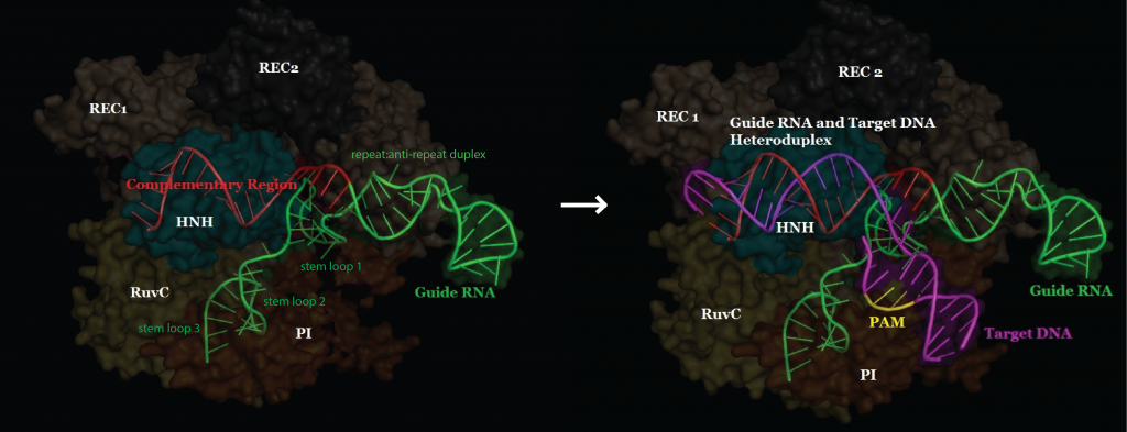
“I had never heard that word,” Zhang told me recently as we sat in his office, which looks out across the Charles River and Beacon Hill. Zhang has a perfectly round face, its shape accentuated by rectangular wire-rimmed glasses and a bowl cut. “So I went to Google just to see what was there,” he said. Zhang read every paper he could; five years later, he still seemed surprised by what he found. CRISPR, he learned, was a strange cluster of DNA sequences that could recognize invading viruses, deploy a special enzyme to chop them into pieces, and use the viral shards that remained to form a rudimentary immune system. The sequences, identical strings of nucleotides that could be read the same way backward and forward, looked like Morse code, a series of dashes punctuated by an occasional dot. The system had an awkward name—clustered regularly interspaced short palindromic repeats—but a memorable acronym.
CRISPR has two components. The first is essentially a cellular scalpel that cuts DNA. The other consists of RNA, the molecule most often used to transmit biological information throughout the genome. It serves as a guide, leading the scalpel on a search past thousands of genes until it finds and fixes itself to the precise string of nucleotides it needs to cut. It has been clear at least since Louis Pasteur did some of his earliest experiments into the germ theory of disease, in the nineteenth century, that the immune systems of humans and other vertebrates are capable of adapting to new threats. But few scientists had considered the possibility that single bacterial cells could defend themselves in the same way. The day after Zhang heard about CRISPR, he flew to Florida for a genetics conference. Rather than attend the meetings, however, he stayed in his hotel room and kept Googling. “I just sat there reading every paper on CRISPR I could find,” he said. “The more I read, the harder it was to contain my excitement.”
It didn’t take Zhang or other scientists long to realize that, if nature could turn these molecules into the genetic equivalent of a global positioning system, so could we. Researchers soon learned how to create synthetic versions of the RNA guides and program them to deliver their cargo to virtually any cell. Once the enzyme locks onto the matching DNA sequence, it can cut and paste nucleotides with the precision we have come to expect from the search-and-replace function of a word processor. “This was a finding of mind-boggling importance,” Zhang told me. “And it set off a cascade of experiments that have transformed genetic research.”
With CRISPR, scientists can change, delete, and replace genes in any animal, including us. Working mostly with mice, researchers have already deployed the tool to correct the genetic errors responsible for sickle-cell anemia, muscular dystrophy, and the fundamental defect associated with cystic fibrosis. One group has replaced a mutation that causes cataracts; another has destroyed receptors that H.I.V. uses to infiltrate our immune system.
The potential impact of CRISPR on the biosphere is equally profound. Last year, by deleting all three copies of a single wheat gene, a team led by the Chinese geneticist Gao Caixia created a strain that is fully resistant to powdery mildew, one of the world’s most pervasive blights. In September, Japanese scientists used the technique to prolong the life of tomatoes by turning off genes that control how quickly they ripen. Agricultural researchers hope that such an approach to enhancing crops will prove far less controversial than using genetically modified organisms, a process that requires technicians to introduce foreign DNA into the genes of many of the foods we eat.
The technology has also made it possible to study complicated illnesses in an entirely new way. A few well-known disorders, such as Huntington’s disease and sickle-cell anemia, are caused by defects in a single gene. But most devastating illnesses, among them diabetes, autism, Alzheimer’s, and cancer, are almost always the result of a constantly shifting dynamic that can include hundreds of genes. The best way to understand those connections has been to test them in animal models, a process of trial and error that can take years. CRISPR promises to make that process easier, more accurate, and exponentially faster.
Inevitably, the technology will also permit scientists to correct genetic flaws in human embryos. Any such change, though, would infiltrate the entire genome and eventually be passed down to children, grandchildren, great-grandchildren, and every subsequent generation. That raises the possibility, more realistically than ever before, that scientists will be able to rewrite the fundamental code of life, with consequences for future generations that we may never be able to anticipate. Vague fears of a dystopian world, full of manufactured humans, long ago became a standard part of any debate about scientific progress. Yet not since J. Robert Oppenheimer realized that the atomic bomb he built to protect the world might actually destroy it have the scientists responsible for a discovery been so leery of using it.
For much of the past century, biology has been consumed with three essential questions: What does each gene do? How do we find the genetic mutations that make us sick? And how can we overcome them? With CRISPR, the answers have become attainable, and we are closing in on a sort of grand unified theory of genetics. “I am not sure what a Golden Age looks like,” Winston Yan, a member of Zhang’s research team, told me one day when I was with him in the lab, “but I think we are in one.”
At least since 1953, when James Watson and Francis Crick characterized the helical structure of DNA, the central project of biology has been the effort to understand how the shifting arrangement of four compounds—adenine, guanine, cytosine, and thymine—determines the ways in which humans differ from each other and from everything else alive. CRISPR is not the first system to help scientists pursue that goal, but it is the first that anyone with basic skills and a few hundred dollars’ worth of equipment can use.
“CRISPR is the Model T of genetics,” Hank Greely told me when I visited him recently, at Stanford Law School, where he is a professor and the director of the Center for Law and the Biosciences. “The Model T wasn’t the first car, but it changed the way we drive, work, and live. CRISPR has made a difficult process cheap and reliable. It’s incredibly precise. But an important part of the history of molecular biology is the history of editing genes.”
Scientists took the first serious step toward controlling our genes in the early nineteen-seventies, when they learned to cut chains of DNA by using proteins called restriction enzymes. Suddenly, genes from organisms that would never have been able to mate in nature could be combined in the laboratory. But those initial tools were more hatchet than scalpel, and, because they could recognize only short stretches within the vast universe of the human genome, the editing was rarely precise. (Imagine searching through all of Shakespeare for Hamlet’s soliloquy on suicide, relying solely on the phrase “to be.” You’d find the passage, but only after landing on several hundred unrelated citations.)
When the first draft of the Human Genome Project was published, in 2001, the results were expected to transform our understanding of life. In fundamental ways, they have; the map has helped researchers locate thousands of genes associated with particular illnesses, including hundreds that cause specific types of cancer. To understand the role that those genes play in the evolution of a disease, however, and repair them, scientists need to turn genes on and off systematically and in many combinations. Until recently, though, altering even a single gene took months or years of work.
That began to change with the growing use of zinc fingers, a set of molecular tools that, like CRISPR clusters, were discovered by accident. In 1985, scientists studying the genetic code of the African clawed frog noticed a finger-shaped protein wrapped around its DNA. They soon figured out how to combine that tenacious grip with an enzyme that could cut the DNA like a knife. Two decades later, geneticists began using TALEs, which are made up of proteins secreted by bacteria. But both engineering methods are expensive and cumbersome. Even Zhang, who published the first report on using TALEs to alter the genes of mammals, realized that the system was little more than an interim measure. “It is difficult to use,” he told me. “I had to assign a graduate student just to make the proteins and test them before I could begin to use them in an experiment. The procedure was not easy.”
Zhang’s obsession with science began in middle school, when his mother prodded him to attend a Saturday-morning class in molecular biology. “I was thirteen and had no idea what molecular biology was,” he said one evening as we walked across the M.I.T. campus on the way to the fiftieth-anniversary celebration of the Department of Brain and Cognitive Sciences, where Zhang is also a faculty member. “It really opened my imagination.” His parents, both engineers, moved the family to Iowa when he was eleven. They stayed largely because they thought he would get a better education in the United States than in China.
In 1997, when Zhang was fifteen, he was offered an internship in a biosafety facility at the Des Moines Human Gene Therapy Research Institute—but he was told that federal law prohibited him from working in a secure lab until he was sixteen. “So I had to wait,” he said. On his birthday, Zhang went to the lab and met the scientists. “I was assigned to a man who had a Ph.D. in chemistry but trained as a molecular biologist,” he continued. “He had a lot of passion for science, and he had a very big impact on me and my research.” On his first day, Zhang spent five hours in the lab, and nearly as much time every day after school until he graduated.
Zhang is unusually reserved, and he speaks in low, almost sleepy tones. I asked him if he considered himself to be mellow, a characteristic rarely associated with prize-winning molecular biologists. “You came to the lab meeting, right?” he replied. Earlier that morning, I had caught the tail end of a weekly meeting that Zhang holds for his group. I watched as he gently but relentlessly demolished a presentation given by one of the people on his team. When I mentioned it to one of the scientists who was at the meeting, he responded, “That was nothing. You should have been there from the start.”
At his Saturday-morning classes, Zhang learned how to extract DNA from cells and determine the length of each sequence. But that isn’t what he remembers best. “They showed us ‘Jurassic Park,’ ” he said, his voice moving up a register. “And it was amazing to me. The teacher explained the different scientific concepts in the movie, and they all seemed completely feasible.”
We had reached the cocktail party, a tepid affair crowded with men in khakis and women wearing sensible shoes. Zhang left after barely twenty minutes and headed back to the lab. He retains his position on the cognitive-sciences faculty, because he hopes that his research will help neuroscientists study the brain in greater detail. He told me that when he was young he had a friend who suffered from serious depression, and he had been surprised to find that there was almost no treatment available. It spurred a lasting interest in psychiatry. “People think you are weak if you are depressed,” he said. “It is still a common prejudice. But many people suffer from problems we cannot begin to address. The brain is still the place in the universe with the most unanswered questions.”
The Broad Institute was founded, in 2003, by the entrepreneur Eli Broad and his wife, Edythe, to foster research into the molecular components of life and their connections to disease. One afternoon in Zhang’s laboratory, Winston Yan offered to walk me through the mechanics of using CRISPR to edit a gene. “We need to be able to break DNA in a very precise place in the genome,” he said as I watched him at work. He swivelled in his chair and pointed to a row of vials that contained DNA samples to be analyzed and edited. Yan, a thin, bespectacled man, wore black laboratory gloves and a white Apple Watch; he clapped his hands and shrugged, as if to suggest that the work was simple.
Ordering the genetic parts required to tailor DNA isn’t as easy as buying a pair of shoes from Zappos, but it seems to be headed in that direction. Yan turned on the computer at his lab station and navigated to an order form for a company called Integrated DNA Technologies, which synthesizes biological parts. “It takes orders online, so if I want a particular sequence I can have it here in a day or two,” he said. That is not unusual. Researchers can now order online almost any biological component, including DNA, RNA, and the chemicals necessary to use them. One can buy the parts required to assemble a working version of the polio virus (it’s been done) or genes that, when put together properly, can make feces smell like wintergreen. In Cambridge, I.D.T. often makes same-day deliveries. Another organization, Addgene, was established, more than a decade ago, as a nonprofit repository that houses tens of thousands of ready-made sequences, including nearly every guide used to edit genes with CRISPR. When researchers at the Broad, and at many other institutions, create a new guide, they typically donate a copy to Addgene.
The RNA that CRISPR relies upon to guide the molecular scalpel to its target is made of twenty base pairs. Humans have twenty thousand genes, and twenty base pairs occupy roughly the same percentage of space in a single gene as would one person standing in a circle that contained the entire population of the United States. CRISPR is better at locating specific genes than any other system, but it isn’t perfect, and sometimes it cuts the wrong target. Yan would order a ready-made probe from Addgene. When it arrives, he pairs it with a cutting enzyme and sends it to the designated gene.
Yan joined Zhang’s lab just before what he described as “the CRISPR craze” began. But, he added, the technology has already transformed the field. “For many years, there was a reductionist approach to genetics,” he said. “A kind of wishful thinking: ‘We will find the gene that causes cancer or the gene that makes you prone to heart disease.’ It is almost never that simple.”
The next morning, I walked over to the Broad’s new Stanley Building and rode the elevator to the top floor, where I emptied my pockets, put on a mask and gown, and slipped booties over my shoes. Then I passed through an air chamber that was sealed with special gaskets and had a fan blowing continuously to keep out foreign microbes. I entered the vivarium, a long, clean floor that looked like a combination of research unit and hospital ward. The vivarium, which opened last year, provides thousands of mice with some of the world’s most carefully monitored accommodations.
Despite our growing knowledge of the way that cancer develops in human cells, mutations can’t be studied effectively in a petri dish, and, since the late nineteen-eighties, genetically modified mice have served as the standard proxy. What cures (or kills) a mouse won’t necessarily have the same effect on a human, but the mouse genome is surprisingly similar to our own, and the animals are cheap and easy to maintain. Like humans, and many other mammals, mice develop complex diseases that affect the immune system and the brain. They get cancer, atherosclerosis, hypertension, and diabetes, among other chronic illnesses. Mice also reproduce every three weeks, which allows researchers to follow several generations at once. Typically, technicians would remove a stem cell from the mouse, then edit it in a lab to produce a particular gene or to prevent the gene from working properly. After putting the stem cell back into the developing embryo of the mouse, and waiting for it to multiply, they can study the gene’s effect on the animal’s development. The process works well, but it generally allows for the study of only one characteristic in one gene at a time.
The vivarium at the Broad houses an entirely different kind of mouse, one that carries the protein Cas9 (which stands for CRISPR-associated nuclease) in every cell. Cas9, the part of the CRISPR system that acts like a genetic scalpel, is an enzyme. When scientists originally began editing DNA with CRISPR, they had to inject both the Cas9 enzyme and the probe required to guide it. A year ago, Randall Platt, another member of Zhang’s team, realized that it would be possible to cut the CRISPR system in two. He implanted the surgical enzyme into a mouse embryo, which made it a part of the animal’s permanent genome. Every time a cell divided, the Cas9 enzyme would go with it. In other words, he and his colleagues created a mouse that was easy to edit. Last year, they published a study explaining their methodology, and since then Platt has shared the technique with more than a thousand laboratories around the world.
The “Cas9 mouse” has become the first essential tool in the emerging CRISPR arsenal. With the enzyme that acts as molecular scissors already present in every cell, scientists no longer have to fit it onto an RNA guide. They can dispatch many probes at once and simply make mutations in the genes they want to study.
To demonstrate a potential application for cancer research, the team used the Cas9 mouse to model lung adenocarcinoma, the most common form of lung cancer. Previously, scientists working with animal models had to modify one gene at a time or cross-breed animals to produce a colony with the needed genetic modifications. Both processes were challenging and time-consuming. “Now we can activate CRISPR directly in the cells we’re interested in studying, and modify the genome in whatever way we want,” Platt said, as he showed me around the vivarium. We entered a small exam room with a commanding view of Cambridge. I watched as a technician placed a Cas9 mouse in a harness inside a biological safety cabinet. Then, peering through a Leica microscope, she used a fine capillary needle to inject a single cell into the mouse’s tail.
“And now we have our model,” Platt said, explaining that the mouse had just received an injection that carried three probes, each of which was programmed to carry a mutation that scientists believe is associated with lung cancer. “The cells will carry as many mutations as we want to study. That really is a revolutionary development.”
“In the past, this would have taken the field a decade, and would have required a consortium,” Platt said. “With CRISPR, it took me four months to do it by myself.” In September, Zhang published a report, in the journal Cell, describing yet another CRISPR protein, called Cpf1, that is smaller and easier to program than Cas9.
The lab employs a similar approach to studying autism. Recent experiments suggest that certain psychiatric conditions can be caused by just a few malfunctioning neurons out of the trillions in every brain. Studying the way neurons function within the brain is difficult. But by re-creating, in the lab, genetic mutations that others have linked to autism and schizophrenia Zhang’s team has been able to investigate faulty neurons that may play a role in those conditions.
As the price of sequencing plunges, cancer clinics throughout the United States have begun to study their patients’ tumors in greater detail. Tumors are almost never uniform; one may have five mutations or fifty, which means, essentially, that every cancer is a specific, personal disease. Until CRISPR became available, the wide genetic variations in cancer cells often made it hard to develop effective treatments.
“What I love most about the CRISPR process is that you can take any cancer-cell line, knock out every gene, and identify every one of the cell’s Achilles’ heels,” Eric Lander, the fifty-eight-year-old director of the Broad, told me recently. Lander, who was among the leaders of the Human Genome Project, said that he had never encountered a more promising research tool. “You can also use CRISPR to systematically study the ways that a cancer cell can escape from a treatment,” he said. “That should make it possible to build a comprehensive road map for cancer.”
Lander went on to say that each vulnerability of a tumor might be attacked by a single drug. But cancer cells elude drugs in many ways, and, to succeed, a therapy may need to block them all. That strategy has proved effective for infectious diseases like AIDS. “Remember the pessimism about H.I.V.,” he said, referring to the early years of the AIDS epidemic, when a diagnosis was essentially a death sentence. Eventually, virologists developed a series of drugs that interfere with the virus’s ability to replicate. The therapy became truly successful, however, only when those drugs, working together, could block the virus completely.
The same approach has proved successful in treating tuberculosis. Lander is convinced that it will also work for many cancers: “With triple-drug therapy,” for H.I.V., “we reached an inflection point: we were losing badly, and one day suddenly we were winning.”
He stood up and walked across the office toward his desk, then pointed at the wall and described his vision for the future of cancer treatment. “There will be an enormous chart,” he said. “Well, it will be electronic, and it will contain the therapeutic road map of every trick that cancer cells have—how they form, all the ways you can defeat them, and all the ways they can escape and defeat a treatment. And when we have that we win. Because every cancer cell starts naïve. It doesn’t know what we have waiting in the freezer for it. Infectious diseases are a different story; they share their knowledge as they spread. They learn from us as they move from person to person. But every person’s cancer starts naïve. And this is why we will beat it.”
Developing any technology as complex and widely used as CRISPR invariably involves contributions from many scientists. Patent fights over claims of discovery and licensing rights are common. Zhang, the Broad Institute, and M.I.T. are now embroiled in such a dispute with Jennifer Doudna and the University of California; she is a professor of chemistry and of molecular biology at Berkeley. By 2012, Doudna, along with Emmanuelle Charpentier, a medical microbiologist who studies pathogens at the Helmholtz Centre for Infection Research, in Germany, and their lab teams, demonstrated, for the first time, that CRISPR could edit purified DNA. Their paper was published that June. In January of 2013, though, Zhang and George Church, a professor of genetics at both Harvard Medical School and M.I.T., published the first studies demonstrating that CRISPR could be used to edit human cells. Today, patents are generally awarded to the first people to file—in this case, Doudna and Charpentier. But Zhang and the Broad argued that the earlier success with CRISPR had no bearing on whether the technique would work in the complex organisms that matter most to scientists looking for ways to treat and prevent diseases.
Zhang was awarded the patent, but the University of California has requested an official reassessment, and a ruling has not yet been issued. Both he and Doudna described the suit to me as “a distraction” that they wished would go away. Both pledged to release all intellectual property to researchers without charge (and they have). But both are also involved in new companies that intend to develop CRISPR technology as therapies, as do many pharmaceutical firms and other profit-seeking enterprises.
CRISPR research is becoming big business: venture-capital firms are competing with one another to invest millions, and any patent holder would have the right to impose licensing fees. Whoever wins stands to make a fortune. Other achievements are also at stake, possibly including a Nobel Prize. (Doudna’s supporters have described her as America’s next female Nobel Prize winner, and at times the publicity war seems a bit like the battles waged by movie studios during Academy Award season.) Last year, the National Science Foundation presented Zhang with its most prestigious award, saying that his fundamental research “moves us in the direction” of eliminating schizophrenia, autism, and other brain disorders. A few months later, Doudna and Charpentier received three million dollars each for the Breakthrough Prize, awarded each year for scientific achievement. The prize was established, in 2012, by several Silicon Valley billionaires who are seeking to make science a more attractive career path. The two women also appeared on Time’s annual list of the world’s hundred most influential people.
In fact, neither group was involved in the earliest identification of CRISPR or in the first studies to demonstrate how it works. In December, 1987, biologists at the Research Institute of Microbial Diseases, in Osaka, Japan, published the DNA sequence of a gene taken from the common intestinal bacterium E. coli. Those were early days in the genomic era, and thousands of labs around the world had embarked on similar attempts to map the genes of species ranging from fruit flies to humans. In an effort to better understand how this particular gene functioned, the Japanese scientists also sequenced some of the DNA that surrounded it. When they examined the data, they were surprised to see cellular structures that none of them recognized: they had no idea what to make of the strange phenomenon, but they took note of it, writing in the final sentence of their report, published in the Journal of Bacteriology, that the “biological significance of these sequences is not known.”
The mystery remained until 2005, when Francisco Mojica, a microbiologist at the University of Alicante, who had long sought to understand CRISPR, decided to compare its DNA with the DNA of tens of thousands of similar organisms. What he saw amazed him: every unknown sequence turned out to be a fragment of DNA from an invading virus.
The pace of research quickened. In 2007, Rodolphe Barrangou and Philippe Horvath, microbiologists then working for Danisco, the Danish food company, had noticed that some of its yogurt cultures were routinely destroyed by viruses and others were not. They decided to find out why. The scientists infected the microbe Streptococcus thermophilus, which is widely used to make yogurt, with two viruses. Most of the bacteria died, but those which survived had one property in common: they all contained CRISPR molecules to defend them.
“No single person discovers things anymore,” George Church told me when we met in his office at Harvard Medical School. “The whole patent battle is silly. There has been much research. And if anybody should be making a fuss about this I should be making a fuss. But I am not doing that, because I don’t think it matters. They are all nice people. They are all doing important work. It’s a tempest in a teapot.”
From the moment that manipulating genes became possible, many people, including some of those involved in the experiments, were horrified by the idea of scientists in lab coats rearranging the basic elements of life. In 1974, David Baltimore, the pioneering molecular biologist, who was then at M.I.T., and Paul Berg, of Stanford, both of whom went on to win a Nobel Prize for their research into the fundamentals of viral genetics, called for a moratorium on gene-editing research until scientists could develop safety principles for handling organisms that contained recombinant DNA. That meeting, which took place in 1975, at a conference center in Asilomar, California, has come to be regarded as biotechnology’s Constitutional Convention.
Roughly a hundred and fifty participants, most of them scientists, gathered to discuss ways to limit the risks of accidentally releasing genetically modified organisms. At the time, the possibility of creating “designer babies”—a prospect that, no matter how unlikely, is attached to almost everything written or said about CRISPR—was too remote to consider. Nevertheless, the technology seemed frightening. In Cambridge, home to both M.I.T. and Harvard, the city council nearly banned such research altogether. The work went on, but decoding sequences of DNA wasn’t easy. “In 1974, thirty base pairs”—thirty rungs on the helical ladder of the six billion nucleotides that make up our DNA—“was a good year’s work,” George Church told me. Now the same work would take seconds.
At least for the foreseeable future, CRISPR’s greatest impact will lie in its ability to help scientists rapidly rewrite the genomes of animal and plant species. In laboratories, agricultural companies have already begun to use CRISPR to edit soybeans, rice, and potatoes in an effort to make them more nutritious and more resistant to drought. Scientists might even be able to edit allergens out of foods like peanuts.
Normally, it takes years for genetic changes to spread through a population. That is because, during sexual reproduction, each of the two versions of any gene has only a fifty per cent chance of being inherited. But a “gene drive”—which is named for its ability to propel genes through populations over many generations—manages to override the traditional rules of genetics. A mutation made by CRISPR on one chromosome can copy itself in every generation, so that nearly all descendants would inherit the change. A mutation engineered into a mosquito that would block the parasite responsible for malaria, for instance, could be driven through a large population of mosquitoes within a year or two. If the mutation reduced the number of eggs produced by that mosquito, the population could be wiped out, along with any malaria parasites it carried.
Kevin Esvelt, an evolutionary biologist at Harvard, was the first to demonstrate how gene drives and CRISPR could combine to alter the traits of wild populations. Recently, he has begun to study the possibility of using the technology to eliminate Lyme disease by rewriting the genes of mice in the wild. Lyme disease is caused by a bacterium and transmitted by ticks, and more than eighty-five per cent of the time they become infected after biting a mouse. Once exposed, however, some mice naturally acquire resistance or immunity. “My idea is to take the existing genes that confer resistance to Lyme and make sure that all mice have the most effective version,’’ Esvelt said. To do that, scientists could encode the most protective genes next to the CRISPR system and force them to be passed on together. Esvelt stressed that such an approach would become possible only after much more research and a lengthy series of public discussions on the risks and benefits of the process.
The promise of CRISPR research becomes more evident almost every month. Recently, Church reported that he had edited sixty-two genes simultaneously in a pig cell. The technique, if it proves accurate and easy to repeat, could help alleviate the constant shortage of organ donors in the U.S. For years, scientists have tried to find a way to use pig organs for transplants, but a pig’s DNA is filled with retroviruses that have been shown in labs to infect human cells. Church and his colleagues discovered that those viruses share a common genetic sequence. He deployed CRISPR to their exact locations and snipped them out of the genome. In the most successful of the experiments, the CRISPR system deleted all sixty-two of the retroviruses embedded in the pig’s DNA. Church then mixed those edited cells with human cells in the laboratory, and none became infected.
While CRISPR will clearly make it possible to alter our DNA, serious risks remain. Jennifer Doudna has been among the most vocal of those calling for caution on what she sees as the inevitable march toward editing human genes. “It’s going to happen,” she told me the first time we met, in her office at Berkeley. “As a research tool, CRISPR could hardly be more valuable—but we are far from the day when it should be used in a clinical setting.” Doudna was a principal author of a letter published in Science this spring calling for a temporary research moratorium. She and others have organized a conference to discuss the ethics of editing DNA, a sort of Asilomar redux. The conference, to be attended by more than two hundred scientists—from the U.S., England, and China, among other countries—will take place during the first week of December at the National Academy of Sciences, in Washington.
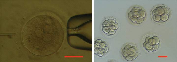
Until April, the ethical debate over the uses of CRISPR technology in humans was largely theoretical. Then a group at Sun Yat-sen University, in southern China, attempted to repair, in eighty-six human embryos, the gene responsible for betathalassemia, a rare but often fatal blood disorder. If those disease genes, and genes that cause conditions like cystic fibrosis, could be modified successfully in a fertilized egg, the alteration could not only protect a single individual but eventually eliminate the malady from that person’s hereditary lineage. Given enough time, the changes would affect all of humanity. The response to the experiment was largely one of fear and outrage. The Times carried the story under the headline “CHINESE SCIENTISTS EDIT GENES OF HUMAN EMBRYOS, RAISING CONCERNS.”
Critics called the experiment irresponsible and suggested that the scientists had violated an established code of conduct. “This paper demonstrates the enormous safety risks that any such attempt would entail, and underlines the urgency of working to forestall other such efforts,” Marcy Darnovsky, of the Center for Genetics and Society, told National Public Radio when the report was published. “The social dangers of creating genetically modified human beings cannot be overstated.”
There seems to be little disagreement about that. But the Chinese researchers were not trying to create genetically modified humans. They were testing the process, and every CRISPR researcher I spoke to considered the experiment to have been well planned and carried out with extraordinary care. The scientists also agreed that the results were illuminating. “That was an ethical paper, and a highly responsible project,’’ Lander told me. “What did they do? They took triploid zygotes’’—a relatively common genetic aberration—“from I.V.F. clinics. They deliberately chose those because they knew no human could ever develop from them. And what did the paper say? ‘Boy, we see problems everywhere.’ That was good science, and it was cautionary.”
Fewer than half the embryos were edited successfully, and, of those, most retained none of the new DNA that was inserted into the genes. The experiment, which was published in the Beijing-based journal Protein & Cell, demonstrated clearly that the day when scientists could safely edit humans is far off. The CRISPR system also made unintended cuts and substitutions, the potential effects of which are unknown. In other cases, it made the right changes in some cells of the embryo but not in all of them, which could cause other problems. “These authors did a very good job, pointing out the challenges,” Dieter Egli, a stem-cell researcher at Columbia University, said when the study was published. “They say themselves that this type of technology is not ready for any kind of application.”
Doudna agreed that the Chinese experiment yielded valuable results. She is fifty-one, and has been at Berkeley since 2002, when she and her husband, the biochemist Jamie Cate, were offered joint appointments to the departments of chemistry and molecular and cell biology. Their offices are next to each other, with the same commanding view of San Francisco Bay and the Golden Gate Bridge. Doudna’s work, unlike that of the scientists at the Broad, has been focussed on molecules, not mammalian genetics. For years, she has been leading investigations into the shape, structure, and capabilities of RNA, and in 2011 Charpentier asked for her help in exploring the mechanism of CRISPR. Doudna is tall, with graying blond hair and piercing blue eyes. She grew up in Hawaii, where her parents were academics; when it was time for college, she decided to leave the island and study in California, at Pomona. She earned her doctorate at Harvard and then moved on to Yale. “I have always been a bit of a restless soul,” she said. “I may spend too much time wondering what comes next.”
Doudna is a highly regarded biochemist, but she told me that not long ago she considered attending medical school or perhaps going into business. She said that she wanted to have an effect on the world and had begun to fear that the impact of her laboratory research might be limited. The promise of her work on CRISPR, however, has persuaded her to remain in the lab. She told me that she was constantly amazed by its potential, but when I asked if she had ever wondered whether the powerful new tool might do more harm than good she looked uncomfortable. “I lie in bed almost every night and ask myself that question,” she said. “When I’m ninety, will I look back and be glad about what we have accomplished with this technology? Or will I wish I’d never discovered how it works?”
Her eyes narrowed, and she lowered her voice almost to a whisper. “I have never said this in public, but it will show you where my psyche is,” she said. “I had a dream recently, and in my dream”—she mentioned the name of a leading scientific researcher—“had come to see me and said, ‘I have somebody very powerful with me who I want you to meet, and I want you to explain to him how this technology functions.’ So I said, Sure, who is it? It was Adolf Hitler. I was really horrified, but I went into a room and there was Hitler. He had a pig face and I could only see him from behind and he was taking notes and he said, ‘I want to understand the uses and implications of this amazing technology.’ I woke up in a cold sweat. And that dream has haunted me from that day. Because suppose somebody like Hitler had access to this—we can only imagine the kind of horrible uses he could put it to.”
Nobody is going to employ CRISPR technology to design a baby, let alone transform the genetic profile of humanity, anytime soon. Even if scientists become capable of editing human embryos, it would take years for the genetically modified baby to grow old enough to reproduce—and then many generations for the alteration to disseminate throughout the population.
But there are long-term consequences to consider. Modern medicine already shapes our genome, by preserving genes that might otherwise have been edited out of our genome by natural selection. Today, millions of people suffer from myopia, and many of them are legally blind. Were it not for the invention of glasses, which have turned poor eyesight largely into a nuisance rather than an existential threat, the genes responsible for myopia might be less prevalent than they are today. The same could be said about many infectious diseases, and even chronic conditions like diabetes.
Humans also carry genes that protect us from one disease but increase our susceptibility to others, and it’s impossible to predict the impact of changing all or even most of them. The AIDS virus often enters our blood cells through a protein called CCR5. One particular genetic variant of that protein, called the Delta32 mutation, prevents H.I.V. from locking onto the cell. If every person carried that mutation, nobody would get AIDS. So why not introduce that mutation into the human genome? Several research teams are working to develop drugs that do that in people who have already been infected.
Yet it’s important to note that, while such a procedure would prevent H.I.V. infection, it would also elevate our susceptibility to West Nile virus. Today, that trade-off may seem worth the risk, but there’s no way of knowing whether it would be true seven or ten generations from now. For example, sickle cells, which cause anemia, evolved as a protection against malaria; the shape of the cell blocks the spread of the parasite. If CRISPR technology had been available two hundred thousand years ago, it might have seemed sensible to edit sickle cells into the entire human population. But the results would have been devastating.
“This is a little bit like geoengineering,” Zhang told me, referring to attempts to deliberately alter the climate to offset damages associated with global warming. “Once you go down that path, it may not be so reversible.”
George Church disagrees. “It strikes me as a fake argument to say that something is irreversible,” he told me. “There are tons of technologies that are irreversible. But genetics is not one of them. In my lab, we make mutations all the time and then we change them back. Eleven generations from now, if we alter something and it doesn’t work properly we will simply fix it.”
In 1997, Scottish scientists shocked the world by announcing that they had cloned a lamb, which they named Dolly. Scores of journalists (including me) descended on Edinburgh, and wrote that the achievement, while wondrous, also carried the ominous implication that scientists had finally pried open Pandora’s box. Many articles about cloning and the value of human life were published. Evil people and dictators would clone themselves, their children, their pets. A new class of humans would arise.
Eighteen years later, the closest we have come to cloning a person was a failed attempt at a monkey, in 2007. Nobody spends much time worrying about it today. In Cambridge this summer, one of the researchers at the Broad told me that he and Louise Brown, the first success of in-vitro fertilization, were both born in 1978. “Did that set off an uproar?” he asked. It did. Even seven years earlier, James Watson had written, in The Atlantic, that the coming era of designer babies might overwhelm us all. Today, though, with more than five million children on earth born through in-vitro fertilization, that particular furor, too, seems to have passed.
CRISPR technology offers a new outlet for the inchoate fear of tinkering with the fundamentals of life. There are many valid reasons to worry. But it is essential to assess both the risks and the benefits of any new technology. Most people would consider it dangerous to fundamentally alter the human gene pool to treat a disease like AIDS if we could cure it with medicine or a vaccine. But risks always depend on the potential result. If CRISPR helps unravel the mysteries of autism, contributes to a cure for a form of cancer, or makes it easier for farmers to grow more nutritious food while reducing environmental damage, the fears, like the many others before them, will almost certainly disappear.
2.1.3.10 Can CRISPR/Cas9 Target Multiple Targets? Volume 2 (Volume Two: Latest in Genomics Methodologies for Therapeutics: Gene Editing, NGS and BioInformatics, Simulations and the Genome Ontology), Part 2: CRISPR for Gene Editing and DNA Repair
CRISPR/Cas9 has advanced genome editing and revolutionized molecular biology perhaps even more than the restriction enzyme. But can it edit multiple targets efficiently?
For CRISPR/Cas9 editing, single guide RNAs (sgRNAs) direct the bacterial Cas9 endonuclease to specific loci, allowing targeting of almost any gene. But is it possible to efficiently target multiple genes? “You can express one, two, or even three sgRNAs pretty easily, but if you want to do four, five, or more, it becomes difficult,” Yinong Yang at Pennsylvania State University said.
Yang’s team addressed this question in a Proceedings of the National Academy of Science paper by turning to the cell’s own tRNA processing systems. The group created polycistronic tRNA-gRNA (PTG) constructs that consisted of an sgRNA flanked by a pre-tRNA gene; the cell’s endogenous RNases can then cleave one or multiple transcribed gRNAs from the cistron to direct Cas9 to target genes.
Schematic depiction of the synthetic tRNA-gRNA gene. Credit: Yinong Yang.
“The beauty of this approach is that the 77 bp pre-tRNA gene contains internal promoter elements (box A and B) to recruit the RNA Pol III complex, so maybe you don’t even need a promoter. The Pol III promoter [which is currently used to drive expression of the sgRNA] isn’t very strong, so the tRNA will give you enhanced expression of multiple RNAs.”
The group first tested the PTG in rice protoplasts and soon realized that existing CRISPR/Cas9 vectors can be used to express PTGs. They also observed that the PTGs were more effective at introducing insertions or deletions than sgRNAs, perhaps owing to their higher expression levels from the endogenous tRNA enhancers.
Yang and his colleagues next asked if it was possible to introduce deletions in multiple genes by targeting the MAP kinase components MPK1, MPK2, MPK5, and MPK6 individually and in combinations of two or four. The PTG system introduced deletions for up to four genes, although there was a two-fold reduction in editing efficiency, which the authors attribute to competition for Cas9 among the multiple gRNAs. They then usedAgrobacterium-mediated transformation to transform mature rice plants with sgRNAs or PTGs for MPK genes and observed a higher mutational frequency of bi-allelic mutations and deletions in the plants transformed with the PTGs. Finally, they were able to target the phytoene desaturase (PDS) gene to generate a photo-bleached phenotype in the resulting plants. While they only obtained a single line carrying the fragment deletion of PDS, the mutational efficiency for PTGs was 100 percent.
Reference:
Xie, K, Minkenberg B, and Yang, Y. Boosting CRISPR/Cas9 multiplex editing capability with the endogenous tRNA-processing system. Proc Natl Acad Sci U S A. 2015 Mar 17;112(11):3570-5. doi: 10.1073/pnas.1420294112.
Boosting CRISPR/Cas9 multiplex editing capability with the endogenous tRNA-processing system
Kabin Xie, Bastian Minkenberg, and Yinong Yang1
Department of Plant Pathology and Environmental Microbiology and the Huck Institutes of the Life Sciences,
Pennsylvania State University, University Park, PA 16802
Edited by Jennifer A. Doudna, University of California, Berkeley, CA, and approved February 3, 2015
The clustered regularly interspaced short palindromic repeat (CRISPR)/ CRISPR-associated protein 9 nuclease (Cas9) system is being harnessed as a powerful tool for genome engineering in basic research, molecular therapy, and crop improvement. This system uses a small guide RNA (gRNA) to direct Cas9 endonuclease to a specific DNA site; thus, its targeting capability is largely constrained by the gRNA-expressing device. In this study, we developed a general strategy to produce numerous gRNAs from a single polycistronic gene. The endogenous tRNA-processing system, which precisely cleaves both ends of the tRNA precursor, was engineered as a simple and robust platform to boost the targeting and multiplex editing capability of the CRISPR/ Cas9 system. We demonstrated that synthetic genes with tandemly arrayed tRNA–gRNA architecture were efficiently and precisely processed into gRNAs with desired 5′ targeting sequences in vivo, which directed Cas9 to edit multiple chromosomal targets. Using this strategy, multiplex genome editing and chromosomal-fragment deletion were readily achieved in stable transgenic rice plants with a high efficiency (up to 100%). Because tRNA and its processing system are virtually conserved in all living organisms, this method could be broadly used to boost the targeting capability and editing efficiency of CRISPR/Cas9 toolkits.
Significance The clustered regularly interspaced short palindromic repeat (CRISPR)/CRISPR-associated protein 9 nuclease (Cas9) system has recently emerged as an efficient and versatile tool for genome editing in various organisms. However, its targeting capability and multiplex editing efficiency are often limited by the guide RNA (gRNA)-expressing device. This study demonstrates a general strategy and platform for precise processing and efficient production of numerous gRNAs in vivo from a synthetic polycistronic gene via the endogenous tRNA-processing system. This strategy is shown to significantly increase CRISPR/Cas9 multiplex editing capability and efficiency in plants and is expected to have broad applications for small RNA expression and genome engineering.
Fig. 1. Engineering the endogenous tRNA system for multiplex genome editing with CRISPR/Cas9. (A) The eukaryotic pre-tRNA with 5′ leader and 3′ trailer is cleaved by RNase P and RNase Z at specific sites. (B) Transcription of tRNA gene with RNA polymerase III (Pol III). The box A and box B elements in the tRNA gene function as internal transcriptional elements and are bound by transcription factor IIIC (TFIII C), which recruits TFIIIB and Pol III to start transcription. (C) Schematic depiction of the PTG/Cas9 method for simultaneously targeting multiple sites. The synthetic PTG consists of tandemly arrayed tRNA-gRNA units, with each gRNA containing a target-specific spacer (labeled as a diamond with different color) and conserved gRNA scaffold (rectangle). The tRNA containing box A and B elements is shown as round rectangles. The primary transcript of PTG is cleaved by endogenous RNase P and RNase Z (labeled as scissors) to release mature gRNAs and tRNA (red lines of cloverleaf structure). The excised mature gRNAs direct Cas9 to multiple targets.
Strategy to Engineer a tRNA-processing System for Producing Numerous gRNAs
Precise Processing of PTG to Produce Functional gRNAs with Desired Targeting Sequences
Fig. 2. Precise excision of functional gRNAs in vivo from synthetic PTG genes. (A) The architecture of two sgRNA genes and four PTGs to produce one gRNA. (B) Sequence and predicted secondary structure of tRNA–gRNA–tRNA fusion of PTG gene. The bases of the tRNA region are indicated with red color and the tRNA 5′ leader is shown in lowercase. The gRNA is indicated in black, and the gRNA spacer sequence is shown as N. (C–F) Examination of mature gRNAs produced from sgRNA or PTGs with cRT-PCR. Total RNAs from the protoplasts expressing empty vector were used as control (CK). Arrows indicate mature gRNAs amplified by cRT-PCR, and asterisks indicate the nonspecifically amplified rRNA. (G) Summary of excision sites in PTG according to mapped gRNA ends from cRT-PCR (SI Appendix, Figs. S3–S5). Arrows indicate the cleavage sites in PTG to release gRNA. The mature gRNA 5′ ends were excised from PTG exactly at the tRNA–gRNA fusion site in all cRT-PCR results whereas its 3′ ends shifted 1–4 nt within the tRNA 5′ leader (lowercase). (H) gRNA produced from U3p:sgRNA. All detected U3p:sgRNA-produced gRNA started with ribonucleotide A and terminated with multiple Us. (I) Introduction of indels at the desired sites by PTG1:Cas9 or PTG2:Cas9 in rice protoplasts as shown by PCR/RE. Arrows indicate mutated fragments resistant to RE digestion. The indel frequency is indicated at the bottom. (J) Relative expression of sgRNA1/2 and PTG1/2 in rice protoplasts. Data represent mean ± SD. ND, not detected. CK, empty vector control.
Efficient Multiplex Genome Editing in Rice Protoplasts via PTG/Cas9.
Fig. 3. Simultaneous editing of multiple genomic sites in rice protoplasts expressing PTG:Cas9. (A) Architecture, gRNA components, and targets of PTGs for multiplex genome editing. (B) PCR detection of chromosomal fragment deletion at targeted loci in rice protoplasts expressing respective PTGs with Cas9. Successful deletion is shown as truncated PCR product (indicated with arrows). The chromosomal fragment deletion frequency (del %) is indicated at the bottom of each lane. The protoplast samples expressing an empty vector were used as control (CK). (C) Representative sequences of chromosomal fragment deletion aligned with that of WT. The gRNA paired region is labeled with green color, and the PAM region is shown in red color letters. The number at the end indicates deleted (−) or inserted (+) bases between two Cas9 cuts. The total length between two Cas9 cut sites (labeled with scissor) is indicated on the top. Short lines in the aligned sequences indicate deletions.
Improving Multiplex Genome Editing in Stable Transgenic Plants with PTG/Cas9
Table 1. Targeted mutation efficiency in PTG:Cas9 vs. sgRNA:Cas9 plants
Fig. 4. Highly efficient targeted mutagenesis in transgenic rice expressing PTG:Cas9. (A and B) Chromosomal fragment deletion in PTG7:Cas9 plant at T0 generation. Of note, only mpk1 with 358-bp deletion (Δ358) was detected in genomic DNA. Sequence analysis of the PCR products (the number in parentheses) reveals an identical deletion pattern in the transgenic plant. (C) Albino seedlings were regenerated from calli transformed with PTG10:Cas9. Most T0 seedlings (87%, n = 15) exhibited a similar photo-bleach phenotype, suggesting a very high efficiency of knocking out PDS with PTG10:Cas9. Vec, control plants transformed with empty vector. (Scale bar: 5 cm.)
We developed a general strategy and platform to produce multiple gRNAs from a single synthetic PTG gene by hijacking the endogenous tRNA-processing system (Fig. 1). We also provided a framework to design, synthesize, and use PTG for multiplex genome editing with Cas9. These PTGs were expressed with Pol III promoters (e.g., U3p) in the same manner as sgRNA genes but were not obligated to start with a specific nucleotide (Fig. 2). As a result, current CRISPR/Cas9 vectors for expressing sgRNA could be directly used to express PTG for multiplex genome editing as we demonstrated in this study.
By producing multiple gRNAs from a single polycistronic gene, the PTG technology could be used to improve simultaneous mutagenesis of multiple genomic loci or deletion of short chromosomal fragments (Figs. 3 and 4). Such a genome engineering approach may lead to simultaneous knock-out of multiple protein coding genes or deletion of noncoding RNA regions and other genetic elements. In addition to targeted mutagenesis/ deletion, the PTG approach could facilitate other Cas9-based applications in which multiple gRNAs are required. For example, PTG could be used with Cas9 nickase to improve targeting fidelity (13, 33, 34), or with deactivated Cas9 transcriptionalactivator or -repressor to manipulate multiple gene expression (35, 36). Given the high processing accuracy and capability of RNase P and RNase Z that we observed (Fig. 2), the tRNAprocessing system also could be used as a general platform to produce other regulatory RNAs (e.g., short hairpin RNA or artificial microRNA) from a single synthetic gene. These different classes of regulatory RNAs, like gRNA and short hairpin RNA, also could be compacted into a single polycistronic gene to develop more sophisticated devices for genetic engineering.
Recently, polycistronic transcripts that fused gRNA with a 28-nt RNA (referred to as gRNA-28nt) were successfully used to guide Cas9 to target up to four targets in human cells (12, 13). The synthetic gene with a gRNA-28nt architecture produced mature gRNAs with a 28-nt extra 3′ sequence and also required coexpressing the endonuclease Csy4 from Pseudomonas aeruginosa to cleave the transcript. In comparison with the gRNA-28nt strategy, our approach uses a robust endogenous tRNA-processing system that enables precise production of gRNAs with only a 1- to 4-nt extra sequence at the gRNA 3′ end (Figs. 1 and 2) and carries no additional risk of endonuclease Csy4 toxicity to recipients. Given the extremely large number of tRNA genes with variable sequences and the fact that RNase P and RNase Z precisely recognize RNA substrates with tRNA-like structures (18, 37), there are many choices of tRNA sequences to be embedded in PTG. Furthermore, the tRNA-processing system is universal in all living organisms; thus, the PTG technology could be directly adapted to other organisms for Cas9-mediated genome engineering.
When multiple double-strand breaks (DSBs) in genomic DNA were generated by PTG/Cas9 in rice plants, indels resulting from error-prone NHEJ repairing occurred more frequently than fragment deletions generated by directly joining two DSBs (SI Appendix, Figs. S10 and S11). To date, the molecular mechanism by which two DSBs directly link together to generate chromosomal translocation or fragment deletion in vivo is largely unclear. We speculate that the process leading to such a chromosomal disorder may require two DSBs at the same time interval and is likely determined by the highly dynamic interaction between gRNA/Cas9 cutting and endogenous DNA repairing and also by the distance between DSBs. Due to the differences in the delivery, expression, and activity of gRNAs and Cas9, it is not surprising to see some discrepancies in fragment-deletion frequency between protoplasts (Fig. 3B) and stable transgenic plants and among different PTG transgenic lines (Fig. 4A and SI Appendix, Figs. S9–S11). Because the PTG technology enables the generation of many DSBs in genomic DNAs, it may provide an efficient tool to help dissect the molecular process of chromosomal deletion. More importantly, the PTG technology significantly improves multiplex editing capability and efficiency and is expected to facilitate more sophisticated Cas9 applications, such as targeted mutagenesis and deletion of redundant genes or Fig. 4. Highly efficient targeted mutagenesis in transgenic rice expressing PTG:Cas9. (A and B) Chromosomal fragment deletion in PTG7:Cas9 plant at T0 generation. Of note, only mpk1 with 358-bp deletion (Δ358) was detected in genomic DNA. Sequence analysis of the PCR products (the number in parentheses) reveals an identical deletion pattern in the transgenic plant. (C) Albino seedlings were regenerated from calli transformed with PTG10:Cas9. Most T0 seedlings (87%, n = 15) exhibited a similar photo-bleach phenotype, suggesting a very high efficiency of knocking out PDS with PTG10:Cas9. Vec, control plants transformed with empty vector. (Scale bar: 5 cm.) genetic elements, transcriptional modulation of multiple genes and pathways, modification and labeling of numerous genomic sites, site-specific integration, and gene replacement.
3570-3575 | www.pnas.org/cgi/doi/10.1073/pnas.1420294112 Xie et al. genetic elements, transcriptional modulation of multiple genes and pathways, modification and labeling of numerous genomic sites, site-specific integration, and gene replacement
Validating “predicted” regulatory elements through CRISPR editing of the non-coding genome
CRISPR/Cas9-mediated genome editing is not only an efficient way to create gene KO & KI, but is a uniquely powerful tool to functionally characterize the >98% of the genome that does not encode protein. A new study demonstrates how CRISPR can be used to systematically validate putative regulatory elements described by the ENCODE and EPIGENOME projects: even in a repeat-rich genomic region, a genomic insulator upstream of mouse tyrosinase was efficiently deleted or inverted, with no significant off-target effects and high efficiency in vivo, demonstrating a functional role for this noncoding region in regulating tyrosinase gene expression and mouse coat pigmentation.
How can CRISPR/Cas9 bring us closer to finding a cure for HIV?
GenScript makes it easy for your lab to start using CRISPR to create transgenic animals or isogenic cell lines. Our license with the Broad Institute and our extensive in-house experience making CRISPR-edited mammalian cell lines enable us to make your transition to CRISPR seamless:
Complementary custom design of gRNA for any target in any species – request it here.
Choose from vectors developed in Feng Zhang’s laboratory, including all-in-one and improved GeCKO lentiviral vectors – FREE when you order gRNA constructs.
Newly developed genome-editing tools, such as the clustered regularly interspaced short palindromic repeat (CRISPR)–Cas9 system, allow simple and rapid genetic modification in most model organisms and human cell lines. Here, we report the production and analysis of mice carrying the inactivation via deletion of a genomic insulator, a key non-coding regulatory DNA element found 5′ upstream of the mouse tyrosinase (Tyr) gene. Targeting sequences flanking this boundary in mouse fertilized eggs resulted in the efficient deletion or inversion of large intervening DNA fragments delineated by the RNA guides. The resulting genome-edited mice showed a dramatic decrease in Tyr gene expression as inferred from the evident decrease of coat pigmentation, thus supporting the functionality of this boundary sequence in vivo, at the endogenous locus. Several potential off-targets bearing sequence similarity with each of the two RNA guides used were analyzed and found to be largely intact. This study reports how non-coding DNA elements, even if located in repeat-rich genomic sequences, can be efficiently and functionally evaluated in vivo and, furthermore, it illustrates how the regulatory elements described by the ENCODE and EPIGENOME projects, in the mouse and human genomes, can be systematically validated.
Non-coding DNA regulatory elements are composed of arrays of DNA–protein binding sites extending over tens to hundreds of base pairs that are occupied by multiple groups of transcription factors. DNA methylation, covalent modification of histone proteins and DNase I hypersensitivity profiles allow unbiased identification of such elements as regions of active chromatin that might be relevant in the regulation of different genes in a particular tissue or condition. Systematic ChIP-Sequencing (chromatin immunoprecipitation coupled with massive parallel sequencing) using antibodies specific for a variety of nuclear factors, applied to several human cell lines (1) and mouse tissues (2), served to identify cell type-specific regulatory elements accounting for almost 80% of the non-coding fraction of the genome. These studies, globally known as the ENCODE project (Encyclopaedia of DNA Elements; (3)) underline the rich proportion of functional elements existing within the non-coding areas of mammalian genomes. The recent publication of the human EPIGENOME project has provided additional evidence for the relevance of DNA regulatory elements in controlling gene expression (4). However, many functional experiments are required to unequivocally demonstrate the links between the observed biochemical chromatin features and the predicted biological function (5).
In the past years, the relevance of non-coding regions has been typically addressed, in vivo, using genomic-type transgenes (mostly bacterial and yeast artificial chromosomes, BACs and YACs; reviewed in (6)) carrying the inactivation of putative regulatory elements surrounded by tens to hundreds of kilo bases of genomic sequences of a suitable endogenous gene or coupled to a reporter gene (7–11). In this manner, large genomic fragments have been easily manipulated using homologous recombination in bacteria (12) and yeast (13) and then introduced into the mouse germline by standard procedures (14–15). However, variability is often observed between transgenic lines generated with BAC- or YAC-type transgenes, suggesting that position effects can influence transgene expression, even on large constructs (15–21). In addition, not all loci fit in such artificial chromosome-type transgenes, for example, large multi-gene syntenic blocks or gene clusters, whose transcriptional regulation programs during development are coordinated (22).
Here, we propose a simple strategy to functionally validate the relevance of non-coding regulatory elements in the mouse genome, in vivo. We have applied CRISPR–Cas9-mediated mutagenesis tools to inactivate, via deletion, a key regulatory sequence identified in the mouse Tyr gene (48).
We previously reported a DNAse hypersensitive (HS) site, located at ∼12 kb 5′-upstream of the mouse Tyr transcription start site (TSS), associated with a melanocyte-specific enhancer that was required for the correct expression of the Tyr gene (39). The deletion or inactivation of this element, in the context of YAC transgenesis, produced mice displaying variegation with severely reduced coat color pigmentation, supporting the notion that this key element was acting as a Locus Control Region (LCR) (7)). Homologous sequences to this mouse Tyr 5′ element were also found within the 5′ end of the human TYR locus, suggesting that mutations in this element could also impair the function of the human TYR gene (54). Traditional molecular diagnosis efforts for OCA1 patients regularly fail to detect all TYR mutations, beyond coding, promoter and limited intronic DNA sequences routinely explored. Consequently, it has been repeatedly suggested that mutations in non-coding regions could be responsible for some of these unknown non-functional TYR alleles (38,55,56). Interestingly, the recent human epigenome data released for many cellular types, including skin melanocytes, describes a regulatory element (a DNAse HS) located at ∼10 kb 5′ upstream of the human TYR gene promoter ((4); Supplementary Figure S8) at the same genomic location as was previously predicted (54). Until now, the direct relevance of TYR or Tyrregulatory elements could not be adequately studied at the endogenous loci. Instead, their role had to be inferred from results obtained using diverse standard and chromosome-type transgenes in mice (17,35).
Further studies revealed that the Tyr LCR had properties typical of genomic boundaries or insulators (57), including the capacity of establishing barriers that prevent spreading of heterochromatin and epigenetic silencing (29), and enhancer-blocking activity (40). The function of insulators is rather complex and strictly dependent on the interactions with other proximal and distal sequences in the genomic locus (43,58–60). The context-dependent activity of insulators should be therefore characterized in their native chromosomal context by gene targeting. However, the presence of repetitive sequences surrounding theTyr 5′ boundary element (29) invalidated the application of standard gene targeting approaches. As an alternative, we decided to use CRISPR–Cas9-mediated mutagenesis to overcome the limitations of classical gene targeting strategies.
Similar approaches have been recently reported to address the role of a distal Sox2 enhancer in mouse ES cells (5). Endonuclease-mediated deletions, using Transcription Activator-Like Effector Nucleases (TALENs) and Zinc-Finger Nucleases (ZFN), have been described in zebrafish (61). CRISPR–Cas9 was also used to characterize mutations found at the distal enhancer of the TAL1 oncogene in human tumor cell lines (62). Additionally, mouse models were generated using CRISPR–Cas9 in mouse ES cells to reproduce structural variants, including deletions and inversions, found in human disease (63).
In this work, we report that defined deletions and inversions in non-coding regions can be efficiently generated in vivo by CRISPR–Cas9 approaches using sgRNAs directed to adjacent genomic target sites. CRISPR–Cas9 RNA species are injected into fertilized eggs where they generate mutations at the target sequences. These mutations are then efficiently transmitted through the germ line. Using this strategy, mouse embryos are exposed to a limited amount of Cas9 nuclease for a short time, thus minimizing the risk of off-target mutations. Indeed, in our screen, no undesired mutations were detected at the six genomic loci highly similar to the targeted sequences under investigation. In contrast to this, approaches based on the delivery of CRISPR–Cas9 plasmids to somatic or ES cells may increase the associated risk of off-target mutations since exposure to the Cas9 nuclease is massive and prolonged (31).
Inactivation of the Tyr 5′ boundary element in genomic-type transgenes resulted in a severe reduction in coat color pigmentation, pointing to a relevant role for this non-coding sequence (7). However, these results were based on ectopic chromosomal locations, where variables such as transgene integrity, copy number and integration site could affect the overall gene expression program (15–21). Because of this, our vision was to target this 5′ boundary element directly at the Tyr endogenous locus, where we could unequivocally link this element to the observed phenotype without further uncontrolled variables. In actual fact, a comparative analysis of Tyr expression patterns in YAC Tyr transgenic mouse lines and TYRINS5 edited lines reveals fundamental differences in both melanocytes and RPE cells (Figures 4A, C, D, 5A, B and C). Deleting the Tyr 5′ boundary appears to have a milder effect in skin and choroidal melanocytes and a more limited impact in RPE cells, suggesting that additional regulatory elements may be responsible for controlling Tyr gene expression in RPE cells. Indeed, the presence of RPE-specific regulatory elements further upstream had been previously proposed and investigated in mice using BAC Tyr transgenes engineered with a lacZ reporter gene and variable combinations of Tyr 5′ genomic sequences (64).
CRISPR genome editing in human cells: improved targeting with the H1 promoter
A recent paper in Nature Communications reports success with a clever technique to make CRISPR-mediated genome editing easier in human cells. Compared to the commonly-used U6 promoter, driving guide RNA expression from the H1 promoter more than doubles the number of targetable sites within the genomes of humans and other eukaryotes.
Why is H1 more versatile than U6? The U6 promoter initiates transcription from a guanosine (G) nucleotide, while the H1 promoter can initiate transcription from A or G. In designing a gRNA sequence, the requirement for the protospacer adjacent motif (PAM) sequence “NGG” at the end of a 20-mer means that U6-driven gRNA must fit the pattern GN19NGG. But H1-driven gRNAs can also target sequences of the form AN19NGG, which occur 15% more frequently than GN19NGG within the human genome.
To support your genome editing efforts, GenScript offers:
The repurposed CRISPR–Cas9 system has recently emerged as a revolutionary genome-editing tool. Here we report a modification in the expression of the guide RNA (gRNA) required for targeting that greatly expands the targetable genome. gRNA expression through the commonly used U6 promoter requires a guanosine nucleotide to initiate transcription, thus constraining genomic-targeting sites to GN19NGG. We demonstrate the ability to modify endogenous genes using H1 promoter-expressed gRNAs, which can be used to target both AN19NGG and GN19NGG genomic sites. AN19NGG sites occur ~15% more frequently than GN19NGG sites in the human genome and the increase in targeting space is also enriched at human genes and disease loci. Together, our results enhance the versatility of the CRISPR technology by more than doubling the number of targetable sites within the human genome and other eukaryotic species.
Figure 1: Evaluating the ability to direct CRISPR targeting via gRNA synthesis from the H1 promoter.
(a) Schematic illustration depicting the gRNA expression constructs. Above, the U6 promoter only expresses gRNAs with a +1 guanosine nucleotide; below, the H1 promoter can drive expression of gRNAs initiating at either purine (adenosine…
Figure 2: Bioinformatics analysis of GN19NGG and AN19NGG sites in the genome.
(a) Circos plot depicting the frequency of CRISPR sites in the human genome. The outside circle depicts the human chromosome ideograms. Moving inwards, GN19NGG (orange), AN19NGG (blue) and RN19NGG (purple) CRISPR sites frequency is indi…
Could CRISPR technology be used to cure AIDS and other devastating viral diseases?
Why are viral diseases like AIDS still incurable? Although antiretroviral drugs can effectively control viral load in many patients, the permanent integration of viral DNA into a host genome means that patients remain vulnerable to re-activation of a latent virus. Exciting new research now shows that CRISPR technology can remove HIV DNA that has integrated into the host genome in human cells, re-igniting our hopes for developing a true cure for AIDS.
CRISPR-mediated genome editing is revolutionizing biomedical research due to its precise targeting, high efficiency, and ease of use in any cell type or experimental system. CRISPR has been used to create new transgenic animal models for basic and translational research, and it holds promise for use in gene therapy and other medical applications.
Our gene synthesis services have been cited in landmark publications in Nature Methods, Genetics, and Development by researchers who’ve pioneered CRISPR/Cas9 technology and applied it to new species: see references
For more than three decades since the discovery of HIV-1, AIDS remains a major public health problem affecting greater than 35.3 million people worldwide. Current antiretroviral therapy has failed to eradicate HIV-1, partly due to the persistence of viral reservoirs. RNA-guided HIV-1 genome cleavage by the Cas9 technology has shown promising efficacy in disrupting the HIV-1 genome in latently infected cells, suppressing viral gene expression and replication, and immunizing uninfected cells against HIV-1 infection. These properties may provide a viable path toward a permanent cure for AIDS, and provide a means to vaccinate against other pathogenic viruses. Given the ease and rapidity of Cas9/guide RNA development, personalized therapies for individual patients with HIV-1 variants can be developed instantly.
AIDS remains incurable due to the permanent integration of HIV-1 into the host genome, imparting risk of viral reactivation even after antiretroviral therapy. New strategies are needed to ablate the viral genome from latently infected cells, because current methods are too inefficient and prone to adverse off-target effects. To eliminate the integrated HIV-1 genome, we used the Cas9/guide RNA (gRNA) system, in single and multiplex configurations. We identified highly specific targets within the HIV-1 LTR U3 region that were efficiently edited by Cas9/gRNA, inactivating viral gene expression and replication in latently infected microglial, promonocytic, and T cells. Cas9/gRNAs caused neither genotoxicity nor off-target editing to the host cells, and completely excised a 9,709-bp fragment of integrated proviral DNA that spanned from its 5′ to 3′ LTRs. Furthermore, the presence of multiplex gRNAs within Cas9-expressing cells prevented HIV-1 infection. Our results suggest that Cas9/gRNA can be engineered to provide a specific, efficacious prophylactic and therapeutic approach against AIDS.
Infection with HIV-1 is a major public health problem affecting more than 35 million people worldwide (1). Current therapy for controlling HIV-1 infection and impeding AIDS development (highly active antiretroviral therapy; HAART) includes a mixture of compounds that suppress various steps of the viral life cycle (2). HAART profoundly reduces viral replication in cells that support HIV-1 infection and reduces plasma viremia to a minimal level but neither suppresses low-level viral genome expression and replication in tissues nor targets the latently infected cells that serve as a reservoir for HIV-1, including brain macrophages, microglia, and astrocytes, gut-associated lymphoid cells, and others (3, 4). HIV-1 persists in ∼106 cells per patient during HAART, and is linked to comorbidities including heart and renal diseases, osteopenia, and neurological disorders (5). Because current therapies are unable to suppress viral gene transcription from integrated proviral DNA or eliminate the transcriptionally silent proviral genomes, low-level viral protein production by latently infected cells may contribute to multiple illnesses in the aging HIV-1–infected patient population. Supporting this notion, pathogenic viral proteins including transactivator of transcription (Tat) are present in the cerebrospinal fluid of HIV-1–positive patients receiving HAART (6). To prevent viral protein expression and viral reactivation in latently infected host cells, new strategies are thus needed to permanently disable the HIV-1 genome by eradicating large segments of integrated proviral DNA.
Advances in the engineered nucleases including zinc finger nuclease (ZFN), transcription activator-like effector nuclease (TALEN), and clustered regularly interspaced short palindromic repeats (CRISPR) associated 9 (Cas9) that can disrupt target genes have raised prospects of selectively deleting HIV-1 proviral DNA integrated into the host genome (7⇓⇓–10). These approaches have been used to disrupt HIV-1 entry coreceptors C-C chemokine receptor 5 (CCR5) or C-C-C chemokine receptor 4 (CXCR4) and proviral DNA-encoding viral proteins (8, 9). CCR5 gene-targeting ZFNs are in phase II clinical trials for HIV-1/AIDS treatment (11). Also, various gene editing technologies have recently been shown to remove the proviral HIV-1 DNA from the host cell genome by targeting its highly conserved 5′ and 3′ long terminal repeats (LTRs) (12, 13). However, introduction of nucleases into cells via these nuclease-based genomic editing approaches remains inefficient and partially selective to remove the entire HIV-1 genome. Thus, the key barrier to their clinical translation is insufficient gene specificity to prevent potential off-target effects (toxicities). To achieve highly specific HIV-1 genome editing, we combined approaches to identify HIV-1 targets while circumventing host off-target effects. The resulting highly specific Cas9-based method proved capable of eradicating integrated HIV-1 DNA with high efficiency from latently infected human “reservoir” cell types, and prevented their infection by HIV-1.
Here, we found that LTR-directed gRNA/Cas9 eradicates the HIV-1 genome and effectively immunizes target cells against HIV-1 reactivation and infection with high specificity and efficiency. These properties may provide a viable path toward a permanent or “sterile” HIV-1 cure, and perhaps provide a means to eradicate and vaccinate against other pathogenic viruses. In the current study, we have mainly focused our efforts on myeloid lineage cells (microglia/macrophage), which are the primary cell types that harbor HIV-1 in the brain. However, this proof of concept is certainly applicable to any other cell type, including T-lymphoid cells (Fig. S6) (12, 13), astrocytes, and neural stem cells.
Our combined approaches minimized off-target effects while achieving high efficiency and complete ablation of the genomically integrated HIV-1 provirus. In addition to an extremely low homology between the foreign viral genome and host cellular genome including endogenous retroviral DNA, the key design attributes in our study included: bioinformatic screening using the strictest 12-bp+NGG target selection criteria to exclude off-target human transcriptome or (even rarely) untranslated genomic sites; avoiding transcription factor binding sites within the HIV-1 LTR promoter (potentially conserved in the host genome); selection of LTR-A- and -B-directed, 30-bp protospacer and also precrRNA system reflecting the original bacterial immune mechanism to enhance specificity/efficiency vs. 20-bp protospacer-based, chimeric crRNA-tracRNA system (16, 30); and WGS, Sanger sequencing, and SURVEYOR assay, to identify and exclude potential off-target effects. Indeed, the use of newly developed Cas9 double-nicking (23) and RNA-guided FokI nuclease (31, 32) may further assist identification of new targets within the various conserved regions of HIV-1 with reduced off-target effects.
More recently, a clinical trial using the ZFN gene editing strategy was launched to disrupt the gene encoding the HIV-1 coreceptor, CCR5 (8, 9, 11). Functional knockout of CCR5 in autologous CD4 T cells of a small cohort of patients revealed that in one out of four enrolled subjects, the viral load remained undetectable at the time of treatment (33). Similarly, TALEN and Cas9 have been tested experimentally for efficient disruption of CCR5 and CXCR4 (9, 28, 34⇓⇓–37); therefore, taking them into consideration for clinical trials is anticipated. Whether or not the strategies targeting HIV-1 entry can reach the “sterile” cure of AIDS remains to be seen. Our results show that the HIV-1 Cas9/gRNA system has the ability to target more than one copy of the LTR, which are positioned on different chromosomes, suggesting that this genome-editing system can alter the DNA sequence of HIV-1 in latently infected patient’s cells harboring multiple proviral DNAs. To further ensure high editing efficacy and consistency of our technology, one may consider the most stable region of HIV-1 genome as a target to eradicate HIV-1 in patient samples, which may not harbor only one strain of HIV-1. Alternatively, one may develop personalized treatment modalities based on the data from deep sequencing of the patient-derived viral genome before engineering therapeutic Cas9/gRNA molecules.
Our results also demonstrate, for the first time to our knowledge, that Cas9/gRNA genome editing can be used to immunize cells against HIV-1 infection. The preventative vaccination is independent of HIV-1 strain’s diversity because the system targets genomic sequences regardless of how the viruses enter the infected cells. Interestingly, the preexistence of the Cas9/gRNA system in cells leads to a rapid elimination of the new HIV-1 before it integrates into the host genome, just like the way by which the bacteria defense system evolved to combat phage infection (38). Similarly, a gene-editing-based vaccine strategy may be effective in eradicating postintegrated HIV-1 genome and newly packaged proviruses in cells. Therefore, investigation of such HIV-1 vaccination in various latent reservoir cells and animal models with stable expression of Cas9/LTR-gRNAs presents an important next step to assess the ability of Cas9 to eradicate viral reservoirs in vivo. Moreover, in light of recent data illustrating efficient in vitro genome editing using a mixture of Cas9/gRNA and DNA (39⇓⇓–42), one may explore various systems for delivery of Cas9/LTR-gRNA via various routes for immunizing high-risk subjects. Once advanced, one may use gene therapies (viral vector and nanoparticle) and transplantation of autologous Cas9/gRNA-modified bone marrow stem/progenitor cells (43, 44) or inducible pluripotent stem cells for eradicating HIV-1 infection.
Here, we demonstrated the high specificity of Cas9/gRNAs in editing HIV-1 target genome. Results from subclone data revealed the strict dependence of genome editing on the presence of both Cas9 and gRNA. Moreover, only one nucleotide mismatch in the designed gRNA target will disable the editing potency. In addition, all four of our designed LTR gRNAs worked well with different cell lines, indicating that the editing is more efficient in the HIV-1 genome than the host cellular genome, wherein not all designed gRNAs are functional, which may be due to different epigenetic regulation, variable genome accessibility, or other reasons. Given the ease and rapidity of Cas9/gRNA development, even if HIV-1 mutations confer resistance to one Cas9/gRNA-based therapy, as described above, HIV-1 variants can be genotyped to enable another personalized therapy for individual patients (10).
CRISPR-Cas9 Gene Editing: Check Three Times, Cut Once
Two new studies from UC Berkeley should give scientists who use CRISPR-Cas9 for genome engineering greater confidence that they won’t inadvertently edit the wrong DNA.
The gene editing technique, created by UC Berkeley biochemist Jennifer Doudna and her colleague, Emmanuelle Charpentier, director of the Max Planck Institute of Infection Biology in Berlin, has taken the research and clinical communities by storm as an easy and cheap way to make precise changes in DNA in order to disable genes, correct genetic disorders or insert mutated genes into animals to create models of human disease.
The two new reports from Doudna’s lab and that of UC Berkeley colleague Robert Tjian show in much greater detail how the Cas9 protein searches through billions of base pairs in a cell to find the right DNA sequence, and how Cas9 determines whether to bind, or bind and cut, thereby initiating gene editing. Based on these experiments, Cas9 appears to have at least three ways of checking to make sure it finds the right target DNA before it takes the irrevocable step of making a cut.
“CRISPR-Cas9 has evolved for accurate DNA targeting, and we now understand the molecular basis for its seek-and-cleave activity, which helps limit off-target DNA editing,” said Doudna, a Howard Hughes Medical Institute investigator at UC Berkeley and professor of molecular and cell biology and of chemistry. Tjian is president of the Howard Hughes Medical Institute and a UC Berkeley professor of molecular and cell biology.
The studies also illustrate how well CRISPR/Cas9 works in human and animal cells – eukaryotes – even though “the technique was invented by bacteria to protect themselves from getting the flu,” Doudna said.
CRISPR-Cas9 is a hybrid of protein and RNA – the cousin to DNA – that functions as an efficient search-and-snip system in bacteria. It arose as a way to recognize and kill viruses, but Doudna and Charpentier realized that it could also work well in other cells, including humans, to facilitate genome editing. The Cas9 protein, obtained from the bacteria Streptococcus pyogenes, functions together with a “guide” RNA that targets a complementary 20-nucleotide stretch of DNA. Once the RNA identifies a sequence matching these nucleotides, Cas9 cuts the double-stranded DNA helix.
One study tracked Cas9-RNA molecules though the nucleus of mammalian cells as they rapidly searched through the entire genome to find and bind just the region targeted and no other.
“It’s crazy that the Cas9 complex manages to scan the vast space of eukaryotic genomes,” said graduate student Spencer Knight, first author of the paper.
Previous studies had suggested that there are many similar-looking DNA regions that Cas9 could bind and cut, which could limit its usefulness if precision were important. These off-target regions might share as few as four or five nucleotides with the 20-nucleotide primer, just enough for Cas9 to recognize.
“There is a lot of off-target binding by Cas9, but we found that these interactions are very brief – from milliseconds to seconds – before Cas9 moves on,” he said.
Because these exploratory bindings – perhaps as many as 300,000 of them – are often very short-lived, a few thousand CRISPR-Cas9 complexes can scour the entire genome to find one targeted stretch of DNA. Cas9 must also recognize a short three-base-pair DNA sequence immediately following the primer sequence, dubbed PAM, which occurs about 300 million times within the human genome.
“If Cas9 bound for tens of seconds or minutes at each off-target site, it would never, ever be able to find a target and cut in a timely manner,” Knight said.
Cas9’s final checkpoint
The other study, published online Oct. 28 in Nature, showed that once Cas9 binds to a region of DNA, it performs another check before two distant sections of the Cas9 protein complex come together, like the blades of a scissors, to precisely align the active sites that cut double-stranded DNA.
“We found that RNA-guided Cas9 can bind some off-target DNA sequences, which differ from the correct target by just a few mutations, very tightly. Surprisingly, though, the region of Cas9 that does the cutting is inhibited because of the imperfect match. But when the correctly matching DNA is located, Cas9 undergoes a large structural change that releases this inhibition and triggers DNA cutting,” said first author Samuel Sternberg, who recently received his Ph.D. at UC Berkeley. He was able to observe these changes using a fluorescently labeled version of the Cas9 complex.
“We think that this structural change is the last checkpoint, or proofreading stage, of the DNA targeting reaction,” he said. “First, Cas9 recognizes a short DNA segment next to the target – the PAM – then the target DNA is matched up with the guide RNA via Watson-Crick base-pairing. Finally, when a perfect match is identified, the last part of the protein swings into place to enable cutting and initiate genome editing.”
A smaller Cas9 protein from a different species of bacteria, Staphylococcus aureus, likely exploits the same strategy to improve the precision of DNA targeting, suggesting that “this important feature has been preserved throughout evolutionary time,” he added.
“This is good news, in that it suggests that you have more than one checkpoint to ensure correct Cas9 binding,” Knight said. “There’s not just sequence regulation, there is also temporal regulation: it has to engage with the DNA and park long enough that it can actually rearrange and cut.”
The discoveries from Doudna, Tjian and their teams shed light on the molecular basis of off-target effects during genome editing applications, and may guide the future design of more accurate Cas9 variants.
Turning CRISPR/Cas9 On or Off, Volume 2 (Volume Two: Latest in Genomics Methodologies for Therapeutics: Gene Editing, NGS and BioInformatics, Simulations and the Genome Ontology), Part 2: CRISPR for Gene Editing and DNA Repair
This image depicts a conventional CRISPR-Cas9 system. The Cas9 enzyme acts like a wrench, and specific RNA guides act as different socket heads. Conventional CRISPR-Cas9 systems act continuously, raising the risk of off-target effects. But CRISPR-Cas9 systems that incorporate specially engineered RNAs could act transiently, potentially reducing unwanted changes. [Ernesto del Aguila III, NHGRI]
By removing parts of the CRISPR/Cas9 gene-editing system, and replacing them with specially engineered molecules, researchers at the University of California, San Diego (UCSD) and Isis Pharmaceutical hope to limit the CRISPR/Cas9 system’s propensity for off-target effects. The researchers say that CRISPR/Cas9 needn’t remain continuously active. Instead, it could be transiently activated and deactivated. Such on/off control could prevent residual gene-editing activity that might go awry. Also, such control could be exploited for therapeutic purposes.
The key, report the scientists, is the introduction of RNA-based drugs that can replace the guide RNA that usually serves to guide the Cas9 enzyme to a particular DNA sequence. When Cas9 is guided by a synthetic RNA-based drug, its cutting action can be suspended whenever the RNA-based drug is cleared. The Cas9’s cutting action can be stopped even more quickly if a second, chemically modified RNA drug is added, provided that it is engineered to direct inactivation of the gene encoding the Cas9 enzyme.
Details about temporarily activated CRISPR/Cas9 systems appeared November 16 in the Proceedings of the National Academy of Sciences, in a paper entitled, “Synthetic CRISPR RNA-Cas9–guided genome editing in human cells.” The paper’s senior author, the USCD’s Don Cleveland, Ph.D., noted that the RNA-based drugs described in the study “provide many advantages over the current CRISPR/Cas9 system,” such as increased editing efficiency and potential selectivity.
“Here we develop a chemically modified, 29-nucleotide synthetic CRISPR RNA (scrRNA), which in combination with unmodified transactivating crRNA (tracrRNA) is shown to functionally replace the natural guide RNA in the CRISPR-Cas9 nuclease system and to mediate efficient genome editing in human cells,” wrote the authors of the PNAS paper. “Incorporation of rational chemical modifications known to protect against nuclease digestion and stabilize RNA–RNA interactions in the tracrRNA hybridization region of CRISPR RNA (crRNA) yields a scrRNA with enhanced activity compared with the unmodified crRNA and comparable gene disruption activity to the previously published single guide RNA.”
Not only did the synthetic RNA functionally replace the natural crRNA, it produced enhanced cleavage activity at a target DNA site with apparently reduced off-target cleavage. These findings, Dr. Cleveland explained, could provide a platform for multiple therapeutic applications, especially for nervous system diseases, using successive application of cell-permeable, synthetic CRISPR RNAs to activate and then silence Cas9 activity. “In addition,” he said, “[these designer RNAs] can be synthesized efficiently, on an industrial scale and in a commercially feasible manner today.”
Synthetic CRISPR RNA-Cas9–guided genome editing in human cells
Genome editing with nucleases that recognize specific DNA sequences is a powerful technology for manipulating genomes. This is especially true for the Cas9 nuclease, the site specificity of which is determined by a bound RNA, called a CRISPR RNA (crRNA). Here we develop a chemically modified, 29-nucleotide synthetic CRISPR RNA (scrRNA) and show that it can functionally replace the natural crRNA, producing enhanced cleavage activity at a target DNA site with apparently reduced off-target cleavage. scrRNAs can be synthesized in a commercially feasible manner today and provide a platform for therapeutic applications.
Genome editing with the clustered, regularly interspaced, short palindromic repeats (CRISPR)-Cas9 nuclease system is a powerful technology for manipulating genomes, including introduction of gene disruptions or corrections. Here we develop a chemically modified, 29-nucleotide synthetic CRISPR RNA (scrRNA), which in combination with unmodified transactivating crRNA (tracrRNA) is shown to functionally replace the natural guide RNA in the CRISPR-Cas9 nuclease system and to mediate efficient genome editing in human cells. Incorporation of rational chemical modifications known to protect against nuclease digestion and stabilize RNA–RNA interactions in the tracrRNA hybridization region of CRISPR RNA (crRNA) yields a scrRNA with enhanced activity compared with the unmodified crRNA and comparable gene disruption activity to the previously published single guide RNA. Taken together, these findings provide a platform for therapeutic applications, especially for nervous system disease, using successive application of cell-permeable, synthetic CRISPR RNAs to activate and then silence Cas9 nuclease activity.
The bacterial type II clustered, regularly interspaced, short palindromic repeats (CRISPR)-associated (Cas) system is composed of a dual RNA-guided Cas endonuclease complex that is capable of sequence-specific nucleic acid cleavage (1–3). The CRISPR-Cas system was discovered in bacteria and is a natural defense mechanism to protect against invading pathogens (3–5). In the type II system, the Cas9 protein recognizes the complex of a 42-nucleotide CRISPR RNA (crRNA), which provides DNA specificity by Watson–Crick pairing with the sequence adjacent to a protospacer adjacent motif (PAM) and an 80-nucleotide transactivating crRNA (tracrRNA), which binds to crRNA (6). These dual RNA molecules bind to Cas9 protein, and the threecomponent complex has been shown to mediate site-specific DNA double-stranded breaks in vitro and in mammalian cells. A single 102-nucleotide guide RNA (sgRNA), constructed as a fusion of crRNA and tracrRNA, was shown to enhance double stranded break activity compared with the initial two RNA system (6–8). In mammalian cells the ensuing double-stranded break is repaired either by mutagenic nonhomologous end joining (NHEJ), a process that results in insertions or deletions (indels) leading to gene disruptions, or by homologous recombination where introduction of an exogenous donor template can result in precise insertion of a user-defined sequence. The CRISPR-Cas9 system is advantageous over other engineered nucleases including zinc finger nucleases (ZFNs) and transcription activator-like effector nucleases (TALENs) because of its ease of use, low cost, multiplexing capabilities, and equal or greater on-target DNA cleavage activity (8, 9).
Genome editing using ZFNs, TALENs, or CRISPRs has transformed biomedical research because of the unique ability to manipulate expression of mammalian proteins and RNAs through gene disruption and tagging as well as modulation of gene expression (10–14). Many in vitro successes of engineered nucleases, especially with the multiplexing capabilities of CRISPR-Cas9, stimulated proof-of-principle studies in animal models (12, 15–17), including the ability to produce mice in which modification of both alleles of a target gene can be made in one generation (15). Additionally, a recently reported transgenic mouse in which the Cas9 protein is made constitutively provides a model for many biological applications upon delivery of sgRNA [e.g., with adenoassociated virus (AAV)-mediated gene delivery and subsequent transcription to produce the sgRNA] (18, 19). Nevertheless, despite early successes in animal models, application of genome editing is still nascent as a potential therapeutic approach in humans (20) with practical, ethical, and safety concerns still to be solved (21). One of the technical challenges is delivery of the Cas9 protein and guide RNA(s) to recipient cells (17). Recently, Hendel et al. (22) demonstrated that chemically synthesized 100-mer sgRNAs with minimal modifications can be used for genome editing in vitro by cotransfection with either a DNA plasmid or mRNA encoding Cas9. Other challenges include identifying and limiting potential off-target cleavage (23–27) and controlling persistent nuclease activity (24, 25, 28).
We now report development of a chemically synthesized, 29- nucleotide synthetic CRISPR RNA (scrRNA) suitable for transient, therapeutic delivery. Using rational chemical design, we identify scrRNA phosphorothioate (PS) backbone modifications and 2′-fluoro (2′-F), 2′-O-methyl (2′-O-Me) and S-constrained ethyl (cEt) substitutions that increase metabolic stability (29, 30) and binding affinity of scrRNA to tracrRNA while enhancing their ability (relative to unmodified crRNA) to mediate gene editing in human cells. These synthetic CRISPR RNAs provide a platform for therapeutic applications of genome editing, including use of two scrRNAs for mediating activation and then silencing of Cas9 nuclease activity in human cells.
Production of a scrRNA with Chemical Modifications for Targeted Gene Disruption in Human Cells. Activation of the natural CRISPRCas9 system requires hybridization of a 42-nucleotide crRNA to an 80-nucleotide tracrRNA with both RNAs bound to the Cas9 protein (Fig. 1A). RNA is highly unstable in vivo as a result of a variety of ribonucleases present in serum and tissues. Specific chemical modifications have greatly increased half-lives and allowed for their use in many therapeutic platforms (29, 30). For development of a scrRNA, we first tested the activities of Cas9 protein/tracrRNA when combined with crRNA or with a scrRNA in which PS modifications (31) were incorporated (i.e., substitution of sulfur for one of the nonbridging oxygen atoms in the phosphate backbone) (Fig. 1B). The PS modification is known to improve stability to nucleolytic degradation (31).
We designed synthetic CRISPR RNAs to human low-density lipoprotein receptor (LDLR) as either unmodified (crRNA) or PS backbone-modified (scrRNA-PS) (Fig. 1C). We first compared gene disruption of uniformly PS-modified scrRNA to native RNA (as outlined in Fig. 2A). Briefly, cells were transfected with plasmids expressing RNAs encoding Cas9 or tracrRNA. After 24 h, cells were subsequently transfected with either crRNA or scrRNA. CRISPR activity was assessed 48 h later by a Surveyor assay (12, 15–17, 32) where the genomic DNA from transfected cells was (i) used as a template to generate amplicons from the CRISPR target sequence (amplified by PCR); (ii) melted; (iii) slowly reannealed to allow formation of homo and heteroduplexes; (iv) incubated with the surveyor nuclease that recognizes and cleaves DNA mismatches (a result of insertions or deletions from repair of the double-stranded break by NHEJ); and (v) separated using standard electrophoresis (Fig. 2B).
Fig. 1. Sequence recognition and structure of synthetic CRISPR RNA. (A) Schematic illustration of DNA recognition by CRISPR-Cas9. Cas9 (pink circle) recognizes the complex of crRNA (blue and orange squares) and tracrRNA (green circles) and binds to its complementary 20-nucleotide DNA target (yellow boxes) adjacent to a 3′-PAM (brown squares). The crRNA seed sequence (orange squares) is the 10 nucleotides that recognize DNA closest to the PAM sequence. Dashed lines between nucleotides indicate direct base pairing. (B) Structure of modified nucleotides incorporated into scrRNA. Native RNA (green box) is substituted at the sugar 2′-position with O-Me (orange box). The native phosphate backbone (blue box, pink circle) is substituted with a sulfur at a nonbridging oxygen (pink box, blue circle). (C) Full sequence of LDLR-specific crRNA unmodified (crRNA), complete PS substituted backbone (scrRNA-PS), or complete PS substituted backbone with 2′-O-Me (scrRNA-PS-OMe).
Fig. 2. scrRNA mediates gene disruption in human cells. (A) Schematic outlines the experimental design for assessing activity of scrRNA in cells where the red line is the time line for the parallel transcription-produced sgRNA transfection and the black line for crRNA or scrRNA transfection. (B) Representative surveyor nuclease assay of genomic DNA isolated from transfected cells as outlined in A where “neg” indicates no transfection and “tracrRNA” indicates transcriptionproduced Cas9 and tracrRNA alone. (C) Graph shows quantification of B normalized to sgRNA (100%).
The efficiency of disruption of the LDLR alleles was measured by direct comparison with activity measured in a parallel assay 72 h after expression by DNA transfection of both the Cas9 protein and a highly active, 102-base sgRNA. scrRNA with scrRNA-PS showed significant gene disruption (33% of the level of cleavage by Cas9/sgRNA) and a greater than fourfold activity compared with unmodified crRNA (Fig. 2 B and C). (We note that this is a relative normalization to Cas9 activation produced by sgRNA, which is produced by continuous transcription from the active H1 promoter, whereas the scrRNA is limited by the amount of modified RNA transfected into cells.) Several modifications at the 2′-position of the sugar ring, such as 2′-O-Me, forces RNA to adopt an energy-favorable conformation that increases Watson–Crick binding affinity due to the proximity of the 2′-substituent and the 3′-phosphate, thus improving nuclease resistance (33). We therefore generated scrRNA with five 2′-O-Me–modified nucleotides at both the 5′- and 3′-termini with a fully substituted phosphorothioate backbone (scrRNA-PS-OMe) (Fig. 1C). These combined modifications produced an additional improvement in CRISPR-Cas9–mediated gene disruption activity, yielding a sevenfold increase relative to unmodified crRNA and reaching nearly half (48%) of the activity achieved with sgRNA. As expected, no activity was observed in cells in the absence of crRNA/scrRNA or tracrRNA alone (Fig. 2B, lanes 1 and 2). Additional 2′ Sugar Modifications to scrRNA Can Enhance Gene Disruption. Recognizing that our initial scrRNA demonstrated a sevenfold higher target gene disruption activity compared with native crRNA, we next tested if further enhanced activity could be produced with replacement of the 2′-OH of the sugar with a 2′-F group or replacement of the ribose sugar with the bicyclic nucleotide-cEt (Fig. 3A). Both of these modifications are known to increase binding affinity to RNA and DNA. The 2′-F binding is largely energetically driven by the electronegative substituent (33). Alternatively, affinity, as well as stability, can be increased with the use of constrained bicyclic analogs like the cEt substitution that links the 2′ and 4′ positions of the ribose sugar (34). Using the same experimental design as in Fig. 2A, we first tested whether scrRNA with an 2′-F substitution of a single position at the 5′- (F01), 3′- (F02), or both- (F03) termini mediated gene disruption, this time using scrRNAs targeted to the vascular endothelial growth factor A (VEGF-A) gene locus. All three 2′-F–modified scrRNA showed significant target gene disruption, retaining 18–29% of the activity shown by transcription-produced sgRNA (Fig. 3B; Figs. S1 and S2).
………
Identification of a Minimal 29-mer scrRNA Retaining High Activity in Gene Disruption. Because addition of 2′-sugar modifications increased stability and affinity-enhanced scrRNA activity, we further hypothesized that addition of specifically placed high-affinity nucleosides at the 3′-end of the scrRNA, which interacts with the tracrRNA, would allow truncation of the scrRNA from the 3′-end while retaining the ability to mediate gene disruption. Correspondingly, we designed several scrRNAs in which we removed 10 nucleotides from the 3′-end of scrRNA (positions 33–42) (FC-32– 01) (Fig. 4A). FC-32–01 has a similar chemical substitution pattern to the most active scrRNA FC01 (Fig. 2B). Remarkably, FC-32–01 demonstrated efficient gene disruption at the VEGF-A locus, resulting in ∼42% maximal activity compared with sgRNA and almost 60% of the activity of the comparable 42-mer scrRNA (Fig. 3B and Fig. S3 A and B). As in the case with the 42-mer scrRNA, multiple substitutions in the seed region (FM-32-01) resulted in loss of activity (Fig. S3 A and B).
It has been previously shown that CRISPR-Cas9 specificity can be enhanced by reduction of the 20-nucleotide DNA specificity sequence to 17 nucleotides without affecting overall ontarget activity (36). We therefore tested whether cleavage activity at the VEGF-A locus was retained after further shortening the scrRNA by an additional 3-nucleotide truncation from the 5′-end, thereby producing a 29-mer scrRNA (Fig. 4A). Remarkably, FC-29–01, synthesized with the same chemistry as FC-32–01 (Fig. 4B) yet 3 nucleotides shorter, displayed an equivalent activity to mediate gene disruption at the VEGF-A locus relative to its 3-base longer variant (FC-32-01), an activity level 42% of the transcription-produced 102-nucleotide sgRNA (Fig. S3 A and B). FC-29–02, with an identical chemistry to FC-29–01, but without a modified nucleotide in the seed sequence, enhanced activity to equal that of transcription-produced sgRNA and exceeded by more than twofold the activity of FC-32–01 and FC-29–01, demonstrating that even one modified nucleotide in the seed region can greatly affect activity.
Additional scrRNA variants FMC-29–01, MC29-01, and C-29– 01 (Fig. 4B; Fig. S3 A and B; Fig. S4) were synthesized with the same modifications 3′ of the seed sequence but varying alternating modifications 5′ to the seed sequence. These variants retained ∼63–71% activity compared with sgRNA. Not unexpectedly, extensive modification in the seed region (e.g., FM-29-01) produced scrRNAs that were completely inactive (Fig. 4B). It is predicted that FMC-29–01 would have enhanced activity, similar to or better than FC-29–02, if synthesized without a modified nucleotide in the seed sequence (Fig. 4B). cEt modifications at position 20 of several scrRNAs (FC-29–03, MC-29–02, and C-29-02) produced complete loss of activity (Fig. 4B and Fig. S4).
Synthetic CRISPR RNA Activity at Predicted VEGF-A Off-Target Sites. Because we had identified scrRNAs (e.g., FC01, FMC01, FMC- 29–01, MC-29–01, C-29–01, and FC-29-02) with 50–100% on-target activity relative to transcription-produced sgRNA in stimulating Cas9-dependent cleavage of the VEGF-A gene in cells, we tested the relative selectivity of that scrRNA-dependent activity on target DNAs containing 1- or 2-base mismatches with the scrRNA. We examined off-target cleavage within the MAX gene at chromosome position 14q23 (36), which carries an 18- of 20-nucleotide match within the VEGF-A gene targeted by FC01 (Fig. S5A). Remarkably, using the surveyor nuclease assay, we determined that FC01 produced a fourfold reduction relative to the corresponding sgRNA in cleavage of the MAX locus (Fig. S5A). Correcting for its retention of three-fourths the level of nuclease activity for cleavage of the VEGF-A gene target, this yielded an overall threefold decrease in off-target:on-target cutting mediated by FC01 relative to the corresponding sgRNA (Fig. 3B). Despite a shortened 17-bp DNA recognition domain including a 1-nucleotide mismatch to the MAX locus (Fig. 4B), FC-29–01 also demonstrated a 1.6-fold decrease in off-target:ontarget nuclease activity at this locus (Fig. S5A) (4-fold reduced cleavage at MAX combined with retention of 40% of sgRNA activity at VEGF-A). Most interestingly, FC-29–02 was even more selective for the VEGF-A target locus. It produced a fourfold reduced level of off-target MAX cleavage to on-target VEGF-A activity while maintaining activity at VEGF-A equal to that of transcription-produced sgRNA (Fig. 4B). The cleavage activity of the scrRNAs at three additional predicted VEGF-A off-target sites (36) was also compared with off-target cleavage by sgRNA. This revealed that, compared with the off-target:on-target activity of sgRNA, the scrRNAs were (i) greater than fivefold reduced at chromosome 5q14.3 (Fig. S5B), (ii) slightly reduced at the SLIT1 gene (chromosome 10 q24.1), and (iii) similar to sgRNA at chromosome 22q13.1 (Fig. S5C).
Genome engineering using CRISPR-Cas9 is a valuable tool for manipulating mammalian genes in cell culture as well as animal models and holds the potential for therapeutic applications in humans. In this report, we have provided characterization of scrRNA and demonstrated that (i) a PS-modified backbone throughout the scrRNA can mediate high levels of gene disruption; (ii) addition of 2′-O-Me or 2′-F to >5 terminal nucleotides at 5′, 3′, or both ends enhances this activity; (iii) cEt substitution of nucleotides in the tracrRNA-binding region further increases activity; and (iv) truncation of the 42-mer scrRNA to a 29-mer scrRNA retains high levels of gene disruption activity. Furthermore, multiple modified nucleotides in the DNAbinding seed sequence completely abolish this activity, indicating the importance of this region for target recognition.
A 100-mer sgRNA delivered by nucleofection and synthesized with 2′-O-Me, 2′-O-Me 3′phosphorothioate, or 2′-O-Me further modified with the neutral thiophosphonoacetate substitution (37) modifications of the three terminal 5′ and 3′ nucleotides has been reported to yield sgRNA capable of mediating genome editing against three target genes in vitro and in human primary T cells and CD34+ hematopoietic stem and progenitor cells (22). Our synthetic CRISPR RNA approach with short scrRNAs overcomes many of the multiple technical limitations that preclude the use of synthetic sgRNAs for routine cell culture and in vivo therapeutic applications. Advantages of the scrRNA approach include the following: First, due to the stepwise synthesis of chemically modified oligonucleotides, the synthesis complexity, yields, and purities of 100-mer sgRNAs severely limit the utility of the synthetic sgRNA approach. In contrast, the 29-mer scrRNAs described here can be chemically synthesized at high efficiency, on an industrial scale, and in a commercially feasible manner today. In addition, all three modifications described in this article have been broadly used in animal studies and are in approved therapeutic products or in clinical trials, demonstrating broad efficacy and safety. Second, to reach high-enough levels in cells, a partially modified synthesized sgRNA (22) is likely only useful ex vivo, requiring transient transfection or nucleofection. In contrast, prior experience has established that fully chemically modified oligonucleotides (PS backbone) when injected systemically (33, 38) or infused into the cerebral spinal fluid (CSF) to target cells in the central nervous system (39–41) are rapidly distributed out of plasma and taken up by cells in many tissues or out of CSF to neurons and nonneurons throughout the nervous system. Additionally, in many cases such single-stranded oligonucleotides are freely taken up by cells in culture (42). The scrRNAs described here (with similar modifications as the previously studied diffusible oligonucleotides) are expected, although not yet tested, to have similar properties with the potential to activate Cas9-dependent cleavage without transfection. Third, the most active scrRNA reported here has a uniformly modified PS backbone and terminally modified nucleotides, both of which protect against exo- and endo-nuclease degradation (33, 43). The 100-mer synthetic sgRNAs (with only terminal modifications) are almost certain to be much more susceptible to nucleolytic degradation. They would also be predicted to activate immune cells through interaction with Toll-like receptors (44). And fourth, we have demonstrated that activity at several predicted off-target sites was reduced with a 29-mer scrRNA that maintains on-target activity equivalent to transcription-produced sgRNA (Fig. S5).
The properties we have established for a modified 29-nucleotide scrRNA enable development of strategies for gene disruption or editing either in cell culture or in animals, the latter activated by infusion of scrRNA into the periphery or nervous systems of mice or humans. Coupled with the known free uptake into cells and tissues of similarly modified short oligonucleotides (33, 38–42), scrRNAs enable gene inactivation or modification, provided that the target cells already express Cas9 and tracrRNA (which have no cleavage activity in the absence of a crRNA or scrRNA). scrRNA with the backbone and side-chain modifications established here will very likely result in dose-dependent, high rates of gene-editing events over time. One specific strategy (outlined in Fig. 5) that is enabled by discovery of effective 29- base scrRNAs would be to exploit a delivery vector (such as AAV) to drive expression of Cas9 and the tracrRNA in transduced cells in either peripheral tissues (17, 45, 46) or within the nervous system (47). Target gene inactivation would be achieved by error-prone NHEJ following Cas9-dependent target locus cleavage activated by free uptake of the scrRNA after injection. Gene correction, rather than inactivation, could be achieved by homology-driven repair after Cas9-mediated target locus cleavage by providing a DNA template with a corrected sequence of the target gene on the transduced AAV. A major advantage of the use of exogenously added scrRNA is that it allows not only controlled activation of Cas9 activity, but also controlled silencing (i) through natural decay of the scrRNA; (ii) by injection of an oligonucleotide that is freely taken up by cells and is complementary to the scrRNA or tracrRNA (48), thereby inhibiting their ability to activate Cas9; or, and perhaps most attractively, (iii) by injection of an scrRNA to induce cleavage of the gene encoding Cas9, thereby eliminating chronic synthesis of the Cas9 nuclease. Finally, it is expected, although not yet tested, that scrRNA will also be applicable to other CRISPR/Cas9 technologies including gene transcription, inhibition, and activation (CRISPRi/a) (49) and genomic loci visualization (50, 51).
2.2.17 Obesity Variant Circuitry, Volume 2 (Volume Two: Latest in Genomics Methodologies for Therapeutics: Gene Editing, NGS and BioInformatics, Simulations and the Genome Ontology), Part 2: CRISPR for Gene Editing and DNA Repair
FTO Obesity Variant Circuitry and Adipocyte Browning in Humans
Melina Claussnitzer, Simon N. Dankel, Kyoung-Han Kim, Gerald Quon, Wouter Meuleman, Christine Haugen, Viktoria Glunk, Isabel S. Sousa, et al.
Genomewide association studies can be used to identify disease-relevant genomic regions, but interpretation of the data is challenging. The FTO region harbors the strongest genetic association with obesity, yet the mechanistic basis of this association remains elusive.
We examined epigenomic data, allelic activity, motif conservation, regulator expression, and gene coexpression patterns, with the aim of dissecting the regulatory circuitry and mechanistic basis of the association between the FTO region and obesity. We validated our predictions with the use of directed perturbations in samples from patients and from mice and with endogenous CRISPR–Cas9 genome editing in samples from patients.
Our data indicate that the FTO allele associated with obesity represses mitochondrial thermogenesis in adipocyte precursor cells in a tissue-autonomous manner. The rs1421085 T-to-C single-nucleotide variant disrupts a conserved motif for the ARID5B repressor, which leads to derepression of a potent preadipocyte enhancer and a doubling of IRX3 and IRX5 expression during early adipocyte differentiation. This results in a cell-autonomous developmental shift from energy-dissipating beige (brite) adipocytes to energy-storing white adipocytes, with a reduction in mitochondrial thermogenesis by a factor of 5, as well as an increase in lipid storage. Inhibition of Irx3 in adipose tissue in mice reduced body weight and increased energy dissipation without a change in physical activity or appetite. Knockdown of IRX3 or IRX5 in primary adipocytes from participants with the risk allele restored thermogenesis, increasing it by a factor of 7, and overexpression of these genes had the opposite effect in adipocytes from nonrisk-allele carriers. Repair of the ARID5B motif by CRISPR–Cas9 editing of rs1421085 in primary adipocytes from a patient with the risk allele restored IRX3 and IRX5 repression, activated browning expression programs, and restored thermogenesis, increasing it by a factor of 7.
Effect of the FTO Locus on IRX3 and IRX5 in Human Adipocyte Progenitor Cells
To identify the cell types in which the causal variant may act, we examined chromatin state maps15,16 of the FTO obesity region across 127 cell types. An unusually long enhancer (12.8 kb) in mesenchymal adipocyte progenitors indicated a major regulatory locus (Figure 1B; and Fig. S1A, S1B, and S1C in the Supplementary Appendix). Haplotype-specific enhancer assays showed activity in association with the risk haplotype that was 2.4 times as high as that associated with the nonrisk haplotype in human SGBS adipocytes (i.e., adipocytes derived from a patient with the Simpson–Golabi–Behmel syndrome), which indicated genetic control of enhancer activity (Figure 1C). Enhancers in brain cells and other cell types were considerably shorter than those in mesenchymal adipocyte progenitors and lacked allelic activity (Fig. S1C and S1D in the Supplementary Appendix).
Figure 1. Activation of a Superenhancer in Human Adipocyte Progenitors by the FTO Obesity Risk Haplotype.
Panel A shows the genetic association with body-mass index (BMI) for all common FTO locus variants,14 including the reported single-nucleotide variant (SNV) rs1558902 (red diamond) and the predicted causal SNV rs1421085 (red square). Gray shading delineates consecutive 10-kb segments. CEU denotes a population of Utah residents with northern and western European ancestry, and LD linkage disequilibrium. Panel B shows chromatin state annotations for the locus across 127 reference epigenomes (rows) for cell and tissue types profiled by the Roadmap Epigenomics Project.15,16 For information on the colors used to denote chromatin states, see Figure S1A in the Supplementary Appendix. Vertical lines delineate the consecutive 10-kb segments shown in Panel A. ESC denotes embryonic stem cell, HSC hematopoietic stem cell, and iPSC induced pluripotent stem cell. Panel C shows human SGBS adipocyte enhancer activity, for 10-kb tiles, of the risk and nonrisk haplotypes with the use of relative luciferase expression. The boxes indicate means from seven triplicate experiments, and T bars indicate standard deviations.
To predict putative target genes, we examined large domains that had long-range three-dimensional chromatin interactions surrounding FTO and identified eight candidate genes (Figure 2A and 2B)
Figure 2. Activation of IRX3 and IRX5 Expression in Human Adipocyte Progenitors by the FTO Obesity Risk Genotype.
Panel A shows gene annotations and LD with array tag variant rs9930506 in a 2.5-Mb window; LD is expressed as r2 values in the CEU population. Arrows indicate the direction of transcription of annotated genes in the locus. Panel B shows chromosome conformation capture (Hi-C) interactions contact probabilities in human IMR90 myofibroblasts,22 revealing a 2-Mb topologically associating domain, and LD mean r2 statistics for all SNV pairs at 40-kb resolution. Panel C shows box plots for expression levels, after 2 days of differentiation, in human adipose progenitors isolated from 20 risk-allele carriers and 18 nonrisk-allele carriers, evaluated by means of a quantitative polymerase-chain-reaction analysis for all genes in the 2.5-Mb locus. The horizontal line within each box represents the median, the top and bottom of each box indicate the 75th and 25th percentile, and I bars indicate the range.
Among them, the developmental regulators IRX3 and IRX5 had genotype-associated expression, which indicated long-range (1.2-Mb) genetic control in primary preadipocytes (Figure 2C). Genotype-associated expression was not observed in whole-adipose tissue, a finding consistent with previous reports23,24; this indicated that the effect was cell type–specific and restricted to preadipocytes, which represent a minority of cells in adipose tissue (Fig. S2A in the Supplementary Appendix).
Effect of the FTO Locus on Mitochondrial Thermogenesis and Lipid Storage
To identify the biologic processes affected by altered IRX3 and IRX5expression in adipocytes, we used genomewide expression patterns in brown adipocyte–containing perirenal adipose tissue from a separate cohort of 10 nongenotyped, healthy kidney donors to identify genes with expression that was positively or negatively correlated with IRX3 and IRX5 expression. Genes that are associated with mitochondrial functions were found to have a negative correlation with IRX3 and IRX5, and genes with FXR and RXR lipid-metabolism functions were found to have a positive correlation, which suggests thatIRX3 and IRX5 may play roles in energy dissipation and storage
Regulation of Obesity-Associated Cellular Phenotypes in Human Adipocytes by IRX3and IRX5., and Table S1 in the Supplementary Appendix). IRX3 and IRX5 had consistently higher mean expression in white adipose tissue from nine participants, as well as negative correlation with PGC1A and UCP1expression, as assessed with the use of interindividual expression patterns in perithyroid brown adipose tissue (Fig. S2B and S2C in the Supplementary Appendix); these findings indicated potential roles for IRX3 and IRX5 in the repression of thermogenesis.
To examine the trans-eQTL genetic control of energy balance by the FTOobesity locus, we used primary preadipocytes from risk-allele carriers and nonrisk-allele carriers to evaluate the genes with mitochondrial and FXR and RXR functions that had expression patterns most closely correlated with those of IRX3 and IRX5, as well as several known markers of energy-balance regulation (Fig. S2D and S2E in the Supplementary Appendix). As compared with nonrisk-allele carriers, risk-allele carriers had lower expression of mitochondrial, browning, and respiration genes and higher expression of lipid-storage markers, which indicated a shift from energy dissipation to energy storage.
These differences in expression were also reflected in the cellular signatures of obesity. Risk-allele carriers had increased adipocyte size, reduced mitochondrial DNA content, and a loss of UCP1 response to β-adrenergic stimulus or cold exposure (Figure 3B and 3C, and Fig. S2F in theSupplementary Appendix), as well as resistance to isoproterenol-mediated uncoupling, a decreased basal oxygen consumption rate, and a reduction in mitochondrial thermogenesis by a factor of 5 (Fig. S2G in the Supplementary Appendix); this indicated excessive accumulation of triglycerides, reduced mitochondrial oxidative capacity, reduced white adipocyte browning, and reduced thermogenesis.
Adipocyte-Autonomous Effects of IRX3 and IRX5 on Energy Balance
We next quantified the effect that manipulation of IRX3 and IRX5 expression had on thermogenesis in primary preadipocytes that were isolated from both risk-allele carriers and nonrisk-allele carriers. In preadipocytes from risk-allele carriers, IRX3 and IRX5 knockdown restored oxygen consumption and thermogenesis response to nonrisk levels, increased thermogenesis by a factor of 7 (Figure 3D), and restored UCP1 expression levels (Fig. S3A in the Supplementary Appendix). In preadipocytes from nonrisk-allele carriers, IRX3 and IRX5 overexpression reduced basal respiration and thermogenesis to risk-allele levels (with thermogenesis reduced by a factor of 8) (Figure 3D) and decreased the expression of UCP1, other regulators of mitochondrial function and thermogenesis (PGC1A, PGC1B, and PRDM16), and the β-adrenergic receptor (ADRB3), which also regulates UCP1-independent thermogenesis programs (Fig. S3B and S3C in the Supplementary Appendix). These manipulations had no significant effect on preadipocytes from participants with the reciprocal genotypes, which indicated that IRX3 and IRX5 levels recapitulate the effect that the FTO genetic variant has on thermogenesis.
To examine the organism-level effects of the repression of Irx3 in adipose tissue, we used adipose Irx3 dominant-negative (aP2-Irx3DN) mice. These mice had pronounced antiobesity characteristics, including reduced body size, body weight, fat mass, white and brown fat depots, and adipocyte size (Fig. S4A through S4G in the Supplementary Appendix). These aP2-Irx3DN mice also had resistance to weight gain on a high-fat diet, increased energy expenditure both at night and during the day, and increased oxygen consumption both at room temperature (22°C) and in thermoneutral conditions (30°C), but they did not have significant differences from control mice in food intake or locomotor activity (Fig. S4A and S4H through S4L in the Supplementary Appendix). At the molecular and cellular levels, these mice had increased mitochondrial activity and thermogenesis marker expression, reduced lipid-storage marker expression in both white and brown fat compartments, and markedly smaller adipocytes than did control mice (Fig. S4M, S4N, and S4O in the Supplementary Appendix).
Figure 4. Disruption of a Conserved ARID5B Repressor Motif by Causal SNV rs1421085 in Humans.
Panel A shows disruption of an ARID5B repressor motif in the evolutionarily conserved motif module surrounding rs1421085. The sequences shown at the top of the panel indicate the frequencies of each nucleotide, with the size scaled to indicate the information content (measured as entropy) at each position. Panel B shows adapted phylogenetic module complexity analysis (PMCA)25 scores in the FTO region for all 82 noncoding SNPs in LD (r2≥0.8) with tag SNV rs1558902, which was identified in a genomewide association study26; rs1421085 had the maximal score. Chromatin state annotation is shown for Roadmap Epigenomics reference genome E025, which corresponds to adipose-derived mesenchymal stem cells; for information on the colors used to denote chromatin states, see Figure S1A in the Supplementary Appendix. Panel C shows increased endogenous expression of IRX3 and IRX5 on single-nucleotide T-to-C editing of rs1421085 in the nonrisk haplotype of a nonrisk-allele carrier, using CRISPR–Cas9 (five clonal expansions). CRISPR–Cas9 re-editing from the engineered C risk allele back to a T nonrisk allele with the use of an alternative single guide RNA restores low endogenous IRX3 and IRX5 gene expression. Panel D shows reduced expression of IRX3 and IRX5 on C-to-T editing of the risk allele in adipocyte progenitors from a risk-allele carrier. Knockdown of ARID5B increases IRX3 and IRX5 levels, as compared….
We next evaluated the tissue-autonomous versus brain-mediated roles of Irx3 by comparing the aP2-Irx3DN mice with hypothalamus dominant-negative Ins2-Irx3DN mice.19 The aP2-Irx3DN mice had a reduction in fat-mass ratio that was 3 times as great as that in Ins2-Irx3DN mice (a reduction of 57% vs. 19%), despite the fact that transgene expression in the hypothalamus was 3 times lower than that in Ins2-Irx3DN mice (Fig. S4P and S4Q in the Supplementary Appendix), which indicated that Irx3 has a hypothalamus-independent regulatory role in whole-body energy regulation. The phenotypic effects of Irx3 repression in aP2-Irx3DN mice were also stronger than those in whole-body Irx3 knockout mice, which suggested potential dominant repressor effects in adipocytes or other tissues, and were independent of Fto gene expression, which did not change (Fig. S4P and S4R in the Supplementary Appendix).
Our findings indicate that both Irx3 and Irx5 have cell-autonomous roles: manipulation of Irx3 andIrx5 led to energy-balance differences in three mouse cellular models, including mouse embryonic fibroblast–derived adipocytes, white 3T3-L1 preadipocytes, and β-adrenergic–stimulated beige ME3 preadipocytes (Fig. S5 in the Supplementary Appendix). In each case, our results indicated that Irx3 and Irx5 induced adipocyte lipid accumulation and repressed thermogenesis in a cell-autonomous way.
Determination of the Causal Variant and Disruption of Repression by ARID5B
To predict the causal variant, the disruption of which is necessary and sufficient to cause IRX3 andIRX5 dysregulation in human preadipocytes, we used phylogenetic module complexity analysis (PMCA)25
Disruption of a Conserved ARID5B Repressor Motif by Causal SNV rs1421085 in Humans., and Fig. S6A and S6B in the Supplementary Appendix). The highest PMCA score was found for the rs1421085 T-to-C SNV, which is in perfect linkage disequilibrium with the most significant reported SNV, rs1558902, across multiple populations (1000 Genomes Phase 1 data), a finding that is consistent with a potentially causal role.
To evaluate whether rs1421085 plays a causal role in enhancer activity, we introduced the C allele into the nonrisk haplotype in our luciferase reporter assay. The T-to-C single-nucleotide alteration increased enhancer activity levels for 10-kb and 1-kb segments centered on the variant, in both orientations and both upstream and downstream of the transcription start, which indicated a gain of enhancer activity in association with the rs1421085 risk allele (Fig. S6C and S6D in the Supplementary Appendix).
To evaluate the effect of the variant on regulator binding, we used electrophoretic mobility-shift assays (EMSAs) of adipocyte nuclear extract with probes for the risk allele and the nonrisk allele of rs1421085. We found binding for the nonrisk allele, T, which lacked enhancer activity, but no binding for the risk allele, C; this indicated that the increased enhancer activity associated with the risk allele is probably due to a loss of repressor binding rather than to a gain of activator binding (Fig. S6E in the Supplementary Appendix).
We examined disrupted motifs and regulator expression to identify potential upstream regulators. The T-to-C substitution disrupted conserved motifs for NKX6-3, LHX6, and the ARID family of regulators (Figure 4A). Among them, ARID5B had the highest expression in adipose tissue and adipocytes and was bound specifically to the nonrisk allele in EMSA competition experiments (Fig. S6E and S6F in the Supplementary Appendix). ARID5B is known to play both repressive and activating roles and was previously implicated in adipogenesis and lipid metabolism in mice.27,28. Among nonrisk-allele carriers, expression of ARID5B was negatively correlated with expression ofIRX3 and IRX5, a finding consistent with ARID5B having a repressive role. No correlation was found in risk-allele carriers, which indicates a loss of ARID5B regulation (Fig. S6G in the Supplementary Appendix).
To evaluate the causal role of ARID5B, we next examined the effects of its knockdown and overexpression on IRX3 and IRX5. ARID5B knockdown increased IRX3 and IRX5 expression in primary preadipocytes from nonrisk-allele carriers to risk-allele levels, which indicates a loss of repression, but it had no effect on preadipocytes from risk-allele carriers, which indicates epistasis with the obesity-risk haplotype (Fig. S6H in the Supplementary Appendix). Consistent with this finding, in SGBS enhancer assays, ARID5B knockdown increased the activity of preadipocytes with the nonrisk allele to risk-allele levels, which indicates a loss of repression, but had no effect on risk-allele constructs, indicating epistasis with the rs1421085 risk allele (Fig. S6I in the Supplementary Appendix). ARID5B overexpression further reduced IRX3 and IRX5 levels in nonrisk-allele carriers, which indicated that repression was strengthened, but had no significant effect on risk-allele carriers, a finding consistent with impaired ARID5B repression in association with the risk haplotype (Fig. S6J in the Supplementary Appendix).
We also evaluated the cellular effects of ARID5B-directed perturbations in primary preadipocytes from risk-allele carriers and nonrisk-allele carriers. In preadipocytes from nonrisk-allele carriers,ARID5B knockdown reduced basal oxygen consumption and lipolysis (Fig. S6K and S6L in theSupplementary Appendix) and shifted expression patterns from mitochondrial to lipid markers (Fig. S2E in the Supplementary Appendix), which indicated that ARID5B plays causal roles in energy-balance regulation. In contrast, ARID5B knockdown had no effect on preadipocytes from risk-allele carriers, a finding consistent with a loss of ARID5B control.
These results suggest that the FTO obesity variant acts through disruption of ARID5B binding in the risk haplotype, leading to a loss of repression, a gain of enhancer activity, and increases inIRX3 and IRX5 expression (Fig. S6M in the Supplementary Appendix).
C-to-T Editing of the rs1421085 Risk Variant and the Effect on Thermogenesis
Targeted genome editing technology involving CRISPR–Cas929 makes it possible to test the phenotypic effect of altering the predicted causal nucleotide rs1421085 in its endogenous genomic context, in isolation from the other obesity-associated genetic variants in the same haplotype. We used CRISPR–Cas9 in primary preadipocytes with two separate guide RNAs, one for rs1421085 C-to-T rescue of the ARID5B motif disruption in risk-allele carriers and one for rs1421085 T-to-C disruption of the ARID5B motif in nonrisk-allele carriers.
We first evaluated the effect of rs1421085 editing on IRX3 and IRX5 expression levels. Starting from preadipocytes of a nonrisk-allele carrier, T-to-C editing doubled endogenous IRX3 and IRX5expression, to levels seen in risk-allele carriers; starting from the edited preadipocytes, C-to-T re-editing back to the nonrisk allele restored low expression levels (Figure 4C). Starting from the risk haplotype, C-to-T editing reduced IRX3 and IRX5 to nonrisk-allele levels, but only in the presence of ARID5B (Figure 4D); this established that disruption of ARID5B repression by rs1421085 is the mechanistic basis of the IRX3 and IRX5 dysregulatory event that mediates the effects of the FTOlocus on obesity.
Next, we evaluated the role of rs1421085 editing during differentiation of white and beige adipocytes, by studying differences in expression between edited and unedited preadipocytes during differentiation. Unedited adipocytes from a risk-allele carrier had a peak in IRX3 and IRX5expression during days 0 and 2 of preadipocyte differentiation into adipocytes; expression during early differentiation was reduced to nonrisk-allele levels by rs1421085 editing, which indicated a causal role of rs1421085 in developmental gene expression programs.
Rescue of Metabolic Effects on Adipocyte Thermogenesis through Editing of SNV rs1421085 in a Risk-Allele Carrier. The causal role of rs1421085 was further reflected in a significant increase in the expression of thermogenesis regulators (ADRB3, DIO2, PGC1A, and UCP1) and mitochondrial markers (NDUFA10, COX7A, and CPT1) in differentiating preadipocytes (Figure 5B), which indicated that C-to-T editing of the risk allele rescued thermogenesis regulatory programs.
Last, we evaluated the role of rs1421085 editing in cellular signatures of obesity by quantifying phenotypic differences between edited and unedited adipocytes. A causal role in the regulation of energy balance was indicated by the fact that C-to-T rescue of rs1421085 in edited adipocytes resulted in a reduction in gene expression for lipid storage and lipolytic markers (Fig. S2E and S8A in the Supplementary Appendix), an increase by a factor of 4 in basal metabolic rate and β-adrenergic oxygen consumption, and an increase by a factor of 7 in thermogenesis (Figure 5C, and Fig. S7B in the Supplementary Appendix). In particular, rescue of the ARID5B motif in C-to-T edited preadipocytes restored the strong dependence of mitochondrial respiration on ARID5B that is seen in nonrisk-allele carriers (Fig. S7C in the Supplementary Appendix).
These results indicate that the rs1421085 T-to-C single-nucleotide alteration underlies the association between FTO and obesity by disrupting ARID5B-mediated repression of IRX3 andIRX5. This disruption leads to a developmental shift from browning to whitening programs and loss of mitochondrial thermogenesis (Figure 5D).
DISCUSSION
Our work elucidates a potential mechanistic basis for the genetic association between FTO and obesity and indicates that the causal variant rs1421085 can disrupt ARID5B repressor binding; this disruption results in derepression of IRX3 and IRX5 during early adipocyte differentiation. This process could lead to a cell-autonomous shift from white adipocyte browning and thermogenesis to lipid storage, increased fat stores, and body-weight gain.
To translate the results of genomewide association studies into mechanistic insights, we combined public resources (epigenomic annotations, chromosome conformation, and regulatory motif conservation), targeted experiments for risk and nonrisk haplotypes (enhancer tiling, gene expression, and cellular profiling), and directed perturbations in human primary cells and mouse models (regulator–target knockdown and overexpression and CRISPR–Cas9 genome editing). These methods are specific to the elucidation of noncoding variants, which constitute the majority of signals in genomewide association studies; 80% of the trait-associated loci identified in such studies lack protein-altering variants, and 93% of the top hits are noncoding.30
The FTO association with obesity is unusual in many ways. First, rs1421085 has both a high frequency and a strong effect size,31 which suggests positive selection or bottlenecks (e.g., 44% frequency in European populations vs. 5% in African populations). Second, rs1421085 has switchlike behavior in enhancer activity, target-gene expression, and cellular phenotypes, possibly because of selective pressures on energy-balance control for rapid adaptation. Third, rs1421085 acts specifically in the early differentiation of preadipocytes, which emphasizes the importance of profiling diverse tissues, cell types, and developmental stages. Fourth, enhancer activity is found only for the risk allele, which emphasizes the importance of profiling both alleles. Finally, rs1421085 leads to a gain of function (increased enhancer, IRX3, and IRX5 activity); this is a rare property in protein-coding variants but may be common in noncoding variants.
The apparent genetic link between obesity and cell-autonomous adipocyte browning suggests a central role of beige adipocyte thermogenesis in whole-body energy metabolism in humans, a role that is consistent with that suggested in recent reports on PRDM16 in mice.9IRX3 and IRX5 have evolutionarily conserved roles, and the ARID5B motif lies in a module that is functionally conserved across multiple mammalian species; this indicates that adaptive thermogenesis circuits are conserved, and IRX3 and IRX5 probably play both UCP1-dependent and UCP1-independent roles. Even though IRX3 and IRX5 dysregulation by rs1421085 was restricted to early differentiation, their effects persisted in mature adipocytes, and the targeting of these genes can have broader effects.
Last, we found that direct manipulation of the ARID5B–rs1421085–IRX3/IRX5 regulatory axis in primary cell cultures of adipocytes from patients reversed the signatures of obesity. This indicates that in addition to changes in physical activity and nutrition, manipulation of mitochondrial thermogenesis26 offers a potential third pathway for shifting between energy storage and expenditure in a brain-independent and tissue-autonomous way in humans.
In summary, our work elucidates a mechanistic basis for the strongest genetic association with obesity. Our results indicate that the SNV rs1421085 underlies the genetic association between theFTO locus and obesity. The SNV disrupts an evolutionarily conserved motif for the ARID5B repressor, which leads to loss of binding, derepression of a potent preadipocyte superenhancer, and activation of downstream targets IRX3 and IRX5 during early differentiation of mesenchymal progenitors into adipocyte subtypes. This results in a cell-autonomous shift from white adipocyte browning to lipid-storage gene expression programs and to repression of basal mitochondrial respiration, a decrease in thermogenesis in response to stimulus, and an increase in adipocyte size. Manipulation of the uncovered pathway, including knockdown or overexpression of the upstream regulator ARID5B, genome editing of the predicted causal variant rs1421085, and knockdown or overexpression of target genes IRX3 and IRX5, had a significant effect on obesity phenotypes.
Where is the most promising avenue to success in Pharmaceuticals with CRISPR-Cas9?
Author: Larry H. Bernstein, MD, FCAP
2.2.18 CRISPR-Cas9 and Regenerative Medicine, Volume 2 (Volume Two: Latest in Genomics Methodologies for Therapeutics: Gene Editing, NGS and BioInformatics, Simulations and the Genome Ontology), Part 2: CRISPR for Gene Editing and DNA Repair
There has been a rapid development of methods for genetic engineering that is based on an initial work on bacterial resistance to viral invasion. The engineering called RNA inhibition (RNAi) has gone through several stages leading to a more rapid and more specific application with minimal error.
It is a different issue to consider this application with respect to bacterial, viral, fungal, or parasitic invasion than it would be for complex human metabolic conditions and human cancer. The difference is that humans and multi-organ species are well differentiated systems with organ specific genome translation to function.
I would expect to see the use of genomic alteration as most promising in the near term for the enormous battle against antimicrobial, antifungal, and antiparasitic drug resistance. This could well be expected to be a long-term battle because of the invading organisms innate propensity to develop resistance.
A CRISPR/Cas system mediates bacterial innate immune evasion and virulence
CRISPR/Cas (clustered regularly interspaced palindromic repeats/CRISPR-associated) systems are a bacterial defence against invading foreign nucleic acids derived from bacteriophages or exogenous plasmids1, 2, 3, 4. These systems use an array of small CRISPR RNAs (crRNAs) consisting of repetitive sequences flanking unique spacers to recognize their targets, and conserved Cas proteins to mediate target degradation5, 6, 7, 8. Recent studies have suggested that these systems may have broader functions in bacterial physiology, and it is unknown if they regulate expression of endogenous genes9, 10. Here we demonstrate that the Cas protein Cas9 of Francisella novicida uses a unique, small, CRISPR/Cas-associated RNA (scaRNA) to repress an endogenous transcript encoding a bacterial lipoprotein. As bacterial lipoproteins trigger a proinflammatory innate immune response aimed at combating pathogens11, 12, CRISPR/Cas-mediated repression of bacterial lipoprotein expression is critical for F. novicida to dampen this host response and promote virulence. Because Cas9 proteins are highly enriched in pathogenic and commensal bacteria, our work indicates that CRISPR/Cas-mediated gene regulation may broadly contribute to the regulation of endogenous bacterial genes, particularly during the interaction of such bacteria with eukaryotic hosts.
Zhang lab unlocks crystal structure of new CRISPR/Cas9 genome editing tool
Paul Goldsmith, 2015 Aug
In a paper published today in Cell researchers from the Broad Institute and University of Tokyo revealed the crystal structure of theStaphylococcus aureus Cas9 complex (SaCas9)—a highly efficient enzyme that overcomes one of the primary challenges to in vivo mammalian genome editing.
First identified as a potential genome-editing tool by Broad Institute core member Feng Zhang and his colleagues (and published by Zhang lab in April 2015), SaCas9 is expected to expand scientists’ ability to edit genomes in vivo. This new structural study will help researchers refine and further engineer this promising tool to accelerate genomic research and bring the technology closer to use in the treatment of human genetic disease.
“SaCas9 is the latest addition to our Cas9 toolbox, and the crystal shows us its blueprint,” said co-senior author Feng Zhang, who in addition to his Broad role, is also an investigator at the McGovern Institute for Brain Research, and an assistant professor at MIT.
The engineered CRISPR-Cas9 system adapts a naturally-occurring system that bacteria use as a defense mechanism against viral infection. The Zhang lab first harnessed this system as an effective genome-editing tool in mammalian cells using the Cas9 enzymes from Streptococcus thermophilus (StCas9) andStreptococcus pyogenes (SpCas9). Now, Zhang and colleagues have detailed the molecular structure of SaCas9, providing scientists with a high-resolution map of this enzyme. By comparing the crystal structure of SaCas9 to the crystal structure of the more commonly-used SpCas9 (published by the Zhang lab in February 2014), the team was able to focus on aspects important to Cas9 function— potentially paving the way to further develop the experimental and therapeutic potential of the CRISPR-Cas9 system.
Advances in CRISPR-Cas9 genome engineering: lessons learned from RNA interference
Rodolphe Barrangou1,†, Amanda Birmingham2,†, Stefan Wiemann3, Roderick L. Beijersbergen4, Veit Hornung5 and Anja van Brabant Smith2
Nucleic Acids Research, 2015 Mar 23. http:dx.doi.org:/10.1093/nar/gkv226
RNAi and CRISPR-Cas9 have many clear similarities. Indeed, the mechanisms of both use small RNAs with an on-target specificity of ∼18–20 nt. Both methods have been extensively reviewed recently (3–5) so we only highlight their main features here. RNAi operates by piggybacking on the endogenous eukaryotic pathway for microRNA-based gene regulation (Figure 1A). microRNAs (miRNAs) are small, ∼22-nt-long molecules that cause cleavage, degradation and/or translational repression of RNAs with adequate complementarity to them(6).RNAi reagentsfor research aim to exploit the cleavage pathway using perfect complementarity to their targets to produce robust downregulation of only the intended target gene. The CRISPRCas9 system, on the other hand, originates from the bacterial CRISPR-Cas system, which provides adaptive immunity against invading genetic elements (7). Generally, CRISPR-Cas systems provide DNA-encoded (7), RNAmediated (8), DNA- (9) or RNA-targeting(10) sequencespecific targeting. Cas9 is the signature protein for Type II CRISPR-Cas systems (11).
…….
Both RNAi and CRISPR-Cas9 have experienced significant milestones in their technological development, as highlighted in Figure 2 (7–14,16–22,24–51) (highlighted topics have been detailed in recent reviews (2,4,52–58)). The CRISPR-Cas9 milestones to date have mimicked a compressed version of those for RNAi, underlining the practical benefit of leveraging similarities to this well-trodden research path. While RNAi has already influenced many advances in the CRISPR-Cas9 field, other applications of CRISPR-Cas9 have not yet been attained but will likely continue to be inspired by the corresponding advances in the RNAi field (Table 1). Of particular interest are the potential parallels in efficiency, specificity, screening and in vivo/therapeutic applications, which we discuss further below.
Figure2. Timeline of milestones for RNAi and CRISPR-Cas9. Milestones in the RNAi field are noted above the line and milestones in the CRISPR-Cas9 field are noted below the line. These milestones have been covered in depth in recent reviews (2,4,52–29).
Table 1. Summary of improvements in the CRISPR-Cas9 field that can be anticipated by corresponding RNAi advances
Bases of gene therapy in leukemias
C. Bonini Experimental Hematology Unit, Division of Regenerative Medicine, Gene Therapy and Stem Cells,
Program of Immunology, Gene Therapy and Bio-Immunotherapy of Cancer, Leukemia Unit, San Raffaele Scientific Institute, Milan, Italy
Hematopoietic stem cell transplantation from a healthy donor (allo-HSCT) represents the most potent form of cellular adoptive immunotherapy to treat leukemias. During the past decades, allo-HSCT has developed from being an experimental therapy offered to patients with end-stage leukemia into a wellestablished therapeutic option for patients affected by several hematological malignancies. In allo-HSCT, donor T cells are double edge-swords, highly potent against residual tumor cells, but potentially highly toxic, and responsible of the graft versus host disease (GVHD), a major clinical complication of transplantation. Gene transfer technologies can improve the safety (ie: use of suicide genes), and the efficacy (ie: TCR gene transfer, TCR gene editing, CAR gene transfer) of adoptive T-cell therapy in the context of allo-HSCT. The encouraging preclinical and clinical results obtained in these years with genetically engineered T lymphocytes in the treatment of leukemias will be discussed.
Recent developments in gene therapy of solid tumors
R. Hernandez Division of Gene Therapy and Hepatology,
Universidad de Navarra, Madrid, Spain
Treatment of cancer has been one of the earliest and most frequent applications of gene therapy in experimental medicine. However, this indication entails unique difficulties, especially in the case of solid tumors. Pioneering strategies were aimed to reverse the malignant phenotype or to induce the death of cancer cells by transferring tumor-suppressor genes, inhibiting oncogenes or selectively expressing toxic genes. Proof of principle has been generated in abundant pre-clinical models and in humans. However, clinical efficacy is hampered by the diffi- culty in delivering therapeutic genes to a significant proportion of cancer cells in solid tumors using the currently available vectors. Therefore, current work aims to extend the effect to non-transduced cancer cells. This can be achieved by local or systemic expression of secreted proteins with the ability to block key pathways involved in angiogenesis, cell proliferation and invasion. Recent advances in gene therapy vectors allow sustained expression of transgenes and make these strategies feasible in the clinic. Another attractive option is the stimulation of immune reactions against cancer cells using gene transfer. In this case the therapeutic genes are antigens, cytokines or proteins capable of blocking the immunosuppressive microenvironment of tumors. Adaptation of replication-competent (oncolytic) viruses as vectors for these genes combines the intrinsic immunogenicity of viruses, their capacity to amplify gene expression and their direct lytic effect on cancer cells. In general, the ‘‘immunogene therapy’’ strategies offer the opportunity to destroy primary and distant lesions, especially if they are combined with other treatments that reduce tumor burden. More importantly, vaccination against cancer cells could prevent cancer relapse. Finally, gene and cell therapies are joining forces to improve the efficacy of adoptive cell therapy. Ex vivo gene transfer of natural or chimeric tumor-specific receptors in T lymphocytes enhances the cytotoxic potency of the cells and is expanding the applicability of this promising approach to different tumor types.
Production of vector and genetically modified stem cells
A. Galy and E. de Barbeyrac Genethon, 1
bis rue de l’Internationale, F91002 Evry, France
Hematopoietic gene therapy is currently used to treat a variety of genetic disorders of the blood and immune systems, or metabolic diseases, with promising results. The approach currently relies on the infusion of patient-autologous hematopoietic stem cells that have been subjected to gene-transfer ex vivo with a viral vector of clinical grade, during a short period of culture. The manufacture of such advanced therapy medicinal products for clinical trials should comply with the clinical trials EC directive. Requirements for gene and cell-based medicinal products both apply, therefore a high level of complexity is involved in the development of such products. Hematopoietic cell and gene therapy has many potential indications based on encouraging preclinical and early-phase clinical results. However, somatic cell and gene therapy medicinal products are still in early phases of development and no such product has been registered yet. The standardization of the manufacturing process and characterization of the drug product (i.e. geneticallymodified cells) are important but present challenges. Many aspects, and in particular limited available patient material, complicate a precise characterization of the drug product. On the other hand, clinical-grade gene transfer retroviral vectors are well-characterized starting materials that are described in a pharmacopeia monograph and can be robustly manufactured in successive campaigns of production under GMP conditions. Examples obtained in preclinical and ongoing clinical studies to treat Wiskott Aldrich Syndrome illustrate the vast differences in the level of characterization between the viral vector starting material and the drug product used in hematopoietic gene therapy. Characterization of the products and standardization/ validation of the manufacturing process are the next challenges in the field.
Gammaretro and lentiviral vectors for the gene therapy of X-linked chronic Granulomatous disease
M. Grez Institute for Biomedical Research,
Georg-Speyer-Haus, Frankfurt, Germany
Gene therapy of inherited diseases has provided convincing evidence of therapeutic benefits for many treated patients. In particular, treatment of primary severe congenital immunodeficiencies by gene transfer into hematopoietic stem cells (HSCs) has proven in some cases to be as beneficial as allogeneic stem cell transplantation, the treatment of choice for these diseases if HLA-matched donors are available. We conducted a Phase I clinical trial aimed at the correction of X-CGD, a rare inherited immunodeficiency characterized by severe and life threatening bacterial and fungal infections as well as widespread tissue granuloma formation. Phagocytic cells of CGD patients fail to kill ingested microbes due to a defect in the nicotinamide dinucleotide phosphate (NADPH) oxidase complex resulting in compromised antimicrobial activity. In this clinical trial we used a gammaretroviral vector with strong enhancer-promoter sequences in the long terminal repeats (LTRs) to genetically modify CD34 + cells in two X-CGD patients. After successful reconstitution of phagocytic functions, both patients experienced a clonal outgrowth of gene marked cells caused by vector-mediated insertional activation of proto-oncogenes leading to the development of myeloid malignancies. Moreover, functional correction of gene transduced cells decreased with time, due to epigenetic inactivation of the vector promoter within the LTR, resulting in the accumulation of nonfunctional gene transduced cells. The understanding of the molecular basis of insertional mutagenesis has motivated the development of advanced integrating vectors with equal therapeutic potency but reduced genotoxicity. In particular, the deletion of the enhancer elements within the viral LTR U3 regions has significantly contributed to the reduction of genotoxic effects associated with LTR-driven gammaretroviral vectors. Moreover, the use of tissue specific promoters, which are inactive in stem/progenitor cells but active in terminally differentiated cells, should further increase the safety level of SIN vectors. Based on the aforementioned advancements, we developed SIN gammaretroviral and lentiviral vectors for the safe and effective gene therapy of X-linked CGD. We combined the SIN configuration with an internal promoter, with preferential expression in myeloid cells. However, the introduction of a new vector into the clinic demands a series of sophisticated pre-clinical studies, which are quite challenging in particular within an academic environment. In this presentation we will report on the comprehensive and thorough preclinical efficacy and safety testing of both SIN vectors assessing dosage requirements, therapeutic efficacy, resistance to transgene silencing and genotoxic potential.
Progress and challenges of in vivo gene transfer with AAV vectors
F. Mingozzi1,2 1 Genethon, Evry, France; 2
University Pierre and Marie Curie, Paris, France
In vivo gene replacement for the treatment of an inherited disease is one of the most compelling concepts in modern medicine. Adeno-associated virus (AAV) vectors have been extensively used for this purpose and have shown therapeutic efficacy in a range of animal models. The translation of preclinical results to the clinic was initially slow, but early studies in humans helped defining the roadblocks to successful therapeutic gene transfer in vivo, which are highly depending on the target tissue, the route of vector delivery, and the specific disease. The development of strategies to overcome these limitations allowed achieving long-term expression of donated genes at therapeutic levels in patients with inherited retinal disorders, hemophilia B and other diseases. The recent market approval of Glybera, an AAV vector-based gene therapy product for lipoprotein lipase deficiency, further con- firmed the potential of AAV vectors as a therapeutic platform, raising hopes for the development of in vivo gene transfer treatments for many additional inherited and acquired diseases.
Glybera approval: a road map for advanced therapies in the orphan space
H. Petry
uniQure, Amsterdam, Netherlands
Glybera, is a gene therapy product based on the use of recombinant adeno-associated virus for gene delivery. It is designed for patients with Lipoprotein Lipase Deficiency (LPLD). On November 2, 2012, the European Commission approved the marketing authorisation for Glybera as a treatment for LPLD, under exceptional circumstances, in all 27 EU member states. Glybera is intended to treat patients with lipoprotein lipase deficiency. LPLD is caused by errors in the gene that codes for the protein lipoprotein lipase (LPL). LPL has a central role in fat metabolism. Non-functional LPL can lead to pancreatitis attacks, the most sever phenotype of this disease. The presentation will cover a summary of the clinical development, as well as a summary of the regulatory process. In addition post approval commitments will be discussed and their importance to follow up on the long term safety and efficacy of the this gene therapy product.
Phase Ib/IIa, escalating dose, single blind, clinical trial to assess the safety of the intravenous administration of expanded allogeneic adipose-derived mesenchymal stem cells (eASCs) to refractory rheumatoid arthritis (RA) patients
L. Dorrego
Tigenix, Madrid, Spain
Advanced therapies are emerging and fast-growing biotechnology sector paves the way for new, highly promising treatment opportunities for European patients. TiGenix is a leading European cell therapy company a marketed product for cartilage repair, and a strong pipeline with advanced clinical stage allogeneic adult stem cell programs for the treatment of autoimmune and inflammatory diseases. TiGenix has developed an innovative trial design in the stem cell area for treating refractory rheumatoid arthritis (RA) using expanded allogeneic adipose-derived mesenchymal stem cells (eASCS). The multicenter, randomized, double blind, placebocontrolled Phase IIa trial enrolled 53 patients with active refractory rheumatoid arthritis (mean time since diagnosis 15 years), who failed to respond to at least two biologics (mean previous treatment with 3 or more disease-modifying antirheumatic drugs and 3 or more biologics). The study design was based on a threecohort dose-escalating protocol. For both the low and medium dose regimens 20 patients received active treatment versus 3 patients on placebo; for the high dose regimen 6 patients received active treatment versus 1 on placebo. Patients were dosed at day 1, 8, and 15 and were followed up monthly over a six-month period. Follow-up consisted of a detailed monthly workup of all patients measuring all pre-defined parameters. The aim was to evaluate the safety, tolerability and optimal dosing over the full 6 months of the trial, as well as exploring therapeutic activity. Twenty five Spanish sites participated in this clinical trial. Coordinating Investigator: Dr. Jose´ Marı´a Alvaro-Gracia
Induction of multi-, pluri- and totipotency
H.R. Scho¨ler
Department Cell and Developmental Biology, Max Planck Institute for Molecular Biomedicine, Muenster, 48149, Germany
The pluripotent and multipotent states of stem cells are governed by the expression of few, specific transcription factors forming a highly interconnected regulatory network with more numerous, widely expressed transcription factors. When the set of master transcription factors comprising Oct4, Sox2, Klf4, and Myc is expressed ectopically in somatic cells, this network organizes itself to support a pluripotent cell state. But when Oct4 is replaced by Brn4, another POU transcription factor, fibroblasts are converted into multipotent neural stem cells. These two transcription factors appear to play distinct but interdependent roles in remodelling gene expression by influencing the local chromatin status during reprogramming. Furthermore, structural analysis of Oct4 bound to DNA shows that the Oct4 linker—a region connecting the two POU domains of Oct4—is exposed to the surface, and we therefore postulate that it recruits key epigenetic players onto Oct4 target genes during reprogramming. The role of Oct4 in defining totipotency and inducing pluripotency during embryonic development remains unclear, however. We genetically eliminated maternal Oct4 using a Cre/ lox approach and found no effect on the establishment of totipotency, as shown by the generation of live pups. After complete inactivation of both maternal and zygotic Oct4 expression, the embryos still formed Oct4-GFP– and Nanog–expressing inner cell masses, albeit nonpluripotent, indicating that Oct4 is not a determinant for the pluripotent cell lineage separation. Interestingly, Oct4-deficient oocytes were able to reprogram fibroblasts into pluripotent cells. Our results indicate that, in contrast to its crucial role in the maintenance of pluripotency, maternal Oct4 is crucial for neither the establishment of totipotency in embryos, nor the induction of pluripotency in somatic cells using oocytes.
Reprogramming in vivo is possible and generates a new type of iPS
M. Serrano
Spanish National Cancer Research Center (CNIO), Madrid, Spain
Reprogramming into induced pluripotent stem cells (iPSCs) has opened new therapeutic opportunities, however, little is known about the possibility of in vivo reprogramming within tissues. We have generated transgenic mice with inducible expression of the four Yamanaka factors. Interestingly, transitory induction of the reprogramming factors results in teratomas emerging from multiple organs, thereby, implying that full reprogramming can occur in vivo. Analyses of the stomach, intestine, pancreas and kidney reveal groups of dedifferentiated cells that express the pluripotency marker NANOG, indicative of in situ reprogramming. Also, by bone marrow transplantation, we demonstrate that hematopoietic cells can also be reprogrammed in vivo. Remarkably, induced reprogrammable mice also present circulating iPSCs in the blood. These in vivo-generated iPSCs can be purified and grown (in the absence of further induction of the reprogramming factors). Strikingly, at the transcriptome level, the in vivo-generated iPSCs are closer to embryonic stem cells (ESCs) than to standard in vitro-generated iPSCs. Moreover, in vivo-iPSCs efficiently contribute to the trophectoderm lineage, suggesting that they achieve a more plastic or primitive state than ESCs. Finally, in vivo-iPSCs show an unprecedented capacity to form embryo-like structures upon intraperitoneal injection, including the three germ layers of the proper embryo and extraembryonic tissues, such as extraembryonic ectoderm and yolk sac-like with associated embryonic erythropoiesis. These capacities are absent in ESCs or in standard in vitro-iPSCs. In summary, in vivo-iPSCs represent a more primitive or plastic state than ESCs or in vitro-iPSCs. These discoveries could be relevant for future applications of reprogramming in regenerative medicine.
Sleeping Beauty transpsons for molecular medicine
J.C. Izpisua
Belmonte Salk Institute for Biological Studies, La Jolla, CA, USA
The development of gene-editing technologies in combination with the generation of patient-specific induced pluripotent stem cells (iPSCs) represents the merge of both the stem cell and gene therapy fields. Novel gene-editing technologies in combination with iPSCs derivation methodologies open the possibility not only for direct gene therapy but also for the replenishment of loss and/or defective cell populations with gene-corrected cells. We will present recent examples developed in our laboratory to illustrate some of the different approaches being undertaken in these fields.
The Sleeping Beauty transposon system for molecular medicine
Z. Ivics
Paul Ehrlich Institute, Langen, Germany
Non-viral gene transfer approaches typically result in only short-lived transgene expression in primary cells, due to the lack of nuclear maintenance of the vector over time and cell division. The development of efficient and safe non-viral vectors armed with an integrating feature would thus greatly facilitate clinical gene therapy studies. The latest generation transposon technology based on the Sleeping Beauty (SB) transposon may potentially overcome some of these limitations. SB was recently shown to provide efficient stable gene transfer and sustained transgene expression in primary cell types, including human hematopoietic progenitors, mesenchymal stem cells, muscle stem/progenitor cells (myoblasts), iPSCs and T cells. The first-in-man clinical trial has been launched to use redirected T cells engineered with SB for gene therapy of B cell lymphoma. In addition, an EU FP7 project was recently initiated with the aim of replacing degenerated retinal pigment epithelial cells with cells that have been genetically modified by SB gene vectors ex vivo to produce an anti-angiogenic and neuroprotective factor for the potential treatment of patients suffering from age-related macular degeneration.
X-reactivation impacts human iPSC differentiation potential towards blood
N-B. Woods
Lund’s Stem Cell Center, Lund University, Sweden
To determine novel key regulators that direct ES/iPS cell differentiation to hematopoietic lineages, we compared the gene expression profiles of multiple iPS cell lines with differential blood forming capacity. We generated multiple iPS cell lines from amniotic fluid derived mesenchymal stromal cells (AFiPS) which differentiated towards hematopoietic lineages using our standardized and highly reproducible differentiation protocol. Of the 9 AF-iPS cell lines derived from an individual female patient, the average efficiency of CD45 + hematopoietic cells was 14.2 + / – 9% (range 1.6 to 26.3%). To elucidate the possible reasons for this diversity in efficiency, we grouped the AF-iPS cell lines on the basis of lowest and highest blood differentiation capacity and compared their gene expression pro- files by microarray. We found very few changes above 1.5-fold, but interestingly, among the 11 genes that were over-expressed in the AF-iPSC lines with poor blood differentiation efficiency, 10 were located on X chromosome, and the remaining one reported to be involved in Notch signalling. A combination of cumulative sum analysis and the location of differentially expressed genes on the X chromosome identified putative regions of reactivation at multiple, but distinct locations. The possibility of X-reactivation in these female lines was reinforced further where lower levels of XIST were seen in AF-iPSC lines shown to have low blood forming potential, however only half of the iPS cell lines with high blood differentiation capacity showed normal XIST expression when compared to the amniotic fluid mesenchymal starting cell material. To determine whether the block in differentiation was tissue specific we tested the differentiation capacity of the AF-iPSC lines towards neuronal lineages. Intriguingly, we found neural cell differentiation was not hampered within all lines with poor blood potential suggesting that the over-expression of genes as a consequence of X-reactivation can impart a specific negative effect on differentiation towards the blood lineages from pluripotency stage, while not having an effect on neuronal cell development. To further define the source of this block, we have begun working knocking down the overexpressed genes on X chromosome in lines with poor blood differentiation potential to determine whether the efficiency can be increased (or fully rescued) with one, or a combination of these 11 candidate genes. These results have implications for the identification and selection of female iPS lines suitable for therapeutic purposes. I will also discuss the identification of three new factors for improving blood lineage potential of iPS cells lines.
DLL4/Notch1 signaling is required for endothelial-tohematopoietic transition in a hESC model of human embryonic hematopoiesis
V. Ayllon1 , V. Ramos-Mejı´a1 , P.J. Real1 , O. Navarro-Montero1 , T. Romero1 , C. Bueno1,2, P. Menendez1,2,3 1
GENyO, Centre for Genomics & Oncological Research: Pfizer/ University of Granada / Andalusian Government, Granada, Spain; 2 Josep Carreras Leukemia Research Institute and Cell Therapy Program of University of Barcelona, Barcelona, Spain; 3 ICREA: Institucio´ Catalana de Reserca i Estudis Avanc¸ats, Catalunya Government, Spain
Notch signaling is essential for definitive embryonic hematopoiesis, but little is known on how Notch regulates hematopoiesis in early human embryonic development. Here we analyzed the contribution of Notch signaling to human embryonic hematopoietic differentiation using hESCs. We determined the expression of Notch receptors and ligands during hematopoietic differentiation of hESCs and found that expression of the Notch ligand DLL4 strongly parallels the emergence of bipotent hematoendothelial progenitors (HEPs). Co-cultures of hESCs with OP9-DLL4 cells demonstrated that DLL4 has a dual role in hematopoietic differentiation: during HEPs specification untimely DLL4-mediated Notch activation is detrimental for HEPs generation; however, once HEPs are specified, activation of Notch by DLL4 enhances hematopoietic commitment of these HEPs. We determined by flow cytometry that in hESCs differentiation, DLL4 is only expressed in a subpopulation of HEPs. Gene expression profiling of DLL4high and DLL4low/- HEPs showed that these two subpopulations already exhibit a distinct transcriptome program which determines their differentiation commitment: DLL4high HEPs are highly enriched in endothelial genes, while DLL4low/- HEPs display a clear hematopoietic transcriptional signature. Single cell cloning analysis of these two populations confirmed that DLL4high HEPs are enriched in committed endothelial precursors, while DLL4low/- HEPs contain committed hematopoietic progenitors. Confocal microscopy analysis of whole embryoid bodies revealed that DLL4high HEPs are located in close proximity to DLL4low/- HEPs, and at the base of clusters of CD45 + cells forming structures that resemble AGM hematopoietic clusters found in mouse embryos. Moreover, we found active Notch1 in clusters of emerging CD45 + cells. Overall, our data indicate that DLL4 regulates blood formation from hESCs, with DLL4high HEPs enriched in endothelial potential, whereas DLL4low/- HEPs are transcriptional and functionally committed to hematopoietic development. We propose a model for human embryonic hematopoiesis in which DLL4low/- HEPs receive a signal from DLL4high HEPs to activate Notch1, to undergo an endothelial-to-hematopoietic transition and differentiate into CD45 + hematopoietic cells, resembling what occurs in mouse AGM hematopoietic clusters.
Researchers Investigate Importance of STAT1 Phosphorylation in NK Cells
“If we can stop CDK8 from inactivating STAT1 in NK cells, we could stimulate tumor surveillance and thus possibly have a new handle on treating cancer, harnessing the body’s own weapons against malignant cells.” –Dr. Eva Maria Putz.
Mammals contain cells whose primary function is to kill other cells in the body. The so-called Natural Killer (NK) cells are highly important in defending our bodies against viruses or even cancer. Scientists at the University of Veterinary Medicine, Vienna (Vetmeduni Vienna) provide evidence that NK cell activity can be influenced by phosphorylating a protein (STAT1) in NK cells. The results, which could be of immediate therapeutic relevance, were recently published.
Since its discovery in the early 1990s, the protein STAT1 (Signal Transducer and Activator of Transcription 1) has been found to be central in passing signals across immune cells, ensuring that our bodies react quickly and appropriately to threats from viruses or other pathogens. Animals without STAT1 are also prone to develop cancer, suggesting that STAT1 is somehow involved in protection against malignant cells. The STAT1 protein is known to be phosphorylated on at least two positions: phosphorylation of a particular tyrosine (tyr-701) is required for the protein to enter the cell nucleus (where it exerts its effects), while subsequent phosphorylation of a serine residue alters the way it interacts with other proteins, thereby affecting its function.
Natural Killer (NK) cells are among the first cells to respond to infections by viruses or to attack malignant cells when tumors develop. When they detect cells to be targeted, they produce a number of proteins, such as granzyme B and perforin, which enter infected cells and destroy them from within. Clearly, the lethal activity must be tightly controlled to prevent NK cells from running wild and destroying healthy cells or tissues. How is this done?
Eva Maria Putz and colleagues at the Institute of Pharmacology and Toxicology of the University of Veterinary Medicine, Vienna (Vetmeduni) have now investigated the importance of STAT1 phosphorylation in NK cells. The researchers found that when a particular serine residue (ser-727) in the STAT1 protein is mutated, NK cells produce far higher amounts of granzyme B and perforin and are far more effective at killing a wide range of tumor cells. Mice with the correspondingly mutated Stat1 gene are far less likely to develop melanoma, leukemia, or metastasizing breast cancer. On the other hand, when the same serine residue is phosphorylated, the NK cells are less able to kill infected or cancerous cells.
The Vetmeduni researchers have accumulated a body of evidence to suggest that the cyclin-dependent kinase CDK8 phosphorylates STAT1 on serine 727. Surprisingly, this phosphorylation does not require prior phosphorylation of the activating tyrosine residue, at least in NK cells. Instead, it seems to represent a way in which the lethal activity of the NK cells is kept in check. Putz is keen to note the potential significance of the finding. As she says, “If we can stop CDK8 from inactivating STAT1 in NK cells, we could stimulate tumor surveillance and thus possibly have a new handle on treating cancer, harnessing the body’s own weapons against malignant cells.”
Illustration: Inhibition of NK cells by phosphorylation of STAT1-Serin 727 mediated by CDK8. –Eva-Maria Putz/Vetmeduni Vienna.
Important Step in Development of Artificial Nerves via Regenerative Medicine
The new cells successfully regenerated axons and extended their growth farther across nerve cell gaps toward damaged nerve stumps, with healthier vascularity.
A study carried out by researchers at the Kyoto University School of Medicine has shown that when transplanted bone marrow cells (BMCs) containing adult stem cells are protected by a 15mm silicon tube and nourished with bio-engineered materials, they successfully help regenerate damaged nerves. The research may provide an important step in developing artificial nerves.
“We focused on the vascular and neurochemical environment within the tube,” said Tomoyuki Yamakawa, MD, the study’s lead author. “We thought that BMCs containing adult stem cells, with the potential to differentiate into bone, cartilage, fat, muscle, or neuronal cells, could survive by obtaining oxygen and nutrients, with the result that rates of cell differentiation and regeneration would improve.”
Nourished with bioengineered additives, such as growth factors and cell adhesion molecules, the BMCs after 24 weeks differentiated into cells with characteristics of Schwann cells – a variety of neural cell that provides the insulating myelin around the axons of peripheral nerve cells. The new cells successfully regenerated axons and extended their growth farther across nerve cell gaps toward damaged nerve stumps, with healthier vascularity.
“The differentiated cells, similar to Schwann cells, contributed significantly to the promotion of axon regeneration through the tube,” explained Yamakawa. “This success may be a further step in developing artificial nerves.”
Grafting self-donated (autologous) nerve cells to damaged nerves has been widely practiced and considered the “gold standard.” However, autologous cells for transplant are in limited supply. Allologous cells, donated by other individuals, require the host to take heavy immunosuppressant drugs.
Translating dosage compensation to trisomy 21
Authors: Jun Jiang, Yuanchun Jing, Gregory J. Cost, Jen-Chieh Chiang, Heather J. Kolpa, Allison M. Cotton, Dawn M. Carone, Benjamin R. Carone, David A. Shivak, Dmitry Y. Guschin, Jocelynn R. Pearl, Edward J. Rebar, Meg Byron, Philip D. Gregory, Carolyn J. Brown, Fyodor D. Urnov, Lisa L. Hall, & Jeanne B. Lawrence
Down’s syndrome is a common disorder with enormous medical and social costs, caused by trisomy for chromosome 21. We tested the concept that gene imbalance across an extra chromosome can be de facto corrected by manipulating a single gene, XIST (the X-inactivation gene). Using genome editing with zinc finger nucleases, we inserted a large, inducible XIST transgene into the DYRK1A locus on chromosome 21, in Down’s syndrome pluripotent stem cells. The XIST non-coding RNA coats chromosome 21 and triggers stable heterochromatin modifications, chromosome-wide transcriptional silencing and DNA methylation to form a ‘chromosome 21 Barr body’. This provides a model to study human chromosome inactivation and creates a system to investigate genomic expression changes and cellular pathologies of trisomy 21, free from genetic and epigenetic noise. Notably, deficits in proliferation and neural rosette formation are rapidly reversed upon silencing one chromosome 21. Successful trisomy silencing in vitro also surmounts the major first step towards potential development of ‘chromosome therapy’.
A new article published in Regenerative Medicine reviews the latest advances in magnetic particle tracking in cell therapy, a potentially groundbreaking strategy in disease treatment and regenerative medicine.
Cell therapy is one of the most promising avenues for regenerative medicine, however, its success is restricted by a number of limitations, such as inefficient delivery and retention of the therapeutic cells at the target organ, difficulties in monitoring the safety and efficacy of the therapy, in addition to issues obtaining and maintaining therapeutic cell phenotypes.
In a review by a group from the UCL Centre for Advanced Biomedical Imaging team (London, UK), emerging and established magnetic particle-based techniques for targeting, imaging and stimulating cells in vivo are discussed, in addition to potential benefits of their application in cell-based regenerative medicine therapies the clinic.
“The magnetic control of stem cells inside the body is a fascinating and promising concept for treatment of a vast range of diseases” commented Mark Lythgoe, director of the Centre for Advanced Biomedical Imaging at UCL. “Using microscopic nanomagnets we now have the potential to image, guide and activate therapeutic cells, combining therapy and diagnosis – theranostics – creating a novel type of dual imaging/therapy’
Commissioning Editor for Regenerative Medicine, Elena Conroy, added: “This timely review provides a much needed update on the different methods by which researchers can track cells with magnetic particles and how these can be used for cell therapy. I strongly believe that this will be of great use to cell biologists in both regenerative medicine and other research areas.”
EDITING Researchers are learning how to use synthetic RNA sequences to control the cutting of any piece of DNA they choose. The cell will repair the cut, but an imperfect repair may disable the gene. Or a snippet of different DNA can be inserted to fill the gap, effectively editing the DNA sequence.
By:
Wallace Ravven
2.2.20 Principles of Gene Editing, Volume 2 (Volume Two: Latest in Genomics Methodologies for Therapeutics: Gene Editing, NGS and BioInformatics, Simulations and the Genome Ontology), Part 2: CRISPR for Gene Editing and DNA Repair
The New York Times calls it “a scientific frenzy.” Science magazine dubbed it “red hot” — “The CRISPR Craze.”
It’s been less than two years since Berkeley biochemist Jennifer Doudna reported in Science a startlingly versatile strategy to precisely target and snip out DNA at multiple sites in the cells of microbes, plants and animals.
But since her landmark paper, more than 100 labs have already taken up the new genomic engineering technique to delete, add or suppress genes in fruit flies, mice, zebrafish and other animals widely used to model genetic function in human disease.
Jennifer Doudna in her lab. Photo: Roy Kaltschmidt
Last year, Doudna and her colleagues showed that this “molecular scissors” approach, known as CRISPR/Cas9, can be used with great precision to selectively disable or add several genes at once in human cells, offering a potent new tool to understand and treat complex genetic diseases.
Journal articles now appear almost weekly as researchers around the word apply the technique in basic and clinical research. Patents have been filed and licensed, and companies founded last year in Cambridge, London and Berkeley have begun zeroing in on agricultural, industrial and biomedical applications.
“I’ve never experienced anything like the pace of discovery before in my life,” Doudna says of the flurry of experimentation flowing from her 2012 paper co-authored with Emmanuelle Charpentier, now at the Helmholtz Centre for Infection Research in Germany.
HOW can we create a SNIPPET using 3D Printer? The SNIPPET with Transcription Error will be removed REPAIR the gene by a new Snippet wihtout the Transcription error
Zinc-finger proteins of the Cys2His2 type bind DNA-RNA hybrids with affinities comparable to those for DNA duplexes. Such zinc-finger proteins were converted into site-specific cleaving enzymes by fusing them to the FokI cleavage domain. The fusion proteins are active and under optimal conditions cleave DNA duplexes in a sequence-specific manner. These fusions also exhibit site-specific cleavage of the DNA strand within DNA-RNA hybrids albeit at a lower efficiency (approximately 50-fold) compared to the cleavage of the DNA duplexes. These engineered endonucleases represent the first of their kind in terms of their DNA-RNA cleavage properties, and they may have important biological applications.
Construction of vectors producing ZF–FN.
Chembiochem. 2009 May 25;10(8):1279-88. doi: 10.1002/cbic.200900040.
Artificial restriction DNA cutters as new tools for gene manipulation.
The final cut. Two types of artificial tools (artificial restriction DNA cutter and zinc finger nuclease) that cut double-stranded DNA through hydrolysis of target phosphodiester linkages, have been recently developed. The chemical structures, preparation, properties, and typical applications of these two man-made tools are reviewed.Two types of artificial tools that cut double-stranded DNA through hydrolysis of target phosphodiester linkages have been recently developed. One is the chemistry-based artificial restriction DNA cutter (ARCUT) that is composed of a Ce(IV)-EDTA complex, which catalyses DNA hydrolysis, and a pair of pseudo-complementary peptide nucleic acid fragments for sequence recognition. Another type of DNA cutter, zinc finger nuclease (ZFN), is composed of the nuclease domain of naturally occurring FokI restriction endonuclease and a designed zinc finger DNA-binding domain. For both of these artificial tools, the scission site and specificity can be freely chosen according to our needs, so that even huge genomic DNA sequences can be selectively cut at the target site. In this article, the chemical structures, preparation, properties, and typical applications of these two man-made tools are described.
Figure 1: DNA Binding Overview (original image) (crystal image rendered from PDB: 4UN3 Anders et al. 2014.)
CRISPR/Cas9 systems use a guide RNA with a region complementary to the target DNA to specifically bind their target sequences. However, there is an immediate and inherent issue with this. In order to achieve specificity, longer guide RNAs are beneficial, as each nucleotide in the RNA guide increases the specificity of the nuclease about 4-fold. However, in order for the DNA to melt and accommodate base-pairing to the guide RNA, the longer the RNA guide, the less efficient the nuclease. How can CRISPR/Cas9 systems have such dramatically increased specificity over other nucleases such as TALENS and ZFNS and still maintain roughly the same, if not better, efficiency? (Mali et al. 2013)
The answer is that the CRISPR/Cas9 system uses the Protospacer Adjacent Motif (PAM) binding as a preliminary step in locating the target sequence. As was determined by single molecule fluorescence microscopy, the initial binding of Cas9 to PAM (N-G-G) sequences allows the enzyme to quickly screen for potential target sequences. The enzyme will rapidly detach from DNA that does not have the proper PAM sequence. If the protein finds a potential target with the appropriate PAM, it will to melt the remaining DNA on the target to test whether the remaining target sequence is complementary to its guide sequence. The PAM binding step allows the protein to quickly screen potential targets and avoid melting many non-target sequences in its search for fully complementary sequences to cut. (Sternberg et al. 2014)
In July of 2014, Anders et al. published a crystal structure that led to a model for PAM-dependent target DNA binding, unwinding, and recognition by the Cas9 nuclease. The following images are created based off of figure 4 of the paper, or are images rendered inPymol (distributed by Schrödinger) using the crystal structure from that paper (obtained from the Protein Data Bank).
Proposed model for PAM-dependent target DNA binding, melting, and recognition by Cas9:
1. PAM Binding:
The Protospacer Adjacent Motif (PAM) NGG bases of the target DNA strand are shown in yellow. Arginine residues 1333 and 1335 of the PAM Interacting (PI) domain bind to the major groove of the guanine bases in the PAM. A lysine residue in the Phosphate Lock Loop, also in the PI domain, binds the minor groove.
2. Phosphate Lock Loop:
This positions the PAM and target DNA such that serine 1109 in the phosphate lock loop, and two nitrogens of the phosphate lock loop’s backbone, can form hydrogen bonds to the phosphate at position +1 of the PAM. This stabilizes the target DNA such that the first bases of the target sequence (or the protospacer) can melt and rotate upwards towards the guide RNA.
3. Guide RNA:
If the target DNA is complementary to the guide RNA strand, the two strands will base pair. This will allow the target DNA to unzip, as the bases flip up and bind the guide RNA. Without the initial PAM binding and stabilization of the +1 phosphate, the guide RNA would very rarely be able to bind the target DNA, and Cas9 would be very inefficient. This illustrates a mechanism that explains why Cas9 is able to have both high efficiency and high specificity, thus making it a powerful genome editing tool.
4. Cleavage:
Finally, complete annealing of the guide RNA to the target DNA allows the HNH and RuvC nucleases to cleave their respective strands. These nucleases cleave very specifically between the 3rd and 4th nucleotides from the PAM. Again, this specificity of cleavage, as well as the fact that the individual nucleases may be mutated independently and without affecting the ability of Cas9 to bind specific sequences, make the CRISPR/Cas9 system a simultaneously powerful and flexible genome editing tool.
Seminal studies showed that CRISPR-Cas systems provide adaptive immunity in prokaryotes and promising gene-editing tools from bacteria to humans. Yet, reports diverged on whether some CRISPR systems naturally target DNA or RNA. Here, Samai and colleagues unify the studies, showing that a single type III CRISPR-Cas system cleaves both DNA and RNA targets, independently.
More on Cleavage
Supplementary Figure 11: Base-skipping CRISPR mutants mediated efficient cleavage with Cas9 and D10A Cas9.
HEK293T cells were transfected with the indicated plasmids and the genomic DNA harvested 48 h later was assessed using the Surveyor assay. The mutant name is as described in Fig. 2. wt: wild type; UD, undetectable.
September 18, 2015 | After Chinese scientists announced in April that they had edited the genes in human embryos, many researchers said it shouldn’t be done. Scientists in London say they want to do it for research only. NPR.org
October 29, 2015 | BGI ― formerly the Beijing Genomics Institute, China’s contribution to the Human Genome Project, and now a hybrid state agency and private corporation ― is one of the world’s largest scientific research and industrial powers. From its headquarters in Shenzhen and outposts across Asia, Europe and the United States, BGI performs population-scale genomics studies, runs the world’s largest on-demand DNA sequencing service, and sells a small but growing suite of commercial products. Last week, BGIrevealed the first sequencing instrument to be developed and produced in China, the BGISEQ-500, launched exclusively to Chinese markets.
Like other recent Chinese accomplishments in high-tech fields, the sequencer is as much a point of national pride as it is a commercial venture. “Shenzhen has transformed itself from labor-intensive industry to high tech,” says He Jiankui, a specialist in genomics and biochemistry who teaches at the city’s South University of Science and Technology of China. “The government has ambitions. They’re trying to switch from ‘Made in China’ to ‘Invented in China.’”
October 1, 2015 | This Wednesday, in a surprise announcement, Pacific Biosciences of Menlo Park, Calif., confirmed rumors that it has been working on a smaller, more price-effective version of its RS II gene sequencer. But rather than push out a scaled-down benchtop instrument for simple use cases, as many had anticipated, the company unveiled a machine that improves on the RS II in every particular: less than half the cost, a third the size, and most importantly, almost seven times as powerful.
New and Unusual DNA Repair Activity Identified
Click Image To Enlarge +
The new type of DNA repair enzyme, AlkD on the left, can identify and remove a damaged DNA base without forcing it to physically “flip” to the outside of the DNA backbone, which is how all the other DNA repair enzymes in its family work, as illustrated by the human AAG enzyme on the right. The enzymes are shown in grey, the DNA backbone is orange, normal DNA base pairs are yellow, the damaged base is blue and its pair base is green. [Brandt Eichman, Vanderbilt University]
Hot on the heels of the recent announcement of the Nobel Prize in Chemistry being awarded for seminal discoveries in the area of DNA repair, researchers at Vanderbilt University have published data describing new enzymatic activity for a DNA glycosylase discovered previously in the bacteria Bacillus cereus.
When Watson and Crick first published their now famous double-helix structure of DNA, many scientists imagined the molecule to be extremely chemically stable—acting as the template for passing along inheritable genetic traits. However, over the years investigators have since discovered DNA’s susceptibility to damage and its dynamic nature to repair itself, to maintain genomic stability.
“It’s a double-edged sword,” remarked senior author and project leader Brandt Eichman, Ph.D., associate professor of biological sciences and biochemistry at Vanderbilt. “If DNA were too reactive then it wouldn’t be capable of storing genetic information. But, if it were too stable, then it wouldn’t allow organisms to evolve.”
There are many ways that DNA can become damaged, but they can be classified into two basic groups: environmental sources including ultraviolet light, toxic chemicals, and ionizing radiation and internal sources, which include, reactive oxygen species, a number of chemicals the cell produces during normal metabolism, and even water.
“More than 10,000 DNA damage events occur each day within every cell of the human body, which must be repaired for DNA to function properly,” explained lead author Elwood Mullins, Ph.D., a postdoctoral research associate in Dr. Eichman’s laboratory.
The Vanderbilt team discovered the new repair activity while studying the DNA glycosylase AlkD. Glycosylases are part of a family of enzymes discovered by Tomas Lindahl, Ph.D., who received this year’s Nobel prize for recognizing that these enzymes removed damaged DNA bases through a process called base-excision repair (BER).
Briefly, during BER, a specific glycosylase molecule binds to DNA at the location of a lesion and bends the double-helix in a way that causes the damaged base to flip from the inside of the helix to the outside. The enzyme fits around the flipped out base and holds it in a position that exposes its link to the DNA’s sugar backbone, allowing the enzyme to detach it. After the damaged base has been removed, additional DNA-repair proteins move in to replace it with a new, undamaged base.
Dr. Eichman and his team found that AlkD from B. cereus works in a totally different fashion—as it does not require base flipping to recognize damaged DNA or repair it. Using crystallography techniques, the researchers were able to determine that AlkD forms a series of interactions with the DNA backbone at and around the lesion while the lesion is still stacked in the double helix. Several of these interactions are contributed by three amino acids in the enzyme that catalyze excision of the damaged base.
The findings from this study were published recently in Nature through an article entitled “The DNA glycosylase AlkD uses a non-base-flipping mechanism to excise bulky lesions.”
Additionally, the investigators found that AlkD identifies lesions by interacting with the DNA backbone without contacting the damaged base itself and can repair many different types of lesions as long as they are positively charged. Since the enzyme doesn’t have the same type of binding pocket, it isn’t restricted in the same way as other glycosylases. Lastly, AlkD can excise much bulkier lesions than other glycosylases. Base excision repair is limited to relatively small lesions. A different pathway called nucleotide excision repair typically handles larger lesions like those caused by UV radiation damage. However, Dr. Eichman’s team discovered that AlkD could excise lesions that would normally default to other DNA repair pathways.
“Our discovery shows that we still have a lot to learn about DNA repair and that there may be alternative repair pathways yet to be discovered. It certainly shows us that a much broader range of DNA damage can be removed in ways that we didn’t think were possible,” Dr. Eichman stated. “Bacteria are using this to their advantage to protect themselves against the antibacterial agents they produce. Humans may even have DNA-repair enzymes that operate in similar fashion to remove complex types of DNA damage. This could have clinical relevance because these enzymes if they exist, could be reducing the effectiveness of drugs designed to kill cancer cells by shutting down their ability to replicate.”
It’s not simply based in a desire to have super-children – remember that the PRC did everything in it’s power to crush the traditional clan system in China.
Rather, it’s based on fundamentally different values when it comes to what it means to be a human. In China, traditional religious notions mingle with Atheist practicality to produce a culture which thinks it can judge people as superior or inferior. The west shared this point of view until very recently, relatively.
The revulsion against the Nazis was motivated by a disgust towards the unnatural and the synthetic – hypocritically, despite having eugenics programs of their own which continued after the fall of Nazi Germany, the rest of the world used Germany as a sacrifice to Gaia. To make matters even muddier, Operation Paperclip assured that the USA was infected by the German elite.
Just like the Nazis, the Chinese are motivated by lofty ideals of the perfect human. The world at large doesn’t condemn or punish them for their political repression, their work camps, or their censorship. Germany didn’t apologize to gays until 2001, and it still hasn’t apologized to trade unionists. Instead the world condemns the Nazis and Chinese for trying to make a perfect person.
This has nothing to do with human rights or dignity, and everything to do with social conservatism and a ‘nature is best; god gave you cancer’ mentality. Our biology determines the reality we experience, and how we can interact with that reality. Sentimentalism demands that we all feel the same – or else there is no empathy, as in the modern west.
All this ‘ethical debate’ amounts to is a way to prevent individuals from pursuing their own biological destinies. To muddy the waters, and tie together human rights and state contorol – as if you can’t have one without the other.
Western humanism is being left in the dust – in a few decades, the average westerner isn’t going to be in the running for anything but a Darwin Award. Regulation will have driven everyone with any ambition or imagination further east and west – to China and the pacific ocean. Note seasteading.