Principles of Gene Editing
Larry H. Bernstein, MD, FCAP, Curator
LPBI

2.2.20 Principles of Gene Editing, Volume 2 (Volume Two: Latest in Genomics Methodologies for Therapeutics: Gene Editing, NGS and BioInformatics, Simulations and the Genome Ontology), Part 2: CRISPR for Gene Editing and DNA Repair
The New York Times calls it “a scientific frenzy.” Science magazine dubbed it “red hot” — “The CRISPR Craze.”
It’s been less than two years since Berkeley biochemist Jennifer Doudna reported in Science a startlingly versatile strategy to precisely target and snip out DNA at multiple sites in the cells of microbes, plants and animals.
But since her landmark paper, more than 100 labs have already taken up the new genomic engineering technique to delete, add or suppress genes in fruit flies, mice, zebrafish and other animals widely used to model genetic function in human disease.

Last year, Doudna and her colleagues showed that this “molecular scissors” approach, known as CRISPR/Cas9, can be used with great precision to selectively disable or add several genes at once in human cells, offering a potent new tool to understand and treat complex genetic diseases.
Journal articles now appear almost weekly as researchers around the word apply the technique in basic and clinical research. Patents have been filed and licensed, and companies founded last year in Cambridge, London and Berkeley have begun zeroing in on agricultural, industrial and biomedical applications.
“I’ve never experienced anything like the pace of discovery before in my life,” Doudna says of the flurry of experimentation flowing from her 2012 paper co-authored with Emmanuelle Charpentier, now at the Helmholtz Centre for Infection Research in Germany.
VIDEOS

Cas9
http://static01.nyt.com/images/2014/03/04/science/04SUBGENES/04SUBGENES-blog427-v2.jpg
Zinc-finger proteins of the Cys2His2 type bind DNA-RNA hybrids with affinities comparable to those for DNA duplexes. Such zinc-finger proteins were converted into site-specific cleaving enzymes by fusing them to the FokI cleavage domain. The fusion proteins are active and under optimal conditions cleave DNA duplexes in a sequence-specific manner. These fusions also exhibit site-specific cleavage of the DNA strand within DNA-RNA hybrids albeit at a lower efficiency (approximately 50-fold) compared to the cleavage of the DNA duplexes. These engineered endonucleases represent the first of their kind in terms of their DNA-RNA cleavage properties, and they may have important biological applications.
Construction of vectors producing ZF–FN.
The final cut. Two types of artificial tools (artificial restriction DNA cutter and zinc finger nuclease) that cut double-stranded DNA through hydrolysis of target phosphodiester linkages, have been recently developed. The chemical structures, preparation, properties, and typical applications of these two man-made tools are reviewed.Two types of artificial tools that cut double-stranded DNA through hydrolysis of target phosphodiester linkages have been recently developed. One is the chemistry-based artificial restriction DNA cutter (ARCUT) that is composed of a Ce(IV)-EDTA complex, which catalyses DNA hydrolysis, and a pair of pseudo-complementary peptide nucleic acid fragments for sequence recognition. Another type of DNA cutter, zinc finger nuclease (ZFN), is composed of the nuclease domain of naturally occurring FokI restriction endonuclease and a designed zinc finger DNA-binding domain. For both of these artificial tools, the scission site and specificity can be freely chosen according to our needs, so that even huge genomic DNA sequences can be selectively cut at the target site. In this article, the chemical structures, preparation, properties, and typical applications of these two man-made tools are described.


https://www.thebestgene.com/images/crispr_3.jpg
DNA Binding and Cleavage
CLUSTERED REGULARLY INTERSPACED SHORT PALINDROMIC REPEATS / CRISPR ASSOCIATED PROTEIN 9
https://sites.tufts.edu/crispr/crispr-mechanism/rna-guide/

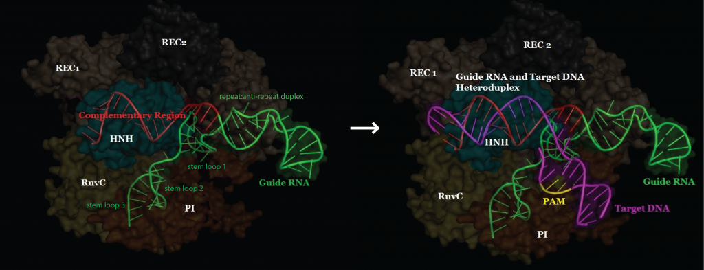
https://sites.tufts.edu/crispr/files/2014/11/target-DNA-binding-overview-1024×393.png
Figure 1: DNA Binding Overview (original image) (crystal image rendered from PDB: 4UN3 Anders et al. 2014.)
CRISPR/Cas9 systems use a guide RNA with a region complementary to the target DNA to specifically bind their target sequences. However, there is an immediate and inherent issue with this. In order to achieve specificity, longer guide RNAs are beneficial, as each nucleotide in the RNA guide increases the specificity of the nuclease about 4-fold. However, in order for the DNA to melt and accommodate base-pairing to the guide RNA, the longer the RNA guide, the less efficient the nuclease. How can CRISPR/Cas9 systems have such dramatically increased specificity over other nucleases such as TALENS and ZFNS and still maintain roughly the same, if not better, efficiency? (Mali et al. 2013)
The answer is that the CRISPR/Cas9 system uses the Protospacer Adjacent Motif (PAM) binding as a preliminary step in locating the target sequence. As was determined by single molecule fluorescence microscopy, the initial binding of Cas9 to PAM (N-G-G) sequences allows the enzyme to quickly screen for potential target sequences. The enzyme will rapidly detach from DNA that does not have the proper PAM sequence. If the protein finds a potential target with the appropriate PAM, it will to melt the remaining DNA on the target to test whether the remaining target sequence is complementary to its guide sequence. The PAM binding step allows the protein to quickly screen potential targets and avoid melting many non-target sequences in its search for fully complementary sequences to cut. (Sternberg et al. 2014)
In July of 2014, Anders et al. published a crystal structure that led to a model for PAM-dependent target DNA binding, unwinding, and recognition by the Cas9 nuclease. The following images are created based off of figure 4 of the paper, or are images rendered inPymol (distributed by Schrödinger) using the crystal structure from that paper (obtained from the Protein Data Bank).
Proposed model for PAM-dependent target DNA binding, melting, and recognition by Cas9:

1. PAM Binding:
The Protospacer Adjacent Motif (PAM) NGG bases of the target DNA strand are shown in yellow. Arginine residues 1333 and 1335 of the PAM Interacting (PI) domain bind to the major groove of the guanine bases in the PAM. A lysine residue in the Phosphate Lock Loop, also in the PI domain, binds the minor groove.
2. Phosphate Lock Loop:
This positions the PAM and target DNA such that serine 1109 in the phosphate lock loop, and two nitrogens of the phosphate lock loop’s backbone, can form hydrogen bonds to the phosphate at position +1 of the PAM. This stabilizes the target DNA such that the first bases of the target sequence (or the protospacer) can melt and rotate upwards towards the guide RNA.

3. Guide RNA:
If the target DNA is complementary to the guide RNA strand, the two strands will base pair. This will allow the target DNA to unzip, as the bases flip up and bind the guide RNA. Without the initial PAM binding and stabilization of the +1 phosphate, the guide RNA would very rarely be able to bind the target DNA, and Cas9 would be very inefficient. This illustrates a mechanism that explains why Cas9 is able to have both high efficiency and high specificity, thus making it a powerful genome editing tool.

4. Cleavage:
Finally, complete annealing of the guide RNA to the target DNA allows the HNH and RuvC nucleases to cleave their respective strands. These nucleases cleave very specifically between the 3rd and 4th nucleotides from the PAM. Again, this specificity of cleavage, as well as the fact that the individual nucleases may be mutated independently and without affecting the ability of Cas9 to bind specific sequences, make the CRISPR/Cas9 system a simultaneously powerful and flexible genome editing tool.
A CRISPR View of Cleavage

Seminal studies showed that CRISPR-Cas systems provide adaptive immunity in prokaryotes and promising gene-editing tools from bacteria to humans. Yet, reports diverged on whether some CRISPR systems naturally target DNA or RNA. Here, Samai and colleagues unify the studies, showing that a single type III CRISPR-Cas system cleaves both DNA and RNA targets, independently.
- Nature Biotechnology 19 Jan 2015; 33, 175–178 http://dx.doi.org:/10.1038/nbt.3127

HEK293T cells were transfected with the indicated plasmids and the genomic DNA harvested 48 h later was assessed using the Surveyor assay. The mutant name is as described in Fig. 2. wt: wild type; UD, undetectable.
October 29, 2015 | BGI ― formerly the Beijing Genomics Institute, China’s contribution to the Human Genome Project, and now a hybrid state agency and private corporation ― is one of the world’s largest scientific research and industrial powers. From its headquarters in Shenzhen and outposts across Asia, Europe and the United States, BGI performs population-scale genomics studies, runs the world’s largest on-demand DNA sequencing service, and sells a small but growing suite of commercial products. Last week, BGIrevealed the first sequencing instrument to be developed and produced in China, the BGISEQ-500, launched exclusively to Chinese markets.
Like other recent Chinese accomplishments in high-tech fields, the sequencer is as much a point of national pride as it is a commercial venture. “Shenzhen has transformed itself from labor-intensive industry to high tech,” says He Jiankui, a specialist in genomics and biochemistry who teaches at the city’s South University of Science and Technology of China. “The government has ambitions. They’re trying to switch from ‘Made in China’ to ‘Invented in China.’”
A Worthy Sequel: PacBio’s New Sequencing System
http://www.bio-itworld.com/2015/10/1/a-worthy-sequel.aspx
Aaron Krol
October 1, 2015 | This Wednesday, in a surprise announcement, Pacific Biosciences of Menlo Park, Calif., confirmed rumors that it has been working on a smaller, more price-effective version of its RS II gene sequencer. But rather than push out a scaled-down benchtop instrument for simple use cases, as many had anticipated, the company unveiled a machine that improves on the RS II in every particular: less than half the cost, a third the size, and most importantly, almost seven times as powerful.
New and Unusual DNA Repair Activity Identified
-
Click Image To Enlarge +
 The new type of DNA repair enzyme, AlkD on the left, can identify and remove a damaged DNA base without forcing it to physically “flip” to the outside of the DNA backbone, which is how all the other DNA repair enzymes in its family work, as illustrated by the human AAG enzyme on the right. The enzymes are shown in grey, the DNA backbone is orange, normal DNA base pairs are yellow, the damaged base is blue and its pair base is green. [Brandt Eichman, Vanderbilt University]
Hot on the heels of the recent announcement of the Nobel Prize in Chemistry being awarded for seminal discoveries in the area of DNA repair, researchers at Vanderbilt University have published data describing new enzymatic activity for a DNA glycosylase discovered previously in the bacteria Bacillus cereus.
When Watson and Crick first published their now famous double-helix structure of DNA, many scientists imagined the molecule to be extremely chemically stable—acting as the template for passing along inheritable genetic traits. However, over the years investigators have since discovered DNA’s susceptibility to damage and its dynamic nature to repair itself, to maintain genomic stability.
“It’s a double-edged sword,” remarked senior author and project leader Brandt Eichman, Ph.D., associate professor of biological sciences and biochemistry at Vanderbilt. “If DNA were too reactive then it wouldn’t be capable of storing genetic information. But, if it were too stable, then it wouldn’t allow organisms to evolve.”
There are many ways that DNA can become damaged, but they can be classified into two basic groups: environmental sources including ultraviolet light, toxic chemicals, and ionizing radiation and internal sources, which include, reactive oxygen species, a number of chemicals the cell produces during normal metabolism, and even water.
“More than 10,000 DNA damage events occur each day within every cell of the human body, which must be repaired for DNA to function properly,” explained lead author Elwood Mullins, Ph.D., a postdoctoral research associate in Dr. Eichman’s laboratory.
The Vanderbilt team discovered the new repair activity while studying the DNA glycosylase AlkD. Glycosylases are part of a family of enzymes discovered by Tomas Lindahl, Ph.D., who received this year’s Nobel prize for recognizing that these enzymes removed damaged DNA bases through a process called base-excision repair (BER).
Briefly, during BER, a specific glycosylase molecule binds to DNA at the location of a lesion and bends the double-helix in a way that causes the damaged base to flip from the inside of the helix to the outside. The enzyme fits around the flipped out base and holds it in a position that exposes its link to the DNA’s sugar backbone, allowing the enzyme to detach it. After the damaged base has been removed, additional DNA-repair proteins move in to replace it with a new, undamaged base.
Dr. Eichman and his team found that AlkD from B. cereus works in a totally different fashion—as it does not require base flipping to recognize damaged DNA or repair it. Using crystallography techniques, the researchers were able to determine that AlkD forms a series of interactions with the DNA backbone at and around the lesion while the lesion is still stacked in the double helix. Several of these interactions are contributed by three amino acids in the enzyme that catalyze excision of the damaged base.
The findings from this study were published recently in Nature through an article entitled “The DNA glycosylase AlkD uses a non-base-flipping mechanism to excise bulky lesions.”
Additionally, the investigators found that AlkD identifies lesions by interacting with the DNA backbone without contacting the damaged base itself and can repair many different types of lesions as long as they are positively charged. Since the enzyme doesn’t have the same type of binding pocket, it isn’t restricted in the same way as other glycosylases. Lastly, AlkD can excise much bulkier lesions than other glycosylases. Base excision repair is limited to relatively small lesions. A different pathway called nucleotide excision repair typically handles larger lesions like those caused by UV radiation damage. However, Dr. Eichman’s team discovered that AlkD could excise lesions that would normally default to other DNA repair pathways.
“Our discovery shows that we still have a lot to learn about DNA repair and that there may be alternative repair pathways yet to be discovered. It certainly shows us that a much broader range of DNA damage can be removed in ways that we didn’t think were possible,” Dr. Eichman stated. “Bacteria are using this to their advantage to protect themselves against the antibacterial agents they produce. Humans may even have DNA-repair enzymes that operate in similar fashion to remove complex types of DNA damage. This could have clinical relevance because these enzymes if they exist, could be reducing the effectiveness of drugs designed to kill cancer cells by shutting down their ability to replicate.”

Leave a Reply