Pentose Shunt, Electron Transfer, Galactose, more Lipids in brief
Reviewer and Curator: Larry H. Bernstein, MD, FCAP
Pentose Shunt, Electron Transfer, Galactose, and other Lipids in brief
This is a continuation of the series of articles that spans the horizon of the genetic
code and the progression in complexity from genomics to proteomics, which must
be completed before proceeding to metabolomics and multi-omics. At this point
we have covered genomics, transcriptomics, signaling, and carbohydrate metabolism
with considerable detail.In carbohydrates. There are two topics that need some attention –
(1) pentose phosphate shunt;
(2) H+ transfer
(3) galactose.
(4) more lipids
Then we are to move on to proteins and proteomics.
Summary of this series:
The outline of what I am presenting in series is as follows:
- Signaling and Signaling Pathways
http://pharmaceuticalintelligence.com/2014/08/12/signaling-and-signaling-pathways/
- Signaling transduction tutorial.
http://pharmaceuticalintelligence.com/2014/08/12/signaling-transduction-tutorial/
- Carbohydrate metabolism
http://pharmaceuticalintelligence.com/2014/08/13/carbohydrate-metabolism/
Selected References to Signaling and Metabolic Pathways published in this Open Access Online Scientific Journal, include the following:
http://pharmaceuticalintelligence.com/2014/08/14/selected-references-to-signaling-
and-metabolic-pathways-in-leaders-in-pharmaceutical-intelligence/
- Lipid metabolism
4.1 Studies of respiration lead to Acetyl CoA
http://pharmaceuticalintelligence.com/2014/08/18/studies-of-respiration-lead-to-acetyl-coa/
4.2 The multi-step transfer of phosphate bond and hydrogen exchange energy
http://pharmaceuticalintelligence.com/2014/08/19/the-multi-step-transfer-of-phosphate-
bond-and-hydrogen-exchange-energy/
5.Pentose shunt, electron transfers, galactose, and other lipids in brief
6. Protein synthesis and degradation
7. Subcellular structure
8. Impairments in pathological states: endocrine disorders; stress
hypermetabolism; cancer.
Section I. Pentose Shunt
Bernard L. Horecker’s Contributions to Elucidating the Pentose Phosphate Pathway
Nicole Kresge, Robert D. Simoni and Robert L. Hill
The Enzymatic Conversion of 6-Phosphogluconate to Ribulose-5-Phosphate
and Ribose-5-Phosphate (Horecker, B. L., Smyrniotis, P. Z., and Seegmiller,
J. E. J. Biol. Chem. 1951; 193: 383–396
Bernard Horecker
Bernard Leonard Horecker (1914) began his training in enzymology in 1936 as a
graduate student at the University of Chicago in the laboratory of T. R. Hogness.
His initial project involved studying succinic dehydrogenase from beef heart using
the Warburg manometric apparatus. However, when Erwin Hass arrived from Otto
Warburg’s laboratory he asked Horecker to join him in the search for an enzyme
that would catalyze the reduction of cytochrome c by reduced NADP. This marked
the beginning of Horecker’s lifelong involvement with the pentose phosphate pathway.
During World War II, Horecker left Chicago and got a job at the National Institutes of
Health (NIH) in Frederick S. Brackett’s laboratory in the Division of Industrial Hygiene.
As part of the wartime effort, Horecker was assigned the task of developing a method
to determine the carbon monoxide hemoglobin content of the blood of Navy pilots
returning from combat missions. When the war ended, Horecker returned to research
in enzymology and began studying the reduction of cytochrome c by the succinic
dehydrogenase system.
Shortly after he began these investigation changes, Horecker was approached by
future Nobel laureate Arthur Kornberg, who was convinced that enzymes were the
key to understanding intracellular biochemical processes. Kornberg suggested
they collaborate, and the two began to study the effect of cyanide on the succinic
dehydrogenase system. Cyanide had previously been found to inhibit enzymes
containing a heme group, with the exception of cytochrome c. However, Horecker
and Kornberg found that
- cyanide did in fact react with cytochrome c and concluded that
- previous groups had failed to perceive this interaction because
- the shift in the absorption maximum was too small to be detected by
visual examination.
Two years later, Kornberg invited Horecker and Leon Heppel to join him in setting up
a new Section on Enzymes in the Laboratory of Physiology at the NIH. Their Section on Enzymes eventually became part of the new Experimental Biology and Medicine
Institute and was later renamed the National Institute of Arthritis and Metabolic
Diseases.
Horecker and Kornberg continued to collaborate, this time on
- the isolation of DPN and TPN.
By 1948 they had amassed a huge supply of the coenzymes and were able to
present Otto Warburg, the discoverer of TPN, with a gift of 25 mg of the enzyme
when he came to visit. Horecker also collaborated with Heppel on
- the isolation of cytochrome c reductase from yeast and
- eventually accomplished the first isolation of the flavoprotein from
mammalian liver.
Along with his lab technician Pauline Smyrniotis, Horecker began to study
- the enzymes involved in the oxidation of 6-phosphogluconate and the
metabolic intermediates formed in the pentose phosphate pathway.
Joined by Horecker’s first postdoctoral student, J. E. Seegmiller, they worked
out a new method for the preparation of glucose 6-phosphate and 6-phosphogluconate, both of which were not yet commercially available.
As reported in the Journal of Biological Chemistry (JBC) Classic reprinted here, they
- purified 6-phosphogluconate dehydrogenase from brewer’s yeast (1), and
- by coupling the reduction of TPN to its reoxidation by pyruvate in
the presence of lactic dehydrogenase,
- they were able to show that the first product of 6-phosphogluconate oxidation,
- in addition to carbon dioxide, was ribulose 5-phosphte.
- This pentose ester was then converted to ribose 5-phosphate by a
pentose-phosphate isomerase.
They were able to separate ribulose 5-phosphate from ribose 5- phosphate and demonstrate their interconversion using a recently developed nucleotide separation
technique called ion-exchange chromatography. Horecker and Seegmiller later
showed that 6-phosphogluconate metabolism by enzymes from mammalian
tissues also produced the same products.8

Bernard Horecker
http://www.jbc.org/content/280/29/e26/F1.small.gif
Over the next several years, Horecker played a key role in elucidating the
- remaining steps of the pentose phosphate pathway.
His total contributions included the discovery of three new sugar phosphate esters,
ribulose 5-phosphate, sedoheptulose 7-phosphate, and erythrose 4-phosphate, and
three new enzymes, transketolase, transaldolase, and pentose-phosphate 3-epimerase.
The outline of the complete pentose phosphate cycle was published in 1955
(2). Horecker’s personal account of his work on the pentose phosphate pathway can
be found in his JBC Reflection (3).1
Horecker’s contributions to science were recognized with many awards and honors
including the Washington Academy of Sciences Award for Scientific Achievement in
Biological Sciences (1954) and his election to the National Academy of Sciences in
1961. Horecker also served as president of the American Society of Biological
Chemists (now the American Society for Biochemistry and Molecular Biology) in 1968.
Footnotes
- ↵1 All biographical information on Bernard L. Horecker was taken from Ref. 3.
- The American Society for Biochemistry and Molecular Biology, Inc.
References
- ↵Horecker, B. L., and Smyrniotis, P. Z. (1951) Phosphogluconic acid dehydrogenase
from yeast. J. Biol. Chem. 193, 371–381FREE Full Text
- ↵Gunsalus, I. C., Horecker, B. L., and Wood, W. A. (1955) Pathways of carbohydrate
metabolism in microorganisms. Bacteriol. Rev. 19, 79–128 FREE Full Text
- ↵Horecker, B. L. (2002) The pentose phosphate pathway. J. Biol. Chem. 277, 47965–
47971 FREE Full Text
The Pentose Phosphate Pathway (also called Phosphogluconate Pathway, or Hexose
Monophosphate Shunt) is depicted with structures of intermediates in Fig. 23-25
p. 863 of Biochemistry, by Voet & Voet, 3rd Edition. The linear portion of the pathway
carries out oxidation and decarboxylation of glucose-6-phosphate, producing the
5-C sugar ribulose-5-phosphate.
Glucose-6-phosphate Dehydrogenase catalyzes oxidation of the aldehyde
(hemiacetal), at C1 of glucose-6-phosphate, to a carboxylic acid in ester linkage
(lactone). NADP+ serves as electron acceptor.
6-Phosphogluconolactonase catalyzes hydrolysis of the ester linkage (lactone)
resulting in ring opening. The product is 6-phosphogluconate. Although ring opening
occurs in the absence of a catalyst, 6-Phosphogluconolactonase speeds up the
reaction, decreasing the lifetime of the highly reactive, and thus potentially
toxic, 6-phosphogluconolactone.
Phosphogluconate Dehydrogenase catalyzes oxidative decarboxylation of
6-phosphogluconate, to yield the 5-C ketose ribulose-5-phosphate. The
hydroxyl at C3 (C2 of the product) is oxidized to a ketone. This promotes loss
of the carboxyl at C1 as CO2. NADP+ again serves as oxidant (electron acceptor).

pglucose hd
https://www.rpi.edu/dept/bcbp/molbiochem/MBWeb/mb2/part1/images/pglucd.gif
Reduction of NADP+ (as with NAD+) involves transfer of 2e- plus 1H+ to the
nicotinamide moiety.

NADPH, a product of the Pentose Phosphate Pathway, functions as a reductant in
various synthetic (anabolic) pathways, including fatty acid synthesis.
NAD+ serves as electron acceptor in catabolic pathways in which metabolites are
oxidized. The resultant NADH is reoxidized by the respiratory chain, producing ATP.

https://www.rpi.edu/dept/bcbp/molbiochem/MBWeb/mb2/part1/images/nadnadp.gif
Regulation:
Glucose-6-phosphate Dehydrogenase is the committed step of the Pentose
Phosphate Pathway. This enzyme is regulated by availability of the substrate NADP+.
As NADPH is utilized in reductive synthetic pathways, the increasing concentration of
NADP+ stimulates the Pentose Phosphate Pathway, to replenish NADPH.
The remainder of the Pentose Phosphate Pathway accomplishes conversion of the
5-C ribulose-5-phosphate to the 5-C product ribose-5-phosphate, or to the 3-C
glyceraldehyde -3-phosphate and the 6-C fructose-6-phosphate (reactions 4 to 8
p. 863).
Transketolase utilizes as prosthetic group thiamine pyrophosphate (TPP), a
derivative of vitamin B1.

tpp
https://www.rpi.edu/dept/bcbp/molbiochem/MBWeb/mb2/part1/images/tpp.gif
Thiamine pyrophosphate binds at the active sites of enzymes in a “V” conformation.The amino group of the aminopyrimidine moiety is close to the dissociable proton,
and serves as the proton acceptor. This proton transfer is promoted by a glutamate
residue adjacent to the pyrimidine ring.
The positively charged N in the thiazole ring acts as an electron sink, promoting
C-C bond cleavage. The 3-C aldose glyceraldehyde-3-phosphate is released.
A 2-C fragment remains on TPP.
FASEB J. 1996 Mar;10(4):461-70. http://www.ncbi.nlm.nih.gov/pubmed/8647345
Reviewer
The importance of this pathway can easily be underestimated. The main source for
energy in respiration was considered to be tied to the
- high energy phosphate bond in phosphorylation and utilizes NADPH, converting it to NADP+.
glycolysis n skeletal muscle in short term, dependent on muscle glycogen conversion
to glucose, and there is a buildup of lactic acid – used as fuel by the heart. This
pathway accounts for roughly 5% of metabolic needs, varying between tissues,
depending on there priority for synthetic functions, such as endocrine or nucleic
acid production.
The mature erythrocyte and the ocular lens both are enucleate. 85% of their
metabolic energy needs are by anaerobic glycolysis. Consider the erythrocyte
somewhat different than the lens because it has iron-based hemoglobin, which
exchanges O2 and CO2 in the pulmonary alveoli, and in that role, is a rapid
regulator of H+ and pH in the circulation (carbonic anhydrase reaction), and also to
a lesser extent in the kidney cortex, where H+ is removed from the circulation to
the urine, making the blood less acidic, except when there is a reciprocal loss of K+.
This is how we need a nomogram to determine respiratory vs renal acidosis or
alkalosis. In the case of chronic renal disease, there is substantial loss of
functioning nephrons, loss of countercurrent multiplier, and a reduced capacity to
remove H+. So there is both a metabolic acidosis and a hyperkalemia, with increased
serum creatinine, but the creatinine is only from muscle mass – not accurately
reflecting total body mass, which includes visceral organs. The only accurate
measure of lean body mass would be in the linear relationship between circulating
hepatic produced transthyretin (TTR).
The pentose phosphate shunt is essential for
- the generation of nucleic acids, in regeneration of red cells and lens – requiring NADPH.
Insofar as the red blood cell is engaged in O2 exchange, the lactic dehydrogenase
isoenzyme composition is the same as the heart. What about the lens of and cornea the eye, and platelets? The explanation does appear to be more complex than
has been proposed and is not discussed here.
Section II. Mitochondrial NADH – NADP+ Transhydrogenase Reaction
There is also another consideration for the balance of di- and tri- phospopyridine
nucleotides in their oxidized and reduced forms. I have brought this into the
discussion because of the centrality of hydride tranfer to mitochondrial oxidative
phosphorylation and the energetics – for catabolism and synthesis.
The role of transhydrogenase in the energy-linked reduction of TPN ☆
Fritz Hommes∗, Ronald W. Estabrook∗∗
The Wenner-Gren Institute, University of Stockholm
Stockholm, Sweden
Biochemical and Biophysical Research Communications 11, (1), 2 Apr 1963, Pp 1–6
http://dx.doi.org:/10.1016/0006-291X(63)90017-2
In 1959, Klingenberg and Slenczka (1) made the important observation that incubation of isolated
- liver mitochondria with DPN-specific substrates or succinate in the absence of phosphate
acceptor resulted in a rapid and almost complete reduction of the intramitochondrial TPN.
These and related findings led Klingenberg and co-workers (1-3) to postulate
- the occurrence of an ATP-controlled transhydrogenase reaction catalyzing the reduction of
mitochondrial TPN by DPNH. A similar conclusion was reached by Estabrook and Nissley (4).
The present paper describes the demonstration and some properties of an
- energy-dependent reduction of TPN by DPNH, catalyzed by submitochondrial particles.
Preliminary reports of some of these results have already appeared (5, 6 ) , and a
complete account is being published elsewhere (7).We have studied the energy- dependent reduction of TPN by PNH with submitochondrial particles from both
rat liver and beef heart. Rat liver particles were prepared essentially according to
the method of Kielley and Bronk (8), and beef heart particles by the method of
Low and Vallin (9).
PYRIDINE NUCLEOTIDE TRANSHYDROGENASE II. DIRECT EVIDENCE FOR
AND MECHANISM OF THE TRANSHYDROGENASE REACTION*
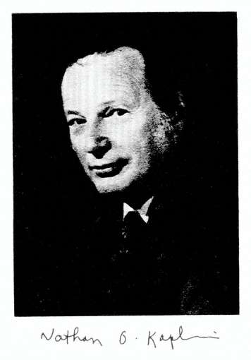
BY NATHAN 0. KAPLAN, SIDNEY P. COLOWICK, AND ELIZABETH F. NEUFELD
(From the McCollum-Pratt Institute, The Johns Hopkins University, Baltimore,
Maryland) J. Biol. Chem. 1952, 195:107-119.
http://www.jbc.org/content/195/1/107.citation

NO Kaplan

Sidney Colowick

Elizabeth Neufeld
Kaplan studied carbohydrate metabolism in the liver under David M. Greenberg at the
University of California, Berkeley medical school. He earned his Ph.D. in 1943. From
1942 to 1944, Kaplan participated in the Manhattan Project. From 1945 to 1949,
Kaplan worked with Fritz Lipmann at Massachusetts General Hospital to study
coenzyme A. He worked at the McCollum-Pratt Institute of Johns Hopkins University
from 1950 to 957. In 1957, he was recruited to head a new graduate program in
biochemistry at Brandeis University. In 1968, Kaplan moved to the University of
California, San Diego, where he studied the role of lactate dehydrogenase in cancer. He also founded a colony of nude mice, a strain of laboratory mice useful in the study
of cancer and other diseases. [1] He was a member of the National Academy of
Sciences.One of Kaplan’s students at the University of California was genomic
researcher Craig Venter.[2]3] He was, with Sidney Colowick, a founding editor of the scientific book series Methods
in Enzymology.[1]
http://books.nap.edu/books/0309049768/xhtml/images/img00009.jpg
Colowick became Carl Cori’s first graduate student and earned his Ph.D. at
Washington University St. Louis in 1942, continuing to work with the Coris (Nobel
Prize jointly) for 10 years. At the age of 21, he published his first paper on the
classical studies of glucose 1-phosphate (2), and a year later he was the sole author on a paper on the synthesis of mannose 1-phosphate and galactose 1-phosphate (3). Both papers were published in the JBC. During his time in the Cori lab,
Colowick was involved in many projects. Along with Herman Kalckar he discovered
myokinase (distinguished from adenylate kinase from liver), which is now known as
adenyl kinase. This discovery proved to be important in understanding transphos-phorylation reactions in yeast and animal cells. Colowick’s interest then turned to
the conversion of glucose to polysaccharides, and he and Earl Sutherland (who
will be featured in an upcoming JBC Classic) published an important paper on the
formation of glycogen from glucose using purified enzymes (4). In 1951, Colowick
and Nathan Kaplan were approached by Kurt Jacoby of Academic Press to do a
series comparable to Methodem der Ferment Forschung. Colowick and Kaplan
planned and edited the first 6 volumes of Methods in Enzymology, launching in 1955
what became a series of well known and useful handbooks. He continued as
Editor of the series until his death in 1985.
http://bioenergetics.jbc.org/highwire/filestream/9/field_highwire_fragment_image_s/0/F1.small.gif
The Structure of NADH: the Work of Sidney P. Colowick
On the Structure of Reduced Diphosphopyridine Nucleotide
(Pullman, M. E., San Pietro, A., and Colowick, S. P. (1954)
J. Biol. Chem. 206, 129–141)
Elizabeth Neufeld
· Born: September 27, 1928 (age 85), Paris, France
· Education: Queens College, City University of New York, University of California,
Berkeley
http://fdb5.ctrl.ucla.edu/biological-chemistry/institution/photo?personnel%5fid=45290&max_width=155&max_height=225
In Paper I (l), indirect evidence was presented for the following transhydrogenase
reaction, catalyzed by an enzyme present in extracts of Pseudomonas
fluorescens:
TPNHz + DPN -+ TPN + DPNHz
The evidence was obtained by coupling TPN-specific dehydrogenases with the
transhydrogenase and observing the reduction of large amounts of diphosphopyridine nucleotide (DPN) in the presence of catalytic amounts of triphosphopyridine
nucleotide (TPN).
In this paper, data will be reported showing the direct
- interaction between TPNHz and DPN, in thepresence of transhydrogenase alone,
- to yield products having the propertiesof TPN and DPNHZ.
Information will be given indicating that the reaction involves
- a transfer of electrons (or hydrogen) rather than a phosphate
Experiments dealing with the kinetics and reversibility of the reaction, and with the
nature of the products, suggest that the reaction is a complex one, not fully described
by the above formulation.
Materials and Methods [edited]
The TPN and DPN used in these studies were preparations of approximately 75
percent purity and were prepared from sheep liver by the chromatographic procedure
of Kornberg and Horecker (unpublished). Reduced DPN was prepared enzymatically with alcohol dehydrogenase as described elsewhere (2). Reduced TPN was prepared by treating TPN with hydrosulfite. This treated mixture contained 2 pM of TPNHz per ml.
The preparations of desamino DPN and reduced desamino DPN have been
described previously (2, 3). Phosphogluconate was a barium salt which was kindly
supplied by Dr. B. F. Horecker. Cytochrome c was obtained from the Sigma Chemical Company.
Transhydrogenase preparations with an activity of 250 to 7000 units per mg. were
used in these studies. The DPNase was a purified enzyme, which was obtained
from zinc-deficient Neurospora and had an activity of 5500 units per mg. (4). The
alcohol dehydrogenase was a crystalline preparation isolated from yeast according to the procedure of Racker (5).
Phosphogluconate dehydrogenase from yeast and a 10 per cent pure preparation of the TPN-specific cytochrome c reductase from liver (6) were gifts of Dr. B. F.
Horecker.
DPN was assayed with alcohol and crystalline yeast alcohol dehydrogenase. TPN was determined By the specific phosphogluconic acid dehydrogenase from yeast and also by the specific isocitric dehydrogenase from pig heart. Reduced DPN was
determined by the use of acetaldehyde and the yeast alcohol dehydrogenase.
All of the above assays were based on the measurement of optical density changes
at 340 rnp. TPNHz was determined with the TPN-specific cytochrome c reductase system. The assay of the reaction followed increase in optical density at 550 rnp as a measure of the reduction of the cytochrome c after cytochrome c
reductase was added to initiate the reaction. The changes at 550 rnp are plotted for different concentrations of TPNHz in Fig. 3, a. The method is an extremely sensitive and accurate assay for reduced TPN.
Results
[No Figures or Table shown]
Formation of DPNHz from TPNHz and DPN-Fig. 1, a illustrates the direct reaction between TPNHz and DPN to form DPNHZ. The reaction was carried out by incubating TPNHz with DPN in the presence of the
transhydrogenase, yeast alcohol dehydrogenase, and acetaldehyde. Since the yeast dehydrogenase is specific for DPN,
- a decrease in absorption at340 rnp can only be due to the formation of reduced DPN. It can
be seen from the curves in Fig. 1, a that a decrease in optical density occurs only in the
presence of the complete system.
The Pseudomonas enzyme is essential for the formation of DPNH2. It is noteworthy
that, under the conditions of reaction in Fig. 1, a,
- approximately 40 per cent of theTPNH, reacted with the DPN.
Fig. 1, a also indicates that magnesium is not required for transhydrogenase activity. The reaction between TPNHz and DPN takes place in the absence of alcohol
dehydrogenase and acetaldehyde. This can be demonstrated by incubating the
two pyridine nucleotides with the transhydrogenase for 4 8 12 16 20 24 28 32 36
minutes
FIG. 1. Evidence for enzymatic reaction of TPNHt with DPN.
- Rate offormation of DPNH2.
(b) DPN disappearance and TPN formation.
(c) Identification of desamino DPNHz as product of reaction of TPNHz with desamino DPN. (assaying for reduced DPN by the yeast alcohol dehydrogenase technique.
Table I (Experiment 1) summarizes the results of such experiments in which TPNHz was added with varying amounts of DPN.
- In the absence of DPN, no DPNHz was formed. This eliminates the possibility that TPNH 2 is
converted to DPNHz
- by removal ofthe monoester phosphate grouping.
The data also show that the extent of the reaction is
- dependent on the concentration of DPN.
Even with a large excess of DPN, only approximately 40 per cent of the TPNHzreacts to form reduced DPN. It is of importance to emphasize that in the above
experiments, which were carried out in phosphate buffer, the extent of the reaction
- is the same in the presence or absence of acetaldehyde andalcohol dehydrogenase.
With an excess of DPN and different levels of TPNHZ,
- the amount of reduced DPN which is formed is
- dependent on the concentration of TPNHz(Table I, Experiment 2).
- In all cases, the amount of DPNHz formed is approximately
40 per cent of the added reduced TPN.
Formation of TPN-The reaction between TPNHz and DPN should yield TPN as well as DPNHz.
The formation of TPN is demonstrated in Table 1. in Fig. 1, b. In this experiment,
TPNHz was allowed to react with DPN in the presence of the transhydrogenase
(PS.), and then alcohol and alcohol dehydrogenase were added . This
would result in reduction of the residual DPN, and the sample incubated with the
transhydrogenase contained less DPN. After the completion of the alcohol
dehydrogenase reaction, phosphogluconate and phosphogluconic dehydrogenase (PGAD) were added to reduce the TPN. The addition of this TPN-specific
dehydrogenase results in an
- increase inoptical density in the enzymatically treated sample.
- This change represents the amount of TPN formed.
It is of interest to point out that, after addition of both dehydrogenases,
- the total optical density change is the same in both
Therefore it is evident that
- for every mole of DPN disappearing a mole of TPN appears.
Balance of All Components of Reaction–
Table II (Experiment 1) shows that,
- if measurements for all components of the reaction are made, one can demonstrate
that there is
- a mole for mole disappearance of TPNH, and DPN, and
- a stoichiometric appearance of TPN and DPNH2.
- The oxidized forms of the nucleotides were assayed as described
- the reduced form of TPN was determined by the TPNHz-specific cytochrome c reductase,
- the DPNHz by means of yeast alcohol dehydrogenase plus
This stoichiometric balance is true, however,
- only when the analyses for the oxidized forms are determined directly on the reaction
When analyses are made after acidification of the incubated reaction mixture,
- the values found forDPN and TPN are much lower than those obtained by direct analysis.
This discrepancy in the balance when analyses for the oxidized nucleotides are
carried out in acid is indicated in Table II (Experiment 2). The results, when
compared with the findings in Experiment 1, are quite striking.
Reaction of TPNHz with Desamino DPN–
Desamino DPN
- reacts with the transhydrogenase system at the same rate as does DPN (2).
This was of value in establishing the fact that
- the transhydrogenase catalyzesa transfer of hydrogen rather than a phosphate transfer reaction.
The reaction between desamino DPN and TPNHz can be written in two ways.
TPN f desamino DPNHz
TPNH, + desamino DPN
DPNH2 + desamino TPN
If the reaction involved an electron transfer,
- desamino DPNHz would be
- Phosphate transfer would result in the production of reduced
Desamino DPNHz can be distinguished from DPNHz by its
- slowerrate of reaction with yeast alcohol dehydrogenase (2, 3).
Fig. 1, c illustrates that, when desamino DPN reacts with TPNH2,
- the product of the reaction is desamino DPNHZ.
This is indicated by the slow rate of oxidation of the product by yeast alcohol
dehydrogenase and acetaldehyde.
From the above evidence phosphate transfer
- has been ruled out as a possible mechanism for the transhydrogenase reaction.
Inhibition by TPN–
As mentioned in Paper I and as will be discussed later in this paper,
- the transhydrogenase reaction does not appear to be readily reversible.
This is surprising, particularly since only approximately
- 40 per cent of the TPNHz undergoes reaction with DPN
under the conditions described above. It was therefore thought that
- the TPN formed might inhibit further transfer of electrons from TPNH2.
Table III summarizes data showing the
- strong inhibitory effect of TPN on thereaction between TPNHz and DPN.
It is evident from the data that
- TPN concentration is a factor in determining the extent of the reaction.
Effect of Removal of TPN on Extent of Reaction–
A purified DPNase from Neurospora has been found
- to cleave the nicotinamide riboside linkagesof the oxidized forms of both TPN and DPN
- without acting on thereduced forms of both nucleotides (4).
It has been found, however, that
- the DPNase hydrolyzes desamino DPN at a very slow rate (3).
In the reaction between TPNHz and desamino DPN, TPN and desamino DPNH:,
- TPNis the only component of this reaction attacked by the Neurospora enzyme
at an appreciable rate
It was thought that addition of the DPNase to the TPNHZ-desamino DPN trans-
hydrogenase reaction mixture
- would split the TPN formed andpermit the reaction to go to completion.
This, indeed, proved to be the case, as indicated in Table IV, where addition of
the DPNase with desamino DPN results in almost
- a stoichiometric formation of desamino DPNHz
- and a complete disappearance of TPNH2.
Extent of Reaction in Buffers Other Than Phosphate–
All the reactions described above were carried out in phosphate buffer of pH 7.5.
If the transhydrogenase reaction between TPNHz and DPN is run at the same pH
in tris(hydroxymethyl)aminomethane buffer (TRIS buffer)
- with acetaldehydeand alcohol dehydrogenase present,
- the reaction proceeds muchfurther toward completion
- than is the case under the same conditions ina phosphate medium (Fig. 2, a).
The importance of phosphate concentration in governing the extent of the reaction
is illustrated in Fig. 2, b.
In the presence of TRIS the transfer reaction
- seems to go further toward completion in the presence of acetaldehyde
and alcohol dehydrogenase
- than when these two components are absent.
This is not true of the reaction in phosphate,
- in which the extent is independent of the alcoholdehydrogenase system.
Removal of one of the products of the reaction (DPNHp) in TRIS thus
- appears to permit the reaction to approach completion,whereas
- in phosphate this removal is without effect on the finalcourse of the reaction.
The extent of the reaction in TRIS in the absence of alcohol dehydrogenase
and acetaldehyde is
- somewhat greater than when the reaction is run in phosphate.
TPN also inhibits the reaction of TPNHz with DPN in TRIS medium, but the inhibition
- is not as marked as when the reaction is carried out in phosphate buffer.
Reversibility of Transhydrogenase Reaction;
Reaction between DPNHz and TPN–
In Paper I, it was mentioned that no reversal of the reaction could be achieved in a system containing alcohol, alcohol dehydrogenase, TPN, and catalytic amounts of
DPN.
When DPNH, and TPN are incubated with the purified transhydrogenase, there is
also
- no evidence for reversibility.
This is indicated in Table V which shows that
- there is no disappearance of DPNHz in such a system.
It was thought that removal of the TPNHz, which might be formed in the reaction,
could promote the reversal of the reaction. Hence,
- by using the TPNHe-specific cytochrome c reductase, one could
- not only accomplishthe removal of any reduced TPN,
- but also follow the course of the reaction.
A system containing DPNH2, TPN, the transhydrogenase, the cytochrome c
reductase, and cytochrome c, however, gives
- no reduction of the cytochrome
This is true for either TRIS or phosphate buffers.2
Some positive evidence for the reversibility has been obtained by using a system
containing
- DPNH2, TPNH2, cytochrome c, and the cytochrome creductase in TRIS buffer.
In this case, there is, of course, reduction of cytochrome c by TPNHZ, but,
- when the transhydrogenase is present.,there is
- additional reduction over and above that due to the added TPNH2.
This additional reduction suggests that some reversibility of the reaction occurred
under these conditions. Fig. 3, b shows
- the necessity of DPNHzfor this additional reduction.
Interaction of DPNHz with Desamino DPN-
If desamino DPN and DPNHz are incubated with the purified Pseudomonas enzyme,
there appears
- to be a transfer of electrons to form desamino DPNHz.
This is illustrated in Fig. 4, a, which shows the
- decreased rate of oxidation by thealcohol dehydrogenase system
- after incubation with the transhydrogenase.
- Incubation of desamino DPNHz with DPN results in the formation of DPNH2,
- which is detected by the faster rate of oxidation by the alcohol dehydrogenase system
- after reaction of the pyridine nucleotides with thetranshydrogenase (Fig. 4, b).
It is evident from the above experiments that
the transhydrogenase catalyzes an exchange of hydrogens between
- the adenylic and inosinic pyridine nucleotides.
However, it is difficult to obtain any quantitative information on the rate or extent of
the reaction by the method used, because
- desamino DPNHz also reacts with the alcohol dehydrogenase system,
- although at a much slower rate than does DPNH2.
DISCUSSION
The results of the balance experiments seem to offer convincing evidence that
the transhydrogenase catalyzes the following reaction.
TPNHz + DPN -+ DPNHz + TPN
Since desamino DPNHz is formed from TPNHz and desamino DPN,
- thereaction appears to involve an electron (or hydrogen) transfer
- rather thana transfer of the monoester phosphate grouping of TPN.
A number of the findings reported in this paper are not readily understandable in
terms of the above simple formulation of the reaction. It is difficult to understand
the greater extent of the reaction in TRIS than in phosphate when acetaldehyde
and alcohol dehydrogenase are present.
One possibility is that an intermediate may be involved which is more easily converted
to reduced DPN in the TRIS medium. The existence of such an intermediate is also
suggested by the discrepancies noted in balance experiments, in which
- analyses of the oxidized nucleotides after acidification showed
- much lower values than those found by direct analysis.
These findings suggest that the reaction may involve
- a 1 electron ratherthan a 2 electron transfer with
- the formation of acid-labile free radicals as intermediates.
The transfer of hydrogens from DPNHz to desamino DPN
- to yield desamino DPNHz and DPN and the reversal of this transfer
- indicate the unique role of the transhydrogenase
- in promoting electron exchange between the pyridine nucleotides.
In this connection, it is of interest that alcohol dehydrogenase and lactic
dehydrogenase cannot duplicate this exchange between the DPN and
the desamino systems.3 If one assumes that desamino DPN behaves
like DPN,
- one might predict that the transhydrogenase would catalyze an
exchange of electrons (or hydrogen) 3.
Since alcohol dehydrogenase alone
- does not catalyze an exchange of electrons between the adenylic
and inosinic pyridine nucleotides, this rules out the possibility
- that the dehydrogenase is converted to a reduced intermediate
- during electron between DPNHz and added DPN.
It is hoped to investigate this possibility with isotopically labeled DPN.
Experiments to test the interaction between TPN and desamino TPN are
also now in progress.
It seems likely that the transhydrogenase will prove capable of
- catalyzingan exchange between TPN and TPNH2, as well as between DPN and
The observed inhibition by TPN of the reaction between TPNHz and DPN may
therefore
- be due to a competition between DPN and TPNfor the TPNH2.
SUMMARY
- Direct evidence for the following transhydrogenase reaction. catalyzedby an
enzyme from Pseudomonas fluorescens, is presented.
TPNHz + DPN -+ TPN + DPNHz
Balance experiments have shown that for every mole of TPNHz disappearing
1 mole of TPN appears and that for each mole of DPNHz generated 1 mole of
DPN disappears. The oxidized nucleotides found at the end of the reaction,
however, show anomalous lability toward acid.
- The transhydrogenase also promotes the following reaction.
TPNHz + desamino DPN -+ TPN + desamino DPNH,
This rules out the possibility that the transhydrogenase reaction involves a
phosphate transfer and indicates that the
- enzyme catalyzes a shift of electrons (or hydrogen atoms).
The reaction of TPNHz with DPN in 0.1 M phosphate buffer is strongly
inhibited by TPN; thus
- it proceeds only to the extent of about40 per cent or less, even
- when DPNHz is removed continuously by meansof acetaldehyde
and alcohol dehydrogenase.
- In other buffers, in whichTPN is less inhibitory, the reaction proceeds
much further toward completion under these conditions.
- The reaction in phosphate buffer proceedsto completion when TPN
is removed as it is formed.
- DPNHz does not react with TPN to form TPNHz and DPN in the presence
of transhydrogenase. Some evidence, however, has been obtained for
the reversibility by using the following system:
- DPNHZ, TPNHZ, cytochromec, the TPNHz-specific cytochrome c reductase,
and the transhydrogenase.
- Evidence is cited for the following reversible reaction, which is catalyzed
by the transhydrogenase.
DPNHz + desamino DPN fi DPN + desamino DPNHz
- The results are discussed with respect to the possibility that the
transhydrogenase reaction may
- involve a 1 electron transfer with theformation of free radicals as intermediates.
BIBLIOGRAPHY
- Coiowick, S. P., Kaplan, N. O., Neufeld, E. F., and Ciotti, M. M., J. Biol. Chem.,196, 95 (1952).
- Pullman, 111. E., Colowick, S. P., and Kaplan, N. O., J. Biol. Chem., 194, 593(1952).
- Kaplan, N. O., Colowick, S. P., and Ciotti, M. M., J. Biol. Chem., 194, 579 (1952).
- Kaplan, N. O., Colowick, S. P., and Nason, A., J. Biol. Chem., 191, 473 (1951).
- Racker, E., J. Biol. Chem., 184, 313 (1950).
- Horecker, B. F., J. Biol. Chem., 183, 593 (1950).
Section !II.

The Leloir pathway: a mechanistic imperative for three enzymes to change
the stereochemical configuration of a single carbon in galactose.
Frey PA.
FASEB J. 1996 Mar;10(4):461-70. http://www.fasebj.org/content/10/4/461.full.pdf
PMID:8647345
The biological interconversion of galactose and glucose takes place only by way of
the Leloir pathway and requires the three enzymes galactokinase, galactose-1-P
uridylyltransferase, and UDP-galactose 4-epimerase.
The only biological importance of these enzymes appears to be to
- provide for the interconversion of galactosyl and glucosyl groups.
Galactose mutarotase also participates by producing the galactokinase substrate
alpha-D-galactose from its beta-anomer. The galacto/gluco configurational change takes place at the level of the nucleotide sugar by an oxidation/reduction
mechanism in the active site of the epimerase NAD+ complex. The nucleotide portion
of UDP-galactose and UDP-glucose participates in the epimerization process in two ways:
1) by serving as a binding anchor that allows epimerization to take place at glycosyl-C-4 through weak binding of the sugar, and
2) by inducing a conformational change in the epimerase that destabilizes NAD+ and
increases its reactivity toward substrates.
Reversible hydride transfer is thereby facilitated between NAD+ and carbon-4
of the weakly bound sugars.
The structure of the enzyme reveals many details of the binding of NAD+ and
inhibitors at the active site.
The essential roles of the kinase and transferase are to attach the UDP group
to galactose, allowing for its participation in catalysis by the epimerase. The
transferase is a Zn/Fe metalloprotein, in which the metal ions stabilize the
structure rather than participating in catalysis. The structure is interesting
in that
- it consists of single beta-sheet with 13 antiparallel strands and 1 parallel strand
connected by 6 helices.
The mechanism of UMP attachment at the active site of the transferase is a double
displacement, with the participation of a covalent UMP-His 166-enzyme intermediate
in the Escherichia coli enzyme. The evolution of this mechanism appears to have
been guided by the principle of economy in the evolution of binding sites.
PMID: 8647345 Free full text
Section IV.
More on Lipids – Role of lipids – classification
- Energy
- Energy Storage
- Hormones
- Vitamins
- Digestion
- Insulation
- Membrane structure: Hydrophobic properties
Lipid types

lipid types

nat occuring FAs in mammals
Read Full Post »


 The new type of DNA repair enzyme, AlkD on the left, can identify and remove a damaged DNA base without forcing it to physically “flip” to the outside of the DNA backbone, which is how all the other DNA repair enzymes in its family work, as illustrated by the human AAG enzyme on the right. The enzymes are shown in grey, the DNA backbone is orange, normal DNA base pairs are yellow, the damaged base is blue and its pair base is green. [Brandt Eichman, Vanderbilt University]


