Feeds:
Posts
Comments

Archive for the ‘Myocardial Metabolism’ Category

Compilation of References in Leaders in Pharmaceutical Intelligence about proteomics, metabolomics, signaling pathways, and cell regulation

Compilation of References in Leaders in Pharmaceutical Intelligence about
proteomics, metabolomics, signaling pathways, and cell regulation

Curator: Larry H. Bernstein, MD, FCAP


Proteomics

  1. The Human Proteome Map Completed
    Reporter and Curator: Larry H. Bernstein, MD, FCAP
    http://pharmaceuticalintelligence.com/2014/08/28/the-human-proteome-map-completed/
  1. Proteomics – The Pathway to Understanding and Decision-making in Medicine
    Author and Curator, Larry H Bernstein, MD, FCAP
    http://pharmaceuticalintelligence.com/2014/06/24/proteomics-the-pathway-to-understanding-and-decision-making-in-medicine/
  1. Advances in Separations Technology for the “OMICs” and Clarification of Therapeutic Targets
    Author and Curator, Larry H Bernstein, MD, FCAP
    http://pharmaceuticalintelligence.com/2012/10/22/advances-in-separations-technology-for-the-omics-and-clarification-of-therapeutic-targets/
  1. Expanding the Genetic Alphabet and Linking the Genome to the Metabolome
    Author and Curator, Larry H Bernstein, MD, FCAP
    http://pharmaceuticalintelligence.com/2012/09/24/expanding-the-genetic-alphabet-and-linking-the-genome-to-the-metabolome/
  1. Synthesizing Synthetic Biology: PLOS Collections
    Reporter: Aviva Lev-Ari
    http://pharmaceuticalintelligence.com/2012/08/17/synthesizing-synthetic-biology-plos-collections/


Metabolomics

  1. Extracellular evaluation of intracellular flux in yeast cells
    Larry H. Bernstein, MD, FCAP, Reviewer and Curator
    http://pharmaceuticalintelligence.com/2014/08/25/extracellular-evaluation-of-intracellular-flux-in-yeast-cells/ 
  2. Metabolomic analysis of two leukemia cell lines. I.
    Larry H. Bernstein, MD, FCAP, Reviewer and Curator
    http://pharmaceuticalintelligence.com/2014/08/23/metabolomic-analysis-of-two-leukemia-cell-lines-_i/ 
  3. Metabolomic analysis of two leukemia cell lines. II.
    Larry H. Bernstein, MD, FCAP, Reviewer and Curator
    http://pharmaceuticalintelligence.com/2014/08/24/metabolomic-analysis-of-two-leukemia-cell-lines-ii/ 
  4. Metabolomics, Metabonomics and Functional Nutrition: the next step in nutritional metabolism and biotherapeutics
    Reviewer and Curator, Larry H. Bernstein, MD, FCAP
    http://pharmaceuticalintelligence.com/2014/08/22/metabolomics-metabonomics-and-functional-nutrition-the-next-step-in-nutritional-metabolism-and-biotherapeutics/ 
  5. Buffering of genetic modules involved in tricarboxylic acid cycle metabolism provides homeomeostatic regulation
    Larry H. Bernstein, MD, FCAP, Reviewer and curator
    http://pharmaceuticalintelligence.com/2014/08/27/buffering-of-genetic-modules-involved-in-tricarboxylic-acid-cycle-metabolism-provides-homeomeostatic-regulation/


Metabolic Pathways

  1. Pentose Shunt, Electron Transfer, Galactose, more Lipids in brief
    Reviewer and Curator: Larry H. Bernstein, MD, FCAP
    http://pharmaceuticalintelligence.com/2014/08/21/pentose-shunt-electron-transfer-galactose-more-lipids-in-brief/
  2. Mitochondria: More than just the “powerhouse of the cell”
    Reviewer and Curator: Ritu Saxena
    http://pharmaceuticalintelligence.com/2012/07/09/mitochondria-more-than-just-the-powerhouse-of-the-cell/
  3. Mitochondrial fission and fusion: potential therapeutic targets?
    Reviewer and Curator: Ritu saxena
    http://pharmaceuticalintelligence.com/2012/10/31/mitochondrial-fission-and-fusion-potential-therapeutic-target/ 
  4. Mitochondrial mutation analysis might be “1-step” away
    Reviewer and Curator: Ritu Saxena
    http://pharmaceuticalintelligence.com/2012/08/14/mitochondrial-mutation-analysis-might-be-1-step-away/
  5. Selected References to Signaling and Metabolic Pathways in PharmaceuticalIntelligence.com
    Curator: Larry H. Bernstein, MD, FCAP
    http://pharmaceuticalintelligence.com/2014/08/14/selected-references-to-signaling-and-metabolic-pathways-in-leaders-in-pharmaceutical-intelligence/
  6. Metabolic drivers in aggressive brain tumors
    Prabodh Kandal, PhD
    http://pharmaceuticalintelligence.com/2012/11/11/metabolic-drivers-in-aggressive-brain-tumors/ 
  7. Metabolite Identification Combining Genetic and Metabolic Information: Genetic association links unknown metabolites to functionally related genes
    Author and Curator: Aviva Lev-Ari, PhD, RD
    http://pharmaceuticalintelligence.com/2012/10/22/metabolite-identification-combining-genetic-and-metabolic-information-genetic-association-links-unknown-metabolites-to-functionally-related-genes/
  8. Mitochondria: Origin from oxygen free environment, role in aerobic glycolysis, metabolic adaptation
    Author and curator:Larry H Bernstein, MD, FCAP
    http://pharmaceuticalintelligence.com/2012/09/26/mitochondria-origin-from-oxygen-free-environment-role-in-aerobic-glycolysis-metabolic-adaptation/
  9. Therapeutic Targets for Diabetes and Related Metabolic Disorders
    Reporter, Aviva Lev-Ari, PhD, RD
    http://pharmaceuticalintelligence.com/2012/08/20/therapeutic-targets-for-diabetes-and-related-metabolic-disorders/
  10. Buffering of genetic modules involved in tricarboxylic acid cycle metabolism provides homeomeostatic regulation
    Larry H. Bernstein, MD, FCAP, Reviewer and curator
    http://pharmaceuticalintelligence.com/2014/08/27/buffering-of-genetic-modules-involved-in-tricarboxylic-acid-cycle-metabolism-provides-homeomeostatic-regulation/
  11. The multi-step transfer of phosphate bond and hydrogen exchange energy
    Curator:Larry H. Bernstein, MD, FCAP,
    http://pharmaceuticalintelligence.com/2014/08/19/the-multi-step-transfer-of-phosphate-bond-and-hydrogen-exchange-energy/
  12. Studies of Respiration Lead to Acetyl CoA
    Author and Curator: Larry H. Bernstein, MD, FCAP
    http://pharmaceuticalintelligence.com/2014/08/18/studies-of-respiration-lead-to-acetyl-coa/
  13. Lipid Metabolism
    Author and Curator: Larry H. Bernstein, MD, FCAP
    http://pharmaceuticalintelligence.com/2014/08/15/lipid-metabolism/
  14. Carbohydrate Metabolism
    Author and Curator: Larry H. Bernstein, MD, FCAP
    http://pharmaceuticalintelligence.com/2014/08/13/carbohydrate-metabolism/
  15. Prologue to Cancer – e-book Volume One – Where are we in this journey?
    Author and Curator: Larry H. Bernstein, MD, FCAP
    http://pharmaceuticalintelligence.com/2014/04/13/prologue-to-cancer-ebook-4-where-are-we-in-this-journey/
  16. Introduction – The Evolution of Cancer Therapy and Cancer Research: How We Got Here?
    Author and Curator: Larry H. Bernstein, MD, FCAP
    http://pharmaceuticalintelligence.com/2014/04/04/introduction-the-evolution-of-cancer-therapy-and-cancer-research-how-we-got-here/
  17. Inhibition of the Cardiomyocyte-Specific Kinase TNNI3K
    Author and Curator: Larry H. Bernstein, MD, FCAP
    http://pharmaceuticalintelligence.com/2013/11/01/inhibition-of-the-cardiomyocyte-specific-kinase-tnni3k/
  18. The Binding of Oligonucleotides in DNA and 3-D Lattice Structures
    Author and Curator: Larry H. Bernstein, MD, FCAP
    http://pharmaceuticalintelligence.com/2013/05/15/the-binding-of-oligonucleotides-in-dna-and-3-d-lattice-structures/
  19. Mitochondrial Metabolism and Cardiac Function
    Author and Curator: Larry H. Bernstein, MD, FCAP
    http://pharmaceuticalintelligence.com/2013/04/14/mitochondrial-metabolism-and-cardiac-function/
  20. How Methionine Imbalance with Sulfur-Insufficiency Leads to Hyperhomocysteinemia
    Curator: Larry H. Bernstein, MD, FCAP
    http://pharmaceuticalintelligence.com/2013/04/04/sulfur-deficiency-leads_to_hyperhomocysteinemia/
  21. AMPK Is a Negative Regulator of the Warburg Effect and Suppresses Tumor Growth In Vivo
    Author and Curator: SJ. Williams
    http://pharmaceuticalintelligence.com/2013/03/12/ampk-is-a-negative-regulator-of-the-warburg-effect-and-suppresses-tumor-growth-in-vivo/
  22. A Second Look at the Transthyretin Nutrition Inflammatory Conundrum
    Author and Curator: Larry H. Bernstein, MD, FCAP
    http://pharmaceuticalintelligence.com/2012/12/03/a-second-look-at-the-transthyretin-nutrition-inflammatory-conundrum/
  23. Overview of Posttranslational Modification (PTM)
    Writer and Curator: Larry H. Bernstein, MD, FCAP
    http://pharmaceuticalintelligence.com/2014/07/29/overview-of-posttranslational-modification-ptm/
  24. Malnutrition in India, high newborn death rate and stunting of children age under five years
    Writer and Curator: Larry H. Bernstein, MD, FCAP
    http://pharmaceuticalintelligence.com/2014/07/15/malnutrition-in-india-high-newborn-death-rate-and-stunting-of-children-age-under-five-years/
  25. Update on mitochondrial function, respiration, and associated disorders
    Writer and Curator: Larry H. Bernstein, MD, FCAP
    http://pharmaceuticalintelligence.com/2014/07/08/update-on-mitochondrial-function-respiration-and-associated-disorders/
  26. Omega-3 fatty acids, depleting the source, and protein insufficiency in renal disease
    Larry H. Bernstein, MD, FCAP, Curator
    http://pharmaceuticalintelligence.com/2014/07/06/omega-3-fatty-acids-depleting-the-source-and-protein-insufficiency-in-renal-disease/ 
  27. Late Onset of Alzheimer’s Disease and One-carbon Metabolism
    Reporter and Curator: Dr. Sudipta Saha, Ph.D.
    http://pharmaceuticalintelligence.com/2013/05/06/alzheimers-disease-and-one-carbon-metabolism/
  28. Problems of vegetarianism
    Reporter and Curator: Dr. Sudipta Saha, Ph.D.
    http://pharmaceuticalintelligence.com/2013/04/22/problems-of-vegetarianism/


Signaling Pathways

  1. Introduction to e-Series A: Cardiovascular Diseases, Volume Four Part 2: Regenerative Medicine
    Larry H. Bernstein, MD, FCAP, writer, and Aviva Lev- Ari, PhD, RN  http://pharmaceuticalintelligence.com/2014/04/27/larryhbernintroduction_to_cardiovascular_diseases-translational_medicine-part_2/
  2. Epilogue: Envisioning New Insights in Cancer Translational Biology
    Series C: e-Books on Cancer & Oncology
    Author & Curator: Larry H. Bernstein, MD, FCAP, Series C Content Consultant
    http://pharmaceuticalintelligence.com/2014/03/29/epilogue-envisioning-new-insights/
  3. Ca2+-Stimulated Exocytosis:  The Role of Calmodulin and Protein Kinase C in Ca2+ Regulation of Hormone and Neurotransmitter  Writer and Curator: Larry H Bernstein, MD, FCAP and Curator and Content Editor: Aviva Lev-Ari, PhD, RN
    http://pharmaceuticalintelligence.com/2013/12/23/calmodulin-and-protein-kinase-c-drive-the-ca2-regulation-of-hormone-and-neurotransmitter-release-that-triggers-ca2-stimulated-exocy
  4. Cardiac Contractility & Myocardial Performance: Therapeutic Implications of Ryanopathy (Calcium Release-related Contractile Dysfunction) and Catecholamine Responses
    Author, and Content Consultant to e-SERIES A: Cardiovascular Diseases: Justin Pearlman, MD, PhD, FACC
    Author and Curator: Larry H Bernstein, MD, FCAP and Article Curator: Aviva Lev-Ari, PhD, RN
    http://pharmaceuticalintelligence.com/2013/08/28/cardiac-contractility-myocardium-performance-ventricular-arrhythmias-and-non-ischemic-heart-failure-therapeutic-implications-for-cardiomyocyte-ryanopathy-calcium-release-related-contractile/
  5. Role of Calcium, the Actin Skeleton, and Lipid Structures in Signaling and Cell Motility
    Author and Curator: Larry H Bernstein, MD, FCAP Author: Stephen Williams, PhD, and Curator: Aviva Lev-Ari, PhD, RN
    http://pharmaceuticalintelligence.com/2013/08/26/role-of-calcium-the-actin-skeleton-and-lipid-structures-in-signaling-and-cell-motility/
  6. Identification of Biomarkers that are Related to the Actin Cytoskeleton
    Larry H Bernstein, MD, FCAP, Author and Curator
    http://pharmaceuticalintelligence.com/2012/12/10/identification-of-biomarkers-that-are-related-to-the-actin-cytoskeleton/
  7. Advanced Topics in Sepsis and the Cardiovascular System at its End Stage
    Author and Curator: Larry H Bernstein, MD, FCAP
    http://pharmaceuticalintelligence.com/2013/08/18/advanced-topics-in-Sepsis-and-the-Cardiovascular-System-at-its-End-Stage/
  8. The Delicate Connection: IDO (Indolamine 2, 3 dehydrogenase) and Cancer Immunology
    Demet Sag, PhD, Author and Curator
    http://pharmaceuticalintelligence.com/2013/08/04/the-delicate-connection-ido-indolamine-2-3-dehydrogenase-and-immunology/
  9. IDO for Commitment of a Life Time: The Origins and Mechanisms of IDO, indolamine 2, 3-dioxygenase
    Demet Sag, PhD, Author and Curator
    http://pharmaceuticalintelligence.com/2013/08/04/ido-for-commitment-of-a-life-time-the-origins-and-mechanisms-of-ido-indolamine-2-3-dioxygenase/
  10. Confined Indolamine 2, 3 dioxygenase (IDO) Controls the Homeostasis of Immune Responses for Good and Bad
    Author and Curator: Demet Sag, PhD, CRA, GCP
    http://pharmaceuticalintelligence.com/2013/07/31/confined-indolamine-2-3-dehydrogenase-controls-the-hemostasis-of-immune-responses-for-good-and-bad/
  11. Signaling Pathway that Makes Young Neurons Connect was discovered @ Scripps Research Institute
    Reporter: Aviva Lev-Ari, PhD, RN
    http://pharmaceuticalintelligence.com/2013/06/26/signaling-pathway-that-makes-young-neurons-connect-was-discovered-scripps-research-institute/
  12. Naked Mole Rats Cancer-Free
    Writer and Curator: Larry H. Bernstein, MD, FCAP
    http://pharmaceuticalintelligence.com/2013/06/20/naked-mole-rats-cancer-free/
  13. Amyloidosis with Cardiomyopathy
    Writer and Curator: Larry H. Bernstein, MD, FCAP
    http://pharmaceuticalintelligence.com/2013/03/31/amyloidosis-with-cardiomyopathy/
  14. Liver endoplasmic reticulum stress and hepatosteatosis
    Larry H Bernstein, MD, FACP
    http://pharmaceuticalintelligence.com/2013/03/10/liver-endoplasmic-reticulum-stress-and-hepatosteatosis/
  15. The Molecular Biology of Renal Disorders: Nitric Oxide – Part III
    Curator and Author: Larry H Bernstein, MD, FACP
    http://pharmaceuticalintelligence.com/2012/11/26/the-molecular-biology-of-renal-disorders/
  16. Nitric Oxide Function in Coagulation – Part II
    Curator and Author: Larry H. Bernstein, MD, FCAP
    http://pharmaceuticalintelligence.com/2012/11/26/nitric-oxide-function-in-coagulation/
  17. Nitric Oxide, Platelets, Endothelium and Hemostasis
    Curator and Author: Larry H Bernstein, MD, FACP
    http://pharmaceuticalintelligence.com/2012/11/08/nitric-oxide-platelets-endothelium-and-hemostasis/
  18. Interaction of Nitric Oxide and Prostacyclin in Vascular Endothelium
    Curator and Author: Larry H Bernstein, MD, FACP
    http://pharmaceuticalintelligence.com/2012/09/14/interaction-of-nitric-oxide-and-prostacyclin-in-vascular-endothelium/
  19. Nitric Oxide and Immune Responses: Part 1
    Curator and Author:  Aviral Vatsa PhD, MBBS
    http://pharmaceuticalintelligence.com/2012/10/18/nitric-oxide-and-immune-responses-part-1/
  20. Nitric Oxide and Immune Responses: Part 2
    Curator and Author:  Aviral Vatsa PhD, MBBS
    http://pharmaceuticalintelligence.com/2012/10/28/nitric-oxide-and-immune-responses-part-2/
  21. Nitric Oxide and iNOS have Key Roles in Kidney Diseases – Part II
    Curator and Author: Larry H Bernstein, MD, FACP
    http://pharmaceuticalintelligence.com/2012/11/26/nitric-oxide-and-inos-have-key-roles-in-kidney-diseases/
  22. New Insights on Nitric Oxide donors – Part IV
    Curator and Author: Larry H Bernstein, MD, FACP
    http://pharmaceuticalintelligence.com/2012/11/26/new-insights-on-no-donors/
  23. Crucial role of Nitric Oxide in Cancer
    Curator and Author: Ritu Saxena, Ph.D.
    http://pharmaceuticalintelligence.com/2012/10/16/crucial-role-of-nitric-oxide-in-cancer/
  24. Nitric Oxide has a ubiquitous role in the regulation of glycolysis -with a concomitant influence on mitochondrial function
    Curator and Author: Larry H Bernstein, MD, FACP
    http://pharmaceuticalintelligence.com/2012/09/16/nitric-oxide-has-a-ubiquitous-role-in-the-regulation-of-glycolysis-with-a-concomitant-influence-on-mitochondrial-function/
  25. Nitric Oxide and Immune Responses: Part 2
    Author and Curator: Aviral Vatsa, PhD, MBBS
    http://pharmaceuticalintelligence.com/2012/10/28/nitric-oxide-and-immune-responses-part-2/
  26. Mitochondrial Damage and Repair under Oxidative Stress
    Author and Curator: Larry H. Bernstein, MD, FCAP
    http://pharmaceuticalintelligence.com/2012/10/28/mitochondrial-damage-and-repair-under-oxidative-stress/
  27. Is the Warburg Effect the cause or the effect of cancer: A 21st Century View?
    Curator and Author: Larry H Bernstein, MD, FACP
    http://pharmaceuticalintelligence.com/2012/10/17/is-the-warburg-effect-the-cause-or-the-effect-of-cancer-a-21st-century-view/
  28. Targeting Mitochondrial-bound Hexokinase for Cancer Therapy
    Curator and Author: Ziv Raviv, PhD, RN 04/06/2013
    http://pharmaceuticalintelligence.com/2013/04/06/targeting-mitochondrial-bound-hexokinase-for-cancer-therapy/
  29. Ubiquinin-Proteosome pathway, autophagy, the mitochondrion, proteolysis and cell apoptosis
    Curator and Author: Larry H Bernstein, MD, FACP
    http://pharmaceuticalintelligence.com/2012/10/30/ubiquinin-proteosome-pathway-autophagy-the-mitochondrion-proteolysis-and-cell-apoptosis/
  30. Ubiquitin-Proteosome pathway, Autophagy, the Mitochondrion, Proteolysis and Cell Apoptosis: Part III
    Curator and Author: Larry H Bernstein, MD, FACP
    http://pharmaceuticalintelligence.com/2013/02/14/ubiquinin-proteosome-pathway-autophagy-the-mitochondrion-proteolysis-and-cell-apoptosis-reconsidered/
  31. Biochemistry of the Coagulation Cascade and Platelet Aggregation – Part I
    Curator and Author: Larry H Bernstein, MD, FACP
    http://pharmaceuticalintelligence.com/2012/11/26/biochemistry-of-the-coagulation-cascade-and-platelet-aggregation/


Genomics, Transcriptomics, and Epigenetics

  1. What is the meaning of so many RNAs?
    Writer and Curator: Larry H. Bernstein, MD, FCAP
    http://pharmaceuticalintelligence.com/2014/08/06/what-is-the-meaning-of-so-many-rnas/
  2. RNA and the transcription the genetic code
    Larry H. Bernstein, MD, FCAP, Writer and Curator
    http://pharmaceuticalintelligence.com/2014/08/02/rna-and-the-transcription-of-the-genetic-code/
  3. A Primer on DNA and DNA Replication
    Writer and Curator: Larry H. Bernstein, MD, FCAP
    http://pharmaceuticalintelligence.com/2014/07/29/a_primer_on_dna_and_dna_replication/
  4. Pathology Emergence in the 21st Century
    Author and Curator: Larry Bernstein, MD, FCAP
    http://pharmaceuticalintelligence.com/2014/08/03/pathology-emergence-in-the-21st-century/
  5. RNA and the transcription the genetic code
    Writer and Curator, Larry H. Bernstein, MD, FCAP
    http://pharmaceuticalintelligence.com/2014/08/02/rna-and-the-transcription-of-the-genetic-code/
  6. Commentary on Biomarkers for Genetics and Genomics of Cardiovascular Disease: Views by Larry H Bernstein, MD, FCAP
    Author: Larry H Bernstein, MD, FCAP
    http://pharmaceuticalintelligence.com/2014/07/16/commentary-on-biomarkers-for-genetics-and-genomics-of-cardiovascular-disease-views-by-larry-h-bernstein-md-fcap/
  7. Observations on Finding the Genetic Links in Common Disease: Whole Genomic Sequencing Studies
    Author an Curator: Larry H Bernstein, MD, FCAP
    http://pharmaceuticalintelligence.com/2013/05/18/observations-on-finding-the-genetic-links/
  8. Silencing Cancers with Synthetic siRNAs
    Larry H. Bernstein, MD, FCAP, Reviewer and Curator
    http://pharmaceuticalintelligence.com/2013/12/09/silencing-cancers-with-synthetic-sirnas/
  9. Cardiometabolic Syndrome and the Genetics of Hypertension: The Neuroendocrine Transcriptome Control Points
    Reporter: Aviva Lev-Ari, PhD, RN
    http://pharmaceuticalintelligence.com/2013/12/12/cardiometabolic-syndrome-and-the-genetics-of-hypertension-the-neuroendocrine-transcriptome-control-points/
  10. Developments in the Genomics and Proteomics of Type 2 Diabetes Mellitus and Treatment Targets
    Larry H. Bernstein, MD, FCAP, Reviewer and Curator
    http://pharmaceuticalintelligence.com/2013/12/08/developments-in-the-genomics-and-proteomics-of-type-2-diabetes-mellitus-and-treatment-targets/
  11. CT Angiography & TrueVision™ Metabolomics (Genomic Phenotyping) for new Therapeutic Targets to Atherosclerosis
    Reporter: Aviva Lev-Ari, PhD, RN
    http://pharmaceuticalintelligence.com/2013/11/15/ct-angiography-truevision-metabolomics-genomic-phenotyping-for-new-therapeutic-targets-to-atherosclerosis/
  12. CRACKING THE CODE OF HUMAN LIFE: The Birth of BioInformatics & Computational Genomics
    Genomics Curator, Larry H Bernstein, MD, FCAP
    http://pharmaceuticalintelligence.com/2014/08/30/cracking-the-code-of-human-life-the-birth-of-bioinformatics-computational-genomics/
  13. Big Data in Genomic Medicine
    Author and Curator, Larry H Bernstein, MD, FCAP
    http://pharmaceuticalintelligence.com/2012/12/17/big-data-in-genomic-medicine/
  14.  From Genomics of Microorganisms to Translational Medicine
    Author and Curator: Demet Sag, PhD
    http://pharmaceuticalintelligence.com/2014/03/20/without-the-past-no-future-but-learn-and-move-genomics-of-microorganisms-to-translational-medicine/
  15.  Summary of Genomics and Medicine: Role in Cardiovascular Diseases
    Author and Curator, Larry H Bernstein, MD, FCAP
    http://pharmaceuticalintelligence.com/2014/01/06/summary-of-genomics-and-medicine-role-in-cardiovascular-diseases/

Read Full Post »

Metabolomics, Metabonomics and Functional Nutrition: the next step in nutritional metabolism and biotherapeutics

Metabolomics, Metabonomics and Functional Nutrition: the next step in nutritional metabolism and biotherapeutics

Reviewer and Curator: Larry H. Bernstein, MD, FCAP 


The human genome is estimated to encode over 30,000 genes, and to be responsible for generating more than 100,000 functionally distinct proteins. Understanding the interrelationships among

  1. genes,
  2. gene products, and
  3. dietary habits

is fundamental to identifying those who will benefit most from or be placed at risk by intervention strategies.

Unraveling the multitude of

  • nutrigenomic,
  • proteomic, and
  • metabolomic patterns

that arise from the ingestion of foods or their

  • bioactive food components

will not be simple but is likely to provide insights into a tailored approach to diet and health. The use of new and innovative technologies, such as

  • microarrays,
  • RNA interference, and
  • nanotechnologies,

will provide needed insights into molecular targets for specific bioactive food components and

  • how they harmonize to influence individual phenotypes(1).

Nutrigenetics asks the question how individual genetic disposition, manifesting as

  • single nucleotide polymorphisms,
  • copy-number polymorphisms and
  • epigenetic phenomena,

affects susceptibility to diet.

Nutrigenomics addresses the inverse relationship, that is how diet influences

  • gene transcription,
  • protein expression and
  • metabolism.

A major methodological challenge and first pre-requisite of nutrigenomics is integrating

  • genomics (gene analysis),
  • transcriptomics (gene expression analysis),
  • proteomics (protein expression analysis) and
  • metabonomics (metabolite profiling)

to define a “healthy” phenotype. The long-term deliverable of nutrigenomics is personalised nutrition (2).

Science is beginning to understand how genetic variation and epigenetic events

  • alter requirements for, and responses to, nutrients (nutrigenomics).

At the same time, methods for profiling almost all of the products of metabolism in a single sample of blood or urine are being developed (metabolomics). Relations between

  • diet and nutrigenomic and metabolomic profiles and
  • between those profiles and health

have become important components of research that could change clinical practice in nutrition.

Most nutrition studies assume that all persons have average dietary requirements, and the studies often

  • do not plan for a large subset of subjects who differ in requirements for a nutrient.

Large variances in responses that occur when such a population exists

  • can result in statistical analyses that argue for a null effect.

If nutrition studies could better identify responders and differentiate them from nonresponders on the basis of nutrigenomic or metabolomic profiles,

  • the sensitivity to detect differences between groups could be greatly increased, and
  • the resulting dietary recommendations could be appropriately targeted (3).

In recent years, nutrition research has moved from classical epidemiology and physiology to molecular biology and genetics. Following this trend,

  • Nutrigenomics has emerged as a novel and multidisciplinary research field in nutritional science that
  • aims to elucidate how diet can influence human health.

It is already well known that bioactive food compounds can interact with genes affecting

  • transcription factors,
  • protein expression and
  • metabolite production.

The study of these complex interactions requires the development of

  • advanced analytical approaches combined with bioinformatics.

Thus, to carry out these studies

  • Transcriptomics,
  • Proteomics and
  • Metabolomics

approaches are employed together with an adequate integration of the information that they provide(4).

Metabonomics is a diagnostic tool for metabolic classification of individuals with the asset of quantitative, non-invasive analysis of easily accessible human body fluids such as urine, blood and saliva. This feature also applies to some extent to Proteomics, with the constraint that

  • the latter discipline is more complex in terms of composition and dynamic range of the sample.

Apart from addressing the most complex “Ome”, Proteomics represents

  • the only platform that delivers not only markers for disposition and efficacy
  • but also targets of intervention.

Application of integrated Omic technologies will drive the understanding of

  • interrelated pathways in healthy and pathological conditions and
  • will help to define molecular ‘switchboards’,
  • necessary to develop disease related biomarkers.

This will contribute to the development of new preventive and therapeutic strategies for both pharmacological and nutritional interventions (5).

Human health is affected by many factors. Diet and inherited genes play an important role. Food constituents,

  • including secondary metabolites of fruits and vegetables, may
  • interact directly with DNA via methylation and changes in expression profiles (mRNA, proteins)
  • which results in metabolite content changes.

Many studies have shown that

  • food constituents may affect human health and
  • the exact knowledge of genotypes and food constituent interactions with
  • both genes and proteins may delay or prevent the onset of diseases.

Many high throughput methods have been employed to get some insight into the whole process and several examples of successful research, namely in the field of genomics and transcriptomics, exist. Studies on epigenetics and RNome significance have been launched. Proteomics and metabolomics need to encompass large numbers of experiments and linked data. Due to the nature of the proteins, as well as due to the properties of various metabolites, experimental approaches require the use of

  • comprehensive high throughput methods and a sufficiency of analysed tissue or body fluids (6).

New experimental tools that investigate gene function at the subcellular, cellular, organ, organismal, and ecosystem level need to be developed. New bioinformatics tools to analyze and extract meaning

  • from increasingly systems-based datasets will need to be developed.

These will require, in part, creation of entirely new tools. An important and revolutionary aspect of “The 2010 Project”  is that it implicitly endorses

  • the allocation of resources to attempts to assign function to genes that have no known function.

This represents a significant departure from the common practice of defining and justifying a scientific goal based on the biological phenomena. The rationale for endorsing this radical change is that

  • for the first time it is feasible to envision a whole-systems approach to gene and protein function.

This whole-systems approach promises to be orders of magnitude more efficient than the conventional approach (7).

The Institute of Medicine recently convened a workshop to review the state of the various domains of nutritional genomics research and policy and to provide guidance for further development and translation of this knowledge into nutrition practice and policy (8). Nutritional genomics holds the promise to revolutionize both clinical and public health nutrition practice and facilitate the establishment of

(a) genome-informed nutrient and food-based dietary guidelines for disease prevention and healthful aging,

(b) individualized medical nutrition therapy for disease management, and

(c) better targeted public health nutrition interventions (including micronutrient fortification and supplementation) that

  • maximize benefit and minimize adverse outcomes within genetically diverse human populations.

As the field of nutritional genomics matures, which will include filling fundamental gaps in

  • knowledge of nutrient-genome interactions in health and disease and
  • demonstrating the potential benefits of customizing nutrition prescriptions based on genetics,
  • registered dietitians will be faced with the opportunity of making genetically driven dietary recommendations aimed at improving human health.

The new era of nutrition research translates empirical knowledge to evidence-based molecular science (9). Modern nutrition research focuses on

  • promoting health,
  • preventing or delaying the onset of disease,
  • optimizing performance, and
  • assessing risk.

Personalized nutrition is a conceptual analogue to personalized medicine and means adapting food to individual needs. Nutrigenomics and nutrigenetics

  • build the science foundation for understanding human variability in
  • preferences, requirements, and responses to diet and
  • may become the future tools for consumer assessment

motivated by personalized nutritional counseling for health maintenance and disease prevention.

The primary aim of ―omic‖ technologies is

  • the non-targeted identification of all gene products (transcripts, proteins, and metabolites) present in a specific biological sample.

By their nature, these technologies reveal unexpected properties of biological systems.

A second and more challenging aspect of ―omic‖ technologies is

  • the refined analysis of quantitative dynamics in biological systems (10).

For metabolomics, gas and liquid chromatography coupled to mass spectrometry are well suited for coping with

  • high sample numbers in reliable measurement times with respect to
  • both technical accuracy and the identification and quantitation of small-molecular-weight metabolites.

This potential is a prerequisite for the analysis of dynamic systems. Thus, metabolomics is a key technology for systems biology.

In modern nutrition research, mass spectrometry has developed into a tool

  • to assess health, sensory as well as quality and safety aspects of food.

In this review, we focus on health-related benefits of food components and, accordingly,

  • on biomarkers of exposure (bioavailability) and bioefficacy.

Current nutrition research focuses on unraveling the link between

  • dietary patterns,
  • individual foods or
  • food constituents and

the physiological effects at cellular, tissue and whole body level

  • after acute and chronic uptake.

The bioavailability of bioactive food constituents as well as dose-effect correlations are key information to understand

  • the impact of food on defined health outcomes.

Both strongly depend on appropriate analytical tools

  • to identify and quantify minute amounts of individual compounds in highly complex matrices–food or biological fluids–and
  • to monitor molecular changes in the body in a highly specific and sensitive manner.

Based on these requirements,

  • mass spectrometry has become the analytical method of choice
  • with broad applications throughout all areas of nutrition research (11).

Recent advances in high data-density analytical techniques offer unrivaled promise for improved medical diagnostics in the coming decade. Genomics, proteomics and metabonomics (as well as a whole slew of less well known ―omics‖ technologies) provide a detailed descriptor of each individual. Relating the large quantity of data on many different individuals to their current (and possibly even future) phenotype is a task not well suited to classical multivariate statistics. The datasets generated by ―omics‖ techniques very often violate the requirements for multiple regression. However, another statistical approach exists, which is already well established in areas such as medicinal chemistry and process control, but which is new to medical diagnostics, that can overcome these problems. This approach, called megavariate analysis (MVA),

  • has the potential to revolutionise medical diagnostics in a broad range of diseases.

It opens up the possibility of expert systems that can diagnose the presence of many different diseases simultaneously, and

  • even make exacting predictions about the future diseases an individual is likely to suffer from (12).

Cardiovascular diseases

Cardiovascular diseases are the leading cause of morbidity and mortality in Western countries. Although coronary thrombosis is the final event in acute coronary syndromes,

  • there is increasing evidence that inflammation also plays a role in development of atherosclerosis and its clinical manifestations, such as
  • myocardial infarction, stroke, and peripheral vascular disease.

The beneficial cardiovascular health effects of

  • diets rich in fruits and vegetables are in part mediated by their flavanol content.

This concept is supported by findings from small-scale intervention studies with surrogate endpoints including

  1. endothelium-dependent vasodilation,
  2. blood pressure,
  3. platelet function, and
  4. glucose tolerance.

Mechanistically, short term effects on endothelium-dependent vasodilation

  • following the consumption of flavanol-rich foods, as well as purified flavanols,
  • have been linked to an increased nitric oxide bioactivity.

The critical biological target(s) for flavanols have yet to be identified (13), but we are beginning to see over the horizon.

Nutritional sciences

Nutrition sciences apply

  1. transcriptomics,
  2. proteomics and
  3. metabolomics

to molecularly assess nutritional adaptations.

Transcriptomics can generate a

  • holistic overview on molecular changes to dietary interventions.

Proteomics is most challenging because of the higher complexity of proteomes as compared to transcriptomes and metabolomes. However, it delivers

  • not only markers but also
  • targets of intervention, such as
  • enzymes or transporters, and
  • it is the platform of choice for discovering bioactive food proteins and peptides.

Metabolomics is a tool for metabolic characterization of individuals and

  • can deliver metabolic endpoints possibly related to health or disease.

Omics in nutrition should be deployed in an integrated fashion to elucidate biomarkers

  • for defining an individual’s susceptibility to diet in nutritional interventions and
  • for assessing food ingredient efficacy (14).

The more elaborate tools offered by metabolomics opened the door to exploring an active role played by adipose tissue that is affected by diet, race, sex, and probably age and activity. When the multifactorial is brought into play, and the effect of changes in diet and activities studied we leave the study of metabolomics and enter the world of ―metabonomics‖. Adiponectin and adipokines arrive (15-22). We shall discuss ―adiposity‖ later.

Potential Applications of Metabolomics

Either individually or grouped as a profile, metabolites are detected by either

  • nuclear magnetic resonance spectroscopy or mass spectrometry.

There is potential for a multitude of uses of metabolome research, including

  1. the early detection and diagnosis of cancer and as
  2. both a predictive and pharmacodynamic marker of drug effect.

However, the knowledge regarding metabolomics, its technical challenges, and clinical applications is unappreciated

  • even though when used as a translational research tool,
  • it can provide a link between the laboratory and clinic.

Precise numbers of human metabolites is unknown, with estimates ranging from the thousands to tens of thousands. Metabolomics is a term that encompasses several types of analyses, including

(a) metabolic fingerprinting, which measures a subset of the whole profile with little differentiation or quantitation of metabolites;

(b) metabolic profiling, the quantitative study of a group of metabolites, known or unknown, within or associated with a particular metabolic pathway; and

(c) target isotope-based analysis, which focuses on a particular segment of the metabolome by analyzing

  • only a few selected metabolites that comprise a specific biochemical pathway.


Dynamic Construct of the –Omics

Dynamic Construct of the –Omics


Dynamic Construct of the –Omics


Iron metabolism – Anemia

Hepcidin is a key hormone governing mammalian iron homeostasis and may be directly or indirectly involved in the development of most iron deficiency/overload and inflammation-induced anemia. The anemia of chronic disease (ACD) is characterized by macrophage iron retention induced by cytokines and hepcidin regulation. Hepcidin controls cellular iron efflux on binding to the iron export protein ferroportin. While patients present with both ACD and iron deficiency anemia (ACD/IDA), the latter results from chronic blood loss. Iron retention during inflammation occurs in macrophages and the spleen, but not in the liver. In ACD, serum hepcidin concentrations are elevated, which is related to reduced duodenal and macrophage expression of ferroportin. Individuals with ACD/IDA have significantly lower hepcidin levels than ACD subjects. ACD/IDA patients, in contrast to ACD subjects, were able to absorb dietary iron from the gut and to mobilize iron from macrophages. Hepcidin elevation may affect iron transport in ACD and ACD/IDA and it is more responsive to iron demand with IDA than to inflammation. Hepcidin determination may aid in selecting appropriate therapy for these patients (23).

There is correlation between serum hepcidin, iron and inflammatory indicators associated with anemia of chronic disease (ACD), ACD, ACD concomitant iron-deficiency anemia (ACD/IDA), pure IDA and acute inflammation (AcI) patients. Hepcidin levels in anemia types were statistically different, from high to low: ACD, AcI > ACD/IDA > the control > IDA. Serum ferritin levels were significantly increased in ACD and AcI patients but were decreased significantly in ACD/IDA and IDA. Elevated serum EPO concentrations were found in ACD, ACD/IDA and IDA patients but not in AcI patients and the controls. A positive correlation exists between hepcidin and IL-6 levels only in ACD/IDA, AcI and the control groups. A positive correlation between hepcidin and ferritin was marked in the control group, while a negative correlation between hepcidin and ferritin was noted in IDA. The significant negative correlation between hepcidin expression and reticulocyte count was marked in both ACD/IDA and IDA groups. If the hepcidin role in pathogenesis of ACD, ACD/IDA and IDA, it could be a potential marker for detection and differentiation of these anemias (24).

Cancer

Because cancer cells are known to possess a highly unique metabolic phenotype, development of specific biomarkers in oncology is possible and might be used in identifying fingerprints, profiles, or signatures to detect the presence of cancer, determine prognosis, and/or assess the pharmacodynamic effects of therapy (25).

HDM2, a negative regulator of the tumor suppressor p53, is over-expressed in many cancers that retain wild-type p53. Consequently, the effectiveness of chemotherapies that induce p53 might be limited, and inhibitors of the HDM2–p53 interaction are being sought as tumor-selective drugs. A binding site within HDM2 has been dentified which can be blocked with peptides inducing p53 transcriptional activity. A recent report demonstrates the principle using drug-like small molecules that target HDM2 (26).

Obesity, CRP, interleukins, and chronic inflammatory disease

Elevated CRP levels and clinically raised CRP levels were present in 27.6% and 6.7% of the population, respectively. Both overweight (body mass index [BMI], 25-29.9 kg/m2) and obese (BMI, 30 kg/m2) persons were more likely to have elevated CRP levels than their normal-weight counterparts (BMI, <25 kg/m2). After adjusting for potential confounders, the odds ratio (OR) for elevated CRP was 2.13 for obese men and 6.21 for obese women. In addition, BMI was associated with clinically raised CRP levels in women, with an OR of 4.76 (95% CI, 3.42-6.61) for obese women. Waist-to-hip ratio was positively associated with both elevated and clinically raised CRP levels, independent of BMI. Restricting the analyses to young adults (aged 17-39 years) and excluding smokers, persons with inflammatory disease, cardiovascular disease, or diabetes mellitus and estrogen users did not change the main findings (27).

A study of C-reactive protein and interleukin-6 with measures of obesity and of chronic infection as their putative determinants related levels of C-reactive protein and interleukin-6 to markers of the insulin resistance syndrome and of endothelial dysfunction. Levels of C-reactive protein were significantly related to those of interleukin-6 (r=0.37, P<0.0005) and tumor necrosis factor-a (r=0.46, P<0.0001), and concentrations of C-reactive protein were related to insulin resistance as calculated from the homoeostasis model and to markers of endothelial dysfunction (plasma levels of von Willebrand factor, tissue plasminogen activator, and cellular fibronectin). A mean standard deviation score of levels of acute phase markers correlated closely with a similar score of insulin resistance syndrome variables (r=0.59, P<0.00005) and the data suggested that adipose tissue is an important determinant of a low level, chronic inflammatory state as reflected by levels of interleukin-6, tumor necrosis factor-a, and C-reactive protein (28).

A number of other studies have indicated the inflammatory ties of visceral obesity to adipose tissue metabolic profiles, suggesting a role in ―metabolic syndrome‖. There is now a concept of altered liver metabolism in ―non-alcoholic‖ fatty liver disease (NAFLD) progressing from steatosis to steatohepatitis (NASH) (31,32).

These unifying concepts were incomprehensible 50 years ago. It was only known that insulin is anabolic and that insulin deficiency (or resistance) would have consequences in the point of entry into the citric acid cycle, which generates 16 ATPs. In fat catabolism, triglycerides are hydrolyzed to break them into fatty acids and glycerol. In the liver the glycerol can be converted into glucose via dihydroxyacetone phosphate and glyceraldehyde-3-phosphate by way of gluconeogenesis. In the case of this cycle there is a tie in with both catabolism and anabolism.


TCA_reactions

TCA_reactions

 http://www.newworldencyclopedia.org/entry/Image:TCA_reactions.gif


For bypass of the Pyruvate Kinase reaction of Glycolysis, cleavage of 2 ~P bonds is required. The free energy change associated with cleavage of one ~P bond of ATP is insufficient to drive synthesis of phosphoenolpyruvate (PEP), since PEP has a higher negative G of phosphate hydrolysis than ATP.

The two enzymes that catalyze the reactions for bypass of the Pyruvate Kinase reaction are the following:

(a) Pyruvate Carboxylase (Gluconeogenesis) catalyzes:

pyruvate + HCO3 + ATP — oxaloacetate + ADP + Pi

(b) PEP Carboxykinase (Gluconeogenesis) catalyzes:

oxaloacetate + GTP — phosphoenolpyruvate + GDP + CO2

The concept of anomalies in the pathways with respect to diabetes was sketchy then, and there was much to be filled in. This has been substantially done, and is by no means complete. However, one can see how this comes into play with diabetic ketoacidosis accompanied by gluconeogenesis and in severe injury or sepsis with peripheral proteolysis to provide gluconeogenic precursors. The reprioritization of liver synthetic processes is also brought into play with the conundrum of protein-energy malnutrition.

The picture began to be filled in with the improvements in technology that emerged at the end of the 1980s with the ability to profile tissue and body fluids by NMR and by MS. There was already a good inkling of a relationship of type 2 diabetes to major indicators of CVD (29,30). And a long suspected relationship between obesity and type 2 diabetes was evident. But how did it tie together?

End Stage Renal Disease and Cardiovascular Risk

Mortality is markedly elevated in patients with end-stage renal disease. The leading cause of death is cardiovascular disease.

As renal function declines,

  • the prevalence of both malnutrition and cardiovascular disease increase.

Malnutrition and vascular disease correlate with the levels of

  • markers of inflammation in patients treated with dialysis and in those not yet on dialysis.

The causes of inflammation are likely to be multifactorial. CRP levels are associated with cardio-vascular risk in the general population.

The changes in endothelial cell function,

  • in plasma proteins, and
  • in lpiids in inflammation

are likely to be atherogenic.

That cardiovascular risk is inversely correlated with serum cholesterol in dialysis patients, suggests that

  • hyperlipidemia plays a minor role in the incidence of cardiovascular disease.

Hypoalbuminemia, ascribed to malnutrition, has been one of the most powerful risk factors that predict all-cause and cardiovascular mortality in dialysis patients. The presence of inflammation, as evidenced by increased levels of specific cytokines (interleukin-6 and tumor necrosis factor a) or acute-phase proteins (C-reactive protein and serum amyloid A), however, has been found to be associated with vascular disease in the general population as well as in dialysis patients. Patients have

  • loss of muscle mass and changes in plasma composition—decreases in serum albumin, prealbumin, and transferrin levels, also associated with malnutrition.

Inflammation alters

  • lipoprotein structure and function as well as
  • endothelial structure and function

to favor atherogenesis and increases

  • the concentration of atherogenic proteins in serum.

In addition, proinflammatory compounds, such as

  • advanced glycation end products, accumulate in renal failure, and
  • defense mechanisms against oxidative injury are reduced,

contributing to inflammation and to its effect on the vascular endothelium (33,34).

Endogenous copper can play an important role in postischemic reperfusion injury, a condition associated with endothelial cell activation and increased interleukin 8 (IL-8) production. Excessive endothelial IL-8 secreted during trauma, major surgery, and sepsis may contribute to the development of systemic inflammatory response syndrome (SIRS), adult respiratory distress syndrome (ARDS), and multiple organ failure (MOF). No previous reports have indicated that copper has a direct role in stimulating human endothelial IL-8 secretion. Copper did not stimulate secretion of other cytokines. Cu(II) appeared to be the primary copper ion responsible for the observed increase in IL-8 because a specific high-affinity Cu(II)-binding peptide, d-Asp-d-Ala-d-Hisd-Lys (d-DAHK), completely abolished this effect in a dose-dependent manner. These results suggest that Cu(II) may induce endothelial IL-8 by a mechanism independent of known Cu(I) generation of reactive oxygen species (35).

Blood coagulation plays a key role among numerous mediating systems that are activated in inflammation. Receptors of the PAR family serve as sensors of serine proteinases of the blood clotting system in the target cells involved in inflammation. Activation of PAR_1 by thrombin and of PAR_2 by factor Xa leads to a rapid expression and exposure on the membrane of endothelial cells of both adhesive proteins that mediate an acute inflammatory reaction and of the tissue factor that initiates the blood coagulation cascade. Other receptors that can modulate responses of the cells activated by proteinases through PAR receptors are also involved in the association of coagulation and inflammation together with the receptors of the PAR family. The presence of PAR receptors on mast cells is responsible for their reactivity to thrombin and factor Xa , essential to the inflammation and blood clotting processes (36).

The understanding of regulation of the inflammatory process in chronic inflammatory diseases is advancing.

Evidence consistently indicates that T-cells play a key role in initiating and perpetuating inflammation, not only via the production of soluble mediators but also via cell/cell contact interactions with a variety of cell types through membrane receptors and their ligands. Signalling through CD40 and CD40 ligand is a versatile pathway that is potently involved in all these processes. Many inflammatory genes relevant to atherosclerosis are influenced by the transcriptional regulator nuclear factor κ B (NFκB). In these events T-cells become activated by dendritic cells or inflammatory cytokines, and these T-cells activate, in turn, monocytes / macrophages, endothelial cells, smooth muscle cells and fibroblasts to produce pro-inflammatory cytokines, chemokines, the coagulation cascade in vivo, and finally matrix metalloproteinases, responsible for tissue destruction. Moreover, CD40 ligand at inflammatory sites stimulates fibroblasts and tissue monocyte/macrophage production of VEGF, leading to angiogenesis, which promotes and maintains the chronic inflammatory process.

NFκB plays a pivotal role in co-ordinating the expression of genes involved in the immune and inflammatory response, evoking tumor necrosis factor α (TNFα), chemokines such as monocyte chemoattractant protein-1 (MCP-1) and interleukin (IL)-8, matrix metalloproteinase enzymes (MMP), and genes involved in cell survival. A complex array of mechanisms, including T cell activation, leukocyte extravasation, tissue factor expression, MMP expression and activation, as well induction of cytokines and chemokines, implicated in atherosclerosis, are regulated by NFκB.

Expression of NFκB in the atherosclerotic milieu may have a number of potentially harmful consequences. IL-1 activates NFκB upregulating expression of MMP-1, -3, and -9. Oxidized LDL increases macrophage MMP-9, associated with increased nuclear binding of NFκB and AP-1. Expression of tissue factor, initiating the coagulation cascade, is regulated by NFκB. In atherosclerotic plaque cells, tissue factor antigen and activity were inhibited following over-expression of IκBα and dominant-negative IKK-2, but not by dominant negative IKK-1 or NIK. Tis supports the concept that activation of the ―canonical‖ pathway upregulates pro-thrombotic mediators involved in disease. Many of the cytokines and chemokines which have been detected in human atherosclerotic plaques are also regulated by NFκB. Over-expression of IκBα inhibits release of TNFα, IL-1, IL-6, and IL-8 in macrophages stimulated with LPS and CD40 ligand (CD40L). This report describes how NFκB activation upregulates major pro-inflammatory and pro-thrombotic mediators of atherosclerosis (37-41).

This review is both focused and comprehensive. The details of evolving methods are avoided in order to build the argument that a very rapid expansion of discovery has been evolving depicting disease, disease mechanisms, disease associations, metabolic biomarkers, study of effects of diet and diet modification, and opportunities for targeted drug development. The extent of future success will depend on the duration and strength of the developed interventions, and possibly the avoidance of dead end interventions that are unexpectedly bypassed. I anticipate the prospects for the interplay between genomics, metabolomics, metabonomics, and personalized medicine may be realized for several of the most common conditions worldwide within a few decades (42-44).

References

  1. Trujillo E, Davis C, Milner J. Nutrigenomics, proteomics, metabolomics, and the practice of dietetics. J Am Diet Assoc. 2006;106(3):403-13.
  2. Kussmann M, Raymond F, Affolter M. OMICS-driven biomarker discovery in nutrition and health. J Biotechnol. 2006;124(4):758-87.
  3. (Zeisel SH. Nutrigenomics and metabolomics will change clinical nutrition and public health practice: insights from studies on dietary requirements for choline. Am J Clin Nutr. 2007;86(3):542-8.
  4. García-Cañas V, Simó C, León C, Cifuentes A. Advances in Nutrigenomics research: novel and future analytical approaches to investigate the biological activity of natural compounds and food functions. J Pharm Biomed Anal. 2010;51(2):290-304.
  5. Kussmann M, Blum S. OMICS-derived targets for inflammatory gut disorders: opportunities for the development of nutrition related biomarkers. Endocr Metab Immune Disord Drug Targets. 2007;7(4):271-87.
  6. Ovesná J, Slabý O, Toussaint O, Kodícek M, et al. High throughput ‘omics’ approaches to assess the effects of phytochemicals in human health studies. Br J Nutr. 2008;99 E Suppl 1:ES127-34.
  7. Workshop Report: ―The 2010 Project‖. Chory J, Ecker JR, Briggs S, et al. A Blueprint for Understanding How Plants Are Built and How to Improve Them. Plant Physiology 2000;123:423–425, http://www.plantphysiol.org.
  8. Stover PJ, Caudill MA. Genetic and epigenetic contributions to human nutrition and health: managing genome-diet interactions. J Am Diet Assoc. 2008 Sep;108(9):1480-7.
  9. Kussmann M, Panchaud A, Affolter M.. Proteomics in nutrition: status quo and outlook for biomarkers and bioactives. J Proteome Res. 201;9(10):4876-87.
  10. Wolfram Weckwerth. Metabolomics in Systems Biology. Annual Review of Plant Biology 2003; 54: 669-689.
  11. Kussmann M, Affolter M, Nagy K, Holst B, Fay LB. Mass spectrometry in nutrition: understanding dietary health effects at the molecular level. Mass Spectrom Rev. 2007;26(6):727-50.
  12. Grainger DJ. Megavariate Statistics meets High Data-density Analytical Methods: The Future of Medical Diagnostics? IRTL Reviews 2003;1:1-6.
  13. Heiss; C, Keen CL, Kelm M. Flavanols and Cardiovascular Disease Prevention. European Heart Journal 2010;31(21):2583-2592.
  14. Kussmann M, Rezzi S, Daniel H. Profiling techniques in nutrition and health research. Curr Opin Biotechnol. 2008;19 (2):83-99.
  15. Ohashi N, Ito C, Fujikawa R, Yamamoto H, et al. The impact of visceral adipose tissue and high-molecular weight adiponectin on cardia-ankle vascular index in asymptomatic Japanese subjects. Metabolism 2009; 58:1023-9. [CAVI and VAT and HMW adiponectin levels];
  1. Zha JM, Di WJ, Zhu T, Xie T, et al. Comparison of gene transcription between subcutaneous and visceral adipose tissue in chinese adults. Endocr J 2009;56:934-44. [TLR4 signaling, 11 beta-HSD1 and GR levels in VAT];
  2. Albert L, Girola A, Gilardini L, Conti A, et al. Type 2 diabetes and metabolic syndrome are associated with increased expression of 11 beta-hydroxysteroid dehydrogenase 1 in obese subjects. Int J Obesity (Lond) 2007;31:1826-31;
  3. Fabbrini E, Markos F, Mohammed BS, Pietka T, et al. Intrahepatic fat, not visceral fat, is linked with metabolic complications of obesity. PNAS 2009;106:15430-5;
  4. Tong J, Boyko EJ, Utzschneider KM, McNeely MJ, et al. Intraabdominal fat accumulation predicts the development of the metabolic syndrome in non-diabetic Japanese-Americans. Diabetologia 2007;50:1156-60;
  5. Kim K, Valentine RJ, Shin Y, Gong K. Association of visceral adiposity and exercise participation with C- reactive protein, insulin resistance, and endothelial dysfunction in Korean healthy adults. Metabolism 2008;57:1181-9. [(VAT-EC exhibits a marked angiogenic and proinflammatory state];
  6. Villaret A, Galitzky J, Decaunes P, Exteve D, et al. Adipose tisue endothelial cells from obese human subjects: differences among depots in angiogenic, metabolic, and inflammatory gene expression and cellular senescence. Diabetes 2010;59:2755-63;
  7. van Dijk -, Feskens EJ, Bos MB, Hoelen DW, et al. A saturated fatty acid-rich diet induces an obesity-linked proinflammatory gene expression profile in adipose tissue of subjects at risk of metabolic syndrome. Am J Clin Nutr 2009;90:1656-64.[MUFA in LDL lowering].
  8. Theurl I, Aigner E, Theurl M, Nairz M, et al. Regulation of iron homeostasis in anemia of chronic disease and iron deficiency anemia: diagnostic and therapeutic implications. Blood. 2009;113(21):5277-86
  9. Cheng PP, Jiao XY, Wang XH, Lin JH, Cai YM. Hepcidin expression in anemia of chronic disease and concomitant iron-deficiency anemia. Clin Exp Med. 2010 May 25. [Epub ahead of print].
  10. Spratlin JL, Serkova NJ, and Eckhardt SG. Clinical Applications of Metabolomics in Oncology: A Review. Clin Cancer Res. 2009 ;15; 15(2): 431–440.
  11. Fischer PM, Lane DP. Small molecule inhibitors of thep53 suppressor HDM2: have protein-protein interactions come of age as drug targets? Trends in Pharm Sci 2004;25(7):343-346.
  12. Visser M, Bouter LM, McQuillan GM, Wener HM. Elevated C-Reactive Protein Levels in Overweight and Obese Adults. JAMA. 1999;282:2131-2135.
  13. Yudkin JS, Stehouwer CDA, Emeis JJ, Coppack SW. C-Reactive Protein in Healthy Subjects: Associations With Obesity, Insulin Resistance, and Endothelial Dysfunction : A Potential Role for Cytokines Originating From Adipose Tissue? Arterioscler. Thromb. Vasc. Biol. 1999; 19:972-978.
  14. Visvikis-Siest S, Siest G. The STANISLAS cohort: a 10-year followup of supposed healthy families. Gene-environment interactions, reference values and evaluation of biomarkers in prevention of cardiovascular diseases. Clin Chem Lab Med 2008;46:733-47.
  15. Schmidt MI, Duncan BB. Diabesity: an inflammatory metabolic condition. Clin Chem Lab Med 2003;41:1120-1130.
  16. Fenkci S, Rota S, Sabir N, Akdag B. Ultrasonographic and biochemical evaluation of visceral obesity in obese women with non-alcoholic fatty liver disease. Eur J Med Res 2007;12:68-73. (VAT, HOMA)
  17. Lee JW, Lee HR, Shim JY, Im JA, et al. Viscerally obese women with normal body weight have greater brachial-ankle pulse wave velocity than non viscerally obese women with excessive body weight. Clin Endocrinol (Oxf) 2007;66:572-8. [visceral obesity – high trigly, high baPWV, greater SFA and thigh SFA].
  18. Kaysen GE. The Microinflammatory State in Uremia: Causes and Potential Consequences. J Am Soc Nephrol 2001;12:1549–1557.
  19. Kaysen GE. Role of Inflammation and Its Treatment in ESRD Patients. Blood Purif 2002;20:70–80.
  20. Bar-Or D, Thomas GW, Yukl RL, Rael LT, et al. Copper stimulates the synthesis and release of interleukin-8 in human endothelial cells: a possible early role in systemic inflammatory responses. Shock 2003;20(2):154–158.
  21. Dugina TN, Kiseleva EV, Chistov IV, Umarova BA, and Strukova SM. Receptors of the PAR Family as a Link between Blood Coagulation and Inflammation. Biochemistry (Moscow), 2002; 67(1):65-74. [Translated from Biokhimiya 2002;67(1):77-87].
  22. Monaco C, Andreakos E, Kiriakidis S, Feldmann M, and and Ewa Paleolog. T-Cell-Mediated Signalling in Immune, Inflammatory and Angiogenic Processes: The Cascade of Events Leading to Inflammatory Diseases. Current Drug Targets – Inflammation & Allergy, 2004, 3, 35-42.
  23. Monaco C, Grosjean J, and Paleolog E. The role of the NFκB pathway in atherosclerosis. [E-mail: e.paleolog@imperial.ac.uk]
  24. Libby P, Ridker PM, and Maseri A. Inflammation and atherosclerosis. Circulation 2002;105:1135-43.
  25. Karin M, Yamamoto Y, Wang QM. The IKK NF-kappa B system: a treasure trove for drug development. Nat Rev Drug Discov 2004;3:17-26.
  26. Karin M, Ben-Neriah Y. Phosphorylation meets ubiquitination: the control of NF-[kappa]B activity. Annu Rev Immunol 2000;18:621-63.
  27. Lee DY, Bowen BP, and Northen TR. Mass spectrometry–based metabolomics, analysis of metabolite-protein interactions, and imaging. BioTechniques 2010;49:557-565.
  28. Faca V, Krasnoselsky A, and Hanash S. Innovative proteomic approaches for cancer biomarker discove.
  29. Sharp, P, and MIT faculty. ‘Convergence’ offers potential for revolutionary advance in biomedicine. The Third Revolution: Convergence of the Life Sciences, Physical Sciences and Engineering. White paper. Reported in Biotechnology Jan 5, 2011. [Convergence is a new paradigm that can yield critical advances in a broad array of sectors]


Read Full Post »

Pentose Shunt, Electron Transfer, Galactose, more Lipids in brief

Pentose Shunt, Electron Transfer, Galactose, more Lipids in brief

Reviewer and Curator: Larry H. Bernstein, MD, FCAP

Pentose Shunt, Electron Transfer, Galactose, and other Lipids in brief

This is a continuation of the series of articles that spans the horizon of the genetic
code and the progression in complexity from genomics to proteomics, which must
be completed before proceeding to metabolomics and multi-omics.  At this point
we have covered genomics, transcriptomics, signaling, and carbohydrate metabolism
with considerable detail.In carbohydrates. There are two topics that need some attention –
(1) pentose phosphate shunt;
(2) H+ transfer
(3) galactose.
(4) more lipids
Then we are to move on to proteins and proteomics.

Summary of this series:

The outline of what I am presenting in series is as follows:

  1. Signaling and Signaling Pathways
    http://pharmaceuticalintelligence.com/2014/08/12/signaling-and-signaling-pathways/
  2. Signaling transduction tutorial.
    http://pharmaceuticalintelligence.com/2014/08/12/signaling-transduction-tutorial/
  3. Carbohydrate metabolism
    http://pharmaceuticalintelligence.com/2014/08/13/carbohydrate-metabolism/

Selected References to Signaling and Metabolic Pathways published in this Open Access Online Scientific Journal, include the following: 

http://pharmaceuticalintelligence.com/2014/08/14/selected-references-to-signaling-
and-metabolic-pathways-in-leaders-in-pharmaceutical-intelligence/

  1. Lipid metabolism

4.1  Studies of respiration lead to Acetyl CoA
http://pharmaceuticalintelligence.com/2014/08/18/studies-of-respiration-lead-to-acetyl-coa/

4.2 The multi-step transfer of phosphate bond and hydrogen exchange energy
http://pharmaceuticalintelligence.com/2014/08/19/the-multi-step-transfer-of-phosphate-
bond-and-hydrogen-exchange-energy/

5.Pentose shunt, electron transfers, galactose, and other lipids in brief

6. Protein synthesis and degradation

7.  Subcellular structure

8. Impairments in pathological states: endocrine disorders; stress
hypermetabolism; cancer.

Section I. Pentose Shunt

Bernard L. Horecker’s Contributions to Elucidating the Pentose Phosphate Pathway

Nicole Kresge,     Robert D. Simoni and     Robert L. Hill

The Enzymatic Conversion of 6-Phosphogluconate to Ribulose-5-Phosphate
and Ribose-5-Phosphate (Horecker, B. L., Smyrniotis, P. Z., and Seegmiller,
J. E.      J. Biol. Chem. 1951; 193: 383–396

Bernard Horecker

Bernard Leonard Horecker (1914) began his training in enzymology in 1936 as a
graduate student at the University of Chicago in the laboratory of T. R. Hogness.
His initial project involved studying succinic dehydrogenase from beef heart using
the Warburg manometric apparatus. However, when Erwin Hass arrived from Otto
Warburg’s laboratory he asked Horecker to join him in the search for an enzyme
that would catalyze the reduction of cytochrome c by reduced NADP. This marked
the beginning of Horecker’s lifelong involvement with the pentose phosphate pathway.

During World War II, Horecker left Chicago and got a job at the National Institutes of
Health (NIH) in Frederick S. Brackett’s laboratory in the Division of Industrial Hygiene.
As part of the wartime effort, Horecker was assigned the task of developing a method
to determine the carbon monoxide hemoglobin content of the blood of Navy pilots
returning from combat missions. When the war ended, Horecker returned to research
in enzymology and began studying the reduction of cytochrome c by the succinic
dehydrogenase system.

Shortly after he began these investigation changes, Horecker was approached by
future Nobel laureate Arthur Kornberg, who was convinced that enzymes were the
key to understanding intracellular biochemical processes
. Kornberg suggested
they collaborate, and the two began to study the effect of cyanide on the succinic
dehydrogenase system. Cyanide had previously been found to inhibit enzymes
containing a heme group, with the exception of cytochrome c. However, Horecker
and Kornberg found that

  • cyanide did in fact react with cytochrome c and concluded that
  • previous groups had failed to perceive this interaction because
    • the shift in the absorption maximum was too small to be detected by
      visual examination.

Two years later, Kornberg invited Horecker and Leon Heppel to join him in setting up
a new Section on Enzymes in the Laboratory of Physiology at the NIH. Their Section on Enzymes eventually became part of the new Experimental Biology and Medicine
Institute and was later renamed the National Institute of Arthritis and Metabolic
Diseases.

Horecker and Kornberg continued to collaborate, this time on

  • the isolation of DPN and TPN.

By 1948 they had amassed a huge supply of the coenzymes and were able to
present Otto Warburg, the discoverer of TPN, with a gift of 25 mg of the enzyme
when he came to visit. Horecker also collaborated with Heppel on 

  • the isolation of cytochrome c reductase from yeast and 
  • eventually accomplished the first isolation of the flavoprotein from
    mammalian liver.

Along with his lab technician Pauline Smyrniotis, Horecker began to study

  • the enzymes involved in the oxidation of 6-phosphogluconate and the
    metabolic intermediates formed in the pentose phosphate pathway.

Joined by Horecker’s first postdoctoral student, J. E. Seegmiller, they worked
out a new method for the preparation of glucose 6-phosphate and 6-phosphogluconate, 
both of which were not yet commercially available.
As reported in the Journal of Biological Chemistry (JBC) Classic reprinted here, they

  • purified 6-phosphogluconate dehydrogenase from brewer’s yeast (1), and 
  • by coupling the reduction of TPN to its reoxidation by pyruvate in
    the presence of lactic dehydrogenase
    ,
  • they were able to show that the first product of 6-phosphogluconate oxidation,
  • in addition to carbon dioxide, was ribulose 5-phosphte.
  • This pentose ester was then converted to ribose 5-phosphate by a
    pentose-phosphate isomerase.

They were able to separate ribulose 5-phosphate from ribose 5- phosphate and demonstrate their interconversion using a recently developed nucleotide separation
technique called ion-exchange chromatography. Horecker and Seegmiller later
showed that 6-phosphogluconate metabolism by enzymes from mammalian
tissues also produced the same products
.8

Bernard Horecker

Bernard Horecker

http://www.jbc.org/content/280/29/e26/F1.small.gif

Over the next several years, Horecker played a key role in elucidating the

  • remaining steps of the pentose phosphate pathway.

His total contributions included the discovery of three new sugar phosphate esters,
ribulose 5-phosphate, sedoheptulose 7-phosphate, and erythrose 4-phosphate, and
three new enzymes, transketolase, transaldolase, and pentose-phosphate 3-epimerase.
The outline of the complete pentose phosphate cycle was published in 1955
(2). Horecker’s personal account of his work on the pentose phosphate pathway can
be found in his JBC Reflection (3).1

Horecker’s contributions to science were recognized with many awards and honors
including the Washington Academy of Sciences Award for Scientific Achievement in
Biological Sciences (1954) and his election to the National Academy of Sciences in
1961. Horecker also served as president of the American Society of Biological
Chemists (now the American Society for Biochemistry and Molecular Biology) in 1968.

Footnotes

  • 1 All biographical information on Bernard L. Horecker was taken from Ref. 3.
  • The American Society for Biochemistry and Molecular Biology, Inc.

References

  1. ↵Horecker, B. L., and Smyrniotis, P. Z. (1951) Phosphogluconic acid dehydrogenase
    from yeast. J. Biol. Chem. 193, 371–381FREE Full Text
  2. Gunsalus, I. C., Horecker, B. L., and Wood, W. A. (1955) Pathways of carbohydrate
    metabolism in microorganisms. Bacteriol. Rev. 19, 79–128  FREE Full Text
  3. Horecker, B. L. (2002) The pentose phosphate pathway. J. Biol. Chem. 277, 47965–
    47971 FREE Full Text

The Pentose Phosphate Pathway (also called Phosphogluconate Pathway, or Hexose
Monophosphate Shunt) is depicted with structures of intermediates in Fig. 23-25
p. 863 of Biochemistry, by Voet & Voet, 3rd Edition. The linear portion of the pathway
carries out oxidation and decarboxylation of glucose-6-phosphate, producing the
5-C sugar ribulose-5-phosphate.

Glucose-6-phosphate Dehydrogenase catalyzes oxidation of the aldehyde
(hemiacetal), at C1 of glucose-6-phosphate, to a carboxylic acid in ester linkage
(lactone). NADPserves as electron acceptor.

6-Phosphogluconolactonase catalyzes hydrolysis of the ester linkage (lactone)
resulting in ring opening. The product is 6-phosphogluconate. Although ring opening
occurs in the absence of a catalyst, 6-Phosphogluconolactonase speeds up the
reaction, decreasing the lifetime of the highly reactive, and thus potentially
toxic, 6-phosphogluconolactone.

Phosphogluconate Dehydrogenase catalyzes oxidative decarboxylation of
6-phosphogluconate, to yield the 5-C ketose ribulose-5-phosphate. The
hydroxyl at C(C2 of the product) is oxidized to a ketone. This promotes loss
of the carboxyl at C1 as CO2.  NADP+ again serves as oxidant (electron acceptor).

pglucose hd

pglucose hd

https://www.rpi.edu/dept/bcbp/molbiochem/MBWeb/mb2/part1/images/pglucd.gif

Reduction of NADP+ (as with NAD+) involves transfer of 2e- plus 1H+ to the
nicotinamide moiety.

nadp

NADPH, a product of the Pentose Phosphate Pathway, functions as a reductant in
various synthetic (anabolic) pathways, including fatty acid synthesis.

NAD+ serves as electron acceptor in catabolic pathways in which metabolites are
oxidized. The resultant NADH is reoxidized by the respiratory chain, producing ATP.

nadnadp

https://www.rpi.edu/dept/bcbp/molbiochem/MBWeb/mb2/part1/images/nadnadp.gif

Regulation: 
Glucose-6-phosphate Dehydrogenase is the committed step of the Pentose
Phosphate Pathway. This enzyme is regulated by availability of the substrate NADP+.
As NADPH is utilized in reductive synthetic pathways, the increasing concentration of
NADP+ stimulates the Pentose Phosphate Pathway, to replenish NADPH.

The remainder of the Pentose Phosphate Pathway accomplishes conversion of the
5-C ribulose-5-phosphate to the 5-C product ribose-5-phosphate, or to the 3-C
glyceraldehyde -3-phosphate and the 6-C fructose-6-phosphate (reactions 4 to 8
p. 863).

Transketolase utilizes as prosthetic group thiamine pyrophosphate (TPP), a
derivative of vitamin B1.

tpp

tpp

https://www.rpi.edu/dept/bcbp/molbiochem/MBWeb/mb2/part1/images/tpp.gif

Thiamine pyrophosphate binds at the active sites of enzymes in a “V” conformation.The amino group of the aminopyrimidine moiety is close to the dissociable proton,
and serves as the proton acceptor. This proton transfer is promoted by a glutamate
residue adjacent to the pyrimidine ring.

The positively charged N in the thiazole ring acts as an electron sink, promoting
C-C bond cleavage. The 3-C aldose glyceraldehyde-3-phosphate is released.
2-C fragment remains on TPP.

FASEB J. 1996 Mar;10(4):461-70.   http://www.ncbi.nlm.nih.gov/pubmed/8647345

Reviewer

The importance of this pathway can easily be underestimated.  The main source for
energy in respiration was considered to be tied to the

  • high energy phosphate bond in phosphorylation and utilizes NADPH, converting it to NADP+.

glycolysis n skeletal muscle in short term, dependent on muscle glycogen conversion
to glucose, and there is a buildup of lactic acid – used as fuel by the heart.  This
pathway accounts for roughly 5% of metabolic needs, varying between tissues,
depending on there priority for synthetic functions, such as endocrine or nucleic
acid production.

The mature erythrocyte and the ocular lens both are enucleate.  85% of their
metabolic energy needs are by anaerobic glycolysis.  Consider the erythrocyte
somewhat different than the lens because it has iron-based hemoglobin, which
exchanges O2 and CO2 in the pulmonary alveoli, and in that role, is a rapid
regulator of H+ and pH in the circulation (carbonic anhydrase reaction), and also to
a lesser extent in the kidney cortex, where H+ is removed  from the circulation to
the urine, making the blood less acidic, except when there is a reciprocal loss of K+.
This is how we need a nomogram to determine respiratory vs renal acidosis or
alkalosis.  In the case of chronic renal disease, there is substantial loss of
functioning nephrons, loss of countercurrent multiplier, and a reduced capacity to
remove H+.  So there is both a metabolic acidosis and a hyperkalemia, with increased
serum creatinine, but the creatinine is only from muscle mass – not accurately
reflecting total body mass, which includes visceral organs.  The only accurate
measure of lean body mass would be in the linear relationship between circulating
hepatic produced transthyretin (TTR).

The pentose phosphate shunt is essential for

  • the generation of nucleic acids, in regeneration of red cells and lens – requiring NADPH.

Insofar as the red blood cell is engaged in O2 exchange, the lactic dehydrogenase
isoenzyme composition is the same as the heart. What about the lens of and cornea the eye, and platelets?  The explanation does appear to be more complex than
has been proposed and is not discussed here.

Section II. Mitochondrial NADH – NADP+ Transhydrogenase Reaction

There is also another consideration for the balance of di- and tri- phospopyridine
nucleotides in their oxidized and reduced forms.  I have brought this into the
discussion because of the centrality of hydride tranfer to mitochondrial oxidative
phosphorylation and the energetics – for catabolism and synthesis.

The role of transhydrogenase in the energy-linked reduction of TPN 

Fritz HommesRonald W. Estabrook∗∗

The Wenner-Gren Institute, University of Stockholm
Stockholm, Sweden
Biochemical and Biophysical Research Communications 11, (1), 2 Apr 1963, Pp 1–6
http://dx.doi.org:/10.1016/0006-291X(63)90017-2

In 1959, Klingenberg and Slenczka (1) made the important observation that incubation of isolated

  • liver mitochondria with DPN-specific substrates or succinate in the absence of phosphate
    acceptor resulted in a rapid and almost complete reduction of the intramitochondrial TPN.

These and related findings led Klingenberg and co-workers (1-3) to postulate

  • the occurrence of an ATP-controlled transhydrogenase reaction catalyzing the reduction of
    mitochondrial TPN by DPNH. A similar conclusion was reached by Estabrook and Nissley (4).

The present paper describes the demonstration and some properties of an

  • energy-dependent reduction of TPN by DPNH, catalyzed by submitochondrial particles.

Preliminary reports of some of these results have already appeared (5, 6 ) , and a
complete account is being published elsewhere (7).We have studied the energy- dependent reduction of TPN by PNH with submitochondrial particles from both
rat liver and beef heart. Rat liver particles were prepared essentially according to
the method of Kielley and Bronk (8), and beef heart particles by the method of
Low and Vallin (9).

PYRIDINE NUCLEOTIDE TRANSHYDROGENASE  II. DIRECT EVIDENCE FOR
AND MECHANISM OF THE
 TRANSHYDROGENASE REACTION*

BY  NATHAN 0. KAPLAN, SIDNEY P. COLOWICK, AND ELIZABETH F. NEUFELD
(From the McCollum-Pratt Institute, The Johns Hopkins University, Baltimore,
Maryland)  J. Biol. Chem. 1952, 195:107-119.
http://www.jbc.org/content/195/1/107.citation

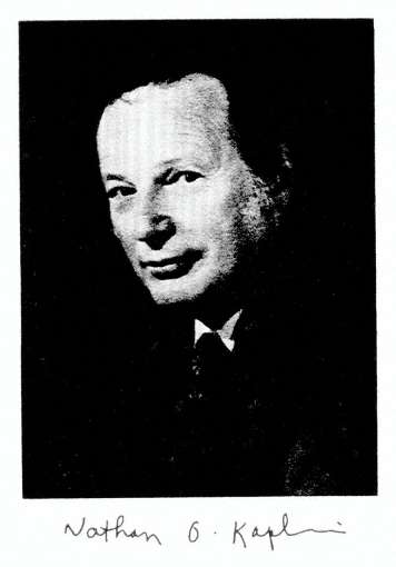
NO Kaplan

NO Kaplan

Sidney Colowick

Sidney Colowick

Elizabeth Neufeld

Elizabeth Neufeld

Kaplan studied carbohydrate metabolism in the liver under David M. Greenberg at the
University of California, Berkeley medical school. He earned his Ph.D. in 1943. From
1942 to 1944, Kaplan participated in the Manhattan Project. From 1945 to 1949,
Kaplan worked with Fritz Lipmann at Massachusetts General Hospital to study
coenzyme A. He worked at the McCollum-Pratt Institute of Johns Hopkins University
from 1950 to 957. In 1957, he was recruited to head a new graduate program in
biochemistry at Brandeis University. In 1968, Kaplan moved to the University of
California, San Diego
, where he studied the role of lactate dehydrogenase in cancer. He also founded a colony of nude mice, a strain of laboratory mice useful in the study
of cancer and other diseases. [1] He was a member of the National Academy of
Sciences.One of Kaplan’s students at the University of California was genomic
researcher Craig Venter.[2]3]  He was, with Sidney Colowick, a founding editor of the scientific book series Methods
in Enzymology
.[1]

http://books.nap.edu/books/0309049768/xhtml/images/img00009.jpg

Colowick became Carl Cori’s first graduate student and earned his Ph.D. at
Washington University St. Louis in 1942, continuing to work with the Coris (Nobel
Prize jointly) for 10 years. At the age of 21, he published his first paper on the
classical studies of glucose 1-phosphate (2), and a year later he was the sole author on a paper on the synthesis of mannose 1-phosphate and galactose 1-phosphate (3). Both papers were published in the JBC. During his time in the Cori lab,

Colowick was involved in many projects. Along with Herman Kalckar he discovered
myokinase (distinguished from adenylate kinase from liver), which is now known as
adenyl kinase. This discovery proved to be important in understanding transphos-phorylation reactions in yeast and animal cells. Colowick’s interest then turned to
the conversion of glucose to polysaccharides, and he and Earl Sutherland (who
will be featured in an upcoming JBC Classic) published an important paper on the
formation of glycogen from glucose using purified enzymes (4). In 1951, Colowick
and Nathan Kaplan were approached by Kurt Jacoby of Academic Press to do a
series comparable to Methodem der Ferment Forschung. Colowick and Kaplan
planned and edited the first 6 volumes of Methods in Enzymology, launching in 1955
what became a series of well known and useful handbooks. He continued as
Editor of the series until his death in 1985.

http://bioenergetics.jbc.org/highwire/filestream/9/field_highwire_fragment_image_s/0/F1.small.gif

The Structure of NADH: the Work of Sidney P. Colowick

Nicole KresgeRobert D. Simoni and Robert L. Hill

On the Structure of Reduced Diphosphopyridine Nucleotide

(Pullman, M. E., San Pietro, A., and Colowick, S. P. (1954)

J. Biol. Chem. 206, 129–141)

Elizabeth Neufeld
·  Born: September 27, 1928 (age 85), Paris, France
·  EducationQueens College, City University of New YorkUniversity of California,
Berkeley

http://fdb5.ctrl.ucla.edu/biological-chemistry/institution/photo?personnel%5fid=45290&max_width=155&max_height=225

In Paper I (l), indirect evidence was presented for the following transhydrogenase
reaction, catalyzed by an enzyme present in extracts of Pseudomonas
fluorescens:

TPNHz + DPN -+ TPN + DPNHz

The evidence was obtained by coupling TPN-specific dehydrogenases with the
transhydrogenase and observing the reduction of large amounts of diphosphopyridine nucleotide (DPN) in the presence of catalytic amounts of triphosphopyridine
nucleotide (TPN).

In this paper, data will be reported showing the direct

  • interaction between TPNHz and DPN, in thepresence of transhydrogenase alone,
  • to yield products having the propertiesof TPN and DPNHZ.

Information will be given indicating that the reaction involves

  • a transfer of electrons (or hydrogen) rather than a phosphate 

Experiments dealing with the kinetics and reversibility of the reaction, and with the
nature of the products, suggest that the reaction is a complex one, not fully described
by the above formulation.

Materials and Methods [edited]

The TPN and DPN used in these studies were preparations of approximately 75
percent purity and were prepared from sheep liver by the chromatographic procedure
of Kornberg and Horecker (unpublished). Reduced DPN was prepared enzymatically with alcohol dehydrogenase as described elsewhere (2). Reduced TPN was prepared by treating TPN with hydrosulfite. This treated mixture contained 2 pM of TPNHz per ml.
The preparations of desamino DPN and reduced desamino DPN have been
described previously (2, 3). Phosphogluconate was a barium salt which was kindly
supplied by Dr. B. F. Horecker. Cytochrome c was obtained from the Sigma Chemical Company.

Transhydrogenase preparations with an activity of 250 to 7000 units per mg. were
used in these studies. The DPNase was a purified enzyme, which was obtained
from zinc-deficient Neurospora and had an activity of 5500 units per mg. (4). The
alcohol dehydrogenase was a crystalline preparation isolated from yeast according to the procedure of Racker (5).

Phosphogluconate dehydrogenase from yeast and a 10 per cent pure preparation of the TPN-specific cytochrome c reductase from liver (6) were gifts of Dr. B. F.
Horecker.

DPN was assayed with alcohol and crystalline yeast alcohol dehydrogenase. TPN was determined By the specific phosphogluconic acid dehydrogenase from yeast and also by the specific isocitric dehydrogenase from pig heart. Reduced DPN was
determined by the use of acetaldehyde and the yeast alcohol dehydrogenase.
All of the above assays were based on the measurement of optical density changes
at 340 rnp. TPNHz was determined with the TPN-specific cytochrome c reductase system. The assay of the reaction followed increase in optical density at 550 rnp  as a measure of the reduction of the cytochrome c after cytochrome c
reductase was added to initiate the reaction. The changes at 550 rnp are plotted for different concentrations of TPNHz in Fig. 3, a. The method is an extremely sensitive and accurate assay for reduced TPN.

Results
[No Figures or Table shown]

Formation of DPNHz from TPNHz and DPN-Fig. 1, a illustrates the direct reaction between TPNHz and DPN to form DPNHZ. The reaction was carried out by incubating TPNHz with DPN in the presence of the
transhydrogenase, yeast alcohol dehydrogenase, and acetaldehyde. Since the yeast dehydrogenase is specific for DPN,

  • a decrease in absorption at340 rnp can only be due to the formation of reduced DPN. It can
    be seen from the curves in Fig. 1, a that a decrease in optical density occurs only in the
    presence of the complete system.

The Pseudomonas enzyme is essential for the formation of DPNH2. It is noteworthy
that, under the conditions of reaction in Fig. 1, a,

  • approximately 40 per cent of theTPNH, reacted with the DPN.

Fig. 1, a also indicates that magnesium is not required for transhydrogenase activity.  The reaction between TPNHz and DPN takes place in the absence of alcohol
dehydrogenase and acetaldehyde
. This can be demonstrated by incubating the
two pyridine nucleotides with the transhydrogenase for 4 8 12 16 20 24 28 32 36
minutes

FIG. 1. Evidence for enzymatic reaction of TPNHt with DPN.

  • Rate offormation of DPNH2.

(b) DPN disappearance and TPN formation.

(c) Identification of desamino DPNHz as product of reaction of TPNHz with desamino DPN.  (assaying for reduced DPN by the yeast alcohol dehydrogenase technique.

Table I (Experiment 1) summarizes the results of such experiments in which TPNHz was added with varying amounts of DPN.

  • In the absence of DPN, no DPNHz was formed. This eliminates the possibility that TPNH 2 is
    converted to DPNHz
  • by removal ofthe monoester phosphate grouping.

The data also show that the extent of the reaction is

  • dependent on the concentration of DPN.

Even with a large excess of DPN, only approximately 40 per cent of the TPNHzreacts to form reduced DPN. It is of importance to emphasize that in the above
experiments, which were carried out in phosphate buffer, the extent of  the reaction

  • is the same in the presence or absence of acetaldehyde andalcohol dehydrogenase.

With an excess of DPN and different  levels of TPNHZ,

  • the amount of reduced DPN which is formed is
  • dependent on the concentration of TPNHz(Table I, Experiment 2).
  • In all cases, the amount of DPNHz formed is approximately
    40 per cent of the added reduced TPN.

Formation of TPN-The reaction between TPNHz and DPN should yield TPN as well as DPNHz.
The formation of TPN is demonstrated in Table 1. in Fig. 1, b. In this experiment,
TPNHz was allowed to react with DPN in the presence of the transhydrogenase
(PS.), and then alcohol and alcohol dehydrogenase were added . This
would result in reduction of the residual DPN, and the sample incubated with the
transhydrogenase contained less DPN. After the completion of the alcohol
dehydrogenase reaction, phosphogluconate and phosphogluconic dehydrogenase (PGAD) were added to reduce the TPN. The addition of this TPN-specific
dehydrogenase results in an

  • increase inoptical density in the enzymatically treated sample.
  • This change represents the amount of TPN formed.

It is of interest to point out that, after addition of both dehydrogenases,

  • the total optical density change is the same in both

Therefore it is evident that

  • for every mole of DPN disappearing  a mole of TPN appears.

Balance of All Components of Reaction

Table II (Experiment 1) shows that,

  • if measurements for all components of the reaction are made, one can demonstrate
    that there is
  • a mole for mole disappearance of TPNH, and DPN, and
  • a stoichiometric appearance of TPN and DPNH2.
  1. The oxidized forms of the nucleotides were assayed as described
  2. the reduced form of TPN was determined by the TPNHz-specific cytochrome c reductase,
  3. the DPNHz by means of yeast alcohol dehydrogenase plus

This stoichiometric balance is true, however,

  • only when the analyses for the oxidized forms are determined directly on the reaction

When analyses are made after acidification of the incubated reaction mixture,

  • the values found forDPN and TPN are much lower than those obtained by direct analysis.

This discrepancy in the balance when analyses for the oxidized nucleotides are
carried out in acid is indicated in Table II (Experiment 2). The results, when
compared with the findings in Experiment 1, are quite striking.

Reaction of TPNHz with Desamino DPN

Desamino DPN

  • reacts with the transhydrogenase system at the same rate as does DPN (2).

This was of value in establishing the fact that

  • the transhydrogenase catalyzesa transfer of hydrogen rather than a phosphate transfer reaction.

The reaction between desamino DPN and TPNHz can be written in two ways.

TPN f desamino DPNHz

TPNH, + desamino DPN

DPNH2 + desamino TPN

If the reaction involved an electron transfer,

  • desamino DPNHz would be
  • Phosphate transfer would result in the production of reduced

Desamino DPNHz can be distinguished from DPNHz by its

  • slowerrate of reaction with yeast alcohol dehydrogenase (2, 3).

Fig. 1, c illustrates that, when desamino DPN reacts with TPNH2, 

  • the product of the reaction is desamino DPNHZ.

This is indicated by the slow rate of oxidation of the product by yeast alcohol
dehydrogenase and acetaldehyde.

From the above evidence phosphate transfer 

  • has been ruled out as a possible mechanism for the transhydrogenase reaction.

Inhibition by TPN

As mentioned in Paper I and as will be discussed later in this paper,

  • the transhydrogenase reaction does not appear to be readily reversible.

This is surprising, particularly since only approximately 

  • 40 per cent of the TPNHz undergoes reaction with DPN
    under the conditions described above. It was therefore thought that
  • the TPN formed might inhibit further transfer of electrons from TPNH2.

Table III summarizes data showing the

  • strong inhibitory effect of TPN on thereaction between TPNHz and DPN.

It is evident from the data that

  • TPN concentration is a factor in determining the extent of the reaction.

Effect of Removal of TPN on Extent of Reaction

A purified DPNase from Neurospora has been found

  • to cleave the nicotinamide riboside linkagesof the oxidized forms of both TPN and DPN
  • without acting on thereduced forms of both nucleotides (4).

It has been found, however, that

  • the DPNase hydrolyzes desamino DPN at a very slow rate (3).

In the reaction between TPNHz and desamino DPN, TPN and desamino DPNH:,

  • TPNis the only component of this reaction attacked by the Neurospora enzyme
    at an appreciable rate

It was  thought that addition of the DPNase to the TPNHZ-desamino DPN trans-
hydrogenase reaction mixture

  • would split the TPN formed andpermit the reaction to go to completion.

This, indeed, proved to be the case, as indicated in Table IV, where addition of
the DPNase with desamino DPN results in almost

  • a stoichiometric formation of desamino DPNHz
  • and a complete disappearance of TPNH2.

Extent of Reaction in Buffers Other Than Phosphate

All the reactions described above were carried out in phosphate buffer of pH 7.5.
If the transhydrogenase reaction between TPNHz and DPN is run at the same pH
in tris(hydroxymethyl)aminomethane buffer (TRIS buffer)

  • with acetaldehydeand alcohol dehydrogenase present,
  • the reaction proceeds muchfurther toward completion 
  • than is the case under the same conditions ina phosphate medium (Fig. 2, a).

The importance of phosphate concentration in governing the extent of the reaction
is illustrated in Fig. 2, b.

In the presence of TRIS the transfer reaction

  • seems to go further toward completion in the presence of acetaldehyde
    and 
    alcohol dehydrogenase
  • than when these two components are absent.

This is not true of the reaction in phosphate,

  • in which the extent is independent of the alcoholdehydrogenase system.

Removal of one of the products of the reaction (DPNHp) in TRIS thus

  • appears to permit the reaction to approach completion,whereas
  • in phosphate this removal is without effect on the finalcourse of the reaction.

The extent of the reaction in TRIS in the absence of alcohol dehydrogenase
and acetaldehyde
 is

  • somewhat greater than when the reaction is run in phosphate.

TPN also inhibits the reaction of TPNHz with DPN in TRIS medium, but the inhibition

  • is not as marked as when the reaction is carried out in phosphate buffer.

Reversibility of Transhydrogenase Reaction;

Reaction between DPNHz and TPN

In Paper I, it was mentioned that no reversal of the reaction could be achieved in a system containing alcohol, alcohol dehydrogenase, TPN, and catalytic amounts of
DPN.

When DPNH, and TPN are incubated with the purified transhydrogenase, there is
also

  • no evidence for reversibility.

This is indicated in Table V which shows that

  • there is no disappearance of DPNHz in such a system.

It was thought that removal of the TPNHz, which might be formed in the reaction,
could promote the reversal of the reaction. Hence,

  • by using the TPNHe-specific cytochrome c reductase, one could
  1. not only accomplishthe removal of any reduced TPN,
  2. but also follow the course of the reaction.

A system containing DPNH2, TPN, the transhydrogenase, the cytochrome c
reductase, and cytochrome c, however, gives

  • no reduction of the cytochrome

This is true for either TRIS or phosphate buffers.2

Some positive evidence for the reversibility has been obtained by using a system
containing

  • DPNH2, TPNH2, cytochrome c, and the cytochrome creductase in TRIS buffer.

In this case, there is, of course, reduction of cytochrome c by TPNHZ, but,

  • when the transhydrogenase is present.,there is
  • additional reduction over and above that due to the added TPNH2.

This additional reduction suggests that some reversibility of the reaction occurred
under these conditions. Fig. 3, b shows

  • the necessity of DPNHzfor this additional reduction.

Interaction of DPNHz with Desamino DPN-

If desamino DPN and DPNHz are incubated with the purified Pseudomonas enzyme,
there appears

  • to be a transfer of electrons to form desamino DPNHz.

This is illustrated in Fig. 4, a, which shows the

  • decreased rate of oxidation by thealcohol dehydrogenase system
  • after incubation with the transhydrogenase.
  • Incubation of desamino DPNHz with DPN results in the formation of DPNH2,
  • which is detected by the faster rate of oxidation by the alcohol dehydrogenase system
  • after reaction of the pyridine nucleotides with thetranshydrogenase (Fig. 4, b).

It is evident from the above experiments that

the transhydrogenase catalyzes an exchange of hydrogens between

  • the adenylic and inosinic pyridine nucleotides.

However, it is difficult to obtain any quantitative information on the rate or extent of
the reaction by the method used, because

  • desamino DPNHz also reacts with the alcohol dehydrogenase system,
  • although at a much slower rate than does DPNH2.

DISCUSSION

The results of the balance experiments seem to offer convincing evidence that
the transhydrogenase catalyzes the following reaction.

TPNHz + DPN -+ DPNHz + TPN

Since desamino DPNHz is formed from TPNHz and desamino DPN,

  • thereaction appears to involve an electron (or hydrogen) transfer
  • rather thana transfer of the monoester phosphate grouping of TPN.

A number of the findings reported in this paper are not readily understandable in
terms of the above simple formulation of the reaction. It is difficult to understand
the greater extent of the reaction in TRIS than in phosphate when acetaldehyde
and alcohol dehydrogenase are present.

One possibility is that an intermediate may be involved which is more easily converted
to reduced DPN in the TRIS medium. The existence of such an intermediate is also
suggested by the discrepancies noted in balance experiments, in which

  • analyses of the oxidized nucleotides after acidification showed
  • much lower values than those found by direct analysis.

These findings suggest that the reaction may involve

  • a 1 electron ratherthan a 2 electron transfer with
  • the formation of acid-labile free radicals as intermediates.

The transfer of hydrogens from DPNHz to desamino DPN

  • to yield desamino DPNHz and DPN and the reversal of this transfer
  • indicate the unique role of the transhydrogenase
  • in promoting electron exchange between the pyridine nucleotides.

In this connection, it is of interest that alcohol dehydrogenase and lactic
dehydrogenase cannot duplicate this exchange  between the DPN and
the desamino systems.3  If one assumes that desamino DPN behaves
like DPN,

  • one might predict that the transhydrogenase would catalyze an
    exchange of electrons (or hydrogen) 3.

Since alcohol dehydrogenase alone

  • does not catalyze an exchange of electrons between the adenylic
    and inosinic pyridine nucleotides, this rules out the possibility
  • that the dehydrogenase is converted to a reduced intermediate
  • during electron between DPNHz and added DPN.

It is hoped to investigate this possibility with isotopically labeled DPN.
Experiments to test the interaction between TPN and desamino TPN are
also now in progress.

It seems likely that the transhydrogenase will prove capable of

  • catalyzingan exchange between TPN and TPNH2, as well as between DPN and

The observed inhibition by TPN of the reaction between TPNHz and DPN may
therefore

  • be due to a competition between DPN and TPNfor the TPNH2.

SUMMARY

  1. Direct evidence for the following transhydrogenase reaction. catalyzedby an
    enzyme from Pseudomonas fluorescens, is presented.

TPNHz + DPN -+ TPN + DPNHz

Balance experiments have shown that for every mole of TPNHz disappearing
1 mole of TPN appears and that for each mole of DPNHz generated 1 mole of
DPN disappears. The oxidized nucleotides found at the end of the reaction,
however, show anomalous lability toward acid.

  1. The transhydrogenase also promotes the following reaction.

TPNHz + desamino DPN -+ TPN + desamino DPNH,

This rules out the possibility that the transhydrogenase reaction involves a
phosphate transfer and indicates that the

  • enzyme catalyzes a shift of electrons (or hydrogen atoms).

The reaction of TPNHz with DPN in 0.1 M phosphate buffer is strongly
inhibited by TPN; thus

  • it proceeds only to the extent of about40 per cent or less, even
  • when DPNHz is removed continuously by meansof acetaldehyde
    and alcohol dehydrogenase.
  • In other buffers, in whichTPN is less inhibitory, the reaction proceeds
    much further toward completion under these conditions.
  • The reaction in phosphate buffer proceedsto completion when TPN
    is removed as it is formed.
  1. DPNHz does not react with TPN to form TPNHz and DPN in the presence
    of transhydrogenase. Some evidence, however, has been obtained for
    the reversibility by using the following system:
  • DPNHZ, TPNHZ, cytochromec, the TPNHz-specific cytochrome c reductase,
    and the transhydrogenase.
  1. Evidence is cited for the following reversible reaction, which is catalyzed
    by the transhydrogenase.

DPNHz + desamino DPN fi DPN + desamino DPNHz

  1. The results are discussed with respect to the possibility that the
    transhydrogenase reaction may
  • involve a 1 electron transfer with theformation of free radicals as intermediates.


BIBLIOGRAPHY

  1. Coiowick, S. P., Kaplan, N. O., Neufeld, E. F., and Ciotti, M. M., J. Biol. Chem.,196, 95 (1952).
  2. Pullman, 111. E., Colowick, S. P., and Kaplan, N. O., J. Biol. Chem., 194, 593(1952).
  3. Kaplan, N. O., Colowick, S. P., and Ciotti, M. M., J. Biol. Chem., 194, 579 (1952).
  4. Kaplan, N. O., Colowick, S. P., and Nason, A., J. Biol. Chem., 191, 473 (1951).
  5. Racker, E., J. Biol. Chem., 184, 313 (1950).
  6. Horecker, B. F., J. Biol. Chem., 183, 593 (1950).

Section !II. 

Luis_Federico_Leloir_-_young

The Leloir pathway: a mechanistic imperative for three enzymes to change
the stereochemical configuration of a single carbon in galactose.

Frey PA.
FASEB J. 1996 Mar;10(4):461-70.    http://www.fasebj.org/content/10/4/461.full.pdf
PMID:8647345

The biological interconversion of galactose and glucose takes place only by way of
the Leloir pathway and requires the three enzymes galactokinase, galactose-1-P
uridylyltransferase, and UDP-galactose 4-epimerase.
The only biological importance of these enzymes appears to be to

  • provide for the interconversion of galactosyl and glucosyl groups.

Galactose mutarotase also participates by producing the galactokinase substrate
alpha-D-galactose from its beta-anomer. The galacto/gluco configurational change takes place at the level of the nucleotide sugar by an oxidation/reduction
mechanism in the active site of the epimerase NAD+ complex. The nucleotide portion
of UDP-galactose and UDP-glucose participates in the epimerization process in two ways:

1) by serving as a binding anchor that allows epimerization to take place at glycosyl-C-4 through weak binding of the sugar, and

2) by inducing a conformational change in the epimerase that destabilizes NAD+ and
increases its reactivity toward substrates.

Reversible hydride transfer is thereby facilitated between NAD+ and carbon-4
of the weakly bound sugars.

The structure of the enzyme reveals many details of the binding of NAD+ and
inhibitors at the active site
.

The essential roles of the kinase and transferase are to attach the UDP group
to galactose, allowing for its participation in catalysis by the epimerase. The
transferase is a Zn/Fe metalloprotein
, in which the metal ions stabilize the
structure rather than participating in catalysis. The structure is interesting
in that

  • it consists of single beta-sheet with 13 antiparallel strands and 1 parallel strand
    connected by 6 helices.

The mechanism of UMP attachment at the active site of the transferase is a double
displacement
, with the participation of a covalent UMP-His 166-enzyme intermediate
in the Escherichia coli enzyme. The evolution of this mechanism appears to have
been guided by the principle of economy in the evolution of binding sites.

PMID: 8647345 Free full text

Section IV.

More on Lipids – Role of lipids – classification

  • Energy
  • Energy Storage
  • Hormones
  • Vitamins
  • Digestion
  • Insulation
  • Membrane structure: Hydrophobic properties

Lipid types

lipid types

lipid types

nat occuring FAs in mammals

nat occuring FAs in mammals

Read Full Post »

Selected References to Signaling and Metabolic Pathways in PharmaceuticalIntelligence.com

Curator: Larry H. Bernstein, MD, FCAP


This is an added selection of articles in Leaders in Pharmaceutical Intelligence after the third portion of the discussion in a series of articles that began with signaling and signaling pathways. There are fine features on the functioning of enzymes and proteins, on sequential changes in a chain reaction, and on conformational changes that we shall return to.  These are critical to developing a more complete understanding of life processes.  I have indicated that many of the protein-protein interactions or protein-membrane interactions and associated regulatory features have been referred to previously, but the focus of the discussion or points made were different.

  1. Signaling and signaling pathways
  2. Signaling transduction tutorial.
  3. Carbohydrate metabolism3.1  Selected References to Signaling and Metabolic Pathways in Leaders in Pharmaceutical Intelligence
  4. Lipid metabolism
  5. Protein synthesis and degradation
  6. Subcellular structure
  7. Impairments in pathological states: endocrine disorders; stress hypermetabolism; cancer.

Selected References to Signaling and Metabolic Pathwayspublished in this Open Access Online Scientific Journal, include the following:

Update on mitochondrial function, respiration, and associated disorders

Curator and writer: Larry H. Benstein, MD, FCAP

http://pharmaceuticalintelligence.com/2014/07/08/update-on-mitochondrial-function-respiration-and-associated-disorders/

A Synthesis of the Beauty and Complexity of How We View Cancer


Cancer Volume One – Summary

A Synthesis of the Beauty and Complexity of How We View Cancer

Author: Larry H. Bernstein, MD, FCAP

http://pharmaceuticalintelligence.com/2014/03/26/a-synthesis-of-the-beauty-and-complexity-of-how-we-view-cancer/

Introduction – The Evolution of Cancer Therapy and Cancer Research: How We Got Here?

Author and Curator: Larry H Bernstein, MD, FCAP

http://pharmaceuticalintelligence.com/2014/04/04/introduction-the-evolution-of-cancer-therapy-and-cancer-research-how-we-got-here/

 The Centrality of Ca(2+) Signaling and Cytoskeleton Involving Calmodulin Kinases and Ryanodine Receptors in Cardiac Failure, Arterial Smooth Muscle, Post-ischemic Arrhythmia, Similarities and Differences, and Pharmaceutical Targets

Author and Curator: Larry H Bernstein, MD, FCAP, 
Author, and Content Consultant to e-SERIES A: Cardiovascular Diseases: Justin Pearlman, MD, PhD, FACC
And Curator: Aviva Lev-Ari, PhD, RN

http://pharmaceuticalintelligence.com/2013/09/08/the-centrality-of-ca2-signaling-and-cytoskeleton-involving-calmodulin-kinases-and-ryanodine-receptors-in-cardiac-failure

Renal Distal Tubular Ca2+ Exchange Mechanism in Health and Disease

Author and Curator: Larry H. Bernstein, MD, FCAP
Curator:  Stephen J. Williams, PhD
and Curator: Aviva Lev-Ari, PhD, RN

http://pharmaceuticalintelligence.com/2013/09/02/renal-distal-tubular-ca2-exchange-mechanism-in-health-and-disease/

Mitochondrial Metabolism and Cardiac Function

Curator: Larry H Bernstein, MD, FACP

http://pharmaceuticalintelligence.com/2013/04/14/mitochondrial-metabolism-and-cardiac-function/

Mitochondrial Dysfunction and Cardiac Disorders

Curator: Larry H Bernstein, MD, FACP

http://pharmaceuticalintelligence.com/2013/04/14/mitochondrial-metabolism-and-cardiac-function/

Reversal of Cardiac mitochondrial dysfunction

Curator: Larry H Bernstein, MD, FACP

http://pharmaceuticalintelligence.com/2013/04/14/reversal-of-cardiac-mitochondrial-dysfunction/

Advanced Topics in Sepsis and the Cardiovascular System  at its End Stage

Author: Larry H Bernstein, MD, FCAP

http://pharmaceuticalintelligence.com/2013/08/18/advanced-topics-in-Sepsis-and-the-Cardiovascular-System-at-its-End-Stage/

Ubiquinin-Proteosome pathway, autophagy, the mitochondrion, proteolysis and cell apoptosis

Curator: Larry H Bernstein, MD, FACP

http://pharmaceuticalintelligence.com/2012/10/30/ubiquinin-proteosome-pathway-autophagy-the-mitochondrion-proteolysis-and-cell-apoptosis/

Ubiquitin-Proteosome pathway, Autophagy, the Mitochondrion, Proteolysis and Cell Apoptosis: Part III

Curator: Larry H Bernstein, MD, FCAP

http://pharmaceuticalintelligence.com/2013/02/14/ubiquinin-proteosome-pathway-autophagy-the-mitochondrion-proteolysis-and-cell-apoptosis-reconsidered/


Nitric Oxide, Platelets, Endothelium and Hemostasis (Coagulation Part II)

Curator: Larry H. Bernstein, MD, FCAP 

http://pharmaceuticalintelligence.com/2012/11/08/nitric-oxide-platelets-endothelium-and-hemostasis/


Mitochondrial Damage and Repair under Oxidative Stress

Curator: Larry H Bernstein, MD, FCAP

http://pharmaceuticalintelligence.com/2012/10/28/mitochondrial-damage-and-repair-under-oxidative-stress/

Mitochondria: Origin from oxygen free environment, role in aerobic glycolysis, metabolic adaptation

Reporter and Curator: Larry H Bernstein, MD, FACP

http://pharmaceuticalintelligence.com/2012/09/26/mitochondria-origin-from-oxygen-free-environment-role-in-aerobic-glycolysis-metabolic-adaptation/


Nitric Oxide has a Ubiquitous Role in the Regulation of Glycolysis – with a Concomitant Influence on Mitochondrial Function

Reporter, Editor, and Topic Co-Leader: Larry H. Bernstein, MD, FCAP

http://pharmaceuticalintelligence.com/2012/09/16/nitric-oxide-has-a-ubiquitous-role-in-the-regulation-of-glycolysis-with-a-concomitant-influence-on-mitochondrial-function/


Mitochondria and Cancer: An overview of mechanisms

Author and Curator: Ritu Saxena, Ph.D.

http://pharmaceuticalintelligence.com/2012/09/01/mitochondria-and-cancer-an-overview/

Mitochondria: More than just the “powerhouse of the cell”

Author and Curator: Ritu Saxena, Ph.D.

http://pharmaceuticalintelligence.com/2012/07/09/mitochondria-more-than-just-the-powerhouse-of-the-cell/

Overview of Posttranslational Modification (PTM)

Curator: Larry H. Bernstein, MD, FCAP

http://pharmaceuticalintelligence.com/2014/07/29/overview-of-posttranslational-modification-ptm/


Ubiquitin Pathway Involved in Neurodegenerative Diseases

Author and curator: Larry H Bernstein, MD,  FCAP

http://pharmaceuticalintelligence.com/2013/02/15/ubiquitin-pathway-involved-in-neurodegenerative-diseases/

Is the Warburg Effect the Cause or the Effect of Cancer: A 21st Century View?

Author: Larry H. Bernstein, MD, FCAP 

http://pharmaceuticalintelligence.com/2012/10/17/is-the-warburg-effect-the-cause-or-the-effect-of-cancer-a-21st-century-view/

New Insights on Nitric Oxide donors – Part IV

Curator and Author: Larry H. Bernstein, MD, FCAP

http://pharmaceuticalintelligence.com/2012/11/26/new-insights-on-no-donors/

Perspectives on Nitric Oxide in Disease Mechanisms [Kindle Edition]

Margaret Baker PhD (Author), Tilda Barliya PhD (Author), Anamika Sarkar PhD (Author), Ritu Saxena PhD (Author), Stephen J. Williams PhD (Author), Larry Bernstein MD FCAP (Editor), Aviva Lev-Ari PhD RN (Editor), Aviral Vatsa PhD (Editor)

http://pharmaceuticalintelligence.com/biomed-e-books/series-a-e-books-on-cardiovascular-diseases/perspectives-on-nitric-oxide-in-disease-mechanisms-v2/


Summary

Nitric oxide and its role in vascular biology

Signal transmission by a gas that is produced by one cell, penetrates through membranes and regulates the function of another cell represents an entirely new principle for signaling in biological systems.   All compounds that inhibit endothelium-derived relaxation-factor (EDRF) have one property in common, redox activity, which accounts for their inhibitory action on EDRF. One exception is hemoglobin, which inactivates EDRF by binding to it. Furchgott, Ignarro and Murad received the Nobel Prize in Physiology and Medicine for discovery of EDRF in 1998 and demonstrating that it might be nitric oxide (NO) based on a study of the transient relaxations of endothelium-denuded rings of rabbit aorta.  These investigators working independently demonstrated that NO is indeed produced by mammalian cells and that NO has specific biological roles in the human body. These studies highlighted the role of NO in cardiovascular, nervous and immune systems. In cardiovascular system NO was shown to cause relaxation of vascular smooth muscle cells causing vasodilatation, in nervous system NO acts as a signaling molecule and in immune system it is used against pathogens by the phagocytosis cells. These pioneering studies opened the path of investigation of role of NO in biology.

NO modulates vascular tone, fibrinolysis, blood pressure and proliferation of vascular smooth muscles. In cardiovascular system disruption of NO pathways or alterations in NO production can result in preponderance to hypertension, hypercholesterolemia, diabetes mellitus, atherosclerosis and thrombosis. The three enzyme isoforms of NO synthase family are responsible for generating NO in different tissues under various circumstances.

Reduction in NO production is implicated as one of the initial factors in initiating endothelial dysfunction. This reduction could be due to

  • reduction in eNOS production
  • reduction in eNOS enzymatic activity
  • reduced bioavailability of NO

Nitric oxide is one of the smallest molecules involved in physiological functions in the body. It is seeks formation of chemical bonds with its targets.  Nitric oxide can exert its effects principally by two ways:

  • Direct
  • Indirect

Direct actions, as the name suggests, result from direct chemical interaction of NO with its targets e.g. with metal complexes, radical species. These actions occur at relatively low NO concentrations (<200 nM)

Indirect actions result from the effects of reactive nitrogen species (RNS) such as NO2 and N2O3. These reactive species are formed by the interaction of NO with superoxide or molecular oxygen. RNS are generally formed at relatively high NO concentrations (>400 nM)

Although it can be tempting for scientists to believe that RNS will always have deleterious effects and NO will have anabolic effects, this is not entirely true as certain RNS mediated actions mediate important signalling steps e.g. thiol oxidation and nitrosation of proteins mediate cell proliferation and survival, and apoptosis respectively.

  • Cells subjected to NO concentration between 10-30 nM were associated with cGMP dependent phosphorylation of ERK
  • Cells subjected to NO concentration between 30-60 nM were associated with Akt phosphorylation
  • Concentration nearing 100 nM resulted in stabilisation of hypoxia inducible factor-1
  • At nearly 400 nM NO, p53 can be modulated
  • >1μM NO, it nhibits mitochondrial respiration


Nitric oxide signaling, oxidative stress,  mitochondria, cell damage

Recent data suggests that other NO containing compounds such as S- or N-nitrosoproteins and iron-nitrosyl complexes can be reduced back to produce NO. These NO containing compounds can serve as storage and can reach distant tissues via blood circulation, remote from their place of origin. Hence NO can have both paracrine and ‘endocrine’ effects.

Intracellularly the oxidants present in the cytosol determine the amount of bioacitivity that NO performs. NO can travel roughly 100 microns from NOS enzymes where it is produced.

NO itself in low concentrations have protective action on mitochondrial signaling of cell death.

The aerobic cell was an advance in evolutionary development, but despite the energetic advantage of using oxygen, the associated toxicity of oxygen abundance required adaptive changes.

Oxidation-reduction reactions that are necessary for catabolic and synthetic reactions, can cumulatively damage the organism associated with cancer, cardiovascular disease, neurodegerative disease, and inflammatory overload.  The normal balance between production of pro-oxidant species and destruction by the antioxidant defenses is upset in favor of overproduction of the toxic species, which leads to oxidative stress and disease.

We reviewed the complex interactions and underlying regulatory balances/imbalances between the mechanism of vasorelaxation and vasoconstriction of vascular endothelium by way of nitric oxide (NO), prostacyclin, in response to oxidative stress and intimal injury.

Nitric oxide has a ubiquitous role in the regulation of glycolysis with a concomitant influence on mitochondrial function. The influence on mitochondrial function that is active in endothelium, platelets, vascular smooth muscle and neural cells and the resulting balance has a role in chronic inflammation, asthma, hypertension, sepsis and cancer.

Potential cytotoxic mediators of endothelial cell (EC) apoptosis include increased formation of reactive oxygen and nitrogen species (ROSRNS) during the atherosclerotic process. Nitric oxide (NO) has a biphasic action on oxidative cell killing with low concentrations protecting against cell death, whereas higher concentrations are cytotoxic.

ROS induces mitochondrial DNA damage in ECs, and this damage is accompanied by a decrease in mitochondrial RNA (mtRNA) transcripts, mitochondrial protein synthesis, and cellular ATP levels.

NO and circulatory diseases

Blood vessels arise from endothelial precursors that are thin, flat cells lining the inside of blood vessels forming a monolayer throughout the circulatory system. ECs are defined by specific cell surface markers that characterize their phenotype.

Scientists at the University of Helsinki, Finland, wanted to find out if there exists a rare vascular endothelial stem cell (VESC) population that is capable of producing very high numbers of endothelial daughter cells, and can lead to neovascular growth in adults.

VESCs discovered that reside at the blood vessel wall endothelium are a small population of CD117+ ECs capable of self-renewal.  These cells are capable of undergoing clonal expansion unlike the surrounding ECs that bear limited proliferating potential. A single VESC cell isolated from the endothelial population was able to generate functional blood vessels.

Among many important roles of Nitric oxide (NO), one of the key actions is to act as a vasodilator and maintain cardiovascular health. Induction of NO is regulated by signals in tissue as well as endothelium.

Chen et. al. (Med. Biol. Eng. Comp., 2011) developed a 3-D model consisting of two branched arterioles and nine capillaries surrounding the vessels. Their model not only takes into account of the 3-D volume, but also branching effects on blood flow.

The model indicates that wall shear stress changes depending upon the distribution of RBC in the microcirculations of blood vessels, lead to differential production of NO along the vascular network.

Endothelial dysfunction, the hallmark of which is reduced activity of endothelial cell derived nitric oxide (NO), is a key factor in developing atherosclerosis and cardiovascular disease. Vascular endothelial cells play a pivotal role in modulation of leukocyte and platelet adherence, thrombogenicity, anticoagulation, and vessel wall contraction and relaxation, so that endothelial dysfunction has become almost a synonym for vascular disease. A single layer of endothelial cells is the only constituent of capillaries, which differ from other vessels, which contain smooth muscle cells and adventitia. Capillaries directly mediate nutritional supply as well as gas exchange within all organs. The failure of the microcirculation leads to tissue apoptosis/necrosis.

Read Full Post »

Calcium and Cardiovascular Diseases: A Series of Twelve Articles in Advanced Cardiology

Curator: Aviva Lev-Ari, PhD, RN

UPDATED on 7/18/2021

ER

IMAGE SOURCE:

Claudio A. Hetz. Antioxidants & Redox Signaling.Dec 2007.

2345-2356. http://doi.org/10.1089/ars.2007.1793

FIG. 3. Regulation of ER calcium homeostasis by the BCL-2 protein family. Different anti- and proapoptotic members of the BCL-2 family of proteins are located at the ER membrane, where they have an important role regulating ER calcium content. BCL-2 and BCL-XL interact with the IP3R calcium channel, modulating its activity. BCL-2 has been shown to increase ER calcium leak through the IP3R because of an increase on its phosphorylation levels.

BAX and BAK have the opposite effect on ER calcium content, a function that may be further modulated by BH3-only proteins (such as PUMA and BIK). In addition, the activity of BCL-2 at the ER membrane is regulated by phosphorylation. JNK phosphorylates BCL-2, decreasing its antiapoptotic activity and increasing ER calcium content, whereas the phosphatase PP2A decreases this phosphorylation through a direct interaction. Alternatively, ER stress activates the IRE1/JNK pathway that may alter the activity of BCL-2 at the ER membrane. BI-1 is also located at the ER membrane, where it regulates calcium homeostasis.

CONCLUSIONS AND THERAPEUTIC PERSPECTIVES

I have summarized different pieces of evidence suggesting that the BCL-2 family of proteins has evolved to regulate multiple processes involved in cell survival under stress conditions. The global view of the current state of the field indicates that the BCL-2–related proteins are not only the “death gateway” keeper (as upstream regulators of caspases), but they also have multiple functions in essential processes for the cell. BCL-2–related proteins are particularly important in the physiologic maintenance of the ER, where they operate as

(a) a calcium rheostat,

(b) modulators of the UPR,

(c) regulators of ER network structure, and

(d) regulators of autophagy.

In addition, examples of a role of the BCL-2 family of proteins in cell-cycle regulation (87, 113), DNA damage responses (37, 114), and glucose/energy metabolism (16) are available, strongly supporting the notion that the BCL-2 protein family is a multifunctional group of proteins that, under normal conditions, participate in essential cellular process. In doing so, the BCL-2 protein family may represent specialized stress sentinels that actively participate in essential processes, allowing a constant homeostatic “quality control.” In response to irreversible cellular damage, particular BCL-2 family members may turn into direct activators of apoptosis.

Mutations in specific genes are responsible for a variety of neurologic disorders due to the misfolding and accumulation of abnormal protein aggregates in the brain. In many of these diseases, it has been suggested that alteration in the homeostasis of the ER contributes significantly to neuronal dysfunction.

These diseases include Parkinson’s disease (32, 84), Alzheimer’s disease (22), prion diseases (27, 28, 31), amyotrophic lateral sclerosis (ALS) (97), Huntington’s disease (63, 90) and many others (see list of diseases in 86). Consequently, the first steps in the death pathways downstream of ER stress represent important therapeutic targets. In this line of thinking, pharmacologic manipulation of the activity of the BCL-2 protein family may have beneficial consequences to treat these fatal diseases. Different small molecules and synthetic peptides are currently available with proven therapeutic applications in mouse disease models, including BCL-2 inhibitors (71), BAX channel inhibitors (29), BAX/BAK activator peptides (100, 101) and many others (see reviews in 52, 79). These drugs may be used as pharmacologic tools to manipulate the activity of stress-signaling pathways regulated by the BCL-2 protein family (i.e., autophagy, calcium metabolism, or the UPR) and their possible role in pathologic conditions.

SOURCE

Claudio A. Hetz.Antioxidants & Redox Signaling.Dec 2007.

2345-2356. http://doi.org/10.1089/ars.2007.1793

  • Published in Volume: 9 Issue 12: November 2, 2007
  • Online Ahead of Print: September 13, 2007

UPDATED on 7/1/2015

We add the following to this series:

Part XIII 

Ca2+-Stimulated Exocytosis:  The Role of Calmodulin and Protein Kinase C in Ca2+ Regulation of Hormone and Neurotransmitter
Larry H Bernstein, MD, FCAP and Aviva Lev-Ari, PhD, RN

Part I:

Identification of Biomarkers that are Related to the Actin Cytoskeleton

Larry H Bernstein, MD, FCAP

Part II:

Role of Calcium, the Actin Skeleton, and Lipid Structures in Signaling and Cell Motility

Larry H. Bernstein, MD, FCAP, Stephen Williams, PhD and Aviva Lev-Ari, PhD, RN

Part III:

Renal Distal Tubular Ca2+ Exchange Mechanism in Health and Disease

Larry H. Bernstein, MD, FCAP, Stephen J. Williams, PhD
 and Aviva Lev-Ari, PhD, RN

Part IV:

The Centrality of Ca(2+) Signaling and Cytoskeleton Involving Calmodulin Kinases and Ryanodine Receptors in Cardiac Failure, ArterialSmooth Muscle, Post-ischemic Arrhythmia, Similarities and Differences, and Pharmaceutical Targets

Larry H Bernstein, MD, FCAP, Justin Pearlman, MD, PhD, FACC and Aviva Lev-Ari, PhD, RN

Part V:

Heart, Vascular Smooth Muscle, Excitation-Contraction Coupling (E-CC), Cytoskeleton, Cellular Dynamics and Ca2 Signaling

Larry H Bernstein, MD, FCAP, Justin Pearlman, MD, PhD, FACC and Aviva Lev-Ari, PhD, RN

Part VI:

Calcium Cycling (ATPase Pump) in Cardiac Gene Therapy: Inhalable Gene Therapy for Pulmonary Arterial Hypertension and Percutaneous Intra-coronary Artery Infusion for Heart Failure: Contributions by Roger J. Hajjar, MD

Aviva Lev-Ari, PhD, RN

Part VII:

Cardiac Contractility & Myocardium Performance: Ventricular Arrhythmias and Non-ischemic Heart Failure – Therapeutic Implications for Cardiomyocyte Ryanopathy (Calcium Release-related Contractile Dysfunction) and Catecholamine Responses

Justin Pearlman, MD, PhD, FACC, Larry H Bernstein, MD, FCAP and Aviva Lev-Ari, PhD, RN

Part VIII

Disruption of Calcium Homeostasis: Cardiomyocytes and Vascular Smooth Muscle Cells: The Cardiac and Cardiovascular Calcium Signaling Mechanism – Part VIII

Justin Pearlman, MD, PhD, FACC, Larry H Bernstein, MD, FCAP and Aviva Lev-Ari, PhD, RN

Part IX

Calcium-Channel Blockers, Calcium Release-related Contractile Dysfunction (Ryanopathy) and Calcium as Neurotransmitter Sensor – Part IX

Justin Pearlman, MD, PhD, FACC, Larry H Bernstein, MD, FCAP and Aviva Lev-Ari, PhD, RN

Part X

Synaptotagmin functions as a Calcium Sensor: How Calcium Ions Regulate the fusion of vesicles with cell membranes during Neurotransmission – Part X

Larry H Bernstein, MD, FCAP and Aviva Lev-Ari, PhD, RN

Part XI

Sensors and Signaling in Oxidative Stress – Part XI

Larry H. Bernstein, MD, FCAP

Part XII

Atherosclerosis Independence: Genetic Polymorphisms of Ion Channels Role in the Pathogenesis of Coronary Microvascular Dysfunction and Myocardial Ischemia (Coronary Artery Disease (CAD)) – Part XII

Larry H Bernstein, MD, FCAP and Aviva Lev-Ari, PhD,

Read Full Post »

Personalized Cardiovascular Genetic Medicine at Partners HealthCare and Harvard Medical School

Curator: Aviva Lev-Ari, PhD, RN

UPDATED on 5/4/2015

Goes to Clinic @MGH: Clinically validated versions of Exome Sequencing and Analysis using Broad-developed methods like Hybrid Capture, the Genome Analysis Toolkit (GATK), and MuTect

http://pharmaceuticalintelligence.com/2015/05/04/goes-to-clinic-mgh-clinically-validated-versions-of-exome-sequencing-and-analysis-using-broad-developed-methods-like-hybrid-capture-the-genome-analysis-toolkit-gatk-and-mutect/

Center for Personalized Genetic Medicine, Partners HealthCare and Harvard Medical School

The Partners HealthCare Center for Personalized Genetic Medicine offers technologies and technical support for the research activities of Partners investigators. Our objective is to help investigators advance their research programs and to provide the highest quality service, technical expertise, and leading technologies for genomics research. Our goal is to broaden the access to these technologies while offering the best customer service in the most cost conscious and time efficient manner possible.

We are organized into four principal service areas:

  • sequence analysis,
  • genotyping,
  • expression analysis, and
  • bioprocessing/sample management

Our platforms include next generation sequencing with Illumina HiSeq2000 and GA ii analyzers as well as Sanger sequencing using ABI 3730 XL sequence analyzers. Targeted custom genotyping is offered using Sequenom and Illumina GoldenGate panels as well as GWAS scale projects using Illumina Infinium and DNA methylation analysis using Illumina bead arrays. Expression analysis is available with capabilities for processing total RNA on either Affymetrix or Illumina arrays.

Through services from our BioSample Services Facility (BSF) and Partners Biorepository for Medical Discovery (PBMD) teams we provide a research platform for handling samples in a standardized manner to provide consistency from sample to sample. The BSF is able to assist investigators to configure projects utilizing your own samples or coupled with the PCPGM-PBMD we are able to support the integration of cohorts of samples selected from the PBMD into analysis on our genomics platforms.

DNA Sequencing

The DNA Sequencing Group at the Partners HealthCare Center for Personalized Genetic Medicine has a strong history of producing high quality, dependable, and informative results for collaborators and clients. The DNA Sequencing Group participated in the Human Genome Project, building the STS-Based BAC map for Human Chromosome 12, and providing Chromosome 12 tiling path clones to the Baylor Human Genome Sequencing Center for sequencing.

The group sequenced 113 BACs for the Mouse Genome Project, contributing 24 megabases of finished mouse sequences to the published Mouse Genome, as well as providing draft sequences for unique strains of several bacterial genomes, including Pseudomonas aeruginosa, and Vibrio cholerae. More recently, the group participated in identifying mutations linked to numerous diseases, either in collaborations or by providing client laboratories with full service resequencing and analysis.

Services by Project Goals

Mutation Identification via Resequencing

This facility provides full-service resequencing of regions of interest in one or more genomic DNAs, including the following:

  • Discussion of the scope of the project and a cost quote
  • Identification of genes in the region of interest as needed, with the Investigator
  • Primer design using our automated system, to amplify desired regions
  • Primer ordering
  • QC of the primers on DNA standards, if required
  • PCR amplification of DNA provided by Investigator
  • PCR clean-up
  • Sequencing reactions
  • Sequencing reaction clean-up
  • Sequence application to the ABI 3730 XL Analyzer
  • Chromatograms are made available to Investigator over web (GIGPAD)
  • Data assembly and analysis using Phred Phrap and PolyPhred
  • One round of repeats and redesign if necessary
  • Report of variations found throughout sequence
  • Trouble shooting for 100% coverage if desired
Research Services
  • Fee-for-service sequencing
  • Fragment analysis / genotyping (Microsatellite Instability)
Technology Development
  • New technology testing and development
  • Collaborative Protocol development
  • Beta-test site for instrumentation and software
Clinical Diagnosis
  • Diagnostic test development
  • Sequencing for clinical diagnostics group
Genomic Sequencing Projects
  • Human
  • Rodent
  • Bacteria

http://pcpgm.partners.org/research-services/sequencing

Advancing Translational Genomics through Personalized Medicine Projects

The mission of the Partners HealthCare Center for Personalized Genetic Medicine (PCPGM) is to utilize genetics and genomics to promote and implement personalized medicine in caring for patients throughout the Partners HealthCare system and in health care nationally and globally.

The Personalized Medicine Project program was developed to support the clinical research efforts of junior Partners HealthCare investigators for translational genetics and genomic projects to advance personalized medicine.  The goal of this program is to identify biological markers that can be used as potential predictive tests.  This will be accomplished by:

  • Leveraging the Partners HealthCare Research Patient Data Registry (RPDR) and the Partners Biorepository for Medical Discovery (PBMD), centralized locations where Partners HealthCare patient data and/or samples are stored.
  • Identifying novel biological markers or new uses for existing markers.
  • Focusing on tests that could:
    • improve diagnostic sensitivity or specificity;
    • further stratify patient groups with a given diagnosis;
    • predict improved clinical outcomes; or
    • assist with selection of therapies or methods to manage disease.

http://pcpgm.partners.org/biorepository/pmprojectsrfp

Harvard Medical School Genetics Training Program

The Harvard Medical School (HMS) Genetics Training Program is one of the oldest and largest programs in the country. It was founded by Drs. John Littlefield at the Massachusetts General Hospital and Park Gerald at Children’s Hospital Boston in the early 1970’s. The program has trained scientists and clinicians who have become leaders in academic genetics, and has supported investigators who have made major contributions to the clinical practice of genetics and genetics research.

The HMS Genetics Training Program is accredited by the ABMG in all areas of training – Clinical Genetics, Biochemical Genetics, Cytogenetics, and Molecular Genetics. This provides an opportunity for our trainees to become active candidates for board certification in a discipline(s) of medical genetics in addition to receiving laboratory training. The training laboratories and clinics of the program are centered at HMS and its affiliated institutions including Brigham and Women’s Hospital (BWH), the HMS Department of Genetics, Beth Israel Deaconess Medical Center (BIDMC), Children’s Hospital Boston (CHB), Dana Farber Cancer Institute (DFCI), and Massachusetts General Hospital (MGH). The HMS Genetics Training Program provides trainees the opportunity to take advantage of the extraordinarily rich academic environment offered at HMS and its affiliated institutions as well as the greater Boston scientific community.

Cardiovascular Research Center @MGH

The Cardiovascular Research Center was founded in 1990, and occupies over 30,000 sq. ft. of laboratory space in both the Charlestown Navy Yard and the Richard B. Simches Research Building. Dr. Mark Fishman, now president of the Novartis Institutes for Biomedical Research, directed the Center from 1990 until 2002. From 2002-2005, Dr. Kenneth Bloch served as Interim Director and then in June 2005, the Massachusetts General Hospital welcomed Dr. Kenneth Chien as the new scientific director of the Cardiovascular Research Center. Prior to his MGH appointments, Dr. Chien directed the Institute for Molecular Medicine at the University of California at San Diego. An internationally recognized biologist specializing in cardiovascular science, he is a true pioneer in developing new therapeutic strategies to prevent the onset and progression of heart failure. Dr. Chien served as director until 2012.

Cardiovascular Research Center investigators have made many groundbreaking discoveries. Among these include:

• first identification of progenitor cells in the heart
• cloning of the first vertebrate cell death genes
• knocking out the genes that produce nitric oxide (NO), showing the importance of this molecule to atherosclerosis and stroke
• clinical use of NO to treat patients with pulmonary hypertension
• development of gene and cell transfer approaches to treat heart failure
• performance of the first large-scale genetic screen in a vertebrate (the zebrafish)
• identification of genes critical to cardiac pacemaking, rhythm, contractile function, and normal heart patterning
• discovery of a new methylase gene responsible for altering DNA structure during an individual’s lifetime

The Cardiovascular Research Center has taken great pride in the training of scientists with MDs and/or PhDs, as well as graduate students from a variety of Boston area institutions.

The Cardiovascular Research Center has two locations, one in the Charlestown Navy Yard and the other on the main campus’s Charles River Plaza complex in the Richard Simches Research Center.

Both the Simches and Navy Yard sites offer state of the art facilities, including tissue culture rooms, warm and cold rooms, histology rooms, autoclave facilities, hot labs, scope rooms and conference rooms. The Navy Yard lab has a topnotch zebrafish facility that is utilized by many scientists both inside and outside the Center, and a transgenic mouse core for both knock-ins and knock-outs. The Navy Yard facilities also contain echocardiogram equipment, specialized microscopes equipped with video capability for making movies, as well as a confocal microscope available to the Center researchers. The Simches lab houses the CVRC Stem Cell Biology + Therapy program, including a dedicated facility for human ES cell based technology, run by Dr. Chad Cowan, and future plans for high throughput screening facility to allow chemical screening in ESX cell based systems. Other cores available to researchers include a Cell Sorting and Flow Cytometry lab and a DNA sequencing core.

The Cardiology Laboratory for Integrative Physiology & Imaging lab is dedicated to large animal studies. An in house interventional cardiologist specializing in large animals performs the surgeries. In addition there are technicians that assist in the daily operations of the lab and can assist in experiment design and project implementation. This lab specializes in large animal imaging, CAT scans and catheter base manipulations. There is also an MRI imaging facility housed in the lab.

http://www2.massgeneral.org/cvrc/about.html

Genomics and Cardiovascular Medicine @MGH

Translational Medicine: Genomics and Proteomics @MGH

The goal of the Translational Medicine Program is to harness the rich clinical cardiovascular population at the Massachusetts General Hospital to identify and validate novel genomic determinants of cardiovascular disease. Our goal is not to capture the entire cohort of cardiovascular patients presenting to Massachusetts General Hospital, but rather to focus our efforts on extremely well-phenotyped human models that are unique to cardiovascular disease. Of particular interest are “perturbational” studies in humans (e.g., cardiac exercise testing) that elicit robust phenotypes in affected individuals to serve as the springboard for analyses that span from genomics to proteomics and biochemical profiling. The Translational Medicine Program will involve a multidisciplinary group of investigators who contribute expertise in cardiovascular basic science, clinical cardiology, genetic/genomic epidemiology, bioinformatics, imaging, pathology, as well as clinical chemistry and mass spectrometry. While the Program in Translational Medicine will be physically located at the Massachusetts General Hospital Main Campus, the effort will leverage ongoing interdisciplinary collaborations with investigators at the Framingham Heart Study, the Broad Institute of M.I.T., Harvard University, and Harvard Medical School. Our goals are to:• Identify specific unmet needs in cardiovascular biomarker and pathway discovery (e.g., genomic markers of subclinical premature coronary artery disease, serum biomarkers of myocardial ischemia)• Match cutting-edge technologies with our unique patient cohorts for “first in man” studies• Establish the infrastructure necessary to phenotype patients with the targeted condition (from plasma samples, RNA, DNA, imaging, etc.) and enroll sufficiently sized cohort(s) with the requisite power to validate novel biomarkers.• Establish scientifically high priority research projects to target for independent funding.• Ultimately, develop novel therapeutic interventions.While efforts in translational investigation are already underway, this program will identify synergies between ongoing studies and catalyze new opportunities. Several of the ongoing projects that are anticipated to serve as cornerstones of this effort include:Proteomics and Metabolomics Studies (PI: Gerszten , Wang)
Recent advances in proteomic and metabolic profiling technologies have enhanced the feasibility of high throughput patient screening for the diagnosis of disease states. Small biochemicals and proteins are the end result of the entire chain of regulatory changes that occur in response to physiological stressors, disease processes, or drug therapy. In addition to serving as biomarkers, both circulating metabolites and proteins participate as regulatory signals, such as in the control of blood pressure. Our ongoing studies have helped pioneer the application of novel mass spectrometry and liquid chromatography techniques to plasma analysis. In parallel with the profiling efforts, we have developed statistical software for functional pathway trend analysis and used it to demonstrate significant coordinate changes in specific pathways. Such analysis allows us to gain insight into the functionally relevant cellular mechanisms contributing to disease pathways and increases the likelihood that prospective biomarkers will be validated in other patient cohorts. Support for this effort would be synergistic with ongoing funding, including the recent appointment and support for Dr. Gerszten to lead a metabolomics initiative at the Broad Institute.Cardiovascular Genetics and Genomics Studies (PIs: KathiresanNewton-Cheh,Wang, and O’Donnell)
Through the Human Genome Project and the International Haplotype Map project, researchers now have available the complete human genome sequence, a nearly complete set of common single nucleotide polymorphisms (SNPs), and a map of the patterns of correlation (“linkage disequilibrium”) among SNPs. Research on a large-scale is now possible to define associations of common, complex human cardiovascular diseases —such as myocardial infarction and sudden cardiac death—with genetic variants using candidate gene and genome-wide association studies, gene sequencing, and family-based linkage studies. Specific diseases and traits being studied by CVRC researchers include early-onset myocardial infarction, sudden cardiac death, blood lipids, blood pressure, electrocardiographic QT interval and blood hemostatic factor levels. These studies draw clinical material from the Massachusetts General Hospital and from collaborations with population-based epidemiologic cohorts such as the Framingham Heart Study. Like the metabolomics/proteomics work, these efforts build on the technologic and scientific expertise at the Broad Institute. Specifically, CVRC researchers leverage the Broad Institute’s expertise in large-scale genotyping, genomics, and statistical genetics. The collaboration between the Massachusetts General Hospital, the Framingham Heart Study, and the Broad Institute brings together resources that are unique to each institution to identify genes related to complex cardiovascular traits and to ultimately impact human health.Chemical Biology Program (PIs: Peterson and Shaw) Dr. Peterson’s group has championed the zebrafish as a tool for drug discovery. The zebrafish has become a widely used model organism because of its fecundity, its morphological and physiological similarity to mammals, the existence of many genomic tools and the ease with which large, phenotype-based screens can be performed. Because of these attributes, the zebrafish also provides opportunities to accelerate the process of drug discovery. By combining the scale and throughput of in vitro screens with the physiological complexity of animal studies, the zebrafish promises to contribute to several aspects of the drug development process, including target identification, disease modeling, lead discovery and toxicology. The Program in Translational Medicine will specifically support efforts to test novel pro-angiogenic factors (discovered as suppressors of the “gridlock” phenotype in zebrafish) on human cells such as circulating endothelial precursors.Dr. Shaw’s group is studying the cellular effects of human disease mutations in patient samples, by perturbing cells with a panel of thousands of drugs, and asking whether mutant versus wild-type cells react differently to a given biochemical (reminiscent of a genetic interaction screen). Dr. Shaw has demonstrated the feasibility of this approach using lymphoblast cell lines from a family affected by a monogenic form of diabetes (MODY1), and shown that glucocorticoid signaling differs between affected vs. unaffected patients. Because his studies incorporate the use of FDA-approved drugs, he can quickly identify both potentially “druggable” disease pathways as well as novel therapeutic agents. Further validation of these efforts in other monogenic disorders, such as LDL-receptor deficient patients is planned next. Ultimately this work will be extended to studies in complex genetic diseases.Director: Rob Gerszten, MDPrincipal Investigators:
• Farouc Jaffer, MD, PhD
• Sekar Kathiresan, MD
• Chris Newton-Cheh, MD, MPH
• Randall Peterson, PhD
• Stanley Shaw, MD, PhD
• Thomas Wang, MD

Genetic Basis of Cardiomyopathy

Original gene identification for Arrhythmogenic Right Ventricular Dysplasia/Cardiomyopathy, Autosomal Dominant

McNally E, MacLeod H, Dellefave L. Arrhythmogenic Right Ventricular Dysplasia/Cardiomyopathy, Autosomal Dominant. 2005 Apr 18 [Updated 2009 Oct 13]. In: Pagon RA, Bird TD, Dolan CR, et al., editors. GeneReviews™ [Internet]. Seattle (WA): University of Washington, Seattle; 1993-.

Summary

Disease characteristics. Autosomal dominant arrhythmogenic right ventricular dysplasia/cardiomyopathy (ARVD/C) is characterized by progressive fibrofatty replacement of the myocardium that predisposes to ventricular tachycardia and sudden death in young individuals and athletes. It primarily affects the right ventricle; with time, it may also involve the left ventricle. The presentation of disease is highly variable even within families, and affected individuals may not meet established clinical criteria. The mean age at diagnosis is 31 years (±13; range: 4-64 years).

Available from:

http://www.ncbi.nlm.nih.gov/books/NBK1131/

Pan Cardiomyopathy Panel

@the Center for Personalized Genetic Medicine of Partners HealthCare and Harvard Medical School

The Pan Cardiomyopathy (PCM) Panel contains 51 cardiomyopathy genes including Titin (TTN), which encodes the largest human protein. This panel covers genes associated with HCM, DCM, RCM, LVNC, ARVC and CPVT and uses a combination of Next Generation Sequencing technology and conventional Sanger sequencing.

For illustrative reference, click to see one of our images or diagrams. Genes on Pan Cardiomyopathy Panels, Disease-Gene AssociationsGene Cellular Location.

Please select on the disease to read moreHCM,DCMARVC/CPVT, or LVNC.

Current Tests:

Pan Cardiomyopathy Panel – 51 genes

  • HCM Panel – 18 genes§
  • DCM Panel – 27 genes§
  • ARVC/CPVT Panel – 8 genes§
  • LVNC Panel – 10 genes§

§Optional reflex to remaining genes

Storage Cardiomyopathy – please select a disease to learn more

For any other single gene tests, please call the LMM at 617-768-8499 or lmm@partners.org.

For Variant Classification Rules – Lab for Molecular Medicine (LMM)

http://pcpgm.partners.org/sites/default/files/LMM/Resources/LMM_VariantClassification_05.26.11.pdf 

For LMM Reference Sequences

http://pcpgm.partners.org/sites/default/files/LMM/Resources/LMMRefSeq-2.20.13.pdf

When to order which panel?

The Pan Cardiomyopathy panel may shorten the “testing odyssey” when a clear diagnosis has not been established. However, because many genes have not yet been associated with more than one cardiomyopathy, interpretation of novel variants may be more difficult when they are found in a gene that is not (yet) known to cause the patient’s cardiomyopathy. Please note: We are expecting an increase in “variants of unknown significance” and recommend careful consideration of the following factors when deciding whether to order the full panel or the disease specific sub-panels. The Pan Cardiomyopathy Panel may be best suited for patients who have already exhausted current testing options or whose clinical diagnosis is not yet clear. It may also be a good first line test for patients who have a family history where the number of living affected relatives would allow segregation analysis to establish or rule out pathogenicity for “variants of unknown significance (VUSs)”. Finally, the patient’s personal preferences should be considered as VUSs can cause anxiety.

Disease Backgrounds

Hypertrophic cardiomyopathy (HCM) is characterized by unexplained left ventricular hypertrophy (LVH) in a non-dilated ventricle. With a prevalence estimated to be ~1/500 in the general population, HCM is the most common monogenic cardiac disorder. To date, over 1000 variants have been identified in genes causative of HCM, most of which affect the sarcomere, the contractile unit of the cardiac muscle. In addition, defects in genes involved in storage diseases, such as LAMP2, PRKAG2 and GLA, typically cause systemic disease but may also result in predominant cardiac manifestations, which can mimic hypertrophic cardiomyopathy (HCM). For additional information about HCM, please visit GeneReviews. 

Dilated cardiomyopathy (DCM) is characterized by ventricular chamber enlargement and systolic dysfunction with normal left ventricular wall thickness. The estimated prevalence of DCM is 1/2,500 and about 20-35% of cases have a family history showing a predominantly autosomal mode of inheritance. To date, over 40 genes have been demonstrated to cause DCM, encoding proteins involved in the sarcomere, Z-disk, nuclear lamina, intermediate filaments and the dystrophin-associated glycoprotein complex. Variants in some genes cause additional abnormalities: LMNA variants are frequently found in DCM that occurs with progressive conduction system disease. Variants in the TAZ gene cause Barth syndrome, an X-linked cardioskeletal myopathy in infants. In addition, variants in several genes (including LMNA, DES, SGCD, TCAP and EMD) can cause DCM in conjunction with skeletal myopathy.  For additional information about DCM, please visit GeneReviews.

Arrhythmogenic Right Ventricular Cardiomyopathy (ARVC) is estimated to affect approximately 1/5,000 individuals in the general population, about half of which have a family history. The disease is characterized by replacement of myocytes by fatty or fibrofatty tissue, mainly in the right ventricle. The resulting manifestations are broad and include ventricular tachyarrhythmias and sudden death in young individuals and athletes. ARVC is typically inherited in an autosomal dominant fashion with incomplete penetrance and variable expressivity and to date, 5 ARVC genes (DSP, DSC2, DSG2, PKP2, TMEM43) have been identified, all but one (TMEM43) encode components of the desmosome. For more information about ARVC, please visit GeneReviews.

Catecholaminergic polymorphic ventricular tachycardia (CPVT) is typically characterized by exercise induced syncope due to ventricular tachycardia in individuals without structural heart disease. Two CPVT genes are known to date (RYR2 – autosomal dominant; CASQ2 – autosomal recessive). For more information about CPVT, please visit GeneReviews.

Left ventricular noncompaction (LVNC) has recently been established as a specific type of cardiomyopathy and is characterized by a spongy appearance of the left ventricular myocardium, resulting from an arrest in normal cardiac development. LVNC can be found in isolation or in association with other cardiomyopathies (HCM, DCM) as well as congenital cardiac abnormalities. The population prevalence is not known but LVNC is reported in ~0.014% of echocardiograms. LVNC is often familial and the genetic spectrum is beginning to emerge although it is not yet well defined. LVNC genes reported to date include ACTC, DTNA, LDB3, MYBPC3, MYH7, TAZ, and TNNT2 (Montserrat 2007, Klaassen 2008; Kaneda 2007, Zaragoza 2007; reviewed in: Maron 2006, Finsterer 2009). For more information about LVNC, please visit OMIM.org.


For any additional information, please contact us at 617-768-8500 or lmm@partners.org.

SOURCE:
Genes: 51 genesMethodology: A combination of next generation sequencing technology and Sanger sequencingAnalytical Sensitivity:Substitutions: 100% (95%CI=98.5-100)Small InDels: 95% (95%CI=83-99)Clinical Sensitivity: See below.Additional Links:

Cardiomyopathy

Price TAT CPT Codes
Pan Cardiomyopathy Panel (51 Genes)  –  lmPCM-pnlAv2_L 
$3,950 8-12 wks 81479
HCM Panel (18 Genes)  –  lmPCM-pnlB_L
$3,200 8-12 wks 81479
DCM Panel (27 Genes)  –  lmPCM-pnlCv2_L
$3,850 8-12 wks 81479
ARVC/CPVT Panel (8 Genes)  –  lmPCM-pnlD_L
$3,000 8-12 wks 81479
LVNC Panel (10 Genes)  –  lmPCM-pnlE_L
$3,200 8-12 wks 81479
Remaining Pan Cardiomyopathy Genes (HCM Reflex)  –  lmPCM-pnlFv2_L
$2,000 8-12 wks 81479
Remaining Pan Cardiomyopathy Genes (DCM Reflex)  –  lmPCM-pnlGv2_L
$2,000 8-12 wks 81479
Remaining Pan Cardiomyopathy Genes (ARVC/CPVT Reflex)  –  lmPCM-pnlHv2_L
$2,000 8-12 wks 81479
Remaining Pan Cardiomyopathy Genes (LVNC Reflex)  –  lmPCM-pnlIv2_L
$2,000 8-12 wks 81479
Remaining Pan Cardiomyopathy Genes (Version 1 Reflex) – lmPCM-pnlL_L
$750 8-12 wks 81479
Unexplained Cardiac Hypertrophy Panel (2 genes)  –  lmUCH-pnlA_L
$1,500 3 wks 81479
ABCC9 Gene Sequencing  –  lmABCC9-a_L
$1,800 3 wks 81479
ACTC Gene Sequencing  –  lmACTC-a_L
$700 3 wks 81405
ACTN2 Gene Sequencing  –  lmACTN2-a_L
$1,500 3 wks 81479
CSRP3 Gene Sequencing  –  lmCSRP3-a_L
$900 3 wks 81479
CTF1 Gene Sequencing  –  lmCTF1-a_L
$800 3 wks 81479
DES Gene Sequencing  –  lmDES-a_L
$750 3 wks 81479
DSC2 Gene Sequencing  –  lmDSC2-a_L
$1,150 3 wks 81479
DSG2 Gene Sequencing  –  lmDSG2-a_L
$1,075 3 wks 81479
DSP Gene Sequencing  –  lmDSP-a_L
$1,700 3 wks 81479
DTNA Gene Sequencing – lmDTNA-a_L
$1,500 5-6 wks 81479
EMD Gene Sequencing  –  lmEMD-a_L
$450 3 wks 81479
GLA Gene Sequencing  –  lmGLA-a_L
$700 3 wks 81405
LAMP2 Gene Sequencing  –  lmLAMP2-a_L
$700 3 wks 81405
LDB3 Gene Sequencing  –  lmLDB3-a_L
$950 3 wks 81406
LMNA Gene Sequencing  –  lmLMNA-a_L
$700 3 wks 81406
MYBPC3 Gene Sequencing  –  lmMYBPC3-a_L
$1,500 3 wks 81407
MYH7 Gene Sequencing  –  lmMYH7-a_L
$1,700 3 wks 81407
MYL2 Gene Sequencing  –  lmMYL2-a_L
$700 3 wks 81405
MYL3 Gene Sequencing  –  lmMYL3-a_L
$700 3 wks 81405
PKP2 Gene Sequencing  –  lmPKP2-a_L
$1,500 3 wks 81479
PLN Gene Sequencing  –  lmPLN-a_L
$400 3 wks 81479
PRKAG2 Gene Sequencing  –  lmPRKAG2-a_L
$1,000 3 wks 81406
SCN5A Gene Sequencing – lmSCN5A-a_L
$1,700 5-6 wks 81407
SGCD Gene Sequencing  –  lmSGCD-a_L
$1,100 3 wks 81405
TAZ Gene Sequencing  –  lmTAZ-a_L
$700 3 wks 81406
TCAP Gene Sequencing  –  lmTCAP-a_L
$700 3 wks 81479
TMEM43 Gene Sequencing  –  lmTMEM43-a_L
$700 3 wks 81479
TNNI3 Gene Sequencing  –  lmTNNI3-a_L
$700 3 wks 81405
TNNT2 Gene Sequencing  –  lmTNNT2-a_L
$1,000 3 wks 81406
TPM1 Gene Sequencing  –  lmTPM1-a_L
$700 3 wks 81405
TTN Gene Sequencing  –  lmTTN-a_L
$3,000 8-12 wks 81479
TTR Gene Sequencing – lmTTR-a_L
$485 3 wks 81404
VCL Gene Sequencing  –  lmVCL-a_L
$1,500 3 wks 81479

Congenital Heart Disease/Defects

Price TAT CPT Codes
Congenital Heart Disease Panel A (GATA4, NKX2-5, JAG1)  –  lmCHD-pnlA_L
$1,300 4 wks 81479
ELN (Elastin) Gene Sequencing  –  lmELN-a_L
$1,300 4 wks  81479
GATA4 Gene Sequencing  –  lmGATA4-a_L
$750 3 wks 81479
JAG1 Gene Sequencing  –  lmJAG1-a_L
$1,100 3 wks 81407
NKX2-5 Gene Sequencing  –  lmNKX2-5-a_L
$600 3 wks 81479
SOURCE:

Lakdawala NK, Funke BH, Baxter S, Cirino A, Roberts AE, Judge DP, Johnson N, Mendelsohn NJ, Morel C, Care M, Chung WK, Jones C, Psychogios A, Duffy ERehm HL, White E, Seidman JG, Seidman CE, Ho CY.  Genetic Testing for Dilated Cardiomyopathy in Clinical Practice. J Card Fail 2012, In press.

Neri PM, Pollard SE, Volk LA, Newmark L, Varugheese M, Baxter S, Aronson SJRehm HL, Bates DW. Usability of a Novel Clinician Interface for Genetic ResultsJ Biomed Informatics. 2012. In press.

Genomics @Brigham and Women’s Hospital and Harvard Medical School  

The goal of The Cardiovascular Genome Unit (TCGU) is to foster interdisciplinary interaction between clinical investigators and scientists to comprehensively explore the era of human genomic research. In particular, our aim would be to identify, categorize and characterize the genes and genetic pathways of the vascular and cardiac tissues of the cardiovascular system during oncogenesis, normal function and the pathogenesis of cardiovascular diseases.

    The Cardiovascular Genome Unit is responsible for indexing gene expression, profiling gene expression, identifying SNPs and generation of protein profiles from a wide variety of tissues representative of various anatomical regions as well as developmental and pathological stages in the cardiovascular system. This information resource emphasizes on cardiovascular disease and should aid in the discovery of disease causing genes, diagnostic and prognostic markers, drug targets, protein therapeutics and improved therapeutic strategies for cardiovascular disease.

    Our laboratory is the curator of a genome-based resource for molecular cardiovascular medicine consisting of over 52,000 ESTs generated from nine heart and artery libraries, representing different developmental stages and disease states (Liew et al 1994, Hwang et al 1997, Dempsey et al 2000). 

    This comprehensive catalogue of cardiac and hematopoietic genes is an unmined molecular resource for microarray analysis and a genetic gold mine for the discovery of genes that may play a role in cardiovascular disorders. In order to exploit this raw data, we propose to develop cDNA microarrays consisting of known and novel sequence-tagged genes. The arrayed clones provide an excellent substrate for expression profiling of cardiovascular disease, for example heart failure or ischemic heart disease, leading the potential discovery of diagnostic as well as prognostic markers.

    In order to accomplish the goals of the center, several cutting edge technologies are being employed.

The human cardiovascular research component of our labs.

One of the most efficient and effective strategies for the identification genes is the Expressed Sequence Tag (EST) approach.  In this approach, randomly selected cDNA clones are subjected to automated sequencing (PCR or plasmid templates) to generate a partial sequence from either the 5’- or 3’-end termed an EST.  This method allows for large-scale gene tagging and indexing from any tissue- or cell-type of interest.  A comprehensive cardiovascular gene index could be developed using a variety of cardiovascular tissues representing different anatomical, developmental and pathological states.

Comparing transcript profiles between different development or disease states is a powerful way to gain insight into the genetic changes underlying these events.  This is especially important when looking at complex systems, such as in development or disease (e.g. hypertension or atherosclerosis).  There are several unique approaches to this problem, several of which are:

a)      EST profile Comparison– After the production of a significant number of ESTs from 2 or more libraries, the frequencies of ESTs can be compared to identify those genes which are differentially expressed.     However, normalized or subtracted cDNA libraries cannot be used for this and this method is most effective for finding large differences in expression.


b)      cDNA Microarray Hybridization– The recent introduction of the cDNA microarray, a technology capable of analyzing the expression of thousands of genes simultaneously in a single experimentmay  provide one of the best ways to delineate gene expression patterns.  In the cDNA microarray, cDNA clones are spotted onto a glass slide matrix and hybridized with fluorescently labeled cDNA probes derived from total RNA pools of test and reference cells or tissues.  The signal intensity for each probe is quantified and any differences between the two samples becomes readily apparent.  Thus, the genetic changes underlying the phenotype of study can be identified at the level of a single gene. 


c)      Identification of Single Nucleotide Polymorphisms– SNPs are single-base heritable variations in the genome which occur once in approximately 1000 bases in the human genome and occur at a frequency of >1% in the human population.  SNPs provide an important genetic resource useful for disease gene discovery. including the identification of disease susceptible genes.  SNPs can be identified through comparison of EST sequences, DNA hybridization strategies and direct sequencing of genomic DNA.  The generation of a SNP database for genes expressed in the cardiovascular system will provide a valuable resource to aid in disease gene discovery. 


d)      Quantitative determination of expressed genes– the up- and down- regulated genes are crucial to the phenotypic expression of any given cell.  The frequency of gene expressed in development or disease state can be obtained from an EST approach using cDNA libraries as well as its intensity detected using microarrays.  Such results can be verified through RT-PCR analysis from the tissue samples.  A high through-put analysis of 96 samples can be performed by real-time PCR analyses.

Using our 10,000 element “CardioChip”, we elucidated over 100 differentially expressed genes in end-stage heart failure resulting from dilated cardiomyopathy. The results were published in

Am J Pathol. 2002 June; 160(6): 2035–2043.

Global Gene Expression Profiling of End-Stage Dilated Cardiomyopathy Using a Human Cardiovascular-Based cDNA Microarray

From Cardiovascular Genome Unit*, the Department of Medicine, and the Department of Anesthesiology,Brigham and Women’s Hospital, Harvard Medical School, Boston, Massachusetts; and the Department of Laboratory Medicine and Pathobiology,University of Toronto, Toronto, Ontario, Canada

Abstract

To obtain a genomic portrait of heart failure derived from end-stage dilated cardiomyopathy (DCM), we explored expression analysis using the CardioChip, a nonredundant 10,848-element human cardiovascular-based expressed sequence tag glass slide cDNA microarray constructed in-house. RNA was extracted from the left ventricular free wall of seven patients undergoing transplantation, and five nonfailing heart samples. Cy3- and Cy5-labeled (and reverse dye-labeled) cDNA probes were synthesized from individual diseased or nonfailing adult heart RNA, and hybridized to the array. More than 100 transcripts were consistently differentially expressed in DCM >1.5-fold (versus pooled nonfailing heart,P < 0.05). Atrial natriuretic peptide was found to be up-regulated in DCM (19-fold compared to nonfailing, P < 0.05), as well as numerous sarcomeric and cytoskeletal proteins (eg, cardiac troponin, tropomyosin), stress response proteins (eg, HSP 40, HSP 70), and transcription/translation regulators (eg, CCAAT box binding factor, eIF-1AY). Down-regulation was most prominently observed with cell-signaling channels and mediators, particularly those involved in Ca2+ pathways (Ca2+/calmodulin-dependent kinase, inositol 1,4,5-trisphosphate receptor, SERCA). Most intriguing was the co-expression of several novel, cardiac-enriched expressed sequence tags. Quantitative real-time reverse transcriptase-polymerase chain reaction of a selection of these clones verified expression. Our study provides a preliminary molecular profile of DCM using the largest human heart-specific cDNA microarray to date.

Dilated cardiomyopathy (DCM) is characterized clinically by left ventricular dilatation, wall thinning, and homogeneous dysfunction of the myocardium leading to congestive heart failure. Genetically, DCM seems to evolve through primary mutations in the genes of the sarcomeric proteins. 1 However, recent evidence suggests that, despite distinct pathways leading to divergent endpoint phenotypes of each disease, there may exist some overlapping genetic modifiers leading to a conversion of one to the other. 2 How this occurs is under question; to understand this, a better knowledge of the molecular pathways and intermediary regulators is required.

Global analysis of gene expression has proven to be a fruitful means of examining the overall molecular portrait of a particular event as well as seeking out novel candidate transcripts that may play a role in formulating the phenotype or genotype of interest. By using this strategy, multiple genes and pathways in complex disorders can be visualized simultaneously, allowing for a feasible platform from which to investigate new and interesting genes. Using expressed sequence tag technology, our laboratory has generated a compendium of genes expressed in the human cardiovascular system, with the ultimate goal of assembling the intricacies of development and of disease, particularly the pathways leading to heart failure. 3 Through a computer-based in silico strategy, we have been able to identify—in a large scale—both known and previously unsuspected genetic modulators contributing to the growth of the myocardium from fetal through adult, and from normal to a perturbed hypertrophic phenotype. In contrast a gene-by-gene approach in elucidating the genes and mechanisms involved is time-consuming and cumbersome.

Recently, microarray technology has been used as a means of large-scale screening of vast numbers of genes—if not whole genomes—that possess differential expression in two distinct conditions. Although new and exciting developments have arisen in such fields as cancer 4 and yeast, 5 advances in understanding the complexity of cardiovascular disease, 6 specifically DCM, have been limited. One recent study examined gene expression in two failing hearts using oligo-based arrays. 7 Although the GeneChip® (Affymetrix, Santa Clara, CA) offers a carefully controlled systematic method of analysis, its current lack of user flexibility in its design hinders novel gene discovery currently available in tissue-specific arrays. Our laboratory has taken advantage of our vast previously acquired resources and has constructed what we believe to be the first ever custom-made cardiovascular-based cDNA microarray, which we term the “CardioChip.” 8 Its practicality and flexibility has allowed us to conceptualize the molecular events surrounding end-stage heart failure.

This report describes the most informative cDNA microarray-based analysis of end-stage heart failure derived from DCM currently available. Although we believe we have effectively demonstrated reproducibility and reliability of our technology (both for the entire array and for a selection of genes located on it), a larger n from our population would enhance the validity of our conclusions. Certainly, there exists no homogeneous heart failure genotype, especially among only seven DCM patients. Nonetheless, we have demonstrated a common expression pattern among our set of samples, from both microarray and QRT-PCR analysis. We are also limited by the genes (both in number and identity) present on this array. Although we are currently unable to spot every gene and gene cluster on our CardioChip, we have tried to draw from a diverse assortment of genes and gene pathways, both known and unknown. It must be emphasized that this investigation is not exhaustive; by no means does it attempt to fully characterize the molecular basis of heart failure. Its intention is to provide a preliminary portrait of global gene expression in complex cardiovascular disease using cDNA microarray and QRT-PCR technology, and to highlight the effectiveness of our ever-evolving platform for gene discovery. With even more patient samples and a CardioChip toward completeness, we will be in a better position to reap the important benefits from this initial work and expand our body of knowledge.

REFERENCES

http://www.pnas.org/content/91/22/10645.full.pdf

1. Towbin JA, Bowles NE: Genetic abnormalities responsible for dilated cardiomyopathy. Curr Cardiol Rep 2000, 2:475-480. [PubMed]
2. Chien KR: Stress pathways and heart failure. Cell 1999, 98:555-558. [PubMed]
3. Hwang JJ, Dzau VJ, Liew CC: Genomics and the pathophysiology of heart failure. Curr Cardiol Rep2001, 3:198-207. [PubMed]
4. Alizadeh AA, Eisen MB, Davis RE, Ma C, Lossos IS, Rosenwald A, Boldrick JC, Sabet H, Tran T, Yu X, Powell JI, Yang L, Marti GE, Moore T, Hudson J, Jr, Lu L, Lewis DB, Tibshirani R, Sherlock G, Chan WC, Greiner TC, Weisenburger DD, Armitage JO, Warnke R, Levy R, Wilson W, Grever M, Byrd JC, Botstein D, Brown PO, Staudt LM: Distinct types of diffuse large B-cell lymphoma identified by gene expression profiling. Nature 2000, 403:503-511. [PubMed]
5. Gasch AP, Spellman PT, Kao CM, Carmel-Harel O, Eisen MB, Storz G, Botstein D, Brown PO: Genomic expression programs in the response of yeast cells to environmental changes. Mol Biol Cell 2000, 11:4241-4257. [PMC free article] [PubMed]
6. Taylor LA, Carthy CM, Yang D, Saad K, Wong D, Schreiner G, Stanton LW, McManus BM: Host gene regulation during coxsackievirus B3 infection in mice: assessment by microarrays. Circ Res 2000, 87:328-334. [PubMed]
7. Yang J, Moravec CS, Sussman MA, DiPaola NR, Fu D, Hawthorn L, Mitchell CA, Young JB, Francis GS, McCarthy PM, Bond M: Decreased SLIM1 expression and increased gelsolin expression in failing human hearts measured by high-density oligonucleotide arrays. Circulation 2000, 102:3046-3052.[PubMed]
8. Barrans JD, Stamatiou D, Liew CC: Construction of a human cardiovascular cDNA microarray: portrait of the failing heart. Biochem Biophys Res Commun 2001, 280:964-969. [PubMed]
9. Hwang DM, Hwang WS, Liew CC: Single pass sequencing of a unidirectional human fetal heart cDNA library to discover novel genes of the cardiovascular system. J Mol Cell Cardiol 1994, 26:1329-1333.[PubMed]
10. Hwang DM, Dempsey AA, Lee CY, Liew CC: Identification of differentially expressed genes in cardiac hypertrophy by analysis of expressed sequence tags. Genomics 2000, 66:1-14. [PubMed]
11. Hwang DM, Dempsey AA, Wang RX, Rezvani M, Barrans JD, Dai KS, Wang HY, Ma H, Cukerman E, Liu YQ, Gu JR, Zhang JH, Tsui SK, Waye MM, Fung KP, Lee CY, Liew CC: A genome-based resource for molecular cardiovascular medicine: toward a compendium of cardiovascular genes. Circulation 1997,96:4146-4203. [PubMed]
12. Liew CC, Hwang DM, Fung YW, Laurenssen C, Cukerman E, Tsui S, Lee CY: A catalogue of genes in the cardiovascular system as identified by expressed sequence tags. Proc Natl Acad Sci USA 1994,91:10645-10649. [PMC free article] [PubMed]
13. Liew CC, Hwang DM, Wang RX, Ng SH, Dempsey A, Wen DH, Ma H, Cukerman E, Zhao XG, Liu YQ, Qiu XK, Zhou XM, Gu JR, Tsui S, Fung KP, Waye MM, Lee CY: Construction of a human heart cDNA library and identification of cardiovascular based genes (CVBest). Mol Cell Biochem 1997, 172:81-87.[PubMed]
14. Altschul SF, Gish W, Miller W, Myers EW, Lipman DJ: Basic local alignment search tool. J Mol Biol1990, 215:403-410. [PubMed]
15. DeRisi J, Penland L, Brown PO, Bittner ML, Meltzer PS, Ray M, Chen Y, Su YA, Trent JM: Use of a cDNA microarray to analyze gene expression patterns in human cancer. Nat Genet 1996, 14:457-460.[PubMed]
16. Vikstrom KL, Bohlmeyer T, Factor SM, Leinwand LA: Hypertrophy, pathology, and molecular markers of cardiac pathogenesis. Circ Res 1998, 82:773-778. [PubMed]
17. Towbin JA: The role of cytoskeletal proteins in cardiomyopathies. Curr Opin Cell Biol 1998, 10:131-139.[PubMed]
18. Elliott K, Watkins H, Redwood CS: Altered regulatory properties of human cardiac troponin I mutants that cause hypertrophic cardiomyopathy. J Biol Chem 2000, 275:22069-22074. [PubMed]
19. Kimura A, Harada H, Park JE, Nishi H, Satoh M, Takahashi M, Hiroi S, Sasaoka T, Ohbuchi N, Nakamura T, Koyanagi T, Hwang TH, Choo JA, Chung KS, Hasegawa A, Nagai R, Okazaki O, Nakamura H, Matsuzaki M, Sakamoto T, Toshima H, Koga Y, Imaizumi T, Sasazuki T: Mutations in the cardiac troponin I gene associated with hypertrophic cardiomyopathy. Nat Genet 1997, 16:379-382. [PubMed]
20. Redwood C, Lohmann K, Bing W, Esposito GM, Elliott K, Abdulrazzak H, Knott A, Purcell I, Marston S, Watkins H: Investigation of a truncated cardiac troponin T that causes familial hypertrophic cardiomyopathy: Ca(2+) regulatory properties of reconstituted thin filaments depend on the ratio of mutant to wild-type protein. Circ Res 2000, 86:1146-1152. [PubMed]
21. Geisterfer-Lowrance AA, Kass S, Tanigawa G, Vosberg HP, McKenna W, Seidman CE, Seidman JG: A molecular basis for familial hypertrophic cardiomyopathy: a beta cardiac myosin heavy chain gene missense mutation. Cell 1990, 62:999-1006. [PubMed]
22. Thierfelder L, Watkins H, MacRae C, Lamas R, McKenna W, Vosberg HP, Seidman JG, Seidman CE: Alpha-tropomyosin and cardiac troponin T mutations cause familial hypertrophic cardiomyopathy: a disease of the sarcomere. Cell 1994, 77:701-712. [PubMed]
23. Arber S, Hunter JJ, Ross J, Jr, Hongo M, Sansig G, Borg J, Perriard JC, Chien KR, Caroni P: MLP-deficient mice exhibit a disruption of cardiac cytoarchitectural organization, dilated cardiomyopathy, and heart failure. Cell 1997, 88:393-403. [PubMed]
24. Dalakas MC, Park KY, Semino-Mora C, Lee HS, Sivakumar K, Goldfarb LG: Desmin myopathy, a skeletal myopathy with cardiomyopathy caused by mutations in the desmin gene. N Engl J Med 2000,342:770-780. [PubMed]
25. Satoh M, Takahashi M, Sakamoto T, Hiroe M, Marumo F, Kimura A: Structural analysis of the titin gene in hypertrophic cardiomyopathy: identification of a novel disease gene. Biochem Biophys Res Commun 1999, 262:411-417. [PubMed]
26. Olson TM, Michels VV, Thibodeau SN, Tai YS, Keating MT: Actin mutations in dilated cardiomyopathy, a heritable form of heart failure. Science 1998, 280:750-752. [PubMed]
27. Schonberger J, Seidman CE: Many roads lead to a broken heart: the genetics of dilated cardiomyopathy. Am J Hum Genet 2001, 69:249-260. [PMC free article] [PubMed]
28. Dempsey AA, Ton C, Liew CC: A cardiovascular EST repertoire: progress and promise for understanding cardiovascular disease. Mol Med Today 2000, 6:231-237. [PubMed]
29. Francis GS: Changing the remodeling process in heart failure: basic mechanisms and laboratory results. Curr Opin Cardiol 1998, 13:156-161. [PubMed]
30. Rao VU, Spinale FG: Controlling myocardial matrix remodeling: implications for heart failure.Cardiol Rev 1999, 7:136-143. [PubMed]
31. McKinsey TA, Olson EN: Cardiac hypertrophy: sorting out the circuitry. Curr Opin Genet Dev 1999,9:267-274. [PubMed]
32. Chien KR: Genomic circuits and the integrative biology of cardiac diseases. Nature 2000, 407:227-232. [PubMed]
33. Miyamoto MI, del Monte F, Schmidt U, DiSalvo TS, Kang ZB, Matsui T, Guerrero JL, Gwathmey JK, Rosenzweig A, Hajjar RJ: Adenoviral gene transfer of SERCA2a improves left ventricular function in aortic-banded rats in transition to heart failure. Proc Natl Acad Sci USA 2000, 97:793-798.[PMC free article] [PubMed]
34. Tada M, Yabuki M, Toyofuku T: Molecular regulation of phospholamban function and gene expression. Ann NY Acad Sci 1998, 853:116-129. [PubMed]
35. Marks AR: Cardiac intracellular calcium release channels: role in heart failure. Circ Res 2000, 87:8-11. [PubMed]
36. Dewaste V, Pouillon V, Moreau C, Shears S, Takazawa K, Erneux C: Cloning and expression of a cDNA encoding human inositol 1,4,5-trisphosphate 3-kinase C. Biochem J 2000, 352:343-351.[PMC free article] [PubMed]
Books (partial list):

Liew CC, Jackowski, G, Ma T, Jung, YC, Sole, MJ. Possible role of nonhistone chromatin proteins associated with heterogeneous nuclear RNA in myocardial differentiation and in the genesis of cardiomyopathy. In: Alpert NR, editor. Perspectives in
Cardiovascular Research. Raven Press; 1983. p. 497-511.

Liew CC, Takihara KY, Jandreski M, Liew J, Sole MJ. Structure and expression of human b-myosin heavy chain gene. In: Carraro U, editor. Sarcomeric and Non-sarcomeric Muscles: Basic and Applied Research Prospects for the 90s. Padova, Italy: Unipress
Padova; 1988. P 11-17.

Liew CC, Takihara KY, Liew J, Sole MJ. Characterization of human cardiac myosin heavy chain genes. In: Wu F, Wu CW, editors. Structure and Function of Nucleic Acids and Proteins, New York, Raven Press; 1990. pp.303-309.

Wang RX, Cukerman E, Chen B, Liew CC. Differential screening and megasequencing of human heart cDNA library: A search for genes associated with heart failure. In: Dhalla NS, Pierce GN, Panagia V, Beamish RE, editors. Boston: Kluwer Academic Press; 1995. P. 67-77.

Dempsey A, Liew CC. Genes involved in normal cardiac development. In: Sheridan DJ, editor. Left Ventricular Hypertrophy. London: Churchill Communications Europe Ltd; 1998: p. 61-70.

Tan K, Dempsey A, Liew CC. Cardiac genes and gene databases for cardiovascular disease genetics. In: Hollenberg NK, editor. Current Hypertension Reports. Philadelphia: Current Science Group; 1999: Vol 1:51-58.

Liew, CC. Expressed Sequence Tags. In: Encyclopedia of Molecular Medicine, Ed: T. Creighton, John Wiley and Son, New York. 2001

Hwang J-J, Dzau V and Liew CC. Genomics and thePathophysiology of Heart Failure. In: Current Cardiology Reports; Current Science Inc; 2001: Vol 3: 198-207.

Read Full Post »

« Newer Posts