Healthcare analytics, AI solutions for biological big data, providing an AI platform for the biotech, life sciences, medical and pharmaceutical industries, as well as for related technological approaches, i.e., curation and text analysis with machine learning and other activities related to AI applications to these industries.
Structure-guided Drug Discovery: (1) The Coronavirus 3CL hydrolase (Mpro) enzyme (main protease) essential for proteolytic maturation of the virus and (2) viral protease, the RNA polymerase, the viral spike protein, a viral RNA as promising two targets for discovery of cleavage inhibitors of the viral spike polyprotein preventing the Coronavirus Virion the spread of infection
Curators and Reporters: Stephen J. Williams, PhD and Aviva Lev-Ari, PhD, RN
Therapeutical options to coronavirus (2019-nCoV) include consideration of the following:
(a) Monoclonal and polyclonal antibodies
(b) Vaccines
(c) Small molecule treatments (e.g., chloroquinolone and derivatives), including compounds already approved for other indications
(d) Immuno-therapies derived from human or other sources
Structure of the nCoV trimeric spike
The World Health Organization has declared the outbreak of a novel coronavirus (2019-nCoV) to be a public health emergency of international concern. The virus binds to host cells through its trimeric spike glycoprotein, making this protein a key target for potential therapies and diagnostics. Wrapp et al. determined a 3.5-angstrom-resolution structure of the 2019-nCoV trimeric spike protein by cryo–electron microscopy. Using biophysical assays, the authors show that this protein binds at least 10 times more tightly than the corresponding spike protein of severe acute respiratory syndrome (SARS)–CoV to their common host cell receptor. They also tested three antibodies known to bind to the SARS-CoV spike protein but did not detect binding to the 2019-nCoV spike protein. These studies provide valuable information to guide the development of medical counter-measures for 2019-nCoV. [Bold Face Added by ALA]
The outbreak of a novel coronavirus (2019-nCoV) represents a pandemic threat that has been declared a public health emergency of international concern. The CoV spike (S) glycoprotein is a key target for vaccines, therapeutic antibodies, and diagnostics. To facilitate medical countermeasure development, we determined a 3.5-angstrom-resolution cryo–electron microscopy structure of the 2019-nCoV S trimer in the prefusion conformation. The predominant state of the trimer has one of the three receptor-binding domains (RBDs) rotated up in a receptor-accessible conformation. We also provide biophysical and structural evidence that the 2019-nCoV S protein binds angiotensin-converting enzyme 2 (ACE2) with higher affinity than does severe acute respiratory syndrome (SARS)-CoV S. Additionally, we tested several published SARS-CoV RBD-specific monoclonal antibodies and found that they do not have appreciable binding to 2019-nCoV S, suggesting that antibody cross-reactivity may be limited between the two RBDs. The structure of 2019-nCoV S should enable the rapid development and evaluation of medical countermeasures to address the ongoing public health crisis.
SOURCE
Cryo-EM structure of the 2019-nCoV spike in the prefusion conformation
Recent emergence of the COVID-19 coronavirus has resulted in a WHO-declared public health emergency of international concern. Research efforts around the world are working towards establishing a greater understanding of this particular virus and developing treatments and vaccines to prevent further spread.
While PDB entry 6lu7 is currently the only public-domain 3D structure from this specific coronavirus, the PDB contains structures of the corresponding enzyme from other coronaviruses. The 2003 outbreak of the closely-related Severe Acute Respiratory Syndrome-related coronavirus (SARS) led to the first 3D structures, and today there are more than 200 PDB structures of SARS proteins. Structural information from these related proteins could be vital in furthering our understanding of coronaviruses and in discovery and development of new treatments and vaccines to contain the current outbreak.
The coronavirus 3CL hydrolase (Mpro) enzyme, also known as the main protease, is essential for proteolytic maturation of the virus. It is thought to be a promising target for discovery of small-molecule drugs that would inhibit cleavage of the viral polyprotein and prevent spread of the infection.
Comparison of the protein sequence of the COVID-19 coronavirus 3CL hydrolase (Mpro) against the PDB archive identified 95 PDB proteins with at least 90% sequence identity. Furthermore, these related protein structures contain approximately 30 distinct small molecule inhibitors, which could guide discovery of new drugs. Of particular significance for drug discovery is the very high amino acid sequence identity (96%) between the COVID-19 coronavirus 3CL hydrolase (Mpro) and the SARS virus main protease (PDB 1q2w). Summary data about these closely-related PDB structures are available (CSV) to help researchers more easily find this information. In addition, the PDB houses 3D structure data for more than 20 unique SARS proteins represented in more than 200 PDB structures, including a second viral protease, the RNA polymerase, the viral spike protein, a viral RNA, and other proteins (CSV).
Public release of the COVID-19 coronavirus 3CL hydrolase (Mpro), at a time when this information can prove most vital and valuable, highlights the importance of open and timely availability of scientific data. The wwPDB strives to ensure that 3D biological structure data remain freely accessible for all, while maintaining as comprehensive and accurate an archive as possible. We hope that this new structure, and those from related viruses, will help researchers and clinicians address the COVID-19 coronavirus global public health emergency.
Update: Released COVID-19-related PDB structures include
PDB structure 6lu7 (X. Liu, B. Zhang, Z. Jin, H. Yang, Z. Rao Crystal structure of COVID-19 main protease in complex with an inhibitor N3 doi: 10.2210/pdb6lu7/pdb) Released 2020-02-05
PDB structure 6vsb (D. Wrapp, N. Wang, K.S. Corbett, J.A. Goldsmith, C.-L. Hsieh, O. Abiona, B.S. Graham, J.S. McLellan (2020) Cryo-EM structure of the 2019-nCoV spike in the prefusion conformation Science doi: 10.1126/science.abb2507) Released 2020-02-26
PDB structure 6lxt (Y. Zhu, F. Sun Structure of post fusion core of 2019-nCoV S2 subunit doi: 10.2210/pdb6lxt/pdb) Released 2020-02-26
PDB structure 6lvn (Y. Zhu, F. Sun Structure of the 2019-nCoV HR2 Domain doi: 10.2210/pdb6lvn/pdb) Released 2020-02-26
PDB structure 6vw1
J. Shang, G. Ye, K. Shi, Y.S. Wan, H. Aihara, F. Li Structural basis for receptor recognition by the novel coronavirus from Wuhan doi: 10.2210/pdb6vw1/pdb
Released 2020-03-04
PDB structure 6vww
Y. Kim, R. Jedrzejczak, N. Maltseva, M. Endres, A. Godzik, K. Michalska, A. Joachimiak, Center for Structural Genomics of Infectious Diseases Crystal Structure of NSP15 Endoribonuclease from SARS CoV-2 doi: 10.2210/pdb6vww/pdb
Released 2020-03-04
PDB structure 6y2e
L. Zhang, X. Sun, R. Hilgenfeld Crystal structure of the free enzyme of the SARS-CoV-2 (2019-nCoV) main protease doi: 10.2210/pdb6y2e/pdb
Released 2020-03-04
PDB structure 6y2f
L. Zhang, X. Sun, R. Hilgenfeld Crystal structure (monoclinic form) of the complex resulting from the reaction between SARS-CoV-2 (2019-nCoV) main protease and tert-butyl (1-((S)-1-(((S)-4-(benzylamino)-3,4-dioxo-1-((S)-2-oxopyrrolidin-3-yl)butan-2-yl)amino)-3-cyclopropyl-1-oxopropan-2-yl)-2-oxo-1,2-dihydropyridin-3-yl)carbamate (alpha-ketoamide 13b) doi: 10.2210/pdb6y2f/pdb
Released 2020-03-04
PDB structure 6y2g
L. Zhang, X. Sun, R. Hilgenfeld Crystal structure (orthorhombic form) of the complex resulting from the reaction between SARS-CoV-2 (2019-nCoV) main protease and tert-butyl (1-((S)-1-(((S)-4-(benzylamino)-3,4-dioxo-1-((S)-2-oxopyrrolidin-3-yl)butan-2-yl)amino)-3-cyclopropyl-1-oxopropan-2-yl)-2-oxo-1,2-dihydropyridin-3-yl)carbamate (alpha-ketoamide 13b) doi: 10.2210/pdb6y2g/pdb
Released 2020-03-04
Coronavirus disease 2019 (COVID-19) is a global pandemic impacting nearly 170 countries/regions and more than 285,000 patients worldwide. COVID-19 is caused by the Severe Acute Respiratory Syndrome Coronavirus-2 (SARS-CoV-2), which invades cells through the angiotensin converting enzyme 2 (ACE2) receptor. Among those with COVID-19, there is a higher prevalence of cardiovascular disease and more than 7% of patients suffer myocardial injury from the infection (22% of the critically ill). Despite ACE2 serving as the portal for infection, the role of ACE inhibitors or angiotensin receptor blockers requires further investigation. COVID-19 poses a challenge for heart transplantation, impacting donor selection, immunosuppression, and post-transplant management. Thankfully there are a number of promising therapies under active investigation to both treat and prevent COVID-19. Key Words: COVID-19; myocardial injury; pandemic; heart transplant
Towler P, Staker B, Prasad SG, Menon S, Tang J, Parsons T, Ryan D, Fisher M, Williams D, Dales NA, Patane MA, Pantoliano MW (Apr 2004). “ACE2 X-ray structures reveal a large hinge-bending motion important for inhibitor binding and catalysis”. The Journal of Biological Chemistry. 279 (17): 17996–8007. doi:10.1074/jbc.M311191200. PMID14754895.
Turner AJ, Tipnis SR, Guy JL, Rice G, Hooper NM (Apr 2002). “ACEH/ACE2 is a novel mammalian metallocarboxypeptidase and a homologue of angiotensin-converting enzyme insensitive to ACE inhibitors”. Canadian Journal of Physiology and Pharmacology. 80 (4): 346–53. doi:10.1139/y02-021. PMID12025971.
Zhang, Haibo; Penninger, Josef M.; Li, Yimin; Zhong, Nanshan; Slutsky, Arthur S. (3 March 2020). “Angiotensin-converting enzyme 2 (ACE2) as a SARS-CoV-2 receptor: molecular mechanisms and potential therapeutic target”. Intensive Care Medicine. Springer Science and Business Media LLC. doi:10.1007/s00134-020-05985-9. ISSN0342-4642. PMID32125455.
^Gurwitz, David (2020). “Angiotensin receptor blockers as tentative SARS‐CoV‐2 therapeutics”. Drug Development Research. doi:10.1002/ddr.21656. PMID32129518.
ACE2 receptors have been shown to be the entry point into human cells for some coronaviruses, including the SARSvirus.[10] A number of studies have identified that the entry point is the same for SARS-CoV-2,[11] the virus that causes COVID-19.[12][13][14][15]
Some have suggested that a decrease in ACE2 could be protective against Covid-19 disease[16], but others have suggested the opposite, that Angiotensin II receptor blocker drugs could be protective against Covid-19 disease via increasing ACE2, and that these hypotheses need to be tested by datamining of clinical patient records.[17]
We need your help! Folding@home is joining researchers around the world working to better understand the 2019 Coronavirus (2019-nCoV) to accelerate the open science effort to develop new life-saving therapies. By downloading Folding@Home, you can donate your unused computational resources to the Folding@home Consortium, where researchers working to advance our understanding of the structures of potential drug targets for 2019-nCoV that could aid in the design of new therapies. The data you help us generate will be quickly and openly disseminated as part of an open science collaboration of multiple laboratories around the world, giving researchers new tools that may unlock new opportunities for developing lifesaving drugs.
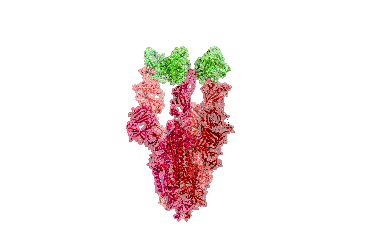
2019-nCoV is a close cousin to SARS coronavirus (SARS-CoV), and acts in a similar way. For both coronaviruses, the first step of infection occurs in the lungs, when a protein on the surface of the virus binds to a receptor protein on a lung cell. This viral protein is called the spike protein, depicted in red in the image below, and the receptor is known as ACE2. A therapeutic antibody is a type of protein that can block the viral protein from binding to its receptor, therefore preventing the virus from infecting the lung cell. A therapeutic antibody has already been developed for SARS-CoV, but to develop therapeutic antibodies or small molecules for 2019-nCoV, scientists need to better understand the structure of the viral spike protein and how it binds to the human ACE2 receptor required for viral entry into human cells.
Proteins are not stagnant—they wiggle and fold and unfold to take on numerous shapes. We need to study not only one shape of the viral spike protein, but all the ways the protein wiggles and folds into alternative shapes in order to best understand how it interacts with the ACE2 receptor, so that an antibody can be designed. Low-resolution structures of the SARS-CoV spike protein exist and we know the mutations that differ between SARS-CoV and 2019-nCoV. Given this information, we are uniquely positioned to help model the structure of the 2019-nCoV spike protein and identify sites that can be targeted by a therapeutic antibody. We can build computational models that accomplish this goal, but it takes a lot of computing power.
This is where you come in! With many computers working towards the same goal, we aim to help develop a therapeutic remedy as quickly as possible. By downloading Folding@home here [LINK] and selecting to contribute to “Any Disease”, you can help provide us with the computational power required to tackle this problem. One protein from 2019-nCoV, a protease encoded by the viral RNA, has already been crystallized. Although the 2019-nCoV spike protein of interest has not yet been resolved bound to ACE2, our objective is to use the homologous structure of the SARS-CoV spike protein to identify therapeutic antibody targets.
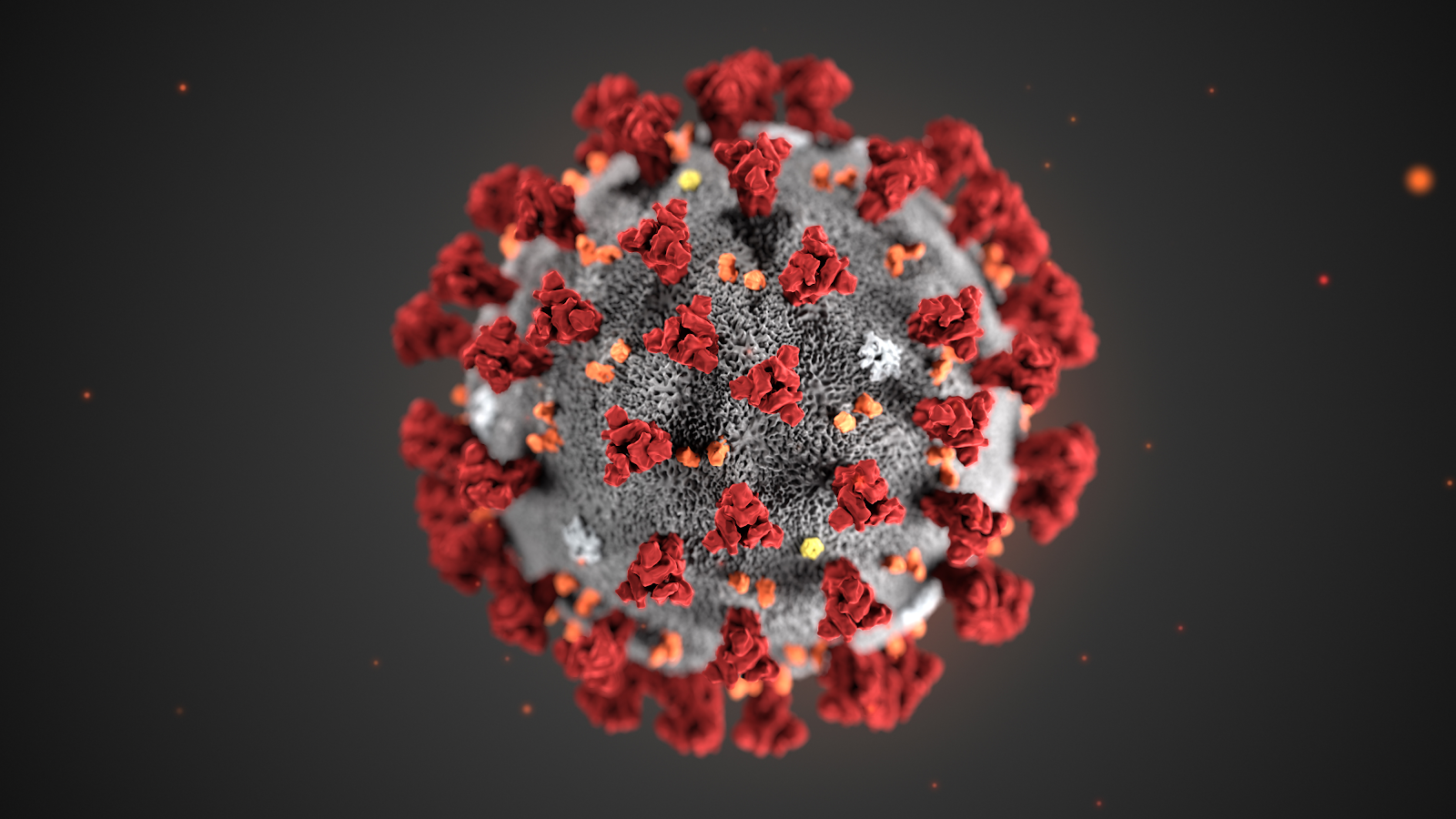
This illustration, created at the Centers for Disease Control and Prevention (CDC), reveals ultrastructural morphology exhibited by coronaviruses. Note the spikes that adorn the outer surface of the virus, which impart the look of a corona surrounding the virion, when viewed electron microscopically. A novel coronavirus virus was identified as the cause of an outbreak of respiratory illness first detected in Wuhan, China in 2019.
Structures of the closely related SARS-CoV spike protein bound by therapeutic antibodies may help rapidly design better therapies. The three monomers of the SARS-CoV spike protein are shown in different shades of red; the antibody is depicted in green. [PDB: 6NB7 https://www.rcsb.org/structure/6nb7]
I am reposting the following Science blog post from Derrick Lowe as is and ask people go browse through the comments on his Science blog In the Pipeline because, as Dr. Lowe states that in this current crisis it is important to disseminate good information as quickly as possible so wanted the readers here to have the ability to read his great posting on this matter of Covid-19. Also i would like to direct readers to the journal Science opinion letter concerning how important it is to rebuild the trust in good science and the scientific process. The full link for the following In the Pipeline post is: https://blogs.sciencemag.org/pipeline/archives/2020/03/06/covid-19-small-molecule-therapies-reviewed
A Summary of current potential repurposed therapeutics for COVID-19 Infection from In The Pipeline: A Science blog from Derick Lowe
Let’s take inventory on the therapies that are being developed for the coronavirus epidemic. Here is a very thorough list of at Biocentury, and I should note that (like Stat and several other organizations) they’re making all their Covid-19 content free to all readers during this crisis. I’d like to zoom in today on the potential small-molecule therapies, since some of these have the most immediate prospects for use in the real world.
The ones at the front of the line are repurposed drugs that are already approved for human use, for a lot of obvious reasons. The Biocentury list doesn’t cover these, but here’s an article at Nature Biotechnology that goes into detail. Clinical trials are a huge time sink – they sort of have to be, in most cases, if they’re going to be any good – and if you’ve already done all that stuff it’s a huge leg up, even if the drug itself is not exactly a perfect fit for the disease. So what do we have? The compound that is most advanced is probably remdesivir from Gilead, at right. This has been in development for a few years as an RNA virus therapy – it was originally developed for Ebola, and has been tried out against a whole list of single-strand RNA viruses. That includes the related coronaviruses SARS and MERS, so Covid-19 was an obvious fit.
The compound is a prodrug – that phosphoramide gets cleaved off completely, leaving the active 5-OH compound GS-44-1524. It mechanism of action is to get incorporated into viral RNA, since it’s taken up by RNA polymerase and it largely seems to evade proofreading. This causes RNA termination trouble later on, since that alpha-nitrile C-nucleoside is not exactly what the virus is expecting in its genome at that point, and thus viral replication is inhibited.
There are five clinical trials underway (here’s an overview at Biocentury). The NIH has an adaptive-design Phase II trial that has already started in Nebraska, with doses to be changed according to Bayesian readouts along the way. There are two Phase III trials underway at China-Japan Friendship Hospital in Hubei, double-blinded and placebo-controlled (since placebo is, as far as drug therapy goes, the current standard of care). And Gilead themselves are starting two open-label trials, one with no control arm and one with an (unblinded) standard-of-care comparison arm. Those might read out first, depending on when they get off the ground, but will be only rough readouts due to the fast-and-loose trial design. The two Hubei trials and the NIH one will add some rigor to the process, but I’m not sure when they’re going to report. My personal opinion is that I like the chances of this drug more than anything else on this list, but it’s still unlikely to be a game-changer.
There’s an RNA polymerase inhibitor (favipiravir) from Toyama, at right, that’s in a trial in China. It’s a thought – a broad-spectrum agent of this sort would be the sort of thing to try. But unfortunately, from what I can see, it has already turned up as ineffective in in vitro tests. The human trial that’s underway is honestly the sort of thing that would only happen under circumstances like the present: a developing epidemic with a new pathogen and no real standard of care. I hold out little hope for this one, but given that there’s nothing else at present, it probably should be tried. As you’ll see, this is far from the only situation like this.
One of the screens of known drugs in China that also flagged remdesivir noted that the old antimalarial drug chloroquine seemed to be effective in vitro. It had been reported some years back as a possible antiviral, working through more than one mechanism, probably both at viral entry and intracellularly thereafter. That part shouldn’t be surprising – chloroquine’s actual mode(s) of action against malaria parasites are still not completely worked out, either, and some of what people thought they knew about it has turned out to be wrong. There are several trials underway with it at Chinese facilities, some in combination with other agents like remdesivir. Chloroquine has of course been taken for many decades as an antimalarial, but it has a number of liabilities, including seizures, hearing damage, retinopathy and sudden effects on blood glucose. So it’s going to be important to establish just how effective it is and what doses will be needed. Just as with vaccine candidates, it’s possible to do more harm with a rushed treatment than the disease is doing itself
There are several other known antiviral drugs are being tried in China, but I don’t have too much hope for those, either. The neuraminidase inhibitors such as oseltamivir (better known as Tamiflu) were tried against SARS and were ineffective; there is no reason to expect anything versus Covid-19 although these drugs are a component of some drug cocktail trials. The HIV protease therapies such as darunavir and the combination therapy Kaletra are in trials, but that’s also a rather desperate long shot, since there’s no particular reason to think that they will have any such protease inhibition against what this new virus has to offer (and indeed, such agents weren’t much help against SARS in the end, either). The classic interferon/ribavirin combination seems to have had some activity against SARS and MERS, and is in two trials from what I can see. That’s not an awful idea by any means, but it’s not a great one, either: if your viral disease has interferon/ribavirin as a front line therapy, it generally means that there’s nothing really good available. No, unless we get really lucky none of these ideas are going to slow the disease down much.
There are a few other repurposed-protease-inhibitors ideas out there, such as this one. (Edit: I had seen this paper but couldn’t track it down, so thanks to those who sent it along). This paper suggests that the TMPRSS2 protease is important for viral entry on the human-cell-side of the process, a pathway that has been noted for other coronaviruses. And it points out that there is a an approved inhibitor (in Japan) for this enzyme (camostat), so that would definitely seem to be worth a trial, probably in combination with remdesivir.
That’s about it for the existing small molecules, from what I can see. What about new ones? Don’t hold your breath, is all I can say. A drug discovery program from scratch against a new pathogen is, as many readers here well know, not a trivial exercise. As this Bloomberg article details, many such efforts in the past (small molecules and vaccines alike) have come to grief because by the time they had anything to deliver the epidemic itself had passed. Indeed, Gilead’s remdesivir had already been dropped as a potential Ebola therapy.
You will either need to have a target in mind up front or go phenotypic. For the former, what you’d see are better characterizations of the viral protease and more extensive screens against it. Two other big target areas are viral entry (which involves the “spike” proteins on the virus surface and the ACE2 protein on human cells) and viral replication. To the former, it’s worth quickly noting that ACE2 is so much unlike the more familiar ACE protein that none of the cardiovascular ACE inhibitors do anything to it at all. And targeting the latter mechanisms is how remdesivir was developed as a possible Ebola agent, but as you can see, that took time, too. Phenotypic screens are perfectly reasonable against viral pathogens as well, but you’ll need to put time and effort into that assay up front, just as with any phenotypic effort, because as anyone who does that sort of work will tell you, a bad phenotypic screen is a complete waste of everyone’s time.
One of the key steps for either route is identifying an animal model. While animal models of infectious disease can be extremely well translated to human therapy, that doesn’t happen by accident: you need to choose the right animal. Viruses in general (and coronaviruses are no exception) vary widely in their effects in different species, and not just across the gaps of bird/reptile/human and the like. No, you’ll run into things where even the usual set of small mammals are acting differently from each other, with some of them not even getting sick at all. This current virus may well have gone through a couple of other mammalian species before landing on us, but you’ll note that dogs (to pick one) don’t seem to have any problem with it.
All this means that any new-target new-chemical-matter effort against Covid-19 (or any new pathogen) is going to take years, and there is just no way around that. Update: see here for just such an effort to start finding fragment hits for the viral protease. This puts small molecules in a very bimodal distribution: you have the existing drugs that might be repurposed, and are presumably available right now. Nothing else is! At the other end, for completely new therapies you have the usual prospects of drug discovery: years from now, lots of money, low success rate, good luck to all of us. The gap between these two could in theory be filled by vaccines and antibody therapies (if everything goes really, really well) but those are very much their own area and will be dealt with in a separate post.
Either way, the odds are that we (and I mean “we as a species” here) are going to be fighting this epidemic without any particularly amazing pharmacological weapons. Eventually we’ll have some, but I would advise people, pundits, and politicians not to get all excited about the prospects for some new therapies to come riding up over the hill to help us out. The odds of that happening in time to do anything about the current outbreak are very small. We will be going for months, years, with the therapeutic options we have right now. Look around you: what we have today is what we have to work with.
Other related articles published in this Open Access Online Scientific Journal include the following:
Group of Researchers @ University of California, Riverside, the University of Chicago, the U.S. Department of Energy’s Argonne National Laboratory, and Northwestern University solve COVID-19 Structure and Map Potential Therapeutics
Reporters: Stephen J Williams, PhD and Aviva Lev-Ari, PhD, RN
Predicting the Protein Structure of Coronavirus: Inhibition of Nsp15 can slow viral replication and Cryo-EM – Spike protein structure (experimentally verified) vs AI-predicted protein structures (not experimentally verified) of DeepMind (Parent: Google) aka AlphaFold
Curators: Stephen J. Williams, PhD and Aviva Lev-Ari, PhD, RN
19th Annual Koch Institute Summer Symposium on Cancer Immunotherapy, June 12, 2020 at MIT’s Kresge Auditorium
Reporter: Aviva Lev-Ari, PhD, RN
Summer Symposium 2020
Engineering the Next Wave of Immunotherapy
The 19th Annual Koch Institute Summer Symposium on June 12, 2020 at MIT’s Kresge Auditorium will focus on cancer immunotherapy.
Cancer immunotherapy has revolutionized the landscape of cancer treatment, our thinking of tumor biology and clinical practice. Following the groundbreaking successes of checkpoint blockade therapy and CAR T cell therapy, culminating in multiple FDA-approved treatments and the awarding of the 2018 Nobel Prize in Medicine to Jim Allison and Tasuku Honjo, the field is currently at a critical juncture.
While checkpoint blockade therapy has demonstrated that the immune system can be harnessed to fight cancer, the next generation of treatments will require us to understand what causes resistance in non-responders, how this can be overcome, and how these issues are best addressed clinically. Discussing these questions will be at the core of this symposium as we move towards our ultimate goal to increase the number of patients benefiting from immunotherapy.
Session Speakers
Targeting T Cells
Rafi Ahmed, Michael Dougan, Chris Love
Thinking Beyond T Cells
Angelika Amon, Yasemine Belkaid, Stefani Spranger
Engineering Clinical Translation
Nina Bhardwaj, Chris Garcia
Panel Discussion: Clinical Translation: A Real Life Perspective
Daniel Chen, Howard Kaufman, Kimberly Schaefer-Weaver
Moderator: Steven Silverstein
Core member and chair of the faculty, Broad Institute of MIT and Harvard; director, Klarman Cell Observatory, Broad Institute of MIT and Harvard; professor of biology, MIT; investigator, Howard Hughes Medical Institute; founding co-chair, Human Cell Atlas.
millions of genome variants, tens of thousands of disease-associated genes, thousands of cell types and an almost unimaginable number of ways they can combine, we had to approximate a best starting point—choose one target, guess the cell, simplify the experiment.
In 2020, advances in polygenic risk scores, in understanding the cell and modules of action of genes through genome-wide association studies (GWAS), and in predicting the impact of combinations of interventions.
we need algorithms to make better computational predictions of experiments we have never performed in the lab or in clinical trials.
Human Cell Atlas and the International Common Disease Alliance—and in new experimental platforms: data platforms and algorithms. But we also need a broader ecosystem of partnerships in medicine that engages interaction between clinical experts and mathematicians, computer scientists and engineers
Feng Zhang, PhD
investigator, Howard Hughes Medical Institute; core member, Broad Institute of MIT and Harvard; James and Patricia Poitras Professor of Neuroscience, McGovern Institute for Brain Research, MIT.
fundamental shift in medicine away from treating symptoms of disease and toward treating disease at its genetic roots.
Gene therapy with clinical feasibility, improved delivery methods and the development of robust molecular technologies for gene editing in human cells, affordable genome sequencing has accelerated our ability to identify the genetic causes of disease.
1,000 clinical trials testing gene therapies are ongoing, and the pace of clinical development is likely to accelerate.
refine molecular technologies for gene editing, to push our understanding of gene function in health and disease forward, and to engage with all members of society
Elizabeth Jaffee, PhD
Dana and Albert “Cubby” Broccoli Professor of Oncology, Johns Hopkins School of Medicine; deputy director, Sidney Kimmel Comprehensive Cancer Center at Johns Hopkins.
a single blood test could inform individuals of the diseases they are at risk of (diabetes, cancer, heart disease, etc.) and that safe interventions will be available.
developing cancer vaccines. Vaccines targeting the causative agents of cervical and hepatocellular cancers have already proven to be effective. With these technologies and the wealth of data that will become available as precision medicine becomes more routine, new discoveries identifying the earliest genetic and inflammatory changes occurring within a cell as it transitions into a pre-cancer can be expected. With these discoveries, the opportunities to develop vaccine approaches preventing cancers development will grow.
shape how the culture of research will develop over the next 25 years, a culture that cares more about what is achieved than how it is achieved.
building a creative, inclusive and open research culture will unleash greater discoveries with greater impact.
John Nkengasong, PhD
Director, Africa Centres for Disease Control and Prevention.
To meet its health challenges by 2050, the continent will have to be innovative in order to leapfrog toward solutions in public health.
Precision medicine will need to take center stage in a new public health order— whereby a more precise and targeted approach to screening, diagnosis, treatment and, potentially, cure is based on each patient’s unique genetic and biologic make-up.
Eric Topol, MD
Executive vice-president, Scripps Research Institute; founder and director, Scripps Research Translational Institute.
In 2045, a planetary health infrastructure based on deep, longitudinal, multimodal human data, ideally collected from and accessible to as many as possible of the 9+ billion people projected to then inhabit the Earth.
enhanced capabilities to perform functions that are not feasible now.
AI machines’ ability to ingest and process biomedical text at scale—such as the corpus of the up-to-date medical literature—will be used routinely by physicians and patients.
the concept of a learning health system will be redefined by AI.
Linda Partridge, PhD
Professor, Max Planck Institute for Biology of Ageing.
Geroprotective drugs, which target the underlying molecular mechanisms of ageing, are coming over the scientific and clinical horizons, and may help to prevent the most intractable age-related disease, dementia.
Trevor Mundel, MD
President of Global Health, Bill & Melinda Gates Foundation.
finding new ways to share clinical data that are as open as possible and as closed as necessary.
moving beyond drug donations toward a new era of corporate social responsibility that encourages biotechnology and pharmaceutical companies to offer their best minds and their most promising platforms.
working with governments and multilateral organizations much earlier in the product life cycle to finance the introduction of new interventions and to ensure the sustainable development of the health systems that will deliver them.
deliver on the promise of global health equity.
Josep Tabernero, MD, PhD
Vall d’Hebron Institute of Oncology (VHIO); president, European Society for Medical Oncology (2018–2019).
genomic-driven analysis will continue to broaden the impact of personalized medicine in healthcare globally.
Precision medicine will continue to deliver its new paradigm in cancer care and reach more patients.
Immunotherapy will deliver on its promise to dismantle cancer’s armory across tumor types.
AI will help guide the development of individually matched
genetic patient screenings
the promise of liquid biopsy policing of disease?
Pardis Sabeti, PhD
Professor, Harvard University & Harvard T.H. Chan School of Public Health and Broad Institute of MIT and Harvard; investigator, Howard Hughes Medical Institute.
the development and integration of tools into an early-warning system embedded into healthcare systems around the world could revolutionize infectious disease detection and response.
But this will only happen with a commitment from the global community.
Els Toreele, PhD
Executive director, Médecins Sans Frontières Access Campaign
we need a paradigm shift such that medicines are no longer lucrative market commodities but are global public health goods—available to all those who need them.
This will require members of the scientific community to go beyond their role as researchers and actively engage in R&D policy reform mandating health research in the public interest and ensuring that the results of their work benefit many more people.
The global research community can lead the way toward public-interest driven health innovation, by undertaking collaborative open science and piloting not-for-profit R&D strategies that positively impact people’s lives globally.
Parkinson’s Disease (PD), characterized by both motor and non-motor system pathology, is a common neurodegenerative disorder affecting about 1% of the population over age 60. Its prevalence presents an increasing social burden as the population ages. Since its introduction in the 1960’s, dopamine (DA)-replacement therapy (e.g., L-DOPA) has remained the gold standard treatment. While improving PD patients’ quality of life, the effects of treatment fade with disease progression and prolonged usage of these medications often (>80%) results in side effects including dyskinesias and motor fluctuations. Since the selective degeneration of A9 mDA neurons (mDANs) in the substantia nigra (SN) is a key pathological feature of the disease and is directly associated with the cardinal motor symptoms, dopaminergic cell transplantation has been proposed as a therapeutic strategy.
Researchers showed that mammalian fibroblasts can be converted into embryonic stem cell (ESC)-like induced pluripotent stem cells (iPSCs) by introducing four transcription factors i.e., Oct4, Sox2, Klf4, and c-Myc. This was then accomplished with human somatic cells, reprogramming them into human iPSCs (hiPSCs), offering the possibility of generating patient-specific stem cells. There are several major barriers to implementation of hiPSC-based cell therapy for PD. First, probably due to the limited understanding of the reprogramming process, wide variability exists between the differentiation potential of individual hiPSC lines. Second, the safety of hiPSC-based cell therapy has yet to be fully established. In particular, since any hiPSCs that remain undifferentiated or bear sub-clonal tumorigenic mutations have neoplastic potential, it is critical to eliminate completely such cells from a therapeutic product.
In the present study the researchers established human induced pluripotent stem cell (hiPSC)-based autologous cell therapy. Researchers reported a platform of core techniques for the production of mDA progenitors as a safe and effective therapeutic product. First, by combining metabolism-regulating microRNAs with reprogramming factors, a method was developed to more efficiently generate clinical grade iPSCs, as evidenced by genomic integrity and unbiased pluripotent potential. Second, a “spotting”-based in vitro differentiation methodology was established to generate functional and healthy mDA cells in a scalable manner. Third, a chemical method was developed that safely eliminates undifferentiated cells from the final product. Dopaminergic cells thus produced can express high levels of characteristic mDA markers, produce and secrete dopamine, and exhibit electrophysiological features typical of mDA cells. Transplantation of these cells into rodent models of PD robustly restored motor dysfunction and reinnervated host brain, while showing no evidence of tumor formation or redistribution of the implanted cells.
Together these results supported the promise of these techniques to provide clinically applicable personalized autologous cell therapy for PD. It was recognized by researchers that this methodology is likely to be more costly in dollars and manpower than techniques using off-the-shelf methods and allogenic cell lines. Nevertheless, the cost for autologous cell therapy may be expected to decrease steadily with technological refinement and automation. Given the significant advantages inherent in a cell source free of ethical concerns and with the potential to obviate the need for immunosuppression, with its attendant costs and dangers, it was proposed that this platform is suitable for the successful implementation of human personalized autologous cell therapy for PD.
Effective humoral immune responses to infection and immunization are defined by high-affinity antibodies generated as a result of B cell differentiation and selection that occurs within germinal centers (GC). Within the GC, B cells undergo affinity maturation, an iterative and competitive process wherein B cells mutate their immunoglobulin genes (somatic hypermutation) and undergo clonal selection by competing for T cell help. Balancing the decision to remain within the GC and continue participating in affinity maturation or to exit the GC as a plasma cell (PC) or memory B cell (MBC) is critical for achieving optimal antibody avidity, antibody quantity, and establishing immunological memory in response to immunization or infection. Humoral immune responses during chronic infections are often dysregulated and characterized by hypergammaglobulinemia, decreased affinity maturation, and delayed development of neutralizing antibodies. Previous studies have suggested that poor antibody quality is in part due to deletion of B cells prior to establishment of the GC response.
In fact the impact of chronic infections on B cell fate decisions in the GC remains poorly understood. To address this question, researchers used single-cell transcriptional profiling of virus-specific GC B cells to test the hypothesis that chronic viral infection disrupted GC B cell fate decisions leading to suboptimal humoral immunity. These studies revealed a critical GC differentiation checkpoint that is disrupted by chronic infection, specifically at the point of dark zone re-entry. During chronic viral infection, virus-specific GC B cells were shunted towards terminal plasma cell (PC) or memory B cell (MBC) fates at the expense of continued participation in the GC. Early GC exit was associated with decreased B cell mutational burden and antibody quality. Persisting antigen and inflammation independently drove facets of dysregulation, with a key role for inflammation in directing premature terminal GC B cell differentiation and GC exit. Thus, the present research defines GC defects during chronic viral infection and identify a critical GC checkpoint that is short-circuited, preventing optimal maturation of humoral immunity.
Together, these studies identify a key GC B cell differentiation checkpoint that is dysregulated during chronic infection. Further, it was found that the chronic inflammatory environment, rather than persistent antigen, is sufficient to drive altered GC B cell differentiation during chronic infection even against unrelated antigens. However, the data also indicate that inflammatory circuits are likely linked to perception of antigen stimulation. Nevertheless, this study reveals a B cell-intrinsic program of transcriptional skewing in chronic viral infection that results in shunting out of the cyclic GC B cell process and early GC exit with consequences for antibody quality and hypergammaglobulinemia. These findings have implications for vaccination in individuals with pre-existing chronic infections where antibody responses are often ineffective and suggest that modulation of inflammatory pathways may be therapeutically useful to overcome impaired humoral immunity and foster affinity maturation during chronic viral infections.
One of the most contagious diseases known to humankind, measles killed an average of 2.6 million people each year before a vaccine was developed, according to the World Health Organization. Widespread vaccination has slashed the death toll. However, lack of access to vaccination and refusal to get vaccinated means measles still infects more than 7 million people and kills more than 100,000 each year worldwide as reported by WHO. The cases are on the rise, tripling in early 2019 and some experience well-known long-term consequences, including brain damage and vision and hearing loss. Previous epidemiological research into immune amnesia suggests that death rates attributed to measles could be even higher, accounting for as much as 50 percent of all childhood mortality.
Over the last decade, evidence has mounted that the measles vaccine protects in two ways. It prevents the well-known acute illness with spots and fever and also appears to protect from other infections over the long term by giving general boost to the immune system. The measles virus can impair the body’s immune memory, causing so-called immune amnesia. By protecting against measles infection, the vaccine prevents the body from losing or “forgetting” its immune memory and preserves its resistance to other infections. Researchers showed that the measles virus wipes out 11% to 73% of the different antibodies that protect against viral and bacterial strains a person was previously immune to like from influenza to herpes virus to bacteria that cause pneumonia and skin infections.
This study at Harvard Medical School and their collaborators is the first to measure the immune damage caused by the virus and underscores the value of preventing measles infection through vaccination. The discovery that measles depletes people’s antibody repertoires, partially obliterating immune memory to most previously encountered pathogens, supports the immune amnesia hypothesis. It was found that those who survive measles gradually regain their previous immunity to other viruses and bacteria as they get re-exposed to them. But because this process may take months to years, people remain vulnerable in the meantime to serious complications of those infections and thus booster shots of routine vaccines may be required.
VirScan detects antiviral and antibacterial antibodies in the blood that result from current or past encounters with viruses and bacteria, giving an overall snapshot of the immune system. Researchers gathered blood samples from unvaccinated children during a 2013 measles outbreak in the Netherlands and used VirScan to measure antibodies before and two months after infection in 77 children who’d contracted the disease. The researchers also compared the measurements to those of 115 uninfected children and adults. Researchers found a striking drop in antibodies from other pathogens in the measles-infected children that clearly suggested a direct effect on the immune system resembling measles-induced immune amnesia.
Further tests revealed that severe measles infection reduced people’s overall immunity more than mild infection. This could be particularly problematic for certain categories of children and adults, the researchers said. The present study observed the effects in previously healthy children only. But, measles is known to hit malnourished children much harder, the degree of immune amnesia and its effects could be even more severe in less healthy populations. Inoculation with the MMR (measles, mumps, rubella) vaccine did not impair children’s overall immunity. The results align with decades of research. Ensuring widespread vaccination against measles would not only help prevent the expected 120,000 deaths that will be directly attributed to measles this year alone, but could also avert potentially hundreds of thousands of additional deaths attributable to the lasting damage to the immune system.
An Intelligent DNA Nanorobot to Fight Cancer by Targeting HER2 Expression
Reporter and Curator: Dr. Sudipta Saha, Ph.D.
3.2.9 An Intelligent DNA Nanorobot to Fight Cancer by Targeting HER2 Expression, Volume 2 (Volume Two: Latest in Genomics Methodologies for Therapeutics: Gene Editing, NGS and BioInformatics, Simulations and the Genome Ontology), Part 2: CRISPR for Gene Editing and DNA Repair
HER2 is an important prognostic biomarker for 20–30% of breast cancers, which is the most common cancer in women. Overexpression of the HER2 receptor stimulates breast cells to proliferate and differentiate uncontrollably, thereby enhancing the malignancy of breast cancer and resulting in a poor prognosis for affected individuals. Current therapies to suppress the overexpression of HER2 in breast cancer mainly involve treatment with HER2-specific monoclonal antibodies. However, these monoclonal anti-HER2 antibodies have severe side effects in clinical trials, such as diarrhea, abnormal liver function, and drug resistance. Removing HER2 from the plasma membrane or inhibiting the gene expression of HER2 is a promising alternative that could limit the malignancy of HER2-positive cancer cells.
DNA origami is an emerging field of DNA-based nanotechnology and intelligent DNA nanorobots show great promise in working as a drug delivery system in healthcare. Different DNA-based nanorobots have been developed as affordable and facile therapeutic drugs. In particular, many studies reported that a tetrahedral framework nucleic acid (tFNA) could serve as a promising DNA nanocarrier for many antitumor drugs, owing to its high biocompatibility and biosecurity. For example, tFNA was reported to effectively deliver paclitaxel or doxorubicin to cancer cells for reversing drug resistance, small interfering RNAs (siRNAs) have been modified into tFNA for targeted drug delivery. Moreover, the production and storage of tFNA are not complicated, and they can be quickly degraded in lysosomes by cells. Since both free HApt and tFNA can be diverted into lysosomes, so, combining the HApt and tFNA as a novel DNA nanorobot (namely, HApt-tFNA) can be an effective strategy to improve its delivery and therapeutic efficacy in treating HER2-positive breast cancer.
Researchers reported that a DNA framework-based intelligent DNA nanorobot for selective lysosomal degradation of tumor-specific proteins on cancer cells. An anti-HER2 aptamer (HApt) was site-specifically anchored on a tetrahedral framework nucleic acid (tFNA). This DNA nanorobot (HApt-tFNA) could target HER2-positive breast cancer cells and specifically induce the lysosomal degradation of the membrane protein HER2. An injection of the DNA nanorobot into a mouse model revealed that the presence of tFNA enhanced the stability and prolonged the blood circulation time of HApt, and HApt-tFNA could therefore drive HER2 into lysosomal degradation with a higher efficiency. The formation of the HER2-HApt-tFNA complexes resulted in the HER2-mediated endocytosis and digestion in lysosomes, which effectively reduced the amount of HER2 on the cell surfaces. An increased HER2 digestion through HApt-tFNA further induced cell apoptosis and arrested cell growth. Hence, this novel DNA nanorobot sheds new light on targeted protein degradation for precision breast cancer therapy.
It was previously reported that tFNA was degraded by lysosomes and could enhance cell autophagy. Results indicated that free Cy5-HApt and Cy5-HApt-tFNA could enter the lysosomes; thus, tFNA can be regarded as an efficient nanocarrier to transmit HApt into the target organelle. The DNA nanorobot composed of HApt and tFNA showed a higher stability and a more effective performance than free HApt against HER2-positive breast cancer cells. The PI3K/AKT pathway was inhibited when membrane-bound HER2 decreased in SK-BR-3 cells under the action of HApt-tFNA. The research findings suggest that tFNA can enhance the anticancer effects of HApt on SK-BR-3 cells; while HApt-tFNA can bind to HER2 specifically, the compounded HER2-HApt-tFNA complexes can then be transferred and degraded in lysosomes. After these processes, the accumulation of HER2 in the plasma membrane would decrease, which could also influence the downstream PI3K/AKT signaling pathway that is associated with cell growth and death.
However, some limitations need to be noted when interpreting the findings: (i) the cytotoxicity of the nanorobot on HER2-positive cancer cells was weak, and the anticancer effects between conventional monoclonal antibodies and HApt-tFNA was not compared; (ii) the differences in delivery efficiency between tFNA and other nanocarriers need to be confirmed; and (iii) the confirmation of anticancer effects of HApt-tFNA on tumors within animals remains challenging. Despite these limitations, the present study provided novel evidence of the biological effects of tFNA when combined with HApt. Although the stability and the anticancer effects of HApt-tFNA may require further improvement before clinical application, this study initiates a promising step toward the development of nanomedicines with novel and intelligent DNA nanorobots for tumor treatment.
Single-cell RNA-seq helps in finding intra-tumoral heterogeneity in pancreatic cancer
Reporter and Curator: Dr. Sudipta Saha, Ph.D.
4.3.6 Single-cell RNA-seq helps in finding intra-tumoral heterogeneity in pancreatic cancer, Volume 2 (Volume Two: Latest in Genomics Methodologies for Therapeutics: Gene Editing, NGS and BioInformatics, Simulations and the Genome Ontology), Part 4: Single Cell Genomics
Pancreatic cancer is a significant cause of cancer mortality; therefore, the development of early diagnostic strategies and effective treatment is essential. Improvements in imaging technology, as well as use of biomarkers are changing the way that pancreas cancer is diagnosed and staged. Although progress in treatment for pancreas cancer has been incremental, development of combination therapies involving both chemotherapeutic and biologic agents is ongoing.
Cancer is an evolutionary disease, containing the hallmarks of an asexually reproducing unicellular organism subject to evolutionary paradigms. Pancreatic ductal adenocarcinoma (PDAC) is a particularly robust example of this phenomenon. Genomic features indicate that pancreatic cancer cells are selected for fitness advantages when encountering the geographic and resource-depleted constraints of the microenvironment. Phenotypic adaptations to these pressures help disseminated cells to survive in secondary sites, a major clinical problem for patients with this disease.
The immune system varies in cell types, states, and locations. The complex networks, interactions, and responses of immune cells produce diverse cellular ecosystems composed of multiple cell types, accompanied by genetic diversity in antigen receptors. Within this ecosystem, innate and adaptive immune cells maintain and protect tissue function, integrity, and homeostasis upon changes in functional demands and diverse insults. Characterizing this inherent complexity requires studies at single-cell resolution. Recent advances such as massively parallel single-cell RNA sequencing and sophisticated computational methods are catalyzing a revolution in our understanding of immunology.
PDAC is the most common type of pancreatic cancer featured with high intra-tumoral heterogeneity and poor prognosis. In the present study to comprehensively delineate the PDAC intra-tumoral heterogeneity and the underlying mechanism for PDAC progression, single-cell RNA-seq (scRNA-seq) was employed to acquire the transcriptomic atlas of 57,530 individual pancreatic cells from primary PDAC tumors and control pancreases. The diverse malignant and stromal cell types, including two ductal subtypes with abnormal and malignant gene expression profiles respectively, were identified in PDAC.
The researchers found that the heterogenous malignant subtype was composed of several subpopulations with differential proliferative and migratory potentials. Cell trajectory analysis revealed that components of multiple tumor-related pathways and transcription factors (TFs) were differentially expressed along PDAC progression. Furthermore, it was found a subset of ductal cells with unique proliferative features were associated with an inactivation state in tumor-infiltrating T cells, providing novel markers for the prediction of antitumor immune response. Together, the findings provided a valuable resource for deciphering the intra-tumoral heterogeneity in PDAC and uncover a connection between tumor intrinsic transcriptional state and T cell activation, suggesting potential biomarkers for anticancer treatment such as targeted therapy and immunotherapy.
4.1.8 Newly Found Functions of B Cell, Volume 2 (Volume Two: Latest in Genomics Methodologies for Therapeutics: Gene Editing, NGS and BioInformatics, Simulations and the Genome Ontology), Part 4: Single Cell Genomics
The importance of B cells to human health is more than what is already known. Vaccines capable of eradicating disease activate B cells, cancer checkpoint blockade therapies are produced using B cells, and B cell deficiencies have devastating impacts. B cells have been a subject of fascination since at least the 1800s. The notion of a humoral branch to immunity emerged from the work of and contemporaries studying B cells in the early 1900s.
Efforts to understand how we could make antibodies from B cells against almost any foreign surface while usually avoiding making them against self, led to Burnet’s clonal selection theory. This was followed by the molecular definition of how a diversity of immunoglobulins can arise by gene rearrangement in developing B cells. Recombination activating gene (RAG)-dependent processes of V-(D)-J rearrangement of immunoglobulin (Ig) gene segments in developing B cells are now known to be able to generate an enormous amount of antibody diversity (theoretically at least 1016 possible variants).
With so much already known, B cell biology might be considered ‘‘done’’ with only incremental advances still to be made, but instead, there is great activity in the field today with numerous major challenges that remain. For example, efforts are underway to develop vaccines that induce broadly neutralizing antibody responses, to understand how autoantigen- and allergen-reactive antibodies arise, and to harness B cell-depletion therapies to correct non-autoantibody-mediated diseases, making it evident that there is still an enormous amount we do not know about B cells and much work to be done.
Multiple self-tolerance checkpoints exist to remove autoreactive specificities from the B cell repertoire or to limit the ability of such cells to secrete autoantigen-binding antibody. These include receptor editing and deletion in immature B cells, competitive elimination of chronically autoantigen binding B cells in the periphery, and a state of anergy that disfavors PC (plasma cell) differentiation. Autoantibody production can occur due to failures in these checkpoints or in T cell self-tolerance mechanisms. Variants in multiple genes are implicated in increasing the likelihood of checkpoint failure and of autoantibody production occurring.
Autoantibodies are pathogenic in a number of human diseases including SLE (Systemic lupus erythematosus), pemphigus vulgaris, Grave’s disease, and myasthenia gravis. B cell depletion therapy using anti-CD20 antibody has been protective in some of these diseases such as pemphigus vulgaris, but not others such as SLE and this appears to reflect the contribution of SLPC (Short lived plasma cells) versus LLPC (Long lived plasma cells) to autoantibody production and the inability of even prolonged anti-CD20 treatment to eliminate the later. These clinical findings have added to the importance of understanding what factors drive SLPC versus LLPC development and what the requirements are to support LLPCs.
B cell depletion therapy has also been efficacious in several other autoimmune diseases, including multiple sclerosis (MS), type 1 diabetes, and rheumatoid arthritis (RA). While the potential contributions of autoantibodies to the pathology of these diseases are still being explored, autoantigen presentation has been posited as another mechanism for B cell disease-promoting activity.
In addition to autoimmunity, B cells play an important role in allergic diseases. IgE antibodies specific for allergen components sensitize mast cells and basophils for rapid degranulation in response to allergen exposures at various sites, such as in the intestine (food allergy), nose (allergic rhinitis), and lung (allergic asthma). IgE production may thus be favored under conditions that induce weak B cell responses and minimal GC (Germinal center) activity, thereby enabling IgE+ B cells and/or PCs to avoid being outcompeted by IgG+ cells. Aside from IgE antibodies, B cells may also contribute to allergic inflammation through their interactions with T cells.
B cells have also emerged as an important source of the immunosuppressive cytokine IL-10. Mouse studies revealed that B cell-derived IL-10 can promote recovery from EAE (Experimental autoimmune encephalomyelitis) and can be protective in models of RA and type 1 diabetes. Moreover, IL-10 production from B cells restrains T cell responses during some viral and bacterial infections. These findings indicate that the influence of B cells on the cytokine milieu will be context dependent.
The presence of B cells in a variety of solid tumor types, including breast cancer, ovarian cancer, and melanoma, has been associated in some studies with a positive prognosis. The mechanism involved is unclear but could include antigen presentation to CD4 and CD8 T cells, antibody production and subsequent enhancement of presentation, or by promoting tertiary lymphoid tissue formation and local T cell accumulation. It is also noteworthy that B cells frequently make antibody responses to cancer antigens and this has led to efforts to use antibodies from cancer patients as biomarkers of disease and to identify immunotherapy targets.
Malignancies of B cells themselves are a common form of hematopoietic cancer. This predilection arises because the gene modifications that B cells undergo during development and in immune responses are not perfect in their fidelity, and antibody responses require extensive B cell proliferation. The study of B cell lymphomas and their associated genetic derangements continues to be illuminating about requirements for normal B cell differentiation and signaling while also leading to the development of targeted therapies.
Overall this study attempted to capture some of the advances in the understanding of B cell biology that have occurred since the turn of the century. These include important steps forward in understanding how B cells encounter antigens, the co-stimulatory and cytokine requirements for their proliferation and differentiation, and how properties of the B cell receptor, the antigen, and helper T cells influence B cell responses. Many advances continue to transform the field including the impact of deep sequencing technologies on understanding B cell repertoires, the IgA-inducing microbiome, and the genetic defects in humans that compromise or exaggerate B cell responses or give rise to B cell malignancies.
Other advances that are providing insight include:
single-cell approaches to define B cell heterogeneity,
glycomic approaches to study effector sugars on antibodies,
new methods to study human B cell responses including CRISPR-based manipulation, and
the use of systems biology to study changes at the whole organism level.
With the recognition that B cells and antibodies are involved in most types of immune response and the realization that inflammatory processes contribute to a wider range of diseases than previously believed, including, for example, metabolic syndrome and neurodegeneration, it is expected that further
basic research-driven discovery about B cell biology will lead to more and improved approaches to maintain health and fight disease in the future.
CytoReason is re-defining the Context of the Immune System for Drug Discovery
Reporter: Aviva Lev-Ari, PhD, RN
CytoReason is re-defining the context of the immune system at a cellular level in order to better understand disease and support more effective drug discovery and development.
Our leading-edge machine-learning driven approach identifies “cause and effect” of the gene/cell/cytokine relationships that lie at the heart of treating disease.
Faster and more accurately than ever before.
CytoReason’s mission is to simulate the cells that can stimulate discovery of:
New targets for treating disease
New insights to mechanism of actions (both of disease and drugs)
Differences in responses to both disease and treatment
Which diseases a drug can impact
We have developed a unique machine-learning driven approach to “seeing” the cells that can make the difference in patients seeing a better life.
The insights our approach generates, enable pharmaceutical and biotech companies to make the right decisions, at the right time, in the drug discovery and development programs that bring better therapies.
Based on cutting edge technologies, trained on data that would normally be impossible to access, and steered by leading biological and data science researchers, our approach is underpinned by three core principles:
Science is the backbone of our methodologies and applications, and must stand the test of scientific scrutiny. To date we have 16 research papers published in top quality peer-reviewed scientific journals, including four in 2018 alone – 3 of which were published in journals from the Nature group
Shen-Orr told Forbes in an article published late last month that CytoReason’s tech is able to calculate immune age in one of two ways: “Via cell-subset composition nearest neighbor approach or based on a gene expression signature where the genes are predictive of the cell-subsets composition, and they test for their enrichment in the gene expression pattern of the sample. The immune profiles of individuals are used to predict immune changes based on a machine learning methodology deployed on data on a range of cell-subsets. ”
“The immune age is a biological clock that will help to identify, the decline and progress in immunity that occurs in old age, to determine preventive measures and develop new treatment modalities to minimize chronic disease and death,” he added.
CytoReason’s tech has so far yielded two pending patents, 10 commercial and scientific collaborations, and 16 peer-reviewed publications.
Harel says it was a combination of forces that made CytoReason’s immune-focused methodology work: Big Data, machine learning, and biology. He describes it as “the intersection of computer science and biology.”
Professor Magdassi tells NoCamels that with 3D printing of hydrogels, molecules that are soluble in water, scientists can improve the performance of the drug through its delivery. For example, “the hydrogel once ingested can be designed to swell, releasing two, or three, or four drugs at a time [or with a delay] or it can be designed not to swell, depending on what we are trying to achieve.”
“The drug can be tailored to the patient because of the unique shape or structure of the hydrogel and/or its release behavior,” Professor Magdassi explains.
Last January, CytoReason announced an agreement with Pfizer, in which the latter will leverage the former’s technology to create cell-based models of the immune system. According to the agreement, CytoReason will receive an undisclosed amount in the low double-digit millions of U.S. dollars from Pfizer in access fees, research support and success-based payments. Prof. Shen-Orr concluded, “The immune age is a biological clock that will help to identify, the decline and progress in immunity that occurs in old age, to determine preventive measures and develop new treatment modalities to minimize chronic disease and death.”




 2012pharmaceutical
 sjwilliamspa