Feeds:
Posts
Comments

Archive for 2014

Hashtags and Twitter Handles for 10th Annual Personalized Medicine at Harvard Medical School, 11/12 – 11/13/2014

Reporter: Stephen J Williams, PhD

10th Annual Personalized Medicine at Harvard (Partners Healthcare)

Meeting #: #PMConf

Meeting @: @HarvardPMConf

 PLEASE ALSO FOLLOW LIVE MEETING NOTES AT @pharma_BI OR AT http://pharmaceuticalintelligence.com/

Overall good meeting #s:

#personalizedmedicine

#powerofgenomics

#genomics

#pharmanews

@BiotechNews

Day Time Talk Title # @
Wed. Nov 12, 2014 8:00 AM Welcome & Opening Remarks #personalizedmedicine

#genomics

#Boston

#Harvard

#MGH

#biotech

#harvardhealth

@PartnersNews

@MassGeneral

@AstellasUS (Stephen Eck)

@HarvardHealth

@harvardmed

Greetings #personalizedmedicine @theIOM (Dzau Institute of Medicine)

@newsfrompmc

(Personalized Medicine Coalition)

8:20 AM Special Guest Keynote Speaker Margaret Hamburg, M.D. #FDA

#personalizedmedicine

#FDACommissioner

#pharma

#healthcare

#PublicHealth

#HealthPolicy

@US_FDA

@HIVForum

@AIDSAlliance_DC

@HHSGov

8:50 Keynote Speaker

James Madera, M.D.

#AMA

#meded

#HealthPolicy

#personalizedmedicine

#healthcare

#physician

#PublicHealth

@AmerMedicalAssn
9:20 Panel Discussion

Genomic Technologies

#genomics

#genome

#personalizedmedicine

#health

#biotech

#lifesciences

#syntheticbiology

#startups

#startup

#disease

#endcancer

#MDAnderson

@harvardmed

@PartnersNews

@Harvard

@geochurch

@BrighamWomens

@MDAndersonNews

@servingscience (Thermo Fisher)

11:00 Keynote Mirella Marlow #personalizedmedicine

#UK

#health

#healthcare

#NIH

@NIH

@NICEcomms

@MirellaMarlow

11:30 10th Annual Award for Personalized Medicine #personalizedmedicine

#VC

#venturecapital

#startups

#startup

#VoyagerTherapeutics

#biotech

#biopharma

#partnership

#investing

@VCapitalGuide

@hgbc_harvard

@MassBio

@MALifeSciences

@FierceBiotech

1:15 Keynote Speaker International Genetics Health and Disease #personalizedmedicine

#health

#disease

#populationgenetics

#spain

#medicine

#EU

#salud

@insalud_es

@CIBER-BBN

@EIGlobalNet

1:45 Panel Discussion Oncology #oncology

#cancer

#bioinformatics

#genetics

#technologies

#endcancer

#personalmed

MAKE LINK TO VIDEO

@MDAndersonNews

@MDAnderson

@DrRoyHerbstYale

@YaleCancer

@lnadauld

@intermountain

3:20 Discussion #disease

#populationgenetics

#health

#geneticfactors

#PopulationHealth

#epidemiology

#genome

#merck

#risk

@WeillCornell

@ASU (for Anna Barker)

@rplenge

@Merck

4 pm Panel Discussion Novel Approaches to Personalized Medicine #personalizedmedicine

#personalmed

#genetictesting

#biomarker

#oncology

#diseasedetection

#geneticcounseling

#partnership

#startup

#biotech

#executiveMBA

@cardinalhealth

@Sarepta

@KEWGroup

@MolecularHealth

10th Annual Personalized Medicine at Harvard (Partners Healthcare)

DAY TWO (Thursday 11/13, 2014)

Meeting #: #PMConf

Meeting @: @HarvardPMConf

Day Time Talk Title # @
Thurs. Nov 13, 2014 8:00 AM Welcome from Gary Gottlieb, M.D. #personalizedmedicine

#genomics

#Boston

#Harvard

#harvardmed

#biotech

#harvardhealth

@PartnersNews

@MassGeneral

@HarvardHealth

@harvardmed

@BrighamWomens

8:30 AM Harvard Business School Case Study: 23andMe #genetictesting

#personalizedmedicine

#management

#diagnostic

#clinical

#HealthPolicy

#DigitalHealth

#innovation

#geneticcounseling

@HarvardHBS

@harvardmed

@23andMe

10:15 AM Panel Discussion: IT/Big Data #BigData

#sequencing

#NGS

#clinicalresearch

#genetictesting

#bioinformatics

#oncology

#cancer

#health

#diagnostic

#genome

#epidemiology

@Duke_Medicine

@AstellasUS

@GoogleVentures

@harvardmed

@BrighamWomens

@kyeshwant

11:30 AM Keynote Speaker: Role of Genetics and Genomics in Pharmaceutical Develpment #genetics

#genomics

#pharma

#pharmanews

#neurodegenerative

#brain

#Alzheimers

#HuntingtonsDisease

#cancer

#oncology

@EliLillyCo

@LillyHealth

@FiercePharma

@PharmaNews

@medicalnews

1:00 PM Genomics in Prenatal and Childhood Disorders #genomics

#pediatriccancer

#ChildhoodCancer

#research

#childhood#disease

#diagnosis

#Healthcare

@MGH

@MassGeneral

@Tufts

@MedscapePeds

@ClinicSpecChild

@InovaHealth

2:15 PM Panel Discussion: Reimbursement and Regulation #personalizedmedicine

#powerofgenomics

#HealthCare

#healthpolicy

#healthinsurance

#ACA

#patientchat

@GenomeQuebec

@clopezcorrea

@Aetna (for Dr. Kolodziej)

@FoleyHoag (for Bruce Quinn)

3:30 PM Closing Remarks

Raju Kucherlapati, Ph.D.

#personalizedmedicine

#pharmanews

@harvardmed

@BiotechNews

4 pm

Read Full Post »

NanoString Technologies, Inc. Introduces Novel Gene Expression Panel for Immuno-Oncology Research

Reporter: Aviva Lev-Ari, PhD, RN


NanoString Introduces Novel Gene Expression Panel for Immuno-Oncology Research 

PanCancer Immune Profiling Panel Enables Researchers to Develop Profiles of the Human Immune Response in all Cancer Types 

SEATTLE – September 9, 2014 – NanoString Technologies, Inc., (NASDAQ:NSTG) a provider of life science tools for translational research and molecular diagnostic products, today announced the introduction of a new highly multiplexed gene expression panel for the nCounter® Analysis System, the PanCancer Immune Profiling Panel. This novel panel will enable researchers to create profiles of the human immune response in all cancer types, potentially accelerating the discovery and development of drugs, therapies and predictive biomarker signatures for immunotherapy treatment response.

The growing field of Immuno-oncology, aimed at leveraging the body’s ability to generate an effective immune response against cancer, has seen a surge of progress in recent years. New discoveries, immunotherapy drugs, and promising ongoing clinical trials have intensified the focus on this area of cancer research. NanoString’s PanCancer Immune Profiling Panel is designed to provide researchers with a novel tool to help facilitate new advancements in key research areas including targeting of checkpoint blockades, development of chimeric antigen receptors (CARs) for T-cell therapy, and the identification of adjuvants for stimulating the immune response in the tumor microenvironment.

The PanCancer Immune Profiling Panel includes a novel collection of genes and offers a unique way of profiling the immune response to cancer. The new gene expression panel comprises 770 genes, including more than 100 genes for the identification of 24 different immune cell types, 30 genes specific for the expression of recognized cancer antigens, genes involved with checkpoint blockade, and key immune pathway genes for both innate adaptive and humoral immune responses.

NanoString’s new Immune Profiling Panel for immuno-oncology research was developed in collaboration with worldwide leaders in the field of immuno-oncology, including Dr. Jérôme Galon, Research Director at INSERM Laboratory, Cordeliers Research Center in Paris, France. Dr. Galon is a leading cancer immunologist recognized for his numerous publications in the field of cancer and human tumor microenvironment, and his work with the worldwide Immunoscore task force.

Dr. Galon commented, “NanoString’s PanCancer Immune Profiling Panel is an exceptional collection of genes that encompasses the many characteristics of the immune response and will undoubtedly provide valuable insights for all types of cancer research. By studying gene expression patterns across immune cell types, cancer antigens, checkpoint blockades, and genes involved with both the innate and adaptive immune response, researchers will now be able to construct an in-depth profile for how the host immune system is responding to cancer.”

“The PanCancer Immune Profiling Panel complements our recently launched PanCancer Pathways Panel and brings a new dimension of gene expression information to a field that has historically been focused on protein-based analytical methods, such as immunohistochemistry,” said Joseph Beechem, Ph.D.,

Senior Vice President of Research and Development at NanoString Technologies. “Our collaboration with Dr. Galon has greatly helped guide the development of this new panel that will enable researchers to bridge the gap between what drives tumor growth, and what attempts to control tumor growth, in order to promote a better understanding of immuno-oncology.”

The PanCancer Immune Profiling Panel complements NanoString’s PanCancer Pathways Panel, which analyzes all of the key cancer pathways: PI3K, STAT, MAPK, TGFβ, Notch, Hedgehog, Wnt, Apoptosis, Cell Cycle, RAS, Chromatin Modification, Transcriptional Regulation and DNA Damage Control. The powerful combination of these two panels allows scientists to look both at the immune response to cancer as well as the impact cancer has on gene expression across all major cancer pathways, creating a combined collection of more than 1,300 unique cancer-related genes that can be tested in parallel. The panels can be used on all sample types supported by NanoString’s nCounter system including Formalin-Fixed, Paraffin-Embedded (FFPE), whole blood or peripheral blood mononuclear cells (PBMC) and cell lysates.

About NanoString Technologies, Inc. 

NanoString Technologies provides life science tools for translational research and molecular diagnostic products. The company’s nCounter® Analysis System has been employed in life sciences research since it was first introduced in 2008 and has been cited in more than 500 peer-reviewed publications. The nCounter Analysis System offers a cost-effective way to easily profile the expression of hundreds of genes, miRNAs, or copy number variations, simultaneously with high sensitivity and precision, facilitating a wide variety of basic research and translational medicine applications, including biomarker discovery and validation. The company’s technology has now been applied to diagnostic use. The Prosigna® Breast Cancer Prognostic Gene Signature Assay together with the nCounter Dx Analysis System is FDA 510(k) cleared for use as a prognostic indicator for distant recurrence of breast cancer.

For more information, please visit http://www.NanoString.com.

Forward-Looking Statements This press release contains forward-looking statements within the meaning of the Private Securities Litigation Reform Act of 1995, including statements regarding the potential for the PanCancer Pathways Panel and PanCancer Immune Profiling Panel to accelerate the pace of discovery and development of drugs, therapies and predictive biomarker signatures for immunotherapy treatment response. Forward-looking statements are subject to risks and uncertainties that could cause actual results to differ materially and reported results should not be considered as an indication of future performance. These risks and uncertainties include, but are not limited to: risks associated with keeping pace with rapidly changing technology and customer requirements; risks regarding the company’s ability to successfully introduce new products; risks that new market opportunities may not develop as quickly as expected; risks associated with competition in marketing and selling products; risks of increased regulatory requirements; as well as the other risks set forth in the company’s filings with the Securities and Exchange Commission. These forward-looking statements speak only as of the date hereof. NanoString Technologies disclaims any obligation to update these forward-looking statements. 

The NanoString Technologies logo, NanoString, NanoString Technologies, and nCounter are registered trademarks, and Prosigna and nCounter Elements are trademarks of NanoString Technologies, Inc. 

Investor Contact: 

Leigh Salvo of Westwicke Partners

leigh.salvo@westwicke.com

415-513-1281

Media Contact: Nicole Litchfield of Bioscribe, Inc. nicole@bioscribe.com 415-793-6468


October 6, 2014

NanoString Collaborates with Brigham and Women’s Hospital in Boston to Accelerate

Translation of Genomic Discoveries into Clinical Diagnostics in Oncology

BOSTON & SEATTLE–(BUSINESS WIRE)– NanoString Technologies, Inc.(NASDAQ:NSTG), a provider of life science tools for

translational research and molecular diagnostic products, today announced that it has signed a multi-year, multi-investigator

research collaboration with Harvard-affiliated Brigham and Women’s Hospital to accelerate the translation of genomic biomarker

discoveries into clinical cancer diagnostics. Using NanoString’s Elements™ reagents, assays will be developed to detect gene

expression, copy number variations, and fusions from a diverse range of tumor samples.

NanoString’s nCounter® Analysis System is an automated and easy-to-use platform that utilizes a novel digital barcoding

chemistry to deliver high precision multiplexed assays across a number of important research applications. The research

collaboration will enable the researchers at Brigham and Women’s Hospital to evaluate and analyze a repository of patient

derived tumor tissues for identification of gene signatures and gene expression profiles that typify a specific tumor type. This

knowledge is expected to fuel the development of validated diagnostic tests with potential to vastly improve clinical decisions

involved in treatment of cancer patients. Under the terms of the agreement, NanoString retains the right to license diagnostic

content developed within the scope of the collaboration.

“BWH’s ultimate goal is to augment the highest quality care we provide to all our patients,” said Jeffrey Golden, MD, chair of the

Department of Pathology at BWH. “This collaboration is designed to support not only the discovery of the underlying biology of

cancer, but the rapid development of clinical tests. Together we believe we will improve our ability to diagnose, provide better

prognostication, improve treatment stratification including qualifications for clinical trials, and conduct biomarker discovery and

validation.”

“We are excited to partner with Brigham and Women’s Hospital to accelerate the process of taking assays the ‘last mile’ from

late stage translational research to clinically-validated assays,” said Brad Gray, president and chief executive officer of

NanoString Technologies. “This collaboration builds on our growing suite of nCounter Elements offerings by combining Brigham

and Women’s Hospital’s world-class translational research capabilities with NanoString technology to develop robust, highly

multiplexed assays that allow researchers to move genomic discoveries quickly from bench to bedside.”

Scott Rodig, MD, is the lead investigator at BWH spearheading the effort with BWH physician colleagues Deborah Dillon, Jon

Aster, Michael Kluk, and Neal Lindeman. “Gene-expression profiling is a powerful tool for classifying human tumors that, until

recently, has not been amenable to the tissue biopsy specimens analyzed in pathology laboratories,” said Rodig. “Further, the

work-flow efficiencies of nCounter Elements reagents enable development of highly multiplexed assays for fusion genes and

CNVs, offering the potential to develop more efficient, scalable panels than those currently available using FISH or qPCR. This

collaboration provides the potential to translate many of the most important discoveries in cancer biology over the last 10 years

into the clinic for the first time.”

About NanoString Technologies, Inc.

NanoString Technologies provides life science tools for translational research and molecular diagnostic products. The

company’s nCounter® Analysis System has been employed in life sciences research since it was first introduced in 2008 and

has been cited in more than 500 peer-reviewed publications. The nCounter Analysis System offers a cost-effective way to

easily profile the expression of hundreds of genes, miRNAs, or copy number variations, simultaneously with high sensitivity and

precision, facilitating a wide variety of basic research and translational medicine applications, including biomarker discovery

and validation. The company’s technology has now been applied to diagnostic use. The Prosigna® Breast Cancer Prognostic

Gene Signature Assay together with the nCounter Dx Analysis System is FDA 510(k) cleared for use as a prognostic indicator

for distant recurrence of breast cancer.

For more information, please visit http://www.NanoString.com.


Investor Contact:

Westwicke Partners for NanoString Technologies

Leigh Salvo, 415-513-1281

leigh.salvo@westwicke.com

or

Media Contact:

Bioscribe, Inc. for NanoString Technologies

Nicole Litchfield, 415-793-6468

nicole@bioscribe.com

Source: NanoString Technologies, Inc.

News Provided by Acquire Media

Read Full Post »

AGENDA for microRNA Conference by CHI, March 16-17, 2015, Cambridge, MA

Reporter: Aviva Lev-Ari, PhD, RN


BIDMC’s Distinguished RNA Scientist, Dr. Frank Slack, to Deliver Presentation at Cambridge Healthtech Institute’s eleventh annual microRNA Conference. 

Dr. Slack, Director of Beth Israel Deaconess Medical Center’s new Institute for RNA Medicine, has led his team of molecular biologists in pioneering various aspects of the microRNA field and continues to make important contributions to this aspect of post-transcriptional control of gene regulation in stem cell development, cancer and aging.

Recent research has shown microRNAs to have tremendous potential as non-invasive biomarkers for the diagnosis and prognosis of disease, monitoring of treatment, and patient stratification. Cambridge Healthtech Institute’s Eleventh Annual microRNA as Biomarkers and Diagnostics will cover the latest developments in the use of microRNA in the early detection of disease for more effective treatment, monitoring tumor growth and disease progression, issues associated with microRNA measurement, and the potential for personalized medicine based on microRNA profile.

————————————————————————

Exosomal RNA/DNA in Cancer

  • The Biology and Functional Contribution of Exosomes in Cancer Progression and Metastasis

Raghu Kalluri, M.D., Ph.D., Professor and Chair, Cancer Biology, University of Texas MD Anderson Cancer Center

  • Exosomic microRNAs Affect the Biology of the Tumor Microenvironment

Muller Fabbri, M.D., Ph.D., Assistant Professor, Pediatrics and Molecular Microbiology & Immunology, University of Southern California Keck School of Medicine, Children’s Hospital Los Angeles

microRNA Biomarkers in Drug Development 

  • Identification of Urinary microRNA Biomarkers of Glomerular Injury

Rounak Nassirpour, Ph.D., Principal Scientist, Investigative Pathology Laboratory, Drug Safety R&D, Pfizer

  • MiRNAs as Circulating Biomarkers for Neurodegenerative Disorders

Pavan Kumar, Ph.D., Senior Scientist, Biomarkers and Personalized Medicine, Eisai

  • MicroRNAs as Biomarkers in Clinical Fluids

Martin Beaulieu, Ph.D., Director, MicroMarkers™, Regulus Therapeutics

  • MiRNAs as Biomarkers for Cancer Development and Drug Resistance

Bing-Hua Jiang, Ph.D., Professor, Pathology, Anatomy and Cell Biology, Thomas Jefferson University

  • Universal Screening Test Based on Analysis of Circulating Organ-Enriched microRNAs

Kira Sheinerman, Ph.D., CEO, DiamiR Biosciences Corp.

microRNAs in Personalized Medicine 

  • MiRNAs in Personalized Medicine: Hurdles and Possibilities

Omar Laterza, Ph.D., Director, Clinical Development Laboratory, Merck

  • Circulating miRNAs in Autoimmune Disorders – Opportunities as Biomarkers for Patient Stratification and Challenges

Hungyun Lin, Ph.D., Principal Scientist, Drug Safety R&D, Pfizer

  • MiRNA Biomarkers for Colorectal Cancers

Ajay Goel, Ph.D., Director, Epigenetics and Cancer Prevention, Baylor Research Institute

microRNA in Disease Diagnostics 

  • Circulating miRNA Markers for Disease Diagnosis

Xuemei Zhao, Ph.D., Principal Scientist, Molecular Biomarkers and Diagnostics Laboratory, Merck

  • MicroRNA Biofluid Profiles as Diagnostic Biomarkers in Epilepsy

David Henshall, Ph.D., Professor, Physiology & Medical Physics, Royal College of Surgeons in Ireland, Dublin, Ireland

  • Circulating microRNAs Show Promising Clinical Implications for Type 2 Diabetes

Elena Flowers, Ph.D., Assistant Professor, Physiological Nursing, University of California, San Francisco

Roundtable Discussions

  • Topic 1: microRNA Normalization Strategy

Moderator: Christos Argyropoulos, M.D., Ph.D., University of New Mexico School of Medicine

  • Topic 2: Quantitation Issues

Moderator: John Chevillet, Ph.D., Institute for Systems Biology

  • Topic 3: Biomarker Biology: Delineating between Biomarkers that are the Drivers vs. Passengers in Disease Initiation, Progression and Maintenance

Moderator: Andrea Kasinski, Ph.D., Purdue University

microRNA as Predictive Cancer Biomarkers

Talk Title to be Announced

Frank J. Slack, Ph.D., Director, Institute for RNA Medicine, Beth Israel Deaconess Medical Center, Harvard Medical School

  • Tumor-Associated Circulating microRNAs as Biomarkers of Pancreatic Cancer

Subrata Sen, Ph.D., Professor and Deputy Chair, Translational Molecular Pathology, University of Texas MD Anderson Cancer Center

  • MiRNAs as Predictors of Recurrence and Death in Melanoma

Iman Osman, M.D., Associate Director, NYU Cancer Institute & Professor, Oncology, and Director, NYU Interdisciplinary, Melanoma Coop Group, New York University Langone Medical Center

  • MicroRNAs that Control the PI3K Survival Pathway

Andrei Thomas-Tikhonenko, Ph.D., Professor, Pathology and Laboratory Medicine, University of Pennsylvania

microRNAs in Cancer Pathways 

  • Epigenetic Regulation of miRNAs in a Human T Cell Leukemia

Sundararajan Jayaraman, Ph.D., Clinical Associate Professor, Surgery, University of Illinois at Chicago

  • A Combinatorial microRNA Therapeutics Approach to Eradicating Non-Small Cell Lung Cancer

Andrea Kasinski, Ph.D., Assistant Professor, Biological Sciences, Purdue University

  • MicroRNA as Therapy – MRX34, a First-in-Human, First-in-Class microRNA Therapy in Advanced Cancer and Heme Malignancies

David S. Hong, M.D., Associate Professor and Chair, Investigational Cancer Therapeutics, University of Texas MD Anderson Cancer Center

Sponsored presentation opportunities are available.  For details, contact Carolyn Benton


CONFERENCE TRACKS


MARCH 16-17

Systems Biology, Evidence Synthesis and in silico Discovery Approaches to microRNA Biomarkers 

Executive ThinkTank: Development of Exosome-Based Diagnostics 


MARCH 17-18

RNA-Seq: A Fundamental Guide to the Field 

Circulating Nucleic Acid Biomarkers for Development of Non-Invasive Prenatal Tests

Separate registration is required.

SOURCE

From: microRNA as Biomarkers & Diagnostics <jaimeh@healthtech.com>

Date: Mon, 10 Nov 2014 10:30:00 -0500
To: <avivalev-ari@alum.berkeley.edu>
Subject: BIDMC’s Dr. Frank Slack to Deliver microRNA Research Updates

Read Full Post »

Prefacing the e-Book Epilogue: Metabolic Genomics and Pharmaceutics

Author and Curator: Larry H. Bernstein, MD, FCAP


Adieu, adieu, adieu …

Sound of Music

Snoopy - Charlie happiness

Snoopy – Charlie happiness

This work has been a coming to terms with my scientific and medical end of career balancing in a difficult time after retiring, but it has been rewarding.  In the clinical laboratories, radiology, anesthesiology, and in pharmacy, there has been some significant progress in support of surgical, gynecological, developmental, medical practices, and even neuroscience directed disciplines, as well as epidemiology over a period of half a century.  Even then, cancer and neurological diseases have been most difficult because the scientific basic research has either not yet uncovered a framework, or because that framework has proved to be multidimensional.  In the clinical laboratory sciences, there has been enormous progress in instrumental analysis, with the recent opening of molecular methods not yet prepared for routine clinical use, which will be a very great challenge to the profession, which has seen the development of large sample volume, multianalite, high-throughput, low-cost support emerging for decades.  The capabilities now underway will also enrrich the the capabilities of the anatomic pathology suite and the capabilities of 3-dimensional radiological examination.  In both pathology and radiology, we have seen the division of the fields into major subspecialties.  The development of the electronic health record had to take lessons from the first developments in the separate developments of laboratory, radiology, and pharmacy health record systems, to which were added, full cardiology monitoring systems.  These have been unintegrated.  This made it difficult to bring forth a suitable patient health record because the information needed to support decision-making by practitioners was in separate “silos”.  The mathematical methods that are being applied to the -OMICS sciences, can be brought to bear on the simplification and amplification of the clinicians’ ability to make decisions with near “errorless” discrimination, still allowing for an element of “art” in carrying out the history, physical examination, and knowledge unique to every patient.

We are at this time opening a very large, complex, study of biology in relationship to the human condition.  This will require sufficient resources to be invested in the development of these for a better society, which I suspect, will go on beyond the life of my grandchildren.  Hopefully, the long-term dangers of climate change will be controlled in that time.  As a society, or as a group of interdependent societies, we have no long term interest in continuing self-destructive behaviors that have predominated in the history of mankind.  I now top off these discussions with some further elucidation of what lies before us.

Metabolomics and systems pharmacology: why and how to model the human metabolic network for drug discovery

Douglas B. Kell and Royston Goodacre
School of Chemistry and Manchester Institute of Biotechnology, The University of Manchester, Manchester, UK
Drug Discovery Today Feb 2014;19(2)  http://dx.doi.org/10.1016/j.drudis.2013.07.014

Metabolism represents the ‘sharp end’ of systems biology,

  • because changes in metabolite concentrations
  • are necessarily amplified relative to
  • changes in the transcriptome, proteome and enzyme activities,
  • which can be modulated by drugs.

To understand such behaviour, we therefore need
(and increasingly have)

  • reliable consensus (community) models of the human metabolic network
  • that include the important transporters.

Small molecule ‘drug’ transporters are in fact metabolite transporters,

  • because drugs bear structural similarities to metabolites known
  • from the network reconstructions and from measurements of the metabolome.

Recon2 represents the present state-of-the-art human metabolic
network reconstruction; it can predict inter alia:

  1. the effects of inborn errors of metabolism;
  2. which metabolites are exometabolites, and
  3. how metabolism varies between tissues and cellular compartments.

Even these qualitative network models are not yet complete. As our
understanding improves so do we recognize more clearly the need for a systems (poly)pharmacology.

Modelling biochemical networks – why we do so
There are at least four types of reasons as to why one would wish to model a biochemical network:

  1. Assessing whether the model is accurate, in the sense that it
    reflects – or can be made to reflect – known experimental facts.
  2. Establishing what changes in the model would improve the
    consistency of its behaviour with experimental observations
    and improved predictability, such as with respect to metabolite
    concentrations or fluxes.
  3. Analyzing the model, typically by some form of sensitivity
    analysis, to understand which parts of the system contribute
    most to some desired functional properties of interest.
  4. Hypothesis generation and testing, enabling one to analyse
    rapidly the effects of manipulating experimental conditions in
    the model without having to perform complex and costly
    experiments (or to restrict the number that are performed).

In particular, it is normally considerably cheaper to perform
studies of metabolic networks in silico before trying a smaller
number of possibilities experimentally; indeed for combinatorial
reasons it is often the only approach possible. Although
our focus here is on drug discovery, similar principles apply to the
modification of biochemical networks for purposes of ‘industrial’
or ‘white’ biotechnology.
Why we choose to model metabolic networks more than

  • transcriptomic or proteomic networks

comes from the recognition – made particularly clear

  • by workers in the field of metabolic control analysis

– that, although changes in the activities of individual enzymes tend to have

  • rather small effects on metabolic fluxes,
  • they can and do have very large effects on metabolite concentrations (i.e. the metabolome).

Modelling biochemical networks – how we do so

Although one could seek to understand the

  1. time-dependent spatial distribution of signalling and metabolic substances within indivi
    dual cellular compartments and
  2. while spatially discriminating analytical methods such as Raman spectroscopy and
    mass spectrometry do exist for the analysis of drugs in situ,
  • the commonest type of modelling, as in the spread of substances in
    ecosystems,
  • assumes ‘fully mixed’ compartments and thus ‘pools’ of metabolites.

Although an approximation, this ‘bulk’ modelling will be necessary for complex ecosystems such as humans where, in addition to the need for tissue- and cell-specific models, microbial communities inhabit this superorganism and the
gut serves as a source for nutrients courtesy of these symbionts.

Topology and stoichiometry of metabolic networks as major constraints on fluxes
Given their topology, which admits a wide range of parameters for
delivering the same output effects and thereby reflects biological
robustness,

  • metabolic networks have two especially important constraints that assist their accurate modelling:

(i) the conservation of mass and charge, and
(ii) stoichiometric and thermodynamic constraints.

These are tighter constraints than apply to signalling networks.

New developments in modelling the human metabolic network
Since 2007, several groups have been developing improved but nonidentical models of the human metabolic network at a generalised level and in tissue-specific forms. Following a similar community-driven strategy in Saccharomyces cerevisiae, surprisingly similar to humans, and in Salmonella typhimurium,

we focus in particular on a recent consensus paper that provides a highly curated and semantically annotated model of the human metabolic network, termed

In this work, a substantial number of the major groups active in this area came together to provide a carefully and manually constructed/curated network, consisting of some 1789 enzyme-encoding genes, 7440 reactions and 2626 unique metabolites distributed over eight cellular compartments.  A variety of dead-end metabolites and blocked reactions remain (essentially orphans and widows). But Recon2 was able to

  • account for some 235 inborn errors of metabolism,
  • a variety of metabolic ‘tasks’ (defined as a non-zero flux through a reaction or through a pathway leading to the production of a metabolite Q from a metabolite P).
  • filtering based on expression profiling allowed the construction of 65 cell-type-specific models.
  • Excreted or exometabolites are an interesting set of metabolites,
  • and Recon2 could predict successfully a substantial fraction of those

Role of transporters in metabolic fluxes

The uptake and excretion of metabolites between cells and their macrocompartments

  • requires specific transporters and in the order of one third of ‘metabolic’ enzymes,
  • and indeed of membrane proteins, are in fact transporters or equivalent.

What is of particular interest (to drug discovery), based on their structural similarities, is the increasing recognition (Fig. 3) that pharmaceutical drugs also

  • get into and out of cells by ‘hitchhiking’ on such transporters, and not –

to any significant extent –

  • by passing through phospholipid bilayer portions
    of cellular membranes.

This makes drug discovery even more a problem of systems biology than of biophysics.

role of solute carriers and other transporters in cellular drug uptake

role of solute carriers and other transporters in cellular drug uptake

Two views of the role of solute carriers and other transporters in cellular drug uptake. (a) A more traditional view in which all so-called ‘passive’drug uptake occurs through any unperturbed bilayer portion of membrane that might be present.
(b) A view in which the overwhelming fraction of drug is taken up via solute transporters or other carriers that are normally used for the uptake of intermediary metabolites. Noting that the protein:lipid ratio of biomembranes is typically 3:1 to 1:1 and that proteins vary in mass and density (a typical density is 1.37 g/ml) as does their extension, for example, normal to the ca. 4.5 nm lipid bilayer region, the figure attempts to portray a section of a membrane with realistic or typical sizes and amounts of proteins and lipids. Typical protein areas when viewed normal to the membrane are 30%, membranes are rather more ‘mosaic’ than ‘fluid’ and there is some evidence that there might be no genuinely ‘free’ bulk lipids (typical phospholipid masses are 750 Da) in biomembranes that are uninfluenced by proteins. Also shown is a typical drug: atorvastatin (LipitorW) – with a molecular mass of 558.64 Da – for size comparison purposes. If proteins are modelled as
cylinders, a cylinder with a diameter of 3.6 nm and a length of 6 nm has a molecular mass of ca. 50 kDa. Note of course that in a ‘static’ picture we cannot show the dynamics of either phospholipid chains or lipid or protein diffusion.

‘Newly discovered’ metabolites and/or their roles

To illustrate the ‘unfinished’ nature even of Recon2, which concentrates on the metabolites created via enzymes encoded in the human genome, and leaving aside the more exotic metabolites of drugs and foodstuffs and the ‘secondary’ metabolites of microorganisms, there are several examples of interesting ‘new’ (i.e. more or less recently recognised) human metabolites or roles thereof that are worth highlighting, often from studies seeking biomarkers of various diseases – for caveats of biomarker discovery, which is not a topic that we are covering here, and the need for appropriate experimental design. In addition, classes of metabolites not well represented in Recon2 are oxidised molecules such as those caused by nonenzymatic reaction of metabolites with free radicals such as the hydroxyl radical generated by unliganded iron. There is also significant interest in using methods of determining small molecules such as those in the
metabolome (inter alia) for assessing the ‘exposome’, in other words all the potentially polluting agents to which an
individual has been exposed.

Recently discovered effects of metabolites on enzymes 

Another combinatorial problem reflects the fact that in molecular enzymology it is not normally realistic to assess every possible metabolite to determine whether it is an effector (i.e.activator or inhibitor) of the enzyme under study. Typical proteins are highly promiscuous and there is increasing evidence for the comparative promiscuity of metabolites
and pharmaceutical drugs. Certainly the contribution of individual small effects of multiple parameter changes can have substantial effects on the potential flux through an overall pathway, which makes ‘bottom up’ modelling an inexact science. Even merely mimicking the vivo (in Escherichia coli) concentrations of K+, Na+, Mg2+, phosphate, glutamate, sulphate and Cl significantly modulated the activities of several enzymes tested relative to the ‘usual’ assay conditions. Consequently, we need to be alive to the possibility of many (potentially major) interactions of which we are as yet ignorant. One class of example relates to the effects of the very widespread post-translational modification on metabolic
enzyme activities.

A recent and important discovery (Fig. 4) is that a single transcriptome experiment, serving as a surrogate for fluxes through individual steps, provides a huge constraint on possible models, and predicts in a numerically tractable way and
with much improved accuracy the fluxes to exometabolites without the need for such a variable ‘biomass’ term. Other recent and related strategies that exploit modern advances in ‘omics and network biology to limit the search space in constraint-based metabolic modelling.

Fig 4. Workflow for expression-profile-constrained metabolic flux estimation

  1. Genome-scale metabolic model with gene-protein-reaction relationships
  2. Map absolute gene expression levels to reactions
  3. Maximise correlation between absolute gene expression and metabolic flux
  4. Predict fluxes to exometabolites
  5. Compare predicted with experimental fluxes to exometabolites

Drug Discovery Today

The steps in a workflow that uses constraints based on (i) metabolic network stoichiometry and chemical reaction properties (both encoded in the model) plus, and (ii) absolute (RNA-Seq) transcript expression profiles to enable the
accurate modelling of pathway and exometabolite fluxes. .

Concluding remarks – the role of metabolomics in systems pharmacology

What is becoming increasingly clear, as we recognize that to understand living organisms in health and disease we must treat them as systems, is that we must bring together our knowledge of the topologies and kinetics of metabolic networks with our knowledge of the metabolite concentrations (i.e. metabolomes) and fluxes. Because of the huge constraints imposed on metabolism by reaction stoichiometries, mass conservation and thermodynamics, comparatively few well-chosen ‘omics measurements might be needed to do this reliably (Fig. 4). Indeed, a similar approach exploiting constraints has come to the fore in denovo protein folding and interaction studies.

What this leads us to in drug discovery is the need to develop and exploit a ‘systems pharmacology’ where multiple binding targets are chosen purposely and simultaneously. Along with other measures such as phenotypic screening, and the integrating of the full suite of e-science approaches, one can anticipate considerable improvements in the rate of discovery of safe and effective drugs.

Metabolomics: the apogee of the omics trilogy
Gary J.!Patti, Oscar Yanes and Gary Siuzdak

Metabolites, the chemical entities that are transformed during metabolism, provide a functional readout of cellular biochemistry. With emerging technologies in mass spectrometry, thousands of metabolites can now be
quantitatively measured from minimal amounts of biological material, which has thereby enabled systems-level analyses. By performing global metabolite profiling, also known as untargeted metabolomics, new discoveries linking cellular pathways to biological mechanism are being revealed and are shaping our understanding of cell biology, physiology and medicine.

Metabolites are small molecules that are chemically transformed during metabolism and, as such, they provide a functional readout of cellular state. Unlike genes and proteins, the functions of which are subject to epigenetic regulation and posttranslational modifications, respectively, metabolites serve as direct signatures of biochemical activity and are therefore easier to correlate with phenotype. In this context, metabolite profiling, or metabolomics, has become a powerful approach that has been widely adopted for clinical diagnostics.

The field of metabolomics has made remarkable progress within the past decade and has implemented new tools that have offered mechanistic insights by allowing for the correlation of biochemical changes with phenotype.

In this Innovation article, we first define and differentiate between the targeted and untargeted approaches to metabolomics. We then highlight the value of untargeted metabolomics in particular and outline a guide to performing such studies. Finally, we describe selected applications of un targeted metabolomics and discuss their potential in cell biology.

  • metabolites serve as direct signatures of biochemical activity
  1. In some instances, it may be of interest to examine a defined set of metabolites by using a targeted approach.
  2. In other cases, an untargeted or global approach may be taken in which as many metabolites as possible are measured and compared between samples without bias.
  3. Ultimately, the number and chemical composition of metabolites to be studied is a defining attribute of any metabolomic experiment and shapes experimental design with respect to sample preparation and choice of instrumentation.

The targeted and untargeted workflow for LC/MS-based metabolomics.

a | In the triple quadrupole (QqQ)-based targeted metabolomic workflow, standard compounds for the metabolites of interest are first used to set up selected reaction monitoring methods. Here, optimal instrument voltages are determined and response curves are generated for absolute quantification. After the targeted methods have been established
on the basis of standard metabolites, metabolites are extracted from tissues, biofluids or cell cultures and analysed. The data output provides quantification only of those metabolites for which standard methods have been built.

b | In the untargeted metabolomic workflow, metabolites are first isolated from biological samples and subsequently analysed by liquid chromatography followed by mass spectrometry (LC/MS). After data acquisition, the results are processed by using bioinformatic software such as XCMS to perform nonlinear retention time alignment and identify peaks that are changing between the groups of samples measured. The m/z value s for the peaks of interest are searched in metabolite databases to obtain putative identifications. Putative identifications are then confirmed
by comparing tandem mass spectrometry (MS/MS) data and retention time data to that of standard compounds. The untargeted workflow is global in scope and outputs data related to comprehensive cellular metabolism.

Metabolic Biomarker and Kinase Drug Target Discovery in Cancer Using Stable Isotope-Based Dynamic Metabolic Profiling (SIDMAP)

László G. Boros1*, Daniel J. Brackett2 and George G. Harrigan3
1UCLA School of Medicine, Harbor-UCLA Research and Education Institute, Torrance, CA. 2Department of Surgery, University of Oklahoma Health Sciences Center & VA Medical Center, Oklahoma City, OK, 3Global High Throughput
Screening (HTS), Pharmacia Corporation, Chesterfield, MO.
Current Cancer Drug Targets, 2003, 3, 447-455.

Tumor cells respond to growth signals by the activation of protein kinases, altered gene expression and significant modifications in substrate flow and redistribution among biosynthetic pathways. This results in a proliferating phenotype
with altered cellular function. These transformed cells exhibit unique anabolic characteristics, which includes increased and preferential utilization of glucose through the non-oxidative steps of the pentose cycle for nucleic acid synthesis but limited denovo fatty acid synthesis and TCA cycle glucose oxidation. This primarily nonoxidative anabolic profile reflects an undifferentiated highly proliferative aneuploid cell phenotype and serves as a reliable metabolic biomarker to determine cell proliferation rate and the level of cell transformation/differentiation in response to drug treatment. Novel drugs effective in particular cancers exert their anti-proliferative effects by inducing significant reversions of a few specific non-oxidative anabolic pathways. Here we present evidence that cell transformation of various mechanisms is sustained by a unique
disproportional substrate distribution between the two branches of the pentose cycle for nucleic acid synthesis, glycolysis and the TCA cycle for fatty acid synthesis and glucose oxidation. This can be demonstrated by the broad labeling and unique specificity of [1,2-13C2]glucose to trace a large number of metabolites in the metabolome. Stable isotope-based dynamic metabolic profiles (SIDMAP) serve the drug discovery process by providing a powerful new tool that integrates the metabolome into a functional genomics approach to developing new drugs. It can be used in screening kinases and their metabolic targets, which can therefore be more efficiently characterized, speeding up and improving drug testing, approval and labeling processes by saving trial and error type study costs in drug testing.

Navigating the HumanMetabolome for Biomarker Identification and Design of Pharmaceutical Molecules

Irene Kouskoumvekaki and Gianni Panagiotou
Department of Systems Biology, Center for Biological Sequence Analysis, Building 208, Technical University of Denmark, Lyngby, Denmark
Hindawi Publishing Corporation  Journal of Biomedicine and Biotechnology 2011, Article ID 525497, 19 pages
http://dx.doi.org:/10.1155/2011/525497

Metabolomics is a rapidly evolving discipline that involves the systematic study of endogenous small molecules that characterize the metabolic pathways of biological systems. The study of metabolism at a global level has the potential to contribute significantly to biomedical research, clinical medical practice, as well as drug discovery. In this paper, we present the most up-to-date metabolite and metabolic pathway resources, and we summarize the statistical, and machine-learning tools used for the analysis of data from clinical metabolomics.

Through specific applications on cancer, diabetes, neurological and other diseases, we demonstrate how these tools can facilitate diagnosis and identification of potential biomarkers for use within disease diagnosis. Additionally, we
discuss the increasing importance of the integration of metabolomics data in drug discovery. On a case-study based on the Human Metabolome Database (HMDB) and the Chinese Natural Product Database (CNPD), we demonstrate the close relatedness of the two data sets of compounds, and we further illustrate how structural similarity with human metabolites could assist in the design of novel pharmaceuticals and the elucidation of the molecular mechanisms of medicinal plants.

Metabolites are the byproducts of metabolism, which is itself the process of converting food energy to mechanical energy
or heat. Experts believe there are at least 3,000 metabolites that are essential for normal growth and development (primary metabolites) and thousands more unidentified (around 20,000, compared to an estimated 30,000 genes and 100,000 proteins) that are not essential for growth and development (secondary metabolites) but could represent prognostic, diagnostic, and surrogate markers for a disease state and a deeper understanding of mechanisms of disease.

Metabolomics, the study of metabolism at the global level, has the potential to contribute significantly to biomedical
research, and ultimately to clinical medical practice. It is a close counterpart to the genome, the transcriptome and the proteome. Metabolomics, genomics, proteomics, and other “-omics” grew out of the Human Genome Project, a massive research effort that began in the mid-1990s and culminated in 2003 with a complete mapping of all the genes in the human body. When discussing the clinical advantages of metabolomics, scientists point to the “real-world” assessment
of patient physiology that the metabolome provides since it can be regarded as the end-point of the “-omics” cascade. Other functional genomics technologies do not necessarily predict drug effects, toxicological response, or disease states at the phenotype but merely indicate the potential cause for phenotypical response. Metabolomics can bridge this information gap. The identification and measurement of metabolite profile dynamics of host changes provides the closest link to the various phenotypic responses. Thus it is clear that the global mapping of metabolic signatures pre- and postdrug treatment is a promising approach to identify possible functional relationships between medication and medical phenotype.

Human Metabolome Database (HMDB). Focusing on quantitative, analytic, or molecular scale information about
metabolites, the enzymes and transporters associated with them, as well as disease related properties the HMDB represents the most complete bioinformatics and chemoinformatics medical information database. It contains records for
thousands of endogenous metabolites identified by literature surveys (PubMed, OMIM, OMMBID, text books), data
mining (KEGG, Metlin, BioCyc) or experimental analyses performed on urine, blood, and cerebrospinal fluid samples.
The annotation effort is aided by chemical parameter calculators and protein annotation tools originally developed for
DrugBank.

A key feature that distinguishes the HMDB from other metabolic resources is its extensive support for higher level database searching and selecting functions. More than 175 hand-drawn-zoomable, fully hyperlinked human
metabolic pathway maps can be found in HMDB and all these maps are quite specific to human metabolism and
explicitly show the subcellular compartments where specific reactions are known to take place. As an equivalent to
BLAST the HMDB contains a structure similarity search tool for chemical structures and users may sketch or
paste a SMILES string of a query compound into the Chem-Query window. Submitting the query launches a
structure similarity search tool that looks for common substructures from the query compound that match the
HMDB’s metabolite database. The wealth of information and especially the extensive linkage to metabolic diseases
to normal and abnormal metabolite concentration ranges, to mutation/SNP data and to the genes, enzymes, reactions
and pathways associated with many diseases of interest makes the HMDB one the most valuable tool in the hands
of clinical chemists, nutritionists, physicians and medical geneticists.

Metabolomics in Drug Discovery and Polypharmacology Studies

Drug molecules generally act on specific targets at the cellular level, and upon binding to the receptors, they exert
a desirable alteration of the cellular activities, regarded as the pharmaceutical effect. Current drug discovery depends
largely on ransom screening, either high-throughput screening (HTS) in vitro, or virtual screening (VS) in silico. Because
the number of available compounds is huge, several druglikeness filters are proposed to reduce the number of compounds that need to be evaluated. The ability to effectively predict if a chemical compound is “drug-like” or “nondruglike” is, thus, a valuable tool in the design, optimization, and selection of drug candidates for development. Druglikeness is a general descriptor of the potential of a small molecule to become a drug. It is not a unified descriptor
but a global property of a compound processing many specific characteristics such as good solubility, membrane
permeability, half-life, and having a pharmacophore pattern to interact specifically with a target protein. These
characteristics can be reflected as molecular descriptors such as molecular weight, log P, the number of hydrogen bond
donors, the number of hydrogen-bond acceptors, the number of rotatable bonds, the number of rigid bonds, the
number of rings in a molecule, and so forth.

Metabolomics for the Study of Polypharmacology of Natural Compounds

Internationally, there is a growing and sustained interest from both pharmaceutical companies and public in medicine
from natural sources. For the public, natural medicine represent a holistic approach to disease treatment, with
potentially less side effects than conventional medicine. For the pharmaceutical companies, bioactive natural products
constitute attractive drug leads, as they have been optimized in a long-term natural selection process for optimal interaction with biomolecules. To promote the ecological survival of plants, structures of secondary products have evolved to interact with molecular targets affecting the cells, tissues and physiological functions in competing microorganisms,
plants, and animals. In this, respect, some plant secondary products may exert their action by resembling endogenous
metabolites, ligands, hormones, signal transduction molecules, or neurotransmitters and thus have beneficial
effects on humans.

Future Perspectives

Metabolomics, the study of metabolism at the global level, is moving to exciting directions.With the development ofmore
sensitive and advanced instrumentation and computational tools for data interpretation in the physiological context,
metabolomics have the potential to impact our understanding of molecular mechanisms of diseases. A state-of-theart
metabolomics study requires knowledge in many areas and especially at the interface of chemistry, biology, and
computer science. High-quality samples, improvements in automated metabolite identification, complete coverage of
the human metabolome, establishment of spectral databases of metabolites and associated biochemical identities, innovative experimental designs to best address a hypothesis, as well as novel computational tools to handle metabolomics data are critical hurdles that must be overcome to drive the inclusion of metabolomics in all steps of drug discovery and drug development. The examples presented above demonstrated that metabolite profiles reflect both environmental and genetic influences in patients and reveal new links between metabolites and diseases providing needed prognostic,diagnostic, and surrogate biomarkers. The integration of these signatures with other omic technologies is of utmost importance to characterize the entire spectrum of malignant phenotype.

Read Full Post »

Read Full Post »

Summary and Perspectives: Impairments in Pathological States: Endocrine Disorders, Stress Hypermetabolism and Cancer

Summary and Perspectives: Impairments in Pathological States: Endocrine Disorders, Stress Hypermetabolism and Cancer

Author and Curator: Larry H. Bernstein, MD, FCAP

Article ID #160: Summary and Perspectives: Impairments in Pathological States: Endocrine Disorders, Stress Hypermetabolism and Cancer. Published on 11/9/2014

WordCloud Image Produced by Adam Tubman

This summary is the last of a series on the impact of transcriptomics, proteomics, and metabolomics on disease investigation, and the sorting and integration of genomic signatures and metabolic signatures to explain phenotypic relationships in variability and individuality of response to disease expression and how this leads to  pharmaceutical discovery and personalized medicine.  We have unquestionably better tools at our disposal than has ever existed in the history of mankind, and an enormous knowledge-base that has to be accessed.  I shall conclude here these discussions with the powerful contribution to and current knowledge pertaining to biochemistry, metabolism, protein-interactions, signaling, and the application of the -OMICS to diseases and drug discovery at this time.

The Ever-Transcendent Cell

Deriving physiologic first principles By John S. Torday | The Scientist Nov 1, 2014
http://www.the-scientist.com/?articles.view/articleNo/41282/title/The-Ever-Transcendent-Cell/

Both the developmental and phylogenetic histories of an organism describe the evolution of physiology—the complex of metabolic pathways that govern the function of an organism as a whole. The necessity of establishing and maintaining homeostatic mechanisms began at the cellular level, with the very first cells, and homeostasis provides the underlying selection pressure fueling evolution.

While the events leading to the formation of the first functioning cell are debatable, a critical one was certainly the formation of simple lipid-enclosed vesicles, which provided a protected space for the evolution of metabolic pathways. Protocells evolved from a common ancestor that experienced environmental stresses early in the history of cellular development, such as acidic ocean conditions and low atmospheric oxygen levels, which shaped the evolution of metabolism.

The reduction of evolution to cell biology may answer the perennially unresolved question of why organisms return to their unicellular origins during the life cycle.

As primitive protocells evolved to form prokaryotes and, much later, eukaryotes, changes to the cell membrane occurred that were critical to the maintenance of chemiosmosis, the generation of bioenergy through the partitioning of ions. The incorporation of cholesterol into the plasma membrane surrounding primitive eukaryotic cells marked the beginning of their differentiation from prokaryotes. Cholesterol imparted more fluidity to eukaryotic cell membranes, enhancing functionality by increasing motility and endocytosis. Membrane deformability also allowed for increased gas exchange.

Acidification of the oceans by atmospheric carbon dioxide generated high intracellular calcium ion concentrations in primitive aquatic eukaryotes, which had to be lowered to prevent toxic effects, namely the aggregation of nucleotides, proteins, and lipids. The early cells achieved this by the evolution of calcium channels composed of cholesterol embedded within the cell’s plasma membrane, and of internal membranes, such as that of the endoplasmic reticulum, peroxisomes, and other cytoplasmic organelles, which hosted intracellular chemiosmosis and helped regulate calcium.

As eukaryotes thrived, they experienced increasingly competitive pressure for metabolic efficiency. Engulfed bacteria, assimilated as mitochondria, provided more bioenergy. As the evolution of eukaryotic organisms progressed, metabolic cooperation evolved, perhaps to enable competition with biofilm-forming, quorum-sensing prokaryotes. The subsequent appearance of multicellular eukaryotes expressing cellular growth factors and their respective receptors facilitated cell-cell signaling, forming the basis for an explosion of multicellular eukaryote evolution, culminating in the metazoans.

Casting a cellular perspective on evolution highlights the integration of genotype and phenotype. Starting from the protocell membrane, the functional homolog for all complex metazoan organs, it offers a way of experimentally determining the role of genes that fostered evolution based on the ontogeny and phylogeny of cellular processes that can be traced back, in some cases, to our last universal common ancestor.  ….

As eukaryotes thrived, they experienced increasingly competitive pressure for metabolic efficiency. Engulfed bacteria, assimilated as mitochondria, provided more bioenergy. As the evolution of eukaryotic organisms progressed, metabolic cooperation evolved, perhaps to enable competition with biofilm-forming, quorum-sensing prokaryotes. The subsequent appearance of multicellular eukaryotes expressing cellular growth factors and their respective receptors facilitated cell-cell signaling, forming the basis for an explosion of multicellular eukaryote evolution, culminating in the metazoans.

Casting a cellular perspective on evolution highlights the integration of genotype and phenotype. Starting from the protocell membrane, the functional homolog for all complex metazoan organs, it offers a way of experimentally determining the role of genes that fostered evolution based on the ontogeny and phylogeny of cellular processes that can be traced back, in some cases, to our last universal common ancestor.

Given that the unicellular toolkit is complete with all the traits necessary for forming multicellular organisms (Science, 301:361-63, 2003), it is distinctly possible that metazoans are merely permutations of the unicellular body plan. That scenario would clarify a lot of puzzling biology: molecular commonalities between the skin, lung, gut, and brain that affect physiology and pathophysiology exist because the cell membranes of unicellular organisms perform the equivalents of these tissue functions, and the existence of pleiotropy—one gene affecting many phenotypes—may be a consequence of the common unicellular source for all complex biologic traits.  …

The cell-molecular homeostatic model for evolution and stability addresses how the external environment generates homeostasis developmentally at the cellular level. It also determines homeostatic set points in adaptation to the environment through specific effectors, such as growth factors and their receptors, second messengers, inflammatory mediators, crossover mutations, and gene duplications. This is a highly mechanistic, heritable, plastic process that lends itself to understanding evolution at the cellular, tissue, organ, system, and population levels, mediated by physiologically linked mechanisms throughout, without having to invoke random, chance mechanisms to bridge different scales of evolutionary change. In other words, it is an integrated mechanism that can often be traced all the way back to its unicellular origins.

The switch from swim bladder to lung as vertebrates moved from water to land is proof of principle that stress-induced evolution in metazoans can be understood from changes at the cellular level.

http://www.the-scientist.com/Nov2014/TE_21.jpg

A MECHANISTIC BASIS FOR LUNG DEVELOPMENT: Stress from periodic atmospheric hypoxia (1) during vertebrate adaptation to land enhances positive selection of the stretch-regulated parathyroid hormone-related protein (PTHrP) in the pituitary and adrenal glands. In the pituitary (2), PTHrP signaling upregulates the release of adrenocorticotropic hormone (ACTH) (3), which stimulates the release of glucocorticoids (GC) by the adrenal gland (4). In the adrenal gland, PTHrP signaling also stimulates glucocorticoid production of adrenaline (5), which in turn affects the secretion of lung surfactant, the distension of alveoli, and the perfusion of alveolar capillaries (6). PTHrP signaling integrates the inflation and deflation of the alveoli with surfactant production and capillary perfusion.  THE SCIENTIST STAFF

From a cell-cell signaling perspective, two critical duplications in genes coding for cell-surface receptors occurred during this period of water-to-land transition—in the stretch-regulated parathyroid hormone-related protein (PTHrP) receptor gene and the β adrenergic (βA) receptor gene. These gene duplications can be disassembled by following their effects on vertebrate physiology backwards over phylogeny. PTHrP signaling is necessary for traits specifically relevant to land adaptation: calcification of bone, skin barrier formation, and the inflation and distention of lung alveoli. Microvascular shear stress in PTHrP-expressing organs such as bone, skin, kidney, and lung would have favored duplication of the PTHrP receptor, since sheer stress generates radical oxygen species (ROS) known to have this effect and PTHrP is a potent vasodilator, acting as an epistatic balancing selection for this constraint.

Positive selection for PTHrP signaling also evolved in the pituitary and adrenal cortex (see figure on this page), stimulating the secretion of ACTH and corticoids, respectively, in response to the stress of land adaptation. This cascade amplified adrenaline production by the adrenal medulla, since corticoids passing through it enzymatically stimulate adrenaline synthesis. Positive selection for this functional trait may have resulted from hypoxic stress that arose during global episodes of atmospheric hypoxia over geologic time. Since hypoxia is the most potent physiologic stressor, such transient oxygen deficiencies would have been acutely alleviated by increasing adrenaline levels, which would have stimulated alveolar surfactant production, increasing gas exchange by facilitating the distension of the alveoli. Over time, increased alveolar distension would have generated more alveoli by stimulating PTHrP secretion, impelling evolution of the alveolar bed of the lung.

This scenario similarly explains βA receptor gene duplication, since increased density of the βA receptor within the alveolar walls was necessary for relieving another constraint during the evolution of the lung in adaptation to land: the bottleneck created by the existence of a common mechanism for blood pressure control in both the lung alveoli and the systemic blood pressure. The pulmonary vasculature was constrained by its ability to withstand the swings in pressure caused by the systemic perfusion necessary to sustain all the other vital organs. PTHrP is a potent vasodilator, subserving the blood pressure constraint, but eventually the βA receptors evolved to coordinate blood pressure in both the lung and the periphery.

Gut Microbiome Heritability

Analyzing data from a large twin study, researchers have homed in on how host genetics can shape the gut microbiome.
By Tracy Vence | The Scientist Nov 6, 2014

Previous research suggested host genetic variation can influence microbial phenotype, but an analysis of data from a large twin study published in Cell today (November 6) solidifies the connection between human genotype and the composition of the gut microbiome. Studying more than 1,000 fecal samples from 416 monozygotic and dizygotic twin pairs, Cornell University’s Ruth Ley and her colleagues have homed in on one bacterial taxon, the family Christensenellaceae, as the most highly heritable group of microbes in the human gut. The researchers also found that Christensenellaceae—which was first described just two years ago—is central to a network of co-occurring heritable microbes that is associated with lean body mass index (BMI).  …

Of particular interest was the family Christensenellaceae, which was the most heritable taxon among those identified in the team’s analysis of fecal samples obtained from the TwinsUK study population.

While microbiologists had previously detected 16S rRNA sequences belonging to Christensenellaceae in the human microbiome, the family wasn’t named until 2012. “People hadn’t looked into it, partly because it didn’t have a name . . . it sort of flew under the radar,” said Ley.

Ley and her colleagues discovered that Christensenellaceae appears to be the hub in a network of co-occurring heritable taxa, which—among TwinsUK participants—was associated with low BMI. The researchers also found that Christensenellaceae had been found at greater abundance in low-BMI twins in older studies.

To interrogate the effects of Christensenellaceae on host metabolic phenotype, the Ley’s team introduced lean and obese human fecal samples into germ-free mice. They found animals that received lean fecal samples containing more Christensenellaceae showed reduced weight gain compared with their counterparts. And treatment of mice that had obesity-associated microbiomes with one member of the Christensenellaceae family, Christensenella minuta, led to reduced weight gain.   …

Ley and her colleagues are now focusing on the host alleles underlying the heritability of the gut microbiome. “We’re running a genome-wide association analysis to try to find genes—particular variants of genes—that might associate with higher levels of these highly heritable microbiota.  . . . Hopefully that will point us to possible reasons they’re heritable,” she said. “The genes will guide us toward understanding how these relationships are maintained between host genotype and microbiome composition.”

J.K. Goodrich et al., “Human genetics shape the gut microbiome,” Cell,  http://dx.doi.org:/10.1016/j.cell.2014.09.053, 2014.

Light-Operated Drugs

Scientists create a photosensitive pharmaceutical to target a glutamate receptor.
By Ruth Williams | The Scentist Nov 1, 2014
http://www.the-scientist.com/?articles.view/articleNo/41279/title/Light-Operated-Drugs/

light operated drugs MO1

light operated drugs MO1

http://www.the-scientist.com/Nov2014/MO1.jpg

The desire for temporal and spatial control of medications to minimize side effects and maximize benefits has inspired the development of light-controllable drugs, or optopharmacology. Early versions of such drugs have manipulated ion channels or protein-protein interactions, “but never, to my knowledge, G protein–coupled receptors [GPCRs], which are one of the most important pharmacological targets,” says Pau Gorostiza of the Institute for Bioengineering of Catalonia, in Barcelona.

Gorostiza has taken the first step toward filling that gap, creating a photosensitive inhibitor of the metabotropic glutamate 5 (mGlu5) receptor—a GPCR expressed in neurons and implicated in a number of neurological and psychiatric disorders. The new mGlu5 inhibitor—called alloswitch-1—is based on a known mGlu receptor inhibitor, but the simple addition of a light-responsive appendage, as had been done for other photosensitive drugs, wasn’t an option. The binding site on mGlu5 is “extremely tight,” explains Gorostiza, and would not accommodate a differently shaped molecule. Instead, alloswitch-1 has an intrinsic light-responsive element.

In a human cell line, the drug was active under dim light conditions, switched off by exposure to violet light, and switched back on by green light. When Gorostiza’s team administered alloswitch-1 to tadpoles, switching between violet and green light made the animals stop and start swimming, respectively.

The fact that alloswitch-1 is constitutively active and switched off by light is not ideal, says Gorostiza. “If you are thinking of therapy, then in principle you would prefer the opposite,” an “on” switch. Indeed, tweaks are required before alloswitch-1 could be a useful drug or research tool, says Stefan Herlitze, who studies ion channels at Ruhr-Universität Bochum in Germany. But, he adds, “as a proof of principle it is great.” (Nat Chem Biol, http://dx.doi.org:/10.1038/nchembio.1612, 2014)

Enhanced Enhancers

The recent discovery of super-enhancers may offer new drug targets for a range of diseases.
By Eric Olson | The Scientist Nov 1, 2014
http://www.the-scientist.com/?articles.view/articleNo/41281/title/Enhanced-Enhancers/

To understand disease processes, scientists often focus on unraveling how gene expression in disease-associated cells is altered. Increases or decreases in transcription—as dictated by a regulatory stretch of DNA called an enhancer, which serves as a binding site for transcription factors and associated proteins—can produce an aberrant composition of proteins, metabolites, and signaling molecules that drives pathologic states. Identifying the root causes of these changes may lead to new therapeutic approaches for many different diseases.

Although few therapies for human diseases aim to alter gene expression, the outstanding examples—including antiestrogens for hormone-positive breast cancer, antiandrogens for prostate cancer, and PPAR-γ agonists for type 2 diabetes—demonstrate the benefits that can be achieved through targeting gene-control mechanisms.  Now, thanks to recent papers from laboratories at MIT, Harvard, and the National Institutes of Health, researchers have a new, much bigger transcriptional target: large DNA regions known as super-enhancers or stretch-enhancers. Already, work on super-enhancers is providing insights into how gene-expression programs are established and maintained, and how they may go awry in disease.  Such research promises to open new avenues for discovering medicines for diseases where novel approaches are sorely needed.

Super-enhancers cover stretches of DNA that are 10- to 100-fold longer and about 10-fold less abundant in the genome than typical enhancer regions (Cell, 153:307-19, 2013). They also appear to bind a large percentage of the transcriptional machinery compared to typical enhancers, allowing them to better establish and enforce cell-type specific transcriptional programs (Cell, 153:320-34, 2013).

Super-enhancers are closely associated with genes that dictate cell identity, including those for cell-type–specific master regulatory transcription factors. This observation led to the intriguing hypothesis that cells with a pathologic identity, such as cancer cells, have an altered gene expression program driven by the loss, gain, or altered function of super-enhancers.

Sure enough, by mapping the genome-wide location of super-enhancers in several cancer cell lines and from patients’ tumor cells, we and others have demonstrated that genes located near super-enhancers are involved in processes that underlie tumorigenesis, such as cell proliferation, signaling, and apoptosis.

Super-enhancers cover stretches of DNA that are 10- to 100-fold longer and about 10-fold less abundant in the genome than typical enhancer regions.

Genome-wide association studies (GWAS) have found that disease- and trait-associated genetic variants often occur in greater numbers in super-enhancers (compared to typical enhancers) in cell types involved in the disease or trait of interest (Cell, 155:934-47, 2013). For example, an enrichment of fasting glucose–associated single nucleotide polymorphisms (SNPs) was found in the stretch-enhancers of pancreatic islet cells (PNAS, 110:17921-26, 2013). Given that some 90 percent of reported disease-associated SNPs are located in noncoding regions, super-enhancer maps may be extremely valuable in assigning functional significance to GWAS variants and identifying target pathways.

Because only 1 to 2 percent of active genes are physically linked to a super-enhancer, mapping the locations of super-enhancers can be used to pinpoint the small number of genes that may drive the biology of that cell. Differential super-enhancer maps that compare normal cells to diseased cells can be used to unravel the gene-control circuitry and identify new molecular targets, in much the same way that somatic mutations in tumor cells can point to oncogenic drivers in cancer. This approach is especially attractive in diseases for which an incomplete understanding of the pathogenic mechanisms has been a barrier to discovering effective new therapies.

Another therapeutic approach could be to disrupt the formation or function of super-enhancers by interfering with their associated protein components. This strategy could make it possible to downregulate multiple disease-associated genes through a single molecular intervention. A group of Boston-area researchers recently published support for this concept when they described inhibited expression of cancer-specific genes, leading to a decrease in cancer cell growth, by using a small molecule inhibitor to knock down a super-enhancer component called BRD4 (Cancer Cell, 24:777-90, 2013).  More recently, another group showed that expression of the RUNX1 transcription factor, involved in a form of T-cell leukemia, can be diminished by treating cells with an inhibitor of a transcriptional kinase that is present at the RUNX1 super-enhancer (Nature, 511:616-20, 2014).

Fungal effector Ecp6 outcompetes host immune receptor for chitin binding through intrachain LysM dimerization 
Andrea Sánchez-Vallet, et al.   eLife 2013;2:e00790 http://elifesciences.org/content/2/e00790#sthash.LnqVMJ9p.dpuf

LysM effector

LysM effector

http://img.scoop.it/ZniCRKQSvJOG18fHbb4p0Tl72eJkfbmt4t8yenImKBVvK0kTmF0xjctABnaLJIm9

While host immune receptors

  • detect pathogen-associated molecular patterns to activate immunity,
  • pathogens attempt to deregulate host immunity through secreted effectors.

Fungi employ LysM effectors to prevent

  • recognition of cell wall-derived chitin by host immune receptors

Structural analysis of the LysM effector Ecp6 of

  • the fungal tomato pathogen Cladosporium fulvum reveals
  • a novel mechanism for chitin binding,
  • mediated by intrachain LysM dimerization,

leading to a chitin-binding groove that is deeply buried in the effector protein.

This composite binding site involves

  • two of the three LysMs of Ecp6 and
  • mediates chitin binding with ultra-high (pM) affinity.

The remaining singular LysM domain of Ecp6 binds chitin with

  • low micromolar affinity but can nevertheless still perturb chitin-triggered immunity.

Conceivably, the perturbation by this LysM domain is not established through chitin sequestration but possibly through interference with the host immune receptor complex.

Mutated Genes in Schizophrenia Map to Brain Networks
From www.nih.gov –  Sep 3, 2013

Previous studies have shown that many people with schizophrenia have de novo, or new, genetic mutations. These misspellings in a gene’s DNA sequence

  • occur spontaneously and so aren’t shared by their close relatives.

Dr. Mary-Claire King of the University of Washington in Seattle and colleagues set out to

  • identify spontaneous genetic mutations in people with schizophrenia and
  • to assess where and when in the brain these misspelled genes are turned on, or expressed.

The study was funded in part by NIH’s National Institute of Mental Health (NIMH). The results were published in the August 1, 2013, issue of Cell.

The researchers sequenced the exomes (protein-coding DNA regions) of 399 people—105 with schizophrenia plus their unaffected parents and siblings. Gene variations
that were found in a person with schizophrenia but not in either parent were considered spontaneous.

The likelihood of having a spontaneous mutation was associated with

  • the age of the father in both affected and unaffected siblings.

Significantly more mutations were found in people

  • whose fathers were 33-45 years at the time of conception compared to 19-28 years.

Among people with schizophrenia, the scientists identified

  • 54 genes with spontaneous mutations
  • predicted to cause damage to the function of the protein they encode.

The researchers used newly available database resources that show

  • where in the brain and when during development genes are expressed.

The genes form an interconnected expression network with many more connections than

  • that of the genes with spontaneous damaging mutations in unaffected siblings.

The spontaneously mutated genes in people with schizophrenia

  • were expressed in the prefrontal cortex, a region in the front of the brain.

The genes are known to be involved in important pathways in brain development. Fifty of these genes were active

  • mainly during the period of fetal development.

“Processes critical for the brain’s development can be revealed by the mutations that disrupt them,” King says. “Mutations can lead to loss of integrity of a whole pathway,
not just of a single gene.”

These findings support the concept that schizophrenia may result, in part, from

  • disruptions in development in the prefrontal cortex during fetal development.

James E. Darnell’s “Reflections”

A brief history of the discovery of RNA and its role in transcription — peppered with career advice
By Joseph P. Tiano

James Darnell begins his Journal of Biological Chemistry “Reflections” article by saying, “graduate students these days

  • have to swim in a sea virtually turgid with the daily avalanche of new information and
  • may be momentarily too overwhelmed to listen to the aging.

I firmly believe how we learned what we know can provide useful guidance for how and what a newcomer will learn.” Considering his remarkable discoveries in

  • RNA processing and eukaryotic transcriptional regulation

spanning 60 years of research, Darnell’s advice should be cherished. In his second year at medical school at Washington University School of Medicine in St. Louis, while
studying streptococcal disease in Robert J. Glaser’s laboratory, Darnell realized he “loved doing the experiments” and had his first “career advancement event.”
He and technician Barbara Pesch discovered that in vivo penicillin treatment killed streptococci only in the exponential growth phase and not in the stationary phase. These
results were published in the Journal of Clinical Investigation and earned Darnell an interview with Harry Eagle at the National Institutes of Health.

Darnell arrived at the NIH in 1956, shortly after Eagle  shifted his research interest to developing his minimal essential cell culture medium, still used. Eagle, then studying cell metabolism, suggested that Darnell take up a side project on poliovirus replication in mammalian cells in collaboration with Robert I. DeMars. DeMars’ Ph.D.
adviser was also James  Watson’s mentor, so Darnell met Watson, who invited him to give a talk at Harvard University, which led to an assistant professor position
at the MIT under Salvador Luria. A take-home message is to embrace side projects, because you never know where they may lead: this project helped to shape
his career.

Darnell arrived in Boston in 1961. Following the discovery of DNA’s structure in 1953, the world of molecular biology was turning to RNA in an effort to understand how
proteins are made. Darnell’s background in virology (it was discovered in 1960 that viruses used RNA to replicate) was ideal for the aim of his first independent lab:
exploring mRNA in animal cells grown in culture. While at MIT, he developed a new technique for purifying RNA along with making other observations

  • suggesting that nonribosomal cytoplasmic RNA may be involved in protein synthesis.

When Darnell moved to Albert Einstein College of Medicine for full professorship in 1964,  it was hypothesized that heterogenous nuclear RNA was a precursor to mRNA.
At Einstein, Darnell discovered RNA processing of pre-tRNAs and demonstrated for the first time

  • that a specific nuclear RNA could represent a possible specific mRNA precursor.

In 1967 Darnell took a position at Columbia University, and it was there that he discovered (simultaneously with two other labs) that

  • mRNA contained a polyadenosine tail.

The three groups all published their results together in the Proceedings of the National Academy of Sciences in 1971. Shortly afterward, Darnell made his final career move
four short miles down the street to Rockefeller University in 1974.

Over the next 35-plus years at Rockefeller, Darnell never strayed from his original research question: How do mammalian cells make and control the making of different
mRNAs? His work was instrumental in the collaborative discovery of

  • splicing in the late 1970s and
  • in identifying and cloning many transcriptional activators.

Perhaps his greatest contribution during this time, with the help of Ernest Knight, was

  • the discovery and cloning of the signal transducers and activators of transcription (STAT) proteins.

And with George Stark, Andy Wilks and John Krowlewski, he described

  • cytokine signaling via the JAK-STAT pathway.

Darnell closes his “Reflections” with perhaps his best advice: Do not get too wrapped up in your own work, because “we are all needed and we are all in this together.”

Darnell Reflections - James_Darnell

Darnell Reflections – James_Darnell

http://www.asbmb.org/assets/0/366/418/428/85528/85529/85530/8758cb87-84ff-42d6-8aea-96fda4031a1b.jpg

Recent findings on presenilins and signal peptide peptidase

By Dinu-Valantin Bălănescu

γ-secretase and SPP

γ-secretase and SPP

Fig. 1 from the minireview shows a schematic depiction of γ-secretase and SPP

http://www.asbmb.org/assets/0/366/418/428/85528/85529/85530/c2de032a-daad-41e5-ba19-87a17bd26362.png

GxGD proteases are a family of intramembranous enzymes capable of hydrolyzing

  • the transmembrane domain of some integral membrane proteins.

The GxGD family is one of the three families of

  • intramembrane-cleaving proteases discovered so far (along with the rhomboid and site-2 protease) and
  • includes the γ-secretase and the signal peptide peptidase.

Although only recently discovered, a number of functions in human pathology and in numerous other biological processes

  • have been attributed to γ-secretase and SPP.

Taisuke Tomita and Takeshi Iwatsubo of the University of Tokyo highlighted the latest findings on the structure and function of γ-secretase and SPP
in a recent minireview in The Journal of Biological Chemistry.

  • γ-secretase is involved in cleaving the amyloid-β precursor protein, thus producing amyloid-β peptide,

the main component of senile plaques in Alzheimer’s disease patients’ brains. The complete structure of mammalian γ-secretase is not yet known; however,
Tomita and Iwatsubo note that biochemical analyses have revealed it to be a multisubunit protein complex.

  • Its catalytic subunit is presenilin, an aspartyl protease.

In vitro and in vivo functional and chemical biology analyses have revealed that

  • presenilin is a modulator and mandatory component of the γ-secretase–mediated cleavage of APP.

Genetic studies have identified three other components required for γ-secretase activity:

  1. nicastrin,
  2. anterior pharynx defective 1 and
  3. presenilin enhancer 2.

By coexpression of presenilin with the other three components, the authors managed to

  • reconstitute γ-secretase activity.

Tomita and Iwatsubo determined using the substituted cysteine accessibility method and by topological analyses, that

  • the catalytic aspartates are located at the center of the nine transmembrane domains of presenilin,
  • by revealing the exact location of the enzyme’s catalytic site.

The minireview also describes in detail the formerly enigmatic mechanism of γ-secretase mediated cleavage.

SPP, an enzyme that cleaves remnant signal peptides in the membrane

  • during the biogenesis of membrane proteins and
  • signal peptides from major histocompatibility complex type I,
  • also is involved in the maturation of proteins of the hepatitis C virus and GB virus B.

Bioinformatics methods have revealed in fruit flies and mammals four SPP-like proteins,

  • two of which are involved in immunological processes.

By using γ-secretase inhibitors and modulators, it has been confirmed

  • that SPP shares a similar GxGD active site and proteolytic activity with γ-secretase.

Upon purification of the human SPP protein with the baculovirus/Sf9 cell system,

  • single-particle analysis revealed further structural and functional details.

HLA targeting efficiency correlates with human T-cell response magnitude and with mortality from influenza A infection

From www.pnas.org –  Sep 3, 2013 4:24 PM

Experimental and computational evidence suggests that

  • HLAs preferentially bind conserved regions of viral proteins, a concept we term “targeting efficiency,” and that
  • this preference may provide improved clearance of infection in several viral systems.

To test this hypothesis, T-cell responses to A/H1N1 (2009) were measured from peripheral blood mononuclear cells obtained from a household cohort study
performed during the 2009–2010 influenza season. We found that HLA targeting efficiency scores significantly correlated with

  • IFN-γ enzyme-linked immunosorbent spot responses (P = 0.042, multiple regression).

A further population-based analysis found that the carriage frequencies of the alleles with the lowest targeting efficiencies, A*24,

  • were associated with pH1N1 mortality (r = 0.37, P = 0.031) and
  • are common in certain indigenous populations in which increased pH1N1 morbidity has been reported.

HLA efficiency scores and HLA use are associated with CD8 T-cell magnitude in humans after influenza infection.
The computational tools used in this study may be useful predictors of potential morbidity and

  • identify immunologic differences of new variant influenza strains
  • more accurately than evolutionary sequence comparisons.

Population-based studies of the relative frequency of these alleles in severe vs. mild influenza cases

  • might advance clinical practices for severe H1N1 infections among genetically susceptible populations.

Metabolomics in drug target discovery

J D Rabinowitz et al.

Lewis-Sigler Institute for Integrative Genomics, Princeton University, Princeton, NJ.
Cold Spring Harbor Symposia on Quantitative Biology 11/2011; 76:235-46.
http://dx.doi.org:/10.1101/sqb.2011.76.010694 

Most diseases result in metabolic changes. In many cases, these changes play a causative role in disease progression. By identifying pathological metabolic changes,

  • metabolomics can point to potential new sites for therapeutic intervention.

Particularly promising enzymatic targets are those that

  • carry increased flux in the disease state.

Definitive assessment of flux requires the use of isotope tracers. Here we present techniques for

  • finding new drug targets using metabolomics and isotope tracers.

The utility of these methods is exemplified in the study of three different viral pathogens. For influenza A and herpes simplex virus,

  • metabolomic analysis of infected versus mock-infected cells revealed
  • dramatic concentration changes around the current antiviral target enzymes.

Similar analysis of human-cytomegalovirus-infected cells, however, found the greatest changes

  • in a region of metabolism unrelated to the current antiviral target.

Instead, it pointed to the tricarboxylic acid (TCA) cycle and

  • its efflux to feed fatty acid biosynthesis as a potential preferred target.

Isotope tracer studies revealed that cytomegalovirus greatly increases flux through

  • the key fatty acid metabolic enzyme acetyl-coenzyme A carboxylase.
  • Inhibition of this enzyme blocks human cytomegalovirus replication.

Examples where metabolomics has contributed to identification of anticancer drug targets are also discussed. Eventual proof of the value of

  • metabolomics as a drug target discovery strategy will be
  • successful clinical development of therapeutics hitting these new targets.

 Related References

Use of metabolic pathway flux information in targeted cancer drug design. Drug Discovery Today: Therapeutic Strategies 1:435-443, 2004.

Detection of resistance to imatinib by metabolic profiling: clinical and drug development implications. Am J Pharmacogenomics. 2005;5(5):293-302. Review. PMID: 16196499

Medicinal chemistry, metabolic profiling and drug target discovery: a role for metabolic profiling in reverse pharmacology and chemical genetics.
Mini Rev Med Chem.  2005 Jan;5(1):13-20. Review. PMID: 15638788 [PubMed – indexed for MEDLINE] Related citations

Development of Tracer-Based Metabolomics and its Implications for the Pharmaceutical Industry. Int J Pharm Med 2007; 21 (3): 217-224.

Use of metabolic pathway flux information in anticancer drug design. Ernst Schering Found Symp Proc. 2007;(4):189-203. Review. PMID: 18811058

Pharmacological targeting of glucagon and glucagon-like peptide 1 receptors has different effects on energy state and glucose homeostasis in diet-induced obese mice. J Pharmacol Exp Ther. 2011 Jul;338(1):70-81. http://dx.doi.org:/10.1124/jpet.111.179986. PMID: 21471191

Single valproic acid treatment inhibits glycogen and RNA ribose turnover while disrupting glucose-derived cholesterol synthesis in liver as revealed by the
[U-C(6)]-d-glucose tracer in mice. Metabolomics. 2009 Sep;5(3):336-345. PMID: 19718458

Metabolic Pathways as Targets for Drug Screening, Metabolomics, Dr Ute Roessner (Ed.), ISBN: 978-953-51-0046-1, InTech, Available from: http://www.intechopen.com/books/metabolomics/metabolic-pathways-as-targets-for-drug-screening

Iron regulates glucose homeostasis in liver and muscle via AMP-activated protein kinase in mice. FASEB J. 2013 Jul;27(7):2845-54.
http://dx.doi.org:/10.1096/fj.12-216929. PMID: 23515442

Metabolomics and systems pharmacology: why and how to model the human metabolic network for drug discovery

Drug Discov. Today 19 (2014), 171–182     http://dx.doi.org:/10.1016/j.drudis.2013.07.014

Highlights

  • We now have metabolic network models; the metabolome is represented by their nodes.
  • Metabolite levels are sensitive to changes in enzyme activities.
  • Drugs hitchhike on metabolite transporters to get into and out of cells.
  • The consensus network Recon2 represents the present state of the art, and has predictive power.
  • Constraint-based modelling relates network structure to metabolic fluxes.

Metabolism represents the ‘sharp end’ of systems biology, because changes in metabolite concentrations are

  • necessarily amplified relative to changes in the transcriptome, proteome and enzyme activities, which can be modulated by drugs.

To understand such behaviour, we therefore need (and increasingly have) reliable consensus (community) models of

  • the human metabolic network that include the important transporters.

Small molecule ‘drug’ transporters are in fact metabolite transporters, because

  • drugs bear structural similarities to metabolites known from the network reconstructions and
  • from measurements of the metabolome.

Recon2 represents the present state-of-the-art human metabolic network reconstruction; it can predict inter alia:

(i) the effects of inborn errors of metabolism;

(ii) which metabolites are exometabolites, and

(iii) how metabolism varies between tissues and cellular compartments.

However, even these qualitative network models are not yet complete. As our understanding improves

  • so do we recognise more clearly the need for a systems (poly)pharmacology.

Introduction – a systems biology approach to drug discovery

It is clearly not news that the productivity of the pharmaceutical industry has declined significantly during recent years

  • following an ‘inverse Moore’s Law’, Eroom’s Law, or
  • that many commentators, consider that the main cause of this is
  • because of an excessive focus on individual molecular target discovery rather than a more sensible strategy
  • based on a systems-level approach (Fig. 1).
drug discovery science

drug discovery science

Figure 1.

The change in drug discovery strategy from ‘classical’ function-first approaches (in which the assay of drug function was at the tissue or organism level),
with mechanistic studies potentially coming later, to more-recent target-based approaches where initial assays usually involve assessing the interactions
of drugs with specified (and often cloned, recombinant) proteins in vitro. In the latter cases, effects in vivo are assessed later, with concomitantly high levels of attrition.

Arguably the two chief hallmarks of the systems biology approach are:

(i) that we seek to make mathematical models of our systems iteratively or in parallel with well-designed ‘wet’ experiments, and
(ii) that we do not necessarily start with a hypothesis but measure as many things as possible (the ’omes) and

  • let the data tell us the hypothesis that best fits and describes them.

Although metabolism was once seen as something of a Cinderella subject,

  • there are fundamental reasons to do with the organisation of biochemical networks as
  • to why the metabol(om)ic level – now in fact seen as the ‘apogee’ of the ’omics trilogy –
  •  is indeed likely to be far more discriminating than are
  • changes in the transcriptome or proteome.

The next two subsections deal with these points and Fig. 2 summarises the paper in the form of a Mind Map.

metabolomics and systems pharmacology

metabolomics and systems pharmacology

http://ars.els-cdn.com/content/image/1-s2.0-S1359644613002481-gr2.jpg

Metabolic Disease Drug Discovery— “Hitting the Target” Is Easier Said Than Done

David E. Moller, et al.   http://dx.doi.org:/10.1016/j.cmet.2011.10.012

Despite the advent of new drug classes, the global epidemic of cardiometabolic disease has not abated. Continuing

  • unmet medical needs remain a major driver for new research.

Drug discovery approaches in this field have mirrored industry trends, leading to a recent

  • increase in the number of molecules entering development.

However, worrisome trends and newer hurdles are also apparent. The history of two newer drug classes—

  1. glucagon-like peptide-1 receptor agonists and
  2. dipeptidyl peptidase-4 inhibitors—

illustrates both progress and challenges. Future success requires that researchers learn from these experiences and

  • continue to explore and apply new technology platforms and research paradigms.

The global epidemic of obesity and diabetes continues to progress relentlessly. The International Diabetes Federation predicts an even greater diabetes burden (>430 million people afflicted) by 2030, which will disproportionately affect developing nations (International Diabetes Federation, 2011). Yet

  • existing drug classes for diabetes, obesity, and comorbid cardiovascular (CV) conditions have substantial limitations.

Currently available prescription drugs for treatment of hyperglycemia in patients with type 2 diabetes (Table 1) have notable shortcomings. In general,

Therefore, clinicians must often use combination therapy, adding additional agents over time. Ultimately many patients will need to use insulin—a therapeutic class first introduced in 1922. Most existing agents also have

  • issues around safety and tolerability as well as dosing convenience (which can impact patient compliance).

Pharmacometabolomics, also known as pharmacometabonomics, is a field which stems from metabolomics,

  • the quantification and analysis of metabolites produced by the body.

It refers to the direct measurement of metabolites in an individual’s bodily fluids, in order to

  • predict or evaluate the metabolism of pharmaceutical compounds, and
  • to better understand the pharmacokinetic profile of a drug.

Alternatively, pharmacometabolomics can be applied to measure metabolite levels

  • following the administration of a pharmaceutical compound, in order to
  • monitor the effects of the compound on certain metabolic pathways(pharmacodynamics).

This provides detailed mapping of drug effects on metabolism and

  • the pathways that are implicated in mechanism of variation of response to treatment.

In addition, the metabolic profile of an individual at baseline (metabotype) provides information about

  • how individuals respond to treatment and highlights heterogeneity within a disease state.

All three approaches require the quantification of metabolites found
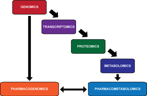
relationship between -OMICS

relationship between -OMICS

http://upload.wikimedia.org/wikipedia/commons/thumb/e/eb/OMICS.png/350px-OMICS.png

Pharmacometabolomics is thought to provide information that

Looking at the characteristics of an individual down through these different levels of detail, there is an

  • increasingly more accurate prediction of a person’s ability to respond to a pharmaceutical compound.
  1. the genome, made up of 25 000 genes, can indicate possible errors in drug metabolism;
  2. the transcriptome, made up of 85,000 transcripts, can provide information about which genes important in metabolism are being actively transcribed;
  3. and the proteome, >10,000,000 members, depicts which proteins are active in the body to carry out these functions.

Pharmacometabolomics complements the omics with

  • direct measurement of the products of all of these reactions, but with perhaps a relatively
  • smaller number of members: that was initially projected to be approximately 2200 metabolites,

but could be a larger number when gut derived metabolites and xenobiotics are added to the list. Overall, the goal of pharmacometabolomics is

  • to more closely predict or assess the response of an individual to a pharmaceutical compound,
  • permitting continued treatment with the right drug or dosage
  • depending on the variations in their metabolism and ability to respond to treatment.

Pharmacometabolomic analyses, through the use of a metabolomics approach,

  • can provide a comprehensive and detailed metabolic profile or “metabolic fingerprint” for an individual patient.

Such metabolic profiles can provide a complete overview of individual metabolite or pathway alterations,

This approach can then be applied to the prediction of response to a pharmaceutical compound

  • by patients with a particular metabolic profile.

Pharmacometabolomic analyses of drug response are

Pharmacogenetics focuses on the identification of genetic variations (e.g. single-nucleotide polymorphisms)

  • within patients that may contribute to altered drug responses and overall outcome of a certain treatment.

The results of pharmacometabolomics analyses can act to “inform” or “direct”

  • pharmacogenetic analyses by correlating aberrant metabolite concentrations or metabolic pathways to potential alterations at the genetic level.

This concept has been established with two seminal publications from studies of antidepressants serotonin reuptake inhibitors

  • where metabolic signatures were able to define a pathway implicated in response to the antidepressant and
  • that lead to identification of genetic variants within a key gene
  • within the highlighted pathway as being implicated in variation in response.

These genetic variants were not identified through genetic analysis alone and hence

  • illustrated how metabolomics can guide and inform genetic data.

en.wikipedia.org/wiki/Pharmacometabolomics

Benznidazole Biotransformation and Multiple Targets in Trypanosoma cruzi Revealed by Metabolomics

Andrea Trochine, Darren J. Creek, Paula Faral-Tello, Michael P. Barrett, Carlos Robello
Published: May 22, 2014   http://dx.doi.org:/10.1371/journal.pntd.0002844

The first line treatment for Chagas disease, a neglected tropical disease caused by the protozoan parasite Trypanosoma cruzi,

  • involves administration of benznidazole (Bzn).

Bzn is a 2-nitroimidazole pro-drug which requires nitroreduction to become active. We used a

  • non-targeted MS-based metabolomics approach to study the metabolic response of T. cruzi to Bzn.

Parasites treated with Bzn were minimally altered compared to untreated trypanosomes, although the redox active thiols

  1. trypanothione,
  2. homotrypanothione and
  3. cysteine

were significantly diminished in abundance post-treatment. In addition, multiple Bzn-derived metabolites were detected after treatment.

These metabolites included reduction products, fragments and covalent adducts of reduced Bzn

  • linked to each of the major low molecular weight thiols:
  1. trypanothione,
  2. glutathione,
  3. g-glutamylcysteine,
  4. glutathionylspermidine,
  5. cysteine and
  6. ovothiol A.

Bzn products known to be generated in vitro by the unusual trypanosomal nitroreductase, TcNTRI,

  • were found within the parasites,
  • but low molecular weight adducts of glyoxal, a proposed toxic end-product of NTRI Bzn metabolism, were not detected.

Our data is indicative of a major role of the

  • thiol binding capacity of Bzn reduction products
  • in the mechanism of Bzn toxicity against T. cruzi.


Read Full Post »

Introduction to Impairments in Pathological States: Endocrine Disorders, Stress Hypermetabolism and Cancer

Author and Curator: Larry H. Bernstein, MD, FCAP 


This leads into a series of presentations and the metabolic imbalance central to findings of endocrine, metabolic, inflammatory, immune diseases and cancer.  All of this has been a result of discoveries based on the methods of study of genomiocs, proteomics, transcriptomics, and metabolomics that have preceded this.  In some cases there has been the use of knockout methods. The completion of the human genomic and other catalogues have been instrumental in the past few years.  In all cases there has been a thorough guidance by a biological concept of mechanism based on gene expression, metabolic disturbance, signaling pathways, and up- or down- regulation of metabolic circuits.  It is interesting to recall that a concept of metabolic circuits was not yet formulated at the time of the mid 20th century physiology, except perhaps with respect to the coagulation pathways, and to some extent, glycolysis, gluconeogenesis, the hexose monophosphate shunt, and mitochondrial respiration, which were linear strings of enzyme substrate reactions that intersected and that had flow restraints not then understood as to the complexity we now appreciate.  We did know the importance of cytochrome c, the adenine and pyridine nucleotides, and the energy balance.  Electron microscopy had opened the door to understanding the mechanism of contraction of skeletal muscle and myocardium, but it also opened the door to understanding kidney structure and function, explaining the “mesangium”.  The first cardiac maker was discovered by Arthur Karmen in the serum alanine and aspartate aminotransferases, with a consequent differentiation between hepatic and myocardial damage.  This was followed by lactic dehydrogenase and the H- and M-type isoenzymes in the 1960s, and in the next decade, by the MB-isoenzyme of creatine kinase.  Troponins T and then I would not be introduced until the mid 1980s, and they have become a gold standard for the diagnosis of myocardial infarction.

In the 1980s we also saw the development of antiplatelet therapy that rapidly advanced interventional cardiology.  But advances in surgical as well as medical intervention also proceeded as the understanding of the lipid metabolism was opened by the work of Brown and Goldstein, and UTSW Medical Campus, and major advances in treatment came at Baylor and UT Medical Center in Houston, and at the Cleveland Clinic.  The next important advance came with the discovery of nitric oxide synthase role in endothelium and oxidative stress.  The field of endocrinology saw advances as well for a solid period of 30 years in a comparable period for the adrenals, thyroid, and pituitary glands, and for the understanding of the male and female sex hormones, and discoveries in breast, ovarian, and prostate cancer.  There were cancer markers, such as, CA125 and CA15-3, and PSA.  This had more of an impact on timely surgical intervention, and if not that, post surgical followup.  Despite a long time into the war on cancer, introduced by President Lynden Johnson, the fundamental knowledge needed was not sufficient.  In the meantime, there were advances in the treatment of diabetes, with eventual introduction of the insulin pump for type I diabetes.  The problem of Type 2 DM increased in prevalence, reaching into the childhood age group, with ascendent obesity.  An epidemiological pattern of disease comorbidities was emergent.  Our population has aged out, and with it we are seeing an increase in dementias, especially Alzheimer’s disease.  But the knowledge of the brain has lagged far behind.

What follows is a series of chapters that address what has currently been advanced with repect to the alignment of our knowledge of the last decade and pharmacetical discovery.  Pharmaceuticals were suitable for bacterial infections until the 1990s, when we saw the rise of resistance to penicillins and Vancomycin, and we had issues with gram negative enterobacter, salmonella, and E. coli strains.  That has been and is a significant challenge.  The elucidation of the gut microbiome in recent years will help to relieve this problem.  The problem of the variety and different aggressive types of cancer has been another challenge.  The door has been opened to better diagnostic tools with respsct to imaging and targeted biomarkers for localization.  I am not dealing with imaging, which is not the subject here.

HLA targeting efficiency correlates with human T-cell response magnitude and with mortality from influenza A infection

From http://www.pnas.org –      Sep 3, 2013 4:24 PM

Experimental and computational evidence suggests that HLAs preferentially bind

  • conserved regions of viral proteins, a concept we term “targeting efficiency,” and
  • that this preference may provide improved clearance of infection in several viral systems.

To test this hypothesis, T-cell responses to A/H1N1 (2009)

  • were measured from peripheral blood mononuclear cells
  • obtained from a household cohort study performed during the 2009–2010 influenza season.

We found that HLA targeting efficiency scores

  • significantly correlated with IFN-γ
    enzyme-linked immunosorbent spot responses (P = 0.042, multiple regression).

A further population-based analysis found that

  • the carriage frequencies of the alleles with the lowest targeting efficiencies, A*24,
  • were associated with pH1N1 mortality (r = 0.37, P = 0.031) and
  • are common in certain indigenous populations in which
  • increased pH1N1 morbidity has been reported.

HLA efficiency scores and HLA use are associated with

  • CD8 T-cell magnitude in humans after influenza infection.

The computational tools used in this study may be useful predictors of

  • potential morbidity and identify immunologic differences of new variant influenza strains
  • more accurately than evolutionary sequence comparisons.

Population-based studies of the relative frequency of these alleles

  • in severe vs. mild influenza cases might advance clinical practices
  • for severe H1N1 infections among genetically susceptible populations.

A deeper look into cholesterol synthesis

By Swathi Parasuraman

The human body needs cholesterol to maintain membrane fluidity, and

  • it acts as a precursor molecule for several important biochemical pathways.

Its regulation requires strict control, as it can cause problems if it’s produced in excess. In 1964, Konrad Bloch received a Nobel Prize for his work elucidating the mechanisms of cholesterol synthesis. His work

  • eventually contributed to the discovery of statins, drugs used today to lower blood cholesterol levels.

The biosynthesis of cholesterol is a complex process with more than 20 steps. One of the first enzymes is

  • 3-hydroxy-3-methylglutaryl-CoA reductase, also known as HMGCR, the main target of statins.

As links between intermediates in cholesterol synthesis and various diseases are being discovered continually, more information about the regulatory role of the post-HMGCR pathway is needed.

In a recent minireview in The Journal of Biological Chemistry, Laura Sharpe and Andrew Brown of the University of New South Wales describe

  • multiple ways various enzymes other than HMGCR
  • are implicated in the modulation of cholesterol synthesis.

One such enzyme is squalene monooxygenase, which, like HMGCR, can be destroyed

  • by the proteasome when cholesterol levels are high.

The minireview also explains how pathway intermediates

  • can have functions distinct from those of cholesterol.

For example, intermediate 7-dehydrocholesterol usually is converted to cholesterol by the enzyme DHCR7

  • but is also a vitamin D precursor.

To synthesize the enzymes necessary to make cholesterol,

  • SREBPs, short for sterol regulatory element binding proteins, have special functions.

Along with transcriptional cofactors, they activate gene expression

  1. in response to low sterol levels and, conversely,
  2. are suppressed when there is enough cholesterol around.

Additionally, SREBPs control production of

  • nicotinamide adenine dinucleotide phosphate, or NADPH,
  • which is the reducing agent required to carry out the different steps in the pathway.

Lipid carrier proteins also can facilitate cholesterol synthesis. One example is SPF, or supernatant protein factor,

  1. which transfers substrate from an inactive to an active pool or
  2. from one enzyme site to another.

Furthermore, translocation of several cholesterogenic enzymes

  • from the endoplasmic reticulum to other cell compartments can occur under various conditions,
  • thereby regulating levels and sites of intracellular cholesterol accumulation.

Immunology in the gut mucosa:

20 Feb 2013 by Kausik Datta, posted in Immunology, Science (Nature)

The human gut can be the scene for devastating conditions such as inflammatory bowel disease,

  • which arises through an improperly controlled immune response.

The gut is often the body’s first point of contact with microbes; every mouthful of food is accompanied by a cargo of micro-organisms that go on to encounter the mucosa, the innermost layer of the gut. Most microbes are destroyed by the harsh acidic environment in the stomach, but a hardy few make it through to the intestines.

The intestinal surface is covered with finger-like protrusions called villi,

whose primary function is the absorption of nutrients.

These structures and the underlying tissues

  • host the body’s largest population of immune cells.

Scattered along the intestinal mucosa are

  • dome-like structures called Peyer’s Patches.

These are enriched in lymphoid tissue, making them key sites for

  • coordinating immune responses to pathogens,
  • whilst promoting tolerance to harmless microbes and food.

The villi contain a network of blood vessels to transport nutrients from food to the rest of the body. Lymphatics

  • from both the Peyer’s Patches and the villi
  • drain into the mesenteric lymph nodes.

Within the villi is a network of loose connective tissue called the lamina propria, and

  • at the base of the villi are the crypts which host the stem cells that replenish the epithelium.

The epithelium together with its overlying mucus forms

  • a barrier against microbial invasion.

A mix of immune cells including T- and B-lymphocytes, macrophages, and dendritic cells are

  • embedded within the matrix of the Peyer’s Patches, .

A key function of the Peyer’s Patch is the sampling of antigens present in the gut. The Peyer’s Patch has a thin mucous layer and specialized phagocytic cells, called M-cells, which

  • transport material across the epithelial barrier via a process called transcytosis.

Dendritic cells extend dendrites between epithelial cells to sample antigens that are then

  • broken down and used for presenting to lymphocytes.

Sampling antigens in this way typically results in so-called tolerogenic activation, where

  • the immune system initiates an anti-inflammatory response.

With their cargo of antigens, these Dendritic Cells then

  • traffic to the T-cell zones of the Peyer’s Patch.

Upon encounter with specific T-cells, the Dendritic Cells

  • convert them into an immunomodulatory cell called regulatory T-cell or T-reg.

Defects in the function of these cells are associated with

  • inflammatory bowel disease in both animals and humans.

These T-regs migrate to lamina propria of the villi via the lymphatics. Here, the T-regs

  • secrete a molecule called Interleukin (IL)-10,
  • which exerts a suppressive action on immune cells within the lamina propria
  • and upon the epithelial layer itself.

IL10 is, therefore, critical in maintaining immune quiescence

  • and preventing unnecessary inflammation.

However, a breakdown in this process of immune homeostasis results in gut pathology and

  • when this occurs over a prolonged period and in an uncontrolled manner,
  • it can lead to inflammatory bowel disease.

Chemical, mechanical or pathogen-triggered barrier disruption

  • coupled with particular genetic susceptibilities may all combine to set off inflammation.

Epithelium coming into contact with bacteria

  • is activated, leading to bacterial influx.

Alarm molecules released by the epithelium

  • activates immune cells, and T-regs in the vicinity
  • scale down their IL10 secretion to enable an immune response to proceed.

Dendritic cells are also activated by this environment, and

  • start to release key inflammatory molecules,
  • such as IL6, IL12, and IL23.

Effector T-cells also appear on the scene and

  • these coordinate an escalation of the immune response
  • by secreting their own inflammatory molecules,
  • Tumor Necrosis Factor (TNF)-α, Interferon (IFN)-γ and IL17.

Soon after the effector T-cells are arrived, a voracious phagocyte called a neutrophil is recruited. Neutrophils are critical for the clearance of the bacteria. One weapon in the neutrophil armory is

  • the ability to undergo self-destruction.

This leaves behind a jumble of DNA saturated with enzymes, called the Neutrophil Extracellular Trap.

Although this can effectively destroy the bacterial invaders

  • and plug any breaches in the epithelial wall,
  • it also causes collateral damage to tissues.

Slowly the tide begins to turn and the bacterial invasion is repulsed. Any remaining neutrophils die off,

  • and are cleared by macrophages.

Epithelial integrity is restored by replacement of damaged cells with new ones from the intestinal crypts. Finally T-regs are recruited once again to calm the immune response.

Targeting the molecules involved in gut pathology is leading to

  • effective therapies for inflammatory bowel disease.

Notes:

T- and B-lymphocytes, Macrophages, and Dendritic Cells: These are all important immune effector cells. Macrophages and Dendritic cells are primary defence cells that can eat up (‘phagocytosis’) microbes and destroy them; they also can present parts of these microbes to lymphocytes. T-lymphocytes or T-cells help B-lymphocytes or B-cells recognize the antigen and form antibodies against it. Other types of T-cells can themselves kill microbes. All these cells also secrete various chemical substances, called cytokines and chemokines, which act as molecular messengers in recruiting various immune cells, coordinating and fine-tuning the immune response. Some of these cytokines are called Interleukins, shortened to IL.

Anti-inflammatory response: A type of immune response in which molecular messengers are used to scale down heavy-handed immune cell activity and switch off processes that recruit immune cells. This helps the body recognize and selectively tolerate beneficial substances such as commensalic microbes that live in the gut.

Neutrophils: These are highly versatile immune effector cells. Usually, they are one of the first cells recruited to the site of infection or tissue damage via message spread by molecular messengers. Neutrophils can themselves elaborate cytokines and chemokines, and have the ability to directly kill microbes.

Oxazoloisoindolinones with in vitro antitumor activity selectively activate a p53-pathway through potential inhibition of the p53-MDM2 interaction.

J Soares, et al. Eur J Pharm Sci 10/2014; http://dx.doi.org:/10.1016/j.ejps.2014.10.006

An appealing target for anticancer treatment is

  • the p53 tumor suppressor protein.

This protein is inactivated in half of human tumors

  • due to endogenous negative regulators such as MDM2.

Therefore, restoring the p53 activity through

  • the inhibition of its interaction with MDM2
  • is considered a valuable therapeutic strategy
  • against cancers with a wild-type p53 status.

We report the synthesis of nine enantiopure phenylalaninol-derived oxazolopyrrolidone lactams

  • and the evaluation of their biological effects as p53-MDM2 interaction inhibitors.

Using a yeast-based screening assay, two oxazoloisoindolinones,

  • were identified as potential p53-MDM2 inhibitors.

The molecular mechanism of oxazoloisoindolinone 3a validated

  • in human colon adenocarcinoma HCT116 cells with wild-type p53 (HCT116 p53(+/+)) and
  • in its isogenic derivative without p53 (HCT116 p53(-/-)).

we demonstrated that oxazoloisoindolinone 3a exhibited

  • a p53-dependent in vitro antitumor activity through
  • induction of G0/G1-phase cell cycle arrest and apoptosis.

The selective activation of a p53-apoptotic pathway by oxazoloisoindolinone 3a was further supported

  • by the occurrence of PARP cleavage only in p53-expressing HCT116 cells.

Oxazoloisoindolinone 3a led

  • to p53 protein stabilization
  • to the up-regulation of p53 transcriptional activity &
  • increased expression levels of several p53 target genes,
  • as p21, MDM2, BAX and PUMA,
  • in p53(+/+) but not in p53(-/-) HCT116 cells.

the ability of oxazoloisoindolinone 3a to block the p53-MDM2 interaction in HCT116 p53(+/+) cells was confirmed by co-immunoprecipitation.

molecular docking analysis of the interactions

  • between the compounds and MDM2 revealed that
  • oxazoloisoindolinone 3a binds to MDM2.

this work adds the oxazoloisoindolinone scaffold to the activators of a wild-type p53-pathway with promising antitumor activity.

it may open the way to the development of

  • a new class of p53-MDM2 interaction inhibitors.

TrypanoCyc: a community-led biochemical pathways database for Trypanosoma brucei.

Sanu Shameer, et al. Nucleic Acids Research10/2014;
http://dx.doi.org/10.1093/nar/gku944

The metabolic network of a cell represents the catabolic and anabolic reactions that interconvert small molecules (metabolites) through the activity of enzymes, transporters and non-catalyzed chemical reactions. Our understanding of individual metabolic networks is increasing as we learn more about the enzymes that are active in particular cells under particular conditions and as technologies advance to allow detailed measurements of the cellular metabolome.

Metabolic network databases are important in allowing us to

  • contextualise data sets emerging from transcriptomic, proteomic and metabolomic experiments.

Here we present a dynamic database, TrypanoCyc (http://www.metexplore.fr/trypanocyc/), which describes

  • the generic and condition-specific metabolic network of Trypanosoma brucei, a parasitic protozoan
  • responsible for human and animal African trypanosomiasis.

In addition to enabling navigation through the BioCyc-based TrypanoCyc interface, we have implemented a network

  • representation of the information through MetExplore,

yielding a novel environment in which to visualise the metabolism of this important parasite.

Read Full Post »

Summary of Metabolomics

Author and Curator: Larry H. Bernstein, MD, FCAP 

This concludes the series on metabolomics, a rapidly developing science that is interconnected with a group termed – OMICS: proteomics, transcriptomics, genomics, and metabolomics.  This chapter is most representative of the many important studies being done in the field, which ranges most widely because it has opened doors into nutrition and nutritional supplements, plant biochemistry, agricultural crops and breeding, animal breeding, worldwide malnutrition, diabetes, cancer, neurosciences, circulatory, respiratory, and musculosletal disorders, infectious diseases and immune system disorders.  Obviously, it is not possible to cover the full range of activity, but metabolomics is most comprehensive in exploring the full range of metabolic changes that occur in health during the full age range from development to the geriatric years.  It can be integrated well with gene expression, proteomics studies, and epidemiological investigations.

The subchapters are given here:

7.1   Extracellular evaluation of intracellular flux in yeast cells  

 http://pharmaceuticalintelligence.com/2014/08/25/extracellular-evaluation-of-intracellular-flux-in-yeast-cells/

 7.2    Metabolomic analysis of two leukemia cell lines. I.  

         http://pharmaceuticalintelligence.com/2014/08/23/metabolomic-analysis-of-two-leukemia-cell-lines-_i/

  7.3   Metabolomic analysis of two leukemia cell lines. II.

           http://pharmaceuticalintelligence.com/2014/08/24/metabolomic-analysis-of-two-leukemia-cell-lines-ii/

  7.4   Buffering of genetic modules involved in tricarboxylic acid cycle metabolism provides homeostatic
regulation
  

           http://pharmaceuticalintelligence.com/2014/08/27/buffering-of-genetic-modules-involved-in-tricarboxylic-acid-cycle-metabolism-provides-homeomeostatic-regulation/

  7.5   Metabolomics, Metabonomics and Functional Nutrition: the next step in nutritional metabolism and
biotherapeutics

 http://pharmaceuticalintelligence.com/2014/08/22/metabolomics-metabonomics-and-functional-nutrition-the-next-step-in-nutritional-metabolism-and-biotherapeutics/

    7.6    Isoenzymes in cell metabolic pathways

 http://pharmaceuticalintelligence.com/2014/10/06/isoenzymes-in-cell-metabolic-pathways/

7.7   A Brief Curation of Proteomics, Metabolomics, and Metabolism

http://pharmaceuticalintelligence.com/2014/10/03/a-brief-curation-of-proteomics-metabolomics-and-metabolism/

   7.8   Metabolomics is about Metabolic Systems Integration

     http://pharmaceuticalintelligence.com/2014/10/13/metabolomics-is-about-metabolic-systems-integration/

 7.9  Mechanisms of Drug Resistance

   http://pharmaceuticalintelligence.com/2014/10/09/mechanisms-of-drug-resistance/

7.10  Development Of Super-Resolved Fluorescence Microscopy

    http://pharmaceuticalintelligence.com/2014/10/12/development-of-super-resolved-fluorescence-microscopy  

7.11  Metabolic Reactions Need Just Enough

 http://pharmaceuticalintelligence.com/2014/10/14/metabolic-reactions-need-just-enough/

7.12  Metabolomics Summary and Perspective

   This chapter will be followed by an exploration of disease and pharmaceutical directed studies using these methods  8. Impairments in pathological states: endocrine disorders; stress
hypermetabolism; cancer.

Networking metabolites and diseases

P Braun, E Rietman, and M Vidal
Center for Cancer Systems Biology and Department of Cancer Biology, Dana-Farber Cancer Institute, and Department of Genetics, Harvard Medical School,  Boston, MA; and Physical Sciences Inc., Andover, MA 01810
PNAS July 22, 2008; 105(29): 9849–9850.    http://pnas.org/cgi/doi/10.1073/pnas.0805644105

Biological systems are increasingly viewed and analyzed as

  • highly complex networks of interlinked macromolecules and metabolites.

Network analysis has been applied to

  • interactome maps of protein–protein, protein–DNA, and protein–RNA interactions
  • as well as transcriptional, metabolic, and genetic data.

Such network views of biological systems should facilitate the detection of

  • nonlinear long-range effects of perturbations, for example, by mutations, and
  • help identification of unanticipated indirect causal connections.

Diseasome and Drug-Target Network

Recently, Goh et al. (1) constructed a ‘‘diseasome’’ network in which

  • two diseases are linked to each other if
  • they share at least one gene, in which mutations are associated with both diseases.

In the resulting network, related disease families cluster tightly together, thus

  • phenotypically defining functional modules.

Importantly, for the first time this study applied concepts from network biology to human diseases,

  • thus opening the door for discovering causal relationships between
  • disregulated networks and resulting ailments.

Subsequently Yilderim et al. (2) linked drugs to protein targets in a drug–target network,

  • which could then be overlaid with the diseasome network.

One notable finding was the recent trend toward the development of

  • new compounds directly targeted at disease gene products, whereas previous drugs,
  • often found by trial and error, appear to target proteins only indirectly related to
  • the actual disease molecular mechanisms.

An important question that remains in this emerging field of network analysis consists of

  • investigating the extent to which directly targeting the product of mutated genes is an efficient approach or
  • whether targeting network properties instead, and
  • thereby accounting for indirect nonlinear effects of system perturbations by drugs, may prove more fruitful.

However, to answer such questions it is important to have a good understanding of the various influences that can lead to diseases.

Metabolic Connections

One group of diseases that was very poorly connected in the original diseasome network was the family of metabolic diseases.

In this issue of PNAS, Lee et al. (3) hypothesize that metabolic diseases may instead be connected

  • via metabolites and common reactions.

To investigate this hypothesis Lee et al. first constructed a metabolic network from data available in

  • two manually curated databases detailing well known
  1. metabolic reactions,
  2. the involved metabolites, and
  3. catalyzing enzymes.

In addition, gene–disease associations were identified by using the Online Mendelian Inheritance in Man (OMIM) database (http://ncbi.nlm.nih.gov/sites/
entrez?dbomim&itooltoolbar). In a last step,

  • a metabolic disease network (MDN) was constructed by connecting
  • two diseases if their associated genes are linked in the metabolic network
  • by a common metabolite or metabolites used in a common reaction.

Metabolites are not only linked by common reactions, but

  • on a larger scale by coupled fluxes within a metabolic network,
  • which may also influence disease phenotypes.

An increase in the concentration of one metabolite may increase several fluxes

  • across reaction pathways that use this compound, which
  • may lead to diverse phenotypes and distinct diseases.

The fluxes within the metabolic network are calculated by using

  • the Flux Coupling Finder method described by Nikolaev et al. (4) and Burgard et al. (5),
  • which is based on the assumption that pools of metabolites are conserved.

To functionally validate the network, coexpression correlations are measured for genes

  • linked by adjacent reactions and those linked by fluxes.

Interestingly, the average coexpression correlation for flux-coupled genes (0.31)

  • is higher than that for genes simply catalyzing adjacent reactions (0.24)
    (compared with 0.10 for all gene pairs in the network).

If the links between diseases identified in the MDN are functionally and causally relevant

  • it should be expected that linked diseases occur more frequently in the same individual.

To test this hypothesis, Lee et al. (3) measured the co-occurrence of diseases in patients by using detailed Medicare information

  • of 13 million patients and 32 million hospital visits within a 3-year period.

A comorbidity index was computed to measure the degree to which one disease

  • will increase the likelihood of a second disease in the same patient.

The average comorbidity for all genes is 0.0008 (Pearson correlation coefficient),

  • which increases 3-fold to 0.0027 when disease pairs that are metabolically linked are analyzed,
  • which is highly statistically significant (P 108).

When diseases are analyzed that are directionally coupled by a flux (see ref. 3 for details),

  • the correlation increases to 0.0062.

Thus, whereas 17% of all diseases in the network show significant comorbidity, this fraction

  • nearly doubles to 31% for metabolically linked diseases.

Further analysis reveals that comorbidity effects can be detected up to three links (metabolites, reactions)

  • apart from each other with statistical significance, but not farther away.

In the MDN, several highly connected hubs, e.g., hypertension and hemolytic anemia, are

  • linked to many different co-occurring diseases not unexpected for such complex diseases
  • that can result from many different genetic alterations or variants.

Importantly, though, most of the connections to the different linked diseases

  • are mediated by diverse connections in the metabolic network.

Thus, in the future such insights may be helpful for finer classification of the complex hub disease.

Furthermore, depending on the onset of the complex (hub) disease in relation to the associated diseases,

  • such relationships may potentially be used to systematically
  • stratify patients and develop targeted treatments acting on
  • the underlying metabolic links.

Returning to the starting point of their study, Lee et al. (3) next investigated

  • whether metabolic diseases are better linked through the metabolic network
  • than they are in the previously described gene–disease network.

When purely metabolic diseases are considered, the comorbidity is, in fact,

  • best predicted by metabolic links.

Interestingly, when all diseases linked to metabolic enzymes are considered,

  • which involves many diseases that are merely related to metabolic diseases through multifunctional enzymes,
  • the gene and metabolic networks are nearly equally predictive of comorbidity,
  • indicating that as a general approach information from
  • many different biological dimensions should be integrated to identify the most relevant connections.

Together, all these findings support the initial hypothesis that metabolic diseases are linked by metabolic networks.

Practically, alteration of one metabolite or one reaction can have numerous repercussions in the network,

  • each of which can manifest as different diseases that frequently occur together in affected patients.

Radoslav Bozov

  1. Glycine, as the only amino acid having no isomer driven central carbon allowing for hing occupancy of ‘free’
    motifs, where quark (proton) ‘fluxes’ play at, is a one – step away observable (1) from synthesis of pyrimidines
    to glyoxylate mitochondrial ‘shunt’ entangling at least two differential compartments longly objected by
    Japanese metabolomics study groups.
  2. One carbon systems emerge out of a glycoprotein ‘complex’, pyrimidine synthase pathway, that possesses
    significant similarity to  BRCA2 and most other transcription factors suggesting that protein allocation is
    coorchestrated by modifications and spatially transforming construes as an outcome of energy processing.
    Directly deduced by TCS, life cannot exist without mutations, as mutations and chromatin states appear to
    be a sort of energy hold and release ‘gates’.
  3. Phosphorylations and small molecules as such as cGMP, cAMP play a role of decompression machinery
    for amplifying bio signal processing C-S, C-N, C-O, interference open systems.  By decompression
    of one relative discrete space, another one becomes compressed, which gets uncertainty of absolute energy
    processing within space scalar wise into vector objected space represented by chromatin remodeling processes,
    possibly seen as network identities information.
  4. Unifying network and quantum theory possess implications to relativity concepts and energy relevant computational methodology.
 translational medicine

translational medicine

Shifts in steady-state profiles caused by kinetic perturbations

Shifts in steady-state profiles caused by kinetic perturbations

mapping metabolomic data using three different approaches

mapping metabolomic data using three different approaches

network genetics metabotypes -  integrated metabolome and interactome mapping (iMIM)

network genetics metabotypes – integrated metabolome and interactome mapping (iMIM)

metabol leukem cell lines

metabol leukem cell lines

Metabolome Informatics Research

Metabolome Informatics Research

Read Full Post »

Infinity and AbbVie partner to develop and commercialise Duvelisib for cancer… for the treatment of chronic lymphocytic leukemia

Reporter: Stephen J. Williams, PhD

Duvelisib is a dual phosphoinositide-3-kinase (PI3K) delta and PI3K gamma inhibitor.  The delta and gamma isozymes are selectively expressed in leukocytes.    This article (at Dr. Melvin Crasto’s blog newdrugapprovals.org) discusses the synthesis of Duvelisib and mentions additional clinical trials underway including a phase II trial for the treatment of patients with mild asthma undergoing allergen challenge, for the treatment of rheumatoid arthritis and for the treatment of refractory indolent non-Hodgkin’s lymphoma. Phase I clinical trials for the treatment of advanced hematological malignancies (including T-cell lymphoma and mantle cell lymphoma).  The drug was originally developed at Takeda subsidiary Intellikine.

SOURCE

http://newdrugapprovals.org/2014/09/09/infinity-and-abbvie-partner-to-develop-and-commercialise-duvelisib-for-cancer-for-the-treatment-of-chronic-lymphocytic-leukemia/

Read Full Post »

Summary to Metabolomics

Summary to Metabolomics

Author and Curator: Larry H. Bernstein, MD, FCAP 

This concludes a long step-by-step journey into rediscovering biological processes from the genome as a framework to the remodeled and reconstituted cell through a number of posttranscription and posttranslation processes that modify the proteome and determine the metabolome.  The remodeling process continues over a lifetime. The process requires a balance between nutrient intake, energy utilization for work in the lean body mass, energy reserves, endocrine, paracrine and autocrine mechanisms, and autophagy.  It is true when we look at this in its full scope – What a creature is man?

http://masspec.scripps.edu/metabo_science/recommended_readings.php
 Recommended Readings and Historical Perspectives

Metabolomics is the scientific study of chemical processes involving metabolites. Specifically, metabolomics is the “systematic study of the unique chemical fingerprints that specific cellular processes leave behind”, the study of their small-molecule metabolite profiles.[1] The metabolome represents the collection of all metabolites in a biological cell, tissue, organ or organism, which are the end products of cellular processes.[2] mRNA gene expression data and proteomic analyses reveal the set of gene products being produced in the cell, data that represents one aspect of cellular function. Conversely, metabolic profiling can give an instantaneous snapshot of the physiology of that cell. One of the challenges of systems biology and functional genomics is to integrate proteomic, transcriptomic, and metabolomic information to provide a better understanding of cellular biology.

The term “metabolic profile” was introduced by Horning, et al. in 1971 after they demonstrated that gas chromatography-mass spectrometry (GC-MS) could be used to measure compounds present in human urine and tissue extracts. The Horning group, along with that of Linus Pauling and Arthur B. Robinson led the development of GC-MS methods to monitor the metabolites present in urine through the 1970s.

Concurrently, NMR spectroscopy, which was discovered in the 1940s, was also undergoing rapid advances. In 1974, Seeley et al. demonstrated the utility of using NMR to detect metabolites in unmodified biological samples.This first study on muscle highlighted the value of NMR in that it was determined that 90% of cellular ATP is complexed with magnesium. As sensitivity has improved with the evolution of higher magnetic field strengths and magic angle spinning, NMR continues to be a leading analytical tool to investigate metabolism. Efforts to utilize NMR for metabolomics have been influenced by the laboratory of Dr. Jeremy Nicholson at Birkbeck College, University of London and later at Imperial College London. In 1984, Nicholson showed 1H NMR spectroscopy could potentially be used to diagnose diabetes mellitus, and later pioneered the application of pattern recognition methods to NMR spectroscopic data.

In 2005, the first metabolomics web database, METLIN, for characterizing human metabolites was developed in the Siuzdak laboratory at The Scripps Research Institute and contained over 10,000 metabolites and tandem mass spectral data. As of September 2012, METLIN contains over 60,000 metabolites as well as the largest repository of tandem mass spectrometry data in metabolomics.

On 23 January 2007, the Human Metabolome Project, led by Dr. David Wishart of the University of Alberta, Canada, completed the first draft of the human metabolome, consisting of a database of approximately 2500 metabolites, 1200 drugs and 3500 food components. Similar projects have been underway in several plant species, most notably Medicago truncatula and Arabidopsis thaliana for several years.

As late as mid-2010, metabolomics was still considered an “emerging field”. Further, it was noted that further progress in the field depended in large part, through addressing otherwise “irresolvable technical challenges”, by technical evolution of mass spectrometry instrumentation.

Metabolome refers to the complete set of small-molecule metabolites (such as metabolic intermediates, hormones and other signaling molecules, and secondary metabolites) to be found within a biological sample, such as a single organism. The word was coined in analogy with transcriptomics and proteomics; like the transcriptome and the proteome, the metabolome is dynamic, changing from second to second. Although the metabolome can be defined readily enough, it is not currently possible to analyse the entire range of metabolites by a single analytical method. The first metabolite database(called METLIN) for searching m/z values from mass spectrometry data was developed by scientists at The Scripps Research Institute in 2005. In January 2007, scientists at the University of Alberta and the University of Calgary completed the first draft of the human metabolome. They catalogued approximately 2500 metabolites, 1200 drugs and 3500 food components that can be found in the human body, as reported in the literature. This information, available at the Human Metabolome Database (www.hmdb.ca) and based on analysis of information available in the current scientific literature, is far from complete.

Each type of cell and tissue has a unique metabolic ‘fingerprint’ that can elucidate organ or tissue-specific information, while the study of biofluids can give more generalized though less specialized information. Commonly used biofluids are urine and plasma, as they can be obtained non-invasively or relatively non-invasively, respectively. The ease of collection facilitates high temporal resolution, and because they are always at dynamic equilibrium with the body, they can describe the host as a whole.

Metabolites are the intermediates and products of metabolism. Within the context of metabolomics, a metabolite is usually defined as any molecule less than 1 kDa in size.
A primary metabolite is directly involved in the normal growth, development, and reproduction. A secondary metabolite is not directly involved in those processes.  By contrast, in human-based metabolomics, it is more common to describe metabolites as being either endogenous (produced by the host organism) or exogenous. Metabolites of foreign substances such as drugs are termed xenometabolites. The metabolome forms a large network of metabolic reactions, where outputs from one enzymatic chemical reaction are inputs to other chemical reactions.

Metabonomics is defined as “the quantitative measurement of the dynamic multiparametric metabolic response of living systems to pathophysiological stimuli or genetic modification”. The word origin is from the Greek μεταβολή meaning change and nomos meaning a rule set or set of laws. This approach was pioneered by Jeremy Nicholson at Imperial College London and has been used in toxicology, disease diagnosis and a number of other fields. Historically, the metabonomics approach was one of the first methods to apply the scope of systems biology to studies of metabolism.

There is a growing consensus that ‘metabolomics’ places a greater emphasis on metabolic profiling at a cellular or organ level and is primarily concerned with normal endogenous metabolism. ‘Metabonomics’ extends metabolic profiling to include information about perturbations of metabolism caused by environmental factors (including diet and toxins), disease processes, and the involvement of extragenomic influences, such as gut microflora. This is not a trivial difference; metabolomic studies should, by definition, exclude metabolic contributions from extragenomic sources, because these are external to the system being studied.

Toxicity assessment/toxicology. Metabolic profiling (especially of urine or blood plasma samples) detects the physiological changes caused by toxic insult of a chemical (or mixture of chemicals).

Functional genomics. Metabolomics can be an excellent tool for determining the phenotype caused by a genetic manipulation, such as gene deletion or insertion. Sometimes this can be a sufficient goal in itself—for instance, to detect any phenotypic changes in a genetically-modified plant intended for human or animal consumption. More exciting is the prospect of predicting the function of unknown genes by comparison with the metabolic perturbations caused by deletion/insertion of known genes.

Nutrigenomics is a generalised term which links genomics, transcriptomics, proteomics and metabolomics to human nutrition. In general a metabolome in a given body fluid is influenced by endogenous factors such as age, sex, body composition and genetics as well as underlying pathologies. The large bowel microflora are also a very significant potential confounder of metabolic profiles and could be classified as either an endogenous or exogenous factor. The main exogenous factors are diet and drugs. Diet can then be broken down to nutrients and non- nutrients.

http://en.wikipedia.org/wiki/Metabolomics

Jose Eduardo des Salles Roselino

The problem with genomics was it was set as explanation for everything. In fact, when something is genetic in nature the genomic reasoning works fine. However, this means whenever an inborn error is found and only in this case the genomic knowledge afterwards may indicate what is wrong and not the completely way to put biology upside down by reading everything in the DNA genetic as well as non-genetic problems.

Coordination of the transcriptome and metabolome by the circadian clock PNAS 2012

Coordination of the transcriptome and metabolome by the circadian clock PNAS 2012

analysis of metabolomic data and differential metabolic regulation for fetal lungs, and maternal blood plasma

conformational changes leading to substrate efflux.img

conformational changes leading to substrate efflux.img

The cellular response is defined by a network of chemogenomic response signatures.

The cellular response is defined by a network of chemogenomic response signatures.

Dynamic Construct of the –Omics

Dynamic Construct of the –Omics

 genome cartoon

genome cartoon

central dogma phenotype

central dogma phenotype

Read Full Post »

« Newer Posts - Older Posts »