Feeds:
Posts
Comments

Posts Tagged ‘gene’

Introduction – The Evolution of Cancer Therapy and Cancer Research: How We Got Here?

Introduction – The Evolution of Cancer Therapy and Cancer Research: How We Got Here?

Author and Curator: Larry H Bernstein, MD, FCAP

The evolution of progress we have achieved in cancer research, diagnosis, and therapeutics has  originated from an emergence of scientific disciplines and the focus on cancer has been recent. We can imagine this from a historical perspective with respect to two observations. The first is that the oldest concepts of medicine lie with the anatomic dissection of animals and the repeated recurrence of war, pestilence, and plague throughout the middle ages, and including the renaissance.  In the awakening, architecture, arts, music, math, architecture and science that accompanied the invention of printing blossomed, a unique collaboration of individuals working in disparate disciplines occurred, and those who were privileged received an education, which led to exploration, and with it, colonialism.  This also led to the need to increasingly, if not without reprisal, questioning long-held church doctrines.

It was in Vienna that Rokitansky developed the discipline of pathology, and his student Semelweis identified an association between then unknown infection and childbirth fever. The extraordinary accomplishments of John Hunter in anatomy and surgery came during the twelve years war, and his student, Edward Jenner, observed the association between cowpox and smallpox resistance. The development of a nursing profession is associated with the work of Florence Nightengale during the Crimean War (at the same time as Leo Tolstoy). These events preceded the work of Pasteur, Metchnikoff, and Koch in developing a germ theory, although Semelweis had committed suicide by infecting himself with syphilis. The first decade of the Nobel Prize was dominated by discoveries in infectious disease and public health (Ronald Ross, Walter Reed) and we know that the Civil War in America saw an epidemic of Yellow Fever, and the Armed Services Medical Museum was endowed with a large repository of osteomyelitis specimens. We also recall that the Russian physician and playwriter, Anton Checkov, wrote about the conditions in prison camps.

But the pharmacopeia was about to open with the discoveries of insulin, antibiotics, vitamins, thyroid action (Mayo brothers pioneered thyroid surgery in the thyroid iodine-deficient midwest), and pitutitary and sex hormones (isolatation, crystal structure, and synthesis years later), and Karl Landsteiner’s discovery of red cell antigenic groups (but he also pioneered in discoveries in meningitis and poliomyelitis, and conceived of the term hapten) with the introduction of transfusion therapy that would lead to transplantation medicine.  The next phase would be heralded by the discovery of cancer, which was highlighted by the identification of leukemia by Rudolph Virchow, who cautioned about the limitations of microscopy. This period is highlighted by the classic work – “Microbe Hunters”.

John Hunter

John Hunter

Walter Reed

Walter Reed

Robert Koch

Robert Koch

goldberger 1916 Pellagra

goldberger 1916 Pellagra

Louis Pasteur

Louis Pasteur

A multidisciplinary approach has led us to a unique multidisciplinary or systems view of cancer, with different fields of study offering their unique expertise, contributions, and viewpoints on the etiology of cancer.  Diverse fields in immunology, biology, biochemistry, toxicology, molecular biology, virology, mathematics, social activism and policy, and engineering have made such important contributions to our understanding of cancer, that without cooperation among these diverse fields our knowledge of cancer would never had evolved as it has. In a series of posts “Heroes in Medical Research:” the work of researchers are highlighted as examples of how disparate scientific disciplines converged to produce seminal discoveries which propelled the cancer field, although, at the time, they seemed like serendipitous findings.  In the post Heroes in Medical Research: Barnett Rosenberg and the Discovery of Cisplatin (Translating Basic Research to the Clinic) discusses the seminal yet serendipitous discoveries by bacteriologist Dr. Barnett Rosenberg, which eventually led to the development of cisplatin, a staple of many chemotherapeutic regimens. Molecular biologist Dr. Robert Ting, working with soon-to-be Nobel Laureate virologist Dr. James Gallo on AIDS research and the associated Karposi’s sarcoma identified one of the first retroviral oncogenes, revolutionizing previous held misconceptions of the origins of cancer (described in Heroes in Medical Research: Dr. Robert Ting, Ph.D. and Retrovirus in AIDS and Cancer).   Located here will be a MONTAGE of PHOTOS of PEOPLE who made seminal discoveries and contributions in every field to cancer   Each of these paths of discovery in cancer research have led to the unique strategies of cancer therapeutics and detection for the purpose of reducing the burden of human cancer.  However, we must recall that this work has come at great cost, while it is indeed cause for celebration. The current failure rate of clinical trials at over 70 percent, has been a cause for disappointment, and has led to serious reconsideration of how we can proceed with greater success. The result of the evolution of the cancer field is evident in the many parts and chapters of this ebook.  Volume 4 contains chapters that are in a predetermined order:

  1. The concepts of neoplasm, malignancy, carcinogenesis,  and metastatic potential, which encompass:

(a)     How cancer cells bathed in an oxygen rich environment rely on anaerobic glycolysis for energy, and the secondary consequences of cachexia and sarcopenia associated with progression

invasion

invasion

ARTS protein and cancer

ARTS protein and cancer

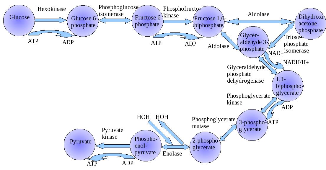
Glycolysis

Glycolysis

Krebs cycle

Krebs cycle

Metabolic control analysis of respiration in human cancer tissue

Metabolic control analysis of respiration in human cancer tissue

akip1-expression-modulates-mitochondrial-function

akip1-expression-modulates-mitochondrial-function

(b)     How advances in genetic analysis, molecular and cellular biology, metabolomics have expanded our basic knowledge of the mechanisms which are involved in cellular transformation to the cancerous state.

nucleotides

nucleotides

Methylation of adenine

Methylation of adenine

ampk-and-ampk-related-kinase-ark-family-

ampk-and-ampk-related-kinase-ark-family-

ubiquitylation

ubiquitylation

(c)  How molecular techniques continue to advance our understanding  of how genetics, epigenetics, and alterations in cellular metabolism contribute to cancer and afford new pathways for therapeutic intervention.

 genomic effects

genomic effects

LKB1AMPK pathway

LKB1AMPK pathway

mutation-frequencies-across-12-cancer-types

mutation-frequencies-across-12-cancer-types

AMPK-activating drugs metformin or phenformin might provide protection against cancer

AMPK-activating drugs metformin or phenformin might provide protection against cancer

pim2-phosphorylates-pkm2-and-promotes-glycolysis-in-cancer-cells

pim2-phosphorylates-pkm2-and-promotes-glycolysis-in-cancer-cells

pim2-phosphorylates-pkm2-and-promotes-glycolysis-in-cancer-cells

pim2-phosphorylates-pkm2-and-promotes-glycolysis-in-cancer-cells

2. The distinct features of cancers of specific tissue sites of origin

3.  The diagnosis of cancer by

(a)     Clinical presentation

(b)     Age of onset and stage of life

(c)     Biomarker features

hairy cell leukemia

hairy cell leukemia

lymphoma leukemia

lymphoma leukemia

(d)     Radiological and ultrasound imaging

  1. Treatments
  2. Prognostic differences within and between cancer types

We have introduced the emergence of a disease of great complexity that has been clouded in more questions than answers until the emergence of molecular biology in the mid 20th century, and then had to await further discoveries going into the 21st century.  What gave the research impetus was the revelation of

1     the mechanism of transcription of the DNA into amino acid sequences

Proteins in Disease

Proteins in Disease

2     the identification of stresses imposed on cellular function

NO beneficial effects

NO beneficial effects

3     the elucidation of the substructure of the cell – cell membrane, mitochondria, ribosomes, lysosomes – and their functions, respectively

pone.0080815.g006  AKIP1 Expression Modulates Mitochondrial Function

AKIP1 Expression Modulates Mitochondrial Function

4     the elucidation of oligonucleotide sequences

nucleotides

nucleotides

dna-replication-unwinding

dna-replication-unwinding

dna-replication-ligation

dna-replication-ligation

dna-replication-primer-removal

dna-replication-primer-removal

dna-replication-leading-strand

dna-replication-leading-strand

dna-replication-lagging-strand

dna-replication-lagging-strand

dna-replication-primer-synthesis

dna-replication-primer-synthesis

dna-replication-termination

dna-replication-termination

5     the further elucidation of functionally relevant noncoding lncDNA

lncRNA-s   A summary of the various functions described for lncRNA

6     the technology to synthesis mRNA and siRNA sequences

RNAi_Q4 Primary research objectives

Figure. RNAi and gene silencing

7     the repeated discovery of isoforms of critical enzymes and their pleiotropic properties

8.     the regulatory pathways involved in signaling

signaling pathjways map

Figure. Signaling Pathways Map

This is a brief outline of the modern progression of advances in our understanding of cancer.  Let us go back to the beginning and check out a sequence of  Nobel Prizes awarded and related discoveries that have a historical relationship to what we know.  The first discovery was the finding by Louis Pasteur that fungi that grew in an oxygen poor environment did not put down filaments.  They did not utilize oxygen and they produced used energy by fermentation.  This was the basis for Otto Warburg sixty years later to make the comparison to cancer cells that grew in the presence of oxygen, but relied on anaerobic glycolysis. He used a manometer to measure respiration in tissue one cell layer thick to measure CO2 production in an adiabatic system.

video width=”1280″ height=”720″ caption=”1741-7007-11-65-s1 Macromolecular juggling by ubiquitylation enzymes.” mp4=”http://pharmaceuticalintelligence.com/wp-content/uploads/2014/04/1741-7007-11-65-s1-macromolecular-juggling-by-ubiquitylation-enzymes.mp4“][/video]

An Introduction to the Warburg Apparatus

http://www.youtube.com/watch?v=M-HYbZwN43o

Lavoisier Antoine-Laurent and Laplace Pierre-Simon (1783) Memoir on heat. Mémoirs de l’Académie des sciences. Translated by Guerlac H, Neale Watson Academic Publications, New York, 1982.

Instrumental background 200 years later:   Gnaiger E (1983) The twin-flow microrespirometer and simultaneous calorimetry. In Gnaiger E, Forstner H, eds. Polarographic Oxygen Sensors. Springer, Heidelberg, Berlin, New York: 134-166.

otto_heinrich_warburg

otto_heinrich_warburg

Warburg apparatus

The Warburg apparatus is a manometric respirometer which was used for decades in biochemistry for measuring oxygen consumption of tissue homogenates or tissue slices.

The Warburg apparatus has its name from the German biochemist Otto Heinrich Warburg (1883-1970) who was awarded the Nobel Prize in physiology or medicine in 1931 for his “discovery of the nature and mode of action of the respiratory enzyme” [1].

The aqueous phase is vigorously shaken to equilibrate with a gas phase, from which oxygen is consumed while the evolved carbon dioxide is trapped, such that the pressure in the constant-volume gas phase drops proportional to oxygen consumption. The Warburg apparatus was introduced to study cell respiration, i.e. the uptake of molecular oxygen and the production of carbon dioxide by cells or tissues. Its applications were extended to the study of fermentation, when gas exchange takes place in the absence of oxygen. Thus the Warburg apparatus became established as an instrument for both aerobic and anaerobic biochemical studies [2, 3].

The respiration chamber was a detachable glass flask (F) equipped with one or more sidearms (S) for additions of chemicals and an open connection to a manometer (M; pressure gauge). A constant temperature was provided by immersion of the Warburg chamber in a constant temperature water bath. At thermal mass transfer equilibrium, an initial reading is obtained on the manometer, and the volume of gas produced or absorbed is determined at specific time intervals. A limited number of ‘titrations’ can be performed by adding the liquid contained in a side arm into the main reaction chamber. A Warburg apparatus may be equipped with more than 10 respiration chambers shaking in a common water bath.   Since temperature has to be controlled very precisely in a manometric approach, the early studies on mammalian tissue respiration were generally carried out at a physiological temperature of 37 °C.

The Warburg apparatus has been replaced by polarographic instruments introduced by Britton Chance in the 1950s. Since Chance and Williams (1955) measured respiration of isolated mitochondria simultaneously with the spectrophotometric determination of cytochrome redox states, a water chacket could not be used, and measurements were carried out at room temperature (or 25 °C). Thus most later studies on isolated mitochondria were shifted to the artifical temperature of 25 °C.

Today, the importance of investigating mitochondrial performance at in vivo temperatures is recognized again in mitochondrial physiology.  Incubation times of 1 hour were typical in experiments with the Warburg apparatus, but were reduced to a few or up to 20 min, following Chance and Williams, due to rapid oxygen depletion in closed, aqueous phase oxygraphs with high sample concentrations.  Today, incubation times of 1 hour are typical again in high-resolution respirometry, with low sample concentrations and the option of reoxygenations.

“The Nobel Prize in Physiology or Medicine 1931”. Nobelprize.org. 27 Dec 2011 www.nobelprize.org/nobel_prizes/medicine/laureates/1931/

  1. Oesper P (1964) The history of the Warburg apparatus: Some reminiscences on its use. J Chem Educ 41: 294.
  2. Koppenol WH, Bounds PL, Dang CV (2011) Otto Warburg’s contributions to current concepts of cancer metabolism. Nature Reviews Cancer 11: 325-337.
  3. Gnaiger E, Kemp RB (1990) Anaerobic metabolism in aerobic mammalian cells: information from the ratio of calorimetric heat flux and respirometric oxygen flux. Biochim Biophys Acta 1016: 328-332. – “At high fructose concen­trations, respiration is inhibited while glycolytic end products accumulate, a phenomenon known as the Crabtree effect. It is commonly believed that this effect is restric­ted to microbial and tumour cells with uniquely high glycolytic capaci­ties (Sussman et al, 1980). How­ever, inhibition of respiration and increase of lactate production are observed under aerobic condi­tions in beating rat heart cell cultures (Frelin et al, 1974) and in isolated rat lung cells (Ayuso-Parrilla et al, 1978). Thus, the same general mechanisms respon­sible for the integra­tion of respiration and glycolysis in tumour cells (Sussman et al, 1980) appear to be operating to some extent in several isolated mammalian cells.”

Mitochondria are sometimes described as “cellular power plants” because they generate most of the cell’s supply of adenosine triphosphate (ATP), used as a source of chemical energy.[2] In addition to supplying cellular energy, mitochondria are involved in other tasks such as signalingcellular differentiationcell death, as well as the control of the cell cycle and cell growth.[3]   The organelle is composed of compartments that carry out specialized functions. These compartments or regions include the outer membrane, the intermembrane space, the inner membrane, and the cristae and matrix. Mitochondrial proteins vary depending on the tissue and the species. In humans, 615 distinct types of proteins have been identified from cardiac mitochondria,[9   Leonor Michaelis discovered that Janus green can be used as a supravital stain for mitochondria in 1900.  Benjamin F. Kingsbury, in 1912, first related them with cell respiration, but almost exclusively based on morphological observations.[13] In 1913 particles from extracts of guinea-pig liver were linked to respiration by Otto Heinrich Warburg, which he called “grana”. Warburg and Heinrich Otto Wieland, who had also postulated a similar particle mechanism, disagreed on the chemical nature of the respiration. It was not until 1925 when David Keilin discovered cytochromes that the respiratory chain was described.[13]    

The Clark Oxygen Sensor

Dr. Leland Clark – inventor of the “Clark Oxygen Sensor” (1954); the Clark type polarographic oxygen sensor remains the gold standard for measuring dissolved oxygen in biomedical, environmental and industrial applications .   ‘The convenience and simplicity of the polarographic ‘oxygen electrode’ technique for measuring rapid changes in the rate of oxygen utilization by cellular and subcellular systems is now leading to its more general application in many laboratories. The types and design of oxygen electrodes vary, depending on the investigator’s ingenuity and specific requirements of the system under investigation.’Estabrook R (1967) Mitochondrial respiratory control and the polarographic measurement of ADP:O ratios. Methods Enzymol. 10: 41-47.   “one approach that is underutilized in whole-cell bioenergetics, and that is accessible as long as cells can be obtained in suspension, is the oxygen electrode, which can obtain more precise information on the bioenergetic status of the in situ mitochondria than more ‘high-tech’ approaches such as fluorescent monitoring of Δψm.” Nicholls DG, Ferguson S (2002) Bioenergetics 3. Academic Press, London.

Great Figures in Cancer

Dr. Elizabeth Blackburn,

Dr. Elizabeth Blackburn,

j_michael_bishop onogene

j_michael_bishop onogene

Harold Varmus

Harold Varmus

Potts and Habener (PTH mRNA, Harvard MIT)  JCI

Potts and Habener (PTH mRNA, Harvard MIT) JCI

JCI Fuller Albright and hPTH AA sequence

JCI Fuller Albright and hPTH AA sequence

Dr. E. Donnall Thomas  Bone Marrow Transplants

Dr. E. Donnall Thomas Bone Marrow Transplants

Dr Haraldzur Hausen  EBV HPV

Dr Haraldzur Hausen EBV HPV

Dr. Craig Mello

Dr. Craig Mello

Dorothy Hodgkin  protein crystallography

Lee Hartwell - Hutchinson Cancer Res Center

Lee Hartwell – Hutchinson Cancer Res Center

Judah Folkman, MD

Judah Folkman, MD

Gertrude B. Elien (1918-1999)

Gertrude B. Elien (1918-1999)

The Nobel Prize in Physiology or Medicine 1922   

Archibald V. Hill, Otto Meyerhof

AV Hill –

“the production of heat in the muscle” Hill started his research work in 1909. It was due to J.N. Langley, Head of the Department of Physiology at that time that Hill took up the study on the nature of muscular contraction. Langley drew his attention to the important (later to become classic) work carried out by Fletcher and Hopkins on the problem of lactic acid in muscle, particularly in relation to the effect of oxygen upon its removal in recovery. In 1919 he took up again his study of the physiology of muscle, and came into close contact with Meyerhof of Kiel who, approaching the problem differently, arrived at results closely analogous to his study. In 1919 Hill’s friend W. Hartree, mathematician and engineer, joined in the myothermic investigations – a cooperation which had rewarding results.

Otto Meyerhof

otto-fritz-meyerhof

otto-fritz-meyerhof

lactic acid production in muscle contraction Under the influence of Otto Warburg, then at Heidelberg, Meyerhof became more and more interested in cell physiology.  In 1923 he was offered a Professorship of Biochemistry in the United States, but Germany was unwilling to lose him.  In 1929 he was he was placed in charge of the newly founded Kaiser Wilhelm Institute for Medical Research at Heidelberg.  From 1938 to 1940 he was Director of Research at the Institut de Biologie physico-chimique at Paris, but in 1940 he moved to the United States, where the post of Research Professor of Physiological Chemistry had been created for him by the University of Pennsylvania and the Rockefeller Foundation.  Meyerhof’s own account states that he was occupied chiefly with oxidation mechanisms in cells and with extending methods of gas analysis through the calorimetric measurement of heat production, and especially the respiratory processes of nitrifying bacteria. The physico-chemical analogy between oxygen respiration and alcoholic fermentation caused him to study both these processes in the same subject, namely, yeast extract. By this work he discovered a co-enzyme of respiration, which could be found in all the cells and tissues up till then investigated. At the same time he also found a co-enzyme of alcoholic fermentation. He also discovered the capacity of the SH-group to transfer oxygen; after Hopkins had isolated from cells the SH bodies concerned, Meyerhof showed that the unsaturated fatty acids in the cell are oxidized with the help of the sulfhydryl group. After studying closer the respiration of muscle, Meyerhof investigated the energy changes in muscle. Considerable progress had been achieved by the English scientists Fletcher and Hopkins by their recognition of the fact that lactic acid formation in the muscle is closely connected with the contraction process. These investigations were the first to throw light upon the highly paradoxical fact, already established by the physiologist Hermann, that the muscle can perform a considerable part of its external function in the complete absence of oxygen.

But it was indisputable that in the last resort the energy for muscle activity comes from oxidation, so the connection between activity and combustion must be an indirect one, and observed that in the absence of oxygen in the muscle, lactic acid appears, slowly in the relaxed state and rapidly in the active state, disappearing in the presence of oxygen. Obviously, then, oxygen is involved when muscle is in the relaxed state. http://upload.wikimedia.org/wikipedia/commons/e/e1/Glycolysis.jpg

The Nobel Prize committee had been receiving nominations intermittently for the previous 14 years (for Eijkman, Funk, Goldberger, Grijns, Hopkins and Suzuki but, strangely, not for McCollum in this period). Tthe Committee for the 1929 awards apparently agreed that it was high time to honor the discoverer(s) of vitamins; but who were they? There was a clear case for Grijns, but he had not been re-nominated for that particular year, and it could be said that he was just taking the relatively obvious next steps along the new trail that had been laid down by Eijkman, who was also now an old man in poor health, but there was no doubt that he had taken the first steps in the use of an animal model to investigate the nutritional basis of a clinical disorder affecting millions. Goldberger had been another important contributor, but his recent death put him out of consideration. The clearest evidence for lack of an unknown “something” in a mammalian diet was presented by Gowland Hopkins in 1912. This Cambridge biochemist was already well known for having isolated the amino acid tryptophan from a protein and demonstrated its essential nature. He fed young rats on an experimental diet, half of them receiving a daily milk supplement, and only those receiving milk grew well, Hopkins suggested that this was analogous to human diseases related to diet, as he had suggested already in a lecture published in 1906. Hopkins, the leader of the “dynamic biochemistry” school in Britain and an influential advocate for the importance of vitamins, was awarded the prize jointly with Eijkman. A door was opened. Recognition of work on the fat-soluble vitamins begun by McCollum. The next award related to vitamins was given in 1934 to George WhippleGeorge Minot and William Murphy “for their discoveries concerning liver therapy in cases of [then incurable pernicious] anemia,” The essential liver factor (cobalamin, or vitamin B12) was isolated in 1948, and Vitamin B12  was absent from plant foods. But William Castle in 1928 had demonstrated that the stomachs of pernicious anemia patients were abnormal in failing to secrete an “intrinsic factor”.

1937   Albert von Szent-Györgyi Nagyrápolt

” the biological combustion processes, with special reference to vitamin C and the catalysis of fumaric acid”

http://www.biocheminfo.org/klotho/html/fumarate.html

structure of fumarate

Szent-Györgyi was a Hungarian biochemist who had worked with Otto Warburg and had a special interest in oxidation-reduction mechanisms. He was invited to Cambridge in England in 1927 after detecting an antioxidant compound in the adrenal cortex, and there, he isolated a compound that he named hexuronic acid. Charles Glen King of the University of Pittsburgh reported success In isolating the anti-scorbutic factor in 1932, and added that his crystals had all the properties reported by Szent-Györgyi for hexuronic acid. But his work on oxidation reactions was also important. Fumarate is an intermediate in the citric acid cycle used by cells to produce energy in the form of adenosine triphosphate (ATP) from food. It is formed by the oxidation of succinate by the enzyme succinate dehydrogenase. Fumarate is then converted by the enzyme fumarase to malate. An enzyme adds water to the fumarate molecule to form malate. The malate is created by adding one hydrogen atom to a carbon atom and then adding a hydroxyl group to a carbon next to a terminal carbonyl group.

In the same year, Norman Haworth from the University of Birmingham in England received a Nobel prize from the Chemistry Committee for having advanced carbohydrate chemistry and, specifically, for having worked out the structure of Szent-Györgyi’s crystals, and then been able to synthesize the vitamin. This was a considerable achievement. The Nobel Prize in Chemistry was shared with the Swiss organic chemist Paul Karrer, cited for his work on the structures of riboflavin and vitamins A and E as well as other biologically interesting compounds. This was followed in 1938 by a further Chemistry award to the German biochemist Richard Kuhn, who had also worked on carotenoids and B-vitamins, including riboflavin and pyridoxine. But Karrer was not permitted to leave Germany at that time by the Nazi regime. However, the American work with radioisotopes at Lawrence Livermore Laboratory, UC Berkeley, was already ushering in a new era of biochemistry that would enrich our studies of metabolic pathways. The importance of work involving vitamins was acknowledged in at least ten awards in the 20th century.

1.   Carpenter, K.J., Beriberi, White Rice and Vitamin B, University of California Press, Berkeley (2000).

2.  Weatherall, M.W. and Kamminga, H., The making of a biochemist: the construction of Frederick Gowland Hopkins’ reputation. Medical History vol.40, pp. 415-436 (1996).

3.  Becker, S.L., Will milk make them grow? An episode in the discovery of the vitamins. In Chemistry and Modern Society (J. Parascandela, editor) pp. 61-83, American Chemical Society,

Washington, D.C. (1983).

4.  Carpenter, K.J., The History of Scurvy and Vitamin C, Cambridge University Press, New York (1986).

Transport and metabolism of exogenous fumarate and 3-phosphoglycerate in vascular smooth muscle.

D R FinderC D Hardin

Molecular and Cellular Biochemistry (Impact Factor: 2.33). 05/1999; 195(1-2):113-21.  http://dx.doi.org/10.1023/A:1006976432578

The keto (linear) form of exogenous fructose 1,6-bisphosphate, a highly charged glycolytic intermediate, may utilize a dicarboxylate transporter to cross the cell membrane, support glycolysis, and produce ATP anaerobically. We tested the hypothesis that fumarate, a dicarboxylate, and 3-phosphoglycerate (3-PG), an intermediate structurally similar to a dicarboxylate, can support contraction in vascular smooth muscle during hypoxia. 3-PG improved maintenance of force (p < 0.05) during the 30-80 min period of hypoxia. Fumarate decreased peak isometric force development by 9.5% (p = 0.008) but modestly improved maintenance of force (p < 0.05) throughout the first 80 min of hypoxia. 13C-NMR on tissue extracts and superfusates revealed 1,2,3,4-(13)C-fumarate (5 mM) metabolism to 1,2,3,4-(13)C-malate under oxygenated and hypoxic conditions suggesting uptake and metabolism of fumarate. In conclusion, exogenous fumarate and 3-PG readily enter vascular smooth muscle cells, presumably by a dicarboxylate transporter, and support energetically important pathways.

Comparison of endogenous and exogenous sources of ATP in fueling Ca2+ uptake in smooth muscle plasma membrane vesicles.

C D HardinL RaeymaekersR J Paul

The Journal of General Physiology (Impact Factor: 4.73). 12/1991; 99(1):21-40.   http://dx.doi.org:/10.1085/jgp.99.1.21

A smooth muscle plasma membrane vesicular fraction (PMV) purified for the (Ca2+/Mg2+)-ATPase has endogenous glycolytic enzyme activity. In the presence of glycolytic substrate (fructose 1,6-diphosphate) and cofactors, PMV produced ATP and lactate and supported calcium uptake. The endogenous glycolytic cascade supports calcium uptake independent of bath [ATP]. A 10-fold dilution of PMV, with the resultant 10-fold dilution of glycolytically produced bath [ATP] did not change glycolytically fueled calcium uptake (nanomoles per milligram protein). Furthermore, the calcium uptake fueled by the endogenous glycolytic cascade persisted in the presence of a hexokinase-based ATP trap which eliminated calcium uptake fueled by exogenously added ATP. Thus, it appears that the endogenous glycolytic cascade fuels calcium uptake in PMV via a membrane-associated pool of ATP and not via an exchange of ATP with the bulk solution. To determine whether ATP produced endogenously was utilized preferentially by the calcium pump, the ATP production rates of the endogenous creatine kinase and pyruvate kinase were matched to that of glycolysis and the calcium uptake fueled by the endogenous sources was compared with that fueled by exogenous ATP added at the same rate. The rate of calcium uptake fueled by endogenous sources of ATP was approximately twice that supported by exogenously added ATP, indicating that the calcium pump preferentially utilizes ATP produced by membrane-bound enzymes.

Evidence for succinate production by reduction of fumarate during hypoxia in isolated adult rat heart cells.

C HohlR OestreichP RösenR WiesnerM Grieshaber

Archives of Biochemistry and Biophysics (Impact Factor: 3.37). 01/1988; 259(2):527-35. http://dx.doi.org:/10.1016/0003-9861(87)90519-4   It has been demonstrated that perfusion of myocardium with glutamic acid or tricarboxylic acid cycle intermediates during hypoxia or ischemia, improves cardiac function, increases ATP levels, and stimulates succinate production. In this study isolated adult rat heart cells were used to investigate the mechanism of anaerobic succinate formation and examine beneficial effects attributed to ATP generated by this pathway. Myocytes incubated for 60 min under hypoxic conditions showed a slight loss of ATP from an initial value of 21 +/- 1 nmol/mg protein, a decline of CP from 42 to 17 nmol/mg protein and a fourfold increase in lactic acid production to 1.8 +/- 0.2 mumol/mg protein/h. These metabolite contents were not altered by the addition of malate and 2-oxoglutarate to the incubation medium nor were differences in cell viability observed; however, succinate release was substantially accelerated to 241 +/- 53 nmol/mg protein. Incubation of cells with [U-14C]malate or [2-U-14C]oxoglutarate indicates that succinate is formed directly from malate but not from 2-oxoglutarate. Moreover, anaerobic succinate formation was rotenone sensitive.

We conclude that malate reduction to succinate occurs via the reverse action of succinate dehydrogenase in a coupled reaction where NADH is oxidized (and FAD reduced) and ADP is phosphorylated. Furthermore, by transaminating with aspartate to produce oxaloacetate, 2-oxoglutarate stimulates cytosolic malic dehydrogenase activity, whereby malate is formed and NADH is oxidized.

In the form of malate, reducing equivalents and substrate are transported into the mitochondria where they are utilized for succinate synthesis.

1953 Hans Adolf Krebs –

 ” discovery of the citric acid cycle” and In the course of the 1920’s and 1930’s great progress was made in the study of the intermediary reactions by which sugar is anaerobically fermented to lactic acid or to ethanol and carbon dioxide. The success was mainly due to the joint efforts of the schools of Meyerhof, Embden, Parnas, von Euler, Warburg and the Coris, who built on the pioneer work of Harden and of Neuberg. This work brought to light the main intermediary steps of anaerobic fermentations.

In contrast, very little was known in the earlier 1930’s about the intermediary stages through which sugar is oxidized in living cells. When, in 1930, I left the laboratory of Otto Warburg (under whose guidance I had worked since 1926 and from whom I have learnt more than from any other single teacher), I was confronted with the question of selecting a major field of study and I felt greatly attracted by the problem of the intermediary pathway of oxidations.

These reactions represent the main energy source in higher organisms, and in view of the importance of energy production to living organisms (whose activities all depend on a continuous supply of energy) the problem seemed well worthwhile studying.   http://www.johnkyrk.com/krebs.html

Interactive Krebs cycle

There are different points where metabolites enter the Krebs’ cycle. Most of the products of protein, carbohydrates and fat metabolism are reduced to the molecule acetyl coenzyme A that enters the Krebs’ cycle. Glucose, the primary fuel in the body, is first metabolized into pyruvic acid and then into acetyl coenzyme A. The breakdown of the glucose molecule forms two molecules of ATP for energy in the Embden Meyerhof pathway process of glycolysis.

On the other hand, amino acids and some chained fatty acids can be metabolized into Krebs intermediates and enter the cycle at several points. When oxygen is unavailable or the Krebs’ cycle is inhibited, the body shifts its energy production from the Krebs’ cycle to the Embden Meyerhof pathway of glycolysis, a very inefficient way of making energy.  

Fritz Albert Lipmann –

 “discovery of co-enzyme A and its importance for intermediary metabolism”.

In my development, the recognition of facts and the rationalization of these facts into a unified picture, have interplayed continuously. After my apprenticeship with Otto Meyerhof, a first interest on my own became the phenomenon we call the Pasteur effect, this peculiar depression of the wasteful fermentation in the respiring cell. By looking for a chemical explanation of this economy measure on the cellular level, I was prompted into a study of the mechanism of pyruvic acid oxidation, since it is at the pyruvic stage where respiration branches off from fermentation.

For this study I chose as a promising system a relatively simple looking pyruvic acid oxidation enzyme in a certain strain of Lactobacillus delbrueckii1.   In 1939, experiments using minced muscle cells demonstrated that one oxygen atom can form two adenosine triphosphate molecules, and, in 1941, the concept of phosphate bonds being a form of energy in cellular metabolism was developed by Fritz Albert Lipmann.

In the following years, the mechanism behind cellular respiration was further elaborated, although its link to the mitochondria was not known.[13]The introduction of tissue fractionation by Albert Claude allowed mitochondria to be isolated from other cell fractions and biochemical analysis to be conducted on them alone. In 1946, he concluded that cytochrome oxidase and other enzymes responsible for the respiratory chain were isolated to the mitchondria. Over time, the fractionation method was tweaked, improving the quality of the mitochondria isolated, and other elements of cell respiration were determined to occur in the mitochondria.[13]

The most important event during this whole period, I now feel, was the accidental observation that in the L. delbrueckii system, pyruvic acid oxidation was completely dependent on the presence of inorganic phosphate. This observation was made in the course of attempts to replace oxygen by methylene blue. To measure the methylene blue reduction manometrically, I had to switch to a bicarbonate buffer instead of the otherwise routinely used phosphate. In bicarbonate, pyruvate oxidation was very slow, but the addition of a little phosphate caused a remarkable increase in rate. The phosphate effect was removed by washing with a phosphate free acetate buffer. Then it appeared that the reaction was really fully dependent on phosphate.

A coupling of this pyruvate oxidation with adenylic acid phosphorylation was attempted. Addition of adenylic acid to the pyruvic oxidation system brought out a net disappearance of inorganic phosphate, accounted for as adenosine triphosphate.   The acetic acid subunit of acetyl CoA is combined with oxaloacetate to form a molecule of citrate. Acetyl coenzyme A acts only as a transporter of acetic acid from one enzyme to another. After Step 1, the coenzyme is released by hydrolysis to combine with another acetic acid molecule and begin the Krebs’ Cycle again.

Hugo Theorell

the nature and effects of oxidation enzymes”

From 1933 until 1935 Theorell held a Rockefeller Fellowship and worked with Otto Warburg at Berlin-Dahlem, and here he became interested in oxidation enzymes. At Berlin-Dahlem he produced, for the first time, the oxidation enzyme called «the yellow ferment» and he succeeded in splitting it reversibly into a coenzyme part, which was found to be flavin mononucleotide, and a colourless protein part. On return to Sweden, he was appointed Head of the newly established Biochemical Department of the Nobel Medical Institute, which was opened in 1937.

Succinate is oxidized by a molecule of FAD (Flavin Adenine Dinucleotide). The FAD removes two hydrogen atoms from the succinate and forms a double bond between the two carbon atoms to create fumarate.

1953

double-stranded-dna

double-stranded-dna

crick-watson-with-their-dna-model.

crick-watson-with-their-dna-model.

Watson & Crick double helix model 

A landmark in this journey

They followed the path that became clear from intense collaborative work in California by George Beadle, by Avery and McCarthy, Max Delbruck, TH Morgan, Max Delbruck and by Chargaff that indicated that genetics would be important.

1965

François Jacob, André Lwoff and Jacques Monod  –

” genetic control of enzyme and virus synthesis”.

In 1958 the remarkable analogy revealed by genetic analysis of lysogeny and that of the induced biosynthesis of ß-galactosidase led François Jacob, with Jacques Monod, to study the mechanisms responsible for the transfer of genetic information as well as the regulatory pathways which, in the bacterial cell, adjust the activity and synthesis of macromolecules. Following this analysis, Jacob and Monod proposed a series of new concepts, those of messenger RNA, regulator genes, operons and allosteric proteins.

Francois Jacob

Having determined the constants of growth in the presence of different carbohydrates, it occurred to me that it would be interesting to determine the same constants in paired mixtures of carbohydrates. From the first experiment on, I noticed that, whereas the growth was kinetically normal in the presence of certain mixtures (that is, it exhibited a single exponential phase), two complete growth cycles could be observed in other carbohydrate mixtures, these cycles consisting of two exponential phases separated by a-complete cessation of growth.

Lwoff, after considering this strange result for a moment, said to me, “That could have something to do with enzyme adaptation.”

“Enzyme adaptation? Never heard of it!” I said.

Lwoff’s only reply was to give me a copy of the then recent work of Marjorie Stephenson, in which a chapter summarized with great insight the still few studies concerning this phenomenon, which had been discovered by Duclaux at the end of the last century.  Studied by Dienert and by Went as early as 1901 and then by Euler and Josephson, it was more or less rediscovered by Karström, who should be credited with giving it a name and attracting attention to its existence.

Lwoff’s intuition was correct. The phenomenon of “diauxy” that I had discovered was indeed closely related to enzyme adaptation, as my experiments, included in the second part of my doctoral dissertation, soon convinced me. It was actually a case of the “glucose effect” discovered by Dienert as early as 1900.   That agents that uncouple oxidative phosphorylation, such as 2,4-dinitrophenol, completely inhibit adaptation to lactose or other carbohydrates suggested that “adaptation” implied an expenditure of chemical potential and therefore probably involved the true synthesis of an enzyme.

With Alice Audureau, I sought to discover the still quite obscure relations between this phenomenon and the one Massini, Lewis, and others had discovered: the appearance and selection of “spontaneous” mutants.   We showed that an apparently spontaneous mutation was allowing these originally “lactose-negative” bacteria to become “lactose-positive”. However, we proved that the original strain (Lac-) and the mutant strain (Lac+) did not differ from each other by the presence of a specific enzyme system, but rather by the ability to produce this system in the presence of lactose.  This mutation involved the selective control of an enzyme by a gene, and the conditions necessary for its expression seemed directly linked to the chemical activity of the system.

1974

Albert Claude, Christian de Duve and George E. Palade –

” the structural and functional organization of the cell”.

I returned to Louvain in March 1947 after a period of working with Theorell in Sweden, the Cori’s, and E Southerland in St. Louis, fortunate in the choice of my mentors, all sticklers for technical excellence and intellectual rigor, those prerequisites of good scientific work. Insulin, together with glucagon which I had helped rediscover, was still my main focus of interest, and our first investigations were accordingly directed on certain enzymatic aspects of carbohydrate metabolism in liver, which were expected to throw light on the broader problem of insulin action. But I became distracted by an accidental finding related to acid phosphatase, drawing most of my collaborators along with me. The studies led to the discovery of the lysosome, and later of the peroxisome.

In 1962, I was appointed a professor at the Rockefeller Institute in New York, now the Rockefeller University, the institution where Albert Claude had made his pioneering studies between 1929 and 1949, and where George Palade had been working since 1946.  In New York, I was able to develop a second flourishing group, which follows the same general lines of research as the Belgian group, but with a program of its own.

1968

Robert W. Holley, Har Gobind Khorana and Marshall W. Nirenberg –

“interpretation of the genetic code and its function in protein synthesis”.

1969

Max Delbrück, Alfred D. Hershey and Salvador E. Luria –

” the replication mechanism and the genetic structure of viruses”.

1975 David Baltimore, Renato Dulbecco and Howard Martin Temin –

” the interaction between tumor viruses and the genetic material of the cell”.

1976

Baruch S. Blumberg and D. Carleton Gajdusek –

” new mechanisms for the origin and dissemination of infectious diseases” The editors of the Nobelprize.org website of the Nobel Foundation have asked me to provide a supplement to the autobiography that I wrote in 1976 and to recount the events that happened after the award. Much of what I will have to say relates to the scientific developments since the last essay. These are described in greater detail in a scientific memoir first published in 2002 (Blumberg, B. S., Hepatitis B. The Hunt for a Killer Virus, Princeton University Press, 2002, 2004).

1980

Baruj Benacerraf, Jean Dausset and George D. Snell 

” genetically determined structures on the cell surface that regulate immunological reactions”.

1992

Edmond H. Fischer and Edwin G. Krebs 

“for their discoveries concerning reversible protein phosphorylation as a biological regulatory mechanism”

1994

Alfred G. Gilman and Martin Rodbell –

“G-proteins and the role of these proteins in signal transduction in cells”

2011

Bruce A. Beutler and Jules A. Hoffmann –

the activation of innate immunity and the other half to Ralph M. Steinman – “the dendritic cell and its role in adaptive immunity”.

Renato L. Baserga, M.D.

Kimmel Cancer Center and Keck School of Medicine

Dr. Baserga’s research focuses on the multiple roles of the type 1 insulin-like growth factor receptor (IGF-IR) in the proliferation of mammalian cells. The IGF-IR activated by its ligands is mitogenic, is required for the establishment and the maintenance of the transformed phenotype, and protects tumor cells from apoptosis. It, therefore, serves as an excellent target for therapeutic interventions aimed at inhibiting abnormal growth. In basic investigations, this group is presently studying the effects that the number of IGF-IRs and specific mutations in the receptor itself have on its ability to protect cells from apoptosis.

This investigation is strictly correlated with IGF-IR signaling, and part of this work tries to elucidate the pathways originating from the IGF-IR that are important for its functional effects. Baserga’s group has recently discovered a new signaling pathway used by the IGF-IR to protect cells from apoptosis, a unique pathway that is not used by other growth factor receptors. This pathway depends on the integrity of serines 1280-1283 in the C-terminus of the receptor, which bind 14.3.3 and cause the mitochondrial translocation of Raf-1.

Another recent discovery of this group has been the identification of a mechanism by which the IGF-IR can actually induce differentiation in certain types of cells. When cells have IRS-1 (a major substrate of the IGF-IR), the IGF-IR sends a proliferative signal; in the absence of IRS-1, the receptor induces cell differentiation. The extinction of IRS-1 expression is usually achieved by DNA methylation.

Janardan Reddy, MD

Northwestern University

The central effort of our research has been on a detailed analysis at the cellular and molecular levels of the pleiotropic responses in liver induced by structurally diverse classes of chemicals that include fibrate class of hypolipidemic drugs, and phthalate ester plasticizers, which we designated hepatic peroxisome proliferators. Our work has been central to the establishment of several principles, namely that hepatic peroxisome proliferation is associated with increases in fatty acid oxidation systems in liver, and that peroxisome proliferators, as a class, are novel nongenotoxic hepatocarcinogens.

We introduced the concept that sustained generation of reactive oxygen species leads to oxidative stress and serves as the basis for peroxisome proliferator-induced liver cancer development. Furthermore, based on the tissue/cell specificity of pleiotropic responses and the coordinated transcriptional regulation of fatty acid oxidation system genes, we postulated that peroxisome proliferators exert their action by a receptor-mediated mechanism. This receptor concept laid the foundation for the discovery of

  • a three member peroxisome proliferator-activated receptor (PPARalpha-, ß-, and gamma) subfamily of nuclear receptors.
  •  PPARalpha is responsible for peroxisome proliferator-induced pleiotropic responses, including
    • hepatocarcinogenesis and energy combustion as it serves as a fatty acid sensor and regulates fatty acid oxidation.

Our current work focuses on the molecular mechanisms responsible for PPAR action and generation of fatty acid oxidation deficient mouse knockout models. Transcription of specific genes by nuclear receptors is a complex process involving the participation of multiprotein complexes composed of transcription coactivators.  

Jose Delgado de Salles Roselino, Ph.D.

Leloir Institute, Brazil

Warburg effect, in reality “Pasteur-effect” was the first example of metabolic regulation described. A decrease in the carbon flux originated at the sugar molecule towards the end metabolic products, ethanol and carbon dioxide that was observed when yeast cells were transferred from anaerobic environmental condition to an aerobic one. In Pasteur´s works, sugar metabolism was measured mainly by the decrease of sugar concentration in the yeast growth media observed after a measured period of time. The decrease of the sugar concentration in the media occurs at great speed in yeast grown in anaerobiosis condition and its speed was greatly reduced by the transfer of the yeast culture to an aerobic condition. This finding was very important for the wine industry of France in Pasteur time, since most of the undesirable outcomes in the industrial use of yeast were perceived when yeasts cells took very long time to create a rather selective anaerobic condition. This selective culture media was led by the carbon dioxide higher levels produced by fast growing yeast cells and by a great alcohol content in the yeast culture media. This finding was required to understand Lavoisier’s results indicating that chemical and biological oxidation of sugars produced the same calorimetric results. This observation requires a control mechanism (metabolic regulation) to avoid burning living cells by fast heat released by the sugar biological oxidative processes (metabolism). In addition, Lavoisier´s results were the first indications that both processes happened inside similar thermodynamics limits.

In much resumed form, these observations indicates the major reasons that led Warburg to test failure in control mechanisms in cancer cells in comparison with the ones observed in normal cells. Biology inside classical thermo dynamics poses some challenges to scientists. For instance, all classical thermodynamics must be measured in reversible thermodynamic conditions. In an isolated system, increase in P (pressure) leads to decrease in V (volume) all this in a condition in which infinitesimal changes in one affects in the same way the other, a continuum response. Not even a quantic amount of energy will stand beyond those parameters. In a reversible system, a decrease in V, under same condition, will led to an increase in P.

In biochemistry, reversible usually indicates a reaction that easily goes from A to B or B to A. This observation confirms the important contribution of E Schrodinger in his What´s Life: “This little book arose from a course of public lectures, delivered by a theoretical physicist to an audience of about four hundred which did not substantially dwindle, though warned at the outset that the subject-matter was a difficult one and that the lectures could not be termed popular, even though the physicist’s most dreaded weapon, mathematical deduction, would hardly be utilized. The reason for this was not that the subject was simple enough to be explained without mathematics, but rather that it was much too involved to be fully accessible to mathematics.”

Hans Krebs describes the cyclic nature of the citrate metabolism. Two major research lines search to understand the mechanism of energy transfer that explains how ADP is converted into ATP. One followed the organic chemistry line of reasoning and therefore, searched how the breakdown of carbon-carbon link could have its energy transferred to ATP synthesis. A major leader of this research line was B. Chance who tried to account for two carbon atoms of acetyl released as carbon dioxide in the series of Krebs cycle reactions. The intermediary could store in a phosphorylated amino acid the energy of carbon-carbon bond breakdown. This activated amino acid could transfer its phosphate group to ADP producing ATP. Alternatively, under the possible influence of the excellent results of Hodgkin and Huxley a second line of research appears.

The work of Hodgkin & Huxley indicated the storage of electrical potential energy in transmembrane ionic asymmetries and presented the explanation for the change from resting to action potential in excitable cells. This second line of research, under the leadership of P Mitchell postulated a mechanism for the transfer of oxide/reductive power of organic molecules oxidation through electron transfer as the key for energetic transfer mechanism required for ATP synthesis. Paul Boyer could present how the energy was transduced by a molecular machine that changes in conformation in a series of 3 steps while rotating in one direction in order to produce ATP and in opposite direction in order to produce ADP plus Pi from ATP (reversibility). Nonetheless, a victorious Peter Mitchell obtained the correct result in the conceptual dispute, over the B. Chance point of view, after he used E. Coli mutants to show H gradients in membrane and its use as energy source.

However, this should not detract from the important work of Chance. B. Chance got the simple and rapid polarographic assay method of oxidative phosphorylation and the idea of control of energy metabolism that bring us back to Pasteur. This second result seems to have been neglected in searching for a single molecular mechanism required for the understanding of the buildup of chemical reserve in our body. In respiring mitochondria the rate of electron transport, and thus the rate of ATP production, is determined primarily by the relative concentrations of ADP, ATP and phosphate in the external media (cytosol) and not by the concentration of respiratory substrate as pyruvate. Therefore, when the yield of ATP is high as is in aerobiosis and the cellular use of ATP is not changed, the oxidation of pyruvate and therefore of glycolysis is quickly (without change in gene expression), throttled down to the resting state. The dependence of respiratory rate on ADP concentration is also seen in intact cells. A muscle at rest and using no ATP has very low respiratory rate.

I have had an ongoing discussion with Jose Eduardo de Salles Roselino, inBrazil. He has made important points that need to be noted.

  1. The constancy of composition which animals maintain in the environment surrounding their cells is one of the dominant features of their physiology. Although this phenomenon, homeostasis, has held the interest of biologists over a long period of time, the elucidation of the molecular basis for complex processes such as temperature control and the maintenance of various substances at constant levels in the blood has not yet been achieved. By comparison, metabolic regulation in microorganisms is much better understood, in part because the microbial physiologist has focused his attention on enzyme-catalyzed reactions and their control, as these are among the few activities of microorganisms amenable to quantitative study. Furthermore, bacteria are characterized by their ability to make rapid and efficient adjustments to extensive variations in most parameters of their environment; therefore, they exhibit a surprising lack of rigid requirements for their environment, and appears to influence it only as an incidental result of their metabolism. Animal cells on the other hand have only a limited capacity for adjustment and therefore require a constant milieu. Maintenance of such constancy appears to be a major goal in their physiology (Regulation of Biosynthetic Pathways H.S. Moyed and H EUmbarger Phys Rev,42 444 (1962)).
  2. A living cell consists in a large part of a concentrated mixture of hundreds of different enzymes, each a highly effective catalyst for one or more chemical reactions involving other components of the cell. The paradox of intense and highly diverse chemical activity on the one hand and strongly poised chemical stability (biological homeostasis) on the other is one of the most challenging problems of biology (Biological feedback Control at the molecular Level D.E. Atkinson Science vol. 150: 851, 1965). Almost nothing is known concerning the actual molecular basis for modulation of an enzyme`s kinetic behavior by interaction with a small molecule. (Biological feedback Control at the molecular Level D.E. Atkinson Science vol. 150: 851, 1965). In the same article, since the core of Atkinson´s thinking seems to be strongly linked with Adenylates as regulatory effectors, the previous phrases seems to indicate a first step towards the conversion of homeostasis to an intracellular phenomenon and therefore, one that contrary to Umbarger´s consideration could be also studied in microorganisms.
  3.  Most biochemical studies using bacteria, were made before the end of the third upper part of log growth phase. Therefore, they could be considered as time-independent as S Luria presented biochemistry in Life an Unfinished Experiment. The sole ingredient on the missing side of the events that led us into the molecular biology construction was to consider that proteins, a macromolecule, would never be affected by small molecules translational kinetic energy. This, despite the fact that in a catalytic environment and its biological implications S Grisolia incorporated A K Balls observation indicating that the word proteins could be related to Proteus an old sea god that changed its form whenever he was subjected to inquiry (Phys Rev v 4,657 (1964).
  1. In D.E. Atkinson´s work (Science vol 150 p 851, 1965), changes in protein synthesis acting together with factors that interfere with enzyme activity will lead to “fine-tuned” regulation better than enzymatic activity regulation alone. Comparison of glycemic regulation in granivorous and carnivorous birds indicate that when no important nutritional source of glucose is available, glycemic levels can be kept constant in fasted and fed birds. The same was found in rats and cats fed on high protein diets. Gluconeogenesis is controlled by pyruvate kinase inhibition. Therefore, the fact that it can discriminate between fasting alone and fasting plus exercise (carbachol) requirement of gluconeogenic activity (correspondent level of pyruvate kinase inhibition) the control of enzyme activity can be made fast and efficient without need for changes in genetic expression (20 minute after stimulus) ( Migliorini,R.H. et al Am J. Physiol.257 (Endocrinol. Met. 20): E486, 1989). Regrettably, this was not discussed in the quoted work. So, when the control is not affected by the absorption of nutritional glucose it can be very fast, less energy intensive and very sensitive mechanism of control despite its action being made in the extracellular medium (homeostasis).

Read Full Post »

Genetic Analysis of Atrial Fibrillation

Author and Curator: Larry H Bernstein, MD, FCAP  

and 

Curator: Aviva-Lev Ari, PhD, RN

This article is a followup of the wonderful study of the effect of oxidation of a methionine residue in calcium dependent-calmodulin kinase Ox-CaMKII on stabilizing the atrial cardiomyocyte, giving protection from atrial fibrillation.  It is also not so distant from the work reviewed, mostly on the ventricular myocyte and the calcium signaling by initiation of the ryanodyne receptor (RyR2) in calcium sparks and the CaMKII d isoenzyme.

We refer to the following related articles published in pharmaceutical Intelligence:

Oxidized Calcium Calmodulin Kinase and Atrial Fibrillation
Author: Larry H. Bernstein, MD, FCAP and Curator: Aviva Lev-Ari, PhD, RN
http://pharmaceuticalintelligence.com/2013/10/26/oxidized-calcium-calmodulin-kinase-and-atrial-fibrillation/

Jmjd3 and Cardiovascular Differentiation of Embryonic Stem Cells

Author: Larry H. Bernstein, MD, FCAP and Curator: Aviva Lev-Ari, PhD, RN

http://pharmaceuticalintelligence.com/2013/10/26/jmjd3-and-cardiovascular-differentiation-of-embryonic-stem-cells/

Contributions to cardiomyocyte interactions and signaling
Author and Curator: Larry H Bernstein, MD, FCAP  and Curator: Aviva Lev-Ari, PhD, RN
http://pharmaceuticalintelligence.com/2013/10/21/contributions-to-cardiomyocyte-interactions-and-signaling/

Cardiac Contractility & Myocardium Performance: Therapeutic Implications for Ryanopathy (Calcium Release-related Contractile Dysfunction) and Catecholamine Responses
Editor: Justin Pearlman, MD, PhD, FACC, Author and Curator: Larry H Bernstein, MD, FCAP, and Article Curator: Aviva Lev-Ari, PhD, RN
http://pharmaceuticalintelligence.com/2013/08/28/cardiac-contractility-myocardium-performance-ventricular-arrhythmias-and-non-ischemic-heart-failure-therapeutic-implications-for-cardiomyocyte-ryanopathy-calcium-release-related-contractile/

Part I. Identification of Biomarkers that are Related to the Actin Cytoskeleton
Curator and Writer: Larry H Bernstein, MD, FCAP
http://pharmaceuticalintelligence.com/2012/12/10/identification-of-biomarkers-that-are-related-to-the-actin-cytoskeleton/

Part II: Role of Calcium, the Actin Skeleton, and Lipid Structures in Signaling and Cell Motility
Larry H. Bernstein, MD, FCAP, Stephen Williams, PhD and Aviva Lev-Ari, PhD, RN
http://pharmaceuticalintelligence.com/2013/08/26/role-of-calcium-the-actin-skeleton-and-lipid-structures-in-signaling-and-cell-motility/

Part IV: The Centrality of Ca(2+) Signaling and Cytoskeleton Involving Calmodulin Kinases and Ryanodine Receptors in Cardiac Failure, Arterial Smooth Muscle, Post-ischemic Arrhythmia, Similarities and Differences, and Pharmaceutical Targets
Larry H Bernstein, MD, FCAP, Justin Pearlman, MD, PhD, FACC and Aviva Lev-Ari, PhD, RN
http://pharmaceuticalintelligence.com/2013/09/08/the-centrality-of-ca2-signaling-and-cytoskeleton-involving-calmodulin-kinases-and-ryanodine-receptors-in-cardiac-failure-arterial-smooth-muscle-post-ischemic-arrhythmia-similarities-and-differen/

Part VI: Calcium Cycling (ATPase Pump) in Cardiac Gene Therapy: Inhalable Gene Therapy for Pulmonary Arterial Hypertension and Percutaneous Intra-coronary Artery Infusion for Heart Failure: Contributions by Roger J. Hajjar, MD
Aviva Lev-Ari, PhD, RN
http://pharmaceuticalintelligence.com/2013/08/01/calcium-molecule-in-cardiac-gene-therapy-inhalable-gene-therapy-for-pulmonary-arterial-hypertension-and-percutaneous-intra-coronary-artery-infusion-for-heart-failure-contributions-by-roger-j-hajjar/

Part VII: Cardiac Contractility & Myocardium Performance: Ventricular Arrhythmias and Non-ischemic Heart Failure – Therapeutic Implications for Cardiomyocyte Ryanopathy (Calcium Release-related Contractile Dysfunction) and Catecholamine Responses
Justin Pearlman, MD, PhD, FACC, Larry H Bernstein, MD, FCAP and Aviva Lev-Ari, PhD, RN
http://pharmaceuticalintelligence.com/2013/08/28/cardiac-contractility-myocardium-performance-ventricular-arrhythmias-and-non-ischemic-heart-failure-therapeutic-implications-for-cardiomyocyte-ryanopathy-calcium-release-related-contractile/

Part VIII: Disruption of Calcium Homeostasis: Cardiomyocytes and Vascular Smooth Muscle Cells: The Cardiac and Cardiovascular Calcium Signaling Mechanism
Justin Pearlman, MD, PhD, FACC, Larry H Bernstein, MD, FCAP and Aviva Lev-Ari, PhD, RN
http://pharmaceuticalintelligence.com/2013/09/12/disruption-of-calcium-homeostasis-cardiomyocytes-and-vascular-smooth-muscle-cells-the-cardiac-and-cardiovascular-calcium-signaling-mechanism/

Part IX: Calcium-Channel Blockers, Calcium Release-related Contractile Dysfunction (Ryanopathy) and Calcium as Neurotransmitter Sensor
Justin Pearlman, MD, PhD, FACC, Larry H Bernstein, MD, FCAP and Aviva Lev-Ari, PhD, RN
http://pharmaceuticalintelligence.com/2013/09/16/calcium-channel-blocker-calcium-as-neurotransmitter-sensor-and-calcium-release-related-contractile-dysfunction-ryanopathy/

Part X: Synaptotagmin functions as a Calcium Sensor: How Calcium Ions Regulate the fusion of vesicles with cell membranes during Neurotransmission
Larry H Bernstein, MD, FCAP and Aviva Lev-Ari, PhD, RN
http://pharmaceuticalintelligence.com/2013/09/10/synaptotagmin-functions-as-a-calcium-sensor-how-calcium-ions-regulate-the-fusion-of-vesicles-with-cell-membranes-during-neurotransmission/

The material presented is very focused, and cannot be found elsewhere in Pharmaceutical Intelligence with respedt to genetics and heart disease.  However, there are other articles that may be of interest to the reader.

Volume Three: Etiologies of Cardiovascular Diseases – Epigenetics, Genetics & Genomics

Curators: Larry H Bernstein, MD, FCAP and Aviva Lev-Ari, PhD, RN
http://pharmaceuticalintelligence.com/biomed-e-books/series-a-e-books-on-cardiovascular-diseases/volume-three-etiologies-of-cardiovascular-diseases-epigenetics-genetics-genomics/

PART 3.  Determinants of Cardiovascular Diseases: Genetics, Heredity and Genomics Discoveries

3.2 Leading DIAGNOSES of Cardiovascular Diseases covered in Circulation: Cardiovascular Genetics, 3/2010 – 3/2013

The Diagnoses covered include the following – relevant to this discussion

  • MicroRNA in Serum as Bimarker for Cardiovascular Pathologies: acute myocardial infarction, viral myocarditis, diastolic dysfunction, and acute heart failure
  • Genomics of Ventricular arrhythmias, A-Fib, Right Ventricular Dysplasia, Cardiomyopathy
  • Heredity of Cardiovascular Disorders Inheritance

3.2.1: Heredity of Cardiovascular Disorders Inheritance

The implications of heredity extend beyond serving as a platform for genetic analysis, influencing diagnosis,

  1. prognostication, and
  2. treatment of both index cases and relatives, and
  3. enabling rational targeting of genotyping resources.

This review covers acquisition of a family history, evaluation of heritability and inheritance patterns, and the impact of inheritance on subsequent components of the clinical pathway.

SOURCE:   Circulation: Cardiovascular Genetics.2011; 4: 701-709.  http://dx.doi.org/10.1161/CIRCGENETICS.110.959379

3.2.2: Myocardial Damage

3.2.2.1 MicroRNA in Serum as Biomarker for Cardiovascular Pathologies: acute myocardial infarction, viral myocarditis,  diastolic dysfunction, and acute heart failure

Increased MicroRNA-1 and MicroRNA-133a Levels in Serum of Patients With Cardiovascular Disease Indicate Myocardial Damage
Y Kuwabara, Koh Ono, T Horie, H Nishi, K Nagao, et al.
SOURCE:  Circulation: Cardiovascular Genetics. 2011; 4: 446-454   http://dx.doi.org/10.1161/CIRCGENETICS.110.958975

3.2.2.2 Circulating MicroRNA-208b and MicroRNA-499 Reflect Myocardial Damage in Cardiovascular Disease

MF Corsten, R Dennert, S Jochems, T Kuznetsova, Y Devaux, et al.
SOURCE: Circulation: Cardiovascular Genetics. 2010; 3: 499-506.  http://dx.doi.org/10.1161/CIRCGENETICS.110.957415

3.2.4.2 Large-Scale Candidate Gene Analysis in Whites and African Americans Identifies IL6R Polymorphism in Relation to Atrial Fibrillation

The National Heart, Lung, and Blood Institute’s Candidate Gene Association Resource (CARe) Project
RB Schnabel, KF Kerr, SA Lubitz, EL Alkylbekova, et al.
SOURCE:  Circulation: Cardiovascular Genetics.2011; 4: 557-564   http://dx.doi.org/10.1161/CIRCGENETICS.110.959197

 Weighted Gene Coexpression Network Analysis of Human Left Atrial Tissue Identifies Gene Modules Associated With Atrial Fibrillation

N Tan, MK Chung, JD Smith, J Hsu, D Serre, DW Newton, L Castel, E Soltesz, G Pettersson, AM Gillinov, DR Van Wagoner and J Barnard
From the Cleveland Clinic Lerner College of Medicine (N.T.), Department of Cardiovascular Medicine (M.K.C., D.W.N.), and Department of Thoracic & Cardiovascular Surgery (E.S., G.P., A.M.G.); and Department of Cellular & Molecular Medicine (J.D.S., J.H.), Genomic Medicine Institute (D.S.), Department of Molecular Cardiology (L.C.), and Department of Quantitative Health Sciences (J.B.), Cleveland Clinic Lerner Research Institute, Cleveland, OH
Circ Cardiovasc Genet. 2013;6:362-371; http://dx.doi.org/10.1161/CIRCGENETICS.113.000133
http://circgenetics.ahajournals.org/content/6/4/362   The online-only Data Supplement is available at http://circgenetics.ahajournals.org/lookup/suppl/doi:10.1161/CIRCGENETICS.113.000133/-/DC1

Background—Genetic mechanisms of atrial fibrillation (AF) remain incompletely understood. Previous differential expression studies in AF were limited by small sample size and provided limited understanding of global gene networks, prompting the need for larger-scale, network-based analyses.

Methods and Results—Left atrial tissues from Cleveland Clinic patients who underwent cardiac surgery were assayed using Illumina Human HT-12 mRNA microarrays. The data set included 3 groups based on cardiovascular comorbidities: mitral valve (MV) disease without coronary artery disease (n=64), coronary artery disease without MV disease (n=57), and lone AF (n=35). Weighted gene coexpression network analysis was performed in the MV group to detect modules of correlated genes. Module preservation was assessed in the other 2 groups. Module eigengenes were regressed on AF severity or atrial rhythm at surgery. Modules whose eigengenes correlated with either AF phenotype were analyzed for gene content. A total of 14 modules were detected in the MV group; all were preserved in the other 2 groups. One module (124 genes) was associated with AF severity and atrial rhythm across all groups. Its top hub gene, RCAN1, is implicated in calcineurin-dependent signaling and cardiac hypertrophy. Another module (679 genes) was associated with atrial rhythm in the MV and coronary artery disease groups. It was enriched with cell signaling genes and contained cardiovascular developmental genes including TBX5.

Conclusions—Our network-based approach found 2 modules strongly associated with AF. Further analysis of these modules may yield insight into AF pathogenesis by providing novel targets for functional studies. (Circ Cardiovasc Genet. 2013;6:362-371.)

Key Words: arrhythmias, cardiac • atrial fibrillation • bioinformatics • gene coexpression • gene regulatory networks • genetics • microarrays

Introduction

trial fibrillation (AF) is the most common sustained car­diac arrhythmia, with a prevalence of ≈1% to 2% in the general population.1,2 Although AF may be an isolated con­dition (lone AF [LAF]), it often occurs concomitantly with other cardiovascular diseases, such as coronary artery disease (CAD) and valvular heart disease.1 In addition, stroke risk is increased 5-fold among patients with AF, and ischemic strokes attributed to AF are more likely to be fatal.1 Current antiarrhythmic drug therapies are limited in terms of efficacy and safety.1,3,4 Thus, there is a need to develop better risk pre­diction tools as well as mechanistically targeted therapies for AF. Such developments can only come about through a clearer understanding of its pathogenesis.

Family history is an established risk factor for AF. A Danish Twin Registry study estimated AF heritability at 62%, indicating a significant genetic component.5 Substantial progress has been made to elucidate this genetic basis. For example, genome-wide association studies (GWASs) have identified several susceptibil­ity loci and candidate genes linked with AF. Initial studies per­formed in European populations found 3 AF-associated genomic loci.6–9 Of these, the most significant single-nucleotide polymor-phisms (SNPs) mapped to an intergenic region of chromosome 4q25. The closest gene in this region, PITX2, is crucial in left-right asymmetrical development of the heart and thus seems promising as a major player in initiating AF.10,11 A large-scale GWAS meta-analysis discovered 6 additional susceptibility loci, implicating genes involved in cardiopulmonary development, ion transport, and cellular structural integrity.12

Differential expression studies have also provided insight into the pathogenesis of AF. A study by Barth et al13 found that about two-thirds of the genes expressed in the right atrial appendage were downregulated during permanent AF, and that many of these genes were involved in calcium-dependent signaling pathways. In addition, ventricular-predominant genes were upregulated in right atrial appendages of sub­jects with AF.13 Another study showed that inflammatory and transcription-related gene expression was increased in right atrial appendages of subjects with AF versus controls.14 These results highlight the adaptive responses to AF-induced stress and ischemia taking place within the atria.

Despite these advances, much remains to be discovered about the genetic mechanisms of AF. The AF-associated SNPs found thus far only explain a fraction of its heritability15; furthermore, the means by which the putative candidate genes cause AF have not been fully established.9,15,16 Additionally, previous dif­ferential expression studies in human tissue were limited to the right atrial appendage, had small sample sizes, and provided little understanding of global gene interactions.13,14 Weighted gene coexpression network analysis (WGCNA) is a technique to construct gene modules within a network based on correla­tions in gene expression (ie, coexpression).17,18 WGCNA has been used to study genetically complex diseases, such as meta­bolic syndrome,19 schizophrenia,20 and heart failure.21 Here, we obtained mRNA expression profiles from human left atrial appendage tissue and implemented WGCNA to identify gene modules associated with AF phenotypes.

Methods

Subject Recruitment

From 2001 to 2008, patients undergoing cardiac surgery at the Cleveland Clinic were prospectively screened and recruited. Informed consent for research use of discarded atrial tissues was ob­tained from each patient by a study coordinator during the presur­gical visit. Demographic and clinical data were obtained from the Cardiovascular Surgery Information Registry and by chart review. Use of human atrial tissues was approved by the Institutional Review Board of the Cleveland Clinic.

Table S1: Clinical definitions of cardiovascular phenotype groups

Criterion Type Mitral Valve (MV) Disease Coronary Artery Disease (CAD) Lone Atrial Fibrillation (LAF)
Inclusion Criteria Surgical indication – Surgical indication – History of atrial fibrillation
mitral valve repair or replacement coronary artery bypass graft
Surgical indication
– MAZE procedure
Preserved ejection fraction (≥50%)
Exclusion Criteria Significant coronary artery disease: Significant mitral valve disease: Significant
coronary artery
– Significant (≥50%) stenosis – Documented echocardiography disease:
 in at least finding of – Significant
one coronary artery  mitral regurgitation (≥3) or (≥50%) stenosis in
via cardiac catheterization mitral stenosis at least one
– History of revascularization – History of mitral valve coronary artery via
(percutaneous coronary intervention or coronary artery bypass graft surgery)  repair or replacement cardiac catheterization
– History of revascularization
(percutaneous coronary intervention or coronary artery bypass graft surgery)
Significant valvular heart disease:
-Documented echocardiography finding of valvular regurgitation (≥3) or stenosis
-History of valve repair or replacement

RNA Microarray Isolation and Profiling

Left atria appendage specimens were dissected during cardiac surgery and stored frozen at −80°C. Total RNA was extracted using the Trizol technique. RNA samples were processed by the Cleveland Clinic Genomics Core. For each sample, 250-ng RNA was reverse tran­scribed into cRNA and biotin-UTP labeled using the TotalPrep RNA Amplification Kit (Ambion, Austin, TX). cRNA was quantified using a Nanodrop spectrophotometer, and cRNA size distribution was as­sessed on a 1% agarose gel. cRNA was hybridized to Illumina Human HT-12 Expression BeadChip arrays (v.3). Arrays were scanned using a BeadArray reader.

Expression Data Preprocessing

Raw expression data were extracted using the beadarray package in R, and bead-level data were averaged after log base-2 transformation. Background correction was performed by fitting a normal-gamma deconvolution model using the NormalGamma R package.22 Quantile normalization and batch effect adjustment with the ComBat method were performed using R.23 Probes that were not detected (at a P<0.05 threshold) in all samples as well as probes with relatively lower vari­ances (interquartile range ≤log2[1.2]) were excluded.

The WGCNA approach requires that genes be represented as sin­gular nodes in such a network. However, a small proportion of the genes in our data have multiple probe mappings. To facilitate the representation of singular genes within the network, a probe must be selected to represent its associated gene. Hence, for genes that mapped to multiple probes, the probe with the highest mean expres­sion level was selected for analysis (which often selects the splice isoform with the highest expression and signal-to-noise ratio), result­ing in a total of 6168 genes.

Defining Training and Test Sets

Currently, no large external mRNA microarray data from human left atrial tissues are publicly available. To facilitate internal validation of results, we divided our data set into 3 groups based on cardiovascular comorbidities: mitral valve (MV) disease without CAD (MV group; n=64), CAD without MV disease (CAD group; n=57), and LAF (LAF group; n=35). LAF was defined as the presence of AF without concomitant structural heart disease, according to the guidelines set by the European Society of Cardiology.1 The MV group, which was the largest and had the most power for detecting significant modules, served as the training set for module derivation, whereas the other 2 groups were designated test sets for module reproducibility. To mini­mize the effect of population stratification, the data set was limited to white subjects. Differences in clinical characteristics among the groups were assessed using Kruskal–Wallis rank-sum tests for con­tinuous variables and Pearson x2 test for categorical variables.

Weight Gene Coexpression Network Analysis

WGCNA is a systems-biology method to identify and characterize gene modules whose members share strong coexpression. We applied previously validated methodology in this analysis.17 Briefly, pair-wise gene (Pearson) correlations were calculated using the MV group data set. A weighted adjacency matrix was then constructed. I is a soft-thresholding pa­rameter that provides emphasis on stronger correlations over weaker and less meaningful ones while preserving the continuous nature of gene–gene relationships. I=3 was selected in this analysis based on the criterion outlined by Zhang and Horvath17 (see the online-only Data Supplement).

Next, the topological overlap–based dissimilarity matrix was com­puted from the weighted adjacency matrix. The topological overlap, developed by Ravasz et al,24 reflects the relative interconnectedness (ie, shared neighbors) between 2 genes.17 Hence, construction of the net­work dendrogram based on this dissimilarity measure allows for the identification of gene modules whose members share strong intercon-nectivity patterns. The WGCNA cutreeDynamic R function was used to identify a suitable cut height for module identification via an adap­tive cut height selection approach.18 Gene modules, defined as branches of the network dendrogram, were assigned colors for visualization.

Network Preservation Analysis

Module preservation between the MV and CAD groups as well as the MV and LAF groups was assessed using network preservation statis­tics as described in Langfelder et al.25 Module density–based statistics (to assess whether genes in each module remain highly connected in the test set) and connectivity-based statistics (to assess whether con­nectivity patterns between genes in the test set remain similar com­pared with the training set) were considered in this analysis.25 In each comparison, a Z statistic representing a weighted summary of module density and connectivity measures was computed for every module (Zsummary). The Zsummary score was used to evaluate module preserva­tion, with values ≥8 indicating strong preservation, as proposed by Langfelder et al.25 The WGCNA R function network preservation was used to implement this analysis.25

Table S2: Network preservation analysis between the MV and CAD groups – size and Zsummary scores of gene modules detected.

Module Module Size

ZSummary

Black 275 15.52
Blue 964 44.79
Brown 817 12.80
Cyan 119 13.42
Green 349 14.27
Green-Yellow 215 19.31
Magenta 239 15.38
Midnight-Blue 83 15.92
Pink 252 23.31
Purple 224 16.96
Red 278 17.30
Salmon 124 13.84
Tan 679 28.48
Turquoise 1512 44.03


Table S3: Network preservation analysis between the MV and LAF groups – size and Zsummary scores of gene modules detected

Module Module Size ZSummary
Black 275 13.14
Blue 964 39.26
Brown 817 14.98
Cyan 119 11.46
Green 349 14.91
Green-Yellow 215 20.99
Magenta 239 18.58
Midnight-Blue 83 13.87
Pink 252 19.10
Purple 224 8.80
Red 278 16.62
Salmon 124 11.57
Tan 679 28.61
Turquoise 1512 42.07

Clinical Significance of Preserved Modules

Principal component analysis of the expression data for each gene module was performed. The first principal component of each mod­ule, designated the eigengene, was identified for the 3 cardiovascular disease groups; this served as a summary expression measure that explained the largest proportion of the variance of the module.26 Multivariate linear regression was performed with the module ei-gengenes as the outcome variables and AF severity (no AF, parox­ysmal AF, persistent AF, permanent AF) as the predictor of interest (adjusting for age and sex). A similar regression analysis was per­formed with atrial rhythm at surgery (no AF history, AF history in sinus rhythm, AF history in AF rhythm) as the predictor of interest. The false discovery rate method was used to adjust for multiple com­parisons. Modules whose eigengenes associated with AF severity and atrial rhythm were identified for further analysis.

In addition, hierarchical clustering of module eigengenes and se­lected clinical traits (age, sex, hypertension, cholesterol, left atrial size, AF state, and atrial rhythm) was used to identify additional module–trait associations. Clusters of eigengenes/traits were detected based on a dissimilarity measure D, as given by

D=1−cor(Vi,Vj),i≠j                                                                              (3)

where V=the eigengene or clinical trait.

Enrichment Analysis

Gene modules significantly associated with AF severity and atrial rhythm were submitted to Ingenuity Pathway Analysis (IPA) to determine enrichment for functional/disease categories. IPA is an application of gene set over-representation analysis; for each dis-ease/functional category annotation, a P value is calculated (using Fisher exact test) by comparing the number of genes from the mod­ule of interest that participate in the said category against the total number of participating genes in the background set.27 All 6168 genes in the current data set served as the background set for the enrichment analysis.

Hub Gene Analysis

Hub genes are defined as genes that have high intramodular connectivity17,20

Alternatively, they may also be defined as genes with high module membership21,25

Both definitions were used to identify the hub genes of modules associated with AF phenotype.

To confirm that the hub genes identified were themselves associ­ated with AF phenotype, the expression data of the top 10 hub genes (by intramodular connectivity) were regressed on atrial rhythm (ad­justing for age and sex). In addition, eigengenes of AF-associated modules were regressed on their respective (top 10) hub gene expres­sion profiles, and the model R2 indices were computed.

Membership of AF-Associated Candidate Genes From Previous Studies

Previous GWAS studies identified multiple AF-associated SNPs.8,9,12,15,28 We selected candidate genes closest to or containing these SNPs and identified their module locations as well as their clos­est within-module partners (absolute Pearson correlations).

Sensitivity Analysis of Soft-Thresholding Parameter

To verify that the key results obtained from the above analysis were robust with respect to the chosen soft-thresholding parameter (I=3), we repeated the module identification process using I=5. The eigen-genes of the detected modules were computed and regressed on atrial rhythm (adjusting for age and sex). Modules significantly associated with atrial rhythm in ≥2 groups of data set were compared with the AF phenotype–associated modules from the original analysis.

Results

Subject Characteristics

Table 1 describes the clinical characteristics of the cardiac surgery patients who were recruited for the study. Subjects in the LAF group were generally younger and less likely to be a current smoker (P=2.0×10⁻⁴ and 0.032, respectively). Subjects in the MV group had lower body mass indices (P=2.7×10⁻⁶), and a larger proportion had paroxysmal AF compared with the other 2 groups (P=0.033).

Table 1. Clinical Characteristics of Study Subjects

Characteristics

MV Group (n=64)

CAD Group (n=57)

LAF Group (n=35)

P Value*

Age, median y (1st–3rd quartiles)

60 (51.75–67.25)

64 (58.00–70.00)

56 (45.50–60.50)

2.0×10⁻⁴

Sex, female (%) 19 (29.7) 6 (10.5)

7 (20.0)

0.033

BMI, median (1st–3rd quartiles)

25.97 (24.27–28.66)

29.01 (27.06–32.11)

29.71 (26.72–35.10)

2.7×10⁻⁶

Current smoker (%) 29 (45.3) 35 (61.4)

12 (21.1)

0.032

Hypertension (%) 21 (32.8) 39 (68.4)

16 (45.7)

4.4×10⁻⁴

AF severity (%)
No AF 7 (10.9) 7 (12.3)

0 (0.0)

0.033

Paroxysmal 19 (29.7) 10 (17.5)

7 (20.0)

Persistent 30 (46.9) 26 (45.6)

15 (42.9)

Permanent 8 (12.5) 14 (24.6)

13 (37.1)

Atrial rhythm at surgery (%)
No AF history in sinus rhythm 7 (10.9) 7 (12.3)

0 (0)

0.065

AF history in sinus rhythm 28 (43.8) 16 (28.1)

11 (31.4)

AF History in AF rhythm 29 (45.3) 34 (59.6)

24 (68.6)

Gene Coexpression Network Construction and Module Identificationsee document at  http://circgenetics.ahajournals.org/content/6/4/362

A total of 14 modules were detected using the MV group data set (Figure 1), with module sizes ranging from 83 genes to 1512 genes; 38 genes did not share similar coexpression with the other genes in the network and were therefore not included in any of the identified modules

Figure 1. Network dendrogram (top) and colors of identified modules (bottom).

Figure 1. Network dendrogram (top) and colors of identified modules (bottom). The dendrogram was constructed using the topological overlap matrix as the similarity measure. Modules corresponded to branches of the dendrogram and were assigned colors for visualization.

Network Preservation Analysis Revealed Strong Preservation of All Modules Between the Training and Test Sets

All 14 modules showed strong preservation across the CAD and LAF groups in both comparisons, with Z [summary]  scores of >10 in most modules (Figure 2). No major deviations in the Z [summary] score distributions for the 2 comparisons were noted, indicating that modules were preserved to a similar extent across the 2 groups

Figure 2. Preservation of mod-ules between mitral valve (MV) and coronary artery disease

Figure 2. Preservation of mod­ules between mitral valve (MV) and coronary artery disease (CAD) groups (left), and MV and lone atrial fibrillation (LAF) groups (right). A Zsummary sta­tistic was computed for each module as an overall measure of its preservation relating to density and connectivity. All modules showed strong pres­ervation in both comparisons with Zsummary scores >8 (red dot­ted line).

Regression Analysis of Module Eigengene Profiles Identified 2 Modules Associated With AF Severity and Atrial Rhythm

Table IV in the online-only Data Supplement summarizes the proportion of variance explained by the first 3 principal components for each module. On average, the first principal component (ie, the eigengene) explained ≈18% of the total variance of its associated module. For each group, the mod­ule eigengenes were extracted and regressed on AF severity (with age and sex as covariates). The salmon module (124 genes) eigengene was strongly associated with AF severity in the MV and CAD groups (P=1.7×10⁻⁶ and 5.2×10⁻⁴, respec­tively); this association was less significant in the LAF group (P=9.0×10⁻²). Eigengene levels increased with worsening AF severity across all 3 groups, with the greatest stepwise change taking place between the paroxysmal AF and per­sistent AF categories (Figure 3A). When the module eigen-genes were regressed on atrial rhythm, the salmon module eigengene showed significant association in all groups (MV: P=1.1×10⁻¹⁴; CAD: P=1.36×10⁻⁶; LAF: P=2.1×10⁻⁴). Eigen-gene levels were higher in the AF history in AF rhythm cat­egory (Figure 3B).

Table S4: Proportion of variance explained by the principal components for each module.

Dataset
Group

Principal
Component

Black

Blue

Brown

Cyan

Green

Green-
Yellow

Magenta

Mitral

1

20.5% 22.2% 20.1% 21.8% 21.4% 22.8% 19.6%

2

4.1% 3.6% 4.8% 5.7% 4.5% 5.9% 3.9%

3

3.4% 3.1% 3.8% 4.4% 3.9% 3.7% 3.7%

CAD

1

12.5% 18.6% 7.1% 16.8% 12.2% 20.3% 12.8%

2

6.0% 5.5% 5.0% 7.0% 5.5% 6.1% 6.4%

3

4.9% 4.1% 4.4% 6.5% 4.8% 4.4% 4.8%

LAF

1

14.0% 16.6% 11.7% 14.3% 14.7% 20.8% 20.2%

2

8.9% 8.5% 7.6% 9.3% 7.3% 11.1% 6.9%

3

6.5% 6.3% 5.5% 8.2% 6.1% 5.3% 6.2%

Dataset
Group

Principal
Component

Midnight- Blue

Pink

Purple

Red

Salmon

Tan

Turquoise

Mitral

1

28.5% 22.6% 18.7% 20.5% 22.3% 19.0% 25.8%

2

4.6% 6.0% 4.7% 4.1% 6.9% 4.0% 3.5%

3

4.2% 4.2% 4.2% 3.5% 4.0% 3.6% 3.3%

CAD

1

23.4% 17.1% 15.5% 15.0% 18.0% 14.6% 18.2%

2

7.4% 8.6% 6.0% 6.4% 7.2% 5.8% 6.6%

3

5.1% 5.4% 5.3% 5.4% 6.2% 5.1% 4.5%

LAF

1

23.5% 18.4% 12.0% 15.9% 16.9% 13.7% 16.5%

2

7.9% 8.5% 9.8% 9.4% 9.5% 9.1% 9.6%

3

6.7% 7.0% 6.6% 6.0% 6.9% 6.8% 6.3%

Figure 3. Boxplots of salmon module eigengene expression levels with respect to atrial fibrillation (AF) severity (A) and atrial rhythm (B).

Figure 3. Boxplots of salmon module eigengene expression levels with respect to atrial fibrillation (AF) severity (A) and atrial rhythm (B).
A, Eigengene expression correlated positively with AF severity, with the largest stepwise increase between the paroxysmal AF and per­manent AF categories. B, Eigengene expression was highest in the AF history in AF rhythm category in all 3 groups. CAD indicates coro­nary artery disease; LAF, lone AF; and MV, mitral valve.

The regression analysis also revealed statistically significant associations between the tan module (679 genes) eigengene and atrial rhythm in the MV and CAD groups (P=5.8×10⁻⁴ and 3.4×10⁻², respectively). Eigengene levels were lower in the AF history in AF rhythm category compared with the AF history in sinus rhythm category (Figure 4); this trend was also observed in the LAF group, albeit with weaker statistical evidence (P=0.15).

Figure 4. Boxplots of tan module eigengene expression levels with respect to atrial rhythm.

Figure 4. Boxplots of tan module eigengene expression levels with respect to atrial rhythm.
Eigengene expression levels were lower in the atrial fibrillation (AF) history in AF rhythm category compared with the AF history in sinus rhythm category. CAD indicates coronary artery disease; LAF, lone AF; and MV, mitral valve

Hierarchical Clustering of Eigengene Profiles With Clinical Traits

Hierarchical clustering was performed to identify relation­ships between gene modules and selected clinical traits. The salmon module clustered with AF severity and atrial rhythm; in addition, left atrial size was found in the same cluster, sug­gesting a possible relationship between salmon module gene expression and atrial remodeling (Figure 5A). Although the tan module was in a separate cluster from the salmon module, it was negatively correlated with both atrial rhythm and AF severity (Figure 5B).

Figure 5. Dendrogram (A) and correlation heatmap (B) of module eigengenes and clinical traits.

Figure 5. Dendrogram (A) and correlation heatmap (B) of module eigengenes and clinical traits

A, The salmon module eigengene but not the tan module eigengene clustered with atrial fibrillation (AF) severity, atrial rhythm, and left atrial size. B, AF severity and atrial rhythm at surgery correlated positively with the salmon module eigengene and negatively with the tan module eigengene. Arhythm indicates atrial rhythm at surgery; Chol, cholesterol; HTN, hypertension; and LASize, left atrial size.

IPA Enrichment Analysis of Salmon and Tan Modules

The salmon module was enriched in genes involved in cardio­vascular function and development (smallest P=4.4×10⁻⁴) and organ morphology (smallest P=4.4×10⁻⁴). In addition, the top disease categories identified included endocrine system disor­ders (smallest P=4.4×10⁻⁴) and cardiovascular disease (small­est P=2.59×10⁻³).

The tan module was enriched in genes involved in cell-to-cell signaling and interaction (smallest P=8.9×10⁻⁴) and cell death and survival (smallest P=1.5×10⁻³). Enriched disease categories included cancer (smallest P=2.2×10⁻⁴) and cardio­vascular disease (smallest P=4.5×10⁻⁴).

see document at  http://circgenetics.ahajournals.org/content/6/4/362

Hub Gene Analysis of Salmon and Tan Modules

We identified hub genes in the 2 modules based on intramod-ular connectivity and module membership. For the salmon module, the gene RCAN1 exhibited the highest intramodular connectivity and module membership. The top 10 hub genes (by intramodular connectivity) were significantly associated with atrial rhythm, with false discovery rate–adjusted P values ranging from 1.5×10⁻⁵ to 4.2×10⁻¹². These hub genes accounted for 95% of the variation in the salmon module eigengene.

In the tan module, the top hub gene was CPEB3. The top 10 hub genes (by intramodular connectivity) correlated with atrial rhythm as well, although the statistical associations in the lower-ranked hub genes were relatively weaker (false discovery rate–adjusted P values ranging from 1.1×10⁻¹ to 3.4×10⁻⁴). These hub genes explained 94% of the total varia­tion in the tan module eigengene.

The names and connectivity measures of the hub genes found in both modules are presented in Table 2.

Table 2. Top 10 Hub Genes in the Salmon (Left) and Tan (Right) Modules as Defined by Intramodular Connectivity and Module Membership

Salmon Module

Tan Module

Gene

IMC

Gene

MM

Gene

IMC

Gene

MM

RCAN1 8.2

RCAN1

0.81

CPEB3

43.3

CPEB3

0.85
DNAJA4 7.7

DNAJA4

0.81

CPLX3

42.4

CPLX3

0.84
PDE8B 7.7

PDE8B

0.80

NEDD4L

40.8

NEDD4L

0.83
PRKAR1A 6.9

PRKAR1A

0.77

SGSM1

40.7

SGSM1

0.82
PTPN4 6.7

PTPN4

0.75

UCKL1

39.0

UCKL1

0.81
SORBS2 6.0

FHL2

0.69

SOSTDC1

37.2

SOSTDC1

0.79
ADCY6 5.7

ADCY6

0.69

PRDX1

35.5

RCOR2

0.78
FHL2 5.7

SORBS2

0.68

RCOR2

35.4

EEF2K

0.77
BVES 5.4

DHRS9

0.67

NPPB

35.3

PRDX1

0.76
TMEM173 5.3

LAPTM4B

0.65

LRRN3

34.6

MMP11

0.76

A visualiza­tion of the salmon module is shown using the Cytoscape tool (Figure 6). A full list of the genes in the salmon and tan mod­ules is provided in the online-only Data Supplement.

Figure 6. Cytoscape visualization of genes in the salmon module.
Nodes representing genes with high intramodu-lar connectivities, such as RCAN1 and DNAJA4, appear larger in the network. Strong connections are visualized with darker lines, whereas weak connections appear more translucent

Figure 6. Cytoscape visualization of genes in the salmon module.

Membership of AF-Associated Candidate Genes From Previous Studies

The tan module contained MYOZ1, which was identified as a candidate gene from the recent AF meta-analysis. PITX2 was located in the green module (n=349), and ZFHX3 was located in the turquoise module (n=1512). The locations of other can­didate genes (and their closest partners) are reported in the online-only Data Supplement.

Sensitivity Analysis of Key Results

We repeated the WGCNA module identification approach using a different soft-thresholding parameter (β=5). One mod­ule (n=121) was found to be strongly associated with atrial rhythm at surgery across all 3 groups of data set, whereas another module (n=244) was associated with atrial rhythm at surgery in the MV and CAD groups. The first module over­lapped significantly with the salmon module in terms of gene membership, whereas most of the second modules’ genes were contained within the tan module. The top hub genes found in the salmon and tan modules remained present and highly connected in the 2 new modules identified with the dif­ferent soft-thresholding parameter.

Discussion

To our knowledge, our study is the first implementation of an unbiased, network-based analysis in a large sample of human left atrial appendage gene expression profiles. We found 2 modules associated with AF severity and atrial rhythm in 2 to 3 of our cardiovascular comorbidity groups. Functional analy­ses revealed significant enrichment of cardiovascular-related categories for both modules. In addition, several of the hub genes identified are implicated in cardiovascular disease and may play a role in AF initiation and progression.

In our study, WGCNA was used to construct modules based on gene coexpression, thereby reducing the net-work’s dimensionality to a smaller set of elements.17,21 Relating modulewise changes to phenotypic traits allowed statistically significant associations to be detected at a lower false discovery rate compared with traditional differential expression studies. Furthermore, shared functions and path­ways among genes in the modules could be inferred via enrichment analyses.

We divided our data set into 3 groups to verify the repro­ducibility of the modules identified by WGCNA; 14 modules were identified in the MV group in our gene network. All were strongly preserved in the CAD and LAF groups, suggesting that gene coexpression patterns are robust and reproducible despite differences in cardiovascular comorbidities.

The use of module eigengene profiles as representative summary measures has been validated in a number of studies.20,26 Additionally, we found that the eigengenes accounted for a significant proportion (average 18%) of gene expression variability in their respective modules. Regression analysis of the module eigengenes found 2 modules associated with AF severity and atrial rhythm in ≥2 groups of data set. The association between the salmon module eigengene and AF severity was statistically weaker in the LAF group (adjusted P=9.0×10⁻²). This was probably because of its significantly smaller sample size compared with the MV and CAD groups. Despite this weaker association, the relationship between the salmon module eigengene and AF severity remained consistent among the 3 groups (Figure 3A). Similarly, the lack of statistical significance for the association between the tan module eigengene and atrial rhythm at surgery in the LAF group was likely driven by the smaller sample size and (by definition) lack of samples in the no AF category.

A major part of our analysis focused on the identifica­tion of module hub genes. Hubs are connected with a large number of nodes; disruption of hubs therefore leads to wide­spread changes within the network. This concept has powerful applications in the study of biology, genetics, and disease.29,30 Although mutations of peripheral genes can certainly lead to disease, gene network changes are more likely to be motivated by changes in hub genes, making them more biologically inter­esting targets for further study.17,29,31 Indeed,

  • the hub genes of the salmon and tan modules accounted for the vast majority of the variation in their respective module eigengenes, signaling their importance in driving gene module behavior.

The hub genes identified in the salmon and tan modules were significantly associated with AF phenotype overall. It was noted that this association was statistically weaker for the lower-ranked hub genes in the tan module. This highlights an important aspect and strength of WGCNA—to be able to capture module-wide changes with respect to disease despite potentially weaker associations among individual genes.

The implementation of WGCNA necessitated the selection of a soft-thresholding parameter 13. Unlike hard-thresholding (where gene correlations below a certain value are shrunk to zero), the soft-thresholding approach gives greater weight to stronger correlations while maintaining the continuous nature of gene–gene relationships. We selected a 13 value of 3 based on the criteria outlined by Zhang and Horvath.17 His team and other investigators have demonstrated that module identifica­tion is robust with respect to the 13 parameter.17,19–21 In our data, we were also able to reproduce the key findings reported with a different, larger 13 value, thereby verifying the stability of our results relating to 13.

The salmon module (124 genes) was associated with both AF phenotypes; furthermore, IPA analysis of its gene con­tents suggested enrichment in cardiovascular development as well as disease. Its eigengene increased with worsening AF severity, with the largest stepwise change occurring between the paroxysmal AF and persistent AF categories (Figure 3). Hence,

  • the gene expression changes within the salmon mod­ule may reflect the later stages of AF pathophysiology.

The top hub gene of the salmon module was RCAN1 (reg­ulator of calcineurin 1). Calcineurin is a cytoplasmic Ca2+/ calmodulin-dependent protein phosphatase that stimulates cardiac hypertrophy via its interactions with NFAT and L-type Ca2+ channels.32,33 RCAN1 is known to inhibit calcineurin and its associated pathways.32,34 However, some data suggest that RCAN1 may instead function as a calcineurin activator when highly expressed and consequently potentiate hypertrophic signaling.35 Thus,

  • perturbations in RCAN1 levels (attribut­able to genetic variants or mutations) may cause an aberrant switching in function, which in turn triggers atrial remodeling and arrhythmogenesis.

Other hub genes found in the salmon module are also involved in cardiovascular development and function and may be potential targets for further study.

  • DNAJA4 (DnaJ homolog, subfamily A, member 4) regulates the trafficking and matu­ration of KCNH2 potassium channels, which have a promi­nent role in cardiac repolarization and are implicated in the long-QT syndromes.36

FHL2 (four-and-a-half LIM domain protein 2) interacts with numerous cellular components, including

  1. actin cytoskeleton,
  2. transcription machinery, and
  3. ion channels.37

FHL2 was shown to enhance the hypertrophic effects of isoproterenol, indicating that

  • FHL2 may modulate the effect of environmental stress on cardiomyocyte growth.38
  • FHL2 also interacts with several potassium channels in the heart, such as KCNQ1, KCNE1, and KCNA5.37,39

Additionally, blood vessel epicardial substance (BVES) and other members of its family were shown to be highly expressed in cardiac pacemaker cells. BVES knockout mice exhibited sinus nodal dysfunction, suggesting that BVES regulates the development of the cardiac pacemaking and conduction system40 and may therefore be involved in the early phase of AF development.

The tan module (679 genes) eigengene was negatively correlated with atrial rhythm in the MV and CAD groups (Figure 4); this may indicate a general decrease in gene expres­sion of its members in fibrillating atrial tissue. IPA analysis revealed enrichment in genes involved in cell signaling as well as apoptosis. The top-ranked hub gene, cytoplasmic polyade-nylation element binding protein 3 (CPEB3), regulates mRNA translation and has been associated with synaptic plasticity and memory formation.41 The role of CPEB3 in the heart is currently unknown, so further exploration via animal model studies may be warranted.

Natriuretic peptide-precursor B (NPPB), another highly interconnected hub gene, produces a precursor peptide of brain natriuretic peptide, which

  • regulates blood pressure through natriuresis and vasodilation.42

(NPPB) gene variants have been linked with diabetes mellitus, although associations with cardiac phenotypes are less clear.42 TBX5 and GATA4, which play important roles in the embryonic heart development,43 were members of the tan module. Although not hub genes, they may also contribute toward developmental sus­ceptibility of AF. In addition, TBX5 was previously reported to be near an SNP associated with PR interval and AF in separate large-scale GWAS studies.12,28 MYOZ1, another candidate gene identified in the recent AF GWAS meta-analysis, was found to be a member as well; it associates with proteins found in the Z-disc of skeletal and cardiac muscle and may suppress calcineurin-dependent hypertrophic signaling.12

Some, but not all, of the candidate genes found in previous GWAS studies were located in the AF-associated modules. One possible explanation for this could be the difference in sample sizes. The meta-analysis involved thousands of indi­viduals, whereas the current study had <100 in each group of data set, which limited the power to detect significant differ­ences between levels of AF phenotype even with the module-wise approach. Additionally, transcription factors like PITX2 are most highly expressed during the fetal phase of develop­ment. Perturbations in these genes (attributable to genetic variants or mutations) may therefore initiate the development of AF at this stage and play no significant role in adults (when we obtained their tissue samples).

Limitations in Study

We noted several limitations in this study. First, no human left atrial mRNA data set of adequate size currently exists publicly. Hence, we were unable to validate our results with an external, independent data set. However, the network pres­ervation assessment performed within our data set showed strong preservation in all modules, indicating that our findings are robust and reproducible.

Although the module eigengenes captured a significant pro­portion of module variance, a large fraction of variability did remain unaccounted for, which may limit their use as repre­sentative summary measures.

We extracted RNA from human left atrial appendage tis­sue, which consists primarily of cardiomyocytes and fibro­blasts. Atrial fibrosis is known to occur with AF-associated remodeling.44 As such, the cardiomyocyte to fibroblast ratio is likely to change with different levels of AF severity, which in turn influences the amount of RNA extracted from each cell type. Hence, true differences in gene expression (and coexpression) within cardiomyocytes may be confounded by changes in cellular composition attributable to atrial remod­eling. Also, there may be significant regional heterogeneity in the left atrium with respect to structure, cellular composi­tion, and gene expression,45 which may limit the generaliz-ability of our results to other parts of the left atrium.

All subjects in the study were whites to minimize the effects of population stratification. However, it is recognized that the genetic basis of AF may differ among ethnic groups.9 Thus, our results may not be generalizable to other ethnicities.

Finally, it is possible for genes to be involved in multiple processes and functions that require different sets of genes. However, WGCNA does not allow for overlapping modules to be formed. Thus,

  • this limits the method’s ability to character­ize such gene interactions.

Conclusions

In summary, we constructed a weighted gene coexpression network based on RNA expression data from the largest collection of human left atrial appendage tissue specimens to date. We identified 2 gene modules significantly associated with AF severity or atrial rhythm at surgery. Hub genes within these modules may be involved in the initiation or progression of AF and may therefore be candidates for functional stud­ies.

Refererences

1. European Heart Rhythm Association, European Association for Cardio-Thoracic Surgery, Camm AJ, Kirchhof P, Lip GY, Schotten U, et al. Guidelines for the management of atrial fibrillation: the task force for the management of atrial fibrillation of the European Society of Cardiology (ESC). Eur Heart J. 2010;31:2369–2429.

2. Lemmens R, Hermans S, Nuyens D, Thijs V. Genetics of atrial fibrilla­tion and possible implications for ischemic stroke. Stroke Res Treat. 2011;2011:208694.

3. Wann LS, Curtis AB, January CT, Ellenbogen KA, Lowe JE, Estes NA III, et al; ACCF/AHA/HRS. 2011 ACCF/AHA/HRS focused update on the management of patients with atrial fibrillation (Updating the 2006 Guideline): a report of the American College of Cardiology Foundation/ American Heart Association Task Force on Practice Guidelines. J Am Coll Cardiol. 2011;57:223–242.

4. Dobrev D, Carlsson L, Nattel S. Novel molecular targets for atrial fibrilla­tion therapy. Nat Rev Drug Discov. 2012;11:275–291.

5. Christophersen IE, Ravn LS, Budtz-Joergensen E, Skytthe A, Haunsoe S, Svendsen JH, et al. Familial aggregation of atrial fibrillation: a study in Danish twins. Circ Arrhythm Electrophysiol. 2009;2:378–383.

6. Gudbjartsson DF, Arnar DO, Helgadottir A, Gretarsdottir S, Holm H, Sig-urdsson A, et al. Variants conferring risk of atrial fibrillation on chromo­some 4q25. Nature. 2007;448:353–357.

7. Ellinor PT, Lunetta KL, Glazer NL, Pfeufer A, Alonso A, Chung MK, et al. Common variants in KCNN3 are associated with lone atrial fibrillation. Nat Genet. 2010;42:240–244.

8. Benjamin EJ, Rice KM, Arking DE, Pfeufer A, van Noord C, Smith AV, et al. Variants in ZFHX3 are associated with atrial fibrillation in individuals of European ancestry. Nat Genet. 2009;41:879–881.

9. Sinner MF, Ellinor PT, Meitinger T, Benjamin EJ, Kääb S. Genome-wide association studies of atrial fibrillation: past, present, and future. Cardio-vasc Res. 2011;89:701–709.

10. Clauss S, Kääb S. Is Pitx2 growing up? Circ Cardiovasc Genet. 2011;4:105–107.

11. Kirchhof P, Kahr PC, Kaese S, Piccini I, Vokshi I, Scheld HH, et al. PITX2c is expressed in the adult left atrium, and reducing Pitx2c expres­sion promotes atrial fibrillation inducibility and complex changes in gene expression. Circ Cardiovasc Genet. 2011;4:123–133.

12. Ellinor PT, Lunetta KL, Albert CM, Glazer NL, Ritchie MD, Smith AV, et al. Meta-analysis identifies six new susceptibility loci for atrial fibrillation. Nat Genet. 2012;44:670–675.

13. Barth AS, Merk S, Arnoldi E, Zwermann L, Kloos P, Gebauer M, et al. Reprogramming of the human atrial transcriptome in permanent atrial fi­brillation: expression of a ventricular-like genomic signature. Circ Res. 2005;96:1022–1029.

Continues to 45.  see

http://circgenetics.ahajournals.org/content/6/4/362

CLINICAL PERSPECTIVE

Atrial fibrillation is the most common sustained cardiac arrhythmias in the United States. The genetic and molecular mecha­nisms governing its initiation and progression are complex, and our understanding of these mechanisms remains incomplete despite recent advances via genome-wide association studies, animal model experiments, and differential expression studies. In this study, we used weighted gene coexpression network analysis to identify gene modules significantly associated with atrial fibrillation in a large sample of human left atrial appendage tissues. We further identified highly interconnected genes (ie, hub genes) within these gene modules that may be novel candidates for functional studies. The discovery of the atrial fibrillation-associated gene modules and their corresponding hub genes provide novel insight into the gene network changes that occur with atrial fibrillation, and closer study of these findings can lead to more effective targeted therapies for disease management.

Read Full Post »


Abstract:

The immune response mechanism is the holy grail of the human defense system for health.   IDO, indolamine 2, 3-dioxygenase, is a key gene for homeostasis of immune responses and producing an enzyme catabolizing the first rate-limiting step in tryptophan degradation metabolism. The hemostasis of immune system is complicated.  In this review, the  properties of IDO such as basic molecular genetics, biochemistry and genesis will be discussed.

IDO belongs to globin gene family to carry oxygen and heme.  The main function and genesis of IDO comes from the immune responses during host-microbial invasion and choice between tolerance and immunegenity.  In human there are three kinds of IDOs, which are IDO1, IDO2, and TDO, with distinguished mechanisms and expression profiles. , IDO mechanism includes three distinguished pathways: enzymatic acts through IFNgamma, non-enzymatic acts through TGFbeta-IFNalpha/IFNbeta and moonlighting acts through AhR/Kyn.

The well understood functional genomics and mechanisms is important to translate basic science for clinical interventions of human health needs. In conclusion, overall purpose is to find a method to manipulate IDO to correct/fix/modulate immune responses for clinical applications.

The first part of the review concerns the basic science information gained overall several years that lay the foundation where translational research scientist should familiar to develop a new technology for clinic. The first connection of IDO and human health came from a very natural event that is protection of pregnancy in human. The focus of the translational medicine is treatment of cancer or prevention/delay cancer by stem cell based Dendritic Cell Vaccine (DCvax) development.

Table of Contents:

  • Abstract

1         Introduction: IDO gene encodes a heme enzyme

2        Location, location, location

3        Molecular genetics

4        Types of IDO:

4.1       IDO1,

4.2       IDO2,

4.3       IDO-like proteins

5        Working mechanisms of IDO

6        Infection Diseases and IDO

7. Conclusion

  1. 1.     Indoleamine 2, 3-dioxygenase (IDO) gene encodes a heme enzyme

IDO is a key homeostatic regulator and confined in immune system mechanism for the balance between tolerance and immunity.  This gene encodes indoleamine 2, 3-dioxygenase (IDO) – a heme enzyme (EC=1.13.11.52) that catalyzes the first rate-limiting step in tryptophan catabolism to N-formyl-kynurenine and acts on multiple tryptophan substrates including D-tryptophan, L-tryptophan, 5-hydroxy-tryptophan, tryptamine, and serotonin.

The basic genetic information describes indoleamine 2, 3-dioxygenase 1 (IDO1, IDO, INDO) as an enzyme located at Chromosome 8p12-p11 (5; 6) that active at the first step of the Tryptophan catabolism.    The cloned gene structure showed that IDO contains 10 exons ad 9 introns (7; 8) producing 9 transcripts.

After alternative splicing only five of the transcripts encode a protein but the other four does not make protein products, three of transcripts retain intron and one of them create a nonsense code (7).  Based on IDO related studies 15 phenotypes of IDO is identified, of which, twelve in cancer tumor models of lung, kidney, endometrium, intestine, two in nervous system, and one HGMD- deletion.

  1. 2.     Location, Location and Location

The specific cellular location of IDO is in cytosol, smooth muscle contractile fibers and stereocilium bundle. The expression specificity shows that IDO is present very widely in all cell types but there is an elevation of expression in placenta, pancreas, pancreas islets, including dendritic cells (DCs) according to gene atlas of transcriptome (9).  Expression of IDO is common in antigen presenting cells (APCs), monocytes (MO), macrophages (MQs), DCs, T-cells, and some B-cells. IDO present in APCs (10; 11), due to magnitude of role play hierarchy and level of expression DCs are the better choice but including MOs during establishment of three DC cell subset, CD14+CD25+, CD14++CD25+ and CD14+CD25++ may increase the longevity and efficacy of the interventions.

IDO is strictly regulated and confined to immune system with diverse functions based on either positive or negative stimulations. The positive stimulations are T cell tolerance induction, apoptotic process, and chronic inflammatory response, type 2 immune response, interleukin-12 production (12).  The negative stimulations are interleukin-10 production, activated T cell proliferation, T cell apoptotic process.  Furthermore, there are more functions allocating fetus during female pregnancy; changing behavior, responding to lipopolysaccharide or multicellular organismal response to stress possible due to degradation of tryptophan, kynurenic acid biosynthetic process, cellular nitrogen compound metabolic process, small molecule metabolic process, producing kynurenine process (13; 14; 15).

IDO plays a role in a variety of pathophysiological processes such as antimicrobial and antitumor defense, neuropathology, immunoregulation, and antioxidant activity (16; 17; 18; 19).


 3.     Molecular Genetics of IDO:

A: Structure of human IDO2 gene and transcripts. Complete coding region is 1260 bps encoding a 420 aa polypeptide. Alternate splice isoforms lacking the exons indicated are noted. Hatch boxes represent a frameshift in the coding region to an alternate reading frame leading to termination. Black boxes represent 3' untranslated regions. Nucleotide numbers, intron sizes, and positioning are based on IDO sequence files NW_923907.1 and GI:89028628 in the Genbank database. (reference: http://atlasgeneticsoncology.org/Genes/IDO2ID44387ch8p11.html)

A: Structure of human IDO2 gene and transcripts. Complete coding region is 1260 bps encoding a 420 aa polypeptide. Alternate splice isoforms lacking the exons indicated are noted. Hatch boxes represent a frameshift in the coding region to an alternate reading frame leading to termination. Black boxes represent 3′ untranslated regions. Nucleotide numbers, intron sizes, and positioning are based on IDO sequence files NW_923907.1 and GI:89028628 in the Genbank database.
(reference: http://atlasgeneticsoncology.org/Genes/IDO2ID44387ch8p11.html)

Molecular genetics data from earlier findings based on reporter assay results showed that IDO promoter is regulated by ISRE-like elements and GAS-sequence at -1126 and -1083 region (20).  Two cis-acting elements are ISRE1 (interferon sequence response element 1) and interferon sequence response element 2 (ISRE2).

Analyses of site directed and deletion mutation with transfected cells demonstrated that introduction of point mutations at these elements decreases the IDO expression. Removing ISRE1 decreases the effects of IFNgamma induction 50 fold and deleting ISRE1 at -1126 reduced by 25 fold (3). Introducing point mutations in conserved t residues at -1124 and -1122 (from T to C or G) in ISRE consensus sequence NAGtttCA/tntttNCC of IFNa/b inducible gene ISG4 eliminates the promoter activity by 24 fold (21).

ISRE2 have two boxes, X box (-114/1104) and Y Box 9-144/-135), which are essential part of the IFNgamma response region of major histocompatibility complex class II promoters (22; 23).  When these were removed from ISRE2 or introducing point mutations at two A residues of ISRE2 at -111 showed a sharp decrease after IFNgamma treatment by 4 fold (3).

The lack of responses related to truncated or deleted IRF-1 interactions whereas IRF-2, Jak2 and STAT91 levels were similar in the cells, HEPg2 and ME180 (3). Furthermore, 748 bp deleted between these elements did not affect the IDO expression, thus the distance between ISRE1 and ISRE2 elements have no function or influence on IDO (3; 24)

B: Amino acid alignment of IDO and IDO2. Amino acids determined by mutagenesis and the crystal structure of IDO that are critical for catalytic activity are positioned below the human IDO sequence. Two commonly occurring SNPs identified in the coding region of human IDO2 are shown above the sequence which alter a critical amino acid (R248W) or introduce a premature termination codon (Y359stop).

B: Amino acid alignment of IDO and IDO2. Amino acids determined by mutagenesis and the crystal structure of IDO that are critical for catalytic activity are positioned below the human IDO sequence. Two commonly occurring SNPs identified in the coding region of human IDO2 are shown above the sequence which alter a critical amino acid (R248W) or introduce a premature termination codon (Y359stop).

4.     There are three types of IDO in human genome:

IDO was originally discovered in 1967 in rabbit intestine (25). Later, in 1990 the human IDO gene is cloned and sequenced (7).  However, its importance and relevance in immunology was not created until prevention of allocation of fetal rejection and founding expression in wide range of human cancers (26; 27).

There are three types of IDO, pro-IDO like, IDO1, and IDO2.  In addition, another enzyme called TDO, tryptophan 2, 3, dehydrogenase solely degrade L-Trp at first-rate limiting mechanism in liver and brain.

4.1.  IDO1:

IDO1 mechanism is the target for immunotherapy applications. The initial discovery of IDO in human physiology is protection of pregnancy (1) since lack of IDO results in premature recurrent abortion (28; 26; 29).   The initial rate-limiting step of tryptophan metabolism is catalyzed by either IDO or tryptophan 2, 3-dioxygenase (TDO).

Structural studies of IDO versus TDO presenting active site environments, conserved Arg 117 and Tyr113, found both in TDO and IDO for the Tyr-Glu motif, but His55 in TDO replaced by Ser167b in IDO (30; 2). As a result, they are regulated with different mechanisms (1; 2) (30).  The short-lived TDO, about 2h, responds to level of tryptophan and its expression regulated by glucorticoids (31; 32).  Thus, it is a useful target for regulation and induced by tryptophan so that increasing tryptophan induces NAD biosynthesis. Whereas, IDO is not activated by the level of Trp presence but inflammatory agents with its interferon stimulated response elements (ISRE1 and ISRE2) in its (33; 34; 35; 36; 3; 10) promoter.

TDO promoter contains glucorticoid response elements (37; 38) and regulated by glucocorticoids and other available amino acids for gluconeogenesis. This is how IDO binds to only immune response cells and TDO relates to NAD biosynthesis mechanisms. Furthermore, TDO is express solely in liver and brain (36).  NAD synthesis (39) showed increased IDO ubiquitous and TDO in liver and causing NAD level increase in rat with neuronal degeneration (40; 41).  NAM has protective function in beta-cells could be used to cure Type1 diabetes (40; 42; 43). In addition, knowledge on NADH/NAD, Kyn/Trp or Trp/Kyn ratios as well as Th1/Th2, CD4/CD8 or Th17/Threg are equally important (44; 40).

Active site of IDO–PI complex. (A) Stereoview of the residues around the heme of IDO viewed from the side of heme plane. The proximal ligand H346 is H-bonded to wa1. The 6-propionate of the heme contacts with wa2 and R343 Nε. The wa2 is H-bonded to wa1, L388 O, and 6-propionate. Mutations of F226, F227, and R231 do not lose the substrate affinity but produce the inactive enzyme. Two CHES molecules are bound in the distal pocket. The cyclohexan ring of CHES-1 (green) contacts with F226 and R231. The 7-propionate of the heme interacts with the amino group of CHES-1 and side chain of Ser-263. The mutational analyses for these distal residues are shown in Table 1. (B) Top view of A by a rotation of 90°. The proximal residues are omitted. (http://www.pnas.org/content/103/8/2611/F3.expansion.html)

Active site of IDO–PI complex. (A) Stereoview of the residues around the heme of IDO viewed from the side of heme plane. The proximal ligand H346 is H-bonded to wa1. The 6-propionate of the heme contacts with wa2 and R343 Nε. The wa2 is H-bonded to wa1, L388 O, and 6-propionate. Mutations of F226, F227, and R231 do not lose the substrate affinity but produce the inactive enzyme. Two CHES molecules are bound in the distal pocket. The cyclohexan ring of CHES-1 (green) contacts with F226 and R231. The 7-propionate of the heme interacts with the amino group of CHES-1 and side chain of Ser-263. The mutational analyses for these distal residues are shown in Table 1. (B) Top view of A by a rotation of 90°. The proximal residues are omitted. (http://www.pnas.org/content/103/8/2611/F3.expansion.html)

4.2. IDO2:

The third type of IDO, called IDO2 exists in lower vertebrates like chicken, fish and frogs (45) and in human with differential expression properties. The expression of IDO2 is only in DCs, unlike IDO1 expresses on both tumors and DCs in human tissues.  Yet, in lower invertebrates IDO2 is not inhibited by general inhibitor of IDO, D-1-methyl-tryptophan (1MT) (46).   Recently, two structurally unusual natural inhibitors of IDO molecules, EXIGUAMINES A and B, are synthesized (47).  LIP mechanism cannot be switch back to activation after its induction in IDO2 (46).

Crucial cancer progression can continue with production of IL6, IL10 and TGF-beta1 to help invasion and metastasis.  Inclusion of two common SNPs affects the function of IDO2 in certain populations.  SNP1 reduces 90% of IDO2 catalytic activity in 50% of European and Asian descent and SNP2 produce premature protein through inclusion of stop-codon in 25% of African descent lack functional IDO2 (Uniport).

4.3. IDO-like proteins: The Origin of IDO:

Knowing the evolutionary steps will helps us to identify how we can manage the regulator function to protect human health in cancer, immune disorders, diabetes, and infectious diseases.

Bacterial IDO has two types of IDOs that are group I and group II IDO (48).  These are the earliest version of the IDO, pro-IDO like, proteins with a quite complicated function.  Each microorganism recognized by a specific set of receptors, called Toll-Like Receptors (TLR), to activate the IDO-like protein expression based on the origin of the bacteria or virus (49; 35).   Thus, the genesis of human IDO originates from gene duplication of these early bacterial versions of IDO-like proteins after their invasion interactions with human host.  IDO1 only exists in mammals and fungi.

Fungi also has three types of IDO; IDOa, IDO beta, and IDO gamma (50) with different properties than human IDOs, perhaps multiple IDO is necessary for the world’s decomposers.

All globins, haemoglobins and myoglobins are destined to evolve from a common ancestor, which  is only 14-16kDa (51) length. Binding of a heme and being oxygen carrier are central to the enzyme mechanism of this family.  Globins are classified under three distinct origins; a universal globin, a compact globin, and IDO-like globin (52) IDO like globin widely distributed among gastropodic mollusks (53; 51).  The indoleamine 2, 3-dioxygenase 1–like “myoglobin” (Myb) was discovered in 1989 in the buccal mass of the abalone Sulculus diversicolor (54).

The conserved region between Myb and IDO-like Myb existed for at least 600 million years (53) Even though the splice junction of seven introns was kept intact, the overall homolog region between Myb and IDO is only about 35%.

No significant evolutionary relationship is found between them after their amino acid sequence of each exon is compared to usual globin sequences. This led the hint that molluscan IDO-like protein must have other functions besides carrying oxygen, like myoglobin.   Alignment of S. cerevisiae cDNA, mollusk and vertebrate IDO–like globins show the key regions for controlling IDO or myoglobin function (55). These data suggest that there is an alternative pathways of myoglobin evolution.  In addition, understanding the diversity of globin may help to design better protocols for interventions of diseases.

Mechanisms of IDO:

The dichotomy of IDO mechanism lead the discovery that IDO is more than an enzyme as a versatile regulator of innate and adaptive immune responses in DCs (66; 67; 68). Meantime IDO also involve with Th2 response and B cell mediated autoimmunity showing that it has three paths, short term (acute) based on enzymatic actions, long term (chronic) based on non-enzymatic role, and moonlighting relies of downstream metabolites of tryptophan metabolism (69; 70).

IFNgamma produced by DC, MQ, NK, NKT, CD4+ T cells and CD8+ T cells, after stimulation with IL12 and IL8.  Inflammatory cytokine(s) expressed by DCs produce IFNgamma to stimulate IDO’s enzymatic reactions in acute response.  Then, TDO in liver and tryptophan catabolites act through Aryl hydrocarbon receptor induction for prevention of T cell proliferation. This mechanism is common among IDO, IDO2 (expresses in brain and liver) and TDO expresses in liver) provide an acute response for an innate immunity (30). When the pDCs are stimulated with IFNgamma, activation of IDO is go through Jak, STAT signaling pathway to degrade Trp to Kyn causing Trp depletion. The starvation of tryptophan in microenvironment inhibits generation of T cells by un-read t-RNAs and induce apoptosis through myc pathway.  In sum, lack of tryptophan halts T cell proliferation and put the T cells in apoptosis at S1 phase of cell division (71; 62).

The intermediary enzymes, functioning during Tryptophan degradation in Kynurenine (Kyn) pathway like kynurenine 3-hydroxylase and kynureninase, are also induced after stimulation with liposaccaride and proinflammatory cytokines (72). They exhibit their function in homeostasis through aryl-hydrocarbon receptor (AhR) induction by kynurenine as an endogenous signal (73; 74).  The endogenous tumor-promoting ligand of AhR are usually activated by environmental stress or xenobiotic toxic chemicals in several cellular processes like tumorigenesis, inflammation, transformation, and embryogenesis (Opitz ET. Al, 2011).

Human tumor cells constitutively produce TDO also contributes to production of Kyn as an endogenous ligand of the AhR (75; 27).  Degradation of tryptophan by IDO1/2 in tumors and tumor-draining lymph nodes occur. As a result, there are animal studies and Phase I/II clinical trials to inhibit the IDO1/2 to prevent cancer and poor prognosis (NewLink Genetics Corp. NCT00739609, 2007).

 IDO mechanism for immune response

Systemic inflammation (like in sepsis, cerebral malaria and brain tumor) creates hypotension and IDO expression has the central role on vascular tone control (63).  Moreover, inflammation activates the endothelial coagulation activation system causing coagulopathies on patients.  This reaction is namely endothelial cell activation of IDO by IFNgamma inducing Trp to Kyn conversion. After infection with malaria the blood vessel tone has decreases, inflammation induce IDO expression in endothelial cells producing Kyn causing decreased trp, lower arterial relaxation, and develop hypotension (Wang, Y. et. al 2010).  Furthermore, existing hypotension in knock out Ido mice point out a secondary mechanism driven by Kyn as an endogenous ligand to activate non-canonical NfKB pathway (63).

Another study also hints this “back –up” mechanism by a significant outcome with a differential response in pDCs against IMT treatment.  Unlike IFN gamma conditioned pDC blocks T cell proliferation and apoptosis, methyl tryptophan fails to inhibit IDO activity for activating naïve T cells to make Tregs at TGF-b1 conditioned pDCs (77; 78).

 Indoleamine-Pyrrole 2,3,-Dioxygenase; IDO dioxygenase; Indeolamine-2,3

The second role of the IDO relies on non-enzymatic action as being a signal molecule. Yet, IDO2 and TDO are devoid of this function. This role mainly for maintenance of microenvironment condition. DCs response to TGFbeta-1 exposure starts the kinase Fyn induce phosphorylation of IDO-associated immunoreceptor tyrosine–based inhibitory motifs (ITIMs) for propagation of the downstream signals involving non-canonical (anti-inflammatory) NF-kB pathway for a long term response. When the pDCs are conditioned with TGF-beta1 the signaling (68; 77; 78) Phospho Inositol Kinase3 (PIK-3)-dependent and Smad independent pathways (79; 80; 81; 82; 83) induce Fyn-dependent phosphorylation of IDO ITIMs.  A prototypic ITIM has the I/V/L/SxYxxL/V/F sequence (84), where x in place of an amino acid and Y is phosphorylation sites of tyrosines (85; 86).

Smad independent pathway stimulates SHP and PIK3 induce both SHP and IDO phosphorylation. Then, formed SHP-IDO complex can induce non-canonical (non-inflammatory) NF-kB pathway (64; 79; 80; 82) by phosphorylation of kinase IKKa to induce nuclear translocation of p52-Relb towards their targets.  Furthermore, the SHP-IDO complex also may inhibit IRAK1 (68). SHP-IDO complex activates genes through Nf-KB for production of Ido1 and Tgfb1 genes and secretion of IFNalpha/IFNbeta.  IFNa/IFNb establishes a second short positive feedback loop towards p52-RelB for continuous gene expression of IDO, TGFb1, IFNa and IFNb (87; 68).  However, SHP-IDO inhibited IRAK1 also activates p52-RelB.  Nf-KB induction at three path, one main and two positive feedback loops, is also critical.  Finally, based on TGF-beta1 induction (76) cellular differentiation occurs to stimulate naïve CD4+ T cell differentiation to regulatory T cells (Tregs).  In sum, TGF-b1 and IFNalpha/IFNbeta stimulate pDCs to keep inducing naïve T cells for generation of Treg cells at various stages, initiate, maintain, differentiate, infect, amplify, during long-term immune responses (67; 66).

Moonlighting function of Kyn/AhR is an adaptation mechanism after the catalytic (enzymatic) role of IDO depletes tryptophan and produce high concentration of Kyn induce Treg and Tr1 cell expansion leading Tregs to use TGFbeta for maintaining this environment (67; 76). In this role, Kyn pathway has positive-feedback-loop function to induce IDO expression.

In T cells, tryptophan starvation induces Gcn2-dependent stress signaling pathway, which initiates uncharged Trp-tRNA binding onto ribosomes. Elevated GCN2 expression stimulates elF2alfa phosphorylation to stop translation initiation (88). Therefore, most genes downregulated and LIP, an alternatively initiated isoform of the b/ZIP transcription factor NF-IL6/CEBP-beta (89).

This mechanism happens in tumor cells based on Prendergast group observations. As a result, not only IDO1 propagates itself while producing IFNalpha/IFNbeta, but also demonstrates homeostasis choosing between immunegenity by production of TH17or tolerance by Tregs. This mechanism acts like a see-saw. Yet, tolerance also can be broken by IL6 induction so reversal mechanism by SOC-3 dependent proteosomal degradation of the enzyme (90).  All proper responses require functional peripheral DCs to generate mature DCs for T cells to avoid autoimmunity (91).

Niacin (vitamin B3) is the final product of tryptophan catabolism and first molecule at Nicotinomic acid (NDA) Biosynthesis.  The function of IDO in tryptophan and NDA metabolism has a great importance to develop new clinical applications (40; 42; 41).  NAD+, biosynthesis and tryptophan metabolisms regulate several steps that can be utilize pharmacologically for reformation of healthy physiology (40).

IDO for protection in Microbial Infection with Toll-like Receptors

The mechanism of microbial response and infectious tolerance are complex and the origination of IDO based on duplication of microbial IDO (49).  During microbial responses, Toll-like receptors (TLRs) play a role to differentiate and determine the microbial structures as a ligand to initiate production of cytokines and pro-inflammatory agents to activate specific T helper cells (92; 93; 94; 95). Uniqueness of TLR comes from four major characteristics of each individual TLR by ligand specificity, signal transduction pathways, expression profiles and cellular localization (96). Thus, TLRs are important part of the immune response signaling mechanism to initiate and design adoptive responses from innate (naïve) immune system to defend the host.

TLRs are expressed cell type specific patterns and present themselves on APCs (DCs, MQs, monocytes) with a rich expression levels (96; 97; 98; 99; 93; 100; 101; 102; 87). Induction signals originate from microbial stimuli for the genesis of mature immune response cells.  Co-stimulation mechanisms stimulate immature DCs to travel from lymphoid organs to blood stream for proliferation of specific T cells (96).  After the induction of iDCs by microbial stimuli, they produce proinflammatory cytokines such as TNF and IL-12, which can activate differentiation of T cells into T helper cell, type one (Th1) cells. (103).

Utilizing specific TLR stimulation to link between innate and acquired responses can be possible through simple recognition of pathogen-associated molecular patterns (PAMPs) or co-stimulation of PAMPs with other TLR or non-TLR receptors, or even better with proinflammatory cytokines.   Some examples of ligand- TLR specificity shown in Table1, which are bacterial lipopeptides, Pam3Cys through TLR2 (92; 104; 105).  Double stranded (ds) RNAs through TLR3 (106; 107), Lipopolysaccharide (LPS) through TLR4, bacterial flagellin through TLR5 (108; 109), single stranded RNAs through TLR7/8 (97; 98), synthetic anti-viral compounds imiquinod through TLR 7 and resiquimod through TLR8, unmethylated CpG DNA motifs through TLR9 (Krieg, 2000).

IDO action

Then, the specificity is established by correct pairing of a TLR with its proinflammatory cytokines, so that these permutations influence creation and maintenance of cell differentiation. For example, leading the T cell response toward a preferred Th1 or Th2 response possible if the cytokines TLR-2 mediated signals induce a Th2 profile when combined with IL-2 but TLR4 mediated signals lean towards Th1 if it is combined with IL-10 or Il-12, (110; 111)  (112).

TLR ligand TLR Reference
Lipopolysaccharide, LPS TLR4 (96).  (112).
Lipopeptides, Pam3Cys TLR2 (92; 104; 105)
Double stranded (ds) RNAs TLR3 (106; 107)
Bacterial flagellin TLR5 (108; 109)
Single stranded RNAs TLR7/8 (97; 98)
Unmethylated CpG DNA motifs TLR9 (Krieg, 2000)
Synthetic anti-viral compounds imiquinod and resiquimod TLR7 and TLR8 (Lee J, 2003)

Furthermore, if the DCs are stimulated with IL-6, DCs relieve the suppression of effector T cells by regulatory T cells (113).

The modification of IDO+ monocytes manage towards specific subset of T cell activation with specific TLRs are significantly important (94).

The type of cell with correct TLR and stimuli improves or decreases the effectiveness of stimuli. Induction of IDO in monocytes by synthetic viral RNAs (isRNA) and CMV was possible, but not in monocyte derived DCs or TLR2 ligand lipopeptide Pam3Cys since single- stranded RNA ligands target TLR7/8 in monocytes derive DCs only (Lee J, 2003).  These data show that TLRs has ligand specificity, signal transduction pathways, expression profiles and cellular localization so design of experiments should follow these rules.

Conclusion:

Overall our purpose of this information is to find a method to manipulate IDO to correct/fix/modulate immune responses for clinical applications.  This first part of the review concerns the basic science information gained overall several years that lay the foundation that translational research scientist should familiar to develop a new technology for clinic. The first connection of IDO and human health came from a very natural event that is protection of pregnancy in human. The focus of the translational medicine is treatment of cancer or prevention/delay cancer by stem cell based Dendritic Cell Vaccine (DCvax) development.

References

1. Biochemistry of tryptophan in health and disease. BenderDA. 1983, Mol Aspects Med , pp. 6:101–197.

2. Molecular insights into substrate recognition and catalysis by indolamine 2,3-dioxygenase. Forouhar, F., Anderson, R., Mowat, C.F, et al. 2006, PNAS, pp. vol. 104, no:2, 473-478.

3. Importance of the Two Interferon-stimulated Response Element. Konan KV, Taylor, MW. 1996, J. Biol. Chem.-, pp. 19140-5.

4. induction of indolamine 2,3 dioxygenase: A mechanism of the anti-tumor activity of interferon gamma. Ozaki, Y., Edelstein, M.P., Duch, D.S. 1998, PNAS USA., pp. vol:85, 1242-1246.

5. Localization of the human indoleamine 2,3-dioxygenase (IDO) gene to the pericentromeric region of human chromosome 8. . Burkin, D. J., Kimbro, K. S., Barr, B. L., Jones, C., Taylor, M. W., Gupta, S. L. 1993, Genomics , pp. 17: 262-263.

6. Localization of indoleamine 2,3-dioxygenase gene (INDO) to chromosome 8p12-p11 by fluorescent in situ hybridization. Najfeld, V., Menninger, J., Muhleman, D., Comings, D. E., Gupta, S. L. 1993, Cytogenet. Cell Genet. , pp. 64: 231-232.

7. Molecular cloning, sequencing and expression of human interferon-gamma-inducible indoleamine 2,3-dioxygenase cDNA. . Dai, W., Gupta, S. L. 1990, Biochem. Biophys. Res. Commun. , pp. 168: 1-8.

8. Gene structure of human indoleamine 2,3-dioxygenase. Kadoya, A., Tone, S., Maeda, H., Minatogawa, Y., Kido, R. 1992, Biochem. Biophys. Res. Commun. , pp. 189: 530-536.

9. A gene atlas of th emouse and human protein-encoding transcriptomes. Andrew I. Su, Tim Wiltshire, Serge Batalov , Hilmar Lapp , Keith A. Ching , David Block, Jie Zhang , Richard Soden , Mimi Hayakawa , Gabriel Kreiman , Michael P. Cooke , John R. Walker , and John B. Hogenesch. 2004, PNAS, pp. vol. 101, no. 166062-6067 (10.1073/pnas.0400782101).

10. Indoleamine 2,3-dioxygenase production by human dendritic cells results in the inhibition of T cell proliferation. Hwu P, Du MX, Lapointe R, Do M, Taylor MW, Young HA. 2000, J. Immunol, pp. 164:3596–3599.

11. Inhibition of T cell proliferation by acrophage tryptophan catabolism. Munn, D.H. et al. 1999, J. Exp. Med., p. 189:1363.

12. HeLa cells cocultured with peripheral blood lymphocytes acquire an immuno-inhibitory phenotype through up-regulation of indoleamine 2,3-dioxygenase activity. Logan, G. J., Smyth, C. M. F., Earl, J. W., Zaikina, I., Rowe, P. B., Smythe, J. A., Alexander, I. E. 2002, Immunology, pp. 105:478-487.

13. Indoleamine 2,3-Dioxygenase – Is It an Immun Suppressor? Soliman H, Mediaville-Varela M, Antonia S. 2010, Cancer J. , pp. 16:354-359.

14. Targeting the immunoregulatory indoleamine 2,3-dioxygenase pathway in immunotherapy. Johnson BA, III, Baban B, Mellor AL. 2009, Immunotherapy. , pp. 645–661.

15. Indoleamine 2,3-dioxygenase and regulation of T cell immunity. AL., Mellor. 2005, Biochem Biophys Res Commun. , pp. 338(1):20–24.

16. Fallarino, F., Grohmann, U., Hwang, K. W., Orabona, C., Vacca, C., Bianchi, R., Belladonna, M. L., Fioretti, M. C.Modulation of tryptophan catabolism by regulatory T cells. Fallarino, F., Grohmann, U., Hwang, K. W., Orabona, C., Vacca, C., Bianchi, R., Belladonna, M. L., Fioretti, M. C., Alegre, M.-L., Puccetti, P. 2003, Nature Immun., pp. 4: 1206-1212.

17. CTLA-4-Ig regulates tryptophan catabolism in vivo. Grohmann, U., Orabona, C., Fallarino, F., Vacca, C., Calcinaro, F., Falorni, A., Candeloro, P., Belladonna, M. L., Bianchi, R., Fioretti, M. C., Puccetti, P. 2002, Nature Immun. , pp. 3: 1097-1101.

18. Reverse signaling through GITR ligand enables dexamethasone to activate IDO in allergy. Grohmann, U., Volpi, C., Fallarino, F., Bozza, S., Bianchi, R., Vacca, C., Orabona, C., Belladonna, M. L., Ayroldi, E., Nocentini, G., Boon, L., Bistoni, F., Fioretti, M. C., Romani, L., Riccardi, C., Puccetti, P. 2007, Nature Med., pp. 13:579-586.

19. Cells expressing indoleamine 2,3-dioxygenase inhibit T cell responses. Mellor, A. L., Keskin, D. B., Johnson, T., Chandler, P., Munn, D. H. 2002, J. Immun. , pp. 168: 3771-3776.

20. Chon, SY, Hassanain, HH, Piine, R., and Gupta, SL. 1995, J. Interferon Cytokine Res. , pp. 15, 517-526.

21. Levy, ED, KEsler, DS, Pine, R., Reich, N, and Darnell, JE.Jr et al. 1988, Genes Dev, pp. 2,383-393.

22. Benoist, C. and Manthis, D. 1990, Annu. Rev of Immunol., pp. 8, 681-715.

23. Dorn, A, Durand, B., Marling, C., Meur, M.L., Beoist, C., and Mathis, D. 1987, PNAS USA, pp. 34, 6249-6253.

24. Konan, K.V. Ph.D. Thesis. Transcriptional Regulation of the Indolamine 2,3-oxygenase Gene. s.l. : Indiana University, Bloominigton, 1995.

25. Tryptophan pyrrolase of rabbit intestine: D- and L–tryptophan cleaving enzyme or enzymes. Yamamoto, S., and Hayashi, O. 1967, J Biol Chem, pp. 242: 5260-5266.

26. Prevention of allogeneic fetal rejection by tryptophan catabolism. Munn, DH, Zhou M, Attwood JT, Bondarev I, Conway SJ, Marshall B, Brown C, Mellor AL. 1998, Science, pp. 281:1191–3.

27. Evidence for a tumoral immune resistance mechanismbased on tryptophan degradation by indoleamine 2,3-dioxygenase. Uyttenhove, C. et al. 2003, Nature Med. 9,, pp. 1269–1274 .

28. Pregnancy: success and failure within the Th1/Th2/Th3 paradigm. Raghupathy, R. 2001., Seminars in Immunology, pp. Volume 13, Issue 4, Pages 219–227.

29. Why is the fetal allograft not rejected? Davies, C. J. March 2007 , J ANIM SCI , pp. vol. 85 no. 13 suppl E32-E35 .

30. Exploring the mechanism of tryptoophan 2,3-dioxygenase. Thackray, S., Mowat, C.G., Chapman, K. 2008, Biochem. Society Transaction., pp. 36, 1120-1123.

31. The new life of a centenarian: signalling functions of NAD(P). Berger F, Ramírez-Hernández MH, Ziegler M. 2004, Trends Biochem Sci , pp. 29:111–118 .

32. Biochemistry of tryptophan in health and disease. DA, Bender. 1983, Mol Aspects Med, pp. 6:101–197.

33. Poliovirus induces indoleamine-2,3-dioxygenase and quinolinic acid synthesis in macaque brain. Heyes MP, Saito K, Jacobowitz D, Markey SP, Takikawa O, Vickers JH. 1992, FASEB J., pp. 6:2977–2989.

34. Sanni LA, Thomas SR, Tattam BN, Moore DE, Chaudhri G, Stocker R, Hunt NH 1998Dramatic changes in oxidative tryptophan metabolism along the kynurenine pathway in experimental cerebral and noncerebral malaria. . Sanni LA, Thomas SR, Tattam BN, Moore DE, Chaudhri G, Stocker R, Hunt NH. 1998, Am J Pathol, pp. 152:611–619.

35. Induction of pulmonary indoleamine 2,3-dioxygenase by intraperitoneal injection of bacterial lipopolysaccharide. . Yoshida R, Hayaishi O. 1978, Proc Natl Acad Sci USA , pp. 75:3998–4000.

36. Induction of indoleamine 2,3-dioxygenase in mouse lung during virus infection. . Yoshida R, Urade Y, Tokuda M, Hayaishi O. 1979, Proc Natl Acad Sci USA , pp. 76:4084–4086.

37. Induction of pulmonary indoleamine 2,3-dioxygenase by intraperitoneal injection of bacterial lipopolysaccharide. Yoshida R, Hayaishi. 1978, PNAS USA, pp. 3998-4000.

38. Sequence of human 2,3-dioxygenase (TDO2): presence of a glucorticoid response-like element composed of a GTT repeat and intronic CCCCT repeat. Comings DE, Muhleman D, Dietz G, Sherman M, Forest. 1995, Genomics, pp. 29:390-396165.

39. Studies on the biosynthesis of Nicotinamide adenine inucleotide. II.Arole of picolinic carboxylase in the Biosynthesisofnicotinamideadeninedinucleotidefromtryptophan in mammals. Ikeda M, Tsuji H, Nakamura S, Ichiyama A, Nishizuka Y, HayaishiO. 1965, J. Biol. Chem. , pp. 240: 1395-1401.

40. The Secret Life of NAD+: An Old Metabolite Controlling New Metabolic Signaling Pathways. Houtkooper R.H., Carles Cantó C. , Wanders, R.J. and Auwerx, J. 2010, Endocrine Reviews , pp. vol. 31 no. 2 194-223, doi: 10.1210/er.2009-0026.

41. Stimulation of Nicotinamide adenine dinucleotide biosynthetic pathways delays axonal degeneration after axotomy. Sasaki Y, Araki T, Milbrandt J. 2006, J Neurosci , pp. 26: 8484–8491.

42. European Nicotinamide Diabetes Intervention Trial (ENDIT): a randomised controlled trial of intervention before the onset of type 1 diabetes. Gale EA, Bingley PJ, Emmett CL, CollierT. 2004, Lancet., pp. 363:925–931.

43. Safety of high-dose nicotinamide: a review. Knip M, Douek IF, Moore WP, Gillmor HA, McLean AE, Bingley PJ, Gale EA. 2000, Diabetologia, pp. 43:1337–1345.

44. Large supplements of nicotinic acid and nicotinamide increase tissue NAD and poly(ADP-ribose) levels but do not affect diethylnitrosamine-induced altered hepatic foci in Fischer-344 rats. JacksonTM, Rawling JM, Roebuck BD, Kirkland JB. 1995, J Nutr , p. 125:1455.

45. Characterization and evolution of vertebrate indelamine 2,3-dihydrogenases IDOs from monotremes and marsupials. Yuasa, HJ, Ball, HJ, Ho, YF, Austin, CJ, et al. 2009, Comp. Biochem. Physiol. B. Biochem.. Mol. Biol., pp. 153 (2): 137-144.

46. Novel tryptophan catabolic enzyme IDO2 is the preferred biochemical target of the antitumor indolamine 2,3-dihydrogenase inhibitor compound D-1 methyl-tryptophan. Metz, R., Duhadaway, JB, Kamasani, U, Laury-Kleintop, L., Muller, AJ, Prendergast, GC. 2007, Cancer Res., pp. 67 (15): 7082-7087.

47. Total synthesis of exiguamines A and B inspired by catechollamine chemistry. Sofiyev, V, Lumb, JP, Volgraf, M., Trauner, D. 2012, Chemistry., pp. 18 (16): 4999-5005.

48. Molecular evolution of bacterial indolamine 2,3-dioxygenase. Yuasa, H J, Ushigoe, A, Ball, HJ. 2011, Gene., pp. 484 (1) : 22-31.

49. Infectious tolerance and the long-term acceptance of transplant tissue. Waldman, H., Adams, E., Fairchild, P., and Cobbold, S. 2006, J. Immunol., pp. 212:301-313.

50. Molecular evolution and characterizationof fungal indolamine 2,3-dioxygenases. Yuasa, HJ and Ball, HJ. 2012, J. Mol. Eval., pp. 72 (2): 160-168.

51. convergent evolution. The gene structure of Sulculus 41 kDa myoglobin is homologous with tht of human indolamine dioxygenase. Suzuki, T, Imai, K. 1996, Biochim. Biophys. Acta., pp. 1308(1):41-48.

52. Evolutionof myoglobin. Suzuki, T., Imai, K. 1998, Cell Mol Life Sci, pp. 54(9):979-1004.

53. A myoglobin evolved from indolamine 2,3-dioxygenase, trtptophan-degrading enzyme. Suzuki, T., Kawamichi, H., Imai, K. 1998, Comp Biochem Phisiol. Mol. Biol., pp. 121(2):117-128.

54. Do molluscs possess indolamine 2,3-dioxygenase? Yuasa, HJ and Suzuki, T. 2005, Comp. Biochem. Physiol. B. Biochem. Mol. Biol. , pp. (3) 445-454.

55. Comparison studies of the indolamine dioxygenase-like myoglobin from the abalone Sulculus diversicolor. Suzuki, T., Imai, K. 1997, Comp. Biohem. Phsiol B Biochem Mol Biol, pp. 117 (4)599-604.

56. Orchestration of the immune response by dendritic cells. Buckwalter MR, Albert ML. 2009, Curr Biol., pp. 19(9):355–361.

57. Dendritic cells and the control of immunity. Banchereau J, Steinman RM. 1998, Nature., pp. 245–52.

58. IDO expression by dendritic cells: tolerance and tryptophan catabolism. . Munn DH, Mellor AL. 2004, Nat Rev Immunol. , pp. 762–74.

59. Monocyte and Macrophage. Gordon, S. and Taylor, P.R. 2005, NATURE REVIEWS | IMMUNOLOGY , pp. vol:5, 953-964.

60. Blood monocytes consist of two principal subsets with distinct migratory properties. Geissmann F, Jung S, Littman DR. 2003, Immunity. , pp. 19:71–82.

61. Identification of a novel cell type in peripheral lymphoid organs of mice. I Morphology, quantitation, tissue distribution. . Steinman RM, Cohn ZA. 1973, J Exp Med., pp. 137(5):1142–1162.

62. T cell apoptosis by tryptophan catabolism. Fallarino F, Grohmann U, Vacca C, Bianchi R, Orabona C, Spreca A, Fioretti MC, Puccetti P. 2002, Cell Death Differ , pp. 9:1069–1077.

63. Kynurenine is a novel endothelium derived relaxing factor produced during inflammation. Wang, et al. 2010, Nat. Med., pp. 16(3): 279-285.

64. Activation of the noncanonical NF-kB pathway by HIV controls a Dendritic cell immunoregulatory phenotype. Manches, O. Fernandez, V.M.,, Plumas, J., Chaperot, L., and Bhardwaj, N. 2012, PNAS, pp. vol: 109, 14122-14127.

65. B cells inhibit induction of T cell-dependent tumor immunity. Qin, Z., Richter, G., Schuler, T., Ibe, S., Cao, X, Blakenstein, T. 1998, Nat. Med, p. 4:627.

66. Different partners, Opposite Outcmes: A new perspective of immunobiology of Indolamine 2,3 dioxygenase. Orabona, C., Pallotta, M.T., Grohman, U. 2012, Molecular Medicine., pp. 18:834-842.

67. Indolamine 2,3-dioxygenase: From catalyst to signaling function. Fallarino, F., Grohman, U., and Puccetti, P. 2012, Eurepean J. of Immunol. , pp. 42:1932-1937.

68. IDO: more than an enzyme. Chen, W. 2011, Nature Immonology, pp. 809-811.

69. Indolamine2,3-dehydrogenase in lung dendritic cells promotes Th2 responses and allergic inflammation. Xu, H., Oriss, T.B., Fei, M., Henry, A.C., Melgert, B.N., Chen, L., Mellor, A.L. 2008, PNAS USA, pp. 105: 6690-6695.

70. The immunoregulatory enzyme IDO paradoxically drives B-cellmediated autoimmunity. Scott, G.N., DuHadaway, J., Pigott, E., Ridge, N., Prendergast, G.C., Muller, A.J., Mandik-Nayak, L. 2009, J. Immunol., pp. 182:7509-7517.

71. Tryptophan deprivation sensitizes activated T cells to apoptosis prior to cell division. Lee GK, Park HJ, Macleod M, Chandler P, Munn DH, Mellor AL. 2002, Immunology , pp. 107:452–460.

72. Enzymology of NAD+ homeostasis in man. . Magni G, Amici A, Emanuelli M, Orsomando G, Raffaelli N, Ruggieri S. 2004, Cell Mol Life Sci , pp. 61:19–34.

73. Kynurenine pathway enzymes in dendritic cells initiate tolerogenesis in the absence of functional IDO. . Belladonna ML, Grohmann U, Guidetti P, Volpi C, Bianchi R, Fioretti MC, Schwarcz R, Fallarino F, Puccetti P. 2006, J Immunol. , pp. ;177:130–7.

74. An indogenous tumour promoting ligand of the human aryl hydrocarbon receptor. Opitz, et. al. 2011, pp. doi: 10.1038/nature10491,.

75. Inhibition of indoleamine 2,3-dioxygenase, animmunoregulatorytarget of the cancer suppression gene Bin1, potentiates cancer chemotherapy. Muller, A. J. et al. 2005, Nature Med. , pp. 11, 312–319 .

76. TGF-b; a master of all T cell trades. Li, M.O., Fravell, R.A. 2008, Cell. , pp. 134: 392-404.

77. Palotta, M.T. et al. 2011, Nat. Immunol., pp. 12:870-878.

78. Chen, W. et al. 2003, J. Exp. Immunol., p. 198: 1875.

79. Smads: transcriptional activators of TGF-beta responses. . Derynck R, Zhang Y, Feng XH. 1998, Cell , pp. 95 (6): 737–40. doi:10.1016/S0092-8674(00)81696-7.PMID 9865691. .

80. Smad transcription factors. Massagué J, Seoane J, Wotton D. 2005, Genes Dev, pp. 19 (23): 2783–810. doi:10.1101/gad.1350705. PMID .

81. A structural basis for mutational inactivation of the tumour suppressor Smad4. Shi Y, Hata A, Lo RS, Massagué J, Pavletich NP. 1997, Nature., pp. 388 (6637): 87–93.doi:10.1038/40431. PMID 9214508.

82. Promoting bone morphogenetic protein signaling through negative regulation of inhibitory Smads. Itoh F, Asao H, Sugamura K, Heldin CH, ten Dijke P, Itoh S. 2001, EMBO J., pp. 20 (15): 4132– doi:10.1093/emboj/20.15.4132. PMC 149146. PMID 11483516.

83. SMAD_Signaling_Network. http://www.sabiosciences.com. [Online] 2013. http://www.sabiosciences.com/pathway.php?sn=SMAD_Signaling_Network.

84. Immune inhibitory receptors. Revetch, J.V., and Lanier, L.L. 2000, Science., pp. 290:84-89.

85. Soc3 drives proteasomal degradation of indolamine 2,3-dioxygenase (IDO) and antagonizes IDO-dependent tolerogenesis. Orabona, C., Pallotta, M., Volpi, C., et al. 2008, PNAS USA, pp. 105: 20828-20833.

86. Cutting edge; silencing supressor of cytokine signaling3 expression in dendritic cells turns CD28-Ig from immune adjuvant to supressant. Orabona, C.,, Belladonna, M.L., et all. 2005, J. Immunol., pp. 174: 6582-6586.

87. Molecular signatures of T-cell inhibition in HIV-1 infection. Larsson, M., Shankar. E.M, Che, K.F., Ellegard, R., Barathan, M., Velu, V., and Kamarulzaman, A. 2013, Retrovirology, p. 10:31.

88. TGF-beta and CD4+CD25+ regulatory cells. Huber, S. and Schramn, C. 2006, Front. Bioscie., pp. 11:1014-1023.

89. Immune Escape as a fundemental trait of cancer; focus on IDO. Prendergast, G.C. 2008, Oncogene., pp. 27, 3889-3900.

90. Il-6 inhibits the tolerogenic functionof CD8+ dendritic cells expressing indolamine 2,3-dioxygenase. Grohman, U., Fallarino, F., et al. 2001, J. Immunol., pp. 167:708-714.

91. Avoiding horror autotoxicus: Th eimportance of dentritic cells in peripheral T cell tolerance. Steinman, R.M., and Nussenzweig, M.C. 2002, PNAS, pp. no:1, 351-358.

92. Dendritic-cell function in Toll-like receptor- and MyD88-knockout mice . Kaisho, T., Akira, S. 2001, Trends Immunol , pp. 22,78-83.

93. Innate sensing of self and non-self RNAs by Toll-like receptors. Sioud, M. 2006., Trends Mol Med., pp. 12:67–76.

94. Impaired expression of indoleamine 2, 3-dioxygenase in monocyte-derived dendritic cells in response to Toll-like receptor-7/8 ligands. Furset, G., Fløisand, Y. and Sioud, M. 2008, Immunology., pp. 123(2): 263–271, doi: 10.1111/j.1365-2567.2007.02695.x.

95. Toll-;ike receptor 9 mediated induction of the immunorepressor pathway of tryptophan metabolism. Fallarino, F., and Puccetti, P. 2006, Eur. J. of Imm., pp. 36:8-11.

96. Toll-like receptors and host defense against microbial pathogens: bringing specificity to the innate immune system. . Netea MG, der Graaf C, Van der Meer JWM, Kullberg BJ. 2004, J Leukoc Biol. , pp. 75:749–55.

97. Species-specific recognition of single-stranded RNA via toll-like receptor 7 and 8. . Heil F, Hemmi H, Hochrein H, et al. 2004, Science. , pp. 303:1526–9.

98. Innate antiviral responses by means of TLR7-mediated recognition of single-stranded RNA. . Diebold SS, Kaisho T, Hemmi H, Akira S, Reis e Sousa C. 2004., Science. , pp. 303:1529–31. .

99. The role of CpG motifs in innate immunity. Krieg, A.M. 2000., Curr Opin Immunol., pp. 12:35–43.

100. Anendogenous tumour-promoting ligand of the human aryl hydrocarbon receptor. Opitz, C.A., Litzenburger, U.M., Sahm, F., Ott,M., Tritschler, I., Trump, S. 2011, Nature, pp. vol 478; 197-203.

101. Impaired impression of Indolamine 2,3-deoxygenase in monocyte derived DCs in response to TLR-7/8. Furset, G., Floisand, Y., Sioud, M. 2007, Immunology, pp. 263-271.

102. Activationof the noncanonical NF-kB pathway by HIV controls a Dendritic cell immunoregulatory phenotype. Manches, O. Fernandez, V.M.,, Plumas, J., Chaperot, L., and Bhardwaj, N. 2012, PNAS, pp. vol: 109, 14122-14127.

103. Regulation of dendritic cell numbers and maturation by lipopolysaccharide in vivo . de Smedt, T., Pajak, B., Muraille, E., Lespagnard, L., Heinen, E., De Baetselier, P., Urbain, J., Leo, O., Moser, M. 1996, J. Exp. Med., pp. 184,1413-1424.

104. Subsets of dendritic cell precursors express different Toll-like receptors and respond to different microbial antigens . Kadowaki, N., Ho, S., Antonenko, S., de Waal Malefyt, R., Kastelein, R. A., Bazan, F., Liu, Y-J. 2001, J. Exp. Med., pp. 194,863-869 .

105. TRAF6 is a critical factor for dendritic cell maturation and development . Kobayashi, T., Walsh, P. T., Walsh, M. C., Speirs, K. M., Chiffoleau, E., King, C. G., Hancock, W. W., Caamano, J. H., Hunter, C. A., Scott, P., Turka, L. A., Choi, Y. 2003, Immunity , pp. 19,353-363 .

106. Activation of interferon regulatory factor-3 via toll-like receptor 3 and immunomodulatory functions detected in A549 lung epithelial cells exposed to misplaced U1-snRNA. Sadik CD, Bachmann M, Pfeilschifter J, Mühl H. 2009, Nucleic Acids Res. , pp. 37(15):5041-56. doi: 10.1093/nar/gkp525. Epub 2009 Jun 18.

107. Triggering of the dsRNA sensors TLR3, MDA5, and RIG-I induces CD55 expression in synovial fibroblasts. Karpus ON, Heutinck KM, Wijnker PJ, Tak PP, Hamann J. 2012, PLoS One., p. 7(5):e35606. doi: 10.1371/journal.pone.0035606. Epub 2012 May 10.

108. The structure of the TLR5-flagellin complex: a new mode of pathogen detection, conserved receptor dimerization for signaling. Lu J, Sun PD. 2012, Sci Signal., p. 5(216):pe11. doi: 10.1126/scisignal.2002963. .

109. Flagellin/Toll-like receptor 5 response was specifically attenuated by keratan sulfate disaccharide via decreased EGFR phosphorylation in normal human bronchial epithelial cells. Shirato K, Gao C, Ota F, Angata T, Shogomori H, Ohtsubo K, Yoshida K, Lepenies B, Taniguchi N. 2013, Biochem Biophys Res Commun., pp. doi:pii: S0006-291X(13)00779-1. 10.1016/j.bbrc.2013.05.009. [Epub ahead of print].

110. Differential induction of interleukin-10 and interleukin-12 in dendritic cells by microbial Toll-like receptor activators and skewing of T-cell cytokine profiles Infect. Qi, H., Denning, T. L., Soong, L. 2003, Immun. , pp. 71,3337-3342 .

111. Thoma-Uszynski, S., Kiertscher, S. M., Ochoa, M. T., Bouis, D. A., Norgard, M. V., Miyake, K., Godowski, P. J., Roth, M. D.Activation of Toll-like receptor 2 on human dendritic cells triggers induction of IL-12, but not IL-10 . Thoma-Uszynski, S., Kiertscher, S. M., Ochoa, M. T., Bouis, D. A., Norgard, M. V., Miyake, K., Godowski, P. J., Roth, M. D., Modlin, R. L. 2000, J. Immunol. , pp. 165,3804-3810.

112. Toll-like receptor 2 (TLR2) and TLR4 differentially activate human dendritic cells . Re, F., Strominger, J. L. 2001, J. Biol. Chem. , pp. 276,37692-37699.

113. Pasare, C., Medzhitov, R. (2003) Toll pathway-dependent blockade of CD4+CD25+ T cell-mediated suppression by dendritic cells. Pasare, C., Medzhitov, R. 2003, Science , pp. 299,1033-1036 .


Read Full Post »

Understanding of OVO-like proteins (OVOL), which are members of the zinc finger protein family, serve as transcription factors to regulate gene expression in various differentiation processes and are involved in epithelial development and differentiation in a wide variety of organisms. Thus, comparative genomic analysis among three different OVOL genes (OVOL1-3) in vertebrates may shed a light onto this crucial gene for development of molecular diagnostics and targeted therapies.

The Figure is from:

Genomics. Author manuscript; available in PMC 2010 June 29.

Published in final edited form as: Genomics. 2002 September; 80(3): 319–325.

Analysis of mouse and human OVOL2 gene products. (A) The 5′ end sequences of the mouse Ovol2B cDNA and the deduced OVOL2B protein. The “#” symbol indicates the position of an internal methionine previously mistaken as the initiation codon [14]. (B) Deduced amino acid sequences of OVOL2A proteins in mouse (mOvol2A) and human (hOvol2A). The “*” symbol indicates amino acid identity. The four C2H2 zinc fingers are underlined. The predicted NLS sequences are boxed. Sequences common to mouse OVOL2A and OVOL2B start at the brackets in (A) and (B). Human OVOL2B starts at the internal methionine (bold). Shown in bold and italics are positions where our predicted sequence differs from the previously reported sequence [14]. (C) Phylogenetic analysis of OVO proteins. cOvo, C. elegans OVO (GenBank acc. no. AF134806); dOvo, Drosophila OVO (GenBank acc. no. X59772); mOvol1, mouse OVOL1 (GenBank acc. no. AF134804); hOvol1, human OVOL1 (GenBank acc. no. AF016045); mOvol2, mouse OVOL2 (GenBank acc. no.AY090537); hOvol2, human OVOL2 (GenBank acc. no. AK022284); mOvol3, mouse OVOL3 (GenBank acc. no. BF714064); hOvol3, human OVOL3 (GenBank acc. no. AD001527).

The Ovo gene family encodes evolutionarily conserved proteins contain four DNA-binding C2H2 zinc fingers at the C termini and possess transcriptional regulatory activities in diverse array of organisms from Caenorhabditis elegansDrosophilaZebrafish, chick, and mammals.  Drosophila ovo, the founding member of the family, acts genetically downstream of Wg (fly Wnt homolog) and DER (fly epidermal growth factor receptor homolog) signaling pathways and is required for epidermal denticle formation and oogenesis.

OVOL proteins are characterized by the presence of hypervariable ID regions.

A. Mouse OVOL1 has ID residues in the first 100 amino acids. B. Mouse OVOL2 possesses ID residues in the first 50 amino acids with a glycine-rich and serine rich region as marked in red color. C. Mouse OVOL3 has ID segments within the N-terminal 100 residues. DDrosophila OVO is intrinsically disordered with large patches of residue biasness as indicated by the red color. We used DISOPRED2 software [47] for the prediction of ID regions. The horizontal line indicates the ordered/disordered threshold for the default false positive rate of 5%. The ‘filter’ curve represents the outputs from DISOPRED2 and the ‘output’ curve represents the outputs from a linear support vector machine (SVM) classifier (DISOPREDsvm). The outputs from DISOPREDsvm are included to indicate shorter as low confidence predictions of disorder.

doi:10.1371/journal.pone.0039399.g001, http://www.plosone.org/article/info%3Adoi%2F10.1371%2Fjournal.pone.0039399)

This gene family is identified as mammalian Ovol (Ovo-like) genes, including Ovol1(movo1), Ovol2 (movo2), and Ovol3 (movo3) in mice and OVOL1OVOL2, andOVOL3 in humans. Ovol1 is the most studied compared to Ovol2 and Ovol3.

Kumar A, Bhandari A, Sinha R, Sardar P, et al. (2012) Molecular Phylogeny of OVOL Genes Illustrates a Conserved C2H2 Zinc Finger Domain Coupled by Hypervariable Unstructured Regions. PLoS ONE 7(6): e39399. doi:10.1371/journal.pone.0039399

http://www.plosone.org/article/info:doi/10.1371/journal.pone.0039399

Phylogenetic history of OVOL proteins using the Bayesian method. A. Full-length OVOL proteins. B. Selected region of OVOL proteins.

Posterior probabilities scores are depicted by various color balls. The placozoan OVOL protein (e_gw1.4.509.1) was used as the outgroup in this phylogenetic tree. Red x indicates sequence position, which did not accord with species phylogeny. BFL: B. floridae (lancelet), SPU: S. purpuratus (sea urchin), NVE: N. vectensis (sea anemone), HRO: H. robusta (annelids), LGI: L. gigantean (molluscs) and TAD: T. adhaerens(placozoan). Trees in figures 7A and 7B are generated using the MrBayes 3.2 [53] from alignments supplied in supplementary Files S1 and S2, respectively. (Figure 7 of doi:10.1371/journal.pone.0039399.g007, http://www.plosone.org/article/info%3Adoi%2F10.1371%2Fjournal.pone.0039399)

Fine scale analysis of gene expression in Drosophila melanogaster gonads reveals Programmed cell death 4 promotes the differentiation of female germline stem cells” Amy C Cash and Justen Andrews* BMC Developmental Biology 2012, 12:4 doi:10.1186/1471-213X-12-4 

”Regulatory and functional interactions between ovarian tumor and ovo during Drosophila oogenesis. “ Shannon Hinson, Janette Pettus, Rod N Nagoshi Mechanisms of Development Volume 88, Issue 1, 1 October 1999, Pages 3–14  http://www.sciencedirect.com/science/article/pii/S0925477399001677

Molecular phylogeny of OVOL genes illustrates a conserved C2H2 zinc finger domain coupled by hypervariable unstructured regions.” Kumar ABhandari ASinha RSardar PSushma MGoyal PGoswami CGrapputo A. PLoS One. 2012;7(6):e39399. doi: 10.1371/journal.pone.0039399. Epub 2012 Jun 21.

Gapped BLAST and PSI-BLAST: a new generation of protein database search programs.” Altschul SF, Madden TL, Schäffer AA, Zhang J, Zhang Z, Miller W, Lipman DJ.

Nucleic Acids Res. 1997 Sep 1; 25(17):3389-402.

Ovol1:


Chromosomal localization of OVOL1 gene from selected vertebrates, flanked by a set of conserved marker genes.   SIPA1: signal-induced proliferation-associated 1; RELA: v-rel reticuloendotheliosis viral oncogene homolog A (avian); KAT5: K (lysine) acetyltransferase; SNX32: sorting nexin 32; MUS81: MUS81 endonuclease homolog (S. cerevisiae); BANF1: barrier to autointegration factor 1; EXOC6B: exocyst complex component 6B; DYSF: dysferlin, limb girdle muscular dystrophy 2B; COL4A5: collagen, type IV, alpha 5; DAK: dihydroxyacetone kinase 2 S. cerevisiae homolog.   doi:10.1371/journal.pone.0039399.g003, http://www.plosone.org/article/info%3Adoi%2F10.1371%2Fjournal.pone.0039399)


Drosophila ovo/svb (dovo) is required for epidermal cuticle/denticle differentiation and is genetically downstream of the wg signaling pathway, so does a mouse homolog of dovomovo1.  Also, Li group showed that movo1 promoter is activated by the lymphoid enhancer factor 1 (LEF1)/β-catenin complex, a transducer of wnt signaling. Simply these data showed movo1 is a developmental target of wnt signaling during hair morphogenesis in mice, and there is a conserved regulatory pathway at wg/wnt-ovolink in epidermal appendage. human OVOL1 has been identified as a gene that is responsive to TGF-β1/BMP7 treatment via a Smad4-dependent pathway (Kowanetz et al., 2004).

Ovo1 li“Characterization of a human homolog (OVOL1) of the Drosophila ovo gene, which maps to chromosome 11q13.” Chidambaram A, Allikmets R, Chandrasekarappa S, Guru SC, Modi W, Gerrard B, Dean M. Mamm Genome. 1997 Dec;8(12):950-1.nks Wnt signaling with N-cadherin localization during neural crest migrationDevelopment 2010 137 (12) 1981-1990.

“Id2 and Id3 define the potency of cell proliferation and differentiation responses to transforming growth factor beta and bone morphogenetic protein.” Marcin Kowanetz

Ulrich ValcourtRosita Bergström,Carl-Henrik Heldin and Aristidis Moustakas*

. Mol. Cell. Biol., 24 (2004), pp. 4241–4254 http://mcb.asm.org/content/24/10/4241

“The LEF1/β-catenin complex activates movo1, a mouse homolog of Drosophila ovo required for epidermal appendage differentiation” Baoan LiDouglas R. MackayQian DaiTony W. H. LiMahalakshmi NairMagid Fallahi, Christopher P. SchonbaumJudith FantesAnthony P. MahowaldMarian L. Waterman,Elaine Fuchs, and Xing DaiPNAS  vol. 99 no. 9  Baoan Li,  6064–6069. http://www.pnas.org/content/99/9/6064.abstract?ijkey=a7d2985635ef09ca63982ae4397ef325aa46252b&keytype2=tf_ipsecsha


“Expression of murine novel zinc finger proteins highly homologous to Drosophila ovo gene product in testis.” Masu Y, Ikeda S, Okuda-Ashitaka E, Sato E, Ito S. FEBS Lett. 1998 Jan 16;421(3):224-8.

The ovo gene required for cuticle formation and oogenesis in flies is involved in hair formation and spermatogenesis in mice” Xing Dai, Christopher Schonbaum, Linda Degenstein, Wenyu Bai,Anthony Mahowald, and Elaine Fuchs. (1998) Genes Dev. 12, 3452–3463 http://www.ncbi.nlm.nih.gov/pubmed/9808631


OvoL2 at the Junction of Decisions:


Brain development is fascinating and complex since cranial neurulation is an integral component of brain morphogenesis  and there are factors present outside of the neuroepithelium can also affect the morphogenesis of the cranial neural tube.  Previous studies revealed Ovol2 expression in brain, testis, and epithelial tissues such as skin and intestine of adult mice (Li et al., 2002a).


OVOL2 orthologs identified in vertebrates by comparing chromosomal localization.

RRBP1: ribosome binding protein 1 homolog; BANF2: barrier to autointegration factor 2; SNX5: sorting nexin 5; CSRP2BP: CSRP2 binding protein; SEC23B: protein transport protein Sec23B; POLR3F: polymerase (RNA) III (DNA directed) polypeptide F; RBBP9: Retinoblastoma-binding  protein 9; DTD1: D-tyrosyl-tRNA deacylase 1. doi:10.1371/journal.pone.0039399.g004, http://www.plosone.org/article/info%3Adoi%2F10.1371%2Fjournal.pone.0039399)


Neural/non-neural cell fate decisions is carried by bone morphogenetic protein (BMP) signaling, which inhibits precocious neural differentiation and allows for proper differentiation of mesoderm, endoderm, and epidermis, during early embryonic development. There are many unknown in this mechanism yet the expression of Ovol2, which encodes an evolutionarily conserved zinc finger transcription factor, is down-regulated during neural differentiation of mouse embryonic stem cells since null Ovol2 in embryonic stem cells facilitates neural conversion and inhibits mesendodermal differentiation, whereas Ovol2 overexpression gives rise to the opposite phenotype. Furthermore, the studies also prove BMP4 and ovo2 interacted to rescue these changes. If BMP4 is provided,  Ovol2 knockdown partially rescues the neural inhibition.  Mechanism studies show the regulation pattern between these BMP and Ovol2.  BMP4 directly regulates Ovol2 expression through the binding of Smad1/5/8 to the second intron of the Ovol2 gene. Thus, Ovol2 acts downstream of BMP pathway.  In addition, in vivo chick studies presented that when Ovol2 is ectopically expressed the prospective neural plate represses the expression of the definitive neural plate marker cSox2In the chick embryo.  Also, lack of Ovol2 prevented increase BMP4 expression.  During early germ layer development there is an important comment between neuroectoderm and mesendoderm provided by Ovol2.

In addition, Ovol2 acts in downstream of key developmental signaling pathways including Wg/Wnt and BMP/TGF-β.  Based on findings from chromatin immunoprecipitation, luciferase reporter, and functional rescue assays, Wells group demonstrated that Ovol2 directly represses two critical downstream targets, c-Mycand Notch1.  Hence, this action suppresses keratinocyte transient proliferation and terminal differentiation.  Like a twilight zone to choose when to proliferate and when to resist differentiation.

Ovol

Ovol2, a Mammalian Homolog of Drosophila ovo: Gene Structure, Chromosomal Mapping, and Aberrant Expression in Blind-Sterile Mice.” Baoan Li, Qian Dai, Ling Li, Mahalakshmi Nair, Douglas R. Mackay, Xing DaiGenomics Volume 80, Issue 3, September 2002, Pages 319–325.

Ovol2 directly represses two critical downstream targets, c-Myc and Notch1, thereby suppressing keratinocyte transient proliferation and terminal differentiation, respectively

Wells JLee BCai AQKarapetyan ALee WJRugg ESinha SNie QDai X.

J Biol Chem. 2009 Oct 16;284(42):29125-35. doi: 10.1074/jbc.M109.008847. Epub 2009 Aug 21.

The zinc finger transcription factor Ovol2 acts downstream of the bone morphogenetic protein pathway to regulate the cell fate decision between neuroectoderm and mesendoderm.” Zhang T, Zhu Q, Xie Z, Chen Y, Qiao Y, Li L, Jing N. J Biol Chem. 2013 Mar 1;288(9):6166-77. doi: 10.1074/jbc.M112.418376. Epub 2013 Jan 14.

The mouse Ovol2 gene is required for cranial neural tube development”  Douglas R. MackayaMing Hua,   Baoan LiaCatherine RhéaumeaXing Dai.l Developmental Biology Volume 291, Issue 1, 1 March 2006, Pages 38–52. 

Ovol3

While tracing the OVOL genes, we identified a third OVOL gene, OVOL3, in a wide array of mammals including humans (chromosome 19), chimpanzees (chromosome 19), mice (chromosome 7), rats (chromosome 1), cows (chromosome 18), pigs (chromosome 6), and opossums (chromosome 4) with a conserved synteny. The conserved synteny comprises an octet of genes, LIN37-PRODH2-KIRREL2-APLP11-NKF3ID-LPFN3​-SDHAF1-CLIF3,on one side and POLR2L-CAPSN1-COX7A1 on the other side of OVOL3 in a region of about 400 kb (Figure 5 of http://www.plosone.org/article/info%3Adoi%2F10.1371%2Fjournal.pone.0039399).

Synteny analysis of OVOL3 genes illustrates the loss of OVOL3a after duplication event and maintenance of paralogous OVOL3b in fishes.

LIN37: lin-37 homolog (C. elegans); PRODH2: proline dehydrogenase (oxidase) 2; KIRREL2: kin of IRRE like 2 (Drosophila); APLP1: amyloid beta (A4) precursor-like protein 1; NFKBID: nuclear factor of kappa light polypeptide gene enhancer in B-cells inhibitor, delta; LRFN3: leucine rich repeat and fibronectin type III domain containing 3; SDHAF1: succinate dehydrogenase complex assembly factor 1; CLIP3: CAP-GLY domain containing linker protein 3; POLR2I: polymerase (RNA) II (DNA directed) polypeptide I, 14.5 kDa; CAPNS1: calpain, small subunit 1; COX7A1: cytochrome c oxidase subunit VIIa polypeptide 1 (muscle); DMPK: dystrophia myotonica-protein kinase; HLCS: holocarboxylase synthetase; AMOT: angiomotin; REXO2: REX2 RNA exonuclease 2 homolog (S. cerevisiae). (Reference from doi:10.1371/journal.pone.0039399.g005 , http://www.plosone.org/article/info%3Adoi%2F10.1371%2Fjournal.pone.0039399)

There are not many studies on Ovol 3but my interest in this region lies on Chromosome 19 since “it has the highest gene density of all human chromosomes and the large clustered gene families, corresponding high G + C content, CpG islands and density of repetitive DNA indicate a chromosome rich in biological and evolutionary significance.”

The DNA sequence and biology of human chromosome 19.” Grimwood J, et al.  Nature. 2004 Apr 1;428(6982):529-35.

Read Full Post »

Genome Jigsaws

Genome Jigsaws (Photo credit: dullhunk)

Screen Shot 2021-07-19 at 6.31.37 PM

Word Cloud By Danielle Smolyar

Sequencing became the household name.  In 2000s, it was thought to be the key of the Pandora’s box for cure.  Then, after completion of Human Genome Projects showed that there are less number of genes than expected.  This outcome induce to originate yet another set of sequencing programs and collaborations around the world, such as Human Protein Project, Human Microorganisms Projects, ENCODE, Transcriptome Sequencing and Consortiums etc.

It is in humankind to believe in magic and illusion.  The strength of biological diversity and complex mechanism of expression may chalanges the set up of a simple but informative specific essay.  Thus, there is a new developing field to mash rules of biology with mathematical formulas to develop the best bioinformatics or also called computational biology.  Predicting transcription start or termination sites, exon boundaries, possible binding sites of transcription regulators for chromatin modification activities, like histone acetylates and enhancer- and insulator-associated factors based on the human genome sequence.  Deep in mind, this assumption supports that the sequence contains signatures for chromatin modifications essential for gene regulation and development.

There are three primary colors, red, yellow and blue, however, an artist can create many shades. Recently, scientists combining and organizing more data to make sense of our blueprint of life to transfer info generation to generation with the hope to cure diseases of human kind.

Analyzing genome and transcriptome open the door.  These studies suggested that all eukaryotic cells has a rich portfolio of RNAs. Among these long non-coding RNAs has impact on protein coding gene expression, regulating multiple processes even including epigenetic gene expression.

Epigenetics, stemness and non-coding RNAs  play a great role to manipulate and correct the gene expression not only at a proper cell type but also location and time within genome without disturbing the host.

Main concern is differentiation of embryonic stem cells under these epigenetics and influencers.  The best known post-transcriptional modifications, which include methylation, acetylation, ubiquination, and SUMOylation of lysine residues, methylation of arginine residues, and phosphorylation of serines, occur on histone tails. “Epi” means “top” or
“above” so this mechanism give a new direction to the genetic pathways as long as the organism live sometime and may lead into evolutions.  It is critical to show the complexity of
mechanism and relativity of a gene role with a single example for each. 

For example,  DNA methylation occurs mostly on cytosine residues on the CpG islands usually located on promoter regions that are associated with tissue-specific gene expression.  However, there are many other forms of DNA methylations, such as  monoallelic methylation in gene imprinting and inactivation of the X chromosome,  in repetitive elements, like transposons.  There are two main mechanisms but this is not our main topic.  Yet, Myc and hypoxia-inducible factor-1α versus certain methyl-CpG-binding proteins, such as MBD1,MBD2, MBD4, MeCP2, and Kaiso works differently.

Stemness is an important factor for an intervention to correct a pathological condition. In terms of epigenetics, regulation and non-coding RNA Vascular endothelial growth factor A (VEGF-A) is an interesting example for differentiation of endothelial cells and morphogenesis of the vascular system during development with several reasons, epigenetics, gene interactions, time and space.  Everything has to be just right, because neither less nor too much can fulfill the destiny to become a complete adult cell or an organism.   For example, both having only one VEGF-A allele and having two-fold excess of VEGF-A results in death during early embryogenesis, since mice can’t develop proper vascular network.  However, explaining diverse mechanisms and functions of VEGF-A is require more information with specific details.  VEGF-A plays many roles in many pathological cases, such as cancer, inflammation, retinopathies, and arthritis because VEGF-A has also function in epigenetic reprogramming of the promoter regions of Rex1 and Oct4 genes, that are critical for a stem cell. Preferred mechanism is anti-angiogeneic state but tumor cells prefer hypermethylation to induce pro-angiogeneic state, thus VEGF-A stimulates PIGF in tumour cells among many other factors.

Now, let’s turn around to observe development of a cell with Polycomb repressive complexes (PRCs) because they are important chromatin regulators of embryonic stem (ES) cell function.  Originally, RYBP shown to function  as transcriptional repressor in reporter assays from both in tissue culture cells and in fruit fly (Drosophila melanogaster ) and as a direct interactor with Ring1A during embryogenesis through methylation. In addition, RYBP in epigenetic resetting during preimplantation development through repression of germ line genes and PcG targets before formation of pluripotent epiblast cells.  However, I do believe that the most important element is efficient repression of endogenous retroviruses (murine endogenous retrovirus called MuERV class),  preimplantation containing zygotic genome activation stage and germ line specific genes. The selective repressor activity of  RYBP  is in the ES cell state. When RYBP−/− ES cells were analyzed by measuring gene expression during differentiation as embryo bodies formed from mutant and wild-type cells, the result presented that  expression of pluripotency genes Oct4 and Nanog was usually downregulated. However, RYBP is able to bind genomic regions independently of H3K27me3 and there is no relation between altered RYBP binding in Dnmt1-mutant cells to DNA methylation status. In sum, RYBP has a large value in undifferentiated ES cells and may affect or even reset epigenetic landscape during early developmental stages. These are the gaps filled by long non coding RNAs.

We learn more compelling information by comparing and contrasting what is normal and what is abnormal. As a result, pathology is a key learning canvas for basic mechanisms in molecular genetics. Then peppered with functional genomics completes the story for an edible outcome.  We generally refer this as a Translational Research.  For example, recent foundlings suggest that H19 contributes to cancer, including hepatocellular carcinoma (HCC) after reviewing Oncomine resource.  According to these observations, in most HCC cases there is a lower expression of  H19 level is compared to the liver. Thus, in vitro and in vivo studies were undertaken with classical genetic analyzes based on loss- and gain-of-function on H19 to characterize two outcomes depend on H19, that are the effects on gene expression and on HCC metastasis. First, the expression of H19 showed gene expression variation since H19 expression was low in tumor cells than peripheral tumor cells.  Second, the metastasis of cancer based on alteration of miR-200 pathway contributing mesenchymal-to-epithelial transition by H19. Therefore, H19 and miR-200 are targets to be utilized during molecular diagnostics development and establishing targeted therapies in cancer.

Long story short, there is a circle of life where everything is connected even though they look different.  As a result, when we see a sunflower or a baby we remember to smile, because life is still an act to puzzle human.

References and Further Readings:

Non-coding RNAs as regulators of gene expression and epigenetics” Cardiovascular Res 1 June 2011: 430-440.

Epigenetic regulation of key vascular genes and growth factors” Cardiovasc Res 1 June 2011: 441-446.

Epigenetic Regulation by Long Noncoding RNAs” Science 14 December 2012: 1435-1439.

Epigenetic control of embryonic stem cell fate” JEM 25 October 2010: 2287-2295.

Transcribed dark matter: meaning or myth?” Hum Mol Genet 15 October 2010: R162-R168.

Epigenetic activation of the MiR-200 family contributes to H19-mediated metastasis suppression in hepatocellular carcinoma” Carcinogenesis 1 March 2013: 577-586.

Vernalization-Mediated Epigenetic Silencing by a Long Intronic Noncoding RNA” Science 7 January 2011: 76-79.

Predicting the probability of H3K4me3 occupation at a base pair from the genome sequence context” Bioinformatics 1 May 2013: 1199-1205.

Further Readings specific to Embryonic Stem Cell Differentiation and Development :

“BMP Induces Cochlin Expression to Facilitate Self-renewal and Suppress Neural Differentiation of Mouse Embryonic Stem Cells” J. Biol. Chem. 2013 288:8053-8060

Abstract

“Regulation of DNA Methylation in Rheumatoid Arthritis Synoviocytes”  J. Immunol. 2013 190:1297-1303

Abstract

“DNA methylome signature in rheumatoid arthritis” Ann Rheum Dis 2013 72:110-117

Abstract

“The histone demethylase Kdm3a is essential to progression through differentiation” Nucleic Acids Res 2012 40:7219-7232

Abstract

“Targeted silencing of the oncogenic transcription factor SOX2 in breast cancer” Nucleic Acids Res 2012 40:6725-6740

Abstract

“Yin Yang 1 extends the Myc-related transcription factors network in embryonic stem cells” Nucleic Acids Res 2012 40:3403-3418

Abstract

“RYBP Represses Endogenous Retroviruses and Preimplantation- and Germ Line-Specific Genes in Mouse Embryonic Stem Cells” Mol. Cell. Biol. 2012 32:1139-1149

Abstract

“Polycomb Repressor Complex-2 Is a Novel Target for Mesothelioma Therapy” Clin. Cancer Res. 2012 18:77-90

Abstract

“OCT4 establishes and maintains nucleosome-depleted regions that provide additional layers of epigenetic regulation of its target genes” Proc. Natl. Acad. Sci. USA 2011 108:14497-14502

Abstract

“Genome-wide promoter DNA methylation dynamics of human hematopoietic progenitor cells during differentiation and aging” Blood 2011 117:e182-e189

Abstract

“The CHD3 Chromatin Remodeler PICKLE and Polycomb Group Proteins Antagonistically Regulate Meristem Activity in the Arabidopsis” RootPlant Cell 2011 23:1047-1060

Abstract

“Chromatin structure of pluripotent stem cells and induced pluripotent stem cells” Briefings in Functional Genomics 2011 10:37-49

Abstract

Abbreviations used:

DNMT       DNA methyl transferase

ES             embryonic stem

JmjC         Jumonji C

lincRNA     long ncRNA

ncRNA       noncoding RNA

PcG          Polycomb group

PRC          Polycomb repressive complex

PRE          Polycomb repressive element

Previous Posts on Stem Cells:

…  Aviva Lev-Ari, PhD, RN New Life – The Healing Promise of Stem Cells View … p://www.technioniit.com/2012/09/new-life-healing-promise-of-stem-cells.html       Diseases and conditions where stem cell treatment is promising or emerging. Source: Wikipedia Since the …

…  Aviva Lev-Ari, PhD, RN Stem cells create new heart cells in baby mice, but not in adults, study …  picture on the left shows green c-kit+ precursor stem cells within an infarct (lower right) in a

14 January 2013  by Dr. Sudipta Saha on Pharmaceutical Intelligence
…  and Curator: Dr. Sudipta Saha, Ph.D. Germline stem cells that produce oocytes in vitro and fertilization-competent eggs in …  from adult mouse ovaries. A fluorescence-activated cell sorting-based protocol has been standardized that can be used with adult …  compared to the ESC-derived or induced pluripotent stem cell-derived germline cells that are currently used as models for human …

…  PhD, RN The two leading therapy classes are: Cell-based Therapies for angiogenesis and myocardial …  Research Projects Stem Cell biology Embryonic stem cells in cardiovascular repairEarly differentiation of human endothelial …

…  Stem Cells with Unread Genome: microRNAs Author, Demet Sag, PhD Life is …  a coherent outcome. Thus, providing an engineered whole cell as a system of correction for “Stem Cell Therapy” may resolve unmet health problems.  Only 1% of the genome …

…  are not yet known. Some studies suggest a high rate of stem cell activity with differentiation of progenitors to cardiomyocytes. Other …

…  T-cells, said Dr. Margaret Goodell, director of the Stem Cells and Regenerative Medicine Center of Baylor College of Medicine. …  of pediatrics at BCM and a member of the Center for Cell and Gene Therapy at BCM, Texas Children¹s Hospital and The Methodist …  found that mice lacking the gene for this factor had a T-cell deficiency and in particular, too few of these early progenitor …

28 March 2013  by ritusaxena on Pharmaceutical Intelligence
…  and Curator: Ritu Saxena, Ph.D Although cancer stem cells constitute only a small percentage of the tumor burden, their …  after therapeutic target in cancer. The post on cancer stem cells published on the 22nd of March, 2013, describes the identity of CSCs, their functional characteristics, possible cell of origin and biomarkers. This post focuses on the therapeutic potential …

…  programs in the fields of personalized medicine, cell biology, cytogenetics, genotyping, and biobanking drive our …  by playing an important role in induced pluripotent stem (iPS) cell research. Induced pluripotent stem cells are powerful cells which can be made from skin or blood cells, and …

30 November 2012  by sjwilliamspa on Pharmaceutical Intelligence
…  seen in hematologic malignancies such as cutaneous T-cell lymphoma and peripheral T-cell lymphoma and little or no positive outcome …  resistance to chemotherapeutics, and similarity to cancer stem cells(6-10). Figure 1. HDACis led to the induction of EMT phemotype. (A …

Read Full Post »

John Rinn - Genomic Garbage Man

John Rinn – Genomic Garbage Man (Photo credit: ChimpLearnGood)

DNA: One man’s trash is another man’s treasure, but there is no JUNK after all

Author: Demet Sag, PhD

One man’s trash is another man’s treasure, but there is no JUNK after all:

The JUNK has a meaning


Long non-coding RNAs recognized after transcriptome sequencing and studied more closely recently thanks to genomic tiling arrays, cDNA sequencing and RNA-Seq, which they have provided initial insights into the extent and depth of transcribed sequence across human and other genomes. How many are there in the genome? What are their mechanisms? How can we use them in molecular diagnostics and targeted therapies?  How do they effect the function in a disease? Is it possible to modulate gene expression at the level of stem cell to redirect the cell differentiation? These are the main questions that we are looking for.

In early 90s actually first lincRNA was described, Xist. The main function was dosage compensation. Then in 2000s FANTOM consortium project changed the perspective on these long transcripts. Then they are called natural antisense transcripts (NATs), because very large number of these transcripts is overlapping with, and is transcribed in the antisense direction, to protein-coding genes.  As a result of this study 11000 lincRNA discovered from full length cDNAs in mice. Later, yet another shift occur since these transcribed units are solely located in the introns or within “junk” DNA of protein-coding genes.  Another independent study quantified that about 40% of protein-coding genes express NATs. Proven that there is nothing junk about DNA. Then, it was found that there are 8000 lincRNAs and among these 4000 are determined since they provide cell identity with multi-exogenic, polyadenylated, capped, ether in the cytoplasm or in the nucleus. However, even more recent studies show that there are about 20,000 lincRNAs.  Furthermore, lincRNAs are classified under three distinct class: 1. Long-non-coding RNAs away from protein-coding genes, 2 NATs transcribed from the opposite strand of protein-coding genes, 3. Intronic lincRNAs expressed from within the introns of protein coding genes.


English: The human genome, categorized by func...

The human genome, categorized by function of each gene product, given both as number of genes and as percentage of all genes. (Photo credit: Wikipedia)

Their function is under study. However, keep in mind that they are redundant, so deleting or creating null mutations may or may not answer specific development questions. On the other hand, epigenetics, gene imprinting, and pathologies can be the best resource to identify their specific roles in biological functions and interactions.  Distinct gene regulation either as a cis or trans element, gene imprinting, modulating alternative splicing, nuclear organization, determining a chromatin structure are under study.  This will allow us to relate genome structure and function in health and disease better.  Identification of their function during biological responses require a long way to be completed due to complexity since lincRNAs also regulate microRNAs.  Regardless of many obstacles there is a progress.  Disregulation of these lincRNA mainly observed in several cancer types, prostate, breast, hepatocellular carcinoma, colorectal, glioma and melanoma, possibly more. Most of the studies are done in vitro. However, there are many great model organism work as well, such as mice, zebra fish, and worm.

It was also not surprising that their regulation possibly under control of hormones based on circadian clock of our body. So better to sleep eight hour a day is not a cliché.


Next topic will include understanding of lincRNA mechanisms and epigenetics followed by lincRNAs during disease and cellular genesis.


Mechanism, Genome and Genetics:

Long non-coding RNAs: insights into functions. Mercer TR, Dinger ME, Mattick JS Nat. Rev. Genet. 2009;10:155159. http://www.ncbi.nlm.nih.gov/pubmed/19188922


Long Noncoding RNAs: Past, Present, and Future” Genetics 1 March 2013: 651-669. http://www.genetics.org/content/193/3/651.abstract


“RNA-protein analysis using a conditional CRISPR nuclease” Proc. Natl. Acad. Sci. USA 2 April 2013: 5416-5421. http://www.pnas.org/content/110/14/5416.abstract

“Noncoding RNA and Polycomb recruitment” RNA 1 April 2013: 429-442. http://rnajournal.cshlp.org/content/19/4/429.abstract


“Emerging functional and mechanistic paradigms of mammalian long non-coding RNAs” Nucleic Acids Res 1 August 2012: 6391-6400. http://nar.oxfordjournals.org/content/40/14/6391.abstract


“Long noncoding RNAs regulate adipogenesis” Proc. Natl. Acad. Sci. USA 26 February 2013: 3387-3392. http://www.pnas.org/content/110/9/3387.abstract


“Circadian changes in long noncoding RNAs in the pineal gland” Proc. Natl. Acad. Sci. USA 14 August 2012: 13319-13324. http://www.pnas.org/content/109/33/13319.abstract

Animal and Development:

“Systematic identification of long noncoding RNAs expressed during zebrafish embryogenesis” Genome Res 1 March 2012: 577-591. http://genome.cshlp.org/content/22/3/577.abstract


“Genes for embryo development are packaged in blocks of multivalent chromatin in zebrafish sperm” Genome Res 1 April 2011: 578-589. http://genome.cshlp.org/content/21/4/578.abstract

Long noncoding RNAs in C. elegans” Genome Res 1 December 2012: 2529-2540. http://genome.cshlp.org/content/22/12/2529.abstract

A spatial and temporal map of C. elegans gene expression” Genome Res 1 February 2011: 325-341. http://genome.cshlp.org/content/21/2/325.abstract


“SFMBT1 functions with LSD1 to regulate expression of canonical histone genes and chromatin-related factors” Genes Dev. 1 April 2013: 749-766. http://genesdev.cshlp.org/content/27/7/749.abstract

Other related articles published on this Open Access Online Scientific Journal include the following:

…  therapies in a variety of animal models and contributed to regulatory CMC and IND-enabling safety and toxicology studies for inclusion in …  sequences sort a-cardiac and b-cytoplasmic actin messenger RNAs to different cytoplasmic compartments. J. Cell. Biol., …

…  and posttranscriptional mechanisms contributing to the regulatory network. We examined proinflammatory gene regulation in …  articles What about Circular RNAs? (pharmaceuticalintelligence.com) How Genes Function …

…  due to their broad scope and non-specificity in the human genome. “I am extremely pro-patent, but I simply believe that people …  believe that individuals have an innate right to their own genome, or to allow their doctor to look at that genome, just like the lungs or …

…  Intelligence http://pharmaceuticalintelligence.com/2013/03/02/genome-sequenc…of-the-healthy/ ‎ Key Issues in Genome Sequencing of Healthy Individuals Eric Topol, MD, Genomic Medicine I …  touching on important controversies in the use of whole genome …

6 February 2013  by Dr. Sudipta Saha on Pharmaceutical Intelligence
…  of recombination is highly uneven across the human genome, as in all studied organisms. Substantial recombination active regions …  this variation would require comparison of recombination genome-wide among many single genomes. Whole-genome amplification (WGA) of …

…  Lev-Ari, PhD, RN and Pnina G. Abir-Am, PhD Putting Genome Interpretation to the Test 01/30/2013 Ashley Yeager How well do methods for interpreting genome variation work? Ashley Yeager takes a look at a community experiment that is trying to assess just how useful genome interpretation tools in real-world situations. At the American …

…  genomes — through the end of this year, National Human Genome Research Institute estimates indicate. And in his book, The Creative …  the interpretation of an apparently healthy person’s genome and that of an individual who is already affected by a disease, whether …
Topics: Cardiovascular Pharmaceutical Genomics, Personalized Medicine & Genomic Research, Pharmaceutical R&D investment, CANCER BIOLOGY & Innovations in Cancer Therapy, Chemical Genetics, Cell Biology, Signaling & Cell Circuits, Computational Biology/Systems and Bioinformatics, Medical and Population Genetics, genome biology, Biological Networks, Gene Regulation and Evolution, Population Health Management, Genetics & Pharmaceut, human genome, National Institutes of Health, Scripps Research Institute, Proteomics, Bio Instrumentation in Experimental Life Sciences Resea, Massachusetts General Hospital, DNA, FDA Regulatory Affairs, Clinical Trials and IRB related issues, Biomarkers & Medical Diagnostics, metabolomics, Molecular Genetics & Pharmaceutical, Genomic Testing: Methodology for Diagnosis, Technology Transfer: Biotech and Pharmaceutical, Health Law & Patient Safety, Eric Topol, national human genome research institute, Encode, NIST

…  scary findings: the tale of John Lauerman’s whole genome sequencing FEBRUARY 15, 2012 Joe Thakuria draws John Lauerman’s blood for whole genome sequencing. By Madeleine Price Ball, licensed under …  scary findings: the tale of John Lauerman’s whole genome sequencing » Joe Thakuria draws John …

…  2: LEADERS in the Competitive Space of Genome Sequencing of Genetic Mutations for Therapeutic Drug Selection in …  Treatment http://pharmaceuticalintelligence.com/2013/01/13/leaders-in-genome-sequencing-of-genetic-mutations-for-therapeutic-drug-selection-in-cancer- …
Topics: Personalized Medicine & Genomic Research, Pharmaceutical R&D investment, Chemical Genetics, Computational Biology/Systems and Bioinformatics, Medical and Population Genetics, genome biology, Disease Biology, Small Molecules in Development of Ther, Population Health Management, Genetics & Pharmaceut, Cancer, Foundation Medicine, Proteomics, DNA, DNA Sequencing, Biomarkers & Medical Diagnostics, metabolomics, AstraZeneca, Molecular Genetics & Pharmaceutical, Nature Medicine, Stem Cells for Regenerative Medicine, Genomic Testing: Methodology for Diagnosis, Technology Transfer: Biotech and Pharmaceutical, Full genome sequencing, Genomic Endocrinology, Preimplantation Genetic Diagnosi, Interviews with Scientific Leaders, Pharmacogenomics, Drug Delivery Platform Technology, Digene, Yuri Milner

3 February 2013  by sjwilliamspa on Pharmaceutical Intelligence
Genome-Wide Detection of Single-Nucleotide and Copy-Number Variation of a Single …  of DNA replication and the ability to amplify a whole genome.  The amplicons are then sequenced either by whole-genome sequencing methods using Sanger-sequencing to verify any single …

…  Aviva Lev-Ari, RN Genome Biol. 2012 Dec 13;13(12):R115. [Epub ahead of print] Whole-genome reconstruction and mutational signatures in gastric cancer. Nagarajan …  read and DNA-PET sequencing to present the first whole-genome analysis of two gastric adenocarcinomas, one with chromosomal …

1 September 2012  by pkandala on Pharmaceutical Intelligence
…  by interpreting the mathematical patterns in the cancer genome. Researchers at the University of Oslo, Norway (UiO) have developed a …  Hospital and UiO. Finds the changed patterns in the genome There is much talk about finding the special cancer gene. In reality, …

Read Full Post »

Reporter: Aviva Lev-Ari, PhD, RN

The 6/13/2013 Supreme Court Decision is covered on this Open Access Online Scientific Journal

Genomics & Ethics: DNA Fragments are Products of Nature or Patentable Genes?

Geneticist Ricki Lewis, PhD: Genetics Errors in Supreme Court Decision of 6/13/2013

DNA Science BlogDNA Science Blog

http://blogs.plos.org/dnascience/2013/06/13/genetics-errors-in-supreme-court-decision/

Earlier today, my “in” box began to fill with info from everyone I’ve ever met letting me know that the Supreme Court had ruled on the Myriad case about patenting the breast cancer genes BRCA1 and BRCA2. I also received a dozen pitches from PR people offering me all manner of instant interviews with lawyers, doctors, bioethicists, and health care analysts.

No one offered me an interview with a geneticist – a person who knows something about DNA. So being such a person myself, I decided to take a look at the decision. And I found errors – starting right smack in the opening paragraph.

“Scientists can extract DNA from cells to isolate specific segments for study. They can also synthetically create exons-only strands of nucleotides known as composite DNA (cDNA). cDNA contains only the exons that occur in DNA, omitting the intervening exons.”

The definition is correct, the terminology, not. “cDNA” does not stand for “composite DNA.” It stands for “complementary DNA.”

cDNA came into fashion when I was in grad school, circa 1977. Like many genetics terms, it has a very precise meaning, something I pay attention to because I write human genetics books, including 10 editions of a textbook.

A cDNA is termed “complementary” because it is complementary in nucleotide base sequence to the messenger RNA (mRNA) that is made from the gene. Enzymes cut from the mRNA the sequences (introns) that do not encode amino acids and retains those (exons) that do encode protein. So a cDNA represents the part of a gene that is actually used to tell the cell to make protein. End of biology lesson.

A cDNA is created in the laboratory, and it is not a DNA sequence that occurs in nature. Hence, the Supreme Court’s part 2 of the decision, which acknowledges Myriad’s right to use a test based on a complementary, or cDNA.

I did a google search for “composite DNA” and just found the media parroting of today’s decision, and a few old forensics uses. So a caveat: my conclusion that the term is incorrect and invented is based on negative evidence. If I’m wrong, mea culpa in advance and I will feel like an idiot.

But cDNA isn’t the only error. I soon found another. On page 16, footnote #8 discusses a pseudogene as resulting from “random incorporation of fragments of cDNA.” That’s not even close to what a pseudogene is.

A pseudogene results from a DNA replication error that makes an extra copy of a gene. Over time, one copy mutates itself into a form that can’t do its job. The pseudogene remains in the genome like a ghost of a functional gene. The mutations occur at random because the pseudogene, not being used, isn’t subject to natural selection – that’s probably what the Court means by “random.” The globin gene locus on chromosome 11 is chock full of pseudogenes. This is such a classic example of basic genetics that my head is about to explode.

And how on earth is the Supreme Court’s definition of a pseudogene supposed to happen, in nature or otherwise? A cDNA exists in a lab dish. A gene exists in a cell that is part of an organism. How does the cDNA “randomly incorporate” itself inside the cell? Jump in from the dish? Part of the footnote states, “… given pseudogenes’ apparently random origins … ” Pseudogenes’ origins aren’t random at all. They happen in specific genes that tend to have repeats in the sequence, “confusing” the replication enzymes.

Today’s decision is undoubtedly a wonderful leap forward for patients, their families, and researchers. And some may think I am nitpicking. But these two errors jumped right out at me — I’d troll for more but I want to post this. What else is wrong? How can we trust the decision if the science is wrong? And what is the background of the people who research the decisions?

I know nothing about the law, zero, which is why I’m not writing about that. But the science in something as important as a Supreme Court decision should accurately use the language of the field under discussion.

http://blogs.plos.org/dnascience/2013/06/13/genetics-errors-in-supreme-court-decision/

Read Full Post »

Copy Number Variants (CNV) Alleles to be Detected by a Complete Recessive Carrier Screening Diagnostics

Reporter: Aviva Lev-Ari, PhD, RN


Using array comparative genomic hybridization data for 21,470 individuals, Baylor College of Medicine‘s James Lupski and colleagues considered the frequency with which deletions or other disruptive copy number variants appear in genes known for roles in recessive disease. As they report in Genome Research, the investigators unearthed more than 3,200 instances in which deletions affected one allele of a recessive disease gene, affecting 419 different recessive disease genes in all. The CNVs — which render individuals potential carriers of recessive disease — tended to occur in long genes and genes falling far from those contributing to dominant disease risk, study authors note. Based on their findings, they argue that “a complete recessive carrier screening method or diagnostic test should detect CNV alleles.”

Deletions of recessive disease genes: CNV contribution to carrier states and disease-causing alleles

Abstract

Over 1,200 recessive disease genes have been described in humans. The prevalence, allelic architecture, and per-genome load of pathogenic alleles in these genes remain to be fully elucidated, as does the contribution of DNA copy-number variants (CNVs) to carrier status and recessive disease. We mined CNV data from 21,470 individuals obtained by array comparative genomic hybridization in a clinical diagnostic setting to identify deletions encompassing or disrupting recessive disease genes. We identified 3,212 heterozygous potential carrier deletions affecting 419 unique recessive disease genes. Deletion frequency of these genes ranged from one occurrence to 1.5%. When compared with recessive disease genes never deleted in our cohort, the 419 recessive disease genes affected by at least one carrier deletion were longer and were located farther from known dominant disease genes, suggesting that the formation and/or prevalence of carrier CNVs may be affected by both local and adjacent genomic features and by selection. Some subjects had multiple carrier CNVs (307 subjects) and/or carrier deletions encompassing more than one recessive disease gene (206 deletions). Heterozygous deletions spanning multiple recessive disease genes may confer carrier status for multiple single-gene disorders, for complex syndromes resulting from the combination of two or more recessive conditions, or may potentially cause clinical phenotypes due to a multiply heterozygous state. In addition to carrier mutations, we identified homozygous and hemizygous deletions potentially causative for recessive disease. We provide further evidence that CNVs contribute to the allelic architecture of both carrier and recessive disease-causing mutations. Thus, a complete recessive carrier screening method or diagnostic test should detect CNV alleles.

  • Received February 7, 2013.
  • Accepted May 6, 2013.

© 2013, Published by Cold Spring Harbor Laboratory Press


Read Full Post »

Reporter: Aviva Lev-Ari, PhD, RN

Genome.gov National Human Genome Research Institute National Institutes of Health

Online Research Resources

Contents

From NHGRI
Online Research Resources Developed at NHGRI
NHGRI Reports and Publications
The NHGRI Genome Sequencing Program (GSP)
Beyond NHGRI
The Completed Human Sequence
Other Federal Agencies Involved in Genomics
Human Genome Sequence Assemblies and Other Genomic Data Resources
Underlying Map Information
Sequencing Centers of the International Human Genome Sequencing Consortium
Model Organism Genome Projects
Archaea and Bacteria
Eukaryotes
Databases
National Center for Biotechnology Information (NCBI) Databases and Tools
Nucleotide Sequence Databases
Trace Archives (Raw Sequence Data Repositories)
Single Nucleotide Polymorphisms (SNPs)
cDNAs and Expressed Sequence Tags (ESTs)
Model Organism Databases
Additional Sequence, Gene and Protein Databases
Ethical, Legal and Social Implications (ELSI) Information
Funding Agencies
Additional Genome Resources
Biology Resources
Selected Journals

From NHGRI

Online Research Resources Developed at NHGRI
Software, databases and research project Web sites from NHGRI’s Division of Intramural Research (DIR).

NHGRI Reports and Publications

The NHGRI Genome Sequencing Program (GSP) 
Genome sequencing projects currently in production and funded by NHGRI.

Beyond NHGRI

The Completed Human Sequence:
Other Federal Agencies Involved in Genomics
Human Genome Sequence Assemblies and Other Genomic Data Resources


Underlying Map Information
Sequencing Centers of the International Human Genome Sequencing Consortium

(Listed in order of total sequence contributed to the draft human sequence published February 15, 2001, Nature, 409:860-921)

Model Organism Genome Projects

Archaea and Bacteria

Eukaryotes

Databases

National Center for Biotechnology Information (NCBI) Databases and Tools

Nucleotide Sequence Databases

Trace Archives (Raw Sequence Data Repositories)

Single Nucleotide Polymorphisms (SNPs)

cDNAs and Expressed Sequence Tags (ESTs)

Model Organism Databases

Additional Sequence, Gene and Protein Databases

  • InterPro protein sequence analysis & classification [ebi.ac.uk]
    An integrated database of predictive protein signatures used for the classification and automatic annotation of proteins and genomes.
  • Eukaryotic Promoter Database [epd.isb-sib.ch]
  • PROSITE [expasy.org]
    A database of protein families and domains.
  • SWISS-PROT [web.expasy.org]
    A protein knowledgebase.
  • BioMagResBank [bmrb.wisc.edu]
    NMR spectroscopy data on proteins, peptides, and nucleic acids.
  • Protein Data Bank (PDB) [rcsb.org]
    The repository for 3-D biological macromolecular structure data.
  • DSSP [swift.cmbi.ru.nl]
    A database of secondary structure protein assignments.
  • FSSP [biocenter.helsinki.fi]
    A database of fold classifications based on structure-structure alignment of proteins.
  • HSSP [cmbi.kun.nl]
    A database of homology-derived secondary structure of proteins.
  • Nucleic Acid Database Project (NDB) [ndbserver.rutgers.edu]
    Structural information about nucleic acids.
  • The I.M.A.G.E. Consortium [image.hudsonalpha.org]
    A public collection of genes.
Ethical, Legal and Social Implications (ELSI) Research Program
Funding Agencies
Additional Genome Resources
Biology Resources
Selected Journals

Last Updated: October 16, 2012

SOURCE:

http://www.genome.gov/10000375

Read Full Post »

Finding the Genetic Links in Common Disease:  Caveats of Whole Genome Sequencing Studies

Writer and Reporter: Stephen J. Williams, Ph.D.

In the November 23, 2012 issue of Science, Jocelyn Kaiser reports (Genetic Influences On Disease Remain Hidden in News and Analysis)[1] on the difficulties that many genomic studies are encountering correlating genetic variants to high risk of type 2 diabetes and heart disease.  At the recent American Society of Human Genetics annual 2012 meeting, results of several DNA sequencing studies reported difficulties in finding genetic variants and links to high risk type 2 diabetes and heart disease.  These studies were a part of an international effort to determine the multiple genetic events contributing to complex, common diseases like diabetes.  Unlike Mendelian inherited diseases (like ataxia telangiectasia) which are characterized by defects mainly in one gene, finding genetic links to more complex diseases may pose a problem as outlined in the article:

  • Variants may be so rare that massive number of patient’s genome would need to be analyzed
  • For most diseases, individual SNPs (single nucleotide polymorphisms) raise risk modestly
  • Hard to find isolated families (hemophilia) or isolated populations (Ashkenazi Jew)
  • Disease-influencing genes have not been weeded out by natural selection after human population explosion (~5000 years ago) resulted in numerous gene variants
  • What percentage variants account for disease heritability (studies have shown this is as low as 26% for diabetes with the remaining risk determined by environment)

Although many genome-wide-associations studies have found SNPs that have causality to increasing risk diseases such as cancer, diabetes, and heart disease, most individual SNPs for common diseases raise risk by about only 20-40% and would be useless for predicting an individual’s chance they will develop disease and be a candidate for a personalized therapy approach.  Therefore, for common diseases, investigators are relying on direct exome sequencing and whole-genome sequencing to detect these medium-rare risk variants, rather than relying on genome-wide association studies (which are usually fine for detecting the higher frequency variants associated with common diseases).

Three of the many projects (one for heart risk and two for diabetes risk) are highlighted in the article:

1.  National Heart, Lung and Blood Institute Exome Sequencing Project (ESP)[2]: heart, lung, blood

  • Sequenced 6,700 exomes of European or African descent
  • Majority of variants linked to disease too rare (as low as one variant)
  • Groups of variants in the same gene confirmed link between APOC3 and higher risk for early-onset heart attack
  • No other significant gene variants linked with heart disease

2.  T2D-GENES Consortium: diabetes

Sequenced 5,300 exomes of type 2 diabetes patients and controls from five ancestry groups
SNP in PAX4 gene associated with disease in East Asians
No low-frequency variant with large effect though

3.  GoT2D: diabetes

  • After sequencing 2700 patient’s exomes and whole genome no new rare variants above 1.5% frequency with a strong effect on diabetes risk

A nice article by Dr. Sowmiya Moorthie entitled Involvement of rare variants in common disease can be found at the PGH Foundation site http://www.phgfoundation.org/news/5164/ further discusses this conundrum,  and is summarized below:

“Although GWAs have identified many SNPs associated with common disease, they have as yet had little success in identifying the causative genetic variants. Those that have been identified have only a weak effect on disease risk, and therefore only explain a small proportion of the heritable, genetic component of susceptibility to that disease. This has led to the common disease-common variant hypothesis, which predicts that common disease-causing genetic variants exist in all human populations, but each individual variant will necessarily only have a small effect on disease susceptibility (i.e. a low associated relative risk).

An alternative hypothesis is the common disease, many rare variants hypothesis, which postulates that disease is caused by multiple strong-effect variants, each of which is only found in a few individuals. Dickson et al. in a paper in PLoS Biology postulate that these rare variants can be indirectly associated with common variants; they call these synthetic associations and demonstrate how further investigation could help explain findings from GWA studies [Dickson et al. (2010) PLoS Biol. 8(1):e1000294][3].  In simulation experiments, 30% of synthetic associations were caused by the presence of rare causative variants and furthermore, the strength of the association with common variants also increased if the number of rare causative variants increased. “

one_of_many rare variants

Figure from Dr. Moorthie’s article showing the problem of “finding one in many”.

(please   click to enlarge)

Indeed, other examples of such issues concerning gene variant association studies occur with other common diseases such as neurologic diseases and obesity, where it has been difficult to clearly and definitively associate any variant with prediction of risk.

For example, Nuytemans et. al.[4] used exome sequencing to find variants in the vascular protein sorting 3J (VPS35) and eukaryotic transcription initiation factor 4  gamma1 (EIF4G1) genes, tow genes causally linked to Parkinson’s Disease (PD).  Although they identified novel VPS35 variants none of these variants could be correlated to higher risk of PD.   One EIF4G1 variant seemed to be a strong Parkinson’s Disease risk factor however there was “no evidence for an overall contribution of genetic variability in VPS35 or EIF4G1 to PD development”.

These negative results may have relevance as companies such as 23andme (www.23andme.com) claim to be able to test for Parkinson’s predisposition.  To see a description of the LLRK2 mutational analysis which they use to determine risk for the disease please see the following link: https://www.23andme.com/health/Parkinsons-Disease/. This company and other like it have been subjects of posts on this site (Personalized Medicine: Clinical Aspiration of Microarrays)

However there seems to be more luck with strategies focused on analyzing intronic sequence rather than exome sequence. Jocelyn Kaiser’s Science article notes this in a brief interview with Harry Dietz of Johns Hopkins University where he suspects that “much of the missing heritability lies in gene-gene interactions”.  Oliver Harismendy and Kelly Frazer and colleagues’ recent publication in Genome Biology  http://genomebiology.com/content/11/11/R118 support this notion[5].  The authors used targeted resequencing of two endocannabinoid metabolic enzyme genes (fatty-acid-amide hydrolase (FAAH) and monoglyceride lipase (MGLL) in 147 normal weight and 142 extremely obese patients.

These patients were enrolled in the CRESCENDO trial and patients analyzed were of European descent. However, instead of just exome sequencing, the group resequenced exome AND intronic sequence, especially focusing on promoter regions.   They identified 1,448 single nucleotide variants but using a statistical filter (called RareCover which is referred to as a collapsing method) they found 4 variants in the promoters and intronic areas of the FAAH and MGLL genes which correlated to body mass index.  It should be noted that anandamide, a substrate for FAAH, is elevated in obese patients. The authors did note some issues though mentioning that “some other loci, more weakly or inconsistently associated in the original GWASs, were not replicated in our samples, which is not too surprising given the sample size of our cohort is inadequate to replicate modest associations”.

PLEASE WATCH VIDEO on the National Heart, Lung and Blood Institute Exome Sequencing Project

https://www.youtube.com/watch?v=-Qr5ahk1HEI

REFERENCES

http://www.phgfoundation.org/news/5164/  PHG Foundation

1.            Kaiser J: Human genetics. Genetic influences on disease remain hidden. Science 2012, 338(6110):1016-1017.

2.            Tennessen JA, Bigham AW, O’Connor TD, Fu W, Kenny EE, Gravel S, McGee S, Do R, Liu X, Jun G et al: Evolution and functional impact of rare coding variation from deep sequencing of human exomes. Science 2012, 337(6090):64-69.

3.            Dickson SP, Wang K, Krantz I, Hakonarson H, Goldstein DB: Rare variants create synthetic genome-wide associations. PLoS biology 2010, 8(1):e1000294.

4.            Nuytemans K, Bademci G, Inchausti V, Dressen A, Kinnamon DD, Mehta A, Wang L, Zuchner S, Beecham GW, Martin ER et al: Whole exome sequencing of rare variants in EIF4G1 and VPS35 in Parkinson disease. Neurology 2013, 80(11):982-989.

5.            Harismendy O, Bansal V, Bhatia G, Nakano M, Scott M, Wang X, Dib C, Turlotte E, Sipe JC, Murray SS et al: Population sequencing of two endocannabinoid metabolic genes identifies rare and common regulatory variants associated with extreme obesity and metabolite level. Genome biology 2010, 11(11):R118.

Other posts on this site related to Genomics include:

Cancer Biology and Genomics for Disease Diagnosis

Diagnosis of Cardiovascular Disease, Treatment and Prevention: Current & Predicted Cost of Care and the Promise of Individualized Medicine Using Clinical Decision Support Systems

Ethical Concerns in Personalized Medicine: BRCA1/2 Testing in Minors and Communication of Breast Cancer Risk

Genomics & Genetics of Cardiovascular Disease Diagnoses: A Literature Survey of AHA’s Circulation Cardiovascular Genetics, 3/2010 – 3/2013

Genomics-based cure for diabetes on-the-way

Personalized Medicine: Clinical Aspiration of Microarrays

Late Onset of Alzheimer’s Disease and One-carbon Metabolism

Genetics of Disease: More Complex is How to Creating New Drugs

Genetics of Conduction Disease: Atrioventricular (AV) Conduction Disease (block): Gene Mutations – Transcription, Excitability, and Energy Homeostasis

Centers of Excellence in Genomic Sciences (CEGS): NHGRI to Fund New CEGS on the Brain: Mental Disorders and the Nervous System

Cancer Genomic Precision Therapy: Digitized Tumor’s Genome (WGSA) Compared with Genome-native Germ Line: Flash-frozen specimen and Formalin-fixed paraffin-embedded Specimen Needed

Mitochondrial Metabolism and Cardiac Function

Pancreatic Cancer: Genetics, Genomics and Immunotherapy

Issues in Personalized Medicine in Cancer: Intratumor Heterogeneity and Branched Evolution Revealed by Multiregion Sequencing

Quantum Biology And Computational Medicine

Personalized Cardiovascular Genetic Medicine at Partners HealthCare and Harvard Medical School

Centers of Excellence in Genomic Sciences (CEGS): NHGRI to Fund New CEGS on the Brain: Mental Disorders and the Nervous System

LEADERS in Genome Sequencing of Genetic Mutations for Therapeutic Drug Selection in Cancer Personalized Treatment: Part 2

Consumer Market for Personal DNA Sequencing: Part 4

Personalized Medicine: An Institute Profile – Coriell Institute for Medical Research: Part 3

Whole-Genome Sequencing Data will be Stored in Coriell’s Spin off For-Profit Entity


Read Full Post »

« Newer Posts - Older Posts »