Feeds:
Posts
Comments

Posts Tagged ‘breast cancer’

The HER-2 Receptor and Breast Cancer: Ten Years of Targeted Anti–HER-2 Therapy and Personalized Medicine

Writer and Curator:  Larry H Bernstein, MD, FCAP

LPBI

3.3 The HER-2 Receptor and Breast Cancer: Ten Years of Targeted Anti–HER-2 Therapy and Personalized Medicine

Jeffrey S. Ross,  Elzbieta A. Slodkowska,  W. Fraser Symmans, et al.
The Oncologist 2009; 14:320 –368
http://cme.theoncologist.com

Objectives:

  1. Contrast the current strengths and limitations of the three main slide-based techniques (IHC, FISH, and CISH) currently in clinical use for testing breast cancer tissues for HER-2 status.
  2. Compare the efficacy of trastuzumab- and lapatinib-based regimens in the adjuvant and metastatic settings as reported in published clinical trials and regulatory approval databases.
  3. Contrast the list of biomarkers that have been associated with clinical resistance to trastuzumab and lapatinib and describe their current level of validation.

The human epidermal growth factor receptor (HER-2) oncogene encodes a transmembrane tyrosine kinase receptor that has evolved as a major classifier of invasive breast cancer and target of therapy for the disease. The validation of the general prognostic significance of HER-2 gene amplification and protein overexpression in the absence of anti–HER-2 targeted therapy is discussed in a study of 107 published studies involving 39,730 patients, which produced an overall HER-2– positive rate of 22.2% and a mean relative risk for overall survival (OS) of 2.74. The issue of HER-2 status in primary versus metastatic breast cancer is considered along with a section on the features of metastatic HER- 2–positive disease. The major marketed slide-based HER-2 testing approaches, immunohistochemistry, fluorescence in situ hybridization, and chromogenic in situ hybridization, are presented and contrasted in detail against the background of the published American Society of Clinical Oncology–College of American Pathologists guidelines for HER-2 testing. Testing issues, such as the impact of chromosome 17 polysomy and local versus central HER-2 testing, are also discussed. Emerging novel HER-2 testing techniques, including mRNA-based testing by real-time polymerase chain reaction and DNA microarray methods, HER-2 receptor dimerization, phosphorylated HER-2 receptors, and HER-2 status in circulating tumor cells, are also considered. A series of biomarkers potentially associated with resistance to trastuzumab is discussed with emphasis on the phosphatase and tensin homologue deleted on chromosome ten/Akt and insulin-like growth factor receptor pathways. The efficacy results for the more recently approved small molecule HER- 1/HER-2 kinase inhibitor lapatinib are also presented along with a more limited review of markers of resistance for this agent. Additional topics in this section include combinations of both anti–HER-2 targeted therapies together as well as with novel agents including bevacizumab, everolimus, and tenespimycin. A series of novel HER-2–targeting agents is also presented, including pertuzumab, ertumaxomab, HER-2 vaccines, and recently discovered tyrosine kinase inhibitors. Biomarkers predictive of HER-2 targeted therapy toxicity are included, and the review concludes with a consideration of HER-2 status in the prediction of response to non–HER-2 targeted treatments including hormonal therapy, anthracyclines, and taxanes.

Biology, Pathology, Diagnosis, And Clinical Significance Of Her-2–Positive Breast Cancer

The human epidermal growth factor receptor 2 (HER-2, HER-2/neu, c-erbB-2) gene, first discovered in 1984 by Weinberg and associates [1], is localized to chromosome 17q and encodes a transmembrane tyrosine kinase receptor protein that is a member of the epidermal growth factor receptor (EGFR) or HER family (Fig. 1) [2]. This family of receptors is involved in cell– cell and cell–stroma communication primarily through a process known as signal transduction, in which external growth factors, or ligands, affect the transcription of various genes, by phosphorylating or dephosphorylating a series of transmembrane proteins and intracellular signaling intermediates, many of which possess enzymatic activity. Signal propagation occurs as the enzymatic activity of one protein turns on the enzymatic activity of the next protein in the pathway [3]. Major pathways involved in signal transduction, including the Ras/mitogen-activated protein kinase pathway, the phosphatidylinositol 3 kinase (PI3K)/Akt pathway, the Janus kinase/signal transducer and activator of transcription pathway, and the phospholipase C pathway, ultimately affect cell proliferation, survival, motility, and adhesion. Receptor activation requires three variables, a ligand, a receptor, and a dimerization partner [4]. After a ligand binds to a receptor, that receptor must interact with another receptor of identical or related structure in a process known as dimerization in order to trigger phosphorylation and activate signaling cascades. Therefore, after ligand binding to an EGFR family member, the receptor can dimerize with various members of the family (EGFR, HER-2, HER-3, or HER-4). It may dimerize with a like member of the family (homodimerization) or it may dimerize with a different member of the family (heterodimerization). The specific tyrosine residues on the intracellular portion of the HER-2/neu receptor that are phosphorylated, and hence the signaling pathways that are activated, depend on the ligand and dimerization partner. The wide variety of ligands and intracellular crosstalk with other pathways allow for significant diversity in signaling. Although no known ligand for the HER-2 receptor has been identified, it is the preferred dimerization partner of the other family members. HER-2 heterodimers are more stable [5, 6] and their signaling is more potent [7] than receptor combinations without HER-2. HER-2 gene amplification and/or protein overexpression has been identified in 10%–34% of invasive breast cancers [1]. Unlike a variety of other epithelial malignancies, in breast cancer, HER-2 gene amplification is uniformly associated with HER-2 (p185neu) protein overexpression and the incidence of single copy overexpression is exceedingly rare [8]. HER-2 gene amplification in breast cancer has been associated with increased cell proliferation, cell motility, tumor invasiveness, progressive regional and distant metastases, accelerated angiogenesis, and reduced apoptosis [9].When classified by routine clinicopathologic parameters and compared with HER-2– negative tumors, HER-2–positive breast cancer is more often of intermediate or high histologic grade, more often lacking estrogen receptors (ERs) and progesterone receptors (PgRs) (ER and PgR negative), and featuring positive lymph node metastases at presentation [1]. In the recent molecular classification of breast cancer, positive HER-2 status does not constitute a unique molecular category and is identified in both the “HER-2” and “luminal” tumor classes [10].

Figure 1 (not shown)

Figure 1. The human epidermal growth factor receptor (HER) gene family. This image depicts the complex crosstalk between members of the HER family of receptor tyrosine kinases and intracellular signaling. Activated HER receptors can function to both stimulate and inhibit downstream signaling of members of other biologic pathways. Note that HER-2 has no activating ligands and HER-3 lacks a tyrosine kinase domain. HER-2–mediated signaling is associated with cell proliferation, motility, resistance to apoptosis, invasiveness, and angiogenesis. The figure shows the complexity of signaling pathways initiated by, and influenced by, HER family protein receptors at the cell surface.

HER-2 Status and Prognosis in Breast Cancer Both morphology-based and molecular-based techniques have been used to measure HER-2/neu status in breast cancer clinical samples [11–117]. By a substantial majority, abnormalities in HER-2 expression at the gene, message, or protein level have been associated with adverse prognosis in both lymph node–negative and lymph node–positive breast cancer. Of the 107 studies considering 39,730 patients listed in Table 1, 95 (88%) of the studies determined that either HER-2 gene amplification or HER-2 (p185 neu) protein overexpression predicted breast cancer outcome on either univariate or multivariate analysis. In 68 (73%) of the 93 studies that featured multivariate analysis of outcome data, the adverse prognostic significance of HER-2 gene, message, or protein overexpression was independent of all other prognostic variables. In only 13 (12%) of the studies, no correlation between HER-2 status and clinical outcome was identified. Of these 13 noncorrelating studies, eight (62%) used immunohistochemistry (IHC) on paraffin-embedded tissues as the HER-2/protein detection technique, two (15%) used fluorescence in situ hybridization (FISH), two (15%) used Southern analysis, and one (7%) used a real-time polymerase chain reaction (RT-PCR) technique. Of the 15 studies that used the FISH technique, 13 (87%) showed univariate prognostic significance of gene amplification, and 11 of these (85%) showed prognostic significance on multivariate analysis as well. The two studies that used chromogenic in situ hybridization (CISH) HER-2 gene amplification detection techniques both found that HER-2 amplification was an independent predictor of outcome on multivariate analysis [100, 112]. However, interpretation of these studies is complicated by the fact that most studies included patients who received variable types of systemic adjuvant therapy; therefore, the pure prognostic value of HER-2 overexpression in the absence of any systemic adjuvant therapy is incompletely understood.

Table 1 HER-2 status and prognosis in breast cancer (not shown)

HER-2 Positivity Rates The frequency of HER-2 positivity in all of the studies presented in Table 1 was 22.2%, with a range of 9%–74%. The HER-2–positive rate was similar for IHC, at 22% (range, 10%–74%), and FISH, at 23.9% (range, 14.7%– 68%). In current practice, HER-2–positive rates have trended below 20%, with most investigators currently reporting that the true positive rate is in the range of 15%–20%. The HER-2– positive rate may be higher when metastatic lesions are tested, and tertiary hospitals and cancer centers report slightly higher rates than community hospitals and national reference laboratories. Relative Risk and Hazard Ratio In Table 1, a number of studies provided data as to the relative risk (RR) of untreated HER-2–positive breast cancer being associated with an adverse clinical outcome. For OS, the mean RR was 2.74 (range, 1.39 – 6.93) and the median was 2.33; for disease-free survival (DFS), the mean RR was 2.04 (range, 1.30 –3.01) and the median was 1.8. In several studies, the RR was estimated with a hazard ratio (HR) model. The mean HR was 2.12 (range, 1.6 –2.7) and the median was 2.08. HER-2 Expression and Breast Pathology The association of HER-2–positive status with specific pathologic conditions of the breast is summarized in Table 2. HER-2 overexpression has been consistently associated with higher grades and extensive forms of ductal carcinoma in situ (DCIS) and DCIS featuring comedo-type necrosis [118 –121]. The incidence of HER-2 positivity in DCIS has varied in the range of 24%–38% in the published literature, which appears to be slightly higher than that for invasive breast cancer [118 –121]. Routine testing for HER-2 status in DCIS is not widely performed. However, should anti– HER-2 targeted therapies directed at HER-2–positive DCIS result in a reduction in the development of invasive disease, the widespread use of HER-2 testing in DCIS would be adopted. Finally, the invasive carcinoma that develops in association with HER-2–positive DCIS may, on occasion, not feature a HER-2–positive status, a finding that has led investigators to believe that HER-2 gene amplification may not be required for the local progression of breast cancer [122]. Compared with invasive ductal carcinoma (IDC), HER-2 gene amplification occurs at a significantly lower rate in invasive lobular carcinoma (ILC) (10%), but has also been linked to an adverse outcome [85]. HER-2 positivity is linked exclusively to the pleomorphic variant of ILC and is not encountered in classic ILC [123]. HER-2 amplification is strongly correlated with tumor grade in both IDC and ILC. For example, in one study, only one of 73 grade I IDC cases and one of 67 low-grade classic ILC cases showed HER-2 amplification detected by FISH [86]. HER-2 overexpression and HER-2 amplification have been a consistent feature of both mammary and extramammary Paget’s disease [124, 125] (Fig. 2). HER-2 amplification and HER-2 overexpression have been associated with adverse outcome in some studies of male breast carcinoma [126 –129], but not in others [130 –132]. The incidence of HER-2 positivity appears to be lower in male breast cancer than in female breast cancer [126 –132]. Documented responses in male breast cancer to HER-2–targeting agents have been described, and therefore treatment with trastuzumab is an acceptable option for these patients, but the true activity rate remains uncertain [133]. The rate of HER-2 overexpression in mucinous (colloid) breast cancers is extremely low, although, on occasion, it has been associated with aggressive disease [134 –136]. In medullary breast carcinoma, HER-2 testing has consistently found negative results [137]. Similarly, HER-2 positivity is extremely rare in cases of tubular carcinoma [138]. HER-2 status has not been consistently linked to the presence of inflammatory breast cancer [139, 140]. Molecular studies of hereditary breast cancer including cases with either BRCA1 or BRCA2 germline mutations have found a consistently lower incidence of HER-2–positive status for these tumors [141].

Figure 2 not shown

Figure 2. Human epidermal growth factor receptor (HER)-2–positive Paget’s disease of the nipple. In this patient, who presented with HER-2–positive invasive duct carcinoma, classic clinical features of Paget’s disease of the nipple were present. A section of the nipple from the mastectomy specimen shows 3+ continuous cell membrane immunoreactivity for HER-2 protein. Nearly 100% of Paget’s disease of the breast cases are HER-2 positive (see text).

Breast sarcomas and phyllodes tumors have consistently been HER-2 negative [142]. Finally, low-level HER-2/neu overexpression has been identified in benign breast disease biopsies and is associated with a greater risk for subsequent invasive breast cancer [143].

HER-2 Status in Primary Versus Metastatic Breast Cancer The majority of studies that have compared the HER-2 status in paired primary and metastatic tumor tissues have found an overwhelming consistency in the patient’s status regardless of the method of testing (IHC versus FISH) [144 –151]. However, several recent studies indicated 20%–30% discordance rates between the HER-2 status of primary and metastatic lesions. Some of these studies have featured relatively high HER-2–positive rates on both paired specimens (> 35% positive), which has created concern about the conclusions of these reports [152]. Also, considering that 10%–30% discordance rates have been reported even when the same tumor is tested repeatedly, it remains uncertain if the discordance rates seen between primary and metastatic sites is higher than expected by the less than perfect reproducibility of the various HER-2 assays. Increasingly, emerging data suggest that there are changes in HER-2 expression between primary and metastatic disease. This is particularly true after intervening HER-2– directed therapy, but also happens in the absence of such treatment. In cases where the original primary HER-2 test result is questioned because of technical or interpretive issues and in patients where there has been an unusually long (i.e., > 5-year) interval between the primary occurrence and the detection of metastatic disease, retesting of a metastatic lesion may be warranted. Thus, although routine HER-2 testing of metastatic disease is advocated by some investigators, the preponderance of data indicates that the HER-2 status remains stable and that routine retesting of HER-2 may not be needed for most patients with metastatic disease.

Features of Metastatic HER-2–Positive Breast Cancer Metastatic HER-2–positive breast cancer retains the phenotype of the primary tumor not only in HER-2 status, but also is typically ER/PgR negative, moderate to high tumor grade, DNA aneuploid with high S phase fraction, and featuring ductal rather than lobular histology. In the era prior to the initiation of HER-2–targeted therapy, HER-2–positive breast cancer was more likely to spread early to major visceral sites including the axillary lymph nodes, bone marrow, lungs, liver, adrenal glands, and ovaries [153]. In the post–HER-2 targeted therapy era, the incidence of progressive visceral metastatic disease in HER-2–positive tumors has diminished and has frequently been superseded by the development of clinically significant central nervous system (CNS) metastatic disease [154 –157]. It is widely held that the success in the control of visceral disease with trastuzumab has unmasked previously occult CNS disease and, because of the inability of the therapeutic antibody to cross the blood– brain barrier, allowed brain metastases to progress during the extended OS duration of treated patients [154, 155]. The small-molecule drug lapatinib has shown some promise for targeting HER-2–positive CNS metastases that are resistant to trastuzumab-based therapies in initial studies [158].

Interaction of HER-2 Expression with Other Prognosis Variables HER-2 gene amplification and protein overexpression have been associated consistently with high tumor grade, DNA aneuploidy, high cell proliferation rate, negative assays for nuclear protein receptors for estrogen and progesterone, p53 mutation, topoisomerase IIa amplification, and alterations in a variety of other molecular biomarkers of breast cancer invasiveness and metastasis [159 –161].

Figure 3. Human epidermal growth factor receptor (HER)-2 testing.
(not shown)  (A): Immunohistochemistry (IHC). This panel depicts the four categories of HER-2 IHC staining including 0 and 1+ (negative), 2+ (equivocal), and 3+ (positive) using the American Society of Clinical Oncology–College of American Pathologists guidelines for HER-2 IHC scoring. (B): Fluorescence in situ hybridization (FISH). This panel demonstrates a case of invasive duct carcinoma, on the left, negative for HER-2 gene amplification (gene copy number < 4) and a case of HER-2 gene–amplified breast cancer (gene copy number > 6),

FISH. The FISH technique (Fig. 3B), like IHC, is a morphology-driven slide-based DNA hybridization assay using fluorescent-labeled probes. Both the hybridization steps and the slide scoring can be automated. FISH has the advantages of a more objective scoring system and the presence of a built-in internal control consisting of the two HER-2 gene signals present both in benign cells and in malignant cells that do not feature HER-2 gene amplification.

IHC Versus FISH. Although the FISH method is more expensive and time-consuming than IHC, numerous studies have concluded that this cost is well borne by the greater accuracy and more precise use of anti–HER-2 targeted therapies [179 –180, 182–183]. FISH is considered to be more objective and reproducible in a number of systematic reviews [165, 180, 183–186]. In one study, the concordance rates between IHC and FISH were highest in tumors scored by IHC as 0 and 1+ and lowest for 2+ and 3+ cases [183]. Currently, the majority (approximately 80%) of HER-2 testing in the U.S. commences with a screen by IHC, with results of 0 and 1+ considered negative, 2+ considered equivocal and referred for FISH testing, and 3+ considered positive. In a pharmacoeconomic study of patients being considered for trastuzumab-based treatment for HER-2– positive tumors, FISH was found to be a cost-effective diagnostic approach “from a societal perspective” [187].

CISH and Silver In Situ Hybridization. The CISH method (Fig. 3E) and silver in situ hybridization (SISH) method feature the advantages of both IHC (routine microscope, lower cost, familiarity) and FISH (built-in internal control, subjective scoring, the more robust DNA target) [190, 191]. The CISH technique uses a single HER-2 probe, detects HER-2 gene copy number only, and was recently approved by the FDA to define patient eligibility for trastuzumab treatment. The SISH method employs both HER-2 and chromosome 17 centromere probes hybridized on separate slides and is currently under review by the FDA. Numerous studies have confirmed a very high concordance between CISH and FISH, typically in the 97%–99% range [191–203]. Similar to FISH, CISH has its highest correlation with IHC 0, 1+, and 3+ results and lowest correlation with IHC 2+ staining.

Chromosome 17 Polysomy. The incidence of chromosome 17 polysomy has varied from as low as 4% to as high as 30% in studies of invasive breast cancer [204 –208]. This may reflect differences in the definition of polysomy ranging from a low-level definition of more than two copies per cell to a high of more than four copies per cell of the chromosome. Most studies have linked chromosome 17 polysomy with greater HER-2 protein overexpression [204 –207], but some have found that protein overexpression only occurs in the presence of selective HER-2 gene amplification [204].

The 2007 ASCO-CAP Guidelines. In early 2007, a combined task force from ASCO and the CAP issued a series of recommendations designed to improve the accuracy of tissue-based HER-2 testing in breast cancer [212]. A summary of the ASCO-CAP guidelines is provided in Table 4. Highlights of these recommendations include (a) standardizing fixation in neutral-buffered formalin for no less than 6 hours and no more than 48 hours, (b) unlike their respective FDA-approval specifications, defining equivocal zones for the IHC, FISH, and CISH tests, (c) establishing a standardized quality assurance program for testing laboratories, and (d) requiring the participation of these laboratories in a proficiency testing program [212]. The published guidelines were designed to improve the overall precision and reliability of all types of slide-based HER-2 tests and remained neutral as to the relative superiority of one test over the others.

Figure 4. Real-time polymerase chain reaction (RT-PCR). In this RT-PCR assay using the Taqman RT-PCR System (Applied Biosystems Inc., Foster City, CA), note the detection of increased human epidermal growth factor receptor(HER)-2 mRNA expression in green detected at lower numbers of amplification cycles compared with the two housekeeping genes shown in red and blue.

Figure 5. DNA microarray. In this image, increased expression of human epidermal growth factor receptor (HER)-2 mRNA has been detected using a proprietary DNA microarray system (Millennium Pharmaceuticals, Inc., Cambridge, MA). The microarray demonstrates the coexpression of seven genes (HER-2 is second from the bottom) related to the amplification of HER-2 DNA in this case of HER-2–positive breast cancer.

Her-2–Targeted Therapy and the Treatment of Her-2–Positive Breast Cancer

Trastuzumab: HER-2 Testing and the Prediction of Response to Trastuzumab Therapy Using recombinant technologies, trastuzumab (Herceptin; Genentech, South San Francisco, CA), a monoclonal IgG1 class humanized murine antibody, was developed by the Genentech Corporation to specifically bind the extracellular portion of the HER-2 transmembrane receptor. This antibody therapy was initially targeted specifically for patients with advanced relapsed breast cancer that overexpresses HER-2 protein [262]. Since its launch in 1998, trastuzumab has become an important therapeutic option for patients with HER-2–positive breast cancer and is widely used for its approved indications in both the adjuvant and metastatic settings (Fig. 6) [185, 263–265]. Although trastuzumab is approved as a single-agent regimen, most patients are treated with trastuzumab plus cytotoxic agents. Table 5 summarizes the significant clinical trials that contributed to the regulatory approvals of trastuzumab.

This topic is scheduled for another article.

Trastuzumab Combinations. Since the FDA approval in 1998 of two trastuzumab plus chemotherapy combinations, a number of additional approaches have gained favor in the clinical practice community. The National Comprehensive Cancer Network (NCCN) Clinical Practice Guidelines [284] currently recommend the following regimens for the first-line treatment of HER-2–positive MBC: trastuzumab plus single agents— either paclitaxel (every 3 weeks or weekly), docetaxel (every 3 weeks or weekly), or vinorelbine (weekly). For combination therapies, the NCCN recommends trastuzumab plus paclitaxel and carboplatin (every 3 weeks) or docetaxel plus carboplatin. Recently, carboplatin-based trastuzumab combinations have gained interest as a result of both the apparent boost in efficacy as measured by a higher overall response rate and longer progression-free survival time and the cardioprotective benefits of avoiding an anthracycline-containing regimen [285].

Neoadjuvant Setting The results of trastuzumab-based neoadjuvant studies (Table 5) have received significant recent interest in the oncology community [289]. Virtually all completed and in progress clinical trials have demonstrated a significant enhancement in the rate of pathologic complete response (pCR), the primary endpoint in these studies, in cases of patients with HER-2–positive breast cancer that received trastuzumab in the neoadjuvant setting [290 –297]. This benefit of the addition of trastuzumab in the neoadjuvant setting appears to be independent of, if not enhanced by, the coexistence of ER positivity [297]. Among the potential explanations for the apparent greater chemosensitivity of HER-2–positive tumors cotreated with trastuzumab in the neoadjuvant setting is the concept that HER-2 gene amplification is in some way related to the growth and survival of breast cancer stem cells [298, 299].

Biomarkers of Trastuzumab Resistance Since trastuzumab was introduced for the treatment of MBC in 1998, there has been growing interest in the discovery and potential clinical utility of biomarkers designed to predict resistance to the drug. Current approaches to HER-2 testing provide a negative predictor of drug response: the test does not predict which patients will respond to trastuzumab, it predicts which patients are unlikely to benefit.

Neoadjuvant Setting The Neo-ALTTO trial is a randomized, open-label, multicenter, phase III study comparing the efficacy of neoadjuvant lapatinib plus paclitaxel with that of trastuzumab plus paclitaxel and with concomitant lapatinib and trastuzumab plus paclitaxel given as neoadjuvant treatment in HER-2– positive primary breast cancer [337].

Biomarkers of Lapatinib Resistance In that lapatinib was approved 9 years after trastuzumab, considerably less information has been published concerning markers of efficacy or resistance to the drug [331, 341– 343].

Trastuzumab Since its introduction in the MBC setting and continuing throughout its advance into use in both the adjuvant and neoadjuvant settings, trastuzumab has been associated with the development of a variety of toxicities [384]. In the original registration trial for MBC, trastuzumab was associated with a variety of adverse events, including pain, gastrointestinal disturbances, minor hematologic deficiencies, pulmonary symptoms, and congestive heart failure (CHF) [265]. Cardiac toxicity has remained the most significant limiting factor for the use of trastuzumab [384 –389]. A major consideration in the development of cardiac toxicity in patients treated with trastuzumab has been their prior or concomitant exposure to anthracycline drugs, also associated with dose-dependent irreversible heart damage [384 – 389].

Lapatinib The most frequent adverse reactions in the lapatinib– capecitabine registration trial for MBC combination were diarrhea (65%), palmar–plantar erythrodysesthesia (53%), nausea (44%), rash (28%), vomiting (26%), and fatigue (23%) [332]. In a comprehensive analysis of the clinical trials featuring lapatinib in combination with various other agents, the overall incidence of LVEF decline was 1.6%, with 0.2% of patients experiencing symptomatic CHF [389].

HER-2 Status and the Prediction of Response to Non–HER-2 Targeted Therapy The use of HER-2 status to predict responsiveness or resistance to hormonal therapies, advocated by a number of oncologists, remains controversial. It has been reported that ER-positive/HER-2–positive patients are either less responsive or completely resistant to single-agent tamoxifen [391–393]. When measured as continuous variables, the expression of HER-2 appears to be inversely related to the expression of ER and PgR even in hormone receptor–positive tumors [394].

Anthracyclines HER-2 overexpression has also been associated with enhanced response rates to anthracycline-containing chemotherapy regimens in most, but not all, studies [42, 410 – 414].

Radiation Therapy Initially, in the era prior to the introduction of anti–HER-2 targeted therapy, HER-2–positive status was associated with a higher rate of local recurrence in some studies of breast cancer treated with surgery and radiation therapy alone, but not in others [427– 429]. However, although large-scale, randomized, prospective studies are lacking, HER-2–positive tumors treated with trastuzumab-based neoadjuvant chemotherapy combined with external-beam radiation have indicated a favorable response in locally advanced breast cancer [430].

Summary The history of the discovery of the HER-2 oncogene in an animal model in 1984, the translation of this finding to the clinical behavior of human breast cancer, and the introduction of the first anti-HER targeted therapy in 1998 is clearly a triumph of “bench to bedside” medicine. In the 10 years that have now passed since the regulatory approval of the first anti–HER-2 targeted therapy, trastuzumab, thousands of preclinical and clinical studies have considered HER-2 as a prognostic factor, its ability to predict response to hormonal and cytotoxic treatments, the best way to test for it in routine specimens, and the clinical efficacy of targeting it in a wide variety of clinical settings. Given the proven efficacy of trastuzumab and lapatinib for the treatment of MBC, and also in the adjuvant and neoadjuvant settings, the critical issue as to which test (IHC versus FISH versus CISH versus mRNA based) is the most accurate and reliable method to determine HER-2 status in breast cancer has continued to increase in importance.

Read Full Post »

New NIH breast cancer research to focus on prevention

Reporter: Stephen J. Williams, PhD

Print this page

A new phase of the Breast Cancer and the Environment Research Program (BCERP), focused on prevention, is being launched at the National Institutes of Health. Grant-funded researchers will now work across scientific disciplines, involve new racially and ethnically diverse communities, and expand the study of risk factors that precede breast cancer, such as breast density.

These new directions reflect recommendations made by the Interagency Breast Cancer and Environmental Research Coordinating Committee (IBCERCC) in 2013. IBCERCC was congressionally mandated to review the state of the science around breast cancer and environmental influences by the Breast Cancer and Environmental Research Act. Recommendations included prioritizing prevention, involving transdisciplinary research teams, engaging public stakeholders, collaborating across federal agencies, and communicating the science to the public.

This broadened research focus will add to the growing knowledge of environmental and genetic factors that may influence breast cancer risk across the lifespan. The six new BCERP projects, plus a new coordinating center promoting cross-project collaboration, are jointly funded by the National Institute of Environmental Health Sciences (NIEHS) and the National Cancer Institute. All projects involve strong partnerships between researchers and organizations focused on breast cancer prevention or environmental health.

The new research will be conducted at the following institutions

  • Brigham and Women’s Hospital, Boston
  • City of Hope/Beckman Research Institute, Duarte, California
  • Columbia University, New York City
  • Georgetown Lombardi Comprehensive Cancer Center, Washington, D.C.
  • Michigan State University, Lansing
  • University of Massachusetts, Amherst
  • University of Wisconsin – Madison (Coordinating Center)

“The beauty of this research is that scientific discoveries and community observations inform each other, in order to dive deeper into the complex causes of breast cancer,” said Gwen Collman, Ph.D., director of NIEHS Division of Extramural Research and Training.

The focus on minority and socio-economically disadvantaged women is an important step in addressing disparities in breast cancer outcomes. Although African-American women are diagnosed with breast cancer less often than white women, more aggressive cancers and breast cancer deaths are more common among African-American women.

Another new direction for BCERP is research on the role of breast density as a possible intermediate risk factor for breast cancer. Dense breast tissue is one of the most common risk factors for breast cancer. Identifying links between environmental exposures and high breast density may provide new insights into prevention.

“These priorities reflect our continued commitment to breast cancer prevention,” noted Caroline Dilworth, Ph.D., BCERP program lead at NIEHS. “Our goal is to build on the high quality science we’ve been funding for more than a decade, while also being responsive to the expert recommendations of the IBCERCC report.”

Grant Numbers: U01ES026130, U01ES026137, U01ES026122, U01ES026132, U01ES026119, U01ES026140, U01ES026127

NIEHS supports research to understand the effects of the environment on human health and is part of NIH. For more information on environmental health topics, visit www.niehs.nih.gov. Subscribe to one or more of the NIEHS news lists to stay current on NIEHS news, press releases, grant opportunities, training, events, and publications.

The National Cancer Institute leads the National Cancer Program and the NIH’s efforts to dramatically reduce the prevalence of cancer and improve the lives of cancer patients and their families, through research into prevention and cancer biology, the development of new interventions, and the training and mentoring of new researchers. For more information about cancer, please visit the NCI website at http://www.cancer.gov or call NCI’s Cancer Information Service at 1-800-4-CANCER.

About the National Institutes of Health (NIH): NIH, the nation’s medical research agency, includes 27 Institutes and Centers and is a component of the U.S. Department of Health and Human Services. NIH is the primary federal agency conducting and supporting basic, clinical, and translational medical research, and is investigating the causes, treatments, and cures for both common and rare diseases. For more information about NIH and its programs, visit www.nih.gov.

Other posts on this site on  Cancer and Early Detection  include

Early Detection of Prostate Cancer: American Urological Association (AUA) Guideline

Mechanism involved in Breast Cancer Cell Growth: Function in Early Detection & Treatment

Warning signs may lead to better early detection of ovarian cancer

‘Mosaicism’ is Associated with Aging and Chronic Diseases like Cancer: detection of genetic mosaicism could be an early marker for detecting cancer.

CDC Findings: Due to Aging Population, Actual Number of Cancer Deaths is Rising while Risk of Dying From Cancer is Falling in the US

Read Full Post »

Curation of Recently Halted Oncology Trials Due to Serious Adverse Events – 2015

Curator: Stephen J. Williams, Ph.D.

The following is reports of oncology clinical trials in 2015 which have been halted for Serious Adverse Events (SAE), in most instances of an idiopathic nature. For comparison I have listed (as of this writing) the oncology drug approvals (8) for 2015. (from CenterWatch.com)

Oncology Drugs Approved in 2015

Farydak (panobinostat); Novartis; For the treatment of multiple myeloma, Approved February 2015

Ibrance (palbociclib); Pfizer; For the treatment of ER-positive, HER2-negative breast cancer, Approved February 2015

Lenvima (lenvatinib); Eisai; For the treatment of thyroid cancer, Approved February 2015

Lonsurf (trifluridine and tipiracil); Taiho Oncology; For the treatment of metastatic colorectal cancer , Approved September 2015

Odomzo (sonidegib); Novartis; For the treatment of locally advanced basal cell carcinoma, July 2015

Opdivo (nivolumab); Bristol-Myers Squibb; For the treatment of metastatic squamous non-small cell lung cancer, Approved March 2015

Unituxin (dinutuximab); United Therapeutics; For the treatment of pediatrics with high-risk neuroblastoma, Approved March 2015

Varubi (rolapitant); Tesaro; For the prevention of delayed nausea and vomiting associated with chemotherapy, Approved September 2015


Death Forces FDA to Place Clinical Hold on Advaxis (ADXS) Cancer Drug

from Biospace News

October 7, 2015
By Alex Keown, BioSpace.com Breaking News Staff

PRINCETON, N.J. – Following the death of a patient, the U.S. Food and Drug Administration (FDA) placed a hold on Advaxis (ADXS)’s experimental cancer treatment axalimogene filolisbac, which is currently in mid-stage trials.

In a statement issued this morning, Advaxis maintains the patient’s death was a result of the severity of her cancer and not due to the company’s experimental cancer treatment. It is seeking proof from the FDA that the drug was not a factor in the death. Still, the hold on the experimental cancer drug will cause the company to halt four clinical trials, Advaxis said. Other clinical trials, including those with the experimental ADXS-PSA and ADXS-HER2, are not affected by this hold. The company said it will continue to actively enroll and dose patients.

The FDA placed a hold on the drug on Oct. 2 after the company submitted a safety report to the regulatory agency that week. The drug is being developed to treat patients with persistent or recurrent metastatic (squamous or non-squamous cell) carcinoma of the cervix (PRmCC) who have progressed on at least one prior line of systemic therapy. Phase I trials released at the end of September showed treatment with axalimogene filolisbac resulted in a 38.5 percent 12-month overall survival rate in 26 patients. Patients typically fighting PRmCC who have failed at least one line of therapy have a typical survival rate of four to seven months.

Read full story here


FDA Halts Trial of Halozyme’s PEGPH20 for Pancreatic Cancer

Apr 9, 2014 Alex Philippidis

Halozyme Therapeutics acknowledged today that the FDA placed a formal clinical hold on its troubled Study 202 assessing its experimental drug PEGPH20 in patients with pancreatic cancer—less than a week after the company temporarily halted enrolling and dosing patients in the ongoing Phase II trial.

The agency told Halozyme it placed the clinical hold following the company’s pause in study activity. The trial’s independent data monitoring committee is evaluating data from the trial to learn why patients treated with PEGPH20 as well as nab-paclitaxel and gemcitabine saw a higher rate of blood clots and other thromboembolic events compared with patients treated with nab-paclitaxel and gemcitabine alone.

“We will be providing this information to the data monitoring committee and the FDA in parallel so they can complete their respective assessments,” Helen Torley, M.B. Ch.B., M.R.C.P., Halozyme’s president and CEO, said in a statement.

“Pancreatic cancer has one of the lowest survival rates of any cancer. We remain committed to evaluating PEGPH20 as a possible therapy to address this devastating disease,” Dr. Torley added.

As with Halozyme’s statement last week, the company’s latest remarks did not indicate when Halozyme expects to resume enrolling and dosing patients in Study 202, or how many patients had been enrolled and dosed when the temporary halt occurred.

The trial was envisioned as having 124 subjects, divided evenly between a treatment arm of PEGPH20 and nab-paclitaxel, and a gemcitabine arm, preceded by eight subject “run-in” phase assessing safety and tolerability, according to Study 202’s page on ClinicalTrials.gov (NCT01839487), last updated on January 27.

The study is one of two Phase II trials for PEGPH20; the other, SWOG, also aims to assess the drug for pancreatic cancer.

PEGPH20 is an investigational PEGylated form of Halozyme’s FDA-approved recombinant human hyaluronidase rHuPH20 (marketed as Hylenex®), designed to dramatically increases the half-life of the compound in the blood and allow for intravenous administration.

The temporary halt for Study 202 came two months after Halozyme publicly cited “potential acceleration of the PEGPH20 program” among several R&D programs for which it raised funds through a public offering of common stock that closed in February and generated approximately $107.8 million in net proceeds.

Read more at GenNEWS


FDA orders CytRx to halt patient enrollment after death of a cancer patient

CytRx ($CYTR) has run into an unexpected roadblock with its cancer drug conjugate aldoxorubicin, slamming the brakes on new patient recruitment in all their clinical trials after the FDA dropped a partial clinical hold on the program. According to the biotech the hold was forced by the death of a patient who was given the drug through a compassionate use program.

LA-based CytRx execs say that patients already enrolled in the studies will continue to receive the therapy as investigators added new safety measures, retooling trial protocols to include an “appropriate inclusion/exclusion criteria, an additional patient screening assessment and an evaluation of serum electrolytes prior to aldoxorubicin administration.” The patient who died, they added, had not qualified for any of its studies.

As it stands now, the biotech doesn’t know exactly how long the partial hold will last, but their announcement sought to calm jumpy investors, saying they expected to resolve the FDA’s demands “expeditiously” and can stick to their current timelines. CytRx says it expects to report preliminary results from their mid-stage study of Kaposi’s sarcoma in the second quarter of 2015 and preliminary results from the ongoing Phase II clinical trial of aldoxorubicin in glioblastoma multiforme in the first half of 2015. The company added that it is committed to completing enrollment in their Phase III trial by the end of next year.

hat reassurance appears to have helped with investors, who seemed to count this as more of a temporary setback than a catastrophe. Shares for CytRx were down about 9% in mid-morning trading.

Aldoxorubicin uses a linker molecule to attach to albumin in the blood and concentrate in tumors, where the acidic environment releases the chemotherapy doxorubicin in doses up to four times higher than what’s used now. Late last year their stock soared after their drug scored promising results for progression-free survival in a Phase IIb trial.

This case illustrates one reason why biotechs often quietly squirm under the pressure of compassionate use programs. They can be expensive to operate, time-consuming and raise fresh concerns when a patient dies or experiences a setback. On the other hand, if regulators take action like this following the death of an advanced stage cancer patient, there may have been something about the case that triggered broader concerns for the entire patient population


Clot risk in Lilly lung-cancer drug raises FDA concerns

July 7, 2015

Eli Lilly and Co.’s experimental lung cancer drug has raised concerns with U.S. regulators that it may increase patients’ risk of suffering potentially deadly blood clots.

The drug, known as necitumumab, improved patients’ overall chances of survival, yet people taking the medicine also experienced more risk, Food and Drug Administration staff said in a report Tuesday. Indianapolis-based Lilly is seeking to sell the medicine to treat a subset of the most common type of lung cancer.

FDA advisers will meet Thursday to discuss the risks and benefits of necitumumab for patients with advanced squamous non-small cell lung cancer, in combination with chemotherapy. The FDA is expected to decide if Lilly can sell the drug by the end of the year.

While the safety of necitumumab reflects that of similar drugs, the increased danger of clotting “in this already high risk population is of concern,” FDA staff wrote.

One study showed that out of 538 patients taking necitumumab and chemotherapy, 9 percent experienced a serious clot, compared with 5 percent of 541 patients given only chemotherapy, according to the staff report.

Squamous lung cancer accounts for 25 percent to 30 percent of all lung cancer, according to the American Cancer Society.

Patients in a clinical trial who took necitumumab lived a median of 11.5 months, 1.6 months longer than those who got only chemotherapy, the FDA staff report said.

Opdivo Side Effects Center (as seen on Rxlist.com) (NOTE:TRIAL NOT HALTED)

Last reviewed on RxList 10/05/2015

Opdivo (nivolumab) is a human monoclonal antibody used to treat patients with unresectable or metastatic melanoma and disease progression following ipilimumab and, if BRAF V600 mutation positive, a BRAF inhibitor; and to treat metastatic squamous non-small cell lung cancer with progression on or after platinum-based chemotherapy. Common side effects of Opdivo include fatigue, rash, itching, cough, upper respiratory tract infection, swelling of the extremities, shortness of breath, muscle pain, decreased appetite, nausea, vomiting, constipation, diarrhea, weakness, swelling, fever, abdominal pain, chest pain, joint pain, and weight loss.


Opdivo FDA Prescribing Information: Side Effects
(Adverse Reactions)

Clinical Trials Experience

Because clinical trials are conducted under widely varying conditions, adverse reaction rates observed in the clinical trials of a drug cannot be directly compared to rates in the clinical trials of another drug and may not reflect the rates observed in clinical practice.

The data described in the WARNINGS AND PRECAUTIONS section and below reflect exposure to OPDIVO in Trial 1, a randomized trial in patients with unresectable or metastatic melanoma and in Trial 3, a single-arm trial in patients with metastatic squamous non-small cell lung cancer (NSCLC).

Clinically significant adverse reactions were evaluated in a total of 691 patients enrolled in Trials 1, 3, or an additional dose finding study (n=306) administering OPDIVO at doses of 0.1 to 10 mg/kg every 2 weeks [see WARNINGS AND PRECAUTIONS].

Unresectable or Metastatic Melanoma

The safety of OPDIVO was evaluated in Trial 1, a randomized, open-label trial in which 370 patients with unresectable or metastatic melanoma received OPDIVO 3 mg/kg every 2 weeks (n=268) or investigator’s choice of chemotherapy (n=102), either dacarbazine 1000 mg/m² every 3 weeks or the combination of carboplatin AUC 6 every 3 weeks plus paclitaxel 175 mg/m² every 3 weeks [see Clinical Studies]. The median duration of exposure was 5.3 months (range: 1 day to 13.8+ months) with a median of eight doses (range: 1 to 31) in OPDIVO-treated patients and was 2 months (range: 1 day to 9.6+ months) in chemotherapy treated patients. In this ongoing trial, 24% of patients received OPDIVO for greater than 6 months and 3% of patients received OPDIVO for greater than 1 year.

In Trial 1, patients had documented disease progression following treatment with ipilimumab and, if BRAF V600 mutation positive, a BRAF inhibitor. The trial excluded patients with autoimmune disease, prior ipilimumab-related Grade 4 adverse reactions (except for endocrinopathies) or Grade 3 ipilimumab-related adverse reactions that had not resolved or were inadequately controlled within 12 weeks of the initiating event, patients with a condition requiring chronic systemic treatment with corticosteroids ( > 10 mg daily prednisone equivalent) or other immunosuppressive medications, a positive test for hepatitis B or C, and a history of HIV.

The study population characteristics in the OPDIVO group and the chemotherapy group were similar: 66% male, median age 59.5 years, 98% white, baseline ECOG performance status 0 (59%) or 1 (41%), 74% with M1c stage disease, 73% with cutaneous melanoma, 11% with mucosal melanoma, 73% received two or more prior therapies for advanced or metastatic disease, and 18% had brain metastasis. There were more patients in the OPDIVO group with elevated LDH at baseline (51% vs. 38%).

OPDIVO was discontinued for adverse reactions in 9% of patients. Twenty-six percent of patients receiving OPDIVO had a drug delay for an adverse reaction. Serious adverse reactions occurred in 41% of patients receiving OPDIVO. Grade 3 and 4 adverse reactions occurred in 42% of patients receiving OPDIVO. The most frequent Grade 3 and 4 adverse reactions reported in 2% to less than 5% of patients receiving OPDIVO were abdominal pain, hyponatremia, increased aspartate aminotransferase, and increased lipase.


FDA Approves Eisai’s LENVIMA™ (lenvatinib) for the Treatment of Patients with Locally Recurrent or Metastatic, Progressive, Radioactive Iodine-Refractory Differentiated Thyroid Cancer

– Press release from Eisai (NOTE: TRIAL NOT HALTED)

Feb 13, 2015

WOODCLIFF LAKE, N.J., Feb. 13, 2015 /PRNewswire/ — Eisai Inc. announced today that the U.S. Food and Drug Administration (FDA) approved the company’s receptor tyrosine kinase inhibitor LENVIMA™ (lenvatinib) for the treatment of locally recurrent or metastatic, progressive, radioactive iodine-refractory differentiated thyroid cancer (RAI-R DTC). LENVIMA was approved following a priority review by the FDA, which is designated for drugs the FDA believes have the potential to provide a significant improvement in the treatment of a serious condition. LENVIMA demonstrated a statistically significant progression-free survival (PFS) prolongation and response rate in patients with progressive, differentiated thyroid cancer who had become refractory to radioactive iodine (RAI) therapy.

In the clinical trial, adverse events led to dose reductions in 68% of patients who received LENVIMA and 5% of patients who received placebo. Some patients will need to discontinue treatment for serious adverse reactions. In the trial, 18% of patients treated with LENVIMA and 5% who received placebo discontinued treatment. The most common adverse reactions (at least 10%) that resulted in dose reductions of LENVIMA were hypertension (13%), proteinuria (11%), decreased appetite (10%), and diarrhea (10%).

AstraZeneca halts a pair of lung cancer trials over a safety scare

From October 9, 2015 | By of FierceBiotech

“AstraZeneca ($AZN) is pressing pause on trials combining two of its most important pipeline cancer treatments after tracking reports of lung disease, halting enrollment as it gathers more information.

The company is testing a combination of AZD9291 and durvalumab, formerly MEDI4736, in two studies involving patients with non-small cell lung cancer. Late last month, AstraZeneca hit the brakes on enrollment in both trials due to an increase in reports of interstitial lung disease, which can lead to dangerous scarring and impaired pulmonary function. The pauses are temporary, the company stressed in an emailed statement, and patients already enrolled in the study will be given new consent forms to ensure they understand the risks before choosing whether keep getting treatment.”

Other posts on this site on Cytotoxicity and Cancer include

Novel Approaches to Cancer Therapy [11.1]

Misfolded Proteins – from Little Villains to Little Helpers… Against Cancer

Multiple Lung Cancer Genomic Projects Suggest New Targets, Research Directions for Non-Small Cell Lung Cancer

A Synthesis of the Beauty and Complexity of How We View Cancer

Good and Bad News Reported for Ovarian Cancer Therapy

Read Full Post »

Topics in Pathology

Larry H Bernstein, MD, FCAP, Curator

LPBI

Special Issues from Medscape Pathology

Journal of Clinical Pathology

Hospital Autopsy: Endangered or Extinct?

Angus Turnbull; Michael Osborn; Nick Nicholas

Disclosures

J Clin Pathol. 2015;68(8):601-604.

Abstract

Aim To determine the hospital autopsy rate for the UK in 2013.

Methods A study of data from a ‘Freedom of Information’ request to all (n=186) acute NHS Trusts within England (n=160), NHS Boards in Scotland (n=14) and Wales (n=7) and Social Care Trusts in Northern Ireland (n=5). Hospital autopsy rates were calculated from the number of hospital autopsies performed in 2013 as a percentage of total inpatient deaths in the Trust that year.

Results The UK response rate was 99% (n=184), yielding a mean autopsy rate of 0.69%. The mean rates were 0.51% (England), 2.13% (Scotland), 0.65% (Wales) and 0.46% (Northern Ireland). 23% (n=38) of all included respondents had a rate of 0% and 86% (n=143) a rate less than 1%.

Conclusions The decline in hospital autopsy has continued relentlessly and, for better or for worse, the practice is on the verge of extinction in the UK. The study highlights to health professionals and policy makers the magnitude of this decline. Further research should investigate the impact of this on patient safety, clinical audit, public health and medical education.

Introduction

Autopsy from the Greek ‘autos’ and ‘opsomeri’ means ‘to see for oneself’.[1,2] Its history stems from mummification and human dissection in 3000 BC, through ancient Greece where Hirophilus discovered the duodenum by live human dissection to Rokitansky (1804–1878), regarded as the father of the modern autopsy and who performed or supervised over 100 000 examinations.[1]

Autopsies in the UK comprise medicolegal (those required by HM coroner or in Scotland the procurator fiscal) and hospital consent (clinical) autopsies. Many doctors believe that autopsy is outdated while some argue that autopsies should remain an integral part of medicine, education, clinical audit and research.[1]

In 2013, 45% of registered deaths in England and Wales were reported to the coroner. Of these, 41% underwent coronial autopsy, accounting for approximately 20% of all deaths and over 94 000 autopsies.[3]

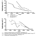
Hospital autopsy rates have been falling in the UK and worldwide for over half a century[4–15] (figure 1A, B) and account for a small minority of all autopsies in the UK.[1,3] Recent studies suggest autopsy rates of less than 10% for teaching hospitals and less than 5% elsewhere.[1,16]

Decline in hospital autopsy rates

Decline in hospital autopsy rates

Figure 1.

Decline in hospital autopsy rates over recent decades. (A) Autopsy rates from three first world countries, data collated from multiple studies. (B) Autopsy rates from four different hospitals/NHS Trusts, data collated from multiple studies.

http://img.medscape.com/article/849/356/849356-thumb1.png

The decline in hospital autopsy rates is well known, yet poorly researched and quantified. The majority of medical professionals and politicians in the UK are likely to be unaware of this conspicuous decline. Consequently, little has been done to address the falling rates and the implications of this are not yet fully understood, nor are the consequences.

A PubMed literature search yielded no research detailing a UK-wide autopsy rate within the past 20 years (search terms “hospital autopsy [title]”, “clinical autopsy [title]”, “autopsy rate [title]”). Given this and documented inter-hospital variation (figure 1B), we aimed to determine the current UK autopsy rate.

The structure of healthcare delivery varies throughout the UK. In England, the provision of acute services (emergency, inpatient and outpatient care) is provided by 186 organisations known as Acute National Health Service (NHS) Trusts—each of which may provide care from multiple hospital sites. In Scotland and Wales, the countries are divided into a number of defined geographical areas (Boards), each of which may contain several sites of healthcare delivery. In Northern Ireland, these geographical areas are known as Health and Social Care Trusts.

Method

Acute NHS Trusts within England (n=160), Boards within Wales (n=7) and Scotland (n=14) and Social Care Trusts within Northern Ireland (n=5) were contacted via ‘Freedom of Information’ requests. The level of response therefore is for the Trust/Board, not individual hospitals. If no reply was received within 4 months, reminders were sent.

The hospital autopsy rate was calculated as the number of autopsies performed on patients who died in the year 2013 as a percentage of total deaths which occurred in the hospital in that calendar year.

Studies indicate significantly higher autopsy rates in stillbirths, neonates and young children.[17,18]Therefore, data were excluded if they fell within the following categories:

  1. Children’s Hospital NHS Trusts
  2. Stillbirth, neonatal, perinatal and paediatric death
  3. Trusts with no recorded deaths
  4. Incomplete responses

Statistical analysis was performed using two-tailed χ2 tests (Prism 6 Software) between each country. The categories used were number of deaths that underwent autopsy and number of deaths not followed by autopsy. Bonferroni correction was used to compensate for the six pairwise comparisons, resulting in 99.25 CIs (p<0.008). Statistical outliers were determined with a ROUT test using a false-positive rate (Q) of 1%.

Results

A 99% (n=184) response rate was achieved for the UK; constituent country response rates were 99% (England), 100% (Scotland), 100% (Wales) and 100% (Northern Ireland). A total of 17 Trusts were removed, according to the exclusion criteria. Eight Trusts were concerned about patient identification because the number of autopsies was small and so provided a ‘fewer than’ figure. In these cases, a maximum possible rate was calculated.

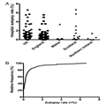
Mean hospital autopsy rates were calculated as the total number of autopsies expressed as a percentage of the total number of deaths. The UK mean autopsy rate was 0.69% and varied considerably between countries. The highest mean autopsy rates were in Scotland (2.1%), followed by Wales (0.65%), England (0.51%) and Northern Ireland (0.46%). The study confirms that hospital autopsy rates are significantly lower than the most recent literature suggests and that there is evident inter-country variation (figure 2A, Table 1) and intra-country variation (figure 2A).

Figure 2.

The results from Freedom of Information request for UK and constituent countries. (A) Individual points representing each sample Trust/Board, non-parametric data, no statistical difference between countries. (B) Cumulative frequency histogram of autopsy rates for NHS Trusts/Boards in the UK.

autopsy rates

http://img.medscape.com/article/849/356/849356-thumb2.png

Inter-country pairwise comparisons using χ2 tests of significance (p<0.008) found Scotland to have a significantly higher hospital autopsy rate than each of the other countries (p<0.0001). Other pairwise comparisons failed to achieve significance (Table 2).

Twenty eight samples were statistical high outliers, 20 from England, 6 from Scotland, 1 from Wales and 1 from Northern Ireland. The mean hospital autopsy rate is skewed by these outliers, which typically were large teaching hospitals or small specialist centres. The top 5% (n=7) of Trusts within England performed 47% of the country’s autopsies and 75% of autopsies in Wales were performed in one health board.

Ninety-eight per cent of samples (n=164) had an autopsy rate of <5%, 86% (n=143) an autopsy rate <1% and 23% (n=38) of all samples did not perform a single autopsy in 2013 (figure 2B). This demonstrates that for a quarter of NHS Trusts/Boards in the UK, hospital autopsy is extinct and in only a fraction (1.8%) of specialist trusts do autopsy rates exceed 5%, the rate previously published for non-teaching hospitals.[1,18]

Hospital autopsy rates in children’s hospital NHS Trusts ranged from 0% to 21%. This higher figure is in agreement with other literature.[17,18]

Discussion

This study has demonstrated that the evident decline in hospital autopsy has continued, if not accelerated, over recent years and already the hospital autopsy is extinct in many NHS Trusts. With 23% of NHS Trusts/Boards having an autopsy rate of 0%, a large part of UK hospital autopsy is now performed in a small number of centres. These few demonstrate that if the provisions and attitudes allow, then hospital autopsy rates of the recent past are still achievable, despite recent legislative changes such as the Human Tissue Act 2004/2006. Trusts with higher autopsy rates tended to be small specialised centres or large teaching hospital Trusts; this influence was not measured in this study due to difficulties in defining a ‘teaching’ or ‘specialised’ Trust/Board. Given that 86% of Trusts/Boards in the UK now have a hospital autopsy rate of <1%, we must pose the question whether a revival in hospital autopsy is possible? In the near future, many of these organisations may join the 23% in which hospital autopsy is extinct, unless they implement those changes in policy and attitude present in the 1.8% of Trusts/Boards where hospital autopsy exceeds 5% of inpatient deaths?

The hospital autopsy rate in Scotland was significantly higher than the other countries (Table 2). The causes of this are uncertain but may include variations in the Human Tissue Act and Authority in Scotland or a lower procurator fiscal (coronial) autopsy rate.

A number of Trusts/Boards gave some explanations as to why their autopsy rate was low, these commonly surrounded provision of facilities. For example, one Trust does not employ an onsite histopathologist or have its own autopsy facilities. However, some Trusts/Boards which themselves do not have onsite hospital autopsy facilities have an agreement with neighbouring Trusts/Boards to carry out their autopsies. From the results, there is evidence of remote island providers that continue to implement autopsy despite no local facilities but which transport cadavers via boat or aeroplane to a separate hospital for autopsy. Thus, a lack of facilities does not preclude hospital autopsy although may add significantly to the cost.

Future research should investigate the differences in Trust/Board policies, clinician attitudes, facilities, funding and local demographics to determine how significantly higher autopsy rates can be achieved.

The strength of this study lies in the nationwide approach to calculating contemporary hospital autopsy rates. Previous studies have focused on single hospitals or Trusts; given the demonstrated wide inter-Trust variation this approach may lead to significant errors. A weakness of this study was that some hospital trusts were unable to separate the data for deaths and autopsies for children and adults. Therefore, mean adult autopsy rates may be slightly over-reported, rates being generally higher among paediatric deaths.

In England and Wales, 94 455 coronial autopsies were performed in 2013[3] yet only 1132 hospital autopsies were performed within the English and Welsh Trusts included in this study. Hospital autopsy now accounts for approximately 1.2% of total autopsies. With such low numbers, questions must be raised regarding the effect such decline has on quality assurance, public health, misdiagnosis (a key contributor to avoidable harm[19,20]), audit and the teaching of both medical students and trainee pathologists. Hospital autopsy presents classic cases used to train junior pathologists, given that many coronial postmortems are not used for training. Training in hospital autopsy will become ever more important given the impending lack of pathologists to cover coronial autopsy. The aim of this paper is to raise awareness of the extent of the decline and to prompt discussion on its consequences. While debate continues over the value of hospital autopsy in medical practice, if action is not taken imminently, the practice may disappear.

COMMENTARY

Prostate Cancer: Is It Time to Retire the Gleason Score?

George D. Lundberg, MD

Hello. I am Dr George Lundberg and this is At Large at Medscape.

If you are an American adult male, you either now have prostate cancer—whether or not you know it—or will likely develop it if you live long enough.

In an average recent year, some 220,000 American men are diagnosed with prostate cancer and some 27,000 die from it. That means, obviously, that it kills 12% of those it is found to afflict and does not kill 88%. Of those patients freshly diagnosed, 98.9% are alive at 5 years.

Does interventional therapy account for the good results of some of that nearly 90% of men who are diagnosed and don’t die? Well, yes, but probably not very many. Really bad prostate cancer tends to do its lethal thing, regardless of interventions.

Back when we collectively had a lot of autopsies, it was possible—even easy—- to follow and learn the natural history of many diseases.

Modern autopsy-less American physicians, including pathologists, are whizz-bang at computers, imaging, lab test panels, genes, microbiomes, electronic medical records, and coding. They are maybe not so great at physical exams, taking a useful personal and family history, or gross and microscopic pathology. This latter list is where physicians once got really good at understanding the natural history of diseases.

Now, epidemiologic facts, outcomes, and common sense have begun to prevail over the national hysteria of prostate-specific antigen (PSA) tests for all men. The hysteria tends to take this form: “Find that prostate cancer and root it out…no matter how small or indolent. That way, ‘Megalopolis U’ can keep those operating rooms, hospital beds, and—get this—unproven (but very elegant) proton beameconomic monstrosities really humming in order to satisfy the overpaid MBAs that determined that such were a good investment idea.”

But some prostate cancers really can kill. How about those? A pathologist named Donald Gleason came up with a numbering system intended to guide therapy based upon anaplasia and prognostic threat of prostate cancer. It was 1, 2, 3, 4, and 5. It makes sense. But then, another number dealing with the relative amount of each level of differentiation and pattern (also 1, 2, 3, 4, 5) got added, and combining the two scores became the Gleason score range of 2-10.

A reasonable human could interpret a 6 on a scale of 10 as middling, pretty bad, or a “better-whack-it-out”-type score. So, a team from Johns Hopkins Medical Institutions[1] has worked out the actual prognosis as falling back into 5 Prognostic Grade Groups (PGGs):

  • Gleason 1, 2, 3, 4, 5, and 6 become Prognostic Grade Group (PGG I);
  • Gleason 3 + 4 = 7 (PGG II);
  • Gleason 4 + 3 = 7 (PGG III);
  • Gleason 4 + 4 = 8 (PGG IV); and
  • Gleason 9-10 (PGG V).

This is so much simpler and less likely to confuse the treating clinician and the patient who is increasingly sharing in this treatment decision.

A European group has just published outcomes based on this PGG system,[2] and it fits nicely. Those many patients with low grades who may not need radical therapy will stand a better chance of notreceiving radical therapy with new low-sounding numbers.

And, by the way, how did “watchful waiting” as a good way to handle those prostate “cancers”—which, from histology, seem like they would behave as indolentomas—morph so quickly into “active surveillance”? My guess is that it is very hard to bill a patient, Medicare, or an insurance company for just letting the patient watch and wait.

Chicago Mayor Rahm—not his physician brother Zeke—Emanuel was right: Never let a crisis go to waste. When the word “cancer” was uttered or written, hair lit on fire and something had to be done, right or wrong.

That is my opinion. I’m Dr George Lundberg.

Medscape Medical News > Conference News

New Clue as to Why Only Some Breast Cancers Relapse

Zosia Chustecka

UPDATED September 26, 2015 // VIENNA — A new clue as to why only some breast cancers recur comes from the largest study of genetic sequencing of breast cancer tissue to date.

While most breast cancer is cured after treatment, about 20% of cases recur. The new study shows that the cases that recur have a different genetic profile, and suggests that some of the genetic drivers of relapse are targetable with drugs.

“We demonstrate that there are clear differences within the driver landscapes of relapsed cancers. This probably reflects a combination of predisposition to relapse and of differences in the mutations acquired during the relapse and metastasis phase,” say the researchers, led by Lucy Yates, MD, a clinical research oncologist from the Wellcome Trust Sanger Institute in Cambridge, United Kingdom.

The finding raises the hope that breast cancer patients who are most at risk for relapse can be identified when they are first diagnosed, they suggest.

In addition, as the newly identified genetic drivers of relapse are targetable with drugs, there is also hope that eventually women who are identified as being at high risk for relapse could be treated with such drugs to prevent recurrence, they suggest.

The study is due to be presented European Cancer Congress (ECC) 2015, but details were released early by the ECC press office.

The finding comes from a study that compared the genetic make-up of breast cancer from 836 tissue samples taken from women on primary diagnosis with 161 samples of tissue taken from recurrences or metastases.

The study is the largest and most comprehensive carried out to date, say the researchers, both in terms of the number of samples from relapsed breast cancers and in terms of the wide-ranging genetic sequencing carried out, which looked at 365 genes involved in cancer-related pathways.

The researchers performed de novo driver mutation discovery, and individual mutations were annotated with likely driver status based upon recurrence and known driver status in previously published, well-curated datasets and databases. The incidence of each driver mutation in the primary and relapse datasets was compared using Fisher’s exact test and using the Benjamini–Hochberg correction for multiple testing.

The team found 11 genes that were significantly enriched in the relapsed cohort compared with the primary tumor cohort. The most heavily enriched were TP53 and ARID1B. Multiple samples were available for 66 patients, including local or distant relapse samples in all cases and a sample from the primary tumor in 21 cases. This multisample analysis allowed the team to trace the evolution of mutations.

“We have found that some of the genetic mutations that drive breast cancers that relapse are relatively uncommon amongst cancers that do not relapse at the point of primary diagnosis,” Dr Yates said in a statement.

“We believe that the differences we have seen reflect genetic differences that can predispose a cancer to return, combined with mutations acquired throughout the period from first diagnosis to the subsequent relapse,” she added.

However, in a discussion of this paper, Fabrice André, MD, PhD, from the Gustave Roussy Institute, in Villejuif, France, questioned whether all the genes that were found to be enriched in the relapsed samples were driving the relapse and whether any could be identified as recurrent markers. He noted that although Dr Yates and colleagues found 11 genes that were highly enriched in the relapsed tissue samples, another study (which analyzed 183 samples) found only one of these genes to be highly enriched.

Dr Andre also wondered whether the late mutations that were identified are clinically relevant ― could they explain the development of resistance to therapy? This has been seen in other cancers, he noted.

Extreme Heterogeneity ― Need for Multiple Biopsies

“Our data reveal extreme heterogeneity and indicate that genomic analysis of primary, relapsed, and matched normal tissue are needed,” Dr Yates concluded.

“We need to do biopsies again and again and again,” said Anne-Lise Borresen-Dale, MD, from the Institute for Cancer Research, Oslo University Hospital, Norway, who chaired the session. But Dr Andre wondered whether circulating tumor cells, the so-called “liquid biopsy,” could be used.

Multiple samples were available for 66 subjects, including local or distant relapse samples in all cases and a sample from the primary tumor in 21 cases. This multisample analysis permitted relative temporal ordering of driver mutation accumulation to be determined, the researchers explain.

“We have found that some of the genetic mutations that drive breast cancers that relapse are relatively uncommon amongst cancers that do not relapse at the point of primary diagnosis,” Dr Yates said in a statement.

“This study highlights the differences between genetic alterations that drive relapsed and metastatic disease as opposed to primary breast cancers, and underlines the importance of analyzing the genetic features of metastases when making treatment decisions,” said Jorge Reis-Filho, MD, from the Memorial Sloan Kettering Cancer Center in New York City, who was acting as a spokesperson for the European Society of Medical Oncology, which is cohosting the meeting. He was not involved with this work.

However, Dr Reis-Filho also cautioned that “the extent of the differences in the repertoire of mutations among different metastatic sites within individual patients remains to be determined, however, as does the best way to obtain tumor-derived genetic material in patients with metastatic disease. We also need to know more about whether single or multiple metastatic sites should be analyzed in this context.”

Also commenting on the study, Peter Naredi, MD, PhD, professor of surgery at Sahlgrenska University Hospital in Gothenburg, Sweden, who is the European CanCer Organization scientific cochair of the congress, said: “Information such as that which Dr Yates will present is very important in the era of precision medicine.”

“This study also underlines the fact that we should consider a recurrence of a cancer as a new event, and carefully select the right treatment for the recurrent tumor as opposed to just relying on information from the first occurrence,” Dr Naredi said in a statement.

JAK Inhibitors in Breast Cancer

Some of the genetic changes that were found in the relapsed/metastatic breast cancer samples appeared at a late stage when the cancer recurred, and were not seen in samples taken at primary diagnosis. Among these later-stage mutations, the researchers say they found “compelling evidence” for the tumor suppression activity of two related genes, called JAK2 and STAT3, that operate within the same signaling pathway.

“Within some breast cancers, a disruption in this signaling pathway seems to be advantageous for survival of the cancer,” Dr Yates said in a statement.

“Interestingly, this is in contrast to the role of JAK2 in some other cancers, where overactivity of the gene drives malignancy rather than suppresses it,” she added.

The JAK (Janus-associated kinase) enzymes JAK1 and JAK2 are involved in regulating blood and immunologic functioning, and a dysregulation of these enzymes is thought to be a driver in the development of myelofibrosis. The first JAK inhibitor, ruxolitinib (Jakafi, Incyte Corp), was approved for the treatment of myelofibrosis in 2011.

Dr Yates and colleagues note that enhanced JAK-STAT signaling is known to play an important role in breast cancer stem cell development and cancerous cell line survival, and preclinical evidence seems to suggest that inhibiting the gene would be therapeutically advantageous. These findings have led to the development of clinical trials for breast cancer using JAK inhibitors in the hope that they will slow cancer progression.

“However, our findings suggest that, in a subset of cancers, inhibiting this pathway may have the opposite effect, and this requires further investigation. In general, the observation highlights the importance of understanding the diverse nature of breast cancers in the era of precision medicine,” Dr Yates said.

The work was funded by the Wellcome Trust. Dr Yates has disclosed no relevant financial relationships.

European Cancer Congress (ECC) 2015: Abstract 1804.

Read Full Post »

Why Does Cytotoxic Chemotherapy Still Remain a Mainstay in Many Chemotherapeutic Regimens? [6.1.1]


Reporter: Stephen J. Williams, Ph.D.

At the 2015 AACR National Meeting, Drs. Anthony Letai, Dr. Michael Hermann, Dr. Rene Bernards, and Dr. Guido Kroemer gave The 2015 Stanley J. Korsmeyer Memorial Symposium: Cell Death and Cancer Therapy: Why Has Conventional Chemotherapy Been So Successful?

Cytotoxic chemotherapy, for which the mechanism of action is centered on the ability of the drug to kill a cell by either necrosis, genotoxic, apoptosis, or autophagy mechanisms rather than just halting cell growth, is still, in this era of personalized and cytostatic therapies, is still a mainstay in many treatment regimens for a majority of cancers. Treatment regimens such as MOPP (mechlorethamine, Oncovin, procarbazine, prednisone), CMF (cyclophosphamide, methotrexate, 5-fluorouracil) , carboplatin with taxol, and even with personalized therapies, which usually are given in combination with a cytotoxic agent. However treatment regimens containing these cytotoxic chemotherapeutics show some of the best survival rates. The abstract for the Symposium is given below:

In this current era of precisely targeted therapies and –omics technologies, it is often forgotten that no medical therapy has cured, and continues to cure, more people of cancer than conventional chemotherapy. Notwithstanding its superior performance across many cancer types, the mechanism of the therapeutic index of conventional agents, largely targeting ubiquitous elements like DNA and microtubules, is poorly understood. The textbook explanation of conventional chemotherapy’s working by killing supposedly rapidly dividing cancer cells lacks clinical evidence and flies in the face of many obvious clinical counter-examples. In the session,m the speakers will describe how conventional cytotoxic chemotherapy preferentially kills cancer cells. Moreover, they will describe how clinical response to chemotherapy might be better predicted.

This post is presented as the speakers titles and a brief curation of their papers related to the subject matter.

Anthony G. Letai, Dana-Farber Cancer Institute, Boston, MA. Conventional chemotherapy cures people by exploiting apoptotic priming.

Conventional chemotherapy has an amazing track record that is often under-appreciated in today’s world of genomics and targeted pathway inhibitors. Conventional chemotherapy is responsible for curing millions of cancer patients over the past 5 decades. That is, millions of patients have presented to their doctors with an otherwise fatal malignancy, were given a finite course of chemotherapy (largely DNA and microtubule perturbing agents) and had their cancer eradicated, never to return. Perhaps as remarkable as the magnitude of the achievement of conventional chemotherapy is the magnitude of our ignorance of why it should ever work, and why it works far better in some tumors than in others. Textbook explanations rely on concepts of differential proliferation rates in cancers that are incompletely supported in the clinical literature. Successful chemotherapy treatments usually kill via the mitochondrial pathway of apoptosis. We have found that simple functional measurements of the pre-treatment state of the tumor cell can be rapidly made with BH3 profiling. These measurements demonstrate that a major, if not the major, reason for a therapeutic index for cancer chemotherapy is that chemo-sensitive cancer cells are simply more primed for apoptosis than normal cells. Moreover, apoptotic priming can be measured to make clinical predictions regarding quality of response on an individualized basis. Enhancing pretreatment priming of cancer cells with selectively acting targeted agents is a promising strategy to extend the demonstrated curative power of conventional chemotherapy.

Maturation Stage of T-cell Acute Lymphoblastic Leukemia Determines BCL-2 versus BCL-XL Dependence and Sensitivity to ABT-199

Triona Ni Chonghaile, Justine E. Roderick, Cian Glenfield, Jeremy Ryan, Stephen E. Sallan, Lewis B. Silverman, Mignon L. Loh, Stephen P. Hunger, Brent Wood, Daniel J. DeAngelo, Richard Stone, Marian Harris, Alejandro Gutierrez, Michelle A. Kelliher, Anthony Letai

Cancer Discov. Author manuscript; available in PMC 2015 March 1.

Published in final edited form as: Cancer Discov. 2014 September; 4(9): 1074–1087. Published online 2014 July 3. doi: 10.1158/2159-8290.CD-14-0353


High Mitochondrial Priming Sensitizes hESCs to DNA-Damage-Induced Apoptosis

Julia C. Liu, Xiao Guan, Jeremy A. Ryan, Ana G. Rivera, Caroline Mock, Vishesh Agrawal, Anthony Letai, Paul H. Lerou, Galit Lahav

Cell Stem Cell. Author manuscript; available in PMC 2014 October 3.

Published in final edited form as: Cell Stem Cell. 2013 October 3; 13(4): 483–491. Published online 2013 August 15. doi: 10.1016/j.stem.2013.07.018

Correction in: volume 13 on page 634


Prolonged mitotic arrest triggers partial activation of apoptosis, resulting in DNA damage and p53 induction

James D. Orth, Alexander Loewer, Galit Lahav, Timothy J. Mitchison

Mol Biol Cell. 2012 February 15; 23(4): 567–576. doi: 10.1091/mbc.E11-09-0781


Stem cells: Balancing resistance and sensitivity to DNA damage

Julia C. Liu, Paul H. Lerou, Galit Lahav

Trends Cell Biol. Author manuscript; available in PMC 2015 May 1.

Published in final edited form as: Trends Cell Biol. 2014 May; 24(5): 268–274. Published online 2014 April 7. doi: 10.1016/j.tcb.2014.03.002


Michael T. Hermann, MIT Koch Institute for Integrated Cancer Research, Cambridge MA. Using convential chemotherapy as targeted agents.

Exploiting the Synergy between Carboplatin and ABT-737 in the Treatment of Ovarian Carcinomas

Harsh Vardhan Jain, Alan Richardson, Michael Meyer-Hermann, Helen M. Byrne

PLoS One. 2014; 9(1): e81582. Published online 2014 January 6.


Rene Bernards, Netherlands Cancer Institute, Amsterdam, The Netherlands. Identifying responders to chemotherapies through functional genomics

MED12 Controls the Response to Multiple Cancer Drugs through Regulation of TGF-β Receptor Signaling

Sidong Huang, Michael Hölzel, Theo Knijnenburg, Andreas Schlicker, Paul Roepman, Ultan McDermott, Mathew Garnett, Wipawadee Grernrum, Chong Sun, Anirudh Prahallad, Floris H. Groenendijk, Lorenza Mittempergher, Wouter Nijkamp, Jacques Neefjes, Ramon Salazar, Peter ten Dijke, Hidetaka Uramoto, Fumihiro Tanaka, Roderick L. Beijersbergen, Lodewyk F.A. Wessels, René Bernards

Cell. Author manuscript; available in PMC 2013 June 5.

Published in final edited form as: Cell. 2012 November 21; 151(5): 937–950.


Sorafenib synergizes with metformin in NSCLC through AMPK pathway activation

Floris H Groenendijk, Wouter W Mellema, Eline van der Burg, Eva Schut, Michael Hauptmann, Hugo M Horlings, Stefan M Willems, Michel M van den Heuvel, Jos Jonkers, Egbert F Smit, René Bernards

Int J Cancer. 2015 March 15; 136(6): 1434–1444. Published online 2014 August 1.


The Corepressor CTBP2 Is a Coactivator of Retinoic Acid Receptor/Retinoid X Receptor in Retinoic Acid Signaling

Prashanth Kumar Bajpe, Guus J. J. E. Heynen, Lorenza Mittempergher, Wipawadee Grernrum, Iris A. de Rink, Wouter Nijkamp, Roderick L. Beijersbergen, Rene Bernards, Sidong Huang

Mol Cell Biol. 2013 August; 33(16): 3343–3353. doi: 10.1128/MCB.01213-12


Using Functional Genetics to Understand Breast Cancer Biology

Alan Ashworth, Rene Bernards

Cold Spring Harb Perspect Biol. 2010 July; 2(7): a003327. doi: 10.1101/cshperspect.a003327


SMARCE1 suppresses EGFR expression and controls responses to MET and ALK inhibitors in lung cancer

Andreas I Papadakis, Chong Sun, Theo A Knijnenburg, Yibo Xue, Wipawadee Grernrum, Michael Hölzel, Wouter Nijkamp, Lodewyk FA Wessels, Roderick L Beijersbergen, Rene Bernards, Sidong Huang

Cell Res. 2015 April; 25(4): 445–458. Published online 2015 February 6.


The Good, the Bad, and the Ugly: in search of gold standards for assessing functional genetic screen quality

Bastiaan Evers, Rene Bernards, Roderick L Beijersbergen

Mol Syst Biol. 2014 July; 10(7): 738. Published online 2014 July 1.


An Integrative Genomic and Proteomic Analysis of PIK3CA, PTEN, and AKT Mutations in Breast Cancer

Katherine Stemke-Hale, Ana Maria Gonzalez-Angulo, Ana Lluch, Richard M. Neve, Wen-Lin Kuo, Michael Davies, Mark Carey, Zhi Hu, Yinghui Guan, Aysegul Sahin, W. Fraser Symmans, Lajos Pusztai, Laura K. Nolden, Hugo Horlings, Katrien Berns, Mien-Chie Hung, Marc J. van de Vijver, Vicente Valero, Joe W. Gray, René Bernards, Gordon B. Mills, Bryan T. Hennessy

Cancer Res. Author manuscript; available in PMC 2009 August 1.

Published in final edited form as: Cancer Res. 2008 August 1; 68(15): 6084–6091.


CTF Meeting 2012: Translation of the Basic Understanding of the Biology and Genetics of NF1, NF2, and Schwannomatosis Toward the Development of Effective Therapies

Brigitte C. Widemann, Maria T. Acosta, Sylvia Ammoun, Allan J. Belzberg, Andre Bernards, Jaishri Blakeley, Antony Bretscher, Karen Cichowski, D. Wade Clapp, Eva Dombi, Gareth D. Evans, Rosalie Ferner, Cristina Fernandez-Valle, Michael J. Fisher, Marco Giovannini, David H. Gutmann, C. Oliver Hanemann, Robert Hennigan, Susan Huson, David Ingram, Joe Kissil, Bruce R. Korf, Eric Legius, Roger J. Packer, Andrea I McClatchey, Frank McCormick, Kathryn North, Minja Pehrsson, Scott R. Plotkin, Vijaya Ramesh, Nancy Ratner, Susann Schirmer, Larry Sherman, Elizabeth Schorry, David Stevenson, Douglas R. Stewart, Nicole Ullrich, Annette C. Bakker, Helen Morrison

Am J Med Genet A. Author manuscript; available in PMC 2014 September 1.

Published in final edited form as: Am J Med Genet A. 2014 March; 0(3): 563–578. Published


Analysis of the MammaPrint Breast Cancer Assay in a Predominantly Postmenopausal Cohort

Ben S. Wittner, Dennis C. Sgroi, Paula D. Ryan, Tako J. Bruinsma, Annuska M. Glas, Anitha Male, Sonika Dahiya, Karleen Habin, Rene Bernards, Daniel A. Haber, Laura J. Van’t Veer, Sridhar Ramaswamy Clin Cancer Res. Author manuscript; available in PMC 2011 May 7.


Guido Kroemer, INSERM U848- Institute Gustave-Roussy, Villejuif, France. A hallmark of successful cancer therapies: Reinstatement of immunosurvelliance.

Immune infiltrate in cancer Gautier Stoll, Laurence Zitvogel, Guido Kroemer

Aging (Albany NY) 2015 June; 7(6): 358–359. Published online 2015 June 25.


Corrigendum: “Combinatorial Strategies for the Induction of Immunogenic Cell Death”

Lucillia Bezu, Ligia C. Gomes-da-Silva, Heleen Dewitte, Karine Breckpot, Jitka Fucikova, Radek Spisek, Lorenzo Galluzzi, Oliver Kepp, Guido Kroemer

Front Immunol. 2015; 6: 275. Published online 2015 June 1. doi: 10.3389/fimmu.2015.00275

Corrects: Front Immunol. 2015; 6: 187.


Meta-analysis of organ-specific differences in the structure of the immune infiltrate in major malignancies

Gautier Stoll, Gabriela Bindea, Bernhard Mlecnik, Jérôme Galon, Laurence Zitvogel, Guido Kroemer

Oncotarget. 2015 May 20; 6(14): 11894–11909. Published online 2015 May 19


Other posts on this site on Cytotoxicity and Cancer include

Novel Approaches to Cancer Therapy [11.1]

Misfolded Proteins – from Little Villains to Little Helpers… Against Cancer

Multiple Lung Cancer Genomic Projects Suggest New Targets, Research Directions for Non-Small Cell Lung Cancer

A Synthesis of the Beauty and Complexity of How We View Cancer

Good and Bad News Reported for Ovarian Cancer Therapy


Read Full Post »

Tumor Associated Macrophages: The Double-Edged Sword Resolved?

Writer/Curator: Stephen J. Williams, Ph.D.

UPDATED 10/04/2021 TAMs Enhance Tumor Hypoxia and Aerobic Glycolysis

Cell-based immunity is vital for our defense against pathologic insult but recent evidence has shown the role of cell-based immunity, especially macrophages to play an important role in both the development and hindrance of tumor growth, including role in ovarian, hematologic cancers, melanoma, and breast cancer.  In the past half century, new immunological concepts of cancer initiation and progression have emerged, including the importance of the harnessing the immune system as a potential anti-cancer strategy. However, as our knowledge of the immune system and tumor biology has grown, the field has realized an immunological conundrum: how can an immune system act to both prevent tumor growth and promote the tumor’s growth?

As discussed in the lower section of this post, authors of a paper in the journal Science show how different populations of tumor-associated macrophages (TAMs) may exert both positive and negative effects on tumor cells, producing a sort of ying-yang war between the tumor and the immune system.

The Immune System: Brief Overview and Role in Cancer

celllineageimmunesystem

Figure. Cell lineage of the immune system. A description of the different cell types can be found here.

Histologic evaluation of multiple tumor types, especially solid tumors, reveal the infiltration of diverse immunological cell types, including myeloid and lymphoid cell lineages, such as macrophages and NK, T cell and B cells respectively.

The immunological conundrum

immuncecancerconundrum

Figure. Potential inflammatory signaling pathways in breast cancer stem cells.
Breast cancer stem cells may be regulated by chemokine- and/or cytokine-mediated inflammatory signaling in an autocrine or paracrine manner. (from University of Tokyo at http://www.ims.u-tokyo.ac.jp/system-seimei/en/research2_e.htm)

Role of Tumor Associated Macrophages

There are conflicting reports as to the functional consequence of these infiltrating tumor-associated macrophages (TAMs). TAMs have been shown to secrete mediators such as interleukins and cytokines in a paracrine manner such as CCL2, IL10 and TGFβ. In certain instances these cytokines and mediators actually promote the growth of the surrounding tumors.

J Leukoc Biol 2009 Nov 86(5) 1065-73, Figure 1

Figure.  TAMs can be divided into subpopulations with distinctive functions and secretogogues.

For Further Reference

Tumor-associated macrophages and the profile of inflammatory cytokines in oral squamous cell carcinoma. http://www.ncbi.nlm.nih.gov/pubmed/23089461 anti-inflamm IL10 and TGFB

Tumor-associated macrophage-derived IL-6 and IL-8 enhance invasive activity of LoVo cells induced by PRL-3 in a KCNN4 channel-dependent manner

TAMscytokines

Figure 2: TAM functions in tumor progression. Tumor cells and stromal cells, which produce a series of chemokines and growth factors, induce monocytes to differentiate into macrophages. In the tumor, most macrophages are M2-like, and they express some cytokines, chemokines, and proteases, which promote tumor angiogenesis, metastasis, and immunosuppression. From Macrophages in Tumor Microenvironments and the Progression of Tumors

ICB-14-NC-BRONTE-V2

Macrophages integrate metabolic and environmental signals to promote tumor growth. Area within dotted rectangle indicates proposed mechanisms of action. ARG, arginase; HIF, hypoxia-inducible factor; MCT, monocarboxylate transporter; NADH, nicotine adenine dinucleotide, reduced; PKM2, M2 isoform of pyruvate kinase; VEGF, vascular endothelial growth factor from Tumor cells hijack macrophages via lactic acid adapted from Colegio OR, Chu N-Q, Szabo AL, Chu T, Rhebergen AM, Jairam V et al. Functional polarization of tumour-associated macrophages by tumour-derived lactic acid. Nature (e-pub ahead of print 13 July 2014; doi:10.1038/nature13490). | Article |

Depletion of M2-Like Tumor-Associated Macrophages Delays Cutaneous T-Cell Lymphoma Development In Vivo

Targeting tumor-associated macrophages in an orthotopic murine model of diffuse malignant mesothelioma

Crosstalk between colon cancer cells and macrophages via inflammatory mediators and CD47 promotes tumour cell migration

Tumor-Associated Macrophages Regulate Murine Breast Cancer Stem Cells Through a Novel Paracrine EGFR/Stat3/Sox-2 Signaling Pathway

Science Paper: Different Populations of TAMS Have Different Tumor Effects

The cellular and molecular origin of tumor-associated macrophages Eric G. Pamer1 Ruth A. Franklin1,2, Will Liao3, Abira Sarkar1, Myoungjoo V. Kim1,2, Michael R. Bivona1, Kang Liu4, Ming O. Li1, Science 23 May 2014: Vol. 344 no. 6186 pp. 921-925

A recent Science paper from Cornel has investigated the origin, function, and characterization of TAMs on breast cancer growth. In summary, their efforts and research suggest different populations of TAMs with varied tumorigenic effects, a finding which may help explain the immunologic conundrum with respect to solid tumors.

The authors characterized the infiltrating immune cell types in a MMTV-PyMt model of breast cancer.

The MMTV-PyMt mouse breast cancer model:

is a transgenic model where mammary gland expression of the polyoma middle T antigen (PyMT) is driven by the Mouse Mammary Tumor Virus promoter (MMTV).

Microbiol. Mol. Biol. Rev. 2009 Sep 73(3) 542-63, FIG. 5

For a review of mouse models of breast cancer please see

Mouse models of breast cancer metastasis. Anna Fantozzi1 and Gerhard Christofori. Breast Cancer Res. 2006; 8(4): 212.

Results

1.     Macrophages constitute the predominate myeloid cell population in MMTV-PyMT mammary tumors

Tumor infiltrating immune cells included

  • Myeloid cells comprised 50% of CD45+ infiltrating leukocytes.
  • The CD45 antigen, also known as Protein tyrosine phosphatase, receptor type, C (PTPRC) is an enzyme that, in humans, is encoded by the PTPRC gene, and acts as a regulator of B and T-lymphocytes.
  • Authors noted three types of cells classified as Type I, II, and III based on
  1. Cell morphology
  2. Major histocompatibility complex
  • Infiltrating monocytes and neutrophils
  • Cells with dendritic and macrophage markers

2. TAMS differentiate from CCR2+ inflammatory monocytes

  • To determine whether Ly6C+CCR2+ inflammatory monocytes contributed to TAMs and MTMs, authors crossed PyMT mice to Ccr2−/− mice and found MTMs (mammary tumor macrophages) were significantly reduced in Ccr2−/− PyMT mice, implying that MTMs are constitutively repopulated by inflammatory monocytes
  • To determine whether inflammatory monocytes were required for TAM maintenance, we generated CCR2DTR PyMT mice expressing diphtheria toxin receptor (DTR) under control of the Ccr2 locus DT treatment resulted in 96% depletion of tumor-associated monocytes compared to 80% depletion in Ccr2−/− mice
  • To investigate whether monocytes could differentiate into TAMs in vivo, we transferred CCR2+ bone marrow cells isolated from CCR2GFP reporter mice into congenically marked CCR2DTR PyMT mice depleted of endogenous monocytes, we observed transferred cells in developing tumors demonstrate that tumor growth induces the differentiation of CCR2+ monocytes into TAMs.

3.     TAMs are phenotypically distinct from AAMs (M2 or alternatively activated macrophages)

  • Gene-expression profiling revealed the integrin CD11b (Itgam) was expressed at lower levels in TAMs than in MTMs while several other integrins and the integrin receptor Vcam1 were up-regulated in TAMs
  • AM population did not express AAM markers such as Ym1, Fizz1, and Mrc1; instead, MTMs more closely resembled AAMs. The authors detected Vcam1 up-regulation on TAMs as a late differentiation event

4.    RBPJ-dependent TAMs modulate the adaptive immune response

  • In DCs, canonical Notch signaling mediated by the key transcriptional regulator RBPJ controls lineage commitment and terminal differentiation. To explore whether Notch signaling played a role in TAM differentiation, authors used CD11ccre mice that efficiently deleted floxed DNA sequences to a greater extent in TAMs than MTMs, but not in monocytes or neutrophils (fig. S14). CD11ccreRbpjfl/fl PyMT mice exhibited a selective loss of MHCIIhiCD11blo TAMs ( 4A). However, a MHCIIhiCD11bhi population still remained
  • Transcriptional profiling comparing this population to WT TAMs confirmed a loss of the Notch-dependent program in RBPJ-deficient cells revealing that in the absence of RBPJ, inflammatory monocytes are unable to terminally differentiate into TAMs.

UPDATED 10/04/2021 TAMs Enhance Tumor Hypoxia and Aerobic Glycolysis

Tumor-Associated Macrophages Enhance Tumor Hypoxia and Aerobic Glycolysis

From:

Tumor-Associated Macrophages Enhance Tumor Hypoxia and Aerobic Glycolysis
Hoibin Jeong, Sehui Kim, Beom-Ju Hong, Chan-Ju Lee, Young-Eun Kim, Seoyeon Bok, Jung-Min Oh, Seung-Hee Gwak, Min Young Yoo, Min Sun Lee, Seock-Jin Chung, Joan Defrêne, Philippe Tessier, Martin Pelletier, Hyeongrin Jeon, Tae-Young Roh, Bumju Kim, Ki Hean Kim, Ji Hyeon Ju, Sungjee Kim, Yoon-Jin Lee, Dong-Wan Kim, Il Han Kim, Hak Jae Kim, Jong-Wan Park, Yun-Sang Lee, Jae Sung Lee, Gi Jeong Cheon, Irving L. Weissman, Doo Hyun Chung, Yoon Kyung Jeon and G-One Ahn

Abstract

Tumor hypoxia and aerobic glycolysis are well-known resistance factors for anticancer therapies. Here, we demonstrate that tumor-associated macrophages (TAM) enhance tumor hypoxia and aerobic glycolysis in mice subcutaneous tumors and in patients with non–small cell lung cancer (NSCLC). We found a strong correlation between CD68 TAM immunostaining and PET 18fluoro-deoxyglucose (FDG) uptake in 98 matched tumors of patients with NSCLC. We also observed a significant correlation between CD68 and glycolytic gene signatures in 513 patients with NSCLC from The Cancer Genome Atlas database. TAM secreted TNFα to promote tumor cell glycolysis, whereas increased AMP-activated protein kinase and peroxisome proliferator-activated receptor gamma coactivator 1-alpha in TAM facilitated tumor hypoxia. Depletion of TAM by clodronate was sufficient to abrogate aerobic glycolysis and tumor hypoxia, thereby improving tumor response to anticancer therapies. TAM depletion led to a significant increase in programmed death-ligand 1 (PD-L1) expression in aerobic cancer cells as well as T-cell infiltration in tumors, resulting in antitumor efficacy by PD-L1 antibodies, which were otherwise completely ineffective. These data suggest that TAM can significantly alter tumor metabolism, further complicating tumor response to anticancer therapies, including immunotherapy.

Significance: These findings show that tumor-associated macrophages can significantly modulate tumor metabolism, hindering the efficacy of anticancer therapies, including anti-PD-L1 immunotherapy.

Introduction

Tumor hypoxia and glycolysis have long been recognized as major resistance factors contributing to failures of chemo- and radiotherapy (1, 2). Traditionally, tumor hypoxia is known to occur by two mechanisms: chronic or acute hypoxia (2). Chronic hypoxia occurs as a result of rapid proliferation of cancer cells and hence being constantly forced away from blood vessels beyond the oxygen diffusion distance of approximately 150 μm (2). Acute hypoxia on the other hand occurs by a temporary cessation of the blood flow due to highly disorganized tumor vasculature (2). Regardless of the mechanism, tumor hypoxia has been extensively documented for their contribution to resistance to all anticancer therapies including chemotherapy (2), surgery (3), radiotherapy (2), and recently immunotherapy (4).

Aerobic glycolysis, also known as Warburg effect, is a phenomenon whereby many types of tumors exhibit a preference of glucose over the oxygen for their energy substrate (5), and this has allowed us to track solid tumors in patients with PET using 18fluoro-deoxyglucose (FDG) radioactive tracer (6). Although several mechanisms for Warburg effect have been suggested including mitochondrial defects, adaptation to hypoxia [hence activation of hypoxia-inducible factor (HIF)], and oncogenic signals such as MYC and RAS (7), the exact mechanism is still controversial. Tumor glycolysis has also been reported to influence the therapy outcome (8). Preclinical studies have suggested that glycolysis can increase DNA repair enzyme expressions including Rad51 and Ku70, which can facilitate radiation-induced DNA double-strand break repair (9). Lactate, a major byproduct of glycolysis, has recently been shown to be utilized as a fuel source for oxidative phosphorylation in nearby cancer cells (10), which can promote the tumor recurrence following anticancer therapies.

Tumor-associated macrophages (TAM) are bone marrow–derived immune cells recruited to tumors and have been extensively reported for their protumoral role (11). Recruited to tumors by various tumor-secreting factors including stromal cell–derived factor-1 (SDF1; ref. 12), VEGF (13), semaphorin 3A (14), and colony-stimulating growth factor-1 (CSF1; ref. 15), TAMs have been shown to produce various growth factors and proteases necessary for tumor survival (11) or immunosuppressive cytokines inhibiting antitumor immune responses (16). Macrophages in general are known to be polarized to either classically activated M1 macrophages or alternatively activated M2 phenotype depending on the cytokine milieu in which they are exposed (17). Bacterial-derived products such as lipopolysaccharide have been shown to polarize macrophages toward M1 phenotype (17), while parasite-associated signals such as IL4 and IL13 can lead to M2-polarized macrophages with increased tissue repair abilities (17). It has been suggested that TAMs are M2-like, although various subpopulations of TAM have been also identified including TIE2-positive macrophages (18), programmed cell death protein-1 (PD-1)–expressing TAM (19), and C-C chemokine receptor type-2 (CCR2)–expressing TAM (20).

In this study, we demonstrate clinically and preclinically that TAMs are a novel contributor to tumor hypoxia and aerobic glycolysis by competing oxygen and glucose with cancer cells. We further observed that TAM can significantly interfere with T-cell infiltration thereby masking programmed death-ligand 1 (PD-L1) expression in the tumors. We believe that our results have an important clinical implication such that patients with high infiltration of TAM in their tumors may poorly respond to all anticancer therapies, including the latest immunotherapy.

Figure 1.

Strong correlations between TAM infiltration and glycolysis in patients with NSCLC. A, Representative PET/CT images for FDG uptake (top) and immunostaining of CD68 (bottom) from paired tumors of patients with NSCLC. Top, yellow circles, location of tumors. Bottom, red arrowheads, CD68-positive TAM. Scale bar, 100 μm. B, Correlation between glycolysis and CD68-positive TAM in 98 patients with NSCLC paired results as in A. Glycolysis was analyzed as FDG maximal standardized uptake value (FDG SUVmax; left) or 40% total lesion glycolysis (TLG; right). C, FDG SUVmax values for CD68low (n = 49) or CD68Hi (n = 49) NSCLC tumors. D, Subgroup analyses of FDG uptake in adenocarcinomas (n = 48; left) or squamous cell carcinomas (n = 50; right) of NSCLC. *, P < 0.05; ***, P < 0.001 in C and D as determined by the Student t test. Data are the mean ± SEM. E, TCGA analysis between CD68 and SLC2A1 (left) or HK2 (right) in 513 patients with adenocarcinoma NSCLC. P values are indicated in each plot.

TAMs make tumors more glycolytic

Figure 2.

TAMs make tumors more glycolytic. A, Left, PET/MRI images for FDG uptake in LLC tumors in mice before (D0, top) and after (D2, bottom) Veh or Clod treatment. Yellow circles, tumors. Right, T2-weighted MR images of LLC tumors treated with Veh or Clod pre (top)- or post (bottom)- contrast. Red arrowheads in Veh tumor, ferumoxytol-labeled TAM. B, FDG uptake SUVmax in A. **, P < 0.01, determined by two-way ANOVA. C, FACS plot indicating HoechstbrightKeratin+ (red boxes) population of cells sorted as aerobic cancer cells. D, Fold changes in gene expression in FACS-sorted aerobic cancer cells from LLC tumors treated with Clod or Veh. E, Glucose uptake (left) and lactate production (right) from the sorted aerobic cancer cells as in C. Data in D and E are the mean ± SEM from at least triplicate samples. *, P < 0.05; ***, P < 0.001 by the Student t test. F, Western blot of FACS-sorted aerobic cancer cells in C for GLUT1. β-Actin was used as the loading control. G, Oxygen consumption kinetics in FACS-sorted aerobic cancer cells as described in C. H, LLC tumor growth in mice treated with Veh, Clod, Veh + metformin (Veh + Met), or Clod + metformin (Clod + Met). *, P < 0.05; **, P < 0.01; ***, P < 0.001, determined by two-way ANOVA. I, LLC tumor growth in mice treated with Veh, Clod, Veh + 2-DG, or Clod + 2-DG. Data in H and I are the mean ± SEM, with number of animals indicated in the graphs.

TAMs secrete TNFα to promote tumor glycolysis

Figure 3.

Macrophages secrete TNFα to facilitate glycolysis in cancer cells. A, Gene expression changes in LLC cocultured with (LLC+BMDM) or without (LLC) BMDM. Data are the mean ± SEM from at least triplicate determinations. B, Glucose uptake (left) and lactate production (right) in LLC cocultured with or without BMDM. Data are the mean ± SEM for triplicate samples per group. **, P < 0.01 by Student t test. C, Glucose uptake in LLC cultured alone, cocultured with BMDM, or cocultured with BMDM with glucose added back to the LLC compartment of the coculture system. Data are the mean ± SEM for n = 4 replicates per group. *, P < 0.05; **, P < 0.01 by one-way ANOVA. D, Antibody cytokine arrays in the supernatant obtained from BMDM culture with (BMDM+LLC) or without (BMDM) LLC. Red boxes indicate those cytokines whose expressions were increased in BMDM cocultured with LLC compared with BMDM alone. Blue box, CXCL1, a cytokine produced by LLC cancer cells themselves (Supplementary Fig. S2D). E, Luminex cytokine assays for TNFα in the supernatant from culture media, LLC alone, BMDM alone, or BMDM cocultured with LLC. Data are the mean ± SEM for n = 3 replicates per group. ***, P < 0.001 by one-way ANOVA. F, Glucose uptake in LLC alone (none), LLC cocultured with BMDM (+BMDM), or LLC treated with TNFα (+TNFα) or with IFNγ (+IFNγ). Data are the mean ± SEM from n = 3 samples per group. **, P < 0.01; ***, P < 0.001 by one-way ANOVA. G, Western blot for LLC cells treated with increasing concentrations of recombinant TNFα protein for GLUT1, HK2, or PGC-1α. β-Actin was used as the loading control. H, TNFα concentrations in the supernatant from LLC cultured with (+BMDM) or without (alone) BMDM, or in BMDM cultured with (+LLC) or without (alone) LLC, measured by ELISA. BD, below the detection limit. **, P < 0.01 by Student t test. I, Immunostaining of TNFα (red) and CD68 (green) in LLC tumors grown in mice. Nuclei are shown in blue with DAPI counterstaining. The inset shows magnified regions where indicated with the asterisk (*). White arrowheads, CD68-positive TAM-expressing TNFα. Scale bar, 100 μm. J, TNFα concentrations measured by ELISA in the supernatant from CD11b and F4/80 double-positive TAM sorted by FACS. Data are the mean ± SEM from triplicate determinations. ***, P < 0.001, determined by one-way ANOVA. K, TCGA analysis of clinical correlations between CD68 and TNF (left) or between TNF and HK2 (right) in 513 patients with adenocarcinoma NSCLC. P values are indicated in each plot.

TAMs exacerbate tumor hypoxia

Figure 4.

TAMs directly contribute to tumor hypoxia. A, Immunostaining of LLC tumors grown in mice for TAM by using S100A8 (red) and hypoxia by using pimonidazole (PIMO; green) antibodies. Nuclei are shown in blue with DAPI counterstaining. B, FACS analysis demonstrating that CD11b and F4/80 double-positive TAMs are pimonidazole-positive. C, Gene expression in CD11b and F4/80 double-positive TAM isolated from LLC tumors compared with those in cultured BMDM. Data are the mean ± SEM from triplicate determinations. D, Two-photon microscopy images of the dorsal window chamber whereby 5 × HRE-GFP–expressing LLC tumors had been implanted. Images were taken at 24 hours after a single intratumoral injection of PBS (+PBS) or PBS containing FACS-sorted TAM (+TAM). Scale bars in A and D, 100 μm. E, Representative FACS plots demonstrating HoechstbrightKeratin+ as aerobic tumor cells (red boxes) and HoechstdimKeratin+ as hypoxic tumor cells in LLC tumors grown in mice treated with Veh or Clod. F, Quantification of aerobic or hypoxic tumor cells in E. Data are the mean ± SEM for n = 6 mice per group. *, P < 0.05 by Student t test. G, TCGA analysis between CD68 and HIF1A in 513 patients with adenocarcinoma NSCLC. P value is indicated in the graph. H, Growth of LLC tumor treated with Veh or Clod immediately prior to a single dose of 20 Gy ionizing irradiation. *, P < 0.05 by two-way ANOVA.


Other posts on this site on Immunology and Cancer include

The Delicate Connection: IDO (Indolamine 2, 3 dehydrogenase) and Cancer Immunology

Innovations in Tumor Immunology

T cell-mediated immune responses & signaling pathways activated by TLRs

Vaccines, Small Peptides, aptamers and Immunotherapy [9]

Report on Cancer Immunotherapy Market & Clinical Pipeline Insight

Molecular Profiling in Cancer Immunotherapy: Debraj GuhaThakurta, PhD

Immunotherapy in Cancer: A Series of Twelve Articles in the Frontier of Oncology by Larry H Bernstein, MD, FCAP

Read Full Post »

Lonely Receptors: RXR – Jensen, Chambon, and Evans

Larry H. Bernstein, MD, FCAP, Curator

Leaders in Pharmaceutical Intelligence

Series E. 2; 7.2


Nuclear receptors provoke RNA production in response to steroid hormones

Albert Lasker Basic Medical Research Award

Pierre Chambon, Ronald Evans and Elwood Jensen

For the discovery of the superfamily of nuclear hormone receptors and elucidation of a unifying mechanism that regulates embryonic development and diverse metabolic pathways.

Hormones control a vast array of biological processes, including embryonic development, growth rate, and body weight. Scientists had known since the early 1900s that tiny hormone doses dramatically alter physiology, but they had no idea that these signaling molecules did so by prodding genes. The 1950s, when Jensen began his work, was the great era of enzymology. Conventional wisdom held that estradiol—the female sex hormone that instigates growth of immature reproductive tissue such as the uterus—entered the cell and underwent a series of chemical reactions that produced a particular compound as a byproduct. This compound—NADPH—is essential for many enzymes’ operations but its small quantities normally limit their productivity. A spike in NADPH concentrations would stimulate growth or other activities by unleashing the enzymes, the reasoning went.

In 1956, Jensen (at the University of Chicago) decided to scrutinize what happened to estradiol within its target tissues, but he had a problem: The hormone is physiologically active in minute quantities, so he needed an extremely sensitive way to track it. He devised an apparatus that tagged it with tritium—a radioactive form of hydrogen—at an efficiency level that had not previously been achieved. This innovation allowed him to detect a trillionth of a gram of estradiol.

When he injected this radioactive substance into immature rats, he noticed that most tissues—skeletal muscle, kidneys and liver, for example—started expelling it within 15 minutes. In contrast, tissues known to respond to the hormone—those of the reproductive tract—held onto it tightly. Furthermore, the hormone showed up in the nuclei of cells, where genes reside. Something there was apparently grabbing the estradiol.

Jensen subsequently showed that his radioactive hormone remained chemically unchanged once inside the cell. Estrogen did not act by being metabolized and producing NADPH, but presumably by performing some job in the nucleus. Subsequent work by Jensen and Jack Gorski established that estradiol converts a protein in the cytoplasm, its receptor, into a form that can migrate to the nucleus, embrace DNA, and turn on specific genes.

From 1962 to 1980, molecular endocrinologists built on Jensen’s work to discover the receptors for the other major steroid hormones—testosterone, progesterone, glucocorticoids, aldosterone, and the steroid-like vitamin D. In addition to Jensen and Gorski, many scientists—notably Bert O’Malley, Jan-Ake Gustafsson, Keith Yamamoto, and the late Gordon Tompkins—made crucial observations during the early days of steroid receptor research.

Clinical Applications of Estrogen-Receptor Detection

Clinicians knew that removing the ovaries or adrenal glands of women with breast cancer would stop tumor growth in one out of three patients, but the molecular basis for this phenomenon was mysterious. Jensen showed that breast cancers with low estrogen-receptor content do not respond to surgical treatment. Receptor status could therefore indicate who would benefit from the procedure and who should skip an unnecessary operation. In the mid-1970s, Jensen and his colleague Craig Jordan found that women with cancers that contain large amounts of estrogen receptor are also likely to benefit from tamoxifen, an anti-estrogen compound that mimics the effect of removing the ovaries or adrenal glands. The other patients—those with small numbers of receptors—could immediately move on to chemotherapy that might combat their disease rather than waiting months to find out that the tumors were growing despite tamoxifen treatment. By 1980, Jensen’s test had become a standard part of care for breast cancer patients.

In the meantime, Jensen set about generating antibodies that bound the receptor—a tool that provided a more reliable way to measure receptor quantities in excised breast tumor specimens. His work has transformed the treatment of breast cancer patients and saves or prolongs more than 100,000 lives annually.

Long-Lost Relatives

By the early 1980s, interest in molecular endocrinology had shifted toward the rapidly developing area of gene control. Chambon and Evans had long wondered how genes turn on and off, and recognized nuclear hormone signaling as the best system for studying regulated gene transcription. They wanted to know exactly how nuclear receptors provoke RNA production in response to steroid hormones. To manipulate and analyze the receptors, they would need to isolate the genes for them.

By late 1985 and early 1986, Evans (at the Salk Institute in La Jolla) and Chambon (at the Institute of Genetics and Molecular and Cellular Biology in Strasbourg, France) had pieced together the glucocorticoid and estrogen receptor genes, respectively. They noticed that the sequences resembled that of v-erbA, a miscreant viral protein that fosters uncontrolled cell growth. This observation raised the possibility that v-erbA and its well-behaved cellular counterpart, c-erbA, would also bind DNA and control gene activity in response to some chemical activator, or ligand. In 1986, Evans and Björn Vennström simultaneously reported that c-erbA was a thyroid hormone receptor that was related to the steroid hormone receptors, thus uniting the fields of thyroid and steroid biology.

Chambon and Evans set to work deconstructing the glucocorticoid and estrogen receptors. By creating mutations at different spots and probing which activities the resulting proteins lost, they dissected the receptor into three domains: one bound hormone, one bound DNA, and one activated target genes. The structure of each domain strongly resembled the analogous one in the other receptor.

Chambon and Evans wanted to match other members of the growing receptor gene family with their chemical triggers. Because the DNA- and ligand-binding regions functioned independently, it was possible to hook the DNA-binding domain of, say, the glucocorticoid receptor to the ligand-binding domain of another receptor whose ligand was unknown. The ligand for that receptor would then activate a glucocorticoid-responsive test gene.

Evans would use this method to identify ligands for several novel members of the nuclear receptor family, and both he and Chambon exploited it to discover a physiologically crucial receptor. In the late 1970s, scientists had suggested that the physiologically active derivative of vitamin A, retinoic acid, could exert its effects by binding to a nuclear receptor. This nutrient is essential from fertilization through adulthood, and researchers were eager to understand its activities on a molecular level. During embryonic development, deficiency of retinoic acid impairs formation of most organs, and the compound can hinder cancer cell proliferation. So Chambon set out to find a receptor that responded to retinoic acid. He isolated a member of the nuclear receptor gene family whose production increased in breast cancer cells that slowed their growth upon exposure to the chemical. Simultaneously, Evans identified the same protein. He tested whether more than a dozen compounds activated an unknown receptor and one passed: retinoic acid.

Remarkably, in 1986, the two scientists had independently—and unbeknownst to each other—identified the same retinoic acid receptor, a molecule of tremendous significance. The discovery of this molecule provided an entry point for detailing vitamin A biology.

Rx for Lonely Receptors: RXR

The list of presumptive nuclear receptors was growing quickly as scientists realized that the common DNA sequences provided a handle with which to grab these molecules from the genome. Because their chemical activators weren’t known, they were called “orphan” receptors, and researchers were keen on “adopting” them to ligands. Some of these ligands, they reasoned, would represent previously unknown classes of gene activators. The test system that Chambon and Evans used to match up retinoic acid with its receptor, in which they stitched an unknown ligand-binding domain to a DNA-binding domain for a receptor with known target sequences, could be harnessed to accomplish this task.

Evans had identified some potential nuclear receptors from fruit flies. He decided to pursue a human orphan receptor that closely resembled one of these receptor genes, reasoning that a protein that functioned in both flies and mammals was likely to perform an important job.

This receptor responded to retinoic acid in intact cells but did not bind it in the test tube, so Evans called it the Retinoid X Receptor (RXR), thinking that its ligand was some retinoic acid derivative. In cells, enzymes convert retinoic acid to metabolites and it seemed possible that one of these compounds was RXR’s ligand. In 1992, Evans’s group and one at Hoffmann-La Roche discovered that 9-cis-retinoic acid, a stereoisomer of retinoic acid, could activate RXR, identifying the first new receptor ligand in 25 years. This finding launched the orphan receptor field because it provided strong evidence that the strategy could unearth previously unknown ligands.

In the meantime, Chambon had found that the purified retinoic acid receptor, in contrast to the estrogen receptor, did not bind efficiently to its target DNA. Other nuclear receptors, too, needed help grasping genes. In the test tube, the retinoic acid, thyroid hormone, and vitamin D3 receptors could attach well to their target DNA only when supplemented with cellular material, which presumably contained some crucial substance. Chambon and Michael Rosenfeld independently purified a single protein that performed this feat, and it turned out to be none other than RXR. This ability of RXR to pair with other receptors—forming so-called heterodimers—would turn out to be key for switching on many orphan receptors. These heterodimeric couplings yield large numbers of distinct gene-controlling entities.

Chambon revealed the power of mixing and matching in these molecular duos through his thorough and extensive genetic manipulations in mice. He has shown that vitamin A exerts its wide-ranging effects on organ development in the embryo through the action of eight different forms of the retinoic acid receptor and six different forms of RXR, interacting with each other in a multitude of combinations.

Clinical Applications of the Superfamily Work

The concept of RXR as a promiscuous heterodimeric partner for certain nuclear receptors led to the unexpected identification of a number of clinically relevant receptors. These proteins include the peroxisome proliferator-activated receptor (PPAR), which stimulates fat-cell maturation and sits at the center of Type 2 diabetes and a number of lipid-related disorders; the liver X receptors (LXRs) and bile acid receptor (FXR), which help manage cholesterol homeostasis; and the steroid and xenobiotic receptor (PXR), which turns on enzymes that dispose of chemicals that need to be detoxified, such as drugs.

Because the nuclear receptors wield such physiological power, they have provided excellent targets for disease treatment. The anti-diabetes compounds glitazones, for example, work by stimulating PPAR, and the clinically used lipid-lowering medications called fibrates work by binding a closely related receptor, PPAR. Retinoic acid therapy has dramatically altered the prognosis of people with acute promyelocytic leukemia by triggering specialization of the immature white blood cells that accumulate in these individuals. The three-dimensional structure of nuclear receptors with and without their ligands, which Chambon and his colleagues first solved, promises to accelerate drug discovery in the whole field.

Nuclear hormone receptors have touched on human health in other ways as well. Genetic perturbations in the genes for these proteins cause a variety of illnesses. For example, certain forms of rickets arise from mutations in the vitamin D receptor and several disorders of male sexual differentiation stem from defects in the androgen receptor.

The discoveries of Jensen, Chambon, and Evans revealed an unimagined superfamily of proteins. At the start of this work almost 50 years ago, no one would have anticipated that steroids, thyroid hormone, retinoids, vitamin D, fatty acids, bile acids, and many lipid-based drugs transmit their signal through similar pathways. Four dozen human nuclear receptors are now known, and scientists are working out the roles of these proteins in normal and aberrant physiology. These discoveries have revolutionized the fields of endocrinology and metabolism, and pointed toward new tactics for drug discovery.

by Evelyn Strauss, Ph.D.


The 2004 Lasker Award for Basic Medical Research will be presented to Elwood Jensen, Ph.D., the Charles B. Huggins Distinguished Service Professor Emeritus in the Ben May Institute for Cancer Research at the University of Chicago, one of three scientists whose discoveries “revolutionized the fields of endocrinology and metabolism,” according to the award citation. Jensen’s work had a rapid, direct and lasting impact on treatment and prevention of breast cancer.

The Lasker Awards are the nation’s most distinguished honor for outstanding contributions to basic and clinical medical research. Often called “America’s Nobels,” the Lasker Award has been awarded to 68 scientists who subsequently went on to receive the Nobel Prize, including 15 in the last 10 years.

Jensen will share the basic medical research award with two colleagues, Pierre Chambon, of the Institute of Genetics and Molecular and Cellular Biology (Strasbourg, France), and Ronald M. Evans of the Salk Institute for Biological Studies (La Jolla, California) and the Howard Hughes Medical Institute.

They were selected for their discovery of the “superfamily of nuclear hormone receptors and the elucidation of a unifying mechanism that regulates embryonic development and diverse metabolic pathways.” The implications of this research for understanding human disease and accelerating drug discovery “have been profound and hold much promise for the future,” notes the announcement from the Lasker Foundation.

Jensen is being honored for his pioneering research on how steroid hormones, such as estrogen, exert their influence. His discoveries explained how these hormones work, which has led to the development of drugs that can enhance or inhibit the process.

Hormones control a vast array of biological processes, including embryonic development, growth rate and body weight. Before Jensen, however, the way which hormones cause these effects was “a complete mystery,” recalled Gene DeSombre, Ph.D., professor emeritus at the University of Chicago, who worked with Jensen in the Ben May Institute as a post-doctoral fellow and then as a colleague.

In the 1950s, biochemists thought a hormone entered a cell, where a series of oxidation and reductions reactions with the estrogen provided needed energy for the growth stimulation and other specific actions shown by estrogens.

From the late 1950s to the 1970s Jensen entirely overturned that notion. Working with estrogen, he proved that hormones do not undergo chemical change. Instead, they bind to a receptor protein within the cell. This hormone-receptor complex then travels to the cell nucleus, where it regulates gene expression.

At the time, this idea was heresy. “That really got him into some hot water,” recalled DeSombre. “Jensen struggled quite a lot,” echoes Shutsung Liao, Ph.D., another Ben May colleague, who subsequently found a similar system for testosterone action. But for Jensen, just getting into hot water was a struggle. When he first presented preliminary data at a 1958 meeting in Vienna, only five people attended, three of whom were the other speakers. More than 1,000 attended a simultaneous symposium on the metabolic processing of estrogen.

In the next 20 years, Jensen convinced his colleagues by publishing a series of major and highly original discoveries in four related areas of hormone research:

  • Jensen discovered the estrogen receptor, the first receptor found for any hormone. In 1958, using a radioactive marker, he showed that only the tissues that respond to estrogen, such as those of the female reproductive tract, were able to concentrate injected estrogen from the blood. This specific uptake suggested that these cells must contain binding proteins, which he called “estrogen receptors.”
  • In 1967, Jensen and Jack Gorski of the University of Wisconsin showed that these putative receptors were macromolecules that could be extracted from these tissues. With this method, Jensen showed that when estrogen bound to this receptor, the compound then migrated to the nucleus where it bound avidly and activated specific genes, stimulating new RNA synthesis.
  • By 1968, Jensen had devised a reliable test for the presence of estrogen receptors in breast cancer cells. It had been known for decades that about one-third of premenopausal women who had advanced breast cancer would respond to estrogen blockade brought about by removing their ovaries, the source of estrogen, but there was no way to predict which women would respond. In 1971, Jensen showed that women with receptor-rich breast cancers often have remissions following removal of the sources of estrogen, but cancers that contain few or no estrogen receptors do not respond to estrogen-blocking therapy.
  • By 1977, Jensen and Geoffrey Greene, Ph.D., also in the University of Chicago’s Ben May Institute, had developed monoclonal antibodies directed against estrogen receptors, which enabled then to quickly and accurately detect and count estrogen receptors in breast and other tumors. By 1980, this test had become a standard part of care for breast cancer patients

This work “transformed the treatment of breast cancer patients,” notes the Lasker Foundation, “and saves or prolongs more than a 100,000 lives annually.”

”Jensen’s revolutionary discovery of estrogen receptors is beyond doubt one of the major achievements in biochemical endocrinology of our time,” said DeSombre. “His work is hallmarked by great technical ingenuity and conceptual novelty. His promulgation of simple yet profound ideas concerning the role of receptors in estrogen action have been of the greatest importance for research on the basic and clinical physiology not only of estrogens but also of all other categories of steroid hormones.”

By the early 1970s, Jensen was searching for chemical, rather than surgical, ways to shield estrogen-dependent tumors from circulating hormones. He and colleague Craig Jordan (then at the Worcester Foundation for Experimental Biology in Massachusetts) subsequently found that women with cancers that contain large amounts of estrogen receptor are also likely to benefit from tamoxifen, a compound that blocks some of the effects of estrogen. Patients with few or no receptors could immediately move on to chemotherapy rather than waiting months to find out that the tumors were growing despite tamoxifen treatment.

Following Jensen’s lead, researchers soon found that the receptors for the other major steroid hormones, such as testosterone, progesterone, and cortisone, worked essentially the same way.

In 1986, Pierre Chambon and Ronald Evans separately but simultaneously discovered that the steroid hormone receptors were merely the tip of the iceberg of what would turn out to be a large family of structurally related nuclear receptors, now known to consist of 48 members. Evans and Chambon unearthed a number of these receptors, which revealed new regulatory systems that control the body’s response to essential nutrients (such as Vitamin A), fat-soluble signaling molecules (such as fatty acids and bile acids), and drugs (such as the glitazones used to treat Type 2 diabetes and retinoic acid for certain forms of acute leukemia).

These three individuals “created the field of nuclear hormone receptor research, which now occupies a large area of biological and medical investigation,” said Dr. Joseph L. Goldstein, chairman of the international jury of researchers that selects recipients of the Lasker Awards, and recipient of the Lasker Award for Basic Medical Research and the Nobel Prize in Medicine in 1985.

They revealed the “unexpected and unifying mechanism by which many signaling molecules regulate a plethora of key physiological pathways that operate from embryonic development through adulthood. They discovered a family of proteins that allows chemicals as diverse as steroid hormones, Vitamin A, and thyroid hormone to perform in the body.”

Jensen, known for concluding his lectures in verse, neatly summed up what his extraordinary series of discoveries might mean to a woman who has been diagnosed with breast cancer:

“A lady with growth neoplastic
Thought surgical ablation too drastic.
She preferred that her ill
Could be cured with a pill,
Which today is no longer fantastic.”

JBC THEMATIC MINIREVIEW SERIES 2011

Nuclear Receptors in Biology and Diseases

Thematic Minireview Series on Nuclear Receptors in Biology and Diseases

Sohaib Khan and Jerry B Lingrel

Although a connection between breast cancer and the ovary was made by Sir George Beatson in 1896 and estrogen was purified in 1920, it remained puzzling as to how the hormone exerted its biological effects. In the late 1950s, when Elwood Jensen delved into this problem by asking, essentially, “What does tissue do with this hormone?” little did he know that his quest would lead to the establishment of the nuclear receptor field. The late 1950s was the era of intermediary metabolism and enzymology, when steroid hormones were considered likely substrates in the formation of metabolites that functioned as cofactors in an essential metabolic pathway. The biological responses to estrogens and other steroids were thought to be mediated by enzymes. Against this background and prevailing dogma, Jensen and colleagues defined the biochemical mechanisms by which steroid hormones exert their effects. While working at the University of Chicago’s Ben May Institute for Cancer Research, they synthesized tritium-labeled estradiol and concurrently developed a new method to measure its uptake in biological material. These tools enabled them to determine the biochemical fate of physiological amounts of hormone. They discovered that the reproductive tissues of the immature rat contain characteristic hormone-binding components with which estradiol reacts to induce uterine growth without itself being chemically changed. From the close correlation between the inhibition of binding and inhibition of growth response, Jensen established that the binding substances were receptors. Thus, we saw the birth of the first member of the nuclear receptor family (known as the estrogen receptor). These findings stimulated the search for other physiological receptors, and the pioneering works by Pierre Chambon, Ronald Evans, Jan-Åke Gustafsson, Bert W. O’Malley, and Keith Yamamoto led to the discoveries of the glucocorticoid receptor (GR),2 progesterone receptor, retinoic acid receptor, and orphan receptors. In a rather short span of time, the nuclear receptor family has grown into a 49-member-strong “superfamily.” This is a family whose members, functioning as sequence-specific transcription factors, have defined the many intricacies of the mechanism of transcription. These ligand-dependent transcription factors generally possess similar “domain organizations,” of which the DNA-binding domain and the ligand-binding domain are critical in amplifying the hormonal signals via the receptor target genes. The nuclear receptor family is divided into four groups: (i) Group 1 is composed of steroid hormone receptors that control target gene transcription by binding as homodimers to response element (RE) palindromes; (ii) in Group 2, the nuclear receptors heterodimerize with retinoid X receptor and generally bind to direct repeat REs; (iii) Group 3 consists of those orphan receptors that function as homodimers and bind to direct repeat REs; and (iv) orphan receptors in Group 4 function as monomers and bind to single REs.

Since the early demonstration by Jack Gorski and Jensen that the estrogen receptor (ER) activates transcription, the nuclear receptor field has come a long way. In addition to the first cloning of the polymerase II transcription factors (GR and ER cDNAs), of note is the discovery of steroid receptor coactivators (SRCs), a truly major piece of the transcriptional jigsaw puzzle, described by the laboratories of O’Malley and Myles Brown. The induction of coactivators and corepressors in the transcriptional machinery has expanded tremendously our understanding of this complex process. We now know that ligand binding to the respective receptors triggers a fascinating chain of events, including the translocation of the receptors to the nucleus, ligand-induced changes in the receptor conformations, receptor dimerization, interaction with the target gene promoter elements, recruitment of coactivators (or corepressors), chromatin remodeling, and subsequent interaction with the polymerase II complex to initiate transcription.

By virtue of their abilities to regulate a myriad of human developmental and physiological functions (reproduction, development, metabolism), nuclear receptors have been implicated in a wide range of diseases, such as cancer, diabetes, obesity, etc. Not surprisingly, drug companies are spending billions of dollars to develop medicines for cancer and metabolic disorders that involve nuclear receptors. More than 50 years after the discovery of the ER, the scientific community owes Jensen and other founding members of the nuclear receptor family much gratitude, for they have taken us through a remarkable expedition filled with eureka moments to understand how hormones and other ligands function!

This thematic minireview series will cover a range of topics in the nuclear receptor field. The minireviews include the current studies of identifying subtypes of the GR. Different receptors arise from alternative mRNA splicing and from the use of different promoter start sites and post-translational modifications, such as phosphorylation. The series covers the physiological roles of the different GRs. The field of orphan nuclear receptors and the search for possible ligands also are reviewed. One minireview concentrates largely on the following nuclear receptors: peroxisome proliferator-activated receptor (PPAR) α, PPARγ, Rev-erbα, and retinoic acid receptor-related orphan receptor α. ERα was the first identified and has been studied the most, whereas ERβ has not been studied in the same detail. ERβ is very important, and one of the minireviews provides a summary of the new biological functions that are being ascribed to it. Also, the development of small molecule inhibitors for the ER will be considered. An important aspect of nuclear receptor function is how these receptors function in transcription. The role of transcriptional coactivators in nuclear receptor gene regulation will be reviewed as well as how signal amplification and interaction are involved in transcription regulation by steroids. The SRC/p160 family of coregulators includes SRC-1, SRC-2, and SRC-3, and the latter has been shown to act as an oncogene, particularly in breast cancer. Molecular analysis of its role in breast cancer progression and metastasis will be the focus of one of the minireviews. In addition, interactions of nuclear receptors with the genome will be reviewed, as will the role of the homeodomain protein HoxB13 in specifying the cellular response to androgens. Mining nuclear receptor cistromes and how nuclear receptors reset metabolism also will be considered. The association of nuclear receptors (e.g. PPARδ) with physiological functions, such as circadian rhythm and muscle functions, will also be addressed. Finally, the role of nuclear receptors in disease using the retinoid X receptor α/β knock-out and transgenic mouse model skin syndromes and asthma will be reviewed. These are diverse and important topics that are critical in understanding the regulation of nuclear receptors and the biological roles they play in normal function and disease.

The Nuclear Receptor Superfamily: A Rosetta Stone for Physiology

Ronald M. Evans
Howard Hughes Medical Institute, Gene Expression Laboratory, The Salk Institute for Biological Studies, La Jolla, California 92037
Molecular Endocrinology 19(6):1429–143   http://dx.doi.org:/10.1210/me.2005-0046

In the December 1985 issue of Nature, we described the cloning of the first nuclear receptor cDNA encoding the human glucocorticoid receptor (GR) (1). In the 20 yr since that event, our field has witnessed a quantum leap by the subsequent discovery and functional elaboration of the nuclear receptor superfamily (2)—a family whose history is linked to the evolution of the entire animal kingdom and whose actions, by decoding the genome, span the vast diversity of biological functions from development to physiology, pathology, and treatment. A messenger is an envoy or courier charged with transmitting a communication or message. In one sense, the cloning of that first messenger (the GR) represented the completion of a prediction that began with Elwood Jensen’s characterization of the first steroid receptor protein (3) and continued with the pioneering work of others in the steroid receptor field (including Gorski, O’Malley, Gustafsson, and Yamamoto). Yet, like the discovery of the Rosetta stone in 1799, the revelation of the GR sequence heralded a completely unpredictable demarcation in the field, helping to solve mysteries unearthed nearly 100 yr ago as well as opening a portal to the future. The beginnings of the adventure lie in disciplines such as medicine and nutrition, which gave rise to the emergent field of endocrinology in the first half of the last century. The purification of chemical messengers ultimately known as hormones from organs and vitamins from foods spurred the study of these compounds and their physiologic effects on the body. At about the same time, the field of molecular biology was emerging from the disciplines of chemistry, physics, and their application to biological problems such as the structure of DNA and the molecular events surrounding its replication and transcription. It would not be until the late 1960s and 1970s that endocrinology and molecular biology would begin to intersect as the link between receptors and transcriptional control were being laid down. During this time, the work of Jensen (4) and Gorski (5) identified a high-affinity estrogen receptor (ER) that suggested an action in the nucleus. Gordon Tomkins and his associates (J. Baxter, G. Ringold, E. B. Thompson, H. Samuels, H. Bourne, and others) were one of the most creative forces studying glucocorticoid action (6). Concurrent work by O’Malley, Gustafsson, and Yamamoto provided further, important evidence supporting a link between steroid receptor action and transcription (see accompanying perspective articles in this issue of Molecular Endocrinology). But whereas the steroid hormone field continued to evolve in this direction, it is of interest to note that the mechanism of action of thyroid hormone and retinoids remained clouded and controversial until the eventual cloning of their receptors in the late 1980s. Likewise, no one had foreseen the possibility that other lipophilic molecules (like oxysterols, bile acids, and fatty acids) would also function through a similar mechanism, or that other steroid receptor-like proteins existed that would play an important role in transcriptional regulation of so many diverse pathways. Thus, the GR isolation in 1985 led to the concept of a hidden superfamily of receptors that in a very real way provided the needed molecular code to unravel the puzzle of physiologic homeostasis.

Unconventional Gene-Ology

The study of RNA tumor viruses was ascendant, and the concept that they evolved by pirating key signaling pathways greatly influenced my future studies. With this training, I went on to work with Jim Darnell at the Rockefeller University on adenovirus transcription, a model brought to the lab by Lennart Philipson. At the time, adenovirus was one of the best tools to study programmed gene expression in an animal cell. My sole focus was to localize the elusive major late promoter, which provided my first Nature paper (7). Ed Ziff, a newly hired assistant professor from Cambridge, brought innovative unpublished DNA and RNA sequencing techniques that, after much technical angst, allowed us to sequence the major late promoter and derive the structure of the first eukaryotic polymerase II promoter (8). This thrilling result convinced me that the problem of gene control could be solved at the molecular level. Our next goal, which I shared with Michael Harpold in the Darnell lab, was to translate the concepts developed around adenovirus into cellular systems. My model was to analyze the glucocorticoid and thyroid hormone regulation of the GH gene. Under the strict federal guidelines for newly approved recombinant DNA research, we cloned the GH cDNA in 1977 and the first genomic clones in 1978 (9) after I moved on to The Salk Institute. However, to fully address the hormone signaling problem, I realized that it would be necessary to clone the GR and thyroid hormone receptors (TRs), which began in earnest in 1981. Up until that time, the purification and cloning of any polymerase II transcription factor had eluded researchers because of their low abundance. Four years later, the GR would be the first transcription factor for a defined response element to be cloned, sequenced, and functionally identified.

A Rock and A Hard Place

A key question was whether the GR protein encoded by the receptor was sufficient, when expressed in a heterologous cell, to convey the hormonal message. Before the publication, a new postdoc, Vincent Giguere, began tinkering with the isolated GR, trying to address this question. The rate of development of any field is limited by the existing techniques and depends on the development of new ones. Vincent devised a revolutionary technique—the cotransfection assay that required two plasmids to be taken up in the same cell, the expression vector to be transcribed, the encoded protein to be functional and an inducible promoter linked to a chloramphenicol acetyltransferase reporter in the nucleus ready to flicker on (10, 12). With so many variables and unknowns, I was stunned and expressionless when it worked the very first time. Cotransfection was an easy, fast, and quantitative technique. It would become (and still remains) the dominant assay to characterize receptor function. It would also become the mainstay for drug discovery in the pharmaceutical industry. The development of this technique proved a great advantage because existing technology involved creating stable cell lines, a tedious process prone to integration artifacts that ultimately could not match the explosive pace of the field. Indeed, within 4 months Stan and Vincent had fully characterized 27 insertional mutants delineating the DBD, LBD, and two activation domains (12). The route to understanding the signaling mechanism now had a solid structural foundation. A serendipitous gift to my retroviral origins was the homology of the GR sequence to the v-erbA oncogene product of the avian erythroblastosis virus genome (13). With this discovery, erbA advanced to a candidate nuclear transcription factor potentially involved in a signal transduction pathway. Thus, while Stan concentrated on the GR, Cary began to delve into the erbA discovery. Within months of the GR publication, the human c-erbA gene was in hand (14). Unbeknownst to us, Bjorn Vennstrom, one of the first to characterize the avian erythroblastosis virus genome, had also isolated c-erbA and was searching for a function. Based on the low homology of the LBD region to the GR and ER, both groups deduced that the imaginary erbA ligand would be nonsteroidal.

The work of our two groups (15, 16), published in December of 1986, broadened the principles of the signal transduction pathway by demonstrating that thyroid and steroid hormone receptor signaling had a common evolutionary origin and provided an entree to understand how mutations within a receptor could activate it to an oncogene. Although we did not know it at the time, this work would also lead us to the concept of the corepressor. In the meantime, my student, Catherine Thompson, zeroed in on an erb-A-related gene and soon identified a second TR expressed at high levels in the central nervous system (17). Thus came into existence the and forms of the TR. Jeff Arriza, the third graduate student in the lab, purified a genomic fragment that had weakly hybridized to the GR resulting in the isolation of the human mineralocorticoid receptor (MR) (18). MR proved to have an at least 10-fold higher affinity for glucocorticoids than the GR itself and was further distinguished by its ability to bind and be activated by aldosterone. This enabled the development of GR- and MR-selective drugs such as the recent MR antagonist eplerenone. Thus, in a 2-yr time span our lab had progressed on three distinct, albeit related, receptor systems, and in doing so molecular biology and endocrinology were irrevocably linked. The field of molecular endocrinology (and coincidentally the eponymous journal) was born.

Ligands From Stone

I have often been asked how we could handle so many divergent systems. Indeed, from a medical perspective, these systems seem widely unrelated. Studies of ER, progesterone receptor, and androgen receptor (AR) fall under reproductive physiology, vitamin D under bone and mineral metabolism, with vitamin A part of nutritional science. Medical fields are naturally idiosyncratic because of the specialized knowledge required to conduct experiments. With my training as a molecular biologist, physiology was the complex output of genes and thus control of gene expression was the overriding problem. This conceptual approach had a great unifying effect because all receptors transduce their signaling through the gene. As an “outsider,” my goal was to exploit multiple receptor systems to seek general principles. This philosophical approach afforded us a freedom to redefine the signaling problem from the nucleus outward and thus even poorly characterized, even unknown, physiologic systems fell into the crosshairs of our molecular gun.

Vincent, while screening a testes Fig. 1. Models of Nuclear Receptor Structure Top, Original hand-shaped wire model (circa 1992) of the nuclear receptor DBD. Bottom, Schematic representation of the GR DBD. Conserved residues in zinc fingers, P-box and D-box are indicated isolated what would become the vitamin A or retinoic acid receptor (RAR) (19). Initially, Vincent thought he had isolated the AR, although this later proved not to be the case. By that stage, the lab had perfected a new technique—the domain swap—by which the GR DBD could be introduced into any receptor and confers on the chimeric protein the ability to activate a mouse mammary tumor virus reporter. This clever technique, independently developed in the Chambon lab, would prove to be essential. Effectively, the domain swap would enable us to screen for ligands without any knowledge of their physiologic function. Activation of a target gene was all that was needed! By creating this GR chimera, Vincent was able to screen the new receptor against a ligand cocktail including androgens, steroids, thyroid hormone, cholesterol, and the vitamin A metabolite retinoic acid. From the first assay, it was clear that he had isolated a high-affinity selective RAR that had no response to any other test ligand. Thus, without knowing any true direct target gene for retinoic acid, we were nonetheless able to isolate and characterize its receptor. Remarkably, Martin Petkovich in the Chambon lab isolated the same gene. Once again, this is an example where a new technique offered an entirely new approach to a problem. Both papers were published in the December 1987 issue of Nature (19, 20). With the combination of steroids, thyroid hormones, and vitamin A, the three elemental components of the nuclear receptor superfamily were in hand. By the time the RAR papers were published, Vincent with Na Yang, had already isolated two estrogen-related receptors termed ERR1 and 2 that would represent the first true orphan receptors in the evolving superfamily (21). A third receptor (ERR3) would be isolated 10 yr later (22). The three ERRs are distinguished by their ability to activate through ER response elements, but required no ligand. However, of potential major medical relevance, estrogen antagonists such as 4-hydroxy-tamoxifen silences ERR constitutive activity (23). The superfamily was growing exponentially, transforming into a new field, driven by a new breed of exceptional students and fellows attracted by the mechanics of transcription and its emerging link to physiology. For example, the RAR and TR offered an unprecedented look at understanding the action of vitamin A as a morphogen and the role of thyroxin in setting the basal metabolic rate of the body. We were a relatively small group, and our decision to work on multiple different receptor systems created a unique environment. Because there was so little overlap between projects, postdocs and students readily discussed all results, exchanged reagents and freely collaborated, resulting in a tremendous acceleration of progress. The high level of camaraderie was powered by the joie de vivre of the exciting discoveries and the ability of our family of students and postdocs to each adopt their own receptors. We all felt we were in a golden age and even more was to come.

In 1989, Jan Sap in Vennstrom’s group and Klaus Damm in our group demonstrated that the TR becomes oncogenic by mutation in the LBD (24, 25). Although we expected ligand-independent activation, it was clearly a constituitive repressor becoming the first example of a dominant-negative oncogene. The concept of the dominant-negative oncogene had been proposed one year earlier by Ira Herskowitz (26). This discovery changed our thinking on hormone action, and repression soon would be shown to be a common feature of receptor antagonists. David Mangelsdorf, who had arrived in the lab the year before was captivated by the glow of weakly hybridizing DNA bands and, in 1989, had amassed his own collection of orphan receptors, among which was the future retinoid X receptor (RXR) (27). In search for biological activity, a candidate ligand was found in lipid extracts from outdated human blood. However, the key test came from demonstrating that addition of all-trans retinoic acid to cultured cells would lead to its rapid metabolism coupled with the release of an inducing activity for RXR, which we termed retinoid X. David and his benchmate, Rich Heyman, began working on the chemistry of this inducer along with Gregor Eichele and Christine Thaller, then at Baylor College of Medicine (Houston, TX). This work led to the identification of 9-cis retinoic acid by our lab and a group at Hoffman LaRoche (Nutley, NJ) (28, 29). Like the retinal molecule in rhodopsin, 9-cis-retinoic acid represents the exploitation of retinoid isomerization by nature to control a key signaling pathway. More importantly, in the 39 yr since the discovery of aldosterone in 1953, this revelation would reawaken and reinvent the single most defining but dormant tool of endocrinology—ligand discovery. Indeed, the discovery that new receptors could lead to new ligands opened up an entirely new avenue of research. Like the puzzle of the structure of the benzene ring, which was solved in 1890 when Fredrick Kekule dreamed of a snake biting its own tail, the physiologic head of the “endocrine snake” and the molecular biology tail had come full circle. The era of reverse endocrinology was now upon us.

Response Elements: Deciphering The Scripts

One problem in addressing the downstream effects of our newly discovered receptors was that their response elements and target genes were by definition unknown. Kaz Umesono delved into this mystery and would produce a paradigm shift that would both solve the problem and further unify the field. With the view that the DBD functioned as a molecular receptor for its cognate hormone response element, meticulous mutational studies revealed two key DBD sequences, termed the P-box and D-box, that controlled the process (30).

The D-box was shown to direct dimerization, a feature previously thought to be unique to the LBD. One perplexing point was that the P-boxes of the nonsteroidal receptors were conserved, leading to the improbable prediction that many different receptors would recognize the same target sequence. By manual compilation and comparison of all known response elements, Kaz proposed a core hexamer— AGGTCA—as the prototypic common target sequence. By requiring the half-site to be an obligate hexamer an underlying pattern—the direct repeat—emerged. In the direct repeat paradigm, we proposed that half-site spacing, not sequence difference, was the key ingredient to distinguishing the response elements. The metric was referred to as the 3-4-5 rule (31). According to the rule, direct repeats of AGGTCA spaced by three nucleotides, would be a vitamin D response element (DR-3), the same repeat spaced by four nucleotides a thyroid hormone response element (DR-4), and the same repeat spaced by five nucleotides a vitamin A response element (DR-5). Eventually, all steps from 0–5 on the DR ladder would be filled (Fig. 2). The validity of this paradigm was ensured by a crystal structure solved in collaboration with Paul Sigler’s group at Yale (32). Indeed, of the remaining 40 nonsteroidal receptors, all but three can be demonstrated to have a preferred binding site within some component of the direct repeat ladder. Exceptions include SHP and DAX, which lack DBDs, and farnesoid X receptor (FXR) that binds to the ecdysone response element as a palindrome with zero spacing. Kaz’s insight, by drawing commonality from diversity, came to solve a problem that impacted on virtually every receptor. Remarkably, each new receptor in the superfamily could immediately be assigned a place on the ladder. The ladder also provided a simple means to conduct a ligand screening assay in absence of any knowledge of an endogenous target gene. Kaz’s ladder was a turbo charge for the field. The next major advance in the field was the discovery of the RXR heterodimer. Although we knew that retinoid and thyroid receptors required a nuclear competence factor for DNA binding, its identity was unknown. We tested RXR, but our initial experiments were flawed. Of the first four papers describing the discovery, that from Chambon’s lab was most elegant because they simply purified an activity to homogeneity to find RXR (33)! Rosenfeld was first to publish, and Ozato, Pfahl and Kliewer all concurred (34–37). Tony Oro and Pang Yao in our lab soon published that the ecdysone receptor functions as a heterodimer with ultraspiracle, the insect homolog of RXR (38, 39), revealing that the ancient origins of the heterodimer which arose before the divergence of vertebrates and invertebrates.

Reverse Endocrinology: Decoding Physiology

The orphan receptors would transform our view of endocrine physiology with unexpected links to toxicology, nutrition, cholesterol, and triglyceride metabolism as well as to a myriad of diseases including atherosclerosis, diabetes, and cancer. The three RXR isoforms formed the core with 14 heterodimer partners including the vitamin D receptor (VDR), TR/, and RAR//. The initial adopters of orphan receptors included Giguere, Mangelsdorf, Weinberger, Bruce Blumberg, Steve Kliewer, and Barry Forman. Barry unlocked the first secret to for peroxisome proliferator-activated receptor (PPAR) by identifying prostaglandin J2 (PGJ2) as a high-affinity ligand (40). The second step, in collaboration with Peter Tontonoz in Bruce Spiegleman’s lab, revealed that PGJ2 was adipogenic in cell lines and perhaps more importantly that the synthetic antidiabetic drug Troglitazone was a potent PPAR agonist (41). Similar work was conducted and published by Kliewer, who had now moved to Glaxo (42). By acquiring a ligand, a physiologic response, and a drug, PPAR was suddenly transported to the center of a physiologic cyclone that would spin into its own specialty field. Since 1995, more than 1000 papers (see PubMed) have been published on PPAR and its natural and synthetic ligands. This early work illuminated the molecular strategy of reverse endocrinology and the emerging importance of the orphan receptors in human disease and drug discovery. Cary returned to the lab for a sabbatical and, with Barry, demonstrated that FXR was responsive to farnesoids and other molecules in the mevalonate pathway. The findings by Mangelsdorf that liver X receptors (LXRs) bound oxysterols (43) and by Kliewer, Mangelsdorf, and Forman that FXR is a bile acid receptor (44–46) provided a whole new conceptual approach to cholesterol and triglyceride homeostasis. The steroid and xenobiotic receptors (SXR)/pregnane X receptor (PXR) (47–49) and the constituitive androstane receptor (CAR) (50) respond to xenobiotics to activate genes for P450 Fig. 2. Examples of Receptor Heterodimer Combinations that Fill the Direct Repeat (DR) Response Element Ladder from DR1 to DR5 Evans enzymes, conjugation and transport systems that detoxify drugs, foreign chemicals, and endogenous steroids. RXR and its associated heterodimeric partners quickly established a new branch of physiology, shedding its dependence on endocrine glands and allowing the body to decode signals from environmental toxins, dietary nutrients, and common metabolites of intermediary metabolism.

Continued…

ROCK OF AGES

The human body is, after all a living machine, a complex device that consumes and uses energy to sustain itself, defend against predators, and ultimately reproduce. One might reasonably ask, “If the superfamily acts through a common molecular template, can the family as a whole be viewed as a functional entity?” In other words, is there yet some overarching principle that we have yet to grasp. . . and might this imaginary principle lie at the heart of systems physiology? Simply stated, what led to the evolution of integrated physiology as the functional output of the superfamily? One obvious speculation is survival. To survive, all organisms must be able to acquire, absorb, distribute, store, and use energy. The receptors are exquisitely evolved to manage fuel—everything from dietary and endogenous fats (PPARs), cholesterol (LXR, FXR), sugar mobilization (GR), salt (MR), and calcium (VDR) balance and maintenance of basal metabolic rate (TR). Because only a fraction of the material we voluntarily place in our bodies is nutritional, the xenobiotic receptors (PXR, CAR) are specialized to defend against the innumerable toxins in our environment. Survival also means reproduction, which is controlled by the gonadal steroid receptors (progesterone receptor, ER, AR). However, fertility is dependent on nutritional status, indicating the presumptive communication between these two branches of the family. The third key component managed by the nuclear receptor family is inflammation. During viral, bacterial, or fungal infection, the inflammatory response defends the body while suppressing appetite, conserving fuel, and encouraging sleep (associated with cytokine release). However, if needed, even an ill body is capable of defending itself by releasing adrenal steroids, mobilizing massive amounts of fuel, and transiently suppressing inflammation. In fact, clinically, (with the exception of hormone replacement) glucocorticoids are only used as antiinflammatory agents. Other receptors including the RARs, LXRs, PPAR and , and vitamin D receptor protect against inflammation. Thus, nature evolved within the structure of the receptor the combined ability to manage energy and inflammation, indicating the important duality between these two systems. In aggregate, this commonality between distinct physiologic branches suggests that the superfamily might be approached as an intact functional dynamic entity.

Historically, endocrinologists and geneticists rarely saw eye to eye. As I have indicated in this perspective article, the disciplines have now become united in a new subject—transcriptional physiology. With this in mind, we might expect the existence of larger organizational principles that establish how the various evolutionary branches of the superfamily integrate to form whole body physiology. The existence of molecular rules governing the function and evolution of a megagenetic entity like the nuclear receptor superfamily, if correct, may be useful in understanding complex human disease and provide a conceptual basis to create more effective pharmacology. With so much accomplished in the last 20 yr (see Fig. 3), there are glimpses of clarity—enough to see the enormity and wonder of the problem and enough to know there is still a long and challenging voyage ahead. But who knows, the next breakthrough may only be a stone’s throw away.

http://press.endocrine.org/doi/pdf/10.1210/me.2005-0046


Pierre Chambon MD

Recipient of the Canada Gairdner International Award, 2010
“For the elucidation of fundamental mechanisms of transcription in animal cells and to the discovery of the nuclear receptor superfamily.”

Institut de Génétique et de Biologie Moléculaire et Cellulaire (IGBMC), Illkirch-Graffenstaden, France

Dr. Pierre Chambon is Honorary Professor at the College de France (Paris), and Emeritus Professor at the Faculté de Médecine of the Strasbourg University. He was the Founder and former Director of the IGBMC, and also the Founder and former Director of the Institut Clinique de la Souris (ICS/MCI), in Strasbourg.

Dr. Pierre Chambon is a world expert in the fields of gene structure, and transcriptional control of gene expression. During the last 25 years his studies on the structure and function of nuclear receptors has changed our concept of signal transduction and endocrinology. By cloning the estrogen and progesterone receptors, and discovering the retinoic acid receptor family, he markedly contributed to the discovery of the superfamily of nuclear receptors and to the elucidation of their universal mechanism of action that links transcription, physiology and pathology. Through extensive site-directed mutagenesis and genetic studies in the mouse, Pierre Chambon has unveiled the paramount importance of nuclear receptor signaling in embryonic development and homeostasis at the adult stage. The discoveries of Pierre Chambon have revolutionized the fields of development, endocrinology and metabolism, and their disorders, pointing to new tactics for drug discovery, and finding important applications in biotechnology and modern medicine.

These scientific achievements are logically inscribed in an uninterrupted series of discoveries made by Pierre Chambon over the last 45 years in the field of transcriptional control of gene expression in higher eukaryotes: discovery of PolyADPribose (1963), discovery of multiple RNA polymerases differently sensitive to a-amanitin (1969), contribution to elucidation of chromatin structure: the Nucleosome (1974), discovery of animal split genes (1977), discovery of enhancer elements (1981), discovery of multiple promoter elements and their cognate factors (1980-1993).

Pierre Chambon has received numerous international awards, including the 2004 Lasker Basic Medical Research Award for the discovery of the superfamily of nuclear hormone receptors and the elucidation of a unifying mechanism that regulates embryonic development and diverse metabolic pathways. He is a member of the French Académie des Sciences, and also a Foreign Member of the National Academy of Sciences (USA) and of the Royal Swedish Academy of Sciences. Pierre Chambon serves on a number of editorial boards, including Cell, and Molecular Cell. Pierre Chambon is author of more than 900 publications. He has been ranked fourth among most prominent life scientists for the 1983-2002 period.

An Interview with Pierre Chambon
2004 Albert Lasker Basic Medical Research Award
http://www.laskerfoundation.org/media/v_chambon.htm

Pierre Chambon, MD

​Honorary Professor at the Collège-de-France and Professor of Molecular Biology and Genetics, Institute for Advanced Study, University of Strasbourg; Group Leader, Institut de Génétique et de Biologie Moléculaire et Cellulaire (IGBMC), Illkirch-Graffenstaden, Strasburg, France

A pioneer in the fields of gene structure and transcriptional control of gene expression, Dr. Chambon has fundamentally changed our understanding of signal transduction, which has led to revolutionary new tactics for drug discovery. His work elucidated how molecules that promote gene transcription are organized and regulated in eukaryotic organisms and, independently of Dr. Ronald Evans, he discovered in 1987 the retinoid receptor families, which led to the discovery and characterization of the superfamily of nuclear hormone receptors, including steroid and retinoid receptors.

Dr. Chambon’s previous research led to the discovery of PolyADPribose, multiple RNA polymerases differentially sensitive to α-amaniti, and has markedly contributed to the elucidation of the nucleosome and chromatin structure, as well as to the discovery of animal split genes, DNA sequences called enhancer elements, and multiple promoter elements and their cognate factors. These discoveries have greatly enhanced understanding of embryonic development and cell differentiation. To further studies of various nuclear receptors, Dr. Chambon has developed a method that allows in the mouse the generation of somatic mutations of any gene, at any time, and in any specific cell type, a tool valuable in generating mouse models of cancer.

In 1994, Dr. Chambon took on the role of founding a major research institute in France. As the first director of IGBMC, he built the institute to encompass hundreds of top researchers and multiple research programs funded by public agencies and private industry. In 2002, he founded and was the first director of the Institut Clinique de la Souris in Strasbourg. In these positions, he has succeeded in supporting and influencing a generation of scientists.

Career Highlights

​2010  Canada Gairdner International Award

2004  Albert Lasker Basic Medical Research Award

2003  Alfred P. Sloan, Jr., Prize, General Motors Cancer Foundation

1999  Louisa Gross Horwitz Prize, Columbia University

1998  Robert A. Welch Award in Chemistry

1991  Prix Louis-Jeantet de médecine, Fondation Louis-Jeantet

1990  Sir Hans Krebs Medal, Federation of European Biochemical Societies

1988  King Faisal International Prize for Science, King Faisal Foundation

1987  Harvey Prize, Technicon-Israel Institute of Technology

more…


Minireviews In This Series:

Thematic Minireview Series on Nuclear Receptors in Biology and Diseases

Sohaib Khan and Jerry B Lingrel

Steroid Receptor Coactivator (SRC) Family: Masters of Systems Biology

Brian York and Bert W. O’Malley

Estrogen Signaling via Estrogen Receptor β

Chunyan Zhao, Karin Dahlman-Wright, and Jan-Åke Gustafsson

Small Molecule Inhibitors as Probes for Estrogen and Androgen Receptor Action

David J. Shapiro, Chengjian Mao, and Milu T Cherian

Cellular Processing of the Glucocorticoid Receptor Gene and Protein: New Mechanisms for Generating Tissue Specific Actions of Glucocorticoids

Robert H Oakley and John A Cidlowski

Endogenous Ligands for Nuclear Receptors: Digging Deeper

Michael Schupp and Mitchell A. Lazar


Read Full Post »

Cancer Biology and Genomics for Disease Diagnosis (Vol. I) Now Available for Amazon Kindle

Cancer Biology and Genomics for Disease Diagnosis (Vol. I) Now Available for Amazon Kindle

Reporter: Stephen J Williams, PhD

Article ID #179: Cancer Biology and Genomics for Disease Diagnosis (Vol. I) Now Available for Amazon Kindle. Published on 8/14/2015

WordCloud Image Produced by Adam Tubman

Leaders in Pharmaceutical Business Intelligence would like to announce the First volume of their BioMedical E-Book Series C: e-Books on Cancer & Oncology

Volume One: Cancer Biology and Genomics for Disease Diagnosis

CancerandOncologyseriesCcoverwhich is now available on Amazon Kindle at                          http://www.amazon.com/dp/B013RVYR2K.

This e-Book is a comprehensive review of recent Original Research on Cancer & Genomics including related opportunities for Targeted Therapy written by Experts, Authors, Writers. This ebook highlights some of the recent trends and discoveries in cancer research and cancer treatment, with particular attention how new technological and informatics advancements have ushered in paradigm shifts in how we think about, diagnose, and treat cancer. The results of Original Research are gaining value added for the e-Reader by the Methodology of Curation. The e-Book’s articles have been published on the Open Access Online Scientific Journal, since April 2012.  All new articles on this subject, will continue to be incorporated, as published with periodical updates.

We invite e-Readers to write an Article Reviews on Amazon for this e-Book on Amazon. All forthcoming BioMed e-Book Titles can be viewed at:

http://pharmaceuticalintelligence.com/biomed-e-books/

Leaders in Pharmaceutical Business Intelligence, launched in April 2012 an Open Access Online Scientific Journal is a scientific, medical and business multi expert authoring environment in several domains of  life sciences, pharmaceutical, healthcare & medicine industries. The venture operates as an online scientific intellectual exchange at their website http://pharmaceuticalintelligence.com and for curation and reporting on frontiers in biomedical, biological sciences, healthcare economics, pharmacology, pharmaceuticals & medicine. In addition the venture publishes a Medical E-book Series available on Amazon’s Kindle platform.

Analyzing and sharing the vast and rapidly expanding volume of scientific knowledge has never been so crucial to innovation in the medical field. WE are addressing need of overcoming this scientific information overload by:

  • delivering curation and summary interpretations of latest findings and innovations
  • on an open-access, Web 2.0 platform with future goals of providing primarily concept-driven search in the near future
  • providing a social platform for scientists and clinicians to enter into discussion using social media
  • compiling recent discoveries and issues in yearly-updated Medical E-book Series on Amazon’s mobile Kindle platform

This curation offers better organization and visibility to the critical information useful for the next innovations in academic, clinical, and industrial research by providing these hybrid networks.

Table of Contents for Cancer Biology and Genomics for Disease Diagnosis

Preface

Introduction  The evolution of cancer therapy and cancer research: How we got here?

Part I. Historical Perspective of Cancer Demographics, Etiology, and Progress in Research

Chapter 1:  The Occurrence of Cancer in World Populations

Chapter 2.  Rapid Scientific Advances Changes Our View on How Cancer Forms

Chapter 3:  A Genetic Basis and Genetic Complexity of Cancer Emerge

Chapter 4: How Epigenetic and Metabolic Factors Affect Tumor Growth

Chapter 5: Advances in Breast and Gastrointestinal Cancer Research Supports Hope for Cure

Part II. Advent of Translational Medicine, “omics”, and Personalized Medicine Ushers in New Paradigms in Cancer Treatment and Advances in Drug Development

Chapter 6:  Treatment Strategies

Chapter 7:  Personalized Medicine and Targeted Therapy

Part III.Translational Medicine, Genomics, and New Technologies Converge to Improve Early Detection

Chapter 8:  Diagnosis                                     

Chapter 9:  Detection

Chapter 10:  Biomarkers

Chapter 11:  Imaging In Cancer

Chapter 12: Nanotechnology Imparts New Advances in Cancer Treatment, Detection, &  Imaging                                 

Epilogue by Larry H. Bernstein, MD, FACP: Envisioning New Insights in Cancer Translational Biology


Read Full Post »

Ablation Techniques in Interventional Oncology

Author and Curator: Dror Nir, PhD

“Ablation is removal of material from the surface of an object by vaporization, chipping, or other erosive processes.”; WikipediA.

The use of ablative techniques in medicine is known for decades. By the late 90s, the ability to manipulate ablation sources and control their application to area of interest improved to a level that triggered their adaptation to cancer treatment. To date, ablation  is still a controversial treatment, yet steadily growing in it’s offerings to very specific cancer patients’ population.

The attractiveness in ablation as a form of cancer treatment is in the promise of minimal invasiveness, focused tissue destruction and better quality of life due to the ability to partially maintain viability of affected organs.  The main challenges preventing wider adaptation of ablative treatments are: the inability to noninvasively assess the level of cancerous tissue destruction during treatment; resulting in metastatic recurrence of the disease and the insufficient isolation of the treatment area from its surrounding.   This frequently results In addition, post-ablation salvage treatments are much more complicated. Since failed ablative treatment represents a lost opportunity to apply effective treatment to the primary tumor the current trend is to apply such treatments to low-grade cancers.

Nevertheless, the attractiveness of treating cancer in a focused way that preserves the long-term quality of life continuously feeds the development efforts and investments related to introduction of new and improved ablative treatments giving the hope that sometime in the future focused ablative treatment will reach its full potential.

The following paper reviews the main ablation techniques that are available for use today: Percutaneous image-guided ablation of bone and soft tissue tumours: a review of available techniques and protective measures.

Abstract

Background

Primary or metastatic osseous and soft tissue lesions can be treated by ablation techniques.

Methods

These techniques are classified into chemical ablation (including ethanol or acetic acid injection) and thermal ablation (including laser, radiofrequency, microwave, cryoablation, radiofrequency ionisation and MR-guided HIFU). Ablation can be performed either alone or in combination with surgical or other percutaneous techniques.

Results

In most cases, ablation provides curative treatment for benign lesions and malignant lesions up to 3 cm. Furthermore, it can be a palliative treatment providing pain reduction and local control of the disease, diminishing the tumor burden and mass effect on organs. Ablation may result in bone weakening; therefore, whenever stabilization is undermined, bone augmentation should follow ablation depending on the lesion size and location.

Conclusion

Thermal ablation of bone and soft tissues demonstrates high success and relatively low complication rates. However, the most common complication is the iatrogenic thermal damage of surrounding sensitive structures. Nervous structures are very sensitive to extremely high and low temperatures with resultant transient or permanent neurological damage. Thermal damage can cause normal bone osteonecrosis in the lesion’s periphery, surrounding muscular atrophy and scarring, and skin burns. Successful thermal ablation requires a sufficient ablation volume and thermal protection of the surrounding vulnerable structures.

Teaching points

• Percutaneous ablations constitute a safe and efficacious therapy for treatment of osteoid osteoma.

• Ablation techniques can treat painful malignant MSK lesions and provide local tumor control.

• Thermal ablation of bone and soft tissues demonstrates high success and low complication rates.

• Nerves, cartilage and skin are sensitive to extremely high and low temperatures.

• Successful thermal ablation occasionally requires thermal protection of the surrounding structures.

For the purpose of this chapter we picked up three techniques:

Radiofrequency ablation

Straight or expandable percutaneously placed electrodes deliver a high-frequency alternating current, which causes ionic agitation with resultant frictional heat (temperatures of 60–100 ˚C) that produces protein denaturation and coagulation necrosis [8]. Concerning active protective techniques, all kinds of gas dissection can be performed. Hydrodissection is performed with dextrose 5 % (acts as an insulator as opposed to normal saline, which acts as a conductor). All kinds of skin cooling, thermal and neural monitoring can be performed.


Microwave ablation

Straight percutaneously placed antennae deliver electromagnetic microwaves (915 or 2,450 MHz) with resultant frictional heat (temperatures of 60–100 ˚C) that produces protein denaturation and coagulation necrosis [8]. Concerning active protective techniques, all kinds of gas dissection can be performed, whilst hydrodissection is usually avoided (MWA is based on agitation of water molecules for energy transmission). All kinds of skin cooling, thermal and neural monitoring can be performed.

Percutaneous ablation of malignant metastatic lesions is performed under imaging guidance, extended local sterility measures and antibiotic prophylaxis. Whenever the ablation zone is expected to extend up to 1 cm close to critical structures (e.g. the nerve root, skin, etc.), all the necessary thermal protection techniques should be applied (Fig. 3).

13244_2014_332_Fig3_HTML

a Painful soft tissue mass infiltrating the left T10 posterior rib. b A microwave antenna is percutaneously inserted inside the mass. Due to the proximity to the skin a sterile glove filled with cold water is placed over the skin. c CT axial scan 3 months …

Irreversible Electroporation (IRE)

Each cell membrane point has a local transmembrane voltage that determines a dynamic phenomenon called electroporation (reversible or irreversible) [16]. Electroporation is manifested by specific transmembrane voltage thresholds related to a given pulse duration and shape. Thus, a threshold for an electronic field magnitude is defined and only cells with higher electric field magnitudes than this threshold are electroporated. IRE produces persistent nano-sized membrane pores compromising the viability of cells [16]. On the other hand, collagen and other supporting structures remain unaffected. The IRE generator produces direct current (25–45 A) electric pulses of high voltage (1,500–3,000 V).

Lastly we wish to highlight a method that is mostly used on patients diagnosed at intermediate or advanced clinical stages of Hepatocellular Carcinoma (HCC); transarterial chemoembolization  (TACE)

“Transcatheter arterial chemoembolization (also called transarterial chemoembolization or TACE) is a minimally invasive procedure performed in interventional radiology  to restrict a tumor’s blood supply. Small embolic particles coated with chemotherapeutic agents are injected selectively into an artery directly supplying a tumor. TACE derives its beneficial effect by two primary mechanisms. Most tumors within the liver are supplied by the proper hepatic artery, so arterial embolization preferentially interrupts the tumor’s blood supply and stalls growth until neovascularization. Secondly, focused administration of chemotherapy allows for delivery of a higher dose to the tissue while simultaneously reducing systemic exposure, which is typically the dose limiting factor. This effect is potentiated by the fact that the chemotherapeutic drug is not washed out from the tumor vascular bed by blood flow after embolization. Effectively, this results in a higher concentration of drug to be in contact with the tumor for a longer period of time. Park et al. conceptualized carcinogenesis of HCC as a multistep process involving parenchymal arterialization, sinusoidal capillarization, and development of unpaired arteries (a vital component of tumor angiogenesis). All these events lead to a gradual shift in tumor blood supply from portal to arterial circulation. This concept has been validated using dynamic imaging modalities by various investigators. Sigurdson et al. demonstrated that when an agent was infused via the hepatic artery, intratumoral concentrations were ten times greater compared to when agents were administered through the portal vein. Hence, arterial treatment targets the tumor while normal liver is relatively spared. Embolization induces ischemic necrosis of tumor causing a failure of the transmembrane pump, resulting in a greater absorption of agents by the tumor cells. Tissue concentration of agents within the tumor is greater than 40 times that of the surrounding normal liver.”; WikipediA

A recent open access research paper: Conventional transarterial chemoembolization versus drug-eluting bead transarterial chemoembolization for the treatment of hepatocellular carcinoma is discussing recent clinical approaches  related to this techniques.

Abstract

Background

To compare the overall survival of patients with hepatocellular carcinoma (HCC) who were treated with lipiodol-based conventional transarterial chemoembolization (cTACE) with that of patients treated with drug-eluting bead transarterial chemoembolization (DEB-TACE).

Methods

By an electronic search of our radiology information system, we identified 674 patients that received TACE between November 2002 and July 2013. A total of 520 patients received cTACE, and 154 received DEB-TACE. In total, 424 patients were excluded for the following reasons: tumor type other than HCC (n = 91), liver transplantation after TACE (n = 119), lack of histological grading (n = 58), incomplete laboratory values (n = 15), other reasons (e.g., previous systemic chemotherapy) (n = 114), or were lost to follow-up (n = 27). Therefore, 250 patients were finally included for comparative analysis (n = 174 cTACE; n = 76 DEB-TACE).

Results

There were no significant differences between the two groups regarding sex, overall status (Barcelona Clinic Liver Cancer classification), liver function (Child-Pugh), portal invasion, tumor load, or tumor grading (all p > 0.05). The mean number of treatment sessions was 4 ± 3.1 in the cTACE group versus 2.9 ± 1.8 in the DEB-TACE group (p = 0.01). Median survival was 409 days (95 % CI: 321–488 days) in the cTACE group, compared with 369 days (95 % CI: 310–589 days) in the DEB-TACE group (p = 0.76). In the subgroup of Child A patients, the survival was 602 days (484–792 days) for cTACE versus 627 days (364–788 days) for DEB-TACE (p = 0.39). In Child B/C patients, the survival was considerably lower: 223 days (165–315 days) for cTACE versus 226 days (114–335 days) for DEB-TACE (p = 0.53).

Conclusion

The present study showed no significant difference in overall survival between cTACE and DEB-TACE in patients with HCC. However, the significantly lower number of treatments needed in the DEB-TACE group makes it a more appealing treatment option than cTACE for appropriately selected patients with unresectable HCC.

Read Full Post »

Nonhematologic Cancer Stem Cells [11.2.3]

Writer and Curator: Larry H. Bernstein, MD, FCAP 

Nonhematologic Stem Cells

11.2.3.1 C8orf4 negatively regulates self-renewal of liver cancer stem cells via suppression of NOTCH2 signalling

Pingping Zhu, Yanying Wang, Ying Du, Lei He, Guanling Huang, et al.
Nature Communications May 2015; 6(7122). http://dx.doi.org:/10.1038/ncomms8122

Liver cancer stem cells (CSCs) harbor self-renewal and differentiation properties, accounting for chemotherapy resistance and recurrence. However, the molecular mechanisms to sustain liver CSCs remain largely unknown. In this study, based on analysis of several hepatocellular carcinoma (HCC) transcriptome datasets and our experimental data, we find that C8orf4 is weakly expressed in HCC tumors and liver CSCs. C8orf4 attenuates the self-renewal capacity of liver CSCs and tumor propagation. We show that NOTCH2 is activated in liver CSCs. C8orf4 is located in the cytoplasm of HCC tumor cells and associates with the NOTCH2 intracellular domain, which impedes the nuclear translocation of N2ICD. C8orf4 deletion causes the nuclear translocation of N2ICD that triggers the NOTCH2 signaling, which sustains the stemness of liver CSCs. Finally, NOTCH2 activation levels are consistent with clinical severity and prognosis of HCC patients. Altogether, C8orf4 negatively regulates the self-renewal of liver CSCs via suppression of NOTCH2 signaling.

Like stem cells, CSCs are characterized by self-renewal and differentiation simultaneously9. Not surprisingly, CSCs share core regulatory genes and developmental pathways with normal tissue stem cells. Accumulating evidence shows that NOTCH, Hedgehog and Wnt signaling pathways are implicated in the regulation of CSC self-renewal4. NOTCH signaling modulates many aspects of metazoan development and tissue stemness10, 11. NOTCH receptors contain four members (NOTCH1–4) in mammals, which are activated by engagement with various ligands. The aberrant NOTCH signaling was first reported to be involved in the tumorigenesis of human T-cell leukaemia12, 13. Recently, a number of studies have reported that the NOTCH signaling pathway is implicated in regulating self-renewal of breast stem cells and mammary CSCs14, 15. However, how the NOTCH signaling regulates the liver CSC self-renewal remains largely unknown.

C8orf4, also called thyroid cancer 1 (TC1), was originally cloned from a papillary thyroid carcinoma and its surrounding normal thyroid tissue16. C8orf4 is ubiquitously expressed across a wide range of vertebrates with the sequence conservation across species. A number of studies have reported that C8orf4 is highly expressed in several tumors and implicated in tumorigenesis17, 18, 19. In addition, C8orf4 augments Wnt/β-catenin signaling in some cancer cells20, 21, suggesting it may be involved in the regulation of self-renewal of CSCs. However, the biological function of C8orf4 in the modulation of liver CSC self-renewal is still unknown. Here we show that C8orf4 is weakly expressed in HCC and liver CSCs. NOTCH2 signaling is highly activated in HCC tumors and liver CSCs. C8orf4 negatively regulates the self-renewal of liver CSCs via suppression of NOTCH2 signaling.

C8orf4 is weakly expressed in HCC tissues and liver CSCs

To search for driver genes in the oncogenesis of HCC, we performed genome-wide analyses using several online-available HCC transcriptome datasets by R language and Bioconductor approaches. After analysing gene expression profiles of HCC tumor and peri-tumor tissues, we identified >360 differentially expressed genes from both Park’s cohort (GSE36376; ref. 22) and Wang’s cohort (GSE14520; refs 23, 24). Of these changed genes, we focused on C8orf4, which was weakly expressed in HCC tumors derived from both Park’s cohort (GSE36376) and Wang’s cohort (GSE14520) (Fig. 1a). Lower expression of C8orf4 was further confirmed in HCC samples by quantitative reverse transcription–PCR (qRT–PCR) and immunoblotting (Fig. 1b,c). In this study, HCC patient samples we used included all subtypes of HCC. In addition, these observations were further validated by immunohistochemical (IHC) staining (Fig. 1d). These data indicate that C8orf4 is weakly expressed in HCC tumor tissues.

C8orf4 is weakly expressed in HCC tumours and liver CSCs

C8orf4 is weakly expressed in HCC tumours and liver CSCs

Figure 1. C8orf4 is weakly expressed in HCC tumours and liver CSCs

http://www.nature.com/ncomms/2015/150519/ncomms8122/images_article/ncomms8122-f1.jpg

(a)C8orf4 is weakly expressed in HCC patients. Using R language and Bioconductor methods, we analyzed C8orf4 expression in HCC tumor and peri-tumor tissues provided by Park’s cohort (GSE36376) and Wang’s cohort (GSE14520) datasets. (b,c) C8orf4 expression levels were verified in HCC patient samples by quantitative RT–PCR (qRT–PCR) (b) and immunoblotting (c). β-actin served as a loading control. 18S: 18S rRNA. (d) HCC samples were assayed by immunohistochemical staining. Scale bar—left: 50 μm; right: 20 μm. (e) C8orf4 is weakly expressed in CD13+CD133+ cells sorted from Huh7 cells and primary HCC samples. C8orf4 messenger RNA (mRNA) was measured by qRT–PCR. Six HCC samples got similar results. (f) C8orf4 is much more weakly expressed in oncospheres than non-sphere tumor cells. Non-sphere: Huh7 or HCC primary cells that failed to form spheres. (g) HCC sample tissues were co-stained with anti-C8orf4 and anti-CD13 or anti-CD133 antibodies, then counterstained with DAPI for confocal microscopy. White arrows indicate CD13+ or CD133+ cells. Scale bars: 20 μm. For a,b, data are shown as box and whisker plot. Boxes represent interquartile range (IQR); upper and lower edge corresponds to the 75th and 25th percentiles, respectively. Horizontal lines within boxes represent median levels of gene intensity. Whiskers below and above boxes extend to the 5th and 95th percentiles, respectively. For e and f, Student’s t-test was used for statistical analysis, *P<0.05;**P<0.01, data are shown as mean ± standard deviation. Data are representative of at least three independent experiments. P, peri-tumor; T, tumor.


Notably, C8orf4 was also weakly expressed in embryonic stem cells (ESCs) and induced pluripotent stem cells (iPSCs) by analysis of its expression profiles derived from online datasets (GSE14897; ref. 25 and GSE25417; ref. 26) (Supplementary Fig. 1a,b). C8orf4 was also lowly expressed in normal liver stem cells (Supplementary Fig. 1c,d), suggesting that C8orf4 may be involved in the regulation of self-renewal of liver stem cells. Thus, we propose that C8orf4 might play a role in the maintenance of liver CSCs. Since CD13 and CD133 were widely used as liver CSC surface markers, we sorted CD13+CD133+ cells from Huh7 and Hep3B HCC cell lines as well as HCC samples, serving as liver CSCs. We observed that C8orf4 was weakly expressed in liver CSCs enriched from both HCC cell lines and patient samples (Fig. 1e). Six HCC samples were analyzed for these experiments. Similar results were obtained in CD13+CD133+ cells from Hep3B cells. Furthermore, we performed sphere formation experiments using Huh7 cells and HCC primary sample cells, and detected expression levels of C8orf4. We observed that C8orf4 was dramatically reduced in the oncospheres generated by both HCC cell lines and patient samples (Fig. 1f). In addition, we noticed that C8orf4 expression was negatively correlated with liver CSC markers such as CD13 and CD133 in HCC samples (Fig. 1g), suggesting lower expression of C8orf4 in liver CSCs. Moreover, C8orf4 was mainly located in the cytoplasm of tumour cells. Altogether, C8orf4 is weakly expressed in HCC tumor tissues and liver CSCs.

C8orf4 negatively regulates self-renewal of liver CSCs

We then wanted to look at whether C8orf4 plays a critical role in the self-renewal maintenance of liver CSCs. C8orf4 was knocked out in Huh7 cells through a CRISPR/Cas9 system (Fig. 2a). TwoC8orf4-knockout (KO) cell strains were established and C8orf4 was completely deleted in these two strains. C8orf4 deletion dramatically enhanced oncosphere formation (Fig. 2b). We co-stained SOX9, a widely used progenitor marker, and Ki67, a well-known proliferation marker, in C8orf4 KO sphere cells. We found that SOX9 was strongly stained in C8orf4 KO sphere cells (Supplementary Fig. 2a). In contrast, Ki67 staining was not significantly altered in C8orf4 KO sphere cells versus WT sphere cells. We also digested sphere cells and examined the SOX9 and Ki67 expression by flow cytometry. Similar results were achieved (Supplementary Fig. 2b). Importantly, through serial passage of CSC sphere cells, similar observations were obtained in the fourth generation oncosphere assay (Supplementary Fig. 2c,d). These data suggest that C8orf4 is involved in the regulation of liver CSC self-renewal.

(not shown)

Figure 2: C8orf4 knockout enhances self-renewal of liver CSCs.

http://www.nature.com/ncomms/2015/150519/ncomms8122/images_article/ncomms8122-f2.jpg

  • C8orf4-deficient Huh7 cells were established using a CRISPR/Cas9 system. T7 endonuclease I cleavage confirmed the efficiency of sgRNA (left panel, white arrowheads), and C8orf4-knockout efficiency was confirmed by western blot (right panel). Two knockout cell lines were used.  C8KO#1:C8orf4KO#1;  C8KO#2: C8orf4KO#2. (b) C8orf4-deficient cells enhanced sphere formation activity. Calculated ratios are shown in the right panel. (c) C8orf4-deficient or WT Huh7 cells (1 × 10⁶) were injected into BALB/c nude mice. Tumor sizes were observed every 5 days. (d) C8orf4 deficiency enhances tumor-initiating capacity. Diluted cell numbers of Huh7 cells were implanted into BALB/c nude mice for tumor initiation. Percentages of tumor-formation mice were calculated (left panel), and frequency of tumor-initiating cells was calculated using extreme limiting dilution analysis (right panel). Error bars represent the 95% confidence intervals of the estimation. (e) Expression levels of CD13 andCD133 were analyzed in C8orf4-knockout Huh7 cells. (f) C8orf4 was silenced in HCC primary cells and C8orf4 depletion enhanced sphere formation activity. Calculated ratios are shown at the right panel. Three HCC specimens obtained similar results. (g) C8orf4-overexpressing Huh7 cells were established (left panel). C8orf4-overexpressing Huh7 cells and control Huh7 cells were cultured for sphere formation. (h,i) Xenograft tumor growth (h) and frequency of tumor-initiating cells (i) for C8orf4-overexpressing Huh7 cells were analyzed as c,d. (j) C8orf4 overexpression reduces expression of CD133 and CD13 in Huh7 cells. (k) C8orf4 was transfected in HCC primary cells and cultured for sphere formation. Three HCC patient samples obtained similar results. Scale bars: b,f,g,k, 500 μm. Student’s t-test was used for statistical analysis,    *P<0.05; **P<0.01; ***P<0.001, data are shown as mean ± standard deviation. Data represent at least three independent experiments. oeC8orf4, overexpression of C8orf4; oeVec, overexpression vector.

In addition, C8orf4-deficient Huh7 cells overtly increased xenograft tumour growth (Fig. 2c). We then performed sphere formation and digested oncospheres formed by C8orf4-deficient or WT cells into single-cell suspension, then subcutaneously implanted 1 × 10⁴, 1 × 10³, 1 × 10² and 10 cells into BALB/c nude mice. Tumour formation was examined for tumour-initiating capacity at the third month. C8orf4 deficiency remarkably enhanced tumour-initiating capacity and liver CSC ratios (Fig. 2d). In addition, C8orf4 deletion significantly enhanced expression levels of the liver CSC markers such as CD13 and CD133 (Fig. 2e). We also silenced C8orf4 in HCC primary cells using a lentivirus infection system and established C8orf4-silenced cells. Two pairs of short hairpin RNA (shRNA) sequences obtained similar knockdown efficiency. C8orf4 knockdown remarkably promoted sphere formation and xenograft tumour growth (Fig. 2f and Supplementary Fig. 2e). These data indicate that C8orf4 deletion potentiates the self-renewal of liver CSCs.
We next overexpressed C8orf4 in Huh7 cells and HCC primary cells using lentivirus infection. We observed that C8orf4 overexpression in Huh7 cells remarkably reduced sphere formation and xenograft tumour growth (Fig. 2g,h). In addition, C8orf4 overexpression remarkably reduced tumour-initiating capacity and expression of liver CSC markers (Fig. 2i,j). Similar results were observed by C8orf4 overexpression in HCC primary cells (Fig. 2k). We tested three HCC samples with similar results. Overall, C8orf4 negatively regulates the maintenance of liver CSC self-renewal and tumour propagation.

C8orf4 suppresses NOTCH2 signaling in liver CSCs

To further determine the underlying mechanism of C8orf4 in the regulation of liver CSCs, we analyzed three major self-renewal signaling pathways, including Wnt/β-catenin, Hedgehog and NOTCH pathways, in C8orf4-deleted Huh7 cells and HCC primary cells. We found that only NOTCH target genes were remarkably upregulated in C8orf4-deficient cells (Fig. 3a), whereasC8orf4 deficiency did not significantly affect the Wnt/β-catenin or the Hedgehog pathway. Given that the NOTCH family receptors have four members, we wanted to determine which NOTCH member was involved in the C8orf4-mediated suppression of liver CSC stemness. We noticed that only NOTCH2 was highly expressed in both Huh7 cells and HCC samples (Fig. 3b). In addition, this result was also confirmed by analysis of NOTCH expression levels derived from Wang’s cohort (GSE14520) and Petel’s cohort (E-TABM-36; ref. 27) (Fig. 3c). Moreover, we analysed expression profiles of C8orf4 and NOTCH target genes using Park’s cohort (GSE36376) and Wurmbach’s cohort (GSE6764; ref. 28). These cohort datasets provided several Notch signaling and its target genes. HEY1, NRARP and HES6 genes were highly expressed in HCC tumour tissues (GSE6764; ref. 28), which were further confirmed in HCC samples by real-time PCR (Supplementary Fig. 3a,b). Furthermore, HEY1, NRARP and HES6 genes have been reported to be relatively specific NOTCH target genes. We then examined these three genes as the NOTCH2 target genes throughout this study. We found that the C8orf4 expression level was negatively correlated with the expression levels of HEY1 and HES6, suggesting that C8orf4 inhibited NOTCH signaling in HCC patients (Fig. 3d). Finally these results were further confirmed in HCC samples by qRT-PCR (Fig. 3e). To further explore the activation status of NOTCH2 signaling in liver CSCs, we examined the expression levels of NOTCH downstream target genes in oncospheres and CD13+CD133+ cells derived from both Huh7 cells and HCC cells. We observed that NOTCH target genes were highly expressed in liver CSCs (Fig. 3f,g). These observations were verified by immunoblotting (Fig. 3h). In addition, the expression levels of NRARP, HES6 and HEY1 were positively related to the expression levels of EpCAM and CD133 derived from Zhang’s cohort (GSE25097; ref. 29) and Wang’s cohort (GSE14520; Supplementary Fig. 3c,d). These data suggest that the NOTCH2 signaling plays a critical role in the maintenance of self-renewal of liver CSCs.

(not shown)

Figure 3: C8orf4 suppresses NOTCH2 signaling in liver CSCs.

http://www.nature.com/ncomms/2015/150519/ncomms8122/images_article/ncomms8122-f3.jpg

(a) C8orf4 deficiency or depletion activates NOTCH signaling. The indicated major stemness signalling pathways were analysed in C8orf4-knockout Huh7 cells (left panel) and C8orf4-silenced primary cells of HCC samples (right panel). (b) Four receptor members of NOTCH family were examined in both Huh7 cells (left panel) and 29 pairs of HCC samples (right panel). (c) NOTCH receptors were analyzed from Wang’s cohort (left panel) and Petel’s cohort (right panel) datasets. (d) HEY1 and HES6 were highly expressed in C8orf4low samples by analysis of Park’s cohort (upper panel) and Wurmbach’s cohort (lower panel). (e) Expression levels of HEY1 and HES6 along with C8orf4 were analysed in HCC samples by qRT–PCR. (f,g) Expression levels of NRARP, HEY1 and HES6 in spheres generated by Huh7 cells and HCC primary cells (f) and in CD13+CD133+ cells sorted from Huh7 cells and HCC primary cells (g). Non-sphere: Huh7 cells or HCC cells that failed to form spheres. (h) HEY1, HES6 and NRARP expression in sphere and non-sphere cells was detected by immunoblotting. β-actin was used as a loading control. For c,d, data are shown as box and whisker plot. Box: interquartile range (IQR); horizontal line within box: median; whiskers: 5–95 percentile. For a,b,f,g, Student’s t-test was used for statistical analysis, *P<0.05;**P<0.01; ***P<0.001, data are shown as mean ± standard deviation. Data are representative of at least three independent experiments.

C8orf4 interacts with NOTCH2 that is critical for liver CSCs

On ligand–receptor binding, the NOTCH receptor experiences a proteolytic cleavage by metalloprotease and γ-secretase, releasing a NOTCH extracellular domain (NECD) and a NOTCH intracellular domain (NICD), respectively30. Then the active NICD undergoes nuclear translocation and activates the expression of NOTCH downstream target genes31.Then we constructed the NOTCH2 extracellular domain (N2ECD) and intracellular domain (N2ICD) and examined the interaction with C8orf4 via a yeast two-hybrid approach. Interestingly, we found that C8orf4 interacted with N2ICD, but not N2ECD (Fig. 4a). The interaction was validated by co-immunoprecipitation (Fig. 4b). Through domain mapping, the ankyrin repeat domain of NOTCH2 was essential and sufficient for its association with C8orf4 (Fig. 4c). Taken together, C8orf4 interacts with the N2ICD domain of NOTCH2.

Figure 4: C8orf4 interacts with NOTCH2 that is required for the self-renewal maintenance of liver CSCs.

C8orf4 interacts with NOTCH2 that is required for the self-renewal maintenance of liver CSCs

C8orf4 interacts with NOTCH2 that is required for the self-renewal maintenance of liver CSCs

http://www.nature.com/ncomms/2015/150519/ncomms8122/images_article/ncomms8122-f4.jpg

(a) C8orf4 interacts with N2ICD. Yeast strain AH109 was co-transfected with Gal4 DNA-binding domain (BD) fused C8orf4 and Gal4-activating domain (AD) fused N2ICD. p53 and large T antigen were used as a positive control. (b) Recombinant Flag-N2ICD and GFP–C8orf4 were incubated for co-immunoprecipitation. (c) The ankyrin repeat AR domain is essential and sufficient for the interaction of C8orf4 with N2ICD. Various N2ICD truncation constructs were co-transfected with GFP–C8orf4 for domain mapping. NLS: nuclear location signal. (d) NOTCH2 was knocked down in Huh7 cells and detected by qRT–PCR and immunoblotting. (e) NOTCH2-silenced Huh7 cells were cultured for sphere formation assays. Two pairs of shRNAs against NOTCH2 obtained similar results. (f,g) Xenograft tumor growth (f) and frequency of tumor-initiating cells (g) for NOTCH2-silenced Huh7 cells were analyzed. (h) NOTCH2 was silenced in HCC primary cells and NOTCH2 depletion declined sphere formation activity. Three HCC specimens obtained similar results. (i) Sphere formation capacity was examined in differently treated HCC primary cells. (j) HCC primary cells were treated with indicated lentivirus and implanted into BALB/c nude mice for xenograft tumor growth assays. Scale bars: e,h,i, 500 μm, Student’s t-test was used for statistical analysis, *P<0.05; **P<0.01; ***P<0.001, data are shown as mean ± standard deviation. Data are representative of at least three independent experiments. IB, immunoblotting; IP, immunoprecipitation; NS, not significant.

To further verify the role of NOTCH2 in the maintenance of liver CSC self-renewal, we knocked down NOTCH2 in Huh7 cells and established stably depleted cell lines by two pairs of NOTCH2 shRNAs (Fig. 4d). NOTCH2 knockdown dramatically reduced sphere formation (Fig. 4e), as well as attenuated xenograft tumor growth and tumor-initiating capacity (Fig. 4f,g). Similar observations were achieved in NOTCH2-depleted HCC primary cells (Fig. 4h). In addition, we found that simultaneous knockdown of NOTCH2 and overexpression of C8orf4 failed to reduce sphere formation capacity compared with individual knockdown of NOTCH2 (Fig. 4i), suggesting that NOTCH2 and C8orf4 affected sphere formation through the same pathway. Meanwhile, C8orf4 knockdown failed to rescue the sphere formation ability of NOTCH2-depleted HCC primary cells (Fig. 4i). Similar observations were obtained in Huh7 cells (Supplementary Fig. 4). Finally, NOTCH2 depletion in C8orf4-silenced Huh7 cells or HCC primary cells also abrogated the C8orf4 depletion-mediated enhancement of xenograft tumor growth (Fig. 4j), suggesting that C8orf4 acted as upstream of NOTCH2 signaling. These data suggest that C8orf4 suppresses the liver CSC stemness through inhibiting the NOTCH2 signaling pathway.

C8orf4 blocks nuclear translocation of N2ICD

As shown in Fig. 1g, C8orf4 was mainly localized in the cytoplasm in tumor cells of HCC samples. To confirm these observations, we stained C8orf4 in several HCC cell lines and noticed that C8orf4 also resided in the cytoplasm of Huh7 cells and Hep3B cells (Fig. 5a and Supplementary Fig. 5a). These results were further validated by cellular fractionation (Fig. 5b). Importantly, C8orf4 KO led to nuclear translocation of N2ICD (Fig. 5c). In addition, we also examined the intracellular location of N2ICD in Huh7 spheres. We found that C8orf4 deletion caused complete nuclear translocation of N2ICD in oncosphere cells (Fig. 5d,e), while N2ICD was mainly located in the cytoplasm of WT oncosphere cells. However, we found that C8orf4 KO did not affect subcellular localization of β-catenin (Supplementary Fig. 5b,c). Through luciferase assays, C8orf4 transfection did not significantly influence promoter transcription activity of Wnt target genes such as TCF1, LEF and SOX4 (Supplementary Fig. 5d). These data indicate that C8orf4 resides in the cytoplasm of HCC cells and inhibits nuclear translocation of N2ICD.

C8orf4 deletion causes the nuclear translocation of N2ICD

C8orf4 deletion causes the nuclear translocation of N2ICD

Figure 5: C8orf4 deletion causes the nuclear translocation of N2ICD.

http://www.nature.com/ncomms/2015/150519/ncomms8122/images_article/ncomms8122-f5.jpg

(a) C8orf4 resides in the cytoplasm of Huh7 cells. Huh7 cells were permeabilized and stained with anti-C8orf4 antibody, then counterstained with PI for confocal microscopy. (b) Cellular fractionation was performed and detected by immunoblotting. (c,d) C8orf4 knockout causes the nuclear translocation of N2ICD. C8orf4-deficient Huh7 cells (c) and sphere cells (d) were permeabilized and stained with anti-C8orf4 and anti-N2ICD antibodies, then counterstained with DAPI followed by confocal microscopy. (e) Cellular fractionation was performed in C8orf4-deficient sphere and WT sphere cells followed by immunoblotting. (f) C8orf4-deficient Huh7 cells were implanted into BALB/c nude mice. Xenograft tumors were analyzed by immunohistochemical staining. Red arrowheads denote nuclear translocation of N2ICD. (g) C8orf4-overexpressing Huh7 cells were permeabilized for immunofluorescence staining. (h) Cellular fractionation was performed in C8orf4-overexpressing Huh7 cells for immunoblotting. (i,j) C8orf4 was overexpressed in N2ICD-overexpressing Huh7 cells followed by immunofluorescence staining (i) and immunoblotting (j). (k) NOTCH target genes were measured in cells treated as in i by real-time PCR. Scale bars: a,c,d,g,i, 10 μm; f, 40 μm. Student’s t-test was used for statistical analysis, **P<0.01;***P<0.001, data are shown as mean±s.d.. Data represent at least three independent experiments.

To further determine whether C8orf4 inhibits the NOTCH2 signaling in the propagation of xenograft tumors, we examined the distribution of N2ICD and NOTCH2 target gene activation inC8orf4-deficient xenograft tumor tissues. We found that C8orf4-deficient tumors displayed much more nuclear translocation of N2ICD compared with WT tumors (Fig. 5f). Expectedly, C8orf4-deficient tumors showed elevated expression levels of NOTCH2 target genes such as HEY1, HES6 and NRARP (Supplementary Fig. 5e). Furthermore, C8orf4 overexpression blocked the nuclear translocation of N2ICD (Fig. 5g,h). Consequently, C8orf4-overexpressing tumors showed much less N2ICD nuclear translocation and reduced expression levels of NOTCH2 target genes compared with control tumors (Supplementary Fig. 5f,g). Of note, C8orf4 overexpression in N2ICD-overexpressing Huh7 cells still blocked nuclear translocation of N2ICD (Fig. 5i,j). Consequently, C8orf4 overexpression abolished the activation of Notch2 signaling (Fig. 5k). These results suggest that C8orf4 deletion causes the nuclear translocation of N2ICD leading to activation of NOTCH2 signaling.

NOTCH2 signalling is required for the stemness of liver CSCs

To further verify the role of NRARP and HEY1 in the maintenance of liver CSC self-renewal, we knocked down these two genes in Huh7 cells and established stably depleted cell lines by two pairs of shRNAs. As expected, NRARP knockdown dramatically reduced sphere formation (Fig. 6a,b). NRARP knockdown also attenuated tumor-initiating capacity and liver CSC ratios (Fig. 6c). Similar results were achieved in NRARP-silenced HCC primary cells (Fig. 6d,e). Similarly, HEY1 silencing remarkably reduced sphere formation derived from Huh7 and HCC primary cells (Fig. 6f–i), as well as declined xenograft tumor growth and tumor-initiating capacity (Supplementary Fig. 6a,b). In sum, NOTCH2 signaling is required for the maintenance of liver CSC self-renewal.

(not shown)

Figure 6: Depletion of NRARP and HEY1 impairs stemness of liver CSCs.

http://www.nature.com/ncomms/2015/150519/ncomms8122/images_article/ncomms8122-f6.jpg

(a,b) NRARP-silenced Huh7 cells were established (a) and showed reduced sphere formation capacity (b). Two pairs of shRNAs against NRARP obtained similar results. (c) NRARP-silenced Huh7 cells decline tumour-initiating capacity (left panel) and reduce liver CSC frequency (right panel). Error bars represent the 95% confidence intervals of the estimation. (d,e) NRARP was knocked down in HCC primary cells (d) and sphere formation was detected (e). Three HCC samples were tested with similar results. (f,g) HEY1-silenced Huh7 cells were established (f) and sphere formation was assayed (g). Two pairs of shRNAs against HEY1 obtained similar results. (h,i) HEY1 was knocked down in HCC primary cells (h) and HEY1 depletion impaired sphere formation capacity (i). Three HCC samples were tested with similar results. Scale bars: b,e,g,i, 500 μm. For a,b,d–i, Student’s t-test was used for statistical analysis, *P<0.05; **P<0.01;  ***P<0.001, data are shown as mean ± standard deviation. Data are representative of at least three independent experiments.

NOTCH2 signaling is correlated with HCC severity

As shown above, the NOTCH2 signaling was highly activated in liver CSCs and involved in the regulation of liver CSC stemness. We further examined the relationship of NOTCH2 signaling with the progression of HCC. First, we analyzed NOTCH2 activation levels in HCC tumor tissues and peri-tumor tissues derived from Park’s cohort (GSE36376). We observed that HEY1, HES6 and NRARP were highly expressed in the tumor tissues of HCC patients (Fig. 7a). Consistently, high expression levels of HEY1, HES6 and NRARP in HCC tumors were validated by Zhang’s cohort (GSE25097) (Fig. 7b). Importantly, high expression of these three genes was confirmed in HCC samples through quantitative RT–PCR (Fig. 7c), as well as immunoblotting (Fig. 7d). To confirm a causative link between low C8orf4 expression level and nuclear N2ICD, we examined 93 HCC samples (31 peri-tumor, 37 early stage of HCC patients and 25 advanced stage of HCC patients) with immunohistochemistry staining. We observed that nuclear staining of N2ICD appeared in ~10% tumor cells in the majority of early HCC patients we tested (Fig. 7e,f). In advanced HCC patients, nuclear staining of N2ICD in tumor cells increased to ~30% in almost all the advanced HCC patients we examined. Consequently, HEY1 staining existed in ~10% tumor cells with scattered distribution and increased to 30% tumor cells in the advanced HCC patients (Fig. 7e). Consistently, low expression of C8orf4 was well correlated with activation of NOTCH2 signaling (Fig. 7e,f).

NOTCH2 activation levels are consistent with clinical severity and prognosis of HCC patients

NOTCH2 activation levels are consistent with clinical severity and prognosis of HCC patients

Figure 7: NOTCH2 activation levels are consistent with clinical severity and prognosis of HCC patients.

http://www.nature.com/ncomms/2015/150519/ncomms8122/images_article/ncomms8122-f7.jpg

(a,b) NOTCH target genes were highly expressed in HCC tumour tissues derived from Park’s cohort (a) and Zhang’s cohort (b). (c) High expression levels of NOTCH target genes in HCC tumor tissues were verified by qRT–PCR. (d) HEY1 expression in HCC tumor tissues was detected by western blot. (e) IHC staining for N2ICD, C8orf4 and HEY1. These images represent 93 HCC samples. Scale bars, 50 μm. (f) IHC images were calculated using Image-Pro Plus 6. (g) Expression levels of NOTCH target genes were elevated in HCC tumors and advanced HCC patients derived from Wang’s cohort. (h) HEY1 expression level was positively correlated with prognosis prediction of HCC patients analyzed by Petel’s cohort and Wang’s cohort. HCC samples were divided into two groups according to HEY1 expression levels followed by Kaplan–Meier survival analysis. For a–c, data are shown as box and whisker plot, Box: interquartile range (IQR); horizontal line within box: median; whiskers: 5–95 percentile. For f,g, Student’s t-test was used for statistical analysis, *P<0.05; **P<0.01; ***P<0.001; data are shown as mean ± standard deviation. Experiments were repeated at least three times. aHCC, advanced HCC; CL, cirrhosis liver; eHCC, early HCC; IL, inflammatory liver; NL, normal liver; NS, not significant.

Serial passages of colonies or sphere formation in vitro, as well as transplantation of tumor cells, are frequently used to assess the long-term self-renewal capacities of CSCs32. We used HCC primary cells for serial passage growth in vitro and tested the expression levels of C8orf4, HEY1 and SOX9. We found that C8orf4 expression was gradually reduced over serial passages in oncosphere cells (Supplementary Fig. 7a). Consequently, the expression of NOTCH2 targets such as HEY1 and SOX9 was gradually increased in oncosphere cells during serial passages (Supplementary Fig. 7b). In addition, N2ICD nuclear translocation appeared in oncosphere cells with high expression of HEY1 plus low expression of C8orf4 (termed as C8orf4−/N2ICDnuc/HEY1+cells) (Supplementary Fig. 7c). These data suggest that the C8orf4−/N2ICDnuc/HEY1+ fraction cells represent a subset of liver CSCs.

Through analyzing Wang’s cohort (GSE54238), we noticed that the NOTCH2 activation levels were positively correlated with the development and progression of HCC (Fig. 7g). By contrast, the NOTCH2 pathway was not activated in inflammation liver, cirrhosis liver and normal liver (Fig. 7f). Consistently, similar observations were achieved by analysis of Zhang’s cohort (GSE25097) (Supplementary Fig. 7d). In addition, the NOTCH2 activation levels were consistent with clinicopathological stages of HCC patients derived from Wang’s cohort (GSE14520) (Supplementary Fig. 7e). Finally, HCC patients with higher expression of HEY1 displayed worse prognosis derived from Petel’s cohort (E-TABM-36) and Wang’s cohort (GSE14520) (Fig. 7h). These two cohorts (E-TABM-36 and GSE14520) have survival information of HCC patients. Taken together, the NOTCH2 activation levels in tumor tissues are consistent with clinical severity and prognosis of HCC patients.

Discussion

CSC have been identified in many solid tumors, including breast, lung, brain, liver, colon, prostate and bladder cancers4, 6, 33. CSCs have similar characteristics associated with normal tissue stem cells, including self-renewal, differentiation and the ability to form new tumors. CSCs may be responsible for cancer relapse and metastasis due to their invasive and drug-resistant capacities34. Thus, targeting CSCs may become a promising therapeutic strategy to deadly malignancies35, 36. However, it remains largely unknown about hepatic CSC biology. In this study, we used CD13 and CD133 to enrich CD13+CD133+
subpopulation cells as liver CSCs. Based on analysis of several online-available HCC transcriptome datasets, we found that C8orf4 is weakly expressed in HCC tumors as well as in CD13+CD133+ liver CSCs. NOTCH2 signaling is required for the maintenance of liver CSC self-renewal. C8orf4 resides in the cytoplasm of tumor cells and interacts with N2ICD, blocking the nuclear translocation of N2ICD. Lower expression of C8orf4 causes nuclear translocation of N2ICD that activates NOTCH2 signaling in liver CSCs. NOTCH2 activation levels are consistent with clinical severity and prognosis of HCC patients. Therefore, C8orf4 negatively regulates self-renewal of liver CSCs via suppression of NOTCH2 signaling.

Elucidating signaling pathways that maintains self-renewal of liver CSCs is pivotal for the understanding of hepatic CSC biology and the development of novel therapies against HCC. Several signaling pathways, such as Wnt/β-catenin, transforming growth factor-beta, AKT and STAT3 pathways, have been defined to be implicated in the regulation of liver CSCs37. Not surprisingly, some liver CSC subsets and normal tissue stem cells may share core regulatory genes and common signaling pathways. The NOTCH signaling pathway plays an important role in development via cell-fate determination, proliferation and cell survival38, 39. The NOTCH family receptors contain four members in mammals (NOTCH1–4), which are activated by binding to their corresponding ligands. A large body of evidence provides that NOTCH signaling is implicated in carcinogenesis40. However, the role of NOTCH signaling in liver cancer is controversial. A previous study reported that NOTCH1 signaling suppresses tumor growth of HCC41. Recently, several reports showed that NOTCH signaling enhances liver tumor initiation42, 43, 44. Importantly, a recent study showed that various NOTCH receptors have differential functions in the development of liver cancer45. Here we demonstrate that NOTCH2 signaling is activated in HCC tumor tissues and liver CSCs, which is required for the maintenance of liver CSC self-renewal.

C8orf4, also known as TC1, was originally cloned from a papillary thyroid cancer16, 46. The copy number variations of C8orf4 are associated with acute myeloid leukemia and other hematological malignancies19, 47. C8orf4 has been reported to be implicated in various cancers. C8orf4 was highly expressed in thyroid cancer, gastric cancer and breast cancer16, 20, 46. C8orf4 has been reported to enhance Wnt/β-catenin signaling in cancer cells that is associated with poor prognosis20, 21. However, C8orf4 is downregulated in colon cancer48. In this study, we show that C8orf4 is weakly expressed in HCC tumor tissues and liver CSCs. Our observations were confirmed by two HCC cohort datasets. Importantly, C8orf4 negatively regulates the NOTCH2 signaling to suppress the self-renewal of liver CSCs. Therefore, C8orf4 may exert distinct functions in the regulation of various malignancies.

NOTCH receptors consist of noncovalently bound extracellular and transmembrane domains. Once binding with membrane-bound Delta or Jagged ligands, the NOTCH receptors undergoes a proteolytic step by metalloprotease and γ-secretase, generating NECD and NICD fragments11, 31. The NICD, a soluble fragment, is released in the cytoplasm on proteolysis. Then the NICD translocates to the nucleus and binds to the transcription initiation complex, leading to activation of NOTCH-associated target genes49. However, it is largely unclear how the NICD is regulated during NOTCH signaling activation. Here we show that N2ICD binds to C8orf4 in the cytoplasm of liver non-CSC tumor cells, which impedes the nuclear translocation of N2ICD. By contrast, in liver CSCs, lower expression of C8orf4 causes the nuclear translocation of N2ICD, leading to activation of NOTCH signaling.

CSCs or tumour-initiating cells, behave like tissue stem cells in that they are capable of self-renewal and of giving rise to hierarchical organization of heterogeneous cancer cells4. Thus, CSCs harbour the stem cell properties of self-renewal and differentiation. Actually, the CSC model cannot account for tumorigenesis in all tumours. CSCs could undergo genetic evolution, and the non-CSCs might switch to CSC-like cells4. These results highlight the dynamic nature of CSCs, suggesting that the clonal evolution and CSC models can act in concert for tumorigenesis. Furthermore, low C8orf4 expression in tumor cells results in overall Notch2 activation, which then may have more of a progenitor signature and be more aggressive. These cells would likely have a growth advantage in non-adherent conditions and express many of the stemness markers. The dynamic nature of CSCs or persistent NOTCH2 activation may contribute to the high number of C8orf4−/N2ICDnuc/HEY1+ cells in advanced HCC tumors and correlation in the patient cohort.

A recent study showed that NOTCH2 and its ligand Jag1 are highly expressed in human HCC tumors, suggesting activation of NOTCH2 signaling in HCC45. In addition, inhibiting NOTCH2 or Jag1 dramatically reduces tumor burden and growth. However, suppression of NOTCH3 has no effect on tumor growth. Dill et al.43 reported that Notch2 is an oncogene in HCC. Notch2-driven HCC are poorly differentiated with a high expression level of the progenitor marker Sox9, indicating a critical role of Notch2 signaling in liver CSCs. Here we found that NOTCH2 and its target genes such as NRARP, HEY1 and HES6 are highly expressed in HCC samples. In addition, depletion of NRARP and HEY1 impairs the stemness maintenance of liver CSCs and tumor propagation. Moreover, the expression levels of NRARP, HEY1 and HES6 in tumors are positively correlated with clinical severity and prognosis of HCC patients. Finally, the NOTCH2 activation status is positively related to the clinicopathological stages of HCC patients. Altogether, C8orf4 and NOTCH2 signaling can be detected for the diagnosis and prognosis prediction of HCC patients, as well as used as targets for eradicating liver CSCs for future therapy.

11.2.3.2 Quantifying the Landscape for Development and Cancer from a Core Cancer Stem Cell Circuit

The authors developed a landscape and path theoretical framework to investigate the global natures and dynamics for a core cancer stem cell gene network. The landscape exhibits four basins of attraction, representing cancer stem cell, stem cell, cancer and normal cell states. They also uncovered certain key genes and regulations responsible for determining the switching between different states. [Cancer Res]

Chunhe Li and Jin Wang
Cancer Res May 13, 2015; 75(10).
http://dx.doi.org:/10.1158/0008-5472.CAN-15-0079

Cancer presents a serious threat to human health. The understanding of the cell fate determination during development and tumor genesis remains challenging in current cancer biology. It was suggested that cancer stem cell (CSC) may arise from normal stem cells, or be transformed from normal differentiated cells. This gives hints on the connection between cancer and development. However, the molecular mechanisms of these cell type transitions and the CSC formation remain elusive. We quantified landscape, dominant paths and switching rates between cell types from a core gene regulatory network for cancer and development. Stem cell, CSC, cancer, and normal cell types emerge as basins of attraction on associated landscape. The dominant paths quantify the transition processes among CSC, stem cell, normal cell and cancer cell attractors. Transition actions of the dominant paths are shown to be closely related to switching rates between cell types, but not always to the barriers in between, due to the presence of the curl flux. During the process of P53 gene activation, landscape topography changes gradually from a CSC attractor to a normal cell attractor. This confirms the roles of P53 of preventing the formation of CSC, through suppressing self-renewal and inducing differentiation. By global sensitivity analysis according to landscape topography and action, we identified key regulations determining cell type switchings and suggested testable predictions. From landscape view, the emergence of the CSCs and the associated switching to other cell types are the results of underlying interactions among cancer and developmental marker genes. This indicates that the cancer and development are intimately connected. This landscape and flux theoretical framework provides a quantitative way to understand the underlying mechanisms of CSC formation and interplay between cancer and development. Major Findings: We developed a landscape and path theoretical framework to investigate the global natures and dynamics for a core cancer stem cell gene network. Landscape exhibits four basins of attraction, representing CSC, stem cell, cancer and normal cell states. We quantified the kinetic rate and paths between different attractor states. We uncovered certain key genes and regulations responsible for determining the switching between different states.

11.2.3.3 IMP3 Promotes Stem-Like Properties in Triple-Negative Breast Cancer by Regulating SLUG

Scientists observed that insulin-like growth factor-2 mRNA binding protein 3 (IMP3) expression is significantly higher in tumor initiating than in non-tumor initiating breast cancer cells and demonstrated that IMP3 contributes to self-renewal and tumor initiation, properties associated with cancer stem cells. [Oncogene]

S Samanta, H Sun, H L Goel, B Pursell, C Chang, A Khan, et al.
Oncogene
 , (18 May 2015) |
http://dx.doi.org:/10.1038/onc.2015.164

IMP3 (insulin-like growth factor-2 mRNA binding protein 3) is an oncofetal protein whose expression is prognostic for poor outcome in several cancers. Although IMP3 is expressed preferentially in triple-negative breast cancer (TNBC), its function is poorly understood. We observed that IMP3 expression is significantly higher in tumor initiating than in non-tumor initiating breast cancer cells and we demonstrate that IMP3 contributes to self-renewal and tumor initiation, properties associated with cancer stem cells (CSCs). The mechanism by which IMP3 contributes to this phenotype involves its ability to induce the stem cell factor SOX2. IMP3 does not interact with SOX2 mRNA significantly or regulate SOX2 expression directly. We discovered that IMP3 binds avidly to SNAI2 (SLUG) mRNA and regulates its expression by binding to the 5′ UTR. This finding is significant because SLUG has been implicated in breast CSCs and TNBC. Moreover, we show that SOX2 is a transcriptional target of SLUG. These data establish a novel mechanism of breast tumor initiation involving IMP3 and they provide a rationale for its association with aggressive disease and poor outcome.

11.2.3.4 Type II Transglutaminase Stimulates Epidermal Cancer Stem Cell Epithelial-Mesenchymal Transition

Researchers investigated the role of type II transglutaminase (TG2) in regulating epithelial mesenchymal transition (EMT) in epidermal cancer stem cells. They showed that TG2 knockdown or treatment with TG2 inhibitor, resulted in a reduced EMT marker expression, and reduced cell migration and invasion. [Oncotarget]

ML Fisher, G Adhikary, W Xu, C Kerr, JW Keillor, RL Ecker
Oncotarget May 08, 2015;

Type II transglutaminase (TG2) is a multifunctional protein that has recently been implicated as having a role in ECS cell survival. In the present study we investigate the role of TG2 in regulating epithelial mesenchymal transition (EMT) in ECS cells. Our studies show that TG2 knockdown or treatment with TG2 inhibitor, results in a reduced EMT marker expression, and reduced cell migration and invasion. TG2 has several activities, but the most prominent are its transamidase and GTP binding activity. Analysis of a series of TG2 mutants reveals that TG2 GTP binding activity, but not the transamidase activity, is required for expression of EMT markers (Twist, Snail, Slug, vimentin, fibronectin, N-cadherin and HIF-1α), and increased ECS cell invasion and migration. This coupled with reduced expression of E-cadherin. Additional studies indicate that NFϰB signaling, which has been implicated as mediating TG2 impact on EMT in breast cancer cells, is not involved in TG2 regulation of EMT in skin cancer. These studies suggest that TG2 is required for maintenance of ECS cell EMT, invasion and migration, and suggests that inhibiting TG2 GTP binding/G-protein related activity may reduce skin cancer tumor survival.

Epidermal squamous cell carcinoma (SCC) is among the most common cancers and the frequency is increasing at a rapid rate [1,2]. SCC is treated by surgical excision, but the rate of recurrence approaches 10% and the recurrent tumors are aggressive and difficult to treat [2]. We propose that human epidermal cancer stem (ECS) cells survive at the site of tumor excision, that these cells give rise to tumor regrowth, and that therapies targeted to kill ECS cells constitute a viable anti-cancer strategy. An important goal in this context is identifying and inhibiting activity of key proteins that are essential for ECS cell survival. Working towards this goal, we have developed systems for propagation of human ECS cells [3]. These cells display properties of cancer stem cells including self-renew and high level expression of stem cell marker proteins [3].

In the present study we demonstrate that ECS cells express proteins characteristic of cells undergoing EMT (epithelial-mesenchymal transition). EMT is a morphogenetic process whereby epithelial cells lose epithelial properties and assume mesenchymal characteristics [4]. The epithelial cells lose cell-cell contact and polarity, and assume a mesenchymal migratory phenotype. There are three types of EMT. This first is an embryonic process, during gastrulation, when the epithelial sheet gives rise to the mesoderm [5]. The second is a growth factor and cytokine-stimulated EMT that occurs at sites of tissue injury to facilitate wound repair [6]. The third is associated with epithelial cancer cell acquisition of a mesenchymal migratory/invasive phenotype. This process mimics normal EMT, but is not as well controlled and coordinated [4, 7, 8]. A number of transcription factors (ZEB1, ZEB2, snail, slug, and twist) that are expressed during EMT suppress expression of epithelial makers, including E-cadherin, desmoplakin and claudins [4]. Snail proteins also activate expression of vimentin, fibronectin and metalloproteinases [4]. Snail factors are not present in normal epithelial cells, but are present in the tumor cells and are prognostic factors for poor survival [4].

An important goal is identifying factors that provide overarching control of EMT in cancer stem cells. In this context, several recent papers implicate type II transglutaminase (TG2) as a regulator of EMT [9–12]. TG2, the best studied transglutaminase, was isolated in 1957 from guinea pig liver extract as an enzyme involved in the covalent crosslinks proteins via formation of isopeptide bonds [13]. However, subsequent studies reveal that TG2 also serves as a scaffolding protein, regulates cell adhesion, and modulates signal transduction as a GTP binding protein that participates in G protein signaling [14]. TG2 is markedly overexpressed in cancer cells, is involved in cancer development [15–18], and has been implicated in maintaining and enhancing EMT in breast and ovarian cancer [10, 12, 19, 20]. The G protein function may have an important role in these processes [10, 21–23].

In the present manuscript we study the role of TG2 in regulating EMT in human ECS cells. Our studies show that TG2 is highly enriched in ECS cells. We further show that these cells express EMT markers and that TG2 is required to maintain EMT protein expression. TG2 knockdown, or treatment with TG2 inhibitor, reduces EMT marker expression and ECS cell survival, invasion and migration. TG2 GTP binding activity is absolutely required for maintenance of EMT protein expression and EMT-related responses. However, in contrast to breast cancer [9, 10], we show that TG2 regulation of EMT is not mediated via NFκB signaling.

TG2 is required for expression of EMT markers

EMT is a property of tumor stem cells that confers an ability to migrate and invade surrounding tissue [24–26]. We first examined whether ECS cells express EMT markers. Non-stem cancer cells and ECS cells, derived from the SCC-13 cancer cell line, were analyzed for expression of EMT markers. Fig. 1A shows that a host of EMT transcriptional regulators, including Twist, Snail and Slug, are increased in ECS cells (spheroid) as compared to non-stem cancer cells (monolayer). This is associated with increased levels of vimentin, fibronectin and N-cadherin, which are mesenchymal proteins, and reduced expression of E-cadherin, an epithelial marker. HIF-1α, an additional marker frequently associated with EMT, is also elevated. We next examined whether TG2 is required to maintain EMT marker expression. SCC-13 cell-derived ECS cells were grown in the presence of control- or TG2-siRNA, to reduce TG2, and the impact on EMT marker level was measured. Fig. 1B shows that loss of TG2 is associated with reduced expression of Twist, Snail, vimentin and HIF-1α. To further assess the role of TG2, we utilized SCC13-Control-shRNA and SCC13-TG2-shRNA2 cell lines. These lines were produced by infection of SCC-13 cells with lentiviruses encoding control- or TG2-specific shRNA. Fig. 1C shows that SCC13-TG2-shRNA2 cells express markedly reduced levels of TG2 and that this is associated with reduced expression of EMT associated transcription factors and target proteins, and increased expression of E-cadherin. To confirm this, we grew SCC13-Control-shRNA and SCC13-TG2-shRNA2 cells as monolayer cultures for immunostain detection of EMT markers. As shown in Fig. 2A, TG2 levels are reduced in TG2-shRNA expressing cells, and this is associated with the anticipated changes in epithelial and mesenchymal marker expression.

Tumor cells that express EMT markers display enhanced migration and invasion ability [24–26]. We therefore examined the impact of TG2 reduction on these responses. To measure invasion, control-shRNA and TG2-shRNA cells were monitored for ability to move through matrigel. Fig. 2B shows that loss of TG2 reduces movement through matrigel by 50%. We further show that this is associated with a reduction in cell migration using a monolayer culture wound closure assay. The control cells close the wound completely within 14 h, while TG2 knockdown reduces closure rate (Fig. 2C).

TG2 inhibitor reduces EMT marker expression and EMT functional responses

NC9 is a recently developed TG2-specific inhibitor [27, 28]. We therefore asked whether pharmacologic inhibition of TG2 suppresses EMT. SCC-13 cells were treated with 0 or 20 μM NC9. Fig. 3A shows that NC9 treatment reduces EMT transcription factor (Twist, Snail, Slug) and EMT marker (vimentin, fibronectin, N-cadherin, HIF-1α) levels. Consistent with these changes, the level of the epithelial marker, E-cadherin, is elevated. Fig. 3B and 3C show that pharmacologic inhibition of TG2 activity also reduces EMT biological response. Invasion (Fig. 3B) and cell migration (Fig. 3C) are also reduced.

Identification of TG2 functional domain required for EMT

We next performed studies to identify the functional domains and activities required for TG2 regulation of EMT. TG2 is a multifunctional enzyme that serves as a scaffolding protein, as a transamidase, as a kinase, and as a GTP binding protein [21]. The two best studied functions are the transamidase and GTP binding/G-protein related activities [21]. Transamidase activity is observed in the presence of elevated intracellular calcium, while GTP binding-related signaling is favored by low calcium conditions (reviewed in [21]). To identify the TG2 activity required for EMT, we measured the ability of wild-type and mutant TG2 to restore EMT in SCC13-TG2-shRNA2 cells, which have reduced TG2 expression (Fig. 4A). SCC13-TG2-shRNA2 cells display reduced expression of EMT markers including Twist, Snail, Slug, vimentin, fibronectin, N-cadherin and HIF-1α, and increased expression of the epithelial maker, E-cadherin, as compared to SCC13-Control-shRNA cells. Expression of wild-type TG2, or the TG2-C277S or TG2-W241A mutants, restores marker expression in SCC13-TG2-shRNA2 cells (Fig. 4A). TG2-C277S and TG2-W241A lack transamidase activity [10,29–31]. In contrast, TG2-R580A, which lacks G-protein activity [29–31], and TG2-Y516F, which retains only partial G-protein activity [30], do not efficiently restore marker expression. These findings suggest that the TG2 GTP binding function is required for EMT.

We next assayed the ability of the TG2 mutants to restore EMT functional responses-invasion and migration. Fig. 4B, 4C shows that wild-type TG2, TG2-C277S and TG2-W241A restore the ability of SCC13-TG2-shRNA2 cells to invade matrigel, but TG2-R580A and Y516F are less active. Fig. 4D shows a similar finding for cell migration, in that the TG2-R580A and Y517F mutant are only partially able to restore SCC13-TG2-shRNA2 cell migration. These findings suggest that TG2 GTP binding/G-protein related activity is required for EMT-related migration and invasion by skin cancer cells.

Role of TG2 in regulating EMT in A431 cells

The number of available epidermis-derived squamous cell carcinoma cell lines is limited, and so we compared our findings with A431 cells. A431 cells are squamous cell carcinoma cells established from human vulvar skin. A431 cells were grown as monolayer (non-stem cancer cells) and spheroids (ECS cells) and after 10 d the cells were harvested and assayed for expression of TG2 and EMT makers. Fig. 5A shows that TG2 levels are elevated in ECS cells and that this is associated with increased levels of mesenchymal markers, including Twist, Snail, Slug, vimentin, fibronectin, N-cadherin and HIF-1α. In contrast, E-cadherin levels are reduced. We next examined the impact of TG2 knockdown on EMT marker expression. Fig. 5B shows that mesenchymal markers are globally reduced and E-cadherin level is increased. As a biological endpoint of EMT, we examine the impact of TG2 knockdown on spheroid formation and found that TG2 loss leads to reduced spheroid formation (Fig. 5C). We next examined the impact of NC9 treatment on EMT and found a reduction in EMT markers expression associated with an increase in epithelial (E-cadherin) marker level (Fig. 5D). This loss of EMT marker expression is associated with reduced matrigel invasion (Fig. 5E), reduced spheroid formation (Fig. 5F) and reduced cell migration (Fig. 5G).

Role of NFκB

Previous studies in breast [18, 32–36], ovarian cancer [12, 37, 38], and epidermoid carcinoma [11] indicate that NFκB signaling mediates TG2 impact on EMT. We therefore assessed the role of NFκB in skin cancer cells. As shown in Fig. 6A, the increase in TG2 level observed in ECS cells (spheroids) is associated with reduced NFκB level. In addition, NFκB level is increased in TG2 knockdown cells (Fig. 6B). Thus, increased NFκB is not associated with increased TG2. We next assessed the impact of NFκB knockdown on TG2 control of EMT marker expression. Fig. 6C shows that TG2 is required for increased expression of EMT markers (HIF-1α, snail, twist, N-cadherin, vimentin and fibronectin) and reduced expression of the E-cadherin epithelial marker; however, knockdown of NFκB expression does not interfere with TG2 regulation of these endpoints. We next examined the effect of TG2 knockdown on NFκB and IκBα localization. The fluorescence images in Fig. 6D suggest that TG2 knockdown with TG2-siRNA does not alter the intracellular localization of NFκB or IκBα. This is confirmed by subcellular fractionation assay (Fig. 6E) which compares NFκB level in SCC13-TG2-Control and SCC13-TG2-shRNA2 (TG2 knockdown) cells. We also monitored NFκB subcellular distribution following treatment with NC9, the TG2 inhibitor. Fig. 6F shows that cytoplasmic/nuclear distribution of NFκB is not altered by NC9. Finally, we monitored the impact of TG2 expression on NFκB binding to a canonical NFκB-response element. Increased NFκB binding to the response element is a direct measure of NFκB activity [10]. Fig. 6G shows that overall binding is reduced in nuclear (N) extract prepared from ECS cells (spheroids) as compared to non-stem cancer cells (monolayer), and that NFkB binding, as indicated by gel supershift assay, is also slightly reduced in ECS cell extracts. These findings indicate that NFkB binding is slightly reduced in ECS cells, which are TG2-enriched (Fig. 1A).

We next monitored the role of NFκB on biological endpoints of EMT. Fig. 7A and 7B show that TG2 knockdown reduces migration through matrigel, but NFκB knockdown has no impact. Likewise, TG2 knockdown reduces wound closure, but NFκB knockdown does not. These findings suggest that NFκB does not mediate the pro-EMT actions of TG2 in epidermal squamous cell carcinoma.

The metastatic cascade, from primary tumor to metastasis, is a complex process involving multiple pathways and signaling cascades [39–41]. Cells that complete the metastatic cascade migrate away from the primary tumor through the blood to a distant site and there form a secondary tumor. Identifying the mechanisms that allow cells to survive this journey and form secondary tumors is an important goal. The processes involved in epithelial-mesenchymal transition (EMT) are important cancer therapy targets, as EMT is associated with enhanced cancer cell migration and stem cell self-renewal. EMT regulators, including Snail, Twist, Slug, are increased in expression in EMT and control expression of genes associated with the EMT phenotype [42].

TG2 is required for EMT

We have characterized a population of ECS cells derived from epidermal squamous cell carcinoma [3]. The present studies show that these cells, which display enhanced migration and invasion, possess elevated levels of TG2. Moreover, these cells are enriched in expression of transcription factors associated with EMT (Snail, Slug, and Twist, HIF-1α) as well as mesenchymal structural proteins including vimentin, fibronectin and N-cadherin. Consistent with a shift to mesenchymal phenotype, E-cadherin, an epithelial marker, is reduced in level. Additional studies show that TG2 knockdown results in a marked reduction in EMT marker expression and that this is associated with reduced ability of the cells to migrate to close a scratch wound and reduced movement in matrigel invasion assays. We also examined the impact of treatment with a TG2 inhibitor. NC9 is an irreversible active site inhibitor of TG2, that locks the enzyme in an open conformation [28, 43–45]. NC9 treatment of ECS cells results in decreased levels of Snail, Slug and Twist. These transcription factors suppress E-cadherin expression [46] and their decline in level is associated with increased levels of E-cadherin. NC9 inhibition of TG2 also reduces expression of vimentin, fibronectin and N-cadherin, and these changes are associated with reduced cell migration and reduced invasion through matrigel.

(Figures are not shown)

We also examined the role of TG2 in A431 squamous cell carcinoma cells derived from the vulva epithelium. TG2 is elevated in A431-derived ECS cells, as are EMT markers, and knockdown of TG2, with TG2-siRNA, reduces EMT marker expression and spheroid formation. Studies with NC9 indicate that NC9 inhibits A431 spheroid formation, EMT, migration and invasion. These studies indicate that TG2 is also required for EMT and migration and invasion in A431 cells. Based on these findings we conclude that TG2 is essential for EMT, migration and invasion, and is likely to contribute to metastasis in squamous cell carcinoma.

TG2 GTP binding activity is required for EMT

TG2 is a multifunctional enzyme that can act as a transamidase, GTP binding protein, protein disulfide isomerase, protein kinase, protein scaffold, and DNA hydrolase [21, 29, 44, 47]. The two most studied functions are the transamidase and GTP binding functions [29, 44, 47]. To identify the TG2 activity responsible for induction of EMT, we studied the ability of TG2 mutants to restore EMT in SCC13-TG2-shRNA2 cells, which express low levels of TG2 and do not express elevated levels of EMT markers or display EMT-related biological responses. These studies show that wild-type TG2 restores EMT marker expression and the ability of the cells to migrate on plastic and invade matrigel. TG2 mutants that retain GTP binding activity (TG2-C277S and TG2-W241A) also restore EMT. In contrast, TG2-R580A, which lacks GTP binding function, does not restore EMT. This evidence suggests that the GTP binding function is essential for TG2 induction of the EMT phenotype in ECS cells. Recent reports suggest that the TG2 is important for maintenance of stem cell survival in breast [9, 10, 17] and ovarian [12, 38, 48] cancer cells. Moreover, our findings are in agreement of those of Mehta and colleagues who reported that the TG2 GTP binding function, but not the crosslinking function, is required for TG2 induction of EMT in breast cancer cells [10].

TG2, NFκB signaling and EMT

To gain further insight into the mechanism of TG2 mediated EMT, we examined the role of NFκB. NFκB has been implicated as mediating EMT in breast, ovarian, and pancreatic cancer; however, NFκB may have a unique role in epidermal squamous cell carcinoma. In keratinocytes, NFκB has been implicated in keratinocyte dysplasia and hyperproliferation [49]. However, inhibition of NFκB function has also been shown to predispose murine epidermis to cancer [50]. Here we show that TG2 levels are elevated and NFκB levels are reduced in ECS cells as compared to non-stem cancer cells, and that TG2 knockdown is associated with increased NFκB level. In addition, TG2 knockdown, or inhibition of TG2 by treatment with NC9, does not altered the nuclear/cytoplasmic distribution of NFκB. We further show that elevated levels of TG2 in spheroid culture results in a slight reduction in NFκB binding to the NFκB response element, as measured by gel mobility supershift assay. These molecular assays strongly suggest that NFκB does not mediate the action of TG2 in epidermal cancer stem cells. Moreover, knockdown of NFκB-p65 in TG2 positive cells does not result in a reduction in Snail, Slug and Twist, or mesenchymal marker proteins expression, and concurrent knockdown of TG2 and NFκB does not reduce EMT marker protein levels beyond that of TG2 knockdown alone. These findings suggest that NFκB is not an intermediary in TG2-stimulated EMT in ECS cells. This is in contrast to the required role of NFκB in mediating TG2 induction of cell survival and EMT in breast cancer cells [18, 32, 33] and ovarian cancer [12, 37, 38] and epidermoid carcinoma [11].

11.2.3.5 CD24+ Ovarian Cancer Cells are Enriched for Cancer Initiating Cells and Dependent on JAK2 Signaling for Growth and Metastasis

Investigators showed that CD24+ and CD133+ cells have increased tumorsphere forming capacity. CD133+ cells demonstrated a trend for increased tumor initiation while CD24+ cells vs CD24– cells, had significantly greater tumor initiation and tumor growth capacity. [Mol Cancer Ther]

D Burgos-Ojeda, R Wu, K McLean, Yu-Chih Chen, M Talpaz, et al.
Molec Cancer Ther May 12, 2015; 14(5)
http://dx.doi.org:/10.1158/1535-7163.MCT-14-0607

Ovarian cancer is known to be composed of distinct populations of cancer cells, some of which demonstrate increased capacity for cancer initiation and/or metastasis. The study of human cancer cell populations is difficult due to long requirements for tumor growth, inter-patient variability and the need for tumor growth in immune-deficient mice. We therefore characterized the cancer initiation capacity of distinct cancer cell populations in a transgenic murine model of ovarian cancer. In this model, conditional deletion of Apc, Pten, and Trp53 in the ovarian surface epithelium (OSE) results in the generation of high grade metastatic ovarian carcinomas. Cell lines derived from these murine tumors express numerous putative stem cell markers including CD24, CD44, CD90, CD117, CD133 and ALDH. We show that CD24+ and CD133+ cells have increased tumor sphere forming capacity. CD133+ cells demonstrated a trend for increased tumor initiation while CD24+ cells vs CD24- cells, had significantly greater tumor initiation and tumor growth capacity. No preferential tumor initiating or growth capacity was observed for CD44+, CD90+, CD117+, or ALDH+ versus their negative counterparts. We have found that CD24+ cells, compared to CD24- cells, have increased phosphorylation of STAT3 and increased expression of STAT3 target Nanog and c-myc. JAK2 inhibition of STAT3 phosphorylation preferentially induced cytotoxicity in CD24+ cells. In vivo JAK2 inhibitor therapy dramatically reduced tumor metastases, and prolonged overall survival. These findings indicate that CD24+ cells play a role in tumor migration and metastasis and support JAK2 as a therapeutic target in ovarian cancer.

11.2.3.6 EpCAM-Antibody-Labeled Noncytotoxic Polymer Vesicles for Cancer Stem Cells-Targeted Delivery of Anticancer Drug and siRNA

Researchers designed and synthesized a novel anti-epithelial cell adhesion molecule (EpCAM)-monoclonal-antibody-labeled cancer stem cells (CSCs)-targeting, noncytotoxic and pH-sensitive block copolymer vesicle as a nano-carrier of anticancer drug and siRNA. [Biomacromolecules]

Jing Chen , Qiuming Liu , Jiangang Xiao , and Jianzhong Du
Biomacromolecules May 19, 2015. (just published)
http://dx.doi.org:/10.1021/acs.biomac.5b00551

Cancer stem cells (CSCs) have the capability to initiate tumor, to sustain tumor growth, to maintain the heterogeneity of tumor, and are closely linked to the failure of chemotherapy due to their self-renewal and multilineage differentiation capability with an innate resistance to cytotoxic agents. Herein, we designed and synthesized a novel anti-EpCAM (epithelial cell adhesion molecule)-monoclonal-antibody-labeled CSCs-targeting, noncytotoxic and pH-sensitive block copolymer vesicle as a nano-carrier of anticancer drug and siRNA (to overcome CSCs drug resistance by silencing the expression of oncogenes). This vesicle shows high delivery efficacy of both anticancer drug doxorubicin hydrochloride (DOX∙HCl) and siRNA to the CSCs because it is labeled by the monoclonal antibodies to the CSCs-surface-specific marker. Compared to non-CSCs-targeting vesicles, the DOX∙HCl or siRNA loaded CSCs-targeting vesicles exhibited much better CSCs killing and tumor growth inhibition capabilities with lower toxicity to normal cells (IC50,DOX decreased by 80%), demonstrating promising potential applications in nanomedicine.

11.2.3.7 Survival of Skin Cancer Stem Cells Requires the Ezh2 Polycomb Group Protein

Investigators showed that Ezh2 is required for epidermal cancer stem (ECS) cell survival, migration, invasion and tumor formation, and that this is associated with increased histone H3 trimethylation on lysine 27, a mark of Ezh2 action. They also showed that Ezh2 knockdown or treatment with Ezh2 inhibitors, GSK126 or EPZ-6438, reduced Ezh2 level and activity, leading to reduced ECS cell spheroid formation, migration, invasion and tumor growth. [Carcinogenesis]

G Adhikary, D Grun, S Balasubramanian, C Kerr, J Huang and RL Eckert
Carcinogenesis (2015)
http://dx.doi.org:/10.1093/carcin/bgv064

Polycomb group (PcG) proteins, including Ezh2, are important candidate stem cell maintenance proteins in epidermal squamous cell carcinoma. We previously showed that epidermal cancer stem cells (ECS cells) represent a minority of cells in tumors, are highly enriched in Ezh2 and drive aggressive tumor formation. We now show that Ezh2 is required for ECS cell survival, migration, invasion and tumor formation, and that this is associated with increased histone H3 trimethylation on lysine 27, a mark of Ezh2 action. We also show that Ezh2 knockdown or treatment with Ezh2 inhibitors, GSK126 or EPZ-6438, reduces Ezh2 level and activity, leading to reduced ECS cell spheroid formation, migration, invasion and tumor growth. These studies indicate that epidermal squamous cell carcinoma cells contain a subpopulation of cancer stem (tumor-initiating) cells that are enriched in Ezh2, that Ezh2 is required for optimal ECS cell survival and tumor formation, and that treatment with Ezh2 inhibitors may be a strategy for reducing epidermal cancer stem cell survival and suppressing tumor formation.

11.2.3.8 Inhibition of STAT3, FAK and Src mediated signaling reduces cancer stem cell load, tumorigenic potential and metastasis in breast cancer

R Thakur, R Trivedi, N Rastogi, M Singh & DP Mishra
Scientific Reports May 14, 2015; 5(10194)
http://dx.doi.org:/10.1038/srep10194

Cancer stem cells (CSCs) are responsible for aggressive tumor growth, metastasis and therapy resistance. In this study, we evaluated the effects of Shikonin (Shk) on breast cancer and found its anti-CSC potential. Shk treatment decreased the expression of various epithelial to mesenchymal transition (EMT) and CSC associated markers. Kinase profiling array and western blot analysis indicated that Shk inhibits STAT3, FAK and Src activation. Inhibition of these signaling proteins using standard inhibitors revealed that STAT3 inhibition affected CSCs properties more significantly than FAK or Src inhibition. We observed a significant decrease in cell migration upon FAK and Src inhibition and decrease in invasion upon inhibition of STAT3, FAK and Src. Combined inhibition of STAT3 with Src or FAK reduced the mammosphere formation, migration and invasion more significantly than the individual inhibitions. These observations indicated that the anti-breast cancer properties of Shk are due to its potential to inhibit multiple signaling proteins. Shk also reduced the activation and expression of STAT3, FAK and Src in vivo and reduced tumorigenicity, growth and metastasis of 4T1 cells. Collectively, this study underscores the translational relevance of using a single inhibitor (Shk) for compromising multiple tumor-associated signaling pathways to check cancer metastasis and stem cell load.

Breast cancer is the most common endocrine cancer and the second leading cause of cancer-related deaths in women. In spite of the diverse therapeutic regimens available for breast cancer treatment, development of chemo-resistance and disease relapse is constantly on the rise. The most common cause of disease relapse and chemo-resistance is attributed to the presence of stem cell like cells (or CSCs) in tumor tissues1, 2. CSCs represent a small population within the tumor mass, capable of inducing independent tumors in vivo and are hard to eradicate2. Multiple signaling pathways including Receptor Tyrosine Kinase (RTKs), Wnt/β-catenin, TGF-β, STAT3, Integrin/FAK, Notch and Hedgehog signaling pathway helps in maintaining the stem cell programs in normal as well as in cancer cells3, 4, 5, 6. These pathways also support the epithelial-mesenchymal transition (EMT) and expression of various drug transporters in cancer cells. Cells undergoing EMT are known to acquire stem cell and chemo-resistant traits7. Thus, the induction of EMT programs, drug resistance and stem cell like properties are interlinked7. Commonly used anti-cancer drugs eradicate most of the tumor cells, but CSCs due to their robust survival mechanisms remain viable and lead to disease relapse8. Studies carried out on patient derived tumor samples and in vivo mouse models have demonstrated that the CSCs metastasize very efficiently than non-CSCs9, 10, 11. Therefore, drugs capable of compromising CSCs proliferation and self-renewal are urgently required as the inhibition of CSC will induce the inhibition of tumor growth, chemo-resistance, metastasis and metastatic colonization in breast cancer.

Shikonin, a natural dietary component is a potent anti-cancer compound12, 13. Previous studies have shown that Shk inhibits the cancer cell growth, migration, invasion and tumorigenic potential12. Shk has good bioavailability, less toxicity and favorable pharmacokinetic and pharmacodynamic profiles in vivo12. In a recent report, it was shown that the prolonged exposure of Shk to cancer cells does not cause chemo-resistance13.Other studies have shown that it inhibits the expression of various key inflammatory cytokines and associated signaling pathways12, 14. It decreases the expression of TNFα, IL12, IL6, IL1β, IL2, IFNγ, inhibits ERK1/2 and JNK signaling and reduces the expression of NFκB and STAT3 transcription factors14, 15. It inhibits proteasome and also modulates the cancer cell metabolism by inhibiting tumor specific pyurvate kinase-M214,15, 16. Skh causes cell cycle arrest and induces necroptosis in various cancer types14. Shk also inhibits the expression of MMP9, integrin β1 and decreases invasive potential of cancer cells14, 17. Collectively, Shk modulates various signaling pathways and elicits anti-cancer responses in a variety of cancer types.

In breast cancer, Shk has been reported to induce the cell death and inhibit cell migration, but the mechanisms responsible for its effect are not well studied18, 19. Signaling pathways modulated by Shk in cancerous and non-cancerous models have previously been shown important for breast cancer growth, metastasis and tumorigenicity20. Therefore in the current study, we investigated the effect of Shk on various hallmark associated properties of breast cancer cells, including migration, invasion, clonogenicity, cancer stem cell load and in vivo tumor growth and metastasis.

Shk inhibits cancer hallmarks in breast cancer cell lines and primary cells

We first examined the effect of Shk on various cancer hallmark capabilities (proliferation, invasion, migration, colony and mammosphere forming potential) in breast cancer cells. MTT assay was used to find out effect of Shk on viability of breast cancer cells. Semi-confluent cultures were exposed to various concentrations of Shk for 24 h. Shk showed specific anti-breast cancer activity with IC50 values ranging from 1.38 μM to 8.3 μM in MDA-MB 231, MDA-MB 468, BT-20, MCF7, T47D, SK-BR-3 and 4T1 cells (Fig. 1A). Whereas the IC50 values in non-cancerous HEK-293 and human PBMCs were significantly higher indicating that it is relatively safe for normal cells (Fig. S1A). Shk was found to induce necroptotic cell death consistent with previous reports (Fig. S1B). Treatment of breast cancer cells for 24 h with 1.25 μM, 2.5 μM and 5.0 μM of Shk significantly reduced their colony forming potential (Fig. 1B). To check the effect of Shk on the heterogeneous cancer cell population, we tested it on patient derived primary breast cancer cells. Shk reduced the viability and colony forming potential of primary breast cancer cells in dose dependent manner (Fig. 1C,D). Further we checked its effects on migration and invasion of breast cancer cells. Shk (2.5 μM) significantly inhibited the migration of MDA-MB 231, MDA-MB 468, MCF7 and 4T1 cells (Fig. 1E). It also inhibited the cell invasion in dose dependent manner (Fig. 1F and S1C, S1D, S1E,S1F). We further examined its effect on mammosphere formation. MDA-MB 231, MDA-MB 468, MCF7 and 4T1 cell mammosphere cultures were grown in presence or absence of 1.25 μM, 2.5 μM and 5.0 μM Shk for 24 h. After 8 days of culture, a dose dependent decrease in the mammosphere forming potential of these cells was observed (Figs. 1G,H). Collectively, these results indicated that Shk effectively inhibits the various hallmarks associated with aggressive breast cancer.

(not shown)

Figure 1: Shk inhibits multiple cancer hallmarks

Shk reduces cancer stem cell load in breast cancer

As Shk exhibited strong anti-mammosphere forming potential; therefore it was further examined for its anti-cancer stem cell (CSC) properties. Cancer stem cell loads in breast cancer cells were assessed using Aldefluor assay which measures ALDH1 expression. MDA-MB 231 cells with the highest number of ALDH1+ cells were selected for further studies (Fig. S2A). We also checked the correlation between ALDH1 expression and mammosphere formation. Sorted ALDH1+ cells were subjected to mammosphere cultures. ALDH1+ cells formed highest number of mammospheres compared to ALDH1-/low and parent cell population, indicating that ALDH1+ cells are enriched in CSCs (Fig. S2B). Shk reduced the Aldefluor positive cells in MDA-MB 231 cells after 24 h of treatment (Fig. 2A,B). Next, we examined the effect of Shk on the expression of stem cell (Sox2, Oct3/4, Nanog, AldhA1 and c-Myc) and EMT (Snail, Slug, ZEB1, Twist, β-Catenin) markers, associated with the sustenance of breast CSCs. Shk (2.5 μM) treatment for 24 h reduced the expression of these markers (Fig. 2C and S2D). Shk also reduced protein expression of these markers in dose dependent manner (Fig. 2D,E and S2C).

(not shown)

Figure 2: Shk decreases stem cell load in breast cancer cells and enriched CD44+,CD24−/low breast cancer stem cells.

To further confirm anti-CSC properties of Shk, we checked the effect of shikonin on the load of CD44+ CD24− breast CSCs in MCF7 cells grown on matrigel. Shikonin reduced CD44+ CD24− cell load in dose dependent manner after 24 h of treatment (Fig S2E). We also tested its effects on the enriched CSC population. CD44+ CD24− cells were enriched from MCF7 cells using MagCellect CD24− CD44+ Breast CSC Isolation Kit (Fig. S2F). Enriched CSCs formed highest number of mammosphere in comparison to parent MCF7 cell population or negatively selected CD24+ cells (Fig. S2G). Enriched CSCs were treated with indicated doses of Shk (0.625 μM, 1.25 μM and 2.5 μM) for 24 h and were either analyzed for ALDH1 positivity or subjected to colony or mammosphere formation. 2.5 μM dose of Shk reduced ALDH1+ cells by 50% and inhibited colony and mammosphere formation (Fig. S2H, 2F, 2G and 2H). Shk also reduced the mRNA expression of CSC markers in CD44+ CD24− cells and patient derived primary cancer cells (Fig. 2I,J). These results collectively indicated that Shk inhibits CSC load and associated programs in breast cancer.

Shk is a potent inhibitor of STAT3 and poorly inhibits FAK and Src

To identify the molecular mechanism responsible for anti-cancer properties of Shk, we used a human phospho-kinase antibody array to study a subset of phosphorylation events in MDA-MB 231 cells after 6h of treatment with 2.5 μM Shk. Amongst the 46 phospho-antibodies spotted on the array, the relative extent of phosphorylation of three proteins decreased to about ≳ 2 fold (STAT3, 3.3 fold; FAK, 2.5 fold and Src, 1.8 fold) upon Shk treatment (Fig. 3A,B). These proteins (STAT3, FAK and Src) are known to regulate CSC proliferation and self renewal21, 22, 23. Therefore, we focused on these proteins and the result of kinase-array was confirmed by western blotting. Shk effectively inhibits STAT3 at early time point (1 h) while activation of FAK and Src decreased on or after 3 h (Fig. 3C) confirming Shk as a potent inhibitor of STAT3. Shk also reduced the protein expression of STAT3, FAK and Src at 24 h (Fig. 3C).

(not shown)

Figure 3. Shk inhibits STAT3, FAK and Src signaling pathways.

We also observed that Shk does not inhibit JAK2 at initial time-points (Fig. 3C). This raised a possibility that Shk either regulates STAT3 independent of JAK2 or it binds directly to STAT3. To check the first probability, we activated STAT3 by treating the cells with IL6 (100 ng ml−1) for 1 h followed by treatment with Shk (2.5 μM) for 1 h. Both immunofluorescence and western-blotting results showed that Shk inhibited activated STAT3 without inhibiting JAK2 (Fig. S3A, S3B) confirming that Shk inhibits JAK2 mediated activation of STAT3 possibly by binding directly to STAT3. For further confirmation, we performed an in silico molecular docking analysis to examine binding of Shk with the STAT3 SH2 domain. In a major conformational cluster, Shk occupied Lys-707, Lys-709 and Phe-710 binding sites in the STAT3 SH2 domain similar to the STAT3 standard inhibitor S3I-201 (Fig. S3C and S3D). The binding energy of Shk to STAT3 was −4.20 kcal mol−1. Collectively, these results showed that Shk potently inhibits STAT3 activation and also attenuates FAK and Src activation.

STAT3, Src and FAK are differentially expressed and activated in breast CSCs (BCSCs)

STAT3 and FAK are known to play an important role in proliferation and self-renewal of CSCs in various cancer types including breast cancer21, 22, 24. Src also support CSC phenotype in some cancer types, but there are limited reports of its involvement in breast cancer25. Therefore, we checked the expression and activation of STAT3, FAK and Src in CSCs and non-CSCs. Here we used two methods to enrich the CSCs and non-CSCs. In the first method, the MDA-MB 231 cells were subjected to mammosphere formation for 96 h. After 96 h, mammosphere and non-mammosphere forming cells were clearly visible (Fig. 4A). These mammosphere and non-mammosphere forming cells were separated by using a 70 micron cell strainer. Mammospheres were subjected to two subculture cycles to enrich CSCs. With each passage, the viable single cells (non-mammosphere forming cells) and mammospheres were collected in RIPA lysis buffer and western blotting was done (Fig. 4B). We found that the activation and expression of the STAT3, FAK and Src is higher in enriched mammosphere cultures (Fig. 4C). In the second method, CD44+ CD24− cells were isolated from MCF7 cultures using MagCellect Breast CSC Isolation Kit. STAT3, FAK and Src activation and their mRNA and protein expression were assessed in enriched CSCs and were compared to parent MCF7 cell population. STAT3, FAK and Src all were differentially activated in CSCs (Fig. 4E). High mRNA as well as protein expressions of all the three genes was also observed in CSCs (Fig. 4D,E). Collectively, these results indicate that STAT3, FAK and Src are over expressed and activated in BCSCs.

Figure 4: STAT3, FAK and Src are differentially activated and expressed in breast cancer cells.

  • Representative picture indicating mammosphere and single suspended cells. (B) Schematic outline of mammosphere enrichment. (C) Protein expression and activation of STAT3, FAK and Src was determined in single suspended cells (non-mammosphere forming cells) and mammospheres by western blot. The full size blots corresponding to the cropped blot images are given in  S10. (D) Gene expression of STAT3, FAK and Src was determined in MCF7 parent population and CD44+ CD24−/low MCF7 cells using PCR. The full agarose gel images corresponding to the cropped images are given in Fig. S10. (E) Protein expression and activation of STAT3, FAK and Src was in CD44+ 24− cells and parent population.
STAT3, FAK and Src are differentially activated and expressed in breast cancer cells.

STAT3, FAK and Src are differentially activated and expressed in breast cancer cells.

http://www.nature.com/srep/2015/150514/srep10194/images_article/srep10194-f4.jpg

STAT3 is important for mammosphere formation and CSC programs in breast cancer

As our results indicated that the expression and activation of STAT3, FAK and Src is high in BCSCs and Shk is capable of inhibiting these signaling proteins; therefore to find out functional relevance of each protein and associated effects on their pharmacological inhibition by Shk, we used specific inhibitors against these three. Effect of these inhibitors was first tested on the mammosphere forming potential of MDA-MB 231, MDA-MB 468 and MCF7 cells. A drastic reduction in the mammosphere formation was observed upon STAT3 inhibition. FAK and Src inhibition also reduced the primary and secondary mammosphere formation but STAT3 inhibition showed most potent effect (Fig. 5A and S4). Further, we also checked the effect of these inhibitors on the expression of various CSC and EMT related markers in MDA-MB 231 cells. STAT3 inhibition decreased the expression of most of the CSC and EMT markers (Fig. 5B). These two findings indicated that STAT3 inhibition is more effective in reducing mammosphere forming potential and weakens major CSC programs and the anti-CSC potential of Shk is possibly due to its strong STAT3 inhibitory effect.
(not shown)

STAT3, FAK and Src activation status correlates with mammosphere forming potential in breast cancer

STAT3, FAK and Src activation status correlates with mammosphere forming potential in breast cancer

Figure 5: STAT3, FAK and Src activation status correlates with mammosphere forming potential in breast cancer.

http://www.nature.com/srep/2015/150514/srep10194/carousel/srep10194-f5.jpg

(A) Bar graph represents number of mammospheres formed from 2500 cells in presence and absence of indicated treatments. MDA-MB 231, MDA-MB 468 and MCF7 24 h mammosphere cultures were treated with Shk (2.5 μM), FAK inhibitor (FAK inhibitor 14; 2.5 μM), Src inhibitor (AZM 475271; 10 μM) and STAT3 inhibitor (WP1066; 10 μM). After 24 h, treatments were removed and cells were allowed to grow in fresh mammosphere culture media for 8 days. (B) Expression of various stem cell and EMT related transcription factors and markers were detected using western blotting in MDA-MB 231 cells with or without indicated treatments. The full size blots corresponding to the cropped blot images are given in Fig. S10. (C) MDA-MB 231, MDA-MB 468 and MCF7 cells were pre-treated with either IL6 (100 ng ml−1), Fibronectin (1 μg ml−1) or EGF (25 ng ml−1) for two population doublings and subjected to mammosphere formation. Bar graph represents average of three independent experiments. (D) MCF7 cells were pre-treated with either IL6 (100 ng ml−1), Fibronectin (1 μg ml−1) or EGF (25 ng ml−1) for two population doublings and subjected to mammosphere formation. After 24 h, cells were treated with DMSO (untreated) or Shk (treated) as indicated in the bar graph. Data are shown as the mean ±SD. (*) p < 0.05 and (**) p < 0.01.

To further check the involvement of these pathways in CSCs, we cultured MDA-MB 231, MDA-MB 468 and MCF7 cells in the presence of either IL6 (100ng ml−1), EGF (25 ng ml−1) or Fibronectin (1 μg ml−1) coated surface for two population doublings. Cells were then subjected to mammosphere formation. In IL6 pre-treated cultures, there was a sharp rise in mammosphere formation, indicating that the STAT3 activation shifts CSC and non-CSC dynamics towards CSCs (Fig. 5C). IL6 is previously known to induce the conversion of non-CSC to CSC via STAT3 activation26. In MCF7 cells, mammosphere forming potential after IL6 pre-treatment increased nearly by three fold. Therefore, we further checked the effectiveness of Shk on mammosphere forming potential in pre-treated MCF7 cells. It was found that Shk inhibits mammosphere formation most effectively in IL6 pre-treated cultures (Fig. 5D). However, in EGF and Fibronectin pre-treated cultures, Shk was relatively less effective. This was possibly due to its weak FAK and Src inhibitory potential. Collectively, these results illustrated that STAT3 activation is significantly correlated with the mammosphere forming potential of breast cancer cells and its inhibition by a standard inhibitor or Shk potently reduce the mammosphere formation.

Shk inhibit CSCs load by disrupting the STAT3-Oct3/4 axis

In breast cancer, STAT3 mediated expression of Oct3/4 is a major regulator of CSC self-renewal26, 27. As we observed that both Shk and STAT3 inhibitors decreased the Oct3/4 expression (Figs. 2C and 5B), we further checked the effect of STAT3 activation on ALDH1+ CSCs and Oct3/4 expression. On IL6 pre-treatment, number of ALDH1+ cells increased in all three (MDA-MB 231, MDA-MB 468 and MCF7) cancer cells (Fig. 6A). MCF7 cells showed highest increase. Therefore, to check the effect of STAT3 inhibition on CSC load, we incubated IL6 pre-treated MCF7 cells with Shk and STAT3 inhibitor for 24 h and analyzed for ALDH1 positivity. It was observed that both Shk and STAT3 inhibitor reduced the IL6 induced ALDH1 positivity from 10% to < 2% (Fig. 6B). These results suggested that Shk induced inhibition of STAT3 and decrease in BCSC load is interlinked. We further checked the effect of STAT3 activation status on Oct3/4 expression in MDA-MB 231, MDA-MB 468 and MCF7 cells. We observed that expression of Oct3/4 increases with the increase in STAT3 activation (Fig. 6C–E).

(not shown)

Figure 6: STAT3 activation status and its effect on cancer stem cell load

STAT3 transcriptional activity is important in maintaining CSC programs28, 29. Therefore, we also examined the effect of Shk on STAT3 promoter activity. STAT3 reporter assay was performed in presence of IL6 and Shk; it was found that Shk reduced the promoter activity of STAT3 in a dose dependent manner (Fig. S5). Collectively, these results showed that Shk mediated STAT3 inhibition are responsible for decrease in CSC load and Oct3/4 associated stem cell programs.

Shk inhibits mammosphere formation, migration and invasion through inhibition of STAT3, FAK and Src in breast cancer cells

As the earlier results (Fig. 1) showed that Shk inhibits cell migration and invasion in breast cancer cells, we further examined the effect of STAT3, FAK and Src inhibitors on cell migration and invasion in MDA-MB 231 cells. It was found that STAT3 inhibitor poorly inhibits cell migration while both Src and FAK inhibitors were effective in reducing cell migration (Fig. 7A). All the three inhibitors decreased the cell invasion and MMP9 expression significantly (Fig. 7B and S6). It was also observed that effect of all these inhibitors, except STAT3 inhibitor on mammosphere formation and FAK inhibitor on cell migration, were not comparable to that of Shk. Shk inhibited all these properties more effectively than individual inhibition of STAT3, FAK and Src. This made us to assume that the ability of Shk to inhibit multiple signaling molecules simultaneously is the reason behind its potent anti-cancer effect. To check this notion, we combined STAT3, FAK and Src inhibitors with each other and examined the effect of combinations on invasion, migration and mammosphere forming potential in MDA-MB 231 cells. We observed further decrease in cell migration and invasion on combining STAT3 and FAK, STAT3 and Src, or FAK and Src (Figs. 7A,B). Combination of FAK and Src was not very effective in inhibiting mammosphere formation in MDA-MB 231 cells and CD44+ CD24− MCF7 CSCs. However, their combination with STAT3 decreased the mammosphere forming potential equivalent to that of Shk (Fig. 7C,D). We also compared the mammosphere forming potential of Shk with Salinomycin (another anti-CSC agent) and found that at 2.5 μM dose of Shk was almost two times more potent than Salinomycin (Fig. S7). Collectively, these results indicated that Shk inhibits multiple signaling proteins (STAT3, FAK and Src) to compromise various aggressive breast cancer hallmarks.

Figure 7: Combination of FAK, Src and STAT3 inhibitors is more potent than individual inhibition against various cancer hallmarks.

combination-of-fak-src-and-stat3-inhibitors-is-more-potent-than-individual-inhibition-against-various-cancer-hallmarks

combination-of-fak-src-and-stat3-inhibitors-is-more-potent-than-individual-inhibition-against-various-cancer-hallmarks

http://www.nature.com/srep/2015/150514/srep10194/images_article/srep10194-f7.jpg

  • Cell migration and (B) cell invasion potential of MDA-MB 231 cells was assessed in the presence of Shk (2.5 μM), FAK inhibitor (FAK inhibitor 14; 2.5 μM), Src inhibitor (AZM 475271; 10 μM) and STAT3 inhibitor (WP1066; 10 μM). Various combinations of these inhibitors were also used STAT3+FAK inhibitor (WP1066; 10 μM + FAK inhibitor 14; 2.5 μM), STAT3 + Src Inhibitor (WP1066; 10 μM + AZM 475271; 10 μM) and FAK+Src Inhibitor (FAK inhibitor 14; 2.5 μM + AZM 475271; 10 μM). Cell migration and cell invasion was assessed through scratch cell migration assay and transwell invasion after 24 h of treatments. (C,D) Mammosphere forming potential of MDA-MB 231 cells and CD44+ CD24−/low enriched MCF7 cells was assessed in presence of similar combination of STAT3+FAK inhibitor (WP1066; 10 μM + FAK inhibitor 14; 2.5 μM), STAT3 + Src Inhibitor (WP1066; 10 μM+ AZM 475271; 10 μM) and FAK + Src Inhibitor (FAK inhibitor 14; 2.5 μM + AZM 475271; 10 μM). Cells were subjected to mammosphere cultures for 24 h and treated with the indicated inhibitors for next 24 h, followed by media change and growth of mammospheres were monitored for next 8 days. Data are shown as the mean ±SD. (**) p < 0.01.

Shk inhibits breast cancer growth, metastasis and decreases tumorigenicity

To explore whether Shk may have therapeutic potential for breast cancer treatment in vivo, we tested Shk against 4T1-induced breast cancer syngenic mouse model. 4T1 cells (mouse breast cancer cells) are capable of growing fast and metastasize efficiently in vivo30. Prior to the in vivo experiments, we checked the effect of Shk on ALDH1 positivity and on activation of STAT3, FAK and Src in 4T1 cells in vitro. Shk effectively decreased the ALDH1+ cells and inhibited STAT3, FAK and Src in 4T1 cells in vitro (Fig. S8A and S8B). For in vivo tumor generation, 1 × 106 cells were injected subcutaneously in the fourth nipple mammary fat pad of BALB/c mice. When the average size of tumors reached around 50 mm3, mice were divided into three groups, vehicle and two Shk treated groups each received either 2.5 mg Kg−1 or 5.0 mg Kg−1 Shk. Shk was administered via the intraperitoneal injection on every alternate day. It significantly suppressed the tumor growth in 4T1 induced syngenic mouse model (Fig. 8A). The average reduction in 4T1 tumor growth was 49.78% and 89.73% in 2.5 mg Kg−1 and 5.0 mg Kg−1 groups respectively compared with the vehicle treated group (Fig. 8A). No considerable change in body weight of the treated group animals was observed (Fig. S9A). We further examined the effect of Shk on the tumor initiating potential of breast cancer cells. 4T1 induced tumors were excised from the control and treatment groups on the second day after 4th dose of Shk was administered. Tumors were dissociated; cells were allowed to adhere and then re-injected into new animals for secondary tumor formation. Growth of secondary tumors was monitored till day 15 post-reinjection. Shk treated groups showed a marked decrease in secondary tumor formation (Fig. 8D). We also observed a drastic reduction in the number of metastatic nodules in the lungs of treatment group animals (Fig. 8F). The reduction in the metastatic load was not proportional to the decrease in tumor sizes; however within the treatment group, some animals with small tumors were carrying higher number of metastatic nodules. As FAK is an important mediator of cancer metastasis and metastatic colonization, we further examined the effects of Shk on metastatic colonization. For this, 1 × 105 4T1 cells were injected to BALB/c mice through tail vein. Animals were divided into three groups, as indicated above. Shk and vehicle were administered through intraperitoneal injections at alternate days starting from the 2nd day post tail vein injections till 33rd day. The average reduction in total number of metastatic nodules was 88.6% – 90.5% in Shk treated mice compared to vehicle control (Fig. 8F). An inset picture (Fig. 8A lower panel) represents lung morphology of vehicle control and treated groups. We further examined the activation and expression status of STAT3, FAK and Src between vehicle control and treated group tumors. There were low expression and activation of STAT3, FAK and Src in treated tumors as compared to the vehicle control (Fig. 8B,C). Similar trend was observed in ALDH1 expressions (Fig. 8B). Further, the mice tumor sections were subjected to immunohistochemistry, immunofluorescence and hematoxylin and eosin (H&E) staining to study histology and expression of key proteins being examined in this study. Fig. 8G shows representative images of H&E staining, proliferating cell nuclear antigen (PCNA), terminal deoxynucleotidyl transferase dUTP nick end labeling (TUNEL), STAT3 and Oct3/4 immunostaining. PCNA expression was low while TUNEL positive cells were high in tumor tissues of Shk treated groups. STAT3 and Oct3/4 expression was low in Shk treated groups. These results collectively demonstrated that Shk modulates the expression and activation of STAT3, FAK and Src in vivo and is effective in suppressing tumorigenic potential and metastasis in syngenic mouse model.

Figure 8: Shk inhibits breast cancer growth, tumorigenicity and metastasis in vivo.

Shk inhibits breast cancer growth, tumorigenicity and metastasis in vivo

Shk inhibits breast cancer growth, tumorigenicity and metastasis in vivo

http://www.nature.com/srep/2015/150514/srep10194/images_article/srep10194-f8.jpg

  • Shk inhibited 4T1 tumor growth. Bar graph represents the average tumor volumes in vehicle control and Shk treated tumor bearing mice (n = 6). (*) p < 0.05 and (**) p < 0.01. Inset picture of upper panel represents tumor sizes and lower pane represents lung morphology in vehicle control and Shk treatment groups. (B) Western blot examination of indicated proteins for their expression and activation in vehicle control and treated tumor groups. The full size blots corresponding to the cropped blot images are given in Fig. S10. (C) Gene expression of stem cell and EMT markers in tumor tissues excised from the vehicle control and Shk treated groups (n = 3). (D) Number of secondary tumors formed after injecting indicated cell dilutions from Vehicle treated and Shk treated 4T1 tumors. (E) Number of lung nodules formed in mice injected with 4T1 mouse mammary tumor cells in the mammary fat pad and administered with 2.5 mg Kg−1 Shk or vehicle control on every alternate day for 3 weeks (n = 6). (F) Number of lung nodules in mice injected with 4T1 mouse mammary tumor cells through tail vein and administered with 2.5 mg Kg−1 Shk or vehicle control on every alternate day for 3 weeks. (n = 8) (G) Representative panel of the histological H&E staining, immunofluorescence staining for the STAT3, Oct3/4, cell proliferation marker PCNA and DNA damage indicator-TUNEL staining of tumor sections from vehicle and treatment groups.

Recent studies have shown that aggressiveness, therapy resistance and disease relapse in breast cancer is attributed to a small population of CSCs involved in continuous self-renewal and differentiation through signaling pathways similar to that of the normal stem cells31. Therapeutic targeting of CSCs therefore, has profound clinical implications for cancer treatment31. Recent studies indicated that therapies / agents targeting both differentiated cancer cells and CSCs may possibly have significant therapeutic advantages32. Therefore, it is imperative to look for novel therapeutic agents with lesser side effects urgently for effective targeting of CSCs. In search of novel, nontoxic anti-CSC agents, attention has been focused on natural agents in recent times33,34. In this study, we have used a natural napthoquinone compound, Shk with established antitumorigenic, favorable pharmacokinetic and toxicity profiles and report for the first time its potent anti-CSC properties. Shk significantly inhibits breast cancer cell proliferation in vitro, ex vivoand in vivo. It decreases the cell migration and invasion of breast cancer cells in vivo, as well as inhibits tumorigenicity, metastasis and metastatic colonization in a syngenic mouse model of breast cancer in vivo. These finding suggest a strong potential of Shk in breast cancer therapy.

We assessed the effect of Shk on the CSC load in breast cancer cells through various functional assays (tumorsphere in vitro and syngenic mouse model of breast cancer in vivo) and quantification of specific stem cell markers. In breast cancer, CD44+ CD24− cells and ALDH1+ cells are considered to be BCSCs21, 25. Shk significantly decreased the mammosphere formation (Fig. 1H, S1G and 2H), ALDH1+ cell and CD44+ CD24− cell loads in vitro (Fig. 2B, S2E and S2H). It also reduced the expression of CSC markers (Oct3/4, Sox2, Nanog, c-Myc and Aldh1) in vivo andin vitro (Fig. 2C,D, S2C and S2D). These genes are known to regulate stem cell programs and in cancer, they are established promoters and regulators of CSC phenotype35, 36, 37, 38, 39, 40. Decrease in the expression of these genes on Shk treatment indicates its potential to suppress CSC programs. Tumor initiating potential (tumorigenicity) is the bona fide measure of CSCs. Reduction in the tumorigenic potential of cells isolated form Shk treated tumors indicates in vivoanti-CSC effects of Shk.

We further demonstrated that Shk is a potent inhibitor of STAT3 and it also inhibits FAK and Src (Fig. 3A–C). Its STAT3 inhibitory property was found to be responsible for its anti-CSC effects (Figs. 6B and 7B). STAT3 and FAK inhibitors are previously known to compromise CSC growth41,42. Here, we found that pharmacological inhibition of STAT3 was more effective in compromising CSC load than FAK and Src inhibitions (Fig. 5A). STAT3 activation through IL6 increases mammosphere formation more significantly than Src and FAK activation through EGF and Fibronectin (Fig. 5C). This indicates that IL6-STAT3 axis is a key regulator of BCSC dynamics.

11.2.3.9 Ovatodiolide Sensitizes Aggressive Breast Cancer Cells to Doxorubicin Anticancer Activity, Eliminates Their Cancer Stem Cell-Like Phenotype, and Reduces Doxorubicin-Associated Toxicity

Investigators evaluated the usability of ovatodiolide (Ova) in sensitizing triple negative breast cancer (TNBC) cells to doxorubicin (Doxo), cytotoxicity, so as to reduce Doxo effective dose and consequently its adverse effects. Ova-sensitized TNBC cells also lost their cancer stem cell-like phenotype evidenced by significant dissolution and necrosis of formed mammospheres, as well as their terminal differentiation. [Cancer Lett]

11.2.3.10 Glabridin Inhibits Cancer Stem Cell-Like Properties of Human Breast Cancer Cells: An Epigenetic Regulation of miR-148a/SMAd2 Signaling

The authors report that glabridin (GLA) attenuated the cancer stem cell (CSC)-like properties through microRNA-148a (miR-148a)/transforming growth factor beta-SMAD2 signal pathway in vitro and in vivo. In MDA-MB-231 and Hs-578T breast cancer cell lines, GLA enhanced the expression of miR-148a through DNA demethylation. [Mol Carcinog]

11.2.3.11 Ginsenoside Rh2 Inhibits Cancer Stem-Like Cells in Skin Squamous Cell Carcinoma

The effects of ginsenoside Rh2 (GRh2) on Lgr5-positive cancer stem cells (CSCs) were determined by flow cytometry and by tumor sphere formation. Scientists found that GRh2 dose-dependently reduced skin squamous cell carcinoma viability, possibly through reduced the number of Lgr5-positive CSCs. [Cell Physiol Biochem]

Liu S. Chen M. Li P. Wu Y. Chang C. Qiu Y. Cao L. Liu Z. Jia C.
Cell Physiol Biochem 2015;36:499-508
http://dx.doi.org:/10.1159/000430115

Background/Aims: Treatments targeting cancer stem cells (CSCs) are most effective cancer therapy, whereas determination of CSCs is challenging. We have recently reported that Lgr5-positive cells are cancer stem cells (CSCs) in human skin squamous cell carcinoma (SCC). Ginsenoside Rh2 (GRh2) has been shown to significantly inhibit growth of some types of cancers, whereas its effects on the SCC have not been examined. Methods: Here, we transduced human SCC cells with lentivirus carrying GFP reporter under Lgr5 promoter. The transduced SCC cells were treated with different doses of GRh2, and then analyzed cell viability by CCK-8 assay and MTT assay. The effects of GRh2 on Lgr5-positive CSCs were determined by fow cytometry and by tumor sphere formation. Autophagy-associated protein and β-catenin were measured by Western blot. Expression of short hairpin small interfering RNA (shRNA) for Atg7 and β-catenin were used to inhibit autophagy and β-catenin signaling pathway, respectively, as loss-of-function experiments. Results: We found that GRh2 dose-dependently reduced SCC viability, possibly through reduced the number of Lgr5-positive CSCs. GRh2 increased autophagy and reduced β-catenin signaling in SCC cells. Inhibition of autophagy abolished the effects of GRh2 on β-catenin and cell viability, while increasing β-catenin abolished the effects of GRh2 on autophagy and cell viability. Conclusion: Taken together, our data suggest that GRh2 inhibited SCC growth, possibly through reduced the number of Lgr5-positive CSCs. This may be conducted through an interaction Carcinoma account for more than 80% of all types of cancer worldwide, and squamous cell carcinoma (SCC) is the most frequent carcinoma. Skin SCC causes a lot of mortality yearly, which requires a better understanding of the molecular carcinogesis of skin SCC for developing efficient therapy [1,2]. Ginsenoside Rh2 (GRh2) is a characterized component in red ginseng, and has proven therapeutic effects on inflammation [3] and a number of cancers [4,5,6,7,8,9,10,11,12,13,14], whereas its effects on the skin SCC have not been examined.

Cancer stem cells (CSCs) are cancer cells with great similarity to normal stem cells, e.g., the ability to give rise to various cell types in a particular cancer [15,16]. CSCs are highly tumorigenic, compared to other non-CSCs. CSCs appear to persist in tumors as a distinct population and CSCs are believed to be responsible for cancer relapse and metastasis after primary tumor resection [15,16,17,18]. Recently, the appreciation of the critical roles of CSCs in cancer therapy have been continuously increasing, although the identification of CSCs in a particular cancer is still challenging.

To date, different cell surface proteins have been used to isolate CSCs from a variety of cancers by flow cytometry. Among these markers for identification of CSCs, the most popular ones are prominin-1 (CD133), side population (SP) and increased activity of aldehyde dehydrogenase (ALDH). CD133 is originally detected in hematopoietic stem cells, endothelial progenitor cells and neuronal and glial stem cells. Later on, CD133 has been shown to be expressed in the CSCs from some tumors [19,20,21,22,23], but with exceptions [24]. SP is a sub-population of cells that efflux chemotherapy drugs, which accounts for the resistance of cancer to chemotherapy. Hoechst (HO) has been experimentally used for isolation of SP cells, while the enrichment of CSCs by SP appears to be limited [25]. Increased activity of ALDH, a detoxifying enzyme responsible for the oxidation of intracellular aldehydes [26,27], has also been used to identify CSCs, using aldefluor assay [28,29]. However, ALDH has also been detected in other cell types, which creates doubts on the purity of CSCs using ALDH method [30,31]. Moreover, all these methods appear to be lack of cancer specificity.

The Wnt target gene Lgr5 has been recently identified as a stem cell marker of the intestinal epithelium, and of the hair follicle [32,33]. Recently, we reported that Lgr5 may be a potential CSC marker for skin SCC [34]. We detected extremely high Lgr5 levels in the resected skin SCC specimen from the patients. In vitro, Lgr5-positive SCC cells grew significantly faster than Lgr5-negative cells, and the fold increase in growth of Lgr5-positive vs Lgr5-negative cells is significantly higher than SP vs non-SP, or ALDH-high vs ALDH-low, or CD133-positive vs CD133-negative cells. Elimination of Lgr5-positive SCC cells completely inhibited cancer cell growth in vitro.

Here, we transduced human SCC cells with lentivirus carrying GFP reporter under Lgr5 promoter. The transduced SCC cells were treated with different doses of GRh2, and then analyzed cell viability by CCK-8 assay and MTT assay. The effects of GRh2 on Lgr5-positive CSCs were determined by flow cytometry and by tumor sphere formation. Autophagy-associated protein and β-catenin were measured by Western blot. Expression of short hairpin small interfering RNA (shRNA) for autophagy-related protein 7 (Atg7) and β-catenin were used to inhibit autophagy and β-catenin signaling pathway, respectively, as loss-of-function experiments. Atg7 was identified based on homology to Pichia pastoris GSA7 and Saccharomyces cerevisiae APG7. In the yeast, the protein appears to be required for fusion of peroxisomal and vacuolar membranes. The protein shows homology to the ATP-binding and catalytic sites of the E1 ubiquitin activating enzymes. Atg7 is a mediator of autophagosomal biogenesis, and is a putative regulator of autophagic function [35,36,37,38]. We found that GRh2 dose-dependently reduced SCC viability, possibly through reduced the number of Lgr5-positive CSCs. GRh2 increased autophagy and reduced β-catenin signaling in SCC cells. Inhibition of autophagy abolished the effects of GRh2 on β-catenin and cell viability, while increasing β-catenin abolished the effects of GRh2 on autophagy and cell viability.

Transduction of SCC cells with GFP under Lgr5 promoter

We have recently shown that Lgr5 is CSC marker for skin SCC [34]. In order to examine the role of GRh2 on SCC cells, as well as a possible effect on CSCs, we transduced human skin SCC cells A431 [34] with a lentivirus carrying GFP reporter under Lgr5 promoter (Fig. 1A). The Lgr5-positive cells were green fluorescent in culture (Fig. 1B), and could be analyzed or isolated by flow cytometry, based on GFP (Fig. 1C).

(not shown)

Fig. 1. Transduction of SCC cells with GFP under Lgr5 promoter. (A) The structure of lentivirus carrying GFP reporter under Lgr5 promoter. (B) The pLgr5-GFP-transduced A431 cells in culture. Lgr5-positive cells were green fluorescent. Nuclear staining was done by DAPI. (C) Representative flow chart for analyzing pLgr5-GFP-transduced A431 cells by flow cytometry based on GFP. Gated cells were Lgr5-positive cells. Scar bar is 20µm.

GRh2 dose-dependently inhibits SCC cell growth

Then, we examined the effect of GRh2 on the viability of SCC cells. We gave GRh2 at different doses (0.01mg/ml, 0.1mg/ml and 1mg/ml) to the cultured pLgr5-GFP-transduced A431 cells. We found that from 0.01mg/ml to 1mg/ml, GRh2 dose-dependently deceased the cell viability in either a CCK-8 assay (Fig. 2A), or a MTT assay (Fig. 2B). Next, we questioned whether GRh2 may have a specific effect on CSCs in SCC cells. Thus, we analyzed GFP+ cells, which represent Lgr5-positive CSCs in pLgr5-GFP-transduced A431 cells after GRh2 treatment. We found that GRh2 dose-dependently deceased the percentage of GFP+ cells, by representative flow charts (Fig. 2C), and by quantification (Fig. 2D). We also examined the capability of the GRh2-treated cells in the formation of tumor sphere. We found that GRh2 dose-dependently deceased the formation of tumor sphere-like structure, by quantification (Fig. 2E), and by representative images (Fig. 2F). Together, these data suggest that GRh2 dose-dependently inhibited SCC cell growth, possibly through inhibition of CSCs.

Fig. 2. GRh2 dose-dependently inhibits SCC cell growth. We gave GRh2 at different doses (0.01mg/ml, 0.1mg/ml and 1mg/ml) to the cultured pLgr5-GFP-transduced A431 cells. (A-B) GRh2 dose-dependently deceased the cell viability in either a CCK-8 assay (A), or a MTT assay (B). (C-D) GFP+ cells after GRh2 treatment were analyzed by flow cytometry, showing that GRh2 dose-dependently deceased the percentage of GFP+ cells, by representative flow charts (C), and by quantification (D). The capability of the GRh2-treated cells to form tumor sphere-like structures was examined, shown by quantification (E), and by representative images (F). *p

http://www.karger.com/Article/ShowPic/430115?image=000430115_f02.JPG

GRh2 treatment decreases β-catenin and increases autophagy in SCC cells

We analyzed the molecular mechanisms underlying the cancer inhibitory effects of GRh2 on SCC cells. We thus examined the growth-regulatory proteins in SCC. From a variety of proteins, we found that GRh2 treatment dose-dependently decreases β-catenin, and dose-dependently upregulated autophagy-related proteins Beclin, Atg7 and increased the ratio of LC3 II to LC3 I, by quantification (Fig. 3A), and by representative Western blots (Fig.3B). Since β-catenin signaling is a strong cell-growth stimulator and autophagy can usually lead to stop of cell-growth and cell death, we feel that the alteration in these pathways may be responsible for the GRh2-mediated suppression of SCC growth.

(not shown)

Figure 3. GRh2 treatment decreases β-catenin and increases autophagy in SCC cells.

http://www.karger.com/Article/ShowPic/430115?image=000430115_f03.JPG

Inhibition of autophagy abolishes the effects of GRh2 on β-catenin

In order to find out the relationship between β-catenin and autophagy in this model, we inhibited autophagy using a shRNA for Atg7, and examined its effect on the changes of β-catenin by GRh2. First, the inhibition of Atg7 in A431 cells by shAtg7 was confirmed by RT-qPCR (Fig. 4A), and by Western blot (Fig. 4B). Inhibition of Atg7 resulted in abolishment of the effects of GRh2 on other autophagy-associated proteins (Fig. 4B), and resulted in abolishment of the inhibitory effect of GRh2 on β-catenin (Fig. 4B). Moreover, the effects of GRh2 on cell viability were completely inhibited (Fig. 4C). Together, inhibition of autophagy abolishes the effects of GRh2 on β-catenin. Thus, the regulation of GRh2 on β-catenin needs autophagy-associated proteins.

Fig. 4. Inhibition of autophagy abolishes the effects of GRh2 on β-catenin.

A431 cells were transfected with shRNA for Atg7, or scrambled sequence (scr) as a control. (A) RT-qPCR for Atg7. (B) Quantification of β-catenin, Beclin, Atg7 and LC3 by Western blot. (C) Cell viability by CCK-8 assay. *p

http://www.karger.com/Article/ShowPic/430115?image=000430115_f04.JPG

Overexpression of β-catenin abolishes the effects of GRh2 on autophagy

Next, we inhibited the effects of GRh2 on β-catenin by overexpression of β-catenin in A431 cells. First, the overexpression of β-catenin in A431 cells was confirmed by RT-qPCR (Fig. 5A), and by Western blot (Fig. 5B). Overexpression of β-catenin resulted in abolishment of the effects of GRh2 on autophagy-associated proteins (Fig. 5B). Moreover, the effects of GRh2 on cell viability were completely inhibited (Fig. 5C). Together, inhibition of β-catenin signaling abolishes the effects of GRh2 on autophagy. Thus, the regulation of GRh2 on autophagy needs β-catenin signaling. This model is thus summarized in a schematic (Fig. 6), suggesting that GRh2 may target both β-catenin signaling and autophagy, which interacts with each other in the regulation of SCC cell viability and growth.

http://www.karger.com/Article/ShowPic/430115?image=000430115_f05.JPG

Fig. 5. Overexpression of β-catenin abolishes the effects of GRh2 on autophagy. A431 cells were transfected with β-catenin, or scrambled sequence (scr) as a control. (A) RT-qPCR for β-catenin. (B) Quantification of β-catenin, Beclin, Atg7 and LC3 by Western blot. (C) Cell viability by CCK-8 assay. *p

http://www.karger.com/Article/ShowPic/430115?image=000430115_f06.JPG

Fig. 6. Schematic of the model. GRh2 may target both β-catenin signaling and autophagy, which interacts with each other in the regulation of SCC cell viability and growth.

Understanding the cancer molecular biology of skin SCC and identification of an effective treatment are both critical for improving the current therapy [1]. Lgr5 has been recently identified as a novel stem cell marker of the intestinal epithelium and the hair follicle, in which Lgr5 is expressed in actively cycling cells [32,33]. Moreover, we recently showed that Lgr5-positive are CSCs in skin SCC [34]. Thus, specific targeting Lgr5-positive cells may be a promising therapy for skin SCC.

In the current study, we analyzed the effects of GRh2 on the viability of SCC. Importantly, we not only found that GRh2 dose-dependently decreases SCC cell viability, but also dose-dependently decreased the number of Lgr5-positive CSCs in SCC cells. These data suggest that the CSCs in SCC may be more susceptible for the GRh2 treatment, and the decreases in CSCs may result in the decreased viability in total SCC cells. This point was supported by following mechanism studies. Activated β-catenin signaling by WNT/GSK3β prevents degradation of β-catenin and induces its nuclear translocation [39]. Nuclear β-catenin thus activates c-myc, cyclinD1 and c-jun to promote cell proliferation, and activates Bcl-2 to inhibit apoptosis [39]. High β-catenin levels thus are a signature of CSCs. Therefore, it is not surprising that CSCs are more affected than other cells when GRh2 targets β-catenin signaling.

In addition, GRh2 appears to target autophagy. Although altered metabolism may be beneficial to the cancer cells, it can create an increased demand for nutrients to support cell growth and proliferation, which creates metabolic stress and subsequently induces autophagy, a catabolic process leading to degradation of cellular components through the lysosomal system [40]. Cancer cells use autophagy as a survival strategy to provide essential biomolecules that are required for cell viability under metabolic stress [40]. However, autophagy not only results in a staring in cell growth, but also may result in cell death [40]. Increases in autophagy may substantially decrease cancer cell growth. Thus, GRh2 has its inhibitory effect on skin SCC cells through a combined effect on cell proliferation (by decreasing β-catenin) and autophagy [40].

Interestingly, our data suggest an interaction between β-catenin and autophagy. This finding is consistent with previous reports showing that autophagy negatively modulates Wnt/β-catenin signaling by promoting Dvl instability [41,42], and with other studies showing that β-catenin regulates autophagy [38,43,44].

Of note, we have checked other SCC lines and essentially got same results. Together with our previous reports showing that Lgr5-positive cells are CSCs in skin SCC [34], these findings thus highlight a future engagement of Lgr5-directed GRh2 therapy, which could be performed in a sufficiently frequent manner, to substantially improve the current treatment for skin SCC.

Normal vs Cancer Thyroid Stem Cells: The Road to Transformation
The authors discuss new insights into thyroid stem cells as a potential source of cancer formation in light of the available information on the oncogenic role of genetic modifications that occur during thyroid cancer development. Understanding the fine mechanisms that regulate tumor transformation may provide new ground for clinical intervention in terms of prevention, diagnosis and therapy. [Oncogene] Abstract
Cancer Stem Cells: A Potential Target for Cancer Therapy
The identification of cancer stem cells (CSCs) and a better understanding of the complex characteristics of CSCs will provide invaluable diagnostic, therapeutic and prognostic targets for clinical application. The authors introduce the dysregulated properties of CSCs in cancers and discuss the possible challenges in targeting CSCs for cancer treatment. [Cell Mol Life Sci] Abstract
Targeting Cancer Stem Cells Using Immunologic Approaches
Wicha, M; Chang, A; Yingxin, X; Xiaolian, Z; Ning, N; Liu, Shuang, Q, L; Pan, Q
Stem Cells 2015-04-15 4.15 | Apr 22
Targeting Notch, Hedgehog, and Wnt Pathways in Cancer Stem Cells: Clinical Update
Ivy, P; Takebe, N
Nat Rev Clin Oncol 2015-04-07 4.14 | Apr 15
Hypoxia-Inducible Factors in Cancer Stem Cells and Inflammation
Liu, Y; Peng, G
Trends Pharmacol Sci 2015-04-06 4.14 | Apr 15
NANOG in Cancer Stem Cells and Tumor Development: An Update and Outstanding Questions
Tang, D; Chao, HP; Wang, J; Yang, Tao; Jeter, C
Stem Cells 2015-03-26 4.12 | Apr 1

Two Genes Control Breast Cancer Stem Cell Proliferation and Tumor Properties

Read Full Post »

« Newer Posts - Older Posts »