Feeds:
Posts
Comments

Posts Tagged ‘gleason score’

Topics in Pathology

Larry H Bernstein, MD, FCAP, Curator

LPBI

Special Issues from Medscape Pathology

Journal of Clinical Pathology

Hospital Autopsy: Endangered or Extinct?

Angus Turnbull; Michael Osborn; Nick Nicholas

Disclosures

J Clin Pathol. 2015;68(8):601-604.

Abstract

Aim To determine the hospital autopsy rate for the UK in 2013.

Methods A study of data from a ‘Freedom of Information’ request to all (n=186) acute NHS Trusts within England (n=160), NHS Boards in Scotland (n=14) and Wales (n=7) and Social Care Trusts in Northern Ireland (n=5). Hospital autopsy rates were calculated from the number of hospital autopsies performed in 2013 as a percentage of total inpatient deaths in the Trust that year.

Results The UK response rate was 99% (n=184), yielding a mean autopsy rate of 0.69%. The mean rates were 0.51% (England), 2.13% (Scotland), 0.65% (Wales) and 0.46% (Northern Ireland). 23% (n=38) of all included respondents had a rate of 0% and 86% (n=143) a rate less than 1%.

Conclusions The decline in hospital autopsy has continued relentlessly and, for better or for worse, the practice is on the verge of extinction in the UK. The study highlights to health professionals and policy makers the magnitude of this decline. Further research should investigate the impact of this on patient safety, clinical audit, public health and medical education.

Introduction

Autopsy from the Greek ‘autos’ and ‘opsomeri’ means ‘to see for oneself’.[1,2] Its history stems from mummification and human dissection in 3000 BC, through ancient Greece where Hirophilus discovered the duodenum by live human dissection to Rokitansky (1804–1878), regarded as the father of the modern autopsy and who performed or supervised over 100 000 examinations.[1]

Autopsies in the UK comprise medicolegal (those required by HM coroner or in Scotland the procurator fiscal) and hospital consent (clinical) autopsies. Many doctors believe that autopsy is outdated while some argue that autopsies should remain an integral part of medicine, education, clinical audit and research.[1]

In 2013, 45% of registered deaths in England and Wales were reported to the coroner. Of these, 41% underwent coronial autopsy, accounting for approximately 20% of all deaths and over 94 000 autopsies.[3]

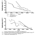
Hospital autopsy rates have been falling in the UK and worldwide for over half a century[4–15] (figure 1A, B) and account for a small minority of all autopsies in the UK.[1,3] Recent studies suggest autopsy rates of less than 10% for teaching hospitals and less than 5% elsewhere.[1,16]

Decline in hospital autopsy rates

Decline in hospital autopsy rates

Figure 1.

Decline in hospital autopsy rates over recent decades. (A) Autopsy rates from three first world countries, data collated from multiple studies. (B) Autopsy rates from four different hospitals/NHS Trusts, data collated from multiple studies.

http://img.medscape.com/article/849/356/849356-thumb1.png

The decline in hospital autopsy rates is well known, yet poorly researched and quantified. The majority of medical professionals and politicians in the UK are likely to be unaware of this conspicuous decline. Consequently, little has been done to address the falling rates and the implications of this are not yet fully understood, nor are the consequences.

A PubMed literature search yielded no research detailing a UK-wide autopsy rate within the past 20 years (search terms “hospital autopsy [title]”, “clinical autopsy [title]”, “autopsy rate [title]”). Given this and documented inter-hospital variation (figure 1B), we aimed to determine the current UK autopsy rate.

The structure of healthcare delivery varies throughout the UK. In England, the provision of acute services (emergency, inpatient and outpatient care) is provided by 186 organisations known as Acute National Health Service (NHS) Trusts—each of which may provide care from multiple hospital sites. In Scotland and Wales, the countries are divided into a number of defined geographical areas (Boards), each of which may contain several sites of healthcare delivery. In Northern Ireland, these geographical areas are known as Health and Social Care Trusts.

Method

Acute NHS Trusts within England (n=160), Boards within Wales (n=7) and Scotland (n=14) and Social Care Trusts within Northern Ireland (n=5) were contacted via ‘Freedom of Information’ requests. The level of response therefore is for the Trust/Board, not individual hospitals. If no reply was received within 4 months, reminders were sent.

The hospital autopsy rate was calculated as the number of autopsies performed on patients who died in the year 2013 as a percentage of total deaths which occurred in the hospital in that calendar year.

Studies indicate significantly higher autopsy rates in stillbirths, neonates and young children.[17,18]Therefore, data were excluded if they fell within the following categories:

  1. Children’s Hospital NHS Trusts
  2. Stillbirth, neonatal, perinatal and paediatric death
  3. Trusts with no recorded deaths
  4. Incomplete responses

Statistical analysis was performed using two-tailed χ2 tests (Prism 6 Software) between each country. The categories used were number of deaths that underwent autopsy and number of deaths not followed by autopsy. Bonferroni correction was used to compensate for the six pairwise comparisons, resulting in 99.25 CIs (p<0.008). Statistical outliers were determined with a ROUT test using a false-positive rate (Q) of 1%.

Results

A 99% (n=184) response rate was achieved for the UK; constituent country response rates were 99% (England), 100% (Scotland), 100% (Wales) and 100% (Northern Ireland). A total of 17 Trusts were removed, according to the exclusion criteria. Eight Trusts were concerned about patient identification because the number of autopsies was small and so provided a ‘fewer than’ figure. In these cases, a maximum possible rate was calculated.

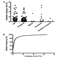
Mean hospital autopsy rates were calculated as the total number of autopsies expressed as a percentage of the total number of deaths. The UK mean autopsy rate was 0.69% and varied considerably between countries. The highest mean autopsy rates were in Scotland (2.1%), followed by Wales (0.65%), England (0.51%) and Northern Ireland (0.46%). The study confirms that hospital autopsy rates are significantly lower than the most recent literature suggests and that there is evident inter-country variation (figure 2A, Table 1) and intra-country variation (figure 2A).

Figure 2.

The results from Freedom of Information request for UK and constituent countries. (A) Individual points representing each sample Trust/Board, non-parametric data, no statistical difference between countries. (B) Cumulative frequency histogram of autopsy rates for NHS Trusts/Boards in the UK.

autopsy rates

http://img.medscape.com/article/849/356/849356-thumb2.png

Inter-country pairwise comparisons using χ2 tests of significance (p<0.008) found Scotland to have a significantly higher hospital autopsy rate than each of the other countries (p<0.0001). Other pairwise comparisons failed to achieve significance (Table 2).

Twenty eight samples were statistical high outliers, 20 from England, 6 from Scotland, 1 from Wales and 1 from Northern Ireland. The mean hospital autopsy rate is skewed by these outliers, which typically were large teaching hospitals or small specialist centres. The top 5% (n=7) of Trusts within England performed 47% of the country’s autopsies and 75% of autopsies in Wales were performed in one health board.

Ninety-eight per cent of samples (n=164) had an autopsy rate of <5%, 86% (n=143) an autopsy rate <1% and 23% (n=38) of all samples did not perform a single autopsy in 2013 (figure 2B). This demonstrates that for a quarter of NHS Trusts/Boards in the UK, hospital autopsy is extinct and in only a fraction (1.8%) of specialist trusts do autopsy rates exceed 5%, the rate previously published for non-teaching hospitals.[1,18]

Hospital autopsy rates in children’s hospital NHS Trusts ranged from 0% to 21%. This higher figure is in agreement with other literature.[17,18]

Discussion

This study has demonstrated that the evident decline in hospital autopsy has continued, if not accelerated, over recent years and already the hospital autopsy is extinct in many NHS Trusts. With 23% of NHS Trusts/Boards having an autopsy rate of 0%, a large part of UK hospital autopsy is now performed in a small number of centres. These few demonstrate that if the provisions and attitudes allow, then hospital autopsy rates of the recent past are still achievable, despite recent legislative changes such as the Human Tissue Act 2004/2006. Trusts with higher autopsy rates tended to be small specialised centres or large teaching hospital Trusts; this influence was not measured in this study due to difficulties in defining a ‘teaching’ or ‘specialised’ Trust/Board. Given that 86% of Trusts/Boards in the UK now have a hospital autopsy rate of <1%, we must pose the question whether a revival in hospital autopsy is possible? In the near future, many of these organisations may join the 23% in which hospital autopsy is extinct, unless they implement those changes in policy and attitude present in the 1.8% of Trusts/Boards where hospital autopsy exceeds 5% of inpatient deaths?

The hospital autopsy rate in Scotland was significantly higher than the other countries (Table 2). The causes of this are uncertain but may include variations in the Human Tissue Act and Authority in Scotland or a lower procurator fiscal (coronial) autopsy rate.

A number of Trusts/Boards gave some explanations as to why their autopsy rate was low, these commonly surrounded provision of facilities. For example, one Trust does not employ an onsite histopathologist or have its own autopsy facilities. However, some Trusts/Boards which themselves do not have onsite hospital autopsy facilities have an agreement with neighbouring Trusts/Boards to carry out their autopsies. From the results, there is evidence of remote island providers that continue to implement autopsy despite no local facilities but which transport cadavers via boat or aeroplane to a separate hospital for autopsy. Thus, a lack of facilities does not preclude hospital autopsy although may add significantly to the cost.

Future research should investigate the differences in Trust/Board policies, clinician attitudes, facilities, funding and local demographics to determine how significantly higher autopsy rates can be achieved.

The strength of this study lies in the nationwide approach to calculating contemporary hospital autopsy rates. Previous studies have focused on single hospitals or Trusts; given the demonstrated wide inter-Trust variation this approach may lead to significant errors. A weakness of this study was that some hospital trusts were unable to separate the data for deaths and autopsies for children and adults. Therefore, mean adult autopsy rates may be slightly over-reported, rates being generally higher among paediatric deaths.

In England and Wales, 94 455 coronial autopsies were performed in 2013[3] yet only 1132 hospital autopsies were performed within the English and Welsh Trusts included in this study. Hospital autopsy now accounts for approximately 1.2% of total autopsies. With such low numbers, questions must be raised regarding the effect such decline has on quality assurance, public health, misdiagnosis (a key contributor to avoidable harm[19,20]), audit and the teaching of both medical students and trainee pathologists. Hospital autopsy presents classic cases used to train junior pathologists, given that many coronial postmortems are not used for training. Training in hospital autopsy will become ever more important given the impending lack of pathologists to cover coronial autopsy. The aim of this paper is to raise awareness of the extent of the decline and to prompt discussion on its consequences. While debate continues over the value of hospital autopsy in medical practice, if action is not taken imminently, the practice may disappear.

COMMENTARY

Prostate Cancer: Is It Time to Retire the Gleason Score?

George D. Lundberg, MD

Hello. I am Dr George Lundberg and this is At Large at Medscape.

If you are an American adult male, you either now have prostate cancer—whether or not you know it—or will likely develop it if you live long enough.

In an average recent year, some 220,000 American men are diagnosed with prostate cancer and some 27,000 die from it. That means, obviously, that it kills 12% of those it is found to afflict and does not kill 88%. Of those patients freshly diagnosed, 98.9% are alive at 5 years.

Does interventional therapy account for the good results of some of that nearly 90% of men who are diagnosed and don’t die? Well, yes, but probably not very many. Really bad prostate cancer tends to do its lethal thing, regardless of interventions.

Back when we collectively had a lot of autopsies, it was possible—even easy—- to follow and learn the natural history of many diseases.

Modern autopsy-less American physicians, including pathologists, are whizz-bang at computers, imaging, lab test panels, genes, microbiomes, electronic medical records, and coding. They are maybe not so great at physical exams, taking a useful personal and family history, or gross and microscopic pathology. This latter list is where physicians once got really good at understanding the natural history of diseases.

Now, epidemiologic facts, outcomes, and common sense have begun to prevail over the national hysteria of prostate-specific antigen (PSA) tests for all men. The hysteria tends to take this form: “Find that prostate cancer and root it out…no matter how small or indolent. That way, ‘Megalopolis U’ can keep those operating rooms, hospital beds, and—get this—unproven (but very elegant) proton beameconomic monstrosities really humming in order to satisfy the overpaid MBAs that determined that such were a good investment idea.”

But some prostate cancers really can kill. How about those? A pathologist named Donald Gleason came up with a numbering system intended to guide therapy based upon anaplasia and prognostic threat of prostate cancer. It was 1, 2, 3, 4, and 5. It makes sense. But then, another number dealing with the relative amount of each level of differentiation and pattern (also 1, 2, 3, 4, 5) got added, and combining the two scores became the Gleason score range of 2-10.

A reasonable human could interpret a 6 on a scale of 10 as middling, pretty bad, or a “better-whack-it-out”-type score. So, a team from Johns Hopkins Medical Institutions[1] has worked out the actual prognosis as falling back into 5 Prognostic Grade Groups (PGGs):

  • Gleason 1, 2, 3, 4, 5, and 6 become Prognostic Grade Group (PGG I);
  • Gleason 3 + 4 = 7 (PGG II);
  • Gleason 4 + 3 = 7 (PGG III);
  • Gleason 4 + 4 = 8 (PGG IV); and
  • Gleason 9-10 (PGG V).

This is so much simpler and less likely to confuse the treating clinician and the patient who is increasingly sharing in this treatment decision.

A European group has just published outcomes based on this PGG system,[2] and it fits nicely. Those many patients with low grades who may not need radical therapy will stand a better chance of notreceiving radical therapy with new low-sounding numbers.

And, by the way, how did “watchful waiting” as a good way to handle those prostate “cancers”—which, from histology, seem like they would behave as indolentomas—morph so quickly into “active surveillance”? My guess is that it is very hard to bill a patient, Medicare, or an insurance company for just letting the patient watch and wait.

Chicago Mayor Rahm—not his physician brother Zeke—Emanuel was right: Never let a crisis go to waste. When the word “cancer” was uttered or written, hair lit on fire and something had to be done, right or wrong.

That is my opinion. I’m Dr George Lundberg.

Medscape Medical News Conference News

New Clue as to Why Only Some Breast Cancers Relapse

Zosia Chustecka

UPDATED September 26, 2015 // VIENNA — A new clue as to why only some breast cancers recur comes from the largest study of genetic sequencing of breast cancer tissue to date.

While most breast cancer is cured after treatment, about 20% of cases recur. The new study shows that the cases that recur have a different genetic profile, and suggests that some of the genetic drivers of relapse are targetable with drugs.

“We demonstrate that there are clear differences within the driver landscapes of relapsed cancers. This probably reflects a combination of predisposition to relapse and of differences in the mutations acquired during the relapse and metastasis phase,” say the researchers, led by Lucy Yates, MD, a clinical research oncologist from the Wellcome Trust Sanger Institute in Cambridge, United Kingdom.

The finding raises the hope that breast cancer patients who are most at risk for relapse can be identified when they are first diagnosed, they suggest.

In addition, as the newly identified genetic drivers of relapse are targetable with drugs, there is also hope that eventually women who are identified as being at high risk for relapse could be treated with such drugs to prevent recurrence, they suggest.

The study is due to be presented European Cancer Congress (ECC) 2015, but details were released early by the ECC press office.

The finding comes from a study that compared the genetic make-up of breast cancer from 836 tissue samples taken from women on primary diagnosis with 161 samples of tissue taken from recurrences or metastases.

The study is the largest and most comprehensive carried out to date, say the researchers, both in terms of the number of samples from relapsed breast cancers and in terms of the wide-ranging genetic sequencing carried out, which looked at 365 genes involved in cancer-related pathways.

The researchers performed de novo driver mutation discovery, and individual mutations were annotated with likely driver status based upon recurrence and known driver status in previously published, well-curated datasets and databases. The incidence of each driver mutation in the primary and relapse datasets was compared using Fisher’s exact test and using the Benjamini–Hochberg correction for multiple testing.

The team found 11 genes that were significantly enriched in the relapsed cohort compared with the primary tumor cohort. The most heavily enriched were TP53 and ARID1B. Multiple samples were available for 66 patients, including local or distant relapse samples in all cases and a sample from the primary tumor in 21 cases. This multisample analysis allowed the team to trace the evolution of mutations.

“We have found that some of the genetic mutations that drive breast cancers that relapse are relatively uncommon amongst cancers that do not relapse at the point of primary diagnosis,” Dr Yates said in a statement.

“We believe that the differences we have seen reflect genetic differences that can predispose a cancer to return, combined with mutations acquired throughout the period from first diagnosis to the subsequent relapse,” she added.

However, in a discussion of this paper, Fabrice André, MD, PhD, from the Gustave Roussy Institute, in Villejuif, France, questioned whether all the genes that were found to be enriched in the relapsed samples were driving the relapse and whether any could be identified as recurrent markers. He noted that although Dr Yates and colleagues found 11 genes that were highly enriched in the relapsed tissue samples, another study (which analyzed 183 samples) found only one of these genes to be highly enriched.

Dr Andre also wondered whether the late mutations that were identified are clinically relevant ― could they explain the development of resistance to therapy? This has been seen in other cancers, he noted.

Extreme Heterogeneity ― Need for Multiple Biopsies

“Our data reveal extreme heterogeneity and indicate that genomic analysis of primary, relapsed, and matched normal tissue are needed,” Dr Yates concluded.

“We need to do biopsies again and again and again,” said Anne-Lise Borresen-Dale, MD, from the Institute for Cancer Research, Oslo University Hospital, Norway, who chaired the session. But Dr Andre wondered whether circulating tumor cells, the so-called “liquid biopsy,” could be used.

Multiple samples were available for 66 subjects, including local or distant relapse samples in all cases and a sample from the primary tumor in 21 cases. This multisample analysis permitted relative temporal ordering of driver mutation accumulation to be determined, the researchers explain.

“We have found that some of the genetic mutations that drive breast cancers that relapse are relatively uncommon amongst cancers that do not relapse at the point of primary diagnosis,” Dr Yates said in a statement.

“This study highlights the differences between genetic alterations that drive relapsed and metastatic disease as opposed to primary breast cancers, and underlines the importance of analyzing the genetic features of metastases when making treatment decisions,” said Jorge Reis-Filho, MD, from the Memorial Sloan Kettering Cancer Center in New York City, who was acting as a spokesperson for the European Society of Medical Oncology, which is cohosting the meeting. He was not involved with this work.

However, Dr Reis-Filho also cautioned that “the extent of the differences in the repertoire of mutations among different metastatic sites within individual patients remains to be determined, however, as does the best way to obtain tumor-derived genetic material in patients with metastatic disease. We also need to know more about whether single or multiple metastatic sites should be analyzed in this context.”

Also commenting on the study, Peter Naredi, MD, PhD, professor of surgery at Sahlgrenska University Hospital in Gothenburg, Sweden, who is the European CanCer Organization scientific cochair of the congress, said: “Information such as that which Dr Yates will present is very important in the era of precision medicine.”

“This study also underlines the fact that we should consider a recurrence of a cancer as a new event, and carefully select the right treatment for the recurrent tumor as opposed to just relying on information from the first occurrence,” Dr Naredi said in a statement.

JAK Inhibitors in Breast Cancer

Some of the genetic changes that were found in the relapsed/metastatic breast cancer samples appeared at a late stage when the cancer recurred, and were not seen in samples taken at primary diagnosis. Among these later-stage mutations, the researchers say they found “compelling evidence” for the tumor suppression activity of two related genes, called JAK2 and STAT3, that operate within the same signaling pathway.

“Within some breast cancers, a disruption in this signaling pathway seems to be advantageous for survival of the cancer,” Dr Yates said in a statement.

“Interestingly, this is in contrast to the role of JAK2 in some other cancers, where overactivity of the gene drives malignancy rather than suppresses it,” she added.

The JAK (Janus-associated kinase) enzymes JAK1 and JAK2 are involved in regulating blood and immunologic functioning, and a dysregulation of these enzymes is thought to be a driver in the development of myelofibrosis. The first JAK inhibitor, ruxolitinib (Jakafi, Incyte Corp), was approved for the treatment of myelofibrosis in 2011.

Dr Yates and colleagues note that enhanced JAK-STAT signaling is known to play an important role in breast cancer stem cell development and cancerous cell line survival, and preclinical evidence seems to suggest that inhibiting the gene would be therapeutically advantageous. These findings have led to the development of clinical trials for breast cancer using JAK inhibitors in the hope that they will slow cancer progression.

“However, our findings suggest that, in a subset of cancers, inhibiting this pathway may have the opposite effect, and this requires further investigation. In general, the observation highlights the importance of understanding the diverse nature of breast cancers in the era of precision medicine,” Dr Yates said.

The work was funded by the Wellcome Trust. Dr Yates has disclosed no relevant financial relationships.

European Cancer Congress (ECC) 2015: Abstract 1804.

Read Full Post »

Urological Cancers of Men

Reporter and Curator: Larry H. Bernstein, MD, FCAP


Impact of the U.S. Preventive Services Task Force Recommendations Against PSA Screening on Prostate Biopsy and Cancer Detection Rates.

B Bhindi, M Mamdani, GS Kulkarni, A Finelli, RJ Hamilton, J Trachtenberg, et al.
The Journal of Urology 12/2014; http://dx.doi.org:/10.1016/j.juro.2014.11.096

To determine if the USPSTF recommendation against PSA screening was associated with a change in biopsy and cancer detection rates.

We conducted a time series analysis (10/2008-06/2013) of prostate biopsies performed at University Health Network (Toronto). Biopsies for active surveillance or solely targeting MRI-detected lesions were excluded. Interventional auto-regressive integrated moving average (ARIMA) models with step functions were used to examine changes in the number of biopsies performed and cancers detected per month. Low risk PC (LRPC) was defined as no Gleason pattern ≥4, ≤3 cores involved or ≤1/3 of total number of cores involved, and no core with >50% cancer involvement. Intermediate-to-high grade PC (I-HGPC) was defined as Gleason 7-10.

A total of 3408 biopsies were performed and 1601 (47.0%) PCs were detected (LRPC=563 (16.5%); I-HGPC=914 (26.8%)). The median number of biopsies per month decreased from 58.0 (IQR=54.5-63.0) before recommendations to 35.5 (IQR=27.0-41.0) afterward (p=0.003), while median number of patients undergoing their first-time biopsies decreased from 42.5 (IQR=37.5-45.5) to 24.0 (IQR=19.0-32.5, p=0.025). The median number of LRPCs detected per month decreased from 8.5 (IQR=6.5-10.5) to 5.5 (IQR=4.0-7.0, p=0.012), while the median number of I-HGPCs per month decreased from 17.5 (IQR=14.5-21.5) to 10.0 (IQR=9.0-12.0, p<0.001).

Following the USPSTF recommendation, the number of biopsies performed (total and first-time biopsies), based on referrals from our catchment area, have decreased. This is likely due to decreased use of PSA-screening. Although encouraging that fewer low risk PCs are being diagnosed, the sudden decrease in the detection rate of Gleason 7-10 PCs is concerning.


Tumour genomic and microenvironmental heterogeneity for integrated prediction of 5-year biochemical recurrence of prostate cancer: a retrospective cohort study.

E Lalonde, AS Ishkanian, J Sykes, M Fraser, H Ross-Adams, N Erho, et al.
The Lancet Oncology 12/2014; 15(13):1521-32.
http://dx.doi.org:/10.1016/S1470-2045(14)71021-6

Clinical prognostic groupings for localised prostate cancers are imprecise, with 30-50% of patients recurring after image-guided radiotherapy or radical prostatectomy. We aimed to test combined genomic and microenvironmental indices in prostate cancer to improve risk stratification and complement clinical prognostic factors.

We used DNA-based indices alone or in combination with intra-prostatic hypoxia measurements to develop four prognostic indices in 126 low-risk to intermediate-risk patients (Toronto cohort) who will receive image-guided radiotherapy. We validated these indices in two independent cohorts of 154 (Memorial Sloan Kettering Cancer Center cohort [MSKCC] cohort) and 117 (Cambridge cohort) radical prostatectomy specimens from low-risk to high-risk patients. We applied unsupervised and supervised machine learning techniques to the copy-number profiles of 126 pre-image-guided radiotherapy diagnostic biopsies to develop prognostic signatures. Our primary endpoint was the development of a set of prognostic measures capable of stratifying patients for risk of biochemical relapse 5 years after primary treatment.

Biochemical relapse was associated with indices of tumour hypoxia, genomic instability, and genomic subtypes based on multivariate analyses. We identified four genomic subtypes for prostate cancer, which had different 5-year biochemical relapse-free survival. Genomic instability is prognostic for relapse in both image-guided radiotherapy (multivariate analysis hazard ratio [HR] 4·5 [95% CI 2·1-9·8]; p=0·00013; area under the receiver operator curve [AUC] 0·70 [95% CI 0·65-0·76]) and radical prostatectomy (4·0 [1·6-9·7]; p=0·0024; AUC 0·57 [0·52-0·61]) patients with prostate cancer, and its effect is magnified by intratumoral hypoxia (3·8 [1·2-12]; p=0·019; AUC 0·67 [0·61-0·73]). A novel 100-loci DNA signature accurately classified treatment outcome in the MSKCC low-risk to intermediate-risk cohort (multivariate analysis HR 6·1 [95% CI 2·0-19]; p=0·0015; AUC 0·74 [95% CI 0·65-0·83]). In the independent MSKCC and Cambridge cohorts, this signature identified low-risk to high-risk patients who were most likely to fail treatment within 18 months (combined cohorts multivariate analysis HR 2·9 [95% CI 1·4-6·0]; p=0·0039; AUC 0·68 [95% CI 0·63-0·73]), and was better at predicting biochemical relapse than 23 previously published RNA signatures.

This is the first study of cancer outcome to integrate DNA-based and microenvironment-based failure indices to predict patient outcome. Patients exhibiting these aggressive features after biopsy should be entered into treatment intensification trials.
Prostate cancer: Is prostatectomy for Gleason score 6 a treatment failure?

Theodorus H van der Kwast, Monique J Roobol
Nature Reviews Urology 12/2014; http://dx.doi.org:/10.1038/nrurol.2014.335

Molecular and clinical support for a four-tiered grading system for bladder cancer based on the WHO 1973 and 2004 classifications
BWG van Rhijn, M Musquera, L Liu, AN Vis, TCM Zuiverloon, GJLH van Leenders, WJ Kirkels, EC Zwarthoff, ER Boevé, …, TH van der Kwast

Modern pathology : an official journal of the United States and Canadian Academy of Pathology, Inc 11/2014; http://dx.doi.org:/10.1038/modpathol.2014.154

Currently, the use of two classification systems for bladder cancer grade is advocated in clinical guidelines because the WHO2004 classification has not been sufficiently validated with biological markers and follow-up. The slides of 325 primary non-muscle invasive bladder cancers from three hospitals were reviewed by one uro-pathologist in two separate sessions for the WHO1973 (G1, G2 and G3) and 2004 (papillary urothelial neoplasm of low malignant potential (LMP), low-grade (LG) and high-grade (HG)) classifications. FGFR3 status was examined with PCR-SNaPshot analysis. Expression of Ki-67, P53 and P27 was analyzed by immuno-histochemistry. Clinical recurrence and progression were determined. We performed validation and cross-validation of the two systems for grade with molecular markers and clinical outcome. Multivariable analyses were done to predict prognosis and pT1 bladder cancer. Grade review resulted in 88 G1, 149 G2 and 88 G3 lesions (WHO1973) and 79 LMP, 101 LG and 145 HG lesions (WHO2004). Molecular validation of both grading systems showed that FGFR3 mutations were associated with lower grades whereas altered expression (Ki-67, P53 and P27) was found in higher grades. Clinical validation showed that the two classification systems were both significant predictors for progression but not for recurrence. Cross-validation of both WHO systems showed a significant stepwise increase in biological (molecular markers) and clinical (progression) potential along the line: G1-LG-G2-HG-G3. The LMP and G1 categories had a similar clinical and molecular profile. On the basis of molecular biology and multivariable clinical data, our results support a four-tiered grading system using the 1973 and 2004 WHO classifications with one low-grade (LMP/LG/G1) category that includes LMP, two intermediate grade (LG/G2 and HG/G2) categories and one high-grade (HG/G3) category.Modern Pathology advance online publication, 28 November 2014; doi:10.1038/modpathol.2014.154.

Read Full Post »

On the road to improve prostate biopsy

Author and Curator: Dror Nir, PhD

Urologists are in constant search for a method that will improve the outcome of prostate biopsy, particularly when it comes to ruling-in and ruling-out clinically significant prostate cancer. As stated in my recent post – State of the art in oncologic imaging of Prostate; “The disease’s staging and related prognosis are determined during diagnosis based on PSA level and the Gleason score of biopsy’s samples. Although prostate-specific antigen (PSA) screening resulted in the diagnosis of prostate cancer at earlier stages and with lower Gleason scores, it has also contributed to concerns about over-diagnosis, overtreatment of clinically insignificant disease, associated treatment-related toxicity, and escalating costs”. I already reported in the past on research conduc ted in this area; New clinical results supports Imaging-guidance for targeted prostate biopsy and Knowing the tumor’s size and location, could we target treatment to THE ROI by applying imaging-guided intervention? Today I report on recent publication presenting the advantage of using targeted trans-perineal biopsy following HistoScanning imaging instead of systematic TRUS biopsies: Computer-aided (HistoScanning) Biopsies Versus Conventional Transrectal Ultrasound-guided Prostate Biopsies: Do Targeted Biopsy Schemes Improve the Cancer Detection Rate? (Moritz F. Hamann, Claudius Hamann, Eckhard Schenk, Amr Al-Najar, Carsten M. Naumann, and Klaus-Peter Jünemann, Urology, Volume 81, Issue 2, February 2013, Pages 370-375

I have mentioned HistoScanning (ultrasound-based tissue characterization technology which I have invented and developed to a medical device) in many of my previous posts. HistoScanning for prostate is a specific HistoScanning application that is applied to the ultrasound’s raw signal (not the image) following a comprehensive scan of the prostate capturing its entire volume. The whole process takes about ten minutes and the output is a digital 3D map of the prostate gland where locations suspicious of presenting with prostate cancer are indicated.

HistoScanning report with 2, bilateral, basal lesions.

HistoScanning report with 2, bilateral, basal lesions.


The urologist translates such map into a “prostate regional biopsy scheme” when planning his biopsies and direct the needle, under ultrasound guidance, to these predefined suspicious locations.

 The systematic biopsy patterns targeted 7 sectors bilaterally: transition zone, apex, center, and base, each medially and laterally.

The systematic biopsy patterns targeted 7 sectors bilaterally: transition zone, apex, center, and base, each medially and laterally.

In that sense, the workflow is similar to using MRI for tumor detection and creating a tumor map for targeting the biopsy.

As reasoning for conducting the study the investigators argue that: “Exact staging of prostate cancer before treatment is essential for relevant therapeutic decision making. Current procedures, such as nerve-sparing prostatectomy and brachytherapy, as well as active surveillance and future focal treatment options, depend on the reliable identification of cancerous lesions within the prostate. Systematic prostate biopsies with at least 10 to 12 cores are the current standard method to detect and locate significant prostate cancer, as scientific evidence during the last decades has shown. Nevertheless, there are no homogeneous data concerning the required number of cores and the technical approach of prostate biopsy procedures. The unstable histologic results on active surveillance and the well-known discrepancy between transrectal diagnostics and radical prostatectomy specimens underline the neces­sity to develop reliable diagnostic tools for precise detection and localization of prostate cancer. Recent data on HistoScanning computer-aided ultra-sonography have shown favorable results. To generate a greater diagnostic yield than systematic needle biopsies, we integrated HistoScanning-guided targeted biopsies in our general prostate biopsy regimen. We report the cancer detection rate in a prospective series of 80 patients.”

The study’s objective was: “To define potential improvement in prostate cancer detection by application of a computer-aided, targeted, biopsy regimen using HistoScanning.”

Materials and Methods: “The data were collected prospectively from 80 men who consecutively underwent a systematic 14-core prostate biopsy supplemented by targeted transrectal and perineal ultrasound-guided biopsies. All biopsies were performed between March 2011 and September 2011. Indications for prostate biopsy were suspicious findings at the digital rectal examination (DRE), or serum prostate-specific antigen (PSA) level >10 ng/mL, or both. In case of elevated serum PSA levels >4 ng/mL a PSA-velocity of >0.75 ng/mL p.a. and free-to-total PSA ratio <15% were seen as the indication for prostate biopsies. Thirty-six patients had undergone a previous transrectal prostate biopsy. All patients were informed of the mode of the extended prostate biopsy scheme and its potential complications. All patients provided written informed consent for the procedure.After indication and before starting the biopsy procedure, all patients underwent a standardized 3-dimensional (3D) transrectal ultrasound (TRUS) with an end-fire array of a BK 8818 probe. Computer-aided analysis of the raw (radio-frequency) back-scatter data was performed by using the Conformite Europeene-marked and commercially available HistoScanning device, admitted for medical use in the Euro­pean Union (software version 2.1, Advanced Medical Diag­nostics, Belgium).”

“Each patient was diagnosed preoperatively by HistoScanning, defining a maximum of 3 suspicious areas. These areas were biopsied, both transrectally and via the perineum, with a maximum of 3 cores per location.”

Results: “We detected prostatitis in 30 patients (37.5%), premalignant lesions in 10 (12.5%), and prostate cancer in 28 (35%). The transrectal technique was used to detect 78.6% of all cancers using 14 cores by systematic biopsy. With a maximum of 9 targeted cores, 82.1% of all cancers were detected with the targeted perineal approach and 53.6% were detected with the targeted transrectal approach. Although our data did not show significant difference in the performance of targeted transperineal compared with systematic transrectal biopsies, the detection rate of targeted transrectal biopsies was significantly lower.”

 table

Conclusion: “The presented targeted biopsy scheme achieved an overall detection rate of 85% of prostate-specific antigen–relevant pathologic lesions within the prostate. Thus, the presented procedure shows an improved detection rate compared with standard systematic prostate biopsies, and the number of cores required is reduced. Furthermore, the perineal HistoScanning-aided approach seems to be superior to the transrectal approach with respect to the prostate cancer detection rate. The presented procedure might be a step toward reliable ultrasound-based tissue characterization and toward fulfilling the requirements of novel therapeutic strategies.”


The authors’ included an elaborated discussion on the background to their study and its results. This discussion is important for understanding the limitation of the study results and for putting the authors conclusion into balanced context: “When other solid-organ cancer guidelines are compared with prostate cancer guidelines, the common methods of prostate cancer detection are unmasked as an outmoded concept because cancer detection is based on chance as a result of a blinded, subjective examination. A systematic biopsy with at least 10 to 12 cores is considered the standard procedure in prostate cancer diagnostics to date. 1,2 The continuous increase in the number of biopsy cores taken over the last years has predictably improved the detection rate, but several studies report detection rates of only 30% to 40% even in repeated biopsies. 5,9 It is noteworthy that the recommendations must be seen as a compromise bbetween the cancer detection rate and the invasiveness of the surgical procedure. Modern diagnostic procedures, including magnetic resonance imaging, elastography, computerized analysis of TRUS/artificial neuronal network analysis, and HistoScanning, try to overcome this principle of approach.7,10,11 The current therapeutic concepts and further currently evolving therapy strategies depend on sophisticated prostate cancer diagnostics. It is more important than ever to look for ways to detect and locate the cancer before subjecting patients to more or less invasive procedures as the indication for surgical treatment or prostate-preserving (focal) tumor therapy. The results of magnetic resonance imaging for prostate cancer detection are very promising so far and show a sensitivity of up to 80%. Elastography has also shown promising capabilities for cancer detection, with a recent review article reporting that several studies show 74% to 75% sensitivity.11 HistoScanning has shown 93% sensitivity in detecting and locating prostate cancer. 12 As a matter of principle, our study is unable to report on the accuracy or sensitivity of prostate cancer detection because the exact number of cancer lesions in our patients collective remains incomputable. Integration of HistoScanning for guided, targeted biopsies helped us achieve a prostate cancer detection rate of 35%. These data are lower than results from current publications on initial prostate biopsies but higher than those of repeated biopsy protocols.4,5,13 Given that tumors looked for during initial biopsies are usually large and easy to detect, we believe that this finding is caused by the smaller overall risk of cancer in a repeated biopsy setting, as was the case in 37.5% of our patients (0.61 biopsies per patient). Overall, HistoScanning seems to improve selective targeting of suspicious prostate lesions. Taking into account all malignant, premalignant, and atypical histologic findings, including prostate cancer, atypical small acinar proliferations, and high-grade prostatic intraepithelial  eoplasia, the detection rate of relevant prostatic lesions by specimens from perineal-targeted biopsies rises to 47.5% and 85%, including prostatitis, respectively. Apart from the high quality of the HistoScanning tissue analysis, we believe in a significant effect of the technical approach used to perform the biopsy. As our data show, prostate cancer detection rates from specimens obtained from perineal-targeted biopsies differed significantly from the transrectal-targeted biopsy regimen, a difference that occurred independently from previous tissue analysis because both targeted approaches are aligned to the same scanning process. Compared with the transrectal approach, the perineal biopsy technique might reduce variables that can influence the needle placement. Furthermore, longitudinal biopsy punches following the axis of the prostate seem to allow more accurate sampling of the anterior part. Theoretically, because previous studies reported inhomogeneous results comparing transrectal and transperineal prostate biopsies.3,4,13 The use of a 14-gauge needle in perineal biopsies might be responsible for a systematic bias because it possibly yields more tissue than transrectal cores. Despite this potential advantage, systematic transrectal biopsies do not reflect a significant difference in the detection rate. Nevertheless, due to the individual setting, our study is unable to report standardized results on the accuracy of comparing transrectal and transperineal needle placement. Template-guided mapping biopsies have recently attracted attention because of the high rate of cancer detection as initial (75%) and even repeat biopsy procedures (46%). 14 It notably increases the ability to locate and differentiate cancer foci within the prostatic gland, implicating mapping biopsies for active surveillance or focal treatment purposes regardless of the considerable surgical trauma generated by the use of extended biopsy protocols. A reduction of tissue trauma by generating a greater diagnostic yield would be a favorable methodologic aspect as initiated by our study. Regarding cancer detection, the presented data show no significant differences between the perineal-targeted and transrectal-targeted systematic biopsy regimen, but even though considerably fewer tissue samples (14 vs 9 cores; -35%) were taken from selected prostate areas, we detected no significant limitations by the perineal approach. These data are even more encouraging when bearing in mind that the number of samples represents a crucial factor in prostate cancer detection rates, as recently reported. A critical issue in the present study is in the implementation of the modified biopsy procedures. Although the surgeons at our center are experienced in using TRUS, our data show a learning curve during the first 80 procedures. In addition, the overlaying of the HistoScanning image analysis to the B-mode grey-scale live ultrasound picture is done by the surgeon performing the biopsy. This process implies a bias in individual interpretation of the TRUS picture and manual needle guidance. The online fusion of HistoScanning with the ultrasound image presumably would increase the handling accuracy. Further methodologic limitations lie in the heterogeneous and small collective of patients that were included in the study. With regard to the reasonably high number of previous negative biopsy specimens, patient selection can affect the hit rate of positive biopsy specimens. This circumstance might make the cancer detection frequency with HistoScanning look relatively small in this particular study compared with other diagnostic methods in patients undergoing an initial biopsy, but this is due to the daily routine in an academic referral center.

For completeness of this reporting and before stating my own conclusion I bring here below two comments that were made, one by Dr. Stephen Jones of Glickman Urological and Kidney Institute, Cleveland, Ohio and the reply by the first author:

COMMENT

The promise of image-guided diagnosis and management of prostate cancer has been frustratingly elusive. Early pioneers of prostate ultrasound imaging reported that hyperechoic lesionswere indicative of malignancy, but it rapidly became clear that the opposite was actually more realistic. Even so, these hypoechoic lesions were soon shown to be poor indicators of prostate cancer. Thus, the value of visual abnormalities on grey-scale prostate ultrasound imaging remains essentially negligible with current technology. As a result, a number of alternative imaging modalities have been developed and introduced with great excitement. Typically, images in the publications showcase an apparently obvious cancer standing out in contrast to adjacent benign tissues. Unfortunately, the data still reveal minimal value from most of these technologies, and those reported in this article are similarly disappointing. HistoScanning demonstrated interesting color images, but coupled with a transperineal-targeted biopsy found exactly one more case of prostate cancer than did the current standard of care—the 14-core extended transrectal biopsy. This “difference” is actually statistically identical (P >.99). Exactly the same number of patients (n¼ 4) was found exclusively by both transperineal HistoScan-targeted biopsy as with standard transrectal biopsy, and when targeted using the transrectal approach, the technology actually missed almost half of the cancers that were identified overall. Furthermore, these data do not support the suggestion that 9 cores are less morbid or traumatic than 14 cores, and the literature is replete with reports demonstrating this is simply not true. This is especially misleading when those 9 cores come at the cost, morbidity, time, and complexity of an operation such as this performed under general anesthesia. So the real question remains whether HistoScanning or any emerging technology to image the prostate—improves visualization of prostate cancer. Although magnetic resonance imaging is beginning to show notable promise, the clinical value of most other modalities remains mostly anecdotal. As one whose desire for a solution remains frustratingly unfulfilled, I hope that some imaging technique will demonstrate clinical value during my career. J. Stephen Jones, M.D., Department of Regional Urology, Glickman Urological and Kidney Institute, Cleveland Clinic Foundation, Cleveland, Ohio

REPLY

In accordance with your comprehensive notes, we have to search for diagnostic improvement and underline the need further investigations in the field of prostate cancer diagnostics and staging. Minimally invasive techniques, focal and targeted therapy modalities, and low-risk cancer surveillance probably are future treatment modalities for prostate malignancies that progressively challenge common diagnostic pathways. Sophisticated therapy strategies will require reliable staging results. The constant increase of cores taken during systematic prostate biopsy apparently will not overcome the well-known diagnostic uncertainties. Consequently, imaging techniques and methods of biopsy targeting will gain in importance. Clearly, the presented data do not show significant improvement in the overall detection rate of prostate cancer in our patient cohort. Further, the maximum number of 9 biopsy cores must be attributed to the initial study design and will undergo further investigation; however, the results rather support the study approach than reduce its validity. Considering the indeterminate number of cancers, the proof of superiority remains incomputable. Compared with the current standard of care of 12 to 14 cores, we detected no significant limitations, although perineal-targeted biopsies took significantly fewer cores (3-9 cores [35%]). Maintaining the detection rate unchanged and focusing on 1 to 3 preselected suspicious index lesions display a proof of principle rather than disappointing results. In line with future treatment options mentioned above, any less invasive, focused diagnostic procedure represents an encouraging advance compared with the common and recommended practice of a nonselective, systematic biopsy of the prostate to harbor cancerous tissue. Moritz F. Hamann, M.D., Department of Urology and Pediatric Urology, University of SchleswigeHolstein, Campus Kiel, Kiel, Germany

As mentioned by the authors, further improvement of the outcome of HistoScanning-based targeted biopsies of prostate is expected when an implementation of image-fusion application between the “off-line” generated 3D tumor map and the real-time ultrasound guiding the needle will be available; similar to the results presented already when using ultrasound-MRI image fusion applications for prostate biopsy. Of course, the ultimate biopsy workflow (which I am currently engaged in developing) that urologist are asking for is the one comprised of real-time ultrasound-based tissue characterization and real-time ultrasound guidance of the needle to the lesion.

References

1. Heidenreich A, Bellmunt J, Bolla M, et al. EAU guidelines on prostate cancer. Part 1: screening, diagnosis, and treatment of clinically localised disease. Eur Urol. 2011;59:61-71.

2. National Comprehensive Cancer Network. Clinical Practice Guidelines in Oncology: Prostate Cancer Early Detection. Available at http://www.nccn.org 2011 Accessed May 2011.

3. Abdollah F, Novara G, Briganti A, et al. Trans-rectal versus transperineal saturation re-biopsy of the prostate: is there a difference in cancer detection rate? Urology. 2011;77:921-925.

4. Hara R, Yoshimasa J, Tomohiro F, et al. Optimal approach for prostate cancer detection as initial biopsy: prospective randomized study comparing transperineal versus transrectal systematic 12-core biopsy. Urology. 2008;71:191-195.

5. Patel AR, Jones JS. Optimal biopsy strategies for the diagnosis and staging of prostate cancer. Curr Opin Urol. 2009;19:232-237.

6. Al Otaibi M, Ross P, Fahmy N, et al. Role of repeated biopsy of the prostate in predicting disease progression in patients with prostate cancer on active surveillance. Cancer. 2008;113:286-292.

7. Braeckman J, Autier P, Garbar C, et al. Computer-aided ultrasonography (HistoScanning): a novel technology for locating and characterizing prostate cancer. BJU Int. 2008;101:293-298.

8. Braeckman J, Autier P, Soviany C, et al. The accuracy of transrectal ultrasonography supplemented with computer-aided ultrasonography for detecting small prostate cancers. BJU Int. 2008;102:1560-1565.

9. Presti JC Jr, O’Dowd G, Miller C, et al. Extended peripheral zone biopsy schemes increase cancer detection rates and minimize variance in prostate specific antigen and age related cancer rates: results of a community multi-practice study. J Urol. 2003;169:125-129.

10. Turkbey B, Mani H, Shah V, et al. Multiparametric 3T prostate magnetic resonance imaging to detect cancer: histopathological correlation using prostatectomy specimens processed in customized magnetic resonance imaging based molds. J Urol. 2011;186: 1818-1824.

11. Trabulsi EJ, Sackett D, Gomella L, et al. Enhanced transrectal ultrasound modalities in the diagnosis of prostate cancer. Urology. 2010;76:1025-1033.

12. Simmons LA, Autier P, Zat_ura F, et al. Detection, localisation and characterisation of prostate cancer by Prostate HistoScanning. BJU Int. 2012;110:28-35.

13. Emiliozzi P, Corsetti A, Tassi B, et al. Best approach for prostate cancer detection: a prospective study on transperineal versus transrectal six-core prostate biopsy. Urology. 2003;61:961-966.

14. Taira AV, Merrick GS, Galbreath RW, et al. Performance of transperineal template-guided mapping biopsy in detecting prostate cancer in the initial and repeat biopsy setting. Prostate Cancer Prostatic Dis. 2010;13:71-77.


Other research papers related to the management of Prostate cancer were published on this Scientific Web site:


Prostate Cancer: Androgen-driven “Pathomechanism” in Early-onset Forms of the Disease

Prostate Cancer and Nanotecnology

Prostate Cancer Cells: Histone Deacetylase Inhibitors Induce Epithelial-to-Mesenchymal Transition

Imaging agent to detect Prostate cancer-now a reality

Scientists use natural agents for prostate cancer bone metastasis treatment

ROLE OF VIRAL INFECTION IN PROSTATE CANCER

Prostate Cancers Plunged After USPSTF Guidance, Will It Happen Again?

Imaging agent to detect Prostate cancer-now a reality

Scientists use natural agents for prostate cancer bone metastasis treatment

Today’s fundamental challenge in Prostate cancer screening

ROLE OF VIRAL INFECTION IN PROSTATE CANCER

Men With Prostate Cancer More Likely to Die from Other Causes

New Prostate Cancer Screening Guidelines Face a Tough Sell, Study Suggests

New clinical results supports Imaging-guidance for targeted prostate biopsy


Read Full Post »

State of the art in oncologic imaging of Prostate.

Author-Writer: Dror Nir, PhD


This is the third post in a series in which I will address the state of the art in oncologic imaging based on a review paper; Advances in oncologic imaging that provides updates on the latest approaches to imaging of 5 common cancers: breast, lung, prostate, colorectal cancers, and lymphoma. This paper is published at CA Cancer J Clin 2012. © 2012 American Cancer Society.

The paper gives a fair description of the use of imaging in interventional oncology based on literature review of more than 200 peer-reviewed publications. In this post I summaries the chapter on prostate cancer imaging.

Prostate Cancer Imaging

Although ultrasound is the most frequently used imaging-device in prostate cancer management the authors did not review the related literature. Instead, they focused their review on MRI and PET imaging. To anyone who wishes to learn about ultrasound-imaging’s state of the art in prostate cancer I can offer reading some of my previous posts that are listed below.

My own interpretation (as stated in my summary-note) to the focus by the authors on MRI and PET imaging is that they were mainly looking to highlight the advances in those imaging modalities which provides tissue characterization! Although, this term is not explicitly mentioned by them.

The authors identifies correctly the main issues in Prostate cancer management:

  1. It’s a frequent disease, but not an aggressive killer
  2. It’s highly heterogeneous, therefore it is difficult to predict the clinical outcomes both before and after treatment.
  3. “Although several predictive methods have been developed,72 the treatment decision-making process is complex and requires balancing clinical benefits, life expectancy, comorbidities and potential treatment-related side effects.”
  4. The disease’s staging and related prognosis are determined during diagnosis based on PSA level and the Gleason score of biopsy’s samples. “Although prostate-specific antigen (PSA) screening hsis as resulted in the diagnosis of prostate cancer at earlier stages and with lower Gleason scores, it has also contributed to concerns about over-diagnosis, overtreatment of clinically insignificant disease, associated treatment-related toxicity, and escalating costs”

The following sections summarizes the latest advances in MRI and PET imaging methods for functional and metabolic assessment of prostate cancer.

Advances in MRI of Prostate Cancer

“MRI is potentially an ideal imaging modality for the local staging of prostate cancer, given its ability to depict the prostate and surrounding structures in exquisite detail. Recently, morphologic imaging with conventional MR imaging sequences has been supplemented by a multiparametric imaging approach using new functional and metabolic methods, namely diffusion waited MRI (DW-MRI); dynamic contrast-enhanced MRI (DCE-MRI), which probes tissue micro-vascular and perfusion properties; and MR spectroscopy (Fig. below).”

Representative images from a 3-T multiparametric MRI examination in a 57-year-old man with PSA level of 9.1 ng/mL and Gleason score 7 (3 + 4) prostate cancer (arrow) located in the right anterior prostate and involving the transition and peripheral zones: (A) transverse T2-weighted image, (B) transverse ADC map generated from DW-MRI images, (C) transverse DCE-MRI image, (D) volume transfer constant (Ktrans) parametric map from DCE-MRI overlaid on T2-weighted image.

Representative images from a 3-T multiparametric MRI examination in a 57-year-old man with PSA level of 9.1 ng/mL and Gleason score 7 (3 + 4) prostate cancer (arrow) located in the right anterior prostate and involving the transition and peripheral zones: (A) transverse T2-weighted image, (B) transverse ADC map generated from DW-MRI images, (C) transverse DCE-MRI image, (D) volume transfer constant (Ktrans) parametric map from DCE-MRI overlaid on T2-weighted image.

Diffusion-Weighted MRI

“Because the diffusion of water molecules within tumors is more restricted than in normal tissue, ADCs calculated with DW-MRI tend to be lower in cancer than in normal tissue. A number of studies, using various image acquisition methods and reference standards, have reported the utility of DW-MRI in prostate cancer detection.74-79. More importantly, studies have indicated that the greatest value of DW-MRI as an addition to conventional MRI might lie in its potential to assess prostate cancer aggressiveness noninvasively, because ADC values have been shown to correlate significantly with tumor Gleason scores.77-79

“However, the clinical value of DW-MRI in predicting the surgical Gleason score needs to be further studied.”

Dynamic Contrast-Enhanced MRI

“DCE-MRI is based on the repeated acquisition of images of a region of interest during the passage of an intravenously administered contrast agent. DCE-MRI allows malignant tissue to be distinguished from benign tissue by exploiting differences in the distribution of the contrast agent between vascular and extravascular spaces over time. 80 Prostate cancer usually shows early, rapid, and intense enhancement with quick washout of contrast compared to noncancerous prostate tissue. Although DCE-MRI has shown potential in assessing prostate cancer in preliminary studies, further research is necessary to establish its clinical value and indications and address technical challenges, such as standardization of acquisition and analysis methods.”

MR Spectroscopy

“Commercially available acquisition and analysis software packages for MR spectroscopic imaging of the prostate produce 3-dimensional spectral data showing the relative concentrations of tissue metabolites within specified volumes of tissue. In the prostate, the metabolites of interest on in vivo MR spectroscopic imaging are citrate, creatine, choline, and polyamines.8788 (choline + creatine)/citrate ratio has traditionally been used to identify prostate cancer on MR spectroscopy. “

“Studies have indicated that MR spectroscopy might have potential for aiding cancer localization, estimating tumor volume, noninvasively assessing prostate cancer aggressiveness and predicting the probability of insignificant cancer.90-92

the authors found that MRI, especially when acquired with multiparametric techniques (DW-MRI, DCE-MRI, and/or MR spectroscopy), has the potential to add value in prostate cancer diagnosis, eg, by guiding biopsy to the most suspicious areas and eventually reduce the number of systematic/random biopsies.108-110 A specific use-case for MRI guided biopsies is men with elevated PSA and negative systematic/random TRUS-guided biopsy where MRI is used for locating suspicious areas for targeted biopsies.111  MRI, “could potentially improve prostate cancer management especially in the intermediate- and high-risk groups.” 112 They also suggest to use MRI, especially when acquired with multiparametric techniques as a tool for choosing and managing active survailance and focal treatment. These two novel methods of treatment have immerged as an answer to unbearable overdiagnosis and overtreatment in prostate cancer management.113 114

About active surveillance: “Given the risks of morbidity associated with radical treatment (eg, radical prostatectomy or radiation therapy), active surveillance (monitoring of PSA levels, periodic imaging and repeat biopsies) is gaining acceptance as an alternative initial management strategy for carefully selected men with low-risk prostate cancer.115 Active surveillance could be a considerably more cost-effective approach than immediate treatment for prostate cancer, as suggested in a theoretical cohort.116 Furthermore, by preserving quality of life and minimizing the harms from radical treatment of low-risk prostate cancer, active surveillance could mitigate the concerns regarding extensive screening, overdiagnosis, and overtreatment of prostate cancer. Ultimately questions about how to best practice active surveillance will need to be addressed in prospective studies. Currently, the main challenges in active surveillance of prostate cancer are adequate characterization of disease at diagnosis and determination of the risk of progression.”

About focal therapy:, sometime referred to as focused therapy. This approach is frequently used in other cancers; e.g. breast lumpectomies. The idea is to treat only the cancer lesion and preserve the rest of the organ. Such treatment has the potential of offering better quality of life for the patients. 117 An open clinical question in respect to focal treatments is related to the fact that prostate cancer is often multifocal. Some studies suggest that it is enough to treat the index tumor (tumor volume > 0.5 mL) in order to control the disease.118 To date, patients’ selection for focal treatment is based on multiparametric MRI techniques and prostate mapping biopsy (trans-perinea template biopsy) 119


Advances in PET Imaging of Prostate Cancer

The main application for [18F]FDG PET is in patients with aggressive, metastatic prostate cancer. For these patients it helps detecting metastasis, and assessment on response to treatment.93-97, The authors of this review did not find support to using it for the majority of prostate cancer patients who are diagnosed at early stage due to its low specificity in this population.

Representative images from 3-T MRI and [18F]FDG PET/CT examinations in a 70-year-old man with PSA level of 8.0 ng/mL and Gleason score of 8 (4 + 4) prostate cancer (arrow) located in the left posterolateral prostate within the peripheral zone: (A) transverse T2-weighted image, (B) transverse fused [18F]FDG PET/CT image, (C) transverse fused [18F]FDG PET/CT image overlaid on T2-weighted MRI.

Representative images from 3-T MRI and [18F]FDG PET/CT examinations in a 70-year-old man with PSA level of 8.0 ng/mL and Gleason score of 8 (4 + 4) prostate cancer (arrow) located in the left posterolateral prostate within the peripheral zone: (A) transverse T2-weighted image, (B) transverse fused [18F]FDG PET/CT image, (C) transverse fused [18F]FDG PET/CT image overlaid on T2-weighted MRI.

Other tracers such as [11C]choline and radiolabeled acetate ([11C]acetate) have recently been evaluated in clinical studies and found to be more promising than [18F]FDG for prostate cancer assessment.939698

“Currently, the major indication for choline PET/CT is the early localization of recurrence in patients with PSA relapse after primary radical treatment. Potentially, this test may also be useful in radiotherapy planning.99100 Acetate participates in cytoplasmic lipid synthesis, and an increased fatty acid synthesis is thought to occur in prostate cancer.101 Similarly to [11C]choline, radiolabeled acetate ([11C]acetate) appears to be more useful than [18F]FDG in the assessment of prostate cancer before and after treatment.102103 “

“In summary, the role of PET imaging in prostate cancer is still evolving, as new and promising tracers are under investigation. Rigorous clinical trials using these new tracers in specific clinical scenarios will be needed before they can be employed routinely.”

On expectations from future screening, diagnosis and pre-treatment staging the authors summarizes:  “An imaging modality that could reliably assess prostate cancer would be of great help in selecting from the wide range of management options now available.” and;

“there is a pressing need to improve not only anatomical imaging for tumor detection, localization and staging, but also functional and metabolic imaging for characterization of tumor biology. “

In regards to treatment choice: “active surveillance, focal therapy, radical prostatectomy, and radiation therapy represent a range of treatments with varying degrees of invasiveness for men with different disease grades and stages. Active surveillance and focal therapy, which are relatively new options, are promising but are complicated by uncertainties in risk stratification that affect treatment decision-making, as well as by uncertainties regarding the definition of appropriate outcome measures. Biopsy, which leaves the possibility of under sampling, is not sufficient to resolve these uncertainties. Novel biomarkers and modern imaging are expected to play increasingly important roles in facilitating broader acceptance of both active surveillance and focal therapy. Further research, particularly involving prospective validation, is needed to facilitate standardization and establish the roles of advanced imaging tools in routine prostate cancer management.”

My summary: Prostate cancer is a disease managed by urologists, not radiologists. This disease’s multi-choice of pathways is “craving” for tissue characterization. Nothing could fit the urologist’s work-flow better than ultrasound-based tissue characterization!


Colorectal Cancers Imaging

To be followed…


Other research papers related to the management of Prostate cancer were published on this Scientific Web site:

Imaging agent to detect Prostate cancer-now a reality

Scientists use natural agents for prostate cancer bone metastasis treatment

Today’s fundamental challenge in Prostate cancer screening

ROLE OF VIRAL INFECTION IN PROSTATE CANCER

Men With Prostate Cancer More Likely to Die from Other Causes

New Prostate Cancer Screening Guidelines Face a Tough Sell, Study Suggests

New clinical results supports Imaging-guidance for targeted prostate biopsy

Read Full Post »

%d bloggers like this: