Feeds:
Posts
Comments

Archive for the ‘Nutrition’ Category

Summary of Proteomics

Author and Curator: Larry H. Bernstein, MD, FCAP 


We have completed a series of discussions on proteomics, a scientific endeavor that is essentially 15 years old.   It is quite remarkable what has been accomplished in that time.  The interest is abetted by the understanding of the limitations of the genomic venture that has preceded it.  The thorough, yet incomplete knowledge of the genome, has led to the clarification of its limits.  It is the coding for all that lives, but all that lives has evolved to meet a demanding and changing environment with respect to

  1. availability of nutrients
  2. salinity
  3. temperature
  4. radiation exposure
  5. toxicities in the air, water, and food
  6. stresses – both internal and external

We have seen how both transcription and translation of the code results in a protein, lipoprotein, or other complex than the initial transcript that was modeled from tRNA. What you see in the DNA is not what you get in the functioning cell, organ, or organism.  There are comparabilities as well as significant differences between plants, prokaryotes, and eukaryotes.  There is extensive variation.  The variation goes beyond genomic expression, and includes the functioning cell, organ type, and species.

Here, I return to the introductory discussion.  Proteomics is a goal directed, sophisticated science that uses a combination of methods to find the answers to biological questions. Graves PR and Haystead TAJ.  Molecular Biologist’s Guide to Proteomics.
Microbiol Mol Biol Rev. Mar 2002; 66(1): 39–63.  http://www.ncbi.nlm.nih.gov/pmc/articles/PMC120780/

Peptide mass tag searching

Peptide mass tag searching

Peptide mass tag searching. Shown is a schematic of how information from an unknown peptide (top) is matched to a peptide sequence in a database (bottom) for protein identification. The partial amino acid sequence or “tag” obtained by MS/MS is combined with the peptide mass (parent mass), the mass of the peptide at the start of the sequence (mass tag 1), and the mass of the peptide at the end of the sequence (mass tag 2). The specificity of the protease used (trypsin is shown) can also be included in the search.

ICAT method for measuring differential protein expression

ICAT method for measuring differential protein expression

The ICAT method for measuring differential protein expression. (A) Structure of the ICAT reagent. ICAT consists of a biotin affinity group, a linker region that can incorporate heavy (deuterium) or light (hydrogen) atoms, and a thiol-reactive end group for linkage to cysteines. (B) ICAT strategy. Proteins are harvested from two different cell states and labeled on cysteine residues with either the light or heavy form of the ICAT reagent. Following labeling, the two protein samples are mixed and digested with a protease such as trypsin. Peptides labeled with the ICAT reagent can be purified by virtue of the biotin tag by using avidin chromatography. Following purification, ICAT-labeled peptides can be analyzed by MS to quantitate the peak ratios and proteins can be identified by sequencing the peptides with MS/MS.

Strategies for determination of phosphorylation sites in proteins

Strategies for determination of phosphorylation sites in proteins

Strategies for determination of phosphorylation sites in proteins. Proteins phosphorylated in vitro or in vivo can be isolated by protein electrophoresis and analyzed by MS. (A) Identification of phosphopeptides by peptide mass fingerprinting. In this method, phosphopeptides are identified by comparing the mass spectrum of an untreated sample to that of a sample treated with phosphatase. In the phosphatase-treated sample, potential phosphopeptides are identified by a decrease in mass due to loss of a phosphate group (80 Da). (B) Phosphorylation sites can be identified by peptide sequencing using MS/MS. (C) Edman degradation can be used to monitor the release of inorganic 32P to provide information about phosphorylation sites in peptides.

protein mining strategy

protein mining strategy

Proteome-mining strategy. Proteins are isolated on affinity column arrays from a cell line, organ, or animal source and purified to remove nonspecific adherents. Then, compound libraries are passed over the array and the proteins eluted are analyzed by protein electrophoresis. Protein information obtained by MS or Edman degradation is then used to search DNA and protein databases. If a relevant target is identified, a sublibrary of compounds can be evaluated to refine the lead. From this method a protein target and a drug lead can be simultaneously identified.

Although the technology for the analysis of proteins is rapidly progressing, it is still not feasible to study proteins on a scale equivalent to that of the nucleic acids. Most of proteomics relies on methods, such as protein purification or PAGE, that are not high-throughput methods. Even performing MS can require considerable time in either data acquisition or analysis. Although hundreds of proteins can be analyzed quickly and in an automated fashion by a MALDI-TOF mass spectrometer, the quality of data is sacrificed and many proteins cannot be identified. Much higher quality data can be obtained for protein identification by MS/MS, but this method requires considerable time in data interpretation. In our opinion, new computer algorithms are needed to allow more accurate interpretation of mass spectra without operator intervention. In addition, to access unannotated DNA databases across species, these algorithms should be error tolerant to allow for sequencing errors, polymorphisms, and conservative substitutions. New technologies will have to emerge before protein analysis on a large-scale (such as mapping the human proteome) becomes a reality.

Another major challenge for proteomics is the study of low-abundance proteins. In some eukaryotic cells, the amounts of the most abundant proteins can be 106-fold greater than those of the low-abundance proteins. Many important classes of proteins (that may be important drug targets) such as transcription factors, protein kinases, and regulatory proteins are low-copy proteins. These low-copy proteins will not be observed in the analysis of crude cell lysates without some purification. Therefore, new methods must be devised for subproteome isolation.

Tissue Proteomics for the Next Decade?  Towards a Molecular Dimension in Histology

R Longuespe´e, M Fle´ron, C Pottier, F Quesada-Calvo, Marie-Alice Meuwis, et al.
OMICS A Journal of Integrative Biology 2014; 18: 9.    http://dx.doi.org:/10.1089/omi.2014.0033

The concept of tissues appeared more than 200 years ago, since textures and attendant differences were described within the whole organism components. Instrumental developments in optics and biochemistry subsequently paved the way to transition from classical to molecular histology in order to decipher the molecular contexts associated with physiological or pathological development or function of a tissue. In 1941, Coons and colleagues performed the first systematic integrated examination of classical histology and biochemistry when his team localized pneumonia antigens in infected tissue sections. Most recently, in the early 21st century, mass spectrometry (MS) has progressively become one of the most valuable tools to analyze biomolecular compounds. Currently, sampling methods, biochemical procedures, and MS instrumentations
allow scientists to perform ‘‘in depth’’ analysis of the protein content of any type of tissue of interest. This article reviews the salient issues in proteomics analysis of tissues. We first outline technical and analytical considerations for sampling and biochemical processing of tissues and subsequently the instrumental possibilities for proteomics analysis such as shotgun proteomics in an anatomical context. Specific attention concerns formalin fixed and paraffin embedded (FFPE) tissues that are potential ‘‘gold mines’’ for histopathological investigations. In all, the matrix assisted laser desorption/ionization (MALDI) MS imaging, which allows for differential mapping of hundreds of compounds on a tissue section, is currently the most striking evidence of linkage and transition between ‘‘classical’’ and ‘‘molecular’’ histology. Tissue proteomics represents a veritable field of research and investment activity for modern biomarker discovery and development for the next decade.

Progressively, tissue analyses evolved towards the description of the whole molecular content of a given sample. Currently, mass spectrometry (MS) is the most versatile
analytical tool for protein identification and has proven its great potential for biological and clinical applications. ‘‘Omics’’ fields, and especially proteomics, are of particular
interest since they allow the analysis of a biomolecular picture associated with a given physiological or pathological state. Biochemical techniques were then adapted for an optimal extraction of several biocompounds classes from tissues of different natures.

Laser capture microdissection (LCM) is used to select and isolate tissue areas of interest for further analysis. The developments of MS instrumentations have then definitively transformed the scientific scene, pushing back more and more detection and identification limits. Since a few decades, new approaches of analyses appeared, involving the use of tissue sections dropped on glass slides as starting material. Two types of analyses can then be applied on tissue sections: shotgun proteomics and the very promising MS imaging (MSI) using Matrix Assisted Laser Desorption/Ionization (MALDI) sources. Also known as ‘‘molecular histology,’’ MSI is the most striking hyphen between histology and molecular analysis. In practice, this method allows visualization of the spatial distribution of proteins, peptides, drugs, or others analytes directly on tissue sections. This technique paved new ways of research, especially in the field of histopathology, since this approach appeared to be complementary to conventional histology.

Tissue processing workflows for molecular analyses

Tissue processing workflows for molecular analyses

Tissue processing workflows for molecular analyses. Tissues can either be processed in solution or directly on tissue sections. In solution, processing involves protein
extraction from tissue pieces in order to perform 2D gel separation and identification of proteins, shotgun proteomics, or MALDI analyses. Extracts can also be obtained from
tissues area selection and protein extraction after laser micro dissection or on-tissue processing. Imaging techniques are dedicated to the morphological characterization or molecular mapping of tissue sections. Histology can either be conducted by hematoxylin/eosin staining or by molecular mapping using antibodies with IHC. Finally, mass spectrometry imaging allows the cartography of numerous compounds in a single analysis. This approach is a modern form of ‘‘molecular histology’’ as it grafts, with the use of mathematical calculations, a molecular dimension to classical histology. (AR, antigen retrieval; FFPE, formalin fixed and paraffin embedded; fr/fr, fresh frozen; IHC, immunohistochemistry; LCM, laser capture microdissection; MALDI, matrix assisted laser desorption/ionization; MSI, mass spectrometry imaging; PTM, post translational modification.)

Analysis of tissue proteomes has greatly evolved with separation methods and mass spectrometry instrumentation. The choice of the workflow strongly depends on whether a bottom-up or a top-down analysis has to be performed downstream. In-gel or off-gel proteomics principally differentiates proteomic workflows. The almost simultaneous discoveries of the MS ionization sources (Nobel Prize awarded) MALDI (Hillenkamp and Karas, 1990; Tanaka et al., 1988) and electrospray ionization (ESI) (Fenn et al., 1989) have paved the way for analysis of intact proteins and peptides. Separation methods such as two-dimension electrophoresis (2DE) (Fey and Larsen, 2001) and nanoscale reverse phase liquid chromatography (nanoRP-LC) (Deterding et al., 1991) lead to efficient preparation of proteins for respectively topdown and bottom-up strategies. A huge panel of developments was then achieved mostly for LC-MS based proteomics in order to improve ion fragmentation approaches and peptide
identification throughput relying on database interrogation. Moreover, approaches were developed to analyze post translational modifications (PTM) such as phosphorylations (Ficarro et al., 2002; Oda et al., 2001; Zhou et al., 2001) or glycosylations (Zhang et al., 2003), proposing as well different quantification procedures. Regarding instrumentation, the most cutting edge improvements are the gain of mass accuracy for an optimal detection of the eluted peptides during LC-MS runs (Mann and Kelleher, 2008; Michalski et al., 2011) and the increase in scanning speed, for example with the use of Orbitrap analyzers (Hardman and Makarov, 2003; Makarov et al., 2006; Makarov et al., 2009; Olsen et al., 2009). Ion transfer efficiency was also drastically improved with the conception of ion funnels that homogenize the ion transmission
capacities through m/z ranges (Kelly et al., 2010; Kim et al., 2000; Page et al., 2006; Shaffer et al., 1998) or by performing electrospray ionization within low vacuum (Marginean et al., 2010; Page et al., 2008; Tang et al., 2011). Beside collision induced dissociation (CID) that is proposed for many applications (Li et al., 2009; Wells and McLuckey, 2005), new fragmentation methods were investigated, such as higher-energy collisional dissociation (HCD) especially for phosphoproteomic
applications (Nagaraj et al., 2010), and electron transfer dissociation (ETD) and electron capture dissociation (ECD) that are suited for phospho- and glycoproteomics (An
et al., 2009; Boersema et al., 2009; Wiesner et al., 2008). Methods for data-independent MS2 analysis based on peptide fragmentation in given m/z windows without precursor selection neither information knowledge, also improves identification throughput (Panchaud et al., 2009; Venable et al., 2004), especially with the use of MS instruments with high resolution and high mass accuracy specifications (Panchaud et al., 2011). Gas fractionation methods such as ion mobility (IM) can also be used as a supplementary separation dimension which enable more efficient peptide identifications (Masselon et al., 2000; Shvartsburg et al., 2013; Shvartsburg et al., 2011).

Microdissection relies on a laser ablation principle. The tissue section is dropped on a plastic membrane covering a glass slide. The preparation is then placed into a microscope
equipped with a laser. A highly focused beam will then be guided by the user at the external limit of the area of interest. This area composed by the plastic membrane, and the tissue section will then be ejected from the glass slide and collected into a tube cap for further processing. This mode of microdissection is the most widely used due to its ease of handling and the large panels of devices proposed by constructors. Indeed, Leica microsystem proposed the Leica LMD system (Kolble, 2000), Molecular Machine and Industries, the MMI laser microdissection system Microcut, which was used in combination with IHC (Buckanovich et al., 2006), Applied Biosystems developed the Arcturus
microdissection System, and Carl Zeiss patented P.A.L.M. MicroBeam technology (Braakman et al., 2011; Espina et al., 2006a; Espina et al., 2006b; Liu et al., 2012; Micke
et al., 2005). LCM represents a very adequate link between classical histology and sampling methods for molecular analyses as it is a simple customized microscope. Indeed,
optical lenses of different magnification can be used and the method is compatible with classical IHC (Buckanovich et al., 2006). Only the laser and the tube holder need to be
added to the instrumentation.

After microdissection, the tissue pieces can be processed for analyses using different available MS devices and strategies. The simplest one consists in the direct analysis of the
protein profiles by MALDI-TOF-MS (MALDI-time of flight-MS). The microdissected tissues are dropped on a MALDI target and directly covered by the MALDI matrix (Palmer-Toy et al., 2000; Xu et al., 2002). This approach was already used in order to classify breast cancer tumor types (Sanders et al., 2008), identify intestinal neoplasia protein biomarkers (Xu et al., 2009), and to determine differential profiles in glomerulosclerosis (Xu et al., 2005).

Currently the most common proteomic approach for LCM tissue analysis is LC-MS/MS. Label free LC-MS approaches have been used to study several cancers like head and neck squamous cell carcinomas (Baker et al., 2005), esophageal cancer (Hatakeyama et al., 2006), dysplasic cervical cells (Gu et al., 2007), breast carcinoma tumors (Hill et al., 2011; Johann et al., 2009), tamoxifen-resistant breast cancer cells (Umar et al., 2009), ER + / – breast cancer cells (Rezaul et al., 2010), Barretts esophagus (Stingl et al., 2011), and ovarian endometrioid cancer (Alkhas et al., 2011). Different isotope labeling methods have been used in order to compare proteins expression. ICAT was first used to investigate proteomes of hepatocellular carcinoma (Li et al., 2004; 2008). The O16/O18 isotopic labeling was then used for proteomic analysis of ductal carcinoma of the breast (Zang et al., 2004).

Currently, the lowest amount of collected cells for a relevant single analysis using fr/fr breast cancer tissues was 3000–4000 (Braakman et al., 2012; Liu et al., 2012; Umar et al., 2007). With a Q-Exactive (Thermo, Waltham) mass spectrometer coupled to LC, Braakman was able to identify up to 1800 proteins from 4000 cells. Processing
of FFPE microdissected tissues of limited sizes still remains an issue which is being addressed by our team.

Among direct tissue analyses modes, two categories of investigations can be done. MALDI profiling consists in the study of molecular localization of compounds and can be
combined with parallel shotgun proteomic methods. Imaging methods give less detailed molecular information, but is more focused on the accurate mapping of the detected compounds through tissue area. In 2007, a concept of direct tissue proteomics (DTP) was proposed for high-throughput examination of tissue microarray samples. However, contrary to the classical workflow, tissue section chemical treatment involved a first step of scrapping each FFPE tissue spot with a razor blade from the glass slide. The tissues were then transferred into a tube and processed with RIPA buffer and finally submitted to boiling as an AR step (Hwang et al., 2007). Afterward, several teams proved that it was possible to perform the AR directly on tissue sections. These applications were mainly dedicated to MALDI imaging analyses (Bonnel et al., 2011; Casadonte and Caprioli, 2011; Gustafsson et al., 2010). However, more recently, Longuespe´e used citric acid antigen retrieval (CAAR) before shotgun proteomics associated to global profiling proteomics (Longuespee et al., 2013).

MALDI imaging workflow

MALDI imaging workflow

MALDI imaging workflow. For MALDI imaging experiments, tissue sections are dropped on conductive glass slides. Sample preparations are then adapted depending on the nature of the tissue sample (FFPE or fr/fr). Then, matrix is uniformly deposited on the tissue section using dedicated devices. A laser beam subsequently irradiates the preparation following a given step length and a MALDI spectrum is acquired for each position. Using adapted software, the different detected ions are then mapped through the tissue section, in function of their differential intensities. The ‘‘molecular maps’’ are called images. (FFPE, formalin fixed and paraffin embedded; fr/fr, fresh frozen; MALDI, matrix assisted laser desorption ionization.)

Proteomics instrumentations, specific biochemical preparations, and sampling methods such as LCM altogether allow for the deep exploration and comparison of different proteomes between regions of interest in tissues with up to 104 detected proteins. MALDI MS imaging that allows for differential mapping of hundreds of compounds on a tissue section is currently the most striking illustration of association between ‘‘classical’’ and ‘‘molecular’’ histology.

Novel serum protein biomarker panel revealed by mass spectrometry and its prognostic value in breast cancer

L Chung, K Moore, L Phillips, FM Boyle, DJ Marsh and RC Baxter*  Breast Cancer Research 2014, 16:R63
http://breast-cancer-research.com/content/16/3/R63

Introduction: Serum profiling using proteomic techniques has great potential to detect biomarkers that might improve diagnosis and predict outcome for breast cancer patients (BC). This study used surface-enhanced laser desorption/ionization time-of-flight (SELDI-TOF) mass spectrometry (MS) to identify differentially expressed proteins in sera from BC and healthy volunteers (HV), with the goal of developing a new prognostic biomarker panel.
Methods: Training set serum samples from 99 BC and 51 HV subjects were applied to four adsorptive chip surfaces (anion-exchange, cation-exchange, hydrophobic, and metal affinity) and analyzed by time-of-flight MS. For validation, 100 independent BC serum samples and 70 HV samples were analyzed similarly. Cluster analysis of protein spectra was performed to identify protein patterns related to BC and HV groups. Univariate and multivariate statistical analyses were used to develop a protein panel to distinguish breast cancer sera from healthy sera, and its prognostic potential was evaluated.
Results: From 51 protein peaks that were significantly up- or downregulated in BC patients by univariate analysis, binary logistic regression yielded five protein peaks that together classified BC and HV with a receiver operating characteristic (ROC) area-under-the-curve value of 0.961. Validation on an independent patient cohort confirmed
the five-protein parameter (ROC value 0.939). The five-protein parameter showed positive association with large tumor size (P = 0.018) and lymph node involvement (P = 0.016). By matrix-assisted laser desorption/ionization time-of-flight (MALDI-TOF) MS, immunoprecipitation and western blotting the proteins were identified as a fragment
of apolipoprotein H (ApoH), ApoCI, complement C3a, transthyretin, and ApoAI. Kaplan-Meier analysis on 181 subjects after median follow-up of >5 years demonstrated that the panel significantly predicted disease-free survival (P = 0.005), its efficacy apparently greater in women with estrogen receptor (ER)-negative tumors (n = 50, P = 0.003) compared to ER-positive (n = 131, P = 0.161), although the influence of ER status needs to be confirmed after longer follow-up.
Conclusions: Protein mass profiling by MS has revealed five serum proteins which, in combination, can distinguish between serum from women with breast cancer and healthy control subjects with high sensitivity and specificity. The five-protein panel significantly predicts recurrence-free survival in women with ER-negative tumors and may have value in the management of these patients.

Cellular prion protein is required for neuritogenesis: fine-tuning of multiple signaling pathways involved in focal adhesions and actin cytoskeleton dynamics

Aurélie Alleaume-Butaux, et al.   Cell Health and Cytoskeleton 2013:5 1–12

Neuritogenesis is a dynamic phenomenon associated with neuronal differentiation that allows a rather spherical neuronal stem cell to develop dendrites and axon, a prerequisite for the integration and transmission of signals. The acquisition of neuronal polarity occurs in three steps:

(1) neurite sprouting, which consists of the formation of buds emerging from the postmitotic neuronal soma;

(2) neurite outgrowth, which represents the conversion of buds into neurites, their elongation and evolution into axon or dendrites; and

(3) the stability and plasticity of neuronal polarity.

In neuronal stem cells, remodeling and activation of focal adhesions (FAs)

  • associated with deep modifications of the actin cytoskeleton is
  • a prerequisite for neurite sprouting and subsequent neurite outgrowth.

A multiple set of growth factors and interactors located in

  • the extracellular matrix and the plasma membrane orchestrate neuritogenesis
  • by acting on intracellular signaling effectors, notably small G proteins such as RhoA, Rac, and Cdc42,
  • which are involved in actin turnover and the dynamics of FAs.

The cellular prion protein (PrPC), a glycosylphosphatidylinositol (GPI)-anchored membrane protein

  • mainly known for its role in a group of fatal neurodegenerative diseases,
  • has emerged as a central player in neuritogenesis.

Here, we review the contribution of PrPC to neuronal polarization and

  • detail the current knowledge on the signaling pathways fine-tuned
  • by PrPC to promote neurite sprouting, outgrowth, and maintenance.

We emphasize that PrPC-dependent neurite sprouting is a process in which

  • PrPC governs the dynamics of FAs and the actin cytoskeleton via β1 integrin signaling.

The presence of PrPC is necessary to render neuronal stem cells

  • competent to respond to neuronal inducers and to develop neurites.

In differentiating neurons, PrPC exerts a facilitator role towards neurite elongation.

This function relies on the interaction of PrPC with a set of diverse partners such as

  1. elements of the extracellular matrix,
  2. plasma membrane receptors,
  3. adhesion molecules, and
  4. soluble factors that control actin cytoskeleton turnover
  • through Rho-GTPase signaling.

Once neurons have reached their terminal stage of differentiation and

  • acquired their polarized morphology,
  • PrPC also takes part in the maintenance of neurites.

By acting on tissue nonspecific alkaline phosphatase, or matrix metalloproteinase type 9,

  • PrPC stabilizes interactions between neurites and the extracellular matrix.

Fusion-pore expansion during syncytium formation is restricted by an actin network

Andrew Chen et al., Journal of Cell Science 121, 3619-3628. http://dx.doi.org:/10.1242/jcs.032169

Cell-cell fusion in animal development and in pathophysiology

  • involves expansion of nascent fusion pores formed by protein fusogens
  • to yield an open lumen of cell-size diameter.

Here we explored the enlargement of micron-scale pores in syncytium formation,

  • which was initiated by a well-characterized fusogen baculovirus gp64.

Radial expansion of a single or, more often, of multiple fusion pores

  • proceeds without loss of membrane material in the tight contact zone.

Pore growth requires cell metabolism and is

  • accompanied by a local disassembly of the actin cortex under the pores.

Effects of actin-modifying agents indicate that

  • the actin cortex slows down pore expansion.

We propose that the growth of the strongly bent fusion-pore rim

  1. is restricted by a dynamic resistance of the actin network and
  2. driven by membrane-bending proteins that are involved in
  3. the generation of highly curved intracellular membrane compartments.

Pak1 Is Required to Maintain Ventricular Ca2+ Homeostasis and Electrophysiological Stability Through SERCA2a Regulation in Mice

Yanwen Wang, et al.  Circ Arrhythm Electrophysiol. 2014;7:00-00.

Impaired sarcoplasmic reticular Ca2+ uptake resulting from

  • decreased sarcoplasmic reticulum Ca2+-ATPase type 2a (SERCA2a) expression or activity
  • is a characteristic of heart failure with its associated ventricular arrhythmias.

Recent attempts at gene therapy of these conditions explored strategies

  • enhancing SERCA2a expression and the activity as novel approaches to heart failure management.

We here explore the role of Pak1 in maintaining ventricular Ca2+ homeostasis and electrophysiological stability

  • under both normal physiological and acute and chronic β-adrenergic stress conditions.

Methods and Results—Mice with a cardiomyocyte-specific Pak1 deletion (Pak1cko), but not controls (Pak1f/f), showed

  • high incidences of ventricular arrhythmias and electrophysiological instability
  • during either acute β-adrenergic or chronic β-adrenergic stress leading to hypertrophy,
  • induced by isoproterenol.

Isolated Pak1cko ventricular myocytes correspondingly showed

  • aberrant cellular Ca2+ homeostasis.

Pak1cko hearts showed an associated impairment of SERCA2a function and

  • downregulation of SERCA2a mRNA and protein expression.

Further explorations of the mechanisms underlying the altered transcriptional regulation

  • demonstrated that exposure to control Ad-shC2 virus infection
  • increased SERCA2a protein and mRNA levels after
  • phenylephrine stress in cultured neonatal rat cardiomyocytes.

This was abolished by the

  • Pak1-knockdown in Ad-shPak1–infected neonatal rat cardiomyocytes and
  • increased by constitutive overexpression of active Pak1 (Ad-CAPak1).

We then implicated activation of serum response factor, a transcriptional factor well known for

  • its vital role in the regulation of cardiogenesis genes in the Pak1-dependent regulation of SERCA2a.

Conclusions—These findings indicate that

Pak1 is required to maintain ventricular Ca2+ homeostasis and electrophysiological stability

  • and implicate Pak1 as a novel regulator of cardiac SERCA2a through
  • a transcriptional mechanism

fusion in animal development and in pathophysiology involves expansion of nascent fusion pores

  • formed by protein fusogens to yield an open lumen of cell-size diameter.

Here we explored the enlargement of micron-scale pores in syncytium formation,

  • which was initiated by a well-characterized fusogen baculovirus gp64.

Radial expansion of a single or, more often, of multiple fusion pores proceeds

  • without loss of membrane material in the tight contact zone.

Pore growth requires cell metabolism and is accompanied by

  • a local disassembly of the actin cortex under the pores.

Effects of actin-modifying agents indicate that the actin cortex slows down pore expansion.

We propose that the growth of the strongly bent fusion-pore rim is restricted

  • by a dynamic resistance of the actin network and driven by
  • membrane-bending proteins that are involved in the generation of
  • highly curved intracellular membrane compartments.

Role of forkhead box protein A3 in age-associated metabolic decline

Xinran Maa,1, Lingyan Xua,1, Oksana Gavrilovab, and Elisabetta Muellera,2
PNAS Sep 30, 2014 | 111 | 39 | 14289–14294  http://pnas.org/cgi/doi/10.1073/pnas.1407640111

Significance
This paper reports that the transcription factor forkhead box protein A3 (Foxa3) is

  • directly involved in the development of age-associated obesity and insulin resistance.

Mice that lack the Foxa3 gene

  1. remodel their fat tissues,
  2. store less fat, and
  3. burn more energy as they age.

These mice also live significantly longer.

We show that Foxa3 suppresses a key metabolic cofactor, PGC1α,

  • which is involved in the gene programs that turn on energy expenditure in adipose tissues.

Overall, these findings suggest that Foxa3 contributes to the increased adiposity observed during aging,

  • and that it can be a possible target for the treatment of metabolic disorders.

Aging is associated with increased adiposity and diminished thermogenesis, but

  • the critical transcription factors influencing these metabolic changes late in life are poorly understood.

We recently demonstrated that the winged helix factor forkhead box protein A3 (Foxa3)

  • regulates the expansion of visceral adipose tissue in high-fat diet regimens; however,
  • whether Foxa3 also contributes to the increase in adiposity and the decrease in brown fat activity
  • observed during the normal aging process is currently unknown.

Here we report that during aging, levels of Foxa3 are significantly and selectively

  • up-regulated in brown and inguinal white fat depots, and that
  • midage Foxa3-null mice have increased white fat browning and thermogenic capacity,
  1. decreased adipose tissue expansion,
  2. improved insulin sensitivity, and
  3. increased longevity.

Foxa3 gain-of-function and loss-of-function studies in inguinal adipose depots demonstrated

  • a cell-autonomous function for Foxa3 in white fat tissue browning.

The mechanisms of Foxa3 modulation of brown fat gene programs involve

  • the suppression of peroxisome proliferator activated receptor γ coactivtor 1 α (PGC1α) levels
  • through interference with cAMP responsive element binding protein 1-mediated
  • transcriptional regulation of the PGC1α promoter.

Our data demonstrate a role for Foxa3 in energy expenditure and in age-associated metabolic disorders.

Control of Mitochondrial pH by Uncoupling Protein 4 in Astrocytes Promotes Neuronal Survival

HP Lambert, M Zenger, G Azarias, Jean-Yves Chatton, PJ. Magistretti,§, S Lengacher
JBC (in press) M114.570879  http://www.jbc.org/cgi/doi/10.1074/jbc.M114.570879

Background: Role of uncoupling proteins (UCP) in the brain is unclear.
Results: UCP, present in astrocytes, mediate the intra-mitochondrial acidification leading to a decrease in mitochondrial ATP production.
Conclusion: Astrocyte pH regulation promotes ATP synthesis by glycolysis whose final product, lactate, increases neuronal survival.
Significance: We describe a new role for a brain uncoupling protein.

Brain activity is energetically costly and requires a steady and

  • highly regulated flow of energy equivalents between neural cells.

It is believed that a substantial share of cerebral glucose, the major source of energy of the brain,

  • will preferentially be metabolized in astrocytes via aerobic glycolysis.

The aim of this study was to evaluate whether uncoupling proteins (UCPs),

  • located in the inner membrane of mitochondria,
  • play a role in setting up the metabolic response pattern of astrocytes.

UCPs are believed to mediate the transmembrane transfer of protons

  • resulting in the uncoupling of oxidative phosphorylation from ATP production.

UCPs are therefore potentially important regulators of energy fluxes. The main UCP isoforms

  • expressed in the brain are UCP2, UCP4, and UCP5.

We examined in particular the role of UCP4 in neuron-astrocyte metabolic coupling

  • and measured a range of functional metabolic parameters
  • including mitochondrial electrical potential and pH,
  1. reactive oxygen species production,
  2. NAD/NADH ratio,
  3. ATP/ADP ratio,
  4. CO2 and lactate production, and
  5. oxygen consumption rate (OCR).

In brief, we found that UCP4 regulates the intra-mitochondrial pH of astrocytes

  • which acidifies as a consequence of glutamate uptake,
  • with the main consequence of reducing efficiency of mitochondrial ATP production.
  • the diminished ATP production is effectively compensated by enhancement of glycolysis.
  • this non-oxidative production of energy is not associated with deleterious H2O2 production.

We show that astrocytes expressing more UCP4 produced more lactate,

  • used as energy source by neurons, and had the ability to enhance neuronal survival.

Jose Eduardo des Salles Roselino

The problem with genomics was it was set as explanation for everything. In fact, when something is genetic in nature the genomic reasoning works fine. However, this means whenever an inborn error is found and only in this case the genomic knowledge afterwards may indicate what is wrong and not the completely way to put biology upside down by reading everything in the DNA genetic as well as non-genetic problems.

Read Full Post »

Introduction to Proteomics

Author and Curator: Larry H. Bernstein, MD, FCAP  


We have had a considerable extended discussion of preoteins and peptides, protein sinthesis, amino acid incorporation into protein, and metabolism of carbohydrates and lipids.  It is also clear that the historic practice of medicine, and the classification of biological systems has been highly dependent on the observations related to the observed phenotypical traits and disturbances of normal function that could be measured by traditional metabolic pathways for over a century.

What did we gain from the genomic revolution?

  1. Traceability of protein expression to a basic coded message
  2. The possibility of tracing disturbed cellular function to mutation related loss-of-function
  3. The ability to trace generational traits over long periods of time
  4. The promise of regenerating the enterprise of pharmacology and pharmaceutical intervention based on the silencing of or readjustment of regulated metabolic pathways to bring an adaptive rebalancing favoring extended life

What can we expect as we progress further as a result of the last two decades?

  1. There is a huge amount of information, as well as missing information that is necessary for adequately tackling the mastery of the life processes.
  2. There is a complex web of knowledge that goes beyond the genome and the one-gene one-enzyme, and the DNA-RNA-protein hypotheses that can only be realized by more full disclosure of the many metabolic control circuits involved in cellular homeostasis and adaptive control.
  3. The ability to come to disclosure and understanding of this cellular balancing will require the comprehensive exploration of the proteome and the active role of proteins and peptides in the functioning of all cells, and the organism.
  4. Proteomics will open up the discovery of new approaches to diagnostics and pharmaceutical discovery.

What about proteins?  What can proteins do? What can’t they do!

  • Enzymes are proteins that make sure that chemical reactions in your body take place up to a million times faster than they would without enzymes.
  • Antibodies are proteins that help your immune system to fight disease.
  • When you get an injury, the bleeding stops because of blood clots, thanks to the proteins fibrinogen and thrombin.
  • Transport! Some proteins carry vitamins ot hormones from one place to another, or form tunnels (pores) in cell membranes that will let only specific molecules (or ions) through. Hemoglobin, a protein in your blood, carries oxygen from your lungs to your cells.
  • Strength and support! Other proteins like collagen and keratin are strong and tough and make up your skin, hair, and fingernails. Collagen also supports your cells and organs so they don’t slosh around.
  • Motion! The proteins myosin and actin make up much of your muscle tissue. They work together so your muscles can move you around. Some bacteria have cilia and flagella made out of proteins. The bacteria can whip these around to move from place to place.

http://www.pslc.ws/macrog/kidsmac/protein.htm

Proteins (/ˈprˌtnz/ or /ˈprti.ɨnz/) are large biological molecules, or macromolecules,

Proteins perform a vast array of functions within living organisms, including

  1. catalyzing metabolic reactions,
  2. replicating DNA,
  3. responding to stimuli, and
  4. transporting molecules from one location to another.

Proteins differ from one another primarily in

  1. their sequence of amino acids,
  2. which is dictated by the nucleotide sequence of their genes, and
  3. which usually results in folding of the protein into

A linear chain of amino acid residues is called a polypeptide. A protein contains at least one long polypeptide. Short polypeptides, containing less than about 20-30 residues, are rarely considered to be proteins and are commonly called peptides, or sometimes oligopeptides. The individual amino acid residues are bonded together by peptide bonds and adjacent amino acid residues. The sequence of amino acid residues in a protein is defined by

In general, the genetic code specifies 20 standard amino acids; however, in certain organisms the genetic code can include selenocysteine and—in certain archaeapyrrolysine. Shortly after or even during synthesis,

  • the residues in a protein are often chemically modified by posttranslational modification,
  • which alters the physical and chemical properties, folding, stability, activity, and ultimately, the function of the proteins.

http://en.wikipedia.org/wiki/Protein

Posttranslational modification (PTM) is a step in protein biosynthesis. Proteins created by ribosomes translating mRNA into polypeptide chains may undergo PTM (such as folding, cutting and other processes) before becoming the mature protein product.  After translation, the posttranslational modification of amino acids extends the range of functions of the protein by attaching it to other biochemical functional groups (such as acetate, phosphate, various lipids and carbohydrates), changing the chemical nature of an amino acid (e.g. citrullination), or making structural changes (e.g. formation of disulfide bridges).

Also, enzymes may remove amino acids from the amino end of the protein, or cut the peptide chain in the middle. For instance, the peptide hormone insulin is cut twice after disulfide bonds are formed, and a propeptide is removed from the middle of the chain; the resulting protein consists of two polypeptide chains connected by disulfide bonds. Also, most nascent polypeptides start with the amino acid methionine because the “start” n mRNA also codes for this amino acid. This amino acid is usually taken off during post-translational modification. Other modifications, like phosphorylation, are part of common mechanisms for controlling the behavior of a protein, for instance activating or inactivating an enzyme.

posttranslational modification of insulin

posttranslational modification of insulin

Posttranslational modification of insulin. At the top, the ribosome translates a mRNA sequence into a protein, insulin, and passes the protein through the endoplasmic reticulum, where it is cut, folded and held in shape by disulfide (-S-S-) bonds. Then the protein passes through the golgi apparatus, where it is packaged into a vesicle. In the vesicle, more parts are cut off, and it turns into mature insulin.

Genetic Code mapped

Genetic Code mapped

The genetic code diagram showing the amino acid residues as target of modification.

PTMs involving addition of cofactors for enhanced enzymatic activity

http://en.wikipedia.org/wiki/Posttranslational_modification

Sometimes proteins have non-peptide groups attached, which can be called prosthetic groups or cofactors.  Examples of cofactors include metal ions like iron and zinc. Proteins can also work together to achieve a particular function, and they often associate to form stable protein complexes.

cofactor-examples

cofactor-examples

Coenzymes are molecules that work at the active site of an enzyme and aid in recognizing, attracting, or repulsing a substrate or product. Many are derived from vitamins. The substrate is the molecule upon which an enzyme catalyzes a reaction transforming A to B by removal or addition of a hydrogen, or a hydroxyl group, or a methyl group, and so forth. This is  how an alcohol or an aldehyde is produced. Such a reaction is critical is carbohydrate metabolism for producing two 3-carbon sugars from a 6-carbon sugar. Coenzymes shuttle chemical groups from one enzyme to another enzyme. They may bind loosely to enzymes, while another group of cofactors do not.

Prosthetic groups are cofactors that bind tightly to proteins or enzymes. As if holding on for dear life, they are not easily removed. They can be organic or metal ions and are often attached to proteins by a covalent bond. The same cofactors can bind multiple different types of enzymes and may bind some enzymes loosely, as a coenzyme, and others tightly, as a prosthetic group. Some cofactors may always tightly bind their enzymes. It’s important to note, though, that these prosthetic groups can also bind to proteins other than enzymes.  A holoenzyme is an enzyme with any metal ions or coenzymes attached to it that is now ready to catalyze a reaction.

prosthetic-groups

prosthetic-groups

http://education-portal.com/academy/lesson/coenzymes-cofactors-prosthetic-groups-function-and-interactions.html#lesson

Around the world, millions of people don’t get enough protein. Protein malnutrition leads to the condition known as kwashiorkor. Lack of protein can cause growth failure, loss of muscle mass, decreased immunity, weakening of the heart and respiratory system, and death.

All Protein Isn’t Alike

Protein is built from building blocks called amino acids. Our bodies make amino acids in two different ways: Either from scratch, or by modifying others. A few amino acids (known as the essential amino acids) must come from food.

  • Animal sources of protein tend to deliver all the amino acids we need.
  • Other protein sources, such as fruits, vegetables, grains, nuts and seeds, lack one or more essential amino acids.

Vegetarians need to be aware of this. People who don’t eat meat, fish, poultry, eggs, or dairy products need to eat a variety of protein-containing foods each day in order to get all the amino acids needed to make new protein.

http://www.hsph.harvard.edu/nutritionsource/what-should-you-eat/protein/
Molecular Biologists Guide to Proteomics

PR. Graves and TA.J. Haystead*
Microbiol Mol Biol Rev. Mar 2002; 66(1): 39–63  PMC120780
http://dx.doi.org:/10.1128/MMBR.66.1.39-63.2002

The emergence of proteomics, the large-scale analysis of proteins, has been inspired by the realization that

  • the final product of a gene is inherently more complex and
  • closer to function than the gene itself.

Shortfalls in the ability of bioinformatics to predict

  • both the existence and function of genes have also illustrated
  • the need for protein analysis.

Moreover, only through the study of proteins can posttranslational modifications be determined,

  • which can profoundly affect protein function.

Proteomics has been enabled by

  • the accumulation of both DNA and protein sequence databases,
  • improvements in mass spectrometry, and
  • the development of computer algorithms for database searching.

In this review, we describe why proteomics is important,

  • how it is conducted, and
  • how it can be applied to complement other existing technologies.

We conclude that currently, the most practical application of proteomics is

  • the analysis of target proteins as opposed to entire proteomes.

This type of proteomics, referred to as functional proteomics, is always

  • driven by a specific biological question.

In this way, protein identification and characterization has a meaningful outcome. We discuss some of the advantages

  • of a functional proteomics approach and

provide examples of how different methodologies can be utilized to address a wide variety of biological problems.

Entry of our laboratory into proteomics 5 years ago was driven by a need to define a complex mixture of proteins (∼36 proteins) we had affinity isolated that bound specifically to the catalytic subunit of protein phosphatase 1 (PP-1, a serine/threonine protein phosphatase that regulates multiple dephosphorylation events in cells). We were faced with the task of trying to understand the significance of these proteins, and the only obvious way to begin to do this was to identify them by sequencing. Since the majority of intact eukaryotic proteins are not immediately accessible to Edman sequencing

  • due to posttranslational N-terminal modifications,
  • we invented mixed-peptide sequencing.

This method enables internal peptide sequence information to be derived from proteins

  • electroblotted onto hydrophobic membranes.

Using the mixed-peptide sequencing strategy, we identified all 36 proteins in about a week. The mixture contained at least two known PP-1 regulatory subunits, but most were novel proteins of unknown function. Herein lies the lesson of proteomics. Identifying long lists of potentially interesting proteins often generates more questions than it seeks to answer.

Despite learning this obvious lesson, our early sequencing experiences were an epiphany that has subsequently altered our whole scientific strategy for probing protein function in cells. The sequencing of the 36 proteins has opened new avenues to further explore the functions of PP-1 in intact cells. Because of increased sensitivity, our approaches now routinely use state-of-the-art mass spectrometry (MS) techniques. However, rather than using proteomics to simply characterize large numbers of proteins in complex mixtures, we see the real application of this technology as a tool to enhance the power of existing approaches currently used by the modern molecular biologist such as classical yeast and mouse genetics, tissue culture, protein expression systems, and site-directed mutagenesis.

Importantly, the one message we would want the reader to take away from reading this review is that one should always let the biological question in mind drive the application of proteomics rather than simply engaging in an orgy of protein sequencing. From our experiences, we believe that if the appropriate controls are performed, proteomics is an extremely powerful approach for addressing important physiological questions. One should always design experiments to define a selected number of relevant proteins in the mixture of interest. Examples of such experiments that we routinely perform include defining early phosphorylation events in complex protein mixtures after hormone treatment of intact cells or comparing patterns of protein derived from a stimulated versus nonstimulated cell in an affinity pull-down experiment. Only the proteins that were specifically phosphorylated or bound in response to the stimulus are sequenced in the complex mixtures. Sequencing proteins that are regulated then has a meaningful outcome and directs all subsequent biological investigation.

The term “proteomics” was first coined in 1995 and was defined as the large-scale characterization of the entire protein complement of a cell line, tissue, or organism. Today, two definitions of proteomics are encountered. The first is the more classical definition, restricting the large-scale analysis of gene products to studies involving only proteins. The second and more inclusive definition combines protein studies with analyses that have a genetic readout such as mRNA analysis, genomics, and the yeast two-hybrid analysis. However, the goal of proteomics remains the same, i.e., to obtain a more global and integrated view of biology by studying all the proteins of a cell rather than each one individually.

Using the more inclusive definition of proteomics, many different areas of study are now grouped under the rubric of proteomics (Fig. (Fig.1).1). These include protein-protein interaction studies, protein modifications, protein function, and protein localization studies to name a few. The aim of proteomics is not only to identify all the proteins in a cell but also to create a complete three-dimensional (3-D) map of the cell indicating where proteins are located. These ambitious goals will certainly require the involvement of a large number of different disciplines such as molecular biology, biochemistry, and bioinformatics. It is likely that in bioinformatics alone, more powerful computers will have to be devised to organize the immense amount of information generated from these endeavors.

Types of proteomics and their applications to biology

Types of proteomics and their applications to biology

In the quest to characterize the proteome of a given cell or organism, it should be remembered that the proteome is dynamic. The proteome of a cell will reflect the immediate environment in which it is studied. In response to internal or external cues, proteins can be modified by posttranslational modifications, undergo translocations within the cell, or be synthesized or degraded. Thus, examination of the proteome of a cell is like taking a “snapshot” of the protein environment at any given time. Considering all the possibilities, it is likely that any given genome can potentially give rise to an infinite number of proteomes.

The first major technology to emerge for the identification of proteins was the sequencing of proteins by Edman degradation. A major breakthrough was the development of microsequencing techniques for electroblotted proteins. This technique was used for the identification of proteins from 2-D gels to create the first 2-D databases.  One of the most important developments in protein identification has been the development of MS technology. In the last decade, the sensitivity of analysis and accuracy of results for protein identification by MS have increased by several orders of magnitude. It is now estimated that proteins in the femtomolar range can be identified in gels. Because MS is more sensitive, can tolerate protein mixtures, and is amenable to high-throughput operations, it has essentially replaced Edman sequencing as the protein identification tool of choice.

The growth of proteomics is a direct result of advances made in large-scale nucleotide sequencing of expressed sequence tags and genomic DNA. Without this information, proteins could not be identified even with the improvements made in MS. Protein identification (by MS or Edman sequencing) relies on the presence of some form of database for the given organism. The majority of DNA and protein sequence information has accumulated within the last 5 to 10 years. In 1995, the first complete genome of an organism was sequenced, that of Haemophilus influenzae. At the time of this writing, the sequencing of the genomes of 45 microorganisms has been completed and that of 170 more is under way (http://www.tiger.org/tdb/mdb/mdbcomplete.html). To date, five eukaryotic genomes have been completed: Arabidopsis thaliana, Saccharomyces cerevisiae, Schizosaccharomyces pombe, Caenorhabditis elegans, and Drosophila melanogaster. In addition, the rice, mouse, and human genomes are near completion.

One of the first applications of proteomics will be to identify the total number of genes in a given genome. This “functional annotation” of a genome is necessary because

  • it is still difficult to predict genes accurately from genomic data. One problem is that
  • the exon-intron structure of most genes cannot be accurately predicted by bioinformatics.

To achieve this goal, genomic information will have to be integrated with

  • data obtained from protein studies to confirm the existence of a particular gene.

The analysis of mRNA is

  • not a direct reflection of the protein content in the cell.

Many studies have shown a poor correlation

  • between mRNA and protein expression levels.

The formation of mRNA is only the first step in a long sequence of events resulting in the synthesis of a protein (Fig. (Fig.2).2).

  1. mRNA is subject to posttranscriptional control in the form of alternative splicing, polyadenylation, and mRNA editing. Many different protein isoforms can be generated from a single gene at this step.
  2. mRNA then can be subject to regulation at the level of protein translation. Proteins, having been formed, are subject to posttranslational modification. It is estimated that up to 200 different types of posttranslational protein modification exist. Proteins can also be regulated by proteolysis and compartmentalization. It is clear that the tenet of “one gene, one protein” is an oversimplification.
Mechanisms by which a single gene can give rise to multiple gene products

Mechanisms by which a single gene can give rise to multiple gene products

Mechanisms by which a single gene can give rise to multiple gene products. Multiple protein isoforms can be generated by RNA processing when RNA is alternatively spliced or edited to form mature mRNA. mRNA, in turn, can be regulated by stability and efficiency
One of the most important applications of proteomics will be the characterization of posttranslational protein modifications. Proteins are known to be modified posttranslationally in response to a variety of intracellular and extracellular signals. For example, protein phosphorylation is an important signaling mechanism and disregulation of protein kinases or phosphatases can result in oncogenesis. By using a proteomics approach, changes in the modifications of many proteins expressed by a cell can be analyzed simultaneously.
Of fundamental importance in biology is the understanding of protein-protein interactions. The process of cell growth, programmed cell death, and the decision to proceed through the cell cycle are all regulated by signal transduction through protein complexes. Proteomics aims to develop a complete 3-D map of all protein interactions in the cell. One step toward this goal was recently completed for the microorganism Helicobacter pylori. Using the yeast two-hybrid method to detect protein interactions, 1,200 connections were identified between H. pylori proteins covering 46.6% of the genome. A comprehensive two-hybrid analysis has also been performed on all the proteins from the yeast S. cerevisiae.
mixed peptide sequencing with MS

mixed peptide sequencing with MS

The process of mixed-peptide sequencing involves separation of a complex protein mixture by polyacrylamide gel electrophoresis (1-D or 2-D) and then transfer of the proteins to an inert membrane by electroblotting (Fig. (Fig.4).4). The proteins of interest are visualized on the membrane surface, excised, and fragmented chemically at methionine (by CNBr) or tryptophan (by skatole) into several large peptide fragments.
FASTF and FASTS search programs

FASTF and FASTS search programs

The mixed-sequence data are fed into the FASTF or TFASTF algorithms, which sort and match the data against protein (FASTF) and DNA (TFASTF) databases to unambiguously identify the protein. The FASTF and TFASTF programs were written in collaboration with William Pearson (Department of Biochemistry, University of Virginia). Because minimal sample handling is involved, mixed-peptide sequencing can be a sensitive approach for identifying proteins in polyacrylamide gels at the 0.1- to 1-pmol level.  A recent variation of T/FASTF has been devised for MS (101) (Fig. (Fig.5B).5B). The T/FASTF/S programs are available at http://fasta.bioch.virginia.edu/ (Table (Table11).

triple quadrupole MS

triple quadrupole MS

Triple-quadrupole mass spectrometers are most commonly used to obtain amino acid sequences. In the first stage of analysis, the machine is operated in MS scan mode and all ions above a certain m/z ratio are transmitted to the third quadrupole for mass analysis (Fig. (Fig.6)6) (82, 173). In the second stage, the mass spectrometer is operated in MS/MS mode and a particular peptide ion is selectively passed into the collision chamber. Inside the collision chamber, peptide ions are fragmented by interactions with an inert gas by a process known as collision-induced dissociation or collisionally activated dissociation. The peptide ion fragments are then resolved on the basis of their m/z ratio by the third quadrupole (Fig. (Fig.6).6). Since two different mass spectra are obtained in this analysis, it is referred to as tandem mass spectrometry (MS/MS). MS/MS is used to obtain the amino acid sequence of peptides by generating a series of peptides that differ in mass by a single amino acid.

The largest application of proteomics continues to be protein expression profiling. Through the use of two-dimensional gels or novel techniques such as ICAT, the expression levels of proteins or changes in their level of modification between two different samples can be compared and the proteins can be identified. This approach can facilitate the dissection of signaling mechanisms or identify disease-specific proteins.

Cancer cells are good candidates for proteomics studies because they can be compared to their non-transformed counterparts. Analysis of differentially expressed proteins in normal versus cancer cells can

(i) identify novel tumor cell biomarkers that can be used for diagnosis,

(ii) provide clues to mechanisms of cancer development, and

(iii) identify novel targets for therapeutic intervention. Protein expression profiling has been used in the study of breast, esophageal, bladder and prostate cancer. From these studies, tumor-specific proteins were identified and 2-D protein expression databases were generated. Many of these 2-D protein databases are now available on the World Wide Web.

Read Full Post »

Functional Correlates of Signaling Pathways

Author and Curator: Larry H. Bernstein, MD, FCAP


We here move on to a number of specific, key published work on signaling, and look at the possible therapeutic applications to disease states.

Scripps Research Professor Wolfram Ruf and colleagues have identified a key connection between

  • the signaling pathways and the immune system spiraling out of control involving
  • the coagulation system and vascular endothelium that,
  • if disrupted may be a target for sepsis. (Science Daily, Feb 29, 2008).

It may be caused by a bacterial infection that enters the bloodstream, but

  • we now recognize the same cascade not triggered by bacterial invasion.

The acute respiratory distress syndrome (ARDS) has been defined as

  • a severe form of acute lung injury featuring
  • pulmonary inflammation and increased capillary leak.

ARDS is associated with a high mortality rate and accounts for 100,000 deaths annually in the United States. ARDS may arise in a number of clinical situations, especially in patients with sepsis. A well-described pathophysiological model of ARDS is one form of

  • the acute lung inflammation mediated by
  1. neutrophils,
  2. cytokines, and
  3. oxidant stress.

Neutrophils are major effect cells at the frontier of

  • innate immune responses, and they play
  • a critical role in host defense against invading microorganisms.

The tissue injury appears to be related to

  • proteases and toxic reactive oxygen radicals
  • released from activated neutrophils.

In addition, neutrophils can produce cytokines and chemokines that

enhance the acute inflammatory response.

Neutrophil accumulation in the lung plays a pivotal role in the pathogenesis of acute lung injury during sepsis. Directed movement of neutrophils is

  • mediated by a group of chemoattractants,
  • especially CXC chemokines.

Local lung production of CXC chemokines is intensified during experimental sepsis induced by cecal ligation and puncture (CLP).

Sepsis, Multi-organ Dysfunction Syndrome, and Septic Shock: A Conundrum of Signaling Pathways Cascading Out of Control

http://pharmaceuticalintelligence.com/2012/10/13/sepsis-multi-organ-dysfunction-syndrome-and-septic-shock-a-conundrum-of-signaling-pathways-cascading-out-of-control/

Integrins and extracellular matrix in mechanotransduction

ligand binding of integrins

ligand binding of integrins

Integrins are a family of cell surface receptors which

mediate cell–matrix and cell–cell adhesions.

Among other functions they provide an important

mechanical link between the cells external and intracellular environments while

the adhesions that they form also have critical roles in cellular signal-transduction.

Cell–matrix contacts occur at zones in the cell surface where

adhesion receptors cluster and when activated

the receptors bind to ligands in the extracellular matrix.

The extracellular matrix surrounds the cells of tissues and forms the

structural support of tissue which is particularly important in connective tissues.

Cells attach to the extracellular matrix through

specific cell-surface receptors and molecules

including integrins and transmembrane proteoglycans.

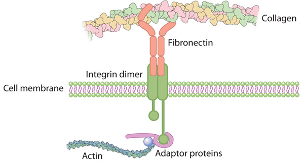
The integrin family of αβ heterodimeric receptors act as

cell adhesion molecules

connecting the ECM to the actin cytoskeleton.

The actin cytoskeleton is involved in the regulation of

1.cell motility,

2.cell polarity,

3.cell growth, and

4.cell survival.

The combination of αβ subunits determines

binding specificity and

signaling properties.

Both α and β integrin subunits contain two separate tails, which

penetrate the plasma membrane and possess small cytoplasmic domains which facilitate

the signaling functions of the receptor.

There is some evidence that the β subunit is the principal site for

binding of cytoskeletal and signaling molecules,

whereas the α subunit has a regulatory role. The integrin tails

link the ECM to the actin cytoskeleton within the cell and with cytoplasmic proteins,

such as talin, tensin, and filamin. The extracellular domains of integrin receptors bind the ECM ligands.

binding of integrins depends on ECM divalent cations ch19

binding of integrins depends on ECM divalent cations ch19

integrin coupled to F-actin via linker

integrin coupled to F-actin via linker

http://www.nature.com/nrm/journal/vaop/ncurrent/images/nrm3896-f4.jpg

Schematic of the ‘focal adhesion clutch’ on stiff (a) versus soft (b) extracellular matrix (ECM). In all cases, integrins are coupled to F-actin via linker proteins (for example, talin and vinculin). The linker proteins move backwards (as indicated by the small arrows) as F-actin also moves backwards, under pushing forces from actin polymerization and/or pulling forces from myosin II activity. This mechanism transfers force from actin to integrins, which pull on the ECM. A stiff ECM (a) resists this force so that the bound integrins remain immobile. A compliant matrix (b) deforms under this force (as indicated by the compressed ECM labelled as deformed matrix) so that the bound integrins can also move backwards. Their movement reduces the net loading rate on all the force-bearing elements, which results in altered cellular responses

The ECM is a complex mixture of matrix molecules, including –

  • glycoproteins, collagens, laminins, glycosaminoglycans, proteoglycans,
  • and nonmatrix proteins, – including growth factors

The integrin receptor formed from the binding of α and β subunits is

  • shaped like a globular head supported by two rod-like legs (Figure 1).

Most of the contact between the two subunits occurs in the head region, with

  • the intracellular tails of the subunits forming the legs of the receptor.

Integrin recognition of ligands is not constitutive but

  • is regulated by alteration of integrin affinity for ligand binding.

For integrin binding to ligands to occur

  • the integrin must be primed and activated, both of which involve
  • conformational changes to the receptor.

Linking integrin conformation to function

Figure  Integrin binding to extracellular matrix (ECM). Conformational changes to integrin structure and clustering of subunits which allow enhanced function of the receptor.

Integrins work alongside other proteins such as

cadherins,

immunoglobulin superfamily

cell adhesion molecules,

selectins, and

syndecans

to mediate

cell–cell and

cell–matrix interactions and communication.

Activation of adhesion receptors triggers the formation of matrix contacts in which

bound matrix components,

adhesion receptors,

and associated intracellular cytoskeletal and signaling molecules

form large functional, localized multiprotein complexes.

Cell–matrix contacts are important in a variety of different cell and

tissue properties including

1.embryonic development,

2.inflammatory responses,

3.wound healing,

4.and adult tissue homeostasis.

Integrin extracellular binding activity is regulated from inside the cell and binding to the ECM induces signals that are transmitted into the cell. This bidirectional signaling requires

dynamic,

spatially, and

temporally regulated formation and

disassembly of multiprotein complexes that

form around the short cytoplasmic tails of integrins.

Ligand binding to integrin family members leads to clustering of integrin molecules in the plasma membrane and recruitment of actin filaments and intracellular signaling molecules to the cytoplasmic domain of the integrins. This forms focal adhesion complexes which are able to maintain

not only adhesion to the ECM

but are involved in complex signaling pathways

which include establishing

1.cell polarity,

2.directed cell migration, and

3.maintaining cell growth and survival.

Initial activation through integrin adhesion to matrix recruits up to around 50 diverse signaling molecules

to assemble the focal adhesion complex

which is capable of responding to environmental stimuli efficiently.

Mapping of the integrin

adhesome binding and signaling interactions

a network of 156 components linked together which can be modified by 690 interactions.

Genetic programming occurs with the binding of integrins to the ECM

Signal transduction pathway activation arising from integrin-ECM binding results in

  • changes in gene expression of cells and
  • leads to alterations in cell and tissue function.

Various different effects can arise depending on the

1.cell type,

2.matrix composition, and

3.integrins activated

It has been suggested that integrin-type I collagen interaction is necessary for

  • the phosphorylation and activation of osteoblast-specific transcription factors
  • present in committed osteoprogenitor cells.

During mechanical loading/stimulation of chondrocytes there is an

  1. influx of ions across the cell membrane resulting from
  2. activation of mechanosensitive ion channels
  3. which can be inhibited by subunit-specific anti-integrin blocking antibodies or RGD peptides.

Using these strategies it was identified that

  • α5β1 integrin is a major mechanoreceptor in articular chondrocyte
  • responses to mechanical loading/stimulation.

Osteoarthritic chondrocytes show a depolarization response to 0.33 Hz stimulation

  • in contrast to the hyperpolarization response of normal chondrocytes.

The mechanotransduction pathway in chondrocytes derived from normal and osteoarthritic cartilage

  • both involve recognition of the mechanical stimulus
  • by integrin receptors resulting in
  • the activation of integrin signaling pathways
  • leading to the generation of a cytokine loop.

Normal and osteoarthritic chondrocytes show differences

  • at multiple stages of the mechanotransduction cascade.
Signaling pathways activated in chondrocytes

Signaling pathways activated in chondrocytes

http://dx.doi.org/10.1016/j.matbio.2014.08.007

Chondrocyte integrins are important mediators of cell–matrix interactions in cartilage

  • by regulating the response of the cells to signals from the ECM that
  1. control cell proliferation,
  2. survival,
  3. differentiation,
  4. matrix remodeling.

Integrins participate in development and maintenance of the tissue but also

  • in pathological processes related to matrix destruction, where
  • they likely play a role in the progression of OA.

Cellular adaptation to mechanical stress: role of integrins, Rho, cytoskeletal tension and mechanosensitive ion channels

Cells exhibited four types of mechanical responses:

(1) an immediate viscoelastic response;

(2) early adaptive behavior characterized by pulse-to-pulse attenuation in response to oscillatory forces;

(3) later adaptive cell stiffening with sustained (>15 second) static stresses; and

(4) a large-scale repositioning response with prolonged (>1 minute) stress.

Importantly, these adaptation responses differed biochemically.

The immediate and early responses were affected by

chemically dissipating cytoskeletal prestress (isometric tension), whereas

the later adaptive response was not.

The repositioning response was prevented by

inhibiting tension through interference with Rho signaling,

similar to the case of the immediate and early responses, but it was also prevented by

blocking mechanosensitive ion channels or

by inhibiting Src tyrosine kinases.

All adaptive responses were suppressed by cooling cells to 4°C to slow biochemical remodeling. Thus, cells use multiple mechanisms to sense and respond to static and dynamic changes in the level of mechanical stress applied to integrins.

Microtubule-Stimulated ADP Release, ATP Binding, and Force Generation In Transport Kinesins

All three classes of molecular motor proteins are now known to be

  • large protein families with diverse cellular functions.

Both the kinesin family and the myosin family have been defined and their proteins grouped into subfamilies. Finally, the elusive cytoplasmic version of dynein was identified and a multigene family of flagellar and cytoplasmic dyneins defined. Members of a given motor protein family share

  • significant homology in their motor domains with the defining member,
  • kinesin, dynein or myosin; but they also contain
  • unique protein domains that are specialized for interaction with different cargoes.

This large number of motor proteins may reflect

  • the number of cellular functions that require force generation or movement,
  • ranging from mitosis to morphogenesis to transport of vesicles.

Kinesins are a large family of microtubule (MT)-based motors that play important roles in many cellular activities including

mitosis,

motility, and

intracellular transport

Their involvement in a range of pathological processes

  • also highlights their significance as therapeutic targets and
  • the importance of understanding the molecular basis of their function

They are defined by their motor domains that contain both

  • the microtubule (MT) and
  • ATP binding sites.

Three ATP binding motifs—

  1. the P-loop,
  2. switch I,
  3. switch II–

are highly conserved among

  1. kinesins,
  2. myosin motors, and
  • small GTPases.

They share a conserved mode of MT binding such that

  • MT binding,
  • ATP binding, and
  • hydrolysis

are functionally coupled for efficient MT-based work.

The interior of a cell is a hive of activity, filled with

  • proteins and other items moving from one location to another.

A network of filaments called microtubules forms tracks

  • along which so-called motor proteins carry these items.

Kinesins are one group of motor proteins, and a typical kinesin protein has

  • one end (called the ‘motor domain’) that can attach itself to the microtubules.

The other end links to the cargo being carried, and a ‘neck’ connects the two. When two of these proteins work together,

  • flexible regions of the neck allow the two motor domains to move past one another,
  • which enable the kinesin to essentially walk along a microtubule in a stepwise manner.

Although the two kinesins have been thought to move along the microtubule tracks in different ways, Atherton et al. find that the core mechanism used by their motor domains is the same.

When a motor domain binds to the microtubule, its shape changes,

  • first stimulating release of the breakdown products of ATP from the previous cycle.

This release makes room for a new ATP molecule to bind. The structural changes caused by ATP binding

  • produce larger changes in the flexible neck region that
  • enable individual motor domains within a kinesin pair to
  • co-ordinate their movement and move in a consistent direction.

The major and largely invariant point of contact between kinesin motor domains and the MT is helix-α4,

  • which lies at the tubulin intradimer interface.

The conformational changes in functionally important regions of each motor domain are described,

  • starting with the nucleotide-binding site,
  • from which all other conformational changes emanate.

The nucleotide-binding site (Figure 2) has three major elements:

(1) the P-loop (brown) is visible in all our reconstructions;

(2) loop9 (yellow, contains switch I) undergoes major conformational changes through the ATPase cycle; and

(3) loop11 (red, contains switch II) that connects strand-β7 to helix-α4, the conformation and flexibility of which is

  • determined by MT binding and motor nucleotide state.

Movement and extension of helix-α6 controls neck linker docking

the N-terminus of helix-α6 is closely associated with elements of the nucleotide binding site suggesting that

  • its conformation alters in response to different nucleotide states.

Further,

  • because the orientation of helix-α6 with respect to helix-α4 controls neck linker docking and
  • because helix-α4 is held against the MT during the ATPase cycle,
    • conformational changes in helix-α6 control movement of the neck linker.

Mechanical amplification and force generation involves conformational changes across the motor domain

A key conformational change in the motor domain following Mg-ATP binding is

  • peeling of the central β-sheet from the C-terminus of helix-α4 increasing their separation;
  • this is required to accommodate rotation of helix-α6 and consequent neck linker docking

ATP binding draws loop11 and loop9 closer together; causing

(1) tilting of most of the motor domain not contacting the MT towards the nucleotide-binding site,

(2) rotation, translation, and extension of helix-α6 which we propose contributes to force generation, and

(3) allows neck linker docking and biases movement of the 2nd head towards the MT plus end.

In both motors, microtubule binding promotes

ordered conformations of conserved loops that

stimulate ADP release,

enhance microtubule affinity and

prime the catalytic site for ATP binding.

ATP binding causes only small shifts of these nucleotide-coordinating loops but induces

large conformational changes elsewhere that

allow force generation and

neck linker docking towards the microtubule plus end.

The study presents evidence provide evidence for a conserved ATP-driven

mechanism for kinesins and

reveals the critical mechanistic contribution of the microtubule interface.

Phosphorylation at endothelial cell–cell junctions: Implications for VE-cadherin function

This review summarizes the role of VE-cadherin phosphorylation in the regulation of endothelial cell–cell junctions and highlights how this affects vascular permeability and leukocyte extravasation.

The vascular endothelium is the inner lining of blood vessels and

forms a physical barrier between the vessel lumen and surrounding tissue;

controlling the extravasation of fluids,

plasma proteins and leukocytes.

Changes in the permeability of the endothelium are tightly regulated. Under basal physiological conditions, there is a continuous transfer of substances across the capillary beds. In addition the endothelium can mediate inducible,

transient hyperpermeability

in response to stimulation with inflammatory mediators,

which takes place primarily in post-capillary venules

However, when severe, inflammation may result in dysfunction of the endothelial barrier

  • in various parts of the vascular tree, including large veins, arterioles and capillaries.

Dysregulated permeability is observed in various pathological conditions, such as

  • tumor-induced angiogenesis,
  • cerebrovascular accident and
  • atherosclerosis.

Two fundamentally different pathways regulate endothelial permeability,

  1. the transcellular and
  2. paracellular pathways.

Solutes and cells can pass through the body of endothelial cells via the transcellular pathway, which includes

  • vesicular transport systems,
  • fenestrae, and
  • biochemical transporters.

The paracellular route is controlled by

  • the coordinated opening and closing of endothelial junctions and
  • thereby regulates traffic across the intercellular spaces between endothelial cells.

Endothelial cells are connected by

tight, gap and

adherens junctions,

of which the latter, and particularly the adherens junction component,

vascular endothelial (VE)-cadherin,

are of central importance for the initiation and stabilization of cell–cell contacts.

Although multiple adhesion molecules are localized at endothelial junctions,

  • blocking the adhesive function of VE-cadherin using antibodies
  • is sufficient to disrupt endothelial junctions and
  • to increase endothelial monolayer permeability both in vitro and in vivo.

Like other cadherins, VE-cadherin mediates adhesion via

  • homophilic, calcium-dependent interactions.

This cell–cell adhesion

is strengthened by binding of cytoplasmic proteins, the catenins,

to the C-terminus of VE-cadherin.

VE-cadherin can directly bind

  • β-catenin and plakoglobin, which
  • both associate with the actin binding protein α-catenin.

Initially, α-catenin was thought to directly anchor cadherins to the actin cytoskeleton, but recently it became clear that

  • α-catenin cannot bind to both β-catenin and actin simultaneously.

Numerous lines of evidence indicate that p120-catenin

  • promotes VE-cadherin surface expression and stability at the plasma membrane.

Different models are proposed that describe how

  • p120-catenin regulates cadherin membrane dynamics, including the hypothesis
  • that p120-catenin functions as a ‘cap’ that prevents the interaction of VE-cadherin
  • with the endocytic membrane trafficking machinery.

In addition, p120-catenin might regulate VE-cadherin internalization

  • through interactions with small GTPases.

Cytoplasmic p120-catenin, which is not bound to VE-cadherin, has been shown to

decrease RhoA activity,

elevate active Rac1 and Cdc42, and thereby is thought

to regulate actin cytoskeleton organization and membrane trafficking.

The intact cadherin-catenin complex is required for proper functioning of the adherens junction.

Several mechanisms may be involved in the

  • regulation of the organization and function of the cadherin–catenin complex, including
  1. endocytosis of the complex,
  2. VE-cadherin cleavage and
  3. actin cytoskeleton reorganization.

The remainder of this review primarily focuses on the

role of tyrosine phosphorylation in the control of VE-cadherin-mediated cell–cell adhesion.

Regulation of the adhesive function of VE-cadherin by tyrosine phosphorylation

It is a widely accepted concept that tyrosine phosphorylation of

  • components of the VE–cadherin-catenin complex
  • Correlates with the weakening of cell–cell adhesion.

A general idea has emerged that

tyrosine phosphorylation of the VE-cadherin complex

leads to the uncoupling of VE-cadherin from the actin cytoskeleton

through dissociation of catenins from the cadherin.

However, tyrosine phosphorylation of VE-cadherin

  • is required for efficient transmigration of leukocytes.

This suggests that VE-cadherin-mediated cell–cell contacts

1.are not just pushed open by the migrating leukocytes, but play

2.a more active role in the transmigration process.

A schematic overview of leukocyte adhesion-induced signals leading to VE-cadherin phosphorylation

Regulation of the integrity of endothelial cell–cell contacts by phosphorylation of VE-cadherin.

Regulation of the integrity of endothelial cell–cell contacts by phosphorylation of VE-cadherin

Regulation of the integrity of endothelial cell–cell contacts by phosphorylation of VE-cadherin

N-glycosylation status of E-cadherin controls cytoskeletal dynamics through the organization of distinct β-catenin- and γ-catenin-containing AJs

N-glycosylation of E-cadherin has been shown to inhibit cell–cell adhesion.

Specifically, our recent studies have provided evidence that

  • the reduction of E-cadherin N-glycosylation
  • promoted the recruitment of stabilizing components,
  • vinculin and serine/ threonine protein phosphatase 2A (PP2A), to adherens junctions (AJs)
  • and enhanced the association of AJs with the actin cytoskeleton.

Here, we examined the details of how

N-glycosylation of E-cadherin affected the molecular organization of AJs and their cytoskeletal interactions.

Using the hypoglycosylated E-cadherin variant, V13, we show that

V13/β-catenin complexes preferentially interacted with PP2A and with the microtubule motor protein dynein.

This correlated with dephosphorylation of the microtubule-associated protein tau, suggesting that

increased association of PP2A with V13-containing AJs promoted their tethering to microtubules.

These studies provide the first mechanistic insights into how N-glycosylation of E-cadherin drives changes in AJ composition through

  • the assembly of distinct β-catenin- and γ-catenin-containing scaffolds that impact the interaction with different cytoskeletal components

Cytoskeletal Basis of Ion Channel Function in Cardiac Muscle

MacKinnon. Fig 1  Ion channels exhibit three basic properties

MacKinnon. Fig 1 Ion channels exhibit three basic properties

In order to contract and accommodate the repetitive morphological changes induced by the cardiac cycle, cardiomyocytes

depend on their highly evolved and specialized cytoskeletal apparatus.

Defects in components of the cytoskeleton, in the long term,

affect the ability of the cell to compensate at both functional and structural levels.

In addition to the structural remodeling,

the myocardium becomes increasingly susceptible to altered electrical activity leading to arrhythmogenesis.

The development of arrhythmias secondary to structural remodeling defects has been noted, although the detailed molecular mechanisms are still elusive.

subjects with severe left ventricular chamber dilation such as in DCM can have left bundle branch block (LBBB), while right bundle branch block (RBBB) is more characteristic of right ventricular failure.  LBBB and RBBB have both been repeatedly associated with AV block in heart failure.

The impact of volume overload on structural and electro-cardiographic alterations has been noted in cardiomyopathy patients treated with left ventricular assist device (LVAD) therapy, which puts the heart at mechanical rest.

In LVAD-treated subjects,

QRS- and both QT- and QTc duration decreased,

suggesting that QRS- and QT-duration are significantly influenced by mechanical load and

that the shortening of the action potential duration contributes to the improved contractile performance after LVAD support.

An early postoperative period study after cardiac unloading therapy in 17 HF patients showed that in the first two weeks after LVAD implantation,

HF was associated with a relatively high incidence of ventricular arrhythmias associated with QTc interval prolongation.

In addition, a recent retrospective study of 100 adult patients with advanced HF, treated with an axial-flow HeartMate LVAD suggested that

  • the rate of new-onset monomorphic ventricular tachycardia (MVT) was increased in LVAD treated patients compared to patients given only medical treatment,

The myocardium is exposed to severe and continuous biomechanical stress during each contraction-relaxation cycle. When fiber tension remains uncompensated or simply unbalanced,

it may represent a trigger for arrhythmogenesis caused by cytoskeletal stretching,

which ultimately leads to altered ion channel localization, and subsequent action potential and conduction alterations.

Cytoskeletal proteins not only provide the backbone of the cellular structure, but they also

maintain the shape and flexibility of the different sub-cellular compartments, including the

1.plasma membrane,

2.the double lipid layer, which defines the boundaries of the cell and where

ion channels are mainly localized.

The interaction between the sarcomere, which is the basic for the passive force during diastole and for the restoring force during systole.

Sarcomeric Proteins and Ion Channels

besides fiber stretch associated with mechanical and hemodynamic impairment, cytoskeletal alterations due to primary genetic defects or indirectly to alterations in response to cellular injury can potentially

1.affect ion channel anchoring, and trafficking, as well as

2.functional regulation by second messenger pathways,

3.causing an imbalance in cardiac ionic homeostasis that will trigger arrhythmogenesis.

Intense investigation of

the sarcomeric actin network,

the Z-line structure, and

chaperone molecules docking in the plasma membrane,

has shed new light on the molecular basis of

  • cytoskeletal interactions in regulating ion channels

Actin disruption using cytochalasin D, an agent that interferes with actin polymerization, increased Na+ channel activity in 90% of excised patches tested within 2 min, which indicated that

the integrity of the filamentous actin (F-actin) network was essential for the maintenance of normal Na+ channel function

These data were the first to support a role for the cytoskeleton in cardiac arrhythmias.

Molecular interactions between the cytoskeleton and ion channels

The figure illustrates the interactions between the ion channels on the sarcolemma, and the sarcomere in cardiac myocytes. Note that the Z-line is connected to the cardiac T-tubules. The diagram illustrates the complex protein-protein interactions that occur between structural components of the cytoskeleton and ion channels. The cytoskeleton is involved in regulating the metabolism of ion channels, modifying their expression, localization, and electrical properties.

sarcomere structure

sarcomere structure

It is important to be aware of the enormous variety of clinical presentations that derive from distinct variants in the same pool of genetic factors. Knowledge of these variants could facilitate tailoring the therapy of choice for each patient. In particular,

the recent findings of structural and functional links between

the cytoskeleton and ion channels

could expand the therapeutic interventions in

arrhythmia management in structurally abnormal myocardium, where aberrant binding

between cytoskeletal proteins can directly or indirectly alter ion channel function.

Read Full Post »

Summary of Signaling and Signaling Pathways

Summary of Signaling and Signaling Pathways

Author and Curator: Larry H Bernstein, MD, FCAP

In the imtroduction to this series of discussions I pointed out JEDS Rosalino’s observation about the construction of a complex molecule of acetyl coenzyme A, and the amount of genetic coding that had to go into it.  Furthermore, he observes –  Millions of years later, or as soon as, the information of interaction leading to activity and regulation could be found in RNA, proteins like reverse transcriptase move this information to a more stable form (DNA). In this way it is easier to understand the use of CoA to make two carbon molecules more reactive.

acetylCoA

acetylCoA

In the tutorial that follows we find support for the view that mechanisms and examples from the current literature, which give insight into the developments in cell metabolism, are achieving a separation from inconsistent views introduced by the classical model of molecular biology and genomics, toward a more functional cellular dynamics that is not dependent on the classic view.  The classical view fits a rigid framework that is to genomics and metabolomics as Mendelian genetics if to multidimentional, multifactorial genetics.  The inherent difficulty lies in two places:

  1. Interactions between differently weighted determinants
  2. A large part of the genome is concerned with regulatory function, not expression of the code

The goal of the tutorial was to achieve an understanding of how cell signaling occurs in a cell.  Completion of the tutorial would provide

  1. a basic understanding signal transduction and
  2. the role of phosphorylation in signal transduction.
Regulation of the integrity of endothelial cell–cell contacts by phosphorylation of VE-cadherin

Regulation of the integrity of endothelial cell–cell contacts by phosphorylation of VE-cadherin

In addition – detailed knowledge of –

  1. the role of Tyrosine kinases and
  2. G protein-coupled receptors in cell signaling.
serine

serine

threonine

threonine

protein kinase

protein kinase

We are constantly receiving and interpreting signals from our environment, which can come

  • in the form of light, heat, odors, touch or sound.

The cells of our bodies are also

  • constantly receiving signals from other cells.

These signals are important to

  • keep cells alive and functioning as well as
  • to stimulate important events such as
  • cell division and differentiation.

Signals are most often chemicals that can be found

  • in the extracellular fluid around cells.

These chemicals can come

  • from distant locations in the body (endocrine signaling by hormones), from
  • nearby cells (paracrine signaling) or can even
  • be secreted by the same cell (autocrine signaling).

Notch-mediated juxtacrine signal between adjacent cells. 220px-Notchccr

Signaling molecules may trigger any number of cellular responses, including

  • changing the metabolism of the cell receiving the signal or
  • result in a change in gene expression (transcription) within the nucleus of the cell or both.
controlling the output of ribosomes.

controlling the output of ribosomes.

To which I would now add..

  • result in either an inhibitory or a stimulatory effect

The three stages of cell signaling are:

Cell signaling can be divided into 3 stages:

Reception: A cell detects a signaling molecule from the outside of the cell.

Transduction: When the signaling molecule binds the receptor it changes the receptor protein in some way. This change initiates the process of transduction. Signal transduction is usually a pathway of several steps. Each relay molecule in the signal transduction pathway changes the next molecule in the pathway.

Response: Finally, the signal triggers a specific cellular response.

signal transduction

signal transduction

http://www.hartnell.edu/tutorials/biology/images/signaltransduction_simple.jpg

The initiation is depicted as follows:

Signal Transduction – ligand binds to surface receptor

Membrane receptors function by binding the signal molecule (ligand) and causing the production of a second signal (also known as a second messenger) that then causes a cellular response. These types of receptors transmit information from the extracellular environment to the inside of the cell.

  • by changing shape or
  • by joining with another protein
  • once a specific ligand binds to it.

Examples of membrane receptors include

  • G Protein-Coupled Receptors and
Understanding these receptors and identifying their ligands and the resulting signal transduction pathways represent a major conceptual advance.

Understanding these receptors and identifying their ligands and the resulting signal transduction pathways represent a major conceptual advance.

  • Receptor Tyrosine Kinases.
intracellular signaling

intracellular signaling

http://www.hartnell.edu/tutorials/biology/images/membrane_receptor_tk.jpg

Intracellular receptors are found inside the cell, either in the cytopolasm or in the nucleus of the target cell (the cell receiving the signal).

Note that though change in gene expression is stated, the change in gene expression does not here imply a change in the genetic information – such as – mutation.  That does not have to be the case in the normal homeostatic case.

This point is the differentiating case between what JEDS Roselino has referred as

  1. a fast, adaptive reaction, that is the feature of protein molecules, and distinguishes this interaction from
  2. a one-to-one transcription of the genetic code.

The rate of transcription can be controlled, or it can be blocked.  This is in large part in response to the metabolites in the immediate interstitium.

This might only be

  • a change in the rate of a transcription or a suppression of expression through RNA.
  • Or through a conformational change in an enzyme
 Swinging domains in HECT E3 enzymes

Swinging domains in HECT E3 enzymes

Since signaling systems need to be

  • responsive to small concentrations of chemical signals and act quickly,
  • cells often use a multi-step pathway that transmits the signal quickly,
  • while amplifying the signal to numerous molecules at each step.

Signal transduction pathways are shown (simplified):

Signal Transduction

Signal Transduction

Signal transduction occurs when an

  1. extracellular signaling molecule activates a specific receptor located on the cell surface or inside the cell.
  2. In turn, this receptor triggers a biochemical chain of events inside the cell, creating a response.
  3. Depending on the cell, the response alters the cell’s metabolism, shape, gene expression, or ability to divide.
  4. The signal can be amplified at any step. Thus, one signaling molecule can cause many responses.

In 1970, Martin Rodbell examined the effects of glucagon on a rat’s liver cell membrane receptor. He noted that guanosine triphosphate disassociated glucagon from this receptor and stimulated the G-protein, which strongly influenced the cell’s metabolism. Thus, he deduced that the G-protein is a transducer that accepts glucagon molecules and affects the cell. For this, he shared the 1994 Nobel Prize in Physiology or Medicine with Alfred G. Gilman.

Guanosine monophosphate structure

Guanosine monophosphate structure

In 2007, a total of 48,377 scientific papers—including 11,211 e-review papers—were published on the subject. The term first appeared in a paper’s title in 1979. Widespread use of the term has been traced to a 1980 review article by Rodbell: Research papers focusing on signal transduction first appeared in large numbers in the late 1980s and early 1990s.

Signal transduction involves the binding of extracellular signaling molecules and ligands to cell-surface receptors that trigger events inside the cell. The combination of messenger with receptor causes a change in the conformation of the receptor, known as receptor activation.

This activation is always the initial step (the cause) leading to the cell’s ultimate responses (effect) to the messenger. Despite the myriad of these ultimate responses, they are all directly due to changes in particular cell proteins. Intracellular signaling cascades can be started through cell-substratum interactions; examples are the integrin that binds ligands in the extracellular matrix and steroids.

Integrin

Integrin

Most steroid hormones have receptors within the cytoplasm and act by stimulating the binding of their receptors to the promoter region of steroid-responsive genes.

steroid hormone receptor

steroid hormone receptor

Various environmental stimuli exist that initiate signal transmission processes in multicellular organisms; examples include photons hitting cells in the retina of the eye, and odorants binding to odorant receptors in the nasal epithelium. Certain microbial molecules, such as viral nucleotides and protein antigens, can elicit an immune system response against invading pathogens mediated by signal transduction processes. This may occur independent of signal transduction stimulation by other molecules, as is the case for the toll-like receptor. It may occur with help from stimulatory molecules located at the cell surface of other cells, as with T-cell receptor signaling. Receptors can be roughly divided into two major classes: intracellular receptors and extracellular receptors.

Signal transduction cascades amplify the signal output

Signal transduction cascades amplify the signal output

Signal transduction cascades amplify the signal output

G protein-coupled receptors (GPCRs) are a family of integral transmembrane proteins that possess seven transmembrane domains and are linked to a heterotrimeric G protein. Many receptors are in this family, including adrenergic receptors and chemokine receptors.

Arrestin binding to active GPCR kinase (GRK)-phosphorylated GPCRs blocks G protein coupling

signal transduction pathways

signal transduction pathways

Arrestin binding to active GPCR kinase (GRK)-phosphorylated GPCRs blocks G protein coupling

Arrestin binding to active GPCR kinase (GRK)-phosphorylated GPCRs blocks G protein coupling

Signal transduction by a GPCR begins with an inactive G protein coupled to the receptor; it exists as a heterotrimer consisting of Gα, Gβ, and Gγ. Once the GPCR recognizes a ligand, the conformation of the receptor changes to activate the G protein, causing Gα to bind a molecule of GTP and dissociate from the other two G-protein subunits.

The dissociation exposes sites on the subunits that can interact with other molecules. The activated G protein subunits detach from the receptor and initiate signaling from many downstream effector proteins such as phospholipases and ion channels, the latter permitting the release of second messenger molecules.

Receptor tyrosine kinases (RTKs) are transmembrane proteins with an intracellular kinase domain and an extracellular domain that binds ligands; examples include growth factor receptors such as the insulin receptor.

 insulin receptor and and insulin receptor signaling pathway (IRS)

insulin receptor and and insulin receptor signaling pathway (IRS)

To perform signal transduction, RTKs need to form dimers in the plasma membrane; the dimer is stabilized by ligands binding to the receptor.

RTKs

RTKs

The interaction between the cytoplasmic domains stimulates the autophosphorylation of tyrosines within the domains of the RTKs, causing conformational changes.

Allosteric_Regulation.svg

Subsequent to this, the receptors’ kinase domains are activated, initiating phosphorylation signaling cascades of downstream cytoplasmic molecules that facilitate various cellular processes such as cell differentiation and metabolism.

Signal-Transduction-Pathway

Signal-Transduction-Pathway

As is the case with GPCRs, proteins that bind GTP play a major role in signal transduction from the activated RTK into the cell. In this case, the G proteins are

  • members of the Ras, Rho, and Raf families, referred to collectively as small G proteins.

They act as molecular switches usually

  • tethered to membranes by isoprenyl groups linked to their carboxyl ends.

Upon activation, they assign proteins to specific membrane subdomains where they participate in signaling. Activated RTKs in turn activate

  • small G proteins that activate guanine nucleotide exchange factors such as SOS1.

Once activated, these exchange factors can activate more small G proteins, thus

  • amplifying the receptor’s initial signal.

The mutation of certain RTK genes, as with that of GPCRs, can result in the expression of receptors that exist in a constitutively activate state; such mutated genes may act as oncogenes.

Integrin


Integrin

Integrin

Integrin-mediated signal transduction

An overview of integrin-mediated signal transduction, adapted from Hehlgens et al. (2007).

Integrins are produced by a wide variety of cells; they play a role in

  • cell attachment to other cells and the extracellular matrix and
  • in the transduction of signals from extracellular matrix components such as fibronectin and collagen.

Ligand binding to the extracellular domain of integrins

  • changes the protein’s conformation,
  • clustering it at the cell membrane to
  • initiate signal transduction.

Integrins lack kinase activity; hence, integrin-mediated signal transduction is achieved through a variety of intracellular protein kinases and adaptor molecules, the main coordinator being integrin-linked kinase.

As shown in the picture, cooperative integrin-RTK signaling determines the

  1. timing of cellular survival,
  2. apoptosis,
  3. proliferation, and
  4. differentiation.
integrin-mediated signal transduction

integrin-mediated signal transduction

Integrin signaling

Integrin signaling

ion channel

A ligand-gated ion channel, upon binding with a ligand, changes conformation

  • to open a channel in the cell membrane
  • through which ions relaying signals can pass.

An example of this mechanism is found in the receiving cell of a neural synapse. The influx of ions that occurs in response to the opening of these channels

  1. induces action potentials, such as those that travel along nerves,
  2. by depolarizing the membrane of post-synaptic cells,
  3. resulting in the opening of voltage-gated ion channels.
RyR and Ca+ release from SR

RyR and Ca+ release from SR

An example of an ion allowed into the cell during a ligand-gated ion channel opening is Ca2+;

  • it acts as a second messenger
  • initiating signal transduction cascades and
  • altering the physiology of the responding cell.

This results in amplification of the synapse response between synaptic cells

  • by remodelling the dendritic spines involved in the synapse.

In eukaryotic cells, most intracellular proteins activated by a ligand/receptor interaction possess an enzymatic activity; examples include tyrosine kinase and phosphatases. Some of them create second messengers such as cyclic AMP and IP3,

cAMP

cAMP

Inositol_1,4,5-trisphosphate.svg

Inositol_1,4,5-trisphosphate.svg

  • the latter controlling the release of intracellular calcium stores into the cytoplasm.

Many adaptor proteins and enzymes activated as part of signal transduction possess specialized protein domains that bind to specific secondary messenger molecules. For example,

  • calcium ions bind to the EF hand domains of calmodulin,
  • allowing it to bind and activate calmodulin-dependent kinase.
calcium movement and RyR2 receptor

calcium movement and RyR2 receptor

PIP3 and other phosphoinositides do the same thing to the Pleckstrin homology domains of proteins such as the kinase protein AKT.

Signals can be generated within organelles, such as chloroplasts and mitochondria, modulating the nuclear
gene expression in a process called retrograde signaling.

Recently, integrative genomics approaches, in which correlation analysis has been applied on transcript and metabolite profiling data of Arabidopsis thaliana, revealed the identification of metabolites which are putatively acting as mediators of nuclear gene expression.

http://fpls.com/unraveling_retrograde_signaling_pathways:_finding_candidate_signaling_molecules_via_metabolomics_and_systems_biology_driven_approaches

Related articles

  1. Systems Biology Approach Reveals Genome to Phenome Correlation in Type 2 Diabetes (plosone.org)
  2. Gene Expression and Thiopurine Metabolite Profiling in Inflammatory Bowel Disease – Novel Clues to Drug Targets and Disease Mechanisms? (plosone.org)
  3. Activation of the Jasmonic Acid Plant Defence Pathway Alters the Composition of Rhizosphere

Nutrients 2014, 6, 3245-3258; http://dx.doi.org:/10.3390/nu6083245

Omega-3 (ω-3) fatty acids are one of the two main families of long chain polyunsaturated fatty acids (PUFA). The main omega-3 fatty acids in the mammalian body are

  • α-linolenic acid (ALA), docosahexaenoic acid (DHA) and eicosapentaenoic acid (EPA).

Central nervous tissues of vertebrates are characterized by a high concentration of omega-3 fatty acids. Moreover, in the human brain,

  • DHA is considered as the main structural omega-3 fatty acid, which comprises about 40% of the PUFAs in total.

DHA deficiency may be the cause of many disorders such as depression, inability to concentrate, excessive mood swings, anxiety, cardiovascular disease, type 2 diabetes, dry skin and so on.

On the other hand,

  • zinc is the most abundant trace metal in the human brain.

There are many scientific studies linking zinc, especially

  • excess amounts of free zinc, to cellular death.

Neurodegenerative diseases, such as Alzheimer’s disease, are characterized by altered zinc metabolism. Both animal model studies and human cell culture studies have shown a possible link between

  • omega-3 fatty acids, zinc transporter levels and
  • free zinc availability at cellular levels.

Many other studies have also suggested a possible

  • omega-3 and zinc effect on neurodegeneration and cellular death.

Therefore, in this review, we will examine

  • the effect of omega-3 fatty acids on zinc transporters and
  • the importance of free zinc for human neuronal cells.

Moreover, we will evaluate the collective understanding of

  • mechanism(s) for the interaction of these elements in neuronal research and their
  • significance for the diagnosis and treatment of neurodegeneration.

Epidemiological studies have linked high intake of fish and shellfish as part of the daily diet to

  • reduction of the incidence and/or severity of Alzheimer’s disease (AD) and senile mental decline in

Omega-3 fatty acids are one of the two main families of a broader group of fatty acids referred to as polyunsaturated fatty acids (PUFAs). The other main family of PUFAs encompasses the omega-6 fatty acids. In general, PUFAs are essential in many biochemical events, especially in early post-natal development processes such as

  • cellular differentiation,
  • photoreceptor membrane biogenesis and
  • active synaptogenesis.

Despite the significance of these

two families, mammals cannot synthesize PUFA de novo, so they must be ingested from dietary sources. Though belonging to the same family, both

  • omega-3 and omega-6 fatty acids are metabolically and functionally distinct and have
  • opposing physiological effects. In the human body,
  • high concentrations of omega-6 fatty acids are known to increase the formation of prostaglandins and
  • thereby increase inflammatory processes [10].

the reverse process can be seen with increased omega-3 fatty acids in the body.

Many other factors, such as

  1. thromboxane A2 (TXA2),
  2. leukotriene
  3. B4 (LTB4),
  4. IL-1,
  5. IL-6,
  6. tumor necrosis factor (TNF) and
  7. C-reactive protein,

which are implicated in various health conditions, have been shown to be increased with high omega-6 fatty acids but decreased with omega-3 fatty acids in the human body.

Dietary fatty acids have been identified as protective factors in coronary heart disease, and PUFA levels are known to play a critical role in

  • immune responses,
  • gene expression and
  • intercellular communications.

omega-3 fatty acids are known to be vital in

  • the prevention of fatal ventricular arrhythmias, and
  • are also known to reduce thrombus formation propensity by decreasing platelet aggregation, blood viscosity and fibrinogen levels

.Since omega-3 fatty acids are prevalent in the nervous system, it seems logical that a deficiency may result in neuronal problems, and this is indeed what has been identified and reported.

The main

In another study conducted with individuals of 65 years of age or older (n = 6158), it was found that

  • only high fish consumption, but
  • not dietary omega-3 acid intake,
  • had a protective effect on cognitive decline

In 2005, based on a meta-analysis of the available epidemiology and preclinical studies, clinical trials were conducted to assess the effects of omega-3 fatty acids on cognitive protection. Four of the trials completed have shown

a protective effect of omega-3 fatty acids only among those with mild cognitive impairment conditions.

A  trial of subjects with mild memory complaints demonstrated

  • an improvement with 900 mg of DHA.

We review key findings on

  • the effect of the omega-3 fatty acid DHA on zinc transporters and the
  • importance of free zinc to human neuronal cells.

DHA is the most abundant fatty acid in neural membranes, imparting appropriate

  • fluidity and other properties,

and is thus considered as the most important fatty acid in neuronal studies. DHA is well conserved throughout the mammalian species despite their dietary differences. It is mainly concentrated

  • in membrane phospholipids at synapses and
  • in retinal photoreceptors and
  • also in the testis and sperm.

In adult rats’ brain, DHA comprises approximately

  • 17% of the total fatty acid weight, and
  • in the retina it is as high as 33%.

DHA is believed to have played a major role in the evolution of the modern human –

  • in particular the well-developed brain.

Premature babies fed on DHA-rich formula show improvements in vocabulary and motor performance.

Analysis of human cadaver brains have shown that

  • people with AD have less DHA in their frontal lobe
  • and hippocampus compared with unaffected individuals

Furthermore, studies in mice have increased support for the

  • protective role of omega-3 fatty acids.

Mice administrated with a dietary intake of DHA showed

  • an increase in DHA levels in the hippocampus.

Errors in memory were decreased in these mice and they demonstrated

  • reduced peroxide and free radical levels,
  • suggesting a role in antioxidant defense.

Another study conducted with a Tg2576 mouse model of AD demonstrated that dietary

  • DHA supplementation had a protective effect against reduction in
  • drebrin (actin associated protein), elevated oxidation, and to some extent, apoptosis via
  • decreased caspase activity.


Zinc

Zinc is a trace element, which is indispensable for life, and it is the second most abundant trace element in the body. It is known to be related to

  • growth,
  • development,
  • differentiation,
  • immune response,
  • receptor activity,
  • DNA synthesis,
  • gene expression,
  • neuro-transmission,
  • enzymatic catalysis,
  • hormonal storage and release,
  • tissue repair,
  • memory,
  • the visual process

and many other cellular functions. Moreover, the indispensability of zinc to the body can be discussed in many other aspects,  as

  • a component of over 300 different enzymes
  • an integral component of a metallothioneins
  • a gene regulatory protein.

Approximately 3% of all proteins contain

  • zinc binding motifs .

The broad biological functionality of zinc is thought to be due to its stable chemical and physical properties. Zinc is considered to have three different functions in enzymes;

  1. catalytic,
  2. coactive and

Indeed, it is the only metal found in all six different subclasses

of enzymes. The essential nature of zinc to the human body can be clearly displayed by studying the wide range of pathological effects of zinc deficiency. Anorexia, embryonic and post-natal growth retardation, alopecia, skin lesions, difficulties in wound healing, increased hemorrhage tendency and severe reproductive abnormalities, emotional instability, irritability and depression are just some of the detrimental effects of zinc deficiency.

Proper development and function of the central nervous system (CNS) is highly dependent on zinc levels. In the mammalian organs, zinc is mainly concentrated in the brain at around 150 μm. However, free zinc in the mammalian brain is calculated to be around 10 to 20 nm and the rest exists in either protein-, enzyme- or nucleotide bound form. The brain and zinc relationship is thought to be mediated

  • through glutamate receptors, and
  • it inhibits excitatory and inhibitory receptors.

Vesicular localization of zinc in pre-synaptic terminals is a characteristic feature of brain-localized zinc, and

  • its release is dependent on neural activity.

Retardation of the growth and development of CNS tissues have been linked to low zinc levels. Peripheral neuropathy, spina bifida, hydrocephalus, anencephalus, epilepsy and Pick’s disease have been linked to zinc deficiency. However, the body cannot tolerate excessive amounts of zinc.

The relationship between zinc and neurodegeneration, specifically AD, has been interpreted in several ways. One study has proposed that β-amyloid has a greater propensity to

  • form insoluble amyloid in the presence of
  • high physiological levels of zinc.

Insoluble amyloid is thought to

  • aggregate to form plaques,

which is a main pathological feature of AD. Further studies have shown that

  • chelation of zinc ions can deform and disaggregate plaques.

In AD, the most prominent injuries are found in

  • hippocampal pyramidal neurons, acetylcholine-containing neurons in the basal forebrain, and in
  • somatostatin-containing neurons in the forebrain.

All of these neurons are known to favor

  • rapid and direct entry of zinc in high concentration
  • leaving neurons frequently exposed to high dosages of zinc.

This is thought to promote neuronal cell damage through oxidative stress and mitochondrial dysfunction. Excessive levels of zinc are also capable of

  • inhibiting Ca2+ and Na+ voltage gated channels
  • and up-regulating the cellular levels of reactive oxygen species (ROS).

High levels of zinc are found in Alzheimer’s brains indicating a possible zinc related neurodegeneration. A study conducted with mouse neuronal cells has shown that even a 24-h exposure to high levels of zinc (40 μm) is sufficient to degenerate cells.

If the human diet is deficient in zinc, the body

  • efficiently conserves zinc at the tissue level by compensating other cellular mechanisms

to delay the dietary deficiency effects of zinc. These include reduction of cellular growth rate and zinc excretion levels, and

  • redistribution of available zinc to more zinc dependent cells or organs.

A novel method of measuring metallothionein (MT) levels was introduced as a biomarker for the

  • assessment of the zinc status of individuals and populations.

In humans, erythrocyte metallothionein (E-MT) levels may be considered as an indicator of zinc depletion and repletion, as E-MT levels are sensitive to dietary zinc intake. It should be noted here that MT plays an important role in zinc homeostasis by acting

  • as a target for zinc ion binding and thus
  • assisting in the trafficking of zinc ions through the cell,
  • which may be similar to that of zinc transporters

Zinc Transporters

Deficient or excess amounts of zinc in the body can be catastrophic to the integrity of cellular biochemical and biological systems. The gastrointestinal system controls the absorption, excretion and the distribution of zinc, although the hydrophilic and high-charge molecular characteristics of zinc are not favorable for passive diffusion across the cell membranes. Zinc movement is known to occur

  • via intermembrane proteins and zinc transporter (ZnT) proteins

These transporters are mainly categorized under two metal transporter families; Zip (ZRT, IRT like proteins) and CDF/ZnT (Cation Diffusion Facilitator), also known as SLC (Solute Linked Carrier) gene families: Zip (SLC-39) and ZnT (SLC-30). More than 20 zinc transporters have been identified and characterized over the last two decades (14 Zips and 8 ZnTs).

Members of the SLC39 family have been identified as the putative facilitators of zinc influx into the cytosol, either from the extracellular environment or from intracellular compartments (Figure 1).

The identification of this transporter family was a result of gene sequencing of known Zip1 protein transporters in plants, yeast and human cells. In contrast to the SLC39 family, the SLC30 family facilitates the opposite process, namely zinc efflux from the cytosol to the extracellular environment or into luminal compartments such as secretory granules, endosomes and synaptic vesicles; thus decreasing intracellular zinc availability (Figure 1). ZnT3 is the most important in the brain where

  • it is responsible for the transport of zinc into the synaptic vesicles of
  • glutamatergic neurons in the hippocampus and neocortex,

Figure 1: Subcellular localization and direction of transport of the zinc transporter families, ZnT and ZIP. Arrows show the direction of zinc mobilization for the ZnT (green) and ZIP (red) proteins. A net gain in cytosolic zinc is achieved by the transportation of zinc from the extracellular region and organelles such as the endoplasmic reticulum (ER) and Golgi apparatus by the ZIP transporters. Cytosolic zinc is mobilized into early secretory compartments such as the ER and Golgi apparatus by the ZnT transporters. Figures were produced using Servier Medical Art, http://www.servier.com/.   http://www.hindawi.com/journals/jnme/2012/173712.fig.001.jpg

Figure 2: Early zinc signaling (EZS) and late zinc signaling (LZS). EZS involves transcription-independent mechanisms where an extracellular stimulus directly induces an increase in zinc levels within several minutes by releasing zinc from intracellular stores (e.g., endoplasmic reticulum). LSZ is induced several hours after an external stimulus and is dependent on transcriptional changes in zinc transporter expression. Components of this figure were produced using Servier Medical Art, http://www.servier.com/ and adapted from Fukada et al. [30].

omega-3 fatty acids in the mammalian body are

  1. α-linolenic acid (ALA),
  2. docosahexenoic acid (DHA) and
  3. eicosapentaenoic acid (EPA).

In general, seafood is rich in omega-3 fatty acids, more specifically DHA and EPA (Table 1). Thus far, there are nine separate epidemiological studies that suggest a possible link between

  • increased fish consumption and reduced risk of AD
  • and eight out of ten studies have reported a link between higher blood omega-3 levels

DHA and Zinc Homeostasis

Many studies have identified possible associations between DHA levels, zinc homeostasis, neuroprotection and neurodegeneration. Dietary DHA deficiency resulted in

  • increased zinc levels in the hippocampus and
  • elevated expression of the putative zinc transporter, ZnT3, in the rat brain.

Altered zinc metabolism in neuronal cells has been linked to neurodegenerative conditions such as AD. A study conducted with transgenic mice has shown a significant link between ZnT3 transporter levels and cerebral amyloid plaque pathology. When the ZnT3 transporter was silenced in transgenic mice expressing cerebral amyloid plaque pathology,

  • a significant reduction in plaque load
  • and the presence of insoluble amyloid were observed.

In addition to the decrease in plaque load, ZnT3 silenced mice also exhibited a significant

  • reduction in free zinc availability in the hippocampus
  • and cerebral cortex.

Collectively, the findings from this study are very interesting and indicate a clear connection between

  • zinc availability and amyloid plaque formation,

thus indicating a possible link to AD.

DHA supplementation has also been reported to limit the following:

  1. amyloid presence,
  2. synaptic marker loss,
  3. hyper-phosphorylation of Tau,
  4. oxidative damage and
  5. cognitive deficits in transgenic mouse model of AD.

In addition, studies by Stoltenberg, Flinn and colleagues report on the modulation of zinc and the effect in transgenic mouse models of AD. Given that all of these are classic pathological features of AD, and considering the limiting nature of DHA in these processes, it can be argued that DHA is a key candidate in preventing or even curing this debilitating disease.

In order to better understand the possible links and pathways of zinc and DHA with neurodegeneration, we designed a study that incorporates all three of these aspects, to study their effects at the cellular level. In this study, we were able to demonstrate a possible link between omega-3 fatty acid (DHA) concentration, zinc availability and zinc transporter expression levels in cultured human neuronal cells.

When treated with DHA over 48 h, ZnT3 levels were markedly reduced in the human neuroblastoma M17 cell line. Moreover, in the same study, we were able to propose a possible

  • neuroprotective mechanism of DHA,

which we believe is exerted through

  • a reduction in cellular zinc levels (through altering zinc transporter expression levels)
  • that in turn inhibits apoptosis.

DHA supplemented M17 cells also showed a marked depletion of zinc uptake (up to 30%), and

  • free zinc levels in the cytosol were significantly low compared to the control

This reduction in free zinc availability was specific to DHA; cells treated with EPA had no significant change in free zinc levels (unpublished data). Moreover, DHA-repleted cells had

  • low levels of active caspase-3 and
  • high Bcl-2 levels compared to the control treatment.

These findings are consistent with previous published data and further strengthen the possible

  • correlation between zinc, DHA and neurodegeneration.

On the other hand, recent studies using ZnT3 knockout (ZnT3KO) mice have shown the importance of

  • ZnT3 in memory and AD pathology.

For example, Sindreu and colleagues have used ZnT3KO mice to establish the important role of

  • ZnT3 in zinc homeostasis that modulates presynaptic MAPK signaling
  • required for hippocampus-dependent memory

Results from these studies indicate a possible zinc-transporter-expression-level-dependent mechanism for DHA neuroprotection.

Read Full Post »

Complex Models of Signaling: Therapeutic Implications

Complex Models of Signaling: Therapeutic Implications

Curator: Larry H. Bernstein, MD, FCAP

Updated 6/24/2019

Fishy Business: Effect of Omega-3 Fatty Acids on Zinc Transporters and Free Zinc Availability in Human Neuronal Cells

Damitha De Mel and Cenk Suphioglu *

NeuroAllergy Research Laboratory (NARL), School of Life and Environmental Sciences, Faculty of Science, Engineering and Built Environment, Waurn Ponds, Victoria, Australia.

Nutrients 2014, 6, 3245-3258; http://dx.doi.org:/10.3390/nu6083245

Omega-3 (ω-3) fatty acids are one of the two main families of long chain polyunsaturated fatty acids (PUFA). The main omega-3 fatty acids in the mammalian body are

  • α-linolenic acid (ALA), docosahexaenoic acid (DHA) and eicosapentaenoic acid (EPA).

Central nervous tissues of vertebrates are characterized by a high concentration of omega-3 fatty acids. Moreover, in the human brain,

  • DHA is considered as the main structural omega-3 fatty acid, which comprises about 40% of the PUFAs in total.

DHA deficiency may be the cause of many disorders such as depression, inability to concentrate, excessive mood swings, anxiety, cardiovascular disease, type 2 diabetes, dry skin and so on.

On the other hand,

  • zinc is the most abundant trace metal in the human brain.

There are many scientific studies linking zinc, especially

  • excess amounts of free zinc, to cellular death.

Neurodegenerative diseases, such as Alzheimer’s disease, are characterized by altered zinc metabolism. Both animal model studies and human cell culture studies have shown a possible link between

  • omega-3 fatty acids, zinc transporter levels and
  • free zinc availability at cellular levels.

Many other studies have also suggested a possible

  • omega-3 and zinc effect on neurodegeneration and cellular death.

Therefore, in this review, we will examine

  • the effect of omega-3 fatty acids on zinc transporters and
  • the importance of free zinc for human neuronal cells.

Moreover, we will evaluate the collective understanding of

  • mechanism(s) for the interaction of these elements in neuronal research and their
  • significance for the diagnosis and treatment of neurodegeneration.

Epidemiological studies have linked high intake of fish and shellfish as part of the daily diet to

  • reduction of the incidence and/or severity of Alzheimer’s disease (AD) and senile mental decline in

Omega-3 fatty acids are one of the two main families of a broader group of fatty acids referred to as polyunsaturated fatty acids (PUFAs). The other main family of PUFAs encompasses the omega-6 fatty acids. In general, PUFAs are essential in many biochemical events, especially in early post-natal development processes such as

  • cellular differentiation,
  • photoreceptor membrane biogenesis and
  • active synaptogenesis.

Despite the significance of these

two families, mammals cannot synthesize PUFA de novo, so they must be ingested from dietary sources. Though belonging to the same family, both

  • omega-3 and omega-6 fatty acids are metabolically and functionally distinct and have
  • opposing physiological effects. In the human body,
  • high concentrations of omega-6 fatty acids are known to increase the formation of prostaglandins and
  • thereby increase inflammatory processes [10].

the reverse process can be seen with increased omega-3 fatty acids in the body.

Many other factors, such as

  1. thromboxane A2 (TXA2),
  2. leukotriene
  3. B4 (LTB4),
  4. IL-1,
  5. IL-6,
  6. tumor necrosis factor (TNF) and
  7. C-reactive protein,

which are implicated in various health conditions, have been shown to be increased with high omega-6 fatty acids but decreased with omega-3 fatty acids in the human body.

Dietary fatty acids have been identified as protective factors in coronary heart disease, and PUFA levels are known to play a critical role in

  • immune responses,
  • gene expression and
  • intercellular communications.

omega-3 fatty acids are known to be vital in

  • the prevention of fatal ventricular arrhythmias, and
  • are also known to reduce thrombus formation propensity by decreasing platelet aggregation, blood viscosity and fibrinogen levels

.Since omega-3 fatty acids are prevalent in the nervous system, it seems logical that a deficiency may result in neuronal problems, and this is indeed what has been identified and reported.

The main omega-3 fatty acids in the mammalian body are

  1. α-linolenic acid (ALA),
  2. docosahexenoic acid (DHA) and
  3. eicosapentaenoic acid (EPA).

In general, seafood is rich in omega-3 fatty acids, more specifically DHA and EPA (Table 1). Thus far, there are nine separate epidemiological studies that suggest a possible link between

  • increased fish consumption and reduced risk of AD
  • and eight out of ten studies have reported a link between higher blood omega-3 levels

Table 1. Total percentage of omega-3 fatty acids in common foods and supplements.

Food/Supplement EPA DHA ALA Total %
Fish
SalmonSardine

Anchovy

Halibut

Herring

Mackerel

Tuna

Fresh Bluefin

XX

X

X

X

X

X

X

XX

X

X

X

X

X

X

>50%>50%

>50%

>50%

>50%

>50%

>50%

>50%

Oils/Supplements
Fish oil capsulesCod liver oils

Salmon oil

Sardine oil

XX

X

X

XX

X

X

>50%>50%

>50%

>50%

Black currant oilCanola oil Mustard seed oils

Soybean oil

Walnut oil

Wheat germ oil

XX

X

X

X

X

10%–50%10%–50%

10%–50%

10%–50%

10%–50%

10%–50%

Seeds and other foods
Flaxseeds/LinseedsSpinach

Wheat germ Human milk

Peanut butter

Soybeans

Olive oil

Walnuts

XX

X

X

X

X

X

X

>50%>50%

10%–50%

10%–50%

<10%

<10%

<10%

<10%


Table adopted from Maclean C.H. et al. [18].

In another study conducted with individuals of 65 years of age or older (n = 6158), it was found that

  • only high fish consumption, but
  • not dietary omega-3 acid intake,
  • had a protective effect on cognitive decline

In 2005, based on a meta-analysis of the available epidemiology and preclinical studies, clinical trials were conducted to assess the effects of omega-3 fatty acids on cognitive protection. Four of the trials completed have shown

a protective effect of omega-3 fatty acids only among those with mild cognitive impairment conditions.

A  trial of subjects with mild memory complaints demonstrated

  • an improvement with 900 mg of DHA.

We review key findings on

  • the effect of the omega-3 fatty acid DHA on zinc transporters and the
  • importance of free zinc to human neuronal cells.

DHA is the most abundant fatty acid in neural membranes, imparting appropriate

  • fluidity and other properties,

and is thus considered as the most important fatty acid in neuronal studies. DHA is well conserved throughout the mammalian species despite their dietary differences. It is mainly concentrated

  • in membrane phospholipids at synapses and
  • in retinal photoreceptors and
  • also in the testis and sperm.

In adult rats’ brain, DHA comprises approximately

  • 17% of the total fatty acid weight, and
  • in the retina it is as high as 33%.

DHA is believed to have played a major role in the evolution of the modern human –

  • in particular the well-developed brain.

Premature babies fed on DHA-rich formula show improvements in vocabulary and motor performance.

Analysis of human cadaver brains have shown that

  • people with AD have less DHA in their frontal lobe
  • and hippocampus compared with unaffected individuals

Furthermore, studies in mice have increased support for the

  • protective role of omega-3 fatty acids.

Mice administrated with a dietary intake of DHA showed

  • an increase in DHA levels in the hippocampus.

Errors in memory were decreased in these mice and they demonstrated

  • reduced peroxide and free radical levels,
  • suggesting a role in antioxidant defense.

Another study conducted with a Tg2576 mouse model of AD demonstrated that dietary

  • DHA supplementation had a protective effect against reduction in
  • drebrin (actin associated protein), elevated oxidation, and to some extent, apoptosis via
  • decreased caspase activity.


Zinc

Zinc is a trace element, which is indispensable for life, and it is the second most abundant trace element in the body. It is known to be related to

  • growth,
  • development,
  • differentiation,
  • immune response,
  • receptor activity,
  • DNA synthesis,
  • gene expression,
  • neuro-transmission,
  • enzymatic catalysis,
  • hormonal storage and release,
  • tissue repair,
  • memory,
  • the visual process

and many other cellular functions. Moreover, the indispensability of zinc to the body can be discussed in many other aspects,  as

  • a component of over 300 different enzymes
  • an integral component of a metallothioneins
  • a gene regulatory protein.

Approximately 3% of all proteins contain

  • zinc binding motifs .

The broad biological functionality of zinc is thought to be due to its stable chemical and physical properties. Zinc is considered to have three different functions in enzymes;

  1. catalytic,
  2. coactive and

Indeed, it is the only metal found in all six different subclasses

of enzymes. The essential nature of zinc to the human body can be clearly displayed by studying the wide range of pathological effects of zinc deficiency. Anorexia, embryonic and post-natal growth retardation, alopecia, skin lesions, difficulties in wound healing, increased hemorrhage tendency and severe reproductive abnormalities, emotional instability, irritability and depression are just some of the detrimental effects of zinc deficiency.

Proper development and function of the central nervous system (CNS) is highly dependent on zinc levels. In the mammalian organs, zinc is mainly concentrated in the brain at around 150 μm. However, free zinc in the mammalian brain is calculated to be around 10 to 20 nm and the rest exists in either protein-, enzyme- or nucleotide bound form. The brain and zinc relationship is thought to be mediated

  • through glutamate receptors, and
  • it inhibits excitatory and inhibitory receptors.

Vesicular localization of zinc in pre-synaptic terminals is a characteristic feature of brain-localized zinc, and

  • its release is dependent on neural activity.

Retardation of the growth and development of CNS tissues have been linked to low zinc levels. Peripheral neuropathy, spina bifida, hydrocephalus, anencephalus, epilepsy and Pick’s disease have been linked to zinc deficiency. However, the body cannot tolerate excessive amounts of zinc.

The relationship between zinc and neurodegeneration, specifically AD, has been interpreted in several ways. One study has proposed that β-amyloid has a greater propensity to

  • form insoluble amyloid in the presence of
  • high physiological levels of zinc.

Insoluble amyloid is thought to

  • aggregate to form plaques,

which is a main pathological feature of AD. Further studies have shown that

  • chelation of zinc ions can deform and disaggregate plaques.

In AD, the most prominent injuries are found in

  • hippocampal pyramidal neurons, acetylcholine-containing neurons in the basal forebrain, and in
  • somatostatin-containing neurons in the forebrain.

All of these neurons are known to favor

  • rapid and direct entry of zinc in high concentration
  • leaving neurons frequently exposed to high dosages of zinc.

This is thought to promote neuronal cell damage through oxidative stress and mitochondrial dysfunction. Excessive levels of zinc are also capable of

  • inhibiting Ca2+ and Na+ voltage gated channels
  • and up-regulating the cellular levels of reactive oxygen species (ROS).

High levels of zinc are found in Alzheimer’s brains indicating a possible zinc related neurodegeneration. A study conducted with mouse neuronal cells has shown that even a 24-h exposure to high levels of zinc (40 μm) is sufficient to degenerate cells.

If the human diet is deficient in zinc, the body

  • efficiently conserves zinc at the tissue level by compensating other cellular mechanisms

to delay the dietary deficiency effects of zinc. These include reduction of cellular growth rate and zinc excretion levels, and

  • redistribution of available zinc to more zinc dependent cells or organs.

A novel method of measuring metallothionein (MT) levels was introduced as a biomarker for the

  • assessment of the zinc status of individuals and populations.

In humans, erythrocyte metallothionein (E-MT) levels may be considered as an indicator of zinc depletion and repletion, as E-MT levels are sensitive to dietary zinc intake. It should be noted here that MT plays an important role in zinc homeostasis by acting

  • as a target for zinc ion binding and thus
  • assisting in the trafficking of zinc ions through the cell,
  • which may be similar to that of zinc transporters

Zinc Transporters

Deficient or excess amounts of zinc in the body can be catastrophic to the integrity of cellular biochemical and biological systems. The gastrointestinal system controls the absorption, excretion and the distribution of zinc, although the hydrophilic and high-charge molecular characteristics of zinc are not favorable for passive diffusion across the cell membranes. Zinc movement is known to occur

  • via intermembrane proteins and zinc transporter (ZnT) proteins

These transporters are mainly categorized under two metal transporter families; Zip (ZRT, IRT like proteins) and CDF/ZnT (Cation Diffusion Facilitator), also known as SLC (Solute Linked Carrier) gene families: Zip (SLC-39) and ZnT (SLC-30). More than 20 zinc transporters have been identified and characterized over the last two decades (14 Zips and 8 ZnTs).

Members of the SLC39 family have been identified as the putative facilitators of zinc influx into the cytosol, either from the extracellular environment or from intracellular compartments (Figure 1).

The identification of this transporter family was a result of gene sequencing of known Zip1 protein transporters in plants, yeast and human cells. In contrast to the SLC39 family, the SLC30 family facilitates the opposite process, namely zinc efflux from the cytosol to the extracellular environment or into luminal compartments such as secretory granules, endosomes and synaptic vesicles; thus decreasing intracellular zinc availability (Figure 1). ZnT3 is the most important in the brain where

  • it is responsible for the transport of zinc into the synaptic vesicles of
  • glutamatergic neurons in the hippocampus and neocortex,


Figure 1. Putative cellular localization of some of the different human zinc transporters (i.e., Zip1- Zip4 and ZnT1- ZnT7). Arrows indicate the direction of zinc passage by the appropriate putative zinc transporters in a generalized human cell. Although there are fourteen Zips and eight ZnTs known so far, only the main zinc transporters are illustrated in this figure for clarity and brevity.

Figure 1: Subcellular localization and direction of transport of the zinc transporter families, ZnT and ZIP. Arrows show the direction of zinc mobilization for the ZnT (green) and ZIP (red) proteins. A net gain in cytosolic zinc is achieved by the transportation of zinc from the extracellular region and organelles such as the endoplasmic reticulum (ER) and Golgi apparatus by the ZIP transporters. Cytosolic zinc is mobilized into early secretory compartments such as the ER and Golgi apparatus by the ZnT transporters. Figures were produced using Servier Medical Art, http://www.servier.com/.   http://www.hindawi.com/journals/jnme/2012/173712.fig.001.jpg

zinc transporters

zinc transporters


Early zinc signaling (EZS) and late zinc signaling (LZS)

Early zinc signaling (EZS) and late zinc signaling (LZS)

http://www.hindawi.com/journals/jnme/2012/floats/173712/thumbnails/173712.fig.002_th.jpg


Figure 2: Early zinc signaling (EZS) and late zinc signaling (LZS). EZS involves transcription-independent mechanisms where an extracellular stimulus directly induces an increase in zinc levels within several minutes by releasing zinc from intracellular stores (e.g., endoplasmic reticulum). LSZ is induced several hours after an external stimulus and is dependent on transcriptional changes in zinc transporter expression. Components of this figure were produced using Servier Medical Art, http://www.servier.com/ and adapted from Fukada et al. [30].


DHA and Zinc Homeostasis

Many studies have identified possible associations between DHA levels, zinc homeostasis, neuroprotection and neurodegeneration. Dietary DHA deficiency resulted in

  • increased zinc levels in the hippocampus and
  • elevated expression of the putative zinc transporter, ZnT3, in the rat brain.

Altered zinc metabolism in neuronal cells has been linked to neurodegenerative conditions such as AD. A study conducted with transgenic mice has shown a significant link between ZnT3 transporter levels and cerebral amyloid plaque pathology. When the ZnT3 transporter was silenced in transgenic mice expressing cerebral amyloid plaque pathology,

  • a significant reduction in plaque load
  • and the presence of insoluble amyloid were observed.

In addition to the decrease in plaque load, ZnT3 silenced mice also exhibited a significant

  • reduction in free zinc availability in the hippocampus
  • and cerebral cortex.

Collectively, the findings from this study are very interesting and indicate a clear connection between

  • zinc availability and amyloid plaque formation,

thus indicating a possible link to AD.

DHA supplementation has also been reported to limit the following:

  1. amyloid presence,
  2. synaptic marker loss,
  3. hyper-phosphorylation of Tau,
  4. oxidative damage and
  5. cognitive deficits in transgenic mouse model of AD.

In addition, studies by Stoltenberg, Flinn and colleagues report on the modulation of zinc and the effect in transgenic mouse models of AD. Given that all of these are classic pathological features of AD, and considering the limiting nature of DHA in these processes, it can be argued that DHA is a key candidate in preventing or even curing this debilitating disease.

In order to better understand the possible links and pathways of zinc and DHA with neurodegeneration, we designed a study that incorporates all three of these aspects, to study their effects at the cellular level. In this study, we were able to demonstrate a possible link between omega-3 fatty acid (DHA) concentration, zinc availability and zinc transporter expression levels in cultured human neuronal cells.

When treated with DHA over 48 h, ZnT3 levels were markedly reduced in the human neuroblastoma M17 cell line. Moreover, in the same study, we were able to propose a possible

  • neuroprotective mechanism of DHA,

which we believe is exerted through

  • a reduction in cellular zinc levels (through altering zinc transporter expression levels)
  • that in turn inhibits apoptosis.

DHA supplemented M17 cells also showed a marked depletion of zinc uptake (up to 30%), and

  • free zinc levels in the cytosol were significantly low compared to the control

This reduction in free zinc availability was specific to DHA; cells treated with EPA had no significant change in free zinc levels (unpublished data). Moreover, DHA-repleted cells had

  • low levels of active caspase-3 and
  • high Bcl-2 levels compared to the control treatment.

These findings are consistent with previous published data and further strengthen the possible

  • correlation between zinc, DHA and neurodegeneration.

On the other hand, recent studies using ZnT3 knockout (ZnT3KO) mice have shown the importance of

  • ZnT3 in memory and AD pathology.

For example, Sindreu and colleagues have used ZnT3KO mice to establish the important role of

  • ZnT3 in zinc homeostasis that modulates presynaptic MAPK signaling
  • required for hippocampus-dependent memory

Results from these studies indicate a possible zinc-transporter-expression-level-dependent mechanism for DHA neuroprotection.

Collectively from these studies, the following possible mechanism can be proposed (Figure 2).

possible benefits of DHA in neuroprotection through reduction of ZnT3 transporter

possible benefits of DHA in neuroprotection through reduction of ZnT3 transporter


Figure 2. Proposed neuroprotection mechanism of docosahexaenoic acid (DHA) in reference to synaptic zinc. Schematic diagram showing possible benefits of DHA in neuroprotection through reduction of ZnT3 transporter expression levels in human neuronal cells, which results in a reduction of zinc flux and thus lowering zinc concentrations in neuronal synaptic vesicles, and therefore contributing to a lower incidence of neurodegenerative diseases (ND), such as Alzheimer’s disease (AD).

More recent data from our research group have also shown a link between the expression levels of histone H3 and H4 proteins in human neuronal cells in relation to DHA and zinc. Following DHA treatment, both H3 and H4 levels were up-regulated. In contrast, zinc treatment resulted in a down-regulation of histone levels. Both zinc and DHA have shown opposing effects on histone post-translational modifications, indicating a possible distinctive epigenetic pattern. Upon treatment with zinc, M17 cells displayed an increase in histone deacetylase (HDACs) and a reduction in histone acetylation. Conversely, with DHA treatment, HDAC levels were significantly reduced and the acetylation of histones was up-regulated. These findings also support a possible interaction between DHA and zinc availability.

Conclusions

It is possible to safely claim that there is more than one potential pathway by which DHA and zinc interact at a cellular level, at least in cultured human neuronal cells. Significance and importance of both DHA and zinc in neuronal survival is attested by the presence of these multiple mechanisms.
Most of these reported studies were conducted using human neuroblastoma cells, or similar cell types, due to the lack of live mature human neuronal cells. Thus, the results may differ from results achieved under actual human physiological conditions due to the structural and functional differences between these cells and mature human neurons. Therefore, an alternative approach that can mimic the human neuronal cells more effectively would be advantageous.

Sphingosine-1-phosphate signaling as a therapeutic target          

E Giannoudaki, DJ Swan, JA Kirby, S Ali

Applied Immunobiology and Transplantation Research Group, Institute of Cellular Medicine, Faculty of Medical Sciences, Newcastle University, Newcastle upon Tyne, UK

Cell Health and Cytoskeleton 2012; 4: 63–72

S1P is a 379Da member of the lysophospholipid family. It is the direct metabolite of sphingosine through the action of two sphingosine kinases, SphK1 and SphK2. The main metabolic pathway starts with the hydrolysis of sphingomyelin, a membrane sphingolipid, into ceramide by the enzyme sphingomyelinase and the subsequent production of sphingosine by ceramidase (Figure 1). Ceramide can also be produced de novo in the endoplasmic reticulum (ER) from serine and palmitoyl coenzyme A through multiple intermediates. S1P production is regulated by various S1P-specific and general lipid phosphatases, as well as S1P lyase, which irreversibly degrades S1P into phosphoethanolamine and hexadecanal. The balance between intracellular S1P and its metabolite ceramide can determine cellular fate. Ceramide promotes apoptosis, while S1P suppresses cell death and promotes cell survival. This creates an S1P ceramide “rheostat” inside the cells. S1P lyase expression in tissue is higher than it is in erythrocytes and platelets, the main “suppliers” of S1P in blood. This causes a tissue–blood gradient of S1P, which is important in many S1P-mediated responses, like the lymphocyte egress from lymphoid organs.

S1P signaling overview

S1P is produced inside cells; however, it can also be found extracellularly, in a variety of different tissues. It is abundant in the blood, at concentrations of 0.4–1.5 μM, where it is mainly secreted by erythrocytes and platelets. Blood S1P can be found separately, but mainly it exists in complexes with high-density lipoprotein (HDL) (∼60%).  Many of the cardioprotective effects of HDL are hypothesized to involve S1P. Before 1996, S1P was thought to act mainly intracellularly as a second messenger. However, the identification of several GPCRs that bind S1P led to the initiation of many studies on

  • extracellular S1P signaling through those receptors.

There are five receptors that have been identified currently. These can be coupled with different G-proteins. Assuming that each receptor coupling with a G protein has a slightly different function, one can recognize the complexity of S1P receptor signaling.

S1P as a second messenger

S1P is involved in many cellular processes through its GPCR signaling; studies demonstrate that S1P also acts at an intracellular level. Intracellular S1P plays a role in maintaining the balance of cell survival signal toward apoptotic signals, creating a

  • cell “rheostat” between S1P and its precursor ceramide.

Important evidence that S1P can act intracellularly as a second messenger came from yeast (Saccharomyces cerevisiae) and plant (Arabidopsis thaliana) cells. Yeast cells do not express any S1P receptors, although they can be affected by S1P during heat-shock responses. Similarly, Arabidopsis has only one GPCR-like protein, termed “GCR1,” which does not bind S1P, although S1P regulates stomata closure during drought.

Sphingosine-1-phosphate

Sphingosine-1-phosphate

In mammals, the sphingosine kinases have been found to localize in different cell compartments, being responsible for the accumulation of S1P in those compartments to give intracellular signals. In mitochondria, for instance,

  • S1P was recently found to interact with prohibitin 2,

a conserved protein that maintains mitochondria assembly and function. According to the same study,

SphK2 is the major producer of S1P in mitochondria and the knockout of its gene can cause

  • disruption of mitochondrial respiration and cytochrome c oxidase function.

SphK2 is also present in the nucleus of many cells and has been implicated to cause cell cycle arrest, and it causes S1P accumulation in the nucleus. It seems that nuclear S1P is affiliated with the histone deacetylases HDAC1 and HDAC2,

  • inhibiting their activity, thus having an indirect effect in epigenetic regulation of gene expression.

In the ER, SphK2 has been identified to translocate during stress, and promote apoptosis. It seems that S1P has specific targets in the ER that cause apoptosis, probably through calcium mobilization signals.

Sphingosine 1-phosphate (S1P) is a small bioactive lipid molecule that is involved in several processes both intracellularly and extracellularly. It acts intracellularly

  • to promote the survival and growth of the cell,

through its interaction with molecules in different compartments of the cell.

It can also exist at high concentrations extracellularly, in the blood plasma and lymph. This causes an S1P gradient important for cell migration. S1P signals through five G protein-coupled receptors, S1PR1–S1PR5, whose expression varies in different types of cells and tissue. S1P signaling can be involved in physiological and pathophysiological conditions of the cardiovascular, nervous, and immune systems and diseases such as ischemia/reperfusion injury, autoimmunity, and cancer. In this review, we discuss how it can be used to discover novel therapeutic targets.

The involvement of S1P signaling in disease

In a mouse model of myocardial ischemia-reperfusion injury (IRI), S1P and its carrier, HDL, can help protect myocardial tissue and decrease the infarct size. It seems they reduce cardiomyocyte apoptosis and neutrophil recruitment to the ischemic tissue and may decrease leukocyte adhesion to the endothelium. This effect appears to be S1PR3 mediated, since in S1PR3 knockout mice it is alleviated.

Ischemia activates SphK1, which is then translocated to the plasma membrane. This leads to an increase of intracellular S1P, helping to promote cardiomyocyte survival against apoptosis, induced by ceramide. SphK1 knockout mice cannot be preconditioned against IRI, whereas SphK1 gene induction in the heart protects it from IRI. Interestingly, a recent study shows SphK2 may also play a role, since its knockout reduces the cardioprotective effects of preconditioning. Further, administration of S1P or sphingosine during reperfusion results in better recovery and attenuation of damage to cardiomyocytes. As with preconditioning, SphK1 deficiency also affects post-conditioning of mouse hearts after ischemia reperfusion (IR).

S1P does not only protect the heart from IRI. During intestinal IR, multiple organs can be damaged, including the lungs. S1P treatment of mice during intestinal IR seems to have a protective effect on lung injury, probably due to suppression of iNOS-induced nitric oxide generation. In renal IRI, SphK1 seems to be important, since its deficiency increased the damage in kidney tissue, whereas the lentiviral overexpression of the SphK1 gene protected from injury. Another study suggests that, after IRI, apoptotic renal cells release S1P, which recruits macrophages through S1PR3 activation and might contribute to kidney regeneration and restoration of renal epithelium. However, SphK2 is negatively implicated in hepatic IRI, its inhibition helping protect hepatocytes and restoring mitochondrial function.

Further studies are implicating S1P signaling or sphingosine kinases in several kinds of cancer as well as autoimmune diseases.

Figure 2 FTY720-P causes retention of T cells in the lymph nodes.

Notes: C57BL/6 mice were injected with BALB/c splenocytes in the footpad to create an allogenic response then treated with FTY720-P or vehicle every day on days 2 to 5. On day 6, the popliteal lymph nodes were removed. Popliteal node-derived cells were mixed with BALB/c splenocytes in interferon gamma (IFN-γ) cultured enzyme-linked immunosorbent spot reactions. Bars represent the mean number of IFN-γ spot-forming cells per 1000 popliteal node-derived cells, from six mice treated with vehicle and seven with FTY720-P. **P , 0.01.  (not shown)

Fingolimod (INN, trade name Gilenya, Novartis) is an immunomodulating drug, approved for treating multiple sclerosis. It has reduced the rate of relapses in relapsing-remitting multiple sclerosis by over half. Fingolimod is a sphingosine-1-phosphate receptor modulator, which sequesters lymphocytes in lymph nodes, preventing them from contributing to an autoimmune reaction.

Fingolimod3Dan

Fingolimod3Dan


http://upload.wikimedia.org/wikipedia/commons/thumb/4/48/Fingolimod3Dan.gif/200px-Fingolimod3Dan.gif

The S1P antagonist FTY720 has been approved by the US Food and Drug Administration to be used as a drug against multiple sclerosis (MS). FTY720 is in fact a prodrug, since it is phosphorylated in vivo by SphK2 into FTY720-P, an S1P structural analog, which can activate S1PR1, 3, 4, and 5. FTY720-P binding to S1PR1 causes internalization of the receptor, as does S1P – but instead of recycling it back to the cell surface, it promotes its ubiquitination and degradation at the proteasome. This has a direct effect on lymphocyte trafficking through the lymph nodes, since it relies on S1PR1 signaling and S1P gradient (Figure 2). In MS, it stops migrating lymphocytes into the brain, but it may also have direct effects on the CNS through neuroprotection. FTY720 can pass the blood–brain barrier and it could be phosphorylated by local sphingosine kinases to act through S1PR1 and S1PR3 receptors that are mainly expressed in the CNS. In MS lesions, astrocytes upregulate those two receptors and it has been shown that FTY720-P treatment in vitro inhibits astrocyte production of inflammatory cytokines. A recent study confirms the importance of S1PR3 signaling on activated astrocytes, as well as SphK1, that are upregulated and promote the secretion of the potentially neuroprotective cytokine CXCL-1.

There are several studies implicating the intracellular S1P ceramide rheostat to cancer cell survival or apoptosis and resistance to chemotherapy or irradiation in vitro. Studies with SphK1 inhibition in pancreatic, prostate cancers, and leukemia, show increased ceramide/S1P ratio and induction of apoptosis. However, S1P receptor signaling plays conflicting roles in cancer cell migration and metastasis.

Modulation of S1P signaling: therapeutic potential

S1P signaling can be involved in many pathophysiological conditions. This means that we could look for therapeutic targets in all the molecules taking part in S1P signaling and production, most importantly the S1P receptors and the sphingosine kinases. S1P agonists and antagonists could also be used to modulate S1P signaling during pathological conditions.

S1P can have direct effects on the cardiovascular system. During IRI, intracellular S1P can protect the cardiomyocytes and promote their survival. Pre- or post-conditioning of the heart with S1P could be used as a treatment, but upregulation of sphingosine kinases could also increase intracellular S1P bioavailability. S1P could also have effects on endothelial cells and neutrophil trafficking. Vascular endothelial cells mainly express S1PR1 and S1PR3; only a few types express S1PR2. S1PR1 and S1PR3 activation on these cells has been shown to enhance their chemotactic migration, probably through direct phosphorylation of S1PR1 by Akt, in a phosphatidylinositol 3-kinase and Rac1-dependent signaling pathway. Moreover, it stimulates endothelial cell proliferation through an ERK pathway. S1PR2 activation, however, inhibits endothelial cell migration, morphogenesis, and angiogenesis, most likely through Rho-dependent inhibition of Rac signaling pathway, as Inoki et al showed in mouse cells with the use of S1PR1 and S1PR3 specific antagonists.

Regarding permeability of the vascular endothelium and endothelial barrier integrity, S1P receptors can have different effects. S1PR1 activation enhances endothelial barrier integrity by stimulation of cellular adhesion and upregulation of adhesion molecules. However, S1PR2 and S1PR3 have been shown to have barrier-disrupting effects in vitro, and vascular permeability increasing effects in vivo. All the effects S1P can have on vascular endothelium and smooth muscle cells suggest that activation of S1PR2, not S1PR1 and S1PR3, signaling, perhaps with the use of S1PR2 specific agonists, could be used therapeutically to inhibit angiogenesis and disrupt vasculature, suppressing tumor growth and progression.

An important aspect of S1P signaling that is being already therapeutically targeted, but could be further investigated, is immune cell trafficking. Attempts have already been made to regulate lymphocyte cell migration with the use of the drug FTY720, whose phosphorylated form can inhibit the cells S1PR1-dependent egress from the lymph nodes, causing lymphopenia. FTY720 is used as an immunosuppressant for MS but is also being investigated for other autoimmune conditions and for transplantation. Unfortunately, Phase II and III clinical trials for the prevention of kidney graft rejection have not shown an advantage over standard therapies. Moreover, FTY720 can have some adverse cardiac effects, such as bradycardia. However, there are other S1PR1 antagonists that could be considered instead, including KRP-203, AUY954, and SEW2871. KRP-203 in particular has been shown to prolong rat skin and heart allograft survival and attenuate chronic rejection without causing bradycardia, especially when combined with other immunomodulators.

There are studies that argue S1P pretreatment has a negative effect on neutrophil chemotaxis toward the chemokine CXCL-8 (interleukin-8) or the potent chemoattractant formyl-methionyl-leucyl-phenylalanine. S1P pretreatment might also inhibit trans-endothelial migration of neutrophils, without affecting their adhesion to the endothelium. S1P effects on neutrophil migration toward CXCL-8 might be the result of S1PRs cross-linking with the CXCL-8 receptors in neutrophils, CXCR-1 and CXCR-2. Indeed, there is evidence suggesting S1PR4 and S1PR3 form heterodimers with CXCR-1 in neutrophils. Another indication that S1P plays a role in neutrophil trafficking is a recent paper on S1P lyase deficiency, a deficiency that impairs neutrophil migration from blood to tissue in knockout mice.

S1P lyase and S1PRs in neutrophils may be new therapeutic targets against IRI and inflammatory conditions in general. Consistent with these results, another study has shown that inhibition of S1P lyase can have a protective effect on the heart after IRI and this effect is alleviated when pretreated with an S1PR1 and S1PR3 antagonist. Inhibition was achieved with a US Food and Drug Administration-approved food additive, 2-acetyl-4-tetrahydroxybutylimidazole, providing a possible new drug perspective. Another S1P lyase inhibitor, LX2931, a synthetic analog of 2-acetyl-4-tetrahydroxybutylimidazole, has been shown to cause peripheral lymphopenia when administered in mice, providing a potential treatment for autoimmune diseases and prevention of graft rejection in transplantation. This molecule is currently under Phase II clinical trials in rheumatoid arthritis patients.

S1P signaling research has the potential to discover novel therapeutic targets. S1P signaling is involved in many physiological and pathological processes. However, the complexity of S1P signaling makes it necessary to consider every possible pathway, either through its GPCRs, or intracellularly, with S1P as a second messenger. Where the activation of one S1P receptor may lead to the desired outcome, the simultaneous activation of another S1P receptor may lead to the opposite outcome. Thus, if we are to target a specific signaling pathway, we might need specific agonists for S1P receptors to activate one S1P receptor pathway, while, at the same time, we might need to inhibit another through S1P receptor antagonists.

Evidence of sphingolipid signaling in cancer

Biologically active lipids are important cellular signaling molecules and play a role in cell communication and cancer cell proliferation, and cancer stem cell biology.  A recent study in ovarian cancer cell lines shows that exogenous sphingosine 1 phosphate (SIP1) or overexpression of the sphingosine kinase (SPHK1) increases ovarian cancer cell proliferation, invasion and contributes to cancer stem cell like phenotype.  The diabetes drug metformin was shown to be an inhibitor of SPHK1 and reduce ovarian cancer tumor growth.

 2019 Apr;17(4):870-881. doi: 10.1158/1541-7786.MCR-18-0409. Epub 2019 Jan 17.

SPHK1 Is a Novel Target of Metformin in Ovarian Cancer.

Abstract

The role of phospholipid signaling in ovarian cancer is poorly understood. Sphingosine-1-phosphate (S1P) is a bioactive metabolite of sphingosine that has been associated with tumor progression through enhanced cell proliferation and motility. Similarly, sphingosine kinases (SPHK), which catalyze the formation of S1P and thus regulate the sphingolipid rheostat, have been reported to promote tumor growth in a variety of cancers. The findings reported here show that exogenous S1P or overexpression of SPHK1 increased proliferation, migration, invasion, and stem-like phenotypes in ovarian cancer cell lines. Likewise, overexpression of SPHK1 markedly enhanced tumor growth in a xenograft model of ovarian cancer, which was associated with elevation of key markers of proliferation and stemness. The diabetes drug, metformin, has been shown to have anticancer effects. Here, we found that ovarian cancer patients taking metformin had significantly reduced serum S1P levels, a finding that was recapitulated when ovarian cancer cells were treated with metformin and analyzed by lipidomics. These findings suggested that in cancer the sphingolipid rheostat may be a novel metabolic target of metformin. In support of this, metformin blocked hypoxia-induced SPHK1, which was associated with inhibited nuclear translocation and transcriptional activity of hypoxia-inducible factors (HIF1α and HIF2α). Further, ovarian cancer cells with high SPHK1 were found to be highly sensitive to the cytotoxic effects of metformin, whereas ovarian cancer cells with low SPHK1 were resistant. Together, the findings reported here show that hypoxia-induced SPHK1 expression and downstream S1P signaling promote ovarian cancer progression and that tumors with high expression of SPHK1 or S1P levels might have increased sensitivity to the cytotoxic effects of metformin. IMPLICATIONS: Metformin targets sphingolipid metabolism through inhibiting SPHK1, thereby impeding ovarian cancer cell migration, proliferation, and self-renewal.

Nrf2:INrf2(Keap1) Signaling in Oxidative Stress

James W. Kaspar, Suresh K. Niture, and Anil K. Jaiswal*

Department of Pharmacology, University of Maryland School of Medicine, Baltimore, MD

Free Radic Biol Med. 2009 Nov 1; 47(9): 1304–1309. http://dx.doi.org:/10.1016/j.freeradbiomed.2009.07.035

Nrf2:INrf2(Keap1) are cellular sensors of chemical and radiation induced oxidative and electrophilic stress. Nrf2 is a nuclear transcription factor that

  • controls the expression and coordinated induction of a battery of defensive genes encoding detoxifying enzymes and antioxidant proteins.

This is a mechanism of critical importance for cellular protection and cell survival. Nrf2 is retained in the cytoplasm by an inhibitor INrf2. INrf2 functions as an adapter for

  • Cul3/Rbx1 mediated degradation of Nrf2.
  • In response to oxidative/electrophilic stress,
  • Nrf2 is switched on and then off by distinct

early and delayed mechanisms.

Oxidative/electrophilic modification of INrf2cysteine151 and/or PKC phosphorylation of Nrf2serine40 results in the escape or release of Nrf2 from INrf2. Nrf2 is stabilized and translocates to the nucleus, forms heterodimers with unknown proteins, and binds antioxidant response element (ARE) that leads to coordinated activation of gene expression. It takes less than fifteen minutes from the time of exposure

  • to switch on nuclear import of Nrf2.

This is followed by activation of a delayed mechanism that controls

  • switching off of Nrf2 activation of gene expression.

GSK3β phosphorylates Fyn at unknown threonine residue(s) leading to

  • nuclear localization of Fyn.

Fyn phosphorylates Nrf2tyrosine568 resulting in

  • nuclear export of Nrf2,
  • binding with INrf2 and
  • degradation of Nrf2.

The switching on and off of Nrf2 protects cells against free radical damage, prevents apoptosis and promotes cell survival.

NPRA-mediated suppression of AngII-induced ROS production contributes to the antiproliferative effects of B-type natriuretic peptide in VSMC

Pan Gao, De-Hui Qian, Wei Li,  Lan Huang
Mol Cell Biochem (2009) 324:165–172

http://dx.doi.org/10.1007/s11010-008-9995-y

Excessive proliferation of vascular smooth cells (VSMCs) plays a critical role in the pathogenesis of diverse vascular disorders, and inhibition of VSMCs proliferation has been proved to be beneficial to these diseases.

In this study, we investigated the antiproliferative effect of

  • B-type natriuretic peptide (BNP), a natriuretic peptide with potent antioxidant capacity,

on rat aortic VSMCs, and the possible mechanisms involved. The results indicate that

  • BNP potently inhibited Angiotensin II (AngII)-induced VSMCs proliferation,

as evaluated by [3H]-thymidine incorporation assay. Consistently, BNP significantly decreased

  • AngII-induced intracellular reactive oxygen species (ROS)
  • and NAD(P)H oxidase activity.

8-Br-cGMP, a cGMP analog,

  • mimicked these effects.

To confirm its mechanism, siRNA of natriuretic peptide receptor-A(NRPA) strategy technology was used

  • to block cGMP production in VSMCs, and
  • siNPRA attenuated the inhibitory effects of BNP in VSMCs.

Taken together, these results indicate that

  • BNP was capable of inhibiting VSMCs proliferation by
  • NPRA/cGMP pathway,

which might be associated with

  • the suppression of ROS production.

These results might be related, at least partly, to the anti-oxidant property of BNP.

Cellular prion protein is required for neuritogenesis: fine-tuning of multiple signaling pathways involved in focal adhesions and actin cytoskeleton dynamics

A Alleaume-Butaux, C Dakowski, M Pietri, S Mouillet-Richard, Jean-Marie Launay, O Kellermann, B Schneider

1INSERM, UMR-S 747, 2Paris Descartes University, Sorbonne Paris, 3Public Hospital of Paris, Department of Biochemistry, Paris, France; 4Pharma Research Department, Hoffmann La Roche Ltd, Basel, Switzerland

Cell Health and Cytoskeleton 2013; 5: 1–12

Neuritogenesis is a complex morphological phenomena accompanying neuronal differentiation. Neuritogenesis relies on the initial breakage of the rather spherical symmetry of neuroblasts and the formation of buds emerging from the postmitotic neuronal soma. Buds then evolve into neurites, which later convert into an axon or dendrites. At the distal tip of neurites, the growth cone integrates extracellular signals and guides the neurite to its target. The acquisition of neuronal polarity depends on deep modifications of the neuroblast cytoskeleton characterized by the remodeling and activation of focal adhesions (FAs) and localized destabilization of the actin network in the neuronal sphere.Actin instability in unpolarized neurons allows neurite sprouting, ie, the protrusion of microtubules, and subsequent neurite outgrowth. Once the neurite is formed, actin microfilaments recover their stability and exert a sheathed action on neurites, a dynamic process necessary for the maintenance and integrity of neurites.

A combination of extrinsic and intrinsic cues pilots the architectural and functional changes in FAs and the actin network along neuritogenesis. This process includes neurotrophic factors (nerve growth factor, brain derived neurotrophic factor, neurotrophin, ciliary neurotrophic factor, glial derived neurotrophic factor) and their receptors, protein components of the extracellular matrix (ECM) (laminin, vitronectin, fibronectin), plasma membrane integrins and neural cell adhesion molecules (NCAM), and intracellular molecular protagonists such as small G proteins (RhoA, Rac, Cdc42) and their downstream targets.

Neuritogenesis is a dynamic phenomenon associated with neuronal differentiation that allows a rather spherical neuronal stem cell to develop dendrites and axon, a prerequisite for the integration and transmission of signals. The acquisition of neuronal polarity occurs in three steps:

(1) neurite sprouting, which consists of the formation of buds emerging from the postmitotic neuronal soma;

(2) neurite outgrowth, which represents the conversion of buds into neurites, their elongation and evolution into axon or dendrites; and

(3) the stability and plasticity of neuronal polarity.

In neuronal stem cells, remodeling and activation of focal adhesions (FAs) associated with deep modifications of the actin cytoskeleton is a prerequisite for neurite sprouting and subsequent neurite outgrowth. A multiple set of growth factors and interactors located in the extracellular matrix and the plasma membrane orchestrate neuritogenesis

  • by acting on intracellular signaling effectors,
  • notably small G proteins such as RhoA, Rac, and Cdc42,
  • which are involved in actin turnover and the dynamics of FAs.

The cellular prion protein (PrPC), a glycosylphosphatidylinositol

  • (GPI)-anchored membrane protein

mainly known for its role in a group of fatal

  • neurodegenerative diseases,

has emerged as a central player in neuritogenesis.

Here, we review the contribution of PrPC to neuronal polarization and detail the current knowledge on the

  • signaling pathways fine-tuned by PrPC
  • to promote neurite sprouting, outgrowth, and maintenance.

We emphasize that PrPC-dependent neurite sprouting is a process in which PrPC

  • governs the dynamics of FAs and the actin cytoskeleton
  • via β1 integrin signaling.

The presence of PrPC is necessary to render neuronal stem cells

  • competent to respond to neuronal inducers and
  • to develop neurites.

In differentiating neurons, PrPC exerts

  • a facilitator role towards neurite elongation.

This function relies on the interaction of PrPC with a set of diverse partners such as

  1. elements of the extracellular matrix,
  2. plasma membrane receptors,
  3. adhesion molecules, and
  4. soluble factors that control actin cytoskeleton turnover through Rho-GTPase signaling.

Once neurons have reached their terminal stage of differentiation and acquired their polarized morphology, PrPC also

  • takes part in the maintenance of neurites.

By acting on tissue nonspecific alkaline phosphatase, or

  • matrix metalloproteinase type 9,

PrPC stabilizes interactions between

  • neurites and the extracellular matrix.

Keywords: prion, neuronal differentiation

Read Full Post »

Introduction to Signaling

Curator: Larry H. Bernstein, MD, FCAP


We have laid down a basic structure and foundation for the remaining presentations.  It was essential to begin with the genome, which changed the course of teaching of biology and medicine in the 20th century, and introduced a central dogma of translation by transcription.  Nevertheless, there were significant inconsistencies and unanswered questions entering the twenty first century, accompanied by vast improvements in technical advances to clarify these issues. We have covered carbohydrate, protein, and lipid metabolism, which function in concert with the development of cellular structure, organ system development, and physiology.  To be sure, the progress in the study of the microscopic and particulate can’t be divorced from the observation of the whole.  We were left in the not so distant past with the impression of the Sufi story of the elephant and the three blind men, who one at a time held the tail, the trunk, and the ear, each proclaiming that it was the elephant.

I introduce here a story from the Brazilian biochemist, Jose

Eduardo des Salles Rosalino, on a formativr experience he had with the Nobelist, Luis Leloir.

Just at the beginning, when phosphorylation of proteins is presented, I assume you must mention that some proteins are activated by phosphorylation. This is fundamental in order to present self –organization reflex upon fast regulatory mechanisms. Even from an historical point of view. The first observation arrived from a sample due to be studied on the following day of glycogen synthetase. It was unintended left overnight out of the refrigerator. The result was it has changed from active form of the previous day to a non-active form. The story could have being finished here, if the researcher did not decide to spent this day increasing substrate levels (it could be a simple case of denaturation of proteins that changes its conformation despite the same order of amino acids). He kept on trying and found restoration of maximal activity. This assay was repeated with glycogen phosphorylase and the result was the opposite – it increases its activity. This led to the discovery

  • of cAMP activated protein kinase and
  • the assembly of a very complex system in the glycogen granule
  • that is not a simple carbohydrate polymer.

Instead, it has several proteins assembled and

  • preserves the capacity to receive from a single event (rise in cAMP)
  • two opposing signals with maximal efficiency,
  • stops glycogen synthesis,
  • as long as levels of glucose 6 phosphate are low
  • and increases glycogen phosphorylation as long as AMP levels are high).

I did everything I was able to do by the end of 1970 in order to repeat the assays with PK I, PKII and PKIII of M. Rouxii and using the Sutherland route to cAMP failed in this case. I then asked Leloir to suggest to my chief (SP) the idea of AA, AB, BB subunits as was observed in lactic dehydrogenase (tetramer) indicating this as his idea. The reason was my “chief”(SP) more than once, had said to me: “Leave these great ideas for the Houssay, Leloir etc…We must do our career with small things.” However, as she also had a faulty ability for recollection she also used to arrive some time later, with the very same idea but in that case, as her idea.
Leloir, said to me: I will not offer your interpretation to her as mine. I think it is not phosphorylation, however I think it is glycosylation that explains the changes in the isoenzymes with the same molecular weight preserved. This dialogue explains why during the reading and discussing “What is life” with him he asked me if as a biochemist in exile, talking to another biochemist, I expressed myself fully. I had considered that Schrödinger would not have confronted Darlington & Haldane because he was in U.K. in exile. This might explain why Leloir could have answered a bad telephone call from P. Boyer, Editor of The Enzymes, in a way that suggested that the pattern could be of covalent changes over a protein. Our FEBS and Eur J. Biochemistry papers on pyruvate kinase of M. Rouxii is wrongly quoted in this way on his review about pyruvate kinase of that year (1971).


Another aspect I think you must call attention to the following. Show in detail with different colors what carbons belongs to CoA, a huge molecule in comparison with the single two carbons of acetate that will produce the enormous jump in energy yield

  • in comparison with anaerobic glycolysis.

The idea is

  • how much must have been spent in DNA sequences to build that molecule in order to use only two atoms of carbon.

Very limited aspects of biology could be explained in this way. In case we follow an alternative way of thinking, it becomes clearer that proteins were made more stable by interaction with other molecules (great and small). Afterwards, it’s rather easy to understand how the stability of protein-RNA complexes where transmitted to RNA (vibrational +solvational reactivity stability pair of conformational energy).

Millions of years later, or as soon as, the information of interaction leading to activity and regulation could be found in RNA, proteins like reverse transcriptase move this information to a more stable form (DNA). In this way it is easier to understand the use of CoA to make two carbon molecules more reactive.

The discussions that follow are concerned with protein interactions and signaling.

Read Full Post »

Summary of Lipid Metabolism


Author: Larry H. Bernstein, MD, FCAP


Lipid Classification System

The LIPID MAPS Lipid Classification System is comprised of eight lipid categories, each with its own sublassification hierarchy.

http://www.lipidmaps.org/resources/tutorials/lipid_cns.html

Each LMSD record contains an image of the

  • molecular structure,
  • common and systematic names,
  • links to external databases,
  • Wikipedia pages (where available),
  • other annotations and links to structure viewing tools.

All lipids in the LIPID MAPS Structure Database (LMSD) have been classified using this system and have been assigned LIPID MAPS ID’s (LM_ID) which reflects their position in the classification hierarchy.

The LIPID MAPS Structure Database (LMSD) is a relational database encompassing structures and annotations of biologically relevant lipids. As of May 3, 2013, LMSD contains over 37,500 unique lipid structures, making it the largest public lipid-only database in the world. Structures of lipids in the database come from several sources:

  • LIPID MAPS Consortium’s core laboratories and partners;
  • lipids identified by LIPID MAPS experiments;
  • biologically relevant lipids manually curated from LIPID BANK, LIPIDAT, Lipid Library, Cyberlipids, ChEBI and other public sources;
  • novel lipids submitted to peer-reviewed journals;
  • computationally generated structures for appropriate classes.

All the lipid structures in LMSD adhere to the structure drawing rules proposed by the LIPID MAPS consortium. A number of structure viewing options are offered: gif image (default), Chemdraw (requires Chemdraw ActiveX/Plugin), MarvinView (Java applet) and JMol (Java applet).

(as of 10/8/14)

Number of lipids per category

Fatty acyls          5869

Glycerolipids       7541

Glycerophospholipids       8002

Sphingolipids      4338

Sterol lipids         2715

Prenol lipids        1259

Sacccharolipids  1293

Polyketides         6742

TOTAL  37,759 structures

References

Sud M, Fahy E, Cotter D, Brown A, Dennis EA, Glass CK, Merrill AH Jr, Murphy RC, Raetz CR, Russell DW, Subramaniam S. LMSD: LIPID MAPS structure database Nucleic Acids Research 35: p. D527-32. PMID:17098933 [doi:10.1093/nar/gkl838] PMID: 17098933

Fahy E, Sud M, Cotter D & Subramaniam S. LIPID MAPS online tools for lipid research Nucleic Acids Research (2007) 35: p. W606-12.PMID:17584797 [doi:10.1093/nar/gkm324] PMID: 17584797 

The Recognition of Essential Fatty Acids

Dietary fat has long been recognized as an important source of energy for mammals, but in the late 1920s, researchers demonstrated the dietary requirement for particular fatty acids, which came to be called essential fatty acids. It was not until the advent of intravenous feeding, however, that the importance of essential fatty acids was widely accepted: Clinical signs of essential fatty acid deficiency are generally observed only in patients on total parenteral nutrition who received mixtures devoid of essential fatty acids or in those with malabsorption syndromes.

These signs include dermatitis and changes in visual and neural function. Over the past 40 years, an increasing number of physiological functions, such as immunomodulation, have been attributed to the essential fatty acids and their metabolites, and this area of research remains quite active.1, 2

Fatty Acid Nomenclature

The fat found in foods consists largely of a heterogeneous mixture of triacylglycerols (triglycerides)–glycerol molecules that are each combined with three fatty acids. The fatty acids can be divided into two categories, based on chemical properties: saturated fatty acids, which are usually solid at room temperature, and unsaturated fatty acids, which are liquid at room temperature. The term “saturation” refers to a chemical structure in which each carbon atom in the fatty acyl chain is bound to (saturated with) four other atoms, these carbons are linked by single bonds, and no other atoms or molecules can attach; unsaturated fatty acids contain at least one pair of carbon atoms linked by a double bond, which allows the attachment of additional atoms to those carbons (resulting in saturation). Despite their differences in structure, all fats contain approximately the same amount of energy (37 kilojoules/gram, or 9 kilocalories/gram).

The class of unsaturated fatty acids can be further divided into monounsaturated and polyunsaturated fatty acids. Monounsaturated fatty acids (the primary constituents of olive and canola oils) contain only one double bond. Polyunsaturated fatty acids (PUFAs) (the primary constituents of corn, sunflower, flax seed and many other vegetable oils) contain more than one double bond. Fatty acids are often referred to using the number of carbon atoms in the acyl chain, followed by a colon, followed by the number of double bonds in the chain (e.g., 18:1 refers to the 18-carbon monounsaturated fatty acid, oleic acid; 18:3 refers to any 18-carbon PUFA with three double bonds).

PUFAs are further categorized on the basis of the location of their double bonds. An omega or n notation indicates the number of carbon atoms from the methyl end of the acyl chain to the first double bond. Thus, for example, in the omega-3 (n-3) family of PUFAs, the first double bond is 3 carbons from the methyl end of the molecule.  Finally, PUFAs can be categorized according to their chain length. The 18-carbon n-3 and n-6 short-chain PUFAs are precursors to the longer 20- and 22-carbon PUFAs, called long-chain PUFAs (LCPUFAs).

Fatty Acid Metabolism

Mammalian cells can introduce double bonds into all positions on the fatty acid chain except the n-3 and n-6 position. Thus, the short-chain alpha- linolenic acid (ALA, chemical abbreviation: 18:3n-3) and linoleic acid (LA, chemical abbreviation: 18:2n-6) are essential fatty acids.

No other fatty acids found in food are considered ‘essential’ for humans, because they can all be synthesized from the short chain fatty acids.

Following ingestion, ALA and LA can be converted in the liver to the long chain, more unsaturated n-3 and n-6 LCPUFAs by a complex set of synthetic pathways that share several enzymes (Figure 1). LC PUFAs retain the original sites of desaturation (including n-3 or n-6). The omega-6 fatty acid LA is converted to gamma-linolenic acid (GLA, 18:3n-6), an omega- 6 fatty acid that is a positional isomer of ALA. GLA, in turn, can be converted to the longerchain omega-6 fatty acid, arachidonic acid (AA, 20:4n-6). AA is the precursor for certain classes of an important family of hormone- like substances called the eicosanoids (see below).

The omega-3 fatty acid ALA (18:3n-3) can be converted to the long-chain omega-3 fatty acid, eicosapentaenoic acid (EPA; 20:5n-3). EPA can be elongated to docosapentaenoic acid (DPA 22:5n-3), which is further desaturated to docosahexaenoic acid (DHA; 22:6n-3). EPA and DHA are also precursors of several classes of eicosanoids and are known to play several other critical roles, some of which are discussed further below.

The conversion from parent fatty acids into the LC PUFAs – EPA, DHA, and AA – appears to occur slowly in humans. In addition, the regulation of conversion is not well understood, although it is known that ALA and LA compete for entry into the metabolic pathways.

Physiological Functions of EPA and AA

As stated earlier, fatty acids play a variety of physiological roles. The specific biological functions of a fatty acid are determined by the number and position of double bonds and the length of the acyl chain.

Both EPA (20:5n-3) and AA (20:4n-6) are precursors for the formation of a family of hormone- like agents called eicosanoids. Eicosanoids are rudimentary hormones or regulating – molecules that appear to occur in most forms of life. However, unlike endocrine hormones, which travel in the blood stream to exert their effects at distant sites, the eicosanoids are autocrine or paracrine factors, which exert their effects locally – in the cells that synthesize them or adjacent cells. Processes affected include the movement of calcium and other substances into and out of cells, relaxation and contraction of muscles, inhibition and promotion of clotting, regulation of secretions including digestive juices and hormones, and control of fertility, cell division, and growth.3

The eicosanoid family includes subgroups of substances known as prostaglandins, leukotrienes, and thromboxanes, among others. As shown in Figure 1.1, the long-chain omega-6 fatty acid, AA (20:4n-6), is the precursor of a group of eicosanoids that include series-2 prostaglandins and series-4 leukotrienes. The omega-3 fatty acid, EPA (20:5n-3), is the precursor to a group of eicosanoids that includes series-3 prostaglandins and series-5 leukotrienes. The AA-derived series-2 prostaglandins and series-4 leukotrienes are often synthesized in response to some emergency such as injury or stress, whereas the EPA-derived series-3 prostaglandins and series-5 leukotrienes appear to modulate the effects of the series-2 prostaglandins and series-4 leukotrienes (usually on the same target cells). More specifically, the series-3 prostaglandins are formed at a slower rate and work to attenuate the effects of excessive levels of series-2 prostaglandins. Thus, adequate production of the series-3 prostaglandins seems to protect against heart attack and stroke as well as certain inflammatory diseases like arthritis, lupus, and asthma.3.

EPA (22:6 n-3) also affects lipoprotein metabolism and decreases the production of substances – including cytokines, interleukin 1ß (IL-1ß), and tumor necrosis factor a (TNF-a) – that have pro-inflammatory effects (such as stimulation of collagenase synthesis and the expression of adhesion molecules necessary for leukocyte extravasation [movement from the circulatory system into tissues]).2 The mechanism responsible for the suppression of cytokine production by omega-3 LC PUFAs remains unknown, although suppression of omega-6-derived eicosanoid production by omega-3 fatty acids may be involved, because the omega-3 and omega-6 fatty acids compete for a common enzyme in the eicosanoid synthetic pathway, delta-6 desaturase.

DPA (22:5n-3) (the elongation product of EPA) and its metabolite DHA (22:6n-3) are frequently referred to as very long chain n-3 fatty acids (VLCFA). Along with AA, DHA is the major PUFA found in the brain and is thought to be important for brain development and function. Recent research has focused on this role and the effect of supplementing infant formula with DHA (since DHA is naturally present in breast milk but not in formula).

Overview of Lipid Catabolism:

http://www.elmhurst.edu/~chm/vchembook/622overview.html

The major aspects of lipid metabolism are involved with

  • Fatty Acid Oxidation to produce energy or
  • the synthesis of lipids which is called Lipogenesis.

The metabolism of lipids and carbohydrates are related by the conversion of lipids from carbohydrates. This can be seen in the diagram. Notice the link through actyl-CoA, the seminal discovery of Fritz Lipmann. The metabolism of both is upset by diabetes mellitus, which results in the release of ketones (2/3 betahydroxybutyric acid) into the circulation.


fatty acid metabolism

fatty acid metabolism


http://www.elmhurst.edu/~chm/vchembook/images/590metabolism.gif

The first step in lipid metabolism is the hydrolysis of the lipid in the cytoplasm to produce glycerol and fatty acids.

Since glycerol is a three carbon alcohol, it is metabolized quite readily into an intermediate in glycolysis, dihydroxyacetone phosphate. The last reaction is readily reversible if glycerol is needed for the synthesis of a lipid.

The hydroxyacetone, obtained from glycerol is metabolized into one of two possible compounds. Dihydroxyacetone may be converted into pyruvic acid, a 3-C intermediate at the last step of glycolysis to make energy.

In addition, the dihydroxyacetone may also be used in gluconeogenesis (usually dependent on conversion of gluconeogenic amino acids) to make glucose-6-phosphate for glucose to the blood or glycogen depending upon what is required at that time.

Fatty acids are oxidized to acetyl CoA in the mitochondria using the fatty acid spiral. The acetyl CoA is then ultimately converted into ATP, CO2, and H2O using the citric acid cycle and the electron transport chain.

There are two major types of fatty acids – ω-3 and ω-6.  There are also saturated and unsaturated with respect to the existence of double bonds, and monounsaturated and polyunsatured.  Polyunsaturated fatty acids (PUFAs) are important in long term health, and it will be seen that high cardiovascular risk is most associated with a low ratio of ω-3/ω-6, the denominator being from animal fat. Ω-3 fatty acids are readily available from fish, seaweed, and flax seed. More can be said of this later.

Fatty acids are synthesized from carbohydrates and occasionally from proteins. Actually, the carbohydrates and proteins have first been catabolized into acetyl CoA. Depending upon the energy requirements, the acetyl CoA enters the citric acid cycle or is used to synthesize fatty acids in a process known as LIPOGENESIS.

The relationships between lipid and carbohydrate metabolism are
summarized in Figure 2.

fatty acid spiral


Energy Production Fatty Acid Oxidation:

Visible” ATP:

In the fatty acid spiral, there is only one reaction which directly uses ATP and that is in the initiating step. So this is a loss of ATP and must be subtracted later.

A large amount of energy is released and restored as ATP during the oxidation of fatty acids. The ATP is formed from both the fatty acid spiral and the citric acid cycle. 

Connections to Electron Transport and ATP:

One turn of the fatty acid spiral produces ATP from the interaction of the coenzymes FAD (step 1) and NAD+ (step 3) with the electron transport chain. Total ATP per turn of the fatty acid spiral is:

Electron Transport Diagram – (e.t.c.)

Step 1 – FAD into e.t.c. = 2 ATP
Step 3 – NAD+ into e.t.c. = 3 ATP
Total ATP per turn of spiral = 5 ATP

In order to calculate total ATP from the fatty acid spiral, you must calculate the number of turns that the spiral makes. Remember that the number of turns is found by subtracting one from the number of acetyl CoA produced. See the graphic on the left bottom.

Example with Palmitic Acid = 16 carbons = 8 acetyl groups

Number of turns of fatty acid spiral = 8-1 = 7 turns

ATP from fatty acid spiral = 7 turns and 5 per turn = 35 ATP.

This would be a good time to remember that single ATP that was needed to get the fatty acid spiral started. Therefore subtract it now.

NET ATP from Fatty Acid Spiral = 35 – 1 = 34 ATP

SREBPs: activators of the complete program of cholesterol and fatty acid synthesis in the liver

Jay D. Horton1,2, Joseph L. Goldstein1 and Michael S. Brown1

1Department of Molecular Genetics, and
2Department of Internal Medicine, University of Texas Southwestern Medical Center, Dallas, Texas, USA

J Clin Invest. 2002;109(9):1125–1131.
http://dx.doi.org:/10.1172/JCI15593
Lipid homeostasis in vertebrate cells is regulated by a family of membrane-bound transcription factors designated sterol regulatory element–binding proteins (SREBPs). SREBPs directly activate the expression of more than 30 genes dedicated to the synthesis and uptake of cholesterol, fatty acids, triglycerides, and phospholipids, as well as the NADPH cofactor required to synthesize these molecules (14). In the liver, three SREBPs regulate the production of lipids for export into the plasma as lipoproteins and into the bile as micelles. The complex, interdigitated roles of these three SREBPs have been dissected through the study of ten different lines of gene-manipulated mice. These studies form the subject of this review.

SREBPs: activation through proteolytic processing

SREBPs belong to the basic helix-loop-helix–leucine zipper (bHLH-Zip) family of transcription factors, but they differ from other bHLH-Zip proteins in that they are synthesized as inactive precursors bound to the endoplasmic reticulum (ER) (1, 5). Each SREBP precursor of about 1150 amino acids is organized into three domains: (a) an NH2-terminal domain of about 480 amino acids that contains the bHLH-Zip region for binding DNA; (b) two hydrophobic transmembrane–spanning segments interrupted by a short loop of about 30 amino acids that projects into the lumen of the ER; and (c) a COOH-terminal domain of about 590 amino acids that performs the essential regulatory function described below.

In order to reach the nucleus and act as a transcription factor, the NH2-terminal domain of each SREBP must be released from the membrane proteolytically (Figure1). Three proteins required for SREBP processing have been delineated in cultured cells, using the tools of somatic cell genetics (see ref. 5for review). One is an escort protein designated SREBP cleavage–activating protein (SCAP). The other two are proteases, designated Site-1 protease (S1P) and Site-2 protease (S2P). Newly synthesized SREBP is inserted into the membranes of the ER, where its COOH-terminal regulatory domain binds to the COOH-terminal domain of SCAP (Figure 1).

Figure 1

Model for the sterol-mediated proteolytic release of SREBPs from membranes JCI0215593.f1

Model for the sterol-mediated proteolytic release of SREBPs from membranes. SCAP is a sensor of sterols and an escort of SREBPs. When cells are depleted of sterols, SCAP transports SREBPs from the ER to the Golgi apparatus, where two proteases, Site-1 protease (S1P) and Site-2 protease (S2P), act sequentially to release the NH2-terminal bHLH-Zip domain from the membrane. The bHLH-Zip domain enters the nucleus and binds to a sterol response element (SRE) in the enhancer/promoter region of target genes, activating their transcription.

SCAP is both an escort for SREBPs and a sensor of sterols. When cells become depleted in cholesterol, SCAP escorts the SREBP from the ER to the Golgi apparatus, where the two proteases reside. In the Golgi apparatus, S1P, a membrane-bound serine protease, cleaves the SREBP in the luminal loop between its two membrane-spanning segments, dividing the SREBP molecule in half. (Fig 1)  The NH2-terminal bHLH-Zip domain is then released from the membrane via a second cleavage mediated by S2P, a membrane-bound zinc metalloproteinase. The NH2-terminal domain, designated nuclear SREBP (nSREBP), translocates to the nucleus, where it activates transcription by binding to nonpalindromic sterol response elements (SREs) in the promoter/enhancer regions of multiple target genes.

SREBPs: two genes, three proteins

The mammalian genome encodes three SREBP isoforms, designated SREBP-1a, SREBP-1c, and SREBP-2.

SREBP-1a is a potent activator of all SREBP-responsive genes, including those that mediate the synthesis of cholesterol, fatty acids, and triglycerides. High-level transcriptional activation is dependent on exon 1a, which encodes a longer acidic transactivation segment than does the first exon of SREBP-1c. The roles of SREBP-1c and SREBP-2 are more restricted than that of SREBP-1a. SREBP-1c preferentially enhances transcription of genes required for fatty acid synthesis but not cholesterol synthesis.

SREBP-1c and SREBP-2 activate three genes required to generate NADPH, which is consumed at multiple stages in these lipid biosynthetic pathways (8) (Figure 2).

major metabolic intermediates in the pathways for synthesis of cholesterol, fatty acids, and triglycerides

major metabolic intermediates in the pathways for synthesis of cholesterol, fatty acids, and triglycerides

Steroids

A major class of lipids, steroids, have a ring structure of three cyclohexanes and one
cyclopentane in a fused ring system as shown below. There are a variety of functional
groups that may be attached. The main feature, as in all lipids, is the large number of
carbon-hydrogens which make steroids non-polar.

Steroids include such well known compounds as cholesterol, sex hormones, birth
control pills, cortisone, and anabolic steroids.


sex hormones

sex hormones

cortisone

cortisone

Adrenocorticoid Hormones

The adrenocorticoid hormones are products of the adrenal glands.

The most important mineralcorticoid is aldosterone, which regulates the
reabsorption of sodium and chloride ions in the kidney tubules and increases
the loss of potassium ions.Aldosterone is secreted when blood sodium ion
levels are too low to cause the kidney to retain sodium ions. If sodium
levels are elevated, aldosterone is not secreted, so that some sodium
will be lost in the urine. Aldosterone also controls swelling in the tissues.

Cortisol, the most important glucocortinoid, has the function of increasing
glucose and glycogen concentrations in the body. These reactions are
completed in the liver by taking fatty acids from lipid storage cells and
amino acids from body proteins to make glucose and glycogen.

In addition, cortisol is elevated in the circulation with cytokine mediated
(IL1, IL1, TNFα) inflammatory reaction, called the systemic inflammatory
response syndrome. Its ketone derivative, cortisone, has the ability
to relieve inflammatory effects. Cortisone or similar synthetic derivatives
such as prednisolone are used to treat inflammatory diseases, rheumatoid
arthritis, and bronchial asthma. There are many side effects with the use
of cortisone drugs, such as bone resorption, so there use must be
monitored carefully.

Hormone Receptors

Steroid hormone receptors are found on the plasma membrane, in the cytosol and also in the nucleus of target cells. They are generally intracellular receptors (typically cytoplasmic) and initiate signal transduction for steroid hormones which lead to changes in gene expression over a time period of hours to days. The best studied steroid hormone receptors are members of the nuclear receptor subfamily 3 (NR3) that include receptors for estrogen (group NR3A)[1] and 3-ketosteroids (group NR3C).[2] In addition to nuclear receptors, several G protein-coupled receptors and ion channels act as cell surface receptors for certain steroid hormones.


Steroid Hormone Receptors and their Response Elements

Steroid hormone receptors are proteins that have a binding site for a particular steroid molecule. Their response elements are DNA sequences that are bound by the complex of the steroid bound to its Steroid receptor.

The response element is part of the promoter of a gene. Binding by the receptor activates or represses, as the case may be, the gene controlled by that promoter.

It is through this mechanism that steroid hormones turn genes on (or off).

steroid hormone receptor

steroid hormone receptor

http://users.rcn.com/jkimball.ma.ultranet/BiologyPages/S/Sigler.jpg

The glucocorticoid receptor, like all steroid hormone receptors, is a zinc-finger transcription factor; the zinc atoms are the four yellow spheres. Each is attached to four cysteines.

For a steroid hormone to regulate (turn on or off) gene transcription, its receptor must:

  1. bind to the hormone (cortisol in the case of the glucocorticoid receptor)
  2. bind to a second copy of itself to form a homodimer
  3. be in the nucleus, moving from the cytosol if necessary
  4. bind to its response element
  5. bind to other protein cofactors

Each of these functions depend upon a particular region of the protein (e.g., the zinc fingers for binding DNA).

Each of these functions depend upon a particular region of the protein (e.g., the zinc fingers for binding DNA). Mutations in any one region may upset the function of that region without necessarily interfering with other functions of the receptor.

Positive and Negative Response Elements

Some of the hundreds of glucocorticoid response elements in the human genome activate gene transcription when bound by the hormone/receptor complex. Others inhibit gene transcription when bound by the hormone/receptor complex.

Example: When the stress hormone cortisol — bound to its receptor — enters the nucleus of a liver cell, the complex binds to the positive response elements of the many genes needed for gluconeogenesis — the conversion of protein and fat into glucose resulting in a rise in the level of blood sugar.

the negative response element of the insulin receptor gene thus diminishing the ability of the cells to remove glucose from the blood. (This hyperglycemic effect is enhanced by the binding of the cortisol/receptor complex to a negative response element in the beta cells of the pancreas thus reducing the production of insulin.)

Note that every type of cell in the body contains the same response elements in its genome. What determines if a given cell responds to the arrival of a hormone depends on the presence of the hormone’s receptor in the cell.

The Nuclear Receptor Superfamily

Retinoids

Retinoids

http://users.rcn.com/jkimball.ma.ultranet/BiologyPages/R/Retinoids.png

 The zinc-finger proteins that serve as receptors for glucocorticoids and progesterone are members of a large family of similar proteins that serve as receptors for a variety of small, hydrophobic molecules. These include:

  1. other steroid hormones like
  2. the mineralocorticoid aldosterone
  3. estrogens
  4. the thyroid hormone, T3
  5. calcitriol, the active form of vitamin D
  6. retinoids: vitamin A (retinol) and its relatives
    1. retinal
    2. retinoic acid (tretinoin — also available as the drug Retin-A®); and its isomer
  7. isotretinoin (sold as Accutane® for the treatment of acne).
  8. bile acids
  9. fatty acids.

These bind members of the superfamily called peroxisome-proliferator-activated receptors (PPARs). They got their name from their initial discovery as the receptors for

  • drugs that increase the number and size of peroxisomes in cells.

In every case, the receptors consist of at least

  • three functional modules or domains.

From N-terminal to C-terminal, these are:

  1. a domain needed
  2. the zinc-finger domain needed for DNA binding (to the response element)
  3. the domain responsible for binding the particular hormone as well as the second unit of the dimer.
  4. for the receptor to activate the promoters of the genes being controlled

Schematic diagram of type II zinc finger proteins characteristic of the DNA-binding domain structure of members of the steroid hormone receptor superfamily. Zinc fingers are common features of many transcription factors, allowing proteins to bind to DNA. Each circle represents one amino acid. The CI zinc finger interacts specifically with five base pairs of DNA and determines the DNA sequence recognized by the particular steroid receptor. The three shaded amino acids indicated by the arrows in the knuckle of the CI zinc finger are in the “P box” that allows HRE sequence discrimination between the GR and ERα. The vertically striped aa within the knuckle of the CII zinc finger constitutes the “D box” that is important for dimerization and contacts with the DNA phosphate backbone. Adapted from Tsai M-J, O’Malley BW. Molecular mechanisms of action of steroid/thyroid receptor superfamily members. Annu Rev Biochem 1994;63:451-483; Gronemeyer H. Transcription activation by estrogen and progesterone receptors. Annu Rev Genet 1991;25:89-123.

type II zinc finger proteins

type II zinc finger proteins

Cytoskeleton and Cell Membrane Physiology

http://pharmaceuticalinnovation.com/10/28/2014/larryhbern/Cytoskeleton_
and_Cell_Membrane_Physiology

Definition and Function

The cytoskeleton is a series of intercellular proteins that help a cell with

  1. shape,
  2. support, and
  3. movement.

Cytoskeleton has three main structural components:

  1. microfilaments,
  2. intermediate filaments, and
  3. movement

The cytoskeleton mediates movement by

  • helping the cell move in its environment and
  • mediating the movement of the cell’s components.

Thereby it provides an important structural framework for the cell –

  • the framework for the movement of organelles, contiguous with the cell membrane, around the cytoplasm. By the activity of
  • the network of protein microfilaments, intermediate filaments, and microtubules.

The structural framework supports cell function as follows:

Cell shape. For cells without cell walls, the cytoskeleton determines the shape of the cell. This is one of the functions of the intermediate filaments.

Cell movement. The dynamic collection of microfilaments and microtubles can be continually in the process of assembly and disassembly, resulting in forces that move the cell. There can also be sliding motions of these structures. Audesirk and Audesirk give examples of white blood cells “crawling” and the migration and shape changes of cells during the development of multicellular organisms.

Organelle movement. Microtubules and microfilaments can help move organelles from place to place in the cell. In endocytosis a vesicle formed engulfs a particle abutting the cell. Microfilaments then attach to the vesicle and pull it into the cell. Much of the complex synthesis and distribution function of the endoplasmic reticulum and the Golgi complex makes use of transport vescicles,  associated with the cytoskeleton.

Cell division. During cell division, microtubules accomplish the movement of the chromosones to the daughter nucleus. Also, a ring of microfilaments helps divide two developing cells by constricting the central region between the cells (fission).

References:
Hickman, et al. Ch 4 Hickman, Cleveland P., Roberts, Larry S., and Larson, Allan, Integrated Principles of Zoology, 9th. Ed., Wm C. Brown, 1995.
Audesirk & Audesirk Ch 6 Audesirk, Teresa and Audesirk, Gerald, Biology, Life on Earth, 5th Ed., Prentice-Hall, 1999.
http://hyperphysics.phy-astr.gsu.edu/hbase/biology/bioref.html#c1
http://hyperphysics.phy-astr.gsu.edu/hbase/biology/cytoskel.html


Read Full Post »

Cytoskeleton and Cell Membrane Physiology


Curator: Larry H Bernstein, MD, FCAP


cell-membrane

cell-membrane

early evolution of lipid membranes and the three domains of life

early evolution of lipid membranes and the three domains of life

Definition and Function

The cytoskeleton is a series of intercellular proteins that help a cell with

  1. shape,
  2. support, and
  3. movement.

Cytoskeleton has three main structural components:

  1. microfilaments,
  2. intermediate filaments, and
  3. movement

The cytoskeleton mediates movement by

  • helping the cell move in its environment and
  • mediating the movement of the cell’s components.

Thereby it provides an important structural framework for the cell –

  • the framework for the movement of organelles, contiguous with the cell membrane, around the cytoplasm. By the activity of
  • the network of protein microfilaments, intermediate filaments, and microtubules.

The structural framework supports cell function as follows:

Cell shape. For cells without cell walls, the cytoskeleton determines the shape of the cell. This is one of the functions of the intermediate filaments.

Cell movement. The dynamic collection of microfilaments and microtubles can be continually in the process of assembly and disassembly, resulting in forces that move the cell. There can also be sliding motions of these structures. Audesirk and Audesirk give examples of white blood cells “crawling” and the migration and shape changes of cells during the development of multicellular organisms.

Organelle movement. Microtubules and microfilaments can help move organelles from place to place in the cell. In endocytosis a vesicle formed engulfs a particle abutting the cell. Microfilaments then attach to the vesicle and pull it into the cell. Much of the complex synthesis and distribution function of the endoplasmic reticulum and the Golgi complex makes use of transport vescicles,  associated with the cytoskeleton.

Cell division. During cell division, microtubules accomplish the movement of the chromosones to the daughter nucleus. Also, a ring of microfilaments helps divide two developing cells by constricting the central region between the cells (fission).

References:
Hickman, et al. Ch 4 Hickman, Cleveland P., Roberts, Larry S., and Larson, Allan, Integrated Principles of Zoology, 9th. Ed., Wm C. Brown, 1995.
Audesirk & Audesirk Ch 6 Audesirk, Teresa and Audesirk, Gerald, Biology, Life on Earth, 5th Ed., Prentice-Hall, 1999.
http://hyperphysics.phy-astr.gsu.edu/hbase/biology/bioref.html#c1
http://hyperphysics.phy-astr.gsu.edu/hbase/biology/cytoskel.html

Intermediate filaments are 8-12 nanometers in diameter and are twisted together in a cord shape. They are composed of keratin and keratin-like proteins.  These filaments are tough and resist tension.

Microtubules are composed of alpha and beta tubulin that form long, hollow cylinders.  These are fairly strong proteins and are the largest component of cytoskeleton at 25 nanometers. Tubular monomers can be lengthened or shortened from the positive end.

Microtubules have three different functions.

They make up the cell’s

  1. centriole
  2. the flagella and cilia of a cell, and
  3. they serve as “tracks” for transport vesicles to move along.

http://biology.kenyon.edu/HHMI/Biol113/cytoskeleton.htm

Key Points 

Microtubules

  1. help the cell resist compression,
  2. provide a track along which vesicles can move throughout the cell, and
  3. are the components of cilia and flagella.

Cilia and flagella are hair-like structures that

  1. assist with locomotion in some cells, as well as
  2. line various structures to trap particles.

The structures of cilia and flagella are a “9+2 array,” meaning that

  • a ring of nine microtubules is surrounded by two more microtubules.

Microtubules attach to replicated chromosomes

  • during cell division and
  • pull them apart to opposite ends of the pole,
  • allowing the cell to divide with a complete set of chromosomes in each daughter cell.

Microtubules are the largest element of the cytoskeleton.

The walls of the microtubule are made of

  • polymerized dimers of α-tubulin and β-tubulin, two globular proteins.

https://figures.boundless.com/18608/full/figure-04-05-04ab.jpe

With a diameter of about 25 nm, microtubules are the widest components of the cytoskeleton.

https://figures.boundless.com/18608/full/figure-04-05-04ab.jpe

They help the cell

  • resist compression,
  • provide a track along which vesicles move through the cell, and
  • pull replicated chromosomes to opposite ends of a dividing cell.

Like microfilaments, microtubules can dissolve and reform quickly.

Microtubules are also the structural elements of flagella, cilia, and centrioles (the latter are the two perpendicular bodies of the centrosome). In animal cells, the centrosome is the microtubule-organizing center. In eukaryotic cells, flagella and cilia are quite different structurally from their counterparts in prokaryotes.

Intermediate Filaments

Intermediate filaments (IFs) are cytoskeletal components found in animal cells. They are composed of a family of related proteins sharing common structural and sequence features.

epithelial cells

epithelial cells

https://figures.boundless.com/22035/full/epithelial-cells.jpe

flagella and cilia share a common structural arrangement of microtubules called a “9 + 2 array.” This is an appropriate name because a single flagellum or cilium is made of a ring of nine microtubule doublets surrounding a single microtubule doublet in the center.

9+2 array

9+2 array

https://figures.boundless.com/18609/full/figure-04-05-05.jpe

https://www.boundless.com/physiology/textbooks/boundless-anatomy-and-physiology-textbook/cellular-structure-and-function-3/the-cytoskeleton-46/the-composition-and-function-of-the-cytoskeleton-348-11460/

http://jcs.biologists.org/content/115/22/4215/F4.large.jpg

The `Spectraplakins’: cytoskeletal giants with characteristics of both spectrin and plakin families

Katja Röper, Stephen L. Gregory and Nicholas H. Brown
J Cell Sci Nov 15, 2002; 115: 4215-4225
http://dx.doi.org:/10.1242/​jcs.00157

cytoskel

cytoskel

http://plantphys.info/plant_physiology/images/cytoskelfcns.gif

cytoskeleton

cytoskeleton

http://img.sparknotes.com/figures/D/d479f5da672c08a54f986ae699069d7a/cytoskeleton.gif

The sequential endosymbiotic origins of eukaryotes: Compared to bacteria and archaea, the typical eukaryotic cell is much more structurally complex.

While the prokaryotes have a rigid cell wall, the ancestral eukaryote appears to have been wall-less (the walls of plant cells appear to represent a adaptation, and are not homologous to prokaryotic cell walls).

In addition to a nucleus (wherein the cell’s DNA is located, and which we will return to in the next section), there are cytoskeletal structures, including distinctive flagella (quite different from those found in prokaryotes), an active (motile) plasma membrane, capable of engulfing other cells, and multiple internal membrane systems. (A more complete description of cell structure is beyond this version of Biofundamentals).

In aerobic bacteria and cyanobacteria, the electron transport chains associated with ATP synthesis (through either photosynthesis or aerobic respiration) located within the plasma membrane (and in the case of cyanobacteria, internal membrane systems as well).

The same processes (aerobic respiration and photosynthesis) occur within eukaryotic cells. Animals have aerobic respiration, while plants have both).

However, these processes do not occur on the plasma membrane, but rather within distinct cytoplasmic organelles: mitochondria for aerobic respiration and chloroplasts for photosynthesis. All eukaryotic cells have mitochondria, plants (which are eukaryotic) have both mitochondria and chloroplasts.

An intriguing evolutionary question was, are these processes related, that is, are the processes of aerobic respiration and photosynthesis found in eukaryotes homologous to the processes found in bacteria and cyanobacteria, or did they originate independently.

The path to understanding that homologous nature of these processes began with studies of cell structure.

http://virtuallaboratory.colorado.edu/Biofundamentals/lectureNotes-Revision/Topic2I_Symbiosis.htm

spectrin protein superfamily.large

spectrin protein superfamily.large

http://mmbr.asm.org/content/70/3/605/F4.large.jpg

The role of secreted factors and extracellular matrix

The role of secreted factors and extracellular matrix

Focal Adhesions: Transmembrane Junctions Between the Extracellular Matrix and the Cytoskeleton

K Burridge, K Fath, T Kelly, G Nuckolls, and C Turner
Ann Rev Cell Biol Nov 1988; 4: 487-525

http://dx.doi.org:/10.1146/annurev.cb.04.110188.002415

the extracellular matrix (ECM) is a collection of extracellular molecules secreted by cells that

  • provides structural and biochemical support to the surrounding cells.[1]

Because multicellularity evolved independently in different multicellular lineages, the composition of ECM varies between multicellular structures; however,

  • cell adhesion,
  • cell-to-cell communication and
  • differentiation

are common functions of the ECM.[2]

The animal extracellular matrix includes

  • the interstitial matrix and
  • the basement membrane.[3]

Interstitial matrix is present between various animal cells (i.e., in the intercellular spaces).

Gels of polysaccharides and fibrous proteins

  • fill the interstitial space and act as
  • a compression buffer against the stress placed on the ECM.[4]

Basement membranes are sheet-like depositions of ECM on which various epithelial cells rest.

The Extracellular Matrix (ECM)
http://userpage.chemie.fu-berlin.de/biochemie/aghaucke/lehre/cytoskelet-ECM.pdf

Mechanical support to tissues

http://www.nature.com/scitable/content/ne0000/ne0000/ne0000/ne0000/14707425/U4CP5-1_FibronectinIntegri_ksm.jpg

http://www.nature.com/scitable/content/integrin-connects-the-extracellular-matrix-with-the-14707425

Organization of cells into tissues

  1. Activation of signaling pathways (cell growth, proliferation; development); examples:
  2. TGF-β, integrins
  3. specialized roles (tendon, bone; cartilage; cell movement during development; basal lamina in epithelia)

Components

  1. proteoglycans
  2. collagen fibers (mechanical strength)
  3. multiadhesive matrix proteins (linking cell surface receptors to the (ECM)

Integrin connects the extracellular matrix with the actin cytoskeleton inside the cell

Fibronectin Integrin

Fibronectin Integrin

http://www.nature.com/scitable/content/ne0000/ne0000/ne0000/ne0000/14707425/U4CP5-1_FibronectinIntegri_ksm.jpg

http://www.nature.com/scitable/content/integrin-connects-the-extracellular-matrix-with-the-14707425

Continuous membrane-cytoskeleton adhesion requires continuous accommodation to lipid and cytoskeleton dynamics.

Sheetz MP, Sable JE, Döbereiner HG.
Annu Rev Biophys Struct Biomol. 2006;35:417-34.

The plasma membrane of most animal cells conforms to the cytoskeleton and only occasionally separates to form blebs. Previous studies indicated that

  • many weak interactions between cytoskeleton and the lipid bilayer
  • kept the surfaces together to counteract the normal outward pressure of cytoplasm.

Either the loss of adhesion strength or the formation of gaps in the cytoskeleton enables the pressure to form blebs. Membrane-associated cytoskeleton proteins, such as spectrin and filamin, can

  • control the movement and aggregation of membrane proteins and lipids,
    e.g., phosphoinositol phospholipids (PIPs), as well as blebbing.

At the same time, lipids (particularly PIPs) and membrane proteins affect

  • cytoskeleton and signaling dynamics.

We consider here the roles of the major phosphatidylinositol-4,5-diphosphate (PIP2) binding protein, MARCKS, and PIP2 levels in controlling cytoskeleton dynamics. Further understanding of dynamics will provide important clues about how membrane-cytoskeleton adhesion rapidly adjusts to cytoskeleton and membrane dynamics. http://www.ncbi.nlm.nih.gov/pubmed/16689643

Interaction of membrane/lipid rafts with the cytoskeleton: impact on signaling and function: membrane/lipid rafts, mediators of cytoskeletal arrangement and cell signaling.

Head BP, Patel HH, Insel PA   Epub 2013 Jul 27.
Biochim Biophys Acta. 2014 Feb;1838(2):532-45.
http://dx.doi.org:/10.1016/j.bbamem.2013.07.018

The plasma membrane in eukaryotic cells contains microdomains that are

  • enriched in certain glycosphingolipids, gangliosides, and sterols (such as cholesterol) to form membrane/lipid rafts (MLR).

These regions exist as caveolae, morphologically observable flask-like invaginations, or as a less easily detectable planar form. MLR are scaffolds for many molecular entities, including

  • signaling receptors and ion channels that
  • communicate extracellular stimuli to the intracellular milieu.

Much evidence indicates that this organization and/or the clustering of MLR into more active signaling platforms

  • depends upon interactions with and dynamic rearrangement of the cytoskeleton.

Several cytoskeletal components and binding partners, as well as enzymes that regulate the cytoskeleton, localize to MLR and help

  • regulate lateral diffusion of membrane proteins and lipids in response to extracellular events
    (e.g., receptor activation, shear stress, electrical conductance, and nutrient demand).

MLR regulate

  • cellular polarity,
  • adherence to the extracellular matrix,
  • signaling events (including ones that affect growth and migration), and
  • are sites of cellular entry of certain pathogens, toxins and nanoparticles.

The dynamic interaction between MLR and the underlying cytoskeleton thus regulates many facets of the function of eukaryotic cells and their adaptation to changing environments. Here, we review general features of MLR and caveolae and their role in several aspects of cellular function, including

  • polarity of endothelial and epithelial cells,
  • cell migration,
  • mechanotransduction,
  • lymphocyte activation,
  • neuronal growth and signaling, and
  • a variety of disease settings.

This article is part of a Special Issue entitled: Reciprocal influences between cell cytoskeleton and membrane channels, receptors and transporters. Guest Editor: Jean Claude Hervé.

Cell control by membrane–cytoskeleton adhesion

Michael P. Sheetz
Nature Reviews Molecular Cell Biology 2, 392-396 (May 2001) | http://dx.doi.doi:/10.1038/35073095

The rates of mechanochemical processes, such as endocytosis, membrane extension and membrane resealing after cell wounding, are known to be controlled biochemically, through interaction with regulatory proteins. Here, I propose that these rates are also controlled physically, through an apparently continuous adhesion between plasma membrane lipids and cytoskeletal proteins.

Lipid Rafts, Signalling and the Cytoskeleton
http://www.bms.ed.ac.uk/research/others/smaciver/Cyto-Topics/lipid_rafts_and_the_cytoskeleton.htm

Lipid rafts are specialised membrane domains enriched in certain lipids cholesterol and proteins. The existence of lipid rafts was first hypothesised in 1988 (Simons & van Meer, 1988; Simon & Ikonen, 1997), but what we know as “caveolae” were first observed  much earlier (Palade, 1953; Yamada, 1955).  Caveolae are flask shaped invaginations on the cell surface that are a type of membrane raft, these were named “caveolae intracellulare” (Yamada, 1955).  After a long argument (Jacobson & Dietrich, 1999), most now consider that these rafts actually exist, however, there is some confusion surrounding the classification of these rafts. It presently seems that there could be three types; caveolae, glycosphingolipid enriched membranes (GEM), and polyphospho inositol rich rafts. It may also be that there are inside rafts (PIP2 rich and caveolae) and outside rafts (GEM).

The fatty-acid chains of lipids within the rafts tend to be extended and so more tightly packed, creating domains with higher order. It is therefore thought that  rafts exist in a separate ordered phase that floats in a sea of poorly ordered lipids.  Glycosphingolipids, and other lipids with long, straight acyl chains are preferentially incorporated into the rafts.

Caveolae are similar in composition to GEMs that lack caveolae and in fact cells that lack caveolin-1 do not have morphologically identifiable caveolae but instead have extra GEM.  These cells can then be transfected with caveolin-1 cDNA and the caveolae then appear.  This suggests that GEM are merely caveolae without caveolin-1.  Caveolin-1 is a 21kDa integral membrane protein that binds cholesterol (Maruta et al, 1995). In cells lacking caveolin-1, caveolin-2 is synthesised but remains in the Golgi.  Caveolin 1 and 2 colocalise when expressed in the same cells and they may form hetero-dimers (Scherer et al, 1997). Caveolin-3 is expressed in muscle where it forms muscle-type caveolae.  Caveolin-3 is involved in certain types of muscular dystrophy (Galbiati et al, ). A slightly confusing finding is that caveolae are the reported site of integrin signalling ().  It is difficult to imagine integrins being available in the depths of membrane invaginations for binding extra-cellular ligands.

The function of rafts

Many functions have been attributed to rafts, from cholesterol transport, endocytosis and signal transduction.  The later is almost certainly the case. It has been suggested that the primary function of caveolae was in constitutive endocytic trafficking but recent data show that this is not the case, instead caveolae are very stable regions of membranes that are not involved in  endocytosis (Thompsen et al, 2002).

lipid raft

lipid raft

Rafts and the Cytoskeleton

Many actin binding proteins are known to bind to polyphosphoinositides and to be regulated by them (see PI and ABPs), by a series of protein domains such as PH, PX and ENTH (see Domains).  It is consequently scarcely surprising that some ABPs are suggested to link the actin cytoskeleton and PIP2-enriched rafts. One of these is gelsolin, a Ca2+, pH and polyphosphoinositide regulated actin capping and severing protein (see Gelsolin Family), that partitions into rafts isolated biochemically from brain (Fanatsu et al, 2000).

GEMs too are suggested to link to the actin cytoskeleton through ABPs particularly ERM proteins through EBP50, a protein that binds members of the ERM proteins through the ERM C-terminus (Brdickova et al, 2001).

References:

Brdickova, N., Brdicka, T., Andrea, L., Spicka, J., Angelisova, P., Milgram, S. L. & Horejsi, V. (2001) Interaction between two adaptor proteins, PAG and EBP50: a possible link between membrane rafts and actin cytoskeleton.  FEBS letters. 507, 133-136.

Cary, L. A. & Cooper, J. A. (2000) Molecular switches in lipid rafts.  Nature. 404, 945-947.

Czarny, M., Fiucci, G., Lavie, Y., Banno, Y., Nozawa, Y. & Liscovitch, M. (2000) Phospholipase D2: functional interaction with caveolin in low-density membrane microdomains.,  FEBS letters.

Foger, N., Funatsu, N., Kumanogoh, H., Sokawa, Y. & Maekawa, S. (2000) Identification of gelsolin as an actin regulatory component in a Triton insoluble low density fraction (raft) of newborn bovine brain.  Neuroscience Research. 36, 311-317.

Galbiati, F., Engelman, J. A., Volonte, D., Zhang, X. L., Minetti, C., Li, M., Hou jr, H., Kneitz, B., Edelman, W. & Lisanti, M. P. (2001) Caveolin-3 null mice show a loss of caveolae, changes in the microdomain distribution of the dystrophin-glycoprotein complex, and T-tubule abnormalities.  J. Biol.Chem. 276, 21425-21433.

…  (more)

centralpore-small  Gating and Ion Conductivity

centralpore-small Gating and Ion Conductivity

Interaction of epithelial ion channels with the actin-based cytoskeleton.

Mazzochi C, Benos DJ, Smith PR.
Am J Physiol Renal Physiol. 2006 Dec;291(6):F1113-22. Epub 2006 Aug 22

The interaction of ion channels with the actin-based cytoskeleton in epithelial cells

  • not only maintains the polarized expression of ion channels within specific membrane domains,
  • it also functions in the intracellular trafficking and regulation of channel activity.

Initial evidence supporting an interaction between

  • epithelial ion channels and the
  • actin-based cytoskeleton

came from patch-clamp studies of the effects of cytochalasins on channel activity. Cytochalasins were shown to

  • either activate or inactivate epithelial ion channels.

An interaction between

  • the actin-based cytoskeleton and epithelial ion channels

was further supported by the fact that the addition of monomeric or filamentous actin to excised patches had an effect on channel activity comparable to that of cytochalasins. Through the recent application of molecular and proteomic approaches, we now know that

  • the interactions between epithelial ion channels and actin can either be direct or indirect,
  • the latter being mediated through scaffolding or actin-binding proteins
  • that serve as links between the channels and the actin-based cytoskeleton.

This review discusses recent advances in our understanding of the interactions between epithelial ion channels and the actin-based cytoskeleton, and the roles these interactions play in regulating the cell surface expression, activity, and intracellular trafficking of epithelial ion channels.

epithelial ion channels

epithelial ion channels

Actin cytoskeleton regulates ion channel activity in retinal neurons.

Maguire G, Connaughton V, Prat AG, Jackson GR Jr, Cantiello HF.
Neuroreport. 1998 Mar 9;9(4):665-70

The actin cytoskeleton is an important contributor to the integrity of cellular shape and responses in neurons. However, the molecular mechanisms associated with functional interactions between the actin cytoskeleton and neuronal ion channels are largely unknown. Whole-cell and single channel recording techniques were thus applied to identified retinal bipolar neurons of the tiger salamander (Ambystoma tigrinum) to assess the role of acute changes in actin-based cytoskeleton dynamics in the regulation of voltage-gated ion channels. Disruption of endogenous actin filaments after brief treatment (20-30 min) with cytochalasin D (CD) activated voltage-gated K+ currents in bipolar cells, which were largely prevented by intracellular perfusion with the actin filament-stabilizer agent, phalloidin. Either CD treatment under cell-attached conditions or direct addition of actin to excised, inside-out patches of bipolar cells activated and/or increased single K+ channels. Thus, acute changes in actin-based cytoskeleton dynamics regulate voltage-gated ion channel activity in bipolar cells.

Cytoskeletal Basis of Ion Channel Function in Cardiac Muscle

Matteo Vatta, Ph.D1,2 and Georgine Faulkner, Ph.D3

The publisher’s final edited version of this article is available at Future Cardiol

The heart is a force-generating organ that responds to self-generated electrical stimuli from specialized cardiomyocytes. This function is modulated by sympathetic and parasympathetic activity.

In order to contract and accommodate the repetitive morphological changes induced by the cardiac cycle,

  • cardiomyocytes depend on their highly evolved and specialized cytoskeletal apparatus.

Defects in components of the cytoskeleton, in the long term, affect

  • the ability of the cell to compensate at
  • both functional and structural levels.

In addition to the structural remodeling, the myocardium becomes

  • increasingly susceptible to altered electrical activity leading to arrhythmogenesis.

The development of arrhythmias secondary to structural remodeling defects has been noted, although the detailed molecular mechanisms are still elusive. Here I will review the current knowledge of the molecular and functional relationships between the cytoskeleton and ion channels and, I will discuss the future impact of new data on molecular cardiology research and clinical practice. 

Stretch-activated ion channel

Stretch-activated or stretch-gated ion channels are

  • ion channels which open their pores in response to
  • mechanical deformation of a neuron’s plasma membrane.

[Also see mechanosensitive ion channels and mechanosensitive channels, with which they may be synonymous]. Opening of the ion channels

  • depolarizes the afferent neuron producing an action potential with sufficient depolarization.[1]

Channels open in response to two different mechanisms: the prokaryotic model and the mammalian hair cell model.[2][3] Stretch-activated ion channels have been shown to detect vibration, pressure, stretch, touch, sounds, tastes, smell, heat, volume, and vision.[4][5][6] Stretch-activated ion channels have been categorized into

three distinct “superfamilies”:

  1. the ENaC/DEG family,
  2. the TRP family, and
  3. the K1 selective family.

These channels are involved with bodily functions such as blood pressure regulation.[7] They are shown to be associated with many cardiovascular diseases.[3] Stretch-activated channels were first observed in chick skeletal muscles by Falguni Guharay and Frederick Sachs in 1983 and the results were published in 1984.[8] Since then stretch-activated channels have been found in cells from bacteria to humans as well as plants.

Mechanosensitivity of cell membranes. Ion channels, lipid matrix and cytoskeleton.

Petrov AG, Usherwood PN.
Eur Biophys J. 1994;23(1):1-19

Physical and biophysical mechanisms of mechano-sensitivity of cell membranes are reviewed. The possible roles of

  • the lipid matrix and of
  • the cytoskeleton in membrane mechanoreception

are discussed. Techniques for generation of static strains and dynamic curvatures of membrane patches are considered. A unified model for

  • stress-activated and stress-inactivated ion channels

under static strains is described. A review of work on

  • stress-sensitive pores in lipid-peptide model membranes

is presented. The possible role of flexoelectricity in mechano-electric transduction, e.g. in auditory receptors is discussed. Studies of

  • flexoelectricity in model lipid membranes, lipid-peptide membranes and natural membranes containing ion channels

are reviewed. Finally, possible applications in molecular electronics of mechanosensors employing some of the recognized principles of mechano-electric transduction in natural membranes are discussed.Marhaba, R. & Zoller, M. (2001) Involvement of CD44 in cytoskeleton rearrangement and raft reorganization in T cells.  J.Cell Sci. 114, 1169-1178.

FIGURE 2 | The transient pore model.

peroxisomal matrix protein

peroxisomal matrix protein

FROM THE FOLLOWING ARTICLE:
Peroxisomal matrix protein import: the transient pore model

Ralf Erdmann & Wolfgang Schliebs
Nature Reviews Molecular Cell Biology 6, 738-742 (September 2005)
http://dx.doi.org:/10.1038/nrm1710

Peroxisomal matrix protein import: the transient pore model
The transient pore model

The peroxisomal import receptor peroxin-5 (Pex5) recognizes peroxisomal targeting signal-1 (PTS1)-containing cargo proteins in the cytosol. It then moves to the peroxisome where it inserts into the peroxisomal membrane to become an integral part of the protein-import apparatus. Pex14 and/or Pex13, which are associated with Pex17, are proposed to be involved in tethering the receptor to the membrane and in the assembly, stabilization and rearrangement of the translocon. Cargo release into the peroxisomal matrix is thought to be initiated by intraperoxisomal factors — for example, the competitive binding of the intraperoxisomal Pex8, which also has a PTS1. The disassembly and recycling of Pex5 is triggered by a cascade of protein–protein interactions at the peroxisomal membrane that results in the Pex1-, Pex6-driven, ATP-dependent dislocation of Pex5 from the peroxisomal membrane to the cytosol. Pex1 and Pex6 are AAA+ (ATPases associated with a variety of cellular activities) peroxins that are associated with the peroxisome membrane through Pex15 in yeast or its orthologue PEX26 in mammals. Pex4, which is membrane-anchored through Pex22, is a member of the E2 family of ubiquitin-conjugating enzymes, and Pex2, Pex10 and Pex12 contain the RING-finger motif that is a characteristic element of E3 ubiquitin ligases. Mono- or di-ubiquitylation are reversible steps that seem to be required for the efficient recycling of import receptors, whereas polyubiquitylation might signal the proteasome-dependent degradation of receptors when the physiological dislocation of receptors is blocked. Ub, ubiquitin.

Nature Reviews Molecular Cell Biology 6, 738-742 (September 2005) |
http://dx.doi.org:/10.1038/nrm1710

FROM THE FOLLOWING ARTICLE:

peroxisomal protein pore model

peroxisomal protein pore model


Peroxisomal matrix protein import: the transient pore model

Ralf Erdmann & Wolfgang Schliebs
Nature Reviews Molecular Cell Biology 6, 738-742 (September 2005)
http://dx.doi.org:/10.1038/nrm1710

Peroxisomal matrix protein import: the transient pore model

Peroxin-13 (Pex13), Pex14 and Pex17 are constituents of the docking complex for cycling peroxisomal import receptors. Another protein assembly in the peroxisomal membrane comprises the RING-finger-motif-containing peroxins Pex2, Pex10 and Pex12. This motif is a characteristic element of E3 ubiquitin ligases, and this subcomplex is linked to the docking complex by Pex8, which is peripherally attached to the lumenal side of the peroxisomal membrane. Pex4 is a member of the E2 family of ubiquitin-conjugating enzymes and is anchored to the peroxisomal membrane through the cytosolic domain of Pex22. Pex1 and Pex6 are interacting AAA+ proteins (ATPases associated with a variety of cellular activities), which are attached to the membrane through binding to Pex15 in yeast or to its mammalian counterpart PEX26.

Peroxisomal matrix protein import: the transient pore model

Ralf Erdmann & Wolfgang Schliebs

Peroxisomes import folded, even oligomeric, proteins, which distinguishes the peroxisomal translocation machinery from the well-characterized translocons of other organelles. How proteins are transported across the peroxisomal membrane is unclear. Here, we propose a mechanistic model that conceptually divides the import process into three consecutive steps: the formation of a

  • translocation pore by the import receptor,
  • the ubiquitylation of the import receptors, and
  • pore disassembly/receptor recycling.

Phytosphingosine

Masoud Naderi Maralani

Identification of the phytosphingosine metabolic pathway leading to odd-numbered fatty acids

The long-chain base ​phytosphingosine is a component of sphingolipids and exists in yeast, plants and some mammalian tissues. ​Phytosphingosine is unique in that it possesses an additional hydroxyl group compared with other long-chain bases. However, its metabolism is unknown. Here we show that ​phytosphingosine is metabolized to odd-numbered fatty acids and is incorporated into glycerophospholipids both in yeast and mammalian cells. Disruption of the yeast gene encoding long-chain base 1-phosphate lyase, which catalyzes the committed step in the metabolism of ​phytosphingosine to glycerophospholipids, causes an ~40% reduction in the level of phosphatidylcholines that contain a C15 fatty acid. We also find that ​2-hydroxypalmitic acid is an intermediate of the phytosphingosine metabolic pathway. Furthermore, we show that the yeast ​MPO1 gene, whose product belongs to a large, conserved protein family of unknown function, is involved in ​phytosphingosine metabolism. Our findings provide insights into fatty acid diversity and identify a pathway by which hydroxyl group-containing lipids are metabolized.  nature.com nature.com

About GPCRs

G-protein-coupled receptors (GPCRs) are a class of membrane proteins that allow the transmission of a wide variety of signals over the cell membrane, between different cells and over long distances inside the body. The molecular mechanisms of action of GPCRs were worked in great detail by Brian Kobilka and Robert Lefkowitz for which they were jointly awarded the Nobel Prize in Chemistry for 2012. Read More

Read Full Post »

Pharmacological Action of Steroid Hormones

Curator: Larry H. Bernstein, MD, FCAP


Hormone Receptors

Steroid hormone receptors are found on the plasma membrane, in the cytosol and also in the nucleus of target cells. They are generally intracellular receptors (typically cytoplasmic) and initiate signal transduction for steroid hormones which lead to changes in gene expression over a time period of hours to days. The best studied steroid hormone receptors are members of the nuclear receptor subfamily 3 (NR3) that include receptors for estrogen (group NR3A)[1] and 3-ketosteroids (group NR3C).[2] In addition to nuclear receptors, several G protein-coupled receptors and ion channels act as cell surface receptors for certain steroid hormones.


Steroid Hormone Receptors and their Response Elements

Steroid hormone receptors are proteins that have a binding site for a particular steroid molecule. Their response elements are DNA sequences that are bound by the complex of the steroid bound to its Steroid receptor.

The response element is part of the promoter of a gene. Binding by the receptor activates or represses, as the case may be, the gene controlled by that promoter.

It is through this mechanism that steroid hormones turn genes on (or off).


steroid hormone receptor

steroid hormone receptor

http://users.rcn.com/jkimball.ma.ultranet/BiologyPages/S/Sigler.jpg


This image (courtesy of P. B. Sigler) shows a stereoscopic view of the glucocorticoid response element (DNA, the double helix shown in yellow at the left of each panel) with the glucocorticoid receptor (a protein homodimer, right portion of each panel) bound to it.


The DNA sequence of the glucocorticoid response element is

  • 5′ AGAACAnnnTGTTCT 3′
  • 3′ TCTTGTnnnACAAGA 5′

where n represents any nucleotide. (Note the inverted repeats.)


The glucocorticoid receptor, like all steroid hormone receptors, is a zinc-finger transcription factor; the zinc atoms are the four yellow spheres. Each is attached to four cysteines.


For a steroid hormone to regulate (turn on or off) gene transcription, its receptor must:

  1. bind to the hormone (cortisol in the case of the glucocorticoid receptor)
  2. bind to a second copy of itself to form a homodimer
  3. be in the nucleus, moving from the cytosol if necessary
  4. bind to its response element
  5. bind to other protein cofactors

Each of these functions depend upon a particular region of the protein (e.g., the zinc fingers for binding DNA). Mutations in any one region may upset the function of that region without necessarily interfering with other functions of the receptor.

Positive and Negative Response Elements

Some of the hundreds of glucocorticoid response elements in the human genome activate gene transcription when bound by the hormone/receptor complex. Others inhibit gene transcription when bound by the hormone/receptor complex.

Example: When the stress hormone cortisol — bound to its receptor — enters the nucleus of a liver cell, the complex binds to

the positive response elements of the many genes needed for gluconeogenesis — the conversion of protein and fat into glucose resulting in a rise in the level of blood sugar.

the negative response element of the insulin receptor gene thus diminishing the ability of the cells to remove glucose from the blood. (This hyperglycemic effect is enhanced by the binding of the cortisol/receptor complex to a negative response element in the beta cells of the pancreas thus reducing the production of insulin.)

Note that every type of cell in the body contains the same response elements in its genome. What determines if a given cell responds to the arrival of a hormone depends on the presence of the hormone’s receptor in the cell.

Visual Evidence of Hormone Binding

This autoradiograph (courtesy of Madhabananda Sar and Walter E. Stumpf) shows the endometrial cells from the uterus of a guinea pig 15 minutes after an injection of radioactive progesterone. The radioactivity has concentrated within the nuclei of the endometrial cells as shown by the dark grains superimposed on the images of the nuclei. The same effect is seen when radioactive estrogens are administered.

The cells of the endometrium are target cells for both progesterone and estrogens, preparing the uterus for possible pregnancy. [Link to discussion]


Endometrium

Endometrium

http://users.rcn.com/jkimball.ma.ultranet/BiologyPages/E/Endometrium.jpg

Nontarget cells (e.g. liver cells or lymphocytes) show no accumulation of female sex hormones. Although their DNA contains the response elements, their cells do not have the protein receptors needed.

 The Nuclear Receptor Superfamily


Retinoids

Retinoids

http://users.rcn.com/jkimball.ma.ultranet/BiologyPages/R/Retinoids.png

 The zinc-finger proteins that serve as receptors for glucocorticoids and progesterone are members of a large family of similar proteins that serve as receptors for a variety of small, hydrophobic molecules. These include:

  1. other steroid hormones like
  2. the mineralocorticoid aldosterone
  3. estrogens
  4. the thyroid hormone, T3
  5. calcitriol, the active form of vitamin D
  6. retinoids: vitamin A (retinol) and its relatives
    1. retinal
    2. retinoic acid (tretinoin — also available as the drug Retin-A®); and its isomer
  7. isotretinoin (sold as Accutane® for the treatment of acne).
  8. bile acids
  9. fatty acids.
The three dimensional crystal structure of holo-retinol binding protein (RBP–ROH)

The three dimensional crystal structure of holo-retinol binding protein (RBP–ROH)


vitamin_d_synthesis


Chemical structures of vitamin A (retinol)

Chemical structures of vitamin A (retinol)


vit D and receptor complex

vit D and receptor complex


These bind members of the superfamily called peroxisome-proliferator-activated receptors (PPARs). They got their name from their initial discovery as the receptors for

  • drugs that increase the number and size of peroxisomes in cells.

In every case, the receptors consist of at least

  • three functional modules or domains.

From N-terminal to C-terminal, these are:

  1. a domain needed
  2. the zinc-finger domain needed for DNA binding (to the response element)
  3. the domain responsible for binding the particular hormone as well as the second unit of the dimer.
  4. for the receptor to activate the promoters of the genes being controlled

The Steroid Hormone Receptors

Klinge, C, Rao, C, Glob. libr. women’s med.,

(ISSN: 1756-2228) 2008;
http://dx.doi.org:/10.3843/GLOWM.10281
Structure of The Steroid Hormone Receptor Protein

In order to understand how steroid hormone receptors regulate gene function, it is important to know the structure of the receptor proteins as well as the identity and cellular function of the genes that they regulate. Members of the steroid receptor superfamily share direct amino acid homology and a common structure (Fig. 1).

Fig. 1 Relative lengths of several members of the steroid/nuclear hormone receptor superfamily, shown schematically as linearized proteins with common structural and functional domains. Variability between members of the steroid hormone receptor family is due primarily to differences in the length and amino acid sequence of the amino (N)-terminal domain. Adapted from Wahli W, Martinez E. Superfamily of steroid nuclear receptors: Positive and negative regulators of gene expression. FASEB J 1991;5:2243-2249.

lengths of steroid hormone receptor superfamily

lengths of steroid hormone receptor superfamily

http://resources.ama.uk.com/glowm_www/graphics/figures/v5/0040/001f.gif

Molecular cloning of the complementary DNA (cDNA) for each of the major steroid receptors has greatly enhanced our understanding of the structure–function relationships for these molecules. The receptor proteins have five or six domains called A–F from N- to C-terminus, encoded by 8–9 exons.  The receptors contain three major functional domains that have been shown experimentally to operate as independent “cassettes”,13 unrestricted as to position within the molecule. The three major functional domains (Fig. 2) of the receptor are:


  1. A variable N-terminus (domains A and B) that confers immunogenicity and modulates transcription in a gene and cell-specific manner through its N-terminal Activation Function-1 (AF-1);
  2. A central DNA-binding domain (DBD, consisting of the C domain), comprised of two functionally distinct zinc fingers through which the receptor physically interacts directly with the DNA helix;
  3. The ligand-binding domain (LBD, domains E and in some receptors F) that contains Activation Function-2 (AF-2).


Fig. 2 Schematic representation of the common structural and functional domains of the steroid hormone receptors. The horizontal lines indicate the domains of the receptor. Adapted from Wahli W, Martinez E. Superfamily of steroid nuclear receptors: Positive and negative regulators of gene expression. FASEB J 1991;5:2243-2249.


http://resources.ama.uk.com/glowm_www/graphics/figures/v5/0040/002f.gif


The F domain is thought to play a role in distinguishing estrogen agonists from antagonists, perhaps through interaction with cell-specific factors. Domain-swapping experiments in which the DBD of estrogen receptor α (ERα) was switched with that of the glucocorticoid receptor (GR), yielded a chimeric receptor that bound to specific DNA sequences bound by GR, but up-regulated transcription of glucocorticoid-responsive target genes when treated with estrogen, thus demonstrating the specificity of the DNA-binding domain in target gene regulation.

The amino (N)-terminal domain is hypervariable (less than 15% homology among steroid receptors) in both size and amino acid sequence, ranging in length from 25 amino acids to 603 amino acids and constituting the major source of size differences between receptors. The AF-1 domain in this region is involved in activation of gene transcription, but does not depend on ligand binding. In rat GR, the AF-1 region is called tau 1 or enh2 and constitutes aa 108–317. Tau 1 is necessary for transcriptional activation and repression. Deletion of the C-terminal LBD of GR yields constitutive (hormone-independent) transcriptional activation, implying that the N-terminal regions harbor autonomous transcriptional activation functions.


Some steroid receptors exist as isoforms, encoded by the same gene, but differing in their N-terminus. The progesterone and androgen receptors (PR and AR) exist in two distinct forms, A and B, synthesized from the same mRNA by alternate splicing. The two PR receptor isoforms differ by 128 amino acids in the N-terminal region, yielding PR-A = 90 kDa and PR-B = 120 kDa, that have strikingly differing capacities to regulate transcription. In contrast, AR-A and AR-B isoforms show minimal differences in activation of a reporter gene in response to androgen agonists or antagonists in transiently transfected cells.

Receptors in this superfamily contain several key structural elements which enable them to bind to their respective ligands with high affinity and specificity, recognize and bind to discrete response elements within the DNA sequence of target genes with high affinity and specificity, and regulate gene transcription.

The central core or DNA-binding domain (DBD) is highly conserved and shows 60–95% homology among steroid receptors.1 The DBD varies in size from 66 to 70 amino acids, and is hydrophilic due to its high content of basic amino acids. The major function of this region is to bind to specific hormone response elements (HREs) of the target gene. DNA-binding is achieved through the tetrahedral coordination of zinc (Zn) by four cysteine residues in each of two extensions, that form two structurally distinct “Zn fingers” (Fig. 3). Zn fingers are common among gene regulatory proteins. Specificity of HRE binding is determined by the more highly conserved hydrophilic first Zn finger (C1), while the second Zn finger (C2) is involved in dimerization and stabilizing DNA binding by ionic interactions with the phosphate backbone of the DNA.18 The D box is involved in HRE half-site spacing recognition. The highly conserved DBD shared by AR, GR, mineralocorticoid receptor (MR), and PR enables them to bind to the same HRE, called the glucocorticoid response element (GRE). The more C-terminal part of the C2 Zn finger and amino acids in the hinge region are involved in receptor dimerization in coordination with amino acids in the hinge region and the LBD.


Fig. 3 Schematic diagram of type II zinc finger proteins characteristic of the DNA-binding domain structure of members of the steroid hormone receptor superfamily. Zinc fingers are common features of many transcription factors, allowing proteins to bind to DNA. Each circle represents one amino acid. The CI zinc finger interacts specifically with five base pairs of DNA and determines the DNA sequence recognized by the particular steroid receptor. The three shaded amino acids indicated by the arrows in the knuckle of the CI zinc finger are in the “P box” that allows HRE sequence discrimination between the GR and ERα. The vertically striped aa within the knuckle of the CII zinc finger constitutes the “D box” that is important for dimerization and contacts with the DNA phosphate backbone. Adapted from Tsai M-J, O’Malley BW. Molecular mechanisms of action of steroid/thyroid receptor superfamily members. Annu Rev Biochem 1994;63:451-483; Gronemeyer H. Transcription activation by estrogen and progesterone receptors. Annu Rev Genet 1991;25:89-123.

type II zinc finger proteins

type II zinc finger proteins

http://resources.ama.uk.com/glowm_www/graphics/figures/v5/0040/003f.gif

The hinge region or D domain is a 40–50 amino acid sequence separating the DNA-binding and ligand-binding domains that contains sequences for receptor dimerization and ligand-dependent and independent nuclear localization sequences (NLSs). The hinge region interacts with nuclear corepressor proteins, and with L7/SPA, a 27 kDa protein that increases the partial agonist activity of certain antagonist-liganded steroid hormone receptors, i.e., tamoxifen-liganded ERα, RU486-occupied PR, or RU486-occupied GR. ….

The carboxy (C)-terminal or ligand-binding domain (LBD) is poorly conserved, ranging in size from 218 to 264 amino acids and is hydrophobic. This region contains the ligand-binding site and dictates hormone binding specificity.

Two human GR isoforms, GRα and GRβ, derived from the same gene by differential splicing at the C-terminus, have been reported. While GRα and GRβ share the first eight exons, they differ in their last two exons, i.e., exons 9α or 9β, spliced into the respective mRNA.40 GRβ was reported to localize in the cell nucleus in the absence of ligand and to block hGRα activity. …

Sequences within the LBD form the binding site for hsp90 that blocks the DBD in the cytosolic, nonliganded GR.40 The CII and CIII regions (Fig. 2) show homology among members of the steroid/nuclear receptor superfamily and are important in forming the ligand binding pocket. …


The Bifunctional Role of Steroid Hormones: Implications for Therapy in Prostate Cancer

Paul Mathew, MD
Review Article | May 15, 2014 | Oncology Journal, Genitourinary Cancers, Prostate Cancer

In a biomarker-driven study reported in 1941, Drs. Huggins and Hodges of the University of Chicago demonstrated reduction in elevated levels of serum acid phosphatase in five men with metastatic prostate cancer treated with estrogens and orchiectomy, whereas three men who received testosterone injections after orchiectomy exhibited increased serum levels of the enzyme. Hitherto, serum elevations of acid phosphatase had been associated strictly with prostate cancer, and Huggins and Hodges thus concluded that androgens activated prostate cancer. Nevertheless, in the years that followed, several investigators experimented with testosterone injections in prostate cancer. Pearson[3] of the Sloan-Kettering Institute reviewed the inconsistent biochemical and clinical responses to testosterone injections associated with these studies and puzzled over two case studies of his own, one of a hormone-naive patient, another of a castration-resistant patient, both of whom had responded to testosterone injection: “These observations invite the development of new concepts to explain the response of these prostatic cancers to alterations in the endocrine environment.”

Table 1: Sex Steroids as Tumor Suppressors (not shown)

ABSTRACT: Ablation of the androgen-signaling axis is currently a dominant theme in developmental therapeutics in prostate cancer. Highly potent inhibitors of androgen biosynthesis and androgen receptor (AR) function have formally improved survival in castration-resistant metastatic disease. Resistance to androgen-ablative strategies arises through diverse mechanisms. Strategies to preserve and extend the success of hormonal therapy while mitigating the emergence of resistance have long been of interest. In preclinical models, intermittent hormonal ablative strategies delay the emergence of resistant stem-cell–driven phenotypes, but clinical studies in hormone-naive disease have not observed more than noninferiority over continual androgen ablation. In castration-resistant disease, response and improvement in subjective quality of life with therapeutic testosterone has been observed, but so too has symptomatic and life-threatening disease acceleration. The multifunctional and paradoxical role of steroid hormones in regulating proliferation and differentiation, as well as cell death, requires deeper understanding. The lack of molecular biomarkers that predict the outcome of hormone supplementation in a particular clinical context remains an obstacle to individualized therapy. Biphasic patterns of response to hormones are identifiable in vitro, and endocrine-regulated neoplasms that proliferate after prolonged periods of hormone deprivation appear preferentially sex steroid–suppressible. This review examines the relevance of a translational framework for studying therapeutic androgens in prostate cancer.


Protection and Damage from Acute and Chronic Stress: Allostasis and Allostatic Overload and Relevance to the Pathophysiology of Psychiatric Disorders

Bruce S. Mcewen*

Annals of the New York Academy of Sciences 12 JAN 2006;
1032 (Biobehavioral Stress Response: Protective and Damaging Effects): Pp1–328

http://dx.doi.org:/10.1196/annals.1314.001


Keywords:

stress;psychiatric disorders;depression;allostasis;allostatic overload;homeostasis

Abstract: Stress promotes adaptation, but prolonged stress leads over time to wear-and-tear on the body (allostatic load). Neural changes mirror the pattern seen in other body systems, that is, short-term adaptation vs. long-term damage. Allostatic load leads to impaired immunity, atherosclerosis, obesity, bone demineralization, and atrophy of nerve cells in the brain. Many of these processes are seen in major depressive illness and may be expressed also in other chronic anxiety disorders. The brain controls the physiological and behavioral coping responses to daily events and stressors. The hippocampal formation expresses high levels of adrenal steroid receptors and is a malleable brain structure that is important for certain types of learning and memory. It is also vulnerable to the effects of stress and trauma. The amygdala mediates physiological and behavioral responses associated with fear. The prefrontal cortex plays an important role in working memory and executive function and is also involved in extinction of learning. All three regions are targets of stress hormones. In animal models, neurons in the hippocampus and prefrontal cortex respond to repeated stress by showing atrophy, whereas neurons in amygdala show a growth response. Yet, these are not necessarily “damaged” and may be treatable with the right medications.

Read Full Post »

Sex Hormones, Adrenal Cortisol, Prostaglandins


Curator: Larry H. Bernstein, MD, FCAP

Steroids

A major class of lipids, steroids, have a ring structure of three cyclohexanes and one
cyclopentane in a fused ring system as shown below. There are a variety of functional
groups that may be attached. The main feature, as in all lipids, is the large number of
carbon-hydrogens which make steroids non-polar.

Steroids include such well known compounds as cholesterol, sex hormones, birth
control pills, cortisone, and anabolic steroids.

cholesterol

cholesterol

http://www.elmhurst.edu/~chm/vchembook/images/556cholesterol.gif

 The best known and most abundant steroid in the body is cholesterol. Cholesterol is
formed in brain tissue, nerve tissue, and the blood stream. It is the major compound
found in gallstones and bile salts. Cholesterol also contributes to the formation of
deposits on the inner walls of blood vessels. This topic was covered in the previous
discussion of the lipids series, and extensively in cardiovascular topics.

Cholesterol is synthesized by the liver from carbohydrates and proteins as well as fat.
Therefore, the elimination of cholesterol rich foods from the diet does not necessarily
lower blood cholesterol levels. Some studies have found that if certain unsaturated fats
and oils are substituted for saturated fats, the blood cholesterol level decreases.
The research is incomplete on this problem.

Cholesterol exists as an ester with fatty acids.What is the functional group at carbon 3
which is used to make an ester?
OH is alcohol

What is the feature on carbon 17?
Branched long hydrocarbon chain

Sex Hormones

sex hormones

sex hormones

http://www.elmhurst.edu/~chm/vchembook/images/556sexhormones.gif

 The primary sex hormones, testosterone and estrogen, are responsible for the
development of secondary sex characteristics. Two female sex hormones,
progesterone and estrogen or estradiol control the ovulation cycle. Notice
that the male and female hormones have only slight differences in structures,
but yet have very different physiological effects.

Testosterone promotes the normal development of male genital organs and
is synthesized from cholesterol in the testes. It also promotes secondary male
sexual characteristics such as deep voice, facial and body hair.

Estrogen, along with progesterone regulates changes occurring in the uterus
and ovaries known as the menstrual cycle. Estrogen is synthesized from
testosterone by making the first ring aromatic which results in the loss of a
methyl group and formation of an alcohol group.

List three functional groups in progesterone?
C#3 & #17 are ketones; C#4&5 are alkenes;

What is difference between progesterone and testosterone?
testosterone has C#17 alcohol vs ketone on progesterone

What is difference between testosterone and estrogen?
Estrogen has C#3 alcohol, + aromatic first ring;
no methyl group on C#17

Adrenocorticoid Hormones

The adrenocorticoid hormones are products of the adrenal glands.

The most important mineralcorticoid is aldosterone, which regulates the
reabsorption of sodium and chloride ions in the kidney tubules and increases
the loss of potassium ions.Aldosterone is secreted when blood sodium ion
levels are too low to cause the kidney to retain sodium ions. If sodium
levels are elevated, aldosterone is not secreted, so that some sodium
will be lost in the urine. Aldosterone also controls swelling in the tissues.

Cortisol, the most important glucocortinoid, has the function of increasing
glucose and glycogen concentrations in the body. These reactions are
completed in the liver by taking fatty acids from lipid storage cells and
amino acids from body proteins to make glucose and glycogen.

In addition, cortisol is elevated in the circulation with cytokine mediated
(IL1, IL1, TNFα) inflammatory reaction, called the systemic inflammatory
response syndrome. Its ketone derivative, cortisone, has the ability
to relieve inflammatory effects. Cortisone or similar synthetic derivatives
such as prednisolone are used to treat inflammatory diseases, rheumatoid
arthritis, and bronchial asthma. There are many side effects with the use
of cortisone drugs, such as bone resorption, so there use must be
monitored carefully.  Cortisol is increased pathologically with the growth
of a pituitary gland tumor that secretes adrenocorticotropic hormone
(ACTH), called Addison’s Disease, which is also associated with
hirsuit features.

What is the only difference between cortisol and aldosterone?
Aldosterone has C#13 aldehyde instead of methyl group

cortisone

cortisone

http://www.elmhurst.edu/~chm/vchembook/images/556cortisone.gif

Prostaglandins

Prostaglandins, are like hormones in that they act as chemical messengers,
but do not move to other sites, but work right within the cells where
they are synthesized. (PARACRINE)

Prostaglandins are unsaturated carboxylic acids, consisting of of a 20 carbon
skeleton that also contains a five member ring. They are biochemically
synthesized from the fatty acid, arachidonic acid.

arachidonic acid

arachidonic acid

http://www.elmhurst.edu/~chm/vchembook/images/551arachidonic.gif

 The unique shape of the arachidonic acid caused by a series of cis double
bonds
 helps to put it into position to make the five member ring.

Prostaglandins are unsaturated carboxylic acids, consisting of a

  • 20 carbon skeleton that also contains
  • a five member ring and
  • are based upon the fatty acid, arachidonic acid.

There are a variety of structures one, two, or three double bonds. On the
five member ring there may also be double bonds, a ketone, or alcohol groups.

In PGE2, list all of the functional groups.
one acid; two alkenes; two alcohols; one ketone

What is difference the C=C double bonds?
the upper is cis; the lower is trans.

prostaglandin PGE2

prostaglandin PGE2

http://www.elmhurst.edu/~chm/vchembook/images/556prostaglandin.gif

Functions of Prostaglandins 

There are a variety of physiological effects including:

  1. Activation of the inflammatory response, production of pain, and fever.
    When tissues are damaged, white blood cells flood to the site to
    try to minimize tissue destruction. Prostaglandins are produced
    as a result.
  2. Blood clots form when a blood vessel is damaged. A type of
    prostaglandin called thromboxane stimulates constriction and
    clotting of platelets. Conversely, PGI2, is produced to have the
    opposite effect on the walls of blood vessels where clots
    should not be forming.
  3. Certain prostaglandins are involved with the induction of labor
    and other reproductive processes. PGE2 causes uterine
    contractions and has been used to induce labor.
  4. Prostaglandins are involved in several other organs such as
    the gastrointestinal tract (inhibit acid synthesis and increase
    secretion of protective mucus), increase blood flow in kidneys,
    and leukotriens promote constriction of bronchi associated
    with asthma.

When you see that prostaglandins induce inflammation, pain, and fever,
what comes to mind but aspirin. Aspirin blocks an enzyme called
cyclooxygenase, COX-1 and COX-2, which is involved with the ring
closure and addition of oxygen to arachidonic acid converting to
prostaglandins.

The acetyl group on aspirin is hydrolzed and then bonded to the
alcohol group of serine as an ester. This has the effect of blocking
the channel in the enzyme and arachidonic can not enter the active
site of the enzyme.

By inhibiting or blocking this enzyme, the synthesis of prostaglandins
is blocked, which in turn relives some of the effects of pain and fever.

cox1 aspirin

cox1 aspirin

http://www.elmhurst.edu/~chm/vchembook/images/556coxaspirin.gif

 http://www.elmhurst.edu/~chm/vchembook/

 Sphingolipids

Sphingolipids are a second type of lipid found in cell membranes, particularly
nerve cells and brain tissues. They do not contain glycerol, but retain the
two alcohols with the middle position occupied by an amine.

As shown in the graphic, sphingosine has three parts, a three carbon
chain with two alcohols and amine attached and a long hydrocarbon chain.

http://www.elmhurst.edu/~chm/vchembook/images/556sphingosine.gif

 Structure of Sphingomyelin

In sphingomyelin, the base sphingosine has several other groups attached
as shown in the graphic on the left. A fatty acid is attached to the amine
through amide bond. Phosphate is attached through a phosphate ester bond,
and again through a phosphate ester bond to choline.

The human brain and spinal cord is made up of gray and white regions.
The white region is made of nerve axons wrapped in a white lipid coating,
the myelin sheath, which provides insulation to allow rapid conduction of
electrical signals. Multiple sclerosis caused by a gradual degradation of
the myelin sheath.

Sphingomyleins are located throughout the body in nerve cell membranes.
They make up about 25 % of the lipids in the myelin sheath that surrounds
and insulates cells of the central nervous system.

Niemann-Pick disease is caused by a deficiency of an enzyme that breaks
down excessive sphingomyelin, which then builds up on the liver, spleen,
brain, and bone marrow. An effected child usually dies within several years.

sphingomyelin

sphingomyelin

http://www.elmhurst.edu/~chm/vchembook/images/556sphingomyelin.gif

Glycolipids and Cerebrosides

Glycolipids are complex lipids that contain carbohydrates. Cerebrosides are an
example which contain the sphingosine backbone attached to a fatty acid and
a carbohydrate. The carbohydrates are most often glucose or galactose. Those
that contain several carbohydrates are called gangliosides. The example on the
left is shown with glucose. Glucocerebroside has the specific function to be in
the cell membranes of macrophages, (cells that protect the body by destroying
foreign microorganisms. Galactocerebroside is found almost exclusively in the
membranes of brain cells.

There are several genetic diseases resulting from the absence of specific enzymes
which breakdown the glycolipids. Tay-Sachs, which mainly effects Jewish children,
results in a build up of gangliosides and result in death in several years. Gaucher’s
disease results in the excessive build up of glucocerebroside resulting in severe
anemia and enlarged liver and spleen.

cerebroside

cerebroside

http://www.elmhurst.edu/~chm/vchembook/images/556cerebroside.gif

Read Full Post »

« Newer Posts - Older Posts »