Feeds:
Posts
Comments

Archive for the ‘Epigenetics and Cardiovascular Risks’ Category

Genomics and epigenetics link to DNA structure, Volume 2 (Volume Two: Latest in Genomics Methodologies for Therapeutics: Gene Editing, NGS and BioInformatics, Simulations and the Genome Ontology), Part 1: Next Generation Sequencing (NGS)

Genomics and epigenetics link to DNA structure

Larry H. Bernstein, MD, FCAP, Curator

LPBI


Sequence and Epigenetic Factors Determine Overall DNA Structure

http://www.genengnews.com/gen-news-highlights/sequence-and-epigenetic-factors-determine-overall-dna-structure/81252592/

http://www.genengnews.com/Media/images/GENHighlight/Atomiclevelsimulationsshowingelectrostaticforcesbetweeneachatom1259202113.jpg

Atomic-level simulations show electrostatic forces between each atom. [Alek Aksimentiev, University of Illinois at Urbana-Champaign]


The traditionally held hypothesis about the highly ordered organization of DNA describes the interaction of various proteins with DNA sequences to mediate the dynamic structure of the molecule. However, recent evidence has emerged that stretches of homologous DNA sequences can associate preferentially with one another, even in the absence of proteins.

Researchers at the University of Illinois Center for the Physics of Living Cells, Johns Hopkins University, and Ulsan National Institute of Science and Technology (UNIST) in South Korea found that DNA molecules interact directly with one another in ways that are dependent on the sequence of the DNA and epigenetic factors, such as methylation.

The researchers described evidence they found for sequence-dependent attractive interactions between double-stranded DNA molecules that neither involve intermolecular strand exchange nor are mediated by DNA-binding proteins.

“DNA molecules tend to repel each other in water, but in the presence of special types of cations, they can attract each other just like nuclei pulling each other by sharing electrons in between,” explained lead study author Hajin Kim, Ph.D., assistant professor of biophysics at UNIST. “Our study suggests that the attractive force strongly depends on the nucleic acid sequence and also the epigenetic modifications.”

The investigators used atomic-level supercomputer simulations to measure the forces between a pair of double-stranded DNA helices and proposed that the distribution of methyl groups on the DNA was the key to regulating this sequence-dependent attraction. To verify their findings experimentally, the scientists were able to observe a single pair of DNA molecules within nanoscale bubbles.

“Here we combine molecular dynamics simulations with single-molecule fluorescence resonance energy transfer experiments to examine the interactions between duplex DNA in the presence of spermine, a biological polycation,” the authors wrote. “We find that AT-rich DNA duplexes associate more strongly than GC-rich duplexes, regardless of the sequence homology. Methyl groups of thymine act as a steric block, relocating spermine from major grooves to interhelical regions, thereby increasing DNA–DNA attraction.”

The findings from this study were published recently in Nature Communications in an article entitled “Direct Evidence for Sequence-Dependent Attraction Between Double-Stranded DNA Controlled by Methylation.”

After conducting numerous further simulations, the research team concluded that direct DNA–DNA interactions could play a central role in how chromosomes are organized in the cell and which ones are expanded or folded up compactly, ultimately determining functions of different cell types or regulating the cell cycle.

“Biophysics is a fascinating subject that explores the fundamental principles behind a variety of biological processes and life phenomena,” Dr. Kim noted. “Our study requires cross-disciplinary efforts from physicists, biologists, chemists, and engineering scientists and we pursue the diversity of scientific disciplines within the group.”

Dr. Kim concluded by stating that “in our lab, we try to unravel the mysteries within human cells based on the principles of physics and the mechanisms of biology. In the long run, we are seeking for ways to prevent chronic illnesses and diseases associated with aging.”


Direct evidence for sequence-dependent attraction between double-stranded DNA controlled by methylation

Jejoong Yoo, Hajin Kim, Aleksei Aksimentiev, and Taekjip Ha
Nature Communications 7 11045 (2016)    DOI:10.1038/ncomms11045BibTex

http://bionano.physics.illinois.edu/sites/default/files/styles/large/public/telepathy_figures_0.png?itok=VUJIHX2_

Although proteins mediate highly ordered DNA organization in vivo, theoretical studies suggest that homologous DNA duplexes can preferentially associate with one another even in the absence of proteins. Here we combine molecular dynamics simulations with single-molecule fluorescence resonance energy transfer experiments to examine the interactions between duplex DNA in the presence of spermine, a biological polycation. We find that AT-rich DNA duplexes associate more strongly than GC-rich duplexes, regardless of the sequence homology. Methyl groups of thymine acts as a steric block, relocating spermine from major grooves to interhelical regions, thereby increasing DNA–DNA attraction. Indeed, methylation of cytosines makes attraction between GC-rich DNA as strong as that between AT-rich DNA. Recent genome-wide chromosome organization studies showed that remote contact frequencies are higher for AT-rich and methylated DNA, suggesting that direct DNA–DNA interactions that we report here may play a role in the chromosome organization and gene regulation.

Formation of a DNA double helix occurs through Watson–Crick pairing mediated by the complementary hydrogen bond patterns of the two DNA strands and base stacking. Interactions between double-stranded (ds)DNA molecules in typical experimental conditions containing mono- and divalent cations are repulsive1, but can turn attractive in the presence of high-valence cations2. Theoretical studies have identified the ion–ion correlation effect as a possible microscopic mechanism of the DNA condensation phenomena3, 4, 5. Theoretical investigations have also suggested that sequence-specific attractive forces might exist between two homologous fragments of dsDNA6, and this ‘homology recognition’ hypothesis was supported by in vitro atomic force microscopy7 and in vivo point mutation assays8. However, the systems used in these measurements were too complex to rule out other possible causes such as Watson–Crick strand exchange between partially melted DNA or protein-mediated association of DNA.

Here we present direct evidence for sequence-dependent attractive interactions between dsDNA molecules that neither involve intermolecular strand exchange nor are mediated by proteins. Further, we find that the sequence-dependent attraction is controlled not by homology—contradictory to the ‘homology recognition’ hypothesis6—but by a methylation pattern. Unlike the previous in vitro study that used monovalent (Na+) or divalent (Mg2+) cations7, we presumed that for the sequence-dependent attractive interactions to operate polyamines would have to be present. Polyamine is a biological polycation present at a millimolar concentration in most eukaryotic cells and essential for cell growth and proliferation9, 10. Polyamines are also known to condense DNA in a concentration-dependent manner2, 11. In this study, we use spermine4+(Sm4+) that contains four positively charged amine groups per molecule.

Sequence dependence of DNA–DNA forces

To characterize the molecular mechanisms of DNA–DNA attraction mediated by polyamines, we performed molecular dynamics (MD) simulations where two effectively infinite parallel dsDNA molecules, 20 base pairs (bp) each in a periodic unit cell, were restrained to maintain a prescribed inter-DNA distance; the DNA molecules were free to rotate about their axes. The two DNA molecules were submerged in 100mM aqueous solution of NaCl that also contained 20 Sm4+molecules; thus, the total charge of Sm4+, 80 e, was equal in magnitude to the total charge of DNA (2 × 2 × 20 e, two unit charges per base pair; Fig. 1a). Repeating such simulations at various inter-DNA distances and applying weighted histogram analysis12 yielded the change in the interaction free energy (ΔG) as a function of the DNA–DNA distance (Fig. 1b,c). In a broad agreement with previous experimental findings13, ΔG had a minimum, ΔGmin, at the inter-DNA distance of 25−30Å for all sequences examined, indeed showing that two duplex DNA molecules can attract each other. The free energy of inter-duplex attraction was at least an order of magnitude smaller than the Watson–Crick interaction free energy of the same length DNA duplex. A minimum of ΔG was not observed in the absence of polyamines, for example, when divalent or monovalent ions were used instead14, 15.

Figure 1: Polyamine-mediated DNA sequence recognition observed in MD simulations and smFRET experiments.
Polyamine-mediated DNA sequence recognition observed in MD simulations and smFRET experiments.

(a) Set-up of MD simulations. A pair of parallel 20-bp dsDNA duplexes is surrounded by aqueous solution (semi-transparent surface) containing 20 Sm4+ molecules (which compensates exactly the charge of DNA) and 100mM NaCl. Under periodic boundary conditions, the DNA molecules are effectively infinite. A harmonic potential (not shown) is applied to maintain the prescribed distance between the dsDNA molecules. (b,c) Interaction free energy of the two DNA helices as a function of the DNA–DNA distance for repeat-sequence DNA fragments (b) and DNA homopolymers (c). (d) Schematic of experimental design. A pair of 120-bp dsDNA labelled with a Cy3/Cy5 FRET pair was encapsulated in a ~200-nm diameter lipid vesicle; the vesicles were immobilized on a quartz slide through biotin–neutravidin binding. Sm4+ molecules added after immobilization penetrated into the porous vesicles. The fluorescence signals were measured using a total internal reflection microscope. (e) Typical fluorescence signals indicative of DNA–DNA binding. Brief jumps in the FRET signal indicate binding events. (f) The fraction of traces exhibiting binding events at different Sm4+ concentrations for AT-rich, GC-rich, AT nonhomologous and CpG-methylated DNA pairs. The sequence of the CpG-methylated DNA specifies the methylation sites (CG sequence, orange), restriction sites (BstUI, triangle) and primer region (underlined). The degree of attractive interaction for the AT nonhomologous and CpG-methylated DNA pairs was similar to that of the AT-rich pair. All measurements were done at [NaCl]=50mM and T=25°C. (g) Design of the hybrid DNA constructs: 40-bp AT-rich and 40-bp GC-rich regions were flanked by 20-bp common primers. The two labelling configurations permit distinguishing parallel from anti-parallel orientation of the DNA. (h) The fraction of traces exhibiting binding events as a function of NaCl concentration at fixed concentration of Sm4+ (1mM). The fraction is significantly higher for parallel orientation of the DNA fragments.

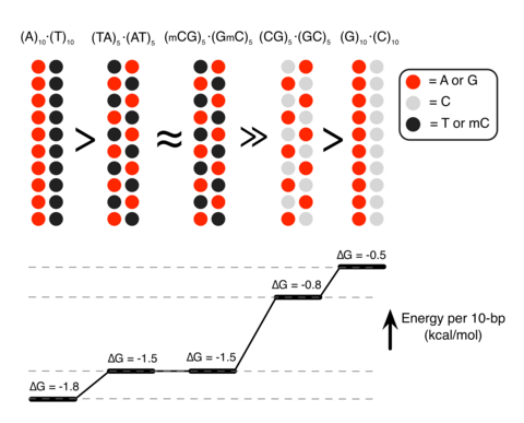
Unexpectedly, we found that DNA sequence has a profound impact on the strength of attractive interaction. The absolute value of ΔG at minimum relative to the value at maximum separation, |ΔGmin|, showed a clearly rank-ordered dependence on the DNA sequence: |ΔGmin| of (A)20>|ΔGmin| of (AT)10>|ΔGmin| of (GC)10>|ΔGmin| of (G)20. Two trends can be noted. First, AT-rich sequences attract each other more strongly than GC-rich sequences16. For example, |ΔGmin| of (AT)10 (1.5kcalmol−1 per turn) is about twice |ΔGmin| of (GC)10 (0.8kcalmol−1 per turn) (Fig. 1b). Second, duplexes having identical AT content but different partitioning of the nucleotides between the strands (that is, (A)20 versus (AT)10 or (G)20 versus (GC)10) exhibit statistically significant differences (~0.3kcalmol−1 per turn) in the value of |ΔGmin|.

To validate the findings of MD simulations, we performed single-molecule fluorescence resonance energy transfer (smFRET)17 experiments of vesicle-encapsulated DNA molecules. Equimolar mixture of donor- and acceptor-labelled 120-bp dsDNA molecules was encapsulated in sub-micron size, porous lipid vesicles18 so that we could observe and quantitate rare binding events between a pair of dsDNA molecules without triggering large-scale DNA condensation2. Our DNA constructs were long enough to ensure dsDNA–dsDNA binding that is stable on the timescale of an smFRET measurement, but shorter than the DNA’s persistence length (~150bp (ref. 19)) to avoid intramolecular condensation20. The vesicles were immobilized on a polymer-passivated surface, and fluorescence signals from individual vesicles containing one donor and one acceptor were selectively analysed (Fig. 1d). Binding of two dsDNA molecules brings their fluorescent labels in close proximity, increasing the FRET efficiency (Fig. 1e).

FRET signals from individual vesicles were diverse. Sporadic binding events were observed in some vesicles, while others exhibited stable binding; traces indicative of frequent conformational transitions were also observed (Supplementary Fig. 1A). Such diverse behaviours could be expected from non-specific interactions of two large biomolecules having structural degrees of freedom. No binding events were observed in the absence of Sm4+ (Supplementary Fig. 1B) or when no DNA molecules were present. To quantitatively assess the propensity of forming a bound state, we chose to use the fraction of single-molecule traces that showed any binding events within the observation time of 2min (Methods). This binding fraction for the pair of AT-rich dsDNAs (AT1, 100% AT in the middle 80-bp section of the 120-bp construct) reached a maximum at ~2mM Sm4+(Fig. 1f), which is consistent with the results of previous experimental studies2, 3. In accordance with the prediction of our MD simulations, GC-rich dsDNAs (GC1, 75% GC in the middle 80bp) showed much lower binding fraction at all Sm4+ concentrations (Fig. 1b,c). Regardless of the DNA sequence, the binding fraction reduced back to zero at high Sm4+ concentrations, likely due to the resolubilization of now positively charged DNA–Sm4+ complexes2, 3, 13.

Because the donor and acceptor fluorophores were attached to the same sequence of DNA, it remained possible that the sequence homology between the donor-labelled DNA and the acceptor-labelled DNA was necessary for their interaction6. To test this possibility, we designed another AT-rich DNA construct AT2 by scrambling the central 80-bp section of AT1 to remove the sequence homology (Supplementary Table 1). The fraction of binding traces for this nonhomologous pair of donor-labelled AT1 and acceptor-labelled AT2 was comparable to that for the homologous AT-rich pair (donor-labelled AT1 and acceptor-labelled AT1) at all Sm4+ concentrations tested (Fig. 1f). Furthermore, this data set rules out the possibility that the higher binding fraction observed experimentally for the AT-rich constructs was caused by inter-duplex Watson–Crick base pairing of the partially melted constructs.

Next, we designed a DNA construct named ATGC, containing, in its middle section, a 40-bp AT-rich segment followed by a 40-bp GC-rich segment (Fig. 1g). By attaching the acceptor to the end of either the AT-rich or GC-rich segments, we could compare the likelihood of observing the parallel binding mode that brings the two AT-rich segments together and the anti-parallel binding mode. Measurements at 1mM Sm4+ and 25 or 50mM NaCl indicated a preference for the parallel binding mode by ~30% (Fig. 1h). Therefore, AT content can modulate DNA–DNA interactions even in a complex sequence context. Note that increasing the concentration of NaCl while keeping the concentration of Sm4+ constant enhances competition between Na+ and Sm4+ counterions, which reduces the concentration of Sm4+ near DNA and hence the frequency of dsDNA–dsDNA binding events (Supplementary Fig. 2).

Methylation determines the strength of DNA–DNA attraction

Analysis of the MD simulations revealed the molecular mechanism of the polyamine-mediated sequence-dependent attraction (Fig. 2). In the case of the AT-rich fragments, the bulky methyl group of thymine base blocks Sm4+ binding to the N7 nitrogen atom of adenine, which is the cation-binding hotspot21, 22. As a result, Sm4+ is not found in the major grooves of the AT-rich duplexes and resides mostly near the DNA backbone (Fig. 2a,d). Such relocated Sm4+ molecules bridge the two DNA duplexes better, accounting for the stronger attraction16, 23, 24, 25. In contrast, significant amount of Sm4+ is adsorbed to the major groove of the GC-rich helices that lacks cation-blocking methyl group (Fig. 2b,e).

Figure 2: Molecular mechanism of polyamine-mediated DNA sequence recognition.
Molecular mechanism of polyamine-mediated DNA sequence recognition.

(ac) Representative configurations of Sm4+ molecules at the DNA–DNA distance of 28Å for the (AT)10–(AT)10 (a), (GC)10–(GC)10 (b) and (GmC)10–(GmC)10 (c) DNA pairs. The backbone and bases of DNA are shown as ribbon and molecular bond, respectively; Sm4+ molecules are shown as molecular bonds. Spheres indicate the location of the N7 atoms and the methyl groups. (df) The average distributions of cations for the three sequence pairs featured in ac. Top: density of Sm4+ nitrogen atoms (d=28Å) averaged over the corresponding MD trajectory and the z axis. White circles (20Å in diameter) indicate the location of the DNA helices. Bottom: the average density of Sm4+ nitrogen (blue), DNA phosphate (black) and sodium (red) atoms projected onto the DNA–DNA distance axis (x axis). The plot was obtained by averaging the corresponding heat map data over y=[−10, 10] Å. See Supplementary Figs 4 and 5 for the cation distributions at d=30, 32, 34 and 36Å.

If indeed the extra methyl group in thymine, which is not found in cytosine, is responsible for stronger DNA–DNA interactions, we can predict that cytosine methylation, which occurs naturally in many eukaryotic organisms and is an essential epigenetic regulation mechanism26, would also increase the strength of DNA–DNA attraction. MD simulations showed that the GC-rich helices containing methylated cytosines (mC) lose the adsorbed Sm4+ (Fig. 2c,f) and that |ΔGmin| of (GC)10 increases on methylation of cytosines to become similar to |ΔGmin| of (AT)10 (Fig. 1b).

To experimentally assess the effect of cytosine methylation, we designed another GC-rich construct GC2 that had the same GC content as GC1 but a higher density of CpG sites (Supplementary Table 1). The CpG sites were then fully methylated using M. SssI methyltransferase (Supplementary Fig. 3; Methods). As predicted from the MD simulations, methylation of the GC-rich constructs increased the binding fraction to the level of the AT-rich constructs (Fig. 1f).

The sequence dependence of |ΔGmin| and its relation to the Sm4+ adsorption patterns can be rationalized by examining the number of Sm4+ molecules shared by the dsDNA molecules (Fig. 3a). An Sm4+ cation adsorbed to the major groove of one dsDNA is separated from the other dsDNA by at least 10Å, contributing much less to the effective DNA–DNA attractive force than a cation positioned between the helices, that is, the ‘bridging’ Sm4+ (ref. 23). An adsorbed Sm4+ also repels other Sm4+ molecules due to like-charge repulsion, lowering the concentration of bridging Sm4+. To demonstrate that the concentration of bridging Sm4+ controls the strength of DNA–DNA attraction, we computed the number of bridging Sm4+ molecules, Nspm (Fig. 3b). Indeed, the number of bridging Sm4+ molecules ranks in the same order as |ΔGmin|: Nspm of (A)20>Nspm of (AT)10Nspm of (GmC)10>Nspm of (GC)10>Nspm of (G)20. Thus, the number density of nucleotides carrying a methyl group (T and mC) is the primary determinant of the strength of attractive interaction between two dsDNA molecules. At the same time, the spatial arrangement of the methyl group carrying nucleotides can affect the interaction strength as well (Fig. 3c). The number of methyl groups and their distribution in the (AT)10 and (GmC)10 duplex DNA are identical, and so are their interaction free energies, |ΔGmin| of (AT)10Gmin| of (GmC)10. For AT-rich DNA sequences, clustering of the methyl groups repels Sm4+ from the major groove more efficiently than when the same number of methyl groups is distributed along the DNA (Fig. 3b). Hence, |ΔGmin| of (A)20>|ΔGmin| of (AT)10. For GC-rich DNA sequences, clustering of the cation-binding sites (N7 nitrogen) attracts more Sm4+ than when such sites are distributed along the DNA (Fig. 3b), hence |ΔGmin| is larger for (GC)10 than for (G)20.

Figure 3: Methylation modulates the interaction free energy of two dsDNA molecules by altering the number of bridging Sm4+.
Methylation modulates the interaction free energy of two dsDNA molecules by altering the number of bridging Sm4+.

(a) Typical spatial arrangement of Sm4+ molecules around a pair of DNA helices. The phosphates groups of DNA and the amine groups of Sm4+ are shown as red and blue spheres, respectively. ‘Bridging’ Sm4+molecules reside between the DNA helices. Orange rectangles illustrate the volume used for counting the number of bridging Sm4+ molecules. (b) The number of bridging amine groups as a function of the inter-DNA distance. The total number of Sm4+ nitrogen atoms was computed by averaging over the corresponding MD trajectory and the 10Å (x axis) by 20Å (y axis) rectangle prism volume (a) centred between the DNA molecules. (c) Schematic representation of the dependence of the interaction free energy of two DNA molecules on their nucleotide sequence. The number and spatial arrangement of nucleotides carrying a methyl group (T or mC) determine the interaction free energy of two dsDNA molecules.

Genome-wide investigations of chromosome conformations using the Hi–C technique revealed that AT-rich loci form tight clusters in human nucleus27, 28. Gene or chromosome inactivation is often accompanied by increased methylation of DNA29 and compaction of facultative heterochromatin regions30. The consistency between those phenomena and our findings suggest the possibility that the polyamine-mediated sequence-dependent DNA–DNA interaction might play a role in chromosome folding and epigenetic regulation of gene expression.

  1. Rau, D. C., Lee, B. & Parsegian, V. A. Measurement of the repulsive force between polyelectrolyte molecules in ionic solution: hydration forces between parallel DNA double helices. Proc. Natl Acad. Sci. USA 81, 26212625 (1984).
  2. Raspaud, E., Olvera de la Cruz, M., Sikorav, J. L. & Livolant, F. Precipitation of DNA by polyamines: a polyelectrolyte behavior. Biophys. J. 74, 381393 (1998).
  3. Besteman, K., Van Eijk, K. & Lemay, S. G. Charge inversion accompanies DNA condensation by multivalent ions. Nat. Phys. 3, 641644 (2007).
  4. Lipfert, J., Doniach, S., Das, R. & Herschlag, D. Understanding nucleic acid-ion interactions.Annu. Rev. Biochem. 83, 813841 (2014).
  5. Grosberg, A. Y., Nguyen, T. T. & Shklovskii, B. I. The physics of charge inversion in chemical and biological systems. Rev. Mod. Phys. 74, 329345 (2002).
  6. Kornyshev, A. A. & Leikin, S. Sequence recognition in the pairing of DNA duplexes. Phys. Rev. Lett. 86, 36663669 (2001).
  7. Danilowicz, C. et al. Single molecule detection of direct, homologous, DNA/DNA pairing.Proc. Natl Acad. Sci. USA 106, 1982419829 (2009).
  8. Gladyshev, E. & Kleckner, N. Direct recognition of homology between double helices of DNA in Neurospora crassa. Nat. Commun. 5, 3509 (2014).
  9. Tabor, C. W. & Tabor, H. Polyamines. Annu. Rev. Biochem. 53, 749790 (1984).
  10. Thomas, T. & Thomas, T. J. Polyamines in cell growth and cell death: molecular mechanisms and therapeutic applications. Cell. Mol. Life Sci. 58, 244258 (2001).

Read Full Post »

Triglycerides: Is it a Risk Factor or a Risk Marker for Atherosclerosis and Cardiovascular Disease ? The Impact of Genetic Mutations on (ANGPTL4) Gene, encoder of (angiopoietin-like 4) Protein, inhibitor of Lipoprotein Lipase


Reporters, Curators and Authors: Aviva Lev-Ari, PhD, RN and Larry H. Bernstein, MD, FCAP

Introduction

The role for triglycerides as a risk factor for cardiovascular disease is not new, going back to Donald Frederickson’s classification of hyperlipidemias, at least with respect to Type I and Type IIb. Whether there was a mechanism beyond the observations was yet an open question.  The paper that follows addresses such a question.


Large Genetic Studies Support Role For Triglycerides In Cardiovascular Disease

SOURCE

http://cardiobrief.org/2016/03/03/large-genetic-studies-support-role-for-triglycerides-in-cardiovascular-disease/#comments


Two  papers published in the New England Journal of Medicine offer new genetic evidence to support the increasingly accepted though still controversial view that triglycerides play an important causal role in cardiovascular disease. If fully validated the new findings could lead to new drugs to prevent and treat cardiovascular disease, though others caution that there is still a long way to go before this could happen.

Both studies describe the impact of genetic mutations on a gene (ANGPTL4) which encodes for a protein (angiopoietin-like 4) that inhibits lipoprotein lipase, an enzyme that plays a key role in breaking down and removing triglycerides from the blood. The large studies found that people with  mutations that inactivate ANGPTL4 have lower levels of triglycerides, higher levels of HDL cholesterol, and decreased risk for cardiovascular disease.

The findings, writes Sander Kersten (Wageningen University, the Netherlands) in an accompanying editorial, “suggest that lowering plasma triglyceride levels is a viable approach to reducing the risk of coronary artery disease.”

The Genetics of Dyslipidemia — When Less Is More

Sander Kersten, Ph.D.   Mar 2, 2016;   http://dx.doi.org:/10.1056/NEJMe1601117

Two groups of investigators now describe in the Journal important genetic evidence showing a causal role of plasma triglycerides in coronary heart disease. Stitziel and colleagues2 tested 54,003 coding-sequence variants covering 13,715 human genes in more than 72,000 patients with coronary artery disease and 120,000 controls. Dewey and colleagues3 sequenced the exons of the gene encoding angiopoietin-like 4 (ANGPTL4) in samples obtained from nearly 43,000 participants in the DiscovEHR human genetics study. The two groups found a significant association between an inactivating mutation (E40K) in ANGPTL4 and both low plasma triglyceride levels and high levels of HDL cholesterol. ANGPTL4 is an inhibitor of lipoprotein lipase, the enzyme that breaks down plasma triglycerides along the capillaries in heart, muscle, and fat.4 Extensive research has shown that ANGPTL4 orchestrates the processing of triglyceride-rich lipoproteins during physiologic conditions such as fasting, exercise, and cold exposure.4 The E40K mutation in ANGPTL4 was previously shown to nearly eliminate the ability of ANGPTL4 to inhibit lipoprotein lipase, a mechanism that may result in part from the destabilization of ANGPTL4.5

The key finding in each study was that carriers of the E40K mutation and other rare mutations in ANGPTL4 had a lower risk of coronary artery disease than did noncarriers, a result that is consistent with the lower triglyceride levels and higher HDL cholesterol levels among mutation carriers. These findings confirm previous data6 and provide convincing genetic evidence that an elevated plasma triglyceride level increases the risk of coronary heart disease. In combination with extensive recent data on other genetic variants that modulate plasma triglyceride levels, the studies suggest that lowering plasma triglyceride levels is a viable approach to reducing the risk of coronary artery disease.

However, as a cautionary note, Talmud and colleagues7 previously found that the presence of the E40K variant was associated with an increased risk of coronary heart disease after adjustment for the altered plasma lipids. Consistent with this hypothesis, the overexpression of Angptl4 in mice was found to protect against atherosclerosis independent of plasma lipids.8

The studies also “implicate targeted inactivation of ANGPTL4 as a potential weapon in the war on heart disease,” though he also points to a previous study that did not support this hypothesis. Sekar Kathiresan (Broad Institute), senior author of one of the NEJM studies, told me that the previous study was small and “basically got the result wrong. Between, the two papers in this NEJM issue, we are looking at 10X more data.”

Recent large genetic studies have resulted in an important change in the field. Many researchers now believe that HDL, which was once thought to play an important protective role in atherosclerosis, is only a marker of disease. In contrast, triglycerides are now thought by many to play an important functional role.

One of the NEJM papers showed that a human monoclonal antibody to ANGPTL4 lowered triglyceride levels in animals. The study was funded by Regeneron and was performed by researchers at Regeneron and Geisinger, as part of an ongoing collaboration using deidentified genetic data from Geisinger patients. In their NEJM paper the researchers reported inflammation and other side effects in the animals treated with the antibody, but they said that no such problem has been observed in humans who have mutations that have the same functional effect as the antibody.

Coding Variation in ANGPTL4, LPL, and SVEP1and the Risk of Coronary Disease Myocardial Infarction Genetics and CARDIoGRAM Exome Consortia Investigators

March 2, 2016     http://dx.doi.org:/10.1056/NEJMoa1507652

Although genomewide association studies have identified more than 56 loci associated with the risk of coronary artery disease,1-3 the disease-associated variants are typically common (minor-allele frequency >5%) and located in noncoding sequences; this has made it difficult to pinpoint causal genes and affected pathways. This lack of a causal mechanism has in part hindered the immediate translation of the findings of genomewide association studies into new therapeutic targets. However, the discovery of rare or low-frequency coding-sequence variants that affect the risk of coronary artery disease has facilitated advances in the prevention and treatment of disease. The most recent example of such advances is the development of a new class of therapeutic agents that is based on the discovery of the gene encoding proprotein convertase subtilisin/kexin type 9 (PCSK9) as a regulator of low-density lipoprotein (LDL) cholesterol4 and the discovery that low-frequency and loss-of-function variants in this gene protect against coronary artery disease.5,6

Recently, low-frequency coding variation across the genome was systematically tabulated with the use of next-generation exome and whole-genome sequencing data from more than 12,000 persons of various ancestries (including a major contribution from the National Heart, Lung, and Blood Institute Exome Sequencing Project). Protein-altering variants (i.e., nonsynonymous, splice-site, and nonsense single-nucleotide substitutions) that were observed at least twice among these 12,000 persons were included in a genotyping array (hereafter referred to as the exome array). In addition, the exome array contains previously described variants from genomewide association studies, a sparse genomewide grid of common markers, markers that are informative with regard to ancestry (i.e., African American, Native American, and European), and some additional content. Additional information on the design of the exome array is provided at http://genome.sph.umich.edu/wiki/Exome_Chip_Design. In this study, we focused on the 220,231 autosomal variants that were present on the array and were expected to alter protein sequence (i.e., missense, nonsense, splice-site, and frameshift variants) and used these to test the contribution of low-frequency coding variation to the risk of coronary artery disease.

Low-Frequency Coding Variants Associated with Coronary Artery Disease

The discovery cohort comprised 120,575 persons (42,335 patients and 78,240 controls) (Table S1 in the Supplementary Appendix). In the discovery cohort, we found significant associations between low-frequency coding variants in theLPA and PCSK9 genes and coronary artery disease (Table 1

TABLE 1

Low-Frequency Coding Variations Previously Associated with Coronary Artery Disease.). Both gene loci also harbor common noncoding variants associated with coronary artery disease that had previously been discovered through genomewide association studies. These variants were also present on the exome array and had significant associations with coronary artery disease in our study (Table 1). In a conditional analysis, the associations between coronary artery disease and the low-frequency coding variants in both LPA and PCSK9 were found to be independent of the associations between coronary artery disease and the more common variants (Table 1). ….

We found a significant association between SVEP1 p.D2702G and blood pressure (Table 3TABLE 3   Association between Low-Frequency Variants and Traditional Risk Factors., and Table S7 in the Supplementary Appendix). The allele associated with an increased risk of coronary artery disease was also associated with higher systolic blood pressure (0.94 mm Hg higher for each copy of the allele among allele carriers, P=3.0×10⁻⁷) and higher diastolic blood pressure (0.57 mm Hg higher for each copy of the allele among allele carriers, P=4.4×10⁻⁷). We did not find an association between SVEP1 p.D2702G and any plasma lipid trait. In contrast, ANGPTL4 p.E40K was not associated with blood pressure but instead was found to be associated with significantly lower levels of triglycerides (approximately 0.3 standard deviation units lower for each copy of the allele among allele carriers, P=1.6×10⁻¹³) (Table 3) and with higher levels of high-density lipoprotein (HDL) cholesterol (approximately 0.3 standard deviation units higher for each copy of the allele among allele carriers, P=8.2×10⁻¹¹) (Table 3). In a conditional analysis, these effects appeared to be at least partially independent of each other (Table S8 in the Supplementary Appendix). We did not observe any significant association between ANGPTL4 p.E40K and LDL cholesterol level (Table 3). Both SVEP1 p.D2702G and ANGPTL4 p.E40K were nominally associated with type 2 diabetes in a direction concordant with the associated risk of coronary artery disease.

ANGPTL4 Loss-of-Function Mutations, Plasma Lipid Levels, and Coronary Artery Disease

The finding that a missense allele in ANGPTL4 reduced the risk of coronary artery disease, potentially by reducing triglyceride levels, raised the possibility that complete loss-of-function variants in ANGPTL4 may have an even more dramatic effect on triglyceride concentrations and the risk of coronary artery disease. We therefore examined sequence data for the seven protein-coding exons of ANGPTL4 in 6924 patients with early-onset myocardial infarction and 6834 controls free from coronary artery disease (details of the patients and controls are provided in Table S3 in the Supplementary Appendix). We discovered a total of 10 variants that were predicted to lead to loss of gene function (Figure 1A FIGURE 1    

Loss-of-Function Alleles in ANGPTL4 and Plasma Lipid Levels., and Table S9 in the Supplementary Appendix), carried by 28 heterozygous persons; no homozygous or compound heterozygous persons were discovered. Carriers of loss-of-function alleles had significantly lower levels of triglycerides than did noncarriers (a mean of 35% lower among carriers, P=0.003) (Figure 1B, and Table S10 in the Supplementary Appendix), with no significant difference in LDL or HDL cholesterol levels. Moreover, we found a lower risk of coronary artery disease among carriers of loss-of-function alleles (9 carriers among 6924 patients vs. 19 carriers among 6834 controls; odds ratio for disease, 0.47; P=0.04) (Table S11 in the Supplementary Appendix). A similar investigation was performed for the 48 protein-coding exons of SVEP1; however, only 3 loss-of-function allele carriers were discovered (2 carriers among 6924 patients vs. 1 carrier among 6834 controls).

Coding Variation in LPL and the Risk of Coronary Artery Disease

On the basis of the fact that a loss of ANGPTL4 function was associated with reduced risk of coronary artery disease and that ANGPTL4 inhibits lipoprotein lipase (LPL), one would expect a gain of LPL function to also be associated with a lower risk of coronary artery disease, whereas a loss of LPL function would be expected to be associated with a higher risk. In observations consistent with these expectations, we found a low-frequency missense variant in LPL on the exome array that was associated with an increased risk of coronary artery disease (p.D36N; minor-allele frequency, 1.9%; odds ratio for disease, 1.13; P=2.0×10⁻⁴) (Table S12 in the Supplementary Appendix); previous studies have shown that this allele (also known as p.D9N) is associated with LPL activity that is 20% lower in allele carriers than in noncarriers.8 We also identified a nonsense mutation in LPL on the exome array that was significantly associated with a reduced risk of coronary artery disease (p.S447*; minor-allele frequency, 9.9%; odds ratio, 0.94; P=2.5×10⁻⁷) (Table S12 in the Supplementary Appendix). Contrary to most instances in which the premature introduction of a stop codon leads to loss of gene function, this nonsense mutation, which occurs in the penultimate codon of the gene, paradoxically induces a gain of LPL function.9 …..

Through large-scale exomewide screening, we identified a low-frequency coding variant in ANGPTL4 that was associated with protection against coronary artery disease and a low-frequency coding variant in SVEP1 that was associated with an increased risk of the disease. Moreover, our results highlight LPL as a significant contributor to the risk of coronary artery disease and support the hypothesis that a gain of LPL function or loss of ANGPTL4 inhibition protects against the disease.

ANGPTL4 has previously been found to be involved in cancer pathogenesis and wound healing.10 Previous functional studies also revealed that ANGPTL4 regulates plasma triglyceride concentration by inhibiting LPL.11 The minor allele at p.E40K has previously been associated with lower levels of triglycerides and higher levels of HDL cholesterol.12 We now provide independent confirmation of these lipid effects. In vitro and in vivo experimental evidence suggests that the lysine allele at p.E40K results in destabilization of ANGPTL4 after its secretion from the cell in which it was synthesized. It may be that the p.E40K variant leads to increases in the enzymatic activity of LPL because of this destabilization.13 Previous, smaller studies produced conflicting results regarding p.E40K and the risk of coronary artery disease14,15; we now provide robust support for an association between p.E40K and a reduced risk of coronary artery disease.

An important caveat  to this research is that it is still very early. Most promising therapeutic targets do not work out. James Stein (University of Wisconsin) praised the papers but also offered a word of caution. “This is great science and important research that sheds light on the genetic regulation of TG-rich lipoproteins, serum TG levels, and CVD risk,” he said. “Since it is hard, if not impossible, to disconnect TG-rich lipoproteins from LDL, we should be humble in extrapolating these findings to clinical medicine in an era of low LDL due to statins and PCSK9 inhibitors. I hope this research identifies new targets for drug therapy and better understanding of CVD risk prediction– only time will tell.”

Previous studies with fibrates and other drugs have failed to convincingly show that lowering triglycerides is beneficial. Kathiresan said that what really seems to matter is “how you alter the plasma triglyceride-rich lipoproteins (TRLs).” Some genes that alter TRLs have other metabolic effects. As an example he cited a gene that lowers TRLs but increases the risk for type 2 diabetes. The NEJM papers, by observing the effect of specific mutations, therefore point the way to targets that may be clinically significant.

Conclusions:  The work that has been presented puts a new light on the possible role of triglycerides in the development of congenitally predetermined cardiovascular disease. It does not necessarily establish a general link to mechanism of cardiovascular disease, but it opens up new pathways to our understanding.

SOURCE

http://cardiobrief.org/2016/03/03/large-genetic-studies-support-role-for-triglycerides-in-cardiovascular-disease/#comments

John Contois commented on your update
“Are triglycerides a CHD risk factor? The answer is still maybe. Triglyceride-rich lipoproteins are inextricably linked to LDL metabolism and LDL particle number (and apo B). Still these are important new data and targets for novel therapeutics.”


Risk of Dis-lipids Syndromes in Modern Society


Risk of Dis-lipids Syndrome in Modern Society

Aurelian Udristioiuᶪ, Manole Cojocaru²
¹Department of Biochemistry, Clinical Laboratory, Emergency County Hospital Targu Jiu & Titu Maiorescu University, Bucharest, Romania,
Department of Physiology, Faculty of Medicine, Titu Maiorescu University, Bucharest, Romania

Abstract
Aim of this work was to emphasis the preclinical evaluation of dis-lipids syndromes types at the patients which were presented to a routine control for checking health status, in the hospital ambulatory.
Material and Method:
Were analyzed 60 patients, registered in Clinical Laboratory, assessing by running on the Hitachi 912 Analyzer, the principal biochemical parameters of lipid metabolism: Cholesterol, Triglycerides and fractions of Cholesterol, HDL and LDL. From the total of 60 patients 35 were females and 25 males.
Results
The persons with an alarm signal of atherosclerotic process were in 28 % and persons with low HDL was in 17%. The cases with atherosclerotic index, report-LDL/HDL>3.5 for men and 2.5 for women were in 14 % , the cases with predictive value with coronary risk, report-CO/HDL>5 were presented in 5 % and the cases with dis-lipid syndrome type 2- 4, with high Cholesterol and Triglycerides, were presented in 30% percent.
Conclusions
Lipids controls, and its fractions, are necessary to be prevented atherosclerotic process in the incipient status of ill.


http://video.epccs.eu/video_1466.html


REFERENCES

http://www.nejm.org/doi/full/10.1056/NEJMe1601117

http://www.nejm.org/doi/full/10.1056/NEJMoa1507652

March 2, 2016 Regeneron Genetics Center Publication in New England Journal of Medicine Links ANGPTL4 Inhibition and Risk of Coronary Artery Disease Demonstrates power of large-scale Precision Medicine initiatives

http://files.shareholder.com/downloads/REGN/1634352863x0x879015/042D3D02-04CB-4DD1-89FE-53927F422025/REGN_News_2016_3_2_General_Releases.pdf

e-Books by Leaders in Pharmaceutical Business Intelligence (LPBI) Group

  • Perspectives on Nitric Oxide in Disease Mechanisms, on Amazon since 6/2/12013

http://www.amazon.com/dp/B00DINFFYC

http://www.amazon.com/dp/B012BB0ZF0

  • Genomics Orientations for Personalized Medicine, on Amazon since 11/23/2015

http://www.amazon.com/dp/B018DHBUO6

  • Milestones in Physiology: Discoveries in Medicine, Genomics and Therapeutics, on Amazon.com since 12/27/2015

http://www.amazon.com/dp/B019VH97LU

  • Cardiovascular, Volume Two: Cardiovascular Original Research: Cases in Methodology Design for Content Co-Curation, on Amazon since 11/30/2015

http://www.amazon.com/dp/B018Q5MCN8

  • Cardiovascular Diseases, Volume Three: Etiologies of Cardiovascular Diseases: Epigenetics, Genetics and Genomics, on Amazon since 11/29/2015

http://www.amazon.com/dp/B018PNHJ84

  • Cardiovascular Diseases, Volume Four: Regenerative and Translational Medicine: The Therapeutics Promise for Cardiovascular Diseases, on Amazon since 12/26/2015

http://www.amazon.com/dp/B019UM909A

Other related articles on this topic published in this Open Access Online Scientific Journal include the following:

Editorial & Publication of Articles in e-Books by  Leaders in Pharmaceutical Business Intelligence:  Contributions of Larry H Bernstein, MD, FCAP

http://pharmaceuticalintelligence.com/2014/10/16/editorial-publication-of-articles-in-e-books-by-leaders-in-pharmaceutical-business-intelligence-contributions-of-larry-h-bernstein-md-fcap/

Editorial & Publication of Articles in e-Books by Leaders in Pharmaceutical Business Intelligence: Contributions of Aviva Lev-Ari, PhD, RN

http://pharmaceuticalintelligence.com/2014/10/16/editorial-publication-of-articles-in-e-books-by-leaders-in-pharmaceutical-business-intelligence-contributions-of-aviva-lev-ari-phd-rn/

TRIGLYCERIDES

http://pharmaceuticalintelligence.com/?s=Triglycerides

HDL

http://pharmaceuticalintelligence.com/?s=HDL

PCSK9

http://pharmaceuticalintelligence.com/?s=PCSK9

STATINS

http://pharmaceuticalintelligence.com/?s=Statins

STATIN

http://pharmaceuticalintelligence.com/?s=STATIN

Read Full Post »

Christopher J. Lynch, MD, PhD, the New Office of Nutrition Research, Director

Curator: Larry H. Bernstein, MD, FCAP


Christopher J. Lynch to direct Office of Nutrition Research

National Institute of Diabetes and Digestive and Kidney Diseases (NIDDK)

http://www.nih.gov/news-events/news-releases/christopher-j-lynch-direct-office-nutrition-research


Christopher J. Lynch, Ph.D., has been named the new director of the Office of Nutrition Research (ONR) and chief of the Nutrition Research Branch within the National Institute of Diabetes and Digestive and Kidney Diseases (NIDDK). Lynch officially assumed his new roles on Feb. 21, 2016. NIDDK is part of the National Institutes of Health.

Lynch will facilitate nutrition research within NIDDK and — through ONR — across NIH, in part by forming and leading a trans-NIH strategic working group. He will also continue and extend ongoing efforts at NIDDK to collaborate widely to advance nutrition research.

“Dr. Lynch is a leader in the nutrition community and his expertise will be vital to guiding the NIH strategic plan for nutrition research,” said NIH Director Francis S. Collins, M.D., Ph.D.  “As NIH works to expand nutrition knowledge, Dr. Lynch’s understanding of the field will help identify information gaps and create a framework to support future discoveries to ultimately improve human health.”

NIH supports a broad range of nutrition research, including studies on the effects of nutrient and dietary intake on human growth and disease, genetic influences on human nutrition and metabolism and other scientific areas. ONR was established in August 2015 to help NIH develop a strategic plan to expand mission-specific nutrition research.

NARRATIVE:
Our laboratory is dedicated to developing cures for metabolic diseases like Obesity, Diabetes and MSUD. We have several projects:
Project 1: How Antipsychotic Drugs Exert Obesity and Metabolic Disease Side effects
Project 2: Impact of Branched Chain Amino Acid (BCAA) signaling and metabolism in obesity and diabetes.
Project 3: Adipose tissue transplant as a treatment for Maple Syrup Urine Disease.
Project 4: How Gastric Bypass Surgery Provides A Rapid Cure For Diabetes And Other Obesity Co-Morbidities Like Hypertension
Project 5: Novel Mechanism Of Action Of Cannabinoid Receptor 1 Blockers For Improvement Of Diabetes

Timeline

  1. Klingerman CM, Stipanovic ME, Hajnal A, Lynch CJ. Acute Metabolic Effects of Olanzapine Depend on Dose and Injection Site. Dose Response. 2015 Oct-Dec; 13(4):1559325815618915.

View in: PubMed

  1. Lynch CJ, Kimball SR, Xu Y, Salzberg AC, Kawasawa YI. Global deletion of BCATm increases expression of skeletal muscle genes associated with protein turnover. Physiol Genomics. 2015 Nov; 47(11):569-80.

View in: PubMed

  1. Lynch CJ, Xu Y, Hajnal A, Salzberg AC, Kawasawa YI. RNA sequencing reveals a slow to fast muscle fiber type transition after olanzapine infusion in rats. PLoS One. 2015; 10(4):e0123966.

View in: PubMed

  1. Shin AC, Fasshauer M, Filatova N, Grundell LA, Zielinski E, Zhou JY, Scherer T, Lindtner C, White PJ, Lapworth AL, Ilkayeva O, Knippschild U, Wolf AM, Scheja L, Grove KL, Smith RD, Qian WJ, Lynch CJ, Newgard CB, Buettner C. Brain Insulin Lowers Circulating BCAA Levels by Inducing Hepatic BCAA Catabolism. Cell Metab. 2014 Nov 4; 20(5):898-909.

View in: PubMed

  1. Lynch CJ, Adams SH. Branched-chain amino acids in metabolic signalling and insulin resistance. Nat Rev Endocrinol. 2014 Dec; 10(12):723-36.

View in: PubMed

  1. Olson KC, Chen G, Xu Y, Hajnal A, Lynch CJ. Alloisoleucine differentiates the branched-chain aminoacidemia of Zucker and dietary obese rats. Obesity (Silver Spring). 2014 May; 22(5):1212-5.

View in: PubMed

  1. Zimmerman HA, Olson KC, Chen G, Lynch CJ. Adipose transplant for inborn errors of branched chain amino acid metabolism in mice. Mol Genet Metab. 2013 Aug; 109(4):345-53.

View in: PubMed

  1. Olson KC, Chen G, Lynch CJ. Quantification of branched-chain keto acids in tissue by ultra fast liquid chromatography-mass spectrometry. Anal Biochem. 2013 Aug 15; 439(2):116-22.

View in: PubMed

  1. She P, Olson KC, Kadota Y, Inukai A, Shimomura Y, Hoppel CL, Adams SH, Kawamata Y, Matsumoto H, Sakai R, Lang CH, Lynch CJ. Leucine and protein metabolism in obese Zucker rats. PLoS One. 2013; 8(3):e59443.

View in: PubMed

  1. Lackey DE, Lynch CJ, Olson KC, Mostaedi R, Ali M, Smith WH, Karpe F, Humphreys S, Bedinger DH, Dunn TN, Thomas AP, Oort PJ, Kieffer DA, Amin R, Bettaieb A, Haj FG, Permana P, Anthony TG, Adams SH. Regulation of adipose branched-chain amino acid catabolism enzyme expression and cross-adipose amino acid flux in human obesity. Am J Physiol Endocrinol Metab. 2013 Jun 1; 304(11):E1175-87.

View in: PubMed

  1. Klingerman CM, Stipanovic ME, Bader M, Lynch CJ. Second-generation antipsychotics cause a rapid switch to fat oxidation that is required for survival in C57BL/6J mice. Schizophr Bull. 2014 Mar; 40(2):327-40.

View in: PubMed

  1. Carr TD, DiGiovanni J, Lynch CJ, Shantz LM. Inhibition of mTOR suppresses UVB-induced keratinocyte proliferation and survival. Cancer Prev Res (Phila). 2012 Dec; 5(12):1394-404.

View in: PubMed

  1. Lynch CJ, Zhou Q, Shyng SL, Heal DJ, Cheetham SC, Dickinson K, Gregory P, Firnges M, Nordheim U, Goshorn S, Reiche D, Turski L, Antel J. Some cannabinoid receptor ligands and their distomers are direct-acting openers of SUR1 K(ATP) channels. Am J Physiol Endocrinol Metab. 2012 Mar 1; 302(5):E540-51.

View in: PubMed

  1. Albaugh VL, Singareddy R, Mauger D, Lynch CJ. A double blind, placebo-controlled, randomized crossover study of the acute metabolic effects of olanzapine in healthy volunteers. PLoS One. 2011; 6(8):e22662.

View in: PubMed

  1. She P, Zhang Z, Marchionini D, Diaz WC, Jetton TJ, Kimball SR, Vary TC, Lang CH, Lynch CJ. Molecular characterization of skeletal muscle atrophy in the R6/2 mouse model of Huntington’s disease. Am J Physiol Endocrinol Metab. 2011 Jul; 301(1):E49-61.

View in: PubMed

  1. Fogle RL, Hollenbeak CS, Stanley BA, Vary TC, Kimball SR, Lynch CJ. Functional proteomic analysis reveals sex-dependent differences in structural and energy-producing myocardial proteins in rat model of alcoholic cardiomyopathy. Physiol Genomics. 2011 Apr 12; 43(7):346-56.

View in: PubMed

  1. Zhou Y, Jetton TL, Goshorn S, Lynch CJ, She P. Transamination is required for {alpha}-ketoisocaproate but not leucine to stimulate insulin secretion. J Biol Chem. 2010 Oct 29; 285(44):33718-26.

View in: PubMed

  1. Agostino NM, Chinchilli VM, Lynch CJ, Koszyk-Szewczyk A, Gingrich R, Sivik J, Drabick JJ. Effect of the tyrosine kinase inhibitors (sunitinib, sorafenib, dasatinib, and imatinib) on blood glucose levels in diabetic and nondiabetic patients in general clinical practice. J Oncol Pharm Pract. 2011 Sep; 17(3):197-202.

View in: PubMed

  1. Li J, Romestaing C, Han X, Li Y, Hao X, Wu Y, Sun C, Liu X, Jefferson LS, Xiong J, Lanoue KF, Chang Z, Lynch CJ, Wang H, Shi Y. Cardiolipin remodeling by ALCAT1 links oxidative stress and mitochondrial dysfunction to obesity. Cell Metab. 2010 Aug 4; 12(2):154-65.

View in: PubMed

  1. Culnan DM, Albaugh V, Sun M, Lynch CJ, Lang CH, Cooney RN. Ileal interposition improves glucose tolerance and insulin sensitivity in the obese Zucker rat. Am J Physiol Gastrointest Liver Physiol. 2010 Sep; 299(3):G751-60.

View in: PubMed

  1. Hajnal A, Kovacs P, Ahmed T, Meirelles K, Lynch CJ, Cooney RN. Gastric bypass surgery alters behavioral and neural taste functions for sweet taste in obese rats. Am J Physiol Gastrointest Liver Physiol. 2010 Oct; 299(4):G967-79.

View in: PubMed

  1. Lang CH, Lynch CJ, Vary TC. BCATm deficiency ameliorates endotoxin-induced decrease in muscle protein synthesis and improves survival in septic mice. Am J Physiol Regul Integr Comp Physiol. 2010 Sep; 299(3):R935-44.

View in: PubMed

  1. Albaugh VL, Vary TC, Ilkayeva O, Wenner BR, Maresca KP, Joyal JL, Breazeale S, Elich TD, Lang CH, Lynch CJ. Atypical antipsychotics rapidly and inappropriately switch peripheral fuel utilization to lipids, impairing metabolic flexibility in rodents. Schizophr Bull. 2012 Jan; 38(1):153-66.

View in: PubMed

  1. Fogle RL, Lynch CJ, Palopoli M, Deiter G, Stanley BA, Vary TC. Impact of chronic alcohol ingestion on cardiac muscle protein expression. Alcohol Clin Exp Res. 2010 Jul; 34(7):1226-34.

View in: PubMed

  1. Lang CH, Frost RA, Bronson SK, Lynch CJ, Vary TC. Skeletal muscle protein balance in mTOR heterozygous mice in response to inflammation and leucine. Am J Physiol Endocrinol Metab. 2010 Jun; 298(6):E1283-94.

View in: PubMed

  1. Albaugh VL, Judson JG, She P, Lang CH, Maresca KP, Joyal JL, Lynch CJ. Olanzapine promotes fat accumulation in male rats by decreasing physical activity, repartitioning energy and increasing adipose tissue lipogenesis while impairing lipolysis. Mol Psychiatry. 2011 May; 16(5):569-81.

View in: PubMed

  1. Lang CH, Lynch CJ, Vary TC. Alcohol-induced IGF-I resistance is ameliorated in mice deficient for mitochondrial branched-chain aminotransferase. J Nutr. 2010 May; 140(5):932-8.

View in: PubMed

  1. She P, Zhou Y, Zhang Z, Griffin K, Gowda K, Lynch CJ. Disruption of BCAA metabolism in mice impairs exercise metabolism and endurance. J Appl Physiol (1985). 2010 Apr; 108(4):941-9.

View in: PubMed

  1. Herman MA, She P, Peroni OD, Lynch CJ, Kahn BB. Adipose tissue branched chain amino acid (BCAA) metabolism modulates circulating BCAA levels. J Biol Chem. 2010 Apr 9; 285(15):11348-56.

View in: PubMed

  1. Li P, Knabe DA, Kim SW, Lynch CJ, Hutson SM, Wu G. Lactating porcine mammary tissue catabolizes branched-chain amino acids for glutamine and aspartate synthesis. J Nutr. 2009 Aug; 139(8):1502-9.

View in: PubMed

  1. Lu G, Sun H, She P, Youn JY, Warburton S, Ping P, Vondriska TM, Cai H, Lynch CJ, Wang Y. Protein phosphatase 2Cm is a critical regulator of branched-chain amino acid catabolism in mice and cultured cells. J Clin Invest. 2009 Jun; 119(6):1678-87.

View in: PubMed

  1. Nairizi A, She P, Vary TC, Lynch CJ. Leucine supplementation of drinking water does not alter susceptibility to diet-induced obesity in mice. J Nutr. 2009 Apr; 139(4):715-9.

View in: PubMed

  1. Meirelles K, Ahmed T, Culnan DM, Lynch CJ, Lang CH, Cooney RN. Mechanisms of glucose homeostasis after Roux-en-Y gastric bypass surgery in the obese, insulin-resistant Zucker rat. Ann Surg. 2009 Feb; 249(2):277-85.

View in: PubMed

  1. Culnan DM, Cooney RN, Stanley B, Lynch CJ. Apolipoprotein A-IV, a putative satiety/antiatherogenic factor, rises after gastric bypass. Obesity (Silver Spring). 2009 Jan; 17(1):46-52.

View in: PubMed

  1. She P, Van Horn C, Reid T, Hutson SM, Cooney RN, Lynch CJ. Obesity-related elevations in plasma leucine are associated with alterations in enzymes involved in branched-chain amino acid metabolism. Am J Physiol Endocrinol Metab. 2007 Dec; 293(6):E1552-63.

View in: PubMed

  1. She P, Reid TM, Bronson SK, Vary TC, Hajnal A, Lynch CJ, Hutson SM. Disruption of BCATm in mice leads to increased energy expenditure associated with the activation of a futile protein turnover cycle. Cell Metab. 2007 Sep; 6(3):181-94.

View in: PubMed

  1. Vary TC, Lynch CJ. Nutrient signaling components controlling protein synthesis in striated muscle. J Nutr. 2007 Aug; 137(8):1835-43.

View in: PubMed

  1. Vary TC, Deiter G, Lynch CJ. Rapamycin limits formation of active eukaryotic initiation factor 4F complex following meal feeding in rat hearts. J Nutr. 2007 Aug; 137(8):1857-62.

View in: PubMed

  1. Vary TC, Anthony JC, Jefferson LS, Kimball SR, Lynch CJ. Rapamycin blunts nutrient stimulation of eIF4G, but not PKCepsilon phosphorylation, in skeletal muscle. Am J Physiol Endocrinol Metab. 2007 Jul; 293(1):E188-96.

View in: PubMed

  1. Vary TC, Lynch CJ. Meal feeding stimulates phosphorylation of multiple effector proteins regulating protein synthetic processes in rat hearts. J Nutr. 2006 Sep; 136(9):2284-90.

View in: PubMed

  1. Lynch CJ, Gern B, Lloyd C, Hutson SM, Eicher R, Vary TC. Leucine in food mediates some of the postprandial rise in plasma leptin concentrations. Am J Physiol Endocrinol Metab. 2006 Sep; 291(3):E621-30.

View in: PubMed

  1. Albaugh VL, Henry CR, Bello NT, Hajnal A, Lynch SL, Halle B, Lynch CJ. Hormonal and metabolic effects of olanzapine and clozapine related to body weight in rodents. Obesity (Silver Spring). 2006 Jan; 14(1):36-51.

View in: PubMed

  1. Vary TC, Lynch CJ. Meal feeding enhances formation of eIF4F in skeletal muscle: role of increased eIF4E availability and eIF4G phosphorylation. Am J Physiol Endocrinol Metab. 2006 Apr; 290(4):E631-42.

View in: PubMed

  1. Vary TC, Goodman S, Kilpatrick LE, Lynch CJ. Nutrient regulation of PKCepsilon is mediated by leucine, not insulin, in skeletal muscle. Am J Physiol Endocrinol Metab. 2005 Oct; 289(4):E684-94.

View in: PubMed

  1. Vary TC, Lynch CJ. Biochemical approaches for nutritional support of skeletal muscle protein metabolism during sepsis. Nutr Res Rev. 2004 Jun; 17(1):77-88.

View in: PubMed

  1. Lynch CJ, Halle B, Fujii H, Vary TC, Wallin R, Damuni Z, Hutson SM. Potential role of leucine metabolism in the leucine-signaling pathway involving mTOR. Am J Physiol Endocrinol Metab. 2003 Oct; 285(4):E854-63.

View in: PubMed

  1. Lynch CJ, Hutson SM, Patson BJ, Vaval A, Vary TC. Tissue-specific effects of chronic dietary leucine and norleucine supplementation on protein synthesis in rats. Am J Physiol Endocrinol Metab. 2002 Oct; 283(4):E824-35.

View in: PubMed

  1. Lynch CJ, Patson BJ, Anthony J, Vaval A, Jefferson LS, Vary TC. Leucine is a direct-acting nutrient signal that regulates protein synthesis in adipose tissue. Am J Physiol Endocrinol Metab. 2002 Sep; 283(3):E503-13.

View in: PubMed

  1. Vary TC, Lynch CJ, Lang CH. Effects of chronic alcohol consumption on regulation of myocardial protein synthesis. Am J Physiol Heart Circ Physiol. 2001 Sep; 281(3):H1242-51.

View in: PubMed

  1. Lynch CJ, Patson BJ, Goodman SA, Trapolsi D, Kimball SR. Zinc stimulates the activity of the insulin- and nutrient-regulated protein kinase mTOR. Am J Physiol Endocrinol Metab. 2001 Jul; 281(1):E25-34.

View in: PubMed


Global deletion of BCATm increases expression of skeletal muscle genes associated with protein turnover.

Lynch CJ1Kimball SR2Xu Y2Salzberg AC3Kawasawa YI4.   Author information
Physiol Genomics. 2015 Nov;47(11):569-80.  http://dx.doi.org:/10.1152/physiolgenomics.00055.2015

Consumption of a protein-containing meal by a fasted animal promotes protein accretion in skeletal muscle, in part through leucine stimulation of protein synthesis and indirectly through repression of protein degradation mediated by its metabolite, α-ketoisocaproate. Mice lacking the mitochondrial branched-chain aminotransferase (BCATm/Bcat2), which interconverts leucine and α-ketoisocaproate, exhibit elevated protein turnover. Here, the transcriptomes of gastrocnemius muscle from BCATm knockout (KO) and wild-type mice were compared by next-generation RNA sequencing (RNA-Seq) to identify potential adaptations associated with their persistently altered nutrient signaling. Statistically significant changes in the abundance of 1,486/∼39,010 genes were identified. Bioinformatics analysis of the RNA-Seq data indicated that pathways involved in protein synthesis [eukaryotic initiation factor (eIF)-2, mammalian target of rapamycin, eIF4, and p70S6K pathways including 40S and 60S ribosomal proteins], protein breakdown (e.g., ubiquitin mediated), and muscle degeneration (apoptosis, atrophy, myopathy, and cell death) were upregulated. Also in agreement with our previous observations, the abundance of mRNAs associated with reduced body size, glycemia, plasma insulin, and lipid signaling pathways was altered in BCATm KO mice. Consistently, genes encoding anaerobic and/or oxidative metabolism of carbohydrate, fatty acids, and branched chain amino acids were modestly but systematically reduced. Although there was no indication that muscle fiber type was different between KO and wild-type mice, a difference in the abundance of mRNAs associated with a muscular dystrophy phenotype was observed, consistent with the published exercise intolerance of these mice. The results suggest transcriptional adaptations occur in BCATm KO mice that along with altered nutrient signaling may contribute to their previously reported protein turnover, metabolic and exercise phenotypes.


RNA sequencing reveals a slow to fast muscle fiber type transition after olanzapine infusion in rats.

Lynch CJ1Xu Y1Hajnal A2Salzberg AC3Kawasawa YI4. Author information
PLoS One. 2015 Apr 20;10(4):e0123966. http://dx.doi.org:/10.1371/journal.pone.0123966. eCollection 2015.

Second generation antipsychotics (SGAs), like olanzapine, exhibit acute metabolic side effects leading to metabolic inflexibility, hyperglycemia, adiposity and diabetes. Understanding how SGAs affect the skeletal muscle transcriptome could elucidate approaches for mitigating these side effects. Male Sprague-Dawley rats were infused intravenously with vehicle or olanzapine for 24h using a dose leading to a mild hyperglycemia. RNA-Seq was performed on gastrocnemius muscle, followed by alignment of the data with the Rat Genome Assembly 5.0. Olanzapine altered expression of 1347 out of 26407 genes. Genes encoding skeletal muscle fiber-type specific sarcomeric, ion channel, glycolytic, O2- and Ca2+-handling, TCA cycle, vascularization and lipid oxidation proteins and pathways, along with NADH shuttles and LDH isoforms were affected. Bioinformatics analyses indicate that olanzapine decreased the expression of slower and more oxidative fiber type genes (e.g., type 1), while up regulating those for the most glycolytic and least metabolically flexible, fast twitch fiber type, IIb. Protein turnover genes, necessary to bring about transition, were also up regulated. Potential upstream regulators were also identified. Olanzapine appears to be rapidly affecting the muscle transcriptome to bring about a change to a fast-glycolytic fiber type. Such fiber types are more susceptible than slow muscle to atrophy, and such transitions are observed in chronic metabolic diseases. Thus these effects could contribute to the altered body composition and metabolic disease olanzapine causes. A potential interventional strategy is implicated because aerobic exercise, in contrast to resistance exercise, can oppose such slow to fast fiber transitions.


Brain insulin lowers circulating BCAA levels by inducing hepatic BCAA catabolism.

Shin AC1Fasshauer M1Filatova N1Grundell LA1Zielinski E1Zhou JY2Scherer T1Lindtner C1White PJ3Lapworth AL3,Ilkayeva O3Knippschild U4Wolf AM4Scheja L5Grove KL6Smith RD2Qian WJ2Lynch CJ7Newgard CB3Buettner C8. Author information
Cell Metab. 2014 Nov 4;20(5):898-909. http://dx.doi.org:/10.1016/j.cmet.2014.09.003   Epub 2014 Oct 9

Circulating branched-chain amino acid (BCAA) levels are elevated in obesity/diabetes and are a sensitive predictor for type 2 diabetes. Here we show in rats that insulin dose-dependently lowers plasma BCAA levels through induction of hepatic protein expression and activity of branched-chain α-keto acid dehydrogenase (BCKDH), the rate-limiting enzyme in the BCAA degradation pathway. Selective induction of hypothalamic insulin signaling in rats and genetic modulation of brain insulin receptors in mice demonstrate that brain insulin signaling is a major regulator of BCAA metabolism by inducing hepatic BCKDH. Short-term overfeeding impairs the ability of brain insulin to lower BCAAs in rats. High-fat feeding in nonhuman primates and obesity and/or diabetes in humans is associated with reduced BCKDH protein in liver. These findings support the concept that decreased hepatic BCKDH is a major cause of increased plasma BCAAs and that hypothalamic insulin resistance may account for impaired BCAA metabolism in obesity and diabetes.


Branched-chain amino acids in metabolic signalling and insulin resistance.

Lynch CJ1Adams SH2Author information
Nat Rev Endocrinol. 2014 Dec; 10(12):723-36. http://dx.doi.org:/10.1038/nrendo.2014.171

Branched-chain amino acids (BCAAs) are important nutrient signals that have direct and indirect effects. Frequently, BCAAs have been reported to mediate antiobesity effects, especially in rodent models. However, circulating levels of BCAAs tend to be increased in individuals with obesity and are associated with worse metabolic health and future insulin resistance or type 2 diabetes mellitus (T2DM). A hypothesized mechanism linking increased levels of BCAAs and T2DM involves leucine-mediated activation of the mammalian target of rapamycin complex 1 (mTORC1), which results in uncoupling of insulin signalling at an early stage. A BCAA dysmetabolism model proposes that the accumulation of mitotoxic metabolites (and not BCAAs per se) promotes β-cell mitochondrial dysfunction, stress signalling and apoptosis associated with T2DM. Alternatively, insulin resistance might promote aminoacidaemia by increasing the protein degradation that insulin normally suppresses, and/or by eliciting an impairment of efficient BCAA oxidative metabolism in some tissues. Whether and how impaired BCAA metabolism might occur in obesity is discussed in this Review. Research on the role of individual and model-dependent differences in BCAA metabolism is needed, as several genes (BCKDHA, PPM1K, IVD and KLF15) have been designated as candidate genes for obesity and/or T2DM in humans, and distinct phenotypes of tissue-specific branched chain ketoacid dehydrogenase complex activity have been detected in animal models of obesity and T2DM.


Leucine and protein metabolism in obese Zucker rats.

She P1Olson KCKadota YInukai AShimomura YHoppel CLAdams SHKawamata YMatsumoto HSakai RLang CHLynch CJAuthor information
PLoS One. 2013;8(3):e59443. http://dx.doi.org:/10.1371/journal.pone.0059443   Epub 2013 Mar 20.

Branched-chain amino acids (BCAAs) are circulating nutrient signals for protein accretion, however, they increase in obesity and elevations appear to be prognostic of diabetes. To understand the mechanisms whereby obesity affects BCAAs and protein metabolism, we employed metabolomics and measured rates of [1-(14)C]-leucine metabolism, tissue-specific protein synthesis and branched-chain keto-acid (BCKA) dehydrogenase complex (BCKDC) activities. Male obese Zucker rats (11-weeks old) had increased body weight (BW, 53%), liver (107%) and fat (∼300%), but lower plantaris and gastrocnemius masses (-21-24%). Plasma BCAAs and BCKAs were elevated 45-69% and ∼100%, respectively, in obese rats. Processes facilitating these rises appeared to include increased dietary intake (23%), leucine (Leu) turnover and proteolysis [35% per g fat free mass (FFM), urinary markers of proteolysis: 3-methylhistidine (183%) and 4-hydroxyproline (766%)] and decreased BCKDC per g kidney, heart, gastrocnemius and liver (-47-66%). A process disposing of circulating BCAAs, protein synthesis, was increased 23-29% by obesity in whole-body (FFM corrected), gastrocnemius and liver. Despite the observed decreases in BCKDC activities per gm tissue, rates of whole-body Leu oxidation in obese rats were 22% and 59% higher normalized to BW and FFM, respectively. Consistently, urinary concentrations of eight BCAA catabolism-derived acylcarnitines were also elevated. The unexpected increase in BCAA oxidation may be due to a substrate effect in liver. Supporting this idea, BCKAs were elevated more in liver (193-418%) than plasma or muscle, and per g losses of hepatic BCKDC activities were completely offset by increased liver mass, in contrast to other tissues. In summary, our results indicate that plasma BCKAs may represent a more sensitive metabolic signature for obesity than BCAAs. Processes supporting elevated BCAA]BCKAs in the obese Zucker rat include increased dietary intake, Leu and protein turnover along with impaired BCKDC activity. Elevated BCAAs/BCKAs may contribute to observed elevations in protein synthesis and BCAA oxidation.


Regulation of adipose branched-chain amino acid catabolism enzyme expression and cross-adipose amino acid flux in human obesity.

Lackey DE1Lynch CJOlson KCMostaedi RAli MSmith WHKarpe FHumphreys SBedinger DHDunn TNThomas APOort PJKieffer DAAmin RBettaieb AHaj FGPermana PAnthony TGAdams SH.
Am J Physiol Endocrinol Metab. 2013 Jun 1; 304(11):E1175-87. http://dx.doi.org:/10.1152/ajpendo.00630.2012

Elevated blood branched-chain amino acids (BCAA) are often associated with insulin resistance and type 2 diabetes, which might result from a reduced cellular utilization and/or incomplete BCAA oxidation. White adipose tissue (WAT) has become appreciated as a potential player in whole body BCAA metabolism. We tested if expression of the mitochondrial BCAA oxidation checkpoint, branched-chain α-ketoacid dehydrogenase (BCKD) complex, is reduced in obese WAT and regulated by metabolic signals. WAT BCKD protein (E1α subunit) was significantly reduced by 35-50% in various obesity models (fa/fa rats, db/db mice, diet-induced obese mice), and BCKD component transcripts significantly lower in subcutaneous (SC) adipocytes from obese vs. lean Pima Indians. Treatment of 3T3-L1 adipocytes or mice with peroxisome proliferator-activated receptor-γ agonists increased WAT BCAA catabolism enzyme mRNAs, whereas the nonmetabolizable glucose analog 2-deoxy-d-glucose had the opposite effect. The results support the hypothesis that suboptimal insulin action and/or perturbed metabolic signals in WAT, as would be seen with insulin resistance/type 2 diabetes, could impair WAT BCAA utilization. However, cross-tissue flux studies comparing lean vs. insulin-sensitive or insulin-resistant obese subjects revealed an unexpected negligible uptake of BCAA from human abdominal SC WAT. This suggests that SC WAT may not be an important contributor to blood BCAA phenotypes associated with insulin resistance in the overnight-fasted state. mRNA abundances for BCAA catabolic enzymes were markedly reduced in omental (but not SC) WAT of obese persons with metabolic syndrome compared with weight-matched healthy obese subjects, raising the possibility that visceral WAT contributes to the BCAA metabolic phenotype of metabolically compromised individuals.


Some cannabinoid receptor ligands and their distomers are direct-acting openers of SUR1 K(ATP) channels.

Lynch CJ1Zhou QShyng SLHeal DJCheetham SCDickinson KGregory PFirnges MNordheim UGoshorn SReiche D,Turski LAntel J.   Author information
Am J Physiol Endocrinol Metab. 2012 Mar 1;302(5):E540-51.
http://dx.doi.org:/10.1152/ajpendo.00258.2011

Here, we examined the chronic effects of two cannabinoid receptor-1 (CB1) inverse agonists, rimonabant and ibipinabant, in hyperinsulinemic Zucker rats to determine their chronic effects on insulinemia. Rimonabant and ibipinabant (10 mg·kg⁻¹·day⁻¹) elicited body weight-independent improvements in insulinemia and glycemia during 10 wk of chronic treatment. To elucidate the mechanism of insulin lowering, acute in vivo and in vitro studies were then performed. Surprisingly, chronic treatment was not required for insulin lowering. In acute in vivo and in vitro studies, the CB1 inverse agonists exhibited acute K channel opener (KCO; e.g., diazoxide and NN414)-like effects on glucose tolerance and glucose-stimulated insulin secretion (GSIS) with approximately fivefold better potency than diazoxide. Followup studies implied that these effects were inconsistent with a CB1-mediated mechanism. Thus effects of several CB1 agonists, inverse agonists, and distomers during GTTs or GSIS studies using perifused rat islets were unpredictable from their known CB1 activities. In vivo rimonabant and ibipinabant caused glucose intolerance in CB1 but not SUR1-KO mice. Electrophysiological studies indicated that, compared with diazoxide, 3 μM rimonabant and ibipinabant are partial agonists for K channel opening. Partial agonism was consistent with data from radioligand binding assays designed to detect SUR1 K(ATP) KCOs where rimonabant and ibipinabant allosterically regulated ³H-glibenclamide-specific binding in the presence of MgATP, as did diazoxide and NN414. Our findings indicate that some CB1 ligands may directly bind and allosterically regulate Kir6.2/SUR1 K(ATP) channels like other KCOs. This mechanism appears to be compatible with and may contribute to their acute and chronic effects on GSIS and insulinemia.


Transamination is required for {alpha}-ketoisocaproate but not leucine to stimulate insulin secretion.

Zhou Y1Jetton TLGoshorn SLynch CJShe PAuthor information
J Biol Chem. 2010 Oct 29;285(44):33718-26. http://dx.doi.org:/10.1074/jbc.M110.136846

It remains unclear how α-ketoisocaproate (KIC) and leucine are metabolized to stimulate insulin secretion. Mitochondrial BCATm (branched-chain aminotransferase) catalyzes reversible transamination of leucine and α-ketoglutarate to KIC and glutamate, the first step of leucine catabolism. We investigated the biochemical mechanisms of KIC and leucine-stimulated insulin secretion (KICSIS and LSIS, respectively) using BCATm(-/-) mice. In static incubation, BCATm disruption abolished insulin secretion by KIC, D,L-α-keto-β-methylvalerate, and α-ketocaproate without altering stimulation by glucose, leucine, or α-ketoglutarate. Similarly, during pancreas perfusions in BCATm(-/-) mice, glucose and arginine stimulated insulin release, whereas KICSIS was largely abolished. During islet perifusions, KIC and 2 mM glutamine caused robust dose-dependent insulin secretion in BCATm(+/+) not BCATm(-/-) islets, whereas LSIS was unaffected. Consistently, in contrast to BCATm(+/+) islets, the increases of the ATP concentration and NADPH/NADP(+) ratio in response to KIC were largely blunted in BCATm(-/-) islets. Compared with nontreated islets, the combination of KIC/glutamine (10/2 mM) did not influence α-ketoglutarate concentrations but caused 120 and 33% increases in malate in BCATm(+/+) and BCATm(-/-) islets, respectively. Although leucine oxidation and KIC transamination were blocked in BCATm(-/-) islets, KIC oxidation was unaltered. These data indicate that KICSIS requires transamination of KIC and glutamate to leucine and α-ketoglutarate, respectively. LSIS does not require leucine catabolism and may be through leucine activation of glutamate dehydrogenase. Thus, KICSIS and LSIS occur by enhancing the metabolism of glutamine/glutamate to α-ketoglutarate, which, in turn, is metabolized to produce the intracellular signals such as ATP and NADPH for insulin secretion.


Effect of the tyrosine kinase inhibitors (sunitinib, sorafenib, dasatinib, and imatinib) on blood glucose levels in diabetic and nondiabetic patients in general clinical practice.

Agostino NM1Chinchilli VMLynch CJKoszyk-Szewczyk AGingrich RSivik JDrabick JJ.
J Oncol Pharm Pract. 2011 Sep; 17(3):197-202. http://dx.doi.org:/10.1177/1078155210378913

Tyrosine kinase is a key enzyme activity utilized in many intracellular messaging pathways. Understanding the role of particular tyrosine kinases in malignancies has allowed for the design of tyrosine kinase inhibitors (TKIs), which can target these enzymes and interfere with downstream signaling. TKIs have proven to be successful in the treatment of chronic myeloid leukemia, renal cell carcinoma and gastrointestinal stromal tumor, and other malignancies. Scattered reports have suggested that these agents appear to affect blood glucose (BG). We retrospectively studied the BG concentrations in diabetic (17) and nondiabetic (61) patients treated with dasatinib (8), imatinib (39), sorafenib (23), and sunitinib (30) in our clinical practice. Mean declines of BG were dasatinib (53 mg/dL), imatinib (9 mg/dL), sorafenib (12 mg/dL), and sunitinib (14 mg/dL). All these declines in BG were statistically significant. Of note, 47% (8/17) of the patients with diabetes were able to discontinue their medications, including insulin in some patients. Only one diabetic patient developed symptomatic hypoglycemia while on sunitinib. The mechanism for the hypoglycemic effect of these drugs is unclear, but of the four agents tested, c-kit and PDGFRβ are the common target kinases. Clinicians should keep the potential hypoglycemic effects of these agents in mind; modification of hypoglycemic agents may be required in diabetic patients. These results also suggest that inhibition of a tyrosine kinase, be it c-kit, PDGFRβ or some other undefined target, may improve diabetes mellitus BG control and it deserves further study as a potential novel therapeutic option.


Cardiolipin remodeling by ALCAT1 links oxidative stress and mitochondrial dysfunction to obesity.

Li J1Romestaing CHan XLi YHao XWu YSun CLiu XJefferson LSXiong JLanoue KFChang ZLynch CJWang HShi Y.    Author information
Cell Metab. 2010 Aug 4;12(2):154-65. http://dx.doi.org:/10.1016/j.cmet.2010.07.003

Oxidative stress causes mitochondrial dysfunction and metabolic complications through unknown mechanisms. Cardiolipin (CL) is a key mitochondrial phospholipid required for oxidative phosphorylation. Oxidative damage to CL from pathological remodeling is implicated in the etiology of mitochondrial dysfunction commonly associated with diabetes, obesity, and other metabolic diseases. Here, we show that ALCAT1, a lyso-CL acyltransferase upregulated by oxidative stress and diet-induced obesity (DIO), catalyzes the synthesis of CL species that are highly sensitive to oxidative damage, leading to mitochondrial dysfunction, ROS production, and insulin resistance. These metabolic disorders were reminiscent of those observed in type 2 diabetes and were reversed by rosiglitazone treatment. Consequently, ALCAT1 deficiency prevented the onset of DIO and significantly improved mitochondrial complex I activity, lipid oxidation, and insulin signaling in ALCAT1(-/-) mice. Collectively, these findings identify a key role of ALCAT1 in regulating CL remodeling, mitochondrial dysfunction, and susceptibility to DIO.


BCATm deficiency ameliorates endotoxin-induced decrease in muscle protein synthesis and improves survival in septic mice.

Lang CH1Lynch CJVary TC.   Author information
Am J Physiol Regul Integr Comp Physiol. 2010 Sep; 299(3):R935-44.
http://dx.doi.org:/10.1152/ajpregu.00297.2010

Endotoxin (LPS) and sepsis decrease mammalian target of rapamycin (mTOR) activity in skeletal muscle, thereby reducing protein synthesis. Our study tests the hypothesis that inhibition of branched-chain amino acid (BCAA) catabolism, which elevates circulating BCAA and stimulates mTOR, will blunt the LPS-induced decrease in muscle protein synthesis. Wild-type (WT) and mitochondrial branched-chain aminotransferase (BCATm) knockout mice were studied 4 h after Escherichia coli LPS or saline. Basal skeletal muscle protein synthesis was increased in knockout mice compared with WT, and this change was associated with increased eukaryotic initiation factor (eIF)-4E binding protein-1 (4E-BP1) phosphorylation, eIF4E.eIF4G binding, 4E-BP1.raptor binding, and eIF3.raptor binding without a change in the mTOR.raptor complex in muscle. LPS decreased muscle protein synthesis in WT mice, a change associated with decreased 4E-BP1 phosphorylation as well as decreased formation of eIF4E.eIF4G, 4E-BP1.raptor, and eIF3.raptor complexes. In BCATm knockout mice given LPS, muscle protein synthesis only decreased to values found in vehicle-treated WT control mice, and this ameliorated LPS effect was associated with a coordinate increase in 4E-BP1.raptor, eIF3.raptor, and 4E-BP1 phosphorylation. Additionally, the LPS-induced increase in muscle cytokines was blunted in BCATm knockout mice, compared with WT animals. In a separate study, 7-day survival and muscle mass were increased in BCATm knockout vs. WT mice after polymicrobial peritonitis. These data suggest that elevating blood BCAA is sufficient to ameliorate the catabolic effect of LPS on skeletal muscle protein synthesis via alterations in protein-protein interactions within mTOR complex-1, and this may provide a survival advantage in response to bacterial infection.


Alcohol-induced IGF-I resistance is ameliorated in mice deficient for mitochondrial branched-chain aminotransferase.

Lang CH1Lynch CJVary TCAuthor information
J Nutr. 2010 May;140(5):932-8. http://dx.doi.org:/10.3945/jn.109.120501

Acute alcohol intoxication decreases skeletal muscle protein synthesis by impairing mammalian target of rapamycin (mTOR). In 2 studies, we determined whether inhibition of branched-chain amino acid (BCAA) catabolism ameliorates the inhibitory effect of alcohol on muscle protein synthesis by raising the plasma BCAA concentrations and/or by improving the anabolic response to insulin-like growth factor (IGF)-I. In the first study, 4 groups of mice were used: wild-type (WT) and mitochondrial branched-chain aminotransferase (BCATm) knockout (KO) mice orally administered saline or alcohol (5 g/kg, 1 h). Protein synthesis was greater in KO mice compared with WT controls and was associated with greater phosphorylation of eukaryotic initiation factor (eIF)-4E binding protein-1 (4EBP1), eIF4E-eIF4G binding, and 4EBP1-regulatory associated protein of mTOR (raptor) binding, but not mTOR-raptor binding. Alcohol decreased protein synthesis in WT mice, a change associated with less 4EBP1 phosphorylation, eIF4E-eIF4G binding, and raptor-4EBP1 binding, but greater mTOR-raptor complex formation. Comparable alcohol effects on protein synthesis and signal transduction were detected in BCATm KO mice. The second study used the same 4 groups, but all mice were injected with IGF-I (25 microg/mouse, 30 min). Alcohol impaired the ability of IGF-I to increase muscle protein synthesis, 4EBP1 and 70-kilodalton ribosomal protein S6 kinase-1 phosphorylation, eIF4E-eIF4G binding, and 4EBP1-raptor binding in WT mice. However, in alcohol-treated BCATm KO mice, this IGF-I resistance was not manifested. These data suggest that whereas the sustained elevation in plasma BCAA is not sufficient to ameliorate the catabolic effect of acute alcohol intoxication on muscle protein synthesis, it does improve the anabolic effect of IGF-I.


Impact of chronic alcohol ingestion on cardiac muscle protein expression.

Fogle RL1Lynch CJPalopoli MDeiter GStanley BAVary TCAuthor information
Alcohol Clin Exp Res. 2010 Jul;34(7):1226-34. http://dx.doi.org:/10.1111/j.1530-0277.2010.01200.x

BACKGROUND:

Chronic alcohol abuse contributes not only to an increased risk of health-related complications, but also to a premature mortality in adults. Myocardial dysfunction, including the development of a syndrome referred to as alcoholic cardiomyopathy, appears to be a major contributing factor. One mechanism to account for the pathogenesis of alcoholic cardiomyopathy involves alterations in protein expression secondary to an inhibition of protein synthesis. However, the full extent to which myocardial proteins are affected by chronic alcohol consumption remains unresolved.

METHODS:

The purpose of this study was to examine the effect of chronic alcohol consumption on the expression of cardiac proteins. Male rats were maintained for 16 weeks on a 40% ethanol-containing diet in which alcohol was provided both in drinking water and agar blocks. Control animals were pair-fed to consume the same caloric intake. Heart homogenates from control- and ethanol-fed rats were labeled with the cleavable isotope coded affinity tags (ICAT). Following the reaction with the ICAT reagent, we applied one-dimensional gel electrophoresis with in-gel trypsin digestion of proteins and subsequent MALDI-TOF-TOF mass spectrometric techniques for identification of peptides. Differences in the expression of cardiac proteins from control- and ethanol-fed rats were determined by mass spectrometry approaches.

RESULTS:

Initial proteomic analysis identified and quantified hundreds of cardiac proteins. Major decreases in the expression of specific myocardial proteins were observed. Proteins were grouped depending on their contribution to multiple activities of cardiac function and metabolism, including mitochondrial-, glycolytic-, myofibrillar-, membrane-associated, and plasma proteins. Another group contained identified proteins that could not be properly categorized under the aforementioned classification system.

CONCLUSIONS:

Based on the changes in proteins, we speculate modulation of cardiac muscle protein expression represents a fundamental alteration induced by chronic alcohol consumption, consistent with changes in myocardial wall thickness measured under the same conditions.


Read Full Post »

https://www.youtube.com/v/vI5maHXG-_c?fs=1&hl=fr_FR

Objectives: At the end of the presentation, the audience should be able to describe appropriate interventions for identified arrhythmias.

Sourced through Scoop.it from: www.youtube.com

See on Scoop.itCardiovascular Disease: PHARMACO-THERAPY

Read Full Post »

Nuts and health in aging

Larry H. Bernstein, MD, FCAP, Curator

LPBI


Nut consumption and age-related disease

Giuseppe GrossoRamon Estruch

MATURITAS · OCT 2015     http://dx.doi.org/10.1016/j.maturitas.2015.10.014

Current knowledge on the effects of nut consumption on human health has rapidly increased in recent years and it now appears that nuts may play a role in the prevention of chronic age-related diseases. Frequent nut consumption has been associated with better metabolic status, decreased body weight as well as lower body weight gain over time and thus reduce the risk of obesity. The effect of nuts on glucose metabolism, blood lipids, and blood pressure are still controversial. However, significant decreased cardiovascular risk has been reported in a number of observational and clinical intervention studies. Thus, findings from cohort studies show that increased nut consumption is associated with a reduced risk of cardiovascular disease and mortality (especially that due to cardiovascular-related causes). Similarly, nut consumption has been also associated with reduced risk of certain cancers, such as colorectal, endometrial, and pancreatic neoplasms. Evidence regarding nut consumption and neurological or psychiatric disorders is scarce, but a number of studies suggest significant protective effects against depression, mild cognitive disorders and Alzheimer’s disease. The underlying mechanisms appear to include antioxidant and anti-inflammatory actions, particularly related to their mono- and polyunsaturated fatty acids (MUFA and PUFA, as well as vitamin and polyphenol content. MUFA have been demonstrated to improve pancreatic beta-cell function and regulation of postprandial glycemia and insulin sensitivity. PUFA may act on the central nervous system protecting neuronal and cell-signaling function and maintenance. The fiber and mineral content of nuts may also confer health benefits. Nuts therefore show promise as useful adjuvants to prevent, delay or ameliorate a number of chronic conditions in older people. Their association with decreased mortality suggests a potential in reducing disease burden, including cardiovascular disease, cancer, and cognitive impairments.


Global life expectancy has increased from 65 years in 1990 to about 71 years in 2013 [1]. As life expectancy has increased, the number of healthy years lost due to disability has also risen in most countries, consistent with greater morbidity [2]. Reduction of mortality rates in developed countries has been associated with a shift towards more chronic non-communicable diseases [1]. Cardiovascular diseases (CVDs) and related risk factors, such as hypertension, diabetes mellitus, hypercholesterolemia, and obesity are the top causes of death globally, accounting for nearly one-third of all deaths worldwide [3]. Equally, the estimated incidence, mortality, and disability- adjusted life-years (DALYs) for cancer rose to 14.9 million incident cancer cases, 8.2 million deaths, and 196.3 million DALYs, with the highest impact of prostate and breast cancer in men and women, respectively [4]. Depression is a leading cause of disability worldwide (in terms of total years lost due to disability), especially in high-income countries, increasing from 15th to 11th rank (37% increase) and accounting for 18% of total DALYs (almost 100 million DALYs) [5]. Overall, the global rise in chronic non-communicable diseases is congruent with a similar rise in the elderly population. The proportion of people over the age of 60 is growing faster than any other age group and is estimated to double from about 11% to 22% within the next 50 years [6]. Public health efforts are needed to face this epidemiological and demographic transition, both improving the healthcare systems, as well as assuring a better health in older people. Accordingly, a preventive approach is crucial to dealing with an ageing population to reduce the burden of chronic disease.

In this context, lifestyle behaviors have demonstrated the highest impact for older adults in preventing and controlling the morbidity and mortality due to non- communicable diseases [7]. Unhealthy behaviors, such as unbalanced dietary patterns, lack of physical activity and smoking, play a central role in increasing both cardiovascular and cancer risk [7]. Equally, social isolation and depression in later life may boost health decline and significantly contribute to mortality risk [8]. The role of diet in prevention of disability and death is a well-established factor, which has an even more important role in geriatric populations. Research has focused on the effect of both single foods and whole dietary patterns on a number of health outcomes, including mortality, cardiovascular disease (CVD), cancer and mental health disorders (such as cognitive decline and depression) [9-13]. Plantbased dietary patterns demonstrate the most convincing evidence in preventing chronic non-communicable diseases [14-17]. Among the main components (including fruit and vegetables, legumes and cereals), only lately has attention focused on foods such as nuts. Knowledge on the effect of nut consumption on human health has increased rapidly in recent years. The aim of this narrative review is to examine recent evidence regarding the role of nut consumption in preventing chronic disease in older people.

Tree nuts are dry fruits with an edible seed and a hard shell. The most popular tree nuts are almonds (Prunus amigdalis), hazelnuts (Corylus avellana), walnuts (Juglans regia), pistachios (Pistachia vera), cashews (Anacardium occidentale), pecans (Carya illinoiensis), pine nuts (Pinus pinea), macadamias (Macadamia integrifolia), Brazil nuts (Bertholletia excelsa), and chestnuts (Castanea sativa). When considering the “nut” group, researchers also include peanuts (Arachis hypogea), which technically are groundnuts. Nuts are nutrient dense foods, rich in proteins, fats (mainly unsaturated fatty acids), fiber, vitamins, minerals, as well as a number of phytochemicals, such as phytosterols and polyphenols [18]. Proteins account for about 10-25% of energy, including individual aminoacids, such as L-arginine, which is involved in the production of nitric oxide (NO), an endogenous vasodilatator [19].

The fatty acids composition of nuts involves saturated fats for 415% and unsaturated fatty acids for 30-60% of the content. Unsaturated fatty acids are different depending on the nut type, including monounsaturated fatty acids (MUFA, such as oleic acid in most of nuts, whereas polyunsaturated fatty acids (PUFA, such as alpha-linolenic acid) in pine nuts and walnuts [20]. Also fiber content is similar among most nut types (about 10%), although pine nuts and cashews hold the least content. Vitamins contained in nuts are group B vitamins, such as B6 (involved in many aspects of macronutrient metabolism) and folate (necessary for normal cellular function, DNA synthesis and metabolism, and homocysteine detoxification), as well as tocopherols, involved in anti-oxidant mechanisms [21]. Among minerals contained in vegetables, nuts have an optimal content in calcium, magnesium, and potassium, with an extremely low amount of sodium, which is implicated on a number of pathological conditions, such as bone demineralization, hypertension and insulin resistance[22]. Nuts are also rich in phytosterols, non-nutritive components of certain plant-foods that exert both structural (at cellular membrane phospholipids level) and hormonal (estrogen-like) activities [23]. Finally, nuts have been demonstrated to be a rich source of polyphenols, which account for a key role in their antioxidant and anti-inflammatory effects.


Metabolic disorders are mainly characterized by obesity, hypertension, dyslipidemia, and hyperglycemia/ hyperinsulinemia/type-2 diabetes, all of which act synergistically to increase morbidity and mortality of aging population.

Obesity Increasing high carbohydrate and fat food intake in the last decades has contributed significantly to the rise in metabolic disorders. Nuts are energy-dense foods that have been thought to be positively associated with increased body mass index (BMI). As calorie-dense foods, nuts may contain 160–200 calories per ounce. The recommendation from the American Heart  Association to consume 5 servings per week (with an average recommended serving size of 28 g) corresponds to a net increase of 800–1000 calories per week, which may cause weight gain. However, an inverse relation between the frequency of nut consumption and BMI has been observed in large cohort studies [24]. Pooling the baseline observations of BMI by category of nut consumption in 5 cohort studies found a significant decreasing trend in BMI values with increasing nut intake [24]. While the evidence regarding nut consumption and obesity is limited, findings so far are encouraging [25, 26]. When the association between nut consumption and body weight has been evaluated longitudinally over time, nut intake was associated with a slightly lower risk of weight gain and obesity [25]. In the Nurses’ Health Study II (NHS II), women who eat nuts ≥2 times per week had slightly less weight gain (5.04 kg) than did women who rarely ate nuts (5.55 kg) and marginally significant 23% lower risk of obesity after 9-year follow-up [25]. Further evaluation of the NHS II data and the Physicians’ Health Study (PHS) comprising a total of 120,877 US women and men and followed up to 20 years revealed that 4-y weight change was inversely associated with a 1-serving increment in the intake of nuts (20.26 kg) [27]. In the “Seguimiento Universidad de Navarra” (SUN) cohort study, a significant decreased weight change has been observed over a period of 6 years [26]. After adjustment for potential confounding factors the analysis was no longer significant, but overall no weight gain associated with >2 servings per week of nuts has been observed. Finally, when considering the role of the whole diet on body weight, a meta-analysis of 31 clinical trials led to the conclusion of a null effect of nut intake on body weight, BMI, and waist circumference [28].

Glucose metabolism and type-2 diabetes The association between nut consumption and risk of type-2 diabetes in prospective cohort studies is controversial [29-32]. A pooled analysis relied on the examination of five large cohorts, including the NHS, the Shanghai Women’s Health Study, the Iowa Women’s Health Study, and the PHS, and two European studies conducted in Spain (the PREDIMED trial) and Finland including a total of more than 230,000 participants and 13,000 cases, respectively. Consumption of 4 servings per week was associated with 13% reduced risk of type-2 diabetes without effect modification by age [29]. In contrast, other pooled analyses showed non-significant reduction of risk for increased intakes of nuts, underlying that the inverse association between the consumption of nuts and diabetes was attenuated after adjustment for confounding factors, including BMI [30]. However, results from experimental studies showed promising results. Thus, nut consumption has been demonstrated to exert beneficial metabolic effects due to their action on post-prandial glycemia an insulin sensitivity. A number of RCTs have demonstrated positive effects of nut consumption on post-prandial glycemia in healthy individuals [33-38]. Moreover, a meta-analysis of RCTs on the effects of nut intake on glycemic control in diabetic individuals including 12 trials and a total of 450 participants showed that diets with an emphasis on nuts (median dose = 56 g/d) significantly lowered HbA1c (Mean Difference [MD] : -0.07%; 95% confidence interval [CI]: -0.10, -0.03%; P = 0.0003) and fasting glucose (MD : -0.15 mmol/L; 95% CI: -0.27, -0.02 mmol/L; P = 0.03) compared with control diets [39]. No significant treatment effects were observed for fasting insulin and homeostatic model assessment (HOMA-IR), despite the direction of effect favoring diet regimens including nuts.

Blood lipids and hypertension Hypertension and dyslipidemia are major risk factors for CVD. Diet alone has a predominant role in blood pressure and plasma lipid homeostasis. One systematic review [40] and 3 pooled quantitative analyses of RCTs [41-43] evaluated the effects of nut consumption on lipid profiles. A general agreement was relevant on certain markers, as daily consumption of nuts (mean = 67 g/d) induced a pooled reduction of total cholesterol concentration (10.9 mg/dL [5.1% change]), low-density lipoprotein cholesterol concentration (LDL-C) (10.2 mg/dL [7.4% change]), ratio of LDL-C to high-density lipoprotein cholesterol concentration (HDL-C) (0.22 [8.3% change]), and ratio of total cholesterol concentration to HDL-C (0.24 [5.6% change]) (P <0.001 for all) [42]. All meta-analyses showed no significant effects of nut (including walnut) consumption on HDL cholesterol or triglyceride concentrations in healthy individuals [41], although reduced plasma triglyceride levels were found in individuals with hypertriglyceridemia [42]. Interestingly, the effects of nut consumption were dose related, and different types of nuts had similar effects on blood lipid concentrations.

There is only limited evidence from observational studies to suggest that nuts have a protective role on blood pressure. A pooled analysis of prospective cohort studies on nut consumption and hypertension reported a decreased risk associated with increased intake of nuts [32]. Specifically, only a limited number of cohort studies have been conducted exploring the association between nut consumption and hypertension (n = 3), but overall reporting an 8% reduced risk of hypertension for individuals consuming >2 servings per week (Risk Ratio [RR] = 0.92, 95% CI: 0.87-0.97) compared with never/rare consumers, whereas consumption of nuts at one serving per week had similar risk estimates (RR = 0.97, 95% CI: 0.83, 1.13) [32]. These findings are consistent with results obtained in a pooled analysis of 21 experimental studies reporting the effect of consuming single or mixed nuts (in doses ranging from 30 to 100 g/d) on systolic (SBP) and diastolic blood pressure (DBP) [44]. A pooled analysis found a significant reduction in SBP in participants without type2 diabetes [MD: -1.29 mmHg; 95% CI: -2.35, -0.22; P = 0.02] and DBP (MD: -1.19; 95% CI: -2.35, -0.03; P = 0.04), whereas subgroup analyses of different nut types showed that pistachios, but not other nuts, significantly reduced SBP (MD: -1.82; 95% CI: -2.97, -0.67; P = 0.002) and SBP (MD: -0.80; 95% CI: -1.43, -0.17; P = 0.01) [44].

Nut consumption and CVD risk Clustering of metabolic risk factors occurs in most obese individuals, greatly increasing risk of CVD. The association between nut consumption and CVD incidence [29-31] and mortality [24] has been explored in several pooled analyses of prospective studies. The overall risk calculated for CVD on a total of 8,862 cases was reduced by 29% for individuals consuming 7 servings per week (RR = 0.71, 95% CI: 0.59, 0.85) [30]. A meta-analysis including 9 studies on coronary artery disease (CAD) including 179,885 individuals and 7,236 cases, reporting that 1-serving/day increment would reduce risk of CAD of about 20% (RR = 0.81, 95% CI: 0.72, 0.91) [31]. Similar risk estimates were calculated for ischemic heart disease (IHD), with a comprehensive reduced risk of about 25-30% associated with a daily intake of nuts [29, 30]. Findings from 4 prospective studies have been pooled to estimate the association between nut consumption and risk of stroke, and a non-significant/borderline reduced risk was found [29-31, 45]. CVD mortality was explored in a recent meta-analysis including a total of 354,933 participants, 44,636 cumulative incident deaths, and 3,746,534 cumulative person-years [24]. One serving of nuts per week and per day resulted in decreased risk of CVD mortality (RR = 0.93, 95% CI: 0.88, 0.99 and RR =0.61, 95% CI: 0.42, 0.91, respectively], primarily driven by decreased coronary artery disease (CAD) deaths rather than stroke deaths [24]. Overall, all pooled analyses demonstrated a significant association between nut consumption and cardiovascular health. However, it has been argued that nut consumption was consistently associated with healthier background characteristics reflecting overall healthier lifestyle choices that eventually lead to decreased CVD mortality risk.

Nut consumption and cancer risk Cancer is one of the leading causes of death in the elderly population. After the evaluation of the impact on cancer burden of food and nutrients, it has been concluded that up to one third of malignancies may be prevented by healthy lifestyle choices. Fruit and vegetable intake has been the focus of major attention, but studies on nut consumption and cancer are scarce. A recent metaanalysis pooled together findings of observational studies on cancer incidence, including a total of 16 cohort and 20 casecontrol studies comprising 30,708 cases, compared the highest category of nut consumption with the lowest category and found a lower risk of any cancer of 25% (RR = 0.85, 95% CI: 0.86, 0.95) [46]. When the analysis was conducted by cancer site, highest consumption of nuts was associated with decreased risk of colorectal (RR = 0.76, 95% CI: 0.61, 0.96), endometrial (RR = 0.58, 95% CI: 0.43, 0.79), and pancreatic cancer (RR = 0.71, 95% CI: 0.51, 0.99), with only one cohort study was conducted on the last [46]. The potential protective effects of nut consumption on cancer outcomes was supported also by pooled analysis of 3 cohort studies [comprising the PREDIMED, the NHS, the HPS, and the Health Professionals Follow-Up Study (HPFS) cohorts] showing a decreased risk of cancer death for individuals consuming 3-5 servings of nuts per week compared with never eaters (RR = 0.86, 95% CI: 0.75, 0.98) [24]. The analysis was recently updated by including results from the Netherlands Cohort Study reaching a total of 14,340 deaths out of 247,030 men and women observed, confirming previous results with no evidence of between-study heterogeneity (RR = 0.85, 95% CI: 0.77, 0.93) [47]. However, a dose- response relation showed the non-linearity of the association, suggesting that only moderate daily consumption up to 5 g reduced risk of cancer mortality, and extra increased intakes were associated with no further decreased risk.

Nut consumption and affective/cognitive disorders Age-related cognitive decline is one of the most detrimental health problems in older people. Cognitive decline is a paraphysiological process of aging, but timing and severity of onset has been demonstrated to be affected by modifiable lifestyle factors, including diet. In fact, the nature of the age- related conditions leading to a mild cognitive impairment (MCI) differs by inflammation-related chronic neurodegenerative diseases, such as dementia, Alzheimer’s disease, Parkinson’s disease and depression. Evidence restricted to nut consumption alone is scarce, but a number of studies have been conducted on dietary patterns including nuts as a major component. A pooled analysis synthesizing findings of studies examining the association between adherence to a traditional Mediterranean diet and risk of depression (n = 9), cognitive decline (n = 8), and Parkinson’s disease (n = 1) showed a reduction of risk of depression (RR = 0.68, 95% CI: 0.54, 0.86) and cognitive impairment (RR = 0.60, 95% CI: 0.43, 0.83) in individuals with increased dietary adherence [10].

The study that first found a decreased risk of Alzheimer’s disease in individuals highly adherent to the Mediterranean diet was conducted in over 2,000 individuals in the Washington/Hamilton Heights-Inwood Columbia Aging Project (WHICAP), a cohort of non-demented elders aged 65 and older living in a multi-ethnic community of Northern Manhattan in the US (Hazard Ratio [HR] = 0.91, 95% CI: 0.83, 0.98) [48]. These results have been replicated in further studies on the Mediterranean diet, however nut consumption was not documented [49, 50]. A number of observational studies also demonstrated a significant association between this dietary pattern and a range of other cognitive outcomes, including slower global cognitive decline [51]. However, evidence from experimental studies is limited to the PREDIMED trial, providing interesting insights on the association between the Mediterranean diet supplemented with mixed nuts and both depression and cognitive outcomes. Regarding depression, the nutritional intervention with a Mediterranean diet supplemented with nuts showed a lower risk of about 40% in participants with type-2 diabetes (RR = 0.59, 95% CI: 0.36, 0.98) compared with the control diet [52]. However the effect was not significant in the whole cohort overall [52]. Regarding cognitive outcomes after a mean follow-up of 4.1 years, findings from the same trial showed significant improvements in memory and global cognition tests for individuals allocated to the Mediterranean diet supplemented with nuts [adjusted differences: -0.09 (95% CI: -0.05, 0.23), P = 0.04 and -0.05 (95% CI: -0.27, 0.18), P = 0.04, respectively], compared to control group, showing that Mediterranean diet plus mixed nuts is associated with improved cognitive function [53].


Potential mechanisms of protection of nut consumption Despite the exact mechanisms by which nuts may ameliorate human health being largely unknown, new evidence has allowed us to start to better understand the protection of some high-fat, vegetable, energy-dense foods such as nuts. Non- communicable disease burden related with nutritional habits is mainly secondary to exaggerated intakes of refined sugars and saturated fats, such as processed and fast- foods. Nuts provide a number of nutrient and non-nutrient compounds and it is only recently that scientists have tried to examine their effects on metabolic pathways.

Metabolic and cardiovascular protection With special regard to body weight and their potential effects in decreasing the risk of obesity (or weight gain, in general), nuts may induce satiation (reduction in the total amount of food eaten in a single meal) and satiety (reduction in the frequency of meals) due to their content in fibers and proteins, which are associated with increased release of glucagon-like protein 1 (GLP-1) and cholecystokinin (CCK), gastrointestinal hormones with satiety effects [54, 55]. The content in fiber of nuts may also increase thermogenesis and resting energy expenditure, and reduce post- prandial changes of glucose, thus ameliorating inflammation and insulin resistance. Moreover, the specific content profile of MUFA and PUFA provides readily oxidized fats than saturated or trans fatty acids, leading to reduced fat accumulation [56, 57]. The beneficial effects of nuts toward glucose metabolism may be provided by their MUFA content that improves the efficiency of pancreatic beta-cell function by enhancing the secretion of GLP1, which in turn helps the regulation of postprandial glycemia and insulin sensitivity [58]. MUFA and PUFA are also able to reduce serum concentrations of the vasoconstrictor thromboxane 2, which might influence blood pressure regulation. Together with polyphenols and anti-oxidant vitamins, nuts may also ameliorate inflammatory status at the vascular level, reducing circulating levels of soluble cellular adhesion molecules, such as intercellular adhesion molecule-1 (ICAM-1), vascular cell adhesion molecule-1 (VCAM-1), and E-selectin, which are released from the activated endothelium and circulating monocytes [59]. Moreover, nuts may improve vascular reactivity due to their content in L-arginine, which is a potent precursor of the endogenous vasodilator nitric oxide. Nuts content in microelements is characterized by a mixture that may exert a direct effect in modulating blood pressure, including low content of sodium and richness in magnesium, potassium and calcium, which may interact to beneficially influence blood pressure
Despite the exact mechanisms by which nuts may ameliorate human health being largely unknown, new evidence has allowed us to start to better understand the protection of some high-fat, vegetable, energy-dense foods such as nuts. Non- communicable disease burden related with nutritional habits is mainly secondary to exaggerated intakes of refined sugars and saturated fats, such as processed and fast- foods. Nuts provide a number of nutrient and non-nutrient compounds and it is only recently that scientists have tried to examine their effects on metabolic pathways.

Cancer protection The potential mechanisms of action of nuts that may intervene in the prevention of cancer have not been totally elucidated. Numerous hypotheses have been proposed on the basis of basic research exploring the antioxidant and anti-inflammatory compounds characterizing nuts [61]. Vitamin E can regulate cell differentiation and proliferation, whereas polyphenols (particularly flavonoids such as quercetin and stilbenes such as resveratrol) have been shown to inhibit chemically-induced carcinogenesis [62]. Polyphenols may regulate the inflammatory response and immunological activity by acting on the formation of the prostaglandins and pro-inflammatory cytokines, which may be an important mechanism involved in a number of cancers, including colorectal, gastric, cervical and pancreatic neoplasms [62]. Among other compounds contained in nuts, dietary fiber may exert protective effects toward certain cancers (including, but not limited to colorectal cancer) by the aforementioned metabolic effects as well as increasing the volume of feces and anaerobic fermentation, and reducing the length of intestinal transit. As a result, the intestinal mucosa is exposed to carcinogens for a reduced time and the carcinogens in the colon are diluted [62]. Finally, there is no specific pathway demonstrating the protective effect of PUFA intake against cancer, but their interference with cytokines and prostaglandin metabolism may inhibit a state of chronic inflammation that may increase cancer risk [63].

Cognitive aging and neuro-protection There is no universal mechanism of action for nuts with regard to age-related conditions. A number of systemic biological conditions, such as oxidative stress, inflammation, and reduced cerebral blood flow have been considered as key factors in the pathogenesis of both normal cognitive ageing and chronic neurodegenerative disease [64]. Nuts, alone or as part of healthy dietary patterns, may exert beneficial effects due to their richness in antioxidants, including vitamins, polyphenols and unsaturated fatty acids, that may be protective against the development of cognitive decline and depression [65, 66]. Both animal studies and experimental clinical trials demonstrated vascular benefits of nuts, including the aforementioned lowering of inflammatory markers and improved endothelial function, which all appear to contribute to improved cognitive function [67]. The antioxidant action may affect the physiology of the ageing brain directly, by protecting neuronal and cell-signaling function and maintenance. Moreover, certain compounds contained in nuts may directly interact with the physiology and functioning of the brain. For instance, walnuts are largely composed of PUFA, especially ALA, which have been suggested to induce structural change in brain areas associated with affective experience [66]. Moreover, PUFA have been associated with improved symptoms in depressed patients, suggesting an active role in the underlying pathophysiological mechanisms [68]. Thus, the mechanisms of action of nut consumption on age-related cognitive and depressive disorders are complex, involving direct effects on brain physiology at the neuronal and cellular level and indirect effects by influencing inflammation.


Summary From an epidemiological point of view, nut eaters have been associated with overall healthier lifestyle habits, such as increased physical activity, lower prevalence of smoking, and increased consumption of fruits and vegetables [24]. These variables represent strong confounding factors in determining the effects of nuts alone on human health and final conclusions cannot be drawn. Nevertheless, results from clinical trials are encouraging. Nuts show promise as useful adjuvants to prevent, delay or ameliorate a number of chronic conditions in older people.

Read Full Post »

Empagliflozin

Larry H. Bernstein, MD, FCAP, Curator

LPBI


Empagliflozin Benefits in EMPA-REG Explored in Diabetics Initially With or Without Heart Failure

Marlene Busko

http://www.medscape.com/viewarticle/854542


ORLANDO, FL — Patients with type 2 diabetes and established CVD who received the antidiabetic sodium-glucose cotransporter 2 (SGLT2) inhibitor empagliflozin (Jardiance, Lilly/Boehringer Ingelheim), as opposed to placebo, had a reduced risk of being hospitalized for heart failure or dying from CVD during a median follow-up of 3.1 years. The finding was strongest in patients without heart failure at baseline[1]. The finding is noteworthy in part because associated heart failure has been a concern, justified or not, with some other diabetes medications.

In these high-risk patients, empagliflozin resulted in a “consistent benefit” in these outcomes, Dr Silvio E Inzucchi (Yale University School of Medicine, New Haven, CT) said, presenting these findings from a prespecified secondary analysis of the EMPA-REG OUTCOME trial at theAmerican Heart Association (AHA) 2015 Scientific Sessions.

Unlike the gasps and applause that greeted him when he presented the trial’s primary outcome results at the European Association for the Study of Diabetes (EASD) 2015 Meeting in Stockholm in mid-September, the audience reaction this time was more measured. The trial had also been published at about the time of its EASD presentation [2].

The principal findings showed that compared with patients who took placebo, those who were randomized to empagliflozin had a 38% (P<0.001) reduced risk of CV death and a 35% P=0.002) reduced risk of hospitalization for HF, at a median follow-up of 3.1 years.

In the current secondary analysis, the 90% of patients who were free of heart failure at study entry showed a steep and significant drop in HF hospitalizations during the trial. There was also a drop in HF hospitalizations with active therapy in the minority who had HF at baseline, but it failed to reach significance.

“I think metformin is likely to remain our first-line oral therapy for patients with type 2 diabetes,” Dr Donald M Lloyd-Jones (Northwestern University Feinberg School of Medicine, Chicago, IL), cochair at an AHA press briefing, told heartwire from Medscape. “There is an alphabet soup of diabetes medications,” with multiple agents that effectively lower blood glucose and reduce patients’ risk of retinopathy, nephropathy, and neuropathy.

However, “it was . . . unexpected that [empagliflozin], as reported recently [at the EASD meeting and] in the New England Journal of Medicine [has an] effect on CV death and other CV events.” This is still an early stage of research, he cautioned, and it is not known how the drug exerts its CV effects and whether there is a class effect. “But [this] could be a game changer, because we would love to have [antidiabetic] medications that not only control blood sugar but also reduce death and [other] hard events,” he said.


First CV Outcomes Trial in this Drug Class

Until now, none of the antiglycemic medications has also been shown to improve HF outcomes, Inzucchi explained. “We’ve actually been searching decades for a diabetes medicine that will not only lower blood glucose but also reduce cardiovascular complications,” he said in a press briefing. “And I would remind you that based on the 2008 FDA guidance to industry, all new diabetes medications need to be tested for cardiovascular safety before being allowed on the market,” he added.

EMPA-REG OUTCOME is the first published, large CV-outcome trial of an SGLT-2 inhibitor.

As previously described, the trial randomized 7028 adult patients who had type 2 diabetes and established CVD to receive 10 mg/day or 25 mg/day empagliflozin or placebo. The CVD included prior MI (46.6%), CABG (24.8%), stroke (23.3%), and peripheral artery disease (PAD) (20.8%).

The patients were also required to have an HbA1c level between 7% and 10%, body-mass index (BMI) <45, and, because the drug exerts its effects via the kidney, estimated glomerular filtration rate (eGFR) >30 mL/min/1.73 m2.

“Importantly, study medication was given upon a backdrop of standard care—antihyperglycemia therapy, as well as other evidence-based cardiovascular therapies such as statins, ACE inhibitors, and aspirin,” Inzucchi stressed.


Spotlight on HF Outcomes

The current analysis dove deeper into the heart-failure outcomes in the trial.

The risk of hospitalization for HF or CV death was consistently significantly lower in patients who received empagliflozin vs placebo, in subgroup analyses related to age, kidney function, and medication use (ACE inhibitors/angiotensin receptor blockers [ARBs], diuretics, beta-blockers, or mineralocorticoid-receptor antagonists).

Overall, the patients who received empagliflozin had a 34% reduced risk of being hospitalized for HF or dying from CV causes and a 39% reduced risk of being hospitalized for or dying from HF.

Risk of Hospitalization or Death, Empagliflozin vs Placebo

Outcome HR (95% CI) P
Hospitalization for HF or CV death 0.66 (0.55–0.79) <0.00001
Hospitalization for or death from HF 0.61 (0.47–0.79) <0.00001

Most patients (90%) did not have HF at baseline.

In the patients without HF at baseline, “as you might expect, [HF] hospitalizations were relatively small in number” (1.8% of patients on the study drug and 3.1% of patients on placebo), said Inzucchi. There was a statistically significant 41% reduced risk of HF hospitalization in patients without HF at baseline on the study drug vs placebo (HR 0.59, 95% CI 0.43–0.82).

In the smaller number of patients who did have HF at baseline, the rate of hospitalizations for HF was much higher (10.4% of patients on the study drug and 12.3% of patients on placebo). But in this case, the difference between patients on the study drug vs placebo was not statistically significant (HR 0.75, 95% CI 0.48–1.19).

The results were similar when the analysis was repeated for the combined outcome of hospitalization for HF or CV death.

“Not surprisingly,” adverse events were more common in sicker patients with baseline HF; genital infections, a well-known adverse event in drugs that increase glucose in the urine, were three times more common in those patients, said Inzucchi.

“I think these are very compelling data, but early days,” said Lloyd-Jones.

Inzucchi receives research grants from Genzyme and honoraria from Boehringer Ingelheim, Merck Sharp & Dome, Sanofi, Amgen, and Genzyme, and he is a consultant on advisory boards for Boehringer Ingelheim, Sanofi, and Amgen. Disclosures for the coauthors are listed in the abstract. Lloyd-Jones has no relevant financial relationships.

Read Full Post »

Biomarker Development

Biomarker Development, Volume 2 (Volume Two: Latest in Genomics Methodologies for Therapeutics: Gene Editing, NGS and BioInformatics, Simulations and the Genome Ontology), Part 1: Next Generation Sequencing (NGS)

Biomarker Development

Curator: Larry H. Bernstein, MD, FCAP


NBDA’s Biomarker R&D Modules

http://nbdabiomarkers.org/

“collaboratively creating the NBDA Standards* required for end-to-end, evidence – based biomarker development to advance precision (personalized) medicine”

http://nbdabiomarkers.org/sites/all/themes/nbda/images/nbda_logo.jpg

http://nbdabiomarkers.org/about/what-we-do/pipeline-overview/assay-development


Successful biomarkers should move systematically and seamlessly through specific R&D “modules” – from early discovery to clinical validation. NBDA’s end-to-end systems approach is based on working with experts from all affected multi-sector stakeholder communities to build an in-depth understanding of the existing barriers in each of these “modules” to support decision making at each juncture.  Following extensive “due diligence” the NBDA works with all stakeholders to assemble and/or create the enabling standards (guidelines, best practices, SOPs) needed to support clinically relevant and robust biomarker development.

Mission: Collaboratively creating the NBDA Standards* required for end-to-end, evidence – based biomarker development to advance precision (personalized) medicine.
NBDA Standards include but are not limited to: “official existing standards”, guidelines, principles, standard operating procedures (SOP), and best practices.

https://vimeo.com/83266065


“The NBDA’s vision is not to just relegate the current biomarker development processes to history, but also to serve as a working example of what convergence of purpose, scientific knowledge and collaboration can accomplish.”

NBDA Workshop VII – “COLLABORATIVELY BUILDING A FOUNDATION FOR FDA BIOMARKER QUALIFICATION”
NBDA Workshop VII   December 14-15, 2015   Washington Court Hotel, Washington, DC

The upcoming meeting was preceded by an NBDA workshop held on December 1-2, 2014, “The Promising but Elusive Surrogate Endpoint:  What Will It Take?” where we explored in-depth with FDA leadership and experts in the field the current status and future vison for achieving success in surrogate endpoint development.  Through panels and workgroups, the attendees extended their efforts to pursue the FDA’s biomarker qualification pathway through the creation of sequential contexts of use models to support qualification of drug development tools – and ultimately surrogate endpoints.

Although the biomarker (drug development tools) qualification pathway (http://www.fda.gov/Drugs/DevelopmentApprovalProcess/DrugDevelopmentTools…) represents an opportunity to increase the value of predictive biomarkers, animal models, and clinical outcomes across the drug (and biologics) development continuum, there are myriad challenges.  In that regard, the lack of evidentiary standards to support contexts of use-specific biomarkers emerged from the prior NBDA workshop as the major barrier to achieving the promise of biomarker qualification.  It also became clear that overall, the communities do not understand the biomarker qualification process; nor do they fully appreciate that it is up to the stakeholders in the field (academia, non-profit foundations, pharmaceutical and biotechnology companies, and patient advocate organizations) to develop these evidentiary standards.

This NBDA workshop will feature a unique approach to address these problems.  Over the past two years, the NBDA has worked with experts in selected disease areas to develop specific case studies that feature a systematic approach to identifying the evidentiary standards needed for sequential contexts of use for specific biomarkers to drive biomarker qualification.   These constructs, and accompanying whitepapers are now the focus of collaborative discussions with FDA experts.

The upcoming meeting will feature in-depth panel discussions of 3-4 of these cases, including the case leader, additional technical contributors, and a number of FDA experts.  Each of the panels will analyze their respective case for strengths and weaknesses – including suggestions for making the biomarker qualification path for the specific biomarker more transparent and efficient. In addition, the discussions will highlight the problem of poor reproducibility of biomarker discovery results, and its impact on the qualification process.


Health Care in the Digital Age

Mobile, big data, the Internet of Things and social media are leading a revolution that is transforming opportunities in health care and research. Extraordinary advancements in mobile technology and connectivity have provided the foundation needed to dramatically change the way health care is practiced today and research is done tomorrow. While we are still in the early innings of using mobile technology in the delivery of health care, evidence supporting its potential to impact the delivery of better health care, lower costs and improve patient outcomes is apparent. Mobile technology for health care, or mHealth, can empower doctors to more effectively engage their patients and provide secure information on demand, anytime and anywhere. Patients demand safety, speed and security from their providers. What are the technologies that are allowing this transformation to take place?


https://youtu.be/WeXEa2cL3oA    Monday, April 27, 2015  Milken Institute

Moderator


Michael Milken, Chairman, Milken Institute


Speakers


Anna Barker, Fellow, FasterCures, a Center of the Milken Institute; Professor and Director, Transformative Healthcare Networks, and Co-Director, Complex Adaptive Systems Network, Arizona State University
Atul Butte, Director, Institute of Computational Health Sciences, University of California, San Francisco
John Chen, Executive Chairman and CEO, BlackBerry
Victor Dzau, President, Institute of Medicine, National Academy of Sciences; Chancellor Emeritus, Duke University
Patrick Soon-Shiong, Chairman and CEO, NantWorks, LLC


Mobile, big data, the Internet of Things and social media are leading a revolution that is transforming opportunities in health care and research. Extraordinary advancements in mobile technology and connectivity have provided the foundation needed to dramatically change the way health care is practiced today and research is done tomorrow. While we are still in the early innings of using mobile technology in the delivery of health care, evidence supporting its potential to impact the delivery of better health care, lower costs and improve patient outcomes is apparent. Mobile technology for health care, or mHealth, can empower doctors to more effectively engage their patients and provide secure information on demand, anytime and anywhere. Patients demand safety, speed and security from their providers. What are the technologies that are allowing this transformation to take place?

Read Full Post »

Obesity Issues

Larry H. Bernstein, MD, FCAP, Curator

LPBI


The Changing Face of Obesity

Science tells us obesity is a chronic disease. Why does the outmoded and injurious notion that it is a problem of willpower persist?

By Joseph Proietto | November 1, 2015   http://www.the-scientist.com//?articles.view/articleNo/44288/title/The-Changing-Face-of-Obesity/

In Dante Alighieri’s Divine Comedy the narrator meets a man named Ciacco who had been sent to Hell for the “Damning sin of Gluttony.” According to Catholic theology, in order to end up in Hell one must willfully commit a serious sin. So Dante believed that fat people chose to be fat. This antiquated view of the cause of obesity is still widespread, even among medical professionals. The consequences of this misconception are significant, because it forms the basis for the discrimination suffered by the obese; for the wasting of scarce resources in attempts to change lifestyle habits by public education; and for the limited availability of subsidized obesity treatments.

http://www.the-scientist.com/November2015/critic1.jpg

While obesity is often labeled a lifestyle disease, poor lifestyle choices alone account for only a 6 to 8 kg weight gain. The body has a powerful negative feedback system to prevent excessive weight gain. The strongest inhibitor of hunger, the hormone leptin, is made by fat cells. A period of increased energy intake will result in fat deposition, which will increase leptin production. Leptin suppresses hunger and increases energy expenditure. This slows down weight gain. To become obese, it may be necessary to harbor a genetic difference that makes the individual resistant to the action of leptin.

Evidence from twin and adoption studies suggests that obesity has a genetic basis, and over the past two decades a number of genes associated with obesity have been described. The most common genetic defect in European populations leading to severe obesity is due to mutations in the gene coding for the melanocortin 4 receptor (MCR4). Still, this defect can explain severe obesity in only approximately 6 percent to 7 percent of cases (J Clin Invest, 106:271-79, 2000). Other genes have been discovered that can cause milder increases in weight; for example, variants of just one gene (FTO) can explain up to 3 kg of weight variation between individuals (Science, 316:889-94, 2007).

Genes do not directly cause weight gain. Rather, genes influence the desire for food and the feeling of satiety. In an environment with either poor access to food or access to only low-calorie food, obesity may not develop even in persons with a genetic predisposition. When there is an abundance of food and a sedentary lifestyle, however, an obesity-prone person will experience greater hunger and reduced satiety, increasing caloric intake and weight gain.

Since the 1980s, there has been a rapid rise in the prevalence of obesity worldwide, a trend that likely results from a variety of complex causes. There is increasing evidence, for example, that the development of obesity on individual or familial levels may be influenced by environmental experiences that occur in early life. For example, if a mother is malnourished during early pregnancy, this results in epigenetic changes to genes involved in the set points for hunger and satiety in the developing child. These changes may then become fixed, resulting in a tendency towards obesity in the offspring.

The biological basis of obesity is further highlighted by the vigorous defense of weight following weight loss. There are at least 10 circulating hormones that modulate hunger. Of these, only one has been confirmed as a hunger-inducing hormone (ghrelin), and it is made and released by the stomach. In contrast, nine hormones suppress hunger, including CCK, PYY, GLP-1, oxyntomodulin, and uroguanylin from the small bowel; leptin from fat cells; and insulin, amylin, and pancreatic polypeptide from the pancreas.


After weight loss, regardless of the diet employed, there are changes in circulating hormones involved in the regulation of body weight. Ghrelin levels tend to increase and levels of multiple appetite-suppressing hormones decrease. There is also a subjective increase in appetite. Researchers have shown that even after three years, these hormonal changes persist (NEJM, 365:1597-604, 2011; Lancet Diabetes and Endocrinology, 2:954-62, 2014). This explains why there is a high rate of weight regain after diet-induced weight loss.

Given that the physiological responses to weight loss predispose people to regain that weight, obesity must be considered a chronic disease. Data show that those who successfully maintain their weight after weight loss do so by remaining vigilant and constantly applying techniques to oppose weight regain. These techniques may involve strict diet and exercise practices and/or pharmacotherapy.

It is imperative for society to move away from a view that obesity is simply a lifestyle issue and to accept that it is a chronic disease. Such a change would not only relieve the stigma of obesity but would also empower politicians, scientists and clinicians to tackle the problem more effectively.

Joseph Proietto was the inaugural Sir Edward Dunlop Medical Research Foundation Professor of Medicine in the Department of Medicine, Austin Health at the University of Melbourne in Australia. He is a researcher and clinician investigating and treating obesity and type 2 diabetes.


A Weighty Anomaly

Why do some obese people actually experience health benefits?

By Jyoti Madhusoodanan | November 1, 2015     http://www.the-scientist.com//?articles.view/articleNo/44304/title/A-Weighty-Anomaly/

http://www.the-scientist.com/November2015/notebook4.jpg

THE ENDOCRINE THEORY: Some researchers have posited that fat cells may secrete molecules that affect glucose homeostasis in muscle or liver tissue.COURTESY OF MITCHELL LAZAR

In the early 19th century, Belgian mathematician Adolphe Quetelet was obsessed with a shape: the bell curve. While helping with a population census, Quetelet proposed that the spread of human traits such as height and weight followed this trend, also known as a Gaussian or normal distribution. On a quest to define a “normal man,” he showed that human height and weight data fell along his beloved bell curves, and in 1823 devised the “Quetelet Index”—more familiar to us today as the BMI, or body mass index, a ratio of weight to height.

Nearly two centuries later, clinicians, researchers, and fitness instructors continue to rely on this metric to pigeonhole people into categories: underweight, healthy, overweight, or obese. But Quetelet never intended the metric to serve as a way to define obesity. And now, a growing body of evidence suggests these categories fail to accurately reflect the health risks—or benefits—of being overweight.

Although there is considerable debate surrounding the prevalence of metabolically healthy obesity, when obesity is defined in terms of BMI (a BMI of 30 or higher), estimates suggest that about 10 percent of adults in the U.S. are obese yet metabolically healthy, while as many as 80 percent of those with a normal BMI may be metabolically unhealthy, with signs of insulin resistance and poor circulating lipid levels, even if they suffer no obvious ill effects. “If all we know about a person is that they have a certain body weight at a certain height, that’s not enough information to know their health risks from obesity,” says health-science researcher Paul McAuley of Winston-Salem State University. “We need better indicators of metabolic health.”

The dangers of being overweight, such as a higher risk of heart disease, type 2 diabetes, and other complications, are well known. But some obese individuals—dubbed the “fat fit”—appear to fare better on many measures of health when they’re heavier. Studies have found lower mortality rates, better response to hemodialysis in chronic kidney disease, and lower incidence of dementia in such people. Mortality, it’s been found, correlates with obesity in a U-shaped curve (J Sports Sci, 29:773-82, 2011). So does extra heft help or hurt?

To answer that question, researchers are trying to elucidate the metabolic reasons for this obesity paradox.

In a recent study, Harvard University epidemiologist Goodarz Danaei and his colleagues analyzed data from nine studies involving a total of more than 58,000 participants to tease apart how obesity and other well-known metabolic risk factors influence the risk of coronary heart disease. Controlling these other risk factors, such as hypertension or high cholesterol, with medication is simpler than curbing obesity itself, Danaei explains. “If you control a person’s obesity you get rid of some health risks, but if you control hypertension or diabetes, that also reduces health risks, and you can do the latter much more easily right now.”

Danaei’s team assessed BMI and metabolic markers such as systolic blood pressure, total serum cholesterol, and fasting blood glucose. The three metabolic markers only explained half of the increased risk of heart disease across all study participants. In obese individuals, the other half appeared to be mediated by fat itself, perhaps via inflammatory markers or other indirect mechanisms (Epidemiology, 26:153-62, 2015). While Danaei’s study was aimed at understanding how obesity hurts health, the results also uncovered unknown mechanisms by which excess adipose tissue might exert its effects. This particular study revealed obesity’s negative effects, but might these unknown mechanisms hold clues that explain the obesity paradox?

Other researchers have suggested additional possibilities—for example, that inflammatory markers such as TNF-α help combat conditions such as chronic kidney disease, or that obesity makes a body more capable of making changes to, and tolerating changes in, blood flow depending on systemic needs (Am J Clin Nutr, 81:543-54, 2005).

According to endocrinologist Mitchell Lazar at the University of Pennsylvania, the key to explaining the obesity paradox may be two nonexclusive ways fat tissue is hypothesized to function. One mechanism, termed the endocrine theory, suggests that fat cells secrete, or don’t secrete enough of, certain molecules that influence glucose homeostasis in other tissues, such as muscle or liver. The first such hormone to be discovered was leptin; later studies reported several other adipocyte-secreted factors, including adiponectin, resistin, and various cytokines.

The other hypothesis, dubbed the spillover theory, suggests that storing lipids in fat cells has some pluses. Adipose tissue might sequester fat-soluble endotoxins, and produce lipoproteins that can bind to and clear harmful lipids from circulation. When fat cells fill up, however, these endotoxins are stashed in the liver, pancreas, or other organs—and that’s when trouble begins. In “fat fit” people, problems typically linked to obesity such as high cholesterol or diabetes may be avoided simply because their adipocytes mop up more endotoxins.

“In this model, one could imagine that if you could store even more fat in fat cells, you could be even more obese, but you might be protected from problems [associated with] obesity because you’re protecting the other tissues from filling up with lipids that cause problems,” says Lazar. “This may be the most popular current model to explain the fat fit.”

Although obesity greatly increases the risk of type 2 diabetes—up to 93-fold in postmenopausal women, for example—not all obese people suffer from the condition. Similarly, a certain subtype of individuals with “normal” BMIs are at greater risk of developing insulin resistance and type 2 diabetes than others with BMIs in the same range. Precisely what distinguishes these two cohorts is still unclear. “Just as important as explaining why some obese people don’t get diabetes is to explain why other subgroups—normal-weight people or those with lipodystrophy—sometimes get it,” Lazar says. “If there are multiple subtypes of obesity and diabetes, can we figure out genetic aspects or biomarkers that cause one of these phenotypes and not the other?”

To Lazar, McAuley, and other researchers, it’s increasingly evident that BMI may not be that metric. Finding better ways to assess a healthy weight, however, has proven challenging. Researchers have tested measures, such as the body shape index (ABSI) or the waist-hip ratio, which attempt to gauge visceral fat—considered to be more metabolically harmful than fat in other body locations. However, these metrics have yet to be implemented widely in clinics, and few are as simple to understand as the BMI (Science, 341:856-58, 2013).

Independent of metrics, however, the health message regarding weight is still unanimous: exercise and healthy dietary choices benefit everyone. “At a certain point, despite all the so-called fit-fat people, the demographics say that there’s a huge risk of diabetes and heart disease at very high BMI,” notes Lazar. “We can’t assume we’ll be one of the lucky ones who will have a BMI in the obese category but will still be protected from heart disease.”

Correction (November 2): The original version of this article misattributed the pull quote above. The attribution for this quote has been corrected, and The Scientist regrets the error.


THE HEALTH RISK OF OBESITY—BETTER METRICS IMPERATIVE

 Science 23 Aug 2013;  341(6148): 856858     DOI: http://dx.doi.org:/10.1126/science.1241244
Obesity paradoxes.
In this review, we examine the original obesity paradox phenomenon (i.e. in cardiovascular disease populations, obese patients survive better), as well as three other related paradoxes (pre-obesity, “fat but fit” theory, and “healthy” obesity). An obesity paradox has been reported in a range of cardiovascular and non-cardiovascular conditions. Pre-obesity (defined as a body mass index of 25.0-29.9 kg · m⁻²) presents another paradox. Whereas “overweight” implies increased risk, it is in fact associated with decreased mortality risk compared with normal weight. Another paradox concerns the observation than when fitness is taken into account, the mortality risk associated with obesity is offset. The final paradox under consideration is the presence of a sizeable subset of obese individuals who are otherwise healthy. Consequently, a large segment of the overweight and obese population is not at increased risk for premature death. It appears therefore that low cardiorespiratory fitness and inactivity are a greater health threat than obesity, suggesting that more emphasis should be placed on increasing leisure time physical activity and cardiorespiratory fitness as the main strategy for reducing mortality risk in the broad population of overweight and obese adults.
Obesity, insulin resistance, and cardiovascular disease.
Recent Prog Horm Res. 2004;59:207-23.
The ability of insulin to stimulate glucose disposal varies more than six-fold in apparently healthy individuals. The one third of the population that is most insulin resistant is at greatly increased risk to develop cardiovascular disease (CVD), type 2 diabetes, hypertension, stroke, nonalcoholic fatty liver disease, polycystic ovary disease, and certain forms of cancer. Between 25-35% of the variability in insulin action is related to being overweight. The importance of the adverse effects of excess adiposity is apparent in light of the evidence that more than half of the adult population in the United States is classified as being overweight/obese, as defined by a body mass index greater than 25.0 kg/m(2). The current epidemic of overweight/obesity is most-likely related to a combination of increased caloric intake and decreased energy expenditure. In either instance, the fact that CVD risk is increased as individuals gain weight emphasizes the gravity of the health care dilemma posed by the explosive increase in the prevalence of overweight/obesity in the population at large. Given the enormity of the problem, it is necessary to differentiate between the CVD risk related to obesity per se, as distinct from the fact that the prevalence of insulin resistance and compensatory hyperinsulinemia are increased in overweight/obese individuals. Although the majority of individuals in the general population that can be considered insulin resistant are also overweight/obese, not all overweight/obese persons are insulin resistant. Furthermore, the cluster of abnormalities associated with insulin resistance – namely, glucose intolerance, hyperinsulinemia, dyslipidemia, and elevated plasma C-reactive protein concentrations — is limited to the subset of overweight/obese individuals that are also insulin resistant. Of greater clinical relevance is the fact that significant improvement in these metabolic abnormalities following weight loss is seen only in the subset of overweight/obese individuals that are also insulin resistant. In view of the large number of overweight/obese subjects at potential risk to be insulin resistant/hyperinsulinemic (and at increased CVD risk), and the difficulty in achieving weight loss, it seems essential to identify those overweight/obese individuals who are also insulin resistant and will benefit the most from weight loss, then target this population for the most-intensive efforts to bring about weight loss.
Long-Term Persistence of Hormonal Adaptations to Weight Loss

Priya Sumithran, Luke A. Prendergast, Elizabeth Delbridge, Katrina Purcell, Arthur Shulkes, Adamandia Kriketos, and Joseph Proietto

N Engl J Med 2011; 365:1597-1604   October 27, 2011http://dx.doi.org:/10.1056/NEJMoa1105816

After weight loss, changes in the circulating levels of several peripheral hormones involved in the homeostatic regulation of body weight occur. Whether these changes are transient or persist over time may be important for an understanding of the reasons behind the high rate of weight regain after diet-induced weight loss.

Weight loss (mean [±SE], 13.5±0.5 kg) led to significant reductions in levels of leptin, peptide YY, cholecystokinin, insulin (P<0.001 for all comparisons), and amylin (P=0.002) and to increases in levels of ghrelin (P<0.001), gastric inhibitory polypeptide (P=0.004), and pancreatic polypeptide (P=0.008). There was also a significant increase in subjective appetite (P<0.001). One year after the initial weight loss, there were still significant differences from baseline in the mean levels of leptin (P<0.001), peptide YY (P<0.001), cholecystokinin (P=0.04), insulin (P=0.01), ghrelin (P<0.001), gastric inhibitory polypeptide (P<0.001), and pancreatic polypeptide (P=0.002), as well as hunger (P<0.001).

What’s new in endocrinology and diabetes mellitus

Large genome wide association studies have demonstrated that variants in the FTO gene have the strongest association with obesity risk in the general population, but the mechanism of the association has been unclear. However, a nonocoding causal variant in FTO has now been identified that changes the function of adipocytes from energy utilization (beige fat) to energy storage (white fat) with a fivefold decrease in mitochondrial thermogenesis [17]. When the effect of the variant was blocked in genetically engineered mice, thermogenesis increased and weight gain did not occur, despite eating a high-fat diet. Blocking the gene’s effect in human adipocytes also increased energy utilization. This observation has important implications for potential new anti-obesity drugs. (See “Pathogenesis of obesity”, section on ‘FTO variants’.)

Liraglutide for the treatment of obesity (July 2015)

Along with diet, exercise, and behavior modification, drug therapy may be a helpful component of treatment for select patients who are overweight or obese. Liraglutide is a glucagon-like peptide-1 (GLP-1) receptor agonist, used for the treatment of type 2 diabetes, and can promote weight loss in patients with diabetes, as well as those without diabetes.

In a randomized trial in nondiabetic patients who had a body mass index (BMI) of ≥30 kg/m2 or ≥27 kg/m2 with dyslipidemia and/or hypertension, liraglutide 3 mg once daily, compared with placebo, resulted in greater mean weight loss (-8.0 versus -2.6 kg with placebo) [18]. In addition, cardiometabolic risk factors, glycated hemoglobin (A1C), and quality of life improved modestly. Gastrointestinal side effects transiently affected at least 40 percent of the liraglutide group and were the most common reason for withdrawal (6.4 percent). Liraglutide is an option for select overweight or obese patients, although gastrointestinal side effects (nausea, vomiting) and the need for a daily injection may limit the use of this drug. (See “Obesity in adults: Drug therapy”, section on ‘Liraglutide’.)

In a trial designed specifically to evaluate the effect of liraglutide on weight loss in overweight or obese patients with type 2 diabetes (mean weight 106 kg), liraglutide, compared with placebo, resulted in greater mean weight loss (-6.4 kg and -5.0 kg for liraglutide 3 mg and 1.8 mg, respectively, versus -2.2 kg for placebo) [19]. Treatment with liraglutide was associated with better glycemic control, a reduction in the use of oral hypoglycemic agents, and a reduction in systolic blood pressure. Although liraglutide is not considered as initial therapy for the majority of patients with type 2 diabetes, it is an option for select overweight or obese patients with type 2 diabetes who fail initial therapy with lifestyle intervention and metformin.  (See “Glucagon-like peptide-1 receptor agonists for the treatment of type 2 diabetes mellitus”, section on ‘Weight loss’.)

The Skinny on Fat Cells

Bruce Spiegelman has spent his career at the forefront of adipocyte differentiation and metabolism.

By Anna Azvolinsky | November 1, 2015

http://www.the-scientist.com//?articles.view/articleNo/44312/title/The-Skinny-on-Fat-Cells/

Bruce Spiegelman
Stanley J. Korsmeyer Professor of Cell Biology
and Medicine
Harvard Medical School
Director, Center for Energy Metabolism
and Chronic
Disease, Dana-Farber Cancer Institute, Boston

It’s hard to know whether you have the right stuff to be a scientist, but I had a passion for the research,” says Bruce Spiegelman, professor of cell biology at Harvard Medical School and the Dana-Farber Cancer Institute. After receiving his PhD in biochemistry from Princeton University in 1978, Spiegelman sent an application to do postdoctoral research to just one lab. “I wasn’t thinking I should apply to five different labs. I just marched forward more or less in a straight line,” he says. Spiegelman did know that he had no financial backup and depended on research fellowships throughout the early phase of his science career. “I thought it was fantastic, and still think so, that a PhD in science is supported by the government. I certainly appreciated that, because many of my friends in the humanities had to support themselves by cobbling together fellowships and teaching every semester, whereas we didn’t face similar challenges in the sciences.”

Since his graduate student days, Spiegelman has realized his potential, pioneering the study of adipose tissue biology and metabolism. He was introduced to the field in Howard Green’s laboratory, then at MIT, where Spiegelman began his one and only postdoc in 1978. Green had recently developed a system for culturing adipose cells and asked Spiegelman if he wanted to study fat cell differentiation. “I knew nothing about adipose tissue, but I was really interested in any model of how one cell switches to another. Whether skin or fat didn’t matter too much to me, because I was not coming at this from the perspective of physiology but from the perspective of how do these switches work at a molecular level?”

Spiegelman has stuck with studying the biology and differentiation of fat cells for more than 30 years. While looking for the master transcriptional regulator of fat development—which his laboratory found in 1994—Spiegelman’s group also discovered one of the first examples of a nuclear oncogene that functions as a transcription factor, and, more recently, the team found that brown fat and white fat come from completely different origins and that brown and beige fat are distinct cell types. Spiegelman was also the first to provide evidence for the connection between inflammation, insulin resistance, and fat tissue.

Here, Spiegelman talks about his strong affinity for the East Coast, his laboratory’s search for molecules that can crank up brown fat production and activity, and the culture of his laboratory’s weekly meeting.

Spiegelman Sets Out

First publication. Spiegelman grew up in Massapequa, New York, a town on Long Island. “Birds, insects, fish, and animals were fascinating to me. As a kid, I imagined I would be a wildlife ranger,” he says. Spiegelman and his brother were the first in their family to attend college; Spiegelman entered the College of William and Mary in 1970 thinking he would major in psychology. But before taking his first psychology course, he had to take a biology course, really loved it, and switched his major. For his senior thesis, he chose one of the few labs that did biochemistry-related research. He studied cultures of the filamentous fungus Aspergillus ornatus in which he induced the upregulation of a metabolic enzyme. Spiegelman applied a calculus transformation that related the age of the culture to the age of individual cells, something that had not been previously done. The work earned him his first first-author publication in 1975. “It was not a great breakthrough, but I think it showed that I was maybe applying myself more than the typical undergraduate.”

Full steam ahead. “My interest in laboratory research was intense. Even though it was not particularly inspired work, the first-author publication in a college where not many of the professors published a lot gave me a lot of confidence. It was probably out of proportion to the quality of the actual work.” That confidence and Spiegelman’s interest in the chemistry of living things led him to pursue a PhD in biochemistry at Princeton University. “Very early on, I felt that I couldn’t understand biology if it didn’t go to the molecular level. To me, just describing how an animal lived without understanding how it worked was very unsatisfying. I think it was one of the best decisions that I made in my life, to do a PhD in biochemistry,” he says, “because if you really want to understand living systems, you are very limited in how you can understand them without having a strong background in biochemistry because these are, essentially, chemical systems.”

Embracing molecular biology. Spiegelman initially joined Arthur Pardee’s laboratory, but switched when Pardee left Princeton for Harvard University in 1975. Because he was already collaborating with Marc Kirschner, a cell biologist and biochemist who studies the regulation of the cell cycle and how the cytoskeleton works, it was an easy transition to transfer to the new laboratory. In Kirschner’s group, Spiegelman became the cell biologist among many protein biochemists working on microtubule assembly in vitro. Rather than understanding how the proteins fit together to form the filamentous structures, Spiegelman wanted to understand what controlled their assembly inside cells. Working in mammalian cells, Spiegelman published three consecutive Cell papers on how microtubule assembly occurs in vivo. The firstpaper, from 1977, demonstrated that a nucleotide functions to stabilize the tubulin molecule rather than to regulate tubulin assembly in vivo.

Spiegelman Simmers

A new tool. For his next move, Spiegelman wanted to marry his background in biochemistry and molecular biology with a good cellular model system. He became interested in differentiation at the end of his PhD, while studying how the cytoskeleton is reorganized during neural differentiation, and settled on Green’s MIT laboratory for his postdoc. Green had developed a way to study both skin and fat cell differentiation. Again, Spiegelman was the odd man out, working on the molecular biology of fat cell differentiation while most of the graduate students and postdocs focused on the cellular biology of skin cell differentiation. While there, Spiegelman learned how to clone cDNA—a new method that some researchers thought was just another new fad, he says. “I thought it was pretty obvious that this was a tool that would be a game changer. I could see how I could clone some of the cDNAs and genes that were regulated in the fat cell lineage and then try to understand the regulation of these genes.”

Setting the stage. Spiegelman demonstrated that cAMP regulates the synthesis of certain enzymes in fat cells during differentiation. But while this was the most influential paper from his postdoc, says Spiegelman, it was his demonstration of cloning mRNAs from adipocytes, published in 1983, that set the stage for cloning fat-selective genes. The work, mostly done when Spiegelman was already a new faculty member at the Dana-Farber Cancer Institute, stemmed from his learning molecular cloning in Phillip Sharp’s lab at MIT and Bryan Roberts’s lab at Harvard. “This was the raw material from which we eventually cloned PPARγ and showed it to be the master regulator of fat [cell] development.”

Roots. Spiegelman became an assistant professor at the Harvard Medical School in 1982, when he was not yet 30. Although he had entertained the idea of moving to the West Coast with his wife, whom he had met at Princeton where she obtained a PhD in French literature, Spiegelman says he is really an East Coaster at heart. “My wife and I came to love Boston and were very comfortable there. Our families were both in New York, which was close, but not too close, and we really enjoyed the culture and pace of Boston; it was more ‘us.’ We really liked to visit California but didn’t particularly want to move there. We’re both real Northeastern people.”

Relating to Sisyphus. The transition from doing a postdoc to setting up his own laboratory was “very exciting and terribly stressful,” says Spiegelman. “When I think back, I always tried to be professional with my laboratory, but I was so stressed at suddenly being on my own with no management training.” The people resources he had encountered in his graduate and postdoctoral training labs were also not there yet, and he says his first publication as a principal investigator was like pushing a rock up a hill. But eventually, Spiegelman’s lab built a reputation and reached a critical mass of talented people who advanced the science. Again in 1983, Spiegelman produced a publication showing that morphological manipulation can affect gene expression and adipose differentiation.

End goal. Spiegelman’s goal was to find a master molecule that  orchestrates the conversion of adipocyte precursor cells into bona fide fat cells. Piece by piece, his lab identified the enhancers, promoters, and other regulatory elements involved in adipocyte differentiation. In 1994, graduate student Peter Tontonoz finallyfound that the PPARγ gene, inserted via a retroviral vector into fibroblasts, could induce the cells to become adipose cells. “It took 10 years,” Spiegelman says. Along the way, the laboratory found that c-fos, the product of a famous nuclear oncogene, bound to the promoters of fat-specific genes and worked as a transcription factor. “It was not really known how nuclear oncogenes worked. This was one of the first papers showing that these oncogenes bound to gene promoters and were transcription factors.”

A wider scope. In 1993, graduate student Gökhan Hotamisligil found that tumor necrosis factor-alpha(TNF-α), is induced in the fat tissue of rodent models of obesity and diabetes. The paper sparked the formation of the field of immunometabolism and resulted in the expansion of Spiegelman’s lab into the physiology arena, partly thanks to the guidance of C. Ronald Kahn and Jeff Flier, who both study metabolism and diabetes. But the work initially encountered pushback, says Spiegelman, partly because it was the merging of two fields.

Spiegelman Scales Up

Fat color palette. Brown fat tissue, abundant in infants but scarce in adults, is a metabolically active form of fat that is chock full of mitochondria and is found in pockets in the body distinct from white fat tissue.Pere Puigserver, then a postdoc in Spiegelman’s lab, found that the coactivator PCG-1, binding to PPARγ and other nuclear receptors, could stimulate mitochondrial biogenesis. The PCG-1 gene is turned on by stimuli such as exercise or a cold environment. Later, postdoc Patrick Seale, Spiegelman, and their colleagues showed brown fat cells derive from the same lineage that gives rise to skeletal muscle. “This was a big surprise, maybe the biggest surprise we ever uncovered in the lab,” says Spiegelman.

A paler shade of brown. More recently, in 2012, Spiegelman’s laboratory showed that within adult white adipose tissue, there are pockets of a yet another type of fat tissue that he called beige fat. “I think the evidence is very good from rodents that if you activate brown and beige fat, you get metabolic benefit both in obesity and diabetes. So the question now is: Can that be done in humans in a way that’s beneficial and not toxic?”  The lab is now looking to identify molecules that can either ramp up the activity of brown and beige fat or increase the production of both cell types as possible therapeutics for metabolic disorders or even cancer-associated cachexia. “Anyone who says that either approach will work better is being foolish. We just don’t know enough to go after just one or the other.”

On the irisin controversy. After reporting in 2012 that a muscle-related hormone called irisin could switch white fat to metabolically active brown fat, Spiegelman became embroiled in a media-covered debate about whether the molecule really exists; he was also the victim of a potential fraud plot. Most recently, Spiegelman provided thorough evidence that irisin does in fact exist. On the controversy, he says it’s a fine line between defending his scientific integrity and not adding more fuel to the fire or engaging with his harassers. “We have a long track record of doing credible and reproducible science and it was not that complicated to address the paper that claimed irisin was ‘a myth.’ That study used very outmoded scientific approaches.”

Raw talent. Many of Spiegelman’s trainees have gone on to become very successful scientists, including Tontonoz, Hotamisligil, Evan Rosen, and Randy Johnson. “It’s a quantum change in the experience of doing science when you get people who have their own visions. I would have thought that interacting with smart people would mainly help me get my scientific vision accomplished. And that was partly true, but also it changed my vision. When you have people challenging you on a day-to-day basis, you learn from them through the questions they ask and the way they challenge you in a constructive way. They made me a much better scientist.”

Rigorous mentorship.  “I feel very passionately that a major part of my job is to prepare the next generation of scientists. Everyone who comes through my lab will tell you that I take that very seriously. We make sure my students give a lot of talks and get critical assessments of their presentations to our lab group. I am very hands-on both scientifically and in developing the way students project their vision. I had a very good mentor, Marc Kirschner, and I’d like to think that I learned how to be a mentor from him. I want to make sure that when people walk out of my lab they are prepared to run independent research programs.”

Greatest Hits

  • Identified the master regulator of adipogenesis, the nuclear receptor PPARγ
  • Was the first to show that a nuclear oncogene, c-fos, codes for a transcription factor that binds to the promoters of genes
  • Demonstrated that adipose tissue synthesizes tumor necrosis factor-alpha (TNF-α), providing the first direct link between obesity, inflammation, insulin resistance, and fat tissue.
  • Showed that brown fat cells are not developmentally related to white fat
  • Identified beige fat as a distinct cell type, different from either white or brown fat


Fanning the Flames

Obesity triggers a fatty acid synthesis pathway, which in turn helps drive T cell differentiation and inflammation.

By Kate Yandell | November 1, 2015

http://www.the-scientist.com//?articles.view/articleNo/44306/title/Fanning-the-Flames/

EDITOR’S CHOICE IN IMMUNOLOGY

The paper
Y. Endo et al., “Obesity drives Th17 cell differentiation by inducing the lipid metabolic kinase, ACC1,” Cell Reports, 12:1042-55, 2015.

Cell Rep. 2015 Aug 11;12(6):1042-55.   http://dx.doi.org:/10.1016/j.celrep.2015.07.014. Epub 2015 Jul 30.
Obesity Drives Th17 Cell Differentiation by Inducing the Lipid Metabolic Kinase, ACC1.
  • A high-fat diet augments Th17 cell development and the expression of Acaca
  • ACC1 controls Th17 cell development in vitro and Th17 cell pathogenicity in vivo
  • ACC1 modulates RORγt function in developing Th17 cells
  • Obesity in humans induces ACACA and IL-17A expression in CD4 T cells

Chronic inflammation due to obesity contributes to the development of metabolic diseases, autoimmune diseases, and cancer. Reciprocal interactions between metabolic systems and immune cells have pivotal roles in the pathogenesis of obesity-associated diseases, although the mechanisms regulating obesity-associated inflammatory diseases are still unclear. In the present study, we performed transcriptional profiling of memory phenotype CD4 T cells in high-fat-fed mice and identified acetyl-CoA carboxylase 1 (ACC1, the gene product of Acaca) as an essential regulator of Th17 cell differentiation in vitro and of the pathogenicity of Th17 cells in vivo. ACC1 modulates the DNA binding of RORγt to target genes in differentiating Th17 cells. In addition, we found a strong correlation between IL-17A-producing CD45RO(+)CD4 T cells and the expression of ACACA in obese subjects. Thus, ACC1 confers the appropriate function of RORγt through fatty acid synthesis and regulates the obesity-related pathology of Th17 cells.

Figure thumbnail fx1

http://www.cell.com/cms/attachment/2035221719/2050630604/fx1.jpg


http://www.the-scientist.com/November2015/NovMediLit_310px.jpg

FEEDING INFLAMMATION: When mice eat a diet high in fat, their CD4 T cells show increased expression of the fatty acid biosynthesis gene Acaca, which encodes the enzyme ACC1 (1). Products of the ACC1 fatty acid synthesis pathway encourage the transcription factor RORγt to bind near the gene encoding the cytokine IL-17A (2). There, RORγt recruits an enzyme called p300 to modify the genome epigenetically and turn on IL-17A. The memory T cells then differentiate into inflammatory T helper 17 cells.
See full infographic: PDF
© STEVE GRAEPEL

Obesity often comes with a side of chronic inflammation, causing inflammatory chemicals and immune cells to flood adipose tissue, the hypothalamus, the liver, and other areas of the body. Inflammation is a big part of what makes obesity such an unhealthy condition, contributing to Type 2 diabetes, heart disease, cancers, autoimmune disorders, and possibly even neurodegenerative diseases.

To better understand the relationship between obesity and inflammation, Toshinori Nakayama, Yusuke Endo, and their colleagues at Chiba University in Japan started with what often leads to obesity: a high-fat diet. They fed mice rich meals for a couple of months and looked at how gene expression in the animals’ T cells compared to gene expression in the T cells of mice fed a normal diet. Most notably, they found increased expression ofAcaca, a gene that codes for a fatty acid synthesis enzyme called acetyl coA carboxylase 1 (ACC1). They went on to show that the resulting increase in fatty acid levels pushed CD4 T cells to differentiate into inflammatory T helper 17 (Th17) cells.

Th17 cells help fight off invading fungi and some bacteria. But these immune cells can also spin out of control in autoimmune diseases such as multiple sclerosis. Nakayama’s team showed that either blocking ACC1 activity with a drug called TOFA or deleting a key portion of Acaca in mouse CD4 T cells reduced the generation of pathologic Th17 cells. Overexpressing Acaca increased Th17-cell generation.

The researchers also demonstrated that mice fed a high-fat diet had elevated susceptibility to a multiple sclerosis–like disease, and that TOFA reduced the symptoms.

“This is a very intriguing finding, suggesting not only that obesity can directly induce Th17 differentiation but also indicating that pharmacologic targeting of fatty acid synthesis may help to interfere with obesity-associated inflammation,” Tim Sparwasser of the Twincore Center for Experimental and Clinical Infection Research in Hannover, Germany, says in an email. Sparwasser and his colleagues had previously shown that ACC1 is required for the differentiation of Th17 cells in mice and humans.

Nakayama explains that CD4 T cells must undergo profound metabolic changes as they mature and differentiate. “The intracellular metabolites, including fatty acids, are essential for cell proliferation and cell growth,” he says in an email. When fatty acid levels in T cells increase, the cells are activated and begin to proliferate.

“It’s a nice illustration of how, really, immune response is so highly connected to the metabolic state of the cell,” says Gökhan S. Hotamisligil of Harvard University’s T.H. Chan School of Public Health who was not involved in the study. “The immune system launches its responses commensurate with the sources of nutrients and energy from the environment,” he adds in an email.

There are still missing pieces in the path from high-fat diet to increased Acaca expression to ACC1’s influence on T-cell differentiation. It also remains to be seen how this plays out in obese humans, although Nakayama and colleagues did show that inhibiting ACC1 reduced pathologic Th17 generation in human immune cell cultures, and that the T cells of obese humans contain elevated levels of ACC1 and show signs of increased differentiation into Th17 cells.


The prevalence of obesity has been increasing worldwide, and obesity is now a major public health problem in most developed countries (Gregor and Hotamisligil, 2011, Ng et al., 2014). Obesity-induced inflammation contributes to the development of various chronic diseases, such as autoimmune diseases, metabolic diseases, and cancer (Kanneganti and Dixit, 2012, Kim et al., 2014,Osborn and Olefsky, 2012, Winer et al., 2009a). A number of studies have pointed out the importance of reciprocal interactions between metabolic systems and immune cells in the pathogenesis of obesity-associated diseases (Kaminski and Randall, 2010, Kanneganti and Dixit, 2012, Kim et al., 2014, Mauer et al., 2014, Stienstra et al., 2012, Winer et al., 2011).

Elucidating the molecular mechanisms by which naive CD4 T cells differentiate into effector T cells is crucial for understanding helper T (Th) cell-mediated immune pathogenicity. After antigen stimulation, naive CD4 T cells differentiate into at least four distinct Th cell subsets: Th1, Th2, Th17, and inducible regulatory T (iTreg) cells (O’Shea and Paul, 2010, Reiner, 2007). Several specific master transcription factors that regulate Th1/Th2/Th17/iTreg cell differentiation have been identified, including T-bet for Th1 (Szabo et al., 2000), GATA3 (Yamashita et al., 2004, Zheng and Flavell, 1997) for Th2, retinoic-acid-receptor-related orphan receptor γt (RORγt) for Th17 (Ivanov et al., 2006), and forkhead box protein 3 (Foxp3) for iTreg (Sakaguchi et al., 2008). The appropriate expression and function of these transcription factors is essential for proper immune regulation by each Th cell subset.

Among these Th cell subsets, Th17 cells contribute to the host defense against fungi and extracellular bacteria (Milner et al., 2008). However, the pathogenicity of IL-17-producing T cells has been recognized in various autoimmune diseases, including multiple sclerosis, psoriasis, inflammatory bowel diseases, and steroid-resistant asthma (Bettelli et al., 2006, Coccia et al., 2012, Ivanov et al., 2006,Leonardi et al., 2012, McGeachy and Cua, 2008, Nylander and Hafler, 2012,Stockinger et al., 2007, Sundrud et al., 2009).

An HFD Promotes Th17 Cell Differentiation and Affects the Expression of Fatty Acid Enzymes in Memory CD4 T Cells In Vivo

Inhibition of ACC1 Function Results in Decreased Th17 Cell Differentiation and Ameliorates the Development of Autoimmune Disease

ACC1 Controls the Differentiation of Th17 Cells Both In Vitro and In Vivo

ACC1 Controls the Function, but Not Expression, of RORγt in Differentiating Th17 Cells

Extrinsic Fatty Acid Supplementation Restored Acaca−/− Th17 Cell Differentiation through the Functional Improvement of RORγt

Obese Subjects Show Upregulation of ACACA and Increased Th17 Cells in CD45RO+ Memory CD4 T Cells

We herein identified a critical role that ACC1 plays in Th17 cell differentiation and the pathogenicity of Th17 cells through the control of the RORγt function under obese circumstances. High-fat-induced obesity augments Th17 cell differentiation and the expression of enzymes involved in fatty acid metabolism, including ACC1. Pharmacological inhibition or genetic deletion of ACC1 resulted in impaired Th17 cell differentiation in both mice and humans. In contrast, overexpression of Acaca induced Th17 cells in vivo, leaving the expression ofIfng and Il4 largely unchanged. ACC1 modulated the binding of RORγt to theIl17a gene and the subsequent p300 recruitment in differentiating Th17 cells. Memory CD4 T cells from peripheral blood mononuclear cells (PBMCs) of obese subjects showed increased IL-17A production and ACACA expression. Furthermore, a strong correlation was detected between the proportion of IL-17A-producing cells and the expression level of ACACA in memory CD4 T cells in obese subjects. Thus, our findings provide evidence of a mechanism wherein obesity can exacerbate IL-17-mediated pathology via the induction of ACC1.

Read Full Post »

Irreconciliable Dissonance in Physical Space and Cellular Metabolic Conception

Irreconciliable Dissonance in Physical Space and Cellular Metabolic Conception

Curator: Larry H. Bernstein, MD, FCAP

Pasteur Effect – Warburg Effect – What its history can teach us today. 

José Eduardo de Salles Roselino

The Warburg effect, in reality the “Pasteur-effect” was the first example of metabolic regulation described. A decrease in the carbon flux originated at the sugar molecule towards the end of the catabolic pathway, with ethanol and carbon dioxide observed when yeast cells were transferred from an anaerobic environmental condition to an aerobic one. In Pasteur´s studies, sugar metabolism was measured mainly by the decrease of sugar concentration in the yeast growth media observed after a measured period of time. The decrease of the sugar concentration in the media occurs at great speed in yeast grown in anaerobiosis (oxygen deficient) and its speed was greatly reduced by the transfer of the yeast culture to an aerobic condition. This finding was very important for the wine industry of France in Pasteur’s time, since most of the undesirable outcomes in the industrial use of yeast were perceived when yeasts cells took a very long time to create, a rather selective anaerobic condition. This selective culture media was characterized by the higher carbon dioxide levels produced by fast growing yeast cells and by a higher alcohol content in the yeast culture media.

However, in biochemical terms, this finding was required to understand Lavoisier’s results indicating that chemical and biological oxidation of sugars produced the same calorimetric (heat generation) results. This observation requires a control mechanism (metabolic regulation) to avoid burning living cells by fast heat released by the sugar biological oxidative processes (metabolism). In addition, Lavoisier´s results were the first indications that both processes happened inside similar thermodynamics limits. In much resumed form, these observations indicate the major reasons that led Warburg to test failure in control mechanisms in cancer cells in comparison with the ones observed in normal cells.

[It might be added that the availability of O2 and CO2 and climatic conditions over 750 million years that included volcanic activity, tectonic movements of the earth crust, and glaciation, and more recently the use of carbon fuels and the extensive deforestation of our land masses have had a large role in determining the biological speciation over time, in sea and on land. O2 is generated by plants utilizing energy from the sun and conversion of CO2. Remove the plants and we tip the balance. A large source of CO2 is from beneath the earth’s surface.]

Biology inside classical thermodynamics places some challenges to scientists. For instance, all classical thermodynamics must be measured in reversible thermodynamic conditions. In an isolated system, increase in P (pressure) leads to increase in V (volume), all this occurring in a condition in which infinitesimal changes in one affects in the same way the other, a continuum response. Not even a quantic amount of energy will stand beyond those parameters.

In a reversible system, a decrease in V, under same condition, will led to an increase in P. In biochemistry, reversible usually indicates a reaction that easily goes either from A to B or B to A. For instance, when it was required to search for an anti-ischemic effect of Chlorpromazine in an extra hepatic obstructed liver, it was necessary to use an adequate system of increased biliary system pressure in a reversible manner to exclude a direct effect of this drug over the biological system pressure inducer (bile secretion) in Braz. J. Med. Biol. Res 1989; 22: 889-893. Frequently, these details are jumped over by those who read biology in ATGC letters.

Very important observations can be made in this regard, when neutral mutations are taken into consideration since, after several mutations (not affecting previous activity and function), a last mutant may provide a new transcript RNA for a protein and elicit a new function. For an example, consider a Prion C from lamb getting similar to bovine Prion C while preserving  its normal role in the lamb when its ability to change Human Prion C is considered (Stanley Prusiner).

This observation is good enough, to confirm one of the most important contributions of Erwin Schrodinger in his What is Life:

“This little book arose from a course of public lectures, delivered by a theoretical physicist to an audience of about four hundred which did not substantially dwindle, though warned at the outset that the subject matter was a difficult one and that the lectures could not be termed popular, even though the physicist’s most dreaded weapon, mathematical deduction, would hardly be utilized. The reason for this was not that the subject was simple enough to be explained without mathematics, but rather that it was much too involved to be fully accessible to mathematics.”

After Hans Krebs, description of the cyclic nature of the citrate metabolism and after its followers described its requirement for aerobic catabolism two major lines of research started the search for the understanding of the mechanism of energy transfer that explains how ADP is converted into ATP. One followed the organic chemistry line of reasoning and therefore, searched for a mechanism that could explain how the breakdown of carbon-carbon link could have its energy transferred to ATP synthesis. One of the major leaders of this research line was Britton Chance. He took into account that relatively earlier in the series of Krebs cycle reactions, two carbon atoms of acetyl were released as carbon dioxide ( In fact, not the real acetyl carbons but those on the opposite side of citrate molecule). In stoichiometric terms, it was not important whether the released carbons were or were not exactly those originated from glucose carbons. His research aimed at to find out an intermediate proteinaceous intermediary that could act as an energy reservoir. The intermediary could store in a phosphorylated amino acid the energy of carbon-carbon bond breakdown. This activated amino acid could transfer its phosphate group to ADP producing ATP. A key intermediate involved in the transfer was identified by Kaplan and Lipmann at John Hopkins as acetyl coenzyme A, for which Fritz Lipmann received a Nobel Prize.

Alternatively, under possible influence of the excellent results of Hodgkin and Huxley a second line of research appears. The work of Hodgkin & Huxley indicated that the storage of electrical potential energy in transmembrane ionic asymmetries and presented the explanation for the change from resting to action potential in excitable cells. This second line of research, under the leadership of Peter Mitchell postulated a mechanism for the transfer of oxide/reductive power of organic molecules oxidation through electron transfer as the key for the energetic transfer mechanism required for ATP synthesis.
This diverted the attention from high energy (~P) phosphate bond to the transfer of electrons. During most of the time the harsh period of the two confronting points of view, Paul Boyer and followers attempted to act as a conciliatory third party, without getting good results, according to personal accounts (in L. A. or Latin America) heard from those few of our scientists who were able to follow the major scientific events held in USA, and who could present to us later. Paul  Boyer could present how the energy was transduced by a molecular machine that changes in conformation in a series of 3 steps while rotating in one direction in order to produce ATP and in opposite direction in order to produce ADP plus Pi from ATP (reversibility).

However, earlier, a victorious Peter Mitchell obtained the result in the conceptual dispute, over the Britton Chance point of view, after he used E. Coli mutants to show H+ gradients in the cell membrane and its use as energy source, for which he received a Nobel Prize. Somehow, this outcome represents such a blow to Chance’s previous work that somehow it seems to have cast a shadow over very important findings obtained during his earlier career that should not be affected by one or another form of energy transfer mechanism.  For instance, Britton Chance got the simple and rapid polarographic assay method of oxidative phosphorylation and the idea of control of energy metabolism that brings us back to Pasteur.

This metabolic alternative result seems to have been neglected in the recent years of obesity epidemics, which led to a search for a single molecular mechanism required for the understanding of the accumulation of chemical (adipose tissue) reserve in our body. It does not mean that here the role of central nervous system is neglected. In short, in respiring mitochondria the rate of electron transport linked to the rate of ATP production is determined primarily by the relative concentrations of ADP, ATP and phosphate in the external media (cytosol) and not by the concentration of respiratory substrate as pyruvate. Therefore, when the yield of ATP is high as it is in aerobiosis and the cellular use of ATP is not changed, the oxidation of pyruvate and therefore of glycolysis is quickly (without change in gene expression), throttled down to the resting state. The dependence of respiratory rate on ADP concentration is also seen in intact cells. A muscle at rest and using no ATP has a very low respiratory rate.   [When skeletal muscle is stressed by high exertion, lactic acid produced is released into the circulation and is metabolized aerobically by the heart at the end of the activity].

This respiratory control of metabolism will lead to preservation of body carbon reserves and in case of high caloric intake in a diet, also shows increase in fat reserves essential for our biological ancestors survival (Today for our obesity epidemics). No matter how important this observation is, it is only one focal point of metabolic control. We cannot reduce the problem of obesity to the existence of metabolic control. There are numerous other factors but on the other hand, we cannot neglect or remove this vital process in order to correct obesity. However, we cannot explain obesity ignoring this metabolic control. This topic is so neglected in modern times that we cannot follow major research lines of the past that were interrupted by the emerging molecular biology techniques and the vain belief that a dogmatic vision of biology could replace all previous knowledge by a new one based upon ATGC readings. For instance, in order to display bad consequences derived from the ignorance of these old scientific facts, we can take into account, for instance, how ion movements across membranes affects membrane protein conformation and therefore contradicts the wrong central dogma of molecular biology. This change in protein conformation (with unchanged amino acid sequence) and/or the lack of change in protein conformation is linked to the factors that affect vital processes as the heart beats. This modern ignorance could also explain some major pitfalls seen in new drugs clinical trials and in a small scale on bad medical practices.

The work of Britton Chance and of Peter Mitchell have deep and sound scientific roots that were made with excellent scientific techniques, supported by excellent scientific reasoning and that were produced in a large series of very important intermediary scientific results. Their sole difference was to aim at very different scientific explanations as their goals (They have different Teleology in their minds made by their previous experiences). When, with the use of mutants obtained in microorganisms P Mitchell´s goal was found to survive and B Chance to succumb to the experimental evidence, all those excellent findings of B Chance and followers were directed to the dustbin of scientific history as an example of lack of scientific consideration.  [On the one hand, the Mitchell model used a unicellular organism; on the other, Chance’s work was with eukaryotic cells, quite relevant to the discussion.]

We can resume the challenge faced by these two great scientists in the following form: The first conceptual unification in bioenergetics, achieved in the 1940s, is inextricably bound up with the name of Fritz Lipmann. Its central feature was the recognition that adenosine triphosphate, ATP, serves as a universal energy  “currency” much as money serves as economic currency. In a nutshell, the purpose of metabolism is to support the synthesis of ATP. In microorganisms, this is perfect! In humans or mammals, or vertebrates, by the same reason that we cannot consider that gene expression is equivalent to protein function (an acceptable error in the case of microorganisms) this oversimplifies the metabolic requirement with a huge error. However, in case our concern is ATP chemistry only, the metabolism produces ATP and the hydrolysis of ATP pays for the performance of almost, all kinds of works. It is possible to presume that to find out how the flow of metabolism (carbon flow) led to ATP production must be considered a major focal point of research of the two contenders. Consequently, what could be a minor fall of one of the contenders, in case we take into account all that was found during their entire life of research, the real failure in B Chance’s final goal was amplified far beyond what may be considered by reason!

Another aspect that must be taken into account: Both contenders have in the scientific past a very sound root. Metabolism may produce two forms of energy currency (I personally don´t like this expression*) and I use it here because it was used by both groups in order to express their findings. Together with simplistic thermodynamics, this expression conveys wrong ideas): The second kind of energy currency is the current of ions passing from one side of a membrane to the other. The P. Mitchell scientific root undoubtedly have the work of Hodgkin & Huxley, Huxley &  Huxley, Huxley & Simmons

*ATP is produced under the guidance of cell needs and not by its yield. When glucose yields only 2 ATPs per molecule it is oxidized at very high speed (anaerobiosis) as is required to match cellular needs. On the other hand, when it may yield (thermodynamic terms) 38 ATP the same molecule is oxidized at low speed. It would be similar to an investor choice its least money yield form for its investment (1940s to 1972) as a solid support. B. Chance had the enzymologists involved in clarifying how ATP could be produced directly from NADH + H+ oxidative reductive metabolic reactions or from the hydrolysis of an enolpyruvate intermediary. Both competitors had their work supported by different but, sound scientific roots and have produced very important scientific results while trying to present their hypothetical point of view.

Before the winning results of P. Mitchell were displayed, one line of defense used by B. Chance followers was to create a conflict between what would be expected by a restrictive role of proteins through its specificity ionic interactions and the general ability of ionic asymmetries that could be associated with mitochondrial ATP production. Chemical catalyzed protein activities do not have perfect specificity but an outstanding degree of selective interaction was presented by the lock and key model of enzyme interaction. A large group of outstanding “mitochondriologists” were able to show ATP synthesis associated with Na+, K+, Ca2+… asymmetries on mitochondrial membranes and any time they did this, P. Mitchell have to display the existence of antiporters that exchange X for hydrogen as the final common source of chemiosmotic energy used by mitochondria for ATP synthesis.

This conceptual battle has generated an enormous knowledge that was laid to rest, somehow discontinued in the form of scientific research, when the final E. Coli mutant studies presented the convincing final evidence in favor of P. Mitchell point of view.

Not surprisingly, a “wise anonymous” later, pointed out: “No matter what you are doing, you will always be better off in case you have a mutant”

(Principles of Medical Genetics T D Gelehrter & F.S. Collins chapter 7, 1990).

However, let’s take the example of a mechanical wristwatch. It clearly indicates when the watch is working in an acceptable way, that its normal functioning condition is not the result of one of its isolated components – or something that can be shown by a reductionist molecular view.  Usually it will be considered that it is working in an acceptable way, in case it is found that its accuracy falls inside a normal functional range, for instance, one or two standard deviations bellow or above the mean value for normal function, what depends upon the rigor wisely adopted. While, only when it has a faulty component (a genetic inborn error) we can indicate a single isolated piece as the cause of its failure (a reductionist molecular view).

We need to teach in medicine, first the major reasons why the watch works fine (not saying it is “automatic”). The functions may cross the reversible to irreversible regulatory limit change, faster than what we can imagine. Latter, when these ideas about normal are held very clear in the mind set of medical doctors (not medical technicians) we may address the inborn errors and what we may have learn from it. A modern medical technician may cause admiration when he uses an “innocent” virus to correct for a faulty gene (a rather impressive technological advance). However, in case the virus, later shows signals that indicate that it was not so innocent, a real medical doctor will be called upon to put things in correct place again.

Among the missing parts of normal evolution in biochemistry a lot about ion fluxes can be found. Even those oscillatory changes in Ca2+ that were shown to affect gene expression (C. De Duve) were laid to rest since, they clearly indicate a source of biological information that despite the fact that it does not change nucleotides order in the DNA, it shows an opposing flux of biological information against the dogma (DNA to RNA to proteins). Another, line has shown a hierarchy, on the use of mitochondrial membrane potential: First the potential is used for Ca2+ uptake and only afterwards, the potential is used for ADP conversion into ATP (A. L. Lehninger). In fact, the real idea of A. L. Lehninger was by far, more complex since according to him, mitochondria works like a buffer for intracellular calcium releasing it to outside in case of a deep decrease in cytosol levels or capturing it from cytosol when facing transient increase in Ca2+ load. As some of Krebs cycle dehydrogenases were activated by Ca2+, this finding was used to propose a new control factor in addition to the one of ADP (B. Chance). All this was discontinued with the wrong use of calculus (today we could indicate bioinformatics in a similar role) in biochemistry that has established less importance to a mitochondrial role after comparative kinetics that today are seen as faulty.

It is important to combat dogmatic reasoning and restore sound scientific foundations in basic medical courses that must urgently reverse the faulty trend that tries to impose a view that goes from the detail towards generalization instead of the correct form that goes from the general finding well understood towards its molecular details. The view that led to curious subjects as bioinformatics in medical courses as training in sequence finding activities can only be explained by its commercial value. The usual form of scientific thinking respects the limits of our ability to grasp new knowledge and relies on reproducibility of scientific results as a form to surpass lack of mathematical equation that defines relationship of variables and the determination of its functional domains. It also uses old scientific roots, as its sound support never replaces existing knowledge by dogmatic and/or wishful thinking. When the sequence of DNA was found as a technical advance to find amino acid sequence in proteins it was just a technical advance. This technical advance by no means could be considered a scientific result presented as an indication that DNA sequences alone have replaced the need to study protein chemistry, its responses to microenvironmental changes in order to understand its multiple conformations, changes in activities and function. As E. Schrodinger correctly describes the chemical structure responsible for the coded form stored of genetic information must have minimal interaction with its microenvironment in order to endure hundreds and hundreds years as seen in Hapsburg’s lips. Only magical reasoning assumes that it is possible to find out in non-reactive chemical structures the properties of the reactive ones.

For instance, knowledge of the reactions of the Krebs cycle clearly indicate a role for solvent that no longer could be considered to be an inert bath for catalytic activity of the enzymes when the transfer of energy include a role for hydrogen transport. The great increase in understanding this change on chemical reaction arrived from conformational energy.

Again, even a rather simplistic view of this atomic property (Conformational energy) is enough to confirm once more, one of the most important contribution of E. Schrodinger in his What is Life:

“This little book arose from a course of public lectures, delivered by a theoretical physicist to an audience of about four hundred which did not substantially dwindle, though warned at the outset that the subject matter was a difficult one and that the lectures could not be termed popular, even though the physicist’s most dreaded weapon, mathematical deduction, would hardly be utilized. The reason for this was not that the subject was simple enough to be explained without mathematics, but rather that it was much too involved to be fully accessible to mathematics.”

In a very simplistic view, while energy manifests itself by the ability to perform work conformational energy as a property derived from our atomic structure can be neutral, positive or negative (no effect, increased or decreased reactivity upon any chemistry reactivity measured as work)

Also:

“I mean the fact that we, whose total being is entirely based on a marvelous interplay of this very kind, yet if all possess the power of acquiring considerable knowledge about it. I think it possible that this knowledge may advance to little just a short of a complete understanding -of the first marvel. The second may well be beyond human understanding.”

In fact, scientific knowledge allows us to understand how biological evolution may have occurred or have not occurred and yet does not present a proof about how it would have being occurred. It will be always be an indication of possible against highly unlike and never a scientific proven fact about the real form of its occurrence.

As was the case of B. Chance in its bioenergetics findings, we may get very important findings that indicates wrong directions in the future as was his case, or directed toward our past.

The Skeleton of Physical Time – Quantum Energies in Relative Space of S-labs

By Radoslav S. Bozov  Independent Researcher

WSEAS, Biology and BioSystems of Biomedicine

Space does not equate to distance, displacement of an object by classically defined forces – electromagnetic, gravity or inertia. In perceiving quantum open systems, a quanta, a package of energy, displaces properties of wave interference and statistical outcomes of sums of paths of particles detected by a design of S-labs.

The notion of S-labs, space labs, deals with inherent problems of operational module, R(i+1), where an imagination number ‘struggles’ to work under roots of a negative sign, a reflection of an observable set of sums reaching out of the limits of the human being organ, an eye or other foundational signal processing system.

While heavenly bodies, planets, star systems, and other exotic forms of light reflecting and/or emitting objects, observable via naked eye have been deduced to operate under numerical systems that calculate a periodic displacement of one relative to another, atomic clocks of nanospace open our eyes to ever expanding energy spaces, where matrices of interactive variables point to the problem of infinity of variations in scalar spaces, however, defining properties of minute universes as a mirror image of an astronomical system. The first and furthermost problem is essentially the same as those mathematical methodologies deduced by Isaac Newton and Albert Einstein for processing a surface. I will introduce you to a surface interference method by describing undetermined objective space in terms of determined subjective time.

Therefore, the moment will be an outcome of statistical sums of a numerical system extending from near zero to near one. Three strings hold down a dual system entangled via interference of two waves, where a single wave is a product of three particles (today named accordingly to either weak or strong interactions) momentum.

The above described system emerges from duality into trinity the objective space value of physical realities. The triangle of physical observables – charge, gravity and electromagnetism, is an outcome of interference of particles, strings and waves, where particles are not particles, or are strings strings, or  are waves waves of an infinite character in an open system which we attempt to define to predict outcomes of tomorrow’s parameters, either dependent or independent as well as both subjective to time simulations.

We now know that aging of a biological organism cannot be defined within singularity. Thereafter, clocks are subjective to apparatuses measuring oscillation of defined parameters which enable us to calculate both amplitude and a period, which we know to be dependent on phase transitions.

The problem of phase was solved by the applicability of carbon relative systems. A piece of diamond does not get wet, yet it holds water’s light entangled property. Water is the dark force of light. To formulate such statement, we have been searching truth by examining cooling objects where the Maxwell demon is translated into information, a data complex system.

Modern perspectives in computing quantum based matrices, 0+1 =1 and/or 0+0=1, and/or 1+1 =0, will be reduced by applying a conceptual frame of Aladdin’s flying anti-gravity carpet, unwrapping both past and future by sending a photon to both, placing present always near zero. Thus, each parallel quantum computation of a natural system approaching the limit of a vibration of a string defining 0 does not equal 0, and 1 does not equal 1. In any case, if our method 1+1 = 1, yet, 1 is not 1 at time i+1. This will set the fundamentals of an operational module, called labris operator or in simplicity S-labs. Note, that 1 as a result is an event predictable to future, while interacting parameters of addition 1+1 may be both, 1 as an observable past, and 1 as an imaginary system, or 1+1 displaced interactive parameters of past observable events. This is the foundation of Future Quantum Relative Systems Interference (QRSI), taking analytical technologies of future as a result of data matrices compressing principle relative to carbon as a reference matter rational to water based properties.

Goedel’s concept of loops exist therefore only upon discrete relative space uniting to parallel absolute continuity of time ‘lags’. ( Goedel, Escher and Bach: An Eternal Golden Braid. A Metaphorical Fugue on Minds and Machines in the Spirit of Lewis Carroll. D Hofstadter.  Chapter XX: Strange Loops, Or Tangled Hierarchies. A grand windup of many of the ideas about hierarchical systems and self-reference. It is concerned with the snarls which arise when systems turn back on themselves-for example, science probing science, government investigating governmental wrongdoing, art violating the rules of art, and finally, humans thinking about their own brains and minds. Does Gödel’s Theorem have anything to say about this last “snarl”? Are free will and the sensation of consciousness connected to Gödel’s Theorem? The Chapter ends by tying Gödel, Escher, and Bach together once again.)  The fight struggle in-between time creates dark spaces within which strings manage to obey light properties – entangled bozons of information carrying future outcomes of a systems processing consciousness. Therefore, Albert Einstein was correct in his quantum time realities by rejecting a resolving cube of sugar within a cup of tea (Henri Bergson 19th century philosopher. Bergson’s concept of multiplicity attempts to unify in a consistent way two contradictory features: heterogeneity and continuity. Many philosophers today think that this concept of multiplicity, despite its difficulty, is revolutionary.) However, the unity of time and space could not be achieved by deducing time to charge, gravity and electromagnetic properties of energy and mass.

Charge is further deduced to interference of particles/strings/waves, contrary to the Hawking idea of irreducibility of chemical energy carrying ‘units’, and gravity is accounted for by intrinsic properties of   anti-gravity carbon systems processing light, an electromagnetic force, that I have deduced towards ever expanding discrete energy space-energies rational to compressing mass/time. The role of loops seems to operate to control formalities where boundaries of space fluctuate as a result of what we called above – dark time-spaces.

Indeed, the concept of horizon is a constant due to ever expanding observables. Thus, it fails to acquire a rational approach towards space-time issues.

Richard Feynman has touched on issues of touching of space, sums of paths of particle traveling through time. In a way he has resolved an important paradigm, storing information and possibly studying it by opening a black box. Schroedinger’s cat is alive again, but incapable of climbing a tree when chased by a dog. Every time a cat climbs a garden tree, a fruit falls on hedgehogs carried away parallel to living wormholes whose purpose of generating information lies upon carbon units resolving light.

In order to deal with such a paradigm, we will introduce i+1 under square root in relativity, therefore taking negative one ( -1 = sqrt (i+1), an operational module R dealing with Wheelers foam squeezed by light, releasing water – dark spaces. Thousand words down!

What is a number? Is that a name or some kind of language or both? Is the issue of number theory possibly accountable to the value of the concept of entropic timing? Light penetrating a pyramid holding bean seeds on a piece of paper and a piece of slice of bread, a triple set, where a church mouse has taken a drop of tear, but a blood drop. What an amazing physics! The magic of biology lies above egoism, above pride, and below Saints.

We will set up the twelve parameters seen through 3+1 in classic realities:

–              discrete absolute energies/forces – no contradiction for now between Newtonian and Albert Einstein mechanics

–              mass absolute continuity – conservational law of physics in accordance to weak and strong forces

–              quantum relative spaces – issuing a paradox of Albert Einstein’s space-time resolved by the uncertainty principle

–              parallel continuity of multiple time/universes – resolving uncertainty of united space and energy through evolving statistical concepts of scalar relative space expansion and vector quantum energies by compressing relative continuity of matter in it, ever compressing flat surfaces – finding the inverse link between deterministic mechanics of displacement and imaginary space, where spheres fit within surface of triangles as time unwraps past by pulling strings from future.

To us, common human beings, with an extra curiosity overloaded by real dreams, value happens to play in the intricate foundation of life – the garden of love, its carbon management in mind, collecting pieces of squeezed cooling time.

The infinite interference of each operational module to another composing ever emerging time constrains unified by the Solar system, objective to humanity, perhaps answers that a drop of blood and a drop of tear is united by a droplet of a substance separating negative entropy to time courses of a physical realities as defined by an open algorithm where chasing power subdue to space becomes an issue of time.

Jose Eduardo de Salles Roselino

Some small errors: For intance an increase i P leads to a decrease in V ( not an increase in V)..


Radoslav S. Bozov  Independent Researcher

If we were to use a preventative measures of medical science, instruments of medical science must predict future outcomes based on observable parameters of history….. There are several key issues arising: 1. Despite pinning a difference on genomic scale , say pieces of information, we do not know how to have changed that – that is shift methylome occupying genome surfaces , in a precise manner.. 2. Living systems operational quo DO NOT work as by vector gravity physics of ‘building blocks. That is projecting a delusional concept of a masonry trick, who has not worked by corner stones and ever shifting momenta … Assuming genomic assembling worked, that is dealing with inferences through data mining and annotation, we are not in a position to read future in real time, and we will never be, because of the rtPCR technology self restriction into data -time processing .. We know of existing post translational modalities… 3. We don’t know what we don’t know, and that foundational to future medicine – that is dealing with biological clocks, behavior, and various daily life inputs ranging from radiation to water systems, food quality, drugs…

Read Full Post »

ZYDPLA 1, New Antidiabetic in Gliptin Class

Larry H. Bernstein, MD, FCAP, Curator

LPBI


ZYDPLA 1 From ZYDUS CADILA, a new NCE in Gliptin class of antidiabetic agents.

by DR ANTHONY MELVIN CRASTO Ph.D

Figure imgf000004_0001


Probable structure –

https://newdrugapprovals.files.wordpress.com/2015/11/zydk-1.jpg


ZYDPLA1 is a novel compound in the Gliptin class of antidiabetic agents. It works by blocking the enzyme Dipeptidyl Peptidase-4 (DPP-4), which inactivates the Incretin hormone GLP-1. By increasing the GLP-1 levels, ZYDPLA1 glucose-dependently increases insulin secretion and lowers glucagon secretion.


Zydus announces data presentations on ZYDPLA1 “A once-weekly small molecule DPP-IV inhibitor for treating diabetes”, at the ENDO conference in Chicago, Illinois, USA. Ahmedabad, India June 9, 2014 The Zydus group will be presenting data on its molecule ZYDPLA1 a novel compound in the Gliptin class of anti-diabetic agents during the joint meeting of the International Society of Endocrinology and the Endocrine Society: ICE/ENDO 2014 to be held from June 21-24, 2014 in Chicago, Illinois.

ZYDPLA1, currently in Phase I clinical evaluation in USA, is an orally active, small molecule NCE, discovered and developed by the Zydus Research Centre. ZYDPLA1 works by blocking the enzyme Dipeptidyl Peptidase-4 (DPP-4), which inactivates the Incretin hormone GLP-1. By increasing the GLP- 1 levels, ZYDPLA1 glucose-dependently increases insulin secretion. This results in an overall improvement in the glucose homoeostasis, including reduction in HbA1c and blood sugar levels.

The Chairman & Managing Director of Zydus, Mr. Pankaj R. Patel said, “Currently, all available DPP-4 inhibitors are dosed once-daily. ZYDPLA1 with a once-a-week dosing regimen would provide diabetic patients with a more convenient treatment alternative. ZYDPLA1 will offer sustained action, which will result in an improved efficacy profile.”

The abstract of Poster Number: LB-PP02-4 can also be viewed on the ENDO web program at https://endo.confex.com/endo/2014endo/webprogram/authora.html. The Poster Preview is scheduled on Sunday, June 22, 2014 at McCormick Place West.

The number of diabetics in the world is estimated to be over 360 million. In 2025 nearly half of the world’s diabetic population will be from India, China, Brazil, Russia and Turkey. The sales of the DPP IV inhibitors is expected to peak at almost $14 billion by 2022. Research in the field of anti-diabetic therapy seeks to address the problems of hypoglycemia, GI side effects, lactic acidosis, weight gain, CV risks, edema, potential immunogenicity etc., which pose a major challenge in the treatment of diabetes.

About Zydus

Headquartered in Ahmedabad, India, Zydus Cadila is an innovative, global pharmaceutical company that discovers, manufactures and markets a broad range of healthcare therapies. The group employs over 16,000 people worldwide including over 1100 scientists engaged in R & D and is dedicated to creating healthier communities globally. As a leading healthcare provider, it aims to become a global researchbased pharmaceutical company by 2020. The group has a strong research pipeline of NCEs, biologics and vaccines which are in various stages of clinical trials including late stage.

About Zydus Research Centre

The Zydus Research Centre has over 20 discovery programmes in the areas of cardio-metabolic disorders, pain, inflammation and oncology. Zydus has in-house capabilities to conduct discovery research from concept to IND-enabling pre-clinical development and human proof-of-concept clinical trials. The Zydus Research group had identified and developed Lipaglyn™ (Saroglitazar) which has now become India’s first NCE to reach the market. Lipaglyn™ is a breakthrough therapy in the treatment of diabetic dyslipidemia and Hypertriglyceridemia. The company recently announced the commencement of Phase III trials of LipaglynTM (Saroglitazar) in patients suffering from Lipodystrophy.

PATENT

http://www.google.com/patents/WO2011013141A2?cl=en

Rajendra Kharul, Mukul R. Jain, Pankaj R. Patel    Substituted benzamide derivatives as glucokinase (gk) activators.

Zydus announces US FDA approval for initiating Phase I clinical trials of ‘ZYDPLA1’ – a novel next generation orally active, small molecule DPP-4 inhibitor to treat Type 2 Diabetes Ahmedabad, October 23, 2013
• Zydus strengthens its cardiometabolic pipeline with the addition of ZYDPLA1
• Novel next generation New Chemical Entity (NCE) would offer once-a-week oral treatment option, a significant benefit to Type-2 diabetic patients Close on the heels of launching Lipaglyn, the breakthrough therapy to treat diabetic dyslipidemia and India’s first NCE to reach the market, the Zydus group announced the Phase I clinical trial approval from the USFDA for ZYDPLA1 – a Next Generation, long-acting DPP-4 Inhibitor.
ZYDPLA1 is an orally active, small molecule NCE, discovered and developed by the Zydus Research Centre, the NCE research wing of Zydus. ZYDPLA1 is a novel compound in the Gliptin class of antidiabetic agents. It works by blocking the enzyme Dipeptidyl Peptidase-4 (DPP-4), which inactivates the Incretin hormone GLP-1. By increasing the GLP-1 levels, ZYDPLA1 glucose-dependently increases insulin secretion and lowers glucagon secretion. This results in an overall improvement in the glucose homoeostasis, including reduction in HbA1c and blood sugar levels.
Currently, all available DPP-4 inhibitors are dosed once-daily. ZYDPLA1 with a once-a-week dosing regimen, would provide diabetic patients with a more convenient treatment alternative. ZYDPLA1 will offer sustained action, which will result in an improved efficacy profile.
Speaking on the new development, Mr. Pankaj R. Patel, Chairman and Managing Director, Zydus Group, said, “After a promising start with Lipaglyn, we take another big leap forward in the area of diabetic research and long term management of Type 2 diabetes. The IND approval by USFDA is another major regulatory milestone for us. We believe that ZYDPLA1 holds promise and would take us closer to our mission of reducing the burden of chronic diseases and addressing unmet medical needs in the treatment of diabetes.”
The number of diabetics in the world is estimated to be over 360 million. In 2025 nearly half of the world’s diabetic population will be from India, China, Brazil, Russia and Turkey. The sales of the DPPIV inhibitors is expected to peak at almost $14 billion by 2022. Research in the field of anti-diabetic therapy seeks to address the problems of hypoglycemia, GI side effects, lactic acidosis, weight gain, CV risks, edema, potential immunogenicity etc., which pose a major challenge in the treatment of diabetes.
Zydus is the only Indian pharma company to launch its own patented NCE – Lipaglyn™, the world’s first drug to be approved for the treatment of diabetic dyslipidemia. It aims to be a leading global healthcare provider with a robust product pipeline, achieve sales of over $3 billion by 2015 and be a research-based pharmaceutical company by 2020.
The Zydus Research Centre has over 20 discovery programmes ongoing with several candidates in the pre-clinical development stage focused on metabolic, cardiovascular, pain, inflammation and oncology therapeutic areas. With over 400 research professionals spearheading its research programme, Zydus has inhouse capabilities to conduct discovery research from concept to IND-enabling pre-clinical development and human proof-of-concept clinical trials. ZYDPLA1 is the latest addition to the group’s strong research pipeline of 6 NCEs which are in various stages of clinical trials. For more information, please visit: http://www.zyduscadila.com
REFERENCES

http://zyduscadila.com/wp-content/uploads/2015/09/ZYDPLA1-a-Novel-LongActing-DPP-4-Inhibitor.pdf


Read Full Post »

« Newer Posts - Older Posts »