Healthcare analytics, AI solutions for biological big data, providing an AI platform for the biotech, life sciences, medical and pharmaceutical industries, as well as for related technological approaches, i.e., curation and text analysis with machine learning and other activities related to AI applications to these industries.
I have been looking to write this for a while as I have to admit that I have a vested interest in both camps. As an immunologist by background, I’ve always been involved with immunoassays, but more recently my focus has been on liquid chromatography. In this blog I will give a balanced view of the pros and cons of each technique and finish up with my opinion. It will be interesting to see which camp you belong to.
Immunoassays
Immunoassays have been used extensively in all areas of life science research and in diagnostic applications for many years. The purpose of an immunoassay is to identify and quantitate specific antigens in a sample, for example in a diagnostic test, where the presence of that antigen could indicate the presence of disease. They use a combination of an antibody or antibody-like molecule to capture the specific antigen or molecule of interest and additionally a reporter molecule to measure the amount of antigen so that quantitative data on the concentration can be obtained when compared to a reference standard curve. The most common format is based on the sandwich ELISA approach where an antibody specific to the antigen of interest is attached to a solid surface (a microplate well or magnetic microsphere, for example) before being exposed to the sample. The antigen in the sample then binds to the antibody and unbound components of the sample are washed off before the addition of a second antibody specific to a different epitope on the target antigen is added. This second antibody has a reporter molecule or tag attached, such as horseradish peroxidase, so that the amount of bound antibody and thus the amount of antigen can be calculated from the signal created by the reporter/tag against a reference standard curve.
Other variations of immunoassays are also used that rely on a single antibody with the array of antigens attached to the solid surface or the antigens are labelled directly, but whichever format is used they all apply the same basic principle of an antibody binding the specific antigen. For more information on immunoassays, download our immunoassay guide.
Advantages of Immunoassays
As would be expected with an established and extensively used technique there are a number of benefits of immunoassays:
Due to its wide use, the technique is well characterised, well understood, trusted and relatively straight forward to troubleshoot.
The equipment required is relatively inexpensive, flexible and scalable so you can choose from a relatively manual process with low capital investment to more automated and costly set-ups.
Typically the training requirements are relatively low for the basic day-to-day operation of immunoassays.
The technique generally offers good sensitivity and selectivity, has a broad dynamic range and is able to deal with complex samples containing multiple antigens.
Disadvantages of Immunoassay
As expected in science, as in life, there are always compromises to be made in, for example, performance and immunoassays are no exception, Here are some of their drawbacks:
The availability of a specific antibody is critical, in some cases an antibody or antibody pair may not exist or their specificity may be inadequate. Poor specificity can lead to low sensitivity and false results.
Coupled to the above, the range of analytes and antigens that can be detected with immunoassay is somewhat limited. Also it is very difficult to identify any post-translational modifications with immunoassay.
Immunoassays are a multi-step process, using a complex biological molecule (antibody), in a biological reaction and as such reproducibility of the process intra- and inter-lab can be an issue. Lot-to-lot variation in the antibody product itself also has to be carefully controlled.
While the cost of the capital equipment to perform the assay can be relatively inexpensive, the day-to-day running costs can be quite high due to the reagent usage and especially the antibody cost.
Sample volumes, especially in ELISA, can be quite high, although the ability to multiplex and application of new technologies are decreasing the sample volumes required.
Due to the need to bind antigen and antibody and the wash steps, the immunoassay process is quite long (1-3 hours), although again new technologies are decreasing this time.
LC / LC-MS
Liquid Chromatography (LC) and Liquid Chromatography-Mass Spectrometry (LC-MS) both offer the potential to be an alternative to immunoassays are at least complementary. The LC / LC-MS process is similar to that of an immunoassay where the LC portion is performing part of the selectivity by separating your compounds/antigens from one another, but the identification and quantitation is done by the HPLC detector in LC (usually a simple UV based detection mechanism) and the mass spectrometer in LC-MS. So while they look and feel very different from immunoassays, the basic processs is essentially the same. How does LC and LC-MS stack up to immunoassay?
Advantages of LC / LC-MS
Liquid chromatography (LC) and liquid chromatography-mass spectrometry (LC-MS) technology has developed at a rapid pace over the previous decade and what might once have been seen as disadvantages are now strengths:
Modern ultra-high-performance LC (UHPLC) and MS instruments, coupled with powerful informatics means that samples can be run and analysed in minutes, giving high sample throughput.
Mass spectrometry offers the highest sensitivity and precision for the identification and detection of analytes. Liquid chromatography with high precision instruments such as the Thermo Scientific™ Vanquish™ UHPLC system, coupled with sensitive diode array detectors, also offers high performance.
Required sample volumes are very small, meaning less of a precious sample is required.
The process is almost completely automated, with few manual steps. When this is coupled to high precision instruments the results are highly reproducible with low coefficient of variance.
Day-to-day reagent costs are low.
A high degree of multiplexed analysis of analytes is possible.
The option is there for further characterisation of your antigen, for example sequence identification, analysis of isoforms and post translational modifications.
A wider range of compounds and analytes can be analysed.
Disadvantages of LC / LC-MS
There are still barriers to the widespread adoption of LC / LC-MS in immunoassay laboratories, these include:
High cost of the initial capital investment in instrumentation which can run in to several hundreds of thousands of dollars depending on the type of instrumentation.
The technology is perceived as being complex to operate with a heavy training commitment; however modern instruments and software have reduced this with a trend to simplification of operation in recent years.
Selectivity in LC is not as good and as easy to optimise compared with immunoassays. Selectivity is obtained in LC by the column chemistry and does require some optimisation. This especially presents a problem with highly complex samples. In addition, when working with unknown or new samples and liquid chromatography, there is no direct way of identifying your specific antigen from all the others unless coupled to mass spectrometry. When coupled to MS, selectivity can be extremely high.
Can LC / LC-MS Ever Replace Immunoassays?
As indicated, I’m torn on this one, but believe that technological advances mean that soon LC and LC-MS will begin to replace immunoassays. However, I see an intermediate step in the transition that combines the best of both techniques.
I feel the major disadvantage with LC is that potential lack of selectivity and ability to identify accurately that particular antigen in a sea of others. If you could pull out that particular antigen, or small subset of antigens, first, then with a precise UHPLC instrument where you are confident that your antigen peak will elute at exactly the same time each run, then you can get all the LC benefits, while side-stepping immunoassay issues. The MS then adds an absolute layer of confidence and certainty to the identification although with additional cost.
One technology that could help in this respect is the Mass Spectrometric Immunoassay (MSIA™), and, as the name suggests, marries the best of both. It enables you to affinity capture your antigen of interest in a pipette tip format (offering low sample volumes and a degree of automation – tick, tick) and then elute the selected antigen for identification with LC or LC-MS.
In conclusion, I can see LC and LC-MS replacing immunoassays in the not too distant future. Indeed in some areas they have already started to do so. For assay development I can see a MSIA approach or UHPLC coupled to High Resolution Accurate Mass (HRAM) mass spectrometry being used, but when the assay is established and routine and reference standards available it can be run on standalone LC or LC-MS and gain the advantages these techniques offer.
Additional Resources
View the LC / LC-MS options currently available from Thermo Fisher Scientific for diagnostics.
Learn more about MSIA technology and publications at the resource library.
Our webpages contain numerous resources and guides on immunoassays.
Gene Editing with CRISPR gets Crisper, Volume 2 (Volume Two: Latest in Genomics Methodologies for Therapeutics: Gene Editing, NGS and BioInformatics, Simulations and the Genome Ontology), Part 2: CRISPR for Gene Editing and DNA Repair
Gene Editing with CRISPR gets Crisper
Curators: Larry H. Bernstein, MD, FCAP and Aviva Lev-Ari, PhD, RN
CRISPR Moves from Butchery to Surgery
More Genomes Are Going Under the CRISPR Knife, So Surgical Standards Are Rising
The Dharmacon subsidary of GE Healthcare provides the Edit-R Lentiviral Gene Engineering platform. It is based on the natural S. pyrogenes system, but unlike that system, which uses a single guide RNA (sgRNA), the platform uses two component RNAs, a gene-specific CRISPR RNA (crRNA) and a universal trans-activating crRNA (tracrRNA). Once hybridized to the universal tracrRNA (blue), the crRNA (green) directs the Cas9 nuclease to a specific genomic region to induce a double- strand break.
Scientists recently convened at the CRISPR Precision Gene Editing Congress, held in Boston, to discuss the new technology. As with any new technique, scientists have discovered that CRISPR comes with its own set of challenges, and the Congress focused its discussion around improving specificity, efficiency, and delivery.
In the naturally occurring system, CRISPR-Cas9 works like a self-vaccination in the bacterial immune system by targeting and cleaving viral DNA sequences stored from previous encounters with invading phages. The endogenous system uses two RNA elements, CRISPR RNA (crRNA) and trans-activating RNA (tracrRNA), which come together and guide the Cas9 nuclease to the target DNA.
Early publications that demonstrated CRISPR gene editing in mammalian cells combined the crRNA and tracrRNA sequences to form one long transcript called asingle-guide RNA (sgRNA). However, an alternative approach is being explored by scientists at the Dharmacon subsidiary of GE Healthcare. These scientists have a system that mimics the endogenous system through a synthetic two-component approach thatpreserves individual crRNA and tracrRNA. The tracrRNA is universal to any gene target or species; the crRNA contains the information needed to target the gene of interest.
Predesigned Guide RNAs
In contrast to sgRNAs, which are generated through either in vitro transcription of a DNA template or a plasmid-based expression system, synthetic crRNA and tracrRNA eliminate the need for additional cloning and purification steps. The efficacy of guide RNA (gRNA), whether delivered as a sgRNA or individual crRNA and tracrRNA, depends not only on DNA binding, but also on the generation of an indel that will deliver the coup de grâce to gene function.
“Almost all of the gRNAs were able to create a break in genomic DNA,” said Louise Baskin, senior product manager at Dharmacon. “But there was a very wide range in efficiency and in creating functional protein knock-outs.”
To remove the guesswork from gRNA design, Dharmacon developed an algorithm to predict gene knockout efficiency using wet-lab data. They also incorporated specificity as a component of their algorithm, using a much more comprehensive alignment tool to predict potential off-target effects caused by mismatches and bulges often missed by other alignment tools. Customers can enter their target gene to access predesigned gRNAs as either two-component RNAs or lentiviral sgRNA vectors for multiple applications.
“We put time and effort into our algorithm to ensure that our guide RNAs are not only functional but also highly specific,” asserts Baskin. “As a result, customers don’t have to do any design work.”
MilliporeSigma’s CRISPR Epigenetic Activator is based on fusion of a nuclease-deficient Cas9 (dCas9) to the catalytic histone acetyltransferase (HAT) core domain of the human E1A-associated protein p300. This technology allows researchers to target specific DNA regions or gene sequences. Researchers can localize epigenetic changes to their target of interest and see the effects of those changes in gene expression.
Knockout experiments are a powerful tool for analyzing gene function. However, for researchers who want to introduce DNA into the genome, guide design, donor DNA selection, and Cas9 activity are paramount to successful DNA integration.MilliporeSigma offers two formats for donor DNA: double-stranded DNA (dsDNA) plasmids and single-stranded DNA (ssDNA) oligonucleotides. The most appropriate format depends on cell type and length of the donor DNA. “There are some cell types that have immune responses to dsDNA,” said Gregory Davis, Ph.D., R&D manager, MilliporeSigma.
The ssDNA format can save researchers time and money, but it has a limited carrying capacity of approximately 120 base pairs.In addition to selecting an appropriate donor DNA format, controlling where, how, and when the Cas9 enzyme cuts can affect gene-editing efficiency. Scientists are playing tug-of-war, trying to pull cells toward the preferred homology-directed repair (HDR) and away from the less favored nonhomologous end joining (NHEJ) repair mechanism.One method to achieve this modifies the Cas9 enzyme to generate a nickase that cuts only one DNA strand instead of creating a double-strand break. Accordingly, MilliporeSigma has created a Cas9 paired-nickase system that promotes HDR, while also limiting off-target effects and increasing the number of sequences available for site-dependent gene modifications, such as disease-associated single nucleotide polymorphisms (SNPs).“The best thing you can do is to cut as close to the SNP as possible,” advised Dr. Davis. “As you move the double-stranded break away from the site of mutation you get an exponential drop in the frequency of recombination.”
Ribonucleo-protein Complexes
Another strategy to improve gene-editing efficiency, developed by Thermo Fisher, involves combining purified Cas9 protein with gRNA to generate a stable ribonucleoprotein (RNP) complex. In contrast to plasmid- or mRNA-based formats, which require transcription and/or translation, the Cas9 RNP complex cuts DNA immediately after entering the cell. Rapid clearance of the complex from the cell helps to minimize off-target effects, and, unlike a viral vector, the transient complex does not introduce foreign DNA sequences into the genome.
To deliver their Cas9 RNP complex to cells, Thermo Fisher has developed a lipofectamine transfection reagent called CRISPRMAX. “We went back to the drawing board with our delivery, screened a bunch of components, and got a brand-new, fully optimized lipid nanoparticle formulation,” explained Jon Chesnut, Ph.D., the company’s senior director of synthetic biology R&D. “The formulation is specifically designed for delivering the RNP to cells more efficiently.”
Besides the reagent and the formulation, Thermo Fisher has also developed a range of gene-editing tools. For example, it has introduced the Neon® transfection system for delivering DNA, RNA, or protein into cells via electroporation. Dr. Chesnut emphasized the company’s focus on simplifying complex workflows by optimizing protocols and pairing everything with the appropriate up- and downstream reagents.
From Mammalian Cells to Microbes
One of the first sources of CRISPR technology was the Feng Zhang laboratory at the Broad Institute, which counted among its first licensees a company called GenScript. This company offers a gene-editing service called GenCRISPR™ to establish mammalian cell lines with CRISPR-derived gene knockouts.
“There are a lot of challenges with mammalian cells, and each cell line has its own set of issues,” said Laura Geuss, a marketing specialist at GenScript. “We try to offer a variety of packages that can help customers who have difficult-to-work-with cells.” These packages include both viral-based and transient transfection techniques.
However, the most distinctive service offered by GenScript is its microbial genome-editing service for bacteria (Escherichia coli) and yeast (Saccharomyces cerevisiae). The company’s strategy for gene editing in bacteria can enable seamless knockins, knockouts, or gene replacements by combining CRISPR with lambda red recombineering. Traditionally one of the most effective methods for gene editing in microbes, recombineering allows editing without restriction enzymes through in vivo homologous recombination mediated by a phage-based recombination system such as lambda red.
On its own, lambda red technology cannot target multiple genes, but when paired with CRISPR, it allows the editing of multiple genes with greater efficiency than is possible with CRISPR alone, as the lambda red proteins help repair double-strand breaks in E. coli. The ability to knockout different gene combinations makes Genscript’s microbial editing service particularly well suited for the optimization of metabolic pathways.
Pooled and Arrayed Library Strategies
Scientists are using CRISPR technology for applications such as metabolic engineering and drug development. Yet another application area benefitting from CRISPR technology is cancer research. Here, the use of pooled CRISPR libraries is becoming commonplace. Pooled CRISPR libraries can help detect mutations that affect drug resistance, and they can aid in patient stratification and clinical trial design.
Pooled screening uses proliferation or viability as a phenotype to assess how genetic alterations, resulting from the application of a pooled CRISPR library, affect cell growth and death in the presence of a therapeutic compound. The enrichment or depletion of different gRNA populations is quantified using deep sequencing to identify the genomic edits that result in changes to cell viability.
MilliporeSigma provides pooled CRISPR libraries ranging from the whole human genome to smaller custom pools for these gene-function experiments. For pharmaceutical and biotech companies, Horizon Discovery offers a pooled screening service, ResponderSCREEN, which provides a whole-genome pooled screen to identify genes that confer sensitivity or resistance to a compound. This service is comprehensive, taking clients from experimental design all the way through to suggestions for follow-up studies.
Horizon Discovery maintains a Research Biotech business unit that is focused on target discovery and enabling translational medicine in oncology. “Our internal backbone gives us the ability to provide expert advice demonstrated by results,” said Jon Moore, Ph.D., the company’s CSO.
In contrast to a pooled screen, where thousands of gRNA are combined in one tube, an arrayed screen applies one gRNA per well, removing the need for deep sequencing and broadening the options for different endpoint assays. To establish and distribute a whole-genome arrayed lentiviral CRISPR library, MilliporeSigma partnered with the Wellcome Trust Sanger Institute. “This is the first and only arrayed CRISPR library in the world,” declared Shawn Shafer, Ph.D., functional genomics market segment manager, MilliporeSigma. “We were really proud to partner with Sanger on this.”
Pooled and arrayed screens are powerful tools for studying gene function. The appropriate platform for an experiment, however, will be determined by the desired endpoint assay.
The QX200 Droplet Digital PCR System from Bio-Rad Laboratories can provide researchers with an absolute measure of target DNA molecules for EvaGreen or probe-based digital PCR applications. The system, which can provide rapid, low-cost, ultra-sensitive quantification of both NHEJ- and HDR-editing events, consists of two instruments, the QX200 Droplet Generator and the QX200 Droplet Reader, and their associated consumables.
Finally, one last challenge for CRISPR lies in the detection and quantification of changes made to the genome post-editing. Conventional methods for detecting these alterations include gel methods and next-generation sequencing. While gel methods lack sensitivity and scalability, next-generation sequencing is costly and requires intensive bioinformatics.
To address this gap, Bio-Rad Laboratories developed a set of assay strategies to enable sensitive and precise edit detection with its Droplet Digital PCR (ddPCR) technology. The platform is designed to enable absolute quantification of nucleic acids with high sensitivity, high precision, and short turnaround time through massive droplet partitioning of samples.
Using a validated assay, a typical ddPCR experiment takes about five to six hours to complete. The ddPCR platform enables detection of rare mutations, and publications have reported detection of precise edits at a frequency of <0.05%, and of NHEJ-derived indels at a frequency as low as 0.1%. In addition to quantifying precise edits, indels, and computationally predicted off-target mutations, ddPCR can also be used to characterize the consequences of edits at the RNA level.
According to a recently published Science paper, the laboratory of Charles A. Gersbach, Ph.D., at Duke University used ddPCR in a study of muscle function in a mouse model of Duchenne muscular dystrophy. Specifically, ddPCR was used to assess the efficiency of CRISPR-Cas9 in removing the mutated exon 23 from the dystrophin gene. (Exon 23 deletion by CRISPR-Cas9 resulted in expression of the modified dystrophin gene and significant enhancement of muscle force.)
Quantitative ddPCR showed that exon 23 was deleted in ~2% of all alleles from the whole-muscle lysate. Further ddPCR studies found that 59% of mRNA transcripts reflected the deletion.
“There’s an overarching idea that the genome-editing field is moving extremely quickly, and for good reason,” asserted Jennifer Berman, Ph.D., staff scientist, Bio-Rad Laboratories. “There’s a lot of exciting work to be done, but detection and quantification of edits can be a bottleneck for researchers.”
The gene-editing field is moving quickly, and new innovations are finding their way into the laboratory as researchers lay the foundation for precise, well-controlled gene editing with CRISPR.
Researchers utilized a systems biology approach to develop new methods to assess drug sensitivity in cells. [The Institute for Systems Biology]
Understanding how cells respond and proliferate in the presence of anticancer compounds has been the foundation of drug discovery ideology for decades. Now, a new study from scientists at Vanderbilt University casts significant suspicion on the primary method used to test compounds for anticancer activity in cells—instilling doubt on methods employed by the entire scientific enterprise and pharmaceutical industry to discover new cancer drugs.
“More than 90% of candidate cancer drugs fail in late-stage clinical trials, costing hundreds of millions of dollars,” explained co-senior author Vito Quaranta, M.D., director of the Quantitative Systems Biology Center at Vanderbilt. “The flawed in vitro drug discovery metric may not be the only responsible factor, but it may be worth pursuing an estimate of its impact.”
The Vanderbilt investigators have developed what they believe to be a new metric for evaluating a compound’s effect on cell proliferation—called the DIP (drug-induced proliferation) rate—that overcomes the flawed bias in the traditional method.
The findings from this study were published recently in Nature Methods in an article entitled “An Unbiased Metric of Antiproliferative Drug Effect In Vitro.”
For more than three decades, researchers have evaluated the ability of a compound to kill cells by adding the compound in vitro and counting how many cells are alive after 72 hours. Yet, proliferation assays that measure cell number at a single time point don’t take into account the bias introduced by exponential cell proliferation, even in the presence of the drug.
“Cells are not uniform, they all proliferate exponentially, but at different rates,” Dr. Quaranta noted. “At 72 hours, some cells will have doubled three times and others will not have doubled at all.”
Dr. Quaranta added that drugs don’t all behave the same way on every cell line—for example, a drug might have an immediate effect on one cell line and a delayed effect on another.
The research team decided to take a systems biology approach, a mixture of experimentation and mathematical modeling, to demonstrate the time-dependent bias in static proliferation assays and to develop the time-independent DIP rate metric.
“Systems biology is what really makes the difference here,” Dr. Quaranta remarked. “It’s about understanding cells—and life—as dynamic systems.”This new study is of particular importance in light of recent international efforts to generate data sets that include the responses of thousands of cell lines to hundreds of compounds. Using the
Cancer Cell Line Encyclopedia (CCLE) and
Genomics of Drug Sensitivity in Cancer (GDSC) databases
will allow drug discovery scientists to include drug response data along with genomic and proteomic data that detail each cell line’s molecular makeup.
“The idea is to look for statistical correlations—these particular cell lines with this particular makeup are sensitive to these types of compounds—to use these large databases as discovery tools for new therapeutic targets in cancer,” Dr. Quaranta stated. “If the metric by which you’ve evaluated the drug sensitivity of the cells is wrong, your statistical correlations are basically no good.”
The Vanderbilt team evaluated the responses from four different melanoma cell lines to the drug vemurafenib, currently used to treat melanoma, with the standard metric—used for the CCLE and GDSC databases—and with the DIP rate. In one cell line, they found a glaring disagreement between the two metrics.
“The static metric says that the cell line is very sensitive to vemurafenib. However, our analysis shows this is not the case,” said co-lead study author Leonard Harris, Ph.D., a systems biology postdoctoral fellow at Vanderbilt. “A brief period of drug sensitivity, quickly followed by rebound, fools the static metric, but not the DIP rate.”
Dr. Quaranta added that the findings “suggest we should expect melanoma tumors treated with this drug to come back, and that’s what has happened, puzzling investigators. DIP rate analyses may help solve this conundrum, leading to better treatment strategies.”
The researchers noted that using the DIP rate is possible because of advances in automation, robotics, microscopy, and image processing. Moreover, the DIP rate metric offers another advantage—it can reveal which drugs are truly cytotoxic (cell killing), rather than merely cytostatic (cell growth inhibiting). Although cytostatic drugs may initially have promising therapeutic effects, they may leave tumor cells alive that then have the potential to cause the cancer to recur.
The Vanderbilt team is currently in the process of identifying commercial entities that can further refine the software and make it widely available to the research community to inform drug discovery.
An unbiased metric of antiproliferative drug effect in vitro
In vitro cell proliferation assays are widely used in pharmacology, molecular biology, and drug discovery. Using theoretical modeling and experimentation, we show that current metrics of antiproliferative small molecule effect suffer from time-dependent bias, leading to inaccurate assessments of parameters such as drug potency and efficacy. We propose the drug-induced proliferation (DIP) rate, the slope of the line on a plot of cell population doublings versus time, as an alternative, time-independent metric.
Researchers develop a technique to direct chromosome recombination with CRISPR/Cas9, allowing high-resolution genetic mapping of phenotypic traits in yeast.
Researchers used CRISPR/Cas9 to make a targeted double-strand break (DSB) in one arm of a yeast chromosome labeled with a green fluorescent protein (GFP) gene. A within-cell mechanism called homologous repair (HR) mends the broken arm using its homolog, resulting in a recombined region from the site of the break to the chromosome tip. When this cell divides by mitosis, each daughter cell will contain a homozygous section in an outcome known as “loss of heterozygosity” (LOH). One of the daughter cells is detectable because, due to complete loss of the GFP gene, it will no longer be fluorescent.REPRINTED WITH PERMISSION FROM M.J. SADHU ET AL., SCIENCE
When mapping phenotypic traits to specific loci, scientists typically rely on the natural recombination of chromosomes during meiotic cell division in order to infer the positions of responsible genes. But recombination events vary with species and chromosome region, giving researchers little control over which areas of the genome are shuffled. Now, a team at the University of California, Los Angeles (UCLA), has found a way around these problems by using CRISPR/Cas9 to direct targeted recombination events during mitotic cell division in yeast. The team described its technique today (May 5) in Science.
“Current methods rely on events that happen naturally during meiosis,” explained study coauthor Leonid Kruglyak of UCLA. “Whatever rate those events occur at, you’re kind of stuck with. Our idea was that using CRISPR, we can generate those events at will, exactly where we want them, in large numbers, and in a way that’s easy for us to pull out the cells in which they happened.”
Generally, researchers use coinheritance of a trait of interest with specific genetic markers—whose positions are known—to figure out what part of the genome is responsible for a given phenotype. But the procedure often requires impractically large numbers of progeny or generations to observe the few cases in which coinheritance happens to be disrupted informatively. What’s more, the resolution of mapping is limited by the length of the smallest sequence shuffled by recombination—and that sequence could include several genes or gene variants.
“Once you get down to that minimal region, you’re done,” said Kruglyak. “You need to switch to other methods to test every gene and every variant in that region, and that can be anywhere from challenging to impossible.”
But programmable, DNA-cutting champion CRISPR/Cas9 offered an alternative. During mitotic—rather than meiotic—cell division, rare, double-strand breaks in one arm of a chromosome preparing to split are sometimes repaired by a mechanism called homologous recombination. This mechanism uses the other chromosome in the homologous pair to replace the sequence from the break down to the end of the broken arm. Normally, such mitotic recombination happens so rarely as to be impractical for mapping purposes. With CRISPR/Cas9, however, the researchers found that they could direct double-strand breaks to any locus along a chromosome of interest (provided it was heterozygous—to ensure that only one of the chromosomes would be cut), thus controlling the sites of recombination.
Combining this technique with a signal of recombination success, such as a green fluorescent protein (GFP) gene at the tip of one chromosome in the pair, allowed the researchers to pick out cells in which recombination had occurred: if the technique failed, both daughter cells produced by mitotic division would be heterozygous, with one copy of the signal gene each. But if it succeeded, one cell would end up with two copies, and the other cell with none—an outcome called loss of heterozygosity.
“If we get loss of heterozygosity . . . half the cells derived after that loss of heterozygosity event won’t have GFP anymore,” study coauthor Meru Sadhu of UCLA explained. “We search for these cells that don’t have GFP out of the general population of cells.” If these non-fluorescent cells with loss of heterozygosity have the same phenotype as the parent for a trait of interest, then CRISPR/Cas9-targeted recombination missed the responsible gene. If the phenotype is affected, however, then the trait must be linked to a locus in the recombined, now-homozygous region, somewhere between the cut site and the GFP gene.
By systematically making cuts using CRISPR/Cas9 along chromosomes in a hybrid, diploid strain ofSaccharomyces cerevisiae yeast, picking out non-fluorescent cells, and then observing the phenotype, the UCLA team demonstrated that it could rapidly identify the phenotypic contribution of specific gene variants. “We can simply walk along the chromosome and at every [variant] position we can ask, does it matter for the trait we’re studying?” explained Kruglyak.
For example, the team showed that manganese sensitivity—a well-defined phenotypic trait in lab yeast—could be pinpointed using this method to a single nucleotide polymorphism (SNP) in a gene encoding the Pmr1 protein (a manganese transporter).
Jason Moffat, a molecular geneticist at the University of Toronto who was not involved in the work, toldThe Scientist that researchers had “dreamed about” exploiting these sorts of mechanisms for mapping purposes, but without CRISPR, such techniques were previously out of reach. Until now, “it hasn’t been so easy to actually make double-stranded breaks on one copy of a pair of chromosomes, and then follow loss of heterozygosity in mitosis,” he said, adding that he hopes to see the approach translated into human cell lines.
Applying the technique beyond yeast will be important, agreed cell and developmental biologist Ethan Bier of the University of California, San Diego, because chromosomal repair varies among organisms. “In yeast, they absolutely demonstrate the power of [this method],” he said. “We’ll just have to see how the technology develops in other systems that are going to be far less suited to the technology than yeast. . . . I would like to see it implemented in another system to show that they can get the same oomph out of it in, say, mammalian somatic cells.”
Kruglyak told The Scientist that work in higher organisms, though planned, is still in early stages; currently, his team is working to apply the technique to map loci responsible for trait differences between—rather than within—yeast species.
“We have a much poorer understanding of the differences across species,” Sadhu explained. “Except for a few specific examples, we’re pretty much in the dark there.”
Linkage and association studies have mapped thousands of genomic regions that contribute to phenotypic variation, but narrowing these regions to the underlying causal genes and variants has proven much more challenging. Resolution of genetic mapping is limited by the recombination rate. We developed a method that uses CRISPR to build mapping panels with targeted recombination events. We tested the method by generating a panel with recombination events spaced along a yeast chromosome arm, mapping trait variation, and then targeting a high density of recombination events to the region of interest. Using this approach, we fine-mapped manganese sensitivity to a single polymorphism in the transporter Pmr1. Targeting recombination events to regions of interest allows us to rapidly and systematically identify causal variants underlying trait differences.
Thank you, David, for the kind words and comments. We agree that the most immediate applications of the CRISPR-based recombination mapping will be in unicellular organisms and cell culture. We also think the method holds a lot of promise for research in multicellular organisms, although we did not mean to imply that it “will be an efficient mapping method for all multicellular organisms”. Every organism will have its own set of constraints as well as experimental tools that will be relevant when adapting a new technique. To best help experts working on these organisms, here are our thoughts on your questions.
You asked about mutagenesis during recombination. We Sanger sequenced 72 of our LOH lines at the recombination site and did not observe any mutations, as described in the supplementary materials. We expect the absence of mutagenesis is because we targeted heterozygous sites where the untargeted allele did not have a usable PAM site; thus, following LOH, the targeted site is no longer present and cutting stops. In your experiments you targeted sites that were homozygous; thus, following recombination, the CRISPR target site persisted, and continued cutting ultimately led to repair by NHEJ and mutagenesis.
As to the more general question of the optimal mapping strategies in different organisms, they will depend on the ease of generating and screening for editing events, the cost and logistics of maintaining and typing many lines, and generation time, among other factors. It sounds like in Drosophila today, your related approach of generating markers with CRISPR, and then enriching for natural recombination events that separate them, is preferable. In yeast, we’ve found the opposite to be the case. As you note, even in Drosophila, our approach may be preferable for regions with low or highly non-uniform recombination rates.
Finally, mapping in sterile interspecies hybrids should be straightforward for unicellular hybrids (of which there are many examples) and for cells cultured from hybrid animals or plants. For studies in hybrid multicellular organisms, we agree that driving mitotic recombination in the early embryo may be the most promising approach. Chimeric individuals with mitotic clones will be sufficient for many traits. Depending on the system, it may in fact be possible to generate diploid individuals with uniform LOH genotype, but this is certainly beyond the scope of our paper. The calculation of the number of lines assumes that the mapping is done in a single step; as you note in your earlier comment, mapping sequentially can reduce this number dramatically.
This is a lovely method and should find wide applicability in many settings, especially for microorganisms and cell lines. However, it is not clear that this approach will be, as implied by the discussion, an efficient mapping method for all multicellular organisms. I have performed similar experiments in Drosophila, focused on meiotic recombination, on a much smaller scale, and found that CRISPR-Cas9 can indeed generate targeted recombination at gRNA target sites. In every case I tested, I found that the recombination event was associated with a deletion at the gRNA site, which is probably unimportant for most mapping efforts, but may be a concern in some specific cases, for example for clinical applications. It would be interesting to know how often mutations occurred at the targeted gRNA site in this study.
The wider issue, however, is whether CRISPR-mediated recombination will be more efficient than other methods of mapping. After careful consideration of all the costs and the time involved in each of the steps for Drosophila, we have decided that targeted meiotic recombination using flanking visible markers will be, in most cases, considerably more efficient than CRISPR-mediated recombination. This is mainly due to the large expense of injecting embryos and the extensive effort and time required to screen injected animals for appropriate events. It is both cheaper and faster to generate markers (with CRISPR) and then perform a large meiotic recombination mapping experiment than it would be to generate the lines required for CRISPR-mediated recombination mapping. It is possible to dramatically reduce costs by, for example, mapping sequentially at finer resolution. But this approach would require much more time than marker-assisted mapping. If someone develops a rapid and cheap method of reliably introducing DNA into Drosophila embryos, then this calculus might change.
However, it is possible to imagine situations where CRISPR-mediated mapping would be preferable, even for Drosophila. For example, some genomic regions display extremely low or highly non-uniform recombination rates. It is possible that CRISPR-mediated mapping could provide a reasonable approach to fine mapping genes in these regions.
The authors also propose the exciting possibility that CRISPR-mediated loss of heterozygosity could be used to map traits in sterile species hybrids. It is not entirely obvious to me how this experiment would proceed and I hope the authors can illuminate me. If we imagine driving a recombination event in the early embryo (with maternal Cas9 from one parent and gRNA from a second parent), then at best we would end up with chimeric individuals carrying mitotic clones. I don’t think one could generate diploid animals where all cells carried the same loss of heterozygosity event. Even if we could, this experiment would require construction of a substantial number of stable transgenic lines expressing gRNAs. Mapping an ~20Mbp chromosome arm to ~10kb would require on the order of two-thousand transgenic lines. Not an undertaking to be taken lightly. It is already possible to perform similar tests (hemizygosity tests) using D. melanogaster deficiency lines in crosses with D. simulans, so perhaps CRISPR-mediated LOH could complement these deficiency screens for fine mapping efforts. But, at the moment, it is not clear to me how to do the experiment.
For the first time, treatment with genetically engineered T-cells has used CD4 T-cells instead of the CD8 T-cells, which are used in the chimeric antigen receptor (CAR) T-cell approach. Early data suggest that this CD4 T-cell approach has activity against solid tumors, whereas the CAR T-cell approach so far has achieved dramatic success in hematologic malignancies.
In the new approach, CD4 T-cells were genetically engineered to target MAGE-A3, a protein found on many tumor cells. The treatment was found to be safe in patients with metastatic cancers, according to data from a phase 1 clinical study presented here at the American Association for Cancer Research (AACR) 2016 Annual Meeting.
“This is the first trial testing an immunotherapy using genetically engineered CD4 T-cells,” senior author Steven A. Rosenberg, MD, PhD, chief of the Surgery Branch at the National Cancer Institute (NCI), told Medscape Medical News.
Most approaches use CD8 T-cells. Although CD8 T-cells are known be cytotoxic and CD4 T-cells are normally considered helper cells, CD4 T-cells can induce tumor regression, he said.
Louis M. Weiner, MD, director of the Lombardi Comprehensive Cancer Center at Georgetown University, in Washington, DC, indicated that in contrast with CAR T-cells, these CD4 T-cells target proteins on solid tumors. “CAR T-cells are not tumor specific and do not target solid tumors,” he said.
Engineering CD4 Cells
Immunotherapy with engineered CD4 T-cells was personalized for each patient whose tumors had not responded to or had recurred following treatment with least one standard therapy. The immunotherapy was specific for patients in whom a specific human leukocyte antigen (HLA) — HLA-DPB1*0401 — was found to be expressed on their cells and whose tumors expressed MAGE-A3.
MAGE-A3 belongs to a class of proteins expressed during fetal development. The expression is lost in normal adult tissue but is reexpressed on tumor cells, explained presenter Yong-Chen William Lu, PhD, a research fellow in the Surgery Branch of the NCI.
Targeting MAGE-A3 is relevant, because it is frequently expressed in a variety of cancers, such as melanoma and urothelial, esophageal, and cervical cancers, he pointed out.
Researchers purified CD4 T-cells from the peripheral blood of patients. Next, the CD4 T-cells were genetically engineered with a retrovirus carrying the T-cell receptor (TCR) gene that recognizes MAGE-A3. The modified cells were grown ex vivo and were transferred back into the patient.
Clinical Results
Dr Lu presented data for 14 patients enrolled into the study: eight patients received cell doses from 10 million to 30 billion cells, and six patients received up to 100 billion cells.
This was similar to a phase 1 dose-finding study, except the researchers were seeking to determine the maximum number of genetically engineered CD4 T-cells that a patient could safely receive.
One patient with metastatic cervical cancer, another with metastatic esophageal cancer, and a third with metastatic urothelial cancer experienced partial objective responses. At 15 months, the response is ongoing in the patient with cervical cancer; after 7 months of treatment, the response was durable in the patient with urothelial cancer; and a response lasting 4 months was reported for the patient with esophageal cancer.
Dr Lu said that a phase 2 trial has been initiated to study the clinical responses of this T-cell receptor therapy in different types of metastatic cancers.
In his discussion of the paper, Michel Sadelain, MD, of the Memorial Sloan Kettering Cancer Center, New York City, said, “Although therapy with CD4 cells has been evaluated using endogenous receptor, this is the first study using genetically engineered CD4 T-cells.”
Although the study showed that therapy with genetically engineered T-cells is safe and efficacious at least in three patients, the mechanism of cytotoxicity remains unclear, Dr Sadelain indicated.
Comparison With CAR T-cells
CAR T-cells act in much the same way. CARs are chimeric antigen receptors that have an antigen-recognition domain of an antibody (the V region) and a “business end,” which activates T-cells. In this case, CD8 T-cells from the patients are used to genetically engineer T-cells ex vivo. In the majority of cases, dramatic responses have been seen in hematologic malignancies.
CARs, directed against self-proteins, result in on-target, off-tumor effects, Gregory L. Beatty, MD, PhD, assistant professor of medicine at the University of Pennsylvania, in Philadelphia, indicated when he reported the first success story of CAR T-cells in a solid pancreatic cancer tumor.
Side effects of therapy with CD4 T-cells targeting MAGE-A3 were different and similar to side effects of chemotherapy, because patients received a lymphodepleting regimen of cyclophosphamide and fludabarine. Toxicities included high fever, which was experienced by the majority of patients (12/14). The fever lasted 1 to 2 weeks and was easily manageable.
High levels of the cytokine interleukin-6 (IL-6) were detected in the serum of all patients after treatment. However, the elevation in IL-6 levels was not considered to be a cytokine release syndrome, because no side effects occurred that correlated with the syndrome, Dr Liu indicated.
He also indicated that future studies are planned that will employ genetically engineered CD4 T-cells in combination with programmed cell death protein 1–blocking antibodies.
This study was funded by Intramural Research Program of the National Institutes of Health. The NCI’s research and development of T-cell receptor therapy targeting MAGE-A3 are supported in part under a cooperative research and development agreement between the NCI and Kite Pharma, Inc. Kite has an exclusive, worldwide license with the NIH for intellectual property relating to retrovirally transduced HLA-DPB1*0401 and HLA A1 T-cell receptor therapy targeting MAGE-A3 antigen. Dr Lu and Dr Rosenberg have disclosed no relevant financial relationships.
American Association for Cancer Research (AACR) 2016 Annual Meeting: Abstract CT003, presented April 17, 2016.
Searches Related to immunotherapy using genetically engineered CD4 T-cells
To be effective for the treatment of cancer and infectious diseases, T cell adoptive immunotherapy requires large numbers of cells with abundant proliferative reserves and intact effector functions. We are achieving these goals using a gene therapy strategy wherein the desired characteristics are introduced into a starting cell population, primarily by high efficiency lentiviral vector-mediated transduction. Modified cells are then expanded using ex vivo expansion protocols designed to minimally alter the desired cellular phenotype. In this article, we focus on strategies to (1) dissect the signals controlling T cell proliferation; (2) render CD4 T cells resistant to HIV-1 infection; and (3) redirect CD8 T cell antigen specificity.
Adoptive T cell therapy is a form of transfusion therapy involving the infusion of large numbers of T cells with the aim of eliminating, or at least controlling, malignancies or infectious diseases. Successful applications of this technique include the infusion of CMV-or EBVspecific CTLs to protect immunosuppressed patients from these transplantation-associated diseases [1,2]. Furthermore, donor lymphocyte infusions of ex vivo-expanded allogeneic T cells have been used to successfully treat hematological malignancies in patients with relapsed disease following allogeneic hematopoietic stem cell transplant [3]. However, in many other malignancies and chronic viral infections such as HIV-1, adoptive T cell therapy has achieved inconsistent and/or marginal successes. Nevertheless, there are compelling reasons for optimism on this strategy. For example, the existence of HIV-positive elite non-progressors [4], as well as the correlation between the presence of intratumoral T cells and a favorable prognosis in malignancies such as ovarian [5,6] and colon carcinoma [7,8], provides in vivo evidence for the critical role of the immune system in controlling both HIV and cancer.
The key to successful adoptive immunotherapy strategies appears to consist of (1) using the “right” T cell type(s) and (2) obtaining therapeutically effective numbers of these cells without compromising their effector functions or their ability to engraft within the host. This article is focused on strategies employed in our laboratory to generate the “right” cell through genetic engineering approaches, with an emphasis on redirecting the antigen specificity of CD8 T cells, and rendering CD4 T cells resistant to HIV-1 infection. The article by Paulos et al. describes the evolving process of how to best obtain therapeutically effective numbers of the “right” cells by optimizing ex vivo cell expansion strategies.
Our laboratory’s overall strategy and flow plan for development and evaluation of engineered T cells is depicted in Fig. 1. We work almost exclusively with primary human T cells; little or no work is performed with conventional established cell lines. Thus, we benefit substantially from our close association with the UPenn Human Immunology Core. The Core performs leukaphereses on healthy donors 2–3 times a week, and provides purified peripheral blood mononuclear cell subsets, ensuring a constant influx of fresh human T cells into our laboratory. We have extensive experience in developing both bead- and cell-based artificial antigen presenting cells (aAPCs), as described in detail in the article by Paulos et al. The ability to genetically modify T cells at high efficiency is critical for virtually every project within the laboratory. We have adapted the lentiviral vector system described by Dull [15] for most, but not all, of the engineering applications in our laboratory.
CD4 T cells are the primary target of HIV-1, and decreasing CD4 T cell numbers is a hallmark of advancing HIV-1 disease [34]. Thus, strategies that protect CD4 T cells from HIV-1 infection in vivo would conceivably provide sufficient immunological help to control HIV-1 infection. Our early observations that CD3/CD28 costimulation resulted in improved ex vivo expansion of CD4 T cells from both healthy and HIV-infected donors, as well as enhanced resistance to HIV-1 infection [35,36], ultimately led to the first-in-human trial of lentiviral vector-modified CD4 T cells [37]. In this trial, CD4 T cells from HIV-positive subjects who had failed antiretroviral therapy were transduced with a lentiviral vector encoding an antisense RNA that targeted a 937 bp region in the HIV-1 envelope gene. Preclinical studies demonstrated that this antisense region, directed against the HIV-1NL4-3 envelope, provided robust protection from a broad range of both R5-and X4-tropic HIV-1 isolates [38]. One year after administration of a single dose of the gene-modified cells, four of the five enrolled patients had increased peripheral blood CD4 T cell counts, and in one subject, a 1.7 log decrease in viral load was observed. Finally, in two of the five patients, persistence of the gene-modified cells was detected one year post-infusion.
Since its identification as the primary co-receptor involved in HIV transmission, CCR5 has attracted considerable attention as a target for HIV therapy [42,43]. Indeed, “experiments of nature” have shown that individuals with a homozygous CCR5 Δ32 deletion are highly resistant to HIV-1 infection. Thus, we hypothesized that knocking out the CCR5 locus would generate CD4 T cells permanently resistant to infection by R5 isolates of HIV-1. To test this hypothesis we took advantage of zinc-finger nuclease (ZFN) technology [44]. ZFNs introduce sequencespecific double-strand DNA breakage, which is imperfectly repaired by non-homologous endjoining. This results in the permanent disruption of the genomic target, a process termed genome editing (Fig. 3).
Genetic modification of T cells to redirect antigen specificity is an attractive strategy compared to the lengthy process of growing T cell lines or CTL clones for adoptive transfer. Genetically modified, adoptively transferred T cells are capable of long-term persistence in humans [37, 46,47], demonstrating the feasibility of this approach. When compared to the months it can take to generate an infusion dose of antigen-specific CTL lines or clones from a patient, a homogeneous population of redirected antigen-specific cells can be expanded to therapeutically relevant numbers in about two weeks [3]. Several strategies are being explored to bypass the need to expand antigen-specific T cells for adoptive T cell therapy. The approaches currently studied in our laboratory involve the genetic transfer of chimeric antigen receptors and supraphysiologic T cell receptors.
Chimeric antigen receptors (CARs or T-bodies) are artificial T cell receptors that combine the extracellular single-chain variable fragment (scFv) of an antibody with intracellular signaling domains, such as CD3ζ or Fc(ε)RIγ [48–50]. When expressed on T cells, the receptor bypasses the need for antigen presentation on MHC since the scFv binds directly to cell surface antigens. This is an important feature, since many tumors and virus-infected cells downregulate MHCI, rendering them invisible to the adaptive immune system. The high-affinity nature of the scFv domain makes these engineered T cells highly sensitive to low antigen densities. In addition, new chimeric antigen receptors are relatively easy to produce from hybridomas. The key to this approach is the identification of antigens with high surface expression on tumor cells, but reduced or absent expression on normal tissues. Since one can redirect both CD4 and CD8 T cells, the T-body approach to immunotherapy represents a near universal “off the shelf” method to generate large numbers of antigen-specific helper and cytotoxic T cells.
Many T-bodies targeting diverse tumors have been developed [51], and four have been evaluated clinically [52–55]. Three of the four studies were characterized by poor transgene expression and limited T-body engraftment. However, in a study of metastatic renal cell carcinoma using a T-body directed against carbonic anhydrase IX [55], T-body-expressing cells were detectable in the peripheral blood for nearly 2 months post-administration.
The major goals in the T-body field currently are to optimize their engraftment and maximize their effector functions. Our laboratory is addressing both problems simultaneously through an in-depth study of the requirements for T-body activation. We hypothesize that their limited persistence is due to incomplete cell activation due to the lack of costimulation. While naïve T cells depend on costimulation through CD28 ligation to avoid anergy and undergo full activation in response to antigen, it is recognized that effector cells also require costimulation to properly proliferate and produce cytokines [56]. Previous studies have shown that providing CD28 costimulation is crucial for the antitumoral function of adoptively transferred T cells and T-bodies [57–59]. Unlike conventional T cell activation, which requires two discrete signals, T-bodies can be engineered to provide both costimulation and CD3 signaling through one binding event.
A different approach for redirecting specificity to T cells for adoptive immunotherapy involves the genetic transfer of full-length TCR genes. A T cell’s specificity for its cognate antigen is solely determined by its TCR. Genes encoding the α and β chains of a T cell receptor (TCR) can be isolated from a T cell specific for the antigen of interest and restricted to a defined HLA allele, inserted into a vector, and then introduced into large numbers of T cells of individual patients that share the restricting HLA allele as well as the targeted antigen. In 1999, Clay and colleagues from Rosenberg’s group at the National Cancer Institute were the first to report the transfer of TCR genes via a retroviral vector into human lymphocytes and to show that T cells gained stable reactivity to MART-1 [67]. To date, many others have shown that the same approach can be used to transfer specificity for multiple viral and tumor associated antigens in mice and human systems. These T cells gain effector functions against the transferred TCR’s cognate antigen, as defined by proliferation, cytokine production, lysis of targets presenting the antigen, trafficking to tumor sites in vivo, and clearance of tumors and viral infection.
In 2006, Rosenberg’s group redirected patients’ PBLs with the naturally occurring, MART-1- specific TCR reported in 1999 by Clay. In the first clinical trial to test TCR-transfer immunotherapy, these modified T cells were infused into melanoma patients [68]. While the transduced T cells persisted in vivo, only two of the 17 patients had an objective response to this therapy. One issue revealed by the study was the poor expression of the transgenic TCRs by the transferred T cells. Nonetheless, the results from this trial showed the potential of TCR transfer immunotherapy as a safe form of therapy for cancer and highlighted the need to optimize such therapy to attain maximum potency.
The adoptive immunotherapy field is advancing by a tried-and-true method: learning from disappointments and moving forward. Our ability to fully realize the therapeutic potential of adoptive T cell therapy is tied to a more complete understanding of how human T cells receive signals, kill targets, and modulate effective immune responses. Our goal is to perform labbased experiments that provide insight into how primary T cells function in a manner that will facilitate and enable adoptive T cell therapy clinical trials. Our ability to efficiently modify (and expand) T cells ex vivo provides the opportunity to deliver sufficient immune firepower where it has heretofore been lacking. Sustained transgene expression, coupled with enhanced in vivo engraftment capability, will move adoptive immunotherapy into a realm where longterm therapeutic benefits are the norm rather than the exception.
Genetic Modification of T Lymphocytes for Adoptive Immunotherapy
Adoptive transfer of T lymphocytes is a promising therapy for malignancies—particularly of the hemopoietic system—and for otherwise intractable viral diseases. Efforts to broaden the approach have been limited by the physiology of the T cells themselves and by a range of immune evasion mechanisms developed by tumor cells. In this review we show how genetic modification of T cells is being used preclinically and in patients to overcome these limitations, by incorporation of novel receptors, resistance mechanisms, and control genes. We also discuss how the increasing safety and effectiveness of gene transfer technologies will lead to an increase in the use of gene-modified T cells for the treatment of a wider range of disorders.
That gene transfer could be used to improve the effectiveness of T lymphocytes was apparent from the beginning of clinical studies in the field. T cells were the very first targets for genetic modification in human gene transfer experiments. Rosenberg’s group marked tumor-infiltrating lymphocytes ex vivo with a Moloney retroviral vector encoding neomycin phosphotransferase before reinfusing them and attempting to demonstrate selective accumulation at tumor sites. Shortly thereafter, Blaese and Anderson led a group that infused corrected T cells into two children with severe combined immunodeficiency due to ADA deficiency. While neither study was completely successful in terms of outcome, both showed the feasibility of ex vivo gene transfer into human cells and set the stage for many of the studies that followed. More recently, a second wave of interest in adoptive T cell therapies has developed, based on their success in the prevention and treatment of viral infections such as EBV and cytomegalovirus (CMV) and on their apparent ability to eradicate hematologic and perhaps solid malignancies1,2,3,4,5,6. There has been a corresponding increase in studies directed toward enhancing the antineoplastic and antiviral properties of the T cells. In this article we will review how gene transfer may be used to produce the desired improvements focusing on vectors and genes that have had clinical application.
Currently available viral and nonviral vector systems lack a pattern of biodistribution that would favor T cell transduction in vivo—as occurs, for example, with adenovectors and the liver or liposomal vectors and the lung. This lack of favorable biodistribution cannot yet be compensated for by the introduction of specific T-cell-targeting ligands into vectors. Hence, all T cell gene transfer studies conducted to date have used ex vivo transduction followed by adoptive transfer of gene-modified cells. This approach is inherently less attractive for commercial development than directin vivo gene transfer and has probably restricted interest in developing clinical applications using these cells. On the other hand, ex vivo transduction may be more readily controlled, characterized, and standardized than in vivo efforts and may ultimately produce a better defined final product (the transduced cell).
The gene products of suicide and coexpressed resistance genes are highly immunogenic and may induce immune-mediated rejection of the transduced cells. In one study, the persistence of adoptively transferred autologous CD8+ HIV-specific CTL clones modified to express the hygromycin phosphotransferase (Hy) gene and the herpesvirus thymidine kinase gene as a fusion gene was limited by the induction of a potent CD8+ class I MHC-restricted CTL response specific for epitopes derived from the Hy-tk protein126. Less immunogenic suicide and selection marker genes, preferably of human origin, may reduce the immunological inactivation of genetically modified donor lymphocytes. Human-derived prodrug-activating systems include the human folylpolyglutamate synthetase/methotrexate127, the deoxycytidine/cytosine arabinoside128, or the carboxylesterase/irinotecan129 systems. These systems do not activate nontoxic prodrugs but are based on enhancement of already potent chemotherapeutic agents. The administration of methotrexate to treat severe GVHD may not only kill transduced donor lymphocytes but may also have additional inhibitory activity on nontransduced but activated T cells.
Finally, endogenous proapoptotic molecules have been proposed as nonimmunogenic suicide genes. A chimeric protein that contains the FK506-binding protein FKBP12 linked to the intracellular domain of human Fas130 was recently introduced. Addition of the dimerizing prodrug induces Fas crosslinking with subsequent triggering of an apoptotic death signal.
Genetic engineering of T lymphocytes should help deliver on the promise of immunotherapies for cancer, infection, and autoimmune disease. Improvements in transduction, selection, and expansion techniques and the development of new viral vectors incapable of insertional mutagenesis will reduce the risks and further enhance the integration of T cell and gene therapies. Nonetheless, successful application of the proposed modifications to the clinical setting still requires many iterative studies to allow investigators to optimize the individual components of the approach.
Genetically modified T cells in cancer therapy: opportunities and challenges
Tumours use many strategies to evade the host immune response, including downregulation or weak immunogenicity of target antigens and creation of an immune-suppressive tumour environment. T cells play a key role in cell-mediated immunity and, recently, strategies to genetically modify T cells either through altering the specificity of the T cell receptor (TCR) or through introducing antibody-like recognition in chimeric antigen receptors (CARs) have made substantial advances. The potential of these approaches has been demonstrated in particular by the successful use of genetically modified T cells to treat B cell haematological malignancies in clinical trials. This clinical success is reflected in the growing number of strategic partnerships in this area that have attracted a high level of investment and involve large pharmaceutical organisations. Although our understanding of the factors that influence the safety and efficacy of these therapies has increased, challenges for bringing genetically modified T-cell immunotherapy to many patients with different tumour types remain. These challenges range from the selection of antigen targets and dealing with regulatory and safety issues to successfully navigating the routes to commercial development. However, the encouraging clinical data, the progress in the scientific understanding of tumour immunology and the improvements in the manufacture of cell products are all advancing the clinical translation of these important cellular immunotherapies.
The immune system is split into two arms: the innate and adaptive immune systems (Fig. 1). Through immune surveillance, any molecules that are identified as non-self are eliminated. Targets include not only virally infected cells but also transformed (tumour) cells, which can acquire antigenicity (see Box 1for a glossary of terms) and hence immunogenicity through the expression of neo-antigens that can be recognised as non-self. However, cancer cells have developed strategies to escape and suppress the immune system (Pinzon-Charry et al., 2005; Blankenstein et al., 2012), which results in a failure to initiate and maintain adequate antitumour immunity, and consequently facilitates tumour survival and progression. Strategies include tumour antigens being only weakly immunogenic. Alternatively, the tumour might downregulate or modulate the expression of antigens, thus evading immune-cell detection. In addition, tumours can suppress the immune response through the synthesis of various immune suppressants that have roles in maintaining self-tolerance, or that inhibit effector immune cell function. Tumour immune suppression affects all branches of the immune system and can result in tumour escape from the immune system.
Fig. 1. Cells of the innate and adaptive immune systems. The innate immune system provides an immediate response to foreign targets, with responses typically within minutes to hours. It consists of a number of soluble factors and proteins as well as a diverse set of cells, including granulocytes, macrophages, dendritic cells and natural killer cells. The second branch of the immune system is the adaptive or acquired immune system, which provides specific, long-lasting immune responses. The adaptive and innate immune systems are linked; for example dendritic cells are important adaptive immune system cell activators. The adaptive immune system consists of antibodies, B cells, and CD4+ and CD8+ T cells, and these enable a highly specific response against a particular target. Natural killer T cells and γδ T cells are cytotoxic lymphocytes that overlap both innate and adaptive immunity. Cells from both arms of the immune system are in development as potential cellular immunotherapies.
The concept of transferring T cells to patients (adoptive T-cell transfer) to treat disease has been established over many years through the ex vivo manipulation, expansion and reinfusion of T cells that target specific viruses, for example to treat viral infections, such as cytomegalovirus or Epstein Barr virus infections following haematopoietic stem cell transplantation (Walter et al., 1995; Heslop et al., 2010; Rooney and Leen, 2012). As described above, rare populations of tumour-antigen-specific T cells do exist and specifically can be isolated at the site of the tumour, and these are known as tumour infiltrating lymphocytes (TILs) (Kawakami et al., 1994; Robbins et al., 2013). TILs can be isolated from excised tumour tissue, cultivated, activated and expanded ex vivo, and, on reinfusion, have shown promising efficacy in the clinic, particularly in the treatment of melanoma (Rosenberg et al., 1988,Besser et al., 2013; Kvistborg et al., 2012; Dudley et al., 2013), supporting the therapeutic potential of tumour-specific T cells.
An alternative option to these approaches that is now starting to generate compelling clinical data is based on the premise that the antigen specificity of T cells can be manipulated by genetic modification and redirected to successfully target antigens that are expressed by tumours. In particular, T cells can be engineered to express modified TCRs (so-called TCR therapies) or protein-fusion-derived chimeric antigen receptors (CARs) that have enhanced antigen specificity (Fig. 3). These approaches could overcome the fundamental limitations associated with central and peripheral tolerance, and generate T cells that will be more efficient at targeting tumours without the requirement for de novo T-cell activation in the patient.
Fig. 3. Genetically modified TCRs for cancer immunotherapy. (A) T-cell response can be manipulated and redirected against cancer, with improved specificity and affinity for tumour antigens, via genetic engineering of the endogenous TCR. (B) Genetically modified TCR: gene sequences are transferred to the T cell to encode new TCR α and β chains with different peptide specificity. In addition, there can also be transmembrane changes (red bars). To minimise interchain mispairing with the endogenous TCR, modifications such as the addition of a disulphide bridge (ss) are made. (C) Alternatively, a fusion receptor can be generated, a chimeric antigen receptor (CAR). Typically, these consist of three parts: a recognition sequence [represented here by an antibody-derived single-chain variable fragment (scFv)], a transmembrane element and an intracellular bespoke signalling domain (CD3ζ), which also contains co-stimulatory molecules, such as CD28 and tumour necrosis factor receptors (TNFr) such as OX-40.
The feasibility of T-cell adoptive transfer was first reported nearly 20 years ago (Walter et al., 1995) and the field of T-cell therapies is now poised for significant clinical advances. Recent clinical trial successes have been achieved through multiple small advances, improved understanding of immunology and emerging technologies. As the key challenges of T-cell avidity, persistence and ability to exert the desired anti-tumour effects as well as the identification of new target antigens are addressed, a broader clinical application of these therapies could be achieved. As the clinical data emerges, the challenge of making these therapies available to patients shifts to implementing robust, scalable and cost-effective manufacture and to the further evolution of the regulatory requirements to ensure an appropriate but proportionate system that is adapted to the characteristics of these innovative new medicines.
Two immunotherapies that target the cell programmed death (PD) pathway are now available, and both nivolumab (Opdivo, Bristol-Myers Squibb Company) and pembrolizumab (Keytruda, Merck Sharp & Dohme Corp) are approved for treating advanced, refractory, non–small cell lung cancer (NSCLC). Across several studies in patients with NSCLC, response to these agents has been correlated with PD-L1 staining, which determines PD-L1 levels in the tumor tissue. How do the available assays for PD-L1 compare?
The linear correlation between three commercially available assays is good across a range of cutoff points, concluded a presentation at the 2016 American Association for Clinical Research Annual Meeting.
Cutoffs are defined as the percentage of cells expressing PD-L1 when analyzed histochemically. “The dataset builds confidence that the assays may be used according to the cutoff clinically validated for the drug in question,” Marianne J. Radcliffe, MD, diagnostic associate director at AstraZeneca, toldMedscape Medical News.
“The correlation is good between the assays across the range examined,” she added.
However, a recently published study showed a high rate of discordance between another set of PD-L1 assays that were tested.
Dr Marianne Radcliffe
“Different diagnostic tests yield different results, depending on the cutoff for each assay. We need to harmonize the assays so clinicians are talking about the same thing,” Brendon Stiles, MD, associate professor of cardiothoracic surgery at Weill Cornell Medicine and New York-Presbyterian Hospital, New York City, told Medscape Medical News.
For Dr Stiles, these studies raise the issue that it is difficult to compare results of diagnostic testing across the different drugs and even with the same drug that are derived from different assays. “More importantly, it raises confusion in clinical practice when a patient’s sample stains positive for PD-L1 with one assay and negative with another,” he said.
“The commercial strategy for developing companion diagnostics for each drug is not in the best interests of the patients. It generates confusion among both clinicians and patients,” Dr Stiles commented. “We need to know if these assays can be used interchangeably,” he said.
As new agents come into the clinic, Dr Stiles believes there should be a universal yes-or-no answer, so that clinicians can use the assay to help decide on the use of immunotherapy.
Three Assays Tested
The study presented by Dr Radcliffe and colleagues investigated three commercially available assays, Ventana SP263, Dako 22C3, and Dako 28-8, with regard to how they compare at different cutoffs. Different studies use different cutoffs to express positivity.
Ventana SP263 was developed as a companion diagnostic for durvalumab (under development by AstraZeneca) using a rabbit monoclonal antibody. Positivity is defined as ≥25% staining of tumor cells.
Dako 22C3 was developed, and is approved, as a companion diagnostic for pembrolizumab. It uses a mouse monoclonal antibody. Positivity is defined as ≥1% and ≥50% staining of tumor cells.
Dako 28-8 was developed as a companion diagnostic for nivolumab and uses a rabbit monoclonal antibody (different from the one used in the Ventana SP263). In clinical practice, this assay is used as a complementary diagnostic for nivolumab, but the drug is approved for use regardless of PD-L1 expression. Positivity is defined as ≥1%, ≥5%, or ≥10% staining of tumor cells.
Ventana SP142 was not included in the study because it is not commercially available, Dr Ratcliffe indicated.
The three assays were used on consecutive sections of 500 archival NSCLC tumor samples obtained from commercial vendors. A single pathologist trained by the manufacturer read all samples in batches on an assay-by-assay basis. Samples were assessed per package inserts provided by Ventana and Dako in a Clinical Laboratory Improvement Amendments program-certified laboratory.
Dr Ratcliffe indicated that between reads of samples from the same patient, there was a washout period for the pathologist to remove bias.
The NSCLC samples included patients with stage I (38%), II (39%), III (20%), and IV (<1%) disease. Histologies included nonsquamous (54%) and squamous (43%) cancers.
All three PD-L1 assays showed similar patterns of staining in the range of 0% to 100%, Dr Ratcliffe indicated.
The correlation between any two of the assays was determined from tumor cell membrane staining. The correlation was linear with Spearman correlation of 0.911 for Ventana SP263 vs Dako 22C3; 0.935 for Ventana SP263 vs Dako 28-8; and 0.954 for Dako 28-8 vs Dako 22C3.
“With an overall predictive value of >90%, the assays have closely aligned dynamic ranges, but more work is needed,” Dr Ratcliffe said. “In general, scoring of immunohistochemical assays can be more variable between 1% and 10%, and we plan to look at this in more detail,” she said. These samples need to be reviewed by an independent pathologist, she added.
Dr Radcliffe said that currently, “Direct clinical efficacy data supporting a specific diagnostic test should still be considered as the highest standard of proof for diagnostic clinical utility.”
Why Correlations Are Needed
Pembrolizumab is approved for use only in patients with PD-L1-positive, previously treated NSCLC. A similar patient profile is being considered for nivolumab, for which testing for PD-L1 expression is not required.
For new PD-immunotherapy agents in clinical development, it is not clear whether PD-L1 testing will be mandated.
However, in clinical practice, it is clear that some patients respond to therapy, even if they are PD-L1 negative, as defined from the study. “Is it a failure of the assay, tumor heterogeneity, or is there another time point when PD-L1 expression is turned on?” Dr Stiles asked.
Dr Stiles also pointed out that a recent publication from Yale researchers showed a high a rate of discordance. In this study, PD-L1 expression was determined using two rabbit monoclonal antibodies. Both of these were different from the ones used in the Ventana SP263 and Dako 28-8 assays.
In this study, whole-tissue sections from 49 NSCLC samples were used, and a corresponding tissue microarray was also used with the same 49 samples. Researchers showed that in 49 NSCLC tissue samples, there was intra-assay variability, with results showing fair to poor concordance with the two antibodies. “Assessment of 588 serial section fields of view from whole tissue showed discordant expression at a frequency of 25%.
“Objective determination of PD-L1 protein levels in NSCLC reveals heterogeneity within tumors and prominent interassay variability or discordance. This could be due to different antibody affinities, limited specificity, or distinct target epitopes. Efforts to determine the clinical value of these observations are under way,” the study authors conclude.
The Blueprint Proposal
Coincidentally, a blueprint proposal was announced here at the AACR meeting at a workshop entitled FDA-AACR-ASCO Complexities in Personalized Medicine: Harmonizing Companion Diagnostics across a Class of Targeted Therapies.
The blueprint proposal was developed by four pharmaceutical giants (Bristol-Myers Squibb Company, Merck & Co, Inc, AstraZeneca PLC, and Genentech, Inc) and two diagnostic companies (Agilent Technologies, Inc/Dako Corp and Roche/Ventana Medical Systems, Inc).
In this proposal, the development of an evidence base for PD-1/PD-L1 companion diagnostic characterization for NSCLC would be built into studies conducted in the preapproval stage. Once the tests are approved, the information will lay the foundation for postapproval studies to inform stakeholders (eg, patients, physicians, pathologists) on how the test results can best be used to make treatment decisions.
Importance Early-phase trials with monoclonal antibodies targeting PD-1 (programmed cell death protein 1) and PD-L1 (programmed cell death 1 ligand 1) have demonstrated durable clinical responses in patients with non–small-cell lung cancer (NSCLC). However, current assays for the prognostic and/or predictive role of tumor PD-L1 expression are not standardized with respect to either quantity or distribution of expression.
Objective To demonstrate PD-L1 protein distribution in NSCLC tumors using both conventional immunohistochemistry (IHC) and quantitative immunofluorescence (QIF) and compare results obtained using 2 different PD-L1 antibodies.
Design, Setting, and Participants PD-L1 was measured using E1L3N and SP142, 2 rabbit monoclonal antibodies, in 49 NSCLC whole-tissue sections and a corresponding tissue microarray with the same 49 cases. Non–small-cell lung cancer biopsy specimens from 2011 to 2012 were collected retrospectively from the Yale Thoracic Oncology Program Tissue Bank. Human melanoma Mel 624 cells stably transfected with PD-L1 as well as Mel 624 parental cells, and human term placenta whole tissue sections were used as controls and for antibody validation. PD-L1 protein expression in tumor and stroma was assessed using chromogenic IHC and the AQUA (Automated Quantitative Analysis) method of QIF. Tumor-infiltrating lymphocytes (TILs) were scored in hematoxylin-eosin slides using current consensus guidelines. The association between PD-L1 protein expression, TILs, and clinicopathological features were determined.
Main Outcomes and Measures PD-L1 expression discordance or heterogeneity using the diaminobenzidine chromogen and QIF was the main outcome measure selected prior to performing the study.
Results Using chromogenic IHC, both antibodies showed fair to poor concordance. The PD-L1 antibodies showed poor concordance (Cohen κ range, 0.124-0.340) using conventional chromogenic IHC and showed intra-assay heterogeneity (E1L3N coefficient of variation [CV], 6.75%-75.24%; SP142 CV, 12.17%-109.61%) and significant interassay discordance using QIF (26.6%). Quantitative immunofluorescence showed that PD-L1 expression using both PD-L1 antibodies was heterogeneous. Using QIF, the scores obtained with E1L3N and SP142 for each tumor were significantly different according to nonparametric paired test (P < .001). Assessment of 588 serial section fields of view from whole tissue showed discordant expression at a frequency of 25%. Expression of PD-L1 was correlated with high TILs using both E1L3N (P = .007) and SP142 (P = .02).
Conclusions and Relevance Objective determination of PD-L1 protein levels in NSCLC reveals heterogeneity within tumors and prominent interassay variability or discordance. This could be due to different antibody affinities, limited specificity, or distinct target epitopes. Efforts to determine the clinical value of these observations are under way.
A Blueprint Proposal for Companion Diagnostic Comparability
A highlight of the FDA-AACR-ASCO Complexities in Personalized Medicine: Harmonizing Companion Diagnostics across a Class of Targeted Therapies workshop was the unveiling of a blueprint proposal developed by four pharmaceutical companies (Bristol-Myers Squibb Company, Merck & Co. Inc., AstraZeneca PLC, and Genentech, Inc.) and two diagnostic companies (Agilent Technologies, Inc./Dako Corp and Roche/Ventana Medical Systems, Inc.). The proposed study would help build an evidence base for PD-1/PD-L1 companion diagnostic characterization for non-small cell lung cancer in the pre-approval stage, such that once the tests are approved, the information generated can lay the groundwork for post-approval studies that will help inform patients, physicians, pathologists, and others on how best to use the test results to determine treatment decisions.
Introduction We are in an era of rapid incorporation of basic scientific discoveries into the drug development pipeline. Currently, numerous sponsors are developing therapeutic products that may use similar or identical biomarkers for therapeutic selection, measured or detected by an in vitro companion diagnostic device. The current practice is to independently develop a companion diagnostic for each therapeutic. Thus, the matrix of therapeutics and companion diagnostics, if each therapeutic were approved in conjunction with a companion diagnostic, may present a complex challenge for testing and decision making in the clinic, potentially putting patients at risk if inappropriate diagnostic tests were used to make treatment decisions. To address this challenge, there is a desire to understand assay comparability and/or standardize analytical and clinical performance characteristics supporting claims that are shared across companion diagnostic devices. Pathologists and oncologists also need clarity on how to interpret test results to inform downstream treatment options for their patients.
Clearly using each of the companion diagnostics to select one of the several available targeted therapies in the same class is not practical and may be impossible. Likewise, having a single test or assay as a sole companion test for all of the multiple therapeutic options within a class is also impractical since the individual therapies have differing modes of action, intended use populations, specificities, safety and efficacy outcomes. Thus, a single assay or test may not adequately capture the appropriate patient population that may benefit (or not) from each individual therapeutic option within a class of therapies. Furthermore, aligning multiple sponsors’ study designs and timelines in order that they all adopt a single companion test may inadvertently slow down development of critical therapeutic products and delay patient access to these life-saving products.
Any solution to this challenge will be multifaceted and will, by necessity, involve multiple stakeholders. Thus, the US Food and Drug Administration (FDA), the American Association for Cancer Research (AACR) and American Society of Clinical Oncology (ASCO) convened a workshop titled “Complexities in Personalized Medicine: Harmonizing Companion Diagnostics Across a Class of Targeted Therapies” to draw out and assess possible solutions. Recognizing that the complex scientific, regulatory and market forces at play here require a collaborative effort, an industry workgroup volunteered to develop a blueprint proposal of potential solutions using nonsmall cell lung cancer (NSCLC) as the use case indication.
Goal and Scope of Blueprint The imminent arrival to the market of multiple PD1 / PD-L1 compounds and the possibility of one or more associated companion diagnostics is unprecedented in the field of oncology. Some may assume that since these products target the same biological pathway, they are interchangeable; however, each PD1/PD-L1 compound is unique with respect to its clinical pharmacology and each compound is being developed in the context of a unique biological scientific hypothesis and registration strategy. Similarly, each companion diagnostic has been optimized within the individual therapeutic development programs to meet specific development goals, e.g., 1) validation for patient selection, 2) subgroup analysis as a prognostic variable, or 3) enrichment.
Further, each companion diagnostic test is optimized for its specific therapy and with its own unique performance characteristics and scoring/interpretation guidelines.
The blueprint development group recognizes that to assume that any one of the available tests could be used for guiding the treatment decision with any one or all of the drugs available in this class presents a potential risk to patients that must be addressed.
The goal of this proposal is to agree and deliver, via cross industry collaboration, a package of information /data upon which analytic comparison of the various diagnostic assays may be conducted, potentially paving the way for post-market standardization and/or practice guideline development as appropriate.
A comparative study of PD-L1 diagnostic assays and the classification of patients as PD-L1 positive and PD-L1 negative
Presentation Time:
Monday, Apr 18, 2016, 8:00 AM -12:00 PM
Location:
Section 10
Poster Board Number:
18
Author Block:
Marianne J. Ratcliffe1, Alan Sharpe2, Anita Midha1, Craig Barker2, Paul Scorer2, Jill Walker2. 1AstraZeneca, Alderley Park, United Kingdom; 2AstraZeneca, Cambridge, United Kingdom
Abstract Body:
Background: PD-1/PD-L1 directed antibodies are emerging as effective therapeutics in multiple oncology settings. Keynote 001 and Checkmate 057 have shown more frequent response to PD-1 targeted therapies in NSCLC patients with high tumour PD-L1 expression than patients with low or no PD-L1 expression. Multiple diagnostic PD-L1 tests are available using different antibody clones, different staining protocols and diverse scoring algorithms. It is vital to compare these assays to allow appropriate interpretation of clinical outcomes. Such understanding will promote harmonization of PD-L1 testing in clinical practice.
Methods: Approximately 500 tumour biopsy samples from NSCLC patients, including squamous and non-squamous histologies, will be assessed using three leading PD-L1 diagnostics assays. PD-L1 assessment by the Ventana SP263 assay that is currently being used in Durvalumab clinical trials (positivity cut off: ≥25% tumour cells with membrane staining) will be compared with the Dako 28-8 assay (used in the Nivolumab Checkmate 057 trial at the 1%, 5% and 10% tumour membrane positivity cut offs), and the Dako 22C3 assay (used in the Pembrolizumab Keynote 001 trial) at the 1% and 50% cut offs).
Results: Preliminary data from 81 non-squamous patients indicated good concordance between the Ventana SP263 and Dako 28-8 assays. Optimal overall percent agreement (OPA) was observed between Dako 28-8 at the 10% cut off and the Ventana SP263 assay (OPA; 96%, Positive percent agreement (PPA); 91%, Negative percent agreement (NPA); 98%), where the Ventana SP263 assay was set as the reference. Data on the full cohort will be presented for all three assays, and a lower 95% confidence interval calculated using the Clopper-Pearson method.
Conclusions: This study indicates that the patient population defined by Ventana SP263 at the 25% cut off is similar to that identified by the Dako-28-8 assay at the 10% tumour membrane cut off. This, together with data on the 22C3 assay, will enable cross comparison of studies using different PD-L1 tests, and widen options for harmonization of PD-L1 diagnostic testing.
Approximately two-thirds of cancer patients who received a programmed death 1 (PD-1) or programmed death ligand 1 (PD-L1) inhibitor in clinical trials experienced treatment-related adverse events, according to a systematic review and meta-analysis recently published in JAMA Oncology. The study findings may facilitate discussions with cancer patients who are considering PD-1 or PD-L1 therapy.
“The vast majority of patients with advanced cancer want to be on the [PD-1 or PD-L1] therapy,” Eric H. Bernicker, MD, a thoracic medical oncologist with Houston Methodist Cancer Center, told Cancer Network. Not involved in the current study, Bernicker explained that patients perceive these therapies to have “very different” side effects and risks from chemotherapy.
While they do, Bernicker explained, it’s important to underscore, which this study does, that these are not “completely innocuous” therapies. The study findings allow physicians to give numbers to patients and families when counseling them about the risks involved, he said.
The systematic review and meta-analysis is based on data from 125 clinical trials and 20,128 participants. Clinical trials were identified by systematically searching for published clinical trials that evaluated single-agent PD-1 and PD-L1 inhibitors and reported treatment-related adverse events in PubMed, Web of Science, Embase, and Scopus. The majority of trials evaluated nivolumab (n = 46) or pembrolizumab (n = 49), and the most common cancer types were lung cancer (n = 26), genitourinary cancer (n = 22), melanoma (n = 16), and gastrointestinal cancer (n = 14).
In all, 66.0% of clinical trial participants reported at least 1 adverse event of any grade, and 14.0% reported at least 1 grade 3 or higher adverse event. The most frequently reported adverse events of any grade were fatigue (18.26%), pruritus (10.61%), and diarrhea (9.47%). As for grade 3 or higher events, the most commonly reported were fatigue (0.89%), anemia (0.78%), and aspartate aminotransferase (AST) increase (0.75%).
Frequently reported immune-related adverse events of any grade included diarrhea (9.47%), AST increase (3.39%), vitiligo (3.26%), alanine aminotransferase (ALT) increase (3.14%), pneumonitis (2.79%), and colitis (1.24%). Grade 3 or higher immune-related adverse events included AST increase (0.75%), ALT increase (0.70%), pneumonitis (0.67%), diarrhea (0.59%), and colitis (0.47%).
If present, certain adverse events had increased likelihood of being grade 3 or higher, including hepatitis (risk ratio [RR], 50.59%), pneumonitis (RR, 24.01%), type 1 diabetes (RR, 41.86%), and colitis (RR, 37.90%).
“In terms of the rough percentage of side effects and the breadth of the side effects, this is pretty much what most of us see in the clinic,” Bernicker said, noting that none of the findings were particularly surprising.
Although no differences in adverse event incidence were found across different cancer types, differences were found between PD-1 and PD-L1 inhibitors in a subgroup analysis. Overall, compared with PD-L1 inhibitors, PD-1 inhibitors had a higher mean incidence of grade 3 or higher events (odds ratio [OR], 1.58; 95% CI, 1.00–2.54). Specifically, nivolumab had a higher mean incidence of grade 3 or higher events (OR, 1.81; 95% CI, 1.04–3.01) compared with PD-L1 inhibitors.
Bernicker commented that these incidence differences on the basis of drug type were “intriguing” but not clinically useful, given that PD-1 and PD-L1 inhibitors are not interchangeable. He said the finding “needs to be further looked at.”
Pinchas Cohen led a team that identified tiny proteins that appear to play a role in controlling how the body ages. (Photo/Beth Newcomb)
A group of six newly discovered proteins may help to divulge secrets of how we age, potentially unlocking insights into diabetes, Alzheimer’s, cancer and other aging-related diseases.
The tiny proteins appear to play several big roles in our bodies’ cells, from decreasing the amount of damaging free radicals and controlling the rate at which cells die to boosting metabolism and helping tissues throughout the body respond better to insulin. The naturally occurring amounts of each protein decrease with age, leading researchers to believe that they play an important role in the aging process and the onset of diseases linked to older age.
The research team led by Pinchas Cohen, dean of the USC Davis School of Gerontology, identified the tiny proteins for the first time and observed their surprising origin from organelles in the cell called mitochondria and their game-changing roles in metabolism and cell survival. This latest finding builds upon prior research by Cohen and his team that uncovered two significant proteins, humanin and MOTS-c, hormones that appear to have significant roles in metabolism and diseases of aging.
Unlike most other proteins, humanin and MOTS-c are encoded in mitochondria, the structure within cells that produces energy from food, instead of in the cell’s nucleus where most genes are contained.
Key functions
Mitochondria have their own small collection of genes, which were once thought to play only minor roles within cells but now appear to have important functions throughout the body. Cohen’s team used computer analysis to see if the part of the mitochondrial genome that provides the code for humanin was coding for other proteins as well. The analysis uncovered the genes for six new proteins, which were dubbed small humanin-like peptides, or SHLPs, 1 through 6 (the name of this hardworking group of proteins is appropriately pronounced “schlep”).
After identifying the six SHLPs and successfully developing antibodies to test for several of them, the team examined both mouse tissues and human cells to determine their abundance in different organs as well as their functions. The proteins were distributed quite differently among organs, which suggests that the proteins have varying functions based on where they are in the body.
Of particular interest is SHLP 2, Cohen said. The protein appears to have profound insulin-sensitizing, anti-diabetic effects as well as potent neuro-protective activity that may emerge as a strategy to combat Alzheimer’s disease. He added that SHLP 6 is also intriguing, with a unique ability to promote cancer cell death and thus potentially target malignant diseases.
“Together with the previously identified mitochondrial peptides, the newly recognized SHLP family expands the understanding of the mitochondria as an intracellular signaling organelle that communicates with the rest of the body to regulate metabolism and cell fate,” Cohen said. “The findings are an important advance that will be ripe for rapid translation into drug development for diseases of aging.”
The study first appeared online in the journal Aging on April 10. Cohen’s research team included collaborators from the Albert Einstein College of Medicine; the findings have been licensed to the biotechnology company CohBar for possible drug development.
The research was supported by a Glenn Foundation Award and National Institutes of Health grants to Cohen (1P01AG034906, 1R01AG 034430, 1R01GM 090311, 1R01ES 020812) and an Ellison/AFAR postdoctoral fellowship to Kelvin Yen. Study authors Laura Cobb, Changhan Lee, Nir Barzilai and Pinchas Cohen are consultants and stockholders of CohBar Inc.
On a blazingly hot morning this past June, a half-dozen scientists convened in a hotel conference room in suburban Maryland for the dress rehearsal of what they saw as a landmark event in the history of aging research. In a few hours, the group would meet with officials at the U.S. Food and Drug Administration (FDA), a few kilometers away, to pitch an unprecedented clinical trial—nothing less than the first test of a drug to specifically target the process of human aging.
“We think this is a groundbreaking, perhaps paradigm-shifting trial,” said Steven Austad, chairman of biology at the University of Alabama, Birmingham, and scientific director of the American Federation for Aging Research (AFAR). After Austad’s brief introductory remarks, a scientist named Nir Barzilai tuned up his PowerPoint and launched into a practice run of the main presentation.
Barzilai is a former Israeli army medical officer and head of a well-known study of centenarians based at the Albert Einstein College of Medicine in the Bronx, New York. To anyone who has seen the ebullient scientist in his natural laboratory habitat, often in a short-sleeved shirt and always cracking jokes, he looked uncharacteristically kempt in a blue blazer and dress khakis. But his practice run kept hitting a historical speed bump. He had barely begun to explain the rationale for the trial when he mentioned, in passing, “lots of unproven, untested treatments under the category of anti-aging.” His colleagues pounced.
“Nir,” interrupted S. Jay Olshansky, a biodemographer of aging from the University of Illinois, Chicago. The phrase “anti-aging … has an association that is negative.”
“I wouldn’t dignify them by calling them ‘treatments,’” added Michael Pollak, director of cancer prevention at McGill University in Montreal, Canada. “They’re products.”
Barzilai, a 59-year-old with a boyish mop of gray hair, wore a contrite grin. “We know the FDA is concerned about this,” he conceded, and deleted the offensive phrase.
Then he proceeded to lay out the details of an ambitious clinical trial. The group—academics all—wanted to conduct a double-blind study of roughly 3000 elderly people; half would get a placebo and half would get an old (indeed, ancient) drug for type 2 diabetes called metformin, which has been shown to modify aging in some animal studies. Because there is still no accepted biomarker for aging, the drug’s success would be judged by an unusual standard—whether it could delay the development of several diseases whose incidence increases dramatically with age: cardiovascular disease, cancer, and cognitive decline, along with mortality. When it comes to these diseases, Barzilai is fond of saying, “aging is a bigger risk factor than all of the other factors combined.”
But the phrase “anti-aging” kept creeping into the rehearsal, and critics kept jumping in. “Okay,” Barzilai said with a laugh when it came up again. “Third time, the death penalty.”
The group’s paranoia about the term “anti-aging” captured both the audacity of the proposed trial and the cultural challenge of venturing into medical territory historically associated with charlatans and quacks. The metformin initiative, which Barzilai is generally credited with spearheading, is unusual by almost any standard of drug development. The people pushing for the trial are all academics, none from industry (although Barzilai is co-founder of a biotech company, CohBar Inc., that is working to develop drugs targeting age-related diseases). The trial would be sponsored by the nonprofit AFAR, not a pharmaceutical company. No one stood to make money if the drug worked, the scientists all claimed; indeed, metformin is not only generic, costing just a few cents a dose, but belongs to a class of drugs that has been part of the human apothecary for 500 years. Patient safety was unlikely to be an issue; millions of diabetics have taken metformin since the 1960s, and its generally mild side effects are well-known.
Finally, the metformin group insisted they didn’t need a cent of federal money to proceed (although they do intend to ask for some). Nor did they need formal approval from FDA to proceed. But they very much wanted the agency’s blessing. By recognizing the merit of such a trial, Barzilai believes, FDA would make aging itself a legitimate target for drug development.
By the time the scientists were done, the rehearsal—which was being filmed for a television documentary—had the feel of a pep rally. They spoke with unguarded optimism. “What we’re talking about here,” Olshansky said, “is a fundamental sea change in how we look at aging and disease.” To Austad, it is “the key, potentially, to saving the health care system.”
As the group piled into a van for the drive to FDA headquarters, there was more talk about setting precedents and opening doors. So it was a little disconcerting when Austad led the delegation up to the main entrance of FDA—and couldn’t get the door open. ……
Mitochondrial Peptides Found in a Preclinical Study Seen to Control Cell Metabolism
CohBar, a developer of mitochondria-based therapeutics, announced that preclinical research by its academic collaborators has found small humanin-like peptides (SHLPs) that can control metabolism and cell survival. The findings have implications for age-related diseases such as Alzheimer’s and cancer.
Researchers discovered the SHLPs by examining the genome of mitochondria with the help of a bioinformatics approach, which identified six peptides. The team then verified the presence of the factors and explored their function in laboratory animals.
CohBar, who have the exclusive license to develop SHLPs into therapeutics, works closely with its academic partners to explore the peptides in preclinical models.
While it was previously believed that mitochondria only have 37 genes, research has revealed that the mitochondrial genome is far more versatile, potentially harboring a multitude of new genes, which can encode peptides acting as cellular signaling factors. The peptides, it has turned out, have shown neuroprotective and anti-inflammatory effects, and act to protect cells in disease-modifying ways in preclinical models of aging.
CohBar’s goal is to bring these peptides to the market as therapies for age-related diseases, such as obesity, type 2 diabetes, cancer, atherosclerosis and neurodegenerative disorders.
“Together with the previously described mitochondrial-derived peptides humanin and MOTS-c, the SHLP family expands our understanding of the role that these peptides play in intracellular signaling throughout the body to regulate both metabolism and cell survival,” Pinchas Cohen, dean of the USC Leonard Davis School of Gerontology, founder and director of CohBar, and the study’s senior author, said in a press release. “These findings further illustrate the enormous potential that mitochondria-based therapeutics could have on treating age-associated diseases like Alzheimer’s and cancer.”
“The pre-clinical evidence continues to confirm that these peptides represent a new class of naturally occurring metabolic regulators,” added Simon Allen, CohBar’s CEO. “They form the foundation of our pipeline of first-in-class treatments for age-related diseases, and we are committed to rapidly advancing them through pre-clinical and clinical activities as we move forward.”
Naturally occurring mitochondrial-derived peptides are age-dependent regulators of apoptosis, insulin sensitivity, and inflammatory markers
Laura J. Cobb1,5, Changhan Lee2, Jialin Xiao2, Kelvin Yen2, Richard G. Wong2, Hiromi K. Nakamura1, ….., Derek M. Huffman4, Junxiang Wan2, Radhika Muzumdar3, Nir Barzilai4 , and Pinchas Cohen2 http://www.impactaging.com/papers/v8/n4/full/100943.html
Mitochondria are key players in aging and in the pathogenesis of age-related diseases. Recent mitochondrial transcriptome analyses revealed the existence of multiple small mRNAs transcribed from mitochondrial DNA (mtDNA). Humanin (HN), a peptide encoded in the mtDNA 16S ribosomal RNA region, is a neuroprotective factor. An in silico search revealed six additional peptides in the same region of mtDNA as humanin; we named these peptides small humanin-like peptides (SHLPs). We identified the functional roles for these peptides and the potential mechanisms of action. The SHLPs differed in their ability to regulate cell viability in vitro. We focused on SHLP2 and SHLP3 because they shared similar protective effects with HN. Specifically, they significantly reduced apoptosis and the generation of reactive oxygen species, and improved mitochondrial metabolism in vitro. SHLP2 and SHLP3 also enhanced 3T3-L1 pre-adipocyte differentiation. Systemic hyperinsulinemic-euglycemic clamp studies showed that intracerebrally infused SHLP2 increased glucose uptake and suppressed hepatic glucose production, suggesting that it functions as an insulin sensitizer both peripherally and centrally. Similar to HN, the levels of circulating SHLP2 were found to decrease with age. These results suggest that mitochondria play critical roles in metabolism and survival through the synthesis of mitochondrial peptides, and provide new insights into mitochondrial biology with relevance to aging and human biology.
Human mitochondrial DNA (mtDNA) is a double-stranded, circular molecule of 16,569 bp and contains 37 genes encoding 13 proteins, 22 tRNAs, and 2 rRNAs. Recent mitochondrial transcriptome analyses revealed the existence of small RNAs derived from mtDNA [1]. In 2001, Nishimoto and colleagues identified humanin (HN), a 24-amino-acid peptide encoded from the 16S ribosomal RNA (rRNA) region of mtDNA. HN is a potent neuroprotective factor capable of antagonizing Alzheimer’s disease (AD)-related cellular insults [2]. HN is a component of a novel retrograde signaling pathway from the mitochondria to the nucleus, which is distinct from mitochondrial signaling pathways, such as the SIRT4-AMPK pathway [3]. HN-dependent cellular protection is mediated in part by interacting with and antagonizing pro-apoptotic Bax-related peptides [4] and IGFBP-3 (IGF binding protein 3) [5].
Because of their involvement in energy production and free radical generation, mitochondria likely play a major role in aging and age-related diseases [6–8]. In fact, improvement of mitochondrial function has been shown to ameliorate age-related memory loss in aged mice [9]. Recent studies have shown that HN levels decrease with age, suggesting that HN could play a role in aging and age-related diseases, such as Alzheimer’s disease (AD), atherosclerosis, and diabetes. Along with lower HN levels in the hypothalamus, skeletal muscle, and cortex of older rodents, the circulating levels of HN were found to decline with age in both humans and mice [10]. Notably, circulating HN levels were found to be (i) significantly higher in long-lived Ames dwarf mice but lower in short-lived growth hormone (GH) transgenic mice, (ii) significantly higher in a GH-deficient cohort of patients with Laron syndrome, and (iii) reduced in mice and humans treated with GH or IGF-1 (insulin-like growth factor 1) [11]. Age-dependent declines in the circulating HN levels may be due to higher levels of reactive oxygen species (ROS) that contribute to atherosclerosis development. Using mouse models of atherosclerosis, it was found that HN-treated mice had a reduced disease burden and significant health improvements [12,13]. In addition, HN improved insulin sensitivity, suggesting clinical potential for mitochondrial peptides in diseases of aging [10]. The discovery of HN represents a unique addition to the spectrum of roles that mitochondria play in the cell [14,15]. A second mitochondrial-derived peptide (MDP), MOTS-c (mitochondrial open reading frame of the 12S rRNA-c), has also been shown to have metabolic effects on muscle and may also play a role in aging [16].
We further investigated mtDNA for the presence of other MDPs. Recent technological advances have led to the identification of small open reading frames (sORFs) in the nuclear genomes ofDrosophila[17,18] and mammals [19,20]. Therefore, we attempted to identify novel sORFs using the following approaches: 1) in silico identification of potential sORFs; 2) determination of mRNA expression levels; 3) development of specific antibodies against these novel peptides to allow for peptide detection in cells, organs, and plasma; 4) elucidating the actions of these peptides by performing cell-based assays for mitochondrial function, signaling, viability, and differentiation; and 5) delivering these peptides in vivo to determine their systemic metabolic effects. Focusing on the 16S rRNA region of the mtDNA where the humanin gene is located, we identified six sORFs and named them small humanin-like peptides (SHLPs) 1-6. While surveying the biological effects of SHLPs, we found that SHLP2 and SHLP3 were cytoprotective; therefore, we investigated their effects on apoptosis and metabolism in greater detail. Further, we showed that circulating SHLP2 levels declined with age, similar to HN, suggesting that SHLP2 is involved in aging and age-related disease progression.
SHLP2 and SHLP3 regulate the expression of metabolic and inflammatory markers
Epidemiological studies have demonstrated that increased levels of mediators of inflammation and acute-phase reactants, such as fibrinogen, C-reactive protein (CRP), and IL-6, correlate with the incidence of type 2 diabetes mellitus (T2DM) [34–36]. In humans, anti-inflammatory drugs, such as aspirin and sodium salicylate, reduce fasting plasma glucose levels and ameliorate the symptoms of T2DM. In addition, anti-diabetic drugs, such as fibrates [37] and thiazolidinediones [38], have been found to lower some markers of inflammation. SHLP2 increased the levels of leptin, which is known to improve insulin sensitivity, but had no effect on the levels of the pro-inflammatory cytokines IL-6 and MCP-1. SHLP3 significantly increased the leptin levels, but also elevated IL-6 and MCP-1 levels, which could explain the lack of an in vivo insulin-sensitizing effect of SHLP3. The mechanism by which SHLPs regulate the expression of metabolic and inflammatory markers remains unclear and needs to be further investigated. Furthermore, SHLPs have different effects on inflammatory marker expression, suggesting differential regulation and function of individual SHLPs.
SHLP2 in aging
Mitochondria have been implicated in increased lifespan in several life-extending treatments [39,40]; however, it is not known whether the relationship is correlative or causative [40]. Additionally, it is well known that hormone levels change with aging. For example, levels of aldosterone, calcitonin, growth hormone, and IGF-I decrease with age. Circulating HN levels decline with age in humans and rodents, specifically in the hypothalamus and skeletal muscle of older rats. These changes parallel increases in the incidence of age-associated diseases such as AD and T2DM. The decline in circulating SHLP2 levels with age (Fig. 6), the anti-oxidative stress function of SHLP2 (Fig. 3C), and its neuroprotective effect (Fig. 6B) indicate that SHLP2 has a role in the regulation of aging and age-related diseases.
Conclusion
By analyzing the mitochondrial transcriptome, we found that sORFs from mitochondrial DNA encode functional peptides. We identified many mRNA transcripts within 13 protein-coding mitochondrial genes [1]. Such previously underappreciated sORFs have also been described in the nuclear genome [41]. The MDPs we describe here may represent retrograde communication signals from the mitochondria to the nucleus and may explain important aspects of mitochondrial biology that are implicated in health and longevity.
Larry, John Walker is working on mt proteins dynamics. His rotor – stator mechanism in ATPase synthase, a ‘complex’ that biologist accepted as energy generator is likely wrong. I was suppose to have met him in Germany few years ago. Energy in biological systems has nothing to do with heat. Heat is an outcome of a reaction, meaning that IR spectra accordingly to wave theory is a source of information memorized in water interference with carbon open systems within protein and glyo-proteins complexes as well as genome space-time outcomes. Physically speaking from a pure perspective of science ATP is highly unstable form of phosphate ‘chains’. It cannot hold energy, it is actually in contrary, it is like a resonator, trapping negativity, thus functioning as space propeller by expanding carbon skeleton of protein ‘machines’ Now, we don’t know what is ‘aging’ in a pure physical sense, except that we observe structural changes in what we call complexes. We we know is that proteins are not stationary structures, but highly dynamic forms of matter, seemingly occupying discrete and relative spaces. A piece of mt ATP ase could be discovered in the nucleus as transcription factor. Our notion of operational space in terms of electro dynamics from a motor – stator perspective is now translated toward defining semi conducting and supracoductive strings. The reality of which is so much more fascinating and beautiful as time progresses overally. There are spaces where time does not change, and there are spaces where time walks, and there are spaces, where time flies, and there are spaces where time runs. Amazing, indeed! The story of aging gets a lot deeper that science could even imagine, probably to roots of immortal energy- spaces. We know that matter is transient, that is nearly all living matter, replenishes of about 3 to 7 weeks.
Take a glass full of some kind of liquid, you know the mass of the glass and the mass of the liquid (say wine, beer, water, or milk) You also know to an approximate reality the composition of both. Now lift the glass full of liquid and let it break on a surface of your choice. Depending on the surface pieces of the glass would travel differential from a center projected by the vertical axis of your hand. What technology does today is recollecting those pieces and modelling them to fit in a form again that would resemble a holding device, a glass. The liquid we don’t know exactly how it spilled due the nature of its absorbancy of both surface physics and physical ‘state’ properties. Thus we can say how much approximate energy we have held thinking of m/z as time flight objectives. Each technology can read 1D and approximate the 2D, absolutely lacking computational methodology for 3D dynamic reality. Many scientists confuse space and volume. Volume is a one dimensional characteristic! So is crystalography! BY taking quantum chemical method computing principles following imaginative rules we could approach 2D, however , that is not enough to define 3D. Time we use as a reference frame of clocks we have invented in order to keep track of a sense to observable ‘change’ . But remember, time is absolute and parallel in continuity while energy is discrete , coming in quantum packages, realization of accumulated information. Information is highly redundant we see, so annotating information is an objective to modern days simulations that could predict outcomes of possible parallel realities we call worlds. One could ‘jump’ from one reality to another through guidance of light and water, but what remains unsolved is why people make mistakes, constantly by accusing in name of greed and power , or disobedience of commandments of the Lord!
On Thu, Apr 21, 2016 at 3:41 AM, Leaders in Pharmaceutical Business Intelligence (LPBI) Group wrote:
> larryhbern posted: “New Insights into mtDNA, mitochondrial proteins, > aging, and metabolic control Larry H. Bernstein, MD, FCAP, Curator LPBI > Newly discovered proteins may protect against age-related illnesses The > proteins could play a key role in the ” >
Metabolic features of the cell danger response
– Mitochondria in Health and Disease
The Cell Danger Response (CDR) is defined in terms of an ancient metabolic response to threat.
The CDR encompasses inflammation, innate immunity, oxidative stress, and the ER stress response.
The CDR is maintained by extracellular nucleotide (purinergic) signaling.
Abnormal persistence of the CDR lies at the heart of many chronic diseases.
Antipurinergic therapy (APT) has proven effective in many chronic disorders in animal models
The cell danger response (CDR) is the evolutionarily conserved metabolic response that protects cells and hosts from harm. It is triggered by encounters with chemical, physical, or biological threats that exceed the cellular capacity for homeostasis. The resulting metabolic mismatch between available resources and functional capacity produces a cascade of changes in cellular electron flow, oxygen consumption, redox, membrane fluidity, lipid dynamics, bioenergetics, carbon and sulfur resource allocation, protein folding and aggregation, vitamin availability, metal homeostasis, indole, pterin, 1-carbon and polyamine metabolism, and polymer formation. The first wave of danger signals consists of the release of metabolic intermediates like ATP and ADP, Krebs cycle intermediates, oxygen, and reactive oxygen species (ROS), and is sustained by purinergic signaling. After the danger has been eliminated or neutralized, a choreographed sequence of anti-inflammatory and regenerative pathways is activated to reverse the CDR and to heal. When the CDR persists abnormally, whole body metabolism and the gut microbiome are disturbed, the collective performance of multiple organ systems is impaired, behavior is changed, and chronic disease results. Metabolic memory of past stress encounters is stored in the form of altered mitochondrial and cellular macromolecule content, resulting in an increase in functional reserve capacity through a process known as mitocellular hormesis. The systemic form of the CDR, and its magnified form, the purinergic life-threat response (PLTR), are under direct control by ancient pathways in the brain that are ultimately coordinated by centers in the brainstem. Chemosensory integration of whole body metabolism occurs in the brainstem and is a prerequisite for normal brain, motor, vestibular, sensory, social, and speech development. An understanding of the CDR permits us to reframe old concepts of pathogenesis for a broad array of chronic, developmental, autoimmune, and degenerative disorders. These disorders include autism spectrum disorders (ASD), attention deficit hyperactivity disorder (ADHD), asthma, atopy, gluten and many other food and chemical sensitivity syndromes, emphysema, Tourette’s syndrome, bipolar disorder, schizophrenia, post-traumatic stress disorder (PTSD), chronic traumatic encephalopathy (CTE), traumatic brain injury (TBI), epilepsy, suicidal ideation, organ transplant biology, diabetes, kidney, liver, and heart disease, cancer, Alzheimer and Parkinson disease, and autoimmune disorders like lupus, rheumatoid arthritis, multiple sclerosis, and primary sclerosing cholangitis.
This is a confocal microscopy image of human fibroblasts derived from embryonic stem cells. The nuclei appear in blue, while smaller and more numerous mitochondria appear in red. [Shoukhrat Mitalipov]
Mutations in our mitochondrial DNA tend to be inconspicuous, but they can become more prevalent as we age. They can even vary in frequency from cell to cell. Naturally, some cells will be relatively compromised because they happen to have a higher percentage of mutated mitochondrial DNA. Such cells make a poor basis for stem cell lines. They should be excluded. But how?
To answer this question, a team of scientists scrutinized skin fibroblasts, blood cells, and induced pluripotent stem cells (iPSCs) for mitochondrial genome integrity. When the scientists tested the samples for mitochondrial DNA mutations, the levels of mutations appeared low. But when the scientists sequenced the iPS cell lines, they found higher numbers of mitochondrial DNA mutations, particularly in cells from patients over 60.
The scientists were led by Shoukhrat Mitalipov, Ph.D., director of the Center for Embryonic Cell and Gene Therapy at Oregon Health & Science University, and Taosheng Huang, M.D., a medical geneticist and director of the Mitochondrial Medicine Program at Cincinnati Children’s Hospital. The Mitalipov/Huang-led team also found higher percentages of mitochondria containing mutations within a cell. The higher the load of mutated mitochondrial DNA in a cell, the more compromised the cell’s function.
Since each iPSC line is created from a different cell, each line may contain different types of mitochondrial DNA mutations and mutation loads. To choose the least damaged line, the authors recommend screening multiple lines per patient. “It’s a good idea to check the iPS clones for mitochondrial DNA mutations and make sure you pick a good cell line,” said Dr. Huang.
This recommendation appeared April 14 in the journal Cell Stem Cell, in an article entitled, “Age-Related Accumulation of Somatic Mitochondrial DNA Mutations in Adult-Derived Human iPSCs.” This article holds that mitochondrial genome integrity is a vital readout in assessing the proficiency of patient-derived regenerative products destined for clinical applications.
“We found that pooled skin and blood mtDNA contained low heteroplasmic point mutations, but a panel of ten individual iPSC lines from each tissue or clonally expanded fibroblasts carried an elevated load of heteroplasmic or homoplasmic mutations, suggesting that somatic mutations randomly arise within individual cells but are not detectable in whole tissues,” wrote the article’s authors. “The frequency of mtDNA defects in iPSCs increased with age, and many mutations were nonsynonymous or resided in RNA coding genes and thus can lead to respiratory defects.”
Potential therapies using stem cells hold tremendous promise for treating human disease. However, defects in the mitochondria could undermine the iPS cells’ ability to repair damaged tissue or organs.
“If you want to use iPS cells in a human, you must check for mutations in the mitochondrial genome,” declared Dr. Huang. “Every single cell can be different. Two cells next to each other could have different mutations or different percentages of mutations.”
Prior to the creation of a therapeutic iPS cell line, a collection of cells is taken from the patient. These cells will be tested for mutations. If the tester uses Sanger sequencing, older technology that is not as sensitive as newer next-generation sequencing, any mutation that occurs in less than 20% of the sample will go undetected. But mitochondrial DNA mutations might occur in less than 20% of mitochondria in the pooled cells. As a result, mutation rates have not been well understood. “These mitochondrial mutations are actually hidden,” explained Dr. Mitalipov.
The mitochondrial genome is relatively small, containing just 37 genes, so screening should be feasible using next generation sequencing, Dr. Mitalipov added. “It should be relatively cheap and do-able.”
Dr. Mitalipov also commented on a more general point, the implications of the current study on illuminating the mechanisms of age-related disease: “Pathogenic mutations in our mitochondrial DNA have long been thought to be a driving force in aging and age-onset diseases, though clear evidence was missing. This foundational knowledge of how cells are damaged in the natural process of aging may help to illuminate the role of mutated mitochondria in degenerative disease.”
A new paperfrom Shoukhrat Mitalipov’s lab on stem cell mitochondria points to a pattern whereby induced pluripotent stem (IPS) cells tend to have more problems if they are from older patients.
What does this paper mean for the stem cell field and could it impact more specifically the clinical applications of IPS cells?
The new paper Kang, et al is entitled “Age-Related Accumulation of Somatic Mitochondrial DNA Mutations in Adult-Derived Human iPSCs”.
This paper reminds us of the very important realities that mitochondria are key players in stem cell function and that mitochondria have their own genomes that impact that function. A lot of us don’t think about mitochondria and their genome as often as we should.
The paper came to three major scientific conclusions (this from the Highlights section of the paper and also see the graphical abstract for a visual sense of the results overall):
Human iPSC clones derived from elderly adults show accumulation of mtDNA mutations
Fewer mtDNA mutations are present in ESCs and iPSCs derived from younger adults
Accumulated mtDNA mutations can impact metabolic function in iPSCs
Importantly the team looked at IPS cells derived from both blood and skin cells and found that the former were less likely to have mitochondrial mutations.
This study suggests that those teams producing or working with human IPS cells (hIPSCs) should be screening the different lines for mitochondrial mutations. This excellent piece from Sara Reardon on the Mitalipov paper quotes IPS cell expert Jeanne Loring on this very point:
“It’s one of those things most of us don’t think about,” says Jeanne Loring, a stem-cell biologist at the Scripps Research Institute in La Jolla, California. Her lab is working towards using iPS cells to treat Parkinson’s disease, and Loring now plans to go back and examine the mitochondria in her cell lines. She suspects that it will be fairly easy for researchers to screen cells for use in therapies.”
Mitalipov goes further and suggests that his team’s new findings could support the use of human embryonic stem cells (hESC) derived by somatic cell nuclear transfer (SCNT) which would be expected to have mitochondria with fewer mutations. However, as Loring points out in the Reardon article, SCNT is really difficult to successfully perform and only a few labs in the world can do it at present. In that context, working with hIPSC and adding on the additional layer of mitochondrial DNA mutation screening could be more practical.
New York stem cell researcher Dieter Egli, however, is quoted that hIPSC have other differences with hESC as well such as epigenetic differences and he’s quoted in the Reardon piece, “It’s going to be very hard to find a cell line that’s perfect.”
One might reasonably ask both Egli and oneself, “What is a perfect cell line”?
In the end the best approach for use of human pluripotent stem cells of any kind is going to involve a balance between practicality of production and the potentially positive or negative traits of those cells as determined by rigorous validation screening.
With this new paper we’ve just learned more about another layer of screening that is needed. An interesting question is whether adult stem cells such as mesenchymal stromal/stem cells (MSC) also should be screened for mitochondrial mutations. They are often produced from patients who are getting up there in years. I hope that someone will publish on that too.
As to pluripotent cells, I expect that sometimes the best lines, meaning those most perfect for a given clinical application, will be hIPSC (autologous or allogeneic in some instances) and in other cases they may be hESC made from leftover IVF embryos. If SCNT-derived hESC can be more widely produced in an affordable manner and they pass validation as well then those (sometimes called NT-hESC) may also come into play clinically. So far that hasn’t happened for the SCNT cells, but it may over time. …..
Age-Related Accumulation of Somatic Mitochondrial DNA Mutations in Adult-Derived Human iPSCs
In Brief Mitalipov, Huang, and colleagues show that human iPSCs derived from older adults carry more mitochondrial DNA mutations than those derived from younger individuals. Defects in metabolic function caused by mtDNA mutations suggest careful screening of hiPSC clones for mutational load before clinical application.
Highlights
Human iPSC clones derived from elderly adults show accumulation of mtDNA mutations
Fewer mtDNA mutations are present in ESCs and iPSCs derived from younger adults
Accumulated mtDNA mutations can impact metabolic function in iPSCs
The genetic integrity of iPSCs is an important consideration for therapeutic application. In this study, we examine the accumulation of somatic mitochondrial genome (mtDNA) mutations in skin fibroblasts, blood, and iPSCs derived from young and elderly subjects (24–72 years). We found that pooled skin and blood mtDNA contained low heteroplasmic point mutations, but a panel of ten individual iPSC lines from each tissue or clonally expanded fibroblasts carried an elevated load of heteroplasmic or homoplasmic mutations, suggesting that somatic mutations randomly arise within individual cells but are not detectable in whole tissues. The frequency of mtDNA defects in iPSCs increased with age, and many mutations were non-synonymous or resided in RNA coding genes and thus can lead to respiratory defects. Our results highlight a need to monitor mtDNA mutations in iPSCs, especially those generated from older patients, and to examine the metabolic status of iPSCs destined for clinical applications.
Induced pluripotent stem cells (iPSCs) offer an unlimited source for autologous cell replacement therapies to treat age-associated degenerative diseases. Aging is generally characterized by increased DNA damage and genomic instability (Garinis et al., 2008; Lombard et al., 2005); thus, iPSCs derived from elderly subjects may harbor point mutations and larger genomic rearrangements. Indeed, iPSCs display increased chromosome aberrations (Mayshar et al., 2010), subchromosomal copy number variations (CNVs) (Abyzov et al., 2012; Laurent et al., 2011), and exome mutations (Johannesson et al., 2014), compared to natural embryonic stem cell (ESC) counterparts (Ma et al., 2014). The rate of mtDNA mutations is believed to be at least 10- to 20-fold higher than that observed in the nuclear genome (Wallace, 1994), and often both mutated and wild-type mtDNA (heteroplasmy) can coexist in the same cell (Rossignol et al., 2003). Large deletions are most frequently observed mtDNA abnormalities in aged post-mitotic tissues such as brain, heart, and muscle (Bender et al., 2006; Bua et al., 2006; Corral-Debrinski et al., 1992; Cortopassi et al., 1992; Mohamed et al., 2006) and have been implicated in aging and diseases such as Alzheimer’s disease (AD), Parkinson’s disease (PD), amyotrophic lateral sclerosis (ALS), and diabetes (Larsson, 2010; Lin and Beal, 2006; Petersen et al., 2003; Wallace, 2005). In addition, mtDNA point mutations were reported in some tumors and replicating tissues (Chatterjee et al., 2006; Ju et al., 2014; Michikawa et al., 1999; Taylor et al., 2003). However, the extent of mtDNA defects in proliferating peripheral tissues commonly used for iPSC induction, such as skin and blood, is thought to be low and limited to common non-coding variants (Schon et al., 2012; Yao et al., 2015). Accumulation of mtDNA variants in these tissues with age was insignificant (Greaves et al., 2010; Hashizume et al., 2015). Several point mutations were identified in iPSCs generated from the newborn foreskin fibroblasts, although most of these variants were non-coding, common for the general population, and did not affect their metabolic activity (Prigione et al., 2011). Somatic mtDNA mutations may be under-reported secondary to the level of sample interrogation. …..
Figure 2. mtDNA Mutations in Skin Fibroblasts, Blood, and the iPSCs of a 72-YearOld B Subject (A) Sixteen mutations at low heteroplasmy levels were detected in the DNA of PF, while a panel of ten FiPSC lines carried nine mutations, including four that were homoplasmic. Gray rectangles define the mutations shared between PF and FiPSCs. (B) Venn diagram showing only one mutation in FiPSCs shared with PF. (C) All ten FiPSC lines carried between one and five high-heteroplasmy (>15%) mutations. (D) Mutation distribution in whole blood and BiPSCs was similar to that in PF and FiPSCs. Six mutations at low-heteroplasmy levels were observed in blood, while BiPSC lines displayed 21 mutations, including four over the 80% heteroplasmy level. (E) Venn diagram showing four mutations in BiPSCs shared with whole blood and the 17 novel variants. (F) Distribution of mutations in individual BiPSC lines. See also Figures S2 and S3; Table S1; Table S3, sheet 2; and Table S4, sheet 1 ….
Figure 4. Transmission and Distribution of Somatic mtDNA Mutations to iPSCs (A) A total of 112 mtDNA mutations were discovered in parental cells (PF, CF, and blood) from 11 subjects. Of these, 39 variants (35%) were found in corresponding 130 iPSC lines. Among non-transmitted, transmitted, and novel mutations in iPSCs, comparable percentages of variants (68%, 69%, and 79%, respectively) were coding mutations in protein, rRNA, or tRNA genes. This suggests that most pathogenic mutations do not affect iPSC induction. However, certain coding mutations including in ND3, ND4L, and 14 tRNA genes were not detected in iPSCs, suggesting possible pathogenicity. n, the number of mtDNA mutations. Blue font genes were detected in parental cells. (B–D) A total of 80 high heteroplasmic (>15%) variants were detected in the present study in 130 FiPSC or BiPSC lines from 11 subjects. (B) The majority of these variants (76%) were non-synonymous or frame-shift mutations in protein-coding genes or affected rRNA and tRNA genes. (C) More than half of the mutations (56%) were never reported in a database containing whole mtDNA sequences from 26,850 healthy subjects representing the general human population (http://www.mitomap.org/MITOMAP). (D) Most mutations (90%) were never reported in a database containing sequences from healthy subjects with corresponding mtDNA haplotypes. freq., frequent. See also Figure S5 and Tables S3 and S4. ….
sjwilliamspa
Mutations will accumulate over age in mitochondrial DNA, however the current study has the difficulty that the authors could not use patient-age-matched controls, in essence they could only compare induced pluripotent stem cells derived from different patients. This could confound the results but the result with higher frequency of mutation in mtDNA in cells reprogrammed from younger patients is interesting but might limit the ability of autologous regenerative therapy in older patients. However reprogramming, although the method not mentioned here although I am assuming by transfection with lentivirus is a rough procedure, involving multiple dedifferentiation steps. Therefore it is very understandable that cells obtained from elderly patients would respond less favorably to such a rough reprogramming regimen, especially if it produced a higher degree of ROS, which has been shown to alter mtDNA. This is why I feel it is more advantageous to obtain a stem cell population from fat cells and forgo the Oct4, htert, reprogramming with lentiviral vectors.
CRISPR/Cas9, Familial Amyloid Polyneuropathy (FAP) and Neurodegenerative Disease, Volume 2 (Volume Two: Latest in Genomics Methodologies for Therapeutics: Gene Editing, NGS and BioInformatics, Simulations and the Genome Ontology), Part 2: CRISPR for Gene Editing and DNA Repair
CRISPR/Cas9, Familial Amyloid Polyneuropathy ( FAP) and Neurodegenerative Disease
Curator: Larry H. Bernstein, MD, FCAP
CRISPR/Cas9 and Targeted Genome Editing: A New Era in Molecular Biology
The development of efficient and reliable ways to make precise, targeted changes to the genome of living cells is a long-standing goal for biomedical researchers. Recently, a new tool based on a bacterial CRISPR-associated protein-9 nuclease (Cas9) from Streptococcus pyogenes has generated considerable excitement (1). This follows several attempts over the years to manipulate gene function, including homologous recombination (2) and RNA interference (RNAi) (3). RNAi, in particular, became a laboratory staple enabling inexpensive and high-throughput interrogation of gene function (4, 5), but it is hampered by providing only temporary inhibition of gene function and unpredictable off-target effects (6). Other recent approaches to targeted genome modification – zinc-finger nucleases [ZFNs, (7)] and transcription-activator like effector nucleases [TALENs (8)]– enable researchers to generate permanent mutations by introducing doublestranded breaks to activate repair pathways. These approaches are costly and time-consuming to engineer, limiting their widespread use, particularly for large scale, high-throughput studies.
The Biology of Cas9
The functions of CRISPR (Clustered Regularly Interspaced Short Palindromic Repeats) and CRISPR-associated (Cas) genes are essential in adaptive immunity in select bacteria and archaea, enabling the organisms to respond to and eliminate invading genetic material. These repeats were initially discovered in the 1980s in E. coli (9), but their function wasn’t confirmed until 2007 by Barrangou and colleagues, who demonstrated that S. thermophilus can acquire resistance against a bacteriophage by integrating a genome fragment of an infectious virus into its CRISPR locus (10).
Three types of CRISPR mechanisms have been identified, of which type II is the most studied. In this case, invading DNA from viruses or plasmids is cut into small fragments and incorporated into a CRISPR locus amidst a series of short repeats (around 20 bps). The loci are transcribed, and transcripts are then processed to generate small RNAs (crRNA – CRISPR RNA), which are used to guide effector endonucleases that target invading DNA based on sequence complementarity (Figure 1) (11).
Figure 1. Cas9 in vivo: Bacterial Adaptive Immunity
In the acquisition phase, foreign DNA is incorporated into the bacterial genome at the CRISPR loci. CRISPR loci is then transcribed and processed into crRNA during crRNA biogenesis. During interference, Cas9 endonuclease complexed with a crRNA and separate tracrRNA cleaves foreign DNA containing a 20-nucleotide crRNA complementary sequence adjacent to the PAM sequence. (Figure not drawn to scale.)
One Cas protein, Cas9 (also known as Csn1), has been shown, through knockdown and rescue experiments to be a key player in certain CRISPR mechanisms (specifically type II CRISPR systems). The type II CRISPR mechanism is unique compared to other CRISPR systems, as only one Cas protein (Cas9) is required for gene silencing (12). In type II systems, Cas9 participates in the processing of crRNAs (12), and is responsible for the destruction of the target DNA (11). Cas9’s function in both of these steps relies on the presence of two nuclease domains, a RuvC-like nuclease domain located at the amino terminus and a HNH-like nuclease domain that resides in the mid-region of the protein (13).
To achieve site-specific DNA recognition and cleavage, Cas9 must be complexed with both a crRNA and a separate trans-activating crRNA (tracrRNA or trRNA), that is partially complementary to the crRNA (11). The tracrRNA is required for crRNA maturation from a primary transcript encoding multiple pre-crRNAs. This occurs in the presence of RNase III and Cas9 (12).
During the destruction of target DNA, the HNH and RuvC-like nuclease domains cut both DNA strands, generating double-stranded breaks (DSBs) at sites defined by a 20-nucleotide target sequence within an associated crRNA transcript (11, 14). The HNH domain cleaves the complementary strand, while the RuvC domain cleaves the noncomplementary strand.
The double-stranded endonuclease activity of Cas9 also requires that a short conserved sequence, (2–5 nts) known as protospacer-associated motif (PAM), follows immediately 3´- of the crRNA complementary sequence (15). In fact, even fully complementary sequences are ignored by Cas9-RNA in the absence of a PAM sequence (16).
Cas9 and CRISPR as a New Tool in Molecular Biology
The simplicity of the type II CRISPR nuclease, with only three required components (Cas9 along with the crRNA and trRNA) makes this system amenable to adaptation for genome editing. This potential was realized in 2012 by the Doudna and Charpentier labs (11). Based on the type II CRISPR system described previously, the authors developed a simplified two-component system by combining trRNA and crRNA into a single synthetic single guide RNA (sgRNA). sgRNAprogrammed Cas9 was shown to be as effective as Cas9 programmed with separate trRNA and crRNA in guiding targeted gene alterations (Figure 2A).
To date, three different variants of the Cas9 nuclease have been adopted in genome-editing protocols. The first is wild-type Cas9, which can site-specifically cleave double-stranded DNA, resulting in the activation of the doublestrand break (DSB) repair machinery. DSBs can be repaired by the cellular Non-Homologous End Joining (NHEJ) pathway (17), resulting in insertions and/or deletions (indels) which disrupt the targeted locus. Alternatively, if a donor template with homology to the targeted locus is supplied, the DSB may be repaired by the homology-directed repair (HDR) pathway allowing for precise replacement mutations to be made (Figure 2A) (17, 18).
Cong and colleagues (1) took the Cas9 system a step further towards increased precision by developing a mutant form, known as Cas9D10A, with only nickase activity. This means it cleaves only one DNA strand, and does not activate NHEJ. Instead, when provided with a homologous repair template, DNA repairs are conducted via the high-fidelity HDR pathway only, resulting in reduced indel mutations (1, 11, 19). Cas9D10A is even more appealing in terms of target specificity when loci are targeted by paired Cas9 complexes designed to generate adjacent DNA nicks (20) (see further details about “paired nickases” in Figure 2B).
The third variant is a nuclease-deficient Cas9 (dCas9, Figure 2C) (21). Mutations H840A in the HNH domain and D10A in the RuvC domain inactivate cleavage activity, but do not prevent DNA binding (11, 22). Therefore, this variant can be used to sequence-specifically target any region of the genome without cleavage. Instead, by fusing with various effector domains, dCas9 can be used either as a gene silencing or activation tool (21, 23–26). Furthermore, it can be used as a visualization tool. For instance, Chen and colleagues used dCas9 fused to Enhanced Green Fluorescent Protein (EGFP) to visualize repetitive DNA sequences with a single sgRNA or nonrepetitive loci using multiple sgRNAs (27).
Wild-type Cas9 nuclease site specifically cleaves double-stranded DNA activating double-strand break repair machinery. In the absence of a homologous repair template non-homologous end joining can result in indels disrupting the target sequence. Alternatively, precise mutations and knock-ins can be made by providing a homologous repair template and exploiting the homology directed repair pathway.
B. Mutated Cas9 makes a site specific single-strand nick. Two sgRNA can be used to introduce a staggered double-stranded break which can then undergo homology directed repair.
C. Nuclease-deficient Cas9 can be fused with various effector domains allowing specific localization. For example, transcriptional activators, repressors, and fluorescent proteins.
Targeting Efficiency and Off-target Mutations
Targeting efficiency, or the percentage of desired mutation achieved, is one of the most important parameters by which to assess a genome-editing tool. The targeting efficiency of Cas9 compares favorably with more established methods, such as TALENs or ZFNs (8). For example, in human cells, custom-designed ZFNs and TALENs could only achieve efficiencies ranging from 1% to 50% (29–31). In contrast, the Cas9 system has been reported to have efficiencies up to >70% in zebrafish (32) and plants (33), and ranging from 2–5% in induced pluripotent stem cells (34). In addition, Zhou and colleagues were able to improve genome targeting up to 78% in one-cell mouse embryos, and achieved effective germline transmission through the use of dual sgRNAs to simultaneously target an individual gene (35).
A widely used method to identify mutations is the T7 Endonuclease I mutation detection assay (36, 37) (Figure 3). This assay detects heteroduplex DNA that results from the annealing of a DNA strand, including desired mutations, with a wildtype DNA strand (37).
Figure 3. T7 Endonuclease I Targeting Efficiency Assay
Genomic DNA is amplified with primers bracketing the modified locus. PCR products are then denatured and re-annealed yielding 3 possible structures. Duplexes containing a mismatch are digested by T7 Endonuclease I. The DNA is then electrophoretically separated and fragment analysis is used to calculate targeting efficiency.
Another important parameter is the incidence of off-target mutations. Such mutations are likely to appear in sites that have differences of only a few nucleotides compared to the original sequence, as long as they are adjacent to a PAM sequence. This occurs as Cas9 can tolerate up to 5 base mismatches within the protospacer region (36) or a single base difference in the PAM sequence (38). Off-target mutations are generally more difficult to detect, requiring whole-genome sequencing to rule them out completely.
Recent improvements to the CRISPR system for reducing off-target mutations have been made through the use of truncated gRNA (truncated within the crRNA-derived sequence) or by adding two extra guanine (G) nucleotides to the 5´ end (28, 37). Another way researchers have attempted to minimize off-target effects is with the use of “paired nickases” (20). This strategy uses D10A Cas9 and two sgRNAs complementary to the adjacent area on opposite strands of the target site (Figure 2B). While this induces DSBs in the target DNA, it is expected to create only single nicks in off-target locations and, therefore, result in minimal off-target mutations.
By leveraging computation to reduce off-target mutations, several groups have developed webbased tools to facilitate the identification of potential CRISPR target sites and assess their potential for off-target cleavage. Examples include the CRISPR Design Tool (38) and the ZiFiT Targeter, Version 4.2 (39, 40).
Applications as a Genome-editing and Genome Targeting Tool
Following its initial demonstration in 2012 (9), the CRISPR/Cas9 system has been widely adopted. This has already been successfully used to target important genes in many cell lines and organisms, including human (34), bacteria (41), zebrafish (32), C. elegans (42), plants (34), Xenopus tropicalis (43), yeast (44), Drosophila (45), monkeys (46), rabbits (47), pigs (42), rats (48) and mice (49). Several groups have now taken advantage of this method to introduce single point mutations (deletions or insertions) in a particular target gene, via a single gRNA (14, 21, 29). Using a pair of gRNA-directed Cas9 nucleases instead, it is also possible to induce large deletions or genomic rearrangements, such as inversions or translocations (50). A recent exciting development is the use of the dCas9 version of the CRISPR/Cas9 system to target protein domains for transcriptional regulation (26, 51, 52), epigenetic modification (25), and microscopic visualization of specific genome loci (27).
The CRISPR/Cas9 system requires only the redesign of the crRNA to change target specificity. This contrasts with other genome editing tools, including zinc finger and TALENs, where redesign of the protein-DNA interface is required. Furthermore, CRISPR/Cas9 enables rapid genome-wide interrogation of gene function by generating large gRNA libraries (51, 53) for genomic screening.
The Future of CRISPR/Cas9
The rapid progress in developing Cas9 into a set of tools for cell and molecular biology research has been remarkable, likely due to the simplicity, high efficiency and versatility of the system. Of the designer nuclease systems currently available for precision genome engineering, the CRISPR/Cas system is by far the most user friendly. It is now also clear that Cas9’s potential reaches beyond DNA cleavage, and its usefulness for genome locus-specific recruitment of proteins will likely only be limited by our imagination.
Scientists urge caution in using new CRISPR technology to treat human genetic disease
The bacterial enzyme Cas9 is the engine of RNA-programmed genome engineering in human cells. (Graphic by Jennifer Doudna/UC Berkeley)
A group of 18 scientists and ethicists today warned that a revolutionary new tool to cut and splice DNA should be used cautiously when attempting to fix human genetic disease, and strongly discouraged any attempts at making changes to the human genome that could be passed on to offspring.
Among the authors of this warning is Jennifer Doudna, the co-inventor of the technology, called CRISPR-Cas9, which is driving a new interest in gene therapy, or “genome engineering.” She and colleagues co-authored a perspective piece that appears in the March 20 issue of Science, based on discussions at a meeting that took place in Napa on Jan. 24. The same issue of Science features a collection of recent research papers, commentary and news articles on CRISPR and its implications. …..
A prudent path forward for genomic engineering and germline gene modification
Scientists today are changing DNA sequences to correct genetic defects in animals as well as cultured tissues generated from stem cells, strategies that could eventually be used to treat human disease. The technology can also be used to engineer animals with genetic diseases mimicking human disease, which could lead to new insights into previously enigmatic disorders.
The CRISPR-Cas9 tool is still being refined to ensure that genetic changes are precisely targeted, Doudna said. Nevertheless, the authors met “… to initiate an informed discussion of the uses of genome engineering technology, and to identify proactively those areas where current action is essential to prepare for future developments. We recommend taking immediate steps toward ensuring that the application of genome engineering technology is performed safely and ethically.”
Amyloid CRISPR Plasmids and si/shRNA Gene Silencers
Santa Cruz Biotechnology, Inc. offers a broad range of gene silencers in the form of siRNAs, shRNA Plasmids and shRNA Lentiviral Particles as well as CRISPR/Cas9 Knockout and CRISPR Double Nickase plasmids. Amyloid gene silencers are available as Amyloid siRNA, Amyloid shRNA Plasmid, Amyloid shRNA Lentiviral Particles and Amyloid CRISPR/Cas9 Knockout plasmids. Amyloid CRISPR/dCas9 Activation Plasmids and CRISPR Lenti Activation Systems for gene activation are also available. Gene silencers and activators are useful for gene studies in combination with antibodies used for protein detection. Amyloid CRISPR Knockout, HDR and Nickase Knockout Plasmids
CRISPR-Cas9-Based Knockout of the Prion Protein and Its Effect on the Proteome
The molecular function of the cellular prion protein (PrPC) and the mechanism by which it may contribute to neurotoxicity in prion diseases and Alzheimer’s disease are only partially understood. Mouse neuroblastoma Neuro2a cells and, more recently, C2C12 myocytes and myotubes have emerged as popular models for investigating the cellular biology of PrP. Mouse epithelial NMuMG cells might become attractive models for studying the possible involvement of PrP in a morphogenetic program underlying epithelial-to-mesenchymal transitions. Here we describe the generation of PrP knockout clones from these cell lines using CRISPR-Cas9 knockout technology. More specifically, knockout clones were generated with two separate guide RNAs targeting recognition sites on opposite strands within the first hundred nucleotides of the Prnp coding sequence. Several PrP knockout clones were isolated and genomic insertions and deletions near the CRISPR-target sites were characterized. Subsequently, deep quantitative global proteome analyses that recorded the relative abundance of>3000 proteins (data deposited to ProteomeXchange Consortium) were undertaken to begin to characterize the molecular consequences of PrP deficiency. The levels of ∼120 proteins were shown to reproducibly correlate with the presence or absence of PrP, with most of these proteins belonging to extracellular components, cell junctions or the cytoskeleton.
Recent advances in genome engineering technologies based on the CRISPR-associated RNA-guided endonuclease Cas9 are enabling the systematic interrogation of mammalian genome function. Analogous to the search function in modern word processors, Cas9 can be guided to specific locations within complex genomes by a short RNA search string. Using this system, DNA sequences within the endogenous genome and their functional outputs are now easily edited or modulated in virtually any organism of choice. Cas9-mediated genetic perturbation is simple and scalable, empowering researchers to elucidate the functional organization of the genome at the systems level and establish causal linkages between genetic variations and biological phenotypes. In this Review, we describe the development and applications of Cas9 for a variety of research or translational applications while highlighting challenges as well as future directions. Derived from a remarkable microbial defense system, Cas9 is driving innovative applications from basic biology to biotechnology and medicine.
The development of recombinant DNA technology in the 1970s marked the beginning of a new era for biology. For the first time, molecular biologists gained the ability to manipulate DNA molecules, making it possible to study genes and harness them to develop novel medicine and biotechnology. Recent advances in genome engineering technologies are sparking a new revolution in biological research. Rather than studying DNA taken out of the context of the genome, researchers can now directly edit or modulate the function of DNA sequences in their endogenous context in virtually any organism of choice, enabling them to elucidate the functional organization of the genome at the systems level, as well as identify causal genetic variations.
Broadly speaking, genome engineering refers to the process of making targeted modifications to the genome, its contexts (e.g., epigenetic marks), or its outputs (e.g., transcripts). The ability to do so easily and efficiently in eukaryotic and especially mammalian cells holds immense promise to transform basic science, biotechnology, and medicine (Figure 1).
For life sciences research, technologies that can delete, insert, and modify the DNA sequences of cells or organisms enable dissecting the function of specific genes and regulatory elements. Multiplexed editing could further allow the interrogation of gene or protein networks at a larger scale. Similarly, manipulating transcriptional regulation or chromatin states at particular loci can reveal how genetic material is organized and utilized within a cell, illuminating relationships between the architecture of the genome and its functions. In biotechnology, precise manipulation of genetic building blocks and regulatory machinery also facilitates the reverse engineering or reconstruction of useful biological systems, for example, by enhancing biofuel production pathways in industrially relevant organisms or by creating infection-resistant crops. Additionally, genome engineering is stimulating a new generation of drug development processes and medical therapeutics. Perturbation of multiple genes simultaneously could model the additive effects that underlie complex polygenic disorders, leading to new drug targets, while genome editing could directly correct harmful mutations in the context of human gene therapy (Tebas et al., 2014).
Eukaryotic genomes contain billions of DNA bases and are difficult to manipulate. One of the breakthroughs in genome manipulation has been the development of gene targeting by homologous recombination (HR), which integrates exogenous repair templates that contain sequence homology to the donor site (Figure 2A) (Capecchi, 1989). HR-mediated targeting has facilitated the generation of knockin and knockout animal models via manipulation of germline competent stem cells, dramatically advancing many areas of biological research. However, although HR-mediated gene targeting produces highly precise alterations, the desired recombination events occur extremely infrequently (1 in 106–109 cells) (Capecchi, 1989), presenting enormous challenges for large-scale applications of gene-targeting experiments.
Genome Editing Technologies Exploit Endogenous DNA Repair Machinery
To overcome these challenges, a series of programmable nuclease-based genome editing technologies have been developed in recent years, enabling targeted and efficient modification of a variety of eukaryotic and particularly mammalian species. Of the current generation of genome editing technologies, the most rapidly developing is the class of RNA-guided endonucleases known as Cas9 from the microbial adaptive immune system CRISPR (clustered regularly interspaced short palindromic repeats), which can be easily targeted to virtually any genomic location of choice by a short RNA guide. Here, we review the development and applications of the CRISPR-associated endonuclease Cas9 as a platform technology for achieving targeted perturbation of endogenous genomic elements and also discuss challenges and future avenues for innovation. ……
Figure 4Natural Mechanisms of Microbial CRISPR Systems in Adaptive Immunity
…… A key turning point came in 2005, when systematic analysis of the spacer sequences separating the individual direct repeats suggested their extrachromosomal and phage-associated origins (Mojica et al., 2005; Pourcel et al., 2005; Bolotin et al., 2005). This insight was tremendously exciting, especially given previous studies showing that CRISPR loci are transcribed (Tang et al., 2002) and that viruses are unable to infect archaeal cells carrying spacers corresponding to their own genomes (Mojica et al., 2005). Together, these findings led to the speculation that CRISPR arrays serve as an immune memory and defense mechanism, and individual spacers facilitate defense against bacteriophage infection by exploiting Watson-Crick base-pairing between nucleic acids (Mojica et al., 2005; Pourcel et al., 2005). Despite these compelling realizations that CRISPR loci might be involved in microbial immunity, the specific mechanism of how the spacers act to mediate viral defense remained a challenging puzzle. Several hypotheses were raised, including thoughts that CRISPR spacers act as small RNA guides to degrade viral transcripts in a RNAi-like mechanism (Makarova et al., 2006) or that CRISPR spacers direct Cas enzymes to cleave viral DNA at spacer-matching regions (Bolotin et al., 2005). …..
As the pace of CRISPR research accelerated, researchers quickly unraveled many details of each type of CRISPR system (Figure 4). Building on an earlier speculation that protospacer adjacent motifs (PAMs) may direct the type II Cas9 nuclease to cleave DNA (Bolotin et al., 2005), Moineau and colleagues highlighted the importance of PAM sequences by demonstrating that PAM mutations in phage genomes circumvented CRISPR interference (Deveau et al., 2008). Additionally, for types I and II, the lack of PAM within the direct repeat sequence within the CRISPR array prevents self-targeting by the CRISPR system. In type III systems, however, mismatches between the 5′ end of the crRNA and the DNA target are required for plasmid interference (Marraffini and Sontheimer, 2010). …..
In 2013, a pair of studies simultaneously showed how to successfully engineer type II CRISPR systems from Streptococcus thermophilus (Cong et al., 2013) andStreptococcus pyogenes (Cong et al., 2013; Mali et al., 2013a) to accomplish genome editing in mammalian cells. Heterologous expression of mature crRNA-tracrRNA hybrids (Cong et al., 2013) as well as sgRNAs (Cong et al., 2013; Mali et al., 2013a) directs Cas9 cleavage within the mammalian cellular genome to stimulate NHEJ or HDR-mediated genome editing. Multiple guide RNAs can also be used to target several genes at once. Since these initial studies, Cas9 has been used by thousands of laboratories for genome editing applications in a variety of experimental model systems (Sander and Joung, 2014). ……
The majority of CRISPR-based technology development has focused on the signature Cas9 nuclease from type II CRISPR systems. However, there remains a wide diversity of CRISPR types and functions. Cas RAMP module (Cmr) proteins identified in Pyrococcus furiosus and Sulfolobus solfataricus (Hale et al., 2012) constitute an RNA-targeting CRISPR immune system, forming a complex guided by small CRISPR RNAs that target and cleave complementary RNA instead of DNA. Cmr protein homologs can be found throughout bacteria and archaea, typically relying on a 5′ site tag sequence on the target-matching crRNA for Cmr-directed cleavage.
Unlike RNAi, which is targeted largely by a 6 nt seed region and to a lesser extent 13 other bases, Cmr crRNAs contain 30–40 nt of target complementarity. Cmr-CRISPR technologies for RNA targeting are thus a promising target for orthogonal engineering and minimal off-target modification. Although the modularity of Cmr systems for RNA-targeting in mammalian cells remains to be investigated, Cmr complexes native to P. furiosus have already been engineered to target novel RNA substrates (Hale et al., 2009, 2012). ……
Although Cas9 has already been widely used as a research tool, a particularly exciting future direction is the development of Cas9 as a therapeutic technology for treating genetic disorders. For a monogenic recessive disorder due to loss-of-function mutations (such as cystic fibrosis, sickle-cell anemia, or Duchenne muscular dystrophy), Cas9 may be used to correct the causative mutation. This has many advantages over traditional methods of gene augmentation that deliver functional genetic copies via viral vector-mediated overexpression—particularly that the newly functional gene is expressed in its natural context. For dominant-negative disorders in which the affected gene is haplosufficient (such as transthyretin-related hereditary amyloidosis or dominant forms of retinitis pigmentosum), it may also be possible to use NHEJ to inactivate the mutated allele to achieve therapeutic benefit. For allele-specific targeting, one could design guide RNAs capable of distinguishing between single-nucleotide polymorphism (SNP) variations in the target gene, such as when the SNP falls within the PAM sequence.
CRISPR/Cas9: a powerful genetic engineering tool for establishing large animal models of neurodegenerative diseases
Zhuchi Tu, Weili Yang, Sen Yan, Xiangyu Guo and Xiao-Jiang Li
Animal models are extremely valuable to help us understand the pathogenesis of neurodegenerative disorders and to find treatments for them. Since large animals are more like humans than rodents, they make good models to identify the important pathological events that may be seen in humans but not in small animals; large animals are also very important for validating effective treatments or confirming therapeutic targets. Due to the lack of embryonic stem cell lines from large animals, it has been difficult to use traditional gene targeting technology to establish large animal models of neurodegenerative diseases. Recently, CRISPR/Cas9 was used successfully to genetically modify genomes in various species. Here we discuss the use of CRISPR/Cas9 technology to establish large animal models that can more faithfully mimic human neurodegenerative diseases.
Neurodegenerative diseases — Alzheimer’s disease(AD),Parkinson’s disease(PD), amyotrophic lateral sclerosis (ALS), Huntington’s disease (HD), and frontotemporal dementia (FTD) — are characterized by age-dependent and selective neurodegeneration. As the life expectancy of humans lengthens, there is a greater prevalence of these neurodegenerative diseases; however, the pathogenesis of most of these neurodegenerative diseases remain unclear, and we lack effective treatments for these important brain disorders.
CRISPR/Cas9, Non-human primates, Neurodegenerative diseases, Animal model
There are a number of excellent reviews covering different types of neurodegenerative diseases and their genetic mouse models [8–12]. Investigations of different mouse models of neurodegenerative diseases have revealed a common pathology shared by these diseases. First, the development of neuropathology and neurological symptoms in genetic mouse models of neurodegenerative diseases is age dependent and progressive. Second, all the mouse models show an accumulation of misfolded or aggregated proteins resulting from the expression of mutant genes. Third, despite the widespread expression of mutant proteins throughout the body and brain, neuronal function appears to be selectively or preferentially affected. All these facts indicate that mouse models of neurodegenerative diseases recapitulate important pathologic features also seen in patients with neurodegenerative diseases.
However, it seems that mouse models can not recapitulate the full range of neuropathology seen in patients with neurodegenerative diseases. Overt neurodegeneration, which is the most important pathological feature in patient brains, is absent in genetic rodent models of AD, PD, and HD. Many rodent models that express transgenic mutant proteins under the control of different promoters do not replicate overt neurodegeneration, which is likely due to their short life spans and the different aging processes of small animals. Also important are the remarkable differences in brain development between rodents and primates. For example, the mouse brain takes 21 days to fully develop, whereas the formation of primate brains requires more than 150 days [13]. The rapid development of the brain in rodents may render neuronal cells resistant to misfolded protein-mediated neurodegeneration. Another difficulty in using rodent models is how to analyze cognitive and emotional abnormalities, which are the early symptoms of most neurodegenerative diseases in humans. Differences in neuronal circuitry, anatomy, and physiology between rodent and primate brains may also account for the behavioral differences between rodent and primate models.
Mitochondrial dynamics–fusion, fission, movement, and mitophagy–in neurodegenerative diseases
Neurons are metabolically active cells with high energy demands at locations distant from the cell body. As a result, these cells are particularly dependent on mitochondrial function, as reflected by the observation that diseases of mitochondrial dysfunction often have a neurodegenerative component. Recent discoveries have highlighted that neurons are reliant particularly on the dynamic properties of mitochondria. Mitochondria are dynamic organelles by several criteria. They engage in repeated cycles of fusion and fission, which serve to intermix the lipids and contents of a population of mitochondria. In addition, mitochondria are actively recruited to subcellular sites, such as the axonal and dendritic processes of neurons. Finally, the quality of a mitochondrial population is maintained through mitophagy, a form of autophagy in which defective mitochondria are selectively degraded. We review the general features of mitochondrial dynamics, incorporating recent findings on mitochondrial fusion, fission, transport and mitophagy. Defects in these key features are associated with neurodegenerative disease. Charcot-Marie-Tooth type 2A, a peripheral neuropathy, and dominant optic atrophy, an inherited optic neuropathy, result from a primary deficiency of mitochondrial fusion. Moreover, several major neurodegenerative diseases—including Parkinson’s, Alzheimer’s and Huntington’s disease—involve disruption of mitochondrial dynamics. Remarkably, in several disease models, the manipulation of mitochondrial fusion or fission can partially rescue disease phenotypes. We review how mitochondrial dynamics is altered in these neurodegenerative diseases and discuss the reciprocal interactions between mitochondrial fusion, fission, transport and mitophagy.
Applications of CRISPR–Cas systems in Neuroscience
Genome-editing tools, and in particular those based on CRISPR–Cas (clustered regularly interspaced short palindromic repeat (CRISPR)–CRISPR-associated protein) systems, are accelerating the pace of biological research and enabling targeted genetic interrogation in almost any organism and cell type. These tools have opened the door to the development of new model systems for studying the complexity of the nervous system, including animal models and stem cell-derived in vitro models. Precise and efficient gene editing using CRISPR–Cas systems has the potential to advance both basic and translational neuroscience research.
Cellular neuroscience, DNA recombination, Genetic engineering, Molecular neuroscience
Figure 3: In vitro applications of Cas9 in human iPSCs.close
a | Evaluation of disease candidate genes from large-population genome-wide association studies (GWASs). Human primary cells, such as neurons, are not easily available and are difficult to expand in culture. By contrast, induced pluripo…
The development of the CRISPR/Cas9 system has made gene editing a relatively simple task. While CRISPR and other gene editing technologies stand to revolutionize biomedical research and offers many promising therapeutic avenues (such as in the treatment of HIV), a great deal of debate exists over whether CRISPR should be used to modify human embryos. As I discussed in my previous Insight article, we lack enough fundamental biological knowledge to enhance many traits like height or intelligence, so we are not near a future with genetically-enhanced super babies. However, scientists have identified a few rare genetic variants that protect against disease. One such protective variant is a mutation in the APP gene that protects against Alzheimer’s disease and cognitive decline in old age. If we can perfect gene editing technologies, is this mutation one that we should be regularly introducing into embryos? In this article, I explore the potential for using gene editing as a way to prevent Alzheimer’s disease in future generations. Alzheimer’s Disease: Medicine’s Greatest Challenge in the 21st Century Can gene editing be the missing piece in the battle against Alzheimer’s? (Source: bostonbiotech.org) I chose to assess the benefit of germline gene editing in the context of Alzheimer’s disease because this disease is one of the biggest challenges medicine faces in the 21st century. Alzheimer’s disease is a chronic neurodegenerative disease responsible for the majority of the cases of dementia in the elderly. The disease symptoms begins with short term memory loss and causes more severe symptoms – problems with language, disorientation, mood swings, behavioral issues – as it progresses, eventually leading to the loss of bodily functions and death. Because of the dementia the disease causes, Alzheimer’s patients require a great deal of care, and the world spends ~1% of its total GDP on caring for those with Alzheimer’s and related disorders. Because the prevalence of the disease increases with age, the situation will worsen as life expectancies around the globe increase: worldwide cases of Alzheimer’s are expected to grow from 35 million today to over 115 million by 2050.
Despite much research, the exact causes of Alzheimer’s disease remains poorly understood. The disease seems to be related to the accumulation of plaques made of amyloid-β peptides that form on the outside of neurons, as well as the formation of tangles of the protein tau inside of neurons. Although many efforts have been made to target amyloid-β or the enzymes involved in its formation, we have so far been unsuccessful at finding any treatment that stops the disease or reverses its progress. Some researchers believe that most attempts at treating Alzheimer’s have failed because, by the time a patient shows symptoms, the disease has already progressed past the point of no return.
While research towards a cure continues, researchers have sought effective ways to prevent Alzheimer’s disease. Although some studies show that mental and physical exercise may lower ones risk of Alzheimer’s disease, approximately 60-80% of the risk for Alzheimer’s disease appears to be genetic. Thus, if we’re serious about prevention, we may have to act at the genetic level. And because the brain is difficult to access surgically for gene therapy in adults, this means using gene editing on embryos.
With the latest CRISPR/Cas9 advance, the exhortation “turn on, tune in, drop out” comes to mind. The CRISPR/Cas9 gene-editing system was already a well-known means of “tuning in” (inserting new genes) and “dropping out” (knocking out genes). But when it came to “turning on” genes, CRISPR/Cas9 had little potency. That is, it had demonstrated only limited success as a way to activate specific genes.
A new CRISPR/Cas9 approach, however, appears capable of activating genes more effectively than older approaches. The new approach may allow scientists to more easily determine the function of individual genes, according to Feng Zhang, Ph.D., a researcher at MIT and the Broad Institute. Dr. Zhang and colleagues report that the new approach permits multiplexed gene activation and rapid, large-scale studies of gene function.
The new technique was introduced in the December 10 online edition of Nature, in an article entitled, “Genome-scale transcriptional activation by an engineered CRISPR-Cas9 complex.” The article describes how Dr. Zhang, along with the University of Tokyo’s Osamu Nureki, Ph.D., and Hiroshi Nishimasu, Ph.D., overhauled the CRISPR/Cas9 system. The research team based their work on their analysis (published earlier this year) of the structure formed when Cas9 binds to the guide RNA and its target DNA. Specifically, the team used the structure’s 3D shape to rationally improve the system.
In previous efforts to revamp CRISPR/Cas9 for gene activation purposes, scientists had tried to attach the activation domains to either end of the Cas9 protein, with limited success. From their structural studies, the MIT team realized that two small loops of the RNA guide poke out from the Cas9 complex and could be better points of attachment because they allow the activation domains to have more flexibility in recruiting transcription machinery.
Using their revamped system, the researchers activated about a dozen genes that had proven difficult or impossible to turn on using the previous generation of Cas9 activators. Each gene showed at least a twofold boost in transcription, and for many genes, the researchers found multiple orders of magnitude increase in activation.
After investigating single-guide RNA targeting rules for effective transcriptional activation, demonstrating multiplexed activation of 10 genes simultaneously, and upregulating long intergenic noncoding RNA transcripts, the research team decided to undertake a large-scale screen. This screen was designed to identify genes that confer resistance to a melanoma drug called PLX-4720.
“We … synthesized a library consisting of 70,290 guides targeting all human RefSeq coding isoforms to screen for genes that, upon activation, confer resistance to a BRAF inhibitor,” wrote the authors of the Nature paper. “The top hits included genes previously shown to be able to confer resistance, and novel candidates were validated using individual [single-guide RNA] and complementary DNA overexpression.”
A gene signature based on the top screening hits, the authors added, correlated with a gene expression signature of BRAF inhibitor resistance in cell lines and patient-derived samples. It was also suggested that large-scale screens such as the one demonstrated in the current study could help researchers discover new cancer drugs that prevent tumors from becoming resistant.
Familial amyloid polyneuropathy type I is an autosomal dominant disorder caused by mutations in the transthyretin (TTR ) gene; however, carriers of the same mutation exhibit variability in penetrance and clinical expression. We analyzed alleles of candidate genes encoding non-fibrillar components of TTR amyloid deposits and a molecule metabolically interacting with TTR [retinol-binding protein (RBP)], for possible associations with age of disease onset and/or susceptibility in a Portuguese population sample with the TTR V30M mutation and unrelated controls. We show that the V30M carriers represent a distinct subset of the Portuguese population. Estimates of genetic distance indicated that the controls and the classical onset group were furthest apart, whereas the late-onset group appeared to differ from both. Importantly, the data also indicate that genetic interactions among the multiple loci evaluated, rather than single-locus effects, are more likely to determine differences in the age of disease onset. Multifactor dimensionality reduction indicated that the best genetic model for classical onset group versus controls involved the APCS gene, whereas for late-onset cases, one APCS variant (APCSv1) and two RBP variants (RBPv1 and RBPv2) are involved. Thus, although the TTR V30M mutation is required for the disease in Portuguese patients, different genetic factors may govern the age of onset, as well as the occurrence of anticipation.
Autosomal dominant disorders may vary in expression even within a given kindred. The basis of this variability is uncertain and can be attributed to epigenetic factors, environment or epistasis. We have studied familial amyloid polyneuropathy (FAP), an autosomal dominant disorder characterized by peripheral sensorimotor and autonomic neuropathy. It exhibits variation in cardiac, renal, gastrointestinal and ocular involvement, as well as age of onset. Over 80 missense mutations in the transthyretin gene (TTR ) result in autosomal dominant disease http://www.ibmc.up.pt/~mjsaraiv/ttrmut.html). The presence of deposits consisting entirely of wild-type TTR molecules in the hearts of 10– 25% of individuals over age 80 reveals its inherent in vivo amyloidogenic potential (1).
FAP was initially described in Portuguese (2) where, until recently, the TTR V30M has been the only pathogenic mutation associated with the disease (3,4). Later reports identified the same mutation in Swedish and Japanese families (5,6). The disorder has since been recognized in other European countries and in North American kindreds in association with V30M, as well as other mutations (7).
TTR V30M produces disease in only 5–10% of Swedish carriers of the allele (8), a much lower degree of penetrance than that seen in Portuguese (80%) (9) or in Japanese with the same mutation. The actual penetrance in Japanese carriers has not been formally established, but appears to resemble that seen in Portuguese. Portuguese and Japanese carriers show considerable variation in the age of clinical onset (10,11). In both populations, the first symptoms had originally been described as typically occurring before age 40 (so-called ‘classical’ or early-onset); however, in recent years, more individuals developing symptoms late in life have been identified (11,12). Hence, present data indicate that the distribution of the age of onset in Portuguese is continuous, but asymmetric with a mean around age 35 and a long tail into the older age group (Fig. 1) (9,13). Further, DNA testing in Portugal has identified asymptomatic carriers over age 70 belonging to a subset of very late-onset kindreds in whose descendants genetic anticipation is frequent. The molecular basis of anticipation in FAP, which is not mediated by trinucleotide repeat expansions in the TTR or any other gene (14), remains elusive.
Variation in penetrance, age of onset and clinical features are hallmarks of many autosomal dominant disorders including the human TTR amyloidoses (7). Some of these clearly reflect specific biological effects of a particular mutation or a class of mutants. However, when such phenotypic variability is seen with a single mutation in the gene encoding the same protein, it suggests an effect of modifying genetic loci and/or environmental factors contributing differentially to the course of disease. We have chosen to examine age of onset as an example of a discrete phenotypic variation in the presence of the particular autosomal dominant disease-associated mutation TTR V30M. Although the role of environmental factors cannot be excluded, the existence of modifier genes involved in TTR amyloidogenesis is an attractive hypothesis to explain the phenotypic variability in FAP. ….
ATTR (TTR amyloid), like all amyloid deposits, contains several molecular components, in addition to the quantitatively dominant fibril-forming amyloid protein, including heparan sulfate proteoglycan 2 (HSPG2 or perlecan), SAP, a plasma glycoprotein of the pentraxin family (encoded by the APCS gene) that undergoes specific calcium-dependent binding to all types of amyloid fibrils, and apolipoprotein E (ApoE), also found in all amyloid deposits (15). The ApoE4 isoform is associated with an increased frequency and earlier onset of Alzheimer’s disease (Ab), the most common form of brain amyloid, whereas the ApoE2 isoform appears to be protective (16). ApoE variants could exert a similar modulatory effect in the onset of FAP, although early studies on a limited number of patients suggested this was not the case (17).
In at least one instance of senile systemic amyloidosis, small amounts of AA-related material were found in TTR deposits (18). These could reflect either a passive co-aggregation or a contributory involvement of protein AA, encoded by the serum amyloid A (SAA ) genes and the main component of secondary (reactive) amyloid fibrils, in the formation of ATTR.
Retinol-binding protein (RBP), the serum carrier of vitamin A, circulates in plasma bound to TTR. Vitamin A-loaded RBP and L-thyroxine, the two natural ligands of TTR, can act alone or synergistically to inhibit the rate and extent of TTR fibrillogenesis in vitro, suggesting that RBP may influence the course of FAP pathology in vivo (19). We have analyzed coding and non-coding sequence polymorphisms in the RBP4 (serum RBP, 10q24), HSPG2 (1p36.1), APCS (1q22), APOE (19q13.2), SAA1 and SAA2 (11p15.1) genes with the goal of identifying chromosomes carrying common and functionally significant variants. At the time these studies were performed, the full human genome sequence was not completed and systematic singlenucleotide polymorphism (SNP) analyses were not available for any of the suspected candidate genes. We identified new SNPs in APCS and RBP4 and utilized polymorphisms in SAA, HSPG2 and APOE that had already been characterized and shown to have potential pathophysiologic significance in other disorders (16,20–22). The genotyping data were analyzed for association with the presence of the V30M amyloidogenic allele (FAP patients versus controls) and with the age of onset (classical- versus late-onset patients). Multilocus analyses were also performed to examine the effects of simultaneous contributions of the six loci for determining the onset of the first symptoms. …..
The potential for different underlying models for classical and late onset is supported by the MDR analysis, which produces two distinct models when comparing each class with the controls. One could view the two onset classes as unique diseases. If this is the case, then the failure to detect a single predictive genetic model is consistent with two related, but different, diseases. This is exactly what would be expected in such a case of genetic heterogeneity (28). Using this approach, a major gene effect can be viewed as a necessary, but not sufficient, condition to explain the course of the disease. Analyzing the cases but omitting from the analysis of phenotype the necessary allele, in this case TTR V30M, can then reveal a variety of important modifiers that are distinct between the phenotypes.
The significant comparisons obtained in our study cohort indicate that the combined effects mainly result from two and three-locus interactions involving all loci except SAA1 and SAA2 for susceptibility to disease. A considerable number of four-site combinations modulate the age of onset with SAA1 appearing in a majority of significant combinations in late-onset disease, perhaps indicating a greater role of the SAA variants in the age of onset of FAP.
The correlation between genotype and phenotype in socalled simple Mendelian disorders is often incomplete, as only a subset of all mutations can reliably predict specific phenotypes (34). This is because non-allelic genetic variations and/or environmental influences underlie these disorders whose phenotypes behave as complex traits. A few examples include the identification of the role of homozygozity for the SAA1.1 allele in conferring the genetic susceptibility to renal amyloidosis in FMF (20) and the association of an insertion/deletion polymorphism in the ACE gene with disease severity in familial hypertrophic cardiomyopathy (35). In these disorders, the phenotypes arise from mutations in MEFV and b-MHC, but are modulated by independently inherited genetic variation. In this report, we show that interactions among multiple genes, whose products are confirmed or putative constituents of ATTR deposits, or metabolically interact with TTR, modulate the onset of the first symptoms and predispose individuals to disease in the presence of the V30M mutation in TTR. The exact nature of the effects identified here requires further study with potential application in the development of genetic screening with prognostic value pertaining to the onset of disease in the TTR V30M carriers.
If the effects of additional single or interacting genes dictate the heterogeneity of phenotype, as reflected in variability of onset and clinical expression (with the same TTR mutation), the products encoded by alleles at such loci could contribute to the process of wild-type TTR deposition in elderly individuals without a mutation (senile systemic amyloidosis), a phenomenon not readily recognized as having a genetic basis because of the insensitivity of family history in the elderly.
Safety and Efficacy of RNAi Therapy for Transthyretin Amyloidosis
Transthyretin amyloidosis is caused by the deposition of hepatocyte-derived transthyretin amyloid in peripheral nerves and the heart. A therapeutic approach mediated by RNA interference (RNAi) could reduce the production of transthyretin.
Methods We identified a potent antitransthyretin small interfering RNA, which was encapsulated in two distinct first- and second-generation formulations of lipid nanoparticles, generating ALN-TTR01 and ALN-TTR02, respectively. Each formulation was studied in a single-dose, placebo-controlled phase 1 trial to assess safety and effect on transthyretin levels. We first evaluated ALN-TTR01 (at doses of 0.01 to 1.0 mg per kilogram of body weight) in 32 patients with transthyretin amyloidosis and then evaluated ALN-TTR02 (at doses of 0.01 to 0.5 mg per kilogram) in 17 healthy volunteers.
Results Rapid, dose-dependent, and durable lowering of transthyretin levels was observed in the two trials. At a dose of 1.0 mg per kilogram, ALN-TTR01 suppressed transthyretin, with a mean reduction at day 7 of 38%, as compared with placebo (P=0.01); levels of mutant and nonmutant forms of transthyretin were lowered to a similar extent. For ALN-TTR02, the mean reductions in transthyretin levels at doses of 0.15 to 0.3 mg per kilogram ranged from 82.3 to 86.8%, with reductions of 56.6 to 67.1% at 28 days (P<0.001 for all comparisons). These reductions were shown to be RNAi mediated. Mild-to-moderate infusion-related reactions occurred in 20.8% and 7.7% of participants receiving ALN-TTR01 and ALN-TTR02, respectively.
ALN-TTR01 and ALN-TTR02 suppressed the production of both mutant and nonmutant forms of transthyretin, establishing proof of concept for RNAi therapy targeting messenger RNA transcribed from a disease-causing gene.
Alnylam May Seek Approval for TTR Amyloidosis Rx in 2017 as Other Programs Advance
Officials from Alnylam Pharmaceuticals last week provided updates on the two drug candidates from the company’s flagship transthyretin-mediated amyloidosis program, stating that the intravenously delivered agent patisiran is proceeding toward a possible market approval in three years, while a subcutaneously administered version called ALN-TTRsc is poised to enter Phase III testing before the end of the year.
Meanwhile, Alnylam is set to advance a handful of preclinical therapies into human studies in short order, including ones for complement-mediated diseases, hypercholesterolemia, and porphyria.
The officials made their comments during a conference call held to discuss Alnylam’s second-quarter financial results.
ATTR is caused by a mutation in the TTR gene, which normally produces a protein that acts as a carrier for retinol binding protein and is characterized by the accumulation of amyloid deposits in various tissues. Alnylam’s drugs are designed to silence both the mutant and wild-type forms of TTR.
Patisiran, which is delivered using lipid nanoparticles developed by Tekmira Pharmaceuticals, is currently in a Phase III study in patients with a form of ATTR called familial amyloid polyneuropathy (FAP) affecting the peripheral nervous system. Running at over 20 sites in nine countries, that study is set to enroll up to 200 patients and compare treatment to placebo based on improvements in neuropathy symptoms.
According to Alnylam Chief Medical Officer Akshay Vaishnaw, Alnylam expects to have final data from the study in two to three years, which would put patisiran on track for a new drug application filing in 2017.
Meanwhile, ALN-TTRsc, which is under development for a version of ATTR that affects cardiac tissue called familial amyloidotic cardiomyopathy (FAC) and uses Alnylam’s proprietary GalNAc conjugate delivery technology, is set to enter Phase III by year-end as Alnylam holds “active discussions” with US and European regulators on the design of that study, CEO John Maraganore noted during the call.
In the interim, Alnylam continues to enroll patients in a pilot Phase II study of ALN-TTRsc, which is designed to test the drug’s efficacy for FAC or senile systemic amyloidosis (SSA), a condition caused by the idiopathic accumulation of wild-type TTR protein in the heart.
Based on “encouraging” data thus far, Vaishnaw said that Alnylam has upped the expected enrollment in this study to 25 patients from 15. Available data from the trial is slated for release in November, he noted, stressing that “any clinical endpoint result needs to be considered exploratory given the small sample size and the very limited duration of treatment of only six weeks” in the trial.
Vaishnaw added that an open-label extension (OLE) study for patients in the ALN-TTRsc study will kick off in the coming weeks, allowing the company to gather long-term dosing tolerability and clinical activity data on the drug.
Enrollment in an OLE study of patisiran has been completed with 27 patients, he said, and, “as of today, with up to nine months of therapy … there have been no study drug discontinuations.” Clinical endpoint data from approximately 20 patients in this study will be presented at the American Neurological Association meeting in October.
As part of its ATTR efforts, Alnylam has also been conducting natural history of disease studies in both FAP and FAC patients. Data from the 283-patient FAP study was presented earlier this year and showed a rapid progression in neuropathy impairment scores and a high correlation of this measurement with disease severity.
During last week’s conference call, Vaishnaw said that clinical endpoint and biomarker data on about 400 patients with either FAC or SSA have already been collected in a nature history study on cardiac ATTR. Maraganore said that these findings would likely be released sometime next year.
The first medication for a rare and often fatal protein misfolding disorder has been approved in Europe. On November 16, the E gave a green light to Pfizer’s Vyndaqel (tafamidis) for treating transthyretin amyloidosis in adult patients with stage 1 polyneuropathy symptoms. [Jeffery Kelly, La Jolla]
The most clinically advanced RNA interference (RNAi) therapeutic achieved a milestone in April when Alnylam Pharmaceuticals in Cambridge, Massachusetts, reported positive results for patisiran, a small interfering RNA (siRNA) oligonucleotide targeting transthyretin for treating familial amyloidotic polyneuropathy (FAP). …
FAP is characterized by the systemic deposition of amyloidogenic variants of the transthyretin protein, especially in the peripheral nervous system, causing a progressive sensory and motor polyneuropathy.
FAP is caused by a mutation of the TTR gene, located on human chromosome 18q12.1-11.2.[5] A replacement of valine by methionine at position 30 (TTR V30M) is the mutation most commonly found in FAP.[1] The variant TTR is mostly produced by the liver.[citation needed] The transthyretin protein is a tetramer. ….
Boston Scientific (NYSE:BSX) reportedly halted European sales of its the next generation of its anti-stroke device, the Watchman FLX, after receiving reports of device embolization.
Spokeswoman Trish Backes told TCTMD that there were 6 device embolizations in 207 (2.9%) European implantations of the Watchman FLX, an implant that designed to occlude the heart’s left atrial appendage. One of those patients died from complications related to an infection suffered after the device was retrieved.
The 1st-generation Watchman device showed a 30-day embolization rate of 0 to 0.7% in trials, and a post-approval registry called Ewolution showed a rate of 0.2%. The Watchman FLX device won CE Mark approval in the European Unionlast November; the original iteration won FDA approval in March 2015.
Watchman FLX will be taken off the shelves until Boston Scientific can determine what’s causing the unexpectedly high embolism rate, Backes told the website.
“With [the original] Watchman, we’re really confident. We’ve seen really low embolization rates,” she said. “With the robust clinical training program that we have in place for physicians before they start implanting the device, we feel really good about that. This doesn’t impact what we’re doing in the U.S. or what we’re doing with the current Watchman device. It’s not raising any concerns for us for the current device.”
Medical officers with the Marlborough, Mass.-based company, speaking at the annual conference of the American College of Cardiology, said they’ll look at whether physician training or implant technique are factors. The company said the sales halt for Watchman FLX will not affect its structural heart sales forecast of $175 million to $200 million this year.
Update on FDA Policy Regarding 3D Bioprinted Material
Curator: Stephen J. Williams, Ph.D.
Last year (2015) in late October the FDA met to finalize a year long process of drafting guidances for bioprinting human tissue and/or medical devices such as orthopedic devices. This importance of the development of these draft guidances was highlighted in a series of articles below, namely that
there were no standards as a manufacturing process
use of human tissues and materials could have certain unforseen adverse events associated with the bioprinting process
In the last section of this post a recent presentation by the FDA is given as well as an excellent pdf here BioprintingGwinnfinal written by a student at University of Kentucky James Gwinn on regulatory concerns of bioprinting.
Bio-Printing Could Be Banned Or Regulated In Two Years
Cross-section of multi-cellular bioprinted human liver tissue Credit: organovo.com
Bio-printing has been touted as the pinnacle of additive manufacturing and medical science, but what if it might be shut down before it splashes onto the medical scene. Research firm, Gartner Inc believes that the rapid development of bio-printing will spark calls to ban the technology for human and non-human tissue within two years.
A report released by Gartner predicts that the time is drawing near when 3D-bioprinted human organs will be readily available, causing widespread debate. They use an example of 3D printed liver tissue by a San Diego-based company named Organovo.
“At one university, they’re actually using cells from human and non-human organs,” said Pete Basiliere, a Gartner Research Director. “In this example, there was human amniotic fluid, canine smooth muscle cells, and bovine cells all being used. Some may feel those constructs are of concern.”
Bio-printing
Bio-printing uses extruder needles or inkjet-like printers to lay down rows of living cells. Major challenges still face the technology, such as creating vascular structures to support tissue with oxygen and nutrients. Additionally, creating the connective tissue or scaffolding-like structures to support functional tissue is still a barrier that bio-printing will have to overcome.
Organovo has worked around a number of issues and they hope to print a fully functioning liver for pharmaceutical industry by the end of this year. “We have achieved thicknesses of greater than 500 microns, and have maintained liver tissue in a fully functional state with native phenotypic behavior for at least 40 days,” said Mike Renard, Organovo’s executive vice president of commercial operations.
clinical trails and testing of organs could take over a decade in the U.S. This is because of the strict rules the U.S. Food and Drug Administration (FDA) places on any new technology. Bio-printing research could outplace regulatory agencies ability to keep up.
“What’s going to happen, in some respects, is the research going on worldwide is outpacing regulatory agencies ability to keep up,” Basiliere said. “3D bio-printing facilities with the ability to print human organs and tissue will advance far faster than general understanding and acceptance of the ramifications of this technology.”
Other companies have been successful with bio-printing as well. Munich-based EnvisionTEC is already selling a printer called a Bioplotter that sells for $188,000 and can print 3D pieces of human tissue. China’s Hangzhou Dianzi University has developed a printer called Regenovo, which printed a small working kidney that lasted four months.
“These initiatives are well-intentioned, but raise a number of questions that remain unanswered. What happens when complex enhanced organs involving nonhuman cells are made? Who will control the ability to produce them? Who will ensure the quality of the resulting organs?” Basiliere said.
Gartner believes demand for bio-printing will explode in 2015, due to a burgeoning population and insufficient levels of healthcare in emerging markets. “The overall success rates of 3D printing use cases in emerging regions will escalate for three main reasons: the increasing ease of access and commoditization of the technology; ROI; and because it simplifies supply chain issues with getting medical devices to these regions,” Basiliere said. “Other primary drivers are a large population base with inadequate access to healthcare in regions often marred by internal conflicts, wars or terrorism.”
It’s interesting to hear Gartner’s bold predictions for bio-printing. Some of the experts we have talked to seem to think bio-printing is further off than many expect, possibly even 20 or 30 years away for fully functioning organs used in transplants on humans. However, less complicated bio-printing procedures and tissue is only a few years away.
FDA examining regulations for 3‑D printed medical devices
The official purpose of a recent FDA-sponsored workshop was “to provide a forum for FDA, medical device manufacturers, additive manufacturing companies and academia to discuss technical challenges and solutions of 3-D printing.” The FDA wants “input to help it determine technical assessments that should be considered for additively manufactured devices to provide a transparent evaluation process for future submissions.”
Simply put, the FDA is trying to stay current with advanced manufacturing technologies that are revolutionizing patient care and, in some cases, democratizing its availability. When a next-door neighbor can print a medical device in his or her basement, it clearly has many positive and negative implications that need to be considered.
Ignoring the regulatory implications for a moment, the presentations at the workshop were fascinating.
STERIS representative Dr. Bill Brodbeck cautioned that the complex designs and materials now being created with additive manufacturing make sterilization practices challenging. For example, how will the manufacturer know if the implant is sterile or if the agent has been adequately removed? Also, some materials and designs cannot tolerate acids, heat or pressure, making sterilization more difficult.
Dr. Thomas Boland from the University of Texas at El Paso shared his team’s work on 3-D-printed tissues. Using inkjet technology, the researchers are evaluating the variables involved in successfully printing skin. Another bio-printing project being undertaken at Wake Forest by Dr. James Yoo involves constructing bladder-shaped prints using bladder cell biopsies and scaffolding.
Dr. Peter Liacouras at Walter Reed discussed his institution’s practice of using 3-D printing to create surgical guides and custom implants. In another biomedical project, work done at Children’s National Hospital by Drs. Axel Krieger and Laura Olivieri involves the physicians using printed cardiac models to “inform clinical decisions,” i.e. evaluate conditions, plan surgeries and reduce operating time.
As interesting as the presentations were, the subsequent discussions were arguably more important. In an attempt to identify and address all significant impacts of additive manufacturing on medical device production, the subject was organized into preprinting (input), printing (process) and post-printing (output) considerations. Panelists and other stakeholders shared their concerns and viewpoints on each topic in an attempt to inform and persuade FDA decision-makers.
An interesting (but expected) outcome was the relative positions of the various stakeholders. Well-established and large manufacturers proposed validation procedures: material testing, process operating guidelines, quality control, traceability programs, etc. Independent makers argued that this approach would impede, if not eliminate, their ability to provide low-cost prosthetic devices.
Comparing practices to the highly regulated food industry, one can understand and accept the need to adopt similar measures for some additively manufactured medical devices. An implant is going into someone’s body, so the manufacturer needs to evaluate and assure the quality of raw materials, processing procedures and finished product.
But, as in the food industry, this means the producer needs to know the composition of materials. Suppliers cannot hide behind proprietary formulations. If manufacturers are expected to certify that a device is safe, they need to know what ingredients are in the materials they are using.
Many in the industry are also lobbying the FDA to agree that manufacturers should be expected to certify the components and not the additive manufacturing process itself. They argue that what matters is whether the device is safe, not what process was used to make it.
Another distinction should be the product’s risk level. Devices should continue to be classified as I, II or III and that classification, not the process used, should determine its level of regulation.
(3DPrintingChannel) The FDA currently assesses 3D printed medical devices and conventionally made products under the same guidelines, despite the different manufacturing methods involved. To receive device approval, manufacturers must prove that the device is equivalent to a product already on the market for the same use, or the device must undergo the process of attaining pre-market approval. However, the approval process for 3D printed devices could become complicated because the devices are manufactured differently and can be customizable. Two teams at the agency are now trying to determine how approval process should be tweaked to account for the changes.
3D Printing and 3D Bioprinting – Will the FDA Regulate Bioprinting?
This entry was posted by Bill Decker on May 20, 2014 at 8:52 am
The 3d printing revolution came to medicine and is making people happy while scaring them at the same time!
3-D printing—the process of making a solid object of any shape from a digital model—has grown increasingly common in recent years, allowing doctors to craft customized devices like hearing aids, dental implants, and surgical instruments. For example, University of Michigan researchers last year used a 3-D laser printer to create an airway splint out of plastic particles. In another case, a patient had 75% of his skull replaced with a 3-D printed implant customized to fit his head. The 3d printing revolution came to medicine and is making people happy while scaring them at the same time!
Printed hearts? Doctors are getting there
FDA currently treats assesses 3-D printed medical devices and conventionally made products under the same guidelines, despite the different manufacturing methods involved. To receive device approval, manufacturers must prove that the device is equivalent to a product already on the market for the same use, or the device must undergo the process of attaining pre-market approval.
“We evaluate all devices, including any that utilize 3-D printing technology, for safety and effectiveness, and appropriate benefit and risk determination, regardless of the manufacturing technologies used,” FDA spokesperson Susan Laine said.
However, the approval process for 3-D printed devices could become complicated because the devices are manufactured differently and can be customizable. Two teams at the agency now are trying to determine how approval process should be tweaked to account for the changes:
With 3D printers, what used to exist only in the realm of science fiction — who doesn’t remember the Star Trek food replicator that could materialize a drink or meal with the mere press of a button — is now becoming more widely available with food on demand, prosthetic devices, tracheal splints, skull implants, and even liver tissue all having recently been printed, used, implanted or consumed. 3D printing, while exciting, also presents a unique hybrid of technology and biology, making it a potentially unique and difficult area to regulate and oversee. With all of the recent technological advances surround 3D printer technology, the FDA recently announced in a blog post that it too was going 3D, using it to “expand our research efforts and expand our capabilities to review innovative medical products.” In addition, the agency will be investigating how 3D printing technology impacts medical devices and manufacturing processes. This will, in turn, raise the additional question of how such technology — one of the goals of which, at least in the medical world, is to create unique and custom printed devices, tissue and other living organs for use in medical procedures — can be properly evaluated, regulated and monitored.
In medicine, 3D printing is known as “bioprinting,” where so-called bioprinters print cells in liquid or gel format in an attempt to engineer cartilage, bone, skin, blood vessels, and even small pieces of liver and other human tissues [see a recent New York Times article here]. Not to overstate the obvious, but this is truly cutting edge science that could have significant health and safety ramifications for end users. And more importantly for regulatory purposes, such bioprinting does not fit within the traditional category of a “device” or a “biologic.” As was noted in Forbes, “more of the products that FDA is tasked with regulating don’t fit into the traditional categories in which FDA has historically divided its work. Many new medical products transcend boundaries between drugs, devices, and biologics…In such a world, the boundaries between FDA’s different centers may no longer make as much sense.” To that end, Forbes reported that FDA Commissioner Peggy Hamburg announced Friday the formation of a “Program Alignment Group” at the FDA whose goal is to identify and develop plans “to best adapt to the ongoing rapid changes in the regulatory environment, driven by scientific innovation, globalization, the increasing complexity of regulated products, new legal authorities and additional user fee programs.”
It will be interesting to see if the FDA can retool the agency to make it a more flexible, responsive, and function-specific organization. In the short term, the FDA has tasked two laboratories in the Office of Science and Engineering Laboratories with investigating how the new 3D technology can impact the safety and efficacy of devices and materials manufactured using the technology. The Functional Performance and Device Use Laboratory is evaluating “the effect of design changes on the safety and performance of devices when used in different patient populations” while the Laboratory for Solid Mechanics is assessing “how different printing techniques and processes affect the strength and durability of the materials used in medical devices.” Presumably, all of this information will help the FDA evaluate at some point in the future whether a 3D printed heart is safe and effective for use in the patient population.
In any case, this type of hybrid technology can present a risk for companies and manufacturers creating and using such devices. It remains to be seen what sort of regulations will be put in place to determine, for example, what types of clinical trials and information will have to be provided before a 3D printer capable of printing a human heart is approved for use by the FDA. Or even on a different scale, what regulatory hurdles (and on-going monitoring, reporting, and studies) will be required before bioprinted cartilage can be implanted in a patient’s knee. Are food replicators and holodecks far behind?
The US Food and Drug Administration (FDA) plans to soon hold a meeting to discuss the future of regulating medical products made using 3D printing techniques, it has announced.
Background
3D printing is a manufacturing process which layers printed materials on top of one another, creating three-dimensional parts (as opposed to injection molding or routing materials).
The manufacturing method has recently come into vogue with hobbyists, who have been driven by several factors only likely to accelerate in the near future:
The cost of 3D printers has come down considerably.
Electronic files which automate the printing process are shareable over the Internet, allowing anyone with the sufficient raw materials to build a part.
The technology behind 3D printing is becoming more advanced, allowing for the manufacture of increasingly durable parts.
While the technology has some alarming components—the manufacture of untraceable weapons, for example—it’s increasingly being looked at as the future source of medical product innovation, and in particular for medical devices like prosthetics.
Promise and Problems
But while 3D printing holds promise for patients, it poses immense challenges for regulators, who must assess how to—or whether to—regulate the burgeoning sector.
In a recent FDA Voice blog posting, FDA regulators noted that 3D-printed medical devices have already been used in FDA-cleared clinical interventions, and that it expects more devices to emerge in the future.
Already, FDA’s Office of Science and Engineering laboratories are working to investigate how the technology will affect the future of device manufacturing, and CDRH’s Functional Performance and Device Use Laboratory is developing and adapting computer modeling methods to help determine how small design changes could affect the safety of a device. And at the Laboratory for Solid Mechanics, FDA said it is investigating the materials used in the printing process and how those might affect durability and strength of building materials.
And as Focus noted in August 2013, there are myriad regulatory challenges to confront as well. For example: If a 3D printer makes a medical device, will that device be considered adulterated since it was not manufactured under Quality System Regulation-compliant conditions? Would each device be required to be registered with FDA? And would FDA treat shared design files as unauthorized promotion if they failed to make proper note of the device’s benefits and risks? What happens if a device was never cleared or approved by FDA?
The difficulties for FDA are seemingly endless.
Plans for a Guidance Document
But there have been indications that FDA has been thinking about this issue extensively.
In September 2013, Focus first reported that CDRH Director Jeffery Shuren was planning to release a guidance on 3D printing in “less than two years.”
Responding to Focus, Shuren said the guidance would be primarily focused on the “manufacturing side,” and probably on how 3D printing occurs and the materials used rather than some of the loftier questions posed above.
“What you’re making, and how you’re making it, may have implications for how safe and effective that device is,” he said, explaining how various methods of building materials can lead to various weaknesses or problems.
“Those are the kinds of things we’re working through. ‘What are the considerations to take into account?'”
“We’re not looking to get in the way of 3D printing,” Shuren continued, noting the parallel between 3D printing and personalized medicine. “We’d love to see that.”
Guidance Coming ‘Soon’
In recent weeks there have been indications that the guidance could soon see a public release. Plastics News reported that CDRH’s Benita Dair, deputy director of the Division of Chemistry and Materials Science, said the 3D printing guidance would be announced “soon.”
“In terms of 3-D printing, I think we will soon put out a communication to the public about FDA’s thoughts,” Dair said, according to Plastics News. “We hope to help the market bring new devices to patients and bring them to the United States first. And we hope to play an integral part in that.”
Public Meeting
But FDA has now announced that it may be awaiting public input before it puts out that guidance document. In a 16 May 2014 Federal Register announcement, the agency said it will hold a meeting in October 2014 on the “technical considerations of 3D printing.”
“The purpose of this workshop is to provide a forum for FDA, medical device manufacturers, additive manufacturing companies, and academia to discuss technical challenges and solutions of 3-D printing. The Agency would like input regarding technical assessments that should be considered for additively manufactured devices to provide a transparent evaluation process for future submissions.”
That language—”transparent evaluation process for future submissions”—indicates that at least one level, FDA plans to treat 3D printing no differently than any other medical device, subjecting the products to the same rigorous premarket assessments that many devices now undergo.
FDA’s notice seems to focus on industrial applications for the technology—not individual ones. The agency notes that it has already “begun to receive submissions using additive manufacturing for both traditional and patient-matched devices,” and says it sees “many more on the horizon.”
Among FDA’s chief concerns, it said, are process verification and validation, which are both key parts of the medical device quality manufacturing regulations.
But the notice also indicates that existing guidance documents, such as those specific to medical device types, will still be in effect regardless of the 3D printing guidance.
Discussion Points
FDA’s proposed list of discussion topics include:
Preprinting considerations, including but not limited to:
material chemistry
physical properties
recyclability
part reproducibility
process validation
Printing considerations, including but not limited to:
The official purpose of a recent FDA-sponsored workshop was “to provide a forum for FDA, medical device manufacturers, additive manufacturing companies and academia to discuss technical challenges and solutions of 3-D printing.” The FDA wants “input to help it determine technical assessments that should be considered for additively manufactured devices to provide a transparent evaluation process for future submissions.”
Simply put, the FDA is trying to stay current with advanced manufacturing technologies that are revolutionizing patient care and, in some cases, democratizing its availability. When a next-door neighbor can print a medical device in his or her basement, it clearly has many positive and negative implications that need to be considered.
Ignoring the regulatory implications for a moment, the presentations at the workshop were fascinating.
STERIS representative Dr. Bill Brodbeck cautioned that the complex designs and materials now being created with additive manufacturing make sterilization practices challenging. For example, how will the manufacturer know if the implant is sterile or if the agent has been adequately removed? Also, some materials and designs cannot tolerate acids, heat or pressure, making sterilization more difficult.
Dr. Thomas Boland from the University of Texas at El Paso shared his team’s work on 3-D-printed tissues. Using inkjet technology, the researchers are evaluating the variables involved in successfully printing skin. Another bio-printing project being undertaken at Wake Forest by Dr. James Yoo involves constructing bladder-shaped prints using bladder cell biopsies and scaffolding.
Dr. Peter Liacouras at Walter Reed discussed his institution’s practice of using 3-D printing to create surgical guides and custom implants. In another biomedical project, work done at Children’s National Hospital by Drs. Axel Krieger and Laura Olivieri involves the physicians using printed cardiac models to “inform clinical decisions,” i.e. evaluate conditions, plan surgeries and reduce operating time.
As interesting as the presentations were, the subsequent discussions were arguably more important. In an attempt to identify and address all significant impacts of additive manufacturing on medical device production, the subject was organized into preprinting (input), printing (process) and post-printing (output) considerations. Panelists and other stakeholders shared their concerns and viewpoints on each topic in an attempt to inform and persuade FDA decision-makers.
An interesting (but expected) outcome was the relative positions of the various stakeholders. Well-established and large manufacturers proposed validation procedures: material testing, process operating guidelines, quality control, traceability programs, etc. Independent makers argued that this approach would impede, if not eliminate, their ability to provide low-cost prosthetic devices.
Comparing practices to the highly regulated food industry, one can understand and accept the need to adopt similar measures for some additively manufactured medical devices. An implant is going into someone’s body, so the manufacturer needs to evaluate and assure the quality of raw materials, processing procedures and finished product.
But, as in the food industry, this means the producer needs to know the composition of materials. Suppliers cannot hide behind proprietary formulations. If manufacturers are expected to certify that a device is safe, they need to know what ingredients are in the materials they are using.
Many in the industry are also lobbying the FDA to agree that manufacturers should be expected to certify the components and not the additive manufacturing process itself. They argue that what matters is whether the device is safe, not what process was used to make it.
Another distinction should be the product’s risk level. Devices should continue to be classified as I, II or III and that classification, not the process used, should determine its level of regulation.
If you are interested in submitting comments to the FDA on this topic, post them by Nov. 10.
Note that these studies were published as abstracts and presented at a conference.
These data and conclusions should be considered to be preliminary until published in a peer-reviewed journal.
More than half of patients with moderate-to-severe plaque psoriasis remained clear of lesions after a year of treatment with the interleukin-17A inhibitor ixekizumab.
Note that in another study, comparing two monoclonal antibodies for secukinumab (Cosentyx) or ustekinumab (Stelara) in patients with moderate to severe plaque psoriasis. showed sustained superiority for secukinumab (Cosentyx) over ustekinumab (Stelara).
More than half of patients with moderate-to-severe plaque psoriasis remained clear of lesions after a year of treatment with the interleukin-17A inhibitor ixekzumab, according to data reported here.
The 60-week follow-up data showed that 54% of patients treated with either of two doses of ixekizumab had 100% improvement in the Psoriasis Area and Severity Index (PASI 100). More than 70% achieved PASI 90, and more than 80% met criteria for PASI 75 response.
In general, the monoclonal antibody demonstrated good tolerability, associated with a discontinuation rate of about 5%, Andrew Blauvelt, MD, of Oregon Medical Research Center in Portland, reported at the American Academy of Dermatology meeting.
“Izekizumab treatment led to high clinical response rates and sustained efficacy in a majority of patients,” Blauvelt said. “More than half of ixekizumab-treated patients achieved complete resolution of psoriatic plaques at week 60. The safety profile for ixekizumab was similar to what was observed during the 12-week induction period.”
Ixekizumab is a specific inhibitor of the IL-17A receptor. The antibody was compared against placebo and etanercept (Enbrel) in two phase III trials that evaluated two ixekizumab dosing schedules (administration every 2 or 4 weeks). More than 1,200 patients were randomized 1:2:2:2 to placebo, etanercept (Enbrel) or one of the ixekizumab schedules.
As previously reported, the antibody demonstrated superior efficacy after a 12-week induction period. PASI 75 response rates were 7.3% with placebo, 53.4% with etanercept, and 84.2% and 87.3% with the two ixekizumab regimens. PASI 90 rates were 3.1%, 25.7%, 65.3%, and 68.1%. PASI 100 responses were attained by 0%, 73%, 35%, and 37.7%.
Upon completion of the induction phase, all patients transitioned to open-label ixekizumab, administered every 4 weeks. Blauvelt reported findings for patients who received only ixekizumab for the entire 60-week follow-up period.
The data showed that response rates attained at 12 weeks with ixekizumab held up through the 60-week follow-up period. The intention-to-treat analysis (n=771) showed response rates of 82%, 72%, and 54% for PASI 75, PASI 90, and PASI 100. A per-protocol analysis (n=722) showed a PASI 75 response rate of 87%, PASI 90 response rate of 77%, and PASI 100 response rate of 57%.
Cosentyx Versus Stelara
In another study reported here, long-term follow-up from a randomized trial comparing two other biologic drugs showed sustained superiority for secukinumab (Cosentyx) over ustekinumab (Stelara) in patients with moderate to severe plaque psoriasis.
The randomized comparison of secukinumab (Cosentyx) and ustekinumab involved almost 700 patients who had a baseline mean PASI score ≥12, an investigator global assessment score ≥3, and body surface area involvement ≥10%. They were randomized to the monclonal antibodies, and the primary endpoint was PASO 90 response at 16 weeks. As reported last year, secukinumab resulted in a PASI 90 rate of 80.1% versus 59.0% for ustekinumab (P<0.0001). PASI 100 rates were 45% and 29.2% (P<0.0001).
Follow-up in both groups continued to week 52, during which time patients treated with secukinumab continued to have better psoriasis clearance rates compared with those treated with ustekinumab, said Diamant Thaci, MD, of the University of Lubeck in Germany. The secukinumab group had a PASI 90 rate of 76.2% compared with 60.6% for the ustekinumab group (P<0.0001). PASI 100 rates (a secondary endpoint) were 45.9% and 35.8% with secukinumab and ustekinumab, respectively (P<0.05).
Investigators in the trial collected quality of life data by means of the Dermatology Qualty of Life Index (DLQI). A secondary endpoint was the proportion of patients with a DLQI score of 0 or 1 at week 52 (responder). Response rates were 71.6% with secukinumab and 59.2% with ustekinumab (P=0.0008). A significant between-group difference emerged at 4 weeks and persisted throughout the 52-week follow-up period, Thaci said.
Secukinumab and ustekinumab had similar and favorable safety profiles. No new or unexpected adverse events or toxicities occurred in either group. No patient developed tuberculosis, Crohn’s disease, or ulcerative colitis. The only notable difference was a higher incidence of candida infection with secukinumab (6.4% versus 1.6%). Thaci said none of the infections were serious.
The ixekizumab trial was supported by Eli Lilly.
Blauvelt disclosed relevant relationships with AbbVie, Amgen, Boehringer Ingelheim, Celgene, Dermira, Genentech, Janssen Ortho Biotech, Eli Lilly, Merck, Novartis, Pfizer, Regeneron, and Sandoz.
The secukinumab trial was supported by Novartis.
Thaci disclosed relevant relationships with AbbVie, Almiral, Amgen, Astellas, Biogen-Idec, Boehringer Ingelheim, Celgene, Dignity, Eli Lilly, Forward Pharma, GlaxoSmithKline, LEO Pharma, Janssen-Cilag, Maruho, Merck Sharp & Dohme, Mitsubishi Pharema, Novartis, Pfizer, Roche, Sandoz, Galapagos, Xenoport, Roche, and Mundipharma.
Comparison of ixekizumab with etanercept or placebo in moderate-to-severe psoriasis (UNCOVER-2 and UNCOVER-3): results from two phase 3 randomised trials.
Ixekizumab is a humanised monoclonal antibody against the proinflammatory cytokine interleukin 17A. We report two studies of ixekizumab compared with placebo or etanercept to assess the safety and efficacy of specifically targeting interleukin 17A in patients with widespread moderate-to-severe psoriasis.
METHODS:
In two prospective, double-blind, multicentre, phase 3 studies (UNCOVER-2 and UNCOVER-3), eligible patients were aged 18 years or older, had a confirmed diagnosis of chronic plaque psoriasis at least 6 months before baseline (randomisation), 10% or greater body-surface area involvement at both screening and baseline visits, at least a moderate clinical severity as measured by a static physician global assessment (sPGA) score of 3 or more, and a psoriasis area and severity index (PASI) score of 12. Participants were randomly assigned (1:2:2:2) by computer-generated random sequence with an interactive voice response system to receive subcutaneous placebo, etanercept (50 mg twice weekly), or one injection of 80 mg ixekizumab every 2 weeks, or every 4 weeks after a 160 mg starting dose. Blinding was maintained with a double-dummy design. Coprimary efficacy endpoints were proportions of patients achieving sPGA score 0 or 1 and 75% or greater improvement in PASI at week 12. Analysis was by intention to treat. These trials are registered with ClinicalTrials.gov, numbers NCT01597245 and NCT01646177.
FINDINGS:
Between May 30, 2012, and Dec 30, 2013, 1224 patients in UNCOVER-2 were randomly assigned to receive subcutaneous placebo (n=168), etanercept (n=358), or ixekizumab every 2 weeks (n=351) or every 4 weeks (n=347); between Aug 11, 2012, and Feb 27, 2014, 1346 patients in UNCOVER-3 were randomly assigned to receive placebo (n=193), etanercept (n=382), ixekizumab every 2 weeks (n=385), or ixekizumab every 4 weeks (n=386). At week 12, both primary endpoints were met in both studies. For UNCOVER-2 and UNCOVER-3 respectively, in the ixekizumab every 2 weeks group, PASI 75 was achieved by 315 (response rate 89·7%; [effect size 87·4% (97·5% CI 82·9-91·8) vs placebo; 48·1% (41·2-55·0) vs etanercept]) and 336 (87·3%; [80·0% (74·4-85·7) vs placebo; 33·9% (27·0-40·7) vs etanercept]) patients; in the ixekizumab every 4 weeks group, by 269 (77·5%; [75·1% (69·5-80·8) vs placebo; 35·9% (28·2-43·6) vs etanercept]) and 325 (84·2%; [76·9% (71·0-82·8) vs placebo; 30·8% (23·7-37·9) vs etanercept]) patients; in the placebo group, by four (2·4%) and 14 (7·3%) patients; and in the etanercept group by 149 (41·6%) and 204 (53·4%) patients (all p<0·0001 vs placebo or etanercept). In the ixekizumab every 2 weeks group, sPGA 0/1 was achieved by 292 (response rate 83·2%; [effect size 80·8% (97·5% CI 75·6-86·0) vs placebo; 47·2% (39·9-54·4) vs etanercept]) and 310 (80·5%; [73·8% (67·7-79·9) vs placebo; 38·9% (31·7-46·1) vs etanercept]) patients; in the ixekizumab every 4 weeks group by 253 (72·9%; [70·5% (64·6-76·5) vs placebo; 36·9% (29·1-44·7) vs etanercept]) and 291 (75·4%; [68·7% (62·3-75·0) vs placebo; 33·8% (26·3-41·3) vs etanercept]) patients; in the placebo group by four (2·4%) and 13 (6·7%) patients; and in the etanercept group by 129 (36·0%) and 159 (41·6%) patients (all p<0·0001 vs placebo or etanercept). In combined studies, serious adverse events were reported in 14 (1·9%) of 734 patients given ixekizumab every 2 weeks, 14 (1·9%) of 729 given ixekizumab every 4 weeks, seven (1·9%) of 360 given placebo, and 14 (1·9%) of 739 given etanercept; no deaths were noted.
INTERPRETATION:
Both ixekizumab dose regimens had greater efficacy than placebo and etanercept over 12 weeks in two independent studies. These studies show that selectively neutralising interleukin 17A with a high affinity antibody potentially gives patients with psoriasis a new and effective biological therapy option.
Vanderbilt University Medical Center nephrologist and Associate Professor of Medicine Dr. William H. Fissell IV, is making major progress on a first-of-its kind device to free kidney patients from dialysis. He is building an implantable artificial kidney with microchip filters and living kidney cells that will be powered by a patient’s own heart. The bio-hybrid device will mimic a kidney to remove enough waste products, salt and water to keep a patient off dialysis,” said Fissell.
Fissell says the goal is to make it small enough, roughly the size of a soda can, to be implanted inside a patient’s body.
Nanotechnology
The key to the device is a microchip.
“It’s called silicon nanotechnology. It uses the same processes that were developed by the microelectronics industry for computers,” said Fissell.
The chips are affordable, precise and make ideal filters. Fissell and his team are designing each pore in the filter one by one based on what they want that pore to do. Each device will hold roughly fifteen microchips layered on top of each other.
But the microchips have another essential role beyond filtering.
“They’re also the scaffold in which living kidney cells will rest,” said Fissell.
An example of the microchip filter being used inside Fissell’s artificial kidney. (Vanderbilt University)
Living kidney cells
Fissell and his team use live kidney cells that will grow on and around the microchip filters. The goal is for these cells to mimic the natural actions of the kidney.
“We can leverage Mother Nature’s 60 million years of research and development and use kidney cells that fortunately for us grow well in the lab dish, and grow them into a bioreactor of living cells that will be the only ‘Santa Claus’ membrane in the world: the only membrane that will know which chemicals have been naughty and which have been nice. Then they can reabsorb the nutrients your body needs and discard the wastes your body desperately wants to get rid of,” said Fissell.
Avoiding organ rejection
Because this bio-hybrid device sits out of reach from the body’s immune response, it is protected from rejection.
“The issue is not one of immune compliance, of matching, like it is with an organ transplant,” said Fissell.
How it works
The device operates naturally with a patient’s blood flow.
The device operates naturally with a patient’s blood flow.
“Our challenge is to take blood in a blood vessel and push it through the device. We must transform that unsteady pulsating blood flow in the arteries and move it through an artificial device without clotting or damage.”
Fluid dynamics
And that’s where Vanderbilt biomedical engineer Amanda Buck comes in. Buck is using fluid dynamics to see if there are certain regions in the device that might cause clotting.
“It’s fun to go in and work in a field that I love, fluid mechanics, and get to see it help somebody,” said Buck.
She uses computer models to refine the shape of the channels for the smoothest blood flow. Then they rapidly prototype the new design using 3-D printing and test it to make the blood flow as smoothly as possible.
Vanderbilt biomedical engineer Amanda Buck is using fluid dynamics to see if there are certain regions in the device that might cause clotting.
Future human trials
Fissell says he has a long list of dialysis patients eager to join a future human trial. Pilot studies of the silicon filters could start in patients by the end of 2017.
“My patients are absolutely my heroes,” said Fissell. “They come back again and again and they accept a crushing burden of illness because they want to live. And they’re willing to put all of that at risk for the sake of another patient.”
Federal investment
The National Institutes of Health awarded a four-year, $6 million grant to Fissell and his research partner Shuvo Roy from the University of California at San Francisco. The two investigators are longtime collaborators on this research. In 2003, the kidney project attracted its first NIH funding, and in 2012 the Food and Drug Administration selected the project for a fast-track approval program. The work is supported by NIH grant 1U01EB021214-01.
The National Kidney Foundation reports that in 2012, Federal Medicare dollars paid more than $87 billion caring for kidney disease patients (not including prescription medications).
Desperate need
Transplant of a human kidney is the best treatment for kidney failure, but donor kidneys are in short supply. According to the U.S. Organ Procurement and Transplantation Network, although more than 100,000 patients in the United States are on the waiting list for a kidney transplant, last year only 17,108 received one.
In all, the National Kidney Foundation says more than 460,000 Americans have end-stage renal disease and every day, 13 people die waiting for a kidney.


 2012pharmaceutical
 sjwilliamspa


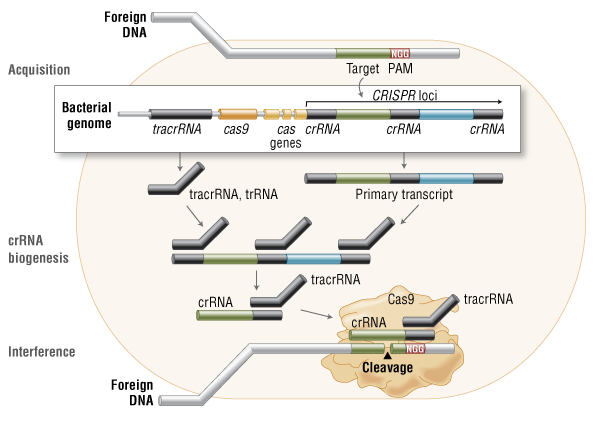
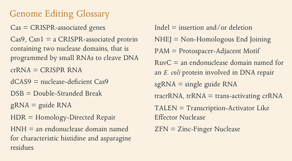
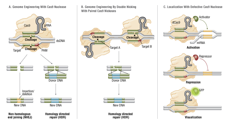
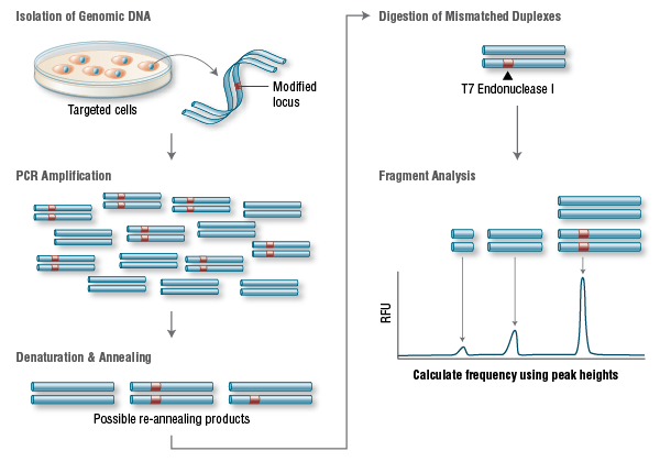



This is a lovely method and should find wide applicability in many settings, especially for microorganisms and cell lines. However, it is not clear that this approach will be, as implied by the discussion, an efficient mapping method for all multicellular organisms. I have performed similar experiments in Drosophila, focused on meiotic recombination, on a much smaller scale, and found that CRISPR-Cas9 can indeed generate targeted recombination at gRNA target sites. In every case I tested, I found that the recombination event was associated with a deletion at the gRNA site, which is probably unimportant for most mapping efforts, but may be a concern in some specific cases, for example for clinical applications. It would be interesting to know how often mutations occurred at the targeted gRNA site in this study.
The wider issue, however, is whether CRISPR-mediated recombination will be more efficient than other methods of mapping. After careful consideration of all the costs and the time involved in each of the steps for Drosophila, we have decided that targeted meiotic recombination using flanking visible markers will be, in most cases, considerably more efficient than CRISPR-mediated recombination. This is mainly due to the large expense of injecting embryos and the extensive effort and time required to screen injected animals for appropriate events. It is both cheaper and faster to generate markers (with CRISPR) and then perform a large meiotic recombination mapping experiment than it would be to generate the lines required for CRISPR-mediated recombination mapping. It is possible to dramatically reduce costs by, for example, mapping sequentially at finer resolution. But this approach would require much more time than marker-assisted mapping. If someone develops a rapid and cheap method of reliably introducing DNA into Drosophila embryos, then this calculus might change.
However, it is possible to imagine situations where CRISPR-mediated mapping would be preferable, even for Drosophila. For example, some genomic regions display extremely low or highly non-uniform recombination rates. It is possible that CRISPR-mediated mapping could provide a reasonable approach to fine mapping genes in these regions.
The authors also propose the exciting possibility that CRISPR-mediated loss of heterozygosity could be used to map traits in sterile species hybrids. It is not entirely obvious to me how this experiment would proceed and I hope the authors can illuminate me. If we imagine driving a recombination event in the early embryo (with maternal Cas9 from one parent and gRNA from a second parent), then at best we would end up with chimeric individuals carrying mitotic clones. I don’t think one could generate diploid animals where all cells carried the same loss of heterozygosity event. Even if we could, this experiment would require construction of a substantial number of stable transgenic lines expressing gRNAs. Mapping an ~20Mbp chromosome arm to ~10kb would require on the order of two-thousand transgenic lines. Not an undertaking to be taken lightly. It is already possible to perform similar tests (hemizygosity tests) using D. melanogaster deficiency lines in crosses with D. simulans, so perhaps CRISPR-mediated LOH could complement these deficiency screens for fine mapping efforts. But, at the moment, it is not clear to me how to do the experiment.