Feeds:
Posts
Comments

Archive for the ‘Clinical & Translational’ Category

CD-4 Therapy for Solid Tumors

Curator: Larry H. Bernstein, MD, FCAP


CD4 T-cell Immunotherapy Shows Activity in Solid Tumors

Alexander M. Castellino, PhD

http://www.medscape.com/viewarticle/862095

For the first time, treatment with genetically engineered T-cells has used CD4 T-cells instead of the CD8 T-cells, which are used in the chimeric antigen receptor (CAR) T-cell approach. Early data suggest that this CD4 T-cell approach has activity against solid tumors, whereas the CAR T-cell approach so far has achieved dramatic success in hematologic malignancies.

In the new approach, CD4 T-cells were genetically engineered to target MAGE-A3, a protein found on many tumor cells. The treatment was found to be safe in patients with metastatic cancers, according to data from a phase 1 clinical study presented here at the American Association for Cancer Research (AACR) 2016 Annual Meeting.

“This is the first trial testing an immunotherapy using genetically engineered CD4 T-cells,” senior author Steven A. Rosenberg, MD, PhD, chief of the Surgery Branch at the National Cancer Institute (NCI), told Medscape Medical News.

Most approaches use CD8 T-cells. Although CD8 T-cells are known be cytotoxic and CD4 T-cells are normally considered helper cells, CD4 T-cells can induce tumor regression, he said.

Louis M. Weiner, MD, director of the Lombardi Comprehensive Cancer Center at Georgetown University, in Washington, DC, indicated that in contrast with CAR T-cells, these CD4 T-cells target proteins on solid tumors. “CAR T-cells are not tumor specific and do not target solid tumors,” he said.

Engineering CD4 Cells

Immunotherapy with engineered CD4 T-cells was personalized for each patient whose tumors had not responded to or had recurred following treatment with least one standard therapy. The immunotherapy was specific for patients in whom a specific human leukocyte antigen (HLA) — HLA-DPB1*0401 — was found to be expressed on their cells and whose tumors expressed MAGE-A3.

MAGE-A3 belongs to a class of proteins expressed during fetal development. The expression is lost in normal adult tissue but is reexpressed on tumor cells, explained presenter Yong-Chen William Lu, PhD, a research fellow in the Surgery Branch of the NCI.

Targeting MAGE-A3 is relevant, because it is frequently expressed in a variety of cancers, such as melanoma and urothelial, esophageal, and cervical cancers, he pointed out.

 Researchers purified CD4 T-cells from the peripheral blood of patients. Next, the CD4 T-cells were genetically engineered with a retrovirus carrying the T-cell receptor (TCR) gene that recognizes MAGE-A3. The modified cells were grown ex vivo and were transferred back into the patient.

Clinical Results

Dr Lu presented data for 14 patients enrolled into the study: eight patients received cell doses from 10 million to 30 billion cells, and six patients received up to 100 billion cells.

This was similar to a phase 1 dose-finding study, except the researchers were seeking to determine the maximum number of genetically engineered CD4 T-cells that a patient could safely receive.

One patient with metastatic cervical cancer, another with metastatic esophageal cancer, and a third with metastatic urothelial cancer experienced partial objective responses. At 15 months, the response is ongoing in the patient with cervical cancer; after 7 months of treatment, the response was durable in the patient with urothelial cancer; and a response lasting 4 months was reported for the patient with esophageal cancer.

Dr Lu said that a phase 2 trial has been initiated to study the clinical responses of this T-cell receptor therapy in different types of metastatic cancers.

In his discussion of the paper, Michel Sadelain, MD, of the Memorial Sloan Kettering Cancer Center, New York City, said, “Although therapy with CD4 cells has been evaluated using endogenous receptor, this is the first study using genetically engineered CD4 T-cells.”

Although the study showed that therapy with genetically engineered T-cells is safe and efficacious at least in three patients, the mechanism of cytotoxicity remains unclear, Dr Sadelain indicated.

Comparison With CAR T-cells

CAR T-cells act in much the same way. CARs are chimeric antigen receptors that have an antigen-recognition domain of an antibody (the V region) and a “business end,” which activates T-cells. In this case, CD8 T-cells from the patients are used to genetically engineer T-cells ex vivo. In the majority of cases, dramatic responses have been seen in hematologic malignancies.

CARs, directed against self-proteins, result in on-target, off-tumor effects, Gregory L. Beatty, MD, PhD, assistant professor of medicine at the University of Pennsylvania, in Philadelphia, indicated when he reported the first success story of CAR T-cells in a solid pancreatic cancer tumor.

Side effects of therapy with CD4 T-cells targeting MAGE-A3 were different and similar to side effects of chemotherapy, because patients received a lymphodepleting regimen of cyclophosphamide and fludabarine. Toxicities included high fever, which was experienced by the majority of patients (12/14). The fever lasted 1 to 2 weeks and was easily manageable.

High levels of the cytokine interleukin-6 (IL-6) were detected in the serum of all patients after treatment. However, the elevation in IL-6 levels was not considered to be a cytokine release syndrome, because no side effects occurred that correlated with the syndrome, Dr Liu indicated.

He also indicated that future studies are planned that will employ genetically engineered CD4 T-cells in combination with programmed cell death protein 1–blocking antibodies.

This study was funded by Intramural Research Program of the National Institutes of Health. The NCI’s research and development of T-cell receptor therapy targeting MAGE-A3 are supported in part under a cooperative research and development agreement between the NCI and Kite Pharma, Inc. Kite has an exclusive, worldwide license with the NIH for intellectual property relating to retrovirally transduced HLA-DPB1*0401 and HLA A1 T-cell receptor therapy targeting MAGE-A3 antigen. Dr Lu and Dr Rosenberg have disclosed no relevant financial relationships.

American Association for Cancer Research (AACR) 2016 Annual Meeting: Abstract CT003, presented April 17, 2016.


Searches Related to immunotherapy using genetically engineered CD4 T-cells


Genetic engineering of T cells for adoptive immunotherapy

To be effective for the treatment of cancer and infectious diseases, T cell adoptive immunotherapy requires large numbers of cells with abundant proliferative reserves and intact effector functions. We are achieving these goals using a gene therapy strategy wherein the desired characteristics are introduced into a starting cell population, primarily by high efficiency lentiviral vector-mediated transduction. Modified cells are then expanded using ex vivo expansion protocols designed to minimally alter the desired cellular phenotype. In this article, we focus on strategies to (1) dissect the signals controlling T cell proliferation; (2) render CD4 T cells resistant to HIV-1 infection; and (3) redirect CD8 T cell antigen specificity.
Adoptive T cell therapy is a form of transfusion therapy involving the infusion of large numbers of T cells with the aim of eliminating, or at least controlling, malignancies or infectious diseases. Successful applications of this technique include the infusion of CMV-or EBVspecific CTLs to protect immunosuppressed patients from these transplantation-associated diseases [1,2]. Furthermore, donor lymphocyte infusions of ex vivo-expanded allogeneic T cells have been used to successfully treat hematological malignancies in patients with relapsed disease following allogeneic hematopoietic stem cell transplant [3]. However, in many other malignancies and chronic viral infections such as HIV-1, adoptive T cell therapy has achieved inconsistent and/or marginal successes. Nevertheless, there are compelling reasons for optimism on this strategy. For example, the existence of HIV-positive elite non-progressors [4], as well as the correlation between the presence of intratumoral T cells and a favorable prognosis in malignancies such as ovarian [5,6] and colon carcinoma [7,8], provides in vivo evidence for the critical role of the immune system in controlling both HIV and cancer.
The key to successful adoptive immunotherapy strategies appears to consist of (1) using the “right” T cell type(s) and (2) obtaining therapeutically effective numbers of these cells without compromising their effector functions or their ability to engraft within the host. This article is focused on strategies employed in our laboratory to generate the “right” cell through genetic engineering approaches, with an emphasis on redirecting the antigen specificity of CD8 T cells, and rendering CD4 T cells resistant to HIV-1 infection. The article by Paulos et al. describes the evolving process of how to best obtain therapeutically effective numbers of the “right” cells by optimizing ex vivo cell expansion strategies.
Our laboratory’s overall strategy and flow plan for development and evaluation of engineered T cells is depicted in Fig. 1. We work almost exclusively with primary human T cells; little or no work is performed with conventional established cell lines. Thus, we benefit substantially from our close association with the UPenn Human Immunology Core. The Core performs leukaphereses on healthy donors 2–3 times a week, and provides purified peripheral blood mononuclear cell subsets, ensuring a constant influx of fresh human T cells into our laboratory. We have extensive experience in developing both bead- and cell-based artificial antigen presenting cells (aAPCs), as described in detail in the article by Paulos et al. The ability to genetically modify T cells at high efficiency is critical for virtually every project within the laboratory. We have adapted the lentiviral vector system described by Dull [15] for most, but not all, of the engineering applications in our laboratory.
CD4 T cells are the primary target of HIV-1, and decreasing CD4 T cell numbers is a hallmark of advancing HIV-1 disease [34]. Thus, strategies that protect CD4 T cells from HIV-1 infection in vivo would conceivably provide sufficient immunological help to control HIV-1 infection. Our early observations that CD3/CD28 costimulation resulted in improved ex vivo expansion of CD4 T cells from both healthy and HIV-infected donors, as well as enhanced resistance to HIV-1 infection [35,36], ultimately led to the first-in-human trial of lentiviral vector-modified CD4 T cells [37]. In this trial, CD4 T cells from HIV-positive subjects who had failed antiretroviral therapy were transduced with a lentiviral vector encoding an antisense RNA that targeted a 937 bp region in the HIV-1 envelope gene. Preclinical studies demonstrated that this antisense region, directed against the HIV-1NL4-3 envelope, provided robust protection from a broad range of both R5-and X4-tropic HIV-1 isolates [38]. One year after administration of a single dose of the gene-modified cells, four of the five enrolled patients had increased peripheral blood CD4 T cell counts, and in one subject, a 1.7 log decrease in viral load was observed. Finally, in two of the five patients, persistence of the gene-modified cells was detected one year post-infusion.
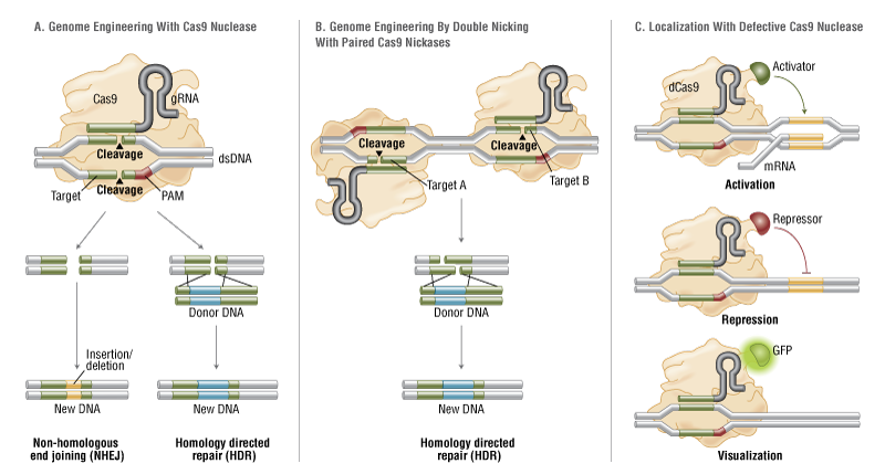
Since its identification as the primary co-receptor involved in HIV transmission, CCR5 has attracted considerable attention as a target for HIV therapy [42,43]. Indeed, “experiments of nature” have shown that individuals with a homozygous CCR5 Δ32 deletion are highly resistant to HIV-1 infection. Thus, we hypothesized that knocking out the CCR5 locus would generate CD4 T cells permanently resistant to infection by R5 isolates of HIV-1. To test this hypothesis we took advantage of zinc-finger nuclease (ZFN) technology [44]. ZFNs introduce sequencespecific double-strand DNA breakage, which is imperfectly repaired by non-homologous endjoining. This results in the permanent disruption of the genomic target, a process termed genome editing (Fig. 3).
Genetic modification of T cells to redirect antigen specificity is an attractive strategy compared to the lengthy process of growing T cell lines or CTL clones for adoptive transfer. Genetically modified, adoptively transferred T cells are capable of long-term persistence in humans [37, 46,47], demonstrating the feasibility of this approach. When compared to the months it can take to generate an infusion dose of antigen-specific CTL lines or clones from a patient, a homogeneous population of redirected antigen-specific cells can be expanded to therapeutically relevant numbers in about two weeks [3]. Several strategies are being explored to bypass the need to expand antigen-specific T cells for adoptive T cell therapy. The approaches currently studied in our laboratory involve the genetic transfer of chimeric antigen receptors and supraphysiologic T cell receptors.
Chimeric antigen receptors (CARs or T-bodies) are artificial T cell receptors that combine the extracellular single-chain variable fragment (scFv) of an antibody with intracellular signaling domains, such as CD3ζ or Fc(ε)RIγ [48–50]. When expressed on T cells, the receptor bypasses the need for antigen presentation on MHC since the scFv binds directly to cell surface antigens. This is an important feature, since many tumors and virus-infected cells downregulate MHCI, rendering them invisible to the adaptive immune system. The high-affinity nature of the scFv domain makes these engineered T cells highly sensitive to low antigen densities. In addition, new chimeric antigen receptors are relatively easy to produce from hybridomas. The key to this approach is the identification of antigens with high surface expression on tumor cells, but reduced or absent expression on normal tissues.  Since one can redirect both CD4 and CD8 T cells, the T-body approach to immunotherapy represents a near universal “off the shelf” method to generate large numbers of antigen-specific helper and cytotoxic T cells.
Many T-bodies targeting diverse tumors have been developed [51], and four have been evaluated clinically [52–55]. Three of the four studies were characterized by poor transgene expression and limited T-body engraftment. However, in a study of metastatic renal cell carcinoma using a T-body directed against carbonic anhydrase IX [55], T-body-expressing cells were detectable in the peripheral blood for nearly 2 months post-administration.
The major goals in the T-body field currently are to optimize their engraftment and maximize their effector functions. Our laboratory is addressing both problems simultaneously through an in-depth study of the requirements for T-body activation. We hypothesize that their limited persistence is due to incomplete cell activation due to the lack of costimulation. While naïve T cells depend on costimulation through CD28 ligation to avoid anergy and undergo full activation in response to antigen, it is recognized that effector cells also require costimulation to properly proliferate and produce cytokines [56]. Previous studies have shown that providing CD28 costimulation is crucial for the antitumoral function of adoptively transferred T cells and T-bodies [57–59]. Unlike conventional T cell activation, which requires two discrete signals, T-bodies can be engineered to provide both costimulation and CD3 signaling through one binding event.
A different approach for redirecting specificity to T cells for adoptive immunotherapy involves the genetic transfer of full-length TCR genes. A T cell’s specificity for its cognate antigen is solely determined by its TCR. Genes encoding the α and β chains of a T cell receptor (TCR) can be isolated from a T cell specific for the antigen of interest and restricted to a defined HLA allele, inserted into a vector, and then introduced into large numbers of T cells of individual patients that share the restricting HLA allele as well as the targeted antigen. In 1999, Clay and colleagues from Rosenberg’s group at the National Cancer Institute were the first to report the transfer of TCR genes via a retroviral vector into human lymphocytes and to show that T cells gained stable reactivity to MART-1 [67]. To date, many others have shown that the same approach can be used to transfer specificity for multiple viral and tumor associated antigens in mice and human systems. These T cells gain effector functions against the transferred TCR’s cognate antigen, as defined by proliferation, cytokine production, lysis of targets presenting the antigen, trafficking to tumor sites in vivo, and clearance of tumors and viral infection.
In 2006, Rosenberg’s group redirected patients’ PBLs with the naturally occurring, MART-1- specific TCR reported in 1999 by Clay. In the first clinical trial to test TCR-transfer immunotherapy, these modified T cells were infused into melanoma patients [68]. While the transduced T cells persisted in vivo, only two of the 17 patients had an objective response to this therapy. One issue revealed by the study was the poor expression of the transgenic TCRs by the transferred T cells. Nonetheless, the results from this trial showed the potential of TCR transfer immunotherapy as a safe form of therapy for cancer and highlighted the need to optimize such therapy to attain maximum potency.
The adoptive immunotherapy field is advancing by a tried-and-true method: learning from disappointments and moving forward. Our ability to fully realize the therapeutic potential of adoptive T cell therapy is tied to a more complete understanding of how human T cells receive signals, kill targets, and modulate effective immune responses. Our goal is to perform labbased experiments that provide insight into how primary T cells function in a manner that will facilitate and enable adoptive T cell therapy clinical trials. Our ability to efficiently modify (and expand) T cells ex vivo provides the opportunity to deliver sufficient immune firepower where it has heretofore been lacking. Sustained transgene expression, coupled with enhanced in vivo engraftment capability, will move adoptive immunotherapy into a realm where longterm therapeutic benefits are the norm rather than the exception.
Genetic Modification of T Lymphocytes for Adoptive Immunotherapy

Claudia Rossig1 and Malcolm K. Brenner2
Molecular Therapy (2004) 10, 5–18;   http://dx.doi.org:/10.1016/j.ymthe.2004.04.014      http://www.nature.com/mt/journal/v10/n1/full/mt20041193a.html

Adoptive transfer of T lymphocytes is a promising therapy for malignancies—particularly of the hemopoietic system—and for otherwise intractable viral diseases. Efforts to broaden the approach have been limited by the physiology of the T cells themselves and by a range of immune evasion mechanisms developed by tumor cells. In this review we show how genetic modification of T cells is being used preclinically and in patients to overcome these limitations, by incorporation of novel receptors, resistance mechanisms, and control genes. We also discuss how the increasing safety and effectiveness of gene transfer technologies will lead to an increase in the use of gene-modified T cells for the treatment of a wider range of disorders.

That gene transfer could be used to improve the effectiveness of T lymphocytes was apparent from the beginning of clinical studies in the field. T cells were the very first targets for genetic modification in human gene transfer experiments. Rosenberg’s group marked tumor-infiltrating lymphocytes ex vivo with a Moloney retroviral vector encoding neomycin phosphotransferase before reinfusing them and attempting to demonstrate selective accumulation at tumor sites. Shortly thereafter, Blaese and Anderson led a group that infused corrected T cells into two children with severe combined immunodeficiency due to ADA deficiency. While neither study was completely successful in terms of outcome, both showed the feasibility of ex vivo gene transfer into human cells and set the stage for many of the studies that followed. More recently, a second wave of interest in adoptive T cell therapies has developed, based on their success in the prevention and treatment of viral infections such as EBV and cytomegalovirus (CMV) and on their apparent ability to eradicate hematologic and perhaps solid malignancies1,2,3,4,5,6. There has been a corresponding increase in studies directed toward enhancing the antineoplastic and antiviral properties of the T cells. In this article we will review how gene transfer may be used to produce the desired improvements focusing on vectors and genes that have had clinical application.

Currently available viral and nonviral vector systems lack a pattern of biodistribution that would favor T cell transduction in vivo—as occurs, for example, with adenovectors and the liver or liposomal vectors and the lung. This lack of favorable biodistribution cannot yet be compensated for by the introduction of specific T-cell-targeting ligands into vectors. Hence, all T cell gene transfer studies conducted to date have used ex vivo transduction followed by adoptive transfer of gene-modified cells. This approach is inherently less attractive for commercial development than directin vivo gene transfer and has probably restricted interest in developing clinical applications using these cells. On the other hand, ex vivo transduction may be more readily controlled, characterized, and standardized than in vivo efforts and may ultimately produce a better defined final product (the transduced cell).

The gene products of suicide and coexpressed resistance genes are highly immunogenic and may induce immune-mediated rejection of the transduced cells. In one study, the persistence of adoptively transferred autologous CD8+ HIV-specific CTL clones modified to express the hygromycin phosphotransferase (Hy) gene and the herpesvirus thymidine kinase gene as a fusion gene was limited by the induction of a potent CD8+ class I MHC-restricted CTL response specific for epitopes derived from the Hy-tk protein126. Less immunogenic suicide and selection marker genes, preferably of human origin, may reduce the immunological inactivation of genetically modified donor lymphocytes. Human-derived prodrug-activating systems include the human folylpolyglutamate synthetase/methotrexate127, the deoxycytidine/cytosine arabinoside128, or the carboxylesterase/irinotecan129 systems. These systems do not activate nontoxic prodrugs but are based on enhancement of already potent chemotherapeutic agents. The administration of methotrexate to treat severe GVHD may not only kill transduced donor lymphocytes but may also have additional inhibitory activity on nontransduced but activated T cells.

Finally, endogenous proapoptotic molecules have been proposed as nonimmunogenic suicide genes. A chimeric protein that contains the FK506-binding protein FKBP12 linked to the intracellular domain of human Fas130 was recently introduced. Addition of the dimerizing prodrug induces Fas crosslinking with subsequent triggering of an apoptotic death signal.

Genetic engineering of T lymphocytes should help deliver on the promise of immunotherapies for cancer, infection, and autoimmune disease. Improvements in transduction, selection, and expansion techniques and the development of new viral vectors incapable of insertional mutagenesis will reduce the risks and further enhance the integration of T cell and gene therapies. Nonetheless, successful application of the proposed modifications to the clinical setting still requires many iterative studies to allow investigators to optimize the individual components of the approach.

Genetically modified T cells in cancer therapy: opportunities and challenges
Michaela Sharpe, Natalie Mount


The feasibility of T-cell adoptive transfer was first reported nearly 20 years ago (Walter et al., 1995) and the field of T-cell therapies is now poised for significant clinical advances. Recent clinical trial successes have been achieved through multiple small advances, improved understanding of immunology and emerging technologies. As the key challenges of T-cell avidity, persistence and ability to exert the desired anti-tumour effects as well as the identification of new target antigens are addressed, a broader clinical application of these therapies could be achieved. As the clinical data emerges, the challenge of making these therapies available to patients shifts to implementing robust, scalable and cost-effective manufacture and to the further evolution of the regulatory requirements to ensure an appropriate but proportionate system that is adapted to the characteristics of these innovative new medicines.


Read Full Post »

What about PDL-1 in oncotherapy diagnostics for NSCLC?

Larry H. Bernstein, MD, FCAP, Curator

LPBI

UPDATED 5/15/2019

Questions on PD-L1 Diagnostics for Immunotherapy in NSCLC
Alexander M. Castellino, PhD
http://www.medscape.com/viewarticle/862275

Two immunotherapies that target the cell programmed death (PD) pathway are now available, and both nivolumab (Opdivo, Bristol-Myers Squibb Company) and pembrolizumab (Keytruda, Merck Sharp & Dohme Corp) are approved for treating advanced, refractory, non–small cell lung cancer (NSCLC). Across several studies in patients with NSCLC, response to these agents has been correlated with PD-L1 staining, which determines PD-L1 levels in the tumor tissue. How do the available assays for PD-L1 compare?

The linear correlation between three commercially available assays is good across a range of cutoff points, concluded a presentation at the 2016 American Association for Clinical Research Annual Meeting.

Cutoffs are defined as the percentage of cells expressing PD-L1 when analyzed histochemically. “The dataset builds confidence that the assays may be used according to the cutoff clinically validated for the drug in question,” Marianne J. Radcliffe, MD, diagnostic associate director at AstraZeneca, toldMedscape Medical News.

“The correlation is good between the assays across the range examined,” she added.

However, a recently published study showed a high rate of discordance between another set of PD-L1 assays that were tested.

Dr Marianne Radcliffe

“Different diagnostic tests yield different results, depending on the cutoff for each assay. We need to harmonize the assays so clinicians are talking about the same thing,” Brendon Stiles, MD, associate professor of cardiothoracic surgery at Weill Cornell Medicine and New York-Presbyterian Hospital, New York City, told Medscape Medical News.

For Dr Stiles, these studies raise the issue that it is difficult to compare results of diagnostic testing across the different drugs and even with the same drug that are derived from different assays. “More importantly, it raises confusion in clinical practice when a patient’s sample stains positive for PD-L1 with one assay and negative with another,” he said.

“The commercial strategy for developing companion diagnostics for each drug is not in the best interests of the patients. It generates confusion among both clinicians and patients,” Dr Stiles commented. “We need to know if these assays can be used interchangeably,” he said.

As new agents come into the clinic, Dr Stiles believes there should be a universal yes-or-no answer, so that clinicians can use the assay to help decide on the use of immunotherapy.

Three Assays Tested

The study presented by Dr Radcliffe and colleagues investigated three commercially available assays, Ventana SP263, Dako 22C3, and Dako 28-8, with regard to how they compare at different cutoffs. Different studies use different cutoffs to express positivity.

Ventana SP263 was developed as a companion diagnostic for durvalumab (under development by AstraZeneca) using a rabbit monoclonal antibody. Positivity is defined as ≥25% staining of tumor cells.

Dako 22C3 was developed, and is approved, as a companion diagnostic for pembrolizumab. It uses a mouse monoclonal antibody. Positivity is defined as ≥1% and ≥50% staining of tumor cells.

Dako 28-8 was developed as a companion diagnostic for nivolumab and uses a rabbit monoclonal antibody (different from the one used in the Ventana SP263). In clinical practice, this assay is used as a complementary diagnostic for nivolumab, but the drug is approved for use regardless of PD-L1 expression. Positivity is defined as ≥1%, ≥5%, or ≥10% staining of tumor cells.

Ventana SP142 was not included in the study because it is not commercially available, Dr Ratcliffe indicated.
The three assays were used on consecutive sections of 500 archival NSCLC tumor samples obtained from commercial vendors. A single pathologist trained by the manufacturer read all samples in batches on an assay-by-assay basis. Samples were assessed per package inserts provided by Ventana and Dako in a Clinical Laboratory Improvement Amendments program-certified laboratory.

Dr Ratcliffe indicated that between reads of samples from the same patient, there was a washout period for the pathologist to remove bias.

The NSCLC samples included patients with stage I (38%), II (39%), III (20%), and IV (<1%) disease. Histologies included nonsquamous (54%) and squamous (43%) cancers.

All three PD-L1 assays showed similar patterns of staining in the range of 0% to 100%, Dr Ratcliffe indicated.


The correlation between any two of the assays was determined from tumor cell membrane staining. The correlation was linear with Spearman correlation of 0.911 for Ventana SP263 vs Dako 22C3; 0.935 for Ventana SP263 vs Dako 28-8; and 0.954 for Dako 28-8 vs Dako 22C3.

“With an overall predictive value of >90%, the assays have closely aligned dynamic ranges, but more work is needed,” Dr Ratcliffe said. “In general, scoring of immunohistochemical assays can be more variable between 1% and 10%, and we plan to look at this in more detail,” she said. These samples need to be reviewed by an independent pathologist, she added.

Dr Radcliffe said that currently, “Direct clinical efficacy data supporting a specific diagnostic test should still be considered as the highest standard of proof for diagnostic clinical utility.”

Why Correlations Are Needed

Pembrolizumab is approved for use only in patients with PD-L1-positive, previously treated NSCLC. A similar patient profile is being considered for nivolumab, for which testing for PD-L1 expression is not required.

For new PD-immunotherapy agents in clinical development, it is not clear whether PD-L1 testing will be mandated.

However, in clinical practice, it is clear that some patients respond to therapy, even if they are PD-L1 negative, as defined from the study. “Is it a failure of the assay, tumor heterogeneity, or is there another time point when PD-L1 expression is turned on?” Dr Stiles asked.

Dr Stiles also pointed out that a recent publication from Yale researchers showed a high a rate of discordance. In this study, PD-L1 expression was determined using two rabbit monoclonal antibodies. Both of these were different from the ones used in the Ventana SP263 and Dako 28-8 assays.

In this study, whole-tissue sections from 49 NSCLC samples were used, and a corresponding tissue microarray was also used with the same 49 samples. Researchers showed that in 49 NSCLC tissue samples, there was intra-assay variability, with results showing fair to poor concordance with the two antibodies. “Assessment of 588 serial section fields of view from whole tissue showed discordant expression at a frequency of 25%.

“Objective determination of PD-L1 protein levels in NSCLC reveals heterogeneity within tumors and prominent interassay variability or discordance. This could be due to different antibody affinities, limited specificity, or distinct target epitopes. Efforts to determine the clinical value of these observations are under way,” the study authors conclude.

The Blueprint Proposal

Coincidentally, a blueprint proposal was announced here at the AACR meeting at a workshop entitled FDA-AACR-ASCO Complexities in Personalized Medicine: Harmonizing Companion Diagnostics across a Class of Targeted Therapies.

The blueprint proposal was developed by four pharmaceutical giants (Bristol-Myers Squibb Company, Merck & Co, Inc, AstraZeneca PLC, and Genentech, Inc) and two diagnostic companies (Agilent Technologies, Inc/Dako Corp and Roche/Ventana Medical Systems, Inc).

In this proposal, the development of an evidence base for PD-1/PD-L1 companion diagnostic characterization for NSCLC would be built into studies conducted in the preapproval stage. Once the tests are approved, the information will lay the foundation for postapproval studies to inform stakeholders (eg, patients, physicians, pathologists) on how the test results can best be used to make treatment decisions.

The blueprint proposal is available online.

Dr Ratcliffe is an employee and shareholder of AstraZeneca. Dr Stiles has disclosed no relevant financial relationships.

 American Association for Cancer Research (AACR) 2016 Annual Meeting: Abstract LB-094, presented April 18, 2016.
Quantitative Assessment of the Heterogeneity of PD-L1 Expression in Non–Small-Cell Lung Cancer
Joseph McLaughlin, 1,2; Gang Han, 3; Kurt A. Schalper, 2; ….,  Roy Herbst, 1; Patricia LoRusso, 1; David L. Rimm, 2

JAMA Oncol. 2016;2(1):46-54.       http://dx.doi.org:/10.1001/jamaoncol.2015.3638.

Importance  Early-phase trials with monoclonal antibodies targeting PD-1 (programmed cell death protein 1) and PD-L1 (programmed cell death 1 ligand 1) have demonstrated durable clinical responses in patients with non–small-cell lung cancer (NSCLC). However, current assays for the prognostic and/or predictive role of tumor PD-L1 expression are not standardized with respect to either quantity or distribution of expression.

Objective  To demonstrate PD-L1 protein distribution in NSCLC tumors using both conventional immunohistochemistry (IHC) and quantitative immunofluorescence (QIF) and compare results obtained using 2 different PD-L1 antibodies.

Design, Setting, and Participants  PD-L1 was measured using E1L3N and SP142, 2 rabbit monoclonal antibodies, in 49 NSCLC whole-tissue sections and a corresponding tissue microarray with the same 49 cases. Non–small-cell lung cancer biopsy specimens from 2011 to 2012 were collected retrospectively from the Yale Thoracic Oncology Program Tissue Bank. Human melanoma Mel 624 cells stably transfected with PD-L1 as well as Mel 624 parental cells, and human term placenta whole tissue sections were used as controls and for antibody validation. PD-L1 protein expression in tumor and stroma was assessed using chromogenic IHC and the AQUA (Automated Quantitative Analysis) method of QIF. Tumor-infiltrating lymphocytes (TILs) were scored in hematoxylin-eosin slides using current consensus guidelines. The association between PD-L1 protein expression, TILs, and clinicopathological features were determined.

Main Outcomes and Measures  PD-L1 expression discordance or heterogeneity using the diaminobenzidine chromogen and QIF was the main outcome measure selected prior to performing the study.

Results  Using chromogenic IHC, both antibodies showed fair to poor concordance. The PD-L1 antibodies showed poor concordance (Cohen κ range, 0.124-0.340) using conventional chromogenic IHC and showed intra-assay heterogeneity (E1L3N coefficient of variation [CV], 6.75%-75.24%; SP142 CV, 12.17%-109.61%) and significant interassay discordance using QIF (26.6%). Quantitative immunofluorescence showed that PD-L1 expression using both PD-L1 antibodies was heterogeneous. Using QIF, the scores obtained with E1L3N and SP142 for each tumor were significantly different according to nonparametric paired test (P < .001). Assessment of 588 serial section fields of view from whole tissue showed discordant expression at a frequency of 25%. Expression of PD-L1 was correlated with high TILs using both E1L3N (P = .007) and SP142 (P = .02).

Conclusions and Relevance  Objective determination of PD-L1 protein levels in NSCLC reveals heterogeneity within tumors and prominent interassay variability or discordance. This could be due to different antibody affinities, limited specificity, or distinct target epitopes. Efforts to determine the clinical value of these observations are under way.


Introduction We are in an era of rapid incorporation of basic scientific discoveries into the drug development pipeline. Currently, numerous sponsors are developing therapeutic products that may use similar or identical biomarkers for therapeutic selection, measured or detected by an in vitro companion diagnostic device. The current practice is to independently develop a companion diagnostic for each therapeutic. Thus, the matrix of therapeutics and companion diagnostics, if each therapeutic were approved in conjunction with a companion diagnostic, may present a complex challenge for testing and decision making in the clinic, potentially putting patients at risk if inappropriate diagnostic tests were used to make treatment decisions. To address this challenge, there is a desire to understand assay comparability and/or standardize analytical and clinical performance characteristics supporting claims that are shared across companion diagnostic devices. Pathologists and oncologists also need clarity on how to interpret test results to inform downstream treatment options for their patients.
Clearly using each of the companion diagnostics to select one of the several available targeted therapies in the same class is not practical and may be impossible. Likewise, having a single test or assay as a sole companion test for all of the multiple therapeutic options within a class is also impractical since the individual therapies have differing modes of action, intended use populations, specificities, safety and efficacy outcomes. Thus, a single assay or test may not adequately capture the appropriate patient population that may benefit (or not) from each individual therapeutic option within a class of therapies. Furthermore, aligning multiple sponsors’ study designs and timelines in order that they all adopt a single companion test may inadvertently slow down development of critical therapeutic products and delay patient access to these life-saving products.
Any solution to this challenge will be multifaceted and will, by necessity, involve multiple stakeholders. Thus, the US Food and Drug Administration (FDA), the American Association for Cancer Research (AACR) and American Society of Clinical Oncology (ASCO) convened a workshop titled “Complexities in Personalized Medicine: Harmonizing Companion Diagnostics Across a Class of Targeted Therapies” to draw out and assess possible solutions. Recognizing that the complex scientific, regulatory and market forces at play here require a collaborative effort, an industry workgroup volunteered to develop a blueprint proposal of potential solutions using nonsmall cell lung cancer (NSCLC) as the use case indication.
Goal and Scope of Blueprint The imminent arrival to the market of multiple PD1 / PD-L1 compounds and the possibility of one or more associated companion diagnostics is unprecedented in the field of oncology. Some may assume that since these products target the same biological pathway, they are interchangeable; however, each PD1/PD-L1 compound is unique with respect to its clinical pharmacology and each compound is being developed in the context of a unique biological scientific hypothesis and registration strategy. Similarly, each companion diagnostic has been optimized within the individual therapeutic development programs to meet specific development goals, e.g., 1) validation for patient selection, 2) subgroup analysis as a prognostic variable, or 3) enrichment.
Further, each companion diagnostic test is optimized for its specific therapy and with its own unique performance characteristics and scoring/interpretation guidelines.
The blueprint development group recognizes that to assume that any one of the available tests could be used for guiding the treatment decision with any one or all of the drugs available in this class presents a potential risk to patients that must be addressed.
The goal of this proposal is to agree and deliver, via cross industry collaboration, a package of information /data upon which analytic comparison of the various diagnostic assays may be conducted, potentially paving the way for post-market standardization and/or practice guideline development as appropriate.
A comparative study of PD-L1 diagnostic assays and the classification of patients as PD-L1 positive and PD-L1 negative
Presentation Time: Monday, Apr 18, 2016, 8:00 AM -12:00 PM
Location: Section 10
Poster Board Number: 18
Author Block: Marianne J. Ratcliffe1, Alan Sharpe2, Anita Midha1, Craig Barker2, Paul Scorer2, Jill Walker2. 1AstraZeneca, Alderley Park, United Kingdom; 2AstraZeneca, Cambridge, United Kingdom
Abstract Body: Background: PD-1/PD-L1 directed antibodies are emerging as effective therapeutics in multiple oncology settings. Keynote 001 and Checkmate 057 have shown more frequent response to PD-1 targeted therapies in NSCLC patients with high tumour PD-L1 expression than patients with low or no PD-L1 expression. Multiple diagnostic PD-L1 tests are available using different antibody clones, different staining protocols and diverse scoring algorithms. It is vital to compare these assays to allow appropriate interpretation of clinical outcomes. Such understanding will promote harmonization of PD-L1 testing in clinical practice.
Methods: Approximately 500 tumour biopsy samples from NSCLC patients, including squamous and non-squamous histologies, will be assessed using three leading PD-L1 diagnostics assays. PD-L1 assessment by the Ventana SP263 assay that is currently being used in Durvalumab clinical trials (positivity cut off: ≥25% tumour cells with membrane staining) will be compared with the Dako 28-8 assay (used in the Nivolumab Checkmate 057 trial at the 1%, 5% and 10% tumour membrane positivity cut offs), and the Dako 22C3 assay (used in the Pembrolizumab Keynote 001 trial) at the 1% and 50% cut offs).
Results: Preliminary data from 81 non-squamous patients indicated good concordance between the Ventana SP263 and Dako 28-8 assays. Optimal overall percent agreement (OPA) was observed between Dako 28-8 at the 10% cut off and the Ventana SP263 assay (OPA; 96%, Positive percent agreement (PPA); 91%, Negative percent agreement (NPA); 98%), where the Ventana SP263 assay was set as the reference. Data on the full cohort will be presented for all three assays, and a lower 95% confidence interval calculated using the Clopper-Pearson method.
Conclusions: This study indicates that the patient population defined by Ventana SP263 at the 25% cut off is similar to that identified by the Dako-28-8 assay at the 10% tumour membrane cut off. This, together with data on the 22C3 assay, will enable cross comparison of studies using different PD-L1 tests, and widen options for harmonization of PD-L1 diagnostic testing.

http://www.abstractsonline.com/Plan/ViewAbstract.aspx

Table 1
Reference: Ventana SP-263 (≥25% tumour membrane staining)
Dako 28-8 assay cut off PPA
(%)
NPA
(%)
OPA
(%)
>1% 58 100 81
>5% 72 100 90
>10% 91 98 96

UPDATED 5/19/2019

Incidence of Adverse Events for PD-1/PD-L1 Inhibitors Underscores Toxicity Risk

https://www.cancernetwork.com/immuno-oncology/incidence-adverse-events-pd-1pd-l1-inhibitors-underscores-toxicity-risk

May 7, 2019

Approximately two-thirds of cancer patients who received a programmed death 1 (PD-1) or programmed death ligand 1 (PD-L1) inhibitor in clinical trials experienced treatment-related adverse events, according to a systematic review and meta-analysis recently published in JAMA Oncology. The study findings may facilitate discussions with cancer patients who are considering PD-1 or PD-L1 therapy.

“The vast majority of patients with advanced cancer want to be on the [PD-1 or PD-L1] therapy,” Eric H. Bernicker, MD, a thoracic medical oncologist with Houston Methodist Cancer Center, told Cancer Network. Not involved in the current study, Bernicker explained that patients perceive these therapies to have “very different” side effects and risks from chemotherapy.

While they do, Bernicker explained, it’s important to underscore, which this study does, that these are not “completely innocuous” therapies. The study findings allow physicians to give numbers to patients and families when counseling them about the risks involved, he said.

The systematic review and meta-analysis is based on data from 125 clinical trials and 20,128 participants. Clinical trials were identified by systematically searching for published clinical trials that evaluated single-agent PD-1 and PD-L1 inhibitors and reported treatment-related adverse events in PubMed, Web of Science, Embase, and Scopus. The majority of trials evaluated nivolumab (n = 46) or pembrolizumab (n = 49), and the most common cancer types were lung cancer (n = 26), genitourinary cancer (n = 22), melanoma (n = 16), and gastrointestinal cancer (n = 14).

In all, 66.0% of clinical trial participants reported at least 1 adverse event of any grade, and 14.0% reported at least 1 grade 3 or higher adverse event. The most frequently reported adverse events of any grade were fatigue (18.26%), pruritus (10.61%), and diarrhea (9.47%). As for grade 3 or higher events, the most commonly reported were fatigue (0.89%), anemia (0.78%), and aspartate aminotransferase (AST) increase (0.75%).

Frequently reported immune-related adverse events of any grade included diarrhea (9.47%), AST increase (3.39%), vitiligo (3.26%), alanine aminotransferase (ALT) increase (3.14%), pneumonitis (2.79%), and colitis (1.24%). Grade 3 or higher immune-related adverse events included AST increase (0.75%), ALT increase (0.70%), pneumonitis (0.67%), diarrhea (0.59%), and colitis (0.47%).

If present, certain adverse events had increased likelihood of being grade 3 or higher, including hepatitis (risk ratio [RR], 50.59%), pneumonitis (RR, 24.01%), type 1 diabetes (RR, 41.86%), and colitis (RR, 37.90%).

“In terms of the rough percentage of side effects and the breadth of the side effects, this is pretty much what most of us see in the clinic,” Bernicker said, noting that none of the findings were particularly surprising.

Although no differences in adverse event incidence were found across different cancer types, differences were found between PD-1 and PD-L1 inhibitors in a subgroup analysis. Overall, compared with PD-L1 inhibitors, PD-1 inhibitors had a higher mean incidence of grade 3 or higher events (odds ratio [OR], 1.58; 95% CI, 1.00–2.54). Specifically, nivolumab had a higher mean incidence of grade 3 or higher events (OR, 1.81; 95% CI, 1.04–3.01) compared with PD-L1 inhibitors.

Bernicker commented that these incidence differences on the basis of drug type were “intriguing” but not clinically useful, given that PD-1 and PD-L1 inhibitors are not interchangeable. He said the finding “needs to be further looked at.”

Read Full Post »

A Concise Review of Cardiovascular Biomarkers of Hypertension

Curator: Larry H. Bernstein, MD, FCAP

LPBI

Revised 5/25/2016


Introduction

While a large body of work had been done on cholesterol synthesis, HDL and LDL cholesterol, triglycerides, and lipoproteins for a quarter century, and the concept of metabolic syndrome was emerging, there was neither a unifying concept nor a sufficient multivariable approach to apply the use of laboratory markers to clinical practice.  The mathematical foundation for such an evaluation of the biological markers and the computational tools were maturing at the turn of the 20th century, and the interest in outcomes research for improved healthcare practice was maturing. In addition, there was now heavy investment in health information systems that would support emerging health networks of a rapidly consolidating patient base.  This has become important for the pharmaceutical industry and for allied health sciences to enable a suitable method of measuring the effectiveness of drug and of lifestyle changes to improve the population health.

The importance of finding biomarkers for hypertension is significant as stated above. I refer to observations in a lecture by Teresa Seeman, Ph.D., Professor, UCLA Geffen School of Medicine (1).
The missed cased of hypertension in the U.S. alone has been examined by the NHANES studies. Table  I
shows the poor identification of this serious chronic condition. The next table (Table II)*, also from NHANES  (Seeman study) looks at Allostatic Load for biomarkers using component biomarker measurement criterion cutpoints.  Table III* gives the odds ratios for mortality by Allostatic Load Score.

An explanatory problem for our difficulty with diagnosis of a number of hypertension disease “subsets” is that there is peripheral hypertension that might be idiopathic, or it might be related to coexisting diseases with both inflammatory and vascular structural dynamics nature.  In addition, this may be concurrent with pulmonary hypertension, systemic hypertension, and progressive renal disease.  This discussion is reserved for later.  As stated, the late or missed diagnosis of systemic or essential idiopathic hypertension is illustrated in the three Seeman tables (1).


Table 1

Table 2

Table 3


Table 1*. Missed cases by “self report”

Self-reports

vs undiagnosed

study NHANES 88-94 NHANES 99-2004 NHANES 2005-08
Hypertension %unaware  BP > 140/90 42.7 43.5 39.06
SR-controlled
SR-high

Unaware

  7.45

10

13.88

8.35

10.85

16.12

6.5

10.18

19.98

High cholesterol Chol > 220 g/dl 55.93 49.3 47.05
SR-controlled
SR- high
Unaware
  11.02
8.68
12.12
8.47
8.72
18.5
7.22
8.12
23.46
Diabetes HgA1C > 6.4%      
SR-controlled

SR- high

Unaware

  2.41

3.43

1.64

1.76

5.01

3.09

2.11
5.51
3.09

*modified from Seeman


Table II* USHANES: Allostatic Load – component cutpoints

Biomarker Total N High Risk Percent (%) Cutpoint
DBP (mm Hg) 15,489 1,180   7.62    90
SBP (mm Hg) 15,491 3,461 22.34  140
Pulse Rate 15,117 1,009   6.67    90
HgA1C (%) 15,441 1,482   9.60    6.4
WHR 14,824 6,778 45.72    0.94
HDL Cholesterol (mg/dl) 15,187 3,440 22.65     40
Total Cholesterol

(mg/dl)

15,293 3,196  20.90    240

*From  T. Seaman, UCLA Geffen SOM


Table III*. Odds of mortality by Allostatic Load Score.

ALS Odds Ratio
7-8 5
6 2.6
5 2.3
4 2.1
3 1.8
2 1.5
1 1.4


*From  T. Seaman, UCLA Geffen SOM


I refer to cardiovascular diseases in reference to an aggregate of diseases affecting the heart, the circulatory system from large artery to the capillary, the lungs and kidneys, excluding the lymphatics.
These major disease entities are both separate and interrelated, not necessarily found in the same combinations. However, they account for a growing proportion of illness, apart from cancers, that affect the aging population of western societies. In the discussion that follows, I shall construct a picture of the pathophysiology of cardiovascular diseases, describe the major biomarkers for the assessment of these, point out the relationship of these to hypertension, and try to develop a more targeted approach to the assessment of hypertension and related disorders.

Chronic kidney disease (CKD) is defined as persistent kidney damage accompanied by a reduction in the glomerular filtration rate (GFR) and the presence of albuminuria. The rise in incidence of CKD is attributed to an aging populace and increases in hypertension (HTN), diabetes, and obesity within the U.S. population. CKD is associated with a host of complications including electrolyte imbalances, mineral and bone disorders, anemia, dyslipidemia, and HTN. It is well known that CKD is a risk factor for cardiovascular disease (CVD), and that a reduced GFR and albuminuria are independently associated with an increase in cardiovascular and all-cause mortality.

The relationship between CKD and HTN is cyclic, as CKD can contribute to or cause HTN (3). Elevated BP leads to damage of blood vessels within the kidney, as well as throughout the body. This damage impairs the kidney’s ability to filter fluid and waste from the blood, leading to an increase of fluid volume in the blood—thus causing an increase in BP.


A cursory description of the blood circulation

The full circulation involves the heart as a pump, and the arteries and veins, comprising small and large vessels, and capillaries at the point of delivery of oxygen and capture of carbon dioxide, and of transfer of substrates to tissues.  The brain, liver, pancreas and spleen, and endocrines are not further considered here, except for a consideration on neuro-humoral peptides that have emerged in the regulation of blood pressure and are essential to the stress response. The lung and the liver are both important with respect to the exchange of air and metabolites, and both have secondary circulations, the pulmonary and the portal vascular circulations.  In the case of the lungs, the vena cava flows into the right atrium, which delivers unoxygenated blood to the lungs via the right ventricle and right pulmonary artery, which returns to the left atrium by way of the right pulmonary vein.  The blood from the left atrium that flows into the left ventricle is ejected into the aorta.  The coronary arteries that nourish the heart are at the base of the aorta.  The heart muscle is a syncytium, unlike striated muscle, and it is densely packed with mitochondria, suitable for continuous contraction under vasovagal control. This is the anatomical construct, but the physiology is still being clarified because normal function and disease are both a matter of regulatory control.

In order to understand hypertension, we have to view the heart functioning over a long period of time.
In a still frame picture, we envision the left ventricle contracts emptying the oxygenated blood into the circulation. The ejection of blood into the aorta is called systole, by which the blood is delivered by the force of contraction into the circulation.  The filling pressure is called diastole.  So we have a filling and an emptying, and heard by the stethoscope is a lub-dub, synchronously repeated.   A normal systolic blood pressure is below 120. A systolic blood pressure of 120 to 139 means you have prehypertension, or borderline high blood pressure. Even people with prehypertension are at a higher risk of developing heart disease. A systolic blood pressure number of 140 or higher is considered to be hypertension, or high blood pressure. The diastolic blood pressure number or the bottom number indicates the pressure in the arteries when the heart rests between beats. A normal diastolic blood pressure number is less than 80. A diastolic blood pressure between 80 and 89 indicates prehypertension. A diastolic blood pressure number of 90 or higher is considered to be hypertension or high blood pressure. So now we have identified a systolic and a diastolic high blood pressure. Systolic pressure increases with vigorous activity, and becomes normal when the activity resides.  The systolic blood pressure increases with age. Over time, consistently high blood pressure weakens and damages the blood vessels so affected. Moreover, changes in the body’s normal functions may cause high blood pressure, including changes to kidney fluid and salt balances, the renin-angiotensin-aldosterone system, sympathetic nervous system activity, and blood vessel structure and function.


Starling’s Law of the Heart

Two principal intrinsic mechanisms, namely the Frank-Starling mechanism and rate induced regulation, enable the myocardium to adapt to changes in hemodynamic conditions. The Frank-Starling mechanism (also referred to as Starling’s law of the heart), is invoked in response to changes in the resting length of the myocardial fibers. Rate-induced regulation is invoked in response to changes in the frequency of the heartbeat.  (3-9).

Frank and Starling (3, 4) showed that an increase in diastolic volume caused an increase in systolic performance. The stretch effect persists across a range of myocardial contractile states, but during exercise it plays only a lesser role augmenting ventricular function maximal exercise. This is because in healthy human subjects adrenergic reflex mechanisms modulate myocardial performance, heart rate, vascular impedance and coronary flow during exercise and changes in these variables can overshadow the effect of fiber stretch or even prevent an increase in end-diastolic volume during stress (5). (See you- tube (6).

According to Lakatta muscle length modulates the extent of myofilament calcium ion (Ca2+) activation (7-9).   Similarly, the fiber length during a contraction, which is determined in part by the load encountered during shortening, also determines the extent of myofilament Ca2+ activation. Therefore, the terms preload, afterload and myocardial contractile state lose part of their significance in light of current knowledge.


Biology and High Blood Pressure

Researchers continue to study how various changes in normal body functions cause high blood pressure. The key functions affected in high blood pressure include (10):

Kidney Fluid and Salt Balances

The kidneys normally regulate the body’s salt balance by retaining sodium and water and excreting potassium. Imbalances in this kidney function can expand blood volumes, which can cause high blood pressure.

Renin-Angiotensin-Aldosterone System

The renin-angiotensin-aldosterone system makes angiotensin and aldosterone hormones. Angiotensin narrows or constricts blood vessels, which can lead to an increase in blood pressure. Aldosterone controls how the kidneys balance fluid and salt levels. Increased aldosterone levels or activity may change this kidney function, leading to increased blood volumes and high blood pressure.

Sympathetic Nervous System Activity

The sympathetic nervous system has important functions in blood pressure regulation, including heart rate, blood pressure, and breathing rate. Researchers are investigating whether imbalances in this system cause high blood pressure.

Blood Vessel Structure and Function

Changes in the structure and function of small and large arteries may contribute to high blood pressure. The angiotensin pathway and the immune system may stiffen small and large arteries, which can affect blood pressure.

Two or more types of hypertension

Systemic hypertension

Idiopathic hypertension

Hypertension from chronic renal disease

Pulmonary artery hypertension

Hypertension associated with systemic chronic inflammatory disease (rheumatoid arthritis and other collagen vascular diseases)

Genetic Causes of High Blood Pressure

Much of the understanding of the body systems involved in high blood pressure has come from genetic studies. High blood pressure often runs in families. Years of research have identified many genes and other mutations associated with high blood pressure, some in the renal salt regulatory and renin-angiotensin-aldosterone pathways. However, these known genetic factors only account for 2 to 3 percent of all cases. Emerging research suggests that certain DNA changes during fetal development also may cause the development of high blood pressure later in life.

Environmental Causes of High Blood Pressure

Environmental causes of high blood pressure include unhealthy lifestyle habits, being overweight or obese, and medicines.

Other medical causes of high blood pressure include other medical conditions such as chronic kidney disease, sleep apnea, thyroid problems, or certain tumors.

The common complications of hypertension and their signs and symptoms include:

http://www.nhlbi.nih.gov/health/health-topics/topics/hbp/causes10


Pulse Pressure and Stroke Volume

The  pulse pressure is the difference between systolic (the upper number) and diastolic (the lower number) (11).

Systemic pulse pressure = Psystolic – Pdiastolic

The pulse pressure is 40 mmHg for a typical blood pressure reading of 120/80 mmHg.

Pulse pressure (PP) is proportional to stroke volume (SV), the amount of blood pumped from the heart in one beat, and inversely proportional to the compliance or flexibility of the blood vessels, mainly the aorta.

A low (also called narrow) pulse pressure means that not much blood is being expelled from the heart, and can be caused by a number of factors, including severe blood loss due to trauma, congestive heart failure, shock, a narrowing of the valve leading from the heart to the aorta (stenosis), and fluid accumulating around the heart (tamponade).

High (or wide) pulse pressures occur during exercise, as stroke volume increases and the overall resistance to blood flow decreases. It can also occur for many reasons, such as hardening of the arteries (which can have numerous causes), various deficiencies in the aorta (mainly) or other arteries, including leaks, fistulas, and a usually-congenital condition known as AVM, pain/anxiety, fever, anemia, pregnancy, and more. Certain medications for high blood pressure can widen pulse pressure, while others narrow it. A chronic increase in pulse pressure is a risk factor for heart disease, and can lead to the type of arrhythmia called atrial fibrillation or A-Fib.


Hypertension Background and Definition

The prevalence of CKD has steadily increased over the past two decades, and was reported to affect over 13% of the U.S. population in 2004.  In 2009, more than 570,000 people in the United States were classified as having end-stage renal disease (ESRD), including nearly 400,000 dialysis patients and over 17,000 transplant recipients.  A patient is determined to have ESRD when he or she requires replacement therapy, including dialysis or kidney transplantation. A National Health Examination Survey (NHANES) spanning 2005-2006 showed that 29% of US adults 18 years of age and older were hypertensive, and of those with high blood pressure (BP), 78% were aware they were hypertensive, 68% were being treated with antihypertensive agents, and only 64% of treated individuals had controlled hypertension (12, 13). In addition, data from NHANES 1999-2006 estimated that 30% of adults 20 years of age and older have prehypertension, defined as an untreated SBP of 120-139 mm Hg or untreated DBP of 80-89 mmHg (12, 13).

Hypertension is the most important modifiable risk factor for coronary heart disease (the leading cause of death in North America), stroke (the third leading cause), congestive heart failure, end-stage renal disease, and peripheral vascular disease. The 2010 Institute for Clinical Systems Improvement (ICSI) guideline (14) on the diagnosis and treatment of hypertension indicates that systolic blood pressure (SBP) should be the major factor to detect, evaluate, and treat hypertension In adults aged 50 years and older. The 2013 joint European Society of Hypertension (ESH) (15) and the European Society of Cardiology (ESC) (16) guidelines recommend that ambulatory blood-pressure monitoring (ABPM) be incorporated into the assessment of cardiovascular risk factors and hypertension.

The JNC 7 (17) identifies the following as major cardiovascular risk factors:

  • Hypertension: component of metabolic syndrome
  • Tobacco use, particularly cigarettes, including chewing tobacco
  • Elevated LDL cholesterol (or total cholesterol ≥240 mg/dL) or low HDL cholesterol: component of metabolic syndrome
  • Diabetes mellitus: component of metabolic syndrome
  • Obesity (BMI ≥30 kg/m 2): component of metabolic syndrome
  • Age greater than 55 years for men or greater than 65 years for women: increased risk begins at the respective ages; the Adult Treatment Panel III used earlier age cut points to suggest the need for earlier action
  • Estimated glomerular filtration rate less than 60 mL/min
  • Microalbuminuria
  • Family history of premature cardiovascular disease (men < 55 years; women < 65 years)
  • Lack of exercise

The Eighth Report of the JNC (JNC 8), released in December 2013 no longer recommends just thiazide-type diuretics as initial therapy in most patients. In essence, the JNC 8 recommends treating to 150/90 mm Hg in patients over age 60 years; for everybody else, the goal BP is 140/90 (18).

Biomarkers Associated with Hypertension

The biomarkers associated with hypertension are for the most part derived from features that characterize the disordered physiology. We might first consider the measurement of blood pressure. Then it becomes necessary to analyze the physiological elements that largely contribute to blood pressure. Finally, there are several biomarkers that have loomed large as measures are myocardial function or myocardial cell death, and are also not independent of renal function, that are indicators of short term and long term cardiovascular status. Having already indicated the importance of measurement of pulse, diastolic and systolic blood pressure in the routine examination of physical status, which is related to cardiac output we shall pay attention to the pulse pressure and pulse wave velocity.    These were defined in the preceding discussion.  They are critically related to the development of hypertension and in the long term, they emerge significantly earlier than either congestive heart failure, chronic kidney disease, acute coronary syndrome, stroke, or cardio-renal syndrome.

Even though cardiovascular disease (CVD), the leading cause of death in developed countries, is not predicted by classic risk factors, there are elements of the risk factor association that need further exploration and will be dissected, such as activity level, obesity, lipids, diabetes mellitus, family history and stress.  Further analysis will point to endocrine and/or metabolic factors that drive cardiovascular risk.

In taking into account the blood pressure measurements, we consider the pulse pressure (PP) and the pulse wave velocity (PWV).  If we refer back to the stroke volume and the Law of the Heart, the systolic blood pressure (SBP) is increased with increased left ventricular output that raises the left ventricular (LV) afterload. This coincides with a decrease in diastolic pressure (DBP) that accompanies a change in coronary artery perfusion (CAP).  Thus, many studies point to increased SBP as a strong risk factor for stroke and CVD.  However, there are sufficient studies that indicate the brachial artery pulse pressure (PP) is a strong determinant of CVD and stroke, and these two elements, SBP and brachial artery PP, may be an indicator of increased arterial stiffness in hypertensive patients and the general population. Brachial PP is also a determinant of recurrent events after acute coronary syndrome (ACS) or with left ventricular hypertrophy (LVH), or the risk of CHF in the aging population, and of all-cause-mortality in the general population.  In addition, the aortic PWV calculated from the Framingham equations was a suitable predictor of CVD risk. In a classic study of arterial stiffness and of CVD and all-cause mortality in an essential hypertension cohort at the Broussais Hospital between 1980 and 1996 (19), the carotid-femoral PWV was measured as an indicator of aortic stiffness, and it was found to be significantly associated with all-cause and CVD mortality independent of previous CVD, age, and diabetes. They tested the hypothesis that aortic stiffness is a predictor of cardiovascular and all-cause mortality in hypertensive patients based on the consideration that the elastic properties of the aorta and central arteries are the major determinants of systemic arterial impedance, and the PWV measured along the aortic and aorto-iliac pathway is the most clinically relevant. They assessed arterial stiffness by measuring the PWV using  the Moens-Korteweg equation based on the increase of the square root of the elasticity modulus in stiffer arteries (20).

PWV as a Diagnostic Test

To assess the performance of PWV considered as a diagnostic test, with the use of receiver operating characteristic (ROC) curves, they calculated sensitivities, specificities, positive predictive values, and negative predictive values of PWV at different cutoff values, first to detect the presence of AA in the overall population and second to detect patients with high 10-year cardiovascular mortality risk in the subgroup of 462 patients without AA with age range from 30 to 74 years. Optimal cutoff values of PWV were defined as the maximization of the sum of sensitivity and specificity.

The main finding of the study was that PWV was a strong predictor of cardiovascular risks as determined by the Framingham equations in a population of treated or untreated subjects with essential hypertension (21). They measured the PWV from foot-to-foot transit time in the aorta for a noninvasive evaluation of regional aortic stiffness, which allows an estimate of the distance traveled by the pulse. The presence of a PWV > 13 m/s, taken alone, appeared as a strong predictor of cardiovascular mortality with high performance values (21). Their work and other studies (22, 23) established increased pulse pressure, the major hemodynamic consequence of increased aortic PWV, as a strong independent predictor of cardiac mortality, mainly MI, in populations of normotensive and hypertensive subjects.

In addition to the findings above, the PWV was found to be an independent predictor of future increase in SBP and of incident hypertension in the Baltimore study (21). The authors reported that in a subset of 306 subjects who were normotensive at baseline, hypertension developed in 105 (34%) during a median follow-up of 4.3 years (range 2 to 12 years). PWV was also an independent predictor of incident hypertension (hazard ratio 1.10 per 1 m/s increase in PWV, 95% confidence interval 1.00 to 1.30, p = 0.03) in individuals with a follow-up duration greater than the median. The authors (21) concluded that carotid-femoral PWV measured using nondirectional transcutaneous Doppler probes (model 810A, 9 to 10-Mhz probes, Parks Medical Electronics, Inc., Aloha, Oregon) could be done to identify normotensive individuals who should be targeted for the implementation of interventions aimed at preventing or delaying the progression of subclinical arterial stiffening and the onset of hypertension.  They reported that age, BMI, and MAP were independently associated with higher SBP on the last visit (Table IV); in addition, PWV was also independently associated with higher SBP on the last visit, and explained 4% of its variance. As shown in Table V, age, BMI, and MAP (p = 0.09, p = 0.009, p < 0.0001 respectively for the interaction terms with time) were predictors of the longitudinal changes in SBP. In addition, PWV was also an independent predictor of the longitudinal increase in SBP (p = 0.003 for the interaction term with time).

In addition, they report that in the group with follow-up duration greater than the median (in which all subjects remained normotensive for the first 4.3 years), beyond age (hazard ratio [HR] 1.02 per 1 year, 95% confidence interval [CI] 0.99 to 1.04, p = 0.2) and SBP (HR 1.05 per 1 mm Hg, 95% CI 1.01 to 1.09, p = 0.006), both HDL (HR 0.96 per 1 mg/dl, 95% CI 0.93 to 0.99, p = 0.02) and PWV (HR 1.10 per 1 m/s, 95% CI 1.00 to 1.30, p = 0.03) (Fig. 1) were independent predictors of incident HTN.

Their findings in a longitudinal projection indicate that PWV, a marker of central arterial stiffening, is an independent determinant of longitudinal SBP increase in healthy BLSA volunteers, and an independent risk factor for incident hypertension among normotensive subjects followed up for longer than 4 years. The study was accompanied by a commentary in the same journal that states: “Pulse wave velocity (PWV) is a simple measure of the time taken by the pressure wave to travel over a specific distance. By virtue of its intrinsic relation to the mechanical properties of the artery by the Moens–Kortweg formula (PWV=√(Eh/2)Rρ; where E is the Young’s Modulus of the arterial wall, h the wall thickness, R the end- diastolic radius and ρ is the density of blood)(20), and buoyed a number of longitudinal studies that reported on the independent predictive value of PWV measurement for cardiovascular events and mortality in various populations, PWV is now widely accepted as the ‘gold standard’ measure of arterial stiffness.


Table IV Multiple Regression Analysis Evaluating the Predictors of Last Visit SBP 21

Variable Parameter
Estimate
Standard
Error
p Value
Age (yrs) 0.32 0.06 <0.0001
Gender (men) 0.65 1.78 0.71
Race (white) −1.22 2.00 0.54
Smoking (ever) 2.48 1.61 0.12
BMI (kg/m2)* 0.61 0.22 0.006
MAP (mm Hg)* 0.60 0.08 <0.0001
PWV (m/s)* 1.56 0.38 <0.0001
Heart rate (beats/min) 0.08 0.06 0.20
Total cholesterol (mg/dl) −0.005 0.02 0.83
Triglycerides (mg/dl) −0.009 0.01 0.50
HDL cholesterol (mg/dl) −0.001 0.07 0.98
Glucose (mg/dl) −0.02 0.06 0.75


Table V Predictors of Longitudinal SBP Derived From a Linear Mixed-Effects Regression Model 21

Variable Coefficient Standardized

Coefficient

95% Confidence

Interval

p Value
Time (yrs) 3.14 0.14 0.61 to 5.66 0.02
Age (yrs) −0.37 0.25 −0.68 to −0.06 0.02
Age2 (yrs2)* 0.006 0.08 0.002 to 0.008 <0.0001
Gender (men) 0.61 0.03 −1.26 to 2.47 0.52
BMI (kg/m2)* 0.25 0.11 −0.01 to 0.50 0.06
MAP (mmHg)* 1.03 0.47 0.93 to 1.12 <0.0001
PWV (m/s) 0.29 0.12 −0.16 to 0.74 0.21
Time × age* 0.02 0.04 −0.002 to 0.038 0.09
Time × BMI* 0.10 0.06 0.02 to 0.183 0.009
Time × MAP* −0.08 −0.12 −0.11 to −0.05 <0.0001
Time × PWV* 0.22 0.08 0.07 to 0.36 0.003


Figure 1 21

http://content.onlinejacc.org/data/Journals/JAC/23115/10065_gr1.jpeg

Figure 2.21

http://content.onlinejacc.org/data/Journals/JAC/23115/10065_gr2.jpeg

The interest in this physiological measure is illustrated by the increasing number and diversity of research publications in this arena related to human hypertension, relating PWV to pathophysiological processes (for example, homocysteine, inflammation and extracellular matrix turnover and disorders related to hypertension, such as sleep apnea). The epidemiology, genetic associations and prognostic implications of PWV (and arterial stiffness) have also been reported as has the relationship to hemodynamics, cardiac structure and function.” (24) Furthermore, arterial stiffening may be “characterized by an increase in (central) PP and changes in the morphology of the arterial waveform, both of which can now be measured non-invasively using tonometers from commercially available devices. Wave reflection is typically characterized by aortic pressure augmentation (ΔP) and the augmentation index (ΔP/PP) (Figure 3)(24). Higher augmented pressure, as an index of wave reflection, has been linked to adverse clinical outcomes in different populations.”

Figure 3.24

Analysis of the pressure waveform. The initial systolic pressure is labelled as P1 and augmented pressure ( P) is typically measured as the difference between peak pressure (P2) and P1. Augmentation index is  P/PP. PP, pulse pressure.    http://www.nature.com/jhh/journal/v22/n10/images/jhh200847f1.gif 24

A review by Payne et al. (25) states that aortic stiffness and arterial pulse wave reflections determine elevated central systolic pressure and are associated with risk of adverse cardiovascular outcomes. This is because an impaired compensatory mechanism through matrix metalloproteinases of remodeling to compensate for changes in wall stress, possibly related to angiotensin II and inhibition of the vascular adhesion protein semicarbazide-sensitive amine oxidase, related to reduced elastin fiber cross-linking. This has implications for pharmacological agents that target age-related advanced glycation end-product cross-links. This also brings into consideration NO playing a considerable role. But they caution that the endogenous NO synthase inhibitors asymmetric dimethylarginine and L-NG-monomethyl arginine associated with clinical atherosclerosis don’t appear to be associated with arterial stiffening. The matter leaves much to be explained.  The mechanisms underlying arterial stiffness could well require insights into inflammation, calcification, vascular growth and remodeling, and endothelial dysfunction. Nevertheless, arterial stiffness is independently associated with cardiovascular outcome in most of the situations where it has been examined.  Given this train of thinking, O’Rourke (26) considers a progressive arterial dilatation with repeated cycles of stress that leads to degeneration of the arterial wall and increases the pressure wave impulse and wave velocity, augmenting the pressure in late systole. Drugs may reduce wave reflection, but have no direct effect on arterial stiffness.  However, reduction in wave reflection decreases aortic systolic pressure augmentation.  DK Arnett (26) depicts the effect of persistently elevated blood pressure in the following diagram (Figure 4).


Figure 4.26  Both transient and sustained stiffening of the artery are likely to be present in hypertension.

An initial elevation in blood pressure may establish a positive feedback in which hypertension biomechanically increases arterial stiffness without any structural change. This elevated blood pressure   might later lead to additional vascular hypertrophy and hyperplasia, collagen deposition, and atherosclerosis, and fixed elevations in arterial stiffness.  As to a genetic factor, she refers to a gene contributing to pulse pressure on chromosome 8 located at 32 cM, which also contains the lipoprotein lipase (LPL) gene which has been associated with hypertension. LPL may be an important candidate gene for pulse pressure.  She specifically identifies a relationship between genetic regions contributing to aortic compliance in African American sibships ascertained for hypertension in Figure 5 (27).  These results suggest there may be influential genetic regions contributing to aortic compliance in African American sibships ascertained for hypertension (27). Collectively, these two studies, the first to our knowledge, indicate the presence of genetic factors influencing hypertension.

Other authors state that PWV has a direct relationship to intrinsic elasticity of the arterial wall, and it is an independent predictor of CVD related morbidity and mortality, but it is not associated with classical risk factors for atherosclerosis (28).  They point out that PWV doesn’t increase during early stages of atherosclerosis, as measured by intima-media thickness and non-calcified atheroma, but it does increase in the presence of aortic calcification that occurs with advanced atherosclerotic plaque. Age-related
PWV measurement. Carotid-to-femoral PWV is calculated by dividing the distance (d) between the two arterial sites by the difference in time of pressure wave arrival between the carotid (t1) and femoral artery (t2) referenced to the R wave of the electrocardiogram.

Figure 5. Linkage of arterial compliance on chromosome 2: HyperGEN27

Widening of the pulse pressure is the major cause of age-related increase in prevalence of hypertension and is related to arterial stiffening. (28)  Commonly used points for measuring the PWV are the carotid and femoral artery because they are superficial and easy to access. Arterial distensibility is measured by the Bramwell and Hill equation (29): PWV = √(V × ΔP/ρ × ΔV), where ρ is blood density. This is shown in Figure 6.


Figure 6 28


View larger version:


Furthermore, these authors (28) report arterial stiffness increases with age by approximately 0.1 m/s/y in East Asian populations with low prevalences of atherosclerosis, but some authors have found accelerated stiffening between 50 and 60 years of age. In contrast, stiffness of peripheral arteries increases less or not at all with increasing age. Again, ageing of the arterial media is associated with increased expression of matrix metalloproteinases (MMP), which are members of the zinc-dependent endopeptidase family and are involved in degradation of vascular elastin and collagen fibers. Several different types of MMP exist in the vascular wall, but in relation to arterial stiffness, much interest has focused on MMP-2 and MMP-9.  This concludes the discussion of PP and PWV in the evolution of hypertension.


Diagnostic Biomarkers of essential hypertension.

Ioannidis and Tzoulaki (30) reviewed the literature on 10 popular ‘‘new’’ biomarkers and found that each one had accrued more than 6000 publications.1 The predictive effects of these popular blood biomarkers for coronary heart disease in the general population are listed in Table VI (31).


Table VI.* Predictive Value of New Biomarkers 30,31

Biomarker Adjusted Relative Risk (95% C.I.)
Triglycerides 0.99 (0.94–1.05)
C-reactive protein 1.39 (1.32–1.47)
Fibrinogen 1.45 (1.34–1.57)
Interleukin 6 1.27 (1.19–1.35)
BNP or NT-proBNP 1.42 (1.24–1.63)
Serum albumin 1.2 (1.1–1.3)
ICAM-1 (0.75–1.64)
Homocysteine 1.05 (1.03–1.07)
Uric acid 1.09 (1.03–1.16)

*Ionnidis and Tzoulaki from Giles
The majority of these biomarkers show small effects, if any, even in combination.  Giles (31) points out that an elevated homocysteine level might be of great importance to a young person with a myocardial infarction and a positive family history of similar occurrences. Emerging biomarkers, eg, asymmetric and symmetric dimethylarginine and galectin-3, are promising more specific biomarkers based on pathophysiologies for cardiovascular disease. Even then, blood pressure remains the biomarker par excellence for hypertension and for many other cardiovascular entities.

The importance of blood pressure was highlighted by the report of the cardiovascular lifetime risk pooling project.(10) Starting at 55 years of age, 61,585 men and women were followed over an average of 14 years, ie, 700,000 person-years. Individuals who maintained or decreased their blood pressure to normal levels had the lowest remaining lifetime risk for cardiovascular disease (22–41%) compared with individuals who had or developed hypertension by 55 years of age (42–69%). The study indicated that efforts should continue to emphasize the importance of lowering blood pressure and avoiding or delaying the incidence of hypertension to reduce the lifetime risk for cardiovascular disease

A small study involving 120 hypertensive patients with or without heart failure tried to establish a multi-biomarker approach to heart failure (HF) in hypertensive patients using N-terminal pro BNP (32). The following biomarkers were included in the study: Collagen III N-terminal propeptide (PIIINP), cystatin C (CysC), lipocalin-2/NGAL, syndecan-4, tumor necrosis factor-α (TNF-α), interleukin 1 receptor type I (IL1R1), galectin-3, cardiotrophin-1 (CT-1), transforming growth factor β (TGF-β) and N-terminal pro-brain natriuretic peptide (NT-proBNP). The highest discriminative value for HF was observed for NT-proBNP (area under the receiver operating characteristic curve (AUC) = 0.873) and TGF-β (AUC = 0.878). On the basis of ROC curve analysis they found that CT-1 > 152 pg/mL, TGF-β < 7.7 ng/mL, syndecan > 2.3 ng/mL, NT-proBNP > 332.5 pg/mL, CysC > 1 mg/L and NGAL > 39.9 ng/mL were significant predictors of overt HF. There was only a small improvement in predictive ability of the multi-biomarker panel including the four biomarkers with the best performance in the detection of HF (NT-proBNP, TGF-β, CT-1, CysC) compared to the panel with NT-proBNP, TGF-β and CT-1 (absent  CysC). The biomarkers with different pathophysiological backgrounds (NT-proBNP, TGF-β, CT-1) give additive prognostic value for incident compared to NT-proBNP alone.

Inflammation has been associated with pathophysiology of hypertension and vascular damage. Resistant hypertensive patients (RHTN) have unfavorable prognosis due to poor blood pressure control and higher prevalence of target organ damage. Endothelial dysfunction and arterial stiffness are involved in such condition. Previous studies showed that RHTN patients have higher arterial stiffness and endothelial dysfunction than controlled hypertensive and normotensive subjects. The relationship between high blood pressure levels and arterial stiffness may be explained in part, by inflammatory pathways. Previous studies also found that hypertensive subjects have higher levels of inflammatory cytokines including TNF-α, IL-10, IL-1β and CRP. Moreover, IL-1β correlates with arterial stiffness and levels of blood pressure, which are particularly high in patients with resistant hypertension. Increased inflammatory cytokines levels might be related to the development of vascular damage and to the higher cardiovascular risk of resistant hypertensive patients. Elevated BP may cause cardiovascular structural and functional alterations leading to organ damage such as left ventricular hypertrophy, arterial and renal dysfunction. TNF-α inhibition reduced systolic BP and endothelial inflammation in SHR [33]. They also found that IL-1β correlates with arterial stiffness and levels of blood pressure, even after adjust for age and glucose [33]. These investigators then demonstrated that isoprostane levels, an oxidative stress marker, were associated with endothelial dysfunction in these patients [33].

Chao et al. carried out studies of kallistatin (34-36). Kallistatin is an endogenous protein in human plasma as a tissue Kallikrein-Binding Protein (KBP). Tissue kallikrein is a serine protease that releases vasodilating kinin peptides from kininogen substrate. The tissue kallikrein-kinin system is involved in mediating beneficial effects in hypertension as well as cardiac, cerebral and renal injury. KBP was later identified as a serine protease inhibitor (serpin) because of its ability to inhibit tissue kallikrein activity, and was subsequently named “kallistatin”. Kallistatin is mainly expressed in the liver, but is also present in the heart, kidney and blood vessel. Kallistatin protein contains two structural elements: an active site and a heparin-binding domain. The active site of kallistatin is crucial for complex formation with tissue kallikrein, and thus tissue kallikrein inhibition.

Kallistatin is expressed in tissues relevant to cardiovascular function, and has consequently been shown to have vasodilating properties.  Kallistatin has pleiotropic effects in vasodilation and inhibition of inflammation, angiogenesis, oxidative stress, fibrosis, and cancer progression. Injection of a neutralizing Kallistatin antibody into hypertensive rats aggravates cardiovascular and renal injury in association with increased inflammation, oxidative stress and tissue remodeling.  Neither the blood pressure-lowering effect nor the vasorelaxation ability of kallistatin is abolished by icatibant (Hoe140, a kinin B2 receptor antagonist), indicating that kallistatin-mediated vasodilation is unrelated to the tissue kallikrein-kinin system.

The findings reported indicate that kallistatin exerts beneficial effects against hypertension and organ damage. Kallistatin levels in circulation, body fluids or tissues were lower in patients with liver disease, septic syndrome, diabetic retinopathy, severe pneumonia, inflammatory bowel disease, and cancer of the colon and prostate. In addition, reduced plasma kallistatin levels are associated with adiposity and metabolic risk in apparently healthy African American youths. Considered a negative acute-phase protein, circulating kallistatin levels as well as hepatic expression are rapidly reduced within 24 hours after Lipopolysaccharide (LPS) induced endotoxemia in mice. Similarly, circulating kallistatin levels are markedly decreased in patients with septic syndrome and liver disease. Taking together, the studies indicate that kallistatin exhibits potent anti-inflammatory activity.

The pathogenesis of hypertension and cardiovascular and renal diseases is tightly linked to increased oxidative stress and reduced NO bioavailability (37-39). Time-dependent elevation of circulating oxygen species are associated with reduced kallistatin levels in animal models of hypertension and cardiovascular and renal injury. Stimulation of NO formation by kallistatin may lead to inhibition of oxidative stress and thus multi-organ damage. On the other hand, endogenous kallistatin depletion by neutralizing antibody increased oxidative stress and aggravated cardiovascular and renal damage.

A human kallistatin gene polymorphism has been shown to correlate with a decreased risk of developing acute kidney injury during septic shock. Kallistatin levels are markedly reduced in both humans and mice with sepsis syndrome. However, kallistatin administration protects against lethality and organ injury in animal models of toxic septic shock. Moreover, kallistatin levels are decreased in patients with liver disease, septic shock, inflammatory bowel disease, severe pneumonia and acute respiratory distress syndrome. Taken together, the results indicate that kallistatin has the potential to be a molecular biomarker for patients with sepsis, cardiovascular and metabolic disorders.

Pulmonary hypertension (PH) is defined as a mean pulmonary artery pressure of .25 mmHg at rest or .30 mmHg with exercise. Right heart catheterization is required for the definitive diagnosis. Subsequent investigations are instituted to further characterize the disease. The 6-min walk test (6MWT), a measure of exercise capacity, and the New York Heart Association (NYHA)/World Health Organization (WHO) functional classification, a measure of severity, are used to follow the clinical course while receiving treatment, and these both correlate with disease severity and prognosis (43).

Pulmonary arterial hypertension (PAH) is a progressive disease of the pulmonary vasculature that leads to exercise limitation, right heart failure, and death. There is a need for biomarkers that can aid in early detection, disease surveillance, and treatment monitoring in PAH. Several potential molecules have been investigated; however, only brain natriuretic peptide is currently recommended at diagnosis and for follow-up of PAH patients.

ANP is released from storage granules in atrial tissue, while BNP is secreted from ventricular tissue in a constitutive fashion. ANP secretion is stimulated by atrial stretch caused by atrial volume overload; BNP is released in response to ventricular stretch. Natriuretic peptides act on the kidney, causing natriuresis and diuresis, and relax vascular smooth muscle, causing arterial and venous dilatation, leading to reduced blood pressure and ventricular preload. ANP and BNP are released as prohormones and then cleaved into the active peptide and an inactive N-terminal fragment (43).

Natriuretic peptide precursors are released in response to atrial and ventricular stretch, cleaved into active molecules and inactive precursors and convert guanosine 59-triphosphate (GTP) to cyclic guanosine monophosphate (cGMP), leading to their various physiological actions.

There are a number of confounding factors in the interpretation of natriuretic peptide levels, including left heart disease, sex, age and renal dysfunction. Since most studies exclude patients with left heart disease and renal dysfunction, it becomes problematic extrapolating these results to an unselected population (43).

Endothelin-1 (ET-1) is a peptide found in abundance in the human lung and, through action of endothelin receptors (ETA and ETB) on vascular smooth muscle cells, is implicated in the pathogenesis of PAH. Endothelin receptor antagonists are approved for the treatment of PAH. Levels of circulating ET-1 and related molecules are logical biomarkers of interest in PAH. ET-1 is elevated in PAH compared to controls, and correlates with pulmonary hemodynamic parameters. In addition, higher ET-1 levels are associated with increased mortality in patients treated for PAH. ET-1’s precursor, big-ET-1, has a longer half-life and hence is more stable than ET-1.

Endothelin-1 ET-1 is a potent endogenous vasoconstrictor and proliferative cytokine. The ET-1 gene is translated to prepro-ET-1 which is then cleaved, by the action of an intracellular endopeptidase, to form the biologically inactive big ET-1. ET-converting enzymes further cleave this to form functional ET-1 . There are two ET receptor isoforms, termed type A (ETA), located predominantly on vascular smooth muscle cells, and type B (ETB), predominantly expressed on vascular endothelial cells but also on arterial smooth muscle. Activation of both receptor subtypes, when located on vascular smooth muscle, results in vasoconstriction and cell proliferation. In addition, the endothelial ETB receptor mediates vasodilatation and clearance of ET-1 (43).

Prepro-ET-1 is cleaved to inactive big ET-1 and then further cleaved to form active ET-1. This acts on vascular smooth muscle via the ETA and ETB receptors, causing vasoconstriction and cell proliferation, and on endothelial cells via ETB receptors, releasing nitric oxide (NO) and prostacyclin (PGI2), causing vasorelaxation.

As a biomarker, ADMA has been evaluated in several different classes of PH (43, 44). In IPAH, plasma levels are significantly higher than in healthy, matched controls. In such patients, plasma ADMA correlates positively with right atrial pressure, and negatively with mixed venous oxygen saturation, stroke volume, cardiac index and survival. On stepwise multiple regression analysis, ADMA is an independent predictor of mortality and, using Kaplan–Meier survival curves, patients with supramedian ADMA levels have significantly worse survival than those with inframedian levels.

Patients with idiopathic PAH, plasma levels of Ang-1 and Ang-2 were higher in PAH patients as compared to healthy controls.  Moreover, higher plasma levels of Ang-2 were associated with lower CI and mixed venous oxygen saturation (SvO2) and higher PVR, and, with therapy initiation, changes in Ang-2 correlated with changes in hemodynamics (45, 46).

Endostatin is an antiangiogenic peptide. It is synthesized by myocardium, is detectable in the peripheral circulation of patients with decompensated heart failure, and predicts mortality.48 In PAH, reduced RV myocardial oxygen delivery is felt to contribute to a transition from RV adaptation to failure (46).

Cyclic guanosine monophosphate (cGMP) is an intracellular second messenger of nitric oxide and an indirect marker of natriuretic peptide production (46).

Human pentraxin 3 (PTX3) is a protein synthesized by vascular cells that regulates angiogenesis, inflammation, and cell proliferation (46).

N-terminal propeptide of procollagen III (PIIINP), carboxy-terminal telopeptide of collagen I (CITP), matrix metalloproteinase-9 (MMP-9), and tissue inhibitor of metalloproteinase I (TIMP-1)(46).

Osteopontin (OPN) is a matricellular protein that mediates cell migration, adhesion, remodeling, and survival of the vascular and inflammatory cells (46).

F2-isoprostane is a marker of lipid peroxidation of arachidonic acid, which stimulates endothelial cell proliferation and ET-1 synthesis and may play a role in the pathogenesis of PAH (46).

Circulating fibrocytes are bone marrow-derived cells (CD45 /collagen I ) that contribute to organ fibrosis and extracellular matrix deposition (46).

Circulating miRs (46)

Despite many other substances being investigated as potential biomarkers in PAH, more research is needed to validate the results of small studies and assess their clinical utility. Widespread clinical use of current investigational biomarkers will require validated clinical laboratory techniques and increased knowledge of levels in the healthy population as well as other disease states.

Here are important tests in clinical practice (47):


6-min walk distance

Cardiac index

WHO FC

PIIINP

Higher tertiles associated with worse disease

worse renal function

higher right atrial pressure (RAP)

CITP – vascular remodeling


Recent guidelines (17, 18) encourage the use of screening examinations, such as an echocardiogram (UCG), in high-risk populations for the early detection of PAH . To detect PAH in patients with connective tissue disease (CTD), the obvious screening tests are an UCG and spirometry, including assessment of the diffusing capacity of the lung for carbon monoxide (DLCO). Previous studies have suggested that B-type natriuretic peptide (BNP) and its N-terminal prohormone (NT-proBNP) are potential biomarkers for PAH. However, neither BNP nor NT-pro BNP are specific biomarkers of the degeneration of the pulmonary artery; rather, they are biomarkers of cardiac burden resulting from right heart failure.

Human pentraxin 3 (PTX3) is a specific biomarker for PAH, reflecting pulmonary vascular proteins. They are divided into short and long pentraxins on the basis of their primary structure.
C-Reactive protein (CRP) and serum amyloid P are the classic short pentraxins that are produced in the liver in response to systemic inflammatory cytokines (48). In contrast, PTX3 is one of the long pentraxins. It is synthesized by local vascular cells, such as smooth muscle cells, endothelial cells and fibroblasts, as well as innate immunity cells at sites of inflammation. PTX3 plays a key role in the regulation of cell proliferation and angiogenesis (49).

Increased plasma PTX3 levels have been reported in patients with acute myocardial injury in the
24 h after admission to hospital, with levels returning to normal after 3 days. Similarly, PTX3 levels are higher in patients with unstable angina pectoris, with the changes in PTX3 levels found to be independent of other coronary risk factors, such as obesity and diabetes mellitus. Finally, high serum PTX3 levels have been reported in patents with vasculitis, such as small-vessel vasculitis  and Takayasu aortitis.

Mean plasma PTX3 concentrations in the CTD-PAH and CTD patients were 5.02+0.69 ng/mL (range 1.82–12.94 ng/mL) and 2.40+0.14 ng/mL (range 0.70–4.29 ng/mL), respectively (Table 2). Log transformation of the data revealed significantly higher PTX3 levels in CTD-PAH than in CTD patients (1.49+0.12 vs. 0.82+0.06 log ng/mL, respectively; P = 0.001).(not shown)(50)

Figure 1. Serum pentraxin 3 (PTX3) concentrations in 50 patients with pulmonary arterial hypertension (PAH) and 100 healthy controls, and their correlation with serum concentrations of other biomarkers. A: Comparison of PTX3 concentrations in PAH patients and healthy controls. Mean plasma PTX3 concentrations were 4.4060.37 and 1.94+0.09 ng/mL in the controls and PAH patients, respectively. B: Distribution of log-transformed PTX3 concentrations in PAH patients and healthy controls. C: Log-transformed PTX3 concentrations were significantly higher in patients with PAH than in healthy controls (1.34+0.07 vs. 0.55+0.05 log ng/mL, respectively; P,0.001). D, E: There was no correlation between plasma concentrations of PTX3 and either B-type natriuretic peptide (BNP; r=0.33, P=0.02) or C-reactive protein (CRP; r=0.21, P=0.14) in PAH patients. (not shown) (50)


Table 2. Clinical characteristics and biomarkers in patients with connective tissue disease, with or without pulmonary arterial hypertension.

CTD-PAH ( n =17)                CTD alone ( n =34)       P -value

Age (years)                                 56.3+4.6                                 56.3+2.7               0.990

No. women (%)                         15 (88)                                      31(91)                  0.745

No. with SSc (%)                       10 (59)                                      20 (59)                    1

No. with heart failure (%)          1 (6)                                         0                            –

No. being treated for PAH (%)   17 (100)                                  0                           –

Serum PTX3 (mg/dL)                   5.02+0.69                          2.40+0.14             0.001

Serum CRP (mg/dL)                   0.24+0.09                            0.22+0.04             0.936

Serum BNP (pg/mL)                 189.3+74.                            4 49.3+12.1            0.014

…..  CTD, connective tissue disease; PAH, pulmonary arterial hypertension; SSc, scleroderma;

Figure 3. Receiver operating characteristic (ROC) curves for pentraxin 3 (PTX3) and other biomarkers in patients with connective tissue disease (CTD). The areas under the ROC curve (AUCROC) for PTX3 was 0.866 (95% confidence interval (CI) 0.757–0.974). The star indicates the threshold concentration of 2.85 ng/mL PTX3 that maximized true-positive and false-negative results (sensitivity 94.1%, specificity 73.5%). The AUCROC for C-reactive protein (CRP) was 0.518 (95% CI 0.333–0.704), whereas that for B-type natriuretic peptide (BNP) was 0.670 (95% CI 0.497–0.842). (50)  http://dx.doi.org:/10.1371/journal.pone.0045834.g003

This study was to determine whether PTX3, the regulation of which is independent of that of the systemic inflammatory marker CRP, is a useful biomarker for diagnosing PAH. The investigators found that PTX3 may be a more sensitive biomarker for PAH than BNP, which is, to date, the most established biomarker for PAH, especially in patients with CTD-PAH. Their findings suggest that PTX3 does not reflect the cardiac burden due to the pulmonary hypertension, but rather the activity of pulmonary vascular degeneration because PTX3 levels were significantly decreased after active treatment specifically for PAH (50). PLoS ONE 7(9): e45834. http://dx.doi.org:/10.1371/journal.pone.0045834.

Pharmacologic treatment for pulmonary arterial hypertension (PAH) remains suboptimal and mortality rates are still high, even with pulmonary vasodilator therapy. In addition, we have only an incomplete understanding of the pathobiology of PAH, which is characterized at the tissue level by fibrosis, hypertrophy and plexiform remodeling of the distal pulmonary arterioles. Novel therapeutic approaches that might target pulmonary vascular remodeling, rather than pulmonary vaso-reactivity, require precise patient phenotyping both in terms of clinical status and disease subtype. However, current risk stratification models are cumbersome and not precise enough for choosing or assessing the results of therapeutic intervention. Biomarkers used in patients with left heart failure, such as troponin-T and N-terminal pro-B-type natriuretic peptide (NT-proBNP) are elevated in PAH patients but tend to simply reflect increased circulating plasma volumes and elevated right heart pressure, rather than conveying information about disease mechanism.

In this issue of Heart, Calvier and colleagues (see page 390) (51)propose galectin-3 as a useful biomarker in PAH. The rationale for this hypothesis is that elevated aldosterone levels induce an increase in serum levels of galectin-3, a β-galactoside-binding lectin expressed by circulating myocytes, endothelial cells and other cardiovascular cell types. Among other effects, activation of the aldosterone/galactin-3 pathway promotes fibrosis (51), suggesting that elevated levels will correlate with the severity of PAH due to increased pulmonary arteriolar remodeling. To test this hypothesis, serum levels were measured in a total of 57 patients – 41 with idiopathic PAH (iPAH) and 16 with PAH associated with a connective tissue disorder (CTD). The magnitude of elevation in serum levels of aldosterone, galectin-3 and NT-proBNP each correlated with the severity of PAH. However, as shown in figure 1, although serum levels of galectin-3 were elevated in both iPAH and PAH-CTD patients, aldosterone was elevated only in those with iPAH.

In addition, elevated vascular cell adhesion molecule 1 (VCAM-1) and proinflammatory, anti-angiogenic interleukin 12 (IL-12) in were elevated only in PAH-CTD patients, not in those in iPAH. These data suggest that aldosterone and galectin-3 can be used as biomarkers “in tandem” that reflect both the severity and cause of PAH (52).

In the accompanying editorial, Maron (see page 335) summarizes the knowledge gaps in PAH and concludes: “Taken together, Calvier and colleagues provide a key contribution to an underdeveloped area of pulmonary vascular medicine and in doing so identify galectin-3/aldosterone as promising biomarker(s) for informing both disease pathobiology and clinical status in PAH. The rationale of this pursuit in PAH was based, in part, on lessons earned from left heart failure in which the importance of systemically circulating vasoactive factors to clinical trajectory is well established. In this regard, the current work not only develops a novel scientific avenue worthy of further investigation, but also adds to the evolving body of evidence implicating a role for neurohumoral activation in the pathophysiology of PAH”.

Rheumatoid arthritis (RA) affects about 1% of the population and is known to be a significant risk factor for cardiovascular disease, with a 3-fold increased risk of myocardial infarction, a 2-fold increased risk of sudden death and a 50% increase in cardiovascular mortality rates. However, outcomes after PCI in RA patients have not been well characterized and there is little data on the possible effects of disease modifying therapy for RA on risk of restenosis after percutaneous coronary intervention (PCI). In a single center retrospective cohort study, Sintek and colleagues (53)(see page 363) compared the primary endpoint of repeat target vessel revascularization (TVR) in 143 RA patients matched to 541 other.

Pathophysiological targets of differing imaging modalities, demonstrate targets for tracers/contrast agents/pharmacotherapy used in SPECT, PET, MRI and echocardiography to assess myocardial viability.  (Not shown. Adapted from Schuster et al., J Am Coll Cardiol 2012; 59:359–70.)

Ischemic cardiomyopathy implies significant left ventricular systolic dysfunction with an underlying pathophysiology that includes myocardial scarring, hibernation and stunning, or a combination of these disease states. The role of imaging in assessment of myocardial viability is emphasized (not shown) (54) with brief summaries of the role of echocardiography, single photon emission computed tomography (SPECT), positron emission tomography (PET), and magnetic resonance imaging (MRI). The effects of revascularization in patients with ischemic cardiomyopathy remain controversial. Instead, the key elements of evidence based therapy for ischemic cardiomyopathy are standard medical therapy for heart failure combined with implantable cardiac defibrillation (ICD) and/or biventricular pacing device therapy in appropriate patients.

The relationship between the heart and the kidney in hypertension and heart failure

Hypertension is undoubtedly a factor in the treatment of chronic kidney disease because of the relationship between kidney function and BP components that have been studied in people with CKD, diabetes, and hypertension.  Cystatin C was used to evaluate the association between kidney function and both SBP and DBP and 24-h creatinine clearance (CrCl) among 906 participants in the Heart and Soul Study.  (56).  The study investigators hypothesized that although both creatinine and cystatin C are freely filtered at the glomerulus, a major difference between them is that creatinine is secreted by renal tubules, whereas cystatin C is metabolized by the proximal tubule and only a small fraction appears in the urine. In addition, Cystatin C has also been shown to be a stronger predictor of adverse outcomes than serum creatinine. Based on the more linear relationship of cystatin C with GFR, they hypothesized that cystatin C would have a stronger association with SBP than conventional measures of kidney function. Their results found that SBP was linearly associated with cystatin C concentrations (1.19 ± 0.55 mm Hg increase per 0.4 mg/L cystatin C, P = .03) across the range of kidney functions, but only in subjects with CrCl <60 mL/min (6.4 ± 2.13 mm Hg increase per 28 mL/min, P = .003), not >60 mL/min. Further, the DBP was not associated with cystatin C or CrCl. However, PP was linearly associated with both cystatin C (1.28 ± 0.55 mm Hg per 0.4 mg/L cystatin, P = .02) and CrCl <60 mL/min (7.27 ± 2.16 mm Hg per 28 mL/min, P = .001). The relationship between SBP and cystatin C by decile is shown in Figure 7 and Table 3.

Figure 7.

Mean systolic blood pressure (SBP) and diastolic blood pressure (DBP) by decile of kidney function measured as cystatin C. http://www.ncbi.nlm.nih.gov/pmc/articles/PMC2771570/bin/nihms-153474-f0001.jpg


Table 3

Linear regression of systolic blood pressure by kidney function (N = 906)

Age-adjusted Multivariable adjusted*
Measure N β coefficient P β coefficient P
Cystatin-C (per 0.4 mg/L [SD] increase) 1.75 ± 0.72 .01 1.19 ± 0.55 .03
    Overall
    >1.0 551 2.23 ± 0.07 .03 1.23 ± 0.03 .04
    <1.0 355 1.59 ± 0.04 .71 0.54 ± 0.01 .87
Spline P value for difference in slopes .85
24-h CrCl (per 28 mL/min [SD] decrease)
    Overall 1.96 ± 0.76 .01 0.91 ± 0.61 .14
    <60 222 11.20 ± 2.74 <.001 6.40 ± 2.13 .003
    >60 684 0.31 ± 0.99 .42 0.36 ± 0.77 .64
    Spline P-value for difference in slopes .01

The results for both Cystatin C and for eGFR are in agreement with incidence rates for heart failure (57)categorized by ejection fraction (EF) and kidney function over 1992−2000 in the Cardiovascular Health Study. Estimated glomerular filtration rate (mL/min per 1.73 m2) is labeled as “eGFR”. (http://www.ncbi.nlm.nih.gov/pmc/articles/PMC2258307/bin/nihms-39968-f0002.jpg).

The association of cystatin C with risk for SHF appeared linear across quartiles of cystatin C (57) and slightly stronger at the highest categories of cystatin C, whereas the lower three quartiles of cystatin C had similar risks for DHF. Participants with an estimated GFR ≥ 60 mL/min per 1.73 m2 had an equal likelihood of developing DHF or SHF, whereas participants with an estimated GFR < 60 mL/min per 1.73 m2 had a greater likelihood of developing SHF.

When an interaction term for HF type (SHF or DHF) was inserted into a fully adjusted standard Cox proportional hazards model with HF with either type of EF as the outcome, the association of continuous cystatin C with SHF was significantly greater than the association of cystatin C with DHF ( P value for interaction < 0.001). The association of estimated GFR and SHF compared with DHF was weaker (P value for interaction = 0.06 for the fully adjusted model).

Ascending quartiles of cystatin C were associated with increasing adjusted risk for the development of “unclassified” HF, defined by the absence of a point-of-care EF measurement. The magnitude of the fully adjusted hazard ratios for the association between cystatin C and risk of unclassified HF were intermediate between those described for DHF and SHF [hazard ratios (95% confidence intervals) for each higher quartile of cystatin C 1.00 (reference), 1.12 (0.80−1.57), 1.84 (1.34−2.51), 2.18 (1.58−3.00)]. The authors state that increased left atrial filling pressures trigger the release of atrial natriuretic peptide and inhibition of vasopressin, which leads to decreased renal sympathetic tone and diuresis early in the pathogenesis of HF (57).  They suggest that even relatively small decrements in k58idney function contribute to the risk of SHF.

Aldosterone plays a key role in homeostatic control and maintenance of blood pressure (BP) by regulation of extracellular volume, vascular tone, and cardiac output. Taking this assumption further, a study unrelated to that above explored the magnitude of the effect of relative aldosterone excess in predicting peripheral as well as aortic blood pressure in a cohort of patients undergoing coronary angiography.  (58) They found that mean peripheral systolic blood pressure (SBP) and diastolic blood pressure (DBP) of the entire cohort were 141 ± 24 mm Hg and 81 ± 11 mm Hg, respectively. Median SBP and aortic SBP increased steadily and significantly from aldosterone/renin ratio (ARR), respectively; p < 0.0001 for both) after multivariate adjustment for parameters potentially influencing BP. ARR emerged as the second most significant independent predictor (after age) of mean SBP and as the most important predictor of mean DBP in this patient cohort.  The authors stress the importance of the ARR in modulating BP over a much wider range than is currently appreciated, as it was already known that the ARR was positively associated with pulse wave velocity in young normotensive healthy adults, indicating that relative aldosterone excess might affect arterial remodeling and precede BP rise as a result of increased vascular stiffness. In this study the ARR was calculated as the PAC/PRC ratio (pg/ml/pg/ml). An ARR >50 pg/ml had a sensitivity and specificity of ARR of 89% and 96%, respectively, for primary aldosteronism. The ARR was modeled as a continuous ratio (with log-transformed values).  The study carried out a multivariate stepwise regression analysis for predictors of BP (not shown). They illustrate (not shown) that marked increases in PRC are a major characteristic of lower ARR categories, and that  across a broad range of ARR values, inappropriately elevated aldosterone levels exert a strong effect on BP values and constitute the most important and second-most important predictor of DBP and SBP, respectively.

Cystatin C may be ordered when a health practitioner is not satisfied with the results of other tests, such as a creatinine or creatinine clearance, or wants to check for early kidney dysfunction, particularly in the elderly, and/or wants to monitor known impairment over time. In diverse populations it has been found to improve the estimate of GFR when combined in an equation with blood creatinine. A high level in the blood corresponds to a decreased glomerular filtration rate (GFR) and hence to kidney dysfunction. Since cystatin C is produced throughout the body at a constant rate and removed and broken down by the kidneys, it should remain at a steady level in the blood if the kidneys are working efficiently and the GFR is normal.

Chronic kidney disease (CKD) is defined as the presence of: persistent and usually progressive reduction in GFR (GFR <60 mL/min/1.73 m2) and/or albuminuria (>30 mg of urinary albumin per gram of urinary creatinine), regardless of GFR. Cystatin C is an index of GFR, especially in patients where serum creatinine may be misleading (eg, very obese, elderly, or malnourished patients); for such patients, use of CKD-EPI cystatin C equation is recommended to estimate GFR. Cystatin C eGFR may have advantages over creatinine eGFR in certain patient groups in whom muscle mass is abnormally high or low (for example quadriplegics, very elderly, or malnourished individuals). Blood levels of cystatin C also equilibrate more quickly than creatinine, and therefore, serum cystatin C may be more accurate than serum creatinine when kidney function is rapidly changing (59) (for example amongst hospitalized individuals).

It is a low molecular weight (13,250 kD) cysteine proteinase inhibitor that is produced by all nucleated cells and found in body fluids, including serum. Since it is formed at a constant rate and freely filtered by the kidneys, its serum concentration is inversely correlated with the glomerular filtration rate (GFR); that is, high values indicate low GFRs while lower values indicate higher GFRs, similar to creatinine. While both cystatin C and creatinine are freely filtered by glomeruli, cystatin C is reabsorbed and metabolized by proximal renal tubules. Thus, under normal conditions, cystatin C does not enter the final excreted urine to any significant degree, and the serum concentration is unaffected by infections, inflammatory or neoplastic states, or by body mass, diet, or drugs.  GFR can be estimated (eGFR) from serum cystatin C utilizing an equation which includes the age and gender of the patient (CKD-EPI cystatin C equation, developed by Inker et al. (59) It demonstrated good correlation with measured iothalamate clearance in patients with all common causes of kidney disease, including kidney transplant recipients.

According to the National Kidney Foundation Kidney Disease Outcome Quality Initiative (K/DOQI) classification, among patients with CKD, irrespective of diagnosis, the stage of disease should be assigned based on the level of kidney function:

Table 4

Stage Description GFR mL/min/BSA
1 Kidney damage with normal or  increased GFR 90
2 Kidney damage with mild decrease in  GFR 60-89
3 Moderate decrease in GFR 30-59
4 Severe decrease in GFR 15-29
5 Kidney failure <15 (or dialysis)

(http://www2.kidney.org/professionals/kdoqi/guidelines_ckd/p4_class_g1.htm)

In a study to evaluate cystatin C as a measure of renal function in comparison to serum creatinine, 500 patients had cystatin C measured by nephelometry and glomerular filtration rate (GFR) measured by nonradiolabeled iothalamate clearance (59). In addition, serum creatinine was measured and the patients’ medical records reviewed. The correlation of 1/cystatin C with GFR (r=0.90) was significantly superior than 1/creatinine (r=0.82, p<0.05) with GFR. The superior correlation of 1/cystatin C with GFR was observed in the various clinical subgroups of patients studied (ie, subjects with no suspected renal disease, renal transplant patients, recipients of some other transplant, patients with glomerular disease, and patients with non-glomerular renal disease). The findings indicated that cystatin C may be superior to serum creatinine for the assessment of GFR in a wide spectrum of patients (59). Others have similarly found that cystatin C correlates better than serum creatinine for assessment of GFR. (60)

Patients were screened for 3 chronic kidney disease (CKD) studies in the United States (n = 2,980) and a clinical population in Paris, France (n = 438)(61).   GFR was measured by using urinary clearance of iodine125-iothalamate in the US studies and chromium51-EDTA in the Paris study. GFR was calculated using the 4 new equations based on serum cystatin C alone, serum cystatin C, serum creatinine, or both with age, sex, and race. New equations were developed by using linear regression with log GFR as the outcome in two thirds of data from US studies. Internal validation was performed in the remaining one third of data from US CKD studies; external validation was performed in the Paris study.

Mean mGFR, serum creatinine, and serum cystatin C values were 48 mL/min/1.73 m2 (5th to 95th percentile, 15 to 95), 2.1 mg/dL, and 1.8 mg/L, respectively. For the new equations, coefficients for age, sex, and race were significant in the equation with serum cystatin C, but 2- to 4-fold smaller than in the equation with serum creatinine (62, 63). Measures of performance in new equations were consistent across the development and internal and external validation data sets. Percentages of estimated GFR within 30% of mGFR for equations based on serum cystatin C alone, serum cystatin C, serum creatinine, or both levels with age, sex, and race were 81%, 83%, 85%, and 89%, respectively. The equation using serum cystatin C level alone yields estimates with small biases in age, sex, and race subgroups, which are improved in equations including these variables. It is concluded that Serum cystatin C level alone provides GFR estimates not linked to muscle mass, and that an equation including serum cystatin C level in combination with serum creatinine level, age, sex, and race provides the most accurate estimates.
The authors report that absence of urinary excretion has made it difficult to rigorously evaluate cystatin C as a filtration marker and to examine its non-GFR determinants. They also point out that a high level of variation in the cystatin C assay (64, 65), and standardization and calibration of clinical laboratories will be important to obtain accurate GFR estimation using cystatin C, as has been shown for creatinine.

The study reported above was followed by a major study by Inker LA, et al. (59). Their findings are summarized as follows. Mean measured GFRs were 68 and 70 ml per minute per 1.73 m2 of body-surface area in the development and validation data sets, respectively. In the validation data set, the creatinine–cystatin C equation performed better than equations that used creatinine or cystatin C alone. Bias was similar among the three equations, with a median difference between measured and estimated GFR of 3.9 ml per minute per 1.73 m2 with the combined equation, as compared with 3.7 and 3.4 ml per minute per 1.73 m2 with the creatinine equation and the cystatin C equation (P=0.07 and P=0.05), respectively. Precision was improved with the combined equation (interquartile range of the difference, 13.4 vs. 15.4 and 16.4 ml per minute per 1.73 m2, respectively [P=0.001 and P<0.001]), and the results were more accurate (percentage of estimates that were >30% of measured GFR, 8.5 vs. 12.8 and 14.1, respectively [P<0.001 for both comparisons]). In participants whose estimated GFR based on creatinine was 45 to 74 ml per minute per 1.73 m2, the combined equation improved the classification of measured GFR as either less than 60 ml per minute per 1.73 m2 or greater than or equal to 60 ml per minute per 1.73 m2 (net reclassification index, 19.4% [P<0.001]) and correctly reclassified 16.9% of those with an estimated GFR of 45 to 59 ml per minute per 1.73 m2 as having a GFR of 60 ml or higher per minute per 1.73 m2.

Other studies have established the importance of cystatin C levels(66, 67) and the factors influencing cystatin C levels on renal function measurement (68), including an implication that cystatin C, an alternative measure of kidney function, was a stronger predictor of the risk of cardiovascular events and death than either creatinine or the estimated GFR (69). This includes the Dallas Heart Study (30) finding that cystatin C was independently associated with a specific cardiac phenotype of concentric hypertrophy, including increased LV mass, concentricity, and wall thickness, but it was not associated with LV systolic function or volume. This association was particularly robust in hypertensives and blacks. The Cystatin C concentrations within stages of CKD are shown in Table 5 (70).

Table 5

      Cystatin C level
Stage a Description GFR range a (ml/min/1.73 m2) Native kidney disease b Transplant recipient c
1 Normal or increased GFR 90 0.80 0.87
2 Mildly decreased GFR 60 to 89 0.80 to 1.09 0.87 to 1.23
3 Moderately decreased GFR 30 to 59 1.10 to 1.86 1.24 to 2.24
4 Severely decreased GFR 15 to 29 1.87 to 3.17 2.25 to 4.10
5 Kidney Failure <15 >3.17 >4.10

a GFR estimates and CKD stage will be inaccurate if there is a calibration difference with the Dade-Behring BN II Nephelometer assay used in this study.

b Using the prediction equation: GFR=66.8 (cystatin C)-1.30.

c Using the prediction equation: GFR=76.6 (cystatin C)-1.16.


Copeptin, a novel marker

Urinary albumin excretion is a powerful predictor of progressive cardiovascular and renal disease. Copeptin is the inactive C-terminal fragment of the vasopressin precursor. It is a reliable marker of vasopressin secretion serves as a useful substitute for circulating vasopressin concentration. This allows  for the indirect measurement of vasopressin in epidemiological studies. Moreover, it has been shown that copeptin is a candidate biomarker for pneumonia 32), a predictor of outcome in heart failure, and is a powerful predictor of renal disease associated with albumin excretion (71).  Figure 8 shows the association between copeptin and 24-hour urinary volume, 24-h urinary osmolality and osmolality (71).


Figure 8


Association between quintiles of copeptin and median 24-h UAE (upper panel) and prevalence of microalbuminuria (lower panel) for males and females. Differences between the quintiles were tested by Kruskal–Wallis test. UAE, urinary albumin excretion.


Table 6 shows the association between copeptin concentration and urinary albumin excretion (UAE) in a log-log plot (71).


Model Corrected for β 95% CI for β P
Males        
 1 − (Crude) 0.25 0.20–0.30 <0.001
 2 As 1+age 0.21 0.16–0.26 <0.001
 3 As 2+MAP, BMI, smoking, glucose, cholesterol, CRP, and eGFR 0.10 0.05–0.16 <0.001
 4 As 3+diuretics and ACEi/ARB. 0.09 0.04–0.15 0.001
         
Females
 1 − (Crude) 0.19 0.15–0.23 <0.001
 2 As 1+age 0.17 0.14–0.22 <0.001
 3 As 2+MAP, BMI, smoking, glucose, cholesterol, CRP, and eGFR 0.16 0.11–0.21 <0.001
 4 As 3+diuretics and ACEi/ARB. 0.17 0.12–0.21 <0.001

ACEi, angiotensin-converting enzyme inhibitor; ARB, angiotensin-II-receptor blocker; BMI, body mass index; CHD, coronary heart disease; CI, confidence interval; CRP, C-reactive protein; eGFR, estimated glomerular filtration rate; MAP, mean arterial pressure.

Log copeptin concentration was entered in the regression analyses as independent and log UAE as the dependent variable. Copeptin was associated with UAE in all age groups, but this association is the strongest when subjects are older. Twenty-four-hour urinary volume and 24-h urinary osmolarity were significantly different, with 24-h urinary volume being higher and 24-h urinary osmolarity being lower in the oldest age group when compared with the youngest age group. In both males and females, high copeptin concentration (a surrogate for vasopressin) is associated with low 24-h urinary volume and high 24-h urinary osmolarity. However, urinary osmolarity was independently associated with UAE, but it was weaker than that between copeptin and UAE.  This might indicate that induction of specific glomerular hyperfiltration or decreased tubular albumin reabsorption are associated with this relationship. In addition, subjects with higher levels of copeptin had lower renal function.  These investigators concluded that copeptin (a reliable substitute for vasopressin) is associated with UAE and microalbuminuria, consistent with the hypothesis that vasopressin induces UAE (72).  Other studies indicated that copeptin levels are increased in patients with pulmonary artery hypertension (73), and
higher serum copeptin levels, a surrogate for arginine vasopressin (AVP) release, are associated not only with systolic and diastolic blood pressure but also with several components of metabolic syndrome (74) including obesity, elevated concentration of triglycerides, albuminuria, and serum uric acid level.


Natriuretic peptides in the evaluation of heart failure

The brain type natriuretic peptide (BNP) and the N-terminal pro B-type natriuretic peptide (NT proBNP), but not yet the atrial natriuretic peptide have gained prominence in the evaluation of patients with CHF, which may be with or without preserved ejection fraction . Richards et al. (75)  make the following points.


  • Threshold values of B-type natriuretic peptide (BNP) and N-terminal prohormone B-type natriuretic peptide (NT-proBNP) validated for diagnosis of undifferentiated acutely decompensated heart failure (ADHF) remain useful in patients with heart failure with preserved ejection fraction (HFPEF), with minor loss of diagnostic performance.


  • BNP and NT-proBNP measured on admission with ADHF are powerfully predictive of in-hospital mortality in both HFPEF and heart failure with reduced EF (HFREF), with similar or greater risk in HFPEF as in HFREF associated with any given level of either peptide.


  • In stable treated heart failure, plasma natriuretic peptide concentrations often fall below cut-point values used for the diagnosis of ADHF in the emergency department; in HFPEF, levels average approximately half those in HFREF.


  • BNP and NT-proBNP are powerful independent prognostic markers in both chronic HFREF and chronic HFPEF, and the risk of important clinical adverse outcomes for a given peptide level is similar regardless of left ventricular ejection fraction.


  • Serial measurement of BNP or NT-proBNP to monitor status and guide treatment in chronic heart failure may be more applicable in HFREF than in HFPEF.


In addition, they point out the following:


BNP and NT-proBNP fall below ADHF thresholds in stable HFREF in approximately 50% and 20% of cases, respectively. Levels in stable HFPEF are even lower, approximately half those in HFREF.


Whereas BNPs have 90% sensitivity for asymptomatic LVEF of less than 40% in the community (a precursor state for HFREF), they offer no clear guide to the presence of early community based HFPEF.


Guidelines recommend BNP and NT-proBNP as adjuncts to the diagnosis of acute and chronic HF and for risk stratification. Refinements for application to HFPEF are needed.


The prognostic power of NPs is similar in HFREF and HFPEF. Defined levels of BNP and NT-proBNP correlate with similar short-term and long-term risks of important clinical adverse outcomes in both HFREF and HFPEF.


They provide a diagnostic algorithm for suspected heart failure (75)(Figure 9).


Figure 9

Diagnostic algorithm for suspected heart failure presenting either acutely or nonacutely


Diagnostic algorithm for suspected heart failure presenting either acutely or nonacutely. a In the acute setting, mid-regional pro–atrial natriuretic peptide may also be used (cutoff point 120 pmol/L; ie, <120 pmol/L 5 heart failure unlikely). b Other causes of elevated natriuretic peptide levels in the acute setting are an acute coronary syndrome, atrial or ventricular arrhythmias, pulmonary embolism, and severe chronic obstructive pulmonary disease with elevated right heart pressures, renal failure, and sepsis. Other causes of an elevated natriuretic level in the nonacute setting are old age (>75 years), atrial arrhythmias, left ventricular hypertrophy, chronic obstructive pulmonary disease, and chronic kidney disease. c Exclusion cutoff points for natriuretic peptides are chosen to minimize the false-negative rate while reducing unnecessary referrals for echocardiography. Treatment may reduce natriuretic peptide concentration, and natriuretic peptide concentrations may not be markedly elevated in patients with heart failure with preserved ejection fraction.


Patients with acute pulmonary symptoms and with acute myocardial infarct present with dyspnea to the Emergency Department.  The evaluation is made particularly difficult in a patient for whom there is no prior history. Maisel et al. (76) presented the utility of the midregion proadrenomedullin (MR-proADM) in all patients presenting with acute shortness of breath.  They found that MR-proADM was superior to BNP or troponin for predicting 90-day all-cause mortality in patients presenting with acute dyspnea (c index = 0.755, p < 0.0001). Furthermore, MR-proADM added significantly to all clinical variables (all adjusted hazard ratios: HR=3.28), and it was also superior to all other biomarkers.


There is a large body of recent work that has enlarged our view of hypertension, kidney disease, cardiovascular disease, including heart failure with (HFpEF) or without preserved ejection fraction. I shall here refer to my review in Leaders in Pharmaceutical Innovation  (78).  The piece contains a study that I published  (79) with collaborators in Brooklyn, Bridgeport and Philadelphia that is no longer available from the publisher.


The natriuretic peptides, B-type natriuretic peptide (BNP) and NT-proBNP that have emerged as tools for diagnosing congestive heart failure (CHF) are affected by age and renal insufficiency (RI).  NTproBNP is used in rejecting CHF and as a marker of risk for patients with acute coronary syndromes. This observational study was undertaken to evaluate the reference value for interpreting NT-proBNP concentrations. The hypothesis is that increasing concentrations of NT-proBNP are associated with the effects of multiple co-morbidities, not merely CHF,

resulting in altered volume status or myocardial filling pressures.


NT-proBNP was measured in a population with normal trans-thoracic echocardiograms
(TTE) and free of anemia or renal impairment. Exclusion conditions were the following
co-morbidities:


  • anemia as defined by WHO,
  • atrial fibrillation (AF),
  • elevated troponin T exceeding 0.070 mg/dl,
  • systolic or diastolic blood pressure exceeding 140 and 90 respectively,
  • ejection fraction less than 45%,
  • left ventricular hypertrophy (LVH),
  • left ventricular wall relaxation impairment, and
  • renal insufficiency (RI) defined by creatinine clearance < 60ml/min using
    the MDRD formula .

Study participants were seen in acute care for symptoms of shortness of breath suspicious for CHF requiring evaluation with cardiac NTproBNP assay. The median NT-proBNP for patients under 50 years is 60.5 pg/ml with an upper limit of 462 pg/ml, and for patients over 50 years the median was 272.8 pg/ml with an upper limit of 998.2 pg/ml.

We suggested that NT-proBNP levels can be more accurately interpreted only after removal of the major co-morbidities that affect an increase in this  peptide in serum. The PRIDE study guidelines (http://www.pridestudy.org/)  should be applied until presence or absence of comorbidities is diagnosed. With no comorbidities, the reference range for normal over 50 years of age remains steady at ~1000 pg/ml. The effect shown in previous papers likely is due to increasing concurrent comorbidity with age.

We observed the following changes with respect to NTproBNP and age:

(i) Sharp increase in NT-proBNP at over age 50

(ii) Increase in NT-proBNP at 7% per decade over 50

(iii) Decrease in eGFR at 4% per decade over 50

(iv) Slope of NT-proBNP increase with age is related to proportion of patients with eGFR less than 90

(v) NT-proBNP increase can be delayed or accelerated based on disease comorbidities

The mean and 95% CI of NTproBNP (CHF removed) by the National Kidney Foundation staging for eGFR interval (eGFR scale: 0, > 120; 1, 90 to 119;2, 60 to 89; 3, 40 to 59; 4, 15 to 39; 5, under 15 ml/min). We created a new variable to minimize the effects of age and eGFR variability by correcting these large effects in the whole sample population.

Adjustment of the NT-proBNP for  both eGFR and for age over 50 differences. We have carried out a normalization to adjust for both eGFR and for age over 50:

(i) Take Log of NT-proBNP and multiply by 1000
(ii) Divide the result by eGFR (using MDRD9 or Cockroft Gault10)
(iii) Compare results for age under 50, 50-70, and over 70 years
(iv) Adjust to age under 50 years by multiplying by 0.66 and 0.56.

Figure 10


NKF staging by GFRe interval and NT-proBNP (CHF removed).


The equation does not require weight because the results are reported normalized

to 1.73 m2 body surface area, which is an accepted average adult surface area.


This is illustrated in Figure 11.

Figure 11


Plot of 1000*log (NT-proBNP)/GFR vs age at  eGFR over 90  and 60 ml/min

Figure 12 compares the reference ranges for NTproBNP before and after adjustment.

  • before adjustment; b) after adjustment. c) the scatterplot for 1000xlog(NT proBNP) versus 1000xlog(NT-proBNP/eGFR). Superimposed scatterplot and regression line with centroid and

confidence interval for 1000*log(NT-proBNP)/eGFR vs age (anemia removed)

at eGFR over 40 and 90 ml/min. (Black: eGFR > 90, Blue:  eGFR > 40)


More recent work is enlightening.  Hijazi et al. (80) studied the incremental value of measuring N-terminal pro–B-type natriuretic peptide (NT-proBNP) levels in addition to established risk factors (including the CHA2DS2VASc [heart failure, hypertension, age 75 years and older, diabetes, and previous stroke or transient ischemic attack, vascular disease, age 65 to 74 years, and sex category) for the prediction of cardiovascular and bleeding events. They concluded that NT-proBNP levels are often elevated in atrial fibrillation (AF) and it is independently associated with an increased risk for stroke and mortality. NT-proBNP improves risk stratification beyond the CHA2DS2VASc score and might be a novel tool for improved stroke prediction in AF. The

efficacy of apixaban compared with warfarin was independent of the NT-proBNP level. Moreover, natriuretic peptides are regulatory hormones associated with cardiac remodeling, namely, left ventricular hypertrophy and systolic/diastolic dysfunction. Another study reported that the risk of death of patients with plasma NT-proBNP 133 pg/mL (third tertile of the distribution) was 3.3 times that of patients with values 50.8 pg/mL (first tertile; hazard ratio: 3.30 [95% CI: 0.90 to 12.29]). This predictive value was independent of, and superior to, that of 2 ECG indexes of left ventricular hypertrophy, the Sokolov-Lyon index and the amplitude of the R wave in lead aVL and it persisted in patients without ECG left ventricular hypertrophy (81).
Many patients presenting with acute dyspnea (including those with ADHF) have multiple coexisting medical disorders that may complicate their diagnosis and management. These patients presenting with acute dyspnea may have longer hospital length of stay and are at high risk for repeat hospitalization or death. In this presentation testing for brain natriuretic peptide (BNP) or NT-proBNP has been shown to be valuable for an accurate and efficient diagnosis and prognostication of HF (82).


The biological activity of BNP, the product of an intracellular peptide (proBNP108) that is converted to NT-proBNP, includes stimulation of natriuresis and vasorelaxation; inhibition of renin, aldosterone, and sympathetic nervous activity; inhibition of fibrosis; and improvement in myocardial relaxation.


Figure 13


Biology of the natriuretic peptide system. BNP indicates brain natriuretic peptide; NT-proBNP, amino-terminal pro-B-type natriuretic peptide; and DPP-IV, dipeptidyl peptidase-4.

The authors remind us that approximately 20% of patients with acute dyspnea have BNP or NT-proBNP levels that are above the cutoff point to exclude HF but too low to definitively identify it (82). Knowledge of the differential diagnosis of non-HF elevation of NP, as well as interpretation of the BNP or NT-proBNP value in the context of a clinical assessment is essential.  Across all stages of HF, elevated BNP or NT-proBNP concentrations are at least comparable prognostic predictors of mortality and cardiovascular events relative to traditional predictors of outcome in this setting, with increasing NP concentrations predicting worse prognosis in a linear fashion. This prognostic value may be used to stratify patients at the highest risk of adverse outcomes (see Figure 2 In this page). Age-adjusted Kaplan-Meier survival curve of mortality at 1 year associated with an elevated amino-terminal pro-B-type natriuretic peptide    (NT-proBNP) concentration at emergency department presentation with dyspnea in those with acutely decompensated heart failure. Reproduced from Januzzi et al22. (82)

The importance of determining diastolic and systolic function and for measurement of pulmonary artery pressure by echocardiography is clear, as NT-proBNP levels may be increased with increase in pulmonary pressure as well as conditions that increase cardiac output. Although Hijazi et al. used the Cockcroft-Gault (CG) equation to determine the glomerular filtration rate (GFR) the CG equation may find higher eGFR in older individuals (80). In addition, elevated NT-proBNP independently predicts all-cause mortality and morbidity of patients with AF. A prominent disease with elevated NT-proBNP is a respiratory system disease, such as chronic obstructive pulmonary disease, pulmonary embolism, and interstitial lung disease, in which B-type natriuretic peptide levels are elevated in response to the pressure of the right side of the heart. The authors conclude that one should keep in mind that NT-proBNP alone may be inadequate.

NT-proBNP level is used for the detection of acute CHF and as a predictor of survival. However, a number of factors, including renal function, may affect the NT-proBNP levels. This study aims to provide a more precise way of interpreting NT-proBNP levels based on GFR, independent of age. This study includes 247 pts in whom CHF and known confounders of elevated NT-proBNP were excluded, to show the relationship of GFR in association with age. The effect of eGFR on NT-proBNP level was adjusted by dividing 1000 x log(NT-proBNP) by eGFR then further adjusting for age in order to determine a normalized NT-proBNP value. The normalized NT-proBNP levels were affected by eGFR independent of the age of the patient. A normalizing function based on eGFR eliminates the need for an age-based reference ranges for NT-proBNP (79).

The routine use of natiuretic peptides in severely dyspneic patients has recently been called into question. We hypothesized that the diagnostic utility of Amino Terminal pro Brain Natiuretic Peptide (NT-proBNP) is diminished in a complex elderly population (83)

We studied 502 consecutive patients in whom NT-proBNP values were obtained to evaluate severe dyspnea in the emergency department (84). The diagnostic utility of NT-proBNP for the diagnosis of congestive heart failure (CHF) was assessed utilizing several published guidelines, as well as the manufacturer’s suggested age dependent cut-off points. The area under the receiver operator curve (AUC) for NT-proBNP was 0.70. Using age-related cut points, the diagnostic accuracy of NT-proBNP for the diagnosis of CHF was below prior reports (70% vs. 83%). Age and estimated creatinine clearance correlated directly with NT-proBNP levels, while hematocrit correlated inversely. Both age > 50 years and to a lesser extent hematocrit < 30% affected the diagnostic accuracy of NT-proBNP, while renal function had no effect. In multivariate analysis, a prior history of CHF was the best predictor of current CHF, odds ratio (OR) = 45; CI: 23-88.

The diagnostic accuracy of NT-proBNP for the evaluation of CHF appears less robust in an elderly population with a high prevalence of prior CHF. Age and hematocrit levels, may adversely affect the diagnostic accuracy off NT-proBNP (85).

Obesity and hypertension.

Obesity is associated with an increased risk of hypertension. In the past 5 years there have been dramatic advances into the genetic and neurobiological mechanisms of obesity with the discovery of leptin and novel neuropeptide pathways regulating appetite and metabolism. In this brief review, we argue that these mounting advances into the neurobiology of obesity have and will continue to provide new insights into the regulation of arterial pressure in obesity. We focus our comments on the sympathetic, vascular, and renal mechanisms of leptin and melanocortin receptor agonists and on the regulation of arterial pressure in rodent models of genetic obesity. Three concepts are proposed (86).

First, the effect of obesity on blood pressure may depend critically on the genetic-neurobiological mechanisms underlying the obesity. Second, obesity is not consistently associated with increased blood pressure, at least in rodent models. Third, the blood pressure response to obesity may be critically influenced by modifying alleles in the genetic background.

Leptin plays an important role in regulation of body weight through regulation of food intake and sympathetically mediated thermogenesis. The hypothalamic melanocortin system, via activation of the melanocortin-4 receptor (MC4-R), decreases appetite and weight, but its effects on sympathetic nerve activity (SNA) are unknown. In addition, it is not known whether sympathoactivation to leptin is mediated by the melanocortin system.

The following study (87) tested the interactions between these systems in regulation of brown adipose tissue (BAT) and renal and lumbar SNA in anesthetized Sprague-Dawley rats. Intracerebroventricular administration of the MC4-R agonist MT-II (200 to 600 pmol) produced a dose-dependent sympathoexcitation affecting BAT and renal and lumbar beds. This response was completely blocked by the MC4-R antagonist SHU9119 (30 pmol ICV). Administration of leptin (1000 m g/kg IV) slowly increased BAT SNA (baseline, 4166 spikes/s; 6 hours, 196628 spikes/s; P50.001) and renal SNA (baseline, 116616 spikes/s; 6 hours, 169626 spikes/s; P50.014).

Intracerebroventricular administration of SHU9119 did not inhibit leptin-induced BAT sympathoexcitation (baseline, 3567 spikes/s; 6 hours, 158634 spikes/s; P50.71 versus leptin alone). However, renal sympathoexcitation to leptin was completely blocked by SHU9119 (baseline, 142617 spikes/s; 6 hours, 146625 spikes/s; P50.007 versus leptin alone). The study (87) demonstrates that the hypothalamic melanocortin system can act to increase sympathetic nerve traffic to thermogenic BAT and other tissues. Our data also suggest that leptin increases renal SNA through activation of hypothalamic melanocortin receptors. In contrast, sympathoactivation to thermogenic BAT by leptin appears to be independent of the melanocortin system.

Troponins

The introduction of the first generation troponins T and I was an important event leading to the declining use of creatine kinase isoenzyme MB because of the short half-life in the circulation of CKMB and the possibility of missing a late presenting ACS. The situation then would call for the measurement of lactate dehydrogenase isoenzyme 1 (H-type), which had a decline in use.  The troponins T and I are proteins associated with the muscle contractile element with high specificity for the cardiomyocyte apparatus, which increased rapidly after ACS and which had estimated diagnostic cutoffs of 0.08 mg/dl and 1 mg/dl respectively.  The choice of marker was largely dependent of the instrument platform.  These biomarkers went through several generations of improvement to improve the diagnostic sensitivity to a cutoff at 2 SD of the lower limit of detection, magnifying confusion in interpretation that had always existed. These cardiospecific markers are elevated in patients with hypertension and specifically, long term CKD. This was clarified by introducing the terms Type 1 and Type 2 myocardial infarct, designating the classic ACS due to plaque rupture as Type 1.  However, the type 2 class might well be non-homogeneous. In any case, these are the best we have in detecting myocardial ischemic damage with biomarker release.


Discussion

This discussion has covered a large body of research involving hypertension, the kidney, and cardiovascular humoral mechanisms of control with a broad brush.  The work that has been done is far more than is cited.  There are several biomarkers that we have considered. They are not only laboratory based measurements.  They are: PWV, cystatin C, eGFR, copeptin, BNP or NT-BNP, Midregional prohormone adrenomedullin (MR-ADM), urinary albumin excretion, and the aldosterone/renin ratio.

The preceding discussion reminds us of the story of the blind men palpating an elephant, set in a poem by John Godfrey Saxe. These blind men were asked to tell of their experiences palpating different parts of an elephant, without seeing the entire animal Figure 1. Each of the blind men was able to palpate one part of the elephant, and thus was able to describe it in terms that were “partly in the right.” However, because none of them was able to encompass the entire elephant in their hands, they were also “in the wrong,” in that they failed to identify the whole elephant (88).
The blind men and the elephant. Poem by John Godfrey Saxe (Cartoon originally copyrighted by the authors (88); G. Renee Guzlas, artist). http://www.nature.com/ki/journal/v62/n5/thumbs/4493262f1bth.gif

These authors advanced the “elephant” as the increased oxidative burden in the uremic milieu of patients with chronic kidney disease. I introduce the concept in the diagnostic dilemma about what biomarkers are diagnostically informative in hypertension and ischemic CVD poses a conundrum. In reviewing the full gamut of biomarkers, we have a replay of the Lone Ranger and the silver bullet.  The problem is that there is no “silver” bullet.  We are accustomed to rely on clinical observations that are themselves weak covariates in actual experience.  The studies that have been done to validate the effectiveness of key biomarkers are well designed and show relevance in the populations studied.  However, they are insufficient by themselves in the emergent care population.
 

Impediments to a solution to the problem

Tests are ordered by physicians based on the findings in a clinical history and physical examination. Test that are ordered are reimbursed by insurance carriers, Medicare and Medicaid based on a provisional diagnosis.  The provisional diagnosis generates an ICD10 code, which has been most recently revised with a weighted input from the insurers that is not in favor of considered clinical evidence.  Moreover, the provider of care is graded based on the number of patients seen and the tests performed on a daily basis over any period.  Given this situation, and in addition, the requirement to interact with an outmoded information system that is more helpful to the insurer and less helpful to the provider, it is not surprising that there is a large burnout of the nursing and physician practitioner workforce.  If the diagnosis is inconclusive at the time of patient examination, then the work is not reimbursable based on ICD10 coding requirements that are disease specific.   This problem breaks down into a workload and a reimbursement inconsistency, neither of which makes sense in terms of the original studies on Diagnosis Related Groups (89) at Yale by Robert Fetter’s group.  The problem is made worse by the design and selection of healthcare information systems.

Many have pointed out the flaws in current EHR design that impede the optimum use of data and hinder workflow. Researchers have suggested that EHRs can be part of a learning health system to better capture and use data to improve clinical practice, create new evidence, educate, and support research efforts. The health care system suffers from both inefficient and ineffective use of data. Data are suboptimally displayed to users, undernetworked, underutilized, and wasted. Errors, inefficiencies, and increased costs occur on the basis of unavailable data in a system that does not coordinate the exchange of information, or adequately support its use (90). Clinicians’ schedules are stretched to the limit and yet the system in which they work exerts little effort to streamline and support carefully engineered care processes. Information for decision-making is difficult to access in the context of hurried real-time workflows(91)


The solution to the problem

The current design of the Electronic Medical Record (EMR) is a linear presentation of portions of the record by services, by diagnostic method, and by date, to cite examples.  This allows perusal through a graphical user interface (GUI) that partitions the information or necessary reports in a workstation entered by keying to icons.  This requires that the medical practitioner finds the history, medications, laboratory reports, cardiac imaging and EKGs, and radiology in different workspaces.  The introduction of a DASHBOARD has allowed a presentation of drug reactions, allergies, primary and secondary diagnoses, and critical information about any patient the care giver needing access to the record.  The advantage of this innovation is obvious.  The startup problem is what information is presented and how it is displayed, which is a source of variability and a key to its success.

Gil David and Larry Bernstein have developed, in consultation with Prof. Ronald Coifman, in the Yale University Applied Mathematics Program, a software system that is the equivalent of an intelligent Electronic Health Records Dashboard (92)( that provides empirical medical reference and suggests quantitative diagnostics options.

The most commonly ordered test used for managing patients worldwide is the hemogram that often incorporates the review of a peripheral smear.  While the hemogram has undergone progressive modification of the measured features over time the subsequent expansion of the panel of tests has provided a window into the cellular changes in the production, release or suppression of the formed elements from the blood-forming organ to the circulation.  In the hemogram one can view data reflecting the characteristics of a broad spectrum of medical conditions.

How we frame our expectations is so important that it determines the data we collect to examine the process.   In the absence of data to support an assumed benefit, there is no proof of validity at whatever cost.   This has meaning for hospital operations, for nonhospital laboratory operations, for companies in the diagnostic business, and for planning of health systems.

In 1983, a vision for creating the EMR was introduced by Lawrence Weed, expressed by McGowan and Winstead-Fry (93)

The data presented has to be comprehended in context with vital signs, key symptoms, and an accurate medical history.  Consequently, the limits of memory and cognition are tested in medical practice on a daily basis.  We deal with problems in the interpretation of data presented to the physician, and how through better design of the software that presents this data the situation could be improved.  The computer architecture that the physician uses to view the results is more often than not presented as the designer would prefer, and not as the end-user would like.

Eugene Rypka contributed greatly to clarifying the extraction of features (94) in a series of articles, which set the groundwork for the methods used today in clinical microbiology.  The method he describes is termed S-clustering, and will have a significant bearing on how we can view hematology data.  He describes S-clustering as extracting features from endogenous data that amplify or maximize structural information to create distinctive classes.  The method classifies by taking the number of features with sufficient variety to map into a theoretic standard. The mapping is done by a truth table, and each variable is scaled to assign values for each: message choice.  The number of messages and the number of choices forms an N-by N table.  He points out that the message choice in an antibody titer would be converted from 0 + ++ +++ to 0 1 2 3.

Bernstein and colleagues had a series of studies using Kullback-Liebler Distance  (effective information) for clustering to examine the latent structure of the elements commonly used for diagnosis of myocardial infarction (95-97)(CK-MB, LD and the isoenzyme-1 of LD),  protein-energy malnutrition (serum albumin, serum transthyretin, condition associated with protein malnutrition (see Jeejeebhoy and subjective global assessment), prolonged period with no oral intake), prediction of respiratory distress syndrome of the newborn (RDS), and prediction of lymph nodal involvement of prostate cancer, among other studies.   The exploration of syndromic classification has made a substantial contribution to the diagnostic literature, but has only been made useful through publication on the web of calculators and nomograms (such as Epocrates and Medcalc) accessible to physicians through an iPhone.  These are not an integral part of the EMR, and the applications require an anticipation of the need for such processing.

Gil David et al. (90, 92) introduced an AUTOMATED processing of the data available to the ordering physician and can anticipate an enormous impact in diagnosis and treatment of perhaps half of the top 20 most common causes of hospital admission that carry a high cost and morbidity.  For example: anemias (iron deficiency, vitamin B12 and folate deficiency, and hemolytic anemia or myelodysplastic syndrome); pneumonia; systemic inflammatory response syndrome (SIRS) with or without bacteremia; multiple organ failure and hemodynamic shock; electrolyte/acid base balance disorders; acute and chronic liver disease; acute and chronic renal disease; diabetes mellitus; protein-energy malnutrition; acute respiratory distress of the newborn; acute coronary syndrome; congestive heart failure; disordered bone mineral metabolism; hemostatic disorders; leukemia and lymphoma; malabsorption syndromes; and cancer(s)[breast, prostate, colorectal, pancreas, stomach, liver, esophagus, thyroid, and parathyroid]. The same approach has also been applied to the problem of hospital malnutrition, but it has not been sufficiently applied to hypertension, cardiovascular diseases, acute coronary syndrome, chronic renal failure.

We have developed (David G, Bernstein L, and Coifman) (92) a software system that is the equivalent of an intelligent Electronic Health Records Dashboard that provides empirical medical reference and suggests quantitative diagnostics options. The primary purpose is to gather medical information, generate metrics, analyze them in realtime and provide a differential diagnosis, meeting the highest standard of accuracy. The system builds its unique characterization and provides a list of other patients that share this unique profile, therefore utilizing the vast aggregated knowledge (diagnosis, analysis, treatment, etc.) of the medical community. The main mathematical breakthroughs are provided by accurate patient profiling and inference methodologies in which anomalous subprofiles are extracted and compared to potentially relevant cases. As the model grows and its knowledge database is extended, the diagnostic and the prognostic become more accurate and precise. We anticipate that the effect of implementing this diagnostic amplifier would result in higher physician productivity at a time of great human resource limitations, safer prescribing practices, rapid identification of unusual patients, better assignment of patients to observation, inpatient beds, intensive care, or referral to clinic, shortened length of patients ICU and bed days.

The main benefit is a real time assessment as well as diagnostic options based on comparable cases, flags for risk and potential problems as illustrated in the following case acquired on 04/21/10. The patient was diagnosed by our system with severe SIRS at a grade of 0.61 .

Method for data organization and classification via characterization metrics.

The database is organized to enable linking a given profile to known profiles. This is achieved by associating a patient to a peer group of patients having an overall similar profile, where the similar profile is obtained through a randomized search for an appropriate weighting of variables. Given the selection of a patients’ peer group, we build a metric that measures the dissimilarity of the patient from its group. This is achieved through a local iterated statistical analysis in the peer group.

This characteristic metric is used to locate other patients with similar unique profiles, for each of whom we repeat the procedure described above. This leads to a network of patients with similar risk condition. Then, the classification of the patient is inferred from the medical known condition of some of the patients in the linked network.

How do we organize the data and linkages provided in the first place?

Predictors: PWV, cystatin C, creatinine, urea, eGFR, copeptin, BNP or NT-BNP, TnI or TnT, Midregional prohormone adrenomedullin (MR-ADM), urinary albumin excretion, and the aldosterone/renin ratio, homocysteine, transthyretin, glucose, albumin, chol/LDL, LD, Na+, K+,  Cl–, HCO3–, pH.

Conditions: AMI, CRF, ARF, hypertension, HFpEF, HFcEF, ADHF, obesity, PHT, RVHF, pulmonary edema, PEM

Other variables: sex (M,F), age, BMI. …

Conditioning data: take log transform for large ascending values, OR take deciles of variables, if necessary.  This could apply to NT-proBNP, BNP, TnI, TnT, CK and LD.

Arrange predictor variables in columns and patient-sequence in rows.  This is a bidimentional table.  The problem is to assign diagnoses to each patient-in sequence. There can be more than one diagnosis.

In reality the patient-sequence or identifier is not relevant. Only the condition assignment is.  The condition assignments are made in a column adjacent to the patient, and they fall into rows.
The construct appears to be a 2×2, but it is actually an n-dimensional  matrix.  Each patient position has one or more diagnoses.

Multivariate statistical analysis is used to extend this analysis to two or more predictors.   In this case a multiple linear regression or a linear discriminant function would be used to predict a dependent variable from two or more independent variables.   If there is linear association dependency of the variables is assumed and the test of hypotheses requires that the variances of the predictors are normally distributed.  A method using a log-linear model circumvents the problem of the distributional dependency in a method called ordinal regression.    There is also a relationship of analysis of variance, a method of examining differences between the means of  two or more groups.  Then there is linear discriminant analysis, a method by which we examine the linear separation between groups rather than the linear association between groups.  Finally, the neural network is a nonlinear, nonparametric model for classifying data with several variables into distinct classes. In this case we might imagine a curved line drawn around the groups to divide the classes. The focus of this discussion will be the use of linear regression  and explore other methods for classification purposes (98).

The real issue is how a combination of variables falls into a table with meaningful information.  We are concerned with accurate assignment into uniquely variable groups by information in test relationships. One determines the effectiveness of each variable by its contribution to information gain in the system.  The reference or null set is the class having no information.  Uncertainty in assigning to a classification is only relieved by providing sufficient information.  One determines the effectiveness of each variable by its contribution to information gain in the system.  The possibility for realizing a good model for approximating the effects of factors supported by data used for inference owes much to the discovery of Kullback-Liebler distance or “information” (99), and Akaike (100) found a simple relationship between K-L information and Fisher’s maximized log-likelihood function. A solid foundation in this work was elaborated by Eugene Rypka (101).  Of course, this was made far less complicated by the genetic complement that defines its function, which made more accessible the study of biochemical pathways.  In addition, the genetic relationships in plant genetics were accessible to Ronald Fisher for the application of the linear discriminant function.    In the last 60 years the application of entropy comparable to the entropy of physics, information, noise, and signal processing, has been fully developed by Shannon, Kullback, and others,  and has been integrated with modern statistics, as a result of the seminal work of Akaike, Leo Goodman, Magidson and Vermunt, and unrelated work by Coifman. Dr. Magidson writes about Latent Class Model evolution:

The recent increase in interest in latent class models is due to the development of extended algorithms which allow today’s computers to perform LC analyses on data containing more than just a few variables, and the recent realization that the use of such models can yield powerful improvements over traditional approaches to segmentation, as well as to cluster, factor, regression and other kinds of analysis.

Perhaps the application to medical diagnostics had been slowed by limitations of data capture and computer architecture as well as lack of clarity in definition of what are the most distinguishing features needed for diagnostic clarification.  Bernstein and colleagues (102-104) had a series of studies using Kullback-Liebler Distance  (effective information) for clustering to examine the latent structure of the elements commonly used for diagnosis of myocardial infarction (CK-MB, LD and the isoenzyme-1 of LD),  protein-energy malnutrition (serum albumin, serum transthyretin, condition associated with protein malnutrition (see Jeejeebhoy and subjective global assessment), prolonged period with no oral intake), prediction of respiratory distress syndrome of the newborn (RDS), and prediction of lymph nodal involvement of prostate cancer, among other studies.   The exploration of syndromic classification has made a substantial contribution to the diagnostic literature, but has only been made useful through publication on the web of calculators and nomograms (such as Epocrates and Medcalc) accessible to physicians through an iPhone.  These are not an integral part of the EMR, and the applications require an anticipation of the need for such processing.

Gil David et al. introduced an AUTOMATED processing of the data (104) available to the ordering physician and can anticipate an enormous impact in diagnosis and treatment of perhaps half of the top 20 most common causes of hospital admission that carry a high cost and morbidity.  For example: anemias (iron deficiency, vitamin B12 and folate deficiency, and hemolytic anemia or myelodysplastic syndrome); pneumonia; systemic inflammatory response syndrome (SIRS) with or without bacteremia; multiple organ failure and hemodynamic shock; electrolyte/acid base balance disorders; acute and chronic liver disease; acute and chronic renal disease; diabetes mellitus; protein-energy malnutrition; acute respiratory distress of the newborn; acute coronary syndrome; congestive heart failure; disordered bone mineral metabolism; hemostatic disorders; leukemia and lymphoma; malabsorption syndromes; and cancer(s)[breast, prostate, colorectal, pancreas, stomach, liver, esophagus, thyroid, and parathyroid].

Our database organized to enable linking a given profile to known profiles(102-104). This is achieved by associating a patient to a peer group of patients having an overall similar profile, where the similar profile is obtained through a randomized search for an appropriate weighting of variables. Given the selection of a patients’ peer group, we build a metric that measures the dissimilarity of the patient from its group. This is achieved through a local iterated statistical analysis in the peer group.

We then use this characteristic metric to locate other patients with similar unique profiles, for each of whom we repeat the procedure described above. This leads to a network of patients with similar risk condition. Then, the classification of the patient is inferred from the medical known condition of some of the patients in the linked network. Given a set of points (the database) and a newly arrived sample (point), we characterize the behavior of the newly arrived sample, according to the database. Then, we detect other points in the database that match this unique characterization. This collection of detected points defines the characteristic neighborhood of the newly arrived sample. We use the characteristic neighborhood in order to classify the newly arrived sample. This process of differential diagnosis is repeated for every newly arrived point.   The medical colossus we have today has become a system out of control and beset by the elephant in the room – an uncharted complexity.


References

  1. http://www.geronet.ucla.edu/research/researchers/385


  1. Chronic Kidney Disease and Hypertension: A Destructive Combination. Buffet L, Ricchetti C.  http://www.medscape.com/viewarticle/766696


  1. Frank O. Zur Dynamik des Herzmuskels. J Biol. 1895;32:370-447. Translation from German: Chapman CP, Wasserman EB. On the dynamics of cardiac muscle. Am Heart J. 1959; 58:282-317.


  1. Starling EH. Linacre Lecture on the Law of the Heart. London, England: Longmans; 1918.


  1. ftp://www.softchalk.com/Step1softchalksyllabus/starlings%20Law%20.pdf


  1. https://youtu.be/5SO58NndlPI


  1. Lakatta EG. Starling’s Law of the Heart Is Explained by an Intimate Interaction of Muscle Length and Myofilament Calcium Activation. J Am Coll Cardiol 1987; 10:1157-64.


  1. Lakatta EG, DiFrancesco D. J Mol Cell Cardiol. 2009 Aug; 47(2):157-70.
    http://dx.doi.org:/10.1016/j.yjmcc.2009.03.022. What keeps us ticking: a funny current, a calcium clock, or both?


  1. Lakatta EG, Maltsev VA, Vinogradova TM. A coupled SYSTEM of intracellular Ca2+ clocks and surface membrane voltage clocks controls the time keeping mechanism of the heart’s pacemaker. Circ Res.2010 Mar 5; 106(4):659-73. http://dx.doi.org:/10.1161/CIRCRESAHA.109.206078.


  1. NHLBI, NIH, Health. http://www.nhlbi.nih.gov/health/health-topics/topics/hbp/causes
  1. Clark CE and Powell RJ. The differential blood pressure sign in general practice: prevalence and prognostic value. Family Practice 2002; 19:439–441.
  2. Madhur  MS. Medscape. http://emedicine.medscape.com/article/241381-treatment
  3. Roger VL, Go AS, Lloyd-Jones DM, et al. Heart disease and stroke statistics–2012 update: a report from the American Heart Association. Circulation. 2012 Jan 3. 125(1):e2-e220. [Medline].
  4. Institute for Clinical Systems Improvement (ICSI). Hypertension diagnosis and treatment. Bloomington, Minn: Institute for Clinical Systems Improvement (ICSI); 2010.
  5. Whelton PK, Appel LJ, Sacco RL, Anderson CA, Antman EM, Campbell N, et al. Sodium, Blood Pressure, and Cardiovascular Disease: Further Evidence Supporting the American Heart Association Sodium Reduction Recommendations. Circulation. 2012 Nov 2. [Medline].
  6. O’Riordan M. New European Hypertension Guidelines Released: Goal Is Less Than 140 mm Hg for All. Medscape [serial online]. Available at http://www.medscape.com/viewarticle/806367. Accessed: June 24, 2013.
  7. Chobanian AV, Bakris GL, Black HR, Cushman WC, Green LA, Izzo JL Jr, et al. Seventh report of the Joint National Committee on Prevention, Detection, Evaluation, and Treatment of High Blood Pressure. Hypertension. 2003 Dec. 42(6):1206-52. [Medline].
  8. [Guideline] James PA, Oparil S, Carter BL, et al. 2014 Evidence-based guideline for the management of high blood pressure in adults: report from the panel members appointed to the Eighth Joint National Committee (JNC 8). JAMA. 2013 Dec 18. [Medline]. [Full Text].
  9. Laurent S, Boutouyrie P, Asmar R, et al. Aortic stiffness is an independent predictor of all-cause and cardiovascular mortality in hypertensive patients. Hypertension. 2001 May; 37(5):1236-41.
    http://hyper.ahajournals.org/content/37/5/1236.long  http://dx.doi.org:/10.1161/01.HYP.37.5.1236


  1. Shahmirzadi D, Li RX, Konofagou EE. Pulse-Wave Propagation in Straight-Geometry Vessels

for Stiffness Estimation: Theory, Simulations, Phantoms and In Vitro Findings. J Biomechanical Engineering Oct 26, 2012; 134(114502): 1-7. http://dx.doi.org:/10.1115/1.4007747


  1. Najjar SS, Scuteri A, Shetty V, …, Lakatta EG. Pulse wave velocity is an independent predictor of the longitudinal increase in systolic blood pressure and of incident hypertension in the Baltimore Longitudinal Study of Aging. J Am Coll Cardiol.2008 Apr 8; 51(14):1377-83. http://dx.doi.org:/10.1016/j.jacc.2007.10.065


  1. Lim HS and Lip GYH. Arterial stiffness: beyond pulse wave velocity and its measurement. Journal of Human Hypertension (2008) 22, 656–658; http://dx.doi.org:/10.1038/jhh.2008.4
  2. Payne RA, Wilkinson IB, Webb DJ. Arterial Stiffness and Hypertension – Emerging Concepts. Hypertension.2010; 55: 9-14.   http://dx.doi.org:/10.1161/HYPERTENSIONAHA.107.090464
  3. http://www.nature.com/jhh/journal/v22/n10/images/jhh200847f1.gif
  4. O’Rourke M. Arterial stiffness, systolic blood pressure, and logical treatment of arterial hypertension. Hypertension. 1990 Apr; 15(4):339-47. http://www.fac.org.ar/scvc/llave/hbp/arnett/arnetti.htm
  5. Arnett DK1, Tyroler HA, …., Howard G, Heiss G. Hypertension and subclinical carotid artery atherosclerosis in blacks and whites. The Atherosclerosis Risk in Communities Study. ARIC Investigators. Arch Intern Med. 1996 Sep 23; 156(17):1983-9. http://archinte.jamanetwork.com/article.aspx?articleid=622453
  6. Cecelja M, Chowienczyk P. Role of arterial stiffness in cardiovascular disease. JRSM Cardiovascular Disease July 2012; 1(4):11 http://cvd.sagepub.com/content/1/4/11.full
  7. Peralta CA, Whooley MA, Ix JH, and Shlipak MG .  Kidney Function and Systolic Blood Pressure New Insights from Cystatin C: Data from the Heart and Soul Study. Am J Hypertens. 2006 Sep; 19(9):939–946.   http://dx.doi.org:/10.1016/j.amjhyper.2006.02.007
  8. Asmar R, Benetos A, Topouchian J, Laurent P, Pannier B, Brisac AM , Target R, Levy BI.
    Assessment of Arterial Distensibility by Automatic Pulse Wave Velocity Measurement: Validation and Clinical Application Studies. Hypertension. 1995; 26: 485-490. http://dx.doi.org:/10.1161/01.HYP.26.3.485
  9. Ioannidis JPA, Tzoulaki I. Minimal and null predictive effects for the most popular blood biomarkers of cardiovascular disease. Circ Res. 2012; 110:658–662.
  1. Giles T. Biomarkers, Cardiovascular Disease, and Hypertension. J Clin Hypertension 2013; 15(1)   http://dx.doi.org:/10.1111/jch.12014


  1. Bielecka-Dabrowa A, Gluba-Brzózka A, Michalska-Kasiczak M, Misztal M, Rysz J and Banach M.   The Multi-Biomarker Approach for Heart Failure in Patients with Hypertension. Int. J. Mol. Sci. 2015; 16: 10715-10733;  http://dx.doi.org:/10.3390/ijms160510715


  1. Barbaro N, Moreno H. Inflammatory biomarkers associated with arterial stiffness in resistant hypertension.  Inflamm Cell Signal 2014; 1: e330. http://dx.doi.org:/10.14800/ics.330.


  1. Chao J, Schmaier A, Chen LM, Yang Z, Chao L. Kallistatin, a novel human tissue kallikrein inhibitor: levels in body fluids, blood cells, and tissues in health and disease. J Lab Clin Med. 1996 Jun; 127(6):612-20. http://www.ncbi.nlm.nih.gov/pubmed/8648266


  1. Chao J, Chao L. Functional Analysis of Human Tissue Kallikrein in Transgenic Mouse Models. Hypertension. 1996; 27: 491-494. http://dx.doi.org://10.1161/01.HYP.27.3.491


  1. Chao J, Bledsoe G and Chao L. Kallistatin: A Novel Biomarker for Hypertension, Organ Injury and Cancer. Austin Biomark Diagn. 2015; 2(2): 1019).


  1. Touyz RM , Schiffrin EL. Reactive oxygen species in vascular biology: implications in hypertension. Histochem, Cell Biol Oct 2004; 122(4):339-352.  http://link.springer.com/article/10.1007/s00418-004-0696-7


  1. Nozik-Grayck E1, Stenmark KR. Role of reactive oxygen species in chronic hypoxia-induced pulmonary hypertension and vascular remodeling. Adv Exp Med Biol. 2007; 618:101-12.
    http://www.ncbi.nlm.nih.gov/pubmed/18269191


  1. Paravicin TM and Touyz RM. NADPH Oxidases, Reactive Oxygen Species, and Hypertension. Diabetes Care 2008 Feb; 31(Supplement 2): S170-S180. http://dx.doi.org/10.2337/dc08-s247


  1. Schiffrin EL. Endothelin: role in hypertension. Biol Res. 1998; 31(3):199-208. Review.


  1. Schiffrin EL. Role of endothelin-1 in hypertension and vascular disease. Am J Hypertens. 2001 Jun;14(6 Pt 2):83S-89S. Review.


  1. Schiffrin EL. Endothelin and endothelin antagonists in hypertension. J Hypertens. 1998 Dec; 16(12 Pt 2):1891-5. Review.


  1. Warwick G, Thomas PS and Yates DH. Biomarkers in pulmonary hypertension.
    Eur Respir J 2008; 32: 503–512 http://dx.doi.org:/10.1183/09031936.00160307


  1. Zhang S, Yang T, Xu X, Wang M, Zhong L, et al. Oxidative stress and nitric oxide signaling related biomarkers in patients with pulmonary hypertension: a case control study. BMC Pulm Med. 2015; 15: 50.    http://dx.doi.org:/1186/s12890-015-0045-8


  1. Kierszniewska-Stępień D, Pietras T, Ciebiada M, Górski P, and Stępień H. Concentration of angiopoietins 1 and 2 and their receptor Tie-2 in peripheral blood in patients with chronic obstructive pulmonary disease. Postepy Dermatol Alergol. 2015 Dec; 32(6): 443–448. http://dx.doi.org:/5114/pdia.2014.44008


  1. Circulating Biomarkers in Pulmonary Arterial Hypertension. Al-Zamani M, Trammell AW,  Safdar Z.  Adv Pulm Hypertension 2015; 14(1):21-27.


  1. Safdar et al. Hea rt  Fai lure  JACC 2014; 2(4): 412– 21.


  1. Camozzi M, Zacchigna S, Rusnati M, Coltrini D, et al. Pentraxin 3 Inhibits Fibroblast Growth Factor 2–Dependent Activation of Smooth Muscle Cells In Vitro and Neointima Formation In Vivo. Arterioscler Thromb Vasc Biol. 2005; 25:1837-1842. http://atvb.ahajournals.org/content/25/9/1837.full.pdf


  1. Presta M, Camozzi M, Salvatori G, and Rusnatia M. Role of the soluble pattern recognition receptor PTX3 in vascular biology. J Cell Mol Med. 2007 Jul; 11(4): 723–738.
    http://dx.doi.org:/1111/j.1582-4934.2007.00061.x


  1. Tamura Y, Ono T, Kuwana M, Inoue K, Takei M, Yamamoto T, Kawakami T, et al. Human pentraxin 3 (PTX3) as a novel biomarker for the diagnosis of pulmonary arterial hypertension.
    PLoS One. 2012;7(9):e45834. http://dx.doi.org:/10.1371/journal.pone.0045834.


  1. Calvier L, Legchenko E, Grimm L, Sallmon H, Hatch A, Plouffe BD, Schroeder C, et al. Galectin-3 and aldosterone as potential tandem biomarkers in pulmonary arterial hypertension. Heart. 2016 Mar 1; 102(5):390-6. http://dx.doi.org:/10.1136/heartjnl-2015-308365


  1. Otto CM. Heart 2016; 102:333–334. Heartbeat: Biomarkers and pulmonary artery hypertension. http://dx.doi.org:/10.1136/heartjnl-2015-309220


  1. Sintek MA, Sparrow CT, Mikuls TR, Lindley KJ, Bach RG, et al. Repeat revascularisation outcomes after percutaneous coronary intervention in patients with rheumatoid arthritis. Heart. 2016 Mar 1; 102(5):363-9. http://dx.doi.org:/10.1136/heartjnl-2015-308634.


  1. Camici PG, Wijns W, Borgers M, De Silva R, et al. Pathophysiological Mechanisms of Chronic Reversible Left Ventricular Dysfunction due to Coronary Artery Disease (Hibernating Myocardium). Circulation. 1997; 96: 3205-3214. http://dx.doi.org:/10.1161/01.CIR.96.9.3205


  1. Rahimtoola SH, Dilsizian V, Kramer CM, Marwick TH, and Jean-Louis J. Vanoverschelde.
    Chronic Ischemic Left Ventricular Dysfunction: From Pathophysiology to Imaging and its Integration into Clinical Practice. JACC Cardiovasc Imaging. 2008 Jul 7; 1(4): 536–555.

http://dx.doi.org:/10.1016/j.jcmg.2008.05.009


  1. Peralta CA, Whooley MA, Ix JH, Shlipak MG. Kidney function and systolic blood pressure new insights from cystatin C: data from the Heart and Soul Study. Am J Hypertens. 2006 Sep; 19(9):939-46. http://dx.doi.org:/10.1016/j.amjhyper.2006.02.007


  1. Moran A, Katz R, Nicolas L. Smith NL, Fried LF, Sarnak MJ, …, Shlipak, MG. Cystatin C Concentration as a Predictor of Systolic and Diastolic Heart Failure. J Card Fail. 2008 Feb; 14(1): 19–26. http://dx.doi.org:/10.1016/j.cardfail.2007.09.002


  1. Tomaschitz A, Maerz W, Pilz S, at al. Aldosterone/Renin Ratio Determines Peripheral and Blood Pressure Values Over a Broad Range. J Am Coll Cardiol 2010-5-11; 55(19):2171-2180.
  2. Inker LA, Schmid CH, Tighiouart H, et al: Estimating glomerular filtration rate from serum creatinine and cystatin C. N Engl J Med 2012 Jul; 367(1):20-29.   http://dx.doi.org:/10.1056/NEJMoa1114248
  3. Buehrig CK, Larson TS, Bergert JH, et al: Cystatin C is superior to serum creatinine for the assessment of renal function. J Am Soc Nephrol 2001; 12:194A
  4. Grubb AO: Cystatin C – properties and use as a diagnostic marker. Adv Clin Chem 2000; 35:63-99
  5. Stevens LA, Coresh J, Schmid CH, Feldman H, et al. Estimating GFR using serum cystatin C alone and in combination with serum creatinine: a pooled analysis of 3,418 individuals with CKD.
    Am J Kidney Dis. 2008 Mar; 51(3):395-406. http://dx.doi.org:/10.1053/j.ajkd.2007.11.018
  6. Levey AS, Coresh J, Greene T, et al. Using standardized serum creatinine values in the modification of diet in renal disease study equation for estimating glomerular filtration rate. Ann Intern Med. 2006;145:247–254.
  7. Stevens LA, Coresh J, Greene T, Levey AS. Assessing kidney function – measured and estimated glomerular filtration rate. N Engl J Med. 2006; 354:2473–2483.
  8. Grubb A, Nyman U, Bjork J, et al. Simple cystatin C-based prediction equations for glomerular filtration rate compared with the Modification of Diet in Renal Disease Prediction Equation for adults and the Schwartz and the Counahan-Barratt Prediction Equations for children. Clin Chem. 2005; 51:1420–1431.
  9. Weir MR.  Improving the Estimating Equation for GFR — A Clinical Perspective. N Engl J Med 2012; 367:75-76.    http://dx.doi.org:/10.1056/NEJMe1204489
  10. Shlipak MG, Sarnak MJ, Katz R, Fried LF, et al. Cystatin C and the Risk of Death and Cardiovascular Events among Elderly Persons. http://journal.9med.net/qikan/article.php?id=216331
  11. Knight EL, Verhave JC, Spiegelman D, et al. Factors influencing serum cystatin C levels other than renal function and the impact on renal function measurement.  Kidney International 2004; 65:1416–1421;  http://dx.doi.org:/10.1111/j.1523-1755.2004.00517.x
  12. Parag C. Patel, Colby R. Ayers, Sabina A. Murphy, et al. Association of Cystatin C with Left Ventricular Structure and Function (The Dallas Heart Study). Circulation: Heart Failure. 2009; 2: 98-104.  http://dx.doi.org:/10.1161/CIRCHEARTFAILURE.108.807271.
  13. Rule AD, Bergstralh EJ, Slezak JM, Bergert J, Larson TS. Glomerular filtraton rate estimated by cystatin C among different clinical presentations. Kidney Int. 2006; 69:399–405. http://dx.doi.org:/10.1038/sj.ki.5000073
  14. Muller B, Morgenthaler N, Stolz D, et al. Circulating levels of copeptin, a novel biomarker, in lower respiratory tract infections. Eur J Clin Invest 2007;37, 145–152.
  15. Stoiser B, Mortl D, Hulsmann M, et al. Copeptin, a fragment of the vasopressin precursor, as a novel predictor of outcome in heart failure.  Eur J Clin Invest Nov 2006; 36(11):771–778.
    http://dx.doi.org:/10.1111/j.1365-2362.2006.01724.x
  16. Meijer E, Bakker SJL, Helbesma N, et al. Copeptin, a surrogate marker of vasopressin, is associated with microalbuminuria in a large population cohort.  Kidney Intl 2010; 77:29–36.
    http://dx.doi.org:/10.1038/ki.2009.397
  17. Nickel NP, Lichtinghagen R, Golpon H, et al. Circulating levels of copeptin predict outcome in patients with pulmonary arterial hypertension. Respir Res. Nov 19, 2013; 14:130. http://dx.doi.org:/10.1186/1465-9921-14-130
  18. Tenderenda-Banasiuk E,  Wasilewska A, Filonowicz R, et al. Serum copeptin levels in adolescents with primary hypertension. Pediatr Nephrol. 2014; 29(3): 423–429.    doi:  10.1007/s00467-013-2683-5
  19. Richards M, Januzzi JL, and Troughton RW. Natriuretic Peptides in Heart Failure with Preserved Ejection Fraction.  Heart Failure Clin 2014; 10:453–470. http://dx.doi.org/10.1016/j.hfc.2014.04.006
  20. Maisel A, Mueller C, Nowak M and Peacock WF, et al. Midregion Prohormone Adrenomedullin and Prognosis in Patients Presenting with Acute Dyspnea Results from the BACH (Biomarkers in Acute Heart Failure) Trial. J Am Coll Cardiol 2011; 58(10):1057–67.  http://dx.doi.org:/10.1016/j.jacc.2011.06.006.
  21. Bernstein LH. Heart-Lung-Kidney: Essential Ties. Leaders in Pharmaceutical Innovation. http://pharmaceuticalinnovations.com
  22. Bernstein LH, Zions MY, Alam ME, et al.  What is the best approximation of reference normal for NT-proBNP? Clinical levels for enhanced assessment of NT-proBNP (CLEAN). J Med Lab and Diag 04/2011; 2:16-21. http://www.academicjournals.org/jmld
  23. Hijazi  Z., Wallentin  L., Siegbahn  A., et al; N-terminal pro-B-type natriuretic peptide for risk assessment in patients with atrial fibrillation: insights from the ARISTOTLE trial (Apixaban for the Prevention of Stroke in Subjects With Atrial Fibrillation. J Am Coll Cardiol. 2013; 61:2274-2284
  24. Paget V, Legedz L, Gaudebout N, et al. N-Terminal Pro-Brain Natriuretic Peptide A Powerful Predictor of Mortality in Hypertension. Hypertension. 2011; 57:702-709   http://hyper.ahajournals.org/content/57/4/702.full.pdf]
  25. Kim Han-Naand  Januzzi JL.  Natriuretic Peptide Testing in Heart Failure. Circulation 2011;  123: 2015-2019. http://dx.doi.org:/10.1161/CIRCULATIONAHA.110.979500
  26. Balta S, Demirkol S, Aydogan M, and Celik T. Higher N-Terminal Pro–B-Type Natriuretic Peptide May Be Related to Very Different Conditions.  J Am Coll Cardiol. 2013; 62(17):1634-1635.   http://dx.doi.org:/10.1016/j.jacc.2013.04.093
  27. Bernstein LH1, Zions MY, Haq SA, et al. Effect of renal function loss on NT-proBNP level variations. Clin Biochem. 2009 Jul; 42(10-11): 1091-8. http://dx.doi.org:/10.1016/j.clinbiochem.2009.02.027
  28. Afaq MA, Shoraki A, Oleg I, Bernstein L, and Stuart W. Zarich.  Validity of Amino Terminal pro-Brain Natiuretic Peptide in a Medically Complex Elderly Population. J Clin Med Res. 2011 Aug; 3(4): 156–163.   doi:  10.4021/jocmr606w
  29. Mark AL, Correia M, MorganDA, et al. New Concepts From the Emerging Biology of Obesity. Hypertension. 1999; 33[part II]:537-541.
  30. Himmelfarb J, Stenvinkel P, Ikizler TA and Hakim RM. The elephant in uremia: Oxidant stress as a unifying concept of cardiovascular disease in uremia. Kidney International (2002) 62, 1524–1538; http://dx.doi.org:/10.1046/j.1523-1755.2002.00600.x  http://www.nature.com/ki/journal/v62/n5/full/4493262a.html
  31. The blind men and the elephant. Poem by John Godfrey Saxe (Cartoon originally copyrighted by the authors; G. Renee Guzlas, artist). http://www.nature.com/ki/journal/v62/n5/thumbs/4493262f1bth.gif
  32. Fetter RB. Diagnosis Related Groups: Understanding Hospital Performance. Interfaces Jan. – Feb., 1991; 21(1), Franz Edelman Award Papers: 6-26
  33. Bernstein LH. Inadequacy of EHRs. Pharmaceutical Intelligence. http://pharmaceuticalintelligence.com/2015/11/05/inadequacy-of-ehrs/
  34. Celi LA,  Marshall JD, Lai Y, Stone DJ. Disrupting Electronic Health Records Systems: The Next Generation.  JMIR  Med Inform 2015 (23.10.15);  3(4) :e34
    http://dx.doi.org:/10.2196/medinform.4192
  35. Realtime Clinical Expert Support. Pharmaceutical Intelligence.  http://pharmaceuticalintelligence.com/2015/05/10/realtime-clinical-expert-support/
  36. McGowan JJ and Winstead-Fry P. Problem Knowledge Couplers: reengineering evidence-based medicine through interdisciplinary development, decision support, and research. Bull Med Libr Assoc. 1999 October;  87(4):462–470.)
  37. Rypka EW and Babb R. Automatic construction and use of an identification scheme. In MEDICAL RESEARCH ENGINEERING Apr 19709; (2):9-19. https://www.researchgate.net/publication/17720773_Automatic_construction_and_use_of_an_identification_scheme
  38. Rudolph, R. A., Bernstein, L. H. and Babb, J. Information induction for predicting acute myocardial infarction. Clinical Chemistry 1988; 34: 2031-2038.
  39. Bernstein LH, Qamar A, McPherson C, Zarich S. Evaluating a new graphical ordinal logit method (GOLDminer) in the diagnosis of myocardial infarction utilizing clinical features and laboratory data. Yale J Biol Med 1999; 72:259-268.
  40. Bernstein LH, Good IJ, Holtzman, Deaton ML, Babb J. Diagnosis of acute myocardial infarction from two measurements of creatine kinase isoenzyme MB with use of nonparametric probability estimation. Clin Chem 1989; 35(3):444-447.
  41. Bernstein LH. Regression: A richly textured method for comparison and classification of predictor variables. http://pharmaceuticalintelligence.com/2012/08/14/regression-a-richly-textured-method-for-comparison-and-classification-of-predictor-variables/
  42. Posada D and Buckley TR. Model Selection and Model Averaging in Phylogenetics: Advantages of Akaike Information Criterion and Bayesian Approaches over Likelihood Ratio Tests. Syst. Biol. 200; 53(5):793–808. http://dx.doi.org:/10.1080/10635150490522304
  1. Kullback S. and Leibler R. On Information and Sufficiency. Ann Math Statistics. Mar 1951; 22(1):79-86. http://www.csee.wvu.edu/~xinl/library/papers/math/statistics/Kullback_Leibler_1951.pdf
  2. Bernstein LH, David G, Rucinski J, Coifman RR. Converting Hematology Based Data Into an Inferential Interpretation. In INTECH Open Access Publisher, 2012. https://books.google.com/books/about/Converting_Hematology_Based_Data_Into_an.html
  3. Bernstein LH, David G, Coifman RR. Generating Evidence Based Interpretation of Hematology Screens via Anomaly Characterization. Open Clin Chem J 2011; 4:10-16
  4. Bernstein LH. Automated Inferential Diagnosis of SIRS, sepsis, septic shock. Medical Informatics View. http://pharmaceuticalintelligence.com/2012/08/01/automated-inferential-diagnosis-of-sirs-sepsis-septic-shock/
  5. Bernstein LH, David G, Coifman RR. The Automated Nutritional Assessment. Nutrition  2013; 29: 113-121


Other related articles published in this Open Access Online Scientific Journal include the following: 

Biomarkers and risk factors for cardiovascular events, endothelial dysfunction, and thromboembolic complications

Commentary on Biomarkers for Genetics and Genomics of Cardiovascular Disease: Views by Larry H Bernstein, MD, FCAP

Summary – Volume 4, Part 2: Translational Medicine in Cardiovascular Diseases

Pathophysiological Effects of Diabetes on Ischemic-Cardiovascular Disease and on Chronic Obstructive Pulmonary Disease (COPD)

Assessing Cardiovascular Disease with Biomarkers

Endothelial Function and Cardiovascular Disease

Adenosine Receptor Agonist Increases Plasma Homocysteine

Inadequacy of EHRs

Innervation of Heart and Heart Rate

Biomarker Guided Therapy

Pharmacogenomics

The Union of Biomarkers and Drug Development

Natriuretic Peptides in Evaluating Dyspnea and Congestive Heart Failure

Epilogue: Volume 4 – Translational, Post-Translational and Regenerative Medicine in Cardiology

Atherosclerosis Independence: Genetic Polymorphisms of Ion Channels Role in the Pathogenesis of Coronary Microvascular Dysfunction and Myocardial Ischemia (Coronary Artery Disease (CAD))

Erythropoietin (EPO) and Intravenous Iron (Fe) as Therapeutics for Anemia in Severe and Resistant CHF: The Elevated N-terminal proBNP Biomarker

Biomarkers: Diagnosis and Management, Present and Future

Genetic Analysis of Atrial Fibrillation

Landscape of Cardiac Biomarkers for Improved Clinical Utilization

Fractional Flow Reserve (FFR) & Instantaneous wave-free ratio (iFR): An Evaluation of Catheterization Lab Tools for Ischemic Assessment

Dealing with the Use of the High Sensitivity Troponin (hs cTn) Assays

Cardiotoxicity and Cardiomyopathy Related to Drugs Adverse Effects

Amyloidosis with Cardiomyopathy

Accurate Identification and Treatment of Emergent Cardiac Events

The potential contribution of informatics to healthcare is more than currently estimated

Realtime Clinical Expert Support

Metabolomics is about Metabolic Systems Integration

Automated Inferential Diagnosis of SIRS, sepsis, septic shock

Read Full Post »

Technologies For Targeting And Delivering Chemotherapeutics Directly To The Tumour Site

Curator: David Orchard-Webb, PhD


Chemotherapy is normally associated with debilitating side effects due to systemic toxicity to normal cells, however targeting the chemotherapeutics directly to the tumour should dramatically reduce these side effects. Several technologies designed to accomplish this are under development (Table 1).


Chemo-targeting-devices


Researchers of the NTU-Northwestern Institute of Nanomedicine at Nanyang Technological University in Singapore are developing magnetic microbubbles which can contain chemotherapeutics. The microbubbles can be systemically delivered and imaged in real time. The chemotherapeutic is released from the microbubbles at the tumour site by directing ultrasound at the location [1]. Therefore this technology has the potential to specifically deliver any chemotherapeutic to a desired tumour site in the body.


For more on nanoparticle delivery make sure to read the following pharmaceutical intelligence article concerning iCluster technology:

https://pharmaceuticalintelligence.com/2016/04/10/avoiding-chemotherapy-toxicities/


Researchers at PanTher Therapeutics are developing a novel drug-eluting device for targeting chemotherapeutics to solid tumours. The cremaphor formulation of paclitaxel has dose limiting toxicity which prevent its use for pancreatic cancer. Paclitaxel’s toxicity like the majority of chemotherapeutics stems from its systemic delivery and toxicity to normal cells. However recently an albumin-bound formulation (nab-paclitaxel) has demonstrated increased survival times in combination with gemcitabine compared to gemcitabine alone [2]. And now PanTher Therapeutics’ novel biodegradable device has been developed which can deliver chemotherapeutics including paclitaxel directly to the pancreas limiting systemic toxicities [3].


The device has been tested with paclitaxel and shown favourable results in mouse xenograft models over systemically delivered paclitaxel. The device is flexible and can be surgically placed over the pancreatic tumour where it rests delivering a steady flow of paclitaxel for the duration of the treatment. The one time insertion is an attractive aspect compared with repeated intravenous deliveries.


Researchers at PharmaCyte Biotech, Inc. are developing a cell encapsidation technology called Cell-in-a-Box® which protects the cells inside from the host immune system while allowing the free exchange of soluble proteins and chemicals. The chemotherapeutic ifosfamide is activated in the liver by cytochrome P450 enzymes and must travel systemically to the tumour site. The greater the distance of the tumour from the liver the greater the dose requirement for effective delivery. The toxicities induced by an effective dose for pancreatic cancer are too great. Using Cell-in-a-Box®, activated ifosfamide can however be targeted to the pancreatic cancer reducing the dose requirement.


Cell-in-a-Box® is made of polymers of cellulose sulphate [4]. Clinical studies have shown that it is possible to encapsulate 293 cells overexpressing cytochrome P450 and deliver the capsules to the pancreas via the blood vessels without adverse effects. Lower doses of ifosfamide can then be systemically delivered and yet have a high active local concentration at the pancreas. A phase II trial is planned to confirm effectiveness in pancreatic cancer patients refractory to gemcitabine and abraxane or FOLFIRINOX [5].


Researchers of the University of North Carolina at Chapel Hill have developed a new device based on inserting positive and negative electrodes on either side of a tumour, injecting a chemotherapeutic and then applying an electric field in order to drive the therapeutic into the tumour. This Iontophoresis device has been tested in pancreatic cancer mouse xenograft models with gemcitabine and the newer combination FOLFIRINOX. Significant tumour volume reductions compared to intravenous delivery of the chemotherapeutic were found in both cases [6, 7]. Clinical trials are planned in the near future [8].


REFERENCES


  1. Gao, Yu, Chon U Chan, Qiushi Gu, Xudong Lin, Wencong Zhang, David Chen Loong Yeo, Astrid Marlies Alsema, et al. ‘Controlled Nanoparticle Release from Stable Magnetic Microbubble Oscillations’. NPG Asia Materials 8, no. 4 (8 April 2016): e260. doi:10.1038/am.2016.37.
  2. Ma, W. W., and M. Hidalgo. ‘The Winning Formulation: The Development of Paclitaxel in Pancreatic Cancer’. Clinical Cancer Research 19, no. 20 (15 October 2013): 5572–79. doi:10.1158/1078-0432.CCR-13-1356.
  3. Ligorio, Matteo, Laura Indolfi, David T. Ting, Kristina Xega, Nicola Aceto, Francesca Bersani, Cristina R. Ferrone, et al. ‘Abstract 4584: A Novel Drug-Eluting Platform for Localized Treatment of Pancreatic Cancer’. Cancer Research 74, no. 19 Supplement (10 January 2014): 4584–4584. doi:10.1158/1538-7445.AM2014-4584.
  4. Gunzburg, W. H., and Brian Salmons. ‘Use of Cell Therapy as a Means of Targeting Chemotherapy to Inoperable Pancreatic Cancer’. ACTA BIOCHIMICA POLONICA-ENGLISH EDITION- 52, no. 3 (2005): 601. https://www.researchgate.net/profile/Brian_Salmons/publication/236627164_Cell_and_gene_therapy_to_improve_cancer_treatment/links/0deec52d89a7e213a2000000.pdf.
  5. ‘PharmaCyte Biotech Issues Update on Preparations for Its Pancreatic Cancer Clinical Trial Other OTC:PMCB’. Accessed 10 March 2016. https://globenewswire.com/news-release/2016/02/22/812825/0/en/PharmaCyte-Biotech-Issues-Update-on-Preparations-for-Its-Pancreatic-Cancer-Clinical-Trial.html.
  6. Byrne, J. D., M. N. R. Jajja, A. T. O’Neill, L. R. Bickford, A. W. Keeler, N. Hyder, K. Wagner, et al. ‘Local Iontophoretic Administration of Cytotoxic Therapies to Solid Tumors’. Science Translational Medicine 7, no. 273 (4 February 2015): 273ra14–273ra14. doi:10.1126/scitranslmed.3009951.
  7. Byrne, James D., Mohammad R. N. Jajja, Allison N. Schorzman, Amanda W. Keeler, J. Christopher Luft, William C. Zamboni, Joseph M. DeSimone, and Jen Jen Yeh. ‘Iontophoretic Device Delivery for the Localized Treatment of Pancreatic Ductal Adenocarcinoma’. Proceedings of the National Academy of Sciences 113, no. 8 (23 February 2016): 2200–2205. doi:10.1073/pnas.1600421113.
  8. ‘Early-Stage Drug Delivery Implant Targeting Pancreatic Cancer Tumors Showing Promise – FierceDrugDelivery’. Accessed 7 March 2016. http://www.fiercedrugdelivery.com/story/early-stage-drug-delivery-implant-targeting-pancreatic-cancer-tumors-showin/2016-02-16.


Other Related Articles Published In This Open Access Online Journal Include The Following:


Avoiding chemotherapy toxicities

Accessing the Blood Brain Barrier for Chemotherapy

Why Does Cytotoxic Chemotherapy Still Remain a Mainstay in Many Chemotherapeutic Regimens?

Development of a Pancreatobiliary Chemotherapy Eluting Stent for Pancreatic Ductal Adenocarcinoma PIs: Jeffrey Clark (MGH), Robert Langer (Koch), Elazer Edelman (Harvard:MIT HST Program)


Read Full Post »

Mutations in RAS genes

Larry H Bernstein, MD, FCAP, Curator

LPBI


Novel Mechanism Targets “Undruggable” RAS Oncogenes

http://www.genengnews.com/gen-news-highlights/novel-mechanism-targets-undruggable-ras-oncogenes/81252643/

https://youtu.be/PUOKny18iro

Dr. Reddy discussing findings from new research on developing drugs toward RAS.

http://www.genengnews.com/Media/images/GENHighlight/fx111851571281.jpg

Mechanism of new research targeting previously undruggable RAS gene. [Cell, Volume 165, 21 April 2016]


For greater than 30% of human cancers, RAS genes are mutated and have been implicated as key tumor drivers—making them some of the most sought after cancer drug targets. However, the absence of any drug-binding pockets in the mutant RAS proteins has made drug development extremely difficult. Yet now, a new study from researchers at the Icahn School of Medicine at Mount Sinai has identified a new mechanism for targeting this important cancer gene.

Mutations in RAS genes—such as HRAS, KRAS, and NRAS—are frequently observed in many of the most common and lethal tumors, including cancers of the pancreas, lung, and colon. While scientists have made significant headway in understanding these mutations and their impact on cellular signaling, little headway has been made toward developing drugs that systematically target the RAS oncogenes. This lack of progress has led many in the field to label RAS an “undruggable” cancer gene.

The Mt. Sinai team identified what they believe to be the first small molecule able to simultaneously inhibit the different signaling pathways activated by RAS oncogenes. This compound called rigosertib or ON01910.Na, acts as a protein-protein interaction inhibitor that prevents binding between RAS and signaling proteins (including RAF, PI3K, and others) that drive a cell into a cancer cell.

“Here, we present evidence that rigosertib, a styryl-benzyl sulfone, acts as a RAS-mimetic and interacts with the RBDs of RAF kinases, resulting in their inability to bind to RAS, disruption of RAF activation, and inhibition of the RAS-RAF-MEK pathway,” the authors wrote. “We also find that rigosertib binds to the RBDs of Ral-GDS and PI3Ks. These results suggest that targeting of RBDs across multiple signaling pathways by rigosertib may represent an effective strategy for inactivation of RAS signaling.”

The findings from this study were published recently in Cell through an article entitled “A Small Molecule RAS-Mimetic Disrupts RAS Association with Effector Proteins to Block Signaling.”

Additionally, the investigators performed structural experiments to confirm the mode of action for rigosertib and also demonstrated the potential for this targeted mechanism in the treatment of several RAS-driven cancers.

“This discovery is a significant breakthrough for the cancer field,” explained senior study author E. Premkumar Reddy, Ph.D., professor of oncological sciences at the Icahn School of Medicine at Mount Sinai. “Rigosertib’s mechanism of action represents a new paradigm for attacking the intractable RAS oncogenes. Our current focus is to use the information from our studies with rigosertib to design the next generation of small molecule RAS-targeting therapies, and we are excited to have recently identified several compounds which we think improve on the qualities of rigosertib.”


A Small Molecule RAS-Mimetic Disrupts RAS Association with Effector Proteins to Block Signaling

Sai Krishna Athuluri-Divakar, Rodrigo Vasquez-Del Carpio,….., Aneel K. Aggarwal, E. Premkumar Reddycorrespondence

  • •Rigosertib binds to the RAS-binding domains (RBDs) of multiple RAS effectors
  • •Binding of rigosertib to RAF-RBD inhibits RAS-RAF interaction and impairs the kinase
  • •Rigosertib inhibits MEK-ERK pathway activated by growth factors and oncogenic RAS

Oncogenic activation of RAS genes via point mutations occurs in 20%–30% of human cancers. The development of effective RAS inhibitors has been challenging, necessitating new approaches to inhibit this oncogenic protein. Functional studies have shown that the switch region of RAS interacts with a large number of effector proteins containing a common RAS-binding domain (RBD). Because RBD-mediated interactions are essential for RAS signaling, blocking RBD association with small molecules constitutes an attractive therapeutic approach. Here, we present evidence that rigosertib, a styryl-benzyl sulfone, acts as a RAS-mimetic and interacts with the RBDs of RAF kinases, resulting in their inability to bind to RAS, disruption of RAF activation, and inhibition of the RAS-RAF-MEK pathway. We also find that ribosertib binds to the RBDs of Ral-GDS and PI3Ks. These results suggest that targeting of RBDs across multiple signaling pathways by rigosertib may represent an effective strategy for inactivation of RAS signaling.

Read Full Post »

PLD1 tests for Lung Cancer

Larry H. Bernstein, MD, FCAP, Curator

PLBI


AACR: Three PD-L1 Biomarker Tests Give Similar Results in Lung Cancer

http://www.oncotherapynetwork.com/lung-cancer-targets/aacr-three-pd-l1-biomarker-tests-give-similar-results-lung-cancer?GUID=08B7ACA4-07B7-4253-8ACC-0C9AAFF0371A&rememberme=1&ts=22042016#sthash.1sWQZ14d.dpuf


Three commercial tests that measure levels of PD-L1 on tumors showed similar results on non-small cell lung cancer (NSCLC) tumor samples, according to a study presented at the American Association of Cancer Research (AACR) Annual Meeting, being held April 16-20, 2016, in New Orleans.
Two anti-PD-1 immunotherapy antibodies are now approved for certain patients with metastatic NSCLC (see FDA Approves Keytruda for Metastatic Non-Small Cell Lung Cancer and FDA Approves Nivolumab (Opdivo) for NSCLC).

Several pharmaceutical companies are developing diagnostic tests to measure the levels of PD-L1 protein expression on patients’ tumors. Two clinical trials, with both nivolumab (Opdivo) and pembrolizumab (Keytruda), support the notion that those patients whose tumors have higher levels of PD-L1 on their surface are more likely to respond to anti-PD-1 treatment compared to those patients whose tumors have low or no PD-L1 expression.

“Before our study, we did not know whether the different assays identified the same patients,” said study author Marianne Ratcliffe, PhD, MA, a diagnostic associate director at AstraZeneca, which is developing durvalumab/MEDI4736, an anti-PD-L1 immunotherapy antibody.

The current study compared the Ventana SP263 assay (developed by Ventana) in collaboration with AstraZeneca for use in conjunction with patients treated with durvalumab, the Dako 22C3 assay (by Dako), approved for the US Food and Drug Administration (FDA) as a companion diagnostic to identify patients who are most likely to benefit from pembrolizumab, and the Dako 28-8 assay, approved by the FDA as a complementary diagnostic for nivolumab. All three tests measure the percentage of tumor cells within a sample that stain positive for the surface protein PD-L1. Each test sets a unique cut-off point of test positivity which corresponds to a greater likelihood of response to the immunotherapy among lung cancer patients.

The researchers evaluated the three tests on 500 patient biopsy samples, including both squamous and nonsquamous histologies. The comparison showed that a 25% cutoff using the Ventana SP263 test was similar to the results using the Dako 28-8 test at a 10% cutoff mark. There were similar results between the SP263 and the 22C3 tests at a 50% cut off mark.

The three tests achieved overall percentage agreement of more than 90%, according to Radcliffe. She also noted that this study points to the ability to extrapolate results from one test to another, which in the future, could allow physicians to use the tests interchangeably.


Comparison of Three Different PD-L1 Diagnostic Tests Shows a High Degree of Concordance

4/18/2016

NEW ORLEANS — Three commercially available diagnostic tests were similarly effective in measuring  PD-L1 protein expression on non-small cell lung cancer (NSCLC) tumor samples, indicating that health care providers may someday be able to use these tests interchangeably when determining which patients will respond best to anti-PD-L1/PD-1 immunotherapeutic drugs, according to research presented here at the AACR Annual Meeting 2016, April 16-20.Marianne Ratcliffe

“PD-L1-directed antibodies are emerging as effective therapeutics in monotherapy and in combination in multiple oncology settings,” said the study’s lead author, Marianne Ratcliffe, MA, PhD, diagnostic associate director at AstraZeneca. She explained that several different diagnostic tests, or assays, are effective in determining which patients’ tumors express high levels of PD-L1 and might, therefore, respond best to targeted treatment.

“Before our study, we did not know whether the different assays identified the same patients,” Ratcliffe said, adding that the tests were developed on different platforms and use different antibody clones and testing protocols.

“Clearly, for the oncology community, this presents a number of issues, including a lack of confidence in being able to identify appropriate patients for treatment with these targeted therapies,” Ratcliffe said. “Our current study complements the ongoing Blueprint initiative, which is also tackling the issue of PD-L1 assay harmonization.”

This study compared the Ventana SP263 assay, developed by Ventana in collaboration with AstraZeneca for use in evaluating patients for the immunotherapeutic drug durvalumab, an anti-PD-L1; the Dako 22C3 assay, made by Dako and approved for the U.S. Food and Drug Administration (FDA) as a companion diagnostic to identify patients for pembrolizumab (Keytruda), an anti-PD-1; and the Dako 28-8 assay, made by Dako and approved by the FDA as a complementary diagnostic for nivolumab (Opdivo), an anti-PD-1.

All three tests assess the percentage of tumor cells whose membranes stain positive for the PD-L1 protein. A cut-off point is set for each test, and patients whose tumors score above the cut-off point are determined to be more likely to respond to the corresponding therapy, Ratcliffe explained.

In this study, the largest to date, approximately 500 tumor biopsy samples from patients with NSCLC were assessed because all three tests have been previously validated for that disease.
The study determined that the patient population defined by Ventana SP263 at the 25 percent cutoff point is similar to the group identified by the Dako 28-8 at the 10 percent cutoff. The study also showed a high degree of concordance between the SP263 and 22C3 assays if a 50 percent cutoff point was applied in both cases.

Ratcliffe said that the three tests achieved overall percentage agreement of more than 90 percent.  The results also indicate which cutoff points should be used to optimize agreement between a positive or negative PD-L1 result, which will help the medical community to compare results from clinical studies that have used different tests.

Ratcliffe said the study results indicate that it may be possible to extrapolate the results from one test to that of another test, and could someday allow physicians to use the tests interchangeably, though she added that further research is needed to confirm the findings.


Read Full Post »

Insight into Blood Brain Barrier

Larry H. Bernstein, MD, FCAP, Curator

LPBI


Gateway to The Brain

This image shows the structural model of critical transporter, Mfsd2a. Source: Duke-NUS Medical School
This image shows the structural model of critical transporter, Mfsd2a. Source: Duke-NUS Medical School.  http://www.dddmag.com/sites/dddmag.com/files/rd1604_brain.jpg

Scientists from Duke-NUS Medical School (Duke-NUS) have derived a structural model of a transporter at the blood-brain barrier called Mfsd2a. This is the first molecular model of this critical transporter, and could prove important for the development of therapeutic agents that need to be delivered to the brain — across the blood-brain barrier. In future, this could help treat neurological disorders such as glioblastoma.

Currently, there are limitations to drug delivery to the brain as it is tightly protected by the blood-brain barrier. The blood-brain barrier is a protective barrier that separates the circulating blood from the central nervous system which can prevent the entry of certain toxins and drugs to the brain. This restricts the treatment of many brain diseases. However, as a transporter at the blood-brain barrier, Mfsd2a is a potential conduit for drug delivery directly to the brain, thus bypassing the barrier.

In this study, recently published in the Journal of Biological Chemistry, first author Duke-NUS MD/PhD student Debra Quek and senior author Professor David Silver used molecular modeling and biochemical analyses of altered Mfsd2a transporters to derive a structural model of human Mfsd2a. Importantly, the work identifies new binding features of the transporter, providing insight into the transport mechanism of Mfsd2a.

“Our study provides the first glimpse into what Mfsd2a looks like and how it might transport essential lipids across the blood-brain barrier,” said Ms Quek. “It also facilitates a structure-guided search and design of scaffolds for drug delivery to the brain via Mfsd2a, or of drugs that can be directly transported by Mfsd2a.”

Currently this information is being used by Duke-NUS researchers to design novel therapeutic agents for direct drug delivery across the blood brain barrier for the treatment of neurological diseases. This initiative by the Centre for Technology and Development (CTeD) at Duke-NUS, is one of many collaborative research efforts aimed at translating Duke-NUS’ research findings into tangible commercial and therapeutic applications for patients.

Ms Quek plans to further validate her findings by purifying the Mfsd2a protein in order to further dissect how it functions as a transporter.


J Biol Chem. 2016 Mar 4. pii: jbc.M116.721035. [Epub ahead of print]
Structural insights into the transport mechanism of the human sodium-dependent lysophosphatidylcholine transporter Mfsd2a.

Major Facilitator Superfamily Domain containing 2A (Mfsd2a) was recently characterized as a sodium-dependent lysophosphatidylcholine (LPC) transporter expressed at the blood-brain barrier endothelium. It is the primary route for importation of docosohexaenoic acid and other long-chain fatty acids into foetal and adult brain, and is essential for mouse and human brain growth and function. Remarkably, Mfsd2a is the first identified MFS family member that uniquely transports lipids, implying that Mfsd2a harbours unique structural features and transport mechanism. Here, we present three 3D structural models of human Mfsd2a derived by homology modelling using MelB- and LacY-based crystal structures, and refined by biochemical analysis. All models revealed 12 transmembrane helices and connecting loops, and represented the partially outward-open, outward-partially occluded, and inward-open states of the transport cycle. In addition to a conserved sodium-binding site, three unique structural features were identified: A phosphate headgroup binding site, a hydrophobic cleft to accommodate a hydrophobic hydrocarbon tail, and three sets of ionic locks that stabilize the outward-open conformation. Ligand docking studies and biochemical assays identified Lys436 as a key residue for transport. It is seen forming a salt bridge with the negative charge on the phosphate headgroup. Importantly, Mfsd2a transported structurally related acylcarnitines but not a lysolipid without a negative charge, demonstrating the necessity of a negative charged headgroup interaction with Lys436 for transport. These findings support a novel transport mechanism by which LPCs are flipped within the transporter cavity by pivoting about Lys436 leading to net transport from the outer to the inner leaflet of the plasma membrane.


Brain and eye contain membrane phospholipids that are enriched in the omega-3 fatty acid docosohexaenoic acid (DHA). It is widely accepted that DHA is important for brain and eye function and brain development (1,2), although mechanisms for DHA function in these tissues are not well defined.   The mechanism by which DHA and other conditionally essential and essential fatty acids cross the blood-brain barrier (BBB) has been a long-standing mystery. Recently, we identified Major Facilitator Superfamily Domain containing 2a (Mfsd2a, aka NLS1) as the primary transporter by which the brain obtains DHA. Importantly, Mfsd2a does not transport unesterified DHA, but transports DHA in the chemical form of lysophosphatidylcholine (LPC) that are synthesized by the liver and circulate largely on albumin (3). This is consistent with biochemical evidence that the brain does not transport unesterified fatty acids (4) and that LPC is the preferred carrier of DHA to the brain (5,6).   Mfsd2a is a sodium-dependent transporter that is part of the Major Facilitator Superfamily (MFS) of proteins. Members of this family with elucidated structures have 12 transmembrane domains composed of two evolutionarily duplicated 6 transmembrane units (7). Transporting an LPC is a unique feature of Mfsd2a, since most members of this family transport water-soluble and minimally polar substrates such as sugars (GLUT, MelB, LacY), and amino acids (TAT1). Mfsd2a transport is not limited to LPCs containing DHA, as it can transport LPCs containing a variety of fatty acyl chains, with higher specificity for LPCs with unsaturated fatty acyl chains with a minimum chain length of 14 carbons (6,8). Crystal structures have been solved for more than a dozen members of the MFS family, with more than 19 structures, including that of Melibiose permease (MelB) of S. typhimurium (9), Lactose permease (LacY) of Escherichia coli (10), glycerol-3-phosphate transporter of E. coli (11) and the mammalian glucose transporters 1, 3, and 5 (GLUT1, GLUT3, GLUT5) (12-14). A common transport mechanism has emerged from both biochemical and structural analyses of MFSs, in which they transport via a rocker-switch, alternating access mechanism (7,15). In the rocker-switch model, rigid-body relative motion of the N- and C-termini domains renders the substrate-binding site alternatively accessible from either side of the membrane.

Mfsd2a is highly expressed at the bloodbrain barrier in both mouse and human (6,16). Mfsd2a deficient mice (KO) have significantly reduced brain DHA as a result of a 90% reduction in brain uptake of LPC containing DHA as well as other LPCs. The most prominent phenotype of Mfsd2a KO mice is microcephaly, and KO mice additionally exhibit motor dysfunction, and behavioral disorders including anxiety and memory and learning deficits (6). In line with the mouse KO phenotypes, human patients with partially or completely inactivating mutations in Mfsd2a presented with severe microcephaly, intellectual disability, and motor dysfunction (8,16). Plasma LPCs are significantly elevated in both KO mice and human patients with Mfsd2a mutations, consistent with reduced uptake at the blood-brain barrier. Taken together, these findings demonstrate that LPCs are essential for normal brain development and function in mouse and humans.

The fact that Mfsd2a transports a lysolipid, a non-canonical substrate for an MFS protein, might indicate unique structure features and a novel transport mechanism. However, no structural information or mechanism of transport of Mfsd2a is known. Human Mfsd2a is composed of 530 amino acids, with two glycosylation sites at Asn217 and Asn227. Mfsd2a is evolutionarily conserved from teleost fish to humans. Although not a functional ortholog of bacterial MFS transporters, Mfsd2a shares 25% and 26% amino acid sequence identity with S. typhimurium MelB (9,17), and LacY from E. coli (10), respectively. Given the high conservation of the MFS fold, the use of homology modeling to gain insight into the structure of S. typhimurium MelB, for example, has proven to be highly accurate and largely consistent with subsequent X-ray crystal data (9,18). Here, we take advantage of two recently derived high resolution X-ray crystal structures of S. typhimurium MelB (9), and a high resolution X-ray crystal structure of LacY (10) to generate three predictive structural models of human Mfsd2a. These models reveal three unique regions critical for function – an LPC headgroup binding site, a hydrophobic cleft occupied by the LPC fatty acyl tail, and three sets of ionic locks. These structural features indicate a novel mechanism of transport for LPCs.

Mfsd2a is a sodium-dependent lysophosphatidylcholine transporter essential for human brain growth and function (40). Mfsd2a is the only known MFS member or secondary transporter that transports a lipid. In line with its unique function, the current study has identified three unique structural features based on a combination of homology structural modeling and biochemical analysis – (1) a unique headgroup binding site and (2) a hydrophobic cleft for acyl chain binding, and (4) 3 sets of ionic locks that stabilize the outward open conformation. Drawing together these findings with studies of the mechanism of transport of other MFS family members, we propose the following alternatingaccess mechanism for LPC transport (Fig. 6). In the first steps, LPC inserts itself into the outer leaflet of the membrane and diffuses laterally into the transporter’s hydrophobic cleft. As Mfsd2a undergoes conformational changes from the outward open to the inward open conformation, the zwitterionic headgroup is inverted from the outer membrane leaflet to the inner membrane leaflet along a translocation pathway within the transporter, interacting with specific polar and charged residues lining the path. Since LPCs are hydrophobic phospholipids, it is unlikely that they will partition out of the transporter into the aqueous environment of the cytoplasm. We propose that the “flipped” LPC exits the transporter laterally into the membrane environment of the inner leaflet. This model of LPC flipping requires further biochemical proof. Of particular interest is the visualization of the interaction of the negatively charged phosphate headgroup of LPC with Lys436 that is maintained in both outward and inward open conformations. The sidechain of Lys436 is seen to be pointing in the upward direction in the outward open conformation, but pointing downward into the translocation cleft in the inward open conformation. These findings suggest that the Lys436 acts as a tether to push or pivot the headgroup down into the translocation cavity while the N- and C-termini of Mfsd2a rock and switch from outward to inward open.

Interestingly, Lys436 is orthologous to the residue Lys377 in the melibiose transporter of S. typhimurium. Based on the S. typhimurium MelB crystal structure, Lys377 has been predicted to be involved in binding melibiose, and in forming a hydrogen bond with Tyr120, likely separating the sodium binding site from the central hydrophilic cavity (9). In a recent molecular dynamic simulation of E. coli MelB, Lys377 was noted to interact differently with residues involved in the sodium binding site (Asp55, Asp59, and Asp124) in the presence or absence of a sodium ion, and thought to be critical for the spatial organization of the sodium binding site (41). Similarly, in our refined models of Mfsd2a, Lys436 is localized in close proximity to the sodium-binding site residue, Asp93, and the central translocation pathway where it has been identified by docking studies to interact with the charged headgroup of LPC. We hypothesize that Lys436 may shuttle between the two binding sites, communicating and coordinating the occupancy status of the two sites. Interestingly, there is a distinct mobility shift in Mfsd2a bands on SDS-PAGE between wild-type Mfsd2a and the L-3 mutant (R498E, R499E, R500E, K503E, K504E) (Fig. 5I) that is not seen when each of the residues are mutated individually (Fig. S1). These findings are consistent with a conformational change in the L-3 mutant. Given that the L-3 ionic lock is visualized in the outward partially occluded model, we hypothesize that the loss of the L-3 ionic lock results in Mfsd2a being trapped in an energetically more favorable inward open conformation, resulting in the loss of transport function (Fig. 5H).

Patients with the partially inactivating mutation p.(S399L) exhibited significant increases specifically in plasma LPCs having monounsaturated (18:1 – 92%, p=0.004) and polyunsaturated LPCs (18:2, 20:4, 20:3 – 254%, p=0.002; 117%, p=0.007, and 238%, p=0.002), but not in the most abundant LPCs – saturated LPCs (C16:0, C18:0) (8). This is consistent with a greater specificity of Mfsd2a for LPCs with unsaturated fatty acyl chains (6)…A possible explanation for this acyl chain specificity is related to the mobility of the acyl tail in the membrane. It is known that phospholipids with unsaturated fatty acyl chains disrupt the packing of the bilayer, resulting in greater lateral membrane fluidity (42). Therefore, one possible mechanism for LPC specificity is that LPCs with unsaturated fatty acyl chains have greater lateral mobility in the membrane, increasing the Ka for interacting with the transport cleft of Mfsd2a.

Another important structural feature of the physiological ligand, LPC, is a minimum acyl chain length of 14 carbons is required for transport by Mfsd2a. A possible explanation for this requirement is that the hydrocarbon chain must extend beyond the cleft, protruding into the hydrophobic milieu of the phospholipid bilayer core. This interaction of the fatty acyl tail with the acyl chains of the membrane bilayer may provide a hydrophobic force strong enough to pull the molecule through and out of the transporter as the LPC headgroup partitions into the inner leaflet of the membrane. A similar scenario is seen in the Sec translocon where a hydrophobic transmembrane domain of a protein partitions laterally from the Sec61p complex channel into the lipid bilayer (43,44). This proposal that the omega carbon of the fatty acyl chain sticks out of the Mfsd2a pocket is consistent with the observation that Mfsd2a can transport nitrobenzoxadiazole (NBD) or Topfluor when these moieties are attached to the omega carbon of the LPC fatty acyl tail [1].

Other known transmembrane phospholipid transporters include flippases, floppases, and scramblases. Flippases and floppases utilize ATP to drive the uphill transport of aminophospholipids from the outer to the inner leaflet, and specific substrates from the inner to the outer leaflet, respectively (45-47). Scramblases are less well understood, facilitating transport of substrates in either direction down concentration gradients upon activation. While the substrates are similar, several differences make comparisons between Mfsd2a and phospholipid transporters of limited relevance. First, the shapes of the substrates differ in shape and size – lysophospholipids are smaller and conical while phospholipids are cylindrical. Second, unlike flippases and floppases, Mfsd2a is a secondary transporter, utilizing a sodium electrochemical gradient to drive the transport of lysophospholipids from one leaflet to the other. Third, the overall structure of MFS members is different from P4- ATPases and ABC transporters. Consequently, the mechanism of action between Mfsd2a and flippases such as P4-ATPases and ABC transporters, or floppases is expected to differ.

Being expressed at the blood-brain barrier, Mfsd2a is a potential conduit for drug delivery to the brain. The blood-brain barrier is highly impermeable, protecting the brain from bloodderived molecules, pathogens, and toxins. However, its impermeability poses a challenge for pharmacological treatment of brain diseases. It has been predicted that 98% of small molecule drugs are excluded from the brain by the blood-brain barrier (48). Currently, most drugs used to treat brain diseases are lipid soluble small molecules with a molecular weight of less than 400 Da (49). A small number of drugs traverse the blood-brain barrier by carrier-mediated transport. An example of this is Levodopa, a treatment for Parkinson’s Disease, which is a precursor of the neurotransmitter dopamine. Levodopa is transported across the blood-brain barrier by the large neutral amino acid transporter, LAT1 (50). Our findings here provide a further refinement of understanding of the structure-activity relationship of LPCs to their transport, and educates the search and design of drugs that can be transported by Mfsd2a. Candidates for transport, whether as a drug itself or as a LPC scaffold, must have a zwitterionic headgroup, but not necessarily a phosphate, and a minimal threshold of hydrophobic character. As the binding pocket is several times larger than LPC, it is sterically feasible to attach a small molecule drug onto LPC or LPC-like scaffolds for delivery across the blood-brain barrier.

In summary, these studies represent a first structural model of human Mfsd2a based on homology modeling and biochemical interrogation. We expect that this model will serve as a foundation for the future development of X-ray crystal structures of the protein, which would provide further insight into the structure and function of this physiologically important transporter required for human brain growth and function.

REFERENCES

1. Salem, N., Jr., Litman, B., Kim, H. Y., and Gawrisch, K. (2001) Mechanisms of action of docosahexaenoic acid in the nervous system. Lipids 36, 945-959

2. Bazan, N. G. (2009) Neuroprotectin D1-mediated anti-inflammatory and survival signaling in stroke, retinal degenerations, and Alzheimer’s disease. Journal of lipid research 50 Suppl, S400- 405

3. Baisted, D. J., Robinson, B. S., and Vance, D. E. (1988) Albumin stimulates the release of lysophosphatidylcholine from cultured rat hepatocytes. The Biochemical journal 253, 693-701

4. Edmond, J., Higa, T. A., Korsak, R. A., Bergner, E. A., and Lee, W. N. (1998) Fatty acid transport and utilization for the developing brain. Journal of neurochemistry 70, 1227-1234

5. Lagarde, M., Bernoud, N., Brossard, N., Lemaitre-Delaunay, D., Thies, F., Croset, M., and Lecerf, J. (2001) Lysophosphatidylcholine as a preferred carrier form of docosahexaenoic acid to the brain. Journal of molecular neuroscience : MN 16, 201-204; discussion 215-221

6. Nguyen, L. N., Ma, D., Shui, G., Wong, P., Cazenave-Gassiot, A., Zhang, X., Wenk, M. R., Goh, E. L., and Silver, D. L. (2014) Mfsd2a is a transporter for the essential omega-3 fatty acid docosahexaenoic acid. Nature 509, 503-506

7. Law, C. J., Maloney, P. C., and Wang, D. N. (2008) Ins and outs of major facilitator superfamily antiporters. Annual review of microbiology 62, 289-305

8. Alakbarzade, V., Hameed, A., Quek, D. Q. Y., Chioza, B. A., Baple, E. L., Cazenave-Gassiot, A., Nguyen, L. N., Wenk, M. R., Ahmad, A. Q., Sreekantan-Nair, A., Weedon, M. N., Rich, P., Patton, M. A., Warner, T. T., Silver, D. L., and Crosby, A. H. (2015) A partially inactivating mutation in the sodium-dependent lysophosphatidylcholine transporter MFSD2A causes a non-lethal microcephaly syndrome. Nat Genet 47, 814-817

9. Ethayathulla, A. S., Yousef, M. S., Amin, A., Leblanc, G., Kaback, H. R., and Guan, L. (2014) Structure-based mechanism for Na(+)/melibiose symport by MelB. Nature communications 5, 3009

10. Guan, L., Mirza, O., Verner, G., Iwata, S., and Kaback, H. R. (2007) Structural determination of wild-type lactose permease. Proceedings of the National Academy of Sciences of the United States of America 104, 15294-15298

…. more

Read Full Post »

New Insights into mtDNA, mitochondrial proteins, aging, and metabolic control

Larry H. Bernstein, MD, FCAP, Curator

LPBI


Newly discovered proteins may protect against age-related illnesses  

The proteins could play a key role in the aging process and the onset of diseases linked to older age

BY Beth Newcomb   APRIL 13, 2016

http://news.usc.edu/98412/newly-discovered-proteins-may-protect-against-agings-illnesses/

http://news.usc.edu/files/2016/04/20160208_PinchasCohen_web-824×549.jpg

Pinchas Cohen led a team that identified tiny proteins that appear to play a role in controlling how the body ages. (Photo/Beth Newcomb)

A group of six newly discovered proteins may help to divulge secrets of how we age, potentially unlocking insights into diabetes, Alzheimer’s, cancer and other aging-related diseases.

The tiny proteins appear to play several big roles in our bodies’ cells, from decreasing the amount of damaging free radicals and controlling the rate at which cells die to boosting metabolism and helping tissues throughout the body respond better to insulin. The naturally occurring amounts of each protein decrease with age, leading researchers to believe that they play an important role in the aging process and the onset of diseases linked to older age.

The research team led by Pinchas Cohen, dean of the USC Davis School of Gerontology, identified the tiny proteins for the first time and observed their surprising origin from organelles in the cell called mitochondria and their game-changing roles in metabolism and cell survival. This latest finding builds upon prior research by Cohen and his team that uncovered two significant proteins, humanin and MOTS-c, hormones that appear to have significant roles in metabolism and diseases of aging.

Unlike most other proteins, humanin and MOTS-c are encoded in mitochondria, the structure within cells that produces energy from food, instead of in the cell’s nucleus where most genes are contained.

Key functions

Mitochondria have their own small collection of genes, which were once thought to play only minor roles within cells but now appear to have important functions throughout the body. Cohen’s team used computer analysis to see if the part of the mitochondrial genome that provides the code for humanin was coding for other proteins as well. The analysis uncovered the genes for six new proteins, which were dubbed small humanin-like peptides, or SHLPs, 1 through 6 (the name of this hardworking group of proteins is appropriately pronounced “schlep”).

After identifying the six SHLPs and successfully developing antibodies to test for several of them, the team examined both mouse tissues and human cells to determine their abundance in different organs as well as their functions. The proteins were distributed quite differently among organs, which suggests that the proteins have varying functions based on where they are in the body.

Of particular interest is SHLP 2, Cohen said. The protein appears to have profound insulin-sensitizing, anti-diabetic effects as well as potent neuro-protective activity that may emerge as a strategy to combat Alzheimer’s disease. He added that SHLP 6 is also intriguing, with a unique ability to promote cancer cell death and thus potentially target malignant diseases.

“Together with the previously identified mitochondrial peptides, the newly recognized SHLP family expands the understanding of the mitochondria as an intracellular signaling organelle that communicates with the rest of the body to regulate metabolism and cell fate,” Cohen said. “The findings are an important advance that will be ripe for rapid translation into drug development for diseases of aging.”

The study first appeared online in the journal Aging on April 10. Cohen’s research team included collaborators from the Albert Einstein College of Medicine; the findings have been licensed to the biotechnology company CohBar for possible drug development.

The research was supported by a Glenn Foundation Award and National Institutes of Health grants to Cohen (1P01AG034906, 1R01AG 034430, 1R01GM 090311, 1R01ES 020812) and an Ellison/AFAR postdoctoral fellowship to Kelvin Yen. Study authors Laura Cobb, Changhan Lee, Nir Barzilai and Pinchas Cohen are consultants and stockholders of CohBar Inc.

Feature: The man who wants to beat back aging

By Stephen S. Hall Sep. 16, 201

Nir Barzilai hopes to persuade FDA to bless the proposed anti-aging trial, which is unconventional in its goals and design.
http://www.sciencemag.org/news/2015/09/feature-man-who-wants-beat-back-aging

On a blazingly hot morning this past June, a half-dozen scientists convened in a hotel conference room in suburban Maryland for the dress rehearsal of what they saw as a landmark event in the history of aging research. In a few hours, the group would meet with officials at the U.S. Food and Drug Administration (FDA), a few kilometers away, to pitch an unprecedented clinical trial—nothing less than the first test of a drug to specifically target the process of human aging.

“We think this is a groundbreaking, perhaps paradigm-shifting trial,” said Steven Austad, chairman of biology at the University of Alabama, Birmingham, and scientific director of the American Federation for Aging Research (AFAR). After Austad’s brief introductory remarks, a scientist named Nir Barzilai tuned up his PowerPoint and launched into a practice run of the main presentation.

Barzilai is a former Israeli army medical officer and head of a well-known study of centenarians based at the Albert Einstein College of Medicine in the Bronx, New York. To anyone who has seen the ebullient scientist in his natural laboratory habitat, often in a short-sleeved shirt and always cracking jokes, he looked uncharacteristically kempt in a blue blazer and dress khakis. But his practice run kept hitting a historical speed bump. He had barely begun to explain the rationale for the trial when he mentioned, in passing, “lots of unproven, untested treatments under the category of anti-aging.” His colleagues pounced.

“Nir,” interrupted S. Jay Olshansky, a biodemographer of aging from the University of Illinois, Chicago. The phrase “anti-aging … has an association that is negative.”

“I wouldn’t dignify them by calling them ‘treatments,’” added Michael Pollak, director of cancer prevention at McGill University in Montreal, Canada. “They’re products.”

Barzilai, a 59-year-old with a boyish mop of gray hair, wore a contrite grin. “We know the FDA is concerned about this,” he conceded, and deleted the offensive phrase.

Then he proceeded to lay out the details of an ambitious clinical trial. The group—academics all—wanted to conduct a double-blind study of roughly 3000 elderly people; half would get a placebo and half would get an old (indeed, ancient) drug for type 2 diabetes called metformin, which has been shown to modify aging in some animal studies. Because there is still no accepted biomarker for aging, the drug’s success would be judged by an unusual standard—whether it could delay the development of several diseases whose incidence increases dramatically with age: cardiovascular disease, cancer, and cognitive decline, along with mortality. When it comes to these diseases, Barzilai is fond of saying, “aging is a bigger risk factor than all of the other factors combined.”

But the phrase “anti-aging” kept creeping into the rehearsal, and critics kept jumping in. “Okay,” Barzilai said with a laugh when it came up again. “Third time, the death penalty.”

The group’s paranoia about the term “anti-aging” captured both the audacity of the proposed trial and the cultural challenge of venturing into medical territory historically associated with charlatans and quacks. The metformin initiative, which Barzilai is generally credited with spearheading, is unusual by almost any standard of drug development. The people pushing for the trial are all academics, none from industry (although Barzilai is co-founder of a biotech company, CohBar Inc., that is working to develop drugs targeting age-related diseases). The trial would be sponsored by the nonprofit AFAR, not a pharmaceutical company. No one stood to make money if the drug worked, the scientists all claimed; indeed, metformin is not only generic, costing just a few cents a dose, but belongs to a class of drugs that has been part of the human apothecary for 500 years. Patient safety was unlikely to be an issue; millions of diabetics have taken metformin since the 1960s, and its generally mild side effects are well-known.

Finally, the metformin group insisted they didn’t need a cent of federal money to proceed (although they do intend to ask for some). Nor did they need formal approval from FDA to proceed. But they very much wanted the agency’s blessing. By recognizing the merit of such a trial, Barzilai believes,  FDA would make aging itself a legitimate target for drug development.

By the time the scientists were done, the rehearsal—which was being filmed for a television documentary—had the feel of a pep rally. They spoke with unguarded optimism. “What we’re talking about here,” Olshansky said, “is a fundamental sea change in how we look at aging and disease.” To Austad, it is “the key, potentially, to saving the health care system.”

As the group piled into a van for the drive to FDA headquarters, there was more talk about setting precedents and opening doors. So it was a little disconcerting when Austad led the delegation up to the main entrance of FDA—and couldn’t get the door open.   ……

Mitochondrial Peptides Found in a Preclinical Study Seen to Control Cell Metabolism

APRIL 13, 2016 BY MAGDALENA KEGEL  IN NEWS.
http://mitochondrialdiseasenews.com/2016/04/13/newly-discovered-mitochondrial-factors-might-give-insights-into-aging/


CohBar, a developer of mitochondria-based therapeutics, announced that preclinical research by its academic collaborators has found small humanin-like peptides (SHLPs) that can control metabolism and cell survival. The findings have implications for age-related diseases such as Alzheimer’s and cancer.

The study, “Naturally Occurring Mitochondrial-derived Peptides are Age-dependent Regulators of Apoptosis, Insulin Sensitivity, and Inflammatory Markers,” was the result of a joint effort between researchers at the University of Southern California (USC) and theInstitute for Aging Research at the Albert Einstein College of Medicine of Yeshiva University. The study was published in the journal Aging.

Researchers discovered the SHLPs by examining the genome of mitochondria with the help of a bioinformatics approach, which identified six peptides. The team then verified the presence of the factors and explored their function in laboratory animals.

CohBar, who have the exclusive license to develop SHLPs into therapeutics, works closely with its academic partners to explore the peptides in preclinical models.

While it was previously believed that mitochondria only have 37 genes, research has revealed that the mitochondrial genome is far more versatile, potentially harboring a multitude of new genes, which can encode peptides acting as cellular signaling factors. The peptides, it has turned out, have shown neuroprotective and anti-inflammatory effects, and act to protect cells in disease-modifying ways in preclinical models of aging.

CohBar’s goal is to bring these peptides to the market as therapies for age-related diseases, such as obesity, type 2 diabetes, cancer, atherosclerosis and neurodegenerative disorders.

“Together with the previously described mitochondrial-derived peptides humanin and MOTS-c, the SHLP family expands our understanding of the role that these peptides play in intracellular signaling throughout the body to regulate both metabolism and cell survival,” Pinchas Cohen, dean of the USC Leonard Davis School of Gerontology, founder and director of CohBar, and the study’s senior author, said in a press release. “These findings further illustrate the enormous potential that mitochondria-based therapeutics could have on treating age-associated diseases like Alzheimer’s and cancer.”

“The pre-clinical evidence continues to confirm that these peptides represent a new class of naturally occurring metabolic regulators,” added Simon Allen, CohBar’s CEO. “They form the foundation of our pipeline of first-in-class treatments for age-related diseases, and we are committed to rapidly advancing them through pre-clinical and clinical activities as we move forward.”

Naturally occurring mitochondrial-derived peptides are age-dependent regulators of apoptosis, insulin sensitivity, and inflammatory markers

Laura J. Cobb1,5, Changhan Lee2, Jialin Xiao2, Kelvin Yen2, Richard G. Wong2, Hiromi K. Nakamura1, ….., Derek M. Huffman4, Junxiang Wan2, Radhika Muzumdar3, Nir Barzilai4 , and Pinchas Cohen2
http://www.impactaging.com/papers/v8/n4/full/100943.html

Mitochondria are key players in aging and in the pathogenesis of age-related diseases. Recent mitochondrial transcriptome analyses revealed the existence of multiple small mRNAs transcribed from mitochondrial DNA (mtDNA). Humanin (HN), a peptide encoded in the mtDNA 16S ribosomal RNA region, is a neuroprotective factor. An in silico search revealed six additional peptides in the same region of mtDNA as humanin; we named these peptides small humanin-like peptides (SHLPs). We identified the functional roles for these peptides and the potential mechanisms of action. The SHLPs differed in their ability to regulate cell viability in vitro. We focused on SHLP2 and SHLP3 because they shared similar protective effects with HN. Specifically, they significantly reduced apoptosis and the generation of reactive oxygen species, and improved mitochondrial metabolism in vitro. SHLP2 and SHLP3 also enhanced 3T3-L1 pre-adipocyte differentiation. Systemic hyperinsulinemic-euglycemic clamp studies showed that intracerebrally infused SHLP2 increased glucose uptake and suppressed hepatic glucose production, suggesting that it functions as an insulin sensitizer both peripherally and centrally. Similar to HN, the levels of circulating SHLP2 were found to decrease with age. These results suggest that mitochondria play critical roles in metabolism and survival through the synthesis of mitochondrial peptides, and provide new insights into mitochondrial biology with relevance to aging and human biology.

Human mitochondrial DNA (mtDNA) is a double-stranded, circular molecule of 16,569 bp and contains 37 genes encoding 13 proteins, 22 tRNAs, and 2 rRNAs. Recent mitochondrial transcriptome analyses revealed the existence of small RNAs derived from mtDNA [1]. In 2001, Nishimoto and colleagues identified humanin (HN), a 24-amino-acid peptide encoded from the 16S ribosomal RNA (rRNA) region of mtDNA. HN is a potent neuroprotective factor capable of antagonizing Alzheimer’s disease (AD)-related cellular insults [2]. HN is a component of a novel retrograde signaling pathway from the mitochondria to the nucleus, which is distinct from mitochondrial signaling pathways, such as the SIRT4-AMPK pathway [3]. HN-dependent cellular protection is mediated in part by interacting with and antagonizing pro-apoptotic Bax-related peptides [4] and IGFBP-3 (IGF binding protein 3) [5].

Because of their involvement in energy production and free radical generation, mitochondria likely play a major role in aging and age-related diseases [6–8]. In fact, improvement of mitochondrial function has been shown to ameliorate age-related memory loss in aged mice [9]. Recent studies have shown that HN levels decrease with age, suggesting that HN could play a role in aging and age-related diseases, such as Alzheimer’s disease (AD), atherosclerosis, and diabetes. Along with lower HN levels in the hypothalamus, skeletal muscle, and cortex of older rodents, the circulating levels of HN were found to decline with age in both humans and mice [10]. Notably, circulating HN levels were found to be (i) significantly higher in long-lived Ames dwarf mice but lower in short-lived growth hormone (GH) transgenic mice, (ii) significantly higher in a GH-deficient cohort of patients with Laron syndrome, and (iii) reduced in mice and humans treated with GH or IGF-1 (insulin-like growth factor 1) [11]. Age-dependent declines in the circulating HN levels may be due to higher levels of reactive oxygen species (ROS) that contribute to atherosclerosis development. Using mouse models of atherosclerosis, it was found that HN-treated mice had a reduced disease burden and significant health improvements [12,13]. In addition, HN improved insulin sensitivity, suggesting clinical potential for mitochondrial peptides in diseases of aging [10]. The discovery of HN represents a unique addition to the spectrum of roles that mitochondria play in the cell [14,15]. A second mitochondrial-derived peptide (MDP), MOTS-c (mitochondrial open reading frame of the 12S rRNA-c), has also been shown to have metabolic effects on muscle and may also play a role in aging [16].

We further investigated mtDNA for the presence of other MDPs. Recent technological advances have led to the identification of small open reading frames (sORFs) in the nuclear genomes ofDrosophila[17,18] and mammals [19,20]. Therefore, we attempted to identify novel sORFs using the following approaches: 1) in silico identification of potential sORFs; 2) determination of mRNA expression levels; 3) development of specific antibodies against these novel peptides to allow for peptide detection in cells, organs, and plasma; 4) elucidating the actions of these peptides by performing cell-based assays for mitochondrial function, signaling, viability, and differentiation; and 5) delivering these peptides in vivo to determine their systemic metabolic effects. Focusing on the 16S rRNA region of the mtDNA where the humanin gene is located, we identified six sORFs and named them small humanin-like peptides (SHLPs) 1-6. While surveying the biological effects of SHLPs, we found that SHLP2 and SHLP3 were cytoprotective; therefore, we investigated their effects on apoptosis and metabolism in greater detail. Further, we showed that circulating SHLP2 levels declined with age, similar to HN, suggesting that SHLP2 is involved in aging and age-related disease progression.

SHLP2 and SHLP3 regulate the expression of metabolic and inflammatory markers

Epidemiological studies have demonstrated that increased levels of mediators of inflammation and acute-phase reactants, such as fibrinogen, C-reactive protein (CRP), and IL-6, correlate with the incidence of type 2 diabetes mellitus (T2DM) [34–36]. In humans, anti-inflammatory drugs, such as aspirin and sodium salicylate, reduce fasting plasma glucose levels and ameliorate the symptoms of T2DM. In addition, anti-diabetic drugs, such as fibrates [37] and thiazolidinediones [38], have been found to lower some markers of inflammation. SHLP2 increased the levels of leptin, which is known to improve insulin sensitivity, but had no effect on the levels of the pro-inflammatory cytokines IL-6 and MCP-1. SHLP3 significantly increased the leptin levels, but also elevated IL-6 and MCP-1 levels, which could explain the lack of an in vivo insulin-sensitizing effect of SHLP3. The mechanism by which SHLPs regulate the expression of metabolic and inflammatory markers remains unclear and needs to be further investigated. Furthermore, SHLPs have different effects on inflammatory marker expression, suggesting differential regulation and function of individual SHLPs.

SHLP2 in aging

Mitochondria have been implicated in increased lifespan in several life-extending treatments [39,40]; however, it is not known whether the relationship is correlative or causative [40]. Additionally, it is well known that hormone levels change with aging. For example, levels of aldosterone, calcitonin, growth hormone, and IGF-I decrease with age. Circulating HN levels decline with age in humans and rodents, specifically in the hypothalamus and skeletal muscle of older rats. These changes parallel increases in the incidence of age-associated diseases such as AD and T2DM. The decline in circulating SHLP2 levels with age (Fig. 6), the anti-oxidative stress function of SHLP2 (Fig. 3C), and its neuroprotective effect (Fig. 6B) indicate that SHLP2 has a role in the regulation of aging and age-related diseases.

Conclusion

By analyzing the mitochondrial transcriptome, we found that sORFs from mitochondrial DNA encode functional peptides. We identified many mRNA transcripts within 13 protein-coding mitochondrial genes [1]. Such previously underappreciated sORFs have also been described in the nuclear genome [41]. The MDPs we describe here may represent retrograde communication signals from the mitochondria to the nucleus and may explain important aspects of mitochondrial biology that are implicated in health and longevity.

Larry, John Walker is working on mt proteins dynamics. His rotor – stator mechanism in ATPase synthase, a ‘complex’ that biologist accepted as energy generator is likely wrong. I was suppose to have met him in Germany few years ago. Energy in biological systems has nothing to do with heat. Heat is an outcome of a reaction, meaning that IR spectra accordingly to wave theory is a source of information memorized in water interference with carbon open systems within protein and glyo-proteins complexes as well as genome space-time outcomes. Physically speaking from a pure perspective of science ATP is highly unstable form of phosphate ‘chains’. It cannot hold energy, it is actually in contrary, it is like a resonator, trapping negativity, thus functioning as space propeller by expanding carbon skeleton of protein ‘machines’ Now, we don’t know what is ‘aging’ in a pure physical sense, except that we observe structural changes in what we call complexes. We we know is that proteins are not stationary structures, but highly dynamic forms of matter, seemingly occupying discrete and relative spaces. A piece of mt ATP ase could be discovered in the nucleus as transcription factor. Our notion of operational space in terms of electro dynamics from a motor – stator perspective is now translated toward defining semi conducting and supracoductive strings. The reality of which is so much more fascinating and beautiful as time progresses overally. There are spaces where time does not change, and there are spaces where time walks, and there are spaces, where time flies, and there are spaces where time runs. Amazing, indeed! The story of aging gets a lot deeper that science could even imagine, probably to roots of immortal energy- spaces. We know that matter is transient, that is nearly all living matter, replenishes of about 3 to 7 weeks.

Take a glass full of some kind of liquid, you know the mass of the glass and the mass of the liquid (say wine, beer, water, or milk) You also know to an approximate reality the composition of both. Now lift the glass full of liquid and let it break on a surface of your choice. Depending on the surface pieces of the glass would travel differential from a center projected by the vertical axis of your hand. What technology does today is recollecting those pieces and modelling them to fit in a form again that would resemble a holding device, a glass. The liquid we don’t know exactly how it spilled due the nature of its absorbancy of both surface physics and physical ‘state’ properties. Thus we can say how much approximate energy we have held thinking of m/z as time flight objectives. Each technology can read 1D and approximate the 2D, absolutely lacking computational methodology for 3D dynamic reality. Many scientists confuse space and volume. Volume is a one dimensional characteristic! So is crystalography! BY taking quantum chemical method computing principles following imaginative rules we could approach 2D, however , that is not enough to define 3D. Time we use as a reference frame of clocks we have invented in order to keep track of a sense to observable ‘change’ . But remember, time is absolute and parallel in continuity while energy is discrete , coming in quantum packages, realization of accumulated information. Information is highly redundant we see, so annotating information is an objective to modern days simulations that could predict outcomes of possible parallel realities we call worlds. One could ‘jump’ from one reality to another through guidance of light and water, but what remains unsolved is why people make mistakes, constantly by accusing in name of greed and power , or disobedience of commandments of the Lord!

On Thu, Apr 21, 2016 at 3:41 AM, Leaders in Pharmaceutical Business Intelligence (LPBI) Group wrote:

> larryhbern posted: “New Insights into mtDNA, mitochondrial proteins, > aging, and metabolic control Larry H. Bernstein, MD, FCAP, Curator LPBI > Newly discovered proteins may protect against age-related illnesses The > proteins could play a key role in the ” >


Metabolic features of the cell danger response
– Mitochondria in Health and Disease

Mitochondrion  Volume 16, May 2014, Pages 7–17     doi:10.1016/j.mito.2013.08.006


Highlights

  •  The Cell Danger Response (CDR) is defined in terms of an ancient metabolic response to threat.
  •  The CDR encompasses inflammation, innate immunity, oxidative stress, and the ER stress response.
  •  The CDR is maintained by extracellular nucleotide (purinergic) signaling.
  •  Abnormal persistence of the CDR lies at the heart of many chronic diseases.
  •  Antipurinergic therapy (APT) has proven effective in many chronic disorders in animal models

The cell danger response (CDR) is the evolutionarily conserved metabolic response that protects cells and hosts from harm. It is triggered by encounters with chemical, physical, or biological threats that exceed the cellular capacity for homeostasis. The resulting metabolic mismatch between available resources and functional capacity produces a cascade of changes in cellular electron flow, oxygen consumption, redox, membrane fluidity, lipid dynamics, bioenergetics, carbon and sulfur resource allocation, protein folding and aggregation, vitamin availability, metal homeostasis, indole, pterin, 1-carbon and polyamine metabolism, and polymer formation. The first wave of danger signals consists of the release of metabolic intermediates like ATP and ADP, Krebs cycle intermediates, oxygen, and reactive oxygen species (ROS), and is sustained by purinergic signaling. After the danger has been eliminated or neutralized, a choreographed sequence of anti-inflammatory and regenerative pathways is activated to reverse the CDR and to heal. When the CDR persists abnormally, whole body metabolism and the gut microbiome are disturbed, the collective performance of multiple organ systems is impaired, behavior is changed, and chronic disease results. Metabolic memory of past stress encounters is stored in the form of altered mitochondrial and cellular macromolecule content, resulting in an increase in functional reserve capacity through a process known as mitocellular hormesis. The systemic form of the CDR, and its magnified form, the purinergic life-threat response (PLTR), are under direct control by ancient pathways in the brain that are ultimately coordinated by centers in the brainstem. Chemosensory integration of whole body metabolism occurs in the brainstem and is a prerequisite for normal brain, motor, vestibular, sensory, social, and speech development. An understanding of the CDR permits us to reframe old concepts of pathogenesis for a broad array of chronic, developmental, autoimmune, and degenerative disorders. These disorders include autism spectrum disorders (ASD), attention deficit hyperactivity disorder (ADHD), asthma, atopy, gluten and many other food and chemical sensitivity syndromes, emphysema, Tourette’s syndrome, bipolar disorder, schizophrenia, post-traumatic stress disorder (PTSD), chronic traumatic encephalopathy (CTE), traumatic brain injury (TBI), epilepsy, suicidal ideation, organ transplant biology, diabetes, kidney, liver, and heart disease, cancer, Alzheimer and Parkinson disease, and autoimmune disorders like lupus, rheumatoid arthritis, multiple sclerosis, and primary sclerosing cholangitis.

The double face of mitochondrial dysfunction

Dmitry Knorre, Anna Zyrina, and Fedor Severin

pp 420-420

Full text | PDF


Flawed Mitochondrial DNA Could Undermine Stem Cell Therapies

http://www.genengnews.com/gen-news-highlights/flawed-mitochondrial-dna-could-undermine-stem-cell-therapies/81252622/

http://www.genengnews.com/Media/images/GENHighlight/thumb_Apr18_2016_OHSU_ConfocalMicroscopyImage7543773219.jpg

This is a confocal microscopy image of human fibroblasts derived from embryonic stem cells. The nuclei appear in blue, while smaller and more numerous mitochondria appear in red. [Shoukhrat Mitalipov]

Mutations in our mitochondrial DNA tend to be inconspicuous, but they can become more prevalent as we age. They can even vary in frequency from cell to cell. Naturally, some cells will be relatively compromised because they happen to have a higher percentage of mutated mitochondrial DNA. Such cells make a poor basis for stem cell lines. They should be excluded. But how?

To answer this question, a team of scientists scrutinized skin fibroblasts, blood cells, and induced pluripotent stem cells (iPSCs) for mitochondrial genome integrity. When the scientists tested the samples for mitochondrial DNA mutations, the levels of mutations appeared low. But when the scientists sequenced the iPS cell lines, they found higher numbers of mitochondrial DNA mutations, particularly in cells from patients over 60.

The scientists were led by Shoukhrat Mitalipov, Ph.D., director of the Center for Embryonic Cell and Gene Therapy at Oregon Health & Science University, and Taosheng Huang, M.D., a medical geneticist and director of the Mitochondrial Medicine Program at Cincinnati Children’s Hospital. The Mitalipov/Huang-led team also found higher percentages of mitochondria containing mutations within a cell. The higher the load of mutated mitochondrial DNA in a cell, the more compromised the cell’s function.

Since each iPSC line is created from a different cell, each line may contain different types of mitochondrial DNA mutations and mutation loads. To choose the least damaged line, the authors recommend screening multiple lines per patient. “It’s a good idea to check the iPS clones for mitochondrial DNA mutations and make sure you pick a good cell line,” said Dr. Huang.

This recommendation appeared April 14 in the journal Cell Stem Cell, in an article entitled, “Age-Related Accumulation of Somatic Mitochondrial DNA Mutations in Adult-Derived Human iPSCs.” This article holds that mitochondrial genome integrity is a vital readout in assessing the proficiency of patient-derived regenerative products destined for clinical applications.

“We found that pooled skin and blood mtDNA contained low heteroplasmic point mutations, but a panel of ten individual iPSC lines from each tissue or clonally expanded fibroblasts carried an elevated load of heteroplasmic or homoplasmic mutations, suggesting that somatic mutations randomly arise within individual cells but are not detectable in whole tissues,” wrote the article’s authors. “The frequency of mtDNA defects in iPSCs increased with age, and many mutations were nonsynonymous or resided in RNA coding genes and thus can lead to respiratory defects.”

Potential therapies using stem cells hold tremendous promise for treating human disease. However, defects in the mitochondria could undermine the iPS cells’ ability to repair damaged tissue or organs.

“If you want to use iPS cells in a human, you must check for mutations in the mitochondrial genome,” declared Dr. Huang. “Every single cell can be different. Two cells next to each other could have different mutations or different percentages of mutations.”

Prior to the creation of a therapeutic iPS cell line, a collection of cells is taken from the patient. These cells will be tested for mutations. If the tester uses Sanger sequencing, older technology that is not as sensitive as newer next-generation sequencing, any mutation that occurs in less than 20% of the sample will go undetected. But mitochondrial DNA mutations might occur in less than 20% of mitochondria in the pooled cells. As a result, mutation rates have not been well understood. “These mitochondrial mutations are actually hidden,” explained Dr. Mitalipov.

The mitochondrial genome is relatively small, containing just 37 genes, so screening should be feasible using next generation sequencing, Dr. Mitalipov added. “It should be relatively cheap and do-able.”

Dr. Mitalipov also commented on a more general point, the implications of the current study on illuminating the mechanisms of age-related disease: “Pathogenic mutations in our mitochondrial DNA have long been thought to be a driving force in aging and age-onset diseases, though clear evidence was missing. This foundational knowledge of how cells are damaged in the natural process of aging may help to illuminate the role of mutated mitochondria in degenerative disease.”

New Mitalipov paper on stem cell mitochondria: challenge for IPS cell field?

http://www.ipscell.com/tag/age-related-accumulation-of-somatic-mitochondrial-dna-mutations-in-adult-derived-human-ipscs/

A new paper from Shoukhrat Mitalipov’s lab on stem cell mitochondria points to a pattern whereby induced pluripotent stem (IPS) cells tend to have more problems if they are from older patients.

What does this paper mean for the stem cell field and could it impact more specifically the clinical applications of IPS cells?

http://i0.wp.com/www.ipscell.com/wp-content/uploads/2016/04/Mitalipov-mitochondrial-mutations.jpg?resize=300%2C295

Graphical Abstract, Kang, et al. 2016

The new paper Kang, et al is entitled “Age-Related Accumulation of Somatic Mitochondrial DNA Mutations in Adult-Derived Human iPSCs”.

This paper reminds us of the very important realities that mitochondria are key players in stem cell function and that mitochondria have their own genomes that impact that function. A lot of us don’t think about mitochondria and their genome as often as we should.

The paper came to three major scientific conclusions (this from the Highlights section of the paper and also see the graphical abstract for a visual sense of the results overall):

  • Human iPSC clones derived from elderly adults show accumulation of mtDNA mutations
  • Fewer mtDNA mutations are present in ESCs and iPSCs derived from younger adults
  • Accumulated mtDNA mutations can impact metabolic function in iPSCs

Importantly the team looked at IPS cells derived from both blood and skin cells and found that the former were less likely to have mitochondrial mutations.

This study suggests that those teams producing or working with human IPS cells (hIPSCs) should be screening the different lines for mitochondrial mutations. This excellent piece from Sara Reardon on the Mitalipov paper quotes IPS cell expert Jeanne Loring on this very point:

“It’s one of those things most of us don’t think about,” says Jeanne Loring, a stem-cell biologist at the Scripps Research Institute in La Jolla, California. Her lab is working towards using iPS cells to treat Parkinson’s disease, and Loring now plans to go back and examine the mitochondria in her cell lines. She suspects that it will be fairly easy for researchers to screen cells for use in therapies.”

Mitalipov goes further and suggests that his team’s new findings could support the use of human embryonic stem cells (hESC) derived by somatic cell nuclear transfer (SCNT) which would be expected to have mitochondria with fewer mutations. However, as Loring points out in the Reardon article, SCNT is really difficult to successfully perform and only a few labs in the world can do it at present. In that context, working with hIPSC and adding on the additional layer of mitochondrial DNA mutation screening could be more practical.

New York stem cell researcher Dieter Egli, however, is quoted that hIPSC have other differences with hESC as well such as epigenetic differences and he’s quoted in the Reardon piece, “It’s going to be very hard to find a cell line that’s perfect.”

One might reasonably ask both Egli and oneself, “What is a perfect cell line”?

In the end the best approach for use of human pluripotent stem cells of any kind is going to involve a balance between practicality of production and the potentially positive or negative traits of those cells as determined by rigorous validation screening.

With this new paper we’ve just learned more about another layer of screening that is needed. An interesting question is whether adult stem cells such as mesenchymal stromal/stem cells (MSC) also should be screened for mitochondrial mutations. They are often produced from patients who are getting up there in years. I hope that someone will publish on that too.

As to pluripotent cells, I expect that sometimes the best lines, meaning those most perfect for a given clinical application, will be hIPSC (autologous or allogeneic in some instances) and in other cases they may be hESC made from leftover IVF embryos. If SCNT-derived hESC can be more widely produced in an affordable manner and they pass validation as well then those (sometimes called NT-hESC) may also come into play clinically. So far that hasn’t happened for the SCNT cells, but it may over time.   …..

 Age-Related Accumulation of Somatic Mitochondrial DNA Mutations in Adult-Derived Human iPSCs

Eunju Kang, Xinjian Wang, Rebecca Tippner-Hedges, …, Don P. Wolf, Taosheng Huang, Shoukhrat Mitalipov
http://www.cell.com/cell-stem-cell/pdfExtended/S1934-5909(16)00067-9

In Brief Mitalipov, Huang, and colleagues show that human iPSCs derived from older adults carry more mitochondrial DNA mutations than those derived from younger individuals. Defects in metabolic function caused by mtDNA mutations suggest careful screening of hiPSC clones for mutational load before clinical application.

Highlights

  1. Human iPSC clones derived from elderly adults show accumulation of mtDNA mutations
  2. Fewer mtDNA mutations are present in ESCs and iPSCs derived from younger adults
  3. Accumulated mtDNA mutations can impact metabolic function in iPSCs

Kang et al., 2016, Cell Stem Cell 18, 1–12 May 5, 2016 ª2016 Elsevier Inc. http://dx.doi.org/10.1016/j.stem.2016.02.005

The genetic integrity of iPSCs is an important consideration for therapeutic application. In this study, we examine the accumulation of somatic mitochondrial genome (mtDNA) mutations in skin fibroblasts, blood, and iPSCs derived from young and elderly subjects (24–72 years). We found that pooled skin and blood mtDNA contained low heteroplasmic point mutations, but a panel of ten individual iPSC lines from each tissue or clonally expanded fibroblasts carried an elevated load of heteroplasmic or homoplasmic mutations, suggesting that somatic mutations randomly arise within individual cells but are not detectable in whole tissues. The frequency of mtDNA defects in iPSCs increased with age, and many mutations were non-synonymous or resided in RNA coding genes and thus can lead to respiratory defects. Our results highlight a need to monitor mtDNA mutations in iPSCs, especially those generated from older patients, and to examine the metabolic status of iPSCs destined for clinical applications.

Induced pluripotent stem cells (iPSCs) offer an unlimited source for autologous cell replacement therapies to treat age-associated degenerative diseases. Aging is generally characterized by increased DNA damage and genomic instability (Garinis et al., 2008; Lombard et al., 2005); thus, iPSCs derived from elderly subjects may harbor point mutations and larger genomic rearrangements. Indeed, iPSCs display increased chromosome aberrations (Mayshar et al., 2010), subchromosomal copy number variations (CNVs) (Abyzov et al., 2012; Laurent et al., 2011), and exome mutations (Johannesson et al., 2014), compared to natural embryonic stem cell (ESC) counterparts (Ma et al., 2014). The rate of mtDNA mutations is believed to be at least 10- to 20-fold higher than that observed in the nuclear genome (Wallace, 1994), and often both mutated and wild-type mtDNA (heteroplasmy) can coexist in the same cell (Rossignol et al., 2003). Large deletions are most frequently observed mtDNA abnormalities in aged post-mitotic tissues such as brain, heart, and muscle (Bender et al., 2006; Bua et al., 2006; Corral-Debrinski et al., 1992; Cortopassi et al., 1992; Mohamed et al., 2006) and have been implicated in aging and diseases such as Alzheimer’s disease (AD), Parkinson’s disease (PD), amyotrophic lateral sclerosis (ALS), and diabetes (Larsson, 2010; Lin and Beal, 2006; Petersen et al., 2003; Wallace, 2005). In addition, mtDNA point mutations were reported in some tumors and replicating tissues (Chatterjee et al., 2006; Ju et al., 2014; Michikawa et al., 1999; Taylor et al., 2003). However, the extent of mtDNA defects in proliferating peripheral tissues commonly used for iPSC induction, such as skin and blood, is thought to be low and limited to common non-coding variants (Schon et al., 2012; Yao et al., 2015). Accumulation of mtDNA variants in these tissues with age was insignificant (Greaves et al., 2010; Hashizume et al., 2015). Several point mutations were identified in iPSCs generated from the newborn foreskin fibroblasts, although most of these variants were non-coding, common for the general population, and did not affect their metabolic activity (Prigione et al., 2011). Somatic mtDNA mutations may be under-reported secondary to the level of sample interrogation. …..

Figure 2. mtDNA Mutations in Skin Fibroblasts, Blood, and the iPSCs of a 72-YearOld B Subject (A) Sixteen mutations at low heteroplasmy levels were detected in the DNA of PF, while a panel of ten FiPSC lines carried nine mutations, including four that were homoplasmic. Gray rectangles define the mutations shared between PF and FiPSCs. (B) Venn diagram showing only one mutation in FiPSCs shared with PF. (C) All ten FiPSC lines carried between one and five high-heteroplasmy (>15%) mutations. (D) Mutation distribution in whole blood and BiPSCs was similar to that in PF and FiPSCs. Six mutations at low-heteroplasmy levels were observed in blood, while BiPSC lines displayed 21 mutations, including four over the 80% heteroplasmy level. (E) Venn diagram showing four mutations in BiPSCs shared with whole blood and the 17 novel variants. (F) Distribution of mutations in individual BiPSC lines. See also Figures S2 and S3; Table S1; Table S3, sheet 2; and Table S4, sheet 1   ….

Figure 4. Transmission and Distribution of Somatic mtDNA Mutations to iPSCs (A) A total of 112 mtDNA mutations were discovered in parental cells (PF, CF, and blood) from 11 subjects. Of these, 39 variants (35%) were found in corresponding 130 iPSC lines. Among non-transmitted, transmitted, and novel mutations in iPSCs, comparable percentages of variants (68%, 69%, and 79%, respectively) were coding mutations in protein, rRNA, or tRNA genes. This suggests that most pathogenic mutations do not affect iPSC induction. However, certain coding mutations including in ND3, ND4L, and 14 tRNA genes were not detected in iPSCs, suggesting possible pathogenicity. n, the number of mtDNA mutations. Blue font genes were detected in parental cells. (B–D) A total of 80 high heteroplasmic (>15%) variants were detected in the present study in 130 FiPSC or BiPSC lines from 11 subjects. (B) The majority of these variants (76%) were non-synonymous or frame-shift mutations in protein-coding genes or affected rRNA and tRNA genes. (C) More than half of the mutations (56%) were never reported in a database containing whole mtDNA sequences from 26,850 healthy subjects representing the general human population (http://www.mitomap.org/MITOMAP). (D) Most mutations (90%) were never reported in a database containing sequences from healthy subjects with corresponding mtDNA haplotypes. freq., frequent. See also Figure S5 and Tables S3 and S4. ….

sjwilliamspa

Mutations will accumulate over age in mitochondrial DNA, however the current study has the difficulty that the authors could not use patient-age-matched controls, in essence they could only compare induced pluripotent stem cells derived from different patients. This could confound the results but the result with higher frequency of mutation in mtDNA in cells reprogrammed from younger patients is interesting but might limit the ability of autologous regenerative therapy in older patients. However reprogramming, although the method not mentioned here although I am assuming by transfection with lentivirus is a rough procedure, involving multiple dedifferentiation steps. Therefore it is very understandable that cells obtained from elderly patients would respond less favorably to such a rough reprogramming regimen, especially if it produced a higher degree of ROS, which has been shown to alter mtDNA. This is why I feel it is more advantageous to obtain a stem cell population from fat cells and forgo the Oct4, htert, reprogramming with lentiviral vectors.


Read Full Post »

Chemotherapy Benefit in Early Breast Cancer Patients

Larry H Bernstein, MD, FCAP, Curator

LPBI


Agendia’s MammaPrint® First and Only Genomic Assay to Receive Level 1A Clinical Utility Evidence for Chemotherapy Benefit in Early Breast Cancer Patients

http://www.b3cnewswire.com/201604191373/agendias-mammaprintr-first-and-only-genomic-assay-to-receive-level-1a-clinical-utility-evidence-for-chemotherapy-benefit-in-early-breast-cancer-patients.

  • Clinical high-risk patients with a low-risk MammaPrint® result, including 48 percent node-positive, had five-year distant metastasis-free survival rate in excess of 94 percent, whether randomized to receive adjuvant chemotherapy or not
  • MammaPrint could change clinical practice by substantially de-escalating the use of adjuvant chemotherapy and sparing many patients an aggressive treatment they will not benefit from
  • Forty-six percent overall reduction in chemotherapy prescription among clinically high-risk patients

April 19, 2016 / B3C newswire / —Agendia, Inc., together with the European Organisation for Research and Treatment of Cancer (EORTC) and Breast International Group (BIG), announced results from the initial analysis of the primary objective of the Microarray In Node-negative (and 1 to 3 positive lymph node) Disease may Avoid ChemoTherapy (MINDACT) study at the American Association for Cancer Research Annual Meeting 2016 in New Orleans, LA.

Using the company’s MammaPrint® assay, patients with early-stage breast cancer who were considered at high risk for disease recurrence based on clinical and biological criteria had a distant metastasis-free survival at five years in excess of 94 percent.The MammaPrint test—the first and only genomic assay with FDA 510(k) clearance for use in risk assessment for women of all ages with early stage breast cancer—identified a large group of patients for whom five-year distant metastasis–free survival was equally good whether or not they received adjuvant chemotherapy (chemotherapy given post-surgery).

“The MINDACT trial design is the optimal way to prove clinical utility of a genomic assay,” said Prof. Laura van ’t Veer, CRO at Agendia, Leader, Breast Oncology Program, and Director, Applied Genomics at UCSF Helen Diller Family Comprehensive Cancer Center. “It gives the level 1A clinical evidence (prospective, randomized and controlled) that empowers physicians to clearly and confidently know when chemotherapy is part of optimal early-stage breast cancer therapy.  In this trial, MammaPrint (70-gene assay) was compared to the standard of care physicians use today, to decide what is the best treatment option for an early-stage breast cancer patient.”

The MINDACT trial is the first prospective randomized controlled clinical trial of a breast cancer recurrence genomic assay with level 1A clinical evidence and the first prospective translational research study of this magnitude in breast cancer to report the results of its primary objective.

Among the 3,356 patients enrolled in the MINDACT trial, who were categorized as having a high risk of breast cancer recurrence based on common clinical and pathological criteria (C-high), the MammaPrint assay reduced the chemotherapy treatment prescription by 46 percent.Using the 70-gene assay, MammaPrint, 48 percent of lymph-node positive breast cancer patients considered clinically high-risk (Clinical-high) and genomic low-risk (MammaPrint-low) had an excellent distant metastasis-free survival at five years in excess of 94 percent.

“Traditionally, physicians have relied on clinical-pathological factors such as age, tumor size, tumor grade, lymph node involvement, and hormone receptor status to make breast cancer treatment decisions,” said Massimo Cristofanilli, MD, Associate Director of Translational Research and Precision Medicine at the Robert H. Lurie Comprehensive Cancer Center, Northwestern University in Chicago. “These findings provide level 1A clinical utility evidence by demonstrating that the detection of low-risk of distant recurrence reported by the MammaPrint test can be safely used in the management of thousands of women by identifying those who can be spared from a toxic and unnecessary treatment.”

MINDACT is a randomized phase III trial that investigates the clinical utility of MammaPrint, when compared (or – “used in conjunction with”) to the standard clinical pathological criteria, for the selection of patients unlikely to benefit from adjuvant chemotherapy. From 2007 to 2011, 6,693 women who had undergone surgery for early-stage breast cancer enrolled in the trial (111 centers in nine countries). Participants were categorized as low or high risk for tumor recurrence in two ways: first, through analysis of tumor tissue using MammaPrint at a central location in Amsterdam; and second, using Adjuvant! Online, a tool that calculates risk of breast cancer recurrence based on common clinical and biological criteria.

Patients characterized in both clinical and genomic assessments as “low- risk” are spared chemotherapy, while patients characterized as “high- risk” are advised chemotherapy. Those with conflicting results are randomized to use either clinical or genomic risk (MammaPrint) evaluation to decide on chemotherapy treatment.

The MINDACT trial is managed and sponsored by the EORTC as part of an extensive and complex partnership in collaboration with Agendia and BIG, and many other academic and commercial partners, as well as patient advocates.

“These MINDACT trial results are a testament that the science of the MammaPrint test is the most robust in the genomic breast recurrence assay market.  Agendia will continue to collaborate with pharmaceutical companies, leading cancer centers and academic groups on additional clinical research and in the pursuit of bringing more effective, individualized treatments within reach of cancer patients,” said Mark Straley, Chief Executive Officer at Agendia. “We value the partnership with the EORTC and BIG and it’s a great honor to share this critical milestone.”

Breast cancer is the most frequently diagnosed cancer in women worldwide(1). In 2012, there were nearly 1.7 million new breast cancer cases among women worldwide, accounting for 25 percent of all new cancer cases in women(2).

Read Full Post »

CRISPR/Cas9, Familial Amyloid Polyneuropathy ( FAP) and Neurodegenerative Disease

CRISPR/Cas9, Familial Amyloid Polyneuropathy (FAP) and Neurodegenerative Disease, Volume 2 (Volume Two: Latest in Genomics Methodologies for Therapeutics: Gene Editing, NGS and BioInformatics, Simulations and the Genome Ontology), Part 2: CRISPR for Gene Editing and DNA Repair

CRISPR/Cas9, Familial Amyloid Polyneuropathy ( FAP) and Neurodegenerative Disease

Curator: Larry H. Bernstein, MD, FCAP


CRISPR/Cas9 and Targeted Genome Editing: A New Era in Molecular Biology

https://www.neb.com/tools-and-resources/feature-articles/crispr-cas9-and-targeted-genome-editing-a-new-era-in-molecular-biology

The development of efficient and reliable ways to make precise, targeted changes to the genome of living cells is a long-standing goal for biomedical researchers. Recently, a new tool based on a bacterial CRISPR-associated protein-9 nuclease (Cas9) from Streptococcus pyogenes has generated considerable excitement (1). This follows several attempts over the years to manipulate gene function, including homologous recombination (2) and RNA interference (RNAi) (3). RNAi, in particular, became a laboratory staple enabling inexpensive and high-throughput interrogation of gene function (4, 5), but it is hampered by providing only temporary inhibition of gene function and unpredictable off-target effects (6). Other recent approaches to targeted genome modification – zinc-finger nucleases [ZFNs, (7)] and transcription-activator like effector nucleases [TALENs (8)]– enable researchers to generate permanent mutations by introducing doublestranded breaks to activate repair pathways. These approaches are costly and time-consuming to engineer, limiting their widespread use, particularly for large scale, high-throughput studies.

The Biology of Cas9

The functions of CRISPR (Clustered Regularly Interspaced Short Palindromic Repeats) and CRISPR-associated (Cas) genes are essential in adaptive immunity in select bacteria and archaea, enabling the organisms to respond to and eliminate invading genetic material. These repeats were initially discovered in the 1980s in E. coli (9), but their function wasn’t confirmed until 2007 by Barrangou and colleagues, who demonstrated that S. thermophilus can acquire resistance against a bacteriophage by integrating a genome fragment of an infectious virus into its CRISPR locus (10).

Three types of CRISPR mechanisms have been identified, of which type II is the most studied. In this case, invading DNA from viruses or plasmids is cut into small fragments and incorporated into a CRISPR locus amidst a series of short repeats (around 20 bps). The loci are transcribed, and transcripts are then processed to generate small RNAs (crRNA – CRISPR RNA), which are used to guide effector endonucleases that target invading DNA based on sequence complementarity (Figure 1) (11).

Figure 1. Cas9 in vivo: Bacterial Adaptive Immunity

https://www.neb.com/~/media/NebUs/Files/Feature%20Articles/Images/FA_Cas9_Fig1_Cas9InVivo.png

In the acquisition phase, foreign DNA is incorporated into the bacterial genome at the CRISPR loci. CRISPR loci is then transcribed and processed into crRNA during crRNA biogenesis. During interference, Cas9 endonuclease complexed with a crRNA and separate tracrRNA cleaves foreign DNA containing a 20-nucleotide crRNA complementary sequence adjacent to the PAM sequence. (Figure not drawn to scale.)

https://www.neb.com/~/media/NebUs/Files/Feature%20Articles/Images/FA_Cas9_GenomeEditingGlossary.png

One Cas protein, Cas9 (also known as Csn1), has been shown, through knockdown and rescue experiments to be a key player in certain CRISPR mechanisms (specifically type II CRISPR systems). The type II CRISPR mechanism is unique compared to other CRISPR systems, as only one Cas protein (Cas9) is required for gene silencing (12). In type II systems, Cas9 participates in the processing of crRNAs (12), and is responsible for the destruction of the target DNA (11). Cas9’s function in both of these steps relies on the presence of two nuclease domains, a RuvC-like nuclease domain located at the amino terminus and a HNH-like nuclease domain that resides in the mid-region of the protein (13).

To achieve site-specific DNA recognition and cleavage, Cas9 must be complexed with both a crRNA and a separate trans-activating crRNA (tracrRNA or trRNA), that is partially complementary to the crRNA (11). The tracrRNA is required for crRNA maturation from a primary transcript encoding multiple pre-crRNAs. This occurs in the presence of RNase III and Cas9 (12).

During the destruction of target DNA, the HNH and RuvC-like nuclease domains cut both DNA strands, generating double-stranded breaks (DSBs) at sites defined by a 20-nucleotide target sequence within an associated crRNA transcript (11, 14). The HNH domain cleaves the complementary strand, while the RuvC domain cleaves the noncomplementary strand.

The double-stranded endonuclease activity of Cas9 also requires that a short conserved sequence, (2–5 nts) known as protospacer-associated motif (PAM), follows immediately 3´- of the crRNA complementary sequence (15). In fact, even fully complementary sequences are ignored by Cas9-RNA in the absence of a PAM sequence (16).

Cas9 and CRISPR as a New Tool in Molecular Biology

The simplicity of the type II CRISPR nuclease, with only three required components (Cas9 along with the crRNA and trRNA) makes this system amenable to adaptation for genome editing. This potential was realized in 2012 by the Doudna and Charpentier labs (11). Based on the type II CRISPR system described previously, the authors developed a simplified two-component system by combining trRNA and crRNA into a single synthetic single guide RNA (sgRNA). sgRNAprogrammed Cas9 was shown to be as effective as Cas9 programmed with separate trRNA and crRNA in guiding targeted gene alterations (Figure 2A).

To date, three different variants of the Cas9 nuclease have been adopted in genome-editing protocols. The first is wild-type Cas9, which can site-specifically cleave double-stranded DNA, resulting in the activation of the doublestrand break (DSB) repair machinery. DSBs can be repaired by the cellular Non-Homologous End Joining (NHEJ) pathway (17), resulting in insertions and/or deletions (indels) which disrupt the targeted locus. Alternatively, if a donor template with homology to the targeted locus is supplied, the DSB may be repaired by the homology-directed repair (HDR) pathway allowing for precise replacement mutations to be made (Figure 2A) (17, 18).

Cong and colleagues (1) took the Cas9 system a step further towards increased precision by developing a mutant form, known as Cas9D10A, with only nickase activity. This means it cleaves only one DNA strand, and does not activate NHEJ. Instead, when provided with a homologous repair template, DNA repairs are conducted via the high-fidelity HDR pathway only, resulting in reduced indel mutations (1, 11, 19). Cas9D10A is even more appealing in terms of target specificity when loci are targeted by paired Cas9 complexes designed to generate adjacent DNA nicks (20) (see further details about “paired nickases” in Figure 2B).

The third variant is a nuclease-deficient Cas9 (dCas9, Figure 2C) (21). Mutations H840A in the HNH domain and D10A in the RuvC domain inactivate cleavage activity, but do not prevent DNA binding (11, 22). Therefore, this variant can be used to sequence-specifically target any region of the genome without cleavage. Instead, by fusing with various effector domains, dCas9 can be used either as a gene silencing or activation tool (21, 23–26). Furthermore, it can be used as a visualization tool. For instance, Chen and colleagues used dCas9 fused to Enhanced Green Fluorescent Protein (EGFP) to visualize repetitive DNA sequences with a single sgRNA or nonrepetitive loci using multiple sgRNAs (27).

Figure 2. CRISPR/Cas9 System Applications

https://www.neb.com/~/media/NebUs/Files/Feature%20Articles/Images/FA_Cas9_Fig2_Cas9forGenomeEditing.png?device=modal

  1. Wild-type Cas9 nuclease site specifically cleaves double-stranded DNA activating double-strand break repair machinery. In the absence of a homologous repair template non-homologous end joining can result in indels disrupting the target sequence. Alternatively, precise mutations and knock-ins can be made by providing a homologous repair template and exploiting the homology directed repair pathway.
    B. Mutated Cas9 makes a site specific single-strand nick. Two sgRNA can be used to introduce a staggered double-stranded break which can then undergo homology directed repair.
    C. Nuclease-deficient Cas9 can be fused with various effector domains allowing specific localization. For example, transcriptional activators, repressors, and fluorescent proteins.

Targeting Efficiency and Off-target Mutations

Targeting efficiency, or the percentage of desired mutation achieved, is one of the most important parameters by which to assess a genome-editing tool. The targeting efficiency of Cas9 compares favorably with more established methods, such as TALENs or ZFNs (8). For example, in human cells, custom-designed ZFNs and TALENs could only achieve efficiencies ranging from 1% to 50% (29–31). In contrast, the Cas9 system has been reported to have efficiencies up to >70% in zebrafish (32) and plants (33), and ranging from 2–5% in induced pluripotent stem cells (34). In addition, Zhou and colleagues were able to improve genome targeting up to 78% in one-cell mouse embryos, and achieved effective germline transmission through the use of dual sgRNAs to simultaneously target an individual gene (35).

A widely used method to identify mutations is the T7 Endonuclease I mutation detection assay (36, 37) (Figure 3). This assay detects heteroduplex DNA that results from the annealing of a DNA strand, including desired mutations, with a wildtype DNA strand (37).

Figure 3. T7 Endonuclease I Targeting Efficiency Assay

https://www.neb.com/~/media/NebUs/Files/Feature%20Articles/Images/FA_Cas9_Fig3_T7Assay_TargetEfficiency.png

Genomic DNA is amplified with primers bracketing the modified locus. PCR products are then denatured and re-annealed yielding 3 possible structures. Duplexes containing a mismatch are digested by T7 Endonuclease I. The DNA is then electrophoretically separated and fragment analysis is used to calculate targeting efficiency.

Another important parameter is the incidence of off-target mutations. Such mutations are likely to appear in sites that have differences of only a few nucleotides compared to the original sequence, as long as they are adjacent to a PAM sequence. This occurs as Cas9 can tolerate up to 5 base mismatches within the protospacer region (36) or a single base difference in the PAM sequence (38). Off-target mutations are generally more difficult to detect, requiring whole-genome sequencing to rule them out completely.

Recent improvements to the CRISPR system for reducing off-target mutations have been made through the use of truncated gRNA (truncated within the crRNA-derived sequence) or by adding two extra guanine (G) nucleotides to the 5´ end (28, 37). Another way researchers have attempted to minimize off-target effects is with the use of “paired nickases” (20). This strategy uses D10A Cas9 and two sgRNAs complementary to the adjacent area on opposite strands of the target site (Figure 2B). While this induces DSBs in the target DNA, it is expected to create only single nicks in off-target locations and, therefore, result in minimal off-target mutations.

By leveraging computation to reduce off-target mutations, several groups have developed webbased tools to facilitate the identification of potential CRISPR target sites and assess their potential for off-target cleavage. Examples include the CRISPR Design Tool (38) and the ZiFiT Targeter, Version 4.2 (39, 40).

Applications as a Genome-editing and Genome Targeting Tool

Following its initial demonstration in 2012 (9), the CRISPR/Cas9 system has been widely adopted. This has already been successfully used to target important genes in many cell lines and organisms, including human (34), bacteria (41), zebrafish (32), C. elegans (42), plants (34), Xenopus tropicalis (43), yeast (44), Drosophila (45), monkeys (46), rabbits (47), pigs (42), rats (48) and mice (49). Several groups have now taken advantage of this method to introduce single point mutations (deletions or insertions) in a particular target gene, via a single gRNA (14, 21, 29). Using a pair of gRNA-directed Cas9 nucleases instead, it is also possible to induce large deletions or genomic rearrangements, such as inversions or translocations (50). A recent exciting development is the use of the dCas9 version of the CRISPR/Cas9 system to target protein domains for transcriptional regulation (26, 51, 52), epigenetic modification (25), and microscopic visualization of specific genome loci (27).

The CRISPR/Cas9 system requires only the redesign of the crRNA to change target specificity. This contrasts with other genome editing tools, including zinc finger and TALENs, where redesign of the protein-DNA interface is required. Furthermore, CRISPR/Cas9 enables rapid genome-wide interrogation of gene function by generating large gRNA libraries (51, 53) for genomic screening.

The Future of CRISPR/Cas9

The rapid progress in developing Cas9 into a set of tools for cell and molecular biology research has been remarkable, likely due to the simplicity, high efficiency and versatility of the system. Of the designer nuclease systems currently available for precision genome engineering, the CRISPR/Cas system is by far the most user friendly. It is now also clear that Cas9’s potential reaches beyond DNA cleavage, and its usefulness for genome locus-specific recruitment of proteins will likely only be limited by our imagination.


Scientists urge caution in using new CRISPR technology to treat human genetic disease

By Robert Sanders, Media relations | MARCH 19, 2015
http://news.berkeley.edu/2015/03/19/scientists-urge-caution-in-using-new-crispr-technology-to-treat-human-genetic-disease/

http://news.berkeley.edu/wp-content/uploads/2015/03/crispr350.jpg

The bacterial enzyme Cas9 is the engine of RNA-programmed genome engineering in human cells. (Graphic by Jennifer Doudna/UC Berkeley)

A group of 18 scientists and ethicists today warned that a revolutionary new tool to cut and splice DNA should be used cautiously when attempting to fix human genetic disease, and strongly discouraged any attempts at making changes to the human genome that could be passed on to offspring.

Among the authors of this warning is Jennifer Doudna, the co-inventor of the technology, called CRISPR-Cas9, which is driving a new interest in gene therapy, or “genome engineering.” She and colleagues co-authored a perspective piece that appears in the March 20 issue of Science, based on discussions at a meeting that took place in Napa on Jan. 24. The same issue of Science features a collection of recent research papers, commentary and news articles on CRISPR and its implications.    …..

A prudent path forward for genomic engineering and germline gene modification

David Baltimore1,  Paul Berg2, …., Jennifer A. Doudna4,10,*, et al.
http://science.sciencemag.org/content/early/2015/03/18/science.aab1028.full
Science  19 Mar 2015.  http://dx.doi.org:/10.1126/science.aab1028


Correcting genetic defects

Scientists today are changing DNA sequences to correct genetic defects in animals as well as cultured tissues generated from stem cells, strategies that could eventually be used to treat human disease. The technology can also be used to engineer animals with genetic diseases mimicking human disease, which could lead to new insights into previously enigmatic disorders.

The CRISPR-Cas9 tool is still being refined to ensure that genetic changes are precisely targeted, Doudna said. Nevertheless, the authors met “… to initiate an informed discussion of the uses of genome engineering technology, and to identify proactively those areas where current action is essential to prepare for future developments. We recommend taking immediate steps toward ensuring that the application of genome engineering technology is performed safely and ethically.”


Amyloid CRISPR Plasmids and si/shRNA Gene Silencers

http://www.scbt.com/crispr/table-amyloid.html

Santa Cruz Biotechnology, Inc. offers a broad range of gene silencers in the form of siRNAs, shRNA Plasmids and shRNA Lentiviral Particles as well as CRISPR/Cas9 Knockout and CRISPR Double Nickase plasmids. Amyloid gene silencers are available as Amyloid siRNA, Amyloid shRNA Plasmid, Amyloid shRNA Lentiviral Particles and Amyloid CRISPR/Cas9 Knockout plasmids. Amyloid CRISPR/dCas9 Activation Plasmids and CRISPR Lenti Activation Systems for gene activation are also available. Gene silencers and activators are useful for gene studies in combination with antibodies used for protein detection.    Amyloid CRISPR Knockout, HDR and Nickase Knockout Plasmids


CRISPR-Cas9-Based Knockout of the Prion Protein and Its Effect on the Proteome


Mehrabian M, Brethour D, MacIsaac S, Kim JK, Gunawardana C.G, Wang H, et al.
PLoS ONE 2014; 9(12): e114594. http://dx.doi.org/10.1371/journal.pone.0114594

The molecular function of the cellular prion protein (PrPC) and the mechanism by which it may contribute to neurotoxicity in prion diseases and Alzheimer’s disease are only partially understood. Mouse neuroblastoma Neuro2a cells and, more recently, C2C12 myocytes and myotubes have emerged as popular models for investigating the cellular biology of PrP. Mouse epithelial NMuMG cells might become attractive models for studying the possible involvement of PrP in a morphogenetic program underlying epithelial-to-mesenchymal transitions. Here we describe the generation of PrP knockout clones from these cell lines using CRISPR-Cas9 knockout technology. More specifically, knockout clones were generated with two separate guide RNAs targeting recognition sites on opposite strands within the first hundred nucleotides of the Prnp coding sequence. Several PrP knockout clones were isolated and genomic insertions and deletions near the CRISPR-target sites were characterized. Subsequently, deep quantitative global proteome analyses that recorded the relative abundance of>3000 proteins (data deposited to ProteomeXchange Consortium) were undertaken to begin to characterize the molecular consequences of PrP deficiency. The levels of ∼120 proteins were shown to reproducibly correlate with the presence or absence of PrP, with most of these proteins belonging to extracellular components, cell junctions or the cytoskeleton.

http://journals.plos.org/plosone/article/figure/image?size=inline&id=info:doi/10.1371/journal.pone.0114594.g001

http://journals.plos.org/plosone/article/figure/image?size=inline&id=info:doi/10.1371/journal.pone.0114594.g003


Development and Applications of CRISPR-Cas9 for Genome Engineering

Patrick D. Hsu,1,2,3 Eric S. Lander,1 and Feng Zhang1,2,*
Cell. 2014 Jun 5; 157(6): 1262–1278.   doi:  10.1016/j.cell.2014.05.010

Recent advances in genome engineering technologies based on the CRISPR-associated RNA-guided endonuclease Cas9 are enabling the systematic interrogation of mammalian genome function. Analogous to the search function in modern word processors, Cas9 can be guided to specific locations within complex genomes by a short RNA search string. Using this system, DNA sequences within the endogenous genome and their functional outputs are now easily edited or modulated in virtually any organism of choice. Cas9-mediated genetic perturbation is simple and scalable, empowering researchers to elucidate the functional organization of the genome at the systems level and establish causal linkages between genetic variations and biological phenotypes. In this Review, we describe the development and applications of Cas9 for a variety of research or translational applications while highlighting challenges as well as future directions. Derived from a remarkable microbial defense system, Cas9 is driving innovative applications from basic biology to biotechnology and medicine.

The development of recombinant DNA technology in the 1970s marked the beginning of a new era for biology. For the first time, molecular biologists gained the ability to manipulate DNA molecules, making it possible to study genes and harness them to develop novel medicine and biotechnology. Recent advances in genome engineering technologies are sparking a new revolution in biological research. Rather than studying DNA taken out of the context of the genome, researchers can now directly edit or modulate the function of DNA sequences in their endogenous context in virtually any organism of choice, enabling them to elucidate the functional organization of the genome at the systems level, as well as identify causal genetic variations.

Broadly speaking, genome engineering refers to the process of making targeted modifications to the genome, its contexts (e.g., epigenetic marks), or its outputs (e.g., transcripts). The ability to do so easily and efficiently in eukaryotic and especially mammalian cells holds immense promise to transform basic science, biotechnology, and medicine (Figure 1).

http://www.ncbi.nlm.nih.gov/pmc/articles/PMC4343198/bin/nihms659174f1.jpg

For life sciences research, technologies that can delete, insert, and modify the DNA sequences of cells or organisms enable dissecting the function of specific genes and regulatory elements. Multiplexed editing could further allow the interrogation of gene or protein networks at a larger scale. Similarly, manipulating transcriptional regulation or chromatin states at particular loci can reveal how genetic material is organized and utilized within a cell, illuminating relationships between the architecture of the genome and its functions. In biotechnology, precise manipulation of genetic building blocks and regulatory machinery also facilitates the reverse engineering or reconstruction of useful biological systems, for example, by enhancing biofuel production pathways in industrially relevant organisms or by creating infection-resistant crops. Additionally, genome engineering is stimulating a new generation of drug development processes and medical therapeutics. Perturbation of multiple genes simultaneously could model the additive effects that underlie complex polygenic disorders, leading to new drug targets, while genome editing could directly correct harmful mutations in the context of human gene therapy (Tebas et al., 2014).

Eukaryotic genomes contain billions of DNA bases and are difficult to manipulate. One of the breakthroughs in genome manipulation has been the development of gene targeting by homologous recombination (HR), which integrates exogenous repair templates that contain sequence homology to the donor site (Figure 2A) (Capecchi, 1989). HR-mediated targeting has facilitated the generation of knockin and knockout animal models via manipulation of germline competent stem cells, dramatically advancing many areas of biological research. However, although HR-mediated gene targeting produces highly precise alterations, the desired recombination events occur extremely infrequently (1 in 106–109 cells) (Capecchi, 1989), presenting enormous challenges for large-scale applications of gene-targeting experiments.

Genome Editing Technologies Exploit Endogenous DNA Repair Machinery

http://www.ncbi.nlm.nih.gov/pmc/articles/PMC4343198/bin/nihms659174f2.gif

To overcome these challenges, a series of programmable nuclease-based genome editing technologies have been developed in recent years, enabling targeted and efficient modification of a variety of eukaryotic and particularly mammalian species. Of the current generation of genome editing technologies, the most rapidly developing is the class of RNA-guided endonucleases known as Cas9 from the microbial adaptive immune system CRISPR (clustered regularly interspaced short palindromic repeats), which can be easily targeted to virtually any genomic location of choice by a short RNA guide. Here, we review the development and applications of the CRISPR-associated endonuclease Cas9 as a platform technology for achieving targeted perturbation of endogenous genomic elements and also discuss challenges and future avenues for innovation.   ……

Figure 4   Natural Mechanisms of Microbial CRISPR Systems in Adaptive Immunity

http://www.ncbi.nlm.nih.gov/pmc/articles/PMC4343198/bin/nihms659174f4.gif

……  A key turning point came in 2005, when systematic analysis of the spacer sequences separating the individual direct repeats suggested their extrachromosomal and phage-associated origins (Mojica et al., 2005; Pourcel et al., 2005; Bolotin et al., 2005). This insight was tremendously exciting, especially given previous studies showing that CRISPR loci are transcribed (Tang et al., 2002) and that viruses are unable to infect archaeal cells carrying spacers corresponding to their own genomes (Mojica et al., 2005). Together, these findings led to the speculation that CRISPR arrays serve as an immune memory and defense mechanism, and individual spacers facilitate defense against bacteriophage infection by exploiting Watson-Crick base-pairing between nucleic acids (Mojica et al., 2005; Pourcel et al., 2005). Despite these compelling realizations that CRISPR loci might be involved in microbial immunity, the specific mechanism of how the spacers act to mediate viral defense remained a challenging puzzle. Several hypotheses were raised, including thoughts that CRISPR spacers act as small RNA guides to degrade viral transcripts in a RNAi-like mechanism (Makarova et al., 2006) or that CRISPR spacers direct Cas enzymes to cleave viral DNA at spacer-matching regions (Bolotin et al., 2005).   …..

As the pace of CRISPR research accelerated, researchers quickly unraveled many details of each type of CRISPR system (Figure 4). Building on an earlier speculation that protospacer adjacent motifs (PAMs) may direct the type II Cas9 nuclease to cleave DNA (Bolotin et al., 2005), Moineau and colleagues highlighted the importance of PAM sequences by demonstrating that PAM mutations in phage genomes circumvented CRISPR interference (Deveau et al., 2008). Additionally, for types I and II, the lack of PAM within the direct repeat sequence within the CRISPR array prevents self-targeting by the CRISPR system. In type III systems, however, mismatches between the 5′ end of the crRNA and the DNA target are required for plasmid interference (Marraffini and Sontheimer, 2010).  …..

In 2013, a pair of studies simultaneously showed how to successfully engineer type II CRISPR systems from Streptococcus thermophilus (Cong et al., 2013) andStreptococcus pyogenes (Cong et al., 2013; Mali et al., 2013a) to accomplish genome editing in mammalian cells. Heterologous expression of mature crRNA-tracrRNA hybrids (Cong et al., 2013) as well as sgRNAs (Cong et al., 2013; Mali et al., 2013a) directs Cas9 cleavage within the mammalian cellular genome to stimulate NHEJ or HDR-mediated genome editing. Multiple guide RNAs can also be used to target several genes at once. Since these initial studies, Cas9 has been used by thousands of laboratories for genome editing applications in a variety of experimental model systems (Sander and Joung, 2014). ……

The majority of CRISPR-based technology development has focused on the signature Cas9 nuclease from type II CRISPR systems. However, there remains a wide diversity of CRISPR types and functions. Cas RAMP module (Cmr) proteins identified in Pyrococcus furiosus and Sulfolobus solfataricus (Hale et al., 2012) constitute an RNA-targeting CRISPR immune system, forming a complex guided by small CRISPR RNAs that target and cleave complementary RNA instead of DNA. Cmr protein homologs can be found throughout bacteria and archaea, typically relying on a 5′ site tag sequence on the target-matching crRNA for Cmr-directed cleavage.

Unlike RNAi, which is targeted largely by a 6 nt seed region and to a lesser extent 13 other bases, Cmr crRNAs contain 30–40 nt of target complementarity. Cmr-CRISPR technologies for RNA targeting are thus a promising target for orthogonal engineering and minimal off-target modification. Although the modularity of Cmr systems for RNA-targeting in mammalian cells remains to be investigated, Cmr complexes native to P. furiosus have already been engineered to target novel RNA substrates (Hale et al., 2009, 2012).   ……

Although Cas9 has already been widely used as a research tool, a particularly exciting future direction is the development of Cas9 as a therapeutic technology for treating genetic disorders. For a monogenic recessive disorder due to loss-of-function mutations (such as cystic fibrosis, sickle-cell anemia, or Duchenne muscular dystrophy), Cas9 may be used to correct the causative mutation. This has many advantages over traditional methods of gene augmentation that deliver functional genetic copies via viral vector-mediated overexpression—particularly that the newly functional gene is expressed in its natural context. For dominant-negative disorders in which the affected gene is haplosufficient (such as transthyretin-related hereditary amyloidosis or dominant forms of retinitis pigmentosum), it may also be possible to use NHEJ to inactivate the mutated allele to achieve therapeutic benefit. For allele-specific targeting, one could design guide RNAs capable of distinguishing between single-nucleotide polymorphism (SNP) variations in the target gene, such as when the SNP falls within the PAM sequence.


CRISPR/Cas9: a powerful genetic engineering tool for establishing large animal models of neurodegenerative diseases

Zhuchi Tu, Weili Yang, Sen Yan, Xiangyu Guo and Xiao-Jiang Li

Molecular Neurodegeneration 2015; 10:35  http://dx.doi.org:/10.1186/s13024-015-0031-x

Animal models are extremely valuable to help us understand the pathogenesis of neurodegenerative disorders and to find treatments for them. Since large animals are more like humans than rodents, they make good models to identify the important pathological events that may be seen in humans but not in small animals; large animals are also very important for validating effective treatments or confirming therapeutic targets. Due to the lack of embryonic stem cell lines from large animals, it has been difficult to use traditional gene targeting technology to establish large animal models of neurodegenerative diseases. Recently, CRISPR/Cas9 was used successfully to genetically modify genomes in various species. Here we discuss the use of CRISPR/Cas9 technology to establish large animal models that can more faithfully mimic human neurodegenerative diseases.

Neurodegenerative diseases — Alzheimer’s disease(AD),Parkinson’s disease(PD), amyotrophic lateral sclerosis (ALS), Huntington’s disease (HD), and frontotemporal dementia (FTD) — are characterized by age-dependent and selective neurodegeneration. As the life expectancy of humans lengthens, there is a greater prevalence of these neurodegenerative diseases; however, the pathogenesis of most of these neurodegenerative diseases remain unclear, and we lack effective treatments for these important brain disorders.

CRISPR/Cas9,  Non-human primates,  Neurodegenerative diseases,  Animal model

There are a number of excellent reviews covering different types of neurodegenerative diseases and their genetic mouse models [8–12]. Investigations of different mouse models of neurodegenerative diseases have revealed a common pathology shared by these diseases. First, the development of neuropathology and neurological symptoms in genetic mouse models of neurodegenerative diseases is age dependent and progressive. Second, all the mouse models show an accumulation of misfolded or aggregated proteins resulting from the expression of mutant genes. Third, despite the widespread expression of mutant proteins throughout the body and brain, neuronal function appears to be selectively or preferentially affected. All these facts indicate that mouse models of neurodegenerative diseases recapitulate important pathologic features also seen in patients with neurodegenerative diseases.

However, it seems that mouse models can not recapitulate the full range of neuropathology seen in patients with neurodegenerative diseases. Overt neurodegeneration, which is the most important pathological feature in patient brains, is absent in genetic rodent models of AD, PD, and HD. Many rodent models that express transgenic mutant proteins under the control of different promoters do not replicate overt neurodegeneration, which is likely due to their short life spans and the different aging processes of small animals. Also important are the remarkable differences in brain development between rodents and primates. For example, the mouse brain takes 21 days to fully develop, whereas the formation of primate brains requires more than 150 days [13]. The rapid development of the brain in rodents may render neuronal cells resistant to misfolded protein-mediated neurodegeneration. Another difficulty in using rodent models is how to analyze cognitive and emotional abnormalities, which are the early symptoms of most neurodegenerative diseases in humans. Differences in neuronal circuitry, anatomy, and physiology between rodent and primate brains may also account for the behavioral differences between rodent and primate models.


Mitochondrial dynamics–fusion, fission, movement, and mitophagy–in neurodegenerative diseases

Hsiuchen Chen and David C. Chan
Human Molec Gen 2009; 18, Review Issue 2 R169–R176
http://dx.doi.org:/10.1093/hmg/ddp326

Neurons are metabolically active cells with high energy demands at locations distant from the cell body. As a result, these cells are particularly dependent on mitochondrial function, as reflected by the observation that diseases of mitochondrial dysfunction often have a neurodegenerative component. Recent discoveries have highlighted that neurons are reliant particularly on the dynamic properties of mitochondria. Mitochondria are dynamic organelles by several criteria. They engage in repeated cycles of fusion and fission, which serve to intermix the lipids and contents of a population of mitochondria. In addition, mitochondria are actively recruited to subcellular sites, such as the axonal and dendritic processes of neurons. Finally, the quality of a mitochondrial population is maintained through mitophagy, a form of autophagy in which defective mitochondria are selectively degraded. We review the general features of mitochondrial dynamics, incorporating recent findings on mitochondrial fusion, fission, transport and mitophagy. Defects in these key features are associated with neurodegenerative disease. Charcot-Marie-Tooth type 2A, a peripheral neuropathy, and dominant optic atrophy, an inherited optic neuropathy, result from a primary deficiency of mitochondrial fusion. Moreover, several major neurodegenerative diseases—including Parkinson’s, Alzheimer’s and Huntington’s disease—involve disruption of mitochondrial dynamics. Remarkably, in several disease models, the manipulation of mitochondrial fusion or fission can partially rescue disease phenotypes. We review how mitochondrial dynamics is altered in these neurodegenerative diseases and discuss the reciprocal interactions between mitochondrial fusion, fission, transport and mitophagy.


Applications of CRISPR–Cas systems in Neuroscience

Matthias Heidenreich  & Feng Zhang
Nature Rev Neurosci 2016; 17:36–44   http://dx.doi.org:/10.1038/nrn.2015.2

Genome-editing tools, and in particular those based on CRISPR–Cas (clustered regularly interspaced short palindromic repeat (CRISPR)–CRISPR-associated protein) systems, are accelerating the pace of biological research and enabling targeted genetic interrogation in almost any organism and cell type. These tools have opened the door to the development of new model systems for studying the complexity of the nervous system, including animal models and stem cell-derived in vitro models. Precise and efficient gene editing using CRISPR–Cas systems has the potential to advance both basic and translational neuroscience research.
Cellular neuroscience
, DNA recombination, Genetic engineering, Molecular neuroscience

Figure 3: In vitro applications of Cas9 in human iPSCs.close

http://www.nature.com/nrn/journal/v17/n1/carousel/nrn.2015.2-f3.jpg

a | Evaluation of disease candidate genes from large-population genome-wide association studies (GWASs). Human primary cells, such as neurons, are not easily available and are difficult to expand in culture. By contrast, induced pluripo…

  1. Genome-editing Technologies for Gene and Cell Therapy

Molecular Therapy 12 Jan 2016

  1. Systematic quantification of HDR and NHEJ reveals effects of locus, nuclease, and cell type on genome-editing

Scientific Reports 31 Mar 2016

  1. Controlled delivery of β-globin-targeting TALENs and CRISPR/Cas9 into mammalian cells for genome editing using microinjection

Scientific Reports 12 Nov 2015


Alzheimer’s Disease: Medicine’s Greatest Challenge in the 21st Century

https://www.physicsforums.com/insights/can-gene-editing-eliminate-alzheimers-disease/

The development of the CRISPR/Cas9 system has made gene editing a relatively simple task.  While CRISPR and other gene editing technologies stand to revolutionize biomedical research and offers many promising therapeutic avenues (such as in the treatment of HIV), a great deal of debate exists over whether CRISPR should be used to modify human embryos. As I discussed in my previous Insight article, we lack enough fundamental biological knowledge to enhance many traits like height or intelligence, so we are not near a future with genetically-enhanced super babies. However, scientists have identified a few rare genetic variants that protect against disease.  One such protective variant is a mutation in the APP gene that protects against Alzheimer’s disease and cognitive decline in old age. If we can perfect gene editing technologies, is this mutation one that we should be regularly introducing into embryos? In this article, I explore the potential for using gene editing as a way to prevent Alzheimer’s disease in future generations. Alzheimer’s Disease: Medicine’s Greatest Challenge in the 21st Century Can gene editing be the missing piece in the battle against Alzheimer’s? (Source: bostonbiotech.org) I chose to assess the benefit of germline gene editing in the context of Alzheimer’s disease because this disease is one of the biggest challenges medicine faces in the 21st century. Alzheimer’s disease is a chronic neurodegenerative disease responsible for the majority of the cases of dementia in the elderly. The disease symptoms begins with short term memory loss and causes more severe symptoms – problems with language, disorientation, mood swings, behavioral issues – as it progresses, eventually leading to the loss of bodily functions and death. Because of the dementia the disease causes, Alzheimer’s patients require a great deal of care, and the world spends ~1% of its total GDP on caring for those with Alzheimer’s and related disorders. Because the prevalence of the disease increases with age, the situation will worsen as life expectancies around the globe increase: worldwide cases of Alzheimer’s are expected to grow from 35 million today to over 115 million by 2050.

Despite much research, the exact causes of Alzheimer’s disease remains poorly understood. The disease seems to be related to the accumulation of plaques made of amyloid-β peptides that form on the outside of neurons, as well as the formation of tangles of the protein tau inside of neurons. Although many efforts have been made to target amyloid-β or the enzymes involved in its formation, we have so far been unsuccessful at finding any treatment that stops the disease or reverses its progress. Some researchers believe that most attempts at treating Alzheimer’s have failed because, by the time a patient shows symptoms, the disease has already progressed past the point of no return.

While research towards a cure continues, researchers have sought effective ways to prevent Alzheimer’s disease. Although some studies show that mental and physical exercise may lower ones risk of Alzheimer’s disease, approximately 60-80% of the risk for Alzheimer’s disease appears to be genetic. Thus, if we’re serious about prevention, we may have to act at the genetic level. And because the brain is difficult to access surgically for gene therapy in adults, this means using gene editing on embryos.

Reference https://www.physicsforums.com/insights/can-gene-editing-eliminate-alzheimers-disease/


Utilising CRISPR to Generate Predictive Disease Models: a Case Study in Neurodegenerative Disorders


Dr. Bhuvaneish.T. Selvaraj  – Scottish Centre for Regenerative Medicine

http://www.crisprsummit.com/utilising-crispr-to-generate-predictive-disease-models-a-case-study-in-neurodegenerative-disorders

  • Introducing the latest developments in predictive model generation
  • Discover how CRISPR is being used to develop disease models to study and treat neurodegenerative disorders
  • In depth Q&A session to answer your most pressing questions


Turning On Genes, Systematically, with CRISPR/Cas9

http://www.genengnews.com/gen-news-highlights/turning-on-genes-systematically-with-crispr-cas9/81250697/


Scientists based at MIT assert that they can reliably turn on any gene of their choosing in living cells. [Feng Zhang and Steve Dixon]  http://www.genengnews.com/media/images/GENHighlight/Dec12_2014_CRISPRCas9GeneActivationSystem7838101231.jpg

With the latest CRISPR/Cas9 advance, the exhortation “turn on, tune in, drop out” comes to mind. The CRISPR/Cas9 gene-editing system was already a well-known means of “tuning in” (inserting new genes) and “dropping out” (knocking out genes). But when it came to “turning on” genes, CRISPR/Cas9 had little potency. That is, it had demonstrated only limited success as a way to activate specific genes.

A new CRISPR/Cas9 approach, however, appears capable of activating genes more effectively than older approaches. The new approach may allow scientists to more easily determine the function of individual genes, according to Feng Zhang, Ph.D., a researcher at MIT and the Broad Institute. Dr. Zhang and colleagues report that the new approach permits multiplexed gene activation and rapid, large-scale studies of gene function.

The new technique was introduced in the December 10 online edition of Nature, in an article entitled, “Genome-scale transcriptional activation by an engineered CRISPR-Cas9 complex.” The article describes how Dr. Zhang, along with the University of Tokyo’s Osamu Nureki, Ph.D., and Hiroshi Nishimasu, Ph.D., overhauled the CRISPR/Cas9 system. The research team based their work on their analysis (published earlier this year) of the structure formed when Cas9 binds to the guide RNA and its target DNA. Specifically, the team used the structure’s 3D shape to rationally improve the system.

In previous efforts to revamp CRISPR/Cas9 for gene activation purposes, scientists had tried to attach the activation domains to either end of the Cas9 protein, with limited success. From their structural studies, the MIT team realized that two small loops of the RNA guide poke out from the Cas9 complex and could be better points of attachment because they allow the activation domains to have more flexibility in recruiting transcription machinery.

Using their revamped system, the researchers activated about a dozen genes that had proven difficult or impossible to turn on using the previous generation of Cas9 activators. Each gene showed at least a twofold boost in transcription, and for many genes, the researchers found multiple orders of magnitude increase in activation.

After investigating single-guide RNA targeting rules for effective transcriptional activation, demonstrating multiplexed activation of 10 genes simultaneously, and upregulating long intergenic noncoding RNA transcripts, the research team decided to undertake a large-scale screen. This screen was designed to identify genes that confer resistance to a melanoma drug called PLX-4720.

“We … synthesized a library consisting of 70,290 guides targeting all human RefSeq coding isoforms to screen for genes that, upon activation, confer resistance to a BRAF inhibitor,” wrote the authors of the Nature paper. “The top hits included genes previously shown to be able to confer resistance, and novel candidates were validated using individual [single-guide RNA] and complementary DNA overexpression.”

A gene signature based on the top screening hits, the authors added, correlated with a gene expression signature of BRAF inhibitor resistance in cell lines and patient-derived samples. It was also suggested that large-scale screens such as the one demonstrated in the current study could help researchers discover new cancer drugs that prevent tumors from becoming resistant.

More at –  http://www.genengnews.com/gen-news-highlights/turning-on-genes-systematically-with-crispr-cas9/81250697/


Susceptibility and modifier genes in Portuguese transthyretin V30M amyloid polyneuropathy: complexity in a single-gene disease
Miguel L. Soares1,2, Teresa Coelho3,6, Alda Sousa4,5, …, Maria Joa˜o Saraiva2,5 and Joel N. Buxbaum1
Human Molec Gen 2005; 14(4): 543–553   http://dx.doi.org:/10.1093/hmg/ddi051
https://www.researchgate.net/profile/Isabel_Conceicao/publication/8081351_Susceptibility_and_modifier_genes_in_Portuguese_transthyretin_V30M_amyloid_polyneuropathy_complexity_in_a_single-gene_disease/links/53e123d70cf2235f352733b3.pdf

Familial amyloid polyneuropathy type I is an autosomal dominant disorder caused by mutations in the transthyretin (TTR ) gene; however, carriers of the same mutation exhibit variability in penetrance and clinical expression. We analyzed alleles of candidate genes encoding non-fibrillar components of TTR amyloid deposits and a molecule metabolically interacting with TTR [retinol-binding protein (RBP)], for possible associations with age of disease onset and/or susceptibility in a Portuguese population sample with the TTR V30M mutation and unrelated controls. We show that the V30M carriers represent a distinct subset of the Portuguese population. Estimates of genetic distance indicated that the controls and the classical onset group were furthest apart, whereas the late-onset group appeared to differ from both. Importantly, the data also indicate that genetic interactions among the multiple loci evaluated, rather than single-locus effects, are more likely to determine differences in the age of disease onset. Multifactor dimensionality reduction indicated that the best genetic model for classical onset group versus controls involved the APCS gene, whereas for late-onset cases, one APCS variant (APCSv1) and two RBP variants (RBPv1 and RBPv2) are involved. Thus, although the TTR V30M mutation is required for the disease in Portuguese patients, different genetic factors may govern the age of onset, as well as the occurrence of anticipation.

Autosomal dominant disorders may vary in expression even within a given kindred. The basis of this variability is uncertain and can be attributed to epigenetic factors, environment or epistasis. We have studied familial amyloid polyneuropathy (FAP), an autosomal dominant disorder characterized by peripheral sensorimotor and autonomic neuropathy. It exhibits variation in cardiac, renal, gastrointestinal and ocular involvement, as well as age of onset. Over 80 missense mutations in the transthyretin gene (TTR ) result in autosomal dominant disease http://www.ibmc.up.pt/~mjsaraiv/ttrmut.html). The presence of deposits consisting entirely of wild-type TTR molecules in the hearts of 10– 25% of individuals over age 80 reveals its inherent in vivo amyloidogenic potential (1).

FAP was initially described in Portuguese (2) where, until recently, the TTR V30M has been the only pathogenic mutation associated with the disease (3,4). Later reports identified the same mutation in Swedish and Japanese families (5,6). The disorder has since been recognized in other European countries and in North American kindreds in association with V30M, as well as other mutations (7).

TTR V30M produces disease in only 5–10% of Swedish carriers of the allele (8), a much lower degree of penetrance than that seen in Portuguese (80%) (9) or in Japanese with the same mutation. The actual penetrance in Japanese carriers has not been formally established, but appears to resemble that seen in Portuguese. Portuguese and Japanese carriers show considerable variation in the age of clinical onset (10,11). In both populations, the first symptoms had originally been described as typically occurring before age 40 (so-called ‘classical’ or early-onset); however, in recent years, more individuals developing symptoms late in life have been identified (11,12). Hence, present data indicate that the distribution of the age of onset in Portuguese is continuous, but asymmetric with a mean around age 35 and a long tail into the older age group (Fig. 1) (9,13). Further, DNA testing in Portugal has identified asymptomatic carriers over age 70 belonging to a subset of very late-onset kindreds in whose descendants genetic anticipation is frequent. The molecular basis of anticipation in FAP, which is not mediated by trinucleotide repeat expansions in the TTR or any other gene (14), remains elusive.

Variation in penetrance, age of onset and clinical features are hallmarks of many autosomal dominant disorders including the human TTR amyloidoses (7). Some of these clearly reflect specific biological effects of a particular mutation or a class of mutants. However, when such phenotypic variability is seen with a single mutation in the gene encoding the same protein, it suggests an effect of modifying genetic loci and/or environmental factors contributing differentially to the course of disease. We have chosen to examine age of onset as an example of a discrete phenotypic variation in the presence of the particular autosomal dominant disease-associated mutation TTR V30M. Although the role of environmental factors cannot be excluded, the existence of modifier genes involved in TTR amyloidogenesis is an attractive hypothesis to explain the phenotypic variability in FAP. ….

ATTR (TTR amyloid), like all amyloid deposits, contains several molecular components, in addition to the quantitatively dominant fibril-forming amyloid protein, including heparan sulfate proteoglycan 2 (HSPG2 or perlecan), SAP, a plasma glycoprotein of the pentraxin family (encoded by the APCS gene) that undergoes specific calcium-dependent binding to all types of amyloid fibrils, and apolipoprotein E (ApoE), also found in all amyloid deposits (15). The ApoE4 isoform is associated with an increased frequency and earlier onset of Alzheimer’s disease (Ab), the most common form of brain amyloid, whereas the ApoE2 isoform appears to be protective (16). ApoE variants could exert a similar modulatory effect in the onset of FAP, although early studies on a limited number of patients suggested this was not the case (17).

In at least one instance of senile systemic amyloidosis, small amounts of AA-related material were found in TTR deposits (18). These could reflect either a passive co-aggregation or a contributory involvement of protein AA, encoded by the serum amyloid A (SAA ) genes and the main component of secondary (reactive) amyloid fibrils, in the formation of ATTR.

Retinol-binding protein (RBP), the serum carrier of vitamin A, circulates in plasma bound to TTR. Vitamin A-loaded RBP and L-thyroxine, the two natural ligands of TTR, can act alone or synergistically to inhibit the rate and extent of TTR fibrillogenesis in vitro, suggesting that RBP may influence the course of FAP pathology in vivo (19). We have analyzed coding and non-coding sequence polymorphisms in the RBP4 (serum RBP, 10q24), HSPG2 (1p36.1), APCS (1q22), APOE (19q13.2), SAA1 and SAA2 (11p15.1) genes with the goal of identifying chromosomes carrying common and functionally significant variants. At the time these studies were performed, the full human genome sequence was not completed and systematic singlenucleotide polymorphism (SNP) analyses were not available for any of the suspected candidate genes. We identified new SNPs in APCS and RBP4 and utilized polymorphisms in SAA, HSPG2 and APOE that had already been characterized and shown to have potential pathophysiologic significance in other disorders (16,20–22). The genotyping data were analyzed for association with the presence of the V30M amyloidogenic allele (FAP patients versus controls) and with the age of onset (classical- versus late-onset patients). Multilocus analyses were also performed to examine the effects of simultaneous contributions of the six loci for determining the onset of the first symptoms.  …..

The potential for different underlying models for classical and late onset is supported by the MDR analysis, which produces two distinct models when comparing each class with the controls. One could view the two onset classes as unique diseases. If this is the case, then the failure to detect a single predictive genetic model is consistent with two related, but different, diseases. This is exactly what would be expected in such a case of genetic heterogeneity (28). Using this approach, a major gene effect can be viewed as a necessary, but not sufficient, condition to explain the course of the disease. Analyzing the cases but omitting from the analysis of phenotype the necessary allele, in this case TTR V30M, can then reveal a variety of important modifiers that are distinct between the phenotypes.

The significant comparisons obtained in our study cohort indicate that the combined effects mainly result from two and three-locus interactions involving all loci except SAA1 and SAA2 for susceptibility to disease. A considerable number of four-site combinations modulate the age of onset with SAA1 appearing in a majority of significant combinations in late-onset disease, perhaps indicating a greater role of the SAA variants in the age of onset of FAP.

The correlation between genotype and phenotype in socalled simple Mendelian disorders is often incomplete, as only a subset of all mutations can reliably predict specific phenotypes (34). This is because non-allelic genetic variations and/or environmental influences underlie these disorders whose phenotypes behave as complex traits. A few examples include the identification of the role of homozygozity for the SAA1.1 allele in conferring the genetic susceptibility to renal amyloidosis in FMF (20) and the association of an insertion/deletion polymorphism in the ACE gene with disease severity in familial hypertrophic cardiomyopathy (35). In these disorders, the phenotypes arise from mutations in MEFV and b-MHC, but are modulated by independently inherited genetic variation. In this report, we show that interactions among multiple genes, whose products are confirmed or putative constituents of ATTR deposits, or metabolically interact with TTR, modulate the onset of the first symptoms and predispose individuals to disease in the presence of the V30M mutation in TTR. The exact nature of the effects identified here requires further study with potential application in the development of genetic screening with prognostic value pertaining to the onset of disease in the TTR V30M carriers.

If the effects of additional single or interacting genes dictate the heterogeneity of phenotype, as reflected in variability of onset and clinical expression (with the same TTR mutation), the products encoded by alleles at such loci could contribute to the process of wild-type TTR deposition in elderly individuals without a mutation (senile systemic amyloidosis), a phenomenon not readily recognized as having a genetic basis because of the insensitivity of family history in the elderly.


Safety and Efficacy of RNAi Therapy for Transthyretin Amyloidosis

Coelho T, Adams D, Silva A, et al.
N Engl J Med 2013;369:819-29.    http://dx.doi.org:/10.1056/NEJMoa1208760

Transthyretin amyloidosis is caused by the deposition of hepatocyte-derived transthyretin amyloid in peripheral nerves and the heart. A therapeutic approach mediated by RNA interference (RNAi) could reduce the production of transthyretin.

Methods We identified a potent antitransthyretin small interfering RNA, which was encapsulated in two distinct first- and second-generation formulations of lipid nanoparticles, generating ALN-TTR01 and ALN-TTR02, respectively. Each formulation was studied in a single-dose, placebo-controlled phase 1 trial to assess safety and effect on transthyretin levels. We first evaluated ALN-TTR01 (at doses of 0.01 to 1.0 mg per kilogram of body weight) in 32 patients with transthyretin amyloidosis and then evaluated ALN-TTR02 (at doses of 0.01 to 0.5 mg per kilogram) in 17 healthy volunteers.

Results Rapid, dose-dependent, and durable lowering of transthyretin levels was observed in the two trials. At a dose of 1.0 mg per kilogram, ALN-TTR01 suppressed transthyretin, with a mean reduction at day 7 of 38%, as compared with placebo (P=0.01); levels of mutant and nonmutant forms of transthyretin were lowered to a similar extent. For ALN-TTR02, the mean reductions in transthyretin levels at doses of 0.15 to 0.3 mg per kilogram ranged from 82.3 to 86.8%, with reductions of 56.6 to 67.1% at 28 days (P<0.001 for all comparisons). These reductions were shown to be RNAi mediated. Mild-to-moderate infusion-related reactions occurred in 20.8% and 7.7% of participants receiving ALN-TTR01 and ALN-TTR02, respectively.

ALN-TTR01 and ALN-TTR02 suppressed the production of both mutant and nonmutant forms of transthyretin, establishing proof of concept for RNAi therapy targeting messenger RNA transcribed from a disease-causing gene.


Alnylam May Seek Approval for TTR Amyloidosis Rx in 2017 as Other Programs Advance


https://www.genomeweb.com/rnai/alnylam-may-seek-approval-ttr-amyloidosis-rx-2017-other-programs-advance

Officials from Alnylam Pharmaceuticals last week provided updates on the two drug candidates from the company’s flagship transthyretin-mediated amyloidosis program, stating that the intravenously delivered agent patisiran is proceeding toward a possible market approval in three years, while a subcutaneously administered version called ALN-TTRsc is poised to enter Phase III testing before the end of the year.

Meanwhile, Alnylam is set to advance a handful of preclinical therapies into human studies in short order, including ones for complement-mediated diseases, hypercholesterolemia, and porphyria.

The officials made their comments during a conference call held to discuss Alnylam’s second-quarter financial results.

ATTR is caused by a mutation in the TTR gene, which normally produces a protein that acts as a carrier for retinol binding protein and is characterized by the accumulation of amyloid deposits in various tissues. Alnylam’s drugs are designed to silence both the mutant and wild-type forms of TTR.

Patisiran, which is delivered using lipid nanoparticles developed by Tekmira Pharmaceuticals, is currently in a Phase III study in patients with a form of ATTR called familial amyloid polyneuropathy (FAP) affecting the peripheral nervous system. Running at over 20 sites in nine countries, that study is set to enroll up to 200 patients and compare treatment to placebo based on improvements in neuropathy symptoms.

According to Alnylam Chief Medical Officer Akshay Vaishnaw, Alnylam expects to have final data from the study in two to three years, which would put patisiran on track for a new drug application filing in 2017.

Meanwhile, ALN-TTRsc, which is under development for a version of ATTR that affects cardiac tissue called familial amyloidotic cardiomyopathy (FAC) and uses Alnylam’s proprietary GalNAc conjugate delivery technology, is set to enter Phase III by year-end as Alnylam holds “active discussions” with US and European regulators on the design of that study, CEO John Maraganore noted during the call.

In the interim, Alnylam continues to enroll patients in a pilot Phase II study of ALN-TTRsc, which is designed to test the drug’s efficacy for FAC or senile systemic amyloidosis (SSA), a condition caused by the idiopathic accumulation of wild-type TTR protein in the heart.

Based on “encouraging” data thus far, Vaishnaw said that Alnylam has upped the expected enrollment in this study to 25 patients from 15. Available data from the trial is slated for release in November, he noted, stressing that “any clinical endpoint result needs to be considered exploratory given the small sample size and the very limited duration of treatment of only six weeks” in the trial.

Vaishnaw added that an open-label extension (OLE) study for patients in the ALN-TTRsc study will kick off in the coming weeks, allowing the company to gather long-term dosing tolerability and clinical activity data on the drug.

Enrollment in an OLE study of patisiran has been completed with 27 patients, he said, and, “as of today, with up to nine months of therapy … there have been no study drug discontinuations.” Clinical endpoint data from approximately 20 patients in this study will be presented at the American Neurological Association meeting in October.

As part of its ATTR efforts, Alnylam has also been conducting natural history of disease studies in both FAP and FAC patients. Data from the 283-patient FAP study was presented earlier this year and showed a rapid progression in neuropathy impairment scores and a high correlation of this measurement with disease severity.

During last week’s conference call, Vaishnaw said that clinical endpoint and biomarker data on about 400 patients with either FAC or SSA have already been collected in a nature history study on cardiac ATTR. Maraganore said that these findings would likely be released sometime next year.

Alnylam Presents New Phase II, Preclinical Data from TTR Amyloidosis Programs
https://www.genomeweb.com/rnai/alnylam-presents-new-phase-ii-preclinical-data-ttr-amyloidosis-programs


Amyloid disease drug approved

Nature Biotechnology 2012; (3)  http://dx.doi.org:/10.1038/nbt0212-121b

The first medication for a rare and often fatal protein misfolding disorder has been approved in Europe. On November 16, the E gave a green light to Pfizer’s Vyndaqel (tafamidis) for treating transthyretin amyloidosis in adult patients with stage 1 polyneuropathy symptoms. [Jeffery Kelly, La Jolla]


Safety and Efficacy of RNAi Therapy for Transthyretin …

http://www.nejm.org/…/NEJMoa1208760?&#8230;

The New England Journal of Medicine

Aug 29, 2013 – Transthyretin amyloidosis is caused by the deposition of hepatocyte-derived transthyretin amyloid in peripheral nerves and the heart.


Alnylam’s RNAi therapy targets amyloid disease

Ken Garber
Nature Biotechnology 2015; 33(577)    http://dx.doi.org:/10.1038/nbt0615-577a

RNA interference’s silencing of target genes could result in potent therapeutics.

http://www.nature.com/nbt/journal/v33/n6/images/nbt0615-577a-I1.jpg

The most clinically advanced RNA interference (RNAi) therapeutic achieved a milestone in April when Alnylam Pharmaceuticals in Cambridge, Massachusetts, reported positive results for patisiran, a small interfering RNA (siRNA) oligonucleotide targeting transthyretin for treating familial amyloidotic polyneuropathy (FAP).  …

  1. Analysis of 589,306 genomes identifies individuals resilient to severe Mendelian childhood diseases

Nature Biotechnology 11 April 2016

  1. CRISPR-Cas systems for editing, regulating and targeting genomes

Nature Biotechnology 02 March 2014

  1. Near-optimal probabilistic RNA-seq quantification

Nature Biotechnology 04 April 2016


Translational Neuroscience: Toward New Therapies

https://books.google.com/books?isbn=0262029863

Karoly Nikolich, ‎Steven E. Hyman – 2015 – ‎Medical

Tafamidis for Transthyretin Familial Amyloid Polyneuropathy: A Randomized, Controlled Trial. … Multiplex Genome Engineering Using CRISPR/Cas Systems.


Is CRISPR a Solution to Familial Amyloid Polyneuropathy?

Author and Curator: Larry H. Bernstein, MD, FCAP

Originally published as

https://pharmaceuticalintelligence.com/2016/04/13/is-crispr-a-solution-to-familial-amyloid-polyneuropathy/


http://scholar.aci.info/view/1492518a054469f0388/15411079e5a00014c3d

FAP is characterized by the systemic deposition of amyloidogenic variants of the transthyretin protein, especially in the peripheral nervous system, causing a progressive sensory and motor polyneuropathy.

FAP is caused by a mutation of the TTR gene, located on human chromosome 18q12.1-11.2.[5] A replacement of valine by methionine at position 30 (TTR V30M) is the mutation most commonly found in FAP.[1] The variant TTR is mostly produced by the liver.[citation needed] The transthyretin protein is a tetramer.    ….


Read Full Post »

« Newer Posts - Older Posts »