Feeds:
Posts
Comments

Archive for the ‘Nutrigenomics’ Category

Read Full Post »

Larry H. Bernstein, MD, FCAP, Curator

http://pharmaceuticalintelligence.com/7/7/2014/Bzzz! Are fruitflies like us?

We are following closely the developments in genomics that have had a progression since the Double-Helix dogma served the Nobel Prize to Watson and Crick, and that achievement led to the completion of a provisional Human Genome at the birth of the 21st century.  Since then there has been exploration of cellular regulation, signaling pathways, and protein-protein as well as protein membrane interactions in eukaryotes.  But we can go further back prior to the double-helix and remind ourselves of the huge contributions that led up to the double helix.  This was a time of great research that set the tone for what is now called molecular biology.  We associate the work with the genetic studies of Thomas Hunt Morgan on the fruit fly.  There may yet be a new chapter that is stradling the gap between DNA, RNA and transcription turning toward a deeper understanding of gene expression and organ specificities.  Is it a new beginning?  There is certainly going to be a deeper understanding of the several roles of RNA as well as proteins.


The Gateway Opens

THOMAS HUNT MORGAN AT COLUMBIA UNIVERSITY
Genes, Chromosomes, and the Origins of Modern Biology
Eric R. Kandel, MD
2000 recipent of Nobel Prize in Medicine

University Professor & Kavli Professor of Brain Science,
Co-director, Mind Brain Behavior Initiative
Director, The Kavli Institute for Brain Science
Senior Investigator, Howard Hughes Medical Institute
Columnia University

When future historians turn to examine the major intellectual accomplishments of the twentieth century, they will undoubtedly give a special place to the extraordinary achievements in biology, achievements that have revolutionized our understanding of life’s processes and of disease. Important intimations of what was to happen in biology were already apparent in the second half of the nineteenth century. Darwin had delineated the evolution of animal species, Mendel had discovered some basic rules about inheritance, and Weissman, Roux, Driesch, de Vries, and other embryologists were beginning to decipher how an organism develops from a single cell. What was lacking at the end of the nineteenth century, however, was an overarching sense of how these bold advances were related to one another.

The insight that unified these three fields- heredity, evolution, and development- and set biology on the course toward its current success came only at the beginning of the twentieth century. It derived from the discovery that the gene, localized to specific positions on the chromosome, was at once the unit of Mendelian heredity, the driving force for Darwinian evolution, and the control switch for development. This remarkable discovery can be traced directly to one person and to one institution: Thomas Hunt Morgan and Columbia University. Much as Darwin’s insights into the evolution of animal species first gave coherence to nineteenth-century biology as a descriptive science, Morgan’s findings about genes and their location on chromosomes helped transform biology into an experimental science.

aware that abstract thinking, remote from, and even antagonistic to the study of nature, leads easily into dogma, taboos and fettering of free thinking because it does not carry its own corrective, the recourse to factual evidence. The scientist, therefore, with all respect for the many facets of the human mind, is more impressed by the revolutions in thinking brought about by great factual discoveries, which by their very nature lead to generalizations which change at once the outlook of many, if not all, lines of thought.”

. . . . the rise and development of genetics to mature age is another instance of an all-comprising and all-affecting generalization based upon an overwhelming body of integrated facts, . . . [and] will rank in the history of science with such other great events ..”

Richard B. Goldschmidt, The Impact of Genetics Upon Science (1950)

Even more important, Morgan’s discoveries made it possible to address a series of questions regarding the function and structure of genes. What is their chemical nature? How do genes duplicate themselves? What goes wrong when genes mutate? How do genes provide the basis for understanding genetic disease? How do genes determine the properties of cells, the development of organisms, and the course of evolution?

Thomas Hunt Morgan

Thomas Hunt Morgan


Morgan

Morgan


Eric Kandel

Eric Kandel


New Study of Fruit Fly Genome Reveals Complexity of RNA and Provides a Model for Studying Mechanisms for Hereditary Diseases in Humans

July 7, 2014

This investigation of the fruit fly’s transcriptome—the complete collection of the genome’s RNA—unearthed thousands of new genes, transcripts, and proteins

Scientists have teased another level of information out of the genome. This time, the new insights were developed from studies of the fruit fly’s transcriptome. This knowledge will give pathologists another channel of information that may be useful in developing assays to support more precise diagnosis and therapeutic decisions.

The findings were published in a recent issue of Nature. The study focused on the transcriptome—a complete collection of the genome’s RNA—of the common fruit fly−Drosophila melangogaster.

Why Studies of the Fruit Fly Are Useful

The fruit fly has been used as a genetics model to study human genetics for more than a 100 years. Not only are they easy to care for and work with, but they share 75% of the same genes as humans. Today, the fruit fly genome has emerged as a critical tool tor understanding human biology and disease, by providing an understanding of genes and life processes that are conserved over extensive evolutionary changes.

The research consortium included 41 researchers from 11 universities and institutes that are members of the National Human Genome Research Institute’s Model Organism Encyclopedia of DNA Elements, called modENCODE for short. This project used state-of-the-art gene sequencing to study all of the expressed RNAs produced by a genome in greater detail than ever before accomplished.

RNA Sequenced in Diverse Tissues at Different Stages of Development

RNA was sequenced at different stages of development, in diverse tissues, in cells growing in culture, and in flies stressed by environmental contaminants, stated a IU press release issued by Indiana University Bloomington (IUB).

The modENCODE study revealed that the fruit fly genome is far more complex than previously suspected. These new findings suggest that this also may be true for the genomes of higher animals. Specifically, the researchers found that:

  • a small set of genes in the nervous system is responsible for much of the complexity;
  • long regulatory and antisense RNA (asRNA), a single-stranded RNA complementary to a messenger RNA transcribed within a cell, are prominent during gonadal development;
  • splicing factors, proteins involved in controlling maturation of RNAs, are themselves spliced in complex ways; and,
  • the fruit fly transcriptome undergoes major changes in response toenvironmental stressors.

How Study of Fruit Fly RNA Benefits Human Genome Research

“The modENCODE work is intended to provide a new baseline for research using Drosophila,” declared Peter Cherbas, Ph.D., an IUB Professor Emeritus of Biology and one of 10 IUB researchers who served as co-authors of the study. “The goal is to provide researchers working on particular processes with much of the detailed background information they would otherwise need to collect for themselves.

Click here for photo
Peter Cherbas, Ph.D. (pictured), Professor Emeritus of Biology at Indiana University Bloomington, says that the modENCORE study of the fruit fly’s complete RNA answered a lot of questions about the genome of organisms, but raised even more questions that science will want to answer. (Photo copyright Indiana University Bloomington.)

“As usual in science, we’ve answered a number of questions and raised even more,” observed Cherbas. “For example, we identified 1,468 new genes, of which 536 were found to reside in previously uncharacterized gene-free zones.”

“We think these results could influence gene regulation research in all animals,” added Thom Kaufman, Ph.D., IUB Distinguished Professor of Biology who also co-authored the study. “This exhaustive study also identified a number of phenomena previously reported only in mammals, and that alone is really telling about the versatility of Drosophila melanogaster as a model organism. The new work provides a number of new potential uses for this powerful model system,” he stressed.

Click here for photo
Thom Kaufman, Ph.D. (pictured), Indiana University Bloomington Distinguished Professor of Biology, says that the modENCORE study provides a powerful model for studying gene regulation in all organisms. (Photo copyright Indiana University Bloomington.)

Impact of Environmental Stressors on Gene Expression

Both Kaufman and Cherbas cited perturbation experiments that identified genes and transcripts. The new genes were identified after subjecting adult fruit flies to heat and cold shock, then exposing them to heavy metals, caffeine and the herbicide paraquat. Fruit fly larvae were treated with heavy metals, caffeine, ethanol, or the insecticide rotenone.

These environmental stressors generated small changes in the expression level of thousands of genes. One treatment experiment resulted in four newly modeled genes being expressed altogether differently, noted the researchers. Perturbation experiments, in fact, revealed a total of 5,249 transcript models for 811 genes.

In fact, the findings from these perturbation experiments mirror similar findings made following the 2010 British Petroleum Deepwater Horizon oil spill in the Gulf of Mexico. Researchers studying the impact on marsh fishes found that, similar to the fruit flies, these fish responded to chronic hydrocarbon exposure with a number of expressions beyond the heat shock pathway. These expressions included the down regulation of genes encoding eggshell and yolk proteins.

The response overlap between species indicates that the modENCODE consortium may have identified a conserved metazoan [animal] stress response that enhances metabolism and suppresses genes involved in reproduction.

What This Means for Pathologists and Laboratory Professionals

This study is significant for pathologists and medical laboratory professionals because it peels away another layer of information encoded in DNA and RNA. The findings of this study also show how genomic knowledge is moving to the next level in the quest to understand the origins of disease.
—by Patricia Kirk


Study of complete RNA collection of fruit fly uncovers unprecedented complexity

IU plays key role in consortium; 1,468 new genes discovered March 17, 2014  

BLOOMINGTON, Ind. — Scientists from Indiana University are part of a consortium that has described the transcriptome of the fruit fly Drosophila melanogaster in unprecedented detail, identifying thousands of new genes, transcripts and proteins.

In the new work, published Sunday in the journal Nature, scientists studied the transcriptome — the complete collection of RNAs produced by a genome — at different stages of development, in diverse tissues, in cells growing in culture, and in flies stressed by environmental contaminants. To do so, they used contemporary sequencing technology to sequence all of the expressed RNAs in greater detail than ever before possible.

The paper shows that the Drosophila genome is far more complex than previously suspected and suggests that the same will be true of the genomes of other higher organisms. The paper also reports a number of novel, particular results: that a small set of genes used in the nervous system are responsible for a disproportionate level of complexity; that long regulatory and so-called “antisense” RNAs are especially prominent during gonadal development; that “splicing factors” (proteins that control the maturation of RNAs by splicing) are themselves spliced in complex ways; and that the Drosophila transcriptome undergoes large and interesting changes in response to environmental stresses.

The importance of Drosophila melanogaster as a model system cannot be overstated. Using it, the mechanisms of heredity were worked out about 100 years ago. Today, as biologists have developed increasing appreciation of how well genes and critical life processes are conserved over long evolutionary distances, flies have emerged as critical tools for understanding human biology and disease. Drosophila research is an area that has long had associations with IU, beginning with Nobel Laureate Herman J. Muller.

IU has 10 co-authors on the paper from the IU Bloomington College of Arts and Sciences’ Department of Biology and the university’s Center for Genomics and Bioinformatics. They are included among the 41 co-authors from 11 universities and institutes that are members of the National Human Genome Research Institute’s Model Organism Encyclopedia of DNA Elements project, or modENCODE. Among the IU co-authors are Professor Emeritus of Biology Peter Cherbas, who helped manage the expansive project, and Distinguished Professor of Biology Thom Kaufman, who helped oversee design of the project and the production of biological samples.

“The modENCODE work is intended to provide a new baseline for research using Drosophila,” Cherbas said. “The goal is to provide researchers working on particular processes with much of the detailed background information they would otherwise need to collect for themselves.

“As usual in science, we’ve answered a number of questions and raised even more. For example, we identified 1,468 new genes, of which 536 were found to reside in previously uncharacterized gene-free zones.”

“We think these results could influence gene regulation research in all animals,” Kaufman said. “This exhaustive study also identified a number of phenomena previously reported only in mammals, and that alone is really telling about the versatility of Drosophila melanogaster as a model organism. The new work provides a number of new potential uses for this powerful model system.”

An example they pointed to was the perturbation experiments that identified new genes and transcripts. New genes were identified in experiments where adults were challenged with heat shock, cold shock, exposure to heavy metals, the drug caffeine and the herbicide paraquat, while larvae were treated with heavy metals, caffeine, ethanol or the insecticide rotenone.

Those environmental stresses resulted in small changes in expression level at thousands of genes; and in one treatment, four newly modeled genes were expressed altogether differently. In total, 5,249 transcript models for 811 genes were revealed only under perturbed conditions.

As did the flies in this new research, scientists who studied the Deepwater Horizon incident in the Gulf of Mexico found that marsh fishes responding to chronic hydrocarbon exposure had a number of expressional responses beyond the heat shock pathway, including the down regulation of genes encoding eggshell and yolk proteins as did the flies. To see this response overlap across phyla means the consortium may have identified a conserved metazoan stress response involving enhanced metabolism and the suppression of genes involved in reproduction.

Indiana University co-authors with Cherbas and Kaufman were co-first author Robert Eisman, Justen Andrews, Lucy Cherbas, Brian D. Eads, David Miller, Keithanne Mockaitis, Johnny Roberts and Dayu Zhang. All were associated with the Department of Biology and/or the Center for Genomics and Bioinformatics.

“Diversity and dynamics of the Drosophila transcriptome,” published March 16 in the journal Nature, also included 31 other co-authors whose affiliations were with the University of California, Berkeley; Lawrence Berkeley National Laboratory; University of Connecticut Health Center; Cold Spring Harbor Laboratory; Sloan-Kettering Institute; National Institute of Diabetes and Digestive and Kidney Diseases; RIKEN Yokohama Institute (Japan); Harvard Medical School; and Howard Hughes Medical Institute.

Antisense RNA

From Wikipedia, the free encyclopedia

Antisense RNA (asRNA) is a single-stranded RNA that is complementary to a messenger RNA (mRNA) strand transcribed within a cell. Some authors have used the term micRNA (mRNA-interfering complementary RNA) to refer to these RNAs but it is not widely used.[1]

Antisense RNA may be introduced into a cell to inhibit translation of a complementary mRNA by base pairing to it and physically obstructing the translation machinery.[2] [3] This effect is therefore stoichiometric. An example of naturally occurring mRNA antisense mechanism is the hok/sok system of the E. coli R1 plasmid. Antisense RNA has long been thought of as a promising technique for disease therapy; the only such case to have reached the market is the drug fomivirsen. One commentator has characterized antisense RNA as one of “dozens of technologies that are gorgeous in concept, but exasperating in [commercialization]”.[4] Generally, antisense RNA still lack effective design, biological activity, and efficient route of administration.[5]

Historically, the effects of antisense RNA have often been confused with the effects of RNA interference (RNAi), a related process in which double-stranded RNA fragments called small interfering RNAs trigger catalytically mediated gene silencing, most typically by targeting the RNA-induced silencing complex (RISC) to bind to and degrade the mRNA. Attempts to genetically engineer transgenic plants to express antisense RNA instead activate the RNAi pathway, although the processes result in differing magnitudes of the same downstream effect; gene silencing. Well-known examples include the Flavr Savr tomato and two cultivars of ringspot-resistant papaya.[6][7]

Transcription of longer cis-antisense transcripts is a common phenomenon in the mammalian transcriptome.[8] Although the function of some cases have been described, such as the Zeb2/Sip1 antisense RNA, no general function has been elucidated. In the case of Zeb2/Sip1,[9] the antisense noncoding RNA is opposite the 5′ splice site of an intron in the 5’UTR of the Zeb2 mRNA. Expression of the antisense ncRNA prevents splicing of an intron that contains a ribosome entry site necessary for efficient expression of the Zeb2 protein. Transcription of long antisense ncRNAs is often concordant with the associated protein-coding gene,[10] but more detailed studies have revealed that the relative expression patterns of the mRNA and antisense ncRNA are complex.[11][12]


Histidine kinase-, DNA gyrase B-, and HSP90-like ATPase

Histidine kinase-, DNA gyrase B-, and HSP90-like ATPase


Structure of the N-terminal domain of the yeast Hsp90 chaperone

Structure of the N-terminal domain of the yeast Hsp90 chaperone


Pincer movement of Hsp90 coupled to the ATPase cycle. NTD = N-terminal domain, MD = middle domain, CTD = C-terminal domain.

Pincer movement of Hsp90 coupled to the ATPase cycle. NTD = N-terminal domain, MD = middle domain, CTD = C-terminal domain.


Read Full Post »

Larry H. Bernstein, MD, FCAP, Curator

http://pharmaceuticalinnovation/6/7/2014/Omega-3 fatty acids, depleting the source, and protein insufficiency in renal disease


This article is concerned only with updating the importance of key nutrients for maintenance of health. Nutritional losses are associated with memory loss, impaired immunity, and loss of lean body mass.


Low levels of omega-3 fatty acids may cause memory problems

Disease and ConditionsGeneral Diet • Tags: Alzheimer’s diseaseAmerican Academy of NeurologyDocosahexaenoic acidMagnetic resonance imagingNeurologyOmega-3 fatty acid, United States Environmental Protection AgencyUniversity of California Los Angeles

09 Mar 2012


ST. PAUL, Minn. – A diet lacking in omega-3 fatty acids, nutrients commonly found in fish, may cause your brain to age faster and lose some of its memory and thinking abilities, according to a study published in the February 28, 2012, print issue of Neurology®, the medical journal of the American Academy of Neurology. Omega-3 fatty acids include the nutrients called docosahexaenoic acid (DHA) and eicosapentaenoic acid (EPA).

salmon dinner

salmon dinner


“People with lower blood levels of omega-3 fatty acids had lower brain volumes that were equivalent to about two years of structural brain aging,” said study author Zaldy S. Tan, MD, MPH, of the Easton Center for Alzheimer’s Disease Research and the Division of Geriatrics, University of California at Los Angeles.

For the study, 1,575 people with an average age of 67 and free of dementia underwent MRI brain scans. They were also given tests that measured mental function, body mass and the omega-3 fatty acid levels in their red blood cells.

The researchers found that people whose DHA levels were among the bottom 25 percent of the participants had lower brain volume compared to people who had higher DHA levels. Similarly, participants with levels of all omega-3 fatty acids in the bottom 25 percent also scored lower on tests of visual memory and executive function, such as problem solving and multi-tasking and abstract thinking.

Related articles


Mechanisms of muscle wasting in chronic kidney disease.

Xiaonan H WangWilliam E Mitch

Nature Reviews Nephrology (Impact Factor: 7.94). 07/2014; DOI: 10.1038/nrneph.2014.112

Source: PubMed

ABSTRACT In patients with chronic kidney disease (CKD), loss of cellular proteins increases the risks of morbidity and mortality. Persistence of muscle protein catabolism in CKD results in striking losses of muscle proteins as whole-body protein turnover is great; even small but persistent imbalances between protein synthesis and degradation cause substantial protein loss. No reliable methods to prevent CKD-induced muscle wasting currently exist, but mechanisms that control cellular protein turnover have been identified, suggesting that therapeutic strategies will be developed to suppress or block protein loss. Catabolic pathways that cause protein wasting include activation of the ubiquitin-proteasome system (UPS), caspase-3, lysosomes and myostatin (a negative regulator of skeletal muscle growth). These pathways can be initiated by complications associated with CKD, such as metabolic acidosis, defective insulin signalling, inflammation, increased angiotensin II levels, abnormal appetite regulation and impaired microRNA responses. Inflammation stimulates cellular signalling pathways that activate myostatin, which accelerates UPS-mediated catabolism. Blocking this pathway can prevent loss of muscle proteins. Myostatin inhibition could yield new therapeutic directions for blocking muscle protein wasting in CKD or disorders associated with its complications.


We’re Fishing the Oceans Dry. It’s Time to Reconsider Fish Farms.

Food and Agriculture Organization of the United Nations -State of World Fisheries and Aquaculture  2014

Food and Agriculture Organization of the United Nations -State of World Fisheries and Aquaculture 2014


Aquaculture has gotten much greener, with American innovators leading the way.

— Text by Maddie Oatman; video by Brett Brownell

| Wed Jul. 2, 2014 6:00 AM EDT    MotherJones.com


When I meet Kenny Belov mid-morning at San Francisco’s Fisherman’s Wharf, the boats that would normally be out at sea chasing salmon sit tethered to their docks. The steady breeze coursing through the bay belies choppier conditions farther out—so rough that the local fishermen threw in the towel for the fifth morning in a row. Belov scans the horizon as he explains this, feet away from the warehouse of his sustainable seafood company, TwoXSea. Because his business hinges on what local fishermen can bring in, he’s used to coping with wild fish shortages.

If we continue to fish at the current pace, some scientists predict we’ll be facing oceans devoid of edible marine creatures by 2050.

But unlike these fishermen, Belov has a stash of treasure in his warehouse, as he soon shows me: a golf-cart-size container of plump trout, their glossy bodies still taut from rigor mortis. The night before, Belov drove north to Humboldt to help “chill kill” the fish by submerging them live into barrels of slushy ice water. Belov can count on shipments of these McFarland Springs trout every week—because he helped grow them himself on a farm.

For many consumers, aquaculture lost its appeal after unappetizing news spread about commercial fish farms—like fish feed’s pressure on wild resources, overflowing waste, toxic buildup in the water, and displacement of natural species. But consider this: Our appetite for seafood continues to rise. Globally, we’ve hungered for 3.2 percent more seafood every year for the last five decades, double the rate of our population. Yet more than four-fifths of the world’s wild fisheries are overexploited or fully exploited (yielding the most fish possible with no expected room for growth). Only 3 percent of stocks are considered underexploited—meaning they have any significant room for expansion. If we continue to fish at the current pace, some scientists predict we’ll be facing oceans devoid of edible marine creatures by 2050.

Aquaculture could come to the rescue. The Food and Agriculture Organization of the United Nations predicts that farmed fish will soon surpass wild-caught; by 2030, aquaculture may produce more than 60 percent of fish we consume as food.

Food and Agriculture Organization of the United Nations “State of World Fisheries and Aquaculture” 2014 report

One of the most pressing concerns about aquaculture, though, is that many farmed fish are raised on a diet of 15 million tons a year of smaller bait fish—species like anchovies and menhaden. These bait—also known as forage fish—are ground up and converted into a substance called fishmeal. It takes roughly five pounds of them to produce one pound of farmed salmon. Bait fish are also used for nonfood products like pet food, makeup, farm animal feed, and fish oil supplements.

Forage fish are a “finite resource that’s been fully utilized.”

It may appear as though the ocean enjoys endless schools of these tiny fish, but they too have been mismanaged, and their populations are prone to collapse. They’re a “finite resource that’s been fully utilized,” says Mike Rust of NOAA’s fisheries arm. Which is disturbing, considering that researchers like those at Oceana argue that forage fish may play an outsize role in maintaining the ocean’s ecological balance, including by contributing to the abundance of bigger predatory fish.

And that’s where Belov’s trout come in: Though he swears no one can taste the difference, his fish are vegetarians. That means those five pounds of forage fish can rest easy at sea. It also means that the trout don’t consume some of the other rendered animal proteins in normal fishmeal pellets: bone meal, feather meal, blood meal, and chicken byproducts.

Belov and McFarland Springs’ owner David McFarland were inspired to switch to vegetarian feed in part by Rick Barrows, a USDA researcher. About six years ago, recounts Barrows, several USDA studies confirmed that fish rely on nutrients—vitamins, minerals, fatty acids, and protein—rather than fishmeal or fish oil, to thrive. If those nutrients could be found in other products, including purely plant-based substances, then aquaculture might not be so dependent on feeding fish other smaller fish.

Barrows and team began to test about 50 potential materials a year, and now have a database of 140 that anyone can browse through online. Belov was one of their first commercial partners. The plant-based food fed to McFarland Springs’ trout consists of a hearty blend of marine algae, freshwater micro algae, vitamins, minerals, flax, flax oil, corn, and nut waste. The resulting complete protein means the trout’s omega 3s are high and their omega 6s are low—a ratio that’s said to enhance anti-inflammatory properties. And “they don’t have the concentration of heavy metals that come from the bait fish,” Belov says. I took one of his rosy fillets home and turned it into trout lox; find the recipe here.

McFarland Springs manages the trout’s waste by funneling it out into a natural sagebrush pasture where it composts the soil.

Belov’s fish feed includes California nuts that are too broken or disfigured to be sold.

Barrows thinks region-specific material for this type of feed offers the most potential. For instance, his team learned that around 5 percent of California nuts can’t be sold because they’re broken or disfigured. They realized they could repurpose excess nut parts for the trout feed; the nut bits helped round out the complete protein. Lately, Barrows has become especially excited about turning barley surplus from the beer industry—which comes at a cheap price in Montana, where he’s based—into a feed-grade concentrate for trout feed.

“You can get just as much growth rate out of fishmeal-free feeds as fishmeal,” says Barrows. And his lab has proven as much with eight different fish species: cobia, Florida pompano, coho salmon, Atlantic salmon, walleye, yellowtail, and White seabass.

But the price difference still stands in the way for many fish farmers. Belov pays slightly more than $1/pound for his plant-based feed, whereas fishmeal pellets average around $0.71/pound. He sells his trout for $6.95/pound, about a dollar more than conventional. But he’s well positioned in the affluent Bay Area, and he usually sells out of his McFarland Springs trout well before the end of each week. As innovation continues in the realm of plant-based feeds, he’s hopeful, along with Barrows, that the price of the pellets will continue to drop.

Here in the United States, we consume plenty of farmed fish already, but only 5 percent of it is sourced domestically. “If we didn’t import so much farmed seafood,” implored Four Fish author Paul Greenberg in a recent New York Times op-ed, “we might develop a viable, sustainable aquaculture sector of our own.” It doesn’t just boil down to economics: The locations we generally export from, like China and South Asia, don’t have near the stringent environmental and health regulations as the US. “Growing more seafood at home would help with trade deficit, but also we could control the safety more,” says Barrows.

Though our current aquaculture sector is relatively tiny, US farmers are in a better position to innovate, because we have a sophisticated animal nutrition research center and feed sector, says NOAA’s Rust. “We’re the leading technical country in the world on feed.”

Belov wasn’t always open to aquaculture, and he still feels that fish—such as some salmon—with healthy wild fisheries attached to them should never be farmed. That way, environmentally responsible fishermen can stay in business. His long-term strategy for sustainable seafood? Draw from the “amazing [wild] fisheries that exist, and then you backfill with intelligent aquaculture, and yes, you can feed the planet with sustainable marine products.” Which may take more work, but as he puts it, “We depleted the ocean. It wasn’t anybody else’s fault. So it’s our job to fix it.”


Read Full Post »

Larry H Bernstein, MD, FCAP, Curator

http://pharmaceuticalintelligence.com/6/7/2014/Health benefit of anthocyanins from apples and berries noted for men

After significant studies have been completed, particularly on a relationship between anthocyanins consumption and decreasd risk of Parkinson’s Disease in men, it is unclear why a comparable effect is not seen in women.  This would lead one to ask questions about predominant time course of development in relationship to androgen activity.  Pre- and postmenopausal status would seem to make no difference. It is reported that the anthocyanins cross the blood brain barrier.  There are other questions that need to be raised.  There is a decline in the production of transthyretin by the choroid plexus in the elderly – not sex related – with an elevation of homocysteine that is reciprocal to decline in transthyretin-RBP complex, that is related to AD.  This is mediated by cystathionine-beta synthase, and involves matrix metalloproteinases.  A mechanism for Parkinson’s Disease has been postulated to be related to Parkin gene expression, but how does this work, and why do we see the sex assymetry?

Eating flavonoids protects men against Parkinson’s disease

General DietMissed – Medical Breakthroughs • Tags: AnthocyaninFlavonoidHarvard University,HealthNeurologyParkinsonParkinson DiseaseUniversity of East Anglia

http://healthresearchreport.me/       07 Apr 2012

Men who eat flavonoid-rich foods such as berries, tea, apples and red wine significantly reduce their risk of developing Parkinson’s disease, according to new research by Harvard University and the University of East Anglia (UEA).

Published today in the journal Neurology ®, the findings add to the growing body of evidence that regular consumption of some flavonoids can have a marked effect on human health. Recent studies have shown that these compounds can offer protection against a wide range of diseases including heart disease, hypertension, some cancers and dementia.

This latest study is the first study in humans to show that flavonoids can protect neurons against diseases of the brain such as Parkinson’s.

Around 130,000 men and women took part in the research. More than 800 had developed Parkinson’s disease within 20 years of follow-up. After a detailed analysis of their diets and adjusting for age and lifestyle, male participants who ate the most flavonoids were shown to be 40 per cent less likely to develop the disease than those who ate the least. No similar link was found for total flavonoid intake in women.

The research was led by Dr Xiang Gao of Harvard School of Public Health in collaboration with Prof Aedin Cassidy of the Department of Nutrition, Norwich Medical School at UEA.

“These exciting findings provide further confirmation that regular consumption of flavonoids can have potential health benefits,” said Prof Cassidy.

“This is the first study in humans to look at the associations between the range of flavonoids in the diet and the risk of developing Parkinson’s disease and our findings suggest that a sub-class of flavonoids called anthocyanins may have neuroprotective effects.”

Prof Gao said: “Interestingly, anthocyanins and berry fruits, which are rich in anthocyanins, seem to be associated with a lower risk of Parkinson’s disease in pooled analyses. Participants who consumed one or more portions of berry fruits each week were around 25 per cent less likely to develop Parkinson’s disease, relative to those who did not eat berry fruits. Given the other potential health effects of berry fruits, such as lowering risk of hypertension as reported in our previous studies, it is good to regularly add these fruits to your diet.”

Flavonoids are a group of naturally occurring, bioactive compunds found in many plant-based foods and drinks. In this study the main protective effect was from higher intake of anthocyanins, which are present in berries and other fruits and vegetables including aubergines, blackcurrants and blackberries. Those who consumed the most anthocyanins had a 24 per cent reduction in risk of developing Parkinson’s disease and strawberries and blueberries were the top two sources in the US diet.

The findings must now be confirmed by other large epidemiological studies and clinical trials.

Parkinson’s disease is a progresssive neurological condition affecting one in 500 people, which equates to 127,000 people in the UK. There are few effective drug therapies available.  Dr Kieran Breen, director of research at Parkinson’s UK said: “This study raises lots of interesting questions about how diet may influence our risk of Parkinson’s…   there are still a lot of questions to answer and much more research to do before we really know how important diet might be for people with Parkinson’s.”


Eating berries may lower risk of Parkinson’s

Missed – Medical Breakthroughs • Tags: BerryDoctor of PhilosophyFlavonoidParkinson,Parkinson DiseaseXiang Gao

http://healthresearchreport.me/    Public release date: 13-Feb-2011

ST. PAUL, Minn. –New research shows men and women who regularly eat berries may have a lower risk of developing Parkinson’s disease, while men may also further lower their risk by regularly eating apples, oranges and other sources rich in dietary components called flavonoids. The study was released today and will be presented at the American Academy of Neurology’s 63rd Annual Meeting in Honolulu April 9 to April 16, 2011.

Flavonoids are found in plants and fruits and are also known collectively as vitamin P and citrin. They can also be found in berry fruits, chocolate, and citrus fruits such as grapefruit.

The study involved 49,281 men and 80,336 women. Researchers gave participants questionnaires and used a database to calculate intake amount of flavonoids. They then analyzed the association between flavonoid intakes and risk of developing Parkinson’s disease. They also analyzed consumption of five major sources of foods rich in flavonoids: tea, berries, apples, red wine and oranges or orange juice. The participants were followed for 20 to 22 years.

During that time, 805 people developed Parkinson’s disease. In men, the top 20 percent who consumed the most flavonoids were about 40 percent less likely to develop Parkinson’s disease than the bottom 20 percent of male participants who consumed the least amount of flavonoids. In women, there was no relationship between overall flavonoid consumption and developing Parkinson’s disease. However, when sub-classes of flavonoids were examined, regular consumption of anthocyanins, which are mainly obtained from berries, were found to be associated with a lower risk of Parkinson’s disease in both men and women.

“This is the first study in humans to examine the association between flavonoids and risk of developing Parkinson’s disease,” said study author Xiang Gao, MD, PhD, with the Harvard School of Public Health in Boston. “Our findings suggest that flavonoids, specifically a group called anthocyanins, may have neuroprotective effects. If confirmed, flavonoids may be a natural and healthy way to reduce your risk of developing Parkinson’s disease.”
May 10, 2013

Could eating peppers prevent Parkinson’s?

Missed – Medical Breakthroughs • Tags: American Neurological AssociationAnnals of Neurology,Group Health CooperativeNicotineParkinsonParkinson’s diseaseSolanaceaeUniversity of Washington

Contact: Dawn Peters sciencenewsroom@wiley.com 781-388-8408 Wiley

Dietary nicotine may hold protective key

New research reveals that Solanaceae—a flowering plant family with some species producing foods that are edible sources of nicotine—may provide a protective effect against Parkinson’s disease. The study appearing today inAnnals of Neurology, a journal of the American Neurological Association and Child Neurology Society, suggests that eating foods that contain even a small amount of nicotine, such as peppers and tomatoes, may reduce risk of developing Parkinson’s.

Parkinson’s disease is a movement disorder caused by a loss of brain cells that produce dopamine. Symptoms include facial, hand, arm, and leg tremors, stiffness in the limbs, loss of balance, and slower overall movement. Nearly one million Americans have Parkinson’s, with 60,000 new cases diagnosed in the U.S. each year, and up to ten million individuals worldwide live with this disease according to the Parkinson’s Disease Foundation. Currently, there is no cure for Parkinson’s, but symptoms are treated with medications and procedures such as deep brain stimulation.

Previous studies have found that cigarette smoking and other forms of tobacco, also a Solanaceae plant, reduced relative risk of Parkinson’s disease. However, experts have not confirmed if nicotine or other components in tobacco provide a protective effect, or if people who develop Parkinson’s disease are simply less apt to use tobacco because of differences in the brain that occur early in the disease process, long before diagnosis.

For the present population-based study Dr. Susan Searles Nielsen and colleagues from the University of Washington in Seattle recruited 490 patients newly diagnosed with Parkinson’s disease at the university’s Neurology Clinic or a regional health maintenance organization, Group Health Cooperative. Another 644 unrelated individuals without neurological conditions were used as controls. Questionnaires were used to assess participants’ lifetime diets and tobacco use, which researchers defined as ever smoking more than 100 cigarettes or regularly using cigars, pipes or smokeless tobacco.

Vegetable consumption in general did not affect Parkinson’s disease risk, but as consumption of edible Solanaceae increased, Parkinson’s disease risk decreased, with peppers displaying the strongest association. Researchers noted that the apparent protection from Parkinson’s occurred mainly in men and women with little or no prior use of tobacco, which contains much more nicotine than the foods studied.

“Our study is the first to investigate dietary nicotine and risk of developing Parkinson’s disease,” said Dr. Searles Nielsen. “Similar to the many studies that indicate tobacco use might reduce risk of Parkinson’s, our findings also suggest a protective effect from nicotine, or perhaps a similar but less toxic chemical in peppers and tobacco.” The authors recommend further studies to confirm and extend their findings, which could lead to possible interventions that prevent Parkinson’s disease.

###

This study is published in Annals of Neurology. Media wishing to receive a PDF of this article may contact sciencenewsroom@wiley.com.

Full citation: “Nicotine from Edible Solanaceae and Risk of Parkinson Disease.” Susan Searles Nielsen, Gary M. Franklin, W.T. Longstreth Jr, Phillip D. Swanson and Harvey Checkoway. Annals of Neurology; Published May 9, 2013 (DOI:10.1002/ana.23884).

URL Upon Publication: http://doi.wiley.com/10.1002/ana.23884

Author Contact: To arrange an interview with Dr. Susan Searles Nielsen, please contact Leila Gray with the University of Washington Health Sciences News Office at +1 206-685-0381 or at leilag@uw.edu.

About the Journal

Annals of Neurology, the official journal of the American Neurological Association and the Child Neurology Society, publishes articles of broad interest with potential for high impact in understanding the mechanisms and treatment of diseases of the human nervous system. All areas of clinical and basic neuroscience, including new technologies, cellular and molecular neurobiology, population sciences, and studies of behavior, addiction, and psychiatric diseases are of interest to the journal. The journal is published by Wiley on behalf of the
American Neurological Association and Child Neurology Society. For more information, please visit http://onlinelibrary.wiley.com/journal/10.1002/ana.

Flavonoids from berries shown to protect men against Parkinson’s disease

December 19, 2013 · by MrT

by: John Phillip, John is a Certified Nutritional Consultant and Health Researcher

(NaturalNews) Past research bodies have confirmed the health-protective effect of a natural diet rich in flavonoids to protect against a wide range of diseases including heart disease, hypertension, some cancers, and dementia. Researchers from Harvard University and the University of East Anglia have published the result of a study in the journalNeurology that demonstrates how these plant-based phytonutrients can significantly lower the risk of developing Parkinson’s disease, especially in men.

Flavonoids from healthy foods such as berries, tea, apples, and red wine cross the delicate blood-brain barrier to protect neurons against neurologic diseases such as Parkinson’s. This large scale study included more than 130,000 men and women participants that were followed for a period of twenty years. During this time, more than 800 individuals developed Parkinson’s disease.

A diet high in flavonoids from berries lowers Parkinson’s disease risk by forty percent

After a detailed analysis of their diets and adjusting for age and lifestyle, male participants who ate the most flavonoids were shown to be forty percent  less likely to develop the disease than those who ate the least. No similar link was found for total flavonoid intake in women.

This was the first study to examine the connection between flavonoid consumption and the development of Parkinson’s disease. The findings suggest that a sub-class of flavonoids called anthocyanins may exhibit neuroprotective effects. Participants consuming one or more portions of berry fruits each week were around twenty-five percent less likely to develop Parkinson’s disease, relative to those who did not eat berry fruits.

Flavonoids are the bioactive, naturally occurring chemical compounds found in many plant-based foods and drinks.

This study demonstrated the main protective effect was from the consumption of anthocyanins, which are present in berries and other fruits and vegetables including aubergines, blackcurrants, and blackberries. Strawberries and blueberries are the two most common sources of flavonoids in the US diet, contributing to a twenty-four percent lowered risk in this research.

Parkinson’s disease is among a group of chronic diseases presently affecting one in 500 people, with new cases on the rise. Drug therapies are ineffective and bear significant side effects.

Nutrition experts recommend adding a minimum of three to five servings of flavonoids to your diet each week. Include all varieties of berries, apples, and green tea to guard against Parkinson’s disease and other neurodegenerative illnesses.


Read Full Post »

Acetylation and Deacetylation of non-Histone Proteins

Author and Curator: Larry H Bernstein, MD, FCAP 


Acetylation and Deacetylation of non-histone proteins

MA Glozak, N Sengupta, X Zhang, E Seto
Gene 2005; 363(19): 15-23     http://dx.doi.org/10.1016/j.gene.2005.09.010

Since the first report of p53 as a non-histone target of a histone acetyltransferase (HAT), there has been a rapid proliferation in the description of new non-histone targets of HATs. Of these,

  • transcription factors comprise the largest class of new targets.

The substrates for HATs extend to

  1. cytoskeletal proteins,
  2. molecular chaperones and
  3. nuclear import factors.

Deacetylation of these non-histone proteins by histone deacetylases (HDACs) opens yet another exciting new field of discovery in

  • the role of the dynamic acetylation and deacetylation on cellular function.

This review will focus on these non-histone targets of HATs and HDACs and the consequences of their modification.

Abbreviations:

HAT, histone acetyltransferase; HDAC, histone deacetylase; TSA, trichostatin A; CtBP, C-terminal binding protein; YY1, yin yang 1; HMG, high mobility group; NR, nuclear receptor; AR, androgen receptor; ER α, estrogen receptor α; SHP, short heterodimer partner; EKLF, erythroid Kruppel like factor; Rb, retinoblastoma; GR, glucocorticoid receptor; HDV, hepatitis delta virus; L-HDAg, large delta antigen; S-HDAg, small delta antigen

Keywords  HATs; HDACs; Post-translational modification

Histone deacetylases (EC 3.5.1.98, HDAC) are a class of enzymes that

This is important because DNA is wrapped around histones, and

  • DNA expression is regulated by acetylation and de-acetylation.

Its action is opposite to that of histone acetyltransferase. HDAC proteins are now also called

  • lysine deacetylases (KDAC),
  • to describe their function rather than their target, which also
  • includes non-histone proteins

Histone modification

Histone tails are normally positively charged due to

These positive charges help the histone tails to

  • interact with and bind to the negatively charged phosphate groups on the DNA backbone.

Acetylation, which occurs normally in a cell,

  1. neutralizes the positive charges on the histone by changing amines into amides and
  2. decreases the ability of the histones to bind to DNA.

This decreased binding

Histone deacetylases

  1. remove those acetyl groups,
  2. increasing the positive charge of histone tails and
  3. encouraging high-affinity binding between the histones and DNA backbone.

The increased DNA binding

  1. condenses DNA structure,
  2. preventing transcription.

Histone deacetylase is involved in a series of pathways within the living system. According to the Kyoto Encyclopedia of Genes and Genomes (KEGG), these are:

Histone acetylation plays an important role in the regulation of gene expression.

Hyperacetylated chromatin is

  • transcriptionally active, and

hypoacetylated chromatin

  • is silent.

A study on mice found that a

  • specific subset of mouse genes (7%) was
    • deregulated in the absence of HDAC1.[10]

Their study also found a

  • regulatory crosstalk between HDAC1 and HDAC2 and suggest
    • a novel function for HDAC1 as a transcriptional coactivator.

HDAC1 expression was found to be

  1. increased in the prefrontal cortex of schizophrenia subjects,[11]
  2. negatively correlating with the expression of GAD67 mRNA.

Non-histone effects

It is a mistake to regard HDACs solely in the context of regulating gene transcription by modifying histones and chromatin structure, although

  • that appears to be the predominant function.

The function, activity, and stability of proteins can be controlled by

Protein phosphorylation is perhaps the most widely studied and understood modification in which

  1. certain amino acid residues are phosphorylated by the action of protein kinases or
  2. dephosphorylated by the action of phosphatases.

The acetylation of lysine residues is emerging as an analogous mechanism, in which

    • non-histone proteins are acted on by acetylases and deacetylases.[12]

It is in this context that HDACs are being found to interact with a variety of non-histone proteins

some of these are transcription factors and co-regulators, some are not. Note the following four examples:

  • HDAC6 is associated with aggresomes.Misfolded protein aggregates are
    • tagged by ubiquitination and removed from the cytoplasm by dynein motors via the microtubule network to an organelle termed the aggresome.
    • HDAC 6 binds polyubiquitinated misfolded proteins and links to dynein motors, thereby
    • allowing the misfolded protein cargo to be physically transported to chaperones and proteasomes for subsequent destruction.[13]
  • PTEN is an important phosphatase involved in cell signaling via phosphoinositols and the AKT/PI3 kinase pathway.
    • PTEN is subject to complex regulatory control via phosphorylation, ubiquitination, oxidation and acetylation.
    • Acetylation of  PTEN by the histone acetyltransferase p300/CBP-associated factor (PCAF) can repress its activity; on the converse,
    • deacetylation of  PTEN by SIRT1 deacetylase and, by HDAC1, can stimulate its activity.[14][15]
  • APE1/Ref-1 (APEX1) is a multifunctional protein possessing both
    • DNA repair activity (on abasic and single-strand break sites) and
    • transcriptional regulatory activity associated with oxidative stress.
    • APE1/Ref-1isacetylatedbyPCAF; on the converse,
      • it is stably associated with and deacetylated by Class I HDACs.
    • The acetylation state of APE1/Ref-1 does not appear to affect its DNA repair activity, but it does
      • regulate its transcriptional activity such as
      • its ability to bind to the PTH promoter and initiate transcription of the parathyroid hormone gene.[16][17]
  • NF-κB is a key transcription factor and
    • effector molecule involved in responses to cell stress, consisting of a p50/p65 heterodimer.
    • The p65 subunit is controlled by acetylation via PCAF and by deacetylation via HDAC3 and HDAC6.[18]

HDAC inhibitors

Main article: Histone deacetylase inhibitor

Histone deacetylase inhibitors (HDIs) have a long history of use in psychiatry and neurology as mood stabilizers and anti-epileptics,

In more recent times, HDIs are being studied as

  1. a mitigator or treatment for neurodegenerative diseases.[19][20]
  2. there has been an effort to develop HDIs for cancer therapy.[21][22]

The exact mechanisms by which the compounds may work are unclear, but

  • epigenetic pathways are proposed.[23] In addition, a clinical trial is studying valproic acid effects on the latent pools of HIV in infected persons.[24]

HDIs are currently being investigated as chemosensitizers for

  • cytotoxic chemotherapy or radiation therapy, or in association with DNA methylation inhibitors based on in vitro synergy.[25]

Recent research has focused on developing isoform selective HDIs which can aid in elucidating role of

  1. individual HDAC isoforms and device strategy for effective treatment of
  2. diseases related to relevant HDAC isoform.[26][27][28]

HDAC inhibitors have effects on non-histone proteins that are related to acetylation. HDIs can

  1. alter the degree of acetylation of these molecules and, therefore,
  2. increase or repress their activity.

For the four examples given above (see Function) on HDACs acting on non-histone proteins, in each of those instances

HDIs have been shown to alter the activity of many transcription factors, including

ACTR, cMyb, E2F1, EKLF, FEN 1, GATA, HNF-4, HSP90, Ku70, NFκB, PCNA, p53, RB, Runx, SF1 Sp3, STAT, TFIIE, TCF, YY1.[29][30]

To carry out gene expression, a cell must control the coiling and uncoiling of DNA around histones. This is accomplished with the assistance of histone acetyl transferases (HAT), which

  1. acetylate the lysine residues in core histones leading to
    • a less compact and more transcriptionally active chromatin, and, on the converse,
  2. the actions of histone deacetylases (HDAC), which
    • remove the acetyl groups from the lysine residues
    • leading to the formation of a condensed and transcriptionally silenced chromatin.

Reversible modification of the terminal tails of core histones constitutes

HDAC inhibitors (HDI) block this action and

  • can result in hyperacetylation of histones, thereby affecting gene expression.[5][6][7]

The histone deacetylase inhibitors are a new class of cytostatic agents that inhibit the proliferation of tumor cells in culture and in vivo

  1. by inducing cell cycle arrest,
  2. differentiation
  3. and/or apoptosis.

Histone deacetylase inhibitors exert their anti-tumour effects via

  1. the induction of expression changes of oncogenes or tumour suppressor, through
  2. modulating that the acetylation/deactylation of histones and/or non-histone proteins such as transcription factors[8].

Histone acetylation and deacetylation play important roles in the modulation of chromatin topology and the regulation of gene transcription.

Histone deacetylase inhibition induces

  • the accumulation of hyperacetylated nucleosome core histones in most regions of chromatin

but affects the expression of only a small subset of genes, leading to transcriptional activation of some genes, but repression of an equal or larger number of other genes.

Non-histone proteins such as transcription factors are also targets for acetylation with varying functional effects. Acetylation

  • enhances the activity of some transcription factors such as the tumor suppressor p53 and
  • the erythroid differentiation factor GATA-1
  • but may repress transcriptional activity of others including T cell factor and the co-activator ACTR.

Recent studies […] have shown that the estrogen receptor alpha (ERalpha) can be hyperacetylated

  1. in response to histone deacetylase inhibition,
  2. suppressing ligand sensitivity and regulating transcriptional activation by histone deacetylase inhibitors.[9]

Conservation of the acetylated ER-alpha motif in other nuclear receptors suggests that

  • acetylation may play an important regulatory role in diverse nuclear receptor signaling functions.

A number of structurally diverse histone deacetylase inhibitors have shown potent antitumor efficacy with little toxicity in vivo in animal models. Several compounds are currently in early phase clinical development as potential treatments for solid and hematological cancers both as monotherapy and in combination with cytotoxics and differentiation agents.”[10]

HDIs MI  ·  Granger, A.; Abdullah, I.; Huebner, F.; Stout, A.; Wang, T.; Huebner, T.; Epstein, J. A.; Gruber, P. J. (2008). “Histone deacetylase inhibition reduces myocardial ischemia-reperfusion injury in mice”. The FASEB Journal 22 (10): 3549–60. http://dx.doi.org/10.1096/fj.08-108548. PMC 2537432. PMID 18606865.


Protein Acetylation: Much More than Histone Acetylation

By Tom Brock, Ph.D.

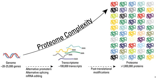
Just last decade, everyone was excited about the Human Genome Project,  and the gene was king. Today, epigenetics is reminding us that

  • non-genetic factors are important in shaping gene expression and development.

Similarly, where phosphorylation once seemed the primary way to modulate proteins,

  • epigenetics has re-introduced us to acetylation as an important force in defining protein function.

In particular, the acetylation of histones has moved to center stage, even though it was described over 45 years ago. Research on histone acetylation has

  • led to a resurgence in the interest in enzymatically-mediated acetylation of other proteins.

This article examines acetylation as a post-translational modification of proteins that impacts gene expression and plays a role in epigenetics.

The Basics

Acetylation refers to the addition of an acetyl group (CH3CO) to organic compounds. Proteins can be acetylated by both enzymatic and non-enzymatic processes.

One group of acetyltransferases commonly catalyze the transfer of an acetyl group from acetyl-CoA to the terminal amine on the side chain of lysine residues (Figure 1).

These enzymes are commonly called HATs, because their best-known substrates have been histones.

However, the nomenclature is being revised to lysine acetyltransferases (KATs), reflecting their ability to acetylate lysine (denoted ‘K’) on many proteins.

1 The KATs are numerous, with many assigned, based on structural similarities, to either

  1. the GNAT (Gcn5-related N-acetyltransferases) superfamily or
  2. the MYST (MOZ, YBF2/Sas3, Sas2, Tip60) family.

Other important KATs include

  1. p300 (E1A-associated protein 300 kDa),
  2. CBP (cAMP response element binding (CREB)-binding protein), and
  3. TAFII 250 (TATA-binding protein associated factor II 250).

The conversion of the positively charged lysine to acetyl-lysine, like the addition of negative phosphates to uncharged amino acids during phosphorylation,

alters protein structure and interactions with other biomolecules. For example, acetylation of  histones typically

  1. promotes the recruitment of effector proteins,
  2. relaxation of chromatin conformation, and
  3. an increase in transcription.

Like phosphorylation,

  • acetylation is reversible.

Histone deacetylases (HDACs, a.k.a. KDACs) are a smaller group of evolutionarily conserved enzymes.

The human class I HDACs are

  • homologous to the yeast enzyme Rpd3 and include HDAC1, 2, 3, and 8.

Class II HDACs are

  • homologous to yeast HDA1 and are divided into class IIa (HDAC4, 5, 7, 9) and class IIb (HDAC6 and 10) based on structure.
  • The human class III HDACs include the sirtuin family of NAD+-dependent protein deacetylases.
  • The novel HDAC11 has a distinct structure and is a class IV HDAC.

The HDACs often participate in the formation of transcriptional repressor complexes, inducing

  • chromatin compaction through histone deacetylation, and silencing gene expression.

A Diversity of Partners

A great resource for the research scientist is the National Center for Biotechnology Information (NCBI), your tax dollars at work compiling information about everything molecular. This site should be your first stopping point when trying to learn authoritative information about a new protein or gene that you’re studying. Information at this site helps to underscore two points about KATs and deacetylases: they are social enzymes, always interacting with other proteins, and they are promiscuous, binding to an astounding array of partners. Take, for example, the KAT known commonly as p300. At the NCBI gene link, entering ‘human p300’ finds the gene EP300 (KAT3B), with a summary stating that it associates with the adenovirus protein E1A, acetylates histones, binds CREB, and is a co-activator of HIF-1α (hypoxia-inducible factor 1α). Further down, we find that it binds three different proteins produced by the lentivirus human immunodeficiency virus (HIV)-1. Then, impressively, is a list of over two hundred proteins that have been documented to directly interact with p300 (with links to references and other interactome datasets included). Similarly, the deacetylase HDAC1 is summarized as a histone deacetylase that also interacts with retinoblastoma tumor-suppressor to control cell growth and, together with metastasis-associated protein-2, deacetylates the tumor suppressor p53. Like p300, HDAC1 has an amazing list of partners: it interacts with some 300 proteins, with over 125 of these documented as direct binding partners.

The abundance of protein partners, for both KATs and HDACs, suggests that these enzymes tend to form multimeric complexes. In fact, such complexes serve the critical purpose of positioning the (de)acetylases at specific sites to perform their functions. Certainly, KATs can directly acetylate substrates in vitro. However, KAT activity in vivo is regulated, at least in part, by where it is positioned. For example, the classical model for activation of PPARs (peroxisome proliferator-activated receptors) posits that this receptor heterodimerizes at specific response elements with RXR (retinoid X receptor). In the absence of ligand, the unactivated heterodimer binds co-repressor proteins, such as nuclear receptor co-repressors (NCoR), G-protein pathways suppressor 2 (GPS2), and HDACs (Figure 2). The HDACs help prevent expression of PPAR-specific genes by keeping the neighboring histones deacetylated. The appearance of a ligand for PPAR causes dissociation of the co-repressor proteins followed by the recruitment of co-activators, including PPAR co-activator (PGC-1), CREB binding protein (CBP), and p300. Formation of the PPAR activation complex leads to histone acetylation by CBP and p300, giving rise to altered expression of genes involved in fatty acid metabolism, lipid homeostasis, and adipocyte differentiation. In this example, ligand binding to its receptor causes a large scale switch from a cluster of proteins serving various roles in preventing transcription to a different group designed to facilitate gene transcription.

Acetylation Patterns

In its simplest form acetylation is merely another form of post-translational modification of proteins. A good example is the acetylation of tubulin, which can be deacetylated by HDAC6 or SIRT2. Acetylation of this key microtubule component appears to alter its affinity for kinesin-1 and redirect motor-based trafficking of vesicles.2,3 In short, acetylation changes protein function by adjusting protein-protein interactions. The net ‘global’ acetylation, in this case, may be determined by the balance of overall KAT and HDAC activities.

More commonly, acetylation is targeted to specific proteins and, possibly, specific lysine residues on those protein targets. One way that this can be achieved is by the formation of protein complexes containing either KATs or HDACs, as in the PPAR case described above. The assembly of the complex serves to place the KATs/HDACs near histones, transcription factors, or other targets. Histones, assembled as an octamer core surrounded by DNA, have amino termini that are freely exposed (Figure 3). Positively-charged lysine residues on these tails interact electrostatically with negatively-charged phosphate groups along the DNA backbone. Acetylation reduces these interactions and loosens the DNA, facilitating transcription. Bear in mind that, while it is generally true that histone acetylation increases transcriptional activation, there are exceptions. For example, acetylation of estrogen receptor-α suppresses ligand sensitivity and reduces ligand-induced transcriptional activity.4,5

References

1. Glozak, M.A., Sengubpta, N., Zhang, X., et al. Gene 363, 15-23 (2005).

2. Hammond, J.W., Cai, D., and Verhey, K.J. Curr. Opin. Cell Biol. 20, 71-76 (2008).

3. Gao, Y., Hubber, C.C., and Yao, T.P. J. Biol. Chem. epub ahead of print (2010).

4. Wang, C., Fu, M., Angeletti, R.H., et al. J. Biol. Chem. 276, 18375-18383 (2001).

5. Popov, V.M., Wang, C., Shirley, L.A., et al. Steroids 72, 221-230 (2007).

6. Mellert, H.S. and McMahon, S.B. Trends Biochem. Sci. 34, 571-578 (2009).

7. Yang, X.J. and Seto, E. Mol. Cell 31, 49-461 (2008).

8. Wilson, A.J., Byun, D.S., Popova, N., et al. J. Biol. Chem. 281, 13548-13558 (2006).

9. Vincent, A. and Van Seuningen, I. Differentiation 78, 99-107 (2009).

10. Li, Z., Chen, L., Kabra, N., et al. J. Biol. Chem. 284, 10361-10366 (2009).

From Protein Acetylation: Much More than Histone Acetylation by Brock, T.G.

2 Figures saved


PTM modifications

PTM modifications

Basic CMYK


3-d-genome-map

3-d-genome-map


graphs_superdex-both-high-resolution-size-exclusion-gel-filtration-chromatography

graphs_superdex-both-high-resolution-size-exclusion-gel-filtration-chromatography


Read Full Post »

Natural Drug Target Discovery and Translational Medicine in Human Microbiome

Author and Curator: Demet Sag, PhD


Remember Ecology 101, simple description of ecosystem includes both living, biotic, and non-living, abiotic, that response to differentiation based on external and internal factors.  Hence, biodiversity changes since living systems are open systems and always try to reach stability. Both soil and human body are rich in microbial life against ever changing conditions. Previously, discovery of marine microorganisms for treatment of complex diseases especially cancer and drug discovery for pharmaceutical applications was discussed. (http://pharmaceuticalintelligence.com/2014/03/20/without-the-past-no-future-but-learn-and-move-genomics-of-microorganisms-to-translational-medicine/)

Here, the focus will be given to clinical drug discovery based on how lactose intolerance and human microbiome related to treat cancer patients or other diseases. In sum, creating clinical relevance with human microbiome require knowledge of both of the worlds to make best of it to solve complex diseases naturally.

The huge undertake as a roadmap to biomedical research originated by NIH under The Human Microbiome Project (HMP) (http://nihroadmap.nih.gov) with 250 healthy individuals as a starting point.  Recent developments opened the doors to pursue us to understand how human microbiome reflects on metabolism, drug interactions and numerous diseases.  Finally, association between clinical states and microbiome are improving with advanced algorithms, bioinformatics and genomics. In classical reading tests questions finding the simile between two groups of words can well relate how microbiome- human and soil-earth relates.  Both are rich in microbial life with quite changing characters to survive through commensal living.

Thus, it is also good to talk about how we can synthesize existing info on interactions between soil microorganisms and decomposers for human diseases and human microbiome. Epidemiology of living organisms is diverse but they all share common interest. In soil, for example, radioactively contaminated soil can’t support plant growth well so Nitrosomonas may support to bring the life to soil through supplying nitrogen. And others can be added to bring a favorable enriched soil.

In human microbiome nutrition-diseases interacts in such a harmony with genetic make up (the information received at time of birth germline- or acquired later in life due to mutations by various reasons). For example, the simplest example is lactose intolerance and the other is development of diabetes.  Generally, it is described as If person is missing a gene to metabolize lactose (sugar) this person become Lactose intolerant yet this can be gained before birth or after. The fix is easy since avoiding certain food groups i.e. milk products.

Yet, this is not that simple!

In human microbiome, the rich gastrointestinal (GI) tract contains many organisms and one of the most important ones is Enterococci that are often simply described as lactic-acid–producing bacteria—by under- appreciation of their power of microbial physiology and outcomes as well as their ubiquitous nature of enterococci.  Schleifer & Kilpper-Bälz, 1984 also reported that the Group D streptococci, such as Streptococcus faecalis and Streptococcus faecium, were included in the new genus called Enterococcus.

The importance of this genius, consists of 37 species, coming from their spectrum of  habitats that include the gastrointestinal microbiota of nearly every animal phylum and flexibility with ability to widely colonize, intrinsic resistance to many inhabitable conditions even though they don’t have spores but they can survive against desiccation and can persist for months on dried surfaces.  Furthermore, they can tolerate extreme conditions such as pH changes, ionizing radiation, osmotic and oxidative stresses, high heavy metal concentrations, and antibiotics.

There is a double sword application as these organisms used as probiotics to improve immune system of the host.  If it is human to prevent contaminated food related diseases or in animals prevent transmitting them to the consumers. Thus, E. faecium and E. faecalis strains are used as probiotics and are ingested in high numbers, generally in the form of pharmaceutical preparations to treat diarrhea, antibiotic-associated diarrhea or irritable bowel syndrome, to lower cholesterol levels or to improve host immunity.

When it comes to human body within each system specific organs may create distinct values.  For example the pH values of GI tract vary and during diseases since pH levels are not at at correct levels.  As a result, due to mal-absorption of nutrients and elements such as food, vitamins and minerals body can’t heal itself. This changing microbial genomics on the surface of GI reflects on general health.  Entrococcus family among the other GI’s natural flora has the microbial physiology adopt these various pH conditions well. 


Our body has its own standards to function, such as  pH, temperature, oxygen etc these are basics so that enzymatic reactions may happen to metabolize,synthesizing (making) or catalyzing (breaking) what we eat.  The pH is the measure of hydrogen-ion concentration  in solution.  For example, human blood has a narrow pH (7.35 – 7.45 ) and below or above this range means symptoms and disease yet if blood pH moves to much below 6.8 or above 7.8, cells stop functioning and the patient dies since the ideal pH for blood is 7.4.  This value is unified.  On the other hand, the pH in the human digestive tract or GI changes tremendously to adopt and carry on its function, the pH of saliva (6.5 – 7.5), upper portion of the stomach (4.0 – 6.5) where “predigestion” occurs, the lower portion of the stomach is secreting hydrochloric acid (HCI) and pepsin until it reaches a pH between 1.5 – 4.0; duodenum, small intestine, (7.0 – 8.5) where 90% of the absorption of nutrients is taken in by the body while the waste products are passed out through the colon (pH 4.0 – 7.0).


Why is pH important and how related to anything?

Development and presence of cancer always require an acid pH and lack of oxygen.  Thus, prevention of these two factors may be the key for treatment of cancer as it progress the acidity increases such that the level raises even up to 1000 more than normal levels.

Mainly, due to Warburg effect body opt to get its energy from fermentation of glucose and produce lactic acid that decreases the body pH from 7.3 down to 7 then to 6.5 in advanced stages of cancer.  Furthermore, during metastases this level even reaches to 6.0 and even 5.7 where body can’t fight back with the disease. (Warburg effect is well explained previously by Dr. Larry Berstein (www.linkedin.com/pub/larry-bernstein/38/94b/3aa).

How to bypass the lack of oxygen naturally?

One of the many solution can be a natural solution. The nature made the hemoglobin carrying bacteria, Vitreoscilla hemoglobin (VHb), which is first described by Dale Webster in 1966. The gram negative and obligate aerobic bacterium, Vitreoscilla synthesizes elevated quantities of a homodimeric hemoglobin (VHb) under hypoxic growth conditions.   The main role is likely the binding of oxygen at low concentrations and its direct delivery to the terminal respiratory oxidase(s) such as cytochrome o.  Then, after 1986 with detailed description of the molecule other hemoglobins and flavohemoglobins were identified in a variety of microbes, indicating the widespread occurrence of Hb-like proteins.   Currently, it is the most studied bacterial hemoglobin with application potentials in biotechnology.

It is a plausible solution to integrate Vitroscilla and Enterobacter powers for cancer detection and treatment naturally with body’s own microbiome.

However, there are many microbial organisms and differ person to person based on gender, age, background, genetic make-up, food intake, habits, location etc.  The huge undertake as a roadmap to biomedical research originated by NIH under The Human Microbiome Project (HMP) (http://nihroadmap.nih.gov) with 250 healthy individuals as a starting point.

There were three goals in the agenda of The Human Microbiome Project (HMP) simply:

 1. Utilize advanced high throughput technology,

2. Identify any association between microbiome and disease/health stages,

3. Initiate scientific studies to collect more data.

In sum, creating clinical relevance with human microbiome require knowledge of both of the worlds to make best of it to solve complex diseases naturally.

Previously  Discussed:

AMPK Is a Negative Regulator of the Warburg Effect and Suppresses Tumor Growth In Vivo
Reporter-Curator: Stephen J. Williams, Ph.D.
http://pharmaceuticalintelligence.com/2013/03/12/ampk-is-a-negative-regulator-of-the-warburg-effect-and-suppresses-tumor-growth-in-vivo/

Is the Warburg Effect the Cause or the Effect of Cancer: A 21st Century View?
Author: Larry H. Bernstein, MD, FCAP
http://pharmaceuticalintelligence.com/2012/10/17/is-the-warburg-effect-the-cause-or-the-effect-of-cancer-a-21st-century-view/

Otto Warburg, A Giant of Modern Cellular Biology
Reporter: Larry H Bernstein, MD, FCAP
http://pharmaceuticalintelligence.com/2012/11/02/otto-warburg-a-giant-of-modern-cellular-biology/

Targeting Mitochondrial-bound Hexokinase for Cancer Therapy
Author: Ziv Raviv, PhD
http://pharmaceuticalintelligence.com/2013/04/06/targeting-mito…cancer-therapy

Nitric Oxide has a ubiquitous role in the regulation of glycolysis -with a concomitant influence on mitochondrial function
Curator, Larry H. Bernstein, MD, FCAP
http://pharmaceuticalintelligence.com/2012/09/16/nitric-oxide-has-a-ubiquitous-role-in-the-regulation-of-glycolysis-with-a-concomitant-influence-on-mitochondrial-function/

Potential Drug Target: Glucolysis Regulation – Oxidative stress-responsive microRNA-320
Reporter: Aviva Lev-Ari, PhD, RN
http://pharmaceuticalintelligence.com/2012/07/25/potential-drug-target-glucolysis-regulation-oxidative-stress-responsive-microrna-320/

Differentiation Therapy – Epigenetics Tackles Solid Tumors
Author-Writer: Stephen J. Williams, Ph.D.
http://pharmaceuticalintelligence.com/2013/01/03/differentiation-therapy-epigenetics-tackles-solid-tumors/

Prostate Cancer Cells: Histone Deacetylase Inhibitors Induce Epithelial-to-Mesenchymal Transition
Reporter-Curator: Stephen J. Williams, Ph.D.
http://pharmaceuticalintelligence.com/2012/11/30/histone-deacetylase-inhibitors-induce-epithelial-to-mesenchymal-transition-in-prostate-cancer-cells/

Mitochondrial Damage and Repair under Oxidative Stress
Curator: Larry H Bernstein, MD, FCAP
http://pharmaceuticalintelligence.com/2012/10/28/mitochondrial-damage-and-repair-under-oxidative-stress/

Mitochondria: Origin from oxygen free environment, role in aerobic glycolysis, metabolic adaptation
Curator: Larry H Bernsatein, MD, FCAP
http://pharmaceuticalintelligence.com/2012/09/26/mitochondria-origin-from-oxygen-free-environment-role-in-aerobic-glycolysis-metabolic-adaptation/

Expanding the Genetic Alphabet and Linking the Genome to the Metabolome
Reporter& Curator: Larry Bernstein, MD, FCAP
http://pharmaceuticalintelligence.com/2012/09/24/expanding-the-genetic-alphabet-and-linking-the-genome-to-the-metabolome/

What can we expect of tumor therapeutic response?
Author: Larry H. Bernstein, MD, FCAP
http://pharmaceuticalintelligence.com/2012/12/05/what-can-we-expect-of-tumor-therapeutic-response/

A Second Look at the Transthyretin Nutrition Inflammatory Conundrum
Larry H. Bernstein, MD, FACP
http://pharmaceuticalintelligence.com/2012/12/03/a-second-look-at-the-transthyretin-nutrition-inflammatory-conundrum/


Further  Readings and References:

Palmer KL, van Schaik W, Willems RJL, Gilmore MS. “Enterococcal Genomics Enterococci: From Commensals to Leading Causes of Drug Resistant Infection.” 2014-.2014 Feb 8

Franz CM, Holzapfel WH, Stiles ME. Enterococci at the crossroads of food safety?

Int J Food Microbiol.” 1999 Mar 1; 47(1-2):1-24.

Franz CM, Huch M, Abriouel H, Holzapfel W, Gálvez A.Int J Food Microbiol. “Enterococci as probiotics and their implications in food safety.” 2011 Dec 2; 151(2):125-40. Epub 2011 Sep 8.

Kayser FH.”Safety aspects of enterococci from the medical point of view.” Int J Food Microbiol. 2003 Dec 1; 88(2-3):255-62.

Webster DA, Hackett DP (1966). “The purification and properties of cytochrome o fromVitreoscilla“. J Biol Chem 241 (14): 3308–3315

Stark BC, Dikshit KL, Pagilla KR (2011). “Recent advances in understanding the structure, function, and biotechnological usefulness of the hemoglobin from the bacterium Vitreoscilla“. Biotechnol Lett 33 (9): 1705–1714

Stark BC, Dikshit KL, Pagilla KR (2012). “The Biochemistry  of Vitreoscillahemoglobin“. Computational and Structural Biotechnology Journal 3 (4): e201210002.

Brenner K, You L, Arnold F. (2008). “Engineering microbial consortia: A new frontier in synthetic biology.” Trends in Biotechnology 26: 483489.

Dunbar J, White S, Forney L. (1997). “Genetic diversity through the looking glass: Effect of enrichment bias.Applied and Environmental Microbiology 63: 13261331.

Foster J. (2001). “Evolutionary computation Nature Reviews Genetics 2: 428436.

Dinsdale EA, et al. 2008. “Functional metagenomic profiling of nine biomes.” Nature452: 629632.

Gudelj I, Beardmore RE, Arkin SS, MacLean RC. (2007). “Constraints on microbial metabolism drive evolutionary diversification in homogeneous environments.” Journal of Evolutionary Biology 20: 1882–1889.

Haack SK, Garchow H, Klug MJ, Forney L. (1995). “Analysis of factors affecting the accuracy, reproducibility, and interpretation of microbial community carbon source utilization patterns.” Applied and Environmental Microbiology 61: 14581468.

Lozupone C, Knight R. (2007). “Global patterns in bacterial diversity.” Proceedings of the National Academy of Sciences 104: 1143611440.

Thurnheer T, Gmr R, Guggenheim B,  (2004). “Multiplex FISH analysis of a six-species bacterial biofilm. “Journal of Microbiological Methods 56: 3747.

VijayKumar M, Aitken JD, Carvalho FA, Cullender TC, Mwangi S, Srinivasan S,Sitaraman S, Knight R, Ley RE, Gewirtz AT. (2010). “Metabolic syndrome and altered gut microbiota in mice lacking Toll-like receptor 5.” Science 328: 228231

Williams HTP, Lenton TM. (2007). “Artificial selection of simulated microbial ecosystems.” Proceedings of the National Academy of Sciences 104: 89188923.


Read Full Post »

“Sudden Cardiac Death,” SudD is in Ferrer inCode’s Suite of Cardiovascular Genetic Tests to be Commercialized in the US

“Sudden Cardiac Death,” SudD is in Ferrer inCode’s Suite of Cardiovascular Genetic Tests to be Commercialized in the US

Curator: Aviva Lev-Ari, PhD, RN

Article ID #111: “Sudden Cardiac Death,” SudD is in Ferrer inCode’s Suite of Cardiovascular Genetic Tests to be Commercialized in the US. Published on 2/10/2014

WordCloud Image Produced by Adam Tubman

Uncertainty around reimbursement for targeted NGS tests is faced by Molecular Diagnostic and Genomics Services companies

VIEW VIDEO

Democratization of Genomic Medicine: Michael Bolick @ TEDxTalks

Ferrer inCode’s Suite of Cardiovascular Genetic Tests included the following tests: 

  • SudD inCode (Sudden Cardiac Death)
  • Cardio inCode,
  • Thrombo inCode, and
  • Nutri inCode

Selah Genomics, Ferrer inCode to Offer NGS-based Cardiovascular Test in US

2014/02/06

Selah Genomics, a Greenville, S.C.-based molecular diagnostic and genomics services company, has partnered with Spanish pharmaceutical company Ferrer inCode to commercialize Ferrer inCode’s suite of cardiovascular genetic tests in the US.

Selah will first validate Ferrer’s next-generation sequencing-based test for sudden cardiac death, SudD inCode, on Illumina’s MiSeq system to run out of its CLIA-certified laboratory.

Meantime, Selah plans to validate three other Ferrer inCode PCR-based cardiovascular tests — Cardio inCode, Thrombo inCode, and Nutri inCode — in its own lab using PCR, but may eventually combine the tests into one comprehensive panel to run on an NGS system, Selah CEO Michael Bolick told Clinical Sequencing News.

Selah already offers its PrecisionPath targeted Cancer Test in collaboration with the Greenville Health System’s Institute for Translational Oncology Research. All consenting cancer patients at ITOR receive the PrecisionPath test, which runs on Life Technologies’ Ion Torrent PGM and uses the Ion AmpliSeq technology.

Currently, Selah receives between 10 and 20 samples per week for PrecisionPath, and it plans to roll the test out nationwide later this year.

Bolick said that the company is also developing Hepatitis C and HIV assays for the MiSeq, and that the firm will likely purchase Illumina’s MiSeqDx, which recently received clearance from the US Food and Drug Administration.

Selah also collaborates with pharmaceutical companies to develop companion diagnostic tests. Bolick anticipates that the firm will use the MiSeqDx for those tests since they will “ultimately need [pre-market approval].” Having an FDA-cleared platform on which to develop the tests will be helpful in gaining a PMA designation, he said.

Selah also offers Exome Sequencing Services on the Ion Proton for research use only. In addition, it has a

  • Pacific Biosciences RS II and
  • Roche’s 454 GS FLX in house.

Bolick said that the company is currently using the PacBio machine for discovery work in infectious disease.

Ferrer inCode’s SudD inCode Test

currently assesses 55 genes related to structural heart problems that cause sudden cardiac arrest, Robert Jenkins, who manages Ferrer inCode’s UK and Americas groups, told CSN. However, the company is planning to

  • expand the test to 104 genes and also to include
  • genes related to conductive myopathy,
  • sudden infant death, and
  • aneurysms.

While the test sequences the entire genes, only well-known causative variants are reported, Jenkins said. However, the firm has been collecting all the sequenced variants, so it could potentially add content to the test if enough evidence is gathered to validate any of those variants as clinically significant.

Ferrer inCode currently runs SudD inCode on the MiSeq as an LDT, which is how Selah will validate and market the test in the US.

Jenkins said that for now, Ferrer plans to keep the Cardio, Nutri, and Thrombo inCode tests PCR-based.

  • Cardio inCode looks at around 125 variants involved in genetic risk for cardiac disease.

When it is used with traditional markers such as

  • lipid profiling, an individual’s
  • smoking and drinking habits, and
  • body mass index,

Jenkins said the genetic test helps to reclassify around 20 percent to 25 percent of individuals deemed in the intermediate risk category as either high or low risk.

Thrombo inCode Test

is an approximately 20-variant thrombosis test for individuals that have had a thrombotic event or who have had a history of unsuccessful pregnancies. Often, the cause of thrombosis can go unexplained via testing from serological workups, Jenkins said.

Nutri inCode Test

is a nutrigenomics test that looks at around 90 SNPs. In combination with lifestyle factors, it helps individuals develop a tailored genetics-based plan to reduce obesity, Jenkins said.

Bolick said that while Selah will validate and develop each of these tests individually out of its laboratory, it is also deciding whether to combine the tests into one next-gen sequencing-based test.

Jeremy Stuart, Selah’s VP of genomic services, told CSN that one option would be to incorporate the individual SNPs assessed in the Thrombo, Cardio, and Nutri tests into the SudD test.

Bolick said that the company is now in discussions with third party payors about reimbursement for the tests and is readying a regional pilot program to offer the sequencing-based cardiovascular test as part of a corporate wellness program. The pilot will help Selah figure out a pricing structure and will also demonstrate a “return on investment to the corporation, by allowing for better determination of risk of heart disease,” Bolick said.

Currently, Selah’s other NGS test, PrecisionPath, is being paid for by ITOR. However, Bolick said that initial conversations with third party payors about launching the assay outside of the Greenville Health System have been positive.

Reimbursement success will play a role in determining how the company expands beyond its current tests. For instance, while Selah is interested in moving into

  • clinical exome sequencing,

Stuart said that right now there is a “lot of uncertainty around reimbursement for targeted NGS tests, let alone exome sequencing.” Selah will first “establish reimbursement for those and then may expand into what’s possible for exome sequencing,” Stuart said. But currently, the exome market is research use only.

SOURCE

http://www.ferrerincode.com/en/node/98

Selah Genomics

SELAH GENOMICS: HARNESS THE POWER OF PRECISION FOR MORE PERSONALIZED TREATMENT

Selah Genomics is a clinical diagnostic specialist supporting healthcare providers and the pharmaceutical industry with advanced molecular and genomic diagnostic services. Selah’s services add value to early stage drug development, clinical trials and regulatory processes in the pharmaceutical industry and helps clinicians and healthcare providers treat and monitor patients, thereby improving patient outcomes.

With the Power of Precision, Selah Genomics provides the best in molecular diagnostic testing, assay validation and genomic profiling that all leads to one common goal: to provide better outcomes for patients.

Michael Bolick, CEO

Michael is a serial entrepreneur with 25 years of experience in the life science and healthcare industries. Most recently, he led a management buyout of Lab21 Ltd’s US-based operations to form Selah Genomics Inc. Prior to co-founding Selah Genomics, Michael served as President of Lab21 Inc which was formed following Lab21 Ltd’s acquisition of his prior company, Selah Technologies LLC. He founded Selah Technologies LLC to commercialize nanotechnologies licensed from Clemson University. Selah focused these nanotechnologies to enable doctors to see cancer during surgery. Prior to founding Selah Technologies, Michael’s career included roles of increasing responsibility in the pharmaceutical sector.

Michael is a Fellow in the Liberty Fellowship Class of 2011. Liberty Fellowship is a program designed specifically for emerging state leaders to reinforce values necessary to lead an exemplary life both personally and professionally. Michael serves as Immediate Past Chair of SCBIO, South Carolina’s Life Sciences Industry Association. Michael earned his bachelor’s degree in Chemical Engineering from North Carolina State University.

  • Selah Genomics specializes in supporting healthcare providers and the pharmaceutical industry with advanced molecular and genomic diagnostic services.

    read more »

    Latest News

    Find out what the buzz is about

    • Greenville Health System, Roswell Park Adopt Targeted Sequencing in Cancer Treatment

      8 May 2013

    • Selah, GHS expand personalized medicine

      2 May 2013

    • The Democratization of Genomic Medicine: Michael Bolick at TEDxGreenville

      21 Apr 2013

    • Greenville Magazine features Selah Genomics

      1 Apr 2013

    • Upstate Biotech Firm Expands to Columbia

      14 Mar 2013

    • Genetic Engineering and Biotechnology News; “Selah Genomics Establishes Second Clinical Genomics Center”

      20 Feb 2013

    • Selah Genomics Forms Second Clinical Genomic Center

      19 Feb 2013

  • Clinical Laboratory

    Helping physicians by applying our scientific expertise and skills in advanced molecular diagnostic assay development in a CLIA-certified laboratory.

    read more »

  • PrecisionPath™

    Genomic profiling of solid tumors, identifying actionable targets today and enabling the discovery of clinically relevant genes for tomorrow.

    read more »

  • Genomic Services


    Selah Genomics provides a suite of services focused on support of molecular biomarker discovery, assay validation and prospective/retrospective clinical trial testing in support of companion diagnostic development and commercialization.

    read more »

 SOURCE

THE FAST-TRACK TO DISCOVERY AND CLINICAL UTILIZATION

Selah Genomics provides a suite of services focused on support of molecular biomarker discovery, assay validation and prospective/retrospective clinical trial testing in support of companion diagnostic development and commercialization. Selah operates NGS platforms from Life Technologies, Illumina, Roche and PacBio as well as an array of real time PCR and other supporting instrumentation systems. We help you select the best platform for each Project in support of your particular goals. Our prime focus – to help fast-track the clinical utilization and commercialization of your biomarker.

Selah enjoys a key corporate relationship with the Greenville Health System’s (GHS) Institute of Translational Oncology Research (ITOR) conducting multiple clinical trials and identification of new oncology biomarkers.

GHS is the 13th largest public hospital in the United States and ITOR has the largest Phase 1 clinical trial program in South Carolina, including a track record of 16 first-in-human trials. The close relationship with ITOR is an enormous asset for Selah. Not only does it allow Selah to provide state-of-the-art molecular diagnostics support for ITOR clinical studies but it leads to first-hand daily interaction with cancer physicians. This interaction stimulates early identification and development of new biomarker panels.

Selah’s Clinical Genomics Center at ITOR is physically located within GHS & ITOR. In addition, Selah operates a Clinical Genomics Center at Innovista on the campus of the University of South Carolina.

SOURCE

http://selahgenomics.com/genomic-services/

Read Full Post »

Endothelial Dysfunction (release into the circulation of damaged endothelial cells) as A Risk Marker for Ischemia and MI

Reporter and Curator: Larry H Bernstein, MD, FCAP

Endothelial Dysfunction: An Early Cardiovascular Risk Marker in Asymptomatic Obese Individuals with Prediabete

AK Gupta, E Ravussin, DL Johannsen, AJ Stull,WT.Cefalu and WD Johnson at Pennington Biomedical Research Center, Louisiana State University System, Baton Rouge, LA Brit J Med & Med Res 2012; 2(3):413-423 [www.ScienceDomain.org]

provides an exceedingly interesting insight into the relationship between type 2 diabetes mellitus, obesity and risk for cardiovascular disease in patients who are asymptomatic prediabetics, defined as a fasting blood glucose between 1000 and 1240 mg/L, or a Hb A1c (may not accurate for African Americans) between 5.6 and 6.5.  They would be expected to show an abnormal 5-hr GTT.

Obesity is associated with the release from adipocytes of adiponectin, which it has been reported is countered by resistin.  We might also have the effect of the insulin secreting beta cell, that releases insulin without a relationship to an anabolic function, through IGF-1 related to feedback to the pituitary GH, which takes a dominant catabolic role. Thus, insulin resistance. This is an oversimplification, and far greater depth is found elsewhere.

This study is consistent with another study on  Metabolism Influences Cancer

Reuben Shaw, Ph.D., a geneticist and researcher at the Salk Institute: Metabolism Influences Cancer

Recent development on Human Stem Cell Therapies for comorbidity and Cardiovascular disease

Human Stem Cell Therapies: UCSD New Discovery addressing the Limiting Factor and Providing the Solution

http://pharmaceuticalintelligence.com/2014/01/06/human-stem-cell-therapies-ucsd-new-discovery-addressing-the-limiting-factor-and-providing-the-solution/

This study reported a potential early marker of myocardial infarction by the release into the circulation of damaged endothelial cells that are to be measured in patients suspected of severe ischemia in a clinical trial.  The question that I raised in my comment was whether this would have to be a special immunochemical assay of tagged cells, and if that were the case, would it be measured on an automated flow-based hemocytometer, which can differentiate several populations of cells – granulocytes, lymphocytes, red cells, platelets, immature granuloytes, BLASTS.  That would be a very practical extension of the technology for labs worldwide.

Abstract

Aims: To elucidate if endothelial dysfunction is an early CV risk marker in obese men and women with prediabetes.
Study Design: Cross-sectional study.

Place and Duration of Study: Clinical Research Unit, Pennington Biomedical Research Center, Baton Rouge, LA. United States.

Background: Overweight and obese status denotes an increasing adipose tissue burden which spills over into ectopic locations, including the visceral compartment, muscle and liver. Associated co-morbidities enhance cardiovascular (CV) risk. Endothelium which is the largest receptor-effector end-organ in our bodies, while responding to numerous physical and chemical stimuli maintains vascular homeostasis. Endothelial dysfunction (ED) is the initial perturbation, which precedes fatty streak known to initiate atherosclerosis: insidious process which often culminates as sudden catastrophic CV adverse event.

Methodology:  Asymptomatic men and women; [n=42] coming in after an overnight fast had demographic, anthropometric, clinical chemistry and

  • resting endothelial function (EF)
  • increased test finger peripheral arterial tone (PAT) relative to control;
    • expressed as relative hyperemia index (RHI)] assessments.

Results: Adults with desirable weight [n=12] and overweight [n=8] state, had normal fasting plasma glucose [Mean(SD)]: FPG [91.1(4.5), 94.8(5.8) mg/dL], insulin [INS, 2.3(4.4), 3.1(4.8) µU/ml], insulin sensitivity by homeostasis model assessment [HOMA-IR, 0.62(1.2), 0.80(1.2)] and desirable resting clinic blood pressure [SBP/DBP, 118(12)/74(5), 118(13)/76(8) mmHg].

Obese adults [n=22] had

  • prediabetes [FPG, 106.5(3.5) g/dL],
  • hyperinsulinemia [INS 18.0(5.2) µU/ml],
  • insulin resistance [HOMA-IR .59(2.3)],
  • prehypertension [PreHTN; SBP/DBP 127(13)/81(7) mmHg] and
  • endothelial dysfunction [ED;
  • reduced RHI 1.7(0.3) vs. 2.4(0.3); all p<0.05].

Age-adjusted RHI correlated with BMI [r=-0.53; p<0.001]; however,

    • BMI-adjusted RHI was not correlated with age [r=-0.01; p=0.89].

Conclusion: Endothelial dysfunction reflective of cardiometabolic changes in obese adults can be an early risk marker for catastrophic CV events.

Keywords: Fasting plasma glucose; healthy adults; reverse cholesterol transport pathway; insulin resistance; body weight; relative hyperemia index.

ABBREVIATIONS

ADA: American Diabetes association; BMI: body mass index; CVD: cardiovascular disease; CV: cardiovascular; DBP: diastolic blood pressure; ED: endothelial dysfunction; EF: resting endothelial function; FPG: fasting plasma glucose; HOMA-IR: homeostasis model assessment; INS: insulin; JNC 7: Joint National Commission 7; LDL-C/HDL-C: low density lipoprotein cholesterol to high density lipoprotein; NCEP ATP III: National Cholesterol Education Program Adult Treatment Panel III; PAT: peripheral arterial tone; PreDM: prediabetes; PreHTN: prehypertension; PBRC: Pennington Biomedical Research Center; RHI: relative hyperemia index; SBP: systolic blood pressure; Total-C/HDL-C: total cholesterol to high density lipoprotein cholestrol; TG/HDL-C: triglycerides to high density lipoprotein cholesterol; WC: waist circumference.

Introduction

Healthy adults with no chronic medical conditions, on no prescription medications (n=24) and with low cardiovascular risk, in a randomized-order, cross-over clinical trial, with a 2 week washout period, exhibitd improved endothelial function (measured with flow mediated dilatation) with a diet rich in antioxidants (Franzini et al., 2012). Healthy over weight and obese volunteers with normal glucose appear to attenuate flow mediated dilation after high
glycemic index carbohydrate meals (Suessenbacher et al., 2011). In matched (age, work place, physical activity, tobacco use, blood pressure, serum lipids and family history of premature coronary artery disease) male shift and no shift workers, peripheral endothelial function (peripheral arterial tone (PAT) index obtained with the EndoPAT technique) was impaired in shift workers, suggesting elevated cardiovascular risk (Lavi et al., 2009).

Endothelial function thus appears to be an exquisitely sensitive marker for a variety of populations, under various conditions. Although endothelial function has been evaluated in numerous disease conditions and perturbed with a variety of agents, there has, to our knowledge, not been a comparison of resting endothelial function in free living healthy lean, overweight and obese subjects. Using a noninvasive assessment for resting endothelial function (by measuring the peripheral arterial tone, Bonetti et al., 2004), we tested the hypothesis that fasting glucose escalation in otherwise asymptomatic obese men and women is functionally reflected as endothelial dysfunction.

Endothelial Function

Assessment of resting endothelial function was done with the participant in fasting state, after having avoided stimulants (caffeine, tobacco, alcohol, exercise) for 12 hours, at the same fixed clock hour (range 8-10 AM), using the EndoPAT 2000 device manufactured by ITAMAR Medical®. This assessment technique has been previously validated (Bonetti et al., 2004), has been used in numerous (>250) peer reviewed publications (Carty et al., 2012; Kuvin et al., 2003) and has been in routine use in our clinical core. Briefly: subjects coming
in from home, after an overnight fast and having avoided stimulants for 12-hours, were placed in a supine position for 20 minutes in a quiet room before the test. A patented single use finger sleeve was then placed on the index finger of each hand to continuously measure peripheral arterial tone. A blood pressure cuff applied to the upper arm of the non-dominant arm (test arm) was then used to occlude the brachial artery for 5 minutes. This was followed by a rapid release. The dominant arm without any manipulation served as the control. The
built in, validated software integrated the data gathered from the finger sleeves of the control (undisturbed) and the test arms (during the baseline, occlusion and release phases), thus providing the relative hyperemia index (RHI) for the test arm. This flow mediated dilatation induced change in the test arm, relative to the control arm, served as the measure for endothelial function (RHI).

The subjects with desirable and overweight body weight were significantly younger [36.7(19.1) and 27.4(3.9) years, respectively], than those who were obese [53.2(11.6) years]. We performed correlations between the measure for endothelial function (RHI) and confounding factors like BMI, age and gender. Age-adjusted RHI correlated with BMI [r=- 0.53, p<0.001]; however, BMI-adjusted RHI was not associated with age [r=-0.01, p=0.89]. Fig. 1 depicts panels for the regression line for RHI as a function of age, (and BMI, glucose
and HOMA-IR, respectively) superimposed on a scatter plot. No correlation was observed between endothelial function and age (r²=0.07), while endothelial function was highly correlated with body mass index, glucose and insulin sensitivity (r²=0.3).

DISCUSSION

Asymptomatic obese adults with prediabetes (when compared to asymptomatic desirable weight and overweight adults with normal glucose), exhibit above the upper limits for desirable fasting plasma total cholesterol (>200mg/dL) and triglycerides (>150 mg/dL), but due to a relatively lower HDL-C display higher cardiac risk ratios (Total-C/HDL-C; p=0.05 and TG/HDL-C; p=0.02). A lower HDL-C and the elevated cardiac risk ratios are early clinical indicators for an impaired reverse cholesterol transport (RCT) pathway, a process by which cholesterol from the periphery is transported to the liver (Tall, 1998). The RCT pathway has been shown to be a sensitive indicator of the net flux (deposition vs. removal) of cholesterol homeostasis at the endothelium (Gupta et al., 1993; Tall et al., 2000). It is at the endothelium that the first fatty streaks, which over time deteriorate into atherosclerosis, have been shown to develop (Rosenfeld et al., 2000).

Impaired endothelial dysfunction is the first step in the process of atherosclerosis, even before the development of the fatty streak (Davignon, 2004; Ross 1999). These healthy obese men and women with prediabetes, prehypertension and impaired reverse cholesterol transport pathway were assessed to have impaired resting endothelial function, which is consistent with latent early onset cardiovascular disease.

We have demonstrated a high prevalence of isolated prediabetes or prehypertension and co-existing prediabetes and prehypertension, among the otherwise healthy US adults (Gupta et al., 2011). We have also elucidated that asymptomatic obese adults with overly heightened systemic inflammation, tend to have prediabetes and prehypertension (Gupta et al., 2010a). These individuals by various conventional measures (larger waist circumference, exacerbated systemic inflammation, higher insulin resistance, elevated triglycerides, lower high-density lipoprotein cholesterol, above average cardiac risk ratios and a significant co-existence of two or three concomitant metabolic risk factors) appear to be on an accelerated pathway towards early adverse cardiovascular events (Gupta et al., 2010a, 2010b). With this study we provide a dynamic, non-invasive, functional correlate: significant resting endothelial dysfunction, as an early biomarker for pre-atherosclerosis in obese adults with prediabetes.

Increased organ ectopic adipose burden especially in the muscle and liver appears to drive clinically recognizable adverse cardio metabolic changes (Hamdy et al., 2006). Increased inflammation (local and systemic) along with enhanced insulin resistance (liver, muscle) manifests as dysglycemia, dyslipidemia, excess reactive oxygen species, hyper-coagulablility and loss of blood pressure control (Gastaldelli et al., 2010).

We demonstrate an early impairment in the reverse cholesterol transport pathway, indicating a net deposition versus removal of cholesterol at the endothelium. In asymptomatic obese men and women with predisease  conditions (prediabetes and prehypertension) when contrasted with ideal bodyweight or overweight adults with normoglycemia and normal blood pressure, resting endothelial dysfunction can be an early warning sign for future catastrophic cardiovascular adverse events.

© 2012 Gupta et al.; This is an Open Access article distributed under the terms of the Creative Commons Attribution License http://creativecommons.org/licenses/by/3.0),
which permits unrestricted use, distribution, and reproduction in any medium, provided the original work is properly cited.

REFERENCES on circulating Endothelial Progenitor Cells as Biomarkers for Cardiovascular Disease and their Angiogenesis Potential.

Asahara T, Murohara T, Sullivan A, et al. Isolation of putative progenitor endothelial cells for angiogenesis. Science 1997;275:964-967.

Takahashi T, Kalka C, Masuda H, et al. Ischemia- and cytokine-induced mobilization of bone marrow-derived endothelial progenitor cells for neovascularization. Nat Med 1999;5:434-438.

Kocher AA, Schuster MD, Szabolcs MJ, et al. Neovascularization of ischemic myocardium by human bone-marrow-derived angioblasts prevents cardiomyocyte apoptosis, reduces remodeling and improves cardiac function. Nat Med 2001;7:430-436.

Rauscher FM, Goldschmidt-Clermont PJ, Davis BH, et al. Aging, progenitor cell exhaustion, and atherosclerosis. Circulation 2003;108:457-463.

Hill JM, Zalos G, Halcox JPJ, et al. Circulating endothelial progenitor cells, vascular function, and cardiovascular risk. N Engl J Med 2003;348:593-600.

Vasa M, Fichtlscherer S, Adler K, et al. Increase in circulating endothelial progenitor cells by statin therapy in patients with stable coronary artery disease. Circulation 2001;103:2885-2890

Laufs U, Werner N, Link A, et al. Physical training increases endothelial progenitor cells, inhibits neointima formation, and enhances angiogenesis. Circulation 2004;109:220-226.

Werner N, Kosiol S, Schiegl T, et al. Circulating endothelial progenitor cells and cardiovascular outcomes. N Engl J Med 2005;353:999-1007.

Aicher A, Heeschen C, Mildner-Rihm C, et al. Essential role of endothelial nitric oxide synthase for mobilization of stem and progenitor cells. Nat Med 2003;9:1370-1376.

Wollert KC, Meyer GP, Lotz J, et al. Intracoronary autologous bone-marrow cell transfer after myocardial infarction: the BOOST randomised controlled clinical trial. Lancet 2004;364:141-148.

Zhang H, Vakil V, Braunstein M, et al. Circulating endothelial progenitor cells in multiple myeloma: implications and significance. Blood 2005;105:3286-3294

Lyden D, Hattori K, Dias S, et al. Impaired recruitment of bone-marrow-derived endothelial and hematopoietic precursor cells blocks tumor angiogenesis and growth. Nat Med 2001;7:1194-1201.

Other related articles that were published in this Open Access Online Scientific Journal include the following:

Lev-Ari, A. 2/28/2013 The Heart: Vasculature Protection – A Concept-based Pharmacological Therapy including THYMOSIN

http://pharmaceuticalintelligence.com/2013/02/28/the-heart-vasculature-protection-a-concept-based-pharmacological-therapy-including-thymosin/

Lev-Ari, A. 2/27/2013 Arteriogenesis and Cardiac Repair: Two Biomaterials – Injectable Thymosin beta4 and Myocardial Matrix Hydrogel

http://pharmaceuticalintelligence.com/2013/02/27/arteriogenesis-and-cardiac-repair-two-biomaterials-injectable-thymosin-beta4-and-myocardial-matrix-hydrogel/

Lev-Ari, A. 11/13/2012 Peroxisome proliferator-activated receptor (PPAR-gamma) Receptors Activation: PPARγ transrepression for Angiogenesis in Cardiovascular Disease and PPARγ transactivation for Treatment of Diabetes

http://pharmaceuticalintelligence.com/2012/11/13/peroxisome-proliferator-activated-receptor-ppar-gamma-receptors-activation-pparγ-transrepression-for-angiogenesis-in-cardiovascular-disease-and-pparγ-transactivation-for-treatment-of-dia/

Lev-Ari, A. 8/29/2012 Positioning a Therapeutic Concept for Endogenous Augmentation of cEPCs — Therapeutic Indications for Macrovascular Disease: Coronary, Cerebrovascular and Peripheral

http://pharmaceuticalintelligence.com/2012/08/29/positioning-a-therapeutic-concept-for-endogenous-augmentation-of-cepcs-therapeutic-indications-for-macrovascular-disease-coronary-cerebrovascular-and-peripheral/ 

Lev-Ari, A. 8/28/2012 Cardiovascular Outcomes: Function of circulating Endothelial Progenitor Cells (cEPCs): Exploring Pharmaco-therapy targeted at Endogenous Augmentation of cEPCs

http://pharmaceuticalintelligence.com/2012/08/28/cardiovascular-outcomes-function-of-circulating-endothelial-progenitor-cells-cepcs-exploring-pharmaco-therapy-targeted-at-endogenous-augmentation-of-cepcs/

Lev-Ari, A. 8/27/2012 Endothelial Dysfunction, Diminished Availability of cEPCs, Increasing CVD Risk for Macrovascular Disease – Therapeutic Potential of cEPCs

http://pharmaceuticalintelligence.com/2012/08/27/endothelial-dysfunction-diminished-availability-of-cepcs-increasing-cvd-risk-for-macrovascular-disease-therapeutic-potential-of-cepcs/

Lev-Ari, A. 8/24/2012 Vascular Medicine and Biology: CLASSIFICATION OF FAST ACTING THERAPY FOR PATIENTS AT HIGH RISK FOR MACROVASCULAR EVENTS Macrovascular Disease – Therapeutic Potential of cEPCs

http://pharmaceuticalintelligence.com/2012/08/24/vascular-medicine-and-biology-classification-of-fast-acting-therapy-for-patients-at-high-risk-for-macrovascular-events-macrovascular-disease-therapeutic-potential-of-cepcs/

Lev-Ari, A. 7/19/2012 Cardiovascular Disease (CVD) and the Role of agent alternatives in endothelial Nitric Oxide Synthase (eNOS) Activation and Nitric Oxide Production

http://pharmaceuticalintelligence.com/2012/07/19/cardiovascular-disease-cvd-and-the-role-of-agent-alternatives-in-endothelial-nitric-oxide-synthase-enos-activation-and-nitric-oxide-production/

Lev-Ari, A. 4/30/2012 Resident-cell-based Therapy in Human Ischaemic Heart Disease: Evolution in the PROMISE of Thymosin beta4 for Cardiac Repair

http://pharmaceuticalintelligence.com/2012/04/30/93/

Lev-Ari, A. 7/2/2012 Macrovascular Disease – Therapeutic Potential of cEPCs: Reduction Methods for CV Risk

http://pharmaceuticalintelligence.com/2012/07/02/macrovascular-disease-therapeutic-potential-of-cepcs-reduction-methods-for-cv-risk/

Read Full Post »

Summary of Genomics and Medicine: Role in Cardiovascular Diseases

Summary of Genomics and Medicine: Role in Cardiovascular Diseases

Author: Larry H. Bernstein, MD, FCAP

The articles within Chapters and Subchapters you have just read have been organized into four interconnected parts.
  1. Genomics and Medicine
  2. Epigenetics – Modifyable Factors Causing CVD
  3. Determinants of CVD – Genetics, Heredity and Genomics Discoveries
  4. Individualized Medicine Guided by Genetics and Genomics Discoveries
The first part established the
  • rapidly evolving science of genomics
  • aided by analytical and computational tools for the identification of nucleotide substitutions, or combinations of them
that have a significant association with the development of
  • cardiovascular diseases,
  • hypercoagulable state,
  • atherosclerosis,
  • microvascular disease,
  • endothelial disruption, and
  • type-2DM, to name a few.
These may well be associated with increased risk for stroke and/or peripheral vascular disease in some cases,
  • essentially because the involvement of the circulation is systemic in nature.

Part 1

establishes an important connection between RNA and disease expression.  This development has led to
  • the necessity of a patient-centric approach to patient-care.
When I entered medical school, it was eight years after Watson and Crick proposed the double helix.  It was also
  • the height of a series of discoveries elucidating key metabolic pathways.
In the period since then there have been treatments for some of the important well established metabolic diseases of
  • carbohydrate,
  • protein, and
  • lipid metabolism,
such as –  glycogen storage disease, and some are immense challenges, such as
  • Tay Sachs, or
  • Transthyretin-Associated amyloidosis.
But we have crossed a line delineating classical Mendelian genetics to
  • multifactorial non-linear traits of great complexity and
involving combinatorial program analyses to resolve.
The Human Genome Project was completed in 2001, and it has opened the floodgates of genomic discovery.  This resulted in the identification of
genomic alterations in
  • cardiovascular disease,
  • cancer,
  • microbial,
  • plant,
  • prion, and
  • metabolic diseases.
This has also led to
  • the identification of genomic targets
  • that are either involved in transcription or
  • are involved with cellular control mechanisms for targeted pharmaceutical development.
In addition, there is great pressure on the science of laboratory analytics to
  • codevelop with new drugs,
  • biomarkers that are indicators of toxicity or
  • of drug effectiveness.
I have not mentioned the dark matter of the genome. It has been substantially reduced, and has been termed dark because
  • this portion of the genome is not identified in transcription of proteins.
However, it has become a lightning rod to ongoing genomic investigation because of
  • an essential role in the regulation of nuclear and cytoplasmic activities.
Changes in the three dimensional structure of these genes due to
  • changes in Van der Waal forces and internucleotide distances lead to
  • conformational changes that could have an effect on cell activity.

Part 2

is an exploration of epigenetics in cardiovascular diseases.  Epigenetics is
  • the post-genomic modification of genetic expression
  • by the substitution of nucleotides or by the attachment of carbohydrate residues, or
  • by alterations in the hydrophobic forces between sequences that weaken or strengthen their expression.
This could operate in a manner similar to the conformational changes just described.  These changes
  • may be modifiable, and they
  • may be highly influenced by environmental factors, such as
    1. smoking and environmental toxins,
    2. diet,
    3. physical activity, and
    4. neutraceuticals.
While neutraceuticals is a black box industry that evolved from
  • the extraction of ancient herbal remedies of agricultural derivation
    (which could be extended to digitalis and Foxglove; or to coumadin; and to penecillin, and to other drugs that are not neutraceuticals).

The best examples are the importance of

  • n-3 fatty acids, and
  • fiber
  • dietary sulfur (in the source of methionine), folic acid, vitamin B12
  • arginine combined with citrulline to drive eNOS
  • and of iodine, which can’t be understated.
In addition, meat consumption involves the intake of fat that contains

  • the proinflammatory n-6 fatty acid.

The importance of the ratio of n-3/n-6 fatty acids in diet is not seriously discussed when

  • we look at the association of fat intake and disease etiology.
Part 2 then leads into signaling pathways and proteomics. The signaling pathways are
  • critical to understanding the inflammatory process, just as
  • dietary factors tie in with a balance that is maintained by dietary intake,
    • possibly gut bacteria utilization of delivered substrate, and
    • proinflammatory factors in disaease.
These are being explored by microfluidic proteomic and metabolomic technologies that were inconceivable a half century ago.
This portion extended into the diagnosis of cardiovascular disease, and
  • elucidated the relationship between platelet-endothelial interaction in the formation of vascular plaque.
It explored protein, proteomic, and genomic markers
  1. for identifying and classifying types of disease pathobiology, and
  2. for following treatment measures.

Part 3

connected with genetics and genomic discoveries in cardiovascular diseases.

Part 4

is the tie between life style habits and disease etiology, going forward with
  • the pursuit of cardiovascular disease prevention.
The presentation of walking and running, and of bariatric surgery (type 2DM) are fine examples.
It further discussed gene therapy and congenital heart disease.  But the most interesting presentations are
  • in the pharmacogenomics for cardiovascular diseases, with
    1. volyage-gated calcium-channels, and
    2. ApoE in the statin response.

This volume is a splendid example representative of the entire collection on cardiovascular diseases.

Read Full Post »

Heart Metabolism or Metabolic Cardiology: The Role of Ribose (D-ribose) for the Ischemic Heart -The Work of John St. Cyr, M.D., Ph.D.

Reporter: Aviva Lev-Ari, PhD, RN

REVIEW

An interview with John St. Cyr, M.D., Ph.D. on Ribose : A Key to Heart Health and Energy

By Richard A. Passwater, Ph.D.


© Whole Foods Magazine

January 2005

Ribose : A Key to Heart Health and Energy

An interview with John St. Cyr, M.D., Ph.D.

By Richard A. Passwater, Ph.D.

SOURCE

http://www.drpasswater.com/nutrition_library/John_St_Cyr.html


John St. Cyr, M.D., Ph.D. — PATENTS:

Issued:

Suture removal device, USP5250052

Double layer prophylactic incorporating pharmacological fluid and spiral barrier layer, USP5623945

Compositions for increasing energy in vivo, USP6159942

Method for determining viability of a myocardial segment, USP6339716

Method for raising the hypoxic threshold, USP6218366

Use of ribose to prevent cramping and soreness in muscles, USP6159943

Compositions for increasing athletic performance in mammals, USP6429198

Dual lumen adjustable length cannulae for liquid perfusion or lavage, USP6692473

Method for treating acute mountain sickness, USP6511964

Compositions for increasing energy in vivo, USP6534480

Compositions for the storage of platelets, USP6790603

Compositions for enhancing the immune response, USP6663859

Composition methods for improving cardiovascular function, USP7553817

Rejuvenation of stored blood, USP7687468


John St. Cyr, M.D., Ph.D. — Pending applications:

Method for improving ventilatory efficiency, SN20050277598

Storage of blood SN20070111191

Ventilatory benefits of ribose in COPD, smoking, SN

Use of ribose in recovery from anesthesia, SN20070105787

Use of ribose to alleviate rhabdomyolysis and the side effects of statin drugs, SN20060135440

Use of ribose in first response to acute myocardial infarction, SN20100055206

Compositions and methods for improving cardiovascular function, SN20100009924

Use of ribose in lessening the clinical symptoms of aberrant firing of neurons, SN20090286750

Compositions for indoor tanning, SN20090232750

Compositions for improving and repairing skin, SN20090197819

Use of ribose for recovery from anesthesia, SN20090197818

Cosmetic use of D-ribose, SN20080312169

Method for improving ventilator efficiency SN20100099630

Method and compositions for improving pulmonary hypertension, SN20080146514

Storage of blood, SN20070111191

Compositions and methods for feeding poultry, SN201100221446

Use of D-ribose for fatigued subjects, SN20100189785

Fibrin sealants and platelet concentrates applied to effect hemostasis in the interface of an implantable medical device with body tissue, SN20060190017

Compositions for reducing the deleterious effects of stress and aging, SN20120045426


John St. Cyr, M.D., Ph.D. — Provisional patents:

Use of ribose in pre-slaughtering of animals

Rescue therapy for acute decompensated heart failure

Combination of D-ribose plus caffeine

Role of ribose in reducing joint swelling in mammals

Role of D-ribose in cardiac remodeling

Role of D-ribose in cachexia

Use of ribose in stem cells

Use of ribose in cardioplegia

Use of ribose for doping blood for cardioplegia

Surgical adhesive for bleeding situations

Metabolic approach with EECP

Role of ribose in mitral regurgitation

Compositions for the preservation of morphology in stored blood

Methods and nutritional supplements for improving the quality of meat


John St. Cyr, M.D., Ph.D. — Publications 2011 to 2013

This list does not include Publication #1 to #219

220. Shecterle LM, Wagner S, St.Cyr JA.  A sugar for congestive heart failure patients.  Ther Adv Cardiovasc Dis 5(2):95-97, 2011.

221. Perkowski D, Wagner S, Schneider JR, St.Cyr JA.  A targeted metabolic protocol with D-ribose for off pump coronary artery bypass procedures: A retrospective analysis.  Ther Adv Cardiovasc Dis 5(4):185-192, 2011.

222. Foker J, Berry J, Harvey B, Befera N, Tveter K, St.Cyr J, Bianco R.  Heart failure is initiated by and progresses because of normal responses of energy metabolism to stress.  Circ Res   , 2011.

223. Rakow N, Barka N, Gerhart R, Rothstein P, Green M, Schu C, Grassl E, St.Cyr JA, Kopcak MW, Jr.  Chronic aortic root pressure-loading assessment model.  J Invest Surg 25(2):137, 2012.

224. Shecterle LM, St.Cyr JA.  Chapter 11; Myocardial Ischemia: Alterations in myocardial cellular energy and diastolic function, a potential role for D-ribose. In: Novel Strategies in Ischemia Heart Disease. Lakshmanadoss U(Ed). InTech, Croatia.  219-228, 2012.

225. Addis P, Shecterle LM, St.Cyr JA.  Cellular protection during oxidative stress: a potential role for D-ribose and antioxidants.  Journal of Dietary Supplements 9(3):178-182, 2012.

226. Holsworth R, Shecterle L, St.Cyr J, Sloop G.  Letter to the Editor.  Importance of monitoring blood viscosity during cardiopulmonary bypass.  Perfusion 28(1):91-2, 2013.

227. Seifert JG, Frost J, ST.Cyr JA.  Recovery benefits of a heat and moisture exchange mask when performing sprint exercise in cold temperature environments.  Aviation, Space and Environmental Medicine.    , 2013.

228. Seifert JG, McNair M, DeClercq P, St.Cyr JA.  A heat and moisture mask attenuates cardiovascular stress during cold air exposure.  Ther Adv Cardiovasc Dis 7(3):123-129, 2013.

229. Holsworth R, Cho Y, Weldman J, Sloop G, St.Cyr, J.  Cardiovascular benefits of phlebotomy: Relationship to changes in hemorheological variables.  Perfusion,   2013.


Read Full Post »

« Newer Posts - Older Posts »