Feeds:
Posts
Comments

Archive for the ‘Nutrigenomics’ Category

Relationship between Adiposity and High Fructose Intake Revealed

Reporter: Larry H Bernstein, MD, FCAP


Dietary Fructose Feeding Increases Adipose Methylglyoxal Accumulation in Rats in Association with Low Expression and Activity of Glyoxalase-2

Christopher Masterjohn 1,2email, Youngki Park 1email, Jiyoung Lee 1email, Sang K. Noh 3email, Sung I. Koo 1email and Richard S. Bruno 1,4,* email
1 Department of Nutritional Sciences, University of Connecticut, Storrs, CT 06269, USA 2 Department of Comparative Biosciences, University of Illinois, Urbana, IL 61801, USA 3 Department of Food and Nutrition, Changwon National University, Changwon 641-773, Korea 4 Human Nutrition Program, Department of Human Sciences, The Ohio State University, Columbus, OH 43210, USA
Nutrients 2013, 5(8), 3311-3328;    http://dx.doi.org/10.3390/nu5083311

Abstract

  1. Methylglyoxal is a precursor to advanced glycation endproducts that may contribute to diabetes and its cardiovascular-related complications.
  2. Methylglyoxal is successively catabolized to d-lactate by glyoxalase-1 and glyoxalase-2.

The objective of this study was to determine whether dietary fructose and green tea extract (GTE) differentially regulate methylglyoxal accumulation in liver and adipose, mediated by tissue-specific differences in the glyoxalase system.

We fed six week old male Sprague-Dawley rats a low-fructose diet (10% w/w) or a high-fructose diet (60% w/w) containing no GTE or GTE at 0.5% or 1.0% for nine weeks.

Fructose-fed rats had higher (P < 0.05) adipose methylglyoxal, but GTE had no effect. Plasma and hepatic methylglyoxal were unaffected by fructose and GTE. Fructose and GTE also had no effect on the expression or activity of glyoxalase-1 and glyoxalase-2 at liver or adipose.

  • Regardless of diet, adipose glyoxalase-2 activity was 10.8-times lower (P < 0.05) than adipose glyoxalase-1 activity and 5.9-times lower than liver glyoxalase-2 activity.
  • Adipose glyoxalase-2 activity was also inversely related to adipose methylglyoxal (r = −0.61; P < 0.05).
  • These findings suggest that fructose-mediated adipose methylglyoxal accumulation is independent of GTE supplementation and that its preferential accumulation in adipose compared to liver is due to low constitutive expression of glyoxalase-2.

Keywords: fructose; glyoxalase I; glyoxalase II; pyruvaldehyde; rats; Sprague-Dawley

Masterjohn C, Park Y, Lee J, Noh SK, Koo SI, Bruno RS. Dietary Fructose Feeding Increases Adipose Methylglyoxal Accumulation in Rats in Association with Low Expression and Activity of Glyoxalase-2. Nutrients. 2013; 5(8):3311-3328. EISSN 2072-6643 Published by MDPI AG, Basel, Switzerland

Read Full Post »

Heart, Vascular Smooth Muscle, Excitation-Contraction Coupling (E-CC), Cytoskeleton, Cellular Dynamics and Ca2 Signaling

Heart, Vascular Smooth Muscle, Excitation-Contraction Coupling (E-CC), Cytoskeleton, Cellular Dynamics and Ca2 Signaling

Author and Curator: Larry H Bernstein, MD, FCAP

Author and Cardiovascular Three-volume Series, Editor: Justin Pearlman, MD, PhD, FACC, and

Curator: Aviva Lev-Ari, PhD, RN

Article V Heart, Vascular Smooth Muscle, Excitation-Contraction Coupling (E-CC), Cytoskeleton, Cellular Dynamics and Ca2 Signaling

Image created by Adina Hazan 06/30/2021

Abbreviations

AP, action potential; ARVD2, arrhythmogenic right ventricular cardiomyopathy type 2; CaMKII, Ca2+/calmodulim-dependent protein kinase II; CICR, Ca2+ induced Ca2+ release;CM, calmodulin; CPVT, catecholaminergic polymorphic ventricular tachycardia;  ECC, excitation–contraction coupling; FKBP12/12.6, FK506 binding protein; HF, heart failure; LCC, L-type Ca2+ channel;  P-1 or P-2, phosphatase inhibitor type-1 or type-2; PKA, protein kinase A; PLB, phosphoplamban; PP1, protein phosphatase 1; PP2A, protein phosphatase 2A; RyR1/2, ryanodine receptor type-1/type-2; SCD, sudden cardiac death; SERCA, sarcoplasmic reticulum Ca2+ ATPase; SL, sarcolemma; SR, sarcoplasmic reticulum.

This is Part V of a series on the cytoskeleton and structural shared thematics in cellular movement and cellular dynamics.

The Series consists of the following articles:

Part I: Identification of Biomarkers that are Related to the Actin Cytoskeleton

Larry H Bernstein, MD, FCAP

http://pharmaceuticalintelligence.com/2012/12/10/identification-of-biomarkers-that-are-related-to-the-actin-cytoskeleton/

Part II: Role of Calcium, the Actin Skeleton, and Lipid Structures in Signaling and Cell Motility

Larry H. Bernstein, MD, FCAP, Stephen Williams, PhD and Aviva Lev-Ari, PhD, RN

http://pharmaceuticalintelligence.com/2013/08/26/role-of-calcium-the-actin-skeleton-and-lipid-structures-in-signaling-and-cell-motility/

Part III: Renal Distal Tubular Ca2+ Exchange Mechanism in Health and Disease

Larry H. Bernstein, MD, FCAP, Stephen J. Williams, PhD
 and Aviva Lev-Ari, PhD, RN

http://pharmaceuticalintelligence.com/2013/09/02/renal-distal-tubular-ca2-exchange-mechanism-in-health-and-disease/

Part IV: The Centrality of Ca(2+) Signaling and Cytoskeleton Involving Calmodulin Kinases and Ryanodine Receptors in Cardiac Failure, Arterial Smooth Muscle, Post-ischemic Arrhythmia, Similarities and Differences, and Pharmaceutical Targets


Larry H Bernstein, MD, FCAP, Justin Pearlman, MD, PhD, FACC and Aviva Lev-Ari, PhD, RN

http://pharmaceuticalintelligence.com/2013/09/08/the-centrality-of-ca2-signaling-and-cytoskeleton-involving-calmodulin-kinases-and-ryanodine-receptors-in-cardiac-failure-arterial-smooth-muscle-post-ischemic-arrhythmia-similarities-and-differen/

Part V: Heart, Vascular Smooth Muscle, Excitation-Contraction Coupling (E-CC), Cytoskeleton, Cellular Dynamics and Ca2 Signaling

Larry H Bernstein, MD, FCAP, Justin Pearlman, MD, PhD, FACC and Aviva Lev-Ari, PhD, RN

http://pharmaceuticalintelligence.com/2013/08/26/heart-smooth-muscle-excitation-contraction-coupling-cytoskeleton-cellular-dynamics-and-ca2-signaling/

Part VI: Calcium Cycling (ATPase Pump) in Cardiac Gene Therapy: Inhalable Gene Therapy for Pulmonary Arterial Hypertension and Percutaneous Intra-coronary Artery Infusion for Heart Failure: Contributions by Roger J. Hajjar, MD

Aviva Lev-Ari, PhD, RN

http://pharmaceuticalintelligence.com/2013/08/01/calcium-molecule-in-cardiac-gene-therapy-inhalable-gene-therapy-for-pulmonary-arterial-hypertension-and-percutaneous-intra-coronary-artery-infusion-for-heart-failure-contributions-by-roger-j-hajjar/

Part VII: Cardiac Contractility & Myocardium Performance: Ventricular Arrhythmias and Non-ischemic Heart Failure – Therapeutic Implications for Cardiomyocyte Ryanopathy (Calcium Release-related Contractile Dysfunction) and Catecholamine Responses

Justin Pearlman, MD, PhD, FACC, Larry H Bernstein, MD, FCAP and Aviva Lev-Ari, PhD, RN

http://pharmaceuticalintelligence.com/2013/08/28/cardiac-contractility-myocardium-performance-ventricular-arrhythmias-and-non-ischemic-heart-failure-therapeutic-implications-for-cardiomyocyte-ryanopathy-calcium-release-related-contractile/

In the first part, we discussed common MOTIFs across cell-types that are essential for cell division, embryogenesis, cancer metastasis, osteogenesis, musculoskeletal function, vascular compliance, and cardiac contractility.   This second article concentrates on specific functionalities for cardiac contractility based on Ca++ signaling in excitation-contraction coupling.  The modifications discussed apply specifically to cardiac muscle and not to skeletal muscle.  Considering the observations described might raise additional questions specifically address to the unique requirements of smooth muscle, abundant in the GI tract and responsible for motility in organ function, and in blood vessel compliance or rigidity. Due to the distinctly different aspects of the cardiac contractility and contraction force, and the interactions with potential pharmaceutical targets, there are two separate articles on calcium signaling and cardiac arrhythmias or heart failure (Part 2 and Part 3).  Part 2 focuses on the RYANODINE role in cardiac Ca(2+) signaling and its effect in heart failure.  Part 3 takes up other aspects of heart failure and calcium signaling with respect to phosporylation/dephosphorylation. I add a single review and classification of genetic cardiac disorders of the same cardiac Ca(2+) signaling and the initiation and force of contraction. Keep in mind that the heart is a syncytium, and this makes a huge difference compared with skeletal muscle dynamics. In Part 1 there was some discussion of the importance of Ca2+ signaling on innate immune system, and the immunology will be further expanded in a fourth of the series.

SUMMARY:

This second article on the cardiomyocyte and the Ca(2+) cycling between the sarcomere and the cytoplasm, takes a little distance from the discussion of the ryanodine that precedes it.  In this discussion we found that there is a critical phosphorylation/dephosphorylation balance that exists between Ca(+) ion displacement and it occurs at a specific amino acid residue on the CaMKIId, specific for myocardium, and there is a 4-fold increase in contraction and calcium release associated with this CAM kinase (ser 2809) dependent exchange.  These events are discussed in depth, and the research holds promise for therapeutic application. We also learn that Ca(2+) ion channels are critically involved in the generation of arrhythmia as well as dilated and hypertrophic cardiomyopathy.  In the case of arrhythmiagenesis, there are two possible manners by which this occurs.  One trigger is Ca(2+) efflux instability.  The other is based on the finding that when the cellular instability is voltage driven, the steady-state wave­length (separation of nodes in space) depends on electrotonic coupling between cells and the steepness of APD and CV restitution. The last article is an in depth review of the genetic mutations that occur in cardiac diseases.  It is an attempt at classifying them into reasonable groupings. What are the therapeutic implications of this? We see that the molecular mechanism of cardiac function has been substantially elucidated, although there are contradictions in experimental findings that are unexplained.  However, for the first time, it appears that personalized medicine is on a course that will improve health in the population, and the findings will allow specific targets designed for the individual with a treatable impairment in cardiac function that is identifiable early in the course of illness. This article is a continuation to the following articles on tightly related topics: Part I: Identification of Biomarkers that are Related to the Actin Cytoskeleton     Larry H Bernstein, MD, FCAP http://pharmaceuticalintelligence.com/2012/12/10/identification-of-biomarkers-that-are-related-to-the-actin-cytoskeleton/ Part II:  Role of Calcium, the Actin Skeleton, and Lipid Structures in Signaling and Cell Motility    Larry H. Bernstein, MD, FCAP, Stephen Williams, PhD and Aviva Lev-Ari, PhD, RN  http://pharmaceuticalintelligence.com/2013/08/26/role-of-calcium-the-actin-skeleton-and-lipid-structures-in-signaling-and-cell-motility/ Part III: Renal Distal Tubular Ca2+ Exchange Mechanism in Health and Disease    Larry H. Bernstein, MD, FCAP, Stephen J. Williams, PhD
 and  Aviva Lev-Ari, PhD, RN http://pharmaceuticalintelligence.com/2013/09/02/renal-distal-tubular-ca2-exchange-mechanism-in-health-and-disease/ Part  IV:  The Centrality of Ca(2+) Signaling and Cytoskeleton Involving Calmodulin Kinases and Ryanodine Receptors in Cardiac Failure, Arterial Smooth Muscle, Post-ischemic Arrhythmia, Similarities and Differences, and Pharmaceutical Targets Larry H Bernstein, MD, FCAP, Justin Pearlman, MD, PhD, FACC and Aviva Lev-Ari, PhD, RN  http:/pharmaceuticalintelligence.com/2013.09.089/lhbern/The Centrality of Ca(2+) Signaling and Cytoskeleton Involving Calmodulin Kinases and Ryanodine Receptors in Cardiac Failure, Arterial Smooth Muscle, Post-ischemic Arrhythmia, Similarities and Differences, and Pharmaceutical Targets

Part V:  Heart Smooth Muscle and Cardiomyocyte Cells: Excitation-Contraction Coupling & Ryanodine Receptor (RyR) type-1/type-2 in Cytoskeleton Cellular Dynamics and Ca2+ Signaling

Larry H Bernstein, MD, FCAP, Justin Pearlman, MD, PhD, FACC and Aviva Lev-Ari, PhD, RN http://pharmaceuticalintelligence.com/2013/08/26/heart-smooth-muscle-excitation-contraction-coupling-cytoskeleton-cellular-dynamics-and-ca2-signaling/ Part VI:  Calcium Cycling (ATPase Pump) in Cardiac Gene Therapy: Inhalable Gene Therapy for Pulmonary Arterial Hypertension and Percutaneous Intra-coronary Artery Infusion for Heart Failure: Contributions by Roger J. Hajjar, MD Curator: Aviva Lev-Ari, PhD, RN http://pharmaceuticalintelligence.com/2013/08/01/calcium-molecule-in-cardiac-gene-therapy-inhalable-gene-therapy-for-pulmonary-arterial-hypertension-and-percutaneous-intra-coronary-artery-infusion-for-heart-failure-contributions-by-roger-j-hajjar/ and Advanced Topics in Sepsis and the Cardiovascular System at its End Stage Larry H Bernstein, MD, FCAP  http://pharmaceuticalintelligence.com/2013/08/18/advanced-topics-in-sepsis-and-the-cardiovascular-system-at-its-end-stage/

The Role of Protein Kinases and Protein Phosphatases in the Regulation of Cardiac Sarcoplasmic Reticulum Function

EG Kranias, RC Gupta, G Jakab, HW Kim, NAE Steenaart, ST Rapundalo Molecular and Cellular Biochemistry 06/1988; 82(1):37-44. · 2.06 Impact Factor http://www.researchgate.net/publication/6420466_Protein_phosphatases_decrease_sarcoplasmic_reticulum_calcium_content_by_stimulating_calcium_release_in_cardiac_myocytes Canine cardiac sarcoplasmic reticulum is phosphorylated by

  • adenosine 3,5-monophosphate (cAMP)-dependent and
  • calcium calmodulin-dependent protein kinases on
  • a proteolipid, called phospholamban.

Both types of phosphorylation are associated with

  •  an increase in the initial rates of Ca(2+) transport by SR vesicles
  • which reflects an increased turnover of elementary steps of the calcium ATPase reaction sequence.

The stimulatory effects of the protein kinases on the calcium pump may be reversed by an endogenous protein phosphatase, which

  • can dephosphorylate both the CAMP-dependent and the calcium calmodulin-dependent sites on phospholamban.

Thus, the calcium pump in cardiac sarcoplasmic reticulum appears to be under reversible regulation mediated by protein kinases and protein phosphatases. calcium release calmodulin + ER Ca(2+) and contraction

Regulation of the Cardiac Ryanodine Receptor Channel by Luminal Ca2+ involves Luminal Ca2+ Sensing Sites

I Györke, S Györke.   Biophysical Journal 01/1999; 75(6):2801-10. · 3.65 Impact factor  http:// www.researchgate.net/publication/13459335/Regulation_of_the_cardiac_ryanodine_receptor_channel_by_luminal_Ca2_involves_luminal_Ca2_sensing_sites The mechanism of activation of the cardiac calcium release channel/ryanodine receptor (RyR) by luminal Ca(2+) was investigated in native canine cardiac RyRs incorporated into lipid bilayers in the presence of 0.01 microM to 2 mM Ca(2+) (free) and 3 mM ATP (total) on the cytosolic (cis) side and 20 microM to 20 mM Ca(2+) on the luminal (trans) side of the channel and with Cs+ as the charge carrier. Under conditions of low [trans Ca(2+)] (20 microM), increasing [cis Ca(2+)] from 0.1 to 10 microM caused a gradual increase in channel open probability (Po). Elevating [cis Ca(2+)] [cytosolic] above 100 microM resulted in a gradual decrease in Po. Elevating trans [Ca(2+)] [luminal] enhanced channel activity (EC50 approximately 2.5 mM at 1 microM cis Ca2+) primarily by increasing the frequency of channel openings. The dependency of Po on trans [Ca2+] [luminal] was similar at negative and positive holding potentials and was not influenced by high cytosolic concentrations of the fast Ca(2+) chelator, 1,2-bis(2-aminophenoxy)ethane-N,N,N, N-tetraacetic acid. Elevated luminal Ca(2+)

  1. enhanced the sensitivity of the channel to activating cytosolic Ca(2+), and it
  2. essentially reversed the inhibition of the channel by high cytosolic Ca(2+).

Potentiation of Po by increased luminal Ca(2+) occurred irrespective of whether the electrochemical gradient for Ca(2+) supported a cytosolic-to-luminal or a luminal-to-cytosolic flow of Ca(2+) through the channel. These results rule out the possibility that under our experimental conditions, luminal Ca(2+) acts by interacting with the cytosolic activation site of the channel and suggest that the effects of luminal Ca2+ are mediated by distinct Ca(2+)-sensitive site(s) at the luminal face of the channel or associated protein. F1.large  calcium movement and RyR2 receptor

Protein phosphatases Decrease Sarcoplasmic Reticulum Calcium Content by Stimulating Calcium Release in Cardiac Myocytes

D Terentyev, S Viatchenko-Karpinski, I Gyorke, R Terentyeva and S Gyorke Texas Tech University Health Sciences Center, Lubbock, TX J Physiol 2003; 552(1), pp. 109–118.  http://dx.doi.org/10.1113/jphysiol.2003.046367 Phosphorylation/dephosphorylation of Ca2+ transport proteins by cellular kinases and phosphatases plays an important role in regulation of cardiac excitation–contraction coupling; furthermore,

  • abnormal protein kinase and phosphatase activities have been implicated in heart failure.

However, the precise mechanisms of action of these enzymes on intracellular Ca2+ handling in normal and diseased hearts remains poorly understood. We have investigated

  •   the effects of protein phosphatases PP1 and PP2A on spontaneous Ca(2+) sparks and SR Ca(2+) load in myocytes permeabilized with saponin.

Exposure of myocytes to PP1 or PP2A caused a dramatic increase in frequency of Ca(2+) sparks followed by a nearly complete disappearance of events, which were accompanied by depletion of the SR Ca(2+) stores, as determined by application of caffeine. These changes in

  •  Ca(2+) release and
  • SR Ca(2+) load

could be prevented by the inhibitors of PP1 and PP2A phosphatase activities okadaic acid and calyculin A. At the single channel level, PP1 increased the open probability of RyRs incorporated into lipid bilayers. PP1-medited RyR dephosphorylation in our permeabilized myocytes preparations was confirmed biochemically by quantitative immunoblotting using a phosphospecific anti-RyR antibody. Our results suggest that

  •  increased intracellular phosphatase activity stimulates
  • RyR mediated SR Ca(2+) release
    • leading to depleted SR Ca(2+) stores in cardiac myocytes.

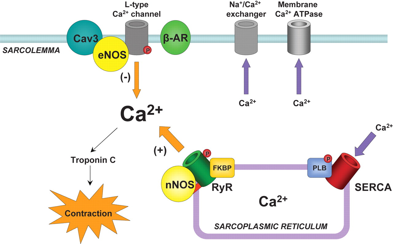
In heart muscle cells, the process of excitation–contraction (EC) coupling is mediated by

  •  Ca(2+) influx through sarcolemmal L-type Ca(2+) channels
  • activating Ca(2+) release channels (ryanodine receptors, RyRs) in the sarcoplasmic reticulum (SR).

Once activated, the RyR channels allow Ca(2+) to be released from the SR into the cytosol to induce contraction. This mechanism is known as Ca(2+)-induced calcium release (CICR) (Fabiato, 1985; Bers, 2002).  During relaxation, most of the Ca(2+) is resequestered into the SR by the Ca(2+)-ATPase. The amount of Ca(2+) released and the force of contraction depend on

  •  the magnitude of the Ca(2+) trigger signal,
  • the functional state of the RyRs and
  • the amount of Ca(2+) stored in the SR.

F1.large  calcium movement and RyR2 receptor Ca(2+) and contraction calcium release calmodulin + ER Reversible phosphorylation of proteins composing the EC coupling machinery plays an important role in regulation of cardiac contractility (Bers, 2002). Thus, during stimulation of the b-adrenergic pathway, phosphorylation of several target proteins, including

  • the L-type Ca(2+) channels,
  • RyRs and
  • phospholamban,

by protein kinase A (PKA) leads to an overall increase in SR Ca2+ release and contractile force in heart cells (Callewaert et al. 1988, Spurgeon et al. 1990; Hussain & Orchard, 1997; Zhou et al. 1999; Song et al. 2001; Viatchenko-Karpinski & Gyorke, 2001). PKA-dependent phosphorylation of the L-type Ca(2+) channels increases the Ca2+ current (ICa), increasing both

  • the Ca2+ trigger for SR Ca2+ release and
  • the SR Ca(2+) content

(Callewaert et al. 1988; Hussain & Orchard, 1997; Del Principe et al. 2001). Phosphorylation of phospholamban (PLB) relieves the tonic inhibition dephosphorylated PLB exerts on the SR Ca(2+)-ATPase (SERCA) resulting in enhanced SR Ca(2+) accumulation and enlarged Ca(2+) release (Kranias et al. 1985; Simmermann & Jones, 1998). With regard to the RyR, despite clear demonstration of phosphorylation of the channel in biochemical studies (Takasago et al. 1989; Yoshida et al. 1992), the consequences of this reaction to channel function have not been clearly defined. RyR phosphorylation by PKA and Ca(2+)–calmodulin dependent protein kinase (CaMKII) has been reported to increase RyR activity in lipid bilayers (Hain et al. 1995; Marx et al. 2000; Uehara et al. 2002). Moreover, it has been reported that in heart failure (HF), hyperphosphorylation of RyR causes

  •  the release of FK-506 binding protein (FKBP12.6) from the RyR,
    • rendering the channel excessively leaky for Ca(2+) (Marx et al. 2000).

However, other studies have reported no functional effects (Li et al. 2002) or even found phosphorylation to reduce RyR channel steady-state open probability (Valdivia et al. 1995; Lokuta et al. 1995).  The action of protein kinases is opposed by dephosphorylating phosphatases. Three types of protein phosphatases (PPs), referred to as PP1, PP2A and PP2B (calcineurin), have been shown to influence cardiac performance (Neumann et al. 1993; Rusnak & Mertz, 2000). Overall, according to most studies phosphatases appear to downregulate SR Ca(2+) release and contractile performance (Neumann et al. 1993; duBell et al. 1996, 2002; Carr et al. 2002; Santana et al. 2002). Furthermore, PP1 and PP2A activities appear to be increased in heart failure (Neumann, 2002; Carr et al. 2002). However, again the precise mode of action of these enzymes on intracellular Ca(2+) handling in normal and diseased hearts remains poorly understood.  In the present study, we have investigated the effects of protein phosphatases PP1 and PP2A on local Ca(2+) release events, Ca(2+) sparks, in cardiac cells. Our results show that

  •  phosphatases activate RyR mediated SR Ca(2+) release
    • leading to depletion of SR Ca(2+) stores.

These results provide novel insights into the mechanisms and potential role of protein phosphorylation/dephosphorylation in regulation of Ca(2+) signaling in normal and diseased hearts. F2.large   RyR and calcium

RESULTS

Effects of PP1 and PP2A on Ca2+ sparks and SR Ca(2+) content.

[1]  PP1 caused an early transient potentiation of Ca2+ spark frequency followed by a delayed inhibition of event occurrence. [2]  PP1 produced similar biphasic effects on the magnitude and spatio-temporal characteristics of Ca(2+) sparks Specifically, during the potentiatory phase (1 min after addition of the enzyme), PP1 significantly increased

  • the amplitude,
  • rise-time,
  • duration and
  • width of Ca(2+) sparks;

during the inhibitory phase (5 min after addition of the enzyme),

  •  all these parameters were significantly suppressed by PP1.

The SR Ca(2+) content decreased by 35 % or 69 % following the exposure of myocytes to either 0.5 or 2Uml_1 PP1, respectively (Fig. 1C). Qualitatively similar results were obtained with phosphatase PP2A. Similar to the effects of PP1, PP2A (5Uml_1) produced a transient increase in Ca(2+) spark frequency (~4-fold) followed by a depression of event occurrence and decreased SR Ca(2+) content (by 82 % and 65 %, respectively). Also similar to the action of PP1, PP2A increased

  •  the amplitude and
  • spatio-temporal spread (i.e. rise-time, duration and width) of Ca(2+) sparks at 1 min
  • and suppressed the same parameters at 5 min of exposure to the enzyme (Table 1).

Together, these results suggest that phosphatases enhance spark-mediated SR Ca2+ release, leading to decreased SR Ca(2+) content. Preventive effects of calyculin A and okadaic acid Preventive effects of ryanodine

PP1-mediated RyR dephosphorylation

F3.large  cardiomyocyte SR F3.large  cardiomyocyte SR F2.large   RyR and calcium coupled receptors coupled receptors The cardiac RyR is phosphorylated at Ser-2809 (in the rabbit sequence) by both PKA and CAMKII (Witcher et al. 1991; Marx et al. 2000). Although additional phosphorylation sites may exist on the RyR (Rodriguez et al. 2003), but Ser-2809 is believed to be the only site that is phosphorylated by PKA, and RyR hyperphosphorylation at this site has been reported in heart failure (Marx et al. 2000).  To test whether indeed phosphatases dephosphorylated the RyR in our permeabilized myocyte experiments we performed quantitative immunoblotting using an antibody that specifically recognizes the phosphorylated form of the RyR at Ser-2809 (Rodriguez et al. 2003). Myocytes exhibited a significant level of phosphorylation under baseline conditions. Maximal phosphorylation was 201 % of control. When exposed to 2Uml_1 PP1, RyR phosphorylation was 58 % of the control basal condition. Exposing to a higher PP1 concentration (10Uml_1) further reduced RyR phosphorylation to 22% of control. Thus, consistent with the results of our functional measurements,

  •  PP1 decreased RyR phosphorylation in cardiac myocytes.

Figure 1. Effects of PP1 on properties of Ca(2+) sparks and SR Ca(2+) content in rat permeabilized myocytes    see .  http://dx.doi.org/10.1113/jphysiol.2003.046367 A, spontaneous Ca(2+) spark images recorded under reference conditions, and 1 or 5 min after exposure of the cell to 2Uml_1 PP1. Traces below the images are Ca(2+) transients induced by application of 10 mM caffeine immediately following the acquisition of sparks before (3 min) and after (5 min) application of PP1 in the same cell. The Ca(2+) transients were elicited by a whole bath application of 10 mM caffeine. B, averaged spark frequency at early (1 min) and late (5 min) times following the addition of either 0.5 or 2Uml_1 of PP1 to the bathing solution. C, averaged SR Ca(2+) content for 0.5 or 2Uml_1 of PP1 measured before and 5 min after exposure to the enzyme. Data are presented as means ± S.E.M. of 6 experiments in different cells. Figure 2. Effects of PP2A on properties of Ca2+ sparks and SR Ca2+ content in rat permeabilized myocytes   see .  http://dx.doi.org/10.1113/jphysiol.2003.046367 A, spontaneous Ca(2+) spark images recorded under reference conditions, and 1 or 5 min after exposure of the cell to 5Uml_1 PP2A. Traces below the images are Ca(2+) transients induced by application of 10 mM caffeine immediately following the acquisition of sparks before (3 min) and after (5 min) application of PP2A in the same cell. B and C, averaged spark frequency (B) and SR Ca(2+) content (C) for the same conditions as in A. Data are presented as means ± S.E.M. of 6 experiments in different cells.

 DISCUSSION

In the present study, we have investigated the impact of physiologically relevant exogenous protein phosphatases PP1 and PP2A on RyR-mediated SR Ca(2+) release (measured as Ca(2+) sparks) in permeabilized heart cells. Our principal finding is that

  • phosphatases stimulated RyR channels lead to depleted SR Ca(2+) stores.

These results have important ramifications for understanding the mechanisms and role of protein phosphorylation/dephosphorylation in

  •  modulation of Ca(2+) handling in normal and diseased heart.

Modulation of SR Ca2+ release by protein phosphorylation/dephophorylation

Since protein dephosphorylation clearly resulted in increased functional activity of the Ca(+)release channel, our results imply that a reverse, phosphorylation reaction should reduce RyR activity. If indeed such effects take place, why do they not manifest in inhibition of Ca(+)sparks? One possibility is that enhanced Ca(+) uptake by SERCA

  •  masks or overcomes the effects phosphorylation may have on RyRs.

In addition, the potential inhibitory influence of protein phosphorylation on RyR activity in myocytes could be countered by feedback mechanisms  involving changes in luminal Ca(2+)(Trafford et al. 2002; Gyorke et al. 2002). In particular, reduced open probability of RyRs would be expected to lead to

  •  increased Ca2+ accumulation in the SR;
  • and increased intra-SR [Ca(2+)], in turn would
  • increase activity of RyRs at their luminal Ca(2+) regulatory sites

as demonstrated for the RyR channel inhibitor tetracaine (Gyorke et al. 1997; Overend et al. 1997). Thus

  • potentiation of SERCA
  • combined with the intrinsic capacity of the release mechanism to self-regulate

could explain at least in part why PKA-mediated protein phoshorylation results in maintained potentiation of Ca(2+) sparks despite a potential initial decrease in RyR activity.

Role of altered RyR Phosphorylation in Heart Failure

Marx et al. (2000) have proposed that  enhanced levels of circulating catecholamines lead to increased phosphorylation of RyR in heart failure.  Based on biochemical observations as well as on studying properties of single RyRs incorporated into artificial lipid bilayers, these investigators have hypothesized that

  •  hyperphosphorylation of RyRs contributes to pathogenesis of heart failure
    • by making the channel excessively leaky due to dissociation of FKBP12.6 from the channel.

We show that the mode of modulation of RyRs by phosphatases does not support this hypothesis as

  • dephosphorylation caused activation instead of

Interestingly, our results provide the basis for a different possibility in which

  •  dephophosphorylation of RyR rather than its phosphorylation causes depletion of SR Ca(2+) stores by stimulating RyRs in failing hearts.

It has been reported that PP1 and PP2 activities are increased in heart failure (Huang et al. 1999; Neumann et al. 1997; Neuman, 2002). Furthermore,  overexpression of PP1 or ablation of the endogenous PP1 inhibitor, l-1, results in

  • depressed contractile performance and heart failure (Carr et al. 2002).

Our finding that PP1 causes depletion of SR Ca(2+) stores by activating RyRs could account for, or contribute to, these results.

References

1 DelPrincipe F, Egger M, Pignier C & Niggli E (2001). Enhanced E-C coupling efficiency after beta-stimulation of cardiac myocytes. Biophys J 80, 64a. 2 Gyorke I & Gyorke S (1998). Regulation of the cardiac ryanodine receptor channel by luminal Ca2+ involves luminal Ca2+ sensing sites. Biophys J 75, 2801–2810. 3 Gyorke S, Gyorke I, Lukyanenko V, Terentyev D, Viatchenko-Karpinski S & Wiesner TF (2002). Regulation of sarcoplasmic reticulum calcium release by luminal calcium in cardiac muscle. Front Biosci 7, d1454–d1463. 4 Gyorke I, Lukyanenko V & Gyorke S (1997). Dual effects of tetracaine on spontaneous calcium release in rat ventricular myocytes. J Physiol 500, 297–309. 5 MacDougall LK, Jones LR & Cohen P (1991). Identification of the major protein phosphatases in mammalian cardiac muscle which dephosphorylate phospholamban. Eur J Biochem 196, 725–734. 6 Marx SO, Reiken S, Hisamatsu Y, Jayaraman T, Burkhoff D, Rosemblit N & Marks AR (2000). PKA phosphorylation dissociates FKBP12.6 from the calcium release channel (ryanodine receptor): defective regulation in failing hearts. Cell 101, 365–376. 7 Rodriguez P, Bhogal MS & Colyer J (2003). Stoichiometric phosphorylation of cardiac ryanodine receptor on serine-2809 by calmodulin-dependent kinase II and protein kinase A. J Biol Chem (in press).

The δC Isoform of CaMKII Is Activated in Cardiac Hypertrophy and Induces Dilated Cardiomyopathy and Heart Failure

T Zhang, LS Maier, ND Dalton, S Miyamoto, J Ross, DM Bers, JH Brown.  University of California, San Diego, La Jolla, Calif; and Loyola University, Chicago, Ill. Circ Res. 2003;92:912-919.    http://dx.doi.org/10.1161/01.RES.0000069686.31472.C5 Recent studies have demonstrated that transgenic (TG) expression of either Ca(2+)/calmodulin-dependent protein kinase IV (CaMKIV) or CaMKIIδB, both of which localize to the nucleus, induces cardiac hypertrophy. However,

  •  CaMKIV is not present in heart, and
  • cardiomyocytes express not only the nuclear CaMKIIδB
    • but also a cytoplasmic isoform, CaMKII δC.

In the present study, we demonstrate that

  1.  expression of the δC isoform of CaMKII is selectively increased and
  2. its phosphorylation elevated as early as 2 days and continuously for up to 7 days after pressure overload.

To determine whether enhanced activity of this cytoplasmic δC isoform of CaMKII can lead to phosphorylation of Ca(2+) regulatory proteins and induce hypertrophy, we generated TG mice that expressed the δC isoform of CaMKII.  Immunocytochemical staining demonstrated that the expressed transgene is confined to the cytoplasm of cardiomyocytes isolated from these mice. These mice develop a dilated cardiomyopathy with up to a 65% decrease in fractional shortening and die prematurely. Isolated myocytes are enlarged and exhibit reduced contractility and altered Ca2(2+) handling. Phosphorylation of the ryanodine receptor (RyR) at a CaMKII site is increased even before development of heart failure, and

  • CaMKII is found associated with the RyR  from the CaMKII TG mice.
  • Phosphorylation of phospholamban is increased specifically at the CaMKII but not at the PKA phosphorylation site.

These findings are the first to demonstrate that CaMKIIδC can mediate phosphorylation of Ca(2+) regulatory proteins in vivo and provide evidence for the involvement of CaMKIIδC activation in the pathogenesis of dilated cardiomyopathy and heart failure.  Multifunctional Ca(2+)/calmodulin-dependent protein kinases (CaM kinases or CaMKs) are transducers of Ca2+ signals that phosphorylate a wide range of substrates and thereby affect Ca(2+)-mediated cellular responses.1 The family includes CaMKI and CaMKIV, monomeric enzymes activated by CaM kinase kinase,2,3 and CaMKII, a multimer of 6 to 12 subunits activated by autophosphorylation.1 The CaMKII subunits α, β, γ, and δ show different tissue distributions,1 with

  • the δ isoform predominating in the heart.4–7
  • Splice variants of the δ isoform, characterized by the presence of a second variable domain,4,7 include δB, which contains a nuclear localization signal (NLS), and
  • δC, which does not. CaMKII composed of δB subunits localizes to the nucleus, whereas CaMKIIδC localizes to the cytoplasm.4,8,9

CaMKII has been implicated in several key aspects of acute cellular Ca(2+) regulation related to cardiac excitation-contraction (E-C) coupling. CaMKII

  • phosphorylates sarcoplasmic reticulum (SR) proteins including the ryanodine receptors (RyR2) and
  • phospholamban (PLB).10–14

Phosphorylation of RyR has been suggested to alter the channel open probability,14,15 whereas phosphorylation of PLB has been suggested to regulate SR Ca(2+) uptake.14 It is also likely that CaMKII phosphorylates the L-type Ca(2+) channel complex or an associated regulatory protein and thus

  1. mediates Ca(2+) current (ICa) facilitation.16-18 and
  2. the development of early after-depolarizations and arrhythmias.19

Thus, CaMKII has significant effects on E-C coupling and cellular Ca(2 +) regulation. Nothing is known about the CaMKII isoforms regulating these responses.  Contractile dysfunction develops with hypertrophy, characterizes heart failure, and is associated with changes in cardiomyocyte (Ca2+) homeostasis.20  CaMKII expression and activity are altered in the myocardium of rat models of hypertensive cardiac hypertrophy21,22 and heart failure,23 and

  • in cardiac tissue from patients with dilated cardiomyopathy.24,25

Several transgenic mouse models have confirmed a role for CaMK in the development of cardiac hypertrophy, as originally suggested by studies in isolated neonatal rat ventricular myocytes.9,26–28 Hypertrophy develops in transgenic mice that overexpress CaMKIV,27 but this isoform is not detectable in the heart,4,29 and CaMKIV knockout mice still develop hypertrophy after transverse aortic constriction (TAC).29  Transgenic mice overexpressing calmodulin developed severe cardiac hypertrophy,30 later shown to be associated with an increase in activated CaMKII31; the isoform of CaMKII involved in hypertrophy could not be determined from these studies. We recently reported that transgenic mice that overexpress CaMKIIδB, which is highly concentrated in cardiomyocyte nuclei, develop hypertrophy and dilated cardiomyopathy.32 To determine whether

  • in vivo expression of the cytoplasmic CaMKIIδC can phosphorylate cytoplasmic Ca(2+) regulatory proteins and
  • induce hypertrophy or heart failure,

we generated transgenic (TG) mice that expressed the δC isoform of CaMKII under the control of the cardiac specific α-myosin heavy chain (MHC) promoter. Our findings implicate CaMKIIδC in the pathogenesis of dilated cardiomyopathy and heart failure and suggest that

  • this occurs at least in part via alterations in Ca(2+) handling proteins.33

Ca(2+) and contraction RyR yuan_image3  Ca++ exchange yuan_image3  Ca++ exchange

Results

 Expression and Activation of CaMKIIδC Isoform After TAC

To determine whether CaMKII was regulated in pressure overload–induced hypertrophy, CaMKIIδ expression and phosphorylation were examined by Western blot analysis using left ventricular samples obtained at various times after TAC.  A selective increase (1.6-fold) in the lower band of CaMKIIδwas observed as early as 1 day and continuously for 4 days (2.3-fold) and 7 days (2-fold) after TAC (Figure 1A).  To confirm that CaMKIIδC was increased and determine whether this occurred at the transcriptional level, we performed semiquantitative RT-PCR using primers specific for the CaMKIIδC isoform. These experiments revealed that

  • mRNA levels for CaMKIIδC were increased 1 to 7 days after TAC (Figure 1B).

In addition to examining CaMKII expression, the activation state of CaMKII was monitored by its autophosphorylation, which confers Ca2-independent activity.

Figure 1. Expression and activation of CaMKII δC isoform after TAC.

see http://dx.doi.org/10.1161/01.RES.0000069686.31472.C5 A, Western blot analysis of total CaMKII in left ventricular (LV) homogenates obtained at indicated times after TAC. Cardiomyocytes transfected with CaMKIIδB and δC (right) served as positive controls and molecular markers. Top band (58 kDa) represents CaMKIIδB plus δ9, and the bottom band (56 kDa) corresponds to CaMKIIδC. *P0.05 vs control. B, Semiquantitative RT-PCR using primers specific for CaMKIIδC isoform (24 cycles) and GAPDH (19 cycles) using total RNA isolated from the same LV samples. C, Western blot analysis of phospho-CaMKII in LV homogenates obtained at various times after TAC. Three bands seen for each sample represent CaMKIIγ subunit (uppermost), CaMKIIδB plus δ9 (58 kDa), and CaMKIIδC (56 kDa). Quantitation is based on the sum of all of the bands. *P0.05 vs control.

 Figure 2. Expression and activation of CaMKII in CaMKIIδC transgenic mice.

see  http://dx.doi.org/10.1161/01.RES.0000069686.31472.C5 A, Transgene copy number based on Southern blots using genomic DNA isolated from mouse tails (digested with EcoRI). Probe (a 32P-labeled 1.7-kb EcoRI-SalI -MHC fragment) was hybridized to a 2.3-kb endogenous fragment (En) and a 3.9-kb transgenic fragment (TG). Transgene copy number was determined from the ratio of the 3.9-kb/2.3-kb multiplied by 2. B, Immunocytochemical staining of ventricular myocytes isolated from WT and CaMKIIδTG mice. Myocytes were cultured on laminin-coated slides overnight. Transgene was detected by indirect immunofluorescence staining using rabbit anti-HA antibody (1:100 dilution) followed by FITC-conjugated goat antirabbit IgG antibody (1:100 dilution). CaMKIIδB localization to the nucleus in CaMKIIδB TG mice (see Reference 32) is shown here for comparative purpose. C, Quantitation of the fold increase in CaMKIIδprotein expression in TGL and TGM lines. Different amounts of ventricular protein (numbers) from WT control, TG () and their littermates () were immunoblotted with an anti-CaMKIIδ antibody. Standard curve from the WT control was used to calculate fold increases in protein expression in TGL and TGM lines. D, Phosphorylated CaMKII in ventricular homogenates was measured by Western blot analysis (n5 for each group). **P0.01 vs WT.

 Generation and Identification of CaMKIIδC Transgenic Mice

TG mice expressing HA-tagged rat wild-type CaMKIIδC under the control of the cardiac-specific α-MHC promoter were generated as described in Materials and Methods. By Southern blot analysis, 3 independent TG founder lines carrying 3, 5, and 15 copies of the transgene were identified. They were designated as TGL (low copy number), TGM (medium copy number), and TGH (high copy number), The founder mice from the TGH line died at 5 weeks of age with marked cardiac enlargement.  The other two lines showed germline transmission of the transgene. The transgene was expressed only in the heart. Although CaMKII protein levels in TGL and TGM hearts were increased 12- and 17-fold over wild-type (WT) controls (Figure 2C), the amount of activated CaMKII was only increased 1.7- and 3-fold in TGL and TGM hearts (Figure 2D). The relatively small increase in CaMKII activity in the TG lines probably reflects the fact that the enzyme is not constitutively activated and that the availability of Ca2/CaM, necessary for activation of the overexpressed CaMKII, is limited. Importantly,

  • the extent of increase in active CaMKII in the TG lines was similar to that elicited by TAC.

 Cardiac Overexpression of CaMKIIδC Induces Cardiac Hypertrophy and Dilated Cardiomyopathy

There was significant enlargement of hearts from CaMKIIδC TGM mice by 8 to 10 weeks [see  http://dx.doi.org/10.1161/01.RES.0000069686.31472.C5%5D  (Figure 3A) and from TGL mice by 12 to 16 weeks. Histological analysis showed ventricular dilation (Figure 3B), cardiomyocyte enlargement (Figure 3C), and mild fibrosis (Figure 3D) in CaMKIIδC TG mice. Quantitative analysis of cardiomyocyte cell volume from 12-week-old TGM mice gave values of 54.7 + 0.1 pL for TGM (n = 96) versus 28.6 + 0.1 pL for WT littermates (n=94; P0.001). Ventricular dilation and cardiac dysfunction developed over time in proportion to the extent of transgene expression. Left ventricular end diastolic diameter (LVEDD) was increased by 35% to 45%, left ventricular posterior wall thickness (LVPW) decreased by 26% to 29% and fractional shortening decreased by 50% to 60% at 8 weeks for TGM and at 16 weeks for TGL. None of these parameters were significantly altered at 4 weeks in TGM or up to 11 weeks in TGL mice, indicating that heart failure had not yet developed.  Contractile function was significantly decreased. Figure 6. Dilated cardiomyopathy and dysfunction in CaMKIIδC TG mice at both whole heart and single cell levels.  [see Fig 6:  http://dx.doi.org/10.1161/01.RES.0000069686.31472.C5] C, Decreased contractile function in ventricular myocytes isolated from 12-week old TGM and WT controls presented as percent change of resting cell length (RCL) stimulated at 0.5 Hz. Representative trace and mean values are shown. *P0.05 vs WT. Figure 7. Phosphorylation of PLB in CaMKIIδC TG mice.  [see Fig 7: http://dx.doi.org/10.1161/01.RES.0000069686.31472.C5] Thr17 and Ser16 phosphorylated PLB was measured by Western blots using specific anti-phospho antibodies. Ventricular homogenates were from 12- to 14-week-old WT and TGM mice (A) or 4 to 5-week-old WT and TGM mice (B). Data were normalized to total PLB examined by Western blots (data not shown here). n = 6 to 8 mice per group; *P0.05 vs WT.

 Cardiac Overexpression of CaMKIIδC Results in Changes in the Phosphorylation of Ca2 Handling Proteins

To assess the possible involvement of phosphorylation of Ca2cycling proteins in the phenotypic changes observed in the CaMKIIC TG mice, we first compared PLB phosphorylation state in homogenates from 12- to 14-week-old TGM and WT littermates. Western blots using antibodies specific for phosphorylated PLB showed a 2.3-fold increase in phosphorylation of Thr17 (the CaMKII site) in hearts from TGM versus WT (Figure 7A). Phosphorylation of PLB at the CaMKII site was also increased 2-fold in 4- to 5-week-old TGM mice (Figure 7B). Significantly, phosphorylation of the PKA site (Ser16) was unchanged in either the older or the younger TGM mice (Figures 7A and 7B). (see  http://dx.doi.org/10.1161/01.RES.0000069686.31472.C5)  To demonstrate that the RyR2 phosphorylation changes observed in the CaMKII transgenic mice are not secondary to development of heart failure, we performed biochemical studies examining RyR2 phosphorylation in 4- to 5-week-old TGM mice. At this age, most mice showed no signs of hypertrophy or heart failure (see Figure 6B) and there was no significant increase in myocyte size (21.3 + 1.3 versus 27.7 + 4.6 pL; P0.14). Also, twitch Ca2 transient amplitude was not yet significantly depressed, and mean δ [Ca2+]i (1 Hz) was only 20% lower (192 + 36 versus 156 + 13 nmol/L; P0.47) versus 50% lower in TGM at 13 weeks.33  The in vivo phosphorylation of RyR2, determined by back phosphorylation, was significantly (2.10.3-fold; P0.05) increased in these 4- to 5-week-old TGM animals (Figure 8C), an increase equivalent to that seen in 12- to 14-week-old mice. We also performed the RyR2 back-phosphorylation assay using purified CaMKII rather than PKA. RyR2 phosphorylation at the CaMKII site was also significantly increased (2.2 + 0.3-fold; P0.05) in 4- to 5-week-old TGM mice (Figure 8C).  (http://dx.doi.org/10.1161/01.RES.0000069686.31472.C5) The association of CaMKII with the RyR2 is consistent with a physical interaction between this protein kinase and its substrate. The catalytic subunit of PKA and the phosphatases PP1 and PP2A were also present in the RyR2 immunoprecipitates, but not different in WT versus TG mouse hearts (Figure 8D). These data provide further evidence that

  • the increase in RyR2 phosphorylation, which precedes development of failure in the 4- to 5-week-old CaMKIIδC TG hearts, can be attributed to the increased activity of CaMKII.

 Discussion

  1. CaMKII is involved in the dynamic modulation of cellular
  2. Ca2 regulation and has been implicated in the development of cardiac hypertrophy and heart failure.14
  3. Published data from CaMK-expressing TG mice demonstrate that forced expression of CaMK can induce cardiac hypertrophy and lead to heart failure.27,32

However, the CaMK genes expressed in these mice are neither the endogenous isoforms of the enzyme nor the isoforms likely to regulate cytoplasmic Ca(2+) handling, because they localize to the nucleus.

  1.  the cytoplasmic cardiac isoform of CaMKII is upregulated at the expression level and is in the active state (based on autophosphorylation) after pressure overload induced by TAC.
  2.  two cytoplasmic CaMKII substrates (PLB and RyR) are phosphorylated in vivo when CaMKII is overexpressed and its activity increased to an extent seen under pathophysiological conditions.
  3. CaMKIIδ is found to associate physically with the RyR in the heart.
  4.  heart failure can result from activation of the cytoplasmic form of CaMKII and this may be due to altered Ca(2+) handling.

 Differential Regulation of CaMKIIδ Isoforms in Cardiac Hypertrophy

  1.  The isoform of CaMKII that predominates in the heart is the δ isoform.4–7 Neither the α nor the β isoforms are expressed and there is only a low level of expression of the γ isoforms.39
  2. Both δB and δC splice variants of CaMKIIδ are present in the adult mammalian myocardium36,40 and expressed in distinct cellular compartments.4,8,9

We suggest that the CaMKIIδ isoforms are differentially regulated in pressure-overload–induced hypertrophy, because the expression of CaMKIIδC is selectively increased as early as 1 day after TAC. Studies using RT-PCR confirm that

  • CaMKIIδC is regulated at the transcriptional level in response to TAC. In addition,
  • activation of both CaMKIIδB and CaMKIIδC, as indexed by autophosphorylation, increases as early as 2 days after TAC.
  • Activation of CaMKIIδB by TAC is relevant to our previous work indicating its role in hypertrophy.9,32
  • The increased expression, as well as activation of the CaMKIIδC isoform, suggests that it could also play a critical role in both the acute and longer responses to pressure overload.

In conclusion, we demonstrate here that CaMKIIδC can phosphorylate RyR2 and PLB when expressed in vivo at levels leading to 2- to 3-fold increases in its activity. Similar increases in CaMKII activity occur with TAC or in heart failure. Data presented in this study and in the accompanying article33 suggest that altered phosphorylation of Ca(2+) cycling proteins is a major component of the observed decrease in contractile function in CaMKIIδC TG mice. The occurrence of increased CaMKII activity after TAC, and of RyR and PLB phosphorylation in the CaMKIIδC TG mice suggest that

  • CaMKIIδC plays an important role in the pathogenesis of dilated cardiomyopathy and heart failure.

These results have major implications for considering CaMKII and its isoforms in exploring new treatment strategies for heart failure.

Cardiac Electrophysiological Dynamics From the Cellular Level to the Organ Level

Daisuke Sato and Colleen E. Clancy Department of Pharmacology, University of California – Davis, Davis, CA. Biomedical Engineering and Computational Biology 2013:5: 69–75 http://www.la-press.com.   http://dx.doi.org/10.4137/BECB.S10960 Abstract: Cardiac alternans describes contraction of the ventricles in a strong-weak-strong-weak sequence at a constant pacing fre­quency. Clinically, alternans manifests as alternation of the T-wave on the ECG and predisposes individuals to arrhythmia and sudden cardiac death. In this review, we focus on the fundamental dynamical mechanisms of alternans and show how alternans at the cellular level underlies alternans in the tissue and on the ECG. A clear picture of dynamical mechanisms underlying alternans is important to allow development of effective anti-arrhythmic strategies. The cardiac action potential is the single cellular level electrical signal that triggers contraction of the heart.1 Under normal conditions, the originating activation signal comes from a small bundle of tissue in the right atrium called the sinoatrial node (SAN). The action potentials generated by the SAN initiate an excitatory wave that, in healthy tissue, propagates smoothly through a well-defined path and causes excitation and contraction in the ventricles. In disease states, the normal excitation pathway is disrupted and a variety of abnormal rhythms can occur, including cardiac alternans, a well-known precursor to sudden cardiac death. Cardiac alternans was initially documented in 1872 by a German physician, Ludwig Traube.2 He observed contraction of the ventricles in a strong-weak-strong-weak sequence even though the pacing frequency was constant. Clinically, alternans mani­fests as alternation of the T-wave on the ECG, typi­cally in the microvolt range. It is well established that individuals with microvolt T-wave alternans are at much higher risk for arrhythmia and sudden cardiac death. A clear picture of physio­logical mechanisms underlying alternans is important to allow development of effective anti-arrhythmic drugs. It is also important to understand dynamical mechanisms because while the cardiac action poten­tial is composed of multiple currents, each of which confers specific properties, revelation of dynamical mechanisms provides a unified fundamental view of the emergent phenomena that holds independently of specific current interactions. The ventricular myocyte is an excitable cell pro­viding the cellular level electrical activity that under­lies cardiac contraction. Under resting conditions, the membrane potential is about -80 mV. When the cell is stimulated, sodium (Na) channels open and the membrane potential goes above 0 mV. Then, a few ms later, the inward current L-type calcium (Ca) current activates and maintains depolarization of the mem­brane potential. During this action potential plateau, several types of outward current potassium (K) chan­nels also activate. Depending on the balance between inward and outward currents, the action potential duration (APD) is determined.The diastolic interval (DI) that follows cellular repolarization describes the duration the cell resides in the resting state until the next excitation. During the DI, channels recover with kinetics determined by intrinsic time constants. APD restitution defines the relationship between the APD and the previous DI (Fig. 1 top panel). In most cases1, the APD becomes longer as the previous DI becomes longer due to recovery of the L-type Ca channel (Fig. 1, bottom panel), and thus the APD restitution curve has a positive slope. Figure 1. (Top): APD and DI. (Bottom): The physiological mechanism of APD alternans involves recovery from inactivation of ICaL.  [see  http://dx.doi.org/10.4137/BECB.S10960]

 Action Potential Duration Restitution

In 1968 Nolasco and Dahlen showed graphically that APD alternans occurs when the slope of the APD res­titution curve exceeds unity. Why is the steepness of the slope important? As shown graphically in Figure 2, APD alternans amplitude is multiplied by the slope of the APD restitution curve in each cycle. When the slope is larger than one, then the alternans amplitude will be amplified until the average slope reaches 1 or the cell shows a 2:1 stimulus to response ratio.  The one-dimensional mapping between APD and DI fails to explain quasi-periodic oscillation of the APD. Figure 2. APD restitution and dynamical mechanism of APD alternans.   [see  http://dx.doi.org/10.4137/BECB.S10960]

Calcium Driven Alternans

A strong-weak-strong-weak oscillation in contrac­tion implies that the Ca transient (CaT) is alternating. Until 1999 it was assumed that if the APD is alternat­ing then the CaT alternates because the CaT follows APD changes. However, Chudin et al showed that CaT can alternate even when APD is kept constant during pacing with a periodic AP clamp waveform.14 This implies that the intracellular Ca cycling has intrinsic nonlinear dynamics. A critical component in this process is the sarcoplasmic reticulum (SR), a subcellular organelle that stores Ca inside the cell. When Ca enters a cell through the L-type Ca channel (or reverse mode Na-Ca exchanger (NCX) ryanodine receptors open and large Ca releases occur from the SR (Ca induced Ca release). The amount of Ca release steeply depends on SR Ca load. This steep relation between Ca release and SR Ca load is the key to induce CaT alternans.  A one-dimensional map between Ca release and SR calcium load can be constructed to describe the relationship21 similar to the map used in APD restitution.

 Subcellular Alternans

A number of experimental and computational stud­ies have been undertaken to identify molecular mechanisms of CaT alternans by identifying the specific components in the calcium cycling process critical to formation of CaT alternans. These compo­nents include SR Ca leak and load, Ca spark frequency and amplitude, and rate of SR refilling. For example, experiments have shown that alternation in diastolic SR Ca is not required for CaT alternans.24 In addition, stochastic openings of ryanodine receptors (RyR) lead to Ca sparks that occur randomly, not in an alternating sequence that would be expected to underlie Ca altern-ans. So, how do local random sparks and constant dia­stolic SR calcium load lead to global CaT alternans? Mathematical models with detailed representations of subcellular Ca cycling have been developed in order to elucidate the underlying mechanisms. Model­ing studies have shown that even when SR Ca load is not changing, RyRs, which are analogous to ICaL in APD alternans, recover gradually from refractoriness. As RyR availability increases (for example during a long diastolic interval) a single Ca spark from a RyR will be larger in amplitude and recruit neighboring Ca release units to generate more sparks. The large resultant CaT causes depletion of the SR and when complete recovery of RyRs does not occur prior to the arrival of the next stimulus, the subsequent CaT will be small. This process results in an alternans of CaT amplitude from beat-to-beat.

 Coupling Between the Membrane Potential and Subcellular Calcium Dynamics

Importantly, the membrane voltage and intracellu­lar Ca cycling are coupled via Ca sensitive channels such as the L-type Ca channel and the sodium-calcium exchanger (NCX). The membrane voltage dynamics and the intracellular Ca dynamics are bi-directionally coupled. One direction is from voltage to Ca. As the DI becomes longer, the CaT usually becomes larger since the recovery time for the L-type Ca channel in increased and the SR Ca release becomes larger. The other direction is from Ca to voltage. Here we consider two major currents, NCX and ICaL. As the CaT becomes larger, forward mode NCX becomes larger and pro­longs APD. On the other hand, as the CaT becomes larger, ICaL becomes smaller due to Ca-induced inacti­vation, and thus, larger CaT shortens the APD. There­fore, depending on which current dominates, larger CaT can prolong or shorten APD. If a larger CaT pro­longs (shortens) the APD, then the coupling is positive (negative). The coupled dynamics of the membrane voltage and the intracellular Ca cycling can be cate­gorized by the instability of membrane voltage (steep APD restitution), instability of the intracellular Ca cycling (steep relation between Ca release versus SR Ca load), and the coupling (positive or negative). If the coupling is positive, alternans is electromechani­cally concordant (long-short-long-short APD cor­responds to large-small-large-small CaT sequence) regardless of the underlying instability mechanism. On the other hand, if the coupling is negative, alternans is electromechanically concordant in a voltage-driven regime. However, if alternans is Ca driven, alternans becomes electromechanically discordant (long-short-long-short APD corresponds to small-large-small-large CaT sequence). It is also possible to induce quasi- periodic oscillation of APD and CaT when volt­age and Ca instabilities contribute equally.

 Alternans in Higher Dimensions

Tissue level alternans in APD and CaT also occur and here we describe how the dynamical mechanism of alternans at the single cell level determines the phenomena in tissue. Spatially discordant alternans (SDA) where APDs in different regions of tissue alternate out-of-phase, is more arrhythmogenic since it causes large gradients of refractoriness and wave-break, which can initiate ventricular tachycardia and ventricular fibrillation. How is SDA induced? As the APD is a function of the previous DI, con­duction velocity (CV) is also function of the previ­ous DI (CV restitution) since the action potential propagation speed depends on the availability of the sodium channel. As the DI becomes shorter, sodium channels have less time to recover. Therefore, in general, as the DI becomes shorter, the CV becomes slower. When tissue is paced rapidly, action poten­tials propagate slowly near the stimulus, and thenac-celerate downstream as the DI becomes longer. This causes heterogeneity in APD (APD is shorter near the stimulus). During the following tissue excitation, APD becomes longer and the CV becomes faster at the pacing site then gradually APD becomes shorter and the CV becomes slower. The interaction between steep APD restitution and steep CV restitution creates SDA. This mechanism applies only when the cel­lular instability is voltage driven. When the cellular instability is Ca driven, the mechanism of SDA formation is different. If the volt­age-Ca coupling is negative, SDA can form without steep APD and CV restitution. The mechanism can be understood as follows. First, when cells are uncou­pled, alternans of APD and Ca are electromechanically discordant. If two cells are alternating in opposite phases, once these cells are coupled by voltage, due to electrotonic coupling, the membrane voltage of both cells is synchronized and thus APD becomes the same. This synchronization of APD amplifies the difference of CaT between two cells (Fig. 5 in). In other words it desynchronizes CaT. This instability mechanism is also found in subcellular SDA. In the case where the instability is Ca driven and the coupling is positive, there are several interest­ing distinctive phenomena that can occur. First, the profile of SDA of Ca contains a much steeper gra­dient at the node (point in space where no alternans occurs–cells downstream of the node are alternating out of phase with those upstream of the node) com­pared to the case of voltage driven SDA. Thus, the cellular mechanism of instability can be identified by evaluating the steepness of the alternans amplitude gradient in space around the node. When the cellular instability is voltage driven, the steady-state wave­length (separation of nodes in space) depends on electrotonic coupling between cells and the steepness of APD and CV restitution, regardless of the initial conditions. However, if the cellular instability is Ca driven, the location of nodes depends on the pacing history, which includes pacing cycle length and other parameters affected by pacing frequency. In this case, once the node is formed, the location of the node may be fixed, especially when Ca instability is strong. Such an explanation may apply to recent experimen­tal results. Summary In this review, we described how the origin of alternans at the cellular level (voltage driven, Ca drive, coupling between voltage and Ca) affects the formation of spatially discordant alternans at the tissue level. Cardiac alternans is a multi-scale emergent phenomenon. Channel properties determine the instability mechanism at the cellular level. Alternans mechanisms at cellular level determine SDA patterns at the tissue level. In order to understand alternans and develop anti-arrhythmic drug and therapy, multi-scale modeling of the heart is useful, which is increasingly enabled by emerging technologies such as general-purpose computing on graphics processing units (GPGPU) and cloud computing.

English: Diagram of contraction of smooth musc...

English: Diagram of contraction of smooth muscle fiber (Photo credit: Wikipedia)

Schematic representation of Calcium Cycling in Contractile and Proliferating VSMCs receptors voltage gated Ca(2) channel Marks-Wehrens Model and multiphosphorylation  site model ncpcardio0419-f4   calcium leak

Read Full Post »

Treatment for Endocrine Tumors and Side Effects

Reporter and Curator: Dr. Sudipta Saha, Ph.D.

Surgery

The purpose of surgery is typically to remove the entire tumor, along with some of the healthy tissue around it, called the margin. If the tumor cannot be removed entirely, “debulking” surgery may be performed. Debulking surgery is a procedure in which the goal is to remove as much of the tumor as possible. Side effects of surgery include weakness, fatigue, and pain for the first few days following the procedure.

Chemotherapy

Chemotherapy is the use of drugs to kill tumor cells, usually by stopping the cells’ ability to grow and divide. Systemic chemotherapy is delivered through the bloodstream to reach tumor cells throughout the body. A chemotherapy regimen (schedule) usually consists of a specific number of cycles given over a set period of time. A patient may receive one drug at a time or combinations of different drugs at the same time. The side effects of chemotherapy depend on the individual and the dose used, but they can include fatigue, risk of infection, nausea and vomiting, loss of appetite, and diarrhea. These side effects usually go away once treatment is finished.

Radiation therapy

Radiation therapy is the use of high-energy x-rays or other particles to kill tumor cells. The most common type of radiation treatment is called external-beam radiation therapy, which is radiation given from a machine outside the body. When radiation treatment is given using implants, it is called internal radiation therapy or brachytherapy. A radiation therapy regimen usually consists of a specific number of treatments given over a set period of time. Side effects from radiation therapy may include fatigue, mild skin reactions, upset stomach, and loose bowel movements. Most side effects go away soon after treatment is finished.

Hormone therapy

The goal of hormone therapy is often to lower the levels of hormones in the body. Hormone therapy may be given to help stop the tumor from growing or to relieve symptoms caused by the tumor. In addition, for thyroid cancer, hormone therapy will be given if the thyroid gland has been removed, to replace the hormone that is needed by the body to function properly.

Immunotherapy

Immunotherapy (also called biologic therapy) is designed to boost the body’s natural defenses to fight the tumor. It uses materials made either by the body or in a laboratory to bolster, target, or restore immune system function. Examples of immunotherapy include cancer vaccines, monoclonal antibodies, and interferons. Alpha interferon is a form of biologic therapy given as an injection under the skin. This is sometimes used to help relieve symptoms caused by the tumor, but it can have severe side effects including fatigue, depression, and flu-like symptoms.

Targeted therapy

Targeted therapy is a treatment that targets the tumor’s specific genes, proteins, or the tissue environment that contributes to cancer growth and survival. This type of treatment blocks the growth and spread of tumor cells while limiting damage to normal cells, usually leading to fewer side effects than other cancer medications.

Recent studies show that not all tumors have the same targets. To find the most effective treatment, the doctor may run tests to identify the genes, proteins, and other factors in the tumor. As a result, doctors can better match each patient with the most effective treatment whenever possible.

Depending on the type of endocrine tumor, targeted therapy may be a possible treatment option. For instance, targeted therapies, such as sunitinib (Sutent) and everolimus (Afinitor), have been approved for treating advanced islet cell tumors. Early results of clinical trials (research studies) with targeted therapy drugs for other types of endocrine tumors are promising, but more research is needed to prove they are effective.

Recurrent endocrine tumor

Once the treatment is complete and there is a remission (absence of symptoms; also called “no evidence of disease” or NED). Many survivors feel worried or anxious that the tumor will come back. If the tumor does return after the original treatment, it is called a recurrent tumor. It may come back in the same place (called a local recurrence), nearby (regional recurrence), or in another place (distant recurrence). When this occurs, a cycle of testing will begin again to learn as much as possible about the recurrence. Often the treatment plan will include the therapies described above (such as surgery, chemotherapy, and radiation therapy) but may be used in a different combination or given at a different pace. People with a recurrent tumor often experience emotions such as disbelief or fear. Patients are encouraged to talk with their health care team about these feelings and ask about support services to help them cope.

Metastatic endocrine tumor

If a cancerous tumor has spread to another location in the body, it is called metastatic cancer. A treatment plan that includes a combination of surgery, chemotherapy, radiation therapy, hormone therapy, immunotherapy, or targeted therapy may be recommended if required.

In addition to treatment to slow, stop, or eliminate the cancer (also called disease-directed treatment), an important part of cancer care is relieving a person’s symptoms and side effects. It includes supporting the patient with his or her physical, emotional, and social needs, an approach called palliative or supportive care. People often receive disease-directed therapy and treatment to ease symptoms at the same time.

Source References:

http://www.cancer.net/cancer-types/endocrine-tumor/treatment


http://www.macmillan.org.uk/Cancerinformation/Cancertypes/Endocrine/Endocrinetumours.aspx


http://cancer.osu.edu/patientsandvisitors/cancerinfo/cancertypes/endocrine/Pages/index.aspx


http://cancer.northwestern.edu/cancertypes/cancer_type.cfm?category=8


http://www.cancervic.org.au/about-cancer/cancer_types/endocrine_cancer


http://www.oncolink.org/types/types1.cfm?c=4

Read Full Post »

Reporter and Curator: Dr. Sudipta Saha, Ph.D.

Screen Shot 2021-07-19 at 6.17.32 PM

Word Cloud By Danielle Smolyar

Cancer is one of the most devastating and widespread diseases today. The development of cancer is a multi-step process involving genetic or epigenetic changes often occurring over a longer period of time. Moreover, cancer occurs in more or less all organs and tissues and is characterized by extensive heterogeneity both concerning the type and aggressiveness of the disease. Although some substantial progress in some areas has been made, there are still huge unmet needs in treatment methods and the efficacy of currently available drugs. The pharmaceutical industry has struggled with the ever increasing costs in drug development and unfortunately novel drugs have not seldom demonstrated only marginal improvement in efficacy often at the cost of quality of life of the patients. For these reasons, new approaches are focusing on disease prevention instead of only treating the symptoms. Recently, much attention has been paid to prevention of the disease in parallel to continuous drug discovery.

Intervention in food intake has been demonstrated to play an enormous role in both prevention as well as treatment of diseases. Numerous studies indicate a clear link between cancer and diet. The substantial development of sequencing technologies has resulted in access to enormous amounts of genomics information, which resulted in the establishment of nutrigenomics as an emerging approach to link genomics research to studies on nutrition. Increased understanding has demonstrated how nutrition can influence human health both at genetic and epigenetic levels. It investigates the effects of nutrition and bioactive food compounds on gene expression. This approach has allowed the investigation of the effect on nutrition on individuals with specific genetic features. Moreover, it has provided the basis for nutritional intervention in prevention and treatment of disease and the inauguration of personalized nutrition. However, differences in types of cancer, the level of aggressiveness, and their occurrence at different stages of life have seriously complicated the understanding of the effect of nutrition on cancer prevention and treatment. Other individual variations such as the amounts of food consumed, digestion, metabolism and other factors like geographical, ethnic and sociological diversity has hampered the identification of which food components are most important for human health. Dramatic dietary modifications have proven essential in reducing risk and even prevention of cancer. Moreover, intense revision of diet in cancer patients has revealed significant changes in gene expression and also has provided therapeutic efficacy even after short-term application.

Obviously, a multitude of diets have been evaluated, but probably the common factor for achieving both prophylactic and therapeutic responses is to consume predominantly diets rich in fruits, vegetables, fish and fibers and reduced quantities of especially red meat. There are numerous examples of how dietary intake can promote health on both a preventive as well as therapeutic level. Radical change in diet has resulted in dramatic changes in gene expression in prostate cancer patients revealing that many of those genes involved in cancer development were down-regulated. The importance of nutrigenomics as a multi-task approach involving genomics, proteomics, metabolomics, et cetera has further provided novel possibilities to address the effect of nutrition on human health. Despite encouraging findings on how dietary modifications can prevent disease and restore health, there are a number of factors which complicate the outcome. There are variations in response to dietary changes depending on age and gender. However, the vast amount of accumulated nutrigenomics data should not overshadow the needs to take into account other important factors such as lifestyle, social, geographical and economic factors affecting diet and health.

Source References:

http://www.lifescienceglobal.com/home/cart?view=product&id=121

http://www.frontiersin.org/Nutrigenomics/10.3389/fgene.2011.00091/abstract

http://www.sciencedirect.com/science/article/pii/S0002822308021871

http://ajcn.nutrition.org/content/89/5/1553S

http://www.sciencedirect.com/science/article/pii/S030438350800390X

Read Full Post »

Reporter and Curator: Dr. Sudipta Saha, Ph.D.

Antioxidant micronutrients, such as vitamins and carotenoids, exist in abundance in fruit and vegetables and have been known to contribute to the body’s defence against reactive oxygen species. Numerous epidemiological studies have demonstrated that a high dietary consumption of fruit and vegetables rich in carotenoids or with high serum carotenoid concentrations results in lower risks of certain cancers, diabetes and cardiovascular disease. These epidemiological studies have suggested that antioxidant carotenoids may have a protective effect against diabetes or cardiovascular disease. However, the consumption of carotenoids in pharmaceutical forms for the treatment or prevention of these chronic diseases cannot be recommended, because some large randomized controlled trials did not reveal any reduction in cardiovascular events or type 2 diabetes with b-carotene. High doses of carotenoids used in the supplementation studies could have a pro-oxidant effect. Therefore, it is favourable to intake carotenoids from foods through the combination of other nutrients such as vitamins, minerals or phytochemicals, not by supplements.

The metabolic syndrome is a clustering of metabolic abnormalities that increase the risk for diabetes and cardiovascular disease. Typically, it includes excess weight, hyperglycaemia, evaluated blood pressure, low concentration of HDL-cholesterol, and hypertriacylglycerolaemia. This syndrome is emerging as one of the major medical and public health problems in Japan, and persons with this syndrome have an increased risk of morbidity and mortality due to cardiovascular disease and diabetes. Recently, many studies have examined the associations of dietary patterns with the metabolic syndrome and shown that diets rich in fruit and vegetables have been inversely associated with the metabolic syndrome. These previous reports suggest that a high intake of fruit and vegetables may reduce the risk of the metabolic syndrome through the beneficial combination of antioxidants, fibre, minerals, and other phytochemicals. Some recent cross-sectional and case–control studies have shown the associations of serum antioxidant status with the metabolic syndrome. Ford et al. reported that low intake and/or low serum concentrations of vitamins and carotenoids were associated with the risk of the metabolic syndrome. Although very few data are available about the associations of antioxidant carotenoids with the metabolic syndrome, people who have the metabolic syndrome are more likely to have increased oxidative stress than people who do not have this syndrome.

In some recent studies, it has been reported that oxidative stress, which is an imbalance between pro-oxidants and antioxidants, occurs more frequently in metabolic syndrome subjects than in non-metabolic syndrome subjects. Oxidative stress may play a key role in the pathophysiology of diabetes and cardiovascular disease. On the other hand, smoking is a potent oxidative stress in man. This increment of oxidative stress induced by smoking may develop insulin resistance, and increased insulin resistance may result in the clustering of the metabolic abnormality. Therefore, antioxidants could have a beneficial effect on reducing the risk of these conditions in smokers. However, there is limited information about the interaction of serum antioxidant carotenoids and the metabolic syndrome with smoking habit. This study was aimed to investigate the interaction of serum carotenoid concentrations and the metabolic syndrome with smoking. The association of the concentrations of six serum carotenoids, i.e. lutein, lycopene, a-carotene, b-carotene, b-cryptoxanthin and zeaxanthin, with metabolic syndrome status stratified by smoking status was evaluated crosssectionally.

In this study, the associations of the serum carotenoids with the metabolic syndrome stratified by smoking habit were evaluated cross-sectionally. A total of 1073 subjects (357 male and 716 female) who had received health examinations in the town of Mikkabi, Shizuoka Prefecture, Japan, participated in the study. Inverse associations of serum carotenoids with the metabolic syndrome were more evident among current smokers than non-smokers. These results support that antioxidant carotenoids may have a protective effect against development of the metabolic syndrome, especially in current smokers who are exposed to a potent oxidative stress.

Source References:

http://www.ncbi.nlm.nih.gov/pubmed/18445303

http://www.ncbi.nlm.nih.gov/pubmed/19450371

http://www.ncbi.nlm.nih.gov/pubmed/21216053

http://www.ncbi.nlm.nih.gov/pubmed/19631019

http://www.ncbi.nlm.nih.gov/pubmed/12324189

http://www.ncbi.nlm.nih.gov/pubmed/18689373

Read Full Post »

The Implications of a Newly Discovered  CYP2J2 Gene Polymorphism  Associated with Coronary Vascular Disease in the Uygur Chinese Population

Author, Curator: Larry H Bernstein, MD, FCAP

This is an interesting genomic study of the relationship of genetic polymorphism in the Chinese Uygur population that highlights the difficulty in CVD genomics, and casts a promising light on difficulties over
1.  possibly no more than 8 genetic signatures to account for all of human CVD conditions
2.  genetic signatures may no be equally distributed over studied populations
3.  genetic signatures may be more pronounced in different populations
4.  there is little predictable validity in such studies over large assimilated populations (such as African-Americans
5.  the best genomic evidence for meaningful associations does appear to tie in with endothelial metabolism
6.  the greatest difficulty in all studies is the small dose of information provided by an such linkage
7.  there has been too little information provided in studies of the effect of dietary factors on the affected population, which would entail nutrigenomics.
8.  there is an association between certain distinct CVD’s and later development of coronary heart disease (CHD).
This study concepts, methods and difficulties were recently reviewed in the following articles:
Synthetic Biology: On Advanced Genome Interpretation for Gene Variants and Pathways: What is the Genetic Base of Atherosclerosis and Loss of Arterial Elasticity with Aging
Aviva Lev-Ari, PhD, RN
Genomics & Genetics of Cardiovascular Disease Diagnoses: A Literature Survey of AHA’s Circulation Cardiovascular Genetics, 3/2010 – 3/2013
Aviva Lev-Ari, PhD, RN and Larry H Bernstein, MD, FCAP
Diagnosis of Cardiovascular Disease, Treatment and Prevention: Current & Predicted Cost of Care and the Promise of Individualized Medicine Using Clinical Decision Support Systems
Aviva Lev-Ari, PhD, RN and Larry H Bernstein, MD, FCAP
Hypertension and Vascular Compliance: 2013 Thought Frontier – An Arterial Elasticity Focus
Justin D. Pearlman, MD, PhD, and Aviva Lev-Ari, PhD, RN
Clinical Trials Results for Endothelin System: Pathophysiological role in Chronic Heart Failure, Acute Coronary Syndromes and MI – Marker of Disease Severity or Genetic Determination?
Aviva Lev-Ari, PhD, RN
Vascular Medicine and Biology: CLASSIFICATION OF FAST ACTING THERAPY FOR PATIENTS AT HIGH RISK FOR MACROVASCULAR EVENTS Macrovascular Disease – Therapeutic Potential of cEPCs
Aviva Lev-Ari, PhD, RN
Endothelial Function and Cardiovascular Disease
Larry H Bernstein, MD, FCAP
Reversal of Cardiac Mitochondrial Dysfunction
Larry H Bernstein, MD, FCAP
A Second Look at the Transthyretin Nutrition Inflammatory Conundrum
Larry H Bernstein, MD, FCAP

A Novel Polymorphism of the CYP2J2 Gene is Associated with Coronary Artery Disease in Uygur Population in China

Qing Zhu, Zhenyan Fu, Yitong Ma, Hong Yang, Ding Huang, Xiang Xie, Fen Liu, Yingying Zheng, Erdenbat Cha
PII: S0009-9120(13)00174-4    Available online 15 May 2013
Reference: CLB 8375
To appear in: Clinical Biochemistry
Received date: 17 February 2013
Revised date: 13 April 2013
Accepted date: 3 May 2013
Background: Cytochrome P450 (CYP) 2J2 is expressed in the vascular endothelium and metabolizes arachidonic acid to biologically active epoxyeicosatrienoic acids (EETs).
  • The EETs are potent endogenous vasodilators and
  • inhibitors of vascular inflammation.
The aim of the present study was to assess the association between the human CYP2J2 gene polymorphism and coronary artery disease (CAD) in a Han and Uygur population of China.
We use two independent case-control studies:
  1. a Han population (206 CAD patients and 262 control subjects) and
  2. a Uygur population (336 CAD patients and 448 control subjects).
All CAD patients  and controls were genotyped for the same three single nucleotide polymorphisms (SNPs)
  1. rs890293
  2. rs11572223
  3. rs2280275
of CYP2J2 gene by a Real-time PCR instrument.
Results: In the Uygur population, for total, the distribution of SNP3 (rs2280275) genotypes showed a significant difference between CAD and control participants (P=0.048).
For total and men, the distribution of SNP3 (rs2280275) alleles and the dominant model (CC vs CT + TT)
  • showed a significant difference between CAD and control participants (for allele: P=0.014 and P=0.035, respectively; for dominant model: P=0.014 and P=0.034, respectively).
The significant difference in dominant model was retained after adjustment for covariates (OR: 0.279, 95% confidence interval [CI]: 0.176-0.440, P=0.001; OR: 0.240, 95% CI: 0.128-0.457, P=0.001, respectively).
Conclusions: The CC genotype of rs2280275 in CYP2J2 gene could be a protective genetic marker of CAD and T allele may be a risk genetic marker of CAD in men of Uygur population in China.
Highlights:
1. We used two independent case-control studies: one was in a Han population and the other was in a Uygur population.
2. The CC genotype of rs2280275 in CYP2J2 gene could be a protective genetic marker of CAD and T allele may be a risk genetic marker of CAD in men of Uygur population in China.
3. Polymorphism of the CYP2J2 gene can affect the synthesis of epoxyeicosatrienoic acids (EETs).
Reviewer Observations:
This article describes the association between CYP2J2 polymorphism(SNP1, SNP2 and SNP3) and coronary artery disease (CAD) in two populations of China (Han and Uygur).
Results show that
  1. the frequency of T allele of rs2280275 (SNP3 of the CYP2J2) is higher in CAD patients than in control subjects and
  2. that CC genotype of rs 2280275 is significantly lower in CAD patients than in control subjects.
  3. “T allele of rs2280275 was significantly higher in CAD patients than in control participants. CC genotype of rs2280275 was significantly lower in CAD patients than in control participants.”;
  4. It appears that CC is the homozygous and dominant state of this SNP3 sequence in a pairing-combination.
  5. The effect of decreased CHD is seen only in the CC double combination, in men and not women. The difference between men and women with CAD is in LDL.
For Uygur population,
(1) after adjusting major confounding factors such as Glu、LDL、EH、DM and smoking, the effect of decreased CAD is seen only in the CC double combination, in men and not women.
(2) for men, the LDL level is higher in CAD than in control, for women, there isn’t a difference of LDL level between CAD and control.
(3) for men, the distribution of T and C allele is different between CAD and control (p=0.035), and not in women (p=0.118).
The T allele of SNP3 is increased in CAD. So the C allele is important, and a CT pair is neutral. Neither SNP1 or SNP2, or presumably both have lower incidence.

I might conjecture that having(heterozygous rs2280275), a C & a T, and eating a lot of fish and/or flax seed would show a difference

  • because of the intimal enzymatic conversion of arachidonic acid to EETs.

Arachidonic acid is a derivative of linoleic acid,an n-6 PUFA, while linolenic acid is an omega-3 PUFA. Substantial documentation of the effect of EETs is given. The anti-inflammatory advantage of an n-3 PUFA is also known.
It appears that the intimal conversion results in an omega-3 product.  In addition, the EET activates eNOS, so that there is endothelial NO produced.

The studies of both Spiecker and Ping Yin Liu showed the polymorphism of CYP2J2 (rs890293, SNP1) has relation with CAD. However, in this study, the authors found there was no association between the polymorphism of CYP2J2 (rs890293, SNP1) and CAD in Han population and Uygur population. We found (rs 2280275, SNP3) has association with CAD.
  • “The CC genotype of rs2280275 in CYP2J2 gene could be a protective genetic marker of CAD and T allele may be a risk genetic marker of CAD in men of Uygur population in China”
All participants had a differential diagnosis for chest pain encountered in the Cardiac Catheterization Laboratory of First Affiliated Hospital of Xinjiang Medical University. We recruited randomly CAD group and control group, subjects with valvular disease were excluded, control subjects were not healthy individuals, some of them have hypertension, some of them have DM, some of them have hyperlipidemia, which means control group expose to the same risk factors of CAD while the results of coronary angiogram is normal. All control subjects underwent a coronary angiogram and have no coronary artery stenosis.
The analysis was a logistic regression analysis, we used the major variables of CAD to analysis and found the CC genotype was the dependent useful factor after adjusting for major confounding factors such as Glu、LDL、EH、DM and smoking.
Schematic of EET interactions with cardiovascularion channels.
A: In the cardiac myocyte, EETs activate sarcolemmal or mitochondrial KATP channels.
B: In the vasculature, EETs activate endothelial small-(SKCa) or intermediate (IKCa)–conductance calcium-activated channels to cause hyperpolarization, which can be transmitted to the vascular smooth muscle via myoendothelial gap junctions. EETs also activate TRPV4 channels to activate Ca2+influx. In the vascular smooth muscle, EETs activate large conductance, calcium-activated (BK-Ca) channels through a G protein-Coupled event.
C: In platelets, EETs activate BK-Ca channels.calcium-activated (BK-Ca) channels through a G-protein-coupled event. C, In platelets, EETs activate BK-Ca channels.

Association of the ADRA2A polymorphisms with the risk of type 2 diabetes: A meta-analysis

Xi Chen, Lei Liu, Wentao He, Yu Lu, Delin Ma, Tingting Du, Qian Liu, Cai Chen, Xuefeng Yu
Clinical Biochemistry 2013;  46 (9): 722–726   http://dx.doi.org/10.1016/j.clinbiochem.2013.02.004
Results from the published studies on the association of ADRA2A (adrenoceptor alpha 2A) variants with type 2 diabetes (T2D) are conflicting and call for further assessment. The aim of this meta-analysis was to quantitatively summarize the effects of the two recently reported ADRA2A single nucleotide polymorphisms (SNPs) rs553668 and rs10885122 on T2D risk.
Results
Twelve studies with 40,828 subjects from seven eligible papers were included in the meta-analysis. Overall, the present meta-analysis failed to support a positive association between ADRA2A SNPs (rs553668 and rs10885122) and susceptibility to T2D (OR = 1.05, p = 0.17, 95% CI: 0.98, 1.12; and OR = 1.06, p = 0.11, 95% CI: 0.99, 1.13; respectively).
However, in the subgroup analysis by ethnicity, the significant association between rs553668 and the risk of T2D was obtained in Europeans under the recessive genetic model (OR = 1.36, p = 0.02, 95% CI: 1.05, 1.76).
Conclusion
The results of the meta-analyses indicated that both SNPs were associated with CHD in Caucasians (P < 0.05) but not in Asians. The results from our case-control study and meta-analyses might be explained by genetic heterogeneity in the susceptibility of CHD and ethnic differences between Asians and Caucasians.

Association between PCSK9 and LDLR gene polymorphisms with coronary heart disease: Case-control study and meta-analysis

Lina Zhang, Fang Yuan, Panpan Liu, Lijuan Fei, Yi Huang, Limin Xu, et al.
Clinical Biochemistry 2013; 46 (9): 727–732
► Association of rs11206510 and rs1122608 with CHD in 813 Chinese participants.
► The first association test of rs1122608 with the risk of CHD in Han Chinese.
► Meta-analyses were performed for rs11206510 and rs1122608.
► The two SNPs were associated with CHD in Caucasians but not in Asians.
Objective
To explore the association of rs11206510 (PCSK9 gene) and rs1122608 (LDLR gene) polymorphisms with coronary heart disease (CHD) in Han Chinese.
Methods
A total of 813 participants (290 CHD cases, 193 non-CHD controls and 330 healthy controls) were recruited in the case-control study. DNA genotyping was performed on the SEQUENOM® Mass–ARRAY iPLEX® platform. χ2-test was used to compare the genotype distribution and allele frequencies. Two meta-analyses were performed to establish the association between the two polymorphisms with CHD.
Results
No significant associations between the two SNPs and the risk of CHD were observed in the present study. The meta-analysis of rs11206510 of PCSK9 gene comprises 11 case-control studies with a total of 69,054 participants. Significant heterogeneity was observed in Caucasian population in subgroup analysis of the association studies of rs11206510 with CHD (P = 0.003, I2 = 67.2%). The meta-analysis of LDLR gene rs1122608 polymorphism comprises 7 case-control studies with a total of 20,456 participants and the heterogeneity of seven studies was minimal (P = 0.148, I2 = 36.7%).
Conclusion
The results of the meta-analyses indicated that both SNPs were associated with CHD in Caucasians (P < 0.05) but not in Asians.

The effect of hyperhomocysteinemia on aortic distensibility in healthy individuals

I Eleftheriadou, P Grigoropoulou, I Moyssakis, A Kokkinos. et al.
Nutrition 18 Feb 2013; 29 (6): 876-880, PII: S0899-9007(13)00015-4
Elevated plasma homocysteine (HCY) levels have been associated with increased risk for cardiovascular disease. Aortic distensibility and aortic pulse wave velocity (PWV) are indices of aortic elasticity. The aim of the present study was to determine the effect of acute methionine-induced HHCY on aortic distensibility and PWV in healthy individuals and the effect of acute HHCY on myocardial performance of the left ventricle (Tei index).
Thirty healthy volunteers were included in this crossover study. Aortic distensibility and Tei index were determined non-invasively by ultrasonography at baseline and 3 h after methionine or water consumption, while PWV was measured by applanation tonometry at baseline and every 1 h for the same time interval.
Oral methionine induced an increase in total plasma HCY concentrations (P < 0.001), whereas HCY concentrations did not change after water consumption. Aortic distensibility decreased 3 h after methionine load (P < 0.001) and Tei index increased (P < 0.001), suggesting worsening compared with baseline values. Water consumption had no effect on aortic distensibility or Tei index values. PWV values did not change after either methionine or water consumption.
Acute methionine-induced HHCY reduces aortic distensibility and worsens myocardial performance in healthy individuals. Further research is warranted to examine in the long term the direct effects of HHCY on cardiovascular function and the indirect effects on structural remodeling.
Micrograph of an artery that supplies the hear...

Micrograph of an artery that supplies the heart with significant atherosclerosis and marked luminal narrowing. Tissue has been stained using Masson’s trichrome. (Photo credit: Wikipedia)

Estimated propability of death or non-fatal my...

Estimated propability of death or non-fatal myocardial-infarction over one year corresponding ti selectet values of the individual scores. Ordinate: individual score, abscissa: Propability of death or non-fatal myocardial infarction in 1 year (in %) (Photo credit: Wikipedia)


Read Full Post »

Cigarette smoke induces pro-inflammatory cytokine release by activation of NF-kappaB and posttranslational modifications of histone deacetylase as seen in macrophages

Reporter and Curator: Dr. Sudipta Saha, Ph.D.

Abbreviations:

Chronic obstructive pulmonary disease (COPD)

Reactive oxygen species (ROS)

Hydroxyl radicals (·OH)

Glutathione (GSH)

Histone deacetylase (HDAC)

TNF (Tumour necrosis factor)

IκB kinase complex (IKK)

Interleukin (IL)

Cigarette smoking is the major etiologic factor in the pathogenesis of chronic obstructive pulmonary disease (COPD), which is characterized by an abnormal inflammatory response in the lungs to cigarette smoke with a progressive and irreversible airflow limitation. Chronic airway inflammation is an archetypal feature of COPD, and increased oxidative stress has been suggested to be responsible for triggering inflammatory events observed within the lungs of smokers and COPD patients. Although the precise mechanisms behind the pathogenesis of COPD are yet to be fully dissected, the current hypothesis suggests that cigarette smoke causes airway inflammation by activating macrophages, neutrophils, and T lymphocytes, which release proteases and reactive oxygen species (ROS) leading to cellular injury. As a consequence, chronic inflammatory processes are triggered that lead to small airway obstruction. An increased oxidant burden in smokers may be derived from the fact that cigarette smoke contains an estimated 1017 oxidants/free radicals and 4,700 chemical compounds, including reactive aldehydes (carbonyls) and quinones, per puff. Many of these are relatively long-lived, such as tar-semiquinone, which can generate hydroxyl radicals (·OH) and H2O2 by the Fenton reaction. One consequence of this increased oxidative stress is activation of redox-sensitive transcription factors, such as NF-κB and activator protein-1 (AP-1), which are critical to transcription of proinflammatory genes (IL-8, IL-6, and TNF-α). However, the precise transcriptional mechanisms leading to enhanced gene expression in response to cigarette smoke are still not clearly understood.

Cigarette smoke-mediated oxidative stress induces an inflammatory response in the lungs by stimulating the release of proinflammatory cytokines. Chromatin remodeling due to histone acetylation and deacetylation is known to play an important role in transcriptional regulation of proinflammatory genes. The aim of this study was to investigate the molecular mechanism(s) of inflammatory responses caused by cigarette smoke extract (CSE) in the human macrophage-like cell line MonoMac6 and whether the treatment of these cells with the antioxidant glutathione (GSH) monoethyl ester, or modulation of the thioredoxin redox system, can attenuate cigarette smoke-mediated IL-8 release. Exposure of MonoMac6 cells to CSE (1% and 2.5%) increased IL-8 and TNF-alpha production vs. control at 24 h and was associated with significant depletion of GSH levels associated with increased reactive oxygen species release in addition to activation of NF-kappaB. Inhibition of IKK ablated the CSE-mediated IL-8 release, suggesting that this process is dependent on theNF-kappaB pathway. CSE also reduced histone deacetylase (HDAC) activity and HDAC1, HDAC2, and HDAC3 protein levels. This was associated with posttranslational modification of HDAC1, HDAC2, and HDAC3 protein by nitrotyrosine and aldehyde-adduct formation. Pretreatment of cells with GSH monoethyl ester, but not thioredoxin/thioredoxin reductase, reversed cigarette smoke-induced reduction in HDAC levels and significantly inhibited IL-8 release. Thus cigarette smoke-induced release of IL-8 is associated with activation of NF-kappaB via IKK and reduction in HDAC levels/activity in macrophages. Moreover, cigarette smoke-mediated proinflammatory events are regulated by the redox status of the cells.

Source References:

http://ajplung.physiology.org/content/291/1/L46.long

http://carcin.oxfordjournals.org/content/23/9/1511.abstract?ijkey=3ea9eff65782ab8153fac166b1d85336efb795b8&keytype2=tf_ipsecsha

http://www.ncbi.nlm.nih.gov/pubmed/101105?dopt=Abstract

http://www.sciencemag.org/content/293/5535/1653.abstract?ijkey=cde39cb6af6142beff66405c8aed965e998d48c1&keytype2=tf_ipsecsha

http://www.ncbi.nlm.nih.gov/pubmed/8319604?dopt=Abstract

Read Full Post »

Observations on Finding the Genetic Links in Common Disease: Whole Genomic Sequencing Studies

Author: Larry H Bernstein, MD, FCAP

In this article I will address the following article by Dr. SJ Williams.

Finding the Genetic Links in Common Disease:  Caveats of Whole Genome Sequencing Studies


In the November 23, 2012 issue of Science, Jocelyn Kaiser reports (Genetic Influences On Disease  Remain Hidden in News and  Analysis) on the difficulties that many genomic studies are encountering correlating genetic variants to high risk of type 2 diabetes and heart disease. American Society of  Human Genetics annual 2012 meeting, results of DNA sequencing studies reporting on genetic variants and links to high risk type 2 diabetes and heart disease, part of an international effort to determine the genetic events contributing to complex, common diseases like diabetes.
The key point is that these disease links are challenged by the identification of genetic determinants that do not follow Mendelian Genetics.  There are many disease associated gene variants, and they have not been deleted as a result of natural selection.  In the case of diabetes (type 2), the genetic risk is a low as 26%.

Gene-wide-association studies (GAWS) have identified single nucleotide polymorphisms (SNPs) with associations for common diseases, most of these individually carry only only 20-40% of risk. This is not sufficient for prediction
and use in personalized  treatment.

What is the implication of this.  Researchers have gone to exome-sequencing and  to whole genome sequencing for answers. SNPs can be easily done  by microarray, and in a clinic setting. GWAS is difficult and has inherent complexity, and it has had high cost of use. But the cost of the technology has been dropping precipitously. Technology is being redesigned for more rapid diagnosis and use in clinical research and personalized medicine.  It appears that this is not  yet a game changer.

My own thinking is that the answer doesn’t  fully lie in the genome sequencing, but that it must turn on the very large weight of importance in the regulatory function in the genome, that which was once “considered” dark matter.  In the regulatory function you have a variety of interactions and adaptive changes to the proximate environment, and this is a key to the nascent study of metabolomics.

Three projects highlighted are:
1.  National Heart, Lung and Blood Institute Exome Sequencing Project (ESP)[2]: heart, lung, blood

  • A majority of variants linked to any disease are rare
  • Groups of variants in the same gene confirmed a link between
    APOC3 and risk for early-onset heart attack

2.  T2D-GENES Consortium
3.  GoT2D

  • SNP and PAX4 gene association for type 2 diabetes in East Asians
  • No new rare variants above 1.5% frequency for diabetes

http://www.phgfoundation.org/news/5164/

The unsupported conclusion from this has been

  1. the common disease-common variant hypothesis, which predicts that common disease-causing genetic variants exist in all human populations, but   (common unexplained complexity?) each individual variant will necessarily only have a small effect on disease susceptibility (i.e. a low associated relative risk).
  1. the common disease, many rare variants hypothesis, which postulates that disease is caused by multiple strong-effect variants, (an alternative complexity situation?) Dickson et al. (2010)  PLoS Biol 2010 8(1):e1000294

The reality is that it has been difficult to associate any variant with prediction of risk, but an alternative approach appears to be intron sequencing and missing information on gene-gene interactions.

Jocelyn Kaiser’s Science article notes this in a brief interview with Harry Dietz of Johns Hopkins University where he suspects that “much of the missing heritability lies in gene-gene interactions”.

Oliver Harismendy and Kelly Frazer and colleagues’ recent publication in Genome Biology  http://genomebiology.com/content/11/11/R118 support this notion.  The authors used targeted resequencing
of two endocannabinoid metabolic enzyme genes (fatty-acid-amide hydrolase (FAAH) and monoglyceride lipase (MGLL) in 147 normal weight and 142 extremely obese patients.

English: The human genome, categorized by func...

English: The human genome, categorized by function of each gene product, given both as number of genes and as percentage of all genes. (Photo credit: Wikipedia)

Read Full Post »

Metabolomics: its Applications in Food and Nutrition Research

Reporter and Curator: Sudipta Saha, Ph.D.


Metabolomics is a relatively new field of “omics” research concerned with the high-throughput identification and quantification of small molecule (<1500 Da) metabolites in the metabolome. The metabolome is formally defined as the collection of all small molecule metabolites or chemicals that can be found in a cell, organ or organism. These small molecules can include a range of endogenous and exogenous chemical entities such as peptides, amino acids, nucleic acids, carbohydrates, organic acids, vitamins, polyphenols, alkaloids, minerals and just about any other chemical that can be used, ingested or synthesized by a given cell or organism.

Metabolomics is ideally positioned to be used in many areas of food science and nutrition research including food component analysis, food quality/authenticity assessment, food consumption monitoring and physiological monitoring in food intervention studies. However, the potential impact of metabolomics is still limited by two factors: (1) technology and (2) databases. In terms of instrumentation, it is clear that significant improvements need to be made to make metabolite detection and quantification technology more robust, automated and comprehensive. While promising advances have been made, current techniques are only capable of detecting perhaps 1/10th of the relevant metabolome. This expanded breadth and depth of coverage is particularly important in food and nutrition studies.

Many more reference spectral or chromatographic databases on metabolites, food components and phytochemicals need to be developed and made public. It is only through these databases that nutritionally relevant compounds can be routinely identified or quantified. Indeed a comprehensive effort, similar to that undertaken to annotate the human metabolome, needs to be made to complete and annotate the “food metabolome”. Similar efforts also need to be directed towards creating publicly accessible, comprehensive nutritional phenotype databases that include quantitative metabolomic (and other omic) data collected from diet-challenge or food intervention experiments. While these kinds of endeavours may take years to complete and cost millions of dollars, hopefully the food science community (and its funding agencies) will find a way of coordinating its activities to complete these efforts. Indeed, having public resource like a food metabolome database or a nutritional phenotype database could be as valuable to food scientists as GenBank has been to molecular biologists.

Source References:

http://www.sciencedirect.com/science/article/pii/S0924224408000770

http://www.sciencedirect.com/science/article/pii/B9780123945983000010

http://www.sciencedirect.com/science/article/pii/S092422440900226X

http://www.sciencedirect.com/science/article/pii/S1359644605036093

http://www.sciencedirect.com/science/article/pii/B9780080885049000520

http://www.sciencedirect.com/science/article/pii/B9780123744135000051

Other articles related to this topic were published on this Open Access Online Scientific Journal, including the following:

Ca2+ signaling: transcriptional control

Larry H. Bernstein, MD, FCAP, Reporter, RN 03/06/2013

http://pharmaceuticalintelligence.com/2013/03/06/ca2-signaling-transcriptional-control/

Harnessing Personalized Medicine for Cancer Management, Prospects of Prevention and Cure: Opinions of Cancer Scientific Leaders @ http://pharmaceuticalintelligence.com

Aviva Lev-Ari, PhD, RN 01/12/2013

http://pharmaceuticalintelligence.com/2013/01/12/harnessing-personalized-medicine-for-cancer-management-prospects-of-prevention-and-cure-opinions-of-cancer-scientific-leaders-httppharmaceuticalintelligence-com/

Breakthrough Digestive Disorders Research: Conditions affecting the Gastrointestinal Tract.

Aviva Lev-Ari, PhD, RN 12/12/2012

http://pharmaceuticalintelligence.com/2012/12/12/breakthrough-digestive-disorders-research-conditions-affecting-the-gastrointestinal-tract/

A Second Look at the Transthyretin Nutrition Inflammatory Conundrum

Larry H. Bernstein, MD, FCAP, Reporter, RN 12/03/2012

http://pharmaceuticalintelligence.com/2012/12/03/a-second-look-at-the-transthyretin-nutrition-inflammatory-conundrum/

Metabolic drivers in aggressive brain tumors

Prabodh Kandala, PhD, RN 11/11/2012

http://pharmaceuticalintelligence.com/2012/11/11/metabolic-drivers-in-aggressive-brain-tumors/

Metabolite Identification Combining Genetic and Metabolic Information: Genetic association links unknown metabolites to functionally related genes

Aviva Lev-Ari, PhD, RN 10/22/2012

http://pharmaceuticalintelligence.com/2012/10/22/metabolite-identification-combining-genetic-and-metabolic-information-genetic-association-links-unknown-metabolites-to-functionally-related-genes/

Advances in Separations Technology for the “OMICs” and Clarification of Therapeutic Targets

Larry H. Bernstein, MD, FCAP, Reporter, RN 10/22/2012

http://pharmaceuticalintelligence.com/2012/10/22/advances-in-separations-technology-for-the-omics-and-clarification-of-therapeutic-targets/

Expanding the Genetic Alphabet and linking the genome to the metabolome

Larry H. Bernstein, MD, FCAP, Reporter, RN 09/24/2012

http://pharmaceuticalintelligence.com/2012/09/24/expanding-the-genetic-alphabet-and-linking-the-genome-to-the-metabolome/

Therapeutic Targets for Diabetes and Related Metabolic Disorders

Aviva Lev-Ari, PhD, RN 08/20/2012

http://pharmaceuticalintelligence.com/2012/08/20/therapeutic-targets-for-diabetes-and-related-metabolic-disorders/

The Automated Second Opinion Generator

Larry H. Bernstein, MD, FCAP, Reporter, RN 08/13/2012

http://pharmaceuticalintelligence.com/2012/08/13/the-automated-second-opinion-generator/


Read Full Post »

Late Onset of Alzheimer’s Disease and One-carbon Metabolism

Reporter and Curator: Dr. Sudipta Saha, Ph.D.

Abbreviations:

AD (Alzheimer’s disease)

amyloid-beta ()

late onset AD (LOAD)

GSK-3β (glycogen synthase kinase 3-beta)

PP2A (protein phosphatase 2A)

homocysteine (HCY)

S-adenosylmethionine (SAM)

methionine synthase (MS)

betaine-homocysteine methyltransferase (BHMT)

cystathionine beta synthase (CBS)

cysteine (Cys)

glutathione (GSH)

S-adenosylhomocysteine (SAH)

adenosine (Ado)

presenilin 1 (PSEN1)

beta-site APP cleaving enzyme 1 (BACE)

The two main molecular signs of AD are:

  • Extracellular deposits of Amyloid-beta (Aβ) peptides (amyloidogenic pathway) and
  • Intracellular deposits of phosphorylated protein TAU (fibrillogenic pathway)

For many years, both these two pathways (amyloidogenic and fibrillogenic) contended the role of “responsible” for AD onset in the researchers’ debates, even originating respectively the two groups of “BAptists” and “TAUists” scientists. In the recent years, however, these absolutist hypotheses were confuted by the emerging data evidencing that late onset AD (LOAD) has the characteristics to be considered a multifactorial disease and by scientific reports demonstrating possible interconnection between (but not limited to) the two above-mentioned “pathogenic” pathways.

For example, it was demonstrated that

  • GSK-3β (glycogen synthase kinase 3-beta), a phosphorylase involved in tau phosphorylation, is also responsible for APP (Amyloid Precursor Protein) phosphorylation and that
  • Aβ peptides are able to induce GSK-3β.

Among the several possible cocauses and interconnected pathways involved in LOAD onset and progression, a very rapidly emerging topic is related to the role of epigenetics. Moreover, it was hypothesized that methylation impairment could be a common promoter and/or a connection between amyloid and tau pathogenic pathways involving not only DNA methylation but also protein methylation mechanisms. This observation rises from studies on PP2A (protein phosphatase 2A) protein methylation showing that downregulation of neuronal PP2A methylation occurs in affected brain regions from AD patients, causing the accumulation of both phosphorylated tau and APP isoforms and increased secretion of Aβ peptides.

Altered methylation metabolism could represent the connection between B vitamins and LOAD. B vitamins are essential cofactors of homocysteine (HCY) metabolism, also called 1-carbon metabolism. One-carbon metabolism is a complex biochemical pathway regulated by the presence of folate, vitamin B12 and B6 (among other metabolites), and leading to the production of methyl donor molecule S-adenosylmethionine (SAM). High HCY and low B vitamin levels are associated to LOAD, even if a cause-effect relationship is still far to be ascertained; moreover, a clear correlation between HCY and Aβ levels has been found.

In addition, SAM, the principal metabolite in the HCY cycle and the main methyl donor in eukaryotes, appears to be altered in some neurological disorders, including AD. HCY, a thiol containing amino acid produced during the methionine metabolism via the adenosylated compound SAM, once formed is either converted to cysteine by transsulfuration or remethylated to form methionine. In the remethylation pathway HCY is remethylated by the vitamin B12-dependent enzyme methionine synthase (MS) using 5-methyltetrahydrofolate as cosubstrate. Alternatively, mainly in liver, betaine can donate a methyl group in a vitamin B12-independent reaction, catalyzed by betaine-homocysteine methyltransferase (BHMT). In the transsulfuration pathway, HCY can condense with serine to form cystathionine in a reaction catalyzed by the cystathionine beta synthase (CBS), a vitamin B6-dependent enzyme, and the cystathionine is hydrolyzed to cysteine (Cys). Cysteine is used for protein synthesis, metabolized to sulfate, or used for glutathione (GSH) synthesis. The tripeptide GSH is the most abundant intracellular nonprotein thiol, and it is a versatile reductant, serving multiple biological functions, acting, among others, as a quencher of free radicals and a cosubstrate in the enzymatic reduction of peroxides. HCY accumulation causes the accumulation of S-adenosylhomocysteine (SAH) because of the reversibility of the reaction converting SAH to HCY and adenosine (Ado); the equilibrium dynamic favors SAH synthesis. The reaction proceeds in the hydrolytic direction only if HCY and adenosine are efficiently removed. SAH is a strong DNA methyltransferases inhibitor, which reinforces DNA hypomethylation (Chiang et al., 1996). Thus, an alteration of the metabolism through either remethylation or transsulfuration pathways can lead to hyperhomocysteinemia, decrease of SAM/SAH ratio (methylation potential; MP), and alteration of GSH levels, suggesting that hypomethylation is a mechanism through which HCY is involved in vascular disease and AD, together with the oxidative damage. To add insult to injury, oxidative stress also promotes the formation of oxidized derivatives of HCY, like homocysteic acid and homocysteine sulfinic acid. These compounds, through the interaction with glutamate receptors, generate intracellular free radicals.

The first observations about B vitamins or HCY deficiency in neurological disorders were hypothesized in the 80 seconds. Despite this recent acknowledgement, alterations of HCY levels and related compounds were only recently widely recognized as risk factors for LOAD and other forms of dementia. Few mechanisms are suggested as possible protagonists in the toxic pathway of HCY in LOAD onset:

  • oxidative stress and neurotoxicity,
  • vascular damage,
  • alteration of cholesterol and lipids,
  • alteration of protein function by methylation and
  • deregulation of gene expression by DNA methylation.

These results were obtained by using both transgenic and dietary models of hyperhomocysteinemia or altered 1-carbon metabolism. On the one hand, this variety of experimental models allowed to investigate multiple aspects of the biochemical alterations and their consequences; on the other, the lacking of common methods or goals generated a large body of literature in part overlapping for some aspects but fragmentary or incomplete for others. This aspect represents, together with the scarce interplay between clinical/epidemiological and biomolecular research, one of the reasons for the poor relevance given by the scientific community to the role of 1-carbon metabolism in certain diseases like dementia.

A causal connection between 1-carbon alterations:

  • hyperhomocysteinemia,
  • low B vitamins,
  • low SAM, or
  • high SAH

and biological alterations responsible for LOAD onset and progression is still missing. So, it was previously demonstrated that 1-carbon metabolism was related to AD-like hallmarks (increased Aβ production) via PSEN1 (presenilin 1) and BACE (beta-site APP cleaving enzyme 1) upregulation in cellular and animal models. More recently, it was added to the rising literature body dealing with 1-carbon metabolism and GSK-3β and PP2A modulation; it was also demonstrated that PSEN1 promoter is regulated by site-specific DNA methylation in cell cultures and mice and that this modulation of methylation is dependent on the regulation of the DNA methylation machinery. Although all the proposed pathways of HCY toxicity are possibly involved and nonmutually exclusive, as suggested by the multifactorial origin of LOAD, the recent advances in the connection between epigenetics and LOAD (as discussed above) stress a primary role for methylation dishomeostasis dependent on 1-carbon metabolism alterations.

Source References:

http://www.sciencedirect.com/science/article/pii/S0197458011000741

http://www.sciencedirect.com/science/article/pii/0306987784901543

http://www.sciencedirect.com/science/article/pii/S1044743107002953

http://onlinelibrary.wiley.com/doi/10.1196/annals.1297.059/abstract;jsessionid=FE6A683C10230B201295DDF1388DAC68.d02t01

http://www.nejm.org/doi/full/10.1056/NEJMoa011613

Other articles related to this topic were published on this Open Access Online Scientific Journal, including the following:

Introduction to Nanotechnology and Alzheimer disease

Tilda Barliya PhD, RN 03/14/2013

http://pharmaceuticalintelligence.com/2013/03/14/introduction-to-nanotechnology-and-alzheimer-disease/

Alzheimer’s disease conundrum – Are we near the end of the puzzle?

Larry H Bernstein, MD, FCAP, RN 03/09/2013

http://pharmaceuticalintelligence.com/2013/03/09/alzheimers-disease-conundrum-are-we-near-the-end-of-the-puzzle/

Ustekinumab New Drug Therapy for Cognitive Decline resulting from Neuroinflammatory Cytokine Signaling and Alzheimer’s Disease

Aviva Lev-Ari, PhD, RN 02/27/2013

http://pharmaceuticalintelligence.com/2013/02/27/ustekinumab-new-drug-therapy-for-cognitive-decline-resulting-from-neuroinflammatory-cytokine-signaling-and-alzheimers-disease/

The Alzheimer Scene around the Web

Larry H Bernstein, MD, FCAP, Reporter, RN 11/02/2012

http://pharmaceuticalintelligence.com/2012/11/02/the-alzheimer-scene-around-the-web/

Alzheimer’s before Symptoms show: Imaging Techniques for Detection and Pre-Clinical Diagnosis

Aviva Lev-Ari, PhD, RN 09/29/2012

http://pharmaceuticalintelligence.com/2012/09/29/alzheimers-before-symptoms-show-imaging-techniques-for-detection-and-pre-clinical-diagnosis/

Blood markers for Alzheimer’s disease

Dr. Venkat S Karra, Ph.D., RN 09/05/2012

http://pharmaceuticalintelligence.com/2012/09/05/blood-markers-for-alzheimers-disease/

THREE new drugs for Alzheimer’s Disease: Two Antibodies against AMYLOID and one IV Immune Globulin

Aviva Lev-Ari, PhD, RN 07/17/2012

http://pharmaceuticalintelligence.com/2012/07/17/three-new-drugs-for-alzheimers-disease-two-antibodies-against-amyloid-and-one-iv-immune-globulin/

New ADNI Project to Perform Whole-genome Sequencing of Alzheimer’s Patients,

Aviva Lev-Ari, PhD, RN 07/03/2012

http://pharmaceuticalintelligence.com/2012/07/03/new-adni-project-to-perform-whole-genome-sequencing-of-alzheimers-patients/

New Bio-markers in Alzheimer’s & Stress Induced Changes in the Brains of Alzheimer’s Patients

Dr. Venkat S Karra, Ph.D., RN 06/26/2012

http://pharmaceuticalintelligence.com/2012/06/26/new-bio-markers-in-alzeihmers-stress-induced-changes-in-the-brains-of-alzheimers-patients/


How Methionine Imbalance with Sulfur-Insufficiency Leads to Hyperhomocysteinemia

Larry H Bernstein, MD, FACP, RN 04/04/2013

http://pharmaceuticalintelligence.com/2013/04/04/sulfur-deficiency-and-hyperhomocusteinemia/


Problems of vegetarianism

Dr. Sudipta Saha, Ph.D., RN 04/22/2013

http://pharmaceuticalintelligence.com/2013/04/22/problems-of-vegetarianism/


Amyloidosis with Cardiomyopathy

Larry H Bernstein, MD, FACP, RN 03/31/2013

http://pharmaceuticalintelligence.com/2013/03/31/amyloidosis-with-cardiomyopathy/


Liver endoplasmic reticulum stress and hepatosteatosis

Larry H Bernstein, MD, FACP, RN 03/10/2013

http://pharmaceuticalintelligence.com/2013/03/10/liver-endoplasmic-reticulum-stress-and-hepatosteatosis/


Assessing Cardiovascular Disease with Biomarkers

Larry H Bernstein, MD, FACP, RN 12/25/2012

http://pharmaceuticalintelligence.com/2012/12/25/assessing-cardiovascular-disease-with-biomarkers/


Telling NO to Cardiac Risk

Stephen J. Williams, PhD, RN 12/10/2012

http://pharmaceuticalintelligence.com/2012/12/10/telling-no-to-cardiac-risk/


A Second Look at the Transthyretin Nutrition Inflammatory Conundrum

Larry H Bernstein, MD, FACP, RN 12/03/2012

http://pharmaceuticalintelligence.com/2012/12/03/a-second-look-at-the-transthyretin-nutrition-inflammatory-conundrum/


Special Considerations in Blood Lipoproteins, Viscosity, Assessment and Treatment

Larry H Bernstein, MD, FACP, RN 11/28/2012

http://pharmaceuticalintelligence.com/2012/11/28/special-considerations-in-blood-lipoproteins-viscosity-assessment-and-treatment/


The Molecular Biology of Renal Disorders: Nitric Oxide – Part III

Larry H Bernstein, MD, FACP, RN 11/26/2012

http://pharmaceuticalintelligence.com/2012/11/26/the-molecular-biology-of-renal-disorders/


Nitric Oxide Function in Coagulation

Larry H Bernstein, MD, FACP, RN 11/26/2012

http://pharmaceuticalintelligence.com/2012/11/26/nitric-oxide-function-in-coagulation/


The Potential for Nitric Oxide Donors in Renal Function Disorders

Larry H Bernstein, MD, FACP, RN 11/20/2012

http://pharmaceuticalintelligence.com/2012/11/20/the-potential-for-nitric-oxide-donors-in-renal-function-disorders/


Nitric Oxide, Platelets, Endothelium and Hemostasis

Larry H Bernstein, MD, FACP, RN 11/08/2012

http://pharmaceuticalintelligence.com/2012/11/08/nitric-oxide-platelets-endothelium-and-hemostasis/


Expanding the Genetic Alphabet and linking the genome to the metabolome

Larry H Bernstein, MD, FACP, RN 09/24/2012

http://pharmaceuticalintelligence.com/2012/09/24/expanding-the-genetic-alphabet-and-linking-the-genome-to-the-metabolome/


Interaction of Nitric Oxide and Prostacyclin in Vascular Endothelium

Larry H Bernstein, MD, FACP, RN 09/14/2012

http://pharmaceuticalintelligence.com/2012/09/14/interaction-of-nitric-oxide-and-prostacyclin-in-vascular-endothelium/


Positioning a Therapeutic Concept for Endogenous Augmentation of cEPCs — Therapeutic Indications for Macrovascular Disease: Coronary, Cerebrovascular and Peripheral

Aviva Lev-Ari, PhD, RN 08/29/2012

http://pharmaceuticalintelligence.com/2012/08/29/positioning-a-therapeutic-concept-for-endogenous-augmentation-of-cepcs-therapeutic-indications-for-macrovascular-disease-coronary-cerebrovascular-and-peripheral/


Drug Eluting Stents: On MIT’s Edelman Lab’s Contributions to Vascular Biology and its Pioneering Research on DES

Larry H Bernstein, MD, FACP, RN 04/25/2013

http://pharmaceuticalintelligence.com/2013/04/25/contributions-to-vascular-biology/


Personalized Medicine in NSCLC

Larry H Bernstein, MD, FACP, RN 03/03/2013

http://pharmaceuticalintelligence.com/2013/03/03/personalized-medicine-in-nsclc/


Nitric Oxide and Immune Responses: Part 2

Aviral Vatsa PhD, MBBS, RN 10/28/2012

http://pharmaceuticalintelligence.com/2012/10/28/nitric-oxide-and-immune-responses-part-2/


Mitochondrial Damage and Repair under Oxidative Stress

Larry H Bernstein, MD, FACP, RN 10/28/2012

http://pharmaceuticalintelligence.com/2012/10/28/mitochondrial-damage-and-repair-under-oxidative-stress/


Is the Warburg Effect the cause or the effect of cancer: A 21st Century View?

Larry H Bernstein, MD, FACP, RN 10/17/2012

http://pharmaceuticalintelligence.com/2012/10/17/is-the-warburg-effect-the-cause-or-the-effect-of-cancer-a-21st-century-view/


Ubiquitin-Proteosome pathway, Autophagy, the Mitochondrion, Proteolysis and Cell Apoptosis: Part III

Larry H Bernstein, MD, FACP, RN 02/14/2012

http://pharmaceuticalintelligence.com/2013/02/14/ubiquinin-proteosome-pathway-autophagy-the-mitochondrion-proteolysis-and-cell-apoptosis-reconsidered/

Special Considerations in Blood Lipoproteins, Viscosity, Assessment and Treatment

Larry H Bernstein, MD, FACP, RN 11/28/2012

http://pharmaceuticalintelligence.com/2012/11/28/special-considerations-in-blood-lipoproteins-viscosity-assessment-and-treatment/

Nitric Oxide and iNOS have Key Roles in Kidney Diseases – Part II

Larry H Bernstein, MD, FACP, RN 11/26/2012

http://pharmaceuticalintelligence.com/2012/11/26/nitric-oxide-and-inos-have-key-roles-in-kidney-diseases/

New Insights on Nitric Oxide donors – Part IV

Larry H Bernstein, MD, FACP, RN 11/26/2012

http://pharmaceuticalintelligence.com/2012/11/26/new-insights-on-no-donors/

The Essential Role of Nitric Oxide and Therapeutic NO Donor Targets in Renal Pharmacotherapy

Larry H Bernstein, MD, FACP, RN 11/26/2012

http://pharmaceuticalintelligence.com/2012/11/26/the-essential-role-of-nitric-oxide-and-therapeutic-no-donor-targets-in-renal-pharmacotherapy/

Paclitaxel vs Abraxane (albumin-bound paclitaxel)

Tilda Barliya PhD, RN 11/17/2012

http://pharmaceuticalintelligence.com/2012/11/17/paclitaxel-vs-abraxane-albumin-bound-paclitaxel/

Ubiquinin-Proteosome pathway, autophagy, the mitochondrion, proteolysis and cell apoptosis

Larry H Bernstein, MD, FACP, RN 10/30/2012

http://pharmaceuticalintelligence.com/2012/10/30/ubiquinin-proteosome-pathway-autophagy-the-mitochondrion-proteolysis-and-cell-apoptosis/

Advances in Separations Technology for the “OMICs” and Clarification of Therapeutic Targets

Larry H Bernstein, MD, FACP, RN 10/22/2012

http://pharmaceuticalintelligence.com/2012/10/22/advances-in-separations-technology-for-the-omics-and-clarification-of-therapeutic-targets/

Nitric Oxide and Immune Responses: Part 1

Aviral Vatsa PhD, MBBS, RN 10/18/2012

http://pharmaceuticalintelligence.com/2012/10/18/nitric-oxide-and-immune-responses-part-1/

Crucial role of Nitric Oxide in Cancer

Ritu Saxena, Ph.D., RN 10/16/2012

http://pharmaceuticalintelligence.com/2012/10/16/crucial-role-of-nitric-oxide-in-cancer/

Nitric Oxide Covalent Modifications: A Putative Therapeutic Target?

Stephen J. Williams, PhD, RN 09/24/2012

http://pharmaceuticalintelligence.com/2012/09/24/nitric-oxide-covalent-modifications-a-putative-therapeutic-target/

Nitric Oxide Signalling Pathways

Aviral Vatsa, PhD, MBBS, RN 08/22/2012

http://pharmaceuticalintelligence.com/2012/08/22/nitric-oxide-signalling-pathways/

Proteomics and Biomarker Discovery

Larry H Bernstein, MD, FACP, RN 08/21/2012

http://pharmaceuticalintelligence.com/2012/08/21/proteomics-and-biomarker-discovery/

The rationale and use of inhaled NO in Pulmonary Artery Hypertension and Right Sided Heart Failure

Larry H Bernstein, MD, FACP, RN 08/20/2012

http://pharmaceuticalintelligence.com/2012/08/20/the-rationale-and-use-of-inhaled-no-in-pulmonary-artery-hypertension-and-right-sided-heart-failure/

Bystolic’s generic Nebivolol – positive effect on circulating Endothelial Progenitor Cells endogenous augmentation

Larry H Bernstein, MD, FACP, RN 07/16/2012

http://pharmaceuticalintelligence.com/2012/07/16/bystolics-generic-nebivolol-positive-effect-on-circulating-endothilial-progrnetor-cells-endogenous-augmentation/

The mechanism of action of the drug ‘Acthar’ for Systemic Lupus Erythematosus (SLE)

 Dr. Venkat S. Karra, Ph.D., RN 07/08/2012

http://pharmaceuticalintelligence.com/2012/07/08/the-mechanism-of-action-of-the-drug-acthar-for-systemic-lupus-erythematosus-sle/

Arthritis, Cancer: New Screening Technique Yields Elusive Compounds to Block Immune-Regulating Enzyme

Prabodh Kandala, PhD, RN 05/11/2012

http://pharmaceuticalintelligence.com/2012/05/11/arthritis-cancer-new-screening-technique-yields-elusive-compounds-to-block-immune-regulating-enzyme/

In Focus: Targeting of Cancer Stem Cells

Ritu Saxena, Ph.D, RN 03/27/2013

http://pharmaceuticalintelligence.com/2013/03/27/in-focus-targeting-of-cancer-stem-cells/

Novel Cancer Hypothesis Suggests Antioxidants Are Harmful

Ritu Saxena, Ph.D, RN 01/27/2013

http://pharmaceuticalintelligence.com/2013/01/27/novel-cancer-hypothesis-suggests-antioxidants-are-harmful/

What can we expect of tumor therapeutic response?

Larry H Bernstein, MD, FACP, RN 12/05/2012

http://pharmaceuticalintelligence.com/2012/12/05/what-can-we-expect-of-tumor-therapeutic-response/

Nitric Oxide has a ubiquitous role in the regulation of glycolysis -with a concomitant influence on mitochondrial function

Larry H Bernstein, MD, FACP, RN 09/16/2012

http://pharmaceuticalintelligence.com/2012/09/16/nitric-oxide-has-a-ubiquitous-role-in-the-regulation-of-glycolysis-with-a-concomitant-influence-on-mitochondrial-function/

Targeting Mitochondrial-bound Hexokinase for Cancer Therapy

Ziv Raviv, PhD, RN 04/06/2013

http://pharmaceuticalintelligence.com/2013/04/06/targeting-mitochondrial-bound-hexokinase-for-cancer-therapy/

Genomics-based cure for diabetes on-the-way

Ritu Saxena, Ph.D, RN 03/04/2013

http://pharmaceuticalintelligence.com/2013/03/04/genomics-based-cure-for-diabetes-on-the-way/

PLATO Trial on ACS: BRILINTA (ticagrelor) better than Plavix® (clopidogrel bisulfate): Lowering chances of having another heart attack

Aviva Lev-Ari, PhD, RN 12/28/2012

http://pharmaceuticalintelligence.com/2012/12/28/plato-trial-on-acs-brilinta-ticagrelor-better-than-plavix-clopidogrel-bisulfate-lowering-chances-of-having-another-heart-attack/

Biochemistry of the Coagulation Cascade and Platelet Aggregation – Part I

Larry H Bernstein, MD, FACP, RN 11/26/2012

http://pharmaceuticalintelligence.com/2012/11/26/biochemistry-of-the-coagulation-cascade-and-platelet-aggregation/

Mitochondria: Origin from oxygen free environment, role in aerobic glycolysis, metabolic adaptation

Larry H Bernstein, MD, FACP, RN 09/26/2012

http://pharmaceuticalintelligence.com/2012/09/26/mitochondria-origin-from-oxygen-free-environment-role-in-aerobic-glycolysis-metabolic-adaptation/

Mitochondrial Mechanisms of Disease in Diabetes Mellitus

Aviva Lev-Ari, PhD, RN 08/01/2012

http://pharmaceuticalintelligence.com/2012/08/01/mitochondrial-mechanisms-of-disease-in-diabetes-mellitus/

Cardiovascular Disease (CVD) and the Role of Agent Alternatives in endothelial Nitric Oxide Synthase (eNOS) Activation and Nitric Oxide Production

Aviva Lev-Ari, PhD, RN 07/19/2012

http://pharmaceuticalintelligence.com/2012/07/19/cardiovascular-disease-cvd-and-the-role-of-agent-alternatives-in-endothelial-nitric-oxide-synthase-enos-activation-and-nitric-oxide-production/

Mitochondria: More than just the “powerhouse of the cell”

Ritu Saxena, Ph.D, RN 07/09/2012

http://pharmaceuticalintelligence.com/2012/07/09/mitochondria-more-than-just-the-powerhouse-of-the-cell/

Ovarian Cancer and fluorescence-guided surgery: A report

Tilda Barliya PhD, RN 01/19/2013

http://pharmaceuticalintelligence.com/2013/01/19/ovarian-cancer-and-fluorescence-guided-surgery-a-report/

NO Nutritional remedies for hypertension and atherosclerosis. It’s 12 am: do you know where your electrons are?

Meg Baker, Ph.D., Registered Patent Agent, RN 10/07/2012

http://pharmaceuticalintelligence.com/2012/10/07/no-nutritional-remedies-for-hypertension-and-atherosclerosis-its-12-am-do-you-know-where-your-electrons-are/

High Doses of Certain Dietary Supplements Increase Cancer Risk

Prabodh Kandala, PhD, RN 05/17/2012

http://pharmaceuticalintelligence.com/2012/05/17/high-doses-of-certain-dietary-supplements-increase-cancer-risk/

Read Full Post »

« Newer Posts - Older Posts »