Pituitary Neuroendocrine Axis
Writer and Curator: Larry H. Bernstein, MD, FCAP
Hypothalamic-Pituitary-Endocrine Axis
The attachments below are fully illustrated annotated outline of the discussion we are about to be engaged in.
http://bcs.whfreeman.com/thelifewire/content/chp42/4202002.html
Animation 8.5: The Hypothalamus and Endocrine Function
The hypothalamus is a small, yet vitally important, brain region that integrates the body’s two communication systems: the endocrine and nervous systems. It links the two by sending and receiving signals from other regions of the nervous system while also controlling the body’s “master gland“—the pituitary gland. The pituitary, in turn, controls most other endocrine organs of the body.
The interaction between the hypothalamus, pituitary, and other endocrine glands is known as the hypothalamic–pituitary–endocrine axis. In one animation, we examine the hypothalamic control of the pituitary gland, and we show the endocrine glands that the pituitary controls. In another, we examine a phenomenon called a negative feedback loop, in which hormones from endocrine glands influence the action of the hypothalamus.
http://www.mindsmachine.com/av08.05.script.html
Hypothalamus-Pituitary Overview
The hormonal control center of the body can be found at the base of the brain, in a tiny pea-sized structure, called the pituitary gland, and an overlying region, called the hypothalamus. Because the pituitary controls many other endocrine glands, it is known as the “master gland” of the body. However, the hypothalamus wields even greater power, because it controls the pituitary gland.
The pituitary gland consists of two distinct parts. One part, the anterior pituitary, originates from glandular tissue. The other part, the posterior pituitary, consists of neural tissue and is essentially an extension of the brain.
As an extension of the brain, the posterior pituitary contains axons from neurons in the hypothalamus. The cell bodies of these neurons are clustered in groups, called nuclei. A number of nuclei exist in the hypothalamus; the important ones for the posterior pituitary are the paraventricular and supraoptic nuclei.
The neurons that extend into the posterior pituitary produce either the hormone arginine vasopressin (abbreviated AVP) or the hormone oxytocin. These hormones are made in the cell bodies and then transported to the axon terminals.
The axon terminals abut tiny capillaries in the posterior pituitary. If a neuron is stimulated and fires an action potential, the neuron releases its hormones from the axon terminals. The hormones quickly enter the capillaries and flow with the blood into the general circulation of the body.
The AVP-producing (arginine-vasopressin, related to angiotensin and vasopressin peptides) neurons respond to signals relating to thirst and water regulation. If body fluids have a high osmolality, this signal causes the neurons to release AVP into the bloodstream. AVP stimulates the kidneys to conserve water. Although water conservation is its major role, AVP also triggers blood vessels to contract, which increases blood pressure.
The oxytocin-producing neurons respond to stimulation from a suckling baby. When these neurons fire action potentials, they release oxytocin into the general circulation. Oxytocin reaches the mammary glands, triggering them to express milk. These neurons are also activated during childbirth, during which oxytocin triggers uterine contractions. But we have also seen in a previous document that the action of oxytocin is also tied to social behavior, which is expressed as empathy, or anxiety, or anger control in aggressive behavior. There is another layer in this story that is related to glutaminergic chemistry and GABAergic response.
Unlike the posterior pituitary, the anterior pituitary consists of glandular tissue. The gland consists of numerous cell types, which specialize in making and releasing specific hormones. However, these hormones are only released (or, in some cases, inhibited from being released) in response to hypothalamic hormones.
An elaborate web of capillaries, called the hypothalamic-pituitary portal system, connects the glandular cells with neurons from the hypothalamus. The hypothalamic neurons abut the capillaries, and when stimulated, release hormones into the portal circulation.
The hypothalamic hormones are peptides that travel directly to the cells of the anterior pituitary. Here, a specific hormone affects a specific type of anterior pituitary cell. Each cell type, in turn, produces and releases its own hormones into the general circulation. Once released, the anterior pituitary hormones travel throughout the body to their various targets.
The hypothalamic hormones are generally called releasing hormones, because most of them trigger the anterior pituitary to release hormones. Some, however, inhibit hormone release, as indicated by their specific names. The anterior pituitary hormones are called tropic hormones. Click on these hormone pairs to learn the function of the tropic hormones in the body.
Negative Feedback Loops
The hypothalamus initiates a chain of events that control the endocrine system. It releases hormones that trigger the anterior pituitary to release more hormones. These hormones – control vital endocrine organs: the adrenal glands, thyroid, ovaries, testes, which in turn influence the pituitary gland by a feedback loop.. Although the hypothalamus drives the system, the hypothalamus is kept in check by this negative feedback loop.
Let’s look at a negative feedback loop using the hormones of the adrenal cortex as an example. In response to stress signals, the hypothalamus releases corticotropin-releasing hormone, or CRH. CRH triggers the anterior pituitary to release adrenocorticotropic hormone, or ACTH, which triggers the adrenal cortex to release a steroid hormone called cortisol. The same mechanism pertains to the thyroid and the relationship between thyroid stimulating hormone (TSH) and thyroid hormone.
Cortisol has many effects on different target organs in the body, but the primary one is to increase glucose in the blood. This sugar is an energy resource that allows the body to respond to physiological or psychological stress. Cortisol, estrogen and androgen are not peptide hormones. They are steroid hormones, synthesized with a cholesterol backbone, and are also related to the bile secreted by the liver. While peptide hormones have an amino acid sequence and are highly polar, this is not the case for the steroids.
In addition to acting on organs and tissues throughout the body, the hormones travel through the bloodstream back to the brain, where they inhibit the release of CRH.
Without CRH, the anterior pituitary does not release ACTH. In addition to this effect, the cortisol also acts directly on the anterior pituitary to inhibit ACTH release. Without ACTH, the adrenal cortex stops releasing cortisol.
This interaction is an example of a negative feedback loop. In this loop, the output of the system—the hormones from the adrenal cortex—ultimately diminish the input from the system—the hormones from the hypothalamus and anterior pituitary. This system turns on cortisol release, but then turns it off before cortisol levels get too high, keeping them at a fairly steady level.
This description is not complete without mention of the relationship between growth hormone (GSH) and the liver. Growth hormone stimulates the liver to produce insulin-like peptide 1 (IL-1), which acts on the pancreatic islet cells to produce insulin. There is also a competing relationship between glucagon, synthesized by the liver, which acts on glycogenolysis, and insulin, which facilitates glucose entry into peripheral tissues, and is therefore, anabolic. Insofar as GSH is concerned, it is pleiotrophic because it promotes insulin secretion by the pancreas, but it also raises blood glucose levels.
CONCLUSION
Through its release of hormones, the hypothalamus controls reproduction, growth, metabolism, water conservation, blood pressure, lactation, childbirth, and responses to stress. Through its connections with other regions of the nervous system, the hypothalamus controls many other bodily functions.
http://www.mindsmachine.com/av08.05.script.html

HPA_Axis_Diagram_(Brian_M_Sweis_2012)
Hypothalamic-Pituitary-Adrenal Axis
http://upload.wikimedia.org/wikipedia/commons/5/55/HPA_Axis_Diagram_%28Brian_M_Sweis_2012%29.png
The interactions among the organs that constitute the HPA axis, a major part of the neuroendocrine system that controls reactions to stress and regulates many body processes, including digestion, the immune system, mood and emotions, sexuality and energy storage and expenditure is illustrated in the picture above. It is the common mechanism for interactions among glands, hormones, and parts of the midbrain that mediate the general adaptation syndrome (GAS).[1] While steroids are produced only by vertebrates, the physiological role of the HPA axis and corticosteroids in stress response is so fundamental that analogous systems can be found in invertebrates and monocellular organisms as well.
Anatomical connections between brain areas such as the amygdala, hippocampus, prefrontal cortex and hypothalamus facilitate activation of the HPA axis. Sensory information arriving at the lateral aspect of the amygdala is processed and conveyed to the central nucleus, which projects to several parts of the brain involved in responses to fear. At the hypothalamus, fear-signaling impulses activate both the sympathetic nervous system and the modulating systems of the HPA axis.
The key elements of the HPA axis are:
The paraventricular nucleus of the hypothalamus, which contains neuroendocrine neurons that synthesize and secrete vasopressin and corticotropin-releasing hormone (CRH). These two peptides regulate:
The anterior lobe of the pituitary gland. In particular, CRH and vasopressin stimulate the secretion of adrenocorticotropic hormone (ACTH), once known as corticotropin. ACTH in turn acts on:
the adrenal cortex, which produces glucocorticoid hormones (mainly cortisol in humans) in response to stimulation by ACTH. Glucocorticoids in turn act back on the hypothalamus and pituitary (to suppress CRH and ACTH production) in a negative feedback cycle.
CRH and vasopressin are released from neurosecretory nerve terminals at the median eminence. CRH is transported to the anterior pituitary through the portal blood vessel system of the hypophyseal stalk and vasopressin is transported by axonal transport to the posterior pituitary. There, CRH and vasopressin act synergistically to stimulate the secretion of stored ACTH from corticotrope cells. ACTH is transported by the blood to the adrenal cortex of the adrenal gland, where it rapidly stimulates biosynthesis of corticosteroids such as cortisol from cholesterol. Cortisol is a major stress hormone and has effects on many tissues in the body, including the brain. In the brain, cortisol acts on two types of receptor – mineralocorticoid receptors and glucocorticoid receptors, and these are expressed by many different types of neurons. One important target of glucocorticoids is the hypothalamus, which is a major controlling centre of the HPA axis.
http://en.wikipedia.org/wiki/Hypothalamic%E2%80%93pituitary%E2%80%93adrenal_axis
Hypothalamic–pituitary–gonadal axis
This axis controls development, reproduction, and aging in animals. Gonadotropin-releasing hormone (GnRH) is secreted from the hypothalamus by GnRH-expressing neurons. The anterior portion of the pituitary gland produces luteinizing hormone (LH) and follicle-stimulating hormone (FSH), and the gonads produce estrogen and testosterone.
In oviparous organisms (e.g. fish, reptiles, amphibians, birds), the HPG axis is commonly referred to as the hypothalamus-pituitary-gonadal-liver axis (HPGL-axis) in females. Many egg-yolk and chorionic proteins are synthesized heterologously in the liver, which are necessary for oocyte growth and development. Examples of such necessary liver proteins are vitellogenin and choriogenin.
The hypothalamus is located in the brain and secretes GnRH. GnRH travels down the anterior portion of the pituitary via the hypophyseal portal system and binds to receptors on the secretory cells of the adenohypophysis. In response to GnRH stimulation these cells produce LH and FSH, which travel into the blood stream.
These two hormones play an important role in communicating to the gonads. In females FSH and LH act primarily to activate the ovaries to produce estrogen and inhibin and to regulate the menstrual cycle and ovarian cycle. Estrogen forms a negative feedback loop by inhibiting the production of GnRH in the hypothalamus. Inhibin acts to inhibit activin, which is a peripherally produced hormone that positively stimulates GnRH-producing cells. Follistatin, which is also produced in all body tissue, inhibits activin and gives the rest of the body more control over the axis. In males LH stimulates the interstitial cells located in the testes to produce testosterone, and FSH plays a role in spermatogenesis. Only small amounts of estrogen are secreted in males. Recent research has shown that a neurosteroid axis exists, which helps the cortex to regulate the hypothalamus’s production of GnRH.
http://en.wikipedia.org/wiki/Hypothalamic%E2%80%93pituitary%E2%80%93gonadal_axis
Hypothalamic–pituitary–thyroid axis

thyroid function axis

Short overview of thyroid homeostasis
Thyroid homeostasis results from a multi-loop feedback system that is found in virtually all higher vertebrates. Proper function of thyrotropic feedback control is indispensable for growth, differentiation, reproduction and intelligence. Very few animals (e.g. axolotls and sloths) have impaired thyroid homeostasis that exhibits a very low set-point that is assumed to underlie the metabolic and ontogenetic anomalies of these animals.
The pituitary gland secretes thyrotropin (TSH; Thyroid Stimulating Hormone) that stimulates the thyroid to secrete thyroxine (T4) and, to a lesser degree, triiodothyronine (T3). The major portion of T3, however, is produced in peripheral organs, e.g. liver, adipose tissue, glia and skeletal muscle by deiodination from circulating T4. Deiodination is controlled by numerous hormones and nerval signals including TSH, vasopressin and catecholamines.
Both peripheral thyroid hormones (iodothyronines) inhibit thyrotropin secretion from the pituitary (negative feedback). Consequently, equilibrium concentrations for all hormones are attained.
TSH secretion is also controlled by thyrotropin releasing hormone (thyroliberin, TRH), whose secretion itself is again suppressed by plasma T4 and T3 in CSF (long feedback, Fekete–Lechan loop). Additional feedback loops are ultrashort feedback control of TSH secretion (Brokken-Wiersinga-Prummel loop) and linear feedback loops controlling plasma protein binding. Convergence of multiple afferent signals in the control of TSH release may be the reason for the observation that the relation between free T4 concentration and TSH levels deviates from a pure loglinear relation that has previously been proposed.

Thyrotropic feedback control – Jwdietrich
“Thyrotropic feedback control” by Jwdietrich2 – Own work. Licensed under CC BY 3.0 via Wikimedia Commons – http://commons.wikimedia.org/wiki/File:Thyrotropic_feedback_control.svg#mediaviewer/File:Thyrotropic_feedback_control.svg
The above has been a broad stroke of the Pituitary-Hypophysial-Endocrine Axis. It does not take into account another level of complexity in the receptor mediated reactions.
Anatomy of the pituitary, thyroid, parathyroid and adrenal glands
Ritchie, J.E., Balasubramanian, S.P
Surgery (United Kingdom) 2014; 32 (10), pp. 499-503
A detailed understanding of anatomy is essential for several reasons: to enable
accurate diagnosis and plan appropriate management; to perform surgery in a safe
and effective manner avoiding damage to adjacent structures and; to anticipate and
recognize variations in normal anatomy. This chapter will cover the anatomy of four
major endocrine glands (thyroid, parathyroid, pituitary and adrenal). Other
endocrine glands (such as the hypothalamus, pineal gland, thymus, endocrine
pancreas and the gonads) are beyond the scope of this chapter. In addition to gross
anatomy, clinically relevant embryological and histological details of these four
glands are also discussed.
Physiology of the pituitary, thyroid, parathyroid and adrenal glands
Mihai, R.
Surgery (United Kingdom) 2014; 32 (10), pp. 504-512
The pituitary gland is made of clusters of cells producing specific hormones that
control growth (growth hormone), thyroid function (triiodothyronine (T3) and
thyroxine (T4)), adrenal function (adrenocorticotrophic hormone (ACTH)) and gonadal
function (follicle-timulating hormone and luteinizing hormone). In addition, the neurons
that join the posterior pituitary (neurohypophysis) secrete vasopressin – the
antidiuretic hormone involved in maintaining water balance. The negative feedback
loop is the basic mechanism to control the regulation of all endocrine glands.
Hypothalamic peptides – releasing hormones (e.g. TRH, corticotrophin-releasing
hormone) reach the hypophysis via the portal venous system and induce the
secretion of specific stimulating hormones (e.g. thyroid-stimulating hormone,
ACTH) that drive the end-target endocrine cells to secrete hormones (e.g.
thyroid hormones – T3 and T4 or adrenal hormones – cortisol, dehydro-epiandrosterone sulphate). The plasma levels of these circulating hormones inhibit
the pituitary (short feedback) or the hypothalamus (long feedback) and limit the further
release of releasing and stimulating hormones. The effects of circulating hormones
on different tissues are mediated via specific receptors on the cell membrane (e.g.
vasopressin receptors), in the cytoplasm (steroid receptor for cortisol) or in the
nucleus (e.g. thyroid hormone receptors). Understanding the physiological effects of
peripheral hormones helps understanding the mechanisms by which clinical signs
and symptoms develop in diseases characterized by excessive hormone secretion
(e.g. thyrotoxicosis, Cushing syndrome, phaeochromocytomas) or lack of hormone
secretion (e.g. diabetes insipidus). The parathyroid gland and adrenal medulla are
not controlled by the pituitary but play important roles in calcium metabolism
and the adrenergic (sympathetic nervous system) function respectively.
Pathology of the pituitary, parathyroid, thyroid and adrenal glands
Okpokam, A., Johnson, S.J.
Surgery (United Kingdom) 2014; 32 (10), pp. 513-524
The clinical presentation of pathology of these endocrine organs is usually of hyper-
or hypo-secretion of hormones, enlargement and/or nodules found either clinically
or radiologically. Hyperfunction usually results from hyperplasia or functioning
neoplasms. Hypofunction usually represents destruction of the gland. Neoplasms
may be functional or non-functional, and benign or malignant, the latter may also
present as distant metastases. Many cases benefit from multidisciplinary team
discussion, pre- and/or post-operatively. Most hyperplasia/neoplasia is sporadic,
but a significant minority occurs in familial settings, for example multiple endocrine
neoplasia (MEN) syndromes type 1 and type 2. Any of these endocrine organs
can also be involved by non-endocrine primary malignancy, either by direct
infiltration or blood-borne metastasis.
Neuroanatomy and Physiology of the Avian Hypothalamic/Pituitary Axis: Clinical Aspects
Midge Ritchie
Vet Clin Exot Anim 17 (2014) 13–22
http://dx.doi.org/10.1016/j.cvex.2013.09.005
The pituitary gland (hypophysis) is a small gland that is intimately connected
to the hypothalamus at the base of the brain and is classified as either
adenohypophysis or neurohypophysis.
The avian thyroid glands are paired glands located ventrolaterally to the
trachea. The histology of the avian thyroids is the same as in mammals:
organized into follicles filled with colloid and lined with cuboidal epithelial cells
that secrete into the interior of the follicles.
Adrenal lesions in birds have been described postmortem only. Antemortem
diagnosis of adrenal disease has not been reported in birds. It is believed,
however, that the ACTH stimulation and low dose dexamethasone suppression
test can potentially be used in birds for the diagnosis of hypoadrenocorticism
and hyperadrenocorticism.
In birds, as in other verterbrates, gonadotropin-releasing hormone (GnRH), also
known as luteinizing hormone releasing hormone (LHRH), released from the
hypothalamus, is the primary factor responsible for the release of gonadotropins
(luteinizing hormone [LH], follicle-stimulating hormone [FSH], and prolactin) by the
anterior pituitary gland. Gonadotropins bind to their gonadal receptors and affect
the function of the ovaries and testes.
The 2 hormones of the neurohypophysis, arginine vasotocin (AVT) and mesotocin
(MT), are produced by and secreted from separate neurosecretory neurons. AVT
and MT are transported bound to carrier proteins by axoplasmic transport. The
hormones are then stored in pars nervosa before release.
Endocrine responses to critical illness: Novel insights and therapeutic implications
Boonen, E., Van Den Berghe, G.
Journal of Clinical Endocrinology and Metabolism 2014; 99 (5), pp. 1569-1582
Context: Critical illness, an extreme form of severe physical stress, is characterized
by important endocrine and metabolic changes. Due to critical care medicine,
survival from previously lethal conditions has become possible, but many
patients now enter a chronic phase of critical illness. The role of the endocrine
and metabolic responses to acute and prolonged critical illness in mediating or
hampering recovery remains highly debated. Evidence Acquisition: The recent
literature on changes within the hypothalamic-pituitary-thyroid axis and the
hypothalamic-pituitary-adrenal axis and on hyperglycemia in relation to recovery
from critical illness was critically appraised and interpreted against previous
insights. Possible therapeutic implications of the novel insights were analyzed.
Specific remaining questions were formulated. Evidence Synthesis: In recent years,
important novel insights in the pathophysiology and the consequences of some
of these endocrine responses to acute and chronic critical illness were generated.
Acute endocrine adaptations are directed toward providing energy and substrates
for the vital fight-or-flight response in a context of exogenous substrate deprivation.
Distinct endocrine and metabolic alterations characterize the chronic phase of critical
illness, which seems to be no longer solely beneficial and could hamper recovery and
rehabilitation.Conclusions: Important novel insights reshape the current view on
endocrine and metabolic responses to critical illness and further clarify underlying
pathways. Although many issues remain unresolved, some therapeutic implications
were already identified. More work is required to find better treatments, and the
optimal timing for such treatments, to further prevent protracted critical illness, to
enhance recovery thereof, and to optimize rehabilitation.
Endocrinopathies after allogeneic and autologous transplantation of hematopoietic
stem cells
Orio, F., Muscogiuri, G., Palomba, S., (…), Colao, A., Selleri, C.
Scientific World Journal 2014; 2014, 282147
Early and late endocrine disorders are among the most common complications in
survivors after hematopoietic allogeneic- (allo-) and autologous- (auto-stem cell
transplant (HSCT). This review summarizes main endocrine disorders reported in
literature and observed in our center as consequence of auto- and allo-HSCT and
outlines current options for their management. Gonadal impairment has been found
early in approximately two-thirds of auto- and allo-HSCT patients: 90-99% of
women and 60-90% of men. Dysfunctions of the hypothalamus-pituitary-growth
hormone/insulin growth factor-I axis, hypothalamus-pituitary-thyroid axis, and
hypothalamus-pituitary-adrenal axis were documented as later complications,
occurring in about 10, 30, and 40% of transplanted patients, respectively. Moreover,
overt or subclinical thyroid complications (including persistent low-T3 syndrome,
chronic thyroiditis, subclinical hypo- or hyperthyroidism, and thyroid carcinoma),
gonadal failure, and adrenal insufficiency may persist many years after HSCT. Our
analysis further provides evidence that main recognized risk factors for endocrine
complications after HSCT are the underlying disease, previous pretransplant
therapies, the age at HSCT, gender, total body irradiation, posttransplant
derangement of immune system, and in the allogeneic setting, the presence of
graft-versus-host disease requiring prolonged steroid treatment. Early identification of
endocrine complications can greatly improve the quality of life of long-term survivors
after HSCT.
Purinergic signalling in endocrine organs
Burnstock, G.
Purinergic Signalling 2014; 10 (1), pp. 189-231
There is widespread involvement of purinergic signalling in endocrine biology.
Pituitary cells express P1, P2X and P2Y receptor subtypes to mediate hormone
release. Adenosine 5′-triphosphate (ATP) regulates insulin release in the
pancreas and is involved in the secretion of thyroid hormones. ATP plays a major
role in the synthesis, storage and release of catecholamines from the adrenal gland.
In the ovary purinoceptors mediate gonadotrophin-induced progesterone secretion,
while in the testes, both Sertoli and Leydig cells express purinoceptors that
mediate secretion of oestradiol and testosterone, respectively. ATP released as
a cotransmitter with noradrenaline is involved in activities of the pineal gland
and in the neuroendocrine control of the thymus. In the hypothalamus, ATP and
adenosine stimulate or modulate the release of luteinising hormone-releasing
hormone, as well as arginine-vasopressin and oxytocin. Functionally active P2X
and P2Y receptors have been identified on human placental syncytiotrophoblast
cells and on neuroendocrine cells in the lung, skin, prostate and intestine. Adipocytes
have been recognised recently to have endocrine function involving purinoceptors.
Heroes in endocrinology: Nobel prizes
de Herder, W.W.
Endocrine Connections 2014; 3 (3), pp. R94-R104
The Nobel Prize in Physiology or Medicine was first awarded in 1901. Since then,
the Nobel Prizes in Physiology or Medicine, Chemistry and Physics have been awarded
to at least 33 distinguished researchers who were directly or indirectly involved
in research into the field of endocrinology. This paper reflects on the life histories,
careers and achievements of 11 of them: Frederick G Banting, Roger Guillemin,
Philip S Hench, Bernardo A Houssay, Edward C Kendall, E Theodor Kocher,
John J R Macleod, Tadeus Reichstein, Andrew V Schally, Earl W Sutherland, Jr
and Rosalyn Yalow. All were eminent scientists, distinguished lecturers and
winners of many prizes and awards.
A brief history of great discoveries in pharmacology: In celebration of the centennial
anniversary of the founding of the American Society of Pharmacology and
Experimental Therapeutics
Rubin, R.P.
Pharmacological Reviews 2007; 59 (4), pp. 289-359
http://dx.doi.org:/10.1124/pr.107.70102
Chapter 49 – Primary Hyperparathyroidism and Hyperparathyroid Bone Disease
Lorraine A. Fitzpatrick
Osteoporosis (Second Edition), Volume 2, 2001, Pages 259–269
http://dx.doi.org:/10.1016/B978-012470862-4/50050-7
This chapter reviews the current state of knowledge about primary hyperparathyroidism
(1°HPT) and bone and highlights recent long-term data. Variable degrees of osteopenia
are common in patients having 1°HPT and osteoporosis may be evident at the
diagnosis of 1°HPT. The skeletal deficits are occasionally severe, but usually of
undetermined relationship to the hyperparathyroidism. On average, the decrements
of bone mass suggest only about a doubling of fracture risk, an increment
not discernible in the small studies done to date. The few prospective studies
of fracture risk in 1°HPT were not sufficiently powered to adequately address the
issue. Osteopenia may be worst at primarily cortical sites, which would suggest
a greater risk of appendicular than of spinal crush fractures. Regardless of site or
severity of osteopenia, surgical therapy of 1°HPT causes substantially increased
bone mineral density (BMD) at most sites, on the order of 10 to 12%.
Increases of such magnitude are rarely seen in therapy of osteoporosis by any
other means. Moreover, the increases are larger and may go on for longer periods
than could be accounted for by simple filling in of remodeling space. One
must reason that decrements of bone mass similar to those seen in 1°HPT
increase fracture risk under other circumstances, and assure that restoration of
BMD after parathyroid adenomectomy in hyperparathyroid patients
should substantially reduce fracture risk. Severe bone disease caused by
1°HPT is rare. As a group, hyperparathyroid patients have mildly to moderately
reduced bone mineral density that may be worst for cortical bone, but which
has been observed at all sites. Removal of parathyroid adenomas and restoration
of normal parathyroid function causes substantial, lasting increases of BMD
(averaging 10 to 12%). Gain of bone occurs at all sites, may go on for up to
10 years, and is greatest in patients having the greatest baseline decrements
of BMD.
New aspects of immunoregulation by growth and lactogenic hormones
Berczi, I., Quintanar Stephano, A., Campos, R., Kovacs, K.
Advances in Neuroimmune Biology 2014; 5 (1), pp. 43-60
http://dx.doi.org:/10.3233/NIB-140086
Growth hormone and prolactin maintain adaptive immunity, which incudes cell
mediated immunity, antibody- and autoimmune reactions, maintain thymus
and bone marrow function. Insulin like growth factor-1 participate in the
regulatory action of growth hormone and prolactin. The hypothalamus-pituitary-
adrenal axis stimulates innate immunity and suppresses adaptive immunity.
Dopamine also inhibits adaptive immunity and regulates innate immunity.
Catecholamine’s and corticosteroids support innate immunity and stimulate
suppressor-regulatory T cells, which inhibit adaptive immunity. Adrenalectomy
sensitized mice to Lipid A, which was mediated by exaggerated production
of tumor necrosis factor-alpha, due to the lack of functional hypothalamic
pituitary adrenal axis. Growth and lactogenic hormones share signal
transduction pathways with type I (gamma-c) cytokines. This indicates
functional overlap. The hypothalamic pituitary adrenal axis produces
glucocorticoids, which stimulate innate immunity, and play a primary
role during the acute phase response. Vasopressin supports the acute
phase response, maintains chronic inflammatory reactions and coordinates
healing. Vasopressin maintains immunocompetence during homeostasis
as it stimulates the hypothalamus-pituitary-adrenal axis and also prolactin.
Vasopressin stimulates innate immune cytokine production. Oxytocin is
immunoregulatory. Thyroidectomy in rats suppresses immune function and
thyroxin releases growth hormone and prolactin from transplanted pituitary
grafts in rats and also restores immunocompetence. This indicates that
thyroxin is an indirect immunoregulator. The growth hormone secretagouge,
ghrelin, is immunoregulatory. Dopamine is a neurotransmitter and immuno-regulator. Dopamine has a role in normal immune function and in stress,
inflammatory diseases, schizophrenia, Parkinson disease, Tourette syndrome,
Lupus, Multiple Sclerosis, AIDS, and generalized anxiety syndrome.
Increased frequency of the rs2066853 variant of aryl hydrocarbon receptor gene
in patients with acromegaly
Cannavo, S., Ferrau, F., Ragonese, M., (…), Ruggeri, R.M., Trimarchi, F.
Clinical Endocrinology 2014; 81 (2), pp. 249-253
http://dx.doi.org:/10.1111/cen.12424
Context
Aryl hydrocarbon receptor (AHR) pathway has a key role in cellular detoxification
mechanisms and seems implicated in tumorigenesis. Moreover, polymorphisms
and mutations of AHR gene have been associated with several human and
animal tumours. Although AHR has been found differently expressed in pituitary
adenomas, AHR gene mutation status has never been investigated in acromegalic
patients. Design In this study, we evaluated patients with apparently sporadic GH-secreting pituitary adenoma for AHR gene variants.
Patients and Methods
Seventy patients with sporadic GH-secreting pituitary adenoma (M = 27, age
59·1 ± 1·6 years) and 157 sex- and age-atched controls were enrolled in the
study. In all patients and controls, the exons 1, 2, 3, 5 and 10 of AHR gene were
evaluated for nucleotide variants by sequencing analysis.
Results
The rs2066853 polymorphism was identified in the exon 10 of 18/70 acromegalic
patients and 9/157 healthy subjects (25·7 vs. 5·7%, χ2 = 18·98 P < 0·0001), in
homozygosis in one patient and in heterozygosis in the other 17 and in the
9 healthy subjects. Moreover, a heterozygous rs4986826 variant in exon 10
was identified in a patient with heterozygous rs2066853 polymorphism, and
in the patient with homozygous rs2066853 variant. This second polymorphism
was not detected in the control group. Patients with rs2066853 polymorphism
showed increased IGF-1 ULN (P < 0·05) and prevalence of cavernous
sinus invasion (P = 0·05), thyroid (P = 0·02), bladder (P = 0·0001) or
lymphohematopoietic (P < 0·05) tumours.
Conclusions
AHR gene rs2066853 polymorphism is significantly more frequent in
acromegalic patients than in healthy subjects and is associated with
increased disease aggressivity. Moreover, the rs4986826 variant was
detected in few patients with rs2066853 polymorphism, but its role is
to be cleared.
Current knowledge of D-aspartate in glandular tissues
Hunn, B.H.M., Martin, W.G., Simpson Jr., S., Mclean, C.A.
Clinical Endocrinology 2014; 81 (2), pp. 249-253
http://dx.doi.org:/10.1111/cen.12424
Context
Aryl hydrocarbon receptor (AHR) pathway has a key role in cellular
detoxification mechanisms and seems implicated in tumorigenesis.
Moreover, polymorphisms and mutations of AHR gene have been
associated with several human and animal tumours. Although AHR has
been found differently expressed in pituitary adenomas, AHR gene mutation
status has never been investigated in acromegalic patients.
Design
In this study, we evaluated patients with apparently sporadic GH-secreting
pituitary adenoma for AHR gene variants.
Patients and Methods
Seventy patients with sporadic GH-secreting pituitary adenoma (M = 27,
age 59·1 ± 1·6 years) and 157 sex- and age-atched controls were enrolled
in the study. In all patients and controls, the exons 1, 2, 3, 5 and 10 of
AHR gene were evaluated for nucleotide variants by sequencing analysis.
Results
The rs2066853 polymorphism was identified in the exon 10 of 18/70
acromegalic patients and 9/157 healthy subjects (25·7 vs. 5·7%, χ2 = 18·98
P < 0·0001), in homozygosis in one patient and in heterozygosis in the other
17 and in the 9 healthy subjects. Moreover, a heterozygous rs4986826 variant
in exon 10 was identified in a patient with heterozygous rs2066853
polymorphism, and in the patient with homozygous rs2066853 variant.
This second polymorphism was not detected in the control group. Patients
with rs2066853 polymorphism showed increased IGF-1 ULN (P < 0·05)
and prevalence of cavernous sinus invasion (P = 0·05), thyroid (P = 0·02),
bladder (P = 0·0001) or lymphohematopoietic (P < 0·05) tumours.
Conclusions
AHR gene rs2066853 polymorphism is significantly more frequent in
acromegalic patients than in healthy subjects and is associated with
increased disease aggressivity. Moreover, the rs4986826 variant was
detected in few patients with rs2066853 polymorphism, but its role is
to be cleared.
Autophagy in the endocrine glands
Weckman, A., Di Ieva, A., Rotondo, F., (…), Kovacs, K., Cusimano
Journal of Molecular Endocrinology 2013; 52 (2), pp. R151-R163
http://dx.doi.org:/10.1530/JME-13-0241
Autophagy is an important cellular process involving the degradation of
intracellular components. Its regulation is complex and while there are
many methods available, there is currently no single effective way of
detecting and monitoring autophagy. It has several cellular functions
that are conserved throughout the body, as well as a variety of different
physiological roles depending on the context of its occurrence in the
body. Autophagy is also involved in the pathology of a wide range of
diseases. Within the endocrine system, autophagy has both its traditional
conserved functions and specific functions. In the endocrine glands,
autophagy plays a critical role in controlling intracellular hormone levels.
In peptide-secreting cells of glands such as the pituitary gland, crinophagy,
a specific form of autophagy, targets the secretory granules to control the
levels of stored hormone. In steroid-secreting cells of glands such as the
testes and adrenal gland, autophagy targets the steroid-producing organelles.
The dysregulation of autophagy in the endocrine glands leads to several
different endocrine diseases such as diabetes and infertility. This review
aims to clarify the known roles of autophagy in the physiology of the
endocrine system, as well as in various endocrine diseases.
Insm1 controls development of pituitary endocrine cells and requires a SNAG
domain for function and for recruitment of histone-modifying factors
Welcker, J.E., Hernandez-Miranda, L.R., Paul, F.E., (…), Selbach, M., Birchmeier, C.
Development (Cambridge) 2013; 140 (24), pp. 4947-4958
http://dx.doi.org:/10.1242/dev.097642
The Insm1 gene encodes a zinc finger factor expressed in many endocrine organs.
We show here that Insm1 is required for differentiation of all endocrine cells in the
pituitary. Thus, in Insm1 mutant mice, hormones characteristic of the different
pituitary cell types (thyroid-stimulating hormone, follicle-stimulating hormone,
melanocyte-stimulating hormone, adrenocorticotrope hormone, growth hormone
and prolactin) are absent or produced at markedly reduced levels. This differentiation
deficit is accompanied by upregulated expression of components of the Notch
signaling pathway, and by prolonged expression of progenitor markers, such
as Sox2. Furthermore, skeletal muscle-specific genes are ectopically expressed
in endocrine cells, indicating that Insm1 participates in the repression of an
inappropriate gene expression program. Because Insm1 is also essential for
differentiation of endocrine cells in the pancreas, intestine and adrenal gland,
it is emerging as a transcription factor that acts in a pan-endocrine manner.
The Insm1 factor contains a SNAG domain at its N-terminus, and we show
here that the SNAG domain recruits histone-modifying factors (Kdm1a, Hdac1/2
and Rcor1-3) and other proteins implicated in transcriptional regulation (Hmg20a/b
and Gse1). Deletion of sequences encoding the SNAG domain in mice disrupted
differentiation of pituitary endocrine cells, and resulted in an upregulated expression
of components of the Notch signaling pathway and ectopic expression of skeletal
muscle-specific genes. Our work demonstrates that Insm1 acts in the epigenetic
and transcriptional network that controls differentiation of endocrine cells in the
anterior pituitary gland, and that it requires the SNAG domain to exert
this function in vivo.
Neuromedin B stimulates the hypothalamic–pituitary–gonadal axis in male rats
C.K. Boughton, S.A. Patel, E.L. Thompson, M. Patterson, A.E. Curtis, A. Amina, et al.
Regulatory Peptides 187 (2013) 6–11
http://dx.doi.org/10.1016/j.regpep.2013.10.002
Neuromedin B (NMB) is a highly conserved bombesin-related peptide found in mammals. NMB mRNA is detected in the central nervous system(CNS) and is highly expressed in the rat hypothalamus, in particular the medial preoptic area and the arcuate nucleus. The mammalian bombesin family of receptors consists of three closely related G protein coupled receptors, BB1, BB2 and BB3. The BB1 receptor subtype has the highest affinity for NMB. NMB has well documented roles in the regulation of the thyroid axis and the stress axis in rats. However, there is little available data regarding the role of NMB in the regulation of the hypothalamic–pituitary–gonadal (HPG) axis. It is known that the NMB receptor is expressed in immortalized gonadotrophin releasing hormone (GnRH) releasing GT1-7 cells and murine forebrain GnRH neurons, and that anterior pituitary NMB immunoreactivity is altered by changes in the sex steroid environment.
The objective of these studies was thus to further investigate the effects of NMB on the HPG axis. Intracerebroventricular (ICV) administration of NMB (10nmol) to adult male rats significantly increased plasma luteinizing hormone (LH) levels 30min after injection (plasma LH ng/ml; saline 0.69±0.07, 10nmol NMB1.33± 0.17, P b 0.01). In vitro, NMB stimulated GnRH release from hypothalamic explants from male rats and from hypothalamic GT1-7 cells.
NMB had no significant effect on LH release from anterior pituitary explants from male rats, or from pituitary LβT2 cells in vitro. These results suggest a previously unreported role for NMB in the stimulation of the HPG axis via hypothalamic GnRH. Further work is now required to determine the receptor mediating the effects of NMB on the reproductive axis and the physiological role of NMB in reproduction.
Thyroid and Pituitary
TGFβ2 regulates hypothalamic Trh expression through the TGFβ inducible early gene-1 (TIEG1) during fetal development
M Molecular and Cellular Endocrinology 400 (2015) 129–139 Martínez-Armenta, SD de León-Guerrero, A Catalán, L Alvarez-Arellano, et al.
http://dx.doi.org/10.1016/j.mce.2014.10.021
The hypothalamus regulates the homeostasis of the organism by controlling hormone secretion from the pituitary. The molecular mechanisms that regulate the differentiation of the hypothalamic thyrotropin releasing hormone (TRH) phenotype are poorly understood. We have previously shown that Klf10 or TGFβ inducible early gene-1 (TIEG1) is enriched in fetal hypothalamic TRH neurons. Here, we show that expression of TGFβ isoforms (1-3) and both TGFβ receptors (TβRI and II) occurs in the hypothalamus concomitantly with the establishment of TRH neurons during late embryonic development. TGFβ2 induces Trh expression via a TIEG1 dependent mechanism. TIEG1 regulates Trh expression through an evolutionary conserved GC rich sequence on the Trh promoter. Finally, in mice deficient in TIEG1, Trh expression is lower than in wild type animals at embryonic day 17. These results indicate that TGFβ signaling, through the upregulation of TIEG1, plays an important role in the establishment of Trh expression in the embryonic hypothalamus.
Gonadotropic Hormone
The essence of female–male physiological dimorphism: Differential Ca2+-homeostasis enabled by the interplay between farnesol-like endogenous sesquiterpenoids and sex-steroids? The Calcigender paradigm
Arnold De Loof
General and Comparative Endocrinology 211 (2015) 131–146
http://dx.doi.org/10.1016/j.ygcen.2014.12.003
Ca2+ is the most omnipresent pollutant on earth, in higher concentrations a real threat to all living cells. When [Ca2+]i rises above 100 nM (=resting level), excess Ca2+ needs to be confined in the SER and mitochondria, or extruded by the different Ca2+-ATPases. The evolutionary origin of eggs and sperm cells has a crucial, yet often overlooked link with Ca2+-homeostasis. Because there is no goal whatsoever in evolution, gametes did neither originate ‘‘with the purpose’’ of generating a progeny nor of increasing fitness by introducing meiosis. The explanation may simply be that females ‘‘invented the trick’’ to extrude eggs from their body as an escape strategy for getting rid of toxic excess Ca2+ resulting from a sex-hormone driven increased influx into particular cells and tissues. The production of Ca2+-rich milk, seminal fluid in males and all secreted proteins by eukaryotic cells may be similarly explained. This view necessitates an upgrade of the role of the RER-Golgi system in extruding Ca2+. In the context of insect metamorphosis, it has recently been (re)discovered that (some isoforms of) Ca2+-ATPases act as membrane receptors for some types of lipophilic ligands, in particular for endogenous farnesol-like sesquiterpenoids (FLS) and, perhaps, for some steroid hormones as well. A novel paradigm, tentatively named ‘‘Calcigender’’ emerges. Its essence is: gender-specific physiotypes ensue from differential Ca2+-homeostasis enabled by genetic differences, farnesol/FLS and sex hormones. Apparently the body of reproducing females gets temporarily more poisoned by Ca2+ than the male one, a selective benefit rather than a disadvantage.
Kisspeptin induces expression of gonadotropin-releasing hormone receptor in GnRH-producing GT1–7 cells overexpressing G protein-coupled receptor 54
U Sukhbaatar, H Kanasaki, T Mijiddorj, Aki Oride, Ki Miyazaki
General and Comparative Endocrinology 194 (2013) 94–101
http://dx.doi.org/10.1016/j.ygcen.2013.09.002
Kisspeptin signaling through its receptor is crucial for many reproductive functions. However, the molecular mechanisms and biomedical significance of the regulation of GnRH neurons by kisspeptin have not been adequately elucidated.
In the present study, we found that kisspeptin increases GnRH receptor (GnRHR) expression in a GnRH-producing cell line (GT1–7). Because cellular activity of G protein-coupled receptor 54 (GPR54) and GnRHR was limited in GT1–7 cells, we overexpressed these receptors to clarify receptor function.
Using luciferase reporter constructs, the activity of both the serum response element (Sre) promoter, a target for extracellular signal-regulated kinase (ERK), and the cyclic AMP (cAMP) response element (Cre) promoter were increased by kisspeptin. Although GnRH increased Sre promoter activity, the Cre promoter was not significantly activated by GnRH. Kisspeptin, but not GnRH, increased cAMP accumulation in these cells. Kisspeptin also increased the transcriptional activity of GnRHR; however, the effect of GnRH on the GnRHR promoter was limited and not significant. Transfection of GT1–7 cells with constitutively active MEK kinase (MEKK) and protein kinase A (PKA) increased GnRHR expression. In addition, GnRHR expression was further increased by co-overexpression of MEKK and PKA. The Cre promoter, but not the Sre promoter, was also further activated by co-overexpression of MEKK and PKA. GnRH significantly increased the activity of the GnRHR promoter in the presence of cAMP.
The present findings suggest that kisspeptin is a potent stimulator of GnRHR expression in GnRH-producing neurons in association with ERK and the cAMP/PKA pathways
Role of leptin in the regulation of sterol/steroid biosynthesis in goose granulosa cells
Shenqiang Hu, Chao Gan, Rui Wen, Qihai Xiao, Hua Gou, Hehe Liu, et al.
Theriogenology 82 (2014) 677–685
http://dx.doi.org/10.1016/j.theriogenology.2014.05.025
Leptin is critical for reproductive endocrinology. The aim of this study is to assess the expression patterns of leptin receptor (Lepr) during ovarian follicle development and to reveal the mechanism by which leptin affects steroid hormone secretion in goose granulosa cells. Transcripts of Lepr were ubiquitous in all tested tissues, with pituitary and adrenal glands being the predominant sites. Goose ovarian follicles were divided into several groups by diameter including prehierarchical (4 to 6, 6 to 8, and 8 to 10 mm) and hierarchical (F5–F1) follicles. Lepr gene expression was significantly higher in granulosa cells than in theca cells from follicles of 4 to 8 mm in diameter. Expression of Lepr in granulosa cells decreased gradually as follicles developed, with fluctuating expression in F5 and F3 follicles. Lepr mRNA in theca cells underwent a slight decrease from the 6- to 8-mm cohorts to F5 follicle and then exhibited a transient increase and declined later. In vitro experiments in cultured goose granulosa cells showed that estradiol release was significantly stimulated, whereas progesterone increased slightly and testosterone decreased dramatically after leptin treatment. In accordance with the data for steroids, expression of Lepr, Srebp1, Cyp51, StAR, and Cyp19a1 were induced by the addition of leptin, and the concomitant changes in Hmgcs1, Dhcr24, Cyp11a1, 17b-hsd, Cyp17, and 3b-hsd gene expression were seen. These results suggested that leptin is involved in the development of goose ovarian follicles, and leptin’s effect on steroid hormone secretion could be due to altered sterol/steroidogenic gene expression via interaction with its receptor.
Progesterone and 17[1]-estradiol regulate expression ofnesfatin-1/NUCB2 in mouse pituitary gland
Yiwa Chung, Jinhee Kim, Eunji Im, Heejeong Kim, Hyunwon Yang
Peptides 63 (2015) 4–9
http://dx.doi.org/10.1016/j.peptides.2014.10.011
tNesfatin-1 was first shown to be involved in the control of appetite and energy metabolism in the hypo-thalamus. Many recent reports have shown nesfatin-1 expression in various tissues including the pituitary gland, but its expression and regulation mechanisms in the pituitary gland are unclear. Therefore, first, we investigated the mRNA and protein expression of nesfatin-1 in the pituitary using qRT-PCR and Western blotting, respectively. Expression of NUCB2 mRNA and nesfatin-1 protein was higher in the pituitary gland than in other organs, and nesfatin-1 protein was localized in many cells in the anterior pituitary gland. Next, we investigated whether NUCB2 mRNA expression in the pituitary gland was regulated by sex steroid hormones secreted by the ovary. Mice were ovariectomized and injected with progesterone (P4) and 17[1]-estradiol (E2). The expression of NUCB2 in the pituitary gland was dramatically decreased after ovariectomy and increased with injection of P4 and E2, respectively. The in vitro experiment to elucidate the direct effect of P4 and E2 on NUCB2 mRNA expression showed NUCB2 mRNA expression was significantly increased with E2 and decreased with P4 alone and P4 plus E2 in cultured pituitary tissue. The present study demonstrated that nesfatin-1/NUCB2 was highly expressed in the mouse pituitary and was regulated by P4 and E2. These data suggest that reproductive-endocrine regulation through hypothalamus–pituitary–ovary axis may contribute to nesfatin-1/NUCB2 expression in the pituitary gland.
The role of TGF-β/Smad signaling in dopamine agonist-resistant prolactinomas
Zhenye Li, Qian Liu1, Chuzhong Li, Xuyi Zong, Jiwei Bai, YoutuWu, et al.
Molecular and Cellular Endocrinology 402 (2015) 64–71
http://dx.doi.org/10.1016/j.mce.2014.12.024
Background: Prolactinomas are the most common secretory pituitary adenomas. The first line of treatment involves dopamine agonists (DAs); however, a subset of patients is resistant to such therapy. Recent studies suggest that dopamine can up-regulate TGF-β1 synthesis in rat pituitary lactotrophs whereas estradiol down-regulates TGF-β1. To date, the role of TGF-β/Smad signaling in DAs-resistant prolactinomas has not been explored.
Methods: High-content screening (HCS) techniques, qRT-PCR,Western blot, immunofluorescence and ELISA, were performed to determine the role of TGF-β/Smad signaling in DAs-resistant prolactinomas.
Results: We reported a significant down-regulation of TGF-β/Smad signaling cascade in DAs-resistant prolactinomas compared to normal human anterior pituitaries. Following treatment with TGF-β1, the dopamine agonist, bromocriptine, and the estrogen antagonist (ER), fulvestrant in GH3 cells, we found that TGF-β1 and fulvestrant caused significant cytotoxicity in a dose- and time-dependent manner and activated Smad3 was detected following exposure to TGF-β1 and fulvestrant. In addition, treating GH3 cells with fulvestrant increased active TGF-β1 levels and decreased PRL levels in a dose-dependent manner.
Conclusion: TGF-β/Smad signaling pathway may play an important role in DA-resistant prolactinomas and has the potential to be a viable target for the diagnosis and treatment of prolactinomas, particularly in patients who are resistant to Das.
Pituitary adenylate cyclase-activating polypeptide (PACAP) increases expression of the gonadotropin-releasing hormone (GnRH) receptor in GnRH-producing GT1-7 cells overexpressing PACAP type I receptor
Haruhiko Kanasaki, T Mijiddorj, U Sukhbaatar, Aki Oride, K Miyazaki
General and Comparative Endocrinology 193 (2013) 95–102
http://dx.doi.org/10.1016/j.ygcen.2013.07.013
The present study demonstrates the action of pituitary adenylate cyclase-activating polypeptide (PACAP) on gonadotropin-releasing hormone (GnRH)-producing neuronal cells, GT1-7. Because we found the expression levels of PACAP type 1 receptor (PAC1R) to be low in these cells, we transfected them with PAC1R expression vector and observed the outcome. PACAP increased the activity of the serum response element (Sre) promoter, a target of extracellular signal-regulated kinase (ERK), as well as the cAMP response element (Cre) promoter in GT1-7 cells overexpressing PAC1R. We also observed ERK phosphorylation and cAMP accumulation upon PACAP stimulation. PACAP stimulated the promoter activity of GnRH receptor (GnRHR) with increasing levels of GnRHR proteins. Notably, the increase in GnRHR promoter activity from kisspeptin was potentiated in the presence of PACAP. A similar increasing effect of PACAP on the action of kisspeptin was observed for Cre promoter activity. On the other hand, the Sre promoter activated by kisspeptin was inhibited by co-treatment with kisspeptin and PACAP. Likewise, kisspeptin-increased GnRHR promoter activity and Cre promoter activity were both potentiated in the presence of cAMP, whereas the Sre promoter activated by kisspeptin was inhibited in the presence of cAMP. Our observations show that PACAP increases GnRHR expression and stimulates kisspeptin’s effect on GnRHR expression in association with the cAMP/PKA signaling pathway in GT1-7 cells overexpressing PAC1R. In addition, PACAP was shown to have an inhibitory effect on ERK-mediated kisspeptin action.
PACAP modulates GnRH signaling in gonadotropes
Lisa M. Halvorson
Molecular and Cellular Endocrinology 385 (2014) 45–55
http://dx.doi.org/10.1016/j.mce.2013.09.029
Hypothalamic gonadotropin-releasing hormone is known to be critical for normal gonadotropin biosynthesis and secretion by the gonadotrope cells of the anterior pituitary gland. Additional regulation is provided by gonadal steroid feedback as well as by intrapituitary factors, such as activin and follistatin. Less well-appreciated is the role of pituitary adenylate-cyclase activating polypeptide (PACAP) as both a hypothalamic–pituitary releasing factor as well as an autocrine–paracrine factor within the pituitary. PACAP regulates gonadotropin expression alone and through modulation of GnRH responsiveness achieved by increases in GnRH receptor expression and interactions at the level of intracellular signaling pathways. In addition to direct effects on the gonadotrope, PACAP stimulates follistatin secretion by the folliculostellate cells and thereby contributes to differential expression of the gonadotropin subunits. Conversely, GnRH augments the ability of PACAP to regulate gonadotrope function by increasing pituitary PACAP and PACAP receptor expression. This review will summarize the current understanding of the mechanisms by which PACAP modulates gonadotrope function, with a focus on interactions with GnRH.
Grass carp prolactin: Molecular cloning, tissue expression, intrapituitary autoregulation by prolactin and paracrine regulation by growth hormone and luteinizing hormone
Chengyuan Lin, Xue Jiang, Guangfu Hu, W.K.W. Ko, A.O.L.Wong
Molecular and Cellular Endocrinology 399 (2015) 267–283
http://dx.doi.org/10.1016/j.mce.2014.0.00
Prolactin (PRL), a pituitary hormone with diverse functions, is well-documented to be under the control of both hypothalamic and peripheral signals. Intrapituitary modulation of PRL expression via autocrine/paracrine mechanisms has also been reported, but similar information is still lacking in lower vertebrates. To shed light on autocrine/paracrine regulation of PRL in fish model, grass carp PRL was cloned and its expression in the carp pituitary has been confirmed. In grass carp pituitary cells, local secretion of PRL could suppress PRL release with concurrent rises in PRL production and mRNA levels. Paracrine stimulation by growth hormone (GH) was found to up-regulate PRL secretion, PRL production and PRL transcript expression, whereas the opposite was true for the local actions of luteinizing hormone (LH). Apparently, local interactions of PRL, GH and LH via autocrine/paracrine mechanisms could modify PRL production in carp pituitary cells through differential regulation of PRL mRNA stability and gene transcription.
Gonadotropin inhibitory hormone (GnIH) as a regulator of gonadotropes
Iain J. Clarke, Helena C. Parkington
Molecular and Cellular Endocrinology 385 (2014) 36–44
http://dx.doi.org/10.1016/j.mce.2013.08.017
Gonadotropin inhibitory hormone (GnIH) has emerged as a negative regulator of gonadotrope function in a range of species. In rodents, such as rats and mice, GnIH exerts influence upon GnRH cells within the brain. In other species, however, the peptide is secreted into hypophysial portal blood to act on pituitary gonadotropes. In particular, a series of studies in sheep have demonstrated potent actions at the level of the pituitary gland to counteract the function of GnRH in terms of the synthesis and secretion of gonadotropins. This review focuses on the action of GnIH at the level of the gonadotrope.
GPR30 mediates anorectic estrogen-induced STAT3 signaling in the hypothalamus
Obin Kwona,, Eun Seok Kang, Insook Kim, Sora Shina, Mijung Kima, et al.
Metabolism Clinical Exper 2014: 63: 1455–1461
http://dx.doi.org/10.1016/j.metabol.2014.07.015
Objective. Estrogen plays an important role in the control of energy balance in the hypothalamus. Leptin-independent STAT3 activation (i.e., tyrosine705-phosphorylation of STAT3, pSTAT3) in the hypothalamus is hypothesized as the primary mechanism of the estrogen-induced anorexic response. However, the type of estrogen receptor that mediates this regulation is unknown. We investigated the role of the G protein-coupled receptor 30 (GPR30) in estradiol (E2)-induced STAT3 activation in the hypothalamus.
Materials/methods. Regulation of STAT3 activation by E2, G-1, a specific agonist of GPR30 and G-15, a specific antagonist of GPR30 was analyzed in vitro and in vivo. Effect of GPR30 activation on eating behavior was analyzed in vivo.
Results. E2 stimulated pSTAT3 in cells expressing GPR30, but not expressing estrogen receptor ERα and ERβ. G-1 induced pSTAT3, and G-15 inhibited E2-induced pSTAT3 in primary cultures of hypothalamic neurons. A cerebroventricular injection of G-1 increased pSTAT3 in the arcuate nucleus of mice, which was associated with a decrease in food intake and body weight gain.
Conclusions. These results suggest that GPR30 is the estrogen receptor that mediates the anorectic effect of estrogen through the STAT3 pathway in the hypothalamus.
Leptin influences estrogen metabolism and accelerates prostate cell proliferation
CN Habib, AM Al-Abd, Mai F. Tolba, AE Khalifa, Alaa Khedr, et al.
Life Sciences 121 (2015) 10–15
http://dx.doi.org/10.1016/j.lfs.2014.11.007
Aim: The present study was designed to investigate the effect of leptin on estrogen metabolism in prostatic cells.
Main methods: Malignant (PC-3) and benign (BPH-1) human prostate cells were treated with 17-β-hydroxyestradiol (1 μM) alone or in combination with leptin (0.4, 4, 40 ng/ml) for 72 h. Cell proliferation assay, immunocytochemical staining of estrogen receptor (ER), liquid chromatography–tandem mass spectrometry method (LC–MS) and semi-quantitative reverse transcriptase polymerase chain reaction (RT-PCR) were used.
Key findings: Cell proliferation assay demonstrated that leptin caused significant growth potentiation in both cells. Immunocytochemical staining showed that leptin significantly increased the expression of ER-α and decreased that of ER-β in PC-3 cells. LC–MS method revealed that leptin increased the concentration 4-hydroxyestrone and/or decreased that of 2-methoxyestradiol, 4-methoxyestradiol and 2-methoxyestrone. Interestingly, RT-PCR showed that leptin significantly up-regulated the expression of aromatase and cytochrome P450 1B1 (CYP1B1) enzymes; however down-regulated the expression of catechol-o-methyltransferase (COMT) enzyme.
Significance: These data indicate that leptin-induced proliferative effect in prostate cells might be partly attributed to estrogen metabolism. Thus, leptin might be a novel target for therapeutic intervention in prostatic disorders.
Ovariectomy in young prepubertal dairy heifers causes complete suppression of mammary progesterone receptors
B.T. Velayudhan, B.P. Huderson, S.E. Ellis, C.L. Parsons, R.C. Hovey, et al.
Domestic Animal Endocrinology 51 (2015) 8–18
http://dx.doi.org/10.1016/j.domaniend.2014.10.002
Mammary growth and development depends on ovarian steroids and particularly interaction of estrogen and progesterone with their intracellular receptors. The objectives of this study were to determine the effect of ovariectomy on the expression of protein and messenger RNA for estrogen receptor-alpha (ESR1) and progesterone receptor (PGR) and their relation to mammary ductal development and cell proliferation. Prepubertal Holstein heifers 2, 3, or 4 mo of age were randomly assigned to one of 2 treatments, ovariectomized (OVX; n ¼ 8) or sham operated (INT; n ¼ 12). Mammary parenchymal (PAR) tissue samples were harvested 30 d after surgery. Localization and quantitation of ESR1 and PGR in PAR were determined by immunohistochemistry and quantitative multispectral imaging. Relative messenger RNA expression of ESR1 and PGR in PAR was measured by quantitative real time polymerase chain reaction. We observed the complete absence of PGR-positive epithelial cell nuclei and reduced PGR transcript abundance in mammary parenchyma of OVX heifers. The percent of epithelial cells expressing ESR1 did not differ by treatment but was decreased with age. However, average intensity of ESR1 expression per cell was reduced in OVX heifers. The abundance of Ki67 labeled epithelial cells and stromal cells was reduced after ovariectomy. These data suggest that reduced mammary development after ovariectomy may be mediated by loss of PGR expression and reduced ESR1 expression in positive cells. A presumptive relationship with ovarian-derived circulating estradiol remains unresolved, but data suggest other ovarian-derived agents may play a role. Use of specific antagonists to manipulate expression or action of PGR and ESR1 receptors should provide direct evidence for roles of these receptors in prepubertal bovine mammary development.
Growth Hormone and IGF 1..2
IGF1R blockade with ganitumab results in systemic effects on the GH-IGF axis in mice
Moody, G., Beltran, P.J., Mitchell, P., (…), Cohen, P., Calzone, F.J.
2014 Journal of Endocrinology 221 (1), pp. 145-155
Ganitumab is a fully human MAB to the human type 1 IGF receptor (IGF1R). Binding assays showed that ganitumab recognized murine IGF1R with sub-nanomolar affinity (KDZ0.22 nM) and inhibited the interaction of murine IGF1R with IGF1 and IGF2. Ganitumab inhibited IGF1-induced activation of IGF1R in murine lungs and CT26 murine colon carcinoma cells and tumors. Addition of ganitumab to 5-fluorouracil resulted in enhanced inhibition of tumor growth in the CT26 model. Pharmacological intervention with ganitumab in naïve nude mice resulted in a number of physiological changes described previously in animals with targeted deletions of Igf1 and Igf1r, including inhibition of weight gain, reduced glucose tolerance and significant increase in serum levels of GH, IGF1 and IGFBP3. Flow cytometric analysis identified GR1/CD11b-positive cells as the highest IGF1R-expressing cells in murine peripheral blood. Administration of ganitumab led to a dose-dependent, reversible decrease in the number of peripheral neutrophils with no effect on erythrocytes or platelets. These findings indicate that acute IGF availability for its receptor plays a critical role in physiological growth, glucose metabolism and neutrophil physiology and support the presence of a pituitary IGF1R-driven negative feedback loop that tightly regulates serum IGF1 levels through Gh signaling.
Determinants of GH resistance in malnutrition
Fazeli, P.K., Klibanski, A.
2014 Journal of Endocrinology 220 (3), pp. R57-R65
States of undernutrition are characterized by GH resistance. Decreased total energy intake, as well as isolated protein-calorie malnutrition and isolated nutrient deficiencies, result in elevated GH levels and low levels of IGF1. We review various states of malnutrition and a disease state characterized by chronic undernutrition – anorexia nervosa – and discuss possible mechanisms contributing to the state of GH resistance, including fibroblast growth factor 21 and Sirtuin 1. We conclude by examining the hypothesis that GH resistance is an adaptive response to states of undernutrition, in order to maintain euglycemia and preserve energy.
Hepatic Hedgehog signaling contributes to the regulation of IGF1 and IGFBP1 serum levels
Matz-Soja, M., Aleithe, S., Marbach, E., (…), Kratzsch, J., Gebhardt, R.
2014 Cell Communication and Signaling 12 (1), 11
Background: Hedgehog signaling plays an important role in embryonic development, organogenesis and cancer. In the adult liver, Hedgehog signaling in non-parenchymal cells has been found to play a role in certain disease states such as fibrosis and cirrhosis. However, whether the Hedgehog pathway is active in mature healthy hepatocytes and is of significance to liver function are controversial.
Findings. Two types of mice with distinct conditional hepatic deletion of the Smoothened gene, an essential co-receptor protein of the Hedgehog pathway, were generated for investigating the role of Hedgehog signaling in mature hepatocytes. The knockout animals (KO) were inconspicuous and healthy with no changes in serum transaminases, but showed a slower weight gain. The liver was smaller, but presented a normal architecture and cellular composition. By quantitative RT-PCR the downregulation of the expression of Indian hedgehog (Ihh) and the Gli3 transcription factor could be demonstrated in healthy mature hepatocytes from these mice, whereas Patched1 was upregulated. Strong alterations in gene expression were also observed for the IGF axis. While expression of Igf1 was downregulated, that of Igfbp1 was upregulated in the livers of both genders. Corresponding changes in the serum levels of both proteins could be detected by ELISA. By activating and inhibiting the transcriptional output of Hedgehog signaling in cultured hepatocytes through siRNAs against Ptch1 and Gli3, respectively, in combination with a ChIP assay evidence was collected indicating that Igf1 expression is directly dependent on the activator function of Gli3. In contrast, the mRNA level of Igfbp1 appears to be controlled through the repressor function of Gli3, while that of Igfbp2 and Igfbp3 did not change. Interestingly, body weight of the transgenic mice correlated well with IGF-I levels in both genders and also with IGFBP-1 levels in females, whereas it did not correlate with serum growth hormone levels.
Conclusions: Our results demonstrate for the first time that Hedgehog signaling is active in healthy mature mouse hepatocytes and that it has considerable importance for IGF-I homeostasis in the circulation. These findings may have various implications for mouse physiology including the regulation of body weight and size, glucose homeostasis and reproductive capacity.
How IGF-1 activates its receptor
Jennifer M Kavran, JM McCabe, PO Byrne, MK Connacher, et al.
eLife 2014;10.7554/eLife.03772 http://dx.doi.org/10.7554/eLife.03772
The Type I Insulin-like Growth Factor Receptor (IGF1R) is involved in growth and survival of normal and neoplastic cells. A ligand-dependent conformational change is thought to regulate IGF1R activity, but the nature of this change is unclear. We point out an underappreciated dimer in the crystal structure of the related Insulin Receptor (IR) with Insulin bound that allows direct comparison with unliganded IR and suggests a mechanism by which ligand regulates IR/IGF1R activity.
We test this mechanism in a series of biochemical and biophysical assays and find the IGF1R ectodomain maintains an autoinhibited state in which the TMs are held apart. Ligand binding releases this constraint, allowing TM association and unleashing an intrinsic propensity of the intracellular regions to autophosphorylate. Enzymatic studies of full-length and kinase containing fragments show phosphorylated IGF1R is fully active independent of ligand and the extracellular-TM regions.
The key step triggered by ligand binding is thus autophosphorylation.
Molecular evolution of growth hormone and insulin-like growth factor 1 receptors in long-lived, small-bodied mammals
Kalina T.J. Davies, Georgia Tsagkogeorga, Nigel C. Bennett, Liliana M. Dávalos, et al.
Gene 549 (2014) 228–236 http://dx.doi.org/10.1016/j.gene.2014.07.061
Mammals typically display a robust positive relationship between lifespan and body size. Two groups that deviate markedly from this pattern are bats and African mole-rats, with members of both groups being extremely long-lived given their body size, with the maximum documented lifespan for many species exceeding 20 years.
A recent genomics study of the exceptionally long-lived Brandt’s bat, Myotis brandtii (41 years), suggested that its longevity and small body size may be at least partly attributed to key amino acid substitutions in the transmembrane domains of the receptors of growth hormone (GH) and insulin-like growth factor 1 (IGF1). However, whereas elevated longevity is likely to be common across all 19 bat families, the reported amino acid substitutions were only observed in two closely related bat families.
To test the hypothesis that an altered GH/IGF1 axis relates to the longevity of African mole-rats and bats, we compared and analyzed the homologous coding gene sequences in genomic and transcriptomic data from 26 bat species, five mole-rats and 38 outgroup species.
Phylogenetic analyses of both genes recovered the majority of nodes in the currently accepted species tree with high support. Compared to other clades, such as primates and carnivores, the bats and rodents had longer branch lengths. The single 24 amino acid transmembrane domain of IGF1Rwas found to be more conserved across mammals compared to that of GHR. Within bats, considerable variation in the transmembrane domain of GHR was found, including a previously unreported deletion in Emballon uridae. The transmembrane domains of rodents were found to be more conserved, with mole-rats lacking uniquely conserved amino acid substitutions. Molecular evolutionary analyses showed that both genes were under purifying selection in bats and mole-rats.
Our findings suggest that while the previously documented mutations may confer some additional lifespan to Myotis bats, other, as yet unknown, genetic differences are likely to account for the long lifespans observed in many bat and mole-rat species.
Treatment with N- And C-terminal peptides of parathyroid hormone-related
protein partly compensate the skeletal abnormalities in IGF-I deficient mice
Rodríguez-de La Rosa, L., López-Herradón, A., Portal-Núñez, S., (…), Varela-Nieto, I., Esbrit, P.
2014 PLoS ONE 9 (2), e87536
Insulin-like growth factor-I (IGF-I) deficiency causes growth delay, and IGF-I has been shown to partially mediate bone anabolism by parathyroid hormone (PTH). PTH-related protein (PTHrP) is abundant in bone, and has osteogenic features by poorly defined mechanisms. We here examined the capacity of PTHrP (1-36) and PTHrP (107-111) (osteostatin) to reverse the skeletal alterations associated with IGF-I deficiency. Igf1-null mice and their wild type littermates were treated with each PTHrP peptide (80 mg/Kg/every other day/2 weeks; 2 males and 4 females for each genotype) or saline vehicle (3 males and 3 females for each genotype). We found that treatment with either PTHrP peptide ameliorated trabecular structure in the femur in both genotypes. However, these peptides were ineffective in normalizing the altered cortical structure at this bone site in Igf1-null mice. An aberrant gene expression of factors associated with osteoblast differentiation and function, namely runx2, osteoprotegerin/ receptor activator of NF-?B ligand ratio, Wnt3a, cyclin D1, connexin 43, catalase and Gadd45, as well as in osteocyte sclerostin, was found in the long bones of Igf1-null mice. These mice also displayed a lower amount of trabecular osteoblasts and osteoclasts in the tibial metaphysis than those in wild type mice. These alterations in Igf1-null mice were only partially corrected by each PTHrP peptide treatment. The skeletal expression of Igf2, Igf1 receptor and Irs2 was increased in Igf1- null mice, and this compensatory profile was further improved by treatment with each PTHrP peptide related to ERK1/2 and FoxM1 activation. In vitro, PTHrP (1-36) and osteostatin were effective in promoting bone marrow stromal cell mineralization in normal mice but not in IGF-I-deficient mice. Collectively, these findings indicate that PTHrP (1- 36) and osteostatin can exert several osteogenic actions even in the absence of IGF-I in the mouse bone.
Paternally expressed, imprinted insulin-like growth factor-2 in chorionic villi correlates significantly with birth weight
Demetriou, C., Abu-Amero, S., Thomas, A.C., (…), Stanier, P., Moore, G.E.
2014 PLoS ONE 9 (1), e85454
Context: Fetal growth involves highly complex molecular pathways. IGF2 is a key paternally expressed growth hormone that is critical for in utero growth in mice. Its role in human fetal growth has remained ambiguous, as it has only been studied in term tissues. Conversely the maternally expressed growth suppressor, PHLDA2, has a significant negative correlation between its term placental expression and birth weight.
Objective: The aim of this study is to address the role in early gestation of expression of IGF1, IGF2, their receptors IGF1R and IGF2R, and PHLDA2 on term birth weight.
Design: Real-time quantitative PCR was used to investigate mRNA expression of IGF1, IGF2, IGF1R, IGF2R and PHLDA2 in chorionic villus samples (CVS) (n = 260) collected at 11-13 weeks’ gestation. Expression was correlated with term birth weight using statistical package R including correction for several confounding factors. Results: Transcript levels of IGF2 and IGF2R revealed a significant positive correlation with birth weight (0.009 and 0.04, respectively). No effect was observed for IGF1, IGF1R or PHLDA2 and birth weight. Critically, small for gestational age (SGA) neonates had significantly lower IGF2 levels than appropriate for gestational age neonates (p = 3·6610-7).
Interpretation: Our findings show that IGF2 mRNA levels at 12 weeks gestation could provide a useful predictor of future fetal growth to term, potentially predicting SGA babies. SGA babies are known to be at a higher risk for type 2 diabetes. This research reveals an imprinted, parentally driven rheostat for in utero growth
Jensen, R.B., Thankamony, A., O’Connell, S.M., (…), Dunger, D.B., Juul, A.
2014 European Journal of Endocrinology 171 (4), pp. 509-518
A randomised controlled trial evaluating IGF1 titration in contrast to current GH dosing strategies in children born small for gestational age: The North European Small-for-Gestational-Age Study
Minireview: Mechanisms of growth hormone- mediated gene regulation
Chia, D.J.
2014 Molecular Endocrinology 28 (7), pp. 1012-1025
GH exerts a diverse array of physiological actions that include prominent roles in growth and metabolism, with a major contribution via stimulating IGF-1 synthesis. GH achieves its effects by influencing gene expression profiles, and Igf1 is a key transcriptional target of GH signaling in liver and other tissues. This review examines the mechanisms of GH-mediated gene regulation that begin with signal transduction pathways activated downstream of the GH receptor and continue with chromatin events at target genes and additionally encompasses the topics of negative regulation and cross talk with other cellular inputs. The transcription factor, signal transducer and activator of transcription 5b, is regarded as the major signaling pathway by which GH achieves its physiological effects, including in stimulating Igf1 gene transcription in liver. Recent studies exploring the mechanisms of how activated signal transducer and activator of transcription 5b accomplishes this are highlighted, which begin to characterize epigenetic features at regulatory domains of the Igf1 locus. Further research in this field offers promise to better understand the GH-IGF-1 axis in normal physiology and disease and to identify strategies to manipulate the axis to improve human health.
Management of endocrine disease: GH excess: diagnosis and medical therapy.
Andersen, M.
2014 European journal of endocrinology / European Federation of Endocrine Societies 170 (1), pp. R31-41
Acromegaly is predominantly caused by a pituitary adenoma, which secretes an excess of GH resulting in increased IGF1 levels. Most of the GH assays used currently measure only the levels of the 22 kDa form of GH. In theory, the diagnostic sensitivity may be lower compared with the previous assays, which have used polyclonal antibodies. Many GH-secreting adenomas are plurihormonal and may co-secrete prolactin, TSH and ?-subunit. Hyperprolactinemia is found in 30-40% of patients with acromegaly, and hyperprolactinemia may occasionally be diagnosed before acromegaly is apparent. Although trans-sphenoidal surgery of a GH-secreting adenoma remains the first treatment at most centers, the role of somatostatin analogues, octreotide long-acting repeatable and lanreotide Autogel as primary therapy is still the subject of some debate. Although the normalization of GH and IGF1 levels is the main objective in all patients with acromegaly, GH and IGF1 levels may be discordant, especially during somatostatin analogue therapy. This discordance usually takes the form of high GH levels and an IGF1 level towards the upper limit of the normal range. Pasireotide, a new somatostatin analogue, may be more efficacious in some patients, but the drug has not yet been registered for acromegaly. Papers published on pasireotide have reported an increased risk of diabetes mellitus due to a reduction in insulin levels. Pegvisomant, the GH receptor antagonist, is indicated – alone or in combination with a somatostatin analogue – in most patients who fail to enter remission on a somatostatin analogue. Dopamine-D2-agonists may be effective as monotherapy in a few patients, but it may prove necessary to apply combination therapy involving a somatostatin analogue and/or pegvisomant.
Characterization and prevalence of severe primary IGF1 deficiency in a large cohort of French children with short stature
Teissier, R., Flechtner, I., Colmenares, A., (…), Souberbielle, J.C., Polak, M
2014 European Journal of Endocrinology 170 (6), pp. 847-854
Objective: The prevalence of severe primary IGF1 deficiency (IGFD) is unclear. IGFD must be identified promptly as treatment with recombinant human IGF1 (rhIGF1) is now available. Our objective was to characterize and assess the prevalence of severe primary IGFD in a large cohort of patients evaluated for short stature at a pediatric endocrinology unit in France.
Design: Observational study in a prospective cohort.
Methods: Consecutive patients referred to our unit between 2004 and 2009 for suspected slow statural growth were included. Patients were classified into eight etiological categories. IGFD was defined by height ? -3 SDS, serum IGF1 levels <2.5th percentile, GH sufficiency, and absence of causes of secondary IGFD.
Results: Out of 2546 patients included, 337 (13.5%) were born small for gestational age and 424 (16.9%) had idiopathic short stature. In these two categories, we identified 30 patients who met our criterion for IGFD (30/2546, 1.2%). In these 30 patients, we assessed the response to IGF1 generation test, time course of IGF1 levels, and efficiency of GH replacement therapy. The results indicated that only four of the 30 children were definite or possible candidates for rhIGF1 replacement therapy.
Conclusion: The prevalence of severe primary IGFD defined using the standard criterion for rhIGF1 treatment was 1.2%, and only 0.2% of patients were eligible for rhIGF1 therapy.
GH signaling in skeletal muscle and adipose tissue in healthy human subjects: Impact of gender and age
Vestergaard, P.F., Vendelbo, M.H., Pedersen, S.B., (…), Jessen, N., Jorgensen, J.O.L.
2014 European Journal of Endocrinology 171 (5), pp. 623-631
Objective: The mechanisms underlying the impact of age and gender on the GH-IGF1 axis remain unclear. We tested the hypothesis that age and gender have impacts on GH signaling in human subjects in vivo.
Design: A total of 20 healthy non-obese adults (‘young group’ <30 years (5F/5M) and ‘old group’ >60 years (5F/5M)) were studied after: i) an i.v. GH bolus (0.5 mg) and ii) saline.
Methods: Muscle and fat biopsies were obtained after 30 and 120 min. Total and phosphorylated STAT5B proteins, gene expression of IGF1, SOCS1, SOCS2, SOCS3 and CISH, body composition, VO2max, and muscle strength were measured. Results: In the GH-unstimulated state, women displayed significantly elevated levels of CISH mRNA in muscle (P=0.002) and fat (P=0.05) and reduced levels of IGF1 mRNA in fat. Phosphorylated STAT5B (pSTAT5b) was maximally increased in all subjects 30 min after GH exposure and more pronounced in women when compared with men (P=0.01). IGF1, SOCS1, SOCS2, SOCS3, and CISH mRNA expression increased significantly in muscle after 120 min in all subjects with no impact of age and gender. GH-induced pSTAT5b correlated inversely with lean body mass (LBM; r=-0.56, P Z0.01) and positively with the CISH mRNA response (r=0.533, P=0.05).
Conclusion: i) GH signaling in muscle and fat after a single GH bolus in healthy human subjects is age independent, ii) we hypothesize that constitutive overexpression of CISH may contribute to the relative GH resistance in women, and iii) experimental studies on the impact of sex steroid administration and physical training on GH signaling in human subjects in vivo are required.
Direct stimulation of bone mass by increased GH signaling in the osteoblasts of Socs2-/- mice
Dobie, R., MacRae, V.E., Huesa, C., (…), Ahmed, S.F., Farquharson, C.
2014 Journal of Endocrinology 223 (1), pp. 93-106
The suppressor of cytokine signaling (Socs2-/-)-knockout mouse is characterized by an overgrowth phenotype due to enhanced GH signaling. The objective of this study was to define the Socs2-/- bone phenotype and determine whether GH promotes bone mass via IGF1-dependent mechanisms. Despite no elevation in systemic IGF1 levels, increased body weight in 4-week-old Socs2-/- mice following GH treatment was associated with increased cortical bone area (Ct.Ar) (P<0.01). Furthermore, detailed bone analysis of male and female juvenile and adult Socs2-/- mice revealed an altered cortical and trabecular phenotype consistent with the known anabolic effects of GH. Indeed, male Socs2-/- mice had increased Ct.Ar (P<0.05) and thickness associated with increased strength. Despite this, there was no elevation in hepatic Igf1 expression, suggesting that the anabolic bone phenotype was the result of increased local GH action. Mechanistic studies showed that in osteoblasts and bone of Socs2-/- mice, STAT5 phosphorylation was significantly increased in response to GH. Conversely, overexpression of SOCS2 decreased GH-induced STAT5 signaling. Although an increase in Igf1 expression was observed in Socs2-/- osteoblasts following GH, it was not evident in vivo. Igf1 expression levels were not elevated in response to GH in 4-week-old mice and no alterations in expression was observed in bone samples of 6-week-old Socs2-/- mice. These studies emphasize the critical role of SOCS2 in controlling the local GH anabolic bone effects. We provide compelling evidence implicating SOCS2 in the regulation of GH osteoblast signaling and ultimately bone accrual, which maybe via mechanisms that are independent of IGF1 production in vivo.
Therapy of acromegalic patients exacerbated by concomitant type 2 diabetes requires higher pegvisomant doses to normalise IGF1 levels
Droste, M., Domberg, J., Buchfelder, M., (…), Stalla, G., Strasburger, C.J.
2014 European Journal of Endocrinology 171 (1), pp. 59-68
Objective: Acromegaly is associated with an increased prevalence of glucose metabolism disorders. Clinically confirmed diabetes mellitus is observed in approximately one quarter of all patients with acromegaly and is known to have a worse prognosis in these patients.
Design: Of 514 acromegalic patients treated with pegvisomant and recorded in the German Cohort of ACROSTUDY, 147 had concomitant diabetes mellitus. We analysed these patients in an observational study and compared patients with and without concomitant diabetes.
Results: Under treatment with pegvisomant, patients with diabetes mellitus rarely achieved normalization (64% in the diabetic cohort vs 75% in the non-diabetic cohort, P=0.04) for IGF1. Diabetic patients normalised for IGF1 required higher pegvisomant doses (18.9 vs 15.5 mg pegvisomant/day, P<0.01). Furthermore, those diabetic patients requiring insulin therapy showed a tendency towards requiring even higher pegvisomant doses to normalize IGF1 values than diabetic patients receiving only oral treatment (22.8 vs 17.2 mg pegvisomant/day, PZ0.11).
Conclusions: Hence, notable interdependences between the acromegaly, the glucose metabolism of predisposed patients and their treatment with pegvisomant were observed. Our data support recent findings suggesting that intra-portal insulin levels determine the GH receptor expression in the liver underlined by the fact that patients with concomitant diabetes mellitus, in particular those receiving insulin therapy, require higher pegvisomant doses to normalize IGF1. It is therefore important to analyse various therapy modalities to find out whether they influence the associated diabetes mellitus and/or whether the presence of diabetes mellitus influences the treatment results of an acromegaly therapy.
Sustained biochemical control in patients with acromegaly treated with lanreotide depot 120 mg administered every 4 weeks, or an extended dosing interval of 6 or 8 weeks: a pharmacokinetic approach
Edda Gomez-Panzani, S Chang, J Ramis, MM Landolfi, B Bakker
Research and Reports in Endocrine Disorders 2012:2 79–84
http://dx.doi.org/10.2147/RRED.S38149
Objective: Lanreotide depot is a long-acting somatostatin receptor ligand injected deep subcutaneously every 4 weeks for the treatment of acromegaly. The aim of the presented studies was to establish whether lanreotide depot, administered to patients with acromegaly at an extended dosing interval of 6 or 8 weeks, is effective in maintaining appropriate serum growth hormone (GH) and insulin-like growth factor-1 (IGF-1) levels, with acceptable tolerability.
Methods: Two studies were conducted. Study B1 compared lanreotide depot 120 mg (every 4, 6, or 8 weeks) with lanreotide microparticle formulation 30 mg (every 7, 10, or 14 days) in 98 patients who had a GH level of #2.5 ng/mL and normalized IGF-1. Study B2 evaluated lanreotide depot 120 mg administered to 64 patients every 8 weeks, after which the dosing interval was adjusted based on GH levels.
Results: Mean lanreotide trough serum concentrations at steady state for all dosing intervals were .1.13 ng/mL, shown to achieve a GH level of #2.5 ng/mL. In Study B1, following treatment with lanreotide depot given every 6 or 8 weeks, 87.5% and 93.9% of patients, respectively, had normalized GH, whereas 83.3% and 88.5% of patients, respectively, had both normalized GH and IGF-1. In Study B2, 88.9% had normalized GH and 42.9% of patients had normalized GH and IGF-1 following lanreotide depot every 8 weeks. Gastrointestinal disorders, generally mild/moderate in severity, were the most common adverse events.
Conclusion: In the studies presented, lanreotide depot 120 mg every 4, 6, or 8 weeks provided effective hormonal control with acceptable safety. An extended dosing interval is a feasible approach for patients adequately controlled with lanreotide depot 60 or 90 mg every 4 weeks.
The endocrine effects of acylated and des-acylated ghrelin
David E Andrich, K Cianflone, Alain-Steve Comtois, S Lalonde, DH St-Pierre
Research and Reports in Endocrine Disorders 2012:2 31–40
http://dx.doi.org/10.2147/RRED.S33480
Acylated ghrelin is one of the few peptides known whose isolation and characterization follow the description of its receptor and its basic biological functions. Characterized initially for its somatotrophic properties, ghrelin was shown later to exert various effects on other important physiological functions in mammals, such as appetite, gastric acid secretion, gut motility, insulin sensitivity, adiposity, and energy expenditure. Further, ghrelin influences cardiac function, reproduction, and the immune system as well. Here we present an overview of the discovery and subsequent development of ghrelin as an important peptide hormone involved in the control of energy metabolism in humans and other mammals. Recently reported effects of acylated ghrelin on glucose/lipid uptake, de novo lipogenesis, gluconeogenesis, lipid-droplet formation, fatty acid transport into mitochondria, and mitochondrial activity are particularly emphasized and discussed
Regulatory neuropeptides (ghrelin, obestatin and nesfatin-1) levels in serum and reproductive tissues of female and male rats with fructose-induced metabolic syndrome
Zekiye Catak, S Aydin, I Sahin, T Kuloglu, A Aksoy, AF Dagli
Neuropeptides 48 (2014) 167–177
http://dx.doi.org/10.1016/j.npep.2014.04.002
Although, the exact mechanisms underlying the development of the metabolic syndrome (MetS) are not still completely understood, obesity, circulated peptide hormone levels and their interaction with genetic factors are considered largely responsible. The purpose of this study is to explore how the levels of ghrelin, obestatin (OBS) and NUCB2/nesfatin-1 (NES)/NUCB2 change in serum and the reproductive tissues of female and male rats with fructose-induced metabolic syndrome, and whether the levels of each hormone is correlated with the hormones involved with fertility. Experiments were conducted on 5-week-old Sprague–Dawley male and female rats assigned to either a control group or a MetS group. Controls were fed standard rat food and water ad libitum, while the MetS group was fed standard food with 10% (v/v) fructose solution added to their drinking water for 12 weeks with a 12/12 h photoperiod circle. Then, all animals were sacrificed after a one night fast. Peptides levels in the serum and reproductive tissues of rats were studied using the ELISA method while the immunoreactivity of reproductive system peptide hormones were shown by immunohistochemical staining method. Furthermore, the other biochemical parameters were measured using Konelab-60 equipment and infertility hormones were measured with Immulite2000. Fasting serum insulin, glucose, triglyceride, alanine aminotransferase (ALT), gamma glutamyl transpeptidase (GGT), low-density lipoprotein cholesterol (LDL-C), and total cholesterol (TC) levels were statistically significantly higher, and the amount of high density lipoprotein cholesterol (HDL-C) was significantly lower, in the MetS groups. Serum and tissue supernatant NES levels were significantly higher in the rats with MetS than the control group. Ghrelin, OBS and NES were expressed in the cytoplasm, concentrated around the apical parts of the epithelial cells in the reproductive tissues of the rats. The amounts of ghrelin were lower in the reproductive tissues of the animals with MetS, while NES levels in the same tissues increased. Obestatin also decreased, though not in the seminal glands.
Hypothalamus Role in Stress Response and Adaptability
Oxytocin mechanisms of stress response and aggression in a territorial finch
James L. Goodson, Sara E. Schrock, Marcy A. Kingsbury
Physiology & Behavior 141 (2015) 154–163
http://dx.doi.org/10.1016/j.physbeh.2015.01.016
All jawed vertebrates produce a form of oxytocin (OT), and in birds, mammals and fish, OT is strongly associated with affiliation. However, remarkably few data are available on the roles of OT and OT receptors (OTRs) in aggression. Because OT and OTRs exert anxiolytic effects in mammals (although context-specific) and modulate stress coping, we hypothesized that OTR activation is at least permissive for territorial aggression. Indeed, we find that peripheral injections of an OTR antagonist significantly reduce male–male and female–female aggression in a highly territorial finch. This finding suggests the hypothesis that aggression is accompanied by an increase in transcriptional (Fos) activity of OT neurons, but contrary to this hypothesis, we find that dominant male residents do not elevate OT-Fos colocalization following an aggressive encounter and that OT-Fos colocalization in the preoptic area and hypothalamus correlates negatively with aggression. Furthermore, OT-Fos colocalization increases dramatically in males that were aggressively subjugated or pursued by a human hand, likely reflecting OT modulation of stress response. Because OT inhibits the hypothalamo–pituitary–adrenal axis, the antagonist effects may reflect the fact that aggressive birds and mammals tend to be hyporesponsive to stress. If this is correct, then 1) the observed effects of OTR antagonism may reflect alterations in corticosterone feedback to the brain rather than centrally mediated OTR effects, and 2) the negative correlation between OT-Fos colocalization and aggression may reflect the fact that more aggressive, stress hyporesponsive males require less inhibition of the hypothalamo–pituitary–adrenal axis than do less aggressive males, despite the requirement of that inhibition for the normal display of aggression.
Oxytocin induces social communication by activating arginine-vasopressin V1areceptors and not oxytocin receptors
Zhimin Song, Katharine E. McCann, John K. McNeill IV, et al.
Psychoneuroendocrinology (2014) 50, 14—19
http://dx.doi.org/10.1016/j.psyneuen.2014.08.005
Arginine-vasopressin (AVP) and oxytocin (OT) and their receptors are very similar in structure. As a result, at least some of the effects of these peptides may be the result of crosstalk between their canonical receptors. The present study investigated this hypothesis by determining whether the induction of flank marking, a form of social communication in Syrian hamsters, by OT is mediated by the OT receptor or the AVP V1a receptor. Intracerebroventricular(ICV) injections of OT or AVP induced flank marking in a dose-dependent manner although the effects of AVP were approximately 100 times greater than those of OT. Injections of highly selective V1a receptor agonists but not OT receptor agonists induced flank marking, and V1a receptor antagonists but not OT receptor antagonists significantly inhibited the ability of OT to induce flank marking. Lastly, injection of alpha-melanocyte-stimulating hormone ([1]-MSH), a peptide that stimulates OT but not AVP release, significantly increased odor-induced flank marking, and these effects were blocked by a V1a receptor antagonist. These data demonstrate that OT induces flank marking by activating AVP V1a and not OT receptors, suggesting that theV1a receptor should be considered to be an OT receptor as well as an AVP receptor.
Levels of central oxytocin and glucocorticoid receptor and serum adrenocorticotropic hormone and corticosterone in mandarin voles with different levels of sociability
Xufeng Qiao, Yating Yan, Fadao Tai∗, Ruiyong Wu, Ping Hao, et al.
Behavioural Brain Research 274 (2014) 226–234
http://dx.doi.org/10.1016/j.bbr.2014.08.020
Sociability is the prerequisite to social living. Oxytocin and the hypothalamo-pituitary-adrenocortical axis mediate various social behaviors across different social contexts in different rodents. We hypothesized that they also mediate levels of non-reproductive social behavior. Here we explored naturally occurring variation in sociability through a social preference test and compared central oxytocin, glucocorticoid receptors, serum adrenocorticotropic hormone and corticosterone in mandarin voles with different levels of sociability.
We found that low-social voles showed higher levels of anxiety-like behavior in open field tests, and had more serum adrenocorticotropic hormone and corticosterone than high-social voles. High-social individuals had more glucocorticoid receptor positive neurons in the hippocampus and more oxytocin positive neurons in the paraventricular nuclei and supraoptic nuclei of the hypothalamus than low-social individuals.
Within the same level of sociability, females had more oxytocin positive neurons in the paraventricular nuclei and supraoptic nuclei of the hypothalamus than males. These results indicate that naturally occurring social preferences are associated with higher levels of central oxytocin and hippocampus glucocorticoid receptor and lower levels of anxiety and serum adrenocorticotropic hormone and corticosterone.
HPA axis genetic variation, pubertal status, and sex interact to predict amygdala and hippocampus responses to negative emotional faces in school-age children
David Pagliaccio, JL Luby, R Bogdan, A Agrawal, MS. Gaffrey, et al.
NeuroImage 109 (2015) 1–11
http://dx.doi.org/10.1016/j.neuroimage.2015.01.017
Accumulating evidence suggests a role for stress exposure, particularly during early life, and for variation in genes involved in stress response pathways in neural responsivity to emotional stimuli. Understanding how individual differences in these factors predict differences in emotional responsivity may be important for understanding both normative emotional development and for understanding the mechanisms underlying internalizing disorders, like anxiety and depression, that have often been related to increased amygdala and hippocampus responses to negatively valenced emotional stimuli. The present study examined whether stress exposure and genetic profile scores (10 single nucleotide polymorphisms within four hypothalamic–pituitary–adrenal axis genes: CRHR1, NR3C2, NR3C1, and FKBP5) predict individual differences in amygdala and hippocampus responses to fearful vs. neutral faces in school-age children (7–12 year olds; N = 107). Experience of more stressful and traumatic life events predicted greater left amygdala responses to negative emotional stimuli. Genetic profile scores interacted with sex and pubertal status to predict amygdala and hippocampus responses. Specifically, genetic profile scores were a stronger predictor of amygdala and hippocampus responses among pubertal vs. prepubertal children where they positively predicted responses to fearful faces among pubertal girls. and positively predicted responses to neutral faces among pubertal boys. The current results suggest that genetic and environmental stress-related factors may be important in normative individual differences in responsivity to negative emotional stimuli, a potential mechanism underlying internalizing disorders. Further, sex and pubertal development may be key moderators of the effects of stress-system genetic variation on amygdala and hippocampus responsivity, potentially relating to sex differences in stress-related psychopathology.
Hypothalamic—pituitary—adrenal axis activity in older persons with and without a depressive disorder
D. Rhebergen, N.C.M. Korten, B.W.J.H. Penninx, M.L. Stek, et al.
Psychoneuroendocrinology (2015) 51, 341—350
http://dx.doi.org/10.1016/j.psyneuen.2014.10.005
Background: Altered functioning of the hypothalamic—pituitary—adrenal axis (HPA-axis) has been associated with depression, but findings have been inconsistent. Among older depressed persons, both hyperactivity and hypo-activity of the HPA-axis were demonstrated. However, most studies were population-based studies, with single cortisol measurements, lacking insight into diurnal patterns of HPA-axis functioning. We aim to provide insight into functioning of the HPA-axis, assessed by various salivary cortisol samples, in depressed older adults and non-depressed controls.
Methods: Data were derived from the Netherlands Study of Depression in Older Persons. Cortisol levels of older persons without a lifetime diagnosis of depression and/or anxiety (n = 109) were compared with older persons with a 6-month major depression diagnosis (n = 311). ANCOVA analyses and random coefficient analysis on the four morning cortisol samples were performed. A possible U-shaped association between cortisol and depression status was examined.
Results: Depressed older persons showed higher morning cortisol levels at awakening (T1) and a less dynamic awakening response compared to non-depressed older persons. Dexamethasone suppression did not differ across groups. No U-shaped association between HPA-axis activity and depression was observed.
Conclusion: We demonstrated a hypercortisolemic state and a diminished ability to respond tothe stress of awakening among depressed older persons. Previously it was shown, that hyper-cortisolemic states may indicate a lifelong biological vulnerability for depression. Our findings expand on previous literature by demonstrating that in older persons the HPA-axis may become less responsive to stress, culminating in a further dysregulation of the diurnal cortisol-rhythm, superimposed on — possibly lifelong — hypercortisolemic states.
Hypothalamic–pituitary hormones during critical illness: a dynamic neuroendocrine response
Lies Langouche and Greet Van Den Berghe
Handbook of Clinical Neurology, Vol. 124 (3rd series)
Clinical Neuroendocrinology: Chapter 8
The early phase of illness is characterized by an actively secreting pituitary in the presence of low peripheral target hormones. The acute endocrine alterations can be considered beneficial, as they appear to delay costly anabolism and facilitate the release of substrates as fuel to vital tissues in order to improve survival. In the prolonged phase of critical illness, when recovery does not quickly ensue, a uniform hypothalamic–pituitary suppression occurs, further contributing to the low levels of peripheral target hormones. The ongoing hypercatabolism, despite the administration of artificial nutrition, leads to substantial loss of lean body mass. Ultimately, this may compromise recovery of vital functions and delay rehabilitation.

neuroendocrine changes during critical illness
Simplified scheme of the neuroendocrine changes during the acute, chronic, and recovery phase of critical illness. In the acute phase of illness (first hours to a few days after onset), the secretory activity of the anterior pituitary is essentially maintained or amplified, whereas anabolic target organ hormones are inactivated. In the chronic phase of protracted critical illness (intensive care-dependent for weeks), the secretory activity of the anterior pituitary appears uniformly suppressed in relation to reduced circulating levels of target organ hormones. Impaired anterior pituitary hormone secretion allows the respective target organ hormones to decrease proportionately over time, with cortisol being a noteworthy exception, the circulating levels of which remain elevated. The onset of recovery is characterized by restored levels of target hormones and pituitary hormones. Shaded areas represent the range within which the hormonal changes occur.
GPER1 (GPR30) knockout mice display reduced anxiety and altered stress response in a sex and paradigm dependent manner
Iris Kastenberger, Christoph Schwarzer
Hormones and Behavior 66 (2014) 628–636
http://dx.doi.org/10.1016/j.yhbeh.2014.09.001
The putative estrogen receptor GPER1 (the former orphan receptor GPR30) is discussed to be involved in emotional and cognitive functions and stress control. We recently described the induction of anxiety-like effects by the GPER1 agonist G-1 upon systemic injection into mice. To contribute to a better understanding of the role of GPER1 in anxiety and stress, we investigated germ-line GPER1 deficient mice. Our experiments revealed marked differences between the sexes. A mild but consistent phenotype of increased exploratory drive was observed in the home cage, the elevated plus maze and the light–dark choice test in male GPER1 KO mice. In contrast, female GPER1-KO mice displayed a less pronounced phenotype in these tests. Estrous-stage dependent mild anxiolytic-like effects were observed solely in the open field test. Notably, we observed a strong shift in acute stress coping behavior in the tail suspension test and basal corticosterone levels in different phases of the estrous cycle in female GPER1-KO mice. Our data, in line with previous reports, suggest that GPER1 is involved in anxiety and stress control. Surprisingly, its effects appear to be stronger in male than female mice.
Testosterone and Estradiol Differentially Affect Cell Proliferation in the Subventricular Zone of Young Adult Gonadectomized Male and Female Rats
Farinetti, S. Tomasi, B. Foglio, A. Ferraris, G. Ponti, S. Gotti, et al.
Neuroscience 286 (2015) 162–170
http://dx.doi.org/10.1016/j.neuroscience.2014.11.050
Steroid hormones are important players to regulate adult neurogenesis in the dentate gyrus of the hippocampus, but their involvement in the regulation of the same phenomenon in the subventricular zone (SVZ) of the lateral ventricles is not completely understood.
Here, in male rats, we tested the existence of activational effects of testosterone (T) on cell proliferation in the adult SVZ. To this aim, three groups of male rats: castrated, castrated and treated with T, and controls were treated with 5-bromo-20-deoxyuridine (BrdU) and killed after 24 h. The density of BrdU-labeled cells was significantly lower in castrated animals in comparison to the other two groups, thus supporting a direct correlation between SVZ proliferation and levels of circulating T.
To clarify whether this effect is purely androgen-dependent, or mediated by the T metabolites, estradiol (E2) and dihydrotestosterone (DHT), we evaluated SVZ proliferation in castrated males treated with E2, DHT and E2+ DHT, in comparison to T- and vehicle-treated animals, and sham-operated controls. The stereological analysis demonstrated that E2 and T, but not DHT, increase proliferation in the SVZ of adult male rats. Quantitative evaluation of cells expressing the endogenous marker of cell proliferation phosphorylated form of Histone H3 (PHH3), or the marker of highly dividing SVZ progenitors Mash1, indicated the effect of T/E2 is mostly restricted to SVZ proliferating progenitors. The same experimental protocol was repeated on ovariectomized female rats treated with E2 or T. In this case, no statistically significant difference was found among groups.
Overall, our results clearly show that the gonadal hormones T and E2 represent important mediators of cell proliferation in the adult SVZ. Moreover, we show that such an effect is restricted to males, supporting adult neurogenesis in rats is a process differentially modulated in the two sexes.
Neuroendocrine regulation of inflammation
Caroline J. Padro, Virginia M. Sanders
Seminars in Immunology 26 (2014) 357–368
http://dx.doi.org/10.1016/j.smim.2014.01.003
The interaction between the sympathetic nervous system and the immune system has been documented over the last several decades. In this review, the neuroanatomical, cellular, and molecular evidence for neuroimmune regulation in the maintenance of immune homeostasis will be discussed, as well as the potential impact of neuroimmune dysregulation in health and disease.
mAbs and pituitary dysfunction: clinical evidence and pathogenic hypotheses
F Torino, A Barnabei, RM Paragliola, P Marchetti, R Salvatori and SM Corsello
European Journal of Endocrinology (2013) 169 R153–R164
http://dx.doi.org:/10.1530/EJE-13-0434
mAbs are established targeted therapies for several diseases, including hematological and solid malignancies. These agents have shown a favorable toxicity profile, but, despite their high selectivity, new typical side-effects have emerged. In cancer patients, pituitary dysfunction may be mainly due to brain metastases or primary tumors and to related surgery and radiotherapy. Anticancer agents may induce hypopituitarism in patients cured for childhood cancers. These agents infrequently affect pituitary function in adult cancer patients. Notably, hypophysitis, a previously very rare disease, has emerged as a distinctive side-effect of ipilimumab and tremelimumab, two mAbs inhibiting the cytotoxic T-lymphocyte antigen-4 receptor, being occasionally seen with nivolumab, another immune checkpoint inhibitor. Enhanced antitumor immunity is the suggested mechanism of action of these drugs and autoimmunity the presumptive mechanism of their toxicity. Recently, ipilimumab has been licensed for the treatment of patients affected by metastatic melanoma. With the expanding use of these drugs, hypophysitis will be progressively encountered by oncologists and endocrinologists in clinical practice. The optimal management of this potentially life-threatening adverse event needs a rapid and timely diagnostic and therapeutic intervention. Hypopituitarism caused by these agents is rarely reversible, requiring prolonged or lifelong substitutive hormonal treatment. Further studies are needed to clarify several clinical and pathogenic aspects of this new form of secondary pituitary dysfunction.
Aberrant gonadotropin-releasing hormone receptor (GnRHR) expression and its regulation of CYP11B2 expression and aldosterone production in adrenal aldosterone-producing adenoma (APA)
Yasuhiro Nakamura, NG. Hattangady, Ping Ye, F Satoh, Ryo Morimoto, et al.
Molecular and Cellular Endocrinology 384 (2014) 102–108
http://dx.doi.org/10.1016/j.mce.2014.01.016
Aberrant expression of gonadotropin-releasing hormone receptor (GnRHR) has been reported in human adrenal tissues including aldosterone-producing adenoma (APA). However, the details of its expression and functional role in adrenals are still not clear. In this study, quantitative RT-PCR analysis revealed the mean level of GnRHR mRNA was significantly higher in APAs than in human normal adrenal (NA) (P = 0.004). GnRHR protein expression was detected in human NA and neoplastic adrenal tissues. In H295R cells transfected with GnRHR, treatment with GnRH resulted in a concentration-dependent increase in CYP11B2 reporter activity. Chronic activation of GnRHR with GnRH (100 nM), in a cell line with doxycycline-inducible GnRHR (H295R-TR/GnRHR), increased CYP11B2 expression and aldosterone production. These agonistic effects were inhibited by blockers for the calcium signaling pathway, KN93 and calmidazolium. These results suggest GnRH, through heterotopic expression of its receptor, may be a potential regulator of CYP11B2 expression levels in some cases of APA.
Additional sources:
Lies Langouche and Greet Van Den Berghe. Chapter 8. Hypothalamic–pituitary hormones during critical illness: a dynamic neuroendocrine response. In Handbook of Clinical Neurology, Vol. 124 (3rd series). Clinical Neuroendocrinology
Critical illness is the medical condition in which a patient, because of major surgery or severe illness, requires immediate intensive medical support of vital organ functions in order to survive. Independent of the underlying condition, critical illness is characterized by a uniform dysregulation of the hypothalamic–pituitary–peripheral axes. In the majority of these axes a clear biphasic pattern can be distinguished (Fig. 8.1). The early phase of illness is characterized by an actively secreting pituitary in the presence of low peripheral target hormones. The acute endocrine alterations can be considered beneficial, as they appear to delay costly anabolism and facilitate the release of substrates as fuel to vital tissues in order to improve survival. In the prolonged phase of critical illness, when recovery does not quickly ensue, a uniform hypothalamic–pituitary suppression occurs, further contributing to the low levels of peripheral target hormones. The ongoing hypercatabolism, despite the administration of artificial nutrition, leads to substantial loss of lean body mass. Ultimately, this may compromise recovery of vital functions and delay rehabilitation. The severity of the neuroendocrine alterations is associated with a high risk of morbidity and mortality in the intensive care unit (ICU).
- Fliers, A. Boelen, and A.S.P. Van Trotsenburg. Chapter 9. Central regulation of the hypothalamo–pituitary–thyroid (HPT) axis: focus on clinical aspects. In Handbook of Clinical Neurology, Vol. 124 (3rd series). Clinical Neuroendocrinology
The tripeptide thyrotropin-releasing hormone (TRH) was first isolated from the hypothalamus in the late 1960s, and its neuronal expression in various hypothalamic nuclei was demonstrated when immunocytochemistry became available for neuroanatomic studies in the 1970s. These studies helped establish the pivotal role for TRH neurons in the hypothalamic paraventricular nucleus (PVN) in the neuroendocrine regulation of the hypothalamo–pituitary–thyroid (HPT) axis. The demonstration of an inverse relationship between plasma thyroid hormone concentrations and TRH mRNA expression in the PVN during experimentally induced hyper- and hypothyroidism (Segerson et al., 1987) confirmed the central role of TRH neurons in the HPT axis as a classic neuroendocrine feedback loop. The neuroanatomic distribution of TRH neurons in the human hypothalamus was reported only in the 1990s.
Kelly Cheer and Peter J. Trainer. Chapter 10. Evaluation of pituitary function. In Handbook of Clinical Neurology, Vol. 124 (3rd series). Clinical Neuroendocrinology.
This chapter aims to give a rational, reliable and strategic approach to pituitary investigation with understanding of the underlying physiology, thereby increasing confidence when seeing patients with pituitary dysfunction or reading about dynamic pituitary function tests in clinical letters.


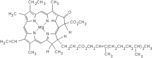


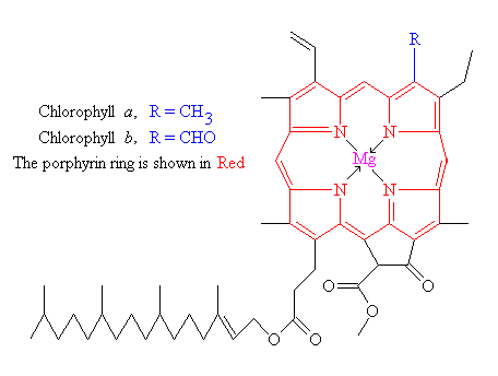
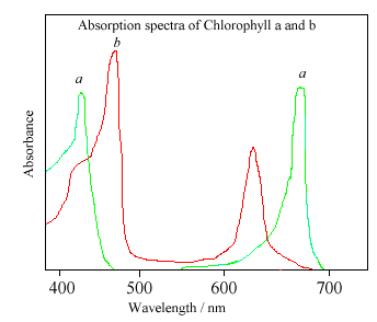
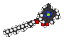
.PNG/220px-Spectra_Chlorophyll_ab_oenin_(1).PNG)
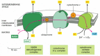