Feeds:
Posts
Comments

Archive for the ‘Biological Networks’ Category

Calcium Channel Blocker Potential for Angina

Larry H. Bernstein, MD, FCAP, Curator

LPBI


Pranidipine    

ANTHONY MELVIN CRASTO, PhD

str1

https://newdrugapprovals.files.wordpress.com/2015/12/str116.jpg


File:Pranidipine structure.svg

Pranidipine , OPC-13340, FRC 8411

Acalas®

NDA Filing in Japan

A calcium channel blocker potentially for the treatment of angina pectoris and hypertension.


CAS No. 99522-79-9

  • Molecular FormulaC25H24N2O6
  • Average mass 448.468


see dipine series………..http://organicsynthesisinternational.blogspot.in/p/dipine-series.html

manidipine


PAPER

Der Pharmacia Sinica, 2014, 5(1):11-17

https://newdrugapprovals.files.wordpress.com/2015/12/str113.jpg

pelagiaresearchlibrary.com/der-pharmacia-sinica/vol5-iss1/DPS-2014-5-1-11-17.pdf


Names
IUPAC name

methyl (2E)-phenylprop-2-en-1-yl 2,6-dimethyl-4-(3-nitrophenyl)-1,4-dihydropyridine-3,5-dicarboxylate
Other names

2,6-dimethyl-4-(3-nitrophenyl)-1,4-dihydropyridine-3,5-dicarboxylic acid O5-methyl O3-[(E)-3-phenylprop-2-enyl] ester
Identifiers
99522-79-9 Yes
ChEMBL ChEMBL1096842 
ChemSpider 4940726 
Jmol interactive 3D Image
MeSH C048161
PubChem 6436048
UNII 9DES9QVH58 Yes


PATENT SUBMITTED GRANTED
Process for the preparation of 1,4 – dihydropyridines and novel 1,4-dihydropyridines useful as therapeutic agents [US2003230478] 2003-12-18
Advanced Formulations and Therapies for Treating Hard-to-Heal Wounds [US2014357645] 2014-08-19 2014-12-04
METHODS OF TREATING CARDIOVASCULAR AND METABOLIC DISEASES [US2014322199] 2012-08-06 2014-10-30
Protein Carrier-Linked Prodrugs [US2014323402] 2012-08-10 2014-10-30
sGC STIMULATORS [US2014323448] 2014-04-29 2014-10-30
TREATMENT OF ARTERIAL WALL BY COMBINATION OF RAAS INHIBITOR AND HMG-CoA REDUCTASE INHIBITOR [US2014323536] 2012-12-07 2014-10-30
Agonists of Guanylate Cyclase Useful For the Treatment of Gastrointestinal Disorders, Inflammation, Cancer and Other Disorders [US2014329738] 2014-03-28 2014-11-06
METHODS, COMPOSITIONS, AND KITS FOR THE TREATMENT OF CANCER [US2014335050] 2012-05-25 2014-11-13
ROR GAMMA MODULATORS [US2014343023] 2012-09-18 2014-11-20
High-Loading Water-Soluable Carrier-Linked Prodrugs [US2014296257] 2012-08-10 2014-10-02 


Synthesis, isolation and use of a common key intermediate for calcium antagonist inhibitors

Neelakandan K.a,b, Manikandan H.b , B. Prabhakarana*, Santosha N.a , Ashok Chaudharia *, Mukund Kulkarnic , Gopalakrishnan Mannathusamyb and Shyam Titirmarea
a API Research Centre, Emcure Pharmaceutical Limited, Hinjawadi, Pune, India bDepartment of Chemistry, Annamalai University, Chidhambaram, India cDepartment of Chemistry, Pune University, Pune, India _________________________________________________________________________________

Pelagia Research Library     www.pelagiaresearchlibrary.com      Der Pharmacia Sinica, 2014, 5(1):11-17


The compound (3) synthesized from Nitrobenzaldehyde, tertiary butyl acetoacetate and piperidine can be used as a common intermediate for the production of calcium channel blockers like Nicardipine hydrochloride (1) and Pranidipine hydrochloride (2) with high purity.


The last twenty years have witnessed discoveries of calcium antagonists associated with multicoated pharmacodynamics potential which include not only antihypertensive and antiarrhythmic effects of the drugs but also action against excessive calcium entry in the cell of cardiovascular system and subsequent cell damage. Among many classes of calcium channel blockers, 1,4-dihydropyrimidine based drug molecules represented by Felodipine, Clevidipine, Benidipine, Nicardipine and Pranidipine are by far the best to reduce systemic vascular resistance and arterial pressure.

The reported synthetic approaches however proceed with complicated work ups, laborious purification procedures, highly expensive chemicals and low overall yields. (Scheme-I).

Synthetic scheme of Nicardipine and Pranidipine In view of the draw backs associated with previous synthetic approaches there is a strong need for environmentfriendly high yielding process applicable to the multi-kilogram production of calcium antagonist inhibitors. Herein, we report a scalable synthesis for Nicardipine hydrochloride (1) and Pranidipine hydrochloride (2) in fairly high overall yield using key intermediate 3-nitro benzylidene acid (3).Compound (3) was synthesized in two steps using 3-nitrobenzaldehyde, tertiary butyl acetoacetate and piperidine as a base to furnish tertiary butyl ester derivative (10). This was followed by hydrolysis of (10) in TFA and DCM to furnish compound (3) which would serve as a precursor for synthesis of versatile calcium antagonist inhibitors (Scheme-II).

Reported routes for synthesis of Benidipine,1,2 Lercanadipine,3-6 Nimodipine,7-11 Barnidipine12-14 and Manidipine15-16 were explored in our laboratory which involve reaction of nitro benzaldehyde with tertiary butyl acetoacetate using piperidine as a base to get tertiary butyl ester derivative (10). This is further treated with respective reagents to get various calcium channel blockers as shown in scheme 4. Since reported procedures involve in-situ generation of intermediate (3) and its reaction with corresponding fragments, it results in the formation of by-products which ultimately decrease the yield and increase the cost of API.

A novel process of manufacturing benzylidine acid derivative (3) was developed. Use of this intermediate was demonstrated by synthesis of Nicardipine and Pranidipine. This protocol may be employed for synthesis of other calcium channel blockers. In conclusion, a highly efficient, reproducible and scalable process for the synthesis of calcium channel blockers has been developed using (3) as the key intermediate.


[1] US 63 365 (Kyowa Hakko; appl.15.4.1982; J-prior.17.4.1981). [2] US 4 448 964 (Kyowa Hakko;15.5.1984; J-prior.17.4.1981). [3] Leonardi, A. et al.: Eur. J. Med.Chem. (EJMCA5) 33,399 (1988). [4] EP 153 016 (Recordati Chem. and Pharm.; appl. 21.1.1985; GB-prior. 14.2.1984). [5] US 4 705 797 (Recordati;10.11.1987; GB-prior. 14.2.1984). [6] WO 9 635 668 (Recordati Chem. and Pharm.; appl. 9.5.1996; I-prior. 12.5.1995). [7] DOS 2 117 571 (Bayer; appl. 10.4.1971). [8] DE 2 117 573 (Bayer; prior.10.4.1971) [9] US 3 799 934 (Bayer;26.3.1974;D-prior.10.4.1971). [10] US 3 932 645 (Bayer;13.1.1976;D-prior.10.4.1971). [11] Meyer, H. et al.: Arzneim.-Forsch. (ARZNAD) 31, 407 (1981); 33, 106 (1983). [12] DE 2 904 552 (Yamanouchi Pharm.; appl. 7.2.1979; J-prior.14.2.1978). [13] US 4 220 649 (Yamanouchi;2.9.1980; J-prior.14.2.1978). [14] CN 85 107 590( Faming Zhuanli Sheqing Gonhali S.; appl. 11.10.1985; J-prior.24.1.1985). [15] EP 94 159 (Takeda; appl. 15.4.1983; J-prior. 10.5.1982). [16] US 4 892 875 (Takeda;9.1.1990; J-prior. 10.5.1982, 11.1.1983).


Read Full Post »

Molecular On/Off Switches in Bacterial Design

Larry H. Bernstein, MD, FCAP, Curator

LPBI


Controlling Synthetic Bacteria

“Kill switches” ensure that genetically engineered bacteria survive only in certain environmental conditions.

By Kate Yandell | Dec 7, 2015   http://www.the-scientist.com//?articles.view/articleNo/44715/title/Controlling-Synthetic-Bacteria/

http://www.the-scientist.com/images/News/December2015/620ecoli.jpg

FLICKR, NIAID

Two synthetic gene circuits allow researchers to keep genetically engineered (GE) microbes alive only under specific conditions, and to kill them when their services are no longer needed. The circuits, described today (December 7) in Nature Chemical Biology,could help pave the path to safe diagnostics, therapies, or environmental remediation strategies that rely on GE bacteria.

“This is yet another step forward towards better biosafety and biocontainment based on certain aspects of existing technology,” said Guy-Bart Stan, a synthetic biologist at Imperial College London who was not involved in the study.

Study coauthor James Collins, a synthetic biologist at MIT, began to design these gene circuits, or “kill switches,” after becoming interested in using GE microbes for diagnostic and therapeutic purposes. “We were motivated to begin working on the topic as synthetic biology has moved increasingly toward real-world applications,” Collins told The Scientist. Other groups are working to engineer microbes for bioremediation and industrial processes, among other things.

But with genetic modification comes the concern that scientists will create new and uncontrollable species that outcompete or share their genes with wild-type organisms, permanently altering the environment or endangering people’s health.

Earlier this year, two research teams led by Yale bioengineer Farren Isaacs and Harvard geneticist George Church showed that they could genetically modify Escherichia coli to incorporate synthetic amino acids into essential proteins. When the bacteria are not fed the amino acids, they cannot produce these essential proteins, and so they die. This strategy yields bacteria that are very unlikely to survive without support from scientists but requires intensive engineering of the bacterial genome. (See “GMO ‘Kill Switches,’” The Scientist, January 2015.)

In contrast, Collins and his colleagues set out to create kill switches that could work in a more diverse range of microbes. “Our circuit-based safeguards can be conveniently transferred to different bacterial strains without modifying the target cell’s genome,” he wrote in an email.

First, Collins and his colleagues generated a kill switch called “Deadman,” named for a locomotive braking system in which the train will only run if the engineer is affirmatively holding down a pedal. In the microbial version of Deadman, a researcher must feed bacteria a substance called anhydrotetracycline at all times, or else the microbes will express a toxin and self-destruct.

The researchers generated a genetic circuit containing genes for the proteins LacI and TetR, a toxin that is only expressed in the absence of LacI, and a protease that degrades LacI. Under normal circumstances, TetR is preferentially expressed over LacI. TetR expression also triggers expression of the protease, which degrades any LacI that has been expressed. Without LacI, cells express the toxin and die. But when the cells are fed anhydrotetracycline, TetR is inhibited and LacI is expressed. LacI represses the toxin and keeps the cells alive.

Other versions of the Deadman circuit can be designed to degrade essential proteins in the absence of anhydrotetracycline, said Collins.

A second kill switch, “Passcode,” similarly requires that researchers maintain a specific environment for cells lest they express a toxin. Passcode requires a combination of input molecules for cells to survive. The system relies on hybrid transcription factors, each with one component that recognizes a specific DNA sequence, and one component that is sensitive to specific small molecules, such as galactose or cellobiose. One hybrid transcription factor, factor C, turns off expression of a toxin. Two other hybrid transcription factors, factors A and B, suppress expression of factor C. But specific small molecules can keep them from interacting with C. Another small molecule could prevent C from repressing the toxin. Therefore, to keep the cells alive, researchers must provide them with two small molecules that keep factors A and B in check, and make sure not to give them a third small molecule that will interfere with C.

Scientists designing Passcode kill-switches could make hybrid transcription factors respond to whatever combination of small molecules they desired, said Collins. “The strength of our kill switches lies in their flexibility and their ability to detect complex environmental signals for biocontainment.” He noted that companies hoping to keep others from using their cells could keep the recipe for their feed a secret.

“The great advantage is that you can effectively scale this and create different combinations of environments that contain different cocktails of these small molecules, thereby allowing you to effectively create a suite of cells that are going to be viable in different environments,” said Isaacs.

But Church warned that Collins’s circuit-based approach might not as effectively contain bacteria as an amino acid-based method, like one his group developed, since the cells are not fundamentally dependent on foreign biology to survive.

“If you need to have the ability to really scale your containment across a number of different species, then I could see the Passcode kill switches would be incredibly valuable,” said Isaacs. “If you are very concerned about escape frequencies and your degree of biocontainment, maybe you’d opt for something where the organism has been recoded and it relies on a synthetic amino acid.”

Still, Stan said the new paper is a demonstration that creating easy-to-insert kill switches based on genetic circuits is feasible. “I think what they wanted to show in the paper is basically that using some existing genetic circuitry . . .  you can obtain biosafety for the here and now.”


C.T.Y. Chan et al., “‘Deadman’ and ‘Passcode’ microbial kill switches for bacterial containment,” Nature Chemical Biology, doi:10.1038/nchembio.1979, 2015.  

Tags   synthetic biology, microbes, genetic engineering and biosafety


GMO “Kill Switches”

Scientists design bacteria reliant upon synthetic amino acids to contain genetically modified organisms.

By Kerry Grens | Jan 21, 2015   http://www.the-scientist.com/?articles.view/articleNo/41954/title/GMO–Kill-Switches-/

One of the biggest concerns about genetically modified organisms (GMOs) is that they can infiltrate wild populations and spread their altered genes among naturally occurring species. In Nature today (January 21), two groups present a new method of containing GMOs: by making some of their essential proteins reliant upon synthetic amino acids not found outside of the laboratory.

“What really makes this a valuable step change is that kill switches beforehand were very susceptible to mutation or other conditions, such as metabolic cross feeding, from basically inactivating them,” said Tom Ellis, a synthetic biologist at Imperial College London who was not involved in the studies. The new approach circumvents some of those problems by making it extremely unlikely for the genetically modified bacteria to be able to survive outside of the conditions dictated by their custom-designed genomes.

Both research teams—one led by George Church at Harvard Medical School and the other by Farren Isaacsat Yale University—based their work on so-called genetically recoded organisms (GROs), bacterial genomes that have had all instances of a particular codon replaced by another. Church and Isaacs, along with their colleagues, had previously developed this concept in collaboration. Since then, their respective groups designed the replacement codons to incorporate a synthetic amino acid, and engineered proteins essential to the organism to rely upon the artificial amino acid for proper function.

“Here, for the first time, we’re showing that we’re able to engineer a dependency on synthetic biochemical building blocks for these proteins,” Isaacs told reporters during a conference call.

Both teams found that the cells perished in environments lacking the synthetic amino acid. Although the technology is not ready for industrial-scale deployment, the scientists suggested that such an approach could be applied as a safeguard against the escape of GMOs.

…..


‘Deadman’ and ‘Passcode’ microbial kill switches for bacterial containment

Clement T Y ChanJeong Wook LeeD Ewen CameronCaleb J Bashor & James J Collins

Nature Chemical Biology(2015)            http://dx.doi.org:/10.1038/nchembio.1979

Figure 2: The fail-safe mechanism for Deadman circuit activation.

The fail-safe mechanism for Deadman circuit activation.

http://www.nature.com/nchembio/journal/vaop/ncurrent/carousel/nchembio.1979-F2.jpg

To demonstrate active control over host cell viability, cells grown under survival conditions (with ATc) were exposed to 1 mM IPTG to directly induce EcoRI and mf-Lon expression. Cell viability was measured by CFU count and is displayed…


Biocontainment systems that couple environmental sensing with circuit-based control of cell viability could be used to prevent escape of genetically modified microbes into the environment. Here we present two engineered safeguard systems known as the ‘Deadman’ and ‘Passcode’ kill switches. The Deadman kill switch uses unbalanced reciprocal transcriptional repression to couple a specific input signal with cell survival. The Passcode kill switch uses a similar two-layered transcription design and incorporates hybrid LacI-GalR family transcription factors to provide diverse and complex environmental inputs to control circuit function. These synthetic gene circuits efficiently killEscherichia coli and can be readily reprogrammed to change their environmental inputs, regulatory architecture and killing mechanism.


Nontoxic antimicrobials that evade drug resistance

Stephen A DavisBenjamin M VincentMatthew M EndoLuke WhitesellKaren MarchilloDavid R AndesSusan Lindquist & Martin D Burke

Nature Chemical Biology 2015;11:481–487          http://dx.doi.org:/10.1038/nchembio.1821

Drugs that act more promiscuously provide fewer routes for the emergence of resistant mutants. This benefit, however, often comes at the cost of serious off-target and dose-limiting toxicities. The classic example is the antifungal amphotericin B (AmB), which has evaded resistance for more than half a century. We report markedly less toxic amphotericins that nevertheless evade resistance. They are scalably accessed in just three steps from the natural product, and they bind their target (the fungal sterol ergosterol) with far greater selectivity than AmB. Hence, they are less toxic and far more effective in a mouse model of systemic candidiasis. To our surprise, exhaustive efforts to select for mutants resistant to these more selective compounds revealed that they are just as impervious to resistance as AmB. Thus, highly selective cytocidal action and the evasion of resistance are not mutually exclusive, suggesting practical routes to the discovery of less toxic, resistance-evasive therapies.


Read Full Post »

Essential for Rehabilitation

Larry H. Bernstein, MD, FCAP, Curator

LPBI


Cellular Rehab

Physical therapy and exercise are critical to the success of cell therapies approaching the clinic.

By Elie Dolgin |  Scientist  Magazine Dec 1, 2015     http://www.the-scientist.com/?articles.view/articleNo/44605/title/Cellular-Rehab/

http://www.the-scientist.com/Dec2015/feature1.jpg

Ron Strang lay on his back and bent his left leg. “I could feel the difference right away,” recalls the 31-year-old ex-Marine.

The day before, Strang had undergone an experimental surgery to help repair a deep gouge in his quadriceps. He’d been injured in April 2010 while on foot patrol in Afghanistan’s Helmand Province, when a crude roadside bomb sent shrapnel tearing through his upper thigh. Ten soldiers were wounded in the blast, Strang the most grievously. A year later, even after numerous surgeries and skin grafts, he still couldn’t walk without his knee buckling. So he signed up to receive an experimental regenerative therapy.

In July 2011, Stephen Badylak, a tissue-engineering specialist at the University of Pittsburgh, transplanted a thin sheet of extracellular matrix (ECM) derived from pig bladders into Strang’s leg. The fibrous material was intended not only to provide structural support for the muscle, but also, by releasing signaling proteins, to recruit and coax stem cells in the body to differentiate into new tissue.

Physical forces

Researchers have long recognized the influence of physical forces on molecular and cellular function. Nearly 40 years ago, Judah Folkman, a cancer biologist at Harvard Medical School, and his undergraduate assistant Anne Moscona, now an infectious-disease researcher at Weill Cornell Medicine in New York City, grew cells in petri dishes and found that as cells stretched out and flattened more and more on the plate, their rate of DNA synthesis and cell division increased.4 This revelation led to an explosion of interest in how squeezes, tugs, pushes, and pulls mold the architecture of the cell and, in turn, influence molecular processes within, such as gene expression.

For the most part, however, the field of mechanobiology has been stuck in the laboratory, with few physicians thinking about how physical stresses at the cellular level might affect clinical outcomes, and even fewer physical therapists considering the molecular milieu. As Christopher Evans, director of the Rehabilitation Medicine Research Center at the Mayo Clinic in Rochester, Minnesota, puts it: “The people doing the stem cell work have been largely ignorant of rehabilitation, and the rehabilitation medicine community hasn’t been thinking in terms of cell and molecular biology.”

With stem cell therapies and tissue engineering nearing medical prime time, that’s starting to change. A growing number of scientists, clinicians, and physical therapists are now taking an interdisciplinary approach to rehabilitation, pairing exercise with technologies that regenerate bone, muscle, cartilage, ligaments, nerves, and other tissues. They call it regenerative rehabilitation.

“This is a new future,” says Carmen Perez-Terzic, a cardiovascular disease researcher at the Mayo Clinic. “This is an area that’s going to explode in the next 5 or 10 years.”

Fusion approach

The first public call for stem cell biologists and physical therapists to integrate regenerative medicine and rehabilitation science came in a 2010 editorial by Fabrisia Ambrosio, director of the University of Pittsburgh’s Cellular Rehabilitation Laboratory, and Alan Russell, then director of Pitt’s McGowan Institute for Regenerative Medicine.5“Regenerative rehabilitation is difficult but inevitable,” Ambrosio and Russell wrote, “and now is the time to prepare specific, science-based protocols.”

Ambrosio trained as a physical therapist before earning her PhD with rehabilitation medicine specialist Michael Boninger at Pitt, where she studied how wheelchair design affects strength in people with spinal cord injuries and degenerative conditions such as multiple sclerosis. When Ambrosio started her own research group at Pitt in 2005, she began to investigate how mechanical and electrical stimulation might promote healing following stem cell transplantation.


http://www.the-scientist.com/Dec2015/feature1_2.jpg

NEW NEURONS: In the dentate gyrus of the hippocampus, mice injected with stretched muscle stem cells show an approximately threefold greater increase in immature neurons (black, left) than mice injected with untreated stem cells (right), likely as a result of growth and neurotrophic factors released into the bloodstream by the stretched cells. JENNIFER MERRITT


She transplanted muscle-derived stem cells into bruised hind limbs of mice, then ran the animals on treadmills every weekday for five weeks. The active mice developed more new muscle cells than sedentary controls.6 Ambrosio’s team later demonstrated that applying low-level electrical pulses to muscles injected with stem cells improved strength and reduced fatigue in mice that experienced progressive muscle degeneration characteristic of Duchenne muscular dystrophy.7 “Using very noninvasive, clinically relevant protocols, we can actually dictate the behavior of stem cells,” she says. And that got her thinking: “All of this should lay the groundwork for how we see regenerative medicine therapies being applied in the clinic.”

Starting in 2011, Ambrosio and Boninger launched an annual Symposium on Regenerative Rehabilitation; they held the fourth conference in September at the Mayo Clinic in Minnesota. Last year, the duo also started the International Consortium for Regenerative Rehabilitation, a coalition of eight participating institutions from the U.S., Japan, and Italy that is now developing a strategic agenda for the field. And a few months ago, they secured funding to create the Alliance for Regenerative Rehabilitation Research & Training, which includes four US universities and hospitals (Pitt, Stanford University, Mayo, and the University of California, San Francisco) and will support webinars, minisabbaticals, seed grants, and more.

“This is about getting more people doing this work, understanding this work, and translating this field,” says Boninger, who is leading the alliance together with Stanford stem-cell biologist Thomas Rando. Just adding exercise to a stem cell therapy is “easy,” Boninger notes. “Doing the basic science to evaluate that is a little more challenging.”

The science may still be in its infancy, but Ambrosio says her efforts in community building are beginning to pay off. “I can see such a difference in the way people receive some of these ideas of regenerative rehab,” she says. “It was really kind of novel as recently as 2010, whereas now it’s actually part of our vernacular.”

(Re)Generating interest

Rehabilitation regimens are now being integrated into the preclinical development of regenerative treatments for heart disease, bone fractures, and even brain injuries. In Japan, for example, researchers at Hiroshima University have shown that running directs neural stem cells to properly differentiate when transplanted into mice with experimentally induced brain damage.8 “Combining cell therapy and rehabilitation is needed to correct the neural network and achieve a functional recovery,” says study author Takeshi Imura, who presented the research at Japan’s first-ever Workshop on Regenerative Rehabilitation in Kyoto last March. And earlier this year, muscle biologist Marni Boppart and her colleagues at the University of Illinois at Urbana-Champaign reported that stem cells only enhance muscle repair and growth in mice when coupled with weight-training exercise.9

In addition to exercising recipients of cell therapies, scientists are also looking to give the cells themselves a workout, by stretching stem cells in a dish ahead of transplantation. “In effect, we’re exercising the stem cells without exercising the animal,” says Boppart. In unpublished work, Boppart’s team found that old mice injected with muscle stem cells taken from young mice and stretched before injection exhibited improved blood flow, stronger muscles, and more new neurons in the brain’s hippocampus, thanks to the release of growth, neurotrophic, and immunomodulatory factors brought on by the mechanical stimulus. Stem cells not given the laboratory workout provided no such benefits.

At the Mayo Clinic, Perez-Terzic is also applying physical pressure in vitro to improve the differentiation of stem cells. Her goal is to develop new regenerative treatments for heart disease, and she is hoping to find more-efficient ways of coaxing embryonic stem cells to become heart muscle cells for transplantation. The results are preliminary, Perez-Terzic says, but so far it looks like “if you put some pressure into the system, the differentiation is much better.”

Boppart is hopeful that translating such therapies to the clinic will help patients who are unable to exercise, such as some elderly individuals or those with extreme muscle weakness. “This type of alternative stem cell therapy may provide the boost in strength necessary for someone to transition from disability to regain of function,” she says.

Richard Shields, an applied physiologist at the University of Iowa’s Carver College of Medicine, has another solution, one that doesn’t require any sort of cellular calisthenics in the laboratory. He has invented a device that can deliver different kinds of mechanical loads directly to the lower leg, even for patients confined to a wheelchair. A compression system covers the knee, while the foot rests on a vibrating platform. A doctor or physical therapist can then deliver therapeutic loads in a safe and quantifiable manner. (See illustration below.)

After testing the device on eight people with complete paralysis,10 Shields and his colleagues wondered whether delivering a controlled dose of vibration would improve bone architecture in spinal cord injury patients, many of whom eventually develop severe osteoporosis. After 12 months of regular vibration therapy, however, bone health continued to decline in all six study participants.11 “This means that people with long-term paralysis are very resistant to change [in bone density] or that the dose was not high enough,” says Shields, who is now working to refine the training regimen for better results.

Once he and his colleagues work out the kinks, Shields says he hopes that the setup will be useful to more patients than just those who are incapable of exercise. The limb-loading system offers greater control of the degree and target of stimulation than that afforded by running or weight lifting, he says—precision that could have utility for all manner of regenerative cellular treatments. “How you dose these mechanical loads is not just all or none,” he says. “The stresses have to be applied in opportune doses.”

Stretch goals

http://www.the-scientist.com/Dec2015/NovDolginThumb_310px.jpg

CELLULAR WORKOUT: Regenerative rehabilitation promises to enhance the potential of cell- and gene-based techniques by incorporating principles of physical therapy.
See full infographic here: WEB | PDF
© KIMBERLY BATTISTA


Martin Childers, who is collaborating with San Francisco–based Audentes Therapeutics, says he hopes it could also improve outcomes following a gene therapy in children with a rare and fatal muscle weakness disorder called X-linked myotubular myopathy.

The disorder is caused by mutations in the MTM1gene that encodes an enzyme needed for the development and maintenance of muscle cells. Children with the condition suffer from extreme muscle weakness, generally lacking the strength needed to move air in and out of their lungs without mechanical assistance. Audentes’s therapy will deliver a good copy of the MTM1 gene into the blood and hopefully help affected individuals respire without assistance. But after treatment, “you can’t just turn the ventilator off,” Childers says. “There’s going to have to be some rehabilitation therapies.”

Specifically, Childers plans to couple the gene therapy with breathing training. In addition to helping patients wean themselves off the ventilator, pulmonary exercise might enhance the expression of the introduced gene, he says. For now, this is only a hunch. But Audentes is preparing for the launch of a Phase 1 trial next year, and Childers is studying human tissue samples in the lab to answer this question.

Other gene therapy trials already incorporate physical therapy into their recovery protocols. At Nationwide Children’s Hospital in Columbus, Ohio, for example, Jerry Mendell and his colleagues are testing a gene-correction treatment on six- to nine-month-old babies with a severe form of spinal muscular atrophy, a genetic disease that involves the degeneration of motor neurons. The infants are born with tight joints; their legs are often fixed in a splayed, frog-like position. Mendell’s team strongly encourages parents to massage their children’s stiffened limbs on a daily basis after the gene transfer—a necessity, Mendell says, for the gene-corrected motor neurons to interface properly with the weakened muscles. Without it, “you’re not going to be able to improve function,” he says.

Mendell is also incorporating bicycle training into a small gene-therapy trial for children with Duchenne muscular dystrophy. Although mutations in the gene that codes for a muscle-associated protein called dystrophin are responsible for this disorder, Mendell is not delivering a working copy of that particular gene back into patients. Instead, he is using a gene therapy product he developed in collaboration with Cleveland-based Milo Biotechnology that includes the gene for follistatin, a protein that helps release the brakes on muscle growth and could thus prove beneficial for a variety of muscle diseases.

In an earlier trial that tested the same gene therapy on six adults with Becker muscular dystrophy, a milder condition also caused by mutations in the gene for dystrophin, Mendell noticed that the participant who had the most active lifestyle—on account of his job at a garden center—exhibited the most dramatic improvement in how far he could walk.12 “He was one of the highest responders to the follistatin gene therapy,” Mendell says. Hoping to re-create the success, Mendell is now having the kids in his ongoing Duchenne muscular dystrophy trial ride stationary bikes for 15-minute sessions three times per week.

One umbrella

http://www.the-scientist.com/Dec2015/feature1_3.jpg

WHEELCHAIR CALISTHENICS: The University of Iowa’s Richard Shields developed a system to assess the effects of mechanical loading, vibration, and electrical stimulation on bone, muscle, nerve, and cell signaling on the lower leg. The goal is to apply optimal combinations of the three forces to enhance regenerative medicine treatments. COURTESY OF RICHARD SHIELDS

“In the end,” says Boninger, regenerative rehabilitation “is about improving function in patients. So, being able to look someone in the eye and say, ‘This is why we’re doing this exercise program, and this will add to your recovery and function when you’re done’ is where I really want to get to.”

To help make that happen, several academic institutions have been shuffling departmental structures to bring stem cell scientists and rehabilitation doctors under the same administrative umbrella. These include the University of Pittsburgh and the Mayo Clinic, both leaders in the nascent hybrid discipline, though the first place to do so was Columbia University Medical Center in New York City, where the hospital renamed its rehab division the Department of Rehabilitation and Regenerative Medicine in 2010.

Five years on, however, efforts to bridge the two disciplines “remain to some degree aspirational,” admits Joel Stein, a rehabilitation specialist who chairs the Columbia department. Regenerative rehabilitation “is becoming more popular within the field as a vision for the future,” he says. “But has it translated into good, hard science that’s led to definitive new therapies? No, not yet, and it might take a while.”

“We need to be dedicating more efforts to thinking about dosing, intensity, and protocols,” Ambrosio agrees. “That means we have a lot of work ahead for us.”

In the meantime, proponents of regenerative rehabilitation continue to look to success stories like Strang’s for inspiration. At one point, Strang was unsure that he’d ever be able to walk normally again. Now a police officer at the Veterans Affairs hospital in Pittsburgh, Strang is on his feet constantly, moving about easily and pain-free. Just last month, in fact, he married his longtime girlfriend at a church outside Pittsburgh—and he walked down the aisle with no problems.

Elie Dolgin is a news editor at STAT in Boston.

References

  1. V.J. Mase, Jr., et al., “Clinical application of an acellular biologic scaffold for surgical repair of a large, traumatic quadriceps femoris muscle defect,” Orthopedics, 33:511, doi:10.3928/01477447-20100526-24, 2010.
  2. N.E. Gentile et al., “Targeted rehabilitation after extracellular matrix scaffold transplantation for the treatment of volumetric muscle loss,” Am J Phys Med Rehabil, 93:S79-S87, 2014.
  3. B.M. Sicari et al., “An acellular biologic scaffold promotes skeletal muscle formation in mice and humans with volumetric muscle loss,” Sci Transl Med, 6:234ra58, 2014.
  4. J. Folkman, A. Moscona, “Role of cell shape in growth control,” Nature, 273:345-49, 1978.
  5. F. Ambrosio, A. Russell, “Regenerative rehabilitation: a call to action,” J Rehabil Res Dev, 47:xi-xv, 2010.
  6. F. Ambrosio et al., “The synergistic effect of treadmill running on stem-cell transplantation to heal injured skeletal muscle,” Tissue Eng Part A, 16:839-49, 2010.
  7. G. Distefano et al., “Neuromuscular electrical stimulation as a method to maximize the beneficial effects of muscle stem cells transplanted into dystrophic skeletal muscle,” PLOS ONE, 8:e54922, 2013.
  8. T. Imura et al., “Interactive effects of cell therapy and rehabilitation realize the full potential of neurogenesis in brain injury model,” Neurosci Lett, 555:73-78, 2013.
  9. K. Zou et al., “Mesenchymal stem cells augment the adaptive response to eccentric exercise,” Med Sci Sports Exerc, 47:315-25, 2015.
  10. C.L. McHenry et al., “Potential regenerative rehabilitation technology: Implications of mechanical stimuli to tissue health,” BMC Res Notes, 7:334, 2014.
  11. S. Dudley-Javoroski et al., “Bone architecture adaptations after spinal cord injury: impact of long-term vibration of a constrained lower limb,” Osteoporos Int, doi:10.1007/s00198-015-3326-4, 2015.
  12. J.R. Mendell et al., “A phase 1/2a follistatin gene therapy trial for Becker muscular dystrophy,” Mol Ther, 23:192-201, 2015.

Read Full Post »

Muscular dystrophy has deficient stem cell dystrophin

Larry H. Bernstein, MD, FCAP, Curator

LPBI

Article ID #198: Muscular dystrophy has deficient stem cell dystrophin. Published on 11/21/2015

WordCloud Image Produced by Adam Tubman

Dystrophin Deficient Stem Cell Pathology

Muscular Dystrophy is a Stem Cell-Based Disease

Because DMD results from mutations in the dystrophin gene, the vast majority of muscular dystrophy research was based on a simple model in which the Dystrophin protein played a structural role in the structural integrity of muscle fibers. Abnormal versions of the Dystrophin protein caused the muscle fibers to become damaged and die as a result of contraction.  Dystrophin anchors the cytoskeleton of the muscle fibers, which are essential for muscle contraction, to the muscle cell membrane, and then to the extracellular matrix outside the cell that serves as a foundation upon which the muscle cells are built.

gb-2001-2-4-reviews3006-3

However in this current study, Rudnicki and his team discovered that muscle stem cells also express the dystrophin protein. This is a revelation because Dystrophin was thought to be protein that ONLY appeared in mature muscle. However, in this study, it became exceedingly clear that in the absence of Dystrophin, muscle stem cells generated ten-fold fewer muscle precursor cells, and, consequently, far fewer functional muscle fibers. Dystrophin is also a component of a signal transduction pathway that allows muscle stem cells to properly ascertain if they need to replace dead or dying muscle.  Muscle stem cells repair the muscle in response to injury or exercise by dividing to generate precursor cells that differentiate into muscle fibers.

Even though Rudnicki used mice as a model system in these experiments, the Dystrophin protein is highly conserved in most vertebrate animals. Therefore, it is highly likely that these results will also apply to human muscle stem cells.

Gene therapy experiments and trials are in progress and even show some promise, but Rudnicki’s work tells us that gene therapy approaches must target muscle stem cells as well as muscle fibers if they are to work properly.

“We’re already looking at approaches to correct this problem in muscle stem cells,” said Dr. Rudnicki.

This paper has received high praise from the likes of Ronald Worton, who was one of the co-discovers of the dystrophin gene with Louis Kunkel in 1987.

Early pathogenesis of Duchenne muscular dystrophy modelled in patient-derived human induced pluripotent stem cells

Emi Shoji, Hidetoshi Sakurai, Tokiko Nishino, Tatsutoshi Nakahata, Toshio Heike, Tomonari Awaya, Nobuharu Fujii, Yasuko Manabe, Masafumi Matsuo & Atsuko Sehara-Fujisawa

Scientific Reports 5, Article number: 12831 (2015)   http://dx.doi.org:/10.1038/srep12831

Duchenne muscular dystrophy (DMD) is a progressive and fatal muscle degenerating disease caused by a dystrophin deficiency. Effective suppression of the primary pathology observed in DMD is critical for treatment. Patient-derived human induced pluripotent stem cells (hiPSCs) are a promising tool for drug discovery. Here, we report an in vitro evaluation system for a DMD therapy using hiPSCs that recapitulate the primary pathology and can be used for DMD drug screening. Skeletal myotubes generated from hiPSCs are intact, which allows them to be used to model the initial pathology of DMD in vitro. Induced control and DMD myotubes were morphologically and physiologically comparable. However, electric stimulation of these myotubes for in vitro contraction caused pronounced calcium ion (Ca2+) influx only in DMD myocytes. Restoration of dystrophin by the exon-skipping technique suppressed this Ca2+ overflow and reduced the secretion of creatine kinase (CK) in DMD myotubes. These results suggest that the early pathogenesis of DMD can be effectively modelled in skeletal myotubes induced from patient-derived iPSCs, thereby enabling the development and evaluation of novel drugs.

Duchenne muscular dystrophy (DMD) is characterised by progressive muscle atrophy and weakness that eventually leads to ambulatory and respiratory deficiency from early childhood1. It is an X-linked recessive inherited disease with a relatively high frequency of 1 in 3500 males1,2.DMD, which is responsible for DMD, encodes 79 exons and produces dystrophin, which is one of the largest known cytoskeletal structural proteins3. Most DMD patients have various types of deletions or mutations in DMD that create premature terminations, resulting in a loss of protein expression4. Several promising approaches could be used to treat this devastating disease, such as mutation-specific drug exon-skipping5,6, cell therapy7, and gene therapy1,2.

Myoblasts from patients are the most common cell sources for assessing the disease phenotypes of DMD11,12. …Previous reports have shown that muscle cell differentiation from DMD patient myoblasts is delayed and that these cells have poor proliferation capacity compared to those of healthy individuals11,12. Our study revealed that control and DMD myoblasts obtained by activating tetracycline-dependent MyoD transfected into iPS cells (iPStet-MyoD cells) have comparable growth and differentiation potential and can produce a large number of intact and homogeneous myotubes repeatedly.

The pathogenesis of DMD is initiated and progresses with muscle contraction. The degree of muscle cell damage at the early stage of DMD can be evaluated by measuring the leakage of creatine kinase (CK) into the extracellular space15. Excess calcium ion (Ca2+) influx into skeletal muscle cells, together with increased susceptibility to plasma membrane injury, is regarded as the initial trigger of muscle damage in DMD19,20,21,22,23,24. Targeting these early pathogenic events is considered essential for developing therapeutics for DMD.

In this study, we established a novel evaluation system to analyse the cellular basis of early DMD pathogenesis by comparing DMD myotubes with the same clone but with truncated dystrophin-expressing DMD myotubes, using the exon-skipping technique. We demonstrated through in vitro contraction that excessive Ca2+ influx is one of the earliest events to occur in intact dystrophin-deficient muscle leading to extracellular leakage of CK in DMD myotubes.

Generation of tetracycline-inducible MyoD-transfected DMD patient-derived iPSCs (iPStet-MyoD cells)

Figure 1: Generation and characterization of control and DMD patient-derived Tet-MyoD-transfected hiPS cells.   Full size image

Morphologically and physiologically comparable intact myotubes differentiated from control and DMD-derived hiPSCs

Figure 2: Morphologically and physiologically comparable skeletal muscle cells differentiated from Control-iPStet-MyoD and DMD-iPStet-MyoD.   Full size image

Exon-skipping with AO88 restored expression of Dystrophin in DMD myotubes differentiated from DMD-iPStet-MyoD cells


Figure 3: Restoration of dystrophin protein expression by AO88.   Full size image

 
Restored dystrophin expression attenuates Ca2+ overflow in DMD-Myocytes


Figure 4: Restored expression of dystrophin diminishes Ca2+ influx in DMD muscle in response to electric stimulation.   Full size image


Ca2+ influx provokes skeletal muscle cellular damage in DMD muscle


Figure 5: Ca2+ influx induces prominent skeletal muscle cellular damage in DMD-Myocytes.   Full size image


Skeletal muscle differentiation in myoblasts from DMD patients is generally delayed compared to that in healthy individuals11,36,37.  Our differentiation system successfully induced the formation of myotubes from DMD patients, and the myotubes displayed analogous morphology and maturity compared with control myotubes (Fig. 2a–c).  Comparing myotubes generated from patient-derived iPS cells with those derived from the same DMD clones but expressing dystrophin by application of the exon-skipping technique enabled us to demonstrate the primary cellular phenotypes in skeletal muscle solely resulting from the loss of the dystrophin protein (Fig. 4b).  Our results demonstrate that truncated but functional dystrophin protein expression improved the cellular phenotype of DMD myotubes.

In DMD, the lack of dystrophin induces an excess influx of Ca2+ , leading to pathological dystrophic changes22. We consistently observed excess Ca2+ influx in DMD-Myocytes compared to Control-Myocytes (Supplementary Figure S3a and S3b) in response to electric stimulation. TRP channels, which are mechanical stimuli-activated Ca2+ channels40that are expressed in skeletal muscle cells41, can account for this pathogenic Ca2+ influx…

In conclusion, our study revealed that the absence of dystrophin protein induces skeletal muscle damage by allowing excess Ca2+ influx in DMD myotubes. Our experimental system recapitulated the early phase of DMD pathology as demonstrated by visualisation and quantification of Ca2+ influx using intact myotubes differentiated from hiPS cells.  This evaluation system significantly expands prospective applications with regard to assessing the effectiveness of exon-skipping drugs and also enables the discovery of drugs that regulate the initial events in DMD.

Duchenne muscular dystrophy affects stem cells, University of Ottawa study finds  

New treatments could one day be available for the most common form of muscular dystrophy after a study suggests the debilitating genetic disease affects the stem cells that produce healthy muscle fibres.

The findings are based on research from the University of Ottawa and The Ottawa Hospital, published Monday in the journal Nature Medicine.

For nearly two decades, doctors had thought the muscular weakness that is the hallmark of the disease was due to problems with human muscle fibers, said Dr. Michael Rudnicki, the study’s senior author.

The new research shows the specific protein characterized by its absence in Duchenne muscular dystrophy normally exists in stem cells.

Dystrophin protein found in stem cells

“The prevailing notion was that the protein that’s missing in Duchenne muscular dystrophy — a protein called dystrophin — was not involved at all in the function of the stem cells.”

http://soundcloud.com/cbcottawa1

When the genetic mutations caused by Duchenne muscular dystrophy inhibit the production of dystrophin in stem cells, those stem cells produce significantly fewer precursor cells — and thus fewer properly functioning muscle fibres.  Further, stem cells need dystrophin to sense their environment to figure out if they need to divide to produce more stem cells or perform muscle repair work.

Genetic repair might treat Duchenne muscular dystrophy

July 25, 2011|By Thomas H. Maugh II, Los Angeles Times
 

A genetic technique that allows the body to work around a crucial mutation that causes Duchenne muscular dystrophy increased the mass and function of muscles in a small group of patients with the devastating disease, paving the way for larger clinical trials of the drug. The study in a handful of boys age 5 to 15 showed that patients receiving the highest level of the drug, called AVI-4658 or eteplirsen, had a significant increase in production of a missing protein and increases in muscle fibers. The study demonstrated that the drug is safe in the short term. Results were reported Sunday in the journal Lancet.

Duchenne muscular dystrophy affects about one in every 3,500 males worldwide. It is caused by any one of several different mutations that affect production of a protein called dystrophin, which is important for the production and maintenance of muscle fibers. Affected patients become unable to walk and must use a wheelchair by age 8 to 12. Deterioration continues through their teens and 20s, and the condition typically proves fatal as muscle failure impairs their ability to breathe.

This study is designed to assess the efficacy, safety, tolerability, and pharmacokinetics (PK) of AVI-4658 (eteplirsen) in both 50.0 mg/kg and 30.0 mg/kg doses administered over 24 weeks in subjects diagnosed with Duchenne muscular dystrophy (DMD).


Condition Intervention Phase
Duchenne Muscular Dystrophy Drug: AVI-4658 (Eteplirsen)
Other: Placebo
Phase 2
Study Type:Interventional
Study Design:Allocation: Randomized
Endpoint Classification: Safety/Efficacy Study
Intervention Model: Parallel Assignment
Masking: Double Blind (Subject, Caregiver, Investigator, Outcomes Assessor)
Primary Purpose: Treatment
Official Title:A Randomized, Double-Blind, Placebo-Controlled, Multiple Dose Efficacy, Safety, Tolerability and Pharmacokinetics Study of AVI-4658(Eteplirsen),in the Treatment of Ambulant Subjects With Duchenne Muscular Dystrophy
 
 
Resource links provided by NLM:
 
 
Dystrophin expression in muscle stem cells regulates their polarity and asymmetric division

Nature Medicine(2015)   http://dx.doi.org:/10.1038/nm.3990

Dystrophin is expressed in differentiated myofibers, in which it is required for sarcolemmal integrity, and loss-of-function mutations in the gene that encodes it result in Duchenne muscular dystrophy (DMD), a disease characterized by progressive and severe skeletal muscle degeneration. Here we found that dystrophin is also highly expressed in activated muscle stem cells (also known as satellite cells), in which it associates with the serine-threonine kinase Mark2 (also known as Par1b), an important regulator of cell polarity. In the absence of dystrophin, expression of Mark2 protein is downregulated, resulting in the inability to localize the cell polarity regulator Pard3 to the opposite side of the cell. Consequently, the number of asymmetric divisions is strikingly reduced in dystrophin-deficient satellite cells, which also display a loss of polarity, abnormal division patterns (including centrosome amplification), impaired mitotic spindle orientation and prolonged cell divisions. Altogether, these intrinsic defects strongly reduce the generation of myogenic progenitors that are needed for proper muscle regeneration. Therefore, we conclude that dystrophin has an essential role in the regulation of satellite cell polarity and asymmetric division. Our findings indicate that muscle wasting in DMD not only is caused by myofiber fragility, but also is exacerbated by impaired regeneration owing to intrinsic satellite cell dysfunction.

Read Full Post »

Robin Williams death

Larry H. Bernstein, MD, FCAP, Curator

LPBI


Lewy body: The ‘monster’ dementia blamed for Robin Williams’s death

Schneider says depression didn’t cause Williams’s death: “Lewy body dementia killed Robin. That’s what took his life.”

Strikingly, LBD – sometimes referred to as dementia with Lewy bodies or Parkinson’s with Lewy bodies, depending on symptoms – is the second-most common dementia after Alzheimer’s and affects more than 127,000 Britons. Yet most people have never heard of it.

Robin Williams who suffered from Lewy Body Dementia.

James Galvin, a neurology and psychiatry professor at Florida Atlantic University, says: “It’s the most common disease you’ve never heard of.”

“This disease is a sea monster with 50 tentacles of symptoms that show when they want,” Schneider said.

Williams suffered hallucinations, anxiety, depression, loss of motor control and problem-solving skills, sleep, balance and spatial awareness problems, and delusions.

Schneider describes one incident just weeks before Williams’s death, when she was in the shower and he was standing by the sink.

“Something didn’t seem right,” she recalls, so Schneider got out of the shower to find her husband’s head covered in blood. “He pointed to the door and I said, ‘Did you hit your head?’ and he nodded.” The incident confused her at the time. “But now, finding out all about Lewy body disease, lo and behold, their vision is affected, as is the ability to recognise and identify objects,” she says. “Now I get it.”


Lewy bodies are protein deposits in the brain, explains Professor David Burn, consultant neurologist and director of the biomedical research unit in LBD at the UK National Institute of Health Research (NIHR).

Discovered by Dr Frederic Lewy, a colleague of Dr Alzheimer’s, in 1912, the deposits develop inside nerve cells (neurons) in the brain, interrupting messaging and causing neurons to die. A patient’s symptoms will depend on which part of the brain is affected.

“When neurons die in the cortex, it causes dementia, but when it occurs in the brainstem, it causes motor symptoms (Parkinsonism),” says Burn.

“LBD patients face a rapid deterioration in their cognitive, physical and psychiatric function, and it tends to progress faster than other dementias,” he says.

When Paul Moynagh’s wife, Imogen, couldn’t find her way from a cafe table to the counter on a visit to a National Trust house in Devon in 2006, he thought little of it.Paul, 78, couldn’t have predicted the confusing set of symptoms that Imogen, 74, would experience. LBD is often misdiagnosed as Parkinson’s or Alzheimer’s, and it took doctors almost seven years to confirm her illness.

“Looking back, it began with little signs – loss of spatial awareness is an early symptom –  but they were so inconsistent,” recalls Paul, a retired surgeon.

First, there was a minor trembling in Imogen’s hands, then severe sleepiness during the day, along with spasms that made her right foot turn in when she walked. Then she developed depression and suffered panic attacks.

“Imogen has a pragmatic personality,” says Paul. “She used to play sports, was a keen gardener, walked everywhere and looked after our two children, Mark and Rachel. Ten years ago, if you had told her she would be afraid of being left alone, she would have laughed.”

By 2010, Imogen’s reasoning and planning skills were suffering – a key sign of LBD.

A keen bridge player, Imogen recalls: “I stopped winning, so I knew something was wrong.” (Though her speech is now slow, her sense of humour remains.)


Like Schneider, Paul Moynagh was also baffled by his wife’s repeated falls in the years preceding her diagnosis. “She’d had nine different broken bones, breaking her wrist twice, her ankle, and once, when she’d fallen down some stairs, her elbow.”

Imogen, like Williams and many LBD sufferers, was initially diagnosed with Parkinson’s disease.

 “She began shuffling when she walked and her voice became weak, both symptoms of Parkinson’s,” says Paul. Meanwhile, her depression was getting worse, not least because Imogen was so aware of what was happening to her.

“It’s different from Alzheimer’s in that people know exactly what’s happening, and one day can be completely lucid and the next be experiencing terrible anxiety and delusions. The more Imogen is aware of her situation, the more she gets depressed.”

When Paul Moynagh’s wife began experiencing hallucinations – a tell-tale sign of LBD – he knew that there was more to her illness than Parkinson’s. “She would see people in the windows of the conservatory and in our floor – which we made look like natural stone – she saw figures speaking to her.

“In my desperation, I would spend hours Googling Imogen’s symptoms until I stumbled on Lewy body dementia,” he says.

Brown says Robert had paranoia and hallucinations – he was frightened by faces he would see in the windows of a summer house he had built at the bottom of the garden. “One evening we were watching the Baftas on television and the camera panned, settling on various stars, and Bob turned to me and said: ‘I think Judi saw us.’ He meant Dame Judi Dench. He thought we were there and became very distressed because he wasn’t correctly dressed.”

“I’m a doctor and I had never even heard of it, and the neurologist was reluctant to accept it, but Imogen ticked all the boxes.” By 2013, a locum psychiatrist finally diagnosed Imogen with LBD. “I went along with my Google list, and she finally made the diagnosis. Two months later, the neurologist finally agreed.”


June Brown, who plays Dot Cotton in EastEnders, lost her husband, actor Robert Arnold, to LBD in 2003.

In a moving video made for the charity Lewy Body Society, Brown recalls: “Bob knew what was happening to him and he hated it. He once said: ‘I never thought I would go like this.'”

Unlike Alzheimer’s sufferers, LBD patients often have lucid memories. “Bob never lost his memory for people’s names. It’s the most strange disease because he would have moments of confusion and moments of clarity. It’s worse than Alzheimer’s because of this awareness of what you’re going through.”


Now, the only way to know that someone had Lewy body dementia is when a post-mortem examination finds Lewy bodies in the brain.

According to LBD specialist Ian McKeith, professor of old age psychiatry at the Newcastle University Institute for Ageing, LBD often gets misdiagnosed because doctors don’t know which questions to ask. He is in the middle of a study funded by the NIHR to develop a diagnostic toolkit for use in NHS practices.

Although there is no cure for LBD, doctors can treat symptoms using drugs that work on the brain’s messaging system, says McKeith. But correct diagnosis is essential. “If antipsychotic or anti-Parkinson’s drugs are given to patients with LBD, they can be fatal,” he says.

“We were living a nightmare,” Susan Schneider said of Robin Williams’s final months.

McKeith says one study found that when carers looking after someone with LBD were asked to rate their quality of life on a scale of zero to one (where zero was as bad as it could be), one in four rated it as below zero.

Still, Paul Moynagh refuses to refer to life with Imogen as a nightmare. She now needs 24-hour attention and help feeding. They recently celebrated their 50th wedding anniversary. During our interview, she turns to her husband and says slowly, with the difficulty she now has in getting words out: “Without your care, I don’t know where I would be.”

“Underneath it all, she is still the lovely person that I married,” he says.

“We still love each other as much as we did before – that hasn’t changed. If anything, I love her more.”


Read Full Post »

Irreconciliable Dissonance in Physical Space and Cellular Metabolic Conception

Irreconciliable Dissonance in Physical Space and Cellular Metabolic Conception

Curator: Larry H. Bernstein, MD, FCAP

Pasteur Effect – Warburg Effect – What its history can teach us today. 

José Eduardo de Salles Roselino

The Warburg effect, in reality the “Pasteur-effect” was the first example of metabolic regulation described. A decrease in the carbon flux originated at the sugar molecule towards the end of the catabolic pathway, with ethanol and carbon dioxide observed when yeast cells were transferred from an anaerobic environmental condition to an aerobic one. In Pasteur´s studies, sugar metabolism was measured mainly by the decrease of sugar concentration in the yeast growth media observed after a measured period of time. The decrease of the sugar concentration in the media occurs at great speed in yeast grown in anaerobiosis (oxygen deficient) and its speed was greatly reduced by the transfer of the yeast culture to an aerobic condition. This finding was very important for the wine industry of France in Pasteur’s time, since most of the undesirable outcomes in the industrial use of yeast were perceived when yeasts cells took a very long time to create, a rather selective anaerobic condition. This selective culture media was characterized by the higher carbon dioxide levels produced by fast growing yeast cells and by a higher alcohol content in the yeast culture media.

However, in biochemical terms, this finding was required to understand Lavoisier’s results indicating that chemical and biological oxidation of sugars produced the same calorimetric (heat generation) results. This observation requires a control mechanism (metabolic regulation) to avoid burning living cells by fast heat released by the sugar biological oxidative processes (metabolism). In addition, Lavoisier´s results were the first indications that both processes happened inside similar thermodynamics limits. In much resumed form, these observations indicate the major reasons that led Warburg to test failure in control mechanisms in cancer cells in comparison with the ones observed in normal cells.

[It might be added that the availability of O2 and CO2 and climatic conditions over 750 million years that included volcanic activity, tectonic movements of the earth crust, and glaciation, and more recently the use of carbon fuels and the extensive deforestation of our land masses have had a large role in determining the biological speciation over time, in sea and on land. O2 is generated by plants utilizing energy from the sun and conversion of CO2. Remove the plants and we tip the balance. A large source of CO2 is from beneath the earth’s surface.]

Biology inside classical thermodynamics places some challenges to scientists. For instance, all classical thermodynamics must be measured in reversible thermodynamic conditions. In an isolated system, increase in P (pressure) leads to increase in V (volume), all this occurring in a condition in which infinitesimal changes in one affects in the same way the other, a continuum response. Not even a quantic amount of energy will stand beyond those parameters.

In a reversible system, a decrease in V, under same condition, will led to an increase in P. In biochemistry, reversible usually indicates a reaction that easily goes either from A to B or B to A. For instance, when it was required to search for an anti-ischemic effect of Chlorpromazine in an extra hepatic obstructed liver, it was necessary to use an adequate system of increased biliary system pressure in a reversible manner to exclude a direct effect of this drug over the biological system pressure inducer (bile secretion) in Braz. J. Med. Biol. Res 1989; 22: 889-893. Frequently, these details are jumped over by those who read biology in ATGC letters.

Very important observations can be made in this regard, when neutral mutations are taken into consideration since, after several mutations (not affecting previous activity and function), a last mutant may provide a new transcript RNA for a protein and elicit a new function. For an example, consider a Prion C from lamb getting similar to bovine Prion C while preserving  its normal role in the lamb when its ability to change Human Prion C is considered (Stanley Prusiner).

This observation is good enough, to confirm one of the most important contributions of Erwin Schrodinger in his What is Life:

“This little book arose from a course of public lectures, delivered by a theoretical physicist to an audience of about four hundred which did not substantially dwindle, though warned at the outset that the subject matter was a difficult one and that the lectures could not be termed popular, even though the physicist’s most dreaded weapon, mathematical deduction, would hardly be utilized. The reason for this was not that the subject was simple enough to be explained without mathematics, but rather that it was much too involved to be fully accessible to mathematics.”

After Hans Krebs, description of the cyclic nature of the citrate metabolism and after its followers described its requirement for aerobic catabolism two major lines of research started the search for the understanding of the mechanism of energy transfer that explains how ADP is converted into ATP. One followed the organic chemistry line of reasoning and therefore, searched for a mechanism that could explain how the breakdown of carbon-carbon link could have its energy transferred to ATP synthesis. One of the major leaders of this research line was Britton Chance. He took into account that relatively earlier in the series of Krebs cycle reactions, two carbon atoms of acetyl were released as carbon dioxide ( In fact, not the real acetyl carbons but those on the opposite side of citrate molecule). In stoichiometric terms, it was not important whether the released carbons were or were not exactly those originated from glucose carbons. His research aimed at to find out an intermediate proteinaceous intermediary that could act as an energy reservoir. The intermediary could store in a phosphorylated amino acid the energy of carbon-carbon bond breakdown. This activated amino acid could transfer its phosphate group to ADP producing ATP. A key intermediate involved in the transfer was identified by Kaplan and Lipmann at John Hopkins as acetyl coenzyme A, for which Fritz Lipmann received a Nobel Prize.

Alternatively, under possible influence of the excellent results of Hodgkin and Huxley a second line of research appears. The work of Hodgkin & Huxley indicated that the storage of electrical potential energy in transmembrane ionic asymmetries and presented the explanation for the change from resting to action potential in excitable cells. This second line of research, under the leadership of Peter Mitchell postulated a mechanism for the transfer of oxide/reductive power of organic molecules oxidation through electron transfer as the key for the energetic transfer mechanism required for ATP synthesis.
This diverted the attention from high energy (~P) phosphate bond to the transfer of electrons. During most of the time the harsh period of the two confronting points of view, Paul Boyer and followers attempted to act as a conciliatory third party, without getting good results, according to personal accounts (in L. A. or Latin America) heard from those few of our scientists who were able to follow the major scientific events held in USA, and who could present to us later. Paul  Boyer could present how the energy was transduced by a molecular machine that changes in conformation in a series of 3 steps while rotating in one direction in order to produce ATP and in opposite direction in order to produce ADP plus Pi from ATP (reversibility).

However, earlier, a victorious Peter Mitchell obtained the result in the conceptual dispute, over the Britton Chance point of view, after he used E. Coli mutants to show H+ gradients in the cell membrane and its use as energy source, for which he received a Nobel Prize. Somehow, this outcome represents such a blow to Chance’s previous work that somehow it seems to have cast a shadow over very important findings obtained during his earlier career that should not be affected by one or another form of energy transfer mechanism.  For instance, Britton Chance got the simple and rapid polarographic assay method of oxidative phosphorylation and the idea of control of energy metabolism that brings us back to Pasteur.

This metabolic alternative result seems to have been neglected in the recent years of obesity epidemics, which led to a search for a single molecular mechanism required for the understanding of the accumulation of chemical (adipose tissue) reserve in our body. It does not mean that here the role of central nervous system is neglected. In short, in respiring mitochondria the rate of electron transport linked to the rate of ATP production is determined primarily by the relative concentrations of ADP, ATP and phosphate in the external media (cytosol) and not by the concentration of respiratory substrate as pyruvate. Therefore, when the yield of ATP is high as it is in aerobiosis and the cellular use of ATP is not changed, the oxidation of pyruvate and therefore of glycolysis is quickly (without change in gene expression), throttled down to the resting state. The dependence of respiratory rate on ADP concentration is also seen in intact cells. A muscle at rest and using no ATP has a very low respiratory rate.   [When skeletal muscle is stressed by high exertion, lactic acid produced is released into the circulation and is metabolized aerobically by the heart at the end of the activity].

This respiratory control of metabolism will lead to preservation of body carbon reserves and in case of high caloric intake in a diet, also shows increase in fat reserves essential for our biological ancestors survival (Today for our obesity epidemics). No matter how important this observation is, it is only one focal point of metabolic control. We cannot reduce the problem of obesity to the existence of metabolic control. There are numerous other factors but on the other hand, we cannot neglect or remove this vital process in order to correct obesity. However, we cannot explain obesity ignoring this metabolic control. This topic is so neglected in modern times that we cannot follow major research lines of the past that were interrupted by the emerging molecular biology techniques and the vain belief that a dogmatic vision of biology could replace all previous knowledge by a new one based upon ATGC readings. For instance, in order to display bad consequences derived from the ignorance of these old scientific facts, we can take into account, for instance, how ion movements across membranes affects membrane protein conformation and therefore contradicts the wrong central dogma of molecular biology. This change in protein conformation (with unchanged amino acid sequence) and/or the lack of change in protein conformation is linked to the factors that affect vital processes as the heart beats. This modern ignorance could also explain some major pitfalls seen in new drugs clinical trials and in a small scale on bad medical practices.

The work of Britton Chance and of Peter Mitchell have deep and sound scientific roots that were made with excellent scientific techniques, supported by excellent scientific reasoning and that were produced in a large series of very important intermediary scientific results. Their sole difference was to aim at very different scientific explanations as their goals (They have different Teleology in their minds made by their previous experiences). When, with the use of mutants obtained in microorganisms P Mitchell´s goal was found to survive and B Chance to succumb to the experimental evidence, all those excellent findings of B Chance and followers were directed to the dustbin of scientific history as an example of lack of scientific consideration.  [On the one hand, the Mitchell model used a unicellular organism; on the other, Chance’s work was with eukaryotic cells, quite relevant to the discussion.]

We can resume the challenge faced by these two great scientists in the following form: The first conceptual unification in bioenergetics, achieved in the 1940s, is inextricably bound up with the name of Fritz Lipmann. Its central feature was the recognition that adenosine triphosphate, ATP, serves as a universal energy  “currency” much as money serves as economic currency. In a nutshell, the purpose of metabolism is to support the synthesis of ATP. In microorganisms, this is perfect! In humans or mammals, or vertebrates, by the same reason that we cannot consider that gene expression is equivalent to protein function (an acceptable error in the case of microorganisms) this oversimplifies the metabolic requirement with a huge error. However, in case our concern is ATP chemistry only, the metabolism produces ATP and the hydrolysis of ATP pays for the performance of almost, all kinds of works. It is possible to presume that to find out how the flow of metabolism (carbon flow) led to ATP production must be considered a major focal point of research of the two contenders. Consequently, what could be a minor fall of one of the contenders, in case we take into account all that was found during their entire life of research, the real failure in B Chance’s final goal was amplified far beyond what may be considered by reason!

Another aspect that must be taken into account: Both contenders have in the scientific past a very sound root. Metabolism may produce two forms of energy currency (I personally don´t like this expression*) and I use it here because it was used by both groups in order to express their findings. Together with simplistic thermodynamics, this expression conveys wrong ideas): The second kind of energy currency is the current of ions passing from one side of a membrane to the other. The P. Mitchell scientific root undoubtedly have the work of Hodgkin & Huxley, Huxley &  Huxley, Huxley & Simmons

*ATP is produced under the guidance of cell needs and not by its yield. When glucose yields only 2 ATPs per molecule it is oxidized at very high speed (anaerobiosis) as is required to match cellular needs. On the other hand, when it may yield (thermodynamic terms) 38 ATP the same molecule is oxidized at low speed. It would be similar to an investor choice its least money yield form for its investment (1940s to 1972) as a solid support. B. Chance had the enzymologists involved in clarifying how ATP could be produced directly from NADH + H+ oxidative reductive metabolic reactions or from the hydrolysis of an enolpyruvate intermediary. Both competitors had their work supported by different but, sound scientific roots and have produced very important scientific results while trying to present their hypothetical point of view.

Before the winning results of P. Mitchell were displayed, one line of defense used by B. Chance followers was to create a conflict between what would be expected by a restrictive role of proteins through its specificity ionic interactions and the general ability of ionic asymmetries that could be associated with mitochondrial ATP production. Chemical catalyzed protein activities do not have perfect specificity but an outstanding degree of selective interaction was presented by the lock and key model of enzyme interaction. A large group of outstanding “mitochondriologists” were able to show ATP synthesis associated with Na+, K+, Ca2+… asymmetries on mitochondrial membranes and any time they did this, P. Mitchell have to display the existence of antiporters that exchange X for hydrogen as the final common source of chemiosmotic energy used by mitochondria for ATP synthesis.

This conceptual battle has generated an enormous knowledge that was laid to rest, somehow discontinued in the form of scientific research, when the final E. Coli mutant studies presented the convincing final evidence in favor of P. Mitchell point of view.

Not surprisingly, a “wise anonymous” later, pointed out: “No matter what you are doing, you will always be better off in case you have a mutant”

(Principles of Medical Genetics T D Gelehrter & F.S. Collins chapter 7, 1990).

However, let’s take the example of a mechanical wristwatch. It clearly indicates when the watch is working in an acceptable way, that its normal functioning condition is not the result of one of its isolated components – or something that can be shown by a reductionist molecular view.  Usually it will be considered that it is working in an acceptable way, in case it is found that its accuracy falls inside a normal functional range, for instance, one or two standard deviations bellow or above the mean value for normal function, what depends upon the rigor wisely adopted. While, only when it has a faulty component (a genetic inborn error) we can indicate a single isolated piece as the cause of its failure (a reductionist molecular view).

We need to teach in medicine, first the major reasons why the watch works fine (not saying it is “automatic”). The functions may cross the reversible to irreversible regulatory limit change, faster than what we can imagine. Latter, when these ideas about normal are held very clear in the mind set of medical doctors (not medical technicians) we may address the inborn errors and what we may have learn from it. A modern medical technician may cause admiration when he uses an “innocent” virus to correct for a faulty gene (a rather impressive technological advance). However, in case the virus, later shows signals that indicate that it was not so innocent, a real medical doctor will be called upon to put things in correct place again.

Among the missing parts of normal evolution in biochemistry a lot about ion fluxes can be found. Even those oscillatory changes in Ca2+ that were shown to affect gene expression (C. De Duve) were laid to rest since, they clearly indicate a source of biological information that despite the fact that it does not change nucleotides order in the DNA, it shows an opposing flux of biological information against the dogma (DNA to RNA to proteins). Another, line has shown a hierarchy, on the use of mitochondrial membrane potential: First the potential is used for Ca2+ uptake and only afterwards, the potential is used for ADP conversion into ATP (A. L. Lehninger). In fact, the real idea of A. L. Lehninger was by far, more complex since according to him, mitochondria works like a buffer for intracellular calcium releasing it to outside in case of a deep decrease in cytosol levels or capturing it from cytosol when facing transient increase in Ca2+ load. As some of Krebs cycle dehydrogenases were activated by Ca2+, this finding was used to propose a new control factor in addition to the one of ADP (B. Chance). All this was discontinued with the wrong use of calculus (today we could indicate bioinformatics in a similar role) in biochemistry that has established less importance to a mitochondrial role after comparative kinetics that today are seen as faulty.

It is important to combat dogmatic reasoning and restore sound scientific foundations in basic medical courses that must urgently reverse the faulty trend that tries to impose a view that goes from the detail towards generalization instead of the correct form that goes from the general finding well understood towards its molecular details. The view that led to curious subjects as bioinformatics in medical courses as training in sequence finding activities can only be explained by its commercial value. The usual form of scientific thinking respects the limits of our ability to grasp new knowledge and relies on reproducibility of scientific results as a form to surpass lack of mathematical equation that defines relationship of variables and the determination of its functional domains. It also uses old scientific roots, as its sound support never replaces existing knowledge by dogmatic and/or wishful thinking. When the sequence of DNA was found as a technical advance to find amino acid sequence in proteins it was just a technical advance. This technical advance by no means could be considered a scientific result presented as an indication that DNA sequences alone have replaced the need to study protein chemistry, its responses to microenvironmental changes in order to understand its multiple conformations, changes in activities and function. As E. Schrodinger correctly describes the chemical structure responsible for the coded form stored of genetic information must have minimal interaction with its microenvironment in order to endure hundreds and hundreds years as seen in Hapsburg’s lips. Only magical reasoning assumes that it is possible to find out in non-reactive chemical structures the properties of the reactive ones.

For instance, knowledge of the reactions of the Krebs cycle clearly indicate a role for solvent that no longer could be considered to be an inert bath for catalytic activity of the enzymes when the transfer of energy include a role for hydrogen transport. The great increase in understanding this change on chemical reaction arrived from conformational energy.

Again, even a rather simplistic view of this atomic property (Conformational energy) is enough to confirm once more, one of the most important contribution of E. Schrodinger in his What is Life:

“This little book arose from a course of public lectures, delivered by a theoretical physicist to an audience of about four hundred which did not substantially dwindle, though warned at the outset that the subject matter was a difficult one and that the lectures could not be termed popular, even though the physicist’s most dreaded weapon, mathematical deduction, would hardly be utilized. The reason for this was not that the subject was simple enough to be explained without mathematics, but rather that it was much too involved to be fully accessible to mathematics.”

In a very simplistic view, while energy manifests itself by the ability to perform work conformational energy as a property derived from our atomic structure can be neutral, positive or negative (no effect, increased or decreased reactivity upon any chemistry reactivity measured as work)

Also:

“I mean the fact that we, whose total being is entirely based on a marvelous interplay of this very kind, yet if all possess the power of acquiring considerable knowledge about it. I think it possible that this knowledge may advance to little just a short of a complete understanding -of the first marvel. The second may well be beyond human understanding.”

In fact, scientific knowledge allows us to understand how biological evolution may have occurred or have not occurred and yet does not present a proof about how it would have being occurred. It will be always be an indication of possible against highly unlike and never a scientific proven fact about the real form of its occurrence.

As was the case of B. Chance in its bioenergetics findings, we may get very important findings that indicates wrong directions in the future as was his case, or directed toward our past.

The Skeleton of Physical Time – Quantum Energies in Relative Space of S-labs

By Radoslav S. Bozov  Independent Researcher

WSEAS, Biology and BioSystems of Biomedicine

Space does not equate to distance, displacement of an object by classically defined forces – electromagnetic, gravity or inertia. In perceiving quantum open systems, a quanta, a package of energy, displaces properties of wave interference and statistical outcomes of sums of paths of particles detected by a design of S-labs.

The notion of S-labs, space labs, deals with inherent problems of operational module, R(i+1), where an imagination number ‘struggles’ to work under roots of a negative sign, a reflection of an observable set of sums reaching out of the limits of the human being organ, an eye or other foundational signal processing system.

While heavenly bodies, planets, star systems, and other exotic forms of light reflecting and/or emitting objects, observable via naked eye have been deduced to operate under numerical systems that calculate a periodic displacement of one relative to another, atomic clocks of nanospace open our eyes to ever expanding energy spaces, where matrices of interactive variables point to the problem of infinity of variations in scalar spaces, however, defining properties of minute universes as a mirror image of an astronomical system. The first and furthermost problem is essentially the same as those mathematical methodologies deduced by Isaac Newton and Albert Einstein for processing a surface. I will introduce you to a surface interference method by describing undetermined objective space in terms of determined subjective time.

Therefore, the moment will be an outcome of statistical sums of a numerical system extending from near zero to near one. Three strings hold down a dual system entangled via interference of two waves, where a single wave is a product of three particles (today named accordingly to either weak or strong interactions) momentum.

The above described system emerges from duality into trinity the objective space value of physical realities. The triangle of physical observables – charge, gravity and electromagnetism, is an outcome of interference of particles, strings and waves, where particles are not particles, or are strings strings, or  are waves waves of an infinite character in an open system which we attempt to define to predict outcomes of tomorrow’s parameters, either dependent or independent as well as both subjective to time simulations.

We now know that aging of a biological organism cannot be defined within singularity. Thereafter, clocks are subjective to apparatuses measuring oscillation of defined parameters which enable us to calculate both amplitude and a period, which we know to be dependent on phase transitions.

The problem of phase was solved by the applicability of carbon relative systems. A piece of diamond does not get wet, yet it holds water’s light entangled property. Water is the dark force of light. To formulate such statement, we have been searching truth by examining cooling objects where the Maxwell demon is translated into information, a data complex system.

Modern perspectives in computing quantum based matrices, 0+1 =1 and/or 0+0=1, and/or 1+1 =0, will be reduced by applying a conceptual frame of Aladdin’s flying anti-gravity carpet, unwrapping both past and future by sending a photon to both, placing present always near zero. Thus, each parallel quantum computation of a natural system approaching the limit of a vibration of a string defining 0 does not equal 0, and 1 does not equal 1. In any case, if our method 1+1 = 1, yet, 1 is not 1 at time i+1. This will set the fundamentals of an operational module, called labris operator or in simplicity S-labs. Note, that 1 as a result is an event predictable to future, while interacting parameters of addition 1+1 may be both, 1 as an observable past, and 1 as an imaginary system, or 1+1 displaced interactive parameters of past observable events. This is the foundation of Future Quantum Relative Systems Interference (QRSI), taking analytical technologies of future as a result of data matrices compressing principle relative to carbon as a reference matter rational to water based properties.

Goedel’s concept of loops exist therefore only upon discrete relative space uniting to parallel absolute continuity of time ‘lags’. ( Goedel, Escher and Bach: An Eternal Golden Braid. A Metaphorical Fugue on Minds and Machines in the Spirit of Lewis Carroll. D Hofstadter.  Chapter XX: Strange Loops, Or Tangled Hierarchies. A grand windup of many of the ideas about hierarchical systems and self-reference. It is concerned with the snarls which arise when systems turn back on themselves-for example, science probing science, government investigating governmental wrongdoing, art violating the rules of art, and finally, humans thinking about their own brains and minds. Does Gödel’s Theorem have anything to say about this last “snarl”? Are free will and the sensation of consciousness connected to Gödel’s Theorem? The Chapter ends by tying Gödel, Escher, and Bach together once again.)  The fight struggle in-between time creates dark spaces within which strings manage to obey light properties – entangled bozons of information carrying future outcomes of a systems processing consciousness. Therefore, Albert Einstein was correct in his quantum time realities by rejecting a resolving cube of sugar within a cup of tea (Henri Bergson 19th century philosopher. Bergson’s concept of multiplicity attempts to unify in a consistent way two contradictory features: heterogeneity and continuity. Many philosophers today think that this concept of multiplicity, despite its difficulty, is revolutionary.) However, the unity of time and space could not be achieved by deducing time to charge, gravity and electromagnetic properties of energy and mass.

Charge is further deduced to interference of particles/strings/waves, contrary to the Hawking idea of irreducibility of chemical energy carrying ‘units’, and gravity is accounted for by intrinsic properties of   anti-gravity carbon systems processing light, an electromagnetic force, that I have deduced towards ever expanding discrete energy space-energies rational to compressing mass/time. The role of loops seems to operate to control formalities where boundaries of space fluctuate as a result of what we called above – dark time-spaces.

Indeed, the concept of horizon is a constant due to ever expanding observables. Thus, it fails to acquire a rational approach towards space-time issues.

Richard Feynman has touched on issues of touching of space, sums of paths of particle traveling through time. In a way he has resolved an important paradigm, storing information and possibly studying it by opening a black box. Schroedinger’s cat is alive again, but incapable of climbing a tree when chased by a dog. Every time a cat climbs a garden tree, a fruit falls on hedgehogs carried away parallel to living wormholes whose purpose of generating information lies upon carbon units resolving light.

In order to deal with such a paradigm, we will introduce i+1 under square root in relativity, therefore taking negative one ( -1 = sqrt (i+1), an operational module R dealing with Wheelers foam squeezed by light, releasing water – dark spaces. Thousand words down!

What is a number? Is that a name or some kind of language or both? Is the issue of number theory possibly accountable to the value of the concept of entropic timing? Light penetrating a pyramid holding bean seeds on a piece of paper and a piece of slice of bread, a triple set, where a church mouse has taken a drop of tear, but a blood drop. What an amazing physics! The magic of biology lies above egoism, above pride, and below Saints.

We will set up the twelve parameters seen through 3+1 in classic realities:

–              discrete absolute energies/forces – no contradiction for now between Newtonian and Albert Einstein mechanics

–              mass absolute continuity – conservational law of physics in accordance to weak and strong forces

–              quantum relative spaces – issuing a paradox of Albert Einstein’s space-time resolved by the uncertainty principle

–              parallel continuity of multiple time/universes – resolving uncertainty of united space and energy through evolving statistical concepts of scalar relative space expansion and vector quantum energies by compressing relative continuity of matter in it, ever compressing flat surfaces – finding the inverse link between deterministic mechanics of displacement and imaginary space, where spheres fit within surface of triangles as time unwraps past by pulling strings from future.

To us, common human beings, with an extra curiosity overloaded by real dreams, value happens to play in the intricate foundation of life – the garden of love, its carbon management in mind, collecting pieces of squeezed cooling time.

The infinite interference of each operational module to another composing ever emerging time constrains unified by the Solar system, objective to humanity, perhaps answers that a drop of blood and a drop of tear is united by a droplet of a substance separating negative entropy to time courses of a physical realities as defined by an open algorithm where chasing power subdue to space becomes an issue of time.

Jose Eduardo de Salles Roselino

Some small errors: For intance an increase i P leads to a decrease in V ( not an increase in V)..


Radoslav S. Bozov  Independent Researcher

If we were to use a preventative measures of medical science, instruments of medical science must predict future outcomes based on observable parameters of history….. There are several key issues arising: 1. Despite pinning a difference on genomic scale , say pieces of information, we do not know how to have changed that – that is shift methylome occupying genome surfaces , in a precise manner.. 2. Living systems operational quo DO NOT work as by vector gravity physics of ‘building blocks. That is projecting a delusional concept of a masonry trick, who has not worked by corner stones and ever shifting momenta … Assuming genomic assembling worked, that is dealing with inferences through data mining and annotation, we are not in a position to read future in real time, and we will never be, because of the rtPCR technology self restriction into data -time processing .. We know of existing post translational modalities… 3. We don’t know what we don’t know, and that foundational to future medicine – that is dealing with biological clocks, behavior, and various daily life inputs ranging from radiation to water systems, food quality, drugs…

Read Full Post »

Novel antibody–antibiotic conjugate

Larry H. Bernstein, MD, FCAP, Curator

LPBI


Brief:

  • On Wednesday, Roche researchers published a paper in Nature online entitled, “Novel antibody–antibiotic conjugate eliminates intracellular S. aureus.
  • The major point of the paper is that by using antibiotic-armed antibodies, researchers were able to kill Staphylococcus aureus (S. aureus) in mice, despite the fact that potent antibiotics often fail to kill S. aureus.
  • This method was used in the development of Roche/Genentech’s breast cancer drug Kadcyla

Insight:

Multi-drug resistant pathogens have become a serious challenge for infectious disease specialists and a major nosocomial killer. But now, researchers are finding new ways to target these hard-to-treat invaders.

By using targeting methods to seek out cancerous cells and kill them, oncologic researchers have been able to develop effective immunotherapeutic treatments for cancer, such as Kadcyla and Keytruda. Now researchers are turning their focus towards hard-to-treat bacterial infections.

Scientists’ success in killing S. aureus in host cells is significant because multidrug-resistant S. aureus (MRSA) is an extremely difficult-to-treat extracellular pathogen which colonizes host cells, thereby providing a protective reservoir against antibiotics.

This antibody–antibiotic conjugate technique looks promising, and could very well be one approach to finally combating the scourge of multi-drug resistant pathogens, including MRSA and others.


Novel antibody–antibiotic conjugate eliminates intracellular S. aureus

Sophie M. LeharThomas PillowMin XuLeanna StabenKimberly K. KajiharaRichard VandlenLaura DePalatisHelga Raab, et al.
Nature (Nov 2015)     http://dx.doi.org:/10.1038/nature16057

Staphylococcus aureus is considered to be an extracellular pathogen. However, survival of S. aureus within host cells may provide a reservoir relatively protected from antibiotics, thus enabling long-term colonization of the host and explaining clinical failures and relapses after antibiotic therapy. Here we confirm that intracellular reservoirs of S. aureus in mice comprise a virulent subset of bacteria that can establish infection even in the presence of vancomycin, and we introduce a novel therapeutic that effectively kills intracellular S. aureus. This antibody–antibiotic conjugate consists of an anti-S. aureus antibody conjugated to a highly efficacious antibiotic that is activated only after it is released in the proteolytic environment of the phagolysosome. The antibody–antibiotic conjugate is superior to vancomycin for treatment of bacteraemia and provides direct evidence that intracellular S. aureus represents an important component of invasive infections.

Figure 1: Intracellular MRSA are protected from vancomycin.

Intracellular MRSA are protected from vancomycin.

a, Experimental design for generating planktonic versus intracellular bacteria for infection and treatment with vancomycin (vanco). b, Bacterial loads in kidney, 4 days after infection. c.f.u., colony-forming units.


http://www.nature.com/nature/journal/vaop/ncurrent/carousel/nature16057-f1.jpg


Figure 3: AAC linker is cleaved after internalization of bacteria.

AAC linker is cleaved after internalization of bacteria.

a, Live cell imaging monitoring cleavage of AAC linker in macrophages with FRET-based antibody conjugate (representative of three fields). TAMRA, tetramethylrhodamine. b, Mass spectrometric quantification of released antibiotic inside m…

http://www.nature.com/nature/journal/vaop/ncurrent/carousel/nature16057-f3.jpg


Figure 2: AAC design.

AAC design.

a, Model of AAC (not drawn to scale). b, Mechanism of AAC action. c, Binding of Alexa-488 anti-β-GlcNAC WTA monoclonal antibody (mAb) or anti-α-GlcNAC WTA monoclonal antibody, or isotype control antibody, anti-cytomegalovirus glycoprote…

http://www.nature.com/nature/journal/vaop/ncurrent/carousel/nature16057-f2.jpg


Figure 4: AAC is a more effective treatment than vancomycin after intravenous infection.

AAC is a more effective treatment than vancomycin after intravenous infection.

a, Wild-type (WT) mice (n = 8 per group) were treated with 50 mg kg−1 of the indicated anti-MRSA antibodies 1 h before MRSA infection or twice daily with 110 mg kg−1 vancomycin (Vanco). b, Treatment of wild-type mice (n = 5 per group) w…

http://www.nature.com/nature/journal/vaop/ncurrent/carousel/nature16057-f4.jpg


Read Full Post »

Diagnostic Revelations

Larry H. Bernstein, MD, FCAP, Curator

LPBI

New Liquid Biopsy Test Uses Platelet RNA as Cancer Diagnostic

  • Click Image To Enlarge +
    Using platelet RNA, scientists have been able to detect the presence of cancer and pinpoint its primary location. [Best et al., 2015, Cancer Cell 28, 1–11]

    The age of fast, accurate, and noninvasive cancer screening is rapidly becoming reality. The power of next-generation sequencing has allowed molecular diagnostic techniques to sample small amounts of blood for the genetic hallmarks of tumorigenesis. These liquid biopsy procedures, as they have been dubbed, typically search for circulating tumor DNA (ctDNA) that has made its way into the systemic circulation from tumor cells that have died or enrich for circulating tumor cells (CTCs) that have broken off from the primary cancer site.

    Now, a team of researchers lead by scientists at Massachusetts General Hospital (MGH), have developed a new diagnostic test that analyzes the tumor RNA picked up in circulating platelets. The investigators believe this new method could become even more useful than other molecular technologies for diagnosing cancer since it can also determine the primary location of the tumor and provide insight to potential therapeutic approaches.

    “By combining next-generation-sequencing gene expression profiles of platelet RNA with computational algorithms we developed, we were able to detect the presence of cancer with 96 percent accuracy,” explains co-senior author Bakhos Tannous, Ph.D., associate professor Harvard Medical School and associate neuroscientist at MGH. “Platelet RNA signatures also provide valuable information on the type of tumor present in the body and can guide the selection of the most optimal treatment for individual patients.

    The findings from this study were published recently in Cancer Cell through an article entitled “RNA-Seq of Tumor-Educated Platelets Enables Blood-Based Pan-Cancer, Multiclass, and Molecular Pathway Cancer Diagnostics.”

    In the current study the research team describes finding that the RNA profiles of tumor-educated platelets (TEPs)—those that have taken up molecules shed by tumors—can distinguish among blood samples of healthy individuals and those of patients with six types of cancer, determine the location of the primary tumor, and identify tumors carrying mutations that can guide therapeutic decision-making.

    Over the past several years, the scientific literature has shown that in addition to their role in promoting blood clotting, platelets take up protein and RNA molecules from tumors, possibly playing a role in tumor growth and metastasis. Dr. Tannous and his colleagues set out to determine whether tumor RNA carried in platelets could be used to diagnose and classify common types of cancer.

    The investigators isolated platelets from blood samples taken from 55 healthy donors, 39 individual with early-stage cancer and 189 patients with advanced, metastatic cancer. Among those patients with cancer, they were diagnosed with non-small-cell lung cancer, colorectal cancer, glioblastoma, pancreatic cancer, hepatobiliary cancer, or breast cancer.

    The comparison of RNA profiles from the healthy donors to those of the cancer patients identified increased levels of approximately 1,500 RNA molecules—many involved in cancer-associated processes—and a reduction of almost 800 in samples from cancer patients. Using their novel algorithm, the MGH group was able to examine close to 1,000 RNAs from almost 300 individuals with 96% accuracy for the presence of cancer.

    Additionally, the platelet mRNA profiles were able to identify the particular type of cancer within each patient participant, including distinguishing among three types of gastrointestinal adenocarcinoma: colorectal cancer, pancreatic cancer, and hepatobiliary cancer. Platelets from patients with tumors driven by mutations in KRAS or EGFR proteins—biomarkers that can guide the use of drugs targeting those mutations—proved to have unique RNA profiles as well.

    The researchers were excited by their findings and emphasize the uniqueness of their approach as currently utilized liquid biopsy approaches have been unable to diagnose cancer while simultaneously pinpointing the location of the primary tumor.

    “We observed that the mRNA profiles of tumor-educated platelets have the sensitivity and specificity to detect cancer, even in early, non-metastasized tumors,” noted Dr. Tannous. “We are further assessing the potential of TEP-based screening for therapeutic decision making and also investigating how non-cancerous diseases may further influence the RNA repertoire of TEPs.”

  • RNA-Seq of Tumor-Educated Platelets Enables Blood-Based Pan-Cancer, Multiclass, and Molecular Pathway Cancer Diagnostics

Myron G. Best Nik Sol, Jihane Tannous, Bart A. Westerman, François Rustenburg, Pepijn Schellen, Heleen Verschueren, Edward Post, Jan Koster, Bauke Ylstra, Irsan Kooi, et al.
Highlights

Tumors “educate” platelets (TEPs) by altering the platelet RNA profile

TEPs provide a RNA biosource for pan-cancer, multiclass, and companion diagnostics

TEP-based liquid biopsies may guide clinical diagnostics and therapy selection

A total of 100–500 pg of total platelet RNA is sufficient for TEP-based diagnostics

mRNA Profiles of Tumor-Educated Platelets Are Distinct from Platelets of Healthy Individuals

Summary

Tumor-educated blood platelets (TEPs) are implicated as central players in the systemic and local responses to tumor growth, thereby altering their RNA profile. We determined the diagnostic potential of TEPs by mRNA sequencing of 283 platelet samples. We distinguished 228 patients with localized and metastasized tumors from 55 healthy individuals with 96% accuracy. Across six different tumor types, the location of the primary tumor was correctly identified with 71% accuracy. Also, MET or HER2-positive, and mutant KRAS, EGFR, orPIK3CA tumors were accurately distinguished using surrogate TEP mRNA profiles. Our results indicate that blood platelets provide a valuable platform for pan-cancer, multiclass cancer, and companion diagnostics, possibly enabling clinical advances in blood-based “liquid biopsies”.

Figure thumbnail fx1

http://www.cell.com/cms/attachment/2039645414/2053235278/fx1.jpg

Significance

Blood-based “liquid biopsies” provide a means for minimally invasive molecular diagnostics, overcoming limitations of tissue acquisition. Early detection of cancer, clinical cancer diagnostics, and companion diagnostics are regarded as important applications of liquid biopsies. Here, we report that mRNA profiles of tumor-educated blood platelets (TEPs) enable for pan-cancer, multiclass cancer, and companion diagnostics in both localized and metastasized cancer patients. The ability of TEPs to pinpoint the location of the primary tumor advances the use of liquid biopsies for cancer diagnostics. The results of this proof-of-principle study indicate that blood platelets are a potential all-in-one platform for blood-based cancer diagnostics, using the equivalent of one drop of blood.

Introduction

Cancer is primarily diagnosed by clinical presentation, radiology, biochemical tests, and pathological analysis of tumor tissue, increasingly supported by molecular diagnostic tests. Molecular profiling of tumor tissue samples has emerged as a potential cancer classifying method (Akbani et al., 2014, Golub et al., 1999, Han et al., 2014, Hoadley et al., 2014, Kandoth et al., 2013,Ramaswamy et al., 2001, Su et al., 2001). In order to overcome limitations of tissue acquisition, the use of blood-based liquid biopsies has been suggested (Alix-Panabières et al., 2012, Crowley et al., 2013, Haber and Velculescu, 2014). Several blood-based biosources are currently being evaluated as liquid biopsies, including plasma DNA (Bettegowda et al., 2014, Chan et al., 2013, Diehl et al., 2008, Murtaza et al., 2013, Newman et al., 2014, Thierry et al., 2014) and circulating tumor cells (Bidard et al., 2014, Dawson et al., 2013, Maheswaran et al., 2008, Rack et al., 2014). So far, implementation of liquid biopsies for early detection of cancer has been hampered by non-specificity of these biosources to pinpoint the nature of the primary tumor (Alix-Panabières and Pantel, 2014,Bettegowda et al., 2014).

It has been reported that tumor-educated platelets (TEPs) may enable blood-based cancer diagnostics (Calverley et al., 2010, McAllister and Weinberg, 2014,Nilsson et al., 2011). Blood platelets—the second most-abundant cell type in peripheral blood—are circulating anucleated cell fragments that originate from megakaryocytes in bone marrow and are traditionally known for their role in hemostasis and initiation of wound healing (George, 2000, Leslie, 2010). More recently, platelets have emerged as central players in the systemic and local responses to tumor growth. Confrontation of platelets with tumor cells via transfer of tumor-associated biomolecules (“education”) is an emerging concept and results in the sequestration of such biomolecules (Klement et al., 2009,Kuznetsov et al., 2012, McAllister and Weinberg, 2014, Nilsson et al., 2011,Quail and Joyce, 2013). Moreover, external stimuli, such as activation of platelet surface receptors and lipopolysaccharide-mediated platelet activation (Denis et al., 2005, Rondina et al., 2011), induce specific splicing of pre-mRNAs in circulating platelets (Power et al., 2009, Rowley et al., 2011, Schubert et al., 2014). Platelets may also undergo queue-specific splice events in response to signals released by cancer cells and the tumor microenvironment—such as stromal and immune cells. The combination of specific splice events in response to external signals and the capacity of platelets to directly ingest (spliced) circulating mRNA can provide TEPs with a highly dynamic mRNA repertoire, with potential applicability to cancer diagnostics (Calverley et al., 2010, Nilsson et al., 2011) (Figure 1A). In this study, we characterize the platelet mRNA profiles of various cancer patients and healthy donors and investigate their potential for TEP-based pan-cancer, multiclass cancer, and companion diagnostics.

  
Results

We prospectively collected and isolated blood platelets from healthy donors (n = 55) and both treated and untreated patients with early, localized (n = 39) or advanced, metastatic cancer (n = 189) diagnosed by clinical presentation and pathological analysis of tumor tissue supported by molecular diagnostics tests. The patient cohort included six tumor types, i.e., non-small cell lung carcinoma (NSCLC, n = 60), colorectal cancer (CRC, n = 41), glioblastoma (GBM, n = 39), pancreatic cancer (PAAD, n = 35), hepatobiliary cancer (HBC, n = 14), and breast cancer (BrCa, n = 39) (Figure 1B; Table 1; Table S1). The cohort of healthy donors covered a wide range of ages (21–64 years old, Table 1).

Table 1Summary of Patient Characteristics
PATIENT GROUP TOTAL (N) GENDER M (%)A AGE (SD)B METASTASIS (%) MUTATION PRESENCE (%)
TRAINING VALIDATION TRAINING VALIDATION TRAINING VALIDATION TRAINING VALIDATION TRAINING VALIDATION
HD 39 16 21 (54) 6 (38) 41 (13) 38 (16)
GBM 23 16 18 (78) 10 (63) 59 (16) 62 (14) 0 (0) 0 (0)
NSCLC 36 24 14 (39) 14 (58) 60 (11) 59 (12) 33 (92) 23 (96) KRAS 15 (42) 11 (46)
EGFR 14 (39) 7 (29)
MET-overexpression 5 (14) 3 (13)
CRC 25 16 13 (52) 9 (56) 59 (13) 63 (16) 20 (80) 15 (94) KRAS 7 (28) 8 (50)
PAAD 21 14 12 (57) 7 (50) 66 (9) 66 (10) 15 (71) 9 (64) KRAS 13 (62) 9 (64)
BrCa 23 16 0 (0) 0 (0) 59 (11) 59 (11) 16 (70) 9 (56) HER2+ 7 (30) 5 (31)
PIK3CA 6 (26) 2 (13)
triple negative 5 (22) 3 (19)
HBC 8 6 6 (75) 2 (33) 68 (13) 62 (16) 6 (75) 4 (67) KRAS 3 (38) 1 (17)

HD, healthy donors; GBM, glioblastoma; NSCLC, non-small cell lung cancer; CRC, colorectal cancer; PAAD, pancreatic cancer; BrCa, breast cancer; HBC, hepatobiliary cancer. See also Table S1.

aIndicated are number of male individuals.
bIndicated is mean age in years.

Platelet purity was confirmed by morphological analysis of randomly selected and freshly isolated platelet samples (contamination is 1 to 5 nucleated cells per 10 million platelets, see Supplemental Experimental Procedures), and platelet RNA was isolated and evaluated for quality and quantity (Figure S1A). A total of 100–500 pg of platelet total RNA (the equivalent of purified platelets in less than one drop of blood) was used for SMARTer mRNA amplification and sequencing (Ramsköld et al., 2012) (Figures 1C and S1A). Platelet RNA sequencing yielded a mean read count of ∼22 million reads per sample. After selection of intron-spanning (spliced) RNA reads and exclusion of genes with low coverage (seeSupplemental Experimental Procedures), we detected in platelets of healthy donors (n = 55) and localized and metastasized cancer patients (n = 228) 5,003 different protein coding and non-coding RNAs that were used for subsequent analyses. The obtained platelet RNA profiles correlated with previously reported mRNA profiles of platelets (Bray et al., 2013, Kissopoulou et al., 2013, Rowley et al., 2011, Simon et al., 2014) and megakaryocytes (Chen et al., 2014) and not with various non-related blood cell mRNA profiles (Hrdlickova et al., 2014) (Figure S1B). Furthermore, DAVID Gene Ontology (GO) analysis revealed that the detected RNAs are strongly enriched for transcripts associated with blood platelets (false discovery rate [FDR] < 10−126).

Among the 5,003 RNAs, we identified known platelet markers, such as B2M, PPBP, TMSB4X, PF4, and several long non-coding RNAs (e.g., MALAT1). A total of 1,453 out of 5,003 mRNAs were increased and 793 out of 5,003 mRNAs were decreased in TEPs as compared to platelet samples of healthy donors (FDR < 0.001), while presenting a strong correlation between these platelet mRNA profiles (r = 0.90, Pearson correlation) (Figure 1D). Unsupervised hierarchical clustering based on the differentially detected platelet mRNAs distinguished two sample groups with minor overlap (Figure 1E; Table S2). DAVID GO analysis revealed that the increased TEP mRNAs were enriched for biological processes such as vesicle-mediated transport and the cytoskeletal protein binding while decreased mRNAs were strongly involved in RNA processing and splicing (Table S3). A correlative analysis of gene set enrichment (CAGE) GO methodology, in which 3,875 curated gene sets of the GSEA database were correlated to TEP profiles (see Experimental Procedures), demonstrated significant correlation of TEP mRNA profiles with cancer tissue signatures, histone deacetylases regulation, and platelets (Table 2). The levels of 20 non-protein coding RNAs were altered in TEPs as compared to platelets from healthy individuals and these show a tumor type-associated RNA profile (Figure S1C).

Thumbnail image of Figure 1. Opens large image

http://www.cell.com/cms/attachment/2039645414/2053235279/gr1.jpg

Tumor-Educated Platelet mRNA Profiling for Pan-Cancer Diagnostics

(A) Schematic overview of tumor-educated platelets (TEPs) as biosource for liquid biopsies.

(B) Number of platelet samples of healthy donors and patients with different types of cancer.

(C) TEP mRNA sequencing (mRNA-seq) workflow, as starting from 6 ml EDTA-coated tubes, to platelet isolation, mRNA amplification, and sequencing.

(D) Correlation plot of mRNAs detected in healthy donor (HD) platelets and cancer patients’ TEPs, including highlighted increased (red) and decreased (blue) TEP mRNAs.

(E) Heatmap of unsupervised clustering of platelet mRNA profiles of healthy donors (red) and patients with cancer (gray).

(F) Cross-table of pan-cancer SVM/LOOCV diagnostics of healthy donor subjects and patients with cancer in training cohort (n = 175). Indicated are sample numbers and detection rates in percentages.

(G) Performance of pan-cancer SVM algorithm in validation cohort (n = 108). Indicated are sample numbers and detection rates in percentages.

(H) ROC-curve of SVM diagnostics of training (red), validation (blue) cohort, and random classifiers, indicating the classification accuracies obtained by chance of the training and validation cohort (gray).

(I) Total accuracy ratios of SVM classification in five subgroups, including corresponding predictive strengths. Genes, number of mRNAs included in training of the SVM algorithm.

See also Figure S1 and Tables S1, S2, S3, and S4.

Table 2Pan-Cancer CAGE Gene Ontology
TOP 25 GO CORRELATIONS
# LOWESTA HIGHESTA
DOWN
Translation 10 −0.865 −0.890
Immune, T cell 5 −0.853 −0.883
Cancer-associated 2 −0.875 −0.887
Viral replication 2 −0.875 −0.878
IL-signaling 2 −0.869 −0.874
RNA processing 1 −0.886
Ago2-Dicer-silencing 1 −0.882
Protein metabolism 1 −0.879
Receptor processing 1 −0.869
UP
Cancer-associated 6 −0.783 −0.906
Infection 3 −0.798 −0.853
HDAC 3 −0.795 −0.852
Platelet 3 −0.837 −0.906
Cytoskeleton 2 −0.801 −0.886
Hypoxia 2 −0.763 −0.937
Protease 1 −0.854
Immunodeficiency 1 −0.812
Differentiation 1 −0.810
Immune differentiation 1 −0.801
Methylation 1 −0.778
Metabolism 1 −0.768

Top-ranking correlations of platelet-mRNA profiles with 3,875 Broad Institute curated gene sets. CAGE, Correlative Analysis of Gene Set Enrichment; GO, gene ontology; #, number of hits per annotation; IL, interleukin; HDAC, histone deacetylase.

aIndicated are lowest and highest correlations per annotation.

Next, we determined the diagnostic accuracy of TEP-based pan-cancer classification in the training cohort (n = 175), employing a leave-one-out cross-validation support vector machine algorithm (SVM/LOOCV, see Experimental Procedures; Figures S1D and S1E), previously used to classify primary and metastatic tumor tissues (Ramaswamy et al., 2001, Su et al., 2001, Vapnik, 1998, Yeang et al., 2001). Briefly, the SVM algorithm (blindly) classifies each individual sample as cancer or healthy by comparison to all other samples (175 − 1) and was performed 175 times to classify and cross validate all individuals samples. The algorithms we developed use a limited number of different spliced RNAs for sample classification. To determine the specific input gene lists for the classifying algorithms we performed ANOVA testing for differences (as implemented in the R-package edgeR), yielding classifier-specific gene lists (Table S4). For the specific algorithm of the pan-cancer TEP-based classifier test we selected 1,072 RNAs (Table S4) for the n = 175 training cohort, yielding a sensitivity of 96%, a specificity of 92%, and an accuracy of 95% (Figure 1F). Subsequent validation using a separate validation cohort (n = 108), not involved in input gene list selection and training of the algorithm, yielded a sensitivity of 97%, a specificity of 94%, and an accuracy of 96% (Figure 1G), with an area under the curve (AUC) of 0.986 to detect cancer (Figure 1H) and high predictive strength (Figure 1I). In contrast, random classifiers, as determined by multiple rounds of randomly shuffling class labels (permutation) during the SVM training process (see Experimental Procedures), had no predictive power (mean overall accuracy: 78%, SD ± 0.3%, p < 0.01), thereby showing, albeit an unbalanced representation of both groups in the study cohort, specificity of our procedure. A total of 100 times random class-proportional subsampling of the entire dataset in a training and validation set (ratio 60:40) yielded similar accuracy rates (mean overall accuracy: 96%, SD: ± 2%), confirming reproducible classification accuracy in this dataset. Of note, all 39 patients with localized tumors and 33 of the 39 patients with primary tumors in the CNS were correctly classified as cancer patients (Figure 1I). Visualization of 22 genes previously identified at differential RNA levels in platelets of patients with various non-cancerous diseases (Gnatenko et al., 2010, Healy et al., 2006, Lood et al., 2010,Raghavachari et al., 2007), revealed mixed levels in our TEP dataset (Figure S1F), suggesting that the platelet RNA repertoire in patients with non-cancerous disease is distinct from patients with cancer.

Tumor-Specific Educational Program of Blood Platelets Allows for Multiclass Cancer Diagnostics

In addition to the pan-cancer diagnosis, the TEP mRNA profiles also distinguished healthy donors and patients with specific types of cancer, as demonstrated by the unsupervised hierarchical clustering of differential platelet mRNA levels of healthy donors and all six individual tumor types, i.e., NSCLC, CRC, GBM, PAAD, BrCa, and HBC (Figures 2A, all p < 0.0001, Fisher’s exact test, and S2A; Table S5), and this resulted in tumor-specific gene lists that were used as input for training and validation of the tumor-specific algorithms (Table S4). For the unsupervised clustering of the all-female group of BrCa patients, male healthy donors were excluded to avoid sample bias due to gender-specific platelet mRNA profiles (Figure S2B). SVM-based classification of all individual tumor classes with healthy donors resulted in clear distinction of both groups in both the training and validation cohort, with high sensitivity and specificity, and 38/39 (97%) cancer patients with localized disease were classified correctly (Figures 2B and S2C). CAGE GO analysis showed that biological processes differed between TEPs of individual tumor types, suggestive of tumor-specific “educational” programs (Table S6). We did not detect sufficient differences in mRNA levels to discriminate patients with non-metastasized from patients with metastasized tumors, suggesting that the altered platelet profile is predominantly influenced by the molecular tumor type and, to a lesser extent, by tumor progression and metastases.

 We next determined whether we could discriminate three different types of adenocarcinomas in the gastro-intestinal tract by analysis of the TEP-profiles, i.e., CRC, PAAD, and HBC. We developed a CRC/PAAD/HBC algorithm that correctly classified the mixed TEP samples (n = 90) with an overall accuracy of 76% (mean overall accuracy random classifiers: 42%, SD: ± 5%, p < 0.01,Figure 2C). In order to determine whether the TEP mRNA profiles allowed for multiclass cancer diagnosis across all tumor types and healthy donors, we extended the SVM/LOOCV classification test using a combination of algorithms that classified each individual sample of the training cohort (n = 175) as healthy donor or one of six tumor types (Figures S2D and S2E). The results of the multiclass cancer diagnostics test resulted in an average accuracy of 71% (mean overall accuracy random classifiers: 19%, SD: ± 2%, p < 0.01,Figure 2D), demonstrating significant multiclass cancer discriminative power in the platelet mRNA profiles. The classification capacity of the multiclass SVM-based classifier was confirmed in the validation cohort of 108 samples, with an overall accuracy of 71% (Figure 2E). An overall accuracy of 71% might not be sufficient for introduction into cancer diagnostics. However, of the initially misclassified samples according to the SVM algorithms choice with strongest classification strength the second ranked classification was correct in 60% of the cases. This yields an overall accuracy using the combined first and second ranked classifications of 89%. The low validation score of HBC samples can be attributed to the relative low number of samples and possibly to the heterogenic nature of this group of cancers (hepatocellular cancers and cholangiocarcinomas).
large Image

Tumor-Educated Platelet mRNA Profiles for Multiclass Cancer Diagnostics

(A) Heatmaps of unsupervised clustering of platelet mRNA profiles of healthy donors (HD; n = 55) (red) and patients with non-small cell lung cancer (NSCLC; n = 60), colorectal cancer (CRC; n = 41), glioblastoma (GBM; n = 39), pancreatic cancer (PAAD, n = 35), breast cancer (BrCa; n = 39; female HD; n = 29), and hepatobiliary cancer (HBC; n = 14).

(B) ROC-curve of SVM diagnostics of healthy donors and individual tumor classes in both training (left) and validation (right) cohort. Random classifiers, indicating the classification accuracies obtained by chance, are shown in gray.

(C) Confusion matrix of multiclass SVM/LOOCV diagnostics of patients with CRC, PAAD, and HBC. Indicated are detection rates as compared to the actual classes in percentages.

(D) Confusion matrix of multiclass SVM/LOOCV diagnostics of the training cohort consisting of healthy donors (healthy) and patients with GBM, NSCLC, PAAD, CRC, BrCa, and HBC. Indicated are detection rates as compared to the actual classes in percentages.

(E) Confusion matrix of multiclass SVM algorithm in a validation cohort (n = 108). Indicated are sample numbers and detection rates in percentages. Genes, number of mRNAs included in training of the SVM algorithm.

See also Figure S2 and Tables S4, S5, and S6.

Companion Diagnostics Tumor Tissue Biomarkers Are Reflected by Surrogate TEP mRNA Onco-signatures

Blood provides a promising biosource for the detection of companion diagnostics biomarkers for therapy selection (Bettegowda et al., 2014, Crowley et al., 2013,Papadopoulos et al., 2006). We selected platelet samples of patients with distinct therapy-guiding markers confirmed in matching tumor tissue. Although the platelet mRNA profiles contained undetectable or low levels of these mutant biomarkers, the TEP mRNA profiles did allow to distinguish patients with KRASmutant tumors from KRAS wild-type tumors in PAAD, CRC, NSCLC, and HBC patients, and EGFR mutant tumors in NSCLC patients, using algorithms specifically trained on biomarker-specific input gene lists (all p < 0.01 versus random classifiers, Figures 3A–3E ; Table S4). Even though the number of samples analyzed is relatively low and the risk of algorithm overfitting needs to be taken into account, the TEP profiles distinguished patients with HER2-amplified, PIK3CA mutant or triple-negative BrCa, and NSCLC patients with MET overexpression (all p < 0.01 versus random classifiers, Figures 3F–3I).

 We subsequently compared the diagnostic accuracy of the TEP mRNA classification method with a targeted KRAS (exon 12 and 13) and EGFR (exon 20 and 21) amplicon deep sequencing strategy (∼5,000× coverage) on the Illumina Miseq platform using prospectively collected blood samples of patients with localized or metastasized cancer. This method did allow for the detection of individual mutant KRAS and EGFR sequences in both plasma DNA and platelet RNA (Table S7), indicating sequestration and potential education capacity of mutant, tumor-derived RNA biomarkers in TEPs. Mutant KRAS was detected in 62% and 39%, respectively, of plasma DNA (n = 103, kappa statistics = 0.370, p < 0.05) and platelet RNA (n = 144, kappa statistics = 0.213, p < 0.05) of patients with a KRAS mutation in primary tumor tissue. The sensitivity of the plasma DNA tests was relatively poor as reported by others (Bettegowda et al., 2014, Thierry et al., 2014), which may partly be attributed to the loss of plasma DNA quality due to relatively long blood sample storage (EDTA blood samples were stored up to 48 hr at room temperature before plasma isolation). To discriminate KRAS mutant from wild-type tumors in blood, the TEP mRNA profiles provided superior concordance with tissue molecular status (kappa statistics = 0.795–0.895, p < 0.05) compared to KRAS amplicon sequencing analysis of both plasma DNA and platelet RNA (Table S7). Thus, TEP mRNA profiles can harness potential blood-based surrogate onco-signatures for tumor tissue biomarkers that enable cancer patient stratification and therapy selection.
large Image

Tumor-Educated Platelet mRNA Profiles for Molecular Pathway Diagnostics

Cross tables of SVM/LOOCV diagnostics with the molecular markers KRAS in (A) CRC, (B) PAAD, and (C) NSCLC patients, (D) KRAS in the combined cohort of patients with either CRC, PAAD, NSCLC, or HBC, (E) EGFR and (F) MET in NSCLC patients, (G) PIK3CA mutations, (H) HER2-amplification, and (I) triple negative status in BrCa patients. Genes, number of mRNAs included in training of the SVM algorithm. See alsoTables S4 and S7.

TEP-Profiles Provide an All-in-One Biosource for Blood-Based Liquid Biopsies in Patients with Cancer

Unequivocal discrimination of primary versus metastatic nature of a tumor may be difficult and hamper adequate therapy selection. Since the TEP profiles closely resemble the different tumor types as determined by their organ of origin—regardless of systemic dissemination—this potentially allows for organ-specific cancer diagnostics. Hence we selected all healthy donors and all patients with primary or metastatic tumor burden in the lung (n = 154), brain (n = 114), or liver (n = 127). We performed “organ exams” and instructed the SVM/LOOCV algorithm to determine for lung, brain, and liver the presence or absence of cancer (96%, 91%, and 96% accuracy, respectively), with cancer subclassified as primary or metastatic tumor (84%, 93%, and 90% accuracy, respectively) and in case of metastases to identify the potential organ of origin (64%, 70%, and 64% accuracy, respectively). The platelet mRNA profiles enabled assignment of the cancer to the different organs with high accuracy (Figure 4). In addition, using the same TEP mRNA profiles we were able to again indicate the biomarker status of the tumor tissues (90%, 82%, and 93% accuracy, respectively) (Figure 4).

large Image

Organ-Focused TEP-Based Cancer Diagnostics

SVM/LOOCV diagnostics of healthy donors (n = 55) and patients with primary or metastatic tumor burden in the lung (n = 99; totaling 154 tests), brain (n = 62; totaling 114 tests), or liver (n = 72; totaling 127 tests), to determine the presence or absence of cancer, with cancer subclassified as primary or metastatic tumor, in case of metastases the identified organ of origin, and the correctly identified molecular markers. Of note, at the exam level of mutational subtypes some samples were included in multiple classifiers (i.e., KRAS, EGFR, PIK3CA,HER2-amplification, MET-overexpression, or triple negative status), explaining the higher number in mutational tests than the total number of included samples. TP, true positive; FP, false positive; FN, false negative; TN, true negative. Indicated are sample numbers and detection rates in percentages.

Discussion

The use of blood-based liquid biopsies to detect, diagnose, and monitor cancer may enable earlier diagnosis of cancer, lower costs by tailoring molecular targeted treatments, improve convenience for cancer patients, and ultimately supplements clinical oncological decision-making. Current blood-based biosources under evaluation demonstrate suboptimal sensitivity for cancer diagnostics, in particular in patients with localized disease. So far, none of the current blood-based biosources, including plasma DNA, exosomes, and CTCs, have been employed for multiclass cancer diagnostics (Alix-Panabières and Pantel, 2014, Bettegowda et al., 2014, Skog et al., 2008), hampering its implementation for early cancer detection. Here, we report that molecular interrogation of blood platelet mRNA can offer valuable diagnostics information for all cancer patients analyzed—spanning six different tumor types. Our results suggest that platelets may be employable as an all-in-one biosource to broadly scan for molecular traces of cancer in general and provide a strong indication on tumor type and molecular subclass. This includes patients with localized disease possibly allowing for targeted diagnostic confirmation using routine clinical diagnostics for each particular tumor type.

Since the discovery of circulating tumor material in blood of patients with cancer (Leon et al., 1977) and the recognition of the clinical utility of blood-based liquid biopsies, a wealth of studies has assessed the use of blood for cancer diagnostics, prognostication and treatment monitoring (Alix-Panabières et al., 2012, Bidard et al., 2014, Crowley et al., 2013, Haber and Velculescu, 2014). By development of highly sensitive targeted detection methods, such as targeted deep sequencing (Newman et al., 2014), droplet digital PCR (Bettegowda et al., 2014), and allele-specific PCR (Maheswaran et al., 2008, Thierry et al., 2014), the utility and applicability of liquid biopsies for clinical implementation has accelerated. These advances previously allowed for a pan-cancer comparison of various biosources and revealed that in >75% of cancers, including advanced stage pancreas, colorectal, breast, and ovarian cancer, cell-free DNA is detectable although detection rates are dependent on the grade of the tumor and depth of analysis (Bettegowda et al., 2014). Here, we show that the platelet RNA profiles are affected in nearly all cancer patients, regardless of the type of tumor, although the abundance of tumor-associated RNAs seems variable among cancer patients. In addition, surrogate RNA onco-signatures of tissue biomarkers, also in 88% of localized KRAS mutant cancer patients as measured by the tumor-specific and pan-cancer SVM/LOOCV procedures, are readily available from a minute amount (100–500 pg) of platelet RNA. As whole blood can be stored up to 48 hr on room temperature prior to isolation of the platelet pellet, while maintaining high-quality RNA and the dominant cancer RNA signatures, TEPs can be more readily implemented in daily clinical laboratory practice and could potentially be shipped prior to further blood sample processing.

Blood platelets are widely involved in tumor growth and cancer progression (Gay and Felding-Habermann, 2011). Platelets sequester solubilized tumor-associated proteins (Klement et al., 2009) and spliced and unspliced mRNAs (Calverley et al., 2010, Nilsson et al., 2011), whereas platelets do also directly interact with tumor cells (Labelle et al., 2011), neutrophils (Sreeramkumar et al., 2014), circulating NK-cells (Palumbo et al., 2005, Placke et al., 2012), and circulating tumor cells (Ting et al., 2014, Yu et al., 2013). Interestingly, in vivo experiments have revealed breast cancer-mediated systemic instigation by supplying circulating platelets with pro-inflammatory and pro-angiogenic proteins, supporting outgrowth of dormant metastatic foci (Kuznetsov et al., 2012). Using a gene ontology methodology, CAGE, we correlated TEP-cancer signatures with publicly available curated datasets. Indeed, we identified widespread correlations with cancer tissues, hypoxia, platelet-signatures, and cytoskeleton, possibly reflecting the “alert” and pro-tumorigenic state of TEPs. We observed strong negative correlations with RNAs implicated in RNA translation, T cell immunity, and interleukin-signaling, implying diminished needs of TEPs for RNAs involved in these biological processes or orchestrated translation of these RNAs to proteins (Denis et al., 2005). We observed that the tumor-specific educational programs in TEPs are predominantly influenced by tumor type and, to a lesser extent, by tumor progression and metastases. Although we were not able to measure significant differences between non-metastasized and metastasized tumors, we do not exclude that the use of larger sample sets could allow for the generation of SVM algorithms that do have the power to discriminate between certain stages of cancer, including those with in situ carcinomas and even pre-malignant lesions. In addition, different molecular tumor subtypes (e.g., HER2-amplified versus wild-type BrCa) result in different effects on the platelet profiles, possibly caused by different “educational” stimuli generated by the different molecular tumor subtypes (Koboldt et al., 2012). Altogether, the RNA content of platelets in patients with cancer is dependent on the transcriptional state of the bone-marrow megakaryocyte (Calverley et al., 2010, McAllister and Weinberg, 2014), complemented by sequestration of spliced RNA (Nilsson et al., 2011), release of RNA (Clancy and Freedman, 2014, Kirschbaum et al., 2015, Rak and Guha, 2012, Risitano et al., 2012), and possibly queue-specific pre-mRNA splicing during platelet circulation. Partial or complete normalization of the platelet profiles following successful treatment of the tumor would enable TEP-based disease recurrence monitoring, requiring the analysis of follow-up platelet samples. Future studies will be required to address the tumor-specific “educated” profiles on both an (small non-coding) RNA (Laffont et al., 2013, Landry et al., 2009, Leidinger et al., 2014, Lu et al., 2005) and protein (Burkhart et al., 2014,Geiger et al., 2013, Klement et al., 2009) level and determine the ability of gene ontology, blood-based cancer classification.

In conclusion, we provide robust evidence for the clinical relevance of blood platelets for liquid biopsy-based molecular diagnostics in patients with several types of cancer. Further validation is warranted to determine the potential of surrogate TEP profiles for blood-based companion diagnostics, therapy selection, longitudinal monitoring, and disease recurrence monitoring. In addition, we expect the self-learning algorithms to further improve by including significantly more samples. For this approach, isolation of the platelet fraction from whole blood should be performed within 48 hr after blood withdrawal, the platelet fraction can subsequently be frozen for cancer diagnosis. Also, future studies should address causes and anticipated risks of outlier samples identified in this study, such as healthy donors classified as cancer patients. Systemic factors such as chronic or transient inflammatory diseases, or cardiovascular events and other non-cancerous diseases may also influence the platelet mRNA profile and require evaluation in follow-up studies, possibly also including individuals predisposed for cancer.

References   

Authors  Title
Source

Akbani, R., Ng, P.K.S., Werner, H.M.J., Shahmoradgoli, M., Zhang, F., Ju, Z., Liu, W., Yang, J.-Y., Yoshihara, K., Li, J. et al. A pan-cancer proteomic perspective on The Cancer Genome Atlas.

Nat. Commun. 2014; 5: 3887

Alix-Panabières, C. and Pantel, K. Challenges in circulating tumour cell research.

Nat. Rev. Cancer. 2014; 14:623–631

Alix-Panabières, C., Schwarzenbach, H., and Pantel, K. Circulating tumor cells and circulating tumor DNA.

Annu. Rev. Med. 2012; 63:199–215

Bettegowda, C., Sausen, M., Leary, R.J., Kinde, I., Wang, Y., Agrawal, N., Bartlett, B.R., Wang, H., Luber, B., Alani, R.M. et al. Detection of circulating tumor DNA in early- and late-stage human malignancies.

Sci. Transl. Med. 2014; 6:224ra24

Bidard, F.-C., Peeters, D.J., Fehm, T., Nolé, F., Gisbert-Criado, R., Mavroudis, D., Grisanti, S., Generali, D., Garcia-Saenz, J.A., Stebbing, J. et al. Clinical validity of circulating tumour cells in patients with metastatic breast cancer: a pooled analysis of individual patient data.

Lancet Oncol. 2014; 15: 406–414

Bray, P.F., McKenzie, S.E., Edelstein, L.C., Nagalla, S., Delgrosso, K., Ertel, A., Kupper, J., Jing, Y., Londin, E., Loher, P. et al. The complex transcriptional landscape of the anucleate human platelet.

BMC Genomics. 2013; 14: 1

Burkhart, J.M., Gambaryan, S., Watson, S.P., Jurk, K., Walter, U., Sickmann, A., Heemskerk, J.W.M., and Zahedi, R.P.  What can proteomics tell us about platelets?.

Circ. Res. 2014; 114: 1204–1219

Calverley, D.C., Phang, T.L., Choudhury, Q.G., Gao, B., Oton, A.B., Weyant, M.J., and Geraci, M.W. Significant downregulation of platelet gene expression in metastatic lung cancer.

Clin. Transl. Sci. 2010; 3:227–232

Chan, K.C.A., Jiang, P., Chan, C.W.M., Sun, K., Wong, J., Hui, E.P., Chan, S.L., Chan, W.C., Hui, D.S.C., Ng, S.S.M. et al. Noninvasive detection of cancer-associated genome-wide hypomethylation and copy number aberrations by plasma DNA bisulfite sequencing.

Proc. Natl. Acad. Sci. USA.2013; 110: 18761–18768

Abstract Image
Chi-Ping Day, Glenn Merlino, Terry Van Dyke
Cell, Vol. 163, Issue 1, p39–53
Published in issue: September 24, 2015
Abstract Image
Katherine A. Hoadley, Christina Yau, Denise M. Wolf, Andrew D. Cherniack, David Tamborero, Sam Ng, Max D.M. Leiserson, Beifang Niu, Michael D. McLellan, Vladislav Uzunangelov, Jiashan Zhang, Cyriac Kandoth, Rehan Akbani, Hui Shen, Larsson Omberg, Andy Chu, Adam A. Margolin, Laura J. van’t Veer, Nuria Lopez-Bigas, Peter W. Laird, Benjamin J. Raphael, Li Ding, A. Gordon Robertson, Lauren A. Byers, Gordon B. Mills, John N. Weinstein, Carter Van Waes, Zhong Chen, Eric A. Collisson, The Cancer Genome Atlas Research Network, Christopher C. Benz, Charles M. Perou, Joshua M. Stuart
Cell, Vol. 158, Issue 4, p929–944
Published online: August 7, 2014

Open Archive

Abstract Image
Pau Creixell, Erwin M. Schoof, Craig D. Simpson, James Longden, Chad J. Miller, Hua Jane Lou, Lara Perryman, Thomas R. Cox, Nevena Zivanovic, Antonio Palmeri, Agata Wesolowska-Andersen, Manuela Helmer-Citterich, Jesper Ferkinghoff-Borg, Hiroaki Itamochi, Bernd Bodenmiller, Janine T. Erler, Benjamin E. Turk, Rune Linding
Cell, Vol. 163, Issue 1, p202–217
Published online: September 17, 2015
Abstract Image
Cell, Vol. 155, Issue 1, p9–10
Published in issue: September 26, 2013

Open Archive

Abstract Image
Corina E. Antal, Andrew M. Hudson, Emily Kang, Ciro Zanca, Christopher Wirth, Natalie L. Stephenson, Eleanor W. Trotter, Lisa L. Gallegos, Crispin J. Miller, Frank B. Furnari, Tony Hunter, John Brognard, Alexandra C. Newton
Cell, Vol. 160, Issue 3, p489–502
Published online: January 22, 2015
Abstract Image
Cell, Vol. 160, Issues 1-2, p7
Published in issue: January 15, 2015
Abstract Image
Amelia J. Johnston, Kate T. Murphy, Laura Jenkinson, David Laine, Kerstin Emmrich, Pierre Faou, Ross Weston, Krishnath M. Jayatilleke, Jessie Schloegel, Gert Talbo, Joanne L. Casey, Vita Levina, W. Wei-Lynn Wong, Helen Dillon, Tushar Sahay, Joan Hoogenraad, Holly Anderton, Cathrine Hall, Pascal Schneider, Maria Tanzer, Michael Foley, Andrew M. Scott, Paul Gregorevic, Spring Yingchun Liu, Linda C. Burkly, Gordon S. Lynch, John Silke, Nicholas J. Hoogenraad
Cell, Vol. 162, Issue 6, p1365–1378
Published in issue: September 10, 2015
Abstract Image
Levi A. Garraway, Eric S. Lander
Cell, Vol. 153, Issue 1, p17–37
Published in issue: March 28, 2013

Open Archive

Abstract Image
Hector L. Franco, W. Lee Kraus
Cell, Vol. 163, Issue 1, p28–30
Published in issue: September 24, 2015

Read Full Post »

MedChemComm articles -3rd Q 2015

Larry H. Bernstein, MD, FCAP, Curator

LPBI


MedChemComm articles in July, August and September 2015.

MedChemComm medchemcomm-rsc@rsc.org


Transition metal diamine complexes with antimicrobial activity against Staphylococcus aureus and methicillin-resistant S. aureus (MRSA)
G. W. Karpin, D. M. Morris, M. T. Ngo, J. S. Merola and J. O. Falkinham III
DOI: 10.1039/C5MD00228A, Concise Article
Multivalent glycoconjugates as vaccines and potential drug candidates
Sumati Bhatia, Mathias Dimde and Rainer Haag
DOI: 10.1039/C4MD00143E, Review Article
Polypharmacology modelling using proteochemometrics (PCM): recent methodological developments, applications to target families, and future prospects
Isidro Cortés-Ciriano, Qurrat Ul Ain, Vigneshwari Subramanian, Eelke B. Lenselink, Oscar Méndez-Lucio, Adriaan P. IJzerman, Gerd Wohlfahrt, Peteris Prusis, Thérèse E. Malliavin, Gerard J. P. van Westen and Andreas Bender
DOI: 10.1039/C4MD00216D, Review Article
Towards understanding cell penetration by stapled peptides
Qian Chu, Raymond E. Moellering, Gerard J. Hilinski, Young-Woo Kim, Tom N. Grossmann, Johannes T.-H. Yeh and Gregory L. Verdine
DOI: 10.1039/C4MD00131A, Concise Article

Rational design of protein–protein interaction inhibitors
Didier Rognan
DOI: 10.1039/C4MD00328D, Review Article


Transition metal diamine complexes with antimicrobial activity againstStaphylococcus aureus and methicillin-resistant S. aureus (MRSA)

Med. Chem. Commun., 2015,6, 1471-1478     DOI: http://dx.doi.org:/10.1039/C5MD00228A


Multivalent glycoconjugates as vaccines and potential drug candidates

Med. Chem. Commun., 2014,5, 862-878   DOI: http://dx.doi.org:/10.1039/C4MD00143E

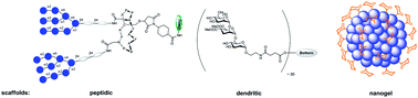
Pathogens adhere to the host cells during the first steps of infection through multivalent interactions which involve protein–glycan recognition. Multivalent interactions are also involved at different stages of immune response. Insights into these multivalent interactions generate a way to use suitable carbohydrate ligands that are attached to a basic scaffold consisting of e.g., dendrimer, polymer, nanoparticle, etc., with a suitable linker. Thus a multivalent architecture can be obtained with controllable spatial and topology parameters which can interfere with pathogen adhesion. Multivalent glycoconjugates bearing natural or unnatural carbohydrate antigen epitopes have also been used as carbohydrate based vaccines to stimulate an innate and adaptive immune response. Designing and synthesizing an efficient multivalent architecture with optimal ligand density and a suitable linker is a challenging task. This review presents a concise report on the endeavors to potentially use multi- and polyvalent glycoconjugates as vaccines as well as anti-infectious and anti-inflammatory drug candidates.


Graphical abstract: Multivalent glycoconjugates as vaccines and potential drug candidates


http://pubs.rsc.org/services/images/RSCpubs.ePlatform.Service.FreeContent.ImageService.svc/ImageService/image/GA?id=C4MD00143E


Polypharmacology modelling using proteochemometrics (PCM): recent methodological developments, applications to target families, and future prospects

Proteochemometric (PCM) modelling is a computational method to model the bioactivity of multiple ligands against multiple related protein targets simultaneously. Hence it has been found to be particularly useful when exploring the selectivity and promiscuity of ligands on different proteins. In this review, we will firstly provide a brief introduction to the main concepts of PCM for readers new to the field. The next part focuses on recent technical advances, including the application of support vector machines (SVMs) using different kernel functions, random forests, Gaussian processes and collaborative filtering. The subsequent section will then describe some novel practical applications of PCM in the medicinal chemistry field, including studies on GPCRs, kinases, viral proteins (e.g. from HIV) and epigenetic targets such as histone deacetylases. Finally, we will conclude by summarizing novel developments in PCM, which we expect to gain further importance in the future. These developments include adding three-dimensional protein target information, application of PCM to the prediction of binding energies, and application of the concept in the fields of pharmacogenomics and toxicogenomics. This review is an update to a related publication in 2011 and it mainly focuses on developments in the field since then.


Graphical abstract: Polypharmacology modelling using proteochemometrics (PCM): recent methodological developments, applications to target families, and future prospects

http://pubs.rsc.org/services/images/RSCpubs.ePlatform.Service.FreeContent.ImageService.svc/ImageService/image/GA?id=C4MD00216D


Related articles   

Experimental and computational studies of fatty acid distribution networks


Kinetics and non-exponential binding of DNA-coated colloids


Configuration of nonspherical amphiphilic particles at a fluid–fluid interface


Towards understanding cell penetration by stapled peptides

Med. Chem. Commun., 2015,6, 111-119   DOI: http://dx.doi.org:/10.1039/C4MD00131A

Hydrocarbon-stapled α-helical peptides are a new class of targeting molecules capable of penetrating cells and engaging intracellular targets formerly considered intractable. This technology has been applied to the development of cell-permeable ligands targeting key intracellular protein–protein interactions. However, the properties governing cell penetration of hydrocarbon-stapled peptides have not yet been rigorously investigated. Herein we report our studies to systematically probe cellular uptake of stapled peptides. We developed a high-throughput epifluorescence microscopy assay to quantitatively measure stapled peptide intracellular accumulation and demonstrated that this assay yielded highly reproducible results. Using this assay, we analyzed more than 200 peptides with various sequences, staple positions and types, and found that cell penetration ability is strongly related to staple type and formal charge, whereas other physicochemical parameters do not appear to have a significant effect. We next investigated the mechanism(s) involved in stapled peptide internalization and have demonstrated that stapled peptides penetrate cells through a clathrin- and caveolin-independent endocytosis pathway that involves, in part, sulfated cell surface proteoglycans, but that also seems to exploit a novel, uncharacterized pathway. Taken together, staple type and charge are the key physical properties in determining the cell penetration ability of stapled peptides, and anionic cell surface proteoglycans might serve as receptors to mediate stapled peptide internalization. These findings improve our understanding of stapled peptides as chemical probes and potential targeted therapeutics, and provide useful guidelines for the design of next-generation stapled peptides with enhanced cell permeability.

Graphical abstract: Towards understanding cell penetration by stapled peptides

http://pubs.rsc.org/services/images/RSCpubs.ePlatform.Service.FreeContent.ImageService.svc/ImageService/image/GA?id=C4MD00131A


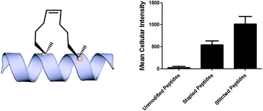
Introduction Hydrocarbon stapled a-helical peptides are an exciting new class of investigational agents capable of targeting and interfering with intracellular protein–protein interactions.1,2 (For reviews on hydrocarbon stapled peptides, see ref. 3 and 4, and for reviews on synthetic a-helix stabilization in general, see ref. 5 and 6.) These peptides contain a synthetic brace, referred to as a staple, introduced across one face of an a-helix (Fig. 1), that in favorable cases can increase a-helical content and protease resistance, enhance target binding affinity, promote cell membrane penetration, and suppress clearance in vivo. 7–10

Fig. 1 All-hydrocarbon stapled peptide technology. (a) Schematic illustration of peptide stapling. Two alpha-methylated, alkenyl-bearing non-natural amino acids are incorporated at two or more positions in the peptide chain and then cross-linked by ruthenium-catalyzed ringclosing olefin metathesis. (b) Different types of alkenyl-containing non-natural amino acids with distinct stereochemistry at the a-carbon and varied lengths of alkenyl side chains. (c) Three types of stapled peptides used in this study with optimized combinations of nonnatural amino acids.

Stapled peptides are synthesized via incorporation of two amethyl, a-alkenyl amino acids at defined positions in a synthetic peptide, followed by ring-closing olefin metathesis to close the helix-spanning hydrocarbon bridge (Fig. 1a).11,12 The two components of the staple, namely the hydrocarbon bridge and terminal methyl groups, are both important to obtain maximal effectiveness of the conformationally constrained peptide products. This technology has been successfully utilized to target several classes of proteins formerly considered intractable, including multi-component transcription factor complexes and protein–protein interactions having extended interfaces, such as the NOTCH transcription factor complex,13 the b-catenin–TCF interaction in the oncogenic Wnt signaling pathway,14 and the epigenetic modulator PRC2 complex.15 Given the difficulties of developing traditional small molecule drugs that can successfully target intracellular protein–protein interactions, hydrocarbon stapling technology is widely considered to represent a promising avenue of research for the development of chemical probes and potential targeted therapeutics.

Multiple types of hydrocarbon staples have been obtained by varying the relative placement of the cross-linking a,a-disubstituted amino acids, as well as the stereochemistry at the acarbon and the lengths of the alkenyl substituents (Fig. 1b).16,17 These staple types were optimized to provide robust a-helical stabilization and confer the potential for in vitro and in vivo activity. As a result of the combinatorial search process used to identify helix-stabilizing hydrocarbon staples, the diversity of the resulting macrocyclic bridges has revealed stapled peptides with different physicochemical properties. Recently, a new hyperstable version of stapled peptide with tandem crosslinks, referred to as a stitched peptide, was generated by introduction of S5 at the i position, B5 at the i + 4 position, and S8 at the i + 11 position (Fig. 1c) (Y.-W. Kim and G. L. Verdine, to be published).

Of the physicochemical properties demonstrated by peptide bearing hydrocarbon staples, the capacity to promote cellular membrane penetration is perhaps the most signicant and yet remains the most poorly understood. Independent of hydrocarbon-stapled peptides, several classes of cell penetrating peptides (CPPs) have been discovered, including naturally occuring transcription factor domains such as pennetratin18 and HIV-Tat19 and synthetic cationic peptides such as polyArginine peptides.20 Notably, despite extensive exploration during the past two decades, the mechanism(s) by which CPPs enter cells remain unclear.21–23 In contrast to CPPs, in which cell penetration appears to be sequence-dependent, numerous cell permeable stapled peptides have been discovered for peptide scaffolds with little sequence homology. These divergent observations regarding cell penetration is proposed to result from several features of stapled peptides that differentiate them from typical CPPs. For example, the introduction of an allhydrocarbon cross-link results in a constrained a-helical conformation, which embeds the hydrophilic amide backbone in the core of the folded structure. Furthermore, the hydrocarbon brace itself introduces a significantly hydrophobic patch to one face of the peptide. The exposure of the hydrophobic moiety as well as the masking of the hydrophilic peptide backbone may facilitate the interaction of stapled peptides with the hydrophobic interior of the cell membrane and thereby enhance the cellular uptake. As cell penetration is a critical property of stapled peptides, we sought to develop quantitative methods to correlate a battery of stapled peptide properties with the capacity for cellular uptake. A direct comparison with several well-known CPPs has revealed that stapled peptides, including some stapled versions of the CPPs, exhibit more robust cell penetration. Lastly, we have demonstrated that stapled peptides penetrate cells through a clathrin- and caveolin-independent endocytosis pathway that involves, in part, sulfated cell surface proteoglycans. These findings significantly expand our current understanding of cell penetration by stapled peptides and provide useful information for the future rational design of cell penetrating stapled peptides with novel applications.

Results and discussion

Development of a high-throughput assay to quantitatively measure cellular uptake of peptides

Understanding the internalization process of cell penetrating peptides (CPPs), especially stapled peptides, has been a subject of great interest. The majority of previous studies have been performed by either using high-resolution microscopy to show the existence of fluorophore-labeled CPPs inside cells, or by quantitatively measuring intracellular fluorescence by flow cytometry.24,25 Although these two methods can provide important information regarding cell penetration, their respective limitations prompted us to adopt an assay that combines high-resolution imaging with reliable quantitation of intracellular accumulation to better analyze and understand the cell penetration of stapled peptides. In recent years, highthroughput cell-based imaging platforms have become increasingly popular to screen for small molecule modulators of various biological processes.26,27 Taking advantage of one of these platforms, high-content epifluorescence microscopy, we developed a high-throughput quantitative assay to measure stapled peptide intracellular access.

Proof-of-principle experiments were performed to determine whether epifluorescence microscopy could be used to quantitatively compare stapled peptide intracellular access. Human U2OS osteosarcoma cells were seeded in black, clear-bottom 384-well plates and then incubated in serum-containing media supplemented with fluorescein-labeled peptides or DMSO vehicle for 12 hours. After the treatment, cells were washed thoroughly with PBS to remove excess peptide, fixed with 4% formaldehyde, and stained with Hoechst dye to visualize nuclei. Once prepared, the plates were imaged and quantified by epi- fluorescence microscopy according to a protocol developed and discussed in detail in Experimental methods. An initial z-scan was performed using the Hoechst channel to locate the cells, and the microscope parameters were subsequently adjusted to optimize the cell size and fluorescence intensity. The parameters from this acquisition were then applied to the FITC channel, and the microscope scanned and recorded images of the FITC-labeled peptides within the z-plane of the cell. This assay was performed in a high-throughput manner, resulting in a panel of Hoechst/FITC images from individual wells (Fig. S1†). The raw image data was then analyzed using MetaXpress® software (Fig. 2a). Cells were identified based on the Hoechst stain of nuclei, with the requirement that they were a contiguous fluorescent region having a specific intensity above local background as well as having a diameter between defined minimum and maximum to be designated as “positive” cells. The cytoplasm of each cell was then identied according to the spatial location of FITC signal in relation to the nuclei as well as empiric parameters (details in Experimental methods). The FITC intensities in the cytoplasm and nuclei were then quantified separately, and the sum of these two values yielded the FITC signal for the whole cell, which can be considered the relative intracellular peptide intensity. In addition, FITC negative cells were identified on the basis of a positive Hoechst stain, which was accompanied by an absence of appreciable signal in the FITC channel.

Fig. 2 Quantitative measurement of cellular peptide intensity. (a) Hoechst channel (left) showing the location and size of nuclei, FITC channel (middle) showing the fluorescence intensity of the same cells. Information about cell size and fluorescence intensity was integrated to identify the FITC positive (green mask) and negative (red mask) cell (right). For positive cells, additional parameters allowed determination of the fluorescence intensity in the nucleus (inner intense green) and the cytoplasm (outer dim green). (b) The background fluorescence the DMSO vehicle was almost identical among different experiments. (c) Four stapled peptides from different batches of synthesis generated similar intracellular fluorescence intensity in different tests. Error bars represent the S.D. of two measurements.

We found that this system generated highly reproducible and reliable results from assay-to-assay and with different stocks of the same stapled peptides. As shown in Fig. 2b, there were negligible fluorescence differences among experiments for cells treated with DMSO vehicle, which could be used as a fluorescence background for all subsequent experiments. In addition, the same stapled peptides from different batches of synthesis and stocks featured almost identical intracellular fluorescence signals in different tests (Fig. 2c), indicating that the assay developed in this study produces repeatable and reliable results that could be directly combined and compared from a large set of experiments. Furthermore, to determine how this assay performs as a screening tool, we have calculated Z0 factor of 0.54 by using the most penetrant A6 peptide as a positive control and DMSO background as negative control, which also indicates a statistically good assay quality.

Analysis of cell penetration by stapled peptides The development of this quantitative high-throughput assay enabled a broad investigation of the physicochemical properties governing the cell uptake of a diverse set of hydrocarbonstapled peptides synthesized in our laboratory. We postulated that any correlation between cellular uptake and physicochemical properties would illuminate characteristics associated with productive cellular uptake and inform the future design of stapled peptides with improved cell penetration.28 To this end, we screened and analyzed more than 200 discrete FITC-labeled peptides belonging to three different classes: wild-type (unmodified), stapled and stitched peptides. All peptides were converted to two-dimensional structures and analyzed for theoretical physicochemical properties with the publicly available Marvin View software package from ChemAxon. Properties including the molecular weight, theoretical pI, calculated 2D polar surface area (PSA), theoretical log P and formal charge at pH 7.5 were calculated for each peptide (Table S1†). In general, the unmodified, stapled and stitched peptide libraries present in this screen had relatively similar physicochemical characteristics (Fig. S2†). The mean molecular weight and calculated PSA values were nearly identical among the three peptide classes. A notable difference was observed among theoretical log P values, which were significantly higher for the stapled and stitched peptides relative to the unmodified peptides, which is not surprising as these modified peptides contain a solvent exposed hydrocarbon crosslink. Additionally, the stapled peptide class had a mean formal charge of approximately zero while the stitched and unmodified peptide classes exhibited a positive mean charge. Overall, the calculated physicochemical properties indicated that the peptide classes were quite similar in terms of their mean properties, which is useful when making comparisons among their cell penetration properties.

We next performed an intracellular access screen by treating U2OS cells with 1 mM of FITC-labeled peptide for 12 hours in duplicate. All assays contained control DMSO wells and positive control peptides, which were compared among assays to ensure plate-to-plate reproducibility (Fig. 2b and c). The primary readout of the screen was mean cellular fluorescence intensity. As the DMSO background was highly consistent between wells and experiments, a mean background value was subtracted from all data. The results of the screen were used to generate plots comparing cell penetration with peptide physicochemical parameters. Interestingly, as a class, stapled and stitched peptides exhibited significantly higher cell penetration compared with wild-type unmodified peptides, which contained several established cell penetrating peptides (CPPs; Fig. 3a). Given that all three peptide classes have similar physicochemical properties in general, the benefit in cell penetration can be largely attributed to the synthetic stabilization of the a-helical peptides with all-hydrocarbon peptide stapling technology. Furthermore, we found that peptide charge near physiologic pH exhibited a strong correlation with intracellular access and could be fitted into a Gaussian distribution with a population centroid at a formal charge of +4 (Fig. 3b). In particular, peptides exhibiting a net negative charge (7 to 1) exhibited little cellular uptake, whereas peptides of approximately neutral charge (1 to +1) displayed moderate cell penetration above background. Interestingly, peptides with a net positive charge (+1 to +7) showed significantly higher cell penetration as a group. Cellular uptake did not appear to increase linearly with charge, as the cell penetration decreases dramatically for the peptides in this study with charge greater than +7. The same trend between formal charge and cellular uptake were observed for individual stapled and stitched peptide classes as well (Fig. S3†). This observation is not consistent with previously reported models that indicate that peptides/mini-proteins with more positive charge have better penetration properties due to tighter electrostatic interactions with the negatively charged phospholipid membrane.29,30 The lower penetration for highly charged peptides in this study could result from any one of many factors including, for example, peptide aggregation in solution, the disruption of peptide packing during internalization or difficulty in dissociation from cell membrane. Additional tests with a larger number of peptides could further our understanding of this phenomenon. In addition, there was no discernible correlation between cell penetration and peptide molecular weight, log P, pI value or PSA (Fig. S4†). Taken together, these data demonstrated that the staple type and peptide charge are key physical properties correlated with peptide cell penetration ability, whereas the other parameters do not appear to be significantly associated.

In order to further investigate the cell penetration properties for stapled peptides and to systematically analyze the similarities and differences in cellular uptake between stapled peptides and other wild-type cell penetrating peptides, we compared cell penetration of several stapled peptides to that of three well known wild-type CPPs: Tat (48–60), penetratin (Antennapedia 43–58) and poly-Arg8 (Table S2†). First, we investigated the cellular uptake at varied peptide concentrations. As shown in Fig. 4a and b, both wild-type CPPs and stapled peptides showed dose-dependent increases in cell penetration. Strong intracellular fluorescence was detected in the low micromolar range, and although the levels of accumulation were different for distinct peptides, stapled peptides featured more robust dosedependent cell penetration at lower concentrations relative to wild-type CPPs, in general. It is also interesting to note that while significant increases in intracellular fluorescence were mostly evident in the 1–10 mM range for stapled peptides, distinct profiles were observed for specific peptides. For example, TNG147 showed little cell penetration at 1 mM but showed a dramatic increase at 5 mM, which might suggest that concentration-dependent peptide packing or a receptor-mediated mechanism may facilitate the cell penetration process, and these processes may be triggered at different concentrations for distinct peptides. Furthermore, it is worth noting that the stapled peptides studied here were more cell permeable than wild-type CPPs at most concentrations tested, exhibiting nearly an order of magnitude higher intracellular fluorescence at the same treatment concentrations.

Fig. 4 Effects of peptide concentration and incubation time on cellular uptake of stapled and wild-type peptides. (a) Wild-type and (b) stapled peptides showed a dose-dependent increase in cell internalization. Cellular uptake for (c) penetratin and (d) SAHM1 peptides over time at concentrations of 5 and 10 mM. (e) A pulse-chase penetration assay for SAHM1 peptide in which fresh medium containing either a new batch of peptide or DMSO vehicle were exchanged at 12 hours after initial treatment. Error bars represent the S.D. of triplicate samples.

We next performed a time-course penetration assay to better understand the kinetics of peptide internalization using a representative CPP and stapled peptide. Penetratin and SAHM1 showed distinct kinetics of uptake and stabilization throughout a 24 hour time course. 5 mM and 10 mM penetratin peptide exhibited similar intracellular cellular fluorescence after 2 hours, which then decreased until approximately 8 hours and finally stabilized at different intracellular levels until 24 hours (Fig. 4c). On the other hand, the stapled peptide SAHM1 showed time- and dose-dependent cellular uptake, which stabilized after approximately 8 hours (Fig. 4d). Compared to the wild-type penetratin, the SAHM1 profile was unique in that dose-dependent accumulation was evident at all time points and no loss of signal was observed. One explanation for the loss of signal observed with penetratin could be attributed to an equilibrium between cell penetration and subsequent intracellular proteolysis followed by export of the fluorophore. The presence of the all-hydrocarbon crosslink in its peptide sequence and lower net charge of SAHM1 relative to penetratin, could contribute to enhanced cellular uptake and reduced intracellular proteolysis, leading to continuous accumulation in cells. To further explore the equilibrium observed for stapled peptides, we performed a pulse-chase experiment using SAHM1. After 12 hours of incubation with SAHM1, cell culture medium was aspirated and the cells were extensively washed with PBS to completely remove excess peptide. Then fresh medium containing either a new batch of 1 mM peptide or DMSO vehicle was added to cells and incubated for the indicated time points (Fig. 4e). As expected, the cellular uptake increased for the first 12 hours incubation. After medium exchange, cells incubated with fresh medium containing DMSO vehicle retained the intracellular fluorescence intensity. Interestingly, the signal for cells treated with a new batch of staple peptide continued to increase up to 24 hours (Fig. 4e). This observation indicates that despite incubation over a time course previously shown to reach equilibrium, the mechanism(s) responsible for cellular uptake are not saturated, as evidenced by further uptake upon replacement with fresh stapled peptide. Taken together, these data indicate that the mechanism(s) underlying cellular uptake by both CPPs and stapled peptides exhibit time- and dose-dependency that is not saturable at early time points or low micromolar doses and, importantly, appears to be more robustly utilized by stapled peptides.15,31

Given that stapled peptides exhibit better cell penetration properties in general than parent unmodified peptides, we wondered whether the peptide stapling strategy could be applied generally to improve cellular uptake of parent unmodified peptides. To test this hypothesis, we designed a panel of stapled peptides based on Tat (48–60), penetratin and poly-Arg8 (Fig. 5a). These stapled peptides and their parent unmodified peptides were incubated in U2OS cells for 12 hours with a concentration range from 10 nM to 20 mM, mirroring the dosedependent uptake studies shown in Fig. 4. As expected, all peptides showed dose-dependent cell penetration (Fig. 5b–d). Interestingly, stapled peptides derived from penetratin and poly-Arg8 showed improved cell permeability at concentrations starting from 1 mM for stapled penetratin and 5 mM for stapled poly-Arg8. It is noteworthy that the staple position also affected the cellular uptake as the two stapled penetratin peptides with different crosslink positions exhibited varied cell penetration, though both were superior to wild-type penetratin. In contrast, reduced cellular uptake was observed for both stapled peptide variants derived from the Tat sequence (Fig. 5b). This could result from several possible effects, including disruption of peptide secondary structure, masking of residues essential for surface recognition or altering peptide packing interactions involved in cell penetration. Further focused study of these variants is warranted to elucidate the source of altered cellular uptake, however these data clearly demonstrate that peptide stapling may be a general method to further improve the cell permeability of CPPs, which could serve as more efficient transduction domains for molecular cargoes. In addition, while increasing the helical content of stabilized peptides has been stated to be a guiding principle in the successful design of biologically active stapled peptides, it has not been shown to be generally correlated with cell penetration. To specifically address whether increasing the helical content of a peptide is correlated with augmented cell penetration, we have measured the relative helicity of hydrocarbon stapled variants of Tat, penetratin and poly-Arg8 (Fig. S5†). Notably, we did not observe a general correlation between increased helical character and cell penetration of these peptides. Peptide stapling increased the helical content of both Tat and poly-Arg8 peptide sequences, which were largely unstructured when unmodified. In contrast, the unmodified penetratin peptide had signicant helical content (>50%), and the hydrocarbon stapled variants of this sequence largely retained their helicity, albeit lower overall helicity. Intriguingly, these species demonstrated the differing effect of hydrocarbon stapling and increased helical content on cell penetration since introduction of the hydrocarbon staple increased the cellular uptake of both penetratin and poly-Arg8 sequences, while it decreased uptake for Tat peptides. Therefore, we cannot conclusively state, a priori, that the incorporation of a hydrocarbon staple or increased a-helicity will lead to more productive cellular penetration, although in general stapling can increase the uptake of specific sequences (Fig. 5) and as a class stapled and stitched peptides are more cell penetrant (Fig. 3a). A more comprehensive follow-up study with CD analyses on a larger peptide library is needed to better address this question.

Fig. 5 Effects of all-hydrocarbon staples on cell penetration by wild-type cell penetrating peptides. (a) List of wild-type cell penetrating peptides and their stapled derivatives investigated in this study. (b–d) Dose-dependent cell penetration assays showed that stapling strategy greatly improves the cellular uptake of penetratin and poly-Arg8 peptides. Experiments were performed in triplicate, and error bars represent S.D. of three measurements.

Mechanistic studies of cell penetration by stapled peptides The aforementioned studies indicate that stapled peptides exhibit better cellular uptake properties than wild-type peptides in general, and that internalization correlated primarily with hydrocarbon staple type and formal peptide charge. However, the mechanism(s) utilized by peptides to translocate across the cell membrane are still unclear. Therefore, we sought to investigate the uptake mechanism(s) for stapled peptides. The uptake mechanism(s) of wild-type CPPs have been extensively studied. Some evidence indicates that they enter cells via energy-dependent endocytosis, which is an active transport process, however data suggesting passive diffusion for CPPs have also been reported; hence, the mechanism(s) of cell uptake by CPPs remains ambiguous.32–34 We first sought to determine whether cell penetration by stapled peptides and wild-type CPPs occurs via ATP-dependent endocytosis.2 Cells were pre-treated with NaN3 and 2-deoxyglucose (2-DG) to reduce cellular ATP levels, and then incubated with FITC-labeled peptides (wildtype and stapled) for 4 hours and compared to normal cells for intracellular fluorescence. Cellular ATP levels were confirmed to be decreased by approximately 90% after NaN3 and 2-DG treatment (Fig. S6†), but Tat and poly-Arg8 exhibited almost identical cellular uptake in ATP-depleted and normal cells, supporting the model that they utilize passive diffusion to translocate across the cell membrane. However, penetratin and all stapled peptides showed 20–50% lower accumulation in ATP-depleted cells, indicating an active trans-membrane process requiring cellular ATP (Fig. 6a). These data indicate that there may be more than one uptake mechanism for CPPs and stapled peptides, but that for the most robust cell penetrating peptides (penetratin and stapled peptides studied here), the internalization mechanism(s) involves ATP-dependent endocytosis.

Fig. 6 Mechanistic study of cell penetration by stapled peptides and wild-type cell penetrating peptides. (a) Cellular uptake in normal and ATP-depleted cells indicated that stapled peptides penetrate cells via an ATP-dependent endocytosis. (b) Impaired uptake was observed in NaClO3 treated cells, which inhibit proteoglycan biosynthesis. (c) Cell penetration of wild-type and stapled peptides in wild-type CHO and proteoglycan-deficient CHO cells. Experiments were performed in triplicate, and error bars represent S.D. of three measurements.*P < 0.05, **P < 0.01, ***P < 0.001.

Next, we sought to identify the specific pathway(s) utilized for cellular uptake, since energy-dependent endocytosis can be accomplished by several different pathways including caveolinand clathrin-mediated endocytosis. We repeated the cell penetration experiments under a variety of conditions that each blocked a different endocytosis pathway (Table S3†).35–37 We found that uptake was partially blocked in cells treated with sodium chlorate (Fig. 6b), which aborts the decoration of cells with sulfated proteoglycans, but was unaffected by inhibitors of other endocytic pathways (Fig. S7†). It thus appears that interaction with sulfated proteoglycans is responsible for some, but not all, endocytic uptake of stapled peptides and wild-type CPPs. It is reasonable to connect this result with the previous discovery that peptide charge is a key factor determining cell penetration. Proteoglycans are negatively charged under physiologic conditions due to the occurrence of sulfate groups, and these might form electrostatic pairs with positively charged peptides to facilitate anchoring on the cell membrane.38–40 To further confirm that sulfated proteoglycans are important to mediate cellular uptake for peptides, we performed a secondary assay using wild-type CHO cells (CHO-K1) and proteoglycan deficient CHO cells (pgsA-745) which harbor a defect in xylosyltransferase, thereby preventing glycosaminoglycan biosynthesis. All peptides showed similar penetration properties in wild-type CHO cells, but uptake was decreased by approximately 50% in proteoglycan-deficient CHO cells, consistent with the experiment using a small molecule inhibitor (Fig. 6c). Taken together, our data suggest that CPPs and stapled peptides penetrate cells through a clathrin- and caveolin-independent endocytosis pathway that is in part mediated by interaction with anionic cell surface proteoglycans. This result is very similar to the previous reports on the mechanism of cellular uptake for supercharged GFP (scGFP), which likewise does not utilize clathrin- or caveolin-mediated endocytosis.41 Notably, scGFP internalization requires actin polymerization, which may not be required for peptide penetration (Fig. S7c†) types, and distinct physicochemical properties. As a result, we found that stapled peptides penetrate cells more efficiently than unmodified peptides, including well-characterized cell penetrating peptides. For the panel of peptides used in this study, only staple type and formal charge were significantly correlated with cell penetration potential, whereas the other physical parameters did not appear to have a signicant effect. We further studied the relationships between cellular uptake and

In conclusion, we sought to investigate the cell penetration properties of stapled peptides, which is one of the most significant yet poorly understood aspects of peptide stapling technology and cellular transduction technologies in general. In order to address this problem, we developed a high-throughput assay to quantitatively measure stapled peptide intracellular accumulation. Using this assay, we analyzed more than 200 discrete peptides with various sequences, staple positions and peptide concentration or incubation time, revealing that stapled peptides accumulate in cells in a dose-dependent fashion and reach steady intracellular levels over a course of a few hours. These studies revealed similar time- and dosedependent behavior for CPPs and stapled peptides, but stapled peptides, including stapled versions of CPPs, were shown to be 10- to 20-fold more penetrant, measured by intracellular fluorescence level at a given dose, than the most potent CPP. We also propose that the specific intracellular accumulation and stabilization kinetics of stapled peptides or unmodified CPPs may be a consequence of equilibria between peptide penetration, cellular proteolysis and/or retrograde transport of the species. Finally, we investigated the mechanism(s) involved in the internalization of stapled peptide and unmodified CPPs and demonstrated that cell penetration occurs through a clathrinand caveolin-independent, energy-dependent endocytosis pathway that utilizes, in part, sulfated cell surface proteoglycans. This dataset provides significant insight into the physicochemical properties correlated with productive cellular penetration as well as a more detailed understanding of the mechanism(s) utilized by stapled peptides to access intracellular compartments, which together should aid in the design of and characterization of novel stapled peptides in the future.


Rational design of protein–protein interaction inhibitors

Med. Chem. Commun., 2015,6, 51-60    DOI: http://dx.doi.org:/10.1039/C4MD00328D
Protein–protein interactions are at the heart of most physiopathological processes. As such, they have attracted considerable attention for designing drugs of the future. Although initially considered as high-value but difficult to identify, low molecular weight compounds able to selectively and potently modulate protein–protein interactions have recently reached clinical trials. Along with high-throughput screening of compound libraries, combining structural and computational approaches has boosted this formerly minor area of research into a currently tremendously active field. This review highlights the very recent developments in the rational design of protein–protein interaction inhibitors.
Graphical abstract: Rational design of protein–protein interaction inhibitors

Didier Rognan heads the Laboratory of Structural Chemogenomics at the Faculty of Pharmacy of Strasbourg (France). He studied Pharmacy at the University of Rennes (France) and did a Ph.D. in Medicinal Chemistry in Strasbourg (France) under the supervision of Prof. C.G. Wermuth. Aer a postdoctoral fellowship at the University of Tubingen (Ger- ¨ many), he moved as an Assistant Professor to the Swiss Federal Institute of Technology (ETH) until October 2000. He was then appointed Research Director at the CNRS to build a new group in Strasbourg. He is mainly interested in all aspects (method development and applications) of structurebased drug design, notably on G protein-coupled receptor ligands and protein–protein interaction inhibitors.

Introduction Drug discovery is a long, costly, multi-step endeavour which aims at reducing all possible risks to deliver a novel therapeutic solution to previously unmet clinical needs. To reduce chemical risks, empirical rules are used to filter the chemical space and retain drug-like low molecular weight compounds. Reduction of the biological risk is addressed by considering privileged target families (e.g., G protein-coupled receptors and kinases) whose activation/inhibition by drug-like compounds is likely to correct or reverse pathological states. Until recently, mostly single macromolecules (proteins and nucleic acids) have been considered as potential drug targets. Out of 68 000 proteins currently annotated in UniProt for the human proteome,1 only about 300 targets2 have been addressed by current drugs, and the large majority of single targets is still awaiting first-in class drugs.

Besides single targets, large scale genomics and proteomics3 have identified complex networks of targets and pathways regulating physiopathological processes in a coordinated manner. The current human protein–protein interactome has been estimated between 130 000 (ref. 4) and 650 000 (ref. 5) complexes, out of which only a tiny amount is known, and only a very few6–8 have been the object of a drug discovery initiative. Protein–protein interactions (PPIs) therefore describe a totally new biological space that attracts more and more attention, with 26PPI inhibitors9,10 already under clinical development, notably in the oncology field.11 Despite PPIs may adopt quite different sizes, shapes and electrostatics,12 identifying highaffinity PPI inhibitors is a considerable challenge for many reasons: (i) in contrast to conventional targets, a medicinal chemist cannot start inhibitor design from the structure of endogenous ligands, (i) PPIs often involve flat surfaces delocalized over multiple epitopes, usually lack well-defined buried cavities13 typical of conventional targets, and are significantly larger (ca. 1000–3000 A˚2 ) than enzyme/receptor pockets (300– 1000 A˚2 ), (iii) high-throughput screening of traditional compound libraries often returns no viable hits14 for the main reason that PPI inhibitor chemical space is quite different from that described by traditional drug-like compounds.10 Nonetheless, thanks to bioinformatics and proteomics-guided prioritization of therapeutically relevant protein–protein complexes, more and more PPI inhibitors are currently reported. Several excellent reviews6,7,9,11,15–18 have already been published on experimental methods (high throughput screening, biochemical and cellular assays, and fragment-based approaches) suitable to discover PPI inhibitors. The present report will only cover computer-aided approaches, with a major emphasis on structure-based methods and recent discoveries (2012–2014).

Databases Preliminary access to experimentally validated data is key to launch a drug discovery program on PPI modulators. A multitude of databases storing genomics, proteomics and structural data are currently available to help the medicinal chemist. We will here briefly review these archives, focusing mostly on easily interpretable structural data.

PPI databases Many experimental methods with different throughputs (from low to high) have been developed to characterize binary interactomes in various species, among which the most prominent has been the yeast two-hybrid (Y2H) assays, and mass-spectrometry (MS) coupled with co-immunoprecipitation or coaffinity purification.19 These experimental data are stored in many primary databases (Table 1) that are difficult to mine due to their large heterogeneity. Metadatabases have been derived thereof to facilitate their analysis, among which the most popular are APID and PRIMOS (Table 1). These metadatabases cover a wide range of organisms and notably offer the possibility to mine experimental PPI data according to disease relevance or inter-organism crosstalk, and provide graphic tools to visualize complex networks of interacting proteins and identifying important protein nodes (hubs). It is however very difficult, from this large amount of data, to clearly prioritize PPIs for a drug discovery program. Attempts to classify the PPIs by structural druggability25 (although ligandability26 is probably a better term) are worth mentioning but should be taken with care due to the still insufficient number of existing PPI three-dimensional (3D) structures.

Table 1 Protein–protein interaction databases

Database                 Interactions                         Website                                                             References

BIND                             32 211           http://bond.unleashedinformatics.com                       20

DIP                                 78 191           http://dip.doe-mbi.ucla.edu/dip/Main.cgi                  21

HPRD                           41 327            http://www.hprd.org/                                                           22

IntAct                      448 986             http://www.ebi.ac.uk/intact/                                            17

MIPS                              9 835             http://mips.helmholtz-muenchen.de/proj/ppi/      23

APID                         196 700             http://bioinfow.dep.usal.es/apid/index.htm             24

PRIMOS                  384 127             http://primos.fh-hagenberg.at/                                        19


Table 2 Database of low molecular-weight PPI inhibitors
Database                 Ligands                             Website                                                                  References

2P2I                                 71                   http://2p2idb.cnrs-mrs.fr/                                                12

iPPI-DB                      1650               http://www.ippidb.cdithem.fr/                                        10

TIMBAL                      6896              http://mordred.bioc.cam.ac.uk/ timbal                        29

Ligand databases Initially limited to a limited subset of inhibitors able to disrupt few PPIs (e.g. p53/MDM2, Bcl-Xl/Bak, and IL-2/IL-2Ra),7,27 the repertoire of PPI inhibitors rises constantly thanks to exciting developments in biophysical fragment screening.15,28 Three publicly available databases storing information on PPIs and their inhibitors (Table 2) may be used to better describe the structural properties of druggable PPIs and the chemical space associated with their disruptors.

The 2P2Idb database12 is a hand-curated repository of protein–protein complexes of known X-ray structures (X-ray diffraction and nuclear magnetic resonance spectroscopy) for which at least one low molecular weight orthosteric inhibitor has been co-crystallized with one of the two protein partners. It currently describes 71 inhibitors for 14 PPIs, clustered in two groups (Fig. 1) with respect to the nature of the interface (protein–peptide and protein–protein). Companion tools (2P2I inspector,30 2P2I score,30 and 2P2I hunter31) are provided to analyse PPIs at a structural level, predict their structural druggability and design PPI focussed libraries, respectively.

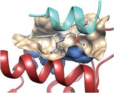
Fig. 1 Prototypical examples of class I (left panel) and class II PPIs (right panel), exemplified by the Bcl-Xl/Bak (PDB id 1BXL) and integrase/LEDGF (PDB id 2B4J) complexes, respectively. Class I PPIs involve the interaction of a globular protein with a peptide or a single secondary structure (a-helix and b-strand) of a second protein partner. Class II PPIs are characterized by the interaction of two globular proteins.

The iPPI-DB10 is another manually curated database from world patents and the medicinal chemistry literature, focussing on low molecular weight orthosteric inhibitors, disease-related protein–protein interfaces and a clear biochemical readout (e.g. fluorescence polarisation and enzyme-linked immunosorbent assay). The database archives 1650 PPI inhibitors targeting 13 families of homologous PPI targets mainly involved in cancer, immune disorders and infectious diseases.

Finally, the TIMBAL database29 reports ca. 7000 inhibitors for 50 known PPIs. In contrast to the two other databases, TIMBAL is maintained through a predefined list of PPIs and automated searches in ChEMBL32 and the Protein Data Bank.33 In contrast to the other databases, TIMBAL also registers short peptides with an upper molecular weight limit of 1200 Da. It should be pointed that most of the 15 000 uncurated biological data present in TIMBAL arise from a single target family (integrins) and should be considered with care.

Analysing the content of these databases enables a first comparison of PPI inhibitors versus drugs, as well as PPIs amenable to disruption versus standard heterodimers. PPI surfaces disrupted by inhibitors tend to be smaller, more hydrophobic and accessible than standard heterodimers.12 As a consequence, low molecular weight PPI inhibitors tend to be larger, more hydrophobic and more aromatic-rich than standard drugs. Interestingly, many of them (ca. 60%) still comply with Lipinski’s rule-of-five, 10 revealing some hopes in the developability of such compounds.

However, it should be stated that the set of empirical rules designed to discriminate druggable from non-druggable PPIs, as well as to distinguish PPI inhibitors from conventional druglike compounds still rely on a very limited set of highly homologous data (PPIs, inhibitors), and should therefore be regarded with caution. Increasing coverage of the PPI repertoire by future experimental screens will undoubtedly lead to a better denition of PPI biological and chemical spaces. We therefore expect in the future the above-mentioned rules to be rened and be more descriptive of the true world of PPI inhibitors, notably with respect to rational design of PPI focussed libraries.


Rational design of PPI modulators

Sequence-based approaches Whatever the nature of the PPI (type I or type II, see the definition above), PPI interfaces are often characterized by the presence of hotspots,34 in other words anchor residues that contribute the most to the binding free energy of the protein– protein complex. The interaction of a single modified amino acid with a single anchor residue might be sufficient to disrupt a PPI as elegantly demonstrated by Lin et al. in a recent study.35 Capitalizing on the presence of a reactive cysteine (C246) at the interface of the complex between caspase-7 (CASP7) and the Xlinked inhibitor of apoptosis protein (XIAP), they designed the N-iodoacetyl-lysine amino acid derivative 1 (Fig. 2) that covalently traps C246 and further disrupts the XIAP–CASP7 complex, therefore triggering CASP7-dependent apoptosis and killing MCF-7 breast cancer cells (EC50 ¼ 0.64 mM) previously resistant to chemotherapy.

The easiest way to inhibit a PPI is to start with the amino acid sequence of one interacting epitope, notably if the latter is part of regular secondary structures (a-helix, b-strand, and b-turn). For example, a-helical peptides mimicking the sequence of protein transmembrane domains may disrupt PPIs quite efficiently.36,37

Fig. 2 Peptidomimetics as PPI disruptors


Due to poor pharmacokinetic profiles, linear peptides are good in vitro tools but usually not efficient clinical candidates. Chemical modifications are required to stabilize their secondary structures in physiological media and prevent early degradation. Among the most exciting developments in this area38,39 is the design of stapled peptides.40,41 Stapled peptides are synthetic analogues of a-helical protein epitopes involved in a PPI, and in which a covalent hydrocarbon linkage connects adjacent turns of the helix. Stapling is known to significantly increase the in vivo half-life of the natural peptide (increasing proteolytic stability), decrease the entropic cost of binding, and even enable cellular uptake.42 Many stapled peptides with potent in vivo activities have already been reported.39 One of these stapled peptides (ATSP-7041, compound 2, Fig. 2) just entered clinical development as a dual nM MDM2/MDMX inhibitor for p53-dependent cancer therapy.43

Heterocyclic scaffolds mimicking secondary structures can also be obtained by solution-phase synthesis to afford peptidomimetic libraries amenable to PPI inhibition. Whitby et al. notably reported the design of 8000 member 4-acetamido-3- alkoxy-benzamide focused library featuring weak p53/MDM2 inhibitors and potent HIV-1/gp41 inhibition (compound 3, Fig. 2).44 When the peptide epitope is not structured, developing macrocyclic analogues is more difficult but still feasible as recently demonstrated by Glas et al.38 who successfully improved 14-3-3 binding of a 11-mer peptide from a bacterial ExoS virulence factor by cross-linking binding amino acids with polymethylene linkers, up to an in vitro 40 nM disruptor of the ExoS/14-3-3 interaction (compound 4, Fig. 2). Interestingly, the cross-linker was not only chosen to rigidify the natural ExoS peptide structure but also to directly provide additional hydrophobic interactions to the 14-3-3 binding site.38 Only in exceptional cases the natural unmodified peptide is directly usable as a PPI inhibitor. One recent example is the 28 amino acid cell-penetrating peptide (p28) from a bacterial azurin redox protein, that binds to the DNA-binding domain of the p53 tumor suppressor and inhibits p53 degradation by interfering with the Cop1-mediated ubiquitination,45 thereby enhancing p53 levels in cancer cells and exhibiting antitumoral efficacy in patients with advanced solid tumors.46

Pharmacophore-based approaches As defined by the IUPAC,47 a pharmacophore is “an ensemble of steric and electronic features that are necessary to ensure the optimal supramolecular interactions with a specific biological target and to trigger (or block) its biological response.” Although pharmacophores are mainly used to align and compare ligands sharing the same target,48 the same concept can be easily transferred to PPIs in which one partner is the “receptor” and the second one the “ligand”. Pharmacophore features (hydrophobic, aromatic, H-bond donor and H-bond acceptor, positively and negatively ionisable) can therefore be manually or automatically mapped to atoms of the ligand in direct interactions with the receptor. The resulting pharmacophore can then be used to identify a compound library for hits fulfilling the defined query. Several tools (e.g. LigandScout,49 Discovery Studio,50 and Pocket Query51) can be directly used to map PPI pharmacophores onto protein–protein X-ray structures (Fig. 3).

Fig. 3 Example of a PPI pharmacophore mapped onto interacting atoms of human LEDGF (yellow ribbons) bound to HIV-1 integrase (red ribbons, PDB ID 2B4J). The PPI pharmacophore is composed of 2 Hbond donors (magenta balls), two H-bond acceptors (green balls), one hydrophobic feature (cyan ball) and 6 exclusion volumes (gray balls).

Using a manual PPI pharmacophore defined from the X-ray structure of the Annexin A2/S100A10 complex, a pro-angiogenic complex, Reddy et al.52 derived a simple pharmacophore (2 hydrophobes, 2 H-bond donors, and 2 H-bond acceptors) using the Unity program,53 and screened a library of 700 000 compounds to select 586 hits which were further docked to the Annexin A2 binding site to retain only 190 candidates with both a good docking and pharmacophore fitness score (Table 3). Out of 190 tested compounds, 7 hits blocked the interaction between S100A10 and the Annexin A2 N-terminus in a competitive fluorescent binding assay, with the most potent PPI inhibitor (compound 5, Fig. 4) exhibiting an IC50 of 24 mM.52 Geppert et al.54 reported the rational discovery of a low molecular weight inhibitor of the complex between interferon-a (IFN-a) and its receptor (IFNAR2). Fortunately, the PPI interface was small enough (ca. 800 A˚2 ) to be targeted by a small heterocyclic compound. After identifying major hotspots at the IFN-a surface, a fuzzy receptor-based pharmacophore was determined using the VirtualLigand approach,55 which assigns pharmacophoric features to Gaussian densities. Screening a collection of 556 000 commercially available compounds retained six virtual hits, out of which two were weak IFN-a inhibitors, but one (compound 6, Fig. 4) was confirmed by NMR and surface plasmon resonance (SPR) to bind to IFN-a with a dissociation constant (Kd) of 4 mM and to inhibit IFN-a responses in various cell assays. The novel inhibitor may be useful to reduce IFN-a titers in autoimmune disorders.


Table 3 Protein–protein pharmacophore searches to identify PPI inhibitors

Target                                     Library size                          Tested                        Hits                      Ref.

Annexin A2/S100A        10 700 000                              190                               7                          52

INFAR2/IFN-a                       556 000                                   6                               3                          54

p53/MDM2                                  21 287                                  15                               6                          56

Nrf2/Keap1                                   21 199                                  17                                1                          57

PKC3/RACK2                          330 000                                 19                                1                          58


Due to the inherent complexity of PPI pharmacophores (many features covering a large surface), combining several pharmacophores into a consensus model may help to retrieve essential features and simplify pharmacophore queries. Xue et al. applied this approach to the identification of p53–MDM2 inhibitors.56 The p53–MDM2 complex has become a prototypical PPI for its biological background (this interaction plays an important role in regulating the transcriptional activity of tumour cells) and many high affinity low molecular-weight inhibitors of this PPI identified by various screening approaches.59 Starting from a set of 15 MDM2-peptide X-ray structures, a common feature structure-based pharmacophore (2 H-bond donors, one H-bond acceptor, 2 aromatic rings, and one hydrophobe) was first identified. In addition, a receptorligand pharmacophore (five hydrophobes, one aromatic, and one H-bond donor) was generated from a separate set of 10 MDM2-non peptide complexes. Merging both pharmacophores and retaining the most common features led to an ensemble pharmacophore definition (two aromatic rings, two hydrophobes, and one H-bond donor) taking into account both peptide and non-peptide binding. This pharmacophore was used to screen a collection of 21 287 commercially available compounds, and led to a hit list of 15 compounds out of which 6 were confirmed as p53–MDM2 inhibitors using an in vitro uorescence polarization assay.56 The most potent inhibitor (compound 7, Fig. 4) is a 180 nM MDM2 inhibitor. Despite a good selectivity in a MTT tumour cell proliferation assay (p53+/+ vs. p53/ cells), compound 7 was a weak inhibitor (IC50 ¼ 85 mM) of tumour cell growth, because of poor pharmacokinetic properties.

Fig. 4 PPI inhibitors identified by pharmacophore-based virtual screening.

Along the same lines, two X-ray structures were used to derive inhibitors of the PPI between Keap1 and Nrf2, a complex involved in the response to oxidative stress.57 The two PPI pharmacophores were merged into a single query consisting of one H-bond donor, two H-bond acceptors and three negative ionisable centers. To afford some fuzziness in the search, up to two features were allowed to be missed by virtual hits. Since the Keap1-binding epitope of Nrf2 is composed of several acidic residues, only compounds bearing a negative charge were searched among a full commercial library of 251 774 compounds. The remaining 21 199 hit list was matched to the pharmacophore, and led after confirmation with docking and MM-PBSA scoring, to a list of 17 potential hits which were tested for Keap1–Nrf2 inhibition using an in vitro fluorescence polarization assay. A single compound (compound 9, Fig. 3) was confirmed in vitro as a moderately potent Keap1–Nrf2 inhibitor with an EC50 of 9.8 mM.57 Interestingly, the inhibitor activated the Nrf2 transcriptional activity .

When both protein partners involved in the PPI have not been co-crystallized, it is still possible to rationally discover PPI inhibitors, starting from the sole X-ray structure of one of the two proteins. This approach was followed by Rechfeld et al. in the discovery of PKC3–RACK2 inhibitors.58 Starting from the Xray structure of the PKC3 octameric epitope binding to RACK2 (a receptor for activated protein kinase C), a simple peptide-based pharmacophore model (3 H-bond donor/acceptor, one hydrophobe) was defined and used to screen a collection of 330 000 compounds. Out of 19 virtual hits, a thienoquinoline was found to disrupt the PPI in vitro and served as a query for a secondary screen for chemically similar analogues, which led to compound 8 (Fig. 4) as a micromolar potent PKC3-RACK2 inhibitor (IC50 ¼ 5.9 mM) which also inhibited PKC3 downstream signalling, HeLa cancer cell migration and invasion.58

Finally, pharmacophore searches may be used to prioritize privileged scaffolds for synthesizing PPI-focused libraries. For example, Fry et al. reported a rational approach to PPI library design targeting a-helical binding epitopes.60 Starting from the known X-ray structure of an a-helical p53 epitope binding to MDM2, a three point pharmacophore, featuring the three important hydrophobic side chains (Phe19, Trp23, and Leu26) of the p53 peptide, was designed and used to find heterocyclic scaffolds among the CSD database61 of small molecule X-ray structures. Several small-sized libraries (ca. 100 members) were synthesized from each hit and tested for general inhibition of PPIs involving an a-helical epitope (e.g. MDM2, BCL2, BCL-XL, and MCL1). Although no potent hit could be discovered, the average hit rate was far superior (4%) to what should be expected from a random screen. Moreover, many starting hits exhibited good ligand efficiencies,60 and are therefore interesting starting points for hit leading optimization.

Despite its apparent simplicity, PPI-based pharmacophore search is a fast, cost-effective and simple in silico approach to discover the very first inhibitors of a particular PPI. Of course, all successful examples mentioned above imply that the PPI is of manageable size and does not involve a too large and complex binding epitope. Beside the existence of a X-ray or NMR structure of the protein–protein (peptide) complex, it is therefore equally important to properly select PPIs amenable to pharmacophore-based searches, notably with respect to the complexity of the query (5–6 features) and its hydrophobic/ hydrophilic balance.

Docking-based approaches At the first sight, protein–ligand docking should be considered as the most intuitive and logical computational tool to predict likely ligands of any target of known 3D structures.62 Unfortunately, severe drawbacks associated with the scoring of protein– ligand interactions render that tool usually suitable for positioning a ligand into a binding site, but rarely to predict binding free energies or to precisely rank ligands by decreasing affinity.63 Moreover, the ability of docking algorithms to anchor ligands to flat PPI surfaces has long remained elusive. In a benchmark study, Kruger ¨ et al. used two popular docking tools (AutoDock and Glide) to reproduce the known X-ray structure of PPI inhibitors to their target.64 Surprisingly, the performance of these standard docking programs with respect to the positioning of the ligand (rmsd to the X-ray structure) was only moderately affected by switching from conventional targets to PPIs. Although PPI inhibitors with more than 10 rotatable bonds were found more difficult to properly dock, a good pose was generated in ca. 54% of the 80 PPI inhibitors considered. Docking to PPIs providing at least one charge residue was favoured over those purely hydrophobic.64 There are therefore no particular reasons to discard docking-based approaches from rational PPI inhibitor discovery scenarios. Many of the following success stories support this assumption.

We will not here review the many recent reports describing docking as a mean to predict the binding mode of a PPI inhibitor discovered by an experimental screening method.59,65–68 The next section will only focus on inhibitors discovered by a docking-based virtual screening campaign (Table 4).


Table 4 Protein–protein inhibitors discovered by docking-based screening

Target                   Library size                                     Tested                             Hits                   Ref.

TLR4/MD-           2 50 000                                           14                                      3                      69

uPA–uPAR      5 000 000                                            50                                      3                     70

IL-6/gp130                          9                                                2                                     2                     71

Keap1–Nrf2            153 611                                              65                                     9                     72

CRYAB/VEGF       139 735                                             40                                     4                    73

NRP-1/VEGF-        429 623                                        1317                                   56                   74

PPxY/Nedd4       4 800 000                                          20                                       1                  75

p53/MDM2                  87 430                                        295                                       1                 76


Despite an apparent unsuitable large and concave cavity, the MD-2-binding site at the surface of the toll-like receptor 4 (TLR4) was selected for pharmacophore-constrained FlexX77 docking of a library of 49 600 compounds pre-filtered for 3D shape similarity to an existing TLR4 antagonist.69 40 virtual hits were selected for in vitro TLR4 binding and functional antagonism, and 3 of them could be confirmed experimentally. The most potent antagonist (compound 10, Fig. 5) blocked TLR4 in a gene receptor assay with an IC50 of 16.6 mM and inhibited proinflammatory cytokine release (e.g. TNF-a) from human peripheral blood mononuclear cells upon LPS activation. Due to unfavourable aqueous solubility, the compound could not be tested in vivo but represent a good starting hit for developing small molecule TLR4 antagonists for the treatment of neuropathic pain and sepsis.

Fig. 5 PPI inhibitors identified by docking-based virtual screening

To account for the conformational flexibility of proteins, Khanna et al. reported a cascade docking-based virtual screening for discovering inhibitors of the interaction between the urokinase-type plasminogen activator (uPA) and the urokinase receptor (uPAR).70 Two X-ray structures of the uPAR were first used for docking a collection of 5 million commercially available compounds using AutoDock4.78 10 000 top-ranked virtual hits were further docked, still with AutoDock, to 50 molecular dynamics snapshots of the uPAR structure, leading to 500 top-ranked compounds which, in a third step, were docked using a different program (Glide) on the 50 receptor conformers. After clustering the top 250 compounds by chemical similarity, the highest scoring compounds from each of the top 50 clusters were finally selected, purchased and evaluated in vitro in a fluorescence polarization assay. Among the three validated hits, the most potent inhibitor (compound 11, Fig. 5) binds to uPRA with a submicromolar affinity (Kd ¼ 310 nM) and inhibits the uPA–uPAR interaction with an IC50 of 10 mM.70 The hit blocked invasion of breast cancer cells but not their migration or adhesion. A close analogue of compound 11 was recently shown to be efficient in an in vivo breast cancer metastasis assay.79

Docking is not limited to the study of single protein–ligand interactions. In an elegant study, Li et al. reports a computational method enabling the simultaneous docking of multiple fragments to a single binding site, by analogy to experimental fragment screening.71 When applied to the PPI between IL-6 and gp130, simultaneous docking of two fragment pools (6 and 3 fragments, respectively) targeting two different hotspots at the PPI, two theoretical ligands could be reconstructed after tethering the best fragments at each hotspot. Searching for known drugs80 which are chemically similar to the two virtual hits suggested than two estrogen receptor modulators (raloxifene and bazedoxifene) may bind to the gp130/IL-6 PPI. Effective binding of both drugs to gp130 was confirmed experimentally, as well as inhibition of IL-6 induced STAT3 phosphorylation in various cancer cell lines defective in estrogen receptor expression. Bazedoxifene (compound 12, Fig. 5) was the most efficient (IC50 ¼ 25 mM) in inhibiting the ER-independent IL6-induced breast cancer cell proliferation, thereby offering some repositioning potential in the treatment of IL-6/gp130/STAT3 dependent tumours.71

The Nrf2–Keap1 complex, previously investigated using a pharmacophore-based approach (see the previous section), was also used for docking 300 000 commercially available compounds with the program Glide. Among the chemically diverse 65 top-ranking hits, 9 compounds were confirmed to be PPI inhibitors, the most potent disruptor (compound 13, Fig. 5) exhibiting a Kd of 2.9 mM in a fluorescence anisotropy-based assay.

A major hurdle in PPI inhibitor development is the frequently objected high molecular weight and unfavourable pharmacokinetic properties. Chen et al. strikingly contradicted this dogma by reporting a very low molecular weight inhibitor of the aB-crystallin (CRYAB)/VEGF-A interaction.73 CRYAB is a protein overexpressed in triple-negative breast cancer cells that acts as a chaperone to several proteins including the proangiogenic vascular endothelial growth factor (VEGF). Disrupting the interaction between CRYAB and VEGF-A is therefore a potential approach to cancer cell proliferation and invasion. The VEGF-binding site on the surface of the CRYAB X-ray structure was therefore targeted by docking 140 000 compounds from the NCI database using the Dock6.5 program (UCSF). Despite a very modest molecular weight (161.16 Da), one compound (compound 14, Fig. 5) was identified as an in vitro disruptor of the CRYAB/VEGF-A interface with an IC50 of ca. 20 mM. Intraperitoneal injection of compound 14 (200 mg kg1 ) remarkably suppresses tumour growth in vivo in human breast cancer xenograph models. VEGF-A is an important angiogenic factor that interacts with many other partners, notably the family of neuropilin receptors (NRP-1, NRP-2) whose inhibition leads to cancer cell apoptosis. The PPI between the C-terminal end of VEGF-A165 and the tandem b1 and b2 domains of NRP-1 was targeted for docking 430 000 molecules with a consensus docking approach relying on two docking programs (SurflexDock81 and ICM82). A consensus list of 1317 top-scoring compounds was retained for their in vitro anti-proliferative activity and binding to NRP-1 using a chemiluminescent assay.74 56 molecules (hit rate of 4.2%) antagonized the NRP-1/ VEGF-A interaction by at least 30% at the concentration of 10 mM. The best hit (compound 15, Fig. 5) is the first non-peptide NRP-1/VEGF-A antagonist (IC50 ¼ 34 mM) and displays remarkable anti-proliferative effects (IC50 ¼ 0.2 mM) on breast cancer cells. Administered at the dose of 50 mg kg1 in NOG xenographed mice, compound 15 strongly inhibits tumour growth inhibition by inducing cell apoptosis, without any effect on pro-angiogenic kinases.

Although most of the above reported therapeutical indications remain in the oncology field, PPI inhibitors have clear potential in other areas, notably infectious diseases as recently demonstrated by Han et al.75 who reported the structure-based discovery of antiviral compounds inhibiting viral–host interactions. The PPI target is the complex between the conserved Ldomain PPxY sequence of several viral matrix proteins (e.g. Ebola, Marburg, Lassa fever, and VSV) and the ubiquitin ligase Nedd4 protein. Docking ca. 5 million compounds (ZINC database)83 on the Nedd4 X-ray structure with the AutoDock4 program, yielded to the evaluation of 20 compounds, out of which one molecule was confirmed as a true inhibitor of the PPI in a cellular assay. Acquiring close analogs of the initial hit led to two more potent inhibitors (compounds 16 and 17, Fig. 5) as submicromolar inhibitors of the PPxY–Nedd4 interaction in vitro. 75 Both compounds exhibit antibudding activity against Ebola, Lassa fever, Marburg and VSV viruses, thereby decreasing viral titers, without apparent cytotoxicity on HEK293T cells.

Natural compounds are also a major source of potentially interesting PPI inhibitors. By docking a library of commercially available compounds to the p53 binding site, Vogel et al. recently reported lithocholic acid (compound 18, Fig. 5), a secondary bile acid, as a weak binder (Kd of 15 mM) to MDM4 and MDM2 proteins with a slight preference for MDM4.76 The natural compound was further shown to inhibit p53–MDM4 interactions and promote apoptosis in a p53-dependent manner by inducting caspase3/7.

Conclusions

We should acknowledge that peptides usually remain a good starting point to derive PPI inhibitors. Given the increasing number of high resolution X-ray structures of biologically relevant protein–protein complexes, the number of potentially increasing PPIs is likely to significantly rise in the next years. Provided that molecular rules exist to prioritize the most interesting anchoring residues at the interface, continuous protein epitopes can be easily converted into linear peptides for quick experimental validation. Recent progress in peptide stabilisation by chemical stapling next opens an immense eld for deriving either pharmacological tools or drug candidates. Numerous successes in identifying non-peptide PPI inhibitors also exist. The present review has only considered inhibitors mostly discovered by a rational structure-based virtual screening approach. Despite the few cases described herein (15 in total), examples are pretty much indicative of results than can be reasonably achieved. Comparing the properties of PPIs (Fig. 6A and B) and their inhibitors (Fig. 6C) with previously reported larger PPI data,64 some trends could be verified. Considering success as the availability of low micromolar nonpeptide inhibitors, successfully targeted PPIs present a higher proportion of charged residues with respect to conventional targets (sc-PDB data).84 Unsurprisingly, PPI inhibitors bind to smaller cavities (200–350 A˚3 ) than that presented by conventional targets (450–800 A˚3 range). Consequently, PPI inhibitors present a high proportion of aromatic rings, amide moieties and charged groups (Fig. 4 and 5) that hamper their druggability potential, as estimated here by the QED metric85 (Fig. 6C). We notice a significant proportion of negatively charged compounds, suggesting that a strong electrostatic interaction with the target is often mandatory to reach detectable affinity to PPI-participating cavities.

Fig. 6 Properties of PPIs and their inhibitors: (A) cavity properties expressed in percentage according to the cavity detection VolSite program86 (Hydro, hydrophobic; Aro, aromatic; H-bond, H-bond accepting/donating properties; Neg: negatively charged; Pos, positively charged, Du: fully accessible); (B) cavity volumes targeted by PPI inhibitors (this review) and conventional ligands (sc-PDB data84). The box delimits the 25th and 75th percentiles, and the whiskers delimit the 5th and 95th percentiles. The median and mean values are indicated by a horizontal line and an empty square in the box; (C) quantitative estimate of druggability (QED)85 of the inhibitors. QED values for true drug-like compounds should be over 0.5 (red broken line).

However, the current survey also indicates that there is no absolute dogma with respect to PPI inhibitor identification. Very low molecular weight compounds (compounds 1, 6 and 14) have been successfully identified as PPI disruptors.

Beside interfacial inhibitors, there exist promising alternative ways of inhibiting PPIs. For example, PPI stabilizers87,88 (e.g. paclitaxel, rapmycine, and forskolin) bind to rim exposed pockets at or very close to the interface, and also lead to the functional inactivation of the protein–protein complex. Such stabilizers are frequent in the nature, and this area still has not been fully exploited until now. Likewise, the allosteric inhibition of PPIs, at pockets remote from the interface, clearly deserves some consideration. Such pockets have been shown to be frequent at the close vicinity of two protein chains in close interaction,89 and represent, at least for some of them, more ligandable pockets than those presented by PPIs.

Although dominated by a continent of flat and featureless interfaces, the PPI world is also populated by very different islands in terms of shape and electrostatics that should not been discarded. Many factors are likely to increase our knowledge of PPIs and their inhibitors among which: (i) the increasing number of biologically relevant and crystallized protein–protein complexes, (ii) the development of label-free experimental screening techniques, and (iii) the significant contribution of molecular simulations to detect transient interfaces. Medicinal chemistry will be a key factor to transform moderately potent PPI inhibitor hits into clinical candidates with desired pharmacokinetic properties.

References

1 http://www.uniprot.org/uniprot/
?query¼organism% 3A9606+AND+keyword:%22Complete+proteome+[KW- 0181]%22, (accessed 17/07/2014).

2 J. P. Overington, B. Al-Lazikani and A. L. Hopkins, Nat. Rev. Drug Discovery, 2006, 5, 993–996.

3 P. Legrain and J. C. Rain, J. Proteomics, 2014, 107, 93–97.

4 K. Venkatesan, J. F. Rual, A. Vazquez, U. Stelzl, I. Lemmens, T. Hirozane-Kishikawa, T. Hao, M. Zenkner, X. Xin, K. I. Goh, M. A. Yildirim, N. Simonis, K. Heinzmann, ….A. L. Barabasi and M. Vidal, Nat. Methods, 2009, 6, 83–90.

5 M. P. Stumpf, T. Thorne, E. de Silva, R. Stewart, H. J. An, M. Lappe and C. Wiuf, Proc. Natl. Acad. Sci. U. S. A., 2008, 105, 6959–6964


Read Full Post »

Advances in acoustics and in learning

Larry H. Brnstein, MD, FCAP, Curator

LPBI


Controlling acoustic properties with algorithms and computational methods

http://www.kurzweilai.net/controlling-acoustic-properties-with-algorithms-and-computational-methods

October 28, 2015

Computer scientists at Columbia Engineering, Harvard, and MIT have demonstrated that acoustic properties — both sound and vibration — can be controlled by 3D-printing specific shapes.

They designed an optimization algorithm and used computational methods and digital fabrication to alter the shape of 2D and 3D objects, creating what looks to be a simple children’s musical instrument — a xylophone with keys in the shape of zoo animals.

Practical uses

“Our discovery could lead to a wealth of possibilities that go well beyond musical instruments,” says Changxi Zheng, assistant professor of computer science at Columbia Engineering, who led the research team.

“Our algorithm could lead to ways to build less noisy computer fans and bridges that don’t amplify vibrations under stress, and advance the construction of micro-electro-mechanical resonators whose vibration modes are of great importance.”

Zheng, who works in the area of dynamic, physics-based computational sound for immersive environments, wanted to see if he could use computation and digital fabrication to actively control the acoustical property, or vibration, of an object.

Zheng’s team decided to focus on simplifying the slow, complicated, manual process of designing “idiophones” — musical instruments that produce sounds through vibrations in the instrument itself, not through strings or reeds.

The surface vibration and resulting sounds depend on the idiophone’s shape in a complex way, so designing the shapes to obtain desired sound characteristics is not straightforward, and their forms have so far been limited to well-understood designs such as bars that are tuned by careful drilling of dimples on the underside of the instrument.

Optimizing sound properties

To demonstrate their new technique, the team settled on building a “zoolophone,” a metallophone with playful animal shapes (a metallophone is an idiophone made of tuned metal bars that can be struck to make sound, such as a glockenspiel).


What happens in the brain when we learn

http://www.kurzweilai.net/what-happens-in-the-brain-when-we-learn

Findings could enhance teaching methods and lead to treatments for cognitive problems
October 28, 2015

A Johns Hopkins University-led research team has proven a working theory that explains what happens in the brain when we learn, as described in the current issue of the journal Neuron.

More than a century ago, Pavlov figured out that dogs fed after hearing a bell eventually began to salivate when they heard the bell ring. The team looked into the question of how Pavlov’s dogs (in “classical conditioning”) managed to associate an action with a delayed reward to create knowledge. For decades, scientists had a working theory of how it happened, but the team is now the first to prove it.

“If you’re trying to train a dog to sit, the initial neural stimuli, the command, is gone almost instantly — it lasts as long as the word sit,” said neuroscientist Alfredo Kirkwood, a professor with the university’s Zanvyl Krieger Mind/Brain Institute. “Before the reward comes, the dog’s brain has already turned to other things. The mystery was, ‘How does the brain link an action that’s over in a fraction of a second with a reward that doesn’t come until much later?’ ”

Eligibility traces

The working theory — which Kirkwood’s team has now validated experimentally — is that invisible “synaptic eligibility traces” effectively tag the synapses activated by the stimuli so that the learning can be cemented with the arrival of a reward. The reward is a neuromodulator* (neurochemical) that floods the dog’s brain with “good feelings.” Though the brain has long since processed the “sit” command, eligibility traces in the synapse respond to the neuromodulators, prompting a lasting synaptic change, a.k.a. “learning.”

The team was able to prove the eligibility-traces theory by isolating cells in the visual cortex of a mouse. When they stimulated the axon of one cell with an electrical impulse, they sparked a response in another cell. By doing this repeatedly, they mimicked the synaptic response between two cells as they process a stimulus and create an eligibility trace.

When the researchers later flooded the cells with neuromodulators, simulating the arrival of a delayed reward, the response between the cells strengthened (“long-term potentiation”) or weakened (“long-term depression”), showing that the cells had “learned” and were able to do so because of the eligibility trace.

“This is the basis of how we learn things through reward,” Kirkwood said, “a fundamental aspect of learning.”

In addition to a greater understanding of the mechanics of learning, these findings could enhance teaching methods and lead to treatments for cognitive problems, the researchers suggest.

Scientists at the University of Texas at Houston and the University of California, Davis were also involved in the research, which was supported by grants from JHU’s Science of Learning Institute and National Institutes of Health.

* The neuromodulators tested were norepinephrine, serotonin, dopamine, and acetylcholine, all of which have been implicated in cortical plasticity (ability to grow and form new connections to other neurons).


Abstract of Distinct Eligibility Traces for LTP and LTD in Cortical Synapses

In reward-based learning, synaptic modifications depend on a brief stimulus and a temporally delayed reward, which poses the question of how synaptic activity patterns associate with a delayed reward. A theoretical solution to this so-called distal reward problem has been the notion of activity-generated “synaptic eligibility traces,” silent and transient synaptic tags that can be converted into long-term changes in synaptic strength by reward-linked neuromodulators. Here we report the first experimental demonstration of eligibility traces in cortical synapses. We demonstrate the Hebbian induction of distinct traces for LTP and LTD and their subsequent timing-dependent transformation into lasting changes by specific monoaminergic receptors anchored to postsynaptic proteins. Notably, the temporal properties of these transient traces allow stable learning in a recurrent neural network that accurately predicts the timing of the reward, further validating the induction and transformation of eligibility traces for LTP and LTD as a plausible synaptic substrate for reward-based learning.


Holographic sonic tractor beam lifts and moves objects using soundwaves

Another science-fiction idea realized
October 27, 2015

British researchers have built a working Star-Trek-style “tractor beam” — a device that can attract or repel one object to another from a distance. It uses high-amplitude soundwaves to generate an acoustic hologram that can grasp and move small objects.

The technique, published in an open-access paper in Nature Communications October 27, has a wide range of potential applications, the researchers say. A sonic production line could transport delicate objects and assemble them, all without physical contact. Or a miniature version could grip and transport drug capsules or microsurgical instruments through living tissue.

The device was developed at the Universities of Sussex and Bristol in collaboration with Ultrahaptics.

https://youtu.be/wDzhlW-rKvM
University of Sussex | Levitation using sound waves

The researchers used an array of 64 miniature loudspeakers. The whole system consumes just 9 Watts of power, used to create high-pitched (40Khz), high-intensity sound waves to levitate a spherical bead 4mm in diameter made of expanded polystyrene.

The tractor beam works by surrounding the object with high-intensity sound to create a force field that keeps the objects in place. By carefully controlling the output of the loudspeakers, the object can be held in place, moved, or rotated.

Three different shapes of acoustic force fields work as tractor beams: an acoustic force field that resembles a pair of fingers or tweezers; an acoustic vortex, the objects becoming trapped at the core; and a high-intensity “cage” that surrounds the objects and holds them in place from all directions.

Previous attempts surrounded the object with loudspeakers, which limits the extent of movement and restricts many applications. Last year, the University of Dundee presented the concept of a tractor beam, but no objects were held in the ray.

The team is now designing different variations of this system. A bigger version aims at levitating a soccer ball from 10 meters away and a smaller version aims at manipulating particles inside the human body.

https://youtu.be/g_EM1y4MKSc
Asier Marzo, Matt Sutton, Bruce Drinkwater and Sriram Subramanian | Acoustic holograms are projected from a flat surface and contrary to traditional holograms, they exert considerable forces on the objects contained within. The acoustic holograms can be updated in real time to translate, rotate and combine levitated particles enabling unprecedented contactless manipulators such as tractor beams.


Abstract of Holographic acoustic elements for manipulation of levitated objects

Sound can levitate objects of different sizes and materials through air, water and tissue. This allows us to manipulate cells, liquids, compounds or living things without touching or contaminating them. However, acoustic levitation has required the targets to be enclosed with acoustic elements or had limited maneuverability. Here we optimize the phases used to drive an ultrasonic phased array and show that acoustic levitation can be employed to translate, rotate and manipulate particles using even a single-sided emitter. Furthermore, we introduce the holographic acoustic elements framework that permits the rapid generation of traps and provides a bridge between optical and acoustical trapping. Acoustic structures shaped as tweezers, twisters or bottles emerge as the optimum mechanisms for tractor beams or containerless transportation. Single-beam levitation could manipulate particles inside our body for applications in targeted drug delivery or acoustically controlled micro-machines that do not interfere with magnetic resonance imaging.


A drug-delivery technique to bypass the blood-brain barrier

http://www.kurzweilai.net/a-drug-delivery-technique-to-bypass-the-blood-brain-barrier

Could benefit a large population of patients with neurodegenerative disorders
October 26, 2015

Researchers at Massachusetts Eye and Ear/Harvard Medical School and Boston University have developed a new technique to deliver drugs across the blood-brain barrier and have successfully tested it in a Parkinson’s mouse model (a line of mice that has been genetically modified to express the symptoms and pathological features of Parkinson’s to various extents).

Their findings, published in the journal Neurosurgery, lend hope to patients with neurological conditions that are difficult to treat due to a barrier mechanism that prevents approximately 98 percent of drugs from reaching the brain and central nervous system.

“Although we are currently looking at neurodegenerative disease, there is potential for the technology to be expanded to psychiatric diseases, chronic pain, seizure disorders, and many other conditions affecting the brain and nervous system down the road,” said senior author Benjamin S. Bleier, M.D., of the department of otolaryngology at Mass. Eye and Ear/Harvard Medical School.

The nasal mucosal grafting solution

Researchers delivered glial derived neurotrophic factor (GDNF), a therapeutic protein in testing for treating Parkinson’s disease, to the brains of mice. They showed that their delivery method was equivalent to direct injection of GDNF, which has been shown to delay and even reverse disease progression of Parkinson’s disease in pre-clinical models.

Once they have finished the treatment, they use adjacent nasal lining to rebuild the hole in a permanent and safe way. Nasal mucosal grafting is a technique regularly used in the ENT (ear, nose, and throat) field to reconstruct the barrier around the brain after surgery to the skull base. ENT surgeons commonly use endoscopic approaches to remove brain tumors through the nose by making a window through the blood-brain barrier to access the brain.

The safety and efficacy of these methods have been well established through long-term clinical outcomes studies in the field, with the nasal lining protecting the brain from infection just as the blood brain barrier has done.

By functionally replacing a section of the blood-brain barrier with nasal mucosa, which is more than 1,000 times more permeable than the native barrier, surgeons could create a “screen door” to allow for drug delivery to the brain and central nervous system.

The technique has the potential to benefit a large population of patients with neurodegenerative disorders, where there is still a specific unmet need for blood-brain-penetrating therapeutic delivery strategies.

The study was funded by The Michael J. Fox Foundation for Parkinson’s Research (MJFF).


Abstract of Heterotopic Mucosal Grafting Enables the Delivery of Therapeutic Neuropeptides Across the Blood Brain Barrier

BACKGROUND: The blood-brain barrier represents a fundamental limitation in treating neurological disease because it prevents all neuropeptides from reaching the central nervous system (CNS). Currently, there is no efficient method to permanently bypass the blood-brain barrier.

OBJECTIVE: To test the feasibility of using nasal mucosal graft reconstruction of arachnoid defects to deliver glial-derived neurotrophic factor (GDNF) for the treatment of Parkinson disease in a mouse model.

METHODS: The Institutional Animal Care and Use Committee approved this study in an established murine 6-hydroxydopamine Parkinson disease model. A parietal craniotomy and arachnoid defect was repaired with a heterotopic donor mucosal graft. The therapeutic efficacy of GDNF (2 [mu]g/mL) delivered through the mucosal graft was compared with direct intrastriatal GDNF injection (2 [mu]g/mL) and saline control through the use of 2 behavioral assays (rotarod and apomorphine rotation). An immunohistological analysis was further used to compare the relative preservation of substantia nigra cell bodies between treatment groups.

RESULTS: Transmucosal GDNF was equivalent to direct intrastriatal injection at preserving motor function at week 7 in both the rotarod and apomorphine rotation behavioral assays. Similarly, both transmucosal and intrastriatal GDNF demonstrated an equivalent ratio of preserved substantia nigra cell bodies (0.79 +/- 0.14 and 0.78 +/- 0.09, respectively, P = NS) compared with the contralateral control side, and both were significantly greater than saline control (0.53 +/- 0.21; P = .01 and P = .03, respectively).

CONCLUSION: Transmucosal delivery of GDNF is equivalent to direct intrastriatal injection at ameliorating the behavioral and immunohistological features of Parkinson disease in a murine model. Mucosal grafting of arachnoid defects is a technique commonly used for endoscopic skull base reconstruction and may represent a novel method to permanently bypass the blood-brain barrier.


Creating an artificial sense of touch by electrical stimulation of the brain

http://www.kurzweilai.net/creating-an-artificial-sense-of-touch-by-electrical-stimulation-of-the-brain

DARPA-funded study may lead to building prosthetic limbs for humans using a direct brain-electrode interface to recreate the sense of touch
October 26, 2015

Neuroscientists in a project headed by the University of Chicago have determined some of the specific characteristics of electrical stimuli that should be applied to the brain to produce different sensations in an artificial upper limb intended to restore natural motor control and sensation in amputees.

The research is part of Revolutionizing Prosthetics, a multi-year Defense Advanced Research Projects Agency (DARPA).

For this study, the researchers used monkeys, whose sensory systems closely resemble those of humans. They implanted electrodes into the primary somatosensory cortex, the area of the brain that processes touch information from the hand. The animals were trained to perform two perceptual tasks: one in which they detected the presence of an electrical stimulus, and a second task in which they indicated which of two successive stimuli was more intense.

The sense of touch is made up of a complex and nuanced set of sensations, from contact and pressure to texture, vibration and movement. The goal of the research is to document the range, composition and specific increments of signals that create sensations that feel different from each other.

To achieve that, the researchers manipulated various features of the electrical pulse train, such as its amplitude, frequency, and duration, and noted how the interaction of each of these factors affected the animals’ ability to detect the signal.

Of specific interest were the “just-noticeable differences” (JND),” — the incremental changes needed to produce a sensation that felt different. For instance, at a certain frequency, the signal may be detectable first at a strength of 20 microamps of electricity. If the signal has to be increased to 50 microamps to notice a difference, the JND in that case is 30 microamps.*

“When you grasp an object, for example, you can hold it with different grades of pressure. To recreate a realistic sense of touch, you need to know how many grades of pressure you can convey through electrical stimulation,” said Sliman Bensmaia, PhD, Associate Professor in the Department of Organismal Biology and Anatomy at the University of Chicago and senior author of the study, which was published today (Oct. 26) in the Proceedings of the National Academy of Sciences. “Ideally, you can have the same dynamic range for artificial touch as you do for natural touch.”

“This study gets us to the point where we can actually create real algorithms that work. It gives us the parameters as to what we can achieve with artificial touch, and brings us one step closer to having human-ready algorithms.”

Researchers from the University of Pittsburgh and Johns Hopkins University were also involved in the DARPA-supported study.

* The study also has important scientific implications beyond neuroprosthetics. In natural perception, a principle known as Weber’s Law states that the just-noticeable difference between two stimuli is proportional to the size of the stimulus. For example, with a 100-watt light bulb, you might be able to detect a difference in brightness by increasing its power to 110 watts. The JND in that case is 10 watts. According to Weber’s Law, if you double the power of the light bulb to 200 watts, the JND would also be doubled to 20 watts.

However, Bensmaia’s research shows that with electrical stimulation of the brain, Weber’s Law does not apply — the JND remains nearly constant, no matter the size of the stimulus. This means that the brain responds to electrical stimulation in a much more repeatable, consistent way than through natural stimulation.

“It shows that there is something fundamentally different about the way the brain responds to electrical stimulation than it does to natural stimulation,” Bensmaia said.


Abstract of Behavioral assessment of sensitivity to intracortical microstimulation of primate somatosensory cortex

Intracortical microstimulation (ICMS) is a powerful tool to investigate the functional role of neural circuits and may provide a means to restore sensation for patients for whom peripheral stimulation is not an option. In a series of psychophysical experiments with nonhuman primates, we investigate how stimulation parameters affect behavioral sensitivity to ICMS. Specifically, we deliver ICMS to primary somatosensory cortex through chronically implanted electrode arrays across a wide range of stimulation regimes. First, we investigate how the detectability of ICMS depends on stimulation parameters, including pulse width, frequency, amplitude, and pulse train duration. Then, we characterize the degree to which ICMS pulse trains that differ in amplitude lead to discriminable percepts across the range of perceptible and safe amplitudes. We also investigate how discriminability of pulse amplitude is modulated by other stimulation parameters—namely, frequency and duration. Perceptual judgments obtained across these various conditions will inform the design of stimulation regimes for neuroscience and neuroengineering applications.

references:

  • Sungshin Kim, Thierri Callier, Gregg A. Tabot, Robert A. Gaunt, Francesco V. Tenore, and Sliman J. Bensmaia. Behavioral assessment of sensitivity to intracortical microstimulation of primate somatosensory cortex. PNAS 2015; doi:10.1073/pnas.1509265112

Read Full Post »

« Newer Posts - Older Posts »