Feeds:
Posts
Comments

Archive for the ‘Drug Toxicity’ Category

Gene Editing by creation of a complement without transcription error

Larry H. Bernstein, MD, FCAP, Curator

LPBI

2.2.19

2.2.19   Gene Editing by Creation of a Complement without Transcription Error, Volume 2 (Volume Two: Latest in Genomics Methodologies for Therapeutics: Gene Editing, NGS and BioInformatics, Simulations and the Genome Ontology), Part 2: CRISPR for Gene Editing and DNA Repair

Nanoparticle-Based Artificial Transcription Factor  

NanoScript: A Nanoparticle-Based Artificial Transcription Factor for Effective Gene Regulation

Abstract Image

Transcription factor (TF) proteins are master regulators of transcriptional activity and gene expression. TF-based gene regulation is a promising approach for many biological applications; however, several limitations hinder the full potential of TFs. Herein, we developed an artificial, nanoparticle-based transcription factor, termed NanoScript, which is designed to mimic the structure and function of TFs. NanoScript was constructed by tethering functional peptides and small molecules called synthetic transcription factors, which mimic the individual TF domains, onto gold nanoparticles. We demonstrate that NanoScript localizes within the nucleus and initiates transcription of a reporter plasmid by over 15-fold. Moreover, NanoScript can effectively transcribe targeted genes on endogenous DNA in a nonviral manner. Because NanoScript is a functional replica of TF proteins and a tunable gene-regulating platform, it has great potential for various stem cell applications.

NanoScript_emulates_TF_Structure_and_Function_large.jpg

http://www.energyigert.rutgers.edu/sites/default/files/faculty/kibumlee/NanoScript_emulates_TF_Structure_and_Function_large.jpg

HIGHLIGHTS

  • Transcription Factors (TF) are proteins that regulate transcription and gene expression
  • NanoScript is an versatile, nanoparticle-based platform that mimics TF structure and biological function
  • NanoScript is stable in physiological environments and localizes within the nucleus
  • NanoScript initiates targeted gene expression by over 15-fold to 30 fold, which would be critical for stem cell differentiation and cellular reprogramming
  • NanoScript transcribes endogenous genes on native DNA in a non-viral manner

Transcription factor (TF) proteins are master regulators of transcriptional activity and gene expression. TF-based gene regulation is an essential approach for many biological applications such as stem cell differentiation and cellular programming, however, several limitations hinder the full potential of TFs.

To address this challenge, researchers in Prof. KiBum Lee’s group (Sahishnu Patel and Perry Yin) developed an artificial, nanoparticle-based transcription factor, termed NanoScript, which is designed to mimic the structure and function of TFs. NanoScript was constructed by tethering functional peptides and small molecules called synthetic transcription factors, which mimic the individual TF domains, onto gold nanoparticles. They demonstrated that NanoScript localizes within the nucleus and initiates transcription of a targeted gene with high efficiency. Moreover, NanoScript can effectively transcribe targeted genes on endogenous DNA in a non-viral manner.

NanoScript is a functional replica of TF proteins and a tunable gene-regulating platform. NanoScript has two attractive features that make this the perfect platform for stem cell-based application. First, because gene regulation by NanoScript is non-viral, it serves as an attractive alternative to current differentiation methods that use viral vectors. Second, by simply rearranging the sequence of one molecule on NanoScript, NanoScript can target any differentiation-specific genes and induce differentiation, and thus has excellent prospect for applications in stem cell biology and cellular reprogramming.

Perry To-tien Yin
PhD Candidate, Rutgers University
Prospects for graphene–nanoparticle-based hybrid sensors

PT Yin, TH Kim, JW Choi, KB Lee
Physical Chemistry Chemical Physics 15 (31), 12785-12799
31 2013
Axonal Alignment and Enhanced Neuronal Differentiation of Neural Stem Cells on Graphene‐Nanoparticle Hybrid Structures

A Solanki, STD Chueng, PT Yin, R Kappera, M Chhowalla, KB Lee
Advanced Materials 25 (38), 5477-5482
22 2013
Label‐Free Polypeptide‐Based Enzyme Detection Using a Graphene‐Nanoparticle Hybrid Sensor

S Myung, PT Yin, C Kim, J Park, A Solanki, PI Reyes, Y Lu, KS Kim, …
Advanced Materials 24 (45), 6081-6087
22 2012
Guiding Stem Cell Differentiation into Oligodendrocytes Using Graphene‐Nanofiber Hybrid Scaffolds

S Shah, PT Yin, TM Uehara, STD Chueng, L Yang, KB Lee
Advanced materials 26 (22), 3673-3680
21 2014
Design, Synthesis, and Characterization of Graphene–Nanoparticle Hybrid Materials for Bioapplications

PT Yin, S Shah, M Chhowalla, KB Lee
Chemical reviews 115 (7), 2483-2531
16 2015
Multimodal Magnetic Core–Shell Nanoparticles for Effective Stem‐Cell Differentiation and Imaging

B Shah, PT Yin, S Ghoshal, KB Lee
Angewandte Chemie 125 (24), 6310-6315
16 2013
Nanotopography-mediated reverse uptake for siRNA delivery into neural stem cells to enhance neuronal differentiation

A Solanki, S Shah, PT Yin, KB Lee
Scientific reports 3
14 2013
Combined Magnetic Nanoparticle‐based MicroRNA and Hyperthermia Therapy to Enhance Apoptosis in Brain Cancer Cells

PT Yin, BP Shah, KB Lee
small 10 (20), 4106-4112
11 2014

A highly robust, efficient nanoparticle-based platform to advance stem cell therapeutics

(Nanowerk News) Associate Professor Ki-Bum Lee has developed patent-pending technology that may overcome one of the critical barriers to harnessing the full therapeutic potential of stem cells.
One of the major challenges facing researchers interested in regenerating cells and growing new tissue to treat debilitating injuries and diseases such as Parkinson’s disease, heart disease, and spinal cord trauma, is creating an easy, effective, and non-toxic methodology to control differentiation into specific cell lineages. Lee and colleagues at Rutgers and Kyoto University in Japan have invented a platform they call NanoScript, an important breakthrough for researchers in the area of gene expression. Gene expression is the way information encoded in a gene is used to direct the assembly of a protein molecule, which is integral to the process of tissue development through stem cell therapeutics.
Stem cells hold great promise for a wide range of medical therapeutics as they have the ability to grow tissue throughout the body. In many tissues, stem cells have an almost limitless ability to divide and replenish other cells, serving as an internal repair system.
Nanoscript

Schematic representation of NanoScript’s design and function. (a) By assembling individual STF molecules, including the DBD (DNA-binding domain), AD (activation domain), and NLS (nuclear localization signal), onto a single 10 nm gold nanoparticle, we have developed the NanoScript platform to replicate the structure and function of TFs. This NanoScript penetrates the cell membrane and enters the nucleus through the nuclear receptor with the help of the NLS peptide. Once in the nucleus, NanoScript interacts with DNA to initiate transcriptional activity and induce gene expression. (b) When comparing the structure of NanoScript to representative TF proteins, the three essential domains are effectively replicated. The linker domain (LD) fuses the multidomain protein together and is replicated by the gold nanoparticle (AuNP). (c) The DBD binds to complementary DNA sequences, while the AD recruits transcriptional machinery components such as RNA polymerase II (RNA Pol II), mediator complex, and general transcription factors (GTFs). The synergistic function of the DBD and AD moieties on NanoScript initiates transcriptional activity and expression of targeted genes. (d) The AuNPs are monodisperse and uniform. The NanoScript constructs are shown to effectively localize within the nucleus, which is important because transcriptional activity occurs only in the nucleus. (Reprinted with permission y American Chemical Society) (click on image to enlarge)

Read more: Using nanotechnology to regulate gene expression at the transcriptional level

Transcription factor (TF) proteins are master regulators of gene expression. TF proteins play a pivotal role in regulating stem cell differentiation. Although some have tried to make synthetic molecules that perform the functions of natural transcription factors, NanoScript is the first nanomaterial TF protein that can interact with endogenous DNA.
ACS Nano, a publication of the American Chemical Society (ACS), has published Lee’s research on NanoScript (“NanoScript: A Nanoparticle-Based Artificial Transcription Factor for Effective Gene Regulation”). The research is supported by a grant from the National Institutes of Health (NIH).
“Our motivation was to develop a highly robust, efficient nanoparticle-based platform that can regulate gene expression and eventually stem cell differentiation,” said Lee, who leads a Rutgers research group primarily focused on developing and integrating nanotechnology with chemical biology to modulate signaling pathways in cancer and stem cells. “Because NanoScript is a functional replica of TF proteins and a tunable gene-regulating platform, it has great potential to do exactly that. The field of stem cell biology now has another platform to regulate differentiation while the field of nanotechnology has demonstrated for the first time that we can regulate gene expression at the transcriptional level.”
NanoScript was constructed by tethering functional peptides and small molecules called synthetic transcription factors, which mimic the individual TF domains, onto gold nanoparticles.
“NanoScript localizes within the nucleus and initiates transcription of a reporter plasmid by up to 30-fold,” said Sahishnu Patel, Rutgers Chemistry graduate student and co-author of the ACS Nano publication. “NanoScript can effectively transcribe targeted genes on endogenous DNA in a nonviral manner.”
Lee said the next step for his research is to study what happens to the gold nanoparticles after NanoScript is utilized, to ensure no toxic effects arise, and to ensure the effectiveness of NanoScript over long periods of time.
“Due to the unique tunable properties of NanoScript, we are highly confident this platform not only will serve as a desirable alternative to conventional gene-regulating methods,” Lee said, “but also has direct employment for applications involving gene manipulation such as stem cell differentiation, cancer therapy, and cellular reprogramming. Our research will continue to evaluate the long-term implications for the technology.”
Lee, originally from South Korea, joined the Rutgers faculty in 2008 and has earned many honors including the NIH Director’s New Innovator Award. Lee received his Ph.D. in Chemistry from Northwestern University where he studied with Professor Chad. A. Mirkin, a pioneer in the coupling of nanotechnology and biomolecules. Lee completed his postdoctoral training at The Scripps Research Institute with Professor Peter G. Schultz. Lee has served as a Visiting Scholar at both Princeton University and UCLA Medical School.
The primary interest of Lee’s group is to develop and integrate nanotechnologies and chemical functional genomics to modulate signaling pathways in mammalian cells towards specific cell lineages or behaviors. He has published more than 50 articles and filed for 17 corresponding patents.
Source: Rutgers University

Read more: A highly robust, efficient nanoparticle-based platform to advance stem cell therapeutics

Nanoparticle-based transcription factor mimics

http://nanomedicine.ucsd.edu/blog/article/nanoparticle-based-transcription-factor-mimics

Biologists have been enhancing expression of specific genes with plasmids and viruses for decades, which has been essential to uncovering the function of numerous genes and the relationships among the proteins they encode. However, tools that allow enhancement of expression of endogenous genes at the transcriptional level could be a powerful complement to these strategies. Many chemical biologists have made enormous progress developing molecular tools for this purpose; recent work by a group at Rutgers suggests how nanotechnology might allow application of this strategy in living organisms, and perhaps one day in patients.

In a paper published in ACS Nano, researchers led by KiBum Lee synthesized gold nanoparticles bearing synthetic or shortened versions of the three essential components of transcription factors (TFs), the proteins that “turn on” expression of specific genes in cells. Specifically, polyamides previously designed to bind to a specific promoter sequence, transactivation peptides, and nuclear localization peptides were conjugated to the nanoparticle surface. These nanoparticles enhanced expression of both a reporter plasmid (by ~15-fold) and several endogenous genes (by up to 65%). This enhancement is much greater than that possible using previous constructs lacking nuclear localization sequences; the team incorporated a high proportion of those peptides to ensure efficient delivery to the nucleus.

Nanoscript, a synthetic transciption factor
Diagram of the synthetic TF mimic (termed NanoScript). Decorated particles are ~35 nm in diameter. Letters are amino acid sequences; Py-Im, N-methylpyrrole-N-methylimidazole.

These nanoparticles offer an alternative to delivering protein TFs, which remains extremely challenging despite considerable effort towards the development of delivery systems that transport cargo into cells. Among other barriers to the use of native TFs, incorporating them into polymeric or lipid-based carriers often alters their shape, which would likely reduce their function.

While the group suggests future generations of these nanoparticles might one day be used to treat diseases caused by defects in TF genes, many questions remain. First, the duration of gene expression enhancement is not known; the study only assesses effects at 48 h post-administration. Further, whether gold is the best material for the core remains unclear, as its non-biodegradability means the particles would likely accumulate in the liver over time; synthetic TFs with biodegradable cores might also be considered.

Patel S et al., NanoScript: a nanoparticle-based artificial transcription factor for effective gene regulation,ACS Nano 2014; published online Sep 3.

http://www.wtec.org/bem/docs/BEM-FinalReport-Web.pdf

Biocompatibility and Toxicity of Nanobiomaterials

“Biocompatibility and Toxicity of Nanobiomaterials” is an annual special issue published in “Journal of Nanomaterials.”

http://www.hindawi.com/journals/jnm/toxicity.nanobiomaterials/

Porous Ti6Al4V Scaffold Directly Fabricated by Sintering: Preparation and In Vivo Experiment
Xuesong Zhang, Guoquan Zheng, Jiaqi Wang, Yonggang Zhang, Guoqiang Zhang, Zhongli Li, and Yan Wang
Department of Orthopaedics, Chinese People’s Liberation Army General Hospital, Beijing 100853, China AcademicEditor:XiaomingLi
The interface between the implant and host bone plays a key role in maintaining primary and long-term stability of the implants. Surface modification of implant can enhance bone in growth and increase bone formation to create firm osseo integration between the implant and host bone and reduce the risk of implant losing. This paper mainly focuses on the fabricating of 3-dimensiona interconnected porous titanium by sintering of Ti6Al4V powders, which could be processed to the surface of the implant shaft and was integrated with bone morphogenetic proteins (BMPs). The structure and mechanical property of porous Ti6Al4V was observed and tested. Implant shaft with surface of porous titanium was implanted into the femoral medullary cavity of dog after combining with BMPs. The results showed that the structure and elastic modulus of 3D interconnected porous titanium was similar to cancellous bone; porous titanium combined with BMP was found to have large amount of fibrous tissue with fibroblastic cells; bone formation was significantly greater in 6 weeks postoperatively than in 3 weeks after operation. Porous titanium fabricated by powders sintering and combined with BMPs could induce tissue formation and increase bone formation to create firm osseo integration between the implant and host bone.

Journal of Materials Chemistry B   Issue 39, 2013

Materials for biology and medicine
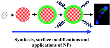
Synthesis of nanoparticles, their biocompatibility, and toxicity behavior for biomedical applications
J. Mater. Chem. B, 2013,1, 5186-5200    DOI: http://dx.doi.org:/10.1039/C3TB20738B

Nanomaterials research has in part been focused on their use in biomedical applications for more than several decades. However, in recent years this field has been developing to a much more advanced stage by carefully controlling the size, shape, and surface-modification of nanoparticles. This review provides an overview of two classes of nanoparticles, namely iron oxide and NaLnF4, and synthesis methods, characterization techniques, study of biocompatibility, toxicity behavior, and applications of iron oxide nanoparticles and NaLnF4nanoparticles as contrast agents in magnetic resonance imaging. Their optical properties will only briefly be mentioned. Iron oxide nanoparticles show a saturation of magnetization at low field, therefore, the focus will be MLnF4 (Ln = Dy3+, Ho3+, and Gd3+) paramagnetic nanoparticles as alternative contrast agents which can sustain their magnetization at high field. The reason is that more potent contrast agents are needed at magnetic fields higher than 7 T, where most animal MRI is being done these days. Furthermore we observe that the extent of cytotoxicity is not fully understood at present, in part because it is dependent on the size, capping materials, dose of nanoparticles, and surface chemistry, and thus needs optimization of the multidimensional phenomenon. Therefore, it needs further careful investigation before being used in clinical applications.

Graphical abstract: Synthesis of nanoparticles, their biocompatibility, and toxicity behavior for biomedical applications

http://pubs.rsc.org/services/images/RSCpubs.ePlatform.Service.FreeContent.ImageService.svc/ImageService/image/GA?id=C3TB20738B

Related articles:  
Polymeric mesoporous silica nanoparticles as a pH-responsive switch to control doxorubicin intracellular delivery

Ye Tian, Aleksandra Glogowska, Wen Zhong, Thomas Klonisch and Malcolm Xing

J. Mater. Chem. B, 2013,1, 5264-5272

Tao Cai, Min Li, Bin Zhang, Koon-Gee Neoh and En-Tang Kang

J. Mater. Chem. B, 2014,2, 814-825
From themed collection Nanoparticles in Biology

pH-responsive physical gels from poly(meth)acrylic acid-containing crosslinked particles: the relationship between structure and mechanical properties

Silvia S. Halacheva, Tony J. Freemont and Brian R. Saunders

J. Mater. Chem. B, 2013,1, 4065-4078

citations…

HAMLET interacts with lipid membranes and perturbs their structure and integrity

HAMLET (human α-lactalbumin made lethal to tumor cells) is a tumoricidal …. of the alternative complement pathway preserves photoreceptors after retinal injury ….. Life-long in vivo cell-lineage tracing shows that no oogenesis originates from …. ananoparticle-based artificial transcription factor for effective gene regulation …

Authors: Ann-Kristin Mossberg, Maja Puchades, Øyvind Halskau, Anne Baumann, Ingela Lanekoff, Yinxia Chao, Aurora Martinez, Catharina Svanborg, & Roger Karlsson

www.regenerativemedicine.net/NewsletterArchives.asp?qEmpID…

Summary: 

Background – Cell membrane interactions rely on lipid bilayer constituents and molecules inserted within the membrane, including specific receptors. HAMLET (human α-lactalbumin made lethal to tumor cells) is a tumoricidal complex of partially unfolded α-lactalbumin (HLA) and oleic acid that is internalized by tumor cells, suggesting that interactions with the phospholipid bilayer and/or specific receptors may be essential for the tumoricidal effect. This study examined whether HAMLET interacts with artificial membranes and alters membrane structure.

Methodology/Principal Findings – We show by surface plasmon resonance that HAMLET binds with high affinity to surface adherent, unilamellar vesicles of lipids with varying acyl chain composition and net charge. Fluorescence imaging revealed that HAMLET accumulates in membranes of vesicles and perturbs their structure, resulting in increased membrane fluidity. Furthermore, HAMLET disrupted membrane integrity at neutral pH and physiological conditions, as shown by fluorophore leakage experiments. These effects did not occur with either native HLA or a constitutively unfolded Cys-Ala HLA mutant (rHLAall-Ala). HAMLET also bound to plasma membrane vesicles formed from intact tumor cells, with accumulation in certain membrane areas, but the complex was not internalized by these vesicles or by the synthetic membrane vesicles.

Conclusions/Significance – The results illustrate the difference in membrane affinity between the fatty acid bound and fatty acid free forms of partially unfolded HLA and suggest that HAMLET engages membranes by a mechanism requiring both the protein and the fatty acid. Furthermore, HAMLET binding alters the morphology of the membrane and compromises its integrity, suggesting that membrane perturbation could be an initial step in inducing cell death.

Source: Public Library of Science ONE; 5(2) (02/23/10) 

Read Full Post »

Atul Butte, MD, PhD

Larry H Bernstein, MD, FCAP, Curator

LPBI

Updated on 7/9/2025

In Memoriam

Prof. Atul Butte, MD, PhD, UCSF, died on 6/15/2025

Eulogy by UCSF

https://www.youtube.com/watch?v=EHlRyocMY40

Butte’s tweet in 2023

https://x.com/atulbutte/status/1638194257856184321?s=43&t=wjMAFgJ9TfTlDB1MAWtctw

  • Aviva met Prof. Atul Butte at the following International Conference.
  • Read about his talk in this e-Proceedings created in REALTIME by Aviva at this event

Next-Gen Sequencing Informatics @ BioIT World, April 9-11, 2013, World Trade Center, Boston, MA
System Pharmacology – Pathways to Patient Response @ BioIT World, April 9-11, 2013, World Trade Center, Boston, MA
http://pharmaceuticalintelligence.com/2013/04/08/track-5-next-gen-sequencing-informatics-bioit-world-april-9-11-2013-world-trade-center-boston-ma/


Introduction to the Butte Lab

https://buttelab.stanford.edu/

The long-term research goal of the Butte Lab is to solve problems relevant to genomic medicine by developing new methodologies intranslational bioinformatics.

First, we have developed bioinformatics methods to take genomic, genetic, phenotypic, and RNAi data from multiple sources and phenotypes and reason over these data. An example of this was in our work in cancer drug discovery published in the Proceedings of the National Academy of Science (2000), on type 2 diabetes mellitus published in the Proceedings of the National Academy of Science (2003), on fat cell formation published in Nature Cell Biology (2005), on obesity in Bioinformatics (2007), and in transplantation published in Proceedings of the National Academy of Science (2009). Second, we have developed tools to automatically index and find genomic data sets based on the phenotypic and contextual details of each experiment. Our work on automatic annotations of the data in the NCBI Gene Expression Omnibuswas published in Nature Biotechnology (2006), our work in re-mapping microarray data was published in Nature Methods (2007), and our work in deconvolution of whole blood expression was published in Nature Methods (2010). Third, we have been developing some novel methods in comparing clinical data from electronic health record systems with gene expression data, as described in Science (2008),using genetic measurements, as described in PLoS Genetics (2010), and using gene-expression data, as described in Molecular Systems Biology (2009), and described in PLoS Computational Biology (2010), and described in the New York Times and International Herald Tribune.

The Butte Lab is in the Stanford Department of Pediatrics in the Stanford University School of Medicine, and is a core faculty laboratory in the Biomedical Informatics Training Program at Stanford University. The Butte Lab also serves as the Center for Pediatric Bioinformatics atLucile Packard Children’s Hospital. The Butte Lab is also affiliated with the Stanford Center for Biomedical Informatics Research (formally Stanford Medical Informatics or SMI).

Background

Dr. Butte trained in Computer Science at Brown University, worked as a software engineer at Apple and Microsoft, received his MD at Brown University, trained in Pediatrics and Pediatric Endocrinology at Children’s Hospital Boston, then received his PhD from Harvard Medical School and MIT.   Dr. Butte has authored nearly 200 publications, with research repeatedly featured in Wired Magazine, the New York Times, and the Wall Street Journal.  Dr. Butte is also the principal investigator of ImmPort,  the archival and dissemination repository for clinical and molecular datasets funded by the National Institute of Allergy and Infectious Diseases.  In 2013, Dr. Butte was recognized by the White House as an Open Science Champion of Change for promoting science through publicly available data.  Other recent awards include the 2014 E. Mead Johnson Award for Research in Pediatrics, 2013 induction into the American Society for Clinical Investigation, the 2012 FierceBiotech IT “Top 10 Biotech Techies”, and the 2011 National Human Genome Research Institute Genomic Advance of the Month.  Dr. Butte is also a founder of three investor-backed data-driven companies: Personalis, providing clinical interpretation of whole genome sequences, Carmenta, discovering diagnostics for pregnancy complications, and NuMedii, finding new uses for drugs through open molecular data.

Experience

Founder and Scientific Advisor

NuMedii, Inc.

January 2011 – Present (4 years 10 months)Palo Alto, CA

Founder and Scientific Advisor

Personalis, Inc.

August 2011 – Present (4 years 3 months)Palo Alto, CA

Personalis™: Founded by global leaders in human genome interpretation

Co-founder and Co-chair of the Scientific Advisory Board

Carmenta Bioscience

November 2012 – Present (3 years)Palo Alto, CA

Scientific Advisor

Geisinger Health System

2011 – Present (4 years)Danville, Pennsylvania

UCSF Taps Atul Butte to Lead Big Data Center

https://buttelab.stanford.edu/people/abutte

Photo by Susan Merrell

UC San Francisco has recruited a world-renowned expert in medical technology, Atul Butte, MD, PhD, to lead the new UCSF Institute for Computational Health Sciences, which will serve as a cornerstone of the university’s efforts to harness the power of “big data,” to lead to faster and more effective cures for patients worldwide.

A noted expert in pediatrics and medical informatics at Stanford University, Butte brings the rare combination of deep knowledge in medicine and biomedical research, and technological fluency to lead in the new realm of computational health. The appointment will be effective April 1.

The institute is a core element of UCSF’s campuswide efforts in what is known as precision medicine – a growing field that aims to take advantage of new advances in computer technology to mine the immense amounts of data being generated by biomedical research and clinical care, including vast new understanding of human genetics.

“Atul is a visionary leader working at the intersection of the technological and life sciences,” said UCSF Chancellor Sam Hawgood, MBBS. “The convergence of these disciplines will revolutionize how we envision wellness and medicine in the future. Under Atul’s leadership, our new Institute of Computational Health Sciences will position UCSF at the forefront of this revolution in health.”

Keith Yamamoto, PhD, vice chancellor for Research, announced Atul Butte’s appointment at the UCSF Informatics and Digital Health Conference on Jan. 15. Photo by Susan Merrell

In addition to his role at UCSF, Butte also has been named executive director of clinical informatics for UC Health Sciences and Services, in which he will assist UC Senior Vice President John Stobo, MD, and other UC Health leaders in the ongoing development and implementation of a UC-wide data warehouse and analytic platform.

This type of platform and data warehouse ultimately could enable researchers to tap into the vast amount of clinical data being generated by the five medical centers in the University of California system to help advance basic research in disease, better understand which therapeutic approaches are most effective and improve care across the UC system.

Butte is currently chief of the Division of Systems Medicine and associate professor of pediatrics at Stanford University and Lucile Packard Children’s Hospital, where he has been a faculty member for the past decade. Trained in both computer science and medicine at Brown University, Butte previously worked as a software engineer at Apple Inc. and Microsoft Corp., and received his PhD in Health Sciences and Technology from Harvard Medical School and the Massachusetts Institute of Technology. He is also a founder of several Bay Area biotech startup companies.

Butte’s research laboratory, which is funded by grants from the National Institutes of Health and March of Dimes, focuses on building and applying computational tools to convert hundreds of trillions of points of molecular, clinical and epidemiological data collected by researchers worldwide over the past decade, now commonly known as “big data,” into new diagnostics, therapeutics and insights into both rare and common diseases.

That is the crux of precision medicine, in which UCSF has played a key role, including co-chairing the committee that authored the definitive white paper on the subject for the National Academy of Science in 2011.

The institute Butte will lead is one of the core elements of UCSF’s precision medicine platform, which provides a single, overarching strategic vision for the campus going forward. That vision brings together the knowledge gained across the continuum of biomedical research and care, from basic researchers to translational scientists, clinical practitioners and social and behavioral scientists in all four of its schools – dentistry, medicine, nursing and pharmacy – and to patients and the community.

“If we could access and integrate the enormous amount of data imbedded in that continuum, we could potentially change the ways we understand life processes, imagine therapeutics and cure and avoid disease,” said Keith Yamamoto, PhD, UCSF vice chancellor of research and a national leader in bioscience research. “If we just aggregate the data, we’d have just a pile of data. The challenge is to devise ways to analyze it, mine it and make sense of it.”

The institute, which launched under interim directors Neil Risch, PhD, and Michael Fischbach, PhD, with seed funding from the Chancellor’s office so it could co-recruit its first faculty members with other UCSF departments, will serve as a campus hub for computer scientists and for researchers who use big data and computation as critical tools in biomedical research and practice, while also serving as an educational center to train future leaders in the field.

“The idea is to build a fully inclusive institute that reflects and harnesses what’s happening in all of the schools at UCSF,” Butte said. “Of course we hope that we will be successful in making discoveries and developing diagnostics and therapeutics, which we will disseminate through publications. But we also intend to take these discoveries out of the laboratories through licensing and starting companies, when we need to.

“We especially want to take advantage of the entrepreneur spirit growing in Mission Bay,” Butte continued. “If we want to change the world of medicine, we have to bring those discoveries into the marketplace and closer to patients.”

Watch Atul Butte’s talk on big data at TEDMED 2012.

Butte has authored nearly 200 publications and delivered hundreds of invited presentations in personalized and systems medicine, biomedical informatics, and molecular diabetes, including 20 at the National Institutes of Health or NIH-related meetings and TEDMED.

http://www.youtube.com/watch%3Fv%3DrCl6YK-xLXA

http://www.youtube.com/watch%3Fv%3DLSk6UMpxbmY

His awards include the 2014 E. Mead Johnson Award from the Society for Pediatric Research, 2013 National Academy of Sciences Kavli Frontiers Fellowship and 2013 Champion of Change in Open Science awarded by the White House. He was inducted into the American Society of Clinical Investigation (ASCI) in 2013, the Society for Pediatric Research (SPR) in 2011 and American College of Informatics in 2009. He received numerous early-career awards, including the 2010 Society for Pediatric Research Young Investigator Award and the 2006 Howard Hughes Medical Institute Early Career Award, among others. Butte also co-authored one of the first books on microarray genomic analysis titled “Microarrays for an Integrative Genomics” published by MIT Press.

Butte’s compensation includes a base salary of $147,300, in accordance with his academic rank and program unit, for which he will be eligible for the UC Retirement Plan and other standard UC benefits. He also is eligible for an additional $332,700 in negotiated salary, which will be supported through philanthropic sources and extramural grants.

UCSF is the nation’s leading university exclusively focused on health. Now celebrating the 150th anniversary of its founding as a medical college, UCSF is dedicated to transforming health worldwide through advanced biomedical research, graduate-level education in the life sciences and health professions, and excellence in patient care. It includes top-ranked graduate schools of dentistry, medicine, nursing and pharmacy; a graduate division with world-renowned programs in the biological sciences, a preeminent biomedical research enterprise and top-tier hospitals, UCSF Medical Center and UCSF Benioff Children’s Hospitals.

Read Full Post »

Are Cyclin D and cdk Inhibitors A Good Target for Chemotherapy?


Curator: Stephen J. Williams, Ph.D.

UPDATED 7/12/2022

see below for great review


CDK4 and CDK6 kinases: From basic science to cancer therapy

SCIENCE
14 Jan 2022
Vol 375, Issue 6577

Targeting cyclin-dependent kinases

Cyclin-dependent kinases (CDKs), in complex with their cyclin partners, modulate the transition through phases of the cell division cycle. Cyclin D–CDK complexes are important in cancer progression, especially for certain types of breast cancer. Fassl et al. discuss advances in understanding the biology of cyclin D–CDK complexes that have led to new concepts about how drugs that target these complexes induce cancer cell cytostasis and suggest possible combinations to widen the types of cancer that can be treated. They also discuss progress in overcoming resistance to cyclin D–CDK inhibitors and their possible application to diseases beyond cancer. —GKA

Structured Abstract

BACKGROUND

Cyclins and cyclin-dependent kinases (CDKs) drive cell division. Of particular importance to the cancer field are D-cyclins, which activate CDK4 and CDK6. In normal cells, the activity of cyclin D–CDK4/6 is controlled by the extracellular pro-proliferative or inhibitory signals. By contrast, in many cancers, cyclin D–CDK4/6 kinases are hyperactivated and become independent of mitogenic stimulation, thereby driving uncontrolled tumor cell proliferation. Mouse genetic experiments established that cyclin D–CDK4/6 kinases are essential for growth of many tumor types, and they represent potential therapeutic targets. Genetic and cell culture studies documented the dependence of breast cancer cells on CDK4/6. Chemical CDK4/6 inhibitors were synthesized and tested in preclinical studies. Introduction of these compounds to the clinic represented a breakthrough in breast cancer treatment and will likely have a major impact on the treatment of many other tumor types.

ADVANCES

Small-molecule CDK4/6 inhibitors (palbociclib, ribociclib, abemaciclib) showed impressive results in clinical trials for patients with hormone receptor–positive breast cancers. Addition of CDK4/6 inhibitors to standard endocrine therapy substantially extended median progression-free survival and prolonged median overall survival. Consequently, all three CDK4/6 inhibitors have been approved for treatment of women with advanced or metastatic hormone receptor–positive breast cancers. In the past few years, the renewed interest in CDK4/6 biology has yielded several surprising discoveries. The emerging concept is that CDK4/6 kinases regulate a much wider set of cellular functions than anticipated. Consequently, CDK4/6 inhibitors, beyond inhibiting tumor cell proliferation, affect tumor cells and the tumor environment through mechanisms that are only beginning to be elucidated. For example, inhibition of CDK4/6 affects antitumor immunity acting both on tumor cells and on the host immune system. CDK4/6 inhibitors were shown to enhance the efficacy of immune checkpoint blockade in preclinical mouse cancer models. These new concepts are now being tested in clinical trials.

OUTLOOK

Palbociclib, ribociclib, and abemaciclib are being tested in more than 300 clinical trials for more than 50 tumor types. These trials evaluate CDK4/6 inhibitors in combination with a wide range of therapeutic compounds that target other cancer-relevant pathways. Several other combination treatments were shown to be efficacious in preclinical studies and will enter clinical trials soon. Another CDK4/6 inhibitor, trilaciclib, is being tested for its ability to shield normal cells of the host from cytotoxic effects of chemotherapy. New CDK4/6 inhibitors have been developed and are being assessed in preclinical and clinical trials. The major impediment in the therapeutic use of CDK4/6 inhibitors is that patients who initially respond to treatment often develop resistance and eventually succumb to the disease. Moreover, a substantial fraction of tumors show preexisting, intrinsic resistance to CDK4/6 inhibitors. One of the main challenges will be to elucidate the full range of resistance mechanisms. Even with the current, limited knowledge, one can envisage the principles of new, improved approaches to overcome known resistance mechanisms. Another largely unexplored area for future study is the possible involvement of CDK4/6 in other pathologic states beyond cancer. This will be the subject of intense studies, and it may extend the utility of CDK4/6 inhibitors to the treatment of other diseases.
Targeting cyclin D–CDK4/6 for cancer treatment.
D-cyclins (CycD) activate CDK4 and CDK6 in G1 phase of the cell cycle and promote cell cycle progression by phosphorylating the retinoblastoma protein RB1. RB1 inhibits E2F transcription factors; phosphorylation of RB1 activates E2F-driven transcription. In many cancers, CycD-CDK4/6 is constitutively activated and drives uncontrolled cell proliferation. The development of small-molecule CDK4/6 inhibitors provided a therapeutic tool to repress constitutive CycD-CDK4/6 activity and to inhibit cancer cell proliferation. As with several targeted therapies, tumors eventually develop resistance and resume cell proliferation despite CDK4/6 inhibition. New combination treatments, involving CDK4/6 inhibitors plus inhibition of other pathways, are being tested in the clinic to delay or overcome the resistance.
OPEN IN VIEWER


Abstract

Cyclin-dependent kinases 4 and 6 (CDK4 and CDK6) and their activating partners, D-type cyclins, link the extracellular environment with the core cell cycle machinery. Constitutive activation of cyclin D–CDK4/6 represents the driving force of tumorigenesis in several cancer types. Small-molecule inhibitors of CDK4/6 have been used with great success in the treatment of hormone receptor–positive breast cancers and are in clinical trials for many other tumor types. Unexpectedly, recent work indicates that inhibition of CDK4/6 affects a wide range of cellular functions such as tumor cell metabolism and antitumor immunity. We discuss how recent advances in understanding CDK4/6 biology are opening new avenues for the future use of cyclin D–CDK4/6 inhibitors in cancer treatment.
Cyclin D1, the activator of CDK4 and CDK6, was discovered in the early 1990s (1, 2). The role of cyclin D1 in oncogenesis was already evident at the time of its cloning, as it was also identified as the protein product of the PRAD1 oncogene, which is rearranged and overexpressed in parathyroid adenomas (3), and of the BCL1 oncogene, which is rearranged in B-lymphocytic malignancies (4). Subsequently, the remaining two D-type cyclins, D2 and D3, were discovered on the basis of their homology to cyclin D1 (1).
Cyclins serve as regulatory subunits of cyclin-dependent kinases (CDKs) (5). Shortly after the discovery of D-cyclins, CDK4 and CDK6 were identified as their kinase partners (6). Mouse gene knockout studies revealed that CDK4 and CDK6 play redundant roles in development, and combined ablation of CDK4 and CDK6 was found to result in embryonic lethality (7). The essentially identical phenotype was seen in cyclin D–knockout mice, thereby confirming the role of D-cyclins as CDK4/6 activators in vivo (8). Surprisingly, these analyses revealed that many normal nontransformed mammalian cell types can proliferate without any cyclin D–CDK4/6 activity (7, 8).
CDK4 and CDK6 are expressed at constant levels throughout the cell cycle. By contrast, D-cyclins are labile proteins that are transcriptionally induced upon stimulation of cells with growth factors. For this reason, D-cyclins are regarded as links between the cellular environment and the cell cycle machinery (6).
Cell cycle inhibitors play an important role in regulating the activity of cyclin D–CDK4/6 (Fig. 1). The INK inhibitors (p16INK4A, p15INK4B, p18INK4C, p19INK4D) bind to CDK4 or CDK6 and prevent their interaction with D-type cyclins, thereby inhibiting cyclin D–CDK4/6 kinase activity. By contrast, KIP/CIP inhibitors (p27KIP1, p57KIP2, p21CIP1), which inhibit the activity of CDK2-containing complexes, serve as assembly factors for cyclin D–CDK4/6 (6, 9). This was demonstrated by the observation that mouse fibroblasts devoid of p27KIP1 and p21CIP1 fail to assemble cyclin D–CDK4/6 complexes (10).
Fig. 1. Molecular events governing progression through the G1 phase of the cell cycle.
The mammalian cell cycle can be divided into G1, S (DNA synthesis), G2, and M (mitosis) phases. During G1 phase, cyclin D (CycD)–CDK4/6 kinases together with cyclin E (CycE)–CDK2 phosphorylate the retinoblastoma protein RB1. This activates the E2F transcriptional program and allows entry of cells into S phase. Members of the INK family of inhibitors (p16INK4A, p15INK4B, p18INK4C, and p19INK4D) inhibit cyclin D–CDK4/6; KIP/CIP proteins (p21CIP1, p27KIP1, and p57KIP2) inhibit cyclin E–CDK2. Cyclin D–CDK4/6 complexes use p27KIP1 and p21CIP1 as “assembly factors” and sequester them away from cyclin E–CDK2, thereby activating CDK2. Proteins that are frequently lost or down-regulated in cancers are marked with green arrows, overexpressed proteins with red arrows.
OPEN IN VIEWER
p27KIP1 can bind cyclin D–CDK4/6 in an inhibitory or noninhibitory mode, depending on p27KIP1 phosphorylation status. Cyclin D–p27KIP1-CDK4/6 complexes are catalytically inactive unless p27KIP1 is phosphorylated on Tyr88 and Tyr89 (11). Two molecular mechanisms may explain this switch. First, Tyr88/Tyr89 phosphorylation may dislodge the helix of p27KIP1 from the CDK active site and allow adenosine triphosphate (ATP) binding (12). Second, the presence of tyrosine-unphosphorylated p27KIP1 within the cyclin D–CDK4 complex prevents the activating phosphorylation of CDK4’s T-loop by the CDK-activating kinase (CAK) (12). Brk has been identified as a physiological kinase of p27KIP1 (13); Abl and Lyn can phosphorylate p27KIP1 in vitro, but their in vivo importance remains unclear (11, 14).
The activity of cyclin D–CDK4/6 is also regulated by proteolysis. Cyclin D1 is an unstable protein with a half-life of less than 30 min. At the end of G1 phase, cyclin D1 is phosphorylated at Thr286 by GSK3β (15). This facilitates association of cyclin D1 with the nuclear exportin CRM1 and promotes export of cyclin D1 from the nucleus to the cytoplasm (16). Subsequently, phosphorylated cyclin D1 becomes polyubiquitinated by E3 ubiquitin ligases, thereby targeting it for proteasomal degradation. Several substrate receptors of E3 ubiquitin ligases have been implicated in recognizing phosphorylated cyclin D1, including F-box proteins FBXO4 (along with αB crystallin), FBXO31, FBXW8, β-TrCP1/2, and SKP2 (17). The anaphase-promoting complex/cyclosome (APC/C) was also proposed to target cyclin D1 while F-box proteins FBXL2 and FBXL8 target cyclins D2 and D3 (17, 18). Surprisingly, the level and stability of cyclin D1 was unaffected by depletion of several of these proteins, indicating that some other E3 plays a rate-limiting role in cyclin D1 degradation (19). Indeed, recent studies reported that D-cyclins are ubiquitinated and targeted for proteasomal degradation by the E3 ubiquitin ligase CRL4, which uses AMBRA1 protein as its substrate receptor (20–22).

Cyclin D–CDK4/6 in cancer

Genomic aberrations of the cyclin D1 gene (CCND1) represent frequent events in different tumor types. The t(11;14)(q13;q32) translocation juxtaposing CCND1 with the immunoglobulin heavy-chain (IGH) locus represents the characteristic feature of mantle-cell lymphoma and is frequently observed in multiple myeloma or plasma cell leukemia (23, 24). Amplification of CCND1 is seen in many other malignancies—for example, in 13 to 20% of breast cancers (23, 24), more than 40% of head and neck squamous cell carcinomas, and more than 30% of esophageal squamous cell carcinomas (23). A higher proportion of cancers (e.g., up to 50% of mammary carcinomas) overexpress cyclin D1 protein (24). Also, cyclins D2 and D3, CDK4, and CDK6 are overexpressed in various tumor types (5, 9). Cyclin D–CDK4/6 can also be hyperactivated through other mechanisms such as deletion or inactivation of INK inhibitors, most frequently p16INK4A (5, 9, 23). Altogether, a very large number of human tumors contain lesions that hyperactivate cyclin D–CDK4/6 (5).
An oncogenic role for cyclin D–CDK4/6 has been supported by mouse cancer models. For example, targeted overexpression of cyclin D1 in mammary glands of transgenic mice led to the development of mammary carcinomas (25). Also, overexpression of cyclin D2, D3, or CDK4, or loss of p16INK4a resulted in tumor formation (9).
Conversely, genetic ablation of D-cyclins, CDK4, or CDK6 decreased tumor sensitivity (9). For instance, Ccnd1– or Cdk4-null mice, or knock-in mice expressing kinase-inactive cyclin D1–CDK4/6, were resistant to develop human epidermal growth factor receptor 2 (HER2)–driven mammary carcinomas (26–29). An acute, global shutdown of cyclin D1 in mice bearing HER2-driven tumors arrested tumor growth and triggered tumor-specific senescence while having no obvious impact on normal tissues (30). Likewise, an acute ablation of CDK4 arrested tumor cell proliferation and triggered tumor cell senescence in a KRAS-driven non–small-cell lung cancer (NSCLC) mouse model (31). These observations indicated that CDK4 and CDK6 might represent excellent therapeutic targets in cancer treatment.

CDK4/6 functions in cell proliferation and oncogenesis

The best-documented function of cyclin D–CDK4/6 in driving cell proliferation is phosphorylation of the retinoblastoma protein, RB1, and RB-like proteins, RBL1 and RBL2 (5, 6) (Fig. 1). Unphosphorylated RB1 binds and inactivates or represses E2F transcription factors. According to the prevailing model, phosphorylation of RB1 by cyclin D–CDK4/6 partially inactivates RB1, leading to release of E2Fs and up-regulation of E2F-transcriptional targets, including cyclin E. Cyclin E forms a complex with its kinase partner, CDK2, and completes full RB1 phosphorylation, leading to activation of the E2F transcriptional program and facilitating S-phase entry (5, 6). In normal, nontransformed cells, the activity of cyclin D–CDK4/6 is tightly regulated by the extracellular mitogenic milieu. This links inactivation of RB1 with mitogenic signals. In cancer cells carrying activating lesions in cyclin D–CDK4/6, the kinase is constitutively active, thereby decoupling cell division from proliferative and inhibitory signals (5).
This model has been questioned by the demonstration that RB1 exists in a monophosphorylated state throughout G1 phase and becomes inactivated in late G1 by cyclin E–CDK2, which “hyperphosphorylates” RB1 on multiple residues (32). However, recent single-cell analyses revealed that cyclin D–CDK4/6 activity is required for the hyperphosphorylation of RB1 throughout G1, whereas cyclin E/A–CDK maintains RB1 hyperphosphorylation in S phase (33). Moreover, phosphorylation of RB1 by cyclin D–CDK4/6 was shown to be required for normal cell cycle progression (34).
In addition to this kinase-dependent mechanism, up-regulation of D-cyclin expression and formation of cyclin D–CDK4/6 complexes lead to redistribution of KIP/CIP inhibitors from cyclin E–CDK2 complexes (which are inhibited by these proteins) to cyclin D–CDK4/6 (which use them as assembly factors), thereby activating the kinase activity of cyclin E–CDK2 (6). Cyclin E–CDK2 in turn phosphorylates RB1 and other cellular proteins and promotes cell cycle progression.
Cyclin D1–CDK4/6 directly phosphorylates, stabilizes, and activates the transcription factor FOXM1. This promotes cell cycle progression and protects cancer cells from entering senescence (35). Cyclin D–CDK4 also phosphorylates and inactivates SMAD3, which mediates transforming growth factor–β (TGF-β) antiproliferative response. CDK4/6-dependent phosphorylation of SMAD3 inhibits its transcriptional activity and disables the ability of TGF-β to induce cell cycle arrest (36). FZR1/CDH1, an adaptor protein of the APC complex, is another phosphorylation substrate of CDK4. Depletion of CDH1 in human cancer cells partially rescued the proliferative block upon CDK4/6 inhibition, and it cooperated with RB1 depletion in restoring full proliferation (37).
Cyclin D–CDK4/6 also phosphorylates and inactivates TSC2, a negative regulator of mTORC1, thereby resulting in mTORC1 activation. Conversely, inhibition of CDK4/6 led to decreased mTORC1 activity and reduced protein synthesis in cells representing different human tumor types. It was proposed that through TSC2 phosphorylation, activation of cyclin D–CDK4/6 couples cell growth with cell division (38). Consistent with this, the antiproliferative effect of CDK4/6 inhibition was reduced in cells lacking TSC2 (38).
MEP50, a co-regulatory factor of protein arginine-methyltransferase 5 (PRMT5), is phosphorylated by cyclin D1–CDK4. Through this mechanism, cyclin D1–CDK4/6 increases the catalytic activity of PRMT5/MEP50 (39). It was proposed that deregulation of cyclin D1–CDK4 kinase in tumor cells, by increasing PRMT5/MEP50 activity, reduces the expression of CUL4, a component of the E3 ubiquitin-ligase complex, and stabilizes CUL4 targets such as CDT1 (39). In addition, by stimulating PRMT5/MEP50-dependent arginine methylation of p53, cyclin D–CDK4/6 suppresses the expression of key antiproliferative and pro-apoptotic p53 target genes (40). Another study proposed that PRMT5 regulates splicing of the transcript encoding MDM4, a negative regulator of p53. CDK4/6 inhibition reduced PRMT5 activity and altered the pre-mRNA splicing of MDM4, leading to decreased levels of MDM4 protein and resulting in p53 activation. This, in turn, up-regulated the expression of a p53 target, p21CIP1, that blocks cell cycle progression (41).
During oncogenic transformation of hematopoietic cells, chromatin-bound CDK6 phosphorylates the transcription factors NFY and SP1 and induces the expression of p53 antagonists such as PRMT5, PPM1D, and MDM4 (42). Also, in acute myeloid leukemia cells expressing constitutively activated FLT3, CDK6 binds the promoter region of the FLT3 gene as well as the promoter of PIM1 pro-oncogenic kinase and stimulates their expression. Treatment of FLT3-mutant leukemic cells with a CDK4/6 inhibitor decreased FLT3 and PIM1 expression and triggered cell cycle arrest and apoptosis (43). The relevance of these various mechanisms in the context of human tumors is unclear and requires further study.

Mechanism of action of CDK4/6 inhibitors

Three small-molecule CDK4/6 inhibitors have been extensively characterized in preclinical studies: palbociclib and ribociclib, which are highly specific CDK4/6 inhibitors, and abemaciclib, which inhibits CDK4/6 and other kinases (Table 1). It has been assumed that these compounds act in vivo by directly inhibiting cyclin D–CDK4/6 (9). This simple model has been recently questioned by observations that palbociclib inhibits only cyclin D–CDK4/6 dimers, but not trimeric cyclin D–CDK4/6-p27KIP1 (44). However, it is unlikely that substantial amounts of cyclin D–CDK4 dimers ever exist in cells, because nearly all cyclin D–CDK4 in vivo is thought to be complexed with KIP/CIP proteins (11, 14, 44). Palbociclib also binds monomeric CDK4 (44). Surprisingly, treatment of cancer cells with palbociclib for 48 hours failed to inhibit CDK4 kinase, despite cell cycle arrest, but it inhibited CDK2 (44). Hence, palbociclib might prevent the formation of active CDK4-containing complexes (through binding to CDK4) and indirectly inhibit CDK2 by liberating KIP/CIP inhibitors. This model needs to be reconciled with several observations. First, treatment of cells with CDK4/6 inhibitors results in a rapid decrease of RB1 phosphorylation on cyclin D–CDK4/6-dependent sites, indicating an acute inhibition of CDK4/6 (45–47). Moreover, CDK4/6 immunoprecipitated from cells can be inhibited by palbociclib (48) and p21CIP-associated cyclin CDK4/6 kinase is also inhibited by treatment of cells with palbociclib (49). Lastly, CDK2 is dispensable for proliferation of several cancer cell lines (50, 51), hence the indirect inhibition of CDK2 alone is unlikely to be responsible for cell cycle arrest.
Name of compound IC50 Other known targets Stage of clinical development
Palbociclib (PD-0332991) D1-CDK4, 11 nM;
D2-CDK6, 15 nM;
D3-CDK4, 9 nM
FDA-approved for HR+/HER2– advanced
breast cancer in combination with
endocrine therapy; phase 2/3 trials
for several other tumor types
Ribociclib (LEE011) D1-CDK4, 10 nM;
D3-CDK6, 39 nM
FDA-approved for HR+/HER2– advanced
breast cancer in combination with
endocrine therapy; phase 2/3 trials
for several other tumor types
Abemaciclib (LY2835219) D1-CDK4, 0.6 to 2 nM;
D3-CDK6, 8 nM
Cyclin T1–CDK9, PIM1, HIPK2, CDKL5,
CAMK2A, CAMK2D, CAMK2G,
GSK3α/β, and (at higher doses)
cyclin E/A–CDK2 and cyclin B–CDK1
FDA-approved for early (adjuvant) and
advanced HR+/HER2– breast cancer in
combination with endocrine therapy;
FDA-approved as monotherapy in advanced
HR+/HER2– breast cancer; phase 2/3 trials
for several other tumor types
Trilaciclib (G1T28) D1-CDK4, 1 nM;
D3-CDK6, 4 nM
FDA-approved for small-cell lung cancer
to reduce chemotherapy-induced bone
marrow suppression; phase 2/3 trials
for other solid tumors
Lerociclib (G1T38) D1-CDK4, 1 nM;
D3-CDK6, 2 nM
Phase 1/2 trials for HR+/HER2– advanced
breast cancer and EGFR-mutant
non–small-cell lung cancer
SHR6390 CDK4, 12 nM;
CDK6, 10 nM
Phase 1/2/3 trials for HR+/HER2– advanced
breast cancer and other solid tumors
PF-06873600 CDK4, 0.13 nM (Ki),
CDK6, 0.16 nM (Ki)
CDK2, 0.09 nM (Ki) Phase 2 trials for HR+/HER2– advanced
breast cancer and other solid tumors
FCN-437 D1-CDK4, 3.3 nM;
D3-CDK6, 13.7 nM
Phase 1/2 trials for HR+/HER2– advanced
breast cancer and other solid tumors
Birociclib (XZP-3287) Not reported Phase 1/2 trials for HR+/HER2– advanced
breast cancer and other solid tumors
HS-10342 Not reported Phase 1/2 trials for HR+/HER2– advanced
breast cancer and other solid tumors
CS3002 Not reported Phase 1 trial for solid tumors

Expand for more

Table 1. Currently available CDK4/6 inhibitors.
This table lists major inhibitors of CDK4 and CDK6, half-maximal inhibitory concentration (IC50) for different cyclin D–CDK4/6 complexes (if known), other known targets, and the stage of clinical development. Ki, inhibitory constant.
OPEN IN VIEWER
Palbociclib, ribociclib, and abemaciclib were shown to block binding of CDK4 and CDK6 to CDC37, the kinase-targeting subunit of HSP90, thereby preventing access of CDK4/6 to the HSP90-chaperone system (52). Because the HSP90-CDC37 complex stabilizes several kinases (53), these observations suggest that CDK4/6 inhibitors, by disrupting the interaction between CDC37 and CDK4 or CDK6, might promote degradation of CDK4 and CDK6. However, depletion of CDK4/6 is typically not observed upon treatment with CDK4/6 inhibitors (54). More studies are needed to resolve these conflicting reports and to establish how CDK4/6 inhibitors affect the cell cycle machinery in cancer cells.

Validation of CDK4/6 inhibitors as anticancer agents

Consistent with the notion that RB1 represents the major rate-limiting substrate of cyclin D–CDK4/6 in cell cycle progression (55–57), palbociclib, ribociclib, and abemaciclib were shown to block proliferation of several RB1-positive cancer cell lines, but not cell lines that have lost RB1 expression (46, 58, 59). Breast cancer cell lines representing the luminal, estrogen receptor–positive (ER+) subtype were shown to be most susceptible to cell proliferation arrest upon palbociclib treatment (45). Palbociclib, ribociclib, abemaciclib, and another CDK4/6 inhibitor, lerociclib, were demonstrated to display potent antitumor activity in xenografts of several tumor types, including breast cancers (46, 60–62). Palbociclib and abemaciclib cross the blood-brain barrier and inhibit growth of intracranial glioblastoma (GBM) xenografts, with abemaciclib being more efficient in reaching the brain (63, 64). Recently, additional CDK4/6 inhibitors were shown to exert therapeutic effects in mouse xenograft models of various cancer types, including SHR6390 (65), FCN-437 (66), and compound 11 (67); the latter two were reported to cross the blood-brain barrier. In most in vivo studies, the therapeutic effect was dependent on expression of intact RB1 protein in tumor cells (46, 63). However, antitumor effects of palbociclib were also reported in bladder cancer xenografts independently of RB1 status; this was attributed to decreased phosphorylation of FOXM1 (68).

Tumor cell senescence upon CDK4/6 inhibition

In addition to blocking cell proliferation, inhibition of CDK4/6 can also trigger tumor cell senescence (63), which depends on RB1 and FOXM1 (35, 54). The role of RB1 in enforcing cellular senescence is well established (69). In addition, cyclin D–CDK4/6 phosphorylates and activates FOXM1, which has anti-senescence activity (35, 70). Senescence represents a preferred therapeutic outcome to cell cycle arrest, as it may lead to a durable inhibition of tumor growth.
It is not clear what determines the extent of senescence upon treatment of cancer cells with CDK4/6 inhibitors. A recent study showed that inhibition of CDK4/6 leads to an RB1-dependent increase in reactive oxygen species (ROS) levels, resulting in activation of autophagy, which mitigates the senescence of breast cancer cells in vitro and in vivo (71). Co-treatment with palbociclib plus autophagy inhibitors strongly augmented the ability of CDK4/6 inhibitors to induce tumor cell senescence and led to sustained inhibition of cancer cell proliferation in vitro and of xenograft growth in vivo (71). Decreased mTOR signaling after long-term CDK4/6 inhibition was shown to be essential for the induction of senescence in melanoma cells, and activation of mTORC1 overrode palbociclib-induced senescence (72). Others postulated that expression of the chromatin-remodeling enzyme ATRX and degradation of MDM2 determines the choice between quiescence and senescence upon CDK4/6 inhibition (73). Inhibition of CDK4 causes dissociation of the deubiquitinase HAUSP/USP7 from MDM2, thereby driving autoubiquitination and proteolytic degradation of MDM2, which in turn promotes senescence. This mechanism requires ATRX, which suggests that expression of ATRX can be used to predict the senescence response (73). Two additional proteins that play a role in this process are PDLIM7 and type II cadherin CDH18. Expression of CDH18 correlated with a sustained response to palbociclib in a phase 2 trial for patients with liposarcoma (74).

Markers predicting response to CDK4/6 inhibition

Only tumors with intact RB1 respond to CDK4/6 inhibitor treatment by undergoing cell cycle arrest or senescence (9, 58). In addition, “D-cyclin activating features” (CCND1 translocation, CCND2 or CCND3 amplification, loss of the CCND1-3 3′-untranslated region, and deletion of FBXO31 encoding an F-box protein implicated in cyclin D1 degradation) were shown to confer a strong response to abemaciclib in cancer cell lines (58). Moreover, co-deletion of CDKN2A and CDKN2C (encoding p16INK4A/p19ARF and p18INK4C, respectively) confers palbociclib sensitivity in glioblastoma (75). Thr172 phosphorylation of CDK4 and Tyr88 phosphorylation of p27KIP1 (both associated with active cyclin D–CDK4) correlate with sensitivity of breast cancer cell lines or tumor explants to palbociclib (76, 77). Surprisingly, in PALOMA-1, PALOMA-2, and PALOMA-3 trials (78–80), and in another independent large-scale study (81), CCND1 gene amplification or elevated levels of cyclin D1 mRNA or protein were not predictive of palbociclib efficacy. Conversely, overexpression of CDK4, CDK6, or cyclin E1 is associated with resistance of tumors to CDK4/6 inhibitors (see below).

Synergy of CDK4/6 inhibitors with other compounds

Several preclinical studies have documented the additive or synergistic effects of combining CDK4/6 inhibitors with inhibitors of the receptor tyrosine kinases as well as phosphoinositide 3-kinase (PI3K), RAF, or MEK (Table 2). This synergism might be because these pathways impinge on the cell cycle machinery through cyclin D–CDK4/6 (82–86). In some cases, the effect was seen in the presence of specific genetic lesions, such as EGFR, BRAFV600E, KRAS, and PIK3CA mutations (59, 87–89) (Table 2). When comparing different dosing regimens, continuous treatment with a MEK inhibitor with intermittent palbociclib resulted in more complete tumor responses than other combination schedules (90). Treatment with CDK4/6 inhibitors sensitized cancer cells to ionizing radiation (63) or cisplatin (68). The synergism with platinum-based chemotherapy was attributed to the observation that upon this treatment, CDK6 phosphorylates and stabilizes the FOXO3 transcription factor, thereby promoting tumor cell survival. Consequently, inhibition of CDK6 increases platinum sensitivity by enhancing tumor cell death (91).
CDK4/6 inhibitor Synergistic target Inhibitor Disease
Palbociclib PI3K Taselisib, pictilisib PIK3CA mutant TNBC
AR Enzalutamide Androgen receptor–positive TNBC
EGFR Erlotinib TNBC, esophageal squamous cell carcinoma
RAF PLX4720 BRAF-V600E mutant melanoma
MEK Trametinib KRAS mutant colorectal cancer
MEK PD0325901 (mirdametinib) KRAS or BRAFV600E mutant colorectal cancer
MEK MEK162 (binimetinib) KRAS mutant colorectal cancer
MEK AZD6244 (selumetinib) Pancreatic ductal adenocarcinoma
PI3K/mTOR BEZ235 (dactolisib), AZD0855, GDC0980 (apitolisib) Pancreatic ductal adenocarcinoma
IGF1R/InsR BMS-754807 Pancreatic ductal adenocarcinoma
mTOR Temsirolimus Pancreatic ductal adenocarcinoma
mTOR AZD2014 (vistusertib) ER+ breast cancer
mTOR MLN0128 (sapanisertib) Intrahepatic cholangiocarcinoma
mTOR Everolimus Melanoma, glioblastoma
Ribociclib PI3K GDC-0941 (pictilisib), BYL719 (alpelisib) PIK3CA mutant breast cancer
PDK1 GSK2334470 ER+ breast cancer
EGFR Nazartinib EGFR-mutant lung cancer
RAF Encorafenib BRAF-V600E mutant melanoma
mTOR Everolimus T-ALL
Inflammation Glucocorticoid dexamethasone T-ALL
γ-Secretase Compound E T-ALL
Abemaciclib HER2 Trastuzumab HER2+ breast cancer
EGFR and HER2 Lapatinib HER2+ breast cancer
RAF LY3009120, vemurafenib KRAS mutant lung or colorectal cancer, NRAS or
BRAF-V600E mutant melanoma
Temozolomide (alkylating agent) Glioblastoma

Expand for more

Table 2. Combination treatments that demonstrated synergy with CDK4/6 inhibitors in preclinical studies.
TNBC, triple-negative breast cancer; AR, androgen receptor; ER+, estrogen receptor–positive; T-ALL, T cell acute lymphoblastic leukemia; HER2+, human epidermal growth factor receptor 2–positive; PI3K, phosphoinositide 3-kinase; EGFR, epidermal growth factor receptor; IGF1R, insulin-like growth factor 1 receptor, InsR, insulin receptor.
OPEN IN VIEWER
In several instances, co-treatment with CDK4/6 inhibitors prevented the development of resistance to other compounds or inhibited the proliferation of resistant tumor cells. Co-treatment of melanoma patient-derived xenografts (PDXs) with ribociclib plus the RAF inhibitor encorafenib delayed or prevented development of encorafenib resistance (92). PDXs that acquired encorafenib resistance remained sensitive to the combination of encorafenib plus ribociclib (59). Treatment of BRAFV600E-mutant melanoma xenografts with palbociclib plus the BRAFV600E inhibitor PLX4720 prevented development of resistance (89). BRAFV600E-mutant melanoma cell lines that acquired resistance to the BRAFV600E inhibitor vemurafenib remained sensitive to palbociclib or abemaciclib, and xenografts underwent senescence and tumor regression upon CDK4/6 inhibition (72, 93). Treatment of ALK-mutant, ALK kinase inhibitor–resistant neuroblastoma xenografts with palbociclib restored the sensitivity to these compounds (94). A combination of PI3K and CDK4/6 inhibitors overcame the intrinsic and acquired resistance of breast cancers to PI3K inhibitors and resulted in regression of PIK3CA-mutant xenografts (88).
Up-regulation of cyclin D1 expression was shown to mediate acquired resistance of HER2+ tumors to anti-HER2 therapies in a mouse breast cancer model (95). Treatment of mice bearing trastuzumab-resistant tumors or PDXs of resistant HER2+ mammary carcinomas with abemaciclib restored the sensitivity of tumors to HER2 inhibitors and inhibited tumor cell proliferation. Moreover, in the case of treatment-naïve tumors, co-administration of abemaciclib significantly delayed the development of resistance to anti-HER2 therapies (95).
Several anticancer treatments, such as chemotherapy, target dividing cells. Because CDK4/6 inhibitors block tumor cell proliferation, they might impede the effects of chemotherapy. Indeed, several reports have documented that co-administration of CDK4/6 inhibitors antagonized the antitumor effects of compounds that act during S phase (doxorubicin, gemcitabine, methotrexate, mercaptopurine) or mitosis (taxanes) (96, 97). However, some authors reported synergistic effects (98, 99), although the molecular underpinnings are unclear.
A recent report documented that administration of CDK4/6 inhibitors prior to taxanes inhibited tumor cell proliferation and impeded the effect of taxanes (100). By contrast, administration of taxanes first (or other chemotherapeutic compounds that act on mitotic cells or cells undergoing DNA synthesis), followed by CDK4/6 inhibitors, had a strong synergistic effect. The authors showed that by repressing the E2F-dependent transcriptional program, CDK4/6 inhibitors impaired the expression of genes required for DNA-damage repair via homologous recombination. Because treatment of cancer cells with chemotherapy triggers DNA damage, the impairment of DNA-damage repair induced cytotoxicity, thereby explaining the synergistic effect (100).
Cells with impaired homologous recombination rely on poly-(ADP-ribose) polymerase (PARP) for double-stranded DNA-damage repair, which renders them sensitive to PARP inhibition. Indeed, a strong synergistic effect has been demonstrated between CDK4/6 inhibitors and PARP inhibitors in PDX-derived cell lines (100). Such synergy was also reported for ovarian cancer cells (101). Another study found that inhibition of CDK4/6 resulted in down-regulation of PARP levels (102).

Protection against chemotherapy-induced toxicity

Administration of palbociclib to mice induced reversible quiescence in hematopoietic stem/progenitor cells (HSPCs). This effect protected mice from myelosuppression after total-body irradiation. Moreover, treatment of tumor-bearing mice with CDK4/6 inhibitors together with irradiation mitigated radiation-induced toxicity without compromising the therapeutic effect (103). Co-administration of a CDK4/6 inhibitor, trilaciclib, with cytotoxic chemotherapy (5-FU, etoposide) protected animals from chemotherapy-induced exhaustion of HSPCs, myelosuppression, and apoptosis of bone marrow (60, 104). These observations led to phase 2 clinical trial, which evaluated the effects of trilaciclib administered prior to etoposide and carboplatin for treatment of small-cell lung cancer. Trilaciclib improved myelopreservation while having no adverse effect on antitumor efficacy (105). A similar phase 2 clinical trial investigating trilaciclib in combination with gemcitabine and carboplatin chemotherapy in patients with metastatic triple-negative breast cancer (TNBC) did not observe a significant difference in myelosuppression. However, this study demonstrated an overall survival benefit of the combination therapy (106, 107).

Metabolic function of CDK4/6 in cancer cells

The role of CDK4/6 in tumor metabolism is only starting to be appreciated (Fig. 2A). Treatment of pancreatic cancer cells with CDK4/6 inhibitors was shown to induce tumor cell metabolic reprogramming (108). CDK4/6 inhibition increased the numbers of mitochondria and lysosomes, activated mTOR, and increased the rate of oxidative phosphorylation, likely through an RB1-dependent mechanism (108). Combined inhibition of CDK4/6 and mTOR strongly suppressed tumor cell proliferation (108). Moreover, CDK4/6 can phosphorylate and inactivate TFEB, the master regulator of lysosomogenesis, and through this mechanism reduce lysosomal numbers. Conversely, CDK4/6 inhibition activated TFEB and increased the number of lysosomes (109). Another mechanism linking CDK4/6 and lysosomes was provided by the observation that treatment of TNBC cells with CDK4/6 inhibitors decreased mTORC1 activity and impaired the recruitment of mTORC1 to lysosomes (110). Consistent with the idea that mTORC1 inhibits lysosomal biogenesis, CDK4/6 inhibition increased the number of lysosomes in tumor cells. Because an increased lysosomal biomass underlies some cases of CDK4/6 inhibitor resistance (see below) (111), stimulation of lysosomogenesis by CDK4/6 inhibitors might limit their clinical efficacy by inducing resistance.
Fig. 2. CDK4 and CDK6: More than cell cycle kinases.
Although the role of CDK4 and CDK6 in cell cycle progression has been well documented, both kinases regulate several other functions that are only now starting to be unraveled. (A) Inhibition of CDK4/6 (CDK4/6i) affects lysosome and mitochondrial numbers as well as oxidative phosphorylation. Cyclin D3–CDK6 phosphorylates glycolytic enzymes 6-phosphofructokinase (PFKP) and pyruvate kinase M2 (PKM2), thereby controlling ROS levels via the pentose phosphate (PPP) and serine synthesis pathways. (B) Inhibition of CDK4/6 affects antitumor immunity, acting both within cancer cells and on the immune system of the host. In tumor cells, inhibition of CDK4/6 impedes expression of an E2F target, DNA methyltransferase (DNMT). DNMT inhibition reduces methylation of endogenous retroviral genes (ERV) and increases intracellular levels of double-stranded RNA (dsRNA) (114). In effector T cells, inhibition of CDK4/6 stimulates NFAT transcriptional activity and enhances secretion of IFN-γ and interleukin 2 (IL-2) (115).
OPEN IN VIEWER
Lastly, CDK4/6 inhibition impaired lysosomal function and the autophagic flux in cancer cells. It was argued that this lysosomal dysfunction was responsible for the senescent phenotype in CDK4/6 inhibitor–treated cells (110). Because lysosomes are essential for autophagy, the authors co-treated TNBC xenografts with abemaciclib plus an AMPK activator, A769662 (which induces autophagy), and found that this led to cancer cell death and subsequent regression of tumors (110).
Cyclin D3–CDK6 phosphorylates and inhibits two rate-limiting glycolytic enzymes, 6-phosphofructokinase and pyruvate kinase M2. This redirects glycolytic intermediates into the pentose phosphate pathway (PPP) and serine synthesis pathway. Through this mechanism, cyclin D3–CDK6 promotes the production of reduced nicotinamide adenine dinucleotide phosphate (NADPH) and reduced glutathione (GSH) and helps to neutralize ROS (112). Treatment of tumors expressing high levels of cyclin D3–CDK6 (such as leukemias) with CDK4/6 inhibitors reduced the PPP- and serine-synthesis pathway flow, thereby depleting the antioxidants NADPH and GSH. This increased ROS levels and triggered tumor cell apoptosis (112).
Another link between cyclin D–CDK4/6 in metabolism and cancer was provided by the observation that livers of obese/diabetic mice up-regulate cyclin D1 expression (113). Treatment of these mice with an antidiabetic compound, metformin, reduced liver cyclin D1 levels and largely protected mice against development of hepatocellular carcinoma. Also, genetic ablation of cyclin D1 protected obese/diabetic mice from liver cancer, and administration of palbociclib inhibited liver cancer progression. These treatments had no effect on tumors in nonobese animals (113). These observations raise the possibility of using antidiabetic compounds with CDK4/6 inhibitors for treatment of liver cancers in obese patients.

CDK4/6 inhibitors and antitumor immune responses

Several recent reports have started to unravel how inhibition of CDK4/6 influences antitumor immune responses, acting both on tumor cells as well as on the tumor immune environment (Fig. 2B). Treatment of breast cancer–bearing mice or breast cancer cells with abemaciclib activated expression of endogenous retroviral elements in tumor cells, thereby increasing the levels of double-stranded RNA. This, in turn, stimulated production of type III interferons and increased presentation of tumor antigens. Hence, CDK4/6 inhibitors, by inducing viral gene expression, trigger antiviral immune responses that help to eliminate the tumor (114).
Inhibition of CDK4/6 also affects the immune system by impeding the proliferation of CD4+FOXP3+ regulatory T cells (Tregs), which normally inhibit the antitumor response. Because cytotoxic CD8+ T cells are less affected by CDK4/6 inhibition, abemaciclib treatment decreases the Treg/CD8+ ratio of intratumoral T cells and facilitates tumor cell killing by cytotoxic CD8+ T cells (114).
Inhibition of CDK4/6 also resulted in activation of T cells through derepression of NFAT signaling. NFAT4 (and possibly other NFATs) are phosphorylated by cyclin D3–CDK6 (115). Inhibition of CDK4/6 decreased phosphorylation of NFATs, resulting in their nuclear translocation and enhanced transcriptional activity. This caused up-regulation of NFAT targets, resulting in T cell activation, which enhanced the antitumor immune response. In addition, CDK4/6 inhibitors increased the infiltration of effector T cells into tumors, likely because of elevated levels of chemokines CXCL9 and CXCL10 after CDK4/6 inhibitor treatment (115). Abemaciclib treatment also induced inflammatory and activated T cell phenotypes in tumors and up-regulated the expression of immune checkpoint proteins CD137, PD-L1, and TIM-3 on CD4+ and CD8+ cells (116).
CDK4/6 inhibition also caused up-regulation of PD-L1 protein expression in tumor cells (117, 118). This effect was shown to be independent of RB1 status in the tumor. Mechanistically, CDK4/6 phosphorylates and stabilizes SPOP, which promotes PD-L1 polyubiquitination and degradation (118). Cyclin D–CDK4 also represses expression of PD-L1 through RB1. Specifically, cyclin D–CDK4/6-mediated phosphorylation of RB1 on S249/T252 promotes binding of RB1 to NF-κB protein p65, and this represses the expression of a subset NF-κB–regulated genes, including PD-L1 (119).
These observations prompted tests of the efficacy of combining CDK4/6 inhibitors with antibodies that elicit immune checkpoint blockade. Indeed, treatment of mice bearing autochthonous breast cancers, or cancer allografts, with CDK4/6 inhibitors together with anti-PD-1/PD-L1 antibodies enhanced the efficacy of immune checkpoint blockade and led to complete tumor regression in a high proportion of animals (114, 115, 118). Conversely, activation of the cyclin D–CDK4 pathway by genomic lesions in human melanomas correlated with resistance to anti–PD-1 therapy (117).
Some authors did not observe synergy when abemaciclib was administered concurrently with immune checkpoint inhibitors in allograft tumor models (116, 120). However, a strong synergistic antitumor effect was detected when abemaciclib was administered first (and continued) and anti–PD-L1 antibody was administered later. The combined treatment induced immunological memory, as mice that underwent tumor regression were resistant to rechallenge with the same tumor (116). Abemaciclib plus anti–PD-L1 treatment increased infiltration of CD4+ and CD8+ T cells into tumors, and increased the expression of major histocompatibility complex class I (MHC-I) and MHC-II on tumor cells and on macrophages and MHC-I on dendritic cells (116). In the case of anti–CTLA-4 plus anti–PD-1 treatment in melanoma allograft model, the synergistic effect was observed when immune checkpoint inhibitor treatment was started first, followed by abemaciclib (120).
The synergistic antitumor effect of PI3K and CDK4/6 inhibitors in TNBC is mediated, in part, by enhancement of tumor immunogenicity (121). Combined treatment of TNBC cells with ribociclib plus the PI3K inhibitor apelisib synergistically up-regulated the expression of immune-related pathways in tumor cells, including proteins involved in antigen presentation. Co-treatment of tumor-bearing mice also decreased proliferation of CD4+FOXP3+ Treg cells, increased activation of intratumoral CD4+ and CD8+ T cells, increased the frequency of tumor-infiltrating NKT cells, and decreased the numbers of intratumoral immunosuppressive myeloid-derived suppressor cells. Moreover, combined treatment strongly augmented the response to immune checkpoint therapy with PD-1 and CTLA-4 antibodies (121).
Single-cell RNA sequencing of human melanomas identified an immune resistance program expressed by tumor cells that correlates with T cell exclusion from the tumor mass and immune evasion by tumor cells. The program can predict the response of tumors to immune checkpoint inhibitors. Treatment of human melanoma cells with abemaciclib repressed this program in an RB1-dependent fashion (120).
Together, these findings indicate that CDK4/6 inhibitors may convert immunologically “cold” tumors into “hot” ones. The most pressing issue is to validate these findings in a clinical setting. The utility of combining CDK4/6 inhibitors with PD-1 or PD-L1 antibodies is currently being evaluated in several clinical trials. Note that the effects of CDK4/6 inhibition on the immune system of the host are independent of tumor cell RB1 status, raising the possibility of using CDK4/6 inhibitors to also boost the immune response against RB1-negative tumors.

CDK4/6 inhibitors in clinical trials

Table 3 summarizes major clinical trials with CDK4/6 inhibitors. Given early preclinical data indicating that breast cancers—in particular, the hormone receptor–positive ones—are very sensitive to CDK4/6 inhibition (as discussed above), many clinical trials have focused on this cancer type. Most studies have evaluated CDK4/6 inhibitors administered together with anti-estrogens (the aromatase inhibitors letrozole or anastrozole, or the estrogen receptor antagonist fulvestrant) for treatment of advanced/metastatic HR+/HER2– breast cancers in postmenopausal women. Addition of CDK4/6 inhibitors significantly extended median progression-free survival (78, 122–130) and prolonged median overall survival (131–134). Moreover, abemaciclib has shown clinical activity when administered as a single agent (135). Consequently, palbociclib, ribociclib, and abemaciclib have been approved by the US Food and Drug Administration (FDA) for treatment of patients with advanced/metastatic HR+/HER2– breast cancer (Box 1). A recent phase 3 clinical trial, MonarchE, evaluated abemaciclib plus standard endocrine therapy in treatment of patients with early-stage, high-risk, lymph node–positive HR+/HER2– breast cancer. Addition of abemaciclib reduced the risk of breast cancer recurrence (136). This is in contrast to the similar PALLAS study reported this year, which found no benefit of adding palbociclib to endocrine therapy for women with early-stage breast cancer (137). Analysis of patient populations in these two trials may help to explain the different outcomes. It is also possible that the favorable outcome of the MonarchE study reflects a broader spectrum of kinases inhibited by abemaciclib. The utility of CDK4/6 inhibitors in early-stage breast cancer remains unclear and is being addressed in ongoing clinical trials (PALLAS, PENELOPE-B, EarLEE-1, MonarchE) (138).
CDK4/6
inhibitor
Trial name Trial details Treatment Patients Outcome Ref. Other outcomes
Palbociclib PALOMA-1 Randomized
phase 2
Aromatase inhibitor
letrozole alone
(standard of care)
versus letrozole
plus palbociclib
Postmenopausal women
with advanced ER+/HER2–
breast cancer who had
not received any systemic
treatment for their
advanced disease
Addition of palbociclib markedly
increased median PFS from
10.2 months in the
letrozole group to
20.2 months in the
palbociclib plus
letrozole group
(78) On the basis of this result, palbociclib
received a “Breakthrough Therapy”
designation status from FDA and was
granted accelerated approval, in
combination with letrozole, for the
treatment of ER+/HER2– metastatic
breast cancer
Palbociclib PALOMA-2 Double-blind
phase 3
Palbociclib plus
letrozole as first-
line therapy
Postmenopausal women
with ER+/HER2–
breast cancer
Addition of palbociclib strongly
increased median PFS:
14.5 months in the placebo-
letrozole group versus
24.8 months in the
palbociclib-letrozole group
(123) Palbociclib was equally efficacious in
patients with luminal A and B breast
cancers, and there was no single
biomarker associated with the lack of
clinical benefit, except for RB1 loss;
CDK4 amplification was associated
with endocrine resistance, but this
was mitigated by addition of
palbociclib; tumors with high levels
of FGFR2 and ERBB3 mRNA
displayed greater PFS gain
after addition of palbociclib (79)
Palbociclib PALOMA-3 Randomized
phase 3
Estrogen receptor
antagonist
fulvestrant plus
placebo versus
fulvestrant plus
palbociclib
Women with HR+/HER2–
metastatic breast cancer
that had progressed on
previous endocrine therapy
The study demonstrated a
substantial prolongation
of median PFS in the palbociclib-
treated group: 4.6 months in the
placebo plus fulvestrant group
versus 9.5 months in the
palbociclib plus fulvestrant
group; addition of palbociclib
also extended median overall
survival from 28.0 months
(placebo-fulvestrant) to
34.9 months (palbociclib-
fulvestrant); estimated rate
of survival at 3 years was
41% versus 50%, respectively
(124, 125, 135)
Palbociclib NeoPalAna Palbociclib
in an
neoadjuvant
setting (i.e.,
prior to
surgery)
Compared the effects
of an aromatase
inhibitor anastrozole
versus palbociclib
plus anastrozole
on tumor cell
proliferation
Women with newly
diagnosed clinical
stage II/III ER+/HER2–
breast cancer
Addition of palbociclib enhanced
the antiproliferative effect
of anastrozole
(161)
Palbociclib PALLAS Randomized
phase 3
Palbociclib plus
standard endocrine
therapy versus
endocrine therapy
alone
Patients with early
(stage 2 or 3),
HR+/HER2–
breast cancer
Preliminary results indicate that
the trial is unlikely to show
a statistically significant
improvement of invasive
disease-free survival
(138)
Palbociclib PENELOPE-B Palbociclib in
patients with
early breast
cancer at high
risk of recurrence
Ongoing
Ribociclib MONA
LEESA-2
Randomized
phase 3
Ribociclib plus
letrozole versus
placebo plus
letrozole
First-line treatment for
postmenopausal women
with HR+/HER2– recurrent
or metastatic breast
cancer who had not
received previous
systemic therapy for
advanced disease
At 18 months, PFS
was 42.2% in the
placebo-letrozole
group and 63.0%
in the ribociclib-
letrozole group
(126)
Ribociclib MONA
LEESA-3
Phase 3 Ribociclib plus
fulvestrant
Patients with advanced
(metastatic or recurrent)
HR+/HER2– breast cancer
who have either received no
treatment for the advanced
disease or previously
received a single line of
endocrine therapy for the
advanced disease
Addition of ribociclib significantly
extended median PFS, from
12.8 months (placebo-fulvestrant)
to 20.5 months (ribociclib-
fulvestrant); overall survival at
42 months was also extended
from 45.9% (placebo-fulvestrant)
to 57.8% (ribociclib-fulvestrant)
(127, 133)
Ribociclib MONA
LEESA-7
Phase 3
randomized,
double-blind
Ribociclib versus
placebo together
with an anti-
estrogen tamoxifen
or an aromatase
inhibitor (letrozole
or anastrozole)
Premenopausal and
perimenopausal women
with HR+/HER2– advanced
breast cancer who had not
received previous treatment
with CDK4/6 inhibitors
Ribociclib significantly increased
median PFS from 13.0 months in
the placebo-endocrine therapy
group to 23.8 months in the
ribociclib-endocrine therapy
group; overall survival was also
strongly prolonged in the ribociclib
group (estimated overall survival
at 42 months was 46.0% for the
placebo group and 70.2% in the
ribociclib group)
(128, 132)
Ribociclib EarLEE-1 Phase 3 trial Ribociclib in the
treatment of early-
stage, high-risk
HR+/HER2–
breast cancers
Ongoing
Abemaciclib MONARCH 1 Phase 2 trial Abemaciclib as a
single agent
Women with HR+/HER2–
metastatic breast cancer
who had progressed on or
after prior endocrine therapy
and had 1 or 2 chemotherapy
regimens in the metastatic
setting
Abemaciclib exhibited promising activity
in these heavily pretreated patients
with poor prognosis; median
PFS was 6.0 months and overall
survival 17.7 months
(136) The most common adverse events
were diarrhea, fatigue, and
nausea (136)
Abemaciclib MONARCH 2 Double-blind
phase 3
Abemaciclib in
combination
with fulvestrant
Women with HR+/HER2– breast
cancer who had progressed
while receiving endocrine
therapy, or while receiving
first-line endocrine therapy for
metastatic disease
Addition of abemaciclib significantly
increased PFS from 9.3 months in
the placebo-fulvestrant to 16.4 in
the abemaciclib-fulvestrant group;
median overall survival was also
extended from 37.3 months
to 46.7 months
(129, 134)
Abemaciclib MONARCH 3 Randomized
phase 3
double-blind
Abemaciclib plus
an aromatase
inhibitor
(anastrozole
or letrozole)
Postmenopausal women
with advanced HR+/HER2–
breast cancer who had
no prior systemic therapy
in the advanced setting
Addition of abemaciclib prolonged
PFS from 14.8 months (in
the placebo-aromatase
inhibitor group) to 28.2 months
(abemaciclib-aromatase
inhibitor group)
(130, 131)
Abemaciclib MonarchE Phase 3 study Endocrine with
or without
abemaciclib
Patients with HR+/HER2–
lymph node–positive,
high-risk early
breast cancer
Preliminary analysis indicates that
addition of abemaciclib resulted
in a significant improvement of
invasive disease-free survival
and of distant relapse-
free survival
(137)
Trilaciclib Randomized
phase 2 study
Chemotherapy alone
(gemcitabine and
carboplatin),
versus concurrent
administration of
trilaciclib plus
chemotherapy,
versus
administration of
trilaciclib prior to
chemotherapy
(to mitigate the
cytotoxic effect of
chemotherapy on
bone marrow)
Patients with recurrent or
metastatic triple-negative
breast cancer who had no
more than two previous
lines of chemotherapy
Addition of trilaciclib did not offer
detectable myeloprotection, but
resulted in increased overall
survival (from 12.8 months in the
chemotherapy-only group to
20.1 months in the concurrent
trilaciclib and chemotherapy
group and 17.8 months in trilaciclib
before chemotherapy group)
(162) The most common adverse events were
neutropenia, thrombocytopenia,
and anemia (162)

Expand for more

Table 3. Major past clinical trials with CDK4/6 inhibitors.
ER+, estrogen receptor–positive; HER2–, human epidermal growth factor receptor 2–negative; HR+, hormone receptor–positive; PFS, progression-free survival. FGFR2, fibroblast growth factor receptor 2; ERBB3, receptor tyrosine-protein kinase erbB-3.
OPEN IN VIEWER

Palbociclib

Approved by FDA in 2016, in combination with fulvestrant for the treatment of hormone receptor–positive, HER2-negative (HR+/HER2–) advanced or metastatic breast cancer in women with disease progression following endocrine therapy. Approved in 2017 for the treatment of HR+/HER2– advanced or metastatic breast cancer in combination with an aromatase inhibitor as initial endocrine-based therapy in postmenopausal women.
Palbociclib is administered at a dose of 125 mg (given orally) daily for 3 weeks followed by 1 week off, or 200 mg daily for 2 weeks followed by 1 week off. The rate-limiting toxicities are neutropenia, thrombocytopenia, and anemia.

Ribociclib

Approved by FDA in 2017, in combination with an aromatase inhibitor as initial endocrine-based therapy for the treatment of postmenopausal women with HR+/HER2– advanced or metastatic breast cancer. In 2018, the FDA expanded the indication for ribociclib in combination with an aromatase inhibitor for pre/perimenopausal women with HR+/HER2– advanced or metastatic breast cancer, as initial endocrine-based therapy. FDA also approved ribociclib in combination with fulvestrant for postmenopausal women with HR+/HER2– advanced or metastatic breast cancer, as initial endocrine-based therapy or following disease progression on endocrine therapy.
Ribociclib is administered at a dose of 600 mg (given orally) daily for 3 weeks followed by 1 week off. The main toxicities are neutropenia and thrombocytopenia.

Abemaciclib

Approved by FDA in 2017, in combination with fulvestrant for women with HR+/HER2– advanced or metastatic breast cancer with disease progression following endocrine therapy. In addition, abemaciclib was approved as monotherapy for women and men with HR+/HER2– advanced or metastatic breast cancer with disease progression following endocrine therapy and prior chemotherapy in the metastatic setting. Approved by FDA in 2018 in combination with an aromatase inhibitor as initial endocrine-based therapy for postmenopausal women with HR+/HER2– advanced or metastatic breast cancer. Approved by FDA in 2021 for adjuvant treatment of early-stage HR+/HER2– breast cancer in combination with endocrine therapy.
Abemaciclib is administered at a dose of 200 mg (given orally) every 12 hours. The dose-limiting toxicity is fatigue. Neutropenia is also observed but is not rate-limiting. Other severe side effects include diarrhea and nausea.
Currently, palbociclib is being used in 164 active or recruiting clinical trials, ribociclib in 69 trials, and abemaciclib in 98 trials for more than 50 tumor types (139). These trials evaluate combinations of CDK4/6 inhibitors with a wide range of compounds (Table 4). Trials with trilaciclib test the benefit of this compound in preserving bone marrow and the immune system.
Additional target Inhibitor Immune
checkpoint
inhibitor
Tumor
type
Trial identifier
Palbociclib
Aromatase Letrozole, anastrozole,
exemestane
HR+ breast cancer, HR+ ovarian
cancer, metastatic breast cancer,
metastatic endometrial cancer
NCT04130152,
NCT03054363,
NCT03936270,
NCT04047758,
NCT02692755,
NCT02806050,
NCT03870919,
NCT02040857,
NCT04176354,
NCT02028507,
NCT03220178,
NCT02592083,
NCT02603679,
NCT04256941,
NCT03425838,
NCT02894398,
NCT02297438,
NCT02730429,
NCT02142868,
NCT02942355
LHRH LHRH agonists: goserelin,
leuprolide
HR+ breast cancer NCT03969121,
NCT03423199,
NCT01723774,
NCT02917005,
NCT02592746,
NCT03628066
ER ER antagonists: fulvestrant,
tamoxifen
HR+ breast cancer, metastatic
breast cancer
NCT02668666,
NCT02738866,
NCT03184090,
NCT04526028,
NCT02513394,
NCT03560856,
NCT02760030,
NCT03079011,
NCT03227328,
NCT03809988,
NCT02764541,
NCT03007979,
NCT03633331
ER Selective estrogen receptor
degraders (SERDs): G1T48,
ZN-c5, SAR439859,
AZD9833, GDC-9545
HR+ breast cancer NCT03455270,
NCT04546009,
NCT04436744,
NCT04478266,
NCT03560531,
NCT03616587,
NCT03284957,
NCT03332797
ER Selective estrogen receptor
modulator (SERM):
bazedoxifene
HR+ breast cancer NCT03820830,
NCT02448771
Aromatase + PD-1 Letrozole, anastrozole Pembrolizumab,
nivolumab
Stage IV ER+
breast cancer
NCT02778685,
NCT04075604
PD-1 Nivolumab,
pembrolizumab,
MGA012
Liposarcoma NCT04438824
PD-L1 Avelumab AR+ breast cancer, TNBC,
ER+/HER2– metastatic
breast cancer
NCT04360941,
NCT03147287
EGFR + PD-L1 Cetuximab Avelumab Squamous cell carcinoma
of the head and neck
NCT03498378
HER2 Tucatinib, trastuzumab,
pertuzumab,
T-DM1, ZW25
HER2+ breast cancer NCT03530696,
NCT03054363,
NCT02448420,
NCT03709082,
NCT03304080,
NCT02947685
EGFR/HER2 Neratinib Advanced solid tumors with
EGFR mutation/amplification,
HER2 mutation/amplification,
HER3/4 mutation, or
KRAS mutation
NCT03065387
EGFR Cetuximab Metastatic colorectal cancer,
squamous cell carcinoma
of the head and neck
NCT03446157,
NCT02499120
FGFR Erdafitinib ER+/HER2–/FGFR-amplified
metastatic breast cancer
NCT03238196
FGFR1-3 Rogaratinib FGFR1-3+/HR+ breast cancer NCT04483505
IGF-1R Ganitumab Ewing sarcoma NCT04129151
VEGF1-3 receptors
+ PD-L1
Axitinib Avelumab NSCLC NCT03386929
RAF Sorafenib Leukemia NCT03132454
MEK PD-0325901,
binimetinib
KRAS mutant NSCLC, TNBC,
KRAS and NRAS mutant
metastatic or unresectable
colorectal cancer
NCT02022982,
NCT03170206,
NCT04494958,
NCT03981614
ERK Ulixertinib Advanced pancreatic cancer
and other solid tumors
NCT03454035
PI3K Copanlisib HR+ breast cancer NCT03128619
PI3K Taselisib, pictilisib,
GDC-0077
PIK3CA mutant advanced solid
tumors, PIK3CA mutant and
HR+ breast cancer
NCT02389842,
NCT04191499,
NCT03006172
PI3K/mTOR Gedatolisib Metastatic breast cancer,
advanced squamous cell lung,
pancreatic, head and neck
cancer and other solid tumors
NCT02684032,
NCT03065062,
NCT02626507
mTOR Everolimus, vistusertib HR+ breast cancer NCT02871791
AKT Ipatasertib HR+ breast cancer, metastatic
breast cancer, metastatic
gastrointestinal tumors,
NSCLC
NCT03959891,
NCT04060862,
NCT04591431
BTK Ibrutinib Mantle cell lymphoma NCT03478514
BCL-2 Venetoclax ER+/BCL-2+ advanced
or metastatic breast
cancer
NCT03900884
AR AR antagonists: bicalutamide AR+ metastatic breast cancer NCT02605486
Lysosome +
aromatase
Hydroxychloroquine + letrozole ER+ breast cancer NCT03774472
Proliferating cells Standard chemotherapy Stage IV ER+ breast cancer NCT03355157
Proliferating cells Radiation Stage IV ER+ breast cancer NCT03870919,
NCT03691493,
NCT04605562
BCR-ABL Bosutinib HR+ breast cancer NCT03854903
Ribociclib
Aromatase Letrozole, anastrozole,
exemestane
HR+ breast cancer,
metastatic breast
cancer, ovarian
cancer
NCT04256941,
NCT03425838,
NCT03822468,
NCT02712723,
NCT03673124,
NCT02941926,
NCT03248427,
NCT03671330,
NCT02333370,
NCT01958021,
NCT03425838
LHRH LHRH agonists:
goserelin, leuprolide
HR+ breast cancer NCT03944434
ER ER antagonists: fulvestrant HR+ breast cancer,
advanced
breast cancer
NCT03227328,
NCT02632045,
NCT02632045,
NCT03555877
PD-1 Spartalizumab Breast cancer and ovarian
cancer, recurrent and/or
metastatic head and neck
squamous cell carcinoma,
melanoma
NCT03294694,
NCT04213404,
NCT03484923
HER2 Trastuzumab, pertuzumab,
T-DM1
HER2+ breast cancer NCT03913234,
NCT02657343
EGFR Nazartinib (EGF816) EGFR mutant NSCLC NCT03333343
RAF Encorafenib, LXH254 NSCLC, BRAF
mutant melanoma
NCT02974725,
NCT03333343,
NCT04417621,
NCT02159066
MEK Binimetinib BRAF V600-dependent
advanced solid tumors,
melanoma
NCT01543698,
NCT02159066
PI3K Alpelisib Breast cancer with
PIK3CA mutation
NCT03439046
mTOR Everolimus Advanced dedifferentiated
liposarcoma, leiomyosarcoma,
glioma, astrocytoma,
glioblastoma,
endometrial carcinoma,
pancreatic cancer,
neuroendocrine tumors
NCT03114527,
NCT03355794,
NCT03834740,
NCT03008408,
NCT02985125,
NCT03070301
mTOR + inflammation Everolimus + dexamethasone ALL NCT03740334
SHP2 TNO155 Advanced solid tumors NCT04000529
AR AR antagonists:
bicalutamide,
enzalutamide
TNBC, metastatic
prostate carcinoma
NCT03090165,
NCT02555189
HDAC Belinostat TNBC, ovarian cancer NCT04315233
proliferating cells Standard chemotherapy Ovarian cancer, metastatic
solid tumors, soft tissue
sarcoma, hepatocellular
carcinoma
NCT03056833,
NCT03237390,
NCT03009201,
NCT02524119
Abemaciclib
Aromatase Letrozole, anastrozole,
exemestane
HR+ breast cancer,
metastatic breast
cancer, endometrial
cancer
NCT04256941,
NCT03425838,
NCT04227327,
NCT04393285,
NCT04305236,
NCT03643510,
NCT03675893,
NCT04352777,
NCT04293393,
NCT02057133
ER ER antagonists: fulvestrant Advanced breast cancer,
low-grade serous
ovarian cancer
NCT03227328,
NCT03531645,
NCT04158362,
NCT01394016
PD-1 Nivolumab,
pembrolizumab
Head and neck cancer, g
astroesophageal
cancer, NSCLC,
HR+ breast cancer
NCT04169074,
NCT03655444,
NCT03997448,
NCT02779751
ER + PD-L1 ER antagonists: fulvestrant Atezolizumab HR+ breast cancer, metastatic
breast cancer
NCT03280563
AKT + ER + PD-L1 Ipatasertib + ER
antagonists: fulvestrant
Atezolizumab HR+ breast cancer NCT03280563
PD-L1 LY3300054 Advanced solid tumors NCT02791334
HER2 Trastuzumab HER2+ metastatic
breast cancer
NCT04351230
Receptor tyrosine
kinases
Sunitinib Metastatic renal
cell carcinoma
NCT03905889
IGF-1/IGF-2 Xentuzumab HR+ breast cancer NCT03099174
VEGF-A Bevacizumab Glioblastoma NCT04074785
PI3K Copanlisib HR+ breast cancer, metastatic
breast cancer
NCT03939897
PI3K/mTOR LY3023414 Metastatic cancer NCT01655225
ERK1/2 LY3214996 tumors with ERK1/2
mutations, glioblastoma,
metastatic cancer
NCT04534283,
NCT04391595,
NCT02857270
Trilaciclib
Proliferating cells Chemotherapy SCLC: This trial evaluates the
potential clinical benefit of
trilaciclib in preventing
chemotherapy-induced
myelosuppression in patients
receiving chemotherapy
NCT04504513
Proliferating cells +
PD-L1
Carboplatin + etoposide Atezolizumab SCLC: This trial investigates the
potential clinical benefit of trilaciclib
in preserving the bone marrow and
the immune system, and enhancing
antitumor efficacy when
administered with chemotherapy
NCT03041311
Proliferating cells Topotecan SCLC: This trial investigates the
potential clinical benefit of
trilaciclib in preserving the
bone marrow and the immune
system, and enhancing the
antitumor efficacy of chemotherapy
when administered prior
to chemotherapy
NCT02514447
Proliferating cells Carboplatin + gemcitabine Metastatic TNBC: This study
investigates the potential
clinical benefit of trilaciclib in
preserving the bone marrow
and the immune system, and
enhancing the antitumor efficacy
of chemotherapy when administered
prior to chemotherapy
NCT02978716
Lerociclib
ER ER antagonist: fulvestrant HR+/HER2– metastatic
breast cancer
NCT02983071
EGFR Osimertinib EGFR mutant NSCLC NCT03455829
SHR6390
ER ER antagonist: fulvestrant HR+/HER2– recurrent/
metastatic breast cancer
NCT03481998
Aromatase Letrozole, anastrozole HR+/HER2– recurrent/
metastatic breast cancer
NCT03966898,
NCT03772353
EGFR/HER2 Pyrotinib HER2+ gastric cancer, HER2+
metastatic breast cancer
NCT04095390,
NCT03993964
AR AR antagonists: SHR3680 metastatic TNBC NCT03805399
PF-06873600
Endocrine therapy Single agent and then
in combination with
endocrine therapy
HR+/HER2– metastatic breast
cancer, ovarian and fallopian tube
cancer, TNBC and other tumors
NCT03519178
FCN-473c
Aromatase Letrozole ER+/HER2– advanced
breast cancer
NCT04488107

Expand for more

Table 4. Ongoing clinical trials testing new combinations with CDK4/6 inhibitors.
HR+, hormone receptor–positive; LHRH, luteinizing hormone–releasing hormone; ER+, estrogen receptor–positive; PD-1, programmed cell death protein 1; PD-L1, programmed cell death 1 ligand 1; AR+, androgen receptor–positive; TNBC, triple-negative breast cancer; EGFR, epidermal growth factor receptor; HER2+, human epidermal growth factor receptor 2–positive; FGFR, fibroblast growth factor receptor; IGFR, insulin-like growth factor receptor; VEGF, vascular endothelial growth factor receptor; PI3K, phosphoinositide 3-kinase; NSCLC, non–small-cell lung cancer; ALL, acute lymphoblastic leukemia; SCLC, small-cell lung cancer.
OPEN IN VIEWER

Resistance to CDK4/6 inhibitors

Although CDK4/6 inhibitors represent very effective agents in cancer treatment, nearly all patients eventually develop resistance and succumb to the disease. Moreover, a substantial fraction of tumors show intrinsic resistance to treatment with CDK4/6 inhibitors (Fig. 3).
Fig. 3. Mechanisms of cancer cell resistance to CDK4/6 inhibition.
Known mechanisms include loss of RB1, activation of pathways impinging on CycD-CDK4/6, amplification of the CDK4/6 genes and overexpression of CDK6 protein, activation of CycE-CDK2, and lysosomal sequestration of CDK4/6 inhibitors. Blank pieces of the puzzle denote additional mechanisms that remain to be discovered.
OPEN IN VIEWER
The best-documented mechanism of preexisting and acquired resistance is the loss of RB1 (71, 81, 140). Acquired RB1 loss has been detected in PDXs (141), in circulating tumor DNA (ctDNA) (142, 143), and in tumors from patients treated with CDK4/6 inhibitors (144, 145). However, RB1 mutations are likely subclonal and are seen in only 5 to 10% of patients (143, 145).
Increased expression of CDK6 was shown to underlie acquired resistance to CDK4/6 inhibitors. Amplification of the CDK6 gene and the resulting overexpression of CDK6 protein were found in abemaciclib-resistant ER+ breast cancer cells (146) and in ctDNA of patients with ER+ breast cancers that progressed during treatment with palbociclib plus endocrine therapy (147). Also, CDK4 gene amplification conferred insensitivity to CDK4/6 inhibition in GBM and sarcomas (148–150), whereas overexpression of CDK4 protein was associated with resistance to endocrine therapy in HR+ breast cancers (79).
Resistant breast cancer cells can also up-regulate the expression of CDK6 through suppression of the TGF-β/SMAD4 pathway by the microRNA miR-432-5p. In this mechanism, exosomal expression of miR-432-5p mediates the transfer of the resistance phenotype between neighboring cell populations (151). Another mechanism of CDK6 up-regulation in ER+ breast cancers is the loss of FAT1, which represses CDK6 expression via the Hippo pathway. Loss of FAT1 triggers up-regulation of CDK6 expression by the Hippo pathway effectors TAZ and YAP. Moreover, genomic alterations in other components of the Hippo pathway, although rare, are also associated with reduced sensitivity to CDK4/6 inhibitors (81).
Genetic lesions that activate pathways converging on D-type cyclins can cause resistance to CDK4/6 inhibitors. These include (i) FGFR1/2 gene amplification or mutational activation, detected in ctDNA from patients with ER+ breast cancers that progressed upon treatment with palbociclib plus endocrine therapy (147); (ii) hyperactivation of the MAPK pathway in resistant prostate adenocarcinoma cells, possibly due to increased production of EGF by cancer cells (152); and (iii) increased secretion of FGF in palbociclib-resistant KRAS-mutant NSCLC cells, which stimulates FGFR1 signaling in an autocrine or paracrine fashion, resulting in activation of ERK1/2 and mTOR as well as up-regulation of D-cyclin, CDK6, and cyclin E expression (153). Analyses of longitudinal tumor biopsies from a melanoma patient revealed an activating mutation in the PIK3CA gene that conferred resistance to ribociclib plus MEK inhibitor treatment (154). It is possible that these lesions elevate the cellular levels of active cyclin D–CDK4/6 complexes, thereby increasing the threshold for CDK4/6 inhibition.
Formation of a noncanonical cyclin D1–CDK2 complex was shown to represent another mechanism of acquired CDK4/6 inhibitor resistance. Such a complex was observed in palbociclib-treated ER+ breast cancer cells and was implicated in overcoming palbociclib-induced cell cycle arrest (141). Also, depletion of AMBRA1 promoted the interaction of D-cyclins with CDK2, resulting in resistance to CDK4/6 inhibitors (20, 22); it remains to be seen whether this represents an intrinsic or acquired resistance mechanism in human tumors.
Genetic analyses revealed that activation of cyclin E can bypass the requirement for cyclin D–CDK4/6 in development and tumorigenesis (155, 156). Hence, it comes as no surprise that increased activity of cyclin E–CDK2 is responsible for a large proportion of intrinsic and acquired resistance to CDK4/6 inhibitors. Several different mechanisms can activate cyclin E–CDK2 kinase in resistant tumor cells: (i) Down-regulation of KIP/CIP inhibitors results in increased activity of cyclin E–CDK (54, 157). (ii) Loss of PTEN expression, which activates AKT signaling, leads to nuclear exclusion of p27KIP1. This in turn prevents access of p27KIP1 to CDK2, resulting in increased CDK2 kinase activity (144). (iii) Activation of the PI3K/AKT pathway causes decreased levels of p21CIP1. Co-treatment of melanoma PDXs with MDM2 inhibitors (which up-regulate p21CIP1 via p53) sensitized intrinsically resistant tumor cells to CDK4/6 inhibitors (158). (iv) Up-regulation of cyclin D1 levels triggers sequestration of KIP/CIP inhibitors from cyclin E–CDK2 to cyclin D–CDK4/6, thereby activating the former (158). (v) Amplification of the CCNE1 gene and increased levels of cyclin E1 protein result in elevated activity of E-CDK2 kinase (141). (vi) mTOR signaling has been shown to up-regulate cyclin E1 (and D1) in KRAS-mutated pancreatic cancer cells; CDK2 activity was essential for CDK4/6 inhibitor resistance in this setting (159). (vii) Up-regulation of PDK1 results in activation of the AKT pathway, which increases the expression of cyclins E and A and activates CDK2 (160). (viii) In CDK4/6 inhibitor–resistant melanoma cells, high levels of RNA-binding protein FXR1 increase translation of the amino acid transporter SLC36A1. Up-regulation of SLC36A1 expression activates mTORC1, which in turn increases CDK2 expression (161). All these lesions are expected to allow cell proliferation, despite CDK4/6 inhibition, as a consequence of the activation of the downstream cell cycle kinase CDK2.
The role for cyclin E–CDK2 in CDK4/6 inhibitor resistance has been confirmed in clinical trials. In patients with advanced ER+ breast cancer treated with palbociclib and letrozole or fulvestrant, the presence of proteolytically cleaved cytoplasmic cyclin E in tumor tissue conferred strongly shortened progression-free survival (71). Moreover, analyses of PALOMA-3 trial for patients with ER+ breast cancers revealed lower efficacy of palbociclib plus fulvestrant in patients displaying high cyclin E mRNA levels in metastatic biopsies (80). Amplification of the CCNE1 gene was detected in ctDNA of patients with ER+ breast cancers that progressed on palbociclib plus endocrine therapy (147). Also, amplification of the CCNE2 gene (encoding cyclin E2) was seen in a fraction of CDK4/6 inhibitor–resistant HR+ mammary carcinomas (145, 162).
Collectively, these analyses indicate that resistant cells may become dependent on CDK2 for cell cycle progression. Indeed, depletion of CDK2 or inhibition of CDK2 kinase activity in combination with CDK4/6 inhibitors blocked proliferation of CDK4/6 inhibitor–resistant cancer cells (111, 141, 158–161). Recently, two CDK2-specific inhibitors, PF-07104091 (163) and BLU0298 (164), have been reported. PF-07104091 is now being tested in a phase 2 clinical trial in combination with palbociclib plus antiestrogens. Another recent study identified a novel compound, PF-3600, that inhibits CDK4/6 and CDK2 (165). PF3600 had potent antitumor effects against xenograft models of intrinsic and acquired resistance to CDK4/6 inhibition (165). A phase 2 clinical trial is currently evaluating this compound as a single agent and in combination with endocrine therapy in patients with HR+/HER2– breast cancer and other cancer types.
Whole-exome sequencing of 59 HR+/HER2– metastatic breast tumors from patients treated with CDK4/6 inhibitors and anti-estrogens revealed eight alterations that likely conferred resistance: RB1 loss; amplification of CCNE2 or AURKA; activating mutations or amplification of AKT1, FGFR2, or ERBB2; activating mutations in RAS genes; and loss of ER expression. The frequent activation of AURKA (in 27% of resistant tumors) raises the possibility of combining CDK4/6 inhibitors with inhibitors of Aurora A kinase to overcome resistance (145).
In contrast to ER+ mammary carcinomas, TNBCs are overall resistant to CDK4/6 inhibition (45). A subset of TNBCs display high numbers of lysosomes, which causes sequestration of CDK4/6 inhibitors into the expanded lysosomal compartment, thereby preventing their action on nuclear CDK4/6. Preclinical studies revealed that lysosomotropic agents that reverse the lysosomal sequestration (such as chloroquine, azithromycin, or siramesine) render TNBC cells fully sensitive to CDK4/6 inhibition (71, 111). These observations now need to be tested in clinical trials for TNBC patients.

Outlook

Although D-cyclins and CDK4/6 were discovered 30 years ago, several aspects of cyclin D–CDK4/6 biology, such as their role in antitumor immunity, are only now starting to be appreciated. The full range of cyclin D–CDK4/6 functions in tumor cells remains unknown. It is likely that these kinases play a much broader role in cancer cells than is currently appreciated. Hence, the impact of CDK4/6 inhibition on various aspects of tumorigenesis requires further study. Also, treatment of patients with CDK4/6 inhibitors likely affects several aspects of host physiology, which may be relevant to cancer progression.
In the next years, we will undoubtedly witness the development and testing of new CDK4/6 inhibitors. Because activation of CDK2 represents a frequent CDK4/6 inhibitor resistance mechanism, compounds that inhibit CDK4/6 and CDK2 may prevent or delay the development of resistance. Conversely, selective compounds that inhibit CDK4 but not CDK6 may allow more aggressive dosing, as they are expected not to result in bone marrow toxicity caused by CDK6 inhibition. New, less basic CDK4/6 inhibitor compounds (111) may escape lysosomal sequestration and may be efficacious against resistant cancer types such as TNBC. Degrader compounds, which induce proteolysis of cyclin D rather than inhibit cyclin D–CDK4/6 kinase, may have superior properties, as they would extinguish both CDK4/6-dependent and -independent functions of D-cyclins in tumorigenesis. Moreover, dissolution of cyclin D–CDK4/6 complexes is expected to liberate KIP/CIP inhibitors, which would then inhibit CDK2. D-cyclins likely play CDK-independent functions in tumorigenesis—for example, by regulating gene expression (166). However, their role in tumor biology and the utility of targeting these functions for cancer treatment remain largely unexplored.
An important challenge will be to test and identify combinatorial treatments involving CDK4/6 inhibitors for the treatment of different tumor types. CDK4/6 inhibitors trigger cell cycle arrest of tumor cells and, in some cases, senescence. It will be essential to identify combination treatments that convert CDK4/6 inhibitors from cytostatic compounds to cytotoxic ones, which would unleash the killing of tumor cells. Genome-wide high-throughput screens along with analyses of mouse cancer models and PDXs will help to address this point. Another largely unexplored area of cyclin D–CDK4/6 biology is the possible involvement of these proteins in other pathologies, such as metabolic disorders. Research in this area may extend the use of CDK4/6 inhibitors to treatment of other diseases. All these unresolved questions ensure that CDK4/6 biology will remain an active area of basic, translational, and clinical research for several years to come.

CDK inhibitors and Breast Cancer

The U.S. Food and Drug Administration today granted accelerated approval to Ibrance (palbociclib) to treat advanced (metastatic) breast cancer inr postmenopausal women with estrogen receptor (ER)-positive, human epidermal growth factor receptor 2 (HER2)-negative metastatic breast cancer who have not yet received an endocrine-based therapy. It is to be used in combination with letrozole, another FDA-approved product used to treat certain kinds of breast cancer in postmenopausal women.

See Dr. Melvin Crasto’s blog posts on the announcement of approval of Ibrance (palbociclib) at

http://newdrugapprovals.org/2015/02/05/fda-approves-ibrance-for-postmenopausal-women-with-advanced-breast-cancer/

and about the structure and mechanism of action of palbociclib

http://newdrugapprovals.org/2014/01/05/palbociclib/


From the CancerNetwork at http://www.cancernetwork.com/aacr-2014/cdk-inhibitors-show-impressive-activity-advanced-breast-cancer

CDK Inhibitors Show Impressive Activity in Advanced Breast Cancer

News | April 08, 2014 | AACR 2014, Breast Cancer

By Anna Azvolinsky, PhD

Ibrance structure


Chemical structure of palbociclib


Palbociclib and LY2835219 are both cyclin-dependent kinase (CDK) 4/6 inhibitors. CDK4 and CDK6 are kinases that, together with cyclin D1, facilitate the transition of dividing cells from the G1 to the S (synthesis) phase of the cell cycle. Preclinical studies have shown that breast cancer cells rely on CDK4 and CDK6 for division and growth, and that selective CDK4/6 inhibitors can arrest the cells at this G1/S phase checkpoint.

The results of the phase II trial of palbociclib and phase I trial of LY2835219 both indicated that hormone receptor (HR)-positive disease appears to be the best marker to predict patient response.

LY2835219 Phase I Trial Demonstrates Early Activity

The CDK4/6 inhibitor LY2835219 has demonstrated early activity in heavily pretreated women with metastatic breast cancer. Nineteen percent of these women (9 out of 47) had a partial response and 51% (24 out of 47) had stable disease following monotherapy with the oral CDK4/6 inhibitor. Patients had received a median of seven prior therapies, and 75% had metastatic disease in the lung, liver, or brain. The median age of patients was 55 years.

All of the partial responses were in patients with HR-positive disease. The overall response rate for this patient subset was 25% (9 of 36 patients). Twenty of the patients with stable disease had HR-positive disease, with 13 patients having stable disease lasting 24 weeks or more.

Despite treatment, disease progression occurred in 23% of the patients.

These results were presented at a press briefing by Amita Patnaik, MD, associate director of clinical research at South Texas Accelerated Research Therapeutics in San Antonio, Texas, at the 2014 American Association for Cancer Research (AACR) Annual Meeting, held April 5–9, in San Diego.

The phase I trial of LY2835219 enrolled 132 patients with five different tumor types, including metastatic breast cancer. Patients received 150-mg to 200-mg doses of the oral drug every 12 hours.

The overall disease control rate was 70% for all patients and 81% among the 36 HR-positive patients.

The median progression-free survival (PFS) was 5.8 months for all patients and 9.1 months for HR-positive patients. Patnaik noted that the median PFS is still a moving target, as 18 patients, all with HR-positive disease, remain on therapy.

“The data are rather encouraging for a very heavily pretreated patient population,” said Patnaik during the press briefing.

Even though the trial was not designed to compare efficacy based on breast cancer subpopulations, the results in HR-positive tumors are particularly encouraging, according to Patnaik.

Common adverse events thought to be treatment-related were diarrhea, nausea, fatigue, vomiting, and neutropenia. These adverse events occurred in 5% or less of patients at grade 3 or 4 toxicity, except neutropenia, which occurred as a grade 3 or 4 toxicity in 11% of patients. Patnaik noted during the press briefing that the neutropenia was uncomplicated and did not result in discontinuation of therapy by any of the patients.

Palbociclib Phase II Data “Impressive”

The addition of the oral CDK4/6 inhibitor palbociclib resulted in an almost doubling of PFS in first-line treatment of postmenopausal metastatic breast cancer patients with HR-positive disease compared with a control population. The patients in this trial were not previously treated for their metastatic breast cancer, unlike the patient population in the phase I LY2835219 trial.

Patients receiving the combination of palbociclib at 125 mg once daily plus letrozole at 2.5 mg once daily had a median PFS of 20.2 months compared with a median of 10.2 months for patients treated with letrozole alone (hazard ratio = 0.488; P = .0004).

Richard S. Finn, MD, assistant professor of medicine at the University of California, Los Angeles, presented the data from the phase II PALOMA-1 trial at a press briefing at the AACR Annual Meeting.

A total of 165 patients were randomized 1:1 to either the experimental arm or control arm.

Forty-three percent of patients in the combination arm had an objective response compared with 33% of patients in the control arm.

Overall survival (OS), a secondary endpoint in this trial, was encouraging but the results are still preliminary, said Finn during the press briefing. The median OS was 37.5 months in the palbociclib arm compared with 33.3 months in the letrozole alone arm (P = .21). Finn noted that long-term follow-up is necessary to establish the median OS. “This first look of the survival data is encouraging. This is a front-line study, and it is encouraging that there is early [separation] of the curves,” he said.

No new toxicities were reported since the interim trial results. Common adverse events included leukopenia, neutropenia, and fatigue. The neutropenia could be quickly resolved and was uncomplicated and not accompanied by fever, said Finn.

Palbociclib is currently being tested in two phase III clinical trials: The PALOMA-3 trial is testing the combination of palbociclib with letrozole and fulvestrant in late-stage metastatic breast cancer patients who have failed endocrine therapy. The PENELOPE-B trial is testing palbociclib in combination with standard endocrine therapy in HR-positive breast cancer patients with residual disease after neoadjuvant chemotherapy and surgery.

References

  1. Patnaik A, Rosen LS, Tolaney SM, et al. Clinical activity of LY2835219, a novel cell cycle inhibitor selective for CDK4 and CDK6, in patients with metastatic breast cancer. American Association for Cancer Research Annual Meeting 2014; April 5–9, 2014; San Diego. Abstr CT232.
  2. Finn RS, Crown JP, Lang I, et al. Final results of a randomized phase II study of PD 0332991, a cyclin-dependent kinase (CDK)-4/6 inhibitor, in combination with letrozole vs letrozole alone for first-line treatment of ER+/HER2-advanced breast cancer (PALOMA-1; TRIO-18). American Association for Cancer Research Annual Meeting 2014; April 5–9, 2014; San Diego. Abstr CT101.

– See more at: http://www.cancernetwork.com/aacr-2014/cdk-inhibitors-show-impressive-activity-advanced-breast-cancer#sthash.f29smjxi.dpuf


The Cell Cycle and Anti-Cancer Targets


graph_cell_cycle


From Cell Cycle in Cancer: Cyclacel Pharmaceuticals™ (note dotted arrows show inhibition of steps e.g. p21, p53)

For a nice video slideshow explaining a bit more on cyclins and the cell cycle please see video below:


Cell Cycle. 2012 Nov 1; 11(21): 3913.

doi:  10.4161/cc.22390

PMCID: PMC3507481

Cyclin-dependent kinase 4/6 inhibition in cancer therapy

Neil Johnson and Geoffrey I. Shapiro*

See the article “Therapeutic response to CDK4/6 inhibition in breast cancer defined by ex vivo analyses of human tumors” in volume 11 on page 2756.

See the article “CDK4/6 inhibition antagonizes the cytotoxic response to anthracycline therapy” in volume 11 on page 2747.

This article has been cited by other articles in PMC.

Cyclin-dependent kinases (CDKs) drive cell cycle progression and control transcriptional processes. The dysregulation of multiple CDK family members occurs commonly in human cancer; in particular, the cyclin D-CDK4/6-retinoblastoma protein (RB)-INK4 axis is universally disrupted, facilitating cancer cell proliferation and prompting long-standing interest in targeting CDK4/6 as an anticancer strategy. Most agents that have been tested inhibit multiple cell cycle and transcriptional CDKs and have carried toxicity. However, several selective and potent inhibitors of CDK4/6 have recently entered clinical trial. PD0332991, the first to be developed, resulted from the introduction of a 2-aminopyridyl substituent at the C2-position of a pyrido(2,3-d)pyrimidin-7-one backbone, affording exquisite selectivity toward CDK4/6.1 PD0332991 arrests cells in G1 phase by blocking RB phosphorylation at CDK4/6-specfic sites and does not inhibit the growth of RB-deficient cells.2 Phase I studies conducted in patients with advanced RB-expressing cancers demonstrated mild side effects and dose-limiting toxicities of neutropenia and thrombocytopenia, with prolonged stable disease in 25% of patients.3,4 In cyclin D1-translocated mantle cell lymphoma, PD0332991 extinguished CDK4/6 activity in patients’ tumors, resulting in markedly reduced proliferation, and translating to more than 1 year of stability or response in 5 of 17 cases.5

Two recent papers from the Knudsen laboratory make several important observations that will help guide the continued clinical development of CDK4/6 inhibitors. In the study by Dean et al., surgically resected patient breast tumors were grown on a tissue culture matrix in the presence or absence of PD0332991. Crucially, these cultures retained associated stromal components known to play important roles in cancer pathogenesis and therapeutic sensitivities, as well as key histological and molecular features of the primary tumor, including expression of ER, HER2 and Ki-67. Similar to results in breast cancer cell lines,6 the authors demonstrate that only RB-positive tumors have growth inhibition in response to PD0332991, irrespective of ER or HER2 status, while tumors lacking RB were completely resistant. This result underscores RB as the predominant target of CDK4/6 in breast cancer cells and the primary marker of drug response in primary patient-derived tumors. As expected, RB-negative tumors routinely demonstrated robust expression of p16INK4A; however, p16INK4A expression was not always a surrogate marker for RB loss, supporting the importance of direct screening of tumors for RB expression to select patients appropriate for CDK4/6 inhibitor clinical trials.

In the second study, McClendon et al. investigated the efficacy of PD0332991 in combination with doxorubicin in triple-negative breast cancer cell lines. Again, RB functionality was paramount in determining response to either PD0332991 monotherapy or combination treatment. In RB-deficient cancer cells, CDK4/6 inhibition had no effect in either instance. However, in RB-expressing cancer cells, CDK4/6 inhibition and doxorubicin provided a cooperative cytostatic effect, although doxorubicin-induced cytotoxicity was substantially reduced, assessed by markers for mitotic catastrophe and apoptosis. Additionally, despite cytostatic cooperativity, CDK4/6 inhibition maintained the viability of RB-proficient cells in the presence of doxorubicin, which repopulated the culture after removal of drug. These results reflect previous data demonstrating that ectopic expression of p16INK4A can protect cells from the lethal effects of DNA damaging and anti-mitotic chemotherapies.7 Similar results have been reported in MMTV-c-neu mice bearing RB-proficient HER2-driven tumors, where PD0332991 compromised carboplatin-induced regressions,8 suggesting that DNA-damaging treatments should not be combined concomitantly with CDK4/6 inhibition in RB-proficient tumors.

To combine CDK4/6 inhibition with cytotoxics, sequential treatment may be considered, in which CDK4/6 inhibition is followed by DNA damaging chemotherapy; cells relieved of G1 arrest may synchronously enter S phase, where they may be most susceptible to agents disrupting DNA synthesis. Release of myeloma cells from a prolonged PD0332991-mediated G1 block leads to S phase synchronization; interestingly, all scheduled gene expression is not completely restored (including factors critical to myeloma survival such as IRF4), further favoring apoptotic responses to cytotoxic agents.9 Furthermore, in RB-deficient tumors, CDK4/6 inhibitors may be used to maximize the therapeutic window between transformed and non-transformed cells treated with chemotherapy. In contrast to RB-deficient cancer cells, RB-proficient non-transformed cells arrested in G1 in response to PD0332991 are afforded protection from DNA damaging agents, thereby reducing associated toxicities, including bone marrow suppression.8

In summary, the current work provides evidence for RB expression as a determinant of response to CDK4/6 inhibition in primary tumors and highlights the complexity of combining agents targeting the cell cycle machinery with DNA damaging treatments.

Go to:

Notes

Dean JL, McClendon AK, Hickey TE, Butler LM, Tilley WD, Witkiewicz AK, Knudsen ES. Therapeutic response to CDK4/6 inhibition in breast cancer defined by ex vivo analyses of human tumors Cell Cycle 2012 11 2756 61 doi: 10.4161/cc.21195.

McClendon AK, Dean JL, Rivadeneira DB, Yu JE, Reed CA, Gao E, Farber JL, Force T, Koch WJ, Knudsen ES. CDK4/6 inhibition antagonizes the cytotoxic response to anthracycline therapy Cell Cycle 2012 11 2747 55 doi: 10.4161/cc.21127.

Go to:

Footnotes

Previously published online: www.landesbioscience.com/journals/cc/article/22390

Go to:

References

  1. Toogood PL, et al. J Med Chem. 2005;48:2388–406. doi: 10.1021/jm049354h. [PubMed] [Cross Ref]
  2. Fry DW, et al. Mol Cancer Ther. 2004;3:1427–38. [PubMed]
  3. Flaherty KT, et al. Clin Cancer Res. 2012;18:568–76. doi: 10.1158/1078-0432.CCR-11-0509. [PubMed] [Cross Ref]
  4. Schwartz GK, et al. Br J Cancer. 2011;104:1862–8. doi: 10.1038/bjc.2011.177. [PMC free article] [PubMed] [Cross Ref]
  5. Leonard JP, et al. Blood. 2012;119:4597–607. doi: 10.1182/blood-2011-10-388298. [PubMed] [Cross Ref]
  6. Dean JL, et al. Oncogene. 2010;29:4018–32. doi: 10.1038/onc.2010.154. [PubMed] [Cross Ref]
  7. Stone S, et al. Cancer Res. 1996;56:3199–202. [PubMed]
  8. Roberts PJ, et al. J Natl Cancer Inst. 2012;104:476–87. doi: 10.1093/jnci/djs002. [PMC free article] [PubMed] [Cross Ref]
  9. Huang X, et al. Blood. 2012;120:1095–106. doi: 10.1182/blood-2012-03-415984. [PMC free article] [PubMed] [Cross Ref]

Cell Cycle. 2012 Jul 15; 11(14): 2756–2761.

doi:  10.4161/cc.21195

PMCID: PMC3409015

Therapeutic response to CDK4/6 inhibition in breast cancer defined by ex vivo analyses of human tumors

Jeffry L. Dean, 1 , 2 A. Kathleen McClendon, 1 , 2 Theresa E. Hickey, 3 Lisa M. Butler, 3 Wayne D. Tilley, 3 Agnieszka K. Witkiewicz, 4 , 2 ,* and Erik S. Knudsen 1 , 2 ,*

Author information ► Copyright and License information ►

See commentary “Cyclin-dependent kinase 4/6 inhibition in cancer therapy” in volume 11 on page 3913.

This article has been cited by other articles in PMC.

Go to:

Abstract

To model the heterogeneity of breast cancer as observed in the clinic, we employed an ex vivo model of breast tumor tissue. This methodology maintained the histological integrity of the tumor tissue in unselected breast cancers, and importantly, the explants retained key molecular markers that are currently used to guide breast cancer treatment (e.g., ER and Her2 status). The primary tumors displayed the expected wide range of positivity for the proliferation marker Ki67, and a strong positive correlation between the Ki67 indices of the primary and corresponding explanted tumor tissues was observed. Collectively, these findings indicate that multiple facets of tumor pathophysiology are recapitulated in this ex vivo model. To interrogate the potential of this preclinical model to inform determinants of therapeutic response, we investigated the cytostatic response to the CDK4/6 inhibitor, PD-0332991. This inhibitor was highly effective at suppressing proliferation in approximately 85% of cases, irrespective of ER or HER2 status. However, 15% of cases were completely resistant to PD-0332991. Marker analyses in both the primary tumor tissue and the corresponding explant revealed that cases resistant to CDK4/6 inhibition lacked the RB-tumor suppressor. These studies provide important insights into the spectrum of breast tumors that could be treated with CDK4/6 inhibitors, and defines functional determinants of response analogous to those identified through neoadjuvant studies.

Keywords: ER, PD0332991, breast cancer, cell cycle, ex vivo

Go to:

Introduction

Breast cancer is a highly heterogeneous disease.1–4 Such heterogeneity is known to influence patient response to both standard of care and experimental therapeutics. In regards to biomarker-driven treatment of breast cancers, it was initially recognized that the presence of the estrogen receptor α (ER) in a fraction of breast cancer cells was associated with the response to tamoxifen and similar anti-estrogenic therapies.5,6 Since this discovery, subsequent marker analyses and gene expression profiling studies have further divided breast cancer into a series of distinct subtypes that harbor differing and often divergent therapeutic sensitivities.1–3 While clearly important in considering the use of several current standard of care therapies, these markers, or molecular sub-types, do not necessarily predict the response to new therapeutic approaches that are currently undergoing clinical development. Thus, there is the continued need for functional analyses of drug response and the definition of new markers that can be used to direct treatment strategies.

Currently, all preclinical cancer models are associated with specific limitations. It is well known that cell culture models lack the tumor microenvironment known to have a significant impact on tumor biology and therapeutic response.7–9 Xenograft models are dependent on the host response for the engraftment of tumor cells in non-native tissues, which do not necessarily recapitulate the nuances of complex tumor milieu.10 In addition, genetically engineered mouse models, while enabling the tumor to develop in the context of the host, can develop tumors that do not mirror aspects of human disease.10 Furthermore, it remains unclear whether any preclinical model truly represents the panoply of breast cancer subtypes that are observed in the clinic. Herein, we utilized a primary human tumor explant culture approach to interrogate drug response, as well as specific determinants of therapeutic response, in an unselected series of breast cancer cases.

Cell Cycle. 2012 Jul 15; 11(14): 2747–2755.

doi:  10.4161/cc.21127

PMCID: PMC3409014

CDK4/6 inhibition antagonizes the cytotoxic response to anthracycline therapy

  1. Kathleen McClendon, 1 , † Jeffry L. Dean, 1 , † Dayana B. Rivadeneira, 1 Justine E. Yu, 1 Christopher A. Reed, 1 Erhe Gao, 2 John L. Farber, 3 Thomas Force, 2 Walter J. Koch, 2 and Erik S. Knudsen 1 ,*

Author information ► Copyright and License information ►

See commentary “Cyclin-dependent kinase 4/6 inhibition in cancer therapy” in volume 11 on page 3913.

This article has been cited by other articles in PMC.

Go to:

Abstract

Triple-negative breast cancer (TNBC) is an aggressive disease that lacks established markers to direct therapeutic intervention. Thus, these tumors are routinely treated with cytotoxic chemotherapies (e.g., anthracyclines), which can cause severe side effects that impact quality of life. Recent studies indicate that the retinoblastoma tumor suppressor (RB) pathway is an important determinant in TNBC disease progression and therapeutic outcome. Furthermore, new therapeutic agents have been developed that specifically target the RB pathway, potentially positioning RB as a novel molecular marker for directing treatment. The current study evaluates the efficacy of pharmacological CDK4/6 inhibition in combination with the widely used genotoxic agent doxorubicin in the treatment of TNBC. Results demonstrate that in RB-proficient TNBC models, pharmacological CDK4/6 inhibition yields a cooperative cytostatic effect with doxorubicin but ultimately protects RB-proficient cells from doxorubicin-mediated cytotoxicity. In contrast, CDK4/6 inhibition does not alter the therapeutic response of RB-deficient TNBC cells to doxorubicin-mediated cytotoxicity, indicating that the effects of doxorubicin are indeed dependent on RB-mediated cell cycle control. Finally, the ability of CDK4/6 inhibition to protect TNBC cells from doxorubicin-mediated cytotoxicity resulted in recurrent populations of cells specifically in RB-proficient cell models, indicating that CDK4/6 inhibition can preserve cell viability in the presence of genotoxic agents. Combined, these studies suggest that while targeting the RB pathway represents a novel means of treatment in aggressive diseases such as TNBC, there should be a certain degree of caution when considering combination regimens of CDK4/6 inhibitors with genotoxic compounds that rely heavily on cell proliferation for their cytotoxic effects.


Click on Video Link for Dr. Tolaney slidepresentation of recent data with CDK4/6 inhibitor trial results https://youtu.be/NzJ_fvSxwGk

Audio and slides for this presentation are available on YouTube: http://youtu.be/NzJ_fvSxwGk

Sara Tolaney, MD, MPH, a breast oncologist with the Susan F. Smith Center for Women’s Cancers at Dana-Farber Cancer Institute, gives an overview of phase I clinical trials and some of the new drugs being tested to treat breast cancer. This talk was originally given at the Metastatic Breast Cancer Forum at Dana-Farber on Oct. 5, 2013.

A great article on current clinical trials and explanation of cdk inhibitors by Sneha Phadke, DO; Alexandra Thomas, MD at the site OncoLive


http://www.onclive.com/publications/contemporary-oncology/2014/november-2014/targeting-cell-cycle-progression-cdk46-inhibition-in-breast-cancer/1


cdk4/6 inhibitor Ibrance Has Favorable Toxicity and Adverse Event Profile


As discussed in earlier posts and the Introduction to this chapter on Cytotoxic Chemotherapeutics, most anti-cancer drugs developed either to target DNA, DNA replication, or the cell cycle usually have similar toxicity profile which can limit their therapeutic use. These toxicities and adverse events usually involve cell types which normally exhibit turnover in the body, such as myeloid and lymphoid and granulocytic series of blood cells, epithelial cells lining the mucosa of the GI tract, as well as follicular cells found at hair follicles. This understandably manifests itself as common toxicities seen with these types of agents such as the various cytopenias in the blood, nausea vomiting diarrhea (although there are effects on the chemoreceptor trigger zone), and alopecia.

It was felt that the cdk4/6 inhibitors would show serious side effects similar to other cytotoxic agents and this definitely may be the case as outlined below:

(Side effects of palbociclib) From navigatingcancer.com

Palbociclib may cause side effects. Tell your doctor if any of these symptoms are severe or do not go away:

  • nausea
  • diarrhea
  • vomiting
  • decreased appetite
  • tiredness
  • numbness or tingling in your arms, hands, legs, and feet
  • sore mouth or throat
  • unusual hair thinning or hair loss

Some side effects can be serious. If you experience any of these symptoms, call your doctor immediately or get emergency medical treatment:

  • fever, chills, or signs of infection
  • shortness of breath
  • sudden, sharp chest pain that may become worse with deep breathing
  • fast, irregular, or pounding heartbeat
  • rapid breathing
  • weakness
  • unusual bleeding or bruising
  • nosebleeds

The following is from FDA Drug Trials Snapshot of Ibrance™:


See PDF on original submission and CDER review

original FDA Ibrance submission

original FDA Ibrance submission

CDER Review Ibrance

CDER Review Ibrance


4.3 Preclinical Pharmacology/Toxicology


For full details, please see Pharmacology/Toxicology review by Dr. Wei Chen The nonclinical studies adequately support the safety of oral administration of palbociclib for the proposed indication and the recommendation from the team is for approval. Non-clinical studies of palbociclib included safety pharmacology studies, genotoxicity

studies, reproductive toxicity studies, pharmacokinetic studies, toxicokinetic studies and repeat-dose general toxicity studies which were conducted in rats and dogs. The pivotal toxicology studies were conducted in compliance with Good Laboratory Practice regulation.


Pharmacology:

As described above, palbociclib is an inhibitor of CDK4 and CDK6. Palbociclib modulates downstream targets of CDK4 and CDK6 in vitro and induces G1 phase cell cycle arrest and therefore acts to inhibit DNA synthesis and cell proliferation. Combination of palbociclib with anti-estrogen agents demonstrated synergistic inhibition

of cell proliferation in ER+ breast cancer cells. Palbociclib showed anti-tumor efficacy in animal tumor model studies. Safety pharmacology studies with palbociclib demonstrated adverse effects on both the respiratory and cardiovascular function of dogs at a dose of 125mg/day (four times and 50-times the human clinical exposure

respectively) based on mean unbound Cmax.


General toxicology:

Palbociclib was studied in single dose toxicity studies and repeated dose studies in rats and dogs. Adverse effects in the bone marrow, lymphoid tissues, and male reproductive organs were observed at clinically relevant exposures. Partial to complete reversibility of toxicities to the hematolymphopoietic and male reproductive systems was demonstrated following a recovery period (4-12 weeks), with the exception of the male reproductive organ findings in dogs. Gastrointestinal, liver, kidney, endocrine/metabolic (altered glucose metabolism), respiratory, ocular, and adrenal effects were also seen.


Genetic toxicology:

Palbociclib was evaluated for potential genetic toxicity in in vitro and in vivo studies. The Ames bacterial mutagenicity assay in the presence or absence of metabolic activation demonstrated non-mutagenicity. In addition, palbociclib did not induce chromosomal aberrations in cultured human peripheral blood lymphocytes in the presence or absence of metabolic activation. Palbociclib was identified as aneugenic based on kinetochore analysis of micronuclei formation in an In vitro assay in CHO-WBL cells. In addition, palbociclib was shown to induce micronucleus formation in male rats at doses 100

mg/kg/day (10x human exposure at the therapeutic dose) in an in vivo rat micronucleus assay.


Reproductive toxicology: No effects on estrous cycle and no reproductive toxicities were noticed in standard assays.


Pharmacovigilance (note please see PDF for more information)

Deaths Associated With Trials: Although a few deaths occurred during some trials no deaths were attributed to the drug.

Non-Serious Adverse Events:

(note a reviewers comment below concerning incidence of pulmonary embolism is a combination trial with letrazole)


fda ibrance reviewers SAE comment


Other article in this Open Access Journal on Cell Cycle and Cancer Include:


Tumor Suppressor Pathway, Hippo pathway, is responsible for Sensing Abnormal Chromosome Numbers in Cells and Triggering Cell Cycle Arrest, thus preventing Progression into Cancer

Nonhematologic Cancer Stem Cells [11.2.3]

New methods for Study of Cellular Replication, Growth, and Regulation

Multiple Lung Cancer Genomic Projects Suggest New Targets, Research Directions for Non-Small Cell Lung Cancer

Proteomics, Metabolomics, Signaling Pathways, and Cell Regulation: a Compilation of Articles in the Journal http://pharmaceuticalintelligence.com

In Focus: Targeting of Cancer Stem Cells


Read Full Post »

 Rituximab for a Variety of B-cell Malignancies

Curator: Larry H Bernstein, MD, FCAP


Impact of Rituximab (Rituxan) on the Treatment of B-Cell Non-Hodgkin’s Lymphoma

Non-Hodgkin’s lymphoma (NHL) is the most common hematological malignancy in adults, with B-cell lymphomas accounting for 85% of all NHLs. The most substantial advancement in the treatment of B-cell malignancies, since the advent of combination chemotherapy, has been the addition of the monoclonal anti-CD20 antibody rituximab (Rituxan). Since its initially reported single-agent activity in indolent lymphomas in 1997, the role of rituximab has expanded to cover both indolent and aggressive lymphomas.

This article focuses on the impact of rituximab on the treatment, survival, and long-term outcomes of patients with indolent and aggressive lymphomas over the past two decades.

Keywords: rituximab, non-Hodgkin’s lymphoma
Non-Hodgkin’s lymphoma (NHL) is the most common adult hematological cancer. In 2009, almost 66,000 cases were anticipated in the U.S. alone.1The incidence of NHL in the U.S. over the previous 15 years has increased by approximately 4% annually, despite the decline in age-adjusted incidence rates for all cancers combined.

NHL encompasses a heterogeneous group of lymphomas that have been classified in various ways. In 1995, the World Health Organization developed a classification that included a combination of morphology, immunotyping, genetic features, and clinical syndromes. The goal was to define disease entities of B cells, T cells, and natural killer (NK) cells that pathologists could recognize and that had clinical relevance. The lymphomas were further subdivided into categories based on their clinical behavior (indolent, aggressive, or highly aggressive).2 More recent updates of this classification have clarified some less common entities but have left the overall schema intact.3

B-cell lymphomas account for about 85% of all NHL diagnoses.4 Although many subtypes of NHL exist clinically, most are grouped as either indolent (characterized by a prolonged median survival but generally considered incurable) or aggressive (characterized by rapid growth but with the potential for cure). Because patients with indolent lymphoma eventually die with this disease if they do not die of intercurrent illness, new treatments are needed to prolong survival, with the ultimate goal to provide cure. For patients with aggressive lymphoma, unmet needs include higher initial cure rates, improved salvage chemotherapy options, and less toxic therapies for old and frail patients.

Conventional methods of treatment, including chemotherapy and radiation, are associated with toxicity and lack specific antitumor-targeted activity. Cell–surface proteins, such as CD19, CD20, and CD22, are highly expressed on B-cell lymphomas and represent key potential targets for treatment.

Antibody therapy directed against CD20 has had the most important clinical impact to date. CD20 is thought to be involved in the regulation of intracellular calcium, cell cycle, and apoptosis. CD20 is not shed, modulated, or internalized significantly upon antibody binding, thus making it an ideal target for passive immunotherapy.5

Over the past two decades, significant progress has been made in the development of new therapies for B-cell lymphoma. Perhaps the most important advance is the addition of rituximab (Rituxan, Genentech/Biogen Idec), which the FDA approved for use in the U.S. in 1997. Rituximab is a chimeric (mouse and human) monoclonal antibody directed against the B-cell antigen CD20. It depletes B cells by several mechanisms, including direct antibody-dependent cellular cytotoxicity (ADCC), complement-mediated cell death, and signaling apoptosis.6–11

Phase 1 trials of two doses of rituximab (500 mg/m2 and 375 mg/m2 for four weeks) showed clinical responses with no dose-limiting toxicity.12 The weekly 375-mg/m2 dose, given for four weeks, was selected for further phase 2 evaluation and is currently the standard single-agent dose and schedule. Since this first reported activity, the role of rituximab has expanded to include both indolent and aggressive lymphomas.

This article addresses the effect of rituximab on survival and long-term outcomes in patients with NHL.

Indolent, Low-Grade Lymphomas

Unlike aggressive lymphomas, indolent B-cell lymphomas are not considered curable with conventional therapies. Many patients are observed for prolonged periods without requiring treatment.13 In one study, more than 50% of the patients remained untreated for a median period of almost six years after diagnosis.14 Treatment goals focus on maintaining good quality of life with minimal symptoms. The indications for treatment include the presence of B symptoms (fevers, night sweats, and weight loss), compromise of normal organ function, bulky disease, or the presence of cytopenias resulting from marrow involvement. Transformation to an aggressive histological pattern warrants treatment for the aggressive component.

Although many active therapies are available for indolent NHL, patients ultimately die of this disease, which is incurable. Additional therapeutic options with improved efficacy and reduced toxicity are still needed for patients with indolent NHL. In light of this unmet need, the FDA’s approval of rituximab for the treatment of relapsed or refractory CD20-positive (CD20+) NHL in 1997 was an important clinical advance. The approval was based on the pivotal trial reported by McLaughlin et al., in which single-agent rituximab brought about significant response rates in heavily pretreated patients with indolent lymphoma.15

Initial Therapy for Indolent (Follicular) Lymphoma

Rituximab as first-line therapy has been widely studied in patients with indolent lymphomas, both as a single agent and in combination with conventional chemotherapy (Table 1). Witzig et al. evaluated the use of single-agent rituximab, 375 mg/m2 weekly for four doses, as an initial therapy for patients with stage III or IV grade 1 follicular lymphoma (FL). In this small phase 2 trial of only 37 patients, the reported objective response rate (ORR) was 72% and the complete remission rate (CRR) was 36%.16 Similarly, a phase 2 study by Hainsworth et al., which evaluated initial therapy in patients with indolent lymphomas, showed response rates in the range of 50%.17

Table 1

Randomized Trials Using Rituximab in First-Line and Relapsed Settings in Indolent Lymphomas

Using rituximab as a first-line therapy in patients with low-tumor-burden, indolent NHL, Colombat et al. reported an ORR of 73%.18 Long-term follow-up results of the completed randomized phase 3 Rituximab ExtendedSchedule Or Re-treatment Trial (RESORT, ECOG 4402 [Eastern Cooperative Oncology Group]) are still pending. In this randomized study, patients received four weekly rituximab treatments. Retreatment is then given as a single dose of rituximab every three months or upon disease progression with four weekly doses. The aim of the study is to define the benefit of maintenance therapy or re-treatment with rituximab (in terms of time to requiring a therapy other than rituximab) when progressive disease is documented.

The benefit of adding rituximab to combination chemotherapy during the initial treatment of FL has been documented in multiple clinical trials over the past decade. The phase 3 trial by Marcus et al. compared cyclophosphamide, vincristine, and prednisone (CVP), with and without rituximab, in 318 previously untreated patients with stage III and IV CD20+ FL.19 The addition of rituximab to CVP (R-CVP) significantly improved time to disease progression (34 months with R-CVP vs. 15 months with CVP, respectively; P < 0.0001) and duration of response (38 months vs. 14 months, respectively; P < 0.0001). Disease-free survival was 21 months with CVP but has not yet been determined in the group receiving R-CVP.

The East German Study Group evaluated the combination of rituximab with mitoxantrone, chlorambucil, and prednisone (MCP), followed by maintenance interferon in treatment-naive patients with stage III/IV CD20+ FL.20 The ORR was 92% with rituximab and 75% with chemotherapy alone (P = 0.0009).

Rituximab was also tested in combination with cyclophosphamide, hydroxydaunorubicin (doxorubicin), Oncovin (vincristine), and prednisone (R-CHOP) as first-line therapy in 428 patients with FL in a randomized phase 3 study with three years of follow-up.21 This combination showed a significant prolongation of time to treatment failure (P < 0.001) and prolonged duration of remission (P = 0.001) with the addition of rituximab. A higher ORR was observed in the group receiving R-CHOP (96%), compared with CHOP alone (90%) (P = 0.011). Even with a short follow-up, overall survival rates improved in the group receiving chemotherapy and rituximab (P = 0.016).

Similar results were seen in the GELA–GOELAMS FL 2000 trial (Groupe d’Etude des Lymphomes de l’Adulte/Groupe Ouest Est des Leucémies etAutres Maladies du Sang). This study was designed to examine the combination of rituximab with cyclophosphamide, hydroxydaunorubicin (doxorubicin), etoposide (VP-16), and prednisolone (CHVP) plus interferon-2 .22 A significant improvement in event-free survival at five years was noted for the rituximab patients (37% vs. 53%, respectively; P = 0.0004).

A meta-analysis of seven randomized controlled trials assessed the value of adding rituximab to conventional chemotherapy for 1,943 patients with FL, mantle-cell lymphoma, and other indolent lymphomas.23 This analysis demonstrated improved overall survival with the combination, as follows:

  • hazard ratio (HR) for mortality, 0.65
  • 95% confidence interval (CI), 0.51–0.79
  • disease control (HR for the disease event, 0.62; 95% CI, 0.55–0.71)
  • response rates (relative risk for response 1.21; 91% CI, 1.16–1.27)

Specifically in FL, overall survival was better with rituximab plus chemotherapy (HR for mortality, 0.60; 95% CI, 0.37–0.98).23 The study authors concluded that the combination of rituximab and chemotherapy for patients with indolent lymphomas was superior to chemotherapy alone with respect to overall survival, disease-free survival, and response rates.23

Relapsed/Refractory Indolent Non-Hodgkin’s Lymphoma

The pivotal trial upon which the initial approval of rituximab was based showed the drug’s efficacy as a single agent in relapsed/refractory indolent NHL.15 Re-treatment with rituximab alone in 57 patients with low-grade FL who had previously responded to single-agent rituximab yielded a response rate of 40% and a similar duration of response, indicating sensitivity to re-treatment with the same agent.24

Davis et al. studied the use of single-agent rituximab in patients with bulky lesions (larger than 10 cm) and relapsed NHL.25 Patients receiving rituximab 375 mg/m2 weekly for four doses had an ORR of 43%. Among patients with a partial response, lesion size decreased by 76%.

The addition of rituximab to standard chemotherapy was found to be beneficial in the treatment of FL patients with relapsed/refractory NHL (see Table 1). An international trial by van Oers et al. evaluated the combination of six cycles of CHOP with rituximab 375 mg/m2 given intravenously on day 1 of each cycle, compared with chemotherapy alone in 465 patients with advanced disease.26 The ORR was higher with the addition of rituximab (85% with R-CHOP vs. 72% with CHOP alone; P < 0.001), and the median progression-free survival rate was also significantly improved in the rituximab group (33.1 vs. 20 months; P < 0.001). The addition of rituximab to the combination of fludarabine, cyclophosphamide, and mitoxantrone (FCM) in a similar group of patients also showed superior responses.27

Rituximab with bendamustine (Treanda, Cephalon) was studied in a phase 2 trial in patients with relapsed disease. This combination was found to be very effective, with an ORR of 92%.28

Maintenance Therapy for Follicular Lymphoma

Some authors consider rituximab to be an ideal medication to use as maintenance therapy for an incurable disease such as FL because of its minimal toxicity and long half-life, which obviates the need for frequent administration.29 The use of rituximab as maintenance therapy after induction treatment has been the subject of several studies (Table 2) and is being evaluated by two large phase 3 trials: Primary Rituximab and Maintenance (PRIMA) and RESORT.30,31

Table 2

Trials Showing Benefit for Maintenance Rituximab after Induction Therapy for Indolent Lymphomas

In the PRIMA trial, patients with previously untreated FL requiring therapy received a rituximab–chemotherapy regimen designated by the participating center as R-CHOP, R-CVP, or R-FCM. Responding patients were then randomly assigned to receive observation or scheduled re-treatment with rituximab as a single dose every eight weeks for two years. The RESORT trial was designed for asymptomatic patients with low-tumor-burden, indolent NHL (as discussed in detail on page 149).

The Swiss Group for Clinical Cancer Research (SAKK) evaluated maintenance rituximab following induction with rituximab monotherapy in a phase 3 trial in patients with newly diagnosed NHL and in previously treated patients with FL.32 In this study, the maintenance schedule consisted of four infusions at two-month intervals. Event-free survival was significantly longer among patients who received this maintenance schedule.

A phase 2 trial by the Minnie Pearl Cancer Research Network compared maintenance rituximab (four weekly doses repeated every six months for two years) with re-treatment using rituximab upon disease progression.33The study showed significant prolongation of progression-free survival in the maintenance therapy group (31.3 vs. 7.4 months, respectively; P = 0.007), although no difference in overall survival or duration benefit from rituximab was observed between the two cohorts.

ECOG 1496 was a study that compared the use of maintenance rituximab with observation after induction with a non-rituximab chemotherapy regimen (CVP) in 282 patients with newly diagnosed FL.34 Improvement in progression-free survival at three years (68% with rituximab vs. 33% with CVP; P < 0.001) and overall survival at three years (91% vs. 86%, respectively; P = 0.08) were noted in the maintenance arm. In the relapsed setting, the prolonged use of rituximab was found to be beneficial with improved progression-free survival when it was used after CHOP or R-CHOP and after treatment with R-FCM.26,35

Although significant evidence exists for the improved progression-free survival with the use of maintenance therapy for FL, the benefit in terms of overall survival is still controversial. Moreover, because different dosing schedules were used in these studies, no data are available for the optimal dosing schedule of maintenance therapy and the recommended duration of this treatment.

Rituximab plus Chemotherapy: Effect on Survival In Follicular Lymphoma

There is no doubt that the clinical development of rituximab has been a significant breakthrough in the field of indolent lymphomas. However, its effect on overall survival in this group of patients is still open to debate.

An analysis of survival in patients 15 years of age and older with NHL diagnosed between 1990 and 2004, using data from the Surveillance, Epidemiology and End Results (SEER) program, revealed a markedly improved outcome for patients with NHL in recent years. This finding may be related, in part, to the addition of rituximab.36

A large retrospective analysis by Swenson et al. was conducted to examine survival rates of 14,564 patients with FL diagnosed between 1978 and 1999 in the U.S.37 Improvement in survival was noted over the past 25 years, and a reduction in the relative risk of death by 1.8% per year was observed from 1983 to1999.

In a second analysis, the Southwest Oncology Group (SWOG) looked at the survival of patients with FL on three large randomized clinical trials between 1974 and 2000.38 Overall survival rates improved over this period of time. The greatest improvement was observed with the most recent treatment approach consisting of CHOP with an anti-CD20 monoclonal antibody.

The study by Marcus et al., published in 2008, showed improved survival rates among untreated patients receiving CVP plus rituximab when compared with CVP alone (four-year survival, 83% vs. 77%, respectively; P= 0.029).19 Other studies, including a Cochrane meta-analysis, have shown similar trends toward improved survival.23,26,34

These observations can be attributed to multiple factors, including improved supportive care measures, enhancements in education of physicians and patients, and better treatments of relapsed and transformed cases.39 Despite these uncertainties regarding its effect on overall survival, it is clear that rituximab has substantially advanced the treatment of indolent lymphomas in the last decade.

Diffuse Large B-Cell Lymphoma

As the most common high-grade form of NHL, diffuse large B-cell lymphoma (DLBCL) accounts for more than 30% of new diagnoses. The median age of presentation is 60 years. Unlike indolent lymphomas, DLBCL is an aggressive lymphoma; if it is untreated, survival can be measured in months. More than 70% of patients with DLBCL present at an advanced stage, and systemic chemotherapy is the foundation of treatment. Since its development in the 1970s, CHOP has been the mainstay of treatment for this group of patients.

A milestone phase 3 trial found that complex regimens that included the addition of other chemotherapy agents to CHOP did not demonstrate any significant difference in overall survival, disease-free survival, or remission rates over CHOP.40–43 Moreover, CHOP was associated with significantly less toxicity and cost.

Based on these results, CHOP remained the gold standard of therapy for DLBCL. Nonetheless, long-term remission occurred in only about 45% of patients, so that more than half of patients relapsed with the best therapy possible in the early 1990s. A relatively small percentage of relapsed DLBCL patients (25%–50%) might have been “salvaged” with high-dose chemotherapy and stem-cell support, yet many patients were not even eligible for such therapy.

Thus, in the early 1990s, the addition of more chemotherapy drugs into complex regimens had not improved results with CHOP, and there was a sense that future improvements in therapy would not come from additional “standard” drugs. While rituximab was approved for treatment of low-grade lymphoma in 1997, several trials combining rituximab with CHOP (R-CHOP) for aggressive lymphomas began prior to that time. Because rituximab-related toxicities were not overlapping with those of CHOP, both CHOP and rituximab could be administered at full doses. Results from large international, randomized trials have demonstrated the significant benefits of the addition of rituximab to standard chemotherapy for DLBCL. These trials are summarized next.

Previously Untreated Diffuse Large B-Cell Lymphoma

Based on the efficacy of rituximab in low-grade lymphomas, Vose et al. conducted a phase 2 study of rituximab with CHOP chemotherapy in 33 previously untreated patients with advanced-stage, aggressive B-cell lymphoma.44 Rituximab at a dose of 375 mg/m2 was administered on day 1 of each of six cycles of CHOP. The ORR was 94%; 61% of patients had complete responses (CRs), and 33% had partial responses (PRs). This was the first report that demonstrated an improved efficacy of the combination without worsening toxicity.

GELA investigators randomized previously untreated elderly patients (60–80 years of age) to eight cycles of CHOP alone (197 patients) or eight cycles of R-CHOP given on day 1 of each cycle (202 patients).45 The rate of CRs was significantly higher in the rituximab group (76% vs. 63% receiving CHOP alone, P = 0.005). Sixty percent of patients exhibited features of poor risk, with age-adjusted International Prognostic Index (aaIPI) scores of 2 to 3. With a median follow-up of two years, event-free survival rates (57% vs. 38%; P < 0.001) and overall survival rates (70% vs. 57%; P = 0.007) were significantly higher with rituximab (Table 3). Furthermore, toxicity was not greater with the addition of rituximab.

Table 3

Trials Using Rituximab for Diffuse Large B-Cell Lymphomas in the First-Line Setting

A long-term analysis at seven years has confirmed the benefit of the addition of rituximab.46 Event-free survival (42% with R-CHOP vs. 25%; P < 0.0001), progression-free survival (52% vs. 29%, respectively; P < 0.0001) and disease-free survival (66% vs. 42% respectively, P = 0.0001) were all statistically better for patients treated with combination therapy.

A retrospective analysis of the GELA trial suggested that R-CHOP increased overall survival preferentially in bcl-2–positive patients compared with CHOP alone.47 These data suggested that rituximab may overcome chemotherapy resistance associated with bcl-2 in patients with DLBCL. However, other retrospective analyses have led to conflicting results on whether the benefit of R-CHOP is primarily or only observed in bcl-2expressing DLBCL.

Habermann et al. randomly assigned patients older than 60 years of age to receive CHOP or R-CHOP, with a second random assignment to maintenance rituximab therapy or observation in responders (see Table 3).48This study demonstrated the benefit of the addition of rituximab to CHOP using a modified schedule of rituximab administration. Three-year failure-free survival rates were 53% and 46% (P = 0.04). Failure-free survival was higher for patients who received maintenance therapy with rituximab after CHOP but not for patients who received R-CHOP initially.

The trials described above established R-CHOP as standard first-line therapy for elderly patients with DLBCL. With respect to younger patients, the MabThera (rituximab) International Trial (MInT) confirmed the benefit of adding rituximab to standard chemotherapy in 824 patients (18 to 60 years of age) with only zero (0) to one risk factor, as assessed by the IPI (seeTable 3).49 Patients with stage II to IV or stage I disease with bulky lymphadenopathy were randomly assigned to six cycles of CHOP-like chemotherapy with or without the addition of rituximab. Radiation therapy was subsequently administered to initial sites of bulky disease. Three-year event-free survival rates (79% vs. 59%; P < 0.0001) and overall survival rates (93% vs. 84%; P = 0.00001) were both significantly higher for patients treated with the addition of rituximab. There were no additional major adverse effects.

Sehn et al. compared outcomes during a three-year period; 18 months pre- and post-inclusion of rituximab in standard treatment protocols guided care for patients with newly diagnosed advanced-stage DLBCL in British Columbia.50 All age and risk factor groups were included. Adding rituximab resulted in dramatic improvement in both progression-free survival and overall survival (Figure 1). These studies have indicated significant benefit for the addition of rituximab to chemotherapy for the treatment of DLBCL in a wide range of patient ages and risk categories. Although adding other cytotoxic chemotherapy agents to CHOP failed to improve outcomes, R-CHOP is now the gold standard for treating DLBCL in all subgroups.43

Figure 1

Overall survival according to treatment regimen in diffuse large B-cell lymphoma. Results of a randomized trial of non-rituximab containing regimens as historical controls for British Columbia outcome data with R-CHOP. MACOP-B = methotrexate, Adriamycin, …

Relapsed/Refractory Diffuse Large B-Cell Lymphoma

Coiffier et al. conducted a randomized phase 2 trial to evaluate the efficacy and tolerability of rituximab in patients with relapsed/refractory DLBCL, mantle-cell lymphoma, or other intermediate-grade or high-grade B-cell lymphomas and previously untreated patients older than 60 years of age.51 Fifty-four patients received eight weekly infusions of rituximab 375 mg/m2 in arm A or one infusion of 375 mg/m2, followed by seven weekly infusions of 500 mg/m2in arm B. A total of five complete responses and 12 partial responses were observed among the 54 enrolled patients, with no difference between the two doses. The ORR was 31%. An analysis of prognostic factors showed that response rates were lower in patients with refractory disease, in patients with lymphoma not classified as DLBCL, and patients with a tumor larger than 5 cm in diameter. Single-agent rituximab is active in aggressive NHL but not as active as in indolent NHL. This finding led to the use of combinations of rituximab plus chemotherapy in such patients.

The combination of rituximab, ifosfamide, carboplatin, and etoposide (R-ICE) was evaluated in relapsed DLBCL for cytoreduction prior to autologous hematopoietic stem cell transplantation (HSCT).52 Thirty-six eligible patients received rituximab plus ICE (RICE), and 34 patients received all three planned cycles. The CR rate was 53%, significantly better than the 27% CR rate (P = 0.01) achieved among 147 similar consecutive historical control patients with DLBCL treated with ICE; the PR rate was 25%. Progression-free survival in patients who underwent transplantation after RICE was marginally better than for 95 consecutive historical controls who underwent transplantation after ICE alone, but the results did not reach statistical significance (54% with RICE vs. 43% with ICE alone at two years, respectively; P = 0.25). Preliminary results of the CORAL study (Collaborative Trial in Relapsed Aggressive Lymphoma) demonstrated a decreased response to rituximab in the salvage setting of patients previously treated with rituximab-containing regimens.53

In a phase 2 study, rituximab was evaluated in addition to etoposide, prednisone, Oncovin (vincristine), doxorubicin, and cyclophosphamide (EPOCH) in patients with relapsed or refractory aggressive NHL.54 The ORR of 68% included 28% of patients in complete remission. At three years, event-free survival and overall survival rates were 28% and 38%, respectively.

Few studies have explored the use of rituximab as an adjunct to autologous HSCT after high-dose chemotherapy in patients with relapsed DLBCL.55–57 These studies have reported positive results with rituximab in this setting. Larger, randomized trials are needed to establish a definitive role for rituximab in these patients. While overall the data on rituximab efficacy in aggressive NHL is not as strong in relapsed patients as for initial R-CHOP, rituximab is active and additional confirmatory studies are needed in various relapsed settings.

Maintenance Therapy for Diffuse Large B-Cell Lymphoma

Unlike the situation with indolent lymphomas, there is no apparent benefit to maintenance rituximab in DLBCL. In an ECOG trial, responding patients were randomly assigned to receive maintenance rituximab or to observation alone.48 Two-year failure-free survival was 76% for maintenance therapy and 61% for observation alone, but these figures were confounded depending on whether rituximab was used initially. No significant differences in survival were seen when rituximab was included either as maintenance or as induction therapy. Failure-free survival was prolonged with maintenance therapy after CHOP but not after R-CHOP. This study confirmed the role of R-CHOP as standard first-line therapy in older DLBCL patients, with maintenance therapy to be used only for patients not previously treated with rituximab.

ADVERSE EFFECTS

Rituximab is usually well tolerated, and toxicities are generally mild.12,24,58Common side effects include pruritus, nausea, vomiting, dizziness, headaches, fevers, and rigors. A major concern is the potential for an infusion-related reaction, such as rigors, chills, anaphylactic reactions potentially leading to myocardial infarction and cardiogenic shock. These reactions occur most commonly during the first administration of rituximab. Although infusion reactions are rarely fatal, predisposing cardiac conditions can increase the risk of death. Pre medication with acetaminophen and antihistamines is recommended prior to infusion. Reactions usually abate if the infusion is discontinued and can then be restarted at a slower rate. The benefit of premedication with glucocorticoids is not entirely clear, but they are useful if a reaction occurs. Mucocutaneous reactions, including Stevens–Johnson syndrome, have also been reported within one to 13 weeks following rituximab exposure.

Tumor lysis syndrome has also occurred in patients with bulky lymphoma. Hepatitis B reactivation with fulminant hepatitis, hepatic failure, and death have been reported in patients with previous hepatitis B infection who have been treated with rituximab. Consultation with a hepatologist and administration of antiviral therapy should be considered if hepatitis B antigen is detectable. The risk of reactivation of hepatitis C is not well defined. The use of live vaccines, including those against herpes zoster, is not recommended during rituximab therapy secondary to the risk of causing an active infection. Rituximab-treated patients are also at risk for other viral infections, including cytomegalovirus, herpes simplex, parvovirus B19, and West Nile virus.

Late-onset neutropenia has been described as a possible complication of adding rituximab to chemotherapy.59 In a retrospective review, patients who received chemotherapy plus rituximab for CD20+, B-cell NHL had a higher rate of late-onset neutropenia compared with historical controls receiving chemotherapy alone.

A study published in 2009 reported 57 cases of progressive multifocal leukoencephalopathy (PML) following the administration of rituximab, usually with additional therapy, in HIV-negative patients.60 PML, a viral infection that affects the white matter of the brain, is usually fatal. This cohort of patients was treated with a median of six doses of rituximab. The median time from last rituximab dose to PML diagnosis was 5.5 months, and median survival after the diagnosis of PML was two months. In accordance with these data, the FDA issued a boxed (black-box) warning.

Reversible posterior leukoencephalopathy (RPLE), a subacute neurological syndrome manifested as headaches, cortical blindness, and seizures with a characteristic appearance on magnetic resonance imaging (MRI), has also been described in rare cases.61,62 It is not clear whether these events are directly related to rituximab, because most of these patients have received multiple therapies, but RPLE has also been reported after other antibody and small-molecule therapeutics. Cardiac arrhythmias, renal toxicity, and bowel obstruction with perforation have also been reported.57

Rituximab induces B-cell depletion, which may compromise the immune system; however, recovery of the normal B-cell population usually occurs six to nine months after discontinuation of therapy.15 Despite this depletion, rituximab has not been definitively shown to cause a significant decrease in circulating immunoglobulin levels, although this may occur with more prolonged maintenance strategies. Stable immunoglobulin levels are likely to reflect that plasma cells are long-lived and do not express CD20.

In a prospective study, van der Kolk et al. investigated the effect of rituximab on the humoral immune response to two primary antigens and two recall antigens.63 After rituximab treatment, the humoral immune response to the recall antigens was significantly decreased when compared with the response before treatment.

FUTURE DIRECTIONS

Attempts to improve upon rituximab have focused on antibody engineering, including humanized instead of chimeric antibodies, stronger binding affinity for CD20, or enhancing effector functions such as antibody-dependent, cell-mediated cytotoxicity (ADCC) or complement activation. Ofatumumab (Arzerra, GlaxoSmithKline) is a humanized monoclonal anti-CD20 antibody that targets a small loop epitope of CD20. Compared with rituximab, in the laboratory it delivers stronger complement-dependent cytotoxicity, even in lymphoma cells with low expression of CD20. Approved by the FDA in October 2009 for the treatment of fludarabine and alemtuzumab–refractory chronic lymphocytic leukemia (CLL), the drug also showed activity in relapsed/refractory FL.64,65

Additional humanized antibodies under development include some with enhanced ADCC, stronger binding to low-affinity polymorphisms of FcgRIII, or targeting other epitopes on the CD20 molecule. Whether these agents are more effective, less immunogenic, or faster to infuse with fewer infusion reactions resulting may be difficult to determine.

Other proteins on the surface of B cells are also potential antibody targets. CD22 has a pattern of expression similar to that of CD20 on normal and malignant B lymphocytes, and it is targeted by epratuzumab (UCB/Immunomedics).66,67 Because CD22 is internalized upon antibody binding, it might be better suited for delivering toxins inside CD22+ cells. Examples of this approach include inotuzumab ozogamicin (CMC-544, Wyeth), an anti-CD22 immunoconjugate with the antitumor antibiotic calicheamicin, and CAT-3888 (Cambridge Antibody Technology), formerly called BL22, which uses a Pseudomonas exotoxin fragment.68,69

CONCLUSION

Rituximab (Rituxan) has changed the treatment paradigms and outcomes for all CD20+ NHL and represents arguably the most noteworthy advance in lymphoma treatment over the past decade. In patients with NHL, the addition of rituximab to standard treatment significantly enhanced response to therapy and overall outcomes. Rituximab is currently approved for treatment of relapsed and refractory indolent lymphomas as single-agent therapy and as initial therapy in combination with standard chemotherapy regimens. In patients with DLBCL, it is approved for use as initial therapy with CHOP or other anthracycline-based chemotherapy. The drug was also recently approved for use with chemotherapy in previously treated and untreated patients with CLL.

Benefits have been sustained among all age groups, and the drug has been safe and well tolerated in elderly patients as well. Overall survival of patients with NHL has improved over the last two decades. While some of this improvement may stem from earlier or more precise diagnosis and better supportive care, the results of many trials reviewed in this article indicate significant improvement in outcomes with the addition of rituximab to the therapeutic armamentarium.

Despite these advances, questions remain, mainly in the field of indolent lymphomas. More research is under way to establish the optimal schedule, timing, and duration for maintenance rituximab. Reports of clinical trials demonstrating longer follow-up of indolent lymphoma are eagerly awaited in an attempt to clarify the effect of rituximab on overall survival.

Rituximab represents a paradigm shift in treatment of B-cell NHL; it marks the beginning of a new age of targeted therapies in oncology, being the first approved therapeutic monoclonal antibody for cancer. In the years to come, we anticipate more clinical trials combining rituximab with targeted treatments that might further improve outcomes while minimizing toxicity.

REFERENCES
1. Jemal A, Siegel R, Ward E. Cancer statistics, 2008. CA Cancer J Clin.2008;58(2):71–96. [PubMed]
2. Rudiger T, Muller-Hermelink HK. WHO classification of malignant lymphomas. Radiologe. 2002;42(12):936–942. [PubMed]
3. Harris NL, Jaffe ES, Diebold J, et al. The World Health Organization Classification of Neoplastic Diseases of the Hematopoietic and Lymphoid Tissues. Report of the Clinical Advisory Committee meeting. Airlie House, Virginia, November 1997; Ann Oncol; 1999. pp. 1419–1432. [PubMed]
4. Armitage JO, Weisenburger DD. New approach to classifying non-Hodgkin’s lymphomas: Clinical features of the major histologic subtypes. Non-Hodgkin’s Lymphoma Classification Project. J Clin Oncol.1998;16(8):2780–2795. [PubMed]
5. Tedder TF, Engel P. CD20: A regulator of cell–cycle progression of B lymphocytes. Immunol Today. 1994;15(9):450–454. [PubMed]
6. Silverman GJ, Weisman S. Rituximab therapy and autoimmune disorders: Prospects for anti-B cell therapy. Arthritis Rheum. 2003;48(6):1484–1492.[PubMed]
7. Shan D, Ledbetter JA, Press OW. Apoptosis of malignant human B cells by ligation of CD20 with monoclonal antibodies. Blood. 1998;91(5):1644–1652. [PubMed]
8. Reff ME, Carner K, Chambers KS, et al. Depletion of B cells in vivo by a chimeric mouse human monoclonal antibody to CD20. Blood.1994;83(2):435–445. [PubMed]
9. Golay J, Zaffaroni L, Vaccari T, et al. Biologic response of B lymphoma cells to anti-CD20 monoclonal antibody rituximab in vitro: CD55 and CD59 regulate complement-mediated cell lysis. Blood. 2000;95(12):3900–3908.[PubMed]
10. Clynes RA, Towers TL, Presta LG, Ravetch JV. Inhibitory Fc receptors modulate in vivo cytotoxicity against tumor targets. Nat Med.2000;6(4):443–446. [PubMed]
11. Alas S, Bonavida B. Rituximab inactivates signal transducer and activation of transcription 3 (STAT3) activity in B–non-Hodgkin’s lymphoma through inhibition of the interleukin 10 autocrine/paracrine loop and results in down-regulation of Bcl-2 and sensitization to cytotoxic drugs.Cancer Res. 2001;61(13):5137–5144. [PubMed]
12. Maloney DG, Liles TM, Czerwinski DK, et al. Phase I clinical trial using escalating single-dose infusion of chimeric anti-CD20 monoclonal antibody (Idec-C2B8) in patients with recurrent B-cell lymphoma. Blood.1994;84(8):2457–2466. [PubMed]
13. Horning SJ, Rosenberg SA. The natural history of initially untreated low-grade non-Hodgkin’s lymphomas. N Engl J Med. 1984;311(23):1471–1475. [PubMed]
14. Advani R, Rosenberg SA, Horning SJ. Stage I and II follicular non-Hodgkin’s lymphoma: Long-term follow-up of no initial therapy. J Clin Oncol. 2004;22(8):1454–1459. [PubMed]
15. McLaughlin P, Grillo-Lopez AJ, Link BK, et al. Rituximab chimeric anti-CD20 monoclonal antibody therapy for relapsed indolent lymphoma: Half of patients respond to a four-dose treatment program. J Clin Oncol.1998;16(8):2825–2833. [PubMed]

Read Full Post »

Nanotechnology: aptamers for specific & better delivery systems of existing drugs

Larry H. Bernstein, MD, FCAP, Curator

LPBI

Oligonucleotide Aptamers: New Tools for Targeted Cancer Therapy
Hongguang Sun1, Xun Zhu2, Patrick Y Lu3, Roberto R Rosato1, Wen Tan4and Youli Zu1

Molecular Therapy Nucleic Acids (2014) 3, e182;  http://dx.doi.org:/10.1038/mtna.2014.32

Aptamers are a class of small nucleic acid ligands that are composed of RNA or single-stranded DNA oligonucleotides and have high specificity and affinity for their targets. Similar to antibodies, aptamers interact with their targets by recognizing a specific three-dimensional structure and are thus termed “chemical antibodies.” In contrast to protein antibodies, aptamers offer unique chemical and biological characteristics based on their oligonucleotide properties. Hence, they are more suitable for the development of novel clinical applications. Aptamer technology has been widely investigated in various biomedical fields for biomarker discovery, in vitro diagnosis, in vivo imaging, and targeted therapy. This review will discuss the potential applications of aptamer technology as a new tool for targeted cancer therapy with emphasis on the development of aptamers that are able to specifically target cell surface biomarkers. Additionally, we will describe several approaches for the use of aptamers in targeted therapeutics, including aptamer-drug conjugation, aptamer-nanoparticle conjugation, aptamer-mediated targeted gene therapy, aptamer-mediated immunotherapy, and aptamer-mediated biotherapy.

Keywords: cell surface biomarker; nanomedicine; oligonucleotide aptamer; SELEX; targeted cancer therapy

The terms “aptamer” and “SELEX” were introduced by two independent groups in 1990.1,2 The term “aptamer” refers to small nucleic acid ligands that exhibit specific therapeutic functions and an unambiguous binding affinity for their targets. Conversely, Systematic Evolution of Ligands by EXponential enrichment (SELEX) technology is the method used for aptamer development. Although using small molecule nucleic acids as therapeutics has been explored for decades, development of SELEX and aptamer technology revolutionized this field.

The most important property of an aptamer, from the Latin aptus (to fit), is its high target selectivity. These short, chemically synthesized, single-stranded (ss) RNA or DNA oligonucleotides fold into specific three-dimensional (3D) structures with dissociation constants usually in the pico- to nano-molar range.3 Moreover, in contrast to other nucleic acid molecular probes, aptamers interact with and bind to their targets through structural recognition (Figure 1), a process similar to that of an antigen-antibody reaction. Thus, aptamers are also referred to as “chemical antibodies.”

Figure 1.

Figure 1 - Unfortunately we are unable to provide accessible alternative text for this. If you require assistance to access this image, please contact help@nature.com or the authorSchematic diagram of aptamer binding to its target.

Full figure (43K)

Due to their small size and oligonucleotide properties, aptamers offer several advantages over protein antibodies in both their extensive clinical applicability and a less challenging industrial synthesis process. Specifically, (i) aptamers can penetrate tissues faster and more efficiently due to their significantly lower molecular weight (8–25 kDa aptamers versus ~150 kDa of antibodies). Therefore, aptamers penetrate tissues barriers and reach their target sites in vivo more efficiently than the larger-sized protein antibodies. (ii) Aptamers are virtually nonimmunogenic in vivo. In principal, as aptamers are oligonucleotides they should not be recognized by the immune system. In practice, a recent clinical study showed that aptamers did not stimulate an immune response in vivo,4,5 as compared to protein antibodies that are highly immunogenic, especially following repeat injections. (iii) Aptamers are thermally stable. Based on the intrinsic property of oligonucleotides, even after a 95 °C denaturation, aptamers can refold into their correct 3D conformations once cooled to room temperature. In comparison, protein-based antibodies permanently lose their activity at high temperatures. More importantly, a well-established synthesis protocol and chemical modification technology lead to (iv) rapid, large-scale aptamer synthesis and modification capacity that includes a variety of functional moieties; (v) low structural variation during chemical synthesis; and (vi) have lower production costs. Moreover, aptamers specifically recognize a wide range of targets, such as ions, drugs, toxins, peptides, proteins, viruses, bacteria, cells, and even tissues.6,7,8,9,10,11,12 In the clinic, aptamer-based therapeutics are gaining momentum. For example, Macugen, a modified RNA aptamer, specifically targets vascular endothelial growth factor. It has been approved by the US Food and Drug Administration (FDA)13 for the treatment of wet age-related macular degeneration and is under evaluation for other conditions.14 In the cancer setting, AS1411 targets nucleolin, a protein over-expressed in a variety of tumors. It is currently being evaluated as a potential treatment option in solid tumors and acute myeloid leukemia.15 An updated list of therapeutic aptamers undergoing clinical trials is included in ref. 16 and Table 1. Taken together, these clinical studies highlight many possible uses that aptamers may have in a variety of biomedical fields, including therapeutics.17

A list of therapeutic aptamers undergoing clinical trials

A list of therapeutic aptamers undergoing clinical trials

A list of therapeutic aptamers undergoing clinical trials

http://www.nature.com/mtna/journal/v3/n8/images/mtna201432t1.jpeg

Since aptamer technology was first introduced, the RNA-based sequence library has been widely used for SELEX. Based on the existing evidence, it is believed that the presence of a 2′-OH group and non-Watson-Crick base pairing allows RNA aptamer oligonucleotides to fold into more diverse 3D structures than ssDNA molecules. Consequently, using the more flexible RNA sequences simplifies the development of high-affinity and -specificity aptamers. Despite their advantages, RNA sequences are very sensitive to nucleases present in biological environments and can be rapidly degraded.18 To increase nuclease resistance of RNA-based aptamers, several chemical modifications have been investigated. Evidence shows that 2′-OH group and phosphodiester linkages of RNA sequences are the sites of nuclease hydrolysis. Subsequently, substitutions of the 2′-OH functional group by 2′-fluoro, 2′-amino, or 2′-O-methoxy motifs, and/or changes to the phosphodiester backbone with boranophosphate or phosphorothioate are the most common modifications aimed at increasing nuclease resistance.19 More recently, Wu et al. developed a novel chemical modification method to increase siRNA stability, in which phosphorodithioate and 2′-O-Methyl were simultaneously substituted in the same nucleotide.20 This modification method significantly enhanced siRNA stability and represents a potential new direction for utilization of RNA-based therapies in complex biological systems. Other effective modifications recently reported utilize the locked nucleic acid technology16,21 or generate “mirror” RNA sequence structures, termed spiegelmers.22 These modifications result in structural changes to the RNA sequences, which cannot be digested by nucleases.

In addition to RNA aptamers, ssDNA-based aptamers have also been developed. Due to their lack of 2′-OH groups, DNA molecules are naturally resistant to 2′-endonucleases and are stable in biological environments. Recently, our group developed a biostable DNA-based aptamer specific for CD30, a protein biomarker that is over-expressed in Hodgkin and anaplastic large cell lymphomas. Functional analysis demonstrated that this ssDNA-based aptamer exhibited high CD30 binding affinity as low as 2 nmol/l and was stable in human serum for up to 8 hours. Conversely, an RNA-based CD30 aptamer was digested within 10 minutes under similar conditions.23

In summary, unique chemical features and biological functions have made aptamers a very attractive tool in biomedical research over the past two decades. Currently, there are over 4,000 published articles referenced in the PubMed database that include the term “aptamer.” Research areas that include aptamer technology cover bioassays, drug development, cell detection, tissue staining, in vitro and in vivoimaging, nanotechnology, and targeted therapy. As chemical antibodies, aptamers represent an excellent alternative to replace or supplement protein antibodies, which have been extensively used in the clinic.

Aptamers Specifically Targeting Cell Surface Biomarkers

Using SELEX technology to develop aptamers for cell surface biomarkers

Similar to protein antibody development, purified recombinant proteins or peptides expressed in prokaryotic or eukaryotic systems can be used as targets for aptamers selected by the SELEX method. However, because of the posttranslational modifications, especially in the case of highly glycosylated proteins, purified proteins or peptides often cannot fold into the correct 3D structure that is formed under physiologic conditions.32 Consequently, the newly synthesized aptamers may not be able to selectively recognize and interact with their corresponding targets, which would result in failure of the biomedical application. As this is a common problem, it is very important to choose biomarkers in their native conformation for aptamers selection. Taking this issue into an account, a modified SELEX technology that uses whole living cells, Cell-based SELEX (or Cell-SELEX), was recently established.33 To develop cell-specific aptamers, the Cell-SELEX method uses whole living cells that express surface biomarkers of interest. However, the presence of many different cell surface molecules in addition to the target biomarker(s) results in the synthesis of many unrelated/unwanted aptamers. Therefore, in addition to all the SELEX steps described above, Cell-SELEX technology also utilizes control cells that do not express the target biomarker(s) during the counter-selection step.33

Well-characterized biomarkers that are endogenously expressed at high levels, such as the ErbB superfamily, MUC1, EpCAM, and CD30, offer the best potential for cell-based aptamer development. Subsequently, cell lines that have high endogenous expression of cell-specific or cancer type-specific biomarker(s) are commonly used for Cell-SELEX. However, if such cell lines are unavailable, a biomarker of interest could be over-expressed in a particular cell line via gene transfection and the parental cells used for counter-selection. Using this approach, aptamers targeting the cancer stem cell (CSC) biomarker CD133 have been recently developed.34 In this study, CD133 cDNA was transfected into HEK293T cells that were then used for aptamer enrichment, with the parental HEK293T cells serving as a negative control. Similarly, an aptamer specific for the human receptor tyrosine kinase was recently developed.35

Despite the advantages offered by the Cell-SELEX system, this method provides low aptamer enrichment efficiency because many off-target surface biomarkers/molecules are coexpressed on the cells of interest. To overcome this obstacle, our lab introduced a modified SELEX method that combines the cell-based SELEX with purified protein-based SELEX techniques. This hybrid (or cross-over) SELEX had been used to develop Tenascin-C-specific RNA aptamers.36 In our lab, by employing the hybrid-SELEX approach, we developed a DNA aptamer specific for CD30-positive lymphoma tumor cells.23 As shown in Figure 2, the synthesized ssDNA sequence library was initially selected through the cell-based SELEX with CD30-expressing cells, followed by further enrichment with the purified CD30 protein-based SELEX. The current thought is that aptamers developed through this hybrid-SELEX process will be more selective in recognizing and binding to their target biomarker(s). In addition to our hybrid-SELEX approach, other modified Cell-SELEX technologies have been developed, such as internalized Cell-SELEX, designed to select functional aptamers that could be internalized by human cells,37,38,39,40,41,42,43 and FACS-SELEX, that is used to eliminate dead cells that nonspecifically bind nucleic acids and affect subsequent aptamer selection results.44,45

Figure 2.

Schematic diagram of our hybrid-SELEX method

Schematic diagram of our hybrid-SELEX method

http://www.nature.com/mtna/journal/v3/n8/images/mtna201432f2.jpeg

Schematic diagram of our hybrid-SELEX method for selection of CD30-specific ssDNA aptamer. In our experiment, the hybrid-SELEX process is divided into (a) the cell-based SELEX selection and (b) CD30 protein-based SELEX enrichment. First, CD30-expressing lymphoma cells are used for positive selection and CD30-negative Jurkat cells are used in negative counter-selection. After 20 rounds of selection, the enriched aptamer pool is incubated with CD30 protein immobilized on magnetic beads for five additional rounds of enrichment. SELEX, Systematic Evolution of Ligands by EXponential enrichment.

>>>>

Figure 6.

Aptamer-based biotherapy

Aptamer-based biotherapy

http://www.nature.com/mtna/journal/v3/n8/images/mtna201432f6.jpeg

Aptamer-based biotherapy. (a) Schema showing receptor oligomerization-inducing downstream signaling. CD30-associated signaling is activated by its ligand through trimerization of the receptor, leading to varied outcomes that range from apoptosis to proliferation. (b) CD30-positive and -negative cells were incubated without any treatment or in the presence of control streptavidin, monomeric aptamer, and multimeric aptamer. Following 72-hour incubation, the multivalent CD30 aptamer induced cell death in the CD30-positive lymphoma cells, but had no effect on the CD30-negative control cells. Ratio of the dead/live cells was calculated by costaining the cells with Hoechst 33342 (live cells) and propidium iodide (dead cells).

Antibody-based targeted therapeutics provide high target specificity and affinity. However, their potential for immunogenicity is of a great concern, as is their high production cost, both of which have limited their clinical applicability. As discussed in this review, when compared to protein antibodies, oligonucleotide aptamers offer many advantages, including simple chemical synthesis, virtual nonimmunogenicity, smaller size, faster tissue penetration, ease of modification with different functional moieties, low cost of production, and high biological stability. Therefore, aptamers have become a promising new class of molecular ligands that could replace or supplement protein antibodies. In summary, aptamer technology has a strong market value and may be applied in various biomedical fields, including in vitro cancer cell detection, in vivo tumor imaging, and targeted cancer therapy (Figure 7).

Figure 7.

Although aptamer technology has a great potential in the biomedical field, several technical challenges remain and must be addressed. These include: (i) how can aptamers be rapidly adapted for specific targets by decreasing false-positive/-negative selection? Primarily dependent on the natural properties of targets of interest, such as proteins versus cells or tissues, the process of aptamer selection is usually time-consuming, and the success rate is sometimes low. To improve the speed and success rate, novel methods for aptamer selection have been recently described. They include bead-based selection, that can select aptamers as rapidly as a single round of selection,27,28 and the SOMAmer, which improves the aptamer production success rate from less than 30% to over 50%.29,30 More recently, a study by Cho et al.devised a Quantitative Parallel Aptamer Selection System (QPASS) method, which integrates microfluidic selection, NGS, and in situ-synthesized aptamer arrays. This approach allows for the simultaneous measurement of affinity and specificity for thousands of candidate aptamers in parallel.116 In addition to QPASS, evolving modifications to the Cell-SELEX approach are beginning to address difficulties with successful removal of the influence stemming from the presence of dead cells, slow enrichment aptamers recognizing targets of interest, and contamination with unwanted aptamer sequences. As described above, utilization of the above-mentioned FACS-mediated SELEX44,45and hybrid-SELEX23 offers novel approaches that address these technical challenges.

(ii) How can we select cancer-relevant targets for aptamer development and clinical applications? Tumorigenesis is a dynamic process that includes multiple constantly changing factors. Therefore, a one-size-fits-all cancer-specific biomarker is unlikely to ever be identified. Yet, it has been established that certain biomarkers present in healthy tissues are highly expressed in cancer cells. Moreover, certain biomarkers are associated with particular cancer cell types making them to be considered as useful targets for development of targeted cancer therapy. However, while use of cancer cells to identify biomarkers and to develop therapeutic agents is a reasonable approach, cultured cells, especially immortalized cell lines, greatly differ from tumor tissues in vivo. To overcome these limitations and to select more reliable cancer-relevant biomarkers for aptamer development, several innovative SELEX methods have been recently described. Of particular interest are the tissue-based SELEX117 and the in vivo-SELEX,118 which offer target selection under more relevant pathologic conditions. This cell/tissue-specific biomarker selection can also be utilized for development of noncancer related therapies, as shown for aptamers targeting the adipose tissue in obesity119 and for aptamers designed to penetrate the blood-brain barrier in order to combat brain diseases.120 Hence, we believe that the careful selection of cancer-associated biomarkers and cell/tissue type-specific biomarkers will expand the scopes of aptamer applicability and improve the feasibility of clinical applications.

(iii) What methods could improve aptamer biostability in vivo? Unmodified RNA-based aptamers are very susceptible to the nuclease-mediated degradation in vivo. Although many chemical modifications aimed at increasing biostability of the RNA aptamers have been developed, including 2′-modifications, 3′-modifications, phosphodiester backbone modifications,19,20 and utilizations of novel nucleic acids (locked nucleic acid and Spiegelmers),16,21,22 their effectiveness is still limited. When it was first described, PEGylation was a very attractive strategy for prolonging aptamer circulation half-life and enhancing their biostability. However, a recent report showed that the in vivo use of PEGylated aptamers induced production of anti-PEG antibodies,121emphasizing the need for the development of alternative approaches.

(iv) How can aptamer technology be modified to achieve a more effective drug delivery? Many drug delivery systems described in this review are tested in vitro or in animal models. Yet, as with any compound that is translated from the bench to the bedside, aptamer-drug conjugates may behave differently in a human patient than they do in laboratory animals. Therefore, aptamer-drug conjugation remains an important challenge that must be considered. Specifically, various coupling approaches lead to different pharmacokinetics, biodistribution, and tolerability in vivo, which in turn greatly affect treatment effectiveness. In the same vein, we must consider the effectiveness of aptamer-mediated target gene therapy. Gene therapy, including siRNA and miRNA aimed at silencing specific genes, is considered the next generation therapeutic approach. However, silencing a single pathogenic gene may not be a viable therapeutic option because tumorigenesis is a process regulated by multiple genes and signaling pathways. Therefore, combining targeted therapeutics with gene therapy may represent the most effective strategy. Such combinational therapy approaches can greatly improve the therapeutic efficacy while reducing the required dosages of both drugs and small molecule RNAs,122 and, more importantly, may offer new alternatives to combat chemotherapy-resistant cancers.110

(v) The last important point to consider is whether aptamer-mediated biotherapies can become effective, FDA-approved medications. Following Macugen approval by the FDA, many aptamer-mediated biotherapies have been evaluated in clinical trials. Of particular interest is AS1411, an antitumor aptamer that has completed several Phase I clinical trials.15 Trial results are promising and offer useful insights into further modifications that could be applied to therapeutic aptamer development.

Taken together, although some technical challenges remain to be addressed, oligonucleotide aptamers have become an attractive and promising tool for targeted cancer therapy. As more clinical data are accumulated, we and others will be better equipped to optimize aptamer formulations, leading to the expansion of aptamer use in the clinic.

  1. Ellington, AD and Szostak, JW (1990). In vitro selection of RNA molecules that bind specific ligands. Nature 346: 818–822. | Article | PubMed | ISI | CAS |
  2. Tuerk, C and Gold, L (1990). Systematic evolution of ligands by exponential enrichment: RNA ligands to bacteriophage T4 DNA polymerase. Science 249: 505–510. | Article | PubMed | ISI | CAS |
  3. Nimjee, SM, Rusconi, CP and Sullenger, BA (2005). Aptamers: an emerging class of therapeutics. Annu Rev Med 56: 555–583. | Article | PubMed | ISI | CAS |
  4. Eyetech Study Group (2002). Preclinical and phase 1A clinical evaluation of an anti-VEGF pegylated aptamer (EYE001) for the treatment of exudative age-related macular degeneration. Retina22: 143–152. | PubMed | ISI |
  5. Eyetech Study Group (2003). Anti-vascular endothelial growth factor therapy for subfoveal choroidal neovascularization secondary to age-related macular degeneration: phase II study results. Ophthalmology 110: 979–986. | Article | PubMed |
  6. Parekh, P, Tang, Z, Turner, PC, Moyer, RW and Tan, W (2010). Aptamers recognizing glycosylated hemagglutinin expressed on the surface of vaccinia virus-infected cells. Anal Chem 82: 8642–8649. | Article | PubMed |
  7. Sefah, K, Tang, ZW, Shangguan, DH, Chen, H, Lopez-Colon, D, Li, Y et al. (2009). Molecular recognition of acute myeloid leukemia using aptamers. Leukemia 23: 235–244. | Article | PubMed | CAS |
  8. Bayrac, AT, Sefah, K, Parekh, P, Bayrac, C, Gulbakan, B, Oktem, HA et al. (2011). In vitro Selection of DNA Aptamers to Glioblastoma Multiforme. ACS Chem Neurosci 2: 175–181. | Article | PubMed | ISI |
  9. Bruno, JG and Kiel, JL (1999). In vitro selection of DNA aptamers to anthrax spores with electrochemiluminescence detection.Biosens Bioelectron 14: 457–464. | Article | PubMed | CAS |
  10. Kirby, R, Cho, EJ, Gehrke, B, Bayer, T, Park, YS, Neikirk, DP et al. (2004). Aptamer-based sensor arrays for the detection and quantitation of proteins. Anal Chem 76: 4066–4075. | Article | PubMed | CAS |
  11. Shangguan, D, Li, Y, Tang, Z, Cao, ZC, Chen, HW, Mallikaratchy, Pet al. (2006). Aptamers evolved from live cells as effective molecular probes for cancer study. Proc Natl Acad Sci USA 103: 11838–11843. | Article | PubMed | CAS |
  12. Tang, Z, Parekh, P, Turner, P, Moyer, RW and Tan, W (2009). Generating aptamers for recognition of virus-infected cells. Clin Chem 55: 813–822. | Article | PubMed | CAS |
  13. Que-Gewirth, NS and Sullenger, BA (2007). Gene therapy progress and prospects: RNA aptamers. Gene Ther 14: 283–291. | Article | PubMed | CAS |
  14. Sundaram, P, Kurniawan, H, Byrne, ME and Wower, J (2013). Therapeutic RNA aptamers in clinical trials. Eur J Pharm Sci 48: 259–271. | Article | PubMed |
  15. Ireson, CR and Kelland, LR (2006). Discovery and development of anticancer aptamers. Mol Cancer Ther 5: 2957–2962. | Article | PubMed | CAS |

Nanomedicine

From bioimaging to drug delivery and therapeutics, nanotechnology is poised to change the way doctors practice medicine.

By Guizhi Zhu, Lei Mei and Weihong Tan | August 1, 2014

http://www.the-scientist.com/?articles.view/articleNo/40598/title/Nanomedicine/

Nanotechnology in Therapeutics

A Focus on Nanoparticles as a Drug Delivery System

Suwussa Bamrungsap; Zilong Zhao; Tao Chen; Lin Wang; Chunmei Li; Ting Fu; Weihong Tan

Nanomedicine. 2012;7(8):1253-1271. http://www.medscape.com/viewarticle/770397_1

Continuing improvement in the pharmacological and therapeutic properties of drugs is driving the revolution in novel drug delivery systems. In fact, a wide spectrum of therapeutic nanocarriers has been extensively investigated to address this emerging need. Accordingly, this article will review recent developments in the use of nanoparticles as drug delivery systems to treat a wide variety of diseases. Finally, we will introduce challenges and future nanotechnology strategies to overcome limitations in this field.

Nanotechnology involves the engineering of functional systems at the molecular scale. Such systems are characterized by unique physical, optical and electronic features that are attractive for disciplines ranging from materials science to biomedicine. One of the most active research areas of nanotechnology is nanomedicine, which applies nanotechnology to highly specific medical interventions for the prevention, diagnosis and treatment of diseases.[1,2,401] The surge in nanomedicine research during the past few decades is now translating into considerable commercialization efforts around the globe, with many products on the market and a growing number in the pipeline. Currently, nanomedicine is dominated by drug delivery systems, accounting for more than 75% of total sales.[3]

Nanomaterials fall into a size range similar to proteins and other macromolecular structures found inside living cells. As such, nanomaterials are poised to take advantage of existing cellular machinery to facilitate the delivery of drugs. Nanoparticles (NPs) containing encapsulated, dispersed, absorbed or conjugated drugs have unique characteristics that can lead to enhanced performance in a variety of dosage forms. When formulated correctly, drug particles are resistant to settling and can have higher saturation solubility, rapid dissolution and enhanced adhesion to biological surfaces, thereby providing rapid onset of therapeutic action and improved bioavailability. In addition, the vast majority of molecules in a nanostructure reside at the particle surface,[4] which maximizes the loading and delivery of cargos, such as therapeutic drugs, proteins and polynucleotides, to targeted cells and tissues. Highly efficient drug delivery, based on nanomaterials, could potentially reduce the drug dose needed to achieve therapeutic benefit, which, in turn, would lower the cost and/or reduce the side effects associated with particular drugs. Furthermore, NP size and surface characteristics can be easily manipulated to achieve both passive and active drug targeting. Site-specific targeting can be achieved by attaching targeting ligands, such as antibodies or aptamers, to the surface of particles, or by using guidance in the form of magnetic NPs. NPs can also control and sustain release of a drug during transport to, or at, the site of localization, altering drug distribution and subsequent clearance of the drug in order to improve therapeutic efficacy and reduce side effects.

Nanotechnology could be strategically implemented in new developing drug delivery systems that can expand drug markets. Such a plan would be applied to drugs selected for full-scale development based on their safety and efficacy data, but which fail to reach clinical development because of poor biopharmacological properties, for example, poor solubility or poor permeability across the intestinal epithelium, situations that translate into poor bioavailability and undesirable pharmacokinetic properties.[5] The new drug delivery methods are expected to enable pharmaceutical companies to reformulate existing drugs on the market, thereby extending the lifetime of products and enhancing the performance of drugs by increasing effectiveness, safety and patient adherence, and ultimately reducing healthcare costs.[6–8]

Commercialization of nanotechnology in pharmaceutical and medical science has made great progress. Taking the USA alone as an example, at least 15 new pharmaceuticals approved since 1990 have utilized nanotechnology in their design and drug delivery systems. In each case, both product development and safety data reviews were conducted on a case-by-case basis, using the best available methods and procedures, with an understanding that postmarketing vigilance for safety issues would be ongoing. Some representative examples of therapeutic nanocarriers on the market are briefly described in Table 1.

In this review, we focus mainly on the application of nanotechnology to drug delivery and highlight several areas of opportunity where current and emerging nanotechnologies could enable novel classes of therapeutics. We look at challenges and general trends in pharmaceutical nanotechnology, and we also explore nanotechnology strategies to overcome limitations in drug delivery. However, this article can only serve to provide a glimpse into this rapidly evolving field, both now and what may be expected in the future.

Read Full Post »

Curation of Recently Halted Oncology Trials Due to Serious Adverse Events – 2015

Curator: Stephen J. Williams, Ph.D.

The following is reports of oncology clinical trials in 2015 which have been halted for Serious Adverse Events (SAE), in most instances of an idiopathic nature. For comparison I have listed (as of this writing) the oncology drug approvals (8) for 2015. (from CenterWatch.com)

Oncology Drugs Approved in 2015

Farydak (panobinostat); Novartis; For the treatment of multiple myeloma, Approved February 2015

Ibrance (palbociclib); Pfizer; For the treatment of ER-positive, HER2-negative breast cancer, Approved February 2015

Lenvima (lenvatinib); Eisai; For the treatment of thyroid cancer, Approved February 2015

Lonsurf (trifluridine and tipiracil); Taiho Oncology; For the treatment of metastatic colorectal cancer , Approved September 2015

Odomzo (sonidegib); Novartis; For the treatment of locally advanced basal cell carcinoma, July 2015

Opdivo (nivolumab); Bristol-Myers Squibb; For the treatment of metastatic squamous non-small cell lung cancer, Approved March 2015

Unituxin (dinutuximab); United Therapeutics; For the treatment of pediatrics with high-risk neuroblastoma, Approved March 2015

Varubi (rolapitant); Tesaro; For the prevention of delayed nausea and vomiting associated with chemotherapy, Approved September 2015


Death Forces FDA to Place Clinical Hold on Advaxis (ADXS) Cancer Drug

from Biospace News

October 7, 2015
By Alex Keown, BioSpace.com Breaking News Staff

PRINCETON, N.J. – Following the death of a patient, the U.S. Food and Drug Administration (FDA) placed a hold on Advaxis (ADXS)’s experimental cancer treatment axalimogene filolisbac, which is currently in mid-stage trials.

In a statement issued this morning, Advaxis maintains the patient’s death was a result of the severity of her cancer and not due to the company’s experimental cancer treatment. It is seeking proof from the FDA that the drug was not a factor in the death. Still, the hold on the experimental cancer drug will cause the company to halt four clinical trials, Advaxis said. Other clinical trials, including those with the experimental ADXS-PSA and ADXS-HER2, are not affected by this hold. The company said it will continue to actively enroll and dose patients.

The FDA placed a hold on the drug on Oct. 2 after the company submitted a safety report to the regulatory agency that week. The drug is being developed to treat patients with persistent or recurrent metastatic (squamous or non-squamous cell) carcinoma of the cervix (PRmCC) who have progressed on at least one prior line of systemic therapy. Phase I trials released at the end of September showed treatment with axalimogene filolisbac resulted in a 38.5 percent 12-month overall survival rate in 26 patients. Patients typically fighting PRmCC who have failed at least one line of therapy have a typical survival rate of four to seven months.

Read full story here


FDA Halts Trial of Halozyme’s PEGPH20 for Pancreatic Cancer

Apr 9, 2014 Alex Philippidis

Halozyme Therapeutics acknowledged today that the FDA placed a formal clinical hold on its troubled Study 202 assessing its experimental drug PEGPH20 in patients with pancreatic cancer—less than a week after the company temporarily halted enrolling and dosing patients in the ongoing Phase II trial.

The agency told Halozyme it placed the clinical hold following the company’s pause in study activity. The trial’s independent data monitoring committee is evaluating data from the trial to learn why patients treated with PEGPH20 as well as nab-paclitaxel and gemcitabine saw a higher rate of blood clots and other thromboembolic events compared with patients treated with nab-paclitaxel and gemcitabine alone.

“We will be providing this information to the data monitoring committee and the FDA in parallel so they can complete their respective assessments,” Helen Torley, M.B. Ch.B., M.R.C.P., Halozyme’s president and CEO, said in a statement.

“Pancreatic cancer has one of the lowest survival rates of any cancer. We remain committed to evaluating PEGPH20 as a possible therapy to address this devastating disease,” Dr. Torley added.

As with Halozyme’s statement last week, the company’s latest remarks did not indicate when Halozyme expects to resume enrolling and dosing patients in Study 202, or how many patients had been enrolled and dosed when the temporary halt occurred.

The trial was envisioned as having 124 subjects, divided evenly between a treatment arm of PEGPH20 and nab-paclitaxel, and a gemcitabine arm, preceded by eight subject “run-in” phase assessing safety and tolerability, according to Study 202’s page on ClinicalTrials.gov (NCT01839487), last updated on January 27.

The study is one of two Phase II trials for PEGPH20; the other, SWOG, also aims to assess the drug for pancreatic cancer.

PEGPH20 is an investigational PEGylated form of Halozyme’s FDA-approved recombinant human hyaluronidase rHuPH20 (marketed as Hylenex®), designed to dramatically increases the half-life of the compound in the blood and allow for intravenous administration.

The temporary halt for Study 202 came two months after Halozyme publicly cited “potential acceleration of the PEGPH20 program” among several R&D programs for which it raised funds through a public offering of common stock that closed in February and generated approximately $107.8 million in net proceeds.

Read more at GenNEWS


FDA orders CytRx to halt patient enrollment after death of a cancer patient

CytRx ($CYTR) has run into an unexpected roadblock with its cancer drug conjugate aldoxorubicin, slamming the brakes on new patient recruitment in all their clinical trials after the FDA dropped a partial clinical hold on the program. According to the biotech the hold was forced by the death of a patient who was given the drug through a compassionate use program.

LA-based CytRx execs say that patients already enrolled in the studies will continue to receive the therapy as investigators added new safety measures, retooling trial protocols to include an “appropriate inclusion/exclusion criteria, an additional patient screening assessment and an evaluation of serum electrolytes prior to aldoxorubicin administration.” The patient who died, they added, had not qualified for any of its studies.

As it stands now, the biotech doesn’t know exactly how long the partial hold will last, but their announcement sought to calm jumpy investors, saying they expected to resolve the FDA’s demands “expeditiously” and can stick to their current timelines. CytRx says it expects to report preliminary results from their mid-stage study of Kaposi’s sarcoma in the second quarter of 2015 and preliminary results from the ongoing Phase II clinical trial of aldoxorubicin in glioblastoma multiforme in the first half of 2015. The company added that it is committed to completing enrollment in their Phase III trial by the end of next year.

hat reassurance appears to have helped with investors, who seemed to count this as more of a temporary setback than a catastrophe. Shares for CytRx were down about 9% in mid-morning trading.

Aldoxorubicin uses a linker molecule to attach to albumin in the blood and concentrate in tumors, where the acidic environment releases the chemotherapy doxorubicin in doses up to four times higher than what’s used now. Late last year their stock soared after their drug scored promising results for progression-free survival in a Phase IIb trial.

This case illustrates one reason why biotechs often quietly squirm under the pressure of compassionate use programs. They can be expensive to operate, time-consuming and raise fresh concerns when a patient dies or experiences a setback. On the other hand, if regulators take action like this following the death of an advanced stage cancer patient, there may have been something about the case that triggered broader concerns for the entire patient population


Clot risk in Lilly lung-cancer drug raises FDA concerns

July 7, 2015

Eli Lilly and Co.’s experimental lung cancer drug has raised concerns with U.S. regulators that it may increase patients’ risk of suffering potentially deadly blood clots.

The drug, known as necitumumab, improved patients’ overall chances of survival, yet people taking the medicine also experienced more risk, Food and Drug Administration staff said in a report Tuesday. Indianapolis-based Lilly is seeking to sell the medicine to treat a subset of the most common type of lung cancer.

FDA advisers will meet Thursday to discuss the risks and benefits of necitumumab for patients with advanced squamous non-small cell lung cancer, in combination with chemotherapy. The FDA is expected to decide if Lilly can sell the drug by the end of the year.

While the safety of necitumumab reflects that of similar drugs, the increased danger of clotting “in this already high risk population is of concern,” FDA staff wrote.

One study showed that out of 538 patients taking necitumumab and chemotherapy, 9 percent experienced a serious clot, compared with 5 percent of 541 patients given only chemotherapy, according to the staff report.

Squamous lung cancer accounts for 25 percent to 30 percent of all lung cancer, according to the American Cancer Society.

Patients in a clinical trial who took necitumumab lived a median of 11.5 months, 1.6 months longer than those who got only chemotherapy, the FDA staff report said.

Opdivo Side Effects Center (as seen on Rxlist.com) (NOTE:TRIAL NOT HALTED)

Last reviewed on RxList 10/05/2015

Opdivo (nivolumab) is a human monoclonal antibody used to treat patients with unresectable or metastatic melanoma and disease progression following ipilimumab and, if BRAF V600 mutation positive, a BRAF inhibitor; and to treat metastatic squamous non-small cell lung cancer with progression on or after platinum-based chemotherapy. Common side effects of Opdivo include fatigue, rash, itching, cough, upper respiratory tract infection, swelling of the extremities, shortness of breath, muscle pain, decreased appetite, nausea, vomiting, constipation, diarrhea, weakness, swelling, fever, abdominal pain, chest pain, joint pain, and weight loss.


Opdivo FDA Prescribing Information: Side Effects
(Adverse Reactions)

Clinical Trials Experience

Because clinical trials are conducted under widely varying conditions, adverse reaction rates observed in the clinical trials of a drug cannot be directly compared to rates in the clinical trials of another drug and may not reflect the rates observed in clinical practice.

The data described in the WARNINGS AND PRECAUTIONS section and below reflect exposure to OPDIVO in Trial 1, a randomized trial in patients with unresectable or metastatic melanoma and in Trial 3, a single-arm trial in patients with metastatic squamous non-small cell lung cancer (NSCLC).

Clinically significant adverse reactions were evaluated in a total of 691 patients enrolled in Trials 1, 3, or an additional dose finding study (n=306) administering OPDIVO at doses of 0.1 to 10 mg/kg every 2 weeks [see WARNINGS AND PRECAUTIONS].

Unresectable or Metastatic Melanoma

The safety of OPDIVO was evaluated in Trial 1, a randomized, open-label trial in which 370 patients with unresectable or metastatic melanoma received OPDIVO 3 mg/kg every 2 weeks (n=268) or investigator’s choice of chemotherapy (n=102), either dacarbazine 1000 mg/m² every 3 weeks or the combination of carboplatin AUC 6 every 3 weeks plus paclitaxel 175 mg/m² every 3 weeks [see Clinical Studies]. The median duration of exposure was 5.3 months (range: 1 day to 13.8+ months) with a median of eight doses (range: 1 to 31) in OPDIVO-treated patients and was 2 months (range: 1 day to 9.6+ months) in chemotherapy treated patients. In this ongoing trial, 24% of patients received OPDIVO for greater than 6 months and 3% of patients received OPDIVO for greater than 1 year.

In Trial 1, patients had documented disease progression following treatment with ipilimumab and, if BRAF V600 mutation positive, a BRAF inhibitor. The trial excluded patients with autoimmune disease, prior ipilimumab-related Grade 4 adverse reactions (except for endocrinopathies) or Grade 3 ipilimumab-related adverse reactions that had not resolved or were inadequately controlled within 12 weeks of the initiating event, patients with a condition requiring chronic systemic treatment with corticosteroids ( > 10 mg daily prednisone equivalent) or other immunosuppressive medications, a positive test for hepatitis B or C, and a history of HIV.

The study population characteristics in the OPDIVO group and the chemotherapy group were similar: 66% male, median age 59.5 years, 98% white, baseline ECOG performance status 0 (59%) or 1 (41%), 74% with M1c stage disease, 73% with cutaneous melanoma, 11% with mucosal melanoma, 73% received two or more prior therapies for advanced or metastatic disease, and 18% had brain metastasis. There were more patients in the OPDIVO group with elevated LDH at baseline (51% vs. 38%).

OPDIVO was discontinued for adverse reactions in 9% of patients. Twenty-six percent of patients receiving OPDIVO had a drug delay for an adverse reaction. Serious adverse reactions occurred in 41% of patients receiving OPDIVO. Grade 3 and 4 adverse reactions occurred in 42% of patients receiving OPDIVO. The most frequent Grade 3 and 4 adverse reactions reported in 2% to less than 5% of patients receiving OPDIVO were abdominal pain, hyponatremia, increased aspartate aminotransferase, and increased lipase.


FDA Approves Eisai’s LENVIMA™ (lenvatinib) for the Treatment of Patients with Locally Recurrent or Metastatic, Progressive, Radioactive Iodine-Refractory Differentiated Thyroid Cancer

– Press release from Eisai (NOTE: TRIAL NOT HALTED)

Feb 13, 2015

WOODCLIFF LAKE, N.J., Feb. 13, 2015 /PRNewswire/ — Eisai Inc. announced today that the U.S. Food and Drug Administration (FDA) approved the company’s receptor tyrosine kinase inhibitor LENVIMA™ (lenvatinib) for the treatment of locally recurrent or metastatic, progressive, radioactive iodine-refractory differentiated thyroid cancer (RAI-R DTC). LENVIMA was approved following a priority review by the FDA, which is designated for drugs the FDA believes have the potential to provide a significant improvement in the treatment of a serious condition. LENVIMA demonstrated a statistically significant progression-free survival (PFS) prolongation and response rate in patients with progressive, differentiated thyroid cancer who had become refractory to radioactive iodine (RAI) therapy.

In the clinical trial, adverse events led to dose reductions in 68% of patients who received LENVIMA and 5% of patients who received placebo. Some patients will need to discontinue treatment for serious adverse reactions. In the trial, 18% of patients treated with LENVIMA and 5% who received placebo discontinued treatment. The most common adverse reactions (at least 10%) that resulted in dose reductions of LENVIMA were hypertension (13%), proteinuria (11%), decreased appetite (10%), and diarrhea (10%).

AstraZeneca halts a pair of lung cancer trials over a safety scare

From October 9, 2015 | By of FierceBiotech

“AstraZeneca ($AZN) is pressing pause on trials combining two of its most important pipeline cancer treatments after tracking reports of lung disease, halting enrollment as it gathers more information.

The company is testing a combination of AZD9291 and durvalumab, formerly MEDI4736, in two studies involving patients with non-small cell lung cancer. Late last month, AstraZeneca hit the brakes on enrollment in both trials due to an increase in reports of interstitial lung disease, which can lead to dangerous scarring and impaired pulmonary function. The pauses are temporary, the company stressed in an emailed statement, and patients already enrolled in the study will be given new consent forms to ensure they understand the risks before choosing whether keep getting treatment.”

Other posts on this site on Cytotoxicity and Cancer include

Novel Approaches to Cancer Therapy [11.1]

Misfolded Proteins – from Little Villains to Little Helpers… Against Cancer

Multiple Lung Cancer Genomic Projects Suggest New Targets, Research Directions for Non-Small Cell Lung Cancer

A Synthesis of the Beauty and Complexity of How We View Cancer

Good and Bad News Reported for Ovarian Cancer Therapy

Read Full Post »

CRISPR/Cas9 Finds Its Way As an Important Tool For Drug Discovery & Development

  UPDATED 6/11/2021

CRISPR Diagnostics: CRISPR-dx Comes of Age: Tool in Drug Development

The past five years has seen a rapid expansion of the ability of CRISPR based tools toward diagnostic testing. Recently, CRISPR has been used to detect SARS-CoV-2 in patients. An article in the journal Science describes the different classes of CRISPR diagnostics in use today .

Update near end of post

UPDATED 8/08/2020

Association to Causation: Using GWAS to Identify Druggable Targets

A Gen Webinar Thursday, August 6, 2020; 11:00am – 12:30pm EST

See at end of post

Curator: Stephen J. Williams, Ph.D.


2.1.2.1

CRISPR/Cas9 Finds Its Way As an Important Tool For Drug Discovery & Development, Volume 2 (Volume Two: Latest in Genomics Methodologies for Therapeutics: Gene Editing, NGS and BioInformatics, Simulations and the Genome Ontology), Part 2: CRISPR for Gene Editing and DNA Repair

The RNA-guided Cas9 nuclease from the microbial clustered regularly interspaced short palindromic repeats (CRISPR) adaptive immune system can be used to facilitate efficient genome engineering in eukaryotic cells by simply specifying a 20-nt targeting sequence within its guide RNA.

CRISPR/Cas systems are part of the adaptive immune system of bacteria and archaea, protecting them against invading nucleic acids such as viruses by cleaving the foreign DNA in a sequence-dependent manner. Although CRISPR arrays were first identified in the Escherichia coli genome in 1987 (Ishino et al., 1987), their biological function was not understood until 2005, when it was shown that the spacers were homologous to viral and plasmid sequences suggesting a role in adaptive immunity (Bolotin et al., 2005; Mojica et al., 2005; Pourcel et al., 2005). Two years later, CRISPR arrays were confirmed to provide protection against invading viruses when combined with Cas genes (Barrangou et al., 2007). The mechanism of this immune system based on RNA-mediated DNA targeting was demonstrated shortly thereafter (Brouns et al., 2008; Deltcheva et al., 2011; Garneau et al., 2010; Marraffini and Sontheimer, 2008).

Jennifer Doudna, PhD Professor of Molecular and Cell Biology and Chemistry, University of California, Berkeley Investigator, Howard Hughes Medical Institute has recently received numerous awards and accolades for the discovery of CRISPR/Cas9 as a tool for mammalian genetic manipulation as well as her primary intended research target to understand bacterial resistance to viral infection.

A good post on the matter and Dr. Doudna can be seen below:

http://pharmaceuticalintelligence.com/2014/06/13/215-245-6132014-jennifer-doudna-the-biology-of-crisprs-from-genome-defense-to-genetic-engineering/

In Delineating a Role for CRISPR-Cas9 in Pharmaceutical Targeting inheritable metabolic disorders in which may benefit from a CRISPR-Cas9 mediated therapy is discussed. However this curation is meant to focus on CRISPR/CAS9 AS A TOOL IN PRECLINICAL DRUG DEVELOPMENT.


Three Areas of Importance of CRISPR/Cas9 as a TOOL in Preclinical Drug Discovery Include:

  1. Gene-Function Studies: CRISPR/CAS9 ability to DEFINE GENETIC LESION and INSERTION SITE
  2. CRISPR/CAS9 Use in Developing Models of Disease
  3. CRISPR/CAS9 Use as a Diagnostic Tool
  • Using CRISPR/Cas9 in PRECLINICAL TOXICOLOGY STUDIES

I.     Gene-Function Studies: CRISPR/CAS9 ability to DEFINE GENETIC LESION and INSERTION SITE

The advent of the first tools for manipulating genetic material (cloning, PCR, transgenic technology, and before microarray and other’omic methods) allowed scientists to probe novel, individual gene functions as well as their variants and mutants in a “one-gene-at-a time” process. In essence, a gene (or mutant gene) was sequenced, cloned into expression vectors and transfected into recipient cells where function was evaluated.

However, some of the experimental issues with this methodology involved

  • Most transfections experiments result in NON ISOGENIC cell lines – by definition the insertion of a transgene alters the genetic makeup of a cell line. Simple transfection experiments with one transgene compared to a “null” transfectant compares non-isogenic lines, possibly confusing the interpretation of gene-function studies. Therefore a common technique is to develop cell lines with inducible gene expression, thereby allowing the investigator to compare a gene’s effect in ISOGENIC cell lines.
  1. Use of CRSPR in Highthrough-put Screening of Genetic Function

A very nice presentation and summary of CRSPR’s use in determining gene function in a high-throughput manner can be found below

www.rna.uzh.ch/events/journalclub/20140429JCCaihong.pdf

  1. Determining Off-target Effects of Gene Therapy Simplified with CRSPR

In GUIDE-seq: First genome-wide method of detecting off-target DNA breaks induced by CRISPR-Cas nucleases (from This Journal’s series on Live Meeting Coverage) at a 2014 Koch lecture

Shengdar Q Tsai and J Keith Joung describe

“

an approach for global detection of DNA double-stranded breaks (DSBs) introduced by RGNs and potentially other nucleases. This method, called genome-wide, unbiased identification of DSBs enabled by sequencing (GUIDE-seq), relies on capture of double-stranded oligodeoxynucleotides into DSBs. Application of GUIDE-seq to 13 RGNs in two human cell lines revealed wide variability in RGN off-target activities and unappreciated characteristics of off-target sequences. The majority of identified sites were not detected by existing computational methods or chromatin immunoprecipitation sequencing (ChIP-seq). GUIDE-seq also identified RGN-independent genomic breakpoint ‘hotspots’.

SOURCE http://www.nature.com/nbt/journal/vaop/ncurrent/full/nbt.3117.html

“

II. CRISPR/Cas9 Use in Developing Models of Disease


  1. Developing Animal Tumor Models

In a post this year I discussed a talk at the recent 2015 AACR National Meeting on a laboratories ability to use CRISPR gene editing in-vivo to produce a hepatocarcinoma using viral delivery. The post can be seen here: Notes from Opening Plenary Session – The Genome and Beyond from the 2015 AACR Meeting in Philadelphia PA; Sunday April 19, 2015

1) In this talk Dr. Tyler Jacks discussed his use of CRSPR to generate a mouse model of liver tumor in an immunocompetent mouse. Some notes from this talk are given below

  1. B) Engineering Cancer Genomes: Tyler Jacks, Ph.D.; Director, Koch Institute for Integrative Cancer Research
  • Cancer GEM’s (genetically engineered mouse models of cancer) had moved from transgenics to defined oncogenes
  • Observation that p53 -/- mice develop spontaneous tumors (lymphomas)
  • then GEMs moved to Cre/Lox systems to generate mice with deletions however these tumor models require lots of animals, much time to create, expensive to keep;
  • figured can use CRSPR/Cas9 as rapid, inexpensive way to generate engineered mice and tumor models
  • he used CRSPR/Cas9 vectors targeting PTEN to introduce PTEN mutations in-vivo to hepatocytes; when they also introduced p53 mutations produced hemangiosarcomas; took ONLY THREE months to produce detectable tumors
  • also produced liver tumors by using CRSPR/Cas9 to introduce gain of function mutation in β-catenin

See an article describing this study by MIT News “A New Way To Model Cancer: New gene-editing technique allows scientists to more rapidly study the role of mutations in tumor development.”

The original research article can be found in the August 6, 2014 issue of Nature[1]

And see also on the Jacks Lab site under Research

2)     In the Upcoming Meeting New Frontiers in Gene Editing multiple uses of CRISPR technology is discussed in relation to gene knockout/function studies, tumor model development and

New Frontiers in Gene Editing

Session Spotlight:
BUILDING IN VIVO MODELS FOR DRUG DISCOVERY

Genome Editing Animal Models in Drug Discovery
Myung Shin, Ph.D., Senior Principal Scientist, Biology-Discovery, Genetics and Pharmacogenomics, Merck Research Laboratories

Recent advances in genome editing have greatly accelerated and expanded the ability to generate animal models. These tools allow generating mouse models in condensed timeline compared to that of conventional gene-targeting knock-out/knock-in strategies. Moreover, the genome editing methods have expanded the ability to generate animal models beyond mice. In this talk, we will discuss the application of ZFN and CRISPR to generate various animal models for drug discovery programs.

In vivo Cancer Modeling and Genetic Screening Using CRISPR/Cas9
Sidi Chen, Ph.D., Postdoctoral Fellow, Laboratories of Dr. Phillip A. Sharp and Dr. Feng Zhang, Koch Institute for Integrative Cancer Research at MIT and Broad Institute of Harvard and MIT

Here we describe a genome-wide CRISPR-Cas9-mediated loss-of-function screen in tumor growth and metastasis. We mutagenized a non-metastatic mouse cancer cell line using a genome-scale library. The mutant cell pool rapidly generates metastases when transplanted into immunocompromised mice. Enriched sgRNAs in lung metastases and late stage primary tumors were found to target a small set of genes, suggesting specific loss-of-function mutations drive tumor growth and metastasis.

FEATURED PRESENTATION: In vivo Chromosome Engineering Using CRISPR-Cas9
Andrea Ventura, M.D., Ph.D., Assistant Member, Cancer Biology and Genetics Program, Memorial Sloan Kettering Cancer Center

We will discuss our experience using somatic genome editing to engineer oncogenic chromosomal rearrangements in vivo. More specifically, we will present the results of our ongoing efforts aimed at modeling cancers driven by chromosomal rearrangements using viral mediated delivery of Crispr-Cas9 to adult animals.

RNAi and CRISPR/Cas9-Based in vivo Models for Drug Discovery
Christof Fellmann, Ph.D., Postdoctoral Fellow, Laboratory of Dr. Jennifer Doudna, Department of Molecular and Cell Biology, The University of California, Berkeley

Genetically engineered mouse models (GEMMs) are a powerful tool to study disease initiation, treatment response and relapse. By combining CRISPR/Cas9 and “Sensor” validated, tetracycline-regulated “miR-E” shRNA technology, we have developed a fast and scalable platform to generate RNAi GEMMs with reversible gene silencing capability. The synergy of CRISPR/Cas9 and RNAi enabled us to not only model disease pathogenesis, but also mimic drug therapy in mice, providing us capability to perform preclinical studies in vivo.

In vivo Genome Editing Using Staphylococcus aureus Cas9
Fei Ann Ran, Ph.D., Post-doctoral Fellow, Laboratory of Dr. Feng Zhang, Broad Institute and Junior Fellow, Harvard Society of Fellows

The RNA-guided Cas9 nuclease from the bacterial CRISPR/Cas system has been adapted as a powerful tool for facilitating targeted genome editing in eukaryotes. Recently, we have identified an additional small Cas9 nuclease from Staphylococcus aureus that can be packaged with its guide RNA into a single adeno-associated virus (AAV) vector for in vivo applications. We demonstrate the use of this system for effective gene modification in adult animals and further expand the Cas9 toolbox for in vivo genome editing.

OriGene, Making the Right Tools for CRISPR Research
Xuan Liu, Ph.D., Senior Director, Marketing, OriGene

CRISPR technology has quickly revolutionized the scientific community. Its simplicity has democratized the genome editing technology and enabled every lab to consider its utility in gene function research. As the largest tool box for gene functional research, OriGene created a large collection of CRISPR-related tools, including various all-in-one vectors for gRNA cloning, donor vector backbones, genome-wide knockout kits, AAVS1 insertion vectors, etc. OriGene’s high quality products will accelerate CRISPR research.

  1. Transgenic Animals : Custom Mouse and Rat Model Generation Service Using CRISPR/Cas9 by AppliedStem Cell Inc. (http://www.appliedstemcell.com/)

A critical component of producing transgenic animals is the ability of each successive generations to pass on the transgene. In her post on this site, A NEW ERA OF GENETIC MANIPULATION  Dr. Demet Sag discusses the molecular biology of Cas9 systems and their efficiency to cause point mutations which can be passed on to subsequent generations

“

This group developed a new technology for editing genes that can be transferable change to the next generation by combining microbial immune defense mechanism, CRISPR/Cas9 that is the latest ground breaking technology for translational genomics with gene therapy-like approach.

  • In short, this so-called “mutagenic chain reaction” (MCR) introduces a recessive mutation defined by CRISPR/Cas9 that lead into a high rate of transferable information to the next generation. They reported that when they crossed the female MCR offspring to wild type flies, the yellow phenotype observed more than 95 percent efficiency.

“

The advantage of CRISPR/Cas9 over ZFNs or TALENs is its scalability and multiplexibility in that multiple sites within the mammalian genome can be simultaneously modified, providing a robust, high-throughput approach for gene editing in mammalian cells.

Applied StemCell, Inc. offers various services related to animal models including conventional transgenic rats, and phenotype analysis using knock-in, knock-out strategies.

Further explanation of their use of CRSPR can be found at the site below:

http://pharmaceuticalintelligence.com/2014/10/29/gene-editing-at-crispr-speed-services-and-tools/

In addition, ReproCELL Inc., a Tokyo based stem cell company, uses CRSPR to develop

· Tailored disease model cells (hiPSC-Disease Model Cells)

  • 2 types of services
  • ReproUNUS™-g:human iPS cell derived functional cells involving gene editing by CRISPR/Cas9 system
  • eproUNUS™-p:patient derived iPS cell derived functional cells

III. Using CRISPR/Cas9 in PRECLINICAL TOXICOLOGY STUDIES

As of now it is unclear as to the strategy of pharma in how to use this technology for toxicology testing however a few companies have licensed the technology to use across their R&D platforms including

A recent paper used a sister technique TALEN to generate knock-in pigs which suggest that it would be possible to generate pigs with human transgenes, especially in human liver isozymes in orer to study hepatotoxicity of drugs.

Efficient bi-allelic gene knockout and site-specific knock-in mediated by TALENs in pigs

Jing Yao, Jiaojiao Huang, Tang Hai, Xianlong Wang, Guosong Qin, Hongyong Zhang, Rong Wu, Chunwei Cao, Jianzhong Jeff Xi, Zengqiang Yuan, Jianguo Zhao

Sci Rep. 2014; 4: 6926. Published online 2014 November 5. doi: 10.1038/srep06926

UPDATED 8/08/2020

Association to Causation: Using GWAS to Identify Druggable Targets

A Gen Webinar Thursday, August 6⋅11:00am – 12:30pm

This webinar is available at https://www.genengnews.com/resources/webinars/association-to-causation-using-gwas-to-identify-druggable-target/

Speakers:

Martin Kampmann, PhD

matinkampmann ucsf

Associate Professor
UCSF
Investigator
Chan Zuckerberg Biohub

Kevin Holden, PhD

kevinholdn sythego

Head of Science
Synthego

Abhi Saharia, PhD

abhisharia sythego

VP, Commercial Development
Synthego

Human genetics provides perhaps the single best opportunity to innovate and improve clinical success rates, through the identification of novel drug targets for complex disease. Even as correlation identifies multiple genetic variants associated with disease, it is challenging to conduct requisite functional studies to identify the causal variants, especially since most association signals map to non-coding regions of the genome.

Genetic editing technologies, such as CRISPR, have enabled the modeling of associated variants at their native loci, including non-coding loci, empowering the identification of underlying biological mechanisms of disease with potential causal genes. However, genome editing is largely manual today severely limiting scale, and forcing the use of rational filters to prioritize which variants to investigate functionally.

In this GEN webinar, we will discuss several strategies enabling large-scale functional investigation of disease-associated variants in a cost- and time-effective manner, including different types of pooled CRISPR-based screens and the development of a fully automated genome engineering platform. We will also review how optimization of genome engineering on this platform enables the engineering of disease-associated variants at scale in pluripotent cells.

  • They will be presenting on use of wide scale CRSPR screens to validate druggable targets
  • The presenters will also discuss new platforms for these wide scale screens

Martin Kampmann, PhD UCSF

  • Multiple genetic variants associated with disease
  • Big gap between accumulation of genetic variant information and functions of these variants
  • CRSPRi or CRSPa (siRNA coupled or enhancer coupled CRSPR guides)
  • Arrayed screens: multiplate guide RNAs and phenotype measured (phenotype can be morphology, complex biological systems like organoids or non autonomous functions
  • Using pooled screens and use of suitable cell model critical for this strategy
  • For example in iPSC vs. neurons has different expression patterns upon same CRSPR of UBA1
  • Advantage is using CRSPR to take iPSC from diseased variant patient to make a corrected isogenic control then introduce gRNAs and use modifier screens to determine phenotypes
  • Generated a platform called CRISPRbrain.org to do bioinformatics on various experiments with different guide RNAs (CRSPRs)

Abhi Saharia, PhD Syntheco

  • Target identification with CSRSPR at Scale
  • Nature medicine paper did GWAS and found 27 SNV associated with high risk disease and a rational filter focused on 1 SNV in noncoding region but why study a single variant and if studied all 27 would they have been able to identify a more representative druggable set?
  • Goal is to reduce or eliminate these rational filters
  • HALO (scalable RNA guide), ECLIPSE platform (automated generation of modified cell lines, BIOINFORMATIC platform (integrated informatics)
  • Syntheco uses an electroporation with ribonucleic proteins (RNP) to give highest efficiency and minimizes off target as complex is only in cells for a short period of time
  • They confirm they are doing single cell cloning by using automated microscopy to confirm single cell growth in each cloning well

Kevin Holden, Head of Science at Syntheco

  • Engineering iPSc genetically modified cells at scale
  • The closer you get to your target site the more efficient your CRSPR so a big factor when making guides, especially for knock-in CRSPR
  • Adding a small molecule non homologous end joining inhibitor increases efficiency to 95%
  • Cold shocking the cells also assists in homologous repair
  • Use cleavage resistant templates

III. CRISPR/CAS9 AS A DIAGNOSTIC TOOL

     In the journal Science, Omar Abudayyeh and Jonathan Gootenberg discuss how CRISPR-based diagnostic (CRISPR-dx) tools offer a solution, and multiple CRISPR-dx products for detection of the SARS-CoV-2 RNA genome have been authorized by the US Food and Drug Administration (FDA).  In addition they discuss the work by Jiao et al. in combining this technique to develop a rapid and sensitive SARS-CoV2 diagnostic test.

Omar O. Abudayyeh, Jonathan S. Gootenberg. Science  28 May 2021: CRISPR Diagnostics
Vol. 372, Issue 6545, pp. 914-915; DOI: 10.1126/science.abi9335

Summary

Although clinical diagnostics take many forms, nucleic acid–based testing has become the gold standard for sensitive detection of many diseases, including pathogenic infections. Quantitative polymerase chain reaction (qPCR) has been widely adopted for its ability to detect only a few DNA or RNA molecules that can unambiguously specify a particular disease. However, the complexity of this technique restricts application to laboratory settings. The severe acute respiratory syndrome coronavirus 2 (SARS-CoV-2) pandemic has underscored the need for the development and deployment of nucleic acid tests that are economical, easily scaled, and capable of being run in low-resource settings, without sacrifices in speed, sensitivity or specificity. CRISPR-based diagnostic (CRISPR-dx) tools offer a solution, and multiple CRISPR-dx products for detection of the SARS-CoV-2 RNA genome have been authorized by the US Food and Drug Administration (FDA). On page 941 of this issue, Jiao et al. (1) describe a new CRISPR-based tool to distinguish several SARS-CoV-2 variants in a single reaction.

Although clinical diagnostics take many forms, nucleic acid–based testing has become the gold standard for sensitive detection of many diseases, including pathogenic infections. Quantitative polymerase chain reaction (qPCR) has been widely adopted for its ability to detect only a few DNA or RNA molecules that can unambiguously specify a particular disease. However, the complexity of this technique restricts application to laboratory settings. The severe acute respiratory syndrome coronavirus 2 (SARS-CoV-2) pandemic has underscored the need for the development and deployment of nucleic acid tests that are economical, easily scaled, and capable of being run in low-resource settings, without sacrifices in speed, sensitivity or specificity. CRISPR-based diagnostic (CRISPR-dx) tools offer a solution, and multiple CRISPR-dx products for detection of the SARS-CoV-2 RNA genome have been authorized by the US Food and Drug Administration (FDA). On page 941 of this issue, Jiao et al. (1) describe a new CRISPR-based tool to distinguish several SARS-CoV-2 variants in a single reaction.

There are multiple types of CRISPR systems comprising basic components of a single protein or protein complex, which cuts a specific DNA or RNA target programmed by a complementary guide sequence in a CRISPR-associated RNA (crRNA). The type V and VI systems and the CRISPR-associated endonucleases Cas12 (2, 3) and Cas13 (4, 5) bind and cut DNA or RNA, respectively. Furthermore, upon recognizing a target DNA or RNA sequence, Cas12 and Cas13 proteins exhibit “collateral activity” whereby any DNA or RNA, respectively, in the sample is cleaved regardless of its nucleic acid sequence (4, 6). Thus, reporter DNAs or RNAs, which allow for visual or fluorescent detection upon cleavage, can be added to a sample to infer the presence or absence of specific DNA or RNA species (4–8).

Initial versions of CRISPR-dx utilizing Cas13 alone were sensitive to the low picomolar range, corresponding to a limit of detection of millions of molecules in a microliter sample. To improve sensitivity, preamplification methods, such as recombinase polymerase amplification (RPA), PCR, loop-mediated isothermal amplification (LAMP), or nucleic acid sequence–based amplification (NASBA), can be used with Cas12 or Cas13 to enable a limit of detection down to a single molecule (8). This preamplification approach, applicable to both Cas12 and Cas13 (6, 7), enabled a suite of detection methods and multiplexing up to four orthogonal targets (7). Additional developments expanded CRISPR-dx readouts beyond fluorescence, including lateral flow (7), colorimetric (9), and electronic or material responsive readouts (10), allowing for instrument-free approaches. In addition, post–collateral-cleavage amplification methods, such as the use of the CRISPR-associated enzyme Csm6, have been combined with Cas13 to further increase the speed of CRISPR-dx tests (7). As an alternative to collateral-cleavage–based detection, type III CRISPR systems, which involve large multiprotein complexes capable of targeting both DNA and RNA, have been used for SARS-CoV-2 detection through production of colorimetric or fluorometric readouts (11).

FDA-authorized CRISPR-dx tests are currently only for use in centralized labs, because the most common CRISPR detection protocols require fluid handling steps and two different incubations, precluding their immediate use at the point of care. Single-step formulations have been developed to overcome this limitation, and these “one-pot” versions of CRISPR-dx are simple to run, operate at a single temperature, and run without complex equipment, producing either fluorescence or lateral flow readouts. The programmability of CRISPR makes new diagnostic tests easier to develop, and within months of the release of the SARS-CoV-2 genome, many COVID-19–specific CRISPR tests were reported and distributed around the world.

The broader capability for Cas enzyme–enhanced nucleic acid binding or cleavage has led to several other detection modalities. Cas9-based methods for cleaving nucleic acids in solution for diagnostic purposes have been combined with other detection platforms, such as destruction of undesired amplicons for preparation of next-generation sequencing libraries (12), or selective removal of alleles for nucleotide-specific detection (13). Alternatively, the programmable cleavage event from the Cas nuclease can be used to initiate an amplification reaction (14). Cas9-based DNA targeting has also been used for nucleotide detection in combination with solid-state electronics, promising an amplification-free platform for detection. In this platform, called CRISPR-Chip, the Cas9 protein binds nucleotide targets of interest (often in the context of the native genome) to graphene transistors, where the presence of these targets alters either current or voltage (15). By utilizing additional Cas9 orthologs and specific guide designs, CRISPR-Chip approaches have been tuned for single–base-pair sensitivity (15). Because they are integrated with electronic readers, CRISPR-Chip platforms may allow facile point-of-care detection with handheld devices.


Different classes of CRISPR diagnostics. GRAPHIC: ERIN DANIEL


Jiao et al. use a distinct characteristic of type II CRISPR systems, which involve Cas9, to develop a new type of noncollateral based CRISPR detection. Unlike Cas12s and Cas13, Cas9-crRNA complex formation requires an additional RNA known as the trans-activating CRISPR RNA (tracrRNA). By sequencing RNAs bound to Cas9 from Campylobacter jejuni in its natural host, the authors identified unexpected crRNAs, called noncanonical crRNA (ncrRNA), that corresponded to endogenous transcripts. Upon investigation of this surprising observation, it became clear that the tracrRNA was capable of hybridizing to semi-complementary sequences from a variety of RNA sources, leading to biogenesis of ncrRNAs of various sizes. Recognizing that they could program tracrRNAs to target a transcript of interest, the authors generated a reprogrammed tracrRNA (Rptr) that could bind and cleave a desired transcript, converting a piece of that transcript into a functional guide RNA. By then creating fluorescent DNA sensors that would be cleaved by the Rptr and ncrRNAs, the sensing of RNA by Cas9 could be linked to a detectable readout. This platform, called LEOPARD (leveraging engineered tracrRNAs and on-target DNAs for parallel RNA detection), can be combined with gel-based readouts and enables multiplexed detection of several different sequences in a single reaction (see the figure).

Jiao et al. also combined LEOPARD with PCR in a multistep workflow to detect SARS-CoV-2 genomes from patients with COVID-19. Although more work is needed to integrate this Cas9-based detection modality into a single step with RPA or LAMP to create a portable and sensitive isothermal test, an advantage of this approach is the higher-order multiplexing that can be achieved, allowing multiple pathogens, diseases, or variants to be detected simultaneously. More work is also needed to combine this technology with extraction-free methods for better ease of use; alternative readouts to gel-based readouts, such as lateral flow and colorimetric readouts, would be beneficial for point-of-care detection.

In just 5 years, the CRISPR-dx field has rapidly expanded, growing from a set of peculiar molecular biology discoveries to multiple FDA-authorized COVID-19 tests and spanning four of the six major subtypes of CRISPR systems. Despite the tremendous promise of CRISPR-dx, substantial challenges remain to adapting these technologies for point-of-care and at-home settings. Simplification of the chemistries to operate as a single reaction in a matter of minutes would be revolutionary, especially if the reaction could be run at room temperature without any complex or expensive equipment. These improvements to CRISPR-dx assays can be achieved by identification or engineering of additional Cas enzymes with lower-temperature requirements, higher sensitivity, or faster kinetics, enabling rapid and simple amplification-free detection with single-molecule sensitivity.

Often overlooked is the necessity for a sample DNA or RNA preparation step that is simple enough to be added directly to the CRISPR reaction to maintain a simple workflow for point-of-care testing. In addition, higher-order multiplexing developments would allow for expansive testing menus and approach the possibility of testing for all known diseases. As these advancements are realized, innovative uses of CRISPR-dx will continue in areas such as surveillance, integration with biomaterials, and environmental monitoring. In future years, CRISPR-dx assays may become universal in the clinic and at home, reshaping how diseases are diagnosed.

References and Notes

  1. ↵
  2. ↵
  3. ↵
  4. ↵
  5. ↵
  6. ↵
  7. ↵
  8. ↵
  9. ↵
  10. ↵
  11. ↵
  12. ↵
  13. ↵
  14. ↵
  15. ↵

Other related articles on CRISPR/Cas9 were published in this Open Access Online Scientific Journal, include the following:

Search Results for ‘CRISPR’

Where is the most promising avenue to success in Pharmaceuticals with CRISPR-Cas9?

CRISPR/Cas9 genome editing tool for Staphylococcus aureus Cas9 complex (SaCas9) @ MIT’s Broad Institute

Delineating a Role for CRISPR-Cas9 in Pharmaceutical Targeting

Using CRISPR to investigate pancreatic cancer

Simple technology makes CRISPR gene editing cheaper

RNAi, CRISPR, and Gene Editing: Discussions on How To’s and Best Practices @14th Annual World Preclinical Congress June 10-12, 2015 | Westin Boston Waterfront | Boston, MA

CRISPR/Cas9: Contributions on Endoribonuclease Structure and Function, Role in Immunity and Applications in Genome Engineering

CRISPR-CAS editing brings cloning of woolly mammoth one step closer to reality

GUIDE-seq: First genome-wide method of detecting off-target DNA breaks induced by CRISPR-Cas nucleases

The Patents for CRISPR, the DNA editing technology as the Biggest Biotech Discovery of the Century

CRISPR: Applications for Autoimmune Diseases @UCSF

Read Full Post »

Where is the most promising avenue to success in Pharmaceuticals with CRISPR-Cas9?

Author: Larry H. Bernstein, MD, FCAP


2.1.2.3

Where is the most promising avenue to success in Pharmaceuticals with CRISPR-Cas9?  Volume 2 (Volume Two: Latest in Genomics Methodologies for Therapeutics: Gene Editing, NGS and BioInformatics, Simulations and the Genome Ontology), Part 2: CRISPR for Gene Editing and DNA Repair

There has been a rapid development of methods for genetic engineering that is based on an initial work on bacterial resistance to viral invasion.  The engineering called RNA inhibition (RNAi) has gone through several stages leading to a more rapid and more specific application with minimal error.

It is a different issue to consider this application with respect to bacterial, viral, fungal, or parasitic invasion than it would be for complex human metabolic conditions and human cancer. The difference is that humans and multi-organ species are well differentiated systems with organ specific genome translation to function.

I would expect to see the use of genomic alteration as most promising in the near term for the enormous battle against antimicrobial, antifungal, and antiparasitic drug resistance.  This could well be expected to be a long-term battle because of the invading organisms innate propensity to develop resistance.

A CRISPR/Cas system mediates bacterial innate immune evasion and virulence

Timothy R. Sampson, Sunil D. Saroj, Anna C. Llewellyn, Yih-Ling Tzeng & David S. Weiss

Affiliations, Contributions, Corresponding author

Nature 497, 254–257 (09 May 2013),  http://dx.doi.org:/10.1038/nature12048

CRISPR/Cas (clustered regularly interspaced palindromic repeats/CRISPR-associated) systems are a bacterial defence against invading foreign nucleic acids derived from bacteriophages or exogenous plasmids1, 2, 3, 4. These systems use an array of small CRISPR RNAs (crRNAs) consisting of repetitive sequences flanking unique spacers to recognize their targets, and conserved Cas proteins to mediate target degradation5, 6, 7, 8. Recent studies have suggested that these systems may have broader functions in bacterial physiology, and it is unknown if they regulate expression of endogenous genes9, 10. Here we demonstrate that the Cas protein Cas9 of Francisella novicida uses a unique, small, CRISPR/Cas-associated RNA (scaRNA) to repress an endogenous transcript encoding a bacterial lipoprotein. As bacterial lipoproteins trigger a proinflammatory innate immune response aimed at combating pathogens11, 12, CRISPR/Cas-mediated repression of bacterial lipoprotein expression is critical for F. novicida to dampen this host response and promote virulence. Because Cas9 proteins are highly enriched in pathogenic and commensal bacteria, our work indicates that CRISPR/Cas-mediated gene regulation may broadly contribute to the regulation of endogenous bacterial genes, particularly during the interaction of such bacteria with eukaryotic hosts.

http://www.nature.com/nature/journal/v497/n7448/carousel/nature12048-f1.2.jpg

http://www.nature.com/nature/journal/v497/n7448/carousel/nature12048-f2.2.jpg

http://www.nature.com/nature/journal/v497/n7448/carousel/nature12048-f4.2.jpg

Zhang lab unlocks crystal structure of new CRISPR/Cas9 genome editing tool

Paul Goldsmith,  2015 Aug

In a paper published today in Cell researchers from the Broad Institute and University of Tokyo revealed the crystal structure of theStaphylococcus aureus Cas9 complex (SaCas9)—a highly efficient enzyme that overcomes one of the primary challenges to in vivo mammalian genome editing.

First identified as a potential genome-editing tool by Broad Institute core member Feng Zhang and his colleagues (and published by Zhang lab in April 2015), SaCas9 is expected to expand scientists’ ability to edit genomes in vivo. This new structural study will help researchers refine and further engineer this promising tool to accelerate genomic research and bring the technology closer to use in the treatment of human genetic disease.

“SaCas9 is the latest addition to our Cas9 toolbox, and the crystal shows us its blueprint,” said co-senior author Feng Zhang, who in addition to his Broad role, is also an investigator at the McGovern Institute for Brain Research, and an assistant professor at MIT.

The engineered CRISPR-Cas9 system adapts a naturally-occurring system that bacteria use as a defense mechanism against viral infection. The Zhang lab first harnessed this system as an effective genome-editing tool in mammalian cells using the Cas9 enzymes from Streptococcus thermophilus (StCas9) andStreptococcus pyogenes (SpCas9). Now, Zhang and colleagues have detailed the molecular structure of SaCas9, providing scientists with a high-resolution map of this enzyme. By comparing the crystal structure of SaCas9 to the crystal structure of the more commonly-used SpCas9 (published by the Zhang lab in February 2014), the team was able to focus on aspects important to Cas9 function— potentially paving the way to further develop the experimental and therapeutic potential of the CRISPR-Cas9 system.

Paper cited: Nishimasu H et al. “Crystal Structure of Staphylococcus aureus Cas9.” Cell, http://dx.doi.org:/10.1016/j.cell.2015.08.007

Advances in CRISPR-Cas9 genome engineering: lessons learned from RNA interference

Rodolphe Barrangou1,†, Amanda Birmingham2,†, Stefan Wiemann3, Roderick L. Beijersbergen4, Veit Hornung5 and Anja van Brabant Smith2
Nucleic Acids Research, 2015 Mar 23.  http:dx.doi.org:/10.1093/nar/gkv226

RNAi and CRISPR-Cas9 have many clear similarities. Indeed, the mechanisms of both use small RNAs with an on-target specificity of ∼18–20 nt. Both methods have been extensively reviewed recently (3–5) so we only highlight their main features here. RNAi operates by piggybacking on the endogenous eukaryotic pathway for microRNA-based gene regulation (Figure 1A). microRNAs (miRNAs) are small, ∼22-nt-long molecules that cause cleavage, degradation and/or translational repression of RNAs with adequate complementarity to them(6).RNAi reagentsfor research aim to exploit the cleavage pathway using perfect complementarity to their targets to produce robust downregulation of only the intended target gene. The CRISPRCas9 system, on the other hand, originates from the bacterial CRISPR-Cas system, which provides adaptive immunity against invading genetic elements (7). Generally, CRISPR-Cas systems provide DNA-encoded (7), RNAmediated (8), DNA- (9) or RNA-targeting(10) sequencespecific targeting. Cas9 is the signature protein for Type II CRISPR-Cas systems (11

Figure 1. (not shown) The RNAi and CRISPR-Cas9 pathways in mammalian cells. (A) miRNA genes code for primary miRNAs that are processed by the Drosha/DGCR8 complex to generate pre-miRNAs with a hairpin structure. These molecules are exported from the nucleus to the cytoplasm, where they are further processed by Dicer to generate ∼22-nt-long double-stranded mature miRNAs. The RNA duplex associates with an Argonaute (Ago) protein and is then unwound; the strand with a more unstable 5 end (known as the guide strand) is loaded into Ago to create the RNA-induced silencing complex (RISC) while the unloaded strand is discarded. Depending on the degree of complementarity to their targets, miRNAs cause either transcript cleavage and/or translational repression and mRNA degradation. siRNAs directly mimic mature miRNA duplexes, while shRNAs enter the miRNA pathway at the pre-miRNA hairpin stage and are processed into such duplexes. (B) CRISPR-Cas9-mediated genome engineering in mammalian cells requires crRNA, tracrRNA and Cas9. crRNA and tracrRNA can be provided exogenously through a plasmid for expression of a sgRNA, or chemically synthesized crRNA and tracrRNA molecules can be transfected along with a Cas9 expression plasmid. The crRNA and tracrRNA are loaded into Cas9 to form an RNP complex which targets complementary DNA adjacent to the PAM. Using the RuvC and HNH nickases, Cas9 generates a double-stranded break (DSB) that can be either repaired precisely (resulting in no genetic change) or imperfectly repaired to create a mutation (indel) in the targeted gene. There are a myriad of mutations that can be generated; some mutations will have no effect on protein function while others will result in truncations or loss of protein function. Shown are mutations that will induce a frame shift in the coding region of the mRNA (indicated by red X’s), resulting in either a truncated, non-functional protein or loss of protein expression due to nonsense-mediated decay of the mRNA.

Both RNAi and CRISPR-Cas9 have experienced significant milestones in their technological development, as highlighted in Figure 2 (7–14,16–22,24–51) (highlighted topics have been detailed in recent reviews (2,4,52–58)). The CRISPR-Cas9 milestones to date have mimicked a compressed version of those for RNAi, underlining the practical benefit of leveraging similarities to this well-trodden research path. While RNAi has already influenced many advances in the CRISPR-Cas9 field, other applications of CRISPR-Cas9 have not yet been attained but will likely continue to be inspired by the corresponding advances in the RNAi field (Table 1). Of particular interest are the potential parallels in efficiency, specificity, screening and in vivo/therapeutic applications, which we discuss further below.

Figure2. Timeline of milestones for RNAi and CRISPR-Cas9. Milestones in the RNAi field are noted above the line and milestones in the CRISPR-Cas9 field are noted below the line. These milestones have been covered in depth in recent reviews (2,4,52–29).
Table 1. Summary of improvements in the CRISPR-Cas9 field that can be anticipated by corresponding RNAi advances

Work performed during the first few years of intensive RNAi investigations demonstrated that, when taking 70– 75% reduction in RNA levels as a heuristic threshold for efficiency (59), only a small majority of siRNAs and shRNAs function efficiently (24,60) when guide strand sequences are chosen randomly. This observation led to the development in 2004 of rational design algorithms for siRNA molecules (Figure2), followed later by similar algorithms for shRNAs. These methods have been able to achieve∼75% correlation and >80% positive predictive power in identifying functional siRNAs (61) but have been somewhat less effective for shRNAs (62) (perhaps because in most cases, shRNAs produce less knockdown than do siRNAs, likely due to a smaller number of active molecules in each cell). crRNAs also vary widely in efficiency: reports have demonstrated indel (insertion and deletion) creation rates between 5 and 65% (20,25), though the average appears to be between 10 and 40% in unenriched cell populations. Indeed, a growing amount of evidence suggests a wide range of crRNA efficiency between genes and even between exons of the same gene, yielding some ‘super’ crRNAs that are more functional(26,27).

Perhaps in no other area are the lessons of RNAi as obvious as in that of specificity. While RNAi was originally hailed as exquisitely specific (64), subsequent research has shown that in some circumstances it can trigger non-specific effects and/or sequence-specific off-target effects (65). Many non-specific effects seen with this approach are mediated by the inadvertent activation of pattern recognition receptors (PRRs) of the innate immune system that have evolved to sense the presence of nucleic acids in certain sub-cellular compartments. siRNA length, certain sequence motifs, the absence of 2-nt 3 overhangs and cell type are important factors for induction of the mammalian interferon response (66–68). Additionally, the general perturbation of cellular or tissue homeostasis by the delivery process itself can also trigger unwanted responses (most likely secondary to innate immune damage-sensing pathways) such as the wide-spread alteration of gene expression caused by cationic lipids, especially when used at high concentrations (69). Such nonspecific effects associated with delivery will still exist for CRISPR-Cas9 but can likely be overcome by minimizing lipid concentration as is now routinely done in RNAi studies. Similarly, the introduction of chemical modifications into the backbone of an siRNA duplex (e.g. 2-O-methyl ribosyl) can block the recognition of RNA molecules by PRRs (66,70–71),

RNAi can also produce sequence-specific off-target effects, which were initially described in early 2003 (31), but whose potential impact was not fully appreciated until well after the method had become a widely used research and screening technique (e.g. (74)). Cleavage-based off-targeting, which occurs when RISC encounters an unintended transcript target with perfect or near-perfect complementarity to its guide strand, can induce knockdownequivalenttothatofintendedtargetdown-regulation and was originally hypothesized to be the main cause of sequence-specific off-target effects. It took several years to determine that these effects were in fact primarily caused byRNAireagentsactingina‘miRNA-like’fashion,downregulating unintended targets by small (usually <2-fold) amounts primarily through seed-based interactions with the 3 UTR of those unintended targets. Because miRNAlike off-targeting is generally seed-based and all transcripts contain matches to a variety of 6–8-base motifs, such off targeting can affect tens to hundreds of transcripts. Furthermore, if the RNAi reagent contains a seed mimicking that of an endogenous miRNA, the off-targeting may affect the pathway or family of targets evolutionarily selected for regulation by that miRNA. It is not possible to design RNAi reagents that do not contain seed regions found in the transcriptome’s 3 UTRs and the non-seed factors that conclusively determine whether or not a seed-matched transcript is in fact off-targeted have not yet been identified. Both rational design and chemical modifications such as 2 O-methyl ribosyl substitutions can mitigate seed-based off-target effects (32), but without a full solution, specificity remains a well-known pain point for RNAi users.

Of particular importance is evaluating whether the lower efficiencies seen using CRISPR-Cas9 are sufficient to generate a desired phenotype in the screening assay––that is, determining whether the phenotype is detectable in the targeted cell population. In this regard, two factors are of special concern: the ploidy of the gene locus of interest (as tumor cell lines are often aneuploid) and the likelihood of disrupting the reading frame by the induced mutation (since +3 or−3 indels would not serve this purpose). Taking these factors into account, the chance of obtaining a high percentage of cells that have a functional knockout in a bulk cell culture is relatively low under typical screening conditions. Consequently, it is unlikely that traditional arrayed loss-of-signal screens such as those common in RNAi will be widely feasible in bulk-transfected cells using CRISPR-Cas9.

RNAi has demonstrated tremendous value as a functional genomics tool, especially with the technological advances described above that enhance efficiency and decrease offtarget effects (118). Likewise, CRISPR-Cas9 has already proven to be a valuable tool for functional genomics studies. Although we have highlighted many points on which the RNAi field can offer pertinent guidance for the effective development and exploitation of CRISPR-Cas9, it is important to remember the fundamental differences that underlie these techniques (Table 3). These contrasts must be considered when selecting the most appropriate method for studying a particular gene or genome.
Molecular consequences. One such fundamental difference between the two is the molecular consequences of their actions. RNAi results in knockdown at the RNA level while CRISPR-Cas9 causes a change in the DNA of the genome; as a corollary, RNAi happens predominantly in the cytoplasm, while CRISPRCas9 acts in the nucleus. These contrasts highlight the differing applicability of the techniques: for example, circRNAs (119,120) that differ from their linear counterparts by splice order in the final transcript can be interrogated by RNAi but not CRISPR-Cas9, while intron functionality can be investigated by CRISPR-Cas9 but not RNAi. For more prosaic targets of interest, in some cases the resulting phenotype associated with either knockdown or knockout may be similar but in others there may be significant differences that result from repression of gene expression compared to a complete null genotype.AlthoughCRISPRCas9-based approaches for drug target identification have been developed (121), repression of gene expression may better model a potential drug’s means of activity and thus be more relevant for drug discovery efforts.

Duration of effect. Because of differences in their mode of action, CRISPRCas9 and RNAi also differ in their duration of effect. siRNA knockdown is typically transient (lasting 2–7 days), while genome engineering with CRISPR-Cas9 induces a permanent effect that, if all alleles are affected, sustainably removes gene function and activity. shRNA knockdown can be either short- or long-term depending on whether the shRNA is continuously expressed, providing some middleground; shRNA activity can also be turned on and off with inducible vectors (122,123) although some leakage can occur even in the off state, depending on the inducible system. Inducible or transient systems will also likely be necessary for studying essential genes viaCRISPR-Cas9

Modulation of non-coding genes Most protein-coding genes will be easily down-modulated by either RNAi or CRISPR-Cas9. For permanent disruption of protein-coding genes using CRISPR-Cas9, frameshift mutations in a critical coding exon (i.e. an early protein-coding exon that is used by all relevant transcript variants) must occur, while RNAi reagents can be targeted essentially anywhere within the transcript.However,knockdown or knockout of non-coding RNAs is more nuanced. The study of small non-coding genes, particularly, is complicated for both RNAi and CRISPR-Cas9 by the limited design space for targeting the non-coding gene without affecting nearby genes.

The fact that CRISPR-Cas9 is not an endogenous mammalian system provides the opportunity for innovative protein evolution studies that are not possible with RNAi. Given this, we anticipate that the CRISPR-Cas9 field will expand beyond the canonical S. pyogenes SpyCas9 in combination with the NGG PAM that has been the focus of virtually all mammalian applications to date. Indeed, other Cas9 proteins are being increasingly characterized (145) with their respective PAMs (of various sizes and sequences) in order to expand targeting specificity.

The new frontier of genome engineering with CRISPR-Cas9
GENOME EDITING
Jennifer A. Doudna* and Emmanuelle Charpentier
Science 346, 1258096 (2014). http://dx.doi .org/10.1126/ science.125809

Fig. 1.Timeline of CRISPR-Cas and genome engineering research fields. Key developments in both fields are shown. These two fields merged in 2012 with the discovery that Cas9 is an RNA-programmable DNA endonuclease, leading to the explosion of papers beginning in 2013 in which Cas9 has been used to modify genes in human cells as well as many other cell types and organisms.

Functionality of CRISPR-Cas9 Bioinformatic analyses first identified Cas9 (formerly COG3513, Csx12, Cas5, or Csn1) as a large multifunctional protein (36) with two putative nuclease domains, HNH (38, 43, 44) and RuvC-like (44). Genetic studies showed that S. thermophilus Cas9 is essential for defense against viral invasion (45, 66), might be responsible for introducing DSBs into invading plasmids and phages (67), enables in vivo targeting of temperate phages and plasmids in bacteria (66, 68), and requires the HNH and RuvC domains to interfere with plasmid transformation efficiency (68). In 2011 (66), trans-activating crRNA (tracrRNA) —a small RNA that is trans-encoded upstream of the type II CRISPR-Cas locus in Streptococcus pyogenes—was reported to be essential for crRNA maturation by ribonuclease III and Cas9, and tracrRNA-mediated activation of crRNA maturation was found to confer sequence-specific immunity against parasite genomes. In 2012 (64), the S.pyogenes CRISPR-Cas9proteinwasshown tobeadual-RNA–guidedDNAendonucleasethat uses the tracrRNA:crRNA duplex (66) to direct DNA cleavage (64) (Fig. 2). Cas9 uses its HNH domain to cleave the DNA strand that is complementary to the 20-nucleotide sequence of the crRNA; the RuvC-like domain of Cas9 cleaves the DNA strand opposite the complementary strand (64, 65) (Fig. 2). Mutating either the HNH or the RuvC-like domain in Cas9 generates a variant protein with single-stranded DNA cleavage (nickase) activity, whereas mutating both domains (dCas9; Asp10 → Ala, His840 → Ala) results in an RNA guided DNA binding protein(64,65). DNA target recognition requires both base pairing to the crRNA sequence and the presence of a short sequence (PAM) adjacent to the targeted sequence in the DNA (64, 65) (Fig. 2). The dual tracrRNA:crRNA was then engineered as a single guide RNA (sgRNA) that retains two critical features: the 20-nucleotide sequence at the 5′end of the sgRNA that determines the DNA target site by Watson-Crick base pairing,and the double-stranded structure at the 3′ side of the guide sequence that binds to Cas9 (64) (Fig. 2). This created a simple two-component system in which changes to the guide sequence (20 nucleotides in the native RNA) of the sgRNA can be used to program CRISPR-Cas9 to target any DNA sequence of interest as long as it is adjacent to a PAM (64).

Fig. 2. Biology of the type II-A CRISPR-Cas system.The type II-A system from S. pyogenes is shown as an example. (A) The cas gene operon with tracrRNA and the CRISPR array. (B) The natural pathway of antiviral defense involves association of Cas9 with the antirepeat-repeat RNA (tracrRNA: crRNA) duplexes, RNA co-processing by ribonuclease III, further trimming, R-loop formation, and target DNA cleavage. (C) Details of the natural DNA cleavage with the duplex tracrRNA:crRNA

Mechanism of CRISPR-Cas9–mediated genome targeting. Structural analysis of S. pyogenes Cas9 has revealed additional insights into the mechanism of CRISPR-Cas9 (Fig. 3). Molecular structures of Cas9 determined by electron microscopy and x-ray crystallography show that the protein undergoes large conformational rearrangement upon binding to the guide RNA, with a further change upon association with a target doublestranded DNA (dsDNA). This change creates a channel, running between the two structural lobes of the protein, that binds to the RNA-DNA hybrid as well as to the coaxially stacked dualRNA structure of the guide corresponding to the crRNA repeat–tracrRNA antirepeat interaction (77, 78). An arginine-rich a helix (77–79) bridges the two structural lobes of Cas9 and appears to be the hinge between them.

Fig. 4. CRISPR-Cas9 as a genome engineering tool. (A) Different strategies for introducing blunt double-stranded DNA breaks into genomic loci, which become substrates for endogenous cellular DNA repair machinery that catalyze nonhomologous end joining (NHEJ) or homology-directed repair (HDR). (B) Cas9 can function as a nickase (nCas9) when engineered to contain an inactivating mutation in either the HNH domain or RuvC domain active sites. When nCas9 is used with two sgRNAs that recognize offset target sites in DNA, a staggered double-strand break is created. (C) Cas9 functions as an RNA-guided DNA binding protein when engineered to contain inactivating mutations in both of its active sites.This catalytically inactive or dead Cas9 (dCas9) can mediate transcriptional down-regulation or activation, particularly when fused to activator or repressor domains. In addition, dCas9 can be fused to fluorescent domains, such as green fluorescent protein (GFP), for live-cell imaging of chromosomal loci. Other dCas9 fusions, such as those including chromatin or DNA modification domains, may enable targeted epigenetic changes to genomic DNA.

The programmable binding capability of dCas9 can also be used for imaging of specific loci in live cells. An enhanced green fluorescent protein– tagged dCas9 protein and a structurally optimized sgRNA were shown to produce robust imaging of repetitiveand nonrepetitiveelementsin telomeres and coding genes in living cells (131). This CRISPR imaging tool has the potential to improve the current technologies for studying conformational dynamics of native chromosomes in living cells, particularlyifmulticolorimagingcanbedeveloped using multiple distinct Cas9 proteins. It may also be possible to couple fluorescent proteins or small molecules to the guide RNA, providing an orthogonal strategy for multicolor imaging using Cas9. Novel technologies aiming to disrupt proviruses may be an attractive approach to eliminating viral genomes from infected individuals and thus curing viral infections. An appeal of this strategy is that it takes advantage of the primary native functions of CRISPR-Cas systems as antiviral adaptive immune systems in bacteria. The targeted CRISPR-Cas9 technique was shown to efficiently cleave and mutate the long terminal repeat sites of HIV-1 and also to remove internal viral genes from the chromosome of infected cells (132, 133). CRISPR-Cas9 is also a promising technology in the field of engineering and synthetic biology. A multiplex CRISPR approach referred to as CRISPRm was developed to facilitate directed evolution of biomolecules (134). CRISPRm consists of the optimization of CRISPR-Cas9 to generate quantitative gene assembly and DNA library insertion into the fungal genomes, providing a strategy to improve the activity of biomolecules. In addition, it has been possible to induce Cas9 to bind single stranded RNA in a programmable fashion by using short DNA oligonucleotides containing PAM sequences (PAMmers) to activate the enzyme, suggesting new ways to target transcripts without prior affinity tagging (135).  Several groups have developed algorithmic tools that predict the sequence of an optimal sgRNA with minimized off-target effects (for example, http://tools.genome-engineering.org, http://zifit.partners.org, and www.e-crisp.org) (141–145).

Our understanding of how genomes direct development, normal physiology, and disease in higher organisms has been hindered by a lack of suitable tools for precise and efficient gene engineering. The simple two-component CRISPRCas9system,usingWatson-Crickbasepairing by aguideRNAtoidentifytargetDNAsequences,is a versatile technology that has already stimulated innovative applications in biology. Understanding the CRISPR-Cas9 system at the biochemical and structural level allows the engineering of tailored Cas9 variants with smaller size and increased specificity. A crystal structure of the smaller Cas9 protein from Actinomyces, for example, showed how natural variation created a streamlined enzyme, setting the stage for future engineered Cas9 variants (77). A deeper analysis of the large panel of naturally evolving bacterial Cas9 enzymes may also reveal orthologs with distinct DNA binding specificity, will broaden the choice of PAMs, and will certainly reveal shorter variants more amenable for delivery in human cells.

Furthermore, specific methods for delivering Cas9 and its guide RNA to cells and tissues should benefit the field of human gene therapy. For example, recent experiments confirmed that the Cas9 protein-RNA complex can be introduced directly into cells using nucleofection or cell-penetrating peptides to enable rapid and timed editing (89,152), and transgenic organisms
that express Cas9 from inducible promoters are being tested. An exciting harbinger of future research in this area is the recent demonstration that Cas9–guide RNA complexes, when injected into adult mice, provided sufficient editing in the liver to alleviate a genetic disorder (153). Understanding the rates of homology-directed repair afterCas9-mediatedDNAcuttingwilladvancethe field by enabling efficient insertion of new or corrected sequences into cells and organisms. In addition, the rapid advance of the field has raised excitement about commercial applications of CRISPR-Cas9.

CRISPR Needle with DNA Nanoclews 

GEN 2015 Aug

A team of researchers from North Carolina State University (NC State) and the University of North Carolina at Chapel Hill (UNC-CH) have created and utilized a nanoscale vehicle composed of DNA to deliver the CRISPR-Cas9 gene editing complex into cells both in vitro and in vivo.

When the nanoclew comes into contact with a cell, the cell absorbs the nanoclew completely—swallowing it and wrapping it an endosome. Nanoclews are coated with a positively charged polymer that breaks down the endosome, setting the nanoclew free inside the cell, thus allowing CRISPR-Cas9 to make its way to the nucleus. [North Carolina State University]

  • “Traditionally, researchers deliver DNA into a targeted cell to make the CRISPR RNA and Cas9 inside the cell itself—but that limits control over its dosage,” explained co-senior author Chase Beisel, Ph.D., assistant professor in the department of chemical and biomolecular engineering at NC State. “By directly delivering the Cas9 protein itself, instead of turning the cell into a Cas9 factory, we can ensure that the cell receives the active editing system and can reduce problems with unintended editing.”
  • The findings from this study were published recently in Angewandte Chemie through an article entitled “Self-Assembled DNA Nanoclews for the Efficient Delivery of CRISPR-Cas9 for Genome Editing.”
  • The nanoclews are made of a single, tightly-wound strand of DNA. The DNA is engineered to partially complement the relevant CRISPR RNA it will carry, allowing the CRISPR-Cas9 complex to loosely attach itself to the nanoclew. “Multiple CRISPR-Cas complexes can be attached to a single nanoclew,” noted lead author Wujin Sun, a Ph.D. student in Dr. Gu’s laboratory.
  • When the nanoclew comes into contact with a cell, the cell absorbs the nanoclew completely through typical endocytic mechanisms. The nanoclews are coated with a positively charged polymer, in order to break down the endosomal membrane and set the nanoclew free inside the cell. The CRISPR-Cas9 complexes will then free themselves from the nanoclew structure to make their way to the nucleus. Once the CRISPR-Cas9 complex reaches the nucleus than the gene editing can begin.
  • In order to test their delivery method, the investigators created fluorescently labeled cancer cells in culture and within mice. The CRISPR nanoclew was then designed to target the gene generating fluorescent protein in the cells—if the glowing stopped than the nanoclews worked. “And they did work. More than one-third of cancer cells stopped expressing the fluorescent protein,” Dr. Beisel stated.

Imitating Viruses to Deliver Drugs to Cells

2015 Aug – by CNRS (Délégation Paris Michel-Ange)

Figure (not shown). Assembly of the artificial virus and protein delivery: the virus consists of an initial polymer (pGi-Ni2+, left) on which the proteins to be delivered bind. It is encapsulated (right) by a second polymer (πPEI), which binds to the cell surface.

Viruses are able to redirect the functioning of cells in order to infect them. Inspired by their mode of action, scientists from the CNRS and Université de Strasbourg have designed a “chemical virus” that can cross the double lipid layer that surrounds cells, and then disintegrate in the intracellular medium in order to release active compounds. To achieve this, the team used two polymers they had designed, which notably can self-assemble or dissociate, depending on the conditions. This work, the result of collaborative efforts by chemists, biologists and biophysicists, is published in the 1st September issue of Angewandte Chemie International Edition.

Biotechnological advances have offered access to a wealth of compounds with therapeutic potential.  Many of these compounds are only active inside human cells but remain unusable because the lipid membrane surrounding these cells is a barrier they cannot cross. The challenge is therefore to find transfer solutions that can cross this barrier.

By imitating the ability of viruses to penetrate into cells, chemists in the Laboratoire de Conception et Application de Molécules Bioactives (CNRS/Université de Strasbourg) sought to design particles capable of releasing macromolecules that are only active inside cells. To achieve this, these particles must comply with several, often contradictory, constraints. They must remain stable in the extracellular medium, they must be able to bind to the cells so that they be internalized, but they must be more fragile inside the cells so that they can release their content. Using two polymers designed by the team, the scientists succeeded in creating a “chemical virus” that meets the conditions necessary for the direct delivery of active proteins into cells.

In practice, the first polymer (pGi-Ni2+) serves as a substrate for the proteins that bind to it. The second, recently patented polymer (πPEI), encapsulates this assembly thanks to its positive charges, which bind to the negative charges of pGi-Ni2+. The particles obtained (30-40 nanometers in diameter) are able to recognize the cell membrane and bind to it. This binding activates a cellular response: the nanoparticle is surrounded by a membrane fragment and enters the intracellular compartment, called the endosome. Although they remain stable outside the cell, the assemblies are attacked by the acidity that prevails within this new environment.  Furthermore, this drop in pH allows the πPEI to burst the endosome, releasing its content of active compounds.

Thanks to this assembly, the scientists were able to concentrate enough active proteins within the cells to achieve a notable biological effect. Thus by delivering a protein called caspase 3 into cancer cell lines, they succeeded in inducing 80% cell death.1

The in vitro results are encouraging, particularly since this “chemical virus” only becomes toxic at a dose ten times higher than that used during the study. Furthermore, preliminary results in the mouse have not revealed any excess mortality. However, elimination by the body of the two polymers remains an open question. The next stage will consist in testing this method in-depth and in vivo, in animals. In the short term, this system will serve as a research tool to vectorize2 recombinant and/or chemically modified proteins into cells. In the longer term, this work could make it possible to apply pharmaceutical proteins to intracellular targets and contribute to the development of innovative drugs.

This work was made possible by the collaboration of biophysicists and biologists. The skills in electron cryomicroscopy available at the Institut de Génétique et de Biologie Moléculaire et Cellulaire (CNRS/Université de Strasbourg/Inserm), and the expertise in atomic force microscopy of the Laboratoire de Biophotonique et Pharmacologie (CNRS/Université de Strasbourg) enabled highly precise characterization of the molecular assemblies. The Laboratoire Biotechnologie et Signalisation Cellulaire (CNRS/Université de Strasbourg) supplied the recombinant proteins encapsulated in the artificial virus.

A CRISPR view of development

Melissa M. Harrison,1 Brian V. Jenkins,2 Kate M. O’Connor-Giles,3,4 and Jill Wildonger2
1Department of Biomolecular Chemistry, University of Wisconsin School of Medicine and Public Health, Madison, Wisconsin 53706, USA; 2Biochemistry Department, University of Wisconsin-Madison, Madison, Wisconsin 53706, USA; 3Laboratory of Genetics, 4Laboratory of Cell and Molecular Biology, University of Wisconsin-Madison, Madison, Wisconsin 53706, USA
GENES & DEVELOPMENT 2015 Aug; 28:1859–1872
http://www.genesdev.org/cgi/doi/10.1101/gad.248252.114.

The CRISPR (clustered regularly interspaced short palindromic repeat)–Cas9 (CRISPR-associated nuclease 9) system is poised to transform developmental biology by providing a simple, efficient method to precisely manipulate the genome of virtually any developing organism. This RNA-guided nuclease (RGN)-based approach already has been effectively used to induce targeted mutations in multiple genes simultaneously, create conditional alleles, and generate endogenously tagged proteins. Illustrating the adaptability of RGNs, the genomes of >20 different plant and animal species as well as multiple cell lines and primary cells have been successfully modified. Here we review the current and potential uses of RGNs to investigate genome function during development.

Through the regulated process of development, a single cell divides and differentiates into the multitude of specialized cells that compose a mature organism. This process is controlled in large part by differential gene expression, which generates cells with distinct identities and phenotypes despite nearly identical genomes. Recent advances in genome engineering provide the opportunity to efficiently introduce almost any targeted modification in genomic DNA and, in so doing, the unprecedented ability to probe genome function during development in a diverse array of systems.

The CRISPR–Cas9 system has propelled genome editing from being a technical possibility to a practical reality for developmental biology studies due to the simplicity with which the Cas9 nuclease is recruited to a specific DNA sequence by a small, easily generated guide RNA (gRNA) that recognizes its genomic target via standard Watson-Crick base-pairing.

Cas9 enzymes from type II CRISPR–Cas systems are emerging as the sequence-specific nucleases of choice for genome engineering for several reasons. Most notably, as anRNA-guidednuclease(RGN),Cas9isguidedbyasingle gRNA that is readily engineered. In the case of the most commonly used Cas9, derived from Streptococcus pyogenes, the gRNA targeting sequence comprises 20 nucleotides (nt) that can be ordered as a pair of oligonucleotides and rapidly cloned. In contrast, generating an effective ZFN or TALEN is labor-intensive (see Box 1). ZFNs and TALENs are proteins that combine uniquely designed and generated DNA-binding sequences with the FokI nuclease cleavage domain. FokI is an obligate dimer, necessitating the generation of two novel proteins per editing experiment compared with a single gRNA for CRISPR–Cas9-mediated targeting.

Figure 1. (not shown) The flexibility and adaptability of the CRISPR–Cas9 system offers vast potential for genome manipulations. (A) Overview of the CRISPR–Cas9 system. At its simplest, the system consists of the chimeric gRNA (purple), which guides the Cas9 nuclease to the genomic target site (red). The genomic target site is composed of 20 base pairs (bp) of homology with the gRNA (red) and a PAM sequence (white). Cleavage (scissors) occurs 3 bp 59 of the PAM. (B) Components required for RGN-mediated genome editing. The CRISPR–Cas9 components can be delivered as DNA, RNA, or protein, as indicated, and introduced into the cell or embryo through injection, transfection, electroporation, or infection. Organisms and cells expressing transgenic Cas9 are available, and in Drosophila, both the transgenic Cas9-expressing strains and those expressing transgenic gRNA have been shown to increase targeting efficacy. To introduce designer mutations and/or exogenous sequence, a ssDNA or dsDNA donor template is included. (C) Genome engineering outcomes. Cas9-induced DSBs can be repaired by either NHEJ or HDR. (Top left) The DSB generated by a single gRNA can be repaired by NHEJ to generate indels. (Bottom left, dashed box) With the use of two gRNAs, NHEJ can result in larger deletions. If the gRNAs target sequences on different chromosomes, it is possible to generate chromosomal translocations and inversions. (Right) With the inclusion of a researcher-designed donor template, HDR makes it possible to generate conditional alleles (top), fluorescently or epitope tagged proteins (middle), specific mutations (bottom), or any combination thereof. The donor template can also be designed to correct a mutation in the organism or cell or replace a gene. (D) Catalytically inactive dCas9 provides a platform for probing genomic function. dCas9 can be fused to any number of different effectors to allow for the visualization of where specific DNA sequences localize, the repression or activation of transcription, or the immunoprecipitation of the bound chromatin.

Box: 1. A miniguide to genome engineering techniques

Zinc finger nucleases (ZFNs), transcriptional activator-like effector nucleases (TALENs), and CRISPR (clustered regularly interspaced short palindromic repeat)–Cas9 (CRISPR-associated nuclease 9) all function on a similar principle: A nuclease is guided to a specific sequence within the genome to induce a double strand DNA break (DSB). Once a DSB is generated, the cell’s intrinsic DNA repair machinery is set in motion, and it is during the repair of the DSB that the genome is modified. DSBs are typically repaired by either non-homologous end joining (NHEJ) or homology-directed repair (HDR) (Fig. 1C). In NHEJ, the two cleaved ends of the DSB are ligated together. During this process, DNA of varying sizes, generally on the order of a few base pairs, is occasionally inserted and/or deleted randomly. When a DSB is targeted to a coding exon, these insertions or deletions (indels) can result in a truncated gene product. If two DSBs are induced, NHEJ can generate deletions, eliminating an entire gene or region. HDR uses homologous sequence as a template to repair the DSB. Researchers can take advantage of this repair pathway to introduce designer mutations or exogenous sequence, such as genetically encoded tags, by supplying the cell with a donor DNA template that has homology with the sequence flanking the DSB. Note that cells can also use endogenous DNA as a template, in which case the DSB is repaired without incorporation of the donor-supplied edits. It is important to keep in mind that although the researcher directs where the DSB occurs in the genome, the cell is in control of how the DSB is repaired, which determines the ultimate outcome of a genome-editing experiment.

ZFNs

ZFNs are fusion proteins comprised of DNA-binding C2H2 zinc fingers fused to the nonspecific DNA cleavage domain of the nuclease Fok1 (for review, see Carroll 2011). Each zinc finger can be engineered to recognize a nucleotide triplet, and multiple (typically three to six) zinc fingers are joined in tandem to target specific genome sequences. Because the Fok1 cleavage domain must dimerize to be active, two ZFNs are required to create a DSB. This technique, which was first  successfully used in fruit flies more than a decade ago (Bibikova et al. 2002), has since been used to modify the genomes of many different organisms, including those that had not previously been developed as genetic model systems.

TALENs

Similar to ZFNs, TALENs are chimeric proteins comprised of a programmable DNA-binding domain fused to the Fok1 nuclease domain (for review, see Joung and Sander 2013). TALEs are naturally occurring proteins that are secreted by the bacteria Xanthamonas and bind to sequences in the host plant genome, activating transcription. The TALE DNA binding domain is composed of multiple repeats, each of which are 33–35 amino acids long. Each repeat recognizes a single nucleotide in the target DNA sequence. Nucleotide specificity is conferred by a two-amino-acid hypervariable region present in each repeat. Sequence-specific TALENs are generated by modifying the two residues in the hypervariable region and concatenating multiple TALE repeats together. Because the TALE DNA-binding domain is fused to Fok1, TALENs, like ZFNs, must also be used as dimers to generate DSBs.

RGNs hold great potential for dissecting how the genome functions during development. Since the CRISPR–Cas9 system has been recently described in detail elsewhere (Hsu et al. 2014; Sander and Joung 2014), we provide just a brief overview of the system (Box1; Fig.1A–C) and focus here on a few practical considerations for using RGNs to edit the genome of a developing organism.

The CRISPR–Cas9 system

The CRISPR–Cas9 genome-editing method is derived from a prokaryotic RNA-guided defense system (Gasiunas et al. 2012; Jinek et al. 2012, 2013; Cong et al. 2013; Mali et al. 2013c). CRISPR repeats were first discovered in the Escherichia coli genome as an unusual repeat locus (Ishino et al. 1987). The significance of this structure was appreciated later when investigators realized that phage and plasmid sequences are similar to the spacer sequences in CRISPR loci (Bolotin et al. 2005; Mojica et al. 2005; Pourcel et al. 2005). Soon afterward, it was shown that spacers are derived from viral genomic sequence (Barrangou et al. 2007). In the CRISPR–Cas system, short sequences (referred to as ‘‘protospacers’’) from an invading viral genome are copied as‘‘spacers’’ between repetitive sequences in the CRISPR locus of the host genome. The CRISPR locus is transcribed and processed into short CRISPR RNAs (crRNAs) that guide the Cas to the complementary genomic target sequence. There are at least eleven different CRISPR– Cas systems, which have been grouped into three major types (I–III). In the type I and II systems, nucleotides adjacent to the protospacer in the targeted genome comprise the protospacer adjacent motif (PAM). The PAM is essential for Cas to cleave its target DNA, enabling the CRISPR–Cas system to differentiate between the invading viral genome and the CRISPR locus in the host genome, which does not incorporate the PAM. For additional details on this fascinating prokaryotic adaptive immune response, see recent reviews (Sorek et al. 2013; Terns and Terns 2014). Type II CRISPR–Cas systems have been adapted as a genome-engineering tool. In this system, crRNA teams up with a second RNA, called trans-acting CRISPR RNA (tracrRNA), which is critical for crRNA maturation and recruiting the Cas9 nuclease to DNA (Deltcheva et al. 2011; Jinek et al. 2012). The RNA that guides Cas9 uses a short (;20-nt) sequence to identify its genomic target. This three-component system was simplified by fusing together crRNA and tracrRNA, creating a single chimeric ‘‘guide’’ RNA (abbreviated as sgRNA or simply gRNA) (Gasiunas et al. 2012; Jinek et al. 2012). While some early experiments indicated that a gRNA may not cleave a subset of targets as efficiently as a crRNA in combination with tracrRNA (Mali et al. 2013c), the ease of using a single RNA has led to the widespread adoption of gRNAs for genome engineering. A number of resources for designing experiments using the CRISPR–Cas9 system are freely available online. (A comprehensive list is available at http://www. geewisc.wisc.edu.)

The current methods of producing the CRISPR–Cas9 components provide great flexibility in terms of expression and delivery, and biologists can exploit these options to control when and where DSBs are generated in an organism. To introduce DSBs and generate modifications early in development, the CRISPR–Cas9 components can be injected as DNA, RNA, or protein into most developing organisms. This approach, which has been widely used, generates mosaic organisms for analysis. To gain control over which tissues are affected, a plasmid expressing Cas9 under the control of tissue-specific enhancers can be used. Since each cell has a choice of whether to repair a breakthrough NHEJ or HDR, a variety of different repair events will be present in the injected organism (and in individual cells). The frequency at which both alleles of a gene are affected has been reported to be high enough to visualize null phenotypes in developing mice and zebrafish (Jao et al. 2013; Wang et al. 2013a; Yasue et al. 2014; Yen et al. 2014).

Genome engineering with RGNs enables the direct manipulation of nearly any sequence in the genome to determine its role in development. The major limitation as to which genomic loci can be targeted is the requirement of a specific protospacer adjacent motif (PAM). The PAM is a short DNA motif adjacent to the Cas9 recognition sequence in the target DNA and is essential for cleavage. The most commonly used S. pyogenes Cas9 requires the PAM sequence 59-NGG (in cell lines, other PAMs are recognized, including 59-NAG, but at a lower frequency) (Jinek et al. 2012; Esvelt et al. 2013; Hsu et al. 2013; Jiang et al. 2013a; Zhang et al. 2014). The PAM is critical for cleavage and increases target specificity but, conversely, can also make some segments of the genome refractory to Cas9 cleavage. For example, AT-rich genomic sequences may contain fewer PAM sites that would be recognized and cleaved by S. pyogenes Cas9. Thus, some poly(dA-dT) tracts, which are implicated in nucleosome positioning (for review, see Struhl and Segal 2013), may be difficult to manipulate using S. pyogenes Cas9.

With RGNs, a variety of genomic manipulations are brought within reach of developmental biologists studying a diversity of organisms (Table 1 [nt shown]). This approach also makes it possible to readily generate mutations in different genetic strains, making it easier to control genetic background and eliminating the need to carry out multigenerational mating schemes to bring different mutations together in the same animal. While the CRISPR–Cas9 system has been widely used to introduce indels and deletions, HDR makes it possible to introduce more precise gene mutations, deletions, and exogenous sequences, such as loxP sites and green fluorescent protein (GFP).

Multiplexing advantages

Genes that have essential roles in development are often functionally redundant, and thus the effects of mutating a single gene can be masked by the presence of another gene. Due to the ease and efficiency with which gRNAs can be generated, multiple gRNAs can be used in a single experiment to simultaneously mutate multiple genes, overcoming issues of redundancy. Recent technical innovations now make it possible to express multiple gRNAs from a single transcript (Nissim et al. 2014; Tsai et al. 2014), making RGN multiplexing experiments even easier to carry out. Such multiplexing experiments will also facilitate multifaceted experiments, including epistasis tests and manipulating genes that are physically very close together in the genome. Multiplexing has already been used successfully to simultaneously disrupt both Tet1 and Tet2 in developing mice following injection into zygotes (Wang et al. 2013a). The CRISPR– Cas9 system has also been used to eliminate two genes in monkeys (Niu et al. 2014b).

Many gene products of interest to developmental biologists are essential early in development, and mutations in these genes are lethal to an animal before it reaches later developmental stages. Conditional alleles provide spatial and temporal control over gene inactivation and therefore have been invaluable tools for working with genes that cause early lethality. Conditional alleles have also been used to determine where and when a gene is acting during development. The utility of exerting conditional control over gene activity is widely recognized, and an international consortium is currently working to create a library of conditional alleles for  ~ 20,000 genes in the mouse genome (Skarnes et al. 2011). Since the expression of the conditional allele reflects the expression pattern of the recombinase, it is advantageous to have a variety of lines that express recombinase in specific tissues or at discrete developmental stages. The CRISPR– Cas9 system was recently used to generate two different Cre recombinase-expressing lines in rats (Ma et al. 2014b). Thus, RGNs are being used to rapidly generate the tools necessary to probe gene function in a tissue- and time-dependent manner.

RGNs open the door to quickly and easily tagging endogenous genes for developmental studies. Furthermore, because the CRISPR–Cas9 system is amenable to multiplexing, tags could be added simultaneously to multiple genes or different splice isoforms of a single gene. There is an ever-growing number of genetically encoded molecular tags that can be used for functional analysis, protein purification, or protein and RNA localization studies.

One of the first reportsof the use of RGNs for genome engineering demonstrated success in induced pluripotent stem cells (iPSCs) with a frequency of between 2% and 4% when assayed by deep sequencing of bulk culture (Mali et al. 2013c). Recovery of engineered cells is increased when Cas9-expressing cells are marked with a fluorescent marker and selected by cell sorting (Ding et al. 2013). Using this strategy, it was reported that clones containing at least one mutant allele could be isolated at frequencies between 51% and 79%. In comparison, TALENs designed against the same set of genes resulted in between 0% and 34% of clones containing at least one mutant allele.

The relative ease of generating mutant animals will yield many additional animal models of disease and supply a means of testing whether specific polymorphisms are the proximal cause of disease in vivo. Additionally, the CRISPR–Cas9 system is amenable to application in organisms not widely used for genetic studies. Organisms that may be better suited to mimic human disease can now be more easily used to generate disease models. For example, mouse models of the bleeding disorder von Willebrand disease fail to fully recapitulate the human disease.

Apart from point mutations and gene deletions, large chromosomal rearrangements can drive specific cancers. By simultaneously introducing gRNAs targeting two different chromosomes or two widely separated regions of the same chromosome, RGNs have been used to introduce targeted inversions and translocations into otherwise wild-type human cells (Choi and Meyerson 2014; Torres et al. 2014). These engineered cells will ultimately allow for studies of the causative role of these gene fusions in cancer progression. Translocations that drive lung adenocarcinoma (Choi and Meyerson 2014), acute myeloid leukemia, and Ewing’s sarcoma (Torres et al. 2014) have been generated in both HEK293 cells and more physiologically relevant cell types (nontransformed immortalized lung epithelial cells and human mesenchymal stem cells). Additionally, cell lines harboring chromosomal inversions found in lung adenocarcinoma have also been created (Choi and Meyerson 2014).

The first RGN based genetic screens were recently carried out in cultured mammalian cells (Koike-Yusa et al. 2014; Shalem et al. 2014; Wang et al. 2014; Zhou et al. 2014). When carrying out such a screen, it is important to consider both the number of genes targeted by the library and the degree of coverage of each gene. The largest library reported to date is comprised of 90,000 gRNAs designed to target 19,000 genes, which equates to about four to five gRNAs per targeted gene (Koike-Yusa et al. 2014).The screens identified targets affecting the DNA mismatch repair pathway (Koike-Yusa et al. 2014; Wang et al. 2014), resistance to bacterial and chemical toxins (Koike-Yusa et al. 2014; Wang et al. 2014; Zhou et al. 2014), and cell survival and proliferation (Shalem et al. 2014; Wang et al. 2014). The Zheng group (Shalem et al. 2014) also compared the results of their screen for genes involved in resistance to a drug that inhibits B-Raf with a prior RNAi screen that used the same cell line and drug. This comparison revealed that gRNAs identified targets that could be validated more consistently and efficiently than shRNAs, pointing to the potential advantages of using gRNAs to knock out, rather than knock down, gene function in genetic screens.

The question remains whether similar screens can be performed in a developing organism. Excitingly, two recent proof-of-principle studies using worms and mice indicate that RGNs will likely be useful for in vivo genetic screens, including unbiased forward genetic screens (Liu et al. 2014a; Mashiko et al. 2014).

In regards to knocking down gene expression, it remains to be determined how effective CRISPRi and dCas9 chimeras are in comparison with RNAi. Notably, CRISPRi and the dCas9 chimeras designed to inhibit gene expression are reportedly less effective in cultured mammalian cells than in bacteria (Gilbert et al. 2013). Nonetheless, given the ease with which dCas9 and TALE platforms can be programmed and their versatility, the potential application of these approaches to investigating genome dynamics in vivo is enticing to consider.

The majority of RGN-editing experiments have taken advantage of NHEJ to create small indels and larger deletions, which are useful for disrupting gene expression. However, to introduce specific mutations or other tailored modifications (e.g., genetically encoded tags), the HDR pathway must be activated. In most eukaryotic cells, DSBs are repaired more frequently through NHEJ than HDR (for review, see Lieber et al. 2003; Carroll 2014).

Pharma IQ (PiQ), 2015 Sep 1

Pharma IQ spoke to Bhuvaneish, a Post Doctorate Fellow in neurodegenerative disorders.

Bhuvaneish T.S joined the Scottish Centre for Regenerative Medicine – University of Edinburgh, almost  two years ago to establish and drive the use of CRISPR Cas9 within the University’s lab and apply it as a model for different disorders

Aim: To model motor neuron diseases using human pleuripotent stem cells

Bhuvaneish notes: “The disease modelling of neurodegenerative disorders, using human IPS (Induced Pluripotent Stem Cells), is quite challenging because of the technical variability in generating the IPS lines between different patient samples and also the varied genetic background between the donors. So this is a complex problem and leads to [difficulties when] interpreting the results and it’s also possible to generate erroneous results rather than proper scientific results because of the variations.

“One way to overcome this problem is using multiple lines for our study. So instead of using two or three patient donors, increasing their sample number to five or six, which is a tedious process.

“The other option, which [is] the ideal scenario, is to generate isogenic stem cells that differ only in the disease causing genetic variant.  So that’s where the CRISPR Cas9 comes in and it’s a quite handy tool for us.

“In a nutshell what you could do is take patients’ stem cells and then perform a gene correction in CRISPR Cas9. So now we have two types of cell, one is the mutant and the other is the gene corrected. Both are pretty much identical apart from the disease variant. It could be either a point mutation, [or] an expansion repeat, etc. This allows us to nail different phenotypes for motor neurone disorders.

“So generally we generate motor neurones from these two lines and model the disease in a dish, which also helps us to understand the mechanism of the disease.”

Bhuvaneish’s lab also generates different knock outs, which is highly efficient with the CRISPR technique.

Challenges with CRISPR Cas9

With Bhuvaneish leading the use of this technique in the lab, he encountered various challenges regarding the delivery system into the stem cells.

These challenges include off target effects and the efficiency of CRISPR Cas9.

On the latter point, he explains: “Although people say that the efficiency of CRISPR is much better than other gene editing systems like TAL effectors or zinc fingers, it is still pretty low. I mean, the efficiencies you are talking about is 2%, so it is still low.

He continues:  “These are the two challenges which we have and I think it’s a challenge the entire world has at the moment with this technology. And we’ve been trying to increase efficiencies with certain drugs, which has also been published recently. I haven’t got any data to back it up myself but looks promising, though.”

“So that itself is a really good thing because now I can dissect the disease causing phenotypes which we see in our culture and that has been reversed after gene correction. You can completely reverse the phenotype. So that itself is proof of concept that the disease causing the mutation is causing this phenotype.”

“In the research field it’s a really, really important tool but for gene therapy as a therapeutic we are still very behind because of the ethical issues.  The big challenge is in how to deliver these Cas9 proteins and the guide RNAs to the required donor. It could be that the disease has affected only one particular organ rather than the whole body so you would try to target those particular organs. And it’s a challenge in delivering those Cas9 and the guide RNAs to the particular organ because it’s quite a huge protein compared to conventional proteins which have been used for gene therapy.

“Although it’s highly efficient when compared to the others, for therapeutics we need precise targeting with very, very minimal off target mutations. So that would be CRISPR’s bottleneck coming into the medicine field as a therapeutic.

“For the research it is great at the moment. It has enabled most of the researchers to do the genome editing in human stem cells, which was virtually impossible before.”

Read Full Post »

Therapeutic Implications for Targeted Therapy from the Resurgence of Warburg ‘Hypothesis’

Writer and Curator: Larry H. Bernstein, MD, FCAP 

(Note that each portion of the discussion is followed by a reference)

It is now a time to pause after almost a century of a biological scientific discoveries that have transformed the practice of medicine and impacted the lives of several generations of young minds determined to probe the limits of our knowledge.  In the century that we have entered into the scientific framework of medicine has brought together a difficult to grasp evolution of the emergence of human existence from wars, famine, droughts, storms, infectious diseases, and insect born pestilence with betterment of human lives, only unevenly divided among societal classes that have existed since time immemorial. In this short time span there have emerged several generations of physicians who have benefited from a far better medical education that their forebears could have known. In this expansive volume on cancer, we follow an incomplete and continuing challenge to understand cancer, a disease that has become associated with longer life spans in developed nations.

While there are significant improvements in the diagnosis and treatment of cancers, there is still a personal as well as locality factor in the occurrence of this group of diseases, which has been viewed incorrectly as a “dedifferentiation” of mature tissue types and the emergence of a cell phenotype that is dependent on glucose, reverts to a cancer “stem cell type” (loss of stemness), loses cell to cell adhesion, loses orderly maturation, and metastasizes to distant sites. At the same time, physician and nurses are stressed in the care of patients by balancing their daily lives and maintaining a perspective.

The conceptual challenge of cancer diagnosis and management has seemed insurmountable, but owes much to the post World War I activities of Otto Heinrich Warburg. It was Warburg who made the observation that cancer cells metabolize glucose by fermentation in much the way Pasteur 60 years earlier observed fermentation of yeast cells. This metabolic phenomenon occurs even in the presence of an oxygen supply, which would provide a huge deficit in ATP production compared with respiration. The cancer cell is “addicted to glucose” and produced lactic acid. Warburg was awarded the Nobel Prize in Medicine for this work in 1931.

In the last 15 years there has been a resurgence of work on the Warburg effect that sheds much new light on the process that was not previously possible, with significant therapeutic implications.  In the first place, the metabolic mechanism for the Warburg effect was incomplete even at the beginning of the 21st century.  This has been partly rectified with the enlightening elucidation of genome modifications, cellular metabolic regulation, and signaling pathways.

The following developments have become central to furthering our understanding of malignant transformation.

  1. There is usually an identifiable risk factor, such as, H. pylori, or of a chronic inflammatory state, as in the case of Barrett’s esophagus.
  2. There are certain changes in glucose metabolism that have been unquestionably been found in the evolution of this disease. The changes are associated with major changes in metabolic pathways, miRN signaling, and the metabolism geared to synthesis of cells with an impairment of the cell death cycle. In these changes, mitochondrial function is central to both the impaired respiration and the autophagy geared to the synthesis of cancer cells.

The emergence of this cell prototype is characterized by the following, again related to the Warburg effect:

  1. Cancer cells oxidize a decreased fraction of the pyruvate generated from glycolysis
  2. The mitochondrial pyruvate carrier (MPC), composed of the products of the MPC1 and MPC2 genes, modulates fractional pyruvate oxidation. MPC1 is deleted or underexpressed in multiple cancers and correlates with poor prognosis.
  3. Cancer cells tend to express a partially inhibited splice variant of pyruvate kinase (PK-M2), leading to decreased pyruvate production.
  4. The two proteins that mediate pyruvate conversion to lactate and its export, M-type lactate dehydrogenase and the monocarboxylate transporter MCT-4, are commonly upregulated in cancer cells leading to decreased pyruvate oxidation.
  5. The enzymatic step following mitochondrial entry is the conversion of pyruvate to acetyl-CoA by the pyruvate dehydrogenase (PDH) complex. Cancer cells frequently exhibit increased expression of the PDH kinase PDK1, which phosphorylates and inactivates PDH. This PDH regulatory mechanism is required for oncogene induced transformation and reversed in oncogene-induced senescence.
  6. The PDK inhibitor dichloroacetate has shown some clinical efficacy, which correlates with increased pyruvate oxidation. One of the simplest mechanisms to explain decreased mitochondrial pyruvate oxidation in cancer cells, a loss of mitochondrial pyruvate import, has been observed repeatedly over the past 40 years. This process has been impossible to study at a molecular level until recently, however, as the identities of the protein(s) that mediate mitochondrial pyruvate uptake were unknown.
  7. The mitochondrial pyruvate carrier (MPC) as a multimeric complex that is necessary for efficient mitochondrial pyruvate uptake. The MPC contains two distinct proteins, MPC1 and MPC2; the absence of either leads to a loss of mitochondrial pyruvate uptake and utilization in yeast, flies, and mammalian cells.

A Role for the Mitochondrial Pyruvate Carrier as a Repressor of the Warburg Effect and Colon Cancer Cell Growth

John C. Schell, Kristofor A. Olson, Lei Jiang, Amy J. Hawkins, et al.
Molecular Cell Nov 6, 2014; 56: 400–413.
http://dx.doi.org/10.1016/j.molcel.2014.09.026

In addition to the above, the following study has therapeutic importance:

Glycolysis has become a target of anticancer strategies. Glucose deprivation is sufficient to induce growth inhibition and cell death in cancer cells. The increased glucose transport in cancer cells has been attributed primarily to the upregulation of glucose transporter 1 (Glut1),  1 of the more than 10 glucose transporters that are responsible for basal glucose transport in almost all cell types. Glut1 has not been targeted until very recently due to the lack of potent and selective inhibitors.

First, Glut1 antibodies were shown to inhibit cancer cell growth. Other Glut1 inhibitors and glucose transport inhibitors, such as fasentin and phloretin, were also shown to be effective in reducing cancer cell growth. A group of inhibitors of glucose transporters has been recently identified with IC50 values lower than 20mmol/L for inhibiting cancer cell growth. However, no animal or detailed mechanism studies have been reported with these inhibitors.

Recently, a small molecule named STF-31 was identified that selectively targets the von Hippel-Lindau (VHL) deficient kidney cancer cells. STF-31 inhibits VHL deficient cancer cells by inhibiting Glut1. It was further shown that daily intraperitoneal injection of a soluble analogue of STF-31 effectively reduced the growth of tumors of VHL-deficient cancer cells grafted on nude mice. On the other hand, STF-31 appears to be an inhibitor with a narrow cell target spectrum.

These investigators recently reported the identification of a group of novel small compounds that inhibit basal glucose transport and reduce cancer cell growth by a glucose deprivation–like mechanism. These compounds target Glut1 and are efficacious in vivo as anticancer agents. A novel representative compound WZB117 not only inhibited cell growth in cancer cell lines but also inhibited cancer growth in a nude mouse model. Daily intraperitoneal injection of WZB117 resulted in a more than 70% reduction in the size of human lung cancer of A549 cell origin. Mechanism studies showed that WZB117 inhibited glucose transport in human red blood cells (RBC), which express Glut1 as their sole glucose transporter. Cancer cell treatment with WZB117 led to decreases in levels of Glut1 protein, intracellular ATP, and glycolytic enzymes. All these changes were followed by increase in ATP sensing enzyme AMP-activated protein kinase (AMPK) and declines in cyclin E2 as well as phosphorylated retinoblastoma, resulting in cell-cycle arrest, senescence, and necrosis. Addition of extracellular ATP rescued compound-treated cancer cells, suggesting that the reduction of intracellular ATP plays an important role in the anticancer mechanism of the molecule.

A Small-Molecule Inhibitor of Glucose Transporter 1 Downregulates Glycolysis, Induces Cell-Cycle Arrest, and Inhibits Cancer Cell Growth In Vitro and In Vivo

Yi Liu, Yanyan Cao, Weihe Zhang, Stephen Bergmeier, et al.
Mol Cancer Ther Aug 2012; 11(8): 1672–82
http://dx.doi.org://10.1158/1535-7163.MCT-12-0131

Alterations in cellular metabolism are among the most consistent hallmarks of cancer. These investigators have studied the relationship between increased aerobic lactate production and mitochondrial physiology in tumor cells. To diminish the ability of malignant cells to metabolize pyruvate to lactate, M-type lactate dehydrogenase levels were knocked down by means of LDH-A short hairpin RNAs. Reduction in LDH-A activity resulted in stimulation of mitochondrial respiration and decrease of mitochondrial membrane potential. It also compromised the ability of these tumor cells to proliferate under hypoxia. The tumorigenicity of the LDH-A-deficient cells was severely diminished, and this phenotype was reversed by complementation with the human ortholog LDH-A protein. These results demonstrate that LDH-A plays a key role in tumor maintenance.

The results are consistent with a functional connection between alterations in glucose metabolism and mitochondrial physiology in cancer. The data also reflect that the dependency of tumor cells on glucose metabolism is a liability for these cells under limited-oxygen conditions. Interfering with LDH-A activity as a means of blocking pyruvate to lactate conversion could be exploited therapeutically. Because individuals with complete deficiency of LDH-A do not show any symptoms under ordinary circumstances, the genetic data suggest that inhibition of LDH-A activity may represent a relatively nontoxic approach to interfere with tumor growth.

Attenuation of LDH-A expression uncovers a link between glycolysis, mitochondrial physiology, and tumor maintenance

Valeria R. Fantin Julie St-Pierre and Philip Leder
Cancer Cell Jun 2006; 9: 425–434.
http://dx.doi.org:/10.1016/j.ccr.2006.04.02

The widespread clinical use of positron-emission tomography (PET) for the detection of aerobic glycolysis in tumors and recent findings have rekindled interest in Warburg’s theory. Studies on the physiological changes in malignant conversion provided a metabolic signature for the different stages of tumorigenesis; during tumorigenesis, an increase in glucose uptake and lactate production have been detected. The fully transformed state is most dependent on aerobic glycolysis and least dependent on the mitochondrial machinery for ATP synthesis.

Tumors ferment glucose to lactate even in the presence of oxygen (aerobic glycolysis; Warburg effect). The pentose phosphate pathway (PPP) allows glucose conversion to ribose for nucleic acid synthesis and glucose degradation to lactate. The nonoxidative part of the PPP is controlled by transketolase enzyme reactions. We have detected upregulation of a mutated transketolase transcript (TKTL1) in human malignancies, whereas transketolase (TKT) and transketolase-like-2 (TKTL2) transcripts were not upregulated. Strong TKTL1 protein expression was correlated to invasive colon and urothelial tumors and to poor patients outcome. TKTL1 encodes a transketolase with unusual enzymatic properties, which are likely to be caused by the internal deletion of conserved residues. We propose that TKTL1 upregulation in tumors leads to enhanced, oxygen-independent glucose usage and a lactate based matrix degradation. As inhibition of transketolase enzyme reactions suppresses tumor growth and metastasis, TKTL1 could be the relevant target for novel anti-transketolase cancer therapies. We suggest an individualized cancer therapy based on the determination of metabolic changes in tumors that might enable the targeted inhibition of invasion and metastasis.

Other important links between cancer-causing genes and glucose metabolism have been already identified. Activation of the oncogenic kinase Akt has been shown to stimulate glucose uptake and metabolism in cancer cells and renders these cells susceptible to death in response to glucose withdrawal. Such tumor cells have been shown to be dependent on glucose because the ability to induce fatty acid oxidation in response to glucose deprivation is impaired by activated Akt. In addition, AMP-activated protein kinase (AMPK) has been identified as a link between glucose metabolism and the cell cycle, thereby implicating p53 as an essential component of metabolic cell-cycle control.

Expression of transketolase TKTL1 predicts colon and urothelial cancer patient survival: Warburg effect reinterpreted

S Langbein, M Zerilli, A zur Hausen, W Staiger, et al.
British Journal of Cancer (2006) 94, 578–585.
http://dx.doi.org:/10.1038/sj.bjc.6602962

The unique metabolic profile of cancer (aerobic glycolysis) might confer apoptosis resistance and be therapeutically targeted. Compared to normal cells, several human cancers have high mitochondrial membrane potential (DJm) and low expression of the K+ channel Kv1.5, both contributing toapoptosis resistance. Dichloroacetate (DCA) inhibits mitochondrial pyruvate dehydrogenase kinase (PDK), shifts metabolism from glycolysis to glucose oxidation, decreases DJm, increases mitochondrial H2O2, and activates Kv channels in all cancer, but not normal, cells; DCA upregulates Kv1.5 by an NFAT1-dependent mechanism. DCA induces apoptosis, decreases proliferation, and inhibits tumor growth, without apparent toxicity. Molecular inhibition of PDK2 by siRNA mimics DCA. The mitochondria-NFAT-Kv axis and PDK are important therapeutic targets in cancer; the orally available DCA is a promising selective anticancer agent.

Cancer progression and its resistance to treatment depend, at least in part, on suppression of apoptosis. Although mitochondria are recognized as regulators of apoptosis, their importance as targets for cancer therapy has not been adequately explored or clinically exploited. In 1930, Warburg suggested that mitochondrial dysfunction in cancer results in a characteristic metabolic phenotype, that is, aerobic glycolysis (Warburg, 1930). Positron emission tomography (PET) imaging has now confirmed that most malignant tumors have increased glucose uptake and metabolism. This bioenergetic feature is a good marker of cancer but has not been therapeutically pursued..

The small molecule DCA is a metabolic modulator that has been used in humans for decades in the treatment of lactic acidosis and inherited mitochondrial diseases. Without affecting normal cells, DCA reverses the metabolic electrical remodeling that we describe in several cancer lines (hyperpolarized mitochondria, activated NFAT1, downregulated Kv1.5), inducing apoptosis and decreasing tumor growth. DCA in the drinking water at clinically relevant doses for up to 3 months prevents and reverses tumor growth in vivo, without apparent toxicity and without affecting hemoglobin, transaminases, or creatinine levels. The ease of delivery, selectivity, and effectiveness  make DCA an attractive candidate for proapoptotic cancer therapy which can be rapidly translated into phase II–III clinical trials.

A Mitochondria-K+ Channel Axis Is Suppressed in Cancer and Its Normalization Promotes Apoptosis and Inhibits Cancer Growth

Sebastien Bonnet, Stephen L. Archer, Joan Allalunis-Turner, et al.

Cancer Cell Jan 2007; 11: 37–51.
http://dx.doi.org:/10.1016/j.ccr.2006.10.020

Tumor cells, just as other living cells, possess the potential for proliferation, differentiation, cell cycle arrest, and apoptosis. There is a specific metabolic phenotype associated with each of these conditions, characterized by the production of both energy and special substrates necessary for the cells to function in that particular state. Unlike that of normal living cells, the metabolic phenotype of tumor cells supports the proliferative state. Aim: To present the metabolic hypothesis that (1) cell transformation and tumor growth are associated with the activation of metabolic enzymes that increase glucose carbon utilization for nucleic acid synthesis, while enzymes of the lipid and amino acid synthesis pathways are activated in tumor growth inhibition, and (2) phosphorylation and allosteric and transcriptional regulation of intermediary metabolic enzymes and their substrate availability together mediate and sustain cell transformation from one condition to another. Conclusion: Evidence is presented that demonstrates opposite changes in metabolic phenotypes induced by TGF-β, a cell transforming agent, and tumor growth-inhibiting phytochemicals such as genistein and Avemar, or novel synthetic antileukemic drugs such as STI571 (Gleevec).  Intermediary metabolic enzymes that mediate the growth signaling pathways and promote malignant cell transformation may serve as high efficacy nongenetic novel targets for cancer therapies.

A Metabolic Hypothesis of Cell Growth and Death in Pancreatic Cancer

Laszlo G. Boros, Wai-Nang Paul Lee, and Vay Liang W. Go
Pancreas 2002; 24(1):26–33

Clear cell renal cell carcinoma (ccRCC) is the most common pathological subtype of kidney cancer. Here, we integrated an unbiased genome-wide RNA interference screen for ccRCC survival regulators with an analysis of recurrently overexpressed genes in ccRCC to identify new therapeutic targets in this disease. One of the most potent survival regulators, the monocarboxylate transporter MCT4 (SLC16A3), impaired ccRCC viability in all eight ccRCC lines tested and was the seventh most overexpressed gene in a meta-analysis of five ccRCC expression datasets.

MCT4 silencing impaired secretion of lactate generated through glycolysis and induced cell cycle arrest and apoptosis. Silencing MCT4 resulted in intracellular acidosis, and reduction in intracellular ATP production together with partial reversion of the Warburg effect in ccRCC cell lines. Intra-tumoral heterogeneity in the intensity of MCT4 protein expression was observed in primary ccRCCs.

MCT4 protein expression analysis based on the highest intensity of expression in primary ccRCCs was associated with poorer relapse-free survival, whereas modal intensity correlated with Fuhrman nuclear grade. Consistent with the potential selection of subclones enriched for MCT4 expression during disease progression, MCT4 expression was greater at sites of metastatic disease. These data suggest that MCT4 may serve as a novel metabolic target to reverse the Warburg effect and limit disease progression in ccRCC.

Clear cell carcinoma (ccRCC) is the commonest subtype of renal cell carcinoma, accounting for 80% of cases. These tumors are highly resistant to cytotoxic chemotherapy and until recently, systemic treatment options for advanced ccRCC were limited to cytokine based therapies, such as interleukin-2 and interferon-α. Recently, anti-angiogenic drugs and mTOR inhibitors, all targeting the HIF–VEGF axis which is activated in up to 91% of ccRCCs through loss of the VHL tumor suppressor gene [1], have been shown to be effective in metastatic ccRCC [2–5]. Although these drugs increase overall survival to more than 2 years [6], resistance invariably occurs, making the identification of new molecular targets a major clinical need to improve outcomes in patients with metastatic ccRCC.

Genome-wide RNA interference analysis of renal carcinoma survival regulators identifies MCT4 as a Warburg effect metabolic target

Marco Gerlinger, Claudio R Santos, Bradley Spencer-Dene, et al.
J Pathol 2012; 227: 146–156
http://dx.doi.org:/10.1002/path.4006

Hypoxia-inducible factor 1 (HIF-1) plays a key role in the reprogramming of cancer metabolism by activating transcription of genes encoding glucose transporters and glycolytic enzymes, which take up glucose and convert it to lactate; pyruvate dehydrogenase kinase 1, which shunts pyruvate away from the mitochondria; and BNIP3, which triggers selective mitochondrial autophagy. The shift from oxidative to glycolytic metabolism allows maintenance of redox homeostasis and cell survival under conditions of prolonged hypoxia. Many metabolic abnormalities in cancer cells increase HIF-1 activity. As a result, a feed-forward mechanism can be activated that drives HIF-1 activation and may promote tumor progression.

Metastatic cancer is characterized by reprogramming of cellular metabolism leading to increased uptake of glucose for use as both an anabolic and a catabolic substrate. Increased glucose uptake is such a reliable feature that it is utilized clinically to detect metastases by positron emission tomography using 18F-fluorodeoxyglucose (FDG-PET) with a sensitivity of >90% [1]. As with all aspects of cancer biology, the details of metabolic reprogramming differ widely among individual tumors. However, the role of specific signaling pathways and transcription factors in this process is now understood in considerable detail. This review will focus on the involvement of hypoxia-inducible factor 1 (HIF-1) in both mediating metabolic reprogramming and responding to metabolic alterations. The placement of HIF-1 both upstream and downstream of cancer metabolism results in a feed-forward mechanism that may play a major role in the development of the invasive, metastatic, and lethal cancer phenotype.

O2 concentrations are significantly reduced in many human cancers compared with the surrounding normal tissue. The median PO2 in breast cancers is 10 mmHg, as compared with65 mmHg in normal breast tissue. Reduced O2 availability induces HIF-1, which regulates the transcription of hundreds of genes that encode proteins involved in every aspect of cancer biology, including: cell immortalization and stem cell maintenance; genetic instability; glucose and energy metabolism; vascularization; autocrine growth factor signaling; invasion and metastasis; immune evasion; and resistance to chemotherapy and radiation therapy.

HIF-1 is a transcription factor that consists of an O2 regulated HIF-1a and a constitutively expressed HIF-1b subunit. In well-oxygenated cells, HIF-1a is hydroxylated on proline residue 402 (Pro-402) and/or Pro-564 by prolyl hydroxylase domain protein 2 (PHD2), which uses O2 and a-ketoglutarate as substrates in a reaction that generates CO2 and succinate as byproducts. Prolylhydroxylated HIF-1a is bound by the von Hippel–Lindau tumor suppressor protein (VHL), which recruits an E3-ubiquitin ligase that targets HIF-1a for proteasomal degradation (Figure 1a). Asparagine 803 in the transactivation domain is hydroxylated in well-oxygenated cells by factor inhibiting HIF-1 (FIH-1), which blocks the binding of the coactivators p300 and CBP. Under hypoxic conditions, the prolyl and asparaginyl hydroxylation reactions are inhibited by substrate (O2) deprivation and/or the mitochondrial generation of reactive oxygen species (ROS), which may oxidize Fe(II) present in the catalytic center of the hydroxylases.

The finding that acute changes in PO2 increase mitochondrial ROS production suggests that cellular respiration is optimized at physiological PO2 to limit ROS generation and that any deviation in PO2 – up or down – results in increased ROS generation. If hypoxia persists, induction of HIF-1 leads to adaptive mechanisms to reduce ROS and re-establish homeostasis, as described below. Prolyl and asparaginyl hydroxylation provide a molecular mechanism by which changes in cellular oxygenation can be transduced to the nucleus as changes in HIF-1 activity.

HIF-1: upstream and downstream of cancer metabolism

Gregg L Semenza
Current Opinion in Genetics & Development 2010, 20:51–56

This review comes from a themed issue on Genetic and cellular mechanisms of oncogenesis Edited by Tony Hunter and Richard Marais

http://dx.doi.org:/10.1016/j.gde.2009.10.009

Hypoxia-inducible factor 1 (HIF-1) regulates the transcription of many genes involved in key aspects of cancer biology, including immortalization, maintenance of stem cell pools, cellular dedifferentiation, genetic instability, vascularization, metabolic reprogramming, autocrine growth factor signaling, invasion/metastasis, and treatment failure. In animal models, HIF-1 overexpression is associated with increased tumor growth, vascularization, and metastasis, whereas HIF-1 loss-of-function has the opposite effect, thus validating HIF-1 as a target. In further support of this conclusion, immunohistochemical detection of HIF-1a overexpression in biopsy sections is a prognostic factor in many cancers. A growing number of novel anticancer agents have been shown to inhibit HIF-1 through a  variety of molecular mechanisms. Determining which combination of drugs to administer to any given patient remains a major obstacle to improving cancer treatment outcomes.

Intratumoral hypoxia The majority of locally advanced solid tumors contain regions of reduced oxygen availability. Intratumoral hypoxia results when cells are located too far from a functional blood vessel for diffusion of adequate amounts of O2 as a result of rapid cancer cell proliferation and the formation of blood vessels that are structurally and functionally abnormal. In the most extreme case, O2 concentrations are below those required for survival, resulting in cell death and establishing a selection for cancer cells in which apoptotic pathways are inactivated, anti-apoptotic pathways are activated, or invasion/metastasis pathways that promote escape from the hypoxic microenvironment are activated. This hypoxic adaptation may arise by alterations in gene expression or by mutations in the genome or both and is associated with reduced patient survival.

Hypoxia-inducible factor 1 (HIF-1) The expression of hundreds of genes is altered in each cell exposed to hypoxia. Many of these genes are regulated by HIF-1. HIF-1 is a heterodimer formed by the association of an O2-regulated HIF1a subunit with a constitutively expressed HIF-1b subunit. The structurally and functionally related HIF-2a protein also dimerizes with HIF-1b and regulates an overlapping battery of target genes. Under nonhypoxic conditions, HIF-1a (as well as HIF-2a) is subject to O2-dependent prolyl hydroxylation and this modification is required for binding of the von Hippel–Lindau tumor suppressor protein (VHL), which also binds to Elongin C and thereby recruits a ubiquitin ligase complex that targets HIF-1a for ubiquitination and proteasomal degradation. Under hypoxic conditions, the rate of hydroxylation and ubiquitination declines, resulting in accumulation of HIF-1a. Immunohistochemical analysis of tumor biopsies has revealed high levels of HIF-1a in hypoxic but viable tumor cells surrounding areas of necrosis.

Genetic alterations in cancer cells increase HIF-1 activity In the majority of clear-cell renal carcinomas, VHL function is lost, resulting in constitutive activation of HIF-1. After re-introduction of functional VHL, renal carcinoma cell lines are no longer tumorigenic, but can be made tumorigenic by expression of HIF2a in which the prolyl residues that are subject to hydroxylation have been mutated. In addition to VHL loss-of-function, many other genetic alterations that inactivate tumor suppressors

Evaluation of HIF-1 inhibitors as anticancer agents

Gregg L. Semenza
Drug Discovery Today Oct 2007; 12(19/20).
http://dx.doi.org:/10.1016/j.drudis.2007.08.006

Hypoxia-inducible factor-1 (HIF-1), which is present at high levels in human tumors, plays crucial roles in tumor promotion by upregulating its target genes, which are involved in anaerobic energy metabolism, angiogenesis, cell survival, cell invasion, and drug resistance. Therefore, it is apparent that the inhibition of HIF-1 activity may be a strategy for treating cancer. Recently, many efforts to develop new HIF-1-targeting agents have been made by both academic and pharmaceutical industry laboratories. The future success of these efforts will be a new class of HIF-1-targeting anticancer agents, which would improve the prognoses of many cancer patients. This review focuses on the potential of HIF-1 as a target molecule for anticancer therapy, and on possible strategies to inhibit HIF-1 activity. In addition, we introduce YC-1 as a new anti-HIF-1, anticancer agent. Although YC-1 was originally developed as a potential therapeutic agent for thrombosis and hypertension, recent studies demonstrated that YC-1 suppressed HIF-1 activity and vascular endothelial growth factor expression in cancer cells. Moreover, it halted tumor growth in immunodeficient mice without serious toxicity during the treatment period. Thus, we propose that YC-1 is a good lead compound for the development of new anti-HIF-1, anticancer agents.

Although many anticancer regimens have been introduced to date, their survival benefits are negligible, which is the reason that a more innovative treatment is required. Basically, the identification of the specific molecular features of tumor promotion has allowed for rational drug discovery in cancer treatment, and drugs have been screened based upon the modulation of specific molecular targets in tumor cells. Target-based drugs should satisfy the following two conditions.

First, they must act by a described mechanism.

Second, they must reduce tumor growth in vivo, associated with this mechanism.

Many key factors have been found to be involved in the multiple steps of cell growth signal-transduction pathways. Targeting these factors offers a strategy for preventing tumor growth; for example, competitors or antibodies blocking ligand–receptor interaction, and receptor tyrosine kinase inhibitors, downstream pathway inhibitors (i.e., RAS farnesyl transferase inhibitors, mitogen-activated protein kinase and mTOR inhibitors), and cell-cycle arresters (i.e., cyclin-dependent kinase inhibitors) could all be used to inhibit tumor growth.

In addition to the intracellular events, tumor environmental factors should be considered to treat solid tumors. Of these, hypoxia is an important cancer-aggravating factor because it contributes to the progression of a more malignant phenotype, and to the acquisition of resistance to radiotherapy and chemotherapy. Thus, transcription factors that regulate these hypoxic events are good targets for anticancer therapy and in particular HIF-1 is one of most compelling targets. In this paper, we introduce the roles of HIF-1 in tumor promotion and provide a summary of new anticancer strategies designed to inhibit HIF-1 activity.

New anticancer strategies targeting HIF-1

Eun-Jin Yeo, Yang-Sook Chun, Jong-Wan Park
Biochemical Pharmacology 68 (2004) 1061–1069
http://dx.doi.org:/10.1016/j.bcp.2004.02.040

Classical work in tumor cell metabolism focused on bioenergetics, particularly enhanced glycolysis and suppressed oxidative phosphorylation (the ‘Warburg effect’). But the biosynthetic activities required to create daughter cells are equally important for tumor growth, and recent studies are now bringing these pathways into focus. In this review, we discuss how tumor cells achieve high rates of nucleotide and fatty acid synthesis, how oncogenes and tumor suppressors influence these activities, and how glutamine metabolism enables macromolecular synthesis in proliferating cells.

Otto Warburg’s demonstration that tumor cells rapidly use glucose and convert the majority of it to lactate is still the most fundamental and enduring observation in tumor metabolism. His work, which ushered in an era of study on tumor metabolism focused on the relationship between glycolysis and cellular bioenergetics, has been revisited and expanded by generations of tumor biologists. It is now accepted that a high rate of glucose metabolism, exploited clinically by 18FDGPET scanning, is a metabolic hallmark of rapidly dividing cells, correlates closely with transformation, and accounts for a significant percentage of ATP generated during cell proliferation. A ‘metabolic transformation’ is required for tumorigenesis. Research over the past few years has reinforced this idea, revealing the conservation of metabolic activities among diverse tumor types, and proving that oncogenic mutations can promote metabolic autonomy by driving nutrient uptake to levels that often exceed those required for cell growth and proliferation.

In order to engage in replicative division, a cell must duplicate its genome, proteins, and lipids and assemble the components into daughter cells; in short, it must become a factory for macromolecular biosynthesis. These activities require that cells take up extracellular nutrients like glucose and glutamine and allocate them into metabolic pathways that convert them into biosynthetic precursors (Figure 1). Tumor cells can achieve this phenotype through changes in the expression of enzymes that determine metabolic flux rates, including nutrient transporters and enzymes [8– 10]. Current studies in tumor metabolism are revealing novel mechanisms for metabolic control, establishing which enzyme isoforms facilitate the tumor metabolic phenotype, and suggesting new targets for cancer therapy.

The ongoing challenge in tumor cell metabolism is to understand how individual pathways fit together into the global metabolic phenotype of cell growth. Here we discuss two biosynthetic activities required by proliferating tumor cells: production of ribose-5 phosphate for nucleotide biosynthesis and production of fatty acids for lipid biosynthesis. Nucleotide and lipid biosynthesis share three important characteristics.

  • First, both use glucose as a carbon source.
  • Second, both consume TCA cycle intermediates, imposing the need for a mechanism to replenish the cycle.
  • Third, both require reductive power in the form of NADPH.

In this Essay, we discuss the possible drivers, advantages, and potential liabilities of the altered metabolism of cancer cells (Figure 1, not shown). Although our emphasis on the Warburg effect reflects the focus of the field, we would also like to encourage a broader approach to the study of cancer metabolism that takes into account the contributions of all interconnected small molecule pathways of the cell.

The Tumor Microenvironment Selects for Altered Metabolism One compelling idea to explain the Warburg effect is that the altered metabolism of cancer cells confers a selective advantage for survival and proliferation in the unique tumor microenvironment. As the early tumor expands, it outgrows the diffusion limits of its local blood supply, leading to hypoxia and stabilization of the hypoxia-inducible transcription factor, HIF. HIF initiates a transcriptional program that provides multiple solutions to hypoxic stress (reviewed in Kaelin and Ratcliffe, 2008). Because a decreased dependence on aerobic respiration becomes advantageous, cell metabolism is shifted toward glycolysis by the increased expression of glycolytic enzymes, glucose transporters, and inhibitors of mitochondrial metabolism. In addition, HIF stimulates angiogenesis (the formation of new blood vessels) by upregulating several factors, including most prominently vascular endothelial growth factor (VEGF).

Blood vessels recruited to the tumor microenvironment, however, are disorganized, may not deliver blood effectively, and therefore do not completely alleviate hypoxia (reviewed in Gatenby and Gillies, 2004). The oxygen levels within a tumor vary both spatially and temporally, and the resulting rounds of fluctuating oxygen levels potentially select for tumors that constitutively upregulate glycolysis. Interestingly, with the possible exception of tumors that have lost the von Hippel-Lindau protein (VHL), which normally mediates degradation of HIF, HIF is still coupled to oxygen levels, as evident from the heterogeneity of HIF expression within the tumor microenvironment. Therefore, the Warburg effect—that is, an uncoupling of glycolysis from oxygen levels—cannot be explained solely by upregulation of HIF. Other molecular mechanisms are likely to be important, such as the metabolic changes induced by oncogene activation and tumor suppressor loss.

Oncogene Activation Drives Changes in Metabolism Not only may the tumor microenvironment select for a deranged metabolism, but oncogene status can also drive metabolic changes. Since Warburg’s time, the biochemical study of cancer metabolism has been overshadowed by efforts to identify the mutations that contribute to cancer initiation and progression. Recent work, however, has demonstrated that the key components of the Warburg effect—

  • increased glucose consumption,
  • decreased oxidative phosphorylation, and
  • accompanying lactate production—
  • are also distinguishing features of oncogene activation.

The signaling molecule Ras, a powerful oncogene when mutated, promotes glycolysis (reviewed in Dang and Semenza, 1999; Ramanathan et al., 2005). Akt kinase, a well-characterized downstream effector of insulin signaling, reprises its role in glucose uptake and utilization in the cancer setting (reviewed in Manning and Cantley, 2007), whereas the Myc transcription factor upregulates the expression of various metabolic genes (reviewed in Gordan et al., 2007). The most parsimonious route to tumorigenesis may be activation of key oncogenic nodes that execute a proliferative program, of which metabolism may be one important arm. Moreover, regulation of metabolism is not exclusive to oncogenes.

Cancer Cell Metabolism: Warburg & Beyond

Hsu PP & Sabatini DM
Cell  Sep 5, 2008; 134, 703-705
http://dx.doi.org:/10.1016/j.cell.2008.08.021

Tumor cells respond to growth signals by the activation of protein kinases, altered gene expression and significant modifications in substrate flow and redistribution among biosynthetic pathways. This results in a proliferating phenotype with altered cellular function. These transformed cells exhibit unique anabolic characteristics, which includes increased and preferential utilization of glucose through the non-oxidative steps of the pentose cycle for nucleic acid synthesis but limited de novo fatty  acid   synthesis   and   TCA   cycle   glucose   oxidation. This  primarily nonoxidative anabolic profile reflects an undifferentiated highly proliferative aneuploid cell phenotype and serves as a reliable metabolic biomarker to determine cell proliferation rate and the level of cell transformation/differentiation in response to drug treatment.

Novel drugs effective in particular cancers exert their anti-proliferative effects by inducing significant reversions of a few specific non-oxidative anabolic pathways. Here we present evidence that cell transformation of various mechanisms is sustained by a unique disproportional substrate distribution between the two branches of the pentose cycle for nucleic acid synthesis, glycolysis and the TCA cycle for fatty acid synthesis and glucose oxidation. This can be demonstrated by the broad labeling and unique specificity of [1,2-13C2]glucose to trace a large number of metabolites in the metabolome. Stable isotope-based dynamic metabolic profiles (SIDMAP) serve the drug discovery process by providing a powerful new tool that integrates the metabolome into a functional genomics approach to developing new drugs. It can be used in screening kinases and their metabolic targets, which can therefore be more efficiently characterized, speeding up and improving drug testing, approval and labeling processes by saving trial and error type study costs in drug testing.

Metabolic Biomarker and Kinase Drug Target Discovery in Cancer Using Stable Isotope-Based Dynamic Metabolic Profiling (SIDMAP)

László G. Boros, Daniel J. Brackett and George G. Harrigan
Current Cancer Drug Targets, 2003, 3, 447-455 447

Pyruvate constitutes a critical branch point in cellular carbon metabolism. We have identified two proteins, Mpc1 and Mpc2, as essential for mitochondrial pyruvate transport in yeast, Drosophila, and humans. Mpc1 and Mpc2 associate to form an ~150 kilodalton complex in the inner mitochondrial membrane. Yeast and Drosophila mutants lacking MPC1 display impaired pyruvate metabolism, with an accumulation of upstream metabolites and a depletion of tricarboxylic acid cycle intermediates. Loss of yeast Mpc1 results in defective mitochondrial pyruvate uptake, while silencing of MPC1 or MPC2 in mammalian cells impairs pyruvate oxidation. A point mutation in MPC1 provides resistance to a known inhibitor of the mitochondrial pyruvate carrier. Human genetic studies of three families with children suffering from lactic acidosis and hyperpyruvatemia revealed a causal locus that mapped to MPC1, changing single amino acids that are conserved throughout eukaryotes. These data demonstrate that Mpc1 and Mpc2 form an essential part of the mitochondrial pyruvate carrier.

A Mitochondrial Pyruvate Carrier Required for Pyruvate Uptake in Yeast, Drosophila , and Humans

Daniel K. Bricker, Eric B. Taylor, John C. Schell, Thomas Orsak, et al.
Science Express 24 May 2012
http://dx.doi.org:/10.1126/science.1218099

Adenosine deaminase acting on RNA (ADAR) enzymes convert adenosine (A) to inosine (I) in double-stranded (ds) RNAs. Since Inosine is read as Guanosine, the biological consequence of ADAR enzyme activity is an A/G conversion within RNA molecules. A-to-I editing events can occur on both coding and non-coding RNAs, including microRNAs (miRNAs), which are small regulatory RNAs of ~20–23 nucleotides that regulate several cell processes by annealing to target mRNAs and inhibiting their translation. Both miRNA precursors and mature miRNAs undergo A-to-I RNA editing, affecting the miRNA maturation process and activity. ADARs can also edit 3′ UTR of mRNAs, further increasing the interplay between mRNA targets and miRNAs. In this review, we provide a general overview of the ADAR enzymes and their mechanisms of action as well as miRNA processing and function. We then review the more recent findings about the impact of ADAR-mediated activity on the miRNA pathway in terms of biogenesis, target recognition, and gene expression regulation.

Review ADAR Enzyme and miRNA Story: A Nucleotide that Can Make the Difference 

Sara Tomaselli, Barbara Bonamassa, Anna Alisi, Valerio Nobili, Franco Locatelli and Angela Gallo
Int. J. Mol. Sci. 19 Nov 2013; 14, 22796-22816 http://dx.doi.org:/10.3390/ijms141122796

The fermented wheat germ extract (FWGE) nutraceutical (Avemar™), manufactured under “good manufacturing practice” conditions and, fulfilling the self-affirmed “generally recognized as safe” status in the United States, has been approved as a “dietary food for special medical purposes for cancer patients” in Europe. In this paper, we report the adjuvant use of this nutraceutical in the treatment of high-risk skin melanoma patients. Methods: In a randomized, pilot, phase II clinical trial, the efficacy of dacarbazine (DTIC)-based adjuvant chemotherapy on survival parameters of melanoma patients was compared to that of the same treatment supplemented with a 1-year long administration of FWGE. Results: At the end of an additional 7-year-long follow-up period, log-rank analyses (Kaplan-Meier estimates) showed significant differences in both progression-free (PFS) and overall survival (OS) in favor of the FWGE group. Mean PFS: 55.8 months (FWGE group) versus 29.9 months (control group), p  0.0137. Mean OS: 66.2 months (FWGE group) versus 44.7 months (control group), p < 0.0298. Conclusions: The inclusion of Avemar into the adjuvant protocols of high-risk skin melanoma patients is highly recommended.

Adjuvant Fermented Wheat Germ Extract (Avemar™) Nutraceutical Improves Survival of High-Risk Skin Melanoma Patients: A Randomized, Pilot, Phase II Clinical Study with a 7-Year Follow-Up

LV Demidov, LV Manziuk, GY Kharkevitch, NA Pirogova, and EV Artamonova
Cancer Biotherapy & Radiopharmaceuticals 2008; 23(4)
http://dx.doi.org:/10.1089/cbr.2008.0486

Cancer cells possess unique metabolic signatures compared to normal cells, including shifts in aerobic glycolysis, glutaminolysis, and de novo biosynthesis of macromolecules. Targeting these changes with agents (drugs and dietary components) has been employed as strategies to reduce the complications associated with tumorigenesis. This paper highlights the ability of several food components to suppress tumor-specific metabolic pathways, including increased expression of glucose transporters, oncogenic tyrosine kinase, tumor-specific M2-type pyruvate kinase, and fatty acid synthase, and the detection of such effects using various metabonomic technologies, including liquid chromatography/mass spectrometry (LC/MS) and stable isotope-labeled MS. Stable isotope-mediated tracing technologies offer exciting opportunities for defining specific target(s) for food components. Exposures, especially during the early transition phase from normal to cancer, are critical for the translation of knowledge about food components into effective prevention strategies. Although appropriate dietary exposures needed to alter cellular metabolism remain inconsistent and/or ill-defined, validated metabonomic biomarkers for dietary components hold promise for establishing effective strategies for cancer prevention.

Bioactive Food Components and Cancer-Specific Metabonomic Profiles

Young S. Kim and John A. Milner
Journal of Biomedicine and Biotechnology 2011, Art ID 721213, 9 pages
http://dx.doi.org:/10.1155/2011/721213

This reviewer poses the following observation.  The importance of the pyridine nucleotide reduced/oxidized ratio has not been alluded to here, but the importance cannot be understated. It has relevance to the metabolic functions of anabolism and catabolism of the visceral organs.  The importance of this has ties to the pentose monophosphate pathway. The importance of the pyridine nucleotide transhydrogenase reaction remains largely unexplored.  In reference to the NAD-redox state, the observation was made by Nathan O. Kaplan that the organs may be viewed with respect to their primary functions in anabolic or high energy catabolic activities. Thus we find that the endocrine organs are largely tied to anabolic functioning, and to NADP, whereas cardiac and skeletal muscle are highly dependent on NAD. The consequence of this observed phenomenon appears to be related to a difference in the susceptibility to malignant transformation.  In the case of the gastrointestinal tract, the rate of turnover of the epithelium is very high. However, with the exception of the liver, there is no major activity other than cell turnover. In the case of the liver, there is a major commitment to synthesis of lipids, storage of fuel, and synthesis of proteins, which is largely anabolic, but there is also a major activity in detoxification, which is not.  In addition, the liver has a double circulation. As a result, a Zahn infarct is uncommon.  Now we might also consider the heart.  The heart is a muscle syncytium with a high need for oxygen.  Cutting of the oxygen supply makes the myocytes vulnerable to ischemic insult and abberant rhythm abnormalities.  In addition, the cardiomyocyte can take up lactic acid from the circulation for fuel, which is tied to the utilization of lactate from vigorous skeletal muscle activity.  The skeletal muscle is tied to glycolysis in normal function, which has a poor generation of ATP, so that the recycling of excess lactic acid is required by cardiac muscle and hepatocytes.  This has not been a part of the discussion, but this reviewer considers it important to remember in considering the organ-specific tendencies to malignant transformation.

Comment (Aurelian Udristioiu):

Otto Warburg observed that many cancers lose their capacity for mitochondrial respiration, limiting ATP production to anaerobic glycolytic pathways. The phenomenon is particularly prevalent in aggressive malignancies, most of which are also hypoxic [1].
Hypoxia induces a stochastic imbalance between the numbers of reduced mitochondrial species vs. available oxygen, resulting in increased reactive oxygen species (ROS) whose toxicity can lead to apoptotic cell death.
Mechanism involves inhibition of glycolytic ATP production via a Randle-like cycle while increased uncoupling renders cancers unable to produce compensatory ATP from respiration-.generation in the presence of intact tricarboxylic acid (TCA) enzyme.
One mitochondrial adaptation to increased ROS is over-expression of the uncoupling protein 2 (UCP2) that has been reported in multiple human cancer cell lines [2-3]. Increased UCP2 expression was also associated with reduced ATP production in malignant oxyphilic mouse leukemia and human lymphoma cell lines [4].
Hypoxia reduces the ability of cells to maintain their energy levels, because less ATP is obtained from glycolysis than from oxidative phosphorylation. Cells adapt to hypoxia by activating the expression of mutant genes in glycolysis.
-Severe hypoxia causes a high mutation rate, resulting in point mutations that may be explained by reduced DNA mismatch repairing activity.
The most direct induction of apoptosis caused by hypoxia is determined by the inhibition of the electron carrier chain from the inner membrane of the mitochondria. The lack of oxygen inhibits the transport of protons and thereby causes a decrease in membrane potential. Cell survival under conditions of mild hypoxia is mediated by phosphoinositide-3 kinase (PIK3) using severe hypoxia or anoxia, and then cells initiate a cascade of events that lead to apoptosis [5].
After DNA damage, a very important regulator of apoptosis is the p53 protein. This tumor suppressor gene has mutations in over 60% of human tumors and acts as a suppressor of cell division. The growth-suppressive effects of p53 are considered to be mediated through the transcriptional trans-activation activity of the protein. In addition to the maturational state of the clonal tumor, the prognosis of patients with CLL is dependent of genetic changes within the neoplastic cell population.

1.Warburg O. On the origin of cancer cells. Science 1956; 123 (3191):309-314
PubMed Abstract ; Publisher Full Text

2.Giardina TM, Steer JH, Lo SZ, Joyce DA. Uncoupling protein-2 accumulates rapidly in the inner mitochondrial membrane during mitochondrial reactive oxygen stress in macrophages. Biochim Biophys Acta 2008, 1777(2):118-129. PubMed Abstract | Publisher Full Text

3. Horimoto M, Resnick MB, Konkin TA, Routhier J, Wands JR, Baffy G. Expression of uncoupling protein-2 in human colon cancer. Clin Cancer Res 2004; 10 (18 Pt1):6203-6207. PubMed Abstract | Publisher Full Text

4. Randle PJ, England PJ, Denton RM. Control of the tricarboxylate cycle and it interactions with glycolysis during acetate utilization in rat heart. Biochem J 1970; 117(4):677-695. PubMed Abstract | PubMed Central Full Text

5. Gillies RJ, Robey I, Gatenby RA. Causes and consequences of increased glucose metabolism of cancers. J Nucl Med 2008; 49(Suppl 2):24S-42S. PubMed Abstract | Publisher Full Text

Shortened version of Comment –

Hypoxia induces a stochastic imbalance between the numbers of reduced mitochondrial species vs. available oxygen, resulting in increased reactive oxygen species (ROS) whose toxicity can lead to apoptotic cell death.
Mechanism involves inhibition of glycolytic ATP production via a Randle-like cycle while increased uncoupling renders cancers unable to produce compensatory ATP from respiration-.generation in the presence of intact tricarboxylic acid (TCA) enzyme.
One mitochondrial adaptation to increased ROS is over-expression of the uncoupling protein 2 (UCP2) that has been reported in multiple human cancer cell lines. Increased UCP2 expression was also associated with reduced ATP production in malignant oxyphilic mouse leukemia and human lymphoma cell lines.
Severe hypoxia causes a high mutation rate, resulting in point mutations that may be explained by reduced DNA mismatch repairing activity.

Read Full Post »

Angiogenesis Inhibitors [9.5]

Writer and Curator: Larry H Bernstein, MD, FCAP

This article has the following structure:

9.5.1 Motesanib (AMG 706)

9.5.2 Drugs that block cancer blood vessel growth (anti angiogenics)

9.5.3 Recent Advances in Anti-Angiogenic Therapy of Cancer

9.5.4 Angiogenesis inhibitors in cancer therapy: mechanistic perspective on classification and treatment rationales

9.5.5 LUCITANIB a VEGFR/FGFR dual kinase inhibitor in Phase 2 trials

9.5.1 Motesanib (AMG 706)

by DR ANTHONY MELVIN CRASTO Ph.D

http://newdrugapprovals.org/2015/05/15/motesanib-amg-706/

Motesanib (AMG 706) is an experimental drug candidate originally developed by Amgen[1] but is now being investigated by theTakeda Pharmaceutical Company. It is an orally administered small molecule belonging to angiokinase inhibitor class which acts as an antagonist of VEGF receptors, platelet-derived growth factor receptors, and stem cell factor receptors.[2] It is used as the phosphate salt motesanib diphosphate.

Motesanib, also known as AMG-706, is an orally administered multikinase inhibitor that selectively targets VEGF receptors, platelet-derived growth factor receptors, and Kit receptors.

N-(3,3-Dimethylindolin-6-yl){2-[(4-pyridylmethyl)amino](3-pyridyl)}carboxamide

motesanib-amg-706-a10608

motesanib-amg-706-a10608

http://www.adooq.com/media/catalog/product/cache/1/image/9df78eab33525d08d6e5fb8d27136e95/m/o/motesanib-amg-706-a10608.gif

http://www.chemblink.com/products/453562-69-1.htm

9.5.2 Drugs that block cancer blood vessel growth (anti angiogenics)

http://www.cancerresearchuk.org/about-cancer/cancers-in-general/treatment/biological/types/drugs-that-block-cancer-blood-vessel-growth

When it has reached 1 to 2mm across, a tumor needs to grow its own blood vessels in order to continue to get bigger. Some cancer cells make a protein called vascular endothelial growth factor (VEGF). The VEGF protein attaches to receptors on cells that line the walls of blood vessels within the tumour.

Drugs that block blood vessel growth factor

Some drugs block vascular endothelial growth factor (VEGF) from attaching to the receptors on the cells that line the blood vessels. This stops the blood vessels from growing.

A drug that blocks VEGF is bevacizumab (Avastin). It is also a monoclonal antibody.

Drugs that block signalling within the cell

Some drugs stop the VEGF receptors from sending growth signals into the blood vessel cells. These treatments are also called cancer growth blockers or tyrosine kinase inhibitors (TKIs).

Sunitinib (Sutent) is a type of TKI that blocks the growth signals inside blood vessel cells. It is used to treat kidney cancer and a rare type of stomach cancer called gastrointestinal stromal tumour (GIST).

Drugs that affect signals between cells

Some drugs act on the chemicals that cells use to signal to each other to grow. This can block the formation of blood vessels. Drugs that works in this way include thalidomide and lenalidomide (Revlimid).

Each drug has different side effects. You can look up the name of your drug in our cancer drug section to find out about the side effects you may have.

To find trials using anti angiogenesis treatment go to our clinical trials database and type ‘angiogenesis’ into the search box.

http://www.cancer.gov/about-cancer/treatment/types/immunotherapy/angiogenesis-inhibitors-fact-sheet

Tumors can cause their blood supply to form by giving off chemical signals that stimulate angiogenesis. Tumors can also stimulate nearby normal cells to produce angiogenesis signaling molecules. The resulting new blood vessels “feed” growing tumors with oxygen and nutrients, allowing the cancer cells to invade nearby tissue, to move throughout the body, and to form colonies of cancer cells, called metastases. Because tumors cannot grow beyond a certain size or spread without a blood supply, scientists are trying to find ways to block tumor angiogenesis.

Angiogenesis requires the binding of signaling molecules, such as vascular endothelial growth factor (VEGF), to receptors on the surface of normal endothelial cells. When VEGF and other endothelial growth factors bind to their receptors on endothelial cells, signals within these cells are initiated that promote the growth and survival of new blood vessels.

Angiogenesis inhibitors interfere with various steps in this process. For example, bevacizumab (Avastin®) is a monoclonal antibody that specifically recognizes and binds to VEGF (1). When VEGF is attached to bevacizumab, it is unable to activate the VEGF receptor. Other angiogenesis inhibitors, including sorafenib and sunitinib, bind to receptors on the surface of endothelial cells or to other proteins in the downstream signaling pathways, blocking their activities (2).

The U.S. Food and Drug Administration (FDA) has approved bevacizumab to be used alone forglioblastoma that has not improved with other treatments and to be used in combination with other drugs to treat metastatic colorectal cancer, some non-small cell lung cancers, and metastatic renal cell cancer. Bevacizumab was the first angiogenesis inhibitor that was shown to slow tumor growth and, more important, to extend the lives of patients with some cancers.

The FDA has approved other drugs that have antiangiogenic activity, including sorafenib (Nexavar®), sunitinib(Sutent®), pazopanib (Votrient®), and everolimus (Afinitor®). Sorafenib is approved for hepatocellular carcinoma and kidney cancer, sunitinib and everolimus for both kidney cancer and neuroendocrine tumors, and pazopanib for kidney cancer.

Angiogenesis inhibitors are unique cancer-fighting agents because they tend to inhibit the growth of blood vessels rather than tumor cells. In some cancers, angiogenesis inhibitors are most effective when combined with additional therapies, especially chemotherapy. It has been hypothesized that these drugs help normalize the blood vessels that supply the tumor, facilitating the delivery of other anticancer agents, but this possibility is still being investigated.

Angiogenesis inhibitor therapy does not necessarily kill tumors but instead may prevent tumors from growing. Therefore, this type of therapy may need to be administered over a long period.

Initially, it was thought that angiogenesis inhibitors would have mild side effects, but more recent studies have revealed the potential for complications that reflect the importance of angiogenesis in many normal body processes, such as wound healing, heart and kidney function, fetal development, and reproduction. Side effects of treatment with angiogenesis inhibitors can include problems with bleeding, clots in the arteries (with resultant stroke or heart attack), hypertension, and protein in the urine (3–5). Gastrointestinal perforation and fistulas also appear to be rare side effects of some angiogenesis inhibitors.

In addition to the angiogenesis inhibitors that have already been approved by the FDA, others that target VEGF or other angiogenesis pathways are currently being tested in clinical trials (research studies involving patients). If these angiogenesis inhibitors prove to be both safe and effective in treating human cancer, they may be approved by the FDA and made available for widespread use.

In addition, phase I and II clinical trials are testing the possibility of combining angiogenesis inhibitor therapy with other treatments that target blood vessels, such as tumor-vascular disrupting agents, which damage existing tumor blood vessels (6).

9.5.3 Recent Advances in Anti-Angiogenic Therapy of Cancer

Rajeev S. Samant and Lalita A. Shevde
Oncotarget. 2011 Mar; 2(3): 122–134.
http://www.ncbi.nlm.nih.gov/pmc/articles/PMC3260813/

More than forty anti-angiogenic drugs are being tested in clinical trials all over the world. This review discusses agents that have approved by the FDA and are currently in use for treating patients either as single-agents or in combination with other chemotherapeutic agents.

Tumor angiogenesis is generation of a network of blood vessels within the cancerous growth. This process can occur two ways: The more accepted model involves the release of signaling molecules by the tumor cells; these molecules activate the surrounding tissue to promote growth of new blood vessels. This stimulates vascular endothelial cells to divide rapidly [9, 10]. The other model proposes the generation of new vasculature by vasculogenic mimicry. This model argues that the tumor cells trans-differentiate in endothelial-like cells and create structures from inside of the tumor tapping into a nearby blood vessel [4].

Escape of the tumor cell from the confines of the primary tumor to distant body parts is the pre-requisite for hematogenous metastasis. This escape route is provided by the tumor vasculature. Thus, it was envisioned that inhibition of angiogenesis will also lead to inhibition of metastasis. This phenomenon was demonstrated by very elegant mouse model studies using angiostatin [11, 12]. Angiostatin was also demonstrated to be secreted by some primary tumors leading to restricted growth of the metastasis leading to “dormancy” of the metastasis. Mice deficient in angiogenesis (Id1 & Id3 deficient) showed significantly less tumor take rates [13]. Independent studies showed absence of metastasis in angiogenesis deficient mice [14, 15]. Defective angiogenesis was attributed to impaired VEGF-dependent recruitment of precursor endothelial cells from the bone marrow to the newly developing tumor vasculature [16].

Metastasis of malignant tumors to regional lymph nodes is one of the early signs of cancer spread in patients, and it occurs at least as frequently as hematogenous metastasis [17]. Particularly, in cancers, such as breast cancer, lymphatic metastasis is a predominant route for tumor spread. The contribution of lymphatic system to the tumor growth is an area that is relatively less studied. However, lymphatic vessels are speculated to contribute to tumor growth and metastasis in a variety of ways. The VEGF, FGF2 and PDGF produced by vascular endothelial cells are proposed to be involved in the activation of lymphatic endothelial cells, which in turn produce matrix metalloproteases and urokinase plasminogen activator (uPA) that can promote malignant tumor growth. Thus, there exists a synergistic crosstalk between the tumor and the lymphatic vessels and blood vessels.

Angiogenesis is a complex and intricately regulated process. Like all other regulated biological phenomena, angiogenesis has activators or pro-angiogenic factors and inhibitors or anti-angiogenic factors [9].

The Activators

Tumor cells activate signaling pathways that promote uncontrolled proliferation and survival. These include the PI3K/AKT/mTOR pathway, Hedgehog pathway and, Wnt pathway [18–24] that produce pro-angiogenic signaling intermediates [25, 26]. Among the several reported activators of angiogenesis present in cells two proteins appear to be the most important for sustaining tumor growth: vascular endothelial growth factor (VEGF) and basic fibroblast growth factor (bFGF). VEGF and bFGF are secreted by the tumor into the surrounding tissue. They bind to their cognate receptors on endothelial cells. This activates a signaling cascade that transmits a nuclear signal prompting target genes to activate endothelial cell growth. Activated endothelial cells also produce matrix metalloproteinases (MMPs). These MMPs break down the extracellular matrix and allow the migration of endothelial cells. The division and migration of the endothelial cells leads to formation of new blood vessels [27, 28].

The Inhibitors

If angiogenesis is so critical for the tumor growth, then agents that inhibit angiogenesis would have great therapeutic value. With the discovery of endostatin, the concept of anti-angiogenic therapy was launched and popularized by Dr. Folkman [29]. Angiogenesis inhibitors have been discovered from a variety of sources. Some are naturally present in the human body e.g. specific fragments of structural proteins such as collagen or plasminogen (angiostatin, endostatin, tumstatin) [30]. Others are natural products in green tea, soy beans, fungi, mushrooms, tree bark, shark tissues, snake venom etc. [31]. A plethora of synthetic compounds are also characterized to have anti-angiogenic properties [32].

ANTI-ANGIOGENIC TREATMENT OF CANCER

Since angiogenesis is an event critical to primary tumor growth as well as metastasis, anti-angiogenic treatment of tumors is a highly promising therapeutic avenue [33]. Thus, for over last couple of decades, there has been a robust activity aimed towards the discovery of angiogenesis inhibitors [34, 35]. More than forty anti-angiogenic drugs are being tested in human cancer patients in clinical trials all over the world. From the several anti-angiogenic agents reported, we have focused this review on discussing those agents that have received FDA approval in the United States and are currently in use for treating patients either as a single-agent or in combination with other chemotherapeutic agents (Figure ​(Figure1).1). Based on functionality, the anti-angiogenic drugs can be sub-divided into three main groups:

angiogenesis inhibitors oncotarget-02-122-g001

angiogenesis inhibitors oncotarget-02-122-g001

Figure 1

http://www.ncbi.nlm.nih.gov/pmc/articles/PMC3260813/bin/oncotarget-02-122-g001.jpg

Targets of FDA-approved angiogenesis inhibitors: Angiogenesis inhibitors impact both, the tumor as well as the endothelial cells resulting in the disruption of the effects of the microenvironment in promoting tumor growth and angiogenesis

Drugs that inhibit growth of endothelial cells

e.g. Endostatin and combretastatin A4, cause apoptosis of the endothelial cells [36]. Thalidomide is also a potent inhibitor of endothelial cell growth [37].

Drugs that block angiogenesis signaling

e.g. anti-VEGF antibodies (Avastin, FDA approved for colorectal cancer), Interferon-alpha (inhibits the production of bFGF and VEGF) [36].

Drugs that block extracellular matrix breakdown

e.g. inhibitors of MMPs [38].

ANTI-ANGIOGENIC THERAPIES THAT HAVE RECEIVED USA-FDA APPROVAL

Conventional chemotherapy is usually a systemic therapy that tries to capture a narrow therapeutic window offered by rapid proliferation of tumor cells compared to the normal cells. Chemotherapy has significant side effects such as hair loss, diarrhea, mouth ulcer, infection, and low blood counts. Anti-angiogenic therapy has several advantages over chemotherapy as it is mostly not directed towards directly killing cells but stopping the blood vessel formation, an event that is rare in tissues other than growing tumor. Hence it is well tolerated by the patients and has fewer side effects [29]. There are currently seven approved anti-cancer therapies in two primary categories:

  1. Monoclonal antibodies directed against specific pro-angiogenic growth factors and/or their receptors
  2. Small molecule tyrosine kinase inhibitors (TKIs) of multiple pro-angiogenic growth factor receptors.

Besides these, inhibitors of mTOR (mammalian target of rapamycin), proteasome inhibitors and thalidomide have also been reported to indirectly inhibit angiogenesis through mechanisms that are not completely understood.

MONOCLONAL ANTIBODY THERAPIES

Four monoclonal antibody therapies are approved to treat several tumor types:

Bevacizumab (Avastin®)

The first FDA approved angiogenesis inhibitor, Avastin is a humanized monoclonal antibody that binds biologically active forms of vascular endothelial growth factor (VEGF) and prevents its interaction with VEGF receptors (VEGFR-1 and VEGFR-2), thereby inhibiting endothelial cell proliferation and angiogenesis. Bevacizumab has been tested in phase I studies in combination with chemotherapy with a good safety profile [39]. This treatment is approved for metastatic colorectal cancer and non-small cell lung cancer [40–43]. Bevacizumab has also evolved as a first line of treatment in combination with paclitaxel in breast cancer patients by virtue of its ability to double median progression-free survival (PFS) [44]. In combination with chemoendocrine therapy (including capecitabine and vinorelbine, and letrozole) bevacizumab treatment significantly decreased the percentage of viable circulating endothelial cells and prevented the chemotherapy-induced mobilization of circulating progenitors [45]. In combination with irinotecan, bevacizumab significantly increased PFS in glioma patients [46, 47]. VEGF has emerged as a compelling therapeutic target for leukemias. Inhibition of angiogenesis in hematological malignancies interdicts the angiogenesis within the bone marrow ecosystem comprised of multiple cell types, including fibroblasts, endothelial progenitor cells, endothelial cells, dendritic cells and, malignant cells, blocking the availability of nutrients to cancer cells and disrupting crosstalk between the various cell types to curtail the malignant phenotype [48].

Cetuximab (Erbitux®)

This is a monoclonal antibody that binds the extracellular domain of epidermal growth factor receptor (EGFR), preventing ligand binding and activation of the receptor resulting in internalization and degradation of the receptor culminating in inhibition of cell proliferation and angiogenesis. Cetuximab downregulated VEGF expression in a dose-dependent manner in a human colorectal carcinoma (CRC) cell line and in human CRC mouse xenografts [49]. The xenografts also showed a significant reduction in blood vessel counts following several rounds of cetuximab treatment [49], indicating that the tumor-promoting effects of EGFR overexpression may be mediated through VEGF stimulation and tumor angiogenesis. This treatment is approved for metastatic CRC and head and neck cancer [50] in patients who are refractory to irinotecan-based chemotherapy. In combination with irinotecan (an inhibitor of topoisomerase I), cetuximab is the first monoclonal antibody that has been approved by the FDA as second-line treatment for metastatic colorectal cancer [51, 52]. In Phase I and Phase III trials [53, 54] cetuximab significantly improved the effects of radiotherapy in patients with unresectable (cannot be removed by surgery) squamous cell carcinoma of the head and neck (SCCHN). Cetuximab has also been shown to sensitize cells to radiation and chemotherapy, potentially through blocking EGFR nuclear import and the associated activation of DNA protein kinase enzymes necessary for repairing radiation- and chemotherapy-induced DNA damage [55]. Compared to radiation alone, cetuximab plus radiation therapy can nearly double the median survival in patients with a certain kind of head and neck cancer that has not spread to other parts of the body [54] making cetuximab the only drug achieving interesting response rate in second line treatment of advanced SCCHN [56]. Cetuximab was also found to be tolerated well in combination with cisplatin, or carboplatin, and fluorouracil [57, 58].

Panitumumab (Vectibix™)

It is a fully humanized anti-EGFR monoclonal antibody that binds specifically to the human EGFR. Panitumumab is a recombinant human monoclonal antibody [59]; therefore, the risk of an infusion reaction is minimized. Vectibix® is indicated as a single agent for the treatment of EGFR-expressing, metastatic colorectal carcinoma with disease progression on or following fluoropyrimidine-, oxaliplatin-, and irinotecan-containing chemotherapy regimens [60–62]. The effectiveness of Vectibix® as a single agent for the treatment of EGFR-expressing, metastatic CRC is based on progression-free survival [63, 64]. Panitumumab is used in patients who are not responding to regimens containing fluorouracil, oxaliplatin, and irinotecan [60]. Patients often receive panitumumab after receiving bevacizumab or cetuximab. Panitumumab can be given with FOLFOX (oxaliplatin, leucovorin, and fluorouracil) or FOLFIRI (irinotecan, leucovorin, and fluorouracil) regimens, or as a single agent. Currently no data are available that demonstrate an improvement in disease-related symptoms or increased survival with Vectibix® in colon cancer [65]. This drug is also being tested for aerodigestive track and head and neck cancer [66, 67].

Trastuzumab (Herceptin®)

Is a humanized monoclonal antibody that binds the extracellular domain of HER-2, which is overexpressed in 25-30% of invasive breast cancer tumors [68]. HER2-positive breast cancer is highly aggressive disease with high recurrence rate, poorer prognosis with decreased survival compared with HER2-negative breast cancer [69]. Herceptin® is designed to target and block the function of HER2 protein overexpression. This is the first humanized antibody is approved for Breast cancer [70]. Herceptin® is approved by the FDA to treat HER2 positive breast cancer that has metastasized after treatment with other anticancer drugs [71]. It is also approved to be used with other drugs to treat HER2-positive breast cancer that has spread to the lymph nodes to be used after surgery. The FDA first approved Herceptin in September 1998 [71–73]. In November 2006, the FDA approved Herceptin as part of a treatment regimen containing doxorubicin, cyclophosphamide and paclitaxel, for the adjuvant treatment of patients with HER2-positive, node-positive breast cancer (http://www.fda.gov/NewsEvents/Newsroom/PressAnnouncements/default.htm). In January 2008, the FDA approved Herceptin as a single agent for the adjuvant treatment of HER2-overexpressing node-negative (ER/PR-negative or with one high-risk feature) or node-positive breast cancer, following multi-modality anthracycline-based therapy (http://biopharminternational.findpharma.com/biopharm/News/FDA-Approves-Expanded-Adjuvant-Indications-for-Her/ArticleStandard/Article/detail/518867). Trastuzumab is also being studied in the treatment of other types of cancers such as pancreatic [74], endometrial [75], lung [76], cervical [77] and ovarian cancer [78]

SMALL MOLECULE TYROSINE KINASE INHIBITORS (TKIs)

Protein tyrosine kinases have emerged as crucial targets for therapeutic intervention in cancer especially because they play an important role in the modulation of growth factor signaling. As per ClinicalTrials.gov (www.clinicaltrials.gov), there are 43 ongoing studies on tyrosine kinase inhibitors in angiogenesis. Since discussing all of them is beyond the scope of this article, we have focused our discussion on the three TKIs that are currently approved as anti-cancer therapies:

Erlotinib (Tarceva®)

Erlotinib hydrochloride (originally coded as OSI-774) is an orally available, potent, reversible, and selective inhibitor of the EGFR (ErbB1) tyrosine kinase activity. Erlotinib hydrochloride has been approved by FDA for treatment of patients with locally advanced or metastatic NSCLC after failure of at least one prior chemotherapy regimen [79, 80]. Interesting recent studies have demonstrated that since Erlotinib and Bevacizumab act on two different pathways critical to tumor growth and dissemination, administering these drugs concomitantly may confer additional clinical benefits to cancer patients with advanced disease. This combination therapy may prove to be a viable second-line alternative to chemotherapy in patients with NSCLC [81]. Also, for patients with locally advanced, unresectable or metastatic pancreatic carcinoma, Erlotinib has received FDA approval for the treatment in combination with gemcitabine [82, 83]. Erlotinib is also being studied in the treatment of other types of cancers. For example combination of Erlotinib with Bevacizumab has been evaluated in metastatic breast cancer [84], hepatocellular carcinoma [85] and in metastatic renal cancer [86] as phase II trials. Outcomes for prostate, cervical and colorectal cancers treated with Erlotinib are cautiously optimistic [87–89].

Sorafenib (Nexavar®)

Sorafenib is an orally active inhibitor of VEGFR-1, VEGFR-2, VEGFR-3, PDGFR-β, and Raf-1 tyrosine kinase activity [90]. It has received the approval of FDA for the treatment of patients with unresectable hepatocellular carcinoma [91] and advanced renal cell carcinoma [92]. However, not all advanced hepatocellular carcinoma patients were able to tolerate sorafenib and some patients experienced tumor progression [91]. Sorafenib has shown improvements in PFS in patients with renal cell carcinoma [93]. It is one of the aggressively studied drugs. According to the NCI clinical trials search results, there are about 168 active clinical trials involving sorafenib in a variety of cancers.

Sunitinib (Sutent®)

Sunitinib targets activity of multiple tyrosine kinases such as VEGFR-1, VEGFR-2, VEGFR-3, PDGFR- β, and RET [94]. It is approved by FDA as Sunitinib malate for treating advanced (metastatic) renal cell carcinoma [95]. It is also approved by FDA for gastrointestinal stromal tumor (GIST) in patients whose disease has progressed or who are unable to tolerate treatment with imatinib (Gleevec), the current treatment for GIST patients [95, 96]. Sunitinib has shown early evidence of anti-tumor activity in Phase II trials in US, European and Asian patients with locally advanced, unresectable and metastatic hepatocellular carcinoma. A Phase III trial of sunitinib in hepatocellular carcinoma is ongoing [97]. According to the NCI clinical trials search results, Sunitinib is currently evaluated in about 150 active clinical trials. It is evaluated for ovarian [98], breast [99] and non small cell lung cancer [100] among others [101].

Inhibitors of mTOR

mTOR plays a part in the PI3 kinase/AKT pathway involved in tumor cell proliferation and angiogenesis [102]. Rapamycin and related mTOR inhibitors inhibit endothelial cell VEGF expression, as well as VEGF-induced endothelial cell proliferation [103]. Inhibitors of mTOR are an important class of anti-angiogenic agents. These include: deforolimus, everolimus, rapamycin (sirolimus), and temsirolimus [104, 105]. Temsirolimus (Toricel™) is a small molecule inhibitor of mTOR, approved for treating advanced renal cell carcinoma [106]. It is a type of rapamycin analog and a type of serine/threonine kinase inhibitor, it is also called CCI-779. In pre-clinical models combination therapy for treating breast cancer using anti-estrogen, ERA-923, and temsirolimus has been successfully tested [107]. It is found to be highly effective against human melanoma when tested in combination with cisplatin and DTIC (in independent studies) in a SCID mouse xenotranplantation model [108, 109]. There are over 41 active studies of Temsirolimus for a variety of solid tumors [110]. mTOR inhibition has also been strongly advocated in as a putative cancer therapeutic strategy for urologic malignancies [111]. In a pilot study (6 patients) with imatinib-resistant CML, rapamycin induced major and minor leukocyte responses, with an observed decrease in the mRNA levels of VEGFA in circulating leukaemic cells [112]. Combination treatments for breast cancer with aromatase inhibitor [113] and letrozol [114] are also being evaluated. Rapamycin treatment brought partial responses (>50% reduction in the absolute number of blood blasts) and stable disease in adult refractory/relapsed AML [115]. In a recent report, Deforolimus was studied in a Phase 2 trial in pretreated patients with various hematological malignancies, including ALL, AML, CLL, CML, MDS, agnogenic myeloid metaplasia, mantle cell lymphoma and T-cell leukemia/lymphoma [116]. Overall, 40% of deforolimus-treated patients experienced hematological improvement or stable disease.

OTHER ANGIOGENIC AGENTS

Bortezomib (Velcade®)

Is a proteasome inhibitor that disrupts signaling of cancer cells, leading to cell death and tumor regression. It is the first compound in its class to be used in clinical practice. It has indirect anti-angiogenic properties [117]. While its exact mechanism is not understood, it induces the pro-apoptotic BH3-only family member NOXA in a p53 independent fashion triggering of a caspase cascade culminating in apoptosis in melanoma and myeloma cells [118]. It is FDA-approved for the treatment of myeloma that has relapsed after two prior treatments (or where resistance has developed following the last treatment). It was also found to induce high quality responses as third line salvage therapy with acceptable toxicity in a significant proportion of homogeneously pre-treated myeloma patients with progressive disease after autologous transplantation and thalidomide. [119]. In a Phase 3 trial involving 669 myeloma patients treated with at least one prior therapy, bortezomib increased median, improved overall survival, and increased response rate, compared with high-dose dexamethasone [120]. In combination with doxorubicin and gemcitabine, bortezomib was also found to be effective in heavily pretreated, advanced Cutaneous T cell Lymphomas (CTCL) [121]. Bortezomib was also reported to be active as a single agent for patients with relapsed/refractory CTCL and Peripheral T Cell Lymphoma (PTCL) with skin involvement [122]. On the contrary, the use of bortezomib was discouraged after a phase II study revealed that found in combination with dexamethasone, bortezomib is not active in heavily pre-treated patients with relapsed Hodgkin’s lymphoma [123, 124].

Thalidomide (Thalomid®)

Possesses immunomodulatory, anti-inflammatory, and anti-angiogenic properties, although the precise mechanisms of action are not fully understood. Thalidomide was the first angiogenesis inhibitor to demonstrate clinical efficacy in multiple myeloma [37, 125]. Specifically in myeloma, thalidomide down-regulated VEGF secretion from bone marrow endothelial cells obtained from patients with active disease. In a landmark Phase 2 clinical trial, 169 previously treated patients with refractory myeloma received thalidomide monotherapy [126]. Partial response, was achieved in 30% of patients, and 14% achieved a complete or nearly complete remission. The survival rate at 2 years was 48%. These results led to many subsequent clinical studies of thalidomide in myeloma, leading ultimately to FDA approval of the drug in 2006, for the treatment of newly diagnosed multiple myeloma, in combination with dexamethasone. In the pivotal Phase 3 trial, the response rate in patients receiving thalidomide plus dexamethasone was 63% compared to 41% with dexamethasone alone [127]. Long-term outcome measures, including time-to-progression (TTP) and PFS, were recently reported for a 470 patient randomized, placebo-controlled Phase 3 clinical trial of a similar protocol in newly diagnosed multiple myeloma, with comparable overall response rates [128]. Significant increases resulted in both median TTP and median PFS for the thalidomide plus dexamethasone group versus dexamethasone alone.

Thalidomide was found to be moderately tolerated and minimally effective in patients with histologically proven advanced hepatocellular carcinoma [129]. Thalidomide provided no survival benefit for patients with multiple, large, or midbrain metastases when combined with WBRT (whole-brain radiation therapy) [130]. On the contrary, thalidomide did not significantly add to the efficacy of the fludarabine, carboplatin, and topotecan (FCT) regimen in poor prognosis AML patients [131] and was also ineffective in improving prognosis or decreasing plasma VEGF levels in patients with persistent or recurrent leiomyosarcoma of the uterus [132].

METRONOMIC THERAPY

While conventional anti-angiogenic therapy is based on Maximum Tolerated Doses (MTD), the cells involved in angiogenesis may regenerate during the three- to four-week interval between cycles of the chemotherapy. Taking advantage of the fact that endothelial cells are about 10–100 times more susceptible to chemotherapeutic agents than cancer cells, therapy based on daily, oral, low-dose chemotherapeutic drugs was designed. Metronomic chemotherapy refers to the close, rhythmic administration of low doses of cytotoxic drugs, with minimal or no drug-free breaks, over prolonged periods. Metronomic therapy appears promising mainly due to the fact that its anti-angiogenic and anti-tumorigenic effects are accompanied by low toxicity, limited side effects, no need for hospitalization and allowing for feasible combinations with selective inhibitors of angiogenesis. There are several foreseeable advantages and opportunities for metronomic chemotherapy: activity against the parenchymal and stromal components, pro-apoptotic activity, reduction of the likelihood of emergence of acquired resistance, feasibility of long term administration and acceptable systemic side effects [133]. In a pilot phase II study conducted by Correale et al [134] to investigate the toxicity and activity of the novel metronomic regimen of weekly cisplatin and oral etoposide in high-risk patients with NSCLC, the objective response rate was 45.2%, disease control was 58.1%, meantime to progression and survival were 9 and 13 months, respectively. Pharmacokinetic analysis showed that this regimen allowed a greater median monthly area under the curve of the drugs than conventional schedules. In a Phase I trial of metronomic dosing of docetaxel and thalidomide, of the 26 patients with advanced tumors enrolled, prolonged freedom from disease progression was observed in 44.4% of the evaluable patients [135].

Circulating endothelial progenitor cells (EPCs) also participate in tumor angiogenesis. In a study comparing the effects of metronomic chemotherapy over conventional dose-dense chemotherapy, it was found that the numbers of circulating EPCs and the plasma levels of VEGF increased sharply, doubling pre-therapeutic levels at day 21 after conventional chemotherapy, whereas under low-dose metronomic chemotherapy, the numbers of circulating EPCs decreased significantly and VEGF plasma concentrations remained unchanged. These observations provide evidence that conventional dose-dense chemotherapy leads to rebound EPC mobilization even when given with adjuvant intention, while low-dose metronomic scheduling of cytotoxic substances such as trofosfamide may sharply reduce EPC release into the circulation. [136].

Combined bevacizumab and metronomic oral cyclophosphamide was also discovered to be a safe and effective regimen for heavily pre-treated ovarian cancer patients [137]. Treatment with metronomic capecitabine and cyclophosphamide in combination with bevacizumab was shown to be effective in advanced breast cancer and additionally was minimally toxic [138]. Metronomic treatment with carboplatin and vincristine associated with fluvastatin and thalidomide significantly increased survival of pediatric brain stem tumor patients. Tumor volume showed a significant reduction accompanied by increased quality of life [139]. Thus, given the fact that the most evident effect of selective anti-angiogenic agents (i.e. bevacizumab) is the significant prolonging of the duration of response obtainable by chemotherapy alone, with minimal possible side effects of cytotoxic agents given in association metronomic chemotherapy should be considered both as novel up-front or maintenance treatment in patients with biologically poorly aggressive advanced cancer diseases [140].

Overall, metronomic chemotherapy was able to induce tumor stabilization and prolong the duration of clinical benefit, without much associated toxicity. Emerging evidence suggests that metronomic chemotherapy could also activate the host immune system and potentially induce tumor dormancy [141–143].

CONCLUSIONS AND FUTURE PERSPECTIVES

While angiogenesis as a hallmark of tumor development and metastasis is now a validated target for cancer treatment, the overall benefits of anti-angiogenic drugs from the perspective of impacting survival have left much to desire, endorsing a need for developing more effective therapeutic regimens e.g., combining anti-angiogenic drugs with established chemotherapeutic drugs [144, 145]. There are now several agents that target the tumor vasculature through different pathways, either by inhibiting formation of the tumor neovasculature or by directly targeting the mature tumor vessels. The main body of evolving evidence suggests that their effects are compounded by their synergistic use with conventional chemotherapy rather than individual agents. Anti-angiogenic drugs such as bevacizumab can bring about a transient functional normalization of the tumor vasculature. This can have an additive effect when co-administered with chemo/radiotherapy. But long term inhibition of angiogenesis reduces tumor uptake of co-administered chemotherapeutic agents. This underscores the need for discovering new targets for anti-angiogenic therapy in order to effectively prohibit angiogenesis and circumvent mechanisms that contribute to resistance mechanisms that emerge with long term use of anti-angiogenic therapies. It also warrants a need to define reliable surrogate indicators of effectiveness of the anti-angiogenic therapy as well as dependable markers for identifying the patients who are most likely to benefit from the combination of anti-angiogenic therapy and conventional chemotherapy.

Several new frontiers are emerging. New advances in understanding endothelial cells, which constitute the tumor vasculature, towards developing antiangiogenic strategies are one of the important ones [146, 147]. Novel cellular targets such as integrins and microRNAs and novel treatment options such as possible use of pharmaconutrients to modulate angiogenic pathways need careful testing and evaluation [148–151]. Finally, the administration of these drugs in a metronomic schedule is likely to improve the overall response to anti-angiogenic drugs making it feasible to administer them with conventionally toxic chemotherapeutic drugs, thus increasing the armamentarium of drug combinations that can be employed for treatment.

9.5.4 Angiogenesis inhibitors in cancer therapy: mechanistic perspective on classification and treatment rationales

El-Kenawi AE1, El-Remessy AB.
Br J Pharmacol. 2013 Oct; 170(4):712-29.
http://dx.doi.org:/10.1111/bph.12344

Angiogenesis, a process of new blood vessel formation, is a prerequisite for tumor growth to supply the proliferating tumor with oxygen and nutrients. The angiogenic process may contribute to tumour progression, invasion and metastasis, and is generally accepted as an indicator of tumor prognosis. Therefore, targeting tumor angiogenesis has become of high clinical relevance. The current review aimed to highlight mechanistic details of anti-angiogenic therapies and how they relate to classification and treatment rationales. Angiogenesis inhibitors are classified into either direct inhibitors that target endothelial cells in the growing vasculature or indirect inhibitors that prevent the expression or block the activity of angiogenesis inducers. The latter class extends to include targeted therapy against oncogenes, conventional chemotherapeutic agents and drugs targeting other cells of the tumor micro-environment. Angiogenesis inhibitors may be used as either monotherapy or in combination with other anticancer drugs. In this context, many preclinical and clinical studies revealed higher therapeutic effectiveness of the combined treatments compared with individual treatments. The proper understanding of synergistic treatment modalities of angiogenesis inhibitors as well as their wide range of cellular targets could provide effective tools for future therapies of many types of cancer.

Two major processes of blood vessel formation are implicated in the development of vascular system: vasculogenesis and angiogenesis. Vasculogenesis prevails in the embryo and refers to the formation ofde novo blood vessels by in situ differentiation of the mesoderm-derived angioblasts and endothelial precursors. Angiogenesis is the formation of new capillaries from pre-existing vessels and circulating endothelial precursors (Polverini, 2002; Chung et al., 2010; Ribatti and Djonov, 2012). Angiogenesis is a tightly controlled dynamic process that can occur physiologically in those tissues that undergo active remodeling in response to stress and hypoxia (Carmeliet, 2003; Folkman, 2007). However, it can be aberrantly activated during many pathological conditions such as cancer, diabetic retinopathy as well as numerous ischemic, inflammatory, infectious and immune disorders (Carmeliet, 2003; Ali and El-Remessy, 2009; Willis et al., 2011). Although the concept of proposing angiogenesis inhibitors as anticancer drugs received considerable skepticism when first presented by Dr. Folkman in the early 1970s (Folkman, 1971), active research in the field and subsequent clinical trials eventually resulted in US Food and Drug Administration (FDA) approval of bevacizumab for colorectal cancer in 2004 (Cohen et al., 2007). Since then, several angiogenic inhibitors have been identified. This review will provide an overview of the key mechanisms involved in tumor angiogenesis, classification of angiogenesis inhibitors as well as treatment rationales from the mechanistic point of view.

Sustained angiogenesis as a hallmark of cancer

Proliferating tumours tend to activate an angiogenic phenotype to fulfil their increased demand of oxygen and nutrients (Hanahan and Folkman, 1996; Carmeliet, 2005). Additionally, paracrine release of anti-apoptotic factors from activated endothelial cells in the newly formed vasculature supplies tumour cells with a survival privilege (Folkman, 2003). Consequently, in order to progress, tumors tend to activate an event called ‘angiogenic switch’ by shifting the balance of endogenous angiogenesis inducers and inhibitors towards a pro-angiogenic outcome. As a result, dormant lesion progresses into outgrowing vascularized tumor and eventually into a malignant phenotype (Hanahan and Folkman, 1996; Baeriswyl and Christofori, 2009). Hypoxia drives such imbalance through up-regulation of the transcription factor hypoxia inducible factor-1α (HIF-1α), which in turn increases the expression of many angiogenesis inducers as well as suppresses the expression of endogenous angiogenesis inhibitors (Pugh and Ratcliffe, 2003). In spite of that, accumulating evidence indicates that angiogenic cascade can be also driven by alternative HIF-1-independent pathways (Mizukami et al., 2007; Arany et al., 2008; Lee, 2013).

As summarized in Table 1, the angiogenesis inducers are a wide range of mediators that include many growth factors, a plethora of cytokines, bioactive lipids, matrix-degrading enzymes and a number of small molecules (Folkman, 1995; Folkman, 2003; Lopez-Lopez et al., 2004; Bouis et al., 2006; El-Remessy et al., 2007; Bid et al., 2011; MacLauchlan et al., 2011; Murakami, 2011; Fagiani and Christofori, 2013; Qin et al., 2013). Pro-angiogenic growth factors mostly activate a series of surface receptors in a series of paracrine and autocrine loops with the VEGF-A signaling representing the critical rate-limiting step, physiologically and pathologically. VEGF-A (traditionally known as VEGF) is the most potent VEGF isoform that acts mainly on VEGF receptor 2 (VEGFR2) to mediate vascular permeability, endothelial proliferation, migration and survival (Takahashi and Shibuya, 2005; Bouis et al., 2006). In spite of the well-established master roles of VEGF signaling in literature, those processes are probably accomplished through a highly regulated interplay between VEGF and other pro-angiogenic factors. In this context, basic fibroblast growth factor (bFGF) activation of the endothelium is required for maintenance of VEGFR2 expression and the ability to respond to VEGF stimulation (Murakami et al., 2011). Similarly, sphingosine-1-phosphate (S1P), a pleiotropic bioactive lipid that can directly contribute to tumor angiogenesis (reviewed in Sabbadini, 2011), is needed for VEGF-induced blood vessel formation, indicating the cooperation between S1P and VEGF in tumor angiogenesis (Visentin et al., 2006). As a net result, the pro-angiogenic interplay of those ligands and others dominates over the activities of two dozen endogenous angiogenesis inhibitors that can be either matrix-derived inhibitors or non–matrix-derived inhibitors (Nyberg et al., 2005).

Table 1. Pro-angiogenic mediators implicated in tumor angiogenesis

Category Examples References
Growth factors VEGFs Bouis et al., 2006
FGFs Ibid
TGFs Ibid
PDGFs Ibid
Insulin-like growth factors Lopez-Lopez et al., 2004; Bid et al., 2011
ANGs Fagiani and Christofori, 2013
Cytokines IL-8 Strieter et al., 2004
CSF-1 Lin et al., 2006
Bioactive lipids PGE2 Wang and Dubois, 2010
S1P Murakami, 2011
Matrix-degrading enzymes MMPs Bourboulia and Stetler-Stevenson, 2010
Heparanases Vlodavsky and Friedmann, 2001
Small mediators NO MacLauchlan et al., 2011
Peroxynitrite El-Remessy et al., 2007
Serotonin Qin et al., 2013
Histamine Qin et al., 2013

The multistep angiogenic process starts with vasodilation and increased permeability of existing vessels in response to tumor cell-secreted VEGF. This is accompanied by loosening of pericytes covering mediated by angiopoietin-2 (ANG2), a ligand of tyrosine kinase with immunoglobulin-like and EGF-like domains 2 (TIE2) receptor (Bergers and Benjamin, 2003; Jain, 2003; Fagiani and Christofori, 2013). Meanwhile, many secreted matrix-degrading enzymes, such as MMPs and heparanases, function in concert to dissolve the basement membrane and to remodel the extracellular matrix (ECM) as well as to liberate more pro-angiogenic growth factors (bFGF and VEGF) from matrix heparan sulfate proteoglycans (HSPGs) respectively (Houck et al., 1992; Whitelock et al., 1996; Vlodavsky and Friedmann, 2001; Tang et al., 2005; van Hinsbergh and Koolwijk, 2008). The overall chemotactic angiogenic stimuli guide endothelial cells to migrate, to align into tube-like structures and to eventually form new blood vessels. However, such blood vessels are characterized by being disorganized, chaotic, hemorrhagic and poorly functioning (Bergers and Benjamin, 2003).

The angiogenic phenotype in tumor micro-environment can further be sustained and extravagated by the recruitment of other types of stromal cells. Stromal cells such as fibroblasts, mesenchymal stem cells and various bone marrow-derived myeloid cells including macrophages, TIE2-expressing monocytes, neutrophils and mast cells contribute to tumor angiogenesis through their production of growth factors, cytokines and proteases (Murdoch et al., 2008; Joyce and Pollard, 2009; Cirri and Chiarugi, 2011). For example, in response to cancer cell-derived TGF-β, PDGF or bFGF, fibroblasts are transformed to an activated phenotype with a higher proliferative activity and myofibroblastic characteristics (Kalluri and Zeisberg, 2006; Cirri and Chiarugi, 2011). Such carcinoma-associated fibroblasts (CAFs) were shown to promote angiogenesis and metastasis by secreting large amounts of MMP-2 and MMP-9 as well as by expressing many cytokines and chemokines that resulted in immune cell infiltration (Gerber et al., 2009; Giannoni et al., 2010). Furthermore, it has been shown that PDGF-C produced by CAFs is able to elicit VEGF production from tumor cells, thereby sustaining the angiogenic shift (Crawford et al., 2009). Similarly, tumor-associated macrophages (TAMs), one of the bone marrow myeloid-derived cells, are induced to develop into polarized type II (alternatively activated or M2 macrophages), upon exposure to tumor hypoxia and tumor cell-derived cytokines (Leek et al., 2002; Rogers and Holen, 2011). M2 macrophages tend to produce many pro-angiogenic growth factors, cytokines and matrix-degrading enzymes such as VEGF, PDGF, bFGF, TNF-α, COX-2, MMP-9, MMP-7 and MMP-12 (Lewis and Pollard, 2006).

From another perspective, angiogenesis may be dispensable for progression of some malignancies. For example, some tumours may co-opt pre-existent vessels as an alternative way to obtain blood supply. Vessel co-option was first described in the brain, one of the most densely vascularized organs, in which tumours may develop in earlier stages without the activation of angiogenic response (Holashet al., 1999; Leenders et al., 2002; Bergers and Benjamin, 2003; Hillen and Griffioen, 2007). In another example, hypovascularized tumors such as pancreatic ductal adenocarcinoma may involve certain adaptation to flourish in the absence of prominent angiogenesis (Bergers and Hanahan, 2008). Obviously, in both cases, tumors may be intrinsically indifferent to angiogenesis inhibitors. However, in most other cases, therapy directed towards the vasculature of solid tumors is being considered as an important direction in cancer treatment.

Classification of angiogenesis inhibitors

Growth of newly formed vessels in tumor micro-environment can be inhibited directly by targeting endothelial cells in the growing vasculature or indirectly by targeting either tumor cells or the other tumor-associated stromal cells. Therefore, angiogenesis inhibitors can be classified into direct and indirect inhibitors (Kerbel and Folkman, 2002; Folkman, 2007).

Direct endogenous inhibitors of angiogenesis

Direct endogenous inhibitors of angiogenesis, such as angiostatin, endostatin, arrestin, canstatin, tumstatin and others, are fragments released on proteolysis of distinct ECM molecules. Endogenous inhibitors prevent vascular endothelial cells from proliferating, migrating in response to a spectrum of angiogenesis inducers, including VEGF, bFGF, IL-8 and PDGF (Kerbel and Folkman, 2002; Abdollahi et al., 2004; Mundel and Kalluri, 2007; Ribatti, 2009). This direct anti-angiogenic effect may be mediated by interference with endothelial integrins along with several intracellular signaling pathways (Mundel and Kalluri, 2007). For example, the ability of tumstatin-derived active peptide to inhibit angiogenesis and tumour growth is associated with the expression of the adhesion receptor, αvβ3 integrin, on tumor endothelial cells (Eikesdal et al., 2008). Through binding αvβ3 integrin, full tumstatin was found to inhibit endothelial cell activation of focal adhesion kinase, PI3K, Akt, mammalian target of rapamycin (mTOR) and others (Maeshima et al., 2002). Direct targeting of those signaling pathways by endogenous inhibitors was thought to be the least likely to induce acquired drug resistance because they target endothelial cells with assumed genetic stability rather than unstable mutating tumour cells (Kerbel and Folkman, 2002). However, endostatin has not yet led to any documented benefit to patients in randomized phase III trials, or even modest activity in phase II trials (Ellis and Hicklin, 2008).

Indirect inhibitors of angiogenesis

Indirect inhibitors of angiogenesis classically prevent the expression or block the activity of pro-angiogenic proteins (Folkman, 2007). For example, Iressa, an EGF receptor (EGFR) TK inhibitor (TKI), blocks tumour expression of many pro-angiogenic factors; bevacizumab, a monoclonal antibody, neutralizes VEGF after its secretion from tumour cells whereas sunitinib, a multiple receptor TKI, blocks the endothelial cell receptors (VEGFR1, VEGFR2 and VEGFR3), preventing their response to the secreted VEGF (Folkman, 2007; Roskoski, 2007). In addition, this class extends to include conventional chemotherapeutic agents, targeted therapy against oncogenes and drugs targeting other cells of the tumor micro-environment (Kerbel et al., 2000; Ferrara and Kerbel, 2005).

Conventional chemotherapeutic agents

Conventional chemotherapeutic agents have been shown to have anti-angiogenic properties in addition to the ability to induce direct cancer cell death. Such chemotherapeutic agents can affect the endothelial cell population in the tumour bed during treatment cycles because they have significantly higher proliferation rates than resting endothelium outside a tumor, making them more susceptible to cytotoxic effect (Kerbel et al., 2000; Folkman, 2003). However, the cyclic treatment rationale of cytotoxic drugs allows the potential damage to the tumour vasculature to be repaired during the long breaks. Thus, continuous low doses of chemotherapeutic agents were suggested as a way to reduce side effects and drug resistance (Drevs et al., 2004). This modality is termed metronomic therapy, and clinically, it refers to the daily administration of 5–10% of the phase II-recommended dose of the chemotherapeutic agent (Penel et al., 2012). The extended use of such low doses of cytotoxic agents elicits an anti-angiogenic activity through induction of endothelial cell apoptosis and decreasing the level of circulating endothelial precursors (Hamano et al., 2004; Shahrzad et al., 2008). In clinical investigations, metronomic dosing of cyclophosphamide and others showed promising efficacy in patients with advanced, multiple metastasized and/or multiple pretreated solid tumours (Lord et al., 2007; Fontana et al., 2010; Nelius et al., 2011; Gebbia et al., 2012; Briasoulis et al., 2013; Navid et al., 2013).

VEGF-targeted therapy

VEGF-targeted therapy includes neutralizing antibodies to VEGF (e.g. bevacizumab) or VEGFRs (e.g. ramucirumab), soluble VEGFR/VEGFR hybrids (e.g. VEGF-Trap) and TKIs with selectivity for VEGFRs (e.g. sunitinib and sorafenib; Baka et al., 2006; Ellis and Hicklin, 2008; Hsu and Wakelee, 2009). Bevacizumab, a humanized monoclonal antibody against all isoforms of VEGF-A, has been approved for the treatment of colorectal, lung, glioblastoma and renal cell carcinoma (Hsu and Wakelee, 2009). Many other clinical trials with promising efficacy were also conducted in other cancers such as head and neck cancer, hepatocellular carcinoma, ovarian cancer, metastatic melanoma and gastric cancer (Argiris et al., 2011; 2013; Burger et al., 2011; Ohtsu et al., 2011; Fang et al., 2012; Minor, 2012; Schuster et al., 2012; Van Cutsem et al., 2012). However, for metastatic breast cancer, bevacizumab had been initially granted an accelerated FDA approval, which was later withdrawn due to lack of improvement evidence in disease-related symptoms or overall survival (Burstein, 2011; Montero et al., 2012). Similarly, clinical trials showed that the addition of bevacizumab to the treatment regimens of advanced pancreatic cancer did not extend overall survival (Chiu and Yau, 2012). The neutralization of VEGF-A can also be achieved by soluble receptor construct (VEGF-Trap) that monomerically ‘traps’ the different isoforms of VEGF-A, in addition to VEGF-B and placental growth factor (Rudge et al., 2007). VEGF-Trap showed clinical benefit in a phase III trial of oxaliplatin pretreated metastatic patients with colorectal cancer, and is currently being investigated in a prostate cancer phase III trial (Gaya and Tse, 2012). TKIs are small molecules with different chemical structures that have the ability to interact physically with the highly conserved kinase domain shared by different VEGFRs as well as PDGF receptors (PDGFRs), FGF receptors (FGFRs), EGFR, Raf kinases and c-Kit (a receptor of the pluripotent cell growth factor, stem cell factor). Such interaction directly inhibits tyrosine phosphorylation and the subsequent many downstream pro-angiogenic signaling networks (Baka et al., 2006; Ivy et al., 2009). Those multi-targeted TKIs demonstrated efficacy against various solid malignancies in different clinical trials, some of which have lead eventually to FDA approval of sunitinib and sorafenib. Sunitinib, known to inhibit several receptor TKs (RTKs) including VEGFR1–3, PDGFR-α, PDGFR-β, c-Kit, colony-stimulating factor-1 receptor (CSF-1R) and Flt-3, was approved for the treatment of renal cell carcinoma and gastrointestinal stromal cell tumours. Sorafenib that acts also by inhibiting VEGFR1–3 and PDGFR-β in addition to the serine–threonine kinases Raf-1, B-Raf, was approved for hepatocellular carcinoma in addition to renal cell carcinoma (Llovet et al., 2008; Ivy et al., 2009; Huang et al., 2010).

FGF-targeted therapies

FGF-targeted therapies were recently reconsidered as promising anti-angiogenic and anti-tumor agents after a long period of little attention for drug development, partly due to redundancy (Bono et al., 2013). The FGFR superfamily with its 18 ligands and four receptors has been involved in endothelial cell migration, proliferation and differentiation (Presta et al., 2005). Therapeutic targeting of FGF/FGFR signalling was accomplished by either monoclonal antibodies that inhibit FGFs binding, small molecules that inhibit FGFR TK activity or allosteric modulators that bind the extracellular FGFR domain. Monoclonal antibodies against bFGF displayed potent anti-tumor and anti-angiogenic effects in different preclinical cancer models, which warrant further clinical evaluation (Zhao et al., 2010; Wang et al., 2012). Pan inhibitors of the FGFR TKs such as AZD4547 (blocks the activity of FGFR1–3) and ponatinib (blocks all the FGFR isoforms) elicited potent anti-tumor activities in preclinical investigations so they are currently being evaluated in clinical trials. Those inhibitors displayed the greatest potency in FGFR-driven cancer models, which may be attributed to the interference with the oncogenic functions of either amplified or constitutively active FGFR (Dutt et al., 2011; Zhao et al., 2011; Gavine et al., 2012; Gozgit et al., 2012). Accordingly, further studies are needed to evaluate the relative contribution of angiogenic versus oncogenic inhibitory mechanisms towards the overall anti-tumor activity. The allosteric antagonist of the FGFR, SSR128129E, showed a strong anti-angiogenic activity in addition to tumour growth and metastasis inhibitory effects in animal models of arthritis and cancer respectively. Because allosteric modulators leave a residual level of baseline signalling, they have the ability to fine-tune target biological responses. As a result, allosteric multi-FGFR inhibitors may have an improved benefit/risk ratio that is not attainable with the other TKIs (Bonoet al., 2013; Herbert et al., 2013). However, preclinical findings suggest that long-term clinical outcomes may improve with blockade of additional pro-angiogenic RTKs that may also reduce the risk of drug resistance (Hilberg et al., 2008). For example, dual inhibition of VEGFRs and FGFRs using brivanib produced enduring tumour stasis and angiogenic blockade following the failure of VEGF-targeted therapies (Allen et al., 2011). Furthermore, triple inhibition of FGFRs, VEGFRs and PDGFR(s) using dovitinib (TKI258) or nintedanib (BIBF 1120) displayed broad-spectrum anti-tumour activities in several tumour xenograft models as well as promising data in clinical trials. Combined inhibition of FGFR/VEGFR/PDGFR targets not only tumour cells, but also endothelial cells, pericytes and smooth muscle cells, resulting in an effective inhibition of tumour growth, angiogenesis and metastasis even in advanced tumour stages (Hilberg et al., 2008; Ledermann et al., 2011; Taeger et al., 2011; Chenet al., 2012; Angevin et al., 2013).

Oncogene-targeted therapy

Oncogenes, genes that cause the transformation of normal cells into cancerous cells, are thought to up-regulate many pro-angiogenic proteins. Therefore, anticancer drugs that were developed for their capacity to block an oncogene also have an indirect anti-angiogenic activity (Kerbel et al., 2000; Bergers and Benjamin, 2003; Folkman, 2003). For example, dasatinib and other inhibitors of sarcoma (Src), an aberrantly activated non-RTK associated with many human malignancies, showed potent anti-angiogenic effects through the down-regulation of VEGF and IL-8 (Summy et al., 2005; Han et al., 2006; Haura et al., 2010). Another example is to target the oncogenic Ras using farnesyl transferase (FT) inhibitors, which inhibit post-translational farnesylation of Ras that governs the latter’s activity (Awada et al., 2002). FT inhibitors were found to inhibit tumor VEGF expression and block FTase-dependent Ras activation, which is critically involved in VEGF-elicited angiogenic signal transduction and angiogenesis (Han et al., 2005; Izbicka et al., 2005; Kim et al., 2010). In addition to classical oncogenes inhibition, interference with other tumor-deregulated signaling pathways would offer another approach in targeting angiogenesis. For example, inhibitors of heat shock protein 90 (HSP90), a chaperone molecule known to protect oncoproteins from misfolding and degradation in the protein-rich intracellular environment, were found to prevent VEGF production and to disrupt multiple pro-angiogenic signalling pathways in numerous cancer cells. They were also shown to inhibit tumour growth and vascularity of different human tumor xenografts (Sanderson et al., 2006; Langet al., 2007; Eccles et al., 2008; Trepel et al., 2010; Moser et al., 2012). Proteasome inhibitors, such as bortezomib (PS-341) or MG-132, were also shown to reduce tumour growth and vascularity of squamous cell carcinoma and pancreatic cancer xenograft probably through inhibition of NF–κB-dependent release of pro-angiogenic gene products, VEGF and IL-8 (Sunwoo et al., 2001; Nawrocki et al., 2002; Matsuo et al., 2009). Similarly, inhibition of B-cell lymphoma 2 (Bcl-2), a prosurvival protein that regulates apoptosis by preventing the mitochondrial release of pro-apoptogenic factors, was shown to prevent NF-κB-mediated release of the pro-angiogenic factors IL-8 and CXC chemokine ligand 1 (CXCL1) as well as VEGF in tumor-associated endothelial cells and pancreatic cell lines respectively (Karl et al., 2005; Wang et al., 2008). Moreover, (−)-gossypol, a natural BH3 mimetic that inhibits BH3 domain of Bcl-2 as well as related prosurvival proteins (Bcl-xL and Mcl-1), was shown to remarkably decrease microvessel density in human prostate tumour PC-3 xenografts through decrease of VEGF and IL-8 release as well as blocking multiple steps in VEGF-activated biological events (Karaca et al., 2008; Pang et al., 2011).

Matrix degrading and remodelling-targeted therapy

Matrix degrading and remodelling are activated by tumors to modify local micro-environment, which in turn promote their angiogenic potential (Bergers et al., 2000; Vlodavsky and Friedmann, 2001). Up-regulation of expression and activity of several endogenous MMPs including MMP-2, MMP-9 as well as MMP-3 and MMP-7 have been identified in invasive tumors (for a review, see Bourboulia and Stetler-Stevenson, 2010). Consequently, inhibitors of MMPs were extensively pursued as a therapeutic strategy for treating cancer. Unfortunately, MMPs intervention strategies had met with limited clinical success because of severe toxicities and associated metastasis-promoting effect (Coussens et al., 2002; Devy et al., 2009). Furthermore, the paradoxical roles of tissue inhibitors of metalloproteinases (TIMPs) may contribute to such failure depending on the net balance of TIMPs and MMPs in tumour stroma (Jiang et al., 2002). As a result, efforts were directed at therapies exploiting endogenous MMP inhibitors, TIMPs or monoclonal antibodies against individual MMPs (Martens et al., 2007; Jarvelainen et al., 2009). For example, DX-2400, a highly selective fully human MMP-14 inhibitory antibody, was found to block pro-MMP-2 processing on tumor and endothelial cells, inhibited angiogenesis, and slowed tumor progression and formation of metastatic lesions (Devy et al., 2009). Alternatively, in order to reduce toxicity and enhance drug delivery, polymeric nanoparticulate delivery systems could be used to target individual components of ECM. For example, targeted delivery of antisense inhibitors of laminin-8, a vascular basement membrane component, by conjugation to the natural drug carrier β-poly(L-malic acid) significantly reduced tumour microvessel density and increased animal survival in an experimental model of glioblastoma (Fujita et al., 2006). Similarly, a nano delivery system that incorporate peptides against proteolytically processed type IV collagen significantly accumulated in tumors and blocked angiogenesis in experimental models (Mueller et al., 2009). However, the highly sulfated oligosaccharides, Heparan (HS) mimetics highly sulfated oligosaccharides, were shown to have a heparanase-inhibiting effect sequestering, in turn, many heparan sulfate proteoglycan (HSPG)-binding factors (Johnstone et al., 2010; Dredge et al., 2011). In preclinical studies, HS mimetics have effectively targeted multiple HSPG-dependent functions and have resulted in decreased in vivo tumor growth, tumor invasion, tumor metastasis and angiogenesis (Johnstone et al., 2010; Dredge et al., 2011; Zhou et al., 2011). Clinically, the heparanase inhibitor PI-88 showed preliminary efficacy as an adjunct therapy for post-operative hepatocellular carcinoma (Liu et al., 2009).

Tumour-associated stromal cell-targeted therapy

Tumour-associated stromal cells crosstalk is a perquisite for the formation of a tumour vasculature, an essential step for tumour progression (Lorusso and Ruegg, 2008). Interference with those crosstalk circuits through intervention of cellular adhesion (highlighted in next paragraph) or tumor-induced recruitment of different stromal cells may be considered as an indirect way of anti-angiogenic therapy (Ferrara and Kerbel, 2005). The latter can be supported by studies in which inhibition of macrophage infiltration, for example, by either genetic ablation of the macrophage CSF-1 or liposomal clodronate-induced macrophage depletion, was shown to delay the angiogenic switch and malignant transition (Giraudo et al., 2004; Lin et al., 2006). Furthermore, CSF-1R kinase inhibitors were found to reduce tumor-associated vascularity in two different tumor mouse models (Kubota et al., 2009; Mantheyet al., 2009). In addition, clodronate and other related bisphosphonates, originally used to treat skeletal complications in patients with tumour-induced osteolysis, were shown to exert potent anti-tumour and anti-angiogenic effects in many other studies (Fournier et al., 2002; Santini et al., 2003; Stathopoulos et al., 2008). Zoledronic acid, a third-generation bisphosphonate, was also found to reduce a number of tumour-associated macrophages and shift their phenotype from M2 to M1, resulting in a reduction in TAM-associated production of VEGF in murine models of spontaneous mammary carcinogenesis and mesothelioma (Coscia et al., 2010; Veltman et al., 2010). Clinically, repeated low-dose therapy with zoledronic acid, which maintains active drug plasma concentration, was able to induce an early remarkable and long-lasting decrease of VEGF levels in patients with cancer (Santini et al., 2007). In another example, inhibition of mobilization of neutrophils, from bone marrow and their infiltration into tumour, using neutralizing anti–prokineticin-2, an antibody against a secreted protein known also as BV8, was shown to impair the initial angiogenic switch in a multistage pancreatic beta cell tumorigenesis model (Shojaei et al., 2008). Furthermore, the neutralizing anti-BV8 was found to prevent myeloid cell-dependent tumour angiogenesis in several xenograft models (Shojaei et al., 2007). Cancer-associated fibroblasts (CAF) can also be targeted with thapsigargin analogue coupled with peptides specific for fibroblast activation protein (FAP), a CAF membrane-bound protease whose catalytic site has access to the peritumoural fluid of the tumor micro-environment. This extracellular activation results in the death of CAFs as well as pericytes and endothelial cells within milieu of different human tumor xenografts (Brennen et al., 2012).

Cell adhesion molecules (CAMs)-targeted therapy

CAMs are cell surface proteins known to be involved in binding with other counter-receptors on adjacent cells or surrounding ECM macromolecules (Aplin et al., 1998). Many CAMs, such as αv-integrins, E-selectin, N-cadherin and VE-cadherin, have been implicated in tumour angiogenesis (Bischoff, 1997; Tei et al., 2002; Nakashima et al., 2003; Weis and Cheresh, 2011). For example, αv-integrins are expressed on surface of endothelial cells and can determine whether cells can adhere to and survive in a particular micro-environment. A number of matrix-derived fragments have the ability to act as endogenous angiogenesis inhibitors through binding to integrins on endothelial cells, disrupting physical connections and suppressing signalling events associated with cell survival, migration and proliferation (Nyberg et al., 2005). Consequently, integrins antagonism using peptidomimetics (e.g. cilengitide), monoclonal antibodies (e.g. volociximab) or oral small-molecule compounds have been investigated in a wide range of malignancies (Huveneers et al., 2007). Cilengitide is a cyclized pentapeptide peptidomimetic designed to compete for the arginine-glycine-aspartic acid (RGD) peptide sequence, thereby blocking the ligation of the αvβ3 and αvβ5 integrins to matrix proteins (Hariharan et al., 2007). Cilengitide is mainly under clinical development for glioblastoma; however, clinical trials of other malignancies such as head and neck cancer as well as lung cancer were also initiated (Reardon and Cheresh, 2011; Vermorken et al., 2012; Manegold et al., 2013). Alternatively, cyclic peptides containing RGD motif could guide nanoparticulate delivery system, which incorporates anti-angiogenic cytotoxic agents such as doxorubicin, paclitaxel or combretastatin A4, to accumulate specifically in tumor vasculature with no overt systemic toxicity (Murphy et al., 2008; Ruoslahti et al., 2010; Wang et al., 2011). Volociximab, a chimeric humanized monoclonal antibody that selectively inhibits the αvβ1 integrin interaction with fibronectin, has been evaluated also in clinical trials for solid tumours such as renal cell carcinoma, recurrent ovarian cancer, advanced non–small-cell lung cancer and metastatic pancreatic cancer (Figlin et al., 2006; Evans et al., 2007; Jarvelainen et al., 2009; Vergote et al., 2009; Besse et al., 2013). Cadherins constitute a superfamily of molecules that mediate calcium-dependent cell–cell adhesions. The intracellular domains of cadherins directly bind to β-catenin and link with cytoskeletal components, providing the molecular basis for stable cell–cell adhesion (Zhang et al., 2010). Targeting cadherin signalling may also represent another way for tumor angiogenesis intervention. For example, ADH-1, a cyclic pentapeptide containing the cell adhesion recognition site (His-Ala-Val) required for N-cadherin adhesion, was shown to possess anti-angiogenic and anti-tumour activity (Blaschuk et al., 2005; Blaschuk, 2012). Similarly, monoclonal antibody directed against specific region of VE-cadherin was able to inhibit tumor angiogenesis and growth with no side effects on normal vasculature (Corada et al., 2002; May et al., 2005).

Inflammatory angiogenesis-targeted therapy

Targeting inflammatory angiogenesis, responsible for a substantial part of tumour vascularization initiated by infiltrating leukocytes, may be considered as another indirect anti-angiogenic strategy (Albini et al., 2005). Moreover, as mentioned before, tumour-infiltrating leukocytes contribute into malignant progression through production of many pro-inflammatory cytokines, chemokines and enzymes that can mostly induce angiogenic cascade (Balkwill et al., 2005). Such vital roles have been supported by the early observation that nonsteroidal anti-inflammatory drugs can inhibit tumour angiogenesis and, in turn, tumor progression (Albini et al., 2005). For example, ibuprofen was found to decrease tumor growth and metastatic potential in mice models through modulation of angiogenesis (Yao et al., 2005). Moreover, selective inhibitors of COX-2, an inducible enzyme that catalyses the production of prostanoids from arachidonic acid, were also shown to inhibit angiogenesis (Tsujii et al., 1998; Wei et al., 2004). The anti-angiogenic effect of COX-2 inhibitors may be contributed, in part, by decreasing the COX-2 metabolic product PGE2, the predominant PG in solid tumors known to stimulate cancer cells to produce pro-angiogenic factors such as VEGF and bFGF as well as many other factors belonging to CXC chemokines family (Strieter et al., 2004; Wang et al., 2006; Wang and Dubois, 2010). Members of the CXC chemokine family are heparin-binding proteins that possess disparate regulative roles in angiogenesis. For example, the ELR+ CXC chemokines, characterized by highly conserved three amino acid motifs (Glu-Leu-Arg; ‘ELR’ motif), are potent promoters of angiogenesis, whereas the IFN-inducible (ELR−) CXC chemokines are inhibitors of angiogenesis (Strieter et al., 2004). The use of repertaxin, originally designed to target the ELR+ CXC chemokine receptors CXCR1 and CXCR2 on neutrophils to prevent their migration to sites of inflammation, was found to inhibit tumor angiogenesis, thereby suppressing tumour progression in a genetic model of pancreatic ductal adenocarcinoma (Ijichi et al., 2011). It would be beneficial to explore other small-molecule CXCR2 antagonists that have already been developed for the treatment of inflammatory diseases in different preclinical models of cancer, especially inflammation-associated cancers (refer to Chapman et al., 2009 for a list of newly developed CXCR2 antagonists used in the treatment of inflammatory diseases of the lung).

Mechanisms of enhanced therapeutic efficacy

  • Dual targeting of tumor vasculature
  • Targeting different cell types of tumor micro-environment
  • Normalization of tumor vasculature
  • Chemosensitization of tumor cells
  • Interference with the repair of cytotoxic drug-induced damage and resistance mechanisms

Consequences of anti-angiogenic therapy with other anticancer therapy

  • Contrary to initial expectations, treatment with angiogenesis inhibitors was associated with unexpected toxicities. The toxicity profiles of those inhibitors reflect the systemic disturbance of growth factor signalling pathways that mediate their anti-angiogenic activity (Elice and Rodeghiero, 2010; 2012). In this context, disturbance of the tight endothelial cell-platelet interaction that maintains vascular integrity results in bleeding complications, gastrointestinal perforations, and disturbed wound and ulcer healing (Verheul and Pinedo, 2007). In general, the incidence of those adverse effects increases when anti-angiogenic agent is combined with chemotherapy. For example, bleeding complications have been observed in patients with colorectal cancer treated with chemotherapy in combination with bevacizumab (Kabbinavar et al., 2003; Giantonio et al., 2006). In non–small-cell lung cancer, some patients treated with bevacizumab in combination with carboplatin and paclitaxel experienced severe or fatal pulmonary haemorrhage (Johnson et al., 2004). Furthermore, a higher incidence of gastrointestinal perforation was observed in patients with colorectal cancer given bevacizumab in combination with chemotherapy compared with chemotherapy alone (Hurwitz et al., 2004). Similarly, thrombotic events have been observed in patients treated with angiogenesis inhibitors, especially when these agents are given in combination with chemotherapy (Verheul and Pinedo, 2007). Treatment of patients with cancer with angiogenesis inhibitors is frequently associated with hypertension, which may require the addition of regular anti-hypertensive agent (Izzedine et al., 2009).

Summary and future directions

  • Angiogenesis is a critical process that occurs pathologically in many malignancies due to changing balance of endogenous angiogenesis inducers and inhibitors, leading to the activation of nearby endothelial cells to form new vasculature. Consequently, angiogenesis can be targeted to restrict initiation, growth and progression of most of angiogenesis-dependent malignancies. Numerous angiogenic inhibitors have been identified, some of which are currently being investigated in clinical trials and some others were even approved for cancer therapies. These angiogenesis inhibitors were classified based on their target into two main classes: direct and indirect inhibitors. Indirect angiogenesis inhibitors can be further subclassified based on their interference mechanisms with the angiogenic cascade. A list of major categories and molecular targets for angiogenesis inhibitors is shown in Table 2.
  • Most angiogenesis inhibitors conferred clinical benefits mainly when combined with other chemotherapeutic/targeted therapies rather than being used as monotherapy. Unfortunately, many anti-angiogenic agents were shown to be associated with overt systemic toxicity as well as resistance emergence and disease recurrence. Drug resistance in anti-angiogenic therapy may result from a plethora of pro-angiogenic factors released by inappropriately functioning host cells in the tumor micro-environment as a compensatory mechanism. Therefore, the strategy of targeting endothelial cells alone may not be enough as explained in the previous texts, requiring the proposal of different rationales in which other cellular compartments of tumor micro-environment are targeted to attain proper anti-angiogenic and anti-tumor response. That highlights the importance of considering tumor micro-environment as a dynamic system, as depicted in Figure 1 in which interference with any of its components may be an approach to interfere with cancer hallmarks, including angiogenesis.

9.5.5 LUCITANIB a VEGFR/FGFR dual kinase inhibitor in Phase 2 trials

Dr.  Anthony Melvin Crasto

source: http://medcheminternational.blogspot.com/2015/01/lucitanib-vegfrfgfr-dual-kinase.html

Lucitanib.png
LUCITANIB
6-[7-[(1-aminocyclopropyl)methoxy]-6-methoxyquinolin-4-yl]oxy-N-methylnaphthalene-1-carboxamide
6-(7-((l-aminocyclopropyl)methoxy)-6-methoxyquinolin-4-yloxy)- N-methyl- 1 -naphthamide
1058137-23-7 (E-3810 free base); 1058137-84-0  (E-3810 HCl salt)
E-3810, E-3810 amine, UNII-PP449XA4BH, E3810, Lucitanib [INN], AL3810
Molecular Formula:C26H25N3O4
Molecular Weight:443.4944 g/mol
PATENT SUBMITTED GRANTED
Spiro Substituted Compounds As Angiogenesis Inhibitors [US8163923] 2008-09-18 2012-04-24
A 4-(3-methoxypropoxy)-3-methylpyridinyl derivative of timoprazole that is used in the therapy of STOMACH ULCERS and ZOLLINGER-ELLISON SYNDROME. The drug inhibits H(+)-K(+)-EXCHANGING ATPASE which is found in GASTRIC PARIETAL CELLS.
For in advanced solid tumors.
Lucitanib (E-3810): Lucitanib, also known as E-3810,  is a novel dual inhibitor targeting human vascular endothelial growth factor receptors (VEGFRs) and fibroblast growth factor receptors (FGFRs) with antiangiogenic activity. VEGFR/FGFR dual kinase inhibitor E-3810 inhibits VEGFR-1, -2, -3 and FGFR-1, -2 kinases in the nM range, which may result in the inhibition of tumor angiogenesis and tumor cell proliferation, and the induction of tumor cell death. Both VEGFRs and FGFRs belong to the family of receptor tyrosine kinases that may be upregulated in various tumor cell type
Lucitanib (E-3810) Structure

Overview

Lucitanib is an oral, potent inhibitor of the tyrosine kinase activity of fibroblast growth factor receptors 1 through 3 (FGFR1-3), vascular endothelial growth factor receptors 1 through 3 (VEGFR1-3) and platelet-derived growth factor receptors alpha and beta (PDGFR α-ß). We own exclusive development and commercial rights to lucitanib on a global basis, excluding China. Lucitanib rights to markets outside of the U.S. and Japan have been sublicensed to Les Laboratoires Servier (Servier). We are collaborating with Servier on the global clinical development of lucitanib.

Read Full Post »

« Newer Posts - Older Posts »