Feeds:
Posts
Comments

Archive for the ‘Stem Cells for Regenerative Medicine’ Category

Genomics and Medicine: The Physician’s View

Genomics and Medicine: The Physician’s View

Author and Curator: Larry H. Bernstein, MD, FCAP


Genomics has had a rapid growth of research into variability of human genetics in both healthy populations in the study of population migration, and in the study of genetic sequence alterations that may increase the risk of expressed human disease.  This is the case for cardiology, cancer, inflammtory conditions, and gastrointestinal diseases. For the most part, genomics research in the last decade has shed light on potential therapeutic targets, but the identification of drug toxicities in late phase trials has been associated with a 70 percent failure rate in bringing new drugs to the market.   Despite good technologies for investigative studies, initial work is carried out on animals and then the transferrability of the work from a “model” to man has to be assured.  That is the first issue of concern.

Secondly, there is a well considered reluctance on the part of experienced and well prepared physicians to be “early” adopters to newly introduced drugs, with the apprehension that unidentified clinical problems can be expected to be unmasked.  It is, however, easier to consider when a new drug belongs to an established class of medications, and it has removed known adverse effects.  In this case, the adverse effects are known side effects, but not necessarily serious drug reactions that would preclude use.

A third consideration is the cost of drug development, and the cost of development is passed on to the healthcare organization in the purchasing cost. We can rest assured that the Pharmacy and Therapeutics Review Committee will not cease meeting on a regular schedule anytime soon.  Further, how do the drug failures become embedded in the cost of the pharmaceutical budget passed on to the recipient.  Historically, insurance is an actuarial discipline.  But in the lifetime of an individual, they are bound to see a physician for acute or chronic medical attention.  Only the timing cannot be predicted.  As a result, dealing with the valid introduction of new medications is a big concern for both the public and the private insurer.

How does this compute for the physician provider.  The practice of medicine is not quickly adaptive, as the physician’s primary concern is to do no harm.   Genomics testing is not widely available, and it is for the most part not definitive for diagnostic purposes as things stand today.  It may provide assessment of risk, or of survival expectation.  The physician uses a step by step assessment, using the patient and family history, a focused physical exam, laboratory and radiology, proceeding to other more specialized exams.  Much of the laboratory testing is based on the appearance in the circulation of changes in blood chemistry of the nature of electrolytes, circulating cells in the blood and of the blood forming organ, proteins, urea and uric acid.  They are not exquisitely sensitive, but they might be sufficient for their abnormal concentrations appearing at the time the patient presents with a complaint. What tests are ordered is determioned by a need for relevant information to make a medical decision.

The relevant questions are:

1. acuity of symptoms and signs.
2. actions to be taken.
3. tests that are needed to clarify the examination findings.

once a provisional diagnosis is obtained, referrals, additional testing, and medication orders are provided based on the assessment.

Where does genetic testing fit into this? At this point, it will only be used

  1. to confirm a restricted list of diagnoses that have a high association with the condition, and
  2. only with the participation of a medical geneticist, when
  3. profiling the patient and other members of the family is required.

10d0de1 Vitruvian Man by Leonardo da Vinci

Read Full Post »

Curation, HealthCare System in the US, and Calcium Signaling Effects on Cardiac Contraction, Heart Failure, and Atrial Fibrillation, and the Relationship of Calcium Release at the Myoneural Junction to Beta Adrenergic Release

Curation, HealthCare System in the US, and Calcium Signaling Effects on Cardiac Contraction, Heart Failure, and Atrial Fibrillation, and the Relationship of Calcium Release at the Myoneural Junction to Beta Adrenergic Release

Curator and e-book Contributor: Larry H. Bernstein, MD, FCAP
Curator and BioMedicine e-Series Editor-in-Chief: Aviva Lev Ari, PhD, RN

and 

Content Consultant to Six-Volume e-SERIES A: Cardiovascular Diseases: Justin Pearlman, MD, PhD, FACC

This portion summarises what we have covered and is now familiar to the reader.  There are three related topics, and an extension of this embraces other volumes and chapters before and after this reading.  This approach to the document has advantages over the multiple authored textbooks that are and have been pervasive as a result of the traditional publication technology.  It has been stated by the founder of ScoopIt, that amount of time involved is considerably less than required for the original publications used, but the organization and construction is a separate creative process.  In these curations we amassed on average five articles in one curation, to which, two or three curators contributed their views.  There were surprises, and there were unfulfilled answers along the way.  The greatest problem that is being envisioned is the building a vision that bridges and unmasks the hidden “dark matter” between the now declared “OMICS”, to get a more real perspective on what is conjecture and what is actionable.  This is in some respects unavoidable because the genome is an alphabet that is matched to the mino acid sequences of proteins, which themselves are three dimensional drivers of sequences of metabolic reactions that can be altered by the accumulation of substrates in critical placements, and in addition, the proteome has functional proteins whose activity is a regulatory function and not easily identified.  In the end, we have to have a practical conception, recognizing the breadth of evolutionary change, and make sense of what we have, while searching for more.

We introduced the content as follows:

1. We introduce the concept of curation in the digital context, and it’s application to medicine and related scientific discovery.

Topics were chosen were used to illustrate this process in the form of a pattern, which is mostly curation, but is significantly creative, as it emerges in the context of this e-book.

  • Alternative solutions in Treatment of Heart Failure (HF), medical devices, biomarkers and agent efficacy is handled all in one chapter.
  • PCI for valves vs Open heart Valve replacement
  • PDA and Complications of Surgery — only curation could create the picture of this unique combination of debate, as exemplified of Endarterectomy (CEA) vs Stenting the Carotid Artery (CAS), ischemic leg, renal artery stenosis.

2. The etiology, or causes, of cardiovascular diseases consist of mechanistic explanations for dysfunction relating to the heart or vascular system. Every one of a long list of abnormalities has a path that explains the deviation from normal. With the completion of the analysis of the human genome, in principle all of the genetic basis for function and dysfunction are delineated. While all genes are identified, and the genes code for all the gene products that constitute body functions, there remains more unknown than known.

3. Human genome, and in combination with improved imaging methods, genomics offers great promise in changing the course of disease and aging.

4. If we tie together Part 1 and Part 2, there is ample room for considering clinical outcomes based on individual and organizational factors for best performance. This can really only be realized with considerable improvement in information infrastructure, which has miles to go.

Curation

Curation is an active filtering of the web’s  and peer reviewed literature found by such means – immense amount of relevant and irrelevant content. As a result content may be disruptive. However, in doing good curation, one does more than simply assign value by presentation of creative work in any category. Great curators comment and share experience across content, authors and themes.
Great curators may see patterns others don’t, or may challenge or debate complex and apparently conflicting points of view.  Answers to specifically focused questions comes from the hard work of many in laboratory settings creatively establishing answers to definitive questions, each a part of the larger knowledge-base of reference. There are those rare “Einstein’s” who imagine a whole universe, unlike the three blindmen of the Sufi tale.  One held the tail, the other the trunk, the other the ear, and they all said this is an elephant!
In my reading, I learn that the optimal ratio of curation to creation may be as high as 90% curation to 10% creation. Creating content is expensive. Curation, by comparison, is much less expensive.  The same source says “Scoop.it is my content marketing testing “sandbox”. In sharing, he says that comments provide the framework for what and how content is shared.

Healthcare and Affordable Care Act

We enter year 2014 with the Affordable Care Act off to a slow start because of the implementation of the internet signup requiring a major repair, which is, unfortunately, as expected for such as complex job across the US, and with many states unwilling to participate.  But several states – California, Connecticut, and Kentucky – had very effective state designed signups, separate from the federal system.  There has been a very large rush and an extension to sign up. There are many features that we can take note of:

1. The healthcare system needed changes because we have the most costly system, are endowed with advanced technology, and we have inexcusable outcomes in several domains of care, including, infant mortality, and prenatal care – but not in cardiology.

2. These changes that are notable are:

  • The disparities in outcome are magnified by a large disparity in highest to lowest income bracket.
  • This is also reflected in educational status, and which plays out in childhood school lunches, and is also affected by larger class size and cutbacks in school programs.
  • This is not  helped by a large paralysis in the two party political system and the three legs of government unable to deal with work and distraction.
  • Unemployment is high, and the banking and home construction, home buying, and rental are in realignment, but interest rates are problematic.

3.  The  medical care system is affected by the issues above, but the complexity is not to be discounted.

  •  The medical schools are unable at this time to provide the influx of new physicians needed, so we depend on a major influx of physicians from other countries
  • The technology for laboratories, proteomic and genomic as well as applied medical research is rejuvenating the practice in cardiology more rapidly than any other field.
  • In fields that are imaging related the life cycle of instruments is shorter than the actual lifetime use of the instruments, which introduces a shortening of ROI.
  • Hospitals are consolidating into large consortia in order to maintain a more viable system for referral of specialty cases, and also is centralizing all terms of business related to billing.
  • There is reduction in independent physician practices that are being incorporated into the hospital enterprise with Part B billing under the Physician Organization – as in Partners in Greater Boston, with the exception of “concierge” medical practices.
  • There is consolidation of specialty laboratory services within state, with only the most specialized testing going out of state (Quest, LabCorp, etc.)
  • Medicaid is expanded substantially under the new ACA.
  • The federal government as provider of services is reducing the number of contractors for – medical devices, diabetes self-testing, etc.
  • The current rearrangements seeks to provide a balance between capital expenses and fixed labor costs that it can control, reduce variable costs (reagents, pharmaceutical), and to take in more patients with less delay and better performance – defined by outside agencies.

Cardiology, Genomics, and calcium ion signaling and ion-channels in cardiomyocyte function in health and disease – including heart failure, rhythm abnormalities, and the myoneural release of neurotransmitter at the vesicle junction.

This portion is outlined as follows:

2.1 Human Genome: Congenital Etiological Sources of Cardiovascular Disease

2.2 The Role of Calcium in Health and Disease

2.3 Vasculature and Myocardium: Diagnosing the Conditions of Disease

Genomics & Genetics of Cardiovascular Disease Diagnoses

actin cytoskeleton

wall stress, ventricular workload, contractile reserve

Genetic Base of Atherosclerosis and Loss of Arterial Elasticity with Aging

calcium and actin skeleton, signaling, cell motility

hypertension & vascular compliance

Genetics of Conduction Disease

Ca+ stimulated exostosis: calmodulin & PKC (neurotransmitter)

complications & MVR

disruption of Ca2+ homeostasis cardiac & vascular smooth muscle

synaptotagmin as Ca2+ sensor & vesicles

atherosclerosis & ion channels


It is increasingly clear that there are mutations that underlie many human diseases, and this is true of the cardiovascular system.  The mutations are mistakes in the insertion of a purine nucleotide, which may or may not have any consequence.  This is why the associations that are being discovered in research require careful validation, and even require demonstration in “models” before pursuing the design of pharmacological “target therapy”.  The genomics in cardiovascular disease involves very serious congenital disorders that are asserted early in life, but the effects of and development of atherosclerosis involving large and medium size arteries has a slow progression and is not dominated by genomic expression.  This is characterized by loss of arterial elasticity. In addition there is the development of heart failure, which involves the cardiomyocyte specifically.  The emergence of regenerative medical interventions, based on pleuripotent inducible stem cell therapy is developing rapidly as an intervention in this sector.

Finally, it is incumbent on me to call attention to the huge contribution that research on calcium (Ca2+) signaling has made toward the understanding of cardiac contraction and to the maintenance of the heart rhythm.  The heart is a syncytium, different than skeletal and smooth muscle, and the innervation is by the vagus nerve, which has terminal endings at vesicles which discharge at the myocyte junction.  The heart specifically has calmodulin kinase CaMK II, and it has been established that calmodulin is involved in the calcium spark that triggers contraction.  That is only part of the story.  Ion transport occurs into or out of the cell, the latter termed exostosis.  Exostosis involves CaMK II and pyruvate kinase (PKC), and they have independent roles.  This also involves K+-Na+-ATPase.  The cytoskeleton is also discussed, but the role of aquaporin in water transport appears elsewhere, as the transport of water between cells.  When we consider the Gibbs-Donnan equilibrium, which precedes the current work by a century, we recall that there is an essential balance between extracellular Na+ + Ca2+ and the intracellular K+ + Mg2+, and this has been superceded by an incompletely defined relationship between ions that are cytoplasmic and those that are mitochondrial.  The glass is half full!


Read Full Post »

The Cost to Value Conundrum in Cardiovascular Healthcare Provision

The Cost to Value Conundrum in Cardiovascular Healthcare Provision

Author: Larry H. Bernstein, MD, FCAP

Article ID #98: The Cost to Value Conundrum in Cardiovascular Healthcare Provision. Published on 1/1/2014

WordCloud Image Produced by Adam Tubman

I write this introduction to Volume 2 of the e-series on Cardiovascular Diseases, which curates the basic structure and physiology of the heart, the vasculature, and related structures, e.g., the kidney, with respect to:

1. Pathogenesis
2. Diagnosis
3. Treatment

Curation is an introductory portion to Volume Two, which is necessary to introduce the methodological design used to create the following articles. More needs not to be discussed about the methodology, which will become clear, if only that the content curated is changing based on success or failure of both diagnostic and treatment technology availability, as well as the systems needed to support the ongoing advances.  Curation requires:

  • meaningful selection,
  • enrichment, and
  • sharing combining sources and
  • creation of new synnthesis

Curators have to create a new perspective or idea on top of the existing media which supports the content in the original. The curator has to select from the myriad upon myriad options available, to re-share and critically view the work. A search can be overwhelming in size of the output, but the curator has to successfully pluck the best material straight out of that noise.

Part 1 is a highly important treatment that is not technological, but about the system now outdated to support our healthcare system, the most technolog-ically advanced in the world, with major problems in the availability of care related to economic disparities.  It is not about technology, per se, but about how we allocate healthcare resources, about individuals’ roles in a not full list of lifestyle maintenance options for self-care, and about the important advances emerging out of the Affordable Care Act (ACA), impacting enormously on Medicaid, which depends on state-level acceptance, on community hospital, ambulatory, and home-care or hospice restructuring, which includes the reduction of management overhead by the formation of regional healthcare alliances, the incorporation of physicians into hospital-based practices (with the hospital collecting and distributing the Part B reimbursement to the physician, with “performance-based” targets for privileges and payment – essential to the success of an Accountable Care Organization (AC)).  One problem that ACA has definitively address is the elimination of the exclusion of patients based on preconditions.  One problem that has been left unresolved is the continuing existence of private policies that meet financial capabilities of the contract to provide, but which provide little value to the “purchaser” of care.  This is a holdout that persists in for-profit managed care as an option.  A physician response to the new system of care, largely fostered by a refusal to accept Medicaid, is the formation of direct physician-patient contracted care without an intermediary.

In this respect, the problem is not simple, but is resolvable.  A proposal for improved economic stability has been prepared by Edward Ingram. A concern for American families and businesses is substantially addressed in a macroeconomic design concept, so that financial services like housing, government, and business finance, savings and pensions, boosting confidence at every level giving everyone a better chance of success in planning their personal savings and lifetime and business finances.

http://macro-economic-design.blogspot.com/p/book.html

Part 2 is a collection of scientific articles on the current advances in cardiac care by the best trained physicians the world has known, with mastery of the most advanced vascular instrumentation for medical or surgical interventions, the latest diagnostic ultrasound and imaging tools that are becoming outdated before the useful lifetime of the capital investment has been completed.  If we tie together Part 1 and Part 2, there is ample room for considering  clinical outcomes based on individual and organizational factors for best performance. This can really only be realized with considerable improvement in information infrastructure, which has miles to go.  Why should this be?  Because for generations of IT support systems, they are historically focused on billing and have made insignificant inroads into the front-end needs of the clinical staff.

Read Full Post »

Larry H. Bernstein, MD, FCAP, Reporter and Curator

http://pharmaceuticalintelligence.com/2013-12-15/larryhbern/Stem cells at a closer view/

There are two bloggers who have brought a clear vision to the growing importance of Pleuripotential stem cell research, applications, and noted risks.  They are M Buratov and David O’Connell.
I repost  some work that needs more attention.  The technology has improved, and there are a number of successful applications.  The treatment of the cells, and the ability to put them on a stable and nontoxic resorbable matrix is a bioengineering advance.

Growing Skeletal Muscle in the Laboratory

Skeletal muscle – that type of voluntary muscle that allows movement – has proven difficult to grow in the laboratory. While particular cells can be differentiated into skeletal muscle cells, forming a coherent, structurally sound skeletal muscle is a tough nut to crack from a research perspective. Another problem dogging muscle research is the difficulty growing new muscle in patients with muscle diseases such as muscular dystrophy or other types of disorders that weaken and degrade skeletal muscle. Now research groups at the Boston Children’s Hospital Stem Cell Program have reported that they can boost the muscle mass and even reverse the disease of mice that suffer from a type of murine muscular dystrophy. To do this, this group use a combination of three different compounds that were identified in a rapid culture system.
This ingenious rapid culture system uses
  • the cells of zebrafish (Danio rerio) embryos to screen for these muscle-inducing compounds.
These single cells are placed into the well of a 96-well plate, and then treated with various compounds to determine if those chemical induce the muscle formation. To facilitate this process,
  • the zebrafish embryo cells express a very special marker that consists of the myosin light polypeptide 2 gene fused to a red-colored protein called “cherry.”
When cells become muscle, they express the myosin light polypeptide 2 gene at high levels. Therefore, any embryo cell that differentiates into muscle should glow a red color.
zebrafish-embryos-glow-red  myosin light polypeptide 2 gene
Zebrafish embryos myosin light polypeptide 2 gene fused to a red-colored protein called “cherry.”
(A) myf5-GFP;mylz2-mCherry double-transgenic expression recapitulates expression of the endogenous genes. myf5-GFP is first detected at the 11-somite stage. mylz2-mCherry expression is not observed until 32 hpf. Scale bars represent 200 mm.
(B) myf5-GFP;mylz2-mCherry embryos were dissociated at the oblong stage and cultured in zESC medium. Images were taken 48 hr after plating. Scale bars represent 250 mm.
Once a cocktail of muscle-inducing chemicals were identified in this assay, those same chemicals were used to treat induced pluripotent stem cells made from cells taken from patients with muscular dystrophy.  Those iPSCs were treated with the combination of chemicals identified in the zebrafish embryo screen as muscle inducing agents.
zebrafish-embryo-culture-system
Zebrafish embryo culture system
The results were outstanding.  Leonard Zon from the Division of Hematology/Oncology, Children’s Hospital Boston and Dana-Farber Cancer Institute and his colleagues showed that
a combination of basic Fibroblast Growth Factor, an  adenylyl cyclase activator called forskolin, and the GSK3β inhibitor BIO
  • induced skeletal muscle differentiation in human induced pluripotent stem cells (iPSCs).
Furthermore, these muscle cells produced
  • engraftable myogenic progenitors that contributed to muscle repair
    • when implanted into mice with a rodent form of muscular dystrophy.
 Representative hematoxylin and eosin staining (H&E) images and immunostaining on TA sections of preinjured NSG mice injected with 1 3 105 iPSCs at day 14 of differentiation. Muscles injected with BJ, 00409, or 05400 iPSC-derived cells stain positively for human d-Sarcoglycan protein (red). Fibers were counterstained with Laminin (green). No staining is observed in PBS-injected mice or when 00409 fibroblast cells were transplanted. Because the area of human cell engraftment could not be specifically distinguished on H&E stained sections, which must be processed differently from sections for immunostaining, the H&E images shown do not represent the same muscle region as that shown in immunofluorescence images. Scale bars represent 100 mm, n = 3 per sample.
 cultured-muscle-engraftment
Zon hopes that clinical trials can being soon in order to translate these remarkable results into patients with muscle loss within the next several years.  Zon and his co-workers are also screening compounds to address other types of disorders beyond muscular dystrophy.
This paper represents the application of shear and utter genius.  However, there is one caveat.  The mice into which the muscles were injected were immunodeficient mice who immune systems are unable to reject transplanted tissues.  In human patients with muscular dystrophy,
  • an immune response against dystrophin, the defective protein, has been an enduring problem (for a review of this, see T. Okada and S. Takeda, Pharmaceuticals (Basel). 2013 Jun 27;6(7):813-836).
While there have been some technological developments that might circumvent this problem,
  • transplanting large quantities of muscle cells might be beyond the pale.
Muscular dystrophy results from disruption of an important junction between the muscle and substratum to which the muscle is secured.  This connection is mediated by
  • the “dystrophin-glycoprotein complex.”
Structural disruptions of this complex (shown below) lead to
  • unanchored muscle that cannot contract properly, and
    • eventually atrophies and degrades.

pharmaceuticals-06-00813-g001  Dystrophin-glycoprotein complex

Dystrophin-glycoprotein complex. Molecular structure of the dystrophin-glycoprotein complex and related proteins superimposed on the sarcolemma and subsarcolemmal actin network (redrawn from Yoshida et al. [5], with modifications). cc, coiled-coil motif on dystrophin (Dys) and dystrobrevin (DB); SGC, sarcoglycan complex;SSPN, sarcospan; Syn, syntrophin; Cav3, caveolin-3; N and C, the N and C termini, respectively; G, G-domain of laminin; asterisk indicates the actin-binding site on the dystrophin rod domain; WW, WW domain.
This is a remarkable advance, but until the host immune response issue is satisfactorily addressed, it will remain a problem.

Whole Bone Marrow Transplantations into the Heart: Hope or Hype?

Bone marrow, that squishy material that resides inside your bones, especially your long bones, is a treasure-trove of stem cells. Bone marrow has blood-making stem cells called
  • “hematopoietic stem cells” or HSCs, and

a small subset of bone marrow stem cells can make blood vessels.  These blood vessel-making stem cells are called

  • “endothelial progenitor cells,” or EPCs.
HSCs are the main stem cells in bone marrow that allows bone marrow transplants to reconstitute the blood cell formation system.  People who have cancers of the blood system and have had their own bone marrow
  • completely destroyed by ionizing radiation or drugs like busulphan or cyclophosphamide
  • require bone marrow transplants to refurbish their own decimated bone marrow.
When a leukemia or lymphoma patient receives a bone marrow transplant, the stem cells in the bone marrow proliferate and reconstitute the patient’s blood-making and immune capacity (See R. Haas, et al. High-dose therapy and autologous peripheral blood stem cell transplantation in patients with multiple myeloma. Recent Results in Cancer Research 2011;183:207-38; and Ronjon Chakraverty and Stephen Mackinnon, Allogeneic Transplantation for Lymphoma. Journal of Clinical Oncology2011;29(14):1855-63). Bone marrow also has a supportive tissue called “stroma.”
caroline20bertram20bone20marrow20stromal20cells20on20porous20matrix20crop
Bone marrow stroma growing on plates coated with spider silk protein.
Stromal cells do not make blood, but it plays an essential supportive role in blood making. The main component of the stroma are the mesenchymal stem cells,: or MSCs. MSCs can readily differentiate into fat, bone, or muscle,but a wide variety of experiments have shown that MSCs can also become heart muscle, blood vessels, glial cells, neurons, and several other cell types. There are other types of stem cells as well that include
  • marrow-isolated adult multilineage-inducible (MIAMI) stem cells,
  • multipotent adult progenitor cells (MAPCs),
  • very-small embryonic-like (VSEL) stem cells,
  • mesodermal progenitor cells (MPCs), and
  • side population (SP) cells.
 F1. Delivery and potential effects of MSC therapy in cardiac disease.
Figure 5. Adipogenic and osteogenic differentiation of gene modified ADSC
Given the ability of bone marrow to reconstruct another patient’s bone marrow, could it heal another tissue? This question was given a very strange answer when women who had bone marrow transplants from male donors were found to have heart cells that contained a Y chromosome.  Since human females have cells with two X chromosomes,
  • the only source of these cells was the bone marrow transplant (see Arjun Deb, et al. Bone marrow-derived cardiomyocytes are present in adult human heart: A study of gender-mismatched bone marrow transplantation patients. Circulation 2003;107(9):1247-9).  This finding suggested that bone marrow could be used to heal the hearts of patients who had suffered a heart attack.
 Such notions were tested in mice.  The experimental strategy was rather simple in principle;  experimentally induce a heart attack in laboratory mice and then transplant human bone marrow stem cells into the hearts to see if these cells could help heal these hearts.  The initial experiments in mice were astounding.  Not only did the implanted bone marrow cells regenerate over half of the heart,
  • the implanted bone marrow cells expressed a bevy of heart-specific genes and
  • the hearts of the bone marrow recipient mice worked extremely well (Donald Orlic, et al. Transplanted adult bone marrow cells repair myocardial infarcts in mice. Annals of the New York Academy of Sciences2001;938:221-9; discussion 229-3).
Unfortunately, no one else could recapitulate Orlic’s remarkable studies, and when bone marrow cells were transplanted into mouse hearts in other labs, they helped heart function, but
  • they did not become anything like heart muscle cells (Leora Balsam, et al. Haematopoietic stem cells adopt mature haematopoietic fates in ischemic myocardium. Nature. 2004;428(6983):668-73).
In all cases the transplanted bone marrow cells helped improve the function of the hearts of mice that had recently experienced a heart attack, but there were hanging questions as to how they helped the heart.
Despite these uncertainties, several clinical trials examined the ability of a patient’s own bone marrow to heal their damaged heart.  These trials took patients who had suffered a heart attack and
  • extracted their own bone marrow and
  • then transplanted into the heart of the heart attack patient.
A very noninvasive way to transplant the bone marrow that use catheter technologies that are used to perform angioplasty and apply stents (for an EXCELLENT video on this technology, see this link).  The catheter
  • was used to introduce bone marrow stem cells into the heart by means of a catheter.
This precluded the need to crack the patient’s chest, and was quite safe, since it has already been used in angioplasty. Early Phase I studies just examined the safety of applying stem cells from bone marrow to the heart.  While these early Phase I studies were small and nonrandomized, they universally found that procedure was safe.  See the following references:
    Birgit Assmus, et al. Transplantation of progenitor cells and regeneration enhancement in acute myocardial infarction (TOPCARE -AMI). Circulation 2002;106:3009-17.  59 patients were treated with intracoronary bone marrow cells, the percent of the blood in the ventricle that was pumped per heartbeat (ejection fraction or EF; it is a major indicator of how well the heart is performing) increased; the tendency for the heart to enlarge decreased, the size of the heart scar decreased and the amount of blood flowing to the heart increased.  One patient died during the course of the experiment, but no further cardiovascular events, including ventricular arrhythmias or syncope, occurred during one-year follow-up.
    Bodo E. Strauer, et al. Repair of myocardium by autologous intracoronary mononuclear bone marrow transplantation in humans. Circulation 2002;106:1913-18. Results – Ten patients, were injected with intracoronary bone marrow cells 6-10 days after experiencing a heart attack.  All in all, the amount of blood pumped per beat (stroke volume), increased, the myocardial scar shrunk, and blood supply to the rest of the heart increased.
    Francisco Fernández=Avilés, et al. Experimental and clinical capability of human bone marrow cells after myocardial infarction. Circulation Research 2004;95:742-8.  20 recent heart attack patients who had suffered a heart attack ~13 days earlier received intracoronary bone marrow cells and, on the average, the EF increased, the volume that remains in the chambers after pumping (end-systolic volume or ESV) decreased (means the heart is beat more effectively), and the motion of the surfaces of the heart increased as well.  There were no major adverse events.
    Volker Schächinger, et al. Transplantation of progenitor cells and regeneration enhancement in acute myocardial infarction: Final one-year results of the TOPCARE-AMI Trial. Journal of the American College of Cardiology 2004;44(8): 1690-1699.  See the other TOPCARE-AMI summary above.
    J. Bartunek, et al. Intracoronary injection of CD133-positive enriched bone marrow progenitor cells promotes cardiac recovery after recent myocardial infarction: feasibility and safety. Circulation. 2005;112(9 Suppl):I178-83.  19 recent heart attack patients received intracoronary bone marrow cells 10-13 days after suffering a heart attack and on the average, patients showed an increase in ejection fraction, increase in circulation throughout the heart and fewer dead cells in the heart.  No major adverse effects.
These studies established the safety of the procedure, but they were small, and they were not tested against a placebo.  Therefore, randomized studies were conducted to test the efficacy of bone marrow transplants in the heart to treat heart attack patients.  Remember, drug treatments slow the heart down and delay further cardiac deterioration, but they do not address the problem of dead heart tissue.
  • Only regenerative treatments can potentially replace the dead heat tissue with new, living tissue.
Phase II studies and other studies that were combined Phase I/II studies examined just over 900 patients in almost 20 clinical trials and
  • the result overwhelmingly show that bone marrow transplants
    • significantly improve the function of the hearts of heart attack patients.
A few studies are negative, that is there are no statistically significant differences between the placebo and the experimental patients.  However, the vast majority of the studies are positive, and those studies that are negative seem to have a viable explanation as to why they are so.  These studies are listed below:
        Shao-liang Chen, et al. Effect on left ventricular function of intracoronary transplantation of autologous bone marrow mesenchymal stem cell in patients with acute myocardial infarction. American Journal of Cardiology 2004;94(1): 92-95.  In this study, 69 patients participated, but only 34 received the intracoronary bone marrow-derived mesenchymal stem cells approximately 18 days after experiencing a heart attack.  Patients who had received the stem cells showed a significant increase in ejection fraction versus those patients that had received the placebo.  There were no adverse reactions.
        Junbo Ge, et al. Efficacy of emergent transcatheter transplantation of stem cells for treatment of acute myocardial infarction (TCT-Stami). Heart 2006;92(12):1764-7.  20 patients were treated, the moment they received angioplasty less than a day after they has experience a heart attack.  1o received the placebo and 10 received the bone marrow cells.  Those who received the bone marrow cells showed enhanced ejection fraction, better heart circulation, and showed no signs of enlargement of the heart relative to the placebo group, which showed a decrease in EF, signs of heart enlargement and decreased heart circulation.  There were no adverse reactions.
        Wen Ruan, et al. Assessment of left ventricular segmental function after autologous bone marrow stem cells transplantation in patients with acute myocardial infarction by tissue tracking and strain imaging. Chinese Medical Journal 2005;118(14):1175-81.  Less than one day after a heart attack, twenty patients were randomly treated with intracoronary injections of bone-marrow cells (N= 9) or diluted serum (n = 11).  Echocardiograms at 1 week, 3 weeks and 3 and 6 months after treatment were used to assess the status of the patient’s hearts, and various means were used to assess left ventricular ejection fraction (LVEF), end-diastolic volume (EDV) and end-systolic volume (ESV).  They found that bone marrow stem cells helped improve global and regional contractility and attenuate post-infarction left ventricular remodeling. There were clear increases in EF, and clear decreases in EDV and ESV.  There were no adverse reactions.
        Huang RC, et al. Long term follow-up on emergent intracoronary autologous bone marrow mononuclear cell transplantation for acute inferior-wall myocardial infarction. Long term follow-up on emergent intracoronary autologous bone marrow mononuclear cell transplantation for acute inferior-wall myocardial infarction. Zhonghua Yi Xue Za Zhi 2006; 86(16):1107-10.  This article is only in Chinese, which I do not read.  Therefore this is a summary of the abstract, which is in English.  Forty patients who had just experience a heart attack were treated with angioplasty and intracoronary transplantation of autologous bone marrow cells (n = 20) or normal saline and heparin (n = 20) less than one day after the heart attack.  After six months, the treated group had higher EFs and greater decrease in the size of the heart scar.
        Kang Yao, et al. Administration of intracoronary bone marrow mononuclear cells on chronic myocardial infarction improves diastolic function. Heart 2008;94:1147-53.  47 patients who had just experienced a heart attack received either intracoronary infusion of bone marrow cells (24 of them), or a saline infusion (23 of them) 5-21 days after experiencing the heart attack.  Bone marrow treatments did not lead to significant improvement of cardiac systolic function, infarct size or myocardial perfusion, but did lead to improvement in diastolic function.
        Martin Penicka, et al. Intracoronary injection of autologous bone marrow-derived mononuclear cells in patients with large anterior acute myocardial infarction. Journal of the American College of Cardiology. 2007 49(24):2373-4.  This study was a bit of a mess.  It was prematurely terminated, and four patients died or had severely worsened heart failure during the study.  The authors do not provide details on how they isolated and prepared their bone marrow stem cells, which turns out to be quite important.  27 patients were treated nine days after a heart attack with either intracoronary bone marrow cells (n = 17) or just angioplasty (n = 10).  There were no significant differences between the two groups.  Given the problems with this paper, the results do not inspire much confidence.
        The BOOST study.  Three papers – (1) Arnd Schaefer, et al. Impact of intracoronary bone marrow cell transfer on diastolic function in patients after acute myocardial infarction: results from the BOOST trial. European Heart Journal 2006;27(8):929-35.  (2) Kai C. Wollert, et al. Intracoronary autologous bone-marrow cell transfer after myocardial infarction: the BOOST randomised controlled clinical trial. The Lancet 2004;364(9429):141-8. (3) Gerd P. Meyer, et al. Intracoronary Bone Marrow Cell Transfer After Myocardial Infarction: Eighteen Months’ Follow-Up Data From the Randomized, Controlled BOOST (BOne marrOw transfer to enhance ST-elevation infarct regeneration) Trial. Circulation 2006;113:1287-94.  This study examined 60 heart attack patients and treated 30 of them with intracoronary bone marrow stem cells and other 30 with just angioplasty 4-8 days after the heart attack.  At six-months there was a significant increase in ejection fraction in the bone marrow-recipient group, but those differences between the bone marrow group and the control disappeared after six months and during the 18 month follow-up, no differences could be detected.  At the five-year follow-up, no differences could be detected between the two groups.  Therefore these authors suggested that early recovery is accelerated by bone marrow stem cells, but that these effects are not long-term.  See Arnd Scharfer, et al. Long-term effects of intracoronary bone marrow cell transfer on diastolic function in patients after acute myocardial infarction: 5-year results from the randomized-controlled BOOST trial—an echocardiographic study. European Journal of Echocardiology 2010;11(2):165-71.  No adverse effects were seen in this study.
        Stefan Janssens, et al. Autologous bone marrow-derived stem-cell transfer in patients with ST-segment elevation myocardial infarction: double-blind, randomised controlled trial. The Lancet 2006;267(9505):113-121.  This study treated 67 patients less than one day after experiencing a heart attack, and broke the patients into two groups, half of whom were treated with intracoronary bone marrow stem cells (n = 33), and the other half were treated just with angioplasty (n = 34).  While there was no significant increase in ejection fraction in the treated group in comparison to the control group after four months, the bone marrow-treated patients showed increased shrinkage of the heart scar and increased regional heart contraction abilities.  A follow-up study published in 2009 confirmed these improvements.  See Lieven Herbots, et al. Improved regional function after autologous bone marrow-derived stem cell transfer in patients with acute myocardial infarction: a randomized, double-blind strain rate imaging study. European Heart Journal 2009;30(6):662-70.
        REPAIR-AMI – Several papers:  (1) Sandra Erbs, et al. Restoration of Microvascular Function in the Infarct-Related Artery by Intracoronary Transplantation of Bone Marrow Progenitor Cells in Patients With Acute Myocardial Infarction: The Doppler Substudy of the Reinfusion of Enriched Progenitor Cells and Infarct Remodeling in Acute Myocardial Infarction (REPAIR-AMI) Trial. Circulation 2007;116:366-74.  (2) Throsten Dill, et al. Intracoronary administration of bone marrow-derived progenitor cells improves left ventricular function in patients at risk for adverse remodeling after acute ST-segment elevation myocardial infarction: Results of the Reinfusion of Enriched Progenitor cells And Infarct Remodeling in Acute Myocardial Infarction study (REPAIR-AMI) cardiac Magnetic Resonance Imaging substudy. American Heart Journal 2009;157(3):541-7.  (3) Volker Schächinger, et al. Intracoronary infusion of bone marrow-derived mononuclear cells abrogates adverse left ventricular remodelling post-acute myocardial infarction: insights from the reinfusion of enriched progenitor cells and infarct remodelling in acute myocardial infarction (REPAIR-AMI) trial. European Journal of Heart Failure 2009;11(10):973-9.  (4) Birgit Assmus, et al. Clinical outcome 2 years after intracoronary administration of bone marrow-derived progenitor cells in acute myocardial infarction. Circulation Heart Failure 2010;3(1):89-96.   This large study used 204 patients and treated 102 of them with bone marrow cells and the others with just angioplasty and the infusion of a placebo 3-7 days after suffering a heart attack.  This study definitively showed a significant increase in the ejection fraction in comparison to the placebo group.  Likewise, the combined end point death and recurrence of heart attacks and rehospitalization for heart failure was significantly reduced in the bone marrow-treated group.  A two-year follow-up also showed that these improvements still presisted after two years.  No major adverse side effects were observed.
        Jaroslav Meluzin, et al. Autologous transplantation of mononuclear bone marrow cells in patients with acute myocardial infarction: The effect of the dose of transplanted cells on myocardial function. American Heart Journal 2006;152(5):975(e9-15).  Also see Roman Panovsky, et al. Cell Therapy in Patients with Left Ventricular Dysfunction Due to Myocardial Infarction. Echocardiography 2008;25(8): 888–897.  This study is one of the few to address the dosage of bone marrow cells.  These workers randomized 66 patients, and placed them into three groups:  22 of them received the placebo, 22 received a low dose of bone marrow cells (10,000,000 cells), and 22 received a high dose of bone marrow cells (100,000,000 cells).  These treatments were given seven days after experiencing a heart attack.  At 3 months after the treatment, the ejection fraction was significantly higher in the patients who had received the high dose of bone marrow cells and not the low dose patients.  Again, these treatments were by means of intracoronary delivery, and no major adverse effects were observed.
        The ASTAMI Study – Another fairly large study.  (1) Ketil Lunde, et al. Exercise capacity and quality of life after intracoronary injection of autologous mononuclear bone marrow cells in acute myocardial infarction: Results from the Autologous Stem cell Transplantation in Acute Myocardial Infarction (ASTAMI) randomized controlled trial. American Heart Journal 2007;154(4):710.e1-8.  (2) Jan Otto Beitnes, et al. Left ventricular systolic and diastolic function improve after acute myocardial infarction treated with acute percutaneous coronary intervention, but are not influenced by intracoronary injection of autologous mononuclear bone marrow cells: a 3 year serial echocardiographic sub-study of the randomized-controlled ASTAMI study. European Journal of Echocardiology 2011;12(2):98-106.  (3) Ketil Lunde, et al. Autologous stem cell transplantation in acute myocardial infarction: The ASTAMI randomized controlled trial. Intracoronary transplantation of autologous mononuclear bone marrow cells, study design and safety aspects. Scandinavian Cardiovascular Journal 2005;39(3):150-8. (4) Jan Otto Beitnes, et al. Long-term results after intracoronary injection of autologous mononuclear bone marrow cells in acute myocardial infarction: the ASTAMI randomised, controlled study. Heart 2009;95:1983-9.  (5)  Einar Hopp, et al. Regional myocardial function after intracoronary bone marrow cell injection in reperfused anterior wall infarction – a cardiovascular magnetic resonance tagging study. Journal of Cardiovascular Magnetic Resonance 2011, 13:22This study examined 100 recent heart attack patients and treated 50 of them with intracoronary bone marrow cells and the remaining patients with just angioplasty, 5-7 days after a heart attack.  Measurements of heart function at 3, 6, and 12 months, and 3 years after the procedure found no significant differences between the two groups, with the exception of a slightly increased exercise tolerance in the group that received the bone marrow cells.  Both the control and the treated group showed the same low numbers of adverse reactions; none of which could be attributed directly to the treatment protocol.  This study was negative and it is often brought up by proponents of embryonic stem cells as an example of the failure of bone marrow cells to heal a heart.  However, the protocol that was used by the ASTAMI study to isolate and store the bone marrow cells was different from that used by the successful REPAIR-AMI group.  Florian Seeger at the University of Frankfurt evaluated the two protocols and found that the ASTAMI bone marrow isolation protocol produced cells that showed poor viability and poor response to chemical factors that are made in the heart after a heart attack that summons stem cells to it and holds them there (See FH Seeger, et al. Cell isolation procedures matter: a comparison of different isolation protocols of bone marrow mononuclear cells used for cell therapy in patients with acute myocardial infarction. 2007;28(6):766-72).  The ASTAMI research group has refused to accept that their bone marrow isolation protocol affected the efficacy of their bone marrow stem cells, but Seeger’s work was corroborated by the work of van Beem (see R.T. van Beem, et al. Recovery and functional activity of mononuclear bone marrow and peripheral blood cells after different cell isolation protocols used in clinical trials for cell therapy after acute myocardial infarction. Eurointervention 2008;4(1):133-8).  Therefore, the ASTAMI clinical trial used poor quality bone marrow preparations that were destined to fail, and this clinical trial is no indication of the efficacy or lack of efficacy of bone marrow stem cells to treat failing hearts.
        José Suárez de Lezo, et al. Regenerative Therapy in Patients With a Revascularized Acute Anterior Myocardial Infarction and Depressed Ventricular Function. Revista Espaňola de Cardiologia 2007;60(4):357-65.  A small study treated 30 patients with either angioplasty (n = 10), a drug called G-CSF, which tends to bring bone marrow stem cells from the bone marrow and into the circulating blood (n = 10), or intracoronary bone marrow cell treatments (n = 10).  The bone marrow=treat group showed a 20% increase in ejection fraction whereas the control and G-CSF-treated group only saw 6% and 4% increases, respectively.  Patients received their treatments 5-9 days after their heart attacks.
        The FINCELL Trial – Heikki V. Huikuri, et al. Effects of intracoronary injection of mononuclear bone marrow cells on left ventricular function, arrhythmia risk profile, and restenosis after thrombolytic therapy of acute myocardial infarction. European Heart Journal 2008;29(22):2723-2732.  2-6 days after experiencing a heart attack, 80 patients were randomly assigned to receive intracoronary either bone marrow cells (n = 40) or placebo (n = 40) during angioplasty.  After 6 months, the bone marrow-treated group showed clear increases in ejection fraction in comparison to the control group.  Also, several safety issues, such as “restenosis” or the narrowing of coronary arteries that surround the heart as a result of bone marrow treatments were addressed by this study, since some researchers suspected that bone marrow treatments increased the risk of restenosis.  In this study, no increased incidence of restenosis was observed in the bone marrow-treated group.
        REGENT Study – Michał Tendera, et al. Intracoronary infusion of bone marrow-derived selected CD34+CXCR4+ cells and non-selected mononuclear cells in patients with acute STEMI and reduced left ventricular ejection fraction: results of randomized, multicentre Myocardial Regeneration by Intracoronary Infusion of Selected Population of Stem Cells in Acute Myocardial Infarction (REGENT) Trial. European Heart Journal 2009;30(11):1313-21.  This study examined 200 patients who had experienced a heart attack, and seven days after the heart attack, they treated these patients with either unselected bone marrow cells (n = 80), selected bone marrow cells (n = 80), or a placebo (n = 40).  This large study did not find statistically significant differences between the three groups, but the control group did not show an increase in the ejection fraction, but the unselected and selected bone marrow-treated patients did.
The figure shown below is from the Tendera et al., paper that shows the compiled changes in ejection fraction between the three groups:
changes in ejection fraction between the three groups f3_medium1
As you can see, the control group patients experienced a decrease in their ejection fractions, but the two bone marrow-treated groups experienced an increase, even if it was slight.  The figure below shows the data for the sickest patients.
        As can be seen, for those patients with the sickest hearts there was a significant difference in the increase in the injection fraction and other heart-associated factors.  For this reason, this study does not seem definitive.  There were three deaths (one in each group), no strokes, four heart attacks (two in the controls and one in each experimental group), and a low rate of re-narrowing of the heart blood vessels.  Since this is from 200 total patients, this is a very low rate of adverse events.
15.     Jay H. Tendera, et al. Results of a phase 1, randomized, double-blind, placebo-controlled trial of bone marrow mononuclear stem cell administration in patients following ST-elevation myocardial infarction. American Heart Journal 2010;160:428-34.  In this study forty patients were treated with either intracoronary bone marrow cells or a placebo.  The two groups showed no significant differences in ejection fraction after six months, but the bone marrow-treated group showed no enlargement of the heart in response to the heart attack, whereas the control group did.  No adverse heart events occurred.
This summarizes the clinical trials that used bone marrow to treat patients who had experienced recent heart attacks (acute myocardial infarctions).  The preponderance of the data clearly shows that this procedure is safe, and effective to treat heart attacks.  Secondly, several analyses that take the data from these trials and group them together into one gigantic study (meta-analysis) have been published, and these studies also show that bone marrow treatments for recent heart attacks are safe and effective (for example, see Meng Jiang, et al. Randomized controlled trials on the therapeutic effects of adult progenitor cells for myocardial infarction: meta-analysis. Expert Opinion on Biological Therapy 2010;10(5):667-80).

A new take on efficient delivery in regenerative medicine

Nov 14, 2013 · by David O’Connell http://transbiotex.wordpress.com/
delivery in regenerative medicine image102
Related articles
    StemCells, Inc. Launches Alzheimer’s Disease Program Supported by California Institute for Regenerative Medicine (pharmaceuticalintelligence.com)
    Introduction to Regenerative Therapy: How It Works (pharmaceuticalintelligence.com)
    How nanotechnology can advance regenerative medicine (transbiotex.wordpress.com)
    The Global Revolution in Biotechnology and the Impact of Regenerative Medicine (transbiotex.wordpress.com)
    New Company Applies Regenerative Medicine to Corneal Transplants (transbiotex.wordpress.com)
    Institute for Regenerative Medicine to Lead National Effort to Aid Wounded Warriors (transbiotex.wordpress.com)
    The Alliance for Regenerative Medicine Gains Bipartisan Support for Gao Assessment of Federal Regenerative Medicine Activities (transbiotex.wordpress.com)

Joint development of the world’s first regenerative medicine/cell therapy business based on iPS cell technology

Dec 2, 2013 · by David O’Connell http://transbiotex.wordpress.com/
Healios & Dainippon Sumitomo Pharma in $50.6M Deal to form an alliance in regenerative medicine and cell therapy

Startup to Strengthen Synthetic Biology and Regenerative Medicine Industries with Cutting Edge Cell Products

Nov 28, 2013 · by David O’Connell  http://transbiotex.wordpress.com/ 
synthetic biology for regenerative medicine  image205
Dr. Jon Rowley and Dr. Uplaksh Kumar, Co-Founders of RoosterBio, Inc., a newly formed biotech startup located in Frederick, are paving the way for even more innovation in the rapidly growing fields of Synthetic Biology and Regenerative Medicine
Dr. Jon Rowley and Dr. Uplaksh Kumar, Co-Founders of RoosterBio, Inc., a newly formed biotech startup located in Frederick, are paving the way for even more innovation in the rapidly growing fields of Synthetic Biology and Regenerative Medicine. Synthetic Biology combines engineering principles with basic science to build biological products, including regenerative medicines and cellular therapies. Regenerative medicine is a broad definition for innovative medical therapies that will enable the body to repair, replace, restore and regenerate damaged or diseased cells, tissues and organs. Regenerative therapies that are in clinical trials today may enable repair of damaged heart muscle following heart attack, replacement of skin for burn victims, restoration of movement after spinal cord injury, regeneration of pancreatic tissue for insulin production in diabetics and provide new treatments for Parkinson’s and Alzheimer’s diseases, to name just a few applications.
While the potential of the field is promising, the pace of development has been slow. One main reason for this is that the living cells required for these therapies are cost-prohibitive and not supplied at volumes that support many research and product development efforts. RoosterBio will manufacture large quantities of standardized primary cells at high quality and low cost, which will quicken the pace of scientific discovery and translation to the clinic. “Our goal is to accelerate the development of products that incorporate living cells by providing abundant, affordable and high quality materials to researchers that are developing and commercializing these regenerative technologies” says Dr. Rowley.
RoosterBio’s current focus is to supply high volume research-grade cells manufactured with processes consistent with current Good Manufacturing Practices (cGMP). These cells will be used for tissue engineering research and cell-based product development. This will position RoosterBio to quickly move on to producing clinical-grade cells to be used in translational R&D and clinical studies.
“We have spent almost 20 years as cell and tissue technologists and have lived with the pain of needing to generate large amounts of cells for experiments this whole time. RoosterBio was founded to address this problem for cell and tissue engineers, saving them time and money, and accelerating their path to the clinic,” says Dr. Rowley. RoosterBio will supply cells, starting with adult human bone marrow-derived stem cells, at volumes that will allow for a more rapid pace of experimentation in the lab.
“We will also offer paired media that has been engineered to quickly and efficiently expand the supplied cells to hundreds of millions or billions of cells within 1-2 weeks, something that would take 4-8 weeks using cell and media systems currently on the market,” adds Dr. Kumar. “We aim to usher in a new era of productivity to the field, and we believe that our products will at least triple the efficiency of the average laboratory”.
RoosterBio, Inc. is located in the Frederick Innovative Technology Center on Metropolitan Court in Frederick. Dr. Rowley entered into the incubation program in October of this year, and already gained four full time employees, and has several academic and industrial collaborators lined up. This team has made remarkable progress and are already poised for their official product launch for their human bone marrow-derived Mesenchymal Stem Cells (hBM-MSC), anticipated in March 2014.
RoosterBio’s product formats have been extraordinarily well received by the market, and RoosterBio has already secured customers who are anxiously awaiting their product launch. “I am excited to see that someone is taking on the challenge of providing a sufficient number of MSCs to immediately start experiments upon their receipt. This saves us several weeks of time upfront waiting for cells to expand to volumes that allow us to begin experiments,” says Todd McDevitt, Director of the Stem Cell Engineering Center at the Georgia Institute of Technology. “For tissue engineering folks like myself, this means we can focus our time on high priority research questions and not spend the majority of our time performing routine cell culture.”
The Tissue Engineering and Regenerative Medicine industry is one of the fastest growing in the life science sector with the total expenditure in 2011 at $17.1 billion. This number is expected to increase in 2020 to $40.5 billion. The sales of stem cell products accounted for $1.38 billion in 2010 and is expected to reach $3.9 billion by the year 2014 and $8 billion in annual revenues by 2020.

About RoosterBio

RoosterBio is focused on building a robust and sustainable Regenerative Medicine industry. Our products are affordable and standardized primary cells and media, manufactured and delivered with highest quality and in formats that simplify product development efforts. RoosterBio products will accelerate the translation of cell therapy and tissue engineering technologies into the clinic.
Related articles in Pharmaceutical Intelligence:

Reprogramming Induced Pleuripotent Stem Cells

http://pharmaceuticalintelligence.com/2013/12/08/reprogramming-induced-pleuripotent-stem-cells/

Growing Skeletal Muscle in the Laboratory

http://pharmaceuticalintelligence.com/2013/12/08/growing-skeletal-muscle-in-the-laboratory/

Understanding the Beginnings of Embryonic Stem Cells Helps Predict the Future

http://pharmaceuticalintelligence.com/2013/12/07/understanding-the-beginnings-of-embryonic-stem-cells-helps-predict-the-future/

Human Stem Cells Elucidate the Mechanisms of Beta-Cell Failure in Diabetes

http://pharmaceuticalintelligence.com/2013/12/05/human-stem-cells-elucidate-the-mechanisms-of-beta-cell-failure-in-diabetes/

Brown Fat Stem Cells for Treating Diabetes and Obesity

http://pharmaceuticalintelligence.com/2013/12/02/brown-fat-stem-cells-for-treating-diabetes-and-obesity/

Stem Cells Build “Biobridges” to Aid Brain Repair

http://pharmaceuticalintelligence.com/2013/11/30/stem-cells-build-biobridges-to-aid-brain-repair/

Brain stem cells in the dentate gyrus make new memories and help keep old ones

http://pharmaceuticalintelligence.com/2013/11/30/brain-stem-cells-in-the-dentate-gyrus-make-new-memories-and-help-keep-old-ones/

Human neural stem cells could meet the clinical problem of critical limb ischemia – Science Codex

http://pharmaceuticalintelligence.com/2013/11/29/human-neural-stem-cells-could-meet-the-clinical-problem-of-critical-limb-ischemia-science-codex/

Reprogramming Cell in Tissue Repair

http://pharmaceuticalintelligence.com/2013/11/28/reprogramming-cell-in-tissue-repair/

Startup to Strengthen Synthetic Biology and Regenerative Medicine Industries with Cutting Edge Cell Products

http://pharmaceuticalintelligence.com/2013/11/28/startup-to-strengthen-synthetic-biology-and-regenerative-medicine-industries-with-cutting-edge-cell-products/

A protocol for direct reprogramming of fibroblasts into motor neurons

http://pharmaceuticalintelligence.com/2013/11/26/a-protocol-for-direct-reprogramming-of-fibroblasts-into-motor-neurons/

Regenerative medicine certification system eyed

http://pharmaceuticalintelligence.com/2013/11/24/regenerative-medicine-certification-system-eyed/

Regenerative medicine safety to be ensured by law – Japan

http://pharmaceuticalintelligence.com/2013/11/23/regenerative-medicine-safety-to-be-ensured-by-law-japan/

Regenerative Medicine: Engineering Its Continued Success

http://pharmaceuticalintelligence.com/2013/11/20/regenerative-medicine-engineering-its-continued-success/

Investigational Bioengineered Blood Vessel: Humacyte Presents Interim First-in-Human Data at the American Heart Association (AHA) Scientific Sessions 2013

http://pharmaceuticalintelligence.com/2013/11/20/investigational-bioengineered-blood-vessel-humacyte-presents-interim-first-in-human-data-at-the-american-heart-association-aha-scientific-sessions-2013/

Stem Cells and Society, charting a course for the future

http://pharmaceuticalintelligence.com/2013/11/20/stem-cells-and-society-charting-a-course-for-the-future/

A new take on efficient delivery in regenerative medicine

http://pharmaceuticalintelligence.com/2013/11/15/a-new-take-on-efficient-delivery-in-regenerative-medicine/

Stem cell treatment may become option to treat nonhealing bone fractures

http://pharmaceuticalintelligence.com/2013/11/15/stem-cell-treatment-may-become-option-to-treat-nonhealing-bone-fractures/

Humacyte, Inc., presented the Results of Foundational U.S. Preclinical Studies of its Investigational Bioengineered blood vessel at the American Society of Nephrology’s ‘Kidney Week 2013’ Annual Meeting in Atlanta, GA.

http://pharmaceuticalintelligence.com/2013/11/13/humacyte-inc-presented-the-results-of-foundational-u-s-preclinical-studies-of-its-investigational-bioengineered-blood-vessel-at-the-american-society-of-nephrologys-kidney-week-20/

Healing the Heart

http://greglw.com/2007/02/01/healing-the-heart/

http://pharmaceuticalintelligence.com/2013/11/13/healing-the-heart/

StemCells, Inc. Launches Alzheimer’s Disease Program Supported by California Institute for Regenerative Medicine

http://pharmaceuticalintelligence.com/2013/11/13/stemcells-inc-launches-alzheimers-disease-program-supported-by-california-institute-for-regenerative-medicine/

Tackling diabetes treatment from a different angle

http://pharmaceuticalintelligence.com/2013/11/13/tackling-diabetes-treatment-from-a-different-angle/

Introduction to Regenerative Therapy: How It Works

http://pharmaceuticalintelligence.com/2013/11/12/introduction-to-regenerative-therapy-how-it-works/

STEM CELLS IN RADIATION SICKNESS TREATMENTS

http://pharmaceuticalintelligence.com/2013/11/11/stem-cells-in-radiation-sickness-treatments/

How best to deliver stem cells to the heart?

http://pharmaceuticalintelligence.com/2013/11/10/how-best-to-deliver-stem-cells-to-the-heart/

Transplantation of modified human adipose derived stromal cells expressing VEGF165

http://pharmaceuticalintelligence.com/2013/11/03/transplantation-of-modified-human-adipose-derived-stromal-cells-expressing-vegf165/

Intracoronary Transplantation of Progenitor Cells after Acute MI

http://pharmaceuticalintelligence.com/2013/11/02/progenitor-cells-coronary-graft-after-ami/

Three-Dimensional Fibroblast Matrix Improves Left Ventricular Function Post MI

http://pharmaceuticalintelligence.com/2013/11/02/three-dimensional-fibroblast-matrix-improves-left-ventricular-function/

Stem Cell Therapy for Coronary Artery Disease (CAD)

http://pharmaceuticalintelligence.com/2013/11/02/stem-cell-therapy-for-coronary-heart-disease/


Read Full Post »

Larry, H. Bernstein, MD, FCAP, Author and Curator

http://pharmaceuticalintelligence.com/2013-12-04/larryhbern/Reprogramming_Induced_Pleuripotent_Stem_Cells

Picking the Lock on Pluripotency

Kevin Eggan, Ph.D.
N Engl J Med 28 Nov 2013; 369:2150-2151 http://dx.doi.org/10.1056/NEJMcibr1311880

Induced pluripotent stem (iPS) cells are obtained by reprogramming somatic cells. This powerful disease-modeling research tool is central to certain experimental approaches to therapy. A recent study showed that iPS cells can be generated with a very high degree of efficiency.

FIGURE 1  http://dx.doi.org/nejmcibr1311880_f1

nejmcibr1311880_f1   A Potent Provision of Induced Pluripotent Stem Cells.

A Potent Provision of Induced Pluripotent Stem Cells

Generation of Induced Pluripotent Stem Cells from CD34+ Cells across Blood Drawn from Multiple Donors with Non-Integrating Episomal Vectors

AA Mack, S Kroboth,  D Rajesh,  Wen Bo Wang
Published: November 22, 2011  PLoS ONE 6(11): e27956   http://dx.doi.org/10.1371/journal.pone.0027956

The methodology to create induced pluripotent stem cells (iPSCs) affords the opportunity to generate cells specific to the individual providing the host tissue. However, existing methods of reprogramming as well as the types of source tissue have significant limitations that preclude the ability to generate iPSCs in a scalable manner from a readily available tissue source. We present the first study whereby iPSCs are derived in parallel from multiple donors using episomal, non-integrating, oriP/EBNA1-based plasmids from freshly drawn blood. Specifically, successful reprogramming was demonstrated from a single vial of blood or less using cells expressing the early lineage marker CD34 as well as from unpurified peripheral blood mononuclear cells. From these experiments, we also show that proliferation and cell identity play a role in the number of iPSCs per input cell number. Resulting iPSCs were further characterized and deemed free of transfected DNA, integrated transgene DNA, and lack detectable gene rearrangements such as those within the immunoglobulin heavy chain and T cell receptor loci of more differentiated cell types. Furthermore, additional improvements were made to incorporate completely defined media and matrices in an effort to facilitate a scalable transition for the production of clinic-grade iPSCs.

Citation: Mack AA, Kroboth S, Rajesh D, Wang WB (2011) Generation of Induced Pluripotent Stem Cells from CD34+ Cells across Blood Drawn from Multiple Donors with Non-Integrating Episomal Vectors. PLoS ONE 6(11): e27956. doi:10.1371/journal.pone.0027956

Introduction

Fibroblasts have been a predominate source material for the development of the process to generate induced pluripotent stem cells (iPSCs) given their ability to expand, endure for multiple passages in culture, and receptiveness to efficient infection by viruses expressing a combination of transcription factors for reprogramming [1]–[6]. However, fibroblasts from skin biopsies require invasive surgical procedures, are labor intensive and isolating a sufficient number for reprogramming takes time. In addition, the ability to generate iPSCs from skin appears inversely correlated with the age of the donor likely due to increasing exposure to external mutagens [7]. There is value, therefore, in alternative tissue sources to generate iPSCs that minimize the risk for additional mutation, involve less invasive procedures, and are amenable to industrialization to increase availability across an extensive population range.


A blood draw is an ideal starting point to generate donor-specific iPSCs because it is minimally invasive and established procedures are already in place for acquisition and handling [8]. Lymphocytes comprise a large fraction of the peripheral blood mononuclear cell (PBMC) population but pose at least two potential limitations. First, they are subject to intrinsic DNA rearrangements such as those that occur in B and T cells at the V, D, and J gene segments as well as T cell receptor (TCR) loci to generate a diverse repertoire of antigen-specific surface immunoglobulins. These rearrangements are subsequently perpetuated in iPSCs generated from them and their impact on iPSC function is currently unknown [9], [10]. Second, some research has indicated that host cell types may influence functional properties of iPSCs [11], [12]. For example, while embryonic stem (ES) cells and progenitor cells derived from bone marrow successfully differentiate into B cells, iPSCs derived from B cells have demonstrated resistance to this ability [13], [14], [15]. Therefore, choosing an early lineage cell type that lacks DNA rearrangements alleviates the potential risk of reduced ability to differentiate.

Somatic cells that are characteristically more progenitor-like with respect to the expression of early lineage markers, such as CD34, appear more susceptible to reprogramming, and they too can be isolated from blood [16]. For example, Haase and colleagues successfully isolated and reprogrammed early progenitor cells isolated from cord blood. The ability to reprogram cells from peripheral blood, however, expands the range of host cells available for reprogramming especially when acquisition of cord blood-derived material is not an option. However, the amount of CD34+ cells represents less than 0.1% of the population of PBMCs thus limiting the amount of source material available for reprogramming. To generate enough starting material to perform reprogramming trials, Loh and colleagues relied on patients treated with granulocyte-colony stimulating factor (G-CSF) to expand the number of CD34+ cells in circulating peripheral blood and ultimately generated iPSCs from these cells [17]. The acquisition of blood that does not require donors to receive these agents would be more desirable to avoid the negative side effects associated with them [18]. Studies have also shown that cells from mobilized blood demonstrate functional differences when compared with cells from non-mobilized samples indicating changes to properties intrinsic to the cell [19]. For example, epigenetic and genetic anomalies (i.e. aneuploidy) have been detected in cells derived from patients mobilized with G-CSF [20], [21]. These observations increase the likelihood that similar genetic modifications would carry over into iPSCs generated from mobilized CD34+ cells and potentially impact their function.

The method to generate iPSCs from cord and mobilized peripheral blood has predominately relied on viral-based methods to introduce reprogramming factors [16], [17]. Resulting clones thus have transgenes integrated into their genomes that may alter the function of iPSCs, increase the risk of cancer, and hinder their potential for clinical application. Improvements to reprogramming methods have been made to eliminate integrated transgenes including a recent study examining the reprogramming potential of blood-derived cells using a previously described episomal, oriP/EBNA1-based transfection method [2], [22], [23]. We capitalized on the oriP/EBNA1-method but made modifications to accommodate the reprogramming of CD34+ cells derived from actual vials of blood collected across multiple donors. The oriP/EBNA1-based vectors contribute to the replication and retention of plasmids during each cell division long enough for reprogramming to occur and are lost over time resulting in cells free of transfected DNA and integrated transgenes [24], [25]. Importantly, reprogramming can be achieved through a single transfection. Herein we demonstrate the ability to generate iPSCs from a single vial of blood or less using an improved process of reprogramming that incorporates fully defined conditions to generate iPSCs free of gene rearrangements and transgene elements.

Results


Hematopoietic progenitor cells from non-mobilized, peripheral blood are expandable


Hematopoietic progenitor cells expressing CD34 represent a small fraction of the population and limit the number of cells available for reprogramming; therefore, a modified formulation of a media used to expand cord blood cells was tested on CD34+ cells isolated from peripheral blood [26]. CD34+ cells from two non-mobilized, peripheral (PB.1 and PB.2) blood donors were tested in comparison to CD34+ cells from two cord blood donors (CB.1 and CB.2). Cells were placed into untreated, 24-well culture plates at 1.2×10⁴ per ml of expansion media (StemSpan basal medium; 300 ng/ml each of SCF, Flt3, TPO; 100 ng/ml IL-6; 10 ng.ml IL-3) and fed 3 to 4 days later. The total number of cells from peripheral blood expanded 170-fold and 680-fold from cord blood after 10 to 14 days in culture (Figure 1A, left hand panel). The percentage of CD34+ cells in the peripheral and cord blood samples peaked in less than 1 week (CB data not shown; Figure 1A, right hand panel). The expression profile of expanding populations was determined by flow cytometry for all donors examined herein (Figure 1B). Phenotypic analysis of the resulting peripheral blood culture PB.2 after 10 days of expansion revealed predominately early progenitor cells expressing CD43, CD45, CD33, CD44, CD15, and CD117 and marginal levels of T (CD3, CD4, CD8), NK (CD56, CD94), B (CD19), macrophage (CD163), megakaryocyte (CD41), and monocyte (CD14) cells (Figure 1C).

Figure 1. Hematopoietic cells enriched for CD34 expression are expandable.

journal.pone.0027956.g001  Figure 1. Hematopoietic cells enriched for CD34 expression are expandable


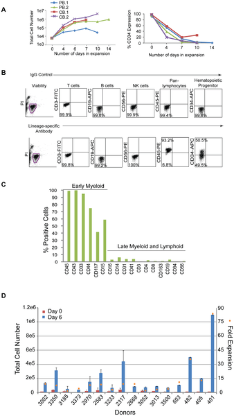
A. Graphs depict the expansion of purified cells from either two peripheral (PB.1 and PB.2) or cord (CB.1 or CB.2) blood donors over time (lefthand panel) along with the percentages of the total population that are CD34+ (righthand panel). B. Representative profile of a purified population of cells after 6 days of expansion by flow cytometry on cells isolated from Donor 3002. Flow cytometry plots from control staining using IgG antibodies (upper plots) are compared to plots with antibodies specific to lineage markers (lower plots). C. The graph represents an extended analysis by flow cytometry of the characteristic profile of PB.2 cells after 10 days of expansion. The % positive indicates the fraction of the population expressing the cell surface markers on the x-axis. D. The total number of CD34+ cells across 16 different donors was assessed beginning at 0 and 6 days of expansion (left side y-axis). The fold expansion (right side y-axis, orange squares) was determined by dividing the total number of cells at day 6 divided by the number of cells at day 0 after purification. The average percent of CD34 expression across all 16 donors was 48+/−19%.     http://dx.doi.org/10.1371/journal.pone.0027956.g001

These expansion conditions were then applied to cells acquired from a single vial of blood collected from multiple donors. PBMCs were isolated, frozen down immediately, or directly purified for CD34+ cells and seeded for expansion. On average, 1×10⁷ PBMCs were recovered per 8 ml vial of blood and yielded approximately 2×10⁴ cells after purification for CD34-expressing cells (data not shown). Although the magnitude of expansion was variable, cells from all of the donors demonstrated expansion ranging from 3 to 83-fold after 6 days in culture and approximately 48+/−19% of that population expressed CD34 (Figure 1D).

Optimizing the generation of iPSCs with small molecules and a defined matrix

The total number of purified cells isolated from a single, 8 ml vial of blood can be as little as 2×10⁴ CD34+ cells; therefore, a range of CD34+ cell numbers was tested to determine transfection efficiency from low cell numbers. The efficiency of transfection was determined by transfecting an oriP/EBNA1-containing plasmid encoding GFP into expanded CD34+ cells and assessing them by flow cytometry. Viability was determined by identifying the fraction of viable cells that did not stain positively for trypan blue the day after transfection divided by the total number of input cells. Viability was approximately 30% when 1×10⁴ to 1×10⁵ input cells were used for transfection (data not shown). Cell numbers at 1×10⁴ and 3×10⁴ resulted in an efficiency of 30% and was 40% when using 6×10⁴ and 1×10⁵ cells (Figure 2A). Cells expanded for only 3 days demonstrated a two-fold increase in transfection efficiency. Over 90% of those cells also co-expressed GFP and CD34 while only 18% of the cells transfected after 6 days of expansion co-expressed both markers (Figure 2B, C). These results support the notion that the conditions selected for this protocol favor the transfection of CD34+ cells present in the population.

Figure 2. Identifying optimal transfection conditions for CD34+ cells.

journal.pone.0027956.g002   Figure 2. Identifying optimal transfection conditions for CD34+ cells.

A. PBMCs (donor GG) were isolated and purified for CD34-expression and expanded for 6 days. A range of cell numbers were transfected with a control, oriP/EBNA1-based plasmid expressing GFP. Transfection efficiency was determined by calculating the percentage of viable cells expressing GFP detectable by flow cytometry (n = 6). B. PBMCs (donor A2389) were isolated, purified for CD34-expression and expanded for 3 or 6 days. 6×10⁴ to 1×10⁵ cells were transfected with the control, GFP-expressing plasmid. The graph depicts the percent of the total population that is GFP-positive along with the absolute number of total cells (n = 3). C. The graph represents the fraction of cells in B that co-express GFP and CD34 when transfected at 3 or 6 days of expansion (n = 3).    http//dx.doi.org/10.1371/journal.pone.0027956.g002

We anticipated variability in reprogramming efficiency given the differences already observed across donors for other cell types tested and with other methods of reprogramming. Therefore, we optimized the matrix, media, and plasmid combinations used for reprogramming. Firstly, a common source of variation occurs when MEFs or matrigel are used because both are undefined, cumbersome to prepare, and vary from lot-to-lot. Therefore, we established a defined matrix by testing a variety of commercially available possibilities. Recombinant protein fragments containing the active domains of human fibronectin (RetroNectin) or vitronectin consistently supported iPSC formation the best among those tested. Second, the efficiency of colony formation on RetroNectin-coated plates improved significantly when used in combination with StemSpan SFEM media, N2, B27, and a cocktail of small molecules that included PD0325901, CHIR99021, A-83-01, and HA-100 (Figure 3A). These molecules have been described previously as inhibitors of MEK, GSK3β, TGFβ, and ROCK pathways, respectively [27], [28]. Patches of adherent cells appeared within one week and became a positive indicator for progression into iPSCs since hematopoietic cells are typically cultured in suspension. The following week many of the colonies exhibited overt characteristics typical of an iPSC and stained positively for the common pluripotency markers Tra-1-81 and alkaline phosphatase (AP) (Figure 3A). The borders of the colonies were compact and the nucleoli more visible when cultures were transitioned to defined, TeSR2 media without small molecules 1.5 to 2 weeks following transfection. Thirdly, episomal oriP/EBNA1-based plasmids were used to deliver Oct4, Sox2, Klf4, C-myc, Nanog, Lin28, and SV40 Large T-antigen as previously described (Set 1; Figure 3B) [2]. Different combinations of reprogramming plasmids were also tested to determine whether a boost in reprogramming efficiency was possible. Based on previous reports indicating the benefit of L-myc in reprogramming trials, we modified plasmid combination Set 1 and substituted L-myc in place of C-myc (Set 2, Figure 3B) [29], [30]. While an improvement was not observed when C-myc was substituted for L-myc in the combination of plasmids represented in Set 1 (data not shown), an equal to or two-fold improvement was observed with plasmid Set 2 expressing L-myc (Figure 3C). Optimizing a range of input cell numbers for transfection also revealed more consistent generation of iPSCs when transfecting greater than 5×10⁴ cells (Figure 3D).

Figure 3. Plasmid transfections to optimize reprogramming efficiency.

journal.pone.0027956.g003  Figure 3. Plasmid transfections to optimize reprogramming efficiency.

A. Representative reprogramming trial from freshly drawn blood (donor 3002) using combination plasmid Set 2 for transfection. A single well is shown from a 6-well plate that contains colonies staining positively for AP activity (i). The white arrowhead highlights the colony magnified in panel ii that also stained positively for Tra-1-81 expression (green), panel iii. B. Schematic of the plasmid sets successfully used for reprogramming trials. Set 1 contains a combination of two plasmids for transfection whereby a 20 kb plasmid that either contains C- or L-myc is depicted. Set 2 includes a three plasmid combination for transfection. C. CD34+ cells purified from four different donors were expanded for 6 days and transfected using the plasmid combination that expresses either Set 1 or Set 2 plasmid sets to compare the total number of resulting iPSCs. D. Reprogramming trials were performed using plasmid Set 2 to transfect a range of cell numbers expanded for 6 days (donor GG, n = 6).  http://dx.doi.org/10.1371/journal.pone.0027956.g003


Optimization of iPSCs generated from CD34+ cells isolated from fresh, whole blood across multiple donors

The next step was to confirm iPSCs could be generated from actual vials of human blood, ensure cell numbers optimized for expansion and transfection are applicable across multiple donors, and determine whether the starting volume of blood can be minimized. Colonies emerging during reprogramming were scored positive by their ability to express Tra1-81 and exhibit a classic embryonic stem (ES) cell-like morphology. After colonies were picked from the reprogramming cultures, a subset of them were further characterized to confirm their pluripotency. Reprogramming efficiency was calculated in two ways 1) the number of iPSCs divided by the total volume of blood collected from each donor and 2) the total number of iPSCs divided by the number of cells for transfection multiplied by 1×10⁵ cells. The first calculation incorporates the whole process beginning from the blood collection to the generation of an iPSC. The second calculation removes the variability incurred during the isolation of PBMCs, purification, and expansion and focuses on the number of iPSCs generated per number of cells placed into transfection.

We tested blood collected across donors spanning a range of ethnicities, ages, and genders to confirm iPSCs could be generated from CD34+ cells purified from fresh blood draws (Table 1). Six of these donors provided up to 55 ml of blood, and PBMCs from them were either isolated and used directly for purification, expansion, and reprogramming or frozen down after isolation. iPSCs were successfully generated from all six donors regardless of whether they were from fresh or frozen cells despite the lower efficiency of reprogramming, less than 1 iPSC per ml of blood (Table 2). Next, smaller volumes of blood representative of a single vial were obtained from six different donors to test parameters established in earlier experiments such as the number of cells for transfection and plasmid combinations. The cell numbers used for transfection from donors 3052, 3233, and 3373 ranged from 2×10⁴ to 4×10⁴ which fall below the minimum, 5×10⁴ cells, established with our optimization studies. These experiments resulted in less than one iPSC per ml of blood (Table 2). Also, transfections with cells from donors 2583, 2970, and 3185 represent early trials performed with plasmid Set 1 expressing C-myc before Set 2 plasmids expressing L-myc were fully optimized which may have resulted in more iPSCs.

Table 1. Diversity across the set of donors used for reprogramming trials to generate iPSCs.
http://dx.doi.org/10.1371/journal.pone.0027956.t001

Table 2. Optimizing reprogramming from a range of blood volumes across multiple donors.
http://dx.doi.org/10.1371/journal.pone.0027956.t002

The next step was to then extend the insights acquired from these donors and verify the robustness of our protocol against ten new donors. The average number of iPSCs per ml of blood and per 1×10⁵ cells across these donors indeed improved after incorporating experience with handling and the testing performed on the earlier donor samples (Table 3). Furthermore, these experiments were extended to test even smaller volumes of blood from a subset of the same donors in Table 3. CD34+ cells purified from approximately 4 ml of blood were sufficient to generate iPSCs from all six donors tested, and CD34+ cells from approximately 2 ml of blood from four out of six of these donors generated iPSCs (Table 4). These results demonstrate that iPSCs can be generated from CD34+ isolated from tractable volumes of blood using this non-intergrating and feeder-free method of reprogramming.

Table 3. Improved reprogramming across multiple donors from a single vial of blood.
http://dx.doi.org/10.1371/journal.pone.0027956.t003


Table 4. The efficiency of reprogramming CD34+ cells beginning from 4 ml of blood or less.
http://dx.doi.org/10.1371/journal.pone.0027956.t004


iPSCs derived from peripheral blood are pluripotent and free of transgene elements

Multiple iPSCs from each of the donors that were reprogrammed from Tables 2, 3, and 4 were selected for further characterization to confirm their pluripotency. The clones exhibited a normal karyotype, were positive for Tra-1-81 and SSEA-4 expression by flow cytometry as well as endogenous genes DNMT3B, REX1, TERT, UTF1, Oct4, Sox2, Nanog, Lin28, Klf4, and C-myc (Table 5, Figure 4A–C). Clones did not exhibit integrated transgene or episomal elements and loss of episomal DNA occurred, on average, within 7–10 passages (Table 5, Figure 4D,E). A PCR screen did not reveal rearrangements pertaining to immunoglobulin heavy chain (IgH) or a subset of T cell receptor (TCR) gene segments (Table 5, Figure 4F). The lack of rearrangements supports the notion that the protocol selectively favors the production of iPSCs from hematopoietic progenitors rather than more differentiated cell types. When used for in vitro directed differentiation at passage 15, donor 2939 iPSC clones 4 and 5, which have lost episomal plasmids, were competent to form neurons (Figure 5G). Furthermore, five iPS clones from three different donors also formed teratomas after injection into immunodeficient (SCID) mice (Figure 4G). Interestingly, the presence of residual episomal plasmids did not appear to hinder the ability to form teratomas since clone 6 from donor 2970 did not lose transfected plasmids until passage 18, well after injection into mice for teratoma studies.

Figure 4. Characterization of iPSCs derived from CD34+ blood cells.

journal.pone.0027956.g004  Figure 4. Characterization of iPSCs derived from CD34+ blood cells.

A subset of iPSC clones were characterized for pluripotency. The experiments demonstrated in this figure provide representative examples of the types of results observed for characterization studies using iPS clones 4 and/or 5 derived from donors 2939 and 3389. A. Cytogenetic analysis on G-banded metaphase cells from iPS clone 4 exhibiting a normal karyotype. B and C. RT-PCR confirms the endogenous expression of classic pluripotency genes and the absence of expression from transgenes. A standard in-house iPS line served as the positive control k. D. Clones were deemed free of episomal (E) DNA and genomic integration (G) by PCR. E. PCR was used to track the loss of oriP/EBNA1-based plasmids at multiple passages using primers that amplify EBNA1. A control plasmid at 1 and 20 copies per genome was used to establish the sensitivity of the PCR at 1 copy per 3,000 cells. F. PCR screen using primers specific for the joining region and all three of the conserved framework regions (FR1, FR2 and FR3) to amplify immunoglobulin heavy chain (IgH) gene rearrangements and two assays with primers specific to the T cell receptor (TCR) gamma gene rearrangement. G. Representative image of donor 2939 clone 5 differentiated in vitro into neurons (i). Clone 5 also demonstrated differentiation into all three germ layers: ii) epithelium iii) endoderm iv) mesoderm v) ectoderm vi) endoderm from teratomas formed when iPSCs were injected into immunodeficient, SCID mice.
http://dx.doi.org/10.1371/journal.pone.0027956.g004

Figure 5. The presence of CD34+ cells correlates with reprogramming efficiency.

journal.pone.0027956.g005  Figure 5. The presence of CD34+ cells correlates with reprogramming efficie

A. CD34+ cells from four different blood donors were expanded for 3, 6, 9, or 13 days. A large volume of blood was collected from donors 3096, 2849, and 3389 to ensure sufficient cell numbers to perform these studies. Expanding CD34+ cells using plasmid DNA combination Set 2. The efficiency of reprogramming was calculated as the total number of iPSCs exhibiting morphological features characteristic of an ES cell and an ability to stain positively for Tra-1-81 divided by the total number of cells used for transfection. Black Squares depict the percentage of the population expressing CD34 at the indicated days of expansion. B. Representative reprogramming trial whereby both the positive (i) and negative (ii) fraction following purification were used for reprogramming. Panel (i) shows one well of a 6-well plate that contains successfully reprogrammed colonies from donor 2939 based on their ability to demonstrate AP activity. The CD34-depleted fraction from donor 2939 was unable to form colonies as indicated by the lack of AP staining when performed in parallel with the purified population panel, ii. Panels iii and iv magnify the colony in panel (i) marked by a white arrowhead and demonstrates expression of Tra-1-81 (green), panel iv.
http://dx.doi.org/10.1371/journal.pone.0027956.g005

Table 5. Characterization of insert-free iPS clones derived from fresh blood.
http://dx.doi.org/10.1371/journal.pone.0027956.t005

Reprogramming efficiency correlates with the amount of CD34-expression

The isolation of CD34+ cells from PBMCs creates an additional step in our process and others have demonstrated successful reprogramming directly from PBMCs without the need for purification [22]. Therefore, several experiments to determine whether a correlation exists between CD34+ cells and reprogramming efficiency were performed. First, the expanding CD34+ populations were screened by flow cytometry for characterization prior to transfection. T, B, and NK cells were undetectable after 3 and 6 days of expansion demonstrated by their lack of CD3, 19, and 56 expression, respectively. The percentage of CD34 expression during expansion ranged from 30 to 100%, thus increasing the likelihood of reprogramming more of an early lineage cell type (n = 9, data not shown). Second, samples were taken from the purified populations during expansion at different timepoints as they lost CD34 expression to determine their receptiveness to reprogramming. A decrease in reprogramming efficiency was observed in correlation with decreasing percentages of CD34 expression across populations of cells from four independent donors (Figure 5A). For example, donor 3096 exhibited only 1 iPSC per 1×10⁵ input cells when beginning from cells expanded for 13 days (31% CD34+) compared to 91.5 iPSCs per 1×10⁵ cells following 3 days when levels of CD34 expression were much higher, 98% (Figure 5B). Third, populations depleted of CD34+ cells were tested for their ability to reprogram in parallel with their CD34+ cell counterparts. These CD34-depleted populations were not receptive to reprogramming as the CD34+ cells even when the same media and transfection conditions were used (n = 3, Figure 5B). Finally, a side-by-side comparison of reprogramming efficiency was performed between unpurified PBMCs and CD34+ cells isolated from 10 different donors. A medium described previously for the expansion of erythroblasts and for successful reprogramming studies was used to ensure media would not be a limiting factor for reprogramming the PBMCs in our protocol [22], [31]. Reprogramming trials beginning from either PBMCs or CD34+ cells were launched in parallel using their respective media for expansion. The efficiency of reprogramming was approximately 2 to 8 fold higher when beginning with cells purified for CD34 expression in 9 out of the 10 donors compared to those from PBMCs (p = 0.007; Figure 6). A significant fraction of the PBMC population was comprised of lymphocytes (~79+/−14% CD3/CD19) at the time of transfection (data not shown); therefore, PCR was performed to screen for potential IgH and TCR gene rearrangements to determine whether both protocols promoted the generation of iPSCs free of gene rearrangements. Interestingly, screened clones from either cell type were free of IgH and TCR gene rearrangements indicating that both protocols favor the reprogramming of early progenitor cells. The lower efficiency of reprogramming from the PBMC population may reflect the dilution of early progenitor blood cells by a predominately lymphocytic population.

Figure 6. Comparing the efficiency of reprogramming between PBMCs and CD34+ cells.

PBMCs and CD34+ cells were isolated from single tubes of blood provided from 10 different donors. The efficiency of reprogramming following transfection with DNA Combination Set 2 was determined for each donor and each method. “Total colonies” refers to all iPS colonies derived from either CD34+ or PBMC populations that stain positively for Tra1-60 and “iPS-like” colonies are those that stain positively for Tra1-60, exhibit clear iPS morphology, and are large enough to pick for expansion. Input cells refer to the number of CD34+ cells or PBMCs were used for transfection. The efficiencies across all donors from both methods were compared using the Wilcoxon signed rank test (two-sided), p = 0.007.
http://dx.doi.org/10.1371/journal.pone.0027956.g006

Generation of iPSCs from CD34+ cells using completely defined reagents

Additional reprogramming trials were performed using completely defined conditions to enable the production of clinic-grade iPSCs. A large pool of CD34+ cells mixed from multiple donors was used for multiple tests and resulted in a successful expansion of 113+/−11 fold in defined media compared to 83+/−32 fold for cells in standard conditions after 6 days of expansion (Figure 7A). Despite the 30-fold difference between the two conditions, the absolute number of CD34+ cells is similar between the two populations when multiplied by the percentage of the population expressing CD34 by flow cytometry. For example, 42+/−13% of the population expanded in standard conditions expressed CD34 and 26+/−16% expressed CD34 using completely defined conditions (Figure 7A). There were no detectable CD3+, CD19+, or CD56+ cells after 6 days in culture consistent with our earlier expansion trials (data not shown). The media used for reprogramming is completely defined with the exception of the supplement B27 which contains bovine serum albumin (BSA). However reprogramming was still achieved in the presence or absence of B27 (Figure 7B). These improvements coupled with a defined matrix enables the production of iPSCs in a completely defined process.

Figure 7. The generation of iPSCs from blood using completely defined conditions.


A. Fold expansion of CD34+ cells pooled from multiple blood donors in standard (n = 13) and completely defined conditions (n = 2) after 6 days of expansion. Fold expansion was calculated from the total number of cells at day 6 divided by the number of cells the day after purification. Percentages indicate the fraction of cells expressing CD34 in the total population as assessed by flow cytometry. B. Reprogramming trials were performed on CD34+ cells obtained by leukapheresis from donors GG and A2389 with and without the B27 supplement.
http://dx.doi.org/10.1371/journal.pone.0027956.g007

Discussion

CD34+ cells possess characteristics that make them an ideal blood cell to reprogram: they are readily identified, highly receptive to reprogramming, and free of gene rearrangements characteristic of more differentiated cell types. However, their low numbers in circulating blood have made them a less desirable cell type for reprogramming because large volumes of blood were predicted to be required for the generation of iPSCs. We describe a method to generate insert-free iPSCs from CD34+ cells beginning from a single vial of blood or less. In some cases, the number of CD34+ cells that expanded exceeded that required for a single transfection after 6 days in culture making it possible to transfect after only 3 days of expansion, shorter than the amount of time Chou and colleagues used to reprogram unpurified PBMCs [22].

Proliferation contributes to reprogramming efficiency; therefore, choosing a culture medium that promotes proliferation will effectively promote reprogramming. Data presented in our manuscript supports this assertion because all four donor populations examined were almost 100% positive for CD34 expression after 3 days in culture, but they were not equally receptive to reprogramming (Figure 5A). Cells from donor 3096 demonstrated the highest fold expansion following purification as well as the highest efficiency of reprogramming. However, we go on to demonstrate that proliferation is not the only contributor to efficient reprogramming. In our experiments, the purity of the cell population diminishes over time following purification, but the cells within the culture continue to expand despite low CD34 expression. Figure 1C shows that this expanding population 10 days following purification consists primarily of early lineage progenitor cells. This result indicates that our medium has the capacity to stimulate the proliferation of non-T and non-B cells that have never been or are no longer CD34+. If proliferation were the primary force driving the efficiency of reprogramming, then it would be expected that proliferating non-T/non-B cells within our population would be equally receptive to reprogramming regardless of their time in culture. Our results are contrary to this hypothesis, however, because the efficiency of reprogramming decreases as the magnitude of CD34 expression decreases. This is observed across four independent donor cell populations and is consistent with dependence of reprogramming on CD34 expression (see Figure 5A). These results, taken together, support our hypothesis that both proliferation and cell identity contribute to the efficiency of reprogramming.

We have also outlined a protocol that begins to systematically address some of the challenges in the generation of clinical-grade iPSCs in an effort to advance their use from research lab to clinic. iPSCs must not only be manufactured consistently from a tractable tissue source but also satisfy safety requirements. The core of these requirements includes the use of completely defined culture conditions and a standardized reprogramming method that results in the removal of the potentially oncogenic transgenes employed to reprogram the cells. The starting point of the protocol is the actual patient sample, a single vial of blood. The ability to begin from frozen rather than fresh starting material allows flexibility to launch multiple reprogramming trials in parallel. We demonstrate that either CD34+ cells or PBMCs may be used as the source population for reprogramming. The iPSCs generated by this method are free of transfected DNA as well as B and T cell gene rearrangements. Several challenges remain, however, before routine production of clinical-grade iPSCs can be completely performed. First, there is considerable variation in the efficiency of reprogramming from donor to donor. Some of this variation is likely due to the inherent differences among the donors, but a careful examination of external sources of variation at each step from blood to iPSCs may well reveal areas in addition to those we have uncovered that can be better controlled. For example, we demonstrate potential in the ability to produce iPSCs using completely defined reagents to minimize variation. Second, an automated method for screening and selecting iPSCs during reprogramming would facilitate high throughput production of iPSCs. Third, a robust production protocol must also include a method for the rapid screening of iPSCs to identify those that both lack potentially harmful mutations and are readily differentiated into various cell types. In sum, the generation of iPSCs using a standardized process beginning from early progenitor cells isolated from routine blood draws minimizes this variation and is a good starting point to provide a more comparable baseline for analysis. We present the first steps towards a standardized process to make the generation of clinical-grade iPSCs a reality.

Materials and Methods

Ethics Statement

All human primary cells were generated in vitro from tissue samples from human donors with appropriate written informed consent given to the commercial providers.

All animal work was conducted according to relevant national and international guidelines under the approval of the Cellular Dynamics International Animal Care and Use Committee. As a private company, our animal facility does not provide a permit number or approval ID since mouse is not a protected species.

Processing whole blood samples

Peripheral (PB.1 and PB.2) and cord (CB.1 and CB.2) blood-derived CD34+ cells were obtained from AllCells (Emeryville, CA USA). Blood collections were performed at AllCells and Meriter Laboratories (Madison, WI USA) using standard, 8 ml Vacutainer Cell Processing Tubes (both sodium citrate and sodium heparin-based tubes are acceptable; BD Biosciences; Franklin Lakes, NJ USA). Appropriate documentation for informed consent was completed prior to blood collection (Meriter Laboratories). Vacutainers were processed within 24 hours of collection. Briefly, the PBMC-containing upper phase was collected and washed with ice-cold PBS (Invitrogen; Carlsbad, CA USA). Cells were either frozen down or used directly for purification with the CD34 MicroBead Kit (Miltenyi; Auburn, CA USA) and used according to the manufacturer’s protocol. Some samples were treated with Histopaque (Sigma Aldrich; St. Louis, MO USA) to minimize the number of red blood cells (RBCs) and centrifuged at 2000 rpm for 20 minutes without braking. The interface containing the PBMCs was removed if samples were treated with histopaque, cells washed again with chilled PBS, centrifuged at 600× g for 15 minutes and either frozen down with CryoStor10 (StemCell Technologies; Vancouver, BC Canada) or used directly for purification. CD34+ cell expansion media: StemSpan SFEM (StemCell Technologies), Flt3, SCF, TPO each at a final concentration of 300 ng/ml, IL-6 (100 ng/ml) and IL-3 (10 ng/ml) (Peprotech; Rocky Hill, NJ USA), supplemented with DNaseI (final concentration at 20 U/ml), and 1× Antibiotic-antimycotic (Invitrogen) for overnight recovery. Defined expansion media: serum-free StemSpan H3000 (StemCell Technologies), animal-free IL-6 (R&D Systems Minneapolis, MN USA), and recombinant human IL-3, TPO, Flt3, and SCF (Peprotech) at the same concentrations listed above. PBMC expansion media: StemSpan SFEM, ExCyte Medium Supplement (Millipore; Billerica, MA), Glutamax (Invitrogen), SCF (250 ng/ml), IL-3 (20 ng/ml), Erythropoietin (2 U/ml; Prospec; Rehovot, Israel), IGF-1 (40 ng/ml; Prospec), and Dexamethasone (1 µM; Fisher; Waltham, MA). PBMCs were resuspended at 1×10⁶ cells/ml for expansion.

Flow cytometry

Cell surface staining of hematopoietic cells was performed with CD45-PE, CD34-APC, CD19-APC and CD56-PE (BD Biosciences) and CD3-PE (eBioscience; San Diego, CA USA) antibodies. iPSCs were processed directly for antibody staining for the presence of Tra-1-81 (Stemgent; Cambridge, MA USA) and SSEA-4 (BD Pharmingen; San Diego, CA USA). Propidium Iodide (Sigma Aldrich) was added for dead cell exclusion, and all stained cells were analyzed in combination with their respective isotype controls using a flow cytometer (Accuri; Ann Arbor, MI USA).

Reprogramming cells enriched for CD34-expression

The CD34 nucleofection kit and device (Lonza; Allendale, NJ USA) were used for transfections. For CD34+ cells, 3.5 µg of each plasmid in Combination Set 1 and 3 ug of each plasmid for Combination Set 2 except for the L-myc containing plasmid where 2 µg was transfected using program U-08. Cells were seeded onto RetroNectin-coated 6-well plates (Takara Bio, Inc; Otsu, Shiga Japan). Seeding density ranged from 5×10⁴ to 1×10⁵ cells/ml. Reprogramming media: StemSpan SFEM (StemCell Technologies) supplemented with non-essential amino acids (NEAA; Invitrogen), 0.5× Glutamax, N2B27 (Invitrogen), 0.1 mM β-mercaptoethanol (Sigma-Aldrich), 100 ng/mL zebrafish basic fibroblast growth factor (zbFGF), 0.5 µM PD0325901, 3 µM CHIR99021, 0.5 µM A-83-01 (all molecules from Stemgent), and 10 µM HA-100 (Santa Cruz; Santa Cruz CA USA). Conditions for PBMC reprogramming relied on 1×10⁶ cells per transfection, program T-16, and DNA from Combination Set 2 at the concentrations described for CD34+ cells. Reprogramming media for PBMCs was the same with the exception of the small molecule cocktail which contained recombinant human LiF (Millipore), 3 uM CHIR99021, and 0.5 uM A-83-01. In general, cultures were fed with fresh medium every other day for 9 to 14 days then transitioned to TeSR2 (Stem Cell Technologies) without the addition of small molecules. iPSC colonies were scored with Tra-1-81 antibody (StainAlive™ DyLight™ 488 Mouse anti-Human Tra-1-81 antibody; Stemgent) or mouse-anti-Tra-1-60 IgM antibody (R&D) in combination with goat anti-mouse IgM Alexa 488 (Invitrogen), and alkaline phosphatase expression (Vector Blue Alkaline Phosphatase Substrate Kit III, Vector Laboratories; Burlingame, CA USA).

Detecting endogenous expression of pluripotency markers

Total RNA was isolated using the RNeasy Mini Plus kit (Qiagen; Valencia, CA USA) per the manufacturer’s protocol. Approximately 1 µg of total RNA was used for cDNA synthesis using the SuperScript III First-Strand Synthesis system for RT-PCR (Invitrogen). RT-PCR was performed using previously described primers and those listed in Table 6 [2]. cDNA was diluted 1:2 and 1 µl was used in reactions with GoTaq Green Master Mix (Promega; Madison, WI USA).

Table 6. Primer sequences for the detection of endogenous gene expression.
http://dx.doi.org/10.1371/journal.pone.0027956.t006

journal.pone.0027956.t006  Table 6. Primer sequences for the detection of endogenous gene expression.

Episomal and Genomic DNA isolation

Immunoglobulin heavy chain and T cell gene rearrangements

Karyotyping

In Vitro Differentiation and Teratoma Studies

Acknowledgments

Author Contributions

References

1. Woltjen K, Michael IP, Mohseni P, Desai R, Mileikovsky M, et al. (2009) piggyBac transposition reprograms fibroblasts to induced pluripotent stem cells. Nature 458: 766–770. doi: 10.1038/nature07863.

2. Yu J, Hu K, Smuga-Otto K, Tian S, Stewart R, et al. (2009) Human Induced Pluripotent Stem Cells Free of Vector and Transgene Sequences. Science.

3. Yu J, Vodyanik MA, Smuga-Otto K, Antosiewicz-Bourget J, Frane JL, et al. (2007) Induced pluripotent stem cell lines derived from human somatic cells. Science 318: 1917–1920. doi: 10.1126/science.1151526.

4. Takahashi K, Tanabe K, Ohnuki M, Narita M, Ichisaka T, et al. (2007) Induction of pluripotent stem cells from adult human fibroblasts by defined factors. Cell 131: 861–872. doi: 10.1016/j.cell.2007.11.019.

5. Okita K, Nakagawa M, Hyenjong H, Ichisaka T, Yamanaka S (2008) Generation of mouse induced pluripotent stem cells without viral vectors. Science 322: 949–953. doi: 10.1126/science.1164270.

6. Saha K, Jaenisch R (2009) Technical challenges in using human induced pluripotent stem cells to model disease. Cell Stem Cell 5: 584–595. doi: 10.1016/j.stem.2009.11.009.

7. Park IH, Zhao R, West JA, Yabuuchi A, Huo H, et al. (2008) Reprogramming of human somatic cells to pluripotency with defined factors. Nature 451: 141–146. doi: 10.1038/nature06534.

8. Yamanaka S (2010) Patient-specific pluripotent stem cells become even more accessible. Cell Stem Cell 7: 1–2. doi: 10.1016/j.stem.2010.06.009.

9. Brown ME, Rondon E, Rajesh D, Mack A, Lewis R, et al. (2010) Derivation of induced pluripotent stem cells from human peripheral blood T lymphocytes. PLoS One 5: e11373. doi: 10.1371/journal.pone.0011373.

10. Hanna J, Markoulaki S, Schorderet P, Carey BW, Beard C, et al. (2008) Direct reprogramming of terminally differentiated mature B lymphocytes to pluripotency. Cell 133: 250–264. doi: 10.1016/j.cell.2008.03.028.

11. Polo JM, Liu S, Figueroa ME, Kulalert W, Eminli S, et al. (2010) Cell type of origin influences the molecular and functional properties of mouse induced pluripotent stem cells. Nat Biotechnol 28: 848–855. doi: 10.1038/nbt.1667.

12. Miura K, Okada Y, Aoi T, Okada A, Takahashi K, et al. (2009) Variation in the safety of induced pluripotent stem cell lines. Nat Biotechnol 27: 743–745. doi: 10.1038/nbt.1554.

13. Nakano T (1995) Lymphohematopoietic development from embryonic stem cells in vitro. Semin Immunol 7: 197–203. doi: 10.1016/1044-5323(95)90047-0.

14. Cho SK, Webber TD, Carlyle JR, Nakano T, Lewis SM, et al. (1999) Functional characterization of B lymphocytes generated in vitro from embryonic stem cells. Proc Natl Acad Sci U S A 96: 9797–9802. doi: 10.1073/pnas.96.17.9797.

15. Wada H, Kojo S, Kusama C, Okamoto N, Sato Y, et al. (2011) Successful differentiation to T cells, but unsuccessful B-cell generation, from B-cell-derived induced pluripotent stem cells. Int Immunol 23: 65–74. doi: 10.1093/intimm/dxq458.

+ more in document

Analysis of Human and Mouse Reprogramming of Somatic Cells to Induced Pluripotent Stem Cells. What Is in the Plate?

Stéphanie Boué, Ida Paramonov,  María José Barrero,  Juan Carlos Izpisúa Belmonte
Published: September 17, 2010  http://dx.doi.org/10.1371/journal.pone.0012664

After the hope and controversy brought by embryonic stem cells two decades ago for regenerative medicine, a new turn has been taken in pluripotent cells research when, in 2006, Yamanaka’s group reported the reprogramming of fibroblasts to pluripotent cells with the transfection of only four transcription factors. Since then many researchers have managed to reprogram somatic cells from diverse origins into pluripotent cells, though the cellular and genetic consequences of reprogramming remain largely unknown. Furthermore, it is still unclear whether induced pluripotent stem cells (iPSCs) are truly functionally equivalent to embryonic stem cells (ESCs) and if they demonstrate the same differentiation potential as ESCs. There are a large number of reprogramming experiments published so far encompassing genome-wide transcriptional profiling of the cells of origin, the iPSCs and ESCs, which are used as standards of pluripotent cells and allow us to provide here an in-depth analysis of transcriptional profiles of human and mouse cells before and after reprogramming. When compared to ESCs, iPSCs, as expected, share a common pluripotency/self-renewal network. Perhaps more importantly, they also show differences in the expression of some genes. We concentrated our efforts on the study of bivalent domain-containing genes (in ESCs) which are not expressed in ESCs, as they are supposedly important for differentiation and should possess a poised status in pluripotent cells, i.e. be ready to but not yet be expressed. We studied each iPSC line separately to estimate the quality of the reprogramming and saw a correlation of the lowest number of such genes expressed in each respective iPSC line with the stringency of the pluripotency test achieved by the line. We propose that the study of expression of bivalent domain-containing genes, which are normally silenced in ESCs, gives a valuable indication of the quality of the iPSC line, and could be used to select the best iPSC lines out of a large number of lines generated in each reprogramming experiment.

Citation: Boué S, Paramonov I, Barrero MJ, Izpisúa Belmonte JC (2010) Analysis of Human and Mouse Reprogramming of Somatic Cells to Induced Pluripotent Stem Cells. What Is in the Plate? PLoS ONE 5(9): e12664.  http://dx.doi.org/10.1371/journal.pone.0012664

Figure 2. Human protein-protein interaction networks of genes with higher expression levels in ESCs and iPSCs compared to somatic cells.

journal.pone.0012664.g002  Figure 2. Human protein-protein interaction networks of genes with higher expression levels in ESCs and iPSCs compared to somatic cells.


The human protein-protein interaction networks of genes most consistently highly expressed in ESCs and iPSCs, compared to the starting cell populations, have been created from the lists of the biggest changes in expression, using String[71] with high confidence interactions (min score 0.7) and have been edited in Medusa[72]. They show a central, highly interconnected network of genes in which the most famous pluripotency transcription factors are to be found and which is likely to represent the core pluripotency network. They also highlight a number of genes whose functions relate to cell-cell communication, cell cycle, DNA repair and other metabolisms.

http://dx.doi.org/10.1371/journal.pone.0012664.g002

Figure 3. Mouse protein-protein interaction networks of genes with higher expression levels in ESCs and iPSCs compared to somatic cells.

journal.pone.0012664.g003  Figure 3. Mouse protein-protein interaction networks of genes with higher expression levels in ESCs and iPSCs compared to somatic cells.

The mouse protein-protein interaction networks of genes most consistently highly expressed in ES and iPSCs, compared to the starting cell populations, have been created from the lists of biggest changes in expression, using String[71] with high confidence interactions (min score 0.7) and have been edited in Medusa[72]. They show a central, highly interconnected network of genes in which the most famous pluripotency transcription factors are to be found and which is likely to represent the core pluripotency network. They also highlight a number of genes those functions relate to cell-cell communication, cell cycle, DNA repair and other metabolisms.
http://dx.doi.org/10.1371/journal.pone.0012664.g003


Read Full Post »

Larry H Bernstein, MD, FCAP, reporter and curator

htto://pharmaceuticalintelligence.com/2013-12-07/larryhbern/Advances-in-Stem-Cell-Research

The amount of success in stem cell research and recent successes is notable.

GEN News  Dec 5, 2013
Stem Cell Leaders Call for Human Embryome Project

Just as an international consortium was formed to map and sequence the human genome, now a group of stem cell and regenerative medicine scientists say it’s critical that such an effort be ramped up to do a similar project focused on the human embryome.

This was the key message of a panel discussion, “From Mapping the Genome to Mapping the Embryome: The Urgent Need for an International Initiative,” moderated by Michael West, Ph.D., CEO of Biotime. It took place at the World Stem Cell Summit, which is taking place this week in San Diego.

“It is becoming increasingly clear in regenerative medicine that pluripotent stem cells, embryonic stem cells, and IPs cells will be as fundamentally important to medicine as was DNA. Maybe even bigger because you can genetically engineer these cells,” said Dr. West.

Dr. West and his colleagues adamantly believe that there needs to be a large international effort aimed at mapping the cellular and molecular basis of all human life starting with the fertilized egg and working its way up to the body of the adult. This is what it is termed the embryome.

“The opportunity presented by pluripotent stem cells to manufacture for the first time in the history of medicine all of the cellular components of the human body on an industrial scale is at once both an opportunity and a challenge,” said Dr. West. “The opportunity is to build a new field we call regenerative medicine in which many currently incurable diseases are treated with cells capable of regenerating tissues afflicted with disease. The challenge relates to the complexity of the cell types in the body and our ability to manufacture products with precisely defined compositions for human clinical use.”

Dr. West went on to say that to get these different types of stem cells into the clinic, and approved by the FDA, researchers will fully need to understand all aspects of the biology of these cells. An identification and understanding of any contaminating cells will also be essential to success in this field. The question to ask is “What is in the syringe?”

Unlike recombinant DNA, continued Dr. West, the contaminants in pluripotent stem cells are alive and may make things that are undesirable at the intended point of therapy. For example, you might have a bioreactor full of cells that are making heart muscle to regenerate heart function in a patient. But you have to be careful that your cells are not contaminated with neural crest cells from the head area which could generate a tooth along with the heart muscle.

“These contaminants, if you do not remove them, can lead to years of delay in filing an IND and a runup in costs as you try to identify these cells,” explained Dr. West.

The major problem in identifying them, according to Dr. West, is that no one has ever mapped the molecular markers or even a rudimentary cell ontology tree, i.e., mapped out the tree from the fertilized egg to the cells of the human body.

“If [there were] a detailed map of all the cellular and molecular components of life from the fertilized egg to adulthood, and then databased in a manner to the information in the human genome, medicine would be the true beneficiary,” added Dr. West. “That’s why we have made this call for an international initiative.”

Also, watch our video “A Brief History of Stem Cells” to see a timeline spanning over 60 years of stem cell research.

Mary Ann Liebert Wins Stem Cell Education Award

Mary Ann Liebert, president and CEO of Mary Ann Liebert Inc., and publisher of GEN, was presented with the Stem Cell Education Award by the Genetics Policy Institute. The award was given during a ceremony at dinner which took place at the World Stem Cell Summit, which is being held in San Diego this week.

Liebert was cited for her outstanding “work in educating patients, researchers, and the broader stem cell community, and in raising the standard in medical research journalism.” Among the seventy journals the Liebert company publishes is the peer-reviewed Stem Cells and Development.

In her acceptance speech Liebert told the audience that she was extremely gratified in being so recognized and thanked the entire staff at her company for their dedication in helping to promote excellence in medical publishing.

In his introductory remarks during the award ceremony GEN’s long-time editor in chief John Sterling noted that Mary Ann always encourages her editors and writers “to inform, enlighten when they can, and educate as much as possible.”

Sterling added that while she started her company 33 years ago her vision for her publications remains the same: “to help advance our knowledge of science and medicine in the best ways possible.”


Neural Precursors “Cure MS” in Mice

During a session at the this week’s World Stem Cell Summit in San Diego, an international research team described an “astonishing” experiment in which a mouse model of multiple sclerosis was able to virtually totally recover and move normally after being transplanted with human neural precursor cells (hNPC). The scientists were able to show almost full recovery in the mice up to six months later.

The investigators, led by Jeanne Loring, Ph.D., from the Scripps Research Institute, included scientists from the University of California, Irvine and a group from Australia.

“Our goal was to demonstrate cell therapy for MS,” Dr. Loring told the audience.

According to Ronald Coleman, a graduate student working with Dr. Loring and who is at UC-Irvine, the team used mice infected with a neurotropic JHM variant of mouse hepatitis virus (JHMV) as a model for MS. They injected hNPCs derived from human pluripotent stem cells (hPSC) into the mice to explore treatment options for the disease.

The results were indeed astonishing, said Dr. Loring. Non-control mice were able to move about in a manner that can be described as consistent and long lasting. T-cell proliferation was reduced and T regulatory cell induction took place. The spinal cords of the mice not only did not undergo further demyelination but actually exhibited remyelination. The control mice dragged their legs around when they tried to move.

“The only problem was that the hNPCs themselves are not directly responsible for the cure. They are not even there when the mice start walking,” explained Dr. Loring. “Those cells are rejected after seven days and we start to see a therapeutic response in three weeks.”

Both Dr. Loring and Coleman believe that the hNPCs are secreting proteins, like cytokines, that do the actual repair work in the CNS of the mice.

“We identified a set of candidate proteins secreted by hNPCs and not by undifferentiated pluripotent stem cells,” continued Dr. Loring, who said the team plans to continue building on this initial research.


World Stem Cell Summit: December 4, 2013 Update

GEN is on the scene at the World Stem Cell Summit in San Diego. Here are some highlights from the conference so far:

Bernard Siegel, J.D., founder and co-chair of the World Stem Cell Summit (WSCS) and executive director of Genetics Policy Institute, today welcomed attendees of WSCS 2013, being held December 4–6, in San Diego, CA.

“Stem cell science represents, to those afflicted with chronic disease, a vehicle for modeling disease and therapeutic development,” states Siegel in World Stem Cell Report 2013, a supplement to Stem Cells and Development (2013;22;Suppl1). “The field is a true scientific revolution and reflects the transformative power of hope, a powerful engine for progress.”

“The future is here now,” says Mahendra Rao, M.D., Ph.D., director, NIH Center for Regenerative Medicine, who delivered a plenary keynote and moderated the plenary panel discussion, “How Stem Cells are Transforming Medicine.” Cell therapies have been used to treat people safely and effectively; the technical barriers have been addressed. The challenge now is to reduce the cost of manufacturing. To drive routine adoption of cell therapy it must be cost effective and must demonstrate more than incremental benefit, according to Dr. Rao.

Professor Teruo Okano, Ph.D., Tokyo Women’s Medical University, described his group’s Cell Sheet Tissue Engineering strategy that involves enzymatic membrane disruption during cell harvesting and growth of an autologous cell sheet for transplantation on an “intelligent surface” that reversibly changes properties from hydrophobic to hydrophilic with a reversible in temperature from 37°C to 20°C. Dr. Okano further described the development of an automatic tissue factory and thick tissue evaluation system for fully automated, industrialized GMP cell processing.

Andre Terzic, M.D., Ph.D., Center for Regenerative Medicine, Mayo Clinic, noted during the opening session of the WSCS that “the Mayo Clinic has embraced regenerative medicine as a strategy for the future of medicine,” and he described their blueprint for moving from knowledge to delivery of treatments and procedures. Education is a critical dimension of this process. Another important component, according to Dr. Terzic, is the Regenerative Medicine Biotrust, in which “the patient is the center of the solution” to develop combinations of diagnostics and therapeutics and conduct clinical trials.

Regardless of the outcomes of current or future clinical trials, “I would argue that we have already seen breakthroughs,” said Evan Snyder, Ph.D., Sanford-Burnham Medical Research Institute, as stem cells “have completely changed the way medicine thinks about disease and development.” They have led to new views on plasticity and regeneration and the development of different types of drug targets.

WSCS 2013 is organized by the Genetics Policy Institute (GPI), California Institute for Regenerative Medicine (CIRM), Institute for Integrated Cell-Material Sciences at Kyoto University (iCeMS), Mayo Clinic, Sanford-Burnham Medical Research Institute, and The Scripps Research Institute. Mary Ann Liebert, Inc. publishers and Genetic Engineering & Biotechnology News (GEN) are sponsors of the summit.

Drug Testing Should Be with Human iPS Cells
Fri, 12/06/2013 – drug discovery & development  (DDD)

Once established such neural stem cells can be used to continuously generate neurons for drug testing and disease modeling. Depicted is an immunofluorescence staining where proteins characteristic of neural stem cells are labeled with fluorescing antibodies (Nestin in green, Dach1 in red). (Source: Jerome Mertens / Uni Bonn)Once established such neural stem cells can be used to continuously generate neurons for drug testing and disease modeling. Depicted is an immunofluorescence staining where proteins characteristic of neural stem cells are labeled with fluorescing antibodies (Nestin in green, Dach1 in red). (Source: Jerome Mertens / Uni Bonn)Why do certain Alzheimer medications work in animal models but not in clinical trials in humans? A research team from the University of Bonn and the biomedical enterprise Life & Brain GmbH has been able to show that results of established test methods with animal models and cell lines used up until now can hardly be translated to the processes in the human brain. Drug testing should therefore be conducted with human nerve cells, conclude the scientists. The results are published by Cell Press in the journal Stem Cell Reports.

In the brains of Alzheimer’s patients, deposits form that consist essentially of beta-amyloid and are harmful to nerve cells. Scientists are therefore searching for pharmaceutical compounds that prevent the formation of these dangerous aggregates. In animal models, certain non-steroidal anti-inflammatory drugs (NSAIDs) were found to a reduced formation of harmful beta-amyloid variants. Yet, in subsequent clinical studies, these NSAIDs failed to elicit any beneficial effects.

“The reasons for these negative results have remained unclear for a long time”, said Oliver Brüstle, director of the Institute for Reconstructive Neurobiology of the University of Bonn and CEO of Life & Brain GmbH. “Remarkably, these compounds were never tested directly on the actual target cells—the human neuron”, added lead author Jerome Mertens of Brüstle’s team, who now works at the Laboratory of Genetics in La Jolla (USA). This is because, so far, living human neurons have been extremely difficult to obtain. However, with the recent advances in stem cell research it has become possible to derive limitless numbers of brain cells from a small skin biopsy or other adult cell types.

Scientists transform skin cells into nerve cells

Now a research team from the Institute for Reconstructive Neurobiology and the Department of Neurology of the Bonn University Medical Center together with colleagues from the Life & Brain GmbH and the University of Leuven (Belgium) has obtained such nerve cells from humans. The researchers used skin cells from two patients with a familial form of Alzheimer’s Disease to produce so-called induced pluripotent stem cells (iPS cells), by reprogramming the body’s cells into a quasi-embryonic stage. They then transformed the resulting iPS cells into nerve cells.

Using these human neurons, the scientists tested several compounds in the group of NSAIDs. As control, the researchers used nerve cells they had obtained from iPS cells of donors who did not have the disease. Both in the nerve cells obtained from the Alzheimer’s patients and in the control cells, the NSAIDs that had previously tested positive in the animal models and cell lines typically used for drug screening had practically no effect: The values for the harmful beta-amyloid variants that form the feared aggregates in the brain remained unaffected when the cells were treated with clinically relevant dosages of these compounds.

Metabolic processes in animal models differ from humans

“In order to predict the efficacy of Alzheimer drugs, such tests have to be performed directly on the affected human nerve cells”, concluded Brüstle’s colleague Philipp Koch, who led the study. Why do NSAIDs decrease the risk of aggregate formation in animal experiments and cell lines but not in human neurons? The scientists explain this with differences in metabolic processes between these different cell types. “The results are simply not transferable”, says Koch.

The scientists now hope that in the future, testing of potential drugs for the treatment of Alzheimer’s disease will be increasingly conducted using neurons obtained from iPS cells of patients. “The development of a single drug takes an average of ten years”, said Brüstle. “By using patient-specific nerve cells as a test system, investments by pharmaceutical companies and the tedious search for urgently needed Alzheimer medications could be greatly streamlined”.

Date: November 6, 2013
Source: University of Bonn


Read Full Post »

Reprogramming Cell in Tissue Repair

Reporter and Curator: Larry H Bernstein, MD, FCAP

This is a novel concept in regenerative medicine that needs attention.

Lin28 enhances tissue repair by reprogramming cellular metabolism

Shyh-Chang N, Zhu H, Yvanka de Soysa T, Shinoda G, Seligson M T, Tsanov K M, Nguyen L, Asara J M, Cantley L C and Daley G Q.

Stem Cell Transplantation Program,Boston Children’s Hospital and Dana Farber Cancer Institute, Boston; Department of Biological Chemistry and Molecular Pharmacology, Harvard Medical School; Harvard Stem Cell Institute;
Manton Center for Orphan Disease Research; Howard Hughes Medical Institute; Department of Medicine, Division of Signal Transduction, Beth Israel Deaconess Medical Center, Boston, MA 02115.

Cell.  7 Nov 2013; 155(4):778-792.    http://dx.doi.org/10.1016/j.cell.2013.09.059.

Lin28 overview

Copyright © 2013 Elsevier Inc.  PMID:     23561442     PMCID:     PMC3652335

Abstract

In recent years, the highly conserved Lin28 RNA-binding proteins have emerged as factors that define stemness in several tissue lineages. Lin28 proteins repress let-7 microRNAs and influence mRNA translation, thereby regulating the self-renewal of mammalian embryonic stem cells. Subsequent discoveries revealed that Lin28a and Lin28b are also important in organismal growth and metabolism, tissue development, somatic reprogramming, and cancer. In this review, we discuss the Lin28 pathway and its regulation, outline its roles in stem cells, tissue development, and pathogenesis, and examine the ramifications for re-engineering mammalian physiology.

Figure 1. Overview of Molecular Mechanisms Underlying Lin28 Function. From: Lin28: Primal Regulator of Growth and Metabolism in Stem Cells.

nihms459462f1  stem cells Lin28

Both Lin28a and Lin28b have been observed to shuttle between the nucleus and cytoplasm, binding both mRNAs and pri-/prelet-7. In the nucleus, Lin28a/b could potentially work in tandem with the heterogeneous nuclear ribonucleoproteins (hnRNPs) to regulate splicing, or with Musashi-1 (Msi1) to block pri-let-7 processing. In the cytoplasm, Lin28a recruits Tut4/7 to oligouridylate pre-let-7, and block Dicer processing to mature let-7 miRNA (right, violet). Lin28a also recruits RNA helicase A (RHA) to regulate mRNA processing in messenger ribonucleoprotein (mRNP) complexes, in tandem with the Igf2bp’s, poly(A)-binding protein (PABP), and the eukaryotic translation initiation factors (eIFs). In response to unknown signals and stimuli, the mRNAs are either shuttled into poly-ribosomes for translation, stress granules for temporary sequestering, or P-bodies for degradation, in part via miRNAs and the Ago2 endonuclease (left, orange).

Figure 2. Signals Upstream and Targets Downstream of Lin28 in the Lin28 Pathway. From: Lin28: Primal Regulator of Growth and Metabolism in Stem Cells.

nihms459462f2  Linm28 stem cell signals

The lin-4 homolog miR-125a/b represses both Lin28a and Lin28b during stem cell differentiation. The core pluripotency transcription factors Oct4, Sox2, Nanog and Tcf3 can activate Lin28a transcription in ESCs and iPSCs, whereas the growth regulator Myc and the inflammation-/stress-responsive NF-κB can transactivate Lin28b. A putative steroid hormone-activated nuclear receptor, conserved from C. elegans daf-12, might also regulate both Lin28a/b and let-7 expression. Downstream of Lin28a/b, the let-7 family represses a network of proto-oncogenes, including the insulin-PI3K-mTOR pathway, Ras, Myc, Hmga2, and the Igf2bp’s. At the same time, Lin28a can also directly bind to and regulate translation of mRNAs, including Igf2bp’s, Igf2, Hmga1, and mRNAs encoding metabolic enzymes, ribosomal peptides, and cell-cycle regulators. Together, this broad network of targets allows Lin28 to program both metabolism and growth to regulate self-renewal.

Figure 3. Potential of Lin28 in Re-Engineering Adult Mammalian Physiology. From: Lin28: Primal Regulator of Growth and Metabolism in Stem Cells.

nihms459462f3  stem cell Lin28

Lin28a, in conjunction with the pluripotency factors Oct4, Sox2 and Nanog, can reprogram somatic cells into iPSCs. Alone, Lin28a/b can reprogram adult HSPCs into a fetal-like state, and enhance insulin sensitivity in the skeletal muscles to improve glucose homeostasis, resist obesity and prevent diabetes. Emergent clues suggest that optimal doses of Lin28a/b might have the potential to re-engineer adult mammalian tissue repair capacities and extend longevity, although Lin28a/b could also cooperate with oncogenes to initiate tumorigenesis. Future work might elucidate these mysteries.

Cell. 2013 Nov 7;155(4):778-92. doi: 10.1016/j.cell.2013.09.059.

Read Full Post »

Transplantation of modified human adipose derived stromal cells expressing VEGF165

Author: Larry H. Bernstein, MD, FCAP and Curator: Aviva Lev-Ari, PhD, RN

http://pharmaceuticalintelligence.com/2013-11-03/larryhbern/Transplantation of modified human adipose derived stromal cells expressing VEGF165 

This contribution to the series on stem cells and regenerative medicine deals with transplantation of modified human adipose tissue to repair  ischemic damaged skeletal muscle by apparently increase neovascularization, essentrial for laying down the circulation that feeds the tissue.

Transplantation of modified human adipose derived stromal cells expressing VEGF165 results in more efficient angiogenic response in ischemic skeletal muscle

Evgeny K Shevchenko1*Pavel I Makarevich12Zoya I Tsokolaeva1,Maria A Boldyreva1Veronika Yu Sysoeva2Vsevolod A Tkachuk23 andYelena V Parfyonova12
1Laboratory of angiogenesis, Russian Cardiology Research and Production Complex;  2Lomonosov Moscow State University; 3Laboratory of molecular endocrinology, Russian Cardiology Research and Production Complex, Moscow,  Russia.
Journal of Translational Medicine 2013, 11:138.   http://www.translational-medicine.com/content/11/1/138   http://dx.doi.org/10.1186/1479-5876-11-138
This is an Open Access article distributed under the terms of the Creative Commons Attribution License   http://creativecommons.org/licenses/by/2.0

Abstract

Background

Modified cell-based angiogenic therapy has become a promising novel strategy for ischemic heart and limb diseases. Most studies focused on myoblast, endothelial cell progenitors or bone marrow mesenchymal stromal cells transplantation. Yet adipose-derived stromal cells (in contrast to bone marrow) are abundantly available and can be easily harvested during surgery or liposuction. Due to high paracrine activity and availability ADSCs appear to be a preferable cell type for cardiovascular therapy. Still neither genetic modification of human ADSC nor in vivo therapeutic potential of modified ADSC have been thoroughly studied. Presented work is sought to evaluate angiogenic efficacy of modified ADSCs transplantation to ischemic tissue.

Materials and methods

Human ADSCs were transduced using recombinant adeno-associated virus (rAAV) serotype 2 encoding human VEGF165. The influence of genetic modification on functional properties of ADSCs and their angiogenic potential in animal models were studied.

Results

We obtained AAV-modified ADSC with substantially increased secretion of VEGF (VEGF-ADSCs). Transduced ADSCs retained their adipogenic and osteogenic differentiation capacities and adhesion properties.

  • The level of angiopoetin-1 mRNA was significantly increased in VEGF-ADSC compared to unmodified cells yet
    • expression of FGF-2, HGF and urokinase did not change.

Using matrigel implant model in mice it was shown that

  • VEGF-ADSC substantially stimulated implant vascularization with paralleling increase of capillaries and arterioles.

In murine hind limb ischemia test we found

  • significant reperfusion and revascularization after intramuscular transplantation of VEGF-ADSC compared to controls with no evidence of angioma formation.

Conclusions

Transplantation of AAV-VEGF- gene modified hADSC resulted in stronger therapeutic effects in the ischemic skeletal muscle and may be a promising clinical treatment for therapeutic angiogenesis.

Keywords:

Therapeutic angiogenesis; Cell therapy; Gene modified cells; Adipose stromal cells; Vascular endothelial growth factor; Adeno-associated virus; Ischemia

Background

Despite advances in revascularization techniques, the treatment of ischemic heart and limb diseases remains a worldwide problem. Therapeutic angiogenesis represents alternative new strategy for ischemia resolution that utilizes regenerative capacity of human body and

  • stimulates natural process of
  1. vessel growth,
  2. remodeling and
  3. tissue revascularization [1].

Commonly adopted approaches for therapeutic angiogenesis include

  • direct introduction of recombinant growth factors and gene therapy.

Yet clinical trials have shown several drawbacks of these modalities. Thus low efficacy of recombinant protein administration is explained by

  • dissemination after injection and
  • rapid degradation of therapeutic agent, which
  • requires multiple and long-term infusions thus
    • leading to tremendous expenses [2,3].

Delivery of cDNA coding angiogenic factors via different expression mammalian vectors (plasmids, recombinant viruses) was found more feasible and allowed to achieve great improvement in some cases yet

  • efficacy was still not high enough especially in double blind placebo controlled trials [4].

Many authors discussed possible reasons of gene therapy low efficacy and most of them are univocal to emphasize transfection efficacy and transient transgene expression after plasmid delivery. This can be circumvented by administration of viral vectors but their use is limited due to possible danger of insertional mutagenesis and immune reactions [5,6].

Recently, autologous transplantation of bone marrow stromal cells or endothelial progenitor cells has been shown to enhance angiogenesis and peripheral blood flow [79]. However,

  • the regenerative capacity of these cells decreases with age and
  • in patients with co-morbidities such as diabetes mellitus which reduces efficacy of autologous cell administration, and
  • limited cell viability after transplantation into ischemic tissues also restricts their angiogenic potential [1012].

It was shown in several experimental studies that this problem could be circumvented by gene modified cell therapy strategy utilizing stem or progenitor cells overexpressing angiogenic proteins [13,14]. To develop a feasible and potent gene modified cell therapy for ischemic diseases

  1. the cells should be both effective and accessible in large numbers as well as
  2. the chosen viral vector should be both safe and effective in terms of gene delivery.

The majority of experimental studies have evaluated gene modified bone marrow stromal cells or endothelial progenitor cells for ischemia treatment [1517]. However, cells extracted from bone marrow or peripheral blood after mobilization are available in limited numbers and as for bone marrow cells painful aspiration procedure is required.

In contrast to bone marrow or myoblasts, stromal fraction of adipose tissue contains an abundant population of multipotent stem cells that can be easily harvested in high numbers by minimally invasive surgical techniques [1821]. These adipose –derived stromal cells (ADSCs)

  • share common properties with bone marrow stromal cells and represent a very convenient object for therapeutic use.

However the best development of ADSC for angiogenic therapy still needs to be determined.

As for genetic modification of cells the choice of safe and effective gene transfer vector as well as the appropriate transgene determines the quality and safety of the cell product affecting the efficacy of modified cell based therapy. Recombinant adeno-associated viruses (rAAV) are one of the most promising and versatile tools in this field due to

  1. low immunogenicity and
  2. high transduction potency in vitro

in many types of both – dividing and non-dividing mammalian cells. Besides that until now no human disease caused by AAV has been identified [22].

In this study we genetically modified human ADSCs with a key regulator of angiogenesis – VEGF165 [23] via rAAV-transduction and then evaluated effects of rAAV-transduction and VEGF165 overexpression on human ADSC

  1. growth,
  2. differentiation capacity,
  3. adhesion and
  4. angiogenic factor expression as well as
  5. revascularization and
  6. functional improvement

after intramuscular injection in a mice hind limb model.

Methods

Cell culture

(refer to doi:10.1186/1479-5876-11-138)

DNA constructs production of rAAV particles and cell transduction

(refer to doi:10.1186/1479-5876-11-138)

Western blotting and ELISA

(refer to doi:10.1186/1479-5876-11-138)

ADSC proliferation activity assay

To assess population doubling time (PDT) of gene modified (transduced with rAAV at passage 1) or untreated ADSC (passage 2) seeded on 6-well plates (2 × 104 cells/well). After a 9 day incubation average cell numbers for three wells were obtained using a hemocytometer chamber. PDT was calculated as follows:

 PDT=(log2)*t/(log(Nt/N0))

 where t is period of incubation (hours), Nt – endpoint amount of cells, N0 – initial number of cells.

ADSC cell cycle stage analysis by flow cytometry

(refer to doi:10.1186/1479-5876-11-138)

ModFit LT 3.2 software (Verity Software House, USA) was used for analysis of cell distribution over cell cycle stages according to intensity of propidium iodide fluorescence in a wavelength range of 600–625 nm (excitation wavelength – 488 nm). Results are presented as a percentage of cells in S + G2/M stages.

Apoptosis assay

Analysis of spontaneous apoptosis frequency in ADSC culture was performed using Annexin-V FITC Apoptosis Kit (Invitrogen, USA) according to manufacturer’s protocol.

Adipogenic, osteogenic and endothelial differentiation of ADSC

To confirm adipogenesis intracellular lipid droplets were detected using Oil red O staining reagent (Millipore, USA) 2 weeks after induction. To confirm osteogenesis Alizarine Red C staining was used to detect extracellular matrix mineralization 2 and 3 weeks post induction. Endothelial cells were stained for CD31 and VEGFR2 surface antigens and cell counts were obtained using flow cytometry.

Cell attachment assay

(refer to doi:10.1186/1479-5876-11-138)

Flow cytometry

Antigen expression analysis was performed on cell sorter MoFlo (DakoCytomation, Denmark) or flow cytometry scanner BD FACS CantoTM II (BD Pharmingen, USA). 10 000 events were acquired and analyzed for antigen expression.

Quantitative polymerase chain reaction

Quantitative polymerase chain reaction (qPCR) was performed using primers specific for human VEGF165, ANGPT1, HGF, FGF2 and PLAU mRNAs.

Animals

8–10 week-old male BALB/c NUDE mice

Matrigel plug assay

(Refer to doi:10.1186/1479-5876-11-138)

Hind limb ischemia model

Ten week-old male BALB/с NUDE mice were anaesthetized by intraperitoneal injection of 0.3 ml of 2.5% avertin. Femoral artery was separated in its distal part and ligated proximal to its popliteal bifurcation (keeping v. femoralis and n. ischiadicus intact). ADSC, GFP-ADSC or VEGF-ADSC (5×10⁵ cells per animal) were resuspended in 150 μl of PBS, and injected in 3 equally divided doses tom. tibialis anterior, m. gastrocnemius and m. biceps femoris to generate three experimental animal groups: “GFP-ADSC”, “VEGF-ADSC”, “ADSC” (14 animals per group). PBS (150 μl) was injected in negative control “PBS” group. Blood flow was subsequently measured by laser Doppler imaging.

Laser doppler imaging

(Refer to doi:10.1186/1479-5876-11-138)

Muscle explants

M. tibialis anterior explant culture was prepared on matrigel according to Jang et al. [26] protocol and cultured in M199 medium (Gibco, USA), containing 2% FBS. At day 3 and 7 medium was collected for determination of human VEGF165 concentration by ELISA.

Specimen preparation and histological analysis

At designated period (day 20 for muscles, day 14 for matrigel plugs) animals were sacrificed by lethal isoflurane dose followed by cervical dislocation. Afterwards m. tibialis anterior or matrigel implants respectively were harvested,

For muscle necrosis analysis we used routine hematoxylin-eosin staining of formalin-fixed muscle sections. Necrotic tissue was defined by loss of fiber morphology, cytoplasm disruption, inflammatory cells infiltration and fibrosis.

Statistical analysis

Results were analyzed in Statsoft Statistica 6.0 (Statsoft, USA).

Results

Effective transduction of human ADSC by adeno-associated virus serotype 2

Low-passage human ADSC obtained from different donors were transduced using rAAV encoding GFP to assess gene delivery efficacy. Transduced to total cells ratio was counted by flow cytometry. GFP-positive ADSC (GFP-ADSC) were detected as early as day 2 after viral infection. Maximum number of positive cells (65.6±3%) and highest GFP-fluorescence intensity was reached by day 4–5 (Figure 1). GFP signal was detectable for at least 30 days. At day 15 and 30 flow cytometry showed that 45±2% and 25±1.5% of ADSC were GFP-positive respectively.

Figure 1. Human ADSC transduction by recombinant adeno-associated virus

Figure 1. Human ADSC transduction by recombinant adeno-associated virus

A. GFP-positive cell count by FACS in GFP-ADSC culture at day 4 after transduction by rAAV. B.Representative image of GFP-positive human ADSC (green) transduced by rAAV, 100 × magnification.

Increase of VEGF expression and secretion after rAAV transduction of human ADSC

To obtain gene modified ADSC we constructed rAAV vector encoding human VEGF165. In ADSC transduced by rAAV-VEGF (VEGF-ADSC) VEGF165 mRNA level increased 80±15-fold compared to basal expression in unmodified ADSC or GFP-ADSC (Figure 2A). Protein production was analyzed by Western blotting and ELISA. Data presented at Figure 2B, C shows that in VEGF-ADSC secretion of VEGF increased 45-50-fold (4.5±1.8 ng/ml/105 cells) compared to unmodified cells (0.1±0.02 ng/ml/105 cells) or GFP-ADSC (0.09±0.02 ng/ml/105cells). VEGF concentration in conditioned medium decreased over time during VEGF-ADSC cultivation but remained 30-fold higher (2.9±1.1 ng/ml/105 cells) than in controls (0.09 ± 0.02 ng/ml/105 cells) at day 30 post transduction. Material from a total of 10 donors was used to obtain mean values of VEGF expression increase.

 Figure 2. Validation of VEGF165 expression in AAV-modified VEGF-ADSC.
Figure 2. Validation of VEGF165 expression in AAV-modified VEGF-ADSC.

A. VEGFA expression level in human ADSC 10 days after AAV transduction determined by quantitative PCR. B, C. Analysis of VEGF secretion by GFP-ADSC, VEGF-ADSC and unmodified cells using ELISA (B) and immunoblotting (C). In immunosorbent assay protein content was determined in conditioned media samples obtained at days 7 and 30 post genetic modification of ADSC.

rAAV-mediated modification of human ADSC suppresses their proliferation activity yet does not influence apoptosis

We found that proliferation rate of VEGF-ADSC and GFP-ADSC was reduced compared to unmodified cells (Figure 3A). ADSC population doubling time was 61.3±7 h, while for GFP-ADSC and VEGF-ADSC it was 116.9±11 and 145.4±12 h respectively (n=5, p<0.01 vs unmodified cells). At the same time spontaneous apoptosis rate in all three cell cultures was comparable and comprised about 2±0.5% of total cell population.

Figure 3. . Proliferation of gene modified ADSC.

Figure 3. Proliferation of gene modified ADSC.
Population doubling time in GFP-ADSC, VEGF-ADSC and ADSC cultures. Data of five serial runs. B. Cells distribution in S-G2 cell cycle stages according to cytometry analysis of GFP-ADSC, VEGF-ADSC and ADSC. Data of three serial runs.

Analysis of cell cycle stages distribution in ADSC, GFP-ADSC and VEGF-ADSC cultures (Figure 3B) showed that number of cells in S-G2 stages was more than 1.5-fold lower in modified cells: GFP-ADSC (16±4% cells) and VEGF-ADSC (13±6% cells) compared to unmodified ADSC (25±3% cells; n=3; p<0.05 vs unmodified cells).

ADSC adhesion does not change after genetic modification

Interactions with extracellular matrix proteins play important role in incorporation and integration to recipient’s tissue, cell viability and their functional properties upon transplantation. ADSC did adhere on main extracellular protein collagen type 1 as well as vitronectin and fibronectin while almost none of cells attached to laminin-coated plastic. We did not observe statistically significant differences in adhesion properties between ADSC, GFP-ADSC and VEGF-ADSC cultures (Figure 4).

Figure 4. Data from comparative study of ADSC, GFP-ADSC and VEGF-ADSC adhesion on culture plates

 Figure 4. Data from comparative study of ADSC, GFP-ADSC and VEGF-ADSC adhesion on culture plates coated by collagen 1, vitronectin, fibronectin or laminin (n=4).

Modified ADSC retain their adipogenic, osteogenic and endothelial differentiation potential in vitro

To analyze potential influence of viral transduction and transgene overexpression on differentiation capacity of gene modified cells we performed experiments on adipogenic and osteogenic differentiation of ADSC.

Microscopic analysis of gene modified and untreated ADSC stained with Oil Red O reagent after 14 days of incubation in adipogenic media showed >30% of differentiated (visualized by intracellular lipid droplets accumulation) cells (Figure 5). Oil Red O+ cell count did not reveal statistically significant differences in both GFP-ADSC (33.7±8.1%) and VEGF-ADSC (34.1±11.5%) as well as unmodified ADSC (34.3±11.7%). Similar results were obtained in osteogenic differentiation assay of ADSC. It was confirmed by Alizarin Red C staining that detects extracellular matrix mineralization. At 14 days of incubation in osteogenic media we detected dye-positive cells in ADSC, GFP-ADSC, VEGF-ADSC culture. At day 21 it was followed by dramatic increase of extracellular matrix calcification in both – modified and untreated cells without significant differences (Figure 5).

Figure 5. Adipogenic and osteogenic differentiation of gene modified ADSC

Figure 5. Adipogenic and osteogenic differentiation of gene modified ADSC

Representative images of ADSC and VEGF-ADSC cultures stained by Oil Red O (lipid droplets detection, kjadipogenic differentiation, 100 × magnification) and Alizarine Red C (matrix mineralization, osteogenic differentiation, 100 × magnification for “day 14” and 50 × magnification for “day 21”) reagents after incubation in specific differentiation medium, n=3.

Taking into account mitogenic activity of VEGF we analyzed possible effect of genetic modification and VEGF overexpression on endothelial cell fraction in VEGF-ADSC. Using flow cytometry we determined amount of cells that carry CD31 and VEGFR2 endothelial markers in ADSC, GFP-ADSC and VEGF-ADSC (rAAV-modified at passage 1) cultures at passage 2. Less than 1.5% of CD31, VEGFR2-positive cells were detected in all three populations. Subsequently modified and untreated ADSC at passage 2 that reached >90% confluency were subject to incubation in EGM-2 medium to stimulate endothelial differentiation. After 14 days of cultivation in EGM-2 repeated analysis of CD31 and VEGFR2 expression showed that percentage of endothelial marker-positive cells did not change and remained about 1% in all assayed cultures.

Level of angiopoietin-1 mRNA increases in VEGF-ADSC

Using qPCR we studied potential impact of genetic modification and augmented VEGF secretion on expression activity of hepatocyte growth factor (HGF), fibroblast growth factor-2 (FGF2), angiopoietin-1 (ANGPT-1) and urokinase (PLAU) genes in VEGF-ADSC. As shown in Figure 6 we did not find any changes in FGF2 and HGF expression in GFP-ADSC and VEGF-ADSC compared to ADSC. We found a 3-fold increase in urokinase expression in VEGF-ADSC yet it was not statistically significant. At the same time increase of ANGPT-1 expression in VEGF-ADSC was significant and 5.3±0.6-fold higher than in unmodified cells or GFP-ADSC (n=6, p<0.05).

Figure 6. Comparison of ANGPT1, FGF2, PLAU and HGF genes expression

Figure 6. Comparison of ANGPT1, FGF2, PLAU and HGF genes expression by quantitative PCR in GFP-ADSC, VEGF-ADSC and unmodified ADSC. Charts represent relative expression for assayed genes from a total of 6 runs.

Analysis of VEGF and PDGF receptors expression on ADSC surface

Analysis of VEGF receptors expression on human ADSC was carried out to assess possible autocrine action of VEGF on VEGF-ADSC functional properties. Flow cytometry of ADSC and VEGF-ADSC (at passage 1–2) from different donors stained for VEGF receptor 1 and 2 showed <1% of positive cells (Figure 7). Taking into account observation of Ball et al. which indicated platelet-derived growth factor receptors (PDGFRα and PDGFRβ) as facultative receptors for VEGF165 [27] we analyzed the presence of cells which expressed PDGFRβ in human ADSC culture. Using specific monoclonal antibodies and subsequent flow cytometry we found that >90% of human ADSC were positive for PDGFRβ (Figure 7).

Figure 7. Analysis of VEGF and PDGF receptors expression on ADSC surface.

Figure 7. Analysis of VEGF and PDGF receptors expression on ADSC surface. VEGFR1, VEGFR2 or PDGFRβ-positive cell count by flow cytometry in ADSC culture.

Increased vascularisation of matrigel implants after VEGF-ADSC transplantation

We used matrigel plug assay to determine angiogenic properties of gene modified ADSC in vivo. At day 14 matrigel implants were harvested and subject to histological analysis (Figure 8). In negative control group we found only small sporadic capillaries (<1 capillary per FOV) were detected while in “ADSC”, “GFP-ADSC” and “VEGF-ADSC” groups formation of vessel network was more evident. Vessel counts revealed a 2.7-fold increase of CD31-positive vessels in group “VEGF-ADSC” (88.1±10.4 vessels per FOV) compared to “GFP-ADSC” (31.3±6.2 vessels per FOV) and “ADSC” (34.5±11.6 per FOV). Number of smooth muscle actin (SMA)-positive vessels was also 2.5-fold higher in “VEGF-ADSC” (1.7±0.24 vessels per FOV) than in “GFP-ADSC” (0.7±0.3 vessels per FOV) and “ADSC” (0.7±0.2 vessels per FOV). Thus capillaries/SMA+vessels ratio did not vary among experimental groups.

Figure 8. Effect of VEGF-ADSC or ADSC on vascularization of matrigel implants in nude mice.

Figure 8. Effect of VEGF-ADSC or ADSC on vascularization of matrigel implants in nude mice.
A.Representative images of matrigel sections from “VEGF-ADSC” and “ADSC” groups stained by antibodies against murine CD31 and SMA, 100× magnification. B. Capillaries and arterioles count in matrigel implants.

Blood flow recovery after VEGF-ADSC transplantation into ischemic murine limb

Perfusion assessment in hind limb ischemia model showed maximum blood flow recovery in “VEGF-ADSC” group (Figure 9). By day 20 spontaneous reperfusion of ischemic limb in «PBS» group was feeble and did not exceed 30%. In contrast we observed evident augmentation of blood supply in three experimental groups that received cell injections. At the end of experiment perfusion in “ADSC” and “GFP-ADSC” groups reached 50% and 55% respectively. Blood flow recovery after VEGF-ADSC transplantation was much more effective. At day 12 perfusion in group “VEGF-ADSC” significantly exceeded values in “ADSC” and “GFP-ADSC” by 15-20% and towards the end of experiment (day 20) it reached 80-90%. Thus transplantation of ADSC overexpressing VEGF was more effective than of untreated or GFP-ADSC.

Figure 9. Reperfusion of murine ischemic limb after ADSC administration.

Figure 9. Reperfusion of murine ischemic limb after ADSC administration.
A. Representative laser-doppler scans of subcutaneous blood flow in mice from “ADSC” and “VEGF-ADSC” groups obtained at days 4 and 20 after ischemia induction and cell transplantation. B. Dynamics of blood flow recovery in ischemic limbs within 20 days after intramuscular injection of ADSC, GFP-ADSC, VEGF-ADSC or PBS.

Transplantation of VEGF-ADSC reduces necrosis and stimulates stable vessel formation in ischemic muscle

Histological analysis of hematoxylin-eosin stained m. tibialis anterior specimens obtained at day 20 after and cell transplantation showed significant decrease in necrotic tissue span in «VEGF-ADSC» group (31.3±7%) compared to «ADSC» and «GFP-ADSC» groups (54.3±8.4% and 55.63±6.8%). Animals that received PBS injection as a negative control were characterized by the highest muscle necrosis span that reached 84±6.7% (Figure 10).

Figure 10. Morphometric analysis of tissue necrosis in ischemic muscle from study group animals.

Figure 10. Morphometric analysis of tissue necrosis in ischemic muscle from study group animals.
A. Images of hematoxylin-eosin stained m. tibialis anterior sections. Necrotic tissue is marked by black line. (N* – necrotic tissue, B* – border zone, H* – healthy or regenerating tissue). B. Representative images of muscle tissue from different zones of section. Labels: star – vasa in normal muscle tissue with; black dot – inflammatory demarcation zone between anucleic disrupted tissue and regenerating muscle fibers; triangle – regenerating round-shaped muscle fibers with multiple centrally located nuclei. C. Statistical data of necrotic tissue area in “PBS”, “ADSC”, “GFP-ADSC” and “VEGF-ADSC” groups. Measurements made in 4–5 animals per group.

To assess vascular density muscle tissue sections were stained by specific antibodies against mouse CD31 and SMA (Figure 11). Vessel count showed that in “ADSC” and “GFP-ADSC” groups capillary and arteriolar densities were similar reaching 129±11 and 125±14 capillaries/FOV, 1.35±0.12 and 1.37±0.09 arterioles/FOV respectively. In specimens from animals that received VEGF-ADSC capillary density was 189±19 per FOV (p<0.05) with arteriolar density of 3.1±0.2 per FOV (p<0.01). Furthermore, we found that arterioles/CD31+ vessels ratio was similar in all experimental groups and slightly higher in group “VEGF-ADSC” (1% vs 1.6%). In addition morphometric analysis of muscle tissue from group “VEGF-ADSC” did not reveal angioma or abnormal vessel formation.

Figure 11. Vascularization of murine ischemic muscles after ADSC administration.

Figure 11. Vascularization of murine ischemic muscles after ADSC administration.
A. Representative images ofm. tibialis anterior sections from “VEGF-ADSC” and “ADSC” groups stained by antibodies against murine CD31 and SMA, 100× magnification. B. Capillaries and arterioles count in m. tibialis anterior sections. Counts made in 5–6 animals per group.

ADSC retain viability and transgene expression after transplantation into ischemic muscle

To evaluate viability of transplanted ADSC after injection into ischemic tissue m. tibialis anterior specimens from “GFP-ADSC” group were harvested at day 7 after induction of ischemia and cell transplantation. Frozen muscle sections were analyzed using fluorescence microscopy that allowed to detect GFP-positive cells distributed throughout muscle (Figure 12A).

1479-5876-11-138-12  Figure 12. Human ADSC viability and VEGF expression

Figure 12. Human ADSC viability and VEGF expression after transplantation to ischemic murine muscle. 

A. Representative image of m. tibialis anterior section from “GFP-ADSC” group obtained at day 7 after ischemia induction and GFP-ADSC injection, 50× magnification. GFP-positive cells are distributed in tissue around injection site.B. Analysis of VEGF165 content by ELISA in explants culture medium from “ADSC”, “GFP-ADSC”, “VEGF-ADSC” groups obtained at days 3 and 20 after cell trasplantation.

Data from experimental studies indicates that prolonged expression of therapeutic transgene is essential for effective stimulation of angiogenesis and ischemic tissue recovery. Muscle explant model was carried out to confirm the presence of viable and functionally active human ADSC overexpressing VEGF in ischemic muscle at hind limb ischemia experiment endpoint. M. tibialis anteriorwere harvested from “ADSC”, “GFP-ADSC” and “VEGF-ADSC” group animals at day 3 and 20 after cell transplantation and cultured as explant in matrigel. In culture medium samples collected after 3 days of “VEGF-ADSC” explant incubation (obtained at day 3 after cell transplantation) human VEGF165 concentration determined by ELISA reached 2.86±0.21 ng/ml (Figure 12B). Protein concentration was expectedly lower (0.145±0.015 ng/ml) in conditioned medium from muscle explants harvested at day 20. In addition comparison of VEGF concentration in culture medium samples collected at day 3 and 7 post incubation of explant culture revealed accumulation of VEGF. It indirectly confirms presence of functionally active human VEGF-ADSC in ischemic muscle up to 20 days post transplantation. In contrast to “VEGF-ASDC” human VEGF165 concentration in explant cultures from “GFP-ADSC” and “ADSC” groups was below limit of detection.

 Discussion

Gene modified cell-based therapy for ischemic disorders: myocardium infarction and limb ischemia is a rapidly evolving trend in experimental and regenerative medicine. Promoting angiogenesis in ischemic tissues via paracrine action of transplanted modified cells is an emerging alternative modality for patients who are unsuitable for surgical and interventional revascularization. Still choice of

  1. appropriate cell type,
  2. angiogenic factor and
  3. gene delivery tool

are crucial issues for efficacy and safety of the method.

Regarding type of cells there are certain issues concerning their derivation and preparation prior to grafting. Thus, embryonic stem cells application is doubtful due to

  1. ethical reasons,
  2. potential risks of teratogenesis and
  3. immune response to their differentiated progenies [28].

Use of endothelial progenitor cells from peripheral blood and bone marrow are limited by

  • expensive procedures of isolation and difficulties in obtaining sufficient amount of cells.

Regarding the latter point it is known that prolonged incubation of cells in vitro prior to transplantation is associated with

  1. potential risks of malignancy,
  2. proliferation decrease and
  3. commitment to terminal differentiation.

Use of skeletal myoblasts or bone marrow derived mesenchymal stromal cells (BMMSC) is associated with painful isolation procedure of muscle biopsy and suprailiac puncture respectively.

ADSC used in our study share a lot of similar properties and characteristics with BMMSC, while they are easier to obtain in sufficient quantity using minimally invasive liposuction procedure. Various data suggests that up to 1.5 × 10adipose stromal cells can be isolated from 1 ml of adipose tissue [29,30]. This allows to reduce the time of cell propagation in vitro prior to transplantation. As for therapeutic angiogenesis,

human ADSC produce a wide spectrum of biologically active molecules – angiogenic growth factors, cytokines, proteases etc. [31,32].

Multiple experimental studies accumulate data on relatively high therapeutic potential of ADSC for tissue regeneration and stimulation of angiogenesis [21,33,34]. However well-known reduction of cell regenerative potential with age and among patients with severe co-morbidities is also relevant for ADSC. Donor age-associated decrease of proliferation activity and differentiation capabilities was shown for human ADSC [35,36]. Angiogenic potential of ADSC also decreases with ageing and is characterized by reduced secretion of

  • VEGF,
  • HGF,
  • angiopoietin-1 and other angiogenic factors [37].

Thus, attempts to improve regenerative potential of ADSC are reasonable.

We have shown high efficacy of rAAV-mediated genetic modification of human ADSC. Using rAAV encoding VEGF165 we obtained human ADSC with increased level of VEGF165 secretion which retained for at least 30 days. VEGF-A and particularly its most abundant 165-amino acid isoform triggers multiple reactions promoting new vessel formation and growth [23] that supported our choice of therapeutic gene in presented study. Observed gradual decrease of transgene expression can be attributed to proliferation activity of ADSC together with known episomal subsistence of rAAV [38]. Moreover cellular mechanism of addressed

  • methylation can be activated after transduction leading to
  • suppression of cytomegalovirus promoter which triggers
  • transgene expression in our vector [39].

Potential influence of genetic modification and transgene expression on cell behavior and functional activity is frequently kept out of consideration while this issue is of great importance, especially for potential clinical application. We examined possible effects of rAAV-transduction and VEGF overexpression on functional properties of ADSC which included

  • proliferation,
  • spontaneous apoptosis,
  • adhesion and
  • differentiation capability.

We observed a decline in ADSC proliferation after modification by rAAV that was evident by

  • increase of population doubling time as well as
  • decrease in number of cells in S–G2 stages of cell cycle.

At the same time spontaneous apoptosis rate did not exceed 2% in modified and unmodified cells. These results contribute to previously published data that showed transient cell cycle arrest after AAV transduction of embryonic fibroblasts and BMMSC [40]. This effect was observed whenever

  • wild-type,
  • recombinant or
  • genome-empty AAV particles were used.

It was suggested that changes in expression profile and decreased proliferation were related to initial stage of virus entry and caused by capsid proteins interaction with cellular signaling pathways [40]. Growth inhibitory effect was transient and

  • proliferation restored to normal level over time of cell passaging [41].

It appears that proliferation decline of rAAV-modified ADSC occurs by a common mechanism.

ADSC are known to be able to differentiate into

  • adipocytes, chondrocytes, osteoblasts, myocytes, neural cells, cardiomyocytes, endothelial and liver cells
  • when cultured in special induction medium [42,43].

Analyzing data from our differentiation experiments we concluded that rAAV-mediated genetic modification of human ADSC and VEGF overexpression did not alter their adipogenic and osteogenic differentiation properties.

There are several observations indicating ability of ADSC for endothelial differentiation [44,45] as well as evidence for presence of small amount of endothelial cells in ADSC population at early passages [18,19]. In our experiments we did not find an increase in amount of cells positive for endothelial markers CD31 and VEGFR2 in VEGF-ADSC compared to unmodified ADSC population. This suggests that VEGF overexpression

  • neither induces endothelial differentiation of modified ADSC
  • or stimulates proliferation of preexisting endothelial cells in ADSC culture.

Adhesion tests conducted in our study were based on a fact that

  • interaction with extracellular matrix proteins is a key factor
  • that contributes to cell viability and integration into host tissue after transplantation [46].

We found that both modified and untreated ADSC showed very common adhesion on collagen type 1, vitronectin and fibronectin. Thus we can suggest that

  • rAAV-mediated genetic modification did not alter expression of adhesion molecules on cell surface of ADSC.

Our results showing low ADSC adhesion on laminin are not surprising taking into account published observations which indicate diminished or lack of α6, α7 and ß1 integrins expression in ADSC-components of α6/ß1 and α7/ß1 receptors for laminin [47,48].

Since VEGF can regulate multiple signaling pathways [23] we next determined whether expression of HGF, FGF2, urokinase and angiopoietin-1 might be altered in VEGF-ADSC. HGF and FGF2 are mitogens and chemoattractants for both endothelial and mural cells and directly participate in angio- and arteriogenesis [4]. Angiopoietin-1 is characterized as a stabilizing factor that provides formation of functionally mature vessel network [49]. Urokinase plasminogen activator is a key regulator of extracellular proteolysis which is

  • responsible for cleavage activation of growth factors and migration of endothelial cells during vessel growth [50,51].

We found almost 3-fold yet not statistically significant increase of urokinase expression while expression of HGF and FGF2 did not change. Another interesting finding is a 5-fold increase of angiopoietin-1 expression in VEGF–ADSC compared to GFP–ADSC or unmodified cells. We assumed that up-regulation of angiopoietin-1 expression occurs due to autocrine action of VEGF165 produced by VEGF-ADSC. However according to our data supported by other studies [30,52] cultured human ADSC population contains <1% of cells that express receptors to VEGF165 – VEGFR1 and VEGFR2. At the same time we found that

  • >90% of ADSC carry receptor to platelet-derived growth factor – PDGFRβ.
There is a published observation that
  • PDGFRα and PDGFRβ can act as a facultative receptor for VEGF [27].
  • it is also known that PDGFR activation leads to increase of angiopoietin-1 expression [53].

Considering that more than 90% of human ADSC are PDGFRβ-positive

  • we can speculate that increased expression of angiopoetin-1 in VEGF-ADSC could be attributed to PDGFRβ-mediated autocrine action of VEGF.
In our study we evaluated therapeutic potential of gene modified human ADSC in terms of their ability to induce angiogenesis in ischemic muscle tissue. It was found that matrigel implants after transplantation of VEGF-ADSC had higher vascular density than after delivery of untreated cells or ADSC transduced by a reporter gene. Along with

  • capillary formation we also found
  • proportional increase in amount of mature blood vessels characterized by smooth-muscle wall.

This can occur due to the fact that cells transplanted in matrigel produce other angiogenic factors besides VEGF that can promote vessel maturation and stabilization.

Key angiogenic property of cell therapies in experimental study is ability to induce reperfusion of ischemic tissue in appropriate animal models. We used hind limb ischemia model to show that

  • VEGF-ADSC transplantation led to significantly higher perfusion restoration than
  • after untreated of GFP-transduced cell administration.

It was also found that intramuscular injection of VEGF-ADSC had a tissue-protective effect and led to vivid decrement of necrosis span. VEGF is known to be significant antiapoptotic factor that can enhance cell survival. We suggest that

  • increased VEGF content during the first days after onset of acute ischemia and cells administration leads to promotion of cell survival and thus to reduction of necrotic disruption in muscle tissue.

We should also point that during the experiment we did not observe any blood flow decrease after cell administration or rapid “plateau” formation like it was previously described for plasmid-mediated gene delivery due to short-term transgene expression [4]. This can be explained by

  • presence of viable and functionally active ADSC that produced VEGF throughout the experiment.

In our muscle explant experiments we showed that VEGF-ADSC retain functional activity even at long terms after injection (up to 27 days) and produce VEGF in detectable quantities. Thus we can confidently attribute

  • tissue protection and restoration of blood flow in mice that received VEGF-ADSC to increased long-term VEGF production by modified cells.

As for decrease of human VEGF content in murine tissue by day 20 we suggest that cells undergo apoptosis over time. Besides that methylation of CMV promoter which drives VEGF expression in our vector could take place. Taking into account that Nude mice were used we find it hard to assume possible rejection of transplanted cells as far as this animal strain lacks T-cells immunity which plays a crucial role in graft rejection. Still, it seems that produced amount of VEGF is sufficient to trigger angiogenesis and relief tissue ischemia via restoration of blood flow.

Histological analysis of ischemic muscle injected with modified VEGF-ADSC revealed that

  • capillary density was significantly higher than in specimens from animals that received untreated cells or GFP-ADSC.

We noticed that this increase was not only due to higher capillary count, but also to SMA-positive blood vessels of arteriolar type. Furthermore arteriole/capillary ratio was constant throughout experimental groups that indicated formation of a stable mature vascular network. Thus, despite high level of VEGF produced by modified ADSC we did not observe any evidence for abnormal tumour-like vascular structures in muscle as it was previously shown e.g. in studies of adenovirus-mediated delivery of VEGF gene [54]. In contrast to matrigel implants experiment in case of skeletal muscle we do not state that increase of vascular density in experimental groups was only due to de novo formed vessels. Besides promoting endothelial cell proliferation VEGF also prevents endothelial apoptosis leading to survival of preexisting vessels. There was surely a vast amount of persisted capillaries in the muscles due to VEGF anti-apoptotic effect of VEGF.

It is often speculated that low efficacy reported in clinical trials using gene delivery of VEGF alone can be explained by its high mitogenic activity which is not supported by vessel stabilizing stimuli and consequently ends up with dissociation of formed capillaries [55]. This led to a concept of combined gene delivery

  • indicating that combinations of angiogenic and vascular stabilizing factors should be used to treat ischemic tissues [5558].

Cell therapy for ischemic disorders has a valuable advantage since transplanted cells produce a whole “cocktail” of biologically active molecules which render combined effect in impaired tissue. We suggest that stable vessel formation observed in our study is

  • mediated by aforementioned ADSC ability to produce a wide spectrum of angiogenic factors
  • including ones responsible for vessel stabilization and maturation: angiopoietin-1, TGF-β, PDGF,
  • which can act synergistically with increased production of VEGF165 by modified cells.

Besides that, genetic modification can alter cell’s expression profile. Observed increase in expression of angiopoietin-1 in VEGF-ADSC can further contribute to

  • formation of mature vascular network that also
  • supports therapeutic effect of transplanted cells.
Increased concentration of VEGF in ischemic tissue plays a substantial role in vessel stabilization and therapeutic effect if maintained over a significant period of time, which was achieved in our study and exceeded a substantial term of 3 weeks.

Conclusions

Thus we can conclude that human ADSC with their accessibility and angiogenic paracrine activity is an appropriate and preferable type of cells for therapeutic angiogenesis. Obtained results indicate that relatively safe rAAV holds great potential for gene transfer into human ADSC. Taken together, we suggest that

  • the use of AAV-modified ADSC overexpressing VEGF165 is a feasible and effective approach for stimulation of stable vascular network formation in ischemic muscle and

can be implied for therapeutic angiogenesis or tissue-engineered transplants. Further study and improvements in vector design, regulated transgene expression, cell preparation and propagation conditions are still to be completed to allow clinical application of modified cell-based therapeuticals.

Abbreviations

ADSC: Adipose derived stromal cells; BMMSC: Bone marrow derived mesenchymal stem cells; CGS: Cell growth supplement; DMEM: Dulbecco’s modified Eagle’s medium; ELISA: Enzyme-linked immunosorbent assay; FBS: Fetal bovine serum; FGF2: Fibroblast growth factor 2; FOV: Field of view; GFP: Green fluorescent protein; HEK293T: Human embryonic kidney 293 T; HGF: Hepatocyte growth factor; PDGF: Platelet derived growth factor; PDT: Population doubling time; PBS: Phosphate buffer saline; rAAV: Recombinant adeno-associated virus; SMA: Smooth muscle actin; VEGF: Vascular endothelial growth factor

Read Full Post »

Intracoronary Transplantation of Progenitor Cells after Acute MI

Curators: Larry H. Bernstein, MD, FCAP and Aviva Lev-Ari, PhD, RN


Transcoronary Transplantation of Progenitor Cells after Myocardial Infarction

Birgit Assmus, M.D., Jörg Honold, M.D., Volker Schächinger, M.D., Martina B. Britten, M.D., Ulrich Fischer-Rasokat, M.D., et al.
From the Division of Cardiology and Mo­lecular Cardiology, Department of Medi­cine III (B.A., J.H., V.S., M.B.B., U.F.-R., R.L., C.T., K.P., S.D., A.M.Z.), Division of He­matology, Department of Medicine II (H.M.), and the Department of Diagnos­tic and Interventional Radiology (N.D.A.), Johann Wolfgang Goethe University; and the Institute for Transfusion Medicine and Immunohematology, Red Cross Blood Donor Service, Baden–Württem-berg–Hessen (T.T.) — both in Frankfurt, Germany.

N Engl J Med 2006;355:1222-32.

Background

Pilot studies suggest that intracoronary transplantation of progenitor cells derived from bone marrow (BMC) or circulating blood (CPC) may improve left ventricular function after acute myocardial infarction. The effects of cell transplantation in patients with healed myocardial infarction are unknown.

METHODS

After an initial pilot trial involving 17 patients, we randomly assigned, in a controlled crossover study, 75 patients with stable ischemic heart disease who had had a myo­cardial infarction at least 3 months previously to receive either no cell infusion (23 patients) or infusion of CPC (24 patients) or BMC (28 patients) into the patent coro­nary artery supplying the most dyskinetic left ventricular area. The patients in the control group were

  • subsequently randomly assigned to receive CPC or BMC, and
  • the patients who initially received BMC or CPC crossed over to receive CPC or BMC, respectively, at 3 months’ follow-up.

RESULTS

The absolute change in left ventricular ejection fraction was significantly greater among patients receiving BMC (+2.9 percentage points) than among those receiving CPC (−0.4 percentage point, P = 0.003) or no infusion (−1.2 percentage points, P<0.001). The increase in global cardiac function was related to significantly

  • en­hanced regional contractility in the area targeted by intracoronary infusion of BMC.

The crossover phase of the study revealed that intracoronary infusion of BMC was associated with a significant increase in global and regional left ventricular func­tion, regardless of whether patients crossed over from control to BMC or from CPC to BMC.

CONCLUSIONS

Intracoronary infusion of progenitor cells is safe and feasible in patients with healed myocardial infarction. Transplantation of BMC is associated with moderate but significant improvement in the left ventricular ejection fraction after 3 months. (ClinicalTrials.gov number, NCT00289822.)

Introduction

HRONIC HEART FAILURE IS COMMON, and its prevalence continues to increase.1 Ischemic heart disease is the principal cause of heart failure.2 Although myocardial salvage due to early reperfusion therapy has significantly re­duced early mortality rates,3

  • postinfarction heart failure resulting from ventricular remodeling re­mains a problem.4

One possible approach to re­versing postinfarction heart failure is

  • enhance­ment of the regeneration of cardiac myocytes as well as
  • stimulation of neovascularization within the infarcted area.

Initial clinical pilot studies have suggested that

  • intracoronary infusion of pro­genitor cells is feasible and may
  • beneficially af­fect postinfarction remodeling processes in pa­tients with acute myocardial infarction.5-9

However, it is currently unknown whether such a treatment strategy may also be associated with

  • improvements in cardiac function in patients with persistent left ventricular dysfunction due to healed myocardial infarction with established scar formation.

Therefore, in the prospective TOPCARE-CHD (Transplantation of Progenitor Cells and Recovery of LV [Left Ventricular] Function in Patients with Chronic Ischemic Heart Disease) trial, we inves­tigated

  • whether intracoronary infusion of pro­genitor cells into the infarct-related artery at least 3 months after myocardial infarction improves global and regional left ventricular function.

Patient Outcome Criteria

The primary end point of the study was the absolute change in global left ventricular ejection fraction (LVEF) as measured by quantitative left ventricular angiography 3 months after cell infu­sion. Secondary end points included quantitative variables relating to the regional left ventricular function of the target area, as well as left ven­tricular volumes derived from serial left ventric­ular angiograms. In addition, functional status was assessed by NYHA classification. Finally, event-free survival was defined as freedom from death, myocardial infarction, stroke, or rehospi­talization for worsening heart failure. Causes of rehospitalization during follow-up were verified by review of the discharge letters or charts of hospital stays.

DETECTION OF VIABLE MYOCARDIUM

All patients underwent low-dose dobutamine stress echocardiography, combined thallium sin­gle-photon-emission computed tomography and [18F]fluorodeoxyglucose positron-emission tomog­raphy, or both, as previously described.6 It was pos­sible to analyze regional left ventricular viability in 80 patients (87%).

RESULTS

BASELINE CHARACTERISTICS OF THE PATIENTS

A total of 92 patients were enrolled in the study. Of these, 35 patients received BMC as their ini­tial treatment (in phases 1 and 2 of the trial), 34 patients received CPC (in phases 1 and 2), and 23 patients received no intracoronary cell infu­sion (in phase 2, as the control group). Table 1 illustrates that the three groups of patients were well matched.

EFFECTS OF PROGENITOR-CELL INFUSION

Quantitative Characteristics of Left Ventricular Function

Patients with an adverse clinical event (six), sub­total stenosis of the target vessel at follow-up (three), an intraventricular thrombus precluding performance of left ventricular angiography (one), or atrial flutter or fibrillation at follow-up (one) were excluded from the exploratory analysis. In addition, of the 81 eligible patients, left ventricu­lar angiograms could not be quantitatively ana­lyzed in 4 because of inadequate contrast opaci-fication, in 1 because of ventricular extrasystoles, and in 4 because of the patients’ refusal to un­dergo invasive follow-up. Thus, a total of 72 of 81 serial paired left ventricular angiograms were available for quantitative analysis (28 in the BMC group, 26 in the CPC group, and 18 in the control group).

Table 2 summarizes the angiographic charac­teristics of the 75 patients included in the ran­domized phase of the study. At baseline, the three groups did not differ with respect to global LVEF, the extent or magnitude of regional left ventricu­lar dysfunction, left ventricular volumes, or stroke volumes.

The absolute change in global LVEF from base­line to 3 months did significantly differ among the three groups of patients. Patients receiving BMC had a significantly larger change in LVEF than patients receiving CPC (P = 0.003) and those in the control group (P<0.001). Similar results were ob­tained when patients from the first two phases of the study (the pilot phase and the randomized phase) were pooled. The results did not differ when patients without evidence of viable myo­cardium before inclusion were analyzed sepa­rately. The change in LVEF was −0.3±3.4 percent­age points in the control group (9 patients), +0.4±3.0 percentage points in the CPC group (18 patients), and +3.7±4.0 percentage points in the BMC group (18 patients) (P = 0.02 for the com­parison with the control group and P = 0.02 for the comparison with the CPC group).

In the subgroup of 35 patients who underwent serial assessment of left ventricular function by MRI, MRI-derived global LVEF increased signifi­cantly, by 4.8±6.0% (P = 0.03) among those receiv­ing BMC (11 patients) and by 2.8±5.2% (P = 0.02) among those receiving CPC (20 patients), where­as no change was observed in 4 control patients (P = 0.14). Thus, MRI-derived assessment of left ventricular function further corroborated the re­sults obtained from the total patient population.

Analysis of regional left ventricular function revealed that BMC treatment significantly in­creased contractility in the center of the left ven­tricular target area (Table 2). Likewise, MRI-derived regional analysis of left ventricular function re­vealed that the number of hypocontractile seg­ments was significantly reduced, from 10.1±3.6 to 8.7±3.6 segments (P = 0.02), and the number of normocontractile segments significantly in­creased, from 3.8±4.5 to 5.4±4.6 segments (P = 0.01), in the BMC group, whereas no significant changes were observed in the CPC group. MRI-derived infarct size, as measured by late enhance­ment volume normalized to left ventricular mass, remained constant both in the CPC group (25± 18% at baseline and 23±14% at 3 months,13 patients) and in the BMC group (20±10% at both time points, 9 patients). Thus, taken together, the data suggest that intracoronary infusion of BMC is associated with significant improvements in global and regional left ventricular contractile function among patients with persistent left ven­tricular dysfunction due to prior myocardial in­farction.

To identify independent predictors of improved global LVEF, a stepwise multivariate regression analysis was performed; it included classic deter­minants of LVEF as well as various baseline characteristics of the three groups (Table 3). The multivariate analysis identified the type of pro­genitor cell infused and the baseline stroke vol­ume as the only statistically significant indepen­dent predictors of LVEF recovery.

Functional Status

The functional status of the patients, as assessed by NYHA classification, improved significantly in the BMC group (from 2.23±0.6 to 1.97±0.7, P = 0.005). It did not improve significantly either in the CPC group (class, 2.16±0.8 at baseline and 1.93±0.8 at 3 months; P = 0.13) or in the control group (class, 1.91±0.7 and 2.09±0.9, respectively; P = 0.27).

RANDOMIZED CROSSOVER PHASE

Of the 24 patients who initially were randomly assigned to CPC infusion, 21 received BMC at the time of their first follow-up examination. Likewise, of the 28 patients who initially were randomly assigned to BMC infusion,

  • 24 received CPC after 3 months.

Of the 23 patients of the control group, 10 patients received CPC and 11 received BMC at their reexamination at 3 months (Fig. 1). As illustrated in Figure 2, regardless of whether patients received BMC as initial treatment, as crossover treatment after CPC infusion, or as crossover treatment after no cell infusion,

  • glob­al LVEF increased significantly after infusion of BMC. In contrast,
  • CPC treatment did not significantly alter LVEF when given either before or after BMC.

Thus, the intrapatient comparison of the dif­ferent treatment strategies not only documents the superiority of intracoronary infusion of BMC over the infusion of CPC for improving global left ventricular function, but also corroborates our findings in the analysis of data according to initial treatment assignment. The

  • preserved im­provement in cardiac function observed among patients who initially received BMC treatment and
  • then crossed over to CPC treatment demon­strates that the initially achieved differences in cardiac function persisted for at least 6 months after intracoronary infusion of BMC.
 Table 1. Baseline Characteristics of the Patients.* (not copied)  

Table 2. Quantitative Variables Pertaining to Left Ventricular Function, as Assessed by Left Ventricular Angiography.*

copy protected

Figure 2. Absolute Change in Quantitative Global Left Ventricular Ejection Fraction (LVEF) during the Crossover Phase of the Trial.

Data at 3 and 6 months are shown for all patients crossing over from BMC to CPC infusion (18 patients), from CPC to BMC infusion
(18 patients), and from no cell infusion to either CPC infusion (10 patients) or BMC infusion (11 patients). I bars represent standard
errors.

Table 3. Stepwise Linear Regression Analysis for Predictors of Improvement in Global Left Ventricular Ejection Fraction.*

Variable Nonstandardized Coefficient B

95% CI for B

P Value

Treatment group

1.49

0.53 to 2.46

0.003
Baseline stroke volume

−0.13

−0.22 to –0.05

0.002
No. of cardiovascular risk factors 0.76
Time since most recent MI 0.48
Concomitant PCI 0.60
Age 0.82
Baseline ejection fraction 0.72
Baseline end-diastolic volume 0.88

* Values are shown only for significant differences. MI denotes myocardial infarc­tion, and PCI percutaneous coronary intervention. For the overall model, the ad­justed R2 was 0.29; P<0.001 by analysis of variance.


DISCUSSION

Intrapatient comparison in the crossover phase of the trial rules out the possibility that differences in the patient populations studied may have affected outcomes. However, the mechanisms involved in mediating improved contractile function after intracoronary progenitor-cell infusion are not well understood.

Experimentally, although there is no definitive proof that cardiac myocytes may be regenerated, BMC were shown to contribute to functional re­covery of left ventricular contraction when in­jected into freshly infarcted hearts,13-15 whereas CPC profoundly stimulated ischemia-induced neovascularization.16,17 Both cell types were shown to prevent cardiomyocyte apoptosis and reduce the development of myocardial fibrosis and there­by improve cardiac function after acute myocar­dial infarction.18,19 Indeed, in our TOPCARE-AMI (Transplantation of Progenitor Cells and Regen­eration Enhancement in Acute Myocardial Infarc­tion) studies,6,7,9 intracoronary infusion of CPC was associated with functional improvements similar to those found with the use of BMC im­mediately after myocardial infarction. In the cur­rent study, however, which involved patients who had had a myocardial infarction at least 3 months before therapy,

  • transcoronary adminis­tration of CPC was significantly inferior to administration of BMC in altering global left ven­tricular function.

CPC obtained from patients with chronic ischemic heart disease show pro­found functional impairments,20,21 which might limit their recruitment, after intracoronary infu­sion, into chronically reperfused scar tissue many months or years after myocardial infarction. Thus, additional studies in which larger numbers of functionally enhanced CPC are used will be re­quired to increase the response to intracoronary infusion of CPC.

The magnitude of the improvement after in-tracoronary infusion of BMC, with absolute increases in global LVEF of approximately 2.9 percentage points according to left ventricular angiography and 4.8 percentage points accord­ing to MRI, was modest. However, it should be noted that the improvement in LVEF occurred in the setting of full conventional pharmacologic treatment: more than 90% of the patients were receiving beta-blocker and angiotensin-convert-ing–enzyme inhibitor treatment. Moreover, results from trials of contemporary reperfusion for the treatment of acute myocardial infarction, which is regarded as the most effective treatment strat­egy for improving left ventricular contractile per­formance after ischemic injury, have reported in­creases in global LVEF of 2.8% (in the CADILLAC [Controlled Abciximab and Device Investigation to Lower Late Angioplasty Complications] trial) and 4.1% (in the ADMIRAL [Abciximab before Direct Angioplasty and Stenting in Myocardial Infarction Regarding Acute and Long-Term Fol­low-up] trial).22,23

The number of patients, as well as the dura­tion of follow-up, is not sufficient to address the question of whether the moderate improvement in LVEF associated with one-time intracoronary BMC infusion is associated with reduced mortal­ity and morbidity among patients with heart fail­ure secondary to previous myocardial infarction. We conclude that intracoronary infusion of BMC is associated with persistent improvements in regional and global left ventricular function and improved functional status among patients who have had a myocardial infarction at least 3 months previously. Given the reasonable short-term safety profile of this therapeutic ap­proach, studies on a larger scale are warranted to examine its potential effects on morbidity and mortality among patients with postinfarction heart failure.

REFERENCES (1-8/23)

  1. 2001 Heart and stroke statistical up­date. Dallas: American Heart Association, 2000.
  2. Braunwald E. Cardiovascular medicine at the turn of the millennium: triumphs, concerns, and opportunities. N Engl J Med 1997;337:1360-9.
  3. Lange RA, Hillis LD. Reperfusion ther­apy in acute myocardial infarction. N Engl J Med 2002;346:954-5.
  4. Sutton MG, Sharpe N. Left ventricular remodeling after myocardial infarction: pathophysiology and therapy. Circulation 2000;101:2981-8.
  5. Strauer BE, Brehm M, Zeus T, et al. Re­pair of infarcted myocardium by autologous intracoronary mononuclear bone marrow cell transplantation in humans. Circula­tion 2002;106:1913-8.
  6. Assmus B, Schachinger V, Teupe C, et al. Transplantation of Progenitor Cells and Regeneration Enhancement in Acute Myo­cardial Infarction (TOPCARE-AMI). Circu­lation 2002;106:3009-17.
  7. Britten MB, Abolmaali ND, Assmus B, et al. Infarct remodeling after intra-coronary progenitor cell treatment in pa­tients with acute myocardial infarction (TOPCARE-AMI): mechanistic insights from serial contrast-enhanced magnetic resonance imaging. Circulation 2003;108: 2212-8.
  8. Wollert KC, Meyer GP, Lotz J, et al. In-tracoronary autologous bone-marrow cell transfer after myocardial infarction: the BOOST randomised controlled clinical trial. Lancet 2004;364:141-8.


Read Full Post »

Three-Dimensional Fibroblast Matrix Improves Left Ventricular Function post MI

Curators: Larry H. Bernstein, MD. FCAP and Aviva Lev-Ari, PhD, RN

Implantation of a Three-Dimensional Fibroblast Matrix Improves Left Ventricular Function and Blood Flow After Acute Myocardial Infarction

Hoang M. Thai*, Elizabeth Juneman*, Jordan Lancaster*, Tracy Hagerty*, Rose Do*, Lisa Castellano*, Robert Kellar†, Stuart Williams†, Gulshan Sethi*, Monika Schmelz*, Mohamed Gaballa*,†, and Steven Goldman*
*Section of Cardiology, Department of Medicine and Pathology, Southern Arizona VA Health Care System, Sarver Heart Center, University of Arizona, Tucson, AZ,  †Theregen Inc., San Francisco, CA

Cell Transplant. 2009 ; 18(3): 283–295.  http://dx.doi.org/10.3727/096368909788535004

Abstract

This study was designed to determine if a viable biodegradable three-dimensional fibroblast construct (3DFC) patch implanted on the left ventricle after myocardial infarction (MI) improves left ventricular (LV) function and blood flow. We ligated the left coronary artery of adult male Sprague-Dawley rats and implanted the 3DFC at the time of the infarct. Three weeks after MI, the 3DFC improved LV systolic function by increasing (p < 0.05) ejection fraction (37 ± 3% to 62 ± 5%), increasing regional systolic displacement of the infarcted wall (0.04 ± 0.02 to 0.11 ± 0.03 cm), and shifting the passive LV diastolic pressure volume relationship toward the pressure axis. The 3FDC improved LV remodeling by decreasing (p < 0.05) LV end-systolic and end-diastolic diameters with no change in LV systolic pressure. The 3DFC did not change LV end-diastolic pressure (LV EDP; 25 ± 2 vs. 23 ± 2 mmHg) but the addition of captopril (2mg/L drinking water) lowered (p < 0.05) LV EDP to 12.9 ± 2.5 mmHg and shifted the pressure–volume relationship toward the pressure axis and decreased (p < 0.05) the LV operating end-diastolic volume from 0.49 ± 0.02 to 0.34 ± 0.03 ml. The 3DFC increased myocardial blood flow to the infarcted anterior wall after MI over threefold (p < 0.05). This biodegradable 3DFC patch improves LV function and myocardial blood flow 3 weeks after MI. This is a potentially new approach to cell-based therapy for heart failure after MI.

Three-Dimensional Fibroblast Patch

Our hypothesis is that the lack of survival of new cells directly injected into the heart is related, in part, to an inadequate blood supply and inadequate matrix support for the new cells. The injected cells are fragile, resulting in cell aggregation due to lack of physical support for the cells to attach to the tissue extracellular matrix. This three-dimensional scaffold offers a potential solution to the problem of an inadequate support structure. While injection of passive materials has been proposed to improve EF potentially by decreasing wall stress (11,35), the 3DFC provides a viable cell matrix that supports new blood vessel growth (15,16). This viable cellular matrix is important because in addition to providing a new support structure for the damaged heart, we also need to create a mature blood supply such that new viable cardiac muscle can be organized in parallel forming physical and neural connections that will conduct electrical signals and create synchronized contractions. Investigators have proposed that the ideal scaffold structure for the heart would consist mainly of highly interconnected pores with a diameter of at least 200 µm, the average size of a capillary, to permit blood vessel penetration and cell interactions (5).

The 3DFC is a viable construct composed of a matrix embedded with human newborn dermal fibroblasts cultured in vitro onto a bioabsorbable mesh to produce living, metabolically active tissue (15,16) (see Fig. 1 and Fig 2). As the fibroblasts proliferate across the mesh, they secrete human dermal collagen, fibronectin, and glycosaminoglycans (GAGs), embedding themselves in a self-produced dermal matrix. The fibroblast cells produce angiogenic growth factors: vascular endothelial growth factor (VEGF), hepatocyte growth factor (HGF), basic fibroblast growth factor (bFGF), and angiopoietin-1. The construct is grown in medium supplemented with serum and ascorbate; at harvest, the medium is replaced with a 10% DMSO-based cryoprotectant, the tissue is frozen and stored at −70°C. This cryopreservation and rewarming technique has been extensively studied to ensure viability of the patch. Although the mechanisms of action of the 3DFC are not completely understood, new blood vessel growth has been documented previously in SCID mice (15).

Previous work using the 3DFC as a patch for the infarcted heart in SCID mice showed histological evidence of new blood vessel growth and improvements in global LV function using a conductance catheter (16). Our data show increases in myocardial blood flow in the infarcted heart, confirming that these blood vessels are functional and that they connect to the native myocardium. We used echocardiography to document improvements in global and regional LV function. The improvements in regional LV function are important because recent work suggests that the injection of passive materials alone may be enough to reduce wall stress and increase global EF (35). In order to prove that cell-based therapy is affecting more than a passive response, the point has been made that it is necessary to be able to define regional changes in the area of the infarcted myocardium (11). We have done this using echocardiography to document that the 3DFC increases systolic displacement of the infarcted regional anterior wall (Fig. 5). Although the mechanism of action of the 3DFC has not been completely delineated, the viable fibroblasts secrete a number of growth factors, thus providing a paracrine effect to stimulate new blood vessel growth. The vicryl mesh is biodegradable such that, with dissolution, the new blood vessel growth is in the previously damaged myocardium. The most likely explanation for the improvements in regional systolic displacement of the anterior wall is that the increases in myocardial blood flow in the border zone results in recruitment of hibernating or stunned cardiac myocytes.

The fact that the 3DFC is viable with fibroblasts implanted on a mesh is important. There are data showing that inert biodegradable patches are beneficial in treating heart failure. In our laboratory we have shown that an inert biodegradable collagen patch placed on the rat heart after a nontransmural MI improves LV function and prevents adverse LV remodeling (10). There are clinical trials with a collagen type 1 matrix seeded with autologous bone marrow cells in patients undergoing coronary artery bypass surgery (4). The best known implanted mechanical constraint device is the Acorn Corp Cap device; it decreases LV size but does not cause constrictive physiology (22). There are no blood flow studies with the Acorn device. There is a recent report using an inert biodegradable polyester urethane cardiac patch applied to rats 2 weeks after coronary ligation where the LV cavity size does not change but fractional area change increases and compliance improves; there are no blood flow data in this report (6).

Application of a Patch as an Alternative to Direct Cell Injection

The use of a biodegradable patch that provides a support structure allowing new cells to attach and grow in a damaged heart is a possible alternative to the current approach of direct cell injection for cell-based therapy. Not only are the results from current clinical trials of cell-based therapy disappointing, the approach used in these trials is cumbersome, requiring harvesting bone marrow and a repeat cardiac catheterization with infarct artery reocclusion to reinject purified autologous mononuclear cells into the coronary arteries. Another problem is the recent report that intracoronary delivery of bone marrow cells results in damage to the coronary artery with luminal loss in the infarct related artery (20). These data suggest that we need new options for cell-based therapy for heart failure.

The translational aspect of this work is important; there is potential for clinical application of this 3DFC patch. At present there are two ongoing phase I clinical trials using the 3DFC; the first is a pilot trial in patients applying the 3DFC patch at the time of coronary artery bypass surgery when the surgeon cannot place a graft to a area of viable myocardium. This trial is designed to determine if the 3DFC increases myocardial perfusion to an area that the surgeon could not graft. While in this clinical study the 3DFC patch is placed with the chest open, two cases have been done with a minimally invasive approach using a modified video-assisted thorascopic surgery VATS procedure. The second trial is in patients getting a left ventricular assist device (LVAD). The 3DFC is applied at the time of LVAD placement and, upon LVAD removal, histology is done on the area of 3DFC placement in order to examine for evidence of angiogenesis.

Summary

We report improvements in myocardial blood flow, regional and global LV function, and partial reversal of LV remodeling using a viable three-dimensional fibroblast patch implanted in rats at the time of an acute MI. This patch provides a support structure that allows cells to grow into the damaged heart and creates new blood vessel growth, resulting in improved blood flow. With the limited success of direct cell injection into the heart, the 3DFC represents a new approach to cell-based therapy for heart failure.

Figures

Figure 1. Scanning electron micrograph of the 3DFC patch

Figure 1. Scanning electron micrograph of the 3DFC patch.

The vicryl fibers are “tube-like” structures. The fibroblasts look like irregular structures with long appendages that span from one vicryl fiber to another.

Figure 2. Three-dimensional fibroblast culture (3DFC)

Figure 2.

(A) Three-dimensional fibroblast culture (3DFC) prior to implantation; the suture in the middle of the patch is used to attach the 3DFC to the left ventricle. (B) 3DFC at the time of implantation on the infarcted left ventricle. (C) 3DFC at 3 weeks after myocardial infarction. Note that the 3DFC is well integrated and attached to the infarcted wall. (D) 3DFC in a perfused heart preparation at 3 weeks after myocardial infarction. As note above, the 3DFC is well integrated into the infarcted wall and the suture is easily visible.

Figure 3. Echocardiographic measured ejection fraction (EF)

Figure 3.

Echocardiographic measured ejection fraction (EF) in sham, myocardial infarction (MI), MI + 3DFC, MI + 3DFC/Cap (captopril), and MI + 3DFC/NV (nonviable). Note that the viable 3DFC increased the EF. The EF remained increased with the addition of captopril to the viable 3DFC; the nonviable 3DFC did not improve EF. Values are mean ± SE. Sham (N = 5); MI (N = 8); MI + 3DFC/cap (N = 10); MI + 3DFC (N = 14); MI + 3DFC (nonviable) (N = 5). *p < 0.05 sham versus all groups; **p < 0.05 MI and MI + 3DFC/NV versus MI + 3DFC/cap and MI + 3DFC.

Figure 4.

Echocardiographic measured systolic displacement of the infarcted anterior wall in sham, myocardial infarction (MI), and MI + 3DFC. Note that the 3DFC improved EF back toward the normal value. Values are mean ± SE. Sham (N = 6); MI (N = 12); MI + 3DFC (N = 15); MI + NV 3DFC (N = 12). *p < 0.05 versus MI; **p < 0.05 versus MI.

Figure 5. A. Echocardiographic measured LV end-diastolic and end-systolic diameters

Figure 5. B. Echocardiographic measured LV end-diastolic and end-systolic diameters

Figure 5.

Echocardiographic measured LV end-diastolic and end-systolic diameters in sham, myocardial infarction (MI), and MI + 3DFC. Note that both the LV end-diastolic diameter and end-systolic diameters decrease with the 3 DFC. Values are mean ± SE. Sham (N=6); MI (N=12); MI + 3DFC (N=15); MI + NV 3DFC, (N=12). *p < 0.05 versus sham; **p < 0.05 versus MI.

Figure 6. Pressure–volume (PV) loops

Figure 6.

Pressure–volume (PV) loops in sham, myocardial infarction (MI), MI + 3DFC, and MI + 3DFC/ captopril. Note that the major shift in the PV loop was with the addition of captopril where the operating LV end-diastolic volume decreased.

Figure 7.

Anterior wall myocardial blood flow in sham (N = 11), at the time of acute myocardial infarction (MI, N = 7), MI at 3 weeks (N = 4), and MI at 3 weeks with 3DFC (N = 4). Note that the 3DFC improved blood flow in the infarcted wall. Values are mean ± SE; *p < 0.05 versus baseline and MI (3w) + 3DFC.

Figure 8

Vessel density defined by Factor VIII staining. Note the increase in vessel density in the area with the 3DFC compared to the untreated myocardial infarction (MI). MI (N = 9), MI + 3DFC (N = 8). Values are mean ± SE. *p < 0.05 versus MI.

Figure 9. Histopathology

Figure 9.

Histopathology sections of Factor VIII staining in MI + 3DFC (A–C) and MI alone (4× and 40×). Note the increased in Factor VIII staining and vessel density with the 3DFC.


Read Full Post »

« Newer Posts - Older Posts »