Feeds:
Posts
Comments

Archive for the ‘FDA Regulatory Affairs’ Category

How Will FDA’s new precision FDA Science 2.0 Collaboration Platform Protect Data? Volume 2 (Volume Two: Latest in Genomics Methodologies for Therapeutics: Gene Editing, NGS and BioInformatics, Simulations and the Genome Ontology), Part 1: Next Generation Sequencing (NGS)

How Will FDA’s new precisionFDA Science 2.0 Collaboration Platform Protect Data?

Reporter: Stephen J. Williams, Ph.D.

As reported in MassDevice.com

FDA launches precisionFDA to harness the power of scientific collaboration

FDA VoiceBy: Taha A. Kass-Hout, M.D., M.S. and Elaine Johanson

Imagine a world where doctors have at their fingertips the information that allows them to individualize a diagnosis, treatment or even a cure for a person based on their genes. That’s what President Obama envisioned when he announced his Precision Medicine Initiative earlier this year. Today, with the launch of FDA’s precisionFDA web platform, we’re a step closer to achieving that vision.

PrecisionFDA is an online, cloud-based, portal that will allow scientists from industry, academia, government and other partners to come together to foster innovation and develop the science behind a method of “reading” DNA known as next-generation sequencing (or NGS). Next Generation Sequencing allows scientists to compile a vast amount of data on a person’s exact order or sequence of DNA. Recognizing that each person’s DNA is slightly different, scientists can look for meaningful differences in DNA that can be used to suggest a person’s risk of disease, possible response to treatment and assess their current state of health. Ultimately, what we learn about these differences could be used to design a treatment tailored to a specific individual.

The precisionFDA platform is a part of this larger effort and through its use we want to help scientists work toward the most accurate and meaningful discoveries. precisionFDA users will have access to a number of important tools to help them do this. These tools include reference genomes, such as “Genome in the Bottle,” a reference sample of DNA for validating human genome sequences developed by the National Institute of Standards and Technology. Users will also be able to compare their results to previously validated reference results as well as share their results with other users, track changes and obtain feedback.

Over the coming months we will engage users in improving the usability, openness and transparency of precisionFDA. One way we’ll achieve that is by placing the code for the precisionFDA portal on the world’s largest open source software repository, GitHub, so the community can further enhance precisionFDA’s features.Through such collaboration we hope to improve the quality and accuracy of genomic tests – work that will ultimately benefit patients.

precisionFDA leverages our experience establishing openFDA, an online community that provides easy access to our public datasets. Since its launch in 2014, openFDA has already resulted in many novel ways to use, integrate and analyze FDA safety information. We’re confident that employing such a collaborative approach to DNA data will yield important advances in our understanding of this fast-growing scientific field, information that will ultimately be used to develop new diagnostics, treatments and even cures for patients.

fda-voice-taha-kass-1x1Taha A. Kass-Hout, M.D., M.S., is FDA’s Chief Health Informatics Officer and Director of FDA’s Office of Health Informatics. Elaine Johanson is the precisionFDA Project Manager.


The opinions expressed in this blog post are the author’s only and do not necessarily reflect those of MassDevice.com or its employees.

So What Are the Other Successes With Such Open Science 2.0 Collaborative Networks?

In the following post there are highlighted examples of these Open Scientific Networks and, as long as

  • transparancy
  • equal contributions (lack of heirarchy)

exists these networks can flourish and add interesting discourse.  Scientists are already relying on these networks to collaborate and share however resistance by certain members of an “elite” can still exist.  Social media platforms are now democratizing this new science2.0 effort.  In addition the efforts of multiple biocurators (who mainly work for love of science) have organized the plethora of data (both genomic, proteomic, and literature) in order to provide ease of access and analysis.

Science and Curation: The New Practice of Web 2.0

Curation: an Essential Practice to Manage “Open Science”

The web 2.0 gave birth to new practices motivated by the will to have broader and faster cooperation in a more free and transparent environment. We have entered the era of an “open” movement: “open data”, “open software”, etc. In science, expressions like “open access” (to scientific publications and research results) and “open science” are used more and more often.

Curation and Scientific and Technical Culture: Creating Hybrid Networks

Another area, where there are most likely fewer barriers, is scientific and technical culture. This broad term involves different actors such as associations, companies, universities’ communication departments, CCSTI (French centers for scientific, technical and industrial culture), journalists, etc. A number of these actors do not limit their work to popularizing the scientific data; they also consider they have an authentic mission of “culturing” science. The curation practice thus offers a better organization and visibility to the information. The sought-after benefits will be different from one actor to the next.

Scientific Curation Fostering Expert Networks and Open Innovation: Lessons from Clive Thompson and others

  • Using Curation and Science 2.0 to build Trusted, Expert Networks of Scientists and Clinicians

Given the aforementioned problems of:

        I.            the complex and rapid deluge of scientific information

      II.            the need for a collaborative, open environment to produce transformative innovation

    III.            need for alternative ways to disseminate scientific findings

CURATION MAY OFFER SOLUTIONS

        I.            Curation exists beyond the review: curation decreases time for assessment of current trends adding multiple insights, analyses WITH an underlying METHODOLOGY (discussed below) while NOT acting as mere reiteration, regurgitation


      II.            Curation providing insights from WHOLE scientific community on multiple WEB 2.0 platforms


    III.            Curation makes use of new computational and Web-based tools to provide interoperability of data, reporting of findings (shown in Examples below)


Therefore a discussion is given on methodologies, definitions of best practices, and tools developed to assist the content curation community in this endeavor

which has created a need for more context-driven scientific search and discourse.

However another issue would be Individual Bias if these networks are closed and protocols need to be devised to reduce bias from individual investigators, clinicians.  This is where CONSENSUS built from OPEN ACCESS DISCOURSE would be beneficial as discussed in the following post:

Risk of Bias in Translational Science

As per the article

Risk of bias in translational medicine may take one of three forms:

  1. a systematic error of methodology as it pertains to measurement or sampling (e.g., selection bias),
  2. a systematic defect of design that leads to estimates of experimental and control groups, and of effect sizes that substantially deviate from true values (e.g., information bias), and
  3. a systematic distortion of the analytical process, which results in a misrepresentation of the data with consequential errors of inference (e.g., inferential bias).

This post highlights many important points related to bias but in summarry there can be methodologies and protocols devised to eliminate such bias.  Risk of bias can seriously adulterate the internal and the external validity of a clinical study, and, unless it is identified and systematically evaluated, can seriously hamper the process of comparative effectiveness and efficacy research and analysis for practice. The Cochrane Group and the Agency for Healthcare Research and Quality have independently developed instruments for assessing the meta-construct of risk of bias. The present article begins to discuss this dialectic.

  • Information dissemination to all stakeholders is key to increase their health literacy in order to ensure their full participation
  • threats to internal and external validity  represent specific aspects of systematic errors (i.e., bias)in design, methodology and analysis

So what about the safety and privacy of Data?

A while back I did a post and some interviews on how doctors in developing countries are using social networks to communicate with patients, either over established networks like Facebook or more private in-house networks.  In addition, these doctor-patient relationships in developing countries are remote, using the smartphone to communicate with rural patients who don’t have ready access to their physicians.

Located in the post Can Mobile Health Apps Improve Oral-Chemotherapy Adherence? The Benefit of Gamification.

I discuss some of these problems in the following paragraph and associated posts below:

Mobile Health Applications on Rise in Developing World: Worldwide Opportunity

According to International Telecommunication Union (ITU) statistics, world-wide mobile phone use has expanded tremendously in the past 5 years, reaching almost 6 billion subscriptions. By the end of this year it is estimated that over 95% of the world’s population will have access to mobile phones/devices, including smartphones.

This presents a tremendous and cost-effective opportunity in developing countries, and especially rural areas, for physicians to reach patients using mHealth platforms.

How Social Media, Mobile Are Playing a Bigger Part in Healthcare

E-Medical Records Get A Mobile, Open-Sourced Overhaul By White House Health Design Challenge Winners

In Summary, although there are restrictions here in the US governing what information can be disseminated over social media networks, developing countries appear to have either defined the regulations as they are more dependent on these types of social networks given the difficulties in patient-physician access.

Therefore the question will be Who Will Protect The Data?

For some interesting discourse please see the following post

Atul Butte Talks on Big Data, Open Data and Clinical Trials


Read Full Post »

Calcium Channel Blocker Potential for Angina

Larry H. Bernstein, MD, FCAP, Curator

LPBI


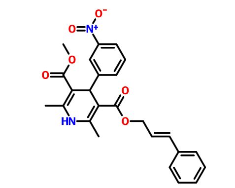
Pranidipine    

ANTHONY MELVIN CRASTO, PhD

str1

https://newdrugapprovals.files.wordpress.com/2015/12/str116.jpg


File:Pranidipine structure.svg

Pranidipine , OPC-13340, FRC 8411

Acalas®

NDA Filing in Japan

A calcium channel blocker potentially for the treatment of angina pectoris and hypertension.


CAS No. 99522-79-9

  • Molecular FormulaC25H24N2O6
  • Average mass 448.468


see dipine series………..http://organicsynthesisinternational.blogspot.in/p/dipine-series.html

manidipine


PAPER

Der Pharmacia Sinica, 2014, 5(1):11-17

https://newdrugapprovals.files.wordpress.com/2015/12/str113.jpg

pelagiaresearchlibrary.com/der-pharmacia-sinica/vol5-iss1/DPS-2014-5-1-11-17.pdf


Names
IUPAC name

methyl (2E)-phenylprop-2-en-1-yl 2,6-dimethyl-4-(3-nitrophenyl)-1,4-dihydropyridine-3,5-dicarboxylate
Other names

2,6-dimethyl-4-(3-nitrophenyl)-1,4-dihydropyridine-3,5-dicarboxylic acid O5-methyl O3-[(E)-3-phenylprop-2-enyl] ester
Identifiers
99522-79-9 Yes
ChEMBL ChEMBL1096842 
ChemSpider 4940726 
Jmol interactive 3D Image
MeSH C048161
PubChem 6436048
UNII 9DES9QVH58 Yes


PATENT SUBMITTED GRANTED
Process for the preparation of 1,4 – dihydropyridines and novel 1,4-dihydropyridines useful as therapeutic agents [US2003230478] 2003-12-18
Advanced Formulations and Therapies for Treating Hard-to-Heal Wounds [US2014357645] 2014-08-19 2014-12-04
METHODS OF TREATING CARDIOVASCULAR AND METABOLIC DISEASES [US2014322199] 2012-08-06 2014-10-30
Protein Carrier-Linked Prodrugs [US2014323402] 2012-08-10 2014-10-30
sGC STIMULATORS [US2014323448] 2014-04-29 2014-10-30
TREATMENT OF ARTERIAL WALL BY COMBINATION OF RAAS INHIBITOR AND HMG-CoA REDUCTASE INHIBITOR [US2014323536] 2012-12-07 2014-10-30
Agonists of Guanylate Cyclase Useful For the Treatment of Gastrointestinal Disorders, Inflammation, Cancer and Other Disorders [US2014329738] 2014-03-28 2014-11-06
METHODS, COMPOSITIONS, AND KITS FOR THE TREATMENT OF CANCER [US2014335050] 2012-05-25 2014-11-13
ROR GAMMA MODULATORS [US2014343023] 2012-09-18 2014-11-20
High-Loading Water-Soluable Carrier-Linked Prodrugs [US2014296257] 2012-08-10 2014-10-02 


Synthesis, isolation and use of a common key intermediate for calcium antagonist inhibitors

Neelakandan K.a,b, Manikandan H.b , B. Prabhakarana*, Santosha N.a , Ashok Chaudharia *, Mukund Kulkarnic , Gopalakrishnan Mannathusamyb and Shyam Titirmarea
a API Research Centre, Emcure Pharmaceutical Limited, Hinjawadi, Pune, India bDepartment of Chemistry, Annamalai University, Chidhambaram, India cDepartment of Chemistry, Pune University, Pune, India _________________________________________________________________________________

Pelagia Research Library     www.pelagiaresearchlibrary.com      Der Pharmacia Sinica, 2014, 5(1):11-17


The compound (3) synthesized from Nitrobenzaldehyde, tertiary butyl acetoacetate and piperidine can be used as a common intermediate for the production of calcium channel blockers like Nicardipine hydrochloride (1) and Pranidipine hydrochloride (2) with high purity.


The last twenty years have witnessed discoveries of calcium antagonists associated with multicoated pharmacodynamics potential which include not only antihypertensive and antiarrhythmic effects of the drugs but also action against excessive calcium entry in the cell of cardiovascular system and subsequent cell damage. Among many classes of calcium channel blockers, 1,4-dihydropyrimidine based drug molecules represented by Felodipine, Clevidipine, Benidipine, Nicardipine and Pranidipine are by far the best to reduce systemic vascular resistance and arterial pressure.

The reported synthetic approaches however proceed with complicated work ups, laborious purification procedures, highly expensive chemicals and low overall yields. (Scheme-I).

Synthetic scheme of Nicardipine and Pranidipine In view of the draw backs associated with previous synthetic approaches there is a strong need for environmentfriendly high yielding process applicable to the multi-kilogram production of calcium antagonist inhibitors. Herein, we report a scalable synthesis for Nicardipine hydrochloride (1) and Pranidipine hydrochloride (2) in fairly high overall yield using key intermediate 3-nitro benzylidene acid (3).Compound (3) was synthesized in two steps using 3-nitrobenzaldehyde, tertiary butyl acetoacetate and piperidine as a base to furnish tertiary butyl ester derivative (10). This was followed by hydrolysis of (10) in TFA and DCM to furnish compound (3) which would serve as a precursor for synthesis of versatile calcium antagonist inhibitors (Scheme-II).

Reported routes for synthesis of Benidipine,1,2 Lercanadipine,3-6 Nimodipine,7-11 Barnidipine12-14 and Manidipine15-16 were explored in our laboratory which involve reaction of nitro benzaldehyde with tertiary butyl acetoacetate using piperidine as a base to get tertiary butyl ester derivative (10). This is further treated with respective reagents to get various calcium channel blockers as shown in scheme 4. Since reported procedures involve in-situ generation of intermediate (3) and its reaction with corresponding fragments, it results in the formation of by-products which ultimately decrease the yield and increase the cost of API.

A novel process of manufacturing benzylidine acid derivative (3) was developed. Use of this intermediate was demonstrated by synthesis of Nicardipine and Pranidipine. This protocol may be employed for synthesis of other calcium channel blockers. In conclusion, a highly efficient, reproducible and scalable process for the synthesis of calcium channel blockers has been developed using (3) as the key intermediate.


[1] US 63 365 (Kyowa Hakko; appl.15.4.1982; J-prior.17.4.1981). [2] US 4 448 964 (Kyowa Hakko;15.5.1984; J-prior.17.4.1981). [3] Leonardi, A. et al.: Eur. J. Med.Chem. (EJMCA5) 33,399 (1988). [4] EP 153 016 (Recordati Chem. and Pharm.; appl. 21.1.1985; GB-prior. 14.2.1984). [5] US 4 705 797 (Recordati;10.11.1987; GB-prior. 14.2.1984). [6] WO 9 635 668 (Recordati Chem. and Pharm.; appl. 9.5.1996; I-prior. 12.5.1995). [7] DOS 2 117 571 (Bayer; appl. 10.4.1971). [8] DE 2 117 573 (Bayer; prior.10.4.1971) [9] US 3 799 934 (Bayer;26.3.1974;D-prior.10.4.1971). [10] US 3 932 645 (Bayer;13.1.1976;D-prior.10.4.1971). [11] Meyer, H. et al.: Arzneim.-Forsch. (ARZNAD) 31, 407 (1981); 33, 106 (1983). [12] DE 2 904 552 (Yamanouchi Pharm.; appl. 7.2.1979; J-prior.14.2.1978). [13] US 4 220 649 (Yamanouchi;2.9.1980; J-prior.14.2.1978). [14] CN 85 107 590( Faming Zhuanli Sheqing Gonhali S.; appl. 11.10.1985; J-prior.24.1.1985). [15] EP 94 159 (Takeda; appl. 15.4.1983; J-prior. 10.5.1982). [16] US 4 892 875 (Takeda;9.1.1990; J-prior. 10.5.1982, 11.1.1983).


Read Full Post »

P13K delta-gamma anticancer agent

Larry H. Bernstein, MD, FCAP, Curator

LPBI


RP 6350, Rhizen Pharmaceuticals S.A. and Novartis tieup for Rhizen’s inhaled dual Pl3K-delta gamma inhibitor

by DR ANTHONY MELVIN CRASTO Ph.D


(A)           and                         (Al)                  and                (A2)

(S)-2-(l-(9H-purin-6-ylamino)propyl)-3-(3-fluorophenyl)-4H-chromen-4-one (Compound A1 is RP 6350).


str1


RP 6350, RP6350, RP-6350

(S)-2-(l-(9H-purin-6-ylamino)propyl)-3-(3-fluorophenyl)-4H-chromen-4-one

mw 415

Rhizen Pharmaceuticals is developing RP-6530, a PI3K delta and gamma dual inhibitor, for the potential oral treatment of cancer and inflammation  In November 2013, a phase I trial in patients with hematologic malignancies was initiated in Italy ]\. In September 2015, a phase I/Ib study was initiated in the US, in patients with relapsed and refractory T-cell lymphoma. At that time, the study was expected to complete in December 2016

PATENTS……..WO 11/055215 ,  WO 12/151525.

  • Antineoplastics; Small molecules
  • Mechanism of Action Phosphatidylinositol 3 kinase delta inhibitors; Phosphatidylinositol 3 kinase gamma inhibitors
  • Phase I Haematological malignancies
  • Preclinical Multiple myeloma


Swaroop K. V. S. Vakkalanka,
COMPANY Rhizen Pharmaceuticals Sa

https://clinicaltrials.gov/ct2/show/NCT02017613


PI3K delta/gamma inhibitor RP6530 An orally active, highly selective, small molecule inhibitor of the delta and gamma isoforms of phosphoinositide-3 kinase (PI3K) with potential immunomodulating and antineoplastic activities. Upon administration, PI3K delta/gamma inhibitor RP6530 inhibits the PI3K delta and gamma isoforms and prevents the activation of the PI3K/AKT-mediated signaling pathway. This may lead to a reduction in cellular proliferation in PI3K delta/gamma-expressing tumor cells. In addition, this agent modulates inflammatory responses through various mechanisms, including the inhibition of both the release of reactive oxygen species (ROS) from neutrophils and tumor necrosis factor (TNF)-alpha activity. Unlike other isoforms of PI3K, the delta and gamma isoforms are overexpressed primarily in hematologic malignancies and in inflammatory and autoimmune diseases. By selectively targeting these isoforms, PI3K signaling in normal, non-neoplastic cells is minimally impacted or not affected at all, which minimizes the side effect profile for this agent. Check for active clinical trials using this agent. (NCI Thesaurus)

Company Rhizen Pharmaceuticals S.A.
Description Dual phosphoinositide 3-kinase (PI3K) delta and gamma inhibitor
Molecular Target Phosphoinositide 3-kinase (PI3K) delta ; Phosphoinositide 3-kinase (PI3K) gamma
Mechanism of Action Phosphoinositide 3-kinase (PI3K) delta inhibitor; Phosphoinositide 3-kinase (PI3K) gamma inhibitor
Therapeutic Modality Small molecule


Dual PI3Kδ/γ Inhibition By RP6530 Induces Apoptosis and Cytotoxicity In B-Lymphoma Cells
 Swaroop Vakkalanka, PhD*,1, Srikant Viswanadha, Ph.D.*,2, Eugenio Gaudio, PhD*,3, Emanuele Zucca, MD4, Francesco Bertoni, MD5, Elena Bernasconi, B.Sc.*,3, Davide Rossi, MD, Ph.D.*,6, and Anastasios Stathis, MD*,7
 1Rhizen Pharmaceuticals S A, La Chaux-de-Fonds, Switzerland, 2Incozen Therapeutics Pvt. Ltd., Hyderabad, India, 3Lymphoma & Genomics Research Program, IOR-Institute of Oncology Research, Bellinzona, Switzerland, 4IOSI Oncology Institute of Southern Switzerland, Bellinzona, Switzerland, 5Lymphoma Unit, IOSI-Oncology Institute of Southern Switzerland, Bellinzona, Switzerland, 6Italian Multiple Myeloma Network, GIMEMA, Italy, 7Oncology Institute of Southern Switzerland, Bellinzona, Switzerland

RP6530 is a potent and selective dual PI3Kδ/γ inhibitor that inhibited growth of B-cell lymphoma cell lines with a concomitant reduction in the downstream biomarker, pAKT. Additionally, the compound showed cytotoxicity in a panel of lymphoma primary cells. Findings provide a rationale for future clinical trials in B-cell malignancies.

POSTER SESSIONS
Blood 2013 122:4411; published ahead of print December 6, 2013
Swaroop Vakkalanka, Srikant Viswanadha, Eugenio Gaudio, Emanuele Zucca, Francesco Bertoni, Elena Bernasconi, Davide Rossi, Anastasios Stathis
  • Dual PI3K delta/gamma Inhibition By RP6530 Induces Apoptosis and Cytotoxicity
  • RP6530, a novel, small molecule PI3K delta/gamma
  • Activity and selectivity of RP6530 for PI3K delta and gamma isoforms

Introduction Activation of the PI3K pathway triggers multiple events including cell growth, cell cycle entry, cell survival and motility. While α and β isoforms are ubiquitous in their distribution, expression of δ and γ is restricted to cells of the hematopoietic system. Because these isoforms contribute to the development, maintenance, transformation, and proliferation of immune cells, dual targeting of PI3Kδ and γ represents a promising approach in the treatment of lymphomas. The objective of the experiments was to explore the therapeutic potential of RP6530, a novel, small molecule PI3Kδ/γ inhibitor, in B-cell lymphomas.

Methods Activity and selectivity of RP6530 for PI3Kδ and γ isoforms and subsequent downstream activity was determined in enzyme and cell-based assays. Additionally, RP6530 was tested for potency in viability, apoptosis, and Akt phosphorylation assays using a range of immortalized B-cell lymphoma cell lines (Raji, TOLEDO, KG-1, JEKO, OCI-LY-1, OCI-LY-10, MAVER, and REC-1). Viability was assessed using the colorimetric MTT reagent after incubation of cells for 72 h. Inhibition of pAKT was estimated by Western Blotting and bands were quantified using ImageJ after normalization with Actin. Primary cells from lymphoid tumors [1 chronic lymphocytic leukemia (CLL), 2 diffuse large B-cell lymphomas (DLBCL), 2 mantle cell lymphoma (MCL), 1 splenic marginal zone lymphoma (SMZL), and 1 extranodal MZL (EMZL)] were isolated, incubated with 4 µM RP6530, and analyzed for apoptosis or cytotoxicity by Annexin V/PI staining.

Results RP6530 demonstrated high potency against PI3Kδ (IC50=24.5 nM) and γ (IC50=33.2 nM) enzymes with selectivity over α (>300-fold) and β (>100-fold) isoforms. Cellular potency was confirmed in target-specific assays, namely anti-FcεR1-(EC50=37.8 nM) or fMLP (EC50=39.0 nM) induced CD63 expression in human whole blood basophils, LPS induced CD19+ cell proliferation in human whole blood (EC50=250 nM), and LPS induced CD45R+ cell proliferation in mouse whole blood (EC50=101 nM). RP6530 caused a dose-dependent inhibition (>50% @ 2-7 μM) in growth of immortalized (Raji, TOLEDO, KG-1, JEKO, REC-1) B-cell lymphoma cells. Effect was more pronounced in the DLBCL cell lines, OCI-LY-1 and OCI-LY-10 (>50% inhibition @ 0.1-0.7 μM), and the reduction in viability was accompanied by corresponding inhibition of pAKT with EC50 of 6 & 70 nM respectively. Treatment of patient-derived primary cells with 4 µM RP6530 caused an increase in cell death. Fold-increase in cytotoxicity as evident from PI+ staining was 1.6 for CLL, 1.1 for DLBCL, 1.2 for MCL, 2.2 for SMZL, and 2.3 for EMZL. Cells in early apotosis (Annexin V+/PI-) were not different between the DMSO blank and RP6530 samples.

Conclusions RP6530 is a potent and selective dual PI3Kδ/γ inhibitor that inhibited growth of B-cell lymphoma cell lines with a concomitant reduction in the downstream biomarker, pAKT. Additionally, the compound showed cytotoxicity in a panel of lymphoma primary cells. Findings provide a rationale for future clinical trials in B-cell malignancies.

Disclosures:Vakkalanka:Rhizen Pharmaceuticals, S.A.: Employment, Equity Ownership; Incozen Therapeutics Pvt. Ltd.: Employment, Equity Ownership.Viswanadha:Incozen Therapeutics Pvt. Ltd.: Employment. Bertoni:Rhizen Pharmaceuticals SA: Research Funding.


PI3K Dual Inhibitor (RP-6530)


Therapeutic Area Respiratory , Oncology – Liquid Tumors , Rheumatology Molecule Type Small Molecule
Indication Peripheral T-cell lymphoma (PTCL) , Non-Hodgkins Lymphoma , Asthma , Chronic Obstructive Pulmonary Disease (COPD) , Rheumatoid Arthritis
Development Phase Phase I Rt. of Administration Oral

Description

Rhizen is developing dual PI3K gamma/delta inhibitors for liquid tumors and inflammatory conditions.

Situation Overview

Dual Pl3K inhibition is strongly implicated as an intervention treatment in allergic and non-allergic inflammation of the airways and autoimmune diseases manifested by a reduction in neutrophilia and TNF in response to LPS. Scientific evidence for PI3-kinase involvement in various cellular processes underlying asthma and COPD stems from inhibitor studies and gene-targeting approaches, which makes it a potential target for treatment of respiratory disease. Resistance to conventional therapies such as corticosteroids in several patients has been attributed to an up-regulation of the PI3K pathway; thus, disruption of PI3K signaling provides a novel strategy aimed at counteracting the immuno-inflammatory response. Given the established criticality of these isoforms in immune surveillance, inhibitors specifically targeting the ? and ? isoforms would be expected to attenuate the progression of immune response encountered in most variations of airway inflammation and arthritis.

Mechanism of Action

While alpha and beta isoforms are ubiquitous in their distribution, expression of delta and gamma is restricted to circulating hematogenous cells and endothelial cells. Unlike PI3K-alpha or beta, mice lacking expression of gamma or delta do not show any adverse phenotype indicating that targeting of these specific isoforms would not result in overt toxicity. Dual delta/gamma inhibition is strongly implicated as an intervention strategy in allergic and non-allergic inflammation of the airways and other autoimmune diseases. Scientific evidence for PI3K-delta and gamma involvement in various cellular processes underlying asthma and COPD stems from inhibitor studies and gene-targeting approaches. Also, resistance to conventional therapies such as corticosteroids in several COPD patients has been attributed to an up-regulation of the PI3K delta/gamma pathway. Disruption of PI3K-delta/gamma signalling therefore provides a novel strategy aimed at counteracting the immuno-inflammatory response. Due to the pivotal role played by PI3K-delta and gamma in mediating inflammatory cell functionality such as leukocyte migration and activation, and mast cell degranulation, blocking these isoforms may also be an effective strategy for the treatment of rheumatoid arthritis as well.

Given the established criticality of these isoforms in immune surveillance, inhibitors specifically targeting the delta and gamma isoforms would be expected to attenuate the progression of immune response encountered in airway inflammation and rheumatoid arthritis.


http://www.rhizen.com/images/backgrounds/pi3k%20delta%20gamma%20ii.png

http://www.rhizen.com/images/backgrounds/pi3k%20delta%20gamma%20ii.pngtps:/

Clinical Trials

Rhizen has identified an orally active Lead Molecule, RP-6530, that has an excellent pre-clinical profile. RP-6530 is currently in non-GLP Tox studies and is expected to enter Clinical Development in H2 2013.

In December 2013, Rhizen announced the start of a Phase I clinical trial. The study entitled A Phase-I, Dose Escalation Study to Evaluate Safety and Efficacy of RP6530, a dual PI3K delta /gamma inhibitor, in patients with Relapsed or Refractory Hematologic Malignancies is designed primarily to establish the safety and tolerability of RP6530. Secondary objectives include clinical efficacy assessment and biomarker response to allow dose determination and potential patient stratification in subsequent expansion studies.


Partners by Region

Rhizen’s pipeline consists of internally discovered (with 100% IP ownership) novel small molecule programs aimed at high value markets of Oncology, Immuno-inflammtion and Metabolic Disorders. Rhizen has been successful in securing critical IP space in these areas and efforts are on for further expansion in to several indications. Rhizen seeks partnerships to unlock the potential of these valuable assets for further development from global pharmaceutical partners. At present global rights on all programs are available and Rhizen is flexible to consider suitable business models for licensing/collaboration.

In 2012, Rhizen announced a joint venture collaboration with TG Therapeutics for global development and commercialization of Rhizen’s Novel Selective PI3K Kinase Inhibitors. The selected lead RP5264 (hereafter, to be developed as TGR-1202) is an orally available, small molecule, PI3K specific inhibitor currently being positioned for the treatment of hematological malignancies.

PATENT
WO2014195888, DUAL SELECTIVE PI3 DELTA AND GAMMA KINASE INHIBITORS

This scheme provides a synthetic route for the preparation of compound of formula wherein all the variables are as described herein in above

Figure imgf000094_0001

15 14 10 12 12a

REFERENCES
April 2015, preclinical data were presented at the 106th AACR Meeting in Philadelphia, PA. RP-6530 had GI50 values of 17,028 and 22,014 nM, respectively
December 2014, data were presented at the 56th ASH Meeting in San Francisco, CA.
December 2013, preclinical data were presented at the 55th ASH Meeting in New Orleans, LA.
June 2013, preclinical data were presented at the 18th Annual EHA Congress in Stockholm, Sweden. RP-6530 inhibited PI3K delta and gamma isoforms with IC50 values of 24.5 and 33.2 nM, respectively.
  • 01 Sep 2015 Phase-I clinical trials in Hematological malignancies (Second-line therapy or greater) in USA (PO) (NCT02567656)
  • 18 Nov 2014 Preclinical trials in Multiple myeloma in Switzerland (PO) prior to November 2014
  • 18 Nov 2014 Early research in Multiple myeloma in Switzerland (PO) prior to November 2014


WO2011055215A2 Nov 3, 2010 May 12, 2011 Incozen Therapeutics Pvt. Ltd. Novel kinase modulators
WO2012151525A1 May 4, 2012 Nov 8, 2012 Rhizen Pharmaceuticals Sa Novel compounds as modulators of protein kinases
WO2013164801A1 May 3, 2013 Nov 7, 2013 Rhizen Pharmaceuticals Sa Process for preparation of optically pure and optionally substituted 2- (1 -hydroxy- alkyl) – chromen – 4 – one derivatives and their use in preparing pharmaceuticals
US20110118257 May 19, 2011 Rhizen Pharmaceuticals Sa Novel kinase modulators
US20120289496 May 4, 2012 Nov 15, 2012 Rhizen Pharmaceuticals Sa Novel compounds as modulators of protein kinases
WO 2011055215


Read Full Post »

von Willebrand Factor

Larry H. Bernstein, MD, FCAP, Curator

LPBI


FDA approves first recombinant von Willebrand factor to treat bleeding episodes

Dr. Anthony Melvin Castro


12/08/2015 02:44
The U.S. Food and Drug Administration today approved Vonvendi, von Willebrand factor (Recombinant), for use in adults 18 years of age and older who have von Willebrand disease (VWD). Vonvendi is the first FDA-approved recombinant von Willebrand factor, and is approved for the on-demand (as needed) treatment and control of bleeding episodes in adults diagnosed with VWD.
Company Baxalta Inc.
Description Recombinant human von Willebrand factor (vWF)
Molecular Target von Willebrand factor (vWF)
Mechanism of Action
Therapeutic Modality Biologic: Protein
Latest Stage of Development Registration
Standard Indication Bleeding
Indication Details Treat and prevent bleeding episodes in von Willebrand disease (vWD) patients; Treat von Willebrand disease (vWD)
Regulatory Designation U.S. – Orphan Drug (Treat and prevent bleeding episodes in von Willebrand disease (vWD) patients);
EU – Orphan Drug (Treat and prevent bleeding episodes in von Willebrand disease (vWD) patients);
Japan – Orphan Drug (Treat and prevent bleeding episodes in von Willebrand disease (vWD) patients)


The U.S. Food and Drug Administration today approved Vonvendi, von Willebrand factor (Recombinant), for use in adults 18 years of age and older who have von Willebrand disease (VWD). Vonvendi is the first FDA-approved recombinant von Willebrand factor, and is approved for the on-demand (as needed) treatment and control of bleeding episodes in adults diagnosed with VWD.

VWD is the most common inherited bleeding disorder, affecting approximately 1 percent of the U.S. population. Men and women are equally affected by VWD, which is caused by a deficiency or defect in von Willebrand factor, a protein that is critical for normal blood clotting. Patients with VWD can develop severe bleeding from the nose, gums, and intestines, as well as into muscles and joints. Women with VWD may have heavy menstrual periods lasting longer than average and may experience excessive bleeding after childbirth.

“Patients with heritable bleeding disorders should meet with their health care provider to discuss appropriate measures to reduce blood loss,” said Karen Midthun, M.D., director of the FDA’s Center for Biologics Evaluation and Research. “The approval of Vonvendi provides an additional therapeutic option for the treatment of bleeding episodes in patients with von Willebrand disease.”

The safety and efficacy of Vonvendi were evaluated in two clinical trials of 69 adult participants with VWD. These trials demonstrated that Vonvendi was safe and effective for the on-demand treatment and control of bleeding episodes from a variety of different sites in the body. No safety concerns were identified in the trials. The most common adverse reaction observed was generalized pruritus (itching).

The FDA granted Vonvendi orphan product designation for these uses.Orphan product designation is given to drugs intended to treat rare diseases in order to promote their development.

Vonvendi is manufactured by Baxalta U.S., Inc., based in Westlake Village, California.


von Willebrand Disease

Author: Eleanor S Pollak; Chief Editor: Srikanth Nagalla

Von Willebrand disease (vWD) is a common, inherited, genetically and clinically heterogeneous hemorrhagic disorder caused by a deficiency or dysfunction of the protein termed von Willebrand factor (vWF). Consequently, defective vWF interaction between platelets and the vessel wall impairs primary hemostasis.

vWF, a large, multimeric glycoprotein, circulates in blood plasma at concentrations of approximately 10 mg/mL. In response to numerous stimuli, vWF is released from storage granules in platelets and endothelial cells. It performs two major roles in hemostasis. First, it mediates the adhesion of platelets to sites of vascular injury. Second, it binds and stabilizes the procoagulant protein factor VIII (FVIII). (See Etiology.)

vWD is divided into three major categories: (1) partial quantitative deficiency (type I), (2) qualitative deficiency (type II), and (3) total deficiency (type III). vWD type II is further divided into four variants (IIA, IIB, IIN, IIM), based on characteristics of dysfunctional vWF. These categories correspond to distinct molecular mechanisms, with corresponding clinical features and therapeutic recommendations.

For discussion of vWD in children, see Pediatric Von Willebrand Disease.

http://emedicine.medscape.com/article/206996-overview


Read Full Post »

newly developed oxazolidinone antibiotics

Larry H. Bernstein, MD, FCAP, Curator

LPBI


New Antibacterial oxazolidinones in pipeline by Wockhardt

by DR ANTHONY MELVIN CRASTO Ph.D


WCK ?

(5S)-N-{3-[3,5-difluoro-4-(4-hydroxy-4-methoxymethyl-piperidin-1-yl)-phenyl]-2-oxo-oxazolidin-5-ylmethyl}-acetamide

MF C19 H25 F2 N3 O5, MW 413.42

Acetamide, N-​[[(5S)​-​3-​[3,​5-​difluoro-​4-​[4-​hydroxy-​4-​(methoxymethyl)​-​1-​piperidinyl]​phenyl]​-​2-​oxo-​5-​oxazolidinyl]​methyl]​-

CAS 957796-51-9

Antibacterial oxazolidinones

THIS MAY BE WCK 4086?????

PATENT

WO 2015173664, US8217058, WO 2012059823, 


Oxazolidinone represent a novel chemical class of synthetic antimicrobial agents.Linezolid represents the first member of this class to be used clinically. Oxazolidinones display activity against important Gram-positive human and veterinary pathogens including Methicillin-Resistant Staphylococcus aureus (MRSA), Vancomycin Resistant Enterococci (VRE) and β-lactam Resistant Streptococcus pneumoniae (PRSP). The oxazolidinones also show activity against Gram-negative aerobic bacteria, Gram-positive and Gram-negative anaerobes. (Diekema D J et al., Lancet 2001 ; 358: 1975-82).

Various oxazolidinones and their methods of preparation are disclosed in the literature. International Publication No. WO 1995/25106 discloses substituted piperidino phenyloxazolidinones and International Publication No. WO 1996/13502 discloses phenyloxazolidinones having a multisubstituted azetidinyl or pyrrolidinyl moiety. US Patent Publication No. 2004/0063954, International Publication Nos. WO 2004/007489 and WO 2004/007488 disclose piperidinyl phenyl oxazolidinones for antimicrobial use.

Pyrrolidinyl/piperidinyl phenyl oxazohdinone antibacterial agents are also described in Kim H Y et al., Bioorg. & Med. Chem. Lett., (2003), 13:2227-2230. International Publication No. WO 1996/35691 discloses spirocyclic and bicyclic diazinyl and carbazinyl oxazolidinone derivatives. Diazepeno phenyloxazolidinone derivatives are disclosed in the International Publication No. WO 1999/24428. International Publication No. WO 2002/06278 discloses substituted aminopiperidino phenyloxazolidinone derivatives.

Various other methods of preparation of oxazolidinones are reported in US Patent No. 7087784, US Patent No. 6740754, US Patent No. 4948801 , US Patent No. 3654298, US Patent No. 5837870, Canadian Patent No. 681830, J. Med. Chem., 32, 1673 (1989), Tetrahedron, 45, 1323 (1989), J. Med. Chem., 33, 2569 (1990), Tetrahedron Letters, 37, 7937-40 (1996) and Organic Process Research and Development, 11 , 739-741(2007).

Indian Patent Application No. 2534/MUM/2007 discloses a process for the preparation of substituted piperidino phenyloxazolidinones. International Publication No. WO2012/059823 further discloses the process for the preparation of phosphoric acid mono-(L-{4-[(5)-5-(acetylaminomethyl)-2-oxo-oxazolidin-3-yl]-2,6-difluorophenyl}4-methoxymethyl piperidine-4-yl)ester.

US Patent No. 8217058 discloses (5S)-N-{3-[3,5-difluoro-4-(4-hydroxy-4-methoxymethyl-piperidin-l-yl)-phenyl]-2-oxo-oxazolidin-5-ylmethyl}-acetamide as an antibacterial agent and its process for preparation.

PATENT

WO2015173664

https://patentscope.wipo.int/search/en/detail.jsf?docId=WO2015173664&recNum=1&maxRec=&office=&prevFilter=&sortOption=&queryString=&tab=PCTDescription


PATENT

http://www.google.st/patents/WO2007132314A2?cl=en


Figure imgf000004_0001

Wockhardt Ltd,

Figure imgf000006_0001
Figure imgf000006_0002

(3) (4)


PATENT

WO 2012059823

http://www.google.co.in/patents/WO2012059823A1?cl=en

Phosphoric acid mono-(l-{4-[(S)-5-(acetylamino- methyl)-2-oxo-oxazolidin-3-yl]-2,6-difluorophenyl}-4-methoxymethyl-piperidin-4-yl) ester of Formula (A),
Figure imgf000022_0001
the process comprising the steps of:
a) Converting intermediate of Formula (1) into intermediate of Formula (3)
Figure imgf000022_0002
b) Converting intermediate of Formula (3) into intermediate of Formula (5)
Figure imgf000022_0003

c) Converting intermediate of Formula (5) into intermediate of structure (6)

Figure imgf000022_0004
(5) <6> d) Converting intermediate of Formula (6) into intermediate of Formula (10)
Figure imgf000023_0001
e) Converting intermediate of Formula (10) into intermediate of Formula (11),
Figure imgf000023_0002

f) Converting intermediate of Formula (11) into compound of Formula (A) or Pharmaceutically acceptable salts thereof

Figure imgf000023_0003


Figure imgf000006_0001
Figure imgf000006_0002
Figure imgf000006_0003


Read Full Post »

Glucokinase target for type 2 diabetes

Larry H. Bernstein, MD, FCAP, Curator

LPBI


Pfizer’s PF 04991532 a Hepatoselective Glucokinase Activator Clinical Candidate for Treating Type 2 Diabetes Mellitus
DR ANTHONY MELVIN CRASTO, WORLD DRUG TRACKER
http://newdrugapprovals.org/2015/11/27/pfizers-pf-04991532-a-hepatoselective-glucokinase-activator-clinical-candidate-for-treating-type-2-diabetes-mellitus/


PF 04991532

GKA PF-04991532

(S)-6-{3-cyclopentyl-2-[4-(trifluoromethyl)-1H-imidazol-1-yl]propanamido}nicotinic acid

(S)-6-(3-Cyclopentyl-2-(4-(trifluoromethyl)-1H-imidazol-1-yl)propanamido)nicotinic Acid

(S)-6-(3-cyclopentyl-2-(4-(trifluoromethyl)-1H-imidazol-1-yl)propanamido)nicotinic acid

MW 396.36, MF C18 H19 F3 N4 O3

CAS 1215197-37-7

3-​Pyridinecarboxylic acid, 6-​[[(2S)​-​3-​cyclopentyl-​1-​oxo-​2-​[4-​(trifluoromethyl)​-​1H-​imidazol-​1-​yl]​propyl]​amino]​-

http://www.biochemj.org/content/441/3/881


Type 2 diabetes mellitus (T2DM) is a rapidly expanding public epidemic affecting over 300 million people worldwide. This disease is characterized by elevated fasting plasma glucose (FPG), insulin resistance, abnormally elevated hepatic glucose production (HGP), and reduced glucose-stimulated insulin secretion (GSIS). Moreover, long-term lack of glycemic control increases risk of complications from neuropathic, microvascular, and macrovascular diseases.

The standard of care for T2DM is metformin followed by sulfonylureas, dipeptidyl peptidase-4 (DPP-IV) inhibitors, and thiazolidinediones (TZD) as second line oral therapies. As disease progression continues, patients typically require injectable agents such as glucagon-like peptide-1 (GLP-1) analogues and, ultimately, insulin to help maintain glycemic control. Despite these current therapies, many patients still remain unable to safely achieve and maintain tight glycemic control, placing them at risk of diabetic complications and highlighting the need for novel therapeutic options.


Glucokinase (hexokinase IV) continues to be a compelling target for the treatment of type 2 diabetes given the wealth of supporting human genetics data and numerous reports of robust clinical glucose lowering in patients treated with small molecule allosteric activators. Recent work has demonstrated the ability of hepatoselective activators to deliver glucose lowering efficacy with minimal risk of hypoglycemia.

While orally administered agents require a considerable degree of passive permeability to promote suitable exposures, there is no such restriction on intravenously delivered drugs. Therefore, minimization of membrane diffusion in the context of an intravenously agent should ensure optimal hepatic targeting and therapeutic index.


Diabetes is a major public health concern because of its increasing prevalence and associated health risks. The disease is characterized by metabolic defects in the production and utilization of carbohydrates which result in the failure to maintain appropriate blood glucose levels. Two major forms of diabetes are recognized. Type I diabetes, or insulin-dependent diabetes mellitus (IDDM), is the result of an absolute deficiency of insulin. Type II diabetes, or non-insulin dependent diabetes mellitus (NIDDM), often occurs with normal, or even elevated levels of insulin and appears to be the result of the inability of tissues and cells to respond appropriately to insulin. Aggressive control of NIDDM with medication is essential; otherwise it can progress into IDDM.

As blood glucose increases, it is transported into pancreatic beta cells via a glucose transporter. Intracellular mammalian glucokinase (GK) senses the rise in glucose and activates cellular glycolysis, i.e. the conversion of glucose to glucose-6-phosphate, and subsequent insulin release. Glucokinase is found principally in pancreatic β-cells and liver parenchymal cells. Because transfer of glucose from the blood into muscle and fatty tissue is insulin dependent, diabetics lack the ability to utilize glucose adequately which leads to undesired accumulation of blood glucose (hyperglycemia). Chronic hyperglycemia leads to decreases in insulin secretion and contributes to increased insulin resistance. Glucokinase also acts as a sensor in hepatic parenchymal cells which induces glycogen synthesis, thus preventing the release of glucose into the blood. The GK processes are thus critical for the maintenance of whole body glucose homeostasis.

It is expected that an agent that activates cellular GK will facilitate glucose-dependent secretion from pancreatic beta cells, correct postprandial hyperglycemia, increase hepatic glucose utilization and potentially inhibit hepatic glucose release. Consequently, a GK activator may provide therapeutic treatment for NIDDM and associated complications, inter alia, hyperglycemia, dyslipidemia, insulin resistance syndrome, hyperinsulinemia, hypertension, and obesity.

Several drugs in five major categories, each acting by different mechanisms, are available for treating hyperglycemia and subsequently, NIDDM (Moller, D. E., “New drug targets for Type II diabetes and the metabolic syndrome” Nature414; 821-827, (2001)): (A) Insulin secretogogues, including sulphonyl-ureas (e.g., glipizide, glimepiride, glyburide) and meglitinides (e.g., nateglidine and repaglinide) enhance secretion of insulin by acting on the pancreatic beta-cells. While this therapy can decrease blood glucose level, it has limited efficacy and tolerability, causes weight gain and often induces hypoglycemia. (B) Biguanides (e.g., metformin) are thought to act primarily by decreasing hepatic glucose production. Biguanides often cause gastrointestinal disturbances and lactic acidosis, further limiting their use. (C) Inhibitors of alpha-glucosidase (e.g., acarbose) decrease intestinal glucose absorption. These agents often cause gastrointestinal disturbances. (D) Thiazolidinediones (e.g., pioglitazone, rosiglitazone) act on a specific receptor (peroxisome proliferator-activated receptor-gamma) in the liver, muscle and fat tissues. They regulate lipid metabolism subsequently enhancing the response of these tissues to the actions of insulin. Frequent use of these drugs may lead to weight gain and may induce edema and anemia. (E) Insulin is used in more severe cases, either alone or in combination with the above agents.

Ideally, an effective new treatment for NIDDM would meet the following criteria: (a) it would not have significant side effects including induction of hypoglycemia; (b) it would not cause weight gain; (c) it would at least partially replace insulin by acting via mechanism(s) that are independent from the actions of insulin; (d) it would desirably be metabolically stable to allow less frequent usage; and (e) it would be usable in combination with tolerable amounts of any of the categories of drugs listed herein.

Substituted heteroaryls, particularly pyridones, have been implicated in mediating GK and may play a significant role in the treatment of NIDDM. For example, U.S. Patent publication No. 2006/0058353 and PCT publication Nos. WO2007/043638, WO2007/043638, and WO2007/117995 recite certain heterocyclic derivatives with utility for the treatment of diabetes. Although investigations are on-going, there still exists a need for a more effective and safe therapeutic treatment for diabetes, particularly NIDDM.


s1

s1


s1


PATENT

US 20100063063

http://www.google.com/patents/US20100063063

SYNTHESIS CONSTRUCTION

6-aminonicotinic acid


BENZYL BROMIDE


Figure US20100063063A1-20100311-C00076

FIRST KEY INTERMEDIATE


SECOND SERIES FOR NEXT INTERMEDIATE

CONDENSED WITH

4-Trifluoromethyl-1H-imidazole

TO  GIVE PRODUCT SHOWN BELOW


Figure US20100063063A1-20100311-C00025

(S)-methyl 3-cyclopentyl-2-(4-(trifluoromethyl)-1H-imidazol-1-yl)propanoate (I-8a)


CONVERTED TO ACID CHLORIDE, (S)-3-cyclopentyl-2-(4-(trifluoromethyl)-1H-imidazol-1-yl)propanoyl chloride (I-8c)

AND CONDENSED WITH

Figure US20100063063A1-20100311-C00076

WILL GIVE BENZYL DERIVATIVE

THEN DEBENZYLATION TO FINAL PRODUCT


1H NMR (400 MHz, DMSO-d6) δ 13.10-13.25 (1H), 11.44 (1H), 8.83 (1H), 8.23-8.26 (1H), 8.09-8.12 (1H), 7.94-7.95 (2H), 5.22-5.26 (1H), 2.06-2.17 (2H), 1.29-1.64 (8H), 1.04-1.07 (1H); m/z 397.3 (M+H)+.


Organic Process Research & Development (2012), 16(10), 1635-1645

http://pubs.acs.org/doi/abs/10.1021/op300194c

Abstract Image

This work describes the process development and manufacture of early-stage clinical supplies of a hepatoselective glucokinase activator, a potential therapy for type 2 diabetes mellitus. Critical issues centered on challenges associated with the synthesis of intermediates and API bearing a particularly racemization-prone α-aryl carboxylate functionality. In particular, a T3P-mediated amidation process was optimized for the coupling of a racemization-prone acid substrate and a relatively non-nucleophilic amine. Furthermore, an unusually hydrolytically-labile amide in the API also complicated the synthesis and isolation of drug substance. The evolution of the process over multiple campaigns is presented, resulting in the preparation of over 110 kg of glucokinase activator.

(S)-6-(3-Cyclopentyl-2-(4-(trifluoromethyl)-1H-imidazol-1-yl)propanamido)nicotinic Acid (1)


Journal of Medicinal Chemistry (2012), 55(3), 1318-1333

http://pubs.acs.org/doi/abs/10.1021/jm2014887

Abstract Image

Glucokinase is a key regulator of glucose homeostasis, and small molecule allosteric activators of this enzyme represent a promising opportunity for the treatment of type 2 diabetes. Systemically acting glucokinase activators (liver and pancreas) have been reported to be efficacious but in many cases present hypoglycaemia risk due to activation of the enzyme at low glucose levels in the pancreas, leading to inappropriately excessive insulin secretion. It was therefore postulated that a liver selective activator may offer effective glycemic control with reduced hypoglycemia risk. Herein, we report structure–activity studies on a carboxylic acid containing series of glucokinase activators with preferential activity in hepatocytes versus pancreatic β-cells. These activators were designed to have low passive permeability thereby minimizing distribution into extrahepatic tissues; concurrently, they were also optimized as substrates for active liver uptake via members of the organic anion transporting polypeptide (OATP) family. These studies lead to the identification of 19 as a potent glucokinase activator with a greater than 50-fold liver-to-pancreas ratio of tissue distribution in rodent and non-rodent species. In preclinical diabetic animals, 19 was found to robustly lower fasting and postprandial glucose with no hypoglycemia, leading to its selection as a clinical development candidate for treating type 2 diabetes.

Bioorganic & Medicinal Chemistry Letters (2013), 23(24), 6588-6592

http://www.sciencedirect.com/science/article/pii/S0960894X13012638

Image for unlabelled figure


Structure of Hepatoselective GKA PF-04991532 (1).

Figure 1.

Structure of Hepatoselective GKA PF-04991532 (1).


Pfizer’s PF 04937319 glucokinase activators for the treatment of Type 2 diabetes
DR ANTHONY MELVIN CRASTO, WORLD DRUG TRACKER
http://newdrugapprovals.org/2015/11/27/pfizers-pf-04937319-glucokinase-activators-for-the-treatment-of-type-2-diabetes/

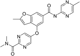
Graphical abstract: Designing glucokinase activators with reduced hypoglycemia risk: discovery of N,N-dimethyl-5-(2-methyl-6-((5-methylpyrazin-2-yl)-carbamoyl)benzofuran-4-yloxy)pyrimidine-2-carboxamide as a clinical candidate for the treatment of type 2 diabetes mellitus

PF 04937319

N,N-dimethyl-5-(2-methyl-6-((5-methylpyrazin-2-yl)-carbamoyl)benzofuran-4-yloxy)pyrimidine-2-carboxamide

MW 432.43

MF C22 H20 N6 O4
CAS 1245603-92-2
2-​Pyrimidinecarboxamid​e, N,​N-​dimethyl-​5-​[[2-​methyl-​6-​[[(5-​methyl-​2-​pyrazinyl)​amino]​carbonyl]​-​4-​benzofuranyl]​oxy]​-
N,N-Dimethyl-5-(2-methyl-6-((5-methylpyrazin-2-yl)carbamoyl)-benzofuran-4- yloxy)pyrimidine-2-carboxamide
Pfizer Inc. clinical candidate currently in Phase 2 development.
CLINICAL TRIALS

A trial to assess the safety, tolerability, pharmacokinetics, and pharmacodynamics of single doses of PF-04937319 in subjects with type 2 diabetes mellitus (NCT01044537)

Multiple dose study of PF-04937319 in patients with type 2 diabetes (NCT01272804)
Phase 2 study to evaluate safety and efficacy of investigational drug – PF04937319 in patients with type 2 diabetes (NCT01475461)


SYNTHESIS

PF 319 SYN

Glucokinase is a key regulator of glucose homeostasis and small molecule activators of this enzyme represent a promising opportunity for the treatment of Type 2 diabetes. Several glucokinase activators have advanced to clinical studies and demonstrated promising efficacy; however, many of these early candidates also revealed hypoglycemia as a key risk. In an effort to mitigate this hypoglycemia risk while maintaining the promising efficacy of this mechanism, we have investigated a series of substituted 2-methylbenzofurans as “partial activators” of the glucokinase enzyme leading to the identification ofN,N-dimethyl-5-(2-methyl-6-((5-methylpyrazin-2-yl)-carbamoyl)benzofuran-4-yloxy)pyrimidine-2-carboxamide as an early development candidate.


It is expected that an agent that activates cellular GK will facilitate glucose-dependent secretion from pancreatic beta cells, correct postprandial hyperglycemia, increase hepatic glucose utilization and potentially inhibit hepatic glucose release. Consequently, a GK activator may provide therapeutic treatment for NIDDM and associated complications, inter alia, hyperglycemia, dyslipidemia, insulin resistance syndrome, hyperinsulinemia, hypertension, and obesity. Several drugs in five major categories, each acting by different mechanisms, are available for treating hyperglycemia and subsequently, NIDDM (Moller, D. E., “New drug targets for Type 2 diabetes and the metabolic syndrome” Nature 414; 821 -827, (2001 )): (A) Insulin secretogogues, including sulphonyl-ureas (e.g., glipizide, glimepiride, glyburide) and meglitinides (e.g., nateglidine and repaglinide) enhance secretion of insulin by acting on the pancreatic beta-cells. While this therapy can decrease blood glucose level, it has limited efficacy and tolerability, causes weight gain and often induces hypoglycemia. (B) Biguanides (e.g., metformin) are thought to act primarily by decreasing hepatic glucose production. Biguanides often cause gastrointestinal disturbances and lactic acidosis, further limiting their use. (C) Inhibitors of alpha-glucosidase (e.g., acarbose) decrease intestinal glucose absorption. These agents often cause gastrointestinal disturbances. (D) Thiazolidinediones (e.g., pioglitazone, rosiglitazone) act on a specific receptor (peroxisome proliferator-activated receptor-gamma) in the liver, muscle and fat tissues. They regulate lipid metabolism subsequently enhancing the response of these tissues to the actions of insulin. Frequent use of these drugs may lead to weight gain and may induce edema and anemia. (E) Insulin is used in more severe cases, either alone or in combination with the above agents. Ideally, an effective new treatment for NIDDM would meet the following criteria: (a) it would not have significant side effects including induction of hypoglycemia; (b) it would not cause weight gain; (c) it would at least partially replace insulin by acting via mechanism(s) that are independent from the actions of insulin; (d) it would desirably be metabolically stable to allow less frequent usage; and (e) it would be usable in combination with tolerable amounts of any of the categories of drugs listed herein.

Substituted heteroaryls, particularly pyridones, have been implicated in mediating GK and may play a significant role in the treatment of NIDDM. For example, U.S. Patent publication No. 2006/0058353 and PCT publication No’s. WO2007/043638, WO2007/043638, and WO2007/117995 recite certain heterocyclic derivatives with utility for the treatment of diabetes. Although investigations are on-going, there still exists a need for a more effective and safe therapeutic treatment for diabetes, particularly NIDDM.


Designing glucokinase activators with reduced hypoglycemia risk: discovery of N,N-dimethyl-5-(2-methyl-6-((5-methylpyrazin-2-yl)-carbamoyl)benzofuran-4-yloxy)pyrimidine-2-carboxamide as a clinical candidate for the treatment of type 2 diabetes mellitus

*Corresponding authors
aPfizer Worldwide Research & Development, Eastern Point Road, Groton
E-mail: jeffrey.a.pfefferkorn@pfizer.com
Tel: +860 686 3421
Med. Chem. Commun., 2011,2, 828-839

DOI: 10.1039/C1MD00116G

http://pubs.rsc.org/en/content/articlelanding/2011/md/c1md00116g/unauth#!divAbstract

http://www.rsc.org/suppdata/md/c1/c1md00116g/c1md00116g.pdf

Glucokinase is a key regulator of glucose homeostasis and small molecule activators of this enzyme represent a promising opportunity for the treatment of Type 2 diabetes. Several glucokinase activators have advanced to clinical studies and demonstrated promising efficacy; however, many of these early candidates also revealed hypoglycemia as a key risk. In an effort to mitigate this hypoglycemia risk while maintaining the promising efficacy of this mechanism, we have investigated a series of substituted 2-methylbenzofurans as “partial activators” of the glucokinase enzyme leading to the identification ofN,N-dimethyl-5-(2-methyl-6-((5-methylpyrazin-2-yl)-carbamoyl)benzofuran-4-yloxy)pyrimidine-2-carboxamide as an early development candidate.

Graphical abstract: Designing glucokinase activators with reduced hypoglycemia risk: discovery of N,N-dimethyl-5-(2-methyl-6-((5-methylpyrazin-2-yl)-carbamoyl)benzofuran-4-yloxy)pyrimidine-2-carboxamide as a clinical candidate for the treatment of type 2 diabetes mellitus

N,N-Dimethyl-5-(2-methyl-6-((5-methylpyrazin-2-yl)carbamoyl)-benzofuran-4- yloxy)pyrimidine-2-carboxamide (28).


PAPER


http://pubs.rsc.org/en/content/articlelanding/2013/md/c2md20317k#!divAbstract


PAPER

Bioorganic & Medicinal Chemistry Letters (2013), 23(16), 4571-4578

http://www.sciencedirect.com/science/article/pii/S0960894X13007452

Glucokinase activators 1 and 2.

Figure 1.

Glucokinase activators 1 and 2.


PATENT

Pfizer Inc.

WO 2010103437

https://www.google.co.in/patents/WO2010103437A1?cl=en

Scheme I outlines the general procedures one could use to provide compounds of the present invention having Formula (I).

Figure imgf000011_0001
PF 319 SYN

Preparations of Starting Materials and Key Intermediates


Beebe, D.A.; Ross, T.T.; Rolph, T.P.; Pfefferkorn, J.A.; Esler, W.P.
The glucokinase activator PF-04937319 improves glycemic control in combination with exercise without causing hypoglycemia in diabetic rats
74th Annu Meet Sci Sess Am Diabetes Assoc (ADA) (June 13-17, San Francisco) 2014, Abst 1113-P


Amin, N.B.; Aggarwal, N.; Pall, D.; Paragh, G.; Denney, W.S.; Le, V.; Riggs, M.; Calle, R.A.
Two dose-ranging studies with PF-04937319, a systemic partial activator of glucokinase, as add-on therapy to metformin in adults with type 2 diabetes
Diabetes Obes Metab 2015, 17(8): 751


Study to compare single dose of three modified release formulations of PF-04937319 with immediate release material-sparing-tablet (IR MST) formulation previously studied in adults with type 2 diabetes mellitus (NCT02206607)

Read Full Post »

New antibiotics to address anti-microbial resistance

Larry H. Bernstein, MD, FCAP, Curator

LPBI


WCK 5107 in PHASE 1 FROM WOCKHARDT

Dr. Anthony Melvin Castro


Figure imgf000036_0001

WCK 5107

Wockhardt Limited

Useful for treating bacterial infections

CAS 1436861-97-0

disclosed in PCT International Patent Application No. PCT/IB2012/054290D

trans- sulphuric acid mono-[2-(N’-[(R)-piperidin-3-carbonyl]-hydrazinocarbonyl)-7-oxo-l,6-diaza-bicyclo[3.2.1]oct-6-yl] ester

(2S, 5R)-sulphuric acid mono-[2-(N’-[(R)-piperidin-3-carbonyl]-hydrazinocarbonyl)-7-oxo-l,6-diaza-bicyclo[3.2.1]oct-6-yl] ester

(lR,2S,5R)-l,6-Diazabicyclo [3.2.1] octane-2-carboxylic acid, 7-oxo-6-(sulfooxy)-, 2-[2-[(3R)-3-piperidinylcarbonyl]hydrazide]

trans- sulphuric acid mono-[2-(N’-[(R)-piperidin-3-carbonyl]-hydrazinocarbonyl)-7-oxo-l,6-diaza-bicyclo[3.2.1]oct-6-yl] ester

(2S, 5R)-sulphuric acid mono-[2-(N’-[(R)-piperidin-3-carbonyl]-hydrazinocarbonyl)-7-oxo-l,6-diaza-bicyclo[3.2.1]oct-6-yl] ester

(lR,2S,5R)-l,6-Diazabicyclo [3.2.1] octane-2-carboxylic acid, 7-oxo-6-(sulfooxy)-, 2-[2-[(3R)-3 -piperidinylcarbonyl] hydrazide]


In September 2015, the drug was reported to be in phase I clinical trial.One of the family members US09132133, claims a combination of sulbactam and WCK-5107.

Bacterial infections continue to remain one of the major causes contributing towards human diseases. One of the key challenges in treatment of bacterial infections is the ability of bacteria to develop resistance to one or more antibacterial agents over time. Examples of such bacteria that have developed resistance to typical antibacterial agents include: Penicillin-resistant Streptococcus pneumoniae, Vancomycin-resistant Enterococci, and Methicillin-resistant Staphylococcus aureus. The problem of emerging drug-resistance in bacteria is often tackled by switching to newer antibacterial agents, which can be more expensive and sometimes more toxic. Additionally, this may not be a permanent solution as the bacteria often develop resistance to the newer antibacterial agents as well in due course. In general, bacteria are particularly efficient in developing resistance, because of their ability to multiply very rapidly and pass on the resistance genes as they replicate.

Treatment of infections caused by resistant bacteria remains a key challenge for the clinician community. One example of such challenging pathogen is Acinetobacter baumannii (A. baumannii), which continues to be an increasingly important and demanding species in healthcare settings. The multidrug resistant nature of this pathogen and its unpredictable susceptibility patterns make empirical and therapeutic decisions more difficult. A. baumannii is associated with infections such as pneumonia, bacteremia, wound infections, urinary tract infections and meningitis.

Therefore, there is a need for development of newer ways to treat infections that are becoming resistant to known therapies and methods. Surprisingly, it has been found that a compositions comprising cefepime and certain nitrogen containing bicyclic compounds (disclosed in PCT/IB2012/054290) exhibit unexpectedly synergistic antibacterial activity, even against highly resistant bacterial strains.

PATENT

http://www.google.com/patents/WO2013030733A1?cl=en


……

REFERENCES

Study to Evaluate the Safety, Tolerability, and Pharmacokinetics of WCK-5107 Alone and in Combination With Cefepime (NCT02532140)  https://clinicaltrials.gov/show/NCT02532140
ClinicalTrials.gov Web Site 2015, September 01, To evaluate the safety,tolerability and pharmacokinetics of single intravenous doses of WCK 5107 alone and in combination with cefepime in healthy adult human subjects.

WO2013030733A1* Aug 24, 2012 Mar 7, 2013 Wockhardt Limited 1,6- diazabicyclo [3,2,1] octan-7-one derivatives and their use in the treatment of bacterial infections
WO2014135931A1* Oct 12, 2013 Sep 12, 2014 Wockhardt Limited A process for preparation of (2s, 5r)-7-oxo-6-sulphooxy-2-[((3r)-piperidine-3-carbonyl)-hydrazino carbonyl]-1,6-diaza-bicyclo [3.2.1]- octane
IB2012054290W Title not available

Read Full Post »

Pharmacy International Conference

Larry H. Bernstein, MD, FCAP, Curator

LPBI


3rd Nirma Institute of Pharmacy International Conference
NIPiCON – 2016
January 21 – 23, 2016 ………….http://www.nipicon.org/.

Anthony Melvin Crasto   https://www.facebook.com/groups/worlddrugtracker/permalink/1170816792946389/

The pharmaceutical sciences is a dynamic and interdisciplinary field that combines a broad range of scientific disciplines that are critical to the discovery and development of new drugs and therapies. Over the years, pharmaceutical scientists have been instrumental in discovering and developing innovative drugs that save people’s lives and improve the quality of life.

NIPiCON was initiated in a year 2013 to offer a common platform for academicians, researchers, industrialists, clinical practitioners and young budding pharmacists to share their ideas and research work and finally emerge with new concepts, innovations and novel strategies for various challenges in the pharmaceutical field.

The 3 International Conference, NIPiCON 2016 aims to provide a knowledge sharing experience in the area of “Global Challenges in Drug Discovery, Development and Regulatory Affairs”.

Pharmaceutical innovation is a complex creative process that harnesses the application of knowledge and creativity for discovering, developing and bringing to clinical use, new medicinal products that extend or improve the lives of patients.A successful pharmaceutical R&D process is one that minimizes the time and cost needed to bring a compound from the scientific ‘idea’, through discovery and clinical development, to final regulatory approval and delivery to the patient. This conference will provide an open forum for the academicians, researchers, clinicians and professionals of pharmaceutical industry to enrich their knowledge in the area of drug discovery, development and its regulatory requirements.

The conference features plenary sessions which will be delivered by eminent national and international speakers from different disciplines of pharmaceutical field. In addition, there will be invited lectures and sessions delivered by distinguished and young researchers in their respective fields during parallel technical sessions. The conference willalso provide the opportunity to scientists and research scholars from various organizations to put forth their innovative ideas and research findings by means of deliberations, discussions and poster presentations.


NIPiCON was initiated in a year 2013 to offer a common platform for academicians, researchers, industrialists, clinical practitioners and young budding pharmacists to share their ideas and research work and finally emerge with new concepts, innovations and novel strategies for various challenges in the pharmaceutical field.

The 3 International Conference, NIPiCON 2016 aims to provide a knowledge sharing experience in the area of “Global Challenges in Drug Discovery, Development and Regulatory Affairs”.

Read Full Post »

Rheumatoid arthritis update

Larry H. Bernstein, MD, FCAP, Curator

LPBI


Innovation update: Advancing the standard of care in rheumatoid arthritis 

Old innovation makes way for new innovation

Twenty years ago, the standard of care for RA was some combination of basic NSAIDS, along with methotrexate. Caregivers focused on symptom relief, and it was widely understood that many patients would fail to achieve remission. As the disease developed, patients would eventually develop severely life-limiting disabilities as their disease progressed.

During this period, researchers presenting at conferences grew excited about data on a new class of drugs known as anti-tumor necrosis factor (TNF) antibodies. In an article published in Acta Orthopaedica Scandinavica in 1995, two physician-researchers wrote the following:

“Primary results have recently been published on the use of anti-TNF monoclonal antibodies. In a controlled trial these antibodies were able to significantly influence a number of disease-activity variables in RA. An important observation was that the clinical effect lasted from weeks to, in some cases, months.  Although the potential of these agents for clinical use is still uncertain, these observations suggest that interfering with certain targets of the immune-inflammatory process is possible, effective and so far without side effects.”

About four years after Drs. Van de Putte and Van Riel extolled the virtues of disease-modifying biologics in clinical trials, the first anti-TNF antibody, Remicade (infliximab) was approved in 1999. At that point, the standard of care for RA improved significantly, forever changing the treatment paradigm for patients with RA.


The expanding class of JAK inhibitors

At this year’s ACR meeting, researchers  focused on  anti-inflammatory antibodies and a relatively new class of oral drugs known as janus kinase (JAK) inhibitors.  Interest in JAK inhibitors has spiked since the approval of Pfizer’s oral medication Xeljanz (tofacitinib) —the first, and currently the only, JAK inhibitor approved for the treatment of moderate-to-severe RA.JAK inhibitors have garnered interest because of the role they can play in expanding a treatment area dominated by synthetic and biologic disease-modifying anti-rheumatic drugs (DMARDs). Could JAK inhibitors provide the breakthrough in RA that the anti-TNF antibodies provided almost 20 years ago?

Currently, Eli Lilly and Incyte are in late-stage development of baricitinib, a JAK1/JAK2 inhibitor for treatment of RA. Until last December, Johnson & Johnson (J&J) and Astellas were working jointly on another JAK inhibitor, known as ASPO15K, but J&J exercised its opt-out option and left the partnership. Astellas vowed to go it alone or look for a new partner, but there have not been many updates on ASPO15K within the last year.


Innovation means understanding and responding to unmet needs

Like many other therapeutic areas, RA treatments are often used in combination. For some patients, the combination of methotrexate and a powerful biologic, such as Remicade (infliximab), will help a patient achieve remission Yet others will either not respond to methotrexate and Remicade, or will have a negative reaction. Understanding how to help nonresponders achieve relief has become a key area of research in RA.

According to Terence Rooney, MD, Medical Director at Lilly Bio-Medicines, “A substantial proportion of patients treated with methotrexate – commonly used across the disease continuum for 25 years – do not achieve satisfactory disease control, signaling a need for more effective RA treatment options. In addition, studies have shown that some patients who initially respond to biologics lose response over time, and approximately 40 percent of patients with high disease activity never respond adequately to TNF antagonist biologics.”


Innovative clinical trial design

As Lilly and Incyte approach the end of the development process for baricitinib, they have been collecting results from clinical trials designed to both establish basic efficacy and safety in placebo-controlled and comparator trials, and to obtain data on targeted patient populations.

According to Rooney, “The baricitinib phase three program investigated the benefit of baricitinib across the spectrum of patients with rheumatoid arthritis, including newly diagnosed patients, patients who had failed to respond to conventional DMARDs, and patients who had failed multiple injectable biologic DMARD therapies.”

“In addition, the phase 3 program included two 52-week studies that incorporated either methotrexate or adalimumab as active comparators to provide useful information for therapeutic positioning of baricitinib. In these studies, baricitinib was statistically superior to methotrexate and to adalimumab in improving signs and symptoms, physical function, and important patient-reported outcomes including pain, fatigue and stiffness.”

Rooney also pointed out that there is additional data establishing baricitinib as a DMARD that significantly inhibits progressive radiographic joint damage.


Experience plus evidence equals more innovation

As has become the norm, companies at ACR often highlight new data confirming the efficacy and safety of already approved drugs in larger patient populations and in real-world settings..

Lilly currently has data on more than 40,000 patients worldwide, reflecting its global ambitions. Assuming that baricitinib is approved next year (the goal is to file at the end of the year), Lilly will continue to present data at ACR in the coming years highlighting the results of its long-term extension study, RA-BEYOND.


Pfizer’s up-to-date Xeljanz data presentation at ACR

Although Xeljanz has been on the market for three years in more than 40 countries, Pfizer continues to focus on collecting new data and using it to expand use of Xeljanz. In fact, Pfizer had 20 abstracts focused solely on Xeljanz at ACR 2015.

According to Rory O’Connor, MD, Senior Vice President and Head of Global Medical Affairs, Global Innovative Pharmaceuticals Business, Pfizer, “Ongoing clinical trials and long-term extension studies provide important information about the safety and efficacy of Xeljanz in RA. We are focused on continuing to build on our knowledge of the clinical application of Xeljanz in real-world settings.”

Pfizer was also able to highlight new data that supports their recent NDA for Xeljanz XR, a once-daily formulation of Xeljanz, which is currently approved as a twice-daily dosing formulation.


JAK inhibition beyond RA

One of the most exciting things about the progress with JAK inhibitors is the possibility to innovate treatments beyond RA. Lilly has been exploring the role of JAK-dependent cytokines in the pathogenesis of numerous inflammatory and autoimmune diseases. The company also plans to meet with regulatory authorities to develop a pediatric program for juvenile RA and idiopathic arthritis.

Meanwhile, Pfizer has developed a broad portfolio of various JAK inhibitors and therapies with new modes of action. Already, Pfizer researchers have completed two phase three studies in ulcerative colitis and the top-line results have been positive.

Medical meetings are exciting, because they provide a forum for discussing breakthroughs and portending a future in which the standard of care improves. For companies like Lilly, Incyte, and Pfizer, continual development of more novel approaches to serious diseasesis like a call-response echo chamber in which innovation drives more innovation, resulting in better long-term outcomes for patients.


The JAK/STAT signaling pathway
, ,

http://d1dvw62tmnyoft.cloudfront.net/content/joces/117/8/1281/F1.large.jpg


In addition to the principal components of the pathway, other effector proteins have been identified that contribute to at least a subset of JAK/STAT signaling events. STAMs (signal-transducing adapter molecules) are adapter molecules with conserved VHS and SH3 domains (Lohi and Lehto, 2001). STAM1 and STAM2A can be phosphorylated by JAK1-JAK3 in a manner that is dependent on a third domain present in some STAMs, the ITAM (inducible tyrosine-based activation motif). Through a poorly understood mechanism, the STAMs facilitate the transcriptional activation of specific target genes, including MYC. A second adapter that facilitates JAK/STAT pathway activation is StIP (stat-interacting protein), a WD40 protein. StIPs can associate with both JAKs and unphosphorylated STATs, perhaps serving as a scaffold to facilitate the phosphorylation of STATs by JAKs. A third class of adapter with function in JAK/STAT signaling is the SH2B/Lnk/APS family. These proteins contain both pleckstrin homology and SH2 domains and are also substrates for JAK phosphorylation. Both SH2-Bβ and APS associate with JAKs, but the former facilitates JAK/STAT signaling while the latter inhibits it. The degree to which each of these adapter families contributes to JAK/STAT signaling is not yet well understood, but it is clear that various proteins outside the basic pathway machinery influence JAK/STAT signaling.

In addition to JAK/STAT pathway effectors, there are three major classes of negative regulator: SOCS (suppressors of cytokine signaling), PIAS (protein inhibitors of activated stats) and PTPs (protein tyrosine phosphatases) (reviewed by Greenhalgh and Hilton, 2001). Perhaps the simplest are the tyrosine phosphatases, which reverse the activity of the JAKs. The best characterized of these is SHP-1, the product of the mouse motheaten gene. SHP-1 contains two SH2 domains and can bind to either phosphorylated JAKs or phosphorylated receptors to facilitate dephosphorylation of these activated signaling molecules. Other tyrosine phosphatases, such as CD45, appear to have a role in regulating JAK/STAT signaling through a subset of receptors.

SOCS proteins are a family of at least eight members containing an SH2 domain and a SOCS box at the C-terminus (reviewed by Alexander, 2002). In addition, a small kinase inhibitory region located N-terminal to the SH2 domain has been identified for SOCS1 and SOCS3. The SOCS complete a simple negative feedback loop in the JAK/STAT circuitry: activated STATs stimulate transcription of the SOCS genes and the resulting SOCS proteins bind phosphorylated JAKs and their receptors to turn off the pathway. The SOCS can affect their negative regulation by three means. First, by binding phosphotyrosines on the receptors, SOCS physically block the recruitment of signal transducers, such as STATs, to the receptor. Second, SOCS proteins can bind directly to JAKs or to the receptors to specifically inhibit JAK kinase activity. Third, SOCS interact with the elongin BC complex and cullin 2, facilitating the ubiquitination of JAKs and, presumably, the receptors. Ubiquitination of these targets decreases their stability by targeting them for proteasomal degradation.

The third class of negative regulator is the PIAS proteins: PIAS1, PIAS3, PIASx and PIASy. These proteins have a Zn-binding RING-finger domain in the central portion, a well-conserved SAP (SAF-A/Acinus/PIAS) domain at the N-terminus, and a less-well-conserved carboxyl domain. The latter domains are involved in target protein binding. The PIAS proteins bind to activated STAT dimers and prevent them from binding DNA. The mechanism by which PIAS proteins act remains unclear. However, PIAS proteins have recently been demonstrated to associate with the E2 conjugase Ubc9 and to have E3 conjugase activity for sumoylation that is mediated by the RING finger domain (reviewed by Jackson, 2001). Although there is evidence that STATs can be modified by sumoylation (Rogers et al., 2003), the function of that modification in negative regulation is not yet known.

Although the mechanism of JAK/STAT signaling is relatively simple in theory, the biological consequences of pathway activation are complicated by interactions with other signaling pathways (reviewed by Heinrich et al., 2003; Rane and Reddy, 2000; Shuai, 2000). An understanding of this cross-talk is only beginning to emerge, but the best characterized interactions of the JAK/STAT pathway are with the receptor tyrosine kinase (RTK)/Ras/MAPK (mitogen-activated protein kinase) pathway. The relationship between these cascades is complex and their paths cross at multiple levels, each enhancing activation of the other. First, activated JAKs can phosphorylate tyrosines on their associated receptors that can serve as docking sites for SH2-containing adapter proteins from other signaling pathways. These include SHP-2 and Shc, which recruit the GRB2 adapter and stimulate the Ras cascade. The same mechanism stimulates other cascades, such as the recruitment and JAK phosphorylation of insulin receptor substrate (IRS) and p85, which results in the activation of the phosphoinositide 3-kinase (PI3K) pathway [for more on PI3K signaling, see Foster et al. (Foster et al., 2003)]. JAK/STAT signaling also indirectly promotes Ras signaling through the transcriptional activation of SOCS3. SOCS3 binds RasGAP, a negative regulator of Ras signaling, and reduces its activity, thereby promoting activation of the Ras pathway. Reciprocally, RTK pathway activity promotes JAK/STAT signaling by at least two mechanisms. First, the activation of some RTKs, including EGFR and PDGFR, results in the JAK-independent tyrosine phosphorylation of STATs, probably by the Src kinase. Second, RTK/Ras pathway stimulation causes the downstream activation of MAPK. MAPK specifically phosphorylates a serine near the C-terminus of most STATs. While not absolutely necessary for STAT activity, this serine phosphorylation dramatically enhances transcriptional activation by STAT. In addition to RTK and PI3K interactions with JAK/STAT signaling, multiple levels of cross-talk with the TGF-β signaling pathway have been recently reported [for a review of TGF-β, see (Moustakas, 2002)]. Furthermore, the functions of activated STATs can be altered through association with other transcription factors and cofactors that are regulated by other signaling pathways. Thus the integration of input from many signaling pathways must be considered if we are to understand the biological consequences of cytokine stimulation.

References

  1. ↵
  2. ↵
  3. ↵
  4. ↵
  5. ↵

…..


https://youtu.be/9JHBHSHaBeI

Published on 27 Feb 2014

The JAK/STAT secondary messenger signaliing pathway..
Presented by: Joseph Farahany, M.D


Jak/Stat Signaling Pathway


Jaks and Stats are critical components of many cytokine receptor systems; regulating growth, survival, differentiation, and pathogen resistance. An example of these pathways is shown for the IL-6 (or gp130) family of receptors, which coregulate B cell differentiation, plasmacytogenesis, and the acute phase reaction. Cytokine binding induces receptor dimerization, activating the associated Jaks, which phosphorylate themselves and the receptor. The phosphorylated sites on the receptor and Jaks serve as docking sites for the SH2-containing Stats, such as Stat3, and for SH2-containing proteins and adaptors that link the receptor to MAP kinase, PI3K/Akt, and other cellular pathways.

Phosphorylated Stats dimerize and translocate into the nucleus to regulate target gene transcription. Members of the suppressor of cytokine signaling (SOCS) family dampen receptor signaling via homologous or heterologous feedback regulation. Jaks or Stats can also participate in signaling through other receptor classes, as outlined in the Jak/Stat Utilization Table. Researchers have found Stat3 and Stat5 to be constitutively activated by tyrosine kinases other than Jaks in several solid tumors

The Jak/Stat pathway mediates the effects of cytokines, like erythropoietin, thrombopoietin, and G-CSF, which are protein drugs for the treatment of anemia, thrombocytopenia, and neutropenia, respectively. The pathway also mediates signaling by interferons, which are used as antiviral and antiproliferative agents. Researchers have found that dysregulated cytokine signaling contributes to cancer. Aberrant IL-6 signaling contributes to the pathogenesis of autoimmune diseases, inflammation, and cancers such as prostate cancer and multiple myeloma. Jak inhibitors currently are being tested in models of multiple myeloma. Stat3 can act as an oncogene and is constitutively active in many tumors. Crosstalk between cytokine signaling and EGFR family members is seen in some cancer cells. Research has shown that in glioblastoma cells overexpressing EGFR, resistance to EGFR kinase inhibitors is induced by Jak2 binding to EGFR via the FERM domain of the former [Sci. Signal. (2013) 6, ra55].

Activating Jak mutations are major molecular events in human hematological malignancies. Researchers have found a unique somatic mutation in the Jak2 pseudokinase domain (V617F) that commonly occurs in polycythemia vera, essential thrombocythemia, and idiopathic myelofibrosis. This mutation results in the pathologic activation Jak2, associated with receptors for erythropoietin, thrombopoietin, and G-CSF, which control erythroid, megakaryocytic, and granulocytic proliferation and differentiation. Researchers have also shown that somatic acquired gain-of-function mutations of Jak1 are found in adult T cell acute lymphoblastic leukemia. Somatic activating mutations in Jak1, Jak2, and Jak3 have also been identified in pediatric acute lymphoblastic leukemia (ALL). Furthermore, Jak2 mutations have been detected around pseudokinase domain R683 (R683G or DIREED) in Down syndrome childhood B-ALL and pediatric B-ALL.

Selected Reviews:

– See more at: http://www.cellsignal.com/contents/science-pathway-research-immunology-and-inflammation/jak-stat-signaling-pathway/pathways-il6#sthash.8SVwSWXw.dpuf


The JAK-STAT Signaling Pathway: Input and Output Integration1

  1. Peter J. Murray

The Journal of Immunology Mar 1, 2007;  178(5): 2623-2629    http://dx.doi.org:/10.4049/​jimmunol.178.5.2623

Universal and essential to cytokine receptor signaling, the JAK-STAT pathway is one of the best understood signal transduction cascades. Almost 40 cytokine receptors signal through combinations of four JAK and seven STAT family members, suggesting commonality across the JAK-STAT signaling system. Despite intense study, there remain substantial gaps in understanding how the cascades are activated and regulated. Using the examples of the IL-6 and IL-10 receptors, I will discuss how diverse outcomes in gene expression result from regulatory events that effect the JAK1-STAT3 pathway, common to both receptors. I also consider receptor preferences by different STATs and interpretive problems in the use of STAT-deficient cells and mice. Finally, I consider how the suppressor of cytokine signaling (SOCS) proteins regulate the quality and quantity of STAT signals from cytokine receptors. New data suggests that SOCS proteins introduce additional diversity into the JAK-STAT pathway by adjusting the output of activated STATs that alters downstream gene activation.


The mammalian JAK and STAT family members have been extensively, and seemingly exhaustively, analyzed in the mouse and human systems. All four JAK and seven STAT family members have been deleted in the mouse, in addition to the creation of conditional alleles for genes whose loss of function leads to embryonic or perinatal lethality (Stat3, combined deficiency of Stat5a and Stat5b, and Jak2). In humans, detailed genetic studies have been performed in people bearing mutant Jak or Stat genes. Specific Abs to phospho-forms of each protein are used to study how the JAK-STAT cascade is activated by cytokine receptors. Crystallographic studies have illuminated structural information for multiple STAT family members in different forms. Pharmacological inhibitors have been developed for clinical use where JAK-STAT signaling is implicated in disease pathology and progression. Finally, in most cases, a specific JAK-STAT combination has been paired with each cytokine receptor, and this information translated into cell-type specific patterns of cytokine responsiveness and gene expression.

Major questions remain concerning how the JAK-STAT cascade functions to control specific gene expression patterns, and how the cascades are regulated. I will describe three elements of JAK-STAT signaling that require experimental investigation. First, I will address an unexpected experimental complication that arises from the analysis of mice and cells that lack one or more STAT family member. Second, I will use JAK1-STAT3 signaling from the IL-10R and IL-6R systems to illustrate that we lack detailed understanding of how specificity in gene expression is generated by receptors that use identical JAK-STAT members. Third, we have yet to explain how STAT activation is negatively regulated. Although the suppressor of cytokine signaling (SOCS)3 proteins are the best understood negative regulators of the JAK-STAT pathway, the biochemical mechanism of SOCS-mediated inhibition is unexplained. Moreover, additional inhibitory pathways have also been proposed to block the production of activated STATs. Collectively, I will argue that our understanding of the pathway from cytokine receptor to gene expression profile is in its infancy, but remains one of the best opportunities to dissect signal transduction.

Overview of the proximal JAK-STAT activation mechanism

The current model of JAK-STAT signaling holds that cytokine receptor engagement activates the associated JAK combination, which in turn phosphorylates the receptor cytoplasmic domain to allow recruitment of a STAT, which in turn is phosphorylated, dimerizes and moves to the nucleus to bind specific sequences in the genome and activate gene expression. Cytoplasmic domains of cytokine receptors associate with JAKs via JAK binding sites located close to the membrane (1). The postulated role of JAKs in trafficking or chaperoning the receptors to the cell surface is debated (2, 3, 4, 5, 6). Regardless of the when and where cytokine receptors and JAKs associate, their close apposition at the membrane is required to stimulate the kinase activity of the JAK following cytokine binding. At this stage in the activation of the pathway, we understand next to nothing about the structural basis of the JAK-receptor interaction, how receptor intracellular domains reorient upon cytokine binding and physically contact the JAK to receive the phosphorylation modification.

JAK-mediated phosphorylation of the receptor creates binding sites for the Src homology 2 (SH2) domains of the STATs. STAT recruitment is followed by tyrosine, and in some cases, serine phosphorylation on key residues (by the JAKs and other closely associated kinases) that leads to transit into the nucleus. This brief summary of the activation of the JAK-STAT pathway omits numerous unresolved details: the STAT monomer to dimer transition has been questioned, as has the role of phosphorylation in dimerization and nuclear transit (7). Furthermore, it is unclear how many configurations of STAT homo- and heterocomplexes are present in cells before, during, and after cytokine stimulation (8, 9,10). We do not understand the detailed structural basis for the preference of one SH2 domain for a given receptor, and we have little knowledge of how other non-JAK kinases are recruited to the receptors and phosphorylate the STATs.

Many receptors signal through a small number of JAKs

Cytokine receptors signal through two types of pathways: the JAK-STAT pathway and other pathways that usually involve the activation of the MAP kinase cascade. Although the latter will not be discussed here, it is worth noting that elegant genetic studies have demonstrated the importance of these pathways in various pathological systems (11, 12,13, 14). There are now ∼36 cytokine receptor combinations that respond to ∼38 cytokines (counting the type I IFNs as one because they all signal through the IFN-αβR). Different cells and tissues express distinct receptor combinations that respond to cytokine combinations unique to the microenvironment or systemic response of the organism. Hence, at any given time, a single cell may integrate signals from multiple cytokine receptors. Genetic studies have established that the cytokine receptor system is restrictive in that different classes of receptors preferentially use one JAK or JAK combination (7): receptors required for hemopoietic cell development and proliferation use JAK2, common γ-chain receptors use JAK1 and JAK3 whereas other receptors use only JAK1 (Fig. 1⇓). Unexplained is the selective use of these combinations: why the IFN-γR rigidly uses the JAK1, JAK2 combination is unknown as is the restricted use of TYK2. Compared with JAK1–3, TYK2 is unusual in that loss of function mutations in the mouse have shown obligate, but not absolute, requirements in IFN-αβR and IL-12R signaling (15, 16). In contrast, human TYK2 seems to be essential for signaling through a broader range of cytokine receptors (17).


FIGURE 1.

FIGURE 1.

The majority of cytokine receptors use three JAK combinations. Shown are well-studied cases where JAK usage by each cytokine receptor has been established by genetic and biochemical studies. Exceptions shown are the G-CSFR (∗) where it is currently unclear whether both JAK1 and JAK2 are required together. Additionally, the IL-12R (†) and IL-23R (†) require TYK2 but the requirement for JAK2 has not been definitively determined. Receptors that use JAK2 and JAK3, JAK3 alone, TYK2 alone, or JAK3 and TYK2 have not been described.

The preferential association of JAKs to certain receptor classes raises several issues. First, how did the JAK-receptor combinations evolve? Because the number of receptors is relatively large, why has the number of JAKs remained small? Why have the combinations of JAK pairs also remained small given that there are 10 possible combinations that can be used (Fig. 1⇑)? Second, how flexible is the cytokine receptor-JAK pair? That is, can receptors be engineered for interchangeable JAK use, or is a given JAK combination fixed for a specific receptor class? For example, can JAK1, JAK3, or TYK2 activate erythropoietin receptor (EpoR) signaling (if so engineered) or is JAK2 obligatory for signaling? These questions allude to a fundamental issue that concerns the function of the JAK in cytokine receptor activation: if the only function of the JAKs is to phosphorylate tyrosine resides on the cytoplasmic domain of the receptors, then it should be possible to trade JAK-receptor pairs. If these receptors retain identical downstream gene expression profiles, then the signal generated by the JAK is generic and functions primarily to activate the receptor (6). Conversely, it is also possible that each receptor-JAK combination retains crucial specificity functions and swapping, for example, JAK1 for JAK2 on the EpoR will modify or destroy a specific function in erythrogenesis. These questions can be addressed experimentally by replacing one preferred JAK binding site for another in genes encoding different receptors. The EpoR is a good test example because the activity of the receptor and its signaling pathway is essential for life and erythropoiesis is readily assayed.

Core versus cell-type specific STAT signaling

Microarray experiments designed to monitor changes in gene expression induced by JAK-STAT signaling have revealed that both cell-type specific transcription and core, or stereotypic, mRNA profiles are induced by activated cytokine receptors in different cell types (Fig. 2⇓). For example, IFN-γ, via STAT1, induces the expression of a similar cohort of genes regardless of the cell type tested (18). These genes are often termed the “IFN signature” and overlap with the gene expression pattern induced by IFN-αβ signaling that also involves STAT1, in cooperation with STAT2 and IRF9. The IFN signature is readily observed in microarray experiments and is indicative of STAT1 activity. The STAT6 pathway activated by IL-4 or IL-13 provides an example of a cell-type specific response. IL-4-regulated genes in T cells have a distinct signature compared with IL-4/IL-13 signaling in macrophages or other non-lymphocytes (19, 20, 21, 22). In the latter, genes such as Arg1(encoding arginase 1) are often induced >100-fold but are silent in T cells (23, 24, 25, 26,27). Collectively these data argue that STATs activate defined gene sets, depending on their genomic accessibility, and possibly on cofactors that further refine gene expression profiles. STAT3 signaling illustrates a more complex system and will be discussed below to illustrate the distinctions between IL-6 and IL-10 signaling.


FIGURE 2.

FIGURE 2.

Core signaling by STATs. Representative examples of gene expression induced by STAT signaling in different tissues. The examples were extracted and edited from numerous microarray and empirical studies.

Interpreting experiments using STAT loss-of-function systems

Experiments with the different STAT knockout mice, and cells derived from these animals, have been critical for understanding specific requirements of individual STATs in gene expression following cytokine receptor signaling. The interpretation of these experiments is generally straightforward. For example, STAT5a and STAT5b are essential for the expression of genes that promote hemopoietic survival (28, 29, 30) whereas STAT1 is required for the expression of IFN-regulated genes that are involved in the protection against pathogens (18). However, by EMSA and immunoblotting experiments, most cytokines have been shown to activate multiple STATs, prompting experiments to determine transcriptional responses that can be activated in the absence of a given STAT. An initial example of this type of approach was performed by Schreiber and colleagues who interrogated gene expression profiles induced by IFN-γ signaling in the absence of STAT1 (31, 32). In these experiments, IFN-γ was used to stimulate STAT1-deficient bone marrow-derived macrophages and fibroblasts. Numerous genes were induced by IFN-γ in the absence of STAT1, leading to the conclusion that the IFN-γR activates a STAT1-independent gene expression program. However, inspection of the genes induced by IFN-γ signaling in STAT1-deficient cells shows many to be STAT3-regulated genes such asSocs3, Gadd45, and Cebpb. STAT3 phosphorylation is normally induced by IFN-γ in wild-type cells but in the absence of STAT1, STAT3 signaling is dominant. What is the mechanism of this effect? We now know from experiments using STAT-deficient cells that receptor occupancy, or lack of occupancy by the dominant STAT that binds the receptor, causes a switch from one activated STAT to another (33). A converse example is the conversion of IL-6R signaling to a dominant STAT1 activation in STAT3-deficient cells (34). This switch causes the downstream induction of the IFN gene expression pathway just as IFN-γ would cause in wild-type cells.

A related example is observed when IL-6 signaling is tested in the absence of SOCS3. SOCS3 is induced by STAT signaling from different cytokine receptors and functions as a feedback inhibitor of the IL-6R (and the G-CSFR, LIFR, and leptinR) by binding to phosphorylated Y757 on the gp130 cytoplasmic domain (see below). However in the absence of SOCS3, STAT3 phosphorylation is greatly increased (35, 36, 37). At the same time however, STAT1 phosphorylation is also induced, leading to a dominant IFN-like gene expression signature (35, 36). Thus SOCS3 regulates both the quantity and type of STAT signal generated from the IL-6R. Although the mechanism of the SOCS3 effect is unclear, the promiscuity of different receptors for different STATs argues that loss-of-function experiments must be carefully examined for the activation of other STAT molecules that fill the “hole” created by the loss of one STAT. These data also suggest that different cytokine receptors have evolved selectivity for different classes of STATs. Although STAT1 and STAT3 can apparently interchangeably bind the IL-6R or IFN-γR when either molecule is missing, signaling in wild-type cells shows a strong preference for one STAT over the other. Likewise, other receptors may have evolved to bind only one STAT, and in the absence of the key STAT, the other STATs cannot bind and/or be activated by the receptor.

The above examples primarily describe experiments using STAT1–STAT3-activating receptors but these are not isolated cases. In T cells stimulated by IL-12, STAT4 is activated and drives IFN-γ production. This pathway is a central regulatory event in the development of the Th1 type T cell responses. IFN-αβ, via the IFN-αβR, also activates STAT4 (in addition to STAT1 and STAT2 that forms a complex with IRF-9 to mediate anti-viral gene expression) but cannot activate strong IFN-γ production and therefore cannot drive Th1 development (38). However, in the absence of STAT1, IFN-αβ causes a large increase in IFN-γ production, especially in vivo during viral infection (39, 40). These data were originally interpreted to mean that STAT1 normally suppressed IFN-γ production. However, the data can just as easily be resolved when we consider that STAT4 activation from the IFN-αβR is increased in the absence of STAT1. Recent data confirm this interpretation but also show that STAT4 activation by the IFN-αβR, although increased, cannot sustain IFN-γ production from T cells when compared with IL-12 (38). This is probably because of the stronger differential activity of SOCS1 on the IFN-αβR versus the IL-12R (discussed below). I would predict that an IFN-αβR that is refractory to SOCS1 (or active in a Socs1−/− background) would behave identically to the IL-12R in the absence of STAT1.

Although loss of gene expression may be observed in a given STAT knockout, a corresponding increase in the ectopic activation of another STAT pathway may confound the interpretation of results in both in vitro and in vivo systems. Because specific Abs are available for each tyrosine-phosphorylated STAT molecule, a simple solution is to first measure which other STATs are activated by a given receptor in the absence of the STAT of interest. Experiments using STAT knockout systems should also be supported by additional data that uses complimentary mutations in the receptor that ablate STAT recruitment, or complete loss of the receptor. Finally, it is worth noting that the loss of a STAT pathway from a receptor signaling system can cause additional loss of key negative regulatory systems including feedback loops such as SOCS induction as presently debated for G-CSFR signaling and receptor systems discussed below (41, 42, 43, 44, 45).

  1. Negative regulation of the JAK-STAT signal
  2. Is there functional equivalence in signaling from receptors using the same JAK-STAT combination in the same cell?
  3. Future directions


FIGURE 3.

FIGURE 3.

Proposed differential STAT activation by IL-10 or IL-6. Shown are three classes of genes activated by STAT3 where Socs3 is a representative “common” gene induced by both receptors. In the absence of SOCS3, the IL-6R can activate the anti-inflammatory genes in the same way as the IL-10R. The mechanism of this effect remains to be established.


JAK/STAT Activation Inhibitors

The JAK/STAT pathway plays an important role in cytokine receptor-mediated signal transduction via activation of downstream signal transducers and activators of transcription (STAT), phosphatidylinositol 3-kinase (PI3K), and mitogen-activated protein kinase (MAPK) pathways.
These inhibitors are useful tools for exploring the contribution of JAK/STAT-mediated signaling.

Pathways of inhibition of JAK/STAT activation

JAK/STAT Activation Inhibitors

AG490 JAK2 inhibitor 10 mg
AZD1480 NEW! JAK1 & JAK2 inhibitor 5 mg
CP-690550 JAK3 Inhibitor 5 mg
CYT387 NEW! JAK1/JAK2 & TBK1/IKK-ε inhibitor 10 mg
Ruxolitinib JAK1 & JAK2 Inhibitor 5 mg


Methotrexate Is a JAK/STAT Pathway Inhibitor

Sally Thomas, Katherine H. Fisher, John A. Snowden, Sarah J. Danson, Stephen Brown, Martin P. Zeidler

PLOS   Published: July 1, 2015
DOI: http://dx.doi.org:/10.1371/journal.pone.0130078
Background 

The JAK/STAT pathway transduces signals from multiple cytokines and controls haematopoiesis, immunity and inflammation. In addition, pathological activation is seen in multiple malignancies including the myeloproliferative neoplasms (MPNs). Given this, drug development efforts have targeted the pathway with JAK inhibitors such as ruxolitinib. Although effective, high costs and side effects have limited its adoption. Thus, a need for effective low cost treatments remains.

Methods & Findings        

We used the low-complexity Drosophila melanogaster pathway to screen for small molecules that modulate JAK/STAT signalling. This screen identified methotrexate and the closely related aminopterin as potent suppressors of STAT activation. We show that methotrexate suppresses human JAK/STAT signalling without affecting other phosphorylation-dependent pathways. Furthermore, methotrexate significantly reduces STAT5 phosphorylation in cells expressing JAK2 V617F, a mutation associated with most human MPNs. Methotrexate acts independently of dihydrofolate reductase (DHFR) and is comparable to the JAK1/2 inhibitor ruxolitinib. However, cells treated with methotrexate still retain their ability to respond to physiological levels of the ligand erythropoietin.

Conclusions

Aminopterin and methotrexate represent the first chemotherapy agents developed and act as competitive inhibitors of DHFR. Methotrexate is also widely used at low doses to treat inflammatory and immune-mediated conditions including rheumatoid arthritis. In this low-dose regime, folate supplements are given to mitigate side effects by bypassing the biochemical requirement for DHFR. Although independent of DHFR, the mechanism-of-action underlying the low-dose effects of methotrexate is unknown. Given that multiple pro-inflammatory cytokines signal through the pathway, we suggest that suppression of the JAK/STAT pathway is likely to be the principal anti-inflammatory and immunosuppressive mechanism-of-action of low-dose methotrexate. In addition, we suggest that patients with JAK/STAT-associated haematological malignancies may benefit from low-dose methotrexate treatments. While the JAK1/2 inhibitor ruxolitinib is effective, a £43,200 annual cost precludes widespread adoption. With an annual methotrexate cost of around £32, our findings represent an important development with significant future potential.

Citation: Thomas S, Fisher KH, Snowden JA, Danson SJ, Brown S, Zeidler MP (2015) Methotrexate Is a JAK/STAT Pathway Inhibitor. PLoS ONE 10(7): e0130078.   http://dx.doi.org:/10.1371/journal.pone.0130078


Read Full Post »

Empagliflozin

Larry H. Bernstein, MD, FCAP, Curator

LPBI


Empagliflozin Benefits in EMPA-REG Explored in Diabetics Initially With or Without Heart Failure

Marlene Busko

http://www.medscape.com/viewarticle/854542


ORLANDO, FL — Patients with type 2 diabetes and established CVD who received the antidiabetic sodium-glucose cotransporter 2 (SGLT2) inhibitor empagliflozin (Jardiance, Lilly/Boehringer Ingelheim), as opposed to placebo, had a reduced risk of being hospitalized for heart failure or dying from CVD during a median follow-up of 3.1 years. The finding was strongest in patients without heart failure at baseline[1]. The finding is noteworthy in part because associated heart failure has been a concern, justified or not, with some other diabetes medications.

In these high-risk patients, empagliflozin resulted in a “consistent benefit” in these outcomes, Dr Silvio E Inzucchi (Yale University School of Medicine, New Haven, CT) said, presenting these findings from a prespecified secondary analysis of the EMPA-REG OUTCOME trial at theAmerican Heart Association (AHA) 2015 Scientific Sessions.

Unlike the gasps and applause that greeted him when he presented the trial’s primary outcome results at the European Association for the Study of Diabetes (EASD) 2015 Meeting in Stockholm in mid-September, the audience reaction this time was more measured. The trial had also been published at about the time of its EASD presentation [2].

The principal findings showed that compared with patients who took placebo, those who were randomized to empagliflozin had a 38% (P<0.001) reduced risk of CV death and a 35% P=0.002) reduced risk of hospitalization for HF, at a median follow-up of 3.1 years.

In the current secondary analysis, the 90% of patients who were free of heart failure at study entry showed a steep and significant drop in HF hospitalizations during the trial. There was also a drop in HF hospitalizations with active therapy in the minority who had HF at baseline, but it failed to reach significance.

“I think metformin is likely to remain our first-line oral therapy for patients with type 2 diabetes,” Dr Donald M Lloyd-Jones (Northwestern University Feinberg School of Medicine, Chicago, IL), cochair at an AHA press briefing, told heartwire from Medscape. “There is an alphabet soup of diabetes medications,” with multiple agents that effectively lower blood glucose and reduce patients’ risk of retinopathy, nephropathy, and neuropathy.

However, “it was . . . unexpected that [empagliflozin], as reported recently [at the EASD meeting and] in the New England Journal of Medicine [has an] effect on CV death and other CV events.” This is still an early stage of research, he cautioned, and it is not known how the drug exerts its CV effects and whether there is a class effect. “But [this] could be a game changer, because we would love to have [antidiabetic] medications that not only control blood sugar but also reduce death and [other] hard events,” he said.


First CV Outcomes Trial in this Drug Class

Until now, none of the antiglycemic medications has also been shown to improve HF outcomes, Inzucchi explained. “We’ve actually been searching decades for a diabetes medicine that will not only lower blood glucose but also reduce cardiovascular complications,” he said in a press briefing. “And I would remind you that based on the 2008 FDA guidance to industry, all new diabetes medications need to be tested for cardiovascular safety before being allowed on the market,” he added.

EMPA-REG OUTCOME is the first published, large CV-outcome trial of an SGLT-2 inhibitor.

As previously described, the trial randomized 7028 adult patients who had type 2 diabetes and established CVD to receive 10 mg/day or 25 mg/day empagliflozin or placebo. The CVD included prior MI (46.6%), CABG (24.8%), stroke (23.3%), and peripheral artery disease (PAD) (20.8%).

The patients were also required to have an HbA1c level between 7% and 10%, body-mass index (BMI) <45, and, because the drug exerts its effects via the kidney, estimated glomerular filtration rate (eGFR) >30 mL/min/1.73 m2.

“Importantly, study medication was given upon a backdrop of standard care—antihyperglycemia therapy, as well as other evidence-based cardiovascular therapies such as statins, ACE inhibitors, and aspirin,” Inzucchi stressed.


Spotlight on HF Outcomes

The current analysis dove deeper into the heart-failure outcomes in the trial.

The risk of hospitalization for HF or CV death was consistently significantly lower in patients who received empagliflozin vs placebo, in subgroup analyses related to age, kidney function, and medication use (ACE inhibitors/angiotensin receptor blockers [ARBs], diuretics, beta-blockers, or mineralocorticoid-receptor antagonists).

Overall, the patients who received empagliflozin had a 34% reduced risk of being hospitalized for HF or dying from CV causes and a 39% reduced risk of being hospitalized for or dying from HF.

Risk of Hospitalization or Death, Empagliflozin vs Placebo

Outcome HR (95% CI) P
Hospitalization for HF or CV death 0.66 (0.55–0.79) <0.00001
Hospitalization for or death from HF 0.61 (0.47–0.79) <0.00001

Most patients (90%) did not have HF at baseline.

In the patients without HF at baseline, “as you might expect, [HF] hospitalizations were relatively small in number” (1.8% of patients on the study drug and 3.1% of patients on placebo), said Inzucchi. There was a statistically significant 41% reduced risk of HF hospitalization in patients without HF at baseline on the study drug vs placebo (HR 0.59, 95% CI 0.43–0.82).

In the smaller number of patients who did have HF at baseline, the rate of hospitalizations for HF was much higher (10.4% of patients on the study drug and 12.3% of patients on placebo). But in this case, the difference between patients on the study drug vs placebo was not statistically significant (HR 0.75, 95% CI 0.48–1.19).

The results were similar when the analysis was repeated for the combined outcome of hospitalization for HF or CV death.

“Not surprisingly,” adverse events were more common in sicker patients with baseline HF; genital infections, a well-known adverse event in drugs that increase glucose in the urine, were three times more common in those patients, said Inzucchi.

“I think these are very compelling data, but early days,” said Lloyd-Jones.

Inzucchi receives research grants from Genzyme and honoraria from Boehringer Ingelheim, Merck Sharp & Dome, Sanofi, Amgen, and Genzyme, and he is a consultant on advisory boards for Boehringer Ingelheim, Sanofi, and Amgen. Disclosures for the coauthors are listed in the abstract. Lloyd-Jones has no relevant financial relationships.

Read Full Post »

« Newer Posts - Older Posts »