Feeds:
Posts
Comments

Archive for the ‘Chemical Biology and its relations to Metabolic Disease’ Category

What about Theranos?

Curator: Larry H. Bernstein, MD, FCAP

Is Theranos Situation False Crowdfunding Claims at Scale or ‘Outsider’ Naivety?

http://www.mdtmag.com/blog/2015/11/theranos-situation-false-crowdfunding-claims-scale-or-outsider-naivety

If you’ve been following the Theranos situation that involves several damning articles from the Wall Street Journal on the company (see sidebar below video), you know that “something is rotten in the state of Denmark.” That is to say, regardless of whether or not you believe the WSJ articles 100%, believe Theranos 100%, or land somewhere in between, it’s hard not to see that something at the company is definitely creating questions about their original claims. In fact, the company has apparently even tempered some language with regard to its capabilities while “debating” the accuracy of the WSJ articles. It’s really a big mess for a company that was supposedly making significant changes in the way we’d conduct blood testing and the way patients controlled and accessed their own health data (although, I think the idea behind that specific aspect is a very good one).

Due to FDA inspections and findings of concern with Thernos practices, the company is currently only collecting blood for one test using its revolutionary proprietary technology. While the company’s CEO Elizabeth Holmes continues to assure the public that the problems are tied to FDA related procedures and not an issue with the technology itself, stakeholders such as Walgreens put any further interactions with the company on hold.

In the following video from Fortune’s Global Forum, you can see Ms. Holmes discussing the situation over the FDA inspections and the changes that are currently in place with regard to the testing that’s happening at the company.

https://youtu.be/A8qgmGtRMsY

So what’s the story behind this story? Is this a deliberate attempt to deceive on the part of Theranos or is it an example of what can happen when an “outsider” gets involved in the highly regulated medical device industry and faces off with the FDA without the proper experience in place to address potential areas of concern?

In a recent blog, I looked at the crowdfunding of medical devices and what can happen when claims made don’t live up to the reality of the product that’s actually developed. Once enthusiastic investors can quickly (and loudly) turn on a company or project, venting their frustration even directly on the crowdfunding page for all to see. Unfortunately, with the way these sites seem to be set-up, the money is still provided to the company that produces a product, albeit one that does not live up to the initial concept.

Is that what Theranos ultimately is? Were the technology claims taken at face value by significant investment backers? It would seem very unlikely, but given some of the accusations of former Theranos employees in the WSJ articles, it wouldn’t be the only instance of Theranos trying to manipulate testing protocols for the sake of appearing more impressive. Theranos counters those claims by saying the former employees were actually unfamiliar with the actual testing the company performs. Whether or not you believe that is entirely up to you.

Another alternative to blatant deceit on the part of Theranos is the possibility that the company was simply playing in an industry it wasn’t truly experienced enough to handle. In other words, how many FDA savy employees work for Theranos? Did they seek consultants to help with the regulatory processes? Or were they simply naïve to the ways of the regulated industry in which they were entering?

Again, this scenario too seems unlikely, but it also brings in the debate over lab-developed tests and the FDA’s regulation of them. If Theranos testing protocols fall under the realm of LDTs, then they aren’t necessary under the oversight of the FDA. Sure, the blood collection device is (and that’s why changes are currently occurring at the company), but does the FDA have the authority to inspect the company’s tests if they are LDTs?

Ultimately, I think everyone (with the exception of competitors to Theranos perhaps) wants the company to be successful. The ideas and hope embedded within the original claims the company made will only enhance the quality of care that we are able to achieve within our healthcare system. Further, empowering patients to make decisions and get involved with their own healthcare management would likely improve their overall health.

Unfortunately, before any of that will be possible, Theranos is going to have an uphill battle in defending itself, its technology, and its CEO in this very public debate over the realistic capabilities it can provide. Hopefully, it learns from this experience and if the technology truly functions the way they’ve claimed, they’ll bring on the necessary regulatory experts and better navigate the troubled waters in which they currently find themselves.

Single Blood Drop Diagnostics Key to Resolving Healthcare Challenges

At TEDMED 2014, President and CEO of Theranos, Elizabeth Holmes, talked about the importance of enabling early detection of disease through new diagnostic tools and empowering individuals to make educated decisions about their healthcare.

Read Full Post »

stand-alone software systems

Larry H. Bernstein, MD, FCAP, Curator

LPBI


Optimization of a Coherent OMA Acquisition System

Sophisticated testing instruments, as well as integrated calibration and error correction software (or stand-alone software systems), can evaluate today’s complex designs. Such tools position designers to successfully tackle challenges in the even faster data environment of the future.

CHRIS LOBERG, TEKTRONIX INC.             http://www.photonics.com/Article.aspx?AID=57878

The demand for optical network data has soared, with rates of 100 Gb/s evolving into 400 Gb/s, 1 Tb/s and beyond, pushing designers to explore inventive and even unconventional modulation schemes in order to encode data more efficiently for faster throughput. In this context, it can pay off for designers to think about how to optimize their testing environment to quickly and accurately evaluate design progress.

When considering a coherent optical modulation analysis system, it’s important to consider the signal fidelity of its acquisition system. This typically includes an optical modulation analyzer (OMA) or coherent receiver, as well as a digitizer (usually an oscilloscope), and some form of algorithmic processing.

When purchasing a coherent optical acquisition system, users must look beyond obvious performance parameters, such as coherent receiver bandwidth and oscilloscope sample rate. Consider also these vital questions:

• Does this OMA achieve the lowest possible error vector magnitude (EVM) value for the acquisition system? And is this oscilloscope the most effective digitizer available? These two considerations have an obvious impact on measured signal quality.

• Is the analysis software that comes with the OMA adequate for testing the complexities of the design or research?

• Do these instruments meet not only present acquisition needs, but also anticipated needs in one year, two years or even longer?


Achieving low EVM and high ENOB

Signal quality is obviously critical to testing success. EVM is often seen as a representation of the overall signal quality — the lower the better. An EVM is simply the vector that points from the actual measured symbol to where that symbol was intended in the signal constellation diagram.

The manufacturing process can introduce a wide range of system impairment and configuration issues into the OMA, which can adversely impact the receiver EVM. These include IQ (in-phase and quadrature) phase angle errors, IQ gain imbalance, IQ skew errors, and XY polarization skew errors. The good news is that some OMAs are able to precisely measure these manufacturing errors and calibrate their impacts in the algorithmic processing that typically follows coherent detection.

With these OMAs, each receiver is tested at the time of manufacture, and a unique calibration file is created. It is later automatically used by the optical modulation analyzer software that comes with the receiver to remove the impacts discussed above during acquisition.

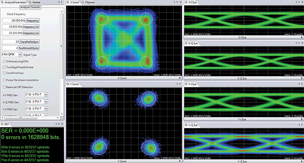
Figure 1 offers an example of the software that accompanies a Tektronix OM4245 45-GHz OMA. Unique calibration files are created for all Tektronix OMAs at the time of manufacture, so that the software can remove any impacts. Once the signal is received by the OMA, the next step is to digitize it on the electrical signal paths using a multichannel oscilloscope. This can introduce a number of factors that can affect the EVM, the most fundamental being the oscilloscope’s bandwidth and sample rate.


An example of the software that accompanies optical modulation analyzer (OMA) systems; here, a Tektronix OM4245 45-GHz OMA

http://www.photonics.com/images/Web/Articles/2015/10/28/OMA_Software.png

Figure 1. An example of the software that accompanies optical modulation analyzer (OMA) systems; here, a Tektronix OM4245 45-GHz OMA is shown.


Assuming an oscilloscope with the appropriate bandwidth and sample rate is utilized, and that all OMA impairments are being corrected algorithmically as described above, achieving the lowest measurable EVM comes down to a function of the effective number of bits (ENOB) of the oscilloscope. The ENOB is measurably impacted by the way the oscilloscope handles interleaved sampling. Some real-time oscilloscopes use frequency interleaving techniques in order to extend bandwidth, but they do so at the cost of increasing the noise in the measurement channel.

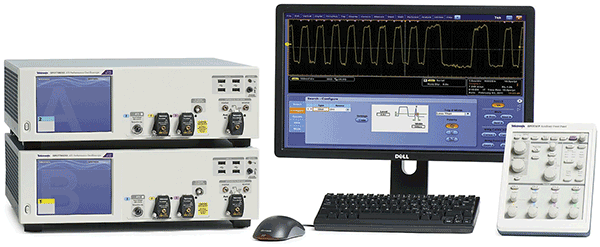
The limitation of the frequency interleaving approach lies in how the various frequency ranges are added together to reconstruct the final waveform, a step that compromises noise performance. In traditional frequency interleaving, each analog-to-digital converter (ADC) in the signal acquisition system only “sees” part of the input spectrum. But other oscilloscopes, such as the one shown in Figure 2, use a time-based interleaving approach, where all the ADCs see the full spectrum with full signal path symmetry. This approach preserves signal fidelity and ensures the highest possible ENOB.


Some oscilloscopes, such as this one, provide signal acquisition up to 70-GHz bandwidth.

http://www.photonics.com/images/Web/Articles/2015/10/28/OMA_Oscilloscopes.png

Figure 2. Some oscilloscopes, such as this one, provide signal acquisition up to 70-GHz bandwidth. Its asynchronous time interleaving (ATI) architecture provides a low-noise, real-time signal acquisition and high effective number of bits (ENOB).


Analysis for conclusive evaluation

Any test and measurement coherent receiver comes with some sort of analysis and visualization software package. But will that software have the particular types of measurement and visualization tools needed for evaluating specific designs or research?

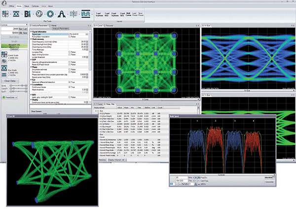
For example, when evaluating the quality of a new phase recovery algorithm, OMA software may be needed. This type of software can provide not only the basic building blocks for measurements but also allows the complete customization of the signal processing. Stand-alone optical analysis software packages of high quality are on the market. Some include features such as a library of analysis algorithms designed specifically for coherent optical analysis and executed in a customer-supplied MATLAB installation, with an applications programmatic interface (API) to these algorithms. Some provide a graphical user interface with optical tools that analyze complex modulated optical signals without needing to know any MATLAB, analysis algorithms or software programming, as shown in Figure 3.


The user interface of software like this, Tektronix’s OM1106 Coherent Optical Analysis system, allows the user to conduct a detailed analysis of complex modulated optical signals without requiring knowledge of MATLAB, analysis algorithms or software programming.

http://www.photonics.com/images/Web/Articles/2015/10/28/OMA_Interface.png

Figure 3. The user interface of software like this, Tektronix’s OM1106 Coherent Optical Analysis system, allows the user to conduct a detailed analysis of complex modulated optical signals without requiring knowledge of MATLAB, analysis algorithms or software programming.


Flexible measurement-taking software also is available. For instance, measurements can be made solely through the user interface, or via the programmatic interface to and from MATLAB for customized processing. Using both methods together is also an option, made possible by employing the user interface as a visualization and measurement framework, around which custom processing can be built.
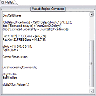
Most software includes sophisticated core processing algorithms for analyzing coherent signals — estimating the signal phase, determining the signal clock frequency, performing ambiguity resolution, estimating the power spectral density, etc. — but some packages can customize the core processing algorithms. This provides an excellent method for conducting signal processing research. For instance, in order to speed up the development of signal processing routines, one user interface provides a dynamic MATLAB integration window (Figure 4).


A dynamic MATLAB integration window helps speed up the development of signal processing routines.

http://www.photonics.com/images/Web/Articles/2015/10/28/OMA_MATLAB.png

Figure 4. A dynamic MATLAB integration window helps speed up the development of signal processing routines.


Any MATLAB code typed in this window is executed on every pass through the signal processing loop. This allows the “comment out” function calls, writing of specific values into data structures, or modification of signal processing parameters on the fly without having to stop the processing loop or modify the MATLAB source code.

Future-proofing an acquisition system

While the bulk of today’s coherent optical R&D activity is focused on 100-G signals, R&D with 400-G signals is already underway at many sites. Testing at 400 G may well be needed within the lifetime of many 100-G test instruments. Therefore, it makes sense to buy equipment at the right performance and price for 100 G now, but also to ensure that future expansion into 400 G is possible.

But how? Typically, four channels of 33-GHz real-time oscilloscope acquisition are used to test 100-G signals. In order to test 400-G signals in the future, bandwidths greater than 65 GHz will be needed, especially for a full dual-polarization system. But if testing at 100 G is all that’s needed now, it could be hard to justify the additional expense. One way around this problem is to purchase a system with a flexible, modular design, and one that uses distributed processing to allow for additional capacity for the system as needed.

For example, Figure 5 shows a system with four channels of 33-GHz acquisition that are distributed across two stand-alone oscilloscopes (left). The instruments are connected by a high speed bus, which not only provides a common external trigger between the two but also includes a common 12.5-GHz sample clock. The result is that the two oscilloscopes are combined to form, in effect, a single instrument whose acquisition-to-acquisition jitter across all channels delivers the same level of measurement precision as a stand-alone, monolithic oscilloscope.


Shown here is a modular way to build coherent optical testing systems from 100 to 400 G using an oscilloscope connected by cables.

Figure 5. Shown here is a modular way to build coherent optical testing systems from 100 to 400 G using an oscilloscope connected by cables. The processing is distributed and provides a common trigger without acquisition-to-acquisition jitter.


The system shown in Figure 5 also has two 70-GHz channels (one in each unit). Therefore, by simply switching from the 33-GHz channels to the 70-GHz channels, the oscilloscope bandwidth and sample rate can both be doubled. This permits a “peek” at single-polarization 400-G signals using the 100-G test system, as shown in the middle of the illustration. When the time comes to perform full 400-G testing, a second system can be added to the first with another high speed bus, providing two more channels of 70-GHz acquisition. This creates a system that is capable of full dual-polarization coherent optical acquisition (as demonstrated on the right). As the base units are stand-alone oscilloscopes, the systems can also be scaled down and redeployed to other projects as needed when a project comes to an end.

Meet the author

Chris Loberg is a senior technical marketing manager at Tektronix Inc., responsible for oscilloscopes in the Americas region; email: christopher.j.loberg@tektronix.com.

Read Full Post »

Obesity Issues

Larry H. Bernstein, MD, FCAP, Curator

LPBI


The Changing Face of Obesity

Science tells us obesity is a chronic disease. Why does the outmoded and injurious notion that it is a problem of willpower persist?

By Joseph Proietto | November 1, 2015   http://www.the-scientist.com//?articles.view/articleNo/44288/title/The-Changing-Face-of-Obesity/

In Dante Alighieri’s Divine Comedy the narrator meets a man named Ciacco who had been sent to Hell for the “Damning sin of Gluttony.” According to Catholic theology, in order to end up in Hell one must willfully commit a serious sin. So Dante believed that fat people chose to be fat. This antiquated view of the cause of obesity is still widespread, even among medical professionals. The consequences of this misconception are significant, because it forms the basis for the discrimination suffered by the obese; for the wasting of scarce resources in attempts to change lifestyle habits by public education; and for the limited availability of subsidized obesity treatments.

http://www.the-scientist.com/November2015/critic1.jpg

While obesity is often labeled a lifestyle disease, poor lifestyle choices alone account for only a 6 to 8 kg weight gain. The body has a powerful negative feedback system to prevent excessive weight gain. The strongest inhibitor of hunger, the hormone leptin, is made by fat cells. A period of increased energy intake will result in fat deposition, which will increase leptin production. Leptin suppresses hunger and increases energy expenditure. This slows down weight gain. To become obese, it may be necessary to harbor a genetic difference that makes the individual resistant to the action of leptin.

Evidence from twin and adoption studies suggests that obesity has a genetic basis, and over the past two decades a number of genes associated with obesity have been described. The most common genetic defect in European populations leading to severe obesity is due to mutations in the gene coding for the melanocortin 4 receptor (MCR4). Still, this defect can explain severe obesity in only approximately 6 percent to 7 percent of cases (J Clin Invest, 106:271-79, 2000). Other genes have been discovered that can cause milder increases in weight; for example, variants of just one gene (FTO) can explain up to 3 kg of weight variation between individuals (Science, 316:889-94, 2007).

Genes do not directly cause weight gain. Rather, genes influence the desire for food and the feeling of satiety. In an environment with either poor access to food or access to only low-calorie food, obesity may not develop even in persons with a genetic predisposition. When there is an abundance of food and a sedentary lifestyle, however, an obesity-prone person will experience greater hunger and reduced satiety, increasing caloric intake and weight gain.

Since the 1980s, there has been a rapid rise in the prevalence of obesity worldwide, a trend that likely results from a variety of complex causes. There is increasing evidence, for example, that the development of obesity on individual or familial levels may be influenced by environmental experiences that occur in early life. For example, if a mother is malnourished during early pregnancy, this results in epigenetic changes to genes involved in the set points for hunger and satiety in the developing child. These changes may then become fixed, resulting in a tendency towards obesity in the offspring.

The biological basis of obesity is further highlighted by the vigorous defense of weight following weight loss. There are at least 10 circulating hormones that modulate hunger. Of these, only one has been confirmed as a hunger-inducing hormone (ghrelin), and it is made and released by the stomach. In contrast, nine hormones suppress hunger, including CCK, PYY, GLP-1, oxyntomodulin, and uroguanylin from the small bowel; leptin from fat cells; and insulin, amylin, and pancreatic polypeptide from the pancreas.


After weight loss, regardless of the diet employed, there are changes in circulating hormones involved in the regulation of body weight. Ghrelin levels tend to increase and levels of multiple appetite-suppressing hormones decrease. There is also a subjective increase in appetite. Researchers have shown that even after three years, these hormonal changes persist (NEJM, 365:1597-604, 2011; Lancet Diabetes and Endocrinology, 2:954-62, 2014). This explains why there is a high rate of weight regain after diet-induced weight loss.

Given that the physiological responses to weight loss predispose people to regain that weight, obesity must be considered a chronic disease. Data show that those who successfully maintain their weight after weight loss do so by remaining vigilant and constantly applying techniques to oppose weight regain. These techniques may involve strict diet and exercise practices and/or pharmacotherapy.

It is imperative for society to move away from a view that obesity is simply a lifestyle issue and to accept that it is a chronic disease. Such a change would not only relieve the stigma of obesity but would also empower politicians, scientists and clinicians to tackle the problem more effectively.

Joseph Proietto was the inaugural Sir Edward Dunlop Medical Research Foundation Professor of Medicine in the Department of Medicine, Austin Health at the University of Melbourne in Australia. He is a researcher and clinician investigating and treating obesity and type 2 diabetes.


A Weighty Anomaly

Why do some obese people actually experience health benefits?

By Jyoti Madhusoodanan | November 1, 2015     http://www.the-scientist.com//?articles.view/articleNo/44304/title/A-Weighty-Anomaly/

http://www.the-scientist.com/November2015/notebook4.jpg

THE ENDOCRINE THEORY: Some researchers have posited that fat cells may secrete molecules that affect glucose homeostasis in muscle or liver tissue.COURTESY OF MITCHELL LAZAR

In the early 19th century, Belgian mathematician Adolphe Quetelet was obsessed with a shape: the bell curve. While helping with a population census, Quetelet proposed that the spread of human traits such as height and weight followed this trend, also known as a Gaussian or normal distribution. On a quest to define a “normal man,” he showed that human height and weight data fell along his beloved bell curves, and in 1823 devised the “Quetelet Index”—more familiar to us today as the BMI, or body mass index, a ratio of weight to height.

Nearly two centuries later, clinicians, researchers, and fitness instructors continue to rely on this metric to pigeonhole people into categories: underweight, healthy, overweight, or obese. But Quetelet never intended the metric to serve as a way to define obesity. And now, a growing body of evidence suggests these categories fail to accurately reflect the health risks—or benefits—of being overweight.

Although there is considerable debate surrounding the prevalence of metabolically healthy obesity, when obesity is defined in terms of BMI (a BMI of 30 or higher), estimates suggest that about 10 percent of adults in the U.S. are obese yet metabolically healthy, while as many as 80 percent of those with a normal BMI may be metabolically unhealthy, with signs of insulin resistance and poor circulating lipid levels, even if they suffer no obvious ill effects. “If all we know about a person is that they have a certain body weight at a certain height, that’s not enough information to know their health risks from obesity,” says health-science researcher Paul McAuley of Winston-Salem State University. “We need better indicators of metabolic health.”

The dangers of being overweight, such as a higher risk of heart disease, type 2 diabetes, and other complications, are well known. But some obese individuals—dubbed the “fat fit”—appear to fare better on many measures of health when they’re heavier. Studies have found lower mortality rates, better response to hemodialysis in chronic kidney disease, and lower incidence of dementia in such people. Mortality, it’s been found, correlates with obesity in a U-shaped curve (J Sports Sci, 29:773-82, 2011). So does extra heft help or hurt?

To answer that question, researchers are trying to elucidate the metabolic reasons for this obesity paradox.

In a recent study, Harvard University epidemiologist Goodarz Danaei and his colleagues analyzed data from nine studies involving a total of more than 58,000 participants to tease apart how obesity and other well-known metabolic risk factors influence the risk of coronary heart disease. Controlling these other risk factors, such as hypertension or high cholesterol, with medication is simpler than curbing obesity itself, Danaei explains. “If you control a person’s obesity you get rid of some health risks, but if you control hypertension or diabetes, that also reduces health risks, and you can do the latter much more easily right now.”

Danaei’s team assessed BMI and metabolic markers such as systolic blood pressure, total serum cholesterol, and fasting blood glucose. The three metabolic markers only explained half of the increased risk of heart disease across all study participants. In obese individuals, the other half appeared to be mediated by fat itself, perhaps via inflammatory markers or other indirect mechanisms (Epidemiology, 26:153-62, 2015). While Danaei’s study was aimed at understanding how obesity hurts health, the results also uncovered unknown mechanisms by which excess adipose tissue might exert its effects. This particular study revealed obesity’s negative effects, but might these unknown mechanisms hold clues that explain the obesity paradox?

Other researchers have suggested additional possibilities—for example, that inflammatory markers such as TNF-α help combat conditions such as chronic kidney disease, or that obesity makes a body more capable of making changes to, and tolerating changes in, blood flow depending on systemic needs (Am J Clin Nutr, 81:543-54, 2005).

According to endocrinologist Mitchell Lazar at the University of Pennsylvania, the key to explaining the obesity paradox may be two nonexclusive ways fat tissue is hypothesized to function. One mechanism, termed the endocrine theory, suggests that fat cells secrete, or don’t secrete enough of, certain molecules that influence glucose homeostasis in other tissues, such as muscle or liver. The first such hormone to be discovered was leptin; later studies reported several other adipocyte-secreted factors, including adiponectin, resistin, and various cytokines.

The other hypothesis, dubbed the spillover theory, suggests that storing lipids in fat cells has some pluses. Adipose tissue might sequester fat-soluble endotoxins, and produce lipoproteins that can bind to and clear harmful lipids from circulation. When fat cells fill up, however, these endotoxins are stashed in the liver, pancreas, or other organs—and that’s when trouble begins. In “fat fit” people, problems typically linked to obesity such as high cholesterol or diabetes may be avoided simply because their adipocytes mop up more endotoxins.

“In this model, one could imagine that if you could store even more fat in fat cells, you could be even more obese, but you might be protected from problems [associated with] obesity because you’re protecting the other tissues from filling up with lipids that cause problems,” says Lazar. “This may be the most popular current model to explain the fat fit.”

Although obesity greatly increases the risk of type 2 diabetes—up to 93-fold in postmenopausal women, for example—not all obese people suffer from the condition. Similarly, a certain subtype of individuals with “normal” BMIs are at greater risk of developing insulin resistance and type 2 diabetes than others with BMIs in the same range. Precisely what distinguishes these two cohorts is still unclear. “Just as important as explaining why some obese people don’t get diabetes is to explain why other subgroups—normal-weight people or those with lipodystrophy—sometimes get it,” Lazar says. “If there are multiple subtypes of obesity and diabetes, can we figure out genetic aspects or biomarkers that cause one of these phenotypes and not the other?”

To Lazar, McAuley, and other researchers, it’s increasingly evident that BMI may not be that metric. Finding better ways to assess a healthy weight, however, has proven challenging. Researchers have tested measures, such as the body shape index (ABSI) or the waist-hip ratio, which attempt to gauge visceral fat—considered to be more metabolically harmful than fat in other body locations. However, these metrics have yet to be implemented widely in clinics, and few are as simple to understand as the BMI (Science, 341:856-58, 2013).

Independent of metrics, however, the health message regarding weight is still unanimous: exercise and healthy dietary choices benefit everyone. “At a certain point, despite all the so-called fit-fat people, the demographics say that there’s a huge risk of diabetes and heart disease at very high BMI,” notes Lazar. “We can’t assume we’ll be one of the lucky ones who will have a BMI in the obese category but will still be protected from heart disease.”

Correction (November 2): The original version of this article misattributed the pull quote above. The attribution for this quote has been corrected, and The Scientist regrets the error.


THE HEALTH RISK OF OBESITY—BETTER METRICS IMPERATIVE

 Science 23 Aug 2013;  341(6148): 856858     DOI: http://dx.doi.org:/10.1126/science.1241244
Obesity paradoxes.
In this review, we examine the original obesity paradox phenomenon (i.e. in cardiovascular disease populations, obese patients survive better), as well as three other related paradoxes (pre-obesity, “fat but fit” theory, and “healthy” obesity). An obesity paradox has been reported in a range of cardiovascular and non-cardiovascular conditions. Pre-obesity (defined as a body mass index of 25.0-29.9 kg · m⁻²) presents another paradox. Whereas “overweight” implies increased risk, it is in fact associated with decreased mortality risk compared with normal weight. Another paradox concerns the observation than when fitness is taken into account, the mortality risk associated with obesity is offset. The final paradox under consideration is the presence of a sizeable subset of obese individuals who are otherwise healthy. Consequently, a large segment of the overweight and obese population is not at increased risk for premature death. It appears therefore that low cardiorespiratory fitness and inactivity are a greater health threat than obesity, suggesting that more emphasis should be placed on increasing leisure time physical activity and cardiorespiratory fitness as the main strategy for reducing mortality risk in the broad population of overweight and obese adults.
Obesity, insulin resistance, and cardiovascular disease.
Recent Prog Horm Res. 2004;59:207-23.
The ability of insulin to stimulate glucose disposal varies more than six-fold in apparently healthy individuals. The one third of the population that is most insulin resistant is at greatly increased risk to develop cardiovascular disease (CVD), type 2 diabetes, hypertension, stroke, nonalcoholic fatty liver disease, polycystic ovary disease, and certain forms of cancer. Between 25-35% of the variability in insulin action is related to being overweight. The importance of the adverse effects of excess adiposity is apparent in light of the evidence that more than half of the adult population in the United States is classified as being overweight/obese, as defined by a body mass index greater than 25.0 kg/m(2). The current epidemic of overweight/obesity is most-likely related to a combination of increased caloric intake and decreased energy expenditure. In either instance, the fact that CVD risk is increased as individuals gain weight emphasizes the gravity of the health care dilemma posed by the explosive increase in the prevalence of overweight/obesity in the population at large. Given the enormity of the problem, it is necessary to differentiate between the CVD risk related to obesity per se, as distinct from the fact that the prevalence of insulin resistance and compensatory hyperinsulinemia are increased in overweight/obese individuals. Although the majority of individuals in the general population that can be considered insulin resistant are also overweight/obese, not all overweight/obese persons are insulin resistant. Furthermore, the cluster of abnormalities associated with insulin resistance – namely, glucose intolerance, hyperinsulinemia, dyslipidemia, and elevated plasma C-reactive protein concentrations — is limited to the subset of overweight/obese individuals that are also insulin resistant. Of greater clinical relevance is the fact that significant improvement in these metabolic abnormalities following weight loss is seen only in the subset of overweight/obese individuals that are also insulin resistant. In view of the large number of overweight/obese subjects at potential risk to be insulin resistant/hyperinsulinemic (and at increased CVD risk), and the difficulty in achieving weight loss, it seems essential to identify those overweight/obese individuals who are also insulin resistant and will benefit the most from weight loss, then target this population for the most-intensive efforts to bring about weight loss.
Long-Term Persistence of Hormonal Adaptations to Weight Loss

Priya Sumithran, Luke A. Prendergast, Elizabeth Delbridge, Katrina Purcell, Arthur Shulkes, Adamandia Kriketos, and Joseph Proietto

N Engl J Med 2011; 365:1597-1604   October 27, 2011http://dx.doi.org:/10.1056/NEJMoa1105816

After weight loss, changes in the circulating levels of several peripheral hormones involved in the homeostatic regulation of body weight occur. Whether these changes are transient or persist over time may be important for an understanding of the reasons behind the high rate of weight regain after diet-induced weight loss.

Weight loss (mean [±SE], 13.5±0.5 kg) led to significant reductions in levels of leptin, peptide YY, cholecystokinin, insulin (P<0.001 for all comparisons), and amylin (P=0.002) and to increases in levels of ghrelin (P<0.001), gastric inhibitory polypeptide (P=0.004), and pancreatic polypeptide (P=0.008). There was also a significant increase in subjective appetite (P<0.001). One year after the initial weight loss, there were still significant differences from baseline in the mean levels of leptin (P<0.001), peptide YY (P<0.001), cholecystokinin (P=0.04), insulin (P=0.01), ghrelin (P<0.001), gastric inhibitory polypeptide (P<0.001), and pancreatic polypeptide (P=0.002), as well as hunger (P<0.001).

What’s new in endocrinology and diabetes mellitus

Large genome wide association studies have demonstrated that variants in the FTO gene have the strongest association with obesity risk in the general population, but the mechanism of the association has been unclear. However, a nonocoding causal variant in FTO has now been identified that changes the function of adipocytes from energy utilization (beige fat) to energy storage (white fat) with a fivefold decrease in mitochondrial thermogenesis [17]. When the effect of the variant was blocked in genetically engineered mice, thermogenesis increased and weight gain did not occur, despite eating a high-fat diet. Blocking the gene’s effect in human adipocytes also increased energy utilization. This observation has important implications for potential new anti-obesity drugs. (See “Pathogenesis of obesity”, section on ‘FTO variants’.)

Liraglutide for the treatment of obesity (July 2015)

Along with diet, exercise, and behavior modification, drug therapy may be a helpful component of treatment for select patients who are overweight or obese. Liraglutide is a glucagon-like peptide-1 (GLP-1) receptor agonist, used for the treatment of type 2 diabetes, and can promote weight loss in patients with diabetes, as well as those without diabetes.

In a randomized trial in nondiabetic patients who had a body mass index (BMI) of ≥30 kg/m2 or ≥27 kg/m2 with dyslipidemia and/or hypertension, liraglutide 3 mg once daily, compared with placebo, resulted in greater mean weight loss (-8.0 versus -2.6 kg with placebo) [18]. In addition, cardiometabolic risk factors, glycated hemoglobin (A1C), and quality of life improved modestly. Gastrointestinal side effects transiently affected at least 40 percent of the liraglutide group and were the most common reason for withdrawal (6.4 percent). Liraglutide is an option for select overweight or obese patients, although gastrointestinal side effects (nausea, vomiting) and the need for a daily injection may limit the use of this drug. (See “Obesity in adults: Drug therapy”, section on ‘Liraglutide’.)

In a trial designed specifically to evaluate the effect of liraglutide on weight loss in overweight or obese patients with type 2 diabetes (mean weight 106 kg), liraglutide, compared with placebo, resulted in greater mean weight loss (-6.4 kg and -5.0 kg for liraglutide 3 mg and 1.8 mg, respectively, versus -2.2 kg for placebo) [19]. Treatment with liraglutide was associated with better glycemic control, a reduction in the use of oral hypoglycemic agents, and a reduction in systolic blood pressure. Although liraglutide is not considered as initial therapy for the majority of patients with type 2 diabetes, it is an option for select overweight or obese patients with type 2 diabetes who fail initial therapy with lifestyle intervention and metformin.  (See “Glucagon-like peptide-1 receptor agonists for the treatment of type 2 diabetes mellitus”, section on ‘Weight loss’.)

The Skinny on Fat Cells

Bruce Spiegelman has spent his career at the forefront of adipocyte differentiation and metabolism.

By Anna Azvolinsky | November 1, 2015

http://www.the-scientist.com//?articles.view/articleNo/44312/title/The-Skinny-on-Fat-Cells/

Bruce Spiegelman
Stanley J. Korsmeyer Professor of Cell Biology
and Medicine
Harvard Medical School
Director, Center for Energy Metabolism
and Chronic
Disease, Dana-Farber Cancer Institute, Boston

It’s hard to know whether you have the right stuff to be a scientist, but I had a passion for the research,” says Bruce Spiegelman, professor of cell biology at Harvard Medical School and the Dana-Farber Cancer Institute. After receiving his PhD in biochemistry from Princeton University in 1978, Spiegelman sent an application to do postdoctoral research to just one lab. “I wasn’t thinking I should apply to five different labs. I just marched forward more or less in a straight line,” he says. Spiegelman did know that he had no financial backup and depended on research fellowships throughout the early phase of his science career. “I thought it was fantastic, and still think so, that a PhD in science is supported by the government. I certainly appreciated that, because many of my friends in the humanities had to support themselves by cobbling together fellowships and teaching every semester, whereas we didn’t face similar challenges in the sciences.”

Since his graduate student days, Spiegelman has realized his potential, pioneering the study of adipose tissue biology and metabolism. He was introduced to the field in Howard Green’s laboratory, then at MIT, where Spiegelman began his one and only postdoc in 1978. Green had recently developed a system for culturing adipose cells and asked Spiegelman if he wanted to study fat cell differentiation. “I knew nothing about adipose tissue, but I was really interested in any model of how one cell switches to another. Whether skin or fat didn’t matter too much to me, because I was not coming at this from the perspective of physiology but from the perspective of how do these switches work at a molecular level?”

Spiegelman has stuck with studying the biology and differentiation of fat cells for more than 30 years. While looking for the master transcriptional regulator of fat development—which his laboratory found in 1994—Spiegelman’s group also discovered one of the first examples of a nuclear oncogene that functions as a transcription factor, and, more recently, the team found that brown fat and white fat come from completely different origins and that brown and beige fat are distinct cell types. Spiegelman was also the first to provide evidence for the connection between inflammation, insulin resistance, and fat tissue.

Here, Spiegelman talks about his strong affinity for the East Coast, his laboratory’s search for molecules that can crank up brown fat production and activity, and the culture of his laboratory’s weekly meeting.

Spiegelman Sets Out

First publication. Spiegelman grew up in Massapequa, New York, a town on Long Island. “Birds, insects, fish, and animals were fascinating to me. As a kid, I imagined I would be a wildlife ranger,” he says. Spiegelman and his brother were the first in their family to attend college; Spiegelman entered the College of William and Mary in 1970 thinking he would major in psychology. But before taking his first psychology course, he had to take a biology course, really loved it, and switched his major. For his senior thesis, he chose one of the few labs that did biochemistry-related research. He studied cultures of the filamentous fungus Aspergillus ornatus in which he induced the upregulation of a metabolic enzyme. Spiegelman applied a calculus transformation that related the age of the culture to the age of individual cells, something that had not been previously done. The work earned him his first first-author publication in 1975. “It was not a great breakthrough, but I think it showed that I was maybe applying myself more than the typical undergraduate.”

Full steam ahead. “My interest in laboratory research was intense. Even though it was not particularly inspired work, the first-author publication in a college where not many of the professors published a lot gave me a lot of confidence. It was probably out of proportion to the quality of the actual work.” That confidence and Spiegelman’s interest in the chemistry of living things led him to pursue a PhD in biochemistry at Princeton University. “Very early on, I felt that I couldn’t understand biology if it didn’t go to the molecular level. To me, just describing how an animal lived without understanding how it worked was very unsatisfying. I think it was one of the best decisions that I made in my life, to do a PhD in biochemistry,” he says, “because if you really want to understand living systems, you are very limited in how you can understand them without having a strong background in biochemistry because these are, essentially, chemical systems.”

Embracing molecular biology. Spiegelman initially joined Arthur Pardee’s laboratory, but switched when Pardee left Princeton for Harvard University in 1975. Because he was already collaborating with Marc Kirschner, a cell biologist and biochemist who studies the regulation of the cell cycle and how the cytoskeleton works, it was an easy transition to transfer to the new laboratory. In Kirschner’s group, Spiegelman became the cell biologist among many protein biochemists working on microtubule assembly in vitro. Rather than understanding how the proteins fit together to form the filamentous structures, Spiegelman wanted to understand what controlled their assembly inside cells. Working in mammalian cells, Spiegelman published three consecutive Cell papers on how microtubule assembly occurs in vivo. The firstpaper, from 1977, demonstrated that a nucleotide functions to stabilize the tubulin molecule rather than to regulate tubulin assembly in vivo.

Spiegelman Simmers

A new tool. For his next move, Spiegelman wanted to marry his background in biochemistry and molecular biology with a good cellular model system. He became interested in differentiation at the end of his PhD, while studying how the cytoskeleton is reorganized during neural differentiation, and settled on Green’s MIT laboratory for his postdoc. Green had developed a way to study both skin and fat cell differentiation. Again, Spiegelman was the odd man out, working on the molecular biology of fat cell differentiation while most of the graduate students and postdocs focused on the cellular biology of skin cell differentiation. While there, Spiegelman learned how to clone cDNA—a new method that some researchers thought was just another new fad, he says. “I thought it was pretty obvious that this was a tool that would be a game changer. I could see how I could clone some of the cDNAs and genes that were regulated in the fat cell lineage and then try to understand the regulation of these genes.”

Setting the stage. Spiegelman demonstrated that cAMP regulates the synthesis of certain enzymes in fat cells during differentiation. But while this was the most influential paper from his postdoc, says Spiegelman, it was his demonstration of cloning mRNAs from adipocytes, published in 1983, that set the stage for cloning fat-selective genes. The work, mostly done when Spiegelman was already a new faculty member at the Dana-Farber Cancer Institute, stemmed from his learning molecular cloning in Phillip Sharp’s lab at MIT and Bryan Roberts’s lab at Harvard. “This was the raw material from which we eventually cloned PPARγ and showed it to be the master regulator of fat [cell] development.”

Roots. Spiegelman became an assistant professor at the Harvard Medical School in 1982, when he was not yet 30. Although he had entertained the idea of moving to the West Coast with his wife, whom he had met at Princeton where she obtained a PhD in French literature, Spiegelman says he is really an East Coaster at heart. “My wife and I came to love Boston and were very comfortable there. Our families were both in New York, which was close, but not too close, and we really enjoyed the culture and pace of Boston; it was more ‘us.’ We really liked to visit California but didn’t particularly want to move there. We’re both real Northeastern people.”

Relating to Sisyphus. The transition from doing a postdoc to setting up his own laboratory was “very exciting and terribly stressful,” says Spiegelman. “When I think back, I always tried to be professional with my laboratory, but I was so stressed at suddenly being on my own with no management training.” The people resources he had encountered in his graduate and postdoctoral training labs were also not there yet, and he says his first publication as a principal investigator was like pushing a rock up a hill. But eventually, Spiegelman’s lab built a reputation and reached a critical mass of talented people who advanced the science. Again in 1983, Spiegelman produced a publication showing that morphological manipulation can affect gene expression and adipose differentiation.

End goal. Spiegelman’s goal was to find a master molecule that  orchestrates the conversion of adipocyte precursor cells into bona fide fat cells. Piece by piece, his lab identified the enhancers, promoters, and other regulatory elements involved in adipocyte differentiation. In 1994, graduate student Peter Tontonoz finallyfound that the PPARγ gene, inserted via a retroviral vector into fibroblasts, could induce the cells to become adipose cells. “It took 10 years,” Spiegelman says. Along the way, the laboratory found that c-fos, the product of a famous nuclear oncogene, bound to the promoters of fat-specific genes and worked as a transcription factor. “It was not really known how nuclear oncogenes worked. This was one of the first papers showing that these oncogenes bound to gene promoters and were transcription factors.”

A wider scope. In 1993, graduate student Gökhan Hotamisligil found that tumor necrosis factor-alpha(TNF-α), is induced in the fat tissue of rodent models of obesity and diabetes. The paper sparked the formation of the field of immunometabolism and resulted in the expansion of Spiegelman’s lab into the physiology arena, partly thanks to the guidance of C. Ronald Kahn and Jeff Flier, who both study metabolism and diabetes. But the work initially encountered pushback, says Spiegelman, partly because it was the merging of two fields.

Spiegelman Scales Up

Fat color palette. Brown fat tissue, abundant in infants but scarce in adults, is a metabolically active form of fat that is chock full of mitochondria and is found in pockets in the body distinct from white fat tissue.Pere Puigserver, then a postdoc in Spiegelman’s lab, found that the coactivator PCG-1, binding to PPARγ and other nuclear receptors, could stimulate mitochondrial biogenesis. The PCG-1 gene is turned on by stimuli such as exercise or a cold environment. Later, postdoc Patrick Seale, Spiegelman, and their colleagues showed brown fat cells derive from the same lineage that gives rise to skeletal muscle. “This was a big surprise, maybe the biggest surprise we ever uncovered in the lab,” says Spiegelman.

A paler shade of brown. More recently, in 2012, Spiegelman’s laboratory showed that within adult white adipose tissue, there are pockets of a yet another type of fat tissue that he called beige fat. “I think the evidence is very good from rodents that if you activate brown and beige fat, you get metabolic benefit both in obesity and diabetes. So the question now is: Can that be done in humans in a way that’s beneficial and not toxic?”  The lab is now looking to identify molecules that can either ramp up the activity of brown and beige fat or increase the production of both cell types as possible therapeutics for metabolic disorders or even cancer-associated cachexia. “Anyone who says that either approach will work better is being foolish. We just don’t know enough to go after just one or the other.”

On the irisin controversy. After reporting in 2012 that a muscle-related hormone called irisin could switch white fat to metabolically active brown fat, Spiegelman became embroiled in a media-covered debate about whether the molecule really exists; he was also the victim of a potential fraud plot. Most recently, Spiegelman provided thorough evidence that irisin does in fact exist. On the controversy, he says it’s a fine line between defending his scientific integrity and not adding more fuel to the fire or engaging with his harassers. “We have a long track record of doing credible and reproducible science and it was not that complicated to address the paper that claimed irisin was ‘a myth.’ That study used very outmoded scientific approaches.”

Raw talent. Many of Spiegelman’s trainees have gone on to become very successful scientists, including Tontonoz, Hotamisligil, Evan Rosen, and Randy Johnson. “It’s a quantum change in the experience of doing science when you get people who have their own visions. I would have thought that interacting with smart people would mainly help me get my scientific vision accomplished. And that was partly true, but also it changed my vision. When you have people challenging you on a day-to-day basis, you learn from them through the questions they ask and the way they challenge you in a constructive way. They made me a much better scientist.”

Rigorous mentorship.  “I feel very passionately that a major part of my job is to prepare the next generation of scientists. Everyone who comes through my lab will tell you that I take that very seriously. We make sure my students give a lot of talks and get critical assessments of their presentations to our lab group. I am very hands-on both scientifically and in developing the way students project their vision. I had a very good mentor, Marc Kirschner, and I’d like to think that I learned how to be a mentor from him. I want to make sure that when people walk out of my lab they are prepared to run independent research programs.”

Greatest Hits

  • Identified the master regulator of adipogenesis, the nuclear receptor PPARγ
  • Was the first to show that a nuclear oncogene, c-fos, codes for a transcription factor that binds to the promoters of genes
  • Demonstrated that adipose tissue synthesizes tumor necrosis factor-alpha (TNF-α), providing the first direct link between obesity, inflammation, insulin resistance, and fat tissue.
  • Showed that brown fat cells are not developmentally related to white fat
  • Identified beige fat as a distinct cell type, different from either white or brown fat


Fanning the Flames

Obesity triggers a fatty acid synthesis pathway, which in turn helps drive T cell differentiation and inflammation.

By Kate Yandell | November 1, 2015

http://www.the-scientist.com//?articles.view/articleNo/44306/title/Fanning-the-Flames/

EDITOR’S CHOICE IN IMMUNOLOGY

The paper
Y. Endo et al., “Obesity drives Th17 cell differentiation by inducing the lipid metabolic kinase, ACC1,” Cell Reports, 12:1042-55, 2015.

Cell Rep. 2015 Aug 11;12(6):1042-55.   http://dx.doi.org:/10.1016/j.celrep.2015.07.014. Epub 2015 Jul 30.
Obesity Drives Th17 Cell Differentiation by Inducing the Lipid Metabolic Kinase, ACC1.
  • A high-fat diet augments Th17 cell development and the expression of Acaca
  • ACC1 controls Th17 cell development in vitro and Th17 cell pathogenicity in vivo
  • ACC1 modulates RORγt function in developing Th17 cells
  • Obesity in humans induces ACACA and IL-17A expression in CD4 T cells

Chronic inflammation due to obesity contributes to the development of metabolic diseases, autoimmune diseases, and cancer. Reciprocal interactions between metabolic systems and immune cells have pivotal roles in the pathogenesis of obesity-associated diseases, although the mechanisms regulating obesity-associated inflammatory diseases are still unclear. In the present study, we performed transcriptional profiling of memory phenotype CD4 T cells in high-fat-fed mice and identified acetyl-CoA carboxylase 1 (ACC1, the gene product of Acaca) as an essential regulator of Th17 cell differentiation in vitro and of the pathogenicity of Th17 cells in vivo. ACC1 modulates the DNA binding of RORγt to target genes in differentiating Th17 cells. In addition, we found a strong correlation between IL-17A-producing CD45RO(+)CD4 T cells and the expression of ACACA in obese subjects. Thus, ACC1 confers the appropriate function of RORγt through fatty acid synthesis and regulates the obesity-related pathology of Th17 cells.

Figure thumbnail fx1

http://www.cell.com/cms/attachment/2035221719/2050630604/fx1.jpg


http://www.the-scientist.com/November2015/NovMediLit_310px.jpg

FEEDING INFLAMMATION: When mice eat a diet high in fat, their CD4 T cells show increased expression of the fatty acid biosynthesis gene Acaca, which encodes the enzyme ACC1 (1). Products of the ACC1 fatty acid synthesis pathway encourage the transcription factor RORγt to bind near the gene encoding the cytokine IL-17A (2). There, RORγt recruits an enzyme called p300 to modify the genome epigenetically and turn on IL-17A. The memory T cells then differentiate into inflammatory T helper 17 cells.
See full infographic: PDF
© STEVE GRAEPEL

Obesity often comes with a side of chronic inflammation, causing inflammatory chemicals and immune cells to flood adipose tissue, the hypothalamus, the liver, and other areas of the body. Inflammation is a big part of what makes obesity such an unhealthy condition, contributing to Type 2 diabetes, heart disease, cancers, autoimmune disorders, and possibly even neurodegenerative diseases.

To better understand the relationship between obesity and inflammation, Toshinori Nakayama, Yusuke Endo, and their colleagues at Chiba University in Japan started with what often leads to obesity: a high-fat diet. They fed mice rich meals for a couple of months and looked at how gene expression in the animals’ T cells compared to gene expression in the T cells of mice fed a normal diet. Most notably, they found increased expression ofAcaca, a gene that codes for a fatty acid synthesis enzyme called acetyl coA carboxylase 1 (ACC1). They went on to show that the resulting increase in fatty acid levels pushed CD4 T cells to differentiate into inflammatory T helper 17 (Th17) cells.

Th17 cells help fight off invading fungi and some bacteria. But these immune cells can also spin out of control in autoimmune diseases such as multiple sclerosis. Nakayama’s team showed that either blocking ACC1 activity with a drug called TOFA or deleting a key portion of Acaca in mouse CD4 T cells reduced the generation of pathologic Th17 cells. Overexpressing Acaca increased Th17-cell generation.

The researchers also demonstrated that mice fed a high-fat diet had elevated susceptibility to a multiple sclerosis–like disease, and that TOFA reduced the symptoms.

“This is a very intriguing finding, suggesting not only that obesity can directly induce Th17 differentiation but also indicating that pharmacologic targeting of fatty acid synthesis may help to interfere with obesity-associated inflammation,” Tim Sparwasser of the Twincore Center for Experimental and Clinical Infection Research in Hannover, Germany, says in an email. Sparwasser and his colleagues had previously shown that ACC1 is required for the differentiation of Th17 cells in mice and humans.

Nakayama explains that CD4 T cells must undergo profound metabolic changes as they mature and differentiate. “The intracellular metabolites, including fatty acids, are essential for cell proliferation and cell growth,” he says in an email. When fatty acid levels in T cells increase, the cells are activated and begin to proliferate.

“It’s a nice illustration of how, really, immune response is so highly connected to the metabolic state of the cell,” says Gökhan S. Hotamisligil of Harvard University’s T.H. Chan School of Public Health who was not involved in the study. “The immune system launches its responses commensurate with the sources of nutrients and energy from the environment,” he adds in an email.

There are still missing pieces in the path from high-fat diet to increased Acaca expression to ACC1’s influence on T-cell differentiation. It also remains to be seen how this plays out in obese humans, although Nakayama and colleagues did show that inhibiting ACC1 reduced pathologic Th17 generation in human immune cell cultures, and that the T cells of obese humans contain elevated levels of ACC1 and show signs of increased differentiation into Th17 cells.


The prevalence of obesity has been increasing worldwide, and obesity is now a major public health problem in most developed countries (Gregor and Hotamisligil, 2011, Ng et al., 2014). Obesity-induced inflammation contributes to the development of various chronic diseases, such as autoimmune diseases, metabolic diseases, and cancer (Kanneganti and Dixit, 2012, Kim et al., 2014,Osborn and Olefsky, 2012, Winer et al., 2009a). A number of studies have pointed out the importance of reciprocal interactions between metabolic systems and immune cells in the pathogenesis of obesity-associated diseases (Kaminski and Randall, 2010, Kanneganti and Dixit, 2012, Kim et al., 2014, Mauer et al., 2014, Stienstra et al., 2012, Winer et al., 2011).

Elucidating the molecular mechanisms by which naive CD4 T cells differentiate into effector T cells is crucial for understanding helper T (Th) cell-mediated immune pathogenicity. After antigen stimulation, naive CD4 T cells differentiate into at least four distinct Th cell subsets: Th1, Th2, Th17, and inducible regulatory T (iTreg) cells (O’Shea and Paul, 2010, Reiner, 2007). Several specific master transcription factors that regulate Th1/Th2/Th17/iTreg cell differentiation have been identified, including T-bet for Th1 (Szabo et al., 2000), GATA3 (Yamashita et al., 2004, Zheng and Flavell, 1997) for Th2, retinoic-acid-receptor-related orphan receptor γt (RORγt) for Th17 (Ivanov et al., 2006), and forkhead box protein 3 (Foxp3) for iTreg (Sakaguchi et al., 2008). The appropriate expression and function of these transcription factors is essential for proper immune regulation by each Th cell subset.

Among these Th cell subsets, Th17 cells contribute to the host defense against fungi and extracellular bacteria (Milner et al., 2008). However, the pathogenicity of IL-17-producing T cells has been recognized in various autoimmune diseases, including multiple sclerosis, psoriasis, inflammatory bowel diseases, and steroid-resistant asthma (Bettelli et al., 2006, Coccia et al., 2012, Ivanov et al., 2006,Leonardi et al., 2012, McGeachy and Cua, 2008, Nylander and Hafler, 2012,Stockinger et al., 2007, Sundrud et al., 2009).

An HFD Promotes Th17 Cell Differentiation and Affects the Expression of Fatty Acid Enzymes in Memory CD4 T Cells In Vivo

Inhibition of ACC1 Function Results in Decreased Th17 Cell Differentiation and Ameliorates the Development of Autoimmune Disease

ACC1 Controls the Differentiation of Th17 Cells Both In Vitro and In Vivo

ACC1 Controls the Function, but Not Expression, of RORγt in Differentiating Th17 Cells

Extrinsic Fatty Acid Supplementation Restored Acaca−/− Th17 Cell Differentiation through the Functional Improvement of RORγt

Obese Subjects Show Upregulation of ACACA and Increased Th17 Cells in CD45RO+ Memory CD4 T Cells

We herein identified a critical role that ACC1 plays in Th17 cell differentiation and the pathogenicity of Th17 cells through the control of the RORγt function under obese circumstances. High-fat-induced obesity augments Th17 cell differentiation and the expression of enzymes involved in fatty acid metabolism, including ACC1. Pharmacological inhibition or genetic deletion of ACC1 resulted in impaired Th17 cell differentiation in both mice and humans. In contrast, overexpression of Acaca induced Th17 cells in vivo, leaving the expression ofIfng and Il4 largely unchanged. ACC1 modulated the binding of RORγt to theIl17a gene and the subsequent p300 recruitment in differentiating Th17 cells. Memory CD4 T cells from peripheral blood mononuclear cells (PBMCs) of obese subjects showed increased IL-17A production and ACACA expression. Furthermore, a strong correlation was detected between the proportion of IL-17A-producing cells and the expression level of ACACA in memory CD4 T cells in obese subjects. Thus, our findings provide evidence of a mechanism wherein obesity can exacerbate IL-17-mediated pathology via the induction of ACC1.

Read Full Post »

Irreconciliable Dissonance in Physical Space and Cellular Metabolic Conception

Irreconciliable Dissonance in Physical Space and Cellular Metabolic Conception

Curator: Larry H. Bernstein, MD, FCAP

Pasteur Effect – Warburg Effect – What its history can teach us today. 

José Eduardo de Salles Roselino

The Warburg effect, in reality the “Pasteur-effect” was the first example of metabolic regulation described. A decrease in the carbon flux originated at the sugar molecule towards the end of the catabolic pathway, with ethanol and carbon dioxide observed when yeast cells were transferred from an anaerobic environmental condition to an aerobic one. In Pasteur´s studies, sugar metabolism was measured mainly by the decrease of sugar concentration in the yeast growth media observed after a measured period of time. The decrease of the sugar concentration in the media occurs at great speed in yeast grown in anaerobiosis (oxygen deficient) and its speed was greatly reduced by the transfer of the yeast culture to an aerobic condition. This finding was very important for the wine industry of France in Pasteur’s time, since most of the undesirable outcomes in the industrial use of yeast were perceived when yeasts cells took a very long time to create, a rather selective anaerobic condition. This selective culture media was characterized by the higher carbon dioxide levels produced by fast growing yeast cells and by a higher alcohol content in the yeast culture media.

However, in biochemical terms, this finding was required to understand Lavoisier’s results indicating that chemical and biological oxidation of sugars produced the same calorimetric (heat generation) results. This observation requires a control mechanism (metabolic regulation) to avoid burning living cells by fast heat released by the sugar biological oxidative processes (metabolism). In addition, Lavoisier´s results were the first indications that both processes happened inside similar thermodynamics limits. In much resumed form, these observations indicate the major reasons that led Warburg to test failure in control mechanisms in cancer cells in comparison with the ones observed in normal cells.

[It might be added that the availability of O2 and CO2 and climatic conditions over 750 million years that included volcanic activity, tectonic movements of the earth crust, and glaciation, and more recently the use of carbon fuels and the extensive deforestation of our land masses have had a large role in determining the biological speciation over time, in sea and on land. O2 is generated by plants utilizing energy from the sun and conversion of CO2. Remove the plants and we tip the balance. A large source of CO2 is from beneath the earth’s surface.]

Biology inside classical thermodynamics places some challenges to scientists. For instance, all classical thermodynamics must be measured in reversible thermodynamic conditions. In an isolated system, increase in P (pressure) leads to increase in V (volume), all this occurring in a condition in which infinitesimal changes in one affects in the same way the other, a continuum response. Not even a quantic amount of energy will stand beyond those parameters.

In a reversible system, a decrease in V, under same condition, will led to an increase in P. In biochemistry, reversible usually indicates a reaction that easily goes either from A to B or B to A. For instance, when it was required to search for an anti-ischemic effect of Chlorpromazine in an extra hepatic obstructed liver, it was necessary to use an adequate system of increased biliary system pressure in a reversible manner to exclude a direct effect of this drug over the biological system pressure inducer (bile secretion) in Braz. J. Med. Biol. Res 1989; 22: 889-893. Frequently, these details are jumped over by those who read biology in ATGC letters.

Very important observations can be made in this regard, when neutral mutations are taken into consideration since, after several mutations (not affecting previous activity and function), a last mutant may provide a new transcript RNA for a protein and elicit a new function. For an example, consider a Prion C from lamb getting similar to bovine Prion C while preserving  its normal role in the lamb when its ability to change Human Prion C is considered (Stanley Prusiner).

This observation is good enough, to confirm one of the most important contributions of Erwin Schrodinger in his What is Life:

“This little book arose from a course of public lectures, delivered by a theoretical physicist to an audience of about four hundred which did not substantially dwindle, though warned at the outset that the subject matter was a difficult one and that the lectures could not be termed popular, even though the physicist’s most dreaded weapon, mathematical deduction, would hardly be utilized. The reason for this was not that the subject was simple enough to be explained without mathematics, but rather that it was much too involved to be fully accessible to mathematics.”

After Hans Krebs, description of the cyclic nature of the citrate metabolism and after its followers described its requirement for aerobic catabolism two major lines of research started the search for the understanding of the mechanism of energy transfer that explains how ADP is converted into ATP. One followed the organic chemistry line of reasoning and therefore, searched for a mechanism that could explain how the breakdown of carbon-carbon link could have its energy transferred to ATP synthesis. One of the major leaders of this research line was Britton Chance. He took into account that relatively earlier in the series of Krebs cycle reactions, two carbon atoms of acetyl were released as carbon dioxide ( In fact, not the real acetyl carbons but those on the opposite side of citrate molecule). In stoichiometric terms, it was not important whether the released carbons were or were not exactly those originated from glucose carbons. His research aimed at to find out an intermediate proteinaceous intermediary that could act as an energy reservoir. The intermediary could store in a phosphorylated amino acid the energy of carbon-carbon bond breakdown. This activated amino acid could transfer its phosphate group to ADP producing ATP. A key intermediate involved in the transfer was identified by Kaplan and Lipmann at John Hopkins as acetyl coenzyme A, for which Fritz Lipmann received a Nobel Prize.

Alternatively, under possible influence of the excellent results of Hodgkin and Huxley a second line of research appears. The work of Hodgkin & Huxley indicated that the storage of electrical potential energy in transmembrane ionic asymmetries and presented the explanation for the change from resting to action potential in excitable cells. This second line of research, under the leadership of Peter Mitchell postulated a mechanism for the transfer of oxide/reductive power of organic molecules oxidation through electron transfer as the key for the energetic transfer mechanism required for ATP synthesis.
This diverted the attention from high energy (~P) phosphate bond to the transfer of electrons. During most of the time the harsh period of the two confronting points of view, Paul Boyer and followers attempted to act as a conciliatory third party, without getting good results, according to personal accounts (in L. A. or Latin America) heard from those few of our scientists who were able to follow the major scientific events held in USA, and who could present to us later. Paul  Boyer could present how the energy was transduced by a molecular machine that changes in conformation in a series of 3 steps while rotating in one direction in order to produce ATP and in opposite direction in order to produce ADP plus Pi from ATP (reversibility).

However, earlier, a victorious Peter Mitchell obtained the result in the conceptual dispute, over the Britton Chance point of view, after he used E. Coli mutants to show H+ gradients in the cell membrane and its use as energy source, for which he received a Nobel Prize. Somehow, this outcome represents such a blow to Chance’s previous work that somehow it seems to have cast a shadow over very important findings obtained during his earlier career that should not be affected by one or another form of energy transfer mechanism.  For instance, Britton Chance got the simple and rapid polarographic assay method of oxidative phosphorylation and the idea of control of energy metabolism that brings us back to Pasteur.

This metabolic alternative result seems to have been neglected in the recent years of obesity epidemics, which led to a search for a single molecular mechanism required for the understanding of the accumulation of chemical (adipose tissue) reserve in our body. It does not mean that here the role of central nervous system is neglected. In short, in respiring mitochondria the rate of electron transport linked to the rate of ATP production is determined primarily by the relative concentrations of ADP, ATP and phosphate in the external media (cytosol) and not by the concentration of respiratory substrate as pyruvate. Therefore, when the yield of ATP is high as it is in aerobiosis and the cellular use of ATP is not changed, the oxidation of pyruvate and therefore of glycolysis is quickly (without change in gene expression), throttled down to the resting state. The dependence of respiratory rate on ADP concentration is also seen in intact cells. A muscle at rest and using no ATP has a very low respiratory rate.   [When skeletal muscle is stressed by high exertion, lactic acid produced is released into the circulation and is metabolized aerobically by the heart at the end of the activity].

This respiratory control of metabolism will lead to preservation of body carbon reserves and in case of high caloric intake in a diet, also shows increase in fat reserves essential for our biological ancestors survival (Today for our obesity epidemics). No matter how important this observation is, it is only one focal point of metabolic control. We cannot reduce the problem of obesity to the existence of metabolic control. There are numerous other factors but on the other hand, we cannot neglect or remove this vital process in order to correct obesity. However, we cannot explain obesity ignoring this metabolic control. This topic is so neglected in modern times that we cannot follow major research lines of the past that were interrupted by the emerging molecular biology techniques and the vain belief that a dogmatic vision of biology could replace all previous knowledge by a new one based upon ATGC readings. For instance, in order to display bad consequences derived from the ignorance of these old scientific facts, we can take into account, for instance, how ion movements across membranes affects membrane protein conformation and therefore contradicts the wrong central dogma of molecular biology. This change in protein conformation (with unchanged amino acid sequence) and/or the lack of change in protein conformation is linked to the factors that affect vital processes as the heart beats. This modern ignorance could also explain some major pitfalls seen in new drugs clinical trials and in a small scale on bad medical practices.

The work of Britton Chance and of Peter Mitchell have deep and sound scientific roots that were made with excellent scientific techniques, supported by excellent scientific reasoning and that were produced in a large series of very important intermediary scientific results. Their sole difference was to aim at very different scientific explanations as their goals (They have different Teleology in their minds made by their previous experiences). When, with the use of mutants obtained in microorganisms P Mitchell´s goal was found to survive and B Chance to succumb to the experimental evidence, all those excellent findings of B Chance and followers were directed to the dustbin of scientific history as an example of lack of scientific consideration.  [On the one hand, the Mitchell model used a unicellular organism; on the other, Chance’s work was with eukaryotic cells, quite relevant to the discussion.]

We can resume the challenge faced by these two great scientists in the following form: The first conceptual unification in bioenergetics, achieved in the 1940s, is inextricably bound up with the name of Fritz Lipmann. Its central feature was the recognition that adenosine triphosphate, ATP, serves as a universal energy  “currency” much as money serves as economic currency. In a nutshell, the purpose of metabolism is to support the synthesis of ATP. In microorganisms, this is perfect! In humans or mammals, or vertebrates, by the same reason that we cannot consider that gene expression is equivalent to protein function (an acceptable error in the case of microorganisms) this oversimplifies the metabolic requirement with a huge error. However, in case our concern is ATP chemistry only, the metabolism produces ATP and the hydrolysis of ATP pays for the performance of almost, all kinds of works. It is possible to presume that to find out how the flow of metabolism (carbon flow) led to ATP production must be considered a major focal point of research of the two contenders. Consequently, what could be a minor fall of one of the contenders, in case we take into account all that was found during their entire life of research, the real failure in B Chance’s final goal was amplified far beyond what may be considered by reason!

Another aspect that must be taken into account: Both contenders have in the scientific past a very sound root. Metabolism may produce two forms of energy currency (I personally don´t like this expression*) and I use it here because it was used by both groups in order to express their findings. Together with simplistic thermodynamics, this expression conveys wrong ideas): The second kind of energy currency is the current of ions passing from one side of a membrane to the other. The P. Mitchell scientific root undoubtedly have the work of Hodgkin & Huxley, Huxley &  Huxley, Huxley & Simmons

*ATP is produced under the guidance of cell needs and not by its yield. When glucose yields only 2 ATPs per molecule it is oxidized at very high speed (anaerobiosis) as is required to match cellular needs. On the other hand, when it may yield (thermodynamic terms) 38 ATP the same molecule is oxidized at low speed. It would be similar to an investor choice its least money yield form for its investment (1940s to 1972) as a solid support. B. Chance had the enzymologists involved in clarifying how ATP could be produced directly from NADH + H+ oxidative reductive metabolic reactions or from the hydrolysis of an enolpyruvate intermediary. Both competitors had their work supported by different but, sound scientific roots and have produced very important scientific results while trying to present their hypothetical point of view.

Before the winning results of P. Mitchell were displayed, one line of defense used by B. Chance followers was to create a conflict between what would be expected by a restrictive role of proteins through its specificity ionic interactions and the general ability of ionic asymmetries that could be associated with mitochondrial ATP production. Chemical catalyzed protein activities do not have perfect specificity but an outstanding degree of selective interaction was presented by the lock and key model of enzyme interaction. A large group of outstanding “mitochondriologists” were able to show ATP synthesis associated with Na+, K+, Ca2+… asymmetries on mitochondrial membranes and any time they did this, P. Mitchell have to display the existence of antiporters that exchange X for hydrogen as the final common source of chemiosmotic energy used by mitochondria for ATP synthesis.

This conceptual battle has generated an enormous knowledge that was laid to rest, somehow discontinued in the form of scientific research, when the final E. Coli mutant studies presented the convincing final evidence in favor of P. Mitchell point of view.

Not surprisingly, a “wise anonymous” later, pointed out: “No matter what you are doing, you will always be better off in case you have a mutant”

(Principles of Medical Genetics T D Gelehrter & F.S. Collins chapter 7, 1990).

However, let’s take the example of a mechanical wristwatch. It clearly indicates when the watch is working in an acceptable way, that its normal functioning condition is not the result of one of its isolated components – or something that can be shown by a reductionist molecular view.  Usually it will be considered that it is working in an acceptable way, in case it is found that its accuracy falls inside a normal functional range, for instance, one or two standard deviations bellow or above the mean value for normal function, what depends upon the rigor wisely adopted. While, only when it has a faulty component (a genetic inborn error) we can indicate a single isolated piece as the cause of its failure (a reductionist molecular view).

We need to teach in medicine, first the major reasons why the watch works fine (not saying it is “automatic”). The functions may cross the reversible to irreversible regulatory limit change, faster than what we can imagine. Latter, when these ideas about normal are held very clear in the mind set of medical doctors (not medical technicians) we may address the inborn errors and what we may have learn from it. A modern medical technician may cause admiration when he uses an “innocent” virus to correct for a faulty gene (a rather impressive technological advance). However, in case the virus, later shows signals that indicate that it was not so innocent, a real medical doctor will be called upon to put things in correct place again.

Among the missing parts of normal evolution in biochemistry a lot about ion fluxes can be found. Even those oscillatory changes in Ca2+ that were shown to affect gene expression (C. De Duve) were laid to rest since, they clearly indicate a source of biological information that despite the fact that it does not change nucleotides order in the DNA, it shows an opposing flux of biological information against the dogma (DNA to RNA to proteins). Another, line has shown a hierarchy, on the use of mitochondrial membrane potential: First the potential is used for Ca2+ uptake and only afterwards, the potential is used for ADP conversion into ATP (A. L. Lehninger). In fact, the real idea of A. L. Lehninger was by far, more complex since according to him, mitochondria works like a buffer for intracellular calcium releasing it to outside in case of a deep decrease in cytosol levels or capturing it from cytosol when facing transient increase in Ca2+ load. As some of Krebs cycle dehydrogenases were activated by Ca2+, this finding was used to propose a new control factor in addition to the one of ADP (B. Chance). All this was discontinued with the wrong use of calculus (today we could indicate bioinformatics in a similar role) in biochemistry that has established less importance to a mitochondrial role after comparative kinetics that today are seen as faulty.

It is important to combat dogmatic reasoning and restore sound scientific foundations in basic medical courses that must urgently reverse the faulty trend that tries to impose a view that goes from the detail towards generalization instead of the correct form that goes from the general finding well understood towards its molecular details. The view that led to curious subjects as bioinformatics in medical courses as training in sequence finding activities can only be explained by its commercial value. The usual form of scientific thinking respects the limits of our ability to grasp new knowledge and relies on reproducibility of scientific results as a form to surpass lack of mathematical equation that defines relationship of variables and the determination of its functional domains. It also uses old scientific roots, as its sound support never replaces existing knowledge by dogmatic and/or wishful thinking. When the sequence of DNA was found as a technical advance to find amino acid sequence in proteins it was just a technical advance. This technical advance by no means could be considered a scientific result presented as an indication that DNA sequences alone have replaced the need to study protein chemistry, its responses to microenvironmental changes in order to understand its multiple conformations, changes in activities and function. As E. Schrodinger correctly describes the chemical structure responsible for the coded form stored of genetic information must have minimal interaction with its microenvironment in order to endure hundreds and hundreds years as seen in Hapsburg’s lips. Only magical reasoning assumes that it is possible to find out in non-reactive chemical structures the properties of the reactive ones.

For instance, knowledge of the reactions of the Krebs cycle clearly indicate a role for solvent that no longer could be considered to be an inert bath for catalytic activity of the enzymes when the transfer of energy include a role for hydrogen transport. The great increase in understanding this change on chemical reaction arrived from conformational energy.

Again, even a rather simplistic view of this atomic property (Conformational energy) is enough to confirm once more, one of the most important contribution of E. Schrodinger in his What is Life:

“This little book arose from a course of public lectures, delivered by a theoretical physicist to an audience of about four hundred which did not substantially dwindle, though warned at the outset that the subject matter was a difficult one and that the lectures could not be termed popular, even though the physicist’s most dreaded weapon, mathematical deduction, would hardly be utilized. The reason for this was not that the subject was simple enough to be explained without mathematics, but rather that it was much too involved to be fully accessible to mathematics.”

In a very simplistic view, while energy manifests itself by the ability to perform work conformational energy as a property derived from our atomic structure can be neutral, positive or negative (no effect, increased or decreased reactivity upon any chemistry reactivity measured as work)

Also:

“I mean the fact that we, whose total being is entirely based on a marvelous interplay of this very kind, yet if all possess the power of acquiring considerable knowledge about it. I think it possible that this knowledge may advance to little just a short of a complete understanding -of the first marvel. The second may well be beyond human understanding.”

In fact, scientific knowledge allows us to understand how biological evolution may have occurred or have not occurred and yet does not present a proof about how it would have being occurred. It will be always be an indication of possible against highly unlike and never a scientific proven fact about the real form of its occurrence.

As was the case of B. Chance in its bioenergetics findings, we may get very important findings that indicates wrong directions in the future as was his case, or directed toward our past.

The Skeleton of Physical Time – Quantum Energies in Relative Space of S-labs

By Radoslav S. Bozov  Independent Researcher

WSEAS, Biology and BioSystems of Biomedicine

Space does not equate to distance, displacement of an object by classically defined forces – electromagnetic, gravity or inertia. In perceiving quantum open systems, a quanta, a package of energy, displaces properties of wave interference and statistical outcomes of sums of paths of particles detected by a design of S-labs.

The notion of S-labs, space labs, deals with inherent problems of operational module, R(i+1), where an imagination number ‘struggles’ to work under roots of a negative sign, a reflection of an observable set of sums reaching out of the limits of the human being organ, an eye or other foundational signal processing system.

While heavenly bodies, planets, star systems, and other exotic forms of light reflecting and/or emitting objects, observable via naked eye have been deduced to operate under numerical systems that calculate a periodic displacement of one relative to another, atomic clocks of nanospace open our eyes to ever expanding energy spaces, where matrices of interactive variables point to the problem of infinity of variations in scalar spaces, however, defining properties of minute universes as a mirror image of an astronomical system. The first and furthermost problem is essentially the same as those mathematical methodologies deduced by Isaac Newton and Albert Einstein for processing a surface. I will introduce you to a surface interference method by describing undetermined objective space in terms of determined subjective time.

Therefore, the moment will be an outcome of statistical sums of a numerical system extending from near zero to near one. Three strings hold down a dual system entangled via interference of two waves, where a single wave is a product of three particles (today named accordingly to either weak or strong interactions) momentum.

The above described system emerges from duality into trinity the objective space value of physical realities. The triangle of physical observables – charge, gravity and electromagnetism, is an outcome of interference of particles, strings and waves, where particles are not particles, or are strings strings, or  are waves waves of an infinite character in an open system which we attempt to define to predict outcomes of tomorrow’s parameters, either dependent or independent as well as both subjective to time simulations.

We now know that aging of a biological organism cannot be defined within singularity. Thereafter, clocks are subjective to apparatuses measuring oscillation of defined parameters which enable us to calculate both amplitude and a period, which we know to be dependent on phase transitions.

The problem of phase was solved by the applicability of carbon relative systems. A piece of diamond does not get wet, yet it holds water’s light entangled property. Water is the dark force of light. To formulate such statement, we have been searching truth by examining cooling objects where the Maxwell demon is translated into information, a data complex system.

Modern perspectives in computing quantum based matrices, 0+1 =1 and/or 0+0=1, and/or 1+1 =0, will be reduced by applying a conceptual frame of Aladdin’s flying anti-gravity carpet, unwrapping both past and future by sending a photon to both, placing present always near zero. Thus, each parallel quantum computation of a natural system approaching the limit of a vibration of a string defining 0 does not equal 0, and 1 does not equal 1. In any case, if our method 1+1 = 1, yet, 1 is not 1 at time i+1. This will set the fundamentals of an operational module, called labris operator or in simplicity S-labs. Note, that 1 as a result is an event predictable to future, while interacting parameters of addition 1+1 may be both, 1 as an observable past, and 1 as an imaginary system, or 1+1 displaced interactive parameters of past observable events. This is the foundation of Future Quantum Relative Systems Interference (QRSI), taking analytical technologies of future as a result of data matrices compressing principle relative to carbon as a reference matter rational to water based properties.

Goedel’s concept of loops exist therefore only upon discrete relative space uniting to parallel absolute continuity of time ‘lags’. ( Goedel, Escher and Bach: An Eternal Golden Braid. A Metaphorical Fugue on Minds and Machines in the Spirit of Lewis Carroll. D Hofstadter.  Chapter XX: Strange Loops, Or Tangled Hierarchies. A grand windup of many of the ideas about hierarchical systems and self-reference. It is concerned with the snarls which arise when systems turn back on themselves-for example, science probing science, government investigating governmental wrongdoing, art violating the rules of art, and finally, humans thinking about their own brains and minds. Does Gödel’s Theorem have anything to say about this last “snarl”? Are free will and the sensation of consciousness connected to Gödel’s Theorem? The Chapter ends by tying Gödel, Escher, and Bach together once again.)  The fight struggle in-between time creates dark spaces within which strings manage to obey light properties – entangled bozons of information carrying future outcomes of a systems processing consciousness. Therefore, Albert Einstein was correct in his quantum time realities by rejecting a resolving cube of sugar within a cup of tea (Henri Bergson 19th century philosopher. Bergson’s concept of multiplicity attempts to unify in a consistent way two contradictory features: heterogeneity and continuity. Many philosophers today think that this concept of multiplicity, despite its difficulty, is revolutionary.) However, the unity of time and space could not be achieved by deducing time to charge, gravity and electromagnetic properties of energy and mass.

Charge is further deduced to interference of particles/strings/waves, contrary to the Hawking idea of irreducibility of chemical energy carrying ‘units’, and gravity is accounted for by intrinsic properties of   anti-gravity carbon systems processing light, an electromagnetic force, that I have deduced towards ever expanding discrete energy space-energies rational to compressing mass/time. The role of loops seems to operate to control formalities where boundaries of space fluctuate as a result of what we called above – dark time-spaces.

Indeed, the concept of horizon is a constant due to ever expanding observables. Thus, it fails to acquire a rational approach towards space-time issues.

Richard Feynman has touched on issues of touching of space, sums of paths of particle traveling through time. In a way he has resolved an important paradigm, storing information and possibly studying it by opening a black box. Schroedinger’s cat is alive again, but incapable of climbing a tree when chased by a dog. Every time a cat climbs a garden tree, a fruit falls on hedgehogs carried away parallel to living wormholes whose purpose of generating information lies upon carbon units resolving light.

In order to deal with such a paradigm, we will introduce i+1 under square root in relativity, therefore taking negative one ( -1 = sqrt (i+1), an operational module R dealing with Wheelers foam squeezed by light, releasing water – dark spaces. Thousand words down!

What is a number? Is that a name or some kind of language or both? Is the issue of number theory possibly accountable to the value of the concept of entropic timing? Light penetrating a pyramid holding bean seeds on a piece of paper and a piece of slice of bread, a triple set, where a church mouse has taken a drop of tear, but a blood drop. What an amazing physics! The magic of biology lies above egoism, above pride, and below Saints.

We will set up the twelve parameters seen through 3+1 in classic realities:

–              discrete absolute energies/forces – no contradiction for now between Newtonian and Albert Einstein mechanics

–              mass absolute continuity – conservational law of physics in accordance to weak and strong forces

–              quantum relative spaces – issuing a paradox of Albert Einstein’s space-time resolved by the uncertainty principle

–              parallel continuity of multiple time/universes – resolving uncertainty of united space and energy through evolving statistical concepts of scalar relative space expansion and vector quantum energies by compressing relative continuity of matter in it, ever compressing flat surfaces – finding the inverse link between deterministic mechanics of displacement and imaginary space, where spheres fit within surface of triangles as time unwraps past by pulling strings from future.

To us, common human beings, with an extra curiosity overloaded by real dreams, value happens to play in the intricate foundation of life – the garden of love, its carbon management in mind, collecting pieces of squeezed cooling time.

The infinite interference of each operational module to another composing ever emerging time constrains unified by the Solar system, objective to humanity, perhaps answers that a drop of blood and a drop of tear is united by a droplet of a substance separating negative entropy to time courses of a physical realities as defined by an open algorithm where chasing power subdue to space becomes an issue of time.

Jose Eduardo de Salles Roselino

Some small errors: For intance an increase i P leads to a decrease in V ( not an increase in V)..


Radoslav S. Bozov  Independent Researcher

If we were to use a preventative measures of medical science, instruments of medical science must predict future outcomes based on observable parameters of history….. There are several key issues arising: 1. Despite pinning a difference on genomic scale , say pieces of information, we do not know how to have changed that – that is shift methylome occupying genome surfaces , in a precise manner.. 2. Living systems operational quo DO NOT work as by vector gravity physics of ‘building blocks. That is projecting a delusional concept of a masonry trick, who has not worked by corner stones and ever shifting momenta … Assuming genomic assembling worked, that is dealing with inferences through data mining and annotation, we are not in a position to read future in real time, and we will never be, because of the rtPCR technology self restriction into data -time processing .. We know of existing post translational modalities… 3. We don’t know what we don’t know, and that foundational to future medicine – that is dealing with biological clocks, behavior, and various daily life inputs ranging from radiation to water systems, food quality, drugs…

Read Full Post »

Amyloid-Targeting Immunotherapy

Curator: Larry H. Bernstein, MD, FCAP

Possible Reasons Found for Failure of Alzheimer’s Treatment

By Staff Editor

http://www.healthnewsdigest.com/news/Alzheimer_Issues_680/Possible-Reasons-Found-for-Failure-of-Alzheimer-s-Treatment.shtml

(HealthNewsDigest.com) – Agglutinated proteins in the brain, known as amyloid-β plaques, are a key characteristic of Alzheimer’s. One treatment option uses special antibodies to break down these plaques. This approach yielded good results in the animal model, but for reasons that are not yet clear, it has so far been unsuccessful in patient studies. Scientists at the Technical University of Munich (TUM) have now discovered one possible cause: they noticed that, in mice that received one antibody treatment, nerve cell disorders did not improve and were even exacerbated.

Immunotherapies with antibodies that target amyloid-β were long considered promising for treating Alzheimer’s. Experiments with animals showed that they reduced plaques and reversed memory loss. In clinical studies on patients, however, it has not yet been possible to confirm these results. A team of researchers working with Dr. Dr. Marc Aurel Busche, a scientist at the TUM hospital Klinikum rechts der Isar Klinik und Poliklinik für Psychiatrie und Psychotherapie and at the TUM Institute of Neuroscience, and Prof. Arthur Konnerth from the Institute of Neuroscience has now clarified one possible reason for this. The findings were published in Nature Neuroscience.

Immunotherapy Increases Number of Hyperactive Nerve Cells

The researchers used Alzheimer’s mice models for their study. These animals carry a transgene for the amyloid-β precursor protein, which, as in humans, leads to the formation of amyloid-β plaques in the brain and causes memory disorders. The scientists treated the animals with immunotherapy antibodies and then analyzed nerve cell activity using high-resolution two-photon microscopy. They found that, while the plaques disappeared, the number of abnormally hyperactive neurons rose sharply.

“Hyperactive neurons can no longer perform their normal functions and, after some time, wear themselves out. They then fall silent and, later, possibly die off,” says Busche, explaining the significance of their discovery. “This could explain why patients who received the immunotherapy experienced no real improvement in their condition despite the decrease in plaques,” he adds.

Released Oligomers Potential Reason for Hyperactivity

Even in young Alzheimer’s mice, when no plaques were yet detectable in the brain, the antibody treatment led to increased development of hyperactive nerve cells. “Looking at these findings, even using the examined immunotherapies at an early stage, before the plaques appear, would offer little chance of success. As the scientist explains, the treatment already exhibits these side effects here, too.

“We suspect that the mechanism is as follows: The antibodies used in treatment release increasing numbers of soluble oligomers. These are precursors of the plaques and have been considered problematic for some time now. This could cause the increase in hyperactivity,” says Busche.

The work was funded by an Advanced ERC grant to Prof. Arthur Konnerth, the EU FP7 program (Project Corticonic) and the Deutsche Forschungsgemeinschaft (IRTG 1373 and SFB870). Marc Aurel Busche was supported by the Hans und Klementia Langmatz Stiftung.

Publication
Marc Aurel Busche, Christine Grienberger, Aylin D. Keskin, Beomjong Song, Ulf Neumann, Matthias Staufenbiel, Hans Förstl and Arthur Konnerth, Decreased amyloid-β and increased neuronal hyperactivity by immunotherapy in Alzheimer’s models, Nature Neuroscience, November 9, 2015.
DOI: 10.1038/nn.4163
http://www.nature.com/neuro/journal/vaop/ncurrent/full/nn.4163.html

Amyloid-Targeting Immunotherapy Disrupts Neuronal Function

Some antibodies designed to eliminate the plaques prominent in Alzheimer’s disease can aggravate neuronal hyperactivity in mice.

By Karen Zusi | November 9, 2015  http://www.the-scientist.com//?articles.view/articleNo/44435/title/Amyloid-Targeting-Immunotherapy-Disrupts-Neuronal-Function/

http://www.the-scientist.com/images/News/November2015/10_alzheimerbrain_b.jpg

Removing built-up plaques of amyloid-β in the brain is a long-sought therapy for patients with Alzheimer’s disease, but for a variety of reasons, few treatments have succeeded in alleviating symptoms once they reach clinical trials. In a study published today (November 9) in Nature Neuroscience, an international team examined the effects of two amyloid-β antibodies on neuronal activity in a mouse model, finding that the antibodies in fact led to an increase in neuronal dysfunction.

Decreased amyloid-β and increased neuronal hyperactivity by immunotherapy in Alzheimer’s models

Marc Aurel BuscheChristine GrienbergerAylin D KeskinBeomjong SongUlf NeumannMatthias StaufenbielHans Förstl & Arthur Konnerth
Nature Neuroscience (2015)
    http://dx.doi.org:/10.1038/nn.4163

Among the most promising approaches for treating Alzheimer´s disease is immunotherapy with amyloid-β (Aβ)-targeting antibodies. Using in vivo two-photon imaging in mouse models, we found that two different antibodies to Aβ used for treatment were ineffective at repairing neuronal dysfunction and caused an increase in cortical hyperactivity. This unexpected finding provides a possible cellular explanation for the lack of cognitive improvement by immunotherapy in human studies.

Marc Busche, a psychiatrist at Technical University of Munich in Germany, and others had previously found that neuronal hyperactivity is common in mouse models of Alzheimer’s disease. The chronically rapid-firing neurons can interfere with normal brain function in mice. “There’s evidence from human fMRI [functional magnetic resonance imaging] studies that humans will show hyperactivation early in the disease, followed by hypoactivation later on,” Busche told The Scientist. “It’s an early stage of neuronal dysfunction that can later turn into neural silencing.”

To investigate whether certain antibodies would alleviate this Alzheimer’s disease-associated phenotype, Busche and his colleagues first turned to bapineuzumab—a human monoclonal antibody that initially showed promise in treating mice modeling Alzheimer’s disease, but failed in human clinical trials. The dominant hypothesis for bapineuzumab’s failure is that it was administered too late in the disease progression, said Busche. “But it’s still a hypothesis,” he added. “There’s no real explanation for why these antibodies failed.”

The team’s latest experimenters used mice with a genetic mutation that caused them to overexpress the human amyloid-β protein; these engineered mice also displayed neuronal hyperactivity. The researchers injected 3D6, the mouse version of bapineuzumab, into the engineered mice, as well as into wild-type mice that had normal expression levels of the mouse amyloid-β protein. The team observed the effects using two-photon calcium imaging in a blinded study.

As expected, 3D6 decreased the amount of amyloid-β plaques in the engineered mice, while the control mice displayed no reaction to the injected antibodies. However, the mice engineered to overexpress human amyloid-β showed increased neuronal hyperactivity in response to the antibody, regardless of what stage of plaque development they were in. Even mice too young to have developed plaques showed aggravated hyperactive neurons. The team observed the same phenomenon when it tested a second antibody, β1, which went through early stages of drug development but was never used in human clinical trials.

As expected, 3D6 decreased the amount of amyloid-β plaques in the engineered mice, while the control mice displayed no reaction to the injected antibodies. However, the mice engineered to overexpress human amyloid-β showed increased neuronal hyperactivity in response to the antibody, regardless of what stage of plaque development they were in. Even mice too young to have developed plaques showed aggravated hyperactive neurons. The team observed the same phenomenon when it tested a second antibody, β1, which went through early stages of drug development but was never used in human clinical trials.

The results surprised Busche. “When it turned out that the antibody group was worse than the control group, it was unbelievable. But we checked many times and there was no mistake,” he said. “We don’t see this effect in wild-type mice so it must be dependent on the interaction between the antibody and amyloid-β.”

Busche was quick to point out that the mouse model is not the same as a human Alzheimer’s patient. However, he said, “it gives a sense that we don’t understand the antibody’s action, and this might go on in the human brain as well.”

“I fully believe in their results, but I have some hesitation in saying that this result explains the failed clinical trials for amyloid-β immunotherapy,” said Cynthia Lemere, a neurologist and Alzheimer’s disease researcher at the Brigham and Women’s Hospital in Boston. “I think the major reason for clinical trials failing for immunotherapy is that up until now, they’ve been done in people with moderate-to-severe Alzheimer’s disease, and then mild-to-moderate. Now the studies are going further to include people with very early stages of clinical symptoms—and to my knowledge, they haven’t been stopped because patients are getting worse.”

Thomas Wisniewski, a cognitive neurologist at New York University, voiced a similar perspective. “I don’t think this is an explanation for why immunotherapy isn’t working—I think there are other more plausible reasons for that,” he said, citing clinical trials that treated patients during later stages of Alzheimer’s disease progression, as well as those that haven’t addressed tau-related pathologies, or didn’t target the key types of amyloid-β. “[The neuronal hyperactivity] is an interesting phenomenon to be studied,” he added, “but I think it’s a separate issue.”

M.A. Busche et al., “Decreased amyloid-β and increased neuronal hyperactivity by immunotherapy in Alzheimer’s models,” Nature Neuroscience, doi:10.1038/nn.4163, 2015.

Figure 2: Worsening of neuronal dysfunction by anti-Aβ antibodies can occur independently of the effects on Aβ pathology.

Worsening of neuronal dysfunction by anti-A[beta] antibodies can occur independently of the effects on A[beta] pathology.

(a) Top, representative in vivo activity maps in WT (left) as well as isotype-treated (middle) and β1-treated (right) Tg2576 mice. Bottom, Ca2+ transients of neurons indicated above. The further aggravation of neuronal hyperactivity (mi…

http://www.nature.com/neuro/journal/vaop/ncurrent/carousel/nn.4163-F2.jpg

Anti-Aβ treatment aggravates abnormal brain activity in a mouse model of Alzheimer’s disease

Nature Neuroscience   Nov 10, 2015

http://www.natureasia.com/en/research/highlight/10316

Therapies that reduce deposits of amyloid-β (Aβ) in the brain are ineffective at repairing neuronal impairment in mice and actually increase it, finds a study published online in Nature Neuroscience. Aβ deposits aggregate into clumps in the brain which are a pathological hallmark of Alzheimer’s disease.

Expression of mutant human amyloid protein in animals results in deposits of Aβ plaques that induce abnormal increases in neuronal activity and impair the normal function of neuronal circuits.

Arthur Konnerth, Marc Busche and colleagues explored whether they could reverse these impairments by treating mice that overexpress the human mutant amyloid precursor protein with either of two different antibodies targeting Aβ (14 mice) or a control antibody (19 mice). They found that, although treatment with the Aβ targeting antibodies reduced the amount of plaques in the animals’ brains, it also increased the amount of hyperactive neurons.

This was true whether the treatment was given to older mice (14 treated, 19 control) or younger mice in which the accumulation of Aβ had yet to occur (10 treated, 13 control). The same therapies had no effect on neuronal activity in a group of normal mice (5 treated, 3 control), suggesting that the observed exacerbation in mutant mice is dependent on the presence of Aβ and cannot be explained by incidental effects of inflammation in response to the antibodies.

The authors note that, although other research has shown that anti-Aβ treatment can prevent the weakening of neuronal connections and memory impairments in animal models of Alzheimer’s disease, these benefits are not enough to repair neuronal dysfunction.

They suggest that their findings provide a cellular mechanism that may explain, in part, why treatments targeting Aβ in human clinical trials have failed to improve cognitive deficits. However, the authors point out that future studies are needed to determine whether the increase in abnormal neural activity seen in their animal models is related to the poor efficacy of Aβ therapy in patients.


ANAVEX™ 2-73

ANAVEX™ 2-73 is an orally available drug candidate developed to potentially modify Alzheimer’s disease rather than temporarily address its symptoms. It has a clean Phase 1 data profile and shows reversal of memory loss (anti-amnesic properties) and neuroprotection in several models of Alzheimer’s disease.

Successful Phase 1 Clinical Trial

A Phase 1 single ascending dose human clinical trial of ANAVEX 2-73 was successfully completed in healthy human volunteers. It was a randomized, placebo-controlled study. Healthy male volunteers aged 18 to 55 received single, ascending oral doses over the course of the trial. The trial objectives were to define the maximum tolerated dose, assess pharmacokinetics (PK), clinical and lab safety.

Results:

  • Dosing from 1-60 mg.
  • Maximum tolerated dose 55-60 mg; above the equivalent dose shown to have positive effects in mouse models of Alzheimer’s disease.
  • Well tolerated below the 55-60 mg dose with only mild adverse events in some volunteers.
  • Observed adverse events at doses above the maximum tolerated single dose included headache and dizziness, which were moderate in severity and reversible. These side effects are often seen with drugs that target central nervous system (CNS) conditions, including Alzheimer’s disease.
  • No significant changes in blood safety measurements.
  • No changes in ECG.
  • Favorable PK profile.
    • Rapid absorption into blood.
    • Dose proportional kinetics.

The trial was conducted in Germany by ABX-CRO in collaboration with the Technical University of Dresden. ABX-CRO and the Technical University of Dresden are well regarded for their experience with clinical trials and CNS compounds.


ANAVEX 2-73,

Clinical-stage biopharmaceutical company Anavex Life Sciences Corp. is working on an investigational oral treatment for Alzheimer’s disease called ANAVEX 2-73, with full PART A data and preliminary PART B data from its ongoing Phase 2a clinical trial to be presented during the Clinical Trials on Alzheimer’s Disease (CTAD) conference, November 5 and 7 in Barcelona, Spain.

The trial’s Principal Investigator, Stephen Macfarlane, who also serves as director and associate professor at Aged Psychiatry, Caulfield Hospital in Melbourne, Australia, will represent the company and host a late-breaking oral session entitled “New Exploratory Alzheimer’s Drug ANAVEX 2-73: Assessment of Safety and Cognitive Performance in a Phase 2a Study in mild-to-moderate Alzheimer’s Patients.” During the presentation, which will take place Saturday, November 7, at 9:45 a.m. CET, at the Gran Hotel Princesa Sofia, in Barcelona, Macfarlane will focus on the the multicenter Phase 2a clinical trial of ANAVEX 2-73. The study includes two separate phases and includes 32 mild-to-moderate Alzheimer’s patients. While PART A is a simple randomized, open-label, two-period, cross-over, adaptive trial of up to 36 days, PART B is an open-label extension trial for an additional 52 weeks.

The research intends to assess the maximum dose of treatment tolerated by patients, and to explore cognitive efficacy using mini-mental state examination score (MMSE), dose response, bioavailability, Cogstate and electroencephalographic (EEG) activity, including event-related potentials (EEG/ERP), as well as the preformance of ANAVEX 2-73 as an add-on therapy to donepezil (Aricept).

ANAVEX 2-73 is Anavex’s lead investigational treatment for Alzheimer’s disease, in line with the company’s goal of finding effective therapies for Alzheimer’s disease, other central nervous system (CNS) diseases, pain, and various types of cancer. The novel drug targets sigma-1 and muscarinic receptors, which are thought to decrease the amount of protein misfolding, beta amyloid tau and inflammation through upstream actions.

Last November, the biopharmaceutical company presented encouraging results from their phase 1 clinical trial for Anavex 2-73, during the CNS Summit 2014 in Boca Raton, Florida. The phase 1 study demonstrated that the treatment is safe and well tolerated, suggesting a favorable pharmacokinetics profile. During the randomized, double-blind, placebo-controlled study no severe adverse events were registered, while the adverse events reported included moderate and reversible headache and dizziness, which are common symptoms associated with drugs that target central nervous system (CNS) conditions, such as Alzheimer’s.

New Exploratory Alzheimer’s Drug ANAVEX 2-73: Assessment of Safety and Cognitive Performance in a Phase 2a Study in mild-to-moderate Alzheimer’s Patients

Steve Macfarlane, MD1 , Paul Maruff, PhD2 , Marco Cecchi, PhD3 , Dennis Moore, PhD3 , Anastasios Zografidis, PhD4 , Christopher Missling, PhD4 (1)

Caulfield Hospital, Melbourne, Australia (2), Cogstate, Melbourne, Australia (3), Neuronetrix, KY, USA (4), Anavex Life Sciences, Corp., New York, NY, USA

Background: Despite major efforts aimed at finding a treatment for Alzheimer’s disease (AD), progress in developing compounds that can relieve cognitive deficits associated with the disease has been slow. ANAVEX 2-73 is a sigma-1 and muscarinic receptor agonist that in preclinical studies has shown memory-preserving and neuroprotective effects. In our ongoing phase 2a clinical study we are assessing ANAVEX 2-73 safety in subjects with mild-to-moderated AD, and measuring drug effects on MMSE, EEG and Event Related Potentials (ERP) cognitive measures, and Cogstate test batteries to optimize dosing.

Methods: Thirty-two subjects that meet NINCDS-ADRDA criteria for probable AD are being recruited at up to seven clinical sites in Melbourne, Australia. Subjects are between 55 and 85 years of age, and have an MMSE of 16 to 28. In PART A of the study, participants are administered ANAVEX 2-73 orally and IV in an open-label, 2-period, cross-over trial with adaptive study design lasting up to 36 days for each participant. In PART B of the study, all participants are administered ANAVEX 2-73 daily orally. MMSE, EEG/ERP (P300) and Cogstate tests are performed at baseline and subsequently at weeks 12, 26, 38 and 52 of the PART B open label extension.

Results: The primary outcome of the study is safety, and ANAVEX 2-73 was well tolerated. In the secondary outcome endpoints preliminary analysis of data from subjects shows an average improvement of the MMSE score at week 5. A majority of all patients tested so far improved their respective MMSE score. The average EEG/ERP (P300 amplitude) signal also improved and also the average Cogstate test improved across the test batteries.

Conclusions: Data collected so far indicate that ANAVEX 2-73 is safe and well tolerated. Interim results also show improved cognitive performance after drug administration in subjects with mild-to-moderate AD. The current results seem to justify a prospective comparison with current standard of care in a larger clinical trial study. A more complete set of results will be available at the time of the conference.

Read Full Post »

Sequence the Human Genome, Volume 2 (Volume Two: Latest in Genomics Methodologies for Therapeutics: Gene Editing, NGS and BioInformatics, Simulations and the Genome Ontology), Part 1: Next Generation Sequencing (NGS)

Sequence the Human Genome

Curator: Larry H Bernstein, MD, FCAP


Geneticist Craig Venter helped sequence the human genome. Now he wants yours.

By CARL ZIMMER   NOVEMBER 5, 2015   http://www.statnews.com/2015/11/05/geneticist-craig-venter-helped-sequence-the-human-genome-now-he-wants-yours/

If you enter Health Nucleus, a new facility in San Diego cofounded by J. Craig Venter, one of the world’s best-known living scientists, you will get a telling glimpse into the state of medical science in 2015.

Your entire genome will be sequenced with extraordinary resolution and accuracy. Your body will be scanned in fine, three-dimensional detail. Thousands of compounds in your blood will be measured. Even the microbes that live inside you will be surveyed. You will get a custom-made iPad app to navigate data about yourself. Also, your wallet will be at least $25,000 lighter.

Venter, who came to the world’s attention in the 1990s when he led a campaign to produce the first draft of a human genome, launched Health Nucleus last month as part of his new company, Human Longevity. He has made clear that his aim is just as lofty as it was when he and his team sequenced the human genome or built a flu vaccine from a genetic sequence delivered to them over the Internet.

“We’re trying to show the value of actual scientific data that can change people’s lives,” Venter told STAT in some of his most extensive remarks yet about the project. “Our goal is to interpret everything in the genome that we can.”

Still, the initiative is drawing deep suspicion among some doctors who question whether Venter’s existing tests can tell patients anything meaningful at all. In interviews, they said they see Health Nucleus as the latest venture that could lead consumers to believe that more testing means improved health. That notion, they say, could drive customers to get procedures they don’t need, which might even be harmful.

“I think there is absolutely no evidence that any of those tests have any benefit for healthy people,” Dr. Rita Redberg, a cardiologist at the University of California at San Diego and the editor-in-chief of JAMA Internal Medicine, said when asked about Venter’s new project.

Venter has a black belt in media savvy — he can make the details of molecular biology alluring for viewers of 60 Minutes and TED talks alike — but off screen he has earned a reputation even from his critics for serious scientific achievements. His non-profit J. Craig Venter Institute, which he founded in 1992, now has a staff of 300. Scientists at the institute have explored everything from the ocean’s biodiversity to the Ebola virus.

Last year, at age 67, Venter cofounded Human Longevity, a company based in San Diego with branches in Mountain View, Calif., and Singapore that is building the largest human genome-sequencing operation on Earth, equipped with massive computing resources to analyze the data being generated. The firm’s database now contains highly accurate genome sequences from 20,000 people; another 3,000 genomes are being added each month.

Franz Och, the former head of Google Translate and an expert on machine learning, is leading a team that’s teaching computers to recognize patterns in the company’s databases that scientists themselves may not be able to see. To demonstrate the power of this approach, Human Longevity researchers are using machine learning to discover how genetic variations shape the human face.

“We can determine a good resemblance of your photograph straight from your genetic code,” said Venter.

Venter and his colleagues will be publishing the results of that study soon — most likely generating another round of headlines. But headlines don’t pay the bills, and at a company that’s got $70 million in funding from private investors, bills matter. The company is now exploring a number of avenues for generating income from its database. It has partnered with Discovery, an insurance company in England and South Africa, to read the DNA of their clients. For $250 apiece, it will sequence the protein-coding regions of the genome, known as exomes, and offer an interpretation of the data.

Health Nucleus could become yet another source of income for Human Longevity. The San Diego facility can handle eight to 12 people a day. There are plans to open more sites both in the United States and abroad. “You can do the math,” Venter said.

Read Full Post »

Quiagen in Molecular Diagnostics

Larry H. Bernstein, MD, FCAP, Curator

LPBI


QIAGEN Releases GeneReader for Clinical Sequencing in Cancer

By Aaron Krol

http://www.bio-itworld.com/2015/11/9/qiagen-releases-genereader-clinical-sequencing-cancer.html


November 9, 2015 | QIAGEN’s GeneReader DNA sequencing system was finally unveiled last week in Austin, Tex., at the annual meeting of the Association for Molecular Pathology. The company had first planned to launch the GeneReader in 2014, but ran into delays during early access testing.

QIAGEN, an all-around molecular diagnostics company with a large customer base in both clinical and research, has been planning an entry into next-generation sequencing (NGS) since at least 2012, when it acquired Intelligent Biosystems, a small genomics player from Waltham, Mass. QIAGEN has also picked up CLC bio andIngenuity, two popular bioinformatics vendors, to build a software suite alongside its sequencing system.


QIAGEN is making a late entry into NGS, at a time when even better-established vendors, like Thermo Fisher and Pacific Biosciences, are fighting to hold onto a meaningful share of a market dominated by Illumina of San Diego. But QIAGEN is not the only company that believes a huge, untapped base of hospital labs will soon be using sequencers as part of regular patient care, providing a chance for new technologies to get a foothold. In principle, QIAGEN’s existing relationships with these labs as a supplier of tests, reagents, and equipment could help the GeneReader get traction, although if labs prefer to buy sequencing equipment from their reagent vendors, that hasn’t been obvious to date. (Just ask Thermo Fisher or Roche.)

system quoteQIAGEN’s big pitch for the GeneReader is that users will not have to homebrew solutions for working with DNA samples or making sense of genetic data. The GeneReader will only be sold as a package with two other instruments, the QIAcube for extracting DNA from blood and tissue samples, and the QIAcube NGS, which prepares those DNA libraries for sequencing. The system also comes with QIAGEN Clinical Insight (QCI), a platform that combines tools from CLC bio and Ingenuity, to analyze the raw data from the GeneReader and report on the clinical meaning of any genetic variants found.

“Labs struggle with the adoption of NGS, and we feel that QIAGEN is uniquely positioned to help them with those barriers,” Jonathan Arnold, QIAGEN’s Senior Director of Marketing for NGS, tells Bio-IT World. “We’re launching a truly complete NGS solution, and that’s very different than what any other vendor has done.”

Well, sort of. Complete Genomics, a subsidiary of BGI, took a similar tack with its Revolocity sequencing system this summer, although Revolocity’s built-in software only goes as far as calling genetic variants, not interpreting them for physicians. But unlike the ultra-high-throughput, whole-genome-processing Revolocity, the GeneReader is clearly a diagnostic instrument, best suited to targeted DNA testing for clear clinical results. It launches alongside a 12-gene cancer test called the Actionable Insights Tumor Panel, which scans almost 800 mutation hotspots in genes like BRAF and EGFR, looking for variants that can be used to help choose therapies for cancer patients.

As a benchtop instrument, the GeneReader should fit neatly into the workflows of small to midsize labs that might otherwise pick up an Illumina NextSeq, or an Ion PGM or Ion S5 from Thermo Fisher, for targeted sequencing panels. QIAGEN is also offering a flexible pricing structure to win over labs that might want to run NGS, but don’t test at a high enough volume to justify buying a sequencer outright.

“We’re in these labs today,” says Arnold. “I’ve seen estimates that 75 to 80% of NGS labs are using a QIAGEN solution. And we have product lines, whether it’s qPCR or multiplex assays, that give us channels into these labs outside NGS.”

The Specs

Genomics researchers and bioinformaticians want to know the specs of a new sequencer: how much data it produces per run, its error profile, its read lengths. QIAGEN hates talking about the specs. The company line is that most of these metrics are irrelevant to a system that’s only meant to run panel tests, with the analysis and interpretation baked in.

“A lab does not need a bioinformatician to process this,” says Arnold.

Regarding volume, Arnold says the GeneReader system can run up to 120 panels per week. Other metrics are in service of those panels. For instance, the sequencer’s read lengths are around 100 base pairs, but not necessarily because of any technical limits. “We’re focused on somatic cancer, specifically from FFPE samples, so a read length of 100 base pairs is what that type of application needs,” Arnold says. “That’s what we designed around.”

Similarly, QIAGEN prefers to talk about accuracy in terms of test results, not base calls. At the Association for Molecular Pathology meeting, early access users from the Broad Institute of MIT and Harvard showed that the GeneReader, running the Actionable Insights Tumor Panel, picked up all the same mutations as an Illumina sequencer, and gave equivalent results to QIAGEN therascreen PCR tests.

GeneReader

The complete GeneReader system, including QIAcube, QIAcube NGS, and a computer running the QIAGEN Clinical Insight platform. Image credit: QIAGEN

http://www.bio-itworld.com/uploadedImages/Bio-IT_World/Top_Headlines/2015/11-Nov/S_4867_NGS_0100_s.jpg


Under the hood, the GeneReader runs on very familiar technology. The sequencing-by-synthesis method QIAGEN inherited from Intelligent Biosystems works the same way as Illumina’s machines, flooding the sample DNA with fluorescently labeled nucleotides and imaging the results. (At one time Illumina and Intelligent Biosystems were involved in a series of lawsuits over this technology, although everyone’s intellectual property was left intact.)

The most unique feature of the GeneReader is that it can stagger samples. The sequencer reads up to four flow cells at a time, each with up to ten samples ― but if you start sequencing with the machine only partly full, you can add more flow cells mid-run. The GeneReader pulls this off by processing flow cells in “turntable” fashion, physically separating each sequencing step: adding new nucleotides, imaging, cleaving the fluorescent markers. New flow cells just slot into place between steps.

The feature is aimed at clinicians, who may have to start new tests in a hurry and can’t always predict their volume.

By Arnold’s count, the GeneReader can process 5,000 panels a year, enough for even fairly high-volume labs. So the 20-flow-cell sequencer that Intelligent Biosystems was at one point designing is probably not forthcoming from QIAGEN. “The sequencer itself will grow with the lab as their NGS business and volume grow,” says Arnold.

A Testing Machine

QIAGEN would like customers to see the GeneReader less as a device for unraveling the DNA code, and more as a high-throughput testing machine. (In that regard QIAGEN is a lot like Direct Genomics, the Shenzhen-based company whose GenoCare sequencer is in early test runs with three Chinese hospitals.)

Of course, the GeneReader is still a sequencer, and in theory users can do whatever they want with it, from running third-party panels to sequencing bacterial genomes. The embedded software will even help with interpretation, to some extent, for pretty well any use case in humans. The former Ingenuity platform ― now QCI Interpret ― finds and reports disease-causing variants across the human genome, although QIAGEN is careful not to make any claims for the clinical validity of findings outside its own panels.

“We’re very focused on our Actionable Insights Tumor Panel,” says Arnold. “We verify that panel’s performance all the way from the GeneReader’s FFPE kit to the backend bioinformatics and QCI Interpret.”

That panel covers much the same ground as QIAGEN’s existing line of therascreen PCR tests for cancer, but also ropes in some extra gene regions with links to drug labels, the scientific literature, and testing guidelines from major clinical organizations. It also comes with a nifty extra feature in QCI Interpret: information on any ongoing clinical trials connected to a patient’s cancer mutations, organized by zip code.

For the time being, the GeneReader’s “on-label” applications will stay firmly in somatic cancer. QIAGEN has made sure the instrument can work with the degraded DNA in FFPE (formalin-fixed, paraffin-embedded) samples, and is planning to launch a solution for liquid biopsy as well. The many other potential uses for NGS ― like infectious disease, prenatal testing, and rare disease ― are on the back burner.

QIAGEN is being coy about the cost of the GeneReader, which it expects to sell mostly on a “price-per-insight” model. “I can tell you we will be extremely price competitive with what’s out there today,” says Arnold. “We would sit down with a lab, talk about the number of samples they’re going to be running, and come up with a price based on these different parameters.”

It’s a smart strategy to expand the number of customers who could think about adopting NGS. Clinical labs that run sequencing panels today already have their technicians trained to prepare DNA libraries, and more importantly, have bioinformatics pipelines in place to deal with the data, either homebrewed or from a vendor. Most likely, they employ experts in genetic interpretation, who can design new tests and know how to deal with ambiguous results. It won’t be easy to win these labs over to a new sequencing system when they’ve already invested heavily in getting this expertise and equipment in-house.

A price-per-insight model lets QIAGEN widen the field, offering more clinics access to the kind of broad cancer testing they might now be farming out to companies like Foundation Medicine, and promising more applications to come. The GeneReader probably won’t steal any customers from Illumina, but it might make labs eyeing their first MiSeqDx think twice about their choice of vendors.

New Regulatory Frontiers

QIAGEN’s plan is to submit both the sequencer and the Actionable Insights Tumor Panel to the FDA for clearance, but until then, it’s selling both of them for research use only.

That means the company has to be a bit circumspect with how QCI Interpret reports findings to doctors. “We make no claims about [the Actionable Insights Tumor Panel] as a diagnostic tool,” Arnold says. “We’re making no diagnostic claims in the interpretive reports. We’re not guiding therapy selection. We’re providing the relevant variants, but nothing more than that.”

therapy selectionThat could change if the GeneReader and its cancer panel eventually win FDA clearance. QIAGEN is testing the waters for a broad form of genetic testing, very different from the tightly-focused NGS assays, like Illumina’s tests for cystic fibrosis, that the FDA has cleared in the past. The Actionable Insights Tumor Panel is more like the kind of sweeping genetic testing that more advanced clinical labs have undertaken on their own initiative, under FDA exemptions for laboratory developed tests.

These types of panels already have wide buy-in from professional organizations like the Association for Molecular Pathology. And QIAGEN isn’t going out on a limb with its genetic targets, mainly testing genes that the FDA has already acknowledged are linked to treatment options. It would be a good sign for the future if QIAGEN, which knows the FDA better than any of its competitors in NGS, could begin to bring these more wide-ranging uses of sequencing under the normal regulatory umbrella.

Right now, the Actionable Insights Tumor Panel is in the odd position of being narrower than many tests already in use, but more expansive than anything the FDA has so far approved. The launch of the GeneReader will be yet another nudge to regulators to clarify where genetic testing in the U.S. stands, joining a new class of sequencersthat are, more than ever, taking NGS to the bedside.


Read Full Post »

Size Matters

Larry H. Bernstein, MD, FCAP, Curator

LPBI


MinION Sequencing Untangles RNA Transcripts in a Difficult Gene

By Aaron Krol

http://www.bio-itworld.com/2015/11/3/minion-sequencing-untangles-rna-transcripts-difficult-gene.html


RNA isoforms are distinct versions of the same isoforms quotegene. Through a process called alternative splicing, the different subunits, or “exons,” that make up a gene can be reshuffled in new combinations. Many genes have two or more mutually exclusive exons, and which ones are actually expressed as RNA and protein can have big effects on cellular behavior ― in effect, expanding the protein arsenal of the genome.


November 3, 2015 | Brenton Graveley received his first MinION shipment in April 2014, at his lab at the University of Connecticut’s Institute of Systems Genomics. His lab was among the first to unwrap one of the candy bar-sized DNA sequencers made by Oxford Nanopore Technologies, and although its accuracy was shaky and its throughput low, right away Graveley and his colleagues could see it was producing real DNA data.

“I’m still amazed to this day that it works at all,” Graveley says. “It’s like Star Trek.”

A lot of buzz around the MinION has focused on its tiny size: early adopters have plotted to take MinIONs into outbreak zones and species-hunting tromps through the rainforest, working with bare-bones labs and laptop computers. But for Graveley, the size of the DNA strands the MinION reads is just as exciting as the size of the sequencer itself. That’s because most other sequencers rely on picking up chemical reactions that become more error-prone over time, meaning DNA can only be read in short fragments. The MinION, which reads genetic material by observing single molecules of DNA as they pass through extremely narrow “nanopores,” keeps producing data for as long as DNA is moving through the pore.

“You get the read length of whatever fragment you put into the MinION,” he says. “We’ve gotten reads that are over 100 kilobases,” hundreds or even thousands of times longer than researchers can expect with most other technologies.

Now, in a paper published in Genome Biology, Graveley and two of his lab members, post-doc Mohan Bolisetty and PhD student Gopinath Rajadinakaran, have shown how these read lengths can help explain the cellular behavior of Dscam1, one of the most difficult-to-study genes known to science. Related to a gene in humans that has been linked to Down syndrome ― the name stands for “Down Syndrome Cell Adhesion Molecule” ―Dscam1 plays a fundamental role in forming the architecture of insect brains. This single gene can produce thousands of subtly different proteins, an ability that makes it both a fascinating subject of research, and almost impossible to understand using standard sequencing technology.


Determining exon connectivity in complex mRNAs by nanopore sequencing

Mohan T. Bolisetty12, Gopinath Rajadinakaran1 and Brenton R. Graveley1*
Genome Biology 2015, 16:204       http://dx.doi.org:/10.1186/s13059-015-0777-z                    http://genomebiology.com/2015/16/1/204

Short-read high-throughput RNA sequencing, though powerful, is limited in its ability to directly measure exon connectivity in mRNAs that contain multiple alternative exons located farther apart than the maximum read length. Here, we use the Oxford Nanopore MinION sequencer to identify 7,899 ‘full-length’ isoforms expressed from four Drosophila genes, Dscam1, MRP, Mhc, and Rdl. These results demonstrate that nanopore sequencing can be used to deconvolute individual isoforms and that it has the potential to be a powerful method for comprehensive transcriptome characterization.

High throughput RNA sequencing has revolutionized genomics and our understanding of the transcriptomes of many organisms. Most eukaryotic genes encode pre-mRNAs that are alternatively spliced [1]. In many genes, alternative splicing occurs at multiple places in the transcribed pre-mRNAs that are often located farther apart than the read lengths of most current high throughput sequencing platforms. As a result, several transcript assembly and quantitation software tools have been developed to address this [2], [3]. While these computational approaches do well with many transcripts, they generally have difficulty assembling transcripts of genes that express many isoforms. In fact, we have been unable to successfully assemble transcripts of complex alternatively spliced genes such as Dscam1 or Mhc using any transcript assembly software (data not shown). These software tools also have difficulty quantitating transcripts that have many isoforms, and for genes with distantly located alternatively spliced regions, they can only infer, and not directly measure, which isoforms may have been present in the original RNA sample [4]. For example, consider a gene containing two alternatively spliced exons located 2 kbp away from one another in the mRNA. If each exon is observed to be included at a frequency of 50 % from short read sequence data, it is impossible to determine whether there are two equally abundant isoforms that each contain or lack both exons, or four equally abundant isoforms that contain both, neither, or only one or the other exon.

Pacific Bioscience sequencing can generate read lengths sufficient to sequence full length cDNA isoforms and several groups have recently reported the use of this approach to characterize the transcriptome [5]. However, the large capital expense of this platform can be a prohibitive barrier for some users. Thus, it remains difficult to accurately and directly determine the connectivity of exons within the same transcript. The MinION nanopore sequencer from Oxford Nanopore requires a small initial financial investment, can generate extremely long reads, and has the potential to revolutionize transcriptome characterization, as well as other areas of genomics.

Several eukaryotic genes can encode hundreds to thousands of isoforms. For example, inDrosophila, 47 genes encode over 1,000 isoforms each [6]. Of these, Dscam1 is the most extensively alternatively spliced gene known and contains 115 exons, 95 of which are alternatively spliced and organized into four clusters [7]. The exon 4, 6, 9, and 17 clusters contain 12, 48, 33, and 2 exons, respectively. The exons within each cluster are spliced in a mutually exclusive manner and Dscam1 therefore has the potential to generate 38,016 different mRNA and protein isoforms. The variable exon clusters are also located far from one another in the mRNA and the exons within each cluster are up to 80 % identical to one another at the nucleotide level. Together, these characteristics present numerous challenges to characterize exon connectivity within full-length Dscam1 transcripts for any sequencing platform. Furthermore, though no other gene is as complex as Dscam1, many other genes have similar issues that confound the determination of exon connectivity.

We are interested in developing methods to perform simple and robust long-read sequencing of individual isoforms of Dscam1 and other complex alternatively spliced genes. Here, we use the Oxford Nanopore MinION to sequence ‘full-length’ cDNAs from four Drosophila genes – Rdl, MRP,Mhc, and Dscam1 – and identify a total of 7,899 distinct isoforms expressed by these four genes.


Similarity between alternative exons

We were interested in determining the feasibility of using the MinION nanopore sequencer to characterize the connectivity of distantly located exons in the mRNAs expressed from genes with complex splicing patterns. For the purposes of these experiments, we have focused on fourDrosophila genes with increasingly complex patterns of alternative splicing (Fig. 1). Resistant to dieldrin (Rdl) contains two clusters, each containing two mutually exclusive exons and therefore has the potential to generate four different isoforms (Fig. 1a). Multidrug-Resistance like Protein 1(MRP) contains two mutually exclusive exons in cluster 1 and eight mutually exclusive exons in cluster 2, and can generate 16 possible isoforms (Fig. 1b). Myosin heavy chain (Mhc) can potentially generate 180 isoforms due to five clusters of mutually exclusive exons – clusters 1 and 5 contain two exons, clusters 2 and 3 each contain three exons, and cluster 4 contains five exons. Finally, Dscam1 contains 12 exon 4 variants, 48 exon 6 variants, 33 exon 9 variants (Fig. 1d), and two exon 17 variants (not shown) and can potentially express 38,016 isoforms. For this study, however, we have focused only on the exon 3 through exon 10 region of Dscam1, which encompasses the 93 exon 4, 6, and 9 variants, and 19,008 potential isoforms (Fig. 1d).

thumbnail

Fig. 1. Schematic of the exon-intron structures of the genes examined in this study. a The Rdl gene contains two clusters (cluster one and two) which each contain two mutually exclusive exons. b The MRP gene contains contains two and eight mutually exclusive exons in clusters 1 and 2, respectively. Mhc contains two mutually exclusive exons in clusters 1 and 5, three mutually exclusive exons in clusters 2 and 3, and five mutually exclusive exons in cluster 4. The Dscam1 gene contains 12, 48, and 33 mutually exclusive exons in the exon 4, 6, and 9 clusters, respectively. For each gene, the constitutive exons are colored blue, while the variable exons are colored yellow, red, orange, green, or light blue

Because our nanopore sequence analysis pipeline uses LAST to perform alignments [8], we aligned all of the Rdl, MRP, Mhc, and Dscam1 exons within each cluster to one another using LAST to determine the extent of discrimination needed to accurately assign nanopore reads to a specific exon variant. For Rdl, each variable exon was only aligned to itself, and not to the other exon in the same cluster (data not shown). For MRP, the two exons within cluster 1 only align to themselves, and though the eight variable exons in cluster 2 do align to other exons, there is sufficient specificity to accurately assign nanopore reads to individual exons (Fig. 2a). For Mhc, the variable exons in cluster 1 and cluster 5 do not align to other exons, and the variable exons in cluster 2, cluster 3, and cluster 4 again align with sufficient discrimination to identify the precise exon present in the nanopore reads (Fig. 2b). Finally, for Dscam1, the difference in the LAST alignment scores between the best alignment (each exon to itself) and the second, third, and fourth best alignments are sufficient to identify the Dscam1 exon variant (Fig. 2c). This analysis indicates that for each gene in this study, LAST alignment scores are sufficiently distinct to identify the variable exons present in each nanopore read.

thumbnail

Fig. 2. Similarity distance between the variable alternative exons of MRP,Mhc, and Dscam1. a Violin plots of the LAST alignment scores of each variable exon within MRP cluster 1 and MRP cluster 2 to themselves and the second (2nd) best alignments. b Violin plots of the LAST alignment scores of each variable exon within each Mhc cluster to themselves and the second (2nd) best alignments. c Violin plots of the LAST alignment scores of each variable exon within each Dscam1 cluster to themselves (1st), and to the exons with the second (2nd), third (3rd) and fourth (4th) best alignments

Optimizing template switching in Dscam1 cDNA libraries

Template switching can occur frequently when libraries are prepared by PCR and can confound the interpretation of results [9], [10]. For example, CAM-Seq [11] and a similar method we independently developed called Triple-Read sequencing [12] to characterize Dscam1 isoforms, were found to have excessive template switching due to amplification during the library prep protocols. To assess template switching in our current study, we generated a spike-in mixture of in vitro transcribed RNAs representing six unique Dscam1 isoforms – Dscam1 4.2,6.32,9.31 , Dscam14.1,6.46,9.30 , Dscam1 4.3,6.33,9.9 , Dscam1 4.12,6.44,9.32 , Dscam1 4.7,6.8,9.15 , and Dscam1 4.5,6.4,9.4. We used 10 pg of this control spike-in mixture and prepared libraries for MinION sequencing by amplifying the exon 3 through exon 10 region for 20, 25, or 30 cycles of RT-PCR. We then end-repaired and dA-tailed the fragments, ligated adapters, and sequenced the samples on a MinION (7.3) for 12 h each. We obtained 33,736, 8,961, and 7,511 base-called reads from the 20, 25, and 30 cycle libraries, respectively. Consistent with the size of the exon 3 to 10 cDNA fragment being 1,806–1,860 bp in length, depending on the precise combination of exons it contains, most reads we observed were in this size range (Fig. 3a). We used Poretools [13] to convert the raw output files into fasta format and then used LAST to align the reads to a LAST database containing each variable exon. From these alignments, we identified reads that mapped to all three exon clusters, as well as the exon with the best alignment score within each cluster. When examining the alignments to each cluster independently, we found that for these spike-in libraries, all reads mapped uniquely to the exons present in the input isoforms. Therefore, any observed isoforms that were not present in the input pool were a result of template switching during the RT-PCR and library prep protocol and not due to false alignments or sequencing errors.

thumbnail

Fig. 3. Optimized RT-PCR minimizes template-switching for MinION sequencing. a Histogram of read lengths from MinION sequencing ofDscam1 spike-ins from the library generated using 25 cycles of PCR. bBar plot indicating the extent of template switching in Dscam1 spike-ins at different PCR cycles (left). The blue portions indicate the fraction of reads corresponding to input isoforms while the red portions correspond to the fraction of reads corresponding to template-switched isoforms. On the right, plots of the rank order versus number of reads (log10) for the 20, 25, and 30 cycle libraries. The blue dots indicate input isoforms while the red portions correspond to template-switched isoforms

When comparing the combinations of exons within each read to the input isoforms, we observed that 32 % of the reads from the 30 cycle library corresponded to isoforms generated by template switching (Fig. 3b). The template-switched isoforms observed by the greatest number of reads in the 30 cycle library were due to template switching between the two most frequently sequenced input isoforms. In most cases, template switching occurred somewhere within exon 7 or 8 and resulted in a change in exon 9. However, the extent of template switching was reduced to only 1 % in the libraries prepared using 25 cycles, and to 0.2 % in the libraries prepared using 20 cycles of PCR (Fig. 3b). Again, for these two libraries the most frequently sequenced template-switched isoforms involved the input isoforms that were also the most frequently sequenced. These experiments demonstrate that the MinION nanopore sequencer can be used to sequence ‘full length’ Dscam1 cDNAs with sufficient accuracy to identify isoforms and that the cDNA libraries can be prepared in a manner that results in a very small amount of template switching.

Dscam1 isoforms observed in adult heads

To explore the diversity of Dscam1 isoforms expressed in a biological sample, we prepared aDscam1 library from RNA isolated from D. melanogaster heads prepared from mixed male and female adults using 25 cycles of PCR and sequenced it for 12 h on the MinION nanopore sequencer obtaining a total of 159,948 reads of which 78,097 were template reads, 48,474 were complement reads, and 33,377 were 2D reads (Fig. 4a). We aligned the reads individually to the exon 4, 6, and 9 variants using LAST. A total of 28,971 reads could be uniquely or preferentially aligned to a single variant in all three clusters. For further analysis, we used all 16,419 2D read alignments and 31 1D reads when both template and complement aligned to same variant exons (not all reads with both a template and complement yield a 2D read). The remaining 12,521 aligned reads were 1D reads where there was either only a template or complement read, or when the template and complement reads disagreed with one another and were therefore not used further. We observed 92 of the 93 potential exon 4, 6, or 9 variants – only exon 6.11 was not observed in any read (Fig. 4f). To assess the accuracy of the results we performed RT-PCR using primers in the flanking constitutive exons that contained Illumina sequencing primers to separately amplify the Dscam1exon 4, 6, and 9 clusters from the same RNA used to prepare the MinION libraries, and sequenced the amplicons on an Illumina MiSeq. The frequency of variable exon use in each cluster was extremely consistent between the two methods (R 2  = 0.95, Fig. 5a).

Fig. 4. MinION sequencing of Dscam1 identified 7,874 isoforms. aHistogram of read length distribution for Drosophila head samples. b The total number of Dscam1 isoforms identified from MinION sequencing. cCumulative distribution of Dscam1 isoforms with respect to expression. dViolin plot of the number of isoforms identified using 100 random pools of the indicated number of reads. e Plot of the estimated number of total isoforms present in the library using the capture-recapture method with two random pools of the indicated number of reads. The shaded blue area indicates the 95 % confidence interval. f Deconvoluted expression of Dscam1 exon cluster variants (top) and the isoform connectivity of two highly expressed Dscam1 isoforms (bottom)

thumbnail

Fig. 5. Accuracy of Dscam1 sequencing results. a Comparison of the frequency of variable exon inclusion for the Dscam1 exon 4 (yellow), 6 (red), and 9 (orange) clusters as determined by nanopore sequencing or by amplicon sequencing using an Illumina MiSeq. b Percent identities (left) or LAST alignment scores (right) of full-length template, complement, and two directions (sequencing both template and complements) nanopore read alignments

Over their entire lengths, the 2D reads that map specifically to one exon 4, 6, and 9 variants map with an average 90.37 % identity and an average LAST score of approximately 1,200 (Fig. 5b). The 16,450 full length reads correspond to 7,874 unique isoforms, or 42 % of the 18,612 possible isoforms given the exon 4, 6, and 9 variants observed. We note, however, that while 4,385 isoforms were represented by more than one read, 3,516 of isoforms were represented by only one read indicating that the depth of sequencing has not reached saturation (Fig. 4b and c). This was further confirmed by performing a bootstrapped subsampling analysis (Fig. 4d) and by using the capture-recapture method to attempt to assess the complexity of isoforms present in the library (Fig. 4e), which suggests that over 11,000 isoforms are likely to be present, though even this analysis has not yet reached saturation. The most frequently observed isoforms were Dscam14.1,6.12,9.30 and Dscam1 4.1,6.1,9.30 which were observed with 30 and 25 reads, respectively (Fig. 4e). In conclusion, these results demonstrate the practical application of using the MinION nanopore sequencer to identify thousands of distinct Dscam1 isoforms in a single biological sample.

Nanopore sequencing of ‘full-length’ Rdl, MRP, and Mhc isoforms

To extend this approach to other genes with complex splicing patterns, we focused on Rdl, MRP, and Mhc which have the potential to generate four, 16, and 180 isoforms, respectively. We prepared libraries for each of these genes by RT-PCR using primers in the constitutive exons flanking the most distal alternative exons using 25 cycles of PCR, pooled the three libraries and sequenced them together on the MinION nanopore sequencer for 12 h obtaining a total of 22,962 reads. The input libraries for Rdl, MRP, and Mhc were 567 bp, 1,769-1,772 bp, and 3,824 bp, respectively. The raw reads were aligned independently to LAST indexes of each cluster of variable exons. The alignment results were then used to assign reads to their respective libraries, identify reads that mapped to all variable exon clusters for each gene, and the exon with the best alignment score within each cluster. In total, we obtained 301, 337, and 112 full length reads forRdl (Fig. 6), MRP (Fig. 7), and Mhc (Fig. 8), respectively. For Rdl, both variable exons in each cluster was observed, and accordingly all four possible isoforms were observed, though in each case the first exon was observed at a much higher frequency than the second exon (Fig. 6d). Interestingly, the ratio of isoforms containing the first versus second exon in the second cluster is similar for isoforms containing either the first exon or the second exon in the first cluster indicating that the splicing of these two clusters may be independent. For MRP, both exons in the first cluster were observed and all but one of the exons in the second cluster (exon B) were observed, though the frequency at which the exons in both clusters were used varied dramatically (Fig. 7d). For example, within the first cluster, exon B was observed 333 times while exon A was observed only four times. Similarly, in the second cluster, exon A was observed 157 times whereas exons B, E, F, and G were observed 0 times, thrice, once, and twice, respectively, and exons D, E, and H were observed between 40 and 76 times. As a result, we observed only nine MRP isoforms. For Mhc, we again observed strong biases in the exons observed in each of the five clusters (Fig. 8d). In the first cluster, exon B was observed more frequently than exon A. In the second cluster, 109 of the reads corresponded to exon A, while exons B and C were observed by only two and one read, respectively. In the third cluster, exon A was not observed at all while exons B and C were observed in roughly 80 % and 20 % of reads, respectively. In the fourth cluster, exon A was observed only once, exons B and C were not observed at all, exon E was observed 13 times while exon D was present in all of the remaining reads. Finally, in the fifth cluster, only exon B was observed. As with MRP, these strong biases and near or complete absences of exons in some of the clusters severely reduces the number of possible isoforms that can be observed. In fact, of the 180 potential isoforms encoded by Mhc, we observed only 12 isoforms. Various Mhc isoforms are known to be expressed in striking spatial and temporally restricted patterns [14] and thus it is likely that other Mhc isoforms that we did not observe, could be observed by sequencing other tissue samples.

thumbnail

Fig. 6. MinION sequencing of Rdl identified four isoforms. a Histogram of read lengths. b The number of reads per isoform. c Cumulative distribution of isoforms with respect to expression. d The number of reads per alternative exon (top) and per isoform (below)

thumbnail

Fig. 7. MinION sequencing of MRP identified nine isoforms. a Histogram of read lengths. b The number of reads per isoform. c Cumulative distribution of isoforms with respect to expression. d The number of reads per alternative exon (top) and per isoform (below)

thumbnail

Fig. 8. MinION sequencing of Mhc identified 12 isoforms. a Histogram of read lengths. b The number of reads per isoform. c Cumulative distribution of isoforms with respect to expression. d The number of reads per alternative exon (top) and per isoform (below)

Conclusions

Here we have demonstrated that nanopore sequencing with the Oxford Nanopore MinION can be used to easily determine the connectivity of exons in a single transcript, including Dscam1, the most complicated alternatively spliced gene known in nature. This is an important advance for several reasons. First, because short-read sequence data cannot be used to conclusively determine which exons are present in the same RNA molecule, especially for complex alternatively spliced genes, long-read sequence data are necessary to fully characterize the transcript structure and exon connectivity of eukaryotic transcriptomes. Second, although the Pacific Bioscience platform can perform long-read sequencing, there are several differences between it and the Oxford Nanopore MinION that could cause users to choose one platform over the other. In general, the quality of the sequence generated by the Pacific Bioscience is higher than that currently generated by the Oxford Nanopore MinION. This is largely due to the fact that each molecule is sequenced multiple times on the Pacific Bioscience platform yielding a high quality consensus sequence whereas on the Oxford Nanopore MinION, each molecule is sequenced at most twice (in the template and complement). We have previously used the Pacific Bioscience platform to characterize Dscam1 isoforms and found that it works well, though due to the large amount of cDNA needed to generate the libraries, many cycles of PCR are necessary and we observed an extensive amount of template switching, making it impractical to use for these experiments (BRG, unpublished data). However, over the past year that we have been involved in the MAP, the quality of sequence has steadily increased. As this trend is likely to continue, the difference in sequence quality between these two platforms is almost certain to shrink. Nonetheless, as we demonstrate, the current quality of the data is more than sufficient to allow us to accurately distinguish between highly similar alternatively spliced isoforms of the most complex gene in nature. Third, the ability to accurately characterize alternatively spliced transcripts with the Oxford Nanopore MinION makes this technology accessible to a much broader range of researchers than was previously possible. This is in part due to the fact that, in contrast to all other sequencing platforms, very little capital expense is needed to acquire the sequencer. Moreover, the MinION is truly a portable sequencer that could literally be used in the field (provided one has access to an Internet connection), and due to its size, almost no laboratory space is required for its use.

Although nanopore sequencing has many exciting and potentially disruptive advantages, there are several areas in which improvement is needed. First, although we were able to accurately identify over 7,000 Dscam1 isoforms with an average identity of full-length alignments >90 %, there are several situations in which this level of accuracy will be insufficient to determine transcript structure. For instance, there are many micro-exons in the human genome [15], and these exons would be difficult to identify if they overlapped a portion of a read that contained errors. Additionally, small unannotated exons could be difficult to identify for similar reasons. Second, the current number of usable reads is lower than that which will be required to perform whole transcriptome analysis. One issue that plagues transcriptome studies is that the majority of the sequence generated comes from the most abundant transcripts. Thus, with the current throughput, numerous runs would be needed to generate a sufficient number of reads necessary to sample transcripts expressed at a low level. In fact, this is one reason that we chose in this study, to begin by targeting specific genes rather than attempting to sequence the entire transcriptome. We do note, however, that over the past year of our participation in the MAP, the throughput of the Oxford Nanopore MinION has increased, and it is reasonable to expect additional improvements in throughput that should make it possible to generate a sufficient number of long reads to deeply interrogate even the most complex transcriptome.

In conclusion, we anticipate that nanopore sequencing of whole transcriptomes, rather than targeted genes as we have performed here, will be a rapid and powerful approach for characterizing isoforms, especially with improvements in the throughput and accuracy of the technology, and the simplification and/or elimination of the time-consuming library preparations.


The Tangled Transcriptome

Graveley’s lab studies the transcriptome, the mass of RNA molecules in living cells whose job is to translate DNA into proteins. The transcriptome is a sort of snapshot of which parts of the genome are active at a given time and place. Which genes are transcribed into RNA, and in what quantities, changes from organ to organ and even cell to cell, and can vary over an organism’s lifetime or in response to environmental changes.

Of particular interest to Graveley are those RNA molecules than can take different shapes, or “isoforms,” depending on random chance or what the cell needs at a particular time. RNA isoforms are distinct versions of the same isoforms quotegene. Through a process called alternative splicing, the different subunits, or “exons,” that make up a gene can be reshuffled in new combinations. Many genes have two or more mutually exclusive exons, and which ones are actually expressed as RNA and protein can have big effects on cellular behavior ― in effect, expanding the protein arsenal of the genome.

“For the entire field of transcriptomics and gene function, knowing what isoforms are expressed is critical,” says Graveley. “Most genes are complicated, especially in humans, and have alternative splicing that occurs at multiple places.”

That brings us to the challenge of Dscam1, the world record holder for alternative splicing. In fruit flies, a particularly well-studied model organism, Dscam1 is made up of 115 exons, only 20 of which are always transcribed into RNA. The other 95 exist in four “clusters” of mutually exclusive exons, and as a result, over 38,000 possible isoforms of Dscam1 have been predicted.

“This is by far, an order of magnitude, more than any other gene,” Graveley explains. This flexibility makes sense in light of Dscam1’s function. The protein it makes helps to “identify” single neurons in the insect brain, making them distinct enough from their neighbors for these cells to assemble a neural circuit on principles of like avoiding like. In experiments where Dscam1 has been altered to make fewer RNA isoforms, the neural wiring breaks down during development, sometimes severely enough to kill the flies.

Dscam1 also plays a role in the insect immune system, another reason for it to produce a huge variety of isoforms. Each of these molecules might be more or less effective at fighting certain pathogens.

It’s frustratingly hard, however, to figure out exactly which isoforms are in a specific sample. Graveley has been working on Dscam1 in fruit flies for more than a decade, but very basic questions remain unanswered: are some isoforms more common, or more important, than others? Are all the theoretical isoforms expressed? Do the isoforms have different behaviors, or are they just arbitrary ways of tagging neurons?

Size Matters

The trouble is the current state of the art in sequencing technology, which reads just a couple of hundred DNA bases at a time. That works great for identifying which exons are present in the transcriptome, but it’s no good for saying which mix of exons any specific strand of RNA is carrying. Different exons can lie thousands of bases apart on the RNA molecule, and there’s no way to bridge the gap between reads.

Graveley has tried a lot of solutions. He’s used the outdated Sanger sequencing method, which is much slower and more labor-intensive than modern sequencers, but does span longer reads. His lab also worked out a roundabout way of reconstructing RNA transcripts with contemporary Illumina sequencers, through a combination of chemistry and computational approaches.

“It worked,” he says, “but it was complicated by a lot of library preparation artifacts, and you basically had to jury-rig a genome analyzer to do something it was not supposed to do.”

Graveley’s preferred method is to use a sequencer produced by Pacific Biosciences, which, like the MinION, is built on long-read, single-molecule technology. PacBio sequencing is much better established than nanopores, and its results are known to be reliable; it also has the high throughput typical of modern instruments. For researchers working on alternative splicing, it’s clearly the technology to beat.

Unfortunately, it’s also very expensive. So Graveley’s team set out to learn whether the MinION, a low-throughput but extremely cheap alternative, could be an adequate substitute.

For the Genome Biology paper, the team focused on a 1.8-kilobase region of Dscam1 RNA that covers 93 of the gene’s 95 alternatively spliced exons. To get their samples, they crushed fruit fly heads, isolated Dscam1 RNA from the sample using a polymerase, and reverse-transcribed it into cDNA for sequencing. They also sequenced transcripts of three other alternatively spliced genes, Rdl, MRP, and Mhc.

splicing quote

The biggest concern for new applications of the MinION is its shaky accuracy. While most sequencers can achieve comfortably over 99% consensus with reference sequences, Graveley’s group has seen only about 90% identity with the MinION. That’s actually a little better than most MinION users have managed, although the device’s accuracy has been steadily improving. Users have had to pick their projects carefully to account for this: the device is pretty reliable in resequencing studies that map DNA reads to known references, but it’s still a dubious choice for sequencing unknown genetic material from scratch (although it’s been tried).

To accurately pin down the exact isoforms in the transcriptome, the MinION didn’t have to read every RNA molecule perfectly, but it did have to come close enough to decisively tell one exon from another ― and inDscam1, those exons could be as much as 80% identical.

In fact, Graveley and his co-authors found that the MinION was very capable of this. Out of around 33,000 high-quality Dscam1 reads pulled off the sequencer, almost 29,000 were a strong match for one and only one combination of exons. To further check their accuracy, the team also sequenced the same sample on Illumina technology. While the Illumina sequencer could not give whole isoforms, it did show the same proportions of different exons, suggesting that the MinION gave a complete and unbiased picture of the sample.

“Alternative splicing, it turns out, is probably one of the ideal applications for this platform,” Graveley says. “Even with a gene as complicated as this one, we’re able to accurately distinguish the isoforms from one another. Unless you have very, very small exons, or two exons that are almost identical to each other, the accuracy is good enough.”

Make Way for PromethION

The results are good news for researchers studying the transcriptome, but the MinION probably won’t push out other methods for dealing with alternative splicing just yet. Its low throughput means that at best it can cover a very small portion of the transcriptome with each run ― and that means isolating targeted RNA transcripts, a process that can introduce new biases into the data.

“You need a lot of reads to get the whole transcriptome, and what happens is you end up sequencing boring genes like actin and tubulin, the really abundantly expressed things,” Graveley explains. Still, his data from this experiment was good enough to replicate a few earlier findings: for instance, that Dscam1 does appear to make every predicted isoform. In this experiment, his lab observed almost half the possible isoforms, containing 92 of 93 possible exons.

Meanwhile, Oxford Nanopore Technologies is working on a new instrument, the PromethION, which will contain 48 MinION-style flow cells in a battery. Graveley has already signed on to be one of the first recipients, in an access program that is likely to start in the winter.

Judging by studies like this one, the PromethION stands a good chance of becoming the instrument of choice for large-scale RNA sequencing. With Dscam1, Graveley hopes to reach high enough throughput to do functional studies, seeking to learn whether different combinations of isoforms give rise to physical or behavioral differences. He also wants to look at human genes with high levels of alternative splicing, and to test whether the MinION can accurately count total numbers of RNA isoforms.

“The fact that you can use this technology to characterize whole isoforms is very exciting,” Graveley says. “It’s going to help us start characterizing the transcriptome in ways that have been very difficult.”


Read Full Post »

blocking copper transport in cancer cells

Larry H. Bernstein, MD, FCAP, Curator

LPBI


DC_AC50, selective way of blocking copper transport in cancer cells

http://newdrugapprovals.org/2015/11/11/dc_ac50-selective-way-of-blocking-copper-transport-in-cancer-cells/

Dr. Melvin Crasto, World Drug Tracker

Jing Chen of Emory University School of Medicine, Hualiang Jiang of the Shanghai Institute of Materia Medica of the Chinese Academy of Sciences, Chuan He of the University of Chicago, and coworkers have now developed a selective way of blocking copper transport in cancer cells (Nat. Chem. 2015, DOI: 10.1038/nchem.2381). By screening a database of 200,000 druglike small molecules, the researchers discovered a promising compound, DC_AC50, for cancer treatment. They zeroed in on the compound by testing how well database hits inhibited a protein-protein interaction leading to copper transport and reduced proliferation of cancer cells.


Figure imgf000094_0001

DC_AC50

3-amino-N-(2-bromo-4,6-difluorophenyl)-6,7-dihydro-5H- cyclopenta [b] thieno [3,2-e] pyridine-2-carboxamide

licensed DC_AC50 to Suring Therapeutics, in Suzhou, China

INNOVATORS  Jing Chen of Emory University School of Medicine, Hualiang Jiang of the Shanghai Institute of Materia Medica of the Chinese Academy of Sciences, Chuan He of the University of Chicago, and coworkers


Developing small molecules that specifically inhibit human copper-trafficking proteins and an overview of the screening process.

http://www.nature.com/nchem/journal/vaop/ncurrent/images/nchem.2381-f1.jpg


COPPER TRANSPORT
Chaperone proteins (green) transfer copper ions to copper-dependent proteins (lilac) via ligand exchange between two cysteines (-SH groups) on each protein. DC_AC50 binds the chaperone and inhibits this interaction.
Credit: Nat. Chem.

Jing Chen of Emory University School of Medicine, Hualiang Jiang of the Shanghai Institute of Materia Medica of the Chinese Academy of Sciences,Chuan He of the University of Chicago, and coworkers have now developed a selective way of blocking copper transport in cancer cells (Nat. Chem. 2015, DOI: 10.1038/nchem.2381). By screening a database of 200,000 druglike small molecules, the researchers discovered a promising compound, DC_AC50, for cancer treatment. They zeroed in on the compound by testing how well database hits inhibited a protein-protein interaction leading to copper transport and reduced proliferation of cancer cells.

20151109lnp1-dca

http://cen.acs.org/content/cen/articles/93/web/2015/11/Agent-Fight-Cancer-Inhibiting-Copper/_jcr_content/articlebody/subpar/articlemedia_0.img.jpg/1447092911801.jpg


Scientists had already found a molecule, tetrathiomolybdate, that interferes with copper trafficking and have tested it in clinical trials against cancer. But tetrathiomolybdate is a copper chelator: It inhibits copper transport in cells by nonselectively sequestering copper ions. Sometimes, the chelator snags too much copper, inhibiting essential copper-based processes in normal cells and causing side effects.

In contrast, DC_AC50 works by inhibiting interactions between proteins in the copper-trafficking pathway: It prevents chaperone proteins, called Atox1 and CCS, from passing copper ions to enzymes that use them to run vital cellular processes. Cancer cells are heavy users of Atox1 and CCS, so DC_AC50 affects cancer cells selectively.

The team has licensed DC_AC50 to Suring Therapeutics, in Suzhou, China, for developing anticancer therapies. The group also plans to further tweak DC_AC50 to develop more-potent versions.

Thomas O’Halloran of Northwestern University, who has studied tetrathiomolybdate, comments that “the challenge in drug design is hitting one of these copper-dependent processes without messing with housekeeping functions that normal cells depend upon. DC_AC50 appears to block the function of copper metallochaperone proteins without interacting directly with their cargo, copper ions. As the first member of a new class of inhibitors, it provides a new way to interrogate the physiology of copper trafficking disorders and possibly intervene.”

PATENT

http://www.google.com/patents/WO2014116859A1?cl=en


Figure imgf000053_0003

COMPD IS LC-1 COMPD 50


NMR and mass spectral data: LC-1 (Compound 50)- 3-amino-N-(2-bromo-4,6-difluorophenyl)-6,7-dihydro-5H- cyclopenta [b] thieno [3,2-e] pyridine-2-carboxamide

Figure imgf000075_0001

1H NMR (CDCI3, 400 MHz) δ 9.15 (s, 1H), 7.61 (s, 1H), 7.13(m, 1H), 6.60 (m, 1H), 6.27 (s, 2H), 3.20 (t, 2H), 2.98 (t, 2H), 2.39 (m, 2H); ESI-MS (EI) m/z 422 (M+)


Read Full Post »

Phase I/II Hepato-specific Glucokinase Activator

Larry H. Bernstein, MD, FCAP, Curator

LPBI

Advinus Therapeutics announced that it has successfully completed a 14-day POC study in 60 Type II diabetic patients on its lead molecule, GKM-001, a glucokinase activator. The results of the trial show effective glucose lowering across all doses tested without any incidence of hypoglycemia or any other clinically relevant adverse events.

GKM-001 is differentiated from most other GK molecules that are in development, or have been discontinued, due to its novel liver selective mechanism of action.

GKM-001 belongs to a novel class of molecules for treatment of type II diabetes. It is an activator of Glucokinase (GK), a glucose-sensing enzyme found mainly in the liver and pancreas. Being liver selective, GKM-001 mostly activates GK in the liver and not in pancreas, which is its key differentiation from most competitor molecules that activate GK in pancreas as well.

GKM 001 in pipeline for Diabetes by Advinus

by DR ANTHONY MELVIN CRASTO Ph.D

ad 1
GKM 001

Advinus Therapeutics Private L,

A glucokinase activator for treatment of type II diabetes, currently in PI. Advinus is actively exploring partnership options to expedite further development and WW marketing of GKM-001.

Company Advinus Therapeutics Ltd.
Description Activator of glucokinase (GCK; GK)
Molecular Target Glucokinase (GCK) (GK)
Mechanism of Action Glucokinase activator
Therapeutic Modality Small molecule
Latest Stage of Development Phase I/II
Standard Indication Diabetes
Indication Details Treat Type II diabetes

PATENT

https://www.google.co.in/patents/WO2009047798A2?cl=en

Example Cl : (-)-{5-ChIoro-2-[2-(4-cyclopropanesulfonylphenyI)-2-(2,4- difluorophenoxy)acetylamino]thiazol-4-yl}-acetic acid, ethyl ester
1H NMR(400 MHz, CDCl3): δ 1.06-1.08 (m, 2H), 1.30 (t, J=7.2 Hz, 3H), 1.33-1.38 (m, 2H), 2.42-2.50 (m, IH), 3.73 (d, J=2 Hz, 2H), 4.22 (q, J=7.2 Hz ,2H), 5.75 (s, IH), 6.76- 6.77 (m, IH), 6.83-6.86 (m, IH), 6.90-6.98 (m, IH), 7.73 (d, J=8.4 Hz, 2H), 7.96 (d, J=8.4 Hz, 2H), 9.96 (bs, IH). MS (EI) m/z: 571.1 and 573.1 (M+ 1; for 35Cl and 37Cl respectively).

Examples C2 and C3 were prepared in analogues manner of example (Cl) from the appropriate chiral intermediate:

Figure imgf000044_0002

Example Dl : (+)-{5-Chloro-2-[2-(4-cyclopropanesulfonylphenyl)-2-(2,4- difluorophenoxy)acetylamino]thiazol-4-yl}acetic acid, ethyl ester

Advinus’ GK-activator Achieves Early POC for Diabetes

November 29 2011

Partnership Dialog Actively Underway

Advinus Therapeutics, a research-based pharmaceutical company founded by globally experienced industry executives and promoted by the TATA Group, announced that it has successfully completed a 14-day POC study in 60 Type II diabetic patients on its lead molecule, GKM-001, a glucokinase activator. The results of the trial show effective glucose lowering across all doses tested without any incidence of hypoglycemia or any other clinically relevant adverse events.

The clinical trials on GKM-001 validate the company’s pre-clinical hypothesis that a liver selective Glucokinase activator would not cause hypoglycemia (very low blood sugar), while showing robust efficacy.

“GKM-001 is differentiated from most other GK molecules that are in development, or have been discontinued, due to its novel liver selective mechanism of action. GKM-001 has a prolonged pharmacological effect and a half-life that should support a once a day dosing as both mono and combination therapy.” said Dr. Rashmi Barbhaiya, MD & CEO, Advinus Therapeutics. He added that Advinus is actively exploring partnership options to expedite further development and global marketing of GKM-001.

GKM-001 belongs to a novel class of molecules for treatment of type II diabetes. It is an activator of Glucokinase (GK), a glucose-sensing enzyme found mainly in the liver and pancreas. Being liver selective, GKM-001 mostly activates GK in the liver and not in pancreas, which is its key differentiation from most competitor molecules that activate GK in pancreas as well. The resulting increase in insulin secretion creates a potential for hypoglycemia-a risk GKM-001 is designed to avoid. Advinus has the composition of matter patent on GKM-001 for all major markets globally. Both the Single Ascending Dose data, in healthy and type II diabetics, and the Multiple Ascending Dose Study in Type II diabetics has shown that the molecule shows effective glucose lowering in a dose dependent manner and has excellent safety and tolerability profile over a 40-fold dose range. The pharmacokinetic properties of the molecule support once a day dosing. GKM-001 has the potential to be “First-in-Class” drug to address this large, growing and yet poorly addressed market.

Advinus also has identified a clinical candidate as a back-up to GKM-001, which is structurally different. In its portfolio, the company has a growing pipeline for COPD, sickle cell disease, inflammatory bowel disease, type 2 diabetes, acute and chronic pain and rheumatoid arthritis in various stages of late discovery and pre-clinical development.

Advinus Therapeutics team discovers novel molecule for treatment of diabetes

  • The first glucokinase modulator discovered and developed in India 
  • A new concept for the management of diabetes for patients, globally 
  • 100 per cent ‘made in India’ molecule for the treatment of diabetes 
  • IND approved by DGCI, Phase I clinical trial shows excellent safety and tolerance profiles with efficacy

Bangalore: Advinus Therapeutics (Advinus), the research-based pharmaceutical company founded by leading global pharmaceutical executives and promoted by the Tata group, today, announced the discovery of a novel molecule for the treatment of type II diabetes — GKM-001.The molecule is an activator of glucokinase; an enzyme that regulates glucose balance and insulin secretion in the body.

GKM-001 is a completely indigenously developed molecule and the initial clinical trials have shown excellent results for both safety and efficacy.

“Considering past failures of other companies on this target, our discovery programme primarily focused on identifying a molecule that would be efficacious without causing hypoglycaemia; a side effect associated with most compounds developed for this target.

“Recently completed Phase I data indicate that Advinus’ GKM–001 is a liver selective molecule that has overcome the biggest clinical challenge of hypoglycaemia. GKM-001 is differentiated from most other GK molecules in development due to this novel mechanism of action,” said Dr Rashmi Barbhaiya, MD and CEO, Advinus Therapeutics.

He further added, “We are very proud that GKM-001 is 100 per cent Indian. Advinus’s discovery team in Pune discovered the molecule and entire preclinical development was carried out at our centre in Bangalore. The Investigational New Drug (IND) application was filed with the DGCI for approval to initiate clinical trials in India within 34 months of initiation of the discovery programme. Subsequent to the approval of the IND, we have completed the Phase I Single Ascending Dose study in India within two months.”

GKM-001 is a novel molecule for the treatment of type II diabetes. It is the first glucokinase modulator discovered and developed in India and has potential to be both first or best in class. The success in discovering GKM-001 is attributed to the science-driven efforts in Advinus laboratories and ‘breaking the conventional mold’ for selection of a drug candidate. Advinus has ‘composition of matter’ patent on the molecule for all major markets globally. Glucokinase as a class of target is considered to be novel as currently there is no product in the market or in late clinical trials. The strategy for early clinical development revolved around assessing safety (particularly hypoglycaemia) and early assessment of therapeutic activity (glucose lowering and other biomarkers) in type II diabetics. The Phase I data, in both healthy and type II diabetics, shows excellent safety and tolerability over a 40-fold dose range and desirable pharmacokinetic properties consistent with ‘once a day’ dosing. The next wave of clinical studies planned continues on this strategy of early testing in type II diabetics.

Right behind the lead candidate GKM-001, Advinus has a rich pipeline of back up compounds on the same target. These include several structurally different compounds with diverse potency, unique pharmacology and tissue selectivity. Having discovered the molecule with early indication of wide safety margins, desired efficacy and pharmacokinetic profiles, the company now seeks to out-licence GKM-001 and its discovery portfolio.

Kasim A. Mookhtiar, , Debnath Bhuniya, Siddhartha De, Anita Chugh, Jayasagar
Gundu, Venkata Palle, Dhananjay Umrani, Nimish Vachharajani, Vikram
Ramanathan and Rashmi H. Barbhaiya
Advinus Therapeutics Ltd, Hinjewadi, Pune – 411057, and Peenya Industrial Area,
Bangalore – 560058, India
REFERENCES

patent

wo 2008104994

wo 2008 149382

wo 2009047798
WO2008104994A2* 25 Feb 2008 4 Sep 2008 Advinus Therapeutics Private L 2,2,2-tri-substituted acetamide derivatives as glucokinase activators, their process and pharmaceutical application

///////GKM 001, pipeline, Diabetes, Advinus, type II diabetes, glucokinase modulator, Rashmi Barbhaiya

Comment    See all comments    Like

Read Full Post »

« Newer Posts - Older Posts »