Feeds:
Posts
Comments

Archive for the ‘Stem Cells for Regenerative Medicine’ Category

New Alzheimer’s Protein – AICD

Larry H. Bernstein, MD, FCAP, Curator

LPBI

SfN 2015 Recap: The Role of Synapses, Neural Networks in Alzheimer’s

Stephanie Guzowski, Editor, Drug Discovery & Development

http://www.biosciencetechnology.com/news/2015/11/sfn-2015-recap-role-synapses-neural-networks-alzheimers

Cognition and behavior rely on communication between individual neurons and extensive interactions between neural networks. But when synaptic dysfunction occurs, the results can be dire, leading to neurodegenerative symptoms in Alzheimer’s disease.

“The brain is the seed of our personal identity,” said Valina Dawson, Ph.D., director of neurogeneration and stem cell programs at Johns Hopkins University in Baltimore, Maryland. “It allows us to interact with our world but when things go wrong in the brain, it’s disastrous for the individual as well as the family.

http://www.biosciencetechnology.com/sites/biosciencetechnology.com/files/bt1511_steph_perineuronal%20nets_SfN.jpg

“Our ability to treat these diseases is limited at the moment. We need new insight into what goes wrong.”

A lesser-known protein

Researchers, for years, have targeted amyloid beta (Aβ) in attempts to halt the progression of Alzheimer’s disease, and have recently, shown increased interest in the protein, tau.

But Paula Pousinha, Ph.D., at the French National Centre for Scientific Research, has focused her research on a lesser-known protein fragment: amyloid precursor protein intracellular domain (AICD). AICD is a fragment of amyloid precursor protein (APP), which is formed at the same time as Aβ in the brain. New evidence suggests that in addition to Aβ, AICD also disrupts communication between neurons during the progression of Alzheimer’s disease. Pousinha presented thesepublished findings at this year’s Society for Neuroscience (SfN) conference, which took place from October 17 to 21 in Chicago.

“Although AICD has been known for more than 10 years, it has been poorly studied,” said Pousinha.
Crtl 1-Venus. Fusion of a fluorescent protein to small link proteins in the PNN allows tracking of PNN dynamics over time. Credit: S.F. Palida et al.

Pousinha’s research team demonstrated that overexpressing AICD levels with AAV vector in rats’ brains “perturbs neuronal communication in the hippocampus,” a key structure necessary in forming memories and an area earliest affected in Alzheimer’s disease.“In normal animals, if we apply to these neurons a high-frequency stimulation, afterward the neurons are stronger,” said Pousinha. “Neurons where we overexpressed AICD failed to have this potentization.”

Pousinha doesn’t negate the importance of Aβ in the development of neurodegenerative diseases. “Our study doesn’t exclude the pathological effects of Aβ,” she said. “We believe that Alzheimer’s disease is much more complex and has more than one candidate that has implications.

“It’s very important for the scientific community to understand the role of all these APP fragments of neuroinflammation — different pieces of the puzzle of how we can stop the disease progression.”

How do memories persist in the brain long term?

New research, also presented at this year’s SfN, has implications for understanding memory to develop treatments for Alzheimer’s disease and dementias. Sakina Palida, a graduate student at the University of California, San Diego found that localized modifications in the perineuronal net (PNN) at synapses could be a mechanism by which information is stably encoded and preserved in the brain over time.

“We still don’t understand how we stably encode and store memories in our brains for up to our entire lifetimes,” said Palida. The prevailing idea on how memories are maintained over time generally focus on postsynaptic proteins, said Palida. “But the problem with looking at intracellular synaptic proteins is that the majority turn over rapidly, of hours to at most a few days. So they’re very unstable.”

So, Palida and her team identified PNN as an ideal substrate for long-term memory. “Kind of like how you carve into stone — stone is a stable substrate — you retain the information regardless of what comes and goes over it.” They demonstrated that individual PNN proteins are highly stable, and that the PNN is locally degraded when synapses are strengthened.

Cord Blood Cells As a Potential Treatment for Alzheimer’s Disease

November 12, 2015 by mburatov

https://beyondthedish.wordpress.com/2015/11/12/cord-blood-cells-as-a-potential-treatment-for-alzheimers-disease/

Jared Ehrhart from the University of South Florida, who also serves as the Director of Research and Development at Saneron CCEL Therapeutics Inc, and his coworkers have shown that cells from umbilical cord blood can not only improve the health of mice that have an experimental form of Alzheimer’s disease (AD), but these can also be administered intravenously, which is safer and easier than other more invasive procedures.

Laboratory mice can be engineered to harbor mutations that can cause a neurodegenerative disease that greatly resembles human AD. One such mouse is the PSAPP mouse that harbors two mutations that are known to cause an inherited, early-onset form of AD in humans. By placing both mutations in the same mouse, the animal forms the characteristic protein plaques more rapidly and shows significant AD symptoms and brain pathology.

Ehrhart used PSAPP mice to test the ability of human umbilical cord blood to ameliorate the symptoms of AD. He injected one million Human Umbilical Cord Blood Cells (HUCBCs) into the tail veins of PSAPP mice and 2.2 million into the tail veins of Sprague-Dawley rats. Then he harvested their tissues at 24 hours, 7 days, and 30 days after injection. Then Ehrhart and his team used a variety of techniques to detect the presence of the HUCBCs.

Interestingly, the HUCBCs were able to cross the blood-brain barrier and take up residence in the brain. The cells remained in the brain and survived there for up to 30 days and did not promote the growth of any tumors.

Several studies have shown that the administration of HUCBCs to mice with a laboratory form of AD can improve the cognitive abilities of those mice (see Darlington D, et al., Cell Transplant. 2015;24(11):2237-50; Banik A, et al., Behav Brain Res. 2015 Sep 15;291:46-59; Darlington D, et al., Stem Cells Dev. 2013 Feb 1;22(3):412-21). However, in such cases it is essential to establish that the administered cells actually found their way to the site of damage and exerted a regenerative response.

Even though Ehrhart and his troop found that the intravenously administered HUCBCs were widely distributed throughout the bodies of the animals, they persisted in the central nervous system for up to one month after they were injected. In the words of this publication, which appeared in Cell Transplantation, the HUCBCs were “broadly detected in both in the brain and several peripheral organs, including the liver, kidneys, and bone marrow.”. The fact that such a minimally invasive procedure like intravenous injection can effectively introduce these cells into the bodies of the PSAPP mice and still produce a significant therapeutic effect is a significant discovery.

Ehrhart and his colleagues concluded that HUCBCs might provide therapeutic effects by modulating the inflammation that tends to accompany the onset of AD. Furthermore, these cells do not need to be delivered by means of an invasive procedure like intracerebroventricular injection. Furthermore, even though HUCBCs were detected in other organs, their numbers in those places was not excessive and the ability of the HUCBCs to cross the blood-brain barrier suggests that these cells might serve as safe, effective therapeutic agents for AD patients some day.
Crtl1-Venus Neurons. Tracking PNN dynamics in live cells, in mouse brain tissue. (Credit: S.F. Palida et al.)

And the team also demonstrated that mice lacking enzymes that degrade the PNN have deficient long-term, but not short-term, memory. “Which is a really exciting new result,” said Palida.

To track the PNN in live animals, Palida and her team fused a fluorescent protein to a small link protein in the PNN to allow tracking of PNN dynamics in real time. They also monitored PNN degradation in live cells after stimulating neurons with brain-derived neurotrophic factor (BDNF), a chemical secreted in the nervous system to enhance signaling — and observed localized degradation of the PNN at some newly formed synapses.

What’s next? “We’re currently making transgenic animals to express this protein, which would allow us to monitor PNN dynamics simultaneously with synaptic dynamics in a live animal brain, and really investigate this hypothesis further,” said Palida.

TOPICS  ANIMAL STUDIES  NEUROSCIENCE  EXCLUSIVE  RESEARCH EXCHANGE

Increased APP intracellular domain (AICD) production perturbs synaptic signal integration via increased NMDAR function

*Paula A Pousinha1 Pubmed Elisabeth Raymond1 Pubmed Xavier Mouska1 Pubmed Michael Willem2 Pubmed Hélène Marie1 Pubmed

http://sfn15.hubbian.com/id_8702

Alzheimer’s disease (AD) is a neurodegenerative disease that begins as mild short-term memory deficits and culminates in total loss of cognition and executive functions. The main culprit of the disease, resulting from Amyloid-Precursor Protein (APP) processing, has been thought to be amyloid-b peptide (Ab). However, despite the genetic and cell biological evidence that supports the amyloid cascade hypothesis, it is becoming clear that AD etiology is complex and that Ab alone is unable to account for all aspects of AD [Pimplikar et al. J Neurosci.30: 14946. 2010]. Gamma-secretase not only liberates Ab, but also its C-terminal intracellular counterpart called APP intracellular domain (AICD) [Passer. et al. JAlzheimers Dis.2: 289-301. 2000], which is known to also accumulate in AD patient’s brain [Ghosal et al. PNAS.106:18367. 2009], but surprisingly little is known about its functions in the hippocampus. To address this crucial issue, we increased AICD production in vivo in adult CA1 pyramidal neurons, mimicking the human pathological condition. Different ex-vivo electrophysiological and pharmacological approaches, including double- patch of neighbor neurons were used. We clearly demonstrate that in vivo AICD production increases synaptic NMDA receptor currents. This causes a frequency-dependent disruption of synaptic signal integration, leading to impaired long-term potentiation, which we were able to rescue by different pharmacological approaches. Our results provide convincing and entirely novel evidence that increased in vivo production of AICD is enough, per se, to cause synaptic dysfunction in CA1 hippocampal neurons.

Multiple low-dose infusions of human umbilical cord blood cells improve cognitive impairments and reduce amyloid-β-associated neuropathology in Alzheimer mice.

Darlington D1Deng JGiunta BHou HSanberg CDKuzmin-Nichols NZhou HDMori TEhrhart JSanberg PRTan J.

Stem Cells Dev. 2013 Feb 1;22(3):412-21. doi: 10.1089/scd.2012.0345. Epub 2012 Sep 5.

Alzheimer’s disease (AD) is the most common progressive age-related dementia in the elderly and the fourth major cause of disability and mortality in that population. The disease is pathologically characterized by deposition of β-amyloid plaques neurofibrillary tangles in the brain. Current strategies for the treatment of AD are symptomatic only. As such, they are less than efficacious in terms of significantly slowing or halting the underlying pathophysiological progression of the disease. Modulation by cell therapy may be new promising disease-modifying therapy. Recently, we showed reduction in amyloid-β (Aβ) levels/β-amyloid plaques and associated astrocytosis following low-dose infusions of mononuclear human umbilical cord blood cells (HUCBCs). Our current study extended our previous findings by examining cognition via (1) the rotarod test, (2) a 2-day version of the radial-arm water maze test, and (3) a subsequent observation in an open pool platform test to characterize the effects of monthly peripheral HUCBC infusion (1×10(6) cells/μL) into the transgenic PSAPP mouse model of cerebral amyloidosis (bearing mutant human APP and presenilin-1 transgenes) from 6 to 12 months of age. We show that HUCBC therapy correlates with decreased (1) cognitive impairment, (2) Aβ levels/β-amyloid plaques, (3) amyloidogenic APP processing, and (4) reactive microgliosis after a treatment of 6 or 10 months. As such, this report lays the groundwork for an HUCBC therapy as potentially novel alternative to oppose AD at the disease-modifying level.

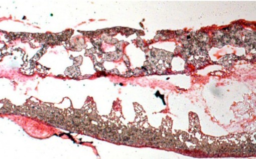
http://www.ncbi.nlm.nih.gov/pmc/articles/PMC3549624/bin/fig-2.jpg

Alzheimer’s disease (ad) is the most common progressive age-related dementia, and is pathologically characterized by the deposition of amyloid-β peptide (Aβ) as amyloid plaques in the brain parenchyma and neurofibrillary tangles (NFTs) within neurons. As a result of the atrophy that occurs in both cortical and subcortical regions, patients suffer cognitive and emotional dysregulation leading eventually to an inability to perform acts of daily living independently and safely. In fact, AD has emerged as a national and international pandemic. According to the World Alzheimer Report 2010, dementia patients account for 35.6 million in worldwide, and are expected to increase to 65.7 million by 2030 and 115.4 million by 2050. Currently, the number of AD patients is around 1% of the world’s gross domestic product. Therefore, it is becoming increasingly evident that a more effective treatment or prophylaxes are needed in the near future. This is because Aβ plaques are potent activators of both microglia and astrocytes—central nervous system (CNS) resident immuno-competent cells that respond to cerebral amyloidosis by chronic, pro-inflammatory activation, also known as “inflammaging” (see review [1]). While it was once thought that activation of microglia and astrocytes in AD brains was an epiphenomenon and not a pathoetiological contributor to AD, more recent studies implicate this Aβ-mediated inflammatory cascade as an etiological perpetrator of AD. For example, therapeutic strategies aimed at manipulating this inflammatory cascade, including Aβ immunization, non-steroidal anti-inflammatory drugs, and modulation of microglial activation, are all able to reduce AD-like pathology and improve cognitive impairments in AD transgenic mouse models [2] and, in some cases, reduce AD pathology in humans [3].

While it is true that no model fully recapitulates AD, transgenic animal models pose novel insights into the pathophysiology of Aβ toxicity. This is especially so with regards to the effects of various Aβ species and the probable pathogenic role of Aβ oligomers [4]. In the PSAPP mouse model of cerebral amyloidosis (bearing mutant human APPsw and presenilin-1 transgenes), there are large numbers of compact Aβ plaques in the hippocampus and cerebral cortex. These mice demonstrate greatly accelerated β-amyloid deposition compared with Tg APPsw mice that is apparent as early as 16 weeks of age [5]. Concurrently, they show increased levels of both Aβ1–40 and Aβ1–42 in their parenchyma and a reduced performance of spatial working memory in the period preceding overt Aβ deposition [5]. Such findings support a critical role of Aβ1–42 in the pathogenesis of AD and suggest a neurotoxic effect of soluble forms of Aβ as well [6].

Human umbilical cord blood cells (HUCBCs) have a unique immunomodulatory potential. Therapeutic benefits derived from HUCBC treatment have been suggested to arise from modulation of peripheral inflammatory processes, which in turn affects inflammation in the brain parenchyma, and the mobilization of adult stem cells from the bone marrow (BM) [711]. Indeed, in the animal model of stroke, HUCBCs have been shown to promote a strong anti-inflammatory T helper 2 (Th2) response [7], as opposed to the deleterious proinflammatory T helper cell type 1 (Th1) response. Interestingly, this observation was seen in conjunction with reduced infarct volume and very importantly with rescue of neurological deficits [7,1214].

…………

HUCBC studies done in vitro have shown that these cells secrete soluble factors that have salutary effects [16,58]. Cultured HUCBC supernatants, for example, stimulate survival of neural cells and peripheral blood mononuclear cells cultured under conditions designed to induce cell stress and limit protein synthesis [12]. Moreover, HUCBCs have the capacity to stimulate generation of a vast amount of cytokines and neurotrophic factors that modify inflammatory responses, including IL-11, CSF-1, NGF, and thrombopoietin [7,22,23]. It has been reported that HUCBC entry into the brain is not required to promote neuroprotection [59]. According to the report just outlined, recovery following brain injury is mediated through peripheral anti-inflammatory responses resulting in brain recovery [9]. This is in accord with our results that indicated more of a peripheral, HUCBC-mediated CNS affect, since the cells were not detected in the mouse brain for any significant amount of time.

On the other hand, it should be noted that it has been shown that after irradiation, peripheral macrophages are able to penetrate the brain and mitigate cerebral amyloidosis in AD mice, implying that hematogenously derived macrophages are efficient at phagocytosing and clearing Aβ deposits [18]. Nevertheless, earlier reports have shown that Aβ can also be phagocytosed or cleared by brain-resident microglia [58,60,61].

In the current experimental paradigm, we did not detect the presence of brain-infiltrating macrophages. Specifically, we stained for CD45 (a marker for both macrophages and microglia), and observed that in and around Aβ plaques there were process-bearing cells that morphologically resembled microglia. Further, vascular “cuffing” that would suggest the presence of infiltrating macrophages that are frequently observed in other CNS inflammatory conditions, such as experimental autoimmune encephalomyelitis [62], was not detected. Also, given the difficulties inherent to distinguishing macrophages from microglia, and the ease of peripheral macrophages to engraft into the brain, as well as changes of microglial phenotype after brain injury [63], it remains possible that peripheral macrophages contribute to decreased cerebral amyloidosis after treatment with HUCBCs.

In this report, we have demonstrated that HUCBC infusion decreases Aβ/β-amyloid pathology in the brain parenchyma, reduces brain inflammation evidenced by reduction of activated microglia, and improves cognitive impairments associated with the AD-like pathology in PSAPP mice. These HUCBC-imparted beneficial effects, which correlate with increased brain-to-blood efflux of Aβ and a shift from proinflammatory Th1 to anti-inflammatory Th2 cytokines both in the brain and in the periphery, are similar to what we observed in previous studies after Aβ immunization [6466]. When taken together, our results provide the basis for a novel immunomodulatory strategy for AD using HUCBCs. While the exact mechanism of efficacy of multiple low-dose HUCBC infusions in AD patients is currently being elucidated, further studies investigating which HUCBC secreted factors are capable of modulating neuroinflammation, reducing AD-like pathology, and rescuing cognitive impairments will need to be explored.

GEN News Highlights    Nov 13, 2015      Alzheimer’s Drug Candidate Also Reverses Effects of Aging

http://www.genengnews.com/gen-news-highlights/alzheimer-s-drug-candidate-also-reverses-effects-of-aging/81251974/

  • Scientists at the Salk Institute say they have found that an experimental drug candidate aimed at combating Alzheimer’s disease has a host of unexpected anti-aging effects in animals.

    The Salk team expanded upon their previous development of a drug candidate, called J147, which takes a different tack by targeting Alzheimer’s major risk factor: old age. In the new work, the team showed that J147 worked well in a mouse model of aging not typically used in Alzheimer’s research. When these mice were treated with J147, they had better memory and cognition, healthier blood vessels in the brain and other improved physiological features.

    The team’s study (“A comprehensive multiomics approach toward understanding the relationship between aging and dementia”) is published in Aging.

    “Initially, the impetus was to test this drug in a novel animal model that was more similar to 99 percent of Alzheimer’s cases,” says Antonio Currais, Ph.D., the lead author and a member of the Schubert Cellular Neurobiology Laboratory at Salk. “We did not predict we’d see this sort of anti-aging effect, but J147 made old mice look like they were young, based upon a number of physiological parameters.”

    “While most drugs developed in the past 20 years target the amyloid plaque deposits in the brain (which are a hallmark of the disease), none have proven effective in the clinic,” says David Schubert, Ph.D., senior author of the study.

    Several years ago, Dr. Schubert and his colleagues began to approach the treatment of the disease from a new angle. Rather than target amyloid, the lab decided to zero in old age. Using cell-based screens against old age-associated brain toxicities, they synthesized J147.

    Previously, the team found that J147 could prevent and even reverse memory loss and Alzheimer’s pathology in mice that have a version of the inherited form of Alzheimer’s, the most commonly used mouse model. However, this form of the disease comprises only about 1% of Alzheimer’s cases. For everyone else, old age is the primary risk factor, according to Dr. Schubert. The team wanted to explore the effects of the drug candidate on a breed of mice that age rapidly and experience a version of dementia that more closely resembles the age-related human disorder.

    In this latest work, the researchers used a comprehensive set of assays to measure the expression of all genes in the brain, as well as over 500 small molecules involved with metabolism in the brains and blood of three groups of the rapidly aging mice. The three groups of rapidly aging mice included one set that was young, one set that was old, and one set that was old but fed J147 as they aged.

    The old mice that received J147 performed better on memory and other tests for cognition and also displayed more robust motor movements. The mice treated with J147 also had fewer pathological signs of Alzheimer’s in their brains. Importantly, because of the large amount of data collected on the three groups of mice, it was possible to demonstrate that many aspects of gene expression and metabolism in the old mice fed J147 were very similar to those of the young animals. These included markers for increased energy metabolism, reduced brain inflammation and reduced levels of oxidized fatty acids in the brain.

    Another notable effect was that J147 prevented the leakage of blood from the microvessels in the brains of old mice. “Damaged blood vessels are a common feature of aging in general, and in Alzheimer’s, it is frequently much worse,” points out Dr. Currais.

    While these studies represent a new and exciting approach to Alzheimer’s drug discovery and animal testing in the context of aging, the only way to demonstrate the clinical relevance of the work is to move J147 into clinical trials for Alzheimer’s disease, note the researchers.

    “If proven safe and effective for Alzheimer’s, the apparent anti-aging effect of J147 would be a welcome benefit,” adds Dr. Schubert. The team aims to begin human trials next year.

J147 was developed at Salk in the laboratory of David Schubert, a professor in the Cellular Neurobiology Laboratory. He said,

“It’s been known for a long time that people in India don’t get very much Alzheimer’s relative to what happens in the United States and the rest of the world.

“One of the curiosities about the diet in India is that they eat a lot of curry. A major spice in curry is turmeric. A major component of turmeric is curcumin.

“Curcumin has been around for a while. It is an FDA-Approved drug for cancer. A friend of mine in Los Angeles, Greg Cole, found that if you give curcumin to very similar mice to what this study’s author has been using, they get they get a little better, the (Alzheimer’s) plaques go away.

“The problem with curcumin is that it is not a great drug, in the sense that it gets degraded very rapidly. It’s availability is quite low in the bloodstream and the brain.

“We decided to make a better version of this. We did a lot of medicinal chemistry. We came up with J147.”

Lead study author Marguerite Prior, a research associate in Salk’s Cellular Neurobiology Laboratory, added,

“J147 is an exciting new compound because it really has strong potential to be an Alzheimer’s disease therapeutic by slowing disease progression and reversing memory deficits following short-term treatment.”

Because of its broad ability to protect nerve cells, the researchers believe that J147 may also be effective for treating other neurological disorders, such as Parkinson’s disease, Huntington’s disease and amyotrophic lateral sclerosis (ALS), as well as vascular dementia from stroke, although their study did not directly explore the drug’s efficacy as a therapy for those diseases.

The findings, published in the journal Alzheimer’s Research and Therapy, may pave the way to a new treatment for Alzheimer’s disease in humans.

Despite years of research, scientists are still seeking the first disease-modifying drugs for Alzheimer’s. Current FDA-approved medications, including Aricept®, Razadyne® and Exelon® (generic donepezil, galantamine and rivastigmine), offer only fleeting short-term benefits for Alzheimer’s patients, but they do nothing to slow the steady, irreversible decline of brain function that erases a person’s memory and ability to think clearly.

Professor Schubert and his colleagues bucked the trend within the pharmaceutical industry, which has focused on the biological pathways involved in the formation of amyloid plaques, the dense deposits of protein that characterize the disease. Instead, the Salk team used living neurons grown in laboratory dishes to test whether their new synthetic compounds, which are based upon natural products derived from plants, were effective at protecting brain cells against several pathologies associated with brain aging. From the test results of each chemical iteration of the lead compound, they were able to alter their chemical structures to make them much more potent. Although J147 appears to be safe in mice, the next step will require clinical trials to determine whether the compound will prove safe and effective in humans.

“Alzheimer’s disease research has traditionally focused on a single target, the amyloid pathway,” says Schubert, “but unfortunately drugs that have been developed through this pathway have not been successful in clinical trials. Our approach is based on the pathologies associated with old age-the greatest risk factor for Alzheimer’s and other neurodegenerative diseases-rather than only the specificities of the disease.”

J147
Salk scientists developed J147, a synthetic drug shown to improve memory and prevent brain damage in mice with Alzheimer’s disease.Images: Courtesy of the Salk Institute for Biological Studies

To test the efficacy of J147 in a much more rigorous preclinical Alzheimer’s model, the Salk team treated mice using a therapeutic strategy that they say more accurately reflects the human symptomatic stage of Alzheimer’s. Administered in the food of 20-month-old genetically engineered mice, at a stage when Alzheimer’s pathology is advanced, J147 rescued severe memory loss, reduced soluble levels of amyloid, and increased neurotrophic factors essential for memory, after only three months of treatment.

In a different experiment, the scientists tested J147 directly against Aricept (generic donepezil), the most widely prescribed Alzheimer’s drug, and found that it performed as well or better in several memory tests.

“In addition to yielding an exceptionally promising therapeutic, both the strategy of using mice with existing disease and the drug discovery process based upon aging are what make the study interesting and exciting,” says Schubert, “because it more closely resembles what happens in humans, who have advanced pathology when diagnosis occurs and treatment begins.” Most studies test drugs before pathology is present, which is preventive rather than therapeutic and may be the reason drugs don’t transfer from animal studies to humans.

Prior and her colleagues say that several cellular processes known to be associated with Alzheimer’s pathology are affected by J147, including an increase in a protein called brain-derived neurotrophic factor (BDNF), which protects neurons from toxic insults, helps new neurons grow and connect with other brain cells, and is involved in memory formation. Postmortem studies show lower than normal levels of BDNF in the brains of people with Alzheimer’s.

The Salk researchers say that J147, with its memory enhancing and neuroprotective properties, along with its safety and availability as an oral medication, would make an “ideal candidate” for Alzheimer’s disease clinical trials. They are currently seeking funding for such a trial.

MORE INFORMATION:
Other researchers on the study were Richard Dargusch, Jennifer L. Ehren and Chandra Chiruta, of the Salk Institute.The work was supported by the Alzheimer’s Drug Discovery Foundation, the Bundy Foundation, the Fritz Burns Foundation, the George E. Hewitt Foundation, the Alzheimer’s Association, and the National Institutes of Health.

AMSBIO announces that Belgian researchers have cited use of BioPORTER Protein Delivery Reagent to introduce Tau seeds into HEK293 cells. BioPORTER Protein Delivery Reagent is a unique lipid formulation that allows direct translocation of proteins into living cells.

Neurodegenerative tauopathies, including Alzheimer disease and frontotemporal dementias, are characterized by neurofibrillary tangles (NFT) composed of intracellular hyperphosphorylated Tau aggregates. Predominantly expressed in neurons, Tau is a microtubule (MT)-binding protein that stabilizes and promotes the assembly of MTs, and the Tau-MT interactions are negatively regulated by phosphorylation of Tau. A naturally unfolded soluble protein under normal conditions, Tau acquires highly ordered ß-pleated sheet structures as it assembles into insoluble hyperphosphorylated paired helical filaments as well as less frequent straight filaments that constitute NFTs in Alzheimer disease and related tauopathies. Significant correlation of total NFT burden with cognitive decline has been observed in Alzheimer disease patients.

In the Belgian research prion-like seeding and propagation of Tau-pathology was demonstrated experimentally and may underlie the stereotyped progression of neurodegenerative Tauopathies. The researchers analyzed the repercussions of prion-like spreading of Tau-pathology via neuronal connections on neuronal network function in TauP301S transgenic mice.

BioPORTER Protein Delivery Reagent provided the researchers with a quick and easy method to study protein function without the need for cloning and DNA transfection. The  reagent lipid captures proteins and transports them inside the target cells. The delivered proteins retain their structure and function while leaving the transduced cells unharmed. The reagent is especially useful when studying protein function in cells that are difficult to transfect using traditional DNA transfection reagents. http://www.amsbio.com/BioPORTER-protein-delivery-transfectiom-reagent.aspx

AMSBIO LLC, (617) 945-5033, http://www.amsbio.com

Neurotrophic factors are a family of proteins that are responsible for the growth and survival of nerve cells during development, and for the maintenance of adult nerve cells. Animal studies and test tube (in vitro Latin phrase for ‘in glass’; in a test tube or other artificial environment, as opposed to inside a living organism.”>in vitro)models

Organisms that scientists use to reproduce features of a disease of interest in an organism and then study it. For example, inserting the gene for HD into a mouse means that it will produce the altered HD protein in the brain. This creates an HD mouse model. The consistent use of these models allows researchers to test ideas about biology in a reproducible way, without the expense and ethical problems of performing these tests in humans.”>models have shown that certain neurotrophic factors are capable of making damaged nerve cells regenerate. Because of this capability, these factors represent exciting possibilities for reversing a number of devastating brain disorders, including Alzheimer’s disease

A neurodegenerative disease that causes progressive memory loss and severe dementia in advanced cases. Alzheimer’s disease is associated with certain abnormalities in brain tissue, involving a particular protein, beta-amyloid.”>Alzheimer’s disease, Parkinson’s disease

A neurodegenerative disorder that primarily affects one’s ability to perform smooth movements. The disease is associated with a loss of dopamine-producing nerve cells in the substantia nigra region of the brain.”>Parkinson’s disease, Lou Gehrig’s disease, and Huntington’s disease

A hereditary neurological disorder characterized by movement, cognitive, and psychiatric symptoms.”>Huntington’s Disease (HD). (For more information on how HD relates to Alzheimer’s and Parkinson’s, click here.) Currently, scientists are looking for ways to harness neurotrophic factors and somehow induce the damaged nerve cells to regenerate in order to improve the symptoms

Changes in the body or its functions, experienced by the patient and indicative of disease.”>symptoms of people with neurological having to do with nerve cells and/or the nervous system, particularly the brain”>neurological disorders.

One neurotrophic factor is a protein in the nervous system that promotes the growth of nerve cells.”>neurotrophic factor that is particularly relevant to HD is brain-derived neurotrophic factor (BDNF)

A protein that causes certain types of nerve cells to survive and grow. BDNF is primarily located in the central nervous system, where it acts on cells in the brain and the eye. In the peripheral nervous system, BDNF promotes the growth of sensory and motor neurons.”>Brain-derived neurotrophic factor (BDNF)

.BDNF

An abbreviation of brain-derived neurotrophic factor, which is a protein that causes certain types of nerve cells to survive and grow. BDNF is primarily located in the central nervous system, where it acts on cells in the brain and the eye. In the peripheral nervous system, BDNF promotes the growth of sensory and motor neurons.”>BDNF levels are decreased in the brains of HD patients, which might be partly responsible for the degenerative when a part of the body stops working well, and begins to decline in function”>degenerative processes of HD. Researchers have recently discovered a link between BDNF An abbreviation of brain-derived neurotrophic factor, which is a protein that causes certain types of nerve cells to survive and grow. BDNF is primarily located in the central nervous system, where it acts on cells in the brain and the eye. In the peripheral nervous system, BDNF promotes the growth of sensory and motor neurons.”>BDNF , mutant huntingtin

The altered form of the huntingtin protein caused by having the HD gene.”>mutant huntingtin , and excitotoxicity

Excessive stimulation of a nerve cell by a neurotransmitter, which poisons the nerve cell and degrades it.”>excitotoxicity, a process by which brain cells die after stimulation. The mutant huntingtin protein A key protein in Huntington’s disease. It exists in all humans but has a chemically different form in people with HD. Please note that although Huntingt<strong>o</strong>n’s disease is spelled with an o, the correct spelling of the protein involved is huntingt<strong>i</strong>n with an i.”>huntingtin protein invariably leads to the death of nerve cells in the striatum part of the brain that is involved in controlling movement. It is made up of the caudate and the putamen. Also referred to as the corpus striatum.”>striatum, the region of the brain needed for movements; however, how mutant huntingtin the altered form of the huntingtin protein caused by having the HD gene.”>mutant huntingtin does this damage is unclear. One possibility is that mutant huntingtin

The altered form of the huntingtin protein caused by having the HD gene.”>mutant huntingtin lowers levels of BDNF

An abbreviation of brain-derived neurotrophic factor, which is a protein that causes certain types of nerve cells to survive and grow. BDNF is primarily located in the central nervous system, where it acts on cells in the brain and the eye. In the peripheral nervous system, BDNF promotes the growth of sensory and motor neurons.”>BDNF, making nerve cells more susceptible to injury and death. Therefore, therapeutic approaches aimed at increasing BDNF

An abbreviation of brain-derived neurotrophic factor, which is a protein that causes certain types of nerve cells to survive and grow. BDNF is primarily located in the central nervous system, where it acts on cells in the brain and the eye. In the peripheral nervous system, BDNF promotes the growth of sensory and motor neurons.”>BDNF production may be able to counteract the effects ofmutant huntingtin

The altered form of the huntingtin protein caused by having the HD gene.”>mutant huntingtin and prevent a significant amount of the neurodegeneration

The deterioration or loss of function of nerve cells. Neurodegenerative diseases include HD, Alzheimer’s, Parkinson’s and many more. <em>Adj.</em>neurodegenerative.”>neurodegeneration that would otherwise occur in HD. (For more information on huntingtin protein

A key protein in Huntington’s disease. It exists in all humans but has a chemically different form in people with HD. Please note that although Huntingt<strong>o</strong>n’s disease is spelled with an o, the correct spelling of the protein involved is huntingt<strong>i</strong>n with an i.”>huntingtin protein

, click here.)

http://ghr.nlm.nih.gov/gene/BDNF

What is the official name of the BDNF gene?

The official name of this gene is “brain-derived neurotrophic factor.”

BDNF is the gene’s official symbol. The BDNF gene is also known by other names, listed below.

Read more about gene names and symbols on the About page.

What is the normal function of the BDNF gene?

The BDNF gene provides instructions for making a protein found in the brain and spinal cord called brain-derived neurotrophic factor. This protein promotes the survival of nerve cells (neurons) by playing a role in the growth, maturation (differentiation), and maintenance of these cells. In the brain, the BDNF protein is active at the connections between nerve cells (synapses), where cell-to-cell communication occurs. The synapses can change and adapt over time in response to experience, a characteristic called synaptic plasticity. The BDNF protein helps regulate synaptic plasticity, which is important for learning and memory.

The BDNF protein is found in regions of the brain that control eating, drinking, and body weight; the protein likely contributes to the management of these functions.

Does the BDNF gene share characteristics with other genes?

The BDNF gene belongs to a family of genes called endogenous ligands (endogenous ligands).

A gene family is a group of genes that share important characteristics. Classifying individual genes into families helps researchers describe how genes are related to each other. For more information, see What are gene families? in the Handbook.

Brain-derived Neurotrophic Factor      

DEVIN K. BINDERa,* and HELEN E. SCHARFMANb

Growth Factors. 2004 Sep; 22(3): 123–131.     doi:  10.1080/08977190410001723308
Nerve growth factor (NGF) was discovered in the early 1950s due to its trophic (survival- and growth-promoting) effects on sensory and sympathetic neurons (Levi-Montalcini and Hamburger, 1951), In 1982, brain-derived neurotrophic factor (BDNF), the second member of the “neurotrophic” family of neurotrophic factors, was shown to promote survival of a subpopulation of dorsal root ganglion neurons, and subsequently purified from pig brain (Barde et al., 1982). Since then, other members of the neurotrophin family such as neurotrophin-3 (NT-3) (Maisonpierre et al., 1990) and neurotrophin-4/5 (NT-4/5) (Hallbook et al., 1991; Ipet al., 1992) have been described, each with a distinct profile of trophic effects on subpopulations of neurons in the peripheral and central nervous systems.

The BDNF gene (in humans mapped to chromosome 11p) has four 5′ exons (exons I-IV) that are associated with distinct promoters, and one 3′ exon (exon V) that encodes the mature BDNF protein (Metsis et al., 1993; Timmusk et al., 1993). Eight distinct mRNAs are transcribed, with transcripts containing exons I-III expressed predominantly in brain and exon IV found in lung and heart (Timmusk et al., 1993).

BDNF shares about 50% amino acid identity with NGF, NT-3 and NT-4/5. Each neurotrophin consists of a noncovalently-1 linked homodimer and contains (1) a signal peptide following the initiation codon; and (2) a pro-region containing an N-linked glycosylation site. Initially produced as proneurotrophins, prohormone convertases such as furin cleave the proneurotrophins (M.W. ~30kDa) to the mature neurotrophin (M.W. ~14kDa) (Chao and Bothwell, 2002). Proneurotrophins have altered binding characteristics and distinct biologic activity in comparison with mature neurotrophins (Lee et al., 2001a,b). Neurotrophins also share a distinctive three-dimensional structure containing two pairs of antiparallel β-strands and cysteine residues in a cystine knot motif.

Each neurotrophin binds one or more of the tropomyosin-related kinase (trk) receptors, members of the family of receptor tyrosine kinases (Patapoutian and Reichardt, 2001). Ligand-induced receptor dimerization results in kinase activation; subsequent receptor autophosphorylation on multiple tyrosine residues creates specific binding sites for intracellular target proteins, which bind to the activated receptor via SH2 domains (Barbacid, 1994; Patapoutian and Reichardt, 2001). These include PLC-γ1 (phospholipase C), p85 (the noncatalytic subunit of PI-3 kinase) and Shc (SH2-containing sequence); activation of these target proteins can then lead to a variety of intracellular signalling cascades such as the Ras-MAP (mitogen-activated protein) kinase cascade and phosphorylation of cyclic AMP-response element binding protein (CREB) (Patapoutian and Reichardt, 2001; Segal, 2003).

TrkA binds NGF (with low-affinity binding by NT-3 in some systems); trkB binds BDNF and NT-4/5 with lower-affinity binding by NT-3; and trkC binds NT-3 (Barbacid, 1994). Trk receptors exist in both a full-length (trkB.FL) form as well as truncated (trkB.T1. trkB.T2) forms lacking the kinase domain (Eide et al., 1996; Fryer et al., 1997). Although most functions attributed to BDNF are associated with full-length trkB, several roles have been suggested for truncated receptors, including growth and development (Fryer et al., 1997; Yacoubian and Lo, 2000; Luikart et al., 2003) and negative modulation of trkB receptor expression and function (Eide et al., 1996; Haapasalo et al., 2001; Haapasalo et al., 2002). Expression of truncated trk receptors on astrocytes is upregulated following injury (Frisen et al.,1993) and may modulate neuronal vulnerability (Saarelainen et al., 2000a,b) and sequestration of BDNF in astrocytes (Biffo et al., 1995;Roback et al., 1995; Alderson et al., 2000). Recent studies have shown that BDNF activates glial calcium signalling by truncated trk receptors (Climent et al., 2000: Rose et al., 2003).

In addition, all of the neurotrophins bind to the p75 receptor, designated p75NTR. p75NTR, related to proteins of the tumor necrosis factor (TNFR) superfamily, has a glycosylated extracellular region involved in ligand binding, a transmembrane region, and a short cytoplasmic sequence lacking intrinsic catalytic activity (Chao and Hempstead, 1995; Dechant and Barde, 2002). Neurotrophin binding to p75NTR is linked to several intracellular signal transduction pathways, including nuclear factor-κB (NF-κB), Jun kinase and sphingo-myelin hydrolysis (Dechant and Barde, 2002). P75NTR signalling mediates biologic actions distinct from those of the trk receptors, notably the initiation of programmed cell death (apoptosis) (Casaccia-Bonnefil et al., 1996; Frade et al., 1996; Roux et al., 1999; Dechant and Barde, 2002). It has also been suggested that p75 may serve to determine neurotrophin binding specificity (Esposito et al., 2001; Lee et al., 2001a,b;Zaccaro et al., 2001).

BDNF GENE REGULATION

A multitude of stimuli have been described that alter BDNF gene expression in both physiologic and pathologic states (Lindholm et al., 1994). For example, light stimulation increases BDNF mRNA in visual cortex (Castrén et al., 1992), osmotic stimulation increases BDNF mRNA in the hypothalamus (Castrén et al., 1995; Dias et al., 2003), and whisker stimulation increases BDNF mRNA expression in somatosensory barrel cortex (Rocamora et al., 1996). Electrical stimuli that induce long-term potentiation (LTP) in the hippocampus, a cellular model of learning and memory, increase BDNF and NGF expression (Patterson et al., 1992; Castrén et al., 1993; Bramham et al., 1996). Even physical exercise has been shown to increase NGF and BDNF expression in hippocampus (Neeper et al., 1995). Interestingly, BDNF levels vary across the estrous cycle, which correlate with its effects on neural excitability (Scharfman et al., 2003).

Distinct BDNF 5′ exons are differentially regulated by stimuli such as neural activity. For example, exons I-III, but not exon IV, increase after kainic acid-induced seizures (Timmusk et al., 1993) or other stimuli that increase activity (Lauterborn et al., 1996; Tao et al., 2002). Protein synthesis is required for the effects of activity on exons I and II, but not III and IV, raising the possibility that the latter act as immediate early genes (Lauterborn et al., 1996; Castrén et al., 1998). The transcription factor CaRF activates transcription of exon III under the control of a calcium response element. CaRE1 (Tao et al., 2002). CREB, which can be stimulated by diverse stimuli ranging from activity to chronic antidepressant treatment (Nibuya et al., 1995,1996; Shieh et al., 1998; Tao et al., 1998; Shieh and Ghosh, 1999), also modulates exon III transcription. Recent evidence also indicates that neural activity triggers calcium-dependent phosphorylation and release of methyl-CpG binding protein 2 (MeCP2) from BDNF promoter III to derepress transcription (Chen et al., 2003).

LOCALIZATION, TRANSPORT AND RELEASE

BDNF and trkB mRNA have a widespread distribution in the central nervous system (Merlio et al., 1993;Conner et al., 1997). BDNF and trkB protein immunoreactivity is also widespread (Conner et al., 1997; Yanet al., 1997a,b; Drake et al., 1999), Like BDNF mRNA, constitutive BDNF protein expression is particularly high in the hippocampus, where the mossy fibre axons of dentate granule cells display BDNF immunoreactivity (Conner et al., 1997).

Unlike the classical target-derived trophic factor model in which neurotrophins—such as NGF—are retrogradely transported, there is now abundant evidence that BDNF is also anterogradely transported in brain. First, BDNF protein is localized to nerve terminals (Conner et al., 1997), and pathway transection or axonal transport inhibition abrogates this terminal expression (Altar et al., 1997; Conner et al., 1997; Altar and DiStefano, 1998). Second, higher-resolution studies have shown that BDNF is associated with dense-core vesicles (Fawcett et al., 1997; Altar and DiStefano, 1998), which are the primary site for neuropeptide storage and release from nerve terminals. Third, further functional studies have supported the anterograde transport hypothesis (Fawcett et al., 1998, 2000). Fourth, pro-BDNF is shuttled from the trans-Golgi network into secretory granules, where it is cleaved by prohormone convertase 1 (PC1) (Farhadi et al., 2000).

In addition, emerging evidence suggests that both BDNF and trk receptors may undergo regulated intracellular transport. For example, seizures lead to redistribution of BDNF mRNA from hippocampal CA3 cell bodies to their apical dendrites (Bregola et al., 2000; Simonato et al., 2002). Trk signalling is now thought to include retrograde transport of intact neurotrophin-trk complexes to the neuronal cell body (Miller and Kaplan, 2001; Ginty and Segal, 2002).

Recent evidence indicates that neurotrophins are released acutely following neuronal depolarization (Griesbeck et al., 1999; Mowla et al., 1999; Goggi et al., 2003). In fact, direct activity-dependent pre- to post-synaptic transneuronal transfer of BDNF has recently been demonstrated using fluorescently-labelled BDNF (Kohara et al., 2001). The released form of BDNF is thought to be proBDNF (Mowla et al., 2001), raising the possibility of postsecretory proteolytic processing by membrane-associated or extracellular proteases in the modulation of BDNF action (Lee et al., 2001a,b).

….. more

Experimental Drug Targeting Alzheimer’s Disease Shows Anti-aging Effects

http://www.biosciencetechnology.com/news/2015/11/experimental-drug-targeting-alzheimers-disease-shows-anti-aging-effects

Salk scientists Antonio Currais, David Schubert and team found a molecule that slows the clock on key aspects of aging in animals. Credit: Salk Institute

Salk scientists Antonio Currais, David Schubert and team found a molecule that slows the clock on key aspects of aging in animals. Credit: Salk Institute

Salk Institute researchers have found that an experimental drug candidate aimed at combating Alzheimer’s disease has a host of unexpected anti-aging effects in animals.

The Salk team expanded upon their previous development of a drug candidate, called J147, which takes a different tack by targeting Alzheimer’s major risk factor–old age. In the new work, the team showed that the drug candidate worked well in a mouse model of aging not typically used in Alzheimer’s research. When these mice were treated with J147, they had better memory and cognition, healthier blood vessels in the brain and other improved physiological features, as detailed November 12, 2015 in the journal Aging.

“Initially, the impetus was to test this drug in a novel animal model that was more similar to 99 percent of Alzheimer’s cases,” said Antonio Currais, the lead author and a member of Professor David Schubert’s Cellular Neurobiology Laboratory at Salk. “We did not predict we’d see this sort of anti-aging effect, but J147 made old mice look like they were young, based upon a number of physiological parameters.”

Alzheimer’s disease is a progressive brain disorder, recently ranked as the third leading cause of death in the United States and affecting more than five million Americans. It is also the most common cause of dementia in older adults, according to the National Institutes of Health.

“While most drugs developed in the past 20 years target the amyloid plaque deposits in the brain (which are a hallmark of the disease), none have proven effective in the clinic,” said Schubert, senior author of the study.

Several years ago, Schubert and his colleagues began to approach the treatment of the disease from a new angle. Rather than target amyloid, the lab decided to zero in on the major risk factor for the disease–old age. Using cell-based screens against old age-associated brain toxicities, they synthesized J147.

Previously, the team found that J147 could prevent and even reverse memory loss and Alzheimer’s pathology in mice that have a version of the inherited form of Alzheimer’s, the most commonly used mouse model. However, this form of the disease comprises only about 1 percent of Alzheimer’s cases. For everyone else, old age is the primary risk factor, said Schubert. The team wanted to explore the effects of the drug candidate on a breed of mice that age rapidly and experience a version of dementia that more closely resembles the age-related human disorder.

In this latest work, the researchers used a comprehensive set of assaid to measure the expression of all genes in the brain, as well as over 500 small molecules involved with metabolism in the brains and blood of three groups of the rapidly aging mice. The three groups of rapidly aging mice included one set that was young, one set that was old and one set that was old but fed J147 as they aged.

The old mice that received J147 performed better on memory and other tests for cognition and also displayed more robust motor movements. The mice treated with J147 also had fewer pathological signs of Alzheimer’s in their brains. Importantly, because of the large amount of data collected on the three groups of mice, it was possible to demonstrate that many aspects of gene expression and metabolism in the old mice fed J147 were very similar to those of the young animals. These included markers for increased energy metabolism, reduced brain inflammation and reduced levels of oxidized fatty acids in the brain.

Another notable effect was that J147 prevented the leakage of blood from the microvessels in the brains of old mice. “Damaged blood vessels are a common feature of aging in general, and in Alzheimer’s, it is frequently much worse,” said Currais.

Currais and Schubert note that while these studies represent a new and exciting approach to Alzheimer’s drug discovery and animal testing in the context of aging, the only way to demonstrate the clinical relevance of the work is to move J147 into human clinical trials for Alzheimer’s disease.

“If proven safe and effective for Alzheimer’s, the apparent anti-aging effect of J147 would be a welcome benefit,” adds Schubert. The team aims to begin human trials next year.

Read Full Post »

Microgravity Expanded Stem Cells

Larry H. Bernstein, MD, FCAP, Curtor

LPBI


Application of Microgravity Expanded Stem Cells in Regenerative Medicine

Author: Abba C Zubair MD PhD   Mayo Clinic      http://medical.wesrch.com/pdfME1LYYYIWQLGL

Description: Application of Microgravity Expanded Stem Cells in Regenerative Medicine:- Regrettably I have no financial disclosures, However, I am available if any company is interested. MSCs for Hemorrhagic Stroke.Oxygen-Glucose Deprivation Model (OGD).Effect of MSC on OGD-Stressed Neurons. Mechanism of MSC induced neuro-recovery.Mechanism of MSC induced immune regulation.

ICH model:- Autologous blood withdraw from femoral Artery MSCs or medium infusion via femoral vein With the help of stereotactic frame and microinjection pump autologous blood will be injected to right baal ganglia, 10µl/min total 100µl.Brain sections @D1 after ICH:- 100ul autologous blood Right basal ganglion Coordinates: 0.2 mm anterior, 5.5mm ventral, and 3.5 mm lateral to bregma. Arrow shows hematoma initiated location.Behavior test includes Forelimb Use Asymmetry test and Rotometer test. Behavior (Forelimb Use Asymmetry Test)- Media, MSC infusion (1×10^6/kg).

Stem Cells in Microgravity- No effective method to expand human stem cells, Need about 200 to 500 million stem cells to achieve effective therapy.Objectives- Measure cell proliferation in microgravity versus gravity of Earth, Observe cell growth and morphological characteristics in microgravity, Analyze gene expression profiles of cells grown in microgravity, Use viable cells grown in microgravity in a variety of downstream in vitro and in vivo experiments on Earth.BioCell Cassettes:- Created by BioServe Space -Aerospace Engineering and Sciences Department at …Please navigate Paper pages for more details

http://medical.wesrchcdn.com/User_images/Pdf/315/1446880640/pdf_jpegs/14LYY_1446880640_2.jpg


http://medical.wesrchcdn.com/User_images/Pdf/315/1446880640/pdf_jpegs/14LYY_1446880640_3.jpg


http://medical.wesrchcdn.com/User_images/Pdf/315/1446880640/pdf_jpegs/14LYY_1446880640_4.jpg

Read Full Post »

Organoid Development

Curator: Larry H Bernstein, MD, FCAP


Thyroid Organoids Made from Stem Cells Treat Thyroid-Deficient Mice

Their gut lining regrew onto the scaffold and functioned normally to absorb water from the colon. Within weeks, the scaffolding dissolved and was replaced with normal connective tissue. “The scaffold was well tolerated and promoted healing by recruiting stem cells,” Hackam says. “[The dogs] had a perfectly normal lining after 8 weeks.”

Sourced through Scoop.it from: news.sciencemag.org


Stem Cells Dev. 2015 Oct 1. [Epub ahead of print]
Zfp553 is essential for maintenance and acquisition of pluripotency.

Pluripotent cells are promising tools in the arena of regenerative medicine. For many years, research efforts have been directed towards uncovering the underlying mechanisms that govern the pluripotent state and this involves identifying new pluripotency associated factors. Zinc finger protein 553 (Zfp553) has been hypothesized to be one such factor due to its predominant expression in inner cell mass of the mouse early embryo. In this study, we have identified Zfp553 as a regulator of pluripotency. Zfp553 knockdown down-regulates pluripotency markers and triggers differentiation in mouse embryonic stem cells (mESCs). Further investigation revealed that Zfp553 regulates pluripotency in mESCs via the transcriptional activation of Pou5f1 and Nanog. Microarray results revealed that depletion of Zfp553 down-regulates many pluripotency genes, as well as genes associated with metabolism related processes. ChIP-seq depicted the genomic binding sites of Zfp553 in mESCs and its binding motif. In addition, we found that depletion of Zfp553 could impair somatic cell reprogramming, evidenced by reduced reprogramming efficiency and cell viability. Together, our preliminary findings provide novel insights to a newly identified pluripotency factor Zfp553 and its role in pluripotency regulation.


Mesenchymal stem cells use extracellular vesicles to outsource mitophagy and shuttle microRNAs

Donald G. PhinneyMichelangelo Di GiuseppeJoel NjahErnest SalaSruti ShivaClaudette M. St CroixDonna B. StolzSimon C. WatkinsY. Peter DiGeorge D. LeikaufJay Kolls,… , & Luis A. Ortiz

Nature Communications 07 Oct 2015; 6(8472)   http://dx.doi.org:/10.1038/ncomms9472   http://www.nature.com/ncomms/2015/151007/ncomms9472/full/ncomms9472.html

Mesenchymal stem cells (MSCs) and macrophages are fundamental components of the stem cell niche and function coordinately to regulate haematopoietic stem cell self-renewal and mobilization. Recent studies indicate that mitophagy and healthy mitochondrial function are critical to the survival of stem cells, but how these processes are regulated in MSCs is unknown. Here we show that MSCs manage intracellular oxidative stress by targeting depolarized mitochondria to the plasma membrane via arrestin domain-containing protein 1-mediated microvesicles. The vesicles are then engulfed and re-utilized via a process involving fusion by macrophages, resulting in enhanced bioenergetics. Furthermore, we show that MSCs simultaneously shed micro RNA-containing exosomes that inhibit macrophage activation by suppressing Toll-like receptor signalling, thereby de-sensitizing macrophages to the ingested mitochondria. Collectively, these studies mechanistically link mitophagy and MSC survival with macrophage function, thereby providing a physiologically relevant context for the innate immunomodulatory activity of MSCs.


MSCs undergo mitophagy in response to oxidative stress

Human MSCs shed from their surface a diverse subpopulation of vesicles. To characterize these vesicles, we performed electron microscopy on those recovered from MSC-conditioned medium by differential ultracentrifugation (100,000g for 18h). This analysis demonstrated the presence of 50–100-nm vesicles that are morphologically consistent with exosomes (Fig. 1a, left). Flotation of the 100,000g pellets on sucrose gradients followed by western blot and fluorescent activated cell sorting (FACS) further demonstrated that these vesicles expressed the exosomal markers’ milk fat globule factor 8 (Mfge8) and the tetraspanins CD9 and CD63, respectively (Supplementary Fig. 1A). Centrifugation of conditioned medium at low speeds (10,000g) revealed the presence of larger vesicles (>100nm) that contain subcellular mitochondrial structures including outer and inner membranes and cristae, and expressed the mitochondria-specific protein ATP synthase as evidenced by immuno-gold labelling (Fig. 1a, centre and Supplementary Fig. 1B). MSCs also release larger multivesicular bodies containing lysosome-like vesicles and entire mitochondria, suggesting that these organelles were selected for mitophagy by targeting to autophagosomes (Fig. 1a, right).

Figure 1: MSC-derived MVs contain depolarized mitochondria.

MSC-derived MVs contain depolarized mitochondria.

(a) Left panel, electron microscopy of vesicles isolated from sucrose densities of 1.11 and 1.14gml−1 and purified using differential ultracentrifugation (100,000g per 18h) reveal a typical exosome morphology. Middle panel, MVs over 100nm in size recovered from human MSC-conditioned medium following low-speed (10,000g per 1h) centrifugation contain structures conforming to the morphology of mitochondria. Right panel, MVs contain closely packed-vesicles and entire mitochondria (multivesicular body (MVB)) representing autophagosomes. (b) Left panel, flow cytometric analysis of MitoSOX Red-stained human MSCs expanded in 5 or 21% oxygen for 7 days. Right panel, quantification of the flow cytometric data. Plotted values (mean±s.e.m.) represent four replicates for each sample using three distinct replicate cultures from each experimental group. (c) Mitochondrial membrane potential of human MSCs from bdetermined using JC-1 staining. Expansion in 21% oxygen results in partial depolarization of mitochondria as evidenced by accumulation of JC-1 monomers (*P<0.005, Student’s-t test versus MSCs in 5%oxygen). (d) Western blot analysis (Supplementary Fig. 1) of cytoplasmic or mitochondrial extracts prepared from human MSCs expanded in 21% oxygen for the indicated passage numbers (P1 or P4) reveals Parkin mitochondrial translocation and Pink1 kinase activation in human MSCs but not human dermal fibroblasts. Data are representative of a single experiment repeated five times.

To determine whether culture expansion of MSCs impairs mitochondrial function, we expanded cells under physiological oxygen levels (5% O2) or standard culture conditions (21% O2) and quantified mtROS levels and mitochondrial membrane potential by staining with MitoSOX Red and JC-1, respectively37. FACS analysis of stained cells confirmed that exposure to 21% oxygen resulted in a significant increase in mtROS (Fig. 1b) and a concomitant decrease in mitochondrial membrane potential as demonstrated by the accumulation of JC-1 monomers (Fig. 1c). Moreover, western blot analysis revealed that prolonged exposure to 21% oxygen activated the Pink1/Parkin-mediated pathway of mitophagy in MSCs but not in human fibroblasts cultured under identical conditions (Fig. 1d). Herein, increased mitochondrial expression of Parkin and Pink1 kinase was accompanied by Pink1 kinase activation as evidenced by the presence of lower molecular weight moieties of the protein and reduced Miro levels in mitochondrial extracts from P4 versus P1 MSCs (Fig. 1c and Supplementary Fig. 1C). Importantly, Pink1 targets Miro for degradation, thereby severing the connection of the mitochondria to the cytoskeleton and facilitating its incorporation into the phagosome38. Consistent with these results, western blot analysis also revealed that mitochondria-containing MVs expressed microtubule-associated protein 1 light chain 3 (LC3) and autophagy-related protein 12, which are highly enriched in MVs as compared with whole-cell extracts (Supplementary Fig. 1D). Therefore, these MVs are characteristic of autophagosomes39.

MSCs package mitochondria in MVs for cellular transfer

To examine mitophagy in MSCs in more detail, MSCs were infected with baculoviruses encoding green fluorescent protein (GFP) fused to the E1alpha pyruvate dehydrogenase leader peptide, which drives transport to the mitochondria, and LC3 fused to a red fluorescent protein (RFP) to allow tracking to the phagophore39, 40. Fluorescent microscopy confirmed that the GFP-labelled mitochondrial network is in close proximity to RFP-LC3-labelled phagosomes (Fig. 2a). Live cell imaging further revealed that mitochondria are loaded in the cytoplasm into LC3-containing vesicles, which migrate towards the cell periphery and are incorporated into outward budding blebs in the plasma membrane (Fig. 2b–d and Supplementary Movie 1). Western blot analysis further revealed that RFP-LC3-MVs also expressed the endosomal sorting complex required for transport (ESCRT)-associated proteins’ tumour suppressor gene 101 (TSG101) and arrestin domain-containing protein 1 (ARRDC1)11, 41 (Supplementary Fig. 1D). Collectively, these results indicate that MSCs employ the release of ARMMs to extrude mitochondria at their cell surface. Moreover, MSCs exhibited marked increases in apoptosis when treated with Bafilomycin A1 or low concentrations (3–5μM) of chloroquine, which block the mitophagy flux, indicating that this process is critical for MSC survival (Supplementary Movie 2).

Figure 2: MSC outsource mitophagy to macrophages.

MSC outsource mitophagy to macrophages.

(ad) Differential interference contrast (DIC) fluorescence overlay of live human MSCs expressing fluorescent proteins that target mitochondria (green) and phagosomes (red) shows mitochondria being loaded into phagosomes (arrows), which are then shuttled to the plasma membrane for extrusion (also seeSupplementary Movie 1). (eh) Inset shows a representative macrophage interacting with a human MSC. This interaction is shown as a time sequence (5min intervals) in the lower images and in Supplementary Movie 3. The inset demarcates the area in the human MSC plasma membrane where the membrane blebs outwards and accumulates vesicles. Macrophages nibble the surface of human MSCs and uptake mitochondrial laden phagosomes from blebs budding (arrows) from the plasma membrane of the human MSCs. Scale bars, 10μ.


Next, we co-cultured GFP-labelled human MSCs from above with primary human or mouse macrophages. Live cell imaging revealed that macrophages nibble the plasma membrane of MSCs, establishing cell contact at areas where membrane blebs are enriched in RFP-labelled vesicles, which are subsequently stripped by the macrophage (Fig. 2e–h and Supplementary Movie 3). This activity was also observed between mouse macrophages and primary human MSCs (Fig. 3a and Supplementary Movies 4 and 5) but was not evident when macrophages were co-cultured with mouse or human fibroblasts (Supplementary Fig. 2). In a subsequent experiment, we co-cultured the macrophage cell line RAW 264.7 with human MSCs containing RFP-labelled mitochondria (10:1 ratio) for 4h and recovered macrophages using FACS after staining with antibodies that recognize macrophage epitopes (that is, F4/80) not expressed by MSCs. Sorted macrophages were cultured for up to 2 weeks in RPMI media, which do not support MSC expansion and survival. Fluorescent microscopy of these macrophages revealed clear evidence of cell-associated RFP derived from human MSCs (Fig. 3b). To confirm these findings, we demonstrated using PCR amplification that these macrophages expressed the mitochondrial specific transcript human cytochrome c oxidase I (MT-COX I), which was confirmed on the basis of the restriction fragment pattern obtained after digestion of the PCR product with Bfa1 (Fig. 3b). This PCR product was not detected in mouse macrophages because of limited sequence homology between the two genes30 but was detected in human MSC-derived MVs as expected (Fig. 3c and Supplementary Fig. 3A). Lastly, we co-cultured Cy5-labelled human MSCs with macrophages that were pre-incubated with or without dextran sulfate (100μgml−1), a nonspecific inhibitor of phagocytosis. Live cell imaging showed phagocytosis of MVs by macrophages over a period of 18min, and confocal microscopy confirmed that the engulfed Cy5-labelled vesicles resided within the cell body of the macrophage (Supplementary Movie 6). However, MV uptake was blocked in macrophages pre-treated with dextran sulfate as evidenced by the accumulation of Cy5-labelled MVs on the macrophage surface (Supplementary Movie 7).

Figure 3: MSC transfer mitochondria to macrophages and lung tissues.

MSC transfer mitochondria to macrophages and lung tissues.

(a) Top panel is DIC fluorescent overlay at time 0 of primary human MSCs infected with Organelle Lights to label mitochondria (green) and co-cultured with mouse (RAW 264.7) macrophages. Lower panels, time sequence at 45min intervals showing transfer of green-labelled mitochondria from the a MSC to a macrophage (red arrow, see Supplementary Movie 4 for transfer of mitochondria in filamentous form, andSupplementary Movie 5 in which GFP signal is compensated to allow the tracking of the transferred mitochondria into macrophages). (b) Left panel, photomicrograph of FACS-sorted mouse macrophages that were co-cultured with mitochondria-labelled (RFP) human MSCs clearly show retention of RFP label. Right panel, electrophoretic pattern of human COX I PCR product treated with or without Bfa1 after amplification from the indicated cell sources. (c) MSC-derived exosomes and MVs express the Bfa1-sensitive 228-bp COX I mtDNA PCR product detected in human MSCs (b). (d) Left panel, electrophoretic pattern of Bfa1-digested human COX1 PCR product amplified from mouse lung DNA isolated 14 days after the intravenous administration of human MSCs, human MSC-derived MVs or exosomes. Right panel, human GAPDH and human COX1 relative expression levels quantified by RT–PCR in mouse lung (3–28 days) after a single (intratracheal (IT) or intravenous (IV)) injection of human MSCs, human MSC-derived exosomes or human fibroblasts. *P<0.001, #P<0.001 by ANOVA compared with untreated mouse lung. Plotted values (mean±s.e.m.) are from experiments repeated four times. Scale bars, 20μ.


To track the in vivo transfer of mitochondria, we systemically administered RFP-labelled human MSCs into C57BL/6 mice expressing a GFP reporter under control of the endothelial specific Tie2 promoter. At 24h post injection, GFP-labelled endothelial cells, epithelial cells and macrophages that contained RFP-labelled mitochondria were visible (Supplementary Fig. 3B). Bfa1 digestion of mouse lung DNA following intravenous administration of human MSCs, exosomes or MVs yielded a pattern of restriction similar to those observed in RAW 264.7 macrophages (Fig. 3d). To follow the fate of viable human MSCs in the mouse lung, we measured the abundance of human-specific GAPDH transcripts via reverse transcriptase–PCR (RT–PCR)42. Human GAPDH mRNA was not detected in the lung tissue of untreated mice but was detected at 3 days post injection of human MSCs or human fibroblasts (Fig. 3d). However, expression rapidly declined and was no longer evident by 14 or 28 days post transplant, consistent with the clearance rate of cells from lung tissue. Expression of human COXI mRNA in mouse lung mirrored that of human GAPDH following injection of human fibroblasts and was detected at 3 days but not 14 or 28 days post transplant. In contrast, human COXI transcripts were detected up to 28 days post injection of human MSCs, indicating that mouse lung tissue retained mtDNA long after the disappearance of viable human MSCs (Fig. 3d). Thus, MSC-derived vesicles constitute an effective mechanism to transfer mtDNA into the mouse lung.


MSC extracellular vesicles enhance macrophage energetics

To study the effect of MVs on macrophage bioenergetics, we analysed oxygen consumption rates (OCRs) using the SeaHorse technology. Human macrophages exhibit higher basal OCR than human MSCs or human fibroblasts (Fig. 4a). Co-culture of macrophages with human MSCs (Mac+hMSC) or MSC-derived exosomes (Mac+Exo) but not human fibroblasts (Mac+Fibro) significantly (analysis of variance (ANOVA) followed by Student–Neuman Keuls (SNK) post-hocpairwise comparisons) increased their OCR, suggesting that MSCs or MSC-derived exosomes alter macrophage bioenergetics (Fig. 4a). Next, we repeated these measurements after treatment of cells with oligomycin A, an inhibitor of ATP synthase, which is required for the oxidative phosphorylation of ADP to ATP. These conditions differentiate ATP-linked respiration from the proton leak. Macrophages exhibited a higher level of proton leak as compared with human MSCs and fibroblasts, and proton leak was significantly (ANOVA followed by SNK post hoc pairwise comparisons) reduced following co-culture with human Mac+Exo but not Mac+Fibro (Fig. 4a). Co-culture with human MSCs (Mac+hMSC) also significantly (ANOVA followed by SNK post hocpairwise comparisons) reduced proton leak in macrophages. We also repeated the OCR measurements following treatment of cells with the uncoupling agent carbonyl cyanide 4-(trifluoromethoxy) phenylhydraone (FCCP) to determine how cells respond to an increase in ATP demand. All three cell types responded to FCCP treatment with increased OCR, and the magnitude of the response was greater in macrophages as compared with human MSCs and fibroblasts. Moreover, OCR was significantly increased in FCCP-treated macrophages following co-culture with human MSCs (Mac+hMSC) or human Mac+Exo but not Mac+Fibro (Fig. 4a).

Figure 4: MSCs enhance macrophage bioenergetics.

MSCs enhance macrophage bioenergetics.

(a) Mitochondrial respiration of human macrophages, human MSCs or human fibroblasts was measured as OCR using the XF technology. Macrophages were co-cultured with or without human MSCs or fibroblasts (1:10 ratio) or treated with human MSC-derived exosomes (40μg per protein) in the presence or absence of Oligomycin A and FCCP to differentiate ATP-linked respiration from the proton leak. Plotted data (mean±s.e.m.) were performed using six replicates per sample and repeated three times. (b) Pseudocoloured photomicrographs (0–240min) of MitoSOX Red-stained macrophages that were non-stimulated (upper panel), or treated with silica (20μgcm−2, lower panel) or silica plus human MSC-derived exosomes (added 10min after silica, middle panel). Scale bars, 50μ. (c) Time course of MitoSOX Red emission by human macrophages treated as in b. Figure is representative of five exposures (nine stages positions per test and 6 cells per stage). (d) OCR as in a of silica-exposed macrophages treated with or without human MSCs, human MSC-derived exosomes or human fibroblasts. Plotted values (mean±s.e.m.) are from experiments repeated three times, *P<0.05 as compared to control, #P<0.05 as compared to silica treated macrophages, as determined by Student’s t-test.


To examine the effect of MSC or exosomes on macrophage bioenergetics under conditions of altered homeostasis, we exposed macrophages to silica particles. Silica exposure results in a burst of mtROS production as evidenced by changes in MitoSOX Red fluorescence intensity; however, this effect is largely mitigated in macrophages incubated with human MSC-derived exosomes (Fig. 4b,c). Silica exposure also decreased macrophage OCR, but this decrease was reversed by co-culture with human MSCs or human MSC-derived exosomes but not with human fibroblasts (Fig. 4d). The fact that transfer of partially depolarized mitochondria from MSCs to macrophages enhances that macrophage bioenergetics appears paradoxical. However, loss of mitochondrial membrane potential as a result of MSC expansion is not absolute as mitochondria exhibit residual membrane potential as evidenced by the concentration of JC-1 aggregates (Fig. 1c). This indicates that the mitochondrial membrane is not collapsed and the mitochondria are still capable of undergoing fusion. To determine whether these mitochondria are recycled in macrophages by fusion, we co-cultured human MSCs with macrophages after labelling cells with two different MitoTracker dyes (Red and Green)43. Live cell imaging clearly demonstrated the transfer and subsequent fusion (yellow colour in merged images) of RFP-labelled, human MSC-derived mitochondria with GFP-labelled mitochondria within human macrophages (Fig. 5). These data indicate that under oxidative stress MSCs outsource mitophagy to macrophages to unload partially depolarized mitochondria, which are recycled via fusion by macrophages thereby enhancing their bioenergetics.

Figure 5: Mitochondrial transfer from human MSCs is followed by fusion inside macrophages.

Mitochondrial transfer from human MSCs is followed by fusion inside macrophages.

Human MSCs and human macrophages (1 × 10⁵) were infected separately with Organelle Lights to label human MSC mitochondria (red) and macrophage mitochondria (green). Twenty-four hours following infection, macrophages were harvested and co-incubated with the human MSCs for 2h. Images were collected using an inverted Nikon TiE fluorescent microscope equipped with a × 60 oil immersion optic and NIS Elements Software. Organelle Lights were excited using a Lumencor diode-pumped light engine and detected using an ORCA-Flash4.0 sCMOS camera. (a,b) DIC images of two separate fields within the same dish. (c) A zoomed image of the outlined section within b (scale bars, 20μ). The fluorescence-based images for each field appear in the panels below the DIC images, with df showing macrophage mitochondria (green); gi showing human MSC mitochondria (red); and jl showing the overlay with yellow indicative of colocalization of human MSC and macrophage mitochondria. Not every macrophage was shown to take up human MSC mitochondria (a,d,g, j).


MSC-derived exosomes are enriched in microRNAs

Exosomes transfer RNAs between cells8. We hypothesized that this process may be exploited by MSCs to tolerize macrophages against mitochondrial transfer. To explore this possibility, we analysed the RNA content of human MSC-derived exosomes. Using microRNA microarray analysis, we identified 156 (45 increased; 111 decreased) microRNAs that differed (log2>1.0,P<0.05 (ANOVA followed by Holm–Sidak post hoc pairwise comparisons) in abundance between exosomes compared with their parent MSCs. The 10 microRNAs that exhibited the greatest increase included miR451a (316-fold), miR1202 (45-fold), miR630 (40-fold) and miR638 (28-fold), while microRNAs that exhibited the greatest decrease in exosomes and were enriched in MSCs included miR125b (148-fold) and miR21 (91-fold; Fig. 6a,b). This pattern of microRNA expression was conserved in MSC-derived exosomes obtained from five human donors (Fig. 6c,d).

Figure 6: RNA expression profile in human MSCs and their exosomes.

RNA expression profile in human MSCs and their exosomes.

(a) Heatmap illustrating the 10 microRNAs most highly enriched in human MSCs versus their corresponding exosomes. Every row represents a microRNA and every column a cell or exosome, and yellow and purple represent increased or decreased expression, respectively. (b) Plotted values represent the means log 2 fold enrichment of exosomal versus human MSC microRNAs (n=5 microarrays of different MSCs cell lines; P<0.05, ANOVA followed by Holm–Sidak post hoc pairwise comparisons). (c) Data in b show distribution of differentially expressed microRNAs between samples based on the –log base 10 significant P value (<0.05) and with a relative fold change of >2 (in log base 2). Green and red squares represent increasingly and decreasingly expressed microRNAs, respectively, in exosomes versus human MSCs. (d) Computational analysis of human MSCs and exosomes from five donors demonstrates that microRNAs isolated in exosomes cluster among different donors.


MSC-derived exosomes inhibit TLR signalling in macrophages

Mitochondrial uptake can induce inflammation via activation of pattern recognition receptors34. Therefore, given the presence of mtDNA and microRNAs in MSC-derived MVs and exosomes, respectively, we hypothesized that exposure to these vesicles would tolerize macrophages to mitochondrial transfer by inducing changes in TLR expression. Subsequently, we profiled the expression of 84 TLR-associated transcripts in mouse macrophages. We contrasted these results with those observed in macrophages that were co-cultured with mouse or human MSCs, human MSC-derived exosomes or silica particles, which when phagocytized induce macrophage activation44. Co-culture of macrophages with MSC-derived exosomes induced nuclear translocation of the transcription factor NF-κB (Fig. 7a) resulting in significant changes (>2.5-fold increase or decrease) in expression of 50 of the 84 TLR-associated transcripts (Fig. 7b). For example, compared with silica-exposed macrophages those treated with exosomes exhibited significant (>2.5-fold) increases in transcripts associated with cytokine signalling including interleukin (IL)-1β, prostaglandin endoperoxide synthase 2 (PTGS2, aka COX2), granulocyte colony-stimulating factor 3 (CSF3), IL-10, chemokine (C–C motif) ligand 2 (CCL2, aka MCP-1), NF-κB-chemokine (C–X–C motif) ligand 10 (CXCL10), tumour necrosis factor (TNF) and reticuloendotheliosis oncogene (Rel; Fig. 7b). In contrast, transcripts encoding proteins involved in MyD88-dependent signalling (MyD88, TLR 1,4,5,7,8 and 9, IRAK1 and TRAF6), TRIF-dependent signalling (TLR adaptor molecule 1 (TICAM1) and TICAM2) and TLR-related signalling (CD80, CD86, IL-2, IL-12, Interferon gamma, PGLYRP1 and CSF2) were downregulated.

Figure 7: MSC-derived MVs inhibit TLR signalling in macrophages.

MSC-derived MVs inhibit TLR signalling in macrophages.

(a) Upper panels, confocal microscopy showing intracellular localization of Cy5-labelled exosomes within macrophages 18min post administration. Lower panel, nuclear localization of NF-κB in macrophages 2h post administration of exosomes. Scale bars, 15μ. (b) Partial heatmap illustrating mRNA levels of 84 TLR-associated transcripts in macrophages at 8h post treatment with silica (20mgcm−2), human or mouse MSCs (1:10 ratio) or human MSC-derived exosomes (40μg protein). Transcript order is highest (top) to lowest (bottom), and each row represents a gene and each column a specific treatment. Red and green illustrates increased or decreased gene expression, respectively. Experiments were repeated four times. (c) Effect of exosome treatment on PGE2, TNF and IL-10 secretion in macrophages from the indicated mouse strains. Plotted data (mean±s.e.m.) were from experiments repeated five times. *P<0.05 compared with C57BL/6J or BALB/CJ macrophages as determined by ANOVA). (d) Upper panel, western blot illustrating the time-dependent effect of silica or human MSC-derived exosomes on expression of TLR7 in macrophages. Lower panel, fold change in expression of the indicated transcripts in macrophages RT–PCR demonstrating the negative regulation of exosomes on macrophage expression of TLR genes. Plotted data (mean±s.e.m.) are from experiments repeated four times. *P<0.05 compared with baseline.#P<0.05 compared with native exosomes by Student’ t-test. P<0.05 compared with the effect of native exosomes and indomethacin (Indo) treated by Student’s t-test.


MSCs secrete PGE2 that acts on prostaglandin receptors of LPS-stimulated macrophages to enhance their production of the anti-inflammatory cytokine IL-10 (ref. 45). However, this effect of MSCs was abrogated in macrophages from TLR4, MyD88, TNFR1 or COX2-deficient mice45. Consistent with these results, exosome treatment of non-stimulated macrophages augmented secretion of PGE2, TNF, IL-10 and IL-1-receptor antagonist (Fig. 7c), which may reprogramme macrophages3. These responses recapitulate those observed when macrophages are exposed to intact human or mouse MSCs, except that IL-6, CSF2 and IL-1 receptor 1 were increased more following exposure to mouse MSCs (Fig. 7b).

Subsequently, we treated TLR-signalling-deficient macrophages (TLR4−/−, TLR9−/−, MyD88−/−) or scavenger receptor-deficient macrophages (MARCO−/−) with MSC-derived exosomes. As shown inFig. 7c, PGE2 production was similar following exosome treatment in all signalling-deficient macrophages as compared with wild-type cells from strain-matched C57BL/6J or BALB/CJ mice. In contrast, secretion of TNF and IL-10 was significantly (ANOVA followed by SNK post hoc pairwise comparisons) reduced in TLR4−/− and MYD88−/− macrophages as compared with wild-type cells following exosome treatment, and IL-10 secretion was also significantly reduced in macrophages from TLR9−/− mice (Fig. 7c). These data confirm the importance of TLRs and in particular MyD88-dependent pathways in mediating exosome-induced effects on macrophage function. Lastly, we showed that pre-incubation with dextran sulfate significantly (ANOVA followed by SNK post hocpairwise comparisons) reduced the release of PGE2, TNF and IL-10 by exosome-treated macrophages, confirming the need for phagocytosis of MSC-derived vesicles in this process (Supplementary Fig. 4A).

To examine the role of microRNAs in macrophage tolerization, we treated RAW 264.7 cells, which use TLRs to recruit autophagy proteins in phagosomes to degrade its cargo46, with exosomes derived from human MSCs transfected with or without an short-hairpin RNA (shRNA) designed to inhibit DICER expression in the presence or absence of indomethacin, a cyclooxygenase inhibitor (Supplementary Fig. 4B). Treatment of naive RAW 264.7 macrophages with native exosomes enhanced TNF and reduced TLRs and MyD88 mRNA expression over 24h (Fig. 7c), while treatment of silica-exposed macrophages with exosomes ameliorated TLR7 induction following silica exposure (Fig. 7c). Pre-incubation of RAW 264.7 macrophages with indomethacin before treatment with native exosomes, or treatment with exosomes from DICER knockout MSCs significantly (ANOVA followed by SNK post hoc pairwise comparisons) reduced the observed effects on TLR mRNA expression (Fig. 7c) and reduced secretion of proteins such as TNF, MIP, MCP1, KC and IP-10 associated with macrophage activation (Supplementary Fig. 4B,C). The inhibitory effects of indomethacin were restricted to TLR4 and MyD88 mRNA, while the effects of DICER-deficient exosomes were of greater magnitude and also involved negative regulation of TLR 7 and 9 (Fig. 7c). Concomitant treatment with indomethacin and exosomes from DICER-deficient exosomes demonstrated additive effects (Fig. 7c).

Importantly, miR-451 is one of the most abundantly expressed microRNAs in MSC-derived exosomes, but its maturation occurs independent of DICER47. Therefore, its expression is not altered in exosomes from DICER knockdown MSCs. MiR-451 negatively regulates cytokine production in dendritic cells infected with influenza virus48. Consistent with these results, transfection of RAW 264.7 macrophages with a miR-451 mimic significantly (Student’s t-test) decreased TNF mRNA expression in non-stimulated macrophages, and inhibited mRNA expression and protein release in silica-exposed macrophages (Supplementary Fig. 4D). In contrast, treatment of cells with a miR-451 antagomir yielded the opposite result. These data confirm a role of exosome-derived microRNAs in regulating cytokine expression in macrophages.


MSC exosomes attenuate monocyte activation and silicosis

Circulating MVs enter the bone marrow and reprogramme cells to express proteins of the tissue of vesicle origin49. Ly6Chi monocytes are recruited from the bone marrow into the lung in response to injury and play an important role in the pathogenesis of lung fibrosis50, 51. Therefore, we investigated whether MSC or their exosomes are capable of altering the lung recruitment and cytokine production of Ly6Chi monocytes in mice following silica exposure. As shown in Fig. 8a, FACS identified a limited number of Ly6Chi monocytes in the normal mouse lung, which was significantly (ANOVA followed by SNK post hoc pairwise comparisons) increased by 72h post exposure to silica. Moreover, high expression of CCR2 and release of inflammatory (TNF, CCL2 and CXCL1) and fibrotic (transforming growth factor β (TGFβ) and IL-10) mediators indicate that these monocytes are activated (Fig. 8b). In contrast, intravenous administration of human MSCs (500,000 cells) or freshly isolated human MSC-derived exosomes (~3 × 10¹¹ exosomes containing 40μg protein) at 24h post-silica exposure (0.2g·kg−1) significantly reduced the extent of Ly6Chimonocyte infiltration into the lung and secreted levels of inflammatory mediators (Fig. 8a,b).

Figure 8: Human MSCs and their exosomes prevent the accumulation of Ly6Chimonocytes in the lungs of silica-exposed mice.

Human MSCs and their exosomes prevent the accumulation of Ly6Chi monocytes in the lungs of silica-exposed mice.

(a) Upper panel, absolute number of F4/80/CD11b- and Ly6C/CCR2-expressing cells in lung tissue of mice 72h post administration of saline (50μl), silica (0.2gkg−1) or silica plus human MSC-derived exosomes (~3 × 10¹¹ exosomes containing 40μg protein). *P<0.05 compared with saline by t-test). Lower panel, representative histograms of flow cytometric data analysed in a showing the phenotype and frequency of cells recovered from lung tissue after enzymatic digestion. (b) Mulitplex ELISA of inflammatory (TNF, MCP1 and KC) and fibrotic (TGFβ and IL-10) mediators secreted by cultured F4/80/CD11b/ and Ly6C/CCR2 cells from a. Plotted values (mean±s.e.m.) are from experiments usingN=5 animals per group and repeated three times. *P<0.05 compared with saline, #P<0.001 compared with silica-treated monocytes by ANOVA.


Silica induces inflammation and collagen deposition in peri-bronchiolar, silicotic nodule and peri-vascular regions of the lung (Fig. 9a). These lesions are associated with enhanced numbers of inflammatory cells (although the percentage of macrophages decreases, there is an increase in neutrophils and lymphocytes) in bronchoalveolar lavage fluid (BALF, Fig. 9b), significant deposition of lung collagen as measured by hydroxyproline (Fig. 9c) and enhanced expression of pro-inflammatory cytokines (TNF and IL-6) and fibrotic mediators (IL-10 and α(I) collagen) by 14 and 28 days after silica exposure (Fig. 9d). Intravenous administration of human MSCs or exosomes 3 days after silica exposure reduced the size of the silicotic nodules (Fig. 9a), the total number of white blood cells in BALF (Fig. 9b) and expression of inflammatory and pro-fibrotic genes in the lung (Fig. 9d). Administration of exosomes significantly (ANOVA followed by SNK post hocpairwise comparisons) reduced the accumulation of neutrophils and lymphocytes in BALF, while MSCs only reduced the accumulation of neutrophils and induced a slight (<1%) increase in eosinophil count (Fig. 9b). In contrast, the intravenous administration of human fibroblasts significantly exacerbated the inflammatory and fibrotic responses to silica (Fig. 9a–c). Exosomes, but not MSC or fibroblast administration, reduced the accumulation of hydroxyproline in lung tissue 28 days after silica (Fig. 9c).

Figure 9: Human MSCs and their exosomes ameliorate experimental silicosis.

Human MSCs and their exosomes ameliorate experimental silicosis.

(a) Photomicrographs of lung sections stained with haematoxylin and eosine from mice 28 days after intratracheal administration of silica (0.2gkg−1) alone or followed 3 days later with an intravenous injection of human MSCs, human MSC-derived exosomes (~3 × 10¹¹ exosomes containing 40μg protein) or human fibroblasts (scale bars, 500μ). (b) Upper panel, photomicrographs of Diff-Quick-stained cytospins of BAL from mice in a. Lower panel, differential cell counts showing counts of total cells (left) and percentage of macrophages, lymphocytes, neutrophils and eosinophils (right panels). *P<0.05 compared with control, P<0.05 compared with fibroblasts treated mice by Student’s t-test. (c) Hydroxyproline content of lung tissue from animals treated as in a. *P<0.001 compared with saline by Student’s t-test.P<0.05 compared with silica, human MSC or fibroblast by ANOVA. (d) Quantification of mouse TNF, IL-6, IL-10 and Col1α1 levels in lung tissue from mice in a at 14 and 28 d post treatment. Plotted values (mean±s.e.m.) are representative of experiments using 15 animals per group and repeated three times.*P<0.001 compared with saline by Student’s t-test, P<0.05 compared with silica by ANOVA.


Discussion

MSCs modulate macrophage function by a variety of mechanisms, and this crosstalk contributes to their anti-inflammatory activity but the physiological relevance of this crosstalk remains obscure particularly as it relates to the survival and function of MSCs. In this study, we report that during their ex vivo culture MSCs transfer partially depolarized mitochondria to macrophages as a pro-survival mechanism in response to oxidative stress and that these mitochondria are repurposed via a process involving fusion to increase macrophage bioenergetics. Moreover, we show that MSCs also desensitize macrophages to mitochondrial transfers by repressing TLR-signalling. Our data indicate that MSCs employ two different types of MVs to achieve these goals. MSCs load mitochondria in the cytoplasm into LC3 containing MVs that are recovered from cell culture media with low-speed centrifugation. These MVs express the ESCRT-I-associated proteins TSG101 and ARRDC1 and are extruded from cells in ARMMs11, which bud outwards directly from the plasma membrane where they are identified by macrophages. MSCs also shed exosomes that modulate TLR signalling and cytokine secretion in macrophages, in part, by transfer of regulatory microRNAs.

Previous reports indicate that mitochondria transferred by MSCs improve the energetic activity of the alveolar epithelium of LPS-treated mice30, 31, 32, 33 and animal models of rotenone-induced airway injury52. However, the beneficial effects of mitochondria were limited to acceptor cells almost completely deficient of mitochondrial function30, 53. Therefore, it is unclear whether rescue is because of the transfer of mitochondria, mtDNA or release of other mediators by MSCs33. Importantly, the bone marrow niche contains few, if any, epithelium, so the physiological relevance of this is unclear.

Our data suggest that mitochondrial transfer by MSCs is not altruistic but rather may serve to enhance MSCs’ cell survival by unloading partially depolarized mitochondria. Elimination of depolarized mitochondria is a priority for MSCs that experience high mtROS generation when cultured under atmospheric oxygen tension28 since inhibitors of the mitophagy flux induce MSC apoptosis. Unexpectedly, MV-mediated mitochondrial transfer augments macrophage function by improving mitochondrial bioenergetics. As reported for the alveolar epithelial cells, recovery of the energetic function of macrophages is characterized by an increased ability to generate ATP under conditions in which the cells exhibit mitochondrial uncoupling or an enhanced proton leak, and involves protection of the macrophage by reducing mtROS generation. This outcome is consistent with data indicating that transfer of mitochondria, even if partially depolarized, is followed by fusion inside the acceptor macrophage. Notably, several studies have reported that transfer of only a few mitochondria is sufficient to rescue cells depleted of mtDNA by culture in ethidium bromide30, 54,55. Furthermore, the current study confirms evidence that exosomes, which do not carry mitochondria, contain nucleic acids56, including mtDNA that can be transferred, long term, in vivoto the lung. Presence of mtDNA inside exosomes is not surprising as mtDNAs are dispersed throughout the mitochondrial network as histone-free nucleoids with an average size in mammals under 100nm, and contain a single copy of mtDNA per nucleoid57. However, we cannot completely exclude the possibility that the exosome preparations could be contaminated by apoptotic bodies.

Accumulation of mtDNA that escapes mitophagy induces TLR 9-mediated inflammation that in the case of cardiac muscle is associated with heart failure35 and mice transplanted with cells harbouring allogeneic mtDNA trigger MyD88 responses to reject these cells36. Therefore, silencing TLR responses in macrophages is likely necessary to induce tolerance to transferred mitochondria. Consistent with this hypothesis, we demonstrate that uptake of MSC-derived exosomes represses TLR signalling in macrophages and the production of inflammatory mediators by targeting pathways (TLRs and NF-κB) central to inflammation.

Interestingly, microRNAs present in MSC-derived exosomes are highly conserved between human MSC donors. One such microRNA, miR-451, is highly abundant in exosomes but is expressed at low levels in macrophages and dendritic cells where it regulates cytokine production58, 59, 60. Mir-451 is known to suppress TNF, and macrophage migration inhibitory factor, which inhibits the anti-inflammatory effects of glucocorticoids and negatively regulates p38 MAPK signalling to protect from diabetic nephropathy48, 58, 59, 60. Indeed, ectopic expression of a mir-451 mimic in macrophages inhibits TNF secretion in response to silica. Consistent with these findings, MSC-derived exosomes prevent the recruitment of Ly6Chi monocytes and reduces secretion of pro-fibrotic IL-10 and TGFβ by these cells in the lung of silica-exposed mice. Therefore, these data suggest that, as tested in vitro, immunomodulatory activities may have evolved, in part, as a mechanism by which MSCs survive oxidative stress and serendipitously confers on cells the ability to suppress inflammation, in lung injury models. Indeed, our data illustrate a physiological role for the innate immune regulatory activity of MSCs, and in doing so further highlights the important association between MSCs and macrophages in vivo.


Read Full Post »

Heart Stem Cells

Curator: Larry H. Bernstein, MD, FCAP


UPDATED on 5/22/2019

The Mount Sinai researchers believe Cdx2 placental cells offer several important advantages over other types of cells that have been studied in cardiovascular disorders. They not only express proteins that have the ability to generate all the organs in the body, they also have proteins that allow them to travel to injury sites. Plus, they don’t seem to cause a damaging immune response, they reported.

The team was able to isolate Cdx2 cells from full-term human placentas, too, raising the possibility of being able to harvest the treatment from an almost “limitless source” of placentas that would normally be discarded, said principal investigator Hina Chaudhry, M.D., director of cardiovascular regenerative medicine at the Icahn School, in a statement.

“These findings may also pave the way to regenerative therapy of other organs besides the heart,” Chaudhry added.

SOURCE

https://www.fiercebiotech.com/research/mount-sinai-researchers-isolate-placental-cells-regenerate-damaged-hearts-mice?mkt_tok=eyJpIjoiTnpjM05tRTJPRGxqTVdGbSIsInQiOiJvSmx2QTdwNFJqYk91UHBLamFidUIrR3NPR2RLT2JUY0VETW5xdkpTN1NVamZzMXRnSEFwbFwvU3ZXUitURCtpQjVGZTVqZk9POG9jVHRPNzFpOE4yTWlpb1Y1aHg4NFVhemdNZjBLNFRvQklueEE0bnV5VTZzbGtvS2FcL09NMjdSIn0%3D&mrkid=993697


Latest in Heart Stem Cell Debate

Given the right environment, cKit+ cells from the mouse heart can develop into new cardiac muscle, according to a study.

By Kerry Grens | October 26, 2015

http://www.the-scientist.com//?articles.view/articleNo/44341/title/Latest-in-Heart-Stem-Cell-Debate/

Cells in the heart expressing the marker cKit were once thought to be the key to cardiac regeneration. These cardiac precursors, researchers found, could proliferate—opening up the opportunity for a way to regrow an organ that until this century was thought incapable of regeneration.

But even as positive results shook out of an early stage clinical trial, a shadow moved in over cKit+ cells, with several labs producing data questioning their reparative powers. Skepticism culminated with a report in 2014 showing that cKit+ cells in mice very rarely produce new heart muscle cells, or cardiomyocytes. The story of cKit+ cells, said Joshua Hare of the University of Miami Miller School of Medicine, “is a very controversial one.”

In the latest development in the cKit+ saga, published this month (October 5) in PNAS, Hare’s team found that cKit+ cells readily become cardiac muscle cells in vitro, as long as the right cellular conditions are present. This could perhaps explain why other groups haven’t seen cKit+ cells becoming cardiomyocytes in vivo that often, he said. “It’s not that the cells don’t have the capacity [to differentiate], but they’re entering the heart at a time that’s nonpermissive for them to become cardiac myocytes.”

Specifically, the researchers found that if they interfered with bone morphogenetic protein signaling—crucial during the development of the heart and other tissues—mouse induced pluripotent stem cells (iPSCs) expressing KIT would become cardiomyocytes. They also demonstrated with genetic fate-mapping that cKit+ cells derive from the neural crest during development and are present in the mouse embryonic heart.

Hare’s group did not find that cKit+ cells have a high propensity to become endothelium, as did the aforementioned 2014 study, which also used genetic fate-mapping. Jeffery Molkentin of Cincinnati Children’s Hospital Medical Center who led that work declined to be interviewed for this story. Hare said the discrepancy could be due to the teams’ different genetic constructs.

Bernardo Nadal-Ginard, an honorary professor at King’s College London whose work has supported the myogenic capacity of cKit+ cells, said he found the evidence from Hare showing they can become myocytes “convincing.” However, he added, “the paper claims the quandary and the dispute is over. But, unfortunately, it is not.”

The paper is more qualitative than quantitative, said Nadal-Ginard, meaning researchers still don’t know how often cKit+ cells become myocytes and whether they become other types of cells (and at what frequency).

Michael Kotlikoff of Cornell University pointed out that Hare’s team didn’t demonstrate that cKit+ cells in vivo have the same regenerative capacity as the iPSCs in vitro. “They never show the myogenic potential of those cells and don’t show them giving rise to cardiomyogensis” in vivo, Kotlikoff told The Scientist. “The expression of [cKit], per se, is not sufficient to identify cells as precursors and the further presumption that signaling processes observed in in vitro differentiation experiments limit such cells from undergoing myogenesis in the adult heart, the stage at which clinical regenerative efforts are focussed, is not supported by data,” he added in an email.

Hare is involved in two planned clinical trials that will administer cKit+ cells to patients with heart failure. (He founded a company called Vestion that is developing cardiac cell therapies.) Already, a phase 1 trial called SCIPIO, which Hare was not part of, found positive signs of tissue repair among patients given their own cKit+ cells. But as questions were raised about the regenerative abilities of these cells, some advocated to wait on the clinical trials until the biology was worked out. Hare said his study does not explain SCIPIO’s results; rather, it offers some clues as to how researchers can boost the reparative potential of these cells.

“To say human trials should be stopped because the experiment didn’t work in the mouse is a bit aggressive,” said Brigham and Women’s Hospital’s Piero Anversa, a leading proponent of cKit+ cells who was involved in SCIPIO and who also found Hare’s results convincing. (Anversa’s own work in the field has been a source of controversy, with an expression of concern issued about some SCIPIO results.) “The answer is going to be in the trial. If the trial goes well we win, if the trial doesn’t go well, we lose.”

K.E. Hatzistergos et al., “cKit+ cardiac progenitors of neural crest origin,” PNAS, 112:13051-56, 2015


More Doubt Cast Over Cardiac Stem Cells

Contrary to previous reports, cell lineage tracing reveals stem cells in the heart rarely contribute to new muscle.

By Kerry Grens | May 7, 2014

http://www.the-scientist.com/?articles.view/articleNo/39912/title/More-Doubt-Cast-Over-Cardiac-Stem-Cells/
FLICKR, GEORGE SHULKINC-kit cells, which are found in the heart and supposedly act as cardiac stem cells, are the basis of a clinical trial to repair cardiac injury. But a new study published in Nature today (May 7) adds what some researchers are calling “definitive” evidence to the idea that these cells hardly ever produce new heart muscle in vivo. Using genetic lineage tracing in a mouse, a team led by Jeff Molkentin of Cincinnati Children’s Hospital Medical Center found that, while c-kit cells readily produce cardiac endothelium, they very rarely generate cardiomyocytes.

“The conclusion I am led to from this is that the c-kit cell is not a cardiac stem cell, at least in term of its normal, in vivo role,” said Charles Murry, a heart regeneration researcher at the University of Washington who was not involved in this study.

The latest findings add to a string of recent setbacks for advancing the use of these cells as a therapy—including a retraction and an expression of concern regarding two publications and an institutional investigation of one of the leaders in the field, Piero Anversa at Harvard Medical School. “There’s been a tidal wave in the last few weeks of rising skepticism,” said Eduardo Marbán, an author of the new study and a cardiologist at the Cedars-Sinai Heart Institute in Los Angeles. Still, he said, the dispute is not settled, and many stand by the regenerative power of these cells.

“Unequivocal” results

Research led by Anversa has shown that c-kit cells—cardiac progenitor cells expressing the cell surface protein c-kit—can produce new cardiomyocytes. Anversa and others have helped usher the cells into clinical trials to test whether they might help repair damaged cardiac tissue.

Work by other teams, however, has raised doubts about the potential for c-kit cells to actually build new heart muscle. To help resolve the discrepancy, Molkentin and his colleagues developed a mouse in which any cell expressing c-kit—and any of that cell’s progeny—would glow green by a green fluorescent protein tagged to the Kit locus. They found that just 0.027 percent of the myocytes in the mouse heart originated from c-kit cells. “C-kit cells in the heart don’t like to make myocytes,” Molkentin told The Scientist. “We’re not saying anything that’s different” from groups that have not had success with c-kit cells in the past, Molkentin said, “we’re just saying we did it in a way that’s unequivocal.”

Molkentin’s study did not address why there’s a discrepancy between his results and those of Anversa and another leader in the c-kit field, Bernardo Nadal-Ginard, an honorary professor at King’s College London. Last year, Nadal-Ginard and his colleagues showed in Cell that heart regeneration in rodents relies on c-kit positive cells and that depleting these cells abolishes the heart’s ability to repair itself. Nadal-Ginard toldThe Scientist that technical issues with Molkentin’s mouse model could have affected his results, causing too few c-kit cells to be labeled. Additionally, “the work presented by Molkentin used none of our experimental approaches; therefore, it is not possible to compare the results,” Nadal-Ginard said in an e-mail.

In an e-mail to The Scientist, Anversa said his lab is working with the same mouse model Molkentin used, “but our data are too preliminary to make any specific comment. Time will tell.”

Clinical future

Molkentin’s paper only serves to darken the cloud that has moved over Anversa’s work on c-kit cells. Last month, a 2012 paper in Circulation by Anversa’s team was retracted because the data were “sufficiently compromised.” Days later, The Lancet published an expression of concern regarding supplemental data in the published results from the human clinical trial using autologous c-kit cells. Harvard Medical School and Brigham and Women’s Hospital continue to investigate what may have gone wrong.

Meanwhile, Marbán is advancing another type of stem cell, called cardiosphere-derived cells, through human clinical trials to try and treat tissue damage after a heart attack. Marbán said he had been a true believer in c-kit cells, until the data started mounting against them. “The totality of the evidence now says the c-kit cell is no longer a cardiomyocyte progenitor,” he told The Scientist.

If c-kit cells don’t produce new cardiomyocytes, as Molkentin and Marbán assert, where does that leave the clinical trial? Murry said that just because the preclinical, mechanistic basis for the human study is foundering, any promising clinical results are not to be dismissed. “Those results can be considered independent,” he said. Molkentin said it’s possible that c-kit cells work in unknown ways to repair heart tissue. He noted that clinical treatment involves high levels of c-kit cells immersed in culture conditions. “Perhaps these cells act a little different,” Molkentin said.

Nadal-Ginard did not dispute that discrepancies exist between his data and those of others, and agreed that these differences ought to be addressed. He said he’d be willing to work with Molkentin to get to the bottom of it. “The concept under dispute is too important for the field of regenerative medicine—and regenerative cardiology, in particular—to turn into a philosophical/dogmatic argument instead of settling it in a proper scientific manner.”

J.H. van Berlo et al., “c-kit1 cells minimally contribute cardiomyocytes to the heart,” Nature, doi:10.1038/nature13309, 2014.

cKit+ cardiac progenitors of neural crest origin

Konstantinos E. HatzistergosaLauro M. TakeuchiaDieter SaurbBarbara SeidlerbSusan M. Dymeckic, Jia Jia MaicIan A. WhiteaWayne BalkanaRosemeire M. Kanashiro-Takeuchia,dAndrew V. Schallye,1, and Joshua M. Harea,1

Author Affiliations

Contributed by Andrew V. Schally, August 29, 2015 (sent for review April 27, 2015; reviewed by Roger Joseph Hajjar)

Abstract Full Text Authors & Info Figures Related Content PDF

PNAS Oct 20, 2015; 112(42): 13051-13056 http://dx.doi.org:/10.1073/pnas.1517201112


Significance

A high-resolution genetic lineage-tracing study in mice reveals that cKit identifies multipotent progenitors of cardiac neural crest (CNC) origin. Normally, the proportion of cardiomyocytes produced from this lineage is limited, not because of poor differentiation capacity as previously thought, but because of stage-specific changes in the activity of the bone morphogenetic protein pathway. Transient bone morphogenetic protein antagonism efficiently directs mouse iPSCs toward the CNC lineage and, consequently, the generation of cKit+ CNCs with full capacity to form cardiomyocytes and other CNC derivatives in vitro. These findings resolve a long-standing controversy regarding the role of cKit in the heart, and are expected to lead to the development of novel stem cell-based therapies for the prevention and treatment of cardiovascular disease.

Abstract

The degree to which cKit-expressing progenitors generate cardiomyocytes in the heart is controversial. Genetic fate-mapping studies suggest minimal contribution; however, whether or not minimal contribution reflects minimal cardiomyogenic capacity is unclear because the embryonic origin and role in cardiogenesis of these progenitors remain elusive. Using high-resolution genetic fate-mapping approaches withcKitCreERT2/+ and Wnt1::Flpe mouse lines, we show that cKit delineates cardiac neural crest progenitors (CNCkit). CNCkit possess full cardiomyogenic capacity and contribute to all CNC derivatives, including cardiac conduction system cells. Furthermore, by modeling cardiogenesis in cKitCreERT2-induced pluripotent stem cells, we show that, paradoxically, the cardiogenic fate of CNCkit is regulated by bone morphogenetic protein antagonism, a signaling pathway activated transiently during establishment of the cardiac crescent, and extinguished from the heart before CNC invasion. Together, these findings elucidate the origin of cKit+ cardiac progenitors and suggest that a nonpermissive cardiac milieu, rather than minimal cardiomyogenic capacity, controls the degree of CNCkit contribution to myocardium.

Read Full Post »

Team succeeds in producing photoreceptors from human embryonic stem cells

Reporter: Aviva Lev-Ari, PhD, RN

Age-related macular degeneration (AMRD) could be treated by transplanting photoreceptors produced by the directed differentiation of stem cells, thanks to findings published today by Professor Gilbert Bernier of the University of Montreal and its affiliated Maisonneuve-Rosemont Hospital. ARMD is a common eye problem caused by the loss of cones. Bernier’s team has developed a highly effective in vitro technique for producing light sensitive retina cells from human embryonic stem cells. “Our method has the capacity to differentiate 80% of the stem cells into pure cones,” Professor Gilbert explained. “Within 45 days, the cones that we allowed to grow towards confluence spontaneously formed organised retinal tissue that was 150 microns thick. This has never been achieved before.”


In order to verify the technique, Bernier injected clusters of retinal cells into the eyes of healthy mice. The transplanted photoreceptors migrated naturally within the retina of their host. “Cone transplant represents a therapeutic solution for retinal pathologies caused by the degeneration of photoreceptor cells,” Bernier explained. “To date, it has been difficult to obtain great quantities of human cones.” His discovery offers a way to overcome this problem, offering hope that treatments may be developed for currently non-curable degenerative diseases, like Stargardt disease and ARMD. “Researchers have been trying to achieve this kind of trial for years,” he said. “Thanks to our simple and effective approach, any laboratory in the world will now be able to create masses of photoreceptors. Even if there’s a long way to go before launching clinical trials, this means, in theory, that will be eventually be able to treat countless patients.”

Sourced through Scoop.it from: medicalxpress.com

See on Scoop.itCardiovascular and vascular imaging

Read Full Post »

Observing the spleen colonies in mice and proving the existence of stem cells – Till and McCulloch

Larry H. Bernstein, MD, FCAP, Curator

Leaders in Pharmaceutical Innovation

Series E. 2; 7.2


Till & McCulloch are Doctors James Till and Ernest McCulloch who, while studying the effect of radiation on the bone marrow of mice at the Ontario Cancer Institute, in Toronto, demonstrated the existence of multipotent stem cells in 1961.

Now recognized as the Fathers of Stem Cell Science, Till & McCulloch exemplified the importance of multidisciplinary collaboration in scientific research and have received many awards for their collaborative and ground-breaking research.

They first published their findings of the discovery of stem cells in the journal Radiation Research.[1][2] In later work, joined by graduate student Andy Becker, they cemented their stem cell theory and published the results in the journal Nature in 1963.[3]

After their pioneering discovery, Till & McCulloch continued to help this new field develop; not only by continuing to expand their research activities, but also by mentoring other young scientists. Together, Till & McCulloch spawned successive generations of scientists who continue to deepen the understanding of how the different types of stem cells work and their application to different diseases and medical conditions—many have also become globally recognized leaders in their field.

Dr. Till’s focus shifted increasingly towards the evaluation of cancer therapies and quality of life issues in the 1980s. He has held a wide range of positions in organizations ranging from the Stem Cell Network to Project Open Source to the Canadian Breast Cancer Foundation, and many others.

Dr. McCulloch continued to expand the depth of work in his field with a heavy emphasis on cellular and molecular mechanisms affecting the growth of malignant blast stem cells from the blood of patients with Acute Myeloblastic Leukemia. Unfortunately, Dr. McCulloch died on January 20, 2011, shortly before the 50th anniversary of the publication of the 1961 paper in Radiation Research.

Lifetime Achievement: Drs. James Till and Ernest McCulloch

http://oicr.on.ca/news/portal-news/lifetime-achievement-drs-james-till-and-ernest-mcculloch

In the early 1960s, two Canadian scientists started a series of experiments involving injection of bone marrow cells into irradiated mice.

Dr. James E. Till, a native of Saskatchewan who completed his PhD in biophysics at Yale, and Dr. Ernest McCulloch, a Toronto-born doctor who completed his research training in England, were working together on research related to leukemia at the Ontario Cancer Institute. Their immediate aim was to investigate a controversial new finding by Colorado scientist Theodore Puck, which seemed to show that normal cells are just as susceptible to radiation as cancer cells. At the time, scientists believed radiation “melted” away cancer cells while leaving normal tissue intact. While there was no doubt that radiation is an effective way to kill cancer cells, Puck’s research suggested scientists must be wrong about the way it acts on cells.

Till and McCulloch’s study proved Puck’s finding was correct. But this wasn’t all that their research proved.

In the mouse experiments, they observed nodules in the animals’ spleens when the bone marrow cells were injected. These nodules appeared in proportion to the number of cells injected, leading the two young scientists to speculate that the nodules – which they termed “spleen colonies” – were arising from a single marrow cell. If this were true, the experiment would be a breakthrough, since scientists had not yet proved that it was possible for cells to act in this fashion.

Till and McCulloch conducted further experiments that proved the cells they were observing were indeed stem cells. The rest, as they say, is history.

Still a groundbreaking field

Stem cell research is often discussed in the media as a new, groundbreaking field, but the idea that certain special cells might be responsible for creating many other types of cell goes back quite a bit further than Till and McCulloch’s experiments in the 1960s. The problem of where cells come from is fundamental to biology; for centuries, or perhaps longer, scientists have searched for the origin of the building blocks of life.

Since early in the 1900s, scientists had suspected that there must be some sort of stem cell in the blood forming system. But stem cells proved extraordinarily tricky to observe.

By observing the spleen colonies in mice and proving the existence of stem cells, Till and McCulloch sparked worldwide interest. Once they had established proof that spleen colonies originate from stem cells, there was solid reason to believe that other cells originate from them too – something that has been confirmed through further research.

Developments in technology, biology and research ethics have recently propelled stem cell research to the forefront of public debates on science. Scientists now know that embryonic stem cells can differentiate into all of the specialized embryonic tissues, while adult organisms’ stem cells and progenitor cells can act as a repair system for the body, replenishing specialized cells and maintaining the normal turnover of regenerative organs, such as blood, skin or intestinal tissues.
In the United States, and to a lesser extent in other countries, controversy has erupted as scientists have proposed to explore using human embryonic stem cells – which, by definition, have to be harvested from human embryos – as treatments for disease.

While they tend to garner fewer headlines, there are also many projects exploring the use of adult stem cells in medicine to regenerate parts of the body affected by disease or injury. Research in this area has become very promising since 2006, when Shinya Yamanaka, a researcher at Kyoto University in Japan, showed that adult somatic cells can be “reprogrammed” to act like embryonic stem cells – opening the possibility of using pluripotent stem cells in medicine without harvesting cells from human embryos. The reprogrammed cells, called induced pluripotent stem cells, are an area of intense research activity. In the few years since Yamanaka’s discovery, researchers have already refined and improved techniques for creating induced pluripotent stem cells.

Remarkable careers

In the decades after their discovery, Till and McCulloch continued their research on stem cells, publishing several groundbreaking papers and eventually developing the framework through which stem cells are currently understood. They later moved on to other projects, with McCulloch focusing on cellular and molecular mechanisms affecting the growth of malignant blast stem cells obtained from the blood of patients with acute myeloblastic leukemia, and Till branching out into a number of other health-related fields including evaluation of cancer therapies, quality of life issues and the ethics of Internet research.

Till and McCulloch have received many honours for their research, including the Albert Lasker Award for Basic Medical Research and the Gairdner International Award, Canada’s major award for biomedical research. Both are University Professors Emeritus at the University of Toronto, Officers of the Order of Canada and members of the Order of Ontario and the Canadian Medical Hall of Fame. Till’s research on the impact of the Internet and advocacy for open access to research publications continues to this day. McCulloch is now retired.

Although Till and McCulloch are no longer working in the stem cell field, there are plenty of Ontario scientists who are. The University of Toronto and Ontario Cancer Institute have retained their early lead, developing programs to harness stem cell research for a wide range of applications in medicine. The province rose to international prominence again in the 1990s when Dr. John Dick, a scientist at the Ontario Cancer Institute, proved the existence of cancer stem cells – a subpopulation of cancer cells that are responsible for the growth and spread of cancer.

In the years since, Dick has established a major hub of cancer stem cell research in Ontario. In 2007 the Ontario Institute for Cancer Research appointed Dick as Director of a new Cancer Stem Cell Program to develop and implement a strategy to further understand cancer stem cells and use the concept as the basis for developing new treatments. The program has already recruited rising stars in the cancer stem cell field and has begun working on its ambitious research plan.

“The truly remarkable thing about Drs. Till and McCulloch is that the stem cell discovery was just one part of two very outstanding careers. They also worked tirelessly behind the scenes as builders, teachers and mentors in the decades when Ontario solidified its presence in cancer research,” says Dr. Bob Phillips, Deputy Director of OICR and a former colleague of Till and McCulloch’s at the Ontario Cancer Institute.

“And the remarkable thing about the discovery itself is that we’re just starting to realize the potential of stem cells for medicine. In the 1960s, scientists recognized that Drs. Till and McCulloch’s discovery was important, but I don’t think anyone could have imagined that more than 45 years down the road their work would still be laying the basis for new ideas, new strategies, even new research institutes built around the concept of stem cells.”

Ernest McCulloch: Cell Biology – Conducted a series of experiments that would eventually result in the first proof of the existence of stem cells, a discovery that would revolutionize our understanding of human biology and disease.

“I learned enough about myself to settle on a career in medicine: I did not like discipline – therefore I wanted to work for myself – to be my own boss.”

On an ordinary Sunday more than half a century ago, so ordinary a day that its exact date would later be forgotten, a young faculty member at the Ontario Cancer Institute in Toronto went to work to perform a routine check on his experimental animals. Many years later, he only remembered that it was a cold day, perhaps in the autumn. Navigating his way through quiet streets, Dr. Ernest McCulloch arrived at the Institute and entered the building. After donning his lab coat, McCulloch went to the animal quarters and checked his experimental mice. McCulloch followed a routine process for obtaining samples of their blood-forming tissues, a process which he had done many times before. His goal, working with his research partner James Till, was to determine if, by irradiating mouse bone marrow cells before transplanting them into irradiated mice, changes might later be found in the kinds of cells responsible for blood formation. It was a routine collection of samples on an ordinary day, noteworthy only because it was a Sunday.

After the samples were processed McCulloch, ever the sharp-eyed observer, noticed the unexpected presence of several small rounded bumps on the spleens of mice that had received bone marrow cells, and he decided to count them. He found that the number of nodules on each spleen was directly related to the number of bone marrow cells the mouse had received.

Suddenly things got very exciting for this unlikely duo of researchers. McCulloch was short, a medical doctor, raised in affluent downtown Toronto, with a penchant for classical literature, cinema and poetry. Till, on the other hand, was tall and athletic, a straight-shooting biophysicist who grew up on the Canadian Prairies and loved the sport of curling.

Although it had long been postulated that a single type of cell—a so-called stem cell— could give rise to multiple different cell types, no definitive evidence proved that they existed. The potential of such a “stem cell”, if discovered, would be dramatic, because its ability to regenerate different human body tissues could be used to treat all sorts of diseases. Following this cold, ordinary yet ultimately incredibly exciting day, McCulloch and Till went on to perform a series of seminal experiments in the 1960s that proved, for the first time, the existence of stem cells detected by their “spleen colony formation” assays.

The initial discovery of a direct relationship between the number of colonies and the number of transplanted cells suggested that single rare cells were able to initiate these colonies, but the suggestion required further validation. They knew that they were onto something very interesting, because they found that the colonies contained a variety of precursors of mature blood cell types—red cells, white cells and platelets—the normal cellular components of blood. These foundational observations were published in the specialty journal “Radiation Research” in 1961 under the un-dramatic title “A Direct Measurement of Radiation Sensitivity of Normal Bone Marrow Cells”. The paper did not use the words ”stem cell”, because Till and McCulloch, being rigorous scientists, required stronger evidence before making such a bold interpretation of their findings. Hence, their paper went unnoticed by the general biology community.

Their next paper, published in Nature in 1963, changed this and really brought Till and McCulloch to the forefront of hematological biology —the study of blood. Till’s PhD student Andy Becker found a way to trace the source of the cells in the spleen colonies to demonstrate that they originated from individual cells (not clusters of cells) in the bone marrow and could generate three types of progenitors required to make blood. The paper, titled “Cytological Demonstration of the Clonal Nature of Spleen Colonies Derived from Transplanted Mouse Marrow Cells”, still did not use the word “stem cell” as this was not the nature of these exacting scientists, who demanded that any degree of doubt be extinguished before making such claims.

McCulloch and Till went on to publish a number of subsequent papers, which have now been cited thousands of times, unequivocally demonstrating the presence of special cells within the bone marrow. They, with colleague Louis Siminovitch, offered the first biological definition of stem cells, which included two key characteristics: 1) self renewal – to be a stem cell, a cell must be able to give rise to new copies of itself; 2) differentiation – stem cells are able to divide and generate more mature cells that, following subsequent divisions, are eventually able to generate the highly specialized and functional cells essential for complex multi-cellular organisms work. An example of this can be seen in the hematopoietic (e.g. blood forming) stem cells they described, with a single undifferentiated stem cell being able to eventually form all the different types of cells that comprise our blood.

After these breakthroughs in the 1960s, the pair continued to work together in the field of experimental hematology for the next two decades.  Although they continued to make more discoveries, it was those first findings that caused a huge impact on biology today by demonstrating the presence of stem cells. The field of stem cell biology has expanded dramatically and is now on the verge of a potential revolution in how we understand health and treat disease.

Born in an affluent neighborhood of Toronto, on Warren Road south of St. Clair Avenue, Ernest “Bun” McCulloch was raised well, with a private school education at Upper Canada College and summers at the cottage in the country. Given the nickname “Bun” by his grandmother, the name stuck with him for his entire life. McCulloch was educated as a medical doctor at the University of Toronto, graduating with an MD in 1948, then going on to the Lister Institute in London, England, where he had his first experience with scientific research.

“Bun” returned to Canada in 1949 where he interned at the Toronto General Hospital, specializing in internal medicine. His medical career began at the Sunnybrook Hospital in Toronto where he became an assistant resident and a research fellow in pathology at the Banting Institute. In 1954, McCulloch joined the University of Toronto as a teacher in the Department of Medicine. His next move, taking on the Head of Hematology in the Biology Division at the Ontario Cancer Institute in 1957, would result in his most famous work. He became part of a team of new promising young cancer researchers in the newly founded Department of Medical Biophysics, McCulloch quickly partnered up with James Till to study the effects of radiation on mouse bone marrow cells. The pair conducted a series of experiments that would eventually result in the first proof of the existence of stem cells, a discovery that would revolutionize our understanding of human biology and disease.

Ernest McCulloch was a man of incredible personality and charm. He was extremely well read and enjoyed discussing a wide variety of poetry, classical literature and theatre with his colleagues. He is known for his long-lasting impact on the Canadian medical research community. A list of the notable scientists mentored by Till and McCulloch is a who’s who of Canadian medical scientists, including (but not limited to): former president of the Canadian Institute for Health Research, Alan Bernstein; the discoverer of the T-cell receptor, Tak Mak, and a world leader in the field of hematopoietic stem cell biology, Connie Eaves.

McCulloch and Till’s work resulted in almost every top honor in science, except for the Noble Prize. Widely expected to be a joint winner of this top prize in science with Jim Till, sadly McCulloch passed away in 2011 preventing him from receiving this distinction. Till and McCulloch’s legacy in Canadian biomedical research cannot be understated, with their foundational work in establishing the presence of stem cells within bone marrow and prolific scientific mentorship. With two recent Nobel prizes, 2007 and 2012, going to stem cell researchers who worked on embryonic stem cells and induced pluripotent stem cells, respectively, it is still expected by many scientists that Till’s seminal experiments on adult stem cells will garner him the Nobel prize in the future.

by Ben Paylor

Read Full Post »

Developmental biology

Larry H. Bernstein, MD, FCAP, Curator

Leaders in Pharmaceutical Intelligence

Series E. 2, 7.4

Lucy Shapiro (born July 16, 1940, New York City) is an American developmental biologist. She is a professor of Developmental Biology at the Stanford University School of Medicine. She is the Ludwig Professor of Cancer Research and the director of the Beckman Center for Molecular and Genetic Medicine.[1] She founded a new field in developmental biology, using microorganisms to examine fundamental questions in developmental biology. Her work has furthered understanding of the basis of stem cell function and the generation of biological diversity.[2] Her ideas have revolutionized understanding of bacterial genetic networks and helped researchers to develop novel drugs to fight antibiotic resistance and emerging infectious diseases.[3] In 2013, Dr. Shapiro was presented with the 2011 National Medal of Science, which is given to individuals who have demonstrated “an outstanding breadth of knowledge in their field.”[3][4]


Lucy
Shapiro, PhD
Stanford University

Virginia and D.K. Ludwig Professor
Professor, Developmental Biology
Director of the Beckman Center for Molecular and Genetic Medicine
Stanford University, Palo Alto, California, USA

The Ludwig Institute for Cancer Research Ltd is an international not-for-profit organization with a 40-year legacy of pioneering cancer discoveries. The Institute provides its scientists from around the world with the resources and the flexibility to realize the life-changing potential of their work and see their discoveries advance human health. This philosophy, combined with robust translational programs, maximizes the potential of breakthrough discoveries to be more attractive for commercial development.

The Ludwig Institute conducts its own research and clinical trials, making it a bridge from the most basic questions of life to the most pressing needs of cancer care. Since its inception, the Institute has invested more than $1.7 billion of its own resources in cancer research, and has an endowment valued at $1.2 billion. The Institute’s assets are managed by the LICR Fund.

Dr. Lucy Shapiro, DF, Ph.D serves as Virginia and D.K. Ludwig Professor of Cancer Research in the Department of Developmental Biology and Director of the Beckman Center for Molecular and Genetic Medicine at the Stanford University School of Medicine where she has been a faculty member since 1989. Dr. Shapiro founded Stanford University’s Department of Developmental Biology in 1989 and served as its Chairperson from 1989 to 1997.

Lucy Shapiro Ph.D.

Co-Founder, Co-Chair of Scientific Advisory Board, Director and Member of Nominating & Corporate Governance Committee,Anacor Pharmaceuticals, Inc.


Age Total Calculated Compensation This person is connected to 46 board members in 3 different organizations across 6 different industries.

See Board Relationships

74 $222,846

Lucy Shapiro named 2015 commencement speaker

Using her unique worldview as both artist and scientist, Shapiro revolutionized the field of developmental biology and set the stage for the new field of systems biology.

Lucy Shapiro

Lucy Shapiro

Stanford developmental biologist Lucy Shapiro, PhD, whose unique worldview has revolutionized the understanding of the bacterial cell as an engineering paradigm, will be the commencement speaker for the School of Medicine Class of 2015.

The diploma ceremony will be held June 13 from 11 a.m. to 1 p.m. on Alumni Green, followed by a luncheon at 1 p.m. on the Dean’s Lawn.

Shapiro, the Virginia and D. K. Ludwig Professor, has spent her career on the leading edge of developmental biology. She is the recipient of numerous awards, including the National Medal of Science in 2012 and the 2014 Pearl Meister Greengard Prize, which celebrates the achievements of outstanding women in science.

Shapiro, director of the Beckman Center for Molecular and Genetic Medicine, has been a faculty member since 1989, when she founded the medical school’s Department of Developmental Biology.

A painter who studied both biology and the fine arts as an undergraduate, Shapiro said that she sees science as part of the world of art. She began her career as a scientist focused on finding new ways of looking at and understanding living things, much as an artist does. She started by hunting for the simplest organism she could find — a bacterial cell — and then studying its molecular mechanisms. Her research into the genetic circuitry of these cells paved the way for new antibiotics. Her use of the microorganism as a model also set the stage for the emerging field of systems biology.

She has served in advisory roles in both the Clinton and George W. Bush administrations on the threat of infectious disease in developing countries. She has said that increasing levels of both antibiotic resistance and novel infectious agents are likely to be a larger threat to the world than bioterrorism. Shapiro also started a biotech company to test and develop antibiotic and antifungal medications.

Use science to make world a better place, graduates told

At the medical school’s commencement, Lucy Shapiro described how years of solitary work in the laboratory led her to influence public policy and battle the growing threat of infectious disease on the global stage.

JUN 152015

test

Commencement speaker Lucy Shapiro discussed how she raised alarms about the threat of emerging infectious diseases, drug-resistant pathogens “and a poor to nonexistent drug pipeline.”
Norbert von der Groeben

Developmental biologist Lucy Shapiro, PhD, told the 2015 School of Medicine graduates how, as a basic scientist who spent most of her life studying single-celled bacteria, she stepped out of her laboratory and onto the global stage to try to help the world avert a potential disaster.

“About 15 years ago, I sat up and looked around me and found that we were in the midst of a perfect storm,” said Shapiro, the Virginia and D. K. Ludwig Professor, speaking at the school’s commencement June 13 on Alumni Green. “There was a global tide of emerging infectious diseases, rampant antibiotic and antiviral resistance amongst all pathogens and a poor to nonexistent drug pipeline.

“For me the alarm bells went off, and I was convinced that I had to try and do something. Let me tell you the story of how I stepped out of my comfort zone. I launched a one-woman attack.”

She took any speaking engagement she could get to educate the public about antibiotic resistance; walked the corridors of power in Washington, D.C., lobbying politicians about the dangers of emerging infectious diseases; and used discoveries from her lab on the single-celled Caulobacter bacterium to develop new, effective disease-fighting drugs.

Bench-to-bedside for a better world

A recipient of the National Medal of Science, Shapiro exhorted the graduates to be unafraid of breaking out of their comfort zones and to use science to improve the human condition. Bridging the gap between the lab and the clinic can make the world a better place, she said.

Lloyd Minor, MD, dean of the School of Medicine, also emphasized the importance of bench-to-bedside work in his remarks to the graduates. There has never been a better time for shepherding advances in basic research into the clinic, he said.

test

Kristy Red-Horse, assistant professor of biology, hoods Katharina Sophia Volz, the first-ever graduate of the Interdepartmental Program in Stem Cell Biology and Regenerative Medicine.
Norbert von der Groeben

“You are beginning your careers at an unprecedented time of opportunities for biomedical science and for human health,” he said.

This year’s class of 195 graduates comprised 78 students who earned PhDs, 78 who earned medical degrees and 39 who earned master’s degrees. It included Katharina Sophia Volz, the first-ever graduate of the Interdepartmental Program in Stem Cell Biology and Regenerative Medicine — the first doctoral program in the nation focusing on stem cell science and translating it to patient care.

Volz, whose work in the lab has opened the doors to improvements in clinical care for heart patients, said Stanford Medicine is the place to be for scientists who want to make a difference in the world.

“Everybody here is reaching for the stars. We can do the best work here of anywhere,” said Volz, 28, a native of Ulm, Germany, the birthplace of Albert Einstein. She has worked in 10 different labs across the globe. Her father and mother, Johannes and Luise Volz, traveled from Germany to celebrate with her.

“I’ve never been in a more supportive environment,” said Volz, who discovered the progenitors to the muscle layer around the coronary arteries, a finding with implications for regenerative medicine and finding treatments for coronary artery disease.

Well-wishers, garlands and fussy babies

Some in the crowd of well-wishers, seated under a giant white tent, held garlands of flowers for the graduates, while toddlers ran around the lawn and babies fussed and cried. The two student speakers added humor and pathos to the occasion, with memories of their years of hard work and discovery.

“I’d like to run one last experiment,” said Francisco Jose Emilio Gimenez, a PhD graduate in biomedical information. “Who here had serious doubts they would ever finish their PhD?”

test

Brook Barajas, who earned a PhD in cancer biology, holds her 15-month-old son Sebastian.
Norbert von der Groeben

The dozens of hands shooting up from the stage were followed by laughter from the crowd.

Meghan Galligan, a medical degree graduate, said she was both nervous to be in front of the crowd and concerned about whether her puffy black graduation cap would stay put. “I’m wearing a French pastry hat and worried it’s going to fall off,” she said.

Her years of education to become a physician changed the day she entered clinical care training. “From the day we started clinics, we would really never be the same as those bright-eyed individuals who gathered here for orientation,” she said. “How could we be after gaining such privileged access into the human condition?”

Role as government adviser

Shapiro’s desire to improve the human condition led her out of the lab to the nation’s capital. She has since served in advisory roles in the administrations of Bill Clinton and George W. Bush on the threat of infectious disease in developing countries. Now director of the Beckman Center for Molecular and Genetic Medicine at Stanford, Shapiro has been a faculty member since 1989. She was founding chair of the Department of Developmental Biology and also started a biotech company in Palo Alto to test and develop antibiotics and antifungals.

Her lab at Stanford made breakthroughs in understanding the genetic circuitry of simple cells, setting the stage for the development of new antibiotics. Shapiro told the audience that over the 25 years that she has worked at the School of Medicine, she has seen a major shift in the connection between those who conduct research in labs and those who care for patients in clinics.

“We have finally learned to talk to each other,” Shapiro said. “I’ve watched the convergence of basic research and clinical applications without the loss of curiosity-driven research in the lab or patient-focused care in the clinic.”

test

Monica Eneriz-Wiemer, who earned a medical degree, hugs her mother Gloria Eneriz on June 13 before the School of Medicine’s diploma ceremony.
Norbert von der Groeben

This new connection, she said, is key to the future of global health.

“This is no ordinary time, from shattering political unrest in the Middle East and North Africa and the consequent flood of immigrant populations that serves as a petri dish for infectious pathogens, to global shifts in urban environments, to climate change, which is having substantial impact on health … all contributing to the appearance of old pathogens in new places and new pathogens for which we have no immunity.

“We here must care about an Ebola outbreak 8,000 miles away in West Africa; we here must care about a cholera outbreak in Haiti; we wait for the consequences of the earthquake in Nepal. We live in a global village.”

This is your time to shape the future, Shapiro told the graduates.

“Step out of your comfort zone and follow your intuition,” she said. “Don’t be afraid of taking chances. Ask, ‘How can I change what’s wrong?’ ”

In closing remarks, Laurie Weisberg, MD, president of the Stanford Medicine Alumni Association and clinical professor of medicine at UC San Francisco, also encouraged students to step outside of their comfort zone.

“You may be the most brilliant, creative and productive scientist, clinician, writer or entrepreneur, but you’ll never know if you don’t embrace uncertainty, take on a new challenge, and give it a try,” she said.  “To do what you love, and do it well, with all your heart — that’s what most important.


Stanford Medicine integrates research, medical education and health care at its three institutions – Stanford University School of Medicine, Stanford Health Care (formerly Stanford Hospital & Clinics), and Lucile Packard Children’s Hospital Stanford.

http://www.youtube.com/watch%3Fv%3D9xiPLvJnmhU  Feb 8, 2013

Lucy Shapiro, Stanford University – National Medal of Science 2011 for the pioneering discovery that the bacterial cell is controlled by an …


Elaine Fuchs, Ph.D.
Investigator, Howard Hughes Medical Institute
Rebecca C. Lancefield Professor
Robin Chemers Neustein Laboratory of Mammalian Cell Biology and Development

Skin harbors our largest reservoirs of stem cells. To maintain the body barrier, epidermis constantly self-renews and hair follicles undergo cyclical bouts of activity. Both stem cell compartments participate in repairing tissue damage after injury. Dr. Fuchs studies where adult stem cells come from, how they make tissues, how they repair wounds and how stem cells malfunction in cancers. Her group focuses on the mechanisms that impart skin stem cells with the ability to self-renew, develop and maintain tissues, and how these cells respond to external cues, and depart from their niche to accomplish these tasks.

Nature Reviews Genetics 13, 381 (June 2012) |   http://dx.doi.org:/10.1038/nrg3252

The 2012 March of Dimes Prize in Developmental Biology has been jointly awarded to Elaine Fuchs, of the Rockefeller University and Howard Hughes Medical Institute, and to Howard Green, of Harvard Medical School, for their pioneering research on the molecular workings of skin stem cells and inherited skin disorders. The prize recognizes researchers whose work has contributed to our understanding of the science that underlies birth defects.

Elaine Fuchs

Fiona Watt

http://jcs.biologists.org/content/117/21/4877.full

  • WOMEN IN CELL SCIENCE
http://dx.doi.org:/10.1242/​jcs.01408  Oct 1, 2004 J Cell Sci 117, 4877-4879.

Elaine Fuchs was born in the United States and raised just outside Chicago. In 1972 she graduated with a B.S. and highest distinction in the Chemical Sciences from the University of Illinois. Her undergraduate thesis research in physical chemistry focused on the electrodiffusion of nickel through quartz. She moved from Illinois to Princeton University to study for her PhD in Biochemistry, investigating changes in bacterial cell walls during sporulation in Bacillus megaterium. In 1977, she joined Howard Green, then at Massachusetts Institute of Technology (MIT), for her postdoctoral studies. There, she focused on elucidating the mechanisms underlying the balance between growth and differentiation in epidermal keratinocytes, a system and research area that continues to fascinate her today. In 1980, she was recruited to the University of Chicago, where she moved up through the ranks to the position of Amgen Professor of Molecular Genetics and Cell Biology and Investigator of the Howard Hughes Medical Institute. She moved to The Rockefeller University in 2002, where she is now the Rebecca C. Lancefield Professor of Mammalian Cell Biology and Development.

Elaine’s research has encompassed identifying and characterizing the keratin genes expressed in human skin, understanding the transcriptional mechanisms underlying gene expression and differentiation in the epidermis and hair follicles, and revealing roles for Wnt and BMP signaling in skin. Currently, her lab’s focus is on understanding the niche for multipotent stem cells in skin. The thread that ties her research areas together is epithelial morphogenesis, understanding how external cues transmit their signals to elicit changes in transcription, cytoskeletal architecture and adhesion to establish the epidermis and hair follicles.

In the interview that follows, Fiona Watt, Editor-in-Chief of JCS, asks Elaine about her experiences as a woman in science.

FMW:How has your research career impacted on your personal life and vice versa?

EF: My father was a geochemist who specialized in meteorites at Argonne National Laboratories. My aunt, who lived in the house next door, was a biologist at Argonne, and an ardent feminist. My sister, four years my senior, is now a neuroscientist. My mother is a housewife, who loves gardening and cooking and used to play piano and paint in oils. Growing up in such a family, and with farm fields, creeks and ponds in the near vicinity, I developed a deep interest in science that has carried me through my professional life.

If I think back to the family influences that shaped my choice of career, I remember that my Dad strongly advocated my being an elementary school teacher. My aunt, his sister, was denied admission to medical school and she encouraged me to go into medicine. My mom told me that she thought I was a good cook and therefore I should become a chemist. My older sis was my idol, although I found her intelligence intimidating. She thought I should become an anthropologist. So, in contrast to my close friend and former colleague Susan Lindquist (now director of the Whitehead Institute at MIT), I was strongly encouraged by my family to go to college and do something with my life. I chose the University of Illinois at Urbana (my Dad told me that if there was a good reason why he should spend more than $2000 a year on my education, we should sit down and discuss the matter – otherwise, I should select either University of Illinois, our State school, or University of Chicago, where he got a tuition break. I vowed NOT to go to University of Chicago, because my sis, Dad and aunt went there, and I wanted to be different).

At the University of Illinois, I was one of three women in an undergraduate physics class of 200. My perception (shaped at least in part by the general aura of the scientific community at the time) was that, if I was to be accepted as a smart student, I probably needed to perform near or at the top of my class. I subsequently began studying very long hours, forgoing sleep and even studying while eating meals in the student cafeteria and while picketing classes during Vietnam War protests. Although my near perfect performances on tough physics and chemistry exams may have turned a few heads, I don’t feel that it served the deeper purpose of education, nor did it instill in me a long-standing love for these fields.

Elaine Fuchs in her lab in 1980.

By contrast, my participation in Vietnam War protests had a deeper impact on me, and I decided to apply to the Peace Corps. Having spent my electives taking Spanish and Latin American history, I was hoping to get accepted to go to Chile, which was headed by Allende, a liberal democratic Marxist. I was instead accepted to Uganda, which was headed by Idi Amin, a ruthless tyrant. It was then that I began in earnest contemplating graduate school, choosing Princeton’s Biochemistry Department, to move from physical sciences into the realm of more medically oriented science. I always suspected that my father was somehow behind the decision by the Peace Corps to send me to Uganda, but in the end going to graduate school was probably the right choice for me.

Not having taken biology since high school, I gravitated towards the most chemically oriented labs at Princeton. When I went to visit Bruce Alberts, he informed me that he only took the best students, which I was certain did not mean me. Marc Kirschner was no longer focused on physical biochemistry, but instead had begun working with disgusting-looking frogs. I settled on a Professor who had been quite open about his views that women should not be in science. Despite the fact that I was viewed by my mentor as a major disappointment relative to a fellow male graduate student who joined the same lab, I did learn from my mentor how to do well-controlled experiments, for which I’ve been forever grateful. Twenty years later, my mentor’s views regarding my relative lack of scientific skills even seemed to soften a bit.

Although I received my PhD in biochemistry, my education had not been very typical. I graduated without yet isolating protein, RNA or DNA. However, I had been frugal with my $3000/year graduate stipend, and had managed to travel (3rd class) through India, Nepal, Guatemala, Mexico, Peru, Bolivia, Ecuador, Turkey, Greece and Egypt (I’ve still never gotten to Chile or Uganda). In retrospect, I understand why my advisor had not taken me seriously!

Somehow, I managed to be accepted into the lab of Howard Green at MIT, and during my postdoctoral years, I limited my travel to Morocco, and began in earnest doing experiments. I chose Howard’s lab, because he was one of the pioneers in mammalian stem cell biology. He had developed methods to culture human epidermal stem cells under conditions where they could be maintained and propagated. I was yearning to switch model systems from bacteria to humans, hoping that my research might be more medically applicable, and I wanted to study the biochemical mechanisms underlying the balance between growth and differentiation in normal human cells. The system seemed ideal, and led me to become a skin biologist. Mouse genetics came later in my career after I was appointed to the HHMI at the University of Chicago, and had the resources to complement the culture system.

My experience at MIT had a powerful impact on my career. Howard Green was a quintessential cell biologist, which was something completely new to me. Nearly every lab at MIT was humming with brilliant postdocs, and I rapidly got hooked on the excitement of the science around me. I began to think that perhaps a scientific career might even be a possible goal for me – at least at some small teaching college or state university. After my first year at MIT, my advisor from Princeton nominated me for an Assistant Professorship at the University of Chicago, something that I assumed was to be a trial run for an academic job later down the road. I viewed the invitation to speak as a free trip home to visit my family, and was quite amazed when I subsequently received a job offer. It was only then when I began to realize that somebody must think more highly of my accomplishments than I did. My family’s pressure to accept the position was relentless and so I began an academic career as an independent scientist, feeling at the base of a totem pole of fantastic colleagues.

FMW:What changes for women in science have you observed during the course of your career?

EF: At Chicago, I was the first woman in a department of 15 biochemistry faculty members. But Janet Rowley, who already was a member of the National Academy of Sciences and a famous cytogeneticist, was in the Department of Medicine, and she sent hand-written notes congratulating me on every small success that would come my way. This inspired me, as did meeting Susan Lindquist in the Department of Biology, who became my long-standing close friend and colleague. In 1982, Sue also introduced me to David Hansen, to whom I have been happily married for 16 years!

Chicago reorganized their biological sciences departments in 1985, and Sue, Janet, several other women and I all chose to join the same Department, Molecular Genetics and Cell Biology. All of a sudden, women faculty members were in abundance and a force to be reckoned with. This and fantastic students became an endless source of enjoyment for me, and I remained at Chicago for over 20 years.

I feel that although there is still considerable work to be done to pave the way for women in science, the situation has improved considerably during the course of my career. Women are now routinely asked and elected to serve the scientific community in important ways. In this regard, I have served on the Advisory Council for the Director of the NIH, the Council of the National Academy of Sciences (NAS) and was President of the American Society of Cell Biology. In addition, major scientific organizations have cracked the door open wider for women, and I certainly feel fortunate to have been elected by my colleagues to the NAS, the Institute of Medicine and the American Academy of Arts and Sciences. I also feel honored to have received recognition from my colleagues through a number of scientific achievement awards, including the Richard Lounsbery Award from the NAS and an honorary doctorate degree from Mt Sinai and New York University Medical School. As women continue to make their way in the scientific community at all levels and in greater numbers, we will continue to see a rise in the creativity, reflection and breadth of thinking that is so necessary to move science forward.

FMW:Do you feel that being a woman is an inherent advantage/disadvantage for a career in science? Why?

EF: I can’t say that it is or isn’t, but for me the discrimination I have faced personally has served as an inspiration and a challenge to do better, not as an impediment to my career. The one thing I do feel now is that it is important for senior women to remember that the road for women scientists is not always an easy one. There is still substantial room for the scientific community to grow in the realization that, by opening the door to women, it is going to raise the level of scientific excellence. Senior women who are recognized by their peers as being successful have a responsibility to help educate those scientists who haven’t quite accepted this important message. And we have a responsibility to maintain the highest scientific and ethical standards and to serve as the best role models we can for the younger generation of outstanding scientists – both men and women – who are rising through the ranks. Leading by good example is still the best way to diffuse the now more subtle and less vocal, but nevertheless lingering, discrimination and dogmatism against women scientists within our scientific community.

No discussion of women and careers is complete without addressing the issue of children and motherhood. In my case, I’m afraid I don’t serve as a good role model because I don’t have children. However, I’d like to emphasize that this was a decision that my husband and I consciously made together. I’m married to the Director of Philosophy and Education at Teachers’ College, Columbia University, and for the past 20 years that we’ve known one another, we’ve enjoyed traveling the world, going to operas, symphony and chamber music concerts, eating leisurely dinners, dancing, swimming, quiet reflection, education and service to the broader community. We love our nieces and nephews, but children were not a high priority for our lives together. In another world, things might be different. However, I certainly don’t view this decision as a sacrifice that I had to make for my science.

FMW:What are your remaining career ambitions?

EF: I made the decision to move to Rockefeller in 2002 because it provided an exceptional constellation of world-renowned colleagues, generating a rich and stimulating new environment for the 17 postdocs and technicians who moved with me. Our research has progressively moved to the field of morphogenesis – understanding the molecular process that begins with a single stem cell and ends with a functional tissue, either epidermis or hair follicles. Characteristic of my checkered past, the research is a blend of biochemistry, molecular biology, cell and developmental biology, and the area enables us to combine our interests in signal transduction, transcriptional regulation, cytoskeletal dynamics and cell adhesion. The caliber of my students and postdocs keeps escalating, and the science continues to keep me in the lab nights and weekends, as it did when I was a postdoctoral fellow. Each day brings new challenges, and there is certainly no doubt now that the flame of excitement and interest in scientific discovery and education burns eternally within me. There is no `last’ objective – only new horizons and challenges. The revolution in biology that I have experienced in my own career tells me not to predict what my next objective will be.

I feel strongly that we make of our lives what we put into them. To succeed in a scientific career in academia takes motivation, commitment, effort, thought, creativity, intelligence, teaching skills, technical talent, organization, leadership, oral and writing skills, compassion and a strong sense of ethics. I know I’ve left out many other essential traits. Very few scientists have all these attributes, but we can each achieve a high degree of satisfaction if not success through honing the subset of attributes that we do have. I know that for me, the more I work on becoming a better scientist, mentor and participator in our scientific community, the richer all aspects of my life become.

Elaine Fuchs: A love for science that’s more than skin deep

JCB Dec 28, 2009;  187(7): 938-939  http://dx.doi.org:/10.1083/jcb.1877pi

Elaine Fuchs has collected many awards in her 30 years researching mammalian skin development, but it’s hard to beat the two prizes she received in late 2009. Shortly before winning the prestigious L’Oreál-UNESCO award for women in science, Fuchs was awarded the National Medal of Science—the US’s highest honor for outstanding scientific contributions.

After studying bacterial sporulation as a PhD student with Charles Gilvarg at Princeton, Fuchs joined Howard Green’s laboratory at MIT, where she investigated the expression of keratins in differentiating skin cells (1, 2). Fuchs then returned to her native Illinois to begin her own laboratory at the University of Chicago, and stayed for more than 20 years before moving to The Rockefeller University in New York in 2002. Fuchs’ research has touched on many aspects of skin differentiation and function. Asked to pick her favorite work, she chooses her pioneering use of mouse genetics to identify mutant keratins as the cause of several human skin diseases (3, 4). She also mentions the generation of super furry mice by expressing a stabilized version of the transcription factor β-catenin (5) as well as the identification and characterization of a multipotent stem cell population in the hair follicle (6, 7). In a recent interview, Fuchs discussed her latest awards, and explained why the skin continues to hold her interest.

Figure

Elaine Fuchs

Is it true that you refused to take the exam for graduate school entry?   

Yes! [laughs] I was graduating near the top of my class from a very good university and I felt that the Graduate Record Examination wasn’t testing my real knowledge, but rather how I could perform in a written exam. So I decided that perhaps they’d appreciate some creative writing instead. I wrote three pages explaining the reasons why I was not going to be taking my GRE, and I sent it along with my applications.

I got accepted everywhere, but it’s quite unlikely that I would be admitted to any graduate program in the US today. I don’t think professors are as open-minded toward rebellious students as they were during the Vietnam War era.

How did you decide to go to Howard Green’s laboratory for your postdoc?

I had been working on bacterial sporulation and, in the course of that, I studied bacterial cell walls. Many antibiotics target the enzymes that synthesize cell walls, and that medical aspect was what I really liked about my science.

To maintain my interest in biomedical research, I decided to switch to the growth and differentiation of human cells, but I knew I was going to need a good culture system. Howard was a cell culture guru—he developed the use of human epidermal cells as well as the 3T3L1 line for adipocyte differentiation. Almost everyone else was using transformed mammalian cells at the time and I thought these were great systems to study—I still do.

And you’ve worked on skin ever since—what has captivated you for so long?

Skin is such a complex organ. We focus on the epithelium, but epithelial–mesenchymal interactions are very important in dictating whether keratinocyte stem cells will stratify to make an epidermis or differentiate into a sebaceous gland or hair follicle. How does that happen? How do you start with a stem cell and build a tissue? There are lots of facets to the problem, ranging from transcription to cell–cell and cell–substratum interactions. There’s this endless array of signals from the environment that, in a sense, encompasses almost every aspect of biology.

So even though we still work on skin as a model system, we continue to ask different questions. We spent 10 years working on keratins, but if I’d stuck with that, I might have burned myself out. I learned early on in my career that it’s important to choose a problem you’re interested in, even if you don’t yet know the technology you need to address it. I think people get into ruts when they become very good at something and do it over and over again. What we’re doing now is very different to what we were doing several years ago, and we continue to try novel and original approaches.

One of those original approaches was using transgenic mice to link keratins with human genetic diseases…

After cloning and sequencing the first keratins, we’d begun to hone in on the key residues that were critical for the assembly of keratin intermediate filaments, but we couldn’t predict the disease we should be looking at from the disrupted keratin networks we saw in our cultured skin cells. We thought that engineering mice harboring our dominant-negative keratin gene might offer us better clues. We set up transgenic mouse technology, but when we got our mice expressing mutant keratin, they showed no phenotype at all. I thought, “We just wasted all this time learning this technology, and we’re getting nowhere.”

Then one day a technician said, “There’s this dead mouse that’s half eaten, and it looks like it’s got a severe problem with its skin.” We took a look and it was expressing whopping amounts of our transgene. We realized that the mom was eating every single phenotypic mutant while leaving behind all the nonphenotypic ones. I gave [laboratory members] Bob Vassar and Pierre Coulombe my office for the night, and they babysat until the moms delivered. After their preliminary analysis, we sat down with a dermatology textbook and it was pretty clear: the pathology matched perfectly with epidermolysis bullosa simplex, a blistering skin disorder in humans.

But not everyone believed you at first?

No. I don’t blame people because diagnosing mice as having a particular human disease was unconventional at the time. I presented the work at a large meeting, and the chair took the microphone and said, “I don’t know what you’ve got, but you certainly don’t have EBS.” It took a few moments for me to react—it was looking pretty bad. The audience listened to the chair, who continued to declare confidently that our findings were rubbish.“There’s this endless array of signals from the environment that encompasses almost every aspect of biology.”

But at that point Mina Bissell stood up and said, “I don’t know whether she’s going to be right or wrong, but I just heard an interesting story, and I think we should give her the chance to find out.” This broke the ice for UPenn’s chair of dermatology, John Stanley, to stand up and say, “Actually, I would also diagnose the pathology as EBS.” Eight months later, we published a paper documenting the human genetic basis of EBS, so it didn’t take long to prove our hypothesis.

You were one of very few female group leaders when you began in Chicago. How was that?

A technician from another laboratory came down as I was setting up my laboratory, and said, “Are you Dr. Fuchs’ new technician?” and I had to say, “I am Dr. Fuchs!” There were cases where I’d be introduced to the seminar speaker as the prettiest member of the department—things that would make me cringe. I didn’t know what to make of these comments, and I’m not sure the men knew what to make of having me there.

I didn’t care what my salary was—it was more than I’d got as a postdoc— until after I was a tenured faculty member, when I discovered that my salary was actually lower than what they were offering to starting assistant professors. It was only after I realized I’d been underpaid all those years that I got angry. So there were definitely gender issues that could’ve distracted me, but I was so thrilled to be able to do my science that nothing else seemed to matter so much.

You’ve been a strong advocate for women in science, which was recognized by your L’Oreál-UNESCO award. Do any significant challenges remain?

Things are enormously better, particularly in the US. In general, the door is open for women all the way up to being an associate professor but it’s still difficult at the upper end of the scale—there are very few women in leadership positions. And there are still women at some universities who feel they are underpaid, have less space, and receive fewer privileges than their male colleagues. Most major universities have gotten the message, but I’m not sure all the smaller universities have followed suit.

The other prize you won recently was the National Medal of Science. How was your trip to the White House?

Figure

Fuchs receives the National Medal of Science from President Obama.

SANDY SCHAEFFER/NSF

Having the President of the United States shake my hand and place a medal around my neck was a moving experience. It was also nice to have not only my husband, but also my mother (who’s close to 88 years old now), my sister, and eldest nephew present. It was particularly thrilling for me because President Obama recognizes the importance of basic research and science education to the future of our country.

Could scientists do a better job of communicating the importance of their work?

Yes—we need to educate politicians about the importance of basic research and increasing the budget for it. [Former congressman] John Porter, at a recent Howard Hughes meeting, asked us all, “When was the last time you contacted a politician and invited them to your laboratory? They need to see what scientists are doing.” If politicians don’t understand what we can learn from basic research and appreciate its importance, why should they support it?

How do you maintain your enthusiasm?

A professor’s role is a combination of research and education. I empathize with the pain students feel as they initially struggle with scientific research, yet there’s nothing more gratifying than watching a student’s first experiment work. You see them think, “Well, it’s really worth it after all. I can do it.” As long as I’m passionate about the scientific questions we tackle, I don’t think I’ll ever get tired of being a professor. It’s the best possible job in the world.

What can we expect next from the Fuchs laboratory?

New approaches, of course! We’ve identified lots of new genes that change their expression patterns as stem cells make epidermis and hair follicles. But we can’t use classical genetics to figure out what all these changes mean—a conditional knockout mouse takes a couple of years to make, and there’s a lot of redundancy in the genome. We’re developing new strategies to make functional analyses of mouse skin development a more tractable process. There are many signaling pathways that must converge to build and maintain tissues during normal development and wound repair, and a lot of pathways go awry to generate the myriad of human skin disorders, including cancers. We know a little bit here and there, yet we still have a lot of pieces to fill in. But I love the puzzle!

References

  1. 1.
  2. 2.
  3. 3.
  4. 4.
  5. 5.
  6. 6.
  7. 7.

Read Full Post »

Modification of genes by homologous recombination

Larry H. Bernstein, MD, FCAP, Curator

Leaders in Pharmaceutical Innovation

Series E: 2; 2.15

Mario Capecchi, Martin Evans, Oliver Smithies

2007 Nobel Prize for their work on targeted gene modification.

Born in Italy in 1937, scientist Mario R. Capecchi emigrated to the United States after World War II and later became a geneticist and professor. His groundbreaking work on targeted gene modification won him a Nobel Prize in 2007. He is Distinguished Professor of Human Genetics at the University of Utah School of Medicine. Mario Capecchi is interested in the molecular genetic analysis of mammalian development, with emphasis on neurogenesis, organogenesis, patterning of the vertebral column, and limb development. He also contributes to the modeling of human disease in the mouse, from cancer to neuropsychiatric disorders.

Capecchi MR. (2005). Gene targeting in mice: functional analysis of the mammalian genome for the twenty-first century. Nat Rev Genet, Jun;6(6):507-12. Review.

https://youtu.be/WQr6ZeNe-vE


Sir Martin John Evans FRS FMedSci (b. 1 January 1941, StroudGloucestershire[1][5]) is a Welsh biologist who, with Matthew Kaufman, was the first to culture mice embryonic stem cells and cultivate them in a laboratory in 1981. He is also known, along with Mario Capecchi and Oliver Smithies, for his work in the development of the knockout mouse and the related technology of gene targeting, a method of using embryonic stem cells to create specific gene modifications in mice.[5][6] In 2007, the three shared the Nobel Prize in Physiology or Medicine in recognition of their discovery and contribution to the efforts to develop new treatments for illnesses in humans.[7][8][9][10][11]

He won a major scholarship to Christ’s College, Cambridge at a time when advances in genetics were occurring there and became interested in biology and biochemistry. He then went to University College London where he learned laboratory skills supervised by Elizabeth Deuchar. In 1978, he moved to the Department of Genetics, at the University of Cambridge, and in 1980 began his collaboration with Matthew Kaufman. They explored the method of using blastocysts for the isolation of embryonic stem cells. After Kaufman left, Evans continued his work, upgrading his laboratory skills to the newest technologies, isolated the embryonic stem cell of the early mouse embryo and established it in a cell culture. He genetically modified and implanted it into adult female mice with the intent of creating genetically modified offspring, work for which he was awarded the Nobel Prize in 2007.

In 1981, Evans and Kaufman published results for experiments in which they described how they isolated embryonic stem cells from mouse blastocysts and grew them in cell cultures.[23][24] This was also achieved by Gail R. Martin, independently, in the same year.[25] Eventually, Evans was able to isolate the embryonic stem cell of the early mouse embryo and establish it in a cell culture. He then genetically modified it and implanted it into adult female mice with the intent of creating genetically modified offspring, the forbearers of the laboratory mice that are considered so vital to medical research today.[23] The availability of these cultured stem cells eventually made possible the introduction of specific gene alterations into the germ line of mice and the creation of transgenic mice to use as experimental models for human illnesses.[23]

Evans and his collaborators showed that they could introduce a new gene into cultured embryonic stem cells and then use such genetically transformed cells to make chimeric embryos.[26] In some chimeric embryos, the genetically altered stem cells produced gametes, thus allowing transmission of the artificially induced mutation into future generations of mice.[27] In this way, transgenic mice with induced mutations in the enzyme Hypoxanthine-guanine phosphoribosyltransferase (HPRT) were created.[28] Today, genetically modified mice are considered vital for medical research.

In the 1990s, he was a fellow at St Edmund’s College, Cambridge. In 1999, he became Professor of Mammalian Genetics and Director of the School of Biosciences at Cardiff University,[5][17] where he worked until he retired at the end of 2007.[18] He became a Knight Bachelor in the 2004 New Year Honours in recognition of his work in stem cell research.[5][19] He received the accolade from Prince Charles at Buckingham Palace on 25 June 2004.[20] In 2007, he was awarded the Nobel Prize in Physiology or Medicine along with Mario Capecchi and Oliver Smithies for their work in discovering a method for introducing homologous recombination in mice employing embryonic stem cells.[7] Evans was appointed president of Cardiff University and was inaugurated into that position on 23 November 2009.[21] Subsequently Evans became Chancellor of Cardiff University in 2012. [22]


The Whole of a Scientific Career: An Interview with Oliver Smithies

Jane Gitschier*

PLoS Genet. 2015 May; 11(5): e1005224.

Published online 2015 May 28. doi:  10.1371/journal.pgen.1005224

Smithies, of course, is well worth any pilgrimage. Nearing 90 years of age, he still works at the bench, seven days a week. He is enthusiastic, curious, gentle, and fearless in attacking new problems, to which he applies his gifts both as a tinkerer and a thinker. He is generous with his ideas and advice and beloved by his colleagues, students, and postdocs, now numbering so many that he has lost count. His scientific journey began in the mid-late 1940s as an undergraduate at Balliol College, Oxford, where his tutor introduced him to a new field, now called “molecular biology.” Smithies embraced the young field, and after a brief postdoctoral stint at University of Wisconsin, took his first job in Toronto. There, in the early 1950s, he invented starch gel electrophoresis, which had the property of fractionating proteins on the basis of size and led him to discover inherited differences in haptoglobin, a serum protein that binds hemoglobin. One variant, the product of an abnormal genetic exchange, piqued his life-long interest in homologous recombination. Three decades later, after an arduous, three-year experiment, he was able to demonstrate homologous recombination between a plasmid and the human genome in the pursuit of correcting genetic defects, a discovery for which he, much later, won the Nobel Prize.


Genetic engineering, also called genetic modification, is the direct manipulation of an organism’s genome using biotechnology. It is therefore a set of technologies used to change the genetic makeup of cells, including the transfer of genes within and across species boundaries to produce improved or novel organisms. New DNA may be inserted in the host genome by first isolating and copying the genetic material of interest using molecular cloning methods to generate a DNA sequence, or by synthesizing the DNA, and then inserting this construct into the host organism. Genes may be removed, or “knocked out”, using a nuclease. Gene targeting is a different technique that uses homologous recombination to change an endogenous gene, and can be used to delete a gene, remove exons, add a gene, or introduce point mutations.

An organism that is generated through genetic engineering is considered to be a genetically modified organism (GMO). The first GMOs were bacteria generated in 1973 and GM mice in 1974. Insulin-producing bacteria were commercialized in 1982 and genetically modified food has been sold since 1994. Glofish, the first GMO designed as a pet, was first sold in the United States December in 2003.[1]

Genetic engineering techniques have been applied in numerous fields including research, agriculture, industrial biotechnology, and medicine. Enzymes used in laundry detergent and medicines such as insulin and human growth hormone are now manufactured in GM cells, experimental GM cell lines and GM animals such as mice or zebrafish are being used for research purposes, and genetically modified crops have been commercialized.

In 1972 Paul Berg created the first recombinant DNA molecules by combining DNA from the monkey virus SV40 with that of the lambda virus.[26] In 1973 Herbert Boyer andStanley Cohen created the first transgenic organism by inserting antibiotic resistance genes into the plasmid of an E. coli bacterium.[27][28] A year later Rudolf Jaenisch created a transgenic mouse by introducing foreign DNA into its embryo, making it the world’s first transgenic animal.[29] These achievements led to concerns in the scientific community about potential risks from genetic engineering, which were first discussed in depth at the Asilomar Conference in 1975. One of the main recommendations from this meeting was that government oversight of recombinant DNA research should be established until the technology was deemed safe.[30][31]

In 1976 Genentech, the first genetic engineering company, was founded by Herbert Boyer and Robert Swanson and a year later the company produced a human protein (somatostatin) in E.coli. Genentech announced the production of genetically engineered human insulin in 1978.[32] In 1980, the U.S. Supreme Court in the Diamond v. Chakrabarty case ruled that genetically altered life could be patented.[33] The insulin produced by bacteria, branded humulin, was approved for release by the Food and Drug Administration in 1982.[34]

The most common form of genetic engineering involves inserting new genetic material randomly within the host genome.[citation needed] Other techniques allow new genetic material to be inserted at a specific location in the host genome or generate mutations at desired genomic loci capable of knocking out endogenous genes. The technique of gene targeting uses homologous recombination to target desired changes to a specific endogenous gene. This tends to occur at a relatively low frequency in plants and animals and generally requires the use of selectable markers. The frequency of gene targeting can be greatly enhanced with the use of engineered nucleases such as zinc finger nucleases,[62][63] engineered homing endonucleases,[64][65] or nucleases created from TAL effectors.[66][67] In addition to enhancing gene targeting, engineered nucleases can also be used to introduce mutations at endogenous genes that generate a gene knockout.[68][69]

Read Full Post »

Newly discovered cells regenerate liver tissue without forming tumors

Liver tissues free of tumors found in newly discovered cells

Reporter: Irina Robu, PhD

Researchers at University of San Diego, School of Medicine discovered a new population of liver cells that are better at regenerating liver tissues than original liver cells, hepatocytes. The article published in August 13 in cells were identified as hybrid hepatocytes and are able to regenerate liver tissue without giving rise to cancer. Of all major organs, the liver has the highest capacity to regenerate—that’s why many liver diseases, including cirrhosis and hepatitis, can often be cured by transplanting a piece of liver from a healthy donor.

A study done recently by Michael Karin, PhD, Distinguished Professor of Pharmacology and Pathology, researchers traced the cells responsible for replenishing hepatocytes following chronic liver injury induced by exposure to carbon tetrachloride.They found a unique population of hepatocytes located in the portal triad which undergo extensive proliferation and replenish liver mass after chronic liver injuries. Since the cells are similar to normal hepatocytes, but express low levels of bile duct cell-specific genes, the researchers called them “hybrid hepatocytes.”

“Hybrid hepatocytes represent not only the most effective way to repair a diseased liver, but also the safest way to prevent fatal liver failure by cell transplantation,” Karin said.

Source

http://medicalxpress.com/news/2015-08-newly-cells-regenerate-liver-tissue.html#nRlvliver

Image Source

http://medicalxpress.com/news/2015-08-newly-cells-regenerate-liver-tissue.html#nRlv


Read Full Post »

Metabolic Genomics and Pharmaceutics, Vol. 1 of BioMed Series D available on Amazon Kindle

Metabolic Genomics and Pharmaceutics, Vol. 1 of BioMed Series D available on Amazon Kindle

Reporter: Stephen S Williams, PhD

Article ID #180: Metabolic Genomics and Pharmaceutics, Vol. 1 of BioMed Series D available on Amazon Kindle. Published on 8/15/2015

WordCloud Image Produced by Adam Tubman

Leaders in Pharmaceutical Business Intelligence would like to announce the First volume of their BioMedical E-Book Series D:

Metabolic Genomics & Pharmaceutics, Vol. I

SACHS FLYER 2014 Metabolomics SeriesDindividualred-page2

which is now available on Amazon Kindle at

http://www.amazon.com/dp/B012BB0ZF0.

This e-Book is a comprehensive review of recent Original Research on  METABOLOMICS and related opportunities for Targeted Therapy written by Experts, Authors, Writers. This is the first volume of the Series D: e-Books on BioMedicine – Metabolomics, Immunology, Infectious Diseases.  It is written for comprehension at the third year medical student level, or as a reference for licensing board exams, but it is also written for the education of a first time baccalaureate degree reader in the biological sciences.  Hopefully, it can be read with great interest by the undergraduate student who is undecided in the choice of a career. The results of Original Research are gaining value added for the e-Reader by the Methodology of Curation. The e-Book’s articles have been published on the Open Access Online Scientific Journal, since April 2012.  All new articles on this subject, will continue to be incorporated, as published with periodical updates.

We invite e-Readers to write an Article Reviews on Amazon for this e-Book on Amazon.

All forthcoming BioMed e-Book Titles can be viewed at:

http://pharmaceuticalintelligence.com/biomed-e-books/

Leaders in Pharmaceutical Business Intelligence, launched in April 2012 an Open Access Online Scientific Journal is a scientific, medical and business multi expert authoring environment in several domains of  life sciences, pharmaceutical, healthcare & medicine industries. The venture operates as an online scientific intellectual exchange at their website http://pharmaceuticalintelligence.com and for curation and reporting on frontiers in biomedical, biological sciences, healthcare economics, pharmacology, pharmaceuticals & medicine. In addition the venture publishes a Medical E-book Series available on Amazon’s Kindle platform.

Analyzing and sharing the vast and rapidly expanding volume of scientific knowledge has never been so crucial to innovation in the medical field. WE are addressing need of overcoming this scientific information overload by:

  • delivering curation and summary interpretations of latest findings and innovations on an open-access, Web 2.0 platform with future goals of providing primarily concept-driven search in the near future
  • providing a social platform for scientists and clinicians to enter into discussion using social media
  • compiling recent discoveries and issues in yearly-updated Medical E-book Series on Amazon’s mobile Kindle platform

This curation offers better organization and visibility to the critical information useful for the next innovations in academic, clinical, and industrial research by providing these hybrid networks.

Table of Contents for Metabolic Genomics & Pharmaceutics, Vol. I

Chapter 1: Metabolic Pathways

Chapter 2: Lipid Metabolism

Chapter 3: Cell Signaling

Chapter 4: Protein Synthesis and Degradation

Chapter 5: Sub-cellular Structure

Chapter 6: Proteomics

Chapter 7: Metabolomics

Chapter 8:  Impairments in Pathological States: Endocrine Disorders; Stress

                   Hypermetabolism and Cancer

Chapter 9: Genomic Expression in Health and Disease 


Summary 

Epilogue


Read Full Post »

« Newer Posts - Older Posts »