Feeds:
Posts
Comments

Posts Tagged ‘Aviva Lev-Ari’

Reporter: Aviva Lev-Ari, PhD, RN


2013 GREAT DEBATE: THE BURNING ISSUES – BIORESORBABLE SCAFFOLDS AND DUAL ANTIPLATELET THERAPY

With an unrestricted educational grant from MEDTRONIC

Watch the videoWatch the video

WATCH VIDEO

BIORESORBABLE SCAFFOLDS AND DUAL ANTIPLATELET THERAPY

SOURCE:

http://www.pcronline.com/EuroPCR/EuroPCR-2013/2013-Great-Debate-The-burning-issues-Bioresorbable-scaffolds-and-dual-antiplatelet-therapy


Full Program for May 21 to May 24, 2013 is presented, below

EUROPCR 2013, Paris 5/21-5/24, 2013 Conference for Cardiolovascular Intervention and Interventional Medicine

http://pharmaceuticalintelligence.com/2013/05/29/europcr-2013-paris-521-524-2013-conference-for-cardiolovascular-intervention-and-interventional-medicine/


Read Full Post »

Imaging Biomarker for Arterial Stiffness: Pathways in Pharmacotherapy for Hypertension and Hypercholesterolemia Management

Author, and Content Consultant to e-SERIES A: Cardiovascular Diseases: Justin Pearlman, MD, PhD, FACC

and

Article Curator: Aviva Lev-Ari, PhD, RN

This article has Four Parts:

Part 1:

Quantification of Arterial Stiffness selected for its Predictive Value for Cardiovascular (CV) Events.

Arterial stiffness can predict cardiovascular adverse events such as stroke and heart attack. While there are various ways to define and estimate arterial stiffness, relatively simple surrogates have clinical advantages and favorable reports regarding predictive accuracy. This article will review in particular carotid-femoral pulse wave velocity (cfPWV) as an imaging-based biomarker of arterial stiffness.

Part II:

Results for Advances and Recent Clinical Trials in Hypertension Management

Caution is required in the interpretation of trial results, due to the Hawthorne Effect: participation in a trial confers benefits to all groups. Usually the Hawthorne effect is attributed to the close attention and is considered transient, but it can have lasting impact. In a retrospective cohort study, the benefits of participation in clinical trials irrespective of the treatment allocation were illustrated by better persistence and adherence to prescribed medication long term.

  • Participation in a clinical trial enhances adherence and persistence to treatment: a retrospective cohort study.

Hypertension . 2011 ; 58 : 573 – 578 .

  • It is proving more and more difficult to show incremental benefit of new therapies over standard therapy in control groups that are on background therapy marked by high statin, antiplatelet, and other antihypertensive therapy rates, as well as more overweight and obesity and less tobacco use than in the past.

Participation in a Clinical Trial Enhances Adherence and Persistence to Treatment, A Retrospective Cohort Study Chronobiol Int . 2011 ; 28 : 601 – 610.

 Cardiorenal end points in a trial of Aliskiren for type 2 diabetes. N Engl J Med . 2012 ; 367 : 2204 – 2213.

Part III:

Pharmacotherapy for Hypertension and Hypercholesterolemia Management: Mechanism of Action of Top 10 Cardio Drugs 2012, published on May 16, 2013. FiercePharma reports the top 10 drugs from expenditure standpoint:

Part IV: Management Aspects of the Global Pharmaceutical Industry

The 20 Highest-Paid Biopharma CEOs of 2012 are also reported by FiersePharma.

Part 1:

Quantification of Arterial Stiffness selected for its Predictive Value for Cardiovascular (CV) Events.

based on

Stéphane Laurent, Elie Mousseaux and Pierre Boutouyrie, Arterial Stiffness as an Imaging Biomarker : Are All Pathways Equal?

http://hyper.ahajournals.org/content/early/2013/05/20/HYPERTENSIONAHA.113.01372.citation

In a recent meta-analysis,2 Seventeen longitudinal studies totalizing 15,877 subjects with a mean follow-up of 7.7 years showed, for 1 SD increase in PWV, a risk ratio of 1.47 (1.31–1.64) for total mortality, 1.47 (1.29–1.66) for CV mortality, and 1.42 (1.29–1.58) for all-cause mortality.

Aortic stiffness, measured through cfPWV, can thus be considered as a novel imaging biomarker for predicting CV events, although its value as a true surrogate end point requires a large intervention trial to demonstrate that the reduction in arterial stiffness translates into a reduction in CV events.

Prediction of Occurrence of Cardiovascular Events Independently of Left Ventricular Mass in Hypertensive Patients: Monitoring of Timing of Korotkoff Sounds as Indicator of Arterial Stiffness

In this article by Gosse et al7 published in the present issue of Hypertension, the Authors provides an important contribution with regard to the predictive value of arterial stiffness for CV events for the following reasons:

  • First, the authors reported that arterial stiffness, measured in a population of 793 patients with hypertension with a mean follow-up of 97 months, was independently related to all CV events, major CV events, and total mortality. Interestingly, the predictive value was significant in all subgroups of CV risk, defined by a low, medium, or high SCORE risk. These findings confirmed those of previous studies.
  • Second, the authors took advantage of the simultaneous measurement of 24-hour blood pressure (BP) to include 24-hour mean BP in the multivariate Cox analysis, and this is a novelty. Thus, they were able to provide the demonstration that the predictive value of arterial stiffness is not only independent of office BP, as shown in most epidemiological studies, but also of 24-hour mean BP and pulse pressure (or alternatively 24-hour systolic and diastolic BPs) simultaneously measured.
  • Third, among the 793 patients, 519 patients had baseline measurements of arterial stiffness before any antihypertensive treatment, and the remaining 274 patients had measurement during the follow- up period. The independent predictive value of arterial stiffness was significant whether measured before or after the administration of antihypertensive treatment.
  • Finally, Gosse et al 7 showed, in a subgroup of 523 patients who had a measurement of left ventricular mass index, that the predictive value of arterial stiffness for major CV events was independent of left ventricular mass index. The authors thus confirmed the very few epidemiological studies which analyzed the predictive value of biomarkers of target organ damages (ie, left ventricular mass index, urinary albumin excretion rate, carotid intimamedia thickness, and arterial stiffness) and found that arterial stiffness retained a significant predictive value when adjusted either to left ventricular mass index6 or carotid intima-media thickness.5
  • The method which has been used to determine arterial stiffness. Indeed, Gosse et al 8 proposed, 2 decades ago, to take advantage of an ambulatory measurement of BP and continuous monitoring of ECG >24 hours, to calculate the QKD interval. QKD is the time between the onset of the QRS on the ECG and the detection of the last Korotkoff sound by the microphone placed on the brachial artery. It has 2 components:
  1. the pre-ejection time, which is influenced by heart rate and
  2. the pulse transmission time, which is inversely related to PWV, and arterial stiffness.
  • BP and QKD are measured repeatedly, and a stiffness parameter is derived from the linear regression of all the measurements of QKD, heart rate, and systolic BP >24 hours. The QKD interval is calculated for a 100-mm Hg BP, thus it gives an isobaric value of arterial stiffness, and for a 60-beats/min heart rate to reduce the influence of the pre-ejection time.
  • Most importantly, the arterial pathway of pulse wave transmission includes the ascending aorta, the aortic arch, and muscular arteries (subclavian and brachial), and thus,
  • differs from the carotid-femoral pathway of the cfPWV measurement, considered as gold standard for arterial stiffness.9
  • cfPWV is calculated as the ratio of the transit time between the feet of the carotid and femoral pressure waveforms, and the carotid-femoral distance, a ratio which is undisputedly recognized as a stiffness parameter. Several studies and a consensus statement have determined the correction factor, which should be applied to the carotid-femoral distance, to take into account the fact that, when the pressure wave is recorded at the carotid level, it has already reached the descending thoracic aorta.
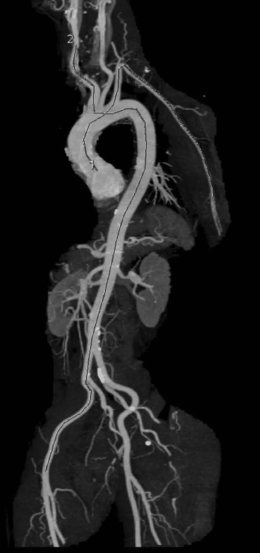
  • The pressure wave travels mostly along an aortic segment, including the thoracic descending aorta and the abdominal aorta, and ultimately travels along the iliac and common femoral arteries. This is well exemplified by the Figure, which superimposes the trajectory of the pressure pulse wave on a normal angiogram obtained by magnetic resonance imaging.

VIEW FIGURE

The trajectories of the pressure pulse waves along the arterial segments are superimposed onto an angiogram obtained by computed tomography scan (left anterior oblique). The carotid-femoral pathway is described as dotted line, and the QKD pathway is described as dashed line.

pap62

FIGURE SOURCE:

http://hyper.ahajournals.org/content/early/2013/05/20/HYPERTENSIONAHA.113.01372.citation

The method developed by Gosse et al 7,8 measures the time delay between the onset of the QRS on the ECG and the detection of the last Korotkoff sound by the microphone placed on the brachial artery. Thus, the pressure pulse wave travels first along the ascending aorta and the aortic arch (ie, a short pathway of elastic arteries) and then along the subclavian and brachial arteries (ie, a much longer pathway of muscular arteries).

Because the stiffness of muscular arteries is little influenced by age and hypertension, Gosse et al8 attributed the difference in QKD duration to ascending aorta and aortic arch. However, a closer look at the Figure shows that the length of the ascending and aortic arch pathway represents a very small part of the total pathway and casts doubt about this statement.

Furthermore, in magnetic resonance imaging studies, the transit time of flow wave along the aortic arch (average 120 mm length) is often found ≈35 ms in young healthy subjects,10 a value which is far from the mean 206 ms QKD duration found in the present study. Thus, part of that QFD duration has to be further explained by both the preejection period and the transit time within muscular arteries.

Alternative Devices

  • 2008 – The arteriograph system estimates PWV from a single-site determination of the suprasystolic waveform at the brachial artery site, and measures the time elapsed between the first wave ejected from the left ventricle to the aortic root, and its reflection from the bifurcation as the second systolic wave, with subtraction of the brachial artery transit time.
  • 2010 – The Mobil-O-Graph system uses oscillometric recording of brachial artery pressure waveform and reconstructs the central pulse wave by applying a transfer function. Central pulse wave is then decomposed into forward and backward waves, and PWV isestimated from their time difference.
  • Device |Method |Arterial Pathway |Predictive Value for CV Events | (Year of First Publication)

1984 Complior Mechanotransducer Carotid-femoral Yes (1999)

1990 Sphygmocor Tonometer Carotid-femoral Yes (2011)

1994 QKD ECG + Korotkoff sounds Aorta + brachial Yes (2005)

1997 Cardiov. Eng. Inc Tonometer Carotid-femoral Yes (2010)

2002 Doppler probes Doppler probe Aortic arch + descending aorta Yes (2002)

2002 VP-1000 Omron Brachial and ankle pressure cuffs Aorta + brachial + lower limbs Yes (2005)

2004 PulsePen Tonometer Carotid-femoral No

2006 CAVI-VaSera ECG + Brachial and ankle pressure cuffs Aorta + brachial + lower limbs No

2008 Arteriograph Arm pressure cuff Aorta + brachial No

2009 MRI-ArtFun MRI Aortic arch No

2009 Vicorder Cuffs Carotid-femoral No

2010 Mobil-O-Graph Arm pressure cuff Aorta No

Conclusions

The measurement of arterial stiffness is increasingly popular among physicians and researchers mainly because its predictive value for cardiovascular (CV) events has been well demonstrated. The largest amount of evidence has been given for aortic stiffness, measured through carotid-femoral pulse wave velocity (cfPWV). This has been initially reported in the late 1990s or early 2000s.1

Aortic stiffness is an independent predictor of all-cause and cardiovascular mortality in hypertensive patientsHypertension. 2001;37:1236–1241.

European Network for Non-invasive Investigation of Large Arteries. Expert consensus document on arterial stiffness: methodological issues and clinical applicationsEur Heart J. 2006;27:2588–2605.

Arterial Stiffness as an Imaging Biomarker : Are All Pathways Equal? http://hyper.ahajournals.org/content/early/2013/05/20/HYPERTENSIONAHA.113.01372.citation

References for Imaging Biomarker for Arterial Stiffness, at the end of the paper

Part II:

Results for Advances and Recent Clinical Trials in Hypertension Management

Based on

Garry L.R. Jennings, Recent Advances in Hypertension:Recent Clinical Trials of Hypertension Management http://hyper.ahajournals.org/content/early/2013/05/20/HYPERTENSIONAHA.113.00863.citation

Trends: tended to drive interest toward equivalence rather than efficacy studies (ie, trials designed to show an investigational agent is as good as, not better than, existing treatment), surrogate end points, including new blood pressure (BP) variables, and studies of combinations and algorithms rather than single interventions. Population studies around the world, however, continue to show that large numbers of people have hypertension that is not treated satisfactorily and are not achieving the goals set by the major national guidelines. These guidelines themselves are under continual scrutiny on the basis of recent data casting doubt on the validity of present BP goals. Guideline committees also face the issue that evidence based on expensive large-scale clinical trials is more often funded by the pharmaceutical or device industries than by government, leaving large evidence gaps in areas of public importance but no direct interest to industry funders. The purpose of the present article is to briefly review clinical trials of interventions in hypertension during the past 2 years.

Subject categories of Last Decade Clinical Trials on Hypertension

  • Resistant Hypertension
  • Resistant Hypertension and the Sympathetic
  • Nervous System
  • Trials of Pharmacotherapy
  • Old Ground, New Findings
  • Are Chlorthalidone and Nonthiazides the Best Diuretics for Treatment of Hypertension?
  • BP Targets and Treatment
  • Lifestyle and Nonpharmacological Approaches to Hypertension
  1.  Sodium
  2. Trials of Nutrition and BP
  • Resistance Exercise and BP

What Can Be Learned From Clinical Trials Reported in the Present Decade?

  • Systems for blood pressure management in the community can be improved because a large treatment gap remains.
  • Drug combinations from different classes with different modes of action are useful.
  • Drug combinations that include drugs with similar mode of action do not generally enhance efficacy and come at a cost in adverse events.
  • Small but important nutritional effects on blood pressure demand further examination.
  • The sympathetic nervous system has returned as an important target for therapy of hypertension.
  • Blood pressure targets and goals need refining, preferably on the basis of specifically designed clinical trials.

The scene for clinical trials of hypertension management is in transition. The era of mega trials may not be over but is certainly in decline, and in the past 2 years there have been no studies reporting primary outcome data the scale of the

  • Antihypertensive and
  • Lipid-Lowering Treatment
  1. Lipid-Lowering Treatment to Prevent Heart Attack Trial (ALLHAT),
  2. The ONgoing Telmisartan Alone and in combination with Ramipril Global Endpoint Trial (ONTARGET),
  3. Anglo-Scandinavian Cardiac Outcomes Trial (ASCOT), and other
  4. major studies that marked clinical trial activity and informed guideline committees during the past 2 to 3 decades.

This reflects in part the view that the

  • present benchmark pharmacological agents for treating hypertension are difficult to improve,
  • some systemic issues affecting the pharmaceutical industry influencing the ability to make the large investment required to perform mega trials and
  • the quality of the antihypertensive drug pipeline.

http://hyper.ahajournals.org/content/early/2013/05/20/HYPERTENSIONAHA.113.00863.citation

References for Clinical Trial on Hypertension, at the end of the paper

Part III:

Mechanism of Action of Top 10 Cardio Drugs 2012, published on May 16, 2013

The top 10 Cardio Drugs in 2012 belong to two drug classes

  • Antihypertensive and
  • Lipid-Lowering Treatment

Sales % Change 2012 vs 2011 by Drug Class

MOA

By

Drug Class

Drug Name

2011 Sales billion

2012 Sales billion

% change

Statins

Crestor

6.622

6.253

-6%

Lipitor

9.577

3.948

-59%

Zetia

2.428

2.567

+6%

Vytorin

1.882

1.747

-7%

Total Sales and % change Statins

 20,509  14,515  -29.2%

ARB

Diovan

5.665

4.417

-22%

ACEII

Benicar

2.602

2.446

-6%

ACEI

Micardis

2.217

2.098

-5%

ARB

Avapro

1.797

1.422

-30% (BMS)

ARB

Blopress

1.808

1.643

-9%

PAH

Tracleer

1.721

1.6

-7%

Total Sales and % change AntiHTN

 15,810  13,626  -13.8%

Data Source:

http://www.fiercepharma.com/special-reports/top-10-cardio-drugs-2012#ixzz2U9Axh8X4 

1 Crestor

Crestor (AstraZeneca)
Patent expiry: July 2016

2012 sales: $6.253 billion
2011 sales: $6.622 billion
Change: (6%)

Crestor – FiercePharma http://www.fiercepharma.com/special-reports/crestor-0#ixzz2UACLZyaa 

(rosuvastatin calcium) is indicated as an adjunct to diet to reduce elevated Total-C, LDL-C, ApoB, non-HDL-C, and triglycerides, and to increase HDL-C in adult patients with primary hyperlipidemia or mixed dyslipidemia and to slow the progression of atherosclerosis in adult patients as part of a treatment strategy to lower Total-C and LDL-C to target levels.1

Diovan

Diovan (Novartis)
Patent expiry: September 2012

2012 sales: $4.417 billion
2011 sales: $5.665 billion
Change: (22%)

Diovan – FiercePharma http://www.fiercepharma.com/special-reports/diovan#ixzz2UACdBCtZ 

Valsartan (Angiotan or Diovan) is an angiotensin II receptor antagonist (more commonly called an “ARB”, or angiotensin receptor blocker), with particularly high affinity for the type I (AT1) angiotensin receptor. By blocking the action of angiotensin, valsartan dilates blood vessels and reduces blood pressure.[1] In the U.S., valsartan is indicated for treatment of high blood pressurecongestive heart failure (CHF), or post-myocardial infarction (MI).[2]

3 Lipitor

Lipitor (Pfizer)
Patent expiry: November 2011

2012 sales: $3.948 billion
2011 sales: $9.577 billion
Change: (59%)

Lipitor – FiercePharma http://www.fiercepharma.com/special-reports/lipitor-2#ixzz2UACsJ2Y2 

(atorvastatin calcium) tablets are a prescription medicine that is used along with a low-fat diet. It lowers the LDL (“bad”) cholesterol and triglycerides in your blood. It can raise your HDL (“good”) cholesterol as well. LIPITOR can lower the risk for heart attack, stroke, certain types of heart surgery, and chest pain in patients who have heart disease or risk factors for heart disease such as age, smoking, high blood pressure, low HDL, or family history of early heart disease. LIPITOR can lower the risk for heart attack or stroke in patients with diabetes and risk factors such as diabetic eye or kidney problems, smoking, or high blood pressure.

LIPITOR is a member of the drug class known as statins, used for lowering blood cholesterol. It also stabilizes plaque and prevents strokes through anti-inflammatory and other mechanisms. Like all statins, atorvastatin works by inhibiting HMG-CoA reductase, an enzyme found in liver tissue that plays a key role in production of cholesterol in the body.

Atorvastatin was first synthesized in 1985 by Bruce Roth of Parke-Davis Warner-Lambert Company (now Pfizer). The best selling drug in pharmaceutical history, sales of Lipitor since it was approved in 1996 exceed US$125 billion, and the drug has topped the list of best-selling branded pharmaceuticals in the world for nearly a decade

4 Zetia

Zetia (Merck)
Patent expiry: December 2016

2012 sales: $2.567 billion
2011 sales: $2.428 billion
Change: 6%

Zetia – FiercePharma http://www.fiercepharma.com/special-reports/zetia#ixzz2UADFaGJ0 

Ezetimibe (pron.: /ɛˈzɛtɨmɪb/) is a drug that lowers plasma cholesterol levels. It acts by decreasing cholesterol absorption in the intestine. It may be used alone (marketed as Zetia or Ezetrol), when other cholesterol-lowering medications are not tolerated, or together withstatins (e.g., ezetimibe/simvastatin, marketed as Vytorin and Inegy) when statins alone do not control cholesterol.

Ezetimibe decreases cholesterol levels, but has not been shown to improve outcomes in cardiovascular disease patients by decreasing atherosclerotic or vascular events compared to placebo. Ezetimibe is endorsed in the Canadian Lipid Guidelines and is considered a well-tolerated option for an add-on agent to statin, to help patients achieve their LDL (or bad cholesterol) targets. [1] Ezetimibe is the only add-on to statin therapy that has successfully shown cardiovascular benefit when combined with statin, but has not been proven to have an incremental benefit compared to statins alone. [2] Britain’s NICE statement, published in 2007, endorses its use for monotherapy if statins are not tolerated or as add-on therapy.[3]

5 Benicar

Benicar (Daiichi Sankyo)
Patent expiry: October 2016

2012 sales: $2.446 billion
2011 sales: $2.602 billion
Change: (6%)

Benicar – FiercePharma http://www.fiercepharma.com/special-reports/benicar#ixzz2UADYvld5 

BENICAR and BENICAR HCT are prescription medicines used to lower high blood pressure (hypertension). They may be used alone or with other medicines used to treat high blood pressure. BENICAR HCT is not for use as the first medicine to treat high blood pressure.

 Olmesartan medoxomil is an angiotensin II receptor antagonistused to treat high blood pressure.

Olmesartan is a prodrug that works by blocking the binding of angiotensin II to the AT1 receptors in vascular muscle; it is therefore independent of angiotensin II synthesis pathways, unlike ACE inhibitors. By blocking the binding rather than the synthesis of angiotensin II, olmesartan inhibits the negative regulatory feedback on renin secretion. As a result of this blockage, olmesartan reduces vasoconstriction and the secretion of aldosterone. This lowers blood pressure by producing vasodilation, and decreasing peripheral resistance.

6 Micardis

Micardis (Boehringer Ingelheim)
Patent Expiry: January 2014

2012 Sales: $2.098 billion
2011 Sales: $2.217 billion
Change: (5%)

Micardis – FiercePharma http://www.fiercepharma.com/special-reports/micardis#ixzz2UADpDZeO 

Micardis® (telmisartan) tablets are a prescription medicine used to treat high blood pressure (hypertension). Additionally, MICARDIS 80 mg tablets are used in certain high-risk people aged 55 years and older who are unable to take a medicine called an angiotensin converting enzyme inhibitor (ACE-I) to help lower their risk of having certain cardiovascular problems such as stroke, heart attack, or death.

Micardis® (telmisartan) tablets are a prescription medicine used to treat high blood pressure (hypertension).

Telmisartan (INN) (pron.: /tɛlmɪˈsɑrtən/) is an angiotensin II receptor antagonist (angiotensin receptor blocker, ARB) used in the management of hypertension. It is marketed under thetrade name Micardis (by Boehringer Ingelheim), among others.

Telmisartan is an angiotensin II receptor blocker that shows high affinity for the angiotensin II receptor type 1 (AT1), with a binding affinity 3000 times greater for AT1 than AT2. It has the longest half-life of any ARB (24 hours)[1][4] and the largest volume of distribution.

In addition to blocking the RAs, telmisartan acts as a selective modulator of peroxisome proliferator-activated receptor gamma (PPAR-γ), a central regulator of insulin and glucose metabolism. It is believed that telmisartan’s dual mode of action may provide protective benefits against the vascular and renal damage caused by diabetes and cardiovascular disease (CVD).[4]

Telmisartan’s activity at the PPAR-γ receptor has prompted speculation around its potential as a sport doping agent as an alternative to GW 501516.[5] Telmisartan activates PPARδ receptors in several tissues. [6][7][8][9]

7 Avapro

Avapro (Sanofi)
Patent expiry: March 2012

Total 2012 sales: $1.925 billion
2012 sales Sanofi: $1.422 billion
2012 sales BMS: $503 million

Total 2011 sales: $2.749 billion
2011 sales Sanofi: $1.797 billion
2011 sales BMS: $952 million
Total Change: (30%)

Avapro – FiercePharma http://www.fiercepharma.com/special-reports/avapro#ixzz2UAE9iB2E 

rbesartan (INN) (pron.: /ɜrbəˈsɑrtən/) is an angiotensin II receptor antagonist used mainly for the treatment of hypertension. Irbesartan was developed by Sanofi Research (now part ofsanofi-aventis). It is jointly marketed by sanofi-aventis and Bristol-Myers Squibb under the trade names AprovelKarvea, and Avapro.

As with all angiotensin II receptor antagonists, irbesartan is indicated for the treatment ofhypertension. Irbesartan may also delay progression of diabetic nephropathy and is also indicated for the reduction of renal disease progression in patients with type 2 diabetes,[1]hypertension and microalbuminuria (>30 mg/24 hours) or proteinuria (>900 mg/24 hours).[2]

 A large randomized trial following 4100+ men and women with heart failure and normal ejection fraction (>=45%) over 4+ years found no improvement in study outcomes or survival with irbesartan as compared to placebo.[3]

8 Vytorin

Vytorin (Merck)
Patent Expiry: April 2017

2012 sales: $1.747 billion
2011 sales: $1.882 billion
Change: (7%)

Vytorin – FiercePharma http://www.fiercepharma.com/special-reports/vytorin#ixzz2UAEQVcQr 

Ezetimibe/simvastatin (pron.: /ɛˈzɛtɨmɪb ˌsɪmvəˈstætɨn/) is a drug combination used for the treatment of dyslipidemia. It is a combination of ezetimibe (best known as Zetia in the United States and Ezetrol elsewhere) and the statin drug simvastatin (best known as Zocor in the U.S.). The combination preparation is marketed by Merck & Co./Schering-PloughPharmaceuticals (joint venture) under the trade names Vytorin and Inegy.

Ezetimibe reduces blood cholesterol by inhibiting absorption of cholesterol by the small intestine by acting at the brush border of the small intestine and inhibits the absorption of cholesterol, leading to a decrease in the delivery of intestinal cholesterol to the liver.

Simvastatin is an HMG-CoA reductase inhibitor or statin. It works by blocking an enzymethat is necessary for the body to make cholesterol.

Even though ezetimibe decreases cholesterol levels, as of 2009 it has not been found to lead to improvement in real world outcomes.[1] The combination of simvastatin and ezetimibe has not been found to be any better than simvastatin alone. A panel of experts thus concluded in 2008 that it should “only be used as a last resort”.[2]

9 Blopress

Blopress (Takeda Pharmaceutical)
Patent expiry: June 2012

2012 sales: $1.643 billion
2011 sales: $1.808 billion
Change: (9%)

Blopress – FiercePharma http://www.fiercepharma.com/special-reports/blopress#ixzz2UAEnxyWy

Candesartan (rINN) (pron.: /ˌkændɨˈsɑrtən/) is an angiotensin II receptor antagonist used mainly for the treatment of hypertension. The prodrug candesartan cilexetil is marketed by AstraZeneca and Takeda Pharmaceuticals, commonly under the trade names Blopress,AtacandAmias, and Ratacand

As all angiotensin II receptor antagonists, candesartan is indicated for the treatment of hypertension. Results from the CHARM study in the early 2000s demonstrated the morbidity and mortality reduction benefits of candesartan therapy in congestive heart failure.[1] Thus, while ACE inhibitors are still considered first-line therapy in heart failure, candesartan can be used in combination with an ACE to achieve improved mortality and morbidity vs. an ACE alone and additionally is an alternative in patients intolerant of ACE inhibitor therapy.

Prehypertension

In a four-year randomized controlled trial, candesartan was compared to placebo to see whether it could prevent or postpone the development of full-blown hypertension in people with so-called prehypertension. During the first two years of the trial, half of participants were given candesartan, and the others received placebo; candesartan reduced the risk of developing hypertension by nearly two-thirds during this period. In the last two years of the study, all participants were switched to placebo. By the end of the study, candesartan hadsignificantly reduced the risk of hypertension, by more than 15%. Serious side effects were actually more common among participants receiving placebo than in those given candesartan.[2]

Candesartan is also available in a combination formulation with a low dose thiazide diuretic, invariably hydrochlorothiazide, to achieve an additive antihypertensive effect. Candesartan/hydrochlorothiazide combination preparations are marketed under various trade names including Atacand HCTHytacandBlopress Plus, Advantec and Ratacand Plus.

10 Tracleer

Tracleer (Actelion)
Patent expiry: November 2015   

2012 sales: $1.600 billion
2011 sales: $1.721 billion
Change: (7%)

Tracleer – FiercePharma http://www.fiercepharma.com/special-reports/tracleer#ixzz2UAF2iIJB 

Bosentan is a dual endothelin receptor antagonist used in the treatment of pulmonary artery hypertension (PAH). It is licensed in the United States, the European Union and other countries by Actelion Pharmaceuticals for the management of PAH under the trade name Tracleer.

Bosentan is a competitive antagonist of endothelin-1 at the endothelin-A (ET-A) and endothelin-B (ET-B) receptors. Under normal conditions, endothelin-1 binding of ET-A or ET-B receptors causes pulmonary vasoconstriction. By blocking this interaction, bosentan decreases pulmonary vascular resistance. Bosentan has a slightly higher affinity for ET-A than ET-B.

Clinical uses 

Bosentan is indicated mainly for the treatment of pulmonary hypertension. In 2007, bosentan was approved in the European Union also for reducing the number of new digital ulcers in patients with systemic sclerosis and ongoing digital ulcer disease.

In the United States, bosentan is indicated for the treatment of pulmonary arterial hypertension (WHO Group I) in patients with WHO Class II-IV symptoms, to improve exercise capacity and decrease the rate of clinical worsening.[1]

http://www.fiercepharma.com/special-reports/top-10-cardio-drugs-2012

For years, cardio was king. The world’s all-time best-selling drug, Pfizer’s ($PFELipitor, after all, is an antihyperlipidemic drug. Cardio drugs have traditionally made up one of the largest categories of therapeutic treatment in the drug universe.

According to EvaluatePharma‘s World Preview 2018 report, combined sales of antihypertensive drugs and antihyperlipidemics were more than $70 billion in 2011. That would put them at the top of the heap. Sales of antihypertensive drugs alone were more than $40 billion that year, making them the second-largest therapy area defined by the report, behind oncology drugs at $64.4 billion. The list, compiled by EvaluatePharma, includes the theraputic areas categorized as cardio, so it does not include some products sometimes used for heart disease but not in that therapeutic area, including blood thinners like Plavix.

But many of the top cardio drugs are long in the tooth, and generics are now eating their lunch. Did I mention Lipitor? Sales cratered last year, falling nearly 60%. Despite that, the drug placed third among the top 10 cardio drugs of 2012, a reminder of the stature it had achieved. Four of the top 10 have lost patent protection in the last two years, and most will be off patent by 2016, with only Merck’s ($MRKVytorin protected until 2017.

Last year, the top 10 cardio drugs racked up sales of $28.644 billion, down 23% from the $37.271 billion they sold in 2011. Still, the group has made a lot of money for its companies for years and, in some cases, completely changed the treatment of heart disease.

It is an interesting list. Only Merck has two drugs in the top 10. The other drugmakers make up a broad swath of the pharma industry. Read our report below, and if you have some insights you would like to share, please do.

Top 10 Cardio Drugs 2012 – FiercePharma http://www.fiercepharma.com/special-reports/top-10-cardio-drugs-2012#ixzz2UAByWR7s 

Part IV:

20 Highest-Paid Biopharma CEOs of 2012

Call it a rite of spring. Every year about this timeFiercePharma takes a look at executive compensation in the industry, and we rank the highest-paid CEOs. If you’re a regular reader, you’ll notice that this year’s list is longer than previous editions. And there’s a reason for that: curiosity.

As we were beginning to gather numbers from biopharma companies’ proxy statements and annual reports, news surfaced that Valeant Pharmaceuticals ($VRX) and Actavis ($ACT) had been in merger talks. The former CEO of Mylan ($MYL), one of Actavis’ rivals, regularly appeared on our highest-paid executives list, so we looked up the numbers on Actavis. No dice; CEO Paul Bisaro may have pulled off his biggest merger ever last year, but $8.66 million in compensation still didn’t qualify him for our ranking.

Then, we pulled out Valeant’s proxy statement. And while CEO Michael Pearson didn’t earn enough in 2012 to make the cutoff–his compensation just surpassed $6 million–he should have been at the top of the list last year. Pearson’s 2011 pay package broke $36 million. He collected more than $18 million in stock and option awards, plus a special $13.7 million dividend payment, stemming from agreements negotiated years before.

We hate to miss a scoop. Naturally. So, we vowed to avoid making the same mistake this time around. Rather than limit our executive-pay search to the biggest pharma companies and biotechs, plus the usual suspects who often make CEO-pay rankings, we used a bigger net. We collected compensation information from 50 companies, including numbers for CEOs, CFOs, R&D chiefs and other top executives.

Partly because of this search, but mostly because of big bonuses and awards at fast-growing Regeneron ($REGN), we have a brand-new No. 1 on our list. That’s Regeneron CEO Leonard Schleifer, whose 2012 compensation totaled $30.047 million. You’ll notice some other newbies, such as Leonard Bell from Alexion ($ALXN), whose pay bump put him in 12th place. And then there are familiar faces, such as Pfizer ($PFE) CEO Ian Read; Johnson & Johnson’s ($JNJ) former chairman and CEO, William Weldon; and Eli Lilly ($LLY) CEO John Lechleiter, who hung on in 10th place.

Many of the companies we researched pay their top people far less than the $10 million that served as our cutoff figure. Novo Nordisk ($NVO) CEO Lars Sorensen, who has presided over double-digit growth there for several years, collected a package of cash and stock awards worth about $5 million for 2012. GlaxoSmithKline ($GSK) CEO Andrew Witty made less than $6 million himself; he took a pay cut for the year because of Glaxo’s shortfall on certain performance targets.

And then there are others who would have made the list, had their titles been different. There’s Regeneron R&D chief George Yancopoulos, whose extraordinary $81 million in compensation shows how much the company appreciates its newly minted blockbuster, Eylea. There’s Mylan Chairman Robert Coury, who used to be a fixture on our list until Heather Bresch took over as CEO; he made more than $28 million last year. Novartis’ ($NVS) former chairman Daniel Vasella could have qualified for 12th place with his $13.98 million in compensation.

Vasella, then, gives us a quick segue to the ongoing debate over executive pay. In Switzerland, populist dismay at some high-profile compensation figures led to a public vote earlier this year. Citizens voted in new restrictions on common bonuses, such as golden parachutes, and gave shareholders a binding vote on executive pay. And local analysts figure that late-breaking news of Vasella’s behind-the-scenes noncompete agreement–worth some $78 million over 5 years–helped pay activists to get out the vote. (Vasella ended up refusing the deal, by the way.)

In the U.S., where executives are paid more than anywhere else in the world, shareholders at some companies have successfully lobbied for a greater emphasis on performance pay and against extraordinary bonuses, such as change-in-control payments that send top executives on their way with tens of millions after a merger. Other companies have instituted “say-on-pay” advisory votes for shareholders, but those often end up as rubber stamps for the status quo.

Now, we’re interested in what you have to say about executive compensation. Are the CEOs on this list worth their price? What’s a supersuccessful new drug worth? Should CEO pay be docked for R&D failures? What about failed launches? Should other, lower-paid executives earn more? Tweet your opinions to @FiercePharma using the hashtag #FPexecpay, leave your comments below or email us. We’ll collect your thoughts in a future article.

As always, feel free to send us your thoughts on our coverage. And if we missed a well-paid CEO, be sure to let us know.

— Tracy Staton (email | Twitter)

For more:
Top 10 Biotech CEO Pay Packages of 2012
Top 10 Pharma CEO salaries of 2010
Top 10 Pharma CEO salaries of 2009
2012’s 10 highest-paid Med Tech CEOs
Top 10 Medical Device Industry CEO Salaries for 2011


20 Highest-Paid Biopharma CEOs of 2012 – FiercePharma http://www.fiercepharma.com/special-reports/20-highest-paid-biopharma-ceos-2012#ixzz2UAGAlHay 

REFERENCES FOR Part I: Arterial Stiffness

1. Laurent S, Boutouyrie P, Asmar R, Gautier I, Laloux B, Guize L,

Ducimetiere P, Benetos A. Aortic stiffness is an independent predictor

of all-cause and cardiovascular mortality in hypertensive patients.

Hypertension. 2001;37:1236–1241.

2. Vlachopoulos C, Aznaouridis K, Stefanadis C. Prediction of cardiovascular

events and all-cause mortality with arterial stiffness: a systematic

review and meta-analysis. J Am Coll Cardiol. 2010;55:1318–1327.

3. Boutouyrie P, Tropeano AI, Asmar R, Gautier I, Benetos A, Lacolley P,

Laurent S. Aortic stiffness is an independent predictor of primary coronary

events in hypertensive patients: a longitudinal study. Hypertension.

2002;39:10–15.

4. Mattace-Raso FU, van der Cammen TJ, Hofman A, van Popele NM, Bos

ML, Schalekamp MA, Asmar R, Reneman RS, Hoeks AP, Breteler MM,

Witteman JC. Arterial stiffness and risk of coronary heart disease and

stroke: the Rotterdam Study. Circulation. 2006;113:657–663.

5. Sehestedt T, Jeppesen J, Hansen TW, Rasmussen S, Wachtell K, Ibsen H,

Torp-Pedersen C, Olsen MH. Risk stratification with the risk chart from

the European Society of Hypertension compared with SCORE in the general

population. J Hypertens. 2009;27:2351–2357.

6. Mitchell GF, Hwang SJ, Vasan RS, Larson MG, Pencina MJ,

Hamburg NM, Vita JA, Levy D, Benjamin EJ. Arterial stiffness and

cardiovascular events: the Framingham Heart Study. Circulation. 2010;

121:505–511.

7. Gosse P, Cremer A, Papaioannou G, Yeim S. Arterial stiffness from monitoring

of timing of Korotkoff sounds predicts the occurrence of cardiovascular

events independently of left ventricular mass in hypertensive

patients. HYPERTENSIONAHA.113.01039 Published online before print May 20, 2013,doi: 10.1161/​HYPERTENSIONAHA.113.01039

Hypertension. 2013;62:XX–XX.

http://hyper.ahajournals.org/content/early/2013/05/20/HYPERTENSIONAHA.113.01039.abstract.html?papetoc

8. Gosse P, Guillo P, Ascher G, Clementy J. Assessment of arterial distensibility

by monitoring the timing of Korotkoff sounds. Am J Hypertens.

1994;7:228–233.

9. Laurent S, Cockcroft J, Van Bortel L, Boutouyrie P, Giannattasio C,

Hayoz D, Pannier B, Vlachopoulos C, Wilkinson I, Struijker-Boudier

H; European Network for Non-invasive Investigation of Large Arteries.

Expert consensus document on arterial stiffness: methodological issues

and clinical applications. Eur Heart J. 2006;27:2588–2605.

10. Dogui A, Redheuil A, Lefort M, DeCesare A, Kachenoura N, Herment A,

Mousseaux E. Measurement of aortic arch pulse wave velocity in cardiovascular

MR: comparison of transit time estimators and description of a

new approach. J Magn Reson Imaging. 2011;33:1321–1329.

downloaded from

http://hyper.ahajournals.org/content/early/2013/05/20/HYPERTENSIONAHA.113.01372.citation

REFERENCES for Part II: Clinical Trials of Hypertension Management

1. Hanselin MR , Saseen JJ , Allen RR , Marrs JC , Nair KV . Description of

antihypertensive use in patients with resistant hypertension prescribed

four or more agents. Hypertension . 2011 ; 58 : 1008 – 1013 .

2. Bakris GL , Lindholm LH , Black HR , Krum H , Linas S , Linseman JV ,

Arterburn S , Sager P , Weber M . Divergent results using clinic and ambulatory

blood pressures: report of a darusentan-resistant hypertension trial.

Hypertension . 2010 ; 56 : 824 – 830 .

3. Smithwick RH , Thompson JE . Splanchnicectomy for essential hypertension;

results in 1,266 cases. J Am Med Assoc . 1953 ; 152 : 1501 – 1504 .

4. Esler M , Ferrier C , Lambert G , Eisenhofer G , Cox H , Jennings G .

Biochemical evidence of sympathetic hyperactivity in human hypertension.

Hypertension . 1991 ; 17 ( 4 Suppl ): III29 – III35 .

5. Esler MD , Krum H , Sobotka PA , Schlaich MP , Schmieder RE , Bohm

M . Symplicity HTN-2 investigators. Renal sympathetic denervation in

patients with treatment-resistant hypertension: a randomized controlled

trial. Lancet 2010 ; 376 : 1903 – 1909 .

6. Brinkmann J , Heusser K , Schmidt BM , Menne J , Klein G , Bauersachs J ,

Haller H , Sweep FC , Diedrich A , Jordan J , Tank J . Catheter-based renal

nerve ablation and centrally generated sympathetic activity in diffi cultto-

control hypertensive patients: prospective case series. Hypertension .

2012 ; 60 : 1485 – 1490 .

7. Mahfoud F , Cremers B , Janker J , et al . Renal hemodynamics and renal

function after catheter-based renal sympathetic denervation in patients

with resistant hypertension. Hypertension . 2012 ; 60 : 419 – 424 .

8. Lambert GW , Hering D , Esler MD , Marusic P , Lambert EA , Tanamas

SK , Shaw J , Krum H , Dixon JB , Barton DA , Schlaich MP . Health-related

quality of life after renal denervation in patients with treatment-resistant

hypertension. Hypertension . 2012 ; 60 : 1479 – 1484 .

9. Daugherty SL , Powers JD , Magid DJ , Masoudi FA , Margolis KL ,

O ’ Connor PJ , Schmittdiel JA , Ho PM . The association between medication

adherence and treatment intensifi cation with blood pressure control

in resistant hypertension. Hypertension . 2012 ; 60 : 303 – 309 .

10. Bobrie G , Frank M , Azizi M , Peyrard S , Boutouyrie P , Chatellier G ,

Laurent S , Menard J , Plouin PF . Sequential nephron blockade versus

sequential renin-angiotensin system blockade in resistant hypertension:

a prospective, randomized, open blinded endpoint study. J Hypertens .

2012 ; 30 : 1656 – 1664 .

11. Mancia G . Additional drug treatment in resistant hypertension: need for

randomized studies. J Hypertens . 2012 ; 30 : 1514 – 1515 .

12. Hermida RC , Ayala DE , Moj ó n A , Fontao MJ , Fern á ndez JR .

Chronotherapy with valsartan/hydrochlorothiazide combination in essential

hypertension: improved sleep-time blood pressure control with bedtime

dosing. Chronobiol Int . 2011 ; 28 : 601 – 610 .

13. Persu A , Renkin J , Thijs L , Staessen JA . Renal denervation: ultima

ratio or standard in treatment-resistant hypertension. Hypertension .

2012 ; 60 : 596 – 606 .

14. Lohmeier TE , Iliescu R . Chronic lowering of blood pressure by carotid

barorefl ex activation: mechanisms and potential for hypertension therapy.

Hypertension . 2011 ; 57 : 880 – 886 .

15. Bisognano JD , Bakris G , Nadim MK , Sanchez L , Kroon AA , Schafer J ,

de Leeuw PW , Sica DA . Barorefl ex activation therapy lowers blood pressure

in patients with resistant hypertension: results from the double-blind,

randomized, placebo-controlled rheos pivotal trial. J Am Coll Cardiol .

2011 ; 58 : 765 – 773 .

16. Parving HH , Brenner BM , McMurray JJ , de Zeeuw D , Haffner SM ,

Solomon SD , Chaturvedi N , Persson F , Desai AS , Nicolaides M , Richard

A , Xiang Z , Brunel P , Pfeffer MA ; ALTITUDE Investigators . Cardiorenal

end points in a trial of aliskiren for type 2 diabetes. N Engl J Med .

2012 ; 367 : 2204 – 2213 .

17. The ONTARGET Investigators . Telmisartan, ramipril, or both in patients

at high risk for vascular events. N Engl J Med . 2008 ; 358 : 1547 – 1559 .

18. Mancia G , Parati G , Bilo G , et al . Ambulatory blood pressure values in

the Ongoing Telmisartan Alone and in Combination with Ramipril Global

Endpoint Trial (ONTARGET). Hypertension . 2012 ; 60 : 1400 – 1406 .

19. Roush GC , Holford TR , Guddati AK . Chlorthalidone compared with

hydrochlorothiazide in reducing cardiovascular events: systematic review

and network meta-analyses. Hypertension . 2012 ; 59 : 1110 – 1117 .

20. Cushman WC , Bakris GL , White WB , Weber MA , Sica D , Roberts A ,

Lloyd E , Kupfer S . Azilsartan medoxomil plus chlorthalidone reduces

blood pressure more effectively than olmesartan plus hydrochlorothiazide

in stage 2 systolic hypertension. Hypertension . 2012 ; 60 : 310 – 318 .

21. Alderman MH , Piller LB , Ford CE , Probstfi eld JL , Oparil S , Cushman WC ,

Einhorn PT , Franklin SS , Papademetriou V , Ong ST , Eckfeldt JH , Furberg

CD , Calhoun DA , Davis BR ; Antihypertensive and Lipid-Lowering

Treatment to Prevent Heart Attack Trial Collaborative Research Group .

Clinical signifi cance of incident hypokalemia and hyperkalemia in treated

hypertensive patients in the antihypertensive and lipid-lowering treatment

to prevent heart attack trial. Hypertension . 2012 ; 59 : 926 – 933 .

22. Kostis WJ , Thijs L , Richart T , Kostis JB , Staessen JA . Persistence of mortality

reduction after the end of randomized therapy in clinical trials of

blood pressure-lowering medications. Hypertension . 2010 ; 56 : 1060 – 1068 .

23. van Onzenoort HA , Menger FE , Neef C , Verberk WJ , Kroon AA , de

Leeuw PW , van der Kuy PH . Participation in a clinical trial enhances

adherence and persistence to treatment: a retrospective cohort study.

Hypertension . 2011 ; 58 : 573 – 578 .

24. Sever PS . The Anglo-Scandinavian Cardiac Outcomes Trial: implications

and further outcomes. Hypertension . 2012 ; 60 : 248 – 259 .

25. Larstorp AC , Ariansen I , Gjesdal K , Olsen MH , Ibsen H , Devereux RB ,

Okin PM , Dahl ö f B , Kjeldsen SE , Wachtell K . Association of pulse pressure

with new-onset atrial fi brillation in patients with hypertension and

left ventricular hypertrophy: the Losartan Intervention For Endpoint

(LIFE) reduction in hypertension study. Hypertension . 2012 ; 60 : 347 – 353 .

26. Rossignol P , Cridlig J , Lehert P , Kessler M , Zannad F . Visit-to-visit blood

pressure variability is a strong predictor of cardiovascular events in hemodialysis:

insights from FOSIDIAL. Hypertension . 2012 ; 60 : 339 – 346 .

27. Matsui Y , O ’ Rourke MF , Hoshide S , Ishikawa J , Shimada K , Kario

K . Combined Effect of Angiotensin II Receptor Blocker and Either a

Calcium Channel Blocker or Diuretic on Day-by-Day Variability of Home

Blood Pressure: The Japan Combined Treatment With Olmesartan and a

Calcium-Channel Blocker Versus Olmesartan and Diuretics Randomized

Effi cacy Study. Hypertension . 2012 ; 59 : 1132 – 1138 .

28. Jennings GL , Sudhir K . Initial therapy of primary hypertension. Med J

Aust . 1990 ; 152 : 198 – 203 .

29. Egan BM , Bandyopadhyay D , Shaftman SR , Wagner CS , Zhao Y ,

Yu-Isenberg KS . Initial monotherapy and combination therapy and hypertension

control the fi rst year. Hypertension . 2012 ; 59 : 1124 – 1131 .

30. Bavry AA , Pepine CJ . Treatment of hypertension: lower is better, or is it?

Hypertension . 2012 ; 60 : 281 – 282 .

31. Mizuno R , Fujimoto S , Saito Y , Okamoto Y . Optimal antihypertensive

level for improvement of coronary microvascular dysfunction: the lower,

the better? Hypertension . 2012 ; 60 : 326 – 332 .

32. Roman MJ , Howard BV , Howard WJ , Mete M , Fleg JL , Lee ET ,

Devereux RB . Differential impacts of blood pressure and lipid lowering

on regression of ventricular and arterial mass: the Stop Atherosclerosis in

Native Diabetics Trial. Hypertension . 2011 ; 58 : 367 – 371 .

33. Bertoia ML , Waring ME , Gupta PS , Roberts MB , Eaton CB . Implications

of new hypertension guidelines in the United States. Hypertension .

2012 ; 60 : 639 – 644 .

34. Stewart S , Carrington MJ , Swemmer CH , et al .; VIPER-BP Study

Investigators . Effect of intensive structured care on individual blood pressure

targets in primary care: multicentre randomised controlled trial. BMJ .

2012 ; 345 : e7156 .

35. Dickinson BD , Havas S ; Council on Science and Public Health, American

Medical Association . Reducing the population burden of cardiovascular

disease by reducing sodium intake: a report of the Council on Science and

Public Health. Arch Intern Med . 2007 ; 167 : 1460 – 1468 .

36. O ’ Donnell MJ , Yusuf S , Mente A , Gao P , Mann JF , Teo K , McQueen M ,

Sleight P , Sharma AM , Dans A , Probstfi eld J , Schmieder RE . Urinary

sodium and potassium excretion and risk of cardiovascular events. JAMA .

2011 ; 306 : 2229 – 2238 .

37. Hummel SL , Seymour EM , Brook RD , Kolias TJ , Sheth SS , Rosenblum

HR , Wells JM , Weder AB . Low-sodium dietary approaches to stop

hypertension diet reduces blood pressure, arterial stiffness, and oxidative

stress in hypertensive heart failure with preserved ejection fraction.

Hypertension . 2012 ; 60 : 1200 – 1206 .

38. Soedamah-Muthu SS , Verberne LD , Ding EL , Engberink MF , Geleijnse JM .

Dairy consumption and incidence of hypertension: a dose-response metaanalysis

of prospective cohort studies. Hypertension . 2012 ; 60 : 1131 – 1137 .

39. West SG , Gebauer SK , Kay CD , Bagshaw DM , Savastano DM ,

Diefenbach C , Kris-Etherton PM . Diets containing pistachios reduce systolic

blood pressure and peripheral vascular responses to stress in adults

with dyslipidemia. Hypertension . 2012 ; 60 : 58 – 63 .

40. Zhang X , Qi Q , Liang J , Hu FB , Sacks FM , Qi L . Neuropeptide Y promoter

polymorphism modifi es effects of a weight-loss diet on 2-year

changes of blood pressure: the preventing overweight using novel dietary

strategies trial. Hypertension . 2012 ; 60 : 1169 – 1175 .

41. Meredith IT , Friberg P , Jennings GL , Dewar EM , Fazio VA , Lambert GW ,

Esler MD . Exercise training lowers resting renal but not cardiac sympathetic

activity in humans. Hypertension . 1991 ; 18 : 575 – 582 .

42. Cornelissen VA , Fagard RH , Coeckelberghs E , Vanhees L . Impact of resistance

training on blood pressure and other cardiovascular risk factors: a metaanalysis

of randomized, controlled trials. Hypertension . 2011 ; 58 : 950 – 958 .

Downloaded from

http://hyper.ahajournals.org/content/early/2013/05/20/HYPERTENSIONAHA.113.00863.citation

RELATED SOURCES:

Aortic pulse pressure is associated with the localization of coronary artery disease based on coronary flow lateralization. American journal of hypertension, 25(10), 1055-1063.

  1. Georges Khoueiry1,
  2. Basem Azab2,
  3. Estelle Torbey2,
  4. Nidal Abi Rafeh1,
  5. Jean-Paul Atallah2,
  6. Kathleen Ahern2,
  7. James Malpeso1,
  8. Donald McCord1 and
  9. Elie R. Chemaly3

Author Affiliations


  1. 1Department of Cardiology, Staten Island University Hospital, Staten Island, New York, USA

  2. 2Department of Internal Medicine, Staten Island University Hospital, Staten Island, New York, USA

  3. 3Cardiovascular Institute, Mount Sinai School of Medicine, New York, New York, USA

Elie R. Chemaly (elie.chemaly@mssm.edu)

Abstract

Background Aortic pulse pressure (APP) is related to arterial stiffness and associated with the presence and extent of coronary artery disease (CAD). Besides, the left coronary artery (LCA) has a predominantly diastolic flow while the right coronary artery (RCA) receives systolic and diastolic flow. Thus, we hypothesized that increased systolic–diastolic pressure difference had a greater atherogenic effect on the RCA than on the LCA.

Methods A random sample of 433 CAD patients (145 females, 288 males, mean age 65.0 ± 11.1 years) undergoing coronary angiography at Staten Island University Hospital between January 2005 and May 2008 was studied. Coronary lesion was defined as a ≥50% luminal stenosis. Patients were divided into three groups, with isolated LCA lesions (n = 154), isolated RCA lesions (n = 36) or mixed LCA and RCA lesions (n = 243).

Results APP differed significantly between groups, being highest when the RCA alone was affected (67.6 ± 20.3 mm Hg for LCA vs. 78.8 ± 22.0 for RCA vs. 72.7 ± 22.6 for mixed, P = 0.008 for analysis of variance (ANOVA)). Age and gender were not associated with CAD location. Heart rate was associated with CAD location, lowest in RCA group, and negatively correlated with APP. However, left ventricular ejection fraction (LVEF) was lower in the mixed CAD group and positively correlated with APP. The association between APP and right-sided CAD persisted in multivariate logistic regression adjusting for confounders, including heart rate, LVEF and medication use. A similar but less significant pattern was seen with brachial arterial pressures.

Conclusions Aortic pulse pressure may affect CAD along with coronary flow phasic patterns.

American Journal of Hypertension, advance online publication 28 June 2012; doi:10.1038/ajh.2012.87

The Relationship Between Diastolic Pressure and Coronary Collateral Circulation in Patients With Stable Angina Pectoris and Chronic Total OcclusionAm J Hypertens (2013)doi: 10.1093/ajh/hps096 

First published online: February 7, 2013

  1. Wang Shu1,
  2. Jing jing1,
  3. Liu Chang Fu1,
  4. Jiang Tie Min2,
  5. Yang Xiao Bo1,
  6. Zhou Ying1and
  7. Chen Yun Dai1,*
  1. 1 The Cardiovascular Medical Department of the General Hospital of the Chinese People’s Liberation Army, Beijing, China;

  2. 2 The Cardiovascular Medical Department of the Affiliated Hospital of the Chinese People’s Armed Police Logistics College, Tianjin, China.
  1. Correspondence: Chen Yun Dai (chenyundai2002@163.com).

Abstract

BACKGROUND The most important biomechanical source of activation of the coronary collateral circulation (CCC) is increased tangential fluid shear stress at the arterial endothelial surface. The coronary circulation is unique in that most coronary blood flow occurs in diastole. Consequently, the diastolic blood pressure (DBP) may influence the tangential fluid shear stress on the arterial endothelial surface in diastole, therebyaffecting development of the CCC.

METHODS To investigate this, we conducted a study of 222 patients with stable angina pectoris and chronic total occlusion of coronary arteries. All of the patients had no history of coronary artery interventional therapy, coronary artery bypass surgery, cardiomyopathy, or congenital heart disease. The extent of the collateral vasculature of the area perfused by the artery affected by chronic total occlusion was graded as poor or well-developed according to Rentrop’s classification.

RESULTS Univariate analysis showed a significant difference between the study subgroup with poorly developed collaterals and that with well-developed collaterals in terms of high diastolic blood pressure (DBP) and mean DBP. Multivariate analysis revealed high DBP as the only independent positive predictor of a well-developed collateral circulation.

CONCLUSIONS High DBP is positively related to a well-developed CCC. Differences in development of the CCC may be one of the pathophysiologic mechanisms responsible for the J-curve phenomenon in the relationship between DBP and cardiovascular risk.

http://ajh.oxfordjournals.org/content/early/2013/02/06/ajh.hps096.abstract

Other related articles that were published on this Open Access Online Scientific Journal, include the following:

Synthetic Biology: On Advanced Genome Interpretation for Gene Variants and Pathways: What is the Genetic Base of Atherosclerosis and Loss of Arterial Elasticity with Aging

Aviva Lev-Ari, PhD, RN May 17, 2013

http://pharmaceuticalintelligence.com/2013/05/17/synthetic-biology-on-advanced-genome-interpretation-for-gene-variants-and-pathways-what-is-the-genetic-base-of-atherosclerosis-and-loss-of-arterial-elasticity-with-aging/

Artherogenesis: Predictor of CVD – the Smaller and Denser LDL Particles

Aviva Lev-Ari, PhD, RN 11/15/2012

http://pharmaceuticalintelligence.com/2012/11/15/artherogenesis-predictor-of-cvd-the-smaller-and-denser-ldl-particles/

Cardiovascular Diseases: Causes, Risks and Management, Volume Two, Risks of Cardiovascular Diseases

Justin D. Pearlman MD ME PhD MA FACC, Editor

http://pharmaceuticalintelligence.com/biomed-e-books/cardiovascular-diseases-risks-and-management/cvd-2-risk-assessment-of-cardiovascular-diseases/

Genetics of Conduction Disease: Atrioventricular (AV) Conduction Disease (block): Gene Mutations – Transcription, Excitability, and Energy Homeostasis

Aviva Lev-Ari, PhD, RN 4/28/2013

http://pharmaceuticalintelligence.com/2013/04/28/genetics-of-conduction-disease-atrioventricular-av-conduction-disease-block-gene-mutations-transcription-excitability-and-energy-homeostasis/

Genomics & Genetics of Cardiovascular Disease Diagnoses: A Literature Survey of AHA’s Circulation Cardiovascular Genetics, 3/2010 – 3/2013

Aviva Lev-Ari, PhD, RN and Larry H. Bernstein, MD, FCAP 3/7/2013

http://pharmaceuticalintelligence.com/2013/03/07/genomics-genetics-of-cardiovascular-disease-diagnoses-a-literature-survey-of-ahas-circulation-cardiovascular-genetics-32010-32013/

Hypertriglyceridemia concurrent Hyperlipidemia: Vertical Density Gradient Ultracentrifugation a Better Test to Prevent Undertreatment of High-Risk Cardiac Patients

Aviva Lev-Ari, PhD, RN 4/4/2013

http://pharmaceuticalintelligence.com/2013/04/04/hypertriglyceridemia-concurrent-hyperlipidemia-vertical-density-gradient-ultracentrifugation-a-better-test-to-prevent-undertreatment-of-high-risk-cardiac-patients/

Hypertension and Vascular Compliance: 2013 Thought Frontier – An Arterial Elasticity Focus

Justin D. Pearlman, MD, PhD and Aviva Lev-Ari, PhD, RN 5/11/2013

http://pharmaceuticalintelligence.com/2013/05/11/arterial-elasticity-in-quest-for-a-drug-stabilizer-isolated-systolic-hypertension-caused-by-arterial-stiffening-ineffectively-treated-by-vasodilatation-antihypertensives/

Read Full Post »

The Implications of a Newly Discovered  CYP2J2 Gene Polymorphism  Associated with Coronary Vascular Disease in the Uygur Chinese Population

Author, Curator: Larry H Bernstein, MD, FCAP

This is an interesting genomic study of the relationship of genetic polymorphism in the Chinese Uygur population that highlights the difficulty in CVD genomics, and casts a promising light on difficulties over
1.  possibly no more than 8 genetic signatures to account for all of human CVD conditions
2.  genetic signatures may no be equally distributed over studied populations
3.  genetic signatures may be more pronounced in different populations
4.  there is little predictable validity in such studies over large assimilated populations (such as African-Americans
5.  the best genomic evidence for meaningful associations does appear to tie in with endothelial metabolism
6.  the greatest difficulty in all studies is the small dose of information provided by an such linkage
7.  there has been too little information provided in studies of the effect of dietary factors on the affected population, which would entail nutrigenomics.
8.  there is an association between certain distinct CVD’s and later development of coronary heart disease (CHD).
This study concepts, methods and difficulties were recently reviewed in the following articles:
Synthetic Biology: On Advanced Genome Interpretation for Gene Variants and Pathways: What is the Genetic Base of Atherosclerosis and Loss of Arterial Elasticity with Aging
Aviva Lev-Ari, PhD, RN
Genomics & Genetics of Cardiovascular Disease Diagnoses: A Literature Survey of AHA’s Circulation Cardiovascular Genetics, 3/2010 – 3/2013
Aviva Lev-Ari, PhD, RN and Larry H Bernstein, MD, FCAP
Diagnosis of Cardiovascular Disease, Treatment and Prevention: Current & Predicted Cost of Care and the Promise of Individualized Medicine Using Clinical Decision Support Systems
Aviva Lev-Ari, PhD, RN and Larry H Bernstein, MD, FCAP
Hypertension and Vascular Compliance: 2013 Thought Frontier – An Arterial Elasticity Focus
Justin D. Pearlman, MD, PhD, and Aviva Lev-Ari, PhD, RN
Clinical Trials Results for Endothelin System: Pathophysiological role in Chronic Heart Failure, Acute Coronary Syndromes and MI – Marker of Disease Severity or Genetic Determination?
Aviva Lev-Ari, PhD, RN
Vascular Medicine and Biology: CLASSIFICATION OF FAST ACTING THERAPY FOR PATIENTS AT HIGH RISK FOR MACROVASCULAR EVENTS Macrovascular Disease – Therapeutic Potential of cEPCs
Aviva Lev-Ari, PhD, RN
Endothelial Function and Cardiovascular Disease
Larry H Bernstein, MD, FCAP
Reversal of Cardiac Mitochondrial Dysfunction
Larry H Bernstein, MD, FCAP
A Second Look at the Transthyretin Nutrition Inflammatory Conundrum
Larry H Bernstein, MD, FCAP

A Novel Polymorphism of the CYP2J2 Gene is Associated with Coronary Artery Disease in Uygur Population in China

Qing Zhu, Zhenyan Fu, Yitong Ma, Hong Yang, Ding Huang, Xiang Xie, Fen Liu, Yingying Zheng, Erdenbat Cha
PII: S0009-9120(13)00174-4    Available online 15 May 2013
Reference: CLB 8375
To appear in: Clinical Biochemistry
Received date: 17 February 2013
Revised date: 13 April 2013
Accepted date: 3 May 2013
Background: Cytochrome P450 (CYP) 2J2 is expressed in the vascular endothelium and metabolizes arachidonic acid to biologically active epoxyeicosatrienoic acids (EETs).
  • The EETs are potent endogenous vasodilators and
  • inhibitors of vascular inflammation.
The aim of the present study was to assess the association between the human CYP2J2 gene polymorphism and coronary artery disease (CAD) in a Han and Uygur population of China.
We use two independent case-control studies:
  1. a Han population (206 CAD patients and 262 control subjects) and
  2. a Uygur population (336 CAD patients and 448 control subjects).
All CAD patients  and controls were genotyped for the same three single nucleotide polymorphisms (SNPs)
  1. rs890293
  2. rs11572223
  3. rs2280275
of CYP2J2 gene by a Real-time PCR instrument.
Results: In the Uygur population, for total, the distribution of SNP3 (rs2280275) genotypes showed a significant difference between CAD and control participants (P=0.048).
For total and men, the distribution of SNP3 (rs2280275) alleles and the dominant model (CC vs CT + TT)
  • showed a significant difference between CAD and control participants (for allele: P=0.014 and P=0.035, respectively; for dominant model: P=0.014 and P=0.034, respectively).
The significant difference in dominant model was retained after adjustment for covariates (OR: 0.279, 95% confidence interval [CI]: 0.176-0.440, P=0.001; OR: 0.240, 95% CI: 0.128-0.457, P=0.001, respectively).
Conclusions: The CC genotype of rs2280275 in CYP2J2 gene could be a protective genetic marker of CAD and T allele may be a risk genetic marker of CAD in men of Uygur population in China.
Highlights:
1. We used two independent case-control studies: one was in a Han population and the other was in a Uygur population.
2. The CC genotype of rs2280275 in CYP2J2 gene could be a protective genetic marker of CAD and T allele may be a risk genetic marker of CAD in men of Uygur population in China.
3. Polymorphism of the CYP2J2 gene can affect the synthesis of epoxyeicosatrienoic acids (EETs).
Reviewer Observations:
This article describes the association between CYP2J2 polymorphism(SNP1, SNP2 and SNP3) and coronary artery disease (CAD) in two populations of China (Han and Uygur).
Results show that
  1. the frequency of T allele of rs2280275 (SNP3 of the CYP2J2) is higher in CAD patients than in control subjects and
  2. that CC genotype of rs 2280275 is significantly lower in CAD patients than in control subjects.
  3. “T allele of rs2280275 was significantly higher in CAD patients than in control participants. CC genotype of rs2280275 was significantly lower in CAD patients than in control participants.”;
  4. It appears that CC is the homozygous and dominant state of this SNP3 sequence in a pairing-combination.
  5. The effect of decreased CHD is seen only in the CC double combination, in men and not women. The difference between men and women with CAD is in LDL.
For Uygur population,
(1) after adjusting major confounding factors such as Glu、LDL、EH、DM and smoking, the effect of decreased CAD is seen only in the CC double combination, in men and not women.
(2) for men, the LDL level is higher in CAD than in control, for women, there isn’t a difference of LDL level between CAD and control.
(3) for men, the distribution of T and C allele is different between CAD and control (p=0.035), and not in women (p=0.118).
The T allele of SNP3 is increased in CAD. So the C allele is important, and a CT pair is neutral. Neither SNP1 or SNP2, or presumably both have lower incidence.

I might conjecture that having(heterozygous rs2280275), a C & a T, and eating a lot of fish and/or flax seed would show a difference

  • because of the intimal enzymatic conversion of arachidonic acid to EETs.

Arachidonic acid is a derivative of linoleic acid,an n-6 PUFA, while linolenic acid is an omega-3 PUFA. Substantial documentation of the effect of EETs is given. The anti-inflammatory advantage of an n-3 PUFA is also known.
It appears that the intimal conversion results in an omega-3 product.  In addition, the EET activates eNOS, so that there is endothelial NO produced.

The studies of both Spiecker and Ping Yin Liu showed the polymorphism of CYP2J2 (rs890293, SNP1) has relation with CAD. However, in this study, the authors found there was no association between the polymorphism of CYP2J2 (rs890293, SNP1) and CAD in Han population and Uygur population. We found (rs 2280275, SNP3) has association with CAD.
  • “The CC genotype of rs2280275 in CYP2J2 gene could be a protective genetic marker of CAD and T allele may be a risk genetic marker of CAD in men of Uygur population in China”
All participants had a differential diagnosis for chest pain encountered in the Cardiac Catheterization Laboratory of First Affiliated Hospital of Xinjiang Medical University. We recruited randomly CAD group and control group, subjects with valvular disease were excluded, control subjects were not healthy individuals, some of them have hypertension, some of them have DM, some of them have hyperlipidemia, which means control group expose to the same risk factors of CAD while the results of coronary angiogram is normal. All control subjects underwent a coronary angiogram and have no coronary artery stenosis.
The analysis was a logistic regression analysis, we used the major variables of CAD to analysis and found the CC genotype was the dependent useful factor after adjusting for major confounding factors such as Glu、LDL、EH、DM and smoking.
Schematic of EET interactions with cardiovascularion channels.
A: In the cardiac myocyte, EETs activate sarcolemmal or mitochondrial KATP channels.
B: In the vasculature, EETs activate endothelial small-(SKCa) or intermediate (IKCa)–conductance calcium-activated channels to cause hyperpolarization, which can be transmitted to the vascular smooth muscle via myoendothelial gap junctions. EETs also activate TRPV4 channels to activate Ca2+influx. In the vascular smooth muscle, EETs activate large conductance, calcium-activated (BK-Ca) channels through a G protein-Coupled event.
C: In platelets, EETs activate BK-Ca channels.calcium-activated (BK-Ca) channels through a G-protein-coupled event. C, In platelets, EETs activate BK-Ca channels.

Association of the ADRA2A polymorphisms with the risk of type 2 diabetes: A meta-analysis

Xi Chen, Lei Liu, Wentao He, Yu Lu, Delin Ma, Tingting Du, Qian Liu, Cai Chen, Xuefeng Yu
Clinical Biochemistry 2013;  46 (9): 722–726   http://dx.doi.org/10.1016/j.clinbiochem.2013.02.004
Results from the published studies on the association of ADRA2A (adrenoceptor alpha 2A) variants with type 2 diabetes (T2D) are conflicting and call for further assessment. The aim of this meta-analysis was to quantitatively summarize the effects of the two recently reported ADRA2A single nucleotide polymorphisms (SNPs) rs553668 and rs10885122 on T2D risk.
Results
Twelve studies with 40,828 subjects from seven eligible papers were included in the meta-analysis. Overall, the present meta-analysis failed to support a positive association between ADRA2A SNPs (rs553668 and rs10885122) and susceptibility to T2D (OR = 1.05, p = 0.17, 95% CI: 0.98, 1.12; and OR = 1.06, p = 0.11, 95% CI: 0.99, 1.13; respectively).
However, in the subgroup analysis by ethnicity, the significant association between rs553668 and the risk of T2D was obtained in Europeans under the recessive genetic model (OR = 1.36, p = 0.02, 95% CI: 1.05, 1.76).
Conclusion
The results of the meta-analyses indicated that both SNPs were associated with CHD in Caucasians (P < 0.05) but not in Asians. The results from our case-control study and meta-analyses might be explained by genetic heterogeneity in the susceptibility of CHD and ethnic differences between Asians and Caucasians.

Association between PCSK9 and LDLR gene polymorphisms with coronary heart disease: Case-control study and meta-analysis

Lina Zhang, Fang Yuan, Panpan Liu, Lijuan Fei, Yi Huang, Limin Xu, et al.
Clinical Biochemistry 2013; 46 (9): 727–732
► Association of rs11206510 and rs1122608 with CHD in 813 Chinese participants.
► The first association test of rs1122608 with the risk of CHD in Han Chinese.
► Meta-analyses were performed for rs11206510 and rs1122608.
► The two SNPs were associated with CHD in Caucasians but not in Asians.
Objective
To explore the association of rs11206510 (PCSK9 gene) and rs1122608 (LDLR gene) polymorphisms with coronary heart disease (CHD) in Han Chinese.
Methods
A total of 813 participants (290 CHD cases, 193 non-CHD controls and 330 healthy controls) were recruited in the case-control study. DNA genotyping was performed on the SEQUENOM® Mass–ARRAY iPLEX® platform. χ2-test was used to compare the genotype distribution and allele frequencies. Two meta-analyses were performed to establish the association between the two polymorphisms with CHD.
Results
No significant associations between the two SNPs and the risk of CHD were observed in the present study. The meta-analysis of rs11206510 of PCSK9 gene comprises 11 case-control studies with a total of 69,054 participants. Significant heterogeneity was observed in Caucasian population in subgroup analysis of the association studies of rs11206510 with CHD (P = 0.003, I2 = 67.2%). The meta-analysis of LDLR gene rs1122608 polymorphism comprises 7 case-control studies with a total of 20,456 participants and the heterogeneity of seven studies was minimal (P = 0.148, I2 = 36.7%).
Conclusion
The results of the meta-analyses indicated that both SNPs were associated with CHD in Caucasians (P < 0.05) but not in Asians.

The effect of hyperhomocysteinemia on aortic distensibility in healthy individuals

I Eleftheriadou, P Grigoropoulou, I Moyssakis, A Kokkinos. et al.
Nutrition 18 Feb 2013; 29 (6): 876-880, PII: S0899-9007(13)00015-4
Elevated plasma homocysteine (HCY) levels have been associated with increased risk for cardiovascular disease. Aortic distensibility and aortic pulse wave velocity (PWV) are indices of aortic elasticity. The aim of the present study was to determine the effect of acute methionine-induced HHCY on aortic distensibility and PWV in healthy individuals and the effect of acute HHCY on myocardial performance of the left ventricle (Tei index).
Thirty healthy volunteers were included in this crossover study. Aortic distensibility and Tei index were determined non-invasively by ultrasonography at baseline and 3 h after methionine or water consumption, while PWV was measured by applanation tonometry at baseline and every 1 h for the same time interval.
Oral methionine induced an increase in total plasma HCY concentrations (P < 0.001), whereas HCY concentrations did not change after water consumption. Aortic distensibility decreased 3 h after methionine load (P < 0.001) and Tei index increased (P < 0.001), suggesting worsening compared with baseline values. Water consumption had no effect on aortic distensibility or Tei index values. PWV values did not change after either methionine or water consumption.
Acute methionine-induced HHCY reduces aortic distensibility and worsens myocardial performance in healthy individuals. Further research is warranted to examine in the long term the direct effects of HHCY on cardiovascular function and the indirect effects on structural remodeling.
Micrograph of an artery that supplies the hear...

Micrograph of an artery that supplies the heart with significant atherosclerosis and marked luminal narrowing. Tissue has been stained using Masson’s trichrome. (Photo credit: Wikipedia)

Estimated propability of death or non-fatal my...

Estimated propability of death or non-fatal myocardial-infarction over one year corresponding ti selectet values of the individual scores. Ordinate: individual score, abscissa: Propability of death or non-fatal myocardial infarction in 1 year (in %) (Photo credit: Wikipedia)


Read Full Post »

Synthetic Biology: On Advanced Genome Interpretation for Gene Variants and Pathways: What is the Genetic Base of Atherosclerosis and Loss of Arterial Elasticity with Aging

Curator: Aviva Lev-Ari, PhD, RN

Article ID #52: Synthetic Biology: On Advanced Genome Interpretation for Gene Variants and Pathways: What is the Genetic Base of Atherosclerosis and Loss of Arterial Elasticity with Aging. Published on 5/17/2013

WordCloud Image Produced by Adam Tubman

UPDATED on 7/12/2021

  • Abstract. Synthetic biology is a field of scientific research that applies engineering principles to living organisms and living systems.
  • Introduction. This article is intended as a perspective on the field of synthetic biology. …
  • Genetic Manipulation—Plasmids. …
  • Genetic Manipulations—Genome. …
  • An Early Example of Synthetic Biology. …

UPDATED on 11/6/2018

Which biological systems should be engineered?

To solve real-world problems using emerging abilities in synthetic biology, research must focus on a few ambitious goals, argues Dan Fletcher, Professor of bioengineering and biophysics, and chair of the Department of Bioengineering at the University of California, Berkeley, USA. He is also a Chan Zuckerberg Biohub Investigator.
Start Quote

Artificial blood cells. Blood transfusions are crucial in treatments for everything from transplant surgery and cardiovascular procedures to car accidents, pregnancy-related complications and childhood malaria (see go.nature.com/2ozbfwt). In the United States alone, 36,000 units of red blood cells and 7,000 units of platelets are needed every day (see go.nature.com/2ycr2wo).

But maintaining an adequate supply of blood from voluntary donors can be challenging, especially in low- and middle-income countries. To complicate matters, blood from donors must be checked extensively to prevent the spread of infectious diseases, and can be kept for only a limited time — 42 days or 5 days for platelets alone. What if blood cells could be assembled from purified or synthesized components on demand?

In principle, cell-like compartments could be made that have the oxygen-carrying capacity of red blood cells or the clotting ability of platelets. The compartments would need to be built with molecules on their surfaces to protect the compartments from the immune system, resembling those on a normal blood cell. Other surface molecules would be needed to detect signals and trigger a response.

In the case of artificial platelets, that signal might be the protein collagen, to which circulating platelets are exposed when a blood vessel ruptures5. Such compartments would also need to be able to release certain molecules, such as factor V or the von Willebrand clotting factor. This could happen by building in a rudimentary form of exocytosis, for example, whereby a membrane-bound sac containing the molecule would be released by fusing with the compartment’s outer membrane.

It is already possible to encapsulate cytoplasmic components from living cells in membrane compartments6,7. Now a major challenge is developing ways to insert desired protein receptors into the lipid membrane8, along with reconstituting receptor signalling.

Red blood cells and platelets are good candidates for the first functionally useful synthetic cellular system because they lack nuclei. Complex functions such as nuclear transport, protein synthesis and protein trafficking wouldn’t have to be replicated. If successful, we might look back with horror on the current practice of bleeding one person to treat another.

Micrograph of red blood cells, 3 T-lymphocytes and activated platelets

Human blood as viewed under a scanning electron microscope.Credit: Dennis Kunkel Microscopy/SPL

Designer immune cells. Immunotherapy is currently offering new hope for people with cancer by shaping how the immune system responds to tumours. Cancer cells often turn off the immune response that would otherwise destroy them. The use of therapeutic antibodies to stop this process has drastically increased survival rates for people with multiple cancers, including those of the skin, blood and lung9. Similarly successful is the technique of adoptive T-cell transfer. In this, a patient’s T cells or those of a donor are engineered to express a receptor that targets a protein (antigen) on the surface of tumour cells, resulting in the T cells killing the cancerous cells (called CAR-T therapies)10. All of this has opened the door to cleverly rewiring the downstream signalling that results in the destruction of tumour cells by white blood cells11.

What if researchers went a step further and tried to create synthetic cells capable of moving towards, binding to and eliminating tumour cells?

In principle, untethered from evolutionary pressures, such cells could be designed to accomplish all sorts of tasks — from killing specific tumour cells and pathogens to removing brain amyloid plaques or cholesterol deposits. If mass production of artificial immune cells were possible, it might even lessen the need to tailor treatments to individuals — cutting costs and increasing accessibility.

To ensure that healthy cells are not targeted for destruction, engineers would also need to design complex signal-processing systems and safeguards. The designer immune cells would need to be capable of detecting and moving towards a chemical signal or tumour. (Reconstituting the complex process of cell motility is itself a major challenge, from the delivery of energy-generating ATP molecules to the assembly of actin and myosin motors that enable movement.)

Researchers have already made cell-like compartments that can change shape12, and have installed signalling circuits within them13. These could eventually be used to control movement and mediate responses to external signals.

Smart delivery vehicles. The relative ease of exposing cells in the lab to drugs, as well as introducing new proteins and engineering genomes, belies how hard it is to deliver molecules to specific locations inside living organisms. One of the biggest challenges in most therapies is getting molecules to the right place in the right cell at the right time.

Harnessing the natural proclivity of viruses to deliver DNA and RNA molecules into cells has been successful14. But virus size limits cargo size, and viruses don’t necessarily infect the cell types researchers and clinicians are aiming at. Antibody-targeted synthetic vesicles have improved the delivery of drugs to some tumours. But getting the drug close to the tumour generally depends on the vesicles leaking from the patient’s circulatory system, so results have been mixed.

Could ‘smart’ delivery vehicles containing therapeutic cargo be designed to sense where they are in the body and move the cargo to where it needs to go, such as across the blood–brain barrier?

This has long been a dream of those in drug delivery. The challenges are similar to those of constructing artificial blood and immune cells: encapsulating defined components in a membrane, incorporating receptors into that membrane, and designing signal-processing systems to control movement and trigger release of the vehicle’s contents.

The development of immune-cell ‘backpacks’ is an exciting step in the right direction. In this, particles containing therapeutic molecules are tethered to immune cells, exploiting the motility and targeting ability of the cells to carry the molecules to particular locations15.

A minimal chassis for expression. In each of the previous examples, the engineered cell-like system could conceivably be built to function over hours or days, without the need for additional protein production and regulation through gene expression. For many other tasks, however, such as the continuous production of insulin in the body, it will be crucial to have the ability to express proteins, upregulate or downregulate certain genes, and carry out functions for longer periods.

Engineering a ‘minimal chassis’ that is capable of sustained gene expression and functional homeostasis would be an invaluable starting point for building synthetic cells that produce proteins, form tissues and remain viable for months to years. This would require detailed understanding and incorporation of metabolic pathways, trafficking systems and nuclear import and export — an admittedly tall order.

It is already possible to synthesize DNA in the lab, whether through chemically reacting bases or using biological enzymes or large-scale assembly in a cell16. But we do not yet know how to ‘boot up’ DNA and turn a synthetic genome into a functional system in the absence of a live cell.

Since the early 2000s, biologists have achieved gene expression in synthetic compartments loaded with cytoplasmic extract17. And genetic circuits of increasing complexity (in which the expression of one protein results in the production or degradation of another) are now the subject of extensive research. Still to be accomplished are: long-lived gene expression, basic protein trafficking and energy production reminiscent of live cells.

End Quote

SOURCE

https://www.nature.com/articles/d41586-018-07291-3?utm_source=briefing-dy&utm_medium=email&utm_campaign=briefing&utm_content=20181106

UPDATED on 10/14/2013

Genetics of Atherosclerotic Plaque in Patients with Chronic Coronary Artery Disease

372/3:15 Genetic influence on LpPLA2 activity at baseline as evaluated in the exome chip-enriched GWAS study among ~13600 patients with chronic coronary artery disease in the STABILITY (STabilisation of Atherosclerotic plaque By Initiation of darapLadIb TherapY) trial. L. Warren, L. Li, D. Fraser, J. Aponte, A. Yeo, R. Davies, C. Macphee, L. Hegg, L. Tarka, C. Held, R. Stewart, L. Wallentin, H. White, M. Nelson, D. Waterworth.

Genetic influence on LpPLA2 activity at baseline as evaluated in the exome chip-enrichedGWASstudy among ~13600 patients with chronic coronary artery disease in the STABILITY (STabilisation of Atherosclerotic plaque By Initiation of darapLadIb TherapY) trial.

L. Warren1, L. Li1, D. Fraser1, J. Aponte1, A. Yeo2, R. Davies3, C. Macphee3, L. Hegg3,

L. Tarka3, C. Held4, R. Stewart5, L. Wallentin4, H. White5, M. Nelson1, D.

Waterworth3.

1) GlaxoSmithKline, Res Triangle Park, NC;

2) GlaxoSmithKline, Stevenage, UK;

3) GlaxoSmithKline, Upper Merion, Pennsylvania, USA;

4) Uppsala Clinical Research Center, Department of Medical Sciences, Uppsala University, Uppsala, Sweden;

5) 5Green Lane Cardiovascular Service, Auckland Cty Hospital, Auckland, New Zealand.

STABILITY is an ongoing phase III cardiovascular outcomes study that compares the effects of darapladib enteric coated (EC) tablets, 160 mg versus placebo, when added to the standard of care, on the incidence of major adverse cardiovascular events (MACE) in subjects with chronic coronary heart disease (CHD). Blood samples for determination of the LpPLA2 activity level in plasma and for extraction of DNA was obtained at randomization. To identify genetic variants that may predict response to darapladib, we genotyped ~900K common and low frequency coding variations using Illumina OmniExpress GWAS plus exome chip in advance of study completion. Among the 15828 Intent-to-Treat recruited subjects, 13674 (86%) provided informed consent for genetic analysis. Our pharmacogenetic (PGx) analysis group is comprised of subjects from 39 countries on five continents, including 10139 Whites of European heritage, 1682 Asians of East Asian or Japanese heritage, 414 Asians of Central/South Asian heritage, 268 Blacks, 1027 Hispanics and 144 others. Here we report association analysis of baseline levels of LpPLA2 to support future PGx analysis of drug response post trial completion. Among the 911375 variants genotyped, 213540 (23%) were rare (MAF < 0.5%).

Our analyses were focused on the drug target, LpPLA2 enzyme activity measured at baseline. GWAS analysis of LpPLA2 activity adjusting for age, gender and top 20 principle component scores identified 58 variants surpassing GWAS-significant threshold (5e-08).

Genome-wide stepwise regression analyses identified multiple independent associations from PLA2G7, CELSR2, APOB, KIF6, and APOE, reflecting the dependency of LpPLA2 on LDL-cholesterol levels. Most notably, several low frequency and rare coding variants in PLA2G7 were identified to be strongly associated with LpPLA2 activity. They are V279F (MAF=1.0%, P= 1.7e-108), a previously known association, and four novel associations due to I1317N (MAF=0.05%, P=4.9e-8), Q287X (MAF=0.05%, P=1.6e-7), T278M (MAF=0.02%, P=7.6e-5) and L389S (MAF=0.04%, P=4.3e-4).

All these variants had enzyme activity lowering effects and each appeared to be specific to certain ethnicity. Our comprehensive PGx analyses of baseline data has already provided great insight into common and rare coding genetic variants associated with drug target and related traits and this knowledge will be invaluable in facilitating future PGx investigation of darapladib response.

SOURCE

http://www.ashg.org/2013meeting/pdf/46025_Platform_bookmark%20for%20Web%20Final%20from%20AGS.pdf

Synthetic Biology: On Advanced Genome Interpretation for

  • Gene Variants and
  • Pathways,
  • Inversion Polymorphism,
  • Passenger Deletions,
  • De Novo Mutations,
  • Whole Genome Sequencing w/Linkage Analysis

What is the Genetic Base of Atherosclerosis and Loss of Arterial Elasticity with Aging?

In a recent publication by my colleague, Stephen J. Williams, Ph.D. on  5/15/2013 titled

Finding the Genetic Links in Common Disease:  Caveats of Whole Genome Sequencing Studies

http://pharmaceuticalintelligence.com/2013/05/15/finding-the-genetic-links-in-common-disease-caveats-of-whole-genome-sequencing-studies/

we learned that:

  • Groups of variants in the same gene confirmed link between APOC3 and higher risk for early-onset heart attack
  • No other significant gene variants linked with heart disease

APOC3 – apolipoprotein C-III – Potential Relevance to the Human Aging Process

Main reason for selection
Entry selected based on indirect or inconclusive evidence linking the gene product to ageing in humans or in one or more model systems
Description
APOC3 is involved in fat metabolism and may delay the catabolism of triglyceride-rich particles. Changes in APOC3 expression levels have been reported in aged mice [1754]. Results from mice suggest that FOXO1 may regulate the expression of APOC3 [1743]. Polymorphisms in the human APOC3 gene and promoter have been associated with lipoprotein profile, cardiovascular health, insulin (INS) sensitivity, and longevity [1756]. Therefore, APOC3 may impact on some age-related diseases, though its exact role in human ageing remains to be determined.

Cytogenetic information

Cytogenetic band
11q23.1-q2
Location
116,205,833 bp to 116,208,997 bp
Orientation
Plus strand

Display region using the UCSC Genome Browser

Protein information

Gene Ontology
Process: GO:0006869; lipid transport
GO:0016042; lipid catabolic process
GO:0042157; lipoprotein metabolic process
Function: GO:0005319; lipid transporter activity
Cellular component: GO:0005576; extracellular region
GO:0042627; chylomicron

Protein interactions and network

No interactions in records.

Retrieve sequences for APOC3

Promoter
Promoter
ORF
ORF
CDS
CDS

Homologues in model organisms

Bos taurus
APOC3_BOVI
Mus musculus
Apoc3
Pan troglodytes
APOC3

In other databases

AnAge
This species has an entry in AnAge

Selected references

  • [2125] Pollin et al. (2008) A null mutation in human APOC3 confers a favorable plasma lipid profile and apparent cardioprotection.PubMed
  • [1756] Atzmon et al. (2006) Lipoprotein genotype and conserved pathway for exceptional longevity in humansPubMed
  • [1755] Araki and Goto (2004) Dietary restriction in aged mice can partially restore impaired metabolism of apolipoprotein A-IV and C-IIIPubMed
  • [1743] Altomonte et al. (2004) Foxo1 mediates insulin action on apoC-III and triglyceride metabolismPubMed
  • [1754] Araki et al. (2004) Impaired lipid metabolism in aged mice as revealed by fasting-induced expression of apolipoprotein mRNAs in the liver and changes in serum lipidsPubMed
  • [1753] Panza et al. (2004) Vascular genetic factors and human longevityPubMed
  • [1752] Anisimov et al. (2001) Age-associated accumulation of the apolipoprotein C-III gene T-455C polymorphism C 

http://genomics.senescence.info/genes/entry.php?hgnc=APOC3

Apolipoprotein C-III is a protein component of very low density lipoprotein (VLDL). APOC3 inhibitslipoprotein lipase and hepatic lipase; it is thought to inhibit hepatic uptake[1] of triglyceride-rich particles. The APOA1, APOC3 and APOA4 genes are closely linked in both rat and human genomes. The A-I and A-IV genes are transcribed from the same strand, while the A-1 and C-III genes are convergently transcribed. An increase in apoC-III levels induces the development of hypertriglyceridemia.

Clinical significance

Two novel susceptibility haplotypes (specifically, P2-S2-X1 and P1-S2-X1) have been discovered in ApoAI-CIII-AIV gene cluster on chromosome 11q23; these confer approximately threefold higher risk ofcoronary heart disease in normal[2] as well as non-insulin diabetes mellitus.[3]Apo-CIII delays the catabolism of triglyceride rich particles. Elevations of Apo-CIII found in genetic variation studies may predispose patients to non-alcoholic fatty liver disease.

  1. ^ Mendivil CO, Zheng C, Furtado J, Lel J, Sacks FM (2009). “Metabolism of VLDL and LDL containing apolipoprotein C-III and not other small apolipoproteins – R2”.Arteriosclerosis, Thrombosis and Vascular Biology 30 (2): 239–45. doi:10.1161/ATVBAHA.109.197830PMC 2818784PMID 19910636.
  2. ^ Singh PP, Singh M, Kaur TP, Grewal SS (2007). “A novel haplotype in ApoAI-CIII-AIV gene region is detrimental to Northwest Indians with coronary heart disease”. Int J Cardiol 130 (3): e93–5. doi:10.1016/j.ijcard.2007.07.029PMID 17825930.
  3. ^ Singh PP, Singh M, Gaur S, Grewal SS (2007). “The ApoAI-CIII-AIV gene cluster and its relation to lipid levels in type 2 diabetes mellitus and coronary heart disease: determination of a novel susceptible haplotype”. Diab Vasc Dis Res 4 (2): 124–29. doi:10.3132/dvdr.2007.030PMID 17654446.

In 2013 we reported on the discovery that there is a

Genetic Associations with Valvular Calcification and Aortic Stenosis

N Engl J Med 2013; 368:503-512

February 7, 2013DOI: 10.1056/NEJMoa1109034

METHODS

We determined genomewide associations with the presence of aortic-valve calcification (among 6942 participants) and mitral annular calcification (among 3795 participants), as detected by computed tomographic (CT) scanning; the study population for this analysis included persons of white European ancestry from three cohorts participating in the Cohorts for Heart and Aging Research in Genomic Epidemiology consortium (discovery population). Findings were replicated in independent cohorts of persons with either CT-detected valvular calcification or clinical aortic stenosis.

CONCLUSIONS

Genetic variation in the LPA locus, mediated by Lp(a) levels, is associated with aortic-valve calcification across multiple ethnic groups and with incident clinical aortic stenosis. (Funded by the National Heart, Lung, and Blood Institute and others.)

SOURCE:

N Engl J Med 2013; 368:503-512

Related Research by Author & Curator of this article:

Artherogenesis: Predictor of CVD – the Smaller and Denser LDL Particles

Cardiovascular Biomarkers

Genetics of Conduction Disease: Atrioventricular (AV) Conduction Disease (block): Gene Mutations – Transcription, Excitability, and Energy Homeostasis

Genomics & Genetics of Cardiovascular Disease Diagnoses: A Literature Survey of AHA’s Circulation Cardiovascular Genetics, 3/2010 – 3/2013

Hypertriglyceridemia concurrent Hyperlipidemia: Vertical Density Gradient Ultracentrifugation a Better Test to Prevent Undertreatment of High-Risk Cardiac Patients

Hypertension and Vascular Compliance: 2013 Thought Frontier – An Arterial Elasticity Focus

Personalized Cardiovascular Genetic Medicine at Partners HealthCare and Harvard Medical School

Genomics Orientations for Individualized Medicine Volume One

Market Readiness Pulse for Advanced Genome Interpretation and Individualized Medicine

We present below the MARKET LEADER in Interpretation of the Genomics Computations Results in the emerging new ERA of Medicine:  Genomic Medicine, Knome.com and its home grown software power house.

A second Case study in the  Advanced Genome Interpretation and Individualized Medicine presented following the Market Leader, is the Genome-Phenome Analyzer by SimulConsult, A Simultaneous Consult On Your Patient’s Diagnosis, Chestnut Hill, MA


2012: The Year When Genomic Medicine Started Paying Off

Luke Timmerman

An excerpt of an interesting article mentioning Knome [emphasis ours]…

Remember a couple of years ago when people commemorated the 10-year anniversary of the first draft human genome sequencing? The storyline then, in 200, was that we all went off to genome camp and only came home with a lousy T-shirt. Society, we were told, invested huge scientific resources in deciphering the code of life, and there wasn’t much of a payoff in the form of customized, personalized medicine.

That was an easy conclusion to reach then, when personalized medicine advocates could only point to a couple of effective targeted cancer drugs—Genentech’s Herceptin and Novartis’ Gleevec—and a couple of diagnostics. But that’s changing. My inbox the past week has been full of analyst reports from medical meetings, which mostly alerted readers to mere “incremental” advances with a number of genomic-based medicines and diagnostics. But that’s a matter of focusing on the trees, not the forest. This past year, we witnessed some really impressive progress from the early days of “clinical genomics” or “medical genomics.” The investment in deep understanding of genomics and biology is starting to look visionary.

The movement toward clinical genomics gathered steam back in June at the American Society of Clinical Oncology annual meeting. One of the hidden gem stories from ASCO was about little companies like Cambridge, MA-based Foundation Medicine and Cambridge, MA-based Knome that started seeing a surprising surge in demand from physicians for their services to help turn genomic data into medical information. The New York Times wrote a great story a month later about a young genomics researcher at Washington University in St. Louis who got cancer, had access to incredibly rich information about his tumors, and—after some wrestling with his insurance company—ended up getting a targeted drug nobody would have thought to prescribe without that information. And last month, I checked back on Stanford University researcher Mike Snyder, who made headlines this year using a smorgasbord of “omics” tools to correctly diagnose himself early with Type 2 diabetes, and then monitor his progress back into a healthy state–read the entire article

http://www.knome.com/knome-blog/2012-the-year-when-genomic-medicine-started-paying-off/

Knome and Real Time Genomics Ink Deal to Integrate and Sell the RTG Variant Platform on knoSYS™100 System

Partnership to bring accurate and fast genome analysis to translational researchers

CAMBRIDGE, MA –  May 6, 2013 – Knome Inc., the genome interpretation company, and Real Time Genomics, Inc., the genome analytics company, today announced that the Real Time Genomics (RTG) Variant platform will be integrated into every shipment of the knoSYS™100 interpretation system. The agreement enables customers to easily purchase the RTG analytics engine as an upgrade to the system. The product will combine two world-class commercial platforms to deliver end-to-end genome analytics and interpretation with superior accuracy and speed. Financial terms of the agreement were not disclosed.

“In the past year demand for genome interpretation has surged as translational researchers and clinicians adopt sequencing for human disease discovery and diagnosis,” said Wolfgang Daum, CEO of Knome. “Concomitant with that demand is the need for accurate and easy-to-use industrial grade analysis that meets expectations of clinical accuracy. The RTG platform is both incredibly fast and truly differentiating to customers doing family studies, and we are excited to add such a powerful platform to the knoSYS ecosystem.”

The partnership simplifies the purchasing process by allowing knoSYS customers to purchase the RTG platform directly from Knome sales representatives.

“The Knome system is a perfect complementary channel to further expand our commercial effort to bring the RTG platform to market,” said Steve Lombardi, CEO of Real Time Genomics. “Knome has built a recognizable brand around human clinical genome interpretation, and by delivering the RTG platform within their system, both companies are simplifying genomics to help customers understand human disease and guide clinical actions.”

About Knome

Knome Inc. (www.knome.com) is a leading provider of human genome interpretation systems and services. We help clients in two dozen countries identify the genetic basis of disease, tumor growth, and drug response. Designed to accelerate and industrialize the process of interpreting whole genomes, Knome’s big data technologies are helping to pave the healthcare industry’s transition to molecular-based, precision medicine.

About Real Time Genomics

Real Time Genomics (www.realtimegenomics.com) has a passion for genomics.  The company offers software tools and applications for the extraction of unique value from genomes.  Its competency lies in applying the combination of its patented core technology and deep computational expertise in algorithms to solve problems in next generation genomic analysis.  Real Time Genomics is a private San Francisco based company backed by investment from Catamount Ventures, Lightspeed Venture Partners, and GeneValue Ltd.

http://www.knome.com/knome-blog/knome-and-real-time-genomics-ink-deal-to-integrate-and-sell-the-rtg-variant-platform-on-knosys100-system/

Direct-to-Consumer Genomics Reinvents Itself

Malorye Allison

An excerpt of an interesting article mentioning Knome [emphasis ours]:

Cambridge, Massachusetts–based Knome made one of the splashiest entries into the field, but has now turned entirely to contract research. The company began providing DTC whole-genome sequencing to independently wealthy individuals at a time when the price was still sky high. The company’s first client, Dan Stoicescu, was a former biotech entrepreneur who paid $350,000 to have his genome sequenced in 2008 so he could review it “like a stock portfolio” as new genetic discoveries unfolded4. About a year later, the company was auctioning off a genome, with such frills as a dinner with renowned Harvard genomics researcher George Church, at a starting price of $68,000; at the time, a full-genome sequence came at the price of $99,000, indicating that the cost of genome sequencing has been plummeting steadily.

Now, the company’s model is very different. “We stopped working with the ‘wealthy healthy’ in 2010,” says Jonas Lee, Knome’s chief marketing officer. “The model changed as sequencing changed.” The new emphasis, he says, is now on using Knome’s technology and technical expertise for genome interpretation. Knome’s customers are researchers, pharmaceutical companies and medical institutions, such as Johns Hopkins University School of Medicine in Baltimore, which in January signed the company up to interpret 1,000 genomes for a study of genetic variants underlying asthma in African American and African Caribbean populations.

Knome is trying to advance the clinical use of genomics, working with groups that “want to be prepared for what’s ahead,” Lee says. “We work with at least 50 academic institutions and 20 pharmaceutical companies looking at variants and drug response.” Cancer and idiopathic genetic diseases are the first sweet spots for genomic sequencing, he says. Although cancer genomics has been hot for a while, a recent string of discoveries of Mendelian diseases5 made by whole-genome sequencing has lit up that field, too. Lee is also confident, however, that “chronic diseases like heart disease are right behind those.” The company also provides software tools. The price for its KnomeDiscovery sequencing and analysis service starts at about $12,000 per sample–read the entire article here.

http://www.knome.com/knome-blog/direct-to-consumer-genomics-reinvents-itself/

Regenesis: How Synthetic Biology Will Reinvent Nature and Ourselves

VIEW VIDEO

http://www.colbertnation.com/the-colbert-report-videos/419824/october-04-2012/george-church


Knome Software Makes Sense of the Genome

The startup’s software takes raw genome data and creates a usable report for doctors.

DNA decoder: Knome’s software can tease out medically relevant changes in DNA that could disrupt individual gene function or even a whole molecular pathway, as is highlighted here—certain mutations in the BRCA2 gene, which affects the function of many other genes, can be associated with an increased risk of breast cancer.

A genome analysis company called Knome is introducing software that could help doctors and other medical professionals identify genetic variations within a patient’s genome that are linked to diseases or drug response. This new product, available for now only to select medical institutions, is a patient-focused spin on Knome’s existing products aimed at researchers and pharmaceutical companies. The Knome software turns a patient’s raw genome sequence into a medically relevant report on disease risks and drug metabolism. The software can be run within a clinic’s own network—rather than in the cloud, as is the case with some genome-interpretation services—which keeps the information private.

Advances in DNA sequencing technology have sharply reduced the amount of time and money required to identify all three billion base pairs of DNA in a person’s genome. But the use of genomic information for medical decisions is still limited because the process creates such large volumes of data. Less than five years ago, Knome, based in Cambridge, Massachusetts, made headlines by offering what seemed then like a low price—$350,000—for a genome sequencing and profiling package. The same service now costs just a few thousand dollars.

Today, genome profiling has two main uses in the clinic. It’s part of the search for the cause of rare genetic diseases, and it generates tumor-specific profiles to help doctors discover the weaknesses of a patient’s particular cancer. But within a few years, the technique could move beyond rare diseases and cancer. The information gleaned from a patient’s genome could explain the origin of specific disease, could help save costs by allowing doctors to pretreat future diseases, or could improve the effectiveness and safety of medications by allowing doctors to prescribe drugs that are tuned to a person’s ability to metabolize drugs.

But teasing out the relevant genetic information from a patient’s genome is not trivial. To find the particular genetic variant that causes a specific disease or drug response can require expertise from many disciplines—from genetics to statistics to software engineering—and a lot of time. In any given patient’s genome, millions of places in that genome will differ from the standard of reference. The vast majority of these differences, or variants, will be unrelated to a patient’s medical condition, but determining that can take between 20 minutes and two hours for each variant, says Heidi Rehm, a clinical geneticist who directs the Laboratory for Molecular Medicine at Partners Healthcare Center for Personalized Genetic Medicine in Boston, and who will soon serve on the clinical advisory board of Knome. “If you scale that to … millions of variants, it becomes impossible.”

A software package like Knome’s can help whittle down the list based on factors such as disease type, the pattern of inheritance in a family, and the effects of given mutations on genes. Other companies have introduced Web- or cloud-based services to perform such an analysis, but Knome’s software suite can operate within a hospital’s network, which is critically important for privacy-concerned hospitals.

The greatest benefit of the widespread adoption of genomics in the clinic will come from the “clinical intelligence” doctors gain from networks of patient data, says Martin Tolar, CEO of Knome. Information about the association between certain genetic variants and disease or drug response could be anonymized—that is, no specific patient could be tied to the data—and shared among large hospital networks. Knome’s software will make it easy to share that kind of information, says Tolar.

“In the future, you could be in the situation where your physician will be able to pull the most appropriate information for your specific case that actually leads to recommendations about drugs and so forth,” he says.

http://www.technologyreview.com/news/428179/knome-software-makes-sense-of-the-genome/

An End-to-end Human Genome Interpretation System

The knoSYS™100 seamlessly integrates an interpretation application (knoSOFT) and informatics engine (kGAP) with a high-performance grid computer. Designed for whole genome, exome, and targeted NGS data, the knoSYS™100 helps labs quickly go “from reads to reports.”


Advanced Interpretation and Reporting Software

The knoSYS™100 ships with knoSOFT, an advanced application for managing sequence data through the informatics pipeline, filtering variants, running gene panels, classifying/interpreting variants, and reporting results.

knoSOFT has powerful and scalable multi-sample comparison features–capable of performing family studies, tumor/normal studies, and large case-control comparisons of hundreds of whole genomes.

Multiple simultaneous users (10) are supported, including technicians running sequence data through informatics pipeline, developers creating next-generation gene panels, geneticists researching causal variants, and production staff processing gene panels.

http://www.knome.com/knosys-100-overview/

Publications

View our collection of journal articles and genome research papers written by Knome employees, Knome board members, and other industry experts.

Publications by Knome employees and board members

The Top Two Axes of Variation of the Combined Dataset (MS, BD, PD, and IBD)

21 Aug 2012

Discerning the Ancestry of European Americans in Genetic Association Studies

Co-authored by Dr. David Goldstein, Clinical and Scientific board member for Knome

Author summary: Genetic association studies analyze both phenotypes (such as disease status) and genotypes (at sites of DNA variation) of a given set of individuals. … more

Pedigree and genetic risk prediction workflow

20 Aug 2012

Phased Whole-Genome Genetic Risk in a Family Quartet Using a Major Allele Reference Sequence

Co-authored by Dr. George Church and Dr. Heidi Rehm, Clinical and Scientific Board Members for Knome

Author summary: An individual’s genetic profile plays an important role in determining risk for disease and response to medical therapy. The development of technologies that facilitate rapid whole-genome sequencing will provide unprecedented power in the estimation of disease risk. Here we develop methods to characterize genetic determinants of disease risk and … more

20 Aug 2012

A Genome-Wide Investigation of SNPs and CNVs in Schizophrenia

Co-authored by Dr. David Goldstein, Clinical and Scientific board member for Knome

Author summary: Schizophrenia is a highly heritable disease. While the drugs commonly used to treat schizophrenia offer important relief from some symptoms, other symptoms are not well treated, and the drugs cause serious adverse effects in many individuals. This has fueled intense interest over the years in identifying genetic contributors to … more

fetchObject

20 Aug 2012

Whole-Genome Sequencing of a Single Proband Together with Linkage Analysis Identifies a Mendelian Disease Gene

Co-authored by Dr. David Goldstein, Clinical and Scientific board member for Knome

Author summary: Metachondromatosis (MC) is an autosomal dominant condition characterized by exostoses (osteochondromas), commonly of the hands and feet, and enchondromas of long bone metaphyses and iliac crests. MC exostoses may regress or even resolve over time, and short stature … more

19 Aug 2012

Exploring Concordance and Discordance for Return of Incidental Findings from Clinical Sequencing Co-authored by Dr. Heidi Rehm, Clinical and Scientific board member for Knome

Introduction: There is an increasing consensus that whole-exome sequencing (WES) and whole-genome sequencing (WGS) will continue to improve in accuracy and decline in price and that the use of these technologies will eventually become an integral part of clinical medicine.1–7 … more

Publications by industry experts and thought-leaders

22 Aug 2012

Rate of De Novo Mutations and the Importance of Father’s Age to Disease Risk

Augustine Kong, Michael L. Frigge, Gisli Masson, Soren Besenbacher, Patrick Sulem, Gisli Magnusson, Sigurjon A. Gudjonsson, Asgeir Sigurdsson, Aslaug Jonasdottir, Adalbjorg Jonasdottir, Wendy S. W. Wong, Gunnar Sigurdsson, G. Bragi Walters, Stacy Steinberg, Hannes Helgason, Gudmar Thorleifsson, Daniel F. Gudbjartsson, Agnar Helgason, Olafur Th. Magnusson, Unnur Thorsteinsdottir, & Kari Stefansson

Abstract: Mutations generate sequence diversity and provide a substrate for selection. The rate of de novo mutations is therefore of major importance to evolution. Here we conduct a study of genome-wide mutation rates by sequencing the entire genomes of 78 … more

15 Aug 2012

Passenger Deletions Generate Therapeutic Vulnerabilities in Cancer

Florian L. Muller, Simona Colla, Elisa Aquilanti, Veronica E. Manzo, Giannicola Genovese, Jaclyn Lee, Daniel Eisenson, Rujuta Narurkar, Pingna Deng, Luigi Nezi, Michelle A. Lee, Baoli Hu, Jian Hu, Ergun Sahin, Derrick Ong, Eliot Fletcher-Sananikone, Dennis Ho, Lawrence Kwong, Cameron Brennan, Y. Alan Wang, Lynda Chin, & Ronald A. DePinho

Abstract: Inactivation of tumour-suppressor genes by homozygous deletion is a prototypic event in the cancer genome, yet such deletions often encompass neighbouring genes. We propose that homozygous deletions in such passenger genes can expose cancer-specific therapeutic vulnerabilities when the collaterally … more

1 Jul 2012

Structural Diversity and African Origin of the 17q21.31 Inversion Polymorphism

Karyn Meltz Steinberg, Francesca Antonacci, Peter H Sudmant, Jeffrey M Kidd, Catarina D Campbell, Laura Vives, Maika Malig, Laura Scheinfeldt, William Beggs, Muntaser Ibrahim, Godfrey Lema, Thomas B Nyambo, Sabah A Omar, Jean-Marie Bodo, Alain Froment, Michael P Donnelly, Kenneth K Kidd, Sarah A Tishkoff, & Evan E Eichler

Abstract: The 17q21.31 inversion polymorphism exists either as direct (H1) or inverted (H2) haplotypes with differential predispositions to disease and selection. We investigated its genetic diversity in 2,700 individuals, with an emphasis on African populations. We characterize eight structural haplotypes … more

http://www.knome.com/publications/

knome’s Systems & Software

Technical specifications

Connections and communications

Two networks: 40-Gigabit Infiniband QDR via a Mellanox Switch for storage traffic and HP ProCurve switch for network traffic

High performance computing cluster

Four nodes, each node with two 8-core/16 thread, 2.4Ghz, 64 bit Intel® Xeon® E5-2660 processor with 20MB cache, 128GB of DDR3 ECC 1600 memory; 2x2TB SATA drives (7,200RPM)

Metadata server

2x2TB 3.5″ drives with 6GB/sec SATA, RAID 1 and 2x300GB SSD (RAID 1)

Object storage server

Lustre array: Two 12x4TB arrays of 12 3.5″ drives with 6GB/sec serial SATA channels, each OSS powered by a 6-core Intel Xeon 64-bit processor running at 20GHz with 32GB RAM.

knoSYS_server

96TB total, 64TB useable storage (redundancy for failure tolerance). Expandable 384TB total.

Data sources

Reference genome GRCh37 (HG19)

dbSNP, v137

Condel (SIFT and PolyPhen-2)

HPO

OMIM

Exome Variant server, with allelisms and allele frequencies

1000 Genomes, with allelisms and allele frequencies

Human Gene Mutation db (HGMD)

Phastcons 46, mammalian conservation

PhyloP

Input/output formats

Input formats: kGAP accepts Illumina FASTQ and VCF 4.1 files as inputs

Output formats: annotated VCF files

Electrical and operating requirements

Line voltage: 110V to 120V AC, 200-240V (single phase)

Frequency: 50Hz to 60Hz

Current: 30A, RoSH compliant

Connection: NEMA L5-30

Operating temperature: 50° to 95° F

UPS included

Maximum operating altitude: 10,000 feet

Power consumption: 2,800 VA (peak)

Size and weight

Height 49.2 Inches (1250 mm)
Width 30.7 Inches (780 mm)
Depth 47.6 Inches (1210 mm)
Weight 394 lbs (179 kg)

Noise generation and heat dissipation

Enclosure provides 28dB of acoustic noise reduction; system suitable for placing in working lab environment

7200w of active heat dissipation

Included in the package

knoSYS™100 hardware

Knome software: knoSOFT, kGAP

Operating system: Linux (CentOS 6.3)

http://www.knome.com/knosys-100-specifications/

Our research services group uses a set of advanced software tools designed for whole genome and exome interpretation. These tools are also available to our clients through our knomeBASE informatics service. In addition to various scripts, libraries, and conversion utilities, these tools include knomeVARIANTS and knomePATHWAYS.

knomeVARIANTS

Genome_software_knomeVARIANTS

knome VARIANTS is a query kit that lets users search for candidate causal variants in studied genomes. It includes a query interface (see above), scripting libraries, and data conversion utilities.

Users select cases and controls, input a putative inheritance mode, and add sensible filter criteria (variant functional class, rarity/novelty, location in prior candidate regions, etc.) to automatically generate a sorted short-list of leading candidates. The application includes a SQL query interface to let users query the database as they wish, including by complex or novel sets of criteria.

In addition to querying, the application lets users export subsets of the database for viewing in MS Excel. Subsets can be output that target common research foci, including the following:

  • Sites implicated in phenotypes, regardless of subject genotypes
  • Sites where at least one studied genome mismatches the reference
  • Sites where a particular set of one or more genomes, but no other genomes, show a novel variant
  • Sites in phenotype-implicated genes
  • Sites with nonsense, frameshift, splice-site, or read-through variants, relative to reference
  • Sites where some but not all subject genome were called

knomePATHWAYS

Genome_software_knomePATHWAYS

knomePATHWAYS is a visualization tool that overlays variants found in each sample genome onto known gene interaction networks in order to help spot functional interactions between variants in distinct genes, and pathways enriched for variants in cases versus controls, differential drug responder groups, etc.

knomePATHWAYS integrates reference data from many sources, including GO, HPRD, and MsigDB (which includes KEGG and Reactome data). The application is particularly helpful in addressing higher-order questions, such as finding candidate genes and protein pathways, that are not readily addressed from tabular annotation data alone.

http://www.knome.com/interpretation-toolkit/

Genome-Phenome Analyzer by SimulConsult

A Simultaneous Consult On Your Patient’s Diagnosis

Clinicians can get a “simultaneous consult” about their patient’s diagnosis using SimulConsult’s diagnostic decision support software.

Using the free “phenome” version, medical professionals can enter patient findings into the software and get an initial differential diagnosis and suggestions about other useful findings, including tests.  The database used by the software has > 4,000 diagnoses, most complete for genetics and neurology.  It includes all genes in GeneTests and all diseases in GeneReviews.  The information about diseases is entered by clinicians, referenced to the literature and peer-reviewed by experts.  The software takes into account pertinent negatives, temporal information, and cost of tests, information ignored in other diagnostic approaches.  It transforms medical diagnosis by lowering costs, reducing errors and eliminating the medical diagnostic odysseys experienced by far too many patients and their families.

http://www.simulconsult.com/index.html

Using the “genome-phenome analyzer” version, a lab can combine a genome variant table with the phenotypic data entered by the referring clinician, thereby using the full power of genome + phenome to arrive at a diagnosis in seconds.  An innovative measure of pertinence of genes focuses attention on the genes accounting for the clinical picture, even if more than one gene is involved.  The referring clinician can use the results in the free phenome version of the software, for example adding information from confirmatory tests or adding new findings that develop over time.  For details, click here.

http://www.simulconsult.com/genome/index.html

Michael M. Segal MD, PhD, Founder,Chairman and Chief Scientist.  Dr. Segal did his undergraduate work at Harvard and his MD and PhD at Columbia, where his thesis project outlined rules for the types of chemical synapses that will form in a nervous system.  After his residency in pediatric neurology at Columbia, he moved to Harvard Medical School, where he joined the faculty and developed the microisland system for studying small numbers of brain neurons in culture.  Using this system, he developed a simplified model of epilepsy, work that won him national and international young investigator awards, and set the stage for later work on the molecular mechanism of attention deficit disorder.  Dr. Segal has a long history of interest in computers, and patterned the SimulConsult software after the way that experienced clinicians actually think about diagnosis.  He is on the Electronic Communication Committee of the Child Neurology Society and the Scientific Program Committee of the American Medical Informatics Association.

http://www.simulconsult.com/company/management.html

Read Full Post »

Finding the Genetic Links in Common Disease:  Caveats of Whole Genome Sequencing Studies

Writer and Reporter: Stephen J. Williams, Ph.D.

In the November 23, 2012 issue of Science, Jocelyn Kaiser reports (Genetic Influences On Disease Remain Hidden in News and Analysis)[1] on the difficulties that many genomic studies are encountering correlating genetic variants to high risk of type 2 diabetes and heart disease.  At the recent American Society of Human Genetics annual 2012 meeting, results of several DNA sequencing studies reported difficulties in finding genetic variants and links to high risk type 2 diabetes and heart disease.  These studies were a part of an international effort to determine the multiple genetic events contributing to complex, common diseases like diabetes.  Unlike Mendelian inherited diseases (like ataxia telangiectasia) which are characterized by defects mainly in one gene, finding genetic links to more complex diseases may pose a problem as outlined in the article:

  • Variants may be so rare that massive number of patient’s genome would need to be analyzed
  • For most diseases, individual SNPs (single nucleotide polymorphisms) raise risk modestly
  • Hard to find isolated families (hemophilia) or isolated populations (Ashkenazi Jew)
  • Disease-influencing genes have not been weeded out by natural selection after human population explosion (~5000 years ago) resulted in numerous gene variants
  • What percentage variants account for disease heritability (studies have shown this is as low as 26% for diabetes with the remaining risk determined by environment)

Although many genome-wide-associations studies have found SNPs that have causality to increasing risk diseases such as cancer, diabetes, and heart disease, most individual SNPs for common diseases raise risk by about only 20-40% and would be useless for predicting an individual’s chance they will develop disease and be a candidate for a personalized therapy approach.  Therefore, for common diseases, investigators are relying on direct exome sequencing and whole-genome sequencing to detect these medium-rare risk variants, rather than relying on genome-wide association studies (which are usually fine for detecting the higher frequency variants associated with common diseases).

Three of the many projects (one for heart risk and two for diabetes risk) are highlighted in the article:

1.  National Heart, Lung and Blood Institute Exome Sequencing Project (ESP)[2]: heart, lung, blood

  • Sequenced 6,700 exomes of European or African descent
  • Majority of variants linked to disease too rare (as low as one variant)
  • Groups of variants in the same gene confirmed link between APOC3 and higher risk for early-onset heart attack
  • No other significant gene variants linked with heart disease

2.  T2D-GENES Consortium: diabetes

Sequenced 5,300 exomes of type 2 diabetes patients and controls from five ancestry groups
SNP in PAX4 gene associated with disease in East Asians
No low-frequency variant with large effect though

3.  GoT2D: diabetes

  • After sequencing 2700 patient’s exomes and whole genome no new rare variants above 1.5% frequency with a strong effect on diabetes risk

A nice article by Dr. Sowmiya Moorthie entitled Involvement of rare variants in common disease can be found at the PGH Foundation site http://www.phgfoundation.org/news/5164/ further discusses this conundrum,  and is summarized below:

“Although GWAs have identified many SNPs associated with common disease, they have as yet had little success in identifying the causative genetic variants. Those that have been identified have only a weak effect on disease risk, and therefore only explain a small proportion of the heritable, genetic component of susceptibility to that disease. This has led to the common disease-common variant hypothesis, which predicts that common disease-causing genetic variants exist in all human populations, but each individual variant will necessarily only have a small effect on disease susceptibility (i.e. a low associated relative risk).

An alternative hypothesis is the common disease, many rare variants hypothesis, which postulates that disease is caused by multiple strong-effect variants, each of which is only found in a few individuals. Dickson et al. in a paper in PLoS Biology postulate that these rare variants can be indirectly associated with common variants; they call these synthetic associations and demonstrate how further investigation could help explain findings from GWA studies [Dickson et al. (2010) PLoS Biol. 8(1):e1000294][3].  In simulation experiments, 30% of synthetic associations were caused by the presence of rare causative variants and furthermore, the strength of the association with common variants also increased if the number of rare causative variants increased. “

one_of_many rare variants

Figure from Dr. Moorthie’s article showing the problem of “finding one in many”.

(please   click to enlarge)

Indeed, other examples of such issues concerning gene variant association studies occur with other common diseases such as neurologic diseases and obesity, where it has been difficult to clearly and definitively associate any variant with prediction of risk.

For example, Nuytemans et. al.[4] used exome sequencing to find variants in the vascular protein sorting 3J (VPS35) and eukaryotic transcription initiation factor 4  gamma1 (EIF4G1) genes, tow genes causally linked to Parkinson’s Disease (PD).  Although they identified novel VPS35 variants none of these variants could be correlated to higher risk of PD.   One EIF4G1 variant seemed to be a strong Parkinson’s Disease risk factor however there was “no evidence for an overall contribution of genetic variability in VPS35 or EIF4G1 to PD development”.

These negative results may have relevance as companies such as 23andme (www.23andme.com) claim to be able to test for Parkinson’s predisposition.  To see a description of the LLRK2 mutational analysis which they use to determine risk for the disease please see the following link: https://www.23andme.com/health/Parkinsons-Disease/. This company and other like it have been subjects of posts on this site (Personalized Medicine: Clinical Aspiration of Microarrays)

However there seems to be more luck with strategies focused on analyzing intronic sequence rather than exome sequence. Jocelyn Kaiser’s Science article notes this in a brief interview with Harry Dietz of Johns Hopkins University where he suspects that “much of the missing heritability lies in gene-gene interactions”.  Oliver Harismendy and Kelly Frazer and colleagues’ recent publication in Genome Biology  http://genomebiology.com/content/11/11/R118 support this notion[5].  The authors used targeted resequencing of two endocannabinoid metabolic enzyme genes (fatty-acid-amide hydrolase (FAAH) and monoglyceride lipase (MGLL) in 147 normal weight and 142 extremely obese patients.

These patients were enrolled in the CRESCENDO trial and patients analyzed were of European descent. However, instead of just exome sequencing, the group resequenced exome AND intronic sequence, especially focusing on promoter regions.   They identified 1,448 single nucleotide variants but using a statistical filter (called RareCover which is referred to as a collapsing method) they found 4 variants in the promoters and intronic areas of the FAAH and MGLL genes which correlated to body mass index.  It should be noted that anandamide, a substrate for FAAH, is elevated in obese patients. The authors did note some issues though mentioning that “some other loci, more weakly or inconsistently associated in the original GWASs, were not replicated in our samples, which is not too surprising given the sample size of our cohort is inadequate to replicate modest associations”.

PLEASE WATCH VIDEO on the National Heart, Lung and Blood Institute Exome Sequencing Project

https://www.youtube.com/watch?v=-Qr5ahk1HEI

REFERENCES

http://www.phgfoundation.org/news/5164/  PHG Foundation

1.            Kaiser J: Human genetics. Genetic influences on disease remain hidden. Science 2012, 338(6110):1016-1017.

2.            Tennessen JA, Bigham AW, O’Connor TD, Fu W, Kenny EE, Gravel S, McGee S, Do R, Liu X, Jun G et al: Evolution and functional impact of rare coding variation from deep sequencing of human exomes. Science 2012, 337(6090):64-69.

3.            Dickson SP, Wang K, Krantz I, Hakonarson H, Goldstein DB: Rare variants create synthetic genome-wide associations. PLoS biology 2010, 8(1):e1000294.

4.            Nuytemans K, Bademci G, Inchausti V, Dressen A, Kinnamon DD, Mehta A, Wang L, Zuchner S, Beecham GW, Martin ER et al: Whole exome sequencing of rare variants in EIF4G1 and VPS35 in Parkinson disease. Neurology 2013, 80(11):982-989.

5.            Harismendy O, Bansal V, Bhatia G, Nakano M, Scott M, Wang X, Dib C, Turlotte E, Sipe JC, Murray SS et al: Population sequencing of two endocannabinoid metabolic genes identifies rare and common regulatory variants associated with extreme obesity and metabolite level. Genome biology 2010, 11(11):R118.

Other posts on this site related to Genomics include:

Cancer Biology and Genomics for Disease Diagnosis

Diagnosis of Cardiovascular Disease, Treatment and Prevention: Current & Predicted Cost of Care and the Promise of Individualized Medicine Using Clinical Decision Support Systems

Ethical Concerns in Personalized Medicine: BRCA1/2 Testing in Minors and Communication of Breast Cancer Risk

Genomics & Genetics of Cardiovascular Disease Diagnoses: A Literature Survey of AHA’s Circulation Cardiovascular Genetics, 3/2010 – 3/2013

Genomics-based cure for diabetes on-the-way

Personalized Medicine: Clinical Aspiration of Microarrays

Late Onset of Alzheimer’s Disease and One-carbon Metabolism

Genetics of Disease: More Complex is How to Creating New Drugs

Genetics of Conduction Disease: Atrioventricular (AV) Conduction Disease (block): Gene Mutations – Transcription, Excitability, and Energy Homeostasis

Centers of Excellence in Genomic Sciences (CEGS): NHGRI to Fund New CEGS on the Brain: Mental Disorders and the Nervous System

Cancer Genomic Precision Therapy: Digitized Tumor’s Genome (WGSA) Compared with Genome-native Germ Line: Flash-frozen specimen and Formalin-fixed paraffin-embedded Specimen Needed

Mitochondrial Metabolism and Cardiac Function

Pancreatic Cancer: Genetics, Genomics and Immunotherapy

Issues in Personalized Medicine in Cancer: Intratumor Heterogeneity and Branched Evolution Revealed by Multiregion Sequencing

Quantum Biology And Computational Medicine

Personalized Cardiovascular Genetic Medicine at Partners HealthCare and Harvard Medical School

Centers of Excellence in Genomic Sciences (CEGS): NHGRI to Fund New CEGS on the Brain: Mental Disorders and the Nervous System

LEADERS in Genome Sequencing of Genetic Mutations for Therapeutic Drug Selection in Cancer Personalized Treatment: Part 2

Consumer Market for Personal DNA Sequencing: Part 4

Personalized Medicine: An Institute Profile – Coriell Institute for Medical Research: Part 3

Whole-Genome Sequencing Data will be Stored in Coriell’s Spin off For-Profit Entity


Read Full Post »

Pros and Cons of Drug Stabilizers for Arterial  Elasticity as an Alternative or Adjunct to Diuretics and Vasodilators in the Management of Hypertension.

Author, and Content Consultant to e-SERIES A: Cardiovascular Diseases: Justin Pearlman, MD, PhD, FACC

and

Article Curator: Aviva Lev-Ari, PhD, RN

This article presents the 2013 Thought Frontier on Hypertension and Vascular Compliance.

Conceptual development of the subject is presented in the following nine parts:

1.        Physiology of Circulation and Role of Arterial Elasticity

2.      Isolated Systolic Hypertension caused by Arterial Stiffening may be inadequately treated by Diuretics or Vasodilatation Antihypertensive Medications

3.         Physiology of Circulation and Compensatory Mechanism of Arterial Elasticity

4.         Vascular Compliance – The Potential for Novel Therapies

  • Novel Mechanism for Disease Etiology: Modulation of Nuclear and Cytoskeletal Actin Polymerization.
  • Genetic Therapy targeting Vascular Conductivity 
  • Regenerative Medicine for Vasculature Function Protection

5.        In addition to curtailing high pressures, stabilizing BP variability is a potential target for management of hypertension

6.        Mathematical Modeling: Arterial stiffening  explains much of primary hypertension

7.         Classification of Blood Pressure and Hypertensive Treatment Best Practice of Care in the US

8.         Genetic Risk for High Blood Pressure

9.         Is it Hypertension or Physical Inactivity: Cardiovascular Risk and Mortality – New results in 3/2013.

Summary By Justin D. Pearlman MD ME PhD MA FACC

1.       Physiology of Circulation and Role of Arterial Elasticity

  • Simplistically, high blood pressure stems from too much volume (salt water) for the vascular space, or conversely, too little space for the volume. Biological signals, such as endothelin, hypoxia, acidosis, nitric oxide, can modify vascular volume by constricting muscles in blood vessel walls. Less simplistically the physics of circulation are governed by numerous factors, with essentials detailed below.
  • The vascular space has two major circuits: pulmonary (lungs) and systemic (body).
  • Compliance (C)  relates change in volume (ΔV) to change in pressure (ΔP) as a measure of the strength of elasticity, where elasticity summarizes the intrinsic forces that  return to original shape after deformation: C = ΔV/ΔP . Those values can be estimated by ultrasound imaging with Doppler blood velocity estimation, by MRI, or invasively. Related properties can also be measured, such as wave propagation time or fractional flow reserve.
  • The vascular system is dynamic, with frequency components and reactive elements. The fundamental frequency is governed by the heart rate delivering a stroke volume forward into the vasculature; a heart rate of 60/minute corresponds to the frequency of 1 Hertz (1 cycle/second). The pressure rise due to the ejection of stroke volume is called the pulse pressure.
  • Numerous factors affect blood flow, including blood composition (affected by anemia or blood dilution), leakiness of vessels, elasticity, wave propagation, streamlines, viscosity, osmotic pressure (affected by protein deficiency and other factors),
  • In a static system, the driving force relates linearly flow by way of resistance (R  in units of dyn·s·cm−5): V=IR (Ohm’s law).
    • Pulmonary:\frac {80 \cdot (mean\ pulmonary\ arterial\ pressure - mean \ pulmonary \ artery \ wedge \ pressure)} {cardiac\ output}
    • Systemic:\frac {80 \cdot (mean\ arterial\ pressure - mean \ right \ atrial \ pressure)} {cardiac\ output}
  • In a dynamic, reactive system, the relation between the driving potential (pressure gradient), and current (blood flow) is governed by a differential equation. However, use of complex numbers and exponentials recovers simplicity similar to Ohm’s law:
    • Variables take the form Ae^{st}, where t is time, s is a complex parameter, and A is a complex scalar. Complex values simply mean two dimensional, e.g., magnitude (as in resistance) plus phase shift (to account for reactive components).
    • Complex version of Ohm’s law: \boldsymbol{V} = \boldsymbol{I} \cdot \boldsymbol{Z} where V and I are the complex scalars in the voltage and current respectively and Z is the complex impedance.
    • Frequency dependent “resistance” is captured by the term impedance.
  • Breathing in increases the return of blood to the heart, adding to pulse variation.
  • Dynamic elastance  (Eadyn relates volume variation (VVS) to pressure variation (PPV): Eadyn=PPV/SVV
    • PPV(%) = 100% × (PPmax − PPmin)/[(PPmax + PPmin)/2)]
      • where PPmax and PPmin are the maximum and minimum pulse pressures determined during a single  respiratory cycle
    • SVV(%) = 100% × [(SVmax − SVmin)/SVmean]
      • where SVmax and SVmin  are the maximum and minimum standard deviation of arterial pressure about the mean arterial pressure during a single respiratory cycle
  • The nervous system provides both stimulants and inhibitors (sympathetic and vagal nerves) to regulate blood vessel wall muscle tone and also heart rate. Many medications, and anesthetic agents in particular, reduce those responses to stimuli, so the vessels dilate, vascular impedance lowers, pressures drop, and autoregulation is impaired.
  • Diuretics aim to decrease volume of circulating fluid, vasodilators aim to increase the vascular space, and elasticity treatments will aim to preserve or improve the ability to accommodate changes in volume of fluid.
    • Vessel dilation near the skin promotes heat loss.
  • Vascular elasticity is impaired by atherosclerosis, menopause, and endothelial dysfunction (impaired nitric oxide signals  response, impaired endothelin response).
  • Elastance in a cyclic pressure system of systole-diastole (contraction-dilation) presents impedance as a pulsatile load on the heart. Inotropy describes the generation of pressure by cardiac contraction, lusiotropy the compliance of the heart to accept filling with minimal back pressure to the lungs. Chronic exposure to elevated vascular impedance leads to impairment of lusiotropy (diastolic failure, stiff heart) and inotropy (systolic failure, weak heart).

2.      Isolated Systolic Hypertension caused by Arterial Stiffening may be inadequately treated by Diuretics or Vasodilatation Antihypertensive Medications

3. Physiology of Circulation and Compensatory Mechanism of Arterial Elasticity

Antihypertensive agents have focused on the following approaches:

  1. The most common prescriptions, a mild diuretic, hydrochlorothiazide (HCTZ), is known to improve blood vessel compliance by reducing cell turgor, which explains why its full onset of benefit as well as its slow offset when stopped can take more than one month.
  2. Chlorthalidone  – Some evidence suggests that chlorthalidone may be superior to hydrochlorothiazide for the treatment of hypertension. However, a recent study concluded: chlorthalidone in older adults was not associated with fewer adverse cardiovascular events or deaths than hydrochlorothiazide. However, it was associated with a greater incidence of electrolyte abnormalities, particularly hypokalemia.
  • Increased vascular space (vasodilation)

    • Alternatively, the pressure can be lowered by increasing the vascular space for a given vascular volume. Examples of mediators for arterial tone (degree of dilation) include nitric oxide, prostacyclin and endothelin.


Class

Description

Hyperpolarization mediated (Calcium channel blocker) Changes in the resting membrane potential of thecell affects the level of intracellular calciumthrough modulation of voltage sensitive calcium channelsin the plasma membrane.
cAMP mediated Adrenergic stimulation results in elevated levelsof cAMP and protein kinase A, which results inincreasing calcium removal from the cytoplasm.
cGMP mediated (Nitrovasodilator) Through stimulation of protein kinase G.Until 2002, the enzyme for this conversion wasdiscovered to be mitochondrial aldehyde dehydrogenase.Proc. Natl. Acad. Sci. USA 102 (34): 12159–12164. doi:10.1073/pnas.0503723102http://www.pnas.org/content/102/34/12159.long

Class

Example

Hyperpolarization mediated (Calcium channel blocker) adenosineamlodipine (Norvasc),diltiazem (Cardizem,Dilacor XR) andnifedipine (Adalat, Procardia).
cAMP mediated prostacyclin
cGMP mediated (Nitrovasodilator) nitric oxide
  • Reduced pulsatile force (beta blockers)

These work by blocking certain nerve and hormonal signals to the heart and blood vessels, thus lowering blood pressure. Frequently prescribed beta blockers include

  • metoprolol (Lopressor, Toprol XL)
  • carvedilol (Coreg)
  • nadolol (Corgard)
  • penbutolol (Levatol).
  • Metabolized nebivolol increases vascular NO production, involves endothelial ß2-adrenergic receptor ligation, with a subsequent rise in endothelial free [Ca2+]i and endothelial NO synthase–dependent NO production
  • Angiotensin-converting enzyme (ACE) inhibitors

These allow blood vessels to widen by preventing the hormone angiotensin from affecting blood vessels. Frequently prescribed ACE inhibitors include captopril (Capoten), lisinopril (Prinivil, Zestril) and ramipril (Altace).

  • Angiotensin II receptor blockers

These help blood vessels relax by blocking the action of angiotensin. Frequently prescribed angiotensin II receptor blockers include losartan (Cozaar), olmesartan (Benicar) and valsartan (Diovan).
Another very commonly prescribed drug class of medication counteracts hardening of arteries.

Atheroma lipids have enzyme systems that explicitly disassemble cholesterol esters and reconstruct them inside blood vessel walls,e.g.,  Anacetrapib, Genetic variants that improve cholesterol levels are stimulating development of additional medications.

We can propose that atheroma build up in arterial blood vessel walls constitutes a maladaptive defense against aneurysm and risk of vessel rupture from hypertension.

Arguably, HMG-CoA reductase inhibitors,  statin therapy is a second example of a medication that helps protect vascular elasticity, both by its lipid effects and its anti-inflammatory effects.

The best-selling statin is atorvastatin, marketed as Lipitor (manufactured by Pfizer) and Torvast. By 2003, atorvastatin became the best-selling pharmaceutical in history,[4] with Pfizer reporting sales of US$12.4 billion in 2008.[5] As of 2010, a number of statinsare on the market: atorvastatin (Lipitor and Torvast), fluvastatin (Lescol), lovastatin (Mevacor, Altocor, Altoprev), pitavastatin(Livalo, Pitava), pravastatin (Pravachol, Selektine, Lipostat), rosuvastatin (Crestor) and simvastatin (Zocor, Lipex).[6] Several combination preparations of a statin and another agent, such as ezetimibe/simvastatin, are also available.

References for Statins from:

http://en.wikipedia.org/wiki/Statin

Clinical Considerations of Statin Therapy’s manifold effects, in

http://pharmaceuticalintelligence.com/2012/10/08/statins-nonlipid-effects-on-vascular-endothelium-through-enos-activation/

Compensatory Effects in the Physiology of Circulation

Before declaring vessel elasticity a new and highly desirable treatment target, consider that it is not firmly established that hardening of arteries (loss of elasticity) is entirely maladaptive.

In parallel with any focus on increasing vascular elasticity or compliance, each of the issues discussed, below merits scrutiny and investigation.

Cardiac Circulation Dynamics

Endothelium morphology, rheological properties of intra vasculature fluid dynamics and blood viscosity provided explanation for shear stress of vessels under arterial pressure

http://pharmaceuticalintelligence.com/2012/11/28/special-considerations-in-blood-lipoproteins-viscosity-assessment-and-treatment/

and

http://pharmaceuticalintelligence.com/2012/11/28/what-is-the-role-of-plasma-viscosity-in-hemostasis-and-vascular-disease-risk/

Aging and Vasculature Diminished Elasticity

While among other reasons for Hypertension increasing prevalence with aging, arterial stiffening is one.

Yet, stiffer vessels are more efficient at transmitting pressure to distal targets. With aging, muscle mass diminishes markedly and the contribution to circulation from skeletal muscle tissue compressions combined with competent venous valves fades.

http://pharmaceuticalintelligence.com/2012/08/27/endothelial-dysfunction-diminished-availability-of-cepcs-increasing-cvd-risk-for-macrovascular-disease-therapeutic-potential-of-cepcs/

and

http://pharmaceuticalintelligence.com/2012/10/19/clinical-trials-results-for-endothelin-system-pathophysiological-role-in-chronic-heart-failure-acute-coronary-syndromes-and-mi-marker-of-disease-severity-or-genetic-determination/

and

http://pharmaceuticalintelligence.com/2012/11/13/peroxisome-proliferator-activated-receptor-ppar-gamma-receptors-activation-pparγ-transrepression-for-angiogenesis-in-cardiovascular-disease-and-pparγ-transactivation-for-treatment-of-dia/

Aging and Myocardial Diminished Contractility and Ejection Fraction

With aging heart contractility diminishes. These issues can cause under perfusion of tissues, inadequate nutrient blood delivery (ischemia), lactic acidosis, tissue dysfunction and multi-organ failure. Hardened arteries may compensate. Thus, pharmacotherapy to increase Arterial Elasticity may be counterindicated for patients with mild to progressive CHF.

http://pharmaceuticalintelligence.com/2013/05/05/bioengineering-of-vascular-and-tissue-models/

and

http://pharmaceuticalintelligence.com/2012/10/20/nitric-oxide-and-sepsis-hemodynamic-collapse-and-the-search-for-therapeutic-options/

and

http://pharmaceuticalintelligence.com/2012/10/17/chronic-heart-failure-personalized-medicine-two-gene-test-predicts-response-to-beta-blocker-bucindolol/
Our biosystems are highly interdependent, and we cannot leap to conclusions without careful thorough evidence. Increasing arterial elastance will lower vascular impedance and change the frequency components of our pulsatile perfusion system.

MOST comprehensive review of the Human Cardiac Conduction System presented to date:

http://pharmaceuticalintelligence.com/2013/04/28/genetics-of-conduction-disease-atrioventricular-av-conduction-disease-block-gene-mutations-transcription-excitability-and-energy-homeostasis/

Diminished contractility will increase the amount of energy needed to maintain circulation. It will change efficiency dramatically – consider the difference between periodically pushing someone sitting on a swing at the resonance frequency if the pendulum versus significantly off resonance.

http://pharmaceuticalintelligence.com/2013/04/14/mitochondrial-metabolism-and-cardiac-function/

and

http://pharmaceuticalintelligence.com/2012/10/28/mitochondrial-damage-and-repair-under-oxidative-stress/

Increased Arterial Elasticity – Potential Risk to Myocardium

The hypothesis that we should focus on cellular therapies to increase vascular compliance may decrease the circulation efficiency and result in worsening of cardiac right ventricular morphology and development of Dilated cardiomyopathy and hypertrophic cardiomyopathy (muscle thickening and diastolic failure), an undesirable outcome resulting from an attempt to treat the hypertension.

4. Vascular Compliance – The Potential of Noval Therapies

  • Novel Mechanism for Disease Etiology for the Cardiac Phenotype: Modulation of Nuclear and Cytoskeletal Actin Polymerization.

Lamin A/C and emerin regulate MKL1–SRF activity by modulating actin dynamics

Chin Yee Ho,

Diana E. Jaalouk,

Maria K. Vartiainen

Jan Lammerding

Nature (2013) doi:10.1038/nature12105

Published online 05 May 2013

Affiliations

Cornell University, Weill Institute for Cell and Molecular Biology/Department of Biomedical Engineering, Ithaca, New York 14853, USA

Chin Yee Ho &

Jan Lammerding

Brigham and Women’s Hospital/Harvard Medical School, Department of Medicine, Boston 02115, Massachusetts, USA

Chin Yee Ho,

Diana E. Jaalouk &

Jan Lammerding

Institute of Biotechnology, University of Helsinki, 00014 Helsinki, Finland

Maria K. Vartiainen

Present address: American University of Beirut, Department of Biology, Beirut 1107 2020, Lebanon.

Diana E. Jaalouk

Contributions

C.Y.H., D.E.J. and J.L. conceived and designed the overall project, with valuable help from M.K.V. C.Y.H. and D.E.J. performed the experiments. C.Y.H., D.E.J. and J.L. analysed data. C.Y.H. and J.L. wrote the paper.

Corresponding author Jan Lammerding

Laminopathies, caused by mutations in the LMNA gene encoding the nuclear envelope proteins lamins A and C, represent a diverse group of diseases that include Emery–Dreifuss muscular dystrophy (EDMD), dilated cardiomyopathy (DCM), limb-girdle muscular dystrophy, and Hutchison–Gilford progeria syndrome1. Most LMNA mutations affect skeletal and cardiac muscle by mechanisms that remain incompletely understood. Loss of structural function and altered interaction of mutant lamins with (tissue-specific) transcription factors have been proposed to explain the tissue-specific phenotypes1. Here we report in mice that lamin-A/C-deficient (Lmna/) and LmnaN195K/N195K mutant cells have impaired nuclear translocation and downstream signalling of the mechanosensitive transcription factor megakaryoblastic leukaemia 1 (MKL1), a myocardin family member that is pivotal in cardiac development and function2. Altered nucleo-cytoplasmic shuttling of MKL1 was caused by altered actin dynamics in Lmna/ and LmnaN195K/N195K mutant cells. Ectopic expression of the nuclear envelope protein emerin, which is mislocalized in Lmnamutant cells and also linked to EDMD and DCM, restored MKL1 nuclear translocation and rescued actin dynamics in mutant cells. These findings present a novel mechanism that could provide insight into the disease aetiology for the cardiac phenotype in many laminopathies, whereby lamin A/C and emerin regulate gene expression through modulation of nuclear and cytoskeletal actin polymerization.

 http://www.nature.com/nature/journal/vaop/ncurrent/full/nature12105.html

  • Genetic Therapy to Conductivity Disease

http://pharmaceuticalintelligence.com/2012/10/01/ngs-cardiovascular-diagnostics-long-qt-genes-sequenced-a-potential-replacement-for-molecular-pathology/

  • Regenerative Medicine for Vasculature Function Protection

http://pharmaceuticalintelligence.com/2012/08/29/positioning-a-therapeutic-concept-for-endogenous-augmentation-of-cepcs-therapeutic-indications-for-macrovascular-disease-coronary-cerebrovascular-and-peripheral/

and

http://pharmaceuticalintelligence.com/2012/08/28/cardiovascular-outcomes-function-of-circulating-endothelial-progenitor-cells-cepcs-exploring-pharmaco-therapy-targeted-at-endogenous-augmentation-of-cepcs/

and

http://pharmaceuticalintelligence.com/2013/02/28/the-heart-vasculature-protection-a-concept-based-pharmacological-therapy-including-thymosin/

5. Stabilizing BP Variability is the next Big Target in Hypertension Management

Hypertension caused by Arterial Stiffening is Ineffectively Treated by Diuretics and Vasodilatation Antihypertensives

Barcelona, Spain – An aging population grappling with rising rates of hypertension and other cardiometabolic risk factors should prompt an overhaul of how hypertension is diagnosed and monitored and should spur development of drugs with entirely new mechanisms of action, one expert says. Speaking here at the 2013 International Conference on Prehypertension and Cardiometabolic Syndrome, meeting cochair Dr Reuven Zimlichman (Tel Aviv University, Israel) argued that the definitions of hypertension, as well as the risk-factor tables used to guide treatment, are no longer appropriate for a growing number of patients.

Most antihypertensives today work by producing vasodilation or decreasing blood volume and so are ineffective treatments in ISH patients. In the future, he predicts, “we will have to start looking for a totally different medication that will aim to improve or at least to stabilize arterial elasticity: medication that might affect factors that determine the stiffness of the arteries, like collagen, like fibroblasts. Those are not the aim of any group of antihypertensive medications today.”

Zimlichman believes existing databases could be used to develop algorithms that take this progression of disease into account, in order to better guide hypertension management. He also points out that new ambulatory blood-pressure-monitoring devices also measure arterial elasticity. “Unquestionably, these will improve our ability to diagnose both the status of the arteries and the changes of the arteries with time as a result of our treatment. So if we treat the patient and we see no improvement in arterial elasticity, or the patient is worse, something is wrong, something is not working—either the patient is not taking the medication, or our choice of medication is not appropriate, or the dose is insufficient, etc.”

http://www.theheart.org/article/1502067.do

Oslo, Norway – New research that is only just starting to be digested by the hypertension community indicates that visit-to-visit variability in blood-pressure readings will likely become another way of looking for “at-risk” hypertensive patients and in fact is likely to be more reliable as an indicator of cardiovascular risk than the currently used mean BP.

The Goal of Stabilizing BP variability 

June 29, 2010  

Discussing the importance of this issue for guidelines and clinical practice, Dr Tony Heagerty (University of Manchester, UK) told the recent European Society of Hypertension (ESH) European Meeting on Hypertension 2010: “We are poking around in the dark, offering treatment blankly across a large community, and probably treating a lot of people who don’t need to be treated, while not necessarily treating the highest-risk patients. We should stop being reassured by ‘occasional’ normal BPs. The whole game now is, can we improve the identification of our ‘at-risk’ individuals?”

Heagerty was speaking at a special plenary session on late-breaking research discussing BP variability as a risk factor. This issue has emerged following new analyses reported at the ACC meeting and published in a number of papers in the Lancet and Lancet Neurology earlier this year, which showed that variability in blood pressure is a much stronger determinant of both stroke and coronary disease outcome than average blood pressure.

http://www.theheart.org/article/1093553.do

Three years later, 2/1/2013, Zimlichman also argued that definitions of essential and secondary hypertension have changed very little over the past few decades and have typically only been tweaked up or down related to other CV risk factors. Diastolic hypertension has been the primary goal of treatment, and treatment goals have not adequately taken patient age into account (in whom arterial stiffening plays a larger role), and they have typically relied too heavily on threshold cutoffs, rather than the “linear progression” of risk factors and their impact on organ damage.

6. Mathematical Modeling: Arterial stiffening provides sufficient explanation for primary hypertension

Klas H. PettersenScott M. BugenhagenJavaid NaumanDaniel A. BeardStig W. Omholt

(Submitted on 3 May 2013 (v1), last revised 6 May 2013 (this version, v2))

Hypertension is one of the most common age-related chronic diseases and by predisposing individuals for heart failure, stroke and kidney disease, it is a major source of morbidity and mortality. Its etiology remains enigmatic despite intense research efforts over many decades. By use of empirically well-constrained computer models describing the coupled function of the baroreceptor reflex and mechanics of the circulatory system, we demonstrate quantitatively that arterial stiffening seems sufficient to explain age-related emergence of hypertension. Specifically, the empirically observed chronic changes in pulse pressure with age, and the impaired capacity of hypertensive individuals to regulate short-term changes in blood pressure, arise as emergent properties of the integrated system. Results are consistent with available experimental data from chemical and surgical manipulation of the cardio-vascular system. In contrast to widely held opinions, the results suggest that primary hypertension can be attributed to a mechanogenic etiology without challenging current conceptions of renal and sympathetic nervous system function. The results support the view that a major target for treating chronic hypertension in the elderly is the reestablishment of a proper baroreflex response.

Klas H. Pettersen1, Scott M. Bugenhagen2, Javaid Nauman3, Daniel A. Beard2 & Stig W. Omholt3

1Department of Mathematical and Technological Sciences, Norwegian University of Life Science, Norway

2Department of Physiology, Medical College of Wisconsin, Milwaukee, Wisconsin, USA

3NTNU Norwegian University of Science and Technology, Department of Circulation and Medical Imaging, Cardiac Exercise Research Group, Trondheim, Norway

Correspondence should be addressed to: KHP (klas.pettersen@gmail.com)

Keywords: hypertension, mechanogenic, baroreceptor signaling, cardiovascular model, arterial stiffening

Author contributions: K.H.P. and S.W.O. designed the study. K.H.P. constructed the

integrated model and performed the numerical experiments with contributions from

D.A.B. and S.M.B.. J.N. extracted and compiled empirical test data from the HUNT2

Survey. S.W.O, K.H.P. and D.A.B. wrote the paper.

http://arxiv.org/abs/1305.0727v2

http://arxiv.org/pdf/1305.0727v2.pdf


7. Classification of Blood Pressure and Hypertensive Treatment:

Best Practice of Care in the US

8. Genetic Risk for High Blood Pressure

Hypertension.2013; 61: 931doi: 10.1161/​HYP.0b013e31829399b2

Blood Pressure Single-Nucleotide Polymorphisms and Coronary Artery Sisease (page 995)

Blood pressure (BP) is considered a major cardiovascular risk factor that is influenced by multiple genetic and environmental factors. However, the precise genetic underpinning influencing interindividual BP variation is not well characterized; and it is unclear whether BP-associated genetic variants also predispose to clinically apparent cardiovascular disease. Such an association of BP-related variants with cardiovascular disease would strengthen the concept of BP as a causal risk factor for cardiovascular disease. In this issue of Hypertension, analyses within the Coronary ARtery DIsease Genome-Wide Replication And Meta-Analysis consortium indicate that common genetic variants associated with BP in the population, indeed, contribute to the susceptibility for coronary artery disease (CAD). Lieb et al tested 30 single-nucleotide polymorphisms—that based on prior studies were known to affect BP—for their association with CAD. In total, data from 22 233 CAD cases and 64 762 controls were analyzed. The vast majority (88%) of BP-related single-nucleotide polymorphisms were also shown to increase the risk of CAD (as defined by an odds ratio for CAD >1; Figure). On average, each of the multiple BP-raising alleles was associated with a 3% (95% confidence interval, 1.8%–4.3%) risk increase for CAD.

Masked Hypertension in Diabetes Mellitus (page 964)

The first important finding in the IDACO study of masked hypertension (MH) in the population with diabetes mellitus and non–diabetes mellitus was that antihypertensive treatment converted some sustained hypertensives into sustained normotensives; this resulted in an increased cardiovascular disease risk in the treated versus untreated normotensive comparator group (Figure). Not surprisingly, normalization of blood pressure (BP) with treatment did not eliminate the lifetime cardiovascular disease burden associated with prior elevated BP nor did it correct other cardiometabolic risk factors that clustered with the hypertensive state.

The second important IDACO finding was that treatment increased the prevalence of MH by decreasing conventional BP versus daytime ambulatory BP (ABP) by a ratio of ≈3 to 2. The clinical implication of increased prevalence of MH with therapy in the population of both diabetes mellitus and non–diabetes mellitus was that these subjects did not receive sufficient antihypertensive therapy to convert MH into normalized ABP (ie, treated, normalized ABP being the gold standard for minimizing cardiovascular disease risk). Indeed, there is a transformation-continuum from sustained hypertension to MH and finally to sustained normotension with increasing antihypertensive therapy. These IDACO findings strongly suggest that many physicians mistakenly have their primary focus on normalizing in-office rather than out-of-office home BP and/or 24-hour ABP values and this results in an increased prevalence of MH. However, what constitutes optimal normalized ABP will remain empirical until established in randomized controlled trials.

Genetic Risk Score for Blood Pressure (page 987)

Elevated blood pressure (BP) is a strong, independent, and modifiable risk factor for stroke and heart disease. BP is a heritable trait, and genome-wide association studies have identified several genetic loci that are associated with systolic BP, diastolic BP, or both. Although the variants have modest effects on BP, typically 0.5 to 1.0 mm Hg, their presence may act over the entire life course and, therefore, lead to substantial increase in risk of cardiovascular disease (CVD). However, the independent impact of these variants on CVD risk has not been established in a prospective setting. Havulinna et al genotyped 32 common single-nucleotide polymorphisms in several Finnish cohorts, with up to 32 669 individuals after exclusion of prevalent CVD cases. The median follow-up was 9.8 years, during which 2295 incident CVD events occurred. Genetic risk scores were created for systolic BP and diastolic BP by multiplying the risk allele count of each single-nucleotide polymorphism by the effect size estimated in published genome-wide association studies on BP traits. The GRSs were strongly associated with baseline systolic BP, diastolic BP, and hypertension (all P<10–62). Hazard ratios for incident CVD increased roughly linearly by quintile of systolic BP or diastolic BP GRS (Figure). GRSs remained significant predictors of CVD risk after adjustment for traditional risk factors, even including BP and use of antihypertensive medication. These findings are consistent with a lifelong effect of these variants on BP and CVD risk.

Related Articles on Genetics and Blood Pressure

Genetic Predisposition to Higher Blood Pressure Increases Coronary Artery Disease Risk

  • Wolfgang Lieb,
  • Henning Jansen,
  • Christina Loley,
  • Michael J. Pencina,
  • Christopher P. Nelson,
  • Christopher Newton-Cheh,
  • Sekar Kathiresan,
  • Muredach P. Reilly,
  • Themistocles L. Assimes,
  • Eric Boerwinkle,
  • Alistair S. Hall,
  • Christian Hengstenberg,
  • Reijo Laaksonen,
  • Ruth McPherson,
  • Unnur Thorsteinsdottir,
  • Andreas Ziegler,
  • Annette Peters,
  • John R. Thompson,
  • Inke R. König,
  • Jeanette Erdmann,
  • Nilesh J. Samani,
  • Ramachandran S. Vasan,
  • andHeribert Schunkert
  • , on behalf of CARDIoGRAM

Hypertension. 2013;61:995-1001, published online before print March 11 2013,doi:10.1161/HYPERTENSIONAHA.111.00275

Masked Hypertension in Diabetes Mellitus: Treatment Implications for Clinical Practice

  • Stanley S. Franklin,
  • Lutgarde Thijs,
  • Yan Li,
  • Tine W. Hansen,
  • José Boggia,
  • Yanping Liu,
  • Kei Asayama,
  • Kristina Björklund-Bodegård,
  • Takayoshi Ohkubo,
  • Jørgen Jeppesen,
  • Christian Torp-Pedersen,
  • Eamon Dolan,
  • Tatiana Kuznetsova,
  • Katarzyna Stolarz-Skrzypek,
  • Valérie Tikhonoff,
  • Sofia Malyutina,
  • Edoardo Casiglia,
  • Yuri Nikitin,
  • Lars Lind,
  • Edgardo Sandoya,
  • Kalina Kawecka-Jaszcz,
  • Jan Filipovský,
  • Yutaka Imai,
  • Jiguang Wang,
  • Hans Ibsen,
  • Eoin O’Brien,
  • and Jan A. Staessen
  • , on behalf of the International Database on Ambulatory blood pressure in relation to Cardiovascular Outcomes (IDACO) Investigators

Hypertension. 2013;61:964-971, published online before print March 11 2013,doi:10.1161/HYPERTENSIONAHA.111.00289

A Blood Pressure Genetic Risk Score Is a Significant Predictor of Incident Cardiovascular Events in 32 669 Individuals

  • Aki S. Havulinna,
  • Johannes Kettunen,
  • Olavi Ukkola,
  • Clive Osmond,
  • Johan G. Eriksson,
  • Y. Antero Kesäniemi,
  • Antti Jula,
  • Leena Peltonen,
  • Kimmo Kontula,
  • Veikko Salomaa,
  • and Christopher Newton-Cheh

Hypertension. 2013;61:987-994, published online before print March 18 2013,doi:10.1161/HYPERTENSIONAHA.111.00649

9. Is it Hypertension or Physical Inactivity: Cardiovascular Risk and Mortality – New results in 3/2013.

Heart doi:10.1136/heartjnl-2012-303461

  • Epidemiology
  • Original article

Estimating the effect of long-term physical activity on cardiovascular disease and mortality: evidence from the Framingham Heart Study

  1. Susan M Shortreed1,2,
  2. Anna Peeters1,3,
  3. Andrew B Forbes1

+Author Affiliations


  1. 1Department of Epidemiology and Preventive Medicine, Monash University, Melbourne, Australia

  2. 2Biostatistics Unit, Group Health Research Institute, Seattle, Washington, USA

  3. 3Obesity and Population Health Unit, Baker IDI Heart and Diabetes Institute, Melbourne, Australia

Correspondence toDr Susan M Shortreed, Biostatistics Unit, Group Health Research Institute, 1730 Minor Avenue, Suite 1600, Seattle, WA 98101, USA; shortreed.s@ghc.org

  • Published Online First 8 March 2013

Abstract

Objective In the majority of studies, the effect of physical activity (PA) on cardiovascular disease (CVD) and mortality is estimated at a single time point. The impact of long-term PA is likely to differ. Our study objective was to estimate the effect of long-term adult-life PA compared with long-term inactivity on the risk of incident CVD, all-cause mortality and CVD-attributable mortality.

Design Observational cohort study.

Setting Framingham, MA, USA.

Patients 4729 Framingham Heart Study participants who were alive and CVD-free in 1956.

Exposures PA was measured at three visits over 30 years along with a variety of risk factors for CVD. Cumulative PA was defined as long-term active versus long-term inactive.

Main outcome measures Incident CVD, all-cause mortality and CVD-attributable mortality.

Results During 40 years of follow-up there were 2594 cases of incident CVD, 1313 CVD-attributable deaths and 3521 deaths. Compared with long-term physical inactivity, the rate ratio of long-term PA was 0.95 (95% CI 0.84 to 1.07) for CVD, 0.81 (0.71 to 0.93) for all-cause mortality and 0.83 (0.72 to 0.97) for CVD-attributable mortality. Assessment of effect modification by sex suggests greater protective effect of long-term PA on CVD incidence (p value for interaction=0.004) in men (0.79 (0.66 to 0.93)) than in women (1.15 (0.97 to 1.37)).

Conclusions

  • Cumulative long-term PA has a protective effect on incidence of all-cause and CVD-attributable mortality compared with long-term physical inactivity.
  • In men, but not women, long-term PA also appears to have a protective effect on incidence of CVD.

Summary – PENDING

REFERENCES
1. Kannel WB, Gordan T (1978) Evaluation of cardiovascular risk in the elderly: the Framingham study. Bull N Y Acad Med 54:573–591.
2. Franklin SS, Khan SA, Wong ND, Larson MG, Levy D (1999) Is pulse pressure useful in predicting risk for coronary heart disease?: The Framingham Heart Study. Circulation 100:354–360.
3. Mitchell GF et al. (2010) Hemodynamic Correlates of Blood Pressure Across the Adult Age Spectrum: Noninvasive Evaluation in the Framingham Heart Study. Circulation 122:1379–1386.
4. Khattar RS, Swales JD, Dore C, Senior R, Lahiri A (2001) Effect of Aging on the Prognostic Significance of Ambulatory Systolic, Diastolic, and Pulse Pressure in Essential Hypertension. Circulation 104:783–789.
5. Franklin SS et al. (1997) Hemodynamic patterns of age-related changes in blood pressure: the Framingham Heart Study. Circulation 96:308.
6. Guyenet PG (2006) The sympathetic control of blood pressure. Nat Rev Neurosci 7:335–346.
7. Monahan KD (2007) Effect of aging on baroreflex function in humans. Am J Physiol Regul Integr Comp Physiol 293:R3–R12.
8. Zieman SJ (2005) Mechanisms, Pathophysiology, and Therapy of Arterial Stiffness. Arterioscler Thromb Vasc Biol 25:932–943.
9. McVeigh GE, Bank AJ, Cohn JN (2007) Arterial compliance. Cardiovasc Med:1811–1831.
10. Guyton AC (1991) Blood pressure control–special role of the kidneys and body fluids. Science 252:1813–1816.
11. Smith BW, Chase JG, Nokes RI, Shaw GM, Wake G (2004) Minimal haemodynamic system model including ventricular interaction and valve dynamics. Med Eng Phys 26:131–139.
12. Smith BW, Geoffrey Chase J, Shaw GM, Nokes RI (2005) Experimentally verified minimal cardiovascular system model for rapid diagnostic assistance. Control Eng Pract 13:1183–1193.13. Bugenhagen SM, Cowley AW, Beard DA (2010) Identifying physiological origins of baroreflex dysfunction in salt-sensitive hypertension in the Dahl SS rat. Physiol Genomics 42:23–41.14. Beard DA et al. (2012) Multiscale Modeling and Data Integration in the Virtual Physiological Rat Project. Ann Biomed Eng.15. King AL (1946) Pressure-Volume Relation for Cylindrical Tubes with Elastomeric Walls: The Human Aorta. J Appl Phys 17:501.16. Dayan P, Abbott LF (2001) Theoretical Neuroscience: Computational and Mathematical Modeling of Neural Systems (Computational Neuroscience) (The MIT Press). 1st Ed.17. Andresen MC, Krauhs JM, Brown AM (1978) Relationship of aortic wall and baroreceptor properties during development in normotensive and spontaneously hypertensive rats. Circ Res 43:728–738.18. Hallock P, Benson IC (1937) Studies on the elastic properties of human isolated aorta. J Clin Invest 16:595–602.19. Coffman TM (2011) Under pressure: the search for the essential mechanisms of hypertension. Nat Med 17:1402–1409.20. Proctor DN et al. (1998) Influence of age and gender on cardiac output-V O 2 relationships during submaximal cycle ergometry. J Appl Physiol 84:599–605.21. Fagard R, Thijs L, AMERY A (1993) Age and the Hemodynamic Response to Posture and Exercise. Am J Geriatr Cardiol 2:23–40.22. Stratton JR, Levy WC, Cerqueira MD, Schwartz RS, Abrass IB (1994) Cardiovascular responses to exercise. Effects of aging and exercise training in healthy men. Circulation 89:1648–1655.23. Holmen J et al. (2003) The Nord-Trøndelag Health Study 1995–97 (HUNT 2): objectives, contents, methods and participation. Norsk epidemiologi 13:19–32.24. Chobanian AV et al. (2003) The Seventh Report of the Joint National Committee on Prevention, Detection, Evaluation, and Treatment of High Blood Pressure: the JNC 7 report. JAMA 289:2560–2572.25. Cowley AW, LIARD JF, Guyton AC (1973) Role of the Baroreceptor Reflex in Daily Control of Arterial Blood Pressure and Other Variables in Dogs. Circ Res 32:564–576.26. Schreihofer AM, Sved AF (1992) Nucleus tractus solitarius and control of blood pressure in chronic sinoaortic denervated rats. Am J Physiol 263:R258–66.27. Ito S, Sved AF (1997) Influence of GABA in the nucleus of the solitary tract on blood pressure in baroreceptor-denervated rats. Am J Physiol Regul Integr Comp Physiol 273:R1657–R1662.28. Thrasher TN (2004) Baroreceptors, baroreceptor unloading, and the long-term control of blood pressure. Am J Physiol Regul Integr Comp Physiol 288:R819– R827.29. Monahan KD et al. (2001) Age-associated changes in cardiovagal baroreflex sensitivity are related to central arterial compliance. Am J Physiol Heart Circ Physiol 281:H284–H289.30. Malpas S (2009) Editorial comment: Montani versus Osborn exchange of views. Experimental Physiology 94:381–382.31. Mori T et al. (2008) High Perfusion Pressure Accelerates Renal Injury in Salt-Sensitive Hypertension. Journal of the American Society of Nephrology 19:1472–1482.32. Beard DA, Mescam M (2012) Mechanisms of pressure-diuresis and pressurenatriuresis in Dahl salt-resistant and Dahl salt-sensitive rats. BMC Physiol 12:6.33. Iliescu R, Irwin ED, Georgakopoulos D, Lohmeier TE (2012) Renal Responses to Chronic Suppression of Central Sympathetic Outflow. Hypertension 60:749–756.34. Krum H et al. (2009) Catheter-based renal sympathetic denervation for resistant hypertension: a multicentre safety and proof-of-principle cohort study. Lancet 373:1275–1281.35. Mahfoud F et al. (2012) Renal Hemodynamics and Renal Function After Catheter-Based Renal Sympathetic Denervation in Patients With Resistant Hypertension. Hypertension 60:419–424.36. Vink EE, Blankestijn PJ (2012) Evidence and Consequences of the Central Role of the Kidneys in the Pathophysiology of Sympathetic Hyperactivity. Front Physio 3.37. Cowley A Jr (1992) Long-term control of arterial blood pressure. Physiol Rev 72:231–300.38. Mancia G, Ludbrook J, Ferrari A, Gregorini L, Zanchetti A (1978) Baroreceptor reflexes in human hypertension. Circ Res 43:170–177.39. Kaess BM et al. (2012) Aortic stiffness, blood pressure progression, and incident hypertension. JAMA 308:875–881.

40. Kirkwood TBL (1977) Evolution of ageing. Nature 270:301–304.

41. Nakayama Y et al. (2001) Heart Rate-Independent Vagal Effect on End-Systolic Elastance of the Canine Left Ventricle Under Various Levels of Sympathetic Tone. Circulation 104:2277–2279.

42. Cohen A (1991) A Padé approximant to the inverse Langevin function. Rheologic Acta 30:270–273.

43. Brown AM, Saum WR, Tuley FH (1976) A comparison of aortic baroreceptor discharge in normotensive and spontaneously hypertensive rats. Circ Res 39:488–496.

44. Smith H (2011) in Texts in Applied Mathematics, Texts in Applied Mathematics. (Springer New York, New York, NY), pp 119–130.

Other related articles were published on this Open Access Online Scientific Journal including the following:

Pearlman, JD and A. Lev-Ari 5/24/2013 Imaging Biomarker for Arterial Stiffness: Pathways in Pharmacotherapy for Hypertension and Hypercholesterolemia Management

http://pharmaceuticalintelligence.com/2013/05/24/imaging-biomarker-for-arterial-stiffness-pathways-in-pharmacotherapy-for-hypertension-and-hypercholesterolemia-management/

Lev-Ari, A. 5/17/2013 Synthetic Biology: On Advanced Genome Interpretation for Gene Variants and Pathways: What is the Genetic Base of Atherosclerosis and Loss of Arterial Elasticity with Aging

http://pharmaceuticalintelligence.com/2013/05/17/synthetic-biology-on-advanced-genome-interpretation-for-gene-variants-and-pathways-what-is-the-genetic-base-of-atherosclerosis-and-loss-of-arterial-elasticity-with-aging/

Bernstein, HL and A. Lev-Ari 5/15/2013 Diagnosis of Cardiovascular Disease, Treatment and Prevention: Current & Predicted Cost of Care and the Promise of Individualized Medicine Using Clinical Decision Support Systems

http://pharmaceuticalintelligence.com/2013/05/15/diagnosis-of-cardiovascular-disease-treatment-and-prevention-current-predicted-cost-of-care-and-the-promise-of-individualized-medicine-using-clinical-decision-support-systems-2/

Pearlman, JD and A. Lev-Ari 5/7/2013 On Devices and On Algorithms: Arrhythmia after Cardiac Surgery Prediction and ECG Prediction of Paroxysmal Atrial Fibrillation Onset

http://pharmaceuticalintelligence.com/2013/05/07/on-devices-and-on-algorithms-arrhythmia-after-cardiac-surgery-prediction-and-ecg-prediction-of-paroxysmal-atrial-fibrillation-onset/

Pearlman, JD and A. Lev-Ari 5/4/2013 Cardiovascular Diseases: Decision Support Systems for Disease Management Decision Making

http://pharmaceuticalintelligence.com/2013/05/04/cardiovascular-diseases-decision-support-systems-for-disease-management-decision-making/

Larry H Bernstein, MD, FACP, 12/10/2012

Genomics & Genetics of Cardiovascular DiseaseDiagnoses: A Literature Survey of AHA’s Circulation Cardiovascular Genetics, 3/2010 – 3/2013

Aviva Lev-Ari, PhD, RN and Larry H. Bernstein, MD, FACP, 3/7/2013

Mitochondrial Dysfunction and Cardiac Disorders

Curator: Larry H Bernstein, MD, FACP

Aviva Lev-Ari, PhD, RN, 4/7/2013


Read Full Post »

Clinical Decision Support Systems for Management Decision Making of Cardiovascular Diseases

Author, and Content Consultant to e-SERIES A: Cardiovascular Diseases: Justin Pearlman, MD, PhD, FACC

and

Curator: Aviva Lev-Ari, PhD, RN

This image has an empty alt attribute; its file name is ArticleID-46.png

WordCloud Image Produced by Adam Tubman

Clinical Decision Support Systems (CDSS)

Clinical decision support system (CDSS) is an interactive decision support system (DSS). It generally relies on computer software designed to assist physicians and other health professionals with decision-making tasks, such as when to apply a particular diagnosis, further specific tests or treatments. A functional definition proposed by Robert Hayward of the Centre for Health Evidence defines CDSS as follows:  “Clinical Decision Support systems link health observations with health knowledge to influence health choices by clinicians for improved health care”. CDSS is a major topic in artificial intelligence in medicine.

Vinod Khosla of A Khosla Ventures investment, in a Fortune Magazine article, “Technology will replace 80% of what doctors do”, on December 4, 2012, wrote about CDSS as a harbinger of science in medicine.

Computer-assisted decision support is in its infancy, but we have already begun to see meaningful impact on healthcare. Meaningful use of computer systems is now rewarded under the Affordable Care Act.  Studies have demonstrated the ability of computerized clinical decision support systems to lower diagnostic errors of omission significantly, by directly countering cognitive bias.  Isabel is a differential diagnosis tool and, according to a Stony Book study, matched the diagnoses of experienced clinicians in 74% of complex cases. The system improved to a 95% match after a more rigorous entry of patient data. The IBM supercomputer, Watson, after beating all humans at the intelligence-based task of playing Jeopardy, is now turning its attention to medical diagnosis. It can process natural language questions and is fast at parsing high volumes of medical information, reading and understanding 200 million pages of text in 3 seconds. 

Examples of CDSS

  1. CADUCEUS
  2. DiagnosisPro
  3. Dxplain
  4. MYCIN
  5. RODIA

VIEW VIDEO

“When Should a Physician Deviate from the Diagnostic Decision Support Tool and What Are the Associated Risks?”

Introduction

Justin D. Pearlman, MD, PhD

A Decision Support System consists of one or more tools to help achieve good decisions. For example, decisions that can benefit from DSS include whether or not to undergo surgery, whether or not to undergo a stress test first, whether or not to have an annual mammogram starting at a particular age, or a computed tomography (CT) to screen for lung cancer, whether or not to utilize intensive care support such as a ventilator, chest shocks, chest compressions, forced feeding, strong antibiotics and so on versus care directed to comfort measures only without regard to longevity.

Any DSS can be viewed like a digestive tract, chewing on input, and producing output, and like the digestive tract, the output may only be valuable to a farmer. A well designed DSS is efficient in the input, timely in its processing and useful in the output. Mathematically, a DSS is a model with input parameters and an output variable or set of variables that can be used to determine an action. The input can be categorical (alive, dead), semi-quantitative (cold-warm-hot), or quantitative (temperature, systolic blood pressure, heart rate, oxygen saturation). The output can be binary (yes-no) or it can express probabilities or confidence intervals.

The process of defining specifications for a function and then deriving a useful function is called mathematical modeling. We will derive the function for “average” as an example. By way of specifications, we want to take a list of numbers as input, and come out with a single number that represents the middle of the pack or “central tendency.”   The order of the list should not matter, and if we change scales, the output should scale the same way. For example, if we use centimeters instead of inches, and we apply 2.54 centimeters to an inch, then the output should increase by the multiplier 2.54. If the list of numbers are all the same then the output should be the consistent value. Representing these specifications symbolically:

1. order doesn’t matter: f(a,b) = f(b,a), where “a” and “b” are input values, “f” is the function.

2. multipliers pass through (linearity):  f(ka,kb)=k f(a,b), where k is a scalar e.g. 2.54 cm/inch.

3. identity:  f(a,a,a,…) = a

Properties 1 and 2 lead us to consider linear functions consisting of sums and multipliers: f(a,b,c)=Aa+Bb+Cc …, where the capital letters are multipliers by “constants” – numbers that are independent of the list values a,b,c, and since the order should not matter, we simplify to f(a,b,c)=K (a+b+c+…) because a constant multiplier K makes order not matter. Property 3 forces us to pick K = 1/N where N is the length of the list. These properties lead us to the mathematical solution: average = sum of list of numbers divided by the length of the list.

A coin flip is a simple DSS: heads I do it, tails I don’t. The challenge of a good DSS is to perform better than random choice and also perform better (more accurately, more efficiently, more reliably, more timely and/or under more adverse conditions) than unassisted human decision making.

Therefore, I propose the following guiding principles for DSS design: choose inputs wisely (accessible, timely, efficient, relevant), determine to what you want output to be sensitive AND to what you want output to be insensitive, and be very clear about your measures of success.

For example, consider designing a DSS to determine whether a patient should receive the full range of support capabilities of an intensive care unit (ICU), or not. Politicians have cited the large bump in the cost of the last year of life as an opportunity to reduce costs of healthcare, and now pay primary care doctors to encourage patients to establish advanced directives not to use ICU services. From the DSS standpoint, the reasoning is flawed because the decision not to use ICU services should be sensitive to benefit as well as cost, commonly called cost-benefit analysis. If we measure success of ICU services by the benefit of quality life net gain (QLNG, “quailing”), measured in quality life-years (QuaLYs) and achieve 50% success with that, then the cost per QuaLY measures the cost-benefit of ICU services. In various cost-benefit decisions, the US Congress has decided to proceed if the cost is under $20-$100,000/QuaLY. If ICU services are achieving such a cost-benefit, then it is not logical to summarily block such services in advance. Rather, the ways to reduce those costs include improving the cost efficiency of ICU care, and improving the decision-making of who will benefit.

An example of a DSS is the prediction of plane failure from a thousand measurements of strain and function of various parts of an airplane. The desired output is probability of failure to complete the next flight safely. Cost-Benefit analysis then establishes what threshold or operating point merits grounding the plane for further inspection and preventative maintenance repairs. If a DSS reports probability of failure, then the decision (to ground the plane) needs to establish a threshold at which a certain probability triggers the decision to ground the plane.

The notion of an operating point brings up another important concept in decision support. At first blush, one might think the success of a DSS is determined by its ability to correctly identify a predicted outcome, such as futility of ICU care (when will the end result be no quality life net gain). The flaw in that measure of success is that it depends on prevalence in the study group. As an extreme example, if you study a group of patients with fatal gunshot wounds to the head, none will benefit and the DSS requirement is trivial and any DSS that says no for that group has performed well. At the other extreme, if all patients become healthy, the DSS requirement is also trivial, just say yes. Therefore the proper assessment of a DSS should pay attention to the prevalence and the operating point.

The impact of prevalence and operating point on decision-making is addressed by receiver-operator curves. Consider looking at the blood concentration of Troponin-I (TnI) as the sole determinant to decide who is having a heart attack.  If one plots a graph with horizontal axis troponin level and vertical axis ultimate proof of heart attack, the percentage of hits will generally be higher for higher values of TnI. To create such a graph, we compute a “truth table” which reports whether the test was above or below a decision threshold operating point, and whether or not the disease (heart attack) was in fact present:

TRUTH TABLE

              Disease            Not Disease
Test Positive

TP

FP

Test Negative

FN

TN

Total

TP+FN

FP+TN

The sensitivity to the disease is the true positive rate (TPR), the percentage of all disease cases that are ranked by the decision support as positive: TPR = TP/(TP+FN). 100% sensitivity can be achieved trivially by lowering the threshold for a positive test to zero, at a cost.  While sensitivity is necessary for success it is not sufficient. In addition to wanting sensitivity to disease, we want to avoid labeling non-disease as disease. That is often measured by specificity, the true negative rate (TNR), the percentage of those without disease who are correctly identified as not having disease: TNR = TN/(FP+TN). I propose also we define the complement to specificity, the anti-sensitivity, as the false positive rate (FPR), FPR = FP/(FP+TN) = 1 – TNR. Anti-sensitivity is a penalty cost of lowering the diagnostic threshold to boost sensitivity, as the concomitant rise in anti-sensitivity means a growing number of non-disease subjects are labeled as having disease. We want high sensitivity to true disease without high anti-sensitivity to false disease, and we want to be insensitive to common distractors. In these formulas, note that false negatives (FN) are True for disease, and false positives (FP) are False for disease, so the denominators add FN to TP for total True disease, and add FP to TN for total False for disease.

The graph in figure 1 justifies the definition of anti-sensitivity. It is an ROC or “Receiver-Operator Curve” which is a plot of sensitivity versus anti-sensitivity for different diagnostic thresholds of a test (operating points). Note, higher sensitivity comes at the cost of higher anti-sensitivity. Where to operate (what threshold to use for diagnosis) can be selected according to cost-benefit analysis of sensitivity versus anti-sensitivity (and specificity).

 untitled
FIgure 1 ROC (Receiver-Operator Curve): Graph of sensitivity (true positive rate) versus anti-sensitivity (false positive rate) computed by changing the operating point (threshold for declaring a test numeric value positive for disease). High area under the curve (AUC) is favorable because it means less anti-sensitivity for high sensitivity (upper left corner of shaded area more to the left, and higher). The dots on the curve are operating points. An inclusive operating point (high on the curve, high sensitivity) is used for screening tests, whereas an exclusive operating point (low on the curve, low anti-sensitivity) is used for definitive diagnosis.

Cost benefit analysis generally is based on a semi-lattice, or upside-down branching tree, which represents all choices and outcomes. It is important to include all branches down to final outcomes. For example, if the test is a mammogram to screen for breast cancer, the cost is not just the cost of the test, and the benefit “early diagnosis.” The cost-benefit calculation forces us to put a numerical value on the impact, such as a financial cost to an avoidable death, or we can get a numerical result in terms of quality life years expected. The cost, however, is not just the cost of the mammogram, but also of downstream events such as the cost of the needle biopsies for the suspicious “positives” and so on.

semilattice decision treeFigure 2 Semi-lattice Decision Tree: Starting from all patients, create a branch point for your test result, and add further branch points for any subsequent step-wise outcomes until you reach the “bottom line.” Assign a value to each, resulting in a numerical net cost and net benefit. If tests invoke risks (for example, needle biopsy of lung can collapse a lung and require hospitalization for a chest tube) then insert branch points for whether the complication occurs or not, as the treatment of a complication counts as part of the cost. The intermediary nodes can have probability of occurrence as their numeric factor, and the bottom line can apply the net probability of the path leading to a value as a multiplier to the dollar value (a 10% chance of costing $10,000 counts as an expectation cost of 0.1 x 10,000 = $1,000).

A third area of discussion is the statistical power of a DSS – how reliable is it in the application that you care about? Commonly DSS design is contrary to common statistical applications which address significance of a deviation in a small number of variables that have been measured many times in a large population. Instead, DSS often uses many variables to fully describe or characterize the status of a small population. For example, thousands of different measurements may be performed on a few dozen airplanes, aiming to predict when the plane should be grounded for repairs. A similar inversion of numbers – numerous variables, small number of cases – is common in genomics studies.

The success of a DDS is measured by its predictive value compared to outcomes or other measures of success. Thus measures of success include positive predictive value, negative predictive value, and confidence. A major problem with DDS is the inversion of the usually desired ratio of repetitions to measurement variables. When you get a single medical lab test, you have a single measurement value such as potassium level and a large number of normal subjects for comparison. If we knew the  mean μ and standard deviation σ that describes the distribution of normal values in the population at large, then we could compute the confidence in the decision to call our observed value abnormal based on the normal distribution:  , <br /><br /><br /><br /><br /><br /><br /><br /><br />
f(x) = \frac{1}{\sigma\sqrt{2\pi}} e^{ -\frac{(x-\mu)^2}{2\sigma^2} }.<br /><br /><br /><br /><br /><br /><br /><br /><br />

A value may be deemed distinctive based on a 95% confidence interval if it falls outside of the norm, say by more than twice the standard deviation σ, thereby establishing that it is unlikely to be random as the distance from the mean excludes 95% of the normal distribution.

The determination of confidence in an observed set of results stems from maximized likelihood estimates. Earlier in this article we described how to derive the the mean, or center, of a set of measurements. A similar analysis can derive the standard deviation (square root of variance) as a measure of spread around the mean, as well as other descriptive statistics based on sample values. These formulas describe the distribution of sample values about the mean. The calculation is based on a simple inversion. If we knew the mean and variance of a population of values for a measurement, we could calculate the likelihood of each new measurement falling a particular distance from the mean, and we could calculate the combined likelihood for a set of observed values. Maximized Likelihood Estimation (MLE) simply inverts the method of calculation. Instead of treating the mean and variance as known, we can treat the sample observations as the known data, to characterize a distribution for the observed data samples from an estimate of the spread about an unknown mean from a set of N normal samples x(one can apply calculus to compute the formulas below for the unknown mean and unknown variance, based simply on computing how to maximize the joint likelihood of the observations  xfrom the frequency distribution above, in order t0 derive the following formulas): 

\sigma = \sqrt{\frac{1}{N}\left[(x_1-\mu)^2 + (x_2-\mu)^2 + \cdots + (x_N - \mu)^2\right]}, {\rm \ \ where\ \ } \mu = \frac{1}{N} (x_1 + \cdots + x_N),

The frequency distribution (a function of mean and spread) reports the frequency of observing x if it is drawn from a population with the specified mean μ and standard deviation σ . We can invert that by treating the observations, x, as known and the mean μ and standard deviation σ unknown, then calculate the values μ and  σ that maximize the likelihood of our sample set as coming from the dynamically described population.

In DSS there is typically an inversion of the usually requisite large number of samples (small versus large) and number of variables (large versus small. This inversion has major consequences on data confidence. If you measure just 14 independent variables versus one variable, each at 95% confidence, the net confidence drops exponentially to less than 50%: 0.9514=49%. In the airplane grounding screen tests, 1000 independent variables, at 95% confidence each, yields a net confidence of only 5 x 10-23 which is 10 sextillion times less than 50% confidence. This same problem arises in genomics research, in which we have a large array of gene product measurements on a small number of patients. Standard statistical tools are problematic at high variable counts. One can turn to qualitative grouping tools such as exploratory factor analysis, or recover statistical robustness with HykGene, a combined cluster and ranking method devised by the author to improve dramatically the ability to identify distinctions with confidence when the number of variables is high.

Evolution of DSS

Aviva Lev-Ari, PhD, RN

The examples provided above refer to sets of binary models, one family of DSS. Another type of DSS is multivariate in nature, a corollary of multivariate scenarios constitute alternative choice options. Last decade development in the DSS field involved the design of Recommendation Engines given manifested preference functions that involved simultaneous trade-off functions against cost function. Game theoretical context is embedded into Recommendation Engines. The output mentioned above, is in fact an array of options with probabilities of saving reward assigned by the Recommendation Engine.

Underlining Computation Engines

Methodological Basis of Clinical DSS

There are many different methodologies that can be used by a CDSS in order to provide support to the health care professional.[7]

The basic components of a CDSS include a dynamic (medical) knowledge base and an inference mechanism (usually a set of rules derived from the experts and evidence-based medicine) and implemented through medical logic modules based on a language such as Arden syntax. It could be based on Expert systems or artificial neural networks or both (connectionist expert systems).

Bayesian Network

The Bayesian network is a knowledge-based graphical representation that shows a set of variables and their probabilistic relationships between diseases and symptoms. They are based on conditional probabilities, the probability of an event given the occurrence of another event, such as the interpretation of diagnostic tests. Bayes’ rule helps us compute the probability of an event with the help of some more readily available information and it consistently processes options as new evidence is presented. In the context of CDSS, the Bayesian network can be used to compute the probabilities of the presence of the possible diseases given their symptoms.

Some of the advantages of Bayesian Network include the knowledge and conclusions of experts in the form of probabilities, assistance in decision making as new information is available and are based on unbiased probabilities that are applicable to many models.

Some of the disadvantages of Bayesian Network include the difficulty to get the probability knowledge for possible diagnosis and not being practical for large complex systems given multiple symptoms. The Bayesian calculations on multiple simultaneous symptoms could be overwhelming for users.

Example of a Bayesian network in the CDSS context is the Iliad system which makes use of Bayesian reasoning to calculate posterior probabilities of possible diagnoses depending on the symptoms provided. The system now covers about 1500 diagnoses based on thousands of findings.

Another example is the DXplain system that uses a modified form of the Bayesian logic. This CDSS produces a list of ranked diagnoses associated with the symptoms.

A third example is SimulConsult, which began in the area of neurogenetics. By the end of 2010 it covered ~2,600 diseases in neurology and genetics, or roughly 25% of known diagnoses. It addresses the core issue of Bayesian systems, that of a scalable way to input data and calculate probabilities, by focusing specialty by specialty and achieving completeness. Such completeness allows the system to calculate the relative probabilities, rather than the person inputting the data. Using the peer-reviewed medical literature as its source, and applying two levels of peer-review to the data entries, SimulConsult can add a disease with less than a total of four hours of clinician time. It is widely used by pediatric neurologists today in the US and in 85 countries around the world.

Neural Network

Artificial Neural Networks (ANN) is a nonknowledge-based adaptive CDSS that uses a form of artificial intelligence, also known as machine learning, that allows the systems to learn from past experiences / examples and recognizes patterns in clinical information. It consists of nodes called neuron and weighted connections that transmit signals between the neurons in a forward or looped fashion. An ANN consists of 3 main layers: Input (data receiver or findings), Output (communicates results or possible diseases) and Hidden (processes data). The system becomes more efficient with known results for large amounts of data.

The advantages of ANN include the elimination of needing to program the systems and providing input from experts. The ANN CDSS can process incomplete data by making educated guesses about missing data and improves with every use due to its adaptive system learning. Additionally, ANN systems do not require large databases to store outcome data with its associated probabilities. Some of the disadvantages are that the training process may be time consuming leading users to not make use of the systems effectively. The ANN systems derive their own formulas for weighting and combining data based on the statistical recognition patterns over time which may be difficult to interpret and doubt the system’s reliability.

Examples include the diagnosis of appendicitis, back pain, myocardial infarction, psychiatric emergencies and skin disorders. The ANN’s diagnostic predictions of pulmonary embolisms were in some cases even better than physician’s predictions. Additionally, ANN based applications have been useful in the analysis of ECG (A.K.A. EKG) waveforms.

Genetic Algorithms

Genetic Algorithm (GA) is a nonknowledge-based method developed in the 1940s at the Massachusetts Institute of Technology based on Darwin’s evolutionary theories that dealt with the survival of the fittest. These algorithms rearrange to form different re-combinations that are better than the previous solutions. Similar to neural networks, the genetic algorithms derive their information from patient data.

An advantage of genetic algorithms is these systems go through an iterative process to produce an optimal solution. The fitness function determines the good solutions and the solutions that can be eliminated. A disadvantage is the lack of transparency in the reasoning involved for the decision support systems making it undesirable for physicians. The main challenge in using genetic algorithms is in defining the fitness criteria. In order to use a genetic algorithm, there must be many components such as multiple drugs, symptoms, treatment therapy and so on available in order to solve a problem. Genetic algorithms have proved to be useful in the diagnosis of female urinary incontinence.

Rule-Based System

A rule-based expert system attempts to capture knowledge of domain experts into expressions that can be evaluated known as rules; an example rule might read, “If the patient has high blood pressure, he or she is at risk for a stroke.” Once enough of these rules have been compiled into a rule base, the current working knowledge will be evaluated against the rule base by chaining rules together until a conclusion is reached. Some of the advantages of a rule-based expert system are the fact that it makes it easy to store a large amount of information, and coming up with the rules will help to clarify the logic used in the decision-making process. However, it can be difficult for an expert to transfer their knowledge into distinct rules, and many rules can be required for a system to be effective.

Rule-based systems can aid physicians in many different areas, including diagnosis and treatment. An example of a rule-based expert system in the clinical setting is MYCIN. Developed at Stanford University by Edward Shortliffe in the 1970s, MYCIN was based on around 600 rules and was used to help identify the type of bacteria causing an infection. While useful, MYCIN can help to demonstrate the magnitude of these types of systems by comparing the size of the rule base (600) to the narrow scope of the problem space.

The Stanford AI group subsequently developed ONCOCIN, another rules-based expert system coded in Lisp in the early 1980s.[8] The system was intended to reduce the number of clinical trial protocol violations, and reduce the time required to make decisions about the timing and dosing of chemotherapy in late phase clinical trials. As with MYCIN, the domain of medical knowledge addressed by ONCOCIN was limited in scope and consisted of a series of eligibility criteria, laboratory values, and diagnostic testing and chemotherapy treatment protocols that could be translated into unambiguous rules. Oncocin was put into production in the Stanford Oncology Clinic.

Logical Condition

The methodology behind logical condition is fairly simplistic; given a variable and a bound, check to see if the variable is within or outside of the bounds and take action based on the result. An example statement might be “Is the patient’s heart rate less than 50 BPM?” It is possible to link multiple statements together to form more complex conditions. Technology such as a decision table can be used to provide an easy to analyze representation of these statements.

In the clinical setting, logical conditions are primarily used to provide alerts and reminders to individuals across the care domain. For example, an alert may warn an anesthesiologist that their patient’s heart rate is too low; a reminder could tell a nurse to isolate a patient based on their health condition; finally, another reminder could tell a doctor to make sure he discusses smoking cessation with his patient. Alerts and reminders have been shown to help increase physician compliance with many different guidelines; however, the risk exists that creating too many alerts and reminders could overwhelm doctors, nurses, and other staff and cause them to ignore the alerts altogether.

Causal Probabilistic Network

The primary basis behind the causal network methodology is cause and effect. In a clinical causal probabilistic network, nodes are used to represent items such as symptoms, patient states or disease categories. Connections between nodes indicate a cause and effect relationship. A system based on this logic will attempt to trace a path from symptom nodes all the way to disease classification nodes, using probability to determine which path is the best fit. Some of the advantages of this approach are the fact that it helps to model the progression of a disease over time and the interaction between diseases; however, it is not always the case that medical knowledge knows exactly what causes certain symptoms, and it can be difficult to choose what level of detail to build the model to.

The first clinical decision support system to use a causal probabilistic network was CASNET, used to assist in the diagnosis of glaucoma. CASNET featured a hierarchical representation of knowledge, splitting all of its nodes into one of three separate tiers: symptoms, states and diseases.

  1. a b c d e “Decision support systems .” 26 July 2005. 17 Feb. 2009 <http://www.openclinical.org/dss.html>.
  2. 2^ a b c d e f g Berner, Eta S., ed. Clinical Decision Support Systems. New York, NY: Springer, 2007.
  3. 3^ Khosla, Vinod (December 4, 2012). “Technology will replace 80% of what doctors do”. Retrieved April 25, 2013.
  4. ^ Garg AX, Adhikari NK, McDonald H, Rosas-Arellano MP, Devereaux PJ, Beyene J et al. (2005). “Effects of computerized clinical decision support systems on practitioner performance and patient outcomes: a systematic review.”JAMA 293 (10): 1223–38. doi:10.1001/jama.293.10.1223PMID 15755945.
  5. ^ Kensaku Kawamoto, Caitlin A Houlihan, E Andrew Balas, David F Lobach. (2005). “Improving clinical practice using clinical decision support systems: a systematic review of trials to identify features critical to success.”BMJ 330 (7494): 765. doi:10.1136/bmj.38398.500764.8FPMC 555881PMID 15767266.
  6. ^ Gluud C, Nikolova D (2007). “Likely country of origin in publications on randomised controlled trials and controlled clinical trials during the last 60 years.”Trials 8: 7. doi:10.1186/1745-6215-8-7PMC 1808475PMID 17326823.
  7. ^ Wagholikar, K. “Modeling Paradigms for Medical Diagnostic Decision Support: A Survey and Future Directions”. Journal of Medical Systems. Retrieved 2012.
  8. ^ ONCOCIN: An expert system for oncology protocol management E. H. Shortliffe, A. C. Scott, M. B. Bischoff, A. B. Campbell, W. V. Melle, C. D. Jacobs Seventh International Joint Conference on Artificial Intelligence, Vancouver, B.C.. Published in 1981

SOURCE for Computation Engines Section and REFERENCES:

http://en.wikipedia.org/wiki/Clinical_decision_support_system

Cardiovascular Diseases: Decision Support Systems (DSS) for Disease Management Decision Making – DSS analyzes information from hospital cardiovascular patients in real time and compares it with a database of thousands of previous cases to predict the most likely outcome.

Can aviation technology reduce heart surgery complications?

Algorithm for real-time analysis of data holds promise for forecasting
August 13, 2012 | By 

British researchers are working to adapt technology from the aviation industry to help prevent complications among heart patients after surgery. Up to 1,000 sensors aboard aircraft help airlines determine when a plane requires maintenance, reports The Engineer, serving as a model for the British risk-prediction system.

The system analyzes information from hospital cardiovascular patients in real time and compares it with a database of thousands of previous cases to predict the most likely outcome.

“There are vast amounts of clinical data currently collected which is not analyzed in any meaningful way. This tool has the potential to identify subtle early signs of complications from real-time data,” Stuart Grant, a research fellow in surgery at University Hospital of South Manchester, says in a hospital statement. Grant is part of the Academic Surgery Unit working with Lancaster University on the project, which is still its early stages.

The software predicts the patient’s condition over a 24-hour period using four metrics: systolic blood pressure, heart rate, respiration rate and peripheral oxygen saturationexplains EE Times.

As a comparison tool, the researchers obtained a database of 30,000 patient records from the Massachusetts Institute of Technology and combined it with a smaller, more specialized database from Manchester.

In six months of testing, its accuracy is about 75 percent, The Engineer reports. More data and an improved algorithm could boost that rate to 85 percent, the researchers believe. Making the software web-based would allow physicians to access the data anywhere, even on tablets or phones, and could enable remote consultation with specialists.

In their next step, the researchers are applying for more funding and for ethical clearance for a large-scale trial.

U.S. researchers are working on a similar crystal ball, but one covering an array of conditions. Researchers from the University of Washington, MIT and Columbia University are using a statistical model that can predict future ailments based on a patient’s history–and that of thousands of others.

And the U.S. Department of Health & Human Services is using mathematical modeling to analyze effects of specific healthcare interventions.

Predictive modeling also holds promise to make clinical research easier by using algorithms examine multiple scenarios based on different kinds of patient populations, specified health conditions and various treatment regimens

To learn more:
– here’s the Engineer article
– check out the hospital report
– read the EE Times article

Related Articles:
Algorithm looks to past to predict future health conditions
HHS moves to mathematical modeling for research, intervention evaluation
Decision support, predictive modeling may speed clinical research

SOURCE:

Can aviation technology reduce heart surgery complications? – FierceHealthIT http://www.fiercehealthit.com/story/can-aviation-technology-reduce-heart-surgery-complications/2012-08-13#ixzz2SITHc61J

http://www.fiercehealthit.com/story/study-decision-support-systems-must-be-flexible-adaptable-transparent/2012-08-20

Medical Decision Making Tools: Overview of DSS available to date  

http://www.openclinical.org/dss.html

Clinical Decision Support Systems – used for Cardiovascular Medical Decisions

Stud Health Technol Inform. 2010;160(Pt 2):846-50.

AALIM: a cardiac clinical decision support system powered by advanced multi-modal analytics.

Amir A, Beymer D, Grace J, Greenspan H, Gruhl D, Hobbs A, Pohl K, Syeda-Mahmood T, Terdiman J, Wang F.

Source

IBM Almaden Research Center, San Jose, CA, USA.

Abstract

Modern Electronic Medical Record (EMR) systems often integrate large amounts of data from multiple disparate sources. To do so, EMR systems must align the data to create consistency between these sources. The data should also be presented in a manner that allows a clinician to quickly understand the complete condition and history of a patient’s health. We develop the AALIM system to address these issues using advanced multimodal analytics. First, it extracts and computes multiple features and cues from the patient records and medical tests. This additional metadata facilitates more accurate alignment of the various modalities, enables consistency check and empowers a clear, concise presentation of the patient’s complete health information. The system further provides a multimodal search for similar cases within the EMR system, and derives related conditions and drugs information from them. We applied our approach to cardiac data from a major medical care organization and found that it produced results with sufficient quality to assist the clinician making appropriate clinical decisions.

PMID: 20841805 [PubMed – indexed for MEDLINE]

DSS development for Enhancement of Heart Drug Compliance by Cardiac Patients 

A good example of a thorough and effective CDSS development process is an electronic checklist developed by Riggio et al. at Thomas Jefferson University Hospital (TJUH) [12]. TJUH had a computerized physician order-entry system in place. To meet congestive heart failure and acute myocardial infarction quality measures (e.g., use of aspirin, beta blockers, and angiotensin-converting enzyme (ACE) inhibitors), a multidisciplinary team including a focus group of residents developed a checklist, embedded in the computerized discharge instructions, that required resident physicians to prescribe the recommended medications or choose from a drop-down list of contraindications. The checklist was vetted by several committees, including the medical executive committee, and presented at resident conferences for feedback and suggestions. Implementation resulted in a dramatic improvement in compliance.

http://virtualmentor.ama-assn.org/2011/03/medu1-1103.html

Early DSS Development at Stanford Medical Center in the 70s

MYCIN (1976)     MYCIN was a rule-based expert system designed to diagnose and recommend treatment for certain blood infections (antimicrobial selection for patients with bacteremia or meningitis). It was later extended to handle other infectious diseases. Clinical knowledge in MYCIN is represented as a set of IF-THEN rules with certainty factors attached to diagnoses. It was a goal-directed system, using a basic backward chaining reasoning strategy (resulting in exhaustive depth-first search of the rules base for relevant rules though with additional heuristic support to control the search for a proposed solution). MYCIN was developed in the mid-1970s by Ted Shortliffe and colleagues at Stanford University. It is probably the most famous early expert system, described by Mark Musen as being “the first convincing demonstration of the power of the rule-based approach in the development of robust clinical decision-support systems” [Musen, 1999].

The EMYCIN (Essential MYCIN) expert system shell, employing MYCIN’s control structures was developed at Stanford in 1980. This domain-independent framework was used to build diagnostic rule-based expert systems such as PUFF, a system designed to interpret pulmonary function tests for patients with lung disease.

http://www.bmj.com/content/346/bmj.f657

ECG for Detection of MI: DSS use in Cardiovascualr Disease Management

http://faculty.ksu.edu.sa/AlBarrak/Documents/Clinical%20Decision%20Support%20Systems_Ch01.pdf

also showed that neural networks did a better job than two experienced cardiologists in detecting acute myocardial infarction in electrocardiograms with concomitant left bundle branch block.

Olsson SE, Ohlsson M, Ohlin H, Edenbrandt L. Neural networks—a diagnostic tool in acute myocardial infarction with concomitant left bundle branch block. Clin Physiol Funct Imaging 2002;22:295–299.

Sven-Erik Olsson, Hans Öhlin, Mattias Ohlsson and Lars Edenbrandt
Neural networks – a diagnostic tool in acute myocardial infarction with concomitant left bundle branch block
Clinical Physiology and Functional Imaging 22, 295-299 (2002) 

Abstract
The prognosis of acute myocardial infarction (AMI) improves by early revascularization. However the presence of left bundle branch block (LBBB) in the electrocardiogram (ECG) increases the difficulty in recognizing an AMI and different ECG criteria for the diagnosis of AMI have proved to be of limited value. The purpose of this study was to detect AMI in ECGs with LBBB using artificial neural networks and to compare the performance of the networks to that of six sets of conventional ECG criteria and two experienced cardiologists. A total of 518 ECGs, recorded at an emergency department, with a QRS duration > 120 ms and an LBBB configuration, were selected from the clinical ECG database. Of this sample 120 ECGs were recorded on patients with AMI, the remaining 398 ECGs being used as a control group. Artificial neural networks of feed-forward type were trained to classify the ECGs as AMI or not AMI. The neural network showed higher sensitivities than both the cardiologists and the criteria when compared at the same levels of specificity. The sensitivity of the neural network was 12% (P = 0.02) and 19% (P = 0.001) higher than that of the cardiologists. Artificial neural networks can be trained to detect AMI in ECGs with concomitant LBBB more effectively than conventional ECG criteria or experienced cardiologists.

http://home.thep.lu.se/~mattias/publications/papers/lu_tp_00_38_abs.html

Additional SOURCES:

http://www.implementationscience.com/content/6/1/92

http://www.fiercehealthit.com/story/study-decision-support-systems-must-be-flexible-adaptable-transparent/2012-08-20

 Comment of Note

During 1979-1983 Dr. Aviva Lev-Ari was part of Prof. Ronald A. Howard, Stanford University, Study Team, the consulting group to Stanford Medical Center during MYCIN feature enhancement development.

Professor Howard is one of the founders of the decision analysis discipline. His books on probabilistic modeling, decision analysis, dynamic programming, and Markov processes serve as major references for courses and research in these fields.

https://engineering.stanford.edu/profile/rhoward

It was Prof. Howard from EES, Prof. Amos Tversky of Behavior Science  (Advisor of Dr. Lev-Ari’s Masters Thesis at HUJ), and Prof. Kenneth Arrow, Economics, with 15 doctoral students in the early 80s, that formed the Interdisciplinary Decision Analysis Core Group at Stanford. Students of Prof. Howard, chiefly, James E. Matheson, started the Decision Analysis Practice at Stanford Research Institute (SRI, Int’l) in Menlo Park, CA.

http://www.sri.com/

Dr. Lev-Ari  was hired on 3/1985 to head SRI’s effort in algorithm-based DSS development. The models she developed were applied in problem solving for  SRI Clients, among them Pharmaceutical Manufacturers: Ciba Geigy, now NOVARTIS, DuPont, FMC, Rhone-Poulenc, now Sanofi-Aventis.

Read Full Post »

Genetics of Conduction Disease: Atrioventricular (AV) Conduction Disease (block): Gene Mutations – Transcription, Excitability, and Energy Homeostasis

Curator: Aviva Lev-Ari, PhD, RN

UPDATED on 6/13/2013

with a CASE of  Anti-Ro Antibodies and Reversible Atrioventricular Block

N Engl J Med 2013; 368:2335-2337 June 13, 2013 DOI: 10.1056/NEJMc1300484

As an Introduction to the Genetics of Conduction Disease, we selected the following article which represents the MOST comprehensive review of the Human Cardiac Conduction System presented to date:

Circulation.2011; 123: 904-915 doi: 10.1161/​CIRCULATIONAHA.110.942284

The Cardiac Conduction System

  1. David S. Park, MD, PhD;
  2. Glenn I. Fishman, MD

+Author Affiliations


  1. From the Leon H. Charney Division of Cardiology, New York University School of Medicine, New York, NY.
  1. Correspondence to Glenn I. Fishman, MD, Leon H. Charney Division of Cardiology, New York University School of Medicine, 522 First Ave, Smilow 801, New York, NY 10016. E-mail glenn.fishman@med.nyu.edu

Key Words:

The human heart beats 2.5 billion times during a normal lifespan, a feat accomplished by cells of the cardiac conduction system (CCS). The functional components of the CCS can be broadly divided into the impulse-generating nodes and the impulse-propagating His-Purkinje system. Human diseases of the conduction system have been identified that alter impulse generation, impulse propagation, or both. CCS dysfunction is primarily due to acquired conditions such as myocardial ischemia/infarct, age-related degeneration, procedural complications, and drug toxicity. Inherited forms of CCS disease are rare, but each new mutation provides invaluable insight into the molecular mechanisms governing CCS development and function. Applying a multidisciplinary approach, which includes human genetic screening, biophysical analysis, and transgenic mouse technology, has yielded a broad array of gene families involved in maintaining normal CCS physiology (Figure 1). In this review, we discuss gene families that have been implicated in human CCS diseases of rhythm, conduction block, accessory conduction, and development (Table). We also investigate evolving therapeutic strategies that may serve as adjuvant or replacement therapy to current implantable pacemakers.

Figure 1.

View larger version:

Figure 1.

Cardiac conduction system cell. Genes identified in human cardiac conduction system disease are highlighted.

Table.

Genetic Basis of Conduction System Disease

Diseases of Automaticity

The human sinoatrial node (SAN) is a crescent-shaped, intramural structure with its head located subepicardially at the junction of the right atrium and the superior vena cava and its tail extending 10 to 20 mm along the crista terminalis.26 The SAN has complex 3-dimensional tissue architecture with central and peripheral components made up of distinct ion channel and gap junction expression profiles.27 Central and peripheral cells have different action potential characteristics and conduction properties (Figure 2).27Experimental and computational models have demonstrated that SAN heterogeneity is necessary to maintain normal automaticity and impulse conduction.28,,30

Figure 2.

Figure 2.

Electrophysiological heterogeneity of the sinoatrial node (SAN). The central SAN, the site of dominant pacemaking, is electronically insulated from the hyperpolarizing atrial myocardium through the differential expression of connexins and ion channels. Peripheral SAN cells are electrophysiologically intermediate between central cells and atrial cardiomyocytes. SR indicates sarcoplasmic reticulum.

Pacemaker automaticity is due to spontaneous diastolic depolarization of phase 4, which depolarizes the membrane to threshold potential generating rhythmic action potentials. The current paradigm of SAN automaticity has been modeled as 2 clocks that function in concert, the “membrane voltage clock” and the “calcium clock.” The membrane voltage clock is produced by the net disequilibrium between the decay of outward potassium currents (IK) and the activation of inward currents that include, but are not limited to, background sodium-sensitive current (Ib Na), L- and T-type calcium currents (ICa,L,ICa,T), sustained inward (Ist) current, and hyperpolarization-activated current (If) (Figure 2).27,31,,33

The subsarcolemmal calcium clock contributes to SAN diastolic depolarization through the spontaneous, rhythmic release of Ca2+ from the sarcoplasmic reticulum (SR) via the ryanodine type 2 receptor (RYR2).34 The local intracellular calcium (Cai) elevations drive the sodium-calcium exchange current (INCX) to substitute 1 intracellular Ca2+ for 3 extracellular Na+. The net gain in positive charge results in membrane depolarization.35The elevation of intracellular Ca2+ occurs in the latter third of diastolic depolarization and is sensitive to β-adrenergic stimulation.36

Human mutations affecting the voltage clock

  • (SCN5A and HCN4),

  • calcium clock (RYR2 and CASQ2), or both mechanisms

  • (ANKB) have been identified that negatively affect sinus node function.37,38

Diseases of Conduction BlockConduction block can occur at any level of the CCS and can manifest as sinoatrial exit block, atrioventricular block, infra-Hisian block, or bundle branch block. Impaired conduction can be caused by ion channel defects that alter action potential shape or by defective coupling between cardiomyocytes. Inherited defects in cardiac conduction have been linked to mutations in SCN5A and SCN1B (both affect phase 0) and KCNJ2 (affects phase 3 and 4). 

The cardiac sodium channel consists of the pore-forming α-subunit (encoded by SCN5A) and a modulatory β-subunit (encoded by SCN1B). The α-subunit contains a voltage sensor that allows for rapid activation in response to membrane depolarization. After depolarization, the sodium channel undergoes a period of inactivation, in which it is refractory to further impulses. SCN5A requires membrane repolarization to relieve the inactivated state. The inward rectifier potassium channel, Kir2.1, encoded by KCNJ2, maintains the resting membrane potential. Therefore, proper functioning of Nav1.5 and Kir2.1 is necessary for normal cardiac excitability.

SCN5A

Progressive cardiac conduction defect, or Lev-Lenègre disease, is characterized by age-related, fibrosclerotic degeneration of the His-Purkinje system.6 Impulse propagation through the proximal ventricular conduction system progressively declines, resulting in bundle branch blocks and eventually complete atrioventricular block. An inherited form of Lev-Lenègre disease is associated with loss of function mutations in SCN5A and can exist alone or as overlap syndromes with Brugada or long QT syndrome 3.6 Inherited progressive cardiac conduction defect is associated with a high risk of complete atrioventricular block and Stoke-Adams syncope without ventricular dysrhythmia.7 Schott et al8 identified a mutation in SCN5A that cosegregates with Lenègre disease in a large French family. Affected individuals had variable degrees of conduction block requiring pacemaker implantation in 4 family members because of syncope or complete heart block. Linkage analysis and candidate gene sequencing identified a T>C substitution at position +2 of the donor splice site of intron 22 (IVS22+2 T>C), which results in a mutant lacking the voltage-sensitive segment.8 Functional analysis demonstrated no transient inward sodium current in response to depolarization, consistent with a loss-of-function mutation.6

SCN1B

The majority of patients with Brugada and conduction disease do not have SCN5Amutations. Therefore, modifiers of Nav1.5 expression or function have become the target of candidate gene sequencing approaches. Watanabe et al9 identified SCN1B mutations in 3 families with conduction disease with or without Brugada syndrome. Coexpression of mutant β-subunits with Nav1.5 resulted in diminished sodium current.

KCNJ2

Mutations in KCNJ2 have been found in a rare autosomal dominant condition called Andersen-Tawil syndrome, characterized by periodic paralysis, dysmorphic features, polymorphic ventricular tachycardia, and cardiac conduction disease.10,11 ECG evaluation of 96 patients with Andersen-Tawil syndrome from 33 unrelated kindreds revealed conduction defects at multiple levels from the atrioventricular node to the distal conduction system.55 Cardiomyocytes expressing a dominant-negative subunit of Kir2.1 exhibited a 95% reduction in IK1, resulting in significant action potential prolongation. Mouse models of Andersen-Tawil syndrome exhibited a slower heart rate and significant slowing of conduction.56,57

Diseases of Accessory Conduction

Wolff-Parkinson-White (WPW) syndrome is characterized by preexcitation of ventricular myocardium via an accessory pathway (bundle of Kent) that bypasses the normal slow conduction through the atrioventricular node. Ventricular preexcitation is common, with a disease prevalence of 1.5 to 3 per 1000 people.22,58 Histological evaluation of Kent bundles resected from human subjects displayed features of typical ventricular myocytes with expression of connexin 43 (Cx43).59 The expression of high-conductance gap junctions in bypass tracts enables them to preexcite ventricular myocardium, manifesting as a short PR and a slurred QRS complex, or “delta wave,” on the ECG. The vast majority of WPW cases are sporadic, and the underlying mechanism remains unknown; however, rare inherited forms have been reported. Vidaillet et al60 determined that 3.4% of probands with WPW had 1 or more first-degree relatives with accessory conduction.

PRKAG2

A familial form of WPW with an autosomal dominant mode of transmission was identified in 2 families. Thirty-one affected individuals had evidence of preexcitation and cardiac hypertrophy. A missense mutation in PRKAG2 was identified that results in a constitutively active form of the γ2 regulatory subunit of AMP-activated protein kinase.22,23 Under normal conditions, AMP-activated protein kinase responds to energy-depleted states by increasing glucose uptake and promoting glycolysis. Transgenic mice expressing a heart-restricted, constitutively active mutant, PRKAG2N488I, recapitulated the human WPW phenotype of cardiac hypertrophy, preexcitation, and conduction defects. The predominant histological finding was ventricular myocyte engorgement with glycogen-laden vacuoles. The disruption of the annulus fibrosus by vacuolated ventricular myocytes resulted in the preexcitation phenotype.61 Using a mouse model of reversible glycogen-storage defect, Wolf et al62 demonstrated that the cardiomyopathy and CCS degeneration seen in PRKAG2N488I mice were reversible processes.

BMP2

Lalani et al24 reported a novel WPW syndrome associated with microdeletion of the bone morphogenetic protein-2 (Bmp2) region within 20p12.3 that is characterized by variable cognitive deficits and dysmorphic features. The BMPs are members of the transforming growth factor-β superfamily and play a critical role in cardiac development. Mice with cardiac deletion of BMP receptor type Ia (Bmpr1a) were embryonic lethal before E18.5 because of abnormal development of trabecular and compact myocardium, interventricular septum, and endocardial cushion.63 More restricted deletion of Bmpr1a in the atrioventricular canal resulted in defective atrioventricular valve formation and maturation defects in the annulus fibrosus, resulting in preexcitation.64,65


Diseases of CCS Development

Congenital heart disease is the most common form of birth defect, affecting 1% to 2% of live births.66 Arrhythmias may result from defective CCS specification/patterning, malformation or displacement of the conduction system, altered hemodynamics, prolonged hypoxic states, or postoperative sequelae.67,68 Developmentally, the conduction system derives from myocardial precursor cells within the fetal heart.69,,71The process by which conduction cells are specified or recruited into a “conduction” versus “working myocyte” lineage is determined by the regional expression of transcription factors.69,,74 The main transcription factors identified in human CCS development are the T-box and homeobox factors.

TBX5

Holt-Oram syndrome is an autosomal dominant condition characterized by preaxial radial ray limb deformities (defects of the radius, carpal bones, and/or thumbs) and cardiac septation defects. The septal defects are typically ostium secundum atrial septal defects, muscular ventricular septal defects, and atrioventricular canal defects. Patients with Holt-Oram syndrome manifest variable degrees of CCS dysfunction, such as sinus bradycardia and atrioventricular block, even in the absence of overt structural heart disease. In 1997, Basson et al18 screened 2 families with Holt-Oram syndrome and identified mutations in the T-box transcription factor, TBX5. The T-box transcription factors can function as transcriptional activators or repressors and are known to be critical regulators of cardiac specification and differentiation. Seven TBX family members are expressed in the developing heart, 3 of which (TBX1, TBX5, TBX20) have been linked to human congenital heart disease.75

Mice deficient in Tbx5 were embryonic lethal at E10.5 because of arrested development of the atria and left ventricle. Tbx5+/− mice recapitulated the upper limb and cardiac manifestations of human Holt-Oram syndrome, including the conduction abnormalities.72,76 Significant maturation defects in the atrioventricular canal and ventricular conduction system were present.76 Moskowitz et al76 demonstrated thatTbx5+/− mice have maturation failure of the atrioventricular canal manifesting as persistent atrioventricular rings around the tricuspid and mitral valves. Patterning defects were noted in the His bundle and bundle branches, including complete absence of right bundle branch formation in some cases. Expression of CCS-enriched markers, such as atrial natriuretic factor and Cx40, were found to be significantly downregulated, implicating TBX5 as a transcriptional activator of these genes. TBX5 and the homeobox transcription factor NKX2-5 were found to act synergistically in upregulating atrial natriuretic factor and Cx40 expression.76

Conduction Disease Associated With Neuromuscular Disorders

Neuromuscular disorders represent a diverse collection of diseases that commonly present with cardiac involvement. Mutations have been identified in genes involved in the cytoskeleton, nuclear envelope, and mitochondrial function. Cardiac involvement typically manifests as dilated or hypertrophic cardiomyopathy, atrioventricular conduction defects, and atrial and ventricular dysrhythmias.82

EMD and LMNA

Mutations affecting the nuclear envelope have been associated with significant CCS dysfunction. The inner membrane of the nuclear envelope is a highly organized structure, composed of integral membrane proteins and nuclear cytoskeletal proteins that function together in higher-order chromatin structure and transcriptional regulation. The lamins (A, B, and C) are an integral part of an intermediate filament network that imparts structural rigidity to the inner nuclear membrane. Emerin, a member of the nuclear lamina-associated protein family, putatively mediates anchoring of chromatin to the cytoskeleton. Mutations in emerin (EMD) or lamin A/C (LMNA) result in X-linked Emery-Dreifuss muscular dystrophy and autosomal dominant Emery-Dreifuss muscular dystrophy,20respectively. Individuals with Emery-Dreifuss muscular dystrophy develop progressive skeletal muscle weakness in the first decade of life and cardiac involvement (dilated cardiomyopathy and atrioventricular block) in the second decade.82,83

Arimura et al84 engineered a mouse model of autosomal dominant Emery-Dreifuss muscular dystrophy by knocking-in an Lmna missense mutation (H222P) previously identified from a family with typical autosomal dominant Emery-Dreifuss muscular dystrophy. The mouse model faithfully recapitulated the human disease with LmnaH222P/H222P mice exhibiting locomotive defects, dilated cardiomyopathy, and CCS dysfunction. Telemetric evaluation of the mutant mice revealed PR prolongation and QRS complex widening. A similar CCS defect was seen in mice haploinsufficient in the Lmna gene. Lmna+/− mice exhibited sinus bradycardia with variable degrees of atrioventricular block. Histological evaluation of these mice revealed nuclear deformation and apoptosis in atrioventricular node cells.85 Another engineered mouse line expressing LmnaN195K, known to cause autosomal dominant dilated cardiomyopathy with conduction disease in humans,86 exhibited high-grade atrioventricular block and complete heart block. Biochemical evaluation revealed reduced expression and mislocalization of Cx40 and Cx43 in mutant atrial tissue.87 Desmin staining also revealed structural defects of the sarcomere and intercalated discs.87

Genome-wide expression profiling of Lmna H222P mouse hearts revealed significant increases in mitogen-activated protein kinase (MAPK) signaling pathways.88Hyperactivation of MAPK pathways is associated with cardiomyopathy and CCS dysfunction. A significant increase of the activated forms of 2 MAPKs, JNK and ERK1/2, was noted in mutant hearts that predated the onset of overt or molecularly defined cardiomyopathy.88 Treatment of Lmna H222P mice with an inhibitor of ERK phosphorylation abrogated the increase in biomarkers of cardiomyopathy and restored ejection fraction to normal levels. These findings directly link MAPK hyperactivation to the cardiomyopathic phenotype in Lmna H222P mice.89

On the basis of the phenotypes of these mouse models, lamin A/C appears to maintain the functional integrity of the CCS in 2 ways: (1) protection of the nucleus against mechanical stress and (2) maintenance of proper chromatin organization to ensure accurate gene expression, such as in connexin expression and MAPK signaling pathways.83

Future Directions

Linkage analysis with positional cloning has been a highly effective means of identifying gene mutations within kindreds of monogenic disease. More than 1000 genes have been identified with this approach, including those in this review. With the sequencing of the human genome, the promise of identifying genetic causes of complex, multifactorial diseases is becoming more of a reality. One major step in this direction was the development of genome-wide association studies.94

The genome-wide association study is a test of association between a disease and genetic markers that span the entire genome. The technique relies on the fact that variance at one locus predicts with high probability variance of an adjacent locus because of linkage disequilibrium. In other words, there is nonrandom cosegregation of a series of genetic markers that are close together in the genome. This cluster of linked markers is known as a haplotype. The first study of haplotype structure within 4 populations (Yoruban, Northern/Western Europeans, Chinese, and Japanese) was published in Naturein 2005 by the International HapMap Consortium. Their work reported that individual genetic markers (single nucleotide polymorphisms) predict adjacent markers typically with a resolution of ≈30 000 bp. Considering that the human genome is ≈3×10⁹ bp, they projected that <500 000 single nucleotide polymorphisms would be needed to survey the entire genome for all common genetic variants.94,95

Genome-wide association studies have now been used to identify genetic variants that influence ECG parameters in different populations. Intermediate parameters, such as heart rate or PR interval, were used as surrogate markers of disease for 2 reasons: (1) They have an association with cardiovascular morbidity and atrial fibrillation, and (2) they have tighter associations with gene variants than the actual disease. Holm et al96reported several genome-wide associations using a cutoff P value <1.6×10⁻⁹. One locus harboring MYH6 was associated with heart rate, 4 loci (TBX5SCN10ACAV1, andARHGAP24) were associated with PR interval, and 4 loci (TBX5SCN10A6p21, and10q21) were associated with QRS duration. They went on to test these associations with individuals manifesting different arrhythmias in an Icelandic and Norwegian population. Correlations were found between atrial fibrillation and TBX5 and CAV1 (P=4.0×10⁻⁵ andP=0.00032, respectively), between advanced atrioventricular block and TBX5 (P=0.0067), and between pacemaker implantation and SCN10A (P=0.0029).

Similar loci were identified by 2 additional independent genome-wide association studies in a European population and an Indian Asian population. Pfeufer et al97 reported 9 loci that were highly associated with PR interval (P<5×10⁻⁸) from a meta-analysis of the CHARGE Consortium with >28 000 European subjects. One locus had associations with 2 sodium channels (SCN10A and SCN5A), and 6 loci were near genes involved in cardiac development (CAV1-CAV2NKX2-5SOX5WNT11MEIS1and TBX5-TBX3). Of these,SCN10ASCN5ACAV1-CAV2NKX2-5, and SOX5 were found to be associated with atrial fibrillation. Chambers et al98 also reported the association between SCN10A and PR interval in 6543 Indian Asians. Physiological testing of Scn10a-deficient mice revealed shortened PR intervals in knockout mice with no significant difference in all other ECG and echocardiographic parameters.

The discovery of novel gene families associated with human conduction and arrhythmic diseases with the use of genome-wide association studies is well under way. Identification of SCN10A by 3 independent groups studying different populations confirms the fidelity of this approach. Further experiments confirming the significance of these associations will need to be performed. In addition to identifying novel gene targets, this technique will also aid in the discovery of new associations with noncoding regions in which new epigenetic modifiers and transcriptional/translational regulators, such as microRNAs, will be identified.

Therapeutic Strategies

The current standard of care for symptomatic bradycardia due to conduction system disease is the implantation of an electronic pacemaker. Despite their success, electronic pacemakers have limitations, which include lead complications, finite battery life, potential for infection, lack of autonomic responsiveness, and size restriction in younger patients. These limitations have spurred on the development of biological pacemakers, the premise of which is to restore pacemaking activity with the use of viral-based or stem cell–based gene delivery systems.99 The identification and characterization of genes involved in generating pacemaker currents have allowed biological pacemaker technology to become a reality.

The restoration of sinus pacing rates can be achieved by modulating inward and outward currents to establish or increase the slope of diastolic depolarization in cardiac tissue. Increasing inward currents and/or decreasing outward currents increase the slope of diastolic depolarization and therefore the pacing rate. Genes that have been investigated or are under current investigation include the following: (1) β2-adrenergic receptor,100,101(2) dominant-negative Kir2.1 mutants,102 (3) adenylate cyclase type VI (ACVI),103,104and (4) HCN channels.105 The β2-adrenergic receptor and adenylate cyclase type VI both increase cAMP levels, leading to activation of endogenous HCN channels and calcium clock mechanisms. Although initial animal models using the β2-adrenergic receptor showed promise with transient increases in heart rate, the potential for proarrhythmia and the inability of this approach to establish de novo pacemaker activity limited its efficacy.101

Another approach focused on modifying ionic currents that convert working myocardial cells, which have relatively stable diastolic potentials, into cells with phase 4 diastolic depolarization. It was postulated that atrial and ventricular myocytes have the potential for automaticity, but that hyperpolarizing currents, such as IK1, prevent diastolic depolarization by stabilizing the resting membrane potential. Miake et al102 confirmed this hypothesis when they demonstrated that adenoviral delivery of a dominant-negative Kir2.1 construct into the left ventricle of guinea pigs resulted in conversion of quiescent myocytes into pacemaker cells. Unfortunately, significant action potential prolongation limited the clinical utility of this treatment strategy.102

Rosen and colleagues105,106 demonstrated that automaticity could be induced in quiescent myocardium with the use of heterologous expression of HCN channels that produce the pacemaker current If. Qu and Plotnikov et al demonstrated that stable autonomous rhythms could be generated when adenovirus encoding HCN2 was injected into the left atrium105 or left bundle branch106 of a canine heart. To bypass the limitations of viral-based systems, such as host immune response, several groups reported the successful use of cell-based delivery systems. Plotnikov et al107 reported the successful implantation of human mesenchymal stem cells expressing HCN2 in the left ventricle of a canine model of atrioventricular block. Dogs maintained stable ectopic pacemaker activity for >6 weeks without the use of immunosuppression.107 Human mesenchymal stem cells electronically couple to host myocardium through gap junctions; therefore, conditions with significant gap junction remodeling may affect the efficacy of this method.

Although standalone biological pacemakers may be far into the future, adjuvant biological pacemakers may find real-world utility for current deficiencies of electronic pacemakers, such as limited battery life and device infections. For example, biological preparations used in conjunction with device therapy may be used to extend battery life, decreasing the frequency of generator changes. Transient injectable pacemakers may also function as bridge therapy after lead extraction of an infected device. The need for adjuvant biological pacemakers is clear, but continued refinement of gene- and cell-based delivery systems will be necessary to make this technology a reality.99

Conclusion

Although rare, inherited arrhythmias have become an invaluable tool in identifying the genetic determinants of CCS function. Each new mutation enhances our understanding and appreciation of the biochemical and structural complexity needed for cardiac impulse generation and propagation. This methodology is hampered, however, by the relative scarcity of inherited conditions affecting the CCS. The addition of genome-wide association studies has broadened this search for novel genes beyond rare familial afflictions to include common, multifactorial conditions. It is hoped that this exciting new frontier will bring to light the complex interplay of genes and genetic/epigenetic modifiers that influence the prevalence of common diseases. These genetic screens will ultimately yield a bevy of new gene targets for pharmaceutical or gene-based therapeutics of the future.

Sources of Funding

Studies in the Fishman laboratory are supported by National Institutes of Health grants HL64757, HL081336, and HL82727 and a New York State STEM award (to Dr Fishman) and a Heart Rhythm Foundation Fellowship (to Dr Park).

Genetics of Atrioventricular Conduction Disease in Humans.

Benson DW.

Source

Division of Cardiology, ML7042, Children’s Hospital Medical Center, 3333 Burnet Avenue, Cincinnati, OH 45229, USA. woody.benson@cchmc.org

Abstract

Atrioventricular (AV) conduction disease (block) describes impairment of the electrical continuity between the atria and ventricles. Classification of AV block has utilized biophysical characteristics, usually the extent (first, second, or third degree) and site of block (above or below His bundle recording site). The genetic significance of this classification is unknown. In young patients, AV block may result from injury or be the major cardiac manifestation of neuromuscular disease. However, in some cases, AV block has unknown or idiopathic cause. In such cases, familial clustering has been noted and published pedigrees show autosomal dominant inheritance; associated heart disease is common (e.g., congenital heart malformation, cardiomyopathy). The latter finding is not surprising given the common origin of working myocytes and specialized conduction system elements. Using genetic models incorporating reduced penetrance (disease absence in some individuals with disease gene), variable expressivity (individuals with disease gene have different phenotypes), and genetic heterogeneity (similar phenotypes, different genetic cause), molecular genetic causes of AV block are being identified. Mutations identified in genes with diverse functions (transcription, excitability, and energy homeostasis) for the first time provide the means to assess risk and offer insight into the molecular basis of this important clinical condition previously defined only by biophysical characteristics.

http://www.ncbi.nlm.nih.gov/pubmed/15372490

Additional Studies on Genetic Congenital AV Block

1) 12738236
Na+ channel mutation leading to loss of function and non-progressive cardiac conduction defects.
BACKGROUND: We previously described a Dutch family in which congenital cardiac conduction disorder has clinically been identified. The ECG of the index patient showed a first-degree AV block associated with extensive ventricular conduction delay. Sequencing of the SCN5A locus coding for the human cardiac Na+ channel revealed a single nucleotide deletion at position 5280, resulting in a frame-shift in the sequence coding for the pore region of domain IV and a premature stop codon at the C-terminus. METHODS AND RESULTS: Wild type and mutant Na+ channel proteins were expressed in Xenopus laevis oocytes and in mammalian cells. Voltage clamp experiments demonstrated the presence of fast activating and inactivating inward currents in cells expressing the wild type channel alone or in combination with the beta1 subinut (SCN1B). In contrast, cells expressing the mutant channels did not show any activation of inward current with or without the beta1 subunit. Culturing transfected cells at 25 degrees C did not restore the Na+ channel activity of the mutant protein. Transient expression of WT and mutant Na+ channels in the form of GFP fusion proteins in COS-7 cells indicated protein expression in the cytosol. But in contrast to WT channels were not associated with the plasma membrane. CONCLUSIONS: The SCN5A/5280delG mutation results in the translation into non-function channel proteins that do not reach the plasma membrane. This could explain the cardiac conduction defects in patients carrying the mutation.
2) 12956334
The genetic origin of atrioventricular conduction disturbance in humans.
Atrioventricular (AV) conduction disturbance (block) describes impairment of the electrical continuity between the atria and ventricles. Clinical classification of AV block has utilized biophysical characteristics, usually the extent (1st, 2nd, 3rd degree) and site of block (above or below His bundle recording site). The genetic significance of this classification is not known. In some casesAV block occurrence is associated with intrauterine exposure to maternal antibody (anti-Ro, anti-La), and other cases are associated with injury (e.g. surgery). Based on familial clustering of idiopathic AV block, a genetic cause has also been suspected. Published pedigrees show autosomal dominant inheritance, and associated heart disease is common (e.g. congenital heart malformation, cardiomyopathy, etc.). The latter finding is not unexpected given the common origin of working myocytes and elements of the specialized conduction system. Using genetic models incorporating reduced penetrance (presence of disease genotype in absence of phenotype), variable expressivity (presence of a disease genotype with variable phenotypes) and genetic heterogeneity (similar phenotypes, different disease genotypes), molecular genetic causes of AV block are being identified. These findings are significant as they provide insight into the molecular basis of a clinical condition previously defined only by biophysical characteristics.
3) 15372490
Genetics of atrioventricular conduction disease in humans.
Atrioventricular (AV) conduction disease (block) describes impairment of the electrical continuity between the atria and ventricles. Classification of AV block has utilized biophysical characteristics, usually the extent (first, second, or third degree) and site of block (above or below His bundle recording site). The genetic significance of this classification is unknown. In young patients, AV block may result from injury or be the major cardiac manifestation of neuromuscular disease. However, in some cases, AV blockhas unknown or idiopathic cause. In such cases, familial clustering has been noted and published pedigrees show autosomal dominant inheritance; associated heart disease is common (e.g., congenital heart malformation, cardiomyopathy). The latter finding is not surprising given the common origin of working myocytes and specialized conduction system elements. Using genetic models incorporating reduced penetrance (disease absence in some individuals with disease gene), variable expressivity (individuals with disease gene have different phenotypes), and genetic heterogeneity (similar phenotypes, different genetic cause), molecular genetic causes of AV block are being identified. Mutations identified in genes with diverse functions (transcription, excitability, and energy homeostasis) for the first time provide the means to assess risk and offer insight into the molecular basis of this important clinical condition previously defined only by biophysical characteristics.

SOURCE:

Anti-Ro Antibodies and Reversible Atrioventricular Block

N Engl J Med 2013; 368:2335-2337 June 13, 2013DOI: 10.1056/NEJMc1300484

To the Editor:

Transplacental transfer of anti-Ro antibodies is a well-known cause of conduction defects and permanent atrioventricular block in newborns.1 In adults, conduction disturbances related to these antibodies are rare.2

We report a case of a 26-year-old woman with no history of this condition who was admitted to the hospital through the emergency department after having several syncopal episodes. Electrocardiography (ECG) performed while the patient was at rest showed complete atrioventricular block and ventricular escape rhythm associated with left bundle-branch block (Figure 1AFIGURE 1Electrocardiographic Findings.). Laboratory evaluation included a positive test for antinuclear antibodies (with the HEp-2 cell substrate) at a titer of 1:320, with a speckled pattern and specificity for extractable nuclear antigens, including antibodies against Ro52 confirmed by means of immunoblot and enzyme-linked immunosorbent assays (first measurement of antibodies, 1.2 U per milliliter). No clinical manifestations of rheumatologic disease were present. Other causes of reversible atrioventricular block were ruled out. The patient had no history of cardiac surgery, ablation procedures, or drug use. There was no evidence of infiltrative diseases (e.g., sarcoidosis or amyloidosis) or myocardial ischemia, nor was there clinical suspicion of infectious diseases that cause conduction disturbances (e.g., Lyme disease or Chagas’ disease). Levels of electrolytes and thyrotropin were normal. Transthoracic echocardiography and magnetic resonance imaging were unremarkable.

During the first 4 days after admission, the patient had varying degrees of atrioventricular block. An electrophysiological study showed a mildly prolonged HV interval of 62 msec during sinus rhythm (normal values, 35 to 55 msec) and a pathologic response to atrial pacing, with atrioventricular block occurring after the deflection of the bundle of His during continuous stimulation at a fixed cycle length of 490 msec (Figure 1B). Intravenous methylprednisolone was initiated at a dose of 1 mg per kilogram of body weight per day, and 1:1 atrioventricular conduction was subsequently maintained on surface ECG. A second electrophysiological study during treatment showed normal atrioventricular conduction.

Maintenance immunosuppressive therapy with azathioprine (at a dose of 100 mg daily) and methylprednisolone (at a dose of 4 mg daily) was initiated and continued for 12 months. Serial anti-Ro (SS-A) levels fluctuated during follow-up and became negative after 1 year. Because of the uncertainty of the outcome, a backup pacemaker was implanted. The patient remained completely asymptomatic for 12 months with sustained normal atrioventricular conduction.

In this case of atrioventricular block in an adult patient with positive anti-Ro antibodies, we used electrophysiological testing to localize the conduction defect below the atrioventricular node. This finding, together with left bundle-branch block detected on ECG, suggests specific involvement of the Purkinje fibers. The pathogenesis of cardiac conduction disturbances in adults with positive anti-Ro (SS-A) antibodies remains unclear.3 Experimental studies suggest that anti-Ro antibodies interact with calcium channels and cause reversible inhibition of calcium currents. In fetal hearts, the internalization of these channels leads to apoptosis and fibrosis of the conduction tissue. The presence of a fully developed sarcoplasmic reticulum and the apparent lack of antibody-induced apoptosis in adult cardiomyocytes may explain the differential susceptibility of adult hearts to anti-Ro antibodies2 and, conceivably, the reversibility of the conduction disease in such persons.

Irene Santos-Pardo, M.D.
Melania Martínez-Morillo, M.D.
Roger Villuendas, M.D.
Antoni Bayes-Genis, M.D., Ph.D.
Hospital Universitari Germans Trias i Pujol, Badalona, Spain
abayes.germanstrias@gencat.cat

REFERENCES

1

Chameides L, Truex RC, Vetter V, Rashkind WJ, Galioto FM Jr, Noonan JA. Association of maternal systemic lupus erythematosus with congenital complete heart block. N Engl J Med 1977;297:1204-1207
Full Text | Web of Science | Medline

Lazzerini PE, Capecchi PL, Laghi-Pasini F. Anti-Ro/SSA antibodies and cardiac arrhythmias in the adult: facts and hypotheses. Scand J Immunol 2010;72:213-222
CrossRef | Web of Science | Medline

Costedoat-Chalumeau N, Georgin-la-Vialle S, Amoura Z, Piette J-C. Anti-SSA/Ro and anti-SSB/La antibody-mediated congenital heart block. Lupus 2005;14:660-664
CrossRef | Web of Science | Medline

SOURCE

http://www.nejm.org/doi/full/10.1056/NEJMc1300484?query=TOC

New Research on the Genetics of Conduction Disease

2010  
Heart failure clinics


conduction diseases (CD) include defects in impulse generation and conduction. Patients with CD may manifest a wide range of clinical presentations, from asymptomatic to potentially life-threatening arrhythmias. The pathophysiologic mechanisms underlying CD are diverse and may have implications for diagnosis, treatment, and prognosis. Known causes of functional CD include cardiac ion channelopathies or defects in modifying proteins, such as cytoskeletal proteins. Progress in molecular biology and genetics along with development of animal models has increased the understanding of the molecular mechanisms of these disorders. This article discusses the genetic basis for CD and its clinical implications.
(Beinart et al. 2010)
Beinart R, Ruskin J, et al. (2010). The genetics of conduction disease. Heart Fail Clin 6 (2): 201-14.
PMID: 20347788  DOI: 10.1016/j.hfc.2009.11.006  PII: S1551-7136(09)00108-1
2012  
PLoS genetics


(Curran and Mohler 2012)
Curran J and Mohler PJ (2012). Defining the Pathways Underlying the Prolonged PR Interval in Atrioventricular Conduction Disease. PLoS Genet. 8 (12): e1003154.
PMID: 23236297  DOI: 10.1371/journal.pgen.1003154  PII: PGENETICS-D-12-02668
2003  
BMC medical genetics


BACKGROUND: Mutations in the gene encoding the nuclear membrane protein lamin A/C have been associated with at least 7 distinct diseases including autosomal dominant dilated cardiomyopathy withconduction system disease, autosomal dominant and recessive Emery Dreifuss Muscular Dystrophy, limb girdle muscular dystrophy type 1B, autosomal recessive type 2 Charcot Marie Tooth, mandibuloacral dysplasia, familial partial lipodystrophy and Hutchinson-Gilford progeria.METHODS: We used mutation detection to evaluate the lamin A/C gene in a 45 year-old woman with familial dilated cardiomyopathy and conduction system disease whose family has been well characterized for this phenotype 1.RESULTS: DNA from the proband was analyzed, and a novel 2 base-pair deletion c.908_909delCT in LMNA was identified.CONCLUSIONS: Mutations in the gene encoding lamin A/C can lead to significant cardiac conductionsystem disease that can be successfully treated with pacemakers and/or defibrillators. Genetic screening can help assess risk for arrhythmia and need for device implantation.
(MacLeod et al. 2003)
MacLeod HM, Culley MR, et al. (2003). Lamin A/C truncation in dilated cardiomyopathy with conduction disease. BMC Med. Genet. 4: 4.
PMID: 12854972  DOI: 10.1186/1471-2350-4-4
2012  
Heart (British Cardiac Society)


(MacRae 2012)
MacRae CA (2012). Pattern recognition: combining informatics and genetics to re-evaluate conduction disease. Heart 98 (17): 1263-4.
PMID: 22875820  DOI: 10.1136/heartjnl-2012-302408  PII: heartjnl-2012-302408
2004  
The anatomical record. Part A, Discoveries in molecular, cellular, and evolutionary biology


Atrioventricular (AV) conduction disease (block) describes impairment of the electrical continuity between the atria and ventricles. Classification of AV block has utilized biophysical characteristics, usually the extent (first, second, or third degree) and site of block (above or below His bundle recording site). The genetic significance of this classification is unknown. In young patients, AV block may result from injury or be the major cardiac manifestation of neuromuscular disease. However, in some cases, AV block has unknown or idiopathic cause. In such cases, familial clustering has been noted and published pedigrees show autosomal dominant inheritance; associated heart disease is common (e.g., congenital heart malformation, cardiomyopathy). The latter finding is not surprising given the common origin of working myocytes and specialized conduction system elements. Using genetic models incorporating reduced penetrance (disease absence in some individuals with diseasegene), variable expressivity (individuals with disease gene have different phenotypes), and genetic heterogeneity (similar phenotypes, different genetic cause), molecular genetic causes of AV block are being identified. Mutations identified in genes with diverse functions (transcription, excitability, and energy homeostasis) for the first time provide the means to assess risk and offer insight into the molecular basis of this important clinical condition previously defined only by biophysical characteristics.
(Benson 2004) – ORIGINAL FIRST PAPER on the Subject
Benson DW (2004). Genetics of atrioventricular conduction disease in humans. Anat Rec A Discov Mol Cell Evol Biol 280 (2): 934-9.
PMID: 15372490  DOI: 10.1002/ar.a.20099
SOURCE:
SOURCE for REFERENCES Listed below:
  1. 1.
    1. Benson DW,
    2. Wang DW,
    3. Dyment M,
    4. Knilans TK,
    5. Fish FA,
    6. Strieper MJ,
    7. Rhodes TH,
    8. George AL Jr.

    . Congenital sick sinus syndrome caused by recessive mutations in the cardiac sodium channel gene (SCN5A). J Clin Invest. 2003;112:1019–1028.

  2. 2.
    1. Makiyama T,
    2. Akao M,
    3. Tsuji K,
    4. Doi T,
    5. Ohno S,
    6. Takenaka K,
    7. Kobori A,
    8. Ninomiya T,
    9. Yoshida H,
    10. Takano M,
    11. Makita N,
    12. Yanagisawa F,
    13. Higashi Y,
    14. Takeyama Y,
    15. Kita T,
    16. HorieM

    . High risk for bradyarrhythmic complications in patients with Brugada syndrome caused by SCN5A gene mutations. J Am Coll Cardiol. 2005;46:2100–2106.

  3. 3.
    1. Groenewegen WA,
    2. Firouzi M,
    3. Bezzina CR,
    4. Vliex S,
    5. van Langen IM,
    6. Sandkuijl L,
    7. Smits JP,
    8. Hulsbeek M,
    9. Rook MB,
    10. Jongsma HJ,
    11. Wilde AA

    . A cardiac sodium channel mutation cosegregates with a rare connexin40 genotype in familial atrial standstill. Circ Res. 2003;92:14–22.

  4. 4.
    1. Veldkamp MW,
    2. Wilders R,
    3. Baartscheer A,
    4. Zegers JG,
    5. Bezzina CR,
    6. Wilde AA

    .Contribution of sodium channel mutations to bradycardia and sinus node dysfunction in LQT3 families. Circ Res. 2003;92:976–983.

  5. 5.
    1. Probst V,
    2. Allouis M,
    3. Sacher F,
    4. Pattier S,
    5. Babuty D,
    6. Mabo P,
    7. Mansourati J,
    8. VictorJ,
    9. Nguyen JM,
    10. Schott JJ,
    11. Boisseau P,
    12. Escande D,
    13. Le Marec H

    . Progressive cardiac conduction defect is the prevailing phenotype in carriers of a Brugada syndrome SCN5A mutation. J Cardiovasc Electrophysiol. 2006;17:270–275.

  6. 6.
    1. Probst V,
    2. Kyndt F,
    3. Potet F,
    4. Trochu JN,
    5. Mialet G,
    6. Demolombe S,
    7. Schott JJ,
    8. Baro I,
    9. Escande D,
    10. Le Marec H

    . Haploinsufficiency in combination with aging causes SCN5A-linked hereditary Lenegre disease. J Am Coll Cardiol. 2003;41:643–652.

  7. 7.
    1. Kyndt F,
    2. Probst V,
    3. Potet F,
    4. Demolombe S,
    5. Chevallier JC,
    6. Baro I,
    7. Moisan JP,
    8. Boisseau P,
    9. Schott JJ,
    10. Escande D,
    11. Le Marec H

    . Novel SCN5A mutation leading either to isolated cardiac conduction defect or Brugada syndrome in a large French family.Circulation. 2001;104:3081–3086.

  8. 8.
    1. Schott JJ,
    2. Alshinawi C,
    3. Kyndt F,
    4. Probst V,
    5. Hoorntje TM,
    6. Hulsbeek M,
    7. Wilde AA,
    8. Escande D,
    9. Mannens MM,
    10. Le Marec H

    . Cardiac conduction defects associate with mutations in SCN5A. Nat Genet. 1999;23:20–21.

  9. 9.
    1. Watanabe H,
    2. Koopmann TT,
    3. Le Scouarnec S,
    4. Yang T,
    5. Ingram CR,
    6. Schott JJ,
    7. Demolombe S,
    8. Probst V,
    9. Anselme F,
    10. Escande D,
    11. Wiesfeld AC,
    12. Pfeufer A,
    13. Kaab S,
    14. Wichmann HE,
    15. Hasdemir C,
    16. Aizawa Y,
    17. Wilde AA,
    18. Roden DM,
    19. Bezzina CR

    . Sodium channel beta1 subunit mutations associated with Brugada syndrome and cardiac conduction disease in humans. J Clin Invest. 2008;118:2260–2268.

  10. 10.
    1. Andelfinger G,
    2. Tapper AR,
    3. Welch RC,
    4. Vanoye CG,
    5. George AL Jr.,
    6. Benson DW

    .KCNJ2 mutation results in Andersen syndrome with sex-specific cardiac and skeletal muscle phenotypes. Am J Hum Genet. 2002;71:663–668.

  11. 11.
    1. Plaster NM,
    2. Tawil R,
    3. Tristani-Firouzi M,
    4. Canun S,
    5. Bendahhou S,
    6. Tsunoda A,
    7. Donaldson MR,
    8. Iannaccone ST,
    9. Brunt E,
    10. Barohn R,
    11. Clark J,
    12. Deymeer F,
    13. George AL Jr.,
    14. Fish FA,
    15. Hahn A,
    16. Nitu A,
    17. Ozdemir C,
    18. Serdaroglu P,
    19. Subramony SH,
    20. Wolfe G,
    21. Fu YH,
    22. Ptacek LJ

    . Mutations in Kir2.1 cause the developmental and episodic electrical phenotypes of Andersen’s syndrome. Cell. 2001;105:511–519.

  12. 12.
    1. Schulze-Bahr E,
    2. Neu A,
    3. Friederich P,
    4. Kaupp UB,
    5. Breithardt G,
    6. Pongs O,
    7. IsbrandtD

    . Pacemaker channel dysfunction in a patient with sinus node disease. J Clin Invest.2003;111:1537–1545.

  13. 13.
    1. Milanesi R,
    2. Baruscotti M,
    3. Gnecchi-Ruscone T,
    4. DiFrancesco D

    . Familial sinus bradycardia associated with a mutation in the cardiac pacemaker channel. N Engl J Med.2006;354:151–157.

  14. 14.
    1. Nof E,
    2. Luria D,
    3. Brass D,
    4. Marek D,
    5. Lahat H,
    6. Reznik-Wolf H,
    7. Pras E,
    8. Dascal N,
    9. Eldar M,
    10. Glikson M

    . Point mutation in the HCN4 cardiac ion channel pore affecting synthesis, trafficking, and functional expression is associated with familial asymptomatic sinus bradycardia. Circulation. 2007;116:463–470.

  15. 15.
    1. Bhuiyan ZA,
    2. van den Berg MP,
    3. van Tintelen JP,
    4. Bink-Boelkens MT,
    5. Wiesfeld AC,
    6. Alders M,
    7. Postma AV,
    8. van Langen I,
    9. Mannens MM,
    10. Wilde AA

    . Expanding spectrum of human RYR2-related disease: new electrocardiographic, structural, and genetic features.Circulation. 2007;116:1569–1576.

  16. 16.
    1. Postma AV,
    2. Denjoy I,
    3. Kamblock J,
    4. Alders M,
    5. Lupoglazoff JM,
    6. Vaksmann G,
    7. Dubosq-Bidot L,
    8. Sebillon P,
    9. Mannens MM,
    10. Guicheney P,
    11. Wilde AA

    . Catecholaminergic polymorphic ventricular tachycardia: RYR2 mutations, bradycardia, and follow up of the patients. J Med Genet. 2005;42:863–870.

  17. 17.
    1. Postma AV,
    2. Denjoy I,
    3. Hoorntje TM,
    4. Lupoglazoff JM,
    5. Da Costa A,
    6. Sebillon P,
    7. Mannens MM,
    8. Wilde AA,
    9. Guicheney P

    . Absence of calsequestrin 2 causes severe forms of catecholaminergic polymorphic ventricular tachycardia. Circ Res. 2002;91;e21–e26.

  18. 18.
    1. Basson CT,
    2. Bachinsky DR,
    3. Lin RC,
    4. Levi T,
    5. Elkins JA,
    6. Soults J,
    7. Grayzel D,
    8. Kroumpouzou E,
    9. Traill TA,
    10. Leblanc-Straceski J,
    11. Renault B,
    12. Kucherlapati R,
    13. Seidman JG,
    14. Seidman CE

    . Mutations in human TBX5 [corrected] cause limb and cardiac malformation in Holt-Oram syndrome. Nat Genet. 1997;15:30–35.

  19. 19.
    1. Schott JJ,
    2. Benson DW,
    3. Basson CT,
    4. Pease W,
    5. Silberbach GM,
    6. Moak JP,
    7. MaronBJ,
    8. Seidman CE,
    9. Seidman JG

    . Congenital heart disease caused by mutations in the transcription factor NKX2-5. Science. 1998;281:108–111.

  20. 20.
    1. Bonne G,
    2. Di Barletta MR,
    3. Varnous S,
    4. Becane HM,
    5. Hammouda EH,
    6. Merlini L,
    7. Muntoni F,
    8. Greenberg CR,
    9. Gary F,
    10. Urtizberea JA,
    11. Duboc D,
    12. Fardeau M,
    13. Toniolo D,
    14. Schwartz K

    . Mutations in the gene encoding lamin A/C cause autosomal dominant Emery-Dreifuss muscular dystrophy. Nat Genet. 1999;21:285–288.

  21. 21.
    1. Le Scouarnec S,
    2. Bhasin N,
    3. Vieyres C,
    4. Hund TJ,
    5. Cunha SR,
    6. Koval O,
    7. MarionneauC,
    8. Chen B,
    9. Wu Y,
    10. Demolombe S,
    11. Song LS,
    12. Le Marec H,
    13. Probst V,
    14. Schott JJ,
    15. AndersonME,
    16. Mohler PJ

    . Dysfunction in ankyrin-B-dependent ion channel and transporter targeting causes human sinus node disease. Proc Natl Acad Sci U S A. 2008;105:15617–15622.

  22. 22.
    1. Gollob MH,
    2. Green MS,
    3. Tang AS,
    4. Gollob T,
    5. Karibe A,
    6. Ali Hassan AS,
    7. Ahmad F,
    8. Lozado R,
    9. Shah G,
    10. Fananapazir L,
    11. Bachinski LL,
    12. Roberts R

    . Identification of a gene responsible for familial Wolff-Parkinson-White syndrome. N Engl J Med.2001;344:1823–1831.

  23. 23.
    1. Arad M,
    2. Benson DW,
    3. Perez-Atayde AR,
    4. McKenna WJ,
    5. Sparks EA,
    6. Kanter RJ,
    7. McGarry K,
    8. Seidman JG,
    9. Seidman CE

    . Constitutively active AMP kinase mutations cause glycogen storage disease mimicking hypertrophic cardiomyopathy. J Clin Invest.2002;109:357–362.

  24. 24.
    1. Lalani SR,
    2. Thakuria JV,
    3. Cox GF,
    4. Wang X,
    5. Bi W,
    6. Bray MS,
    7. Shaw C,
    8. Cheung SW,
    9. Chinault AC,
    10. Boggs BA,
    11. Ou Z,
    12. Brundage EK,
    13. Lupski JR,
    14. Gentile J,
    15. Waisbren S,
    16. PursleyA,
    17. Ma L,
    18. Khajavi M,
    19. Zapata G,
    20. Friedman R,
    21. Kim JJ,
    22. Towbin JA,
    23. Stankiewicz P,
    24. Schnittger S,
    25. Hansmann I,
    26. Ai T,
    27. Sood S,
    28. Wehrens XH,
    29. Martin JF,
    30. Belmont JW,
    31. PotockiL

    . 20p12.3 Microdeletion predisposes to Wolff-Parkinson-White syndrome with variable neurocognitive deficits. J Med Genet. 2009;46:168–175.

  25. 25.
    1. Mahadevan M,
    2. Tsilfidis C,
    3. Sabourin L,
    4. Shutler G,
    5. Amemiya C,
    6. Jansen G,
    7. NevilleC,
    8. Narang M,
    9. Barcelo J,
    10. O’Hoy K,
    11. Leblond S,
    12. Earle-Macdonald J,
    13. De Jong PJ,
    14. WieringaB,
    15. Korneluk RG

    . Myotonic dystrophy mutation: an unstable CTG repeat in the 3′ untranslated region of the gene. Science. 1992;255:1253–1255.

  26. 26.
    1. Anderson KR,
    2. Ho SY,
    3. Anderson RH

    . Location and vascular supply of sinus node in human heart. Br Heart J. 1979;41:28–32.

  27. 27.
    1. Lei M,
    2. Zhang H,
    3. Grace AA,
    4. Huang CL

    . SCN5A and sinoatrial node pacemaker function. Cardiovasc Res. 2007;74:356–365.

  28. 28.
    1. Lei M,
    2. Jones SA,
    3. Liu J,
    4. Lancaster MK,
    5. Fung SS,
    6. Dobrzynski H,
    7. Camelliti P,
    8. MaierSK,
    9. Noble D,
    10. Boyett MR

    . Requirement of neuronal- and cardiac-type sodium channels for murine sinoatrial node pacemaking. J Physiol. 2004;559:835–848.

  29. 29.
    1. Zhang H,
    2. Holden AV,
    3. Kodama I,
    4. Honjo H,
    5. Lei M,
    6. Varghese T,
    7. Boyett MR

    .Mathematical models of action potentials in the periphery and center of the rabbit sinoatrial node. Am J Physiol. 2000;279;H397–H421.

  30. 30.
    1. Dobrzynski H,
    2. Li J,
    3. Tellez J,
    4. Greener ID,
    5. Nikolski VP,
    6. Wright SE,
    7. Parson SH,
    8. Jones SA,
    9. Lancaster MK,
    10. Yamamoto M,
    11. Honjo H,
    12. Takagishi Y,
    13. Kodama I,
    14. Efimov IR,
    15. Billeter R,
    16. Boyett MR

    . Computer three-dimensional reconstruction of the sinoatrial node.Circulation. 2005;111:846–854.

  31. 31.
    1. Liu J,
    2. Noble PJ,
    3. Xiao G,
    4. Abdelrahman M,
    5. Dobrzynski H,
    6. Boyett MR,
    7. Lei M,
    8. NobleD

    . Role of pacemaking current in cardiac nodes: insights from a comparative study of sinoatrial node and atrioventricular node. Prog Biophys Mol Biol. 2008;96:294–304.

  32. 32.
    1. Liu J,
    2. Dobrzynski H,
    3. Yanni J,
    4. Boyett MR,
    5. Lei M

    . Organisation of the mouse sinoatrial node: structure and expression of HCN channels. Cardiovasc Res.2007;73:729–738.

  33. 33.
    1. Chen PS,
    2. Joung B,
    3. Shinohara T,
    4. Das M,
    5. Chen Z,
    6. Lin SF

    . The initiation of the heart beat. Circ J. 2010;74:221–225.

  34. 34.
    1. Huser J,
    2. Blatter LA,
    3. Lipsius SL

    . Intracellular Ca2+ release contributes to automaticity in cat atrial pacemaker cells. J Physiol. 2000;524();415–422.

  35. 35.
    1. Bogdanov KY,
    2. Vinogradova TM,
    3. Lakatta EG

    . Sinoatrial nodal cell ryanodine receptor and Na(+)-Ca(2+) exchanger: molecular partners in pacemaker regulation. Circ Res. 2001;88:1254–1258.

  36. 36.
    1. Vinogradova TM,
    2. Bogdanov KY,
    3. Lakatta EG

    . β-Adrenergic stimulation modulates ryanodine receptor Ca(2+) release during diastolic depolarization to accelerate pacemaker activity in rabbit sinoatrial nodal cells. Circ Res. 2002;90:73–79.

  37. 37.
    1. Lakatta EG,
    2. Maltsev VA,
    3. Vinogradova TM

    . A coupled SYSTEM of intracellular Ca2+ clocks and surface membrane voltage clocks controls the timekeeping mechanism of the heart’s pacemaker. Circ Res. 2010;106:659–673.

  38. 38.
    1. Joung B,
    2. Ogawa M,
    3. Lin SF,
    4. Chen PS

    . The calcium and voltage clocks in sinoatrial node automaticity. Korean Circ J. 2009;39:217–222.

  39. 39.
    1. Ruan Y,
    2. Liu N,
    3. Priori SG

    . Sodium channel mutations and arrhythmias. Nat Rev Cardiol. 2009;6:337–348.

  40. 40.
    1. Smits JP,
    2. Koopmann TT,
    3. Wilders R,
    4. Veldkamp MW,
    5. Opthof T,
    6. Bhuiyan ZA,
    7. Mannens MM,
    8. Balser JR,
    9. Tan HL,
    10. Bezzina CR,
    11. Wilde AA

    . A mutation in the human cardiac sodium channel (E161K) contributes to sick sinus syndrome, conduction disease and Brugada syndrome in two families. J Mol Cell Cardiol. 2005;38:969–981.

  41. 41.
    1. Butters TD,
    2. Aslanidi OV,
    3. Inada S,
    4. Boyett MR,
    5. Hancox JC,
    6. Lei M,
    7. Zhang H

    .Mechanistic links between Na+ channel (SCN5A) mutations and impaired cardiac pacemaking in sick sinus syndrome. Circ Res. 2010;107:126–137.

  42. 42.
    1. Verheijck EE,
    2. van Kempen MJ,
    3. Veereschild M,
    4. Lurvink J,
    5. Jongsma HJ,
    6. BoumanLN

    . Electrophysiological features of the mouse sinoatrial node in relation to connexin distribution. Cardiovasc Res. 2001;52:40–50.

  43. 43.
    1. Fedorov VV,
    2. Schuessler RB,
    3. Hemphill M,
    4. Ambrosi CM,
    5. Chang R,
    6. Voloshina AS,
    7. Brown K,
    8. Hucker WJ,
    9. Efimov IR

    . Structural and functional evidence for discrete exit pathways that connect the canine sinoatrial node and atria. Circ Res. 2009;104:915–923.

  44. 44.
    1. Herrmann S,
    2. Stieber J,
    3. Ludwig A

    . Pathophysiology of HCN channels. Pflugers Arch. 2007;454:517–522.

  45. 45.
    1. Biel M,
    2. Wahl-Schott C,
    3. Michalakis S,
    4. Zong X

    . Hyperpolarization-activated cation channels: from genes to function. Physiol Rev. 2009;89:847–885.

  46. 46.
    1. Stieber J,
    2. Herrmann S,
    3. Feil S,
    4. Loster J,
    5. Feil R,
    6. Biel M,
    7. Hofmann F,
    8. Ludwig A

    . The hyperpolarization-activated channel HCN4 is required for the generation of pacemaker action potentials in the embryonic heart. Proc Natl Acad Sci U S A.2003;100:15235–15240.

  47. 47.
    1. Ludwig A,
    2. Herrmann S,
    3. Hoesl E,
    4. Stieber J

    . Mouse models for studying pacemaker channel function and sinus node arrhythmia. Prog Biophys Mol Biol. 2008;98:179–185.

  48. 48.
    1. Herrmann S,
    2. Stieber J,
    3. Stockl G,
    4. Hofmann F,
    5. Ludwig A

    . HCN4 provides a ‘depolarization reserve’ and is not required for heart rate acceleration in mice. EMBO J.2007;26:4423–4432.

  49. 49.
    1. Hoesl E,
    2. Stieber J,
    3. Herrmann S,
    4. Feil S,
    5. Tybl E,
    6. Hofmann F,
    7. Feil R,
    8. Ludwig A

    .Tamoxifen-inducible gene deletion in the cardiac conduction system. J Mol Cell Cardiol.2008;45:62–69.

  50. 50.
    1. Rubenstein DS,
    2. Lipsius SL

    . Mechanisms of automaticity in subsidiary pacemakers from cat right atrium. Circ Res. 1989;64:648–657.

  51. 51.
    1. Ju YK,
    2. Allen DG

    . How does beta-adrenergic stimulation increase the heart rate? The role of intracellular Ca2+ release in amphibian pacemaker cells. J Physiol.1999;516();793–804.

  52. 52.
    1. Rizzi N,
    2. Liu N,
    3. Napolitano C,
    4. Nori A,
    5. Turcato F,
    6. Colombi B,
    7. Bicciato S,
    8. Arcelli D,
    9. Spedito A,
    10. Scelsi M,
    11. Villani L,
    12. Esposito G,
    13. Boncompagni S,
    14. Protasi F,
    15. Volpe P,
    16. PrioriSG

    . Unexpected structural and functional consequences of the R33Q homozygous mutation in cardiac calsequestrin: a complex arrhythmogenic cascade in a knock in mouse model. Circ Res. 2008;103:298–306.

  53. 53.
    1. Liu N,
    2. Colombi B,
    3. Memmi M,
    4. Zissimopoulos S,
    5. Rizzi N,
    6. Negri S,
    7. Imbriani M,
    8. Napolitano C,
    9. Lai FA,
    10. Priori SG

    . Arrhythmogenesis in catecholaminergic polymorphic ventricular tachycardia: insights from a RyR2 R4496C knock-in mouse model. Circ Res.2006;99:292–298.

  54. 54.
    1. Mohler PJ,
    2. Schott JJ,
    3. Gramolini AO,
    4. Dilly KW,
    5. Guatimosim S,
    6. duBell WH,
    7. SongLS,
    8. Haurogne K,
    9. Kyndt F,
    10. Ali ME,
    11. Rogers TB,
    12. Lederer WJ,
    13. Escande D,
    14. Le Marec H,
    15. Bennett V

    . Ankyrin-B mutation causes type 4 long-QT cardiac arrhythmia and sudden cardiac death. Nature. 2003;421:634–639.

  55. 55.
    1. Zhang L,
    2. Benson DW,
    3. Tristani-Firouzi M,
    4. Ptacek LJ,
    5. Tawil R,
    6. Schwartz PJ,
    7. George AL,
    8. Horie M,
    9. Andelfinger G,
    10. Snow GL,
    11. Fu YH,
    12. Ackerman MJ,
    13. Vincent GM

    .Electrocardiographic features in Andersen-Tawil syndrome patients with KCNJ2 mutations: characteristic T-U-wave patterns predict the KCNJ2 genotype. Circulation.2005;111:2720–2726.

  56. 56.
    1. Zaritsky JJ,
    2. Redell JB,
    3. Tempel BL,
    4. Schwarz TL

    . The consequences of disrupting cardiac inwardly rectifying K(+) current (I(K1)) as revealed by the targeted deletion of the murine Kir2.1 and Kir2.2 genes. J Physiol. 2001;533:697–710.

  57. 57.
    1. McLerie M,
    2. Lopatin AN

    . Dominant-negative suppression of I(K1) in the mouse heart leads to altered cardiac excitability. J Mol Cell Cardiol. 2003;35:367–378.

  58. 58.
    1. Light PE

    . Familial Wolff-Parkinson-White syndrome: a disease of glycogen storage or ion channel dysfunction? J Cardiovasc Electrophysiol. 2006;17():S158–S161.

  59. 59.
    1. Peters NS,
    2. Rowland E,
    3. Bennett JG,
    4. Green CR,
    5. Anderson RH,
    6. Severs NJ

    . The Wolff-Parkinson-White syndrome: the cellular substrate for conduction in the accessory atrioventricular pathway. Eur Heart J. 1994;15:981–987.

  60. 60.
    1. Vidaillet HJ Jr.,
    2. Pressley JC,
    3. Henke E,
    4. Harrell FE Jr.,
    5. German LD

    . Familial occurrence of accessory atrioventricular pathways (preexcitation syndrome). N Engl J Med. 1987;317:65–69.

  61. 61.
    1. Arad M,
    2. Moskowitz IP,
    3. Patel VV,
    4. Ahmad F,
    5. Perez-Atayde AR,
    6. Sawyer DB,
    7. WalterM,
    8. Li GH,
    9. Burgon PG,
    10. Maguire CT,
    11. Stapleton D,
    12. Schmitt JP,
    13. Guo XX,
    14. Pizard A,
    15. Kupershmidt S,
    16. Roden DM,
    17. Berul CI,
    18. Seidman CE,
    19. Seidman JG

    . Transgenic mice overexpressing mutant PRKAG2 define the cause of Wolff-Parkinson-White syndrome in glycogen storage cardiomyopathy. Circulation. 2003;107:2850–2856.

  62. 62.
    1. Wolf CM,
    2. Arad M,
    3. Ahmad F,
    4. Sanbe A,
    5. Bernstein SA,
    6. Toka O,
    7. Konno T,
    8. Morley G,
    9. Robbins J,
    10. Seidman JG,
    11. Seidman CE,
    12. Berul CI

    . Reversibility of PRKAG2 glycogen-storage cardiomyopathy and electrophysiological manifestations. Circulation.2008;117:144–154.

  63. 63.
    1. Gaussin V,
    2. Van de Putte T,
    3. Mishina Y,
    4. Hanks MC,
    5. Zwijsen A,
    6. Huylebroeck D,
    7. Behringer RR,
    8. Schneider MD

    . Endocardial cushion and myocardial defects after cardiac myocyte-specific conditional deletion of the bone morphogenetic protein receptor ALK3.Proc Natl Acad Sci U S A. 2002;99:2878–2883.

  64. 64.
    1. Stroud DM,
    2. Gaussin V,
    3. Burch JB,
    4. Yu C,
    5. Mishina Y,
    6. Schneider MD,
    7. Fishman GI,
    8. Morley GE

    . Abnormal conduction and morphology in the atrioventricular node of mice with atrioventricular canal targeted deletion of Alk3/Bmpr1a receptor. Circulation.2007;116:2535–2543.

  65. 65.
    1. Gaussin V,
    2. Morley GE,
    3. Cox L,
    4. Zwijsen A,
    5. Vance KM,
    6. Emile L,
    7. Tian Y,
    8. Liu J,
    9. HongC,
    10. Myers D,
    11. Conway SJ,
    12. Depre C,
    13. Mishina Y,
    14. Behringer RR,
    15. Hanks MC,
    16. Schneider MD,
    17. Huylebroeck D,
    18. Fishman GI,
    19. Burch JB,
    20. Vatner SF

    . Alk3/Bmpr1a receptor is required for development of the atrioventricular canal into valves and annulus fibrosus. Circ Res.2005;97:219–226.

  66. 66.
    1. Hoffman JI,
    2. Kaplan S

    . The incidence of congenital heart disease. J Am Coll Cardiol. 2002;39:1890–1900.

  67. 67.
    1. Walsh SR,
    2. Tang T,
    3. Wijewardena C,
    4. Yarham SI,
    5. Boyle JR,
    6. Gaunt ME

    .Postoperative arrhythmias in general surgical patients. Ann R Coll Surg Engl.2007;89:91–95.

  68. 68.
    1. Walsh EP,
    2. Cecchin F

    . Arrhythmias in adult patients with congenital heart disease.Circulation. 2007;115:534–545.

  69. 69.
    1. Gourdie RG,
    2. Mima T,
    3. Thompson RP,
    4. Mikawa T

    . Terminal diversification of the myocyte lineage generates Purkinje fibers of the cardiac conduction system.Development. 1995;121:1423–1431.

  70. 70.
    1. Cheng G,
    2. Litchenberg WH,
    3. Cole GJ,
    4. Mikawa T,
    5. Thompson RP,
    6. Gourdie RG

    .Development of the cardiac conduction system involves recruitment within a multipotent cardiomyogenic lineage. Development. 1999;126:5041–5049.

  71. 71.
    1. Moorman AF,
    2. Christoffels VM

    . Development of the cardiac conduction system: a matter of chamber development. Novartis Found Symp. 2003;250:25–34;discussion 34–43, 276–279.

  72. 72.
    1. Bruneau BG,
    2. Nemer G,
    3. Schmitt JP,
    4. Charron F,
    5. Robitaille L,
    6. Caron S,
    7. Conner DA,
    8. Gessler M,
    9. Nemer M,
    10. Seidman CE,
    11. Seidman JG

    . A murine model of Holt-Oram syndrome defines roles of the T-box transcription factor Tbx5 in cardiogenesis and disease. Cell. 2001;106:709–721.

  73. 73.
    1. Christoffels VM,
    2. Smits GJ,
    3. Kispert A,
    4. Moorman AF

    . Development of the pacemaker tissues of the heart. Circ Res. 2010;106:240–254.

  74. 74.
    1. Moskowitz IP,
    2. Kim JB,
    3. Moore ML,
    4. Wolf CM,
    5. Peterson MA,
    6. Shendure J,
    7. NobregaMA,
    8. Yokota Y,
    9. Berul C,
    10. Izumo S,
    11. Seidman JG,
    12. Seidman CE

    . A molecular pathway including Id2, Tbx5, and Nkx2-5 required for cardiac conduction system development.Cell. 2007;129:1365–1376.

  75. 75.
    1. Stennard FA,
    2. Harvey RP

    . T-box transcription factors and their roles in regulatory hierarchies in the developing heart. Development. 2005;132:4897–4910.

  76. 76.
    1. Moskowitz IP,
    2. Pizard A,
    3. Patel VV,
    4. Bruneau BG,
    5. Kim JB,
    6. Kupershmidt S,
    7. RodenD,
    8. Berul CI,
    9. Seidman CE,
    10. Seidman JG

    . The T-box transcription factor Tbx5 is required for the patterning and maturation of the murine cardiac conduction system. Development.2004;131:4107–4116.

  77. 77.
    1. Ranganayakulu G,
    2. Elliott DA,
    3. Harvey RP,
    4. Olson EN

    . Divergent roles for NK-2 class homeobox genes in cardiogenesis in flies and mice. Development.1998;125:3037–3048.

  78. 78.
    1. Tanaka M,
    2. Chen Z,
    3. Bartunkova S,
    4. Yamasaki N,
    5. Izumo S

    . The cardiac homeobox gene Csx/Nkx2.5 lies genetically upstream of multiple genes essential for heart development. Development. 1999;126:1269–1280.

  79. 79.
    1. Jay PY,
    2. Harris BS,
    3. Maguire CT,
    4. Buerger A,
    5. Wakimoto H,
    6. Tanaka M,
    7. KupershmidtS,
    8. Roden DM,
    9. Schultheiss TM,
    10. O’Brien TX,
    11. Gourdie RG,
    12. Berul CI,
    13. Izumo S

    . Nkx2-5 mutation causes anatomic hypoplasia of the cardiac conduction system. J Clin Invest.2004;113:1130–1137.

  80. 80.
    1. Wakimoto H,
    2. Kasahara H,
    3. Maguire CT,
    4. Izumo S,
    5. Berul CI

    . Developmentally modulated cardiac conduction failure in transgenic mice with fetal or postnatal overexpression of DNA nonbinding mutant Nkx2.5. J Cardiovasc Electrophysiol.2002;13:682–688.

  81. 81.
    1. Kasahara H,
    2. Wakimoto H,
    3. Liu M,
    4. Maguire CT,
    5. Converso KL,
    6. Shioi T,
    7. Huang WY,
    8. Manning WJ,
    9. Paul D,
    10. Lawitts J,
    11. Berul CI,
    12. Izumo S

    . Progressive atrioventricular conduction defects and heart failure in mice expressing a mutant Csx/Nkx2.5 homeoprotein. J Clin Invest. 2001;108:189–201.

  82. 82.
    1. Hsu DT

    . Cardiac manifestations of neuromuscular disorders in children. Paediatr Respir Rev. 2010;11:35–38.

  83. 83.
    1. Holaska JM

    . Emerin and the nuclear lamina in muscle and cardiac disease. Circ Res. 2008;103:16–23.

  84. 84.
    1. Arimura T,
    2. Helbling-Leclerc A,
    3. Massart C,
    4. Varnous S,
    5. Niel F,
    6. Lacene E,
    7. FromesY,
    8. Toussaint M,
    9. Mura AM,
    10. Keller DI,
    11. Amthor H,
    12. Isnard R,
    13. Malissen M,
    14. Schwartz K,
    15. Bonne G

    . Mouse model carrying H222P-Lmna mutation develops muscular dystrophy and dilated cardiomyopathy similar to human striated muscle laminopathies. Hum Mol Genet. 2005;14:155–169.

  85. 85.
    1. Wolf CM,
    2. Wang L,
    3. Alcalai R,
    4. Pizard A,
    5. Burgon PG,
    6. Ahmad F,
    7. Sherwood M,
    8. Branco DM,
    9. Wakimoto H,
    10. Fishman GI,
    11. See V,
    12. Stewart CL,
    13. Conner DA,
    14. Berul CI,
    15. Seidman CE,
    16. Seidman JG

    . Lamin A/C haploinsufficiency causes dilated cardiomyopathy and apoptosis-triggered cardiac conduction system disease. J Mol Cell Cardiol.2008;44:293–303.

  86. 86.
    1. Fatkin D,
    2. MacRae C,
    3. Sasaki T,
    4. Wolff MR,
    5. Porcu M,
    6. Frenneaux M,
    7. Atherton J,
    8. Vidaillet HJ Jr.,
    9. Spudich S,
    10. De Girolami U,
    11. Seidman JG,
    12. Seidman C,
    13. Muntoni F,
    14. MuehleG,
    15. Johnson W,
    16. McDonough B

    . Missense mutations in the rod domain of the lamin A/C gene as causes of dilated cardiomyopathy and conduction-system disease. N Engl J Med. 1999;341:1715–1724.

  87. 87.
    1. Mounkes LC,
    2. Kozlov SV,
    3. Rottman JN,
    4. Stewart CL

    . Expression of an LMNA-N195K variant of A-type lamins results in cardiac conduction defects and death in mice.Hum Mol Genet. 2005;14:2167–2180.

  88. 88.
    1. Muchir A,
    2. Pavlidis P,
    3. Decostre V,
    4. Herron AJ,
    5. Arimura T,
    6. Bonne G,
    7. Worman HJ

    .Activation of MAPK pathways links LMNA mutations to cardiomyopathy in Emery-Dreifuss muscular dystrophy. J Clin Invest. 2007;117:1282–1293.

  89. 89.
    1. Muchir A,
    2. Shan J,
    3. Bonne G,
    4. Lehnart SE,
    5. Worman HJ

    . Inhibition of extracellular signal-regulated kinase signaling to prevent cardiomyopathy caused by mutation in the gene encoding A-type lamins. Hum Mol Genet. 2009;18:241–247.

  90. 90.
    1. Morgenlander JC,
    2. Nohria V,
    3. Saba Z

    . EKG abnormalities in pediatric patients with myotonic dystrophy. Pediatr Neurol. 1993;9:124–126.

  91. 91.
    1. Berul CI,
    2. Maguire CT,
    3. Aronovitz MJ,
    4. Greenwood J,
    5. Miller C,
    6. Gehrmann J,
    7. Housman D,
    8. Mendelsohn ME,
    9. Reddy S

    . DMPK dosage alterations result in atrioventricular conduction abnormalities in a mouse myotonic dystrophy model. J Clin Invest. 1999;103;R1–R7.

  92. 92.
    1. Wakimoto H,
    2. Maguire CT,
    3. Sherwood MC,
    4. Vargas MM,
    5. Sarkar PS,
    6. Han J,
    7. ReddyS,
    8. Berul CI

    . Characterization of cardiac conduction system abnormalities in mice with targeted disruption of Six5 gene. J Interv Card Electrophysiol. 2002;7:127–135.

  93. 93.
    1. Mahadevan MS,
    2. Yadava RS,
    3. Yu Q,
    4. Balijepalli S,
    5. Frenzel-McCardell CD,
    6. BourneTD,
    7. Phillips LH

    . Reversible model of RNA toxicity and cardiac conduction defects in myotonic dystrophy. Nat Genet. 2006;38:1066–1070.

  94. 94.
    1. Hardy J,
    2. Singleton A

    . Genomewide association studies and human disease. N Engl J Med. 2009;360:1759–1768.

  95. 95.
    A haplotype map of the human genome. Nature. 2005;437:1299–1320.
  96. 96.
    1. Holm H,
    2. Gudbjartsson DF,
    3. Arnar DO,
    4. Thorleifsson G,
    5. Thorgeirsson G,
    6. Stefansdottir H,
    7. Gudjonsson SA,
    8. Jonasdottir A,
    9. Mathiesen EB,
    10. Njolstad I,
    11. Nyrnes A,
    12. Wilsgaard T,
    13. Hald EM,
    14. Hveem K,
    15. Stoltenberg C,
    16. Lochen ML,
    17. Kong A,
    18. Thorsteinsdottir U,
    19. Stefansson K

    . Several common variants modulate heart rate, PR interval and QRS duration. Nat Genet. 2010;42:117–122.

  97. 97.
    1. Pfeufer A,
    2. van Noord C,
    3. Marciante KD,
    4. Arking DE,
    5. Larson MG,
    6. Smith AV,
    7. Tarasov KV,
    8. Muller M,
    9. Sotoodehnia N,
    10. Sinner MF,
    11. Verwoert GC,
    12. Li M,
    13. Kao WH,
    14. KottgenA,
    15. Coresh J,
    16. Bis JC,
    17. Psaty BM,
    18. Rice K,
    19. Rotter JI,
    20. Rivadeneira F,
    21. Hofman A,
    22. Kors JA,
    23. Stricker BH,
    24. Uitterlinden AG,
    25. van Duijn CM,
    26. Beckmann BM,
    27. Sauter W,
    28. Gieger C,
    29. LubitzSA,
    30. Newton-Cheh C,
    31. Wang TJ,
    32. Magnani JW,
    33. Schnabel RB,
    34. Chung MK,
    35. Barnard J,
    36. SmithJD,
    37. Van Wagoner DR,
    38. Vasan RS,
    39. Aspelund T,
    40. Eiriksdottir G,
    41. Harris TB,
    42. Launer LJ,
    43. Najjar SS,
    44. Lakatta E,
    45. Schlessinger D,
    46. Uda M,
    47. Abecasis GR,
    48. Muller-Myhsok B,
    49. EhretGB,
    50. Boerwinkle E,
    51. Chakravarti A,
    52. Soliman EZ,
    53. Lunetta KL,
    54. Perz S,
    55. Wichmann HE,
    56. Meitinger T,
    57. Levy D,
    58. Gudnason V,
    59. Ellinor PT,
    60. Sanna S,
    61. Kaab S,
    62. Witteman JC,
    63. Alonso A,
    64. Benjamin EJ,
    65. Heckbert SR

    . Genome-wide association study of PR interval. Nat Genet.2010;42:153–159.

  98. 98.
    1. Chambers JC,
    2. Zhao J,
    3. Terracciano CM,
    4. Bezzina CR,
    5. Zhang W,
    6. Kaba R,
    7. Navaratnarajah M,
    8. Lotlikar A,
    9. Sehmi JS,
    10. Kooner MK,
    11. Deng G,
    12. Siedlecka U,
    13. ParasramkaS,
    14. El-Hamamsy I,
    15. Wass MN,
    16. Dekker LR,
    17. de Jong JS,
    18. Sternberg MJ,
    19. McKenna W,
    20. Severs NJ,
    21. de Silva R,
    22. Wilde AA,
    23. Anand P,
    24. Yacoub M,
    25. Scott J,
    26. Elliott P,
    27. Wood JN,
    28. Kooner JS

    . Genetic variation in SCN10A influences cardiac conduction. Nat Genet.2010;42:149–152.

  99. 99.
    1. Cho HC,
    2. Marban E

    . Biological therapies for cardiac arrhythmias: can genes and cells replace drugs and devices? Circ Res. 2010;106:674–685.

  100. 100.
    1. Edelberg JM,
    2. Aird WC,
    3. Rosenberg RD

    . Enhancement of murine cardiac chronotropy by the molecular transfer of the human beta(2) adrenergic receptor cDNA. J Clin Invest. 1998;101:337–343.

  101. 101.
    1. Edelberg JM,
    2. Huang DT,
    3. Josephson ME,
    4. Rosenberg RD

    . Molecular enhancement of porcine cardiac chronotropy. Heart. 2001;86:559–562.

  102. 102.
    1. Miake J,
    2. Marban E,
    3. Nuss HB

    . Gene therapy: biological pacemaker created by gene transfer. Nature. 2002;419:132–133.

  103. 103.
    1. Ruhparwar A,
    2. Kallenbach K,
    3. Klein G,
    4. Bara C,
    5. Ghodsizad A,
    6. Sigg DC,
    7. Karck M,
    8. Haverich A,
    9. Niehaus M

    . Adenylate-cyclase VI transforms ventricular cardiomyocytes into biological pacemaker cells. Tissue Eng Part A. 2010;16:1867–1872.

  104. 104.
    1. Sastry A,
    2. Arnold E,
    3. Gurji H,
    4. Iwasa A,
    5. Bui H,
    6. Hassankhani A,
    7. Patel HH,
    8. Feramisco JR,
    9. Roth DM,
    10. Lai NC,
    11. Hammond HK,
    12. Narayan SM

    . Cardiac-directed expression of adenylyl cyclase VI facilitates atrioventricular nodal conduction. J Am Coll Cardiol. 2006;48:559–565.

  105. 105.
    1. Qu JH,
    2. Plotnikov AN,
    3. Danilo P,
    4. Shlapakova I,
    5. Cohen IS,
    6. Robinson RB,
    7. RosenMR

    . Expression and function of a biological pacemaker in canine heart. Circulation.2003;107:1106–1109.

  106. 106.
    1. Plotnikov AN,
    2. Sosunov EA,
    3. Qu JH,
    4. Shlapakova IN,
    5. Anyukhovsky EP,
    6. Liu LL,
    7. Janse MJ,
    8. Brink PR,
    9. Cohen IS,
    10. Robinson RB,
    11. Danilo P,
    12. Rosen MR

    . Biological pacemaker implanted in canine left bundle branch provides ventricular escape rhythms that have physiologically acceptable rates. Circulation. 2004;109:506–512.

  107. 107.
    1. Plotnikov AN,
    2. Shlapakova I,
    3. Szabolcs MJ,
    4. Danilo P,
    5. Lorell BH,
    6. Potapova IA,
    7. LuZJ,
    8. Rosen AB,
    9. Mathias RT,
    10. Brink PR,
    11. Robinson RB,
    12. Cohen IS,
    13. Rosen MR

    . Xenografted adult human mesenchymal stem cells provide a platform for sustained biological pacemaker function in canine heart. Circulation. 2007;116:706–713.

Other related articles published on this Open Access Online Scientific Journal, include the following:

Aviva Lev-Ari, PhD, RN and Larry H. Bernstein, MD, FCAP

Read Full Post »

#1 Amazon best seller among cell biology books: Secrets of Your Cells: Discovering Your Body’s Inner Intelligence by Sondra Barrett

Reporter: Aviva Lev-Ari, PhD, RN

This image has an empty alt attribute; its file name is ArticleID-44.png

WordCloud Image Produced by Adam Tubman

UPDATED on 5/9/2013 – based on Sondra’s e-mail to me on 5/9/2013

Sondra’s Voice on 5/9

I was SHOCKED and ecstatic to discover that the book hit #1 Amazon best seller among cell biology books.  It was also in the top 100 of “new thought” books.

Thank you so much for helping it get there.  If you already purchased a book, please take the time to write a review at Amazon,  GoodReads, Sounds True, wherever you enjoy reading about books.

If you haven’t yet received a copy, here’s an opportunity to win a free copy.

As mentioned last time, Tami Simon, founder and publisher of Sounds True, interviewed me recently for her Insights at the Edge series -Part 1:  YOUR CELLS ARE LISTENING.  If you’d like a candid look at my current and controversial interpretation of our cells’ intelligence, please listen.  You can also read the transcript of the interview. You may even discover for yourself, what your cells know to help you thrive.

Part two of the interview is now out.  It was fun to do.  You can download a file, read the transcript, enjoy!

I’m keeping this short and to the point.  You’ve helped make the book reach one goal – it can now be called an Amazon best-seller.  However an even bigger goal is to share the helpful information inside the book to folks who can use it – healers, people challenged by illness, stress, spiritual seekers, thinkers and tinkerers.

To that end, I am once again ‘out in the world’ doing book signings (July, August), workshops (August), and talks (September-November). I’d like to offer experential programs also to children so if you have any ideas or leads for that possibility, please let me know.

We’re also working on radio interviews and other media outreach.  What I am discovering – it’s as much work, if not more, promoting the book as it was to write it.  It is not so much just about the book, it’s what’s  inside.  Just like you and me, it’s what’s inside that is the most important to other people.

Stretch Your Self

One of the core themes in Secrets of Your Cells is the fact that tensions and stresses on our cells’ inner matrix  influence their actions and health.  When we extrapolate what has been learned by science about this structure of our cells, we find that it is a place where yoga, walking, stretching, dancing, singing, qigong, can have their beneficial effects.

A profound scientific discovery, first by Harvard scientist Dr. Donald Ingber, showed that stretching  and releasing tensions by the cell affects genetic expression. In other words, the simple practice of contracting and releasing physical tensions reverberates to our tiniest cells and even to our invisible consciousness.

Take your cells for a walk.  They will show their thanks in many ways.

Most promising forthcoming book by my friend, another University of California, Berkeley Alumna, Sondra Barett, PhD

Her acclaimed gift in Photography adorns the Cover Page of our forthcoming e-Book on Genomics, scroll down for the second image by Sondra Barett, PhD

http://pharmaceuticalintelligence.com/biomed-e-books/genomics-orientations-for-personalized-medicine/volume-one-genomics-orientations-for-personalized-medicine/

Sondra’s Voice:

I’m writing to tell you about a forthcoming book, Secrets of Your Cells: Discovering

Your Body’s Inner Intelligence (Sounds True, on sale May 1, 2013) by Sondra Barrett,

PhD, biochemist, mind-body medicine teacher, and author.

About the book:

Secrets of Your Cells: Discovering Your Body’s Inner Intelligence puts cutting-edge

biology into practice for healing body, mind, and spirit. Bringing together a powerful

synthesis of easy-to understand science and ancient wisdom

traditions, “Secrets” offers a compelling and controversial new

look at our cells as our hidden teachers.

Researching children’s cancers brought medical scientist Sondra

Barrett, PhD into real life issues of families suffering, life, and

death. It also catapulted her into a spiritual quest to discover

more about healing.

In Secrets of Your Cells, Dr. Barrett takes an expansive

approach to our cellular universe. As she moves from our

molecular creation, she frames our cells’ roles in human health

and culture in a completely new and fresh way. By exploring the

development, design, and intelligence of human cells and working with people with lifethreatening

illnesses, Dr. Barrett became intrigued that perhaps the inner life of our cells

could add value to our own personal lives.

Beyond their biochemical abilities and knowledge for living, listening and thriving, our

cells carry powerful intelligence to assist us in letting go, diminishing stress and finding

deeper meaning in life.

• What can cells teach us about letting go that may influence genetic expression?

• What 5 things do our cells reveal about thriving physically and spiritually?

• What and where is cellular intelligence?

Willingly embracing the deep-rooted conflict between science and spirituality, Dr. Barrett

offers new controversial ideas that ancient sacred traditions may, in fact, have roots in

our cells and molecules. By searching for the sacred within our cells we might well find

the divine within ourselves.

One tip from your cells: Remembering gratitude with your heart, senses and mind of

your cells brings peacefulness to all of you.

For fans of Dr. Bruce Lipton (Biology of Belief) or Dr. Candace Pert (Molecules of

Emotion), Dr. Barrett’s provocative ideas and practical strategies will further inspire and

educate them.

Author’s BIO

Sondra Barrett, PhD, is a medical scientist and teacher with a degree in biochemistry

from the University of Illinois Medical School followed by a post-doctoral fellowship in

immunology and hematology at the University of California Medical School (UCSF). She

was on the faculty at UCSF for a decade engaged in basic cancer research, which led

her to bridge medical science and healing strategies for children and adults with lifethreatening

illnesses.

She has delivered programs throughout the United States as well as for University of

California, California Pacific Medical Center, Sonoma State, Apple Computer, Esalen,

California Institute of Integral Studies and numerous institutions throughout the Bay

Area. An award-winning photographer and long-time student of qigong and shamanism,

Sondra also explores the inner world of wine and our senses and is the author of book

Wine’s Hidden Beauty.

Book Title: SECRETS OF YOUR CELLS: Discovering Your Body’s Inner Intelligence

Author: Sondra Barrett, PhD

ISBN: 978-1-60407-626-4

ebook ISBN: 978-1-60407-819-0

Publication date: May 1, 2013

Publisher: Sounds True

Books are available online at Sounds True, Amazon, Barnes and Noble, Indie Books

and bookstores.

Additional Links

Online Interviews – “Insights at the Edge” – Sounds True publisher Tami Simon

Part 1 Your Cells are listening.

PART 2: Your Cells are listening. (Imagery, genetic expression, sacred symbols in our

cells

Press Room

Author’s website:

PRESS CONTACT: Wendy Gardner, WendyG@soundstrue.com,

303.665.3151 x114

CONTACT:

Sondra Barrett 707-799-0833

sondra@sondrabarrett.com

3171 Ross Rd. #305, Graton, CA 95444

Read Full Post »

Heroes in Medical Research: Dr. Robert Ting, Ph.D. and Retrovirus in AIDS and Cancer

Curator and Reporter: Stephen J. Williams, PhD

This image has an empty alt attribute; its file name is ArticleID-42.png

WordCloud Image Produced by Adam Tubman

This is the second posting in this series in which I highlight the basic research which led to seminal breakthroughs in the medical field, brought on by the result of basic inquiry, thorough and detailed investigation, meticulously following the scientific method, and eventually leading to development of important medical therapies.

In his autobiography, Virus Hunting: AIDS, Cancer & the Human Retrovirus: A Story of Scientific Discovery, Dr. Robert Gallo, M.D. describes a wonderful story of the history behind, scientific biographies, and chronology of the discoveries which led he and his colleagues (including co-discoverer Dr. Luke Montagnier) to recognize retroviruses (in particular HIV) as the leading culprit for the cause of AIDS and in the etiology of Kaposi’s sarcoma.   For anyone who appreciates the history behind scientific discoveries and appreciates learning about the multitude of individual efforts which are the crux of seminal research, this book is a must read.

Recommendations from the back cover include:

Virus Hunting will be read and reread, for years to come.” —New York Newsday

“Provides a human, revealing look into the arcane, usually secret confines of laboratory science.”

Martin Delany, Project Inform

..as well as others.

While a fascinating aspect of this book is the description, like fitting pieces of a puzzle, of the important discoveries throughout history which are the necessary foundations for further investigations and discoveries, more important is a telling, personal narrative of the people involved in those initial and subsequent discoveries.  In fact, the book has over 396 colleagues, mentors, technicians, students, and even critiques who are given credit, in one form or another, for the ultimate discovery of HIV as a causative agent for the development of AIDS. The book is a literal Who’s Who in Science and shows how important personal collaboration and friendships are in the process of scientific discovery.

In 1972, Dr. Seymour Perry had appointed the young Dr. Robert Gallo as head of a new department, the Human Tumor Cell Biology Branch, renamed the Laboratory of Tumor Cell Biology.  The lab was carrying on the work on tRNA that Dr. Gallo had performed in Dr. Sid Perska’s group at NIH.  However, with the help of new lab members Dr. David Gillespie, Dr. Flossie Wong-Staal, and Dr. Marjorie Robert-Guroff the lab focused on the search for disease-causing retroviruses, especially in human leukemias.  This was, in part, due to conversations with Dr. Robert Huebner and Todaro, who insisted that

“within the genetic makeup of this endogenous retroviral material was, they suggested, a special gene, the oncogene, that was the parent of the cancer-causing protein”

which may explain some of the early work by Rous concerning the Rous sarcoma virus.

Enter in Gallo’s good friend Dr. Bob Ting.  Dr. Gallo had known Dr. Ting socially since 1966, shortly after Gallo had arrived at NIH.  Dr. Bob Ting was a well-established NCI investigator, who was doing work on DNA and RNA oncogenic viruses of animals.  Originally from a large and wealthy family in Hong Kong, Dr. Ting had worked with Nobel Prize winners Salvatore Luria (who worked on phages) and Renato Dulbecco, who, along with his well-known cell culture media, had made the seminal discoveries that led to our knowledge how some DNA viruses can transform normal animal cells into neoplastic-like cells in culture.

Bob Ting gave a talk on these oncogenic viruses and Gallo was very interested in his observations that oncogenic viruses like Rous and Maloney, could transform cells in vitro in a matter of days.

A friendship developed between the two over tennis matches and Chinese food.  During this time, Dr. Ting made the important suggestion that they both collaborate and use the viral systems developed by Dulbecco.  Ting also introduced him to RNA viruses, Dr. Robert Huebner, and Dr. Howard Temin.  It was, in part, due to these associations that Gallo started looking, in earnest, at the possibility of RNA retroviruses in leukemias. Thus, just like the internet today, connections and networking provided new insights into current research, and helped lead the advent of new discoveries, therapies, and scientific disciplines.

Therefore, “after some late-night discussion with Bob Ting, I decided to enter the fray. My own laboratory, … would immediately be set up to compare the properties of reverse transcriptase enzymes from many different animal retroviruses”.

Although the rest is more history, this early friendship, collaboration, and mentoring by Bob Ting had “transformed” Gallo’s research efforts to set him up to make some of the important discoveries eventually leading to the discovery of the role of HIV in AIDS.

A video interviewing Dr. Gallo can be found here:

VIEW VIDEO

https://www.youtube.com/watch?v=ELRlXLGWu4I

A very nice writeup/obituary for Dr. Ting was written by Patricia Sullivan of the Washington Post and is included below.

Robert Ting, 77; Biotech Pioneer

ME/Ting-ob

Dr. Robert Ting’s biotech company in Rockville developed the first FDA-approved diagnostic test kits to test for HIV antibodies. (By Gerald Martineau — The Washington Post)

By Patricia Sullivan

Washington Post Staff Writer
Friday, September 22, 2006

Robert C.Y. Ting, 77, a research scientist who started one of the early biotechnology companies in the Washington area, died Sept. 11 of complications after cardiac surgery at the Cleveland Clinic in Cleveland.

Dr. Ting founded Biotech Research Laboratories Inc. in Rockville in 1973, producing cells for government scientists to use in research. Eleven years later, his firm obtained a federal license to develop and produce the first FDA-approved diagnostic test kits for HIV antibody confirmation.

Robert C. Gallo, who co-discovered the HIV virus as the cause of AIDS, called Dr. Ting a pioneer in the field who popularized the term “biotechnology” when he moved from research to entrepreneurship.

“He introduced me to virology, and he did it twice,” said Gallo, director of the Institute of Human Virology in Baltimore. The men had known each other since the 1960s, and while playing tennis one day, Dr. Ting advised the cancer researcher to look at new research in viruses. Later, when Gallo was studying leukemia, Dr. Ting directed him to animal research in leukemia. “First he showed me how viruses change cells. Then he introduced me to retrovirology. . . . I went into retrovirology solely because of those discussions with Bob Ting on tennis courts,” Gallo said.

Dr. Ting, whom Gallo described as a quiet, modest man, was born in Shanghai, the son of a physician to Gen. Chiang Kai-Shek. His family fled the country during the Japanese invasion of China during World War II and moved to Hong Kong. Soon after, he moved to the United States, where he received a bachelor’s degree and in 1956 a master’s degree in genetics from Amherst College.

He received a doctoral degree in microbiology and biochemistry from the University of Illinois in 1960 under Salvador E. Luria, who later won the 1969 Nobel Prize in Medicine and Physiology. Dr. Ting spent the next two years on a postdoctoral fellowship at the California Institute of Technology, working with Renato Dulbecco, who later won the 1975 Nobel Prize in Medicine and Physiology. Their work focused on how viruses cause tumors.

“A lot of molecular biology developed from this,” Dr. Ting told The Washington Post in 1984 from his Rockville office, cluttered with scientific journals, awards and a large blackboard. “There was so much evidence in animal systems [that viruses cause tumors], that the next question was obvious — can you find the equivalent in humans.”

Dr. Ting joined the National Institutes of Health in 1962 as a visiting fellow and then a senior research scientist at the National Cancer Institute. From 1966 to 1968, he was an associate editor for the Journal of the National Cancer Institute.

In 1969, he joined Litton Bionetics Inc. in Rockville as director of experimental oncology, leading a project funded by the institute to search for viruses in human leukemia patients. He became scientific director of the cancer research branch the next year.

With academic, government and private business experience under his belt, Dr. Ting decided to go into business on his own and in 1973 started Biotech Research Laboratories in Rockville. It was a profitable supplier of research services and supplies until 1981, when it went public and produced the HIV diagnostic test kits. It became one of the most successful public biotech companies in the area in the mid-1980s.

The Economic Development Board of Singapore invited him to return to Asia to start a biotech company, which he did in 1985, forming Diagnostic Biotechnology Ltd. He also joined the Institute of Molecular and Cell Biology at the National University of Singapore, which Gallo called “the most prominent Asian academic biotechnology center.”

He returned to the United States in 1998 to join the board of Cell Works Inc. in Baltimore, and became chair and chief executive of a joint venture, Cell Works Asia Limited, in 2000.

Most recently, Dr. Ting was the founding president and chief executive of Profectus Biosciences Inc. of Baltimore, previously known as Maryland BioTherapeutics Inc.

Dr. Ting was past chairman of the F.F. Fraternity, one of the oldest Chinese fraternities in the United States. He was also a member of the Organization of Chinese Americans in the D.C. area since its inception in the early 1970s. He enjoyed tennis, golf, ballroom dancing and international travel. He also was a wine connoisseur.

Survivors include his wife of 44 years, Sylvia Han Ting of Potomac; three children, Anthony Ting of Shaker Heights, Ohio, Andrew Ting of Beverly, Mass., and Jennifer Chow of Potomac; seven sisters; and seven grandchildren.

An obituary written from his son Anthony can be found here:

https://www.amherst.edu/aboutamherst/magazine/in_memory/1953/robertting

Sources:

http://www.amazon.com/Virus-Hunting-Retrovirus-Scientific-Discovery/dp/0465098150

http://www.washingtonpost.com/wp-dyn/content/article/2006/09/21/AR2006092101936.html

Other articles/postings related to this topic and HIV on this site includes:

Heroes in Medical Research: Barnett Rosenberg and the Discovery of Cisplatin

History of medicine, science, and society: 200 Years of the New England Journal of Medicine

Why did Pauling Lose the “Race” to James Watson and Francis Crick? How Crick Describes his Discovery in a Letter to his Son

John Randall’s MRC Research Unit and Rosalind Franklin’s role at Kings College

Interview with the co-discoverer of the structure of DNA: Watson on The Double Helix and his changing view of Rosalind Franklin

Otto Warburg, A Giant of Modern Cellular Biology

Inspiration From Dr. Maureen Cronin’s Achievements in Applying Genomic Sequencing to Cancer Diagnostics

Nanotechnology and HIV/AIDS treatment

HIV vaccine: Caltech puts us One step further

Getting Better: Documentary Videos on Medical Progress — in Surgery, Leukemia, and HIV/AIDS.

Read Full Post »

« Newer Posts - Older Posts »