Feeds:
Posts
Comments

Posts Tagged ‘Biology’


Larry H Bernstein, MD, Reporter & Curator
http://pharmaceuticalintelligence.com/2013/06/21/Nrf2 Role in Blocking DNA Damage/lhbern


DNA damage has been a central focus of carcinogenesis.  The following is of great interest in this respect.


Nrf2 as a novel molecular target for chemoprevention.


Lee JS, Surh YJ.


Cancer Lett. 2005 Jun 28;224(2):171-84. Epub 2004 Nov 11.


Source


National Research Laboratory of Molecular Carcinogenesis and Chemoprevention, College of Pharmacy, Seoul National University, Shinlim-dong, Kwanak-ku, Seoul 151-742, South Korea.


Abstract


One of the rational and effective strategies for chemoprevention is the blockade of DNA damage caused by carcinogenic insult. This can be achieved either


  • by reducing the formation of reactive carcinogenic species
  • or stimulating their detoxification.


A wide spectrum of xenobiotic metabolizing enzymes catalyze both phase I (oxidation and reduction) and phase II biotransformation (conjugation) reactions involved in carcinogen activation and/or deactivation. Several antioxidant-response element (ARE)-regulated gene products such as


  • glutathione S-transferase,
  • NAD(P)H:quinone oxidoreductase 1,
  • UDP-glucuronosyltransferase,
  • gamma-glutamate cysteine ligase, and
  • hemeoxygenase-1


are known to mediate detoxification and/or to exert antioxidant functions thereby protecting cells from genotoxic damage.


The transcription of ARE-driven genes is regulated, at least in part,


  • by nuclear transcription factor erythroid 2p45 (NF-E2)-related factor 2 (Nrf2),
  • which is sequestered in cytoplasm by Kelch-like ECH-associated protein 1 (Keap1).


Exposure of cells to ARE inducers results in


  1. the dissociation of Nrf2 from Keap1 and
  2. facilitates translocation of Nrf2 to the nucleus,
  3. where it heterodimerizes with small Maf protein, and
  4. binds to ARE,


eventually resulting in the transcriptional regulation of target genes.


The Nrf2-Keap1-ARE signaling pathway can be modulated by several upstream kinases including


  • phosphatidylinositol 3-kinase,
  • protein kinase C, and
  • mitogen-activated protein kinases.


Selected Nrf2-Keap1-ARE activators, such as


  • oltipraz,
  • anethole dithiolethione,
  • sulforaphane,
  • 6-methylsulphinylhexyl isothiocyanate,
  • curcumin,
  • caffeic acid phenethyl ester,
  • 4′-bromoflavone, etc.


are potential chemopreventive agents. This mini-review will focus on a chemopreventive strategy directed towards


  • protection of DNA and other important cellular molecules by
  • inducing de novo synthesis of phase II detoxifying or antioxidant genes via the Nrf2-ARE core signaling pathway.


PMID: 15914268 [PubMed – indexed for MEDLINE]


English: Graph of Nrf2 publications (pubmed se...

English: Graph of Nrf2 publications (pubmed search) by year (Photo credit: Wikipedia)


Most lifespan influencing genes affect the rat...

Most lifespan influencing genes affect the rate of DNA damage (Photo credit: Wikipedia)


Single-strand and double-strand DNA damage

Single-strand and double-strand DNA damage (Photo credit: Wikipedia)


Read Full Post »

Alteration in Reduced Glutathione level in Red Blood Cells: Role of Melatonin

Author: Shilpa Chakrabarti, PhD

List of abbreviation:
DTNB- 5,5- dithiobis,2-nitrobenzoic acid
t-BHP- Tertiary butyl hydroperoxide
GSH-Reduced glutathione
GSSG- Oxidised glutathione

Objective: The study was taken up to see the effect of melatonin on the alteration of reduced glutathione level in red blood cells.

Pineal melatonin is involved in many physiological functions, the most important among them being sleep promotion and circadian regulation. This pineal product exhibits characteristic diurnal rhythm of synthesis and secretion, which attains its peak at night followed by a gradual decrease during the daytime. Melatonin detoxifies highly toxic hydroxyl and peroxyl radicals in vitro, scavenges hydrochlorous acid, as well as peroxynitrite. It has also been reported to increase the synthesis of glutathione and of several antioxidant enzymes [1].

Method: The present study was undertaken to understand the modulation of intracellular reduced glutathione (GSH) by melatonin in human red blood cells according to the oscillatory circadian changes in levels of this hormone.We have also studied the dose-dependent effect of melatonin on GSH in erythrocytes obtained from blood at two different times, subjected to oxidative stress by incubating with tert-butyl hydroperoxide (t-BHP) [2]. We used t-BHP as pro-oxidant [3]. Erythrocyte GSH was measured following the method of Beutler [4]. The method was based on the ability of the –SH group to reduce 5,5- dithiobis,2-nitrobenzoic acid (DTNB) and form a yellow coloured anionic product whose OD is measured at 412 nm.

A suspension of packed red blood cells in phosphate-buffered saline (PBS) containing glucose was treated with melatonin taken at different concentrations. A stock solution (10mM) of melatonin was prepared in absolute ethanol; further dilutions (100 uM–10 nM) were done with PBS. The concentration of ethanol was alwaysThe in vitro effect of melatonin was evaluated by incubating erythrocytes with melatonin at different doses (10 uM –1 nM final concentration) of melatonin for 30 minutes at 37°C. After washing the erythrocytes with the buffer, to remove any amount of the compound, and finally, packed erythrocytes were used for the assay of GSH. In parallel control experiments, blood was incubated with ethanol (final concentration not more than 0.01% (v/v)) but without melatonin.Oxidative stress was induced in vitro by using tert-butyl hydroperoxide both in presence and absence of melatonin. Use of TBHP is in accordance with the published reports [5].

Results and Discussion: The experiment demonstrated that erythrocyte GSH level increased in nocturnal samples which highlights the role of endogenous melatonin in the circadian changes in cellular glutathione level. Exogenous melatonin demonstrated a protective effect against t-BHP-induced peroxidative damage in both diurnal and nocturnal samples, the effect being more pronounced in aliquots containing very low concentration of melatonin (10 nM – 1 nM) [6]. Melatonin was found to inhibit GSH oxidation in a dose-dependent manner.

Melatonin has been found to upregulate cellular glutathione level to check lipid peroxidation in brain cells [7]. We may say that the incubation of the red cells with melatonin for an extended period (more than 30 minutes) may not have the same effects on the level of glutathione in these cells [12]. Melatonin may act as pro-oxidant in the cells exposed to the indoleamine for longer time. Also, the half-life period of pineal melatonin is for 30 to 60 minutes, as reviewed by Karasek and Winczyk [11].The recycling of glutathione in the cells depends on an NADPH-dependent glutathione enzyme system which includes glutathione peroxidise, glutathione reductase, and γ-glutamyl-cysteine synthase forming a meshwork of an antioxidative system. The stimulatory effect of melatonin on the regulation of the antioxidant enzymes has been reported [8].Since melatonin has an amphiphilic nature, its antioxidative efficiency crosses the cellular membrane barriers in a non-receptor-mediated mechanism. Another explanation of melatonin’s antioxidative activity may be based on its role in the upregulation of some antioxidant enzymes directly. Blanco et al had reported that glutathione reductase and glutathione peroxidase, the major constituents of the glutathione-redox system being stimulated by melatonin [9]. The plasma GSH/GSSG redox state is controlled by multiple processes, which includes synthesis of GSH from its constitutive amino acids, cyclic oxidation and reduction involving GSH peroxidase and GSSG reductase, transport of GSH into the plasma, and the degradation of GSH and GSSG by γ-glutamyltranspeptidase. The increase in erythrocyte GSH concentration after melatonin administration can be related Blanco et al’s report on the known stimulation of γ-glutamylcysteine synthase,a rate-limiting enzyme in reduced glutathione synthesis, by melatonin [10].

Conclusion: On the basis of our study, we may conclude that melatonin affects the glutathione level in red blood cells in a circadian manner. The rhythmic pattern of glutathione level confirms the relationship between physiological melatonin and erythrocyte GSH level and pharmacological dosage of the drug. The role of melatonin as an antioxidant and its activity in relation to these biomarkers has been studied in the above experiments.

Key words: Glutathione, circadian rhythm,, melatonin, biomarkers, oxidative stress

REFERENCES


1. D. Bonnefont-Rousselot and F. Collin, “Melatonin: action as antioxidant and potential applications in human disease and aging,” Toxicology, vol. 278, no. 1, pp. 55–67, 2010. http://www.drvitaminsolutions.com/images/products/Melatonin%20as%20antioxidant%20and%20potential%20applications%20in%20human%20disease%20and%20aging.pdf
2. A. V.Domanski, E. A. Lapshina, and I. B. Zavodnik, “Oxidative processes induced by tert-butyl hydroperoxide in human red blood cells: chemiluminescence studies,” Biochemistry (Moscow), vol. 70, no. 7, pp. 761–769, 2005. http://link.springer.com/article/10.1007%2Fs10541-005-0181-5
3. Z. Cˇervinkova´, P. Krˇiva´kova´, A. La´bajova´ et al., “Mechanisms participating in oxidative damage of isolated rat hepatocytes,” Archives of Toxicology, vol. 83, no. 4, pp. 363–372, 2009. http://www.ncbi.nlm.nih.gov/pubmed/16097939
4. E. Beutler, A Manual of Biochemical Methods, Grunne and Stratton, New York, NY, USA, 1984.
5. P. Di Simplicio, M. G. Cacace, L. Lusini, F. Giannerini, D. Giustarini, and R. Rossi, “Role of protein -SH groups in redox homeostasis—the erythrocyte as a model system,” Archives of Biochemistry and Biophysics, vol. 355, no. 2, pp. 145–152, 1998.
6. S. Chakravarty and S. I. Rizvi., “Day and Night GSH andMDA Levels in Healthy Adults and Effects of Different Doses ofMelatonin on These Parameters” International Journal of Cell Biology, vol. 2011, pp. Article ID 404591.http://www.hindawi.com/journals/ijcb/2011/404591/9CDay+and+Night+GSH+andMDA+Levels+in+Healthy+Adults+and+Effects+of+Different+Doses+ofMelatonin+on+These+Parameters”>
7. S. R. Pandi-Perumal, V. Srinivasan, G. J. M. Maestroni, D. P. Cardinali, B. Poeggeler, and R. Hardeland, “Melatonin: nature’s most versatile biological signal?” FEBS Journal, vol. 273, no. 13, pp. 2813–2838, 2006.http://onlinelibrary.wiley.com/doi/10.1111/j.1742-4658.2006.05322.x/full
8. R. J. Reiter, R. C. Carneiro, and C. S. Oh, “Melatonin in relation to cellular antioxidative defense mechanisms,” Hormone and Metabolic Research, vol. 29, no. 8, pp. 363–372, 1997.http://www.ncbi.nlm.nih.gov/pubmed/9288572
9. Y.Urata, S.Honma, S. Goto et al., “Melatonin induces gammaglutamylcysteine synthetase mediated by activator protein-1in human vascular endothelial cells,” Free Radical Biology and Medicine, vol. 27, no. 1-2, pp. 838–847, 1997.http://www.ncbi.nlm.nih.gov/pubmed/10515588
10. R. A. Blanco, T. R. Ziegler, B. A. Carlson et al., “Diurnal variation in glutathione and cysteine redox states in human plasma,” American Journal of Clinical Nutrition, vol. 86, no. 4, pp. 1016–1023, 2007. http://www.ncbi.nlm.nih.gov/pubmed/17921379
11. M. Karasek, K. Winczyk, “Melatonin in humans,” Journal of Phsiology and Pharmacology, vol. 57, no. 5, pp. 19-39, 2006. http://www.jpp.krakow.pl/journal/archive/11_06_s5/articles/02_article.html
12. A. Krokosz ,J. Grebowski, Z. Szweda-Lewandowska et al., ” Can melatonin delay oxidative damage of human
erythrocytes during prolonged incubation?” Advances in Medical Sciences, vol. 58, no. 1, 2013.http://www.researchgate.net/publication/236614971_Can_melatonin_delay_oxidative_damage_of_human_erythrocytes_during_prolonged_incubation


Read Full Post »

The Clinical Epigenome Conference (TCEC): June 26-27, 2013 | Hotel Kabuki | San Francisco, CA

Reporter: Aviva Lev-Ari, PhD, RN

Article ID #56: The Clinical Epigenome Conference (TCEC): June 26-27, 2013 | Hotel Kabuki | San Francisco, CA. Published on 5/22/2013

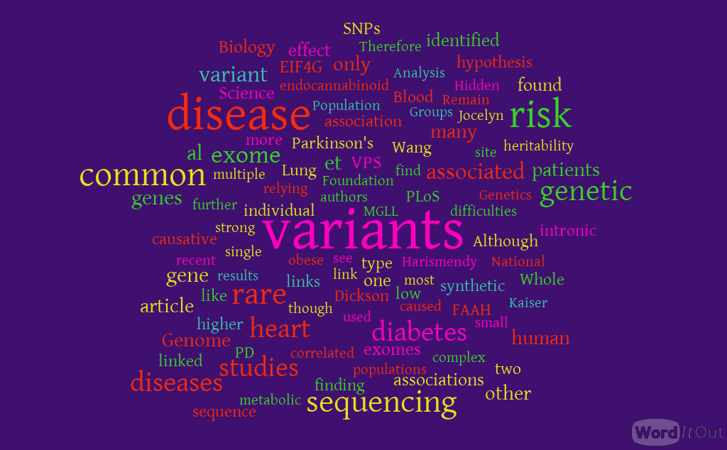
WordCloud Image Produced by Adam Tubman


Above the Genome – Underlying Disease

June 26-27, 2013 | Hotel Kabuki | San Francisco, CA | Visit website

FINAL AGENDA

http://www.clinicalgenomeconference.com/Clinical-Epigenome

KEYNOTE PRESENTATIONS

Michael Snyder


Adventures in Personal Medicine: Integrated Personal Omics Profiling for Following Healthy and Disease States

Michael Snyder, Ph.D., Professor and Chair, Genetics; Director, Stanford Center for Genomics and Personalized Medicine, Stanford University

Joesph Costello


Spontaneous and Therapy Induced Evolution of Tumor Genomes and Epigenomes

Joseph Costello, Ph.D., Professor in Residence, Department of Neurological Surgery; Director, Epigenetics Division, Cell Cycling and Signaling Program, Helen Diller Family Comprehensive Cancer Center, University of California, San Francisco

THIS WEEK’S FEATURED TALKS

This is just a small sample of presentations that you’ll be able to hear at TCEC: The Clinical Epigenome Conference in San Francisco, CA next month.

Distinguishing between Driver and Passenger Epigenetic Modifications in Cancer

Daniel De Carvalho, Ph.D., Principal Investigator, Ontario Cancer Institute, University Health Network; Assistant Professor, Medical Biophysics, Faculty of Medicine, University of Toronto

Cancer cells typically exhibit aberrant DNA methylation patterns that can drive malignant transformation. Whether cancer cells are dependent on these abnormal epigenetic modifications remains elusive. We used experimental and bioinformatic approaches to unveil genomic regions that require DNA methylation for survival of cancer cells, suggesting these are key epigenetic events associated with tumorigenesis.

Family Proteins and 5-Hydroxymethylcytosine in Stem Cells, Development and Cancer

Yujiang Geno Shi, Ph.D., Associate Biochemist, Division of Endocrinology, Brigham and Women’s Hospital; Assistant Professor, Medicine, Harvard Medical School

Recent studies have shown that ten-eleven translocation (Tet) proteins can catalyze 5mC oxidation and generate 5mC derivatives, including 5-hydroxymethylcytosine (5hmC). Not only are Tet family proteins and 5hmC critical for the identity and normal function of embryonic stem cells and early embryonic process of development, but dysregulation of these newly identified epigenetic factors also plays a major role in cancer development. Here we report an essential role of Tet3 in animal development, and define 5hmC as a potential biomarker for tumor progression. These studies will significantly increase our current understanding of the biological functions of Tet proteins and 5hmC while providing mechanistic insight into the development of epigenetic therapeutics.

Defining the Epigenetic Landscape during Normal and Malignant Hematopoiesis

Lucy A. Godley, M.D., Ph.D., Associate Professor, Department of Medicine, Section of Hematology/Oncology, Cancer Research Center, The University of Chicago

Hematopoietic stem cell commitment and differentiation involves silencing of self-renewal genes and induction of a specific transcriptional program, which is controlled in part through dynamic changes in covalent cytosine modifications. We have studied how the abundance and distribution of these derivatized bases influences hematopoietic stem cell commitment during normal erythropoiesis as well as during leukemia development. The identification of recurrent mutations in several genes important in epigenetic pathways as well as mouse modeling suggest that the balance of covalent cytosine modifications is a key driver of normal blood cell development. I will also discuss how these findings impact our understanding of the activity of the ‘hypomethylating drugs’, now in common use for the treatment of myeloid malignancies.

DNA Methylation Alterations in Lung Adenocarcinoma

Ite A. Laird-Offringa, Ph.D., Associate Professor, Departments of Surgery, Biochemistry and Molecular Biology, Norris Comprehensive Cancer Center, Keck School of Medicine, University of Southern California

I will discuss our research on integrated genome-scale DNA methylation and mRNA expression data from microdissected lung adenocarcinoma and matched non-tumor lung. We have identified 164 hypermethylated genes showing concurrent downregulation, and 57 hypomethylated genes showing increased expression. Integrated pathways analysis and detailed examination of individual genes suggests mechanistic contributions of several of these genes to lung adenocarcinoma development and/or progression. I will present information on a number of candidate epigenetic driver genes for lung adenocarcinoma.

Lessons from Surveying the DNA Methylation “Cityscape” of Lethal Metastatic Prostate Cancer

Srinivasan (Vasan) Yegnasubramanian, M.D., Ph.D., Assistant Professor, Department of Oncology, Sidney Kimmel Comprehensive Cancer Center, Johns Hopkins University School of Medicine

Alterations in DNA methylation are a hallmark of human cancers, including prostate cancer. We carried out genome-scale analyses of DNA methylation alterations in multiple metastases from each of 13 men that died of metastatic prostate cancer and created DNA methylation cityscapes to visualize these complex data. These analyses revealed that each individual developed a unique DNA methylation signature that was largely maintained across all metastases within that individual. By analyzing their frequency, clonal maintenance, and correlation with expression, we nominated potential “driver” DNA methylation alterations that could be prioritized for development as epigenetic biomarkers and therapeutic targets.

DNA Methylation Detection using Nanopores

George Vasmatzis, Ph.D., Director, Biomarker Discovery Program, Center of Individualized Medicine; Consultant, Department of Molecular Medicine, Mayo Clinic and Foundation

I will discuss some of our latest work around integrating micro-fabrication and solid state technologies with biomarkers. In diagnostics, a biosensor that could look for subtle structural or sequence variations at the single molecule level would be extremely useful . Epigenetic modifications have been linked with cancer, and we have developed nanopore technology to detect the methylation profile of a single molecule. The challenges and the potential of such technology will be discussed.


CONFERENCE TOPICS INCLUDE:

Genetic & Epigenetic Interplay in Cancer 

Mechanisms in (De)Methylation Underlying 

Development of Disease 

The Predictive Power of Epigenetics: Diagnostic 

& Prognostic Utility 

The Clinical Genome Technology Showcase 

Trends in Analysis & Interpretation 

————————————————————————

DINNER SHORT COURSES*

Wednesday, June 26      View Details     Registration Information

5:30 – 8:30 pm

(SC3) Clinical Combination Economic Conundrums


5:30 – 8:30 pm

(SC4) Advances in Methylation Analysis


Read Full Post »

Observations on Finding the Genetic Links in Common Disease: Whole Genomic Sequencing Studies

Author: Larry H Bernstein, MD, FCAP

In this article I will address the following article by Dr. SJ Williams.

Finding the Genetic Links in Common Disease:  Caveats of Whole Genome Sequencing Studies


In the November 23, 2012 issue of Science, Jocelyn Kaiser reports (Genetic Influences On Disease  Remain Hidden in News and  Analysis) on the difficulties that many genomic studies are encountering correlating genetic variants to high risk of type 2 diabetes and heart disease. American Society of  Human Genetics annual 2012 meeting, results of DNA sequencing studies reporting on genetic variants and links to high risk type 2 diabetes and heart disease, part of an international effort to determine the genetic events contributing to complex, common diseases like diabetes.
The key point is that these disease links are challenged by the identification of genetic determinants that do not follow Mendelian Genetics.  There are many disease associated gene variants, and they have not been deleted as a result of natural selection.  In the case of diabetes (type 2), the genetic risk is a low as 26%.

Gene-wide-association studies (GAWS) have identified single nucleotide polymorphisms (SNPs) with associations for common diseases, most of these individually carry only only 20-40% of risk. This is not sufficient for prediction
and use in personalized  treatment.

What is the implication of this.  Researchers have gone to exome-sequencing and  to whole genome sequencing for answers. SNPs can be easily done  by microarray, and in a clinic setting. GWAS is difficult and has inherent complexity, and it has had high cost of use. But the cost of the technology has been dropping precipitously. Technology is being redesigned for more rapid diagnosis and use in clinical research and personalized medicine.  It appears that this is not  yet a game changer.

My own thinking is that the answer doesn’t  fully lie in the genome sequencing, but that it must turn on the very large weight of importance in the regulatory function in the genome, that which was once “considered” dark matter.  In the regulatory function you have a variety of interactions and adaptive changes to the proximate environment, and this is a key to the nascent study of metabolomics.

Three projects highlighted are:
1.  National Heart, Lung and Blood Institute Exome Sequencing Project (ESP)[2]: heart, lung, blood

  • A majority of variants linked to any disease are rare
  • Groups of variants in the same gene confirmed a link between
    APOC3 and risk for early-onset heart attack

2.  T2D-GENES Consortium
3.  GoT2D

  • SNP and PAX4 gene association for type 2 diabetes in East Asians
  • No new rare variants above 1.5% frequency for diabetes

http://www.phgfoundation.org/news/5164/

The unsupported conclusion from this has been

  1. the common disease-common variant hypothesis, which predicts that common disease-causing genetic variants exist in all human populations, but   (common unexplained complexity?) each individual variant will necessarily only have a small effect on disease susceptibility (i.e. a low associated relative risk).
  1. the common disease, many rare variants hypothesis, which postulates that disease is caused by multiple strong-effect variants, (an alternative complexity situation?) Dickson et al. (2010)  PLoS Biol 2010 8(1):e1000294

The reality is that it has been difficult to associate any variant with prediction of risk, but an alternative approach appears to be intron sequencing and missing information on gene-gene interactions.

Jocelyn Kaiser’s Science article notes this in a brief interview with Harry Dietz of Johns Hopkins University where he suspects that “much of the missing heritability lies in gene-gene interactions”.

Oliver Harismendy and Kelly Frazer and colleagues’ recent publication in Genome Biology  http://genomebiology.com/content/11/11/R118 support this notion.  The authors used targeted resequencing
of two endocannabinoid metabolic enzyme genes (fatty-acid-amide hydrolase (FAAH) and monoglyceride lipase (MGLL) in 147 normal weight and 142 extremely obese patients.

English: The human genome, categorized by func...

English: The human genome, categorized by function of each gene product, given both as number of genes and as percentage of all genes. (Photo credit: Wikipedia)

Read Full Post »

Reporter: Aviva Lev-Ari, PhD, RN


Top 5 Fastest Growing Jobs for Life Scientists

5/13/2013 3:51:54 PM

Top 5 Fastest Growing Jobs for Life ScientistsHelp employers find you! Check out all the scientist jobs and post your resume.

By BioSpace.com

At a time when many graduates are finding it difficult to land jobs, it is interesting to note that life science graduates have a few options they can depend on. This is good news, considering that the media outlets are filled with horror stories of graduates languishing at home without jobs, or having to make do with terrible part-time jobs without benefits. Life science encompasses many different jobs, but here is an overview of five that are growing very rapidly (according to the Bureau of Labor Statistics), and promise to be lucrative for job seekers in the next few years.

1. Biomedical Engineering

Biomedical engineers analyze problems in medicine and biology, and come up with the appropriate solutions. Their ultimate goal is to improve the efficiency of patient care. They work in diverse industries such as universities, medical institutions, research centers, manufacturing industries, and many others. The BLS expects demand for biomedical engineers to be approximately 62 percent for the decade ending 2020. This demand is way above the average for all jobs, and it will likely be fueled by the expected increase in public appreciation of biomedical engineering. With an annual median pay of $81,540 (May 2010), aspiring biomedical engineers should not worry about the job market.

2. Medical Science

Medical scientists are primarily concerned with researching different ways of improving human health. They use different investigative methods in their line of work, such as clinical trials. Medical scientists tend to work in teams rather than individually. They work in laboratories as well as in offices. Job openings for medical scientists are expected to increase by 36 percent for the decade ending 2020, an increase that is very much larger than the industry-wide average. Aspiring medical scientists, however, should be prepared to get PhDs in appropriate life sciences because that is what most employers need. It is totally worth it because the annual median salary is $76,700 (May 2010).

3. Biochemistry and Biophysics

Biochemists and biophysicists deal with chemical and physical properties of living things. They also study biological processes, for example, heredity, growth and cell development. The majority of biophysicists and biochemists work full time in laboratories. This field is also growing, with a growth projection of 31 percent for the period from 2010 to 2020. This high rate of growth will mainly be due to the increased demand for biological products needed to improve life standards for people across the globe. Most employers will require PhDs for advanced positions and graduates with master’s or bachelor’s degrees may start in entry-level positions. BLS indicates the median annual salary as $79, 390 (May 2010).

4. Epidemiology

Epidemiologists study diseases and different public health problems to determine their causes. Their major aim in doing this is to prevent occurrences or recurrences of diseases. Most, but not all, epidemiologists work for the government in different work environments such as laboratories, health centers, and universities, among others. The job outlook for epidemiologists is bright too, given that the industry is expected to grow by 24 percent for the decade ending 2020. A master’s degree is needed in this occupation, but some epidemiologists also hold PhDs. According to the BLS, the median salary for epidemiologists in 2010 was $63,010.

5. Microbiology

Microbiologists are concerned with the study of microscopic organisms, such as fungi and algae. They do most of their work in laboratories. Although it is be possible to get an entry level microbiology job with a bachelor’s degree, most microbiologists have PhDs. The industry is expected to grow by approximately 13 percent for the decade ending 2020, which is as fast as the average for all occupations. Microbiologists command a nice annual average salary of $65, 920 (May 2010).

It is clear from the above discussions that careers in life sciences are going to hold some of the best job opportunities in the next decade. Most of the fast growing occupations may be considered non-traditional, but they are fast becoming mainstream occupations. An examination of the Bright Outlook Occupations section of U.S. Department of Labor’s Occupational Information Network (O*Net) website reveals that most of them are in life science.

Help employers find you! Check out all the scientist jobs and post your resume.

Check out the latest Career Insider eNewsletter – May 16, 2013.

Sign up for the free weekly Career Insider eNewsletter.

Related Articles
* Top 10 Best Cities for Research Scientist Jobs
* What is the Starting Salary for a Job in Biochemistry?
* Should You Relocate for a New Job?

http://www.biospace.com/news_story.aspx?NewsEntityId=296465&source=news-email

Read at BioSpace.com


Read Full Post »

Finding the Genetic Links in Common Disease:  Caveats of Whole Genome Sequencing Studies

Writer and Reporter: Stephen J. Williams, Ph.D.

In the November 23, 2012 issue of Science, Jocelyn Kaiser reports (Genetic Influences On Disease Remain Hidden in News and Analysis)[1] on the difficulties that many genomic studies are encountering correlating genetic variants to high risk of type 2 diabetes and heart disease.  At the recent American Society of Human Genetics annual 2012 meeting, results of several DNA sequencing studies reported difficulties in finding genetic variants and links to high risk type 2 diabetes and heart disease.  These studies were a part of an international effort to determine the multiple genetic events contributing to complex, common diseases like diabetes.  Unlike Mendelian inherited diseases (like ataxia telangiectasia) which are characterized by defects mainly in one gene, finding genetic links to more complex diseases may pose a problem as outlined in the article:

  • Variants may be so rare that massive number of patient’s genome would need to be analyzed
  • For most diseases, individual SNPs (single nucleotide polymorphisms) raise risk modestly
  • Hard to find isolated families (hemophilia) or isolated populations (Ashkenazi Jew)
  • Disease-influencing genes have not been weeded out by natural selection after human population explosion (~5000 years ago) resulted in numerous gene variants
  • What percentage variants account for disease heritability (studies have shown this is as low as 26% for diabetes with the remaining risk determined by environment)

Although many genome-wide-associations studies have found SNPs that have causality to increasing risk diseases such as cancer, diabetes, and heart disease, most individual SNPs for common diseases raise risk by about only 20-40% and would be useless for predicting an individual’s chance they will develop disease and be a candidate for a personalized therapy approach.  Therefore, for common diseases, investigators are relying on direct exome sequencing and whole-genome sequencing to detect these medium-rare risk variants, rather than relying on genome-wide association studies (which are usually fine for detecting the higher frequency variants associated with common diseases).

Three of the many projects (one for heart risk and two for diabetes risk) are highlighted in the article:

1.  National Heart, Lung and Blood Institute Exome Sequencing Project (ESP)[2]: heart, lung, blood

  • Sequenced 6,700 exomes of European or African descent
  • Majority of variants linked to disease too rare (as low as one variant)
  • Groups of variants in the same gene confirmed link between APOC3 and higher risk for early-onset heart attack
  • No other significant gene variants linked with heart disease

2.  T2D-GENES Consortium: diabetes

Sequenced 5,300 exomes of type 2 diabetes patients and controls from five ancestry groups
SNP in PAX4 gene associated with disease in East Asians
No low-frequency variant with large effect though

3.  GoT2D: diabetes

  • After sequencing 2700 patient’s exomes and whole genome no new rare variants above 1.5% frequency with a strong effect on diabetes risk

A nice article by Dr. Sowmiya Moorthie entitled Involvement of rare variants in common disease can be found at the PGH Foundation site http://www.phgfoundation.org/news/5164/ further discusses this conundrum,  and is summarized below:

“Although GWAs have identified many SNPs associated with common disease, they have as yet had little success in identifying the causative genetic variants. Those that have been identified have only a weak effect on disease risk, and therefore only explain a small proportion of the heritable, genetic component of susceptibility to that disease. This has led to the common disease-common variant hypothesis, which predicts that common disease-causing genetic variants exist in all human populations, but each individual variant will necessarily only have a small effect on disease susceptibility (i.e. a low associated relative risk).

An alternative hypothesis is the common disease, many rare variants hypothesis, which postulates that disease is caused by multiple strong-effect variants, each of which is only found in a few individuals. Dickson et al. in a paper in PLoS Biology postulate that these rare variants can be indirectly associated with common variants; they call these synthetic associations and demonstrate how further investigation could help explain findings from GWA studies [Dickson et al. (2010) PLoS Biol. 8(1):e1000294][3].  In simulation experiments, 30% of synthetic associations were caused by the presence of rare causative variants and furthermore, the strength of the association with common variants also increased if the number of rare causative variants increased. “

one_of_many rare variants

Figure from Dr. Moorthie’s article showing the problem of “finding one in many”.

(please   click to enlarge)

Indeed, other examples of such issues concerning gene variant association studies occur with other common diseases such as neurologic diseases and obesity, where it has been difficult to clearly and definitively associate any variant with prediction of risk.

For example, Nuytemans et. al.[4] used exome sequencing to find variants in the vascular protein sorting 3J (VPS35) and eukaryotic transcription initiation factor 4  gamma1 (EIF4G1) genes, tow genes causally linked to Parkinson’s Disease (PD).  Although they identified novel VPS35 variants none of these variants could be correlated to higher risk of PD.   One EIF4G1 variant seemed to be a strong Parkinson’s Disease risk factor however there was “no evidence for an overall contribution of genetic variability in VPS35 or EIF4G1 to PD development”.

These negative results may have relevance as companies such as 23andme (www.23andme.com) claim to be able to test for Parkinson’s predisposition.  To see a description of the LLRK2 mutational analysis which they use to determine risk for the disease please see the following link: https://www.23andme.com/health/Parkinsons-Disease/. This company and other like it have been subjects of posts on this site (Personalized Medicine: Clinical Aspiration of Microarrays)

However there seems to be more luck with strategies focused on analyzing intronic sequence rather than exome sequence. Jocelyn Kaiser’s Science article notes this in a brief interview with Harry Dietz of Johns Hopkins University where he suspects that “much of the missing heritability lies in gene-gene interactions”.  Oliver Harismendy and Kelly Frazer and colleagues’ recent publication in Genome Biology  http://genomebiology.com/content/11/11/R118 support this notion[5].  The authors used targeted resequencing of two endocannabinoid metabolic enzyme genes (fatty-acid-amide hydrolase (FAAH) and monoglyceride lipase (MGLL) in 147 normal weight and 142 extremely obese patients.

These patients were enrolled in the CRESCENDO trial and patients analyzed were of European descent. However, instead of just exome sequencing, the group resequenced exome AND intronic sequence, especially focusing on promoter regions.   They identified 1,448 single nucleotide variants but using a statistical filter (called RareCover which is referred to as a collapsing method) they found 4 variants in the promoters and intronic areas of the FAAH and MGLL genes which correlated to body mass index.  It should be noted that anandamide, a substrate for FAAH, is elevated in obese patients. The authors did note some issues though mentioning that “some other loci, more weakly or inconsistently associated in the original GWASs, were not replicated in our samples, which is not too surprising given the sample size of our cohort is inadequate to replicate modest associations”.

PLEASE WATCH VIDEO on the National Heart, Lung and Blood Institute Exome Sequencing Project

https://www.youtube.com/watch?v=-Qr5ahk1HEI

REFERENCES

http://www.phgfoundation.org/news/5164/  PHG Foundation

1.            Kaiser J: Human genetics. Genetic influences on disease remain hidden. Science 2012, 338(6110):1016-1017.

2.            Tennessen JA, Bigham AW, O’Connor TD, Fu W, Kenny EE, Gravel S, McGee S, Do R, Liu X, Jun G et al: Evolution and functional impact of rare coding variation from deep sequencing of human exomes. Science 2012, 337(6090):64-69.

3.            Dickson SP, Wang K, Krantz I, Hakonarson H, Goldstein DB: Rare variants create synthetic genome-wide associations. PLoS biology 2010, 8(1):e1000294.

4.            Nuytemans K, Bademci G, Inchausti V, Dressen A, Kinnamon DD, Mehta A, Wang L, Zuchner S, Beecham GW, Martin ER et al: Whole exome sequencing of rare variants in EIF4G1 and VPS35 in Parkinson disease. Neurology 2013, 80(11):982-989.

5.            Harismendy O, Bansal V, Bhatia G, Nakano M, Scott M, Wang X, Dib C, Turlotte E, Sipe JC, Murray SS et al: Population sequencing of two endocannabinoid metabolic genes identifies rare and common regulatory variants associated with extreme obesity and metabolite level. Genome biology 2010, 11(11):R118.

Other posts on this site related to Genomics include:

Cancer Biology and Genomics for Disease Diagnosis

Diagnosis of Cardiovascular Disease, Treatment and Prevention: Current & Predicted Cost of Care and the Promise of Individualized Medicine Using Clinical Decision Support Systems

Ethical Concerns in Personalized Medicine: BRCA1/2 Testing in Minors and Communication of Breast Cancer Risk

Genomics & Genetics of Cardiovascular Disease Diagnoses: A Literature Survey of AHA’s Circulation Cardiovascular Genetics, 3/2010 – 3/2013

Genomics-based cure for diabetes on-the-way

Personalized Medicine: Clinical Aspiration of Microarrays

Late Onset of Alzheimer’s Disease and One-carbon Metabolism

Genetics of Disease: More Complex is How to Creating New Drugs

Genetics of Conduction Disease: Atrioventricular (AV) Conduction Disease (block): Gene Mutations – Transcription, Excitability, and Energy Homeostasis

Centers of Excellence in Genomic Sciences (CEGS): NHGRI to Fund New CEGS on the Brain: Mental Disorders and the Nervous System

Cancer Genomic Precision Therapy: Digitized Tumor’s Genome (WGSA) Compared with Genome-native Germ Line: Flash-frozen specimen and Formalin-fixed paraffin-embedded Specimen Needed

Mitochondrial Metabolism and Cardiac Function

Pancreatic Cancer: Genetics, Genomics and Immunotherapy

Issues in Personalized Medicine in Cancer: Intratumor Heterogeneity and Branched Evolution Revealed by Multiregion Sequencing

Quantum Biology And Computational Medicine

Personalized Cardiovascular Genetic Medicine at Partners HealthCare and Harvard Medical School

Centers of Excellence in Genomic Sciences (CEGS): NHGRI to Fund New CEGS on the Brain: Mental Disorders and the Nervous System

LEADERS in Genome Sequencing of Genetic Mutations for Therapeutic Drug Selection in Cancer Personalized Treatment: Part 2

Consumer Market for Personal DNA Sequencing: Part 4

Personalized Medicine: An Institute Profile – Coriell Institute for Medical Research: Part 3

Whole-Genome Sequencing Data will be Stored in Coriell’s Spin off For-Profit Entity


Read Full Post »

Drug Eluting Stents: On MIT’s Edelman Lab’s Contributions to Vascular Biology and its Pioneering Research on DES

Drug Eluting Stents: On MIT‘s Edelman Lab’s Contributions to Vascular Biology and its Pioneering Research on DES

Author: Larry H Bernstein, MD, FACP

and 

Curator: Aviva Lev-Ari, PhD, RN
http://PharmaceuticalIntelligence.com/2013/04/25/Contributions
-to-vascular-biology/

This is the first of a three part series on the evolution of vascular biology and the studies of the effects of biomaterials in vascular reconstruction and on drug delivery, which has embraced a collaboration of cardiologists at Harvard Medical School , Affiliated Hospitals, and MIT,
requiring cardiovascular scientists at the PhD and MD level, physicists, and computational biologists working in concert, and
an exploration of the depth of the contributions by a distinguished physician, scientist, and thinker.

The first part – Vascular Biology and Disease – will cover the advances in the research on

  • vascular biology,
  • signaling pathways,
  • drug diffusion across the endothelium and
  • the interactions with the underlying muscularis (media),
  • with additional considerations for type 2 diabetes mellitus.

The second part – Stents and Drug Delivery – will cover the

  • purposes,
  • properties and
  • evolution of stent technology with
  • the acquired knowledge of the pharmacodynamics of drug interactions and drug distribution.

The third part – Problems and Promise of Biomaterials Technology – will cover the shortcomings of the cardiovascular devices, and opportunities for improvement

Vascular Biology and Cardiovascular Disease

Early work on endothelial injury and drug release principles

The insertion of a catheter for the administration of heparin is not an innocuous procedure. Heparin is infused to block coagulation, lowering the risk of a dangerous

  • clot formation and
  • dissemination.

It was shown experimentally that the continuous infusion of heparin

  • suppresses smooth muscle proliferation after endothelial injury. It may lead to
  • hemorrhage as a primary effect.

The anticoagulant property of heparin was removed by chemical modification without loss of the anti-proliferative effect.

In this study, MIT researches placed ethylene-vinyl acetate copolymer matrices containing standard and modified heparin adjacent to rat carotid arteries at the time of balloon deendothelialization.

Matrix delivery of both heparin compounds effectively diminished this proliferation in comparison to controls without producing systemic anticoagulation or side effects.

This mode of therapy appeared more effective than administering the agents by either

  • intravenous pumps or
  • heparin/polymer matrices placed in a subcutaneous site distant from the injured carotid artery

This indicated that the site of placement at the site of injury is a factor in the microenvironment, and is a preference for avoiding restenosis after angioplasty and other interventions.

This raised the question of why the proliferation of vascular muscle occurs in the first place.
 Edelman, Nugent and Karnovsky  (1) showed that the proliferation required first the denudation of vascular surface endothelium. This exposed the underlayer to the effect of basic fibroblast growth factor, which stimulates mitogenesis of the exposed cell, explained by the endothelium as a barrier from circulating bFGF.

To answer this question, they compared the effect of

  • 125I-labelled bFGF intravenously given with perivascular controlled bFGF release.
  • Polymeric controlled release devices delivered bFGF to the extravascular space without transendothelial transport. 
Deposition within the blood vessel wall was rapidly distributed circumferentially and was substantially greater than that observed following intravenous injection.

The amount of bFGF deposited in arteries adjacent to the release devices was 40 times that deposited in similar arteries in animals who received a single intravenous bolus of bFGF.

The presence of intimal hyperplasia increased deposition of perivascularly released bFGF 2.4-fold but decreased the deposition of intravenously injected bFGF by 67%.

  • bFGF was 5- to 30-fold more abundant in solid organs after intravenous injection than it was following perivascular release, and
  • bFGF deposition was greatest in the kidney, liver, and spleen and was substantially lower in the heart and lung.

This result indicated that vascular deposition of bFGF is independent of endothelium, and

  • bFGF delivery is effectively perivascular. (2)

Drug activity studies have to be done in well controlled and representative conditions.
 Edelsman’s Lab researchers studied the

  • dose response of injured arteries to exogenous heparin in vivo by providing steady and predictable arterial levels of drug.
  • Controlled-release devices were fabricated to direct heparin uniformly and at a steady rate to the adventitial surface of balloon-injured rat carotid arteries.

Researchers predicted the distribution of heparin throughout the arterial wall using computational simulations and correlated these concentrations with the biologic response of the tissues.

Researchers determined from this process that an in vivo arterial concentration of 0.3 mg/ml of heparin is required to maximallyinhibit intimal hyperplasia after injury.

This estimation of the required tissue concentration of a drug is

  • independent of the route of administration and
  • applies to all forms of drug release.

In this way the Team was able to

  • evaluate the potential of  widely disparate forms of drug release and, to finally
  • create some rigorous criteria by which to guide the development of particular delivery strategies for local diseases. (3)

Chiefly, the following three effects:

(1) Effect of controlled adventitial heparin delivery on smooth muscle cell proliferation following endothelial injury. ER Edelman, DH Adams, and MJ Karnovsky. PNAS May 1990; 87: 3773-3777.


(2) Perivascular and intravenous administration of basic fibroblast growth factor: Vascular and solid organ deposition. ER Edelman, MA Nugent, and MJ Karnovsky. PNAS Feb 1993; 90: 1513-1517.


(3) Tissue concentration of heparin, not administered dose, correlates with the biological response of injured arteries in vivo. MA Lovich and ER Edelman. PNAS Sep 1999; 96: 11111–11116.

Vascular Injury and Repair

Perlecan is a heparin-sulfate proteoglycan that might be critical for regulation of vascular repair by inhibiting the binding and mitogenic activity of basic fibroblast growth factor-2 (bFGF-2) in vascular smooth muscle cells .

The Team generated

  • Clones of endothelial cells expressing an antisense vector targeting domain III of perlecan. The transfected cells produced significantly less perlecan than parent cells, and they had reduced bFGF in vascular smooth muscle cells.
  • Endothelial cells were seeded onto three-dimensional polymeric matrices and implanted adjacent to porcine carotid arteries subjected to deep injury.
  • The parent endothelial cells prevented thrombosis, but perlecan deficient cells were ineffective.

The ability of endothelial cells to inhibit intimal hyperplasia, however, was only in part suppressed by perlecan. The differential regulation by perlecan of these aspects of vascular repair may clarify why control of clinical clot formation does not lead to full control of intimal hyperplasia.

The use of genetically modified tissue engineered cells provides a new approach for dissecting the role of specific factors within the blood vessel wall.(1) Successful implementation of local arterial drug delivery requires transmural distribution of drug. The physicochemical properties of the applied compound govern its transport and tissue binding.

  • Hydrophilic compounds are cleared rapidly.
  • Hydrophobic drugs bind to fixed tissue elements, potentially prolonging tissue residence and biological effect.

Local vascular drug delivery provides

  • elevated concentrations of drug in the target tissue while
  • minimizing systemic side effects.

To better characterize local pharmacokinetics the Team examined the arterial transport of locally applied dextran and dextran derivatives in vivo.

Using a two-compartment pharmacokinetic model to correct

  • The measured transmural flux of these compounds for systemic
  • Redistribution and elimination as delivered from a photo-polymerizable hydrogel.
  • The diffusivities and the transendothelial permeabilities were strongly dependent on molecular weight and charge
  • For neutral dextrans, the diffusive resistance increased with molecular weightapproximately 4.1-fold between the molecular weights of 10 and 282 kDa.
  • Endothelial resistance increased 28-fold over the same molecular weight range.
  • The effective medial diffusive resistance was unaffected by cationic charge as such molecules moved identically to neutral compounds, but increased approximately 40% when dextrans were negatively charged.

Transendothelial resistance was 20-fold lower for the cationic dextrans, and 11-fold higher for the anionic dextrans, when both were compared to neutral counterparts.

These results suggest that, while

  • low molecular weight drugs will rapidly traverse the arterial wall with the endothelium posing a minimal barrier,
  • the reverse is true for high molecular weight agents.

The deposition and distribution of locally released vascular therapeutic compounds might be predicted based upon chemical properties, such as molecular weight and charge. (2)

Paclitaxel is hydrophobic and has therapeutic potential against proliferative vascular disease.
 The favorable preclinical data with this compound may, in part, result from preferential tissue binding.
 The complexity of Paclitaxel pharmacokinetics required in-depth investigation if this drug is to reach its full clinical potential in proliferative vascular diseases.

Equilibrium distribution of Paclitaxel reveals partitioning above and beyond perfusate concentration and a spatial gradient of drug across the arterial wall.

The effective diffusivity (Deff) was estimated from the Paclitaxel distribution data to

  • facilitate comparison of transport of Paclitaxel through arterial parenchyma with that of other vasoactive agents and to
  • characterize the disparity between endovascular and perivascular application of drug.

This transport parameter described the motion of drug in tissues given an applied concentration gradient and includes, in addition to diffusion,

  • the impact of steric hindrance within the arterial interstitium;
  • nonspecific binding to arterial elements; and, in the preparation used here,
  • convective effects from the applied transmural pressure gradient.

At all times, the effective diffusivity for endovascular delivery exceeded that of perivascular delivery. The arterial transport of Paclitaxel was quantified through application ex vivo and measurement of the subsequent transmural distribution.

  • Arterial Paclitaxel deposition at equilibrium varied across the arterial wall.
  • Permeation into the wall increased with time, from 15 minutes to 4 hours, and
  • varied with the origin of delivery.

In contrast to hydrophilic compounds, the concentration in tissue exceeded the applied concentration and the rate of transport was markedly slower. Furthermore, endovascular and perivascular Paclitaxel application led to differences in deposition across the blood vessel wall.

This leads to a conclusion that Paclitaxel interacts with arterial tissue elements  as it moves under the forces of

  • diffusion and
  • convection and
  • can establish substantial partitioning and spatial gradients across the tissue. (3)

Endovascular drug-eluting stents have changed the practice of  cardiovascular vascularization, and yet it is unclear how they so dramatically reduce restenosis

We don’t know how to distinguish between the different formulations available.
 Researchers are now questioning whether individual properties of different drugs beyond lipid avidity effect arterial transport and distribution.

In bovine internal carotid segments, tissue-loading profiles for

  • Hydrophobic Paclitaxel and Rapamycin are indistinguishable, reaching load steady state after 2 days.
  • Hydrophilic dextran reaches equilibrium in hours.

Paclitaxel and Rapamycin bind to the artery at 30–40 times bulk concentration, and bind to specific tissue elements.

Transmural drug distribution profiles are markedly different for the two compounds.

  • Rapamycin binds specifically to FKBP12 binding protein and it distributes evenly through the artery,
  • Paclitaxel binds specifically to microtubules, and remains primarily in the subintimal space.

The binding of Rapamycin and Paclitaxel to specific intracellular proteins plays an essential role in

  • determining arterial transport and distribution and in
  • distinguishing one compound from another.

These results offer further insight into the

  • mechanism of local drug delivery and the
  • specific use of existing drug-eluting stent formulations. (4)

The Role of Amyloid beta (A) in Creation of Vascular Toxic Plaque

Amyloid beta (A) is a peptide family produced and deposited in neurons and endothelial cells (EC).
It is found at subnanomolar concentrations in the plasma of healthy individuals.
 Simple conformational changes produce a form of A-beta , A-beta 42, which creates toxic plaque in the brains of Alzheimer’s patients.

Oxidative stress induced blood brain barrier degeneration has been proposed as a key factor for A-beta 42 toxicity.

This cannot account for lack of injury from the same peptide in healthy tissues.
Researchers hypothesized that cell state mediates A-beta’s effect.
 They examined the viability in the presence of A-beta secreted from transfected
Chinese hamster ovary cells (CHO) of

  • aortic Endothelial Cells (EC),
  • vascular smooth muscle cells (SMC) and
  • epithelial cells (EPI) in different states

A-beta was more toxic to all cell types when they were subconfluent.
 Subconfluent EC sprouted and SMC and EPI were inhibited by A-beta.
Confluent EC were virtually resistant to A-beta and suppressed A-beta production by A-beta +CHO.

Products of subconfluent EC overcame this resistant state, stimulating the production and toxicity of A-beta 42. Confluent EC overgrew >35% beyond their quiescent state in the presence of A-beta conditioned in media from subconfluent EC.

These findings imply that A-beta 42 may well be even more cytotoxic to cells in injured or growth states and potentially explain the variable and potent effects of this protein.

One may now need to consider tissue and cell state in addition to local concentration of and exposure duration to A-beta.

The specific interactions of A-beta and EC in a state-dependent fashion may help understand further the common and divergent forms of vascular and cerebral toxicity of A-beta and the spectrum of AD. (5)

(1) Perlecan is required to inhibit thrombosis after deep vascular injury and contributes
to endothelial cell-mediated inhibition of intimal hyperplasia. MA Nugent, HM Nugent,
RV Iozzoi, K Sanchack, and ER Edelman. PNAS Jun 2000; 97(12): 6722-6727


(2) Correlation of transarterial transport of various dextrans with their physicochemical properties.
O Elmalak, MA Lovich, E Edelman. Biomaterials 2000; 21: 2263-2272


(3) Arterial Paclitaxel Distribution and Deposition. CJ Creel, MA Lovich, ER Edelman. Circ Res. 2000;86:879-884


(4) Specific binding to intracellular proteins determines arterial transport properties for rapamycin and Paclitaxel.
AD Levin, N Vukmirovic, Chao-Wei Hwang, and ER Edelman. PNAS Jun 2004; 101(25): 9463–9467.
www.pnas.org/cgi/doi/10.1073/pnas.0400918101


(5) Amyloid beta toxicity dependent upon endothelial cell state. M Balcells, JS Wallins, ER Edelman.
Neuroscience Letters 441 (2008) 319–322

Endothelial Damage as an Inflammatory State

Autoimmunity may drive vascular disease through anti-endothelial cell (EC) antibodies. This raises a question about whether an increased morbidity of cardiovascular diseases in concert with systemic illnesses may involve these antibodies.

Matrix-embedded ECs act as powerful regulators of vascular repair accompanied by significant reduction in expected systemic and local inflammation.

The Lab researchers compared the immune response against free and matrix-embedded ECs in naive mice and mice with heightened EC immune reactivity. Mice were presensitized to EC with repeated subcutaneous injections of saline-suspended porcine EC (PAE) (5*10^5 cells).

On day 42, both naive mice (controls) and mice with heightened EC immune reactivity received 5*10^5 matrix-embedded or free PAEs. Circulating PAE-specific antibodies and effector T-cells were analyzed 90 days after implantation for –

  • PAE-specific antibody-titers,
  • frequency of CD4+-effector cells, and
  • xenoreactive splenocytes

These were 2- to 4-fold lower (P<0.0001) when naıve mice were injected with matrix-embedded instead of saline-suspended PAEs.

Though basal levels of circulating antibodies were significantly elevated after serial PAE injections (2210+341 mean fluorescence intensity, day 42) and almost doubled again 90 days after injection of a fourth set of free PAEs, antibody levels declined by half in recipients of matrix-embedded PAEs at day 42 (P<0.0001), as did levels of CD4+-effector cells and xenoreactive splenocytes.

A significant immune response to implantation of free PAE is elicited in naıve mice, that is even more pronounced in mice with pre-developed anti-endothelial immunity.

Matrix-embedding protects xenogeneic ECs against immune reaction in naive mice and in mice with heightened immune reactivity.

Matrix-embedded EC might offer a promising approach for treatment of advanced cardiovascular disease. (1)

Researchers examined the molecular mechanisms through which

mechanical force and hypertension modulate

endothelial cell regulation of vascular homeostasis.

Exposure to mechanical strain increased the paracrine inhibition of vascular smooth muscle cells (VSMCs) by endothelial cells.

Mechanical strain stimulated the production by endothelial cells of perlecan and heparan-sulfate glycosaminoglycans. By inhibiting the expression of perlecan with an antisense vector researchers demonstrated that perlecan was essential to the strain-mediated effects on endothelial cell growth control.

Mechanical regulation of perlecan expression in endothelial cells was

  • governed by a mechano-transduction pathway
  • requiring transforming growth factor (TGF-β) signaling and
  • intracellular signaling through the ERK pathway.

Immunohistochemical staining of the aortae of spontaneously hypertensive rats
demonstrated strong correlations between

  • endothelial TGF-β,
  • phosphorylated signaling intermediates, and
  • arterial thickening.

Studies on ex vivo arteries exposed to varying levels of pressure demonstrated that

ERK and TGF-beta signaling were required for pressure-induced upregulation of endothelial HSPG.

The Team’s findings suggest a novel feedback control mechanism in which

  • net arterial remodeling to hemodynamic forces is controlled by a dynamic interplay between growth stimulatory signals from vSMCs and
  • growth inhibitory signals from endothelial cells. (2)

Heparan-sulfate proteoglycans (HSPGs) are potent regulators of vascular remodeling and repair.
 The major enzyme capable of degrading HSPGs is heparanase, which led us to examine
the role of heparanase in controlling

  • arterial structure,
  • mechanics, and
  • remodeling.

In vitro studies suggested heparanase expression in endothelial cells serves as a negative regulator of endothelial inhibition of vascular smooth muscle cell (vSMC) proliferation.

ECs inhibit vSMC proliferation through the interplay between

  • growth stimulatory signals from vSMCs and
  • growth inhibitory signals from ECs.

This would be expected if ECs had HSPGs that are degraded by heparanase.
Arterial structure and remodeling to injury is modified by heparanase expression.
Transgenic mice overexpressing heparanase had

  • increased arterial thickness,
  • cellular density, and
  • mechanical compliance.

Endovascular stenting studies in Zucker rats demonstrated increased heparanase expression in the neointima of obese, hyperlipidemic rats in comparison to lean rats.

The extent of heparanase expression within the neointima strongly correlated with the neointimal thickness following injury. To test the effects of heparanase overexpression on arterial repair, researchers developed a novel murine model of stent injury using small diameter self-expanding stents.

Using this model, researchers found that increased

  • neointimal formation and
  • macrophage recruitment occurs in transgenic mice overexpressing heparanase.
  • Taken together, these results support a role for heparanase in the regulation of arterial structure, mechanics, and repair. (3)

The first host–donor reaction in transplantation occurs at the blood–tissue interface.
When the primary component of the implant (donor) is the endothelial cells, it incites an immunologic reaction. Injections of free endothelial cell implants elicit a profound major histocompatibility complex (MHC) II dominated immune response.

Endothelial cells embedded within three-dimensional matrices behave like quiescent endothelial cells.

Perivascular implants of such embedded ECs cells are the most potent inhibitor of intimal hyperplasia and thrombosis following controlled vascular injury, but without any immune reactivity.

Allo- and even exenogenic endothelial cells evoke no significant humoral or
cellular immune response in immune-competent hosts when embedded within matrices.
 Moreover,  endothelial implants are immune-modulatory, reducing the extent of the memory response to previous free cell implants.

Attenuated immunogenicity results in muted activation of adaptive and innate immune cells. These findings point toward a pivotal role of matrix–cell-interconnectivity for

  • the cellular immune phenotype and might therefore assist in the design  of
  • extracellular matrix components for successful tissue engineering. (4)

Because changes in subendothelial matrix composition are associated with alterations of the endothelial immune phenotype, researchers sought to understand if

  • cytokine-induced NF-κB activity and
  • downstream effects depend on substrate adherence of endothelial cells (EC).

The team compared the upstream

  • phosphorylation cascade,
  • activation of NF-ĸβ, and
  • expression/secretion

of downstream effects of EC grown on tissue culture polystyrene plates (TCPS) with EC embedded within collagen-based matrices (MEEC).

Adhesion of natural killer (NK) cells was quantified in vitro and in vivo.

  • NF-κβ subunit p65 nuclear levels were significantly lower and
  • p50 significantly higher in cytokine-stimulated MEEC than in EC-TCPS.

Despite similar surface expression of TNF-α receptors, MEEC had significantly decreased secretion and expression of IL-6, IL-8, MCP-1, VCAM-1, and ICAM-1.

Attenuated fractalkine expression and secretion in MEEC (two to threefold lower than in EC-TCPS; p < 0.0002) correlated with 3.7-fold lower NK cell adhesion to EC (6,335 ± 420 vs. 1,735 ± 135 cpm; p < 0.0002).

Furthermore, NK cell infiltration into sites of EC implantation in vivo was significantly reduced when EC were embedded within matrix.

Matrix embedding enables control of EC substratum interaction.

This in turn regulates chemokine and surface molecule expression and secretion, in particular – of those compounds within NF-κβ pathways,

  • chemoattraction of NK cells,
  • local inflammation, and
  • tissue repair. (5)

Monocyte recruitment and interaction with the endothelium is imperative to vascular recovery.

Tie2 plays a key role in endothelial health and vascular remodeling.
Researchers studied monocyte-mediated Tie2/angiopoietin signaling following interaction of primary monocytes with endothelial cells and its role in endothelial cell survival.

The direct interaction of primary monocytes with subconfluent endothelial cells

resulted in transient secretion of angiopoietin-1 from monocytes and

the activation of endothelial Tie2. This effect was abolished by preactivation of monocytes with tumor necrosis factor-α (TNFα).

Although primary monocytes contained high levels of

  • both angiopoietin 1 and 2,
  • endothelial cells contained primarily angiopoietin 2.

Seeding of monocytes on serum-starved endothelial cells reduced caspase-3 activity by 46+5.1%, and 52+5.8% after TNFα treatment, and it decreased single-stranded DNA levels by 41+4.2% and 40+ 3.5%, respectively.

This protective effect of monocytes on endothelial cells was reversed by Tie2 silencing with specific short interfering RNA.

The antiapoptotic effect of monocytes was further supported by the

  • activation of cell survival signaling pathways involving phosphatidylinositol 3-kinase,
  • STAT3, and
  • AKT.

Monocytes and endothelial cells form a unique Tie2/angiopoietin-1 signaling system that affects endothelial cell survival and may play critical a role in vascular remodeling and homeostasis. (6)

(1) Cell–Matrix Contact Prevents Recognition and Damage of Endothelial Cells in States of Heightened Immunity.
H Methe, ER Edelman. Circulation. 2006;114[suppl I]:I-233–I-238.
http://www.circulationaha.org/DOI/10.1161/CIRCULATIONAHA.105.000687

(2) Endothelial Cells Provide Feedback Control for Vascular Remodeling Through a Mechanosensitive Autocrine
TGFβ Signaling Pathway. AB Baker, DS Ettenson, M Jonas, MA Nugent, RV Iozzo, ER Edelman.
Circ. Res. 2008;103;289-297   http://dx.doi.org/10.1161/CIRCRESAHA.108.179465
http://circres.ahajournals.org/cgi/content/full/103/3/289

(3) Heparanase Alters Arterial Structure, Mechanics, and Repair Following Endovascular Stenting in Mice.
AB Baker, A Groothuis, M Jonas, DS Ettenson…ER Edelman.   Circ. Res. 2009;104;380-387;
http://dx.doi.org/10.1161/CIRCRESAHA.108.180695  http://circres.ahajournals.org/cgi/content/full/104/3/380

(4) The effect of three-dimensional matrix-embedding of endothelial cells on the humoral and cellular immune response.
H Methe, S Hess, ER Edelman. Seminars in Immunology 20 (2008) 117–122. http://dx.doi.org/10.1016/j.smim.2007.12.005

(5) NF-kB Activity in Endothelial Cells Is Modulated by Cell Substratum Inter-actions and Influences Chemokine-Mediated
Adhesion of Natural Killer Cells.  S Hess, H Methe, Jong-Oh Kim, ER Edelman.
Cell Transplantation 2009; 18: 261–273


(6) Primary Monocytes Regulate Endothelial Cell Survival Through Secretion of Angiopoietin-1 and Activation of Endothelial Tie2.
SY Schubert, A Benarroch, J Monter-Solans and ER Edelman. Arterioscler Thromb Vasc Biol 2011;31;870-875
http://dx.doi.org/10.1161/ATVBAHA.110.218255

Neointimal Formation, Shear Stress, and Remodelling with Reference to Diabetes

Innate immunity is of major importance in vascular repair. The present study evaluated whether

  • systemic and transient depletion of monocytes and macrophages with
  • liposome-encapsulated bisphosphonates inhibits experimental in-stent neointimal formation.

The Experiment

Rabbits fed on a hypercholesterolemic diet underwent bilateral iliac artery balloon denudation and stent deployment.

Liposomal alendronate (3 or 6 mg/kg) was given concurrently with stenting.

  • Monocyte counts were reduced by 90% 24 to 48 hours aftera single injection of liposomal alendronate, returning to basal levels at 6 days.

This treatment significantly reduced

  • intimal area at 28 days, from 3.88+0.93 to 2.08+0.58 and 2.16 +0.62 mm2.
  • Lumen area was increased from 2.87+0.44 to 3.57­+0.65 and 3.45+0.58 mm2, and
  • arterial stenosis was reduced from 58 11% to 37 8% and 38 7% in controls, in rabbits treated with 3 mg/kg, and with 6 mg/kg, respectively (mean+SD, n=8 rabbits/group, P< 0.01 for all 3 parameters).

No drug-related adverse effects were observed.
Reduction in neointimal formation was associated with

  • reduced arterial macrophage infiltration and proliferation at 6 days and with an
  • equal reduction in intimal macrophage and smooth muscle cell content at 28 days after injury.

Conversely, drug regimens ineffective in reducing monocyte levels did not inhibit neointimal formation.
Researchers have shown that a

  • single liposomal bisphosphonates injection concurrent with injury reduces in-stent neointimal formation and
  • arterial stenosis in hypercholesterolemic rabbits, accompanied by systemic transient depletion of monocytes and macrophages. (1)

Diabetes and insulin resistance are associated with increased disease risk and poor outcomes from cardiovascular interventions.

Even drug-eluting stents exhibit reduced efficacy in patients with diabetes.
Researchers reported the first study of vascular response to stent injury in insulin-resistant and diabetic animal models.

Endovascular stents were expanded in the aortae of

  • obese insulin-resistant and
  • type 2 diabetic Zucker rats,
  • in streptozotocin-induced type 1 diabetic Sprague-Dawley rats, and
  • in matched controls.

Insulin-resistant rats developed thicker neointima (0.46+0.08 versus 0.37+0.06 mm2, P 0.05), with  decreased lumen area (2.95+0.26 versus 3.29+0.15 mm2, P 0.03) 14 days after stenting compared with controls, but without increased vascular inflammation (tissue macrophages).

Insulin-resistant and diabetic rat vessels did exhibit markedly altered signaling pathway activation 1 and 2 weeks after stenting, with up to a 98% increase in p-ERK (anti-phospho ERK) and a 54% reduction in p-Akt (anti-phospho Akt) stained cells. Western blotting confirmed a profound effect of insulin resistance and diabetes on Akt and ERK signaling in stented segments. p-ERK/p-Akt ratio in stented segments uniquely correlated with neointimal response (R2 = 0.888, P< 0.04) , but not in lean controls.

Transfemoral aortic stenting in rats provides insight into vascular responses in insulin resistance and diabetes.

Shifts in ERK and Akt signaling related to insulin resistance may reflect altered tissue repair in diabetes accompanied by a

  • shift in metabolic : proliferative balance.

These findings may help explain the increased vascular morbidity in diabetes and suggest specific therapies for patients with insulin resistance and diabetes. (2)

Researchers investigated the role of Valsartan (V) alone or in combination with Simvastatin (S) on coronary atherosclerosis and vascular remodeling, and tested the hypothesis that V or V/S attenuate the pro-inflammatory effect of low endothelial shear stress (ESS).

Twenty-four diabetic, hyperlipidemic swine were allocated into Early (n = 12) and Late (n=12) groups.
Diabetic swine in each group were treated with Placebo (n=4), V (n = 4) and V/S (n = 4) and  followed for 8 weeks in the Early group and 30 weeks in the Late group.

Blood pressure, serum cholesterol and glucose were similar across the treatment subgroups.
ESS was calculated in plaque-free subsegments of interest (n = 109) in the Late group at week 23.
Coronary arteries of this group were harvested at week 30, and the subsegments of interest were identified, and analyzed histopathologically.

Intravascular geometrically correct 3-dimensional reconstruction of the coronary arteries of 12 swine was performed 23 weeks after initiation of diabetes mellitus and a hyperlipidemic diet. Local endothelial shear stress was calculated

  • in plaque-free subsegments of interest (n=142) with computational fluid dynamics, and
  • the coronary arteries (n=31) were harvested and the same subsegments were identified at 30 weeks.

V alone or with S

  • reduced the severity of inflammation in high-risk plaques.
Both regimens attenuated the severity of enzymatic degradation of the arterial wall, reducing the severity of expansive remodeling.
  • attenuated the pro-inflammatory effect of low ESS.
V alone or with S
  • exerts a beneficial effect of reducing and stabilizing high-risk plaque characteristics independent of a blood pressure- and lipid-lowering effect. (3)

This study tested the hypothesis that low endothelial shear stress  augments the

  • expression of matrix-degrading proteases, promoting the
  • formation of thin-capped atheromata.

Researchers assessed the messenger RNA and protein expression, and elastolytic activity of selected elastases and their endogenous inhibitors.

Subsegments with low endothelial shear stress at week 23 showed

  • reduced endothelial coverage,
  • enhanced lipid accumulation, and
  • intense infiltration of activated inflammatory cells at week 30.

These lesions showed increased expression of messenger RNAs encoding

  • matrix metalloproteinase-2, -9, and -12, and cathepsins K and S
  • relative to their endogenous inhibitors and
  • increased elastolytic activity.

Expression of these enzymes correlated positively with the severity of internal elastic lamina fragmentation.

Thin-capped atheromata in regions with

  • lower preceding endothelial shear stress had
  • reduced endothelial coverage,
  • intense lipid and inflammatory cell accumulation,
  • enhanced messenger RNA expression and
  • elastolytic activity of MMPs and cathepsins with
  • severe internal elastic lamina fragmentation.

Low endothelial shear stress induces endothelial discontinuity and

  • accumulation of activated inflammatory cells, thereby
  • augmenting the expression and activity of elastases in the intima and
  • shifting the balance with their inhibitors toward matrix breakdown.

Team’s results provide new insight into the mechanisms of regional formation of plaques with thin fibrous caps. (4)

Elevated CRP levels predict increased incidence of cardiovascular events and poor outcomes following interventions. There is the suggestion that CRP is also a mediator of vascular injury.

Transgenic mice carrying the human CRP gene (CRPtg) are predisposed to arterial thrombosis post-injury.

Researchers examined whether CRP similarly modulates the proliferative and hyperplastic phases of vascular repair in CRPtg when thrombosis is controlled with daily aspirin and heparin at the time of trans-femoral arterial wire-injury.

Complete thrombotic arterial occlusion at 28 days was comparable for wild-type and CRPtg mice (14 and 19%, respectively). Neointimal area at 28d was 2.5 fold lower in CRPtg (4190±3134 m2, n = 12) compared to wild-types (10,157±8890 m2, n = 11, p < 0.05).

Likewise, neointimal/media area ratio was 1.10±0.87 in wild-types and 0.45±0.24 in CRPtg (p < 0.05).

  • Seven days post-injury, cellular proliferation and apoptotic cell number in the intima were both less pronounced in CRPtg than wild-type.
  • No differences were seen in leukocyte infiltration or endothelial coverage.
CRPtg mice had significantly reduced p38 MAPK signaling pathway activation following injury.

The pro-thrombotic phenotype of CRPtg mice was suppressed by aspirin/heparin, revealing CRP’s influence on neointimal growth after trans-femoral arterial wire-injury.

  • Signaling pathway activation,
  • cellular proliferation, and
  • neointimal formation

were all reduced in CRPtg following vascular injury.
 Increasingly the Team was aware of CRP multipotent effects.
 Once considered only a risk factor, and recently a harmful agent, CRP is a far more complex regulator of vascular biology. (5)

(1) Liposomal Alendronate Inhibits Systemic Innate Immunity and Reduces In-Stent Neointimal
Hyperplasia in Rabbits. HD Danenberg, G Golomb, A Groothuis, J Gao…, ER Edelman.
Circulation. 2003;108:2798-2804


(2) Vascular Neointimal Formation and Signaling Pathway Activation in Response to Stent Injury
in Insulin-Resistant and Diabetic Animals. M Jonas, ER Edelman, A Groothuis, AB Baker, P Seifert, C Rogers.
Circ. Res. 2005;97;725-733.        http://dx.doi.org/10.1161/01.RES.0000183730.52908.C6
http://circres.ahajournals.org/cgi/content/full/97/7/725

(3) Attenuation of inflammation and expansive remodeling by Valsartan alone or in combination with
Simvastatin in high-risk coronary atherosclerotic plaques. YS Chatzizisis, M Jonas, R Beigel, AU Coskun…
ER Edelman, CL Feldman, PH Stone.  Atherosclerosis 203 (2009) 387–394


(4) Augmented Expression and Activity of Extracellular Matrix-Degrading Enzymes in Regions of Low
Endothelial Shear Stress Colocalize With Coronary Atheromata With Thin Fibrous Caps in Pigs.
YS Chatzizisis, AB Baker, GK Sukhova,…P Libby, CL Feldman, ER Edelman, PH Stone
Circulation 2011;123;621-630     http://dx.doi.org/10.1161/CIRCULATIONAHA.110.970038
http://circ.ahajournals.org/cgi/content/full/123/6/621


(5) Neointimal formation is reduced after arterial injury in human crp transgenic mice
HD Danenberg, E Grad, RV Swaminathan, Z Chenc,…ER Edelman
Atherosclerosis 201 (2008) 85–91

A Rattle Bag of Science and the Art of Translation

Science Translational Medicine – A rattle bag of science and the art of translation
E. R. Edelman, G. A. FitzGerald.
Sci.Transl. Med. 3, 104ed3 (2011). http://dx.doi.org/10.1126/scitranslmed.3002131

Elazer R. Edelman is the Thomas D. and Virginia W. Cabot Professor of Health Sciences and Technology at MIT,
Professor of Medicine at Harvard Medical School, a coronary care unit cardiologist at the Brigham and Women’s
Hospital, and Director of the Harvard-MIT Biomedical Engineering Center. E-mail: ere@mit.edu

Garret A. FitzGerald is the McNeil Professor in Translational Medicine and Therapeutics, Chair of the Department of
Pharmacology, and Director of the Institute for Translational Medicine & Therapeutics, University of Pennsylvania.
E-mail: garret@upenn.edu

In 2011, the American Association for the Advancement of Science (AAAS)  founded Science Translational Medicine (STM)
to disseminate interdisciplinary science integrating basic and clinical research that defines and fosters new therapeutics, devices, and diagnostics.

Conceived and nourished under the creative vision of Elias Zerhouni and Katrina Kelner, the journal has attracted widespread attention.
Now, as we assume the mantle of co-chief scientific advisors, we look back on the journal’s early accomplishments, restate our mission, and make clear the kinds of manuscripts we seek and accept for publication.

STM’s mission, as articulated by Elias and Katrina, was to

“promote human health by providing a forum for communication and cross-fertilization among basic, translational, and clinical research practitioners and trainees from all relevant established and emerging disciplines.”

This statement remains relevant and accurate today.
 With this mission on our masthead, STM now receives ~25 manuscripts (full-length research articles) per week and publishes ~10% of them. Roughly half of the submissions are deemed inappropriate for the journal and are returned without review within 8 to 10 days of receipt.

Of those papers that undergo full peer review,

decisions to reject are made within 48 days and

the mean time to acceptance (including the revision period) is 125 days.

There is now an average wait of only 24 days between acceptance and publication.

Defining TRANSLATIONAL Medicine

In accord with the journal’s broad readership, the ideal manuscript meets five criteria: It
(i) reports a discovery of translational relevance with high-impact potential;
(ii) has a conceptual focus with interdisciplinary appeal;
(iii) elucidates a biological mechanism;
(iv) is innovative and novel; and
(v) is presented in clear, broadly accessible language.
 STM seeks to publish research that describes

  • how innovative concepts drive the creative biomedical science
  • that ultimately improves the quality of people’s lives—

This is the broadest of our journal’s criteria but is the one that sets us apart as well.
Translational relevance does not require demonstration of benefit in humans but does require the evident potential to advance clinical medicine, thus impacting the direction of our culture and the welfare of our communities. Conceptual focus and mechanistic emphasis discriminate our papers from those that contain observational descriptions of technical findings for which value is restricted to a specific discipline.

However, innovation and novelty may apply to a fundamental scientific discovery or to the nature of its application and relevance to the translational process. Criteria enable the journal to consider versatile technological advances that apply new and creative thinking but may not necessarily offer fresh insights into biological mechanisms. Finally, while the subsequent additional efforts of the STM editorial staff are not to be discounted, the clarity of writing and coherence of argument presented within a submitted manuscript are likely to facilitate its progress through the challenge of peer review.

On Causes – Hippocrates, Aristotle, Robert Koch, and the Dread Pirate Roberts

Elazer R. Edelman
Circulation 2001;104:2509-2512

The idea of risk factors for vascular disease has evolved

  • from a dichotomous to continuous hazard analysis and
  • from the consideration of a few factors to
  • mechanistic investigation of many interrelated risks.

However, confusion still abounds regarding issues of association and causation. Originally, the simple presence of

  • tobacco abuse, hypertension, and/or hypercholesterolemia were tallied, and
  • the cumulative score was predictive of subsequent coronary artery disease.

Since then, dose responses have been defined for these and other factors and it has been suggested that almost 300 factors place patients at risk; these factors include elevations in plasma homocysteine.
 Recent studies shed interesting light on the mechanism of this potentially causal relationship, which was first noted in 1969.

Aside from putative effects on vessel wall dynamics, there is now direct evidence that homocysteine is atherogenic. Twenty-fold increases in plasma homocysteine achieved by dietary manipulation of apoE–/– mice increased aortic root lesion size 2-fold and produced a prolonged chronic inflammatory mural response accompanied by elevations in vascular cell adhesion molecule-1 (VCAM) and tumor necrosis factor-a (TNF-a).

In long term followup, homocysteine levels elevated by

  • dietary supplementation with methionine or homocysteine
  • promoted lesion size and plaque fibrosis in these
  • atherosclerosis-prone mice early in life, but without influencing ultimate plaque burden as the animals aged.

A number of mechanisms were proposed by which homocysteine achieved this effect, including

  • promotion of inflammation,
  • regulation of lipoprotein metabolism, and
  • modification of critical biochemical pathways and
  • metabolites including nitric oxide (NO).

See p 2569
In the present issue of Circulation,

Stühlinger et al 7 advance these mechanistic insights one critical step further by defining homocysteine’s effects at an enzymatic level.

The group led by Lentz published an association between levels of the

  • endogenous inhibitor of Nirtic Oxide synthase,
  • asymmetric dimethyl arginine (ADMA), and
  • homocysteine in cultured endothelial cells and in the serum of cynomolgus monkeys.

Such an association is interesting because the L-arginine–NO synthase pathway seems to be a critical component in the full range of endothelial cell biology and vascular dysfunction.

Stühlinger et al 7  now show that increased cultured endothelial cell elaboration of ADMA by homocysteine and its precursor L-methionine is associated with a dose-dependent impairment of the activity of endothelial dimethylarginine dimethylaminohydrolase (DDAH), the enzyme that degrades ADMA. Homocysteine directly inhibited DDAH activity in a cell-free system by targeting a critical sulfhydryl group on this enzyme.

Thus, one could envision that the balance of cardiovascular health and disease could well be determined by the ability of an intact Nirtic Oxide synthase system to overcome environmental, dietary, and even genetic factors.

In patients with altered enzymatic defense systems,

  • elevated homocysteine,
  • oxidized lipoproteins,
  • inflammation, and other
  • vasotoxins

may dominate even the most potent defense mechanisms.
These studies raise a number of issues.
Do we need to add to our list of established cardiovascular risk factors to accommodate new findings and associations?
Is there a final common pathway for all risk factors or perhaps even a unified factor theory into which all potential risks can be grouped?
And, as always, should we consider Nirtic Oxide at the core of this universality?
Finally, should we change our focus altogether and speak not of risk factors but of

  • genetic predisposition,
  • extent of biochemical aberration, and
  • degree of physical damage?

Some would view these remarkable success stories and the repeated association of hyperhomocyst(e)inemia with coronary, cerebral, and peripheral vascular disease and simply advocate for increased folic acid intake for all.

Indeed, this intervention of negligible cost and

  • insignificant side effect is already partially in place;
  • many foods are fortified with folate to prevent congenital neural tube defects.

This reader considers the seminal work by Vernon Young and Yves Ingenbleek on the relationship between

  • S8 and regions distant from lava flows in Asia and Indian subcontinents,
  • where they have determined hyperhomocysteinemia and the consequence associated with:
  • veganism (not voluntary)
  • impaired methyl donor reactions and transsulfuration pathways (not corrected by B12, folate)
  • loss of lean body mass due to the constant relationship of S:N (insufficient from plant sources)

What happens, when we fail to continue to pursue causality,

  • the linkage of biological significance or scientific plausibility with
  • epidemiologically or statistically significant association?

In medicine, risk becomes the likelihood that people without a disease will acquire the disease through contact with factors thought to increase disease risk.

All of these risk factors are then, by nature, imprecise and nonspecific.
 They are stochastic measures of what will happen to normal people who fall into particular measures of these parameters.

The daring may be willing to accept these risks, citing friend and foe who live well beyond or for far lesser times than anticipated by risk alone. Such concerns may well become moot if we can simultaneously identify patients at risk

  • by linking phenotype with genotype,
  • gene expression with protein elaboration, and
  • environmental exposures with the biochemical consequences and
  • direct anatomic aberrations they induce.

This kind of characterization may well replace a family history of arterial disease as a rough estimate of

  • genotype,
  • serum cholesterol as an indirect measure of the health of lipoprotein metabolism,
  • serum glucose as a crude determinant of the ravages of diabetes mellitus,
  • blood pressure measurement as a marker of long-standing endogenous exposure to altered flow, and
  • tobacco abuse as a maker of long-standing exposure to exogenous toxins.

Rather than identifying patients on the basis of their serum cholesterol, we will have a direct measure of their

  • LDL receptor number,
  • internalization rate,
  • macrophage content in the blood vessel wall,
  • metalloproteinase activity, etc.
  • insulin receptor metabolism,
  • oxidative state, and
  • glycated burden.
  • Serum glucose will similarly give way to these tests

Evaluating a new way to open clogged arteries: Computational model offers insight into mechanisms of drug-coated balloons.

A new study from MIT analyzes the potential usefulness of a new treatment that combines the benefits of angioplasty balloons and drug-releasing stents, but may pose fewer risks. With this new approach, a balloon is inflated in the artery for only a brief period, during which it releases a drug that prevents cells from accumulating and clogging the arteries over time.
While approved for limited use in Europe, these drug-coated balloons are still in development in the United States and have not received FDA approval. The MIT study, which models the behavior of the balloons, should help scientists optimize their performance and aid regulators in evaluating their effectiveness and safety.
“Until now, people who evaluate such technology could not distinguish hype from promise,” says Elazer Edelman, the Thomas D. and Virginia W. Cabot Professor of Health Sciences and Technology and senior author of the paper describing the study, which appeared online recently in the journal Circulation.
Lead author of the paper is Vijaya Kolachalama, a former MIT postdoc who is now a principal member of the technical staff at the Charles Stark Draper Laboratory.
Edelman’s lab is investigating a possible alternative to the current treatments: drug-coated balloons. “We’re trying to understand how and when this therapy could work and identify the conditions in which it may not,” Kolachalama says. “It has its merits; it has some disadvantages.”

Modeling drug release

The drug-coated balloons are delivered by a catheter and inflated at the narrowed artery for about 30 seconds, sometimes longer. During that time, the balloon coating, containing a drug such as Zotarolimus, is released from the balloon. The properties of the coating allow the drug to be absorbed in the body’s tissues. Once the drug is released, the balloon is removed.
In their new study, Kolachalama, Edelman and colleagues set out to rigorously characterize the properties of the drug-coated balloons. After performing experiments in tissue grown in the lab and in pigs, they developed a computer model that explains the dynamics of drug release and distribution. They found that factors such as the size of the balloon, the duration of delivery time, and the composition of the drug coating all influence how long the drug stays at the injury site and how effectively it clears the arteries.
One significant finding is that when the drug is released, some of it sticks to the lining of the blood vessels. Over time, that drug is slowly released back into the tissue, which explains why the drug’s effects last much longer than the initial 30-second release period.
“This is the first time we can explain the reasons why drug-coated balloons can work,” Kolachalama says. “The study also offers areas where people can consider thinking about optimizing drug transfer and delivery.”

http://circ.ahajournals.org/content/127/20/2047.short  
http://www.mit.edu/people/vbk/Circulation_2013.pdf 
http://www.sciencedaily.com/…13/05/130521121513.ht…    
Circulation, 2013; 127 (20): 2047 – 2055
http://dx.doi.org/10.1161/CIRCULATIONAHA.113.002051;


Conclusion

MIT’s Edelman’s Lab conducted the pioneering work in Vascular biology, animal models of drug eluting stents and was at the forefront of Empirical Molecular Cardiology in its studies in vascular physiology, biology and biomaterials for medical devices.

Related articles

MUC1* Ligand, NM23-H1, Is a Novel Growth Factor That Maintains Human Stem Cells in a More Naïve State (plosone.org)

Mass. General team develops implantable, bioengineered rat kidney (eurekalert.org)

Suppression of JAK2/STAT3 Signaling Reduces End-to-End Arterial Anastomosis Induced Cell Proliferation in Common Carotid Arteries of Rats (plosone.org)

Blood Vessel Function and Breathing Control Adversely Affected by Cutting Back on Sleep (medindia.net)

miRNA Biogenesis Enzyme Drosha Is Required for Vascular Smooth Muscle Cell Survival (plosone.org)

Cell-Permeable Peptide Shows Promise For Controlling Cardiovascular Disease (medicalnewstoday.com)

The Heart Revolution By Kilmer McCully, Martha McCully

HarperCollinsPublishers, 1969

http://books.google.com/books?id=iYLbuZFxEt8C&pg=PR20&dq=New+York+Times+homocysteine+and+Cholesterol&hl=en&sa=X&ei=_0F7UfDRA8zB4APozIHQAQ&ved=0CEMQ6AEwAg


Other Related Articles that were published on this Open Access Online Scientific Journal include the following:

Modeling Targeted Therapy

Larry H Bernstein, MD, FACP 3/2/2013

Quantum Biology And Computational Medicine

Larry H Bernstein, MD, FACP 4/3/2013

Virtual Biopsy – is it possible?

Larry H Bernstein, MD, FACP 3/3/2013

Reprogramming cell fate  3/2/2013

Larry H Bernstein, MD, FACP

How Methionine Imbalance with Sulfur-Insufficiency Leads to Hyperhomocysteinemia

Larry H Bernstein, MD, FACP 4/4/2013

http://pharmaceuticalintelligence.com/2013/04/04/sulfur-deficiency-and-hyperhomocusteinemia/

Amyloidosis with Cardiomyopathy

Larry H Bernstein, MD, FACP 3/31/2013

http://pharmaceuticalintelligence.com/2013/03/31/amyloidosis-with-cardiomyopathy/

Nitric Oxide, Platelets, Endothelium and Hemostasis

Larry H Bernstein, MD, FACP 11/8/2012

http://pharmaceuticalintelligence.com/2012/11/08/nitric-oxide-platelets-endothelium-and-hemostasis/

Mitochondrial Damage and Repair under Oxidative Stress

Larry H Bernstein, MD, FACP 10/28/2012

http://pharmaceuticalintelligence.com/2012/10/28/mitochondrial-damage-and-repair-under-oxidative-stress/

Endothelial Function and Cardiovascular Disease

Larry H Bernstein, MD, FACP 10/25/2012

http://pharmaceuticalintelligence.com/2012/10/25/endothelial-function-and-cardiovascular-disease/

Endothelial Dysfunction, Diminished Availability of cEPCs, Increasing CVD Risk for Macrovascular Disease –Therapeutic Potential of cEPCs

Aviva Lev-Ari, PhD, RN 8/27/2012

Revascularization: PCI, Prior History of PCI vs CABG

Aviva Lev-Ari, PhD, RN 4/25/2013

http://pharmaceuticalintelligence.com/2013/04/25/revascularization-pci-prior-history-of-pci-vs-cabg/

Cholesteryl Ester Transfer Protein (CETP) Inhibitor: Potential of Anacetrapib to treat Atherosclerosis and CAD

Aviva Lev-Ari, PhD, RN 4/7/2013

http://pharmaceuticalintelligence.com/2013/04/07/cholesteryl-ester-transfer-protein-cetp-inhibitor-potential-of-anacetrapib-to-treat-atherosclerosis-and-cad/

Hypertriglyceridemia concurrent Hyperlipidemia: Vertical Density Gradient Ultracentrifugation a Better Test to Prevent Undertreatment of High-Risk Cardiac Patients

Aviva Lev-Ari, PhD, RN 4/4/2013

http://pharmaceuticalintelligence.com/2013/04/04/hypertriglyceridemia-concurrent-hyperlipidemia-vertical-density-gradient-ultracentrifugation-a-better-test-to-prevent-undertreatment-of-high-risk-cardiac-patients/

Fight against Atherosclerotic Cardiovascular Disease: A Biologics not a Small Molecule – Recombinant Human lecithin-cholesterol acyltransferase (rhLCAT) attracted AstraZeneca to acquire AlphaCore

Aviva Lev-Ari, PhD, RN 4/3/2013

http://pharmaceuticalintelligence.com/2013/04/03/fight-against-atherosclerotic-cardiovascular-disease-a-biologics-not-a-small-molecule-recombinant-human-lecithin-cholesterol-acyltransferase-rhlcat-attracted-astrazeneca-to-acquire-alphacore/

High-Density Lipoprotein (HDL): An Independent Predictor of Endothelial Function & Atherosclerosis, A Modulator, An Agonist, A Biomarker for Cardiovascular Risk

Aviva Lev-Ari, PhD, RN 3/31/2013

http://pharmaceuticalintelligence.com/2013/03/31/high-density-lipoprotein-hdl-an-independent-predictor-of-endothelial-function-artherosclerosis-a-modulator-an-agonist-a-biomarker-for-cardiovascular-risk/

Acute Chest Pain/ER Admission: Three Emerging Alternatives to Angiography and PCI

Aviva Lev-Ari, PhD, RN 3/10/2013

http://pharmaceuticalintelligence.com/2013/03/10/acute-chest-painer-admission-three-emerging-alternatives-to-angiography-and-pci/

Genomics & Genetics of Cardiovascular Disease Diagnoses: A Literature Survey of AHA’s Circulation Cardiovascular Genetics, 3/2010 – 3/2013

Lev-Ari, A. and L H Bernstein 3/7/2013

http://pharmaceuticalintelligence.com/2013/03/07/genomics-genetics-of-cardiovascular-disease-diagnoses-a-literature-survey-of-ahas-circulation-cardiovascular-genetics-32010-32013/

The Heart: Vasculature Protection – A Concept-based Pharmacological Therapy including THYMOSIN

Aviva Lev-Ari, PhD, RN 2/28/2013

http://pharmaceuticalintelligence.com/2013/02/28/the-heart-vasculature-protection-a-concept-based-pharmacological-therapy-including-thymosin/

Arteriogenesis and Cardiac Repair: Two Biomaterials – Injectable Thymosin beta4 and Myocardial Matrix Hydrogel

Aviva Lev-Ari, PhD, RN 2/27/2013

http://pharmaceuticalintelligence.com/2013/02/27/arteriogenesis-and-cardiac-repair-two-biomaterials-injectable-thymosin-beta4-and-myocardial-matrix-hydrogel/

Coronary artery disease in symptomatic patients referred for coronary angiography: Predicted by Serum Protein Profiles

Aviva Lev-Ari, PhD, RN 12/29/2012

http://pharmaceuticalintelligence.com/2012/12/29/coronary-artery-disease-in-symptomatic-patients-referred-for-coronary-angiography-predicted-by-serum-protein-profiles/

Special Considerations in Blood Lipoproteins, Viscosity, Assessment and Treatment

Bernstein, HL and Lev-Ari, A. 11/28/2012

http://pharmaceuticalintelligence.com/2012/11/28/special-considerations-in-blood-lipoproteins-viscosity-assessment-and-treatment/

Peroxisome proliferator-activated receptor (PPAR-gamma) Receptors Activation: PPARγ transrepression for Angiogenesis in Cardiovascular Disease and PPARγ transactivation for Treatment of Diabetes

Aviva Lev-Ari, PhD, RN 11/13/2012

http://pharmaceuticalintelligence.com/2012/11/13/peroxisome-proliferator-activated-receptor-ppar-gamma-receptors-activation-pparγ-transrepression-for-angiogenesis-in-cardiovascular-disease-and-pparγ-transactivation-for-treatment-of-dia/

Clinical Trials Results for Endothelin System: Pathophysiological role in Chronic Heart Failure, Acute Coronary Syndromes and MI – Marker of Disease Severity or Genetic Determination?

Aviva Lev-Ari, PhD, RN 10/19/2012

http://pharmaceuticalintelligence.com/2012/10/19/clinical-trials-results-for-endothelin-system-pathophysiological-role-in-chronic-heart-failure-acute-coronary-syndromes-and-mi-marker-of-disease-severity-or-genetic-determination/

Endothelin Receptors in Cardiovascular Diseases: The Role of eNOS Stimulation

Aviva Lev-Ari, PhD, RN 10/4/2012

http://pharmaceuticalintelligence.com/2012/10/04/endothelin-receptors-in-cardiovascular-diseases-the-role-of-enos-stimulation/

Inhibition of ET-1, ETA and ETA-ETB, Induction of NO production, stimulation of eNOS and Treatment Regime with PPAR-gamma agonists (TZD): cEPCs Endogenous Augmentation for Cardiovascular Risk Reduction – A Bibliography

Aviva Lev-Ari, PhD, RN 10/4/2012

http://pharmaceuticalintelligence.com/2012/10/04/inhibition-of-et-1-eta-and-eta-etb-induction-of-no-production-and-stimulation-of-enos-and-treatment-regime-with-ppar-gamma-agonists-tzd-cepcs-endogenous-augmentation-for-cardiovascular-risk-reduc/

Positioning a Therapeutic Concept for Endogenous Augmentation of cEPCs — Therapeutic Indications for Macrovascular Disease: Coronary, Cerebrovascular and Peripheral

Aviva Lev-Ari, PhD, RN 8/29/2012

http://pharmaceuticalintelligence.com/2012/08/29/positioning-a-therapeutic-concept-for-endogenous-augmentation-of-cepcs-therapeutic-indications-for-macrovascular-disease-coronary-cerebrovascular-and-peripheral/

Cardiovascular Outcomes: Function of circulating Endothelial Progenitor Cells (cEPCs): Exploring Pharmaco-therapy targeted at Endogenous Augmentation of cEPCs

Aviva Lev-Ari, PhD, RN 8/28/2012

http://pharmaceuticalintelligence.com/2012/08/28/cardiovascular-outcomes-function-of-circulating-endothelial-progenitor-cells-cepcs-exploring-pharmaco-therapy-targeted-at-endogenous-augmentation-of-cepcs/

Endothelial Dysfunction, Diminished Availability of cEPCs, Increasing CVD Risk for Macrovascular Disease – Therapeutic Potential of cEPCs

Aviva Lev-Ari, PhD, R N 8/27/2012

http://pharmaceuticalintelligence.com/2012/08/27/endothelial-dysfunction-diminished-availability-of-cepcs-increasing-cvd-risk-for-macrovascular-disease-therapeutic-potential-of-cepcs/

Vascular Medicine and Biology: CLASSIFICATION OF FAST ACTING THERAPY FOR PATIENTS AT HIGH RISK FOR MACROVASCULAR EVENTS Macrovascular Disease – Therapeutic Potential of cEPCs

Aviva Lev-Ari, PhD, RN 8/24/2012

http://pharmaceuticalintelligence.com/2012/08/24/vascular-medicine-and-biology-classification-of-fast-acting-therapy-for-patients-at-high-risk-for-macrovascular-events-macrovascular-disease-therapeutic-potential-of-cepcs/

Cardiovascular Disease (CVD) and the Role of agent alternatives in endothelial Nitric Oxide Synthase (eNOS) Activation and Nitric Oxide Production

Aviva Lev-Ari, PhD, RN 7/19/2012

http://pharmaceuticalintelligence.com/2012/07/19/cardiovascular-disease-cvd-and-the-role-of-agent-alternatives-in-endothelial-nitric-oxide-synthase-enos-activation-and-nitric-oxide-production/

Resident-cell-based Therapy in Human Ischaemic Heart Disease: Evolution in the PROMISE of Thymosin beta4 for Cardiac Repair

Aviva Lev-Ari, PhD, RN 4/30/2012

http://pharmaceuticalintelligence.com/2012/04/30/93/

Triple Antihypertensive Combination Therapy Significantly Lowers Blood Pressure in Hard-to-Treat Patients with Hypertension and Diabetes

Aviva Lev-Ari, PhD, RN 5/29/2012

http://pharmaceuticalintelligence.com/2012/05/29/445/

Macrovascular Disease – Therapeutic Potential of cEPCs: Reduction Methods for CV Risk

Aviva Lev-Ari, PhD, RN 7/2/2012

http://pharmaceuticalintelligence.com/2012/07/02/macrovascular-disease-therapeutic-potential-of-cepcs-reduction-methods-for-cv-risk/

Mitochondria Dysfunction and Cardiovascular Disease – Mitochondria: More than just the “powerhouse of the cell”

Aviva Lev-Ari, PhD, RN 7/9/2012

http://pharmaceuticalintelligence.com/2012/07/09/mitochondria-more-than-just-the-powerhouse-of-the-cell/

Bystolic’s generic Nebivolol – positive effect on circulating Endothelial Proginetor Cells endogenous augmentation

Aviva Lev-Ari, PhD, RN 7/16/2012

http://pharmaceuticalintelligence.com/2012/07/16/bystolics-generic-nebivolol-positive-effect-on-circulating-endothilial-progrnetor-cells-endogenous-augmentation/

Arteriogenesis and Cardiac Repair: Two Biomaterials – Injectable Thymosin beta4 and Myocardial Matrix Hydrogel

Aviva Lev-Ari, PhD, RN 2/27/2013

http://pharmaceuticalintelligence.com/2013/02/27/arteriogenesis-and-cardiac-repair-two-biomaterials-injectable-thymosin-beta4-and-myocardial-matrix-hydrogel/

Cardiac Surgery Theatre in China vs. in the US: Cardiac Repair Procedures, Medical Devices in Use, Technology in Hospitals, Surgeons’ Training and Cardiac Disease Severity”

Aviva Lev-Ari, PhD, RN 1/8/2013

http://pharmaceuticalintelligence.com/2013/01/08/cardiac-surgery-theatre-in-china-vs-in-the-us-cardiac-repair-procedures-medical-devices-in-use-technology-in-hospitals-surgeons-training-and-cardiac-disease-severity/

Heart Remodeling by Design – Implantable Synchronized Cardiac Assist Device: Abiomed’s Symphony

Aviva Lev-Ari, PhD, RN 7/23/2012

http://pharmaceuticalintelligence.com/2012/07/23/heart-remodeling-by-design-implantable-synchronized-cardiac-assist-device-abiomeds-symphony/

Acute Chest Pain/ER Admission: Three Emerging Alternatives to Angiography and PCI

Aviva Lev-Ari, PhD, RN 3/10/2013

http://pharmaceuticalintelligence.com/2013/03/10/acute-chest-painer-admission-three-emerging-alternatives-to-angiography-and-pci/

Dilated Cardiomyopathy: Decisions on implantable cardioverter-defibrillators (ICDs) using left ventricular ejection fraction (LVEF) and Midwall Fibrosis: Decisions on Replacement using late gadolinium enhancement cardiovascular MR (LGE-CMR)

Aviva Lev-Ari, PhD, RN 3/10/2013
http://pharmaceuticalintelligence.com/2013/03/10/dilated-cardiomyopathy-decisions-on-implantable-cardioverter-defibrillators-icds-using-left-ventricular-ejection-fraction-lvef-and-midwall-fibrosis-decisions-on-replacement-using-late-gadolinium/

The Heart: Vasculature Protection – A Concept-based Pharmacological Therapy including THYMOSIN

Aviva Lev-Ari, PhD, RN 2/28/2013
http://pharmaceuticalintelligence.com/2013/02/28/the-heart-vasculature-protection-a-concept-based-pharmacological-therapy-including-thymosin/

FDA Pending 510(k) for The Latest Cardiovascular Imaging Technology

Aviva Lev-Ari, PhD, RN 1/28/2013
http://pharmaceuticalintelligence.com/2013/01/28/fda-pending-510k-for-the-latest-cardiovascular-imaging-technology/

PCI Outcomes, Increased Ischemic Risk associated with Elevated Plasma Fibrinogen not Platelet Reactivity

Aviva Lev-Ari, PhD, RN 1/10/2013
http://pharmaceuticalintelligence.com/2013/01/10/pci-outcomes-increased-ischemic-risk-associated-with-elevated-plasma-fibrinogen-not-platelet-reactivity/

The ACUITY-PCI score: Will it Replace Four Established Risk Scores — TIMI, GRACE, SYNTAX, and Clinical SYNTAX

Aviva Lev-Ari, PhD, RN
http://pharmaceuticalintelligence.com/2013/01/03/the-acuity-pci-score-will-it-replace-four-established-risk-scores-timi-grace-syntax-and-clinical-syntax/

Coronary artery disease in symptomatic patients referred for coronary angiography: Predicted by Serum Protein Profiles

Aviva Lev-Ari, PhD, RN
http://pharmaceuticalintelligence.com/2012/12/29/coronary-artery-disease-in-symptomatic-patients-referred-for-coronary-angiography-predicted-by-serum-protein-profiles/

Heart Renewal by pre-existing Cardiomyocytes: Source of New Heart Cell Growth Discovered

Aviva Lev-Ari, PhD, RN 12/23/2012
http://pharmaceuticalintelligence.com/2012/12/23/heart-renewal-by-pre-existing-cardiomyocytes-source-of-new-heart-cell-growth-discovered/

Cardiovascular Risk Inflammatory Marker: Risk Assessment for Coronary Heart Disease and Ischemic Stroke – Atherosclerosis.

Aviva Lev-Ari, PhD, RN 10/30/2012
http://pharmaceuticalintelligence.com/2012/10/30/cardiovascular-risk-inflammatory-marker-risk-assessment-for-coronary-heart-disease-and-ischemic-stroke-atherosclerosis/

To Stent or Not? A Critical Decision

Aviva Lev-Ari, PhD, RN 10/23/2012
http://pharmaceuticalintelligence.com/2012/10/23/to-stent-or-not-a-critical-decision/

New Definition of MI Unveiled, Fractional Flow Reserve (FFR)CT for Tagging Ischemia

Aviva Lev-Ari, PhD, RN 8/27/2012
http://pharmaceuticalintelligence.com/2012/08/27/new-definition-of-mi-unveiled-fractional-flow-reserve-ffrct-for-tagging-ischemia/

Ethical Considerations in Studying Drug Safety — The Institute of Medicine Report

Aviva Lev-Ari, PhD, RN 8/23/2012
http://pharmaceuticalintelligence.com/2012/08/23/ethical-considerations-in-studying-drug-safety-the-institute-of-medicine-report/

New Drug-Eluting Stent Works Well in STEMI

Aviva Lev-Ari, PhD, RN 8/22/2012
http://pharmaceuticalintelligence.com/2012/08/22/new-drug-eluting-stent-works-well-in-stemi/

Expected New Trends in Cardiology and Cardiovascular Medical Devices

Aviva Lev-Ari, PhD, RN 8/17/2012
http://pharmaceuticalintelligence.com/2012/08/17/expected-new-trends-in-cardiology-and-cardiovascular-medical-devices/

Coronary Artery Disease – Medical Devices Solutions: From First-In-Man Stent Implantation, via Medical Ethical Dilemmas to Drug Eluting Stents

Aviva Lev-Ari, PhD, RN 8/13/2012

http://pharmaceuticalintelligence.com/2012/08/13/coronary-artery-disease-medical-devices-solutions-from-first-in-man-stent-implantation-via-medical-ethical-dilemmas-to-drug-eluting-stents/

Percutaneous Endocardial Ablation of Scar-Related Ventricular Tachycardia

Aviva Lev-Ari, PhD, RN 7/18/2012

http://pharmaceuticalintelligence.com/2012/07/18/percutaneous-endocardial-ablation-of-scar-related-ventricular-tachycardia/

Competition in the Ecosystem of Medical Devices in Cardiac and Vascular Repair: Heart Valves, Stents, Catheterization Tools and Kits for Open Heart and Minimally Invasive Surgery (MIS)

Aviva Lev-Ari, PhD, RN 6/22/2012

http://pharmaceuticalintelligence.com/2012/06/22/competition-in-the-ecosystem-of-medical-devices-in-cardiac-and-vascular-repair-heart-valves-stents-catheterization-tools-and-kits-for-open-heart-and-minimally-invasive-surgery-mis/

Global Supplier Strategy for Market Penetration & Partnership Options (Niche Suppliers vs. National Leaders) in the Massachusetts Cardiology & Vascular Surgery Tools and Devices Market for Cardiac Operating Rooms and Angioplasty Suites

Aviva Lev-Ari, PhD, RN 6/22/2012

http://pharmaceuticalintelligence.com/2012/06/22/global-supplier-strategy-for-market-penetration-partnership-options-niche-suppliers-vs-national-leaders-in-the-massachusetts-cardiology-vascular-surgery-tools-and-devices-market-for-car/

Blood_Vessels

Blood_Vessels (Photo credit: shoebappa)

Visceral Myopathy in Statins

Visceral Myopathy in Statins (Photo credit: Snipergirl)

Medical science has advanced significantly sin...

Medical science has advanced significantly since 1507, when Leonardo da Vinci drew this diagram of the internal organs and vascular systems of a woman. (Photo credit: Wikipedia)

English: Lee Hood, MD, PhD, President and Co-f...

English: Lee Hood, MD, PhD, President and Co-found of the Institute for Systems Biology (Photo credit: Wikipedia)

Read Full Post »

Heroes in Medical Research: Dr. Robert Ting, Ph.D. and Retrovirus in AIDS and Cancer

Curator and Reporter: Stephen J. Williams, PhD

This image has an empty alt attribute; its file name is ArticleID-42.png

WordCloud Image Produced by Adam Tubman

This is the second posting in this series in which I highlight the basic research which led to seminal breakthroughs in the medical field, brought on by the result of basic inquiry, thorough and detailed investigation, meticulously following the scientific method, and eventually leading to development of important medical therapies.

In his autobiography, Virus Hunting: AIDS, Cancer & the Human Retrovirus: A Story of Scientific Discovery, Dr. Robert Gallo, M.D. describes a wonderful story of the history behind, scientific biographies, and chronology of the discoveries which led he and his colleagues (including co-discoverer Dr. Luke Montagnier) to recognize retroviruses (in particular HIV) as the leading culprit for the cause of AIDS and in the etiology of Kaposi’s sarcoma.   For anyone who appreciates the history behind scientific discoveries and appreciates learning about the multitude of individual efforts which are the crux of seminal research, this book is a must read.

Recommendations from the back cover include:

“Virus Hunting will be read and reread, for years to come.” —New York Newsday

“Provides a human, revealing look into the arcane, usually secret confines of laboratory science.”

— Martin Delany, Project Inform

..as well as others.

While a fascinating aspect of this book is the description, like fitting pieces of a puzzle, of the important discoveries throughout history which are the necessary foundations for further investigations and discoveries, more important is a telling, personal narrative of the people involved in those initial and subsequent discoveries.  In fact, the book has over 396 colleagues, mentors, technicians, students, and even critiques who are given credit, in one form or another, for the ultimate discovery of HIV as a causative agent for the development of AIDS. The book is a literal Who’s Who in Science and shows how important personal collaboration and friendships are in the process of scientific discovery.

In 1972, Dr. Seymour Perry had appointed the young Dr. Robert Gallo as head of a new department, the Human Tumor Cell Biology Branch, renamed the Laboratory of Tumor Cell Biology.  The lab was carrying on the work on tRNA that Dr. Gallo had performed in Dr. Sid Perska’s group at NIH.  However, with the help of new lab members Dr. David Gillespie, Dr. Flossie Wong-Staal, and Dr. Marjorie Robert-Guroff the lab focused on the search for disease-causing retroviruses, especially in human leukemias.  This was, in part, due to conversations with Dr. Robert Huebner and Todaro, who insisted that

“within the genetic makeup of this endogenous retroviral material was, they suggested, a special gene, the oncogene, that was the parent of the cancer-causing protein”

which may explain some of the early work by Rous concerning the Rous sarcoma virus.

Enter in Gallo’s good friend Dr. Bob Ting.  Dr. Gallo had known Dr. Ting socially since 1966, shortly after Gallo had arrived at NIH.  Dr. Bob Ting was a well-established NCI investigator, who was doing work on DNA and RNA oncogenic viruses of animals.  Originally from a large and wealthy family in Hong Kong, Dr. Ting had worked with Nobel Prize winners Salvatore Luria (who worked on phages) and Renato Dulbecco, who, along with his well-known cell culture media, had made the seminal discoveries that led to our knowledge how some DNA viruses can transform normal animal cells into neoplastic-like cells in culture.

Bob Ting gave a talk on these oncogenic viruses and Gallo was very interested in his observations that oncogenic viruses like Rous and Maloney, could transform cells in vitro in a matter of days.

A friendship developed between the two over tennis matches and Chinese food.  During this time, Dr. Ting made the important suggestion that they both collaborate and use the viral systems developed by Dulbecco.  Ting also introduced him to RNA viruses, Dr. Robert Huebner, and Dr. Howard Temin.  It was, in part, due to these associations that Gallo started looking, in earnest, at the possibility of RNA retroviruses in leukemias. Thus, just like the internet today, connections and networking provided new insights into current research, and helped lead the advent of new discoveries, therapies, and scientific disciplines.

Therefore, “after some late-night discussion with Bob Ting, I decided to enter the fray. My own laboratory, … would immediately be set up to compare the properties of reverse transcriptase enzymes from many different animal retroviruses”.

Although the rest is more history, this early friendship, collaboration, and mentoring by Bob Ting had “transformed” Gallo’s research efforts to set him up to make some of the important discoveries eventually leading to the discovery of the role of HIV in AIDS.

A video interviewing Dr. Gallo can be found here:

VIEW VIDEO

https://www.youtube.com/watch?v=ELRlXLGWu4I

A very nice writeup/obituary for Dr. Ting was written by Patricia Sullivan of the Washington Post and is included below.

Robert Ting, 77; Biotech Pioneer

ME/Ting-ob

Dr. Robert Ting’s biotech company in Rockville developed the first FDA-approved diagnostic test kits to test for HIV antibodies. (By Gerald Martineau — The Washington Post)

By Patricia Sullivan

Washington Post Staff Writer
Friday, September 22, 2006

Robert C.Y. Ting, 77, a research scientist who started one of the early biotechnology companies in the Washington area, died Sept. 11 of complications after cardiac surgery at the Cleveland Clinic in Cleveland.

Dr. Ting founded Biotech Research Laboratories Inc. in Rockville in 1973, producing cells for government scientists to use in research. Eleven years later, his firm obtained a federal license to develop and produce the first FDA-approved diagnostic test kits for HIV antibody confirmation.

Robert C. Gallo, who co-discovered the HIV virus as the cause of AIDS, called Dr. Ting a pioneer in the field who popularized the term “biotechnology” when he moved from research to entrepreneurship.

“He introduced me to virology, and he did it twice,” said Gallo, director of the Institute of Human Virology in Baltimore. The men had known each other since the 1960s, and while playing tennis one day, Dr. Ting advised the cancer researcher to look at new research in viruses. Later, when Gallo was studying leukemia, Dr. Ting directed him to animal research in leukemia. “First he showed me how viruses change cells. Then he introduced me to retrovirology. . . . I went into retrovirology solely because of those discussions with Bob Ting on tennis courts,” Gallo said.

Dr. Ting, whom Gallo described as a quiet, modest man, was born in Shanghai, the son of a physician to Gen. Chiang Kai-Shek. His family fled the country during the Japanese invasion of China during World War II and moved to Hong Kong. Soon after, he moved to the United States, where he received a bachelor’s degree and in 1956 a master’s degree in genetics from Amherst College.

He received a doctoral degree in microbiology and biochemistry from the University of Illinois in 1960 under Salvador E. Luria, who later won the 1969 Nobel Prize in Medicine and Physiology. Dr. Ting spent the next two years on a postdoctoral fellowship at the California Institute of Technology, working with Renato Dulbecco, who later won the 1975 Nobel Prize in Medicine and Physiology. Their work focused on how viruses cause tumors.

“A lot of molecular biology developed from this,” Dr. Ting told The Washington Post in 1984 from his Rockville office, cluttered with scientific journals, awards and a large blackboard. “There was so much evidence in animal systems [that viruses cause tumors], that the next question was obvious — can you find the equivalent in humans.”

Dr. Ting joined the National Institutes of Health in 1962 as a visiting fellow and then a senior research scientist at the National Cancer Institute. From 1966 to 1968, he was an associate editor for the Journal of the National Cancer Institute.

In 1969, he joined Litton Bionetics Inc. in Rockville as director of experimental oncology, leading a project funded by the institute to search for viruses in human leukemia patients. He became scientific director of the cancer research branch the next year.

With academic, government and private business experience under his belt, Dr. Ting decided to go into business on his own and in 1973 started Biotech Research Laboratories in Rockville. It was a profitable supplier of research services and supplies until 1981, when it went public and produced the HIV diagnostic test kits. It became one of the most successful public biotech companies in the area in the mid-1980s.

The Economic Development Board of Singapore invited him to return to Asia to start a biotech company, which he did in 1985, forming Diagnostic Biotechnology Ltd. He also joined the Institute of Molecular and Cell Biology at the National University of Singapore, which Gallo called “the most prominent Asian academic biotechnology center.”

He returned to the United States in 1998 to join the board of Cell Works Inc. in Baltimore, and became chair and chief executive of a joint venture, Cell Works Asia Limited, in 2000.

Most recently, Dr. Ting was the founding president and chief executive of Profectus Biosciences Inc. of Baltimore, previously known as Maryland BioTherapeutics Inc.

Dr. Ting was past chairman of the F.F. Fraternity, one of the oldest Chinese fraternities in the United States. He was also a member of the Organization of Chinese Americans in the D.C. area since its inception in the early 1970s. He enjoyed tennis, golf, ballroom dancing and international travel. He also was a wine connoisseur.

Survivors include his wife of 44 years, Sylvia Han Ting of Potomac; three children, Anthony Ting of Shaker Heights, Ohio, Andrew Ting of Beverly, Mass., and Jennifer Chow of Potomac; seven sisters; and seven grandchildren.

An obituary written from his son Anthony can be found here:

https://www.amherst.edu/aboutamherst/magazine/in_memory/1953/robertting

Sources:

http://www.amazon.com/Virus-Hunting-Retrovirus-Scientific-Discovery/dp/0465098150

http://www.washingtonpost.com/wp-dyn/content/article/2006/09/21/AR2006092101936.html

Other articles/postings related to this topic and HIV on this site includes:

Heroes in Medical Research: Barnett Rosenberg and the Discovery of Cisplatin

History of medicine, science, and society: 200 Years of the New England Journal of Medicine

Why did Pauling Lose the “Race” to James Watson and Francis Crick? How Crick Describes his Discovery in a Letter to his Son

John Randall’s MRC Research Unit and Rosalind Franklin’s role at Kings College

Interview with the co-discoverer of the structure of DNA: Watson on The Double Helix and his changing view of Rosalind Franklin

Otto Warburg, A Giant of Modern Cellular Biology

Inspiration From Dr. Maureen Cronin’s Achievements in Applying Genomic Sequencing to Cancer Diagnostics

Nanotechnology and HIV/AIDS treatment

HIV vaccine: Caltech puts us One step further

Getting Better: Documentary Videos on Medical Progress — in Surgery, Leukemia, and HIV/AIDS.

Read Full Post »

Epigenetic mechanisms

Epigenetic mechanisms

Image Source:http://nihroadmap.nih.gov/EPIGENOMICS/images/epigeneticmechanisms.jpg |Author=National Institute of Health |Date=2005

Screen Shot 2021-07-19 at 7.02.43 PM

Word Cloud By Danielle Smolyar

The Underappreciated EpiGenome

Author:  Demet Sag, PhD

Early 1990’s Kavai group developed a method called Restriction Landmark Genomic Scanning using Methylation-sensitive endonucleases (RLGS-M) to identify differential methylation during development based on CpG islands. In their study they showed that the appearance and disappearance of the spots were specific to tissue and affecting gene regulation.

English: Revised definition of gene and flow o...

Revised definition of gene and flow of genetic information (adapted from Mattick JS (2003). Challenging the dogma: the hidden layer of non-protein-coding RNAs in complex organisms. BioEssays 25:930. doi:10.1002/bies.10332).

Epigenetics is getting a big attention recently to understand genomics and provide better results. However, this field is studied for many years under functional genomics and developmental biology for cellular and molecular biology. Stem cells have a free drive that we have not figured out yet. So genomics must be studied essentially with people training in developmental biology and comparative molecular genetics knowledge to make heads and tail for translational medicine.

There are three main routes of epigenetic modifications one

In 1993, Kavai group showed brain development assays of mice showed that only 0.7% genome has tissue and cellular specificity, and 1.7% of genome was able to turn on and off. This conclusion is relevant to genome sequencing data. Also, previous studies in genome and RNA biology presented that RNA directed DNA modifications lead into splicing and transcriptional silencing for gene regulation in Arapsidosis, mice, and Drosophila. (Borge, F. and. Martiensse, R.A. 2013; Di Croce L, Raker VA, Corsaro M, et al. 2002; Piferrer, F, 2013; Jun Kawai1 et al. 1993)

Comparative developmental biology studies and genetics in organisms give away clues that can be applied or open the door for a new discovery in human disease models.  Plants do utilize methylation and transposomes, which are viral particles that can affect the genome structure and present in all organisms including human, for their gene regulation and development extensively (The EMBO Journal, (22 March 2013) | doi:10.1038/emboj.2013.49). Thus, Arapsidosis is a good model.

The environment and gene expression define inheritable materials at transcriptional levels. He group (Cui-Jun Zhang et al, 2013, doi:10.1038/emboj.2013.49) suggested splicing machinery affecting RdDM and transcriptional silencing to control gene regulation during development since an RNA-directed DNA methylation (RdDM) pathway directs de novo methylation. Furthermore, these differences are highly regulated at RNA level with silencing, splicing, transposon activation/inactivation and modulation at epigenetic level.

In fruit fly with more than five hundreds years of accumulated data, the sex determination pathways, soma or germline, share common genes but they act differently in each pathway. Sxl (sex-lethal), which is key gene of somatic sex determination and is an RNA binding protein, regulates the genes through splicing in one its mechanisms (DOI: 10.1002/dvdy.23924). Yet, in germline ovo, which is a DNA binding protein, regulates the expression and development with different sets of rules yet these two paths always communicate to make the final outcome. The effects of RNA on epigenetics and gene regulation during development will be another topic to discuss. However, the three major epigenetic factors do not run in order necessarily, but always have three targeted outcome: initiate, differentiate and maintain. Thus, from environment to phenotype there are places/parts scientists can modulate or reprogram but there parts must be kept intact.

Yang et al, 2012 presented a study on PHD Finger Protein 7 (PHF7), which is an important factor for male germline sexual identity in Drosophila, and called this gene as a “epigenetic reader’ since the expression of this gene in XX soma started a female germline development. This type of epigenetic readers may also alter the outcome to balance cell metabolism towards desired phenotype in stem cells.

Adenine methylation

Adenine methylation (Photo credit: Allen Gathman)

The environment creates the epigenerators including temperature, differentiation signals and metabolites that trigger the cell membrane proteins for development of signal transduction within the cell to activate gene(s) and to create cellular response.  These changes can be modulated but they are not necessary for modulation. The second step involves epigenetic initiators that require precise coordination to recognize specific sequences on a chromatin in response to epigenerator signals. These molecules are

After they are involved they are on for life and controlled by autoregulatory mechanisms, like Sxl (sex lethal) RNA binding protein in somatic sex determination and ovo DNA binding protein in germline sex determination of fruit fly. Both have autoregulation mechanisms, cross talks, differential signals and cross reacting genes since after the final update made the soma has to maintain the decision to stay healthy and develop correctly.  Then, this brings the third level mechanism called epigenetic maintainers that are DNA methylating enzymes, histone modifying enzymes and histone variants.  The good news is they can be reversed. As a result the phonotype establishes either a

  • short term phenotype, transient for transcription,
  • DNA replication and repair or
  • long term phenotype outcomes that are chromatin conformation and heritable markers.

Early in development things are short term and stop after the development seized but be able to maintain the short term phonotype during wound healing, coagulation, trauma, disease and immune responses. Some cells will loose their ability to differentiate to very low levels. Yet, in life everything is possible even with less than 1% chance because nothing is accidental.

X-chromosome has fascinating characteristics simply because they present unusual mechanisms among female and male differentiation in fruit flies and mammals but their distinct characteristics in evolution marry with surprising parallel mechanisms in regulation. These features are the importance of noncoding RNAs, and epigenetic spreading of chromatin-modifying activities, and at the end of the actions most part of the Y chromosome is lost and one of the X-chromosome is downregulated in the big picture.

Figure 2: DNA methylation analysis methods not...

DNA methylation analysis methods not based on methylation-specific PCR. Following bisulfite conversion, the genomic DNA is amplified with PCR that does not discriminate between methylated and non-methylated sequences. The numerous methods available are then used to make the discrimination based on the changes within the amplicon as a result of bisulfite conversion. (Photo credit: Wikipedia)

Revisiting RNA directed DNA methylation study once again shows that unread sequence has the word on gene expression; it can still create the diversity that may help rebirth of stem cells with a correct program and develop tools for unmet human diseases. This will be the next topic to discuss.

Personal Impression

While I was listening Dr. Ecker, I remembered these studies. The question becomes what we know then what we know now. “The Underappreciated Epigenome: Methylation of Brain” by Joseph Ecker, Ph.D. of Salk Institute gave a talk on differential expression in adult vs. fetal brain development at Future of Genomics VI Medicine on March 7, 2013.

I like to give snapshot of his talk, and relating to the third wheel of the epigenetics: non coding RNAs for epigenetics, stem cell biology and development. He also reconnecting the dots and demonstrated that there is a linear relation between gene regulation region and methylation type.  As a result the plasticity of development takes place with the extensive mutation reconfigurations during early post natal stages up to two years at synaptogenesis. 

Ecker’s Study

The study focused onto inheritability of methylation in different organisms and comparative expression pattern. The data from chip sequencing for

  • histone modifications,
  • whole genome bisulfide sequencing for DNA modifications and
  • methylome profiling

projected a differential expression pattern between

  • adult and
  • fetal brain

for 5 hydroxymethyl cytosine hmC and 5 methyl cytosine mC.

Completion of base resolution of human methylation and aberrant epigenomic reprogramming in induced stem cells showed that the density of genic mCH is positively correlated with gene expression.

  • There was an increased mCH and an elevated gene expression pattern, unlike mCG that the gene is silenced at stem cell differentiation.
  • mCH expression was not only tissue specific but also cell specific based on comparative expression study.
  • There was no mCH expression in fetal frontal cortex unlike adult frontal cortex with accumulation of mCH during synaptogenesis. Also
  • deserts of methylation can be counted as heterochromatic regions and protective transfactors between mCG and mCH methylation.
  • In DNMT pattern showed neurons enriched with mCH but glia was depleted.  Furthermore, these
  • sites are not randomly but occurring with correlation.

Ecker group also published (Lister et al, 2009) the first genome-wide study in a mammalian genome, from both

  • human embryonic stem cells and
  • fetal fibroblasts, along with
  • comparative analysis of messenger RNA and
  • small RNA components of the transcriptome, several
  • histone modifications, and
  • sites of DNA–protein interaction for several key regulatory factors.

Like the related review paper by Spivakov and Fisher pointed out the search for molecular signatures of ‘stemness’ and pluripotency is becoming important for cell therapy. Thus, there is a huge effort on transcription machinery of key genes during early development and understanding of stem cells, but working on the epigenetic profiles and their interaction with transcription machinery is equally important. This poised but activable factors under the stem cell genome may open new doors for diagnostics and therapies.  As a result, “restricted” human diversity will open doors for a personalized medicine and delivery mechanisms.

Until human genome was sequenced the expected number of genes was high but only 1% of genome is read producing about 25000 genes. That brings up three modules to be concerned:

1. Use the RNA wisely as the ancestor of transferable genetic material that viruses used, even human has embedded over 90% natural viral in their genome;

2. Apply epigenetics with all three types from a scratch;

3. Inheritance.

RNA is regulating the methylation on genome through transposons and silencing the genes at transcriptional level to create intergenerational or transgenerational reprograming. This makes sense since after the decision is made there are two intentions passing onto next generation and maintaining the decision consistently.  If

  • in soma by mitosis to daughter cells, or
  • in germline by meiosis

the characters are transferred to the next generation. However, we also need to mind after the fact because no one is choosing what they get let alone not being able to choose their parents. Choosing the healthy tolerance levels in genome for future medicine is the key.

REFERENCES

Borge, F. and Martiense, R.A. “Establishing epigenetic variation during genome reprogramming” RNA Biology, Volume 10, Issue 4, April 2013,  doi: org/10.4161/rna.24085

Dalakouras,A. and Wassenegger, M. “Revisiting RNA-directed DNA methylation” RNA Biology, Volume 10, Issue 3 March 2013 Pages 453 – 455 http://dx.doi.org/10.4161/rna.23542

Piferrer, F. “Epigenetics of sex determination and gonadogenesis” Developmental Dynamics 8 FEB 2013 DOI: 10.1002/dvdy.23924

Yang SY, Baxter EM, Van Doren M. “Phf7 controls male sex determination in the Drosophila germline” Dev Cell. 2012 May 15;22(5):1041-51. doi: 10.1016/j.devcel.2012.04.013.

Yongkyu Park,  Mitzi I. Kuroda “Epigenetic Aspects of X-Chromosome Dosage Compensation” Science 10 August 2001: Vol. 293 no. 5532 pp. 1083-1085  DOI: 10.1126/science.1063073

Okano M, Xie S, Li E (July 1998). “Cloning and characterization of a family of novel mammalian DNA (cytosine-5) methyltransferases” Nat Genet 19 (3): 219–20. doi:10.1038/890    

Di Croce L, Raker VA, Corsaro M, et al. (2002). “Methyltransferase recruitment and DNA hypermethylation of target promoters by an oncogenic transcription factor” Science 295 (5557): 1079–82. doi:10.1126/science.1065173

Jun Kawai1,+, Shinji Hirotsune2,3,Kenji Hirose1,3,+,Shinji Fushiki4, Sachihiko Watanabe1,+ and Yoshihide Hayashizaki2,3, “Methylation profiles of genomic DNA of mouse developmental brain detected by restriction landmark genomic scanning (RLGS) method” Nucl. Acids Res. (1993) 21 (24): 5604-5608. doi: 10.1093/nar/21.24.5604

Cui-Jun Zhang, Jin-Xing Zhou, Jun Liu, Ze-Yang Ma, Su-Wei Zhang, Kun Dou, Huan-Wei Huang, Tao Cai, Renyi Liu, Jian-Kang Zhu and Xin-Jian He. “The splicing machinery promotes RNA-directed DNA methylation and transcriptional silencing in Arabidopsis” The EMBO Journal , (22 March 2013) | doi:10.1038/emboj.2013.49

Ryan Lister1,9, Mattia Pelizzola1,9, Robert H. Dowen1, R. David Hawkins2, Gary Hon2, Julian Tonti-Filippini4, Joseph R. Nery1, Leonard Lee2, Zhen Ye2, Que-Minh Ngo2, Lee Edsall2, Jessica Antosiewicz-Bourget5,6, Ron Stewart5,6, Victor Ruotti5,6, A. Harvey Millar4, James A. Thomson5,6,7,8, Bing Ren2,3 & Joseph R. Ecker1 “Differential methylation between stem cells and adult stem cells” Nature 462, 315-322 (19 November 2009) | doi:10.1038/nature08514

Mikhail Spivakov & Amanda G. Fisher “Epigenetic signatures of stem-cell identity” Nature Reviews Genetics 8, 263-271 (April 2007) | doi:10.1038/nrg2046

Louise C Laurent1,2,3,15, Caroline M Nievergelt4,15, Candace Lynch2,3, Eyitayo Fakunle2,3, Julie V Harness5, Uli Schmidt6, Vasiliy Galat7,8, Andrew L Laslett9,10,11, Timo Otonkoski12,13, Hans S Keirstead5, Andrew Schork4, Hyun-Sook Park14 & Jeanne F Loring2  “Restricted human ethnic diversity in human stem cell lines.” Nature Methods 7, 6 – 7 (2010) doi:10.1038/nmeth0110-06

Other related articles on this topic were published on this Open Access Online Scientific Journal, including the following:

Prostate Cancer Cells: Histone Deacetylase Inhibitors Induce Epithelial-to-Mesenchymal Transition

SJ Williams, PhD

http://pharmaceuticalintelligence.com/2012/11/30/histone-deacetylase-inhibitors-induce-epithelial-to-mesenchymal-transition-in-prostate-cancer-cells/

How mobile elements in “Junk” DNA promote cancer. Part 1: Transposon-mediated tumorigenesis.

SJ Williams, PhD

http://pharmaceuticalintelligence.com/2012/10/31/how-mobile-elements-in-junk-dna-prote-cacner-part1-transposon-mediated-tumorigenesis/

Diagnosing Diseases & Gene Therapy: Precision Genome Editing and Cost-effective microRNA Profiling 

Aviva Lev-Ari, PhD, RN, March 28, 2013
http://pharmaceuticalintelligence.com/2013/03/28/diagnosing-diseases-gene-therapy-precision-genome-editing-and-cost-effective-microrna-profiling/

Genomics-based cure for diabetes on-the-way 

Ritu Saxena, PhD, March 4, 2013
http://pharmaceuticalintelligence.com/2013/03/04/genomics-based-cure-for-diabetes-on-the-way/

How Genes Function

Larry H Bernstein, MD, FACP, March 4, 2013 

http://pharmaceuticalintelligence.com/2013/03/04/how-genes-function/

Long noncoding RNA: UCSF Researchers have Uncovered its role in Brain Development and in Neurological Diseases

Aviva Lev-Ari, PhD, RN, April 17, 2013 

http://pharmaceuticalintelligence.com/2013/04/17/long-noncoding-rna-ucsf-researchers-have-uncovered-its-role-in-brain-development-and-in-neurological-diseases/

Bibliographies on Genomics by Subject Matter

Genomics and Genetics Articles on this Open Access Online Scientific Journal 2/2012 — 1/2013

Aviva Lev-Ari, PhD, RN, 2/25/2013

http://pharmaceuticalintelligence.com/biomed-e-books/genomics-orientations-for-personalized-medicine/bibliographies-on-genomics/

The Initiation and Growth of Molecular Biology and Genomics – Part I

Larry H Bernstein, MD, FACP, 2/8/2013

http://pharmaceuticalintelligence.com/2013/02/08/the-initiation-and-growth-of-molecular-biology-and-genomics/

CRACKING THE CODE OF HUMAN LIFE: Recent Advances in Genomic Analysis and Disease – Part IIC

Larry H Bernstein, MD, FACP, February 14, 2013

http://pharmaceuticalintelligence.com/2013/02/14/cracking-the-code-of-human-life-recent-advances-in-genomic-analysis-and-disease/

Genomic Endocrinology and its Future

Sudipta Saha, December 27, 2012
http://pharmaceuticalintelligence.com/2012/12/27/genomic-endocrinology-and-its-future-2/

Exome sequencing of serous endometrial tumors shows recurrent somatic mutations in chromatin-remodeling and ubiquitin ligase complex genes
Sudipta Saha, PhD, December 18, 2012
http://pharmaceuticalintelligence.com/2012/12/18/exome-sequencing-of-serous-endometrial-tumors-shows-recurrent-somatic-mutations-in-chromatin-remodeling-and-ubiquitin-ligase-complex-genes

Pancreatic Cancer: Genetics, Genomics and Immunotherapy
Tilda Barlyia, PhD, April 11, 2013 
http://pharmaceuticalintelligence.com/2013/04/11/update-on-pancreatic-cancer/

Exome sequencing of serous endometrial tumors shows recurrent somatic mutations in chromatin-remodeling and ubiquitin ligase complex genes
Sudipta Saha, December 18, 2012
http://pharmaceuticalintelligence.com/2012/12/18/exome-sequencing-of-serous-endometrial-tumors-shows-recurrent-somatic-mutations-in-chromatin-remodeling-and-ubiquitin-ligase-complex-genes/

Genomics & Genetics of Cardiovascular Disease Diagnoses: A Literature Survey of AHA’s Circulation Cardiovascular Genetics
3/2010 – 3/2013 

Aviva Lev-Ari, PhD, RN and Larry H Bernstein, MD, FACP, March 7, 2013

http://pharmaceuticalintelligence.com/2013/03/07/genomics-genetics-of-cardiovascular-disease-diagnoses-a-literature-survey-of-ahas-circulation-cardiovascular-genetics-32010-32013/

What is the Future for Genomics in Clinical Medicine?

Larry H Bernstein, MD, FACP, February 17, 2013
http://pharmaceuticalintelligence.com/2013/02/17/what-is-the-future-for-genomics-in-clinical-medicine/

CRACKING THE CODE OF HUMAN LIFE: Recent Advances in Genomic Analysis and Disease – Part IIC

Larry H Bernstein, MD, FACP, February 14, 2013
http://pharmaceuticalintelligence.com/2013/02/14/cracking-the-code-of-human-life-recent-advances-in-genomic-analysis-and-disease/

Read Full Post »

Minimally invasive image-guided therapy for inoperable hepatocellular carcinoma

Curator & Reporter: Dror Nir, PhD

Large organs like the liver are good candidates for focused treatment. The following paper:

Minimally invasive image-guided therapy for inoperable hepatocellular carcinoma: What is the evidence today?

By Beatrijs A. Seinstra1, et. al. published mid-2010, gives a review of the state-of-the-art of the then available methods for local lesions’ ablation. As far as ablation techniques availability, I have found this review very much relevant to today’s technological reality. It is worthwhile noting that in the last couple of years, new imaging-based navigation and guidance applications were introduced into the market holding a promise to improve the accuracy of administrating such treatment. These are subject to clinical validation in large clinical studies.  From the above mentioned publication I have chosen to highlight the parts discussing the importance of imaging-based guidance to the effective application of localized ablation-type therapies.

The clinical need:

Hepatocellular carcinoma (HCC) is a primary malignant tumor of the liver that accounts for an important health problem worldwide. Primary liver cancer is the sixth most common cancer worldwide with an incidence of 626,000 patients a year, and the third most common cause of cancer-related death [1]. Only 10–15% of HCC patients are suitable candidates for hepatic resection and liver transplantation due to the advanced stage of the disease at time of diagnosis and shortage of donors.

Immerging solution:

In order to provide therapeutic options for patients with inoperable HCC, several minimally invasive image-guided therapies for locoregional treatment have been developed. HCC has a tendency to remain confined to the liver until the disease has advanced, making these treatments particularly attractive.

Minimally invasive image-guided therapies can be divided into the group of the tumor ablative techniques or the group of image-guided catheter-based techniques. Tumor ablative techniques are either based on thermal tumor destruction, as in radiofrequency ablation (RFA), cryoablation, microwave ablation, laser ablation and high-intensity focused ultrasound (HIFU), or chemical tumor destruction, as in percutaneous ethanol injection (PEI). These techniques are mostly used for early stage disease. Image-guided catheter-based techniques rely on intra-arterial delivery of embolic, chemoembolic, or radioembolic agents [22]. These techniques enable treatment of large lesions or whole liver treatment, and are as such used for intermediate stage HCC (Figure 1).

Minimally invasive image-guided ablation techniques and intra-arterial interventions may prolong survival, spare more functioning liver tissue in comparison to surgical resection (which can be very important in cirrhotic patients), allow retreatment if necessary, and may be an effective bridge to transplantation [23–27].

During the last 2 decades, minimally invasive image-guided therapies have revolutionized the management of inoperable HCC.

The value of image guidance

Accurate imaging is of great importance during minimally invasive loco-regional therapies to efficiently guide and monitor the treatment. It enables proper placement of instruments, like the probe in case of ablation or the catheter in case of intra-arterial therapy, and accurate monitoring of the progression of the necrotic zone during ablation.

can all be employed. In current clinical practice, placement of the catheter in intra-arterial procedures is usually performed under fluoroscopic guidance, while ablation may be guided by ultrasound, CT or MRI.

  • Ultrasound guidance allows probe insertion from every angle, offers real time visualization and correction for motion artifacts when targeting the tumor, and is low cost. However, the gas created during ablation (or ice in the case of cryoablation) hampers penetration of the ultrasound beams in tissue, causing acoustic shadowing and obscuring image details like the delineation between tumor borders and ablation zone.
  • CT is also frequently used to guide minimally invasive ablation therapy, and is a reliable modality to confirm treatment results. In comparison to US, it provides increased lesion discrimination, a more reliable depiction of ablated/non-ablated interfaces, and a better correlation to pathologic size [28]. However, due to its hypervascularity, small HCCs can only be clearly visualized in the arterial phase for a short period of time. Another disadvantage of CT is the exposure of the patient and physician to ionizing radiation.
  • Combining US imaging for probe placement and CT for ablation monitoring reduces this exposure. At the moment, hybrid systems are being developed, enabling combination of imaging techniques, like ultrasound and CT imaging, thereby improving the registration accuracy during treatment [29]. The interest in MRI-guided ablation is growing, as it produces a high-quality image allowing high-sensitivity tumor detection and accurate identification of the target region with multiplanar imaging.
  • MRI also enables real-time monitoring of the temperature evolution during treatment [30–35]. However, MRI is an expensive technique, and MRI-guided ablation is still limited in clinical practice. Currently, the most widely used ablation technique for percutaneous treatment of focal hepatic malignancies is radiofrequency ablation (RFA), which has been shown to be safe and effective for the treatment of early stage HCC [48–50]. During RFA, a small electrode is placed within the tumor, and a high-frequency alternating electric current (approximately 400 MHz) is generated, causing ionic agitation within the tissue. ….. Most frequently ultrasound is used for image guidance (Figs. 2, 3), but there are reports of groups who use CT, MRI, or fluoroscopic imaging.
Ultrasound guided RFA. a: HCC lesion in a non-surgical patient pre-treatment (pointed out by arrow). b: Just after start treatment, electrode placed centrally in the tumor. c: Gas formation during ablation causes acoustic shadowing

Ultrasound guided RFA. a: HCC lesion in a non-surgical patient pre-treatment (pointed out by arrow). b: Just after start treatment, electrode placed centrally in the tumor. c: Gas formation during ablation causes acoustic shadowing

Contrast-enhanced CT pre- and post-RFA. Same patient as in Fig. 2. a: Hypervascular lesion (biopsy proven HCC) in right liver lobe (pointed out by arrow) before treatment. b: Ablated lesion directly post ablation, with reactive hyperemia around the RFA lesion

Contrast-enhanced CT pre- and post-RFA. Same patient as in Fig. 2. a: Hypervascular lesion (biopsy proven HCC) in right liver lobe (pointed out by arrow) before treatment. b: Ablated lesion directly post ablation, with reactive hyperemia around the RFA lesion

References

1.

Parkin DM, Bray F, Ferlay J, Pisani P (2005) Global cancer statistics, 2002. CA Cancer J Clin 55:74–108PubMedCrossRef

2.

[No authors listed] (1987) Hepatocellular cancer: differences between high and low incidence regions. Lancet 2:1183–1184

3.

El-Serag HB, Davila JA, Petersen NJ, McGlynn KA (2003) The continuing increase in the incidence of hepatocellular carcinoma in the United States: an update. Ann Intern Med 139:817–823PubMed

4.

Taylor-Robinson SD, Foster GR, Arora S, Hargreaves S, Thomas HC (1997) Increase in primary liver cancer in the UK, 1979–94. Lancet 350:1142–1143PubMedCrossRef

5.

Beasley RP, Hwang LY, Lin CC, Chien CS (1981) Hepatocellular carcinoma and hepatitis B virus. A prospective study of 22,707 men in Taiwan. Lancet 2:1129–1133PubMedCrossRef

6.

Beasley RP (1988) Hepatitis B virus. The major etiology of hepatocellular carcinoma Cancer 61:1942–1956

7.

Chen HL, Chang MH, Ni YH, Hsu HY, Lee PI, Lee CY et al (1996) Seroepidemiology of hepatitis B virus infection in children: Ten years of mass vaccination in Taiwan. JAMA 276:906–908PubMedCrossRef

8.

Chang MH, Chen CJ, Lai MS, Hsu HM, Wu TC, Kong MS et al (1997) Universal hepatitis B vaccination in Taiwan and the incidence of hepatocellular carcinoma in children. Taiwan Childhood Hepatoma Study Group. N Engl J Med 336:1855–1859PubMedCrossRef

9.

Adami HO, Hsing AW, McLaughlin JK, Trichopoulos D, Hacker D, Ekbom A et al (1992) Alcoholism and liver cirrhosis in the etiology of primary liver cancer. Int J Cancer 51:898–902PubMedCrossRef

10.

Bruix J, Barrera JM, Calvet X, Ercilla G, Costa J, Sanchez-Tapias JM et al (1989) Prevalence of antibodies to hepatitis C virus in Spanish patients with hepatocellular carcinoma and hepatic cirrhosis. Lancet 2:1004–1006PubMedCrossRef

11.

Colombo M, Kuo G, Choo QL, Donato MF, Del NE, Tommasini MA et al (1989) Prevalence of antibodies to hepatitis C virus in Italian patients with hepatocellular carcinoma. Lancet 2:1006–1008PubMedCrossRef

12.

Tsukuma H, Hiyama T, Tanaka S, Nakao M, Yabuuchi T, Kitamura T et al (1993) Risk factors for hepatocellular carcinoma among patients with chronic liver disease. N Engl J Med 328:1797–1801PubMedCrossRef

13.

Pons F, Varela M, Llovet JM (2005) Staging systems in hepatocellular carcinoma. HPB (Oxford) 7:35–41

14.

Llovet JM, Fuster J, Bruix J (2004) The Barcelona approach: diagnosis, staging, and treatment of hepatocellular carcinoma. Liver Transpl 10:S115–S120PubMedCrossRef

15.

Bruix J, Llovet JM (2009) Major achievements in hepatocellular carcinoma. Lancet 373:614–616PubMedCrossRef

16.

Geschwind JF (2002) Chemoembolization for hepatocellular carcinoma: where does the truth lie? J Vasc Interv Radiol 13:991–994PubMedCrossRef

17.

Bruix J, Llovet JM (2002) Prognostic prediction and treatment strategy in hepatocellular carcinoma. Hepatology 35:519–524PubMedCrossRef

18.

Bruix J, Castells A, Bosch J, Feu F, Fuster J, Garcia-Pagan JC et al (1996) Surgical resection of hepatocellular carcinoma in cirrhotic patients: prognostic value of preoperative portal pressure. Gastroenterology 111:1018–1022PubMedCrossRef

19.

Llovet JM, Fuster J, Bruix J (1999) Intention-to-treat analysis of surgical treatment for early hepatocellular carcinoma: resection versus transplantation. Hepatology 30:1434–1440PubMedCrossRef

20.

Thomas MB, O’Beirne JP, Furuse J, Chan AT, bou-Alfa G, Johnson P (2008) Systemic therapy for hepatocellular carcinoma: cytotoxic chemotherapy, targeted therapy and immunotherapy. Ann Surg Oncol 15:1008–1014PubMedCrossRef

21.

Llovet JM, Ricci S, Mazzaferro V, Hilgard P, Gane E, Blanc JF et al (2008) Sorafenib in advanced hepatocellular carcinoma. N Engl J Med 359:378–390PubMedCrossRef

22.

Trinchet JC, Ganne-Carrie N, Beaugrand M (2003) Review article: intra-arterial treatments in patients with hepatocellular carcinoma. Aliment Pharmacol Ther 17(Suppl 2):111–118PubMedCrossRef

23.

Lu DS, Yu NC, Raman SS, Lassman C, Tong MJ, Britten C et al (2005) Percutaneous radiofrequency ablation of hepatocellular carcinoma as a bridge to liver transplantation. Hepatology 41:1130–1137PubMedCrossRef

24.

Mazzaferro V, Battiston C, Perrone S, Pulvirenti A, Regalia E, Romito R et al (2004) Radiofrequency ablation of small hepatocellular carcinoma in cirrhotic patients awaiting liver transplantation: a prospective study. Ann Surg 240:900–909PubMedCrossRef

25.

Graziadei IW, Sandmueller H, Waldenberger P, Koenigsrainer A, Nachbaur K, Jaschke W et al (2003) Chemoembolization followed by liver transplantation for hepatocellular carcinoma impedes tumor progression while on the waiting list and leads to excellent outcome. Liver Transpl 9:557–563PubMedCrossRef

26.

Yao FY, Kerlan RK, Hirose R, Davern TJ, Bass NM, Feng S et al (2008) Excellent outcome following down-staging of hepatocellular carcinoma prior to liver transplantation: an intention-to-treat analysis. Hepatology 48:819–827PubMedCrossRef

27.

Chapman WC, Majella Doyle MB, Stuart JE, Vachharajani N, Crippin JS, Anderson CD et al (2008) Outcomes of neoadjuvant transarterial chemoembolization to downstage hepatocellular carcinoma before liver transplantation. Ann Surg 248:617–625PubMed

28.

Cha CH, Lee FT Jr, Gurney JM, Markhardt BK, Warner TF, Kelcz F et al (2000) CT versus sonography for monitoring radiofrequency ablation in a porcine liver. AJR Am J Roentgenol 175:705–711PubMed

29.

Wood BJ, Locklin JK, Viswanathan A, Kruecker J, Haemmerich D, Cebral J et al (2007) Technologies for guidance of radiofrequency ablation in the multimodality interventional suite of the future. J Vasc Interv Radiol 18:9–24PubMedCrossRef

30.

Hokland SL, Pedersen M, Salomir R, Quesson B, Stodkilde-Jorgensen H, Moonen CT (2006) MRI-guided focused ultrasound: methodology and applications. IEEE Trans Med Imaging 25:723–731PubMedCrossRef

31.

Cline HE, Hynynen K, Watkins RD, Adams WJ, Schenck JF, Ettinger RH et al (1995) Focused US system for MR imaging-guided tumor ablation. Radiology 194:731–737PubMed

32.

Hynynen K, Freund WR, Cline HE, Chung AH, Watkins RD, Vetro JP et al (1996) A clinical, noninvasive, MR imaging-monitored ultrasound surgery method. Radiographics 16:185–195PubMed

33.

Kopelman D, Inbar Y, Hanannel A, Dank G, Freundlich D, Perel A et al (2006) Magnetic resonance-guided focused ultrasound surgery (MRgFUS). Four ablation treatments of a single canine hepatocellular adenoma HPB (Oxford) 8:292–298

34.

Kopelman D, Inbar Y, Hanannel A, Freundlich D, Castel D, Perel A et al (2006) Magnetic resonance-guided focused ultrasound surgery (MRgFUS): ablation of liver tissue in a porcine model. Eur J Radiol 59:157–162PubMedCrossRef

35.

Gedroyc WM (2005) Magnetic resonance guidance of thermal ablation. Top Magn Reson Imaging 16:339–353PubMedCrossRef

36.

Livraghi T, Festi D, Monti F, Salmi A, Vettori C (1986) US-guided percutaneous alcohol injection of small hepatic and abdominal tumors. Radiology 161:309–312PubMed

37.

Shiina S, Yasuda H, Muto H, Tagawa K, Unuma T, Ibukuro K et al (1987) Percutaneous ethanol injection in the treatment of liver neoplasms. AJR Am J Roentgenol 149:949–952PubMed

38.

Lencioni R, Cioni D, Crocetti L, Bartolozzi C (2004) Percutaneous ablation of hepatocellular carcinoma: state-of-the-art. Liver Transpl 10:S91–S97PubMedCrossRef

39.

Shiina S, Teratani T, Obi S, Sato S, Tateishi R, Fujishima T et al (2005) A randomized controlled trial of radiofrequency ablation with ethanol injection for small hepatocellular carcinoma. Gastroenterology 129:122–130PubMedCrossRef

40.

Lencioni R, Bartolozzi C, Caramella D, Paolicchi A, Carrai M, Maltinti G et al (1995) Treatment of small hepatocellular carcinoma with percutaneous ethanol injection. Analysis of prognostic factors in 105 Western patients. Cancer 76:1737–1746PubMedCrossRef

41.

Livraghi T, Giorgio A, Marin G, Salmi A, De Sio I, Bolondi L et al (1995) Hepatocellular carcinoma and cirrhosis in 746 patients: long-term results of percutaneous ethanol injection. Radiology 197:101–108PubMed

42.

Di SM, Buscarini L, Livraghi T, Giorgio A, Salmi A, De Sio I et al (1997) Percutaneous ethanol injection in the treatment of hepatocellular carcinoma. A multicenter survey of evaluation practices and complication rates Scand J Gastroenterol 32:1168–1173

43.

Lencioni RA, Allgaier HP, Cioni D, Olschewski M, Deibert P, Crocetti L et al (2003) Small hepatocellular carcinoma in cirrhosis: randomized comparison of radio-frequency thermal ablation versus percutaneous ethanol injection. Radiology 228:235–240PubMedCrossRef

44.

Lin SM, Lin CJ, Lin CC, Hsu CW, Chen YC (2004) Radiofrequency ablation improves prognosis compared with ethanol injection for hepatocellular carcinoma ≤4 cm. Gastroenterology 127:1714–1723PubMedCrossRef

45.

Lin SM, Lin CJ, Lin CC, Hsu CW, Chen YC (2005) Randomised controlled trial comparing percutaneous radiofrequency thermal ablation, percutaneous ethanol injection, and percutaneous acetic acid injection to treat hepatocellular carcinoma of 3 cm or less. Gut 54:1151–1156PubMedCrossRef

46.

Brunello F, Veltri A, Carucci P, Pagano E, Ciccone G, Moretto P et al (2008) Radiofrequency ablation versus ethanol injection for early hepatocellular carcinoma: A randomized controlled trial. Scand J Gastroenterol 43:727–735PubMedCrossRef

47.

Orlando A, Leandro G, Olivo M, Andriulli A, Cottone M (2009) Radiofrequency thermal ablation vs. percutaneous ethanol injection for small hepatocellular carcinoma in cirrhosis: meta-analysis of randomized controlled trials. Am J Gastroenterol 104:514–524PubMedCrossRef

48.

Curley SA, Izzo F, Delrio P, Ellis LM, Granchi J, Vallone P et al (1999) Radiofrequency ablation of unresectable primary and metastatic hepatic malignancies: results in 123 patients. Ann Surg 230:1–8PubMedCrossRef

49.

Curley SA, Izzo F, Ellis LM, Nicolas VJ, Vallone P (2000) Radiofrequency ablation of hepatocellular cancer in 110 patients with cirrhosis. Ann Surg 232:381–391PubMedCrossRef

50.

Goldberg SN, Gazelle GS, Solbiati L, Livraghi T, Tanabe KK, Hahn PF et al (1998) Ablation of liver tumors using percutaneous RF therapy. AJR Am J Roentgenol 170:1023–1028PubMed

Other research papers related to the management of Prostate cancer were published on this Scientific Web site:

HBV and HCV-associated Liver Cancer: Important Insights from the Genome

Issues in Personalized Medicine in Cancer: Intratumor Heterogeneity and Branched Evolution Revealed by Multiregion Sequencing

Harnessing Personalized Medicine for Cancer Management, Prospects of Prevention and Cure: Opinions of Cancer Scientific Leaders @ http://pharmaceuticalintelligence.com

Whole-body imaging as cancer screening tool; answering an unmet clinical need?

Personalized Medicine: Cancer Cell Biology and Minimally Invasive Surgery (MIS)

Read Full Post »

« Newer Posts - Older Posts »