Healthcare analytics, AI solutions for biological big data, providing an AI platform for the biotech, life sciences, medical and pharmaceutical industries, as well as for related technological approaches, i.e., curation and text analysis with machine learning and other activities related to AI applications to these industries.
Group of Researchers @ University of California, Riverside, the University of Chicago, the U.S. Department of Energy’s Argonne National Laboratory, and Northwestern University solve COVID-19 Structure and Map Potential Therapeutics
Reporters: Stephen J Williams, PhD and Aviva Lev-Ari, PhD, RN
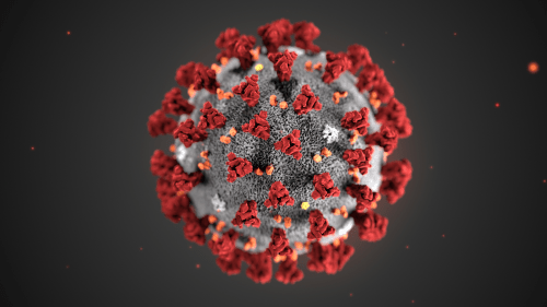
This illustration, created at the Centers for Disease Control and Prevention (CDC), reveals ultrastructural morphology exhibited by coronaviruses. Note the spikes that adorn the outer surface of the virus, which impart the look of a corona surrounding the virion, when viewed electron microscopically. A novel coronavirus virus was identified as the cause of an outbreak of respiratory illness first detected in Wuhan, China in 2019.
Image of newly mapped coronavirus protein, called Nsp15, which helps the virus replicate.
How UC is responding to the coronavirus (COVID-19)
The University of California is vigilantly monitoring and responding to new information about the coronavirus (COVID-19) outbreak, which has been declared a global health emergency.
The 3-D structure of a potential drug target in a newly mapped protein of COVID-19, or coronavirus, has been solved by a team of researchers from the University of California, Riverside, the University of Chicago, the U.S. Department of Energy’s Argonne National Laboratory, and Northwestern University.
The scientists said their findings suggest drugs previously developed to treat the earlier SARS outbreak could now be developed as effective drugs against COVID-19.
The initial genome analysis and design of constructs for protein synthesis were performed by the bioinformatic group of Adam Godzik, a professor of biomedical sciences at the UC Riverside School of Medicine.
The protein Nsp15 from Severe Acute Respiratory Syndrome Coronavirus 2, or SARS-CoV-2, is 89% identical to the protein from the earlier outbreak of SARS-CoV. SARS-CoV-2 is responsible for the current outbreak of COVID-19. Studies published in 2010 on SARS-CoV revealed inhibition of Nsp15 can slow viral replication.This suggests drugs designed to target Nsp15 could be developed as effective drugs against COVID-19.
Adam Godzik, UC Riverside professor of biomedical sciences Credit: Sanford Burnham Prebys Medical Discovery Institute
“While the SARS-CoV-19 virus is very similar to the SARS virus that caused epidemics in 2003, new structures shed light on the small, but potentially important differences between the two viruses that contribute to the different patterns in the spread and severity of the diseases they cause,” Godzik said.
The structure of Nsp15, which will be released to the scientific community on March 4, was solved by the group of Andrzej Joachimiak, a distinguished fellow at the Argonne National Laboratory, University of Chicago Professor, and Director of the Structural Biology Center at Argonne’s Advanced Photon Source, a Department of Energy Office of Science user facility.
“Nsp15 is conserved among coronaviruses and is essential in their lifecycle and virulence,” Joachimiak said. “Initially, Nsp15 was thought to directly participate in viral replication, but more recently, it was proposed to help the virus replicate possibly by interfering with the host’s immune response.”
Mapping a 3D protein structure of the virus, also called solving the structure, allows scientists to figure out how to interfere in the pathogen’s replication in human cells.
“The Nsp15 protein has been investigated in SARS as a novel target for new drug development, but that never went very far because the SARS epidemic went away, and all new drug development ended,” said Karla Satchell, a professor of microbiology-immunology at Northwestern, who leads the international team of scientists investigating the structure of the SARS CoV-2 virus to understand how to stop it from replicating. “Some inhibitors were identified but never developed into drugs. The inhibitors that were developed for SARS now could be tested against this protein.”
Rapid upsurge and proliferation of SARS-CoV-2 raised questions about how this virus could become so much more transmissible as compared to the SARS and MERS coronaviruses. The scientists are mapping the proteins to address this issue.
Over the past two months, COVID-19 infected more than 80,000 people and caused at least 2,700 deaths. Although currently mainly concentrated in China, the virus is spreading worldwide and has been found in 46 countries. Millions of people are being quarantined, and the epidemic has impacted the world economy. There is no existing drug for this disease, but various treatment options, such as utilizing medicines effective in other viral ailments, are being attempted.
Godzik, Satchell, and Joachimiak — along with the entire center team — will map the structure of some of the 28 proteins in the virus in order to see where drugs can throw a chemical monkey wrench into its machinery. The proteins are folded globular structures with precisely defined functions and their “active sites” can be targeted with chemical compounds.
The first step is to clone and express the genes of the virus proteins and grow them as protein crystals in miniature ice cube-like trays. The consortium includes nine labs across eight institutions that will participate in this effort.
Above is a modified version of the Northwestern University news release written by Marla Paul.
Cardiometabolic Syndrome and the Genetics of Hypertension: The Neuroendocrine Transcriptome Control Points
Reporter: Aviva Lev-Ari, PhD, RN
Integrated Computational and Experimental Analysis of the Neuroendocrine Transcriptome in Genetic Hypertension Identifies Novel Control Points for the Cardiometabolic Syndrome
From the Departments of Bioengineering (R.S.F., G.W.S.-S.), Medicine (R.S.F., A.J.S., F.R., P.S.N., D.T.O.), Pharmacology (D.T.O.), and Psychiatry (C.M.N.), the Bioinformatics Program (C.Y.), and the Institute for Genomic Medicine (D.T.O.), University of California at San Diego; the VA San Diego Healthcare System, San Diego, CA (D.T.O.); the Departments of Computer Science & Human Genetics, University of California at Los Angeles (E.E.); the Department of Biotechnology, Indian Institute of Technology Madras, Chennai, India (N.R.M.); Clinical Pharmacology and The Genome Centre, William Harvey Research Institute, Barts and The London School of Medicine and Dentistry, Queen Mary University of London, London, United Kingdom (P.B.M.); Center for Complex Disease Genomics, McKusick-Nathans Institute of Genetic Medicine, Johns Hopkins University School of Medicine, Baltimore, MD (G.B.E.); and Scripps Research Institute, La Jolla, CA (J.W.).
Background—Essential hypertension, a common complex disease, displays substantial genetic influence. Contemporary methods to dissect the genetic basis of complex diseases such as the genomewide association study are powerful, yet a large gap exists betweens the fraction of population trait variance explained by such associations and total disease heritability.
Methods and Results—We developed a novel, integrative method (combining animal models, transcriptomics, bioinformatics, molecular biology, and trait-extreme phenotypes) to identify candidate genes for essential hypertension and the metabolic syndrome. We first undertook transcriptome profiling on adrenal glands from blood pressure extreme mouse strains: the hypertensive BPH (blood pressure high) and hypotensive BPL (blood pressure low). Microarray data clustering revealed a striking pattern of global underexpression of intermediary metabolism transcripts in BPH. The MITRA algorithm identified a conserved motif in the transcriptional regulatory regions of the underexpressed metabolic genes, and we then hypothesized that regulation through this motif contributed to the global underexpression. Luciferase reporter assays demonstrated transcriptional activity of the motif through transcription factors HOXA3, SRY, and YY1. We finally hypothesized that genetic variation at HOXA3, SRY, and YY1 might predict blood pressure and other metabolic syndrome traits in humans. Tagging variants for each locus were associated with blood pressure in a human population blood pressure extreme sample with the most extensive associations for YY1 tagging single nucleotide polymorphism rs11625658 on systolic blood pressure, diastolic blood pressure, body mass index, and fasting glucose. Meta-analysis extended the YY1results into 2 additional large population samples with significant effects preserved on diastolic blood pressure, body mass index, and fasting glucose.
Conclusions—The results outline an innovative, systematic approach to the genetic pathogenesis of complex cardiovascular disease traits and point to transcription factor YY1 as a potential candidate gene involved in essential hypertension and the cardiometabolic syndrome.
Adventures in Personal Medicine: Integrated Personal Omics Profiling for Following Healthy and Disease States
Michael Snyder, Ph.D., Professor and Chair, Genetics; Director, Stanford Center for Genomics and Personalized Medicine, Stanford University
Spontaneous and Therapy Induced Evolution of Tumor Genomes and Epigenomes
Joseph Costello, Ph.D., Professor in Residence, Department of Neurological Surgery; Director, Epigenetics Division, Cell Cycling and Signaling Program, Helen Diller Family Comprehensive Cancer Center, University of California, San Francisco
THIS WEEK’S FEATURED TALKS
This is just a small sample of presentations that you’ll be able to hear at TCEC: The Clinical Epigenome Conference in San Francisco, CA next month.
Daniel De Carvalho, Ph.D., Principal Investigator, Ontario Cancer Institute, University Health Network; Assistant Professor, Medical Biophysics, Faculty of Medicine, University of Toronto
Cancer cells typically exhibit aberrant DNA methylation patterns that can drive malignant transformation. Whether cancer cells are dependent on these abnormal epigenetic modifications remains elusive. We used experimental and bioinformatic approaches to unveil genomic regions that require DNA methylation for survival of cancer cells, suggesting these are key epigenetic events associated with tumorigenesis.
Yujiang Geno Shi, Ph.D., Associate Biochemist, Division of Endocrinology, Brigham and Women’s Hospital; Assistant Professor, Medicine, Harvard Medical School
Recent studies have shown that ten-eleven translocation (Tet) proteins can catalyze 5mC oxidation and generate 5mC derivatives, including 5-hydroxymethylcytosine (5hmC). Not only are Tet family proteins and 5hmC critical for the identity and normal function of embryonic stem cells and early embryonic process of development, but dysregulation of these newly identified epigenetic factors also plays a major role in cancer development. Here we report an essential role of Tet3 in animal development, and define 5hmC as a potential biomarker for tumor progression. These studies will significantly increase our current understanding of the biological functions of Tet proteins and 5hmC while providing mechanistic insight into the development of epigenetic therapeutics.
Defining the Epigenetic Landscape during Normal and Malignant Hematopoiesis
Lucy A. Godley, M.D., Ph.D., Associate Professor, Department of Medicine, Section of Hematology/Oncology, Cancer Research Center, The University of Chicago
Hematopoietic stem cell commitment and differentiation involves silencing of self-renewal genes and induction of a specific transcriptional program, which is controlled in part through dynamic changes in covalent cytosine modifications. We have studied how the abundance and distribution of these derivatized bases influences hematopoietic stem cell commitment during normal erythropoiesis as well as during leukemia development. The identification of recurrent mutations in several genes important in epigenetic pathways as well as mouse modeling suggest that the balance of covalent cytosine modifications is a key driver of normal blood cell development. I will also discuss how these findings impact our understanding of the activity of the ‘hypomethylating drugs’, now in common use for the treatment of myeloid malignancies.
Ite A. Laird-Offringa, Ph.D., Associate Professor, Departments of Surgery, Biochemistry and Molecular Biology, Norris Comprehensive Cancer Center, Keck School of Medicine, University of Southern California
I will discuss our research on integrated genome-scale DNA methylation and mRNA expression data from microdissected lung adenocarcinoma and matched non-tumor lung. We have identified 164 hypermethylated genes showing concurrent downregulation, and 57 hypomethylated genes showing increased expression. Integrated pathways analysis and detailed examination of individual genes suggests mechanistic contributions of several of these genes to lung adenocarcinoma development and/or progression. I will present information on a number of candidate epigenetic driver genes for lung adenocarcinoma.
Lessons from Surveying the DNA Methylation “Cityscape” of Lethal Metastatic Prostate Cancer
Srinivasan (Vasan) Yegnasubramanian, M.D., Ph.D., Assistant Professor, Department of Oncology, Sidney Kimmel Comprehensive Cancer Center, Johns Hopkins University School of Medicine
Alterations in DNA methylation are a hallmark of human cancers, including prostate cancer. We carried out genome-scale analyses of DNA methylation alterations in multiple metastases from each of 13 men that died of metastatic prostate cancer and created DNA methylation cityscapes to visualize these complex data. These analyses revealed that each individual developed a unique DNA methylation signature that was largely maintained across all metastases within that individual. By analyzing their frequency, clonal maintenance, and correlation with expression, we nominated potential “driver” DNA methylation alterations that could be prioritized for development as epigenetic biomarkers and therapeutic targets.
DNA Methylation Detection using Nanopores
George Vasmatzis, Ph.D., Director, Biomarker Discovery Program, Center of Individualized Medicine; Consultant, Department of Molecular Medicine, Mayo Clinic and Foundation
I will discuss some of our latest work around integrating micro-fabrication and solid state technologies with biomarkers. In diagnostics, a biosensor that could look for subtle structural or sequence variations at the single molecule level would be extremely useful . Epigenetic modifications have been linked with cancer, and we have developed nanopore technology to detect the methylation profile of a single molecule. The challenges and the potential of such technology will be discussed.
CONFERENCE TOPICS INCLUDE:
Genetic & Epigenetic Interplay in Cancer
Mechanisms in (De)Methylation Underlying
Development of Disease
The Predictive Power of Epigenetics: Diagnostic
& Prognostic Utility
The Clinical Genome Technology Showcase
Trends in Analysis & Interpretation
————————————————————————
DINNER SHORT COURSES*
Wednesday, June 26 View Details Registration Information
Reporters: Aviva Lev-Ari, PhD, RN and Pnina G. Abir-Am, PhD
Putting Genome Interpretation to the Test
01/30/2013
Ashley Yeager
How well do methods for interpreting genome variation work? Ashley Yeager takes a look at a community experiment that is trying to assess just how useful genome interpretation tools in real-world situations.
At the American Society of Human Genetics (ASHG) conference in November 2012 in San Francisco, CA, Steven Brenner, a computational geneticist from the University of California, Berkley, stood up in front of an audience and argued that it was unlikely that a single genome interpretation tool could identify variants for an array of illnesses or phenotypic traits (1). Instead, interpretation methods would likely need to be gene-specific or tailored for precise applications.
The predictors, assessors, and observers who participated in CAGI 2011, which was held in San Francisco, CA. Source: CAGI
This figure shows the ROC curves for the prediction of patients with Crohn’s disease against the result of 1,000 random predictions, which are shown in gray. Source: CAGI
Steven Brenner helped develop CAGI to determine how well genome interpretation tools could translate to the clinic. Source: UC Berkeley
John Moult, one of the organizers of CAGI, says the challenges are giving scientists a better sense of the genome interpretation tools that currently exist. Source: University of Maryland
Brenner came to that conclusion after looking over the results of the Critical Assessment of Genome Interpretation (CAGI), a community experiment, now in its third year, challenges researchers to computationally predict the phenotypes of genetic variants. The teams then compare their results with unpublished experimental data, showing researchers and clinicians which tools can most accurately interpret large amounts of genomic sequence variation data and which ones might be reliable enough to use in the clinic. The results from the first two rounds of challenges have been clear for Brenner: most genomic interpretation tools are not reliable enough for the clinic yet.
After his talk at ASHG, several clinicians came up to him and expressed their concerns. Many had been using genome interpretation tools more generally, possibly making their conclusions less reliable. “General methods are limited in how well they will perform, which is not what people assumed before,” he says. “What that reaction showed me was that CAGI has a broad set of people that derive value from the experiment’s findings.”
Increasing Confidence
Brenner and John Moult, a computational biologist at the University of Maryland in Rockville, MD, organized the first CAGI experiment in 2010. It was a pilot project to get a better sense of the tools researchers in the community were using to study human genome variation and the phenotypic predictions coming from them. “Coming into CAGI, we had no understanding of how well methods for interpreting genome variation worked,” Brenner says. “Now, we’re starting to get a hint of what the big picture is.”
The goal was to provide a better sense of the correct level of confidence scientists and clinicians should have in the methods to predict the phenotype of sequence variants that are out there right now. “There’s a lot of uncertainty about how these methods work on real problems and so the challenges address the question of how can we test them in real-world situations,” Moult says.
In the beginning, Brenner and Moult had little idea of what to expect. The first year of the experiment was supposed to be very small, a pilot to see who would participate and what tools actually existed. In the end, the 2010 challenges drew more than 100 prediction submissions from eight countries, exceeding the organizers’ expectations.
Forty of the participants traveled to Berkeley in December 2010 to review the results. The top prize was awarded to Yana Bromberg, a bioinformatician at Rutgers University in New Jersey, for her work on interpretation software called screening for non-acceptable polymorphisms, or SNAP for short, which evaluates the effects of single amino acid substitutions on protein function (2). It was the first time Moult and Brenner had heard of SNAP.
In 2011, teams worked on 11 challenges, resulting in 117 predictions from 21 groups representing 18 countries. The challenges expanded, including exercises on exome variation and breast cancer gene variation. Again, SNAP was often one of the best interpretation tools, ranking high on several of the challenges.
One of the challenges in the second year of the experiment asked variation predictors to analyze exome sequence data from 42 Crohn’s disease patients and 6 healthy individuals. Researchers didn’t know how many of the exomes had variations associated with the disease, but many of the tools predicted the disease in patients significantly better than random. The best performing teams used an unexpected approach, looking at rare variants on a large panel of genes (1).
“The Crohn’s results were so great, we wonder if they were an artifact,” Brenner says, explaining that the CAGI organizers have included the challenge again in this year’s experiment to verify the results. If the results hold, “it could be a huge breakthrough there in interpreting genetic variation under certain circumstances,” he says.
The first year results were significant in a statistical sense, but the second year, Brenner says, “really gave us a baseline for better understanding personal genome variations and also started to show which types of interpretation methods might be best for specific applications.”
Nowhere Near
The next step would be to explain why the methods, such as SNAP, are so successful. But that requires more funding. Right now, the experiment has no direct funding, but the CAGI organizing committee does have a grant proposal to run the experiment awaiting review. The National Institutes of Health typically funds the year-end meeting where challenge participants present their results. “We’re doing this on a shoe string,” Moult says. Despite the financial pressure, Brenner and Moult feel that they have invested too much time to give up on the CAGI experiments.
The 2012 challenge deadline is March 2013, with the meeting to present the results slated for July. The delay was largely due to funding issues. But Brenner and Moult hope that the extension will allow more researchers to participate. Overall, Brenner and Moult are excited to see the results.
This year the experiment has 10 challenges, which include a test that focuses on genetic and phenotypic variation in breast cancer as well as the tried-and-true test to predict individuals’ phenotypic traits based on their genomes. The information for the personal genome analysis comes from the Personal Genome Project (PGP). “It acts as a valuable resource for diagnostics evaluations and standardization testing like CAGI,” Harvard molecular geneticist George Church said in an email, adding that the PGP has been providing data to CAGI since its first year.
But this year, there’s a change to the personal genome challenge. For the past two years, participants used the data to predict individual phenotypic traits based on a genome. But phenotypic profiles of all PGP participants are now public. “The availability of the complete profiles makes it impossible to have a valid assessment of individual trait predictions,” Brenner explains.
So instead of predicting the phenotype based on a single genome, in the 2012 challenge, the participants will develop tools that play a “matching game.” The goal will be to match 77 genomes with their corresponding phenotypic profiles, each of which includes 239 traits such as high cholesterol, diabetes, and astigmatism. And to spice things up, the organizers have included 214 phenotypic profiles that do not match any of the 77 genomes.
Ultimately, the CAGI predictors will release the PGP challenge results to those who volunteered their genomes so the individuals can learn more about their genetic susceptibilities for disease. But the reliability of the results is not necessarily high yet, Brenner cautions, so it’s important that individuals, scientists, and clinicians take that into account if someone shows a predicted high risk for cancer or other serious illnesses.
“We are nowhere near having a method for genome interpretation where a doctor could use it and then go and give surgery based on what we are saying,” Moult says. He and Brenner hope CAGI is a first step toward getting there one day.
References
CAGI: The Critical Assessment of Genome Interpretation, a community experiment to evaluate phenotype prediction. (2012). American Society for Human Genetics Conference: Poster.
Bromberg, Y. and B. Rost. 2007. SNAP: predict effect of non-synonymous polymorphisms on function. Nucleic Acids Research 35: 3823-3835.