Feeds:
Posts
Comments

Archive for the ‘Proteins’ Category

Reengineering Therapeutics

Larry H. Bernstein, MD, FCAP, Curator

LPBI

The synNotch solution: UCSF scientists engineer a next-gen T-cell immunotherapy

Sunday, January 31, 2016 | By John Carroll

2.2.9

2.2.9   Reengineering Therapeutics, Volume 2 (Volume Two: Latest in Genomics Methodologies for Therapeutics: Gene Editing, NGS and BioInformatics, Simulations and the Genome Ontology), Part 2: CRISPR for Gene Editing and DNA Repair

CAR-T has been all the rage in cancer R&D for several years now as a slate of biotech upstarts pursue highly promising work reengineering T cells into attack weapons by adding a chimeric antigen receptor that can zero in on particular cancer cells. The approach has been highly effective in acute lymphoblastic leukemia, triggering an attack on B cells by homing in on the CD19 antigen, a breakthrough that has inspired a race to the regulatory finish line with the first CAR-Ts.

That approach, though, has run into some major obstacles when researchers move from the blood cancer to solid tumors. But now a group of scientists working with UC San Francisco’s Wendell Lim say they’ve come up with a new therapeutic model for T-cell engineering that promises to overcome that hurdle and make it a more precise weapon that can tackle solid tumors while avoiding off-target reactions that threaten patients.

The key to this new approach is a new receptor: synNotch. Taking a cue from nature, which relies on a sensor called Notch to perform key functions, the synthetic biology engineers say they can add a receptor that includes one section that sticks out from the cell with one that lies inside. By tinkering with synNotch they can reengineer the immune cell to run down a particular cancer cell target and then issue instructions to turn genes on or off to set up the other half of the therapeutic equation.

In a project described in Cell, the team says they created a synNotch that recognized an antigen on the surface of the cancer cell while the internal mechanism contributed a chimeric antigen receptor that recognized a different antigen. They then tested it on a mouse model that included two different tumor cells: one with both targets recognized by the external synNotch sensor and the CAR and one with just the CAR target.

The newly programmed attack weapon zeroed in on the two targets (which required synNotch activation for it to work) while leaving the other alone, providing preclinical proof-of-concept evidence that they could create a much more efficient tumor cell killing vehicle.

“The kinds of engineered T cells that we can now construct give us the exciting potential to create precision cancer therapeutics that take advantage of all the genomic and proteomic information we are currently gathering on disease,” said Lim, the senior author of the study. “This genomic information now becomes actionable.”

The team was led by Leonardo Morsut and Kole Roybal.

Lim added that it is possible to reengineer a T cell with multiple synNotches to make it even more precise. And there are added applications for autoimmune disease, regenerative medicine, and more.

Engineering Customized Cell Sensing and Response Behaviors Using Synthetic Notch Receptors

Leonardo  MorsutKole T. Roybal, Xin Xiong, Russell M. Gordley, Scott M. Coyle, Matthew Thomson, Wendell A. Lim

Tricked-Out Immune Cells Could Attack Cancer, Spare Healthy Cells  

New Cell-Engineering Technique May Lead To Precision Immunotherapies

New CRISPR/Cas9 delivery method could offer a clinical pathway

Read Full Post »

Nanosensors for Protein Recognition and Gene Proteome Interaction

Larry H Bernstein, MD, FCAP, Curator

LPBI


Synthetic Antibody Detects Proteins

http://www.technologynetworks.com/Proteomics/news.aspx?ID=187242

Research could lead to nanosensors that recognize fibrinogen, insulin, or other biomarkers

Using carbon nanotubes, MIT chemical engineers have devised a new method for detecting proteins, including fibrinogen, one of the coagulation factors critical to the blood-clotting cascade.

This approach, if developed into an implantable sensor, could be useful for monitoring patients who are taking blood thinners, allowing doctors to make sure the drugs aren’t interfering too much with blood clotting.

The new method is the first to create synthetic recognition sites (similar to natural antibodies) for proteins and to couple them directly to a powerful nanosensor such as a carbon nanotube. The researchers have also made significant progress on a similar recognition site for insulin, which could enable better monitoring of patients with diabetes. It may also be possible to use this approach to detect proteins associated with cancer or heart disease, says Michael Strano, the Carbon P. Dubbs Professor in Chemical Engineering at MIT.

A targeted search

The new sensor is the latest example of a method developed in Strano’s lab, known asCorona Phase Molecular Recognition (CoPhMoRe).

This technique takes advantage of the interactions between a given polymer and a nanoparticle surface such as that of a fluorescent single-walled carbon nanotube, when the polymer is wrapped around the nanotube.

Certain regions of the polymers latch onto the nanoparticle surface like anchors, while other regions extend outwards into their environment. This outward-facing region, also known as the adsorbed phase or corona, has a 3-D structure that depends on the composition of the polymer.

CoPhMoRe works when a specific polymer adsorbs to the nanoparticle surface and creates a corona that recognizes the target molecule. These interactions are very specific, just like the binding between an antibody and its target. Binding of the target alters the carbon nanotubes’ natural fluorescence, allowing the researchers to measure how much of the target molecule is present.

Strano’s lab has previously used this approach to find recognition sites and develop nansensors for estradiol and riboflavin, among other molecules. The new paper represents their first attempt to identify corona phases that can detect proteins, which are larger, more complex, and more fragile than the molecules identified by their previous sensors.

For this study, Bisker began by screening carbon nanotubes wrapped in 20 different polymers including DNA, RNA, and polyethylene glycol (PEG), a polymer often added to drugs to increase their longevity in the bloodstream.

On their own, none of the polymers had any affinity for the 14 proteins tested, all taken from human blood. However, when the researchers tested polymer-wrapped nanotubes against the same proteins, they turned up a match between one of the modified nanotubes and fibrinogen.

“A chemist or a biologist would not be able to predict ahead of time that there should be any kind of affinity between fibrinogen and this corona phase,” Strano says. “It really is a new kind of molecular recognition.”

Fibrinogen, one of the most abundant proteins in human blood, is part of the blood-clotting cascade. When a blood vessel is damaged, an enzyme called thrombin converts fibrinogen into fibrin, a stringy protein that forms clots to seal the wound.

A sensor for fibrinogen could help doctors determine if patients who are taking blood thinners still have enough clotting capability to protect them from injury, and could allow doctors to calculate more finely tuned dosages. It could also be used to test patients’ blood clotting before they go into surgery, or to monitor wound healing, Bisker says.

Synthetic antibodies

The researchers believe their synthetic molecular recognition agents are an improvement over existing natural systems based on antibodies or DNA sequences known as aptamers, which are more fragile and tend to degrade over time.

“One of the advantages of this is that it’s a completely synthetic system that can have a much longer lifetime within the body,” Bisker says.

In 2013, researchers in Strano’s lab demonstrated that carbon nanotube sensors can remain active in mice for more than a year after being embedded in a polymer gel and surgically implanted under the skin.

In addition to insulin, the researchers are also interested in detecting troponin, a protein that is released by dying heart cells, or detecting proteins associated with cancer, which would be useful for monitoring the success of chemotherapy. These and other protein sensors could become critical components of devices that deliver drugs in response to a sign of illness.

“By measuring therapeutic markers in the human body in real time, we can enable drug delivery systems that are much smarter, and release drugs in precise quantities,” Strano says. “However, measurement of those biomarkers is the first step.”


New Device Uses Carbon Nanotubes to Snag Molecules
Nanotube “forest” in a microfluidic channel may help detect rare proteins and viruses.
Tuesday, December 22, 2015

Nanotube “forest” in a microfluidic channel may help detect rare proteins and viruses.

Engineers at MIT have devised a new technique for trapping hard-to-detect molecules, using forests of carbon nanotubes.

The team modified a simple microfluidic channel with an array of vertically aligned carbon nanotubes — rolled lattices of carbon atoms that resemble tiny tubes of chicken wire. The researchers had previously devised a method for standing carbon nanotubes on their ends, like trees in a forest. With this method, they created a three-dimensional array of permeable carbon nanotubes within a microfluidic device, through which fluid can flow.

Now the researchers have given the nanotube array the ability to trap certain particles. To do this, the team coated the array, layer by layer, with polymers of alternating electric charge.

“You can think of each nanotube in the forest as being concentrically coated with different layers of polymer,” says Brian Wardle, professor of aeronautics and astronautics at MIT. “If you drew it in cross-section, it would be like rings on a tree.”

Depending on the number of layers deposited, the researchers can create thicker or thinner nanotubes and thereby tailor the porosity of the forest to capture larger or smaller particles of interest.

The nanotubes’ polymer coating may also be chemically manipulated to bind specific bioparticles flowing through the forest. To test this idea, the researchers applied an established technique to treat the surface of the nanotubes with antibodies that bind to prostate specific antigen (PSA), a common experimental target. The polymer-coated arrays captured 40 percent more antigens, compared with arrays lacking the polymer coating.

Wardle says the combination of carbon nanotubes and multilayer coatings may help finely tune microfluidic devices to capture extremely small and rare particles, such as certain viruses and proteins.

“There are smaller bioparticles that contain very rich amounts of information that we don’t currently have the ability to access in point-of-care [medical testing] devices like microfluidic chips,” says Wardle, who is a co-author on the paper. “Carbon nanotube arrays could actually be a platform that could target that size of bioparticle.”

The paper’s lead author is Allison Yost, a former graduate student who is currently an engineer at Accion Systems. Others on the paper include graduate student Setareh Shahsavari; postdoc Roberta Polak; School of Engineering Professor of Teaching Innovation Gareth McKinley; professor of materials science and engineering Michael Rubner, and Raymond A. And Helen E. St. Laurent Professor of Chemical Engineering Robert Cohen.

A porous forest

Carbon nanotubes have been a subject of intense scientific study, as they possess exceptional electrical, mechanical, and optical properties. While their use in microfluidics has not been well explored, Wardle says carbon nanotubes are an ideal platform because their properties may be manipulated to attract certain nanometer-sized molecules. Additionally, carbon nanotubes are 99 percent porous, meaning a nanotube is about 1 percent carbon and 99 percent air.

“Which is what you need,” Wardle says. “You need to flow quantities of fluid through this material to shed all the millions of particles you don’t want to find and grab the one you do want to find.”

What’s more, Wardle says, a three-dimensional forest of carbon nanotubes would provide much more surface area on which target molecules may interact, compared with the two-dimensional surfaces in conventional microfluidics.

“The capture efficiency would scale with surface area,” Wardle notes.

A versatile array

The team integrated a three-dimensional array of carbon nanotubes into a microfluidic device by using chemical vapor deposition and photolithography to grow and pattern carbon nanotubes onto silicon wafers. They then grouped the nanotubes into a cylinder-shaped forest, measuring about 50 micrometers tall and 1 millimeter wide, and centered the array within a 3 millimeter-wide, 7-millimeter long microfluidic channel.

The researchers coated the nanotubes in successive layers of alternately charged polymer solutions in order to create distinct, binding layers around each nanotube. To do so, they flowed each solution through the channel and found they were able to create a more uniform coating with a gap between the top of the nanotube forest and the roof of the channel. Such a gap allowed solutions to flow over, then down into the forest, coating each individual nanotube. In the absence of a gap, solutions simply flowed around the forest, coating only the outer nanotubes.

After coating the nanotube array in layers of polymer solution, the researchers demonstrated that the array could be primed to detect a given molecule, by treating it with antibodies that typically bind to prostate specific antigen (PSA). They pumped in a solution containing small amounts of PSA and found that the array captured the antigen effectively, throughout the forest, rather than just on the outer surface of a typical microfluidic element.

Wardle says that the nanotube array is extremely versatile, as the carbon nanotubes may be manipulated mechanically, electrically, and optically, while the polymer coatings may be chemically altered to capture a wide range of particles. He says an immediate target may be biomarkers called exosomes, which are less than 100 nanometers wide and can be important signals of a disease’s progression.

“Science is really picking up on how much information these particles contain, and they’re sort of everywhere, but really hard to find, even with large-scale equipment,” Wardle says. “This type of device actually has all the characteristics and functionality that would allow you to go after bioparticles like exosomes and things that really truly are nanometer scale.”

This research was funded, in part, by the National Science Foundation.


A Natural Light Switch

MIT scientists identify and map the protein behind a light-sensing mechanism.

MIT scientists, working with colleagues in Spain, have discovered and mapped a light-sensing protein that uses vitamin B12 to perform key functions, including gene regulation.

The result, derived from studying proteins from the bacterium Thermus thermophilus, involves at least two findings of broad interest. First, it expands our knowledge of the biological role of vitamin B12, which was already understood to help convert fat into energy, and to be involved in brain formation, but has now been identified as a key part of photoreceptor proteins — the structures that allow organisms to sense and respond to light.

Second, the research describes a new mode of gene regulation, in which the light-sensing proteins play a key role. In so doing, the scientists observe, the bacteria have repurposed existing protein structures that use vitamin B12, and put them to work in new ways.

MIT-Proteins-Light-1_0.jpg

http://www.technologynetworks.com/images/videos/News%20Images/CR/MIT-Proteins-Light-1_0.jpg

“Nature borrowed not just the vitamin, but really the whole enzyme unit, and modified it … and made it a light sensor,” says Catherine Drennan, a professor of chemistry and biology at MIT


The paper describes the photoreceptors in three different states: in the dark, bound to DNA, and after being exposed to light.

“It’s wonderful that we’ve been able to get all the series of structures, to understand how it works at each stage,” Drennan says.

The paper has nine co-authors, including Drennan; graduate students Percival Yang-Ting Chen, Marco Jost, and Gyunghoon Kang of MIT; Jesus Fernandez-Zapata and S. Padmanabhan of the Institute of Physical Chemistry Rocasolano, in Madrid; and Monserrat Elias-Arnanz, Juan Manuel Ortiz-Guerreo, and Maria Carmen Polanco, of the University of Murcia, in Murcia, Spain.

The researchers used a combination of X-ray crystallography techniques and in-vitro analysis to study the bacteria. Drennan, who has studied enzymes that employ vitamin B12 since she was a graduate student, emphasizes that key elements of the research were performed by all the co-authors.

Jost performed crystallography to establish the shapes of the structures, while the Spanish researchers, Drennan notes, “did all of the control experiments to show that we were really thinking about this right,” among other things.

MIT-Proteins-Light-2.jpg

By studying the structures of the photoreceptor proteins in their three states, the scientists developed a more thorough understanding of the structures, and their functions, than they would have by viewing the proteins in just one state.

Microbes, like many other organisms, benefit from knowing whether they are in light or darkness. The photoreceptors bind to the DNA in the dark, and prevent activity pertaining to the genes of Thermus thermophilus. When light hits the microbes, the photoreceptor structures cleave and “fall apart,” as Drennan puts it, and the bacteria start producing carotenoids, which protect the organisms from negative effects of sunlight, such as DNA damage.

The research also shows that the exact manner in which the photoreceptors bind to the DNA is novel. The structures contain tetramers, four subunits of the protein, of which exactly three are bound to the genetic material — something Drennan says surprised her.

“That’s the best part about science,” Drennan says. “You see something novel, then you think it’s not really going to be that novel, but you do the experiments [and it is].”

Other scientists say the findings are significant. “It’s a very exciting development,” says Rowena Matthews, a professor emerita of biological chemistry at the University of Michigan, who has read the paper. Of the newly discovered use of vitamin B12 and a derivative of it, adenosylcobalamin, Matthews adds, “There was very limited knowledge of its versatility.”

Drennan adds that in the long run, the finding could have practical applications, such as the engineering of light-directed control of DNA transcription, or the development of controlled interactions between proteins.

“I would be very interested in … thinking about whether there could be practical applications of this,” Drennan says.


HIV Protein Manipulates Hundreds of Human Genes

Findings search for new or improved treatments for patients with AIDS.

UT Southwestern Medical Center researchers have deciphered how a small protein made by the human immunodeficiency virus (HIV) that causes AIDS manipulates human genes to further its deadly agenda.

The findings, published in the online journal eLife, could aid in the search for new or improved treatments for patients with AIDS, or to the development of preventive strategies.

“We have identified the molecular mechanisms by which the Tat protein made by HIV interacts with the host cell to activate or repress several hundred human genes,” said Dr. Iván D’Orso, Assistant Professor of Microbiology at UT Southwestern and senior author of the study. “The findings clearly suggest that blocking Tat activity may be of therapeutic value to HIV patients.”

It has long been known that HIV causes AIDS by hijacking the body’s immune cells, transforming them into HIV factories and killing other immune cells that normally fight disease. HIV also hides in cells and continues to undermine the host’s immune system despite antiretroviral therapy that has improved the outlook of those with AIDS.

The latest data from the Centers for Disease Control and Prevention (CDC), in 2012, estimated 1.2 million Americans were living with HIV, including 156,300 whose infections had not been diagnosed. About 50,000 people in the U.S. are newly infected with HIV annually, the CDC projects. In 2013, the CDC estimated that over 26,000 Americans had the advanced form of HIV infection, AIDS.

Like all retroviruses, HIV has very few genes of its own and must take over the host’s cellular machinery in order to propagate and spread throughout the body. Although the broad aspects of that cellular hijacking were known, the nuances remain to be explored, Dr. D’Orso said.

“We observed that HIV methodically and precisely manipulates the host’s genes and cellular machinery. We also observed that HIV rewires cellular defensive pathways to benefit survival of the virus,” he added.

The study provides insights into HIV’s ability to survive despite antiretroviral therapy, findings that could lead to new therapeutic targets or ways to make current therapies more effective, he said.

“Our study indicates that this small viral protein, Tat, directly binds to about 400 human genes to generate an environment in which HIV can thrive. Then, this protein precisely turns off the body’s immune defense. It is striking that such a small viral protein has such a large impact,” Dr. D’Orso said. “The human genes and pathways that Tat manipulates correlate well with symptoms observed in these patients, such as immune system hyperactivation, then weakening, and accelerated aging,” Dr. D’Orso said, describing the situation in which HIV infection leads to AIDS.

Italy’s National Institute of Health in Rome recently completed a phase II clinical trial of an experimental vaccine that targets the Tat protein. That trial, which followed 87 HIV-positive patients for up to three years, reported that the vaccine was well-tolerated without significant side effects. However, it will take several years to determine if the vaccine works, Dr. D’Orso said.

Although someone can have HIV for years without showing symptoms, AIDS occurs when HIV blocks the body’s ability to fight off illness. The person then becomes overrun by the opportunistic infections and specific cancers that are hallmarks of AIDS.


New Light Shed on Genetic Regulation

A team of scientists has uncovered greater intricacy in protein signaling than was previously understood, shedding new light on the nature of genetic production.

Christine Vogel, an assistant professor in New York University’s Department of Biology and one of the study’s senior authors, explains that “to make a protein, we need to make a messenger RNA molecule from the gene encoded in the DNA, and then, in a second process, make proteins from these RNA molecules. Both processes are highly regulated and coupled.”

This coupling is similar to the coupling between a moving escalator and a person walking on it at the same time.

The research takes a closer look at how the two coupled processes change in the cell responding to an outside stimulus.

“Until recently, it has been very difficult to study these systems and researchers have thought that the movement of the escalator is most important during the cellular response,” Vogel explains. “We now show that is not necessarily the case, and under some circumstances, the person’s walking determines the overall outcome.”

In biology, this means that both of the processes—to make RNAs and proteins—play important roles, but with different patterns.

In their study, the scientists, who also included researchers from National University Singapore and Berlin’s Max Delbruck Center, took a closer look at how the two processes exactly respond over time.

Their results showed notable distinctions between DNA and mRNA in the nature of their signaling. Notably, the process of making RNA from DNA was pulse-like—a brief messaging over the studied period that returned to the normal levels by the end of the measurements. By contrast, the process of making a protein from RNA was akin to an on/off switch: once started, levels remained constant for consistent periods before reverting back to long stretches of dormancy.

While the reasons for these differences in cell behavior remain unknown, the researchers believe the answer may lie in the nature of the two tasks.

“It is very costly for the cell to make proteins, but making RNA messages from DNA is a relatively low-energy and simple process, so it makes sense that we see frequent, or pulsating, signaling at this stage,” observes Vogel. “By contrast, creating proteins is an intricate undertaking, requiring a great deal of time and energy. This may be why, once you decided to stop production of proteins, you do not turn it back on that easily—and the other way around.”


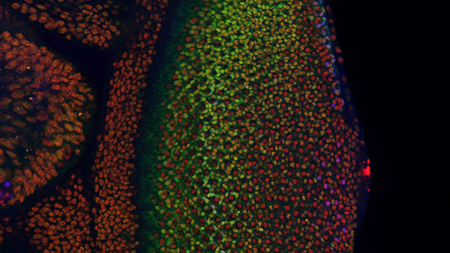
Where Cancer Cells May Begin

Scientists use fruit fly genetics to understand how things could go wrong in cancer.

Cancer cells are normal cells that go awry by making bad developmental decisions during their lives. In a study involving the fruit fly equivalent of an oncogene implicated in many human leukemias, Northwestern University researchers have gained insight into how developing cells normally switch to a restricted, or specialized, state and how that process might go wrong in cancer.

The fruit fly’s eye is an intricate pattern of many different specialized cells, such as light-sensing neurons and cone cells. Because flies share with humans many of the same cancer-causing genes, scientists use the precisely made compound eye of Drosophila melanogaster (the common fruit fly) as a workhorse to study what goes wrong in human cancer.

retina638.jpg

http://www.technologynetworks.com/images/videos/News%20Images/GT/retina638.jpg

A multidisciplinary team co-led by biologist Richard W. Carthew and engineer Luís A.N. Amaral studied normal cell behavior in the developing eye. The researchers were surprised to discover that the levels of an important protein called Yan start fluctuating wildly when the cell is switching from a more primitive, stem-like state to a more specialized state. If the levels don’t or can’t fluctuate, the cell doesn’t switch and move forward.

“This mad fluctuation, or noise, happens at the time of cell transition,” said Carthew, professor of molecular biosciences in Northwestern’s Weinberg College of Arts and Sciences. “For the first time, we see there is a brief time period as the developing cell goes from point A to point B. The noise is a state of ‘in between’ and is important for cells to switch to a more specialized state. This limbo might be where normal cells take a cancerous path.”

The researchers also found that a molecular signal received by a cell receptor called EGFR is important for turning the noise off. If that signal is not received, the cell remains in an uncontrolled state.

By pinpointing this noise and its “off” switch as important points in the normal process of cell differentiation, the Northwestern researchers provide targets for scientists studying how cells can go out of control and transform into cancer cells.

The “noisy” protein the Northwestern researchers studied is called Yan in the fly and Tel-1 in humans. (The protein is a transcription factor.) The Tel-1 protein instructs cells to turn into white blood cells; the gene that produces the protein, oncogene Tel-1, is frequently mutated in leukemia.

The EGFR protein that turns off the noise in flies is called Her-2 in humans. Her-2 is an oncogene that plays an important role in human breast cancer.

“On the surface, flies and humans are very different, but we share a remarkable amount of infrastructure,” said Carthew, a member of the Robert H. Lurie Comprehensive Cancer Center of Northwestern University. “We can use fruit fly genetics to understand how humans work and how things go wrong in cancer and other diseases.”

Fruit fly cells are small and closely packed together, making study of them challenging. Carthew and Amaral’s team of biologists, chemical and biological engineers, computer scientists and chemists together figured out how to identify and analyze thousands and thousands of individual cells in the flies’ eyes.

“In the past, people have built models of regulatory networks that control cell differentiation mostly by genetically perturbing one or two components of the network at a time and then compiling those results into models,” said Amaral, professor of chemical and biological engineering at the McCormick School of Engineering. “We instead measured the retina as it developed and found the unexpected behavior of the key regulatory factors Yan and EGFR.”

Nicolás Peláez, first author of the study and a Ph.D. candidate in interdisciplinary biological sciences working with Amaral and Carthew, built new tools to study this strange feature of noise in developing flies. His methods enabled the researchers to easily measure both the concentration of the Yan protein and its fluctuation (noise).

It takes 15 to 20 hours for a fruit fly cell to go from being an unrestricted cell to a restricted cell, Carthew said. Peláez determined the Yan protein is noisy, or fluctuating, for six to eight of those hours.

“Studying the dynamics of molecules regulating fly-eye patterning can inform us about human disease,” Peláez said. “Using model organisms such as fruit flies will help us understand quantitatively the basic biological principles governing differentiation in complex animals.”


Mechanism of Tumor Suppressing Gene Uncovered

The most commonly mutated gene in cancer,p53, works to prevent tumor formation by keeping mobile elements in check that otherwise lead to genomic instability, UT Southwestern Medical Center researchers have found.

The p53 gene long has been known to suppress tumor formation, but the mechanisms behind this function – and why disabling the gene allows tumors to form – were not fully understood.

Findings from the study answer some of these questions and could one day lead to new ways of diagnosing and treating cancer, said the study’s senior author, Dr. John Abrams, Professor of Cell Biology at UT Southwestern.

The investigators found that normal p53 gene action restrains transposons, mobile genetic elements called retroelements that can make copies of themselves and move to different positions on chromosomes. But, they discovered, when p53 is disabled by mutation, dramatic eruptions of these mobile elements occur. The study revealed that in mice with cancer and in human samples of two types of cancer (Wilms’ tumors and colon tumors) disabled for p53, transposons became very active.

In a healthy state, certain mechanisms work to keep these retroelements quiet and inactive, explained Dr. Abrams. One of those mechanisms is p53 action. Conversely, when p53 is mutated, retroelements can erupt.

“If you take the gene away, transposons can wreak havoc throughout the genome by causing it to become highly dysregulated, which can lead to disease,” Dr. Abrams said. “Our findings help explain why cancer genomes are so much more fluid and destabilized than normal genomes. They also provide a novel framework for understanding how normal cells become tumors.”

Although much more research is needed, Dr. Abrams said, the potential clinical implications of the team’s findings are significant.

“Understanding how p53 prevents tumors raises the prospect of therapeutic interventions to correct cases in which p53 is disabled,” Dr. Abrams said. “If retroelements are at the heart of certain p53-driven cancers, finding ways to suppress them could potentially allow us to prevent those cancers or intervene to keep them from progressing.”

This understanding also could lead to advances in diagnosing some cancers through biomarkers related to p53 and transposon activity.

“One possibility is that perhaps blood or urine tests could detect dysregulated retroelements that could be indicative of certain types of cancer,” Dr. Abrams said.

Read Full Post »

The transport of molecules across membranes

Larry H. Bernstein, MD, FCAP, Curator

LPBI


Cellular Transport and the Nobel Prize for Medicine

Extracted  from October 8, 2013 | by

The 2013 Nobel Prize in Physiology or Medicine was  awarded to  to Randy W. Schekman, at the University of California at Berkeley; James E. Rothman,  at Yale University in New Haven, Connecticut; and Thomas C. Südhof,  at Stanford University, for their discoveries of machinery regulating vesicle traffic, a major transport system in cells.three U.S. scientists for their work on how the cell coordinates its transport system to shuttle proteins and other molecules from one location to another.

The organization and transport of molecules across cellular mmembranes is accomplished via vesicles that shuttle cargo between organelles or fuse to other structures to release their cargo outside the cell. The vesicle transport system is critical for a variety of physiological processes, ranging from signaling in the brainto release of hormones and immune cytokines.

Schekman identified three classes of genes that control different facets of the cell’s transport system.

Vesicle fusion

http://www.highlighthealth.com/wp-content/uploads/2013/10/vesicle-fusion.jpg

This was followed by James Rothman’s discovery that a protein complex enables vesicles to fuse with their target membranes (pictured in orange above). This lock and key mechanism ensures that the vesicle fuses at the right location and that cargo molecules are delivered to the correct destination.

Also in the 1990s, Thomas Südhof was studying how nerve cells communicate in the brain. Calcium ions were known to be involved in vesicle cargo release, and Südhof searched for calcium sensitive proteins in nerve cells. He identified the molecular machinery (pictured in purple above) that responds to an influx of calcium ions (Ca2+) and triggers vesicle fusion.

Extracellular vesicles are  participate in the pathogenesis of various diseases, most notably neurodegenerative disorders, and extracellular vesicles are likely to have therapeutic applications in large-molecule drug delivery.

References

  1. The Nobel Prize in Physiology or Medicine 2013 – Press Release. Nobelprize.org. 7 Oct 2013.
  2. Andaloussi et al. Extracellular vesicles: biology and emerging therapeutic opportunities. Nature Reviews Drug Discovery 2013 Vol: 12(5):347-357. DOI: 10.1038/nrd3978
    View abstract
  3. Anderson et al. Role of extracellular membrane vesicles in the pathogenesis of various diseases, including cancer, renal diseases, atherosclerosis, and arthritis. Lab Invest. 2010 Nov;90(11):1549-57. DOI: 10.1038/labinvest.2010.152. Epub 2010 Aug 30.
    View abstract


Machinery Regulating Vesicle Traffic, A Major Transport System in our Cells

http://www.nobelprizemedicine.org/wp-content/uploads/2013/10/Scientific-Background-Zierath-and-Lindahl.pdf

Together, Rothman, Schekman and Südhof have transformed the way we view transport of molecular cargo to specific destinations inside and outside the cell. Their discoveries explain a long-standing enigma in cell biology and also shed new light on how disturbances in this machinery can have deleterious effects and contribute to conditions such as neurological diseases, diabetes, and immunological disorders.

Eukaryotic cells differ from prokaryotic cells by their more complex intracellular organization. In eukaryotes, specific cellular functions are compartmentalized into the cell nucleus and organelles surrounded by intracellular membranes. This compartmentalization vastly improves the efficiency of many cellular functions and prevents potentially dangerous molecules from roaming freely within the cell. But when distinct cellular processes are compartmentalized, a problem emerges. Different compartments need to exchange specific molecules (Figure 1). Furthermore, certain molecules need to be exported to the cell exterior. Most molecules are too large to directly pass through membranes, thus a mechanism that ensures specific delivery of this molecular cargo is required.

Figure 1: Each cell in the body has a complex organization where specific cellular functions are separated into different compartments called organelles. Molecules produced in the cell are packaged in vesicles and transported with special and temporal precision to the correct locations within and outside the cell.

Mysteries of cellular compartmentalization have long intrigued scientists. Improved light microscopy techniques aided in the understanding of intracellular organization in eukaryotic cells, but the advent of electron microscopy and new staining techniques, combined with subcellular fractionation assays using differential ultracentrifugation procedures, led to a deeper understanding of the cell’s inner life. Albert Claude, George Palade and Christian de Duve, who received the Nobel Prize in Physiology or Medicine 1974*, were pioneers in this area and have shed light on how the cell is organized and compartmentalized. Secretory proteins were shown to be produced on ribosomes in the endoplasmic reticulum (ER) and trafficked to the Golgi complex (named after the 1906 Nobel Laureate Camillo Golgi) (Figure 1). Progress was also made in deciphering how proteins find their appropriate destination. Günter Blobel was awarded the 1999 Nobel Prize in Physiology or Medicine* for his discoveries that proteins have intrinsic signals that govern their transport and localization in the cell. Yet, a lingering question remained. How are molecules, including hormones, transport proteins, and neurotransmitters, correctly routed to their appropriate destination? From the work of Palade, the traffic of secretory proteins from the ER was understood to be carried out using small membrane-surrounded vesicles that bud from one membrane and fuse with another, but how precision could be acquired in this process remained enigmatic.

The work of  Rothman, Schekman and Südhof represents a paradigm shift in our understanding of how the eukaryotic cell, with its complex internal compartmentalization, organizes the routing of molecules packaged in vesicles to various intracellular destinations, as well as to the outside of the cell. Specificity in the delivery of molecular cargo is essential for cell function and survival. This specificity is required for the release of neurotransmitters into the presynaptic region of a nerve cell to transmit a signal to a neighboring nerve cell. Likewise, specificity is required for the export of hormones such as insulin to the cell surface. While vesicles within the cell were long known to be critical components of this transportation scheme, the precise mechanism by which these vesicles found their correct destination and how they fused with organelles or the plasma membrane to deliver the cargo remained mysterious. The work of the three 2013 Laureates radically altered our understanding of this aspect of cell physiology. Randy W. Schekman used yeast genetics to identify a set of genes critical for vesicular trafficking. He showed that these genes were essential for life and could be classified into three categories regulating different aspects of vesicle transport. James E. Rothman embarked on a biochemical approach and identified proteins that form a functional complex controlling cell fusion. Proteins on the vesicle and target membrane sides bind in specific combinations, ensuring precise delivery of molecular cargo to the right destination. Thomas C. Südhof became interested in how vesicle fusion machinery was controlled. He unraveled the mechanism by which calcium ions trigger release of neurotransmitters, and identified key regulatory components in the vesicle fusion machinery.

Schekman discovered genes encoding proteins that are key regulators of vesicle traffic. Comparing normal with genetically mutated yeast cells in which vesicle traffic was disturbed, he identified genes that control transport to different compartments and to the cell surface

Rothman published a series of papers where he reconstituted the intracellular transport of the VSV-G protein within the Golgi complex. He then used the assay to study both vesicle budding and fusion, and purified proteins from the cytoplasm that were required for transport. The first protein to be purified was the Nethylmaleimide-sensitive factor (NSF). Rothman’s discovery of NSF paved the way for the subsequent identification of other proteins important for the control of vesicle fusion, and the next one in line was SNAP (soluble NSFattachment protein). SNAPs bind to membranes and assist in the recruitment of NSF.

One of the yeast mutants, sec18, corresponded to NSF, which also revealed that the vesicle fusion machinery was evolutionarily ancient. Furthermore, Rothman and Schekman collaboratively cloned sec17 and provided evidence of its functional equivalence to SNAP. Other sec genes were shown to correspond to genes encoding fusion proteins were identified by other methods.

Using the NSF and SNAP proteins as bait, Rothman next turned to brain tissue, from which he purified proteins that he later named SNAREs (soluble NSF-attachment protein receptors). Intriguingly, three SNARE proteins, VAMP/Synaptobrevin, SNAP-25 and syntaxin, were found in stoichiometric amounts, which suggested to Rothman that they functioned together in the vesicle and target membranes. The three proteins had previously been identified by several scientists, including Richard Scheller, Kimio Akagawa, Reinhard Jahn and Pietro de Camilli, and localized to the presynaptic region, but their function was largely unknown. VAMP/Synaptobrevin resided on the vesicle, whereas SNAP-25 and syntaxin were found at the plasma membrane. This prompted Rothman to propose a hypothesis – the SNARE hypothesis – which stipulated that target and vesicle SNAREs (t-SNAREs and v-SNAREs) were critical for vesicle fusion through a set of sequential steps of synaptic docking, activation and fusion.

Thomas C. Südhof originally trained at the Georg-August-Universität and the Max-Planck Institute for Biophysical Sciences in Göttingen, Germany, and was a postdoctoral fellow with Michael Brown and Joseph Goldstein (Nobel Prize 1985) at University of Texas Southwestern Medical School in Dallas. As a junior group leader, he set out to study how synaptic vesicle fusion was controlled. Rothman and Schekman had provided fundamental machinery for vesicle fusion, but how vesicle fusion was temporally controlled remained enigmatic. Vesicular fusions in the body need to be kept carefully in check, and in some cases vesicle fusion has to be executed with high precision in response to specific stimuli. This is the case for example for neurotransmitter release in the brain and for insulin secretion from the endocrine pancreas.

The neurophysiology field was electrified by the discoveries of Bernard Katz, Ulf von Euler and Julius Axelrod who received the Nobel Prize in Physiology or Medicine 1970* for their discoveries concerning the humoral transmittors in the nerve terminals and the mechanism for their storage, release and inactivation. Südhof was intrigued by the rapid exocytosis of synaptic vesicles, which is under tight temporal control and regulated by the changes in the cytoplasmic free calcium concentration. Südhof elucidated how calcium regulates neurotransmitter release in neurons and discovered that complexin and synaptotagmin are two critical proteins in calcium-mediated vesicle fusion.

Read Full Post »

Hybrid lipid bioelectronic membranes

Larry H. Bernstein, MD, FCAP, Curator

LPBI


Hybrid solid-state chips and biological cells integrated at molecular level

Biological ion channels combine with solid-state transistors to create a new kind of hybrid bioelectronics. Imagine chips with dog-like capability to taste and smell, or even recognize specific molecules.
http://www.kurzweilai.net/hybrid-solid-state-chips-and-biological-cells-integrated-at-molecular-level
Illustration depicting a biocell attached to a CMOS integrated circuit with a membrane containing sodium-potassium pumps in pores. Energy is stored chemically in ATP molecules. When the energy is released as charged ions (which are then converted to electrons to power the chip at the bottom of the experimental device), the ATP is converted to ADP + inorganic phosphate. (credit: Trevor Finney and Jared Roseman/Columbia Engineering)

Columbia Engineering researchers have combined biological and solid-state components for the first time, opening the door to creating entirely new artificial biosystems.

In this experiment, they used a biological cell to power a conventional solid-state complementary metal-oxide-semiconductor (CMOS) integrated circuit. An artificial lipid bilayer membrane containing adenosine triphosphate (ATP)-powered ion pumps (which provide energy for cells) was used as a source of ions (which were converted to electrons to power the chip).

The study, led by Ken Shepard, Lau Family Professor of Electrical Engineering and professor of biomedical engineering at Columbia Engineering, was published online today (Dec. 7, 2015) in an open-access paper in Nature Communications.

How to build a hybrid biochip

Living systems achieve this functionality with their own version of electronics based on lipid membranes and ion channels and pumps, which act as a kind of “biological transistor.” Charge in the form of ions carry energy and information, and ion channels control the flow of ions across cell membranes.

Solid-state systems, such as those in computers and communication devices, use electrons; their electronic signaling and power are controlled by field-effect transistors.

To build a prototype of their hybrid system, Shepard’s team packaged a CMOS integrated circuit (IC) with an ATP-harvesting “biocell.” In the presence of ATP, the system pumped ions across the membrane, producing an electrical potential (voltage)* that was harvested by the integrated circuit.

“We made a macroscale version of this system, at the scale of several millimeters, to see if it worked,” Shepard notes. “Our results provide new insight into a generalized circuit model, enabling us to determine the conditions to maximize the efficiency of harnessing chemical energy through the action of these ion pumps. We will now be looking at how to scale the system down.”

While other groups have harvested energy from living systems, Shepard and his team are exploring how to do this at the molecular level, isolating just the desired function and interfacing this with electronics. “We don’t need the whole cell,” he explains. “We just grab the component of the cell that’s doing what we want. For this project, we isolated the ATPases because they were the proteins that allowed us to extract energy from ATP.”

The capability of a bomb-sniffing dog, no Alpo required

Next, the researchers plan to go much further, such as recognizing specific molecules and giving chips the potential to taste and smell.

The ability to build a system that combines the power of solid-state electronics with the capabilities of biological components has great promise, they believe. “You need a bomb-sniffing dog now, but if you can take just the part of the dog that is useful — the molecules that are doing the sensing — we wouldn’t need the whole animal,” says Shepard.

The technology could also provide a power source for implanted electronic devices in ATP-rich environments such as inside living cells, the researchers suggest.

*  “In general, integrated circuits, even when operated at the point of minimum energy in subthreshold, consume on the order of 10−2 W mm−2 (or assuming a typical silicon chip thickness of 250 μm, 4 × 10⁻² W mm−3). Typical cells, in contrast, consume on the order of 4 × 10⁻⁶ W mm−3. In the experiment, a typical active power dissipation for the IC circuit was 92.3 nW, and the active average harvesting power was 71.4 fW for the biocell (the discrepancy is managed through duty-cycled operation of the IC).” — Jared M. Roseman et al./Nature Communications


Hybrid integrated biological–solid-state system powered with adenosine triphosphate

Jared M. RosemanJianxun LinSiddharth RamakrishnanJacob K. Rosenstein & Kenneth L. Shepard
Nature Communications 7 Dec 2015; 6(10070)
     http://dx.doi.org:/10.1038/ncomms10070

There is enormous potential in combining the capabilities of the biological and the solid state to create hybrid engineered systems. While there have been recent efforts to harness power from naturally occurring potentials in living systems in plants and animals to power complementary metal-oxide-semiconductor integrated circuits, here we report the first successful effort to isolate the energetics of an electrogenic ion pump in an engineered in vitro environment to power such an artificial system. An integrated circuit is powered by adenosine triphosphate through the action of Na+/K+ adenosine triphosphatases in an integrated in vitro lipid bilayer membrane. The ion pumps (active in the membrane at numbers exceeding 2 × 106mm−2) are able to sustain a short-circuit current of 32.6pAmm−2 and an open-circuit voltage of 78mV, providing for a maximum power transfer of 1.27pWmm−2 from a single bilayer. Two series-stacked bilayers provide a voltage sufficient to operate an integrated circuit with a conversion efficiency of chemical to electrical energy of 14.9%.


Figure 1: Fully hybrid biological–solid-state system.


Fully hybrid biological-solid-state system.

http://www.nature.com/ncomms/2015/151207/ncomms10070/images/ncomms10070-f1.jpg

(a) Illustration depicting biocell attached to CMOS integrated circuit. (b) Illustration of membrane in pore containing sodium–potassium pumps. (c) Circuit model of equivalent stacked membranes, =2.1pA, =98.6G, =575G and =75pF, Ag/AgCl electrode equivalent resistance RWE+RCE<20k, energy-harvesting capacitor CSTOR=100nF combined with switch as an impedance transformation network (only one switch necessary due to small duty cycle), and CMOS IC voltage doubler and resistor representing digital switching load. RL represents the four independent ring oscillator loads. (d) Equivalent circuit detail of stacked biocell. (e) Switched-capacitor voltage doubler circuit schematic.


The energetics of living systems are based on electrochemical membrane potentials that are present in cell plasma membranes, the inner membrane of mitochondria, or the thylakoid membrane of chloroplasts1. In the latter two cases, the specific membrane potential is known as the proton-motive force and is used by proton adenosine triphosphate (ATP) synthases to produce ATP. In the former case, Na+/K+-ATPases hydrolyse ATP to maintain the resting potential in most cells.

While there have been recent efforts to harness power from some naturally occurring potentials in living systems that are the result of ion pump action both in plants2 and animals3, 4 to power complementary metal-oxide semiconductor (CMOS) integrated circuits (ICs), this work is the first successful effort to isolate the energetics of an electrogenic ion pump in an engineered in vitroenvironment to power such an artificial system. Prior efforts to harness power from in vitromembrane systems incorporating ion-pumping ATPases5, 6, 7, 8, 9 and light-activated bacteriorhodopsin9, 10, 11 have been limited by difficulty in incorporating these proteins in sufficient quantity to attain measurable current and in achieving sufficiently large membrane resistances to harness these currents. Both problems are solved in this effort to power an IC from ATP in an in vitro environment. The resulting measurements provide new insight into a generalized circuit model, which allows us to determine the conditions to maximize the efficiency of harnessing chemical energy through the action of electrogenic ion pumps.


ATP-powered IC

Figure 1a shows the complete hybrid integrated system, consisting of a CMOS IC packaged with an ATP-harvesting ‘biocell’. The biocell consists of two series-stacked ATPase bearing suspended lipid bilayers with a fluid chamber directly on top of the IC. Series stacking of two membranes is necessary to provide the required start-up voltage for IC and eliminates the need for an external energy source, which is typically required to start circuits from low-voltage supplies2, 3. As shown inFig. 1c, a matching network in the form of a switched capacitor allows the load resistance of the IC to be matched to that presented by the biocell. In principle, the switch S can be implicit. The biocell charges CSTOR until the self start-up voltage, Vstart, is reached. The chip then operates until the biocell voltage drops below the minimum supply voltage for operation, Vmin. Active current draw from the IC stops at this point, allowing the charge to build up again on CSTOR. In our case, however, the IC leakage current exceeds 13.5nA at Vstart, more than can be provided by the biocell. As a result, an explicit transistor switch and comparator (outside of the IC) are used for this function in the experimental results presented here, which are not powered by the biocell and not included in energy efficiency calculations (see Supplementary Discussion for additional details). The energy from the biocell is used to operate a voltage converter (voltage doubler) and some simple inverter-based ring oscillators in the IC, which receive power from no other sources.

Figure 1: Fully hybrid biological–solid-state system.

http://www.nature.com/ncomms/2015/151207/ncomms10070/images/ncomms10070-f1.jpg


……..   Prior to the addition of ATP, the membrane produces no electrical power and has an Rm of 280G. A 1.7-pA short-circuit (SC) current (Fig. 2b) through the membrane is observed upon the addition of ATP (final concentration 3mM) to the cis chamber where functional, properly oriented enzymes generate a net electrogenic pump current. To perform these measurements, currents through each membrane of the biocell are measured using a voltage-clamp amplifier (inset of Fig. 2b) with a gain of 500G with special efforts taken to compensate amplifier leakage currents. Each ATPase transports three Na+ ions from the cis chamber to the trans chamber and two K+ ions from thetrans chamber to the cis chamber (a net charge movement of one cation) for every molecule of ATP hydrolysed. At a rate of 100 hydrolysis events per second under zero electrical (SC) bias13, this results in an electrogenic current of ~16aA. The observed SC current corresponds to about 105 active ATPases in the membrane or a concentration of about 2 × 106mm−2, about 5% of the density of channels occurring naturally in mammalian nerve fibres14. It is expected that half of the channels inserted are inactive because they are oriented incorrectly.

Figure 2: Single-cell biocell characterization.

http://www.nature.com/ncomms/2015/151207/ncomms10070/images_article/ncomms10070-f2.jpg

(a)…Pre-ATP data linear fit (black line) slope yield Rm=280G. Post ATP data fit to a Boltzmann curve, slope=0.02V (blue line). Post-ATP linear fit (red line) yields Ip=−1.8pA and Rp=61.6G, which corresponds to a per-ATP source resistance of 6.16 × 10¹⁵. The current due to membrane leakage through R_{m} is subtracted in the post-ATP curve…. (b)…


Current–voltage characteristics of the ATPases

Figure 2a shows the complete measured current–voltage (IV) characteristic of a single ATPase-bearing membrane in the presence of ATP. The current due to membrane leakage through Rm is subtracted in the post-ATP curve. The IV characteristic fits a Boltzmann sigmoid curve, consistent with sodium–potassium pump currents measured on membrane patches at similar buffer conditions13, 15, 16. This nonlinear behaviour reflects the fact that the full ATPase transport cycle (three Na+ ions from cis to trans and two K+ ions from trans to cis) time increases (the turn-over rate, kATP, decreases) as the membrane potential increases16. No effect on pump current is expected from any ion concentration gradients produced by the action of the ATPases (seeSupplementary Discussion). Using this Boltzmann fit, we can model the biocell as a nonlinear voltage-controlled current source IATPase (inset Fig. 2a), in which the current produced by this source varies as a function of Vm. In the fourth quadrant, where the cell is producing electrical power, this model can be linearized as a Norton equivalent circuit, consisting of a DC current source (Ip) in parallel with a current-limiting resistor (Rp), which acts to limit the current delivered to the load at increasing bias (IATPase~IpVm/Rp). Figure 2c shows the measured and simulated charging of Cm for a single membrane (open-circuited voltage). A custom amplifier with input resistance Rin>10T was required for this measurement (see Electrical Measurement Methods).


Reconciling operating voltage differences

The electrical characteristics of biological systems and solid-state systems are mismatched in their operating voltages. The minimum operating voltage of solid-state systems is determined by the need for transistors to modulate a Maxwell–Boltzmann (MB) distribution of carriers by several orders of magnitude through the application of a potential that is several multiples of kT/q (where kis Boltzmann’s constant, T is the temperature in degrees Kelvin and q is the elementary charge). Biological systems, while operating under the same MB statistics, have no such constraints for operating ion channels since they are controlled by mechanical (or other conformational) processes rather than through modulation of a potential barrier. To bridge this operating voltage mismatch, the circuit includes a switched-capacitor voltage doubler (Fig. 1d) that is capable of self-startup from voltages as low Vstart=145mV (~5.5kT/q) and can be operated continuously from input voltages from as low as Vmin=110mV (see Supplementary Discussion)…..


Maximizing the efficiency of harvesting energy from ATP

Solid-state systems and biological systems are also mismatched in their operating impedances. In our case, the biocell presents a source impedance, =84.2G, while the load impedance presented by the complete integrated circuit (including both the voltage converter and ring oscillator loads) is approximately RIC=200k. (The load impedance, RL, of the ring oscillators alone is 305k.) This mismatch in source and load impedance is manifest in large differences in power densities. In general, integrated circuits, even when operated at the point of minimum energy in subthreshold, consume on the order of 10−2Wmm−2 (or assuming a typical silicon chip thickness of 250μm, 4 × 10⁻²Wmm−3) (ref. 17). Typical cells, in contrast, consume on the order of 4 × 10⁻⁶Wmm−3 (ref. 18). In our case, a typical active power dissipation for our circuit is 92.3nW, and the active average harvesting power is 71.4fW for the biocell. This discrepancy is managed through duty-cycled operation of the IC in which the circuit is largely disabled for long periods of time (Tcharge), integrating up the power onto a storage capacitor (CSTOR), which is then expended in a very brief period of activity (Trun), as shown in Fig. 3a.

The overall efficiency of the system in converting chemical energy to the energy consumed in the load ring oscillator (η) is given by the product of the conversion efficiency of the voltage doubler (ηconverter) and the conversion efficiency of chemical energy to electrical energy in the biocell (ηbiocell), η=ηconverter × ηbiocell. ηconverter is relatively constant over the range of input voltages at ~59%, as determined by various loading test circuits included in the chip design (Supplementary Figs 1–6). ηbiocell, however, varies with transmembrane potential Vm. η is the efficiency in transferring power to the power ring oscillator loads from the ATP harvested by biocell.

…….

To first order, the energy made available to the Na+/K+-ATPase by the hydrolysis of ATP is independent of the chemical or electric potential of the membrane and is given by |ΔGATP|/(qNA), where ΔGATP is the Gibbs free energy change due to the ATP hydrolysis reaction per mole of ATP at given buffer conditions and NA is Avogadro’s number. Since every charge that passes through IATPase corresponds to a single hydrolysis event, we can use two voltage sources in series with IATPase to independently account for the energy expended by the pumps both in moving charge across the electric potential difference and in moving ions across the chemical potential difference. The dependent voltage source Vloss in this branch fixes the voltage across IATPase, and the total power produced by the pump current source is (|ΔGATP|/NA)(NkATP), which is the product of the energy released per molecule of ATP, the number of active ATPases and the ATP turnover rate. The power dissipated in voltage source Vchem models the work performed by the ATPases in transporting ions against a concentration gradient. In the case of the Na+/K+ ATPase,Vchem is given by . The power dissipated in this source is introduced back into the circuit in the power generated by the Nernst independent voltage sources, and . The power dissipated in the dependent voltage source Vloss models any additional power not used to perform chemical or electrical work. ……


Integration of ATP-harvesting ion pumps could provide a means to power future CMOS microsystems scaled to the level of individual cells22. In molecular diagnostics, the integration of pore-forming proteins such as alpha haemolysin23 or MspA porin24 with CMOS electronics is already finding application in DNA sequencing25. Exploiting the large diversity of function available in transmembrane proteins in these hybrid systems could, for example, lead to highly specific sensing platforms for airborne odorants or soluble molecular entities26, 27. Heavily multiplexed platforms could become high-throughput in vitro drug-screening platforms against this diversity of function. In addition, integration of transmembrane proteins with CMOS may become a convenient alternative to fluorescence for coupling to synthetic biological systems28.


Roseman, J. M. et al. Hybrid integrated biological–solid-state system powered with adenosine triphosphate. Nat. Commun. 6:10070      http://dx.doi.org:/10.1038/ncomms10070 (2015).


  • Rottenberg, H. The measurement of membrane potential and deltapH in cells, organelles, and vesicles. Methods Enzymol. 55, 547569 (1979).
  • Himes, C., Carlson, E., Ricchiuti, R. J., Otis, B. P. & Parviz, B. A. Ultralow voltage nanoelectronics powered directly, and solely, from a tree. IEEE Trans. Nanotechnol. 9, 25(2010).
  • Mercier, P. P., Lysaght, A. C., Bandyopadhyay, S., Chandrakasan, A. P. & Stankovic, K. M.Energy extraction from the biologic battery in the inner ear. Nat. Biotechnol. 30, 12401243(2012).
  • Halámková, L. et al. Implanted Biofuel Cell Operating in a Living Snail. J. Am. Chem. Soc.134, 50405043 (2012).


Read Full Post »

Topology of Protein Complexes

Larry H. Bernstein, MD, FCAP, Curator

LPBI


Periodic Table of Protein Complexes Unveiled

http://www.genengnews.com/gen-news-highlights/periodic-table-of-protein-complexes-unveiled/81252091/


http://www.genengnews.com/Media/images/GENHighlight/thumb_Dec11_2015_EMBLEBI_ProteinComplexesPeriodicTable1692141643.jpg

A new periodic table presents a systematic, ordered view of protein assembly, providing a visual tool for understanding biological function. [EMBL-EBI / Spencer Phillips]


Move over Mendeleev, there’s a new periodic table in science. Unlike the original periodic table, which organized the chemical elements, the new periodic table organizes protein complexes, or more precisely, quaternary structure topologies. Though there are other differences between the old and new periodic tables, they share at least one important feature—predictive power.

When Mendeleev introduced his periodic table, he predicted that when new chemical elements were discovered, they would fill his table’s blank spots. Analogous predictions are being ventured by the scientific team that assembled the new periodic table. This team, consisting of scientists from the Wellcome Genome Campus and the University of Cambridge, asserts that its periodic table reveals the regions of quaternary structure space that remain to be populated.

The periodic table of protein complexes not only offers a new way of looking at the enormous variety of structures that proteins can build in nature, it also indicates which structures might be discovered next. Moreover, it could point protein engineers toward entirely novel structures that never occurred in nature, but could be engineered.

The new table appeared December 11 in the journal Science, in an article entitled, “Principles of assembly reveal a periodic table of protein complexes.” The “principles of assembly” referenced in this title amount to three basic assembly types: dimerization, cyclization, and heteromeric subunit addition. In dimerization, one protein complex subunit doubles, and becomes two; in cyclization, protein complex subunits from a ring of three or more; and in heteromeric subunit addition, two different proteins bind to each other.

These steps, repeated in different combinations, gives rise to enormous number of proteins of different kinds. “Evolution has given rise to a huge variety of protein complexes, and it can seem a bit chaotic,” explained Joe Marsh, Ph.D., formerly of the Wellcome Genome Campus and now of the MRC Human Genetics Unit at the University of Edinburgh. “But if you break down the steps proteins take to become complexes, there are some basic rules that can explain almost all of the assemblies people have observed so far.”

The authors of the Science article noted that many protein complexes assemble spontaneously via ordered pathways in vitro, and these pathways have a strong tendency to be evolutionarily conserved. “[There] are strong similarities,” the authors added, “between protein complex assembly and evolutionary pathways, with assembly pathways often being reflective of evolutionary histories, and vice versa. This suggests that it may be useful to consider the types of protein complexes that have evolved from the perspective of what assembly pathways are possible.”

To explore this rationale, the authors examined the fundamental steps by which protein complexes can assemble, using electrospray mass spectrometry experiments, literature-curated assembly data, and a large-scale analysis of protein complex structures. Ultimately, they derived their approach to explaining the observed distribution of known protein complexes in quaternary structure space. This approach, they insist, provides a framework for understanding their evolution.

“In addition, it can contribute considerably to the prediction and modeling of quaternary structures by specifying which topologies are most likely to be adopted by a complex with a given stoichiometry, potentially providing constraints for multi-subunit docking and hybrid methods,” the authors concluded. “Lastly, it could help in the bioengineering of protein complexes by identifying which topologies are most likely to be stable, and thus which types of essential interfaces need to be engineered.”

The rows and columns of the periodic table of the elements, called periods and groups, were originally determined by each element’s atomic mass and chemical properties, later by atomic number and electron configuration. In contrast, the rows and columns of the periodic table of protein complexes correspond to the number of different subunit types and the number of times these subunits are repeated. The new table is not, it should be noted, periodic in the same sense as the periodic table of the elements. It is in principle open-ended.

Although there are no theoretical limitations to quaternary structure topology space in either dimension, the abridged version of the table presented in the Science article can accommodate the vast majority of known structures. Moreover, when the table’s creators compared the large variety of countenanced topologies to observed structures, they found that about 92% of known protein complex structures were compatible with their model.

“Despite its strong predictive power, the basic periodic table model does not account for about 8% of known protein complex structures,” the authors conceded. “More than half of these exceptions arise as a result of quaternary structure assignment errors.

“A benefit of this approach is that it highlights likely quaternary structure misassignments, particularly by identifying nonbijective complexes with even subunit stoichiometry. However, this still leaves about 4% of known structures that are correct but are not compatible with the periodic table.” The authors added that the exceptions to their model are interesting in their own right, and are the subject of ongoing studies.


http://phys.org/news/2015-12-periodic-table-protein-complexes.html

The Periodic Table of Protein Complexes, published today in Science, offers a new way of looking at the enormous variety of structures that proteins can build in nature, which ones might be discovered next, and predicting how entirely novel structures could be engineered. Created by an interdisciplinary team led by researchers at the Wellcome Genome Campus and the University of Cambridge, the Table provides a valuable tool for research into evolution and protein engineering.

Different ballroom dances can be seen as an endless combination of a small number of basic steps. Similarly, the ‘dance’ of assembly can be seen as endless variations on dimerization (one doubles, and becomes two), cyclisation (one forms a ring of three or more) and subunit addition (two different proteins bind to each other). Because these happen in a fairly predictable way, it’s not as hard as you might think to predict how a novel protein would form.

“We’re bringing a lot of order into the messy world of protein complexes,” explains Sebastian Ahnert of the Cavendish Laboratory at the University of Cambridge, a physicist who regularly tangles with biological problems. “Proteins can keep go through several iterations of these simple steps, , adding more and more levels of complexity and resulting in a huge variety of structures. What we’ve made is a classification based on these underlying principles that helps people get a handle on the complexity.”

The exceptions to the rule are interesting in their own right, adds Sebastian, as are the subject of on-going studies.

“By analysing the tens of thousands of protein complexes for which three-dimensional structures have already been experimentally determined, we could see repeating patterns in the assembly transitions that occur – and with new data from we could start to see the bigger picture,” says Joe.

“The core work for this study is in theoretical physics and computational biology, but it couldn’t have been done without the mass spectrometry work by our colleagues at Oxford University,” adds Sarah Teichmann, Research Group Leader at the European Bioinformatics Institute (EMBL-EBI) and the Wellcome Trust Sanger Institute. “This is yet another excellent example of how extremely valuable interdisciplinary research can be.”

Read more at: http://phys.org/news/2015-12-periodic-table-protein-complexes.html#jCp


More information: “Principles of assembly reveal a periodic table of protein complexes” www.sciencemag.org/lookup/doi/10.1126/science.aaa2245

PRINCIPLES OF ASSEMBLY REVEAL A PERIODIC TABLE OF PROTEIN COMPLEXES

Sebastian E. Ahnert1,*Joseph A. Marsh2,3,*Helena Hernández4Carol V. Robinson4Sarah A. Teichmann1,3,5,
Science 11 Dec 2015; 350(6266): aaa2245         DOI:http://dx.doi.org:/10.1126/science.aaa2245      

INTRODUCTION

The assembly of proteins into complexes is crucial for most biological processes. The three-dimensional structures of many thousands of homomeric and heteromeric protein complexes have now been determined, and this has had a broad impact on our understanding of biological function and evolution. Despite this, the organizing principles that underlie the great diversity of protein quaternary structures observed in nature remain poorly understood, particularly in comparison with protein folds, which have been extensively classified in terms of their architecture and evolutionary relationships.

RATIONALE

In this work, we sought a comprehensive understanding of the general principles underlying quaternary structure organization. Our approach was to consider protein complexes in terms of their assembly. Many protein complexes assemble spontaneously via ordered pathways in vitro, and these pathways have a strong tendency to be evolutionarily conserved. Furthermore, there are strong similarities between protein complex assembly and evolutionary pathways, with assembly pathways often being reflective of evolutionary histories, and vice versa. This suggests that it may be useful to consider the types of protein complexes that have evolved from the perspective of what assembly pathways are possible.

RESULTS

We first examined the fundamental steps by which protein complexes can assemble, using electrospray mass spectrometry experiments, literature-curated assembly data, and a large-scale analysis of protein complex structures. We found that most assembly steps can be classified into three basic types: dimerization, cyclization, and heteromeric subunit addition. By systematically combining different assembly steps in different ways, we were able to enumerate a large set of possible quaternary structure topologies, or patterns of key interfaces between the proteins within a complex. The vast majority of real protein complex structures lie within these topologies. This enables a natural organization of protein complexes into a “periodic table,” because each heteromer can be related to a simpler symmetric homomer topology. Exceptions are mostly the result of quaternary structure assignment errors, or cases where sequence-identical subunits can have different interactions and thus introduce asymmetry. Many of these asymmetric complexes fit the paradigm of a periodic table when their assembly role is considered. Finally, we implemented a model based on the periodic table, which predicts the expected frequencies of each quaternary structure topology, including those not yet observed. Our model correctly predicts quaternary structure topologies of recent crystal and electron microscopy structures that are not included in our original data set.

CONCLUSION

This work explains much of the observed distribution of known protein complexes in quaternary structure space and provides a framework for understanding their evolution. In addition, it can contribute considerably to the prediction and modeling of quaternary structures by specifying which topologies are most likely to be adopted by a complex with a given stoichiometry, potentially providing constraints for multi-subunit docking and hybrid methods. Lastly, it could help in the bioengineering of protein complexes by identifying which topologies are most likely to be stable, and thus which types of essential interfaces need to be engineered.

http://www.sciencemag.org/content/350/6266/aaa2245/F1.small.gif

Protein assembly steps lead to a periodic table of protein complexes and can predict likely quaternary structure topologies.

Three main assembly steps are possible: cyclization, dimerization, and subunit addition. By combining these in different ways, a large set of possible quaternary structure topologies can be generated. These can be arranged on a periodic table that describes most known complexes and that can predict previously unobserved topologies.

Ahnert SE, et. al. ‘Principles of assembly reveal a periodic table of protein complexes.’
Science (2015).   DOI: http://dx.doi.org:/10.1126/science.aaa2245    http://www.cam.ac.uk/research/news/the-periodic-table-of-proteins


Evolution, classification and dynamics of protein complexes

This talk is included in these lists:

This talk is part of the Biological and Statistical Physics discussion group (BSDG) series.

Classification of protein structure has had a broad impact on our understanding of biological function and evolution, yet this work has largely focused on individual protein domains and their pairwise interactions. In contrast, the assembly of individual polypeptides into protein complexes, which are ubiquitous in cells, has received comparatively little attention. The periodic table of protein complexes is a new framework for analysis of complexes based on the principles of self-assembly. This reveals that sequence-identical subunits almost always have identical assembly roles within a complex and allows us to unify the vast majority of complexes of known structure (~32,000) into about 120 topologies. This facilitates the exhaustive enumeration of unobserved protein complex topologies and has significant practical applications for quaternary structure prediction, modelling and engineering.

http://talks.cam.ac.uk/talk/index/61632


Genome-wide analysis of thylakoid-bound ribosomes in maize reveals principles of cotranslational targeting to the thylakoid membrane

Reimo Zoschke1 and Alice Barkan2
http://www.pnas.org/content/112/13/E1678.full.pdf

Chloroplast genomes encode ∼37 proteins that integrate into the thylakoid membrane. The mechanisms that target these proteins to the membrane are largely unexplored. We used ribosome profiling to provide a comprehensive, high-resolution map of ribosome positions on chloroplast mRNAs in separated membrane and soluble fractions in maize seedlings. The results show that translation invariably initiates off the thylakoid membrane and that ribosomes synthesizing a subset of membrane proteins subsequently become attached to the membrane in a nucleaseresistant fashion. The transition from soluble to membraneattached ribosomes occurs shortly after the first transmembrane segment in the nascent peptide has emerged from the ribosome. Membrane proteins whose translation terminates before emergence of a transmembrane segment are translated in the stroma and targeted to the membrane posttranslationally. These results indicate that the first transmembrane segment generally comprises the signal that links ribosomes to thylakoid membranes for cotranslational integration. The sole exception is cytochrome f, whose cleavable N-terminal cpSecA-dependent signal sequence engages the thylakoid membrane cotranslationally. The distinct behavior of ribosomes synthesizing the inner envelope protein CemA indicates that sorting signals for the thylakoid and envelope membranes are distinguished cotranslationally. In addition, the fractionation behavior of ribosomes in polycistronic transcription units encoding both membrane and soluble proteins adds to the evidence that the removal of upstream ORFs by RNA processing is not typically required for the translation of internal genes in polycistronic chloroplast mRNAs.


Significance Proteins in the chloroplast thylakoid membrane system are derived from both the nuclear and plastid genomes. Mechanisms that localize nucleus-encoded proteins to the thylakoid membrane have been studied intensively, but little is known about the analogous issues for plastid-encoded proteins. This genome-wide, high-resolution analysis of the partitioning of chloroplast ribosomes between membrane and soluble fractions revealed that approximately half of the chloroplast encoded thylakoid proteins integrate cotranslationally and half integrate posttranslationally. Features in the nascent peptide that underlie these distinct behaviors were revealed by analysis of the position on each mRNA at which elongating ribosomes first become attached to the membrane.


Structures of the HIN Domain:DNA Complexes Reveal Ligand Binding and Activation Mechanisms of the AIM2 Inflammasome and IFI16 Receptor

Tengchuan Jin, Andrew Perry, Jiansheng Jiang, Patrick Smith, James A. Curry, et al.
Immunity 20 Apr 2012; 36(4):561–571    http://dx.doi.org/10.1016/j.immuni.2012.02.014

Figure thumbnail fx1
Highlights
  • Electrostatic attraction underlies innate dsDNA recognition by the HIN domains
  • Both OB folds and the linker between them engage the dsDNA backbone
  • An autoinhibited state of AIM2 is activated by DNA that liberates the PYD domain
  • DNA serves as an oligomerization platform for the inflammasome assembly


Summary

Recognition of DNA by the innate immune system is central to antiviral and antibacterial defenses, as well as an important contributor to autoimmune diseases involving self DNA. AIM2 (absent in melanoma 2) and IFI16 (interferon-inducible protein 16) have been identified as DNA receptors that induce inflammasome formation and interferon production, respectively. Here we present the crystal structures of their HIN domains in complex with double-stranded (ds) DNA. Non-sequence-specific DNA recognition is accomplished through electrostatic attraction between the positively charged HIN domain residues and the dsDNA sugar-phosphate backbone. An intramolecular complex of the AIM2 Pyrin and HIN domains in an autoinhibited state is liberated by DNA binding, which may facilitate the assembly of inflammasomes along the DNA staircase. These findings provide mechanistic insights into dsDNA as the activation trigger and oligomerization platform for the assembly of large innate signaling complexes such as the inflammasomes.

Read Full Post »

P13K delta-gamma anticancer agent

Larry H. Bernstein, MD, FCAP, Curator

LPBI


RP 6350, Rhizen Pharmaceuticals S.A. and Novartis tieup for Rhizen’s inhaled dual Pl3K-delta gamma inhibitor

by DR ANTHONY MELVIN CRASTO Ph.D


(A)           and                         (Al)                  and                (A2)

(S)-2-(l-(9H-purin-6-ylamino)propyl)-3-(3-fluorophenyl)-4H-chromen-4-one (Compound A1 is RP 6350).


str1


RP 6350, RP6350, RP-6350

(S)-2-(l-(9H-purin-6-ylamino)propyl)-3-(3-fluorophenyl)-4H-chromen-4-one

mw 415

Rhizen Pharmaceuticals is developing RP-6530, a PI3K delta and gamma dual inhibitor, for the potential oral treatment of cancer and inflammation  In November 2013, a phase I trial in patients with hematologic malignancies was initiated in Italy ]\. In September 2015, a phase I/Ib study was initiated in the US, in patients with relapsed and refractory T-cell lymphoma. At that time, the study was expected to complete in December 2016

PATENTS……..WO 11/055215 ,  WO 12/151525.

  • Antineoplastics; Small molecules
  • Mechanism of Action Phosphatidylinositol 3 kinase delta inhibitors; Phosphatidylinositol 3 kinase gamma inhibitors
  • Phase I Haematological malignancies
  • Preclinical Multiple myeloma


Swaroop K. V. S. Vakkalanka,
COMPANY Rhizen Pharmaceuticals Sa

https://clinicaltrials.gov/ct2/show/NCT02017613


PI3K delta/gamma inhibitor RP6530 An orally active, highly selective, small molecule inhibitor of the delta and gamma isoforms of phosphoinositide-3 kinase (PI3K) with potential immunomodulating and antineoplastic activities. Upon administration, PI3K delta/gamma inhibitor RP6530 inhibits the PI3K delta and gamma isoforms and prevents the activation of the PI3K/AKT-mediated signaling pathway. This may lead to a reduction in cellular proliferation in PI3K delta/gamma-expressing tumor cells. In addition, this agent modulates inflammatory responses through various mechanisms, including the inhibition of both the release of reactive oxygen species (ROS) from neutrophils and tumor necrosis factor (TNF)-alpha activity. Unlike other isoforms of PI3K, the delta and gamma isoforms are overexpressed primarily in hematologic malignancies and in inflammatory and autoimmune diseases. By selectively targeting these isoforms, PI3K signaling in normal, non-neoplastic cells is minimally impacted or not affected at all, which minimizes the side effect profile for this agent. Check for active clinical trials using this agent. (NCI Thesaurus)

Company Rhizen Pharmaceuticals S.A.
Description Dual phosphoinositide 3-kinase (PI3K) delta and gamma inhibitor
Molecular Target Phosphoinositide 3-kinase (PI3K) delta ; Phosphoinositide 3-kinase (PI3K) gamma
Mechanism of Action Phosphoinositide 3-kinase (PI3K) delta inhibitor; Phosphoinositide 3-kinase (PI3K) gamma inhibitor
Therapeutic Modality Small molecule


Dual PI3Kδ/γ Inhibition By RP6530 Induces Apoptosis and Cytotoxicity In B-Lymphoma Cells
 Swaroop Vakkalanka, PhD*,1, Srikant Viswanadha, Ph.D.*,2, Eugenio Gaudio, PhD*,3, Emanuele Zucca, MD4, Francesco Bertoni, MD5, Elena Bernasconi, B.Sc.*,3, Davide Rossi, MD, Ph.D.*,6, and Anastasios Stathis, MD*,7
 1Rhizen Pharmaceuticals S A, La Chaux-de-Fonds, Switzerland, 2Incozen Therapeutics Pvt. Ltd., Hyderabad, India, 3Lymphoma & Genomics Research Program, IOR-Institute of Oncology Research, Bellinzona, Switzerland, 4IOSI Oncology Institute of Southern Switzerland, Bellinzona, Switzerland, 5Lymphoma Unit, IOSI-Oncology Institute of Southern Switzerland, Bellinzona, Switzerland, 6Italian Multiple Myeloma Network, GIMEMA, Italy, 7Oncology Institute of Southern Switzerland, Bellinzona, Switzerland

RP6530 is a potent and selective dual PI3Kδ/γ inhibitor that inhibited growth of B-cell lymphoma cell lines with a concomitant reduction in the downstream biomarker, pAKT. Additionally, the compound showed cytotoxicity in a panel of lymphoma primary cells. Findings provide a rationale for future clinical trials in B-cell malignancies.

POSTER SESSIONS
Blood 2013 122:4411; published ahead of print December 6, 2013
Swaroop Vakkalanka, Srikant Viswanadha, Eugenio Gaudio, Emanuele Zucca, Francesco Bertoni, Elena Bernasconi, Davide Rossi, Anastasios Stathis
  • Dual PI3K delta/gamma Inhibition By RP6530 Induces Apoptosis and Cytotoxicity
  • RP6530, a novel, small molecule PI3K delta/gamma
  • Activity and selectivity of RP6530 for PI3K delta and gamma isoforms

Introduction Activation of the PI3K pathway triggers multiple events including cell growth, cell cycle entry, cell survival and motility. While α and β isoforms are ubiquitous in their distribution, expression of δ and γ is restricted to cells of the hematopoietic system. Because these isoforms contribute to the development, maintenance, transformation, and proliferation of immune cells, dual targeting of PI3Kδ and γ represents a promising approach in the treatment of lymphomas. The objective of the experiments was to explore the therapeutic potential of RP6530, a novel, small molecule PI3Kδ/γ inhibitor, in B-cell lymphomas.

Methods Activity and selectivity of RP6530 for PI3Kδ and γ isoforms and subsequent downstream activity was determined in enzyme and cell-based assays. Additionally, RP6530 was tested for potency in viability, apoptosis, and Akt phosphorylation assays using a range of immortalized B-cell lymphoma cell lines (Raji, TOLEDO, KG-1, JEKO, OCI-LY-1, OCI-LY-10, MAVER, and REC-1). Viability was assessed using the colorimetric MTT reagent after incubation of cells for 72 h. Inhibition of pAKT was estimated by Western Blotting and bands were quantified using ImageJ after normalization with Actin. Primary cells from lymphoid tumors [1 chronic lymphocytic leukemia (CLL), 2 diffuse large B-cell lymphomas (DLBCL), 2 mantle cell lymphoma (MCL), 1 splenic marginal zone lymphoma (SMZL), and 1 extranodal MZL (EMZL)] were isolated, incubated with 4 µM RP6530, and analyzed for apoptosis or cytotoxicity by Annexin V/PI staining.

Results RP6530 demonstrated high potency against PI3Kδ (IC50=24.5 nM) and γ (IC50=33.2 nM) enzymes with selectivity over α (>300-fold) and β (>100-fold) isoforms. Cellular potency was confirmed in target-specific assays, namely anti-FcεR1-(EC50=37.8 nM) or fMLP (EC50=39.0 nM) induced CD63 expression in human whole blood basophils, LPS induced CD19+ cell proliferation in human whole blood (EC50=250 nM), and LPS induced CD45R+ cell proliferation in mouse whole blood (EC50=101 nM). RP6530 caused a dose-dependent inhibition (>50% @ 2-7 μM) in growth of immortalized (Raji, TOLEDO, KG-1, JEKO, REC-1) B-cell lymphoma cells. Effect was more pronounced in the DLBCL cell lines, OCI-LY-1 and OCI-LY-10 (>50% inhibition @ 0.1-0.7 μM), and the reduction in viability was accompanied by corresponding inhibition of pAKT with EC50 of 6 & 70 nM respectively. Treatment of patient-derived primary cells with 4 µM RP6530 caused an increase in cell death. Fold-increase in cytotoxicity as evident from PI+ staining was 1.6 for CLL, 1.1 for DLBCL, 1.2 for MCL, 2.2 for SMZL, and 2.3 for EMZL. Cells in early apotosis (Annexin V+/PI-) were not different between the DMSO blank and RP6530 samples.

Conclusions RP6530 is a potent and selective dual PI3Kδ/γ inhibitor that inhibited growth of B-cell lymphoma cell lines with a concomitant reduction in the downstream biomarker, pAKT. Additionally, the compound showed cytotoxicity in a panel of lymphoma primary cells. Findings provide a rationale for future clinical trials in B-cell malignancies.

Disclosures:Vakkalanka:Rhizen Pharmaceuticals, S.A.: Employment, Equity Ownership; Incozen Therapeutics Pvt. Ltd.: Employment, Equity Ownership.Viswanadha:Incozen Therapeutics Pvt. Ltd.: Employment. Bertoni:Rhizen Pharmaceuticals SA: Research Funding.


PI3K Dual Inhibitor (RP-6530)


Therapeutic Area Respiratory , Oncology – Liquid Tumors , Rheumatology Molecule Type Small Molecule
Indication Peripheral T-cell lymphoma (PTCL) , Non-Hodgkins Lymphoma , Asthma , Chronic Obstructive Pulmonary Disease (COPD) , Rheumatoid Arthritis
Development Phase Phase I Rt. of Administration Oral

Description

Rhizen is developing dual PI3K gamma/delta inhibitors for liquid tumors and inflammatory conditions.

Situation Overview

Dual Pl3K inhibition is strongly implicated as an intervention treatment in allergic and non-allergic inflammation of the airways and autoimmune diseases manifested by a reduction in neutrophilia and TNF in response to LPS. Scientific evidence for PI3-kinase involvement in various cellular processes underlying asthma and COPD stems from inhibitor studies and gene-targeting approaches, which makes it a potential target for treatment of respiratory disease. Resistance to conventional therapies such as corticosteroids in several patients has been attributed to an up-regulation of the PI3K pathway; thus, disruption of PI3K signaling provides a novel strategy aimed at counteracting the immuno-inflammatory response. Given the established criticality of these isoforms in immune surveillance, inhibitors specifically targeting the ? and ? isoforms would be expected to attenuate the progression of immune response encountered in most variations of airway inflammation and arthritis.

Mechanism of Action

While alpha and beta isoforms are ubiquitous in their distribution, expression of delta and gamma is restricted to circulating hematogenous cells and endothelial cells. Unlike PI3K-alpha or beta, mice lacking expression of gamma or delta do not show any adverse phenotype indicating that targeting of these specific isoforms would not result in overt toxicity. Dual delta/gamma inhibition is strongly implicated as an intervention strategy in allergic and non-allergic inflammation of the airways and other autoimmune diseases. Scientific evidence for PI3K-delta and gamma involvement in various cellular processes underlying asthma and COPD stems from inhibitor studies and gene-targeting approaches. Also, resistance to conventional therapies such as corticosteroids in several COPD patients has been attributed to an up-regulation of the PI3K delta/gamma pathway. Disruption of PI3K-delta/gamma signalling therefore provides a novel strategy aimed at counteracting the immuno-inflammatory response. Due to the pivotal role played by PI3K-delta and gamma in mediating inflammatory cell functionality such as leukocyte migration and activation, and mast cell degranulation, blocking these isoforms may also be an effective strategy for the treatment of rheumatoid arthritis as well.

Given the established criticality of these isoforms in immune surveillance, inhibitors specifically targeting the delta and gamma isoforms would be expected to attenuate the progression of immune response encountered in airway inflammation and rheumatoid arthritis.


http://www.rhizen.com/images/backgrounds/pi3k%20delta%20gamma%20ii.png

http://www.rhizen.com/images/backgrounds/pi3k%20delta%20gamma%20ii.pngtps:/

Clinical Trials

Rhizen has identified an orally active Lead Molecule, RP-6530, that has an excellent pre-clinical profile. RP-6530 is currently in non-GLP Tox studies and is expected to enter Clinical Development in H2 2013.

In December 2013, Rhizen announced the start of a Phase I clinical trial. The study entitled A Phase-I, Dose Escalation Study to Evaluate Safety and Efficacy of RP6530, a dual PI3K delta /gamma inhibitor, in patients with Relapsed or Refractory Hematologic Malignancies is designed primarily to establish the safety and tolerability of RP6530. Secondary objectives include clinical efficacy assessment and biomarker response to allow dose determination and potential patient stratification in subsequent expansion studies.


Partners by Region

Rhizen’s pipeline consists of internally discovered (with 100% IP ownership) novel small molecule programs aimed at high value markets of Oncology, Immuno-inflammtion and Metabolic Disorders. Rhizen has been successful in securing critical IP space in these areas and efforts are on for further expansion in to several indications. Rhizen seeks partnerships to unlock the potential of these valuable assets for further development from global pharmaceutical partners. At present global rights on all programs are available and Rhizen is flexible to consider suitable business models for licensing/collaboration.

In 2012, Rhizen announced a joint venture collaboration with TG Therapeutics for global development and commercialization of Rhizen’s Novel Selective PI3K Kinase Inhibitors. The selected lead RP5264 (hereafter, to be developed as TGR-1202) is an orally available, small molecule, PI3K specific inhibitor currently being positioned for the treatment of hematological malignancies.

PATENT
WO2014195888, DUAL SELECTIVE PI3 DELTA AND GAMMA KINASE INHIBITORS

This scheme provides a synthetic route for the preparation of compound of formula wherein all the variables are as described herein in above

Figure imgf000094_0001

15 14 10 12 12a

REFERENCES
April 2015, preclinical data were presented at the 106th AACR Meeting in Philadelphia, PA. RP-6530 had GI50 values of 17,028 and 22,014 nM, respectively
December 2014, data were presented at the 56th ASH Meeting in San Francisco, CA.
December 2013, preclinical data were presented at the 55th ASH Meeting in New Orleans, LA.
June 2013, preclinical data were presented at the 18th Annual EHA Congress in Stockholm, Sweden. RP-6530 inhibited PI3K delta and gamma isoforms with IC50 values of 24.5 and 33.2 nM, respectively.
  • 01 Sep 2015 Phase-I clinical trials in Hematological malignancies (Second-line therapy or greater) in USA (PO) (NCT02567656)
  • 18 Nov 2014 Preclinical trials in Multiple myeloma in Switzerland (PO) prior to November 2014
  • 18 Nov 2014 Early research in Multiple myeloma in Switzerland (PO) prior to November 2014


WO2011055215A2 Nov 3, 2010 May 12, 2011 Incozen Therapeutics Pvt. Ltd. Novel kinase modulators
WO2012151525A1 May 4, 2012 Nov 8, 2012 Rhizen Pharmaceuticals Sa Novel compounds as modulators of protein kinases
WO2013164801A1 May 3, 2013 Nov 7, 2013 Rhizen Pharmaceuticals Sa Process for preparation of optically pure and optionally substituted 2- (1 -hydroxy- alkyl) – chromen – 4 – one derivatives and their use in preparing pharmaceuticals
US20110118257 May 19, 2011 Rhizen Pharmaceuticals Sa Novel kinase modulators
US20120289496 May 4, 2012 Nov 15, 2012 Rhizen Pharmaceuticals Sa Novel compounds as modulators of protein kinases
WO 2011055215


Read Full Post »

Effect of Heat Shock on Protein Folding

Larry H. Bernstein, MD, FCAP, Curator

LPBI


Getting Back in Shape

Contrary to years of research suggesting otherwise, most aggregated proteins regain their shape and functionality following heat shock.

By Karen Zusi | Dec 1, 2015    http://www.the-scientist.com//?articles.view/articleNo/44506/title/Getting-Back-in-Shape/

http://www.the-scientist.com/Dec2015/DecMediLit1_640px.jpg

SHOCKED OUT OF SHAPE: Exogenous proteins in a cell denature and aggregate in misfolded clumps when heat-shocked. During cell recovery, specialized molecular chaperone proteins degrade and dispose of the aggregates. A small portion of the exogenous proteins may refold, escaping degradation.© EVAN OTO/SCIENCE SOURCE

http://www.the-scientist.com/Dec2015/DecMediLit2_640px.jpg

Mature endogenous proteins aggregate in an organized fashion when heat-shocked, remodeling the cell’s protein synthesis machinery to facilitate survival. During recovery, molecular chaperones free the mature proteins to resume their normal activity.© EVAN OTO/SCIENCE SOURCE

EDITOR’S CHOICE IN CELL & MOLECULAR BIOLOGY

The paper
E.W.J. Wallace et al., “Reversible, specific, active aggregates of endogenous proteins assemble upon heat stress,” Cell, 162:1286-98, 2015.

Like cooking an egg, heating up a purified protein enough will denature it, destroying the 3-D structure key to its functionality. The protein unfolds in a one-way trip to a fried state. Previous studies of this phenomenon in cells often used exogenous proteins, which clumped together in response to heat and were largely degraded by the cell’s internal cleanup machinery—a set of molecular chaperones known as heat-shock proteins—if the cell survived.

“That, and other examples, had convinced people that what they were seeing inside cells, the clumps of proteins, represented a disaster—these giant piles of damaged proteins shoved together inside the cell so they can ultimately be cleaned up,” reflects Allan Drummond, a molecular biologist at the University of Chicago. “Nobody had really looked systematically at what happens to the proteins that are native to the cell.”

To do so, Drummond and colleagues tagged proteins in yeast cells with a set of stable isotope labels. They subjected the cells to temperatures that would stress, but not kill, them and then flash-froze them within minutes to capture a snapshot of protein aggregation at different intervals. The researchers then analyzed the clumped proteins using mass spectrometry.

Drummond’s group identified 982 proteins in the yeast cells, 177 of which aggregated in the cytosol and nucleus after heat shock. However, the researchers’ isotope labels revealed something unexpected: aggregated proteins were unclumping and returning to their previous states during the cell’s recovery period. “We had no cases that we were able to detect where proteins go into aggregates and then are degraded,” says Drummond.

“It’s really challenging this long-held assumption that heat stress causes terminal aggregation. They’ve shown that we get these aggregates or assemblies that are reversible and that may actually be pro-survival,” says Kevin Morano, a microbiologist at the University of Texas Health Science Center at Houston who studies cellular responses to stress. “It is paradigm-shifting in a sense. We thought they were destroyed.” But in fact, the researchers found, the yeast cells began growing again without making substantial numbers of new proteins, suggesting that the proteins coming out of the aggregates were still functional.

Drummond’s group figures that heat stress, and the ensuing aggregation, may trigger the cell to produce more of its molecular chaperones to aid in recovery. The team observed a three-protein complex, needed to synthesize chaperones, that was active even while clumped with other proteins. “There are many different things that aggregation seems to be doing,” says Drummond. “It’s stopping the synthesis of most proteins, but promoting synthesis of a small set of proteins that are called in response to heat shock.”

Postdoc Edward Wallace, the lead author on the study, says halting the production of most other proteins may protect newly synthesized ones. Before they have folded into their mature shapes, new proteins are more susceptible to heat shock—and may still be degraded following stress. “We speculate that [the aggregated complexes] are remodeling protein synthesis, stopping the majority of new proteins from being made, and thus preventing the aggregation of these newly synthesized proteins,” says Wallace.

Editor’s Note (December 2): The sub-headline for this article was changed to emphasize the finding that most proteins regain functionality.

Reversible, Specific, Active Aggregates of Endogenous Proteins Assemble upon Heat Stress

Edward W.J. Wallace, Jamie L. Kear-Scott, Evgeny V. Pilipenko, Michael H. Schwartz,…, Edoardo M. Airoldi, Tao Pan, Bogdan A. Budnik, D. Allan Drummond
Cell 10 Sept 2015; 162(6):1286–1298   DOI: http://dx.doi.org/10.1016/j.cell.2015.08.041
Figure thumbnail fx1
Highlights
  • Mass spectrometry quantifies aggregation of endogenous proteins during heat stress
  • Aggregates form rapidly in specific subcellular compartments
  • Endogenous protein aggregates are disassembled without degradation during recovery
  • In vitro, a heat-aggregated enzyme complex retains activity and fidelity


Summary

Heat causes protein misfolding and aggregation and, in eukaryotic cells, triggers aggregation of proteins and RNA into stress granules. We have carried out extensive proteomic studies to quantify heat-triggered aggregation and subsequent disaggregation in budding yeast, identifying >170 endogenous proteins aggregating within minutes of heat shock in multiple subcellular compartments. We demonstrate that these aggregated proteins are not misfolded and destined for degradation. Stable-isotope labeling reveals that even severely aggregated endogenous proteins are disaggregated without degradation during recovery from shock, contrasting with the rapid degradation observed for many exogenous thermolabile proteins. Although aggregation likely inactivates many cellular proteins, in the case of a heterotrimeric aminoacyl-tRNA synthetase complex, the aggregated proteins remain active with unaltered fidelity. We propose that most heat-induced aggregation of mature proteins reflects the operation of an adaptive, autoregulatory process of functionally significant aggregate assembly and disassembly that aids cellular adaptation to thermal stress.

Read Full Post »

Retromer in neurological disorders

Larry H. Bernstein, MD, FCAP, Curator

LPBI


Retromer in Alzheimer disease, Parkinson disease and other neurological disorders.

Scott A. Small and Gregory A. Petsko
Nature Reviews Neuroscience 16; 126–132 (2015)      http://dx.doi.org:/10.1038/nrn3896


As discussed in the forum (see video here), there are many cellular pathways which are believed to be perturbed in Alzheimer’s Disease. Recent work has suggested that deficits in retromer complex function may underlie impairment of endosomal trafficking in neurons and may contribute to AD pathogenesis. This recent review illustrates the function of the retromer complex and discusses how its dysfunction may contribute to neurodegeneration.

By Tim Spencer on 24 Nov, 2015


Retromer is a protein assembly that has a central role in endosomal trafficking, and retromer dysfunction has been linked to a growing number of neurological disorders. First linked to Alzheimer disease, retromer dysfunction causes a range of pathophysiological consequences that have been shown to contribute to the core pathological features of the disease. Genetic studies have established that retromer dysfunction is also pathogenically linked to Parkinson disease, although the biological mechanisms that mediate this link are only now being elucidated. Most recently, studies have shown that retromer is a tractable target in drug discovery for these and other disorders of the nervous system.


Yeast has proved to be an informative model organism in cell biology and has provided early insight into much of the molecular machinery that mediates the intracellular transport of proteins1,2. Indeed, the term ‘retromer’ was first introduced in a yeast study in 1998 (Ref. 3). In this study, retromer was referred to as a complex of proteins that was dedicated to transporting cargo in a retrograde direction, from the yeast endosome back to the Golgi.

By 2004, a handful of studies had identified the molecular4 and the functional5, 6 homologies of the mammalian retromer, and in 2005 retromer was linked to its first human disorder, Alzheimer disease (AD)7. At the time, the available evidence suggested that the mammalian retromer might match the simplicity of its yeast homologue. Since then, a dramatic and exponential rise in research focusing on retromer has led to more than 300 publications. These studies have revealed the complexity of the mammalian retromer and its functional diversity in endosomal transport, and have implicated retromer in a growing number of neurological disorders.

New evidence indicates that retromer is a ‘master conductor’ of endosomal sorting and trafficking8. Synaptic function heavily depends on endosomal trafficking, as it contributes to the presynaptic release of neurotransmitters and regulates receptor density in the postsynaptic membrane, a process that is crucial for neuronal plasticity9. Therefore, it is not surprising that a growing number of studies are showing that retromer has an important role in synaptic biology10, 11, 12, 13. These observations may account for why the nervous system seems particularly sensitive to genetic and other defects in retromer. In this Progress article, we briefly review the molecular organization and the functional role of retromer, before discussing studies that have linked retromer dysfunction to several neurological diseases — notably, AD and Parkinson disease (PD).


The endosome is considered a hub for intracellular transport. From the endosome, transmembrane proteins can be actively sorted and trafficked to various intracellular sites via distinct transport routes (Fig. 1a). Studies have shown that the mammalian retromer mediates two of the three transport routes out of endosomes. First, retromer is involved in the retrieval of cargos from endosomes and in their delivery, in a retrograde direction, to the trans-Golgi network (TGN)5,6. Retrograde transport has many cellular functions but, as we describe, it is particularly important for the normal delivery of hydrolases and proteases to the endosomal–lysosomal system. The second transport route in which retromer functions is the recycling of cargos from endosomes back to the cell surface14, 15 (Fig. 1a). It is this transport route that is particularly important for neurons, as it mediates the normal delivery of glutamate and other receptors to the plasma membrane during synaptic remodelling and plasticity10, 11, 12, 13.

Figure 1: Retromer’s endosomal transport function and molecular organization.
Retromer's endosomal transport function and molecular organization.

a | Retromer mediates two transport routes out of endosomes via tubules that extend out of endosomal membranes. The first is the retrograde pathway in which cargo is retrieved from the endosome and trafficked to the trans-Golgi network (TGN). The second is the recycling pathway in which cargo is trafficked back from the endosome to the cell surface. The degradation pathway, which is not mediated by retromer, involves the trafficking of cargo from endosomes to lysosomes for degradation. b | The retromer assembly of proteins can be organized into distinct functional modules, all of which work together as part of retromer’s transport role. The ‘cargo-recognition core’ is the central module of the retromer assembly and comprises a trimer of proteins, in which vacuolar protein sorting-associated protein 26 (VPS26) and VPS29 bind VPS35. The ‘tubulation’ module includes protein complexes that bind the cargo-recognition core and aid in the formation and stabilization of tubules that extend out of endosomes, directing the transport of cargos towards their final destinations. The ‘membrane-recruiting’ proteins recruit the cargo-recognition core to the endosomal membrane. The WAS protein family homologue (WASH) complex of proteins also binds the cargo-recognition core and is involved in endosomal ‘actin remodelling’ to form actin patches, which are important for directing cargos towards retromer’s transport pathways. Retromer cargos includes a range of receptors — which bind the cargo-recognition core — and their ligands. PtdIns3P, phosphatidylinositol-3-phosphate.

As well as extending the endosomal transport routes, recent studies have considerably expanded the number of molecular constituents and what is known about the functional organization of the mammalian retromer. Following this expansion in knowledge of the molecular diversity and organizational complexity, retromer might be best described as a multimodular protein assembly. The protein or group of proteins that make up each module can vary, but each module is defined by its distinct function, and the modules work in unison in support of retromer’s transport role.

Two modules are considered central to the retromer assembly. First and foremost is a trimeric complex that functions as a ‘cargo-recognition core’, which selects and binds to the transmembrane proteins that need to be transported and that reside in endosomal membranes5, 6. This trimeric core comprises vacuolar protein sorting-associated protein 26 (VPS26), VPS29 and VPS35; VPS35 functions as the core’s backbone to which the other two proteins bind16. VPS26 is the only member of the core that has been found to have two paralogues, VPS26a and VPS26b17,18, and studies suggest that VPS26b might be differentially expressed in the brain19, 20. Some studies suggest that VPS26a and VPS26b are functionally redundant21, whereas others suggest that they might form distinct cargo-recognition cores20, 22.

The second central module of the retromer assembly is the ‘tubulation’ module, which is made up of proteins that work together in the formation and the stabilization of tubules that extend out of endosomes and that direct the transport of cargo towards its final destination (Fig. 1b). The proteins in this module, which directly binds the cargo-recognition core, are members of the subgroup of the sorting nexin (SNX) family that are characterized by the inclusion of a carboxy-terminal BIN–amphiphysin–RVS (BAR) domain23. These members include SNX1, SNX2, SNX5 and SNX6 (Refs 24,25). As part of the tubulation module, these SNX-BAR proteins exist in different dimeric combinations, but typically SNX1 interacts with SNX5 or SNX6, and SNX2 interacts with SNX5 or SNX6 (Refs 26,27). The EPS15-homology domain 1 (EHD1) protein can be included in this module, as it is involved in stabilizing the tubules formed by the SNX-BAR proteins28.

A third module of the retromer assembly functions to recruit the cargo-recognition core to endosomal membranes and to stabilize the core once it is there (Fig. 1b). Proteins that are part of this ‘membrane-recruiting’ module include SNX3 (Ref. 29), the RAS-related protein RAB7A30, 31,32 and TBC1 domain family member 5 (TBC1D5), which is a member of the TRE2–BUB2–CDC16 (TBC) family of RAB GTPase-activating proteins (GAPs)28. In addition, the lipid phosphatidylinositol-3-phosphate (PtdIns3P), which is found on endosomal membranes, contributes to recruiting most of the retromer-related SNXs through their phox homology domains33. Interestingly, another SNX with a phox homology domain, SNX27, was recently linked to retromer and its function15, 34. SNX27 functions as an adaptor for binding to PDZ ligand-containing cargos that are destined for transport to the cell surface via the recycling pathway. Thus, according to the functional organization of the retromer assembly, SNX27 belongs to the module that engages in cargo recognition and selection.

Recent studies have identified a fourth module of the retromer assembly. The five proteins in this module — WAS protein family homologue 1 (WASH1), FAM21, strumpellin, coiled-coil domain-containing protein 53 (CCDC53) and KIAA1033 (also known as WASH complex subunit 7) — form the WASH complex and function as an ‘actin-remodelling’ module28, 35, 36 (Fig. 1b). Specifically, the WASH complex functions in the rapid polymerization of actin to create patches of actin filaments on endosomal membranes. The complex is recruited to endosomal membranes by binding VPS35 (Ref. 28), and together they divert cargo towards retromer transport pathways and away from the degradation pathway.

The cargos that are transported by retromer include the receptors that directly bind the cargo-recognition core and the ligands of these receptors that are co-transported with the receptors. The receptors that are transported by retromer that have so far been identified to be the most relevant to neurological diseases are the family of VPS10 domain-containing receptors (including sortilin-related receptor 1 (SORL1; also known as SORLA), sortilin, and SORCS1, SORCS2 and SORCS3)7; the cation-independent mannose-6-phosphate receptor (CIM6PR)6, 5; glutamate receptors10; and phagocytic receptors that mediate the clearing function of microglia37. The most disease-relevant ligand to be identified that is trafficked as retromer cargo is the β-amyloid precursor protein (APP)7, 38, 39, 40, 41, which binds SORL1 and perhaps other VPS10 domain-containing receptors42 at the endosomal membrane.

Retromer dysfunction

Guided by retromer’s established function, and on the basis of empirical evidence, there are three well-defined pathophysiological consequences of retromer dysfunction that have proven to be relevant to AD and nervous system disorders. First, retromer dysfunction can cause cargos that typically transit rapidly through the endosome to reside in the endosome for longer than normal durations, such that they can be pathogenically processed into neurotoxic fragments (for example, APP, when stalled in the endosome, is more likely to be processed into amyloid-β, which is implicated in AD43 (Fig. 2a)). Second, by reducing endosomal outflow via impairment of the recycling pathway, retromer dysfunction can lead to a reduction in the number of cell surface receptors that are important for brain health (for example, microglia phagocytic receptors37 (Fig. 2b)).

Figure 2: The pathophysiology of retromer dysfunction.
The pathophysiology of retromer dysfunction.

Retromer dysfunction has three established pathophysiological consequences. In the examples shown, the left graphic represents a cell with normal retromer function and the right graphic represents a cell with a deficit in retromer function. a | Retromer dysfunction causes increased levels of cargo to reside in endosomes. For example, in primary neurons, retromer transports the β-amyloid precursor protein (APP) out of endosomes. Accordingly, retromer dysfunction increases APP levels in endosomes, leading to accelerated APP processing, resulting in an accumulation of neurotoxic fragments of APP (namely, β-carboxy-terminal fragment (βCTF) and amyloid-β) that are pathogenic in Alzheimer disease. b | Retromer dysfunction causes decreased cargo levels at the cell surface. For example, in microglia, retromer mediates the transport of phagocytic receptors to the cell surface and retromer dysfunction results in a decrease in the delivery of these receptors. Studies suggest that this cellular phenotype might have a pathogenic role in Alzheimer disease. c | Retromer dysfunction causes decreased delivery of proteases to the endosome. Retromer is required for the normal retrograde transport of the cation-independent mannose-6-phosphate receptor (CIM6PR) from the endosome back to the trans-Golgi network (TGN). It is in the TGN that this receptor binds cathepsin D and other proteases, and transports them to the endosome, to support the normal function of the endosomal–lysosomal system. By impairing the retrograde transport of the receptor, retromer dysfunction ultimately leads to reduced delivery of cathepsin D to this system. Cathepsin D deficiency has been shown to disrupt the endosomal–lysosomal system and to trigger tau pathology either within endosomes or secondarily in the cytosol.

The third consequence (Fig. 2c) is a result of the established role that retromer has in the retrograde transport of receptors, such as CIM6PR5, 6 or sortilin44, after these receptors transport proteases from the TGN to the endosome. Once at the endosome, the proteases disengage from the receptors, are released into endosomes and migrate to lysosomes. These proteases function in the endosomal–lysosomal system to degrade proteins, protein oligomers and aggregates45. Retromer functions to transfer the ‘naked’ receptor from the endosome back to the TGN via the retrograde pathway5, 6, allowing the receptors to continue in additional rounds of protease delivery. Accordingly, by reducing the normal retrograde transport of these receptors, retromer dysfunction has been shown to reduce the proper delivery of proteases to the endosomal–lysosomal system5,6, which, as discussed below, is a pathophysiological state linked to several brain disorders.

Although requiring further validation, recent studies suggest that retromer dysfunction might be involved in two other mechanisms that have a role in neurological disease. One study suggested that retromer might be involved in trafficking the transmembrane protein autophagy-related protein 9A (ATG9A) to recycling endosomes, from where it can then be trafficked to autophagosome precursors — a trafficking step that is crucial in the formation and the function of autophagosomes46. Autophagy is an important mechanism by which neurons clear neurotoxic aggregates that accumulate in numerous neurodegenerative diseases47. A second study has suggested that retromer dysfunction might enhance the seeding and the cell-to-cell spread of intracellular neurotoxic aggregates48, which have emerged as novel pathophysiological mechanisms that are relevant to AD49, PD50 and other neurodegenerative diseases.

Alzheimer disease

Retromer was first implicated in AD in a molecular profiling study that relied on functional imaging observations in patients and animal models to guide its molecular analysis7. Collectively, neuroimaging studies confirmed that the entorhinal cortex is the region of the hippocampal circuit that is affected first in AD, even in preclinical stages, and suggested that this effect was independent of ageing (as reviewed in Ref. 51). At the same time, neuroimaging studies identified a neighbouring hippocampal region, the dentate gyrus, that is relatively unaffected in AD52. Guided by this information, a study was carried out in which the two regions of the brain were harvested post mortem from patients with AD and from healthy individuals, intentionally covering a broad range of ages. A statistical analysis was applied to the determined molecular profiles of the regions that was designed to address the following question: among the thousands of profiled molecules, which are the ones that are differentially affected in the entorhinal cortex versus the dentate gyrus, in patients versus controls, but that are not affected by age? The final results led to the determination that the brains of patients with AD are deficient in two core retromer proteins — VPS26 and VPS35 (Ref. 7).

Little was known about the receptors of the neuronal retromer, so to understand how retromer deficiency might be mechanistically linked to AD, an analysis was carried out on the molecular data set that looked for transmembrane molecules for which expression levels correlated with VPS35 expression. The top ‘hit’ was the transcript encoding the transmembrane protein SORL1 (Ref. 43). As SORL1 belongs to the family of VPS10-containing receptors and as VPS10 is the main retromer receptor in yeast3, it was postulated that SORL1 and the family of other VPS10-containing proteins (sortillin, SORCS1, SORCS2 and SORCS3) might function as retromer receptors in neurons7. In addition, SORL1 had recently been reported to bind APP53, so if SORL1 was assumed to be a receptor that is trafficked by retromer, then APP might be the cargo that is co-trafficked by retromer. This led to a model in which retromer traffics APP out of endosomes7, which are the organelles in which APP is most likely to be cleaved by βAPP-cleaving enzyme 1 (BACE1; also known as β-secretase 1)43; this is the initial enzymatic step in the pathogenic processing of APP.

Subsequent studies were required to further establish the pathogenic link between retromer and AD, and to test the proposed model. The pathogenic link was further supported by human genetic studies. First, a genetic study investigating the association between AD, the genes encoding the components of the retromer cargo-recognition core and the family of VPS10-containing receptors found that variants of SORL1 increase the risk of developing AD38. This finding was confirmed by numerous studies, including a recent large-scale AD genome-wide association study54. Other genetic studies identified AD-associated variants in genes encoding proteins that are linked to nearly all modules of the retromer assembly55, including genes encoding proteins of the retromer tubulation module (SNX1), genes encoding proteins of the retromer membrane-recruiting module (SNX3 and RAB7A) and genes encoding proteins of the retromer actin-remodelling module (KIAA1033). In addition, nearly all of the genes encoding the family of VPS10-containing retromer receptors have been found to have variants that associate with AD56. Finally, a study found that brain regions that are differentially affected in AD are deficient in PtdIns3P, which is the phospholipid required for recruiting many sorting nexins to endosomal membranes57. Thus, together with the observation that the brains of patients with AD are deficient in VPS26a and VPS35 (Refs 7,37), all modules in the retromer assembly are implicated in AD.

Studies in mice39, 58, 59, flies39 and cells in culture34, 40, 41, 60, 61 have investigated how retromer dysfunction leads to the pathogenic processing of APP. Although rare discrepancies have been observed among these studies62, when viewed in total, the most consistent findings are that retromer dysfunction causes increased pathogenic processing of APP by increasing the time that APP resides in endosomes. Moreover, these studies have confirmed that SORL1 and other VPS10-containing proteins function as APP receptors that mediate APP trafficking out of endosomes.

Retromer has unexpectedly been linked to microglial abnormalities37 — another core feature of AD — which, on the basis of recent genetic findings, seem to have an upstream role in disease pathogenesis54, 63. A recent study found that microglia harvested from the brains of individuals with AD are deficient in VPS35 and provided evidence suggesting that retromer’s recycling pathway regulates the normal delivery of various phagocytic receptors to the cell surface of microglia37, including the phagocytic receptor triggering receptor expressed on myeloid cells 2 (TREM2) (Fig. 2b). Mutations in TREM2 have been linked to AD63, and a recent study indicates that these mutations cause a reduction in its cell surface delivery and accelerate TREM2 degradation, which suggests that the mutations are linked to a recycling defect64. While they are located at the microglial cell surface, these phagocytic receptors function in the clearance of extracellular proteins and other molecules from the extracellular space65. Taken together, these recent studies suggest that defects in the retromer’s recycling pathway can, at least in part, account for the microglial defects observed in the disease.

The microtubule-associated protein tau is the key element of neurofibrillary tangles, which are the other hallmark histological features of AD. Although a firm link between retromer dysfunction and tau toxicity remains to be established, recent insight into tau biology suggests several plausible mechanisms that are worth considering. Tau is a cytosolic protein, but nonetheless, through mechanisms that are still undetermined, it is released into the extracellular space from where it gains access to neuronal endosomes via endocytosis66, 67. In fact, recent studies suggest that the pathogenic processing of tau is triggered after it is endocytosed into neurons and while it resides in endosomes67. Of note, it still remains unknown which specific tau processing step — its phosphorylation, cleavage or aggregation — is an obligate step towards tau-related neurotoxicity. Accordingly, if defects in microglia or in other phagocytic cells reduce their capacity to clear extracellular tau, this would accelerate tau endocytosis in neurons and its pathogenic processing.

A second possibility comes from the established role retromer has in the proper delivery of cathepsin D and other proteases to the endosomal–lysosomal system via CIM6PR or sortilin (Fig. 2c). Studies in sheep, mice and flies68 have shown that cathepsin D deficiency can enhance tau toxicity and that this is mediated by a defective endosomal–lysosomal system68. Whether this mechanism leads to abnormal processing of tau within endosomes or in the cytosol via caspase activation68 remains unclear. As discussed above, retromer dysfunction will lead to a decrease in the normal delivery of cathepsin D to the endosome and will result in endosomal–lysosomal system defects. Retromer dysfunction can therefore be considered as a functional phenocopy of cathepsin D deficiency, which suggests a plausible link between retromer dysfunction and tau toxicity. Nevertheless, although these recent insights establish plausibility and support further investigation into the link between retromer and tau toxicity, whether this link exists and how it may be mediated remain open and outstanding questions.

Parkinson disease

The pathogenic link between retromer and PD is singular and straightforward: exome sequencing has identified autosomal-dominant mutations in VPS35 that cause late-onset PD69, 70, one of a handful of genetic causes of late-onset disease. However, the precise mechanism by which these mutations cause the disease is less clear.

Among a group of recent studies, all46, 48, 71, 72, 73, 74, 75, 76 but one77 strongly suggest that these mutations cause a loss of retromer function. At the molecular level, the mutations do not seem to disrupt mutant VPS35 from interacting normally with VPS26 and VPS29, and from forming the cargo-recognition core. Rather, two studies suggest that the mutations have a restricted effect on the retromer assembly but reduce the ability of VPS35 to associate with the WASH complex46, 75. Studies disagree about the pathophysiological consequences of the mutations. Four studies suggest that the mutations affect the normal retrograde transport of CIM6PR71, 73, 75, 76 from the endosome back to the TGN (Fig. 2c). In this scenario, the normal delivery of cathepsin D to the endosomal–lysosomal system should be reduced and this has been empirically shown73. Cathepsin D has been shown to be the dominant endosomal–lysosomal protease for the normal processing of α-synuclein76, and mutations could therefore lead to abnormal α-synuclein processing and to the formation of α-synuclein aggregates, which are thought to have a key pathogenic role in PD.

A separate study suggested that the mutation might cause a mistrafficking of ATG9, and thereby, as discussed above, reduce the formation and the function of autophagosomes46. Autophagosomes have also been implicated as an intracellular site in which α-synuclein aggregates are cleared. Thus, although future studies are needed to resolve these discrepant findings (which may in fact not be mutually exclusive), these studies are generally in agreement that retromer defects will probably increase the neurotoxic levels of α-synuclein aggregates48.

Several studies in flies71, 74 and in rat neuronal cultures71 provide strong evidence that increasing retromer function by overexpressing VPS35 rescues the neurotoxic effects of the most common PD-causing mutations in leucine-rich repeat kinase 2 (LRRK2). Moreover, a separate study has shown that increasing retromer levels rescues the neurotoxic effect of α-synuclein aggregates in a mouse model48. These findings have immediate therapeutic implications for drugs that increase VPS35 and retromer function, as discussed in the next section, but they also offer mechanistic insight. LRRK2 mutations were found to phenocopy the transport defects caused either by theVPS35 mutations or by knocking down VPS35 (Ref. 71). Together, this and other studies78suggest that LRRK2 might have a role in retromer-dependent transport, but future studies are required to clarify this role.

Other neurological disorders

Besides AD and PD, in which a convergence of findings has established a strong pathogenic link, retromer is being implicated in an increasing number of other neurological disorders. Below, we briefly review three disorders for which the evidence of the involvement of retromer in their pathophysiology is currently the most compelling.

The first of these disorders is Down syndrome (DS), which is caused by an additional copy of chromosome 21. Given the hundreds of genes that are duplicated in DS, it has been difficult to identify which ones drive the intellectual impairments that characterize this condition. A recent elegant study provides strong evidence that a deficiency in the retromer cargo-selection protein SNX27 might be a primary driver for some of these impairments79. This study found that the brains of individuals with DS were deficient in SNX27 and that this deficiency may be caused by an extra copy of a microRNA (miRNA) encoded by human chromosome 21 (the miRNA is produced at elevated levels and thereby decreases SNX27 expression). Consistent with the known role of SNX27 in retromer function, decreased expression of this protein in mice disrupted glutamate receptor recycling in the hippocampus and led to dendritic dysfunction. Importantly, overexpression of SNX27 rescued cognitive and other defects in animal models79, which not only strengthens the causal link between retromer dysfunction and cognitive impairment in DS but also has important therapeutic implications.

Hereditary spastic paraplegia (HSP) is another disorder linked to retromer. HSP is caused by genetic mutations that affect upper motor neurons and is characterized by progressive lower limb spasticity and weakness. Although there are numerous mutations that cause HSP, most are unified by their effects on intracellular transport80. One HSP-associated gene in particular encodes strumpellin81, which is a member of the WASH complex.

The third disorder linked to retromer is neuronal ceroid lipofuscinosis (NCL). NCL is a young-onset neurodegenerative disorder that is part of a larger family of lysosomal storage diseases and is caused by mutations in one of ten identified genes — nine neuronal ceroid lipofuscinosis (CLN) genes and the gene encoding cathepsin D82. Besides cathepsin D, for which the link to retromer has been discussed above, CLN3 seems to function in the normal trafficking of CIM6PR83. However, the most direct link to retromer has been recently described for CLN5, which seems to function, at least in part, as a retromer membrane-recruiting protein84.

Retromer as a therapeutic target

As suggested by the first study implicating retromer in AD7, and in several subsequent studies71,85, increasing the levels of retromer’s cargo-recognition core enhances retromer’s transport function. Motivated by this observation and after a decade-long search86, we identified a novel class of ‘retromer pharmacological chaperones’ that can bind and stabilize retromer’s cargo-recognition core and increase retromer levels in neurons61.

Validating the motivating hypothesis, the chaperones were found to enhance retromer function, as shown by the increased transport of APP out of endosomes and a reduction in the accumulation of APP-derived neurotoxic fragments61. Although there are numerous other pharmacological approaches for enhancing retromer function, this success provides the proof-of-principle that retromer is a tractable therapeutic target.

As retromer functions in all cells, a general concern is whether enhancing its function will have toxic adverse effects. However, studies have found that in stark contrast to even mild retromer deficiencies, increasing retromer levels has no obvious negative consequences in yeast, neuronal cultures, flies or mice40, 48, 61, 71. This might make sense because unlike drugs that, for example, function as inhibitors, simply increasing the normal flow of transport through the endosome might not be cytotoxic.

If retromer drugs are safe and can effectively enhance retromer function in the nervous system — which are still outstanding issues — there are two general indications for considering their clinical application. One rests on the idea that these agents will only be efficacious in patients who have predetermined evidence of retromer dysfunction. The most immediate example is that of individuals with PD that is caused by LRRK2 mutations. As discussed above, several ‘preclinical’ studies in flies and neuronal cultures have already established that increasing retromer levels71, 74can reverse the neurotoxic effects of such mutations and, thus, if this approach is proven to be safe, LRRK2-linked PD might be an appropriate indication for clinical trials.

Alternatively, the pathophysiology of a disease might be such that retromer-enhancing drugs would be efficacious regardless of whether there is documented evidence of retromer dysfunction. AD illustrates this point. As reviewed above, current evidence suggests that retromer-enhancing drugs will, at the very least, decrease pathogenic processing of APP in neurons and enhance microglial function, even if there are no pre-existing defects in retromer.

More generally, histological studies comparing the entorhinal cortex of patients with sporadic AD to age-matched controls have documented that enlarged endosomes are a defining cellular abnormality in AD87, 88. Importantly, enlarged endosomes are uniformly observed in a broad range of patients with sporadic AD, which suggests that enlarged endosomes reflect an intracellular site at which molecular aetiologies converge87. In addition, because they are observed in early stages of the disease in regions of the brain without evidence of amyloid pathology87, enlarged endosomes are thought to be an upstream event. Mechanistically, the most likely cause of enlarged endosomes is either too much cargo flowing into endosomes — as occurs, for example, with apolipoprotein E4 (APOE4), which has been shown to accelerate endocytosis89, 90 — or too little cargo flowing out, as observed in retromer dysfunction40, 61 and related transport defects57. By any mechanism, retromer-enhancing drugs might correct this unifying cellular defect and might be expected to be beneficial regardless of the specific aetiology.

Conclusions

The fact that retromer defects, including those derived from bona fide genetic mutations, seem to differentially target the nervous system suggests that the nervous system is differentially dependent on retromer for its normal function. We think that this reflects the unique cellular properties of neurons and how synaptic biology heavily depends on endosomal transport and trafficking. Although plausible, future studies are required to confirm and to test the details of this hypothesis.

However, currently, it is the clinical rather than the basic neuroscience of retromer that is much better understood, with the established pathophysiological consequences of retromer dysfunction providing a mechanistic link to the disorders in which retromer has been implicated. Nevertheless, many questions remain. The two most interesting questions, which are in fact inversions of each other, relate to regional vulnerability in the nervous system. First, why does retromer dysfunction target specific neuronal populations? Second, how can retromer dysfunction cause diseases that target different regions of the nervous system? Recent evidence hints at answers to both questions, which must somehow be rooted in the functional and molecular diversity of retromer.

The type and the extent of retromer defects linked to different disorders might provide pathophysiological clues as well as reasons for differential vulnerability. As discussed, in AD there seem to be across-the-board defects in retromer, such that each module of the retromer assembly as well as multiple retromer cargos have been pathogenically implicated. By contrast, the profile of retromer defects in PD seems to be more circumscribed, involving selective disruption of the interaction between VPS35 and the WASH complex. These insights might agree with histological87, 88 and large-scale genetic studies54 that suggest that endosomal dysfunction is a unifying focal point in the cellular pathogenesis of AD. In contrast, genetics and other studies91suggest that the cellular pathobiology of PD is more distributed, implicating the endosome but other organelles as well, in particular the mitochondria.

Interestingly, studies suggest that the entorhinal cortex — a region that is differentially vulnerable to AD — has unique dendritic structure and function92, which are highly dependent on endosomal transport. We speculate that it is the unique synaptic biology of the entorhinal cortex that can account for why it might be particularly sensitive to defects in endosomal transport in general and retromer dysfunction in particular, and for why this region is the early site of disease. Future studies are required to investigate this hypothesis, as well as to understand why the substantia nigra or other regions that are differentially vulnerable to PD would be particularly sensitive to the more circumscribed defect in retromer.

Perhaps the most important observation for clinical neuroscience is the now well-established fact that increasing levels of retromer proteins enhances retromer function and has already proved capable of reversing defects associated with AD, PD and DS in either cell culture or in animal models. The relationships between protein levels and function are not always simple, but emerging pharmaceutical technologies that selectively and safely increase protein levels are now a tractable goal in drug discovery93. With the evidence mounting that retromer has a pathogenic role in two of the most common neurodegenerative diseases, we think that targeting retromer to increase its functional activity is an important goal that has strong therapeutic promise.

References

  1. Schekman, R. Charting the secretory pathway in a simple eukaryote. Mol. Biol. Cell 21,37813784 (2010).
  2. Henne, W. M., Buchkovich, N. J. & Emr, S. D. The ESCRT pathway. Dev. Cell 21, 7791(2011).
  3. Seaman, M. N., McCaffery, J. M. & Emr, S. D. A membrane coat complex essential for endosome-to-Golgi retrograde transport in yeast. J. Cell Biol. 142, 665681 (1998).
  4. Haft, C. R. et al. Human orthologs of yeast vacuolar protein sorting proteins Vps26, 29, and 35: assembly into multimeric complexes. Mol. Biol. Cell 11, 41054116 (2000).
  5. Seaman, M. N. Cargo-selective endosomal sorting for retrieval to the Golgi requires retromer. J. Cell Biol. 165, 111122 (2004).

Read Full Post »

Rhodopsin role in ciliary trafficking

Jillian N Pearring
Department of Ophthalmology, Duke University School of Medicine, Durham, United States
No competing interests declared

” data-author-inst=”DukeUniversitySchoolofMedicineUnitedStates”>Jillian N Pearring

William J Spencer
Department of Ophthalmology, Duke University School of Medicine, Durham, United States
No competing interests declared

” data-author-inst=”DukeUniversitySchoolofMedicineUnitedStates”>William J Spencer

Eric C Lieu
Department of Ophthalmology, Duke University School of Medicine, Durham, United States
No competing interests declared

” data-author-inst=”DukeUniversitySchoolofMedicineUnitedStates”>Eric C Lieu, 

Vadim Y Arshavsky
Department of Ophthalmology, Duke University School of Medicine, Durham, United States
For correspondence: vadim.arshavsky@duke.edu
No competing interests declared

” data-author-inst=”DukeUniversitySchoolofMedicineUnitedStates”>Vadim Y Arshavsky
eLife 2015;10.7554/eLife.12058   http://dx.doi.org/10.7554/eLife.12058

Sensory cilia are populated by a select group of signaling proteins that detect environmental stimuli. How these molecules are delivered to the sensory cilium and whether they rely on one another for specific transport remains poorly understood. Here, we investigated whether the visual pigment, rhodopsin, is critical for delivering other signaling proteins to the sensory cilium of photoreceptor cells, the outer segment. Rhodopsin is the most abundant outer segment protein and its proper transport is essential for formation of this organelle, suggesting that such a dependency might exist. Indeed, we demonstrated that guanylate cyclase-1, producing the cGMP second messenger in photoreceptors, requires rhodopsin for intracellular stability and outer segment delivery. We elucidated this dependency by showing that guanylate cyclase-1 is a novel rhodopsin-binding protein. These findings expand rhodopsin’s role in vision from being a visual pigment and major outer segment building block to directing trafficking of another key signaling protein.


Photoreceptor cells transform information entering the eye as photons into patterns of neuronal electrical activity. This transformation takes place in the sensory cilium organelle, the outer segment. Outer segments are built from a relatively small set of structural and signaling proteins, including components of the classical GPCR phototransduction cascade. Such a distinct functional and morphological specialization allow outer segments to serve as a nearly unmatched model system for studying general principles of GPCR signaling (Arshavsky et al., 2002) and, in more recent years, a model for ciliary trafficking (Garcia-Gonzalo and Reiter, 2012; Nemet et al., 2015; Pearring et al., 2013; Schou et al., 2015; Wang and Deretic, 2014). Despite our deep understanding of visual signal transduction, little is known how the outer segment is populated by proteins performing this function. Indeed, nearly all mechanistic studies of outer segment protein trafficking were devoted to rhodopsin (Nemet et al., 2015; Wang and Deretic, 2014), which is a GPCR visual pigment comprising the majority of the outer segment membrane protein mass (Palczewski, 2006). The mechanisms responsible for outer segment delivery of other transmembrane proteins remain essentially unknown. Some of them contain short outer segment targeting signals, which can be identified through site-specific mutagenesis (Deretic et al., 1998; Li et al., 1996; Pearring et al., 2014; Salinas et al., 2013; Sung et al., 1994; Tam et al., 2000; Tam et al., 2004). A documented exception is retinal guanylate cyclase 1 (GC-1), whose exhaustive mutagenesis did not yield a distinct outer segment targeting motif (Karan et al., 2011).

GC-1 is a critical component of the phototransduction machinery responsible for synthesizing the second messenger, cGMP (Wen et al., 2014). GC-1 is the only guanylate cyclase isoform expressed in the outer segments of cones and the predominant isoform in rods (Baehr et al., 2007; Yang et al., 1999). GC-1 knockout in mice is characterized by severe degeneration of cones and abnormal light-response recovery kinetics in rods (Yang et al., 1999). Furthermore, a very large number of GC-1 mutations found in human patients cause one of the most severe forms of early onset retinal dystrophy, called Leber’s congenital amaurosis (Boye, 2014; Kitiratschky et al., 2008). Many of these mutations are located outside the catalytic site of GC-1, which raises great interest to understanding the mechanisms of its intracellular processing and trafficking.

In this study, we demonstrate that, rather than relying on its own targeting motif, GC-1 is transported to the outer segment in a complex with rhodopsin. We conducted a comprehensive screen of outer segment protein localization in rod photoreceptors of rhodopsin knockout (Rho-/- ) mice and found that GC-1 was the only protein severely affected by this knockout. We next showed that this unique property of GC-1 is explained by its interaction with rhodopsin, which likely initiates in the biosynthetic membranes and supports both intracellular stability and outer segment delivery of this enzyme. These findings explain how GC-1 reaches its specific intracellular destination and also expand the role of rhodopsin in supporting normal vision by showing that it guides trafficking of another key phototransduction protein.


GC-1 is the outer segment-resident protein severely down-regulated in rhodopsin knockout rods

GC-1 stability and trafficking require the transmembrane core of rhodopsin but not its outer 119 segment targeting domain

GC-1 is a rhodopsin-interacting protein


The findings reported in this study expand our understanding of how the photoreceptor’s sensory cilium is populated by its specific membrane proteins. We have found that rhodopsin serves as an interacting partner and a vehicle for ciliary delivery of a key phototransduction protein, GC-1. This previously unknown function adds to the well-established roles of rhodopsin as a GPCR visual pigment and a major building block of photoreceptor membranes. We further showed that GC-1 is unique in its reliance on rhodopsin, as the other nine proteins tested in this study were expressed in significant amounts and faithfully localized to rod outer segments in the absence of rhodopsin.

Our data consolidate a number of previously published observations, including a major puzzle related to GC-1: the lack of a distinct ciliary targeting motif encoded in its sequence. The shortest recombinant fragment of GC-1 which localized specifically to the outer segment was found to be very large and contain both transmembrane and cytoplasmic domains (Karan et al., 2011). Our study shows that GC-1 delivery requires rhodopsin and, therefore, can rely on specific targeting information encoded in the rhodopsin molecule. Interestingly, we also found that this information can be replaced by an alternative ciliary targeting sequence from a GPCR not endogenous to photoreceptors. This suggests that the functions of binding/stabilization of GC-1 and ciliary targeting are performed by different parts of the rhodopsin molecule. Our findings also shed new light on the report that both rhodopsin and GC-1 utilize intraflagellar transport (IFT) for their ciliary trafficking and co-precipitate with IFT proteins (Bhowmick et al., 2009). The authors hypothesized that GC-1 plays a primary role in assembling cargo for the IFT particle bound for ciliary delivery. Our data suggest that it is rhodopsin that drives this complex, at least in photoreceptor cells where these proteins are specifically expressed. Unlike GC-1’s reliance on rhodopsin for its intracellular stability or outer segment trafficking, rhodopsin does not require GC-1 as its expression level and localization remain normal in rods of GC-1 knockout mice ((Baehr et al., 2007) and this study). The outer segment trafficking of cone opsins is not affected by the lack of GC-1 either (Baehr et al., 2007; Karan et al., 2008), although GC-1 knockout cones undergo rapid degeneration, likely because they do not express GC-2 – an enzyme with redundant function. The primary role of rhodopsin in guiding GC-1 to the outer segment is further consistent with rhodopsin directly interacting with IFT20, a mobile component of the IFT complex responsible for recruiting IFT cargo at the Golgi network (Crouse et al., 2014; Keady et al., 2011).

It was also reported that GC-1 trafficking requires participation of chaperone proteins, most importantly DnaJB6 (Bhowmick et al., 2009). Our data suggest that GC-1 interaction with DnaJB6 is transient, most likely in route to the outer segment, since we were not able to co-precipitate DnaJB6 with GC-1 from whole retina lysates (Figure 5). In contrast, the majority of GC-1 co-precipitates with rhodopsin from these same lysates, suggesting that these proteins remain in a complex after being delivered to the outer segment. Although our data do not exclude that the mature GC-1-rhodopsin complex may contain additional protein component(s), our attempts to identify such components by mass spectrometry have not yielded potential candidates.

Interestingly, GC-1 was previously shown to stably express in cell culture where it localizes to either ciliary or intracellular membranes (Bhowmick et al., 2009; Peshenko et al., 2015). This strikes at the difference between the composition of cellular components supporting membrane protein stabilization and transport in cell culture models versus functional photoreceptors. The goal of future experiments is to determine whether these protein localization patterns would be affected by co-expressing GC-1 with rhodopsin, thereby gaining further insight into the underlying intracellular trafficking mechanisms.

Finally, GC-1 trafficking was reported to depend on the small protein, RD3, thought to stabilize both guanylate cyclase isoforms, GC-1 and GC-2, in biosynthetic membranes (Azadi et al., 2010; Zulliger et al., 2015). In the case of GC-1, this stabilization would be complementary to that by rhodopsin and potentially could take place at different stages of GC-1 maturation and trafficking in photoreceptors. Another proposed function of RD3 is to inhibit the activity of guanylate cyclase isoforms outside the outer segment in order to prevent undesirable cGMP synthesis in other cellular compartments (Peshenko et al., 2011a).

In summary, this study explains how GC-1 reaches its intracellular destination without containing a dedicated targeting motif, expands our understanding of the role of rhodopsin in photoreceptor biology and extends the diversity of signaling proteins found in GPCR complexes to a member of the guanylate cyclase family. Provided that the cilium is a critical site of GPCR signaling in numerous cell types (Schou et al., 2015), it would be interesting to learn whether other ciliary GPCRs share rhodopsin’s ability to stabilize and deliver fellow members of their signaling pathways


Like this:

Read Full Post »

brown adipocyte protein CIDEA promotes lipid droplet fusion

brown adipocyte protein CIDEA promotes lipid droplet fusion

Larry H. Bernstein, MD, FCAP, Curator

LPBI


The brown adipocyte protein CIDEA promotes lipid droplet fusion via a phosphatidic acid-binding

Parker, Nicholas T Ktistakis, Ann M Dixon, Judith Klein-Seetharaman, Susan Henry, Mark Christian Dirk Dormann, Gil-Soo Han, Stephen A Jesch, George M Carman, Valerian Kagan, et al.

eLife 2015;10.7554/eLife.07485     http://dx.doi.org/10.7554/eLife.07485


Maintenance of energy homeostasis depends on the highly regulated storage and release of triacylglycerol primarily in adipose tissue and excessive storage is a feature of common metabolic disorders. CIDEA is a lipid droplet (LD)-protein enriched in brown adipocytes promoting the enlargement of LDs which are dynamic, ubiquitous organelles specialized for storing neutral lipids. We demonstrate an essential role in this process for an amphipathic helix in CIDEA, which facilitates embedding in the LD phospholipid monolayer and binds phosphatidic acid (PA). LD pairs are docked by CIDEA trans-complexes through contributions of the N-terminal domain and a C-terminal dimerization region. These complexes, enriched at the LD-LD contact site, interact with the cone-shaped phospholipid PA and likely increase phospholipid barrier permeability, promoting LD fusion by transference of lipids. This physiological process is essential in adipocyte differentiation as well as serving to facilitate the tight coupling of lipolysis and lipogenesis in activated brown fat.


Evolutionary pressures for survival in fluctuating environments that expose organisms to times of both feast and famine have selected for the ability to efficiently store and release energy in the form of triacyclglycerol (TAG). However, excessive or defective lipid storage is a key feature of common diseases such as diabetes, atherosclerosis and the metabolic syndrome (1). The organelles that are essential for storing and mobilizing intracellular fat are lipid droplets (LDs) (2). They constitute a unique cellular structure where a core of neutral lipids is stabilized in the hydrophilic cytosol by a phospholipid monolayer embedding LD-proteins. While most mammalian 46 cells present small LDs (<1 Pm) (3), white (unilocular) adipocytes contain a single giant LD occupying most of their cell volume. In contrast, brown (multilocular) adipocytes hold multiple LDs of lesser size, increasing the LD surface/volume ratio which facilitates the rapid consumption of lipids for adaptive thermogenesis (4).

The exploration of new approaches for the treatment of metabolic disorders has been stimulated by the rediscovery of active brown adipose tissue (BAT) in adult humans (5, 6) and by the induction of multilocular brown-like cells in white adipose tissue (WAT) (7). The multilocular morphology of brown adipocytes is a defining characteristic of these cells along with expression of genes such as Ucp1. The acquisition of a unilocular or multilocular phenotype is likely to be controlled by the regulation of LD growth. Two related proteins, CIDEA and CIDEC promote LD enlargement in adipocytes (8-10), with CIDEA being specifically found in BAT. Together with CIDEB, they form the CIDE (cell death-inducing DFF45-like effector) family of LD-proteins, which have emerged as important metabolic regulators (11).

Different mechanisms have been proposed for LD enlargement, including in situ neutral lipid synthesis, lipid uptake and LD-LD coalescence (12-14). The study of CIDE 62 proteins has revealed a critical role in the LD fusion process in which a donor LD progressively transfers its content to an acceptor LD until it is completely absorbed (15). However, the underlying mechanism by which CIDEC and CIDEA facilitate the interchange of triacylglycerol (TAG) molecules between LDs is not understood. In the present study, we have obtained a detailed picture of the different steps driving this LD enlargement process, which involves the stabilization of LD pairs, phospholipid binding, and the permeabilization of the LD monolayer to allow the transference of lipids.


CIDEA expression mimics the LD dynamics observed during the differentiation of brown adipocytes

Phases of CIDEA activity: LD targeting, LD-LD docking and LD growth

A cationic amphipathic helix in C-term drives LD targeting

The amphipathic helix is essential for LD enlargement

LD-LD docking is induced by the formation of CIDEA complexes

CIDEC differs from CIDEA in its dependence on the N-term domain

CIDEA interacts with Phosphatidic Acid

PA is required for LD enlargement


The Cidea gene is highly expressed in BAT, induced in WAT following cold exposure (46), and is widely used by researchers as a defining marker to discriminate brown or brite adipocytes from white adipocytes (7, 28). As evidence indicated a key role in the LD biology (47) we have characterized the mechanism by which CIDEA promotes LD enlargement, which involves the targeting of LDs, the docking of LD pairs and the transference of lipids between them. The lipid transfer step requires the interaction of CIDEA and PA through a cationic amphipathic helix. Independently of PA-binding, this helix is also responsible for anchoring CIDEA in the LD membrane. Finally, we demonstrate that the docking of LD pairs is driven by the formation of CIDEA complexes involving the N-term domain and a C-term interaction site.

CIDE proteins appeared during vertebrate evolution by the combination of an ancestor N-term domain and a LD-binding C-term domain (35). In spite of this, the full process of LD enlargement can be induced in yeast by the sole exogenous expression of 395 CIDEA, indicating that in contrast to SNARE-triggered vesicle fusion, LD fusion by lipid transference does not require the coordination of multiple specific proteins (48). Whereas vesicle fusion implicates an intricate restructuring of the phospholipid bilayers, LD fusion is a spontaneous process that the cell has to prevent by tightly controlling their phospholipid composition (23). However, although phospholipid-modifying enzymes have been linked with the biogenesis of LDs (49, 50), the implication of phospholipids in physiologic LD fusion processes has not been previously described.

Complete LD fusion by lipid transfer can last several hours, during which the participating LDs remain in contact. Our results indicate that both the N-term domain and a C-term dimerization site (aa 126-155) independently participate in the docking of LD pairs by forming trans interactions (Fig. 7). Certain mutations in the dimerization sites that do not eliminate the interaction result in a decrease on the TAG transference efficiency, reflected on the presence of small LDs docked to enlarged LDs. This suggests that in addition to stabilizing the LD-LD interaction, the correct conformation of the 409 CIDEA complexes is necessary for optimal TAG transfer. Furthermore, the formation of stable LD pairs is not sufficient to trigger LD fusion by lipid transfer. In fact, although LDs can be tightly packed in cultured adipocytes, no TAG transference across neighbour LDs is observed in the absence of CIDE proteins (15), showing that the phospholipid monolayer acts as a barrier impermeable to TAG. Our CG-MD simulations indicate that certain TAG molecules can escape the neutral lipid core of the LD and be integrated within the aliphatic chains of the phospholipid monolayer. This could be a transition state 416 prior to the TAG transference and our data indicates that the docking of the amphipathic helix in the LD membrane could facilitate this process. However, the infiltrated TAGs in LD membranes in the presence of mutant helices, or even in the absence of docking, suggests that this is not enough to complete the TAG transference.

To be transferred to the adjacent LD, the TAGs integrated in the hydrophobic region of the LD membrane should cross the energy barrier defined by the phospholipid polar heads, and the interaction of CIDEA with PA could play a role in this process, as suggested by the disruption of LD enlargement by the mutations preventing PA-binding (K167E/R171E/R175E) and the inhibition of CIDEA after PA depletion. The minor effects observed with more conservative substitutions in the helix, suggests that the presence of positive charges is sufficient to induce TAG transference by attracting anionic phospholipids present in the LD membrane. PA, which requirement is indicated by our PA-depletion experiments, is a cone-shaped anionic phospholipid which could locally destabilize the LD monolayer by favoring a negative membrane curvature incompatible with the spherical LD morphology (51). Interestingly, while the zwitterion PC, the main component of the monolayer, stabilizes the LD structure (23), the negatively charged PA promote their coalescence (29). This is supported by our CD-MD results which resulted in a deformation of the LD shape by the addition of PA. We propose a model in which the C-term amphipathic helix positions itself in the LD monolayer and interacts with PA molecules in its vicinity, which might include trans interactions with PA in the adjacent LD. The interaction with PA disturbs the integrity of the phospholipid barrier at the LD-LD interface, allowing the LD to LD transference of TAG molecules integrated in the LD membrane (Fig. 7). Additional alterations in the LD composition could be facilitating TAG transference, as differentiating adipocytes experience a reduction in saturated fatty acids in the LD phospholipids (52), and in their PC/PE ratio (53) which could increase the permeability of the LD membranes, and we previously observed that a change in the molecular structures of TAG results in an altered migration pattern to the LD surface (32).

During LD fusion by lipid transfer, the pressure gradient experienced by LDs favors TAG flux from small to large LDs (15). However, the implication of PA, a minor component of the LD membrane, could represent a control mechanism, as it is plausible that the cell could actively influence the TAG flux direction by differently regulating the levels of PA in large and small LDs, which could be controlled by the activity of enzymes such as AGPAT3 and LIPIN-1J (13, 30). This is a remarkable possibility, as a switch in the favored TAG flux direction could promote the acquisition of a multilocular phenotype and facilitate the browning of WAT (24). Interestingly, Cidea mRNA is the LD protein- encoding transcript that experiences the greatest increase during the cold-induced process by which multilocular BAT-like cells appear in WAT (24). Furthermore, in BAT, cold exposure instigates a profound increase in CIDEA protein levels that is independent of transcriptional regulation (54). The profound increase in CIDEA is coincident with elevated lipolysis and de novo lipogenesis that occurs in both brown and white adipose tissues after E-adrenergic receptor activation (55). It is likely that CIDEA has a central role in coupling these processes to package newly synthesized TAG in LDs for subsequent lipolysis and fatty acid oxidation. Importantly, BAT displays high levels of glycerol kinase activity (56, 57) that facilitates glycerol recycling rather than release into the blood stream, following induction of lipolysis (58), which occurs in WAT. Hence, the reported elevated glycerol released from cells depleted of CIDEA (28) is likely to be a result of decoupling lipolysis from the ability to efficiently store the products of lipogenesis in LDs and therefore producing a net increase in detected extracellular glycerol. This important role of CIDEA is supported by the marked depletion of TAG in the BAT of Cidea null mice following overnight exposure to 4 °C (28) and our findings that CIDEA-dependent LD enlargement is maintained in a lipase negative yeast strain.

Cidea and the genes that are required to facilitate high rates of lipolysis and lipogenesis are associated with the “browning” of white fat either following cold exposure (46) or in genetic models such as RIP140 knockout WAT (59). The induction of a brown- like phenotype in WAT has potential benefits in the treatment and prevention of metabolic disorders (60). Differences in the activity and regulation of CIDEC and CIDEA could also be responsible for the adoption of unilocular or multilocular phenotypes. In addition to their differential interaction with PLIN1 and 5, we have observed that CIDEC is more resilient to the deletion of the N-term than CIDEA, indicating that it may be less sensitive to regulatory posttranslational modifications of this domain. This robustness of CIDEC activity together with its potentiation by PLIN1, could facilitate the continuity of the LD enlargement in white adipocytes until the unilocular phenotype is achieved. In contrast, in brown adipocytes expressing CIDEA the process would be stopped at the multilocular stage for example due to post-translational modifications that modulate the function or stability of the protein or alteration of the PA levels in LDs.

Read Full Post »

« Newer Posts - Older Posts »