Feeds:
Posts
Comments

Archive for the ‘Medical Imaging Technology, Image Processing/Computing, MRI, CT, Nuclear Medicine, Ultra Sound’ Category

Reporter: Aviva Lev-Ari, PhD, RN

Can Coronary Artery Anomalies Be Detected on CT Calcium Scoring Studies?

Academic Radiology, 04/11/2013  Review Article

Maddux PT et al. – The purpose of this study is to determine whether coronary artery anomalies can be detected on noncontrast computed tomography (CT) coronary artery calcium scoring (CCS) studies. Benign and malignant coronary artery anomalies can be detected with relatively high accuracy on noncontrast–enhanced CCS studies. CCS studies should be reviewed for signs of coronary artery anomalies in order to identify malignant variants with possible impact on patient management.

Methods

  • A total of 126 patients (mean age 62 years; 35 women) underwent noncontrast CCS and contrast enhanced coronary CT angiography (cCTA).
  • Thirty–three patients were diagnosed with a coronary anomaly on cCTA, whereas coronary anomalies were excluded in 93.
  • Two observers (reader 1 [R1] and reader 2 [R2]), blinded to patient information independently evaluated each CCS study for: 1) visibility of coronary artery origins, 2) detection of coronary anomalies, and 3) benign or malignant (ie, interarterial) course.
  • Using cCTA as the reference standard, sensitivity, specificity, positive predictive value (PPV), and negative predictive value (NPV) of CCS studies for detecting coronary anomalies were calculated.

Results

  • Of the 33 coronary anomalies, 16 were benign and 17 malignant.
  • Based on noncontrast CCS studies, R1 and R2 correctly identified the left main origin in 123/126 (97.6%) and 121/126 (96%) patients; the left anterior descending origin in 125/126 (99.2%) and 122/126 (96.8%); the circumflex origin in 120/126 (95.2%) and 105/126 (83.3%); and the right coronary artery origin in 117/126 (92.9%) and 103/126 (81.7%), respectively.
  • R1 and R2 identified 34 and 27 coronary anomalies and classified 19 and 15 as malignant, respectively.
  • Interobserver reproducibility for detection of coronary anomalies was good (k = 0.76).
  • Interobserver agreement for detection of malignant variants was even stronger (k = 0.80).
  • On average, coronary artery anomalies were diagnosed with 85.2% sensitivity, 96.4% specificity, 90.5% PPV, and 94.1% NPV on noncontrast CCS studies.

http://www.mdlinx.com/radiology/news-article.cfm/4559774/?xml

http://www.mdlinx.com/radiology/news-article.cfm/4559774/cardiac-ct-coronary-anomaly-coronary-anatomy#ixzz2QrCXwP2q

Advanced CT reconstruction improves cardiac plaque assessment

By Eric Barnes, AuntMinnie.com staff writer

April 12, 2013 — Automated plaque assessment in coronary CT angiography (CCTA) is a promising new way to evaluate a patient’s plaque burden quickly and noninvasively — but it won’t be quick or accurate without the use of advanced iterative reconstruction, according to researchers from Massachusetts General Hospital in Boston.

Automated techniques are still in their infancy, but once they become more reliable they promise to greatly improve risk assessment and management compared with, for example, calcium scoring, by precisely quantifying the amount of coronary artery plaque — fibrotic, lipid core, and calcium — that is present.

“We know the plaque volume and characteristics … are at least as important as the presence of calcium,” said Dr. Stefan Puchner in an interview with AuntMinnie.com. “If we could make plaque assessment more accurate, we could implement all this stuff in our daily practice.”

The process isn’t accurate today. Automated plaque quantification requires significant time for radiologists to fix the incorrectly drawn vessel wall boundaries, making it impractical for routine use. Manually drawing the boundaries would actually take about a day’s work for each patient, so automation is the only way forward, Puchner said. The group wanted to determine if an advanced reconstruction algorithm might produce fewer errors and make semiautomated plaque estimation practical.

In a study that reconstructed ex vivo coronary vessel segments using three different reconstruction methods, the study team found that, indeed, accuracy in plaque quantification depended on the reconstruction algorithm, as well as vessel size and the extent of calcifications. Using advanced reconstruction, fewer corrections were needed to the vessel wall segmentation, Puchner reported at the 2013 European Congress of Radiology (ECR) in Vienna. Specifically, they compared the use of automated vessel assessment using model-based iterative reconstruction (MBIR, GE Healthcare) compared with an earlier IR algorithm, advanced statistical iterative reconstruction (ASIR, GE), or conventional filtered back projection (FBP) reconstruction.

Cross section of a noncalcified plaque reconstructed with the three different algorithms

Cross section of a noncalcified plaque reconstructed with the three different algorithms (left to right: FBP, ASIR, MBIR). No significant differences can be seen between the three algorithms in terms of correct delineation of the plaque borders. All images courtesy of Dr. Stephan Puchner.

For subjects, the group examined three ex vivo human hearts imaged with CCTA and reconstructed with FBP, ASIR, and MBIR. An automated plaque quantification tool (Vitrea Cardiac Solutions, Vital) was applied to each of the three reconstruction algorithms to fit the outer and inner vessel wall boundaries in nine “triplets” constituting 27 vessels. Only the first 40 mm of the contrast-filled vessels was used for analysis.

Each coronary cross section for which the software assigned incorrect boundaries was tallied and corrected in a blinded manner. The group then compared the number of vessel wall corrections between the different reconstruction algorithms using a Chi-square test.

Cross sections reconstructed with ASIR (middle) and MBIR (right) are correctly delineated by the software

Cross ection of a calcified plaque reconstructed with the three different algorithms (left to right: FBP, ASIR, MBIR). In this case, FBP shows an incorrect delineation of the inner vessel wall boundary, including parts of the calcified plaque. In contrast, the vessel wall boundaries in the cross sections reconstructed with ASIR and MBIR are correctly delineated by the software.

“Our analysis included the percentage of corrections between the three algorithms, and a per-vessel comparison of the percentage of corrections between the three algorithms,” Puchner said in his presentation.

In all, the study comprised 2,295 cross sections in 0.5-mm increments from nine coronary vessels, combined into 765 coregistered triplets evaluated with the three algorithms. Overall, 31% of the cross sections needed boundary corrections, he said. Outer vessel wall boundary corrections were needed in 400 cross sections, and inner vessel boundaries were needed in 381 cross sections.

Only in the cross section reconstructed with MBIR (right) are the boundaries correctly delineated

Cross section of a calcified plaque reconstructed with the three different algorithms (left to right: FBPR, ASIR, MBIR). In this case, FBP and ASIR show an incorrect delineation of the inner vessel wall boundary, including the whole or parts of the calcified plaque. Only in the cross section reconstructed with MBIR are the boundaries correctly delineated by the software.

The percentage of corrected cross sections was lower for MBIR (24.1%) versus ASIR (32.4%, p = 0.0003) and FBP (36.6%, p < 0.0001) — but the differences were only marginal between ASIR and FBP, he said.

“We found that MBIR works much better than the conventional algorithms … significantly reducing the number of corrections needed compared to FBP and ASIR, whereas the difference between the two other algorithms was not significant,” Puchner said.

The use of MBIR significantly reduced the need for vessel wall boundary corrections compared with other reconstruction algorithms, particularly at the site of calcifications.

Automated segmentation is certainly faster than manual processing, Puchner said. Just on the three cases used in the study and in the analysis of the proximal 40 mm of each vessel, use of the software saved about three hours compared with what manual segmentation would have required. There is significant processing time required to create MBIR reconstructions, he acknowledged, but in those cases, it’s the technologists, not the physicians, who are spending the additional time, he said.

“The next step will be to look at it in an in vivo environment, to see this application in a beating heart,” Puchner told AuntMinnie.com. And to test other applications and other iterative reconstruction schemes, of course.

“I’m pretty sure that the other newer algorithms will have similar effects, because overall some studies have shown that the use of newer algorithms reduces blooming effects and other stuff that makes it difficult for the software to delineate it correctly,” he said. With manual segmentation, radiologists tend to overcorrect for older reconstruction algorithms and undercorrect for newer techniques, “but if the software does it, the software is much more dependent on image quality, and it makes a difference if it was reconstructed with the newer algorithms or the older algorithms.”

Automated plaque measurements will also have to be compared with assessments in other modalities such as intravascular ultrasound, and even to histology using the donor hearts, he said.

Related Reading

MBIR finds same nodules as ASIR, at fraction of dose, December 13, 2012

MBIR tops ASIR for ultralow-dose CT enterography, November 6, 2012

Study pinpoints optimal ASIR blend for stomach cancer, November 6, 2012

MBIR takes on ASIR in low-dose chest CT, November 6, 2012

Iterative reconstruction cuts CT dose for urinary stone disease, August 20, 2012
Copyright © 2013 AuntMinnie.com

http://www.auntminnie.com/index.aspx?sec=sup&sub=cto&pag=dis&itemid=103108&wf=5397

http://www.mdlinx.com/radiology/news-article.cfm/4559774/?xml

Coronary CT Angiography in the ED

For an analysis of the finding of ROMICAT II Trial:
Hoffmann U, et al; ROMICAT-II Investigators. Coronary CT angiography versus standard evaluation in acute chest pain. N Engl J Med. 2012 Jul 26;367(4):299-308.
go to:

Acute Chest Pain/ER Admission: Three Emerging Alternatives to Angiography and PCI – Corus CAD, hs cTn, CCTA, Curator: Aviva Lev-Ari, PhD, RN, 3/10/2013

It is well known that taking a good history and physical, getting a non-ischemic EKG, and serial cardiac biomarkers, results in a risk of death/AMI of <5% in 30 days. Patients, in whom you still suspect have CAD, should undergo provocative testing within the next 72 hours based on the AHA/ACC guidelines. Their guidelines deem provocative testing as including:

  • Exercise treadmill stress test,
  • Myocardial perfusion scan,
  • Stress echocardiography, and/or
  • Coronary CT angiography (CCTA).

Myocardial perfusion scans and stress echos have a sensitivity of 85–90% and specificity of 75–80%. In contrast, CCTA’s have been shown to have a sensitivity of 93-97% and specificity of 80-90%.

Recently two landmark trials were published in NEJM discussing the use of CCTA in the emergency department.
ACRIN-PA Trial: Litt HI, et al. CT angiography for safe discharge of patients with possible acute coronary syndromes. N Engl J Med. 2012 Apr 12;366(15):1393-403. PMID: 22449295
What they did: 
  •  Non-inferiority study
  • 5 Pennsylvania EDs
  • 1,370 patients, Age > 30 years
  • Inclusion criteria: TIMI score of 0–2, EKG without ischemic changes, and negative first set of Cardiac Biomarkers
  • Randomized 2 patients to CCTA arm (908 patients) for every 1 patient to Standard Stress arm (462 patients)

Primary Outcome:

  • MI or Death from CAD at 30 days

Secondary Outcomes:

  • Rate of discharge from ED
  • Length of stay (LOS) in ED
  • Rate of detection of CAD
  • Resource utilization

What they found:

  • 640/908 pts (70.5%) who underwent CCTA had coronary stenosis of <50% and none had MI or death due to CAD at 30 days
  • Discharge from ED 49.6% with CCTA vs 22.7% with standard stress arm
  • ED LOS 18 hr in CCTA arm vs 24.8 hr in standard stress arm

Conclusion: CCTA allows early discharge of low to intermediate risk patients presenting to the ED with possible ACS.

ROMICAT II Trial:  Hoffmann U, et al; ROMICAT-II Investigators. Coronary CT angiography versus standard evaluation in acute chest pain. N Engl J Med. 2012 Jul 26;367(4):299-308.PMID: 22830462
What they did:
  • Randomized controlled trial
  • 9 EDs in the US
  • 1,000 patients with acute chest pain with ages 40–74 years
  • CCTA (501 patients) versus Standard Evaluation (499 patients)

Primary Outcome:

  • Hospital length of stay

Secondary Outcomes:

  • Cardiovascular events at 28 days
  • Rate of discharge from ED
  • Time to diagnosis
  • Cost
  • Utilization of resources

What they found:

  • Hospital LOS decreased by 7.6 hr in CCTA group
  • Rate of discharge from ED 47% in CCTA arm vs 12% in Standard Evaluation Arm
  • No difference in cardiovascular events at 28 days
  • Cost was similar between two groups $4,289 CCTA vs $4,060 in Standard arm

Conclusion: CCTA decreases length of stay without an increase in rate of cardiovascular events.

Some discussion points worth mentioning:
  • CCTA with 0 lesions is NEGATIVE: These patients can certainly be discharged home with primary care follow up with a nearly 100% NPV for ACS/AMI.
  • CCTA with <50% lesion is NOT NEGATIVE: This patient has CAD. It may not be clinically significant, but we can see plaques. 2/3 of AMIs occur from plaques that have <50% stenosis. Certainly we can start risk factor modification with beta blockers, ASA, and statins, but there are no studies looking at how this group of patients will do long term.
  • CCTAs are anatomic studies and not functional studies. Identified lesions will lead to more diagnostic tests, which is one of the big arguments against CCTA. CCTA identifies CAD more often than standard stress modalities, which leads to more heart catheterizations and PCIs.
  • As the number of CT slice increases, radiation dose decreases:
    1. A 64 slice CT = 10 – 15 mSv of radiation
    2. A 128 slice CT = 5 – 10 mSv of radiation
    3. A 256 slice CT = 1 – 5 mSv of radiation
    4. In contrast, a single-view CXR = 0.02 mSV of radiation
  • There is currently an ongoing National Heart, Lung, and Blood Institute-funded trial called the PROMISE (Prospective Multi-center Imaging Study for Evaluation of Chest Pain) Study with 10,000 patients. Patients with symptoms suggestive of CAD will be randomized to a CCTA vs usual care with a functional test.  What’s interesting about this study is it is being performed in the offices of primary care physicians and cardiologists rather than EDs. The study authors hypothesize that medically optimizing patients identified, as having non-obstructive CAD will yield improved long-term outcomes.
It is well known that in low risk patients, doing a good H&P, having a negative EKG (no ischemic changes), and negative serial cardiac biomarkers gives us about 99% NPV & 99% sensitivity for ACS/AMI. This is even without additional testing, such as CCTAs.So are CCTAs worth the cost and potential harms in this low-risk group to add another 1% to the 99% NPV and 99% sensitivity rates? In my opinion, that answer is NO.
Additional References:
  1. Jancin B. Comparing Technologies for Imaging Chest Pain in the ED.  ACEP News 2013 Mar; 32(3): 1 – 11.
  2. Goldstein JA. A Randomized Controlled Trial of Multi-Slice Coronary Computed Tomography for Evaluation of Acute Chest Pain.  JACC 2007;49: 863 – 71.  PMID: 17320744
  3. Goldstein JA. The CT-STAT (Coronary Computed Tomographic Angiography for Systematic Triage of Acute Chest Pain Patients to Treatment) Trial.  JACC 2011 Sept; 58: 1414 – 22. PMID: 21939822
  4. Hulten E. Outcomes After Coronary Computed Tomography Angiography in the Emergency Department:  A Systematic Review and Meta-Analysis of Randomized, Controlled Trials.  JACC 2013 Feb; 61: 880 – 92.  PMID: 23395069 
  1. Posted by 

Read Full Post »

Reporter: Aviva Lev-Ari, PhD, RN


Postmortem MRI shows sudden cardiac death invisible at autopsy

By Eric Barnes, AuntMinnie.com staff writer
April 17, 2013

In results that add substantial forensic power to identifying the cause of death, postmortem cardiac 3-tesla MRI has been found to identify sudden cardiac death in cases that are invisible at conventional autopsy, according to new research published online in the Journal of the American College of Cardiology.

In 76 cases, unenhanced MRI was able to visualize and discriminate 124 myocardial lesions, and there was excellent correlation among autopsy findings on chronic, subacute, and acute cases at MRI, the study authors said.

Thus, 3-tesla MRI can visualize chronic, subacute, and acute myocardial infarction in situ, and shows a possible source for sudden cardiac death in peracute infarction, wrote Dr. Christian Jackowski from the University of Bern in Switzerland, along with colleagues from the University of Lausanne in Switzerland and Linköping University in Sweden.

In particular, peracute infarction findings cannot be replicated in a physical autopsy; therefore, the study findings supports the use of forensic autopsy by permitting targeted histology, the group reported. Thus, MRI may serve as an alternative postmortem examination technique to traditional autopsy, Jackowski and colleagues wrote (JACC, April 3, 2013).

“In forensics, the cases can be solved better and more convincingly,” Jackowski wrote in an email to AuntMinnie.com. “The second impact I see is for clinical pathology, which has suffered from declining autopsy rates for decades now. I don’t expect the clinical autopsy numbers to increase again, so postmortem MRI can provide information about the cause of death, especially in patients that do not undergo a clinical autopsy anymore.”

Most cardiac deaths remain unsolved

Considering that cardiac events account for most deaths, postmortem MRI’s ability to demonstrate the cause of death in the heart has an enormous potential benefit, the group wrote, noting that “tissue alterations occurring during and after myocardial ischemia” are the most important — yet the least understood — issue with regard to conventional autopsy.

In previous small studies, acute, subacute, and chronic infarction have been differentiated based on signal behavior in T1- and T2-weighted images. Previous research has also suggested that unenhanced T2-weighted images without a hyperintense margin may represent acute ischemic lesions, which are aged between about 15 minutes and one hour, the group noted. Generally, though, cases of sudden cardiac death without myocardial findings of fresh thrombus have puzzled examiners and pathologists for decades.

The study sought to validate previous findings using postmortem MRI at 1.5 tesla, and also sought to use a larger study population. A secondary goal was to link the MRI finding of T2-weighted myocardial hypointensity to the possible myocardial appearance of sudden death to the patient’s coronary status at autopsy. The group used 3-tesla MRI to examine 136 corpses whose case histories showed chronic or acute cardiac anamnesis, or death from circumstances suggesting a cardiac origin.

All cases were examined with MRI using acquired short-axis and horizontal long-axis imaging on a 3-tesla MRI system (Achieva, Philips Healthcare) using a 16-channel torso coil.

Peracute myocardial infarction in postmortem MRI

Peracute myocardial infarction in postmortem MRI (death within one hour). A T2-weighted short-axis image presents with a local hypointensity within the lateral wall without hyperintense edematous margin. Autopsy of the specimen showed no visible alteration within the affected myocardium (not shown). Histology also failed to demonstrate ischemic alterations (not shown). Dissection of the coronary artery system revealed a fresh soft-plaque rupture with intimal hemorrhage within the circumflex coronary artery (not shown). Image courtesy of Dr. Christian Jackowski.

A total of 76 cases (62 men, 14 women; mean age at death 57.8 ± 16.7) presented with cardiac findings at postmortem MRI were investigated further.

Findings of cloudy hypointense myocardial areas in T2-weighted images without any hyperintense marginal edematous reaction were diagnosed as peracute ischemic lesions, and were not visible at autopsy, Jackowski and colleagues wrote.

Among the 76 cases, postmortem analysis identified 124 myocardial lesions (chronic = 25, subacute = 16, acute = 30, and peracute = 53), the team wrote. They found excellent correlation among myocardial findings at autopsy and chronic, subacute, and acute infarction cases. Peracute infarction areas detected at postmortem MRI were verified by histology in 62.3% of cases, and could be related to a matching coronary finding in 84.9%, they wrote.

In 15.1% of peracute lesions seen at MRI, the researchers found no matching coronary finding. But these patients presented severe myocardial hypertrophy or cocaine intoxication that “facilitated a finding of cardiac death without a verifiable coronary stenosis,” they noted.

Postmortem 3-tesla MRI revealed chronic, subacute, and myocardial infarction, the authors concluded, and in cases of peracute infarction, MRI pinpoints the possible source of sudden cardiac death, demonstrating affected myocardial areas at autopsy, Jackowski and colleagues wrote.

More precision in defining cardiac death

The study is the first to amass a large number of cases presenting with hypointense T2-weighted lesions that are well-correlated to coronary events; among the 42 cases, there were 53 hypointense T2-weighted lesions that remained invisible at macroscopic dissection, the authors wrote.

“Knowing about the postmortem MR finding allowed for a targeted histological examination that showed early ischemic alterations in 62.3% of lesions,” they wrote. “Based on the present study, it is … expected that ischemic alterations would have not been detected in a routine histological examination without having the MR finding in advance.”

At MRI, no histological alteration could be seen within the affected myocardium in 37.7% of the cases, the authors cautioned. On the other hand, a comparison of hypointensity in MRI to coronary status at autopsy showed findings that were able to explain an ischemic situation within the myocardium in 84.9% of the cases, demonstrating solid value for the postmortem imaging technique. In daily practice, this suggests that MRI could explain the majority of unsatisfactory autopsy cases thought to be sudden cardiac death but without verifiable myocardial changes.

Among the study limitations cited by the authors, the design did not permit the use of a control group, and the researchers were not blinded to the results, they stated, adding that the gold standard, autopsy, has several known limitations that hamper comparison with imaging.

Postmortem cardiac imaging comes with the disadvantage of no late enhancement. But it may be more than compensated for by the lack of cardiac motion or breathing artifacts.

Before this study, “we have seen hearts that presented with older or no ischemic lesions and may have three-vessel disease, but no fresh lesion that could explain why the heart stopped working exactly at that time,” Jackowski told AuntMinnie.com. “We then speak about sudden death, meaning that an arrhythmic event might have caused the death. However, what causes the arrhythmia often remains unclear. With postmortem MRI, we do see myocardial areas that the arrhythmia possibly originates from, areas that remain hidden for the human eye at autopsy. In forensics, the cases can be solved better and more convincingly.”

Related Reading

Advances in forensic imaging bring new opportunities for radiology, March 10, 2013

Elsevier launches forensic radiology journal, February 11, 2013

New society represents forensic radiology, November 29, 2011

High-field MRI appropriate for fetal autopsy, August 18, 2009
Copyright © 2013 AuntMinnie.com

http://www.auntminnie.com/index.aspx?sec=sup&sub=mri&pag=dis&ItemID=103139&wf=5397


Read Full Post »

Minimally invasive image-guided therapy for inoperable hepatocellular carcinoma

Curator & Reporter: Dror Nir, PhD

Large organs like the liver are good candidates for focused treatment. The following paper:

Minimally invasive image-guided therapy for inoperable hepatocellular carcinoma: What is the evidence today?

By Beatrijs A. Seinstra1, et. al. published mid-2010, gives a review of the state-of-the-art of the then available methods for local lesions’ ablation. As far as ablation techniques availability, I have found this review very much relevant to today’s technological reality. It is worthwhile noting that in the last couple of years, new imaging-based navigation and guidance applications were introduced into the market holding a promise to improve the accuracy of administrating such treatment. These are subject to clinical validation in large clinical studies.  From the above mentioned publication I have chosen to highlight the parts discussing the importance of imaging-based guidance to the effective application of localized ablation-type therapies.

The clinical need:

Hepatocellular carcinoma (HCC) is a primary malignant tumor of the liver that accounts for an important health problem worldwide. Primary liver cancer is the sixth most common cancer worldwide with an incidence of 626,000 patients a year, and the third most common cause of cancer-related death [1]. Only 10–15% of HCC patients are suitable candidates for hepatic resection and liver transplantation due to the advanced stage of the disease at time of diagnosis and shortage of donors.

Immerging solution:

In order to provide therapeutic options for patients with inoperable HCC, several minimally invasive image-guided therapies for locoregional treatment have been developed. HCC has a tendency to remain confined to the liver until the disease has advanced, making these treatments particularly attractive.

Minimally invasive image-guided therapies can be divided into the group of the tumor ablative techniques or the group of image-guided catheter-based techniques. Tumor ablative techniques are either based on thermal tumor destruction, as in radiofrequency ablation (RFA), cryoablation, microwave ablation, laser ablation and high-intensity focused ultrasound (HIFU), or chemical tumor destruction, as in percutaneous ethanol injection (PEI). These techniques are mostly used for early stage disease. Image-guided catheter-based techniques rely on intra-arterial delivery of embolic, chemoembolic, or radioembolic agents [22]. These techniques enable treatment of large lesions or whole liver treatment, and are as such used for intermediate stage HCC (Figure 1).

Minimally invasive image-guided ablation techniques and intra-arterial interventions may prolong survival, spare more functioning liver tissue in comparison to surgical resection (which can be very important in cirrhotic patients), allow retreatment if necessary, and may be an effective bridge to transplantation [2327].

During the last 2 decades, minimally invasive image-guided therapies have revolutionized the management of inoperable HCC.

The value of image guidance

Accurate imaging is of great importance during minimally invasive loco-regional therapies to efficiently guide and monitor the treatment. It enables proper placement of instruments, like the probe in case of ablation or the catheter in case of intra-arterial therapy, and accurate monitoring of the progression of the necrotic zone during ablation.

can all be employed. In current clinical practice, placement of the catheter in intra-arterial procedures is usually performed under fluoroscopic guidance, while ablation may be guided by ultrasound, CT or MRI.

  • Ultrasound guidance allows probe insertion from every angle, offers real time visualization and correction for motion artifacts when targeting the tumor, and is low cost. However, the gas created during ablation (or ice in the case of cryoablation) hampers penetration of the ultrasound beams in tissue, causing acoustic shadowing and obscuring image details like the delineation between tumor borders and ablation zone.
  • CT is also frequently used to guide minimally invasive ablation therapy, and is a reliable modality to confirm treatment results. In comparison to US, it provides increased lesion discrimination, a more reliable depiction of ablated/non-ablated interfaces, and a better correlation to pathologic size [28]. However, due to its hypervascularity, small HCCs can only be clearly visualized in the arterial phase for a short period of time. Another disadvantage of CT is the exposure of the patient and physician to ionizing radiation.
  • Combining US imaging for probe placement and CT for ablation monitoring reduces this exposure. At the moment, hybrid systems are being developed, enabling combination of imaging techniques, like ultrasound and CT imaging, thereby improving the registration accuracy during treatment [29]. The interest in MRI-guided ablation is growing, as it produces a high-quality image allowing high-sensitivity tumor detection and accurate identification of the target region with multiplanar imaging.
  • MRI also enables real-time monitoring of the temperature evolution during treatment [3035]. However, MRI is an expensive technique, and MRI-guided ablation is still limited in clinical practice. Currently, the most widely used ablation technique for percutaneous treatment of focal hepatic malignancies is radiofrequency ablation (RFA), which has been shown to be safe and effective for the treatment of early stage HCC [4850]. During RFA, a small electrode is placed within the tumor, and a high-frequency alternating electric current (approximately 400 MHz) is generated, causing ionic agitation within the tissue. ….. Most frequently ultrasound is used for image guidance (Figs. 23), but there are reports of groups who use CT, MRI, or fluoroscopic imaging.
Ultrasound guided RFA. a: HCC lesion in a non-surgical patient pre-treatment (pointed out by arrow). b: Just after start treatment, electrode placed centrally in the tumor. c: Gas formation during ablation causes acoustic shadowing

Ultrasound guided RFA. a: HCC lesion in a non-surgical patient pre-treatment (pointed out by arrow). b: Just after start treatment, electrode placed centrally in the tumor. c: Gas formation during ablation causes acoustic shadowing

Contrast-enhanced CT pre- and post-RFA. Same patient as in Fig. 2. a: Hypervascular lesion (biopsy proven HCC) in right liver lobe (pointed out by arrow) before treatment. b: Ablated lesion directly post ablation, with reactive hyperemia around the RFA lesion

Contrast-enhanced CT pre- and post-RFA. Same patient as in Fig. 2. a: Hypervascular lesion (biopsy proven HCC) in right liver lobe (pointed out by arrow) before treatment. b: Ablated lesion directly post ablation, with reactive hyperemia around the RFA lesion

References

1.

Parkin DM, Bray F, Ferlay J, Pisani P (2005) Global cancer statistics, 2002. CA Cancer J Clin 55:74–108PubMedCrossRef

2.

[No authors listed] (1987) Hepatocellular cancer: differences between high and low incidence regions. Lancet 2:1183–1184

3.

El-Serag HB, Davila JA, Petersen NJ, McGlynn KA (2003) The continuing increase in the incidence of hepatocellular carcinoma in the United States: an update. Ann Intern Med 139:817–823PubMed

4.

Taylor-Robinson SD, Foster GR, Arora S, Hargreaves S, Thomas HC (1997) Increase in primary liver cancer in the UK, 1979–94. Lancet 350:1142–1143PubMedCrossRef

5.

Beasley RP, Hwang LY, Lin CC, Chien CS (1981) Hepatocellular carcinoma and hepatitis B virus. A prospective study of 22,707 men in Taiwan. Lancet 2:1129–1133PubMedCrossRef

6.

Beasley RP (1988) Hepatitis B virus. The major etiology of hepatocellular carcinoma Cancer 61:1942–1956

7.

Chen HL, Chang MH, Ni YH, Hsu HY, Lee PI, Lee CY et al (1996) Seroepidemiology of hepatitis B virus infection in children: Ten years of mass vaccination in Taiwan. JAMA 276:906–908PubMedCrossRef

8.

Chang MH, Chen CJ, Lai MS, Hsu HM, Wu TC, Kong MS et al (1997) Universal hepatitis B vaccination in Taiwan and the incidence of hepatocellular carcinoma in children. Taiwan Childhood Hepatoma Study Group. N Engl J Med 336:1855–1859PubMedCrossRef

9.

Adami HO, Hsing AW, McLaughlin JK, Trichopoulos D, Hacker D, Ekbom A et al (1992) Alcoholism and liver cirrhosis in the etiology of primary liver cancer. Int J Cancer 51:898–902PubMedCrossRef

10.

Bruix J, Barrera JM, Calvet X, Ercilla G, Costa J, Sanchez-Tapias JM et al (1989) Prevalence of antibodies to hepatitis C virus in Spanish patients with hepatocellular carcinoma and hepatic cirrhosis. Lancet 2:1004–1006PubMedCrossRef

11.

Colombo M, Kuo G, Choo QL, Donato MF, Del NE, Tommasini MA et al (1989) Prevalence of antibodies to hepatitis C virus in Italian patients with hepatocellular carcinoma. Lancet 2:1006–1008PubMedCrossRef

12.

Tsukuma H, Hiyama T, Tanaka S, Nakao M, Yabuuchi T, Kitamura T et al (1993) Risk factors for hepatocellular carcinoma among patients with chronic liver disease. N Engl J Med 328:1797–1801PubMedCrossRef

13.

Pons F, Varela M, Llovet JM (2005) Staging systems in hepatocellular carcinoma. HPB (Oxford) 7:35–41

14.

Llovet JM, Fuster J, Bruix J (2004) The Barcelona approach: diagnosis, staging, and treatment of hepatocellular carcinoma. Liver Transpl 10:S115–S120PubMedCrossRef

15.

Bruix J, Llovet JM (2009) Major achievements in hepatocellular carcinoma. Lancet 373:614–616PubMedCrossRef

16.

Geschwind JF (2002) Chemoembolization for hepatocellular carcinoma: where does the truth lie? J Vasc Interv Radiol 13:991–994PubMedCrossRef

17.

Bruix J, Llovet JM (2002) Prognostic prediction and treatment strategy in hepatocellular carcinoma. Hepatology 35:519–524PubMedCrossRef

18.

Bruix J, Castells A, Bosch J, Feu F, Fuster J, Garcia-Pagan JC et al (1996) Surgical resection of hepatocellular carcinoma in cirrhotic patients: prognostic value of preoperative portal pressure. Gastroenterology 111:1018–1022PubMedCrossRef

19.

Llovet JM, Fuster J, Bruix J (1999) Intention-to-treat analysis of surgical treatment for early hepatocellular carcinoma: resection versus transplantation. Hepatology 30:1434–1440PubMedCrossRef

20.

Thomas MB, O’Beirne JP, Furuse J, Chan AT, bou-Alfa G, Johnson P (2008) Systemic therapy for hepatocellular carcinoma: cytotoxic chemotherapy, targeted therapy and immunotherapy. Ann Surg Oncol 15:1008–1014PubMedCrossRef

21.

Llovet JM, Ricci S, Mazzaferro V, Hilgard P, Gane E, Blanc JF et al (2008) Sorafenib in advanced hepatocellular carcinoma. N Engl J Med 359:378–390PubMedCrossRef

22.

Trinchet JC, Ganne-Carrie N, Beaugrand M (2003) Review article: intra-arterial treatments in patients with hepatocellular carcinoma. Aliment Pharmacol Ther 17(Suppl 2):111–118PubMedCrossRef

23.

Lu DS, Yu NC, Raman SS, Lassman C, Tong MJ, Britten C et al (2005) Percutaneous radiofrequency ablation of hepatocellular carcinoma as a bridge to liver transplantation. Hepatology 41:1130–1137PubMedCrossRef

24.

Mazzaferro V, Battiston C, Perrone S, Pulvirenti A, Regalia E, Romito R et al (2004) Radiofrequency ablation of small hepatocellular carcinoma in cirrhotic patients awaiting liver transplantation: a prospective study. Ann Surg 240:900–909PubMedCrossRef

25.

Graziadei IW, Sandmueller H, Waldenberger P, Koenigsrainer A, Nachbaur K, Jaschke W et al (2003) Chemoembolization followed by liver transplantation for hepatocellular carcinoma impedes tumor progression while on the waiting list and leads to excellent outcome. Liver Transpl 9:557–563PubMedCrossRef

26.

Yao FY, Kerlan RK, Hirose R, Davern TJ, Bass NM, Feng S et al (2008) Excellent outcome following down-staging of hepatocellular carcinoma prior to liver transplantation: an intention-to-treat analysis. Hepatology 48:819–827PubMedCrossRef

27.

Chapman WC, Majella Doyle MB, Stuart JE, Vachharajani N, Crippin JS, Anderson CD et al (2008) Outcomes of neoadjuvant transarterial chemoembolization to downstage hepatocellular carcinoma before liver transplantation. Ann Surg 248:617–625PubMed

28.

Cha CH, Lee FT Jr, Gurney JM, Markhardt BK, Warner TF, Kelcz F et al (2000) CT versus sonography for monitoring radiofrequency ablation in a porcine liver. AJR Am J Roentgenol 175:705–711PubMed

29.

Wood BJ, Locklin JK, Viswanathan A, Kruecker J, Haemmerich D, Cebral J et al (2007) Technologies for guidance of radiofrequency ablation in the multimodality interventional suite of the future. J Vasc Interv Radiol 18:9–24PubMedCrossRef

30.

Hokland SL, Pedersen M, Salomir R, Quesson B, Stodkilde-Jorgensen H, Moonen CT (2006) MRI-guided focused ultrasound: methodology and applications. IEEE Trans Med Imaging 25:723–731PubMedCrossRef

31.

Cline HE, Hynynen K, Watkins RD, Adams WJ, Schenck JF, Ettinger RH et al (1995) Focused US system for MR imaging-guided tumor ablation. Radiology 194:731–737PubMed

32.

Hynynen K, Freund WR, Cline HE, Chung AH, Watkins RD, Vetro JP et al (1996) A clinical, noninvasive, MR imaging-monitored ultrasound surgery method. Radiographics 16:185–195PubMed

33.

Kopelman D, Inbar Y, Hanannel A, Dank G, Freundlich D, Perel A et al (2006) Magnetic resonance-guided focused ultrasound surgery (MRgFUS). Four ablation treatments of a single canine hepatocellular adenoma HPB (Oxford) 8:292–298

34.

Kopelman D, Inbar Y, Hanannel A, Freundlich D, Castel D, Perel A et al (2006) Magnetic resonance-guided focused ultrasound surgery (MRgFUS): ablation of liver tissue in a porcine model. Eur J Radiol 59:157–162PubMedCrossRef

35.

Gedroyc WM (2005) Magnetic resonance guidance of thermal ablation. Top Magn Reson Imaging 16:339–353PubMedCrossRef

36.

Livraghi T, Festi D, Monti F, Salmi A, Vettori C (1986) US-guided percutaneous alcohol injection of small hepatic and abdominal tumors. Radiology 161:309–312PubMed

37.

Shiina S, Yasuda H, Muto H, Tagawa K, Unuma T, Ibukuro K et al (1987) Percutaneous ethanol injection in the treatment of liver neoplasms. AJR Am J Roentgenol 149:949–952PubMed

38.

Lencioni R, Cioni D, Crocetti L, Bartolozzi C (2004) Percutaneous ablation of hepatocellular carcinoma: state-of-the-art. Liver Transpl 10:S91–S97PubMedCrossRef

39.

Shiina S, Teratani T, Obi S, Sato S, Tateishi R, Fujishima T et al (2005) A randomized controlled trial of radiofrequency ablation with ethanol injection for small hepatocellular carcinoma. Gastroenterology 129:122–130PubMedCrossRef

40.

Lencioni R, Bartolozzi C, Caramella D, Paolicchi A, Carrai M, Maltinti G et al (1995) Treatment of small hepatocellular carcinoma with percutaneous ethanol injection. Analysis of prognostic factors in 105 Western patients. Cancer 76:1737–1746PubMedCrossRef

41.

Livraghi T, Giorgio A, Marin G, Salmi A, De Sio I, Bolondi L et al (1995) Hepatocellular carcinoma and cirrhosis in 746 patients: long-term results of percutaneous ethanol injection. Radiology 197:101–108PubMed

42.

Di SM, Buscarini L, Livraghi T, Giorgio A, Salmi A, De Sio I et al (1997) Percutaneous ethanol injection in the treatment of hepatocellular carcinoma. A multicenter survey of evaluation practices and complication rates Scand J Gastroenterol 32:1168–1173

43.

Lencioni RA, Allgaier HP, Cioni D, Olschewski M, Deibert P, Crocetti L et al (2003) Small hepatocellular carcinoma in cirrhosis: randomized comparison of radio-frequency thermal ablation versus percutaneous ethanol injection. Radiology 228:235–240PubMedCrossRef

44.

Lin SM, Lin CJ, Lin CC, Hsu CW, Chen YC (2004) Radiofrequency ablation improves prognosis compared with ethanol injection for hepatocellular carcinoma ≤4 cm. Gastroenterology 127:1714–1723PubMedCrossRef

45.

Lin SM, Lin CJ, Lin CC, Hsu CW, Chen YC (2005) Randomised controlled trial comparing percutaneous radiofrequency thermal ablation, percutaneous ethanol injection, and percutaneous acetic acid injection to treat hepatocellular carcinoma of 3 cm or less. Gut 54:1151–1156PubMedCrossRef

46.

Brunello F, Veltri A, Carucci P, Pagano E, Ciccone G, Moretto P et al (2008) Radiofrequency ablation versus ethanol injection for early hepatocellular carcinoma: A randomized controlled trial. Scand J Gastroenterol 43:727–735PubMedCrossRef

47.

Orlando A, Leandro G, Olivo M, Andriulli A, Cottone M (2009) Radiofrequency thermal ablation vs. percutaneous ethanol injection for small hepatocellular carcinoma in cirrhosis: meta-analysis of randomized controlled trials. Am J Gastroenterol 104:514–524PubMedCrossRef

48.

Curley SA, Izzo F, Delrio P, Ellis LM, Granchi J, Vallone P et al (1999) Radiofrequency ablation of unresectable primary and metastatic hepatic malignancies: results in 123 patients. Ann Surg 230:1–8PubMedCrossRef

49.

Curley SA, Izzo F, Ellis LM, Nicolas VJ, Vallone P (2000) Radiofrequency ablation of hepatocellular cancer in 110 patients with cirrhosis. Ann Surg 232:381–391PubMedCrossRef

50.

Goldberg SN, Gazelle GS, Solbiati L, Livraghi T, Tanabe KK, Hahn PF et al (1998) Ablation of liver tumors using percutaneous RF therapy. AJR Am J Roentgenol 170:1023–1028PubMed

Other research papers related to the management of Prostate cancer were published on this Scientific Web site:

HBV and HCV-associated Liver Cancer: Important Insights from the Genome

Issues in Personalized Medicine in Cancer: Intratumor Heterogeneity and Branched Evolution Revealed by Multiregion Sequencing

Harnessing Personalized Medicine for Cancer Management, Prospects of Prevention and Cure: Opinions of Cancer Scientific Leaders @ http://pharmaceuticalintelligence.com

Whole-body imaging as cancer screening tool; answering an unmet clinical need?

Personalized Medicine: Cancer Cell Biology and Minimally Invasive Surgery (MIS)

Read Full Post »

MIT Skoltech Initiative: 61 Experts from 20 different Countries identified 120 Universities in the field of Entrepreneurship and Innovation

MIT Skoltech Initiative: 61 Experts from 20 different Countries identified 120 Universities in the field of Entrepreneurship and Innovation

Reporter: Aviva Lev-Ari, PhD, RN

This image has an empty alt attribute; its file name is ArticleID-38.png

WordCloud Image Produced by Adam Tubman

The Technion – Israel Institute of Technology was today ranked 6th in the world by a survey conducted by MIT. The study evaluated entrepreneurship and innovation in higher education institutions worldwide. The ranking was compiled by 61 experts from 20 different countries. It identified 120 universities which demonstrate “a decisive impact and significant contribution in the field of entrepreneurship and innovation.”

oraclead

Technion followed MIT, Stanford, Cambridge, Imperial College and Oxford, but preceded the University of San Diego, Berkeley, ETH Swiss and the National University of Singapore. The report also placed  Israel 3rd  in terms of entrepreneurship and innovation, after the US and the UK, but ahead of Sweden, Singapore, Germany, the Netherlands, China and Canada.The survey, which was carried out in partnership with the Skolkovo Institute of Science and Technology in Russia, also placed the Technion first in the category of universities that create or support technological innovation even though they operate in a challenging environment.Instituting an institutional E&I culture – for entrepreneurship and innovation – is considered among experts as the essential ingredient for sustaining a successful system. In this respect, the Technion is mentioned as an institution that possesses the ethos of aspiration and achievement.This is the first stage (out of three) in the comprehensive survey. In his reaction to these most favorable results, Technion President Professor Peretz Lavie said, “Technion’s position among the top ten leading universities in the world in the areas of innovation and entrepreneurship brings us closer to fulfilling our mission goals: to be counted among the top ten leading universities in the world. This is not the first time the Technion has earned international acclaim such as this,” he continued. “The university’s contribution to Israel’s advanced technology industry is recognized around the world. Not by coincidence did we prevail in the New York City’s tender last year to establish a scientific-engineering research center in partnership with Cornell University. The city’s mayor, Michael Bloomberg, said then that the Technion is the only university in the world capable of successfully turning the economic tide of an entire country, from exporters of citrus fruit to a global center for advanced industry and an authority of knowledge. To date, 61 experts from around the world have endorsed this statement.”

VIEW VIDEO – OUTSTANDING  predictions!!

Inventors, Novel Prize Winners & Technology Leaders: IIT

The Technion-Israel Institute of Technology is a major source of the innovation and brainpower that drives the Israeli economy, and a key to Israel’s reputation as the world’s “Start-Up Nation.” Its three Nobel Prize winners exemplify academic excellence.

Read Full Post »

2013 – YEAR OF THE ULTRASOUND

Author – Writer: Dror Nir, PhD

To those of you who did not know, 2013 is the year of the ultrasound: http://www.ultrasound2013.org/. This initiative was launched by AIUM and its objectives:

  • Raise awareness of the value and benefits of ultrasound among patients, health care providers, and insurers
  • Provide ultrasound education and evidence-based guidelines for health care providers
  • Educate insurers about the cost savings and patient benefits associated with performing an ultrasound study when scientific evidence supports its potential effectiveness compared to other imaging modalities
  • Educate patients about the benefits of ultrasound as the appropriate imaging modality for their care
  • Encourage the incorporation of ultrasound into medical education

 Quoting from the ultrasound first web-site:

The initiative is designed to call attention to the safe, effective, and affordable advantages of ultrasound as an alternative to other imaging modalities that are more costly and/or emit radiation. For a growing number of clinical conditions, ultrasound has been shown to be equally effective in its diagnostic capability, with a distinct advantage in safety and cost over computed tomography and magnetic resonance imaging. Despite this advantage, evidence suggests that ultrasound is vastly underutilized. Ultrasound First focuses on educating health care workers, medical educators, insurers, and patients of the benefits of ultrasound in medical care. “There is growing support and public awareness for the need to reduce and carefully monitor patients’ exposure to radiation during medical imaging. The use of ultrasound as an alternative imaging modality will help achieve that goal while reducing cost,” states AIUM President Alfred Abuhamad, MD. “Many health care workers and insurers are unacquainted with the range of conditions for which ultrasound has been shown to have superior diagnostic capabilities. Disseminating this knowledge to health care workers and incorporating ultrasound in medical protocols where scientific evidence has shown its diagnostic efficacy will undoubtedly improve patient safety and reduce cost. The time to act is now.”

 A primary component of Ultrasound First is providing clinical evidence for the use of ultrasound. To that aim, the Journal of Ultrasound in Medicine has launched a special feature, the Sound Judgment Series, consisting of invited articles highlighting the clinical value of using ultrasound first in specific clinical diagnoses where ultrasound has shown comparative or superior value. Clinical conditions that will be addressed in the series include postmenopausal bleeding, right lower quadrant pain, pelvic pain, right upper quadrant pain, and shoulder pain, among others. This series will serve as an important educational resource for health care workers and educators.  On the clinical evidence page one can find reasoning for why ultrasound first. Not much related to cancer diagnosis and management. The only interesting claim is:Ultrasound-guided surgery: Its use to remove tumors from women who have palpable breast cancer is much more successful than standard surgery in excising all the cancerous tissue while sparing as much healthy tissue as possible.”

In support of this initiative The Journal of Ultrasound in Medicine has launched a special series, Sound Judgment, comprised of invited articles highlighting the clinical value of using ultrasound first in specific clinical diagnoses where ultrasound has shown comparative or superior value. So far it includes only two items related to management of cancer: Sonography of Facial Cutaneous Basal Cell Carcinoma, A First-line Imaging Technique; by Ximena Wortsman, MD, and Quantitative Assessment of Tumor Blood Flow Changes in a Murine Breast Cancer Model After Adriamycin Chemotherapy Using Contrast-Enhanced Destruction-Replenishment Sonography; by Jian-Wei Wang, MD et. al. The devoted readers of our Open Access Scientific Journal might find the article by Dr. Wortsman, MD bringing complementary information to a previous post of mine: Virtual Biopsy – is it possible?. Qouting from this article: “Cutaneous basal cell carcinoma is the most common cancer in human beings, and the face is its most frequent location. Basal cell carcinoma is rarely lethal but can generate a high degree of disfigurement. Of all imaging techniques, sonography has proven to support the diagnosis and provide detailed anatomic data on extension in all axes, the exact location, vascularity, and deeper involvement. This information can be used for improving management and the cosmetic results of patients.”

 The article gives clear presentation of the problem and includes demonstrative pictures:

f1

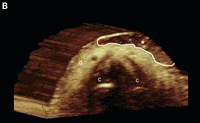
Figure: Basal cell carcinoma with dermal involvement (transverse view, nasal tip). Grayscale sonography (A) and 3-dimensional reconstruction (B, 5- to 8-second sweep) show a 10.1-mm (wide) × 1.4-mm (deep) well-defined hypoechoic oval lesion (between markers in A and outlined in B) that affects the dermis (d) of the left nasal wing. Notice the hyperechoic spots (arrowheads) within the lesion. The nasal cartilage (c) is unremarkable; asterisk indicates basal cell carcinoma.

Basal cell carcinoma with dermal and subcutaneous involvement (transverse view, frontal region). A, Grayscale sonography shows a 11.4-mm (wide) × 6.6-mm (deep) well-defined oval hypoechoic lesion that involves the dermis (d) and subcutaneous tissue (st). There are hyperechoic spots (arrowheads) within the tumor. B, Color Doppler sonography shows increased vascularity within the tumor (asterisk). C, Three-dimensional sonographic reconstruction (5- to 8-second sweep) highlights the lesion (asterisk, outlined); b indicates bony margin of the skull.

Basal cell carcinoma with dermal and subcutaneous involvement (transverse view, frontal region). A, Grayscale sonography shows a 11.4-mm (wide) × 6.6-mm (deep) well-defined oval hypoechoic lesion that involves the dermis (d) and subcutaneous tissue (st). There are hyperechoic spots (arrowheads) within the tumor. B, Color Doppler sonography shows increased vascularity within the tumor (asterisk). C, Three-dimensional sonographic reconstruction (5- to 8-second sweep) highlights the lesion (asterisk, outlined); b indicates bony margin of the skull.

f3

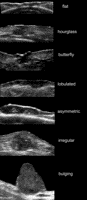
Figure: Pleomorphic presentations of basal cell carcinoma lesions on grayscale sonography (transverse views). Notice the variable shapes of the tumors.

f4

Figure: Frequently, blood flow can be detected within the tumor and its periphery, with slow-flow arteries or veins. The latter vascular data can orient the clinician about the distribution and amount of blood flow that he or she will face during surgery. Despite the fact that basal cell carcinomas usually do not present high vascularity, it should be kept in mind that many of basal cell carcinoma operations are performed in the offices of clinicians and not in the main operating rooms of large hospitals. Nevertheless, the finding of high vascularity within a clinically diagnosed basal cell carcinoma may suggest another type of skin cancer that could occasionally mimic basal cell carcinoma, such as squamous cell carcinoma, Merkel cell carcinoma, or a metastatic tumor. The above figure presents variable degrees of vascularity in basal cell carcinoma lesions going from hypovascular to hypervascular on color and power Doppler sonography (transverse views)

f5

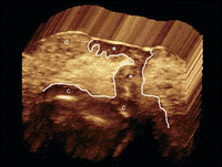
Figure: The depth correlation between sonography (variable frequency) and histologic analysis in facial basal cell carcinoma has been reported to be excellent. Thus, the intraclass correlation coefficient for comparing thickness for the two methods (sonography and histologic analysis) that has been described in literature is 0.9 (intraclass correlation coefficient values ≥0.9 are very good; 0.70–0.89 are good; 0.50–0.69 are moderate; 0.30–049 are mediocre; and ≤0.29 are bad). Two rare sonographic artifacts have been described in basal cell carcinoma. One is the “angled border” that is produced by an inflammatory giant cell reaction underlying the tumor, which may falsely increase the apparent size of the tumor. The other is the “blurry border,” which is produced by large hypertrophy of the sebaceous glands surrounding the lesion. According to the literature, both artifacts can be recognized by a well-trained operator. The figure above presents the sonographic involvement of deeper layers such as the nasal cartilage and orbicularis muscles in the face is of critical importance and may change the decision about the type of surgery. Basal cell carcinoma with nasal cartilage involvement (3-dimensional reconstruction, 5- to 8-second sweep, transverse view, left nasal wing). Notice the extension of the tumor (asterisk, outlined) to the nasal cartilage region (c); d indicates dermis.

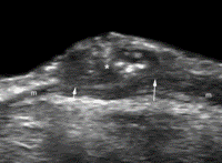
Basal cell carcinoma with involvement of the orbicularis muscle of the eyelid (m). Grayscale sonography (transverse view, right lower eyelid) shows that the tumor (asterisk) affects the muscle layer (arrows).

Basal cell carcinoma with involvement of the orbicularis muscle of the eyelid (m). Grayscale sonography (transverse view, right lower eyelid) shows that the tumor (asterisk) affects the muscle layer (arrows).

Read Full Post »

Reporter and Curator: Ritu Saxena, PhD

Magnetic Resonance Imaging (MRI) is increasingly used in clinical diagnostics, for a rapidly growing number of indications. The MRI technique is non-invasive and can provide information on the anatomy, function and metabolism of tissues in vivo (Strijkers GJ, et al, Anticancer Agents Med Chem, May 2007;7(3):291-305). Basic contrast in the MRI image scans is as a result of contrast generated by differences in the relaxation times between different regions. Since the intrinsic contrast generated between regions is limited to allow clear and specific diagnosis, MRI contrast agents administered intravenously are increasingly being used to alter image contrast.

Gadoxetic acid, a gadolinium-based compound, is a recently developed hepatobiliary-specific contrast material for MRI that has high sensitivity in the detection of malignant liver tumors. Its salt, gadoxetate disodium, is marketed as Primovist in Europe and Eovist in the United States by Bayer HealthCare Pharmaceuticals. Gadoxetic acid is taken up by hepatocytes and then excreted into the bile ducts (Schuhmann-Giampieri G, et al, Radiology, Apr 1992;183(1):59-64). Therefore, hepatic focal lesions without normal hepatobiliary function are depicted as hypointense areas compared with the well-enhanced hyperintense background liver in the hepatobiliary phase of gadoxetic acid–enhanced MR imaging. In addition, gadoxetic acid can be used in the same way as gadopentetate dimeglumine to evaluate the hemodynamics of hepatic lesions in the dynamic phase after an intravenous bolus injection (Kitao A, et al, Radiology, Sep 2010;256(3):817-26).

Recently, researchers from Kanazawa University Graduate School of Medical Science, (Kanazawa, Japan) analyzed the correlation among biologic features, tumor marker production, and signal intensity at gadoxetic acid-enhanced MR imaging in hepatocellular carcinomas (HCCs). The findings were published in Radiology journal. The research was supported in part by a Grant-in-Aid for Scientific Research (21591549) from the Ministry of Education, Culture, Sports, Science and Technology; and by Health and Labor Sciences Research Grants for “Development of novel molecular markers and imaging modalities for earlier diagnosis of hepatocellular carcinoma.”

Research significance: HCC is the most frequent primary malignant tumor of liver and is the third most common cause of cancer death worldwide. It is the most Hepatocellular.

The accurate detection and characterization of HCC focal lesions is crucial for improving prognosis of patients with HCC.

Research problem: Gadoxetic acid–enhanced MR imaging is highly accurate for diagnosing HCC lesions. As discussed earlier, in this imaging process, hepatic focal lesions without normal hepatobiliary are hypointense as compared with the well-enhanced hyperintense background liver. However, approximately 6%–15% of hypervascular HCCs demonstrate isointensity or hyperintensity (Kitao A, et al, Eur Radiol, Oct 2011;21(10):2056-66).

Hypothesis: The reason for hyperintensity in some HCC lesions was previously shown to be due to overexpression of organic anion transporting polypeptide 8 (OATP8) (Kitao A, et al, Radiology, Sep 2010;256(3):817-26). The authors speculated that there might be a correlation of the tumor marker production and signal intensity (SI) on hepatobiliary phase images, which would reflect distinct genomic and proteomic expression of HCC. Thus, authors stated that “the purpose of this study was to analyze the correlation among the pathologic and biologic features, tumor marker production, with signal intensity (SI) on hepatobiliary phase gadoxetic acid–enhanced MR images of HCC” (Kitao A, et al, Radiology, Dec 2012;265(3):780-9).

Experimental design: From April 2008 to September 2011, 180 surgically resected HCCs in 180 patients (age, 65.0 years ± 10.3 [range, 34–83 years]; 138 men, 42 women) were classified as either hypointense (n = 158) or hyperintense (n = 22) compared with the signal intensity of the background liver on hepatobiliary phase gadoxetic acid–enhanced MR images (Abstract of the study).

Pathologic features were analyzed.

Serum analysis and immunohistochemical staining was performed and following were compared:

  1. Alpha fetoprotein (AFP) – is a main tumor marker of HCCs. AFP is the most abundant plasma protein found in the human fetus and plasma levels decrease rapidly after birth. A level above 500 nanograms/milliliter of AFP in adults can be indicative of hepatocellular carcinoma, germ cell tumors, and metastatic cancers of the liver.
  2. Absence of protein induced by vitamin K or antagonist-II (PIVKA-II) – is a clinically important serum tumor marker. PIVKAII is an incomplete coagulation factor prothrombin II whose production is related to the absence of vitamin K or the presence of the antagonist of vitamin K, which is the cofactor of g carboxylase that converts precursor into prothrombin.

Serum levels of both AFP and PIVKA-II correlate with HCC malignancy and prognosis (Miyaaki H, et al, J Gastroenterol, Dec 2007;42(12):962-8).

Results: The hyperintense HCCs showed significantly higher differentiation grade than the hypointense HCCs (P = .028). There was a significant difference in the proliferation pattern between the hypointense and hyperintense HCCs (P < .001) and the hyperintense HCCs showed a significantly lower rate of portal vein invasion than that of hypointense HCCs (P = .039). The serum levels of tumor markers AFP, AFP-L3, and PIVKA-II were significantly lower in the patients with hyperintense HCCs than in those with

hypointense HCCs (P = .003, .004, and .026). In addition, immunohistochemical analysis revealed that the expression of FP and PIVKA-II was lower in hyperintense than in hypointense HCCs (both P < .001). Also, hyperintense HCCs showed lower recurrence rate than hypointense HCCs (P = .039).

Conclusion: Variation was observed within differently stained lesions of HCC in the hepatobiliary phase gadoxetic acid–enhanced MR images as evident in tumor marker expression, proliferation pattern, differentiation grade, immunohistochemical analysis and recurrence.  The results lead to the hypothesis that hyperintense HCCs in the hepatobiliary phase gadoxetic acid–enhanced MR images might represent a particular type of HCC that is hypervascular and biologically less aggressive as compared to hypovascular HCCs. Interestingly, this research is another great example where tumor heterogeneity has been brought to light (similar to genetic heterogeneity in triple negative breast cancer deciphered by Lehmann BD, et al, 2011). The heterogeneity might be the basis of answers to why a particular therapy fails in a certain tumor type and fortifying evidence for appropriate analysis of the tumor for obtaining the desired tumor response from a particular drug.

Reference:

Kitao A, et al, Radiology, Dec 2012;265(3):780-9

Strijkers GJ, et al, Anticancer Agents Med Chem, May 2007;7(3):291-305

Schuhmann-Giampieri G, et al, Radiology, Apr 1992;183(1):59-64

Kitao A, et al, Radiology, Sep 2010;256(3):817-26

Kitao A, et al, Eur Radiol, Oct 2011;21(10):2056-66

Kitao A, et al, Radiology, Sep 2010;256(3):817-26

Miyaaki H, et al, J Gastroenterol, Dec 2007;42(12):962-8

Lehmann BD, et al, J Clin Invest, 2011;121(7):2750–2767

Read Full Post »

Amyloidosis with Cardiomyopathy

Author: Larry H Bernstein, MD, FACP
Introduction
Amyloidosis describes the various clinical syndromes that occur as a result of damage by amyloid deposits in tissues and organs throughout the body.  Systemic amyloidosis is a relatively rare multisystem disease caused by the deposition of misfolded protein in various tissues and organs. The term amyloid describes the deposition in the extracellular space of certain proteins in a highly characteristic, insoluble fibrillar form.  The disease entity is a disorder of misfolded or misassembled proteins.  There is extracellular amyloid fiber laid down as cross β-sheets disrupting organ function, which may affect the pancreas, kidney, autonomic nervous system, the heart, and in one form causes carpal tunnel syndrome.
It may present to almost any specialty, and diagnosis is frequently delayed. Cardiac involvement is a leading cause of morbidity and mortality, especially in primary light chain (AL) amyloidosis and in both wild-type and hereditary transthyretin amyloidosis. The heart is also occasionally involved in acquired serum amyloid A type (AA) amyloidosis and other rare hereditary types. Clinical phenotype varies greatly between different types of amyloidosis, and even the cardiac presentation has a great spectrum. The incidence of amyloidosis is uncertain, but it is thought that the most frequently diagnosed AL amyloidosis has an annual incidence of 6 to 10 cases per million population in the United Kingdom and United States.
The molecular basis for this particular phenomenon came with the extensive work done on multiple myeloma, antibody structure, and light chains.  In 1950, the discovery of a familial amyloid polyneuropathy was described in Portugal, and there were similar diseases in Sweden and Japan.  There were 72 known variants of transthyretin (TTR) in 1995, and now there are 100.  In addition, the occurance of different TTR associated variants with and without (amyloid) is found is Brazil, UK, US, Israel, Spain, France, Germany, Denmark, and Africa.  The table of variants, organ damage, and geographic location is too large to place on this document. If we refer to amyloid cardiomyopathy, it is exclusively a primary amyloidopathy, not secondary to light chain disorders or an inflammatory disease.  If we consider amyloidosis, we also have to consider family history, organ dysfunction, and we have to make a distinction between primary cardiac involvement, autonomic nervous system instability, and the two coexisting.  Familial amyloid polyneuropathy (FAP) is an extremely debilitating and progressive disease that is only treatable by liver transplantation.  Primary amyloid cardiomyopathy has been treated by heart transplant.  The qualifying statement here is, it depends.

Primary and Secondary Amyloidoses

Amyloid was originally described by pathologists based on microscopy. Amyloidoses are a systemic primary or secondary disease. There are distinctions to be made based on location and type.  The clinical significance of amyloid disease varies enormously, ranging from incidental asymptomatic deposits to localized disease through to rapidly fatal systemic forms that can affect multiple vital organs.
Common causes of secondary amyloidosis are – light chain production (AL) as in plasma cell dyscrasia, amyloid A (AA), senile systemic amyloidosis (diagnosed rarely in life).  The systemic amyloidoses are designated by a capital A (for amyloid) followed by the abbreviation for the chemical identity of the fibril protein. Thus, TTR amyloidosis is abbreviated ATTR, and immunoglobulin light chain type amyloidosis is abbreviated AL. Both normal-sequence TTR and variant-sequence TTR form amyloidosis. Normal-sequence TTR forms cardiac amyloidosis in elderly people, termed senile cardiac amyloidosis (SCA). When it was recognized that SCA is often accompanied by microscopic deposits in many other organs, the alternative name senile systemic amyloidosis (SSA) was proposed. Both terms are now used.
Currently available therapy is focused on reducing the supply of the respective amyloid fibril precursor protein and supportive medical care, which together have greatly improved survival. Chemotherapy and anti-inflammatory treatment for the disorders that underlie AL and AA amyloidosis are guided by serial measurements of the respective circulating amyloid precursor proteins, i.e. serial serum free light chains in AL and serum amyloid A protein in AA type.
Quality of life and prognosis of some forms of hereditary systemic amyloidosis can be improved by liver and other organ transplants. Various new therapies, ranging from silencing RNA, protein stabilizers to monoclonal antibodies, aimed at inhibiting fibril precursor supply, fibril formation or the persistence of amyloid deposits, are in development; some are already in clinical phase.
Ann Clin Biochem May 2012; 49(3 ): 229-241   http://acb.2011.011225v1 49/3/229

What is transthyretin (TTR)?

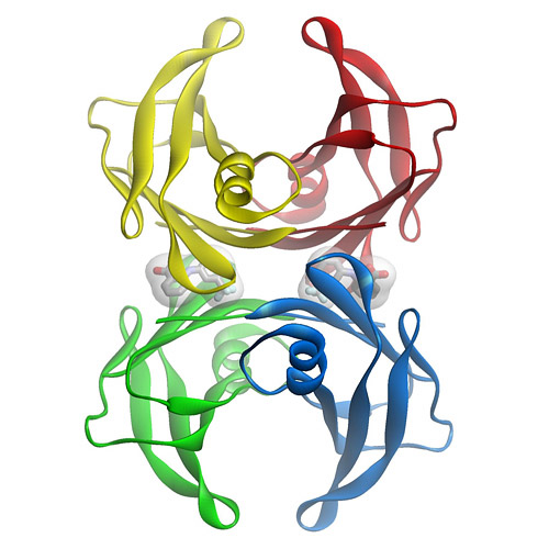
TTR is a  tetramer of 4 127 amino acid subunits synthesized by the liver that circulates as a transporter of thyroxin, and with retinol-binding protein, transports vitamin A.  It was originally defined by the migration in electrophoresis more anodal to albumin, hence, prealbumin.  It is present in cerebrospinal fluid, secreted by the choroid plexus.  The TTR monomer contains 8 antiparallel beta pleated sheet domains. TTR can be found in plasma and in cerebrospinal fluid and is synthesized by the choroid plexus of the brain and, to a lesser degree, by the retina. Its gene is located on the long arm of chromosome 18 and contains 4 exons and 3 introns.
The concentration in serum can be expected to be above 20 mg/dL in a health adult, but the protein decreases by 1 mg/dL/day postoperatively, and it decreases with acute or chronic renal failure, pneumonia or sepsis, rising again with the onset of anabolism.  Patients in the pulmonary intensive care unit have TTR levels that remain low for 7-10 days, but followup data for the remainder of the hospital stay or in relationship to readmission in the six months after release from hospital care was not part of the study.
A decrease in TTR is associated with the systemic inflammatory response, whereby, the liver reprioritizes the synthesis of proteins with an increase in acute phase reactants (APRs), namely, C-reactive protein (CRP) and a-1 acid glycoprotein, and decreased albumin and TTR.  The inflammatory condition maintains a euthyroid status with decreased TTR because of the availability of free thyroxine in equilibrium with the lower binding protein.  This has been referred to sick euthyroid status. The role in thyroxine transport is not insignificant, as chronic protein malnutrition is associated with hypothyroidism, as originally described by Prof. Yves Ingenbleek, Univ. Louis Pateur, Starsbourg, Fr. in Senegalese children with Kwashiorkor.  However, the importance of TTR as a unique biomarker is not to be downgraded because of what is often refered to as “an inverted APR”.
Transthyretin was discovered to be a good reflection of the “lean body mass”, by Vernon Young, MIT, and Ingenbleek, as a result of 3 decades of study. The ratio of S:N being 1:20 in plant proteins and 1:12.5 in animal sources, is closely related to methylation reactions and sustained deficiency of S intake results in elevated homocysteine level.

What is FAP?

Familial amyloid polyneuropathy (FAP), also called transthyretin-related hereditary amyloidosis, transthyretin amyloidosis or Corino de Andrade’s disease, is an autosomal dominant neurodegenerative disease. It is a form of amyloidosis, and was first identified and described by Portuguese neurologist Mário Corino da Costa Andrade, in the 1950s.FAP is distinct from senile systemic amyloidosis (SAS), which is not inherited, and which was determined to be the primary cause of death for 70% of supercentenarians who have been autopsied.
Familial amyloid polyneuropathy (FAP) is an extremely debilitating and progressive disease that is only treatable by liver transplantation.  Primary amyloid cardiomyopathy has been treated by heart transplant.  The qualifying statement here is, it depends.  Those patients with TTR-amyloidopathy have a specific gene substitution in the TTR gene. Consequently, there is circulation TTR, but it is not effectively involved in thyroxine transport.

Characteristics.

Usually manifesting itself between 20 and 40 years of age, it is characterized by pain, paresthesia, muscular weakness and autonomic dysfunction. In its terminal state, the kidneys and the heart are affected. FAP is characterized by the systemic deposition of amyloidogenic variants of the transthyretin protein, especially in the peripheral nervous system, causing a progressive sensory and motor polyneuropathy. The age at symptom onset, pattern of organ involvement, and disease course vary, but most mutations are associated with cardiac and/or nerve involvement. The gastrointestinal tract, vitreous, lungs, and carpal ligament are also frequently affected. When the peripheral nerves are prominently affected, the disease is termed familial amyloidotic polyneuropathy (FAP). When the heart is involved heavily but the nerves are not, the disease is called familial amyloid cardiomyopathy (FAC). Regardless of which organ is primarily targeted, the general term is simply amyloidosis-transthyretin type, abbreviated ATTR.

Genetics.

  1. TTR mutations accelerate the process of TTR amyloid formation and are the most important risk factor for the development of clinically significant ATTR. More than 85 amyloidogenic TTR variants cause systemic familial amyloidosis. The variant TTR is mostly produced by the liver. Amyloidogenic TTR mutations destabilize TTR monomers or tetramers, allowing the molecule to more easily attain an amyloidogenic intermediate conformation. The tetramer has to dissociate into misfolded monomers to aggregate into a variety of structures including amyloid fibrils. Because most patients are heterozygotes, they deposit both mutant and wild type TTR subnits.
  2. Familial amyloid polyneuropathy has an autosomal dominant pattern of inheritance. FAP is caused by a mutation of the TTR gene, located on human chromosome 18q12.1-11.2. A replacement of valine by methionine at position 30 (TTR V30M) is the mutation most commonly found in FAP.
  3. The disease in the TTR V30M kindreds was termed FAP because early symptoms arose from peripheral neuropathy, but these patients actually have systemic amyloidosis, with widespread deposits often involving the heart, gastrointestinal tract, eye, and other organs.
  4. TTR V122I: This variant, carried by 3.9% of African Americans and over 5% of the population in some areas of West Africa, increases the risk of late-onset (after age 60 years) cardiac amyloidosis. It appears to be the most common amyloid-associated TTR variant worldwide. Affected patients usually do not have peripheral neuropathy.
  5. TTR T60A: This variant causes late-onset systemic amyloidosis with cardiac, and sometimes neuropathic, involvement. This variant originated in northwest Ireland and is found in Irish and Irish American patients.
  6. TTR L58H: Typically affecting the carpal ligament and nerves of the upper extremities, this variant originated in Germany. It has spread throughout the United States but is most common in the mid-Atlantic region.
  7. TTR G6S: This is the most common TTR variant, but it appears to be a neutral polymorphism not associated with amyloidosis. It is carried by about 10% of people of white European descent.

Cardiac transthyretin (TTR) amyloidosis

Cardiac amyloidosis of transthyretin fibril protein (ATTR) type is an infiltrative cardiomyopathy characterised by ventricular wall thickening and diastolic heart failure. More than 27 different precursor proteins have the propensity to form amyloid fibrils. The particular precursor protein that misfolds to form amyloid fibrils defines the amyloid type and predicts the patient’s clinical course. Several types of amyloid can infiltrate the heart, resulting in progressive diastolic and systolic dysfunction, congestive heart failure, and death.  Increased access to cardiovascular magnetic resonance imaging has led to a marked increase in referrals to St George’s University of London, London (Dr. Jason Dungu) of Caucasian patients with wild-type ATTR (senile systemic) amyloidosis and Afro-Caribbean patients with the hereditary ATTR V122I type. Both subtypes present predominantly as isolated cardiomyopathy. The differential diagnosis includes cardiac amyloid light-chain (AL) amyloidosis, which has a poorer prognosis and can be amenable to chemotherapy.

Clinical Presentation

Cardiac amyloidosis, irrespective of type, presents as a restrictive cardiomyopathy characterized by progressive diastolic and subsequently systolic biventricular dysfunction and arrhythmia.1 Key “red flags” to possible systemic amyloidosis include nephrotic syndrome, autonomic neuropathy (eg, postural hypotension, diarrhea), soft-tissue infiltrations (eg, macroglossia, carpal tunnel syndrome, respiratory disease), bleeding (eg, cutaneous, such as periorbital, gastrointestinal), malnutrition/cachexia and genetic predisposition (eg, family history, ethnicity). Initial presentations may be cardiac, with progressive exercise intolerance and heart failure. Other organ involvement, particularly in AL amyloidosis, may cloud the cardiac presentation (eg, nephrotic syndrome, autonomic neuropathy, pulmonary or bronchial involvement). Pulmonary edema is not common early in the disease process, but pleural and pericardial effusions and atrial arrhythmias are often seen. Syncope is common and a poor prognostic sign. It is typically exertional or postprandial as part of restrictive cardiomyopathy, sensitivity to intravascular fluid depletion from loop diuretics combined with autonomic neuropathy, or conduction tissue involvement (atrioventricular or sinoatrial nodes) or ventricular arrhythmia. The latter may rarely cause recurrent syncope. Disproportionate septal amyloid accumulation mimicking hypertrophic cardiomyopathy with dynamic left ventricular (LV) outflow tract obstruction is rare but well documented. Myocardial ischemia can result from amyloid deposits within the microvasculature. Atrial thrombus is common, particularly in AL amyloidosis

Diagnosis and Treatment

imaging – Cardiovascular Magnetic Resonance in Cardiac Amyloidosis*.

Cardiac amyloidosis can be diagnostically challenging. Cardiovascular magnetic resonance (CMR) can assess abnormal myocardial interstitium. In cardiac amyloidosis, CMR shows a characteristic pattern of global subendocardial late enhancement coupled with abnormal myocardial and blood-pool gadolinium kinetics. The findings agree with the transmural histological distribution of amyloid protein and the cardiac amyloid load.
 *AM Maceira; J Joshi; SK Prasad; J Charles Moon, et al. Royal Brompton Hospital, London;
The diagnosis of amyloidosis requires histological identification of amyloid deposits. Congo Red staining renders amyloid deposits salmon pink by light microscopy, with a characteristic apple green birefringence under polarized light conditions. Additional immunohistochemical staining for precursor proteins identifies the type of amyloidosis.  Ultimately, immunogold electron microscopy and mass spectrometry confer the greatest sensitivity and specificity for amyloid typing.
Treatment of cardiac amyloidosis is dictated by the amyloid type and degree of involvement. Consequently, early recognition and accurate classification are essential.
Novel diagnostic and surveillance approaches using imaging (echocardiography, cardiovascular magnetic resonance), biomarkers (brain natriuretic peptide [BNP], high-sensitivity troponin), new histological typing techniques, and current and future treatments, including approaches directly targeting the amyloid deposits.

Etiology

Amyloidosis is caused by the extracellular deposition of autologous protein in an abnormal insoluble β-pleated sheet fibrillary conformation—that is, as amyloid fibrils. More than 30 proteins are known to be able to form amyloid fibrils in vivo, which cause disease by progressively damaging the structure and function of affected tissues. Amyloid deposits also contain minor nonfibrillary constituents, including serum amyloid P component (SAP), apolipoprotein E, connective tissue components (glycosaminoglycans, collagen), and basement membrane components (fibronectin, laminin). Amyloid deposits can be massive, and cardiac or other tissues may become substantially replaced. Amyloid fibrils bind Congo red stain, yielding the pathognomonic apple-green birefringence under cross-polarized light microscopy that remains the gold standard for identifying amyloid deposits.

AL Amyloidosis

AL amyloidosis is caused by deposition of fibrils composed of monoclonal immunoglobulin light chains and is associated with clonal plasma cell or other B-cell dyscrasias. The spectrum and pattern of organ involvement is very wide, but cardiac involvement occurs in half of cases and is sometimes the only presenting feature. Cardiac AL amyloidosis may be rapidly progressive. Low QRS voltages, particularly in the limb leads, are common. Thickening of the LV wall is typically mild to moderate and is rarely >18 mm even in advanced disease. Cardiac AL amyloid deposition is accompanied by marked elevation of the biomarkers BNP and cardiac troponin, even at an early stage. Involvement of the heart is the commonest cause of death in AL amyloidosis and is a major determinant of prognosis; without cardiac involvement, patients with AL amyloidosis have a median survival of around 4 years, but the prognosis among affected patients with markedly elevated BNP and cardiac troponin (Mayo stage III disease) is on the order of 8 months.

Hereditary Amyloidoses

Mutations in several genes, such as transthyretin, fibrinogen, apolipoprotein A1, and apolipoprotein A2 can be responsible for hereditary amyloidosis, but by far the most common cause is variant ATTR amyloidosis (variant ATTR) caused by mutations in the transthyretin gene causing neuropathy and, often, cardiac involvement.

TTR gene mutation

 The most common is the Val122Ile mutation. In a large autopsy study that included individuals with cardiac amyloidosis, the TTR Val122Ile allele was present in 3.9% of all African Americans and 23% of African Americans with cardiac amyloidosis. Penetrance of the mutation is not truly known and is associated with a late-onset cardiomyopathy that is indistinguishable from senile cardiac amyloidosis.

Pathology, Presentation, and Management of Amyloidoses

More than 100 genetic variants of TTR are associated with amyloidosis. Most present as the clinical syndrome of progressive peripheral and autonomic neuropathy. Unlike wild-type ATTR or variant ATTR Val122Ile, the features of other variant ATTR include vitreous amyloid deposits or, rarely, deposits in other organs. Cardiac involvement in variant ATTR varies by mutations and can be the presenting or indeed the only clinical feature. For example, cardiac involvement is rare in variant ATTR associated with Val30Met (a common variant in Portugal or Sweden), but it is almost universal and develops early in individuals with variant ATTR due to Thr60Ala mutation (a mutation common in Ireland).

Senile Systemic Amyloidosis (Wild-Type ATTR)

Wild-type TTR amyloid deposits are found at autopsy in about 25% of individuals >80 years of age.  The prevalence of wild-type TTR deposits leading to the clinical syndrome of wild-type ATTR cardiac amyloidosis is unknown. Wild-type ATTR is a predominantly cardiac disease, and the only other significant extracardiac feature is a history of carpal tunnel syndrome, often preceding heart failure by 3 to 5 years. Extracardiac involvement is most unusual.
Both wild-type ATTR and ATTR due to Val122Ile are diseases of the >60-year age group and are often misdiagnosed as hypertensive heart disease. Wild-type ATTR has a strong male predominance, and the natural history remains poorly understood, but studies suggest a median survival of about 7 years from presentation. Recent developments in cardiac magnetic resonance (CMR), which have greatly improved detection of cardiac amyloid during life, suggest that wild-type ATTR is more common than previously thought: It accounted for 0.5% of all patients seen at the UK amyloidosis center until 2001 but now accounts for 7% of 1100 cases with amyloidosis seen since the end of 2009. There appears to be an association between wild-type ATTR and history of myocardial infarctions, G/G (Val/Val) exon 24 polymorphism in the alpha2-macroglobulin (alpha2M), and the H2 haplotype of the tau gene36; the association of tau with Alzheimer’s disease raises interesting questions as both are amyloid-associated diseases of aging.
ECG of a patient with cardiac AL amyloidosis showing small QRS voltages (defined as ≤6 mm height), predominantly in the limb leads and pseudoinfarction pattern in the anterior leads.
Echocardiography is characteristic. Typical findings include concentric ventricular thickening with right ventricular involvement, poor biventricular long-axis function with normal/near-normal ejection fraction and valvular thickening (particularly in wild-type or variant ATTR). Diastolic dysfunction is the earliest echocardiographic abnormality and may occur before cardiac symptoms develop. Biatrial dilatation in presence of biventricular, valvular, and interatrial septal thickening 53 is a useful clue to the diagnosis.
Transthoracic echocardiogram with speckle tracking. The red and yellow lines represent longitudinal motion in the basal segments, whereas the purple and green lines represent apical motion. This shows loss of longitudinal ventricular contraction at the base compared to apex.

Biomarkers.

High-sensitivity troponin is abnormal in >90% of cardiac AL patients, and the combination of BNP/NT-proBNP plus troponin measurements is used to stage and risk-stratify patients with AL amyloidosis at diagnosis. Very interestingly, the concentration of BNP/NT-proBNP in AL amyloidosis may fall dramatically within weeks after chemotherapy that substantially reduces the production of amyloidogenic light chains. The basis for this very rapid phenomenon, which is not mirrored by changes on echocardiography or CMR, remains uncertain, but a substantial fall is associated with improved outcomes.

Cardiac Magnetic Resonance.

CMR provides functional and morphological information on cardiac amyloid in a similar way to echocardiography, though the latter is superior for evaluating and quantifying diastolic abnormalities. An advantage of CMR is in myocardial tissue characterization. Amyloidotic myocardium reveals subtle precontrast abnormalities (T1, T2), but extravascular contrast agents based on chelated gadolinium provide the key information.

CMR with the classic amyloid global, subendocardial late gadolinium enhancement pattern in the left ventricle with blood and mid-/epimyocardium nulling together.
Recently, the technique of equilibrium contrast CMR has demonstrated much higher extracellular myocardial volume in cardiac amyloid than any other measured disease. It is anticipated that accurate measurements of the expanded interstitium in amyloidosis will prove useful in serial quantification of cardiac amyloid burden.
Sequential static images from a CMR TI scout sequence. As the inversion time (TI) increases, myocardium nulls first (arrow in image 3), followed by blood afterwards (arrow in image 6), implying that there is more gadolinium contrast in the myocardium than blood—a degree of interstitial expansion such that the “myocrit” is smaller than the hematocrit.

Tissue biopsy.

To confirm amyloidosis, including familial TTR amyloidosis, the demonstration of amyloid deposition on biopsied tissues is essential. With Congo red staining, amyloid deposits show a characteristic yellow-green birefringence under polarized light. Tissues suitable for biopsy include: subcutaneous fatty tissue of the abdominal wall, skin, gastric or rectal mucosa, sural nerve, and peritendinous fat from specimens obtained at carpal tunnel surgery. Sensitivity of endoscopic biopsy of gastrointestinal mucosa is around 85%; biopsy of the sural nerve is less sensitive. It is ideal to show that these amyloid deposits are specifically immunolabeled by anti-TTR antibodies.

Serum variant TTR protein.

TTR protein normally circulates in serum or plasma as a soluble protein having a tetrameric structure [Kelly 1998, Rochet & Lansbury 2000]. Normal plasma TTR concentration is 20-40 mg/dL (0.20-0.40 mg/mL).  Pathogenic mutations in TTR cause conformational change in the TTR protein molecule, disrupting the stability of the TTR tetramer, which is then more easily dissociated into pro-amyloidogenic monomers.

After immunoprecipitation with anti-TTR antibody, serum variant TTR protein can be detected by mass spectrometry. Approximately 90% of TTR variants so far identified are confirmed by this method. Mass shift associated with each variant TTR protein is indicated.

Molecular genetic testing.

  • TTR is the only gene in which mutations are known to cause familial TTR amyloidosis.
  • Identified in many individuals of different ethnic backgrounds; found in large clusters in Portugal, Sweden, and Japan.
  • The gene has four exons; and all the hitherto-identified mutations are in exons 2, 3, or 4.
GeneReviews designates a molecular genetic test as clinically available only if the test is listed in the GeneTests Laboratory Directory by either a US CLIA-licensed laboratory or a non-US clinical laboratory.
  • Molecular genetic testing of TTR by sequence analysis (may be preceded by targeted mutation analysis)
  • Although deletion/duplication testing is available clinically, no exonic or whole-gene deletions or duplications involving TTR have been reported to cause familial transthyretin amyloidosis.
  • However, with newly available deletion/duplication testing methods, it is theoretically possible that such mutations may be identified in affected individuals in whom prior testing by sequence analysis of the entire coding region was negative.
  • Predictive testing for at-risk asymptomatic adult family members requires prior identification of the disease-causing mutation in the family.
  • Prenatal diagnosis and preimplantation genetic diagnosis (PGD) for at-risk pregnancies require prior identification of the disease-causing mutation in the family.

Genetically Related (Allelic) Disorders

Familial euthyroid hyperthyroxinemia is caused by normal allelic variants in TTR, including Gly6Ser, Ala109Thr, Ala109Val, and Thr119Met (see Table 5) [Nakazato 1998, Benson 2001, Saraiva 2001]. The TTR protein binds approximately 15% of serum thyroxine. These mutations increase total serum thyroxine concentration because of their increased affinity for thyroxine, however, they increase neither free thyroxine nor free triiodothyronine. Therefore, individuals with these sequence variants develop no clinical symptoms (i.e., they are euthyroid).
Senile systemic amyloidosis (SSA; previously called senile cardiac amyloidosis) results from the pathologic deposition of wild-type TTR, predominantly in the heart. Pathologic deposits are also seen in the lungs, blood vessels, and the renal medulla of the kidneys [Westermark et al 2003]. SSA affects mainly the elderly but is rarely diagnosed during life.
Sensorimotor neuropathy and autonomic neuropathy progress over ten to 20 years. Various types of cardiac conduction block frequently appear. Cachexia is a common feature at the late stage of the disease. Affected individuals usually die of cardiac failure, renal failure, or infection.

Cardiac amyloidosis.

Cardiac amyloidosis, mainly characterized by progressive cardiomyopathy, has been reported with more than two thirds of TTR mutations. In some families with specific TTR mutations, such as Asp18Asn, Val20Ile, Pro24Ser, Ala45Thr, Ala45Ser, His56Arg, Gly57Arg, Ile68Leu, Ala81Thr, Ala81Val, His88Arg, Glu92Lys, Arg103Ser, Leu111Met, or Val122Ile, cardiomyopathy without peripheral neuropathy is a main feature of the disease.

Cardiac amyloidosis is usually late onset. Most individuals develop cardiac symptoms after age 50 years; cardiac amyloidosis generally presents with restrictive cardiomyopathy. The typical electrocardiogram shows a pseudoinfarction pattern with prominent Q wave in leads II, III, aVF, and V1-V3, presumably resulting from dense amyloid deposition in the anterobasal or anteroseptal wall of the left ventricle. The echocardiogram reveals left ventricular hypertrophy with preserved systolic function. The thickened walls present “a granular sparkling appearance.”
Among the mutations responsible for cardiac amyloidosis, Val122Ile is notable for its prevalence in African Americans. Approximately 3.0%-3.9% of African Americans are heterozygous for Val122Ile . The high frequency of Val122Ile partly explains the observation that in individuals in the US older than age 60 years, cardiac amyloidosis is four times more common among blacks than whites.

Leptomeningeal (oculoleptomeningeal) amyloidosis.

Amyloid deposition is seen in the pial and arachnoid membrane, as well as in the walls of vessels in the subarachnoid space associated with TTR mutations including Leu12Pro, Asp18Gly, Ala25Thr, Val30Gly, Ala36Pro, Gly53Glu, Gly53Ala, Phe64Ser, Tyr69His, or Tyr114Cys.  Individuals with leptomeningeal amyloidosis show CNS signs and symptoms including: dementia, psychosis, visual impairment, headache, seizures, motor paresis, ataxia, myelopathy, hydrocephalus, or intracranial hemorrhage. When associated with vitreous amyloid deposits, leptomeningeal amyloidosis is known as familial oculolepto-meningeal amyloidosis (FOLMA). In leptomeningeal amyloidosis protein concentration in the cerebrospinal fluid is usually high, and gadolinium-enhanced MRI typically shows extensive enhancement of the surface of the brain, ventricles, and spinal cord.

Genotype-Phenotype Correlations.

In subsets of families with the Val30Met mutation, considerable variation in phenotypic manifestations and age of onset is observed. It is hypothesized that genetic modifiers and non-genetic factors contribute to the pathogenesis and progression of familial TTR amyloidosis. The vast majority of individuals with familial TTR amyloidosis are heterozygous for a TTR mutation. It has been clinically and experimentally demonstrated that the normal allelic variant c.416C>T (Thr119Met) has a protective effect on amyloidogenesis in individuals who have the Val30Met mutation.

Cardiac amyloidosis is caused by Asp18Asn, Val20Ile, Pro24Ser, Ala45Thr, Ala45Ser, His56Arg, Gly57Arg, Ile68Leu, Ala81Thr, Ala81Val, His88Arg, Glu92Lys, Arg103Ser, Leu111Met, or Val122Ile. Peripheral and autonomic neuropathy are absent or less evident in persons with these mutations.
Leptomeningeal amyloidosis is associated with Leu12Pro, Asp18Gly, Ala25Thr, Val30Gly, Ala36Pro, Gly53Glu, Gly53Ala, Phe64Ser, Tyr69His, or Tyr114Cys.

Penetrance.

It is generally accepted that the penetrance is much higher in individuals in endemic foci than outside of endemic foci. In Portugal, cumulative disease risk in individuals with the Val30Met mutation is estimated at 80% by age 50 and 91% by age 70 years, whereas the risk in French heterozygotes is 14% by age 50 and 50% by age 70 years. In Sweden, the penetrance is much lower: 1.7% by age 30, 5% by age 40, 11% by age 50, 22% by age 60, 36% by age 70, 52% by age 80, and 69% by age 90, respectively.

Nomenclature

The neuropathy associated with TTR mutations, now called familial TTR amyloidosis, was formerly referred to as one of the following:
  • Familial amyloid polyneuropathy type I (or the Portuguese-Swedish-Japanese type)
  • Familial amyloid polyneuropathy type II (or the Indiana/Swiss or Maryland/German type)

Prevalence

The Val30Met mutation, found worldwide, is the most widely studied TTR variant and is responsible for the well-known large foci of individuals with TTR amyloid polyneuropathy in Portugal, Sweden, and Japan. Numerous families with various non-Val30Met mutations have also been identified worldwide.

 Small transthyretin (TTR) ligands as possible therapeutic agents in TTR amyloidoses.

Almeida MR, Gales L, Damas AM, Cardoso I, Saraiva MJ. Porto, Portugal.
Curr Drug Targets CNS Neurol Disord. 2005 Oct;4(5):587-96.
In transthyretin (TTR) amyloidosis TTR variants deposit as amyloid fibrils giving origin, in most cases, to peripheral polyneuropathy, cardiomyopathy, carpal tunnel syndrome and/or amyloid deposition in the eye. The amino acid substitutions in the TTR variants destabilize the tetramer, which may dissociate into non native monomeric intermediates that aggregate and polymerize in amyloid fibrils that further elongate. Since this is a multi-step process there is the possibility to impair TTR amyloid fibril formation at different stages of the process namely by tetramer stabilization, inhibition of fibril formation or fibril disruption. Based on the proposed mechanism for TTR amyloid fibril formation we discuss the action of some of the proposed TTR stabilizers such as derivatives of some NSAIDs (diflunisal, diclofenac, flufenamic acid, and derivatives) and the action of amyloid disrupters such as 4′-iodo-4′-deoxydoxorubicin (I-DOX) and tetracyclines. Among all these compounds, TTR stabilizers seem to be the most interesting since they would impair very early the process of amyloid formation and could also have a prophylactic effect.

Clusterin regulates transthyretin amyloidosis.

Lee KW, Lee DH, Son H, Kim YS, Park JY, et al.  Gyeongnam National University, South Korea
Biochem Biophys Res Commun 2009;388(2):256-60.   http://dx.doi.org/10.1016/j.bbrc.2009.07.166.
Clusterin has recently been proposed to play a role as an extracellular molecular chaperone, affecting the fibril formation of amyloidogenic proteins. The ability of clusterin to influence amyloid fibril formation prompted us to investigate whether clusterin is capable of inhibiting TTR amyloidosis. Here, we report that clusterin strongly interacts with wild-type TTR and TTR variants V30M and L55P under acidic conditions, and blocks the amyloid fibril formation of TTR variants. In particular, the amyloid fibril formation of V30M TTR in the presence of clusterin is reduced to level similar to wild-type TTR. We also demonstrated that clusterin is an effective inhibitor of L55P TTR amyloidosis, the most aggressive form of TTR diseases. The mechanism by which clusterin inhibits TTR amyloidosis appears to be through stabilization of TTR tetrameric structure.

Prognosis.

Cardiac amyloidosis in general has a poor prognosis, but this differs according to amyloid type and availability and response to therapy. Treatment may be classified as follows: supportive therapy (ie, modified heart-failure treatment including device therapy); therapies that suppress production of the respective amyloid fibril precursor protein (eg, chemotherapy in AL amyloidosis); and novel strategies to inhibit amyloid fibril formation or to directly target the amyloid deposits or stabilize the precursor protein (especially in ATTR with drugs such as tafamidis or diflunisal). Cardiac transplantation, although rarely feasible, can be very successful in carefully selected patients.

Reducing Amyloid Fibril Precursor Protein Production

Treatment of amyloidosis is currently based on the concept of reducing the supply of the respective amyloid fibril precursor protein. In AL amyloidosis, therapy is directed toward the clonal plasma cells using either cyclical combination chemotherapy or high-dose therapy with autologous stem cell transplantation.
The newer treatment options include bortezomib (a proteosome inhibitor)105 and the newer immunomodulatory drugs lenalidomide and pomalidomide. Bortezomib combinations appear to be especially efficient in amyloidosis with high rates of near-complete clonal responses, which appear to translate into early cardiac responses.106–108 Phase II (bortezomib in combination with cyclophosphamide or doxorubicin) and phase III (bortezomib, melphalan, and dexamethasone compared to melphalan and dexamethasone as front-line treatment) trials are underway.
AA amyloidosis is the only other type of amyloidosis in which production of the fibril precursor protein can be effectively suppressed by currently available therapies. Anti-inflammatory therapies, such as anti-tumor necrosis factor agents in rheumatoid arthritis, can substantially suppress serum amyloid A protein production, but very little experience has been obtained regarding cardiac involvement, which is very rare in this particular type of amyloidosis.
TTR is produced almost exclusively in the liver, and TTR amyloidosis has lately become a focus for novel drug developments aimed at reducing production of TTR through silencing RNA and antisense oligonucleotide therapies. ALN-TTR01, a systemically delivered silencing RNA therapeutic, is already in phase I clinical trial. Liver transplantation has been used as a treatment for variant ATTR for 20 years, to remove genetically variant TTR from the plasma. Although this is a successful approach in ATTR Val30Met, it has had disappointing results in patients with other ATTR variants, which often involve the heart. The procedure commonly results in progressive cardiac amyloidosis through ongoing accumulation of wild-type TTR on the existing template of variant TTR amyloid. The role of liver transplantation in non-Val30Met–associated hereditary TTR amyloidosis thus remains very uncertain.

Inhibition of Amyloid Formation

Amyloid fibril formation involves massive conformational transformation of the respective precursor protein into a completely different form with predominant β-sheet structure. The hypothesis that this conversion might be inhibited by stabilizing the fibril precursor protein through specific binding to a pharmaceutical has lately been explored in TTR amyloidosis. A key step in TTR amyloid fibril formation is the dissociation of the normal TTR tetramer into monomeric species that can autoaggregate in a misfolded form. In vitro studies identified that diflunisal, a now little used nonsteroidal anti-inflammatory analgesic, is bound by TTR in plasma, and that this enhances the stability of the normal soluble structure of the protein. Studies of diflunisal in ATTR are in progress. Tafamidis is a new compound without anti-inflammatory analgesic properties that has a similar mechanism of action. Tafamidis has just been licensed for neuropathic ATTR, but its role in cardiac amyloidosis remains uncertain, and clinical trial results are eagerly awaited. Higher-affinity “superstabilizers” are also in development.

Conclusion

Cardiac amyloidosis remains challenging to diagnose and to treat. Key “red flags” that should raise suspicion include clinical features indicating multisystem disease and concentric LV thickening on echocardiography in the absence of increased voltage on ECG; the pattern of gadolinium enhancement on CMR appears to be very characteristic. Confirmation of amyloid type is now possible in most cases through a combination of immunohistochemistry, DNA analysis, and proteomics. A variety of novel specific therapies are on the near horizon, with potential to both inhibit new amyloid formation and enhance clearance of existing deposits.

Future Prospects

Jeffery W. Kelly, the former Dean of Graduate Studies (2000-2008) and Vice President of Academic Affairs (2000-2006), currently is the Chairman of Molecular and Experimental Medicine and the Lita Annenberg Hazen Professor of Chemistry within the Skaggs Institute of Chemical Biology at The Scripps Research Institute in La Jolla, California.
The work on folding proteins by the Kelly Group focuses on
[1] understanding protein misfolding and aggregation and on developing both chemical
[2] and biological strategies
[3] to ameliorate diseases caused by protein misfolding and/or aggregation.
Besides studying the structural and energetic basis behind protein folding, his laboratory also studies the etiology of neurodegenerative diseases linked to protein aggregation, including Alzheimer’s disease, Parkinson’s Disease, and the familial gelsolin and transthyretin-based amyloidoses–publishing over 250 peer-reviewed papers in this area to date. He has also provided insight into genetic diseases associated with loss of protein function, such as lysosomal storage diseases.
Kelly has cofounded three biotechnology companies, FoldRx Pharmaceuticals (with Susan Lindquist), now owned by Pfizer, Proteostasis Therapeutics, Inc. (with Andrew Dillin and Richard Morimoto) (a private corporation) and Misfolding Diagnostics (with Xin Jiang and Justin Chapman; a private corporation). The Kelly laboratory discovered the first regulatory agency-approved drug that slows the progression of a human amyloid disease using a structure-based design approach. This drug, now called Tafamidis or Vyndaqel, slowed the progression of familial amyloid polyneuropathy in an 18 month placebo controlled trial and in an 18 month extension study sponsored by FoldRx Pharmaceuticals (acquired by Pfizer in 2010). Vyndaqel or Tafamidis  was approved for the treatment of Familial amyloid Polyneuropathy by the European Medicines Agency in late 2011. Kelly also discovered that diflunisal kinetically stabilizes transthyretin, enabling a placebo controlled clinical trial with it to ameliorate familial amyloid polyneuropathy–the results of which will be announced in 2013. Proteostasis Therapeutics, Inc. is developing first-in-class drugs that adapt the proteostasis network to ameliorate both loss-of-function misfolding diseases and gain-of-toxic function diseases linked to protein aggregation.
In addition to discovering the first drug that slows the progression of a human amyloid disease, the Kelly Laboratory is credited with demonstrating that transthyretin conformational changes alone are sufficient for amyloidogenesis, discovering the first example of functional amyloid in mammals, making major contributions toward understanding β-sheet folding, discovering the “enhanced aromatic sequon”–sequences that are more efficiently glycosylated by cells and sequences which stabilize the proteins that they are incorporated into as a consequence of N-glycosylation and was corresponding author on and contributed some of the key experimental data demonstrating that altering cellular proteostasis capacity has the potential to alleviate protein misfolding and aggregation diseases.
Native state kinetic stabilization as a strategy to ameliorate protein misfolding diseases: a focus on the transthyretin amyloidoses. Johnson SM, Wiseman RL, Sekijima Y, Green NS, Adamski-Werner SL, Kelly JW.  http://www.ncbi.nlm.nih.gov/pubmed/16359163
Small molecule-mediated protein stabilization inside or outside of the cell is a promising strategy to treat protein misfolding/misassembly diseases. Herein we focus on the transthyretin (TTR) amyloidoses and demonstrate that preferential ligand binding to and stabilization of the native state over the dissociative transition state raises the kinetic barrier of dissociation (rate-limiting for amyloidogenesis), slowing and in many cases preventing TTR amyloid fibril formation. Since T119M-TTR subunit incorporation into tetramers otherwise composed of disease-associated subunits also imparts kinetic stability on the tetramer and ameliorates amyloidosis in humans, it is likely that small molecule-mediated native state kinetic stabilization will also alleviate TTR amyloidoses.
Energetic characteristics of the new transthyretin variant A25T may explain its atypical central nervous system pathology.
Sekijima Y, Hammarström P, Matsumura M, Shimizu Y, Iwata M, Tokuda T, Ikeda S, Kelly JW.
Lab Invest. 2003 Mar;83(3):409-17.   http://www.ncbi.nlm.nih.gov/pubmed/12649341
Transthyretin (TTR) is a tetrameric protein that must misfold to form amyloid fibrils. Misfolding includes rate-limiting tetramer dissociation, followed by fast tertiary structural changes that enable aggregation. Amyloidogenesis of wild-type (WT) TTR causes a late-onset cardiac disease called senile systemic amyloidosis. The aggregation of one of > 80 TTR variants leads to familial amyloidosis encompassing a collection of disorders characterized by peripheral neuropathy and/or cardiomyopathy. Prominent central nervous system (CNS) impairment is rare in TTR amyloidosis. Herein, we identify a new A25T TTR variant in a Japanese patient who presented with CNS amyloidosis at age 42 and peripheral neuropathy at age 44. The A25T variant is the most destabilized and fastest dissociating TTR tetramer published to date, yet, surprising, disease onset is in the fifth decade. Quantification of A25T TTR in the serum of this heterozygote reveals low levels relative to WT, suggesting that protein concentration influences disease phenotype. Another recently characterized TTR CNS variant (D18G TTR) exhibits strictly analogous characteristics, suggesting that instability coupled with low serum concentrations is the signature of CNS pathology and protects against early-onset systemic amyloidosis. The low A25T serum concentration may be explained either by impaired secretion from the liver or by increased clearance, both scenarios consistent with A25T’s low kinetic and thermodynamic stability. Liver transplantation is the only known treatment for familial amyloid polyneuropathy. This is a form of gene therapy that removes the variant protein from serum preventing systemic amyloidosis. Unfortunately, the choroid plexus would have to be resected to remove A25T from the CSF-the source of the CNS TTR amyloid. Herein we demonstrate that small-molecule tetramer stabilizers represent an attractive therapeutic strategy to inhibit A25T misfolding and CNS amyloidosis. Specifically, 2-[(3,5-dichlorophenyl)amino]benzoic acid is an excellent inhibitor of A25T TTR amyloidosis in vitro.
Prevention of Transthyretin Amyloid Disease by Changing Protein Misfolding Energetics
Per Hammarström*, R. Luke Wiseman*, Evan T. Powers, Jeffery W. Kelly†
Science 31 Jan 2003; 299(5607):713-716    http://dx.doi.org/10.1126/science.1079589
Genetic evidence suggests that inhibition of amyloid fibril formation by small molecules should be effective against amyloid diseases. Known amyloid inhibitors appear to function by shifting the aggregation equilibrium away from the amyloid state. Here, we describe a series of transthyretin amyloidosis inhibitors that functioned by increasing the kinetic barrier associated with misfolding, preventing amyloidogenesis by stabilizing the native state. The trans-suppressor mutation, threonine 119 → methionine 119, which is known to ameliorate familial amyloid disease, also functioned through kinetic stabilization, implying that this small-molecule strategy should be effective in treating amyloid diseases.
R104H may suppress transthyretin amyloidogenesis by thermodynamic stabilization, but not by the kinetic mechanism characterizing T119 interallelic trans-suppression.
Sekijima Y, Dendle MT, Wiseman RL, White JT, D’Haeze W, Kelly JW.
Amyloid. Jun 2006;13(2):57-66.    http://www.ncbi.nlm.nih.gov/pubmed/16911959
The tetrameric protein transthyretin (TTR) forms amyloid fibrils upon dissociation and subsequent monomer misfolding, enabling misassembly. Remarkably, the aggregation of one of over 100 destabilized TTR variants leads to familial amyloid disease. It is known that trans-suppression mediated by the incorporation of T119M subunits into tetramers otherwise composed of the most common familial variant V30M, ameliorates disease by substantially slowing the rate of tetramer dissociation, a mechanism referred to as kinetic stabilization of the native state. R104H TTR has been reported to be non-pathogenic, and recently, this variant has been invoked as a trans-suppressor of amyloid fibril formation. Here, we demonstrate that the trans-suppression mechanism of R104H does not involve kinetic stabilization of the tetrameric structure, instead its modest trans-suppression most likely results from the thermodynamic stabilization of the tetrameric TTR structure. Thermodynamic stabilization increases the fraction of tetramer at the expense of the misfolding competent monomer decreasing the ability of TTR to aggregate into amyloid fibrils. As a consequence of this stabilization mechanism, R104H may be capable of protecting patients with modestly destabilizing mutations against amyloidosis by slightly lowering the overall population of monomeric protein that can misfold and form amyloid.

Amyloidosis, Node, Congo Red. The amyloid depo...

Amyloidosis, Node, Congo Red. The amyloid deposits are strongly congophilic when viewed before white light. (Photo credit: Wikipedia)

Amyloidosis

Amyloidosis (Photo credit: Boonyarit Cheunsuchon)

English: Intermed. mag. (H&E). Image:Cardiac a...

English: Intermed. mag. (H&E). Image:Cardiac amyloidosis high mag he.jpg (Photo credit: Wikipedia)

English: Intermed. mag. (H&E). Image:Cardiac a...

English: Intermed. mag. (H&E). Image:Cardiac amyloidosis high mag he.jpg (Photo credit: Wikipedia)

Read Full Post »

Minimally Invasive Structural CVD Repairs: FDA grants 510(k) Clearance to Philips’ EchoNavigator – X-ray and 3-D Ultrasound Image Fused.

Curator: Aviva Lev-Ari, PhD, RN


UPDATED on 7/15/2018

The growing role of echocardiography in interventional cardiology: The present and the future

Open Access funded by Hellenic Cardiological Society
Under a Creative Commons license

Abstract

As structural heart disease interventions continue to evolve to a sophisticated level, accurate and reliable imaging is required for pre-procedural selection of cases, intra-procedural guidance, post-procedural evaluation, and long-term follow-up of patients.

Traditionally, cardiovascular procedures in the catheterization laboratory are guided by fluoroscopy and angiography. Advances in echocardiography can overcome most limitations of conventional imaging modalities and provide successful completion of each step of any catheter–based treatment. Echocardiography’s unique characteristics rendered it the ideal technique for percutaneous catheter-based procedures.

The purpose of this review is to demonstrate the use of the most common and up-to-date echocardiographic techniques in recent non-coronary percutaneous interventional procedures, underlining its inevitable and growing role, as well as illustrating areas of weakness and limitations, and to provide future perspectives.

SOURCE

https://www.sciencedirect.com/science/article/pii/S1109966617300258


On January 28, we reported on several FDA Pending 510(k) for The Latest Cardiovascular Imaging Technology

http://pharmaceuticalintelligence.com/2013/01/28/fda-pending-510k-for-the-latest-cardiovascular-imaging-technology/

On March 7, 2013 a very significant, pending clearance event, in favor of Philips Healthcare, was announced:

U.S. FDA Clears Philips’ EchoNavigator for Fused TEE-Angiography Image Guidance

March 7, 2013

March 7, 2013 — Philips Healthcare announced it has received 510(k) clearance from the U.S. Food and Drug Administration (FDA) for its EchoNavigator live image-guidance tool. The technology helps interventional cardiologists and cardiac surgeons perform minimally invasive structural heart disease repairs by providing an intelligently integrated view of live X-ray and 3-D ultrasound images.

Following the CE marking of EchoNavigator in Europe, Philips will now be able to introduce the system globally, with systems already installed in Europe and the United States.

EchoNavigator was developed in response to an upward trend in the use of both X-ray imaging and 3-D cardiac ultrasound imaging (echocardiography) during structural heart disease procedures — an area of interventional cardiology that is growing at around 40 percent per year. During such procedures, ultrasound imaging provides critical insights into the heart’s soft tissue anatomy, while X-ray imaging has particular strengths in visualizing the catheters and heart implants. EchoNavigator was designed to address the unique challenges associated with working with live X-ray and 3-D ultrasound images simultaneously.

“Together with Philips, we set out to bring two separate medical imaging techniques together in a way that provides clear visual guidance,” said John Carroll, M.D., interventional cardiologist, University of Colorado Hospital, Denver. “EchoNavigator is enabling us to use X-ray images combined with real-time 3-D ultrasound images to navigate catheters and deploy implants in the right position in the heart, making such treatments more straightforward.”

EchoNavigator will enable clinicians to perform procedures more efficiently by providing intelligently integrated X-ray and 3-D ultrasound images into one intuitive and interactive view, as well as providing easy-to-use system navigation and better communication between the multidisciplinary team carrying out the procedure.

“We have learned that ideally two live imaging technologies are needed to guide catheter-based repairs to the heart, and a multidisciplinary team is needed to perform it,” said Roberto Corti, M.D., interventional cardiologist, University Hospital Zurich, Switzerland. “This adds to the complexity of such procedures. The development of a more sophisticated imaging technology such as EchoNavigator will definitely provide us with a better understanding of the complex structures of the heart and their repair.”

“As the global market leader in interventional cardiology, we have worked with our partners to lead the way with pioneering solutions such as our real-time 3-D ultrasound technology and more recently our HeartNavigator navigation tool,” said Gene Saragnese, CEO for Imaging Systems at Philips Healthcare. “EchoNavigator is further evidence of our commitment to transforming healthcare through the introduction of innovations that enable best in class minimally invasive procedures.”

“In the emerging field of complex structural heart disease interventions, the information obtained by merging imaging technologies, as now possible with HeartNavigator and EchoNavigator, will be of tremendous value to the interventionalist, and in turn to the patient,” said Carlos Ruiz, M.D., director of the structural and congenital heart disease program, department of interventional cardiology, Lenox Hill Hospital, New York.

For more information: http://www.healthcare.philips.com

SOURCE:

http://www.dicardiology.com/article/us-fda-clears-philips’-echonavigator-fused-tee-angiography-image-guidance?goback=%2Egde_3693995_member_223204362

 With certainty we ascertain that:

3-D, 4-D Enhancements May Be the Future of Ultrasound

Written By:

Dave Fornell

May 15, 2012
A single-beat, short-axis 4-D echo imaged by GE’s Vivid E9. The system also offers software to reduce the number of clicks needed for exams. Photo courtesy of GE Healthcare

Hardware and software advances are enabling echocardiography to greatly expand its capability with increased quantification accuracy, ease-of-use, increased workflow efficiencies and wider use outside of echo labs. Today, cardiovascular ultrasound systems are being integrated into point-of-care for triage, and in operating rooms and cath labs for procedural guidance to cut the use of contrast and ionizing radiation. Advances in 4-D echo are making it an enhanced tool for structural heart evaluation and visualization during procedures.

3-D, 4-D Echo Advances

3-D echo images a volume of data (similar to a computed tomography [CT] dataset) rather than the traditional 2-D image rendering. These volumes can be manipulated with advanced visualization software just like a CT, slicing images on any plane and enabling the creation of 3-D images that can be rotated.

The proliferation of 3-D echo was previously handicapped by the large amount of labor involved in creating images from a volume dataset, explained Stephen Little, M.D., FRCPC, FACC, FASE, cardiovascular imaging section, department of cardiology, Methodist DeBakey. He said earlier generation systems required 30 or 40 mouse clicks to create an image.

“3-D required a lot of manual processing to slice and dice the images. It just took too long to do anything,” Little explained.

However, he said the newer 3-D systems are making the technology more viable with automation. He said echo is following the same path previously followed by CT advanced visualization software, where automation made a big difference in its wider market adoption for daily use.

Two big technology innovations have recently made 3-D and 4-D systems more commercially viable for everyday use. First, there has been a rapid increase in computing power in less expensive, smaller packages. Second, the automation of many advanced visualization functions drastically simplifies use and reduces the staff time required to manipulate volumes.

The introduction of 4-D echo (the fourth dimension is the addition of time) has opened new possibilities in ultrasound imaging. The analogy of 4-D is the difference between video and a still photograph. The technology allows 3-D images to be continuously updated for a live video view. The platforms with this feature require very fast processors to reconstruct large volumes of data into 3-D images over and over in milliseconds.

4-D ultrasound offers several advantages. It offers real-time color flow to assess hemodynamic information in the same heart cycle. It offers very accurate qualification of the left ventricle, free of geometric and shape assumptions used in 2-D echo. By using a 3-D volume of data, left ventricular wall motion tracking analysis can be done using the raw data volumes acquired. Vendors say this increases the accuracy of quantification.

It also offers multi-dimensional imaging, where operators can simultaneously acquire bi-plane and tri-plane images from the same heartbeat without moving the probe’s position.  This offers two or three different axis views concurrently or as a composite view of the heart in real-time, offering a new field-of-view that previously could not be obtained.  This helps acquire more information in fewer steps.

Real-time 4-D can produce images that are incredibly lifelike. This makes them easier to interpret and offers more meaningful information, including better procedural guidance. As technology continues to advance, 4-D echo will offer images comparable to CT 3-D reconstructions. Surgeons are now using 3-D echo reconstructions to aid procedural planning.

Use of 4-D greatly aids assessment of congenital heart diseases. Siemens recently introduced an updated version of its SC2000 cardiac ultrasound that quantifies volumetric color blood flow when evaluating holes in the heart (ASDs, VSDs, PFOs). The system uses a 3-D representation to show the true surface area and helps estimate the size of the holes for procedural planning.

Innovations in 4-D make possible real-time, comprehensive analysis of the beating heart during the entire cardiac cycle and allows even more detailed surgical-like views of the anatomy.

Toshiba’s new Aplio 500 shows the future of 4-D, where it can reconstruct volumes into color, fly-through video of vessel lumens. It works with peripheral vessels, but the heart is still too fast for the new technology to capture coronary vessels or ventricles. Image quality is similar to CT virtual colonoscopy.

Practical Application of 3-D

Methodist DeBakey Heart and Vascular Center has its own imaging center, which uses 3-D echo extensively. The center also images patients with both magnetic resonance imaging (MRI) and 3-D echo for comparative effectiveness research.

In the echo lab, 3-D echo is very good at estimating left ventricular ejection fractions (LVEF). However, there is a need for standardization between vendors before this technology will be used mainstream, Little said. Each 3-D echo machine is slightly different, so the workflow is not the same from vendor to vendor, and each requires use of proprietary workstations.

He explained 3-D offers a more accurate picture of cardiac function, but the basic concepts of 2-D echo still apply.

“3-D is not magic. It starts with a good 2-D image and you face all the same physics challenges as you do with 2-D technology,” Little said.

At DeBakey, echo contrast is often used to improve 2-D image quality when imaging obese patients, but they found 3-D has some limitations with contrast, said Miguel A. Quiñones, M.D., MACC, chairman, department of cardiology.  The software uses automated 3-D tracking of the borders of the ventricle, he explained, but the automated tracking system is confused by the contrast and has issues. However, an operator can overcome this by switching to a manual mode.

Little said hospitals need to assess whether there is a need for 3-D. “It depends on what they plan to do with the system. If you plan to use it for surgical procedures, then it might be worth investing in a 3-D system. If you are involved in activities with more emphasis on structural heart, then 3-D has a lot of application.”

Expanding TEE Use

Little said DeBakey makes extensive use of 3-D echo transesophogeal echo (TEE) to better guide mitral valve prolapse and regurgitation repairs, atrial septal defects (ASDs) and trans-aortic valve repair (TAVR). In TAVR, he said  TEE helps accurately place the angiographic pigtail catheter in the non-coronary cusp of the aortic root.  It also offers Doppler flow imaging to evaluate the hemodynamics of the valves and check for paravalvular leaks.

Little explained 3-D TEE offers a definite imaging advantage during complex interventions. The use of an X-plane (also referred to as bi-plane) TEE probe allows visualization from two different angles. He said these views are displayed on the main screen in a cath lab or hybrid OR to better visualize where a catheter or device is located in the anatomy more clearly than 2-D angiography. This helps with procedural navigation and in cutting the radiation dose from fluoroscopy.

“You can get two views simultaneously from two different perspectives, which helps speed things up,” Little said. “It adds a level of confidence to show you where wires and devices are inside the heart.”

DeBakey uses 3-D echo from various vendors, including Philips, GE and Siemens, but only the Philips system had offered 3-D TEE, Little said.

Siemens recently introduced syngo FourSight 3-D TEE. It can scan the whole heart in one volume instead of stitching two or three images to create a whole-heart image.

GE Healthcare also has a new 4-D  TEE system pending FDA review, which it previewed as a work-in-progress in March at American College of Cardiology (ACC) 2012 .

Comparison Chart

This article served as an introduction to the cardiovascular ultrasound systems comparison chart in the May-June 2012 issue of DAIC. Participants included:

Esaote North America –http://www.esaoteusa.com

GE Healthcare – http://www.gehealthcare.com

Mindray – http://www.mindray.com

Philips – http://www.philips.com

Siemens – http://www.medical.siemens.com

Toshiba – http://www.medical.toshiba.com

SOURCE:

http://www.dicardiology.com/article/3-d-4-d-enhancements-may-be-future-ultrasound

New Software to aid Interventional Cardiologists and Cardiac Surgeons in TAVI Procedures.

We covered the procedure and the technologies in the following curated article:

Clinical Trials on transcatheter aortic valve replacement (TAVR) to be conducted by American College of Cardiology and the Society of Thoracic Surgeons

http://pharmaceuticalintelligence.com/2013/02/12/american-college-of-cardiologys-and-the-society-of-thoracic-surgeons-entrance-into-clinical-trials-is-noteworthy-read-more-two-medical-societies-jump-into-clinical-trial-effort-for-tavr-tech-f/

TAVI Planning Software Introduced

Software enables selection of patients and access routes; aids procedure navigation, annulus sizing
Written By:

Dave Fornell

February 1, 2012
Philips received FDA clearance in December 2011 for its Heart Navigator TAVI planning and image guidance tool.
With the approval of the Sapien valve in November 2011, transcatheter aortic valve implantation (TAVI) technology is expected to revolutionize heart valve replacement with a minimally invasive procedure to replace open-heart surgery. However, it requires a good deal of planning, sizing and anatomical assessment of access routes using computed tomography (CT) scans with manipulation by advanced visualization software.
The success of this new procedure depends on correct patient selection and reliable pre-operative planning. In the conventional procedure, the necessary measurements are made during the actual surgery under direct visualization, but with TAVI, this can only be done pre-operatively with the aid of image data. A clear appreciation of the involved anatomy is crucial, and due to the fact that aortic anatomy is complex, 3-D visualization and measurement tools may enable more accurate and efficient pre- and post-intervention planning, which can be further enhanced with stereoscopic 3-D.At the 2011 Radiological Society of North America (RSNA) annual meeting, TeraRecon and Qi Imaging (formerly Ziosoft) both unveiled  TAVI planning and tool set software packages. The software helps automate manipulation of a CT dataset to quickly extract only the anatomy of interest and measurements, such as sizing of the aortic valve annulus and evaluation of clearance between the new valve and the right and left main coronary arteries. The software helps evaluate the aortic anatomy of patients to see if the route is clear for the larger delivery catheters required for the procedure. A heavily calcified aorta may disqualify a patient from the femoral access route.
Qi Imaging applied its super-computing, deformable registration software to its TAVI package, allowing lifelike motion of the cardiac cycle. This may offer a more accurate assessment of the motion of annulus for better valve sizing.
Philips Healthcare received FDA clearance in December for its HeartNavigator procedure planning and image guidance tool to help perform minimally invasive heart valve replacements. The technology merges pre-operatively acquired 3-D CT scans of the patient’s heart with the live interventional X-ray views. Using this technology, physicians can now simultaneously see the detailed 3-D anatomy of the patient’s heart together with the positioning of the catheter and the placement and deployment of the artificial valve.
TAVI has been available in Europe since March 2010. In August 2010, Siemens introduced its syngo Aortic ValveGuide in Europe to aid in TAVI procedures. It uses rotational angiography dataset images in the hybrid OR to help surgeons and interventional cardiologists navigate during transcatheter valve implantations. The software processes CT-like images of the heart from images acquired with the angiography system and creates 3-D overlay images on the live fluoroscopy. The software also finds the correct optimal C-arm angulation with a perpendicular view on the aortic root.
  • Siemens’ syngo Aortic ValveGuide aids TAVI navigation with rotational angiography image overlays.

MORE LIKE THIS

Read Full Post »


e-Recognition via Friction-free Collaboration over the Internet: “Open Access to Curation of Scientific Research”

Curator: Aviva Lev-Ari, PhD, RN

This image has an empty alt attribute; its file name is ArticleID-31.png

WordCloud Image Produced by Adam Tubman

Journal Site Statistics UPDATED on 7/22/2014

Scientific Journal Site Statistics

http://pharmaceuticalintelligence.com

415,392 Views

2,093 Posts

241 Categories

6,066 Tags

6,755 Comments

Referrer   Views
Search Engines   175,831
linkedin.com   14,321
Facebook   3,586
Twitter   1,223
investorshub.advfn.com   1,058
 3/05/2014  338,938  1,717  1,830  965

Date

Views to Date

# of articles

NIH Clicks

Nature Clicks

6/24/2013

 199,857

 1,034

 1,275

 661

 7/29/2013  217,356  1,138  1,389  705
 12/1/2013  287,645  1,428  1,676  828
 2/09/2014  325,039  1,665  1,793  892
 7/22/2014  415,392  2,093  2,014  1,132

Top Authors for all days ending 2014-03-05 (Summarized)

AUTHOR ID

VIEWS

Aviva Lev-Ari, PhD, RN [2012pharmaceutical]

131,222

larryhbern

59,751

tildabarliya

22,372

Dr. Sudipta Saha

14,737

Dror Nir

11,550

sjwilliamspa

12,059

ritusaxena

10,210

aviralvatsa

5,428

zraviv06

3,170

Demet Sag, Ph.D., CRA, GCP

3,741

anamikasarkar

2,360

pkandala

1,908

zs22

1,895

Alan F. Kaul, PharmD., MS, MBA, FCCP

1,420

megbaker58

1,107

Aashir Awan, Phd

945

jdpmdphd

569

UPDATED on 10/14/2013 

Cardiovascular Original Research: Cases in Methodology Design for Content Curation and Co-Curation


UPDATED on 4/8/2013

This article has three parts.

Part 1,  presents a pioneering experience in Curation of Scientific Research of three forms:

Part 2, presents Views of two Curators on the transformation of Scientific Publishing and the functioning of the Scientific AGORA (market place in the Ancient Greek CIty of Athena).

Part 3, presents the

“Beall’s list” a blacklist of “predatory” journals: Scientific Articles to be Accepted for Publications followed by a Bill to Pay for been Published

Part One


e-Recognition for Author Views is presented below of a pioneering launch of the ONE and ONLY web-based Open Access Online Scientific Journal on frontiers in Biomedical Technologies, Genomics, Biological SciencesHealthcare Economics, Pharmacology, Pharmaceutical & Medicine.

Friction-free Collaboration over the Internet: An Equity Sharing Venture for “Open Access to Curation of Scientific Research” launched THREE TYPES of Scientific Research Sharing

Type 1:

“Open Access to Curation of Scientific Research – Online Scientific Journal

 http://pharmaceuticalintelligence.com

The venture, Leaders in Pharmaceutical Business Intelligence, operates as an online scientific intellectual EXCHANGE – an Open Access Online Scientific Journal for curation and reporting on frontiers in Biomedical, Genomics, Biological SciencesHealthcare Economics, Pharmacology, Pharmaceutical & Medicine. The website,  http://pharmaceuticalintelligence.com , is a scientific, medical and business multi expert authoring environment  in several domains of  LIFE SCIENCES, PHARMACEUTICAL, HEALTHCARE & MEDICINE INDUSTRIES.

http://pharmaceuticalintelligence.com/open-access-scientific-journal/about/

http://pharmaceuticalintelligence.com/contributors-biographies/

http://pharmaceuticalintelligence.com/contributors-biographies/aviva-lev-ari/

Our organic in growth ONTOLOGY includes ~ 90 Research Categories, i.e.,

  •  Advanced Drug Manufacturing Technology
  •  Alzheimer’s Disease
    •  Etiology
    •  Medical Device Therapies for Altzheimer’s disease
    •  Pharmacotherapy
  •  Bio Instrumentation in Experimental Life Sciences Research
  •  Biological Networks, Gene Regulation and Evolution
  •  Biomarkers & Medical Diagnostics
  •  BioSimilars
  •  Bone Disease and Musculoskeletal Disease
  •  CANCER BIOLOGY & Innovations in Cancer Therapy
  •  Cancer Prevention: Research & Programs
  •  Cardiovascular Pharmaceutical Genomics
  •  Cell Biology, Signaling & Cell Circuits
  •  Cerebrovascular and Neurodegenerative Diseases
  •  Chemical Biology and its relations to Metabolic Disease
  •  Chemical Genetics
  •  Coagulation Therapy and Internal Bleeding
  •  Computational Biology/Systems and Bioinformatics
  •  Disease Biology, Small Molecules in Development of Therapeutic Drugs
  •  Drug Delivery Platform Technology
  •  Ecosystems & Industrial Concentration in the Medical Device Sector
    •  Cardiac & Vascular Repair Tools Subsegment
    •  Exec Compensation in the Cardiac & Vascular Repair Tools Subsegment
    •  Massachusetts Niche Suppliers and National Leaders
  •  FDA Regulatory Affairs
    •  FDA, CE Mark & Global Regulatory Affairs: process management and strategic planning – GCP, GLP, ISO 14155
    •  ISO 10993 for Product Registration: FDA & CE Mark for Development of Medical Devices and Diagnostics
  •  Frontiers in Cardiology
    •  Medical Devices
      •  Stents & Tools
      •  Valves & Tools
    •  Pharmacotherapy of Cardiovascular Disease
      •  HTN
      •  HTN in Youth
      •  Resident-cell-based
    •  Procedures
      •  Aortic Valve: TAVI, TAVI vs Open Heart Surgery
      •  CABG
      •  Mitral Valve: Repair and Replacement
      •  PCI
      •  Renal Denervation
  •  Genome Biology
  •  Genomic Endocrinology, Preimplantation Genetic Diagnosis and Reproductive Genomics
  •  Genomic Testing: Methodology for Diagnosis
  •  Glycobiology: Biopharmaceutical Production, Pharmacodynamics and Pharmacokinetics
  •  Health Economics and Outcomes Research
  •  Health Law & Patient Safety
  •  HealthCare IT
  •  Human Immune System in Health and in Disease
  •  Human Sensation and Cellular Transduction: Physiology and Therapeutics
  •  Imaging-based Cancer Patient Management
  •  Infectious Disease & New Antibiotic Targets
  •  Innovations in Neurophysiology & Neuropsychology
  •  International Global Work in Pharmaceutical
  •  Interviews with Scientific Leaders
  •  Liver & Digestive Diseases Research
  •  Medical and Population Genetics
  •  Medical Devices R&D Investment
  •  Medical Imaging Technology, Image Processing/Computing, MRI
  •  Metabolomics
  •  Molecular Genetics & Pharmaceutical
  •  Nanotechnology for Drug Delivery
  •  Nitric Oxide in Health and Disease
  •  Nutrigenomics
  •  Nutrition
    •  Nutritional Supplements: Atherogenesis, lipid metabolism
  •  Origins of Cardiovascular Disease
    •  Atherogenic Processes & Pathology
  •  Pain: Etiology, Genetics & Innovations in Treatment
  •  Patient Experience: Personal Memories of Invasive Medical Intervantion
  •  Personalized Medicine & Genomic Research
  •  Pharmaceutical Analytics
  •  Pharmaceutical Industry Competitive Intelligence
  •  Pharmaceutical R&D Investment
  •  Pharmacogenomics
  •  Population Health Management, Genetics & Pharmaceutical
  •  Population Health Management, Nutrition and Phytochemistry
  •  Proteomics
  •  Regulated Clinical Trials: Design, Methods, Components and IRB related issues
  •  Reproductive Biology & Bio Instrumentation
  •  Scientist: Career considerations
  •  Statistical Methods for Research Evaluation
  •  Stem Cells for Regenerative Medicine
  •  Systemic Inflammatory Response Related Disorders
  •  Technology Transfer: Biotech and Pharmaceutical

Open Access Online Scientific Journal Site Statistics: Site Launched in February 2012, first post Published on 4/30/2012

http://pharmaceuticalintelligence.com/2012/04/30/93/

On 4/2/2013, less then one year since the first post was published as a CURATED article, we achieved the following results:

150,339 Views

766 Posts

87 Categories

3,908 Tags

3,706 Comments

Referrer   Views
Search Engines   43,238
linkedin.com   9,865
Google   2,171
Facebook   1,591
     

URL    Clicks

ncbi.nlm.nih.gov    1,014

nature.com    513

genomeweb.com    215

medicregister.com    177

sciencedirect.com    156

pnas.org    145

nejm.org    125


Author        Views


2012pharmaceutical        51,214 <<<<—- Aviva


larryhbern    Following    19,819


tildabarliya        6,924


Dr. Sudipta Saha    Following    6,859


ritusaxena    Following    5,795


Dror Nir    Follow    4,190


sjwilliamspa    Following    3,369


aviralvatsa    Following    3,216


anamikasarkar    Following    1,682


pkandala    Follow    1,595


Alan F. Kaul, PharmD., MS, MBA, FCCP    Following    1,068


megbaker58    Following    826


zs22    Following    444


zraviv06    Following    438


Aashir Awan, Phd    Following    413


howarddonohue    Following    297


Ed Kislauskis    Following    157


Demet Sag    Follow    130


jukkakarjalainen    Follow    130


anayou1    Following    128


jdpmdphd    Follow    124


Dr.Sreedhar Tirunagari    Follow    92


S. Chakrabarti, Ph.D.    Following    49


apreconasia    Follow    43

Most Viewed Posts

Is the Warburg Effect the cause or the effect of cancer: A 21st Century View? More stats 1,945
Perspectives on Nitric Oxide in Disease Mechanisms More stats 1,925
About More stats 1,836
Contributors’ Biographies More stats 1,639
Founder More stats 1,026
Future of Calcitonin…? More stats 854
Treatment of Refractory Hypertension via Percutaneous Renal Denervation More stats 851
‘Gamifying’ Drug R&D: Boehringer Ingelheim, Sanofi, Eli Lilly More stats 835
Biosimilars: Intellectual Property Creation and Protection by Pioneer and by Biosimilar Manufacturers More stats 824
The mechanism of action of the drug ‘Acthar’ for Systemic Lupus Erythematosus (SLE) More stats 737
Transcatheter Aortic Valve Implantation (TAVI): Risky and Costly More stats 691
Closing the Mammography gap More stats 667
Nitric Oxide has a ubiquitous role in the regulation of glycolysis -with a concomitant influence on mitochondrial function More stats 659
Assessing Cardiovascular Disease with Biomarkers More stats 629
Introduction to Tissue Engineering; Nanotechnology applications More stats 613
Novel Cancer Hypothesis Suggests Antioxidants Are Harmful More stats 602
Paradigm Shift in Human Genomics – Predictive Biomarkers and Personalized Medicine – Part 1 More stats 597
Mitochondria: Origin from oxygen free environment, role in aerobic glycolysis, metabolic adaptation More stats 593
DNA – The Next-Generation Storage Media for Digital Information More stats 588
“The Molecular pathology of Breast Cancer Progression” More stats 563
Zithromax – likely to ‘max’ Heart Attack More stats 557
TransCelerate BioPharma Inc. to Accelerate the Development of New Meds More stats 554
Sunitinib brings Adult acute lymphoblastic leukemia (ALL) to Remission – RNA Sequencing – FLT3 Receptor Blockade More stats 549
Mitochondria: More than just the “powerhouse of the cell” More stats 537
Big Data in Genomic Medicine More stats 531
Get Rid of the Randomized Trial; Here’s a Better Way More stats 522
Biosimilars: CMC Issues and Regulatory Requirements More stats 521
Biosimilars: Financials 2012 vs. 2008 More stats 513
New England Compounding Center: A Family Business More stats 507
Every sperm is sacred: Sequencing DNA from individual cells vs “humans as a whole.” More stats 501

Most Commented 

Post Comments
Macrovascular Disease – Therapeutic Potential of cEPCs: Reduction Methods for CV Risk 25
Knowing the tumor’s size and location, could we target treatment to THE ROI by applying imaging-guided intervention? 24
Cardiovascular Disease (CVD) and the Role of agent alternatives in endothelial Nitric Oxide Synthase (eNOS) Activation and Nitric Oxide Production 24
Is the Warburg Effect the cause or the effect of cancer: A 21st Century View? 23
Differentiation Therapy – Epigenetics Tackles Solid Tumors 22
Nitric Oxide and Immune Responses: Part 1 22
Nitric Oxide: Chemistry and function 22
Targeted delivery of therapeutics to bone and connective tissues: current status and challenges- Part I 21
Nano-particles as Synthetic Platelets to Stop Internal Bleeding Resulting from Trauma 21
Prostate Cancer Cells: Histone Deacetylase Inhibitors Induce Epithelial-to-Mesenchymal Transition 20
Personalized medicine gearing up to tackle cancer 19

Type 2:

“Open Access to Curation of Scientific Research” – BioMed e-Books Series

http://pharmaceuticalintelligence.com/biomed-e-books/

Launch on Amazon-KINDLE, KINDLE FIRE: 2013, 2014

Eight Authors: 40 articles — Any day on Amazon’s e-Books List

Volume 1: Seven Authors, 29 articles

Volume 2: Six Authors, 28 articles

Volume 3: Eight Authors, 43 articles

Volume 1: Eight Authors, 154 articles [65 posts by Larry, 56 posts by Aviva]

Volume 2: [Work-in-Progress]

Volume 3: [Work-in-Progress]


Type 3:

“Open Access to Curation of Scientific Research” – Scoop.it!

medical imaging of the heart
 

Cardiovascular Disease: Pharmaco-therapy

Drug Therapy for Heart Disease 

Curated by Aviva Lev-Ari, PhD, RN


“Open Access to Curation of Scientific Research” – Articles on this Topic covered in http://pharmaceuticalintelligence.com

“Open Access Publishing” is becoming the mainstream model: “Academic Publishing” has changed Irrevocably

“Open Access Publishing” is becoming the mainstream model: “Academic Publishing” has changed Irrevocably

Digital Publishing Promotes Science and Popularizes it by Access to Scientific 

Open-Access Publishing in Genomics

Open-Access Publishing in Genomics


Part Two

Comprehensive analysis of the phenomena of “Open Access to Curation of Scientific Research” is presented below by two curated articles:

Views of Thomas Lin, NYT, 1/17/2012 – Cracking Open the Scientific Process

 
A GLOBAL FORUM Ijad Madisch, 31, a virologist and computer scientist, founded ResearchGate, a Berlin-based social networking platform for scientists that has more than 1.3 million members.
Published: January 16, 2012 

The New England Journal of Medicine marks its 200th anniversary this year with a timeline celebrating the scientific advances first described in its pages: the stethoscope (1816), the use of ether foranesthesia (1846), and disinfecting hands and instruments before surgery (1867), among others.


Timothy Fadek for The New York Times

LIKE, FOLLOW, COLLABORATE A staff meeting at ResearchGate. The networking site, modeled after Silicon Valley startups, houses 350,000 papers.

For centuries, this is how science has operated — through research done in private, then submitted to science and medical journals to be reviewed by peers and published for the benefit of other researchers and the public at large. But to many scientists, the longevity of that process is nothing to celebrate.

The system is hidebound, expensive and elitist, they say. Peer review can take months, journal subscriptions can be prohibitively costly, and a handful of gatekeepers limit the flow of information. It is an ideal system for sharing knowledge, said the quantum physicist Michael Nielsen, only “if you’re stuck with 17th-century technology.”

Dr. Nielsen and other advocates for “open science” say science can accomplish much more, much faster, in an environment of friction-free collaboration over the Internet. And despite a host of obstacles, including the skepticism of many established scientists, their ideas are gaining traction.

Open-access archives and journals like arXiv and the Public Library of Science (PLoS) have sprung up in recent years. GalaxyZoo, a citizen-science site, has classified millions of objects in space, discovering characteristics that have led to a raft of scientific papers.

On the collaborative blog MathOverflow, mathematicians earn reputation points for contributing to solutions; in another math experiment dubbed the Polymath Project, mathematicians commenting on the Fields medalistTimothy Gower’s blog in 2009 found a new proof for a particularly complicated theorem in just six weeks.

And a social networking site called ResearchGate — where scientists can answer one another’s questions, share papers and find collaborators — is rapidly gaining popularity.

Editors of traditional journals say open science sounds good, in theory. In practice, “the scientific community itself is quite conservative,” said Maxine Clarke, executive editor of the commercial journal Nature, who added that the traditional published paper is still viewed as “a unit to award grants or assess jobs and tenure.”

Dr. Nielsen, 38, who left a successful science career to write “Reinventing Discovery: The New Era of Networked Science,” agreed that scientists have been “very inhibited and slow to adopt a lot of online tools.” But he added that open science was coalescing into “a bit of a movement.”

On Thursday, 450 bloggers, journalists, students, scientists, librarians and programmers will converge on North Carolina State University (and thousands more will join in online) for the sixth annual ScienceOnline conference. Science is moving to a collaborative model, said Bora Zivkovic, a chronobiology blogger who is a founder of the conference, “because it works better in the current ecosystem, in the Web-connected world.”

Indeed, he said, scientists who attend the conference should not be seen as competing with one another. “Lindsay Lohan is our competitor,” he continued. “We have to get her off the screen and get science there instead.”

Facebook for Scientists?

“I want to make science more open. I want to change this,” said Ijad Madisch, 31, the Harvard-trained virologist and computer scientist behind ResearchGate, the social networking site for scientists.

Started in 2008 with few features, it was reshaped with feedback from scientists. Its membership has mushroomed to more than 1.3 million, Dr. Madisch said, and it has attracted several million dollars in venture capital from some of the original investors of Twitter, eBay and Facebook.

A year ago, ResearchGate had 12 employees. Now it has 70 and is hiring. The company, based in Berlin, is modeled after Silicon Valley startups. Lunch, drinks and fruit are free, and every employee owns part of the company.

The Web site is a sort of mash-up of Facebook, Twitter and LinkedIn, with profile pages, comments, groups, job listings, and “like” and “follow” buttons (but without baby photos, cat videos and thinly veiled self-praise). Only scientists are invited to pose and answer questions — a rule that should not be hard to enforce, with discussion threads about topics like polymerase chain reactions that only a scientist could love.

Scientists populate their ResearchGate profiles with their real names, professional details and publications — data that the site uses to suggest connections with other members. Users can create public or private discussion groups, and share papers and lecture materials. ResearchGate is also developing a “reputation score” to reward members for online contributions.

ResearchGate offers a simple yet effective end run around restrictive journal access with its “self-archiving repository.” Since most journals allow scientists to link to their submitted papers on their own Web sites, Dr. Madisch encourages his users to do so on their ResearchGate profiles. In addition to housing 350,000 papers (and counting), the platform provides a way to search 40 million abstracts and papers from other science databases.

In 2011, ResearchGate reports, 1,620,849 connections were made, 12,342 questions answered and 842,179 publications shared. Greg Phelan, chairman of the chemistry department at the State University of New York, Cortland, used it to find new collaborators, get expert advice and read journal articles not available through his small university. Now he spends up to two hours a day, five days a week, on the site.

Dr. Rajiv Gupta, a radiology instructor who supervised Dr. Madisch at Harvard and was one of ResearchGate’s first investors, called it “a great site for serious research and research collaboration,” adding that he hoped it would never be contaminated “with pop culture and chit-chat.”

Mike Peel

EVOLUTION Michael Nielsen, a quantum physicist, says that as online tools slowly catch on, open science is coalescing into “a bit of a movement.”

 
Travis Dove for The New York Times

COME TOGETHER Bora Zivkovic, a chronobiology blogger, is a founder of  the ScienceOnline conference.

Dr. Gupta called Dr. Madisch the “quintessential networking guy — if there’s a Bill Clinton of the science world, it would be him.”

The Paper Trade

Dr. Sönke H. Bartling, a researcher at the German CancerResearch Center who is editing a book on “Science 2.0,” wrote that for scientists to move away from what is currently “a highly integrated and controlled process,” a new system for assessing the value of research is needed. If open access is to be achieved through blogs, what good is it, he asked, “if one does not get reputation and money from them?”

Changing the status quo — opening data, papers, research ideas and partial solutions to anyone and everyone — is still far more idea than reality. As the established journals argue, they provide a critical service that does not come cheap.

“I would love for it to be free,” said Alan Leshner, executive publisher of the journal Science, but “we have to cover the costs.” Those costs hover around $40 million a year to produce his nonprofit flagship journal, with its more than 25 editors and writers, sales and production staff, and offices in North America, Europe and Asia, not to mention print and distribution expenses. (Like other media organizations, Science has responded to the decline in advertising revenue by enhancing its Web offerings, and most of its growth comes from online subscriptions.)

Similarly, Nature employs a large editorial staff to manage the peer-review process and to select and polish “startling and new” papers for publication, said Dr. Clarke, its editor. And it costs money to screen for plagiarism and spot-check data “to make sure they haven’t been manipulated.”

Peer-reviewed open-access journals, like Nature Communications and PLoS One, charge their authors publication fees — $5,000 and $1,350, respectively — to defray their more modest expenses.

The largest journal publisher, Elsevier, whose products include The Lancet, Cell and the subscription-based online archive ScienceDirect, has drawn considerable criticism from open-access advocates and librarians, who are especially incensed by its support for the Research Works Act, introduced in Congress last month, which seeks to protect publishers’ rights by effectively restricting access to research papers and data.

In an Op-Ed article in The New York Times last week,Michael B. Eisen, a molecular biologist at the University of California, Berkeley, and a founder of the Public Library of Science, wrote that if the bill passes, “taxpayers who already paid for the research would have to pay again to read the results.”

In an e-mail interview, Alicia Wise, director of universal access at Elsevier, wrote that “professional curation and preservation of data is, like professional publishing, neither easy nor inexpensive.” And Tom Reller, a spokesman for Elsevier, commented on Dr. Eisen’s blog, “Government mandates that require private-sector information products to be made freely available undermine the industry’s ability to recoup these investments.”

Mr. Zivkovic, the ScienceOnline co-founder and a blog editor for Scientific American, which is owned by Nature, was somewhat sympathetic to the big journals’ plight. “They have shareholders,” he said. “They have to move the ship slowly.”

Still, he added: “Nature is not digging in. They know it’s happening. They’re preparing for it.”

Science 2.0

Scott Aaronson, a quantum computing theorist at the Massachusetts Institute of Technology, has refused to conduct peer review for or submit papers to commercial journals. “I got tired of giving free labor,” he said, to “these very rich for-profit companies.”

Dr. Aaronson is also an active member of online science communities like MathOverflow, where he has earned enough reputation points to edit others’ posts. “We’re not talking about new technologies that have to be invented,” he said. “Things are moving in that direction. Journals seem noticeably less important than 10 years ago.”

Dr. Leshner, the publisher of Science, agrees that things are moving. “Will the model of science magazines be the same 10 years from now? I highly doubt it,” he said. “I believe in evolution.

“When a better system comes into being that has quality and trustability, it will happen. That’s how science progresses, by doing scientific experiments. We should be doing that with scientific publishing as well.”

Matt Cohler, the former vice president of product management at Facebook who now represents Benchmark Capital on ResearchGate’s board, sees a vast untapped market in online science.

“It’s one of the last areas on the Internet where there really isn’t anything yet that addresses core needs for this group of people,” he said, adding that “trillions” are spent each year on global scientific research. Investors are betting that a successful site catering to scientists could shave at least a sliver off that enormous pie.

Dr. Madisch, of ResearchGate, acknowledged that he might never reach many of the established scientists for whom social networking can seem like a foreign language or a waste of time. But wait, he said, until younger scientists weaned on social media and open-source collaboration start running their own labs.

“If you said years ago, ‘One day you will be on Facebook sharing all your photos and personal information with people,’ they wouldn’t believe you,” he said. “We’re just at the beginning. The change is coming.”

 SOURCE:
 

Views of Célya Gruson-Daniel, October 29, 2012, MyScienceWork

 
Monday, October 29, 2012 Célya Gruson-Daniel
The Internet now makes it possible to publish and share billions of data items every day, accessible to over 2 billion people worldwide.  This mass of information makes it difficult, when searching, to extract the relevant and useful information from the background noise. It should be added that these searches are time-consuming and can take much longer than the time we actually have to spend on them. Today, Google and specialized search engines such as Google Scholar are based on established algorithms. But are these algorithms sufficiently in line with users’ needs? What if the web needed a human brain to select and put forward the relevant information and not just the information based on “popularity” and lexical and semantic operations?

This article is a translation of “Science et curation : nouvelle pratique du Web 2.0” available at:http://blog.mysciencework.com/2012/02/03/science-et-curation-nouvelle-pratique-du-web-2-0.html It was translated from French into English by Mayte Perea López.

Curation on the World Wide Web ©Beboy-Fotolia

Web 2.0: New practices, new uses

To address this need, human intermediaries, empowered by the participatory wave of web 2.0, naturally started narrowing down the information and providing an angle of analysis and some context. They are bloggers, regular Internet users or community managers – a new type of profession dedicated to the web 2.0. A new use of the web has emerged, through which the information, once produced, is collectively spread and filtered by Internet users who create hierarchies of information. This “popularization of the web”therefore paves the way to a user-centered Internet that plays a more active role in finding means to improve the dissemination of information and filter it with more relevance. Today, this new practice has also been categorized and is known as curation.

The term “curation” was borrowed from the world of fine arts. Curators are responsible for the exhibitions held in museums and galleries. They build these exhibitions and act as intermediaries between the public and works of art. In contemporary art, the curator’s role is also to interpret works of art and discover new artists and trends of the moment. In a similar way on the web, the tasks performed by content curators include the search, selection, analysis, editorial work and dissemination of information. Curators can also share online the most relevant information on a specific subject. Instead of acting as mere echo chambers, they provide some context for their searches. For example, they address niche topics and themes that do not stand out in a traditional search. They prioritize the information and are able to find new means of presenting it, new types of visualizationTheir role is, therefore, to find new formats, faster and more direct means of consultation for Internet users, in a context in which the time we spend reading the information is more and more limited. Curation on the web has a social and relational dimension that plays a central role in the curator’s work. Anyone can act as a curator and personalize information, providing an angle that he or she invites us to discover. This means that curation can be carried out by individuals who do not have an institutional footing. The expression “powered by people” exemplifies this possibility of democratizing information searches.

The world of scientific research and culture is no exception to this movement. The web 2.0 offers the scientific community and its surrounding spheres the opportunity to discover new tools that transform practices and uses, not only of researchers, but also of all the actors of scientific and technical culture (STC).

 
©Zothen-Fotolia

Curation: an Essential Practice to Manage “Open Science”

The web 2.0 gave birth to new practices motivated by the will to have broader and faster cooperation in a more free and transparent environment. We have entered the era of an “open” movement: “open data”, “open software”, etc. In science, expressions like “open access” (to scientific publications and research results) and “open science” are used more and more often.

The concept of “open science” emerged from the web and created bigger and bigger niches all around the planet. Open science and its derivatives such as open access make us dream of an era of open, collective expertise and innovation on an international scale. This catalyst in the field of science is only possible on one condition: that it be accompanied by the emergence of a reflection on the new practices and uses that are essential to its conservation and progress. Sharing information and data at the international level is very demanding in terms of management and organization. As a result, curation has established itself in the realm of science and technology, both in the research community and in the world of scientific and technical culture.

Curation: Collaborative Bibliographic Management for the Researcher 2.0

In the world of research, curation appears as a logical extension of the literature review and bibliographic search, the pillars of a researcher’s work. Curation on the web has brought a new dimension to this work of organizing and prioritizing information. It makes it easier for researchers to collaborate and share, while also bringing to light some works that had previously remained in the shadows.

Mendeley and Zotero are both search and bibliographic management tools that assist you in the creation of an online library. Thus, it is possible to navigate in this mass of bibliographic data, referenced by the researcher, through multiple gateways: keywords, authors’ names, date of publication, etc. In addition, these programs make it possible to generate automatically article bibliographies in the formats specified by each scientific journal. What is new about these tools, apart from the “logistical” aid they provide, is that they are based on collaboration and sharing. Mendeley and Zotero let you create private or public groups. These groups make it possible to share a bibliography with other researchers. They also give access to discussion forums that are useful for sharing with international researchers. Other tools like EndNote and Papersexist, but these paid softwares are less collaborative.

New platforms, real scientific social networks, have also appeared. The leading platform ResearchGate was founded in 2008 and now counts 1.9 million users (august 2012). It is an online search platform, but it is used above all for social interaction. Researchers can create a profile and discussion groups, make their work available online, job hunt, etc. Other professional social networks for researchers have emerged, among them MyScienceWork, which is devoted to open access.

Curation, in the era of open science, accelerates the dissemination of information and provides access to the most relevant parts. Post-publication comments add value to the content. Apart from the benefits for the community, these new practices change the role of researchers in society by offering them new public spaces for expression. Curation on the web opens the way towards the development of an e-reputation and a new form of celebrity in the world of international science. It gives everyone the opportunity to show the cornerstones of their work in the same way that the research notebooks of Hypothèses.orgwere used in Humanities and Social Sciences. This system based on the dual role of “observer/observed” may also impose limits on researchers who would have to be more thorough in the choice of the articles they list.

Have we entered the era of the “researcher 2.0”? Undoubtedly, even if it is still limited to a small group of people. The tools described above are widely used for bibliographic management but their collaborative function is still less used. It is difficult to change researchers’ practices and attitudes. To move from a closed science to an open science in a world of cutthroat competition, researchers will have to grope their way along. These new means of sharing are still sometimes perceived as a threat to the work of researchers or as an excessively long and tedious activity.

Curation and Scientific and Technical Culture: Creating Hybrid Networks

Another area, where there are most likely fewer barriers, is scientific and technical culture. This broad term involves different actors such as associations, companies, universities’ communication departments, CCSTI (French centers for scientific, technical and industrial culture), journalists, etc. A number of these actors do not limit their work to popularizing the scientific data; they also consider they have an authentic mission of “culturing” science. The curation practice thus offers a better organization and visibility to the information. The sought-after benefits will be different from one actor to the next. University communication departments are using the web 2.0 more and more to promote their values; this is the case, for example, for the FrenchUniversité Paris 8. For companies, curation offers the opportunity to become a reference on the themes related to their corporate identity. MyScienceWork, for example, began curating three collections surrounding the key themes of its project. The key topics of its identity are essentially open accessnew uses and practices of the web 2.0 in the world of science and “women in science”. It is essential to keep abreast of the latest news coming from large institutions and traditional media, but also to take into account bloggers’ articles and links that offer a different viewpoint.

Some tools have also been developed in order to meet the expectations of these various users. Pearltreesand Scoopit are non-specialized curation tools that are widely used by the world of Scientific and Technical Culture. Pearltrees offers a visual representation in which each listed page is presented as a pearl connected to the others through branches. The result: a prioritized data tree. These mindmaps can be shared with one’s contacts. A good example of this is the work done by Sébastien Freudenthal, who uses this tool on a daily basis and offers rich content listed by theme in the field of Sciences and Web. Scoopit offers a more traditional presentation with a nice page layout that looks like a magazine. It enables you to list articles quickly and almost automatically, thanks to a plugin, and also to share them. A special tool for the “world” of Technical and Scientific Culture is the social network of scientific culture Knowtex that, in addition to its referencing and links assessment functions, seeks to create a space interconnecting journalists, artists, communicators, designers, bloggers, researchers, etc.

These different tools are used on a daily basis by various actors of technical and scientific culture, but also by researchers, teachers, etc. They gather these communities around a shared practice and favor multiple conversations. The development of these hybrid networks is surely a cornerstone in the building of open science, encouraging the creation of new ties between science and society that go beyond the traditional geographical limits.

Un grand merci à Antoine Blanchard pour sa participation et relecture de l’article.

Find out more:

« Curation is the new research, »… et le nouveau média, Benoit Raphael, 2011http://benoitraphael.com/2011/01/17/curation-is-the-new-search/

La curation : la révolution du webjournalisme?, non-fiction.fr http://www.nonfiction.fr/article-4158-la_curation__la_revolution_du_webjournalisme_.htm

La curation : les 10 raisons de s’y intéresser, Pierre Tran http://pro.01net.com/editorial/529947/la-curation-les-10-raisons-de-sy-interesser/

Curation : quelle valeur pour les entreprises, les médias, et sa « marque personnelle »?, Marie-Laure Vie http://marilor.posterous.com/curation-et-marketing-de-linformation

Cracking Open the Scientific Process, Thomas Lin, New York Times http://www.nytimes.com/2012/01/17/science/open-science-challenges-journal-tradition-with-web-collaboration.html?_r=4&pagewanted=1

La « massification » du web transforme les relations sociales, Valérie Varandat, INRIA http://www.inria.fr/actualite/actualites-inria/internet-du-futur

Internet a révolutionné le métier de chercheur, AgoraVoxhttp://www.agoravox.fr/actualites/technologies/article/internet-a-revolutionne-le-metier-103514

Gérer ses références numériques, Université de Genèvehttp://www.unige.ch/medecine/udrem/Unit/actualites/biblioManager.html

Notre liste Scoop-it : Scientific Social Network, MyScienceWork

SOURCE:

In French:
 

Summary

This article has two parts, the first presents a pioneering experience in Curation of Scientific Research in an Open Access Online Scientific Journal,  in a BioMed e-Books Series and in curation of a Scoop.it! Journal on Medical Imaging.

The second Part, presents Views of two Curators on the transformation of Scientific Publishing and the functioning of the Scientific AGORA (market place in the Ancient Greek CIty of Athena).

The CHANGES described above are irrevocable and foster progress of civilization by provision of ACCESS to the Scientific Process and Resources via collaboration among peers.

Part Three

SOURCE:

http://www.nytimes.com/2013/04/08/health/for-scientists-an-exploding-world-of-pseudo-academia.html?pagewanted=1&_r=0&emc=eta1 


Scientific Articles Accepted (Personal Checks, Too)

Kevin Moloney for The New York Times

Jeffrey Beall, a research librarian at the University of Colorado at Denver, has developed a blacklist of “predatory” journals.

By 

Published: April 7, 2013

The scientists who were recruited to appear at a conference called Entomology-2013 thought they had been selected to make a presentation to the leading professional association of scientists who study insects.

But they found out the hard way that they were wrong. The prestigious, academically sanctioned conference they had in mind has a slightly different name: Entomology 2013 (without the hyphen). The one they had signed up for featured speakers who were recruited by e-mail, not vetted by leading academics. Those who agreed to appear were later charged a hefty fee for the privilege, and pretty much anyone who paid got a spot on the podium that could be used to pad a résumé.

“I think we were duped,” one of the scientists wrote in an e-mail to the Entomological Society.

Those scientists had stumbled into a parallel world of pseudo-academia, complete with prestigiously titled conferences and journals that sponsor them. Many of the journals and meetings have names that are nearly identical to those of established, well-known publications and events.

Steven Goodman, a dean and professor of medicine at Stanford and the editor of the journal Clinical Trials, which has its own imitators, called this phenomenon “the dark side of open access,” the movement to make scholarly publications freely available.

The number of these journals and conferences has exploded in recent years as scientific publishing has shifted from a traditional business model for professional societies and organizations built almost entirely on subscription revenues to open access, which relies on authors or their backers to pay for the publication of papers online, where anyone can read them.

Open access got its start about a decade ago and quickly won widespread acclaim with the advent of well-regarded, peer-reviewed journals like those published by the Public Library of Science, known as PLoS. Such articles were listed in databases like PubMed, which is maintained by the National Library of Medicine, and selected for their quality.

But some researchers are now raising the alarm about what they see as the proliferation of online journals that will print seemingly anything for a fee. They warn that nonexperts doing online research will have trouble distinguishing credible research from junk. “Most people don’t know the journal universe,” Dr. Goodman said. “They will not know from a journal’s title if it is for real or not.”

Researchers also say that universities are facing new challenges in assessing the résumés of academics. Are the publications they list in highly competitive journals or ones masquerading as such? And some academics themselves say they have found it difficult to disentangle themselves from these journals once they mistakenly agree to serve on their editorial boards.

The phenomenon has caught the attention of Nature, one of the most competitive and well-regarded scientific journals. In a news report published recently, the journal noted “the rise of questionable operators” and explored whether it was better to blacklist them or to create a “white list” of those open-access journals that meet certain standards. Nature included a checklist on “how to perform due diligence before submitting to a journal or a publisher.”

Jeffrey Beall, a research librarian at the University of Colorado in Denver, has developed his own blacklist of what he calls “predatory open-access journals.” There were 20 publishers on his list in 2010, and now there are more than 300. He estimates that there are as many as 4,000 predatory journals today, at least 25 percent of the total number of open-access journals.

“It’s almost like the word is out,” he said. “This is easy money, very little work, a low barrier start-up.”

Journals on what has become known as “Beall’s list” generally do not post the fees they charge on their Web sites and may not even inform authors of them until after an article is submitted. They barrage academics with e-mail invitations to submit articles and to be on editorial boards.

One publisher on Beall’s list, Avens Publishing Group, even sweetened the pot for those who agreed to be on the editorial board of The Journal of Clinical Trails & Patenting, offering 20 percent of its revenues to each editor.

One of the most prolific publishers on Beall’s list, Srinubabu Gedela, the director of the Omics Group, has about 250 journals and charges authors as much as $2,700 per paper. Dr. Gedela, who lists a Ph.D. from Andhra University in India, says on his Web site that he “learnt to devise wonders in biotechnology.”

Another Beall’s list publisher, Dove Press, says on its Web site, “There are no limits on the number or size of the papers we can publish.”

Open-access publishers say that the papers they publish are reviewed and that their businesses are legitimate and ethical.

“There is no compromise on quality review policy,” Dr.Gedela wrote in an e-mail. “Our team’s hard work and dedicated services to the scientific community will answer all the baseless and defamatory comments that have been made aboutOmics.”

But some academics say many of these journals’ methods are little different from spam e-mails offering business deals that are too good to be true.

Paulino Martínez, a doctor in Celaya, Mexico, said he was gullible enough to send two articles in response to an e-mail invitation he received last year from The Journal of Clinical Case Reports. They were accepted. Then came a bill saying he owed $2,900. He was shocked, having had no idea there was a fee for publishing. He asked to withdraw the papers, but they were published anyway.

“I am a doctor in a hospital in the province of Mexico, and I don’t have the amount they requested,” Dr. Martínez said. The journal offered to reduce his bill to $2,600. Finally, after a year and many e-mails and a phone call, the journal forgave the money it claimed he owed.

Some professors listed on the Web sites of journals on Beall’s list, and the associated conferences, say they made a big mistake getting involved with the journals and cannot seem to escape them.

Thomas Price, an associate professor of reproductive endocrinology and fertility at the Duke University School of Medicine, agreed to be on the editorial board of The Journal of Gynecology & Obstetrics because he saw the name of a well-respected academic expert on its Web site and wanted to support open-access journals. He was surprised, though, when the journal repeatedly asked him to recruit authors and submit his own papers. Mainstream journals do not do this because researchers ordinarily want to publish their papers in the best journal that will accept them. Dr. Price, appalled by the request, refused and asked repeatedly over three years to be removed from the journal’s editorial board. But his name was still there.

“They just don’t pay any attention,” Dr. Price said.

About two years ago, James White, a plant pathologist at Rutgers, accepted an invitation to serve on the editorial board of a new journal, Plant Pathology & Microbiology, not realizing the nature of the journal. Meanwhile, his name, photograph and résumé were on the journal’s Web site. Then he learned that he was listed as an organizer and speaker on a Web site advertising Entomology-2013.

“I am not even an entomologist,” he said.

He thinks the publisher of the plant journal, which also sponsored the entomology conference, — just pasted his name, photograph and résumé onto the conference Web site. At this point, he said, outraged that the conference and journal were “using a person’s credentials to rip off other unaware scientists,” Dr. White asked that his name be removed from the journal and the conference.

Weeks went by and nothing happened, he said. Last Monday, in response to this reporter’s e-mail to the conference organizers, Jessica Lincy, who said only that she was a conference member, wrote to explain that the conference had “technical problems” removing Dr. White’s name. On Tuesday, his name was gone. But it remained on the Web site of the journal.

Dr. Gedela, the publisher of the journals and sponsor of the conference, said in an e-mail on Thursday that Dr. Price and Dr. White’s names remained on the Web sites “because of communication gap between the EB member and the editorial assistant,” referring to editorial board members. That day, their names were gone from the journals’ Web sites.

“I really should have known better,” Dr. White said of his editorial board membership, adding that he did not fully realize how the publishing world had changed. “It seems like the Wild West now.”

This article has been revised to reflect the following correction:

Correction: April 8, 2013

An earlier version of this article misstated the name of a city in Mexico that is home to a doctor who sent articles to a pseudo-academic journal. It is Celaya, not Ceyala.

SOURCE:

http://www.nytimes.com/2013/04/08/health/for-scientists-an-exploding-world-of-pseudo-academia.html?pagewanted=1&_r=0&emc=eta1 


Read Full Post »

Accurate Identification and Treatment of Emergent Cardiac Events

Accurate Identification and Treatment of Emergent Cardiac Events

Author: Larry H Bernstein, MD, FCAP
In the immediately preceding article, I discussed the difficulties in predicting long-term safety for developing drugs, and the cost of failure in early identification.

It is not the same scale of issue as for the patient emergently presenting to the ED. Despite enormous efforts to reduce the development of and the complications of acute ischemia related cardiac events, the accurate diagnosis of the patient presenting to the emergency room is still, as always, reliant on clinical history, physical examination, effective use of the laboratory, and increasingly helpful imaging technology. The main issue that we have a consensus agreement that PLAQUE RUPTURE is not the only basis for a cardiac ischemic event. The introduction of  high sensitivity troponin tests has made it no less difficult after throwing out the receiver-operator characteristic curve (ROC) and assuming that any amount of cardiac troponin released from the heart is pathognomonic of an acute ischemic event.  This has resulted in a consensus agreement that

  • ctn measurement at a coefficient of variant (CV) measurement in excess of 2 Std dev of the upper limit of normal is a “red flag”
  • signaling AMI? or other cardiomyopathic disorder

This is the catch.  The ROC curve established AMI in ctn(s) that were accurate for NSTEMI – (and probably not needed with STEMI or new Q-wave, not previously seen) –

  1. ST-depression
  2. T-wave inversion
    • in the presence of other findings
    • suspicious for AMI

Wouldn’t it be nice if it was like seeing a robin on your lawn after a harsh winter?  Life isn’t like that.  When acute illness hits the patient may well present with ambiguous findings.   We are accustomed to relying on

  1. clinical history
  2. family history
  3. co-morbidities, eg., diabetes, obesity, limited activity?, diet?
    1. stroke and/or peripheral vascular disease
    2. hypertension and/or renal vascular disease
    3. aortic atherosclerosis or valvular heart disease
      • these are evidence, and they make up syndromic classes
  4. Electrocardiogram – 12 lead EKG (as above)
  5. Laboratory tests
    1. isoenzyme MB of creatine kinase (CK)… which declines after 12-18 hours
    2. isoenzyme-1 of LD if the time of appearance is > day-1 after initial symptoms (no longer used)
    3. cardiac troponin cTnI or cTnT
      • genome testing
      • advanced analysis of EKG

This may result in more consults for cardiologists, but it lays the ground for better evaluation of the patient, in the long run.  When you look at the amount of information that has to be presented to the physician, there is serious need for improvement in the electronic medical record to benefit the patient and the caregivers.  Recently, we have a publication on a new test that has been evaluated, closely related to the C-reactive protein (CRP), a test that has generated much discussion over the effect of treatment for patients who have elevated CRP in the absence of increased LDL cholesterol, diabetes, or obvious atherosclerotic comorbidities.  The serum pentraxin 3 test is related to cell mediated immunity, and an evaluation has been published in the Journal of Investigative Medicine.

Journal of Investigative Medicine Feb 2013; 61 (2): 278–285.
http://dx.doi.org/10.231/JIM.0b013e31827c2971

Serum Pentraxin 3 Levels Are Associated With the Complexity and Severity of Coronary Artery Disease in Patients With Stable Angina Pectoris
Karakas, Mehmet Fatih MD*; Buyukkaya, Eyup MD*; Kurt, Mustafa MD*; et al.
From the Departments of Cardiology and,Clinical Biochemistry, Mustafa Kemal University, Tayfur Ata Sokmen Medical School, Hatay, Turkey.
Reprints: Mehmet Fatih Karakas, MD, Antakya 31005, Turkey. E-mail: mfkarakas@hotmail.com.

Abstract
Background: Atherosclerosis is a complex inflammatory process. Although pentraxin 3 (PTX-3), a newly identified inflammatory marker, was associated with adverse outcomes in stable angina pectoris,

  • an association between PTX-3 and the complexity of coronary artery disease (CAD) has not been reported.

The aim of the present study is to assess

  • the association between the level of PTX-3 and
  • the complexity and severity of CAD assessed with
  • SYNTAX and Gensini scores in patients with stable angina pectoris.

Methods: The study population is 2 groups:

  • 161 patients with anginal symptoms and evidence of ischemia
    • who underwent coronary angiography and
  • 50 age- and sex- matched control subjects without evidence of ischemia .

Patients were grouped into 3 groups according to the complexity and severity of coronary lesions

  • assessed by the SYNTAX score (30 patients with a SYNTAX score of 0 were excluded).

Serum PTX-3 and high-sensitivity C-reactive protein (hs-CRP) levels were measured in both groups.

Results: The PTX-3 levels demonstrated

  • an increase from low to high SYNTAX groups (r = 0.72, P < 0.001).

Whereas the low SYNTAX group had statistically significantly higher PTX-3 levels when compared with the control group (0.50 ± 0.01 vs 0.24 ± 0.01 ng/mL, P < 0.001),

  • the hs-CRP levels were not different (0.81 ± 0.42 vs 0.86 ± 0.53 mg/dL, P = 0.96).
  • but  the intermediate SYNTAX group had higher hs-CRP levels compared with the low SYNTAX group (1.3 ± 0.66 vs 0.86 ± 0.53 mg/dL, P = 0.002).

Serum PTX-3 levels and hs-CRP levels were both correlated with the SYNTAX scores and Gensini scores (for SYNTAX: r = 0.87 [P < 0.001] and r = 0.36 [P = 0.01]; for Gensini: r = 0.75 [P < 0.001] and r = 0.27 [P = 0.002], respectively), and

  • according to the results of univariate and multivariate analyses, for “intermediate and high” SYNTAX scores, age, diabetes mellitus, low-density lipoprotein cholesterol, hs-CRP, and PTX-3
  • were found to be independent predictors, whereas
  • for the presence of “high” SYNTAX score only PTX-3 was found to be an independent predictor.
  • The receiver operating characteristic curve analysis further revealed that the PTX-3 level was
    • a strong indicator of high SYNTAX score with an area under the curve of 0.91 (95% confidence interval, 0.86–0.96).

Conclusions: Pentraxin 3, a novel inflammatory marker, was more tightly associated with the complexity and severity of CAD than hs-CRP and

    • it was found to be an independent predictor for high SYNTAX score.

The association between atherosclerosis and inflammation has been more understood during recent years. Currently, atherosclerosis is considered as a complex inflammatory process in which

    • leukocytes and inflammatory markers are involved.1

Several inflammatory markers

  1.  high-sensitivity C-reactive protein (hs-CRP),
  2. fibrinogen, and
  3. complement C3…. are associated with cardiovascular events.1–5

Pentraxin 3 (PTX-3), that resembles CRP both in structure and function,1 is produced both by

  • hematopoietic cells such as macrophages, dendritic cells, neutrophils, and by
  • nonhematopoietic cells such as fibroblasts and vascular endothelial cells.2

Plasma PTX-3 levels may be elevated in patients with

  1. vasculitis,6
  2. acute myocardial infarction,7,8 and
  3. systemic inflammation or sepsis,9
  4. psoriasis,
  5. unstable angina pectoris, and
  6. heart failure.10–13

Dubin et al14 reported that PTX-3 levels are associated with with adverse outcomes in stable angina pectoris (SAP). Despite reports about the association of PTX-3 and coronary artery disease (CAD),

an association between the level of PTX-3 and the complexity and severity of CAD is not established.15,16 Thus, the aim of this study was

  • to assess the association between the level of PTX-3 and the complexity and severity of CAD assessed with SYNTAX and Gensini scores in SAP patients.

MATERIALS AND METHODS

Of 211 patients were prospectively recruited,  161 SAP patients with evidence of ischemia (positive treadmill or myocardial perfusion scan) underwent coronary angiography for suspected CAD, and 50 age- and sex- matched outpatient subjects with a negative treadmill or myocardial perfusion scan test were taken as the control group. Patients were excluded if they had

  •  acute coronary syndrome
  • history of previous myocardial infarction;
  • coronary artery bypass grafting or percutaneous coronary intervention;
  • secondary hypertension (HT);
  • renal failure;
  • hepatic failure;
  • chronic obstructive lung disease and/or
  • manifest heart disease, such as
    • cardiac failure (left ventricular ejection fraction <50%),
    • atrial fibrillation, and
    • moderate to severe cardiac valve disease; and
    • SYNTAX score of zero

Similarly, patients were excluded with

  • infection,
  • acute stress, or chronic systemic inflammatory disease and
  • those who had been receiving medications affecting the number of leukocytes .

Thirty patients were excluded from the study because the coronary angiograms revealed normal coronary arteries (SYNTAX score of 0). All the participants included in the study were informed about the study, and they voluntarily consented to participate. The Serum PTX-3 level was measured on blood samples collected after 12-hour fast just prior to coronary angiography and kept at −80°C until the assays were performed. PTX3 was measured by enzyme immunoassay (EIA) using quantitative kit (human PTX-3/TSG-14 immunoassay, DPTX30; R&D Systems, Inc, Minneapolis, MN). The intra-assay and interassay coefficients of variation ranged from 3.8% to 4.4% and 4.1% to 6.1%, respectively (minimum detectable concentration, 0.025 ng/mL). High-sensitivity CRP was measured in serum by EIA (Immage hs-CRP EIA Kit; Beckman Coulter Inc, Brea, CA). Transthoracic echocardiography was performed, and biplane Simpson’s ejection fraction (%) was calculated before coronary angiography. Hypertension was defined as having at least 2 blood pressure measurements greater than 140/90 mm Hg or using antihypertensive drugs, whereas diabetes mellitus (DM) was defined as having at least 2 fasting blood sugar measurements greater than 126 mg/dL or using antidiabetic drugs. Smoking was categorized into current smokers and nonsmokers. Nonsmokers included ex-smokers who had quit smoking for at least 6 months before the study. Body mass index (BMI) values were calculated based on the height and weight of each patient. Medications used before the coronary angiography were noted. The study was approved by the local ethics committee.
SYNTAX and Gensini Scores
To grade the complexity of CAD, the SYNTAX score was used. Each coronary lesion with a stenosis diameter of 50% or greater in vessels of 1.5 mm or greater was scored. Parameters used in the SYNTAX scoring are shown in Table 1. The latest online updated version (2.11) was used in the calculation of the SYNTAX scores (www.syntaxscore.com).17 The SYNTAX score was classified as follows:

  1. low SYNTAX score (≤22),
  2. intermediate SYNTAX score (23–32)
  3. high SYNTAX score (≥33).

Table 1   http://images.journals.lww.com/jinvestigativemed/LargeThumb.00042871-201302000-00007.TT1.jpeg

The severity of CAD was determined by the Gensini score, which

  • measures the extent of coronary stenosis according to degree and location.18

In the Gensini scoring system,

  • larger segments are more heavily weighted ranging from 0.5 to 5.0
    • left main coronary artery × 5;
    • proximal segment of the left anterior descending coronary artery [LAD] × 2.5;
    • proximal segment of the circumflex artery × 2.5;
    • midsegment of the LAD × 1.5;
    • right coronary artery distal segment of the LAD,
    • posterolateral artery, and obtuse marginal artery × 1;
    • and others × 0.5.

The narrowing of the coronary artery lumen is rated

  1. 2 for 0% to 25% stenosis,
  2. 4 for 26% to 50%,
  3. 8 for 51% to 75%,
  4. 16 for 76% to 90%,
  5. 32 for 91% to 99%,
  6. 64 for 100%.

The Gensini index is the sum of the total weights for each segment. All angiographic variables of the SYNTAX and Gensini score were computed by

  • 2 experienced cardiologists who were blinded to the procedural data and clinical outcomes.

The final decision was reached by consensus when a conflict occurred.The number of diseased vessels with

  • greater than 50% luminal stenosis was scored from 1 to 3 (namely, 1-, 2-, or 3-vessel disease), and
  • a lesion greater than 50% in the left main coronary artery was regarded as a 2-vessel disease.

Statistical Analyses

Statistical analyses were conducted with SPSS 17 (SPSS Inc, Chicago, IL) software package program.
Continuous variables were expressed as mean ± SD or median ± interquartile range values, whereas categorical variables were presented as percentages.
The differences between normally distributed numeric variables were evaluated by Student t test or 1-way analysis of variance, whereas

  • non–normally distributed variables were analyzed by Mann-Whitney U test or Kruskal-Wallis variance analysis as appropriate.

χ2 Test was used for the comparison of categorical variables. Pearson test was used for correlation analysis.
To determine the independent predictors of “intermediate and high” SYNTAX scores and only “high” SYNTAX scores,

  • 2 different sets of univariate and multivariate analyses were performed
    • (in the first model SYNTAX cutoff was 22, whereas
    • in the second model SYNTAX cutoff was 33).

The standardized parameters that were found to have a significance (P < 0.10) in the univariate analysis were evaluated by stepwise logistic regression analysis.
Ninety-five percent confidence interval (CI) and odds ratio (OR) per SD increase were presented together. Interobserver and intraobserver variability for SYNTAX scores

  • was done by Bland-Altman analysis.

An exploratory evaluation of additional cut points was performed using the receiver operating characteristic (ROC) curve analysis.
All the P values were 2-sided, and a P < 0.05 was considered as statistically significant.
RESULTS
Baseline Characteristics
In total, 181 patients (50.2 ± 6.5 years, 52.5% were composed of males) were included in the study. Baseline clinical, angiographic, and laboratory characteristics of the patients
relative to SYNTAX score groups are shown in Table 2. Age, sex, HT, DM, BMI, and medication were not different between the groups. Baseline clinical and laboratory characteristics
of patients according to PTX-3 quartiles are shown in Table 3. The Bland-Altman analysis revealed that the degrees of intraobserver and interobserver variability for SYNTAX score
and Gensini score readings were 5% and 6% for SYNTAX and 8% and 9% for Gensini,
respectively.
Table 2   http://images.journals.lww.com/jinvestigativemed/Original.00042871-201302000-00007.TT2.jpeg
Table 3   http://images.journals.lww.com/jinvestigativemed/Original.00042871-201302000-00007.TT3.jpeg

The PTX-3 levels demonstrated an increase from the low SYNTAX group to the high SYNTAX group (r = 0.87, P < 0.001).
The low SYNTAX group had statistically significantly higher PTX-3 levels when compared with the control group (0.50 ± 0.01 vs 0.24 ± 0.01 ng/mL, P < 0.001); similarly,
the PTX-3 levels were higher in the high SYNTAX group than in both

  • the intermediate SYNTAX group (0.84 ± 0.08 vs 0.55 ± 0.01 ng/mL, P < 0.001) and
  • the low SYNTAX group (0.84 ± 0.08 vs 0.50 ± 0.01 ng/mL, P < 0.001).
  • there was no difference in levels of PTX-3 between the low and the intermediate SYNTAX group (0.50 ± 0.01 vs 0.55 ± 0.01 ng/mL, P = 0.09).

On the other hand, there was no difference in levels of hs-CRP between the control and the low SYNTAX group (0.81 ± 0.42 vs 0.86 ± 0.53 mg/dL, P = 0.96).
The intermediate SYNTAX group had statistically significantly higher hs-CRP levels

  • compared with the low SYNTAX group (1.3 ± 0.66 vs 0.86 ± 0.53 mg/dL, P = 0.002);
  • the hs-CRP levels were not different between the high SYNTAX group
    • and the intermediate SYNTAX group. (1.3 ± 0.66 vs 1.3 ± 0.43 mg/dL, P = 0.99).

Univariate correlation analysis revealed a positive correlation between serum PTX-3 levels and hs-CRP levels with

  • the SYNTAX and Gensini scores
    • for SYNTAX: r = 0.87 [P < 0.001] and r = 0.36 [P = 0.01];
    • for Gensini: r = 0.75 [P < 0.001] and r = 0.27 [P = 0.002],  (Fig. 1).

In addition to that, the Gensini and SYNTAX scores are found to be well correlated with each other (r = 0.80, P < 0.001).
When the SYNTAX score was taken as continuous variable, multivariate linear regression analysis revealed that

  • the SYNTAX score was correlated with PTX-3 and hs-CRP (for PTX-3: β = 0.84 [P < 0.001]; hs-CRP: β =0.08 [P = 0.032]).

Figure 1   http://images.journals.lww.com/jinvestigativemed/Original.00042871-201302000-00007.FF1.jpeg

For determining the predictors of intermediate and high SYNTAX scores and only-high SYNTAX scores,

  • 2 different sets of univariate and multivariate analyses were performed among the patients who underwent coronary angiography.

For predicting the intermediate and high SYNTAX scores, the SYNTAX score was dichotomized into

  • high (score ≥22) and
  • low (<22) groups,

whereas for predicting the only-high SYNTAX scores, the SYNTAX score was dichotomized into

  • 2 groups with a score of 33 or greater and a score of less than 33.

In the first multivariate analysis (where SYNTAX cutoff was 22), the parameters showing significance in the univariate analysis

  • age,
  • sex,
  • HT,
  • DM,
  • low-density lipoprotein cholesterol [LDL-C],
  • hs-CRP,
  • PTX-3

were evaluated by multivariate analysis to determine the

  • independent predictors of intermediate and high SYNTAX scores.

In the univariate analysis, higher values of

  • age (OR, 1.5 [95% CI, 1.1–2.0]; P = 0.01),
  • LDL-C (OR, 1.3 [95% CI, 0.98–1.8]; P = 0.068),
  • hs-CRP (OR, 2.6 [95% CI, 1.8–3.8]; P < 0.001), and
  • PTX-3 (OR, 13.6 [95% CI, 6.4–28.9]; P < 0.001)
    • were associated with higher SYNTAX scores,
  • HT (OR, 0.44 [95% CI, 0.24–0.80]; P = 0.008) and
  • DM (OR, 0.48 [95% CI, 0.25–0.91]; P = 0.02)
    • were associated with lower SYNTAX scores.

In the multivariate analysis – age, DM, LDL-C, hs-CRP, and PTX-3 – were found to be

  • independent predictors of “intermediate to high” SYNTAX score (Table 4).

Increased

  • age (OR, 2.5 [95% CI, 1.3–4.8]; P = 0.007),
  • LDL-C (OR, 2.8 [95% CI, 1.5–5.2]; P = 0.001),
  • hs-CRP (OR, 3.3 [95% CI, 1.8–6.1]; P < 0.001), and
  • PTX-3 (OR, 35.4 [95% CI, 10.1–123.6]; P < 0.001)
    • were associated with increased SYNTAX scores,

whereas DM (OR, 0.08 [95% CI, 0.02–0.33]; P < 0.001) was associated with lower SYNTAX score (Table 4).

In the second univariate and multivariate analyses (where SYNTAX cutoff was 33),

  • the parameters that showed significance in the univariate analysis were age, LDL-C, glucose, hs-CRP, and PTX-3.
  • In the univariate analysis, increased
    • age (OR, 1.5 [95% CI, 1.0–2.3]; P = 0.05),
    • LDL-C (OR, 1.5 [95% CI, 0.97–2.2]; P = 0.07),
    • hs-CRP (OR, 1.4 [95% CI, 0.97–2.1]; P = 0.072), and
    • PTX-3 (OR, 18.5 [95% CI, 6.6–51.8]; P < 0.001)
      • were found to be associated with increased SYNTAX scores.

When these parameters were evaluated with multivariate analysis, only PTX-3 (OR, 18.4 [95% CI, 6.2–54.2]; P < 0.001)

    • was found to be an independent predictor for high SYNTAX score (Table 4).

Table 4   http://images.journals.lww.com/jinvestigativemed/Original.00042871-201302000-00007.TT4.jpeg

The ROC curve analysis further revealed that the PTX-3 level was a strong indicator of high SYNTAX score with

  • an area under the curve (AUC) of 0.91 (95% CI, 0.86–0.96) (Fig. 2).

The optimal cutoff of PTX-3 for the high SYNTAX score was 0.75 ng/mL.
Sensitivity, specificity, positive predictive value, and negative predictive value to identify high SYNTAX score for the PTX-3 level

  • were 90%, 84%, 97%, and 60%, respectively.
  • the ROC curve analysis of PTX-3 for intermediate-high SYNTAX score revealed that the AUC value was 0.82 (95% CI, 0.75–0.89).

The optimal threshold of PTX-3 level that

  • maximized the combined specificity and sensitivity to predict
    • intermediate to high SYNTAX score was 0.73 ng/mL.

For the cutoff value of 0.73 ng/mL, sensitivity, specificity, positive predictive value, and negative predictive value

  • to identify intermediate-high SYNTAX score were 56%, 98%, 97%, and 56%, respectively.

Figure 2   http://images.journals.lww.com/jinvestigativemed/Original.00042871-201302000-00007.FF2.jpeg

In the ROC analysis of hs-CRP for high SYNTAX scores, the AUC value was found to be 0.68 (95% CI, 0.59–0.77; P < 0.001).
The optimal threshold of hs-CRP that maximized the combined specificity and sensitivity to predict for high SYNTAX scores was 0.89 mg/dL.
Similarly, the ROC analysis of hs-CRP for the intermediate-high SYNTAX scores revealed an AUC of 0.74 (95% CI, 0.65–0.83; P = 0.001).
The cutoff value of hs-CRP to predict the intermediate-high SYNTAX scores with a maximized sensitivity and specificity was 0.66 mg/dL.
DISCUSSION
In this particular study, we investigated the relationship between the serum PTX-3 level and the severity of CAD

  • assessed by SYNTAX and Gensini scores in patients with SAP.

The PTX-3, was significantly higher than control group in the patients with CAD, and the serum PTX-3 levels

  • were associated with the SYNTAX and Gensini scores.

When compared with the hs-CRP, the PTX-3 was found to be more tightly associated with the complexity and severity of CAD in the patients with SAP.
Pentraxin 3, an acute-phase reactant that is functionally and structurally similar to CRP,1 is produced both by different kinds of cells such as

  • macrophages, dendritic cells, neutrophils, fibroblasts, and vascular endothelial cells.2
  • Pentraxin 3 is released following the inflammatory stimuli19; therefore, it may reflect the local inflammatory status in tissues.20

Serum PTX-3 levels were shown to be elevated in patients with

  • vasculitis,6 acute myocardial infarction,7,8 and systemic inflammation or sepsis,9 psoriasis, unstable angina pectoris, and heart failure.10–13

Higher PTX3 levels were reported to be associated with worse cardiovascular outcomes

  1. after acute coronary syndromes,8,21
  2. in the elderly people without known cardiovascular disease22 and
  3. associated with overall mortality in patients with stable coronary disease,
  4. independent of systemic inflammation.14

There are 2 reports investigating the association of PTX-3 level and the atherosclerotic burden.15,16 In one of these reports,

  • Knoflach et al.15 took B-mode ultrasonography as the atherosclerosis index.

They did not provide any information about coronary anatomy, and in the other report, Soeki et al.16 evaluated 40 patients who

  • underwent coronary angiography and measured their Gensini scores.

However, in none of the studies were the SYNTAX score and Gensini score used together to assess the degree of coronary atherosclerotic burden.
To our knowledge, this is the first report that showed the association of PTX-3 levels with the complexity and severity of CAD assessed by

  • SYNTAX and Gensini scores in patients with stable coronary disease.

Chronic low-grade inflammation has been thought to play a major role in the pathogenesis of atherosclerosis.23,24 Previous studies have reported that

  • levels of inflammatory markers such as hs-CRP, interleukin 6, and so on were increased in atherosclerosis.25

In the present study, both the SYNTAX and the Gensini scores were found to be correlated with serum PTX-3 and hs-CRP levels,

  • which in turn might reflect the degree of inflammation.

The SYNTAX score is an important tool in the classification of complex CAD26 and can give predictive information about short- and long-term outcomes

  • in patients with stable CAD who undergo percutaneous coronary intervention.27–30

Although the SYNTAX score is currently used for assessing the angiographic complexity of CAD rather than the severity of coronary atherosclerotic burden,

  • because more complex lesions tend to have more atherosclerotic burden,
  • the SYNTAX scores may also reflect the severity of coronary atherosclerotic burden.

The Gensini score, a well-known and widely used scoring system to evaluate the severity of CAD,18 was measured and

  • found to be well correlated with the SYNTAX score,
    • which supports the idea that angiographically more complex lesions tend to have more atherosclerotic burden.

When compared with the hs-CRP,

  • the PTX-3 seems to be more tightly associated with coronary disease burden (r = 0.36 vs r = 0.87).

We found out that the serum PTX-3 levels were higher than those in the control group, even in the low SYNTAX group.
On the other side, the serum hs-CRP levels were not different in the control and the low SYNTAX groups.
It was reported that the leukocytes mainly found in the coronary artery lumen are the neutrophils.31
It is also known that PTX-3 is stored in specific granules of neutrophils and released in response to inflammatory signals.32
The reason why serum PTX-3 levels seem more tightly associated with the coronary disease burden

  • when compared with serum hs-CRP levels may be the association of the
  • on-site presence of neutrophils and local inflammatory signal–triggered release of  PTX-3.

On the other hand, some human studies revealed that PTX-3 was produced more in areas of atherosclerosis and may contribute to its pathogenesis.31
Some other studies suggested that PTX-3 may be part of a protective mechanism in

  • vascular repair via inhibiting fibroblast growth factor 2 or some other growth factors responsible for smooth muscle proliferation.33,34

But still, the exact role of PTX-3 in the pathophysiology of atherosclerosis seems to be obscure for the time being. It is well established that atherosclerosis
has an inflammatory background in most of the cases. In addition to that, high blood CRP level is known as an indicator of future cardiovascular disease risk
even in healthy individuals.35 According to the results of univariate and multivariate analyses, for intermediate and high SYNTAX scores,

  1. age, DM, LDL-C, hs-CRP, and PTX-3 were found to be independent predictors, whereas for the presence of
  2. high SYNTAX score, only PTX-3 was found to be an independent predictor.

Because of the tighter association with atherosclerotic burden and the on-site vascular presence,

    • PTX-3 may be a promising candidate marker for vascular inflammation and future cardiovascular events.

LIMITATIONS
The major limitation of the current study is the number of patients included. It would be better to include more patients to increase the statistical power.

Besides, the SYNTAX and Gensini scores give us an idea about the complexity and severity of coronary atherosclerosis; however,
with coronary angiography alone, it is not possible to understand the extent of coronary plaque. In addition to that, the coronary anatomy of the
control group was not known, which was another limitation. Our selected population was free of other confounders of systemic inflammation, and
we did not have data about inflammatory markers other than hs-CRP, such as interleukin 6, tumor necrosis factor α, and so on, which may be accepted
as a limitation. Another limitation of the current study is that because there was no long-term follow-up of the patients, it did not provide any prognostic
data in terms of future cardiovascular events.
CONCLUSIONS
Pentraxin 3, a novel inflammatory marker, is associated with the complexity and severity of the CAD assessed by the SYNTAX and the Gensini scores in patients with SAP and seems to be more tightly associated with coronary atherosclerotic burden than hs-CRP.

REFERENCES

1. Hansson GK. Inflammation, atherosclerosis, and coronary artery disease. N Engl J Med. 2005; 352: 1685–1695.
2. Brown DW, Giles WH, Croft JB. White blood cell count: an independent predictor of coronary heart disease mortality among a national cohort. J Clin Epidemiol. 2001; 54: 316–322.
3. Kannel WB, Anderson K, Wilson PW. White blood cell count and cardiovascular disease. Insights from the Framingham Study. JAMA. 1992; 267: 1253–1256.
4. Muscari A, Bozzoli C, Puddu GM, et al.. Association of serum C3 levels with the risk of myocardial infarction. Am J Med. 1995; 98: 357–364.
5. Ridker PM, Cushman M, Stampfer MJ, et al.. Inflammation, aspirin, and the risk of cardiovascular disease in apparently healthy men. N Engl J Med. 1997; 336: 973–979.
6. Fazzini F, Peri G, Doni A, et al.. PTX3 in small-vessel vasculitides: an independent indicator of disease activity produced at sites of inflammation. Arthritis Rheum. 2001; 44: 2841–2850.
7. Peri G, Introna M, Corradi D, et al.. PTX3, A prototypical long pentraxin, is an early indicator of acute myocardial infarction in humans. Circulation. 2000; 102: 636–641.
8. Latini R, Maggioni AP, Peri G, et al.. Prognostic significance of the long pentraxin PTX3 in acute myocardial infarction. Circulation. 2004; 110: 2349–2354.
9. Muller B, Peri G, Doni A, et al.. Circulating levels of the long pentraxin PTX3 correlate with severity of infection in critically ill patients. Crit Care Med. 2001; 29: 1404–1407.
10. Bevelacqua V, Libra M, Mazzarino MC, et al.. Long pentraxin 3: a marker of inflammation in untreated psoriatic patients. Int J Mol Med. 2006; 18: 415–423.
11. Inoue K, Sugiyama A, Reid PC, et al.. Establishment of a high sensitivity plasma assay for human pentraxin3 as a marker for unstable angina pectoris. Arterioscler Thromb Vasc Biol. 2007; 27: 161–167.
12. Suzuki S, Takeishi Y, Niizeki T, et al.. Pentraxin 3, a new marker for vascular inflammation, predicts adverse clinical outcomes in patients with heart failure. Am Heart J. 2008; 155: 75–81.
13. Matsubara J, Sugiyama S, Nozaki T, et al.. Pentraxin 3 is a new inflammatory marker correlated with left ventricular diastolic dysfunction and heart failure with normal ejection fraction. J Am Coll Cardiol. 2011; 57: 861–869.
14. Dubin R, Li Y, Ix JH, et al.. Associations of pentraxin-3 with cardiovascular events, incident heart failure, and mortality among persons with coronary heart disease: data from the Heart and Soul Study. Am Heart J. 2012; 163: 274–279.
16. Soeki T, Niki T, Kusunose K, et al.. Elevated concentrations of pentraxin 3 are associated with coronary plaque vulnerability. J Cardiol. 2011; 58: 151–157.
17. SYNTAX working group. SYNTAX score calculator. Available at http://www.syntaxscore.com. Accessed May 20, 2012.
18. Gensini GG. A more meaningful scoring system for determining the severity of coronary heart disease. Am J Cardiol. 1983; 51: 606.
20. Mantovani A, Garlanda C, Bottazzi B, et al.. The long pentraxin PTX3 in vascular pathology. Vascul Pharmacol. 2006; 45: 326–330.
21. Matsui S, Ishii J, Kitagawa F, et al.. Pentraxin 3 in unstable angina and non-ST-segment elevation myocardial infarction. Atherosclerosis. 2010; 210: 220–225.
22. Jenny NS, Arnold AM, Kuller LH, et al.. Associations of pentraxin 3 with cardiovascular disease and all-cause death: the Cardiovascular Health Study. Arterioscler Thromb Vasc Biol. 2009; 29: 594–599.
26. Serruys PW, Morice MC, Kappetein AP, et al.. Percutaneous coronary intervention versus coronary-artery bypass grafting for severe coronary artery disease. N Engl J Med. 2009; 360: 961–972.
27. van Gaal WJ, Ponnuthurai FA, Selvanayagam J, et al.. The SYNTAX score predicts peri-procedural myocardial necrosis during percutaneous coronary intervention. Int J Cardiol. 2009; 135: 60–65.
28. Lemesle G, Bonello L, de Labriolle A, et al.. Prognostic value of the SYNTAX score in patients undergoing coronary artery bypass grafting for three-vessel coronary artery disease. Catheter Cardiovasc Interv. 2009; 73: 612–617.
29. Capodanno D, Di Salvo ME, Cincotta G, et al.. Usefulness of the SYNTAX score for predicting clinical outcome after percutaneous coronary intervention of unprotected left main coronary artery disease. Circ Cardiovasc Interv. 2009; 2: 302–308.
30. Kim YH, Park DW, Kim WJ, et al.. Validation of SYNTAX (Synergy between PCI with Taxus and Cardiac Surgery) score for prediction of outcomes after unprotected left main coronary revascularization. JACC Cardiovasc Interv. 2010; 3: 612–623.
32. Jaillon S, Peri G, Delneste Y, et al.. The humoral pattern recognition receptor PTX3 is stored in neutrophil granules and localizes in extracellular traps. J Exp Med. 2007; 204: 793–804.
33. Inforzato A, Baldock C, Jowitt TA, et al.. The angiogenic inhibitor long pentraxin PTX3 forms an asymmetric octamer with two binding sites for FGF2. J Biol Chem. 2010; 285: 17681–17692.
34. Camozzi M, Zacchigna S, Rusnati M, et al.. Pentraxin 3 inhibits fibroblast growth factor 2–dependent activation of smooth muscle cells in vitro and neointima formation in vivo. Arterioscler Thromb Vasc Biol. 2005; 25: 1837–1842.
35. Koenig W, Sund M, Frohlich M, et al.. C-Reactive protein, a sensitive marker of inflammation, predicts future risk of coronary heart disease in initially healthy middle-aged men: results from the MONICA (Monitoring Trends and Determinants in Cardiovascular Disease) Augsburg Cohort Study, 1984 to 1992. Circulation. 1999; 99: 237–242.
Keywords:  pentraxin 3; coronary artery disease; SYNTAX score; hs-CRP; inflammation

This is not the only recent finding that adds to the ability to evaluate these patients.  An as yet unpublished paper, expected to be published soon reports on

QRS fragmentation as a Prognostic test in Acute Coronary Syndrome,  and this reviewer expects the work to have a high impact.  The authors state that
QRS complex fragmentation is a promising bed-side test for assessment of prognosis in those patients.  Presence of fragmented QRS in surface ECG during ACS

  • represents myocardial scar or fibrosis and reflect severity of coronary lesions and a correlation between fQRS and depression of Lv function is established.

There are still other indicators that need to be considered, such as the mean arterial blood pressure.

There has been review and revisions of the guidelines for treatment of UA/NSTEMI within the last year, with differences being resolved among the Europeans and US.

Guidelines Updated for Unstable Angina/Non-ST Elevation Myocardial Infarction
According to the current study by Jneid and colleagues, new evidence is available on the management of unstable angina. This report replaces the 2007 American College of Cardiology Foundation/American Heart Association (ACC/AHA) Guidelines for the Management of Patients With Unstable Angina/Non–ST-Elevation Myocardial Infarction (UA/NSTEMI) that were updated by the 2011 guidelines.

This guideline was reviewed by

  • 2 official reviewers each nominated by the ACCF and the AHA, as well as
  • 1 or 2 reviewers each from the American College of Emergency Physicians; the Society for Cardiovascular Angiography and Interventions; and the Society of Thoracic Surgeons; and
  • 29 individual content reviewers, including members of the ACCF Interventional Scientific Council.

The recommendations in this focused update are considered current

  • until they are superseded in another focused update or the full-text guideline is revised, and are official policy of both the ACCF and the AHA.

STUDY SYNOPSIS AND PERSPECTIVE
American cardiology societies have caught up with the European Society of Cardiology by

  • issuing their second update to the UA/NSTEMI guidelines in 18 months,
  • with the 2012 focused update replacing the 2011 guidelines [1].

The new recommendations include ticagrelor (Brilinta) as one of the options for antiplatelet therapy alongside prasugrel (Effient) and clopidogrel, bringing them in line with European.
The European guidance, however, gave precedence to the new antiplatelets over clopidogrel, whereas the American update “places ticagrelor on an equal footing with the other two antiplatelets available
this is the main reason for the update,” lead author Dr Hani Jneid (Baylor College of Medicine, Houston, TX), told heartwire . “Doctors now have a choice for second-line therapy after aspirin, depending on

  • the patient’s clinical scenario,
  • physician preference, and cost,”
    • now that clopidogrel is available generically.

The US decision to recommend

  • first prasugrel–in its 2011 update to the UA/NSTEMI guidelines–and
  • now ticagrelor as equivalent antiplatelet therapy choices to clopidogrel after aspirin
    • puts it somewhat at odds with the Europeans,
    • who reserve clopidogrel use for those who cannot take the newer agents.

The reason for the Americans differing stance is that because while they are faster acting and more potent–

  • the cost-effectiveness of the new agents is not known.
  • it isn’t clear how the efficacy observed in pivotal clinical trials of these agents is going to translate into real-world benefit,
  • and issues such as bleeding with prasugrel and compliance with a twice-daily drug such as ticagrelor remain concerns.

Bulk of 2012 Update on How to Use Ticagrelor
The 2012 ACCF/AHA focused update for the management of UA/NSTEMI stresses that

  • all patients at medium/high risk should receive dual antiplatelet therapy on admission,
  • with aspirin being first-line, indefinite therapy.

The bulk of the update centers on how to use ticagrelor which–

  • like prasugrel or clopidogrel–
  • can be added to aspirin for up to 12 months (or longer, at the discretion of the treating clinician).

Jneid notes it’s important to remember that prasugrel can only be used in the cath lab

  • in patients undergoing percutaneous coronary intervention (PCI),
  • whereas ticagrelor, like clopidogrel, can be used in medically managed or PCI patients.

And he emphasizes that, in line with the FDA’s black-box warning on ticagrelor,

The 81-mg aspirin dose is also considered a reasonable option in preference to a higher maintenance dose of 325 mg in

  • any acute coronary syndrome (ACS) patient following PCI, he adds, as
  • this strategy is believed to result in equal efficacy and lower bleeding risk.

With regard to how long antiplatelet therapy should be stopped before planned cardiac surgery, the recommendation is

  • five days for ticagrelor–the same as that advised for clopidogrel.
  • and seven days prior to surgery for prasugrel.

Jneid also highlights other important recommendations from the 2011 focused update carried over to 2012:

It is “reasonable” to proceed with cardiac catheterization and revascularization within

  • 12–24 hours of admission in initially stable, very high-risk patients with ACS.

An invasive strategy is “reasonable” in patients with

  • mild and moderate chronic kidney disease.

In those with diabetes hospitalized with ACS, insulin use should target glucose levels <180 mg/dL,

  • a less-intensive reduction than previously recommended.

Platelet function or genotype testing for clopidogrel resistance are both considered “reasonable”

  • if clinicians think the results will alter management,
  • but Jneid acknowledged that “there is not much evidence to support these assays” .

Committee Encourages Participation in Registries
Jneid observes that unstable angina and NSTEMI are “very common” conditions that carry a high risk of death and recurrent heart attacks,

  • which is why “the AHA and ACCF constantly update their guidelines so that physicians can provide patients with
  • the most appropriate, aggressive therapy with the goal of improving health and survival.”

To this end, he notes that the writing panel encourages

  • clinicians and hospitals to participate in quality-of-care registries designed
  • to track and measure outcomes, complications, and
  • adherence to evidence-based medicines.

Conflicts of interest for the writing committee are listed in the paper.

References

Jneid H, Anderson JL, Wright SR, et al. 2012 ACCF/AHA focused update on the guideline for the management of patients with unstable angina/non-ST elevation myocardial infarction (Updating the 2007 guideline and replacing the 2011 focused update): A report of the ACCF/AHA.
Circulation 2012;      Available at: http://circ.ahajournals.org/  http://dx.doi.org/10.1161/CIR0b013e3182566fleo
source   http://www.medscape.org

Related articles
Obstructive Coronary Artery Disease diagnosed by RNA levels of 23 genes – CardioDx, a Pioneer in the Field of Cardiovascular Genomic Diagnostics
FDA Pending 510(k) for The Latest Cardiovascular Imaging Technology
New Definition of MI Unveiled, Fractional Flow Reserve (FFR)CT for Tagging Ischemia
Assessing Cardiovascular Disease with Biomarkers
Coronary CT Angiography versus Standard Evaluation in Acute Chest Pain
Dilated Cardiomyopathy: Decisions on implantable cardioverter-defibrillators (ICDs) using left ventricular ejection fraction (LVEF) and Midwall Fibrosis: Decisions on Replacement using late gadolinium enhancement cardiovascular MR (LGE-CMR)
The potential contribution of informatics to healthcare is more than currently estimated
PCI Outcomes, Increased Ischemic Risk associated with Elevated Plasma Fibrinogen not Platelet Reactivity
Amplifying Information Using S-Clustering and Relationship to Kullback-Liebler Distance: An Application to Myocardial Infarction
Percutaneous Endocardial Ablation of Scar-Related Ventricular Tachycardia
Vascular Medicine and Biology: CLASSIFICATION OF FAST ACTING THERAPY FOR PATIENTS AT HIGH RISK FOR MACROVASCULAR EVENTS Macrovascular Disease – Therapeutic Potential of cEPCs
Ablation Devices Market to 2016 – Global Market Forecast and Trends Analysis by Technology, Devices & Applications
Endothelial Dysfunction, Diminished Availability of cEPCs, Increasing CVD Risk for Macrovascular Disease – Therapeutic Potential of cEPCs
Positioning a Therapeutic Concept for Endogenous Augmentation of cEPCs — Therapeutic Indications for Macrovascular Disease: Coronary, Cerebrovascular and Peripheral
Cardiovascular Outcomes: Function of circulating Endothelial Progenitor Cells (cEPCs): Exploring Pharmaco-therapy targeted at Endogenous Augmentation of cEPCs
Competition in the Ecosystem of Medical Devices in Cardiac and Vascular Repair: Heart Valves, Stents, Catheterization Tools and Kits for Open Heart and Minimally Invasive Surgery (MIS)

Age-standardised disability-adjusted life year...

Age-standardised disability-adjusted life year (DALY) rates from Cardiovascular diseases by country (per 100,000 inhabitants). (Photo credit: Wikipedia)

English: Cardiovascular disease: PAD therapy w...

English: Cardiovascular disease: PAD therapy with stenting Deutsch: PAVK Therapie: Kathetertherapie mit stenting (Photo credit: Wikipedia)

Micrograph of an artery that supplies the hear...

Micrograph of an artery that supplies the heart with significant atherosclerosis and marked luminal narrowing. Tissue has been stained using Masson’s trichrome. (Photo credit: Wikipedia)

Read Full Post »

« Newer Posts - Older Posts »