Healthcare analytics, AI solutions for biological big data, providing an AI platform for the biotech, life sciences, medical and pharmaceutical industries, as well as for related technological approaches, i.e., curation and text analysis with machine learning and other activities related to AI applications to these industries.
Cambridge Healthtech Institute’s Ninth Annual Predictive Cancer Biomarkers, August 26, 2016 Symposia, Washington DC
Reporter: Aviva Lev-Ari, PhD, RN
Predictive cancer biomarkers hold the promise of early cancer detection, personalized treatment, and accurate patient monitoring. Cambridge Healthtech Institute’s Ninth Annual Predictive Cancer Biomarkers symposium will focus on current research regarding tumor pathways and biomarker discovery, the progress toward clinical applications such as early detection and therapy monitoring, and case studies focusing on clinical utility and actionability, how many patients benefit from these assays, the role of genetic counselors, and practicality of funding and reimbursement. FDA validation will also be discussed. Join top cancer researchers and your peers to examine discoveries, best practices, and future directions.
8:30 Integrated Genomic Analysis of Pancreatic Ductal Adenocarcinomas Reveals Genomic Rearrangement Events as Significant Drivers of Disease
George Vasmatzis, Ph.D., Director, Biomarker Discovery Program, Center for Individualized Medicine, Mayo Clinic
In this study, we performed mate pair sequencing (MPseq) on genomic DNA from 24 PDAC tumors, including 15 laser-captured microdissected PDAC and 9 patient-derived xenografts, to identify genome-wide rearrangements. Large genomic rearrangements with intragenic breakpoints altering key regulatory genes involved in PDAC progression were detected in all tumors. SMAD4, ZNF521, and FHIT were among the most frequently hit genes. Conversely, commonly reported genes with copy number gains, including MYC and GATA6, were frequently observed in the absence of direct intragenic breakpoints, suggesting a requirement for sustaining oncogenic function during PDAC progression. Additionally, a number of potentially targetable amplifications and fusions have been observed opening the possibility to genomically driven targetable treatment in PDAC.
9:00 Towards Cancer Diagnostics Based on Interphase Spatial Genome Positioning
Karen Meaburn, Ph.D., Research Scientist, NCI, NIH
The genome is non-randomly organized in the cell nucleus. We have identified several genes that robustly alter position in breast or prostate cancer. These genes, used singularly or in combination, are able to distinguish cancer from normal tissue with high accuracy. We are working towards exploiting these changes in positioning patterns to develop a novel diagnostic strategy for the detection of cancer.
9:30 FEATURED PRESENTATION: Ultraconserved Elements: The Enigma and the Potential
C.-Ting Wu, Ph.D., Professor, Genetics, Harvard Medical School
Ultraconserved elements (UCEs) are DNA sequences that have been perfectly (100%) conserved for 300-500 million years. Because neither protein coding, nor enhancer, nor transcription factor binding, nor promoter regions require such conservation, the mere existence of UCEs has been a long debated conundrum. We propose that UCEs contribute to genome integrity and, hence, may provide a strategy by which otherwise healthy tissues can be culled of cells harboring deleterious rearrangements.
10:00 A Novel rRNA Depletion Method to Enable Whole Transcriptome Analysis of Single Cells with RNA-Seq
Maureen Peterson, Ph.D., Product Development Scientist, Research & Development, NuGEN Technologies
RNA-Seq analyses of transcripts from large numbers of cells often masks biologically relevant differences that occur in individual cells. Understanding RNA expression in a single cell has great potential for biomarker development, and monitoring disease progression and therapy. We describe a method for generating RNA-Seq libraries from single cells that enables removal of specific transcripts without perturbing the original total RNA population, providing a solution for biomarker discovery and diagnostic applications using extremely limited samples.
10:30 Coffee Break
MOVING TOWARDS CLINICAL APPLICATIONS
11:00 Circulating Cell-Free DNA, RNA and Exosomes –Key Biomarkers for Early Cancer Detection and LiquidBiopsy Diagnostics
Michael J. Heller, Ph.D., Professor, Bioengineering & Nanoengineering, University of California San Diego
Exosomes and their associated RNA and proteins, as well as circulating cell-free DNA are now considered to be important key biomarkers for cancer diagnostics ranging from early detection to liquid biopsy for patient management and therapy monitoring. Nevertheless, the isolation and detection of these biomarkers, exosomes in particular, require considerable time and effort which greatly limits their practical, reliable and more widespread use in cancer diagnostics. Using a DEP device, we are now able to isolate all of these key biomarkers, including exosomes, in 15-20 minutes from 25-50ul of blood, plasma or serum. In the case of glioblastoma exosomes isolated from plasma, specific surface and interior proteins CD63 and TSG101 could then be detected by immunofluorescence, and EGFRvlll mutations in mRNA were detected by RT-PCR.
11:30 Using Molecular Profiling in Early Clinical Trials
Barbara Conley, M.D., Associate Director, Cancer Diagnosis Program, Division of Cancer Treatment & Diagnosis, National Cancer Institute (NCI)
The talk will inform the audience of the thinking behind the design of the NCI-MATCH trial, which profiles a fresh biopsy of patients with malignancies who have progressed on standard treatment. This trial uses a validated locked platform of NGS, supplemented by IHC assays, to assign patients to treatment arms based on molecular eligibility criteria, regardless of histology. The trial accrued very robustly from first opening. The MATCH trial is a signal-finding trial based on levels of evidence for both the treatments and the molecular eligibility criteria. Lessons learned will be presented. As such it represents a type of clinical validation of potentially predictive molecular features, and presents the opportunity to learn about the effect of co-mutations on response rate.
12:00 pm Tumor Heterogeneity and the Course of Mutation Detection Following Treatment
Jennifer Morrissette, Ph.D., Scientific Director, Clinical Cytogenetics Laboratory; Clinical Director, Center for Personalized Diagnostics (CPD), University of Pennsylvania School of Medicine
The routine implementation of NGS in the diagnosis and decision making process of malignancies has increasingly become part of routine clinical care. For most malignancies tested, only one specimen is received; however there are increasingly more patients assessed at additional time points throughout their disease allowing for tracking of the response to targeted and traditional chemotherapy. In hematological malignancies there has been the greatest uptake for the use of NGS to track disease response, as these patients can be serially assessed. This talk will focus on the mutation patterns before and after treatment and patient response to targeted therapies, on clinical trials and off-label.
12:30 Non-Bisulfite Epigenetic Profiling of DNA Methylation Enables Epigenetic Diagnostic Biomarker Discovery
Adam Marsh, Ph.D., CSO, Genome Profiling LLC
We have developed a novel computational platform for NGS quantitative DNA methylation profiling. This diagnostic platform has great utility for CDx applications. A use case in patients with Acute Myeloid Leukemia shows high confidence in pre-treatment identification of responders and non-responders to a hypomethylating drug therapy.
1:00 Luncheon Presentation: Elucidation of Circulating miRNA Biomarkers in Oral Cavity Cancer Utilizing Multiplex qPCR Array
Eric Yang, Ph.D., Vice President, Quark Biosciences
Numerous publications have illustrated that microRNAs can be of diagnostic, prognostic and predictive values in the field of oncology. Utilizing PanelChip™, a qPCR array technology developed by Quark Biosciences, we’ve looked at the level of 160 microRNAs in the plasma of oral cavity cancer patients before and after surgery. Preliminary result suggests that PanelChip™ can be of a powerful tool to screen and validate cell-free miRNA biomarkers for translational medical researchers.
1:30 Session Break
EXAMINING CLINICAL UTILITY
2:00 Chairperson’s Remarks
Chair to be Announced, Stilla Technologies
2:05 Crucial Considerations for Pipelines to Validate Circulating Biomarkers for Breast Cancer
Karen Anderson, M.D., Ph.D., Associate Professor, School of Life Sciences, Biodesign Institute, Arizona State University
There has been significant progress in the discovery of potential circulating biomarkers, including proteins, autoantibodies, nucleic acids, exosomes, and circulating tumor cells, but the vast majority of these biomarkers have not progressed beyond initial research discovery, and none have yet been approved for clinical use in early stage disease. We will review the crucial considerations of developing pipelines for the rapid evaluation of circulating biomarkers for breast cancer.
2:35 Germline Findings in Tumor-Only Sequencing
Victoria M. Raymond, Adjunct Clinical Assistant Professor, Licensed Certified Genetic Counselor, Cancer Genetics Clinic, University of Michigan Health System
Precision oncology holds great potential to improve patient outcomes. Tumor sequencing is rapidly moving into clinical care as our understanding of the cancer genome and the availability of targeted therapies increase. Analysis of the cancer genome is most informative when paired with germline DNA to delineate inherited and somatic variants. Although tumor-only analysis remains the most common methodology, it holds the potential to identify clinically significant germline variants.
3:05 Refreshment Break
3:35 PANEL DISCUSSION: Clinical Utility and Cost Effectiveness of Predictive Cancer Biomarkers
Moderator: Michael J. Heller, Ph.D., Professor, Bioengineering & Nanoengineering, University of California San Diego
Panelists: David Hoon, Ph.D., Director of the John Wayne Cancer Institute
Raj Krishnan, Ph.D., CEO, Biological Dynamics
Christos Hatzis, PhD, Assistant Professor, Medicine; Director, Bioinformatics, Breast Medical Oncology, Yale Comprehensive Cancer Center, Yale School of Medicine
Among other things, the use of a cancer biomarker is determined by its predictive value and any cost saving that enables a physician to better determine treatment, to modify or change treatment and which increases the likelihood of patient survival. Information that allows switching the patient from an expensive, ineffective, or toxic therapy earlier or faster will be most beneficial. On the other side of the equation is the intrinsic cost of the assay or technology required to detect and analyze the biomarker(s). Improvements in this area will allow better cost to benefit ratios, and lead to more widespread use of the biomarker in cancer diagnostics.
UC Berkeley researchers have found a long-elusive Achilles’ heel within “triple-negative” breast tumors, a common type of breast cancer that is difficult to treat. The scientists then used a drug-like molecule to successfully target this vulnerability, killing cancer cells in the lab and shrinking tumors in mice.
“We were looking for targets that drive cancer metabolism in triple-negative breast cancer, and we found one that was very specific to this type of cancer,” said Daniel K. Nomura, an associate professor of chemistry and of nutritional sciences and toxicology at UC Berkeley and senior author for the study, which is published online ahead of print in Cell Chemical Biology.
Triple-negative breast cancers account for about one in five breast cancers, and they are deadlier than other forms of breast cancer, in part because no drugs have been developed to specifically target these tumors.
Triple-negative breast cancers do not rely on the hormones estrogen and progesterone for growth, nor on human epidermal growth factor receptor 2 (HER2). Because they do not depend on these three targets, they are not vulnerable to modern hormonal therapies or to the HER2-targeted drug Herceptin (trastuzumab).
Instead, oncologists treat triple-negative breast cancer with older chemotherapies that target all dividing cells. If triple-negative breast cancer spreads beyond the breast to distant sites within the body, an event called metastasis, there are few treatment options.
Tumor cells develop abnormal metabolism, which they rely on to get the energy boost they need to fuel their rapid growth. In their new study, the research team used an innovative approach to search for active enzymes that triple-negative breast cancers use differently for metabolism in comparison to other cells and even other tumors.
Inhibiting cancer metabolism
They discovered that cells from triple-negative breast cancer cells rely on vigorous activity by an enzyme called glutathione-S-transferase Pi1 (GSTP1). They showed that in cancer cells, GSTP1 regulates a type of metabolism called glycolysis, and that inhibition of GSTP1 impairs glycolytic metabolism in triple-negative cancer cells, starving them of energy, nutrients and signaling capability. Normal cells do not rely as much on this particular metabolic pathway to obtain usable chemical energy, but cells within many tumors heavily favor glycolysis.
Co-author Eranthie Weerapana, an associate professor of chemistry at Boston College, developed a molecule named LAS17 that tightly and irreversibly attaches to the target site on the GSTP1 molecule. By binding tightly to GSTP1, LAS17 inhibits activity of the enzyme. The researchers found that LAS17 was highly specific for GSTP1, and did not attach to other proteins in cells.
According to Nomura, LAS17 did not appear to have toxic side effects in mice, where it shrank tumors grown to an invasive stage from surgically transplanted, human, triple-negative breast cancer cells that had long been maintained in lab cultures.
The research team intends to continue studying LAS17, Nomura said, with the next step being to study tumor tissue resected from human triple-negative breast cancers and transplanted directly into mice.
“Inhibiting GSTP1 impairs glycolytic metabolism,” Nomura said. “More broadly, this inhibition starves triple-negative breast cancer cells, preventing them from making the macromolecules they need, including the lipids they need to make membranes and the nucleic acids they need to make DNA. It also prevents these cells from making enough ATP, the molecule that is the basic energy fuel for cells.”
Beyond the metabolic role they first sought to track down, GSTP1 also appears to aid signaling within triple-negative breast cancer cells, helping to spur tumor growth, the researchers found.
Technique identifies Achilles’ heels
Nomura said it was surprising that a single, unique target emerged from the research team’s search.
The method used by the researchers, called “reactivity-based chemoproteomics,” can quickly lead to specific targetable sites — the Achilles’ heels — on proteins of interest, and eventually to drug development strategies, Nomura said.
The approach is to search for protein targets that are actively functioning within cells, instead of first using the well-trod path of surveying all genes to identify the specific genes that have taken the first step toward protein production. With that more conventional strategy, the switching on, or “expression,” of genes is evidenced by the easily quantified molecule called messenger RNA, made by the cell from a gene’s DNA template.
Nomura’s team instead first used chemical probes that can react with certain configurations of two of the amino acid building blocks of protein — cysteine and lysine — known to be involved in several kinds of important structural and functional transitions that active proteins can undergo.
“A lot can happen after the first step in protein production, and we believe our method for identifying fully formed, active proteins is more useful for tracking down relevant differences in cellular physiology,” Nomura said.
The researchers analyzed and compared cells from five distinct triple-negative breast cancers that had been grown in cell cultures for generations, along with cells from four distinct breast cancers that were not triple negative.
The scientists used a chemical identification technique known as mass spectrometry to narrow down the set of proteins that had active lysines and cysteines to just those that were metabolic enzymes. Only then did they use the more conventional approach of measuring gene expression in the different cancer cell types.
GSTP1 was the only metabolically active enzyme that was specifically expressed only in triple-negative breast cancer cells compared to other breast cancer cell types, the researchers found. Separate analysis of databases of human breast cancer by UC San Francisco co-authors confirmed that GSTP1 is overexpressed in patients with triple-negative breast cancers in comparison to patients with other breast cancers.
In addition to Nomura and Weerapana, study authors included Sharon Louie, Elizabeth Grossman, Lucky Ding, Tucker Huffman and David Miyamoto, from UC Berkeley; Roman Camarda and Andrei Goga, from UC San Francisco, and Lisa Crawford, from Boston College. Study funders included the National Institutes of Health, the American Cancer Society, the U.S. Department of Defense, and the Searle Scholar Foundation.
Breast cancer metastasized to the liver (Courtesy of National Cancer Institute)
UC Berkeley researchers have found a long-elusive Achilles’ heel within “triple-negative” breast tumors, a common type of breast cancer that is difficult to treat. The scientists then used a drug-like molecule to successfully target this vulnerability, killing cancer cells in the lab and shrinking tumors in mice.
“We were looking for targets that drive cancer metabolism in triple-negative breast cancer, and we found one that was very specific to this type of cancer,” said Daniel K. Nomura, an associate professor of chemistry and of nutritional sciences and toxicology at UC Berkeley and senior author for the study, which is published online ahead of print on May 12 in Cell Chemical Biology.
Triple-negative breast cancers account for about one in five breast cancers, and they are deadlier than other forms of breast cancer, in part because no drugs have been developed to specifically target these tumors.
Triple-negative breast cancers do not rely on the hormones estrogen and progesterone for growth, nor on human epidermal growth factor receptor 2 (HER2). Because they do not depend on these three targets, they are not vulnerable to modern hormonal therapies or to the HER2-targeted drug Herceptin (trastuzumab).
Instead, oncologists treat triple-negative breast cancer with older chemotherapies that target all dividing cells. If triple-negative breast cancer spreads beyond the breast to distant sites within the body, an event called metastasis, there are few treatment options.
Tumor cells develop abnormal metabolism, which they rely on to get the energy boost they need to fuel their rapid growth. In their new study, the research team used an innovative approach to search for active enzymes that triple-negative breast cancers use differently for metabolism in comparison to other cells and even other tumors.
Inhibiting cancer metabolism
They discovered that cells from triple-negative breast cancer cells rely on vigorous activity by an enzyme called glutathione-S-transferase Pi1 (GSTP1). They showed that in cancer cells, GSTP1 regulates a type of metabolism called glycolysis, and that inhibition of GSTP1 impairs glycolytic metabolism in triple-negative cancer cells, starving them of energy, nutrients and signaling capability. Normal cells do not rely as much on this particular metabolic pathway to obtain usable chemical energy, but cells within many tumors heavily favor glycolysis.
•We used chemoproteomics to profile metabolic drivers of breast cancer
•GSTP1 is a novel triple-negative breast cancer-specific target
•GSTP1 inhibition impairs triple-negative breast cancer pathogenicity
•GSTP1 inhibition impairs GAPDH activity to affect metabolism and signaling
Breast cancers possess fundamentally altered metabolism that fuels their pathogenicity. While many metabolic drivers of breast cancers have been identified, the metabolic pathways that mediate breast cancer malignancy and poor prognosis are less well understood. Here, we used a reactivity-based chemoproteomic platform to profile metabolic enzymes that are enriched in breast cancer cell types linked to poor prognosis, including triple-negative breast cancer (TNBC) cells and breast cancer cells that have undergone an epithelial-mesenchymal transition-like state of heightened malignancy. We identified glutathione S-transferase Pi 1 (GSTP1) as a novel TNBC target that controls cancer pathogenicity by regulating glycolytic and lipid metabolism, energetics, and oncogenic signaling pathways through a protein interaction that activates glyceraldehyde-3-phosphate dehydrogenase activity. We show that genetic or pharmacological inactivation of GSTP1 impairs cell survival and tumorigenesis in TNBC cells. We put forth GSTP1 inhibitors as a novel therapeutic strategy for combatting TNBCs through impairing key cancer metabolism and signaling pathways.
Accurate detection of somatic mutations has proven to be challenging in cancer NGS analysis, due to tumor heterogeneity and cross-contamination between tumor and matched normal samples. Oftentimes, a somatic caller that performs well for one tumor may not for another.
In this webinar we will introduce SomaticSeq, a tool within the Bina Genomic Management Solution (Bina GMS) designed to boost the accuracy of somatic mutation detection with a machine learning approach. You will learn:
Benchmarking of leading somatic callers, namely MuTect, SomaticSniper, VarScan2, JointSNVMix2, and VarDict
Integration of such tools and how accuracy is achieved using a machine learning classifier that incorporates over 70 features with SomaticSeq
Accuracy validation including results from the ICGC-TCGA DREAM Somatic Mutation Calling Challenge, in which Bina placed 1st in indel calling and 2nd in SNV calling in stage 5
Creation of a new SomaticSeq classifier utilizing your own dataset
Review of the somatic workflow within the Bina Genomic Management Solution
Speakers:
Li Tai Fang
Sr. Bioinformatics Scientist
Bina Technologies, Part of
Roche Sequencing
Anoop Grewal
Product Marketing Manager
Bina Technologies, Part of
Roche Sequencing
Gene Editing with CRISPR gets Crisper, Volume 2 (Volume Two: Latest in Genomics Methodologies for Therapeutics: Gene Editing, NGS and BioInformatics, Simulations and the Genome Ontology), Part 2: CRISPR for Gene Editing and DNA Repair
Gene Editing with CRISPR gets Crisper
Curators: Larry H. Bernstein, MD, FCAP and Aviva Lev-Ari, PhD, RN
CRISPR Moves from Butchery to Surgery
More Genomes Are Going Under the CRISPR Knife, So Surgical Standards Are Rising
The Dharmacon subsidary of GE Healthcare provides the Edit-R Lentiviral Gene Engineering platform. It is based on the natural S. pyrogenes system, but unlike that system, which uses a single guide RNA (sgRNA), the platform uses two component RNAs, a gene-specific CRISPR RNA (crRNA) and a universal trans-activating crRNA (tracrRNA). Once hybridized to the universal tracrRNA (blue), the crRNA (green) directs the Cas9 nuclease to a specific genomic region to induce a double- strand break.
Scientists recently convened at the CRISPR Precision Gene Editing Congress, held in Boston, to discuss the new technology. As with any new technique, scientists have discovered that CRISPR comes with its own set of challenges, and the Congress focused its discussion around improving specificity, efficiency, and delivery.
In the naturally occurring system, CRISPR-Cas9 works like a self-vaccination in the bacterial immune system by targeting and cleaving viral DNA sequences stored from previous encounters with invading phages. The endogenous system uses two RNA elements, CRISPR RNA (crRNA) and trans-activating RNA (tracrRNA), which come together and guide the Cas9 nuclease to the target DNA.
Early publications that demonstrated CRISPR gene editing in mammalian cells combined the crRNA and tracrRNA sequences to form one long transcript called asingle-guide RNA (sgRNA). However, an alternative approach is being explored by scientists at the Dharmacon subsidiary of GE Healthcare. These scientists have a system that mimics the endogenous system through a synthetic two-component approach thatpreserves individual crRNA and tracrRNA. The tracrRNA is universal to any gene target or species; the crRNA contains the information needed to target the gene of interest.
Predesigned Guide RNAs
In contrast to sgRNAs, which are generated through either in vitro transcription of a DNA template or a plasmid-based expression system, synthetic crRNA and tracrRNA eliminate the need for additional cloning and purification steps. The efficacy of guide RNA (gRNA), whether delivered as a sgRNA or individual crRNA and tracrRNA, depends not only on DNA binding, but also on the generation of an indel that will deliver the coup de grâce to gene function.
“Almost all of the gRNAs were able to create a break in genomic DNA,” said Louise Baskin, senior product manager at Dharmacon. “But there was a very wide range in efficiency and in creating functional protein knock-outs.”
To remove the guesswork from gRNA design, Dharmacon developed an algorithm to predict gene knockout efficiency using wet-lab data. They also incorporated specificity as a component of their algorithm, using a much more comprehensive alignment tool to predict potential off-target effects caused by mismatches and bulges often missed by other alignment tools. Customers can enter their target gene to access predesigned gRNAs as either two-component RNAs or lentiviral sgRNA vectors for multiple applications.
“We put time and effort into our algorithm to ensure that our guide RNAs are not only functional but also highly specific,” asserts Baskin. “As a result, customers don’t have to do any design work.”
MilliporeSigma’s CRISPR Epigenetic Activator is based on fusion of a nuclease-deficient Cas9 (dCas9) to the catalytic histone acetyltransferase (HAT) core domain of the human E1A-associated protein p300. This technology allows researchers to target specific DNA regions or gene sequences. Researchers can localize epigenetic changes to their target of interest and see the effects of those changes in gene expression.
Knockout experiments are a powerful tool for analyzing gene function. However, for researchers who want to introduce DNA into the genome, guide design, donor DNA selection, and Cas9 activity are paramount to successful DNA integration.MilliporeSigma offers two formats for donor DNA: double-stranded DNA (dsDNA) plasmids and single-stranded DNA (ssDNA) oligonucleotides. The most appropriate format depends on cell type and length of the donor DNA. “There are some cell types that have immune responses to dsDNA,” said Gregory Davis, Ph.D., R&D manager, MilliporeSigma.
The ssDNA format can save researchers time and money, but it has a limited carrying capacity of approximately 120 base pairs.In addition to selecting an appropriate donor DNA format, controlling where, how, and when the Cas9 enzyme cuts can affect gene-editing efficiency. Scientists are playing tug-of-war, trying to pull cells toward the preferred homology-directed repair (HDR) and away from the less favored nonhomologous end joining (NHEJ) repair mechanism.One method to achieve this modifies the Cas9 enzyme to generate a nickase that cuts only one DNA strand instead of creating a double-strand break. Accordingly, MilliporeSigma has created a Cas9 paired-nickase system that promotes HDR, while also limiting off-target effects and increasing the number of sequences available for site-dependent gene modifications, such as disease-associated single nucleotide polymorphisms (SNPs).“The best thing you can do is to cut as close to the SNP as possible,” advised Dr. Davis. “As you move the double-stranded break away from the site of mutation you get an exponential drop in the frequency of recombination.”
Ribonucleo-protein Complexes
Another strategy to improve gene-editing efficiency, developed by Thermo Fisher, involves combining purified Cas9 protein with gRNA to generate a stable ribonucleoprotein (RNP) complex. In contrast to plasmid- or mRNA-based formats, which require transcription and/or translation, the Cas9 RNP complex cuts DNA immediately after entering the cell. Rapid clearance of the complex from the cell helps to minimize off-target effects, and, unlike a viral vector, the transient complex does not introduce foreign DNA sequences into the genome.
To deliver their Cas9 RNP complex to cells, Thermo Fisher has developed a lipofectamine transfection reagent called CRISPRMAX. “We went back to the drawing board with our delivery, screened a bunch of components, and got a brand-new, fully optimized lipid nanoparticle formulation,” explained Jon Chesnut, Ph.D., the company’s senior director of synthetic biology R&D. “The formulation is specifically designed for delivering the RNP to cells more efficiently.”
Besides the reagent and the formulation, Thermo Fisher has also developed a range of gene-editing tools. For example, it has introduced the Neon® transfection system for delivering DNA, RNA, or protein into cells via electroporation. Dr. Chesnut emphasized the company’s focus on simplifying complex workflows by optimizing protocols and pairing everything with the appropriate up- and downstream reagents.
From Mammalian Cells to Microbes
One of the first sources of CRISPR technology was the Feng Zhang laboratory at the Broad Institute, which counted among its first licensees a company called GenScript. This company offers a gene-editing service called GenCRISPR™ to establish mammalian cell lines with CRISPR-derived gene knockouts.
“There are a lot of challenges with mammalian cells, and each cell line has its own set of issues,” said Laura Geuss, a marketing specialist at GenScript. “We try to offer a variety of packages that can help customers who have difficult-to-work-with cells.” These packages include both viral-based and transient transfection techniques.
However, the most distinctive service offered by GenScript is its microbial genome-editing service for bacteria (Escherichia coli) and yeast (Saccharomyces cerevisiae). The company’s strategy for gene editing in bacteria can enable seamless knockins, knockouts, or gene replacements by combining CRISPR with lambda red recombineering. Traditionally one of the most effective methods for gene editing in microbes, recombineering allows editing without restriction enzymes through in vivo homologous recombination mediated by a phage-based recombination system such as lambda red.
On its own, lambda red technology cannot target multiple genes, but when paired with CRISPR, it allows the editing of multiple genes with greater efficiency than is possible with CRISPR alone, as the lambda red proteins help repair double-strand breaks in E. coli. The ability to knockout different gene combinations makes Genscript’s microbial editing service particularly well suited for the optimization of metabolic pathways.
Pooled and Arrayed Library Strategies
Scientists are using CRISPR technology for applications such as metabolic engineering and drug development. Yet another application area benefitting from CRISPR technology is cancer research. Here, the use of pooled CRISPR libraries is becoming commonplace. Pooled CRISPR libraries can help detect mutations that affect drug resistance, and they can aid in patient stratification and clinical trial design.
Pooled screening uses proliferation or viability as a phenotype to assess how genetic alterations, resulting from the application of a pooled CRISPR library, affect cell growth and death in the presence of a therapeutic compound. The enrichment or depletion of different gRNA populations is quantified using deep sequencing to identify the genomic edits that result in changes to cell viability.
MilliporeSigma provides pooled CRISPR libraries ranging from the whole human genome to smaller custom pools for these gene-function experiments. For pharmaceutical and biotech companies, Horizon Discovery offers a pooled screening service, ResponderSCREEN, which provides a whole-genome pooled screen to identify genes that confer sensitivity or resistance to a compound. This service is comprehensive, taking clients from experimental design all the way through to suggestions for follow-up studies.
Horizon Discovery maintains a Research Biotech business unit that is focused on target discovery and enabling translational medicine in oncology. “Our internal backbone gives us the ability to provide expert advice demonstrated by results,” said Jon Moore, Ph.D., the company’s CSO.
In contrast to a pooled screen, where thousands of gRNA are combined in one tube, an arrayed screen applies one gRNA per well, removing the need for deep sequencing and broadening the options for different endpoint assays. To establish and distribute a whole-genome arrayed lentiviral CRISPR library, MilliporeSigma partnered with the Wellcome Trust Sanger Institute. “This is the first and only arrayed CRISPR library in the world,” declared Shawn Shafer, Ph.D., functional genomics market segment manager, MilliporeSigma. “We were really proud to partner with Sanger on this.”
Pooled and arrayed screens are powerful tools for studying gene function. The appropriate platform for an experiment, however, will be determined by the desired endpoint assay.
The QX200 Droplet Digital PCR System from Bio-Rad Laboratories can provide researchers with an absolute measure of target DNA molecules for EvaGreen or probe-based digital PCR applications. The system, which can provide rapid, low-cost, ultra-sensitive quantification of both NHEJ- and HDR-editing events, consists of two instruments, the QX200 Droplet Generator and the QX200 Droplet Reader, and their associated consumables.
Finally, one last challenge for CRISPR lies in the detection and quantification of changes made to the genome post-editing. Conventional methods for detecting these alterations include gel methods and next-generation sequencing. While gel methods lack sensitivity and scalability, next-generation sequencing is costly and requires intensive bioinformatics.
To address this gap, Bio-Rad Laboratories developed a set of assay strategies to enable sensitive and precise edit detection with its Droplet Digital PCR (ddPCR) technology. The platform is designed to enable absolute quantification of nucleic acids with high sensitivity, high precision, and short turnaround time through massive droplet partitioning of samples.
Using a validated assay, a typical ddPCR experiment takes about five to six hours to complete. The ddPCR platform enables detection of rare mutations, and publications have reported detection of precise edits at a frequency of <0.05%, and of NHEJ-derived indels at a frequency as low as 0.1%. In addition to quantifying precise edits, indels, and computationally predicted off-target mutations, ddPCR can also be used to characterize the consequences of edits at the RNA level.
According to a recently published Science paper, the laboratory of Charles A. Gersbach, Ph.D., at Duke University used ddPCR in a study of muscle function in a mouse model of Duchenne muscular dystrophy. Specifically, ddPCR was used to assess the efficiency of CRISPR-Cas9 in removing the mutated exon 23 from the dystrophin gene. (Exon 23 deletion by CRISPR-Cas9 resulted in expression of the modified dystrophin gene and significant enhancement of muscle force.)
Quantitative ddPCR showed that exon 23 was deleted in ~2% of all alleles from the whole-muscle lysate. Further ddPCR studies found that 59% of mRNA transcripts reflected the deletion.
“There’s an overarching idea that the genome-editing field is moving extremely quickly, and for good reason,” asserted Jennifer Berman, Ph.D., staff scientist, Bio-Rad Laboratories. “There’s a lot of exciting work to be done, but detection and quantification of edits can be a bottleneck for researchers.”
The gene-editing field is moving quickly, and new innovations are finding their way into the laboratory as researchers lay the foundation for precise, well-controlled gene editing with CRISPR.
Researchers utilized a systems biology approach to develop new methods to assess drug sensitivity in cells. [The Institute for Systems Biology]
Understanding how cells respond and proliferate in the presence of anticancer compounds has been the foundation of drug discovery ideology for decades. Now, a new study from scientists at Vanderbilt University casts significant suspicion on the primary method used to test compounds for anticancer activity in cells—instilling doubt on methods employed by the entire scientific enterprise and pharmaceutical industry to discover new cancer drugs.
“More than 90% of candidate cancer drugs fail in late-stage clinical trials, costing hundreds of millions of dollars,” explained co-senior author Vito Quaranta, M.D., director of the Quantitative Systems Biology Center at Vanderbilt. “The flawed in vitro drug discovery metric may not be the only responsible factor, but it may be worth pursuing an estimate of its impact.”
The Vanderbilt investigators have developed what they believe to be a new metric for evaluating a compound’s effect on cell proliferation—called the DIP (drug-induced proliferation) rate—that overcomes the flawed bias in the traditional method.
The findings from this study were published recently in Nature Methods in an article entitled “An Unbiased Metric of Antiproliferative Drug Effect In Vitro.”
For more than three decades, researchers have evaluated the ability of a compound to kill cells by adding the compound in vitro and counting how many cells are alive after 72 hours. Yet, proliferation assays that measure cell number at a single time point don’t take into account the bias introduced by exponential cell proliferation, even in the presence of the drug.
“Cells are not uniform, they all proliferate exponentially, but at different rates,” Dr. Quaranta noted. “At 72 hours, some cells will have doubled three times and others will not have doubled at all.”
Dr. Quaranta added that drugs don’t all behave the same way on every cell line—for example, a drug might have an immediate effect on one cell line and a delayed effect on another.
The research team decided to take a systems biology approach, a mixture of experimentation and mathematical modeling, to demonstrate the time-dependent bias in static proliferation assays and to develop the time-independent DIP rate metric.
“Systems biology is what really makes the difference here,” Dr. Quaranta remarked. “It’s about understanding cells—and life—as dynamic systems.”This new study is of particular importance in light of recent international efforts to generate data sets that include the responses of thousands of cell lines to hundreds of compounds. Using the
Cancer Cell Line Encyclopedia (CCLE) and
Genomics of Drug Sensitivity in Cancer (GDSC) databases
will allow drug discovery scientists to include drug response data along with genomic and proteomic data that detail each cell line’s molecular makeup.
“The idea is to look for statistical correlations—these particular cell lines with this particular makeup are sensitive to these types of compounds—to use these large databases as discovery tools for new therapeutic targets in cancer,” Dr. Quaranta stated. “If the metric by which you’ve evaluated the drug sensitivity of the cells is wrong, your statistical correlations are basically no good.”
The Vanderbilt team evaluated the responses from four different melanoma cell lines to the drug vemurafenib, currently used to treat melanoma, with the standard metric—used for the CCLE and GDSC databases—and with the DIP rate. In one cell line, they found a glaring disagreement between the two metrics.
“The static metric says that the cell line is very sensitive to vemurafenib. However, our analysis shows this is not the case,” said co-lead study author Leonard Harris, Ph.D., a systems biology postdoctoral fellow at Vanderbilt. “A brief period of drug sensitivity, quickly followed by rebound, fools the static metric, but not the DIP rate.”
Dr. Quaranta added that the findings “suggest we should expect melanoma tumors treated with this drug to come back, and that’s what has happened, puzzling investigators. DIP rate analyses may help solve this conundrum, leading to better treatment strategies.”
The researchers noted that using the DIP rate is possible because of advances in automation, robotics, microscopy, and image processing. Moreover, the DIP rate metric offers another advantage—it can reveal which drugs are truly cytotoxic (cell killing), rather than merely cytostatic (cell growth inhibiting). Although cytostatic drugs may initially have promising therapeutic effects, they may leave tumor cells alive that then have the potential to cause the cancer to recur.
The Vanderbilt team is currently in the process of identifying commercial entities that can further refine the software and make it widely available to the research community to inform drug discovery.
An unbiased metric of antiproliferative drug effect in vitro
In vitro cell proliferation assays are widely used in pharmacology, molecular biology, and drug discovery. Using theoretical modeling and experimentation, we show that current metrics of antiproliferative small molecule effect suffer from time-dependent bias, leading to inaccurate assessments of parameters such as drug potency and efficacy. We propose the drug-induced proliferation (DIP) rate, the slope of the line on a plot of cell population doublings versus time, as an alternative, time-independent metric.
Researchers develop a technique to direct chromosome recombination with CRISPR/Cas9, allowing high-resolution genetic mapping of phenotypic traits in yeast.
Researchers used CRISPR/Cas9 to make a targeted double-strand break (DSB) in one arm of a yeast chromosome labeled with a green fluorescent protein (GFP) gene. A within-cell mechanism called homologous repair (HR) mends the broken arm using its homolog, resulting in a recombined region from the site of the break to the chromosome tip. When this cell divides by mitosis, each daughter cell will contain a homozygous section in an outcome known as “loss of heterozygosity” (LOH). One of the daughter cells is detectable because, due to complete loss of the GFP gene, it will no longer be fluorescent.REPRINTED WITH PERMISSION FROM M.J. SADHU ET AL., SCIENCE
When mapping phenotypic traits to specific loci, scientists typically rely on the natural recombination of chromosomes during meiotic cell division in order to infer the positions of responsible genes. But recombination events vary with species and chromosome region, giving researchers little control over which areas of the genome are shuffled. Now, a team at the University of California, Los Angeles (UCLA), has found a way around these problems by using CRISPR/Cas9 to direct targeted recombination events during mitotic cell division in yeast. The team described its technique today (May 5) in Science.
“Current methods rely on events that happen naturally during meiosis,” explained study coauthor Leonid Kruglyak of UCLA. “Whatever rate those events occur at, you’re kind of stuck with. Our idea was that using CRISPR, we can generate those events at will, exactly where we want them, in large numbers, and in a way that’s easy for us to pull out the cells in which they happened.”
Generally, researchers use coinheritance of a trait of interest with specific genetic markers—whose positions are known—to figure out what part of the genome is responsible for a given phenotype. But the procedure often requires impractically large numbers of progeny or generations to observe the few cases in which coinheritance happens to be disrupted informatively. What’s more, the resolution of mapping is limited by the length of the smallest sequence shuffled by recombination—and that sequence could include several genes or gene variants.
“Once you get down to that minimal region, you’re done,” said Kruglyak. “You need to switch to other methods to test every gene and every variant in that region, and that can be anywhere from challenging to impossible.”
But programmable, DNA-cutting champion CRISPR/Cas9 offered an alternative. During mitotic—rather than meiotic—cell division, rare, double-strand breaks in one arm of a chromosome preparing to split are sometimes repaired by a mechanism called homologous recombination. This mechanism uses the other chromosome in the homologous pair to replace the sequence from the break down to the end of the broken arm. Normally, such mitotic recombination happens so rarely as to be impractical for mapping purposes. With CRISPR/Cas9, however, the researchers found that they could direct double-strand breaks to any locus along a chromosome of interest (provided it was heterozygous—to ensure that only one of the chromosomes would be cut), thus controlling the sites of recombination.
Combining this technique with a signal of recombination success, such as a green fluorescent protein (GFP) gene at the tip of one chromosome in the pair, allowed the researchers to pick out cells in which recombination had occurred: if the technique failed, both daughter cells produced by mitotic division would be heterozygous, with one copy of the signal gene each. But if it succeeded, one cell would end up with two copies, and the other cell with none—an outcome called loss of heterozygosity.
“If we get loss of heterozygosity . . . half the cells derived after that loss of heterozygosity event won’t have GFP anymore,” study coauthor Meru Sadhu of UCLA explained. “We search for these cells that don’t have GFP out of the general population of cells.” If these non-fluorescent cells with loss of heterozygosity have the same phenotype as the parent for a trait of interest, then CRISPR/Cas9-targeted recombination missed the responsible gene. If the phenotype is affected, however, then the trait must be linked to a locus in the recombined, now-homozygous region, somewhere between the cut site and the GFP gene.
By systematically making cuts using CRISPR/Cas9 along chromosomes in a hybrid, diploid strain ofSaccharomyces cerevisiae yeast, picking out non-fluorescent cells, and then observing the phenotype, the UCLA team demonstrated that it could rapidly identify the phenotypic contribution of specific gene variants. “We can simply walk along the chromosome and at every [variant] position we can ask, does it matter for the trait we’re studying?” explained Kruglyak.
For example, the team showed that manganese sensitivity—a well-defined phenotypic trait in lab yeast—could be pinpointed using this method to a single nucleotide polymorphism (SNP) in a gene encoding the Pmr1 protein (a manganese transporter).
Jason Moffat, a molecular geneticist at the University of Toronto who was not involved in the work, toldThe Scientist that researchers had “dreamed about” exploiting these sorts of mechanisms for mapping purposes, but without CRISPR, such techniques were previously out of reach. Until now, “it hasn’t been so easy to actually make double-stranded breaks on one copy of a pair of chromosomes, and then follow loss of heterozygosity in mitosis,” he said, adding that he hopes to see the approach translated into human cell lines.
Applying the technique beyond yeast will be important, agreed cell and developmental biologist Ethan Bier of the University of California, San Diego, because chromosomal repair varies among organisms. “In yeast, they absolutely demonstrate the power of [this method],” he said. “We’ll just have to see how the technology develops in other systems that are going to be far less suited to the technology than yeast. . . . I would like to see it implemented in another system to show that they can get the same oomph out of it in, say, mammalian somatic cells.”
Kruglyak told The Scientist that work in higher organisms, though planned, is still in early stages; currently, his team is working to apply the technique to map loci responsible for trait differences between—rather than within—yeast species.
“We have a much poorer understanding of the differences across species,” Sadhu explained. “Except for a few specific examples, we’re pretty much in the dark there.”
Linkage and association studies have mapped thousands of genomic regions that contribute to phenotypic variation, but narrowing these regions to the underlying causal genes and variants has proven much more challenging. Resolution of genetic mapping is limited by the recombination rate. We developed a method that uses CRISPR to build mapping panels with targeted recombination events. We tested the method by generating a panel with recombination events spaced along a yeast chromosome arm, mapping trait variation, and then targeting a high density of recombination events to the region of interest. Using this approach, we fine-mapped manganese sensitivity to a single polymorphism in the transporter Pmr1. Targeting recombination events to regions of interest allows us to rapidly and systematically identify causal variants underlying trait differences.
Thank you, David, for the kind words and comments. We agree that the most immediate applications of the CRISPR-based recombination mapping will be in unicellular organisms and cell culture. We also think the method holds a lot of promise for research in multicellular organisms, although we did not mean to imply that it “will be an efficient mapping method for all multicellular organisms”. Every organism will have its own set of constraints as well as experimental tools that will be relevant when adapting a new technique. To best help experts working on these organisms, here are our thoughts on your questions.
You asked about mutagenesis during recombination. We Sanger sequenced 72 of our LOH lines at the recombination site and did not observe any mutations, as described in the supplementary materials. We expect the absence of mutagenesis is because we targeted heterozygous sites where the untargeted allele did not have a usable PAM site; thus, following LOH, the targeted site is no longer present and cutting stops. In your experiments you targeted sites that were homozygous; thus, following recombination, the CRISPR target site persisted, and continued cutting ultimately led to repair by NHEJ and mutagenesis.
As to the more general question of the optimal mapping strategies in different organisms, they will depend on the ease of generating and screening for editing events, the cost and logistics of maintaining and typing many lines, and generation time, among other factors. It sounds like in Drosophila today, your related approach of generating markers with CRISPR, and then enriching for natural recombination events that separate them, is preferable. In yeast, we’ve found the opposite to be the case. As you note, even in Drosophila, our approach may be preferable for regions with low or highly non-uniform recombination rates.
Finally, mapping in sterile interspecies hybrids should be straightforward for unicellular hybrids (of which there are many examples) and for cells cultured from hybrid animals or plants. For studies in hybrid multicellular organisms, we agree that driving mitotic recombination in the early embryo may be the most promising approach. Chimeric individuals with mitotic clones will be sufficient for many traits. Depending on the system, it may in fact be possible to generate diploid individuals with uniform LOH genotype, but this is certainly beyond the scope of our paper. The calculation of the number of lines assumes that the mapping is done in a single step; as you note in your earlier comment, mapping sequentially can reduce this number dramatically.
This is a lovely method and should find wide applicability in many settings, especially for microorganisms and cell lines. However, it is not clear that this approach will be, as implied by the discussion, an efficient mapping method for all multicellular organisms. I have performed similar experiments in Drosophila, focused on meiotic recombination, on a much smaller scale, and found that CRISPR-Cas9 can indeed generate targeted recombination at gRNA target sites. In every case I tested, I found that the recombination event was associated with a deletion at the gRNA site, which is probably unimportant for most mapping efforts, but may be a concern in some specific cases, for example for clinical applications. It would be interesting to know how often mutations occurred at the targeted gRNA site in this study.
The wider issue, however, is whether CRISPR-mediated recombination will be more efficient than other methods of mapping. After careful consideration of all the costs and the time involved in each of the steps for Drosophila, we have decided that targeted meiotic recombination using flanking visible markers will be, in most cases, considerably more efficient than CRISPR-mediated recombination. This is mainly due to the large expense of injecting embryos and the extensive effort and time required to screen injected animals for appropriate events. It is both cheaper and faster to generate markers (with CRISPR) and then perform a large meiotic recombination mapping experiment than it would be to generate the lines required for CRISPR-mediated recombination mapping. It is possible to dramatically reduce costs by, for example, mapping sequentially at finer resolution. But this approach would require much more time than marker-assisted mapping. If someone develops a rapid and cheap method of reliably introducing DNA into Drosophila embryos, then this calculus might change.
However, it is possible to imagine situations where CRISPR-mediated mapping would be preferable, even for Drosophila. For example, some genomic regions display extremely low or highly non-uniform recombination rates. It is possible that CRISPR-mediated mapping could provide a reasonable approach to fine mapping genes in these regions.
The authors also propose the exciting possibility that CRISPR-mediated loss of heterozygosity could be used to map traits in sterile species hybrids. It is not entirely obvious to me how this experiment would proceed and I hope the authors can illuminate me. If we imagine driving a recombination event in the early embryo (with maternal Cas9 from one parent and gRNA from a second parent), then at best we would end up with chimeric individuals carrying mitotic clones. I don’t think one could generate diploid animals where all cells carried the same loss of heterozygosity event. Even if we could, this experiment would require construction of a substantial number of stable transgenic lines expressing gRNAs. Mapping an ~20Mbp chromosome arm to ~10kb would require on the order of two-thousand transgenic lines. Not an undertaking to be taken lightly. It is already possible to perform similar tests (hemizygosity tests) using D. melanogaster deficiency lines in crosses with D. simulans, so perhaps CRISPR-mediated LOH could complement these deficiency screens for fine mapping efforts. But, at the moment, it is not clear to me how to do the experiment.
Somatic Mutation Theory – Why it’s Wrong for Most Cancers, Volume 2 (Volume Two: Latest in Genomics Methodologies for Therapeutics: Gene Editing, NGS and BioInformatics, Simulations and the Genome Ontology), Part 1: Next Generation Sequencing (NGS)
Somatic Mutation Theory – Why it’s Wrong for Most Cancers
Reporter: Aviva Lev-Ari, PhD, RN
Somatic Mutation Theory – Why it’s Wrong for Most Cancers.
Hysteron proteron reverses both temporal and logical order and this syllogism occurs in carcinogenesis and the somatic mutation theory (SMT): the first (somatic mutation) occurs only after the second (onset of cancer) and, therefore, observed somatic mutations in most cancers appear well after the early cues of carcinogenesis are in place. It is no accident that mutations are increasingly being questioned as the causal event in the origin of the vast majority of cancers as clinical data show little support for this theory when compared against the metrics of patient outcomes. Ever since the discovery of the double helical structure of DNA, virtually all chronic diseases came to be viewed as causally linked to one degree or another to mutations, even though we now know that genes are not simply blueprints, but rather an assemblage of alphabets that can, under non-genetic influences, be used to assemble a business letter or a work of Shakespearean literature. A minority of all cancers is indeed caused by mutations but the SMT has been applied to all cancers, and even to chemical carcinogenesis, in the absence of hard evidence of causality. Herein, we review the 100 year story of SMT and aspects that show why genes are not just blueprints, how radiation and mutation are associated in a more nuanced view, the proposed risk of cancer and bad luck, and the in vitro and in vivo evidence for a new cancer paradigm. This paradigm is scientifically applicable for the majority of non-heritable cancers and consists of a six-step sequence for the origin of cancer. This new cancer paradigm proclaims that somatic mutations are epiphenomena or later events occurring after carcinogenesis is already underway. This serves not just as a plausible alternative to SMT and explains the origin of the majority of cancers, but also provides opportunities for early interventions and prevention of the onset of cancer as a disease.
Conclusions
The incorrect interpretation of data can sometimes appear to be the more parsimonious explanation especially when it has acquired the mantle of a paradigm, as in the case of the SMT. Summa Cancerologica is not hypothetical or ontological. Its syllogism of carcinogenesis needs the consideration of all reasonable perspectives such as whether somatic mutations are later events or epiphenomena occurring at the end of the sequence of events in carcinogenesis. This mutatio praemissarum leads to a reflection of reasoned judgments of correct findings in cancer (mutations within tumors) together with clinical observations (relevance of such mutations to cancer therapy). An overemphasis of the SMT as the sole reason of the origin of carcinogenesis elevated it to the status of a dogma which downplays significant findings of mutations and genetics in different fields of nature, biology and science. However, there is hope that hereditary cancers can be treated in the near future as new technologies make it possible to manipulate proteins packaging DNA to turn on specific gene promoters and enhancers [164]. If this were applicable to the mass of non-hereditary cancers this approach would still be only symptomatic as the genesis of non-hereditary cancers is not caused by somatic mutations though somatic mutations occur within tumors. Focusing on the tumor cell without its origin including the microenvironment won’t be enough [165]. The reasoning on the origin of carcinogenesis, including different step-wise sequences, may help unmask mechanisms of the transition of a normal into a cancer cell (cancer genesis) as well as its different primary pathogenic stimulus, which can serve to prevent or retard cancers instead of concentrating on symptomatic strategies or for a cure for all cancers. It is scientifically valid based on in vitro and in vivo genetic findings that carcinogenesis consists of a six-step multi sequence process [17, 18]. This serves not just as a plausible alternative to the SMT to explain the origin of the majority of cancers, but could also suggest early interventions and thereby prevent the onset of cancer as a disease.
LIVE – 8:00 am – 12:00 pm 4/25/2016 – First Look: The Next Wave of Cancer Breakthroughs @2016 World Medical Innovation Forum: CANCER, April 25-27, 2016, Westin Hotel, Boston
by Mark Cobbold, MD, PhD, MGH, Tumor Antigens in Cancer and How to Manipulate Antigens to Stimulate Immunity
which I found to be, as I wrote in Real Time, at the end of the Session:
[MOST IMPORTANT TALK]
challenges for immuno-oncology (IO)
Posttranslational NeoAntigens
tumor — Surfacre MHC
PTM post-translational modifications — MS
CRC Surface Phosphopeptides – antigenicity increasing in metastasis
Re-arming Existing Antibodies
Re-arming Existing Antibodies for Antigen Modulation
T cell activation
Redirecting CMV- specific T cells
Each presenter had 15 minutes, thus, what was captured was minimal. I am providing here few more details based on the material included in the handout distributed to attendees: First Look, The Next Wave of Cancer Breakthroughs, 4/25/2016, 8AM – 12PM
Tumor Antigens in Cancer and How to Manipulate Antigens to Stimulate Immunity
Mark Cobbold, MD, PhD, Center for Cancer Immunology, MGH
Associate Professor of Medicine, HMS
Associate Member of the Broad Institute of Harvard and MIT
QUOTE START
The role of anti-tumor immunity in the protection and control of arising tumors has been an intense focus of research for decades. While it is clear from strong correlative clinical data combined with definitive experimental evidence from mouse models that T-cells mediate this protection; the nature of the antigens targeted remains poorly characterized. Over the past decade, the role of altered-self antigens, termed neoantigens, has become clear. Tumor-specific neoantigens are known to act as targets of spontaneously arising adaptive immunity to cancer and thereby determine the ultimate fate of developing tumors. Nonsynonymous mutations in coding regions of expressed proteins are termed mutational neoantigents and, perhaps critically, are not subject to central tolerance. In cancers with high mutational loads such as non-small cell lung cancer and melanoma CD8+ T-cells can be identified within the tumor against MHC class-I restricted neoantigens in patients responding to immunotherapy. Despite these advance questions remain in the field, for example tumor-resident immunity against mutational neoantigens is typically at a very low frequencies and it is surprising that this magnitude of immunity can be responsible for dramatic reductions in tumor volume. Additionally, some of the tumors with the best clinical responses to immunotherapy have some of the lowest mutational loads for example renal cell carcinoma and leukemia. Hematological malignancies in particular are known to be amongst the most immunogenic cancers. Therefore, it is likely that the antigens in these malignancies derive from other classes of antigens.
Since dysregulation of cell signaling pathways plays a prominent role in cancer, leukemia-specific antigens may derive from the posttranslational modifications (PTMs) associated with aberrant signaling. Indeed, we have previously shown that a number of phosphorylated peptides as potent cancer antigens. Interestingly, immunity to these antigens was seen in healthy donors, but lost in a subset of leukemia patients with poor clinical outcomes and restored after SCT, suggesting a role for these neoantigens in the graft-vs-leukemia response. O-GlcNAcylation is another process involved in the dysregulation of cell signaling pathways in cancer. It functions as a nutrient sensor and regulates numerous cell signaling pathways by blocking and unblocking phosphorylation sites.
QUOTE END
SOURCE
Handout distributed to attendees: First Look, The Next Wave of Cancer Breakthroughs, 4/25/2016, 8AM – 12PM @2016 World Medical Innovation Forum: CANCER, April 25-27, 2016, Westin Hotel, Boston
The first priority cited by the vice president was data sharing. Biden defended the concept as essential to advancing the process of cancer research and countered a January 21 New England Journal of Medicine editorial in which editor-in-chief Jeffrey Drazen, M.D., contended that data sharing could breed data “parasites.”
Four days later, Dr. Drazen clarified NEJM’s position by adding that with “appropriate systems” in place, “we will require a commitment from authors to make available the data that underlie the reported results of their work within 6 months after we publish them.”
Other priorities Biden said should serve as the basis of new incentives:
Involve patients in clinical trial design—Raising awareness of trials, and allowing patients to participate in how they are designed and conducted, could help address the difficulty of recruiting patients for studies. Only 4% of cancer patients are involved in a trial, he said.
“Let scientists do science”—Biden contrasted unfavorably NIH’s roughly 1-year process for decisions on grants to that of the Prostate Cancer Foundation, which limits grant applications to 10 pages and decides on those funding requests within 30 days: “Why is it that it takes multiple submissions and more than a year to get an answer from us?” Biden said.
Encourage grants from younger researchers—Biden decried the current professional system under which younger researchers are sidetracked for years doing administrative work in labs before they can pursue their own research grants: “It’s like asking Derek Jeter to take several years off to sell bonds to build Yankee Stadium,” the VP quipped.
Measure progress by outcomes—Rather than the quantity of research papers generated by grants, Biden said, “what you propose and how it affects patients, it seems to me, should be the basis of whether you continue to get the grant.”
Promote open-access publication of results—Biden criticized academic publishing’s reliance on paid-subscription journals that block content behind paywalls and which own data for up to a year. He contrasted that system with the Bill and Melinda Gates Foundation’s stipulation that the research it funds be published in an open-access journal and be freely available once published.
Reward verification—Research that verifies results through replication should be encouraged, Biden said, which acknowledging that few people now get such funding.
Biden recalled how following Beau’s diagnosis with cancer, he and his wife Jill Biden, Ed.D., who introduced the VP at the AACR event, “had access to the best doctors in the world.”
“The more we talked to them, the more we understood that we are on the cusp of a real inflection point in the fight against cancer.”
Updated 4/12/2019
Pediatric Cancer Initiatives
Data Sharing for Pediatric Cancers: President Trump Announces Pledge to Fight Childhood Cancer Will Involve Genomic Data Sharing Effort
In the journal Science, Drs. Olena Morozova Vaske ( and David Haussler University of California, Santa Cruz) recently wrote an editorial entitled “Data Sharing for Pediatric Cancers“, in which they discuss the implications of President Trump’s intentions to increase funding for pediatric cancers with a corresponding effort for genomic data sharing. Also discussed is the current efforts on pediatric genomic data sharing as well as some opinions on coordinating these efforts on a world-wide scale to benefit the patients, researchers, and clinicians.
The article is found below as it is a very good read on the state of data sharing in the pediatric cancer field and offers some very good insights in designing such a worldwide system to handle this data sharing, including allowing patients governance over their own data.
Last month, in a conference call held by the U.S. Department of Health and Human Services and National Institutes of Health (NIH), it was revealed that a large focus of President Trump’s pledge to fund childhood cancer research will be genomic data sharing. Although the United States has only 5% of the world’s pediatric cancer cases, it has disproportionately more resources and access to genomic information compared to low-income countries. We hope that the spotlight on genomic data sharing in the United States will galvanize the world’s pediatric cancer community to elevate genomic data sharing to a level where its full potential can finally be realized.
Pediatric cancers are rare, affecting 50 to 200 children per million a year worldwide. Thus, with 16 different major types and many subtypes, no cancer center encounters large cohorts of patients with the same diagnosis. To advance their understanding of particular cancer subtypes, pediatric oncologists must have access to data from similar cases at other centers. Because subtypes of pediatric cancer are rare, assembling large cohorts is a limiting factor in clinical trials as well. Here, too, data sharing is the first critical step.
Typically, pediatric cancers don’t have the number of mutations that make immunotherapies effective, and only a few subtypes have recurrent mutations that can be used to develop gene-targeted therapies. However, the abnormal expression level of genes gives a vivid picture of genetic misregulation, and just sharing this information would be a huge step forward. Using gene expression and mutation data, analysis of genetic misregulation in different pediatric cancer subtypes could point the way to new treatments.
A major challenge in genomic data sharing is the patient’s young age, which frequently precludes an opportunity for informed consent. Compounding this, the rarity of subtypes requires the aggregation of patients from multiple jurisdictions, raising barriers to assembling large representative data sets. A greater percentage of children than adults with cancer participate in research studies, and children often participate in multiple studies. However, this means that data collected on individual children may be found at multiple institutions, creating difficulties if there are no standards for data sharing.
To enable effective sharing of genomic and clinical data, the Global Alliance for Genomics and Health has developed the Key Implications for Data Sharing (KIDS) framework for pediatric genomics. The recommendations include involving children in the data-sharing decision-making process and imposing an ethical obligation on data generators to provide children and parents with the opportunity to share genomic and clinical information with researchers. Although KIDS guidelines are not legally binding, they could inform policy development worldwide.
To advance the sharing culture, along with the NIH, pediatric cancer foundations such as the St. Baldrick’s Foundation and Alex’s Lemonade Stand Foundation have incorporated genomic data-sharing requirements into their grants processes. Researchers and clinicians around the world have created dozens of pediatric cancer genomic databases and portals, but pulling these together into a larger network is problematic, especially for patients with data at more than one institution, as patient identifiers are stripped from shared data. However, initiatives like the Children’s Oncology Group’s Project Every Child and the European Network for Cancer Research in Children and Adolescents’ Unified Patient Identity may resolve this issue.
We urge the creators of pediatric cancer genomic resources to collaborate and build a real-time federated data-sharing system, and hope that the new U.S. initiative will inspire other countries to link databases rather than just create new siloed regional resources. The great advances in information technology and life sciences in the last decades have given us a new opportunity to save our children from the scourge of cancer. We must resolve to use them.
Genomics and epigenetics link to DNA structure, Volume 2 (Volume Two: Latest in Genomics Methodologies for Therapeutics: Gene Editing, NGS and BioInformatics, Simulations and the Genome Ontology), Part 1: Next Generation Sequencing (NGS)
Genomics and epigenetics link to DNA structure
Larry H. Bernstein, MD, FCAP, Curator
LPBI
Sequence and Epigenetic Factors Determine Overall DNA Structure
Atomic-level simulations show electrostatic forces between each atom. [Alek Aksimentiev, University of Illinois at Urbana-Champaign]
The traditionally held hypothesis about the highly ordered organization of DNA describes the interaction of various proteins with DNA sequences to mediate the dynamic structure of the molecule. However, recent evidence has emerged that stretches of homologous DNA sequences can associate preferentially with one another, even in the absence of proteins.
Researchers at the University of Illinois Center for the Physics of Living Cells, Johns Hopkins University, and Ulsan National Institute of Science and Technology (UNIST) in South Korea found that DNA molecules interact directly with one another in ways that are dependent on the sequence of the DNA and epigenetic factors, such as methylation.
The researchers described evidence they found for sequence-dependent attractive interactions between double-stranded DNA molecules that neither involve intermolecular strand exchange nor are mediated by DNA-binding proteins.
“DNA molecules tend to repel each other in water, but in the presence of special types of cations, they can attract each other just like nuclei pulling each other by sharing electrons in between,” explained lead study author Hajin Kim, Ph.D., assistant professor of biophysics at UNIST. “Our study suggests that the attractive force strongly depends on the nucleic acid sequence and also the epigenetic modifications.”
The investigators used atomic-level supercomputer simulations to measure the forces between a pair of double-stranded DNA helices and proposed that the distribution of methyl groups on the DNA was the key to regulating this sequence-dependent attraction. To verify their findings experimentally, the scientists were able to observe a single pair of DNA molecules within nanoscale bubbles.
“Here we combine molecular dynamics simulations with single-molecule fluorescence resonance energy transfer experiments to examine the interactions between duplex DNA in the presence of spermine, a biological polycation,” the authors wrote. “We find that AT-rich DNA duplexes associate more strongly than GC-rich duplexes, regardless of the sequence homology. Methyl groups of thymine act as a steric block, relocating spermine from major grooves to interhelical regions, thereby increasing DNA–DNA attraction.”
The findings from this study were published recently in Nature Communications in an article entitled “Direct Evidence for Sequence-Dependent Attraction Between Double-Stranded DNA Controlled by Methylation.”
After conducting numerous further simulations, the research team concluded that direct DNA–DNA interactions could play a central role in how chromosomes are organized in the cell and which ones are expanded or folded up compactly, ultimately determining functions of different cell types or regulating the cell cycle.
“Biophysics is a fascinating subject that explores the fundamental principles behind a variety of biological processes and life phenomena,” Dr. Kim noted. “Our study requires cross-disciplinary efforts from physicists, biologists, chemists, and engineering scientists and we pursue the diversity of scientific disciplines within the group.”
Dr. Kim concluded by stating that “in our lab, we try to unravel the mysteries within human cells based on the principles of physics and the mechanisms of biology. In the long run, we are seeking for ways to prevent chronic illnesses and diseases associated with aging.”
Direct evidence for sequence-dependent attraction between double-stranded DNA controlled by methylation
Jejoong Yoo, Hajin Kim, Aleksei Aksimentiev, and Taekjip Ha Nature Communications 7 11045 (2016) DOI:10.1038/ncomms11045BibTex
Although proteins mediate highly ordered DNA organization in vivo, theoretical studies suggest that homologous DNA duplexes can preferentially associate with one another even in the absence of proteins. Here we combine molecular dynamics simulations with single-molecule fluorescence resonance energy transfer experiments to examine the interactions between duplex DNA in the presence of spermine, a biological polycation. We find that AT-rich DNA duplexes associate more strongly than GC-rich duplexes, regardless of the sequence homology. Methyl groups of thymine acts as a steric block, relocating spermine from major grooves to interhelical regions, thereby increasing DNA–DNA attraction. Indeed, methylation of cytosines makes attraction between GC-rich DNA as strong as that between AT-rich DNA. Recent genome-wide chromosome organization studies showed that remote contact frequencies are higher for AT-rich and methylated DNA, suggesting that direct DNA–DNA interactions that we report here may play a role in the chromosome organization and gene regulation.
Formation of a DNA double helix occurs through Watson–Crick pairing mediated by the complementary hydrogen bond patterns of the two DNA strands and base stacking. Interactions between double-stranded (ds)DNA molecules in typical experimental conditions containing mono- and divalent cations are repulsive1, but can turn attractive in the presence of high-valence cations2. Theoretical studies have identified the ion–ion correlation effect as a possible microscopic mechanism of the DNA condensation phenomena3, 4, 5. Theoretical investigations have also suggested that sequence-specific attractive forces might exist between two homologous fragments of dsDNA6, and this ‘homology recognition’ hypothesis was supported by in vitro atomic force microscopy7 and in vivo point mutation assays8. However, the systems used in these measurements were too complex to rule out other possible causes such as Watson–Crick strand exchange between partially melted DNA or protein-mediated association of DNA.
Here we present direct evidence for sequence-dependent attractive interactions between dsDNA molecules that neither involve intermolecular strand exchange nor are mediated by proteins. Further, we find that the sequence-dependent attraction is controlled not by homology—contradictory to the ‘homology recognition’ hypothesis6—but by a methylation pattern. Unlike the previous in vitro study that used monovalent (Na+) or divalent (Mg2+) cations7, we presumed that for the sequence-dependent attractive interactions to operate polyamines would have to be present. Polyamine is a biological polycation present at a millimolar concentration in most eukaryotic cells and essential for cell growth and proliferation9, 10. Polyamines are also known to condense DNA in a concentration-dependent manner2, 11. In this study, we use spermine4+(Sm4+) that contains four positively charged amine groups per molecule.
Sequence dependence of DNA–DNA forces
To characterize the molecular mechanisms of DNA–DNA attraction mediated by polyamines, we performed molecular dynamics (MD) simulations where two effectively infinite parallel dsDNA molecules, 20 base pairs (bp) each in a periodic unit cell, were restrained to maintain a prescribed inter-DNA distance; the DNA molecules were free to rotate about their axes. The two DNA molecules were submerged in 100mM aqueous solution of NaCl that also contained 20 Sm4+molecules; thus, the total charge of Sm4+, 80 e, was equal in magnitude to the total charge of DNA (2 × 2 × 20 e, two unit charges per base pair; Fig. 1a). Repeating such simulations at various inter-DNA distances and applying weighted histogram analysis12 yielded the change in the interaction free energy (ΔG) as a function of the DNA–DNA distance (Fig. 1b,c). In a broad agreement with previous experimental findings13, ΔG had a minimum, ΔGmin, at the inter-DNA distance of 25−30Å for all sequences examined, indeed showing that two duplex DNA molecules can attract each other. The free energy of inter-duplex attraction was at least an order of magnitude smaller than the Watson–Crick interaction free energy of the same length DNA duplex. A minimum of ΔG was not observed in the absence of polyamines, for example, when divalent or monovalent ions were used instead14, 15.
Figure 1: Polyamine-mediated DNA sequence recognition observed in MD simulations and smFRET experiments.
(a) Set-up of MD simulations. A pair of parallel 20-bp dsDNA duplexes is surrounded by aqueous solution (semi-transparent surface) containing 20 Sm4+ molecules (which compensates exactly the charge of DNA) and 100mM NaCl. Under periodic boundary conditions, the DNA molecules are effectively infinite. A harmonic potential (not shown) is applied to maintain the prescribed distance between the dsDNA molecules. (b,c) Interaction free energy of the two DNA helices as a function of the DNA–DNA distance for repeat-sequence DNA fragments (b) and DNA homopolymers (c). (d) Schematic of experimental design. A pair of 120-bp dsDNA labelled with a Cy3/Cy5 FRET pair was encapsulated in a ~200-nm diameter lipid vesicle; the vesicles were immobilized on a quartz slide through biotin–neutravidin binding. Sm4+ molecules added after immobilization penetrated into the porous vesicles. The fluorescence signals were measured using a total internal reflection microscope. (e) Typical fluorescence signals indicative of DNA–DNA binding. Brief jumps in the FRET signal indicate binding events. (f) The fraction of traces exhibiting binding events at different Sm4+ concentrations for AT-rich, GC-rich, AT nonhomologous and CpG-methylated DNA pairs. The sequence of the CpG-methylated DNA specifies the methylation sites (CG sequence, orange), restriction sites (BstUI, triangle) and primer region (underlined). The degree of attractive interaction for the AT nonhomologous and CpG-methylated DNA pairs was similar to that of the AT-rich pair. All measurements were done at [NaCl]=50mM and T=25°C. (g) Design of the hybrid DNA constructs: 40-bp AT-rich and 40-bp GC-rich regions were flanked by 20-bp common primers. The two labelling configurations permit distinguishing parallel from anti-parallel orientation of the DNA. (h) The fraction of traces exhibiting binding events as a function of NaCl concentration at fixed concentration of Sm4+ (1mM). The fraction is significantly higher for parallel orientation of the DNA fragments.
Unexpectedly, we found that DNA sequence has a profound impact on the strength of attractive interaction. The absolute value of ΔG at minimum relative to the value at maximum separation, |ΔGmin|, showed a clearly rank-ordered dependence on the DNA sequence: |ΔGmin| of (A)20>|ΔGmin| of (AT)10>|ΔGmin| of (GC)10>|ΔGmin| of (G)20. Two trends can be noted. First, AT-rich sequences attract each other more strongly than GC-rich sequences16. For example, |ΔGmin| of (AT)10 (1.5kcalmol−1 per turn) is about twice |ΔGmin| of (GC)10 (0.8kcalmol−1 per turn) (Fig. 1b). Second, duplexes having identical AT content but different partitioning of the nucleotides between the strands (that is, (A)20 versus (AT)10 or (G)20 versus (GC)10) exhibit statistically significant differences (~0.3kcalmol−1 per turn) in the value of |ΔGmin|.
To validate the findings of MD simulations, we performed single-molecule fluorescence resonance energy transfer (smFRET)17 experiments of vesicle-encapsulated DNA molecules. Equimolar mixture of donor- and acceptor-labelled 120-bp dsDNA molecules was encapsulated in sub-micron size, porous lipid vesicles18 so that we could observe and quantitate rare binding events between a pair of dsDNA molecules without triggering large-scale DNA condensation2. Our DNA constructs were long enough to ensure dsDNA–dsDNA binding that is stable on the timescale of an smFRET measurement, but shorter than the DNA’s persistence length (~150bp (ref. 19)) to avoid intramolecular condensation20. The vesicles were immobilized on a polymer-passivated surface, and fluorescence signals from individual vesicles containing one donor and one acceptor were selectively analysed (Fig. 1d). Binding of two dsDNA molecules brings their fluorescent labels in close proximity, increasing the FRET efficiency (Fig. 1e).
FRET signals from individual vesicles were diverse. Sporadic binding events were observed in some vesicles, while others exhibited stable binding; traces indicative of frequent conformational transitions were also observed (Supplementary Fig. 1A). Such diverse behaviours could be expected from non-specific interactions of two large biomolecules having structural degrees of freedom. No binding events were observed in the absence of Sm4+ (Supplementary Fig. 1B) or when no DNA molecules were present. To quantitatively assess the propensity of forming a bound state, we chose to use the fraction of single-molecule traces that showed any binding events within the observation time of 2min (Methods). This binding fraction for the pair of AT-rich dsDNAs (AT1, 100% AT in the middle 80-bp section of the 120-bp construct) reached a maximum at ~2mM Sm4+(Fig. 1f), which is consistent with the results of previous experimental studies2, 3. In accordance with the prediction of our MD simulations, GC-rich dsDNAs (GC1, 75% GC in the middle 80bp) showed much lower binding fraction at all Sm4+ concentrations (Fig. 1b,c). Regardless of the DNA sequence, the binding fraction reduced back to zero at high Sm4+ concentrations, likely due to the resolubilization of now positively charged DNA–Sm4+ complexes2, 3, 13.
Because the donor and acceptor fluorophores were attached to the same sequence of DNA, it remained possible that the sequence homology between the donor-labelled DNA and the acceptor-labelled DNA was necessary for their interaction6. To test this possibility, we designed another AT-rich DNA construct AT2 by scrambling the central 80-bp section of AT1 to remove the sequence homology (Supplementary Table 1). The fraction of binding traces for this nonhomologous pair of donor-labelled AT1 and acceptor-labelled AT2 was comparable to that for the homologous AT-rich pair (donor-labelled AT1 and acceptor-labelled AT1) at all Sm4+ concentrations tested (Fig. 1f). Furthermore, this data set rules out the possibility that the higher binding fraction observed experimentally for the AT-rich constructs was caused by inter-duplex Watson–Crick base pairing of the partially melted constructs.
Next, we designed a DNA construct named ATGC, containing, in its middle section, a 40-bp AT-rich segment followed by a 40-bp GC-rich segment (Fig. 1g). By attaching the acceptor to the end of either the AT-rich or GC-rich segments, we could compare the likelihood of observing the parallel binding mode that brings the two AT-rich segments together and the anti-parallel binding mode. Measurements at 1mM Sm4+ and 25 or 50mM NaCl indicated a preference for the parallel binding mode by ~30% (Fig. 1h). Therefore, AT content can modulate DNA–DNA interactions even in a complex sequence context. Note that increasing the concentration of NaCl while keeping the concentration of Sm4+ constant enhances competition between Na+ and Sm4+ counterions, which reduces the concentration of Sm4+ near DNA and hence the frequency of dsDNA–dsDNA binding events (Supplementary Fig. 2).
Methylation determines the strength of DNA–DNA attraction
Analysis of the MD simulations revealed the molecular mechanism of the polyamine-mediated sequence-dependent attraction (Fig. 2). In the case of the AT-rich fragments, the bulky methyl group of thymine base blocks Sm4+ binding to the N7 nitrogen atom of adenine, which is the cation-binding hotspot21, 22. As a result, Sm4+ is not found in the major grooves of the AT-rich duplexes and resides mostly near the DNA backbone (Fig. 2a,d). Such relocated Sm4+ molecules bridge the two DNA duplexes better, accounting for the stronger attraction16, 23, 24, 25. In contrast, significant amount of Sm4+ is adsorbed to the major groove of the GC-rich helices that lacks cation-blocking methyl group (Fig. 2b,e).
Figure 2: Molecular mechanism of polyamine-mediated DNA sequence recognition.
(a–c) Representative configurations of Sm4+ molecules at the DNA–DNA distance of 28Å for the (AT)10–(AT)10 (a), (GC)10–(GC)10 (b) and (GmC)10–(GmC)10 (c) DNA pairs. The backbone and bases of DNA are shown as ribbon and molecular bond, respectively; Sm4+ molecules are shown as molecular bonds. Spheres indicate the location of the N7 atoms and the methyl groups. (d–f) The average distributions of cations for the three sequence pairs featured in a–c. Top: density of Sm4+ nitrogen atoms (d=28Å) averaged over the corresponding MD trajectory and the z axis. White circles (20Å in diameter) indicate the location of the DNA helices. Bottom: the average density of Sm4+ nitrogen (blue), DNA phosphate (black) and sodium (red) atoms projected onto the DNA–DNA distance axis (x axis). The plot was obtained by averaging the corresponding heat map data over y=[−10, 10] Å. See Supplementary Figs 4 and 5 for the cation distributions at d=30, 32, 34 and 36Å.
If indeed the extra methyl group in thymine, which is not found in cytosine, is responsible for stronger DNA–DNA interactions, we can predict that cytosine methylation, which occurs naturally in many eukaryotic organisms and is an essential epigenetic regulation mechanism26, would also increase the strength of DNA–DNA attraction. MD simulations showed that the GC-rich helices containing methylated cytosines (mC) lose the adsorbed Sm4+ (Fig. 2c,f) and that |ΔGmin| of (GC)10 increases on methylation of cytosines to become similar to |ΔGmin| of (AT)10 (Fig. 1b).
To experimentally assess the effect of cytosine methylation, we designed another GC-rich construct GC2 that had the same GC content as GC1 but a higher density of CpG sites (Supplementary Table 1). The CpG sites were then fully methylated using M. SssI methyltransferase (Supplementary Fig. 3; Methods). As predicted from the MD simulations, methylation of the GC-rich constructs increased the binding fraction to the level of the AT-rich constructs (Fig. 1f).
The sequence dependence of |ΔGmin| and its relation to the Sm4+ adsorption patterns can be rationalized by examining the number of Sm4+ molecules shared by the dsDNA molecules (Fig. 3a). An Sm4+ cation adsorbed to the major groove of one dsDNA is separated from the other dsDNA by at least 10Å, contributing much less to the effective DNA–DNA attractive force than a cation positioned between the helices, that is, the ‘bridging’ Sm4+ (ref. 23). An adsorbed Sm4+ also repels other Sm4+ molecules due to like-charge repulsion, lowering the concentration of bridging Sm4+. To demonstrate that the concentration of bridging Sm4+ controls the strength of DNA–DNA attraction, we computed the number of bridging Sm4+ molecules, Nspm (Fig. 3b). Indeed, the number of bridging Sm4+ molecules ranks in the same order as |ΔGmin|: Nspm of (A)20>Nspm of (AT)10≈Nspm of (GmC)10>Nspm of (GC)10>Nspm of (G)20. Thus, the number density of nucleotides carrying a methyl group (T and mC) is the primary determinant of the strength of attractive interaction between two dsDNA molecules. At the same time, the spatial arrangement of the methyl group carrying nucleotides can affect the interaction strength as well (Fig. 3c). The number of methyl groups and their distribution in the (AT)10 and (GmC)10 duplex DNA are identical, and so are their interaction free energies, |ΔGmin| of (AT)10≈|ΔGmin| of (GmC)10. For AT-rich DNA sequences, clustering of the methyl groups repels Sm4+ from the major groove more efficiently than when the same number of methyl groups is distributed along the DNA (Fig. 3b). Hence, |ΔGmin| of (A)20>|ΔGmin| of (AT)10. For GC-rich DNA sequences, clustering of the cation-binding sites (N7 nitrogen) attracts more Sm4+ than when such sites are distributed along the DNA (Fig. 3b), hence |ΔGmin| is larger for (GC)10 than for (G)20.
Figure 3: Methylation modulates the interaction free energy of two dsDNA molecules by altering the number of bridging Sm4+.
(a) Typical spatial arrangement of Sm4+ molecules around a pair of DNA helices. The phosphates groups of DNA and the amine groups of Sm4+ are shown as red and blue spheres, respectively. ‘Bridging’ Sm4+molecules reside between the DNA helices. Orange rectangles illustrate the volume used for counting the number of bridging Sm4+ molecules. (b) The number of bridging amine groups as a function of the inter-DNA distance. The total number of Sm4+ nitrogen atoms was computed by averaging over the corresponding MD trajectory and the 10Å (x axis) by 20Å (y axis) rectangle prism volume (a) centred between the DNA molecules. (c) Schematic representation of the dependence of the interaction free energy of two DNA molecules on their nucleotide sequence. The number and spatial arrangement of nucleotides carrying a methyl group (T or mC) determine the interaction free energy of two dsDNA molecules.
Genome-wide investigations of chromosome conformations using the Hi–C technique revealed that AT-rich loci form tight clusters in human nucleus27, 28. Gene or chromosome inactivation is often accompanied by increased methylation of DNA29 and compaction of facultative heterochromatin regions30. The consistency between those phenomena and our findings suggest the possibility that the polyamine-mediated sequence-dependent DNA–DNA interaction might play a role in chromosome folding and epigenetic regulation of gene expression.
Rau, D. C., Lee, B. & Parsegian, V. A.Measurement of the repulsive force between polyelectrolyte molecules in ionic solution: hydration forces between parallel DNA double helices. Proc. Natl Acad. Sci. USA81, 2621–2625 (1984).
Raspaud, E., Olvera de la Cruz, M., Sikorav, J. L. & Livolant, F.Precipitation of DNA by polyamines: a polyelectrolyte behavior. Biophys. J.74, 381–393 (1998).
Grosberg, A. Y., Nguyen, T. T. & Shklovskii, B. I.The physics of charge inversion in chemical and biological systems. Rev. Mod. Phys.74, 329–345 (2002).
Thomas, T. & Thomas, T. J.Polyamines in cell growth and cell death: molecular mechanisms and therapeutic applications. Cell. Mol. Life Sci.58, 244–258 (2001).
For the more than 680,000 Americans living with a brain tumor, there is a revolutionary research effort under way at the Cedars-Sinai Precision Medicine Initiative in Brain Cancer in Los Angeles to look at ways of using precision science to tailor personalized treatments for individuals with malignant brain tumors.
Brain Cancer Meets Precision Science
Brain cancer continues to be among the hardest of diseases to treat. Until now, most medical treatments for the most common, aggressive and lethal form of brain cancer, glioblastoma multiforme, which affects more than 138,000 Americans yearly, have been designed for the average patient. Given that every cancer is genetically unique, this “one-size-fits-all” drug treatment has not worked for brain cancer and for most solid cancers. Unfortunately, today’s standard-of-care, which includes surgical removal, radiation therapy, and chemotherapy, has only modest benefits with patients living on average 15 months after diagnosis.
“Precision Medicine, an innovative approach that takes into account individual differences in people’s genes, environments and lifestyles, only works when we apply ‘Precision Science’ to the effort,” notes Dr. Chirag Patil, M.D., Neurosurgeon & Program Director at Cedars-Sinai Medical Center. “If we want to treat cancer more effectively, we need a novel approach to cancer care. In our program, we use tumor genomics and precision science to build a holistic mathematical model of cancer that then can be used to develop new, personalized cancer treatments. Right now, we’re focused on the most common type of brain cancer, but are developing a unique scientific process that could tackle ANY type of cancer.”
This past year, the White House launched the Precision Medicine Initiative to dramatically improve health and treatment through a $215 million investment in the President’s 2016 budget. The Initiative will provide additional impetus to Precision Medicine’s approach to disease prevention and treatment that has already led to powerful new discoveries and several new treatment methods for critical diseases.
Caption: The Cedars-Sinai program uses precision science to build a mathematical virtual brain tumor for testing.
Delivering Personalized Cancer Care Through Big Data And Virtual Simulations
Harnessing the power of big data, Dr. Patil’s program puts a patient’s brain tumor through next-generation genomic sequencing to establish a comprehensive profile of that specific brain cancer. Researchers, in collaboration with Cellworks Inc., a therapeutics design company, use this profile to build a mathematical “virtual“ tumor cell. The simulations are then compared to the real patient tumor cells that have been growing in Dr. Patil’s laboratory. The “real data” from experiments in the lab are used to confirm the virtual tumor model – again, this is customized for each individual patient.
The next step is to run a virtual experiment where all FDA-approved targeted drug combinations are tried on the virtual tumor cell to identify the best drug combination that eradicates the cells for the specific brain tumor. In the final step, researchers expose the patient’s real cancer cells to this unique and personalized drug combination to ensure that it effectively kills the patient’s cancer cells in the laboratory.
Spreading the Word
This effort is not someday in the future but is happening now, and has demonstrated remarkable progress in the last six months. Researchers expect to have data on 30 brain cancer patients from this precision medicine strategy by mid-2016. From this, they will develop an innovative randomized clinical trial, not simply to compare one drug to another, but rather compare this innovative Precision Medicine treatment algorithm to a current standard treatment regimen.
Learn More
For more information on this revolutionary approach, visit www.BrainTumorExpert.com, to learn more about Dr. Patil and his precision science approach to treating brain tumors.
Other related articles published in this Open Access Online Scientific Journal include the following:
2015
The 11th Annual Personalized Medicine Conference, November 18-19, 2015, Joseph B. Martin Conference Center of the Harvard New Research Building at Harvard Medical School
In 2013, two independent teams of scientists, one in Maryland and one in France, made a surprising observation: both germ-free mice and mice treated with a heavy dose of antibiotics responded poorly to a variety of cancer therapies typically effective in rodents. The Maryland team, led by Romina Goldszmidand Giorgio Trinchieri of the National Cancer Institute, showed that both an investigational immunotherapy and an approved platinum chemotherapy shrank a variety of implanted tumor types and improved survival to a far greater extent in mice with intact microbiomes.1 The French group, led by INSERM’s Laurence Zitvogel, got similar results when testing the long-standing chemotherapeutic agent cyclophosphamide in cancer-implanted mice, as well as in mice genetically engineered to develop tumors of the lung.2
The findings incited a flurry of research and speculation about how gut microbes contribute to cancer cell death, even in tumors far from the gastrointestinal tract. The most logical link between the microbiome and cancer is the immune system. Resident microbes can either dial up inflammation or tamp it down, and can modulate immune cells’ vigilance for invaders. Not only does the immune system appear to be at the root of how the microbiome interacts with cancer therapies, it also appears to mediate how our bacteria, fungi, and viruses influence cancer development in the first place.
“We clearly see shifts in the [microbial] community that precede development of tumors,” says microbiologist and immunologist Patrick Schloss, who studies the influence of the microbiome on colon cancer at the University of Michigan.
But the relationship between the microbiome and cancer is complex: while some microbes promote cell proliferation, others appear to protect us against cancerous growth. And in some cases, the conditions that spur one cancer may have the opposite effect in another. “It’s become pretty obvious that the commensal microbiota affect inflammation and, through that or through other mechanisms, affect carcinogenesis,” says Trinchieri. “What we really need is to have a much better understanding of which species, which type of bug, is doing what and try to change the balance.”
Gut feeling
In the late 1970s, pathologist J. Robin Warren of Royal Perth Hospital in Western Australia began to notice that curved bacteria often appeared in stomach tissue biopsies taken from patients with chronic gastritis, an inflammation of the stomach lining that often precedes the development of stomach cancer. He and Barry J. Marshall, a trainee in internal medicine at the hospital, speculated that the bacterium, now called Helicobacter pylori, was somehow causing the gastritis.3 So committed was Marshall to demonstrating the microbe’s causal relationship to the inflammatory condition that he had his own stomach biopsied to show that it contained no H. pylori, then infected himself with the bacterium and documented his subsequent experience of gastritis.4 Scientists now accept that H. pylori, a common gut microbe that is present in about 50 percent of the world’s population, is responsible for many cases of gastritis and most stomach ulcers, and is a strong risk factor for stomach cancer.5 Marshall and Warren earned the 2005 Nobel Prize in Physiology or Medicine for their work.
H. pylori may be the most clear-cut example of a gut bacterium that influences cancer development, but it is likely not the only one. Researchers who study cancer in mice have long had anecdotal evidence that shifts in the microbiome influence the development of diverse tumor types. “You have a mouse model of carcinogenesis. It works beautifully,” says Trinchieri. “You move to another institution. It works completely differently,” likely because the animals’ microbiomes vary with environment.
Around the turn of the 21st century, cancer researchers began to systematically experiment with the rodent microbiome, and soon had several lines of evidence linking certain gut microbes with a mouse’s risk of colon cancer. In 2001, for example, Shoichi Kado of the Yakult Central Institute for Microbiological Research in Japan and colleagues found that a strain of immunocompromised mice rapidly developed colon tumors, but that germ-free versions of these mice did not.6 That same year, an MIT-based group led by the late David Schauer demonstrated that infecting mice with the bacterium Citrobacter rodentium spurred colon tumor development.7 And in 2003, MIT’s Susan Erdman and her colleagues found that they could induce colon cancer in immunocompromised mice by infecting them with Helicobacter hepaticus, a relative of? H. pylori that commonly exists within the murine gut microbiome.8
More recent work has documented a similar link between colon cancer and the gut microbiome in humans. In 2014, a team led by Schloss sequenced 16S rRNA genes isolated from the stool of 90 people, some with colon cancer, some with precancerous adenomas, and still others with no disease.9 The researchers found that the feces of people with cancer tended to have an altered composition of bacteria, with an excess of the common mouth microbes Fusobacterium or Porphyromonas. A few months later, Peer Bork of the European Molecular Biology Laboratory performed metagenomic sequencing of stool samples from 156 people with or without colorectal cancer. Bork and his colleagues found they could predict the presence or absence of cancer using the relative abundance of 22 bacterial species, including Porphyromonas andFusobacterium.10 They could also use the method to predict colorectal cancer with about the same accuracy as a blood test, correctly identifying about 50 percent of cancers while yielding false positives less than 10 percent of the time. When the two tests were combined, they caught more than 70 percent of cancers.
Whether changes in the microbiota in colon cancer patients are harbingers of the disease or a consequence of tumor development remained unclear. “What comes first, the change in the microbiome or tumor development?” asks Schloss. To investigate this question, he and his colleagues treated mice with microbiome-altering antibiotics before administering a carcinogen and an inflammatory agent, then compared the outcomes in those animals and in mice that had received only the carcinogenic and inflammatory treatments, no antibiotics. The antibiotic-treated animals had significantly fewer and smaller colon tumors than the animals with an undisturbed microbiome, suggesting that resident bacteria were in some way promoting cancer development. And when the researchers transferred microbiota from healthy mice to antibiotic-treated or germ-free mice, the animals developed more tumors following carcinogen exposure. Sterile mice that received microbiota from mice already bearing malignancies developed the most tumors of all.11
Most recently, Schloss and his colleagues showed that treating mice with seven unique combinations of antibiotics prior to exposing them to carcinogens yielded variable but predictable levels of tumor formation. The researchers determined that the number of tumors corresponded to the unique ways that each antibiotic cocktail modulated the microbiome.12
“We’ve kind of proven to ourselves, at least, that the microbiome is involved in colon cancer,” says Schloss, who hypothesizes that gut bacteria–driven inflammation is to blame for creating an environment that is hospitable to tumor development and growth. Gain or loss of certain components of the resident bacterial community could lead to the release of reactive oxygen species, damaging cells and their genetic material. Inflammation also involves increased release of growth factors and blood vessel proliferation, potentially supporting the growth of tumors. (See illustration above.)
Recent research has also yielded evidence that the gut microbiota impact the development of cancer in sites far removed from the intestinal tract, likely through similar immune-modulating mechanisms.
Systemic effects
In the mid-2000s, MIT’s Erdman began infecting a strain of mice predisposed to intestinal tumors withH. hepaticus and observing the subsequent development of colon cancer in some of the animals. To her surprise, one of the mice developed a mammary tumor. Then, more of the mice went on to develop mammary tumors. “This told us that something really interesting was going on,” Erdman recalls. Sure enough, she and her colleagues found that mice infected with H. hepaticus were more likely to develop mammary tumors than mice not exposed to the bacterium.13The researchers showed that systemic immune activation and inflammation could contribute to mammary tumors in other, less cancer-prone mouse models, as well as to the development of prostate cancer.
At the University of Chicago, Thomas Gajewski and his colleagues have taken a slightly different approach to studying the role of the microbiome in cancer development. By comparing Black 6 mice coming from different vendors—Taconic Biosciences (formerly Taconic Farms) and the Jackson Laboratory—Gajewski takes advantage of the fact that the animals’ different origins result in different gut microbiomes. “We deliberately stayed away from antibiotics, because we had a desire to model how intersubject heterogeneity [in cancer development] might be impacted by the commensals they happen to be colonized with,” says Gajewski in an email to The Scientist.
Last year, the researchers published the results of a study comparing the progression of melanoma tumors implanted under the mice’s skin, finding that tumors in the Taconic mice grew more aggressively than those in the Jackson mice. When the researchers housed the different types of mice together before their tumors were implanted, however, these differences disappeared. And transferring fecal material from the Jackson mice into the Taconic mice altered the latter’s tumor progression.14
Instead of promoting cancer, in these experiments the gut microbiome appeared to slow tumor growth. Specifically, the reduced tumor growth in the Jackson mice correlated with the presence of Bifidobacterium, which led to the greater buildup of T?cells in the Jackson mice’s tumors. Bifidobacteriaactivate dendritic cells, which present antigens from bacteria or cancer cells to T?cells, training them to hunt down and kill these invaders. Feeding Taconic mice bifidobacteria improved their response to the implanted melanoma cells.
“One hypothesis going into the experiments was that we might identify immune-suppressive bacteria, or commensals that shift the immune response towards a character that was unfavorable for tumor control,” says Gajewski. “But in fact, we found that even a single type of bacteria could boost the antitumor immune response.”
Ideally, the immune system should recognize cancer as invasive and nip tumor growth in the bud. But cancer cells display “self” molecules that can inhibit immune attack. A new type of immunotherapy, dubbed checkpoint inhibition or blockade, spurs the immune system to attack cancer by blocking either the tumor cells’ surface molecules or the receptors on T?cells that bind to them.
CANCER THERAPY AND THE MICROBIOME
In addition to influencing the development and progression of cancer by regulating inflammation and other immune pathways, resident gut bacteria appear to influence the effectiveness of many cancer therapies that are intended to work in concert with host immunity to eliminate tumors.
Some cancer drugs, such as oxaliplatin chemotherapy and CpG-oligonucleotide immunotherapy, work by boosting inflammation. If the microbiome is altered in such a way that inflammation is reduced, these therapeutic agents are less effective.
Cancer-cell surface proteins bind to receptors on T cells to prevent them from killing cancer cells. Checkpoint inhibitors that block this binding of activated T cells to cancer cells are influenced by members of the microbiota that mediate these same cell interactions.
Cyclophosphamide chemotherapy disrupts the gut epithelial barrier, causing the gut to leak certain bacteria. Bacteria gather in lymphoid tissue just outside the gut and spur generation of T helper 1 and T helper 17 cells that migrate to the tumor and kill it.
As part of their comparison of Jackson and Taconic mice, Gajewski and his colleagues decided to test a type of investigational checkpoint inhibitor that targets PD-L1, a ligand found in high quantities on the surface of multiple types of cancer cells. Monoclonal antibodies that bind to PD-L1 block the PD-1 receptors on T?cells from doing so, allowing an immune response to proceed against the tumor cells. While treating Taconic mice with PD-L1–targeting antibodies did improve their tumor responses, they did even better when that treatment was combined with fecal transfers from Jackson mice, indicating that the microbiome and the immunotherapy can work together to take down cancer. And when the researchers combined the anti-PD-L1 therapy with a bifidobacteria-enriched diet, the mice’s tumors virtually disappeared.14
Gajewski’s group is now surveying the gut microbiota in humans undergoing therapy with checkpoint inhibitors to better understand which bacterial species are linked to positive outcomes. The researchers are also devising a clinical trial in which they will give Bifidobacterium supplements to cancer patients being treated with the approved anti-PD-1 therapy pembrolizumab (Keytruda), which targets the immune receptor PD-1 on T?cells, instead of the cancer-cell ligand PD-L1.
Meanwhile, Zitvogel’s group at INSERM is investigating interactions between the microbiome and another class of checkpoint inhibitors called CTLA-4 inhibitors, which includes the breakthrough melanoma treatment ipilimumab (Yervoy). The researchers found that tumors in antibiotic-treated and germ-free mice had poorer responses to a CTLA-4–targeting antibody compared with mice harboring unaltered microbiomes.15 Particular Bacteroides species were associated with T-cell infiltration of tumors, and feedingBacteroides fragilis to antibiotic-treated or germ-free mice improved the animals’ responses to the immunotherapy. As an added bonus, treatment with these “immunogenic” Bacteroides species decreased signs of colitis, an intestinal inflammatory condition that is a dangerous side effect in patients using checkpoint inhibitors. Moreover, Zitvogel and her colleagues showed that human metastatic melanoma patients treated with ipilimumab tended to have elevated levels of B. fragilis in their microbiomes. Mice transplanted with feces from patients who showed particularly strong B. fragilis gains did better on anti-CTLA-4 treatment than did mice transplanted with feces from patients with normal levels of B. fragilis.
“There are bugs that allow the therapy to work, and at the same time, they protect against colitis,” says Trinchieri. “That is very exciting, because not only [can] we do something to improve the therapy, but we can also, at the same time, try to reduce the side effect.”
And these checkpoint inhibitors aren’t the only cancer therapies whose effects are modulated by the microbiome. Trinchieri has also found that an immunotherapy that combines antibodies against interleukin-10 receptors with CpG oligonucleotides is more effective in mice with unaltered microbiomes.1He and his NCI colleague Goldszmid further found that the platinum chemotherapy oxaliplatin (Eloxatin) was more effective in mice with intact microbiomes, and Zitvogel’s group has shown that the chemotherapeutic agent cyclophosphamide is dependent on the microbiota for its proper function.
Although the mechanisms by which the microbiome influences the effectiveness of such therapies remains incompletely understood, researchers once again speculate that the immune system is the key link. Cyclophosphamide, for example, spurs the body to generate two types of T?helper cells, T?helper 1 cells and a subtype of T?helper 17 cells referred to as “pathogenic,” both of which destroy tumor cells. Zitvogel and her colleagues found that, in mice with unaltered microbiomes, treatment with cyclophosphamide works by disrupting the intestinal mucosa, allowing bacteria to escape into the lymphoid tissues just outside the gut. There, the bacteria spur the body to generate T?helper 1 and T?helper 17 cells, which translocate to the tumor. When the researchers transferred the “pathogenic” T?helper 17 cells into antibiotic-treated mice, the mice’s response to chemotherapy was partly restored.
Microbiome modification
As the link between the microbiome and cancer becomes clearer, researchers are thinking about how they can manipulate a patient’s resident microbial communities to improve their prognosis and treatment outcomes. “Once you figure out exactly what is happening at the molecular level, if there is something promising there, I would be shocked if people don’t then go in and try to modulate the microbiome, either by using pharmaceuticals or using probiotics,” says Michael Burns, a postdoc in the lab of University of Minnesota genomicist Ran Blekhman.
Even if researchers succeed in identifying specific, beneficial alterations to the microbiome, however, molding the microbiome is not simple. “It’s a messy, complicated system that we don’t understand,” says Schloss.
So far, studies of the gut microbiome and colon cancer have turned up few consistent differences between cancer patients and healthy controls. And the few bacterial groups that have repeatedly shown up are not present in every cancer patient. “We should move away from saying, ‘This is a causal species of bacteria,’” says Blekhman. “It’s more the function of a community instead of just a single bacterium.”
But the study of the microbiome in cancer is young. If simply adding one type of microbe into a person’s gut is not enough, researchers may learn how to dose people with patient-specific combinations of microbes or antibiotics. In February 2016, a team based in Finland and China showed that a probiotic mixture dubbed Prohep could reduce liver tumor size by 40 percent in mice, likely by promoting an anti-inflammatory environment in the gut.16
“If it is true that, in humans, we can alter the course of the disease by modulating the composition of the microbiota,” says José Conejo-Garcia of the Wistar Institute in Philadelphia, “that’s going to be very impactful.”
Kate Yandell has been a freelance writer living Philadelphia, Pennsylvania. In February she became an associate editor at Cancer Today.
GENETIC CONNECTION
The microbiome doesn’t act in isolation; a patient’s genetic background can also greatly influence response to therapy. Last year, for example, the Wistar Institute’s José Garcia-Conejo and Melanie Rutkowski, now an assistant professor at the University of Virginia, showed that a dominant polymorphism of the gene for the innate immune protein toll-like receptor 5 (TLR5) influences clinical outcomes in cancer patients by changing how the patients’ immune cells interact with their gut microbes (Cancer Cell, 27:27-40, 2015).
More than 7 percent of people carry a specific mutation in TLR5 that prevents them from mounting a full immune response when exposed to bacterial flagellin. Analyzing both genetic and survival data from the Cancer Genome Atlas, Conejo-Garcia, Rutkowski, and their colleagues found that estrogen receptor–positive breast cancer patients who carry the TLR5 mutation, called the R392X polymorphism, have worse outcomes than patients without the mutation. Among patients with ovarian cancer, on the other hand, those with the TLR5 mutation were more likely to live at least six years after diagnosis than patients who don’t carry the mutation.
Investigating the mutation’s contradictory effects, the researchers found that mice with normal TLR5produce higher levels of the cytokine interleukin 6 (IL-6) than those carrying the mutant version, which have higher levels of a different cytokine called interleukin 17 (IL-17). But when the researchers knocked out the animals’ microbiomes, these differences in cytokine production disappeared, as did the differences in cancer progression between mutant and wild-type animals.
“The effectiveness of depleting specific populations or modulating the composition of the microbiome is going to affect very differently people who are TLR5-positive or TLR5-negative,” says Conejo-Garcia. And Rutkowski speculates that many more polymorphisms linked to cancer prognosis may act via microbiome–immune system interactions. “I think that our paper is just the tip of the iceberg.”
N. Iida et al., “Commensal bacteria control cancer response to therapy by modulating the tumor microenvironment,” Science, 342:967-70, 2013.
S. Viaud et al., “The intestinal microbiota modulates the anticancer immune effects of cyclophosphamide,” Science, 342:971-76, 2013.
J.R. Warren, B. Marshall, “Unidentified curved bacilli on gastric epithelium in active chronic gastritis,”Lancet, 321:1273-75, 1983.
B.J. Marshall et al., “Attempt to fulfil Koch’s postulates for pyloric Campylobacter,” Med J Aust, 142:436-39, 1985.
J. Parsonnet et al., “Helicobacter pylori infection and the risk of gastric carcinoma,” N Engl J Med, 325:1127-31, 1991.
S. Kado et al., “Intestinal microflora are necessary for development of spontaneous adenocarcinoma of the large intestine in T-cell receptor β chain and p53 double-knockout mice,” Cancer Res, 61:2395-98, 2001.
J.V. Newman et al., “Bacterial infection promotes colon tumorigenesis in ApcMin/+ mice,” J Infect Dis, 184:227-30, 2001.
S.E. Erdman et al., “CD4+ CD25+ regulatory T lymphocytes inhibit microbially induced colon cancer in Rag2-deficient mice,” Am J Pathol, 162:691-702, 2003.
J.P. Zackular et al., “The human gut microbiome as a screening tool for colorectal cancer,” Cancer Prev Res, 7:1112-21, 2014.
G. Zeller et al., “Potential of fecal microbiota for early-stage detection of colorectal cancer,” Mol Syst Biol, 10:766, 2014.
J.P. Zackular et al., “The gut microbiome modulates colon tumorigenesis,” mBio, 4:e00692-13, 2013.
J.P. Zackular et al., “Manipulation of the gut microbiota reveals role in colon tumorigenesis,”mSphere, doi:10.1128/mSphere.00001-15, 2015.
V.P. Rao et al., “Innate immune inflammatory response against enteric bacteria Helicobacter hepaticus induces mammary adenocarcinoma in mice,” Cancer Res, 66:7395, 2006.
A. Sivan et al., “Commensal Bifidobacterium promotes antitumor immunity and facilitates anti-PD-L1 efficacy,” Science, 350:1084-89, 2015.
M. Vétizou et al., “Anticancer immunotherapy by CTLA-4 blockade relies on the gut microbiota,”Science, 350:1079-84, 2015.
ABSTRACT: Intestinal Th17 cells are induced and accumulate in response to colonization with a subgroup of intestinal microbes such as segmented filamentous bacteria (SFB) and certain extracellular pathogens. Here, we show that adhesion of microbes to intestinal epithelial cells (ECs) is a critical cue for Th17 induction. Upon monocolonization of germ-free mice or rats with SFB indigenous to mice (M-SFB) or rats (R-SFB), M-SFB and R-SFB showed host-specific adhesion to small intestinal ECs, accompanied by host-specific induction of Th17 cells. Citrobacter rodentium and Escherichia coli O157 triggered similar Th17 responses, whereas adhesion-defective mutants of these microbes failed to do so. Moreover, a mixture of 20 bacterial strains, which were selected and isolated from fecal samples of a patient with ulcerative colitis on the basis of their ability to cause a robust induction of Th17 cells in the mouse colon, also exhibited EC-adhesive characteristics….. · Sep 2015 · Cell 163(2) https://www.researchgate.net/profile/Shoichi_Kadohttp://dx.doi.org:/10.1016/j.cell.2015.08.058
MIT Faculty Newsletter Vol. XXII No. 2 Nov / Dec 2009 Memorial Resolution for David B. Schauer
We regretfully acknowledge the sudden death of our friend and colleague, Dr. David Schauer, on June 7, 2009 – one day after his 48th birthday. David Schauer was known not only for his keen scientific mind, but also as a friend whose empathy and compassion touched countless individuals.
David’s research was supported on a continuous basis by the National Institutes of Health throughout his career at MIT. His studies on microbial pathogenesis of gastrointestinal pathogenic bacteria, particularly Citrobacter rodentium, a murine model of enteropathogenic Escherichia coli, and enterohepatic helicobacter are widely known and respected.
David’s research provided important insights into the molecular mechanisms evoked by enteric pathogens and how the infections caused by these bacteria perturb the gastrointestinal barrier, elicit inflammation, and produce clinically relevant disease.
Susan Erdman is a Principal Research Scientist and Assistant Director in the Division of Comparative Medicine at MIT.
Invited Speaker. National Cancer Institute (NCI) Mucosal, Immunosurveillance, Inflammation and Cancer Workshop. April, 2005.
Invited reviewer. National Cancer Institute (NCI) RFA-CA-06-014 Tumor Microenvironment Network. August, 2006.
Invited speaker. Keystone Symposium on Mechanisms Linking Inflammation and Cancer, February, 2007
Alumni Fellow, College of Veterinary Medicine, Mississippi State University, 2007
Invited Speaker. National Cancer Institute (NCI) Changing Concepts in Cancer Etiology: The Role of the Human Microbiome. November, 2009.
Invited speaker. Keystone Symposium on Role of Inflammation in Oncogenesis, February, 2010
Invited reviewer. National Cancer Institute (NCI) Tumor Microenvironment Network. June, 2011
Invited Speaker, National Institute of Health (NIH) Human Microbiome Science Conference: Vision for the Future. Wound healing to longevity: Microbe-induced immune proficiency for human health. July, 2013.
Invited speaker, Wellcome Trust, Human Host-Microbiome Interactions Conference, April 2014.
Invited speaker, Van Andel Institute, Origins of Cancer Conference, July 2014
Peer Bork
Current position: senior group leader (bioinformatics) at the EMBL (Heidelberg);
joint coordinator of the EMBL Structural and Computational Biology unit;
visiting group leader at the MDC (Berlin-Buch)
Scientific interests: prediction of protein function at different spatial scales,
protein and genome evolution (iTOL), protein domain analysis,
comparative genome and proteome analysis, bridging genotype and phenotype
Some Projects: genome annotation,
comparative analysis of protein modules (SMART),
biochemical pathways and networks (STRING),
networks of chemicals and proteins (STITCH)
bioinformatics applications in molecular medicine
comparative metagenomics
•TLR5-dependent IL-6 mobilizes MDSCs that drive galectin-1 production by γδ T cells
•IL-17 drives malignant progression in IL-6-unresponsive tumors
•TLR5-dependent differences in tumor growth are abrogated upon microbiota depletion
•A common dominant TLR5 polymorphism influences the outcome of human cancers
The dominant TLR5R392X polymorphism abrogates flagellin responses in >7% of humans. We report that TLR5-dependent commensal bacteria drive malignant progression at extramucosal locations by increasing systemic IL-6, which drives mobilization of myeloid-derived suppressor cells (MDSCs). Mechanistically, expanded granulocytic MDSCs cause γδ lymphocytes in TLR5-responsive tumors to secrete galectin-1, dampening antitumor immunity and accelerating malignant progression. In contrast, IL-17 is consistently upregulated in TLR5-unresponsive tumor-bearing mice but only accelerates malignant progression in IL-6-unresponsive tumors. Importantly, depletion of commensal bacteria abrogates TLR5-dependent differences in tumor growth. Contrasting differences in inflammatory cytokines and malignant evolution are recapitulated in TLR5-responsive/unresponsive ovarian and breast cancer patients. Therefore, inflammation, antitumor immunity, and the clinical outcome of cancer patients are influenced by a common TLR5 polymorphism.
see also… Immune Influence
In recent years, research has demonstrated that microbes living in and on the mammalian body can affect cancer risk, as well as responses to cancer treatment.
Although the details of this microbe-cancer link remain unclear, investigators suspect that the microbiome’s ability to modulate inflammation and train immune cells to react to tumors is to blame. Here are some of the hypotheses that have come out of recent research in rodents for how gut bacteria shape immunity and influence cancer.
HOW THE MICROBIOME PROMOTES CANCER
Gut bacteria can dial up inflammation locally in the colon, as well as in other parts of the body, leading to the release of reactive oxygen species, which damage cells and DNA, and of growth factors that spur tumor growth and blood vessel formation.
Helicobacter pylori can cause inflammation and high cell turnover in the stomach wall, which may lead to cancerous growth.
HOW THE MICROBIOME STEMS CANCER
Gut bacteria can also produce factors that lower inflammation and slow tumor growth.
Some gut bacteria (e.g., Bifidobacterium)
appear to activate dendritic cells,
which present cancer-cell antigens to T cells that in turn kill the cancer cells.

 George Vasmatzis, Ph.D., Director, Biomarker Discovery Program, Center for Individualized Medicine, Mayo Clinic
 Karen Meaburn, Ph.D., Research Scientist, NCI, NIH
 C.-Ting Wu, Ph.D., Professor, Genetics, Harvard Medical School
 Michael J. Heller, Ph.D., Professor, Bioengineering & Nanoengineering, University of California San Diego
 Barbara Conley, M.D., Associate Director, Cancer Diagnosis Program, Division of Cancer Treatment & Diagnosis, National Cancer Institute (NCI)
 Jennifer Morrissette, Ph.D., Scientific Director, Clinical Cytogenetics Laboratory; Clinical Director, Center for Personalized Diagnostics (CPD), University of Pennsylvania School of Medicine
 12:30 Non-Bisulfite Epigenetic Profiling of DNA Methylation Enables Epigenetic Diagnostic Biomarker Discovery
 1:00 Luncheon Presentation: Elucidation of Circulating miRNA Biomarkers in Oral Cavity Cancer Utilizing Multiplex qPCR Array
 Karen Anderson, M.D., Ph.D., Associate Professor, School of Life Sciences, Biodesign Institute, Arizona State University
 Victoria M. Raymond, Adjunct Clinical Assistant Professor, Licensed Certified Genetic Counselor, Cancer Genetics Clinic, University of Michigan Health System




 2012pharmaceutical
 sjwilliamspa


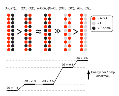


This is a lovely method and should find wide applicability in many settings, especially for microorganisms and cell lines. However, it is not clear that this approach will be, as implied by the discussion, an efficient mapping method for all multicellular organisms. I have performed similar experiments in Drosophila, focused on meiotic recombination, on a much smaller scale, and found that CRISPR-Cas9 can indeed generate targeted recombination at gRNA target sites. In every case I tested, I found that the recombination event was associated with a deletion at the gRNA site, which is probably unimportant for most mapping efforts, but may be a concern in some specific cases, for example for clinical applications. It would be interesting to know how often mutations occurred at the targeted gRNA site in this study.
The wider issue, however, is whether CRISPR-mediated recombination will be more efficient than other methods of mapping. After careful consideration of all the costs and the time involved in each of the steps for Drosophila, we have decided that targeted meiotic recombination using flanking visible markers will be, in most cases, considerably more efficient than CRISPR-mediated recombination. This is mainly due to the large expense of injecting embryos and the extensive effort and time required to screen injected animals for appropriate events. It is both cheaper and faster to generate markers (with CRISPR) and then perform a large meiotic recombination mapping experiment than it would be to generate the lines required for CRISPR-mediated recombination mapping. It is possible to dramatically reduce costs by, for example, mapping sequentially at finer resolution. But this approach would require much more time than marker-assisted mapping. If someone develops a rapid and cheap method of reliably introducing DNA into Drosophila embryos, then this calculus might change.
However, it is possible to imagine situations where CRISPR-mediated mapping would be preferable, even for Drosophila. For example, some genomic regions display extremely low or highly non-uniform recombination rates. It is possible that CRISPR-mediated mapping could provide a reasonable approach to fine mapping genes in these regions.
The authors also propose the exciting possibility that CRISPR-mediated loss of heterozygosity could be used to map traits in sterile species hybrids. It is not entirely obvious to me how this experiment would proceed and I hope the authors can illuminate me. If we imagine driving a recombination event in the early embryo (with maternal Cas9 from one parent and gRNA from a second parent), then at best we would end up with chimeric individuals carrying mitotic clones. I don’t think one could generate diploid animals where all cells carried the same loss of heterozygosity event. Even if we could, this experiment would require construction of a substantial number of stable transgenic lines expressing gRNAs. Mapping an ~20Mbp chromosome arm to ~10kb would require on the order of two-thousand transgenic lines. Not an undertaking to be taken lightly. It is already possible to perform similar tests (hemizygosity tests) using D. melanogaster deficiency lines in crosses with D. simulans, so perhaps CRISPR-mediated LOH could complement these deficiency screens for fine mapping efforts. But, at the moment, it is not clear to me how to do the experiment.