Feeds:
Posts
Comments

Archive for the ‘Cancer Informatics’ Category

Prognostic algorithm for retroperitoneal sarcoma

Larry H. Bernstein, MD, FCAP, Curator

LPBI


Prognostic Nomogram for Retroperitoneal Sarcoma Validated in Large Cohort

Cancer Network  By


A new study validated a prognostic nomogram for retroperitoneal sarcoma (RPS) using a large, external cohort. The nomogram incorporates six variables, and provided strong concordance with observed disease-free survival (DFS) and overall survival (OS).
RPS account for about 15% of all soft-tissue sarcomas, and their unique characteristics make traditional staging difficult, according to study authors led by Chandrajit P. Raut, MD, of Brigham and Women’s Hospital in Boston. A multi-institutional nomogram was developed previously based on a 523-patient cohort who underwent surgical resection for RPS between 1999 and 2009.

The nomogram included six clinical characteristics and variables: age at the time of diagnosis; tumor size; FNCLCC tumor grade; histologic subtype; multifocality at the time of initial presentation; and extent of surgical resection. Because the American Joint Committee on Cancer (AJCC) is considering including this tool in its forthcoming revised staging system, the study authors decided to validate it in an external cohort of 631 RPS patients who underwent surgery at one of six centers. The results were published online ahead of print in Cancer.

The 7-year DFS rate in the validation cohort was 38.4%, and the 7-year OS rate was 58%. This was similar to the development cohort’s outcomes, at 35.7% and 50.5%, respectively. All six of the nomogram’s included variables were found to be independently prognostic.

The researchers used a Harrell C-statistic to measure the nomogram’s discriminative ability. For DFS, the result was 0.69, while for OS it was 0.73. These were very similar to the concordance rates found in the development cohort (0.71 and 0.74, respectively).

“Correlation between observed and nomogram-predicted DFS and OS rates suggest good calibration of the multi-institutional RPS nomogram in a larger, independent validation cohort,” the authors wrote. “External validation of the…nomogram established its broad applicability in predicting outcomes in patients with primary RPS who were treated at experienced centers, and supports its inclusion in the 8th edition of the AJCC classification.”

Study Details
The median age in the validation cohort was 59 years, and the median tumor size was 21 cm. There were similar numbers of patients with FNCLCC grade 1 (32.7%), 2 (39.8%), and 3 (27.5%) tumors.

The most common histologic subtype was dedifferentiated liposarcoma (36.6%), followed by well-differentiated liposarcoma (25%), leiomyosarcoma (21.7%), and other subtypes. Almost all patients (94.8%) had a complete resection. Most patients (91%) did not receive chemotherapy or radiotherapy (68.3%).


Panobinostat Shows Promise in Phase I Sarcoma Trial

By

http://www.cancernetwork.com/sarcoma/panobinostat-shows-promise-phase-i-sarcoma-trial

Ball-and-stick model of panobinostat; image © molekuul.be / Shuttersto…


A phase I trial found that panobinostat combined with epirubicin is well tolerated and could offer benefit in patients with refractory sarcoma.
“More than half of patients treated for localized soft tissue sarcoma will experience relapse,” wrote study authors led by Pamela N. Munster, MD, of the University of California, San Francisco. Histone deacetylase (HDAC) inhibitors have shown some promise in other early phase I studies with anthracyclines. “Furthermore, HDAC inhibition was shown to promote differentiation and apoptosis in sarcoma.”

The new phase I trial combined the HDAC inhibitor panobinostat—which was approved in February 2015 for the treatment of multiple myeloma—with epirubicin. It began as a 3 + 3 dose escalation trial in patients with advanced solid tumors, followed by an expansion cohort at the maximum tolerated dose in 20 sarcoma patients. The results were published in Annals of Oncology.

In total, 40 patients received 20- to 60-mg panobinostat; 17 patients had previous exposure to topoisomerase II inhibitors. The study drug was administered on days 1, 3, and 5, followed by 75-mg/m2 epirubicin on day 5, in 21-day cycles. Dose-limiting toxicities were seen at 50 mg (one patient) and 60 mg (two patients); these included atrial fibrillation, grade 4 thrombocytopenia and febrile neutropenia, and grade 3 fatigue. The recommended phase II dose was set at 50 mg, and the 20 sarcoma patients in the expansion cohort received this dose.

Of 37 total evaluable patients, a partial response was seen in four of them (11%), and 17 patients (46%) had stable disease.

In only the 20-patient sarcoma expansion cohort, 12 patients were deemed to have received benefit from panobinostat. There was one response in those patients, and 11 patients had stable disease for longer than 3 months. The median progression-free survival in these patients was 3.4 months, and the median overall survival was 8.3 months. The authors noted that acquired topoisomerase resistance was reversed in 8 of 14 patients.

“A major challenge in HDAC inhibitor therapy is the absence of biomarkers,” wrote the authors. In this study, they showed that patients with a “pronounced degree” of peripheral blood mononucleocyte histone acetylation were more likely to derive benefit from the therapy. A decrease in neutrophil count over the first 5 days of the first cycle of therapy also was correlated with clinical benefit.

“This study suggests that further investigation of HDAC inhibition in combination with DNA-damaging agents in defined advanced sarcoma subtypes to validate these preliminary findings is warranted,” concluded the authors.

Read Full Post »

Avvinity will have exclusive rights in oncology to use Alphamer therapeutic platform, invented by a Nobel Laureate and developed by Centauri: A Case of a Joint Venture Model, Volume 2 (Volume Two: Latest in Genomics Methodologies for Therapeutics: Gene Editing, NGS and BioInformatics, Simulations and the Genome Ontology), Part 1: Next Generation Sequencing (NGS)

Avvinity will have exclusive rights in oncology to use Alphamer therapeutic platform, invented by a Nobel Laureate and developed by Centauri: A Case of a Joint Venture Model

Reporter: Aviva Lev-Ari, PhD, RN


Horizon Discovery Group plc Enters Immuno-Oncology Therapeutic Development and Forms Joint Venture with Centauri Therapeutics Limited

·           Newly formed company Avvinity Therapeutics will be jointly managed by Horizon, via its Research Biotech business, and Centauri

·           Joint venture creates a differentiated new player in the rapidly growing immuno-oncology market, currently valued at £25 billion

·           Avvinity will have exclusive rights in oncology to use Alphamer therapeutic platform, invented by a Nobel Laureate and developed by Centauri

·           Horizon to invest up to £5.3 million, with an initial outlay of £2.5 million, in a  joint venture to identify novel immuno-oncology therapeutics

·           Horizon will contribute its gene editing technology platform and oncology expertise and will also benefit from service revenues from the joint venture

·           Formation of the joint venture represents a strategic move by Horizon to capture the upside potential of its IP, platform technologies and capabilities in immuno-oncology, synthetic lethality and cell therapy based therapeutics


Horizon will host a conference call today at 09:30 GMT to discuss this news. To access the live conference call please use the dial-in details below. The presentation will be made available shortly before the call at:https://www.horizondiscovery.com/about-us/investor-relations/corporate-videos-and-presentations.


Cambridge, UK, 2 March 2016: Horizon Discovery Group plc (LSE: HZD) (“Horizon” or “the Company”), the leading international gene editing company, announces today that it has formed an immuno-oncology joint venture, Avvinity Therapeutics (“Avvinity”), with Centauri Therapeutics Limited (“Centauri”), a UK-based biotechnology company focused on the discovery and development of novel molecules targeting life-threatening infectious diseases. This transaction represents part of Horizon’s previously outlined strategy to invest up to £10 million, further leveraged by its IP, technology platforms and know-how, to identify the next generation of molecular and cellular cancer therapeutics. 


Avvinity will combine Horizon’s gene editing, immunology, oncology and drug discovery capabilities with Centauri’s Alphamer technology to provide a powerful and proprietary platform to discover and develop novel immuno-oncology therapeutics, for both solid tumours and leukaemias. Avvinity will be targeting an immuno-oncology market currently worth £25 billion per year and expected to grow to approximately £50 billion per year by 2020.(1)


Under the terms of this agreement, Horizon will out-license certain background intellectual property relating to its translational genomics and drug discovery platforms, and will invest up to £5.3 million over two tranches with the first tranche of £2.5m committed, and the second to be committed at Horizon’s discretion pending the progress of three development programs. Centauri will license background IP and expertise on its Alphamer technology to Avvinity, which will have exclusivity for the field of oncology for an initial three year period and can be extended via the issue of further equity concurrently with the raise of new investment.


Avvinity will be managed jointly by Horizon and Centauri, and based on the investment of IP, technology and the first tranche of funding; Horizon will own 33% of Avvinity’s equity, representing 50% of the most-preferred class of voting shares. Upon completion of the 2nd tranche of funding Horizon will own 49.99% of Avvinity’s equity, representing 50% of the most-preferred class of voting shares. The joint venture will be managed within Horizon as part of the Company’s Research Biotech business (formerly Horizon’s Leveraged business unit).


Neither Horizon nor Centauri will be obliged to provide further funding to Avvinity, though both retain pre-emption rights and may elect to participate in future funding rounds. Subject to achieving key development milestones, Avvinity plans to raise significant new external investment to take its innovative drugs into clinical trials, at which time the value of Horizon’s stake in the business would be highly-material.


Dr. Darrin M. Disley, Chief Executive Officer, President Research Biotech of Horizon Discovery Group plc, said: By combining Horizon’s deep understanding of the genetic basis of cancer alongside its gene editing, drug discovery and emerging immuno-oncology toolbox, with Centauri’s unique Alphamer technology and knowledge of its use, we have created an exciting new company to spearhead Horizon’s move into targeted therapeutic development.  We are confident this joint venture will break new ground in the development of immunotherapies, and bring significant value creation to Horizon shareholders.


“The establishment of Avvinity is in line with our hybrid Research Biotech strategy to not only work with partners but also take advantage of the therapeutic upside potential of the most exciting new areas of personalised and genomic medicine in a risk-managed way.”


Dr. Mike Westby, Chief Executive Officer of Centauri Therapeutics Limited, commented: Alphamers are an entirely novel way to target disease and represent an exciting new approach for recruitment of host immunity. At Centauri we have invested to build the Alphamer platform and assembled the drug discovery expertise necessary to exploit the platform in infectious diseases. Through this joint venture with Horizon, we look forward to applying our combined know-how and capabilities to develop Alphamers as important new immuno-oncology medicines, particularly for cancer indications that have proven intractable to date.”


More information on Avvinity can be found at www.avvinity.com.


 ENDS


Notes for Editors:


Alphamer technology is based on chemically synthesized molecules that redirect naturally occurring antibodies in the human immune system to selected pathogens or cancer cells. One end of a molecule binds a cell-surface target on a pathogen or cancer cell using an aptamer, while the other end presents specific epitopes that attach to the circulating antibodies. The result of this redirection is cell death and subsequent recruitment of the T-Cell mediated pathways to clear the body of the pathogen or cancer cell.


Alphamers promise key advantages over conventional antibody and antibody-drug conjugate molecules in immuno-oncology applications, including the ability to target cancers driven by both wild type (“normal”) gene overexpression as well as mutant (”abnormal”) gene overexpression, and by exhibiting a short half-life in the body yielding reduced toxicity and systemic side-effects.  Considerable investment has been made in the Alphamer technology over the past four years and its ability to engage the immune system to destroy bacteria has been confirmed.(2)


Platform technologies based around alternative immuno-oncology approaches have secured high valuations once early positive results in clinical trials have been achieved: Amgen’s 2012 acquisition of Micromet valued Micromet at $1.16b, and in 2013, Spirogen was acquired by AstraZeneca for up to $440m.


References:

1. http://www.prnewswire.com/news-releases/global–usa-cancer-immunotherapy-market-analysis-2015—forecasts-to-2020-300157219.html


2. Retargeting pre-existing human antibodies to a bacterial pathogen with an alpha-Gal conjugated aptamer. Kristian SA, Hwang JH, Hall B, Leire E, Iacomini J, Old R, Galili U, Roberts C, Mullis KB, Westby M, Nizet V. J Mol Med (Berl). 2015 Jun;93(6):619-31


Conference call:

A conference call for analysts, investors and media will take place at 09:30 GMT today hosted by Darrin Disley, Chief Executive Officer and President, Research Biotech, and Richard Vellacott, Chief Financial Officer, who will run through a presentation followed by a Q&A session. The presentation will be made available shortly before the call at: https://www.horizondiscovery.com/about-us/investor-relations/corporate-videos-and-presentations.


The dial-in numbers for the conference call are:


UK: 08006940257

Standard International: +44 (0) 1452 555566

Conference call ID: 62637628


A replay of the call is available approximately four hours after the call concludes. For those unable to attend it live, or who would like to listen to it again, call +44 (0)1452550000 and quote the conference call ID: 62637628.


For further information, please contact:


Horizon Discovery Group plc

Dr. Darrin Disley CEO / Richard Vellacott CFO

Tel: +44 (0) 1223 655580


Centauri Therapeutics Limited

Dr. Mike Westby CEO

Tel +44 (0) 1304 728611


Zyme Communications (Trade and Regional Media)

Katie Odgaard

Tel: +44 (0)7787 502 947

Email: katie.odgaard@zymecommunications.com


Consilium Strategic Communications (Financial Media and Investor Relations)

Amber Fennell / Jessica Hodgson / Matthew Neal / Laura Thornton

Tel: +44 (0) 20 3709 5701

Email: horizon@consilium-comms.com


Panmure Gordon & Co. (NOMAD)

Corporate Finance: Freddy Crossley / Duncan Monteith / Fabien Holler

Broking: Tom Salvesen

Tel: +44 20 7886 2500


About Horizon Discovery Group plc www.horizondiscovery.com/

Horizon is a leading international gene-editing company that supplies products, services and research programmes that enable genomics research and the development of personalised and genomic medicines. Horizon has a diverse and global customer base of over 1,400 unique organisations across more than 50 countries, including major pharmaceutical, biotechnology and diagnostic companies as well as leading academic research centres. The Group supplies its products and services into multiple markets, estimated to total in excess of £29 billion in 2015.


Horizon’s core capabilities are built around its proprietary translational genomics platform, a highly precise and flexible suite of gene editing tools (rAAV, ZFN and CRISPR) able to alter almost any gene sequence in human or mammalian cell-lines.


Horizon offers over 23,000 catalogue products, almost all of which are based on the application of gene editing to generate in vitro and in-vivo models that accurately model the disease-causing genetic anomalies found in diseases like cancer. These ‘patients-in-a-test-tube’ are being used by customers to: understand the genetic drivers of disease; identify targets of therapeutic intervention that can moderate or correct these genetic drivers; develop novel medicines and companion diagnostic tests that result in the right patient getting the right medicine.


Horizon also provides custom in vitro and in vivo disease model generation services, biopharmaceutical manufacturing cell lines and generation services, quantitative molecular reference standards and contract research and custom screening services.


In addition, Horizon through its Research Biotech business deploys the Company’s intellectual property, gene-editing platform, products, services and know-how in cancer research, drug discovery and immunology to develop its immuno-oncology, synthetic lethality and cell therapy platforms which aim to deliver novel drug treatments into the pharmaceutical pipeline.


Horizon is headquartered in Cambridge, UK, and is listed on the London Stock Exchange’s AIM market under the ticker “HZD”. For further information please visit: www.horizondiscovery.com.


About Centauri Therapeutics Ltd www.centauritherapeutics.com

Centauri Therapeutics is a UK-based biotechnology company focused on the discovery and development of novel molecules targeting life threatening diseases.


Centauri Therapeutics has established a core R&D facility at Discovery Park in Sandwich, Kent, with an experienced team of industry scientists focused on discovery, optimisation and development of novel Alphamers targeting acute hospital acquired infections. The company is currently focussed on the development of Alphamers against anti-microbial resistant (AMR) pathogens, which pose an increasing threat to human health.


Centauri Therapeutics’ Executive team is led by Mike Westby, Chief Executive Officer (previously Pfizer, Roche) and Stuart Lawson, Chief Financial Officer (CEO of the private investment group Animatrix Capital LLP, and previously KPMG). Clive Dix is Non-Executive Director and Chairman of the Board (previously Convergence Pharmaceuticals, PowderMed, PowderJect, Glaxo Wellcome).


SOURCE

From: “Katie Odgaard” <katie.odgaard@zymecommunications.com>

Date: March 2, 2016 at 2:12:41 AM EST

To:avivalev-ari@alum.berkeley.edu” <avivalev-ari@alum.berkeley.edu>

Subject: Horizon Discovery Group plc Enters Immuno-Oncology Therapeutic Development and Forms Joint Venture with Centauri Therapeutics Limited

Reply-To: katie.odgaard@zymecommunications.com

Read Full Post »

Breast Cancer Extratumor Microenvironment has Effect on Progression

Larry H. Bernstein, MD, FCAP, Curator

LPBI


Tumor Microenvironment Diversity Predicts Breast Cancer Outcomes

GEN News Highlights   Feb 17, 2016   http://www.genengnews.com/gen-news-highlights/tumor-microenvironment-diversity-predicts-breast-cancer-outcomes/81252378/


Intratumor heterogeneity, it is known, can complicate cancer treatments. Now it appears the same may be true of tumor microenvironment heterogeneity. According to a new study from the Institute of Cancer Research (ICR), London, breast cancers that develop within an “ecologically diverse” breast cancer microenvironment are particularly likely to progress and lead to death.

The study took an unusual approach: It combined ecological scoring methods with genome-wide profiling data. This approach, besides showing clinical utility in the evaluation of breast cancer outcomes, demonstrated that even so contextual a discipline as genomics can benefit from being placed within a larger context. In this case, the context is essentially Darwinian, albeit at a small scale.

Natural selection is typically studied at the level of ecosystems consisting of animals and plants. In the current study, however, it was assessed at the level of the tumor microenvironment, which consists of cancer cells, immune system lymphocytes, and stromal cells.

The ICR scientists, led by Yinyin Yuan, Ph.D., presented their work February 16 in the journal PLoS Medicine, in an article entitled “Microenvironmental Heterogeneity Parallels Breast Cancer Progression: A Histology–Genomic Integration Analysis.” The article describes how the scientists developed a tumor ecosystem diversity index (EDI), a scoring system that indicates the degree of microenvironmental heterogeneity along three spatial dimensions in solid tumors. EDI scores take account of “fully automated histology image analysis coupled with statistical measures commonly used in ecology.”

“[EDI] was compared with disease-specific survival, key mutations, genome-wide copy number, and expression profiling data in a retrospective study of 510 breast cancer patients as a test set and 516 breast cancer patients as an independent validation set,” wrote the authors. “In high-grade (grade 3) breast cancers, we uncovered a striking link between high microenvironmental heterogeneity measured by EDI and a poor prognosis that cannot be explained by tumor size, genomics, or any other data types.”

By using the EDI, the ICR team was able to identify several particularly aggressive subgroups of breast cancer. In fact, the EDI was a stronger predictor of survival than many established markers for the disease.

The ICR researchers also looked at the EDI in addition to genetic factors. For example, the researchers found that the prognostic value of EDI was enhanced with the addition of TP53 mutation status. By integrating EDI data and genome-wide profiling data, the researchers identified losses of specific genes on 4p14 and 5q13 that were enriched in grade 3 tumors. These tumors, which showed high microenvironmental diversity, substratified patients into poor prognostic groups.

“Our findings show that mathematical models of ecological diversity can spot more aggressive cancers,” said Dr. Yuan. “By analyzing images of the environment around a tumor based on Darwinian natural selection principles, we can predict survival in some breast cancer types even more effectively than many of the measures used now in the clinic.

“In the future, we hope that by combining cell diversity scores with other factors that influence cancer survival, such as genetics and tumor size, we will be able to tell apart patients with more or less aggressive disease so we can identify those who might need different types of treatment.”

“This ingenious study…teaches us a valuable lesson,” added Paul Workman, Ph.D., chief executive of the ICR. “[We] should always remember that cancer cells are not developing and growing in isolation, but are part of a complex ecosystem that involves normal human cells, too. By better understanding these ecosystems, we aim to create new ways to diagnose, monitor and treat cancer.”


Microenvironmental Heterogeneity Parallels Breast Cancer Progression: A Histology–Genomic Integration Analysis


Background

The intra-tumor diversity of cancer cells is under intense investigation; however, little is known about the heterogeneity of the tumor microenvironment that is key to cancer progression and evolution. We aimed to assess the degree of microenvironmental heterogeneity in breast cancer and correlate this with genomic and clinical parameters.

Methods and Findings

We developed a quantitative measure of microenvironmental heterogeneity along three spatial dimensions (3-D) in solid tumors, termed the tumor ecosystem diversity index (EDI), using fully automated histology image analysis coupled with statistical measures commonly used in ecology. This measure was compared with disease-specific survival, key mutations, genome-wide copy number, and expression profiling data in a retrospective study of 510 breast cancer patients as a test set and 516 breast cancer patients as an independent validation set. In high-grade (grade 3) breast cancers, we uncovered a striking link between high microenvironmental heterogeneity measured by EDI and a poor prognosis that cannot be explained by tumor size, genomics, or any other data types. However, this association was not observed in low-grade (grade 1 and 2) breast cancers. The prognostic value of EDI was superior to known prognostic factors and was enhanced with the addition of TP53 mutation status (multivariate analysis test set, p = 9 × 10⁻⁴, hazard ratio = 1.47, 95% CI 1.17–1.84; validation set, p = 0.0011, hazard ratio = 1.78, 95% CI 1.26–2.52). Integration with genome-wide profiling data identified losses of specific genes on 4p14 and 5q13 that were enriched in grade 3 tumors with high microenvironmental diversity that also substratified patients into poor prognostic groups. Limitations of this study include the number of cell types included in the model, that EDI has prognostic value only in grade 3 tumors, and that our spatial heterogeneity measure was dependent on spatial scale and tumor size.

Conclusions

To our knowledge, this is the first study to couple unbiased measures of microenvironmental heterogeneity with genomic alterations to predict breast cancer clinical outcome. We propose a clinically relevant role of microenvironmental heterogeneity for advanced breast tumors, and highlight that ecological statistics can be translated into medical advances for identifying a new type of biomarker and, furthermore, for understanding the synergistic interplay of microenvironmental heterogeneity with genomic alterations in cancer cells.

Background

The human body contains millions of cells, all of which grow, divide, and die in an orderly fashion to build tissues during early life and to replace worn-out or dying cells and repair injuries during adult life. Sometimes, however, normal cells acquire genetic changes (mutations) that allow them to divide uncontrollably and to move around the body (metastasize), resulting in cancer. Because any cell in the body can acquire the mutations needed for cancer development, there are many types of cancer. For example, breast cancer, the most common cancer in women, begins when the cells in the breast that normally make milk become altered. Moreover, different types of cancer progress and evolve differently—some cancers grow quickly and kill their “host” soon after diagnosis, whereas others can be successfully treated with drugs, surgery, or radiotherapy. The behavior of individual cancers depends both on the characteristics of the cancer cells within the tumor and on the interactions between the cancer cells and the normal stromal cells (the connective tissue cells of organs) and other cells (for example, immune cells) that surround and feed cancer cells (the tumor microenvironment).

Why Was This Study Done?

Although recent studies have highlighted the importance of the tumor microenvironment for disease-related outcomes, little is known about how the heterogeneity of the tumor microenvironment—the diversity of non-cancer cells within the tumor—affects outcomes. Mathematical modeling suggests that tumors with heterogeneous and homogeneous microenvironments have different growth patterns and that heterogeneous microenvironments are more likely to be associated with aggressive cancers than homogenous microenvironments. However, the lack of methods to quantify the spatial variability and cellular composition across solid tumors has prevented confirmation of these predictions. Here, the researchers develop a computational system for quantifying microenvironmental heterogeneity in breast cancer based on tumor morphology (shape and form) in histological sections (tissue samples taken from tumors that are examined microscopically). They then use this system to analyze the associations between clinical outcomes, molecular changes, and microenvironmental heterogeneity in breast cancer.

What Did the Researchers Do and Find?

The researchers used automated image analysis and statistical analysis to develop the ecosystem diversity index (EDI), a numerical measure of microenvironmental heterogeneity in solid tumors. They compared the EDI with prognosis (likely outcome), key mutations, genome-wide copy number (tumor cells often contain abnormal numbers of copies of specific genes), and expression profiling data (the expression of several key proteins is altered in tumors) in a test set of 510 samples from patients with breast cancer and in a validation set of 516 additional samples. Among high-grade breast cancers (grade 3 cancers; the grade of a cancer indicates what the cells look like; high-grade breast cancers have a poor prognosis), but not among low-grade breast cancers (grades 1 and 2), a high EDI (high microenvironmental heterogeneity) was associated with a poor prognosis. Specifically, patients with grade 3 tumors and a high EDI had a ten-year disease-specific survival rate of 51%, whereas the remaining patients with grade 3 tumors had a ten-year survival rate of 70%. Notably, the combination of a high EDI with specific DNA alterations—mutations in a gene called TP53 and loss of genes on Chromosomes 4p14 and 5q13—improved the accuracy of prognosis among patients with grade 3 breast cancer and stratified them into subgroups with disease-specific five-year survival rates of 35%, 9%, and 32%, respectively.

What Do These Findings Mean?

These findings establish a method for measuring the spatial heterogeneity of the microenvironment of solid tumors and suggest that the measurement of tumor microenvironmental heterogeneity can be coupled with information about genomic alterations to provide an accurate way to predict outcomes among patients with high-grade breast cancer. The association between EDI, specific genomic alterations, and outcomes needs to be confirmed in additional patients. However, these findings suggest that microenvironmental heterogeneity might provide an additional biomarker to help clinicians identify those patients with advanced breast cancer who have a particularly bad prognosis. The ability to identify these patients is important because it will help clinicians target aggressive treatments to individuals with a poor prognosis and avoid the overtreatment of patients whose prognosis is more favorable. Finally, and more generally, these findings describe a new way to investigate the interactions between the tumor microenvironment and genomic alterations in cancer cells.

Additional Information

This list of resources contains links that can be accessed when viewing the PDF on a device or via the online version of the article at http://dx.doi.org/10.1371/journal.pmed.1001961.

Citation: Natrajan R, Sailem H, Mardakheh FK, Arias Garcia M, Tape CJ, Dowsett M, et al. (2016) Microenvironmental Heterogeneity Parallels Breast Cancer Progression: A Histology–Genomic Integration Analysis.
PLoS Med 13(2): e1001961.     http://dx.doi.org:/10.1371/journal.pmed.1001961
Fig 1. In silico tumor dissection pipeline for quantifying spatial diversity in the tumor ecosystem.
Fig 1. In silico tumor dissection pipeline for quantifying spatial diversity in the tumor ecosystem. (A) Flow diagram depicting the overall study design. (B) Schematic of our pipeline for quantifying spatial diversity in pathological samples. H&E sections are morphologically classified and divided into regions to be spatially scored. The number of clusters k in the regional scores is indicative of the number of sub-populations of cell types in the tumor regions. (C) Examples of tumor regions with low and high diversity scores using the Shannon diversity index, accounting for cancer cells (outlined in green), lymphocytes (blue), and stromal cells (red). Cell classification is automated by image analysis. (D) The 3-D landscape of cell diversity scores on an example H&E section; the x- and y-axes are the geometric axes of the image, and the z-axis is cell diversity computed on a region-by-region basis. (E) The distribution of regional scores in a tumor from the METABRIC study with two regional clusters identified using Gaussian mixture clustering (grey shading: histogram; dashed black line: density; solid black lines: mixture components/clusters).
Fig 2. Application of EDI to 1,026 breast tumors from the METABRIC study.
Fig 2. Application of EDI to 1,026 breast tumors from the METABRIC study. (A) The frequencies of EDI scores in breast tumors. (B) H&E staining, distribution of classified cells (green: cancer; blue: lymphocyte; red: stromal cells), and the heatmap of regional diversity scores for a tumor with the highest EDI score (EDI = 5). (C) Representative regions from each of the clusters k1–k5 are shown in a tumor with EDI = 5, with cluster k1 having the lowest diversity score and k5 the highest. By mapping regional clusters to the H&E image, we can begin to interpret these clusters with different cell diversity. We observed predominantly cancer cells in k1, increasingly more stromal cells and ductal in situ carcinoma cells (DCIS) in k2, and a vessel in k3. Cluster k4 features extensive stromal lymphocytes between ductal in situ carcinoma components, while k5 shows tumor-infiltrating lymphocytes (TIL) associated with invasive carcinoma cells.
Fig 3. Reproducibility, stability, and independence of the EDI-high group in 507 grade 3 breast tumors.
Fig 3. Reproducibility, stability, and independence of the EDI-high group in 507 grade 3 breast tumors. (A) Kaplan–Meier curves of disease-specific survival to illustrate the prognosis of EDI-high samples compared to other grade 3 samples in two independent patient cohorts. Shown below the graph are the number of patients (the number of disease-specific events) per group for EDI-low (grey) and EDI-high (red). (B) Agreement of the EDI subtyping between 100% data and resampling with progressively fewer tumor regions in 200 repeats. (C) Distribution of known subtypes in grade 3 tumors stratified by EDI; asterisks mark subtypes enriched in the EDI-high group. (D) Kaplan–Meier curves illustrating the duration of disease-specific survival according to tumor size (left) and improvement of stratification with the addition of EDI information (right).
Accumulating evidence suggests that the interactions of cancer cells and stromal cells within their microenvironment govern disease progression, metastasis, and, ultimately, the evolution of therapeutic resistance [1–3]. Recent reports have highlighted the significance of the contribution of stromal gene expression and morphological structure as powerful prognostic determinants for a number of tumor types, emphasizing the importance of the tumor microenvironment in disease-related outcomes [4–7]. In breast cancer, a number of studies have demonstrated the prognostic correlation of individual cell types, including the immune cell infiltrate that predicts response to therapy [8–10], and the high percentage of tumor stroma that predicts poor prognosis in triple-negative disease but good prognosis in estrogen receptor (ER)–positive disease [11,12]. Nevertheless, different types of cells coexist with varying degrees of heterogeneity within a tumor. This fundamental feature of human tumors and the combinatorial effects of cell types have been largely ignored, and the collective implications for clinical outcome remain elusive. Consistent observations from mathematical models have highlighted that tumors with diverse microenvironments show growth patterns dramatically different from those of tumors with homogeneous environments [13] and are more likely to be associated with aggressive cancer phenotypes [2] that select for cell migration and eventual metastasis by allowing cancer cells to evolve more rapidly [14]. These observations highlight the need to understand the collective physiological characteristics and heterogeneity of tumor microenvironments. However, there is a lack of methods to quantify the high spatial variability and diverse cellular composition across different solid tumors. Moreover, the interplay of genomic alterations in cancer cells and microenvironmental heterogeneity and its subsequent role in treatment response have not been explored. Our aims were (i) to develop a computational system for quantifying microenvironmental heterogeneity based on tumor morphology in routine histological sections, (ii) to define the clinical implications of microenvironmental heterogeneity, and (iii) to integrate this histologybased index with RNA gene expression and DNA copy number profiling data to identify molecular changes associated with microenvironmental heterogeneity.
The Ecosystem Diversity Index To characterize the tumor ecosystem based on cell compositions, we developed a new index to be used in conjunction with our image analysis tool [16]. First, we used our automated morphological classification method [16] to identify and classify cells into cancer, lymphocyte, or stromal cell classes in H&E sections (Fig 1B). We next divided sections into smaller spatial regions and quantified the diversity of the tumor ecosystem in a tumor region j using the Shannon diversity index: dj ¼ Sm i pi logpi ; ð1Þ where m is the number of cell types and pi is the proportion of the ith cell type (Fig 1B and 1C). A high value of the Shannon diversity index dj reports a heterogeneous environment populated by many cell types, whilst a low value indicates a homogeneous environment (Fig 1C). Compared to other methods such as the Simpson index, the Shannon diversity index accounts for rare species and, hence, is less dominated by main species [17]. Subsequently, we derived the ecosystem diversity index (EDI) by applying unsupervised clustering that identifies the optimum number of clusters in the dataset in an unbiased manner, in order to group tumor regions and quantify the degree of spatial heterogeneity. Let D = d1,d2,…,dn be the Shannon index for n regions in a tumor. We used Gaussian mixture models to fit data D: D SK k¼1okNðmk; s2 kÞ: ð2Þ where μk, ,s2 k, and ωk are the mean, variance, and weight of a Gaussian distribution k, and K is the number of clusters. The Bayesian information criterion was then used to select the best number of clusters K [18]. We used K = 1–5 as the range of K to avoid small EDI groups (S1 Text). The final value of K thus is a measurement of heterogeneity and the score of EDI for a tumor.
Fig 5. The relationship between ecological heterogeneity and cancer genomic aberrations in 507 grade 3 tumors. (A) Genome-wide copy number aberrations in grade 3 breast tumors and genomic coordinates of genes with copy number aberrations enriched in the EDI-high group. Lengths of black lines denote level of enrichment significance with copy number gains (above the horizontal line) or losses (below the horizontal line). (B) Kaplan–Meier curves illustrating the duration of disease-specific survival in grade 3 breast cancer patients according to copy number loss of the 4p14 region (left) and the EDI-high group with additional information of 4p14 copy number loss (right). (C) Kaplan–Meier curves illustrating the duration of disease-specific survival according to copy number loss of the 5q13 region (left) and the EDI-high group with additional information of 5q13 copy number loss (right).
This study has a number of limitations. The motivation for our computational development was to use a data-driven model and measure the degree of spatial heterogeneity in tumor pathological specimens. In this model, only three major cell types in breast tumors were considered. Further sub-classification of the different types of stromal and immune cells by immunohistochemistry may add additional discriminatory value to our model. For dissecting spatial heterogeneity, we chose to use square regions with equal sizes. We found that EDI was correlated with the size of the region chosen for calculation of the Shannon diversity index, and as such the spatial heterogeneity is scale dependent. This phenomenon has been well described in a number of studies in ecology that show that a scale needs to be chosen that is appropriate for the ecological process under study [38,39], further highlighting the analogy between tumor studies and ecology. Similar to the recent observation that breast cancer subclonal heterogeneity is correlated with tumor size [35], we also found an association between microenvironmental heterogeneity and tumor size; hence, EDI may have more limited value in smaller tumors. However, small tumors were present in the EDI-high group, and addition of EDI within tumors grouped by size further stratified their prognosis. We found that EDI was prognostic only in grade 3 tumors in our study, which could be a limitation, given the possible discordance in grading between pathologists.
The identification of additional biomarkers in subgroups of patients that identify them as high risk is important for patient management and to avoid overtreatment for low-risk patients. We envision that the use of our measure of microenvironment heterogeneity, together with key genomic alterations, will enable the diagnosis of patients at very high risk of relapse and facilitate the enrollment of these patients into additional clinical trials for novel therapies or treatment intensification. Our novel computational approach provides a fully automated tool that is relatively easy to implement. Integration of this measure with genomic profiling provides additional prognostic information independent of known clinical parameters. The results of this study highlight the possibility of a grade-3-specific prognostic tool that may aid in further classification of high-grade breast cancer patients beyond standard assays such as ER and HER2 status.


Read Full Post »

How Cancer Cells “Grab” Neighbors and “Reel them in”

Larry H. Bernstein, MD, FCAP, Curator

LPBI


How cancer cells form tumors by reaching out with ‘cables’ and grabbing cells

A cancer riddle solved; counters “cancer stem cell” explanation
January 27, 2016  http://www.kurzweilai.net/how-cancer-cells-form-tumors-by-reaching-out-with-cables-and-grabbing-cells

https://youtu.be/5JToZT_xSyo

University of Iowa | Cancer cells’ motion and accretion into tumors

Two University of Iowa studies have recorded the movements of cancerous human breast tissue cells in real time and in 3D — the first time cancer cells’ motion and accretion into tumors has been continuously tracked, the researchers believe.

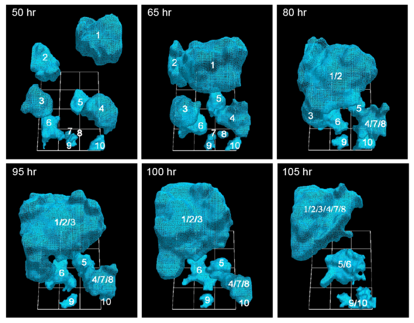
The team discovered that cancerous cells, moving at move at 92 micrometers per hour (about twice the speed of healthy cells), actively recruit healthy cells into tumors by extending a kind of cable to grab their neighbors — both cancerous and healthy — and reel them in. Surprisingly, as little as five percent of cancerous cells are needed to form the tumors, a ratio previously unknown.

“It’s not like things sticking to each other,” said David Soll, biology professor at the UI and corresponding author on the open-access paper, published in the American Journal of Cancer Research. “It’s that these cells go out and actively recruit. It’s complicated stuff, and it’s not passive. No one had a clue that there were specialized cells in this process, and that it’s a small number that pulls all the rest in.”

The findings could lead to a more precise identification of tumorigenic cells (those that form tumors) and to testing which antibodies would be best equipped to eliminate them.*

How cancer cells “know” what to do

The question is: how do these cells know what to do. Soll hypothesizes they’re reaching back to a primitive past, when these cells were programmed to form embryos. If true, perhaps the cancerous cells — masquerading as embryo-forming cells — recruit other cells to make tissue that then forms the layered, self-sustaining architecture needed for a tumor to form and thrive. “It’s as if it’s building its own defenses against the body’s efforts to defeat them.”

http://www.kurzweilai.net/images/Cancer-Cells-50-105-hours-stacked.png

University of Iowa researchers have documented how cancerous tumors form by tracking in real time the movement of individual cells in 3-D. They report that just 5 percent of cancer cells are needed to form tumors, a ratio that heretofore had been unknown. (credit: Soll Laboratory)

In the AJCR paper, the researchers found support for their previous observation that tumorigenic cell lines and fresh tumor cells possess the unique capacity to form tumors by the active formation of cellular cables.

The finding lends more weight to the idea that tumors are created concurrently, in multiple locations, by individual clusters of cells that employ the cancer-cell cables to draw in more cells and enlarge themselves. Some have argued that tumors come about more by cellular changes within the masses, known as the “cancer stem cell theory.”

The Developmental Studies Hybridoma Bank funded the study.

* Soll’s Monoclonal Antibody Research Institute and the Developmental Studies Hybridoma Bank, created by the National Institutes of Health as a national resource, directed by Soll and housed at the UI, together contain one of the world’s largest collections of antibodies that could be used for the anti-cancer testing, based on the new findings.


Abstract of Mediated coalescence: a possible mechanism for tumor cellular heterogeneity

Recently, we demonstrated that tumorigenic cell lines and fresh tumor cells seeded in a 3D Matrigel model, first grow as clonal islands (primary aggregates), then coalesce through the formation and contraction of cellular cables. Non-tumorigenic cell lines and cells from normal tissue form clonal islands, but do not form cables or coalesce. Here we show that as little as 5% tumorigenic cells will actively mediate coalescence between primary aggregates of majority non-tumorigenic or non-cancerous cells, by forming cellular cables between them. We suggest that this newly discovered, specialized characteristic of tumorigenic cells may explain, at least in part, why tumors contain primarily non-tumorigenic cells.


Read Full Post »

Pancreatic Cancer: a discovery in Toulouse that would slow its progression

Reporter: Danut Dragoi, PhD

This week was held in Toulouse edition 2016 Toulouse Onco Week (TOW), which is also supposed to end this Friday after three days of attendance. This is an event “under the patronage” of the President of the Republic, François Hollande. The event is within the framework of the World Day of fight against cancer which was held on February 4, 2016.

At TOW global oncology conference the team of the National Institute of Health and Medical Research (INSERM) in Toulouse announced their discovery, described as promising. It is about a significant advancement in the treatment against pancreatic cancer. The picture below shows a cancer cell in 3D taken from Photos: jovan vitanovski / shutterstock.com. It suggests how cancer cells attach to human organ cells. It is interesting that similar pictures were obtained with an SEM microscope.

Cancer Cell 3D Toulouse

Elimination of the CDA would stop the progress of pancreatic cancer

Working with cytidine deaminase (CDA), an enzyme familiar in the field of oncology, the scientists expected  a higher sensitivity to chemotherapy, but they found another action of a modified protein, the stoppage of the progression of the tumor.

Treatment to wait in 5 to 10 years

It is stated that this advanced cancer upset was possible even before chemotherapy has not been established. The scientist Pierre Cordelier believes, however, that it will take between 5 to 10 years before this discovery will lead to a treatment. Recall that pancreatic cancer is a disease whose chances of survival appear relatively low. Indeed there are 12,000 deaths related to this tumor on the 13,000 cases reported each year. Experts expect also that pancreatic cancer from fourth to second place in the classification of causes of death by cancer.

The sections of the symposium

The symposium had six scientific sessions:

–       Genetic instability and cell cycle

–       Gene expression and epigenetics

–       Metabolism

–       Tumor biology and stem cells

–       Immunity

–       New technologies applicable to health

During the TOW, the first international symposium of Toulouse Center for Cancerology Research two Nobel laureates. Professor Jules Hoffmann, winner of the 2011 Nobel Prize in Medicine, and Professor Gerd Binnig, winner of the 1986 Nobel Prize in Physics, were announced to honor the symposium with their presence.

Comment

It is interesting that the last symposium on pancreatic cancer was held in France too, at Marseilles, 2015. At that meeting there were four sessions on pancreas cancer and the high frequency of such meetings shows a sense of urgency on pancreatic cancer that is raising. In the world, India and  Africa has the less rate of pancreas cancers per 100.000 people. The future of pancreas cancer is discussed in here. Pancreatic cancer is rare (it represents 2.1% of all tumors), but its incidence has increased since the middle of the 20th century. Furthermore, over 80% of patients experience a recurrence following surgery (60% of them within 6 months). In Spain, almost 4,000 new cases are recorded every year — 53% in men and 44% in women — most of whom are diagnosed between the ages of 65 and 75. In the US, the two curves, new cases and non-survivals are almost the same, the separation is only a small margin, making everybody aware of this disease.

Source

Photos: http://www.shutterstock.com/, jovan vitanovski / shutterstock.com

http://www.24matins.fr/cancer-pancreas-decouverte-a-toulouse-281754

http://www.thepharmaletter.com/event/toulouse-onco-week-2016

https://www.pancreasfoundation.org/event/page/3/

http://www.vitamindwiki.com/Pancreatic+Cancer+massively+deregulates+the+local+Vitamin+D+receptors+and+CPY24A1+%E2%80%93+July+2014

http://www.eurekalert.org/pub_releases/2015-04/cndi-tpp042315.php

http://seer.cancer.gov/statfacts/html/pancreas.html


Read Full Post »

BioMEMS The Market aspects of Oligonucleotide-Chips, Products and Applications, Competition, January 21, 2016

Curator: Gérard LOISEAU, ESQ


BioMEMS

The Market aspects of Oligonucleotide-Chips, Products, Applications, Competition 

January 21, 2016

2015-2020

The oligonucleotide synthesis market is expected to reach USD 1.918.6Billion at a CAGR of 10.1% by 2020 from USD 1.078.1Billion in 2015.

SOURCE

MARKETSANDMARKETS marketsandmarkets.com/


PLAYERS

  • Agilent Technologies Inc.
  • BioAutomation Corp.
  • Biosearch Technologies
  • Gen9 Inc.
  • GenScript Inc.
  • Illumina Inc.
  • Integrated DNA Technologies
  • New England Biolabs Inc.
  • Nitto Denko Avecia Inc.
  • OriGene Technologies Inc.
  • Sigma-Aldrich Corporation
  • Thermo Fisher Scientific Inc.
  • TriLink Biotechnologies


Agilent Technologies
 CA NYSE :A


http://www.agilent.com/

  • Agilent was created as a spin off from Hewlett-Packard Company in 1999.
  • Agilent Technologies Inc. is engaged in the life sciences, diagnostics and applied chemical markets. The Company provides application focused solutions that include instruments, software, services and consumables for the entire laboratory workflow. The Company has three business segments:

the life sciences and applied markets business,

the diagnostics and genomics business, and

the Agilent Cross Lab business

  • The Company’s life sciences and applied markets business segment brings together the Company’s analytical laboratory instrumentation and informatics.
  • The Company’s diagnostics and genomics business segment consists of three businesses: the Dako business, the genomics business and the nucleic acid solutions business.
  • The Company’s Agilent Cross Lab business segment combines its analytical laboratory services and consumables business

SOURCE

http://reuters.com/

PRODUCTS AND SERVICES

https://www.agilent.com/en-us/default#collapse-0

  • October 09, 2015 03:21 PM Eastern Daylight Time
  • CARPINTERIA, Calif.–(BUSINESS WIRE)–Dako, an Agilent Technologies company and a worldwide provider of cancer diagnostics, today announced the U.S. Food and Drug Administration has approved a new test that can identify PD-L1 expression levels on the surface of non-small cell lung cancer tumor cells and provide information on the survival benefit with OPDIVO® (nivolumab) for patients with non-squamous NSCLC.

SOURCE

BUSINESS WIRE busibesswire.com/


BioAutomation Corp.

 TX


http://bioautomation.com/

          PRODUCTS AND SERVICES

  • DNA and RNA synthesis reagents for the MerMades


Note: The MerMade 192E Oligonucleotide synthesizer is designed to synthesize DNA, RNA & LNA oligonucleotides in a column format

          PARTNERSHIPS

  • HONGENE BIOTECH : BIOAUTOMATION is the exclusive distributor for the Americas
  • EMD MILLIPORE
  • BIOSEARCH TECHNOLOGIES


DISTRIBUTORS

  • LINK TECHNOLOGIES : UK
  • AME BIOSCIENCE : UK
  • BOSUNG SCIENCE : KOREA
  • DNA CHEM : CHINA
  • WAKO : JAPAN
  • ACE PROBE : INDIA

SOURCE

bioautomation.com/


Biosearch Technologies
 CA


http://biosearchtech.com/

          PRODUCTS

  • qPCR & SNP Genotyping
  • Custom Oligonucleotides
  • – highly sophisticated oligonucleotides
  • – simple PCR primers
  • Oligos in Plates
  • RNA FISH
  • Synthesis Reagents
  • Immunochemicals
  • Primers
  • Probes
  • Large-Scale Synthesis Oligos
  • Intermediate-Scale Synthesis Oligos

          SERVICES

  • GMP & Commercial Services
  • OEM & Kit Manufacturing
  • qPCR Design Collaborations

          DISTRIBUTORS

Argentina | Australia | Austria | Brazil | Canada |Chile | China | Colombia | Czech Republic | Denmark | Ecuador | Finland | Germany |Hong Kong | Israel | Italy | Japan | Korea | Malaysia | Mexico | New Zealand | Norway | Paraguay | Peru| Philippines | Poland | Romania | Singapore | South Africa | Spain | Sweden |Switzerland | Taiwan ROC | Thailand | Turkey | United Kingdom | Uruguay | Vietnam

SOURCE

biosearchtech.com/


Gen9 Inc.
 MA 


http://www.gen9bio.com/

          PRODUCTS

Gen9 is building on advances in synthetic biology to power a scalable fabrication capability that will significantly increase the world’s capacity to produce DNA content. The privately held company’s next-generation gene synthesis technology allows for the high-throughput, automated production of DNA constructs at lower cost and higher accuracy than previous methods on the market. Founded by world leaders in synthetic biology, Gen9 aims to ensure the constructive application of synthetic biology in industries ranging from enzyme and chemical production to pharmaceuticals and biofuels.

          SERVICES

  • Synthetic Biology
  • Gene Synthesis Services
  • Variant Libraries
  • Gene Sequence Design Services

         INVESTORS

  • Agilent Technologies : Private Equity
  • CAMBRIDGE, Mass. and SANTA CLARA, Calif. — April 24, 2013 —Gen9 Receives $21 Million Strategic Investment from Agilent Technologies

SOURCE

gen9bio.com/


GenScript Inc.
 NJ 


http://www.genscript.com/

  • GenScript is the largest gene synthesis provider in the USA
  • GenScript Corporation, a biology contract research organization, provides biological research and drug discovery services to pharmaceutical companies, biotech firms, and research institutions in the United States, Europe, and Japan. It offers bio-reagent, custom molecular biology, custom peptide, protein production, custom antibody production, drug candidates testing, assay development and screening, lead optimization, antibody drug development, gene synthesis, and assay-ready cell line production services.
  • The company also offers molecular biology, peptide, protein, immunoassay, chemicals, and cell biology products. It offers its products through distributors in Tokyo, Japan; and Seoul, Korea. GenScript Corporation has a strategic partnership with Immunologix, Inc. The company was founded in 2002 and is based in Piscataway, New Jersey. It has subsidiaries in France, Japan, and China.


Note: As of October 24, 2011, Immunologix, Inc. was acquired by Intrexon Corporation. Immunologix, Inc. develops and produces antibody-based therapeutics for various biological targets. It produces human monoclonal antibodies against viral, bacterial, and tumor antigens, as well as human auto antigens.

Intrexon Corporation, founded in 1998, is a leader in synthetic biology focused on collaborating with companies in Health, Food, Energy, Environment and Consumer sectors to create biologically based products that improve quality of life and the health of the planet.


             PRODUCTS AND SERVICES

  • Gene synthesis
  • Antibody services
  • Protein Services
  • Peptide services


               INVESTORS


Note: The Balloch Group (‘TBG’) was established in 2001 by Howard Balloch (Canada‘s ambassador to China from 1996 to 2001). TBG has since grown from a market-entry consultancy working with North American clients in China to a leading advisory and merchant banking firm serving both domestic Chinese companies and multinational corporations. TBG was ranked as the number one boutique investment bank in China by ChinaVenture in 2008.

Kleiner, Perkins, Caufield and Byers


Illumina
Inc. CA


http://illumina.com/


Monica Heger : SAN FRANCISCO (GenomeWeb) – Illumina today announced two new next-generation sequencing platforms, a targeted sequencing system called MiniSeq and a semiconductor sequencer that is still under development.

Illumina disclosed the initiatives during a presentation at the JP Morgan Healthcare conference held here today. During the presentation, Illumina CEO Jay Flatley also announced a new genotyping array called Infinium XT; a partnership with Bio-Rad to develop a single-cell sequencing workflow; preliminary estimates of its fourth-quarter 2015 revenues; and an update on existing products. The presentation followed the company’s announcement on Sunday that it has launched a new company called Grail to develop a next-generation sequencing test for early cancer detection from patient blood samples.

The MiniSeq system, which is based on Illumina’s current sequencing technology, will begin shipping early this quarter and has a list price of $49,500. It can perform a variety of targeted DNA and RNA applications, from single-gene to pathway sequencing, and promises “all-in” prices, including library prep and sequencing, of $200 to $300 per sample, Flatley said during the JP Morgan presentation.

SOURCES

https://www.genomeweb.com/sequencing-technology/illumina-unveils-mini-targeted-sequencer-semiconductor-sequencing-project-jp

http://investor.biospace.com/biospace/quote?Symbol=ILMN


              PRODUCTS AND SERVICES

  •               Mid to large scale manufacturing assets
  •               Analytical Labs
  •               Pre-clinical
  •               Clinical
  •               Launched products


              COMPETITORS

https://finance.yahoo.com/q/co?s=ILMN+Competitors Tue, Feb 2, 2016, 2:16pm EST – US Markets

ILMN PVT1 AFFX LMNX Industry
Market Cap: 22.75B N/A 1.13B 835.66M 134.14M
Employees: 3,700 10,000 1,200 745 45.00
Qtrly Rev Growth (yoy): 0.14 N/A -0.01 0.07 0.18
Revenue (ttm): 2.14B 3.80B1 357.74M 235.37M 8.47M
Gross Margin (ttm): 0.73 N/A 0.63 0.71 0.58
EBITDA (ttm): 770.84M N/A 46.64M 52.99M -12.31M
Operating Margin (ttm): 0.30 N/A 0.08 0.17 -1.62
Net Income (ttm): 510.36M 430.90M1 11.22M 39.29M N/A
EPS (ttm): 3.42 N/A 0.13 0.93 -0.34
P/E (ttm): 45.43 N/A 104.40 20.91 25.33
PEG (5 yr expected): 2.68 N/A 4.66 0.55 N/A
P/S (ttm): 10.87 N/A 3.13 3.45 13.65


Pvt1 = Life Technologies Corporation (privately held)

AFFX = Affymetrix Inc.

LMNX = Luminex Corporation


Integrated DNA Technologies (IDT)
IOWA + CA

http://www.com/


Integrated DNA Technologies, Inc. (IDT), the global leader in nucleic acid synthesis, serving all areas of life sciences research and development, offers products for a broad range of genomics applications. IDT’s primary business is the production of custom, synthetic nucleic acids for molecular biology applications, including qPCR, sequencing, synthetic biology, and functional genomics. The company manufactures and ships an average of 44,000 custom nucleic acids per day to more than 82,000 customers worldwide. For more information, visit idtdna.com.


               PRODUCTS AND SERVICES

               https://eu.idtdna.com/site

  • DNA & RNA Synthesis
  • Custom DNA Oligos 96- & 384-Well Plates Ultramer Oligos Custom RNA Oligos SameDay Oligos HotPlates ReadyMade Primers Oligo Modifications Freedom
  • Dyes GMP for Molecular Diagnostics Large Scale Oligo Synthesis


Note : Skokie, IL – December 1, 2015. Integrated DNA Technologies Inc. (“IDT”), the global leader in custom nucleic acid synthesis, has entered into a definitive agreement to acquire the oligonucleotide synthesis business of AITbiotech Pte. Ltd. in Singapore (“AITbiotech”). With this acquisition, IDT expands its customer base across Southeast Asia making it possible for these additional customers to now have access to its broad range of products for genomic applications. AITbiotech will continue operations in its other core business areas.


New England Biolabs Inc.
 MA 


http://www.neb.com/


                PRODUCTS AND SERVICES

  •                 Restriction Endonucleases
  •                 PCR, Polymerases & Amplification Technologies
  •                 DNA Modifying Enzymes
  •                 Library Preparation for Next Generation Sequencing
  •                 Nucleic Acid Purification
  •                 Markers & Ladders
  •                 RNA Reagents
  •                 Gene Expression
  •                 Cellular Analysis

SOURCE

neb.com/


Nitto Denko Avecia Inc.
 MA


http://avecia.com/


With over 20 years of experience in oligonucleotide development and production, and over 1000 sequences manufactured, Avecia has played an integral role in the advancing oligo therapeutic market. Our mission is to continue to build value for our customers, as they progress through drug development into commercialization. And as a member of the Nitto Denko Corporation (nitto.com), Avecia is committed to the future of the oligonucleotide market. We are driven by innovative ideas and flexible solutions, designed to provide our customers with the best in service, quality, and technology.


SOURCE

http://avecia.com/


Note : 1918 Nitto Electric Industrial Co., Ltd. forms in Ohsaki, Tokyo, to produce electrical insulating materials in Japan.

2011 Acquires Avecia Biotechnology Inc. in the U.S.A.


OriGene Technologies Inc.
 CA

http://www.com/


OriGene Technologies, Inc. develops, manufactures, and sells genome wide research and diagnostic products for pharmaceutical, biotechnology, and academic research applications. The company offers cDNA clones, including TrueORF cDNA, viral ORF, destination vectors, TrueClones (human), TrueClones (mouse), organelle marker plasmids, MicroRNA tools, mutant and variant clones, plasmid purification kits, transfection reagents, and gene synthesis service; and HuSH shRNA, siRNA, miRNA, qPCR reagents, plasmid purification products, transfection reagents, PolyA+ and total RNA products, first-strand cDNA synthesis, and CRISPR/Cas9 genome products. It also provides proteins and lysates, such as purified human proteins, over-expression cell lysates, mass spectrometry standard proteins, and protein purification reagents; UltraMAB IHC antibodies, TrueMAB primary antibodies, anti-tag and fluorescent proteins, ELISA antibodies, luminex antibodies, secondary antibodies, and controls and others; and anatomic pathology products, including IHC antibodies, detection systems, and IHC accessories

The company offers luminex and ELISA antibody pairs, autoantibody profiling arrays, ELISA kits, cell assay kits, assay reagents, custom development, and fluorogenic cell assays; TissueFocus search tools; tissue sections; tissue microarrays, cancer protein lysate arrays, TissueScan cDNA arrays, tissue blocks, and quality control products, as well as tissue RNA, DNA, and protein lysates; and lab essentials. Its research areas include cancer biomarker research, RNAi, pathology IHC, stem cell research, ion channels, and protein kinase products. The company provides gene synthesis and molecular biology services, genome editing, custom cloning, custom shRNA, purified protein, monoclonal antibody development, and assay development. It sells its products through distributors worldwide, as well as online. OriGene Technologies, Inc. was incorporated in 1995 and is based in Rockville, Maryland.

SOURCE

http://BLOOMBERG.com

               PRODUCTS AND SERVICES

  •                cDNA Clones
Human, mouse, rat
Expression validated
  •                RNAi
shRNA, siRNA
microRNA & 3’UTR clones
  •                Gene Synthesis
Codon optimization
Variant libraries
  •                Real-time PCR
Primer pairs, panels
SYBR green reagents
  •                Lab Essentials
DNA/RNA purification kits
Transfection reagents
  •                Anatomic Pathology
UltraMAB antibodies
Specificity validated
  •                Recombinant Proteins
10,000 human proteins
from mammalian system
  •                Antibodies
TrueMAB primary antibodies
Anti-tag antibodies
  •                Assays and Kits
ELISA & Luminex antibodies
Autoantibody Profiling Array
  •                Cancer & Normal Tissues
Pathologist verified
gDNA, RNA, sections, arrays

SOURCE

origene.com/


Sigma-Aldrich Corporation 
MI 


http://www.sigmaaldrich.com/

Louis, MO – November 18, 2015 Merck KGaA, Darmstadt, Germany, Completes Sigma-Aldrich Acquisition

Merck KGaA today announced the completion of its $17 billion acquisition of Sigma-Aldrich, creating one of the leaders in the $130 billion global industry to help solve the toughest problems in life science.

Press Release: 18-Nov-2015

Letter to our Life Science Customers from Dr. Udit Batra

The life science business of Merck KGaA, Darmstadt, Germany brings together the world-class products and services, innovative capabilities and exceptional talent of EMD Millipore and Sigma-Aldrich to create a global leader in the life science industry.

Everything we do starts with our shared purpose – to solve the toughest problems in life science by collaborating with the global scientific community. 

This combination is built on complementary strengths, which will enable us to serve you even better as one organization than either company could alone.

This means providing a broader portfolio with a catalog of more than 300,000 products, including many of the most respected brands in the industry, greater geographic reach, and an unmatched combination of industry-leading capabilities.

                PRODUCTS AND SERVICES

                http://www.sigmaaldrich.com/configurator/servlet/DesignCenter?btnOpen_0.x=1

                http://www.sigmaaldrich.com/content/dam/sigma-aldrich/common/quality-products.jpg


Thermo Fisher Scientific Inc.
 MA 
NYSE :TMO


http://thermofisher.com/

Thermo Fisher Scientific Inc. is a provider of analytical instruments, equipment, reagents and consumables, software and services for research, manufacturing, analysis, discovery and diagnostics. The company operates through four segments: Life Sciences Solutions, provides reagents, instruments and consumables used in biological and medical research, discovery and production of new drugs and vaccines as well as diagnosis of disease; Analytical Instruments, provides instruments, consumables, software and services that are used in the laboratory; Specialty Diagnostics, offers diagnostic test kits, reagents, culture media, instruments and associated products, and Laboratory Products and Services, offers self-manufactured and sourced products for the laboratory.

SOURCE

http://REUTERS.com


                PRODUCTS AND SERVICES

  •                 Oligos Value – Standard – Plate
  •                 Primers
  •                 Probes
  •                 Nucleotides


                BRANDS

  1.                THERMO SCIENTIFIC
  2.                 APPLIED BIOSYSTEMS
  3.                 INVITROGEN
  4.                 FISHER SCIENTIFIC
  5.                 UNITY LAB SERVICES


                 PARTNERSHIPS

AFFYMETRIX : NASDAQ : AFFX : affymetrix.com/

WALTHAM, Mass. & SANTA CLARA, Calif.–(BUSINESS WIRE)–Jan. 8, 2016– Thermo Fisher Scientific Inc. (NYSE:TMO), the world leader in serving science, and Affymetrix Inc. (NASDAQ:AFFX), a leading provider of cellular and genetic analysis products, today announced that their boards of directors have unanimously approved Thermo Fisher’s acquisition of Affymetrix for $14.00 per share in cash. The transaction represents a purchase price of approximately $1.3 billion.

SOURCE

http://BUSINESSWIRE.com


TriLink Biotechnologies
 CA 


http://www.com/


              PRODUCTS

              Oligonucleotides

  •               DNA Oligos
  •               RNA Oligos
  •               Modified Oligos
  •               Specialty Oligos

              Nucleotides

  •               NTPs (Nucleoside Triphosphates)
  •               Biphosphates
  •               Monophosphates


              SERVICES

  •              Custom Chemistry
  •              Reagents
  •              Aptamers


             PARTNERSHIPS

  • LIFE TECHNOLOGIES,
  • TERMO FISHER SCIENTIFIC since July 2015 thermofisher.com/
  • GENMARK genmarkdx.com/

SOURCE

http://trilinkbiotech.com/


Other related articles published in this Open Access Online Scientific Journal include the following:

Gene Editing: The Role of Oligonucleotide Chips

http://pharmaceuticalintelligence.com/2016/01/07/gene-editing-the-role-of-oligonucleotide-chips/

Gene Editing for Exon 51: Why CRISPR Snipping might be better than Exon Skipping for DMD

http://pharmaceuticalintelligence.com/2016/01/23/gene-editing-for-exon-51-why-crispr-snipping-might-be-better-than-exon-skipping-for-dmd/


Read Full Post »

Live Notes From AACR TownHall on Precision Medicine January 21, 2016 in Philadelphia, PA: Background Information on Speakers

Reporter: Stephen J. Williams, Ph.D.

The Speakers:

Margaret Foti, PhD, MD (hc)

Chief Executive Officer
Margaret Foti, PhD, MD (hc)
​American Association for Cancer Research
Philadelphia, Pennsylvania

Margaret Foti, PhD, MD (hc), is the chief executive officer of the American Association for Cancer Research (AACR), the oldest cancer research organization in the world. Under her visionary leadership, membership has grown from about 3,000 members to 35,000 in 101 countries and the AACR’s portfolio of peer-reviewed scientific journals has increased from one to eight.

Chi Van Dang, MD, PhD

faculty photo

John H. Glick, M.D. Abramson Cancer Center Director’s Professor
Director, Abramson Cancer Center, University of Pennsylvania


Selected Publications:

Koppenol WH, Bounds PL, Dang CV: Otto Warburg’s contributions to current concepts of cancer metabolism. Nature Reviews Cancer 11 (5): 325-337,2011.

Dang CV, Hamaker M, Sun P, Le A, Gao P: Therapeutic targeting of cancer cell metabolism Journal of Molecular Medicine 89 (3): 205-212,2011.

Seltzer MJ, Bennett BD, Joshi AD, Gao P, Thomas AG, Ferraris DV, Tsukamoto T, Rojas C, Slusher BS, Rabinowitz JD, Dang CV, Riggins GJ: Inhibition of Glutaminase Preferentially Slows Growth of Glioma Cells with Mutant IDH1. Cancer Research 70 (22): 8981-8987,2010.

Wang, JB, Erickson, JW, Fuji, R, Ramachandran, S, Gao, P, Dinavahi, R, Wilson, KF, Ambrosio, ALB, Dias, SMG, Dang, CV, Cerione, RA: Targeting Mitochondrial Glutaminase Activity Inhibits Oncogenic Transformation (vol 18, pg 207, 2010) Cancer Cell 18 (4): 397,2010.

Otto AE, Hurd TW, Airik R, Chaki M, Zhou W, Stoetzel C, Patil SB, Levy S, Ghosh A K, Murga-Zamalloa CA, van Reeuwijk J, Letteboer SJF, Sang L, Giles RH, Liu Q, Coene KLM, Estrada-CuzcanA, Collin RWJ, McLaughlin HM, Held S, Kasanuki JM, Ramaswami G, Conte J, Lopez I, Washburn J, MacDonald J, Hu J, Yamashita Y, Maher ER, Guay-Woodford L, Neumann HPH, Obermüller N, Koenekoop RK, Bergmann C, Bei X, Lewis RA, Katsanis N, Lopes V, Williams DS, Lyons RH, Dang CV, Brito DA, Zhang X, Dias MB, Nürnberg G, Nürnberg P: Candidate exome capture identifies mutation of SDCCAG8 as the cause of a retinal-renal ciliopathy. Nature Genetics 42 (10): 840-50,2010.

Dang CV: Glutaminolysis Supplying carbon or nitrogen or both for cancer cells? Cell Cycle 9 (19): 3884-3886,2010.

Wang JB, Erickson JW, Fuji R, Ramachandran S, Gao P, Dinavahi R, Wilson KF, Ambrosio ALB, Dias SMG, Dang CV, Cerione RA: Targeting Mitochondrial Glutaminase Activity Inhibits Oncogenic Transformation. Cancer Cell 18 (3): 207-219,2010.

Koh, CM, Bierberich CJ, Dang CV, Nelson WG, Yegnaubramanian S, De Marzo A: Myc and prostate cancer. Genes & Cancer 1 (6): 617-628,2010.

Fan J, Zeller K, Chen YC, Watkins T, Barnes KC, Becker KG, Dang CV, Cheadle C: Time-Dependent c-Myc Transactomes Mapped by Array-Based Nuclear Run-On Reveal Transcriptional Modules in Human B Cells. Plos One 5 (3): e9691,2010.

Dang CV: p32 (C1QBP) and Cancer Cell Metabolism: Is the Warburg Effect a Lot of Hot Air? Molecular And Cellular Biology 30 (6): 1300-1302,2010.

Nancy E. Davidson, MD

photo

Director, University of Pittsburgh Cancer Institute

Hillman Professor of Oncology

Associate Vice Chancellor for Cancer Research

Distinguished Professor of Medicine

Richard I. Fisher, MD,

President and CEO

Cancer Center Director

Senior Associate Dean, Lewis Katz School of Medicine, Temple University

Robert C. Young, MD, Chair in Cancer Research

Richard Fisher, MD

Stephan A. Grupp, MD, PhD,director of the Cancer Immunotherapy Frontier Program, director of Translational Research for the Center for Childhood Cancer Research at CHOP and medical director of the Stem Cell Laboratory

Stephan A. Grupp, MD, PhD, is director of the Cancer Immunotherapy Frontier Program, director of Translational Research for the Center for Childhood Cancer Research at CHOP and medical director of the Stem Cell Laboratory.

Areas of Expertise: Development of engineered T cell therapies such as CTL019, Novel leukemia therapy, Stem cell transplants, Treatment of high-risk neuroblastoma

Working with our colleagues at the University of Pennsylvania, we have recently opened a phase I clinical trial called CART19. We’re using genetically modified T cells in this trial to treat patients with B cell cancers such as ALL, B cell non-Hodgkin lymphoma (NHL), the adult disease chronic lymphocytic leukemia and other B cell malignancies. T cells have the potential to kill cancer cells, but in patients with cancer, they’re not doing their job. By modifying them we can make the cells behave differently so they’ll attack cancer cells, using an engineered targeting protein called a chimeric antigen receptor (CAR). Initial results show that this could be an effective therapy for patients with B cell cancers. Indeed, our initial results show some of the most powerful activity against cancer of any clinical trial testing engineered cell therapy to date. This has received international attention, and some of this work has been published recently in Science Translational Medicine and the New England Journal of Medicine.

Expertise & Research Interests

Prostate cancer is the most commonly diagnosed malignancy in the Unites States and the second leading cause of cancer death in men. Early prostate cancers require androgen to survive and proliferate; this dependence is exploited in treatment for disseminated disease. Wherein androgen ablation in the first line of therapeutic intervention. Although these regimens are initially effective, tumors ultimately recur due to reactivation of androgen receptor (AR) signaling, causing treatment failure and patient morbidity.

Despite the importance of understanding androgen action in the prostate, little is understood about the mechanisms underlying androgen dependence, and the means by which the androgen requirement is bypassed in relapsed tumors. My lab is dedicated to delineating the molecular mechanisms that govern these events. We currently have four main projects in the lab:

1. Regulation of AR dependent gene expression and cellular proliferation by cell cycle crosstalk in prostate cancer

2. Impact of SWI/SNF chromatin remodeling factors on AR function and prostate tumorigenesis

3. Impact of cell cycle deregulation on therapeutic efficacy

4. Role of endocrine disrupting compounds in circumventing the androgen requirement

George C. Prendergast, PhD, President and CEO, Lankenau Institute for Medical Research

Photo of George Prendergast

By studying disease modifier genes we seek to develop new principles to treat cancer, diabetes, autoimmune disorders and cardiovascular disease. Currently most biomedical research focuses on understanding disease pathways. We seek to understand general disease modifier pathways that determine disease severity, an understudied area from which many useful drugs such as NSAIDs and statins are based. A major thrust of our present work focuses on modifiers of inflammatory processes which contribute significantly to the severity of many age-associated diseases. In our main project, we have developed a new class of drugs that recruit the immune system to eradicate a broad spectrum of advanced cancers, including breast, lung, skin, and pancreas tumors that are often refractory to chemotherapy. These drugs, called IDO inhibitors, are presently in Phase II clinical trials. In other projects, with our Lankenau colleagues we are developing new agents to treat autoimmune disorders, reduce risks of cardiovascular disease, and ameliorate diabetes.

Scientific Description

Our laboratory is interested primarily in cancer genes, cancer immunology and molecular therapeutics. We use transgenic mouse models and preclinical drug strategies to learn new ways to suppress cancer, focusing on long-term goals of improving strategies for cancer prognosis and treatment.

Localized tumors are often curable if they are detected before progression to invasive status, but many patients diagnosed with cancer already have invasive disease. What factors dictate malignant progression and how might they be therapeutically exploited? Molecular therapeutics that target key oncogene and tumor suppressor pathways show some clinical promise, but they have shown limited efficacy to date. Cancer modifier pathways that influence the immune microenvironment of tumor cells may strongly influence clinical course. Accordingly, new therapies we are developing are based on blocking enzymes that limit the ability of immune cells to destroy cancer cells or drive disease.

RhoB studies derive from our long-standing research on this member of the Ras/Rho superfamily in cancer cell signaling. Recent work in collaboration with Drs. Lisa Laury-Kleintop and Laura Mandik-Nayak at Lankenau has opened exciting new directions in studies of the role of RhoB in autoimmune and cardiovascular disease. A start-up company has been created to fund and advance the preclinical and clinical work needed to explore a provocative new therapy emerging from these novel directions, which in principle may be useful to treat one or more diseases in important areas of medicine.

Bin1 studies originating in cancer cell studies led us to discover that it regulates the immune modulatory enzyme indoleamine 2,3-dioxygenase (IDO). Bin1 modifies inflammation in a variety of settings including cancer. Recently, in preclinical studies we found that its genetic blockade can limit the development of inflammatory bowel disease (colitis). Based on this finding, we are now investigating the use of Bin1 antibodies we have developed to treat this disorder.

IDO is a tryptophan catabolic enzyme that blocks T cell activation in physiological settings such as pregnancy and in many pathophysiological settings like cancer. IDO is very widely activated as a mechanism of immune escape by cancer cells. Genetic studies reveal that IDO is essential for inflammation-driven cancers, not only supporting immune escape but also angiogenesis and metastasis. We pioneered preclinical studies of IDO inhibitory drugs that can arrest tumor growth and enhance chemotherapeutic efficacy. Mechanistic studies of one clinical lead inhibitor, D-1MT (indoximod), will greatly assist ongoing Phase II studies of this drug. Translational studies including on an IDO-related gene called IDO2 discovered at Lankenau are currently a major focus of the laboratory.


Please Follow on Twitter @pharma_BI and @AACR using meeting #cbi16


Read Full Post »

TSUNAMI in HealthCare under the New Name Verily.com, Volume 2 (Volume Two: Latest in Genomics Methodologies for Therapeutics: Gene Editing, NGS and BioInformatics, Simulations and the Genome Ontology), Part 1: Next Generation Sequencing (NGS)

TSUNAMI in HealthCare under the New Name Verily.com

Curator: Aviva Lev-Ari, PhD, RN


UPDATED on 6/8/2016

The Tricorder project was announced only 3 months after Google entered the life sciences field, according to the report, and came from the same incubator which rolled out the company’s self-driving car and recently cancelled Google Glass.

Verily CEO Andrew Conrad said the scientific basis for the device was proven upon unveiling in 2014, but experts have presented conflicting views on the reality of such a device, STAT Newsreports.

“What (Verily is) really good at is physical measurements — things like temperature, pulse rate, activity level. They are not particularly good at … the chemical and the biological stuff,” Walt toldSTAT news.

Four former Verily employees said the Tricorder “has been seen internally more as a way to generate buzz than as a viable project,” according to the report.

SOURCE

http://www.massdevice.com/googles-star-trek-tricorder-bid-flops/?spMailingID=9031578&spUserID=MTI2MTQxNTczMjM5S0&spJobID=940786327&spReportId=OTQwNzg2MzI3S0


UPDATED on 4/16/2016

SOURCE

http://recode.net/2016/04/13/verily-alphabet-profitable/

Verily, Alphabet’s medical business, is profitable, Sergey Brin tells Googlers

20160413-verily-google-life-sciences

Verily | YouTube

SCIENCE

Publicly, Alphabet has said very little about its assortment of companies not named Google.

But internally, Alphabet is a little more forthcoming.

As we reported earlier, Nest CEO Tony Fadell appeared before Google’s all-hands meeting two weeks ago to address recent criticism of his company. During that meeting, Google co-founder and Alphabet exec Sergey Brin also defended another company under the holding conglomerate: Verily, the medical tech unit previously called Google Life Sciences.

Lumped together, Alphabet’s moonshots aren’t making money yet — but Verily is, Brin said.

Verily was the target of a scathing article — in Stat, a medical publication from the Boston Globe — scrutinizing its CEO, Andy Conrad. Several former employees told Stat that Verily suffered a talent exodus due to “derisive and impulsive” leadership by Conrad.

Here’s what Brin said in response at Google’s TGIF meeting:

I have seen a smattering of articles. And, you know, it’s actually sad to see sometimes where it appeared that … former employees or soon-to-be former employees talked to the press. But, anyhow, I can tell you what’s going on with these companies, fortunately. So in Verily’s case, despite a handful of examples, their attrition rate is below Google’s and Alphabet’s as a whole. And also, there are articles that have generally said we are blowing a lot of money and so forth. It’s true that, you know, as whole our Other Bets are not yet profitable, but some of them are, including Verily on a cash basis and increasingly so. So we’re pretty excited about these efforts.

Verily makes money through

  • partnerships with pharmaceutical companies — such as Novartis, which is licensing and planning to sell Verily’s smart contact lens — and
  • medical institutions.

It is one of three units contributing to the Other Bets total revenue ($448 million) in 2015, along with

  • Google Fiber and
  • Nest.

As we reported earlier, Nest likely brought in around $340 million of that and Fiber pulled close to $100 million, meaning that Verily’s sales were somewhere around $10 million. During the year, all the moonshot units combined reported operating losses of $3.6 billion.

Note Brin’s stipulation that Verily’s profit comes on a “cash basis.” That probably means that it’s not making profit on the normal basis, meaning when you take into account total sales minus total costs. But “cash positive” suggests they’re booking sales faster than they’re spending money, which is a positive sign. Companies normally report financials accounting for all costs. And that’s how Alphabet will next week, when it shares first-quarter results for Google and the Other Bets — although we almost certainly won’t see figures on Verily’s profitability.

We reached out to Alphabet and Verily reps for more clarity, but didn’t get any.

SOURCE

http://recode.net/2016/04/13/verily-alphabet-profitable/


Original Curation dated 12/14/2015

  1. Part 1: Verily in Action
  2. Part II: Innovations at a Different Scale: GDE Enterprises – A Case in Point of Healthcare in Focus – Work-in-Progress

12/31/2015 – All time

  1. Following this Journal by e-mail subscription: along with X other amazing people
  2. views
  3. comments
  4. Top Post and Pages
  5. Click Summaries
  6. Posts
  7. Categories
  8. Tags
  9. Top Authors Views


Part 1: Verily in Action

They write @ https://verily.com/

When Google[x] embarked on a project in 2012 to put computing inside a contact lens — an immensely challenging technical problem with an important application to health — we could not have imagined where it would lead us. As a life sciences team within Google[x], we were able to combine the best of our technology heritage with expertise from across many fields. Now, as an independent company, Verily is focused on using technology to better understand health, as well as prevent, detect, and manage disease.

Andy Conrad, Ph.D.

Chief Executive OfficerFormerly the chief scientific officer of LabCorp, Andy is a cell biologist with a doctorate from UCLA. He has always been passionate about early detection and prevention of disease: Andy co-founded the National Genetics Institute, which developed the first cost-effective test to screen for HIV in blood supply.

Brian Otis, Ph.D.

Chief Technical OfficerBrian’s team focuses on end-to-end innovation ranging from integrated circuits to biocompatible materials to sensors. He joined Google[x] as founder of the smart contact lens project and now leads our efforts across all hardware and device projects, including wearables, implanted devices, and technology like Liftware.

Jessica Mega, M.D., MPH

Chief Medical OfficerJessica leads the clinical strategy and research team at Verily. She is a board-certified cardiologist who trained and practiced at Massachusetts General Hospital and Brigham and Women’s Hospital. As a faculty member at Harvard Medical School and a senior investigator with the TIMI Study Group, Jessica directed large, international trials evaluating novel cardiovascular therapies.

Linus Upson

Head of EngineeringA long-time Google software engineer, Linus has been a team lead in developing products that now help billions of people worldwide find the information they need on the Internet, including Chrome and Chrome OS. He now oversees our engineering teams.

Tom Stanis

Head of SoftwareTom spent nine years working on core Google products before joining Google[x] in 2014 to work on the Baseline Study. He now leads all our Software projects, including the development of machine learning algorithms for applications ranging from robotic-assisted surgery to diabetes management.

Vikram (Vik) Bajaj, Ph.D.

Chief Scientific OfficerVik’s broad research interests in industry and as a former academic principal investigator have included structural and systems biology, molecular imaging, nanoscience, and bioinformatics. Vik now leads the Science team in research directions related to our mission.

What are the Dimensions of the Tsumani in Healthcare?

  • prevention,
  • detection,
  • management of disease


Hardware

  • contact lens with an embedded glucose sensor for measuring the glucose in human tears.

Software

  • multiple sclerosis, for example, combines wearable sensors with traditional clinical tests
  • signals that could lead to new knowledge about the disease and why it progresses differently among individuals.

Clinical

  • Constituencies industry, hospitals, government, academic centers, medical societies, and patient advocacy groups
  • The Baseline Study is one of these dedicated efforts, a multi-year initiative that aims to identify the traits of a healthy human by closely observing the transition to disease.

Science

  • Understand processes that lead to conditions like cancer, heart disease, and diabetes
  • computational systems biology platforms and life sciences tools
  • bio-molecular nanotechnology for precision diagnostics and therapeutic delivery
  • advanced imaging methods for applications ranging from early diagnosis to surgical robotics.


FOLLOW the LEADER of Parish in the Tsunami


Google[x] searches for ways to boost cancer immunotherapy | Science/AAAS | News

http://news.sciencemag.org/math/2015/01/googlex-searches-ways-boost-cancer-immunotherapy


Google Life Sciences and American Heart Association commit $50M to study heart disease | VentureBeat

http://venturebeat.com/2015/11/08/google-life-sciences-and-american-heart-association-commit-50m-to-study-heart-disease/


Google Life Sciences Division Is Now Called… Verily?

http://gizmodo.com/google-life-sciences-division-is-now-called-verily-1746729894


WIRED: Google’s Verily Is Spinning Off ‘Verb,’ a Secretive Robot-Surgery Startup

Alphabet’s Verily, née Google Life Sciences, has announced its first spinoff, a brand new robot-assisted surgery company.

http://www.wired.com/2015/12/googles-verily-is-spinning-off-verb-a-secretive-robot-surgery-startup/


Google Life Sciences Rebrands as Verily under Alphabet – Fortune

Vik Bajaj, CSO

http://fortune.com/2015/12/08/google-alphabet-verily/

Verily, I Swear, Google Life Sciences debuts a New Name

By CHARLES PILLER  DECEMBER 7, 2015

http://www.statnews.com/2015/12/07/verily-google-life-sciences-name/


Why biomedical superstars are signing on with Google Tech firm’s ambitious goals and abundant resources attract life scientists.

Erika Check Hayden 21 October 2015

http://www.nature.com/news/why-biomedical-superstars-are-signing-on-with-google-1.18600


GOOGLE LIFE SCIENCES MAKES DIABETES ITS FIRST BIG TARGET

http://www.wired.com/2015/08/google-life-sciences-makes-diabetes-first-big-target/


GOOGLE WON THE INTERNET. NOW IT WANTS TO CURE DISEASES

http://www.wired.com/2015/08/google-won-internet-now-wants-cure-diseases/


Google Reveals Health-Tracking Wristband

Caroline Chen and Brian Womack

June 23, 2015 — 9:30 AM EDT

http://www.bloomberg.com/news/articles/2015-06-23/google-developing-health-tracking-wristband-for-health-research


Google Moves to the Operating Room in Robotics Deal With J&J

ALISTAIR BARR and JOSEPH WALKER

http://blogs.wsj.com/digits/2015/03/27/google-moves-to-the-operating-room-in-robotics-deal-with-jj/


Google, Biogen Seek Reasons for Advance of Multiple Sclerosis

Caroline Chen

January 27, 2015 — 9:00 AM EST

http://www.bloomberg.com/news/articles/2015-01-27/google-biogen-seek-reasons-for-advance-of-multiple-sclerosis


Google’s Newest Search: Cancer Cells

Google X Team Hopes to Develop Nanoparticles to Provide Early Detection of Cancer, Other Diseases

ALISTAIR BARR and RON WINSLOW

Updated Oct. 29, 2014 11:17 a.m. ET

http://www.wsj.com/articles/google-designing-nanoparticles-to-patrol-human-body-for-disease-1414515602


A Spoon That Shakes To Counteract Hand Tremors

Updated May 14, 201411:43 AM ET

INA JAFFE

http://www.npr.org/sections/health-shots/2014/05/13/310399325/a-spoon-that-shakes-to-counteract-hand-tremors


Google’s New Moonshot Project: the Human Body

Baseline Study to Try to Create Picture From the Project’s Findings

ALISTAIR BARR

Updated July 27, 2014 7:24 p.m. ET

http://www.wsj.com/articles/google-to-collect-data-to-define-healthy-human-1406246214


Novartis Joins With Google to Develop Contact Lens That Monitors Blood Sugar

MARK SCOTT JULY 15, 2014

http://www.nytimes.com/2014/07/16/business/international/novartis-joins-with-google-to-develop-contact-lens-to-monitor-blood-sugar.html


Google[x] searches for ways to boost cancer immunotherapy

Jon Cohen

15 January 2015 6:25 am

http://news.sciencemag.org/math/2015/01/googlex-searches-ways-boost-cancer-immunotherapy


SOURCE

https://verily.com/

Part II: Innovations at a Different Scale: GDE Enterprises

A Case in Point of Healthcare in Focus –

Work-in-Progress


Read Full Post »

The Vibrant Philly Biotech Scene: PCCI Meeting Announcement, BioDetego Presents Colon Cancer Diagnostic Tool

Reporter: Stephen J. Williams, Ph.D.

PCCI invites you to attend a presentation by:

BioDetego, A cancer diagnostics development company (see meeting announcement here)

Monday, December 14, 2015, 6:30PM; at the Chesterbrook (Wayne, PA) Embassy Suites Hotel (directions below)

Sponsored by:

To register please click on www.rxpcci.com and follow directions

BioDetego is developing the next generation of cancer diagnostics – identifying people that will benefit from chemotherapy with unprecedented accuracy.

Today there are no clear treatment guidelines for many people with cancer and routinely people are undertreated (the lack of chemotherapy treatment for people at risk of disease relapse) or overtreated (the unnecessary, harmful treatment of people not at risk). The resulting human and economic burden is enormous. Those undertreated face increased mortality at a cost of >$150,000 per relapse, and those overtreated suffer the harmful effects of unnecessary chemotherapy at a cost of $25,000 per person.

Supported by compelling clinical data in multiple cancer types, BioDetego is developing VASPfore, a disruptive cancer diagnostic platform that addresses this critical gap in cancer care by accurately determining each person’s risk of relapse and need for chemotherapy. The lead product, VASPfore-CRC, is poised to change the standard of care in colorectal cancer diagnosis and treatment. The test will:

– Accurately determine individual risk of relapse and chemotherapy need

– Provide 100% actionable information to reduce harmful under- and overtreatment

– Improve health outcomes

– Deliver savings by reducing payor costs

BioDetego’s lead product VASPfore-CRC targets 150,000 patients per year diagnosed with stage II or III a/b colorectal cancer in the US, Europe and Australia excluding those unsuitable for chemotherapy due to age or health. Based on pricing of a competitor test with payor coverage the target market is valued at $1Billion. Ongoing development of the VASPfore platform in additional cancer types (e.g. breast, lung and prostate) will substantially increase market size. The total addressable market for the VASPfore platform is comprised of the 1.5 million patients per year diagnosed with an early/intermediate stage epithelial cancer in the US, Europe and Australia and is valued at $6.5 Billion.

PROGRAM

6:30: Cocktails and Dinner; there will be a cash bar and a special two-entrée buffet

8:00 David Zuzga PhD, CEO, will deliver the Company”s “Elevator” pitch to the group.

8:20: A panel consisting of Maria Maccecchini, Dennis Fujii and Caroline Hoedemaker will address three major issues crucial to helping the Company reach the next level. BioDetego has submitted the following questions:

  1. BioDetego is a virtual company without full-time employees and is open-minded about the composition of itd eventual management team. Given the company’s planned next steps, what mix of experience and commitment (potentially draw from its founders, current advisory board members, or from outside the company) would be desirable to potential investors?
  2. VASPfore has the potential to inform oncology clinical trials where enrolling cancer patients likely to relapse may increase the power of a study to determine treatment efficacy. How might BioDetego pursue and structure a co-development deal with a potential strategic partner?
  3. Clinical development milestones, such as the completion of large clinical validation studies and expansion of the platform to additional cancers, represent significant value inflection points. How should these inflection points be integrated into an exit strategy which best manages investor risk and potential for return.

 9:00: Q&A session

Remember to register: click on www.rxpcci.com and follow directions

Dinner price for members is a flat $40; Parking is free!

Lifetime dues for new members are still $100; join PCCI and your first dinner will be ON US!

Bring a friend and/or a business colleague! You know that our meetings a livelier and more interesting than ever.

The Embassy Suites Hotel provides an excellent facility, more room and a fine menu.

Every PCCI meeting is webcast. The webcast recording of the PCCI meetings will be posted on the PCCI website “rxpcci.com” and webcast live via the internet during the event.

Directions: Take Rt 202 to the Chesterbrook exit (that’s two exits South of the Devon exit), turn Right at the end of the Exit ramp and you’ll see the hotel at your Right. If you are going North on 202, get off at the Chesterbrook Exit and turn Left at the traffic light and drive back over Rt 202. You’ll see the hotel at your Right. Proceed to the traffic light and turn Right into the parking lot of the hotel. Their phone is: 610 647 6700.

Read Full Post »

New Potential for Presonalized Medicine

Larry H. Bernstein, MD, FCAP, Curator

LPBI

Updated 11/22/2015

New Tool to Identify Tumor Heterogeneity Could Help Pave Way for Personalized Cancer Therapies and Help Pathologists Add Value for Oncologists

November 20, 2015

Ohio State University study shows correlation between genetic variability among cancer cells within tumors and the survival of patients with head-and-neck cancers


Anatomic pathologists and clinical laboratories may gain a tool to identify tumorheterogeneity. This would enable them to ultimately guide personalized cancer therapies if a new method for measuring genetic variability within a tumor and predicting outcomes is confirmed in future studies.

Scientists Seek Cause of Resistance to Cancer Treatment

The new tool was dubbed “MATH” by researchers at The Ohio State University Comprehensive Cancer Center–Arthur G. James Cancer Hospital and Richard J. Solove Research Institute  (OSUCCC–James). MATH is the scoring method they developed and stands for  mutant-allele tumor heterogeneity. MATH was used to measure the genetic variability among cancer cells within tumors from 305 patients with head and neck squamous cell carcinoma (HNSCC), treated at multiple institutions, from The Cancer Genome Atlas.

In announcing the study results, OSUCCC-James stated  that cancers that showed high genetic variability—called “intra-tumor heterogeneity”—correlated with lower patient survival.

James Rocco, MD, PhD, Professor in the Department of Otolaryngology-Head and Neck Surgery at The Ohio State University Wexner Medical Center, led the research team that developed a new method for measuring genetic variability within a tumor. The team showed that high MATH (mutant-allele tumor heterogeneity) scores correlated to lower patient survival. the team used MATH values “to document a relation between intra-tumor heterogeneity and overall survival in any type of cancer.”

Genetic Variability Linked with Treatment Failure

Their findings were published in the February 2015 issue of the journal PLOS Medicine.

“Genetic variability within tumors is likely why people fail treatment,” Rocco said in the statement. “In patients who have high heterogeneity tumors it is likely that there are several clusters of underlying mutations—in the same tumor—driving the cancer. So their tumors are likely to have some cells that are already resistant to any particular therapy.”

Medical News Today reported that each 10% increase in MATH score corresponded to an 8.8% increased likelihood of death.

“Our retrospective analysis showed that patients with high heterogeneity tumors were more than twice as likely to die compared to patients with low heterogeneity tumors,” Rocco told Medical News Today. “This type of information could refine the dialog about how we tackle cancer by helping us predict a patient’s treatment success and justify clinical decisions based on the unique makeup of a patient’s tumor.”

MATH Scores  of Tumor Heterogeneity in Clinical Settings to Guide Diagnostics

Until now, oncologists have been reluctant to use “tumor heterogeneity to guide clinical care decisions or assess disease prognosis because there is no single, easy-to-implement method of doing so in clinical practice,” reported the OSUCCC-James statement. The MATH score, however, overcomes that issue since it can be computed from whole-exome sequencing data obtained from a single formalin-fixed, paraffin-embedded tumor sample.

It is pathologists who take tumor tissues and produce formalin-fixed, paraffin-embedded samples in their histology  laboratories. Thus, as further clinical studies confirm that the use of the MATH tool can produce useful diagnostic and prognostic information for oncologists, pathologist will be perfectly positioned to add MATH to their menu of pathology services.

In a guest editorial in PLOS Medicine , Andrew H. Beck, MD, PhD,  of Beth Israel Deaconess Medical Center and Harvard Medical School in Boston, pointed out that Rocco’s MATH score “approach may be more easily translated into clinical use, as compared with approaches requiring multiregion sampling and more complex computational algorithms for the assessment of intratumoral heterogeneity.”

Beck also discussed the important role large sets of cancer samples have in cancer research and in the development of improved personalized therapies for the disease. He observed that open access to large-scale datasets from large populations of cancer patients is “critically important” for devising computational methods for using cancer heterogeneity in clinical settings during the diagnostic process.

“The continuing generation of high-quality, open-access Omics datasets  from large populations of cancer patients will be critically important to enable the development of computational methods to translate knowledge of cancer heterogeneity into new diagnostics and improved clinical outcomes for cancer patients,” Beck wrote.

Researchers Suggest MATH Should Be Biomarker for Treatment Decision-making

While their results must be confirmed in further studies and with other cancers, Rocco’s team believes their scoring method holds great promise as prognostic tool.

“These findings suggest that MATH should be considered a biomarker for survival in HNSCC and other tumor types, and raise the possibility that clinicians could use MATH values to decide on the best treatment for individual patients and to choose patients for inclusion in clinical trials,” they wrote in PLOS Medicine. Pathologists, particularly in academic pathology departments, might want to track the ongoing development of MATH and how it could be used in patient care.

—Andrea Downing Peck


Related Information:

Intra-tumor Genetic Heterogeneity and Mortality in Head and Neck Cancer: Analysis of Data from The Cancer Genome Atlas

New Genomics Tool Could Help Predict Tumor Aggressiveness, Treatment Outcomes


OMICs Data Analysis

Functional, or systems, analysis of OMICs data aims to identify the pathways and biological processes most affected in the datasets and point out the key genes as potential biomarkers and drug targets.

Functional analysis is widely applicable in personalized and translational medicine, drug repositioning, and patient stratification, among many practice areas.

OMICs data analysis relies on quality knowledge base of pathway maps, protein interactions, functional ontologies, gene-disease associations, for example, and advanced analytical algorithms for enrichment, interactome and network analysis. Thomson Reuters offers arguably the most comprehensive systems analysis platform on the market. Highlights:

  • High-quality systems biology content (pathway maps, protein interactions, disease biomarkers, functional ontology) will ensure accurate and reliable analysis results. Biomarkers Graphic >>
  • Validated data analysis algorithms combine power of statistical approaches and comprehensiveness of functional methods for generation of strong hypotheses for your research. Network Page Graphic >>
  • Easy-to-use interfaces, convenient exports and one-click workflows will get you up to speed with systems biology in no time. Innovative Tools Graphic >>
  • Experienced data analysts and disease biologists will help your team with analysis and interpretation of your data bringing an ultimate pathway analysis expertise to your side. OMICs Report Graphic >>


OMICs Data Analysis Table


Open Access to Large Scale Datasets Is Needed to Translate Knowledge of Cancer Heterogeneity into Better Patient Outcomes

PLOS   Published: Feb 24, 2015    DOI: http://dx.doi.org:/10.1371/journal.pmed.1001794
Citation: Beck AH (2015) Open Access to Large Scale Datasets Is Needed to Translate Knowledge of Cancer Heterogeneity into Better Patient Outcomes.
PLoS Med 12(2): e1001794.     http://dx.doi.org:/10.1371/journal.pmed.1001794

Cancer is a heterogeneous disease, which is comprised of a collection of diseases traditionally categorized by tissue type of origin. A distinct set of etiologic causes, treatments, and prognoses are associated with different cancers, and even within a given tissue type, cancer shows significant variability in molecular and clinical features across patients. This interpatient heterogeneity is a major rationale for large-scale research efforts (such as The Cancer Genome Atlas [TCGA] and the International Cancer Genome Consortium [ICGC]) to comprehensively profile the molecular landscape of patient cancer samples across all major cancers [1,2]. These efforts have been bolstered by the recent development of new genomic [3] and computational [4] technologies to enable increasingly detailed and comprehensive analyses of the molecular landscape of solid cancers. It is hoped that the comprehensive molecular characterization of large sets of cancer samples will lead to the identification of new therapeutic targets and the development of improved personalized therapies for cancer patients.

A major challenge in cancer therapy is the development of resistance to molecularly targeted therapies. Although targeted therapies may show initial benefit in the subset of patients carrying a targeted molecular alteration, most patients will nevertheless go on to develop resistance for most advanced solid cancers. Identifying and overcoming drug resistance represents one of the most significant challenges facing cancer researchers today [5]. It is increasingly recognized that cancer is not only a heterogeneous disease across patients but also a heterogeneous disease within individual patients, with different regions of a tumor showing different molecular features at the DNA, RNA, and protein levels [69]. This intratumoral molecular heterogeneity is hypothesized to be a major cause of drug resistance and treatment failure in cancer [10]. However, the clinical significance of intratumoral molecular heterogeneity is not yet well-defined, and assessment of intratumoral molecular heterogeneity is not currently used in clinical cancer medicine for assessing disease prognosis or guiding therapy. Two recent research articles published in PLOS Medicine show the potential clinical utility of measuring intratumoral genetic heterogeneity in clinical cancer samples.

In one, James Brenton, Florian Markowetz, and colleagues applied the Minimum Event Distance for Intra-tumour Copy-number Comparisons (MEDICC) algorithm they recently developed for phylogenetic quantification of intratumoral genetic heterogeneity from multiregion DNA copy number profiling data [11] to predict treatment resistance in high-grade serous ovarian cancer [12]. Their analysis suggests that multiregion tumor sampling, DNA copy number profiling, and quantification of intratumoral genetic heterogeneity with the MEDICC algorithm could be a useful approach for predicting patient survival in ovarian cancer, in which higher levels of heterogeneity associated with decreased survival. This study provides data to support the long-standing hypothesis regarding treatment resistance and intratumoral genetic heterogeneity [10]. Although these results are promising, the developed approach requires sampling multiple distinct regions of tumor, which would be more expensive and complex than molecular profiling from a single tissue sample. It is not yet known how much tumor sampling will be required to adequately quantify intratumoral heterogeneity in the clinic or if measuring intratumoral heterogeneity from multiple tumor samples will outperform other molecular approaches (e.g., prognostic expression signatures [13,14]) for predicting response to therapy in ovarian cancer. These are important research questions that will need to be answered prior to clinical translation.

The second study comes from James Rocco and colleagues [15]. Previously, these investigators used a publicly available data set of whole exome sequencing data in head and neck squamous cell carcinoma (HNSCC) from Stransky et al. [16] to develop a simple quantitative measure of intratumoral heterogeneity (mutant-allele tumor heterogeneity [MATH]) and showed that MATH scores were higher in poor outcome classes of HNSCC [17]. In the current study, the authors used publicly available whole exome sequencing data provided by TCGA and showed that the MATH score is associated with prognosis in HNSCC and contributes additional prognostic information beyond that provided by traditional clinical and molecular features. Since the MATH score can be computed from whole exome sequencing data obtained from a single tumor sample (which is a data type that can be obtained from formalin-fixed, paraffin-embedded tumor tissue, as is routinely collected in pathology laboratories [18]), this approach may be more easily translated into clinical use, as compared with approaches requiring multiregion sampling and more complex computational algorithms for the assessment of intratumoral heterogeneity. Nonetheless, establishing the utility of the MATH score as an effective prognostic and/or predictive biomarker in HNSCC will require additional studies of the MATH score on well-controlled clinical cohorts comprised of homogeneously treated patients with tumors at specific head and neck anatomic locations. It is important to note that the development and application of MATH for assessing prognosis in HNSCC was based entirely on the analysis of publically available clinically annotated whole exome sequencing data, which demonstrates the value in making these data open to the community.

The continuing generation of high-quality, open-access Omics data sets from large populations of cancer patients will be critically important to enable the development of computational methods to translate knowledge of cancer heterogeneity into new diagnostics and improved clinical outcomes for cancer patients. As one step towards this goal, the DREAM (Dialogue for Reverse Engineering Assessments and Methods) consortium will use open innovation crowd sourcing to identify top-performing computational methods for inferring genetic heterogeneity from next-generation sequencing data provided by a large multi-institutional community of cancer genomics projects, including the ICGC and TCGA [19]. If successful, this open innovation competition may identify a set of best-in-class methods for measuring intratumoral genetic heterogeneity in cancer.

In parallel with these advances in computational methods for inferring intratumoral heterogeneity from genomics data, genomics technologies for measuring intratumoral heterogeneity at increasingly fine levels of granularity continue to improve. For example, recent advances in single-cell sequencing of DNA have provided detailed portraits of intratumoral genetic heterogeneity and clonal evolution in cancer [20,21], and recent advances in single-cell RNA sequencing [22], in situ RNA sequencing [23,24], and highly multiplexed next-generation immunohistochemistry [2528] enable characterization of intratumoral heterogeneity in gene expression at a single cell level with subcellular resolution. Thus, there are now many options—both molecular and computational—for measuring and analyzing intratumoral molecular heterogeneity from clinical cancer samples.

Establishing the clinical utility of these new approaches for measuring intratumoral molecular heterogeneity will require applying these methods to large sets of archival tumor samples from randomized trials of cancer therapeutics [29] and high-quality prospective observational studies [30]. To maximize the value of the data that would be produced from such an undertaking, it is critical that infrastructure be created and supported to enable sharing of the Omics and clinical data with a large community of cancer researchers and data scientists. Ensuring open access to high-quality datasets will ensure that the largest possible community of researchers is able to address the most important problems in cancer medicine today. And in generating and sharing these data widely, we will massively increase our chances of effectively translating knowledge of intratumoral heterogeneity into meaningful advances for cancer patients.

References

  1. 1.Hudson TJ, Anderson W, Artez A, Barker AD, Bell C, et al. (2010) International network of cancer genome projects. Nature 464: 993–998. doi: 10.1038/nature08987. pmid:20393554
  2. 2.Garraway LA, Lander ES (2013) Lessons from the cancer genome. Cell 153: 17–37. doi: 10.1016/j.cell.2013.03.002. pmid:23540688
  3. 3.Meyerson M, Gabriel S, Getz G (2010) Advances in understanding cancer genomes through second-generation sequencing. Nat Rev Genet 11: 685–696. doi: 10.1038/nrg2841. pmid:20847746
  4. 4.Ding L, Wendl MC, McMichael JF, Raphael BJ (2014) Expanding the computational toolbox for mining cancer genomes. Nat Rev Genet 15: 556–570. doi: 10.1038/nrg3767. pmid:25001846
  5. 5.Garraway LA, Jänne PA (2012) Circumventing cancer drug resistance in the era of personalized medicine. Cancer Discov 2: 214–226. doi: 10.1158/2159-8290.CD-12-0012. pmid:22585993
  6. 6.Burrell RA, McGranahan N, Bartek J, Swanton C (2013) The causes and consequences of genetic heterogeneity in cancer evolution. Nature 501: 338–345. doi: 10.1038/nature12625. pmid:24048066
  7. 7.Gerlinger M, Rowan AJ, Horswell S, Larkin J, Endesfelder D, et al. (2012) Intratumor heterogeneity and branched evolution revealed by multiregion sequencing. N Engl J Med 366: 883–892. doi: 10.1056/NEJMoa1113205. pmid:22397650
  8. 8.Bashashati A, Ha G, Tone A, Ding J, Prentice LM, et al. (2013) Distinct evolutionary trajectories of primary high-grade serous ovarian cancers revealed through spatial mutational profiling. J Pathol 231: 21–34. doi: 10.1002/path.4230. pmid:23780408
  9. 9.De Bruin EC, McGranahan N, Mitter R, Salm M, Wedge DC, et al. (2014) Spatial and temporal diversity in genomic instability processes defines lung cancer evolution. Science (80-) 346: 251–256. doi: 10.1126/science.1253462
  10. 10.Burrell RA, Swanton C (2014) Tumour heterogeneity and the evolution of polyclonal drug resistance. Mol Oncol 8: 1095–1111. doi: 10.1016/j.molonc.2014.06.005. pmid:25087573

….. more

New Genomics Tool Could Help Predict Tumor Aggressiveness, Treatment Outcomes

APRIL 16, 2015

OSUCCC – James researchers Edmund Mroz, PhD, and James Rocco, MD, PhD, developed the MATH method.

http://cancer.osu.edu/news-and-media/news/new-genomics-tool-could-help-predict-tumor-aggressiveness-treatment-outcomes


COLUMBUS, Ohio — A new method for measuring genetic variability within a tumor might one day help doctors identify patients with aggressive cancers that are more likely to resist therapy, according to a study led by researchers now at The Ohio State University Comprehensive Cancer Center – Arthur G. James Cancer Hospital and Richard J. Solove Research Institute (OSUCCC – James).

Researchers used a new scoring method they developed called MATH (mutant-allele tumor heterogeneity) to measure the genetic variability among cancer cells within tumors from 305 patients with head and neck cancer. High MATH scores corresponded to tumors with many differences among the gene mutations present in different cancer cells.

Cancers that showed high genetic variability – called “intra-tumor heterogeneity” – correlated with lower patient survival. If prospective studies verify the findings, MATH scores could help identify the most effective treatment for patients and predict a patient’s prognosis.

Researchers have long hypothesized that multiple sub-populations of mutated cells within a single cancer lead to worse clinical outcomes; however, oncologists do not use tumor heterogeneity to guide clinical care decisions or assess disease prognosis because there is no single, easy-to-implement method of doing so in clinical practice.


Intra-tumor Genetic Heterogeneity and Mortality in Head and Neck Cancer: Analysis of Data from The Cancer Genome Atlas

PLOS  Published: Feb 10, 2015   DOI: http://dx.doi.org:/10.1371/journal.pmed.1001786
9 Jun 2015: The PLOS Medicine Staff (2015) Correction: Intra-tumor Genetic Heterogeneity and Mortality in Head and Neck Cancer: Analysis of Data from The Cancer Genome Atlas. PLoS Med 12(6): e1001844.
http://dx.doi.org:/10.1371/journal.pmed.1001844View correction

Although the involvement of intra-tumor genetic heterogeneity in tumor progression, treatment resistance, and metastasis is established, genetic heterogeneity is seldom examined in clinical trials or practice. Many studies of heterogeneity have had prespecified markers for tumor subpopulations, limiting their generalizability, or have involved massive efforts such as separate analysis of hundreds of individual cells, limiting their clinical use. We recently developed a general measure of intra-tumor genetic heterogeneity based on whole-exome sequencing (WES) of bulk tumor DNA, called mutant-allele tumor heterogeneity (MATH). Here, we examine data collected as part of a large, multi-institutional study to validate this measure and determine whether intra-tumor heterogeneity is itself related to mortality.

Methods and Findings

Clinical and WES data were obtained from The Cancer Genome Atlas in October 2013 for 305 patients with head and neck squamous cell carcinoma (HNSCC), from 14 institutions. Initial pathologic diagnoses were between 1992 and 2011 (median, 2008). Median time to death for 131 deceased patients was 14 mo; median follow-up of living patients was 22 mo. Tumor MATH values were calculated from WES results. Despite the multiple head and neck tumor subsites and the variety of treatments, we found in this retrospective analysis a substantial relation of high MATH values to decreased overall survival (Cox proportional hazards analysis: hazard ratio for high/low heterogeneity, 2.2; 95% CI 1.4 to 3.3). This relation of intra-tumor heterogeneity to survival was not due to intra-tumor heterogeneity’s associations with other clinical or molecular characteristics, including age, human papillomavirus status, tumor grade and TP53 mutation, and N classification. MATH improved prognostication over that provided by traditional clinical and molecular characteristics, maintained a significant relation to survival in multivariate analyses, and distinguished outcomes among patients having oral-cavity or laryngeal cancers even when standard disease staging was taken into account. Prospective studies, however, will be required before MATH can be used prognostically in clinical trials or practice. Such studies will need to examine homogeneously treated HNSCC at specific head and neck subsites, and determine the influence of cancer therapy on MATH values. Analysis of MATH and outcome in human-papillomavirus-positive oropharyngeal squamous cell carcinoma is particularly needed.

Conclusions

To our knowledge this study is the first to combine data from hundreds of patients, treated at multiple institutions, to document a relation between intra-tumor heterogeneity and overall survival in any type of cancer. We suggest applying the simply calculated MATH metric of heterogeneity to prospective studies of HNSCC and other tumor types.


Editors’ Summary

Background

Normally, the cells in human tissues and organs only reproduce (a process called cell division) when new cells are needed for growth or to repair damaged tissues. But sometimes a cell somewhere in the body acquires a genetic change (mutation) that disrupts the control of cell division and allows the cell to grow continuously. As the mutated cell grows and divides, it accumulates additional mutations that allow it to grow even faster and eventually from a lump, or tumor (cancer). Other mutations subsequently allow the tumor to spread around the body (metastasize) and destroy healthy tissues. Tumors can arise anywhere in the body—there are more than 200 different types of cancer—and about one in three people will develop some form of cancer during their lifetime. Many cancers can now be successfully treated, however, and people often survive for years after a diagnosis of cancer before, eventually, dying from another disease.

Why Was This Study Done?

The gradual acquisition of mutations by tumor cells leads to the formation of subpopulations of cells, each carrying a different set of mutations. This “intra-tumor heterogeneity” can produce tumor subclones that grow particularly quickly, that metastasize aggressively, or that are resistant to cancer treatments. Consequently, researchers have hypothesized that high intra-tumor heterogeneity leads to worse clinical outcomes and have suggested that a simple measure of this heterogeneity would be a useful addition to the cancer staging system currently used by clinicians for predicting the likely outcome (prognosis) of patients with cancer. Here, the researchers investigate whether a measure of intra-tumor heterogeneity called “mutant-allele tumor heterogeneity” (MATH) is related to mortality (death) among patients with head and neck squamous cell carcinoma (HNSCC)—cancers that begin in the cells that line the moist surfaces inside the head and neck, such as cancers of the mouth and the larynx (voice box). MATH is based on whole-exome sequencing (WES) of tumor and matched normal DNA. WES uses powerful DNA-sequencing systems to determine the variations of all the coding regions (exons) of the known genes in the human genome (genetic blueprint).

What Did the Researchers Do and Find?

The researchers obtained clinical and WES data for 305 patients who were treated in 14 institutions, primarily in the US, after diagnosis of HNSCC from The Cancer Genome Atlas, a catalog established by the US National Institutes of Health to map the key genomic changes in major types and subtypes of cancer. They calculated tumor MATH values for the patients from their WES results and retrospectively analyzed whether there was an association between the MATH values and patient survival. Despite the patients having tumors at various subsites and being given different treatments, every 10% increase in MATH value corresponded to an 8.8% increased risk (hazard) of death. Using a previously defined MATH-value cutoff to distinguish high- from low-heterogeneity tumors, compared to patients with low-heterogeneity tumors, patients with high-heterogeneity tumors were more than twice as likely to die (a hazard ratio of 2.2). Other statistical analyses indicated that MATH provided improved prognostic information compared to that provided by established clinical and molecular characteristics and human papillomavirus (HPV) status (HPV-positive HNSCC at some subsites has a better prognosis than HPV-negative HNSCC). In particular, MATH provided prognostic information beyond that provided by standard disease staging among patients with mouth or laryngeal cancers.

What Do These Findings Mean?

By using data from more than 300 patients treated at multiple institutions, these findings validate the use of MATH as a measure of intra-tumor heterogeneity in HNSCC. Moreover, they provide one of the first large-scale demonstrations that intra-tumor heterogeneity is clinically important in the prognosis of any type of cancer. Before the MATH metric can be used in clinical trials or in clinical practice as a prognostic tool, its ability to predict outcomes needs to be tested in prospective studies that examine the relation between MATH and the outcomes of patients with identically treated HNSCC at specific head and neck subsites, that evaluate the use of MATH for prognostication in other tumor types, and that determine the influence of cancer treatments on MATH values. Nevertheless, these findings suggest that MATH should be considered as a biomarker for survival in HNSCC and other tumor types, and raise the possibility that clinicians could use MATH values to decide on the best treatment for individual patients and to choose patients for inclusion in clinical trials.

Additional Information

Please access these websites via the online version of this summary athttp://dx.doi.org/10.1371/journal.pmed.1001786.


SJ Williams, PhD

There are two very important criteria which is located in the papers: First these are data from WES sequencing form the TCGA database therefore the method makes an assumption on INTRA tumoral heterogeneity as there the algorithm test cases were based on whole tumor and not compared to temporal or spatial distribution (a simple solution would be to compare the sequencing results from Dr. Sawyers studies with this algorithm). The model also assumes that the distribution of loci mutants predicts the temporal accumulation of mutants during a clonal evolution. Secondly the authors segregate out HPV positive and negative Head and neck cancers and curious why this was the observed case: is this algorithm good at analyzing the clonal evolution of cancers containing indels or just point mutants. Interesting if they do a larger prospective study where they compare their algorithm versus the multi-core biopsy method. The editor is correct is justifying the need for further larger studies, especially for tumors like lung which is hard or dangerous to biopsy.

Mood Regulation Subject to Mixed Serotonin Signals
http://www.genengnews.com/gen-news-highlights/mood-regulation-subject-to-mixed-serotonin-signals/81252009/

Gene drive, an emerging technology for ecosystem management, is being considered for a range of applications. For example, it could be used to render mosquito populations unable to transmit malaria. Prominent gene-drive researchers are calling for open, well-informed discussion of the technology, which has far-reaching implications for the shared environment, well in advance of any field tests.[Columbia University Department of Psychiatry]

A new study indicates different serotonin-producing brain regions can have opposing effects on emotional behaviors. According to this study, two brain regions in particular, the dorsal raphe nucleus (DRN) and the median raphe nucleus (MRN), appear to have a yin-and-yang relationship when it comes to mood regulation.

Specifically, one region’s serotonergic activity can offset the other region’s serotonergic activity. This finding, which emerged from pharmacogenetic research conducted at Columbia University, provides new insights into the development of mood disorders and may aid in designing improved therapies.

The Columbia University research effort was led by Mark S. Ansorge, Ph.D. “Our study breaks with the simplistic view that ‘more is good and less is bad,’ when it comes to serotonin for mood regulation,” he said. “Rather, it tells us that a more nuanced view is necessary.”

The study’s details appeared November 19 in Cell Reports, in an article entitled, “Activity of Raphé Serotonergic Neurons Controls Emotional Behaviors.” The article noted that even though serotonin signaling has a well-established role in mood regulation, the causal relationships between serotonergic neuronal activity and behavior remain unclear.

To explore these relationships, Dr. Ansorge’s team used a technique called pharmacogenetics to control the activity of serotonergic neurons in the DRN and MRN in both normal mice and in a mouse model of depression- and anxiety-like behavior. (The model was created by giving mice the drug fluoxetine shortly after birth, which produces long-lasting behavioral changes.)

“[Selectively] increasing serotonergic neuronal activity in wild-type mice is anxiogenic and reduces floating in the forced-swim test, whereas inhibition has no effect on the same measures,” wrote the authors of the Cell Reports article. “In a developmental mouse model of altered emotional behavior, increased anxiety and depression-like behaviors correlate with reduced dorsal raphé and increased median raphé serotonergic activity. These mice display blunted responses to serotonergic stimulation and behavioral rescues through serotonergic inhibition.”

In addition, the researchers identified opposing consequences of dorsal versus median raphé serotonergic neuron inhibition on floating behavior. This observation, the researchers surmised, could mean that median raphé hyperactivity increases anxiety, whereas a low dorsal/median raphé serotonergic activity ratio increases depression-like behavior.

http://www.cell.com/cell-reports/abstract/S2211-1247(15)01250-4

Anne Teissier, Alexei Chemiakine, Benjamin Inbar, Sneha Bagchi, Russell S. Ray, et al.   http://dx.doi.org/10.1016/j.celrep.2015.10.061

Figure thumbnail fx1

http://www.cell.com/cms/attachment/2040587978/2054165588/fx1.jpg

http://www.cell.com/cms/attachment/2040587978/2054165588/fx1.jpg

  • Increasing 5-HT neuronal activity increases anxiety-like behavior
  • Low DR/MR 5-HTergic activity correlates with altered emotional behavior in PNFLX mice
  • Reducing 5-HT neuronal activity normalizes emotional behavior in PNFLX mice
  • MR and DR 5-HT neuronal activity exert opposing consequences on floating behavior

Despite the well-established role of serotonin signaling in mood regulation, causal relationships between serotonergic neuronal activity and behavior remain poorly understood. Using a pharmacogenetic approach, we find that selectively increasing serotonergic neuronal activity in wild-type mice is anxiogenic and reduces floating in the forced-swim test, whereas inhibition has no effect on the same measures. In a developmental mouse model of altered emotional behavior, increased anxiety and depression-like behaviors correlate with reduced dorsal raphé and increased median raphé serotonergic activity. These mice display blunted responses to serotonergic stimulation and behavioral rescues through serotonergic inhibition. Furthermore, we identify opposing consequences of dorsal versus median raphé serotonergic neuron inhibition on floating behavior, together suggesting that median raphé hyperactivity increases anxiety, whereas a low dorsal/median raphé serotonergic activity ratio increases depression-like behavior. Thus, we find a critical role of serotonergic neuronal activity in emotional regulation and uncover opposing roles of median and dorsal raphé function.


Read Full Post »

« Newer Posts - Older Posts »