Feeds:
Posts
Comments

Archive for the ‘Biological Networks, Gene Regulation and Evolution’ Category

Platelets in Translational Research – Part 2

Subtitle: Discovery of Potential Anti-platelet Targets

Reviewer and Curator: Larry H. Bernstein, MD, FCAP 


This presentation is the the second of a series on Platelets in Translational Medicine: Part I:  Platelet structure, interactions between platelets and endothelium, and intracellular transcription

Part II: Discovery of Potential Anti-platelet Targets

Endothelium-dependent vasodilator effects of platelet activating factor on rat resistance vessels

1Katsuo Kamata, Tatsuya Mori, *Koki Shigenobu & Yutaka Kasuya Department of Pharmacology, School of Pharmacy, Hoshi University, Tokyo and *Department of Pharmacology, Toho University School of Pharmaceutical Sciences, Funabashi, Chiba, Jp Br. J. Pharmacol. (1989), 98, 1360-1364 To elucidate the mechanisms of the powerful and long-lasting hypotension produced by platelet activating factor (PAF), its effects on perfusion pressure in the perfused mesenteric arterial bed of the rat were examined. 2 Infusion of PAF (10-11 to 3 x 10⁻¹⁰M; EC50 = 4.0 x 10′ m; 95%CL = 1.6 x 10⁻¹¹ — 9.4 x 10⁻¹¹ M) and acetylcholine (ACh) (10′ to 10-6m; EC50 = 3.0 ± 0.1 x 10⁻⁹m) produced marked concentration-dependent vasodilatations which were significantly inhibited by treatment with detergents (0.1% Triton X-100 for 30 s or 0.3% CHAPS for 90 s). 3 Pretreatment with CV-6209, a PAF antagonist, inhibited PAF- but not ACh-induced vasodila­tation. 4 Treatment with indomethacin (10-6m) had no effect on PAF- or ACh-induced vasodilatation. 5


These results demonstrate that extremely low concentrations of PAF produce vasodilatation of resistance vessels through the release of endothelium-derived relaxing factor (EDRF). This may account for the strong hypotension produced by PAF in vivo. Platelet activating factor (PAF, acetyl glyceryl ether phosphorylcholine) has been shown to produce strong and long-lasting hypotension in various animal species, e.g. normotensive and spontaneously hypertensive rats, rabbits, guinea-pigs, and dogs (Tanaka et al., 1983). This action of PAF is thought to be endothelium-dependent (Kamitani et al., 1984; Kasuya et al., 1984a,b; Shigenobu et al., 1985; 1987). In a previous study (Shigenobu et al., 1987), we found that relatively low concentrations of PAF (10-9-10-7m) produced endothelium-dependent relaxation of the rat aorta in the presence of bovine serum albumin. This vasodilator action of PAF at low concentrations might be the cause of its hypo­tensive action in vivo. While the aorta will offer a resistance to flow, it is obvious that the contribution of vessels of smaller diameter to peripheral vascular resistance is much greater. In this regard, the mesen­teric circulation of the rat receives approximately one-fifth of the cardiac output (Nichols et al., 1985) and, thus, regulation of this bed may make a signifi­cant contribution towards systemic blood pressure and circulating blood volume.  Therefore, we examined the effect of PAF on the resistance vessels of the rat mesenteric vascular bed and found that extremely low concentrations (10 -11 to 3 x 10-16 m) can produce endothelium-dependent vasodilatation. Figure 1 Effects of PAF on the perfusion pressure of the methoxamine (10-3N)-constricted mesenteric vascu­lar bed. (a) Upper panel: relaxation induced by PAF (3 x 10⁻¹⁰ M). Lower panel: effects of the PAF-antagonist, CV-6209 (3 x 10-914), on the relaxation induced by PAF (3 x 10“N). (b) Concentration-response curve for the relaxation produced by PAF (10-11 to 3 x 10⁻¹⁰N) in the methoxamine (10-51)-constricted mesenteric vascular bed. Each point is the mean and vertical bars represent the s.e.mean from 5 experiments. Figure 2 Effects of detergents on acetylcholine (ACh)-induced relaxation of the methoxamine (10-5M)-con­stricted mesenteric vascular bed. Concentration-response curves are shown for ACh-induced vasodilatation before (0) and after treatment with 0.3% CHAPS (❑) or 0.1% Triton X-100 (0). Each point is the mean and vertical bars represent the s.e.mean from 5 experiments. Infusions of extremely low concentrations of PAF (10-11 to 3.1 x 10⁻¹° m) produced a marked and long-lasting vasodilatation which was significantly suppressed by treatment with detergents ar bed. Concentration-response curves are shown for ACh-induced vasodilatation before (0) and after treatment with 0.3% CHAPS (❑) or 0.1% Triton X-100 (0). Each point is the mean and vertical bars represent the s.e.mean from 5 experiments. Since Furchgott & Zawadzki (1980) demonstrated the obligatory role of endothelium in vascular relax­ation by ACh, many studies have suggested that endothelium-derived relaxing factor (EDRF) is re­leased from endothelial cells in response to a large number of agonists (Furchgott, 1984). In the present study with perfused resistance vessels, ACh produced vasodilatation in a concentration-dependent manner and the vasorelaxant responses were significantly suppressed by perfusion with detergents such as CHAPS or Triton X-100.  These data strongly suggest the pos­sible involvement of the endothelium in the relax­ation induced by PAF. CV-6209, a PAF antagonist, inhibited PAF-induced but not ACh-induced vasodilatation in a concentration-dependent manner. Specific antago­nism by CV-6209 has already been obtained with respect to PAF-induced hypotension or platelet aggregation (Terashita et al., 1987). An accumulating body of evidence suggests that hypotension resulting from endotoxin challenge is due to the endogenous release of PAF from endothelial cells (Camussi et al., 1983), leukocytes (Demopoules et al., 1979), macro­phages (Mencia-Huerta & Benveniste, 1979; Camussi et al., 1983) and platelets (Chingard et al., 1979). Indeed, PAF antagonists can reverse estab­lished endotoxin-induced hypotension (Terashita et al., 1985; Handley et al., 1985a,b). From the above data and the results of the present study, one pos­sible explanation for endotoxin-induced hypotension may be that the release of PAF occurs, which then binds to its receptors located on the endothelial cells, stimulating production of EDRF. In conclusion, we demonstrated that extremely low concentrations of PAF produce long-lasting vasodilatation in a resistance vessel of the mesenteric vasculature. Moreover, we showed that this PAF-induced vasodilatation is mediated by a vasodilator substance released from endothelial cells (EDRF) which is not a prostaglandin. Since the PAF-induced endothelium-dependent relaxation observed in the present study was elicited at low concentrations and was long-lasting, it may be the main mechanism by which PAF induces hypotension in vivo.

Static platelet adhesion, flow cytometry and serum TXB2 levels for monitoring platelet inhibiting treatment with ASA and clopidogrel in coronary artery disease: a randomised cross-over study

Andreas C Eriksson*1, Lena Jonasson2, Tomas L Lindahl3, Bo Hedbäck2 and Per A Whiss1 1Divisions of Drug Research/Pharmacology and 2Cardiology, Department of Medical and Health Sciences, Linköping University, Linköpin, Sw, and 3Department of Clinical Chemistry, University Hospital, Linköping, Sw Journal of Translational Medicine 2009, 7:42     http:/dx.doi.org/10.1186/1479-5876-7-42   http://www.translational-medicine.com/content/7/1/42

Abstract

Background: Despite the use of anti-platelet agents such as acetylsalicylic acid (ASA) and clopidogrel in coronary heart disease, some patients continue to suffer from atherothrombosis. This has stimulated development of platelet function assays to monitor treatment effects. However, it is still not recommended to change treatment based on results from platelet function assays. This study aimed to evaluate the capacity of a static platelet adhesion assay to detect platelet inhibiting effects of ASA and clopidogrel. The adhesion assay measures several aspects of platelet adhesion simultaneously, which increases the probability of finding conditions sensitive for anti-platelet treatment.

Methods: With a randomised cross-over design we evaluated the anti-platelet effects of ASA combined with clopidogrel as well as monotherapy with either drug alone in 29 patients with a recent acute coronary syndrome. Also, 29 matched healthy controls were included to evaluate intra-individual variability over time. Platelet function was measured by flow cytometry, serum thromboxane B2 (TXB2)-levels and by static platelet adhesion to different protein surfaces. The results were subjected to Principal Component Analysis followed by ANOVA, t-tests and linear regression analysis.

Results: The majority of platelet adhesion measures were reproducible in controls over time denoting that the assay can monitor platelet activity. Adenosine 5′-diphosphate (ADP)-induced platelet adhesion decreased significantly upon treatment with clopidogrel compared to ASA. Flow cytometric measurements showed the same pattern (r2 = 0.49). In opposite, TXB2-levels decreased with ASA compared to clopidogrel. Serum TXB2 and ADP-induced platelet activation could both be regarded as direct measures of the pharmacodynamic effects of ASA and clopidogrel respectively. Indirect pharmacodynamic measures such as adhesion to albumin induced by various soluble activators as well as SFLLRN-induced activation measured by flow cytometry were lower for clopidogrel compared to ASA. Furthermore, adhesion to collagen was lower for ASA and clopidogrel combined compared with either drug alone. Conclusion: The indirect pharmacodynamic measures of the effects of ASA and clopidogrel might be used together with ADP-induced activation and serum TXB2 for evaluation of anti-platelet treatment. This should be further evaluated in future clinical studies where screening opportunities with the adhesion assay will be optimised towards increased sensitivity to anti-platelet treatment. The benefits of ASA have been clearly demonstrated by the Anti-platelet Trialists’ Collaboration. They found that ASA therapy reduces the risk by 25% of myocardial infarction, stroke or vascular death in “high-risk” patients. When using the same outcomes as the Anti-platelet Trialists’ Collaboration on a comparable set of “high-risk” patients, the CAPRIE-study showed a slight benefit of clopidogrel over ASA. Furthermore, the combination of clopidogrel and ASA has been shown to be more effective than ASA alone for preventing vascu­lar events in patients with unstable angina and myo­cardial infarction as well as in patients undergoing percutaneous coronary intervention (PCI). Despite the obvious benefits from anti-platelet therapy in coro­nary disease, low response to clopidogrel has been described by several investigators. A lot of attention has also been drawn towards low response to ASA, often called “ASA resistance”. The concept of ASA resistance is complicated for several reasons. First of all, different stud­ies have defined ASA resistance in different ways. In its broadest sense, ASA resistance can be defined either as the inability of ASA to inhibit platelets in one or more platelet function tests (laboratory resistance) or as the inability of ASA to prevent recurrent thrombosis (i.e. treatment fail­ure, here denoted clinical resistance). The lack of a general definition of ASA resistance results in difficulties when trying to measure the prevalence of this phenome­non. Estimates of laboratory resistance range from approximately 5 to 60% depending on the assay used, the patients studied and the way of defining ASA resistance. Likewise, lack of a standardized definition of low response to clopidogrel makes it difficult to estimate the prevalence of this phenomenon as well. The principles of existing platelet assays, as well as their advantages and disadvantages, have been described elsewhere. In short, assays potentially useful for monitoring treatment effects include those commonly used in research such as platelet aggregometry and flow cytometry as well as immunoassays for measuring metabolites of thromboxane A2 (TXA2). Also, the PFA-100TM, MultiplateTM and the VerifyNowTM are examples of instruments commercially developed for evaluation of anti-platelet therapy. How­ever, no studies have investigated the usefulness of alter­ing treatment based on laboratory findings of ASA resistance. Regarding clopidogrel, there are recent studies showing that adjustment of clopidogrel loading doses according to vasodilator-stimulated phosphoprotein phosphorylation index measured utilising flow cytometry decrease major adverse cardiovascular events in patients with clopidogrel resistance. Static adhesion is an aspect of platelet function that has not been investigated in earlier studies of the effects of platelet inhibiting drugs. Consequently, static platelet adhesion is not measured by any of the current candidate assays for clinical evaluation of platelet function. The static platelet adhesion assay offers an opportunity for simultaneous measurements of the combined effects of several different platelet activators on platelet function. In this study, platelet adhesion to albumin, collagen and fibrinogen was investigated in the presence of soluble platelet activators including adenosine 5′-diphosphate (ADP), adrenaline, lysophosphatidic acid (LPA) and ris-tocetin. Collagen, fibrinogen, ADP and adrenaline are physiological agents that are well-known for their interac­tions with platelets. Ristocetin is a compound derived from bacteria that facilitates the interaction between von Willebrand factor (vWf) and glycoprotein (GP)-Ib-IX-V on platelets, which otherwise occurs only at flow condi­tions. The static nature of the assay therefore prompted us to include ristocetin in order to get a rough estimate on GPIb-IX-V dependent events. LPA is a phospholipid that is produced and released by activated platelets and that also can be generated through mild oxi­dation of LDL. It was included in the present study since it is present in atherosclerotic vessels and suggested to be important for platelet activation after plaque rup­ture. Finally, albumin was included as a surface since the platelet activating effect of LPA can be detected when measuring adhesion to such a surface. Thus, by the use of different platelet activators, several measures of platelet adhesion were obtained simultaneously This means that the possibilities to screen for conditions potentially important for detecting effects of platelet-inhibiting drugs far exceeds the screening abilities of other platelet function tests. Consequently, the static platelet adhesion assay is very well suited for development into a clinically useful device for monitoring platelet inhibiting treatment. Also, it has earlier been proposed that investi­gating the combined effects of two activators on platelet activity might be necessary in order to detect effects of ASA and other antiplatelet agents [26]. This is a criterion that can easily be met by the static platelet adhesion assay. Through the screening procedure we found different con­ditions where the static adhesion was influenced by the drug given.

The inclusion of patients and controls. Patients and controls were included consecutively. Blood samples from controls were drawn at two different occasions separated by 2–5.5 months. All patients entering the study received ASA combined with clopidogrel and blood sampling was performed 1.5–6.5 months after initiating the treatment. This was followed by a randomised cross-over enabling all patients to receive monotherapy with both ASA and clopidogrel. The patients received monotherapy for at least 3 weeks and for a maximum of 4.5 months before performing blood sampling. A total of 33 patients and 30 controls entered the study. In the end, 29 patients and 29 controls completed the study. Blood was drawn from patients at three different occa­sions (Figure 1). The first sample was drawn after all patients had received combined treatment with ASA (75 mg/day) and clopidogrel (75 mg/day) for 1.5–6.5 months after the index event. The study then used a randomised cross-over design meaning that half of the patients received ASA as monotherapy while half received only clopidogrel (75 mg/day for both monotherapies). The monotherapy was then switched for every patient so that all patients in total received all three therapies. Samples for evaluation of the monotherapies were drawn after therapy for at least 3 weeks and at the most for 4.5 months. Most of the differences in treatment length can be ascribed to the fact that the national recommendations for treatment in this patient group were changed during the course of the study. The allocation to monotherapy was blinded for the laboratory personnel. In general, the use of three different treatments for intra-individual com­parisons in a cross-over design is different from previous studies on ASA and clopidogrel, which have mainly been concerned with only two treatment alternatives.

Intra-individual variation in healthy controls

Measurements of platelet adhesion and serum TXB2-levels were performed on healthy controls on two separate occa­sions (2–5.5 months interval) in order to investigate the presence of intraindividual variation in platelet reactivity and clotting-induced TXB2-production. The standardised Z-scores from the simplified factors were used for analysis by Repeated Measures ANOVA of the data from the healthy controls. We found significantly decreased plate­let adhesion at the second compared to the first visit for ADP-induced adhesion (Factor 1, p = 0.012) and for adhe­sion to fibrinogen (Factor 5, p = 0.012). This intra-indi-vidual variability over time makes it difficult to draw any conclusions regarding effects of anti-platelet treatment. We therefore further analysed the individual variables constituting Factors 1 and 5 with Repeated Measures ANOVA in order to distinguish the variables that varied significantly over time. Variables being significantly dif­ferent between visit 1 and visit 2 were then excluded and a new Repeated Measures ANOVA was performed on the new factors. After this modification, none of the factors corresponding to adhesion showed variation over time and these factors were then used for analysis on patients. Serum levels of TXB2, which constituted a separate factor, varied significantly in healthy controls at two separate occasions (Figure 2). flow chart of patients and controls_Image_1 Effect of platelet inhibiting treatment on serum TXB2-levels (Factor 13). Serum TXB2-levels (Factor 13) for patients (n = 29) and healthy controls (n = 29) are presented as mean + SEM. ASA alone or in combination with clopidogrel was signif­icantly different from clopidogrel alone and compared to the mean of the controls (p < 0.001). Also, the difference between controls at visit 1 and visit 2 was significant. ***p < 0.001, ns = not significant. When investigating possible effects of platelet-inhibiting treatment with Repeated Measures ANOVA, significant effects were seen for four of the factors corresponding to platelet adhesion. The factors that were not able to detect significant treatment effects were adrenaline-induced adhesion (Factor 3), ristocetin-induced adhesion (Factor 4) and adhesion to fibrinogen (Factor 5). Regarding adhe­sion factors detecting treatment effects, ADP-induced adhesion (Factor 1, Figure 3A inset) was significantly decreased by clopidogrel alone or by clopidogrel plus ASA compared with ASA alone. Surprisingly, platelet adhesion induced by ADP was lower for the monotherapy with clopidogrel compared to dual therapy. ADP-induced adhesion to albumin is shown as a representative example of the variables of Factor 1 (Figure 3A). Ristocetin-induced adhesion to albumin (Factor 6, Figure 3B inset) was signif­icantly decreased by clopidogrel alone compared with ASA alone. This difference was also seen for ristocetin combined with LPA, which is shown as an example of a variable belonging to Factor 6 (Figure 3B). In Factor 7 (Figure 3C inset), corresponding to LPA-induced adhe­sion to albumin, we found clopidogrel to decrease adhe­sion compared with ASA and compared with ASA plus clopidogrel. These differences were reflected by the com­bined activation through LPA and adrenaline, which was a variable included in Factor 7 (Figure 3C). Finally, adhe­sion to collagen (Factor 8, Figure 3D) was significantly decreased by dual therapy compared with ASA alone or clopidogrel alone. As can be seen from the above descrip­tion, monotherapy with clopidogrel resulted in signifi­cantly decreased adhesion compared to clopidogrel combined with ASA for Factors 1 and 7. This was also observed for the variable shown as a representative exam­ple of Factor 6 (Figure 3B). The two factors corresponding to flow cytometric measurements (Factors 14 and 15, Fig­ure 4) both showed that ASA-treated platelets were more active than platelets treated with clopidogrel alone or clopidogrel plus ASA. Furthermore, serum TXB2-levels (Figure 2) was significantly decreased by ASA alone or by ASA plus clopidogrel compared with clopidogrel alone. Regarding the other measurements not directly measuring platelet function, significant differences were found for Factor 10 including HDL and for platelet count (Factor 12) but neither for the factor corresponding to inflamma­tion (Factor 9) nor for Factor 11 including LDL. Factor 10 including HDL was found to be elevated by both ASA and clopidogrel monotherapies compared with dual therapy (p = 0.003 for ASA, p = 0.019 for clopidogrel, data not shown). Platelet count were found to be increased after dual therapy compared with both monotherapies (p < 0.001, data not shown). flow chart of patients and controls_Image_2 The influence of ASA and clopidogrel on platelet adhesion. The main figures are representative examples of the varia­bles constituting the respective factors. The insets show the Z-scores for each factor. Also shown in the insets are the compar­isons between the control means of visit 1 and 2 and treatment with ASA (A), clopidogrel (C) and the combination of ASA and clopidogrel (A+C). The respective figures show the effect of platelet inhibiting treatment on ADP-induced adhesion (Factor 1, Fig A), ristocetin-induced adhesion to albumin (Factor 6, Fig B), LPA-induced adhesion to albumin (Factor 7, Fig C) and adhe­sion to collagen (Factor 8, Fig D) for patients (n = 29) and healthy controls (n = 29). All values are presented as mean + SEM. *p < 0.05, **p < 0.01, ***p < 0.001, ns = not significant. flow chart of patients and controls_Image_4 The influence of ASA and clopidogrel on platelet activity measured by flow cytometry. The effects of platelet inhibiting treatment on platelet activation detected by flow cytometry induced by ADP (Factor 14, Fig A) and SFLLRN (Factor 15, Fig B) on patients (n = 29). The main figures are representative examples of the variables constituting the respective fac­tors. The insets show the Z-scores for each factor. All values are presented as mean + SEM. ***p < 0.001, ns = not significant. Platelets from patients (n = 29) were activated in vitro with adenosine 5′-diphosphate (ADP; 0.1 and 0.6 μmol/L) or SFLLRN (5.3 μmol/L) followed by flow cytometric measurements of fibrinogen-binding or expression of P-selectin. Presented results are the mean-% of fibrinogen-binding and P-selectin expression ± SEM. Reference values (obtained earlier during routine analysis at the accredited Dept. of Clinical Chemistry at the University hospital in Linköping) are shown as mean with reference interval within parenthesis. Stars indicate significant differences for patients compared to reference values. *p < 0.05, **p < 0.01, ***p < 0.001, ns = not significant.  (Table not shown)

Discussion

With the aim of finding variables sensitive to clopidogrel and ASA-treatment, this study used a screening approach and measured several different variables simultaneously. To reduce the complexity of the material we performed PCA in order to find correlating variables that measured the same property. In this way the 54 measurements of platelet adhesion were reduced to 8 factors. Visual inspec­tion revealed that each factor represented a separate entity of platelet adhesion and the factors could therefore be renamed according to the aspect they measured. We thus conclude that future studies must not involve all 54 adhe­sion variables, but instead, one variable from each factor should be enough to cover 8 different aspects of platelet adhesion. In addition to the adhesion data, the remaining 15 variables also formed distinct factors that were possible to rename according to measured property. It is notable that serum TXB2 formed a distinct group not correlated to any of the other measurements.

It is important that laboratory assays used for clinical pur­poses are reproducible and that they measure parameters that are not confounded by other variables. Some of the measurements performed in this study (clinical chemistry variables and platelet function measured by flow cytome-try) are used for clinical analysis at accredited laboratories at the University hospital in Linköping. However, the reproducibility of the platelet adhesion assay was mostly unknown before this study. Our initial results suggested that the factors corresponding to ADP-induced adhesion and adhesion to fibrinogen were not reproduci­ble. We therefore excluded the most varied variables con­stituting these factors, which resulted in no intra-individual effects for healthy controls in the platelet adhe­sion assay. From this we conclude that many, but not all, measures of platelet adhesion are reproducible. Moreover, the static condition might limit the possibilities for trans­lating the results from the adhesion assay into in vivo platelet adhesion occurring during flow conditions. How­ever, platelet adhesion to collagen and fibrinogen is dependent on α2131– and αIIb133-receptors respectively in the current assay. This suggests that the static platelet adhesion assay can measure important aspects of platelet function despite its simplicity. Furthermore, vWf depend­ent adhesion is not directly covered in the present assay although ristocetin-induced adhesion appears to be dependent on GPIb-IX-V and vWf . From this discussion it is evident that the adhesion assay as well as flow cytometry can measure effects of clopidog-rel when using ADP as activating stimuli. It is also evident that serum-TXB2 levels measure the effects of ASA. How­ever, these measures focus on the primary interaction between the drugs and the platelets, which could be prob­lematic when trying to evaluate the complex in vivo treat­ment effect. It has previously been found that only 12 of 682 ASA-treated patients (≈ 2%) had residual TXB2 serum levels higher than 2 standard deviations from the popula­tion mean. Measurements of the effect of arachidonic acid on platelet aggregometry have also led to the conclu­sion that ASA resistance is a very rare phenomenon. Thus, our study supports these previous findings that assays measuring the pharmacodynamic activity of ASA (to inhibit the COX-enzyme) seldom recognizes patients as ASA-resistant. This suggests that the cause of ASA-resistance is not due to an inability of ASA to act as a COX-inhibitor.

We suggest that direct measurements of ADP and TXA2-effects (in our case ADP-induced activation measured by adhesion or flow cytometry and serum TXB2-levels) must be combined with measures that are only partly dependent on ADP and TXA2 respectively. For instance, an adhesion variable partly dependent on TXA2 might be able to detect ASA resistance caused by increased signalling through other activating pathways. Such a scenario would be character­ized by serum TXB2 values showing normal COX-inhibi­tion while platelet adhesion is increased. This study employed a screening procedure in order to find such indirect measures of the effects of ASA and clopidogrel. Our results show inhibiting effects of clopidogrel com­pared to ASA on adhesion to albumin in the presence of LPA or ristocetin. This was also observed for our flow cytometric measurements with SFLLRN as activator, which confirms that SFLLRN is able to induce release of granule contents in platelets. SFLLRN- and ADP-induced platelet activation, as measured by flow cytometry, was moderately correlated to each other and adhesion induced by LPA as well as ristocetin showed weak correla­tions with ADP-induced adhesion. These results further confirm that these measures of platelet activity are partly dependent on ADP. We have earlier shown that adhesion to albumin induced by simultaneous stimulation by LPA and adrenaline (a variable belonging to the LPA-factor in the present study) can be inhibited by inhibition of ADP-signalling in vitro. This strengthens our conclusion that the effect on LPA-induced adhesion observed for clopidogrel is caused by inhibition of ADP-signalling. Also, the presence of LPA in atherosclerotic plaques and its possible role in thrombus formation after plaque rup­ture makes it especially interesting for the in vivo set­ting of myocardial infarction. Assays of static platelet adhesion that have been used in previous studies aimed at investigating treatment effects of platelet inhibiting drugs. Importantly, this study shows that the static platelet adhesion assay is reproducible over time. We also showed that the static platelet adhesion assay as well as flow cytometry detected the ability of clopidogrel to inhibit platelet activation induced by ADP. Our results further suggest that other measures of platelet adhesion and platelet activation measured by flow cytometry are indirectly dependent on secreted ADP or TXA2. One such measure is adhesion to a collagen surface, which should be more thoroughly investigated for its ability to detect effects of clopidogrel and ASA. Likewise, due to its connection to atherosclerosis and myocardial infarction, the LPA-induced effect should be further evaluated for its ability to detect effects of clopidogrel. In conclusion, the screening procedure undertaken in this study has revealed suggestions on which measures of platelet activity to com­bine in order to evaluate platelet function.

Effect of protein kinase C and phospholipase A2 inhibitors on the impaired ability of human platelets to cause vasodilation

*,1Helgi J. Oskarsson, 1Timothy G. Hofmeyer, 1Lawrence Coppey & 1Mark A. Yorek 1Department of Internal Medicine, University of Iowa and VA Medical Center, Iowa City, IA British Journal of Pharmacology (1999) 127, 903-908   http://www.stockton-press.co.uk/bjp

1   The aim of this study was to examine the mechanism of impaired platelet-mediated endothelium-dependent vasodilation in diabetes. Exposure of human platelets to high glucose in vivo or in vitro impairs their ability to cause endothelium-dependent vasodilation. While previous data suggest that the mechanism for this involves increased activity of the cyclo-oxygenase pathway, the signal transduction pathway mediating this effect is unknown. 2 Platelets from diabetic patients as well as normal platelets and normal platelets exposed to high glucose concentrations were used to determine the role of the polyol pathway, diacylglycerol (DAG) production, protein kinase C (PKC) activity and phospholipase A2 (PLA2) activity on vasodilation in rabbit carotid arteries. 3 We found that two aldose-reductase inhibitors, tolrestat and sorbinil, caused only a modest improvement in the impairment of vasodilation by glucose exposed platelets. However, sorbitol and fructose could not be detected in the platelets, at either normal or hyperglycaemic conditions. We found that incubation in 17 mM glucose caused a significant increase in DAG levels in platelets. Furthermore, the DAG analog 1-oleoyl-2-acetyl-sn-glycerol (OAG) caused significant impairment of platelet-mediated vasodilation. The PKC inhibitors calphostin C and H7 as well as inhibitors of PLA2 activity normalized the ability of platelets from diabetic patients to cause vasodilation and prevented glucose-induced impairment of platelet-mediated vasodilation in vitro. 4 These results suggest that the impairment of platelet-mediated vasodilation caused by high glucose concentrations is mediated by increased DAG levels and stimulation of PKC and PLA2 activity. Keywords: Glucose; signal-transduction; platelet; vasodilation; diabetes Abbreviations: ADP, adenosine diphosphate; DAG, diacyglycerol; DEDA, dimethyleicosadienoic acid; EDNO, endothelium-derived nitric oxide; OAG, 1-oleoyl-2-acetyl-sn-glycerol; PKC, protein kinase C; PLA2, phospholipase A2; PMA, phorbol 12-myristate 13-acetate

Introduction

Activated normal platelets produce vasodilation via release of platelet-derived adenosine diphosphate (ADP), which in turn stimulates the release of endothelium-derived nitric oxide (EDNO) . EDNO causes vascular smooth muscle relaxation and inhibits platelet aggregation and excessive thrombus formation. Recent reports suggest that platelets from patients with diabetes mellitus lack the ability to produce EDNO-dependent vasodilation. This platelet defect can be reproduced in vitro by exposure of normal human platelets to high glucose concentrations, in a time and concentration dependent manner. This glucose-induced platelet defect appears to involve activation of the cyclo-oxygenase pathway, including thromboxane synthase. However, it remains unknown how exposure of platelets to high concentrations of glucose in vivo or in vitro, leads to increased activity of these enzymes. Previous studies indicate that high glucose concentrations mediate some of their adverse biologic effects via the polyol pathway high glucose increases intracellular diacylglycer-ol (DAG) levels, upregulates protein kinase C (PKC) activity and can lead to increased arachidonic acid release via PKC-mediated increase in phospholipase A2 activity, which in turn increases activity of cyclo-oxygenase. In this study we explore the possible role of these metabolic pathways in mediating the inability of diabetic and hyperglycaemia-induced platelets to produce vasodilation. In this study we show that in vitro incubation of normal human platelets in high glucose causes a significant increase in platelet DAG levels, which is evident after 30 min.

The role of protein kinase-C (PKC)

DAG and OAG are known activators of PKC. Data in Figure 2 show that normal human platelets incubated with the DAG analogue, (OAG), in order to mimic the effect of increased intracellular DAG, lost their ability to cause vasodilation.  Next we tested whether enhanced PKC activity plays a role in the signalling pathway leading to impaired ability of diabetic platelets to cause vasodilation. We found that platelets from patients with diabetes mellitus that were treated with the PKC-inhibitor calphostin-C produced normal vasodilation, while untreated platelets from the same patients lacked the ability to cause vasorelaxation (Figure 3A). Similarly, while normal platelets incubated in high glucose lost their ability to cause vasorelaxation, co-incubation with calphostin-C prevented the glucose-mediated impairment of platelet-mediated vasodila-tion (Figure 3B). Calphostin-C did not affect the ability of normal platelets to mediate vasodilation: 35±3 vs 37±4% increase in vessel diameter, with or without the inhibitor (n=5), respectively. Similar results were obtained with the PKC-inhibitor H7 (50 ILM) (results not shown).  In addition, normal platelets  `primed’ by a 20 min incubation in Tyrode’s buffer containing PMA (80 nM) completely lost their ability to produce vasorelaxation (Figure 4). Figure 3 (A) Platelets were isolated from patients with diabetes mellitus (n=6). Platelets were incubated in Tyrode’s buffer for 2 h with or without calphostin-C (50 nM). Subsequently the platelets were thrombin (0.1 U ml1) activated and perfused through a phenylephrine (10 jIM) preconstricted normal rabbit carotid artery, and the change in vessel diameter measured. *P<0.01. (B) Platelets isolated from healthy donors (n=6) were incubated in Tyrode’s buffer containing either 6.6 mM (118 mg dl1) [NL Plts] or 17 mM (300 mg dl1) [Glucose Plts] glucose for 4 h. For the last 2 h the PKC-inhibitor calphostin-C (50 nM) was added to some of the high glucose treated platelets. Subsequently the three groups of platelets were thrombin (0.1 U ml1) activated and perfused through a phenylephrine (10 jIM) preconstricted normal rabbit carotid artery, and the change in vessel diameter measured. *P<0.01 vs NL-Plts and Gluc-Plts+Calp-C. (noy shown) Figure 4 Platelets from healthy donors (n=8) were isolated separated into two groups and treated with or without phorbol 12-myristate 13-acetate (PMA) (80 nM) for 20 min. After a washout period, treated and untreated platelets were thrombin (0.1 U ml1) activated and perfused through a phenylephrine (10 jIM) precon-stricted rabbit carotid artery, and the change in vessel diameter measured. *P<0.01 for PMA-Plts vs NL-Plts. (not shown)

Conclusions

In summary, the results of this study along with recently published data (Oskarsson & Hofmeyer 1997; Oskarsson et al., 1997) suggest that high glucose levels cause an increase in platelet DAG that upregulates the activity of PKC, which in turn increases the activity of phospholipase A2 that causes release of arachidonic acid which leads to increased activity of cyclo-oxygenase and thromboxane synthase in platelets (Oskarsson et al., 1997). From a clinical perspective this pathway is of considerable interest since it lends itself to therapeutic interventions with inhibitors both at the level of cyclo-oxygenase and the thromboxane-synthase.

References

OSKARSSON, H.J. & HOFMEYER, T.G. (1996). Platelet-mediated endothelium-dependent vasodilation is impaired by platelets from patients with diabetes mellitus. J. Am. Coll. Cardiol., 27, 1464 – 1470. OSKARSSON, H.J. & HOFMEYER, T.G. (1997). Diabetic human platelets release a substance which inhibits platelet-mediated vasodilation. Am. J. Phys., 273, H371 – H379. OSKARSSON, H.J., HOFMEYER, T.G. & KNAPP, H.R. (1997). Malondialdehyde inhibits platelet-mediated vasodilation by interfering with platelet-derived ADP. JACC, 29 (Suppl A): 304A.

G-Protein−Coupled Receptors as Signaling Targets for Antiplatele t Therapy

Susan S. Smyth, Donna S. Woulfe, Jeffrey I. Weitz, Christian Gachet, Pamela B. Conley, et al. Participants in the 2008 Platelet Colloquium Arterioscler Thromb Vasc Biol. 2009;29:449-457.     http://dx.doi.org/10.1161/ATVBAHA.108.176388    Online ISSN: 1524-4636    http://atvb.ahajournals.org/content/29/4/449

Abstract

Platelet G protein–coupled receptors (GPCRs) initiate and reinforce platelet activation and thrombus formation. The clinical utility of antagonists of the P2Y12 receptor for ADP suggests that other GPCRs and their intracellular signaling pathways may represent viable targets for novel antiplatelet agents. For example, thrombin stimulation of platelets is mediated by 2 protease-activated receptors (PARs), PAR-1 and PAR-4. Signaling downstream of PAR-1 or PAR-4 activates phospholipase C and protein kinase C and causes autoamplification by production of thromboxane A2, release of ADP, and generation of more thrombin. In addition to ADP receptors, thrombin and thromboxane A2 receptors and their downstream effectors—including phosphoinositol-3 kinase, Rap1b, talin, and kindlin—are promising targets for new antiplatelet agents. The mechanistic rationale and available clinical data for drugs targeting disruption of these signaling pathways are discussed. The identification and development of new agents directed against specific platelet signaling pathways may offer an advantage in preventing thrombotic events while minimizing bleeding risk. (Arterioscler Thromb Vasc Biol. 2009;29:449-457.) Key Words: platelets . signaling . G proteins . receptors . thrombosis

Introduction

Since the first observations of agonist-induced platelet aggregation in 1962, remarkable progress has been made in identifying cell surface receptors and intracellular signaling pathways that regulate platelet function. These discoveries have translated into estab­lished, new, and emerging therapeutics to treat and prevent acute ischemic events by targeting platelet signal transduction.  Indeed, antiplatelet therapy is a mainstay of initial management of patients with ACS and those undergoing percutaneous coronary intervention (PCI). Evidence-based refinements in anticoagulant and antiplatelet therapies have played an important role in the progressive decline in the death rate from coronary disease observed from 1994 to 2004. Despite these therapeutic advances, however, ACS patients receiving “optimal” antithrombotic therapy still suf­fer cardiovascular events. Platelet Signaling Pathways

Vascular injury—whether caused by spontaneous rupture of atherosclerotic plaque, plaque erosion, or PCI-related or other trauma—exposes adhesive proteins, tissue factor, and lipids promoting platelet tethering, adhesion, and activation. Once bound and activated, platelets release soluble mediators such as ADP, thromboxane A2, and serotonin and facilitate throm­bin generation. These mediators, in turn, stimulate GPCRs on the platelet surface that are critical to initiation of various intracellular signaling pathways, including activa­tion of phospholipase C (PLC), protein kinase C (PKC), and phosphoinositide (PI)-3 kinase. Both calcium and PKC con­tribute to activation of the small G protein,  Recently, members of the kindlin family of focal adhesion proteins have been identified as integrin activators, perhaps functioning to facilitate talin–integrin interactions. Platelet signaling pathways Figure. Role of G protein–coupled receptors in the thrombotic process. In humans, protease-activated receptors (PAR)-1 and PAR-4 are coupled to intracellular signaling pathways through molecular switches from the Gq, G12, and Gi protein families. When thrombin (scissors) cleaves the amino-terminal of PAR-l and PAR-4, several signaling pathways are activated, one result of which is ADP secretion. By binding to its receptor, P2Y12, ADP activates additional Gi-mediated pathways. In the absence of wounding, platelet activation is counteracted by signaling from PG I2 (PGI2). Adapted from references 26–28 with permission. Ca2 indicates calcium; CalDAG-GEF1, calcium and diacylglcerol-regulated guanine-nucleotide exchange factor 1; GP, glycoprotein; IP, prostacyclin; PKC, pro­tein kinase C; PLC, phospholipase C; RIAM, Rap1-GTP–interacting adapter molecule.

Future Directions: P2Y1 and P2X Inhibition

Given the clinical success of the P2Y12 antagonists, it is worthwhile to investigate other purinergic signaling pathways in platelets. Although platelets have 2 P2Y receptors acting synergistically through different signaling pathways, the overall platelet response to ADP is relatively modest. For example, ADP alone elicits only reversible responses and does not promote platelet secretion. The low number of ADP receptors on the platelet surface also may limit signal­ing.

Thrombin Signaling in Platelets

Thrombin, the most potent platelet agonist, has diverse effects on various vascular cells. For example, thrombin promotes chemotaxis, adhesion, and inflammation through its effects on neutrophils and monocytes. Thrombin also influ­ences vascular permeability through its effects on endothelial cells and triggers smooth muscle vasoconstriction and mitogenesis.54 Thrombin interacts with 2 protease-activated receptors (PARs) on the surface of human platelets—PAR-1 and PAR-4. Signaling through the PARs is triggered by thrombin-mediated cleavage of the extracellular domain of the receptor and exposure of a “tethered ligand” at the new end of the receptor (Figure 1). Signaling through either PAR can activate PLC and PKC and cause autoamplification through the production of thromboxane A2, the release of ADP, and generation of more thrombin on the platelet surface.

PAR-1 Antagonists as Antithrombotic Therapy

The expression profiles of PARs on platelets differ between humans and nonprimates. Mouse platelets lack PAR-1 and largely signal through PAR-4 in response to thrombin, with PAR-3 serving a cofactor function. Platelets from cynomol-gus monkeys contain primarily PAR-1 and PAR-4, and a peptide-mimetic PAR-1 antagonist extends the time to throm­bosis after carotid artery injury. The nonpeptide antagonist SCH 530348 (described below) inhibits thrombin- and PAR-1 agonist peptide (TRAP)-induced platelet aggregation (inhibitory concentrations of 47 nmol/L and 25 nmol/L, respectively), but it has no effect on ADP, collagen, U46619, or PAR-4 agonist peptide stimulation of platelets. SCH 530348 has excellent bioavailability in rodents and monkeys (82%; 1 mg/kg) and completely inhibits ex vivo platelet aggregation in response to TRAP within 1 hour of oral administration in monkeys with no effect on prothrombin or activated partial thromboplastin times. Of the PAR-1 antagonists, SCH 530348 and E5555 are the compounds farthest along in development and clinical testing. SCH 530348 is an oral reversible PAR-1 antagonist de­rived from himbacine, a compound found in the bark of the Australian magnolia tree. In clinical trials, 68% of patients showed ~80% inhibition of platelet aggregation in response to thrombin receptor activating peptide (TRAP; 15 mol/L) 60 minutes after receiving a 40-mg loading dose of SCH 530348. By 120 minutes, the proportion had risen to 96%. In a Phase 2 trial of SCH 530348, 1031 patients scheduled for angiography and possible stenting were randomized to re­ceive SCH 530348 or placebo plus aspirin, clopidogrel, and antithrombin therapy (heparin or bivalirudin). Major and minor bleeding did not differ substantially between the placebo and individual or combined SCH 530348 groups.

Future Directions: PAR-4 Inhibition

Activation and signaling of PAR-1 and PAR-4 provoke a biphasic “spike and prolonged” response, with PAR-1 acti­vated at thrombin concentrations 50% lower than those required to activate PAR-4. A 4-amino acid segment, YEPF, on the extracellular domain of PAR-1 appears to account for the receptor’s high-affinity interactions with thrombin. The YEPF sequence has homology to the COOH-terminal of hirudin and its synthetic GEPF analog, bivaliru-din, which can interact with exosite-1 on thrombin. Thus, thrombin may interact in tandem with PAR-1 and PAR-4, with the initial interactions involving exosite-1 and PAR-1, and subsequent docking at PAR-4 via the thrombin active site.56 PAR-1 and PAR-4 may form a stable heterodimer that enables thrombin to act as a bivalent functional agonist, rendering the PAR-1–PAR-4 heterodimer complex a unique target for novel antithrombotic therapies. Pepducins, or cell-permeable peptides derived from the third intracellular loop of either PAR-1 or PAR-4, disrupt signaling between the receptors and G proteins and inhibit thrombin-induced platelet aggregation. In mice, a PAR-4 pepducin has been shown to prolong bleeding times and attenuate platelet activation. Combining bivalirudin with a PAR-4 pepducin (P4pal-i1) inhibited aggregation of human platelets from 15 healthy volunteers, even in response to high concentrations of thrombin. In addition, although bivaliru-din and P4pal-i1 each delayed the time to carotid artery occlusion after ferric chloride-induced injury in guinea pigs, their combination prolonged the time to occlusion more than did bivalirudin alone. Additional blockade of the PAR-4 receptor may confer a benefit beyond that achieved by inhibition of thrombin activity.

Targeting Thromboxane Signaling

Thromboxane A2 acts on the thromboxane A2/prostaglandin (PG) H2 (TP) receptor, causing PLC signaling and platelet activation. Several drugs have been tested and developed that prevent thromboxane synthesis—most notably, aspirin. Be­yond the documented success of aspirin, however, results have been uniformly disappointing with a wide variety of thromboxane synthase inhibitors.  Likewise, a multitude of TP receptor antagonists have been developed, but few have progressed beyond Phase 2 trials because of safety concerns. More recently, the thromboxane A2 receptor antagonist terutroban (S18886) showed rapid, potent inhibition of platelet aggregation in a porcine model of in-stent thrombosis that was comparable to the combination of aspirin and clopidogrel but with a more favorable bleeding profile. Ramatroban, another TP inhibitor approved in Japan for treatment of allergic rhinitis, has shown antiaggre-gatory effects in vitro comparable to those of aspirin and cilostazol.

Novel Downstream Signaling Targets

Signaling pathways stimulated by GPCR activation are es­sential for thrombus formation and may represent potential targets for drug development. One pathway involved in platelet activation is signaling through lipid kinases. PI-3 kinases transduce signals by generating lipid second­ary messengers, which then recruit signaling proteins to the plasma membrane. A principal target for PI-3K signaling is the protein kinase Akt (Figure 1). Platelets contain both the Akt1 and Akt2 isoforms.28 In mice, both Akt1 and Akt2 are required for thrombus formation. Mice lacking Akt2 have aggregation defects in response to low concentrations of thrombin or thromboxane A2 and corresponding defects in dense and a-granule secretion. The Akt isoforms have multiple substrates in platelets. Glycogen synthase kinase (GSK)-3(3 is phosphorylated by Akt in platelets and sup­presses platelet function and thrombosis in mice. Akt-mediated phosphorylation of GSK-3(3 inhibits the kinase activity of the enzyme, and with it, its suppression of platelet function. Akt activation also stimulates nitric oxide produc­tion in platelets, which results in protein kinase G–dependent degranulation. Finally, Akt has been implicated in activa­tion of cAMP-dependent phosphodiesterase (PDE3A), which plays a role in reducing platelet cAMP levels after thrombin stimulation.67 Each of these Akt-mediated events is expected to contribute to platelet activation. Rap1 members of the Ras family of small G proteins have been implicated in GPCR signaling and integrin activation. Rap1b, the most abundant Ras GTPase in platelets, is activated rapidly after GPCR stimulation and plays a key role in the activation of integrin aIIb(3) Stimulation of Gq-linked receptors, such as PAR-4 or PAR-1, activates PLC and, with consequent increases in intracellular calcium, PKC. These signals in turn activate calcium and diacylglcerol-regulated guanine-nucleotide exchange factor 1 (CalDAG-GEF1), which has been implicated in activation of Rap1 in plate-lets. Experiments in CalDAG-GEF1-deficient platelets indicate that PKC- and CalDAG-GEF1–dependent events represent independent synergistic pathways leading to Rap1-mediated integrin aIIb(33 activation. Consistent with this concept, ADP can stimulate Rap1b activation in a P2Y12– and PI-3K-dependent, but calcium-independent, manner. A final common step in integrin activation involves bind­ing of the cytoskeletal protein talin to the integrin-(33-subunit cytoplasmic tail. Rap1 appears to be required to form an activation complex with talin and the Rap effector RIAM, which redistributes to the plasma membrane and unmasks the talin binding site, resulting in integrin activation. Mice that lack Rap1b or platelet talin have a bleeding disorder with impaired platelet aggregation because of the lack of integrin aIIb( (3activation. In contrast, mice with a integrin-(33 subunit mutation that prevents talin binding have impaired agonist-induced platelet aggregation and are protected from throm­bosis, but do not display pathological bleeding, suggest­ing that this interaction may be an attractive therapeutic target. Recently, members of the kindlin family of focal adhesion proteins, kindlin-2 and kindlin-3, have been identi­fied as coactivators of integrins, required for talin activation of integrins. Kindlin-2 binds and synergistically en­hances talin activation of aIIb. Of note, deficiency in kindlin-3, the predominant kindlin family member found in hematopoietic cells, results in severe bleeding and protection from thrombosis in mice.

Conclusions

Antiplatelet therapy targeting thromboxane production, ADP effects, and fibrinogen binding to integrin aIIb(33 have proven benefit in preventing or treating acute arterial thrombosis. New agents that provide greater inhibition of ADP signaling and agents that impede thrombin’s actions on platelets are currently in clinical trials. Emerging strategies to inhibit platelet function include blocking alternative platelet GPCRs and their intracellular signaling pathways. The challenge remains to determine how to best combine the various current and pending antiplatelet therapies to maximize benefit and minimize harm. It is well documented that aspirin therapy increases bleeding compared with pla­cebo; that when clopidogrel is added to aspirin therapy, bleeding increases relative to the use of aspirin therapy alone; and that when even greater P2Y12 inhibition with prasugrel is added to aspirin therapy, bleeding is further increased com­pared with the use of clopidogrel and aspirin combination therapy. Does this mean that improved antiplatelet efficacy is mandated to come at the price of increased bleeding? Not necessarily, but it will require a far better understanding of platelet signaling pathways and what aspects of platelet function must be blocked to minimize arterial thrombosis. One of the best clinical examples of the disconnect between antiplatelet-related bleeding and antithrombotic ef­ficacy is the case of the oral platelet glycoprotein (GP) IIb/IIIa antagonists. The use of these agents uniformly led to significantly greater bleeding compared with aspirin but no greater efficacy; in fact, mortality was increased among patients receiving the oral glycoprotein IIb/IIIa inhibitors.77 Through an improved understanding of platelet signaling pathways, antiplatelet therapies likely can be developed not based on their ability to inhibit platelets from aggregating, as current therapies are, but rather based on their ability to prevent the clinically meaningful consequences of platelet activation. What exactly these are remains the greatest obstacle.

Related articles in Pharmaceutical Intelligence

(no name assigned is Larry H Bernstein, MD)

Platelets in Translational Research – 1 http://pharmaceuticalintelligence.com/10-6-2013/larryhbern/Platelets_in_Translational_Research-1

Platelets in Translational Research – 2 http://phramaceuticalintelligence.com/2013-10-7/larryhbern/Platelets-in-Translational-Research-2/

αllbβ3 Antagonists As An Example of Translational Medicine Therapeutics   http://phrmaceuticalintelligence.com/2013-10-12/larryhbern_BS-Coller/αllbβ3_Antagonists_As_An_Example_of_Translational_Medicine_Therapeutics

Do Novel Anticoagulants Affect the PT/INR? The Cases of XARELTO (rivaroxaban) and PRADAXA (dabigatran)    Vivek Lal, MBBS, MD, FCIR, Justin D Pearlman, MD, PhD, FACC and Aviva Lev-Ari, PhD, RN
http://pharmaceuticalintelligence.com/2013/09/23/do-novel-anticoagulants-affect-the-ptinr-the-cases-of-xarelto-rivaroxaban-and-pradaxa-dabigatran/

Intravenous drug for the treatment of Acute Heart Failure (AHF) by Trevena, Inc. (Trevena) – Leader in the Discovery of G-protein coupled receptor (GPCR) biased ligands   Aviva Lev-Ari, PhD, RN
http://pharmaceuticalintelligence.com/2013/10/10/intravenous-drug-for-the-treatment-of-acute-heart-failure-ahf-by-trevena-inc-trevena-leader-in-the-discovery-of-g-protein-coupled-receptor-gpcr-biased-ligands/

Nitric Oxide, Platelets, Endothelium and Hemostasis http://pharmaceuticalintelligence.com/2012/11/08/nitric-oxide-platelets-endothelium-and-hemostasis/

Nitric Oxide Function in Coagulation http://pharmaceuticalintelligence.com/2012/11/26/nitric-oxide-function-in-coagulation/

Advanced Topics in Sepsis and the Cardiovascular System at its End Stage http://pharmaceuticalintelligence.com/2013/08/18/advanced-topics-in-Sepsis-and-the-Cardiovascular-System-at-its-End-Stage/

The Effects of Aprotinin on Endothelial Cell Coagulant Biology Demet Sag, PhD http://pharmaceuticalintelligence.com/2013/07/20/the-effects-of-aprotinin-on-coagulant-biology/

Biomaterials Technology: Models of Tissue Engineering for Reperfusion and Implantable Devices for Revascularization http://pharmaceuticalintelligence.com/5_04_2013/bernstein_lev-ari/Bioengineering_of_Vascular_and_Tissue_Models

Vascular Repair: Stents and Biologically Active Implants http://pharmaceuticalintelligence.com/05-03-2013/Bernstein.Lev-Ari/Stents,_biologically_active_implants_and_vascular_repair/

Prostacyclin and Nitric Oxide: Adventures in Vascular Biology – A Tale of Two Mediators Aviva Lev-Ari, PhD, RN  http://pharmaceuticalintelligence.com/2013/04/30/prostacyclin-and-nitric-oxide-adventures-in-vascular-biology-a-tale-of-two-mediators/

Drug Eluting Stents: On MIT’s Edelman Lab’s Contributions to Vascular Biology and its Pioneering Research on DES Larry Bernstein, MD and Aviva Lev-Ari, PhD, RN http://PharmaceuticalIntelligence.com/2013/04/25/Contributions-to-vascular-biology/

Accurate Identification and Treatment of Emergent Cardiac Events http://pharmaceuticalintelligence.com/2013/03/15/accurate-identification -and-tratment-of-emergent-cardiac-events/

The Heart: Vasculature Protection – A Concept-based Pharmacological Therapy including THYMOSIN Aviva Lev-Ari, PhD, RN http://pharmaceuticalintelligence.com/2013/02/28/the-heart-vascculature-a-concept-based-pharmaceutical-therapy-including-thymosin/

Arteriogenesis and Cardiac Repair: Two Biomaterials – Injectable Thymosin beta4 and Myocardial Matrix Hydrogel Aviva Lev-Ari, PhD, RN http://pharmaceuticalintelligence.com/2013/02/27/arteriogenesis-and-cardiac-repair-two-biomaterials-injectable-thymosin-beta5-and-myocardial-matrix-hydrogel/

PCI Outcomes, Increased Ischemic Risk associated with Elevated Plasma Fibrinogen not Platelet Reactivity Aviva Lev-Ari, PhD, RN http://pharmaceuticalintelligence.com/2013/01/10/pci-outcomes-increased-ischemic-risk-associated-with-elevated-plasma-fibrinogen-not-platelet-reactivity/

PLATO Trial on ACS: BRILINTA (ticagrelor) better than Plavix® (clopidogrel bisulfate): Lowering chances of having another heart attack Aviva Lev-Ari, PhD, RN http://pharmaceuticalintelligence.com/2012/12/28/PLATO-trial-on-ACS:-Brilinta-(tigrelor)-better-than–Plavix-(clopidogrel-bisulfate)-lowering-chances-of-having-another-heart-attack/

Biochemistry of the Coagulation Cascade and Platelet Aggregation – Part I http://pharmaceuticalintelligence.com/2012/11/26/biochemistry-of-the-coagulation-cascade-and-platelet-aggregation/

Peroxisome proliferator-activated receptor (PPAR-gamma) Receptors Activation: PPARγ transrepression  for Angiogenesis in Cardiovascular Disease and PPARγ transactivation for Treatment of Diabetes Aviva Lev-Ari, PhD, RN http://pharmaceuticalintelligence.com/2012/11/13/peroxisome-proliferator-activated-receptor-ppar-gamma-receptors-activation-pparγ-transrepression-for-angiogenesis-in-cardiovascular-disease-and-pparγ-transactivation-for-treatment-of-diabetes/

Coagulation: Transition from a familiar model tied to laboratory testing, and the new cellular-driven model http://pharmaceuticalintelligence.com/2012/11/10/coagulation-transition-from-a-familiar-model-tied-to-laboratory-testing-and-the-new-cellular-driven-model/

Nitric Oxide and Sepsis, Hemodynamic Collapse, and the Search for Therapeutic Options http://pharmaceuticalintelligence.com/2012/10/20/nitric-oxide-and-sepsis-hemodynamic-collapse-and-the-search-for-therapeutic-options/

Sepsis, Multi-organ Dysfunction Syndrome, and Septic Shock: A Conundrum of Signaling Pathways Cascading Out of Control http://pharmaceuticalintelligence.com/2012/10/13/sepsis-multi-organ-dysfunction-syndrome-and-septic-shock-a-conundrum-of-signaling-pathways-cascading-out-of-control/

Endothelin Receptors in Cardiovascular Diseases: The Role of eNOS Stimulation Aviva Lev-Ari, PhD, RN http://pharmaceuticalintelligence.com/2012/10/04/endothelin-receptors-in-cardiovascular-diseases-the-role-of-enos-stimulation/

Nitric Oxide Covalent Modifications: A Putative Therapeutic Target? SJ Williams, PhD http://pharmaceuticalintelligence.com/2012/09/24/nitric-oxide-covalent-modifications-a-putative-therapeutic-target/

Interaction of Nitric Oxide and Prostacyclin in Vascular Endothelium http://pharmaceuticalintelligence.com/2012/09/14/interaction-of-nitric-oxide-and-prostacyclin-in-vascular-endothelium/

Cardiovascular Disease (CVD) and the Role of Agent Alternatives in endothelial Nitric Oxide Synthase (eNOS) Activation and Nitric Oxide Production Aviva Lev-Ari, PhD, RN http://pharmaceuticalintelligence.com/2012/07/19/cardiovascular-disease-cvd-and-the-role-of-agent-alternatives-in-endothelial-nitric-oxide-synthase-enos-activation-and-nitric-oxide-production/                     ‎

Read Full Post »

Nobel Prize in Physiology or Medicine 2013 for Cell Transport: James E. Rothman of Yale University; Randy W. Schekman of the University of California, Berkeley; and Dr. Thomas C. Südhof of Stanford University

Reporter: Aviva Lev-Ari, PhD, RN

Comments by Graduate Students of the nobel Prize Recipients and other in NYT, 10/7/2013:

I had the privilege of meeting Randy Schekman a few times when I was a postdoc at Berkeley. In addition to pioneering the understand of cellular trafficking, he was also a great colleague and educator (of undergrads, grad students, postdocs). Hats off to a wonderful scientist who also pays it forward to future generations as a mentor!

Last couple years, including this year, the Nobel for Physiology or Medicine Award has been dominated by Cell Biologists. I think this highlights how understanding cells is really the key to most medicine.
Paul Knoepfler
http://www.ipscell.com

I guess UC Berkeley will have to add a few more Nobel Laureate Parking Spots on their campus now!
Yes, in parking-challenged Berkeley campus, some of the best parking spots are reserved for the Nobel Laureate Faculty. They have so many winners, and rather spotty on-campus parking, so they don’t want such brains to go hunt for parking. They reason that the Laureates should be doing better things, like more research, or assisting newer researchers and students. A most elegant solution!
I don’t think there is any other institution anywhere in the world that has dedicated parking for their Nobel-winning employees. Or has so many Nobels on the payroll. But then, there is just one Cal.
This prize is another testament to UC Berkeley’s standing.
Congratulations to the scientists, and a big thank you to their institutions that allowed them the freedom and resources to pursue their ideas.

Randy Schekman awarded 2013 Nobel Prize in Physiology or Medicine

By Robert Sanders, Media Relations | October 7, 2013

BERKELEY —

ScheckmanRandy Schekman, who will share the 2013 Nobel Prize in Physiology or Medicine (Peg Skorpinski photo)

Randy W. Schekman, professor of molecular and cell biology at the University of California, Berkeley, has won the 2013 Nobel Prize in Physiology or Medicine for his role in revealing the machinery that regulates the transport and secretion of proteins in our cells. He shares the prize with James E. Rothman of Yale University and Thomas C. Südhof of Stanford University.

Discoveries by Schekman about how yeast secrete proteins led directly to the success of the biotechnology industry, which was able to coax yeast to release useful protein drugs, such as insulin and human growth hormone. The three scientists’ research on protein transport in cells, and how cells control this trafficking to secrete hormones and enzymes, illuminated the workings of a fundamental process in cell physiology.

Schekman is UC Berkeley’s 22nd Nobel Laureate, and the first to receive the prize in the area of physiology or medicine.

In a statement, the 50-member Nobel Assembly lauded Rothman, Schekman and Südhof for making known “the exquisitely precise control system for the transport and delivery of cellular cargo. Disturbances in this system have deleterious effects and contribute to conditions such as neurological diseases, diabetes, and immunological disorders.”

“My first reaction was, ‘Oh, my god!’ said Schekman, 64, who was awakened at his El Cerrito home with the good news at 1:30 a.m. “That was also my second reaction.”

Be part of our developing story on Storify and Twitter: Tweet your congratulations to Professor Schekman, using hashtag #BerkeleyNobel.

Also see:

Happy ending for Berkeley’s newest Nobel winner

Schekman and Rothman separately mapped out one of the body’s critical networks, the system in all cells that shuttles hormones and enzymes out and adds to the cell surface so it can grow and divide. This system, which utilizes little membrane bubbles to ferry molecules around the cell interior, is so critical that errors in the machinery inevitably lead to death.

“Ten percent of the proteins that cells make are secreted, including growth factors and hormones, neurotransmitters by nerve cells and insulin from pancreas cells,” said Schekman, a Howard Hughes Medical Institute Investigator and a faculty member in the Li Ka Shing Center for Biomedical and Health Sciences.

Schekman on the phoneSchekman takes a call at home after getting the news. (Carol Ness photo)
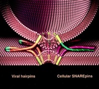
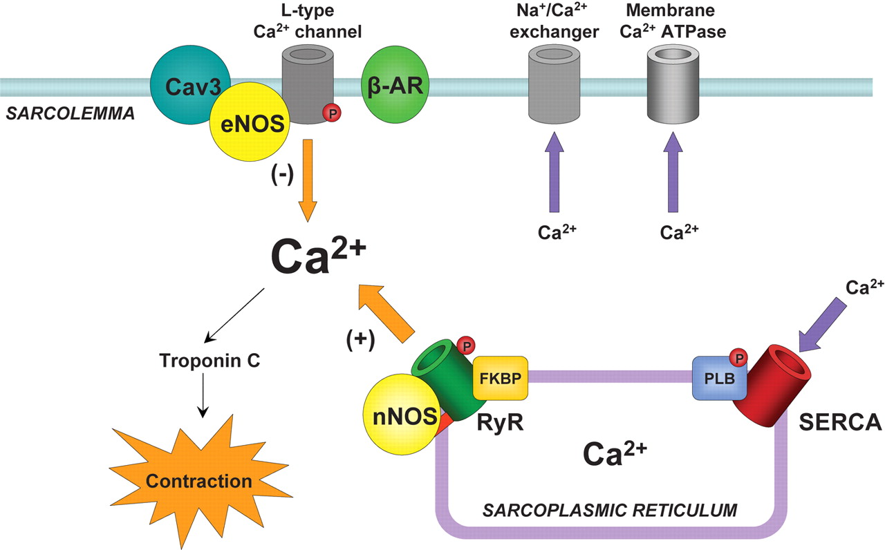
In what some thought was a foolish decision, Schekman decided in 1976, when he first joined the College of Letters and Science at UC Berkeley, to explore this system in yeast. In the ensuing years, he mapped out the machinery by which yeast cells sort, package and deliver proteins via membrane bubbles to the cell surface, secreting proteins important in yeast communication and mating. Yeast also use the process to deliver receptors to the surface, the cells’ main way of controlling activities such as the intake of nutrients like glucose.

In the 1980s and ’90s, these findings enabled the biotechnology industry to exploit the secretion system in yeast to create and release pharmaceutical products and industrial enzymes. Today, diabetics worldwide use insulin produced and discharged by yeast, and most of the hepatitis B vaccine used around the world is secreted by yeast. Both systems were developed by Chiron Corp. of Emeryville, Calif., now part of Novartis International AG, during the 20 years Schekman consulted for the company.

Various diseases, including some forms of diabetes and a form of hemophilia, involve a hitch in the secretion system of cells, and Schekman is now investigating a possible link to Alzheimer’s disease.

“Our findings have aided people in understanding these diseases,” said Schekman.

Based on the machinery discovered by Schekman and Rothman, Südhof subsequently discovered how nerve cells release signaling molecules, called neurotransmitters, which they use to communicate.

For his scientific contributions, Schekman was elected to the National Academy of Sciences in 1992, received the Gairdner International Award in 1996 and the Lasker Award for basic and clinical research in 2002. He was elected president of the American Society for Cell Biology in 1999. On Oct. 3, Schekman received the Otto Warburg Medal of the German Society for Biochemistry and Molecular Biology, which is considered the highest German award in the fields of biochemistry and molecular biology.

Schekman, formerly editor of the journal Proceedings of the National Academy of Sciences, currently is editor-in-chief of the new open access journal eLife.

Schekman and his wife, Nancy Walls, have two adult children.

MORE INFORMATION

SOURCE

tanford Report, October 7, 2013

Thomas Südhof wins Nobel Prize in Physiology or Medicine

Neuroscientist Thomas Südhof, MD, professor of molecular and cellular physiology at the Stanford School of Medicine, won the 2013 Nobel Prize in Physiology or Medicine.

BY KRISTA CONGER

Steve FischThomas SudhofThomas Sudhof won the 2013 Nobel Prize in Physiology or Medicine.

Neuroscientist Thomas Südhof, MD, professor of molecular and cellular physiology at the Stanford University School of Medicine, won the 2013 Nobel Prize in Physiology or Medicine.

He shared the prize with James Rothman, PhD, a former Stanford professor of biochemistry, andRandy Schekman, PhD, who earned his doctorate at Stanford under the late Arthur Kornberg, MD, another winner of the Nobel Prize in Physiology or Medicine.

The three were awarded the prize “for their discoveries of machinery regulating vesicle traffic, a major transport system in our cells.” Rothman is now a professor at Yale University, and Schekman is a professor at UC-Berkeley.

“I’m absolutely surprised,” said Südhof, who was in the remote town of Baeza in Spain to attend a conference and give a lecture. “Every scientist dreams of this. I didn’t realize there was chance I would be awarded the prize. I am stunned and really happy to share the prize with James Rothman and Randy Schekman.”

The three winners will share a prize that totals roughly $1.2 million, with about $413,600 going to each.

Robert Malenka, MD, Stanford’s Nancy Friend Pritzker Professor in Psychiatry and Behavioral Sciences, is at the conference with Südhof, a close collaborator. “He’s dazed, tired and happy,” Malenka said by phone. “The only time I’ve seen him happier was when his children were born.”

Südhof, the Avram Goldstein Professor in the School of Medicine, received the award for his work in exploring how neurons in the brain communicate with one another across gaps called synapses. Although his work has focused on the minutiae of how molecules interact on the cell membranes, the fundamental questions he’s pursuing are large.

“The brain works by neurons communicating via synapses,” Südhof said in a phone conversation this morning. “We’d like to understand how synapse communication leads to learning on a larger scale. How are the specific connections established? How do they form? And what happens in schizophrenia and autism when these connections are compromised?” In 2009, he published research describing how a gene implicated in autism and schizophrenia alters mice’s synapses and produces behavioral changes in the mice, such as excessive grooming and impaired nest building, that are reminiscent of these human neuropsychiatric disorders.

Lloyd Minor, MD, dean of the School of Medicine, said, “Thomas Südhof is a consummate citizen of science. His unrelenting curiosity, his collaborative spirit, his drive to ascertain the minute details of cellular workings, and his skill to carefully uncover these truths — taken together it’s truly awe-inspiring.

“He has patiently but relentlessly probed one of the fundamental questions of medical science — perhaps the fundamental question in neuroscience: How nerve cells communicate with each other. The answer is at the crux of human biology and of monumental importance to human health. Dr. Südhof’s receipt of this prize is inordinately well-deserved, and I offer him my heartfelt congratulations. His accomplishment represents what Stanford Medicine and the biomedical revolution are all about.”

The Nobel committee called Südhof on his cell phone after trying his home in Menlo Park, Calif. His wife, Lu Chen, PhD, associate professor of neurosurgery and of psychiatry and behavioral sciences, then gave the committee his cell phone number to reach him in Spain.

“The phone rang three times before I decided to go downstairs and pick it up,” Chen said. “I thought it was one of my Chinese relatives who couldn’t figure out the time zone.”

Chen and Südhof have two young children, and Südhof has four adult children from a previous marriage. “I was very surprised,” Chen said, “but he’s more concerned about how I’ll get the kids up this morning in time for school.”

“I was expecting a call from a colleague about the conference I’m here to attend, so I pulled off in a parking lot,” said Südhof, who was driving from Madrid to Baeza at the time he received the announcement. “I hadn’t slept at all the previous night, and I certainly wasn’t expecting a call from the Nobel committee.”

On the day he got the call from the Nobel committee, he was scheduled to give a talk at a conference, Membrane Traffic at the Synapse: The Cell Biology of Synaptic Plasticity, held in a 17th-century building that now serves as a conference center.

“Professor Sudhof’s contributions to the understanding of how cells operate have been of enormous importance to medicine, and to his own work in understanding how connections form within the human brain,” said Stanford President John Hennessy. “The recognition by the Nobel committee is a remarkable achievement.”

Südhof, who is also a Howard Hughes Medical Institute investigator, has spent the past 30 years prying loose the secrets of the synapse, the all-important junction where information, in the form of chemical messengers called neurotransmitters, is passed from one neuron to another. The firing patterns of our synapses underwrite our consciousness, emotions and behavior. The simple act of taking a step forward, experiencing a fleeting twinge of regret, recalling an incident from the morning commute or tasting a doughnut requires millions of simultaneous and precise synaptic firing events throughout the brain and peripheral nervous system.

Even a moment’s consideration of the total number of synapses in the typical human brain adds up to instant regard for that organ’s complexity. Coupling neuroscientists’ ballpark estimate of 200 billion neurons in a healthy adult brain with the fact that any single neuron may share synaptic contacts with as few as one or as many as 1 million other neurons (the median is somewhere in the vicinity of 10,000) suggests that your brain holds perhaps 2 quadrillion synapses — 10,000 times the number of stars in the Milky Way.

“The computing power of a human or animal brain is much, much higher than that of any computer,” said Südhof. “A synapse is not just a relay station. It is not even like a computer chip, which is an immutable element. Every synapse is like a nanocomputer all by itself. The amount of neurotransmitter released, or even whether that release occurs at all, depends on that particular synapse’s previous experience.”

Much of a neuron can be visualized as a long, hollow cord whose outer surface conducts electrical impulses in one direction. At various points along this cordlike extension are bulbous nozzles known as presynaptic terminals, each one housing myriad tiny, balloon-like vesicles containing neurotransmitters and each one abutting a downstream (or postsynaptic) neuron.

When an electrical impulse traveling along a neuron reaches one of these presynaptic terminals, calcium from outside the neuron floods in through channels that open temporarily, and a portion of the neurotransmitter-containing vesicles fuse with the surrounding bulb’s outer membrane and spill their contents into the narrow gap separating the presynaptic terminal from the postsynaptic neuron’s receiving end.

Südhof, along with other researchers worldwide, has identified integral protein components critical to the membrane fusion process. Südhof purified key protein constituents sticking out of the surfaces of neurotransmitter-containing vesicles, protruding from nearby presynaptic-terminal membranes, or bridging them. Then, using biochemical, genetic and physiological techniques, he elucidated the ways in which the interactions among these proteins contribute to carefully orchestrated membrane fusion: As a result, synaptic transmission is today one of the best-understood phenomena in neuroscience.

Südhof, who was born in Germany in 1955, received an MD in 1982 from Georg-August-Universität in Göttingen. He came to Stanford in 2008 after 25 years at the University of Texas Southwestern Medical Center at Dallas, where he first worked as a postdoctoral fellow at the laboratories of Michael Brown, MD, and Joseph Goldstein, MD.. Brown and Goldstein were awarded the Nobel Prize in Physiology or Medicine in 1985 for their work in understanding the regulation of cholesterol metabolism. In 1986, Südhof established his own laboratory at the university.

Südhof became an HHMI investigator in 1991, and moved to Stanford as a professor in molecular and cellular physiology in 2008.

The proteins Südhof has focused on for close to three decades are disciplined specialists. They recruit vesicles, bring them into “docked” positions near the terminals, herd calcium channels to the terminal membrane, and, cued by calcium, interweave like two sides of a zipper and force the vesicles into such close contact with terminal membranes that they fuse with them and release neurotransmitters into the synaptic gap. Although these specialists perform defined roles at the synapses, similar proteins, discovered later by Südhof and others, play comparable roles in other biological processes ranging from hormone secretion to fertilization of an egg during conception to immune cells’ defense against foreign invaders.

“We’ve made so many major advances during the past 50 years in this field, but there’s still much more to learn,” said Südhof, who in a 2010 interview with The Lancet credited his bassoon instructor as his most influential teacher for helping him to learn the discipline to practice for hours on end. “Understanding how the brain works is one of the most fundamental problems in neuroscience.”

Südhof’s accomplishments also earned him the 2013 Lasker Basic Medical Research Award. He is a member of the National Academy of Sciences, the Institute of Medicine and the American Academy of Arts & Sciences. He also is a recipient of the 2010 Kavli Prize in neuroscience.

In the Lancet interview, Südhof defined basic research as an approach often neglected in the pursuit of medicine. “This ‘solid descriptive science,’ like neuroanatomy or biochemistry, [are] disciplines that cannot claim to immediately understand functions or provide cures, but which form the basis for everything we do.”

Südhof said this morning he is excited to speak with his family about the prize, although it may be too much for his youngest children, ages 3 and 4, to grasp. “I will try to explain it to them,” he said. “It will be a wonderful occasion.” He noted that he has already received congratulatory calls from two of his four adult children. For them, the news may have come as less of a surprise.

“The Nobel prize became an inevitable topic of conversation when Tom won the Lasker award,” Chen said. “But the two of us share a feeling that one should never work for prizes.”

“Everyone has pegged him as a potential Nobel prize winner for many years,” said Malenka, who described the scene at the conference during the lunch hour. “It was just a matter of time. The attendees were clapping and cheering for him.”

Although he plans to return to the United States as soon as possible, Südhof has no plans to let the award slow his research — or even his plans for the day. He responded to an inquiry with a characteristically low-key reply. “Well, I think I’ll go ahead and give my talk.”

SOURCE

Rothman Lab

Membrane fusion is a fundamental biological process for organelle formation, nutrient uptake, and the secretion of hormones and neurotransmitters.

It is central to vesicular transport, storage, and release in many areas of endocrine and exocrine physiology, and imbalances in these processes give rise to important diseases, such as diabetes.

We employ diverse biophysical, biochemical, and cell biological approaches to characterize the fundamental participants in intracellular transport processes.

flippedcellfull
Time lapse images of fusing flipped-SNARE cells.

SNARE Overview

Over 30 years ago, we observed what we interpreted to be vesicular transport in crude extracts of tissue culture cells. In subsequent years we found that we had reconstituted vesicle trafficking in the Golgi, including the process of membrane fusion. Using this assay as a guide, we purified as a required factor the NEM-Sensitive Fusion protein (NSF). This led to the purification of the Soluble NSF Attachment Factor (SNAP), which bound NSF to Golgi membranes, and then with these tools discovered that the receptors for SNAP in membranes were actually complexes of proteins (which we called SNAREs) which we envisioned could potentially partner as a bridge between membranes to contribute to the process of membrane fusion and provide specificity to it (as captured in the ‘SNARE hypothesis’ proposed at the time).

We now know that organisms have a large family of SNARE proteins that indeed form cognate partnerships in just this way, and that NSF is an ATPase that (using SNAP as an adaptor protein) disrupts the SNARE complex after fusion is complete so its subunits can be recycled for repeated use. Recombinant cognate SNAREs introduced into artificial bilayers or expressed ectopically on the outside of cells ( “flipped SNAREs”) spontaneously and efficiently result in membrane (or cell) fusion, demonstrating that the SNARE complex is not only necessary but is sufficient for fusion. There are many proteins known and rapidly being discovered which closely regulate this vital process, but the muscle – if not always the brains – is in the SNAREs. Compartmental specificity is encoded to a remarkable degree in the functional partnering of SNARE proteins, a fact which is in no way inconsistent with the emerging contribution of upstream regulatory components (like rabGTPases and tethering complexes) to domain/compartment specificity.

Current Research & Projects

Our lab is working to elucidate the underlying mechanisms of vesicular transport within cells and the secretion of proteins and neurotransmitters.

Projects include:

  1. The biochemical and biophysical mechanisms of vesicle budding and fusion;
  2. Cellular regulation of vesicle fusion in exocytosis and synaptic transmission;
  3. Structural and functional organization of the Golgi apparatus from a cellular systems view.

We take an interdisciplinary approach which includes cell-free biochemistry, single molecule biophysics, high resolution optical imaging of single events/single molecules in the cell and in cell-free formats.

The overall goal is to understand transport pathways form structural mechanism to cellular physiology. The latter is facilitated by high throughput functional genomics at the cellular level (see Yale Center for High Throughput Cell Biology).

SNAREpins

We have a strong interest in new lab members who bring backgrounds in chemistry, physics, and engineering.

SOURCE

http://medicine.yale.edu/cellbio/rothman/index.aspx

3 Americans Win Joint Nobel Prize in Medicine

Reuters

From left: Randy W. Schekman, Thomas C. Südhof and James E. Rothman.

<nyt_byline>

By 
Published: October 7, 2013 151 Comments

Three Americans won the Nobel Prize in Physiology or Medicine Monday for discovering the machinery that regulates how cells transport major molecules in a cargo system that delivers them to the right place at the right time in cells.

Science Twitter Logo.
 

The Karolinska Institute in Stockholmannounced the winners: James E. Rothman of Yale University; Randy W. Schekman of the University of California, Berkeley; and Dr. Thomas C. Südhof of Stanford University.

The molecules are moved around cells in small packages called vesicles, and each scientist discovered different facets that are needed to ensure that the right cargo is shipped to the correct destination at precisely the right time.

Their research solved the mystery of how cells organize their transport system, the Karolinska committee said. Dr. Schekman discovered a set of genes that were required for vesicle traffic. Dr. Rothman unraveled protein machinery that allows vesicles to fuse with their targets to permit transfer of cargo. Dr. Südhof revealed how signals instruct vesicles to release their cargo with precision.

The tiny vesicles, which have a covering known as membranes, shuttle the cargo between different compartments or fuse with the membrane. The transport system activates nerves. It also controls the release of hormones.

Disturbances in this exquisitely precise control system cause serious damage that, in turn, can contribute to conditions like neurological diseases, diabetes and immunological disorders.

Dr. Schekman, 64, who was born in St. Paul, used yeast cells as a model system when he began his research in the 1970s. He found that vesicles piled up in parts of the cell and that the cause was genetic. He went on to identify three classes of genes that control different facets of the cell’s transport system. Dr. Schekman studied at the University of California in Los Angeles and at Stanford University, where he obtained his Ph.D. in 1974.

In 1976, he joined the faculty of the University of California, Berkeley, where he is currently professor in the Department of Molecular and Cell Biology. Dr. Schekman is also an investigator at the Howard Hughes Medical Institute.

Dr. Rothman, 63, who was born in Haverhill, Mass., studied vesicle transport in mammalian cells in the 1980s and 1990s. He discovered that a protein complex allows vesicles to dock and fuse with their target membranes. In the fusion process, proteins on the vesicles and target membranes bind to each other like the two sides of a zipper. The fact that there are many such proteins and that they bind only in specific combinations ensures that cargo is delivered to a precise location.

The same principle operates inside the cell and when a vesicle binds to the cell’s outer membrane to release its contents. Dr. Rothman received a Ph.D. from Harvard Medical School in 1976, was a postdoctoral fellow at Massachusetts Institute of Technology, and moved in 1978 to Stanford University, where he started his research on the vesicles of the cell. Dr. Rothman has also worked at Princeton University, Memorial Sloan-Kettering Cancer Institute and Columbia University.

In 2008, he joined the faculty of Yale University where he is currently professor and chairman in the Department of Cell Biology. Some of the genes Dr. Schekman discovered in yeast coded for proteins correspond to those Dr. Rothman identified in mammals. Collectively, they mapped critical components of the cell´s transport machinery.

Dr. Südhof, 57, who was born in Göttingen, Germany, studied neurotransmission, the process by which nerve cells communicate with other cells in the brain. At the time he set out to explore the field 25 years ago, much of it was virgin scientific territory. Researchers had not identified a single protein in the neurotransmission process.

Dr. Südhof helped transform what had been a rough outline into a number of molecular activities to provide insights into the elaborate mechanisms at the crux of neurological activities, from the simplest to the most sophisticated. He did so by systematically identifying, purifying and analyzing proteins that can rapidly release chemicals that underlie the brain’s activities. The transmission process can take less than a thousandth of a second.

Dr. Südhof studied at the Georg-August-Universität in Göttingen, where he received a medical degree in 1982 and a doctorate in neurochemistry the same year. In 1983, he moved to the University of Texas Southwestern Medical Center in Dallas. Dr. Südhof, who has American citizenship, became an investigator at the Howard Hughes Medical Institute in 1991 and was appointed professor of molecular and cellular physiology at Stanford University in 2008.

All three scientists have won other awards, including the Lasker Prize, for their research.

<nyt_correction_bottom>

This article has been revised to reflect the following correction:

Correction: October 7, 2013

An earlier version of this article misstated Randy W. Schekman’s age. He is 64, not 65.

SOURCE

http://www.nytimes.com/2013/10/08/health/3-win-joint-nobel-prize-in-medicine.html?_r=0

Nobel for Cell Transport

October 07, 2013

This year’s Nobel Prize in Physiology or Medicine is going jointly to three scientists for their work figuring out how cells transport their cargo, according to the Karolinska Institute. They will share the $1.25 million prize.

“Imagine hundreds of thousands of people who are traveling around hundreds of miles of streets; how are they going to find the right way? Where will the bus stop and open its doors so that people can get out?” says Nobel committee secretary Goran Hansson, according to the Associated Press. “There are similar problems in the cell.”

By studying yeast cells with defective vesicles, Randy Schekman from the University of California, Berkeley, uncovered three classes of genes that control transportation within the cell, the New York Times adds. Schekman was awakened in California by the call from Stockholm. “I wasn’t thinking too straight. I didn’t have anything elegant to say,” he tells the AP. “All I could say was ‘Oh my God,’ and that was that.” Schekman adds that he called his lab manager to arrange a celebration in the lab.

Meanwhile, Yale University’s James Rothman discovered a protein complex that allows vesicles to bind to their intended membrane targets, getting the vesicle contents to a specific location. Rothman notes that he recently lost funding for work building on his discovery, and says that he hopes that having won the Nobel will help him when he reapplies.

And Thomas Südhof at Stanford University systematically studied how nerve cells communicate, finding that vesicles full of neurotransmitters bind to cell membranes to release their contents through a molecular mechanism that responds to the presence of calcium ions. He was on his way to a give a talk when he got his call. “I got the call while I was driving and like a good citizen I pulled over and picked up the phone,” Südhof says to the AP. “To be honest, I thought at first it was a joke. I have a lot of friends who might play these kinds of tricks.”

SOURCE

Other related articles published on these Open Access Online Scientific Journal include the following:

The Series on Cardiovascular Disease and the role of Calcium Signaling consists of the following articles:

Part I: Identification of Biomarkers that are Related to the Actin Cytoskeleton

Larry H Bernstein, MD, FCAP

http://pharmaceuticalintelligence.com/2012/12/10/identification-of-biomarkers-that-are-related-to-the-actin-cytoskeleton/

Part II: Role of Calcium, the Actin Skeleton, and Lipid Structures in Signaling and Cell Motility

Larry H. Bernstein, MD, FCAP, Stephen Williams, PhD and Aviva Lev-Ari, PhD, RN

http://pharmaceuticalintelligence.com/2013/08/26/role-of-calcium-the-actin-skeleton-and-lipid-structures-in-signaling-and-cell-motility/

Part III: Renal Distal Tubular Ca2+ Exchange Mechanism in Health and Disease

Larry H. Bernstein, MD, FCAP, Stephen J. Williams, PhD
 and Aviva Lev-Ari, PhD, RN

http://pharmaceuticalintelligence.com/2013/09/02/renal-distal-tubular-ca2-exchange-mechanism-in-health-and-disease/

Part IV: The Centrality of Ca(2+) Signaling and Cytoskeleton Involving Calmodulin Kinases and Ryanodine Receptors in Cardiac Failure, Arterial Smooth Muscle, Post-ischemic Arrhythmia, Similarities and Differences, and Pharmaceutical Targets

Larry H Bernstein, MD, FCAP, Justin Pearlman, MD, PhD, FACC and Aviva Lev-Ari, PhD, RN

http://pharmaceuticalintelligence.com/2013/09/08/the-centrality-of-ca2-signaling-and-cytoskeleton-involving-calmodulin-kinases-and-ryanodine-receptors-in-cardiac-failure-arterial-smooth-muscle-post-ischemic-arrhythmia-similarities-and-differen/

Part V: Heart, Vascular Smooth Muscle, Excitation-Contraction Coupling (E-CC), Cytoskeleton, Cellular Dynamics and Ca2 Signaling

Larry H Bernstein, MD, FCAP, Justin Pearlman, MD, PhD, FACC and Aviva Lev-Ari, PhD, RN

http://pharmaceuticalintelligence.com/2013/08/26/heart-smooth-muscle-excitation-contraction-coupling-cytoskeleton-cellular-dynamics-and-ca2-signaling/

Part VI: Calcium Cycling (ATPase Pump) in Cardiac Gene Therapy: Inhalable Gene Therapy for Pulmonary Arterial Hypertension and Percutaneous Intra-coronary Artery Infusion for Heart Failure: Contributions by Roger J. Hajjar, MD

Aviva Lev-Ari, PhD, RN

http://pharmaceuticalintelligence.com/2013/08/01/calcium-molecule-in-cardiac-gene-therapy-inhalable-gene-therapy-for-pulmonary-arterial-hypertension-and-percutaneous-intra-coronary-artery-infusion-for-heart-failure-contributions-by-roger-j-hajjar/

Part VII: Cardiac Contractility & Myocardium Performance: Ventricular Arrhythmiasand Non-ischemic Heart Failure – Therapeutic Implications for Cardiomyocyte Ryanopathy (Calcium Release-related Contractile Dysfunction) and Catecholamine Responses

Justin Pearlman, MD, PhD, FACC, Larry H Bernstein, MD, FCAP and Aviva Lev-Ari, PhD, RN

http://pharmaceuticalintelligence.com/2013/08/28/cardiac-contractility-myocardium-performance-ventricular-arrhythmias-and-non-ischemic-heart-failure-therapeutic-implications-for-cardiomyocyte-ryanopathy-calcium-release-related-contractile/

Part VIII: Disruption of Calcium Homeostasis: Cardiomyocytes and Vascular Smooth Muscle Cells: The Cardiac and Cardiovascular Calcium Signaling Mechanism

Justin Pearlman, MD, PhD, FACC, Larry H Bernstein, MD, FCAP and Aviva Lev-Ari, PhD, RN

http://pharmaceuticalintelligence.com/2013/09/12/disruption-of-calcium-homeostasis-cardiomyocytes-and-vascular-smooth-muscle-cells-the-cardiac-and-cardiovascular-calcium-signaling-mechanism/

Part IX: Calcium-Channel Blockers, Calcium Release-related Contractile Dysfunction (Ryanopathy) and Calcium as Neurotransmitter Sensor

Justin Pearlman, MD, PhD, FACC, Larry H Bernstein, MD, FCAP and Aviva Lev-Ari, PhD, RN

Part X: Synaptotagmin functions as a Calcium Sensor: How Calcium Ions Regulate the fusion of vesicles with cell membranes during Neurotransmission

Larry H Bernstein, MD, FCAP and Aviva Lev-Ari, PhD, RN

http://pharmaceuticalintelligence.com/2013/09/10/synaptotagmin-functions-as-a-calcium-sensor-how-calcium-ions-regulate-the-fusion-of-vesicles-with-cell-membranes-during-neurotransmission/

Read Full Post »

Synaptotagmin functions as a Calcium Sensor: How Calcium Ions Regulate the fusion of vesicles with cell membranes during Neurotransmission

Reporters: Larry H Bernstein, MD, FCAP and Aviva Lev-Ari, PhD, RN

Article X Synaptotagmin functions as a Calcium Sensor How Calcium Ions Regulate the fusion of vesicles with cell membranes during Neurotransmission

Image created by Adina Hazan 06/30/2021

This article is the Part X in a series of articles on Activation and Dysfunction of the Calcium Release Mechanisms in Cardiomyocytes and Vascular Smooth Muscle Cells.

The Series consists of the following articles:

Part I: Identification of Biomarkers that are Related to the Actin Cytoskeleton

Larry H Bernstein, MD, FCAP

http://pharmaceuticalintelligence.com/2012/12/10/identification-of-biomarkers-that-are-related-to-the-actin-cytoskeleton/

Part II: Role of Calcium, the Actin Skeleton, and Lipid Structures in Signaling and Cell Motility

Larry H. Bernstein, MD, FCAP, Stephen Williams, PhD and Aviva Lev-Ari, PhD, RN

http://pharmaceuticalintelligence.com/2013/08/26/role-of-calcium-the-actin-skeleton-and-lipid-structures-in-signaling-and-cell-motility/

Part III: Renal Distal Tubular Ca2+ Exchange Mechanism in Health and Disease

Larry H. Bernstein, MD, FCAP, Stephen J. Williams, PhD
 and Aviva Lev-Ari, PhD, RN

http://pharmaceuticalintelligence.com/2013/09/02/renal-distal-tubular-ca2-exchange-mechanism-in-health-and-disease/

Part IV: The Centrality of Ca(2+) Signaling and Cytoskeleton Involving Calmodulin Kinases and Ryanodine Receptors in Cardiac Failure, Arterial Smooth Muscle, Post-ischemic Arrhythmia, Similarities and Differences, and Pharmaceutical Targets

Larry H Bernstein, MD, FCAP, Justin Pearlman, MD, PhD, FACC and Aviva Lev-Ari, PhD, RN

http://pharmaceuticalintelligence.com/2013/09/08/the-centrality-of-ca2-signaling-and-cytoskeleton-involving-calmodulin-kinases-and-ryanodine-receptors-in-cardiac-failure-arterial-smooth-muscle-post-ischemic-arrhythmia-similarities-and-differen/

Part V: Ca2+-Stimulated Exocytosis:  The Role of Calmodulin and Protein Kinase C in Ca2+ Regulation of Hormone and Neurotransmitter

Larry H Bernstein, MD, FCAP
and
Aviva Lev-Ari, PhD, RN

http://pharmaceuticalintelligence.com/2013/12/23/calmodulin-and-protein-kinase-c-drive-the-ca2-regulation-of-hormone-and-neurotransmitter-release-that-triggers-ca2-stimulated-exocytosis/

Part VI: Calcium Cycling (ATPase Pump) in Cardiac Gene Therapy: Inhalable Gene Therapy for Pulmonary Arterial Hypertension and Percutaneous Intra-coronary Artery Infusion for Heart Failure: Contributions by Roger J. Hajjar, MD

Aviva Lev-Ari, PhD, RN

http://pharmaceuticalintelligence.com/2013/08/01/calcium-molecule-in-cardiac-gene-therapy-inhalable-gene-therapy-for-pulmonary-arterial-hypertension-and-percutaneous-intra-coronary-artery-infusion-for-heart-failure-contributions-by-roger-j-hajjar/

Part VII: Cardiac Contractility & Myocardium Performance: Ventricular Arrhythmiasand Non-ischemic Heart Failure – Therapeutic Implications for Cardiomyocyte Ryanopathy (Calcium Release-related Contractile Dysfunction) and Catecholamine Responses

Justin Pearlman, MD, PhD, FACC, Larry H Bernstein, MD, FCAP and Aviva Lev-Ari, PhD, RN

http://pharmaceuticalintelligence.com/2013/08/28/cardiac-contractility-myocardium-performance-ventricular-arrhythmias-and-non-ischemic-heart-failure-therapeutic-implications-for-cardiomyocyte-ryanopathy-calcium-release-related-contractile/

Part VIII: Disruption of Calcium Homeostasis: Cardiomyocytes and Vascular Smooth Muscle Cells: The Cardiac and Cardiovascular Calcium Signaling Mechanism

Justin Pearlman, MD, PhD, FACC, Larry H Bernstein, MD, FCAP and Aviva Lev-Ari, PhD, RN

http://pharmaceuticalintelligence.com/2013/09/12/disruption-of-calcium-homeostasis-cardiomyocytes-and-vascular-smooth-muscle-cells-the-cardiac-and-cardiovascular-calcium-signaling-mechanism/

Part IX: Calcium-Channel Blockers, Calcium Release-related Contractile Dysfunction (Ryanopathy) and Calcium as Neurotransmitter Sensor

Justin Pearlman, MD, PhD, FACC, Larry H Bernstein, MD, FCAP and Aviva Lev-Ari, PhD, RN

Part X: Synaptotagmin functions as a Calcium Sensor: How Calcium Ions Regulate the fusion of vesicles with cell membranes during Neurotransmission

Larry H Bernstein, MD, FCAP and Aviva Lev-Ari, PhD, RN

http://pharmaceuticalintelligence.com/2013/09/10/synaptotagmin-functions-as-a-calcium-sensor-how-calcium-ions-regulate-the-fusion-of-vesicles-with-cell-membranes-during-neurotransmission/

Part XI: Sensors and Signaling in Oxidative Stress

Larry H. Bernstein, MD, FCAP

http://pharmaceuticalintelligence.com/2013/11/01/sensors-and-signaling-in-oxidative-stress/

Part XII: Atherosclerosis Independence: Genetic Polymorphisms of Ion Channels Role in the Pathogenesis of Coronary Microvascular Dysfunction and Myocardial Ischemia (Coronary Artery Disease (CAD))

Larry H Bernstein, MD, FCAP and Aviva Lev-Ari, PhD, RN

http://pharmaceuticalintelligence.com/2013/12/21/genetic-polymorphisms-of-ion-channels-have-a-role-in-the-pathogenesis-of-coronary-microvascular-dysfunction-and-ischemic-heart-disease/

Introduction

Author: Larry H Bernstein, MD, FCAP 

This introduction is based on two sources:

#1:

Michael J. Berridge, Smooth muscle cell calcium activation mechanisms

The Babraham Institute, Babraham, Cambridge CB22 4AT, UK

J Physiol 586.21 (2008) pp 5047–5061

http://jp.physoc.org/content/586/21/5047.full.pdf

and

#2

Thomas C Südhof, A molecular machine for neurotransmitter release: synaptotagmin and beyond

http://www.nature.com/focus/Lasker/2013/pdf/ES-Lasker13-Sudhof.pdf

Part IX of this series of articles discussed the mechanism of the signaling of smooth muscle cells by the interacting parasympathetic neural innervation that occurs by calcium triggering neurotransmitter release by initiating synaptic vesicle fusion.   It involves the interaction of soluble N-acetylmaleimide-sensitive factor (SNARE) and SM proteins, and in addition, the discovery of a calcium-dependsent Syt1 (C) domain of protein- kinase C isoenzyme, which binds to phospholipids.  It is reasonable to consider that it differs from motor neuron activation of skeletal muscles, mainly because the innervation is in the involuntary domain.   The cranial nerve rooted innervation has evolved comes from the spinal ganglia at the corresponding level of the spinal cord.  It is in this specific neural function that we find a mechanistic interaction with adrenergic hormonal function, a concept intimated by the late Richard Bing.  Only recently has there been a plausible concept that brings this into serious consideration.  Moreover, the review of therapeutic drugs that are used in blocking adrenergic receptors are closely related to the calcium-channels.  Interesting too is the participation of a phospholipid bound protein-kinase isoenzyme C calcium-dependent domain Syt1.  The neurohormonal connection lies in the observation by Katz in the 1950’s that the vesicles of the neurons hold and eject fixed amounts of neurotransmitters.

In Sudhof’s Lasker Award presentation he refers to the biochemical properties of synaptotagmin were found to precisely correspond to the extraordinary calcium-triggering properties of release, and to account for a regulatory pathway that also applies to other types of calcium-triggered fusion, for example fusion observed in hormone secretion and fertilization. At the synapse, finally, these interdependent machines — the fusion apparatus and its synaptotagmin-dependent control mechanism — are embedded in a proteinaceous active zone that links them to calcium channels, and regulates the docking and priming of synaptic vesicles for subsequent calcium-triggered fusion. Thus, work on neurotransmitter release revealed a hierarchy of molecular machines that mediate the fusion of synaptic vesicles, the calcium-control of this fusion, and the embedding of calcium-controlled fusion in the context of the presynaptic terminal at the synapse.  The neural transmission is described as a biological relay system. Neurotransmission kicks off with an electrical pulse that runs down a nerve cell, or neuron. When that signal reaches the tip, calcium enters the cell. In response, the neuron liberates chemical messengers—neurotransmitters—which travel to the next neuron and thus pass the baton.

He further stipulates that synaptic vesicle exocytosis operates by a general mechanism of membrane fusion that revealed itself to be a model for all membrane fusion, but that is uniquely regulated by a calcium-sensor protein called synaptotagmin.  Neurotransmission is thus a combination of electrical signal and chemical transport.

http://www.nature.com/focus/Lasker/2013/pdf/ES-Lasker13-Sudhof.pdf

Several SMC types illustrate how signaling mechanisms have been adapted to control different contractile functions with particular emphasis on how Ca2+ signals are activated.

[1] Neural activation of vas deferens smooth muscle cells

Noradrenaline (NA) acts by stimulating α1-adrenoreceptors to produce InsP3, which then releases Ca2+ that may induce an intracellular Ca2+ wave similar to that triggered by the ATP-dependent entry of external Ca2+. In addition, the α1-adrenoreceptors also activate the smooth muscle Rho/Rho kinase signalling pathway that serves to increase the Ca2+ sensitivity of the contractile machinery.

[2] Detrusor smooth muscle cells

The bladder, which functions to store and expel urine, is surrounded by layers of detrusor SMCs. The latter have two operational modes: during bladder filling they remain relaxed but contract vigorously to expel urine during micturition. The switch from relaxation to contraction, which is triggered by neurotransmitters released from parasympathetic nerves, depends on the acceleration of an endogenous membrane oscillator that produces the repetitive trains of action potentials that drive contraction.

This mechanism of activation is also shared by [1], and uterine contraction.  SMCs are activated by membrane depolarization (ΔV) that opens L-type voltage-operated channels (VOCs) allowing external Ca2+ to flood into the cell to trigger contraction. This depolarization is induced either by ionotropic receptors (vas deferens) or a membrane oscillator (bladder and uterus). The membrane oscillator, which resides in the plasma membrane, generates the periodic pacemaker depolarizations responsible for the action potentials that drive contraction.

The main components of the membrane oscillator are the Ca2+ and K+ channels that sequentially depolarize and hyperpolarize the membrane, respectively. This oscillator generates the periodic pacemaker depolarizations that trigger each action potential. The resulting Ca2+ signal lags behind the action potential because it spreads into the cell as a slower Ca2+ wave mediated by the type 2 RYRs.

Neurotransmitters such as ATP and acetylcholine (ACh), which are released from parasympathetic axonal varicosities that innervate the bladder, activate or accelerate the oscillator by inducing membrane depolarization (ΔV).

[3]  The depolarizing signal that activates gastrointestinal, urethral and ureter SMCs is as follows:

A number of SMCs are activated by pacemaker cells such as the interstitial cells of Cajal (ICCs) (gastrointestinal and urethral SMCs) or atypical SMCs (ureter). These pacemaker cells have a cytosolic oscillator that generates the repetitive Ca2+ transients that activate inward currents that spread through the gap junctions to provide the depolarizing signal (ΔV) that triggers contraction.

[4]  Our greatest interest has been in this mechanism.  The rhythmical contractions of vascular, lymphatic, airway and corpus cavernosum SMCs depend on an endogenous pacemaker driven by a cytosolic Ca2+ oscillator that is responsible for the periodic release of Ca2+ from the endoplasmic reticulum. The periodic pulses of Ca2+ often cause membrane depolarization, but this is not part of the primary activation mechanism but has a secondary role to synchronize and amplify the oscillatory mechanism. Neurotransmitters and hormones act by modulating the frequency of the cytosolic oscillator.

Vascular or airway SMCs are driven by a cytosolic oscillator that generates a periodic release of Ca2+ from the endoplasmic reticulum that usually appears as a propagating Ca2+ wave.

Step 1. The initiation and/or modulation of this oscillator depends upon the action of transmitters and hormones such as ACh, 5-HT, NA and endothelin-1 (ET-1) that increase the formation of InsP3 and diacylglycerol (DAG), both of which promote oscillatory activity.

Step 2. The oscillator is very dependent on Ca2+ entry to provide the Ca2+ necessary to charge up the stores for each oscillatory cycle. The nature of these entry mechanisms vary between cell types.

Step 3. The entry of external Ca2+ charges up the ER to sensitize the RYRs and InsP3 receptors prior to the next phase of release. An important determinant of this sensitivity is the luminal concentration of Ca2+ and as this builds up the release channels become sensitive to Ca2+ and can participate in the process of Ca2+-induced Ca2+ release (CICR), which is responsible for orchestrating the regenerative release of Ca2+ from the ER. The proposed role of cyclic ADP-ribose (cADPR) in airway SMCs is consistent with this aspect of the model on the basis of its proposed action of stimulating the SERCA pump to enhance store loading and such a mechanism has been described in colonic SMCs.

Step 4. The mechanism responsible for initiating Ca2+ release may depend either on the RYRs or the InsP3 receptors (I). RYR channels are sensitive to store loading and the InsP3 receptors will be sensitized by the agonist-dependent formation of InsP3.

Step 5. This initial release of Ca2+ is then amplified by regenerative Ca2+ release by either the RYRs or InsP3 receptors, depending on the cell type.

Step 6. The global Ca2+ signal then activates contraction.

Step 7. The recovery phase depends on the sarco-endoplasmic reticulum Ca2+-ATPase (SERCA), that pumps some of the Ca2+ back into the ER, and the plasma membrane Ca2+-ATPase (PMCA), that pumps Ca2+ out of the cell.

Step 8. One of the effects of the released Ca2+ is to stimulate Ca2+-sensitive K+ channels such as the BK and SK channels that will lead to membrane hyperpolarization. The BK channels are activated by Ca2+ sparks resulting from the opening of RYRs.

Step 9.  Another action of Ca2+ is to stimulate Ca2+-sensitive chloride channels (CLCA) (Liu & Farley, 1996; Haddock & Hill, 2002), which result in membrane depolarization to activate the CaV1.2 channels that introduce Ca2+ into the cell resulting in further membrane depolarization (ΔV).

Step 10. This depolarization can spread to neighbouring cells by current flow through the gap junctions to provide a synchronization mechanism in those cases where the oscillators are coupled together to provide vasomotion.

SOURCE

Smooth muscle cell calcium activation mechanisms. Berridge MJ.
J Physiol. 2008; 586(Pt 21):5047-61.   http://dx.doi.org/10.1113/jphysiol.2008.160440

Synaptotagmin functions as a Calcium Sensor

Thomas C. Südhof is at the Department of Molecular and Cellular Physiology and the Howard Hughes Medical Institute, Stanford University School of Medicine, Palo Alto, California, USA

Prof.  Thomas C. Südhof explains:

Fifty years ago, Bernard Katz’s seminal work revealed that calcium triggers neurotransmitter release by stimulating ultrafast synaptic vesicle fusion. But how a presynaptic terminal achieves the speed and precision of calcium-triggered fusion remained unknown. My colleagues and I set out to study this fundamental problem more than two decades ago.

How do the synaptic vesicle and the plasma membrane fuse during transmitter release? How does calcium trigger synaptic vesicle fusion? How is calcium influx localized to release sites in order to enable the fast coupling of an action potential to transmitter release? Together with contributions made by other scientists, most prominently James Rothman, Reinhard Jahn and Richard Scheller, and assisted by luck and good fortune, we have addressed these questions over the last decades.

As he described below, we now know of a general mechanism of membrane fusion that operates by the interaction of SNAREs (for soluble N-ethylmaleimide–sensitive factor (NSF)-attachment protein receptors) and SM proteins (for Sec1/Munc18-like proteins). We also have now a general mechanism of calcium-triggered fusion that operates by calcium binding to synaptotagmins, plus a general mechanism of vesicle positioning adjacent to calcium channels, which involves the interaction of the so-called RIM proteins with these channels and synaptic vesicles. Thus, a molecular framework that accounts for the astounding speed and precision of neurotransmitter release has emerged. In describing this framework, I have been asked to describe primarily my own work. I apologize for the many omissions of citations to work of others; please consult a recent review for additional references1.

http://www.nature.com/focus/Lasker/2013/pdf/ES-Lasker13-Sudhof.pdf

Outlook

Our work, together with that of other researchers, uncovered a plausible mechanism explaining how membranes undergo rapid fusion during transmitter release, how such fusion is regulated by calcium and how the calcium-controlled fusion of synaptic vesicles is spatially organized in the presynaptic terminal. Nevertheless, many new questions now arise that are not just details but of great importance. For example, what are the precise physicochemical mechanisms underlying fusion, and what is the role of the fusion mechanism we outlined in brain diseases? Much remains to be done in this field.

How calcium controls membrane fusion

The above discussion describes the major progress that was made in determining the mechanism of membrane fusion. At the same time, my laboratory was focusing on a question crucial for neuronal function: how is this process triggered in microseconds when calcium enters the presynaptic terminal?

While examining the fusion machinery, we wondered how it could possibly be controlled so tightly by calcium. Starting with the description of synaptotagmin-1 (Syt1)5, we worked over two decades to show that calcium-dependent exocytosis is mediated by synaptotagmins as calcium sensors.

Synaptotagmins are evolutionarily conserved transmembrane proteins with two cytoplasmic C2 domains (Fig. 3a)5,6. When we cloned Syt1, nothing was known about C2 domains except that they represented the ‘second constant sequence’ in protein-kinase C isozymes. Because protein kinase C had been shown to interact with phospholipids by an unknown mechanism, we speculated that Syt1 C2 domains may bind phospholipids, which we indeed found to be the case5. We also found that this interaction is calcium dependent6,7 and that a single C2 domain mediates calcium-dependent phospholipid binding (Fig. 3b)8. In addition, the Syt1 C2 domains also bind syntaxin-1 and the SNARE complex6,9. All of these observations were first made for Syt1 C2 domains, but they have since been generalized to other C2 domains.

As calcium-binding modules, C2 domains were unlike any other calcium-binding protein known at the time. Beginning in 1995, we obtained atomic structures of calcium-free and calcium-bound Syt1 C2 domains10 in collaboration with structural biologists, primarily Jose Rizo (Fig. 3c). These structures provided the first insights into how C2 domains bind calcium and allowed us to test the role of Syt1 calcium binding in transmitter release11.

The biochemical properties of Syt1 suggested that it constituted Katz’s long-sought calcium sensor for neurotransmitter release. Initial experiments in C. elegans and Drosophila, however, disappointingly indicated otherwise. The ‘synaptotagmin calcium-sensor hypothesis’ seemed unlikely until our electrophysiological analyses of Syt1 knockout mice revealed that Syt1 is required for all fast synchronous synaptic fusion in forebrain neurons but is dispensable for other types of fusion (Fig. 4)12. These experiments established that Syt1 is essential for fast calcium-triggered release, but not for fusion as such.

Although the Syt1 knockout analysis supported the synaptotagmin calcium-sensor hypothesis, it did not exclude the possibility that Syt1 positions vesicles next to voltage-gated calcium channels (a function now known to be mediated by RIMs and RIM-BPs; see below),

with calcium binding to Syt1 performing a role unrelated to calcium sensing and transmitter release. To directly test whether calcium binding to Syt1 triggers release, we introduced a point mutation into the endogenous mouse Syt1 gene locus. This mutation decreased the Syt1 calcium-binding affinity by about twofold11. Electrophysiological recordings revealed that this mutation also decreased the calcium affinity of neurotransmitter release approximately twofold, formally proving that Syt1 is the calcium sensor for release (Fig. 5). In addition to mediating calcium triggering of release, Syt1 controls (‘clamps’) the rate of spontaneous release occurring in the absence of action potentials, thus serving as an essential mediator of the speed and precision of release by association with SNARE complexes and phospholipids (Fig. 6a,b).

It was initially surprising that the Syt1 knockout produced a marked phenotype because the brain expresses multiple synaptotagmins6. However, we found that only three synaptotagmins—Syt1, Syt2 and Syt9—mediate fast synaptic vesicle exocytosis13. Syt2 triggers release faster, and Syt9 slower, than Syt1. Most forebrain neurons express only Syt1, but not Syt2 or Syt9, accounting for the profound Syt1 knockout phenotype. Syt2 is the predominant calcium sensor of very fast synapses in the brainstem14, whereas Syt9 is primarily present in the limbic system13. Thus, the kinetic properties of Syt1, Syt2 and Syt9 correspond to the functional needs of the synapses that contain them.

Parallel experiments in neuroendocrine cells revealed that, in addition to Syt1, Syt7 functions as a calcium sensor for hormone exocytosis. Moreover, experiments in olfactory neurons uncovered a role for Syt10 as a calcium sensor for insulin-like growth factor-1 exocytosis15, showing that, even in a single neuron, different synaptotagmins act as calcium sensors for distinct fusion reactions. Viewed together with results by other groups, these observations indicated that calcium-triggered exocytosis generally depends on synaptotagmin calcium sensors and that different synaptotagmins confer specificity onto exocytosis pathways.

We had originally identified complexin as a small protein bound to SNARE complexes (Fig. 6b)16. Analysis of complexin-deficient neurons showed that complexin represents a cofactor for synaptotagmin that functions both as a clamp and as an activator of calcium-triggered fusion17. Complexin-deficient neurons exhibit a phenotype milder than that of Syt1-deficient neurons, with a selective suppression of fast synchronous exocytosis and an increase in spontaneous exocytosis, which suggests that complexin and synaptotagmins are functionally interdependent.

How does a small molecule like complexin, composed of only ~130 amino acid residues, act to activate and clamp synaptic vesicles for synaptotagmin action? Atomic structures revealed that, when bound to assembled SNARE complexes, complexin contains two short a-helices flanked by flexible sequences (Fig. 6c). One of the a-helices is bound to the SNARE complex and is essential for all complexin function18. The second a-helix is required only for the clamping, and not for the activating function of complexin17. The flexible N-terminal sequence of complexin, conversely, mediates only the activating, but not the clamping, function of the protein. Our current model is that complexin binding to SNAREs activates the SNARE–SM protein complex and that at least part of complexin competes with synaptotagmin for SNARE complex binding. Calcium-activated synaptotagmin displaces this part of complexin, thereby triggering fusion-pore opening (Fig. 6a)1,18.

REFERENCES

1. Südhof, T.C. & Rothman, J.E. Membrane fusion: grappling with SNARE and SM proteins. Science 323, 474–477 (2009).

2. Hata, Y., Slaughter, C.A. & Südhof, T.C. Synaptic vesicle fusion complex contains unc-18 homologue bound to syntaxin. Nature 366, 347–351 (1993).

3. Burré, J. et al. a-synuclein promotes SNARE-complex assembly in vivo and in vitro. Science 329, 1663–1667 (2010).

4. Khvotchev, M. et al. Dual modes of Munc18–1/SNARE interactions are coupled by functionally critical binding to syntaxin-1 N-terminus. J. Neurosci. 27, 12147–12155 (2007).

5. Perin, M.S., Fried, V.A., Mignery, G.A., Jahn, R. & Südhof, T.C. Phospholipid binding by a synaptic vesicle protein homologous to the regulatory region of protein kinase C. Nature 345, 260–263 (1990).

6. Li, C. et al. Ca2+-dependent and Ca2+-independent activities of neural and nonneural synaptotagmins. Nature 375, 594–599 (1995).

7. Brose, N., Petrenko, A.G., Südhof, T.C. & Jahn, R. Synaptotagmin: a Ca2+ sensor on the synaptic vesicle surface. Science 256, 1021–1025 (1992).

8. Davletov, B.A. & Südhof, T.C. A single C2-domain from synaptotagmin I is sufficient for high affinity Ca2+/phospholipid-binding. J. Biol. Chem. 268, 26386–26390 (1993).

9. Pang, Z.P., Shin, O.-H., Meyer, A.C., Rosenmund, C. & Südhof, T.C. A gain-of-function mutation in synaptotagmin-1 reveals a critical role of Ca2+-dependent SNARE-complex binding in synaptic exocytosis. J. Neurosci. 26, 12556–12565 (2006).

10. Sutton, R.B., Davletov, B.A., Berghuis, A.M., Südhof, T.C. & Sprang, S.R. Structure of the first C2-domain of synaptotagmin I: a novel Ca2+/phospholipid binding fold. Cell 80, 929–938 (1995).

11. Fernández-Chacón, R. et al. Synaptotagmin I functions as a Ca2+-regulator of release probability. Nature 410, 41–49 (2001).

12. Geppert, M. et al. Synaptotagmin I: a major Ca2+ sensor for transmitter release at a central synapse. Cell 79, 717–727 (1994).

13. Xu, J., Mashimo, T. & Südhof, T.C. Synaptotagmin-1, -2, and -9: Ca2+-sensors for fast release that specify distinct presynaptic properties in subsets of neurons. Neuron 54, 567–581 (2007).

14. Sun, J. et al. A dual Ca2+-sensor model for neuro-transmitter release in a central synapse. Nature 450, 676–682 (2007).

15. Cao, P., Maximov, A. & Südhof, T.C. Activity-dependent IGF-1 exocytosis is controlled by the Ca2+-sensor synaptotagmin-10. Cell 145, 300–311 (2011).

16. McMahon, H.T., Missler, M., Li, C. & Südhof, T.C. Complexins: cytosolic proteins that regulate SNAP-receptor function. Cell 83, 111–119 (1995).

17. Maximov, A., Tang, J., Yang, X., Pang, Z. & Südhof, T.C. Complexin controls the force transfer from SNARE complexes to membranes in fusion. Science 323, 516–521 (2009).

18. Tang, J. et al. Complexin/synaptotagmin-1 switch controls fast synaptic vesicle exocytosis. Cell 126, 1175–1187 (2006).

19. Wang, Y., Okamoto, M., Schmitz, F., Hofman, K. & Südhof, T.C. RIM: a putative Rab3-effector in regulating synaptic vesicle fusion. Nature 388, 593–598 (1997).

20. Kaeser, P.S. et al. RIM proteins tether Ca2+-channels to presynaptic active zones via a direct PDZ-domain interaction. Cell 144, 282–295 (2011).

21. Schoch, S. et al. RIM1a forms a protein scaffold for regulating neurotransmitter release at the active zone. Nature 415, 321–326 (2002).

22. Verhage, M. et al. Synaptic assembly of the brain in the absence of neurotransmitter secretion. Science 287, 864–869 (2000).


SOURCE

http://www.nature.com/focus/Lasker/2013/pdf/ES-Lasker13-Sudhof.pdf

NATURE MEDICINE | SPOONFUL OF MEDICINE

Lasker Awards go to rapid neurotransmitter release and modern cochlear implant

09 Sep 2013 | 13:38 EDT | Posted by Roxanne Khamsi | Category: 

Lasker_logo 2Posted on behalf of Arielle Duhaime-RossA very brainy area of research has scooped up one of this year’s $250,000 Lasker prizes, announced today: The Albert Lasker Basic Medical Research Award has gone to two researchers who shed light on the molecular mechanisms behind the rapid release of neurotransmitters—findings that have implications for understanding the biology of mental illnesses such as schizophrenia, as well the cellular functions underlying learning and memory formation.By systematically analyzing proteins capable of quickly releasing chemicals in the brain, Genentech’s Richard Scheller and Stanford University’s Thomas Südhofadvanced our understanding of how calcium ions regulate the fusion of vesicles with cell membranes during neurotransmission. Among Scheller’s achievements is the identification of three proteins—SNAP-25, syntaxin and VAMP/synaptobrevin—that have a vital role in neurotransmission and molecular machinery recycling. Moreover, Südhof’s observations elucidated how a protein called synaptotagmin functions as a calcium sensor, allowing these ions to enter the cell. Thanks to these discoveries, scientists were later able to understand how abnormalities in the function of these proteins contribute to some of the world’s most destructive neurological illnesses. (For an essay by Südhof on synaptotagmin, click here.)The Lasker-DeBakey Clinical Medical Research Award went to three researchers whose work led to the development of the modern cochlear implant, which allows the profoundly deaf to perceive sound. During the 1960s and 1970s Greame Clark of the University of Melbourne and Ingeborg Hochmair, CEO of cochlear implant manufacturer MED-EL, independently designed implant components that, when combined, transformed acoustical information into electrical signals capable of exciting the auditory nerve. Duke University’s Blake Wilson later contributed his “continuous interleaved sampling” system, which gave the majority of cochlear implant wearers the ability to understand speech clearly without visual cues. (For a viewpoint by Graeme addressing the evolving science of cochlear implants, click here.)Bill and Melinda Gates were also honored this year with the Lasker-Bloomberg Public Service Award. Through their foundation, the couple has made large investments in helping people living in developing countries gain access to vaccines and drugs. The Seattle-based Bill & Melinda Gates Foundation also runs programs to educate women about proper nutrition for their families and themselves. The organization has a broad mandate in public health; one of its most well known projects is the development of a low-cost toilet that will have the ability to operate without water.The full collection of Lasker essays, as well as a Q&A between Lasker president Claire Pomeroy and the Gateses, can be found here.

Summary

Author: Larry H Bernstein, MD, FCAP

Chapter IX focused on VSM of the artery and related the action of calcium-channel blockers (CCMs) to the presynaptic interruption of synaptic-vesicle fusion necessary for CA+ release that leads to neurotransmitter secretion.  Under the circumstance neurotransmitter activation, the is VSM contraction (associated with tone).  The effect of CCB action on neurotransmitter action, there is a resultant vascular dilation facilitating flow.    In this section, we extend the mechanism to other smooth muscle related action in various organs.

[1] Neural activation of vas deferens smooth muscle cells

Noradrenaline (NA) acts by stimulating α1-adrenoreceptors to produce InsP3, which then releases Ca2+ that may induce an intracellular Ca2+ wave similar to that triggered by the ATP-dependent entry of external Ca2+. In addition, the α1-adrenoreceptors also activate the smooth muscle Rho/Rho kinase signaling pathway that serves to increase the Ca2+ sensitivity of the contractile machinery.

[2]  Urinary bladder and micturition

The bladder, which functions to store and expel urine, is surrounded by layers of detrusor SMCs. The latter have two operational modes: during bladder filling they remain relaxed but contract vigorously to expel urine during micturition. The switch from relaxation to contraction, which is triggered by neurotransmitters released from parasympathetic nerves, depends on the acceleration of an endogenous membrane oscillator that produces the repetitive trains of action potentials that drive contraction.

SMCs are activated by membrane depolarization (ΔV) that opens L-type voltage-operated channels

This mechanism of activation is also shared by [1], and uterine contraction. SMCs are activated by membrane depolarization (ΔV) that opens L-type voltage-operated channels (VOCs) allowing external Ca2+ to flood into the cell to trigger contraction. This depolarization is induced either by ionotropic receptors (vas deferens) or a membrane oscillator (bladder and uterus). The membrane oscillator, which resides in the plasma membrane,  generates the periodic pacemaker depolarizations responsible for the action potentials that drive contraction.

The main components of the membrane oscillator are the Ca2+ and K+ channels that sequentially depolarize and hyperpolarize the membrane, respectively. This oscillator generates the periodic pacemaker   depolarizations that trigger each action potential. The resulting Ca2+ signal lags behind the action potential because it spreads into the cell as a slower Ca2+ wave mediated by the type 2 RYRs.   Neurotransmitters such as ATP and acetylcholine (ACh), which are released from parasympathetic axonal varicosities that innervate the bladder, activate or accelerate the oscillator by inducing membrane depolarization (ΔV).

[3] The depolarizing signal that activates gastrointestinal, urethral and ureter SMCs is as follows:

A number of SMCs are activated by pacemaker cells such as the interstitial cells of Cajal (ICCs) (gastrointestinal and urethral SMCs) or atypical SMCs (ureter). These pacemaker cells have a cytosolic oscillator that generates the repetitive Ca2+ transients that activate inward currents that spread through the gap junctions to provide the depolarizing signal (ΔV) that triggers contraction. Our greatest interest has been in this mechanism. The rhythmical contractions of vascular, lymphatic, airway and corpus cavernosum SMCs depend on an endogenous pacemaker driven by a cytosolic Ca2+ oscillator that is responsible for the periodic release of Ca2+ from the endoplasmic reticulum. The periodic pulses of Ca2+ often cause membrane depolarization, but this is not part of the primary activation mechanism but has a secondary role to synchronize and amplify the oscillatory mechanism. Neurotransmitters and hormones act by modulating the frequency of the cytosolic oscillator.

Vascular or airway SMCs are driven by a cytosolic oscillator that generates a periodic release of Ca2+ from the endoplasmic reticulum that usually appears as a propagating Ca2+ wave.

The following points are repeated:

Step 1. The initiation and/or modulation of this oscillator depends upon the action of transmitters and hormones such as ACh, 5-HT, NA and endothelin-1 (ET-1) that increase the formation of InsP3 and diacylglycerol (DAG), both of which promote oscillatory activity.

Step 2. The oscillator is very dependent on Ca2+ entry to provide the Ca2+ necessary to charge up the stores for each oscillatory cycle. The nature of these entry mechanisms vary between cell types.

Step 3. The entry of external Ca2+ charges up the ER to sensitize the RYRs and InsP3 receptors prior to the next phase of release.

The proposed role of cyclic ADP-ribose (cADPR) in airway SMCs is consistent with this aspect of the model on the basis of its proposed action of stimulating the SERCA pump to enhance store loading and such a mechanism has been described in colonic SMCs.

Step 4. The mechanism responsible for initiating Ca2+ release may depend either on the RYRs or the InsP3 receptors (I). RYR channels are sensitive to store loading and the InsP3 receptors will be sensitized by the agonist-dependent formation of InsP3.

The global Ca2+ signal then activates contraction

Smooth muscle cell calcium activation mechanisms. Berridge MJ.
J Physiol. 2008; 586(Pt 21):5047-61. http://dx.doi.org/10.1113/jphysiol.2008.160440

Read Full Post »

Heart, Vascular Smooth Muscle, Excitation-Contraction Coupling (E-CC), Cytoskeleton, Cellular Dynamics and Ca2 Signaling

Heart, Vascular Smooth Muscle, Excitation-Contraction Coupling (E-CC), Cytoskeleton, Cellular Dynamics and Ca2 Signaling

Author and Curator: Larry H Bernstein, MD, FCAP

Author and Cardiovascular Three-volume Series, Editor: Justin Pearlman, MD, PhD, FACC, and

Curator: Aviva Lev-Ari, PhD, RN

Article V Heart, Vascular Smooth Muscle, Excitation-Contraction Coupling (E-CC), Cytoskeleton, Cellular Dynamics and Ca2 Signaling

Image created by Adina Hazan 06/30/2021

Abbreviations

AP, action potential; ARVD2, arrhythmogenic right ventricular cardiomyopathy type 2; CaMKII, Ca2+/calmodulim-dependent protein kinase II; CICR, Ca2+ induced Ca2+ release;CM, calmodulin; CPVT, catecholaminergic polymorphic ventricular tachycardia;  ECC, excitation–contraction coupling; FKBP12/12.6, FK506 binding protein; HF, heart failure; LCC, L-type Ca2+ channel;  P-1 or P-2, phosphatase inhibitor type-1 or type-2; PKA, protein kinase A; PLB, phosphoplamban; PP1, protein phosphatase 1; PP2A, protein phosphatase 2A; RyR1/2, ryanodine receptor type-1/type-2; SCD, sudden cardiac death; SERCA, sarcoplasmic reticulum Ca2+ ATPase; SL, sarcolemma; SR, sarcoplasmic reticulum.

This is Part V of a series on the cytoskeleton and structural shared thematics in cellular movement and cellular dynamics.

The Series consists of the following articles:

Part I: Identification of Biomarkers that are Related to the Actin Cytoskeleton

Larry H Bernstein, MD, FCAP

http://pharmaceuticalintelligence.com/2012/12/10/identification-of-biomarkers-that-are-related-to-the-actin-cytoskeleton/

Part II: Role of Calcium, the Actin Skeleton, and Lipid Structures in Signaling and Cell Motility

Larry H. Bernstein, MD, FCAP, Stephen Williams, PhD and Aviva Lev-Ari, PhD, RN

http://pharmaceuticalintelligence.com/2013/08/26/role-of-calcium-the-actin-skeleton-and-lipid-structures-in-signaling-and-cell-motility/

Part III: Renal Distal Tubular Ca2+ Exchange Mechanism in Health and Disease

Larry H. Bernstein, MD, FCAP, Stephen J. Williams, PhD
 and Aviva Lev-Ari, PhD, RN

http://pharmaceuticalintelligence.com/2013/09/02/renal-distal-tubular-ca2-exchange-mechanism-in-health-and-disease/

Part IV: The Centrality of Ca(2+) Signaling and Cytoskeleton Involving Calmodulin Kinases and Ryanodine Receptors in Cardiac Failure, Arterial Smooth Muscle, Post-ischemic Arrhythmia, Similarities and Differences, and Pharmaceutical Targets


Larry H Bernstein, MD, FCAP, Justin Pearlman, MD, PhD, FACC and Aviva Lev-Ari, PhD, RN

http://pharmaceuticalintelligence.com/2013/09/08/the-centrality-of-ca2-signaling-and-cytoskeleton-involving-calmodulin-kinases-and-ryanodine-receptors-in-cardiac-failure-arterial-smooth-muscle-post-ischemic-arrhythmia-similarities-and-differen/

Part V: Heart, Vascular Smooth Muscle, Excitation-Contraction Coupling (E-CC), Cytoskeleton, Cellular Dynamics and Ca2 Signaling

Larry H Bernstein, MD, FCAP, Justin Pearlman, MD, PhD, FACC and Aviva Lev-Ari, PhD, RN

http://pharmaceuticalintelligence.com/2013/08/26/heart-smooth-muscle-excitation-contraction-coupling-cytoskeleton-cellular-dynamics-and-ca2-signaling/

Part VI: Calcium Cycling (ATPase Pump) in Cardiac Gene Therapy: Inhalable Gene Therapy for Pulmonary Arterial Hypertension and Percutaneous Intra-coronary Artery Infusion for Heart Failure: Contributions by Roger J. Hajjar, MD

Aviva Lev-Ari, PhD, RN

http://pharmaceuticalintelligence.com/2013/08/01/calcium-molecule-in-cardiac-gene-therapy-inhalable-gene-therapy-for-pulmonary-arterial-hypertension-and-percutaneous-intra-coronary-artery-infusion-for-heart-failure-contributions-by-roger-j-hajjar/

Part VII: Cardiac Contractility & Myocardium Performance: Ventricular Arrhythmias and Non-ischemic Heart Failure – Therapeutic Implications for Cardiomyocyte Ryanopathy (Calcium Release-related Contractile Dysfunction) and Catecholamine Responses

Justin Pearlman, MD, PhD, FACC, Larry H Bernstein, MD, FCAP and Aviva Lev-Ari, PhD, RN

http://pharmaceuticalintelligence.com/2013/08/28/cardiac-contractility-myocardium-performance-ventricular-arrhythmias-and-non-ischemic-heart-failure-therapeutic-implications-for-cardiomyocyte-ryanopathy-calcium-release-related-contractile/

In the first part, we discussed common MOTIFs across cell-types that are essential for cell division, embryogenesis, cancer metastasis, osteogenesis, musculoskeletal function, vascular compliance, and cardiac contractility.   This second article concentrates on specific functionalities for cardiac contractility based on Ca++ signaling in excitation-contraction coupling.  The modifications discussed apply specifically to cardiac muscle and not to skeletal muscle.  Considering the observations described might raise additional questions specifically address to the unique requirements of smooth muscle, abundant in the GI tract and responsible for motility in organ function, and in blood vessel compliance or rigidity. Due to the distinctly different aspects of the cardiac contractility and contraction force, and the interactions with potential pharmaceutical targets, there are two separate articles on calcium signaling and cardiac arrhythmias or heart failure (Part 2 and Part 3).  Part 2 focuses on the RYANODINE role in cardiac Ca(2+) signaling and its effect in heart failure.  Part 3 takes up other aspects of heart failure and calcium signaling with respect to phosporylation/dephosphorylation. I add a single review and classification of genetic cardiac disorders of the same cardiac Ca(2+) signaling and the initiation and force of contraction. Keep in mind that the heart is a syncytium, and this makes a huge difference compared with skeletal muscle dynamics. In Part 1 there was some discussion of the importance of Ca2+ signaling on innate immune system, and the immunology will be further expanded in a fourth of the series.

SUMMARY:

This second article on the cardiomyocyte and the Ca(2+) cycling between the sarcomere and the cytoplasm, takes a little distance from the discussion of the ryanodine that precedes it.  In this discussion we found that there is a critical phosphorylation/dephosphorylation balance that exists between Ca(+) ion displacement and it occurs at a specific amino acid residue on the CaMKIId, specific for myocardium, and there is a 4-fold increase in contraction and calcium release associated with this CAM kinase (ser 2809) dependent exchange.  These events are discussed in depth, and the research holds promise for therapeutic application. We also learn that Ca(2+) ion channels are critically involved in the generation of arrhythmia as well as dilated and hypertrophic cardiomyopathy.  In the case of arrhythmiagenesis, there are two possible manners by which this occurs.  One trigger is Ca(2+) efflux instability.  The other is based on the finding that when the cellular instability is voltage driven, the steady-state wave­length (separation of nodes in space) depends on electrotonic coupling between cells and the steepness of APD and CV restitution. The last article is an in depth review of the genetic mutations that occur in cardiac diseases.  It is an attempt at classifying them into reasonable groupings. What are the therapeutic implications of this? We see that the molecular mechanism of cardiac function has been substantially elucidated, although there are contradictions in experimental findings that are unexplained.  However, for the first time, it appears that personalized medicine is on a course that will improve health in the population, and the findings will allow specific targets designed for the individual with a treatable impairment in cardiac function that is identifiable early in the course of illness. This article is a continuation to the following articles on tightly related topics: Part I: Identification of Biomarkers that are Related to the Actin Cytoskeleton     Larry H Bernstein, MD, FCAP http://pharmaceuticalintelligence.com/2012/12/10/identification-of-biomarkers-that-are-related-to-the-actin-cytoskeleton/ Part II:  Role of Calcium, the Actin Skeleton, and Lipid Structures in Signaling and Cell Motility    Larry H. Bernstein, MD, FCAP, Stephen Williams, PhD and Aviva Lev-Ari, PhD, RN  http://pharmaceuticalintelligence.com/2013/08/26/role-of-calcium-the-actin-skeleton-and-lipid-structures-in-signaling-and-cell-motility/ Part III: Renal Distal Tubular Ca2+ Exchange Mechanism in Health and Disease    Larry H. Bernstein, MD, FCAP, Stephen J. Williams, PhD
 and  Aviva Lev-Ari, PhD, RN http://pharmaceuticalintelligence.com/2013/09/02/renal-distal-tubular-ca2-exchange-mechanism-in-health-and-disease/ Part  IV:  The Centrality of Ca(2+) Signaling and Cytoskeleton Involving Calmodulin Kinases and Ryanodine Receptors in Cardiac Failure, Arterial Smooth Muscle, Post-ischemic Arrhythmia, Similarities and Differences, and Pharmaceutical Targets Larry H Bernstein, MD, FCAP, Justin Pearlman, MD, PhD, FACC and Aviva Lev-Ari, PhD, RN  http:/pharmaceuticalintelligence.com/2013.09.089/lhbern/The Centrality of Ca(2+) Signaling and Cytoskeleton Involving Calmodulin Kinases and Ryanodine Receptors in Cardiac Failure, Arterial Smooth Muscle, Post-ischemic Arrhythmia, Similarities and Differences, and Pharmaceutical Targets

Part V:  Heart Smooth Muscle and Cardiomyocyte Cells: Excitation-Contraction Coupling & Ryanodine Receptor (RyR) type-1/type-2 in Cytoskeleton Cellular Dynamics and Ca2+ Signaling

Larry H Bernstein, MD, FCAP, Justin Pearlman, MD, PhD, FACC and Aviva Lev-Ari, PhD, RN http://pharmaceuticalintelligence.com/2013/08/26/heart-smooth-muscle-excitation-contraction-coupling-cytoskeleton-cellular-dynamics-and-ca2-signaling/ Part VI:  Calcium Cycling (ATPase Pump) in Cardiac Gene Therapy: Inhalable Gene Therapy for Pulmonary Arterial Hypertension and Percutaneous Intra-coronary Artery Infusion for Heart Failure: Contributions by Roger J. Hajjar, MD Curator: Aviva Lev-Ari, PhD, RN http://pharmaceuticalintelligence.com/2013/08/01/calcium-molecule-in-cardiac-gene-therapy-inhalable-gene-therapy-for-pulmonary-arterial-hypertension-and-percutaneous-intra-coronary-artery-infusion-for-heart-failure-contributions-by-roger-j-hajjar/ and Advanced Topics in Sepsis and the Cardiovascular System at its End Stage Larry H Bernstein, MD, FCAP  http://pharmaceuticalintelligence.com/2013/08/18/advanced-topics-in-sepsis-and-the-cardiovascular-system-at-its-end-stage/

The Role of Protein Kinases and Protein Phosphatases in the Regulation of Cardiac Sarcoplasmic Reticulum Function

EG Kranias, RC Gupta, G Jakab, HW Kim, NAE Steenaart, ST Rapundalo Molecular and Cellular Biochemistry 06/1988; 82(1):37-44. · 2.06 Impact Factor http://www.researchgate.net/publication/6420466_Protein_phosphatases_decrease_sarcoplasmic_reticulum_calcium_content_by_stimulating_calcium_release_in_cardiac_myocytes Canine cardiac sarcoplasmic reticulum is phosphorylated by

  • adenosine 3,5-monophosphate (cAMP)-dependent and
  • calcium calmodulin-dependent protein kinases on
  • a proteolipid, called phospholamban.

Both types of phosphorylation are associated with

  •  an increase in the initial rates of Ca(2+) transport by SR vesicles
  • which reflects an increased turnover of elementary steps of the calcium ATPase reaction sequence.

The stimulatory effects of the protein kinases on the calcium pump may be reversed by an endogenous protein phosphatase, which

  • can dephosphorylate both the CAMP-dependent and the calcium calmodulin-dependent sites on phospholamban.

Thus, the calcium pump in cardiac sarcoplasmic reticulum appears to be under reversible regulation mediated by protein kinases and protein phosphatases. calcium release calmodulin + ER Ca(2+) and contraction

Regulation of the Cardiac Ryanodine Receptor Channel by Luminal Ca2+ involves Luminal Ca2+ Sensing Sites

I Györke, S Györke.   Biophysical Journal 01/1999; 75(6):2801-10. · 3.65 Impact factor  http:// www.researchgate.net/publication/13459335/Regulation_of_the_cardiac_ryanodine_receptor_channel_by_luminal_Ca2_involves_luminal_Ca2_sensing_sites The mechanism of activation of the cardiac calcium release channel/ryanodine receptor (RyR) by luminal Ca(2+) was investigated in native canine cardiac RyRs incorporated into lipid bilayers in the presence of 0.01 microM to 2 mM Ca(2+) (free) and 3 mM ATP (total) on the cytosolic (cis) side and 20 microM to 20 mM Ca(2+) on the luminal (trans) side of the channel and with Cs+ as the charge carrier. Under conditions of low [trans Ca(2+)] (20 microM), increasing [cis Ca(2+)] from 0.1 to 10 microM caused a gradual increase in channel open probability (Po). Elevating [cis Ca(2+)] [cytosolic] above 100 microM resulted in a gradual decrease in Po. Elevating trans [Ca(2+)] [luminal] enhanced channel activity (EC50 approximately 2.5 mM at 1 microM cis Ca2+) primarily by increasing the frequency of channel openings. The dependency of Po on trans [Ca2+] [luminal] was similar at negative and positive holding potentials and was not influenced by high cytosolic concentrations of the fast Ca(2+) chelator, 1,2-bis(2-aminophenoxy)ethane-N,N,N, N-tetraacetic acid. Elevated luminal Ca(2+)

  1. enhanced the sensitivity of the channel to activating cytosolic Ca(2+), and it
  2. essentially reversed the inhibition of the channel by high cytosolic Ca(2+).

Potentiation of Po by increased luminal Ca(2+) occurred irrespective of whether the electrochemical gradient for Ca(2+) supported a cytosolic-to-luminal or a luminal-to-cytosolic flow of Ca(2+) through the channel. These results rule out the possibility that under our experimental conditions, luminal Ca(2+) acts by interacting with the cytosolic activation site of the channel and suggest that the effects of luminal Ca2+ are mediated by distinct Ca(2+)-sensitive site(s) at the luminal face of the channel or associated protein. F1.large  calcium movement and RyR2 receptor

Protein phosphatases Decrease Sarcoplasmic Reticulum Calcium Content by Stimulating Calcium Release in Cardiac Myocytes

D Terentyev, S Viatchenko-Karpinski, I Gyorke, R Terentyeva and S Gyorke Texas Tech University Health Sciences Center, Lubbock, TX J Physiol 2003; 552(1), pp. 109–118.  http://dx.doi.org/10.1113/jphysiol.2003.046367 Phosphorylation/dephosphorylation of Ca2+ transport proteins by cellular kinases and phosphatases plays an important role in regulation of cardiac excitation–contraction coupling; furthermore,

  • abnormal protein kinase and phosphatase activities have been implicated in heart failure.

However, the precise mechanisms of action of these enzymes on intracellular Ca2+ handling in normal and diseased hearts remains poorly understood. We have investigated

  •   the effects of protein phosphatases PP1 and PP2A on spontaneous Ca(2+) sparks and SR Ca(2+) load in myocytes permeabilized with saponin.

Exposure of myocytes to PP1 or PP2A caused a dramatic increase in frequency of Ca(2+) sparks followed by a nearly complete disappearance of events, which were accompanied by depletion of the SR Ca(2+) stores, as determined by application of caffeine. These changes in

  •  Ca(2+) release and
  • SR Ca(2+) load

could be prevented by the inhibitors of PP1 and PP2A phosphatase activities okadaic acid and calyculin A. At the single channel level, PP1 increased the open probability of RyRs incorporated into lipid bilayers. PP1-medited RyR dephosphorylation in our permeabilized myocytes preparations was confirmed biochemically by quantitative immunoblotting using a phosphospecific anti-RyR antibody. Our results suggest that

  •  increased intracellular phosphatase activity stimulates
  • RyR mediated SR Ca(2+) release
    • leading to depleted SR Ca(2+) stores in cardiac myocytes.

In heart muscle cells, the process of excitation–contraction (EC) coupling is mediated by

  •  Ca(2+) influx through sarcolemmal L-type Ca(2+) channels
  • activating Ca(2+) release channels (ryanodine receptors, RyRs) in the sarcoplasmic reticulum (SR).

Once activated, the RyR channels allow Ca(2+) to be released from the SR into the cytosol to induce contraction. This mechanism is known as Ca(2+)-induced calcium release (CICR) (Fabiato, 1985; Bers, 2002).  During relaxation, most of the Ca(2+) is resequestered into the SR by the Ca(2+)-ATPase. The amount of Ca(2+) released and the force of contraction depend on

  •  the magnitude of the Ca(2+) trigger signal,
  • the functional state of the RyRs and
  • the amount of Ca(2+) stored in the SR.

F1.large  calcium movement and RyR2 receptor Ca(2+) and contraction calcium release calmodulin + ER Reversible phosphorylation of proteins composing the EC coupling machinery plays an important role in regulation of cardiac contractility (Bers, 2002). Thus, during stimulation of the b-adrenergic pathway, phosphorylation of several target proteins, including

  • the L-type Ca(2+) channels,
  • RyRs and
  • phospholamban,

by protein kinase A (PKA) leads to an overall increase in SR Ca2+ release and contractile force in heart cells (Callewaert et al. 1988, Spurgeon et al. 1990; Hussain & Orchard, 1997; Zhou et al. 1999; Song et al. 2001; Viatchenko-Karpinski & Gyorke, 2001). PKA-dependent phosphorylation of the L-type Ca(2+) channels increases the Ca2+ current (ICa), increasing both

  • the Ca2+ trigger for SR Ca2+ release and
  • the SR Ca(2+) content

(Callewaert et al. 1988; Hussain & Orchard, 1997; Del Principe et al. 2001). Phosphorylation of phospholamban (PLB) relieves the tonic inhibition dephosphorylated PLB exerts on the SR Ca(2+)-ATPase (SERCA) resulting in enhanced SR Ca(2+) accumulation and enlarged Ca(2+) release (Kranias et al. 1985; Simmermann & Jones, 1998). With regard to the RyR, despite clear demonstration of phosphorylation of the channel in biochemical studies (Takasago et al. 1989; Yoshida et al. 1992), the consequences of this reaction to channel function have not been clearly defined. RyR phosphorylation by PKA and Ca(2+)–calmodulin dependent protein kinase (CaMKII) has been reported to increase RyR activity in lipid bilayers (Hain et al. 1995; Marx et al. 2000; Uehara et al. 2002). Moreover, it has been reported that in heart failure (HF), hyperphosphorylation of RyR causes

  •  the release of FK-506 binding protein (FKBP12.6) from the RyR,
    • rendering the channel excessively leaky for Ca(2+) (Marx et al. 2000).

However, other studies have reported no functional effects (Li et al. 2002) or even found phosphorylation to reduce RyR channel steady-state open probability (Valdivia et al. 1995; Lokuta et al. 1995).  The action of protein kinases is opposed by dephosphorylating phosphatases. Three types of protein phosphatases (PPs), referred to as PP1, PP2A and PP2B (calcineurin), have been shown to influence cardiac performance (Neumann et al. 1993; Rusnak & Mertz, 2000). Overall, according to most studies phosphatases appear to downregulate SR Ca(2+) release and contractile performance (Neumann et al. 1993; duBell et al. 1996, 2002; Carr et al. 2002; Santana et al. 2002). Furthermore, PP1 and PP2A activities appear to be increased in heart failure (Neumann, 2002; Carr et al. 2002). However, again the precise mode of action of these enzymes on intracellular Ca(2+) handling in normal and diseased hearts remains poorly understood.  In the present study, we have investigated the effects of protein phosphatases PP1 and PP2A on local Ca(2+) release events, Ca(2+) sparks, in cardiac cells. Our results show that

  •  phosphatases activate RyR mediated SR Ca(2+) release
    • leading to depletion of SR Ca(2+) stores.

These results provide novel insights into the mechanisms and potential role of protein phosphorylation/dephosphorylation in regulation of Ca(2+) signaling in normal and diseased hearts. F2.large   RyR and calcium

RESULTS

Effects of PP1 and PP2A on Ca2+ sparks and SR Ca(2+) content.

[1]  PP1 caused an early transient potentiation of Ca2+ spark frequency followed by a delayed inhibition of event occurrence. [2]  PP1 produced similar biphasic effects on the magnitude and spatio-temporal characteristics of Ca(2+) sparks Specifically, during the potentiatory phase (1 min after addition of the enzyme), PP1 significantly increased

  • the amplitude,
  • rise-time,
  • duration and
  • width of Ca(2+) sparks;

during the inhibitory phase (5 min after addition of the enzyme),

  •  all these parameters were significantly suppressed by PP1.

The SR Ca(2+) content decreased by 35 % or 69 % following the exposure of myocytes to either 0.5 or 2Uml_1 PP1, respectively (Fig. 1C). Qualitatively similar results were obtained with phosphatase PP2A. Similar to the effects of PP1, PP2A (5Uml_1) produced a transient increase in Ca(2+) spark frequency (~4-fold) followed by a depression of event occurrence and decreased SR Ca(2+) content (by 82 % and 65 %, respectively). Also similar to the action of PP1, PP2A increased

  •  the amplitude and
  • spatio-temporal spread (i.e. rise-time, duration and width) of Ca(2+) sparks at 1 min
  • and suppressed the same parameters at 5 min of exposure to the enzyme (Table 1).

Together, these results suggest that phosphatases enhance spark-mediated SR Ca2+ release, leading to decreased SR Ca(2+) content. Preventive effects of calyculin A and okadaic acid Preventive effects of ryanodine

PP1-mediated RyR dephosphorylation

F3.large  cardiomyocyte SR F3.large  cardiomyocyte SR F2.large   RyR and calcium coupled receptors coupled receptors The cardiac RyR is phosphorylated at Ser-2809 (in the rabbit sequence) by both PKA and CAMKII (Witcher et al. 1991; Marx et al. 2000). Although additional phosphorylation sites may exist on the RyR (Rodriguez et al. 2003), but Ser-2809 is believed to be the only site that is phosphorylated by PKA, and RyR hyperphosphorylation at this site has been reported in heart failure (Marx et al. 2000).  To test whether indeed phosphatases dephosphorylated the RyR in our permeabilized myocyte experiments we performed quantitative immunoblotting using an antibody that specifically recognizes the phosphorylated form of the RyR at Ser-2809 (Rodriguez et al. 2003). Myocytes exhibited a significant level of phosphorylation under baseline conditions. Maximal phosphorylation was 201 % of control. When exposed to 2Uml_1 PP1, RyR phosphorylation was 58 % of the control basal condition. Exposing to a higher PP1 concentration (10Uml_1) further reduced RyR phosphorylation to 22% of control. Thus, consistent with the results of our functional measurements,

  •  PP1 decreased RyR phosphorylation in cardiac myocytes.

Figure 1. Effects of PP1 on properties of Ca(2+) sparks and SR Ca(2+) content in rat permeabilized myocytes    see .  http://dx.doi.org/10.1113/jphysiol.2003.046367 A, spontaneous Ca(2+) spark images recorded under reference conditions, and 1 or 5 min after exposure of the cell to 2Uml_1 PP1. Traces below the images are Ca(2+) transients induced by application of 10 mM caffeine immediately following the acquisition of sparks before (3 min) and after (5 min) application of PP1 in the same cell. The Ca(2+) transients were elicited by a whole bath application of 10 mM caffeine. B, averaged spark frequency at early (1 min) and late (5 min) times following the addition of either 0.5 or 2Uml_1 of PP1 to the bathing solution. C, averaged SR Ca(2+) content for 0.5 or 2Uml_1 of PP1 measured before and 5 min after exposure to the enzyme. Data are presented as means ± S.E.M. of 6 experiments in different cells. Figure 2. Effects of PP2A on properties of Ca2+ sparks and SR Ca2+ content in rat permeabilized myocytes   see .  http://dx.doi.org/10.1113/jphysiol.2003.046367 A, spontaneous Ca(2+) spark images recorded under reference conditions, and 1 or 5 min after exposure of the cell to 5Uml_1 PP2A. Traces below the images are Ca(2+) transients induced by application of 10 mM caffeine immediately following the acquisition of sparks before (3 min) and after (5 min) application of PP2A in the same cell. B and C, averaged spark frequency (B) and SR Ca(2+) content (C) for the same conditions as in A. Data are presented as means ± S.E.M. of 6 experiments in different cells.

 DISCUSSION

In the present study, we have investigated the impact of physiologically relevant exogenous protein phosphatases PP1 and PP2A on RyR-mediated SR Ca(2+) release (measured as Ca(2+) sparks) in permeabilized heart cells. Our principal finding is that

  • phosphatases stimulated RyR channels lead to depleted SR Ca(2+) stores.

These results have important ramifications for understanding the mechanisms and role of protein phosphorylation/dephosphorylation in

  •  modulation of Ca(2+) handling in normal and diseased heart.

Modulation of SR Ca2+ release by protein phosphorylation/dephophorylation

Since protein dephosphorylation clearly resulted in increased functional activity of the Ca(+)release channel, our results imply that a reverse, phosphorylation reaction should reduce RyR activity. If indeed such effects take place, why do they not manifest in inhibition of Ca(+)sparks? One possibility is that enhanced Ca(+) uptake by SERCA

  •  masks or overcomes the effects phosphorylation may have on RyRs.

In addition, the potential inhibitory influence of protein phosphorylation on RyR activity in myocytes could be countered by feedback mechanisms  involving changes in luminal Ca(2+)(Trafford et al. 2002; Gyorke et al. 2002). In particular, reduced open probability of RyRs would be expected to lead to

  •  increased Ca2+ accumulation in the SR;
  • and increased intra-SR [Ca(2+)], in turn would
  • increase activity of RyRs at their luminal Ca(2+) regulatory sites

as demonstrated for the RyR channel inhibitor tetracaine (Gyorke et al. 1997; Overend et al. 1997). Thus

  • potentiation of SERCA
  • combined with the intrinsic capacity of the release mechanism to self-regulate

could explain at least in part why PKA-mediated protein phoshorylation results in maintained potentiation of Ca(2+) sparks despite a potential initial decrease in RyR activity.

Role of altered RyR Phosphorylation in Heart Failure

Marx et al. (2000) have proposed that  enhanced levels of circulating catecholamines lead to increased phosphorylation of RyR in heart failure.  Based on biochemical observations as well as on studying properties of single RyRs incorporated into artificial lipid bilayers, these investigators have hypothesized that

  •  hyperphosphorylation of RyRs contributes to pathogenesis of heart failure
    • by making the channel excessively leaky due to dissociation of FKBP12.6 from the channel.

We show that the mode of modulation of RyRs by phosphatases does not support this hypothesis as

  • dephosphorylation caused activation instead of

Interestingly, our results provide the basis for a different possibility in which

  •  dephophosphorylation of RyR rather than its phosphorylation causes depletion of SR Ca(2+) stores by stimulating RyRs in failing hearts.

It has been reported that PP1 and PP2 activities are increased in heart failure (Huang et al. 1999; Neumann et al. 1997; Neuman, 2002). Furthermore,  overexpression of PP1 or ablation of the endogenous PP1 inhibitor, l-1, results in

  • depressed contractile performance and heart failure (Carr et al. 2002).

Our finding that PP1 causes depletion of SR Ca(2+) stores by activating RyRs could account for, or contribute to, these results.

References

1 DelPrincipe F, Egger M, Pignier C & Niggli E (2001). Enhanced E-C coupling efficiency after beta-stimulation of cardiac myocytes. Biophys J 80, 64a. 2 Gyorke I & Gyorke S (1998). Regulation of the cardiac ryanodine receptor channel by luminal Ca2+ involves luminal Ca2+ sensing sites. Biophys J 75, 2801–2810. 3 Gyorke S, Gyorke I, Lukyanenko V, Terentyev D, Viatchenko-Karpinski S & Wiesner TF (2002). Regulation of sarcoplasmic reticulum calcium release by luminal calcium in cardiac muscle. Front Biosci 7, d1454–d1463. 4 Gyorke I, Lukyanenko V & Gyorke S (1997). Dual effects of tetracaine on spontaneous calcium release in rat ventricular myocytes. J Physiol 500, 297–309. 5 MacDougall LK, Jones LR & Cohen P (1991). Identification of the major protein phosphatases in mammalian cardiac muscle which dephosphorylate phospholamban. Eur J Biochem 196, 725–734. 6 Marx SO, Reiken S, Hisamatsu Y, Jayaraman T, Burkhoff D, Rosemblit N & Marks AR (2000). PKA phosphorylation dissociates FKBP12.6 from the calcium release channel (ryanodine receptor): defective regulation in failing hearts. Cell 101, 365–376. 7 Rodriguez P, Bhogal MS & Colyer J (2003). Stoichiometric phosphorylation of cardiac ryanodine receptor on serine-2809 by calmodulin-dependent kinase II and protein kinase A. J Biol Chem (in press).

The δC Isoform of CaMKII Is Activated in Cardiac Hypertrophy and Induces Dilated Cardiomyopathy and Heart Failure

T Zhang, LS Maier, ND Dalton, S Miyamoto, J Ross, DM Bers, JH Brown.  University of California, San Diego, La Jolla, Calif; and Loyola University, Chicago, Ill. Circ Res. 2003;92:912-919.    http://dx.doi.org/10.1161/01.RES.0000069686.31472.C5 Recent studies have demonstrated that transgenic (TG) expression of either Ca(2+)/calmodulin-dependent protein kinase IV (CaMKIV) or CaMKIIδB, both of which localize to the nucleus, induces cardiac hypertrophy. However,

  •  CaMKIV is not present in heart, and
  • cardiomyocytes express not only the nuclear CaMKIIδB
    • but also a cytoplasmic isoform, CaMKII δC.

In the present study, we demonstrate that

  1.  expression of the δC isoform of CaMKII is selectively increased and
  2. its phosphorylation elevated as early as 2 days and continuously for up to 7 days after pressure overload.

To determine whether enhanced activity of this cytoplasmic δC isoform of CaMKII can lead to phosphorylation of Ca(2+) regulatory proteins and induce hypertrophy, we generated TG mice that expressed the δC isoform of CaMKII.  Immunocytochemical staining demonstrated that the expressed transgene is confined to the cytoplasm of cardiomyocytes isolated from these mice. These mice develop a dilated cardiomyopathy with up to a 65% decrease in fractional shortening and die prematurely. Isolated myocytes are enlarged and exhibit reduced contractility and altered Ca2(2+) handling. Phosphorylation of the ryanodine receptor (RyR) at a CaMKII site is increased even before development of heart failure, and

  • CaMKII is found associated with the RyR  from the CaMKII TG mice.
  • Phosphorylation of phospholamban is increased specifically at the CaMKII but not at the PKA phosphorylation site.

These findings are the first to demonstrate that CaMKIIδC can mediate phosphorylation of Ca(2+) regulatory proteins in vivo and provide evidence for the involvement of CaMKIIδC activation in the pathogenesis of dilated cardiomyopathy and heart failure.  Multifunctional Ca(2+)/calmodulin-dependent protein kinases (CaM kinases or CaMKs) are transducers of Ca2+ signals that phosphorylate a wide range of substrates and thereby affect Ca(2+)-mediated cellular responses.1 The family includes CaMKI and CaMKIV, monomeric enzymes activated by CaM kinase kinase,2,3 and CaMKII, a multimer of 6 to 12 subunits activated by autophosphorylation.1 The CaMKII subunits α, β, γ, and δ show different tissue distributions,1 with

  • the δ isoform predominating in the heart.4–7
  • Splice variants of the δ isoform, characterized by the presence of a second variable domain,4,7 include δB, which contains a nuclear localization signal (NLS), and
  • δC, which does not. CaMKII composed of δB subunits localizes to the nucleus, whereas CaMKIIδC localizes to the cytoplasm.4,8,9

CaMKII has been implicated in several key aspects of acute cellular Ca(2+) regulation related to cardiac excitation-contraction (E-C) coupling. CaMKII

  • phosphorylates sarcoplasmic reticulum (SR) proteins including the ryanodine receptors (RyR2) and
  • phospholamban (PLB).10–14

Phosphorylation of RyR has been suggested to alter the channel open probability,14,15 whereas phosphorylation of PLB has been suggested to regulate SR Ca(2+) uptake.14 It is also likely that CaMKII phosphorylates the L-type Ca(2+) channel complex or an associated regulatory protein and thus

  1. mediates Ca(2+) current (ICa) facilitation.16-18 and
  2. the development of early after-depolarizations and arrhythmias.19

Thus, CaMKII has significant effects on E-C coupling and cellular Ca(2 +) regulation. Nothing is known about the CaMKII isoforms regulating these responses.  Contractile dysfunction develops with hypertrophy, characterizes heart failure, and is associated with changes in cardiomyocyte (Ca2+) homeostasis.20  CaMKII expression and activity are altered in the myocardium of rat models of hypertensive cardiac hypertrophy21,22 and heart failure,23 and

  • in cardiac tissue from patients with dilated cardiomyopathy.24,25

Several transgenic mouse models have confirmed a role for CaMK in the development of cardiac hypertrophy, as originally suggested by studies in isolated neonatal rat ventricular myocytes.9,26–28 Hypertrophy develops in transgenic mice that overexpress CaMKIV,27 but this isoform is not detectable in the heart,4,29 and CaMKIV knockout mice still develop hypertrophy after transverse aortic constriction (TAC).29  Transgenic mice overexpressing calmodulin developed severe cardiac hypertrophy,30 later shown to be associated with an increase in activated CaMKII31; the isoform of CaMKII involved in hypertrophy could not be determined from these studies. We recently reported that transgenic mice that overexpress CaMKIIδB, which is highly concentrated in cardiomyocyte nuclei, develop hypertrophy and dilated cardiomyopathy.32 To determine whether

  • in vivo expression of the cytoplasmic CaMKIIδC can phosphorylate cytoplasmic Ca(2+) regulatory proteins and
  • induce hypertrophy or heart failure,

we generated transgenic (TG) mice that expressed the δC isoform of CaMKII under the control of the cardiac specific α-myosin heavy chain (MHC) promoter. Our findings implicate CaMKIIδC in the pathogenesis of dilated cardiomyopathy and heart failure and suggest that

  • this occurs at least in part via alterations in Ca(2+) handling proteins.33

Ca(2+) and contraction RyR yuan_image3  Ca++ exchange yuan_image3  Ca++ exchange

Results

 Expression and Activation of CaMKIIδC Isoform After TAC

To determine whether CaMKII was regulated in pressure overload–induced hypertrophy, CaMKIIδ expression and phosphorylation were examined by Western blot analysis using left ventricular samples obtained at various times after TAC.  A selective increase (1.6-fold) in the lower band of CaMKIIδwas observed as early as 1 day and continuously for 4 days (2.3-fold) and 7 days (2-fold) after TAC (Figure 1A).  To confirm that CaMKIIδC was increased and determine whether this occurred at the transcriptional level, we performed semiquantitative RT-PCR using primers specific for the CaMKIIδC isoform. These experiments revealed that

  • mRNA levels for CaMKIIδC were increased 1 to 7 days after TAC (Figure 1B).

In addition to examining CaMKII expression, the activation state of CaMKII was monitored by its autophosphorylation, which confers Ca2-independent activity.

Figure 1. Expression and activation of CaMKII δC isoform after TAC.

see http://dx.doi.org/10.1161/01.RES.0000069686.31472.C5 A, Western blot analysis of total CaMKII in left ventricular (LV) homogenates obtained at indicated times after TAC. Cardiomyocytes transfected with CaMKIIδB and δC (right) served as positive controls and molecular markers. Top band (58 kDa) represents CaMKIIδB plus δ9, and the bottom band (56 kDa) corresponds to CaMKIIδC. *P0.05 vs control. B, Semiquantitative RT-PCR using primers specific for CaMKIIδC isoform (24 cycles) and GAPDH (19 cycles) using total RNA isolated from the same LV samples. C, Western blot analysis of phospho-CaMKII in LV homogenates obtained at various times after TAC. Three bands seen for each sample represent CaMKIIγ subunit (uppermost), CaMKIIδB plus δ9 (58 kDa), and CaMKIIδC (56 kDa). Quantitation is based on the sum of all of the bands. *P0.05 vs control.

 Figure 2. Expression and activation of CaMKII in CaMKIIδC transgenic mice.

see  http://dx.doi.org/10.1161/01.RES.0000069686.31472.C5 A, Transgene copy number based on Southern blots using genomic DNA isolated from mouse tails (digested with EcoRI). Probe (a 32P-labeled 1.7-kb EcoRI-SalI -MHC fragment) was hybridized to a 2.3-kb endogenous fragment (En) and a 3.9-kb transgenic fragment (TG). Transgene copy number was determined from the ratio of the 3.9-kb/2.3-kb multiplied by 2. B, Immunocytochemical staining of ventricular myocytes isolated from WT and CaMKIIδTG mice. Myocytes were cultured on laminin-coated slides overnight. Transgene was detected by indirect immunofluorescence staining using rabbit anti-HA antibody (1:100 dilution) followed by FITC-conjugated goat antirabbit IgG antibody (1:100 dilution). CaMKIIδB localization to the nucleus in CaMKIIδB TG mice (see Reference 32) is shown here for comparative purpose. C, Quantitation of the fold increase in CaMKIIδprotein expression in TGL and TGM lines. Different amounts of ventricular protein (numbers) from WT control, TG () and their littermates () were immunoblotted with an anti-CaMKIIδ antibody. Standard curve from the WT control was used to calculate fold increases in protein expression in TGL and TGM lines. D, Phosphorylated CaMKII in ventricular homogenates was measured by Western blot analysis (n5 for each group). **P0.01 vs WT.

 Generation and Identification of CaMKIIδC Transgenic Mice

TG mice expressing HA-tagged rat wild-type CaMKIIδC under the control of the cardiac-specific α-MHC promoter were generated as described in Materials and Methods. By Southern blot analysis, 3 independent TG founder lines carrying 3, 5, and 15 copies of the transgene were identified. They were designated as TGL (low copy number), TGM (medium copy number), and TGH (high copy number), The founder mice from the TGH line died at 5 weeks of age with marked cardiac enlargement.  The other two lines showed germline transmission of the transgene. The transgene was expressed only in the heart. Although CaMKII protein levels in TGL and TGM hearts were increased 12- and 17-fold over wild-type (WT) controls (Figure 2C), the amount of activated CaMKII was only increased 1.7- and 3-fold in TGL and TGM hearts (Figure 2D). The relatively small increase in CaMKII activity in the TG lines probably reflects the fact that the enzyme is not constitutively activated and that the availability of Ca2/CaM, necessary for activation of the overexpressed CaMKII, is limited. Importantly,

  • the extent of increase in active CaMKII in the TG lines was similar to that elicited by TAC.

 Cardiac Overexpression of CaMKIIδC Induces Cardiac Hypertrophy and Dilated Cardiomyopathy

There was significant enlargement of hearts from CaMKIIδC TGM mice by 8 to 10 weeks [see  http://dx.doi.org/10.1161/01.RES.0000069686.31472.C5%5D  (Figure 3A) and from TGL mice by 12 to 16 weeks. Histological analysis showed ventricular dilation (Figure 3B), cardiomyocyte enlargement (Figure 3C), and mild fibrosis (Figure 3D) in CaMKIIδC TG mice. Quantitative analysis of cardiomyocyte cell volume from 12-week-old TGM mice gave values of 54.7 + 0.1 pL for TGM (n = 96) versus 28.6 + 0.1 pL for WT littermates (n=94; P0.001). Ventricular dilation and cardiac dysfunction developed over time in proportion to the extent of transgene expression. Left ventricular end diastolic diameter (LVEDD) was increased by 35% to 45%, left ventricular posterior wall thickness (LVPW) decreased by 26% to 29% and fractional shortening decreased by 50% to 60% at 8 weeks for TGM and at 16 weeks for TGL. None of these parameters were significantly altered at 4 weeks in TGM or up to 11 weeks in TGL mice, indicating that heart failure had not yet developed.  Contractile function was significantly decreased. Figure 6. Dilated cardiomyopathy and dysfunction in CaMKIIδC TG mice at both whole heart and single cell levels.  [see Fig 6:  http://dx.doi.org/10.1161/01.RES.0000069686.31472.C5] C, Decreased contractile function in ventricular myocytes isolated from 12-week old TGM and WT controls presented as percent change of resting cell length (RCL) stimulated at 0.5 Hz. Representative trace and mean values are shown. *P0.05 vs WT. Figure 7. Phosphorylation of PLB in CaMKIIδC TG mice.  [see Fig 7: http://dx.doi.org/10.1161/01.RES.0000069686.31472.C5] Thr17 and Ser16 phosphorylated PLB was measured by Western blots using specific anti-phospho antibodies. Ventricular homogenates were from 12- to 14-week-old WT and TGM mice (A) or 4 to 5-week-old WT and TGM mice (B). Data were normalized to total PLB examined by Western blots (data not shown here). n = 6 to 8 mice per group; *P0.05 vs WT.

 Cardiac Overexpression of CaMKIIδC Results in Changes in the Phosphorylation of Ca2 Handling Proteins

To assess the possible involvement of phosphorylation of Ca2cycling proteins in the phenotypic changes observed in the CaMKIIC TG mice, we first compared PLB phosphorylation state in homogenates from 12- to 14-week-old TGM and WT littermates. Western blots using antibodies specific for phosphorylated PLB showed a 2.3-fold increase in phosphorylation of Thr17 (the CaMKII site) in hearts from TGM versus WT (Figure 7A). Phosphorylation of PLB at the CaMKII site was also increased 2-fold in 4- to 5-week-old TGM mice (Figure 7B). Significantly, phosphorylation of the PKA site (Ser16) was unchanged in either the older or the younger TGM mice (Figures 7A and 7B). (see  http://dx.doi.org/10.1161/01.RES.0000069686.31472.C5)  To demonstrate that the RyR2 phosphorylation changes observed in the CaMKII transgenic mice are not secondary to development of heart failure, we performed biochemical studies examining RyR2 phosphorylation in 4- to 5-week-old TGM mice. At this age, most mice showed no signs of hypertrophy or heart failure (see Figure 6B) and there was no significant increase in myocyte size (21.3 + 1.3 versus 27.7 + 4.6 pL; P0.14). Also, twitch Ca2 transient amplitude was not yet significantly depressed, and mean δ [Ca2+]i (1 Hz) was only 20% lower (192 + 36 versus 156 + 13 nmol/L; P0.47) versus 50% lower in TGM at 13 weeks.33  The in vivo phosphorylation of RyR2, determined by back phosphorylation, was significantly (2.10.3-fold; P0.05) increased in these 4- to 5-week-old TGM animals (Figure 8C), an increase equivalent to that seen in 12- to 14-week-old mice. We also performed the RyR2 back-phosphorylation assay using purified CaMKII rather than PKA. RyR2 phosphorylation at the CaMKII site was also significantly increased (2.2 + 0.3-fold; P0.05) in 4- to 5-week-old TGM mice (Figure 8C).  (http://dx.doi.org/10.1161/01.RES.0000069686.31472.C5) The association of CaMKII with the RyR2 is consistent with a physical interaction between this protein kinase and its substrate. The catalytic subunit of PKA and the phosphatases PP1 and PP2A were also present in the RyR2 immunoprecipitates, but not different in WT versus TG mouse hearts (Figure 8D). These data provide further evidence that

  • the increase in RyR2 phosphorylation, which precedes development of failure in the 4- to 5-week-old CaMKIIδC TG hearts, can be attributed to the increased activity of CaMKII.

 Discussion

  1. CaMKII is involved in the dynamic modulation of cellular
  2. Ca2 regulation and has been implicated in the development of cardiac hypertrophy and heart failure.14
  3. Published data from CaMK-expressing TG mice demonstrate that forced expression of CaMK can induce cardiac hypertrophy and lead to heart failure.27,32

However, the CaMK genes expressed in these mice are neither the endogenous isoforms of the enzyme nor the isoforms likely to regulate cytoplasmic Ca(2+) handling, because they localize to the nucleus.

  1.  the cytoplasmic cardiac isoform of CaMKII is upregulated at the expression level and is in the active state (based on autophosphorylation) after pressure overload induced by TAC.
  2.  two cytoplasmic CaMKII substrates (PLB and RyR) are phosphorylated in vivo when CaMKII is overexpressed and its activity increased to an extent seen under pathophysiological conditions.
  3. CaMKIIδ is found to associate physically with the RyR in the heart.
  4.  heart failure can result from activation of the cytoplasmic form of CaMKII and this may be due to altered Ca(2+) handling.

 Differential Regulation of CaMKIIδ Isoforms in Cardiac Hypertrophy

  1.  The isoform of CaMKII that predominates in the heart is the δ isoform.4–7 Neither the α nor the β isoforms are expressed and there is only a low level of expression of the γ isoforms.39
  2. Both δB and δC splice variants of CaMKIIδ are present in the adult mammalian myocardium36,40 and expressed in distinct cellular compartments.4,8,9

We suggest that the CaMKIIδ isoforms are differentially regulated in pressure-overload–induced hypertrophy, because the expression of CaMKIIδC is selectively increased as early as 1 day after TAC. Studies using RT-PCR confirm that

  • CaMKIIδC is regulated at the transcriptional level in response to TAC. In addition,
  • activation of both CaMKIIδB and CaMKIIδC, as indexed by autophosphorylation, increases as early as 2 days after TAC.
  • Activation of CaMKIIδB by TAC is relevant to our previous work indicating its role in hypertrophy.9,32
  • The increased expression, as well as activation of the CaMKIIδC isoform, suggests that it could also play a critical role in both the acute and longer responses to pressure overload.

In conclusion, we demonstrate here that CaMKIIδC can phosphorylate RyR2 and PLB when expressed in vivo at levels leading to 2- to 3-fold increases in its activity. Similar increases in CaMKII activity occur with TAC or in heart failure. Data presented in this study and in the accompanying article33 suggest that altered phosphorylation of Ca(2+) cycling proteins is a major component of the observed decrease in contractile function in CaMKIIδC TG mice. The occurrence of increased CaMKII activity after TAC, and of RyR and PLB phosphorylation in the CaMKIIδC TG mice suggest that

  • CaMKIIδC plays an important role in the pathogenesis of dilated cardiomyopathy and heart failure.

These results have major implications for considering CaMKII and its isoforms in exploring new treatment strategies for heart failure.

Cardiac Electrophysiological Dynamics From the Cellular Level to the Organ Level

Daisuke Sato and Colleen E. Clancy Department of Pharmacology, University of California – Davis, Davis, CA. Biomedical Engineering and Computational Biology 2013:5: 69–75 http://www.la-press.com.   http://dx.doi.org/10.4137/BECB.S10960 Abstract: Cardiac alternans describes contraction of the ventricles in a strong-weak-strong-weak sequence at a constant pacing fre­quency. Clinically, alternans manifests as alternation of the T-wave on the ECG and predisposes individuals to arrhythmia and sudden cardiac death. In this review, we focus on the fundamental dynamical mechanisms of alternans and show how alternans at the cellular level underlies alternans in the tissue and on the ECG. A clear picture of dynamical mechanisms underlying alternans is important to allow development of effective anti-arrhythmic strategies. The cardiac action potential is the single cellular level electrical signal that triggers contraction of the heart.1 Under normal conditions, the originating activation signal comes from a small bundle of tissue in the right atrium called the sinoatrial node (SAN). The action potentials generated by the SAN initiate an excitatory wave that, in healthy tissue, propagates smoothly through a well-defined path and causes excitation and contraction in the ventricles. In disease states, the normal excitation pathway is disrupted and a variety of abnormal rhythms can occur, including cardiac alternans, a well-known precursor to sudden cardiac death. Cardiac alternans was initially documented in 1872 by a German physician, Ludwig Traube.2 He observed contraction of the ventricles in a strong-weak-strong-weak sequence even though the pacing frequency was constant. Clinically, alternans mani­fests as alternation of the T-wave on the ECG, typi­cally in the microvolt range. It is well established that individuals with microvolt T-wave alternans are at much higher risk for arrhythmia and sudden cardiac death. A clear picture of physio­logical mechanisms underlying alternans is important to allow development of effective anti-arrhythmic drugs. It is also important to understand dynamical mechanisms because while the cardiac action poten­tial is composed of multiple currents, each of which confers specific properties, revelation of dynamical mechanisms provides a unified fundamental view of the emergent phenomena that holds independently of specific current interactions. The ventricular myocyte is an excitable cell pro­viding the cellular level electrical activity that under­lies cardiac contraction. Under resting conditions, the membrane potential is about -80 mV. When the cell is stimulated, sodium (Na) channels open and the membrane potential goes above 0 mV. Then, a few ms later, the inward current L-type calcium (Ca) current activates and maintains depolarization of the mem­brane potential. During this action potential plateau, several types of outward current potassium (K) chan­nels also activate. Depending on the balance between inward and outward currents, the action potential duration (APD) is determined.The diastolic interval (DI) that follows cellular repolarization describes the duration the cell resides in the resting state until the next excitation. During the DI, channels recover with kinetics determined by intrinsic time constants. APD restitution defines the relationship between the APD and the previous DI (Fig. 1 top panel). In most cases1, the APD becomes longer as the previous DI becomes longer due to recovery of the L-type Ca channel (Fig. 1, bottom panel), and thus the APD restitution curve has a positive slope. Figure 1. (Top): APD and DI. (Bottom): The physiological mechanism of APD alternans involves recovery from inactivation of ICaL.  [see  http://dx.doi.org/10.4137/BECB.S10960]

 Action Potential Duration Restitution

In 1968 Nolasco and Dahlen showed graphically that APD alternans occurs when the slope of the APD res­titution curve exceeds unity. Why is the steepness of the slope important? As shown graphically in Figure 2, APD alternans amplitude is multiplied by the slope of the APD restitution curve in each cycle. When the slope is larger than one, then the alternans amplitude will be amplified until the average slope reaches 1 or the cell shows a 2:1 stimulus to response ratio.  The one-dimensional mapping between APD and DI fails to explain quasi-periodic oscillation of the APD. Figure 2. APD restitution and dynamical mechanism of APD alternans.   [see  http://dx.doi.org/10.4137/BECB.S10960]

Calcium Driven Alternans

A strong-weak-strong-weak oscillation in contrac­tion implies that the Ca transient (CaT) is alternating. Until 1999 it was assumed that if the APD is alternat­ing then the CaT alternates because the CaT follows APD changes. However, Chudin et al showed that CaT can alternate even when APD is kept constant during pacing with a periodic AP clamp waveform.14 This implies that the intracellular Ca cycling has intrinsic nonlinear dynamics. A critical component in this process is the sarcoplasmic reticulum (SR), a subcellular organelle that stores Ca inside the cell. When Ca enters a cell through the L-type Ca channel (or reverse mode Na-Ca exchanger (NCX) ryanodine receptors open and large Ca releases occur from the SR (Ca induced Ca release). The amount of Ca release steeply depends on SR Ca load. This steep relation between Ca release and SR Ca load is the key to induce CaT alternans.  A one-dimensional map between Ca release and SR calcium load can be constructed to describe the relationship21 similar to the map used in APD restitution.

 Subcellular Alternans

A number of experimental and computational stud­ies have been undertaken to identify molecular mechanisms of CaT alternans by identifying the specific components in the calcium cycling process critical to formation of CaT alternans. These compo­nents include SR Ca leak and load, Ca spark frequency and amplitude, and rate of SR refilling. For example, experiments have shown that alternation in diastolic SR Ca is not required for CaT alternans.24 In addition, stochastic openings of ryanodine receptors (RyR) lead to Ca sparks that occur randomly, not in an alternating sequence that would be expected to underlie Ca altern-ans. So, how do local random sparks and constant dia­stolic SR calcium load lead to global CaT alternans? Mathematical models with detailed representations of subcellular Ca cycling have been developed in order to elucidate the underlying mechanisms. Model­ing studies have shown that even when SR Ca load is not changing, RyRs, which are analogous to ICaL in APD alternans, recover gradually from refractoriness. As RyR availability increases (for example during a long diastolic interval) a single Ca spark from a RyR will be larger in amplitude and recruit neighboring Ca release units to generate more sparks. The large resultant CaT causes depletion of the SR and when complete recovery of RyRs does not occur prior to the arrival of the next stimulus, the subsequent CaT will be small. This process results in an alternans of CaT amplitude from beat-to-beat.

 Coupling Between the Membrane Potential and Subcellular Calcium Dynamics

Importantly, the membrane voltage and intracellu­lar Ca cycling are coupled via Ca sensitive channels such as the L-type Ca channel and the sodium-calcium exchanger (NCX). The membrane voltage dynamics and the intracellular Ca dynamics are bi-directionally coupled. One direction is from voltage to Ca. As the DI becomes longer, the CaT usually becomes larger since the recovery time for the L-type Ca channel in increased and the SR Ca release becomes larger. The other direction is from Ca to voltage. Here we consider two major currents, NCX and ICaL. As the CaT becomes larger, forward mode NCX becomes larger and pro­longs APD. On the other hand, as the CaT becomes larger, ICaL becomes smaller due to Ca-induced inacti­vation, and thus, larger CaT shortens the APD. There­fore, depending on which current dominates, larger CaT can prolong or shorten APD. If a larger CaT pro­longs (shortens) the APD, then the coupling is positive (negative). The coupled dynamics of the membrane voltage and the intracellular Ca cycling can be cate­gorized by the instability of membrane voltage (steep APD restitution), instability of the intracellular Ca cycling (steep relation between Ca release versus SR Ca load), and the coupling (positive or negative). If the coupling is positive, alternans is electromechani­cally concordant (long-short-long-short APD cor­responds to large-small-large-small CaT sequence) regardless of the underlying instability mechanism. On the other hand, if the coupling is negative, alternans is electromechanically concordant in a voltage-driven regime. However, if alternans is Ca driven, alternans becomes electromechanically discordant (long-short-long-short APD corresponds to small-large-small-large CaT sequence). It is also possible to induce quasi- periodic oscillation of APD and CaT when volt­age and Ca instabilities contribute equally.

 Alternans in Higher Dimensions

Tissue level alternans in APD and CaT also occur and here we describe how the dynamical mechanism of alternans at the single cell level determines the phenomena in tissue. Spatially discordant alternans (SDA) where APDs in different regions of tissue alternate out-of-phase, is more arrhythmogenic since it causes large gradients of refractoriness and wave-break, which can initiate ventricular tachycardia and ventricular fibrillation. How is SDA induced? As the APD is a function of the previous DI, con­duction velocity (CV) is also function of the previ­ous DI (CV restitution) since the action potential propagation speed depends on the availability of the sodium channel. As the DI becomes shorter, sodium channels have less time to recover. Therefore, in general, as the DI becomes shorter, the CV becomes slower. When tissue is paced rapidly, action poten­tials propagate slowly near the stimulus, and thenac-celerate downstream as the DI becomes longer. This causes heterogeneity in APD (APD is shorter near the stimulus). During the following tissue excitation, APD becomes longer and the CV becomes faster at the pacing site then gradually APD becomes shorter and the CV becomes slower. The interaction between steep APD restitution and steep CV restitution creates SDA. This mechanism applies only when the cel­lular instability is voltage driven. When the cellular instability is Ca driven, the mechanism of SDA formation is different. If the volt­age-Ca coupling is negative, SDA can form without steep APD and CV restitution. The mechanism can be understood as follows. First, when cells are uncou­pled, alternans of APD and Ca are electromechanically discordant. If two cells are alternating in opposite phases, once these cells are coupled by voltage, due to electrotonic coupling, the membrane voltage of both cells is synchronized and thus APD becomes the same. This synchronization of APD amplifies the difference of CaT between two cells (Fig. 5 in). In other words it desynchronizes CaT. This instability mechanism is also found in subcellular SDA. In the case where the instability is Ca driven and the coupling is positive, there are several interest­ing distinctive phenomena that can occur. First, the profile of SDA of Ca contains a much steeper gra­dient at the node (point in space where no alternans occurs–cells downstream of the node are alternating out of phase with those upstream of the node) com­pared to the case of voltage driven SDA. Thus, the cellular mechanism of instability can be identified by evaluating the steepness of the alternans amplitude gradient in space around the node. When the cellular instability is voltage driven, the steady-state wave­length (separation of nodes in space) depends on electrotonic coupling between cells and the steepness of APD and CV restitution, regardless of the initial conditions. However, if the cellular instability is Ca driven, the location of nodes depends on the pacing history, which includes pacing cycle length and other parameters affected by pacing frequency. In this case, once the node is formed, the location of the node may be fixed, especially when Ca instability is strong. Such an explanation may apply to recent experimen­tal results. Summary In this review, we described how the origin of alternans at the cellular level (voltage driven, Ca drive, coupling between voltage and Ca) affects the formation of spatially discordant alternans at the tissue level. Cardiac alternans is a multi-scale emergent phenomenon. Channel properties determine the instability mechanism at the cellular level. Alternans mechanisms at cellular level determine SDA patterns at the tissue level. In order to understand alternans and develop anti-arrhythmic drug and therapy, multi-scale modeling of the heart is useful, which is increasingly enabled by emerging technologies such as general-purpose computing on graphics processing units (GPGPU) and cloud computing.

English: Diagram of contraction of smooth musc...

English: Diagram of contraction of smooth muscle fiber (Photo credit: Wikipedia)

Schematic representation of Calcium Cycling in Contractile and Proliferating VSMCs receptors voltage gated Ca(2) channel Marks-Wehrens Model and multiphosphorylation  site model ncpcardio0419-f4   calcium leak

Read Full Post »

The Centrality of Ca(2+) Signaling and Cytoskeleton Involving Calmodulin Kinases and Ryanodine Receptors in Cardiac Failure, Arterial Smooth Muscle, Post-ischemic Arrhythmia, Similarities and Differences, and Pharmaceutical Targets

Author and Curator: Larry H Bernstein, MD, FCAP

Author, and Content Consultant to e-SERIES A: Cardiovascular Diseases: Justin Pearlman, MD, PhD, FACC

and

Curator: Aviva Lev-Ari, PhD, RN

Article IV The Centrality of Ca(2+) Signaling and Cytoskeleton Involving Calmodulin Kinases and Ryanodine Receptors in Cardiac Failure, ArterialSmooth Muscle, Post-ischemic Arrhythmi

Image generated by Adina Hazan, 06/30/2021

Abbreviations:

TAC – Transverse Aortic Constriction, AP, action potential; ARVD2, arrhythmogenic right ventricular cardiomyopathy type 2; CaMKII, Ca2+/calmodulim-dependent protein kinase II; CICR, Ca2+ induced Ca2+ release;CM, calmodulin; CPVT, catecholaminergic polymorphic ventricular tachycardia;  ECC, excitation–contraction coupling; FKBP12/12.6, FK506 binding protein; HF, heart failure; LCC, L-type Ca2+ channel;  P-1 or P-2, phosphatase inhibitor type-1 or type-2; PKA, protein kinase A; PLB, phosphoplamban; PP1, protein phosphatase 1; PP2A, protein phosphatase 2A; RyR1/2, ryanodine receptor type-1/type-2; SCD, sudden cardiac death; SERCA, sarcoplasmic reticulum Ca2+ ATPase; SL, sarcolemma; SR, sarcoplasmic reticulum.

This is the Part IV of a series on the cytoskeleton and structural shared thematics in cellular movement and cellular dynamics. The last two are specific to the heart, and the third was renal tubular caicium exchange and the effects of Na+ and hormones.

In Part I, Identification of Biomarkers that are Related to the Actin Cytoskeleton

http://pharmaceuticalintelligence.com/2012/12/10/identification-of-biomarkers-that-are-related-to-the-actin-cytoskeleton/

The prior articles discussed common management motifs across cell-types that are essential for cell division, embryogenesis, cancer metastasis, osteogenesis, musculoskeletal function, vascular compliance, and cardiac contractility.   This second article concentrates on specific functionalities for cardiac contractility based on Ca++ signaling in excitation-contraction coupling, addressing modifications specific to cardiac muscle and not to skeletal muscle.  In Part I there was discussion of the importance of Ca2+ signaling on innate immune system, and the roles of calcium in immunology will be further expanded in a third article of the series.

The Series consists of the following articles:

Part I: Identification of Biomarkers that are Related to the Actin Cytoskeleton

Larry H Bernstein, MD, FCAP

http://pharmaceuticalintelligence.com/2012/12/10/identification-of-biomarkers-that-are-related-to-the-actin-cytoskeleton/

Part II: Role of Calcium, the Actin Skeleton, and Lipid Structures in Signaling and Cell Motility

Larry H. Bernstein, MD, FCAP, Stephen Williams, PhD and Aviva Lev-Ari, PhD, RN

http://pharmaceuticalintelligence.com/2013/08/26/role-of-calcium-the-actin-skeleton-and-lipid-structures-in-signaling-and-cell-motility/

Part III: Renal Distal Tubular Ca2+ Exchange Mechanism in Health and Disease

Larry H. Bernstein, MD, FCAP, Stephen J. Williams, PhD
 and Aviva Lev-Ari, PhD, RN

http://pharmaceuticalintelligence.com/2013/09/02/renal-distal-tubular-ca2-exchange-mechanism-in-health-and-disease/

Part IV: The Centrality of Ca(2+) Signaling and Cytoskeleton Involving Calmodulin Kinases and Ryanodine Receptors in Cardiac Failure, Arterial Smooth Muscle, Post-ischemic Arrhythmia, Similarities and Differences, and Pharmaceutical Targets

Larry H Bernstein, MD, FCAP, Justin Pearlman, MD, PhD, FACC and Aviva Lev-Ari, PhD, RN

http://pharmaceuticalintelligence.com/2013/09/08/the-centrality-of-ca2-signaling-and-cytoskeleton-involving-calmodulin-kinases-and-ryanodine-receptors-in-cardiac-failure-arterial-smooth-muscle-post-ischemic-arrhythmia-similarities-and-differen/

Part V: Ca2+-Stimulated Exocytosis:  The Role of Calmodulin and Protein Kinase C in Ca2+ Regulation of Hormone and Neurotransmitter

Larry H Bernstein, MD, FCAP
and
Aviva Lev-Ari, PhD, RN

http://pharmaceuticalintelligence.com/2013/12/23/calmodulin-and-protein-kinase-c-drive-the-ca2-regulation-of-hormone-and-neurotransmitter-release-that-triggers-ca2-stimulated-exocytosis/

Part VI: Calcium Cycling (ATPase Pump) in Cardiac Gene Therapy: Inhalable Gene Therapy for Pulmonary Arterial Hypertension and Percutaneous Intra-coronary Artery Infusion for Heart Failure: Contributions by Roger J. Hajjar, MD

Aviva Lev-Ari, PhD, RN

http://pharmaceuticalintelligence.com/2013/08/01/calcium-molecule-in-cardiac-gene-therapy-inhalable-gene-therapy-for-pulmonary-arterial-hypertension-and-percutaneous-intra-coronary-artery-infusion-for-heart-failure-contributions-by-roger-j-hajjar/

Part VII: Cardiac Contractility & Myocardium Performance: Ventricular Arrhythmias and Non-ischemic Heart Failure – Therapeutic Implications for Cardiomyocyte Ryanopathy (Calcium Release-related Contractile Dysfunction) and Catecholamine Responses

Justin Pearlman, MD, PhD, FACC, Larry H Bernstein, MD, FCAP and Aviva Lev-Ari, PhD, RN

http://pharmaceuticalintelligence.com/2013/08/28/cardiac-contractility-myocardium-performance-ventricular-arrhythmias-and-non-ischemic-heart-failure-therapeutic-implications-for-cardiomyocyte-ryanopathy-calcium-release-related-contractile/

Part VIII: Disruption of Calcium Homeostasis: Cardiomyocytes and Vascular Smooth Muscle Cells: The Cardiac and Cardiovascular Calcium Signaling Mechanism

Justin Pearlman, MD, PhD, FACC, Larry H Bernstein, MD, FCAP and Aviva Lev-Ari, PhD, RN

http://pharmaceuticalintelligence.com/2013/09/12/disruption-of-calcium-homeostasis-cardiomyocytes-and-vascular-smooth-muscle-cells-the-cardiac-and-cardiovascular-calcium-signaling-mechanism/

Part IXCalcium-Channel Blockers, Calcium Release-related Contractile Dysfunction (Ryanopathy) and Calcium as Neurotransmitter Sensor

Justin Pearlman, MD, PhD, FACC, Larry H Bernstein, MD, FCAP and Aviva Lev-Ari, PhD, RN

Part X: Synaptotagmin functions as a Calcium Sensor: How Calcium Ions Regulate the fusion of vesicles with cell membranes during Neurotransmission

Larry H Bernstein, MD, FCAP and Aviva Lev-Ari, PhD, RN

http://pharmaceuticalintelligence.com/2013/09/10/synaptotagmin-functions-as-a-calcium-sensor-how-calcium-ions-regulate-the-fusion-of-vesicles-with-cell-membranes-during-neurotransmission/

Part XI: Sensors and Signaling in Oxidative Stress

Larry H. Bernstein, MD, FCAP

http://pharmaceuticalintelligence.com/2013/11/01/sensors-and-signaling-in-oxidative-stress/

Part XII: Atherosclerosis Independence: Genetic Polymorphisms of Ion Channels Role in the Pathogenesis of Coronary Microvascular Dysfunction and Myocardial Ischemia (Coronary Artery Disease (CAD))

Larry H Bernstein, MD, FCAP and Aviva Lev-Ari, PhD, RN

http://pharmaceuticalintelligence.com/2013/12/21/genetic-polymorphisms-of-ion-channels-have-a-role-in-the-pathogenesis-of-coronary-microvascular-dysfunction-and-ischemic-heart-disease/

Observations of Tissues Dependent on Electrical Impulses and Differences in Calcium-Efflux Mechanisms

Voice of Justin Pearlman

Skeletal muscles are named for muscle bundles attached to skeleton elements, including head and neck,  thorax, and the long bones of limbs, but the same structural and neuronally controlled muscle type is also in the abdomenal wall and the scalp, face, and eyes (for eye motion), each serving the function of movement on demand. The skeletal element these muscles attach to are tendons (fibrous tissue), often anchored to bone before and after an articulation (joint). There are several features that distinguish skeletal muscle from smooth muscle and from myocardium (heart muscle). Skeletal muscles are striated. They have fast-twitch and slow-twitch fibers in various proportions. They are under voluntary neural control, not autonomic (involuntary).

In distinction, smooth muscles line arterial blood vessels, lymphatics, the urinary bladder, the gastrointestinal tract, the respiratory tract, and also the uterus, the pili of the skin (goose bumps), and are in the eyes to control pupil diameter and lens focus. They are controlled by autonomic innervation.

The myocardium, or heart muscle, is distinct in many ways. The heart muscle has a unique architecture with Z-bands. The heart muscle a syncytium of cardiac muscle made of cardiomyocytes, which means instead of a bundle of separate cells each distinctly bounded by a cell membrane, the entire heart muscle can be viewed as a single multinucleated cell (or merger of cells). Skeletal muscle has multinucleated cells also from the merger of multiple blast cells, but unlike the heart there are distinct cell boundaries between skeletal myocytes, known as myofibers. The heart has fiber layers with different orientations (spiral clockwise and counterclockwise arrangement of muscle fibers) that result in multiple types of motion, but technically all of the heart muscle fibers are part of a single conglomerate cell.   The motions of the heart include: translation, tilting, shortening, thickening, narrowing, twisting, rotating, lengthening and widening. The heart cell contracts and has innervation  to the AV node and the SA node, with both sympathetic and parasymptathetic innervation.

All three types of muscle apply a basic Motif of proteins that change length in response to a calcium signal. The calcium is stored is sacks called the sarcoplasmic reticulum. The calcium is released into the main fluid of the cell (the cytoplasm), where it controls different functions. Even in skeletal muscle there is a difference between thigh and thorax, and we know from comparative ornithology that the enzymology and energy metabolism of  the wings of birds that soar, hawks and eagles, differs from the chicken, or the turkey.

Key features are illustrated below.

Figure 1….. skeletal muscle vs heart  calcium channels.

receptors voltage gated Ca(2) channel

receptors voltage gated Ca(2) channel

We see in Figure 1 that both the skeletal muscle and the cardiomyocyte have a Ryanodyne receptor that is the flow device for carrying the Ca(2+) ions from the sarcoplasm into the cytoplasm.  In the skeletal muscle there is a dihydropyridine receptor.   The heart muscle is voltage gated.  The interaction with calmodulin (not shown) via Calcium/calmodulin-dependent  Protein Kinase Type II delta = CaMKI, II – IV.  CaMKII has isoforms a, b, c, d – and CaMKIId has two splice variants (cytoplasmic and nuclear).  These will be discussed fully in the fifth of the series.   Take note of the fact the CaMKII isoform is found only in the heart.  So we have here molecules with similar structure, but not completely homologous.  Structure and function have made small, requiring significant adaptations.

Figure 2.   A cardiomycyte structure with the sarcomere and calcium efflux into the cytoplasn, and with the mitochondrion available for Ca(2+) exchange with the cytoplasm, and with Ca(2+), Na(+) and K(+) channels contiguous with the extracellular space.

RyR

RyR

The arterial endothelium is functionally protected by eNOS converting arginine to citrulline.  This does not occur with adult form of urea cycle (Krebs Henseleit) disorder, as there is no substrate.  iNOS, a nitric oxide isoform present in macrophages that invade through intercellular spaces into the underlying matrix. A large study presented at the European Society of Cardiology (ESC) 2013 Congress has indicated that there is not a relationship of tight control of type 2 diabetes and cardiovascular events, even though we know that there is a relationship between diabetes and

  1. insulin resistance
  2. endothelial activation
  3. inflammatory markers
  4. homocysteine

Adipokines interact in type 2 diabetes with inflammatory cytokines for development of insulin resistance, and these are markers of arterial vascular disease.  But the association of diabetes with heart disease, long considered valid, has come into some dispute.  Recently, saxagliptin was associated with a significant 27% increased risk of hospitalizations for heart failure in the  Saxagliptin Assessment of Vascular Outcomes Recorded in Patients with Diabetes Mellitus (SAVOR-TIMI 53) study, a component of the prespecified secondary end point. In the Examination of Cardiovascular Outcomes with Alogliptin versus Standard of Care in Patients with Type 2 Diabetes Mellitus and Acute Coronary Syndrome (EXAMINE) study, there was no increased risk of heart failure with alogliptin.  While saxagliptin and alogliptin significantly reduced glycated hemoglobin levels, there was some debate about the role of the drugs, which are dipeptidyl peptidase-4 (DPP-4) inhibitors, in clinical practice.  There is some disappointment with respect to the diabetes issue, but that might be remedied by improvement based on the appropriate combination of biomarkers for prediction asnd monitoring at the earliest onset.  Dr William White said alogliptin lowers the glycemic index significantly, and such reductions can reduce the risk of microvascular complications. We know from the prior literature that it might take five years-plus before we determine a microvascular benefit.  A serious problem in the validity of the results was that statistically, saxagliptin met the primary end point of noninferiority, with the drug no worse than placebo. Glycated hemoglobin levels were reduced with saxagliptin, down from 8.0% at baseline to 7.7% at the end of the trial (p<0.001 vs placebo). In addition, more patients in the saxagliptin arm had glycated hemoglobin levels reduced to less than 7.0%.  The relevant question is what the effect was for patients who achieved a glycated Hb of < 7.7%, which makes the p-value meaningless for an 0.3% change overall.

Implications of ca(2+) handling dysfunction

A. if the dysfuction is in smooth muscle  – effect on arterial elasticity

B. if the dysfunction is in cardiomyocytes – Ventricular contractility & arrhythmias

We now review the calcium cycling of smooth muscle based on extracted work at MIT and Harvard Medical School, and at the University of Iowa.   The work focuses on the disordered Ca(2+) signaling that plays a large role in the development of “arterial stiffness”, not disregarding the competing roles of endothelial nitric oxide and the inflammatory cell mediated oxidative stress related iNOS in the arterial circulation, and the preference for stress points at the junction of arteries.   Disordered Ca(2+) in vascular smooth muscle leads to ischemic arterial disease, vascular rigidity from loss of flexibility, which can lead to ischemic myocardial damage.

Calcium Cycling in Synthetic and Contractile Phasic or Tonic Vascular Smooth Muscle Cells

L Lipskaia, I Limon, R Bobe and R Hajjar.

Chapter 2.  Intech Open. @2012.  http://dx.doi.org/10.5772/48240

Calcium ions (Ca2+) are present in low concentrations in the cytosol (~100 nM) and in high concentrations (in mM range) in both the extracellular medium and intracellular stores (mainly sarco/endo/plasmic reticulum, SR). This differential allows the calcium ion to be a ubiquitous 2nd messenger that carries information essential for cellular functions as diverse as contraction, metabolism, apoptosis, proliferation and/or hypertrophic growth. The mechanisms responsible for generating a Ca2+ signal greatly differ from one cell type to another. In the different types of vascular smooth muscle cells (VSMC), enormous variations do exist with regard to the mechanisms responsible for generating Ca2+ signal. In each VSMC phenotype (synthetic/proliferating1 and contractile2 [1], tonic or phasic), the Ca2+ signaling system is adapted to its particular function and is due to the specific patterns of expression and regulation of Ca2+ handling molecules (Figure 1).

1Synthetic VSMCs have a fibroblast appearance, proliferate readily, and synthesize increased levels of various extracellular matrix components, particularly fibronectin, collagen types I and III, and tropoelastin [1].

2Contractile VSMCs have a muscle-like or spindle-shaped appearance and well-developed contractile apparatus resulting from the expression and intracellular accumulation of thick and thin muscle filaments [1].

in contractile VSMCs, the initiation of contractile events is driven by membrane depolarization; and the principal entry-point for extracellular Ca2+ is the voltage-operated L-type calcium channel (LTCC). In contrast, in synthetic/proliferating VSMCs, the principal way-in for extracellular Ca2+ is the store-operated calcium (SOC) channel. Whatever the cell type, the calcium signal consists of limited elevations of cytosolic free calcium ions in time and space. The calcium pump, sarco/endoplasmic reticulum Ca2+ ATPase (SERCA), has a critical role in determining the frequency of SR Ca2+ release by controlling the velocity of Ca2+ upload into the sarcoplasmic reticulum (SR) and the Ca2+ sensitivity of SR calcium channels, Ryanodin Receptor, RyR and Inositol tri-Phosphate Receptor, IP3R.

Figure 1. Schematic representation of Calcium Cycling in Contractile and Proliferating VSMCs.

Schematic representation of Calcium Cycling in Contractile and Proliferating VSMCs

Schematic representation of Calcium Cycling in Contractile and Proliferating VSMCs

Left panel: schematic representation of calcium cycling in quiescent /contractile VSMCs. Contractile response is initiated by extracellular Ca2* influx due to activation of Receptor Operated Ca2* channels (through phosphoinositol-coupled receptor) or to activation of L-Type Calcium channels (through an increase in luminal pressure). Small increase of cytosolic due IP3 binding to IP3R (puff) or RyR activation by LTCC or ROC-dependent Ca2* influx leads to large SR Ca2* release due to the activation of IP3R or RyR clusters (“Ca2*-induced Ca2*release” phenomenon). Cytosolic Ca2* is rapidly reduced by SR calcium pumps (both SERCA2a and SERCA2b are expressed in quiescent VSMCs), maintaining high concentration of cytosolic Ca2* and setting the sensitivity of RyR or IP3R for the next spike. Contraction of VSMCs occurs during oscillatory Ca2* transient. Middle panel: schematic representation of atherosclerotic vessel wall. Contractile VSMC are located in the media layer, synthetic VSMC are located in sub-endothelial intima. Right panel: schematic representation of calcium cycling in quiescent /contractile VSMCs. Agonist binding to phosphoinositol-coupled receptor leads to the activation of IP3R resulting in large increase in cytosolic Ca2*. Calcium is weakly reduced by SR calcium pumps (only SERCA2b, having low turnover and low affinity to Ca2* is expressed). Store depletion leads to translocation of SR Ca2* sensor STIM1 towards PM, resulting in extracellular Ca2* influx though opening of Store Operated Channel (CRAC). Resulted steady state Ca2* transient is critical for activation of proliferation-related transcription factors ‘NFAT). Abbreviations: PLC – phospholipase C; PM – plasma membrane; PP2B – Ca2*/calmodulin-activated protein phosphatase 2B (calcineurin); ROC- receptor activated channel; IP3 – inositol-1,4,5-trisphosphate, IP3R – inositol-1,4,5-trisphosphate receptor; RyR – ryanodine receptor; NFAT – nuclear factor of activated T-lymphocytes; VSMC – vascular smooth muscle cells; SERCA – sarco(endo)plasmic reticulum Ca2* ATPase; SR – sarcoplasmic reticulum.

General aspects of calcium cycling and signaling in vascular smooth muscle cells

Besides maintaining vascular tone in mature vessels, VSMCs also preserve blood vessel integrity. VSMCs are instrumental for vascular remodeling and repair via proliferation and migration. Interestingly, Ca2* plays a central role in both physiological processes. In VSMCs, calcium signaling involves a cross-regulation of Ca2* influx, sarcolemmal membrane signaling molecules and Ca2* release and uptake from the sarco/endo/plasmic reticulum and mitochondria, which plays a central role in both vascular tone and integrity.

Calcium handling by the plasma membrane’s calcium channels and pumps

Membrane depolarization is believed to be a key process for the activation of calcium events in mature VSMCs. Thus, much attention has been given to uncovering the various mechanisms responsible for triggering this depolarization. Increased intra-vascular pressure of resistance arteries stimulates gradual membrane depolarization in VSMCs, increasing the probability of opening L-type high voltage-gated Ca2* channels (Cav1.2) (LTCC). Alternatively, the calcium-dependent contractile response can be induced through the activation of specific membrane receptors coupled to phospholipase C (PLC) isoforms3. The various isoforms of transient receptor potential (TRP) ion channel family, particularly TRPC3, TRPC6 and TRPC7 possibly activated directly by diacyl glycerol (DAG), can also contribute to initial plasma membrane Ca2* influx and subsequent membrane depolarization.

Among voltage-insensitive calcium influx pathways, the store-operated Ca2* channels (SOC), maintain a long-term cellular Ca2* signal. They are activated upon a decrease of internal store Ca2* concentration resulting from a Ca2* release via the opening of SR Ca2* release channels.   SOC has two essential regulatory components, the SR/ER located Ca2* sensor STIM1 (stromal interaction molecule) and the Ca2* channels Orai. Upon decrease of [Ca2*] in the reticulum (<500µM), Ca2* dissociates from STIM1; then STIM1 molecules oligomerize and translocate to specialized cortical reticulum compartments adjacent to the plasma membrane. There, the STIM1 cytosolic activating domains bind to and cluster the Orai proteins into an opened archaic Ca2* channel known as Ca2*-release activated Ca2* channel (CRAC).

  • All isoforms of PLC, catalyze the hydrolysis of phosphatidylinositol4,5-biphosphate (PIP2) to produce the intracellular messengers IP3 increase and diacylglycerol (DAG); both of which promote cytosolic Ca2* rise through activation of plasma membrane or sarcoplasmic reticulum calcium channels.
  •  The CRAC is responsible for the “2h cytosolic Ca2* increase” required to induce VSMCs proliferation.

The calcium signal is terminated by membrane hyper-polarization and cytosolic Ca2+ removal. First, calcium sparks resulting from the opening of sub-plasmalemmal clusters of RyR activate large-conductance Ca2+ sensitive K+ (BK) channels. Then, the resulting spontaneous transient outward currents (STOC) hyperpolarize the membrane and decrease the open probability of L-type Ca2+ channels. Cytosolic calcium is extruded at the level of plasma membrane by plasma membrane Ca2+ ATPase (PMCA) and the Na+/Ca2+ exchanger (NCX). The principal amount of cytosolic Ca2+ (> 70%) is re-uploaded to the internal store.

Calcium handling by the sarco/endoplasmic reticulum’s calcium channels and pumps

The initial entry of Ca2+ through plasma membrane channels triggers large Ca2+ release from the internal store via the process of Ca2+-induced Ca2+-release (CICR). The mechanism responsible for initiating Ca2+ release depends on Ca2+ sensitive SR calcium channels, the ryanodin receptor (RyR)5 or the IP3 receptor (IP3R). Indeed, IP3R and RyR are highly sensitive to cytosolic Ca2+ concentrations and when cytosolic Ca2+ concentration ranges from nM to µM, they open up. On the contrary, a higher cytosolic Ca2+ concentration (from µM to mM) closes them. In other words, cytosolic Ca2+ increase first exerts a positive feedback and facilitates SR channels opening whereas a further increase has an opposite effect and actually inhibits the SR channels opening. Importantly enough to be mentioned, RyR phosphorylation by the second messenger cyclic ADP ribose (cADPR) and protein kinase A (PKA) enhances Ca2+ sensitivity, the phosphorylation induced by the protein kinase C (PKC) decreases RyR sensitivity to Ca2+.

Sarco/Endoplasmic Ca2+ATPases (SERCA), the only calcium transporters expressed within sarco/endoplasmic reticulum (SR), serve to actively return calcium into this organelle. In mammals, three SERCA genes ATP2A1, ATP2A2 and ATP2A3 coding for SERCA1, SERCA2 and SERCA3 isoforms respectively have been identified [35]. Each gene gives rise to a different SERCA isoform through alternative splicing (Figure 2); they all have discrete tissue distributions and unique regulatory properties, providing a potential focal point within the cell for the integration of diverse stimuli to adjust and fine-tune calcium homeostasis in the SR/ER. In VSMCs, SERCA2a and the ubiquitous SERCA2b isoforms are expressed; besides vascular smooth muscle, SERCA2a is preferentially expressed in cardiac and skeletal muscles. SERCA2b differs from SERCA2a by an extension of 46 amino acids.  Diversity of SERCA isoforms in the same cell suggests that each of them could be responsible for controlling unique cell functions.

  • RyR are structurally and functionally analogous to IP3R, although they are approximately twice as large and have twice the conductance of IP3R [27]; RyR channels are sensitive to store loading and IP3R channels are sensitized by the agonist-dependent formation of IP3.

SERCA2’s activity depends on its interaction with phospholamban and is inhibitory in its de-phosphorylated form. PKA phosphorylation of phospholamban results in its dissociation from SERCA2, thus activating the Ca2+ pumps. Cyclic ADP-ribose was also reported to stimulate SERCA pump activity.

As previously mentioned, SR Ca2+ content controls the sensitivity of SR Ca2+ channels, RyR and IP3R, as well as functioning of SOC-mediated Ca2+ entry, thereby determining the type of intracellular calcium transient. Since SOCs opening depends on Ca2+ content of the store, one may suggest that SERCA participates to its regulation. Consistent with this, SOCs open up when the leak of Ca2+ from intracellular stores is not compensated with SERCA activity; SERCA inhibitors such as thapsigargin which prevent Ca2+ uptake are commonly used to chemically induce SOC currents; several works have established that SERCA can cluster with STIM1 and Orai1 in various cellular types.

Mechanisms of cytosolic Ca2+ oscillations in VSMC

Ca2+ oscillations are one of the ways that VSMCs respond to agonists. These Ca2+ oscillations are maintained during receptor occupancy and are driven by an endogenous pacemaker mechanism, called the cellular Ca2+ oscillator. Ca2+ oscillators were classified into two main types, the membrane oscillators and the cytosolic oscillators.

Membrane oscillators are those which generate oscillations at the cell membrane by successive membrane depolarization. In most small resistance arteries, inhibitors of plasma membrane voltage-dependent channels reduce or even abolish the membrane potential oscillations which precede rhythmical contractions. This suggests that rhythmic extracellular Ca2+ influx can be required for calcium oscillatory transient. Besides, membrane oscillators greatly depend on Ca2+ entry in order to provide enough Ca2+ to charge up the intracellular stores for each oscillatory cycle.

Cytosolic oscillators do not depend on the cell membrane to generate oscillations. Instead, they arise from intracellular store membrane instability. The pacemaker mechanism of cytosolic Ca2+ oscillator is based on the velocity of luminal Ca2+ loading and luminal Ca2+ content. The mechanism responsible for initiating Ca2+ release depends either on RyRs or IP3R activation. As soon as stores are sufficiently charged with Ca2+, the SR Ca2+ channels become sensitive to cytosolic Ca2+ and can participate to the process of Ca2+-induced Ca2+-release, which is responsible for orchestrating the regenerative release of Ca2+ from the SR/ER. Importantly, extracellular Ca2+ influx is not required for cytosolic oscillator function. Indeed, the Ca2+ oscillations can be observed in the absence of extracellular Ca2+.

In mature vessels, VSMCs mainly exhibit a tonic or phasic contractile phenotype. In contractile VSMCs extracellular calcium influx predominantly takes place through the voltage-dependent L-type calcium channel, LTCC9 (Figure 3). Extracellular Ca2* influx causes a small increase of cytosolic Ca2* generated by the opening of IP3R clusters, called puff and/or RyR2 clusters, called spark. These local rises of cytosolic Ca2* generate a larger SR Ca2* release through the Ca2*-induced Ca2* release phenomenon. Elevation of free cytosolic calcium triggers VSMC contraction.

  • In contractile VSMCs, NFAT can be activated by sustained Ca2* influx (persistent Ca2* sparklets) mediated by clusters of L-type Ca2* channels operating in a high open probability mode

Steady state increase in cytosolic Ca2* triggers tonic contraction; oscillatory type of Ca2* transient triggers phasic contraction. It is worth mentioning that accumulating evidence indicate that SR Ca2*ATPase functioning/location within the cell (which greatly influences the velocity of calcium upload) determines the mode of Ca2* transient in VSMCs. Consistent with this, i) “phasic” VSMCs display a greater number of peripherally located SR than “tonic” VSMCs; indeed “tonic” VSMCs exhibit centrally located SR; (rev in [61, 77]); ii) drugs which interfere with the IP3 pathway or intracellular stores abolish spontaneous vaso-motion; iii) blocking SERCA strongly inhibits the Ca2* oscillations, demonstrating that they are induced by SR Ca2* release; this latter argument is further supported by the fact that oscillations are present even in the absence of extracellular Ca2*

SERCA2a has a higher catalytic turnover when compared to SERCA2b due to a higher rate of de-phosphorylation and a lower affinity for Ca2+; ii) SER-CA2a is absent in synthetic VSMCs, which only exhibit tonic contraction, iii) transferring the SERCA2a gene to synthetic cultured VSMCs modifies the agonist-induced calcium transient from steady-state to oscillatory mode. Therefore, one might suggest that the physiological role of SERCA2a in VSMCs consists of controlling the “cytosolic oscillator”, thereby determining phasic vs tonic type of smooth muscle contraction.

SERCA2a as a potential target for treating vascular proliferative diseases

Abundant proliferation of VSMCs is an important component of the chronic inflammatory response associated to atherosclerosis and related vascular occlusive diseases (intra-stent restenosis, transplant vasculopathy, and vessel bypass graft failure). Great efforts have been made to prevent/reduce trans-differentiation and proliferation of synthetic VSMCs. Anti-proliferative therapies including the use of pharmacological agents and gene therapy approaches are, until now, considered as a suitable approach in the treatment of these disorders. Indeed, coronary stenting is the only procedure that has been proven to reduce the incidence of late restenosis after percutaneous transluminal coronary angioplasty. Nevertheless, post-interventional intra-stent restenosis, characterized by the re-narrowing of the arteries caused by VSMC proliferation, occurs in 10 to 20 % of patients. These disorders remain the major limitation of revascularization by percutaneous transluminal angioplasty and artery bypass surgery. The use of drug-eluting stents (stent eluting anti-proliferative drug) significantly reduces restenosis but impairs the re-endothelialization process and subsequently often induces late thrombosis. In human, trans-differentiation of contractile VSMCs towards a synthetic/proliferating inflammatory/migratory phenotype after percutaneous transluminal angioplasty appears to be a fundamental process of vascularproliferative disease.

Concluding remarks

Over the last decade, great progress has been made in the understanding of the various intracellular molecular mechanisms in VSMCs which control calcium cycling and excitation/contraction or excitation/transcription coupling. VSMCs employ a great variety of Ca2+ signaling systems that are adapted to control their different contractile functions. Alterations in the expressions of Ca2+ handling molecules are closely associated with VSMC phenotype modulation. Furthermore, these changes in expression are inter-connected and each acquired or lost Ca2+ signaling molecule represents a component of signaling module functioning as a single unit.

In non-excitable synthetic VSMCs, calcium cycling results from the protein module ROC/IP3R/STIM1/ORAI1 which controls SOC influx. Agonist stimulation of synthetic VSMCs translates into a sustained increase in cytosolic Ca2+. This increase is required for the activation of NFAT downstream cellular signaling pathways inducing proliferation, migration and possibly an inflammatory response. Calcium cycling in excitable contractile VSMCs is governed by the protein module composed of ROC/LTCC/RyR2/SERCA2a and controls the contractile response.

Author details
Larissa Lipskaia
Mount Sinai School of Medicine, Department of Cardiology, New York, NY, USA

Isabelle Limon
Univ Paris 6, UR4 stress inflammation and aging, Paris, France

Abbreviations

BK – large-conductance Ca2+ sensitive K+ channel; cADPR – cyclic Adenosine Diphosphate Ribose; CICR – Ca2+- Induced Ca2+ Release; CRAC – Ca2+- Release Activated Ca2+ Channels; DAG – Diacyl Glycerol; IP3R – sarco/endoplasmic reticulum Ca2+ channel Inositol tri-Phosphate Receptor; LTCC – voltage-dependent L-type Ca2+ channels; NCX – Na+/Ca2+ exchanger; PKA – Protein Kinase A (activated by cAMP, cyclic adenosine monophosphate); PLC – Phospholipase C; PMCA – Plasmic Membrane Ca2+ ATPase; RyR – sarco/endoplasmic reticulum Ca2+ channel Ryanodin Receptor

B.  cardiomyocyte or smooth muscle.  Let’s look a little further.

CaM kinase  and disordering of intracellular calcium homeostasis , molecular link to arrhythmias

Mark E. Anderson, MD, PhD, Professor of Medicine and Pharmacology, University of Iowa, Iowa City, IADr. Anderson has presented a large body of work done at Vanderbilt University and University of Iowa Medical Schools for over a decade.  The major hypothesis is that in the aftermath of a heart attack, the structural and electrical remodeling renders the heart prone to arrhythmias .  The signaling molecule called calmodulin (CaM) kinase is a key and the work suggests that drugs that block CaM kinase activity might make good anti-arrhythmic medications.  CaM kinase is a molecule that is intricately involved in calcium signaling and regulation.  CaM kinase regulates calcium entry into the cell and calcium storage and release inside the cell.

Calcium enters heart cells through proteins called L-type calcium channels, donut-like pores in the cell membrane that open and close. If these channels stay open and let too much calcium into the cell, the risk of arrhythmia increases. Studies have shown that CaM kinase activity is increased in animal models and human heart disease.  Dr. Anderson poses the question – does CaM kinase — which we know is elevated in heart disease — drive arrhythmias?  The question is driven by their findings that the addition of activated CaM kinase allowed more calcium than normal to flow into isolated heart cells. The investigators measured the opening and closing of single calcium channels using a technique called patch-clamp electrophysiology. Then they added an already-activated form of CaM kinase to the preparation.  When we added the activated CaM kinase, the calcium channels opened like crazy,” Anderson said. “In fact, they were more likely to open and stay open for long periods of time.

They also showed that cardiac cells with added CaM kinase had electrical changes called early afterdepolarizations (EADs). EADs are believed to be the triggering cause of arrhythmias in cardiomyopathy, hypertrophy, and long QT syndrome.  The investigators implanted tiny telemeters into the mice and recorded electrocardiograms (ECGs) , which revealed not only the electrical changes expected in diseased hearts, Anderson said, but also an increased tendency for arrhythmias.  Next, they treated the mice  with a drug that blocks CaM kinase activity significantly suppressed the arrhythmias.  They also found that cardiac cells isolated from the mice and found spontaneous EADs, which disappeared when the cells were treated with the CaM kinase-blocking drug. The evidence all points to CaM kinase driving arrhythmias.

They have demonstrated that CaM kinase is also important for calcium-activated gene expression and that it may be involved in the changes that occur in association with cardiac hypertrophy and heart failure.  Anderson suggests that CaM kinase could be the link to explain why calcium channels open more frequently in heart failure, why people in heart failure have arrhythmias.  He postulates that it would good to have a target that addresses both phenotypic disorders — the arrhythmia phenotype and the heart failure phenotype — and CaM kinase may be that target.  Further, he observes that with the exception of so-called beta blockers, none of the current anti-arrhythmic drugs have been shown to reduce the mortality rate.  More recent work in Iowa has identified a new link – a link between the inflammation in heart muscle following a heart attack and the enzyme calcium/calmodulin-dependent protein kinase II or CaM kinase II.

CaM kinase II, a pivotal enzyme that registers changes in calcium levels and oxidative stress and translates these signals into cellular effects, including changes in heart rate, cell proliferation and cell death. CaM kinase II also regulates gene expression — which genes are turned on or off at any given time. We have seen how Inhibition of CaM kinase II in mice protects the animals’ hearts against some of the damaging effects of a heart attack.  A study compared a large number of genes that were expressed in the protected mice compared to the non-protected control mice. A particularly interesting finding was that a cluster of inflammatory genes was differently expressed depending on whether CaM kinase II was active or inhibited. Specifically, the research showed that heart attack triggered increased expression of a set of pro-inflammatory genes, and inhibition of CaM kinase II substantially reduced this effect.

The main research themes pursued by the Anderson laboratory are

  1. Oxidative activation of CaMKII;
  2. CaMKII signaling to ion channels;
  3. The role of CaMKII in inflammation;
  4. The role of CaMKII in cardiac pacemaker cells;
  5. The role of CaMKII in cell survival.

Keywords:  Calcium-Calmodulin-Dependent Protein Kinase Type 2, Calcium, Calcium-Calmodulin-Dependent Protein Kinases, Calcium Channels, L-Type, Calmodulin, Arrhythmia, Ion channel, Hypertrophy, Cell Signaling, Signal Transduction

Regulation of cardiac ATP-sensitive potassium channel surface expression by calcium/calmodulin-dependent protein kinase II.
Ana Sierra; Asipu Sivaprasadarao; Peter M Snyder; Ekaterina Subbotina; Michel Vivaudou; Zhiyong Zhu; Leonid V Zingman; et al.

Differential regulated interactions of calcium/calmodulin-dependent protein kinase II with isoforms of voltage-gated calcium channel beta subunits.
Grueter, CE, Abiria, SA, Wu, Y, Anderson, ME, Colbran, RJ.
Biochemistry, 47(6), 1760-7, 2008.

Differential effects of phospholamban and Ca2+/calmodulin-dependent kinase II on [Ca2+]i transients in cardiac myocytes at physiological stimulation frequencies.
Werdich, AA, Lima, EA, Dzhura, I, Singh, MV, Li, J, Anderson, ME, Baudenbacher, FJ.
Am J Physiol Heart Circ Physiol, 294(5), H2352-62, 2008.

Conserved Regulation of Cardiac Calcium Uptake by Peptides Encoded in Small Open Reading Frames

Emile G. Magny1, Jose Ignacio Pueyo1, Frances M.G. Pearl1,2, MA Cespedes1, et al.
1 School of Life Sciences, University of Sussex, Falmer, Brighton, East Sussex, UK.
2 Institute of Cancer Research, Sutton, Surrey SM2 5NG, UK Science
http:/dx.doi.org/10.1126/science.1238802

Small Open Reading Frames (smORFs) are short DNA sequences able to encode small peptides of less than 100 amino acids. Study of these elements has been neglected despite thousands existing in our genomes. We and others showed previously that peptides as short as 11 amino acids are translated and provide essential functions during insect development. Here, we describe two peptides of less than 30 amino acids regulating calcium transport in the Drosophila heart influencing regular muscle contraction. These peptides seem conserved for more than 550 million years in a range of species from flies to humans, where they have been implicated in cardiac pathologies. Such conservation suggests that the mechanisms for heart regulation are ancient and that smORFs may be a fundamental genome component that should be studied systematically.

Excitation-contraction coupling in the heart: the state of the question.

MD Stern, EG Lakatta
Laboratory of Cardiovascular Science, National Institute on Aging, NIH, Baltimore, Md.
The FASEB Journal (impact factor: 5.71). 10/1992; 6(12):3092-100.
Source: PubMed
www.researchgate.net/publication/21829642_Excitation-contraction_coupling_in_the_heart_the_state_of_the_question

Recent developments have led to great progress toward determining the mechanism by which calcium is released from the sarcoplasmic reticulum in the heart. The data support the notion of calcium-induced calcium release via a calcium-sensitive release channel. Calcium release channels have been isolated and cloned. This situation creates a paradox, as it has also been found that calcium release is smoothly graded and closely responsive to sarcolemmal membrane potential, properties that would not be expected of calcium-induced calcium release, which has intrinsic positive feedback. There is, therefore, no quantitative understanding of how the properties of the calcium release channel can lead to the macroscopic physiology of the whole cell. This problem could, in principle, be solved by various schemes involving heterogeneity at the ultrastructural level. The simplest of these require only that the sarcolemmal calcium channel be located in close proximity to one or more sarcoplasmic reticulum release channels. Theoretical modeling shows that such arrangements can, in fact, resolve the positive feedback paradox. An agenda is proposed for future studies required in order to reach a specific, quantitative understanding of the functioning of calcium-induced calcium release.

The role of protein kinases and protein phosphatases in the regulation of cardiac sarcoplasmic reticulum function

EG Kranias, RC Gupta, G Jakab, HW Kim, NAE Steenaart, ST Rapundalo
Molecular and Cellular Biochemistry 06/1988; 82(1):37-44. · 2.06 Impact Factor
www.researchgate.net/publication/6420466_Protein_phosphatases_decrease_sarcoplasmic_reticulum_calcium_content_by_stimulating_calcium_release_in_cardiac_myocytes

Canine cardiac sarcoplasmic reticulum is phosphorylated by adenosine 3,5-monophosphate (cAMP)-dependent and by calcium calmodulin-dependent protein kinases on a 27 000 proteolipid, called phospholamban. Both types of phosphorylation are associated with an increase in the initial rates of Ca(2+) transport by SR vesicles which reflects an increased turnover of elementary steps of the calcium ATPase reaction sequence. The stimulatory effects of the protein kinases on the calcium pump may be reversed by an endogenous protein phosphatase, which can dephosphorylate both the CAMP-dependent and the calcium calmodulin-dependent sites on phospholamban. Thus, the calcium pump in cardiac sarcoplasmic reticulum appears to be under reversible regulation mediated by protein kinases and protein phosphatases.

Regulation of the cardiac ryanodine receptor channel by luminal Ca2+ involves luminal Ca2+ sensing sites

I Györke, S Györke
Biophysical Journal 01/1999; 75(6):2801-10. · 3.65 Impact Factor
www.researchgate.net/publication/13459335_Regulation_of_the_cardiac_ryanodine_receptor_channel_by_luminal_Ca2_involves_luminal_Ca2_sensing_sites

The mechanism of activation of the cardiac calcium release channel/ryanodine receptor (RyR) by luminal Ca(2+) was investigated in native canine cardiac RyRs incorporated into lipid bilayers in the presence of 0.01 microM to 2 mM Ca(2+) (free) and 3 mM ATP (total) on the cytosolic (cis) side and 20 microM to 20 mM Ca(2+) on the luminal (trans) side of the channel and with Cs+ as the charge carrier. Under conditions of low [trans Ca(2+)] (20 microM), increasing [cis Ca(2+)] from 0.1 to 10 microM caused a gradual increase in channel open probability (Po). Elevating [cis Ca(2+)] above 100 microM resulted in a gradual decrease in Po. Elevating trans [Ca(2+)] enhanced channel activity (EC50 approximately 2.5 mM at 1 microM cis Ca2+) primarily by increasing the frequency of channel openings. The dependency of Po on trans [Ca2+] was similar at negative and positive holding potentials and was not influenced by high cytosolic concentrations of the fast Ca(2+) chelator, 1,2-bis(2-aminophenoxy)ethane-N,N,N, N-tetraacetic acid. Elevated luminal Ca(2+) enhanced the sensitivity of the channel to activating cytosolic Ca(2+), and it essentially reversed the inhibition of the channel by high cytosolic Ca(2+). Potentiation of Po by increased luminal Ca(2+) occurred irrespective of whether the electrochemical gradient for Ca(2+) supported a cytosolic-to-luminal or a luminal-to-cytosolic flow of Ca(2+) through the channel. These results rule out the possibility that under our experimental conditions, luminal Ca(2+) acts by interacting with the cytosolic activation site of the channel and suggest that the effects of luminal Ca2+ are mediated by distinct Ca(2+)-sensitive site(s) at the luminal face of the channel or associated protein.

Contemporary Definitions and Classification of the Cardiomyopathies

AHA Scientific Statement: Council on Clin. Cardiol.; HF and Transplant. Committee; Quality of Care and Outcomes Res. and Functional Genomics and Translational Biology Interdisciplinary Working Groups; and Council on Epidemiology and Prevention
BJ Maron, Chair; JA Towbin; G Thiene; C Antzelevitch; D Corrado; D Arnett; AJ Moss; et al.
Circulation. 2006; 113: 1807-1816    http://dx.doi.org/10.1161/CIRCULATIONAHA.106.174287

Classifications of heart muscle diseases have proved to be exceedingly complex and in many respects contradictory. Indeed, the precise language used to describe these diseases is profoundly important. A new contemporary and rigorous classification of cardiomyopathies (with definitions) is proposed here. This reference document affords an important framework and measure of clarity to this heterogeneous group of diseases. Of particular note, the present classification scheme recognizes the rapid evolution of molecular genetics in cardiology, as well as the introduction of several recently described diseases, and is unique in that it incorporates ion channelopathies as a primary cardiomyopathy.

Ryanopathy: causes and manifestations of RyR2 dysfunction in heart failure

Belevych AE, Radwański PB, Carnes CA, Györke S.
College of Medicine, The Ohio State University, Columbus, OH.
Cardiovasc Res. 2013; 98(2):240-7.   http://dx.doi.org/10.1093/cvr/cvt024.
Epub 2013 Feb 12.  PMID: 23408344 PMCID: PMC3633158 [Available on 2014/5/1]

The cardiac ryanodine receptor (RyR2), a Ca(2+) release channel on the membrane of the sarcoplasmic reticulum (SR), plays a key role in determining the strength of the heartbeat by supplying Ca(2+) required for contractile activation. Abnormal RyR2 function is recognized as an important part of the pathophysiology of heart failure (HF). While in the normal heart, the balance between the cytosolic and intra-SR Ca(2+) regulation of RyR2 function maintains the contraction-relaxation cycle, in HF, this behaviour is compromised by excessive post-translational modifications of the RyR2. Such modification of the Ca(2+) release channel impairs the ability of the RyR2 to properly deactivate leading to a spectrum of Ca(2+)-dependent pathologies that include cardiac systolic and diastolic dysfunction, arrhythmias, and structural remodeling. In this article, we present an overview of recent advances in our understanding of the underlying causes and pathological consequences of abnormal RyR2 function in the failing heart. We also discuss the implications of these findings for HF therapy.

Up-regulation of Sarcoplasmic Reticulum Ca(2+) Uptake Leads to Cardiac Hypertrophy, Contractile Dysfunction and Early Mortality in mice deficient in CASQ2

Kalyanasundaram A, Lacombe VA, Belevych AE, Brunello L, Carnes CA, Janssen PM, … Gyørke S.
Department of Physiology and Cell Biology, College of Medicine, Ohio State University, Columbus, OH.
Cardiovasc Res. May 2013; 98(2):297-306.   http://dx.doi.org/10.1093/cvr/cvs334.  Epub 2012 Nov 6.

Aberrant Ca(2+) release (i.e. Ca(2+) ‘leak’) from the sarcoplasmic reticulum (SR) through cardiac ryanodine receptors (RyR2) is linked to heart failure (HF). Does SR-derived Ca(2+) can actually cause HF? We ask whether and by what mechanism combining dysregulated RyR2 function with facilitated Ca(2+) uptake into SR exacerbates abnormal SR Ca(2+) release and induces HF.

We crossbred mice deficient in expression of cardiac calsequestrin (CASQ2) with mice overexpressing the skeletal muscle isoform of SR Ca(2+)ATPase (SERCA1a). The new double-mutant strains displayed early mortality, congestive HF with left ventricular dilated hypertrophy, and decreased ejection fraction. Intact right ventricular muscle preparations from double-mutant mice preserved normal systolic contractile force but were susceptible to spontaneous contractions. Double-mutant cardiomyocytes while preserving normal amplitude of systolic Ca(2+) transients displayed marked disturbances in diastolic Ca(2+) handling in the form of multiple, periodic Ca(2+) waves and wavelets. Dysregulated myocyte Ca(2+) handling and structural and functional cardiac pathology in double-mutant mice were associated with increased rate of apoptotic cell death. Qualitatively similar results were obtained in a hybrid strain created by crossing CASQ2 knockout mice with mice deficient in phospholamban.

We demonstrate that enhanced SR Ca(2+) uptake combined with dysregulated RyR2s results in sustained diastolic Ca(2+) release causing apoptosis, dilated cardiomyopathy, and early mortality. Further, up-regulation of SERCA activity must be advocated with caution as a therapy for HF in the context of abnormal RyR2 function.

Comment in

Mind the store: modulating Ca(2+) reuptake with a leaky sarcoplasmic reticulum. [Cardiovasc Res. 2013] PMID: 23135969 [PubMed – in process] PMCID: PMC3633154 [Available on 2014/5/1]

Myocardial Delivery of Stromal Cell-Derived Factor 1 in Patients With Ischemic Heart Disease: Safe and Promising    Circ. Res.. 2013;112:746-747

Circulation Research Thematic Synopsis: Cardiovascular Genetics Circ. Res.2013;112:e34-e50,

Ryanodine Receptor Phosphorylation and Heart Failure: Phasing Out S2808 and ³Criminalizing² S2814 ,

Héctor H Valdivia
Center for Arrhythmia Research, University of Michigan, Ann Arbor, MI.
Circ. Res.. 2012;110:1398-1402   http://dx.doi.org/10.1161/CIRCRESAHA.112.270876 (IF: 9.49).

By the time the heart reaches the pathological state clinically recognized as heart failure (HF), it has undergone profound and often irreversible alterations in structure and function at the molecular, cellular and organ level. Although the etiologies of HF are diverse:

  • hypertension,
  • myocardial infarction,
  • atherosclerosis,
  • valvular insufficiency,
  • mutations in genes encoding sarcomeric proteins

Some alterations are commonly found in most forms of HF, and they may account for the maladaptive structural remodeling and systolic dysfunction that characterize this syndrome.

At the cellular level, there are well documented changes in

  • ionic channel density and function (electrical remodeling),
  • increased ROS production,
  • mitochondrial dysfunction,
  • imbalanced energy intake and consumption,
  • genetic reprogramming,
  • altered excitation-contraction coupling,

and in general, dysregulation of a multitude of other processes and pathways that are essential for proper cardiac function. Combined, this myriad of alterations leads to

  1. loss in contractility and
  2. loss ejection fraction,
  3. ventricular wall remodeling,
  4. increased vascular resistance, and
  5. dysregulated fluid homeostasis.

In this issue of Circulation Research, Respress et al.2 report that preventing phosphorylation of cardiac ryanodine receptors (RyR2) at a single residue, S2814, is sufficient to avert many of these alterations and improve cardiac function in HF. The results presented here follow a string of papers that touch on the delicate and controversial subject of ryanodine receptor phosphorylation and HF. They offer a new twist to a contentious story and attempt to reconcile many apparently contradicting results, but key issues remain.

Calcium “Leak” in HF

It appears that suppressing the dysfunction of a select group of biological and molecular signaling pathways may substantially improve or even reverse the cardiac deterioration observed in HF. For example, correcting the characteristically depressed sarcoplasmic reticulum (SR) calcium content of failing cardiomyocytes is a target of HF gene therapy. SR calcium “leak”, an operational term that indicates increased and untimely calcium release by RyR2s, also appears common to several models of HF. Therefore, stemming off calcium “leak” may prevent the progression of cardiac malfunction in HF patients. However, a rationalized therapy towards this aim must be founded on the precise knowledge of the mechanisms leading to calcium leak. Marks group, in a landmark publication in 2000 (ref. 6) and later in multiple other high-impact factor papers (many of them co-authored by Wehrens 7-10) postulated that RyR2 “hyperphosphorylation” at S2808 by PKA was the primary mechanism leading to increased calcium “leak” in HF. This idea was initially appealing and fueled intensive research in the subject, but many groups failed to reproduce central tenets of this hypothesis. (11 and 12)  The controversies surrounding the Marks-Wehrens hypothesis of increased calcium leak by hyperphosphorylation of RyR2-S2808 have been recently and comprehensibly reviewed by Bers.13  Here I will focus on the modifications to this hypothesis as derived from the new findings of Respress et al.2 Emerging points from these new findings will be the demotion of S2808, to intervene not as universal player in HF but only in selective forms of this syndrome, and the role of S2814 as pre-eminent generator of calcium leak that leads to arrhythmias and exacerbates other forms of HF. The “criminalization” of S2814 has begun in earnest.

CaMKII Effect on Calcium Leak and the Role of S2808 and S2814

Many studies have provided evidence that persistent CaMKII activity can lead to cardiac arrhythmias and promote HF.14-16 Animals and patients with congestive HF display increased levels of CaMKII,17,18 and overexpression of AC3-I, a peptide inhibitor of CaMKII, delays the onset of HF in mice.19 There is also good agreement4,20 (although not universal21) that CaMKII, and not PKA, increases calcium leak, and therefore, it is likely that the arrhythmogenic and deleterious activity of CaMKII in HF may be associated with this effect. Obviously, if PKA does not cause calcium leak directly, this by itself imposes insurmountable constraints on the Marks-Wehrens hypothesis that posits that PKA phosphorylation of RyR2-S2808 is responsible for the high calcium leak of HF. With the focus now on CaMKII, the obligated question is then, by what mechanisms CaMKII increases calcium leak from the SR? To increase calcium leak, the cell must either increase SR calcium content, and/or increase the activity of the RyR2 (albeit the latter alone would have only transient effects due to autoregulatory mechanisms22). Since both PKA and CaMKII increase SR calcium load by phosphorylating phospholamban (but at different residues) and relieving the inhibition it exerts on SERCA2a, the differential effect of these kinases must result from the regulation they exert on RyR2s. Wehrens group offers here2 at least a partial explanation of this complex mechanism and, along with previous papers co- authored with Marks, these groups set specific roles for S2808 and S2814 on regulation of RyR2 activity and their protective effect (or lack thereof) in HF. In their view, PKA exclusively phosphorylates S2808 and dissociates FKBP12.6, which destabilizes the closed state of the channel and increases RyR2 activity, whereas CaMKII (almost) exclusively phosphorylates S2814, has no effect on FKBP12.6 binding, and equally activates RyR2s. In this issue, Respress et al.2 report that preventing phosphorylation of S2814 (by genetic substitution of Ser by Ala, S2814A) protects against non-ischemic (pressure overload) HF but has no effect on ischemic HF; conversely, and against other data by the same groups, S2808 phosphorylation was not significantly different in non-ischemic HF, implying that it is relevant only in ischemic HF. This clean targeting of RyR2 phospho-epitopes by PKA and CaMKII and their nice “division of labor” for pathogenicity in distinct forms of HF would really simplify phosphorylation schemes and reconcile apparent contradictions. However, as is generally the case, the proposal appears oversimplified and almost too good to be true. Let’s discuss each of the premises on which the Respress et al.2 results have been interpreted and the problems associated with these premises.

One kinase = one site = one effect. Is it really that simple?

The RyR2 is a huge protein. It is assembled as a tetrameric complex of ~2 million Da, with each subunit composed of ~5,000 amino acids.

Using canonical phosphorylation consensus and high confidence values, the RyR2 may be phosphorylated in silico at more than 100 sites by the combined action of PKA,

  • CaMKII,
  • PKG, and
  • PKC, to name a few.11

Granted, a “potential” phosphorylation site is very different than a demonstrated, physiologically-relevant phosphorylation site and it is possible that many of the predicted residues are not phosphorylated in vivo. Even then, several groups have demonstrated that CaMKII phosphorylates RyR2 with stoichiometry of at least 3 or 4 to 1 with respect to PKA.23-26 This fact is by itself compelling evidence that there are multiple phosphorylation sites in RyR2. Now, let’s make the optimistic assumption that all the PKA sites have already been mapped, and that S2808 and S2030 (ref. 27) are the only PKA sites. Taking into account the CaMKII:PKA phosphorylation ratio (3:1 or 4:1), this would then yield a minimum of ~6 – 8 CaMKII phosphorylation sites (per channel subunit!). In this perspective, it is almost disingenuous to label S2808 as “the” PKA site, and we may purposely deceive ourselves when we label S2814 “the” CaMKII site. Against this sense of pessimism and intractability, let’s not forget that S2808 was actually discovered as a CaMKII site.24 It is possible then that the number of CaMKII sites is smaller if only S2030 remains as a bona fide PKA site. Still, neither scheme supports one CaMKII site per channel subunit.

But let’s go along for a moment with the possibility, however unlikely, that PKA phosphorylates S2808 only, and CaMKII phosphorylates S2814 only. When calling these sites by their distinctive numbers, it is easy to forget that these phospho-sites are only 6 residues apart, that is, a minuscule proportion (~0.000003%) in the context of the whole channel protein. How can the same reaction (phosphorylation) that occurs at sites so close to one another be differentially transmitted to the very distant gating domains of the channel? If these residues were lining the pore of the channel, where critical differences emerge by substituting one residue but not the neighboring one, then it would be easier to understand how S2808 and S2814 could transmit distinct signals. But both are part of a “phosphorylation hot spot”, a cytoplasmic loop that contains additional potential phospho-sites11 and that has been mapped to the external surface of the channel.28 Marks and Wehrens groups have shown that phosphorylation of S2808A by CaMKII or of S2814A by PKA fully activate the channel.7,9 At face value, this means that knocking out one phospho-residue does not cripple this “hot spot” and that phosphorylation of at least one residue in this external loop enables it to transmit conformational changes to the gating domains of the channel. Seen in this structural context in which the “hot spot” works in unison upon phosphorylation of at least one residue, it is very difficult (but not impossible) to accommodate the notion that phosphorylation of S2808 or S2814 alone dictates the differential response of the RyR2 to PKA and CaMKII.

An Alternative Model to explain Differential PKA and CaMKII Effects

An alternative model to explain the differential effect of PKA and CaMKII to elicit calcium leak from RyR2 that takes into account other phospho-sites is needed. Before formulating it, let’s consider some important points. First, it is not difficult to assume that the role of the “phosphorylation hot spot” is to readily pick up signals from different kinases. The multi-valence of this “hot spot” is demonstrated so far by the fact that S2808 may be phosphorylated by CaMKII24,25,26 and by PKA,6,25,26 and its eagerness to undergo phosphorylation by the fact that S2808 is at least ~50% phosphorylated even at basal state25-27,29,30 and phospho-signals from these sites may be readily detected upon β-adrenergic stimulation of the heart.30,31Second, if we accept the Shannon and Bers results that CaMKII, and not PKA, elicits calcium leak from the SR,4,20 this obligatorily means that PKA phosphorylation of S2808 is not responsible for eliciting calcium leak (in direct conflict with the Marks-Wehrens hypothesis). In support of this notion, studies by the Houser and Valdivia groups have provided evidence that preventing S2808 phosphorylation has negligible impact on the β-adrenergic response of the heart and on the progression of non-ischemic and ischemic HF.30-32 Third, another PKA site, S2030, largely ignored in the Marks-Wehrens scheme, has been mapped and shown to activate channel openings27 and although its place in the larger context of RyR2 phosphorylation has not been determined yet, I think it is illogical to assume that its existence is futile and that it contributes nothing to regulation of the channel. Thus, according to the preceding discussion, it is almost unsustainable to postulate that the differential effects of CaMKII and PKA to elicit calcium leak stems from their effects on the RyR2 “phosphorylation hot spot” alone. Instead, I would like to posit an alternative model that integrates findings by many of the above-referenced groups (Fig. 1). In this model, the surface domain of the RyR2 comprising residues 2804-2814 (mouse nomenclature) is an eager target for phosphorylation by PKA, CaMKII and probably other kinases (4 Ser/Thr).11,24-26,29 Phosphorylation of this “hot spot” by either PKA or CaMKII (or both) “primes” the RyR2 for subsequent signals and is probably responsible for the coordinated openings in response to fast calcium stimuli detected in single channel recordings33 and in cellular settings34 (but this has yet to be demonstrated). The differential effect of PKA and CaMKII on RyR2 activity would then depend on the integrated response of the phosphorylated “hot spot” and of additional phosphorylation sites. For example, phosphorylation of S2808 and S2030 by PKA could coordinate channel openings in response to fast calcium stimuli, and phosphorylation of S2814 and other CaMKII site(s) could open RyR2s at diastolic [Ca2+], which would translate in calcium leak. Examples of proteins acting as molecular switchboards in response to various degrees of phosphorylation are not unprecedented.35 In fact, RyR2s are activated by phosphorylation and dephosphorylation as well36,37 and their relative degree of phosphorylation determines a final functional output.38 It is therefore conceivable that the complex response of RyR2s to any type of phosphorylation and the variable results obtained by investigators apparently using the same experimental conditions may be due to the variable degree of phosphorylation in which the RyR2s were found. Of course, until the 3D structure of the RyR2 is solved and we understand the mechanism by which the “phosphorylation hot spot” and other phospho-sites “talk” to the channel’s gating domains this structurally-based model will remain speculative, but it at least takes into consideration compelling evidence on the existence of various phosphorylation sites and departs substantially from the simplified notion of one kinase = one site = one effect.

Fig. 1  Models of RyR2 modulation by phosphorylation

Marks-Wehrens Model and multiphosphorylation  site model

See –  Ryanodine Receptor Phosphorylation and Heart Failure – Phasing Out S2808 and “Criminalizing” S2814.  Héctor H. Valdiviahttp://circres.ahajournals.org/content/110/11/1398.full  www.ncbi.nlm.nih.gov/pmc/articles/PMC3386797

Models of RyR2 modulation by phosphorylation. In the Marks-Wehrens model (A), S2808 is the only site phosphorylated by PKA, and S2814 by CaMKII. PKA phosphorylation of S2808 dissociates FKBP12.6, which destabilizes the closed state of the channel and induces subconductance states, eliciting calcium leak. Calcium leak from the SR then causes deleterious effects such as arrhythmias and worsening of (ischemic) HF. CaMKII phosphorylation of S2814 does not dissociate FKBP12.6 but also causes calcium leak. This leak is also arrhythmogenic but is not relevant in ischemic HF, only in nonischemic HF. In the multiphosphorylation site model (B), S2808 and S2814 are part of a “phosphorylation hot spot” that is located in a protruding part of the channel, is targeted by several kinases, and may contain other phospho-epitopes not yet characterized. Phosphorylation of individual residues within this “hot spot” may be undistinguishable by the channel’s gating domains; instead, the differential regulation of PKA and CaMKII on channel gating may come about by the combined effect of each kinase on phospho-residues of the “hot spot” and other phosphorylation sites.

see- Is ryanodine receptor phosphorylation key to the fight or flight response and heart failure? Thomas Eschenhagen.  JCI 210; 120(12): 4197-4203.   http://www.ncbi.nlm.nih.gov/pmc/articles/PMC2994341/

In situations of stress the heart beats faster and stronger. According to Marks and colleagues, this response is, to a large extent, the consequence of facilitated Ca2+ release from intracellular Ca2+ stores via ryanodine receptor 2 (RyR2), thought to be due to catecholamine-induced increases in RyR2 phosphorylation at serine 2808 (S2808). If catecholamine stimulation is sustained (for example, as occurs in heart failure), RyR2 becomes hyperphosphorylated and “leaky,” leading to arrhythmias and other pathology. This “leaky RyR2 hypothesis” is highly controversial. In this issue of the JCI, Marks and colleagues report on two new mouse lines with mutations in S2808 that provide strong evidence supporting their theory.

http://www.ncbi.nlm.nih.gov/pmc/articles/PMC2994341/bin/JCI45251.f1.jpg

JCI45251.f1  classical view of cardiomyocyte excit-contraction coupling and nregulation by beta adrenergic receptors

In the signalling scheme outlined in  ​Figure1 of this commentary, which prevailed until the end of the last century, the two major determinants of intracellular Ca2+  transients and thereby the contractile force of the heart were (a) the size of the Ca2+ current entering via the LTCC (well exemplified by the negative inotropic effects of LTCC blockers) and (b) the activity of SERCA and thus the Ca2+ load of the SR. The critical role of the latter was convincingly demonstrated by the fact that Plb–/– mice, which have maximal SERCA activity, exhibit higher basal force and reduced inotropic response to isoprenaline (1).

See also Table 1

http://www.ncbi.nlm.nih.gov/pmc/articles/PMC2994341/table/T1/?report=thumb

T1  hyperphosphorylation of RyR2 in heart failure and effect of beta adrenergic stimulation of FKBP12.6 binding

In the Marks-Wehrens model, S2808 is the only site phosphorylated by PKA, and S2814 by CaMKII. PKA phosphorylation of S2808 dissociates FKBP12.6, which destabilizes the closed state of the channel and induces subconductance states, eliciting calcium leak. Calcium leak from the SR then causes deleterious effects such as arrhythmias and worsening of (ischemic) HF. CaMKII phosphorylation of S2814 does not dissociate FKBP12.6 but also causes calcium leak. This leak is also arrhythmogenic, but is not relevant in ischemic HF, only in non-ischemic HF. In the multi-phosphorylation site model, S2808 and S2814 are part of a “phosphorylation hot spot” that is located in a protruding part of the channel, is targeted by several kinases, and may contain other phospho-epitopes not yet characterized. Phosphorylation of individual residues within this “hot spot” may be undistinguishable by the channel’s gating domains; instead, the differential regulation of PKA and CaMKII on channel gating may come about by the combined effect of each kinase on phospho-residues of the “hot spot” and other phosphorylation sites.

F1.large  calcium movement and RyR2 receptor F1.large   RyR unzipping ncpcardio0419-f4   calcium leak

Appealing as Marks’ theory is, the concept has been challenged and remains controversial  ​(Tables1 and ​2). On the one hand, some theoretical considerations argue against it. For example, it seems counterintuitive that phosphorylation at a single residue in a protein of more than 5,000 amino acids could profoundly affect channel open probability. Second, S2808, the proposed site of phosphorylation by PKA, is located in an area distant from the FKBP12.6/RyR2 interaction site (3), making it somewhat unlikely that phosphorylation affects FKPB12.6 binding. Third, it seems unlikely and to contradict experimental results (4) that an isolated increase in RyR2 open probability has more than a transient consequence on Ca2+ handling, because an isolated increase in Ca2+release from the RyR2 will automatically lead to reduced Ca2+ load in the SR and therefore fast normalization of Ca2+ transients (autoregulation).

More concerning than theoretical considerations are numerous reports that failed to reproduce important aspects of the data that support the leaky RyR2 hypothesis and the critical importance of S2808 (Tables ​(Tables11and ​and2).2). (a) Phosphorylation of RyR2 at S2808 has been found by others to be either not altered in heart failure at all or to be only moderately increased (58). Others have reported that 75% of the available RyR2 S2808 sites are phosphorylated under normal conditions, making a 9-fold change in chronic heart failure somewhat unlikely (9). (b) Whereas general consensus exists that β-adrenergic stimulation increases spontaneous Ca2+ release (the “Ca2+ leak”) from the SR, the role of RyR2 phosphorylation and FKBP12.6 dissociation remains controversial. Importantly, PKA had no effect on Ca2+release in permeabilized Plb–/– mouse myocytes, i.e., cells in which the SR is maximally loaded with Ca2+ and one would have expected a particularly strong effect of increasing RyR2 open probability.

http://www.ncbi.nlm.nih.gov/pmc/articles/PMC2994341

Now, let’s go back to the results of Respress et al.2 and consider them in this light. They found that preventing phosphorylation of S2814 alone mitigates non-ischemic HF induced by transverse aortic constriction (TAC) in mice. This implies that other CaMKII sites are not necessary to mitigate the CaMKII-induced calcium leak that they propose is responsible for the deleterious effect in WT mice subjected to TAC. If phosphorylation of the “hot spot” is compulsory to prime the RyR2 to process and discriminate other phosphorylation signals, then other residues in that “hot spot” must have been phosphorylated to fulfill this need. Surprisingly, S2808 was not significantly phosphorylated in this setting. This leaves a very difficult conundrum: if S2808 was not phosphorylated significantly and the other CaMKII sites are not necessary to stop calcium leak, how then can we explain the results of Respress et al.2? Of course there are always alternatives, and we would be inconsistent if we rigidly adhere to one model and fell into the dogmatism we are criticizing. The conclusions of Respress et al.2 are in line with their findings, but at this point the numbers do not add up and it’s obvious that the great complexity of this process (RyR2 phosphorylation) precludes simplified and neatly organized schemes. As a clear example of this, in the landmark paper by Marks group,6 S2808 was found substantially hyperphosphorylated in tachypacing-induced failing dogs, also a non-ischemic model of HF. This does not fit well in the current scheme of Wehrens where S2808A protects against ischemic HF, but has no prominent role in non-ischemic HF.

Marks-Wehrens Model and multiphosphorylation  site model

In summary, CaMKII and PKA may have specific roles in calcium leak and, since they both increase SR calcium load, their differential effect likely resides on their effect on RyR2s. However, the effect of PKA- or CaMKII-phosphorylation of RyR2s does not appear solved yet. Starting in 2000 and up to the present day, Marks and Wehrens have provided high-quality data in prominent journals aggressively pursuing the notion that PKA phosphorylates S2808 only, that CaMKII phosphorylates S2814 only, and that these sites alone integrate multiple signals to open RyR2s. Many key aspects of their general hypothesis including dissociation of FKBP12.6 by PKA phosphorylation of S2808, subconductance states as hallmarks of phosphorylation, and the prominent role of S2808 as promoter of arrhythmias and HF have not been confirmed by several groups. The present paper by the Wehrens group modifies slightly the original claim that S2808 was involved in ischemic and non-ischemic forms of HF and continues to shift the lion’s share of pathogenicity to S2814. However, as discussed above, the Marks-Wehrens model largely ignores compelling data on the presence of multiple phosphorylation sites and the complexity they add to the finely graded response of RyR2s to phosphorylation.

2. Respress JL, van Oort RJ, Li N, Rolim N, Dixit S, Dealmeida A, Voigt N, Lawrence WS, Skapura DG, Skårdal K, Wisloff U, Wieland T, Ai X, Pogwizd SM, Dobrev D, Wehrens XH. Role of RyR2 Phosphorylation at S2814 During Heart Failure Progression. Circ Res. 2012;xx:xx–xx. [in the issue; printer, please update] [PMC free article] [PubMed]

6. Marx SO, Reiken S, Hisamatsu Y, Jayaraman T, Burkhoff D, Rosemblit N, Marks AR. PKA phosphorylation dissociates FKBP12.6 from the calcium release channel (ryanodine receptor): defective regulation in failing hearts. Cell. 2000;101(4):365–376. [PubMed]

7. Wehrens XH, Lehnart SE, Reiken S, Vest JA, Wronska A, Marks AR. Ryanodine receptor/calcium release channel PKA phosphorylation: a critical mediator of heart failure progression. Proc Natl Acad Sci U S A. 2006;103:511–518. [PMC free article] [PubMed]

36. Lokuta AJ, Rogers TB, Lederer WJ, Valdivia HH. Modulation of cardiac ryanodine receptors of swine and rabbit by a phosphorylation-dephosphorylation mechanism. J Physiol. 1995;487:609–622. [PMC free article] [PubMed]

37. Terentyev D, Viatchenko-Karpinski S, Gyorke I, Terentyeva R, Gyorke S. Protein phosphatases decrease sarcoplasmic reticulum calcium content by stimulating calcium release in cardiac myocytes. J Physiol. 2003;552(Pt 1):109–118. [PMC free article] [PubMed]

38. Carter S, Colyer J, Sitsapesan R. Maximum phosphorylation of the cardiac ryanodine receptor at Ser-2809 by protein kinase A produces unique modifications to channel gating and conductance not observed at lower levels of phosphorylation. Circ Res. 2006; 98:1506–1513. [PubMed]

The Cardiac Ryanodine Receptor (calcium release channel) – Emerging role in Heart Failure and Arrhythmia Pathogenesis

Cardiovasc Res (2002) 56 (3): 359-372.    http://dx.doi.org/10.1016/S0008-6363(02)00574-6

The cardiac sarcoplasmic reticulum calcium release channel, commonly referred to as the ryanodine receptor, is a key component in cardiac excitation–contraction coupling, where it is responsible for the release of calcium from the sarcoplasmic reticulum. As our knowledge of the ryanodine receptor has advanced an appreciation that this key E–C coupling component may have a role in the pathogenesis of human cardiac disease has emerged. Heart failure and arrhythmia generation are both pathophysiological states that can result from deranged excitation–contraction coupling. Evidence is now emerging that hyperphosphorylation of the cardiac ryanodine receptor is an important event in chronic heart failure, contributing to impaired contraction and the generation of triggered ventricular arrhythmias.

Furthermore the therapeutic benefits of β blockers in heart failure appear to be partly explained through a reversal of this phenomenon. Two rare inherited arrhythmogenic conditions, which can cause sudden death in children, have also been shown to result from mutations in the cardiac ryanodine receptor. These conditions,

  1. catecholaminergic polymorphic ventricular tachycardia and
  2. arrhythmogenic right ventricular cardiomyopathy (subtype 2),

further implicate the ryanodine receptor as a potentially arrhythmogenic substrate and suggest this channel may offer a new therapeutic target in the treatment of both cardiac arrhythmias and heart failure.

Protein phosphatases decrease sarcoplasmic reticulum calcium content by stimulating calcium release in cardiac myocytes

D Terentyev, S Viatchenko-Karpinski, I Gyorke, R Terentyeva and S Gyorke
Texas Tech University Health Sciences Center, Lubbock, TX
J Physiol 2003; 552(1), pp. 109–118.    http:/dx.doi.org/10.1113/jphysiol.2003.046367

Phosphorylation/dephosphorylation of Ca2+ transport proteins by cellular kinases and phosphatases plays an important role in regulation of cardiac excitation–contraction coupling; furthermore, abnormal protein kinase and phosphatase activities have been implicated in heart failure. However, the precise mechanisms of action of these enzymes on intracellular Ca2+ handling in normal and diseased hearts remains poorly understood. We have investigated the effects of protein phosphatases PP1 and PP2A on spontaneous Ca(2+) sparks and SR Ca(2+) load in myocytes permeabilized with saponin. Exposure of myocytes to PP1 or PP2A caused a dramatic increase in frequency of Ca(2+) sparks followed by a nearly complete disappearance of events. These effects were accompanied by depletion of the SR Ca(2+) stores, as determined by application of caffeine. These changes in Ca(2+) release and SR Ca(2+) load could be prevented by the inhibitors of PP1 and PP2A phosphatase activities okadaic acid and calyculin A. At the single channel level, PP1 increased the open probability of RyRs incorporated into lipid bilayers. PP1-medited RyR dephosphorylation in our permeabilized myocytes preparations was confirmed biochemically by quantitative immunoblotting using a phosphospecific anti-RyR antibody. Our results suggest that increased intracellular phosphatase activity stimulates RyR mediated SR Ca(2+) release leading to depleted SR Ca(2+) stores in cardiac myocytes.

In heart muscle cells, the process of excitation–contraction (EC) coupling is mediated by Ca(2+) influx through sarcolemmal L-type Ca(2+) channels activating Ca(2+) release channels (ryanodine receptors, RyRs) in the sarcoplasmicreticulum (SR). Once activated, the RyR channels allow Ca(2+) to be released from the SR into the cytosol to induce contraction. This mechanism is known as Ca(2+)-induced calcium release (CICR) (Fabiato, 1985; Bers, 2002).

During relaxation, most of the Ca(2+) is resequestered into the SR by the Ca(2+)-ATPase. The amount of Ca(2+) released and the force of contraction depend on the magnitude of the Ca(2+) trigger signal, the functional state of the RyRs and the amount of Ca(2+) stored in the SR. Reversible phosphorylation of proteins composing the EC coupling machinery plays an important role in regulation of cardiac contractility (Bers, 2002). Thus, during stimulation of the b-adrenergic pathway, phosphorylation of several target proteins, including the L-type Ca(2+) channels, RyRs and phospholamban, by protein kinase A (PKA) leads to an overall increase in SR Ca2+ release and contractile force in heart cells (Callewaert et al. 1988, Spurgeon et al. 1990; Hussain & Orchard, 1997; Zhou et al. 1999; Song et al. 2001; Viatchenko-Karpinski & Gyorke, 2001). PKA-dependent phosphorylation of the L-type Ca(2+) channels increases the Ca2+ current (ICa), increasing both the Ca2+ trigger for SR Ca2+ release and the SR Ca(2+) content (Callewaert et al. 1988; Hussain & Orchard, 1997; Del Principe et al. 2001). Phosphorylation of phospholamban (PLB) relieves the tonic inhibition dephosphorylated PLB exerts on the SR Ca(2+)-ATPase (SERCA) resulting in enhanced SR Ca(2+) accumulation and enlarged Ca(2+) release (Kranias et al. 1985; Simmermann & Jones, 1998). With regard to the RyR, despite clear demonstration of phosphorylation of the channel in biochemical studies (Takasago et al. 1989; Yoshida et al. 1992), the consequences of this reaction to channel function have not been clearly defined. RyR phosphorylation by PKA and Ca(2+)–calmodulin dependent protein kinase (CaMKII) has been reported to increase RyR activity in lipid bilayers (Hain et al. 1995; Marx et al. 2000; Uehara et al. 2002). Moreover, it has been reported that in heart failure (HF), hyperphosphorylation of RyR causes the release of FK-506 binding protein (FKBP12.6) from the RyR, rendering the channel excessively leaky for Ca(2+) (Marx et al. 2000). However, other studies have reported no functional effects (Li et al. 2002) or even found phosphorylation to reduce RyR channel steady-state open probability (Valdivia et al. 1995; Lokuta et al. 1995).

The Action of Protein Kinases is Opposed by Dephosphorylating Phosphatases.

Three types of protein: phosphatases (PPs), referred to as

  1. PP1,
  2. PP2A and
  3. PP2B (calcineurin),

have been shown to influence cardiac performance (Neumann et al. 1993; Rusnak & Mertz, 2000). Overall, according to most studies phosphatases appear to downregulate SR Ca(2+) release and contractile performance (Neumann et al. 1993; duBell et al. 1996, 2002; Carr et al. 2002; Santana et al. 2002). Furthermore, PP1 and PP2A activities appear to be increased in heart failure (Neumann, 2002; Carr et al. 2002). However, again the precise mode of action of these enzymes on intracellular Ca(2+) handling in normal and diseased hearts remains poorly understood. In the present study, we have investigated the effects of protein phosphatases PP1 and PP2A on local Ca(2+) release events, Ca(2+) sparks, in cardiac cells. Our results show that phosphatases activate RyR mediated SR Ca(2+) release leading to depletion of SR Ca(2+) stores. These results provide novel insights into the mechanisms and potential role of protein phosphorylation/dephosphorylation in regulation of Ca(2+) signaling in normal and diseased hearts.

RESULTS

Effects of PP1 and PP2A on Ca2+ Sparks and SR Ca(2+) Content.

  • PP1 caused an early transient potentiation of Ca2+ spark frequency followed by a delayed inhibition of event occurrence.
  • PP1 produced similar biphasic effects on the magnitude and spatio-temporal characteristics of Ca(2+) sparks

Specifically, during the potentiatory phase (1 min after addition of the enzyme), PP1 significantly increased the amplitude, rise-time, duration and width of Ca(2+) sparks; during the inhibitory phase (5 min after addition of the enzyme), all these parameters were significantly suppressed by PP1.

  • The SR Ca(2+) content decreased by 35 % or 69 % following the exposure of myocytes to either 0.5 or 2Uml_1 PP1, respectively (Fig. 1C).

Qualitatively similar results were obtained with phosphatase PP2A. Similar to the effects of PP1, PP2A (5Uml_1) produced a transient increase in Ca(2+) spark frequency (~4-fold) followed by a depression of event occurrence and decreased SR Ca(2+) content (by 82 % and 65 %, respectively). Also similar to the action of PP1, PP2A increased the amplitude and spatio-temporal spread (i.e. rise-time, duration and width) of Ca(2+) sparks at 1 min and suppressed the same parameters at 5 min of exposure to the enzyme (Table 1).  Together, these results suggest that phosphatases enhance spark-mediated SR Ca2+ release, leading to decreased SR Ca(2+) content.

  • Preventive effects of calyculin A and okadaic acid
  • Preventive effects of ryanodine

PP1-mediated RyR dephosphorylation

The cardiac RyR is phosphorylated at Ser-2809 (in the rabbit sequence) by both PKA and CAMKII (Witcher et al. 1991; Marx et al. 2000). Although additional phosphorylation sites may exist on the RyR (Rodriguez et al. 2003), Ser-2809 is believed to be the only site that is phosphorylated by PKA, and RyR hyperphosphorylation at this site has been reported in heart failure (Marx et al. 2000). To test whether indeed phosphatases dephosphorylated the RyR in our permeabilized myocyte experiments we performed quantitative immunoblotting using an antibody that specifically recognizes the phosphorylated form of the RyR at Ser-2809 (Rodriguez et al. 2003). Myocytes exhibited a significant level of phosphorylation under baseline conditions. Maximal phosphorylation was 201 % of control. When exposed to 2Uml_1 PP1, RyR phosphorylation was 58 % of the control basal condition. Exposing to a higher PP1 concentration (10Uml_1) further reduced RyR phosphorylation to 22% of control. Thus, consistent with the results of our functional measurements, PP1 decreased RyR phosphorylation in cardiac myocytes.

Figure 1. Effects of PP1 on properties of Ca(2+) sparks and SR Ca(2+) content in rat permeabilized myocytes
 http://dx.doi.org/10.1113/jphysiol.2003.046367

A, spontaneous Ca(2+) spark images recorded under reference conditions, and 1 or 5 min after exposure of the cell to 2Uml_1 PP1. Traces below the images are Ca(2+) transients induced by application of 10 mM caffeine immediately following the acquisition of sparks before (3 min) and after (5 min) application of PP1 in the same cell. The Ca(2+) transients were elicited by a whole bath application of 10 mM caffeine. B, averaged spark frequency at early (1 min) and late (5 min) times following the addition of either 0.5 or 2Uml_1 of PP1 to the bathing solution. C, averaged SR Ca(2+) content for 0.5 or 2Uml_1 of PP1 measured before and 5 min after exposure to the enzyme. Data are presented as means ± S.E.M. of 6 experiments in different cells.

Figure 2. Effects of PP2A on properties of Ca2+ sparks and SR Ca2+ content in rat permeabilized myocytes
http://dx.doi.org/10.1113/jphysiol.2003.046367

A, spontaneous Ca(2+) spark images recorded under reference conditions, and 1 or 5 min after exposure of the cell to 5Uml_1 PP2A. Traces below the images are Ca(2+) transients induced by application of 10 mM caffeine immediately following the acquisition of sparks before (3 min) and after (5 min) application of PP2A in the same cell. B and C, averaged spark frequency (B) and SR Ca(2+) content (C) for the same conditions as in A. Data are presented as means ± S.E.M. of 6 experiments in different cells.

coupled receptors

DISCUSSION

In the present study, we have investigated the impact of physiologically relevant exogenous protein phosphatases PP1 and PP2A on RyR-mediated SR Ca(2+) release (measured as Ca(2+) sparks) in permeabilized heart cells. Our principal finding is that phosphatases stimulated RyR channels leading to depleted SR Ca(2+) stores. These results have important ramifications for understanding the mechanisms and role of protein phosphorylation/dephosphorylation in modulation of Ca(2+) handling in normal and diseased heart.

Ca(2+) and contraction


Modulation of SR Ca2+ release by Protein Phosphorylation/Dephophorylation

Since protein dephosphorylation clearly resulted in increased functional activity of the Ca(+)release channel, our results imply that a reverse, phosphorylation reaction should reduce RyR activity. If indeed such effects take place, why do they not manifest in inhibition of Ca(+)sparks? One possibility is that enhanced Ca(+) uptake by SERCA masks or overcomes the effects phosphorylation may have on RyRs. In

addition, the potential inhibitory influence of protein phosphorylation on RyR activity in myocytes could be countered by feedback mechanisms involving changes in luminal Ca(+)(Trafford et al. 2002; Gyorke et al. 2002). In particular, reduced open probability of RyRs would be expected to lead to increased Ca2+ accumulation in the SR; increased intra-SR [Ca(2+)] in turn would increase activity of RyRs at their luminal Ca(2+) regulatory sites as demonstrated for the RyR channel inhibitor tetracaine (Gyorke et al. 1997; Overend et al. 1997). Thus potentiation of SERCA combined with the intrinsic capacity of the release mechanism to self-regulate could explain at least in part why PKA-mediated protein phoshorylation results in maintained potentiation of Ca(2+) sparks despite a potential initial decrease in RyR activity.

F2.large   RyR and calcium

Role of altered RyR Phosphorylation in Heart Failure

Marx et al. (2000) have proposed that enhanced levels of circulating catecholamines lead to increased phosphorylation of RyR in heart failure. Based on biochemical observations as well as on studying properties of single RyRs incorporated into artificial lipid bilayers, these investigators have hypothesized that hyperphosphorylation of RyRs contributes to pathogenesis of heart failure by making the channel excessively leaky due to dissociation of FKBP12.6 from the channel. We show that the mode of modulation of RyRs by phosphatases does not support this hypothesis as dephosphorylation caused activation instead of inhibition of activity of RyR channels in a relatively intact setting. Interestingly, our results provide the basis for a different possibility in which dephophosphorylation of RyR rather than its phosphorylation causes depletion of SR Ca(2+) stores by stimulating RyRs in failing hearts. It has been reported that PP1 and PP2 activities are increased in heart failure (Huang et al. 1999; Neumann et al. 1997; Neuman, 2002). Furthermore, overexpression of PP1 or ablation of the endogenous PP1 inhibitor, l-1, results in depressed contractile performance and heart failure (Carr et al. 2002). Our finding that PP1 causes depletion of SR Ca(2+) stores by activating RyRs could account for, or contribute to, these results.

DelPrincipe F, Egger M, Pignier C & Niggli E (2001). Enhanced E-C coupling efficiency after beta-stimulation of cardiac myocytes. Biophys J 80, 64a.

Gyorke I & Gyorke S (1998). Regulation of the cardiac ryanodine receptor channel by luminal Ca2+ involves luminal Ca2+ sensing sites. Biophys J 75, 2801–2810.

Gyorke S, Gyorke I, Lukyanenko V, Terentyev D, Viatchenko-Karpinski S & Wiesner TF (2002). Regulation of sarcoplasmic reticulum calcium release by luminal calcium in cardiac muscle. Front Biosci 7, d1454–d1463.

Gyorke I, Lukyanenko V & Gyorke S (1997). Dual effects of tetracaine on spontaneous calcium release in rat ventricular myocytes. J Physiol 500, 297–309.

MacDougall LK, Jones LR & Cohen P (1991). Identification of the major protein phosphatases in mammalian cardiac muscle which dephosphorylate phospholamban. Eur J Biochem 196, 725–734.

Marx SO, Reiken S, Hisamatsu Y, Jayaraman T, Burkhoff D, Rosemblit N & Marks AR (2000). PKA phosphorylation dissociates FKBP12.6 from the calcium release channel (ryanodine receptor): defective regulation in failing hearts. Cell 101, 365–376.

Rodriguez P, Bhogal MS & Colyer J (2003). Stoichiometric phosphorylation of cardiac ryanodine receptor on serine-2809 by calmodulin-dependent kinase II and protein kinase A. J Biol Chem (in press).

Proc Natl Acad Sci U S A. 2010 August 3; 107(31): E124.
Published online 2010 July 21. doi:  10.1073/pnas.1009086107
PMCID: PMC2922260

Reply to Eisner et al.: CaMKII phosphorylation of RyR2 increases cardiac contractility


The ryanodine receptor/calcium-release channel (RyR2) on the sarcoplasmic reticulum (SR) is the source of Ca2+ required for myocardial excitation–contraction (EC) coupling. During stress (i.e., exercise), contractility of the cardiac muscle is increased largely because of phosphorylation and activation of key proteins that regulate SR Ca2+ release. These include the voltage-gated calcium channel (Cav1.2) on the plasma membrane through which Ca2+ enters the cardiomyocyte, the sarco/endoplasmic reticulum calcium ATPase (SERCA2a)/phospholamban complex that pumps Ca2+ into the SR, and the RyR2 channel that releases Ca2+ from the SR, all of which are activated by phosphorylation.

For the past 10 y, Eisner et al. (1) have advanced the idea that activation of the RyR2 channel (e.g., by phosphorylation) cannot play a role in regulating systolic Ca2+ release and cardiac contractility. They base their position on an experiment in which they used caffeine to activate the RyR2 channel and showed that Ca2+ release was increased but after a few beats, returned to baseline (1). However, their experiment is not a good model for the physiological response to stress in which the three key regulators of EC coupling are all activated by the same signal (i.e., phosphorylation) such that there is increased Ca2+ influx, increased SR Ca2+ uptake, and increased SR Ca2+ release.

In the Eisner caffeine experiment, RyR2 was activated, but the Cav1.2 and SERCA2a were not. Selective activation of RyR2 is not physiological, and the outcome of their experiment was predictable. Caffeine-induced activation of RyR2 resulted in a transient increase in SR Ca2+ release, but because there was no concomitant increase in Ca2+ influx or SR Ca2+ uptake, the increase in SR Ca2+ release could not be sustained. However, on the basis of this experiment, Eisner et al. (1) concluded that activation of RyR2 plays no role in stress-induced increased cardiac contractility.

We have shown that, during stress, the increased heart rate results in a rate-dependent activation of CaMKII that phosphorylates and activates RyR2. We showed the essential role of this rate-dependent activation of RyR2 by CaMKII by showing that genetically engineered mice, lacking the CaMKII phosphorylation site on RyR2 (RyR2-S2814A), exhibit blunted increases in systolic Ca2+-transient amplitudes and contractile responses as heart rate increases (2). We also showed that a reduction in the amount of CaMKII in the RyR2 complex in failing hearts results in defective regulation of the channel, which could explain the loss of the rate-dependent increase in contractility in heart failure.

Eisner et al. (3) challenge all of our findings based on their caffeine experiment. However, our experiments have been conducted under physiological conditions in which all three components involved in Ca2+signaling during muscle contraction are activated, not just one. The only perturbation that we have introduced is to ablate the CaMKII phosphorylation site on RyR2 using a single amino acid substitution. This results in a blunted contractile response, leading us to conclude that CaMKII phosphorylation of RyR2 does indeed play a key role in enhancing contractility as the heart rate increases.

Cardiac Ryanodine Receptor Function and Regulation in Heart Disease

SE LEHNART, AHT WEHRENS, A KUSHNIR, AR MARKS*
Annals NY Acad Sci JAN 2006    http://dx.doi.org/10.1196/annals.1302.012

Cardiac Engineering: From Genes and Cells to Structure and Function  2004; 1015(1), pp 144–159

The cardiac ryanodine receptor (RyR2) located on the sarcoplasmic reticulum (SR) controls intracellular Ca2+ release and muscle contraction in the heart. Ca2+ release via RyR2 is regulated by several physiological mediators. Protein kinase (PKA) phosphorylation dissociates the stabilizing FKBP12.6 subunit (calstabin2) from the RyR2 complex, resulting in increased contractility and cardiac output. Congestive heart failure is associated with

  • elevated plasma catecholamine levels, and
  • chronic stimulation of β-adrenergic receptors
  • leads to PKA hyperphosphorylation of RyR2 in failing hearts.
  • PKA hyperphosphorylation results in calstabin2-depleted RyR2 that displays altered channel gating and
    • may cause aberrant SR Ca2+ release,
    • depletion of SR Ca2+ stores, and
    • reduced myocardial contractility in heart failure.

Calstabin2-depleted RyR2 may also trigger cardiac arrhythmias that cause sudden cardiac death. In patients with catecholaminergic polymorphic ventricular tachycardia (CPVT), RyR2 missense mutations cause reduced calstabin2 binding to RyR2. Increased RyR2 phosphorylation and pathologically increased calstabin2 dissociation during exercise results in aberrant diastolic calcium release, which may trigger ventricular arrhythmias and sudden cardiac death. In conclusion, heart failure and exercise-induced sudden cardiac death have been linked to defects in RyR2-calstabin2 regulation, and this may represent a novel target for the prevention and treatment of these forms of heart disease

F3.large  cardiomyocyte SR

The δC Isoform of CaMKII Is Activated in Cardiac Hypertrophy and Induces Dilated Cardiomyopathy and Heart Failure

T Zhang, LS Maier, ND Dalton, S Miyamoto, J Ross, DM Bers, JH Brown
University of California, San Diego, La Jolla, Calif; and Loyola University, Chicago, Ill.
Circ Res. 2003;92:912-919.      http://dx.doi.org/10.1161/01.RES.0000069686.31472.C5

Recent studies have demonstrated that transgenic (TG) expression of either Ca(2+)/calmodulin-dependent protein kinase IV (CaMKIV) or CaMKIIδB, both of which localize to the nucleus, induces cardiac hypertrophy. However, CaMKIV is not present in heart, and cardiomyocytes express not only the nuclear CaMKIIδB but also a cytoplasmic isoform, CaMKII δC. In the present study, we demonstrate that expression of the δC isoform of CaMKII is selectively increased and its phosphorylation elevated as early as 2 days and continuously for up to 7 days after pressure overload. To determine whether enhanced activity of this cytoplasmic δC isoform of CaMKII can lead to phosphorylation of Ca(2+) regulatory proteins and induce hypertrophy, we generated TG mice that expressed the δC isoform of CaMKII.  Immunocytochemical staining demonstrated that the expressed transgene is confined to the cytoplasm of cardiomyocytes isolated from these mice. These mice develop a dilated cardiomyopathy with up to a 65% decrease in fractional shortening and die prematurely. Isolated myocytes are enlarged and exhibit reduced contractility and altered Ca2(2+) handling. Phosphorylation of the ryanodine receptor (RyR) at a CaMKII site is increased even before development of heart failure, and CaMKII is found associated with the RyR in immunoprecipitates from the CaMKII TG mice. Phosphorylation of phospholamban is also increased specifically at the CaMKII but not at the PKA phosphorylation site. These findings are the first to demonstrate that CaMKIIδC can mediate phosphorylation of Ca(2+) regulatory proteins in vivo and provide evidence for the involvement of CaMKIIδC activation in the pathogenesis of dilated cardiomyopathy and heart failure.

Multifunctional Ca(2+)/calmodulin-dependent protein kinases (CaM kinases or CaMKs) are transducers of Ca2+ signals that phosphorylate a wide range of substrates and thereby affect Ca(2+)-mediated cellular responses.1 The family includes CaMKI and CaMKIV, monomeric enzymes activated by CaM kinase kinase,2,3 and CaMKII, a multimer of 6 to 12 subunits activated by autophosphorylation.1 The CaMKII subunits α, β, γ, and δ show different tissue distributions,1 with the δisoform predominating in the heart.4–7 Splice variants of the δisoform, characterized by the presence of a second variable domain,4,7 include δB, which contains a nuclear localization signal (NLS), and δC, which does not. CaMKII composed of δB subunits localizes to the nucleus, whereas CaMKIIδC localizes to the cytoplasm.4,8,9

CaMKII has been implicated in several key aspects of acute cellular Ca(2+) regulation related to cardiac excitation-contraction (E-C) coupling. CaMKII phosphorylates sarcoplasmic reticulum (SR) proteins including the ryanodine receptors (RyR2) and phospholamban (PLB).10–14 Phosphorylation of RyR has been suggested to alter the channel open probability,14,15 whereas phosphorylation of PLB has been suggested to regulate SR Ca(2+) uptake.14 It is also likely that CaMKII phosphorylates the L-type Ca2 channel complex or an associated regulatory protein and thus mediates Ca2 current (ICa) facilitation.16-18 and the development of early after-depolarizations and arrhythmias.19 Thus, CaMKII has significant effects on E-C coupling and cellular Ca2 regulation. Nothing is known about the CaMKII isoforms regulating these responses.

Contractile dysfunction develops with hypertrophy, characterizes heart failure, and is associated with changes in cardiomyocyte Ca2homeostasis.20 CaMKII expression and activity are altered in the myocardium of rat models of hypertensive cardiac hypertrophy21,22 and heart failure,23 and in cardiac tissue from patients with dilated cardiomyopathy.24,25

Several transgenic mouse models have confirmed a role for CaMK in the development of cardiac hypertrophy, as originally suggested by studies in isolated neonatal rat ventricular myocytes.9,26–28 Hypertrophy develops in transgenic mice that overexpress CaMKIV,27 but this isoform is not detectable in the heart,4,29 and CaMKIV knockout mice still develop hypertrophy after transverse aortic constriction (TAC).29

Transgenic mice overexpressing calmodulin developed severe cardiac hypertrophy,30 later shown to be associated with an increase in activated CaMKII31; the isoform of CaMKII involved in hypertrophy could not be determined from these studies. We recently reported that transgenic mice that overexpress CaMKIIδB, which is highly concentrated in cardiomyocyte nuclei, develop hypertrophy and dilated cardiomyopathy.32 To determine whether in vivo expression of the cytoplasmic CaMKIIδC can phosphorylate cytoplasmic Ca2regulatory proteins and induce hypertrophy or heart failure, we generated transgenic (TG) mice that expressed the δC isoform of CaMKII under the control of the cardiac specific α-myosin heavy chain (MHC) promoter. Our findings implicate CaMKIIδC in the pathogenesis of dilated cardiomyopathy and heart failure and suggest that this occurs at least in part via alterations in Ca2handling proteins.33

Results

Expression and Activation of CaMKIIδC Isoform After TAC

To determine whether CaMKII was regulated in pressureoverload–induced hypertrophy, CaMKIIδ expression and phosphorylation were examined by Western blot analysis using left ventricular samples obtained at various times after TAC. A selective increase (1.6-fold) in the lower band of CaMKIIδwas observed as early as 1 day and continuously for 4 days (2.3-fold) and 7 days (2-fold) after TAC (Figure 1A).  To confirm that CaMKIIδC was increased and determine whether this occurred at the transcriptional level, we performed semiquantitative RT-PCR using primers specific for the CaMKIIδC isoform. These experiments revealed that mRNA levels for CaMKIIδC were increased 1 to 7 days after TAC (Figure 1B). In addition to examining CaMKII expression, the activation state of CaMKII was monitored by its autophosphorylation, which confers Ca2-independent activity.

Figure 1. Expression and activation of CaMKII δC isoform after TAC.
see http://dx.doi.org/10.1161/01.RES.0000069686.31472.C5

A, Western blot analysis of total CaMKII in left ventricular (LV) homogenates obtained at indicated times after TAC. Cardiomyocytes transfected with CaMKIIδB and δC (right) served as positive controls and molecular markers. Top band (58 kDa) represents CaMKIIδB plus δ9, and the bottom band (56 kDa) corresponds to CaMKIIδC. *P0.05 vs control. B, Semiquantitative RT-PCR using primers specific for CaMKIIδC isoform (24 cycles) and GAPDH (19 cycles) using total RNA isolated from the same LV samples. C, Western blot analysis of phospho-CaMKII in LV homogenates obtained at various times after TAC. Three bands seen for each sample represent CaMKIIγ subunit (uppermost), CaMKIIδB plus δ9 (58 kDa), and CaMKIIδC (56 kDa). Quantitation is based on the sum of all of the bands. *P0.05 vs control.

Figure 2. Expression and activation of CaMKII in CaMKIIδC transgenic mice.
see  http://dx.doi.org/10.1161/01.RES.0000069686.31472.C5

A, Transgene copy number based on Southern blots using genomic DNA isolated from mouse tails (digested with EcoRI). Probe (a 32P-labeled 1.7-kb EcoRI-SalI -MHC fragment) was hybridized to a 2.3-kb endogenous fragment (En) and a 3.9-kb transgenic fragment (TG). Transgene copy number was determined from the ratio of the 3.9-kb/2.3-kb multiplied by 2. B, Immunocytochemical staining of ventricular myocytes isolated from WT and CaMKIIδTG mice. Myocytes were cultured on laminin-coated slides overnight. Transgene was detected by indirect immunofluorescence staining using rabbit anti-HA antibody (1:100 dilution) followed by FITC-conjugated goat antirabbit IgG antibody (1:100 dilution). CaMKIIδB localization to the nucleus in CaMKIIδB TG mice (see Reference 32) is shown here for comparative purpose. C, Quantitation of the fold increase in CaMKIIδprotein expression in TGL and TGM lines. Different amounts of ventricular protein (numbers) from WT control, TG () and their littermates () were immunoblotted with an anti-CaMKIIδ antibody. Standard curve from the WT control was used to calculate fold increases in protein expression in TGL and TGM lines. D, Phosphorylated CaMKII in ventricular homogenates was measured by Western blot analysis (n5 for each group). **P0.01 vs WT.

Generation and Identification of CaMKIIδC Transgenic Mice

TG mice expressing HA-tagged rat wild-type CaMKIIδC under the control of the cardiac-specific α-MHC promoter were generated as described in Materials and Methods. By Southern blot analysis, 3 independent TG founder lines carrying 3, 5, and 15 copies of the transgene were identified. They were designated as TGL (low copy number), TGM (medium copy number), and TGH (high copy number),

The founder mice from the TGH line died at 5 weeks of age with marked cardiac enlargement.  The other two lines showed germline transmission of the transgene. The transgene was expressed only in the heart.

Although CaMKII protein levels in TGL and TGM hearts were increased 12- and 17-fold over wild-type (WT) controls

(Figure 2C), the amount of activated CaMKII was only increased 1.7- and 3-fold in TGL and TGM hearts (Figure 2D). The relatively small increase in CaMKII activity in the TG lines probably reflects the fact that the enzyme is not constitutively activated and that the availability of Ca2/CaM, necessary for activation of the overexpressed CaMKII, is limited. Importantly, the extent of increase in active CaMKII in the TG lines was similar to that elicited by TAC.

Cardiac Overexpression of CaMKIIδC Induces Cardiac Hypertrophy and Dilated Cardiomyopathy

There was significant enlargement of hearts from CaMKIIδC TGM mice by 8 to 10 weeks (Figure 3A) and from TGL mice by 12 to 16 weeks. Histological analysis showed ventricular dilation (Figure 3B), cardiomyocyte enlargement (Figure 3C), and mild fibrosis (Figure 3D) in CaMKIIδC TG mice. Quantitative analysis of cardiomyocyte cell volume from 12-week-old TGM mice gave values of 54.7 +­ 0.1 pL for TGM (n = 96) versus 28.6 + 0.1 pL for WT littermates (n=94; P0.001).

Ventricular dilation and cardiac dysfunction developed over time in proportion to the extent of transgene expression. Left ventricular end diastolic diameter (LVEDD) was increased by 35% to 45%, left ventricular posterior wall thickness (LVPW) decreased by 26% to 29% and fractional shortening decreased by 50% to 60% at 8 weeks for TGM and at 16 weeks for TGL. None of these parameters were significantly altered at 4 weeks in TGM or up to 11 weeks in TGL mice, indicating that heart failure had not yet developed.  Contractile function was significantly decreased.

Figure 6. Dilated cardiomyopathy and dysfunction in CaMKIIδC TG mice at both whole heart and single cell levels.
see Fig 6  http://dx.doi.org/10.1161/01.RES.0000069686.31472.C5

C, Decreased contractile function in ventricular myocytes isolated from 12-week old TGM and WT controls presented as percent change of resting cell length (RCL) stimulated at 0.5 Hz. Representative trace and mean values are shown. *P0.05 vs WT.

Figure 7. Phosphorylation of PLB in CaMKIIδC TG mice.

see Fig 7: http://dx.doi.org/10.1161/01.RES.0000069686.31472.C5

(Figures 7A and 7B). (see  http://dx.doi.org/10.1161/01.RES.0000069686.31472.C6

(Figure 8C).  (http://dx.doi.org/10.1161/01.RES.0000069686.31472.C5

Thr17 and Ser16 phosphorylated PLB was measured by Western blots using specific anti-phospho antibodies. Ventricular homogenates were from 12- to 14-week-old WT and TGM mice (A) or 4 to 5-week-old WT and TGM mice (B). Data were normalized to total PLB examined by Western blots (data not shown here). n = 6 to 8 mice per group; *P0.05 vs WT.

Cardiac Overexpression of CaMKIIδC Results in Changes in the Phosphorylation of Ca2 Handling Proteins

To assess the possible involvement of phosphorylation of Ca2cycling proteins in the phenotypic changes observed in the CaMKIIC TG mice, we first compared PLB phosphorylation state in homogenates from 12- to 14-week-old TGM and WT littermates. Western blots using antibodies specific for phosphorylated PLB showed a 2.3-fold increase in phosphorylation of Thr17 (the CaMKII site) in hearts from TGM versus WT (Figure 7A). Phosphorylation of PLB at the CaMKII site was also increased 2-fold in 4- to 5-week-old TGM mice (Figure 7B). Significantly, phosphorylation of the PKA site (Ser16) was unchanged in either the older or the younger TGM mice (Figures 7A and 7B).

To demonstrate that the RyR2 phosphorylation changes observed in the CaMKII transgenic mice are not secondary to development of heart failure, we performed biochemical studies examining RyR2 phosphorylation in 4- to 5-week-old TGM mice. At this age, most mice showed no signs of hypertrophy or heart failure (see Figure 6B) and there was no significant increase in myocyte size (21.3 + 1.3 versus 27.7 + 4.6 pL; P0.14). Also, twitch Ca2 transient amplitude was not yet significantly depressed, and mean δ   [Ca2+]i (1 Hz) was only 20% lower (192 + 36 versus 156 + 13 nmol/L; P0.47) versus 50% lower in TGM at 13 weeks.33

The in vivo phosphorylation of RyR2, determined by back phosphorylation, was significantly (2.10.3-fold; P0.05) increased in these 4- to 5-week-old TGM animals (Figure 8C), an increase equivalent to that seen in 12- to 14-week-old mice. We also performed the RyR2 back-phosphorylation assay using purified CaMKII rather than PKA. RyR2 phosphorylation at the CaMKII site was also significantly increased (2.2 + 0.3-fold; P0.05) in 4- to 5-week-old TGM mice (Figure 8C).

The association of CaMKII with the RyR2 is consistent with a physical interaction between this protein kinase and its substrate. The catalytic subunit of PKA and the phosphatases PP1 and PP2A were also present in the RyR2 immunoprecipitates, but not different in WT versus TG mouse hearts (Figure 8D). These data provide further evidence that the increase in RyR2 phosphorylation, which precedes development of failure in the 4- to 5-week-old CaMKIIδC TG hearts, can be attributed to the increased activity of CaMKII.

Discussion

CaMKII is involved in the dynamic modulation of cellular Ca2 regulation and has been implicated in the development of cardiac hypertrophy and heart failure.14 Published data from CaMK-expressing TG mice demonstrate that forced expression of CaMK can induce cardiac hypertrophy and lead to heart failure.27,32 However, the CaMK genes expressed in these mice are neither the endogenous isoforms of the enzyme nor the isoforms likely to regulate cytoplasmic Ca(2+) handling, because they localize to the nucleus.

First, we demonstrate that the cytoplasmic cardiac isoform of CaMKII is upregulated at the expression level and is in the active state (based on autophosphorylation) after pressure overload induced by TAC. Second, we demonstrate that two cytoplasmic CaMKII substrates (PLB and RyR) are phosphorylated in vivo when CaMKII is overexpressed and its activity increased to an extent seen under pathophysiological conditions. Moreover, CaMKIIδis found to associate physically with the RyR in the heart. Finally, our data indicate that heart failure can result from activation of the cytoplasmic form of CaMKII and this may be due to altered Ca(2+) handling.

Differential Regulation of CaMKIIδ Isoforms in Cardiac Hypertrophy

The isoform of CaMKII that predominates in the heart is the δ isoform.4–7 Neither the α nor the β isoforms are expressed and there is only a low level of expression of the γ isoforms.39 Both δB and δC splice variants of CaMKIIδ are present in the adult mammalian myocardium36,40 and expressed in distinct cellular compartments.4,8,9

We suggest that the CaMKIIδisoforms are differentially regulated in pressure-overload–induced hypertrophy, because the expression of CaMKIIδC is selectively increased as early as 1 day after TAC. Studies using RT-PCR confirm that CaMKIIδC is regulated at the transcriptional level in response to

TAC. In addition, activation of both CaMKIIδB and CaMKIIδC, as indexed by autophosphorylation, increases as early as 2 days after TAC. Activation of CaMKIIδB by TAC is relevant to our previous work indicating its role in hypertrophy.9,32 The increased expression, as well as activation of the CaMKIIδC isoform, suggests that it could also play a critical role in both the acute and longer responses to pressure overload.

In conclusion, we demonstrate here that CaMKIIδC can phosphorylate RyR2 and PLB when expressed in vivo at levels leading to 2- to 3-fold increases in its activity. Similar increases in CaMKII activity occur with TAC or in heart failure. Data presented in this study and in the accompanying article33 suggest that altered phosphorylation of Ca(2+) cycling proteins is a major component of the observed decrease in contractile function in CaMKIIδC TG mice. The early occurrence of increased CaMKII activity after TAC, and of RyR and PLB phosphorylation in the CaMKIIδC TG mice suggest that CaMKIIδC plays an important role in the pathogenesis of dilated cardiomyopathy and heart failure. These results have major implications for considering CaMKII and its isoforms in exploring new treatment strategies for heart failure.

Unique phosphorylation site on the cardiac ryanodine receptor regulates calcium channel activity.

DR Witcher, RJ Kovacs, H Schulman, DC Cefali, LR Jones
Krannert Institute of Cardiology and the Indiana University School of Medicine, Indianapolis,
Stanford University School of Medicine, Stanford.
Journal of Biological Chemistry 07/1991; 266(17):11144-52. · 4.77 Impact
http://www.jbc.org/content/266/17/11144.full.pdf

Ryanodine receptors have recently been shown to be the Ca2+ release channels of sarcoplasmic reticulum in both cardiac muscle and skeletal muscle. Several regulatory sites are postulated to exist on these receptors, but to date, none have been definitively identified. In the work described here, we localize one of these sites by showing that the cardiac isoform of the ryanodine receptor is a preferred substrate for multifunctional Ca2+/calmodulin-dependent protein kinase (CaM kinase). Phosphorylation by CaM kinase occurs at a single site encompassing serine 2809. Antibodies generated to this site react only with the cardiac isoform of the ryanodine receptor, and immunoprecipitate only cardiac [3H]ryanodine-binding sites. When cardiac junctional sarcoplasmic reticulum vesicles or partially purified ryanodine receptors are fused with planar bilayers, phosphorylation at this site activates the Ca2+ channel. In tissues expressing the cardiac isoform of the ryanodine receptor, such as heart and brain, phosphorylation of the Ca(2+) release channel by CaM kinase may provide a unique mechanism for regulating intracellular (Ca2+) release.

The Ca(2+) release from the SR causes an increase in Ca(2+) concentration which leads to muscle contraction (1). Recently, the sites of Ca(2+) release have been identified and purified from both cardiac (2-4) and skeletal muscle SR (5- 7) and shown to be the same as the ryanodine receptors or high molecular weight proteins. The structures attach the transverse tubules to the junctional SR both in intact tissues and isolated membrane fractions (1, 8-10). Although the Ca(2+) release channels from cardiac and skeletal muscle show many similarities such as nearly identical

  • myoplasmic 3- EGTA,
  • Ca2+ conductances (2-7),
  • protease sensitivities (11, E ) ,
  • calmodulin-binding capabilities (ll), and
  • modulation by allosteric regulators such as Ca2+, Mg2+, ATP, and calmodulin (13-15),

they also exhibit several differences in protein structure and function. Quantitative differences have been noted on the effects of modulators on ryanodine binding to the two proteins (16-18), as well as on Ca(2+) channel kinetics. In addition, the cardiac ryanodine smaller apparent molecular weight than the skeletal muscle receptor on SDS-PAGE (ll), and monoclonal antibodies can be made which react with the cardiac receptor but not the skeletal receptor (16).

Recent work on characterization receptors has culminated in elucidation of structures of the proteins by sequencing of their cDNAs (19-21). Consistent with the differences between the two protein iso- forms noted above, the cardiac and skeletal muscle receptors have been found to be the products of different genes, with overall amino acid identities of 66% (21). Both protein isoforms are very large, containing approximately 5,000 amino acids and exhibiting predicted molecular weights of 564,711 for the cardiac protein (21) and 565,223 (19) or 563,584 (20) for the skeletal muscle protein. In the native state, ryanodine receptors are arranged as tetramers (1-7). In an earlier study (22), we demonstrated that the canine cardiac high molecular weight protein (or ryanodine receptor; Ref. 3) was an excellent substrate CaM kinase (23,24) endogenous to junctional SR membranes. In the work described here, we show that phosphorylation of the cardiac receptor by CaM kinase occurs at a single site, which is not substantially phosphorylated in the skeletal muscle receptor, and that phosphorylation ryanodine receptor at this site activates the Ca2+ channel.

Our data are the first to support the hypothesis (21), that the modulator-binding sites of the cardiac ryanodine receptor are contained within residues 2619-3016. (13, 14). The ryanodine receptor is compared with the primary structure for the multifunctional of the cardiac model of Otsu et al. (21).

Experimental Procedures.

See Figs 1-6    http://www.jbc.org/content/266/17/11144.full.pdf

RESULTS AND DISCUSSION.

Preferential Phosphorylation Receptor-(Fig. 1, arrowheads) is phosphorylated in junctional vesicles by an endogenous calmodulin-requiring proteinase and this phosphorylation is stimulated several fold when exogenous CaM kinase is added. In contrast, the ryanodine receptor in canine fast and vesicles, which migrates with weight on SDS-PAGE (2, 11, 16), is not significantly phosphorylated by either endogenous or exogenous protein kinase (Fig. 1, small arrows).

Similar results were obtained with rabbit skeletal muscle SR vesicles. The identity of the skeletal muscle ryanodine receptor in these studies (Fig. 1, small arrow) was confirmed by immunoblotting with a skeletal muscle isoform-specific antibody (supplied by K. Campbell, University of Iowa). We did detect a low level of phosphorylation of a protein in slow skeletal muscle samples migrating slightly faster than the cardiac receptor, but this protein did not cross-react with skeletal muscle (or cardiac, see below) antibodies, suggesting that it is unrelated to the ryanodine receptor.  CaM kinase-catalyzed phosphorylation of the cardiac ryanodine receptor was always at least 10-fold greater than skeletal receptor phosphorylation. These results demonstrate that the skeletal muscle ryanodine receptor phosphorylation is insignificant compared to cardiac protein phosphorylation. Consistent with our results, Otsu et al. (21) have recently shown that, the cardiac isoform receptor is absent from fast and slow skeletal muscle. Phosphorylation of the cardiac ryanodine receptor by cAMP kinase also occurs, but phosphorylation by added cAMP kinase is no greater than that achieved with endogenous CaM kinase. (Fig. 2). In contrast, the amount of exogenous CaM kinase increases receptor phosphorylation 4-fold, to a maximal level of 26 pmol of P/mg of SR protein (Fig. 2). We observed no significant phosphorylation of canine fast and slow or rabbit skeletal muscle ryanodine. Maximal ryanodine binding (3) in these preparations ranged between 5 and 6 pmol/mg of protein, a value nearly identical to the level of receptor phosphorylation achieved with exogenous cAMP kinase (see CaM kinase), but one-fourth the value achieved with added CaM kinase. Since the functional unit release channel contains only one high affinity ryanodine- binding site/tetramer (4), our results suggest that the endogenous CaM kinase is capable of phosphorylating only one-fourth of the available sites, whereas the exogenous kinase can fully phosphorylate the receptor (below) of the Cardiac Ryanodine. The canine Slow skeletal muscle SR receptor of the ryanodine it was recently reported is phosphorylated 1/20th by the of the CaM kinase.

TABLE 1

Immunoprecipitation of Ryanodine receptors from CHAPS-solubilized canine SR membranes. Values are expressed for aliquots of the following fractions: S, solubilized receptors after treatment of membranes with 2% CHAPS; B, bound fraction, containing ryanodine receptors immunoprecipitated from CHAPS superna- tant; F, free fraction, containing ryanodine receptors not immunoprecipitated. Total binding was measured using 20 nM [3H]ryanodine. For nonspecific binding, 10 PM cold ryanodine was added. FIG. 7.

Effect of ATP and calmodulin on the cardiac Ca(2+) release channel. Holding potential was 0 mV, with upward current deflections representing movement of Ba(2+) from the trans to the cis chamber. Gaussian distributions were fit to the peaks of activity in the histograms. Signals were filtered at 300 Hz (low pass Bessel) and digitized at 1 KHz (Axotape, Axon Instruments) for * off-line analysis. In the control (A), p(open) was 0.26. Addition of 1 mM ATP (B) produced prolonged openings of the channel, increasing p(0pen) to 0.81. Subsequent addition of calmodulin (C) decreased p(open) to 0.12, producing long closures and brief aborted openings.

Sequencing of the Cardiac Phosphorylation Site.  In order to sequence the phosphorylation site of the cardiac ryanodine receptor, we phosphorylated junctional SR membranes on large scale with added CaM kinase and purified the phosphorylated denatured ryanodine receptor to homogeneity in one step using SDS-gel filtration chromatography (Fig. 3). The purified cardiac ryanodine receptor was digested with trypsin, and the radioactive peptides recovered using Fe(3+) affinity chromatography (30,37). 90% of the loaded radioactivity was recovered in the pH 8.6 and 10 eluates from the Fe column (Fig. 4). These fractions were then combined and subjected to reverse-phase chromatography, yielding a single major radioactive peptide peak eluting at approximately 24% acetonitrile (Fig. 4, inset).

http://www.jbc.org/content/266/17/11144.full.pdf

Gas-phase sequencing of the radioactive tryptic peptide gave a single sequence of 18 consecutive residues, which corresponded exactly to residues 2807-2824 reported for the rabbit cardiac ryanodine receptor from cDNA cloning (Fig. 5) (21). When CNBr and endoproteinase Lys-C were used to cleave the receptor, another “P-labeled peptide was isolated and sequenced, which matched with residues 2800-2811 of the rabbit cardiac ryanodine receptor (Fig. 5).

Serine 2809 within the phosphorylated tryptic peptide is situated on the carboxyl-terminal side of 2 arginine residues. The fact that R-R-X-S and R-X-X-S/T are minimal consensus phosphorylation sequences (38,39) for CAMP kinase and CaM kinase, respectively, makes this residue the likely phosphorylation site. Consistent with this, the ratio threitol-serine to phenylthiohydantoin-serine recovered dur- ing cycle 3 of sequencing of this peptide was 10 times greater than that recovered during cycles 6 and 9. It is known that dithiothreitol-serine is the predominant breakdown product of phosphoserine (40, 41). Phosphoamino acid analysis revealed that this peptide contained only phosphoserine; more- over, >90% of the 3’Pi was released from the peptide by cycle 10 (40, 42), demonstrating that no serine residue downstream of this region was significantly labeled.

Based on these results, we conclude that serine 2809 is the amino acid phosphorylated by CaM kinase. When only endogenous CaM kinase was used to phosphorylate the cardiac ryanodine receptor, the same labeled tryptic peptide was recovered and sequenced in four separate runs. Thus, although exogenously added kinase gives a 4-fold stimulation of receptor phosphorylation (Fig. 2), no new sites are phosphorylated. The reason for the low level of phosphorylation obtained with endogenous CaM kinase remains undefined.

Cardiac Electrophysiological Dynamics From the Cellular Level to the Organ Level

Daisuke Sato and Colleen E. Clancy
Department of Pharmacology, University of California – Davis, Davis, CA.
Biomedical Engineering and Computational Biology 2013:5: 69–75

http://www.la-press.com.   http://dx.doi.org/10.4137/BECB.S10960

Figure 1. (Top): APD and DI. (Bottom): The physiological mechanism of APD alternans involves recovery from inactivation of ICaL.  [see  http://dx.doi.org/10.4137/BECB.S10960]

Figure 2. APD restitution and dynamical mechanism of APD alternans.   [see  http://dx.doi.org/10.4137/BECB.S10960]
Review Series.  Genetic Causes of Human Heart Failure

Hiroyuki Morita, Jonathan Seidman and Christine E. Seidman
Harvard Medical School, Brigham and Women’s Hospital, Howard Hughes Medical Institute, Boston, MA
J Clin Invest. 2005;115(3):518–526.    http://dx.doi.org/10.1172/JCI24351.

Correspondence to: Christine E. Seidman, Department of Genetics, Harvard Medical School, Boston, MA. Ph: (617) 432-7871; E-mail: cseidman@genetics.med.harvard.edu

Factors that render patients with cardiovascular disease at high risk for heart failure remain incompletely defined. Recent insights into molecular genetic causes of myocardial diseases have highlighted the importance of single-gene defects in the pathogenesis of heart failure. Through analyses of the mechanisms by which a mutation selectively perturbs one component of cardiac physiology and triggers cell and molecular responses, studies of human gene mutations provide a window into the complex processes of cardiac remodeling and heart failure. Knowledge gleaned from these studies shows promise for defining novel therapeutic targets for genetic and acquired causes of heart failure.

Introduction

Heart failure currently affects 4.8 million Americans, and each year over 500,000 new cases are diagnosed. In 2003 heart failure contributed to over 280,000 deaths and accounted for 17.8 billion health care dollars (1).

Heart failure almost universally arises in the context of antecedent cardiovascular disease:

  • atherosclerosis,
  • cardiomyopathy,
  • myocarditis,
  • congenital malformations, or
  • valvular disease.

The study of single-gene mutations that trigger heart failure provides an opportunity for defining important molecules involved in these processes. Although these monogenic disorders account for only a small subset of overall heart failure cases, insights into the responses triggered by gene mutations are likely to also be relevant to more common etiologies of heart failure.

Early Manifestation – Heart Failure – Ventricular Remodeling.

One of 2 distinct morphologies occurs: left ventricular hypertrophy (increased wall thickness without chamber expansion) or dilation (normal or thinned walls with enlarged chamber volumes).

Each is associated with specific hemodynamic changes. Systolic function is normal, but diastolic relaxation is impaired in hypertrophic remodeling; diminished systolic function characterizes dilated remodeling. Clinical recognition of these cardiac findings usually prompts diagnosis of hypertrophic cardiomyopathy (HCM) or dilated cardiomyopathy (DCM). There is now considerable evidence that many different gene mutations can cause these pathologies (Figure 1), and with these discoveries has come recognition of distinct histopathologic features that further delineate several subtypes of remodeling. The current compendia of genes that remodel the heart already suggest a multiplicity of pathways by which the human heart can fail.

To facilitate a discussion, we have grouped known cardiomyopathy genes according to the probable functional consequences of mutations on

  • force generation and transmission,
  • metabolism,
  • calcium homeostasis, or
  • transcriptional control.

Gene mutations in one functional category inevitably have an impact on multiple myocyte processes, and, the eventual delineation of signals between functional groups may be critical to understanding cardiac decompensation and heart failure development.

Figure 1.  see  http:/dx.doi.org/10.1172/JCI24351

Human gene mutations can cause cardiac hypertrophy (blue), dilation (yellow), or both (green). In addition to these two patterns of remodeling, particular gene defects produce hypertrophic remodeling with glycogen accumulation (pink) or dilated remodeling with fibrofatty degeneration of the myocardium (orange). Sarcomere proteins denote β-myosin heavy chain, cardiac troponin T, cardiac troponin I, α-tropomyosin, cardiac actin, and titin. Metabolic/storage proteins denote AMP-activated protein kinase γ subunit, LAMP2, lysosomal acid α 1,4–glucosidase, and lysosomal hydrolase α-galactosidase A. Z-disc proteins denote MLP and telethonin. Dystrophin-complex proteins denote δ-sarcoglycan, β-sarcoglycan, and dystrophin. Ca2+ cycling proteins denote PLN and RyR2. Desmosome proteins denote plakoglobin, desmoplakin, and plakophilin-2.

Force generation and propagation. Generation of contractile force by the sarcomere and its transmission to the extracellular matrix are the fundamental functions of heart cells. Inadequate performance in either component prompts cardiac remodeling (hypertrophy or dilation), produces symptoms, and leads to heart failure. Given the importance of these processes for normal heart function and overt clinical manifestations of deficits in either force generation or transmission, it is not surprising that more single-gene mutations have been identified in molecules involved in these critical processes than in those of other functional classes.

Figure 2  see http:/dx.doi.org/10.1172/JCI24351

Human mutations affecting contractile and Z-disc proteins. The schematic depicts one sarcomere,

the fundamental unit of contraction encompassing the protein segment between flanking Z discs. Sarcomere thin filament proteins are composed of actin and troponins C, T, and I. Sarcomere thick filament proteins include myosin heavy chain, myosin essential and regulatory light chains, myosin-binding protein-C and titin. The sarcomere is anchored through titin and actin interactions with Z disc proteins α-actinin, calsarcin-1, MLP, telethonin (T-cap), and ZASP. Human mutations (orange text) in contractile proteins and Z-disc proteins can cause HCM or DCM.

Sarcomere protein mutations. Human mutations in the genes encoding protein components of the sarcomere cause either HCM or DCM. While progression to heart failure occurs with both patterns of remodeling, the histopathology, hemodynamic profiles, and biophysical consequences of HCM or DCM mutations suggest that distinct molecular processes are involved.

Over 300 dominant mutations in genes encoding β-cardiac myosin heavy chain (MYH7), cardiac myosin-binding protein-C (MYBPC3), cardiac troponin T (TNNT2), cardiac troponin I (TNNI3), essential myosin light chain (MYL3), regulatory myosin light chain (MYL2), α-tropomyosin (TPM1), cardiac actin (ACTC), and titin (TTN) have been reported to cause HCM (Figure 2) (2, 3). Recent reports of comprehensive sequencing of sarcomere protein genes in diverse patient populations indicate that MYBPC3 and MYH7 mutations are most frequent (4, 5). Sarcomere gene mutations that cause HCM produce a shared histopathology with enlarged myocytes that are disorganized and die prematurely, which results in increased cardiac fibrosis.

The severity and pattern of ventricular hypertrophy,

  • age at onset of clinical manifestations, and
  • progression to heart failure

are, in part, dependent on the precise sarcomere protein gene mutation. For example, TNNT2 mutations are generally associated with a high incidence of sudden death despite only mild left ventricular hypertrophy (6, 7). While only a small subset (10–15%) of HCM patients develop heart failure, this end-stage phenotype has a markedly poor prognosis and often necessitates cardiac transplantation. Accelerated clinical deterioration has been observed with MYH7 Arg719Trp, TNNT2 Lys273Glu, TNNI3 Lys183del, and TPM1 Glu180Val mutations (8–11).

Most HCM mutations encode defective polypeptides containing missense residues or small deletions; these are likely to be stably incorporated into cardiac myofilaments and to produce hypertrophy because normal sarcomere function is disturbed. Many HCM mutations in MYBPC3 fall within carboxyl domains that interact with titin and myosin; however, the exact biophysical properties altered by these defects remain unknown (Figure 2). HCM mutations in myosin are found in virtually every functional domain, which suggests that the biophysical consequences of these defects may vary. Genetic engineering of some human myosin mutations into mice has indicated more consistent sequelae. Isolated single-mutant myosin molecules containing different HCM mutations

  • had increased actin-activated ATPase activity and
  • showed greater force production and
  • faster actin-filament sliding,

biophysical properties that may account for hyperdynamic contractile performance observed in HCM hearts and that suggest a mechanism for premature myocyte death in HCM (12–14). Uncoordinated contraction due to

  • heterogeneity of mutant and normal sarcomere proteins,
  • increased energy consumption, and
  • changes in Ca2+ homeostasis

could diminish myocyte survival and trigger replacement fibrosis. With insidious myocyte loss and increased fibrosis, the HCM heart transitions from hypertrophy to failure.

Mice that are engineered to carry a sarcomere mutation replicate the genetics of human disease; heterozygous mutations cause HCM. One exception is a deletion of proximal myosin-binding protein-C sequences; heterozygous mutant mice exhibited normal heart structure while homozygous mutant mice developed hypertrophy (15). Remarkably, while most heterozygous mouse models with a mutation in myosin heavy chain, myosin-binding protein-C, or troponin T developed HCM (16–18), homozygous mutant mice (19, 20) developed DCM with fulminant heart failure and, in some cases, premature death. These mouse studies might indicate that HCM, DCM, and heart failure reflect gradations of a single molecular pathway. Alternatively, significant myocyte death caused by homozygous sarcomere mutations may result in heart failure. Human data suggest a more complicated scenario. The clinical phenotype of rare individuals who carry homozygous sarcomere mutations in either MYH7 (21) or in TNNT2 (22) is severe hypertrophy, not DCM. Furthermore, individuals with compound heterozygous sarcomere mutations exhibit HCM, not DCM. The absence of ventricular dilation in human hearts with 2 copies of mutant sarcomere proteins is consistent with distinct cellular signaling programs that remodel the heart into hypertrophic or dilated morphologies.

DCM sarcomere protein gene mutations affect distinct amino acids from HCM-causing mutations, although the proximity of altered residues is remarkable. The histopathology of sarcomere DCM mutations is quite different from those causing HCM, and is remarkably nonspecific. Degenerating myocytes with increased interstitial fibrosis are present, but myocyte disarray is notably absent. There are 2 mechanisms by which sarcomere mutations may cause DCM and heart failure: deficits of force production and deficits of force transmission. Diminished force may occur in myosin mutations (e.g., MYH7 Ser532Pro) that alter actin-binding residues involved in initiating the power stroke of contraction. Impaired contractile force may also occur in DCM troponin mutations (TNNT2 ΔLys210, ref. 23; and TNNI3 Ala2Val, ref. 24) that alter residues implicated in tight binary troponin interactions. Because troponin molecules modulate calcium-stimulated actomyosin ATPase activity, these defects may cause inefficient ATP hydrolysis and therein decrease contractile power.

Other DCM sarcomere mutations are more likely to impair force transmission (Figure 2). For example, a myosin mutation (at residue 764) located within the flexible fulcrum that transmits movement from the head of myosin to the thick filament is likely to render ineffectual the force generated by actomyosin interactions (23). DCM TPM1 mutations (25) are predicted to destabilize actin interactions and compromise force transmission to neighboring sarcomere. Likewise, ACTC mutations (26) that impair binding of actin to Z-disc may compromise force propagation. TTN mutations provide quintessential evidence that deficits in force transmission cause DCM and heart failure. By spanning the sarcomere from Z-disc to M-line, this giant muscle protein assembles contractile filaments and provides elasticity through serial spring elements. Titin interacts with α-actinin and telethonin (T-cap) at the Z-disc, with calpain3 and obscurin at the I-band (the extensible thin filament regions flanking Z-discs), and with myosin-binding protein-C, calmodulin, and calpain3 at the M-line region. Human mutations identified in

  • the Z-disc–I-band transition zone (27),
  • in the telethonin and α-actinin–binding domain, and
  • in the cardiac-specific N2B domain (an I-band subregion; ref. 28) each cause DCM and heart failure.

Intermediate filaments and dystrophin-associated glycoprotein mutations. Intermediate filaments function as cytoskeletal proteins linking the Z-disc to the sarcolemma. Desmin is a type III intermediate filament protein, which, when mutated, causes skeletal and cardiac muscle disease (Figure 3). The hearts of mice deficient in desmin (29) are more susceptible to mechanical stress, which is consistent with the function of intermediate proteins in force transmission.

Figure 3

Human mutations (orange text) in components of myocyte cytoarchitecture cause DCM and heart failure. Force produced by sarcomeric actin-myosin interactions is propagated through the actin cytoskeleton and dystrophin to the dystrophin-associated glycoprotein complex (composed of α- and β-dystroglycans, α-, β-, γ- and δ-sarcoglycans, caveolin-3, syntrophin, and dystrobrevin). Desmosome proteins plakoglobin, desmoplakin, and plakophilin-2, provide functional and structural contacts between adjacent cells and are linked through intermediate filament proteins, including desmin, to the nuclear membrane, where lamin A/C is localized. (Adapted from ref. 96.)

Through dystrophin and actin interactions, the dystrophin-associated glycoprotein complex (composed of α- and β-dystroglycans, α-, β-, γ- and δ-sarcoglycans, caveolin-3, syntrophin, and dystrobrevin) provides stability to the sarcomere and transmits force to the extracellular matrix. Human mutations in these proteins cause muscular dystrophy with associated DCM and heart failure (Figure 3). Skeletal muscle manifestations can be minimal in female carriers of X-linked dystrophin defects, and some individuals present primarily with heart failure (30). In the mouse experiment, coxsackievirus B3–encoded protease2A, which can cleave dystrophin, was shown to produce sarcolemmal disruption and cause DCM, which suggests that dystrophin is also involved in the pathologic mechanism of DCM and heart failure that follow viral myocarditis (31).

While deficiencies of proteins that link the sarcomere to the extracellular matrix are likely to impair force transmission, recent studies of mice engineered to carry mutations in these molecules indicate other mechanisms for heart failure. A model of desmin-related cardiomyopathies (32) uncovered striking intracellular aggresomes, electron dense accumulations of heat shock and chaperone protein, α-B-crystalline, desmin, and amyloid in association with sarcomeres. While particularly abundant in the amyloid heart, aggresomes were also found in some DCM and HCM specimens, which suggests that excessive degenerative processing induced by myocyte stress or gene mutation may be toxic to sarcomere function.

Analyses of δ-sarcoglycan null mice (33) also yielded unexpected disease mechanisms, primary coronary vasospasm and myocardial ischemia. Selective restoration of δ-sarcoglycan to the cardiac myocytes extinguished this pathology, thereby implicating chronic ischemia as a contributing factor to heart failure development in patients with sarcoglycan mutations.

Mutations in intercalated and Z-disc proteins. To generate contraction, one end of each actin thin filament must be immobilized. The Z-disc defines the lateral boundary of the sarcomere, where actin filaments, titin, and nebulette filaments are anchored. Metavinculin provides attachment of thin filaments to the plasma membrane and plays a key role in productive force transmission. Two metavinculin gene mutations cause DCM by disruption of disc structure and actin-filament organization (34).

Other Z-disc protein constituents may also function as mechano-stretch receptors (35). Critical components include α-actinin, which aligns actin and titin from neighboring sarcomeres and interacts with muscle LIM protein (MLP encoded by CSRP3), telethonin (encoded by TCAP), which interacts with titin and MLP to subserve overall sarcomere function, and Cypher/Z-band alternatively spliced PDZ-motif protein (Cypher/ZASP), a striated muscle-restricted protein that interacts with α-actinin–2 through a PDZ domain and couples to PKC-mediated signaling via its LIM domains (Figure 2). Mutations in these molecules cause either DCM (35, 36) or HCM (37, 38) and predispose the affected individuals to heart failure. Genetically engineered mice with MLP deficiency (39) help to model the mechanism by which mutations in distinct proteins cause disease. Without MLP, telethonin is destabilized and gradually lost from the Z-disc; as a consequence, MLP-deficient cardiac papillary muscle shows an impairment in tension generation following the delivery of a 10% increase in passive stretch of the muscle and a loss of stretch-dependent induction of molecular markers (e.g., brain natriuretic peptide), which suggests that an MLP-telethonin–titin complex is an essential component of the cardiac muscle mechanical stretch sensor machinery. An important question is how signaling proteins (e.g., Cyper/ZASP) within the Z-disc translate mechanosensing into activation of survival or cell death pathways.

Lamin A/C mutations. The inner nuclear-membrane protein complex contains emerin and lamin A/C. Defects in emerin cause X-linked Emery-Dreifuss muscular dystrophy, joint contractures, conduction system disease, and DCM. Dominant lamin A/C mutations exhibit a more cardiac-restricted phenotype with fibrofatty degeneration of the myocardium and conducting cells, although subclinical involvement of skeletal muscles and contractures are sometimes apparent. The remarkable electrophysiologic deficits (progressive atrioventricular block and atrial arrhythmias) observed in mutations of lamin A/C and emerin indicate the particular importance of these proteins in electrophysiologic cells. A recent study of lamin A/C mutant mice showed evidence of marked nuclear deformation, fragmentation of heterochromatin, and defects in mechanotransduction (40, 41), all of which likely contribute to reduced myocyte viability. The similarities of cardiac histopathology (fibrofatty degeneration) observed in mutations of the nuclear envelope and desmosomes raise the possibility that these structures may both function as important mechanosensors in myocytes (Figure 3).

Desmosome protein mutations. Arrhythmogenic right ventricular cardiomyopathy (ARVD) identifies an unusual group of cardiomyopathies characterized by progressive fibrofatty degeneration of the myocardium, electrical instability, and sudden death (42). While right ventricular dysplasia predominates, involvement of the left ventricle also occurs. Progressive myocardial dysfunction is seen late in the course of disease, often with right-sided heart failure. ARVD occurs in isolation or in the context of Naxos syndrome, an inherited syndrome characterized by prominent skin (palmar-plantar keratosis), hair, and cardiac manifestations. Mutations in protein components of the desmosomes (Figure 3) (plakoglobin, ref. 43; desmoplakin, refs. 44, 45; and plakophilin-2, ref. 46) and in the cardiac ryanodine receptor (RyR2) (ref. 47; discussed below) cause syndromic and nonsydromic ARVD. Desmosomes are organized cell membrane structures that provide functional and structural contacts between adjacent cells and that may be involved in signaling processes. Whether mutations in the desmosomal proteins render cells of the heart (and skin) inappropriately sensitive to normal mechanical stress or cause dysplasia via another mechanism is unknown.

Energy production and regulation

Mitochondrial mutations. Five critical multiprotein complexes, located within the mitochondria, synthesize ATP by oxidative phosphorylation. While many of the protein components of these complexes are encoded by the nuclear genome, 13 are encoded by the mitochondrial genome. Unlike nuclear gene mutations, mitochondrial gene mutations exhibit matrilineal inheritance. In addition, the mitochondrial genome is present in multiple copies, and mutations are often heteroplasmic, affecting some but not all copies. These complexities, coupled with the dependence of virtually all tissues on mitochondrial-derived energy supplies, account for the considerable clinical diversity of mitochondrial gene mutations (Figure 4). While most defects cause either dilated or hypertrophic cardiac remodeling in the context of mitochondrial syndromes such as Kearns-Sayre syndrome, ocular myopathy, mitochondrial encephalomyopathy with lactic-acidosis and stroke-like episodes (MELAS), and myoclonus epilepsy with ragged-red fibers (MERFF) (48), there is some evidence that particular mitochondrial mutations can produce predominant or exclusive cardiac disease (49, 50). An association between heteroplasmic mitochondrial mutations and DCM has been recognized (51).

Figure 4

Human gene mutations affecting cardiac energetics and metabolism. Energy substrate utilization is directed by critical metabolic sensors in myocytes, including AMP-activated protein kinase (AMPK), which, in response to increased AMP/ATP levels, phosphorylates target proteins and thereby regulates glycogen and fatty acid metabolism, critical energy sources for the heart. Glycogen metabolism involves a large number of proteins including α-galactosidase A (mutated in Fabry disease) and LAMP2 (mutated in Danon disease). Glycogen and fatty acids are substrates for multiprotein complexes located within the mitochondria for the synthesis of ATP. KATP channels composed of an enzyme complex and a potassium pore participate in decoding metabolic signals to maximize cellular functions during stress adaptation. Human mutations (orange text) that cause cardiomyopathies have been identified in the regulatory SUR2A subunit of KATP, the γ2 subunit of AMPK, mitochondrial proteins, α-galactosidase A, and LAMP2.

Nuclear-encoded metabolic mutations. Nuclear gene mutations affecting key regulators of cardiac metabolism are emerging as recognized causes of hypertrophic cardiac remodeling and heart failure (Figure 4). Mutations in genes encoding the γ2 subunit of AMP-activated protein kinase (PRKAG2), α-galactosidase A (GLA), and lysosome-associated membrane protein-2 (LAMP2) can cause profound myocardial hypertrophy in association with electrophysiologic defects (52). AMP-activated protein kinase functions as a metabolic-stress sensor in all cells. This heterotrimeric enzyme complex becomes activated during energy-deficiency states (low ATP, high ADP) and modulates (by phosphorylation) a large number of proteins involved in cell metabolism and energy (53). Most GLA mutations can cause multisystem classic Fabry disease (angiokeratoma, corneal dystrophy, renal insufficiency, acroparesthesia, and cardiac hypertrophy), but some defects produce primarily cardiomyopathy. LAMP2 mutations can also produce either multisystem Danon disease (with skeletal muscle, neurologic, and hepatic manifestations) or a more restricted cardiac phenotype.

Cardiac histopathology reveals that, unlike sarcomere gene mutations, which cause hypertrophic remodeling, the mutations in PRKAG2, LAMP2, and GLA accumulate glycogen in complexes with protein and/or lipids, thereby defining these pathologies as storage cardiomyopathies. Progression from hypertrophy to heart failure is particularly common and occurs earlier with LAMP2 mutations than with other gene mutations that cause metabolic cardiomyopathies. Since both GLA and LAMP2 are encoded on chromosome X, disease expression is more severe in men, but heterozygous mutations in women are not entirely benign, perhaps due to X-inactivation that equally extinguishes a normal or mutant allele. The cellular and molecular pathways that produce either profound hypertrophy or progression to heart failure from PRKAG2, GLA, or LAMP2 mutations are incompletely understood. While accumulated byproducts are likely to produce toxicity, animal models indicate that mutant proteins cause far more profound consequences by changing cardiac metabolism and altering cell signaling. This is particularly evident in PRKAG2 mutations that increase glucose uptake by stimulating translocation of the glucose transporter GLUT-4 to the plasma membrane, increase hexokinase activity, and alter expression of signaling cascades (54).

The cooccurrence of electrophysiologic defects in metabolic mutations raises the possibility that pathologic cardiac conduction and arrhythmias contribute to cardiac remodeling and heart failure in these gene mutations. One mechanism for electrophysiologic defects appears to be the direct consequence of storage: transgenic mice that express a human PRKAG2 mutation (55) developed ventricular pre-excitation due to pathologic atrioventricular connections by glycogen-filled myocytes that ruptured the annulus fibrosis (the normal anatomic insulator which separates atrial and ventricular myocytes). A second and unknown mechanism may be that these gene defects are particularly deleterious to specialized cells of the conduction system. Little is known about the metabolism of these cells, although historical histopathologic data indicate glycogen to be particularly more abundant in the conduction system than in the working myocardium (56–58).

Ca2+ Cycling

Considerable evidence indicates the presence of abnormalities in myocyte calcium homeostasis to be a prevalent and important mechanism for heart failure. Protein and RNA levels of key calcium modulators are altered in acquired and inherited forms of heart failure, and human mutations in molecules directly involved in calcium cycling have been found in several cardiomyopathies (Figure 5).

Figure 5

Human mutations affecting Ca2+ cycling proteins. Intracellular Ca2+ handling is the central coordinator of cardiac contraction and relaxation. Ca2+ entering through L-type channels (LTCC) triggers Ca2+ release (CICR) from the SR via the RyR2, and sarcomere contraction is initiated. Relaxation occurs with SR Ca2+ reuptake through the SERCA2a. Calstabin2 coordinates excitation and contraction by modulating RyR2 release of Ca2+. PLN, an SR transmembrane inhibitor of SERCA2a modulates Ca2+ reuptake. Dynamic regulation of these molecules is effected by PKA-mediated phosphorylation. Ca2+ may further function as a universal signaling molecule, stimulating Ca2+-calmodulin and other molecular cascades. Human mutations (orange text) in molecules involved in calcium cycling cause cardiac remodeling and heart failure. NCX, sodium/calcium exchanger.

Calcium enters the myocyte through voltage-gated L-type Ca2+ channels; this triggers release of calcium from the sarcoplasmic reticulum (SR) via the RyR2. Emerging data define FK506-binding protein (FKBP12.6; calstabin2) as a critical stabilizer of RyR2 function (59), preventing aberrant calcium release during the relaxation phase of the cardiac cycle (Figure 5). Stimuli that phosphorylate RyR2 (such as exercise) by protein kinase A (PKA) dissociate calstabin2 from the receptor, thereby increasing calcium release and enhancing contractility. At low concentrations of intracellular calcium, troponin I and actin interactions block actomyosin ATPase activity; increasing levels foster calcium binding to troponin C, which releases troponin I inhibition and stimulates contraction. Cardiac relaxation occurs when calcium dissociates from troponin C, and intracellular concentrations decline as calcium reuptake into the SR occurs through the cardiac sarcoplasmic reticulum Ca2+-ATPase pump (SERCA2a). Calcium reuptake into SR is regulated by phospholamban (PLN), an inhibitor of SERCA2a activity that when phosphorylated dissociates from SERCA2a and accelerates ventricular relaxation.

RyR2 mutations. While some mutations in the RyR2 are reported to cause ARVD (47) (see discussion of desmosome mutations), defects in this calcium channel are more often associated with catecholaminergic polymorphic ventricular tachycardia (60, 61), a rare inherited arrhythmic disorder characterized by normal heart structure and sudden cardiac death during physical or emotional stress. Mutations in calsequestrin2, an SR calcium-binding protein that interacts with RyR2, also cause catecholaminergic polymorphic ventricular tachycardia (62, 63). Whether the effect of calsequestrin2 mutations directly or indirectly alters RyR2 function is unknown (Figure 5).

While RyR2 mutations affect residues in multiple functional domains of the calcium channel, those affecting residues involved in calstabin2-binding provide mechanistic insights into the substantial arrhythmias found in affected individuals. Mutations that impair calstabin2-binding may foster calcium leak from the SR and trigger depolarization. Diastolic calcium leak can also affect excitation-contraction coupling and impair systolic contractility.

Studies of mice deficient in FKBP12.6 (64) confirmed the relevance of SR calcium leak from RyR2 to clinically important arrhythmias. RyR2 channel activity in FKBP12.6-null mice was significantly increased compared with that of wild-type mice, consistent with a diastolic Ca2+ leak. Mutant myocytes demonstrated delayed after-depolarizations, and exercise-induced syncope, ventricular arrhythmias, and sudden death were observed in FKBP12.6-null mice.

Calcium dysregulation is also a component of hypertrophic remodeling that occurs in sarcomere gene mutations. Calcium cycling is abnormal early in the pathogenesis of murine HCM (65, 66): SR calcium stores are decreased and calcium-binding proteins and RyR2 levels are diminished. Whether calcium changes contribute to ventricular arrhythmias in mouse and human HCM remains an intriguing question.

Related mechanisms may contribute to ventricular dysfunction and arrhythmias in acquired forms of heart failure, in which chronic phosphorylation of RyR2 reduces calstabin2 levels in the channel macromolecular complex and increases calcium loss from SR stores. These data indicate the potential benefit of therapeutics that improve calstabin2-mediated stabilization of RyR2 (67, 68); such agents may both improve ventricular contractility and suppress arrhythmias in heart failure.

PLN mutations. Rare human PLN mutations cause familial DCM and heart failure (69, 70). The pathogenetic mechanism of one mutation (PLN Arg9Cys) was elucidated through biochemical studies, which indicated unusual PKA interactions that inhibited phosphorylation of mutant and wild-type PLN. The functional consequence of the mutation was predicted to be constitutive inhibition of SERCA2a, a result confirmed in transgenic mice expressing mutant, but not wild-type, PLN protein. In mutant transgenic mice, calcium transients were markedly prolonged, myocyte relaxation was delayed, and these abnormalities were unresponsive to β-adrenergic stimulation. Profound biventricular cardiac dilation and heart failure developed in mutant mice, providing clear evidence of the detrimental effects of protracted SERCA2a inhibition due to excess PLN activity.

The biophysical consequences accounting for DCM in humans who are homozygous for a PLN null mutation (Leu39stop; ref. 70) are less clear. PLN-deficient mice show increased calcium reuptake into the SR and enhanced basal contractility (71). Indeed, these effects on calcium cycling appear to account for the mechanism by which PLN ablation rescues DCM in MLP-null mice (72). However, normal responsiveness to β-adrenergic stimulation is blunted in PLN-deficient myocytes, and cells are less able to recover from acidosis that accompanies vigorous contraction or pathologic states, such as ischemia (73). The collective lesson from human PLN mutations appears to be that too little or too much PLN activity is bad for long-term heart function.

Acquired causes of heart failure are also characterized by a relative decrease in SERCA2a function due to excessive PLN inhibition. Downregulation of β-adrenergic responsiveness attenuates PLN phosphorylation, which compromises calcium reuptake and depletes SR calcium levels, which may impair contractile force and enhance arrhythmias. Heterozygote SERCA2 null mice are a good model of this phenotype and exhibit impaired restoration of SR calcium with deficits in systolic and diastolic function (74).

Cardiac ATP-sensitive potassium channel mutations. In response to stress such as hypoxia and ischemia, myocardial cells undergo considerable changes in metabolism and membrane excitability. Cardiac ATP-sensitive potassium channels (KATP channels) contain a potassium pore and an enzyme complex that participate in decoding metabolic signals to maximize cellular functions during stress adaptation (Figure 4) (75). KATP channels are multimeric proteins containing the inwardly rectifying potassium channel pore (Kir6.2) and the regulatory SUR2A subunit, an ATPase-harboring, ATP-binding cassette protein. Recently, human mutations in the regulatory SUR2A subunit (encoded by ABCC9) were identified as a cause of DCM and heart failure (76). These mutations reduced ATP hydrolytic activities, rendered the channels insensitive to ADP-induced conformations, and affected channel opening and closure. Since KATP-null mouse hearts have impaired response to stress and are susceptible to calcium overload (75), some of the pathophysiology of human KATP mutations (DCM and arrhythmias) may reflect calcium increases triggered by myocyte stress.

Transcriptional Regulators

Investigation of the molecular controls of cardiac gene transcription has led to the identification of many key molecules that orchestrate physiologic expression of proteins involved in force production and transmission, metabolism, and calcium cycling. Given that mutation in the structural proteins involved in these complex processes is sufficient to cause cardiac remodeling, it is surprising that defects in transcriptional regulation of these same proteins have not also been identified as primary causes of heart failure. Several possible explanations may account for this. Transcription factor gene mutations may be lethal or may at least substantially impair reproductive fitness so as to be rapidly lost. The consequences of transcription factor gene mutations may be so pleiotropic that these cause systemic rather than single-organ disease. Changes in protein function (produced by a structural protein mutation) may be more potent for remodeling than changes in levels of structural protein (produced by transcription factor mutation). While many other explanations may be relevant, the few human defects discovered in transcriptional regulators that cause heart failure provide an important opportunity to understand molecular mechanisms for heart failure.

Nkx2.5 mutations. The homeodomain-containing transcription factor Nkx2.5, a vertebrate homolog of the Drosophila homeobox gene tinman, is one of the earliest markers of mesoderm. When Nkx2.5 is deleted in the fly, cardiac development is lost (77). Targeted disruption of Nkx2.5 in mice (Nkx2.5–/–) causes embryonic lethality due to the arrested looping morphogenesis of the heart tube and growth retardation (78, 79). Multiple human dominant Nkx2.5 mutations have been identified as causing primarily structural malformations (atrial and ventricular septation defects) accompanied by atrioventricular conduction delay, although cardiac hypertrophic remodeling has also been observed (80). Although the mechanism for ventricular hypertrophy in humans with Nkx2.5 mutations is not fully understood, the pathology is unlike that found in HCM, which perhaps indicates that cardiac hypertrophy is a compensatory event. Several human Nkx2.5 mutations have been shown to abrogate DNA binding (81), which suggests that the level of functional transcription factor is the principle determinant of structural phenotypes. Heterozygous Nkx2.5+/– mice exhibit only congenital malformations with atrioventricular conduction delay (82, 83). Remarkably, however, transgenic mice expressing Nkx2.5 mutations develop profound cardiac conduction disease and heart failure (84) and exhibit increased sensitivity to doxorubicin-induced apoptosis (85), which suggests that this transcription factor plays an important role in postnatal heart function and stress response.

Insights into transcriptional regulation from mouse genetics. Dissection of the combinatorial mechanisms that activate or repress cardiac gene transcription has led to the identification of several key molecules that directly or indirectly lead to cardiac remodeling. While human mutations in these genes have not been identified, these molecules are excellent candidates for triggering cell responses to structural protein gene mutations.

Hypertrophic remodeling is associated with reexpression of cardiac fetal genes. Molecules that activate this program may also regulate genes that directly cause hypertrophy. Activation of calcineurin (Ca2+/calmodulin-dependent serine/threonine phosphatase) results in dephosphorylation and nuclear translocation of nuclear factor of activated T cells 3 (NFAT3), which, in association with the zinc finger transcription factor GATA4, induces cardiac fetal gene expression. Transgenic mice that express activated calcineurin or NFAT3 in the heart develop profound hypertrophy and progressive decompensation to heart failure (86), responses that were prevented by pharmacologic inhibition of calcineurin. Although these data implicated NFAT signaling in hypertrophic heart failure, pharmacologic inhibition of this pathway fails to prevent hypertrophy caused by sarcomere gene mutations in mice and even accelerates disease progression to heart failure (65). Mice lacking calsarcin-1, which is localized with calcineurin to the Z-disc, showed an increase in Z-disc width, marked activation of the fetal gene program, and exaggerated hypertrophy in response to calcineurin activation or mechanical stress, which suggests that calsarcin-1 plays a critical role in linking mechanical stretch sensor machinery to the calcineurin-dependent hypertrophic pathway (87).

Histone deacetylases (HDACs) are emerging as important regulators of cardiac gene transcription. Class II HDACs (4/5/7/9) bind to the cardiac gene transcription factor MEF2 and inhibit MEF2-target gene expression. Stress-responsive HDAC kinases continue to be identified but may include an important calcium-responsive cardiac protein, calmodulin kinase. Kinase-induced phosphorylation of class II HDACs causes nuclear exit, thereby releasing MEF2 for association with histone acetyltransferase proteins (p300/CBP) and activation of hypertrophic genes. Mice deficient in HDAC9 are sensitized to hypertrophic signals and exhibit stress-dependent cardiac hypertrophy. The discovery that HDAC kinase is stimulated by calcineurin (88) implicates crosstalk between these hypertrophic signaling pathways.

Recent attention has also been focused on Hop, an atypical homeodomain-only protein that lacks DNA-binding activity. Hop is expressed in the developing heart, downstream of Nkx2-5. While its functions are not fully elucidated, Hop can repress serum response factor–mediated (SRF-mediated) transcription. Mice with Hop gene ablation have complex phenotypes. Approximately half of Hop-null embryos succumb during mid-gestation with poorly developed myocardium; some have myocardial rupture and pericardial effusion. Other Hop-null embryos survive to adulthood with apparently normal heart structure and function. Cardiac transgenic overexpression of epitope-tagged Hop causes hypertrophy, possibly by recruitment of class I HDACs that may inhibit anti-hypertrophic gene expression (89–92).

PPARα plays important roles in transcriptional control of metabolic genes, particularly those involved in cardiac fatty acid uptake and oxidation. Mice with cardiac-restricted overexpression of PPARα replicate the phenotype of diabetic cardiomyopathy: hypertrophy, fetal gene activation, and systolic ventricular dysfunction (93). Heterozygous PPARγ-deficient mice, when subjected to pressure overload, developed greater hypertrophic remodeling than wild-type controls, implicating the PPARγ-pathway as a protective mechanism for hypertrophy and heart failure (94).

Retinoid X receptor α (RXRα) is a retinoid-dependent transcriptional regulator that binds DNA as an RXR/retinoic acid receptor (RXR/RAR) heterodimer. RXRα-null mice die during embryogenesis with hypoplasia of the ventricular myocardium. In contrast, overexpression of RXRα in the heart does not rescue myocardial hypoplasia but causes DCM (95).

Integrating Functional and Molecular Signals

Study of human gene mutations that cause HCM and DCM provides information about functional triggers of cardiac remodeling. In parallel with evolving information about molecular-signaling cascades that influence cardiac gene expression, there is considerable opportunity to define precise pathways that cause the heart to fail. To understand the integration of functional triggers with molecular responses, a comprehensive data set of the transcriptional and proteomic profiles associated with precise gene mutations is needed. Despite the plethora of information associated with such studies, bioinformatic assembly of data and deduction of pathways should be feasible and productive for defining shared or distinct responses to signals that cause cardiac remodeling and heart failure. Accrual of this data set in humans is a desirable goal, although confounding clinical variables and tissue acquisition pose considerable difficulties that can be more readily addressed by study of animal models with heart disease. With more knowledge about the pathways involved in HCM and DCM, strategies may emerge to attenuate hypertrophy, reduce myocyte death, and diminish myocardial fibrosis, processes that ultimately cause the heart to fail.

CardioGenomics. Genomics of Cardiovascular Development, Adaptation, and Remodeling.

NHLBI program for genomic applications. Harvard Medical School. http://cardiogenomics.med.harvard.edu

Morita, H, et al. Molecular epidemiology of hypertrophic cardiomyopathy. Cold Spring Harb. Symp. Quant. Biol. 2002. 67:383-388.

Richard, P, et al. Hypertrophic cardiomyopathy: distribution of disease genes, spectrum of mutations, and implications for a molecular diagnosis strategy. Circulation. 2003. 107:2227-2232

Palmer, BM, et al. Effect of cardiac myosin binding protein-C on mechanoenergetics in mouse myocardium. Circ. Res. 2004. 94:1615-1622.

Harris, SP, et al. Hypertrophic cardiomyopathy in cardiac myosin binding protein-C knockout mice. Circ. Res. 2002. 90:594-601.

Kamisago, M, et al. Mutations in sarcomere protein genes as a cause of dilated cardiomyopathy. N. Engl. J. Med. 2000. 343:1688-1696.

Itoh-Satoh, M, et al. Titin mutations as the molecular basis for dilated cardiomyopathy. Biochem. Biophys. Res. Commun. 2002. 291:385-393.

Gerull, B, et al. Mutations in the desmosomal protein plakophilin-2 are common in arrhythmogenic right ventricular cardiomyopathy. Nat. Genet. 2004. 36:1162-1164.

Tiso, N, et al. Identification of mutations in the cardiac ryanodine receptor gene in families affected with arrhythmogenic right ventricular cardiomyopathy type 2 (ARVD2). Hum. Mol. Genet. 2001. 10:189-194.

Anan, R, et al. Cardiac involvement in mitochondrial diseases. A study on 17 patients with documented mitochondrial DNA defects. Circulation. 1995. 91:955-961.

Cardiovascular Autonomic Dysfunction and  Predicting Outcomes in Diabetes

Marlene Busko  Aug 27, 2013   http://www.medscape.com/viewarticle/810063?src=wnl_edit_tpal&uac=62859DN

Autonomic Dysfunction and Risk of a CV Event   In patients with CAD and type 2 diabetes, autonomic dysfunction is common, but its prognostic value is unknown.

A.
data  a substudy of patients enrolled in the ARTEMIS trial

,530 patients with CAD and diabetes matched with 530 patients with CAD without diabetes. The patients had a mean age of 67, and 69% were males

patients performed a test on an exercise bicycle, which allowed the researchers to determine their heart-rate recovery, defined as the drop in heart rate from the rate at maximal exercise to the rate one minute after stopping the exercise  In univariate analysis, among patients with CAD and type 2 diabetes, those who had a blunted heart-rate recovery after exercise–defined as a drop in heart rate of less than 21 beats per minute–had a 1.69-fold greater risk having a cardiovascular event than their peers.  Similarly, those with blunted heart-rate turbulence (<3.4 ms/R-R interval) had a 2.08-fold increased risk of an event, and those with low heart-rate variability (<110 ms) had a 1.96-fold greater risk of having a cardiovascular event.  After multivariate analysis, C-reactive protein (CRP), but none of the three measures of autonomic function, still predicted an increased risk of having a cardiovascular event during this short follow-up.

During a two-year follow-up, 127 patients (13%) reached the composite end point of a cardiovascular event, which included

  • cardiovascular death (2%),
  • acute coronary event (8%),
  • stroke (3%), or
  • hospitalization for heart failure (2%).

B. Autonomic Dysfunction and Risk of Severe Hypoglycemia

Dr Seung-Hyun Ko (Catholic University of Korea, Gyeonggi-do, South Korea

data  894 consecutive patients with type 2 diabetes, aged 25 to 75

heart-rate variability measured at three times: during a Valsalva maneuver, deep breathing, and going from lying down to standing.   During close to 10 years of  follow-up, 77 episodes of severe hypoglycemia occurred among 62 patients (9.9%). About 16% of patients were diagnosed with early autonomic dysfunction and another 15% were diagnosed with definite autonomic dysfunction.  Patients with type 2 diabetes and definite autonomic dysfunction were more than twice as likely to have an episode of severe hypoglycemia as those with normal autonomic function (HR 2.43).

patient education concerning hypoglycemia is essential for patients with definite [cardiovascular autonomic neuropathy] to prevent [severe hypoglycemia] and related mortality

Measurement of heart-rate turbulence (HRT), an ECG phenomenon that reflects hemodynamic responses to premature ventricular contractions (PVCs), can risk-stratify patients in the post-MI setting and may be similarly useful in heart failure or other heart disease, according to a state-of-the-art review in the October 21, 2008 issue of the Journal of the American College of Cardiology [1]. “Several large-scale retrospective and prospective studies have established beyond any doubt that HRT is one of the strongest independent risk predictors after MI. It thus appears that the stage has now been reached when HRT might be used in large prospective intervention studies,” according to the authors, led by Dr Axel Bauer (Deutsches Herzzentrum, Munich, Germany). The group had been asked to write the review by the International Society for Holter and Noninvasive Electrophysiology (ISHNE), it states. HRT, first published as a potential CV risk stratifier in 1999 [2], and other measures of autonomic function aren’t as well established or even studied as much as some other prognostic markers based on electrocardiography, such as T-wave alternans. As the authors note, it’s usually measured from an average of multiple PVCs on 24-hour Holter monitoring.

The strongest support for the parameter’s risk-stratification role comes from “six large-scale studies and from two prospective studies, both of which have been specifically designed to validate the prognostic value of HRT in post-MI patients receiving state-of-the-art treatment,” the report states.

Other evidence suggests a role for HRT evaluation after PCI to assess the strength of perfusion from the treated coronary artery. “Persistent impairment of HRT after PCI in patients with incomplete reperfusion implies prolonged baroreflex impairment and is consistent with poor prognosis,” write Bauer et al. “Thus, early assessment of HRT may be detecting pathological loss of reflex autonomic response due to incomplete reperfusion or severe microvascular dysfunction after PCI.  In heart failure, according to the authors, patients “are known to have significantly impaired baroreflex sensitivity as well as reduced heart-rate variability. . . . This may suggest the possibility of guiding pharmacological therapy [according to HRT responses] in heart-failure patients.” They also note that the prognostic power of HRT in heart failure appears limited to patients with ischemic cardiomyopathy.

Bauer A, Malik M, Schmidt G, et al. Heart rate turbulence: Standards of measurement, physiological interpretation, and clinical use. International Society for Holter and Noninvasive Electrophysiology consensus. J Am Coll Cardiol 2008; 52:1353–1365.
http://dx.doi.org/10.1016/j.jacc.2008.07.041

Schmidt G, Malik M, Barthel P, et al. Heart-rate turbulence after ventricular premature beats as a predictor of mortality after acute myocardial infarction. Lancet 1999; 353:1390–1396. Abstract
http://www.medscape.com/viewarticle/582091

Read Full Post »

Issues in Personalized Medicine: Discussions of Intratumor Heterogeneity from the Oncology Pharma forum on LinkedIn

Curator and Writer: Stephen J. Williams, Ph.D.

Article ID #77: Issues in Personalized Medicine: Discussions of Intratumor Heterogeneity from the Oncology Pharma forum on LinkedIn. Published on 9/4/2013

WordCloud Image Produced by Adam Tubman

In an earlier post entitled “Issues in Personalized Medicine in Cancer: Intratumor Heterogeneity and Branched Evolution Revealed by Multiregion Sequencing” the heterogenic nature of solid tumors was discussed.  There resulted an excellent discussion in the Oncology Pharma forum on LinkedIn so I curated the comments (below article) to foster further discussion. To summarize the original post, this was a discussion of Dr. Charles Swanton’s paper[1] in which he and colleagues had noticed that individual biopsies from primary renal tumors displayed a variety of mutations of the same and different tumor suppressor genes (TSG), thereby not only revealing the heterogeneity of individual tumors but also how tumors can evolve.  Thus it was suggested that individual cells of a primary tumor can represent individual clones, each evolving on a distinct pathway to tumorigenicity and metastasis as each clone would have accumulated different passenger mutations.  It is these passenger mutations which have been posited to be responsible for a tumor’s continued growth (as discussed in the following post Rewriting the Mathematics of Tumor Growth; Teams Use Math Models to Sort Drivers from Passengers).  Indeed, as Dr. Swanton mentioned in the posting that it is very likely a solid tumor contains discrete clones with different driver and passenger mutations and possibly different mutated TSG but also this intra-tumor heterogeneity would have great implications for personalized chemotherapeutic strategies, not only against the primary tumor but against resistant outgrowth clones, and to the metastatic disease, as Swanton and colleagues had found that the metastatic disease displayed tremendously increased genomic instability than the underlying primary disease.

Therefore it may behoove the clinical oncologist to view solid tumors as a collection of multiple clones, each having their own mutagenic spectrum and tumorigenic phenotype.  Each of these clones may acquire further mutations which provide growth advantage over other clones in the early primary tumor.  In addition, branched evolution of a clone most likely depends more on genomic instability and epigenetic factors than on solely somatic mutation.

This is echoed in a  report in Carcinogenesis back in 2005[3] Lorena Losi, Benedicte Baisse, Hanifa Bouzourene and Jean Benhatter had shown some similar results in colorectal cancer as their abstract described:

“In primary colorectal cancers (CRCs), intratumoral genetic heterogeneity was more often observed in early than in advanced stages, at 90 and 67%, respectively. All but one of the advanced CRCs were composed of one predominant clone and other minor clones, whereas no predominant clone has been identified in half of the early cancers. A reduction of the intratumoral genetic heterogeneity for point mutations and a relative stability of the heterogeneity for allelic losses indicate that, during the progression of CRC, clonal selection and chromosome instability continue, while an increase cannot be proven.”

Therefore if a tumor had evolved in time closer to the initial driver mutation multiple therapies may be warranted while tumors which had not yet evolved much from their driver mutation may be tackled with an agent directed against that driver, hence the branched evolution as shown in the following diagram:

branced chain evolution cancer

Cancer Sequencing

Unravels clonal evolution.

From Carlos Caldas. (2012).

Nature Biotechnology V.30

pp 405-410.[2] used with

permission.


An article written by Drs. Andrei Krivtsov and Scott Armstron entitled “Can One Cell Influence Cancer Heterogeneity”[4] commented on a study by Friedman-Morvinski[5] in Inder Verma’s laboratory discussed how genetic lesions can revert differentiated neorons and glial cells to an undifferentiated state [an important phenotype in development of glioblastoma multiforme].

In particular it is discussed that epigenetic state of the transformed cell may contribute to the heterogeneity of the resultant tumor.  Indeed many investigators (initially discovered and proposed by Dr. Beatrice Mintz of the Institute for Cancer Research, later to be named the Fox Chase Cancer Center) show the cellular microenvironment influences transformation and tumor development[6-8].

Briefly the Friedman-Morvinski study used intra-cerebral ventricular (ICV) injection of lentivirus to introduce oncogenes within the CNS and produced tumors of multiple cell origins including neuronal and glial cell origin (neuroblastoma and glioma).  The important takeaway was differentiated somatic cells which acquire genetic lesions can transform to form multiple tumor types.  As the authors state, “cellular differentiation and specialization are accompanied by gradual changes in epigenetic programs” and that “the cell of origin may influence the epigenetic state of the tumor”.   In essence this means that the success of therapy may depend on the cellular state (whether stem cell, progenitor cell, or differentiated specialized cell) at time of transformation.  In other words tumors arising from cells with an epigenetic state seen in stem cells would be more resistant to therapy unless given an epigenetic therapy, such as azacytididne, retinoic acid or HDAC inhibitors.


So as the Oncology Pharma forum on LinkedIn was such an excellent discussion I would like to post the comments for curation purposes and foster further discussion.  I would like to thank everyone’s great comments below.  I would especially like to thank Dr. Emanuel Petricoin from George Mason and Dr. David Anderson for supplying extra papers which will be the subject of a future post. I had curated each comment with inserted LIVE LINKS to make it easier to refer to a paper and/or company mentioned in the comment.

The comments seemed to center on three main themes:

  1. 1.      Clinicians pondering the benefit to mutational spectrum analysis to determine personalized therapy and develop biomarkers of early disease
  2. 2.      A shift in the clinicians paradigm of cancer development, diagnoses, and treatment from strictly histologic evaluation to a genetic and altered cellular pathway view
  3. 3.      Use of proteomics, microarray and epigenetics as an alternative to mutational analysis to determine aberrant cellular networks in various stages of tumor development


Victor Levenson • Thanks for posting this! To be honest, I am puzzled by the insistence on sequencing as a tool for tumor analysis – we know that expression patterns rather than mutations in a limited number of genes determine tumor physiology (or, even more, physiology of any tissue). Since the AACR-2012 we know that different tumors have similar or even identical mutations, so >functional< rather than >structural< differences are important. Frankly, I’d be much more excited learning about expression pattern heterogeneity in tumors.Granted that is much more challenging than NGS sequencing, but the value of the data would be incomparable, especially in its application to biomarker development.

Stephen J. Williams, Ph.D. • Dear Dr. Levenson, thanks for your comments. I agree with you and in no way am insisting on the releiance of sequencing mutations in cancer as the sole means for determining therapy. It is extremely true that tumors will show tremendous heterogeneity of mRNA expression. There are a number of studies (one which I will post on pharmaceuticalintelligence.com) that individual tumor cells will have differing expression patterns based on the levels of regional hypoxia within the tumor as well as other microenvironmental factors. I do have two posts on pharmaceuticalintelligence.com on this matter, curating various programs around the world which are using microarray expression analysis of tumors to determine personalized strategies. I believe the reliance on mutational analysis is based on the drugs that have been developed (such as Gleevec and crizotinib) which are based on mutant forms of BCR-Abl and ALK, respectively. However (as per two posts I did based on Mike Martin on our site “Mathematical Models of Driver and Passenger mutations) where he discusses how certain driver mutations will get the senescent cell over the hump to get to fully transformed and contribute to a certain level of growth while subsequent passengers are responsible for the sustained survival and expansion of the tumor.

Victor Levenson • Dr. Williams, thanks for the comments. Driving a senescent cell into proliferative stage is a tremendous change, which >may< begin with a mutation, but involves dramatic restructuring of transcription patterns that will drive the process. Hypoxia will definitely contribute to variations in the patterns, although will probably not be the main driver of the process. As to whether a mutation or a change in transcription pattern initiate the process, I am not sure we will ever be able to determine <grin>.

Vanisree Staniforth • Thanks for posting! Certainly a thought provoking article with regard to the future of personalized cancer therapies.


Dr. Raj Batra • If we follow Dr Levenson’s proposed conceptual approach (which we also published in 2009 and 2010), we are MUCH more likely to significantly impact tumor morbidity and mortality.

Stephen J. Williams, Ph.D. • Thanks Vanisiree and Dr. Batra for your comments. Hopefully we will see, from the future cancer statistics, how personlized therapy have improved outcomes for the solid tumors, like the hematologic cancers. 26 days ago

Emanuel Petricoin • The issue about intra and inter tumor heterogeneity is very important however since it is unknown which mutations are true drivers, an explanation of the results found in these studies simply could be the variances are all in the inconsequential mutations and the commonality is the driver mutations. Moreover, at the end of the day, its not the mRNA expression that we really care about but the functional protein signaling -phosphoprotein driven signaling architecture, that we care about since these are the drug targets directly.

Mohammad Azhar Aziz,PhD • This article addresses the potential complexity of dealing with cancer which is apparently increasing proportionally with the amount of data generated. Intratumor heterogeneity will remain there and even multiple biopsies that are randomly chosen will offer no conclusive solution.Mutations,expression profiles and functional protein signaling (as discussed above) alone can not provide any breakthrough. It will be a composite picture of all these and many other components (e.g. microenvironment, alternative splicing, epigenetics,non-coding RNAs etc.) that will hold the promises in the future. We have made phenomenal advances in understanding each of these aspects separately but definitely lack the tools to integrate all these. Developing tools to integrate all these data may provide some breakthrough in understanding and thus treating cancer.

Emanuel Petricoin • I agree Mohammad in a systems biology approach however the current compendium of drugs largely are kinase inhibitors or enzymatic inhibitors. Since most studies have shown little correlation between gene mutation and protein levels and phosphoprotein levels, for example, it is no wonder why the recent spate of failed trials (e.g. stratification by PIK3CA mutation or PTEN mutation for AKT-mTOR inhibitors) should come as any shock. We will be publishing work using protein pathway activation mapping coupled to laser dissection of a number of intra and inter tumoral analysis that indicates that the signaling architecture appears much more stable.

Stephen J. Williams, Ph.D. • Thank you Dr. Pettricoin for your comments. I eagerly await the publication of your results concerning proteomic evaluation of multiple biopsies of a tumor. I am very interested that you found limited intratuoral heterogeneity of signaling pathways given the diversity of intratumoral microenvironmental stresses (changes in regional hypoxia, blood flow, and populations of cancer stem cells). I agree with you and Mohammed that proteomic profiling will be imperative in determining personalized approaches for targeted therapy. Dr. Swanton had informed me that they had used IHC to determine if mTOR signaling had correlated with the mutational spectrum they had seen. In addition he had mentioned that there was enhanced genomic instability in the metastatic disease relative to the primary tumor and it would be very interesting to see how signaling pathways change in cohorts of matched metastatic and primary tumors. A few years ago we were looking at genes which were completely lost upon transformation of ovarian epithelial cells and worked up one of those genes (CRBP1) in cohorts of human ovarian cancer samples, using expression analysis in conjunction with laser capture microdissection and backed up by IHC analysis, and found that the expression pattern of CRBP1 was uniform in a tumor, either there was a complete loss in all cells in a tumor of CRBP1 or all the cells expressed the protein. Therefore I am curious if intratumor heterogeneity is dependent on the cell lineage and evolution of the transformed cell into a full tumor or a function of a discrete population of stem cells with varied genomic instability. Your results might suggest a more clonal evolution rather than a branched evolution which was found in this paper.
It is interesting that you mention the tough trials with the PTEN/PI3K/AKT axis of inhibitors. In high grade serous ovarian cancer we were never able to find any PI3K, PTEN, nor AKT mutations yet PI3K activity is usually overactive. If feel both your and Mohammed’s assessment that a systems biology approach instead of just relying on DNA mutational analysis will be more important in the future. In addition, there is nice work from Dr. Jefferey Peterson at Fox Chase and the development of a database of kinase inhibitors and activity effects on the kinome, showing the vast amount of crosstalk between once thought linear enzyme systems. If TKI’s will be the brunt of pharma’s development I feel they need to quickly develop as many TKI’s as they can now before we get to a clinical problem (resistance and lack of available therapeutics).

Emanuel Petricoin • Thanks Steven- yes, we are working with Charlie Swanton and Marco on the renal sets- our other studies are from breast and colon cancers. I think one of the things we do that really no one else is doing, unfortunately, is to laser capture microdissect the tumor cells from these specimens so that we have a more pure and accurate view of the signaling architecture. One confounder from the proteomic stand-point is the fact that pre-analytical variables such as post-excision delay times where the tissue is a hypoxic wound and signaling changes fluctuating as the tissue reacts to the ex-vivo condition can really effect things. When we look at tissue sets where the tissue is biopsied and immediately frozen we really dont see big differences in the signaling – the within tumor architecture is much more similar then between. We use the reverse phase array technology we invented to provide quantitative analysis on hundreds of phosphoproteins at once – so a nice view of the functional protein activation network. Your results of CRBP1 in ovarian tumors and the IHC data are very interesting. We will see how this all plays out. Of course once other confounder with the mutational data is that we really dont know what are the drivers and what are the passengers…
Yes I know Jeff Peterson’s work- its fantastic. In the end the hope I think- and in my personal opinion- will be rationally combined therapeutics based on the signaling architecture of each individual patient.

Incidentally, we just published a paper that you may be interested in from a “systems biology” standpoint-

SYSTEMS ANALYSIS OF THE NCI-60 CANCER CELL LINES BY ALIGNMENT OF PROTEIN PATHWAY ACTIVATION MODULES WITH “-OMIC” DATA FIELDS AND THERAPEUTIC RESPONSE SIGNATURES.

Federici G, Gao X, Slawek J, Arodz T, Shitaye A, Wulfkuhle JD, De Maria R, Liotta LA, Petricoin EF 3rd. Mol Cancer Res. 2013 May

also- we published a paper that speaks directly to your point where we compared the signaling network activation of patient-matched primary colorectal cancers and synchronous liver mets. indeed there is huge systemic differences in the liver metastasis compared to the primary. there is no doubt in my mind that we will need to biopsy the metastasis to know how to treat. Looking at the primary tumor as a guide for therapy is a fools errand. here is the paper reference:

Protein pathway activation mapping of colorectal metastatic progression reveals metastasis-specific network alterations.

Silvestri A, Calvert V, Belluco C, Lipsky M, De Maria R, Deng J, Colombatti A, De Marchi F, Nitti D, Mammano E, Liotta L, Petricoin E, Pierobon M.

Clin Exp Metastasis. 2013 Mar;30(3):309-16. doi: 10.1007/s10585-012-9538-5. Epub 2012 Sep 29.

Center for Applied Proteomics and Molecular Medicine, George Mason University, 10900 University Blvd., Manassas, VA, 20110, USA.

Abstract

The mechanism by which tissue microecology influences invasion and metastasis is largely unknown. Recent studies have indicated differences in the molecular architecture of the metastatic lesion compared to the primary tumor, however, systemic analysis of the alterations within the activated protein signaling network has not been described. Using laser capture microdissection, protein microarray technology, and a unique specimen collection of 34 matched primary colorectal cancers (CRC) and synchronous hepatic metastasis, the quantitative measurement of the total and activated/phosphorylated levels of 86 key signaling proteins was performed. Activation of the EGFR-PDGFR-cKIT network, in addition to PI3K/AKT pathway, was found uniquely activated in the hepatic metastatic lesions compared to the matched primary tumors. If validated in larger study sets, these findings may have potential clinical relevance since many of these activated signaling proteins are current targets for molecularly targeted therapeutics. Thus, these findings could lead to liver metastasis specific molecular therapies for CRC.

Adrian Anghel • I think both patterns (protein phosphorylation and mRNA) should be important in this complicated equation of heterogeneity. Let’s not forget the so-called functional miRNA-mRNA regulatory modules (FMRMs). Also I think we have different patterns of this heterogeneity for different evolutive stages of the tumour.


Alvin L. Beers, Jr., M.D. • This is a great study, but bad news for attempting to tailor treatment based on molecular markers. Dr. Swanton’s comment: “herterogeneity is likely to complicate matters” is an understatement. Intratumoral heterogeneity, branched, instead of linear, evolution of mutational events portends a nightmare in trying to predict location and volume of biopsies. I am reminded of a series of articles in Nature 491 (22 November 2012) “Physical Scientists take on Cancer”. There is a great comment by Jennie Dusheck: “Cancer researchers now recognize that taming wild cancer cells – populations of cells that evolve, cooperate, and roam freely through the body-demand a wider-angle view than molecular biology has been able to offer. Cross-disciplinary collaborations can approach cancer a greater spatial and temporal scales, using mathematical methods more typical of engineering, physics, ecology and evolutionary biology. The sense of failure so evident five years ago is giving way to the excitement of a productive intellectual partnership.” I’m not certain how well the “productive partnership” is going, but this Swanton study confirms the limitations of molecular biology.

Stephen J. Williams, Ph.D. • Thanks Dr. Beers for adding in your comment and adding in Jennie’s comment. Certainly it is something to be aware of if a cancer center’s strategy is to rely solely on gene arrays to genotype tumors. I think Dr. Pettricoin’s work on using proteomics might give some resolution to the matter however, in communicating with Dr. Swanton, I did not get the feeling of an “all hope is lost” but just that, in the case of solid tumors like renal, that careful monitoring of tumors after treatment may be warranted and, more interestingly, from a scientific standpoint, is the genetic complexity surrounding the origin of the disease, and not simple mutational spectrum of a single clone.

Burke Lillian • This is clinically a very important issue. Right now, sequencing or massive approaches such as pan-phosphorylation studies are helpful because, although we know many of the drivers, these studies are actually identifying new genes or new pathways that are activated. After a few (or several years), we truly will know which genes are typically activated and there will be panels to look for these.

Emanuel Petricoin • yes, I agree. In fact, the company that I co-founded, Theranostics Health, Inc– is launching a CLIA based protein pathway activation mapping test at ASCO that measures actionable drug targets (e.g. phospho HER2, EGFR, HER3, AKT, ERK, JAK, STAT, p70S6) and total HER2, EGFR, HER3 and PTEN. So these tests are coming even now.


Alvin L. Beers, Jr., M.D. • I do not think that “all hope is lost” nor did I have the impression that Dr. Swanton feels that way with regards to molecular profiling of cancer. I certainly applaud further research into the molecular aspects of cancer biology. But I do not believe that this will be sufficient. Integrating physicial sciences into cancer biology makes perfect sense toward better understanding of this complex disease.

Eleni Papadopoulos-Bergquist • I have enjoyed reading these comments and different ideas regarding genetic testing and profiling. As a nurse and researcher at heart, this is information that will make a huge impact on drug protocols, therefore allowing the best and most specific treatment to each individual rather than having a standard treatment protocol. Even with the scientific complexity of specifying genotypes of particular cancers, there is still the question of each individuals body responding to treatment. I’d love to have some dialogue regarding immune response.

Bradford Graves • I too have enjoyed reading this discussion. I am not a clinician but as a drug discovery researcher I have been struck by some parallels to the concept of virus fitness in virology – particularly as applied to HIV. Drug discovery cannot wait for the final answers to the many important questions being addressed in the discussion initiated by Dr. Williams. The best we can do is to pursue a broad range of therapeutics that will give the clinicians the armament they will need to either cure a given cancer or to at least turn it into a chronic as opposed to an acute disease. There has been a measure of success in the HIV field and it seems like it will be achievable for cancer. Obviously, to the extent that the labels of driver and passenger mutations can be correctly applied will help to prioritize the targets we address.

David W. Anderson • I would suggest that you look at the following publications:

Horn and Pao, (2009) JCO 26: 4232-4234.

Bunn and Doebele (2011) JCO:29:1-3

Boguski et al. (2009) Customized care 2020: how medical sequencing and network biology will enable personalized medicine. F1000 Bio Report 1:7.

Jones, S et al. (2010). Evolution of an adenocarcinoma in response to selection by targeted kinase inhibitors. Genome Biology. 11:R82. Marco Marra’s group in Toronto.

Also look at how companies and organizations like Foundation Medicine, Caris, Clarient, and CollabRx who are using genomics and sequencing on a large scale to address cancer from a personalized/individual approach.

Cancer is/will be a chronic disease requiring individualized/combinatorial therapies in many cases.

Alvin L. Beers, Jr., M.D. • David. These are excellent articles by Paul Bunn and Mark Boguski regarding integrating molecular markers into diagnostic evaluation, and I’ve seen other papers of similiar elk, and likely there will be more to come. Particularly in NSC lung cancer, the SOC is to use these markers up front. Diagnosis based on histology alone can no longer be recommended. The challenge for the future is how to integrate other aspects of cell biology with these markers. It remains daunting that not only do we see heterogeneity in molecular within tumors at a particularly point in time, but that there is often an evolution of markers over time, ie, a “plasticity” of markers, whether treatment is given or not. We know that targeted agents, TKI’s, enzyme inhibitors are not curative, but do give an improvement in PFS. A great deal of this resistance has to do with this “moving target” aspect of cancer cell biology..


References:

1.         Gerlinger M, Rowan AJ, Horswell S, Larkin J, Endesfelder D, Gronroos E, Martinez P, Matthews N, Stewart A, Tarpey P et al: Intratumor heterogeneity and branched evolution revealed by multiregion sequencing. The New England journal of medicine 2012, 366(10):883-892.

2.         Caldas C: Cancer sequencing unravels clonal evolution. Nature biotechnology 2012, 30(5):408-410.

3.         Losi L, Baisse B, Bouzourene H, Benhattar J: Evolution of intratumoral genetic heterogeneity during colorectal cancer progression. Carcinogenesis 2005, 26(5):916-922.

4.         Krivtsov AV, Armstrong SA: Cancer. Can one cell influence cancer heterogeneity? Science 2012, 338(6110):1035-1036.

5.         Friedmann-Morvinski D, Bushong EA, Ke E, Soda Y, Marumoto T, Singer O, Ellisman MH, Verma IM: Dedifferentiation of neurons and astrocytes by oncogenes can induce gliomas in mice. Science 2012, 338(6110):1080-1084.

6.         Mintz B, Cronmiller C: Normal blood cells of anemic genotype in teratocarcinoma-derived mosaic mice. Proceedings of the National Academy of Sciences of the United States of America 1978, 75(12):6247-6251.

7.         Watanabe T, Dewey MJ, Mintz B: Teratocarcinoma cells as vehicles for introducing specific mutant mitochondrial genes into mice. Proceedings of the National Academy of Sciences of the United States of America 1978, 75(10):5113-5117.

8.         Mintz B, Cronmiller C, Custer RP: Somatic cell origin of teratocarcinomas. Proceedings of the National Academy of Sciences of the United States of America 1978, 75(6):2834-2838.


Other articles on this site on “PERSONALIZED MEDICINE” and “CANCER” and “OMICS” include:

Personalized medicine-based diagnostic test for NSCLC

Personalized medicine and Colon cancer

Helping Physicians identify Gene-Drug Interactions for Treatment Decisions: New ‘CLIPMERGE’ program – Personalized Medicine @ The Mount Sinai Medical Center

Systems Diagnostics – Real Personalized Medicine: David de Graaf, PhD, CEO, Selventa Inc.

Issues in Personalized Medicine in Cancer: Intratumor Heterogeneity and Branched Evolution Revealed by Multiregion Sequencing

Personalized Medicine: Clinical Aspiration of Microarrays

Understanding the Role of Personalized Medicine

Directions for Genomics in Personalized Medicine

Paradigm Shift in Human Genomics – Predictive Biomarkers and Personalized Medicine – Part 1

Rewriting the Mathematics of Tumor Growth; Teams Use Math Models to Sort Drivers from Passengers

Diagnosing Diseases & Gene Therapy: Precision Genome Editing and Cost-effective microRNA Profiling

Breast Cancer: Genomic profiling to predict Survival: Combination of Histopathology and Gene Expression Analysis

Proteomics and Biomarker Discovery


 Also please see our upcoming e-book “Genomics Orientations for Individualized Medicine” in our Medical E-book Series at http://pharmaceuticalintelligence.com/biomed-e-books/genomics-orientations-for-personalized-medicine/volume-one-genomics-orientations-for-personalized-medicine/


Read Full Post »

Role of Calcium, the Actin Skeleton, and Lipid Structures in Signaling and Cell Motility

Author: Larry H. Bernstein, MD

Author: Stephen Williams, PhD

and

Curator: Aviva Lev-Ari, PhD, RN

Article II Role of Calcium, the Actin Skeleton, and Lipid Structures in Signaling and Cell Motility

Image generated by Adina Hazan, 06/30/2021

This article is Part II in a series of articles on Calcium and its role in Cell motility

The Series consists of the following articles:

Part I: Identification of Biomarkers that are Related to the Actin Cytoskeleton

Larry H Bernstein, MD, FCAP

http://pharmaceuticalintelligence.com/2012/12/10/identification-of-biomarkers-that-are-related-to-the-actin-cytoskeleton/

Part II: Role of Calcium, the Actin Skeleton, and Lipid Structures in Signaling and Cell Motility

Larry H. Bernstein, MD, FCAP, Stephen Williams, PhD and Aviva Lev-Ari, PhD, RN

http://pharmaceuticalintelligence.com/2013/08/26/role-of-calcium-the-actin-skeleton-and-lipid-structures-in-signaling-and-cell-motility/

Part III: Renal Distal Tubular Ca2+ Exchange Mechanism in Health and Disease

Larry H. Bernstein, MD, FCAP, Stephen J. Williams, PhD
 and Aviva Lev-Ari, PhD, RN

http://pharmaceuticalintelligence.com/2013/09/02/renal-distal-tubular-ca2-exchange-mechanism-in-health-and-disease/

Part IV: The Centrality of Ca(2+) Signaling and Cytoskeleton Involving Calmodulin Kinases and Ryanodine Receptors in Cardiac Failure, Arterial Smooth Muscle, Post-ischemic Arrhythmia, Similarities and Differences, and Pharmaceutical Targets

Larry H Bernstein, MD, FCAP, Justin Pearlman, MD, PhD, FACC and Aviva Lev-Ari, PhD, RN

http://pharmaceuticalintelligence.com/2013/09/08/the-centrality-of-ca2-signaling-and-cytoskeleton-involving-calmodulin-kinases-and-ryanodine-receptors-in-cardiac-failure-arterial-smooth-muscle-post-ischemic-arrhythmia-similarities-and-differen/

Part V: Ca2+-Stimulated Exocytosis:  The Role of Calmodulin and Protein Kinase C in Ca2+ Regulation of Hormone and Neurotransmitter

Larry H Bernstein, MD, FCAP
and
Aviva Lev-Ari, PhD, RN

http://pharmaceuticalintelligence.com/2013/12/23/calmodulin-and-protein-kinase-c-drive-the-ca2-regulation-of-hormone-and-neurotransmitter-release-that-triggers-ca2-stimulated-exocytosis/

Part VI: Calcium Cycling (ATPase Pump) in Cardiac Gene Therapy: Inhalable Gene Therapy for Pulmonary Arterial Hypertension and Percutaneous Intra-coronary Artery Infusion for Heart Failure: Contributions by Roger J. Hajjar, MD

Aviva Lev-Ari, PhD, RN

http://pharmaceuticalintelligence.com/2013/08/01/calcium-molecule-in-cardiac-gene-therapy-inhalable-gene-therapy-for-pulmonary-arterial-hypertension-and-percutaneous-intra-coronary-artery-infusion-for-heart-failure-contributions-by-roger-j-hajjar/

Part VII: Cardiac Contractility & Myocardium Performance: Ventricular Arrhythmias and Non-ischemic Heart Failure – Therapeutic Implications for Cardiomyocyte Ryanopathy (Calcium Release-related Contractile Dysfunction) and Catecholamine Responses

Justin Pearlman, MD, PhD, FACC, Larry H Bernstein, MD, FCAP and Aviva Lev-Ari, PhD, RN

http://pharmaceuticalintelligence.com/2013/08/28/cardiac-contractility-myocardium-performance-ventricular-arrhythmias-and-non-ischemic-heart-failure-therapeutic-implications-for-cardiomyocyte-ryanopathy-calcium-release-related-contractile/

Part VIII: Disruption of Calcium Homeostasis: Cardiomyocytes and Vascular Smooth Muscle Cells: The Cardiac and Cardiovascular Calcium Signaling Mechanism

Justin Pearlman, MD, PhD, FACC, Larry H Bernstein, MD, FCAP and Aviva Lev-Ari, PhD, RN

http://pharmaceuticalintelligence.com/2013/09/12/disruption-of-calcium-homeostasis-cardiomyocytes-and-vascular-smooth-muscle-cells-the-cardiac-and-cardiovascular-calcium-signaling-mechanism/

Part IX: Calcium-Channel Blockers, Calcium Release-related Contractile Dysfunction (Ryanopathy) and Calcium as Neurotransmitter Sensor

Justin Pearlman, MD, PhD, FACC, Larry H Bernstein, MD, FCAP and Aviva Lev-Ari, PhD, RN

Part X: Synaptotagmin functions as a Calcium Sensor: How Calcium Ions Regulate the fusion of vesicles with cell membranes during Neurotransmission

Larry H Bernstein, MD, FCAP and Aviva Lev-Ari, PhD, RN

http://pharmaceuticalintelligence.com/2013/09/10/synaptotagmin-functions-as-a-calcium-sensor-how-calcium-ions-regulate-the-fusion-of-vesicles-with-cell-membranes-during-neurotransmission/

Part XI: Sensors and Signaling in Oxidative Stress

Larry H. Bernstein, MD, FCAP

http://pharmaceuticalintelligence.com/2013/11/01/sensors-and-signaling-in-oxidative-stress/

Part XII: Atherosclerosis Independence: Genetic Polymorphisms of Ion Channels Role in the Pathogenesis of Coronary Microvascular Dysfunction and Myocardial Ischemia (Coronary Artery Disease (CAD))

Larry H Bernstein, MD, FCAP and Aviva Lev-Ari, PhD, RN

http://pharmaceuticalintelligence.com/2013/12/21/genetic-polymorphisms-of-ion-channels-have-a-role-in-the-pathogenesis-of-coronary-microvascular-dysfunction-and-ischemic-heart-disease/

This article, constitute, Part II, it is a broad, but not complete review of the emerging discoveries of the critical role of calcium signaling on cell motility and by extension, embryonic development, cancer metastasis, changes in vascular compliance at the junction between the endothelium and the underlying interstitial layer.  The effect of calcium signaling on the heart in arrhtmogenesis and heart failure will be a third in this series, while the binding of calcium to troponin C in the synchronous contraction of the myocardium had been discussed by Dr. Lev-Ari in Part I.

Universal MOTIFs essential to skeletal muscle, smooth muscle, cardiac syncytial muscle, endothelium, neovascularization, atherosclerosis and hypertension, cell division, embryogenesis, and cancer metastasis. The discussion will be presented in several parts:
1.  Biochemical and signaling cascades in cell motility
2.  Extracellular matrix and cell-ECM adhesions
3.  Actin dynamics in cell-cell adhesion
4.  Effect of intracellular Ca++ action on cell motility
5.  Regulation of the cytoskeleton
6.  Role of thymosin in actin-sequestration
7.  T-lymphocyte signaling and the actin cytoskeleton

Part 1.  Biochemical and Signaling Cascades in Cell Motility

BIOCHEMISTRY AND BIOMECHANICS OF CELL MOTILITY

Song Li, Jun-Lin Guan, and Shu Chien
Annu. Rev. Biomed. Eng. 2005. 7:105–50   [doi:10.1146/annurev.bioeng.7.060804.100340]
Cell motility or migration is an essential cellular process for a variety of biological events. In embryonic development, cells migrate to appropriate locations for the morphogenesis of tissues and organs. Cells need to migrate to heal the wound in repairing damaged tissue. Vascular endothelial cells (ECs) migrate to form new capillaries during angiogenesis. White blood cells migrate to the sites of inflammation to kill bacteria. Cancer cell metastasis involves their migration through the blood vessel wall to invade surrounding tissues.

Variety of important roles for cell migration:

1. Embryogenesis
2. Wound healing (secondary extension)
3. Inflammatory infiltrate (chemotaxis)
4. Angiogenesis
5. Cancer metastasis
6. Arterial compliance
7. Myocardial and skeletal muscle contraction
8. Cell division

Portrait of Cell in Migration:

1. protrusion of leading edge
2. Formation of new adhesions at front
3. Cell contraction
4. Release of adhesions at rear
Microenvironmental factor:
1. Concentration gradient of chemoattractants
2. Gradient of immobilized ECM proteins
3. Gradient of matrix rigidity
4. Mechanotaxis
Extracellular signals are sensed by receptors or mechanosensors on cell surface or in cell interior to initiate migration. Actin polymerization is the key event leading to protrusion at the leading edge and new focal adhesions anchor the actin filaments and the cell to the underlying surface.  This is followed by contraction of the actin filaments.  The contraction of actomyosin filaments pulls the elongate body forward and at the same time the tail retracts.

Part 2.  Cell-ECM Adhesions

Cytoskeleton and cell-ECM adhesions are two major molecular machineries involved in mechano-chemical signal transduction during cell migration. Although all three types of cytoskeleton (actin microfilaments, microtubules, and intermediate filaments) contribute to cell motility, actin cytoskeleton plays the central role. The polymerization of actin filaments provides the driving force for the protrusion of the leading edge as lamellipodia (sheet-like protrusions) or filopodia (spike-like protrusions), and actomyosin contraction generates the traction force at (focal adhesions) FAs and induces the retraction at the rear. It is generally accepted that actin filaments interact with the double-headed myosin to generate the force for cell motility and that actomyosin contraction/relaxation involves the modulation of myosin light chain (MLC) phosphorylation.  Rho family GTPases, including Cdc42, Rac, and Rho, are the key regulators of actin polymerization, actomyosin contraction, and cell motility.  Cdc42 activation induces the formation of filopodia; Rac activation induces lamellipodia; and Rho activation increases actin polymerization, stress fiber formation, and actomyosin contractility. All three types of Rho GTPases stimulate new FA formation.
Integrins are the major receptors for ECM proteins. The integrin family includes more than 20  transmembrane heterodimers composed of α and β subunits with noncovalent association. The extracellular domain of integrin binds to specific ligands, e.g., ECM proteins such as fibronectin (FN), vitronectin, collagen, and laminin. The cytoplasmic domain interacts with cytoskeletal proteins (e.g., paxillin, talin, vinculin, and actin) and signaling molecules in the focal adhesion (FA) sites. The unique structural features of integrins enable them to mediate outside-in signaling, in which extracellular stimuli induce the intracellular signaling cascade via integrin activation, and inside-out signaling, in which intracellular signals modulate integrin activation and force generation through FAs.

Part 3. Actin Dynamics in Cell-cell Adhesion

Actin filaments are linked to the focal adhesions (Fas) between cell and ECM through a protein complex that includes talin, vinculin, α-actinin, and filamin. Such a complex couples the actomyosin contractile apparatus to FAs, and plays an important role in the force transmission between ECM and the cell.

3a. Actin dynamics and cell–cell adhesion in epithelia

Valeri Vasioukhin and Elaine Fuchs
Howard Hughes Medical Institute, Department of Molecular Genetics and Cell Biology, The University of Chicago, Chicago, IL
Current Opinion in Cell Biology 2001, 13:76–84
Recent advances in the field of intercellular adhesion highlight the importance of adherens junction association with the underlying actin cytoskeleton. In skin epithelial cells a dynamic feature of adherens junction formation involves filopodia, which physically project into the membrane of adjacent cells, catalyzing the clustering of adherens junction protein complexes at their tips. In turn, actin polymerization is stimulated at the cytoplasmic interface of these complexes. Although the mechanism remains unclear, the VASP/Mena family of proteins seems to be involved in organizing actin polymerization at these sites. In vivo, adherens junction formation appears to rely upon filopodia in processes where epithelial sheets must be physically moved closer to form stable intercellular connections, for example, in ventral closure in embryonic development or wound healing in the postnatal animal.
Located at cell–cell borders, adherens junctions are electron dense transmembrane structures that associate with the actin cytoskeleton. In their absence, the formation of other cell–cell adhesion structures is dramatically reduced. The transmembrane core of adherens junctions consists of cadherins, of which E-cadherin is the epithelial prototype. Its extracellular domain is responsible for homotypic, calcium-dependent, adhesive interactions with E-cadherins on the surface of opposing cells. Its cytoplasmic domain is important for associations with other intracellular proteins involved in the clustering of surface cadherins to form a junctional structure.
The extracellular domain of the transmembrane E-cadherin dimerizes and interacts in a calcium-dependent manner with similar molecules on neighboring cells. The intracellular juxtamembrane part of E-cadherin binds to p120ctn, an armadillo repeat protein capable of modulating E-cadherin clustering. The distal segment of E-cadherin’s cytoplasmic domain can interact with β-catenin or plakoglobin, armadillo repeat proteins which in turn bind to α-catenin. The carboxyl end of α-catenin binds directly to f-actin, and, through a direct mechanism, α-catenin can link the membrane-bound cadherin–catenin complex to the actin cytoskeleton. Additionally, α-catenin can bind to either vinculin or ZO1, and it is required for junctional localization of zyxin. Vinculin and zyxin can recruit VASP (and related family members), which in turn can associate with the actin cytoskeleton, providing the indirect mechanism to link the actin cytoskeleton to adherens junctions. ZO1 is also a member of tight junctions family, providing a means to link these junctions with adherens junctions.
Through a site near its transmembrane domain, cadherins bind directly to the catenin p120ctn, and through a more central site within the cytoplasmic domain, cadherins bind preferentially to β-catenin. Cell migration appears to be promoted by p120ctn through recruiting and activating small GTPases. β-catenin is normally involved in adherens junction formation through its ability to bind to β-catenin and link cadherins to the actin cytoskeleton. However, β-catenin leads a dual life in that it can also act as a transcriptional cofactor when stimulated by the Wnt signal transduction pathway

α-Catenin: More than just a Bridge between Adherens Junctions and the Actin Cytoskeleton

α-catenin was initially discovered as a member of the E-cadherin–catenin complex.  It is related to vinculin, an actin-binding protein that is found at integrin-based focal contacts. The amino-terminal domain of α-catenin is involved in α-catenin/plakoglobin binding and is also important for dimerization. Its central segment can bind to α-actinin and to vinculin, and it partially encompasses the region of the protein necessary for cell adhesion (which is the adhesion-modulation domain; amino acids 509–643). The carboxy-terminal domain of both vinculin and α-catenin is involved in filamentous actin (f-actin) binding, and for α-catenin, this domain is also involved in binding to ZO1.  VH1, VH2 and VH3 are three regions sharing homology to vinculin. The percentage amino acid identity and the numbers correspond to the amino acid residues of the α-catenin polypeptide.
α-catenin is the only catenin that can directly bind to actin filaments , and E-cadherin–catenin complexes do not associate with the actin cytoskeleton after α-catenin is removed by extraction with detergent. Cancer cell lines lacking α-catenin still express E-cadherin and β-catenin, but do not show proper cell–cell adhesion unless the wild-type gene is reintroduced into the cancer cell. This provides strong evidence that clustering of the E-cadherin–catenin complex and cell–cell adhesion requires the presence of α-catenin.
Although intercellular adhesion is dependent upon association of the E-cadherin–β-catenin protein complex with α-catenin and the actin cytoskeleton, it is unclear whether α-catenin’s role goes beyond linking the two structures. Fusion of a nonfunctional tailless E-cadherin (E C71) with α-catenin resulted in a chimeric protein able to confer cell–cell adhesion on mouse fibroblasts in vitro, and generation of additional chimeric proteins enabled delineation of the region of α-catenin that is important for cell aggregation. Not surprisingly, the essential domain of α-catenin was its carboxy-terminal domain (~amino acids 510–906), containing the actin-binding site, which encompasses residues 630–906 of this domain.
The binding of α-catenin to the actin cytoskeleton is required for cell–cell adhesion,  but α-catenin appears to have additional function(s) beyond its ability to link E-cadherin–β-catenin complexes to actin filaments.  The domain encompassing residues 509–643 of α-catenin has been referred to as an adhesion-modulation domain to reflect this added, and as yet unidentified, function.  Besides its association with β-catenin and f-actin, α-catenin binds to a number of additional proteins, some of which are actin binding proteins themselves.  Additionally, the localization of vinculin to cell–cell borders is dependent upon the presence of α-catenin. α-catenin can also bind to the MAGUK (membrane-associated guanylate kinase) family members ZO1 and ZO2.  Thus, the role for α-catenin might not simply be to link E-cadherin–catenin complexes to the actin cytoskeleton but rather to organize a multiprotein complex with multiple actin-binding, bundling and polymerization activities.
The decisive requirement for α-catenin’s actin-binding domain in adherens junction formation underscores the importance of the actin cytoskeleton in intercellular adhesion. Thus, it is perhaps not surprising that the majority of f-actin in epithelial cells localizes to cell–cell junctions.  When epidermal cells are incubated in vitro in culture media with calcium concentrations below 0.08 mM they are unable to form adherens junctions. However, when the calcium concentrations are raised to the levels naturally occurring in skin (1.5–1.8 mM), intercellular adhesion is initiated.
This switch in part promotes a calcium-dependent conformational change in the extracellular domain of E-cadherin that is necessary for homotypic interactions to take place.  It appears that the actin cytoskeleton has a role in facilitating the process that brings opposing membranes together and stabilizing them once junction formation has been initiated. In this regard, the formation of cell–cell adhesion can be divided into two categories:
  • active adhesion, a process that utilizes the actin cytoskeleton to generate the force necessary to bring opposing membranes together, and
  • passive adhesion, a process which may not require actin if the membranes are already closely juxtaposed and stabilized by the deposition of cadherin–catenin complexes.
Upon a switch from low to high calcium, cadherin-mediated intercellular adhesion is activated. Passive adhesion: in cells whose actin cytoskeleton has been largely disrupted by cytochalasin D, cadherin–catenin complexes occur at sites where membranes of neighboring cells directly contact each other. Active adhesion: neighboring cells with functional actin cytoskeletons can draw their membranes together, forming a continuous epithelial sheet.  Upon initial membrane contact, E-cadherin forms punctate aggregates or puncta along regions where opposing membranes are in contact with one another. Each of these puncta is contacted by a bundle of actin filaments that branch off from the cortical belt of actin filaments underlying the cell membrane. At later stages in the process, those segments of the circumferential actin cables that reside along the zone of cell–cell contacts disappear, and the resulting semi-circles of cortical actin align to form a seemingly single circumferential cable around the perimeter of the two cells. At the edges of the zone of cell–cell contact, plaques of E-cadherin–catenin complexes connect the cortical belt of actin to the line of adhesion. At the center of the developing zone of adhesion, E-cadherin puncta associate with small bundles of actin filaments oriented perpendicular to the zone.
Multiple E-cadherin-containing puncta that form along the developing contact rapidly associate with small bundles of actin filaments. As the contact between cells lengthens, puncta continue to develop at a constant average density, with new puncta at the edges of the contact. The segment of the circumferential actin cable that underlies the developing contact gradually ‘dissolves’, and merges into a large cable, encompassing both cells. This is made possible through cable-mediated connections to the E-cadherin plaques at the edges of the contact. As contact propagates, E-cadherin is deposited along the junction as a continuous line. The actin cytoskeleton reorganizes and is now oriented along the cell–cell contact. In primary keratinocytes, two neighboring cells send out filopodia, which, upon contact, slide along each other and project into the opposing cell’s membrane. Filopodia are rich in f-actin. Embedded tips of filopodia are stabilized by puncta, which are transmembrane clusters of adherens junction proteins.
This process draws regions of the two cell surfaces together, which are then clamped by desmosomes. Radial actin fibers reorganize at filopodia tips in a zyxin-, vinculin-, VASP-, and Mena-dependent fashion.  Actin polymerization is initiated at stabilized puncta, creating the directed reverse force needed to push and merge puncta into a single line as new puncta form at the edges. The actin-based movement physically brings remaining regions of opposing membranes together and seals them into epithelial sheets. As filopodia contain actin rather than keratin intermediate filaments, they become natural zones of adherens junctions, whereas the cell surface flanking filopodia becomes fertile ground for desmosome formation, alternating adherens junctions and desmosomes.

Possible Roles of Myosin in Cell–cell Adhesion.

[a] A hypothetical ‘purse string’ model for myosin-driven epithelial sheet closure at a large circular wound site in the cornea of an adult mouse. At the edge of wound site epithelial cables of actin appear to extend from cell to cell, forming a ring around the wound circumference. Contraction of actin cables  driven by myosin can lead to wound closure.
[b] Inside out ‘purse string’ model for contact propagation (compaction) in MDCK cells. During contact formation in MDCK cells, circumferential actin cables contact cadherin–catenin plaques at the edges of the contact. Contraction of actin cables driven by myosin can lead to the contact expansion.

What Regulates the Actin Dynamics that are Important for Cell–cell Adhesion?

The answer to this remains uncertain, but the small GTPases of the Rho family seem to be likely candidates, given that Rho, Rac1 and Cdc42 promote stress fiber, lamellipodia and filopodia formation, respectively.
In vivo mutagenesis studies in Drosophila reveal a role for Rac1 and Rho in dorsal closure and/or in head involution, processes that involve complex and well orchestrated rearrangements of cells. In contrast, Cdc42 appears to be involved in regulating polarized cell shape changes. In vitro, keratinocytes microinjected with dominant negative Rac1 or with C3 toxin, a specific inhibitor of Rho, are unable to form cadherin-based cell–cell contacts.  Similarly, overexpression of a constitutively active form of Rac1 or Cdc42 in MDCK cells increases junctional localization of E-cadherin–catenin complexes, whereas the dominant negative forms of Rac1 and Cdc42, or C3 microinjection, have the opposite effect. The finding that Tiam1, a guanine nucleotide exchange factor for Rac1, increases E-cadherin mediated cell–cell adhesion, inhibits hepatocyte growth-factor-induced cell scattering and reverses the loss of adhesion in Ras-transformed cells is consistent with the above.  Together, these findings provide compelling evidence that activation of the Rho family of small GTPases plays a key role in the actin dynamics that are necessary for adherens junction formation.
We found that E-cadherin–catenin-enriched puncta, which assemble during the first stages of epithelial sheet formation, are sites of de novo actin polymerization. This led us to postulate that actin polymerization might provide the force that is subsequently necessary to merge the double role of puncta into a single row and ultimately into an epithelial sheet. Knowledge of how actin polymerization might generate movement comes largely from studies of the mechanism by which the pathogen Listeria monocytogenes pirates actin polymerization and utilizes it for intracellular propulsion. For this endeavor, these bacteria recruit two types of cellular components, the VASP family of proteins and the Arp2/3 complex. The Arp2/3 protein complex is required for de novo nucleation of actin filament polymerization, whereas VASP appears to accelerate bacterial movement by about 10 fold.
Although most studies have revealed positive roles for VASP and its cousins in actin reorganization/ polymerization, recent experiments have shown that in certain instances these proteins act negatively in directing cell movement. A further complication is the finding that VASP family proteins can be phosphorylated, thereby inhibiting their actin nucleation and f-actin binding ability. A  role for VASP may be in the actin polymerization necessary for filopodia  extensions. In this regard, VASP family proteins localize to the tips of filopodia during neural growth and in calcium-stimulated keratinocytes. VASP family proteins in this process might provide directionality to the process of actin polymerization, reshaping f-actin into parallel bundles to produce and extend filopodia-like structures from branched lamellipodial networks.

The Might of Myosins

Although actin polymerization seems to be important in generating the cellular movement necessary for intercellular adhesion, this does not rule out the possibility that the myosin family of actin motor proteins may also play a role.  It is known, for instance, that cells can use myosin–actin contractile forces to alter cell shape, and myosin II is a ubiquitously expressed protein involved in such diverse processes as cell spreading, cytokinesis, cell migration, generation of tension within actin stress fiber networks and retrograde flow of actin filaments at the leading edge of moving cells. Interestingly, mouse corneal cells at a wound edge assemble cables of actin filaments anchored to E-cadherin–catenin complexes. The cells surrounding the wound site display myosin-II-associated actin filaments that are aligned in a structure resembling a purse string. It has been postulated that closure of the wound may be achieved through myosin-directed contraction of the actin filaments, in a mechanism similar to that of pulling on a purse string.
Overall, through guilt by association, myosins have been implicated in cell–cell adhesion and in adherens junction formation and although the models proposed are attractive, direct experimental evidence is still lacking. BDM (2,3-butanedione monoxime), a general inhibitor of myosin function, had no obvious effect on intercellular junction formation in our keratinocyte adhesion assays (V Vasioukhin, E Fuchs, unpublished data). However, the role of myosins clearly deserves a more detailed investigation, and this awaits the development of new and improved inhibitors and activators of myosin action.

 Key references:

1. Imamura Y, Itoh M, Maeno Y, Tsukita S, Nagafuchi A: Functional  domains of α-catenin required for the strong state of cadherin based cell adhesion. J Cell Biol 1999, 144:1311-1322.
Three distinct functional domains for α-catenin were identified: a vinculin binding domain, a ZO-1-binding domain and an adhesion modulation domain. Both ZO1-binding (also actin binding) and adhesion modulation domains are necessary for strong adhesion.
2. Vasioukhin V, Bauer C, Yin M, Fuchs E: Directed actin polymerization is the driving force for epithelial cell–cell adhesion. Cell 2000, 100:209-219.
A dynamic filopodia-driven process of cell–cell adhesion is described in primary mouse keratinocyte cultures. Newly forming adherens junctions were identified as sites of actin polymerization and/or reorganization, involving VASP/Mena family members.
3. Raich WB, Agbunag C, Hardin J: Rapid epithelial-sheet sealing in the Caenorhabditis elegans embryo requires cadherin-dependent filopodial priming. Curr Biol 1999, 9:1139-1146.
An elegant in vivo analysis of filopodia-based cell–cell junction formation during epithelial-sheet closure in embryonic development of C. elegans.
4. Loisel TP, Boujemaa R, Pantaloni D, Carlier MF: Reconstitution of actin-based motility of Listeria and Shigella using pure proteins.  Nature 1999, 401:613-616.
Using an in vitro reconstitution approach, the authors show that Arp2/3, actin, cofilin and capping proteins are required for motility of Listeria, in contrast VASP seems to act by increasing the speed of movement by about 10 fold.

3b.  Role for Gelsolin in Actuating Epidermal Growth Factor Receptor-mediated Cell Motility

Philip Chen,  Joanne E. Murphy-Ullrich, and Alan Wells
Department of Pathology, University of Alabama at Birmingham, AL
J Cell Biology Aug 1996; 134(3): 689-698
Phospholipase C-~/(PLC~/) is required for EGF-induced motility (Chen, P., H. Xie, M.C. Sekar, K.B. Gupta, and A. Wells. J. Cell Biol. 1994. 127:847-857); however, the molecular basis of how PLC~/modulates the actin filament network underlying cell motility remains undetermined. One connection to the actin cytoskeleton may be direct hydrolysis of PIP 2 with subsequent mobilization of membrane-associated actin modifying proteins. We used signaling restricted EGFR mutants expressed in receptor-devoid NR6 fibroblast cells to investigate whether EGFR activation of PLC causes gelsolin mobilization from the cell membrane in vivo and whether this translocation facilitates cell movement. Gelsolin anti-sense  oligonucleotide (20 p,M) treatment of NR6 ceils expressing the motogenic full-length (WT) and  truncated c’ 1000 EGFR decreased endogenous gelsolin by 30–60%; this resulted in preferential reduction of EGF (25 nM)-induced cell movement by >50% with little effect on the basal motility. As 14 h of EGF stimulation of cells did not increase total cell gelsolin content, we determined whether EGF induced redistribution of gelsolin from the membrane fraction. EGF treatment decreased the gelsolin mass associated with the membrane fraction in motogenic WT and c’1000 EGFR NR6 cells but not in cells expressing the fully mitogenic, but nonmotogenic c’973 EGFR. Blocking PLC activity with the pharmacologic agent U73122 (1 ~M) diminished both this mobilization of gelsolin and EGF-induced motility, suggesting that gelsolin mobilization is downstream of PLC. Concomitantly observed was reorganization of submembranous actin filaments correlating directly with PLC activation and gelsolin mobilization. In vivo expression of a peptide that is reported to compete in vitro with gelsolin in binding to PIP2 dramatically increased basal cell motility in NR6 cells expressing either motogenic (WT and c’1000) or nonmotogenic (c’973) EGFR; EGF did not further augment cell motility and gelsolin mobilization. Cells expressing this peptide demonstrated actin reorganization similar to that observed in EGF-treated control cells; the peptide-induced changes were unaffected by U73122. These data suggest that much of the EGF induced motility and cytoskeletal alterations can be reproduced by displacement of select actin-modifying proteins from a PIP2-bound state. This provides a signaling mechanism for translating cell surface receptor mediated biochemical reactions to the cell movement machinery.

3c.  Actomyosin Contraction at the Cell Rear Drives Nuclear Translocation in Migrating Cortical Interneurons

Francisco J. Martini and Miguel Valdeolmillos
Instituto de Neurociencias de Alicante, Universidad Miguel Hernandez, Alacant, Spain
Journal of Neuroscience 2010 • 30(25):8660–8670
Neuronal migration is a complex process requiring the coordinated interaction of cytoskeletal components and regulated by calcium signaling among other factors. Migratory neurons are polarized cells in which the largest intracellular organelle, the nucleus, has to move repeatedly. Current views support a central role for pulling forces that drive nuclear movement. By analyzing interneurons migrating in cortical slices of mouse brains, we have found that nucleokinesis is associated with a precise pattern of actin dynamics characterized by the initial formation of a cup-like actin structure at the rear nuclear pole. Time-lapse experiments show that progressive actomyosin contraction drives the nucleus forward. Nucleokinesis concludes with the complete contraction of the cup-like structure, resulting in an actin spot at the base of the retracting trailing process. Our results demonstrate that this actin remodeling requires a threshold calcium level provided by low-frequency spontaneous fast intracellular calcium transients. Microtubule stabilization with taxol treatment prevents actin remodeling and nucleokinesis, whereas cells with a collapsed microtubule cytoskeleton induced by nocodazole treatment, display nearly normal actin dynamics and nucleokinesis. In summary, the results presented here demonstrate that actomyosin forces acting at the rear side of the nucleus drives nucleokinesis in tangentially migrating interneurons in a process that requires calcium and a dynamic cytoskeleton of microtubules.

3d. Migration of Zebrafish Primordial Germ Cells: A Role for Myosin Contraction and Cytoplasmic Flow

H Blaser, M Reichman-Fried, I Castanon, K Dumstrei, F L Marlow, et al.
Max Planck Institute, Gottingen & Dresden, Germany;  Vanderbilt University, Nashville, Tenn; National Institute of Genetics, Shizuoka, Japan
Developmental Cell 2006; 11: 613–627 [DOI 10.1016/j.devcel.2006.09.023]
The molecular and cellular mechanisms governing cell motility and directed migration in response to the chemokine SDF-1 are largely unknown. Here, we demonstrate that zebrafish primordial germ cells whose migration is guided by SDF-1 generate bleb-like protrusions that are powered by cytoplasmic flow. Protrusions are formed at sites of higher levels of free calcium where activation of myosin contraction occurs. Separation of the acto-myosin cortex from the plasma membrane at these sites is followed by a flow of cytoplasm into the forming bleb. We propose that polarized activation of the receptor CXCR4 leads to a rise in free calcium that in turn activates myosin contraction in the part of the cell responding to higher levels of the ligand SDF-1. The biased formation of new protrusions in a particular region of the cell in response to SDF-1 defines the leading edge and the direction of cell migration.

Part 4.  Calcium Signaling

4a. Indirect Association of Ezrin with F-Actin: Isoform Specificity and Calcium Sensitivity

Charles B. Shuster and Ira M. Herman
Tufts University Health Science Schools, Boston, MA
J Cell Biology Mar 1995; 128(5): 837-848
Muscle and nonmuscle isoactins are segregated into distinct cytoplasmic domains,  but the mechanism regulating subcellular sorting is unknown (Herman, 1993a). To reveal whether isoform-specific actin-binding proteins function to coordinate these events, cell extracts derived from motile (Era) versus stationary (Es) cytoplasm were selectively and sequentially fractionated over filamentous isoactin affinity columns prior to elution with a KC1 step gradient.  A polypeptide of interest, which binds specifically to/3-actin filament columns, but not to muscle actin columns has been conclusively identified as the ERM family member, ezrin. We studied ezrin-/3 interactions in vitro by passing extracts (Era) over isoactin affinity matrices in the presence of Ca2+-containing versus Ca2+-free buffers, with or without cytochalasin D. Ezrin binds and can be released from/3-actin Sepharose-4B in the presence of Mg2+/EGTA and 100 mM NaC1 (at 4°C and room temperature), but not when affinity fractionation of Em is carried out in the presence of 0.2 mM CaC12 or 2/~M cytochalasin D. N-acetyl-(leucyl)2-norleucinal and E64, two specific inhibitors of the calcium-activated protease, calpain I, protect ezrin binding to β-actin in the presence of calcium. Biochemical analysis of endothelial lysates reveals that a calpain I cleavage product of ezrin emerges when cell locomotion is stimulated in response to monolayer injury. Immunofluorescence analysis shows that anti-ezrin and anti-β-actin IgGs can be simultaneously co-localized, extending the results of isoactin affinity fractionation of Em-derived extracts and suggesting that ezrin and β-actin interact in vivo. To test the hypothesis that ezrin binds directly to β-actin, we performed three sets of studies under a wide range of physiological conditions (pH 7.0-8.5) using purified pericyte ezrin and either α- or β-actin. Results of these experiments reveal that purified ezrin does not directly bind to β-actin filaments. We mapped cellular free calcium in endothelial monolayers crawling in response to injury. Confocal imaging of fluo-3 fluorescence followed by simultaneous double antibody staining reveals a transient rise of free calcium within ezrin-/3-actin-enriched domains in the majority of motile cells bordering the wound edge. These results support the notion that calcium and calpain I modulate ezrin and β-actin interactions during forward protrusion formation.

4b.  Calcium channel and glutamate receptor activities regulate actin organization in salamander retinal neurons

Massimiliano Cristofanilli and Abram Akopian
New York University School of Medicine, New York, NY
J Physiol 575.2 (2006) pp 543–554
Intracellular Ca2+ regulates a variety of neuronal functions, including neurotransmitter release, protein phosphorylation, gene expression and synaptic plasticity. In a variety of cell types, including neurons, Ca2+ is involved in actin reorganization, resulting in either actin polymerization or depolymerization. Very little, however, is known about the relationship between Ca2+ and the actin cytoskeleton organization in retinal neurons. We studied the effect of high-K+-induced depolarization on F-actin organization in salamander retina and found that Ca2+ influx through voltage-gated L-type channels causes F-actin disruption, as assessed by 53±5% (n=23, P <0.001) reduction in the intensity of staining with Alexa-Fluor488-phalloidin, a compound that permits visualization and quantification of polymerized actin. Calcium-induced F-actin depolymerization was attenuated in the presence of protein kinase C antagonists, chelerythrine or bis-indolylmaleimide hydrochloride (GF 109203X). In addition, phorbol 12-myristate 13-acetate (PMA), but not 4α-PMA, mimicked the effect of Ca2+ influx on F-actin. Activation of ionotropic AMPA and NMDA glutamate receptors also caused a reduction in F-actin. No effect on F-actin was exerted by caffeine or thapsigargin, agents that stimulate Ca2+ release from internal stores. In whole-cell recording from a slice preparation, light-evoked ‘off’ but not ‘on’ EPSCs in ‘on–off’ ganglion cells were reduced by 60±8% (n=8, P <0.01) by cytochalasin D. These data suggest that elevation of intracellular Ca2+ during excitatory synaptic activity initiates a cascade for activity-dependent  actin remodelling, which in turn may serve as a feedback mechanism to attenuate excite-toxic Ca2+ accumulation induced by synaptic depolarization.

4c.  Electric Field-directed Cell Shape Changes, Displacement, and Cytoskeletal Reorganization Are Calcium Dependent

Edward K. Onuma and Sek-Wen Hui
Roswell Park Memorial Institute, Buffalo, New York
J Cell Biology 1988; 106: 2067-2075

C3H/10T1/2 mouse embryo fibroblasts were stimulated by a steady electric field ranging up to 10 V/cm. Some cells elongated and aligned perpendicular to the field direction. A preferential positional shift toward the cathode was observed which was inhibited by the calcium channel blocker D-600 and the calmodulin antagonist trifluoperazine. Rhodaminephalloidin labeling of actin filaments revealed a field induced disorganization of the stress fiber pattern, which was reduced when stimulation was conducted in calcium-depleted buffer or in buffer containing calcium antagonist CoC12, calcium channel blocker D-600, or calmodulin antagonist trifluoperazine. Treatment with calcium ionophore A23187 had similar effects, except that the presence of D-600 did not reduce the stress fiber disruption. The calcium-sensitive photoprotein aequorin was used to monitor changes in intracellular-free calcium. Electric stimulation caused an increase of calcium to the micromolar range. This increase was inhibited by calcium-depleted buffer or by CoC12, and was reduced by D-600. A calcium-dependent mechanism is proposed to explain the observed field-directed cell shape changes, preferential orientation, and displacement.

4d. Local Calcium Elevation and Cell Elongation Initiate Guided Motility in Electrically Stimulated osteoblast-Like Cells

N Ozkucur, TK Monsees, S Perike, H Quynh Do, RHW Funk.
Carl Gustav Carus, TU-Dresden, Dresden, Germany; University of the Western Cape, SAfrica.
Plos ONE 2009; 4 (7): e6131

Investigation of the mechanisms of guided cell migration can contribute to our understanding of many crucial biological processes, such as development and regeneration. Endogenous and exogenous direct current electric fields (dcEF) are known to induce directional cell migration, however the initial cellular responses to electrical stimulation are poorly understood. Ion fluxes, besides regulating intracellular homeostasis, have been implicated in many biological events, including regeneration. Therefore understanding intracellular ion kinetics during EF-directed cell migration can provide useful information for development and regeneration.
We analyzed the initial events during migration of two osteogenic cell types, rat calvarial and human SaOS-2 cells, exposed to strong (10–15 V/cm) and weak (#5 V/cm) dcEFs. Cell elongation and perpendicular orientation to the EF vector occurred in a time- and voltage-dependent manner. Calvarial osteoblasts migrated to the cathode as they formed new filopodia or lamellipodia and reorganized their cytoskeleton on the cathodal side. SaOS-2 cells showed similar responses except towards the anode. Strong dcEFs triggered a rapid increase in intracellular calcium levels, whereas a steady state level of intracellular calcium was observed in weaker fields. Interestingly, we found that dcEF induced intracellular calcium elevation was initiated with a local rise on opposite sides in calvarial and SaOS-2 cells, which may explain their preferred directionality. In calcium-free conditions, dcEFs induced neither intracellular calcium elevation nor directed migration, indicating an important role for calcium ions. Blocking studies using cadmium chloride revealed that voltage-gated calcium channels (VGCCs) are involved in dcEF-induced intracellular calcium elevation. Taken together, these data form a time scale of the morphological and physiological rearrangements underlying EF-guided migration of osteoblast-like cell types and reveal a requirement for calcium in these reactions. We show for the first time here that dcEFs trigger different patterns of intracellular calcium elevation and positional shifting in osteogenic cell types that migrate in opposite directions.

4e. TRPM4 Regulates Migration of Mast Cells in Mice

T Shimizua, G Owsianik, M Freichelb, V Flockerzi, et al.
Laboratory of Ion Channel Research, KU Leuven, Leuven, Belgium; Universität des Saarlandes, Homburg, Germany; National Institute for Physiological Sciences,Okazaki, Japan
Cell Calcium 2008; xxx–xxx

We demonstrate here that the transient receptor potential melastatin subfamily channel, TRPM4, controls migration of bone marrow-derived mast cells (BMMCs), triggered by dinitrophenylated human serum albumin (DNP-HSA) or stem cell factor (SCF). Wild-type BMMCs migrate after stimulation with DNPHSA or SCF whereas both stimuli do not induce migration in BMMCs derived from TRPM4 knockout mice (trpm4−/−). Mast cell migration is a Ca2+-dependent process, and TRPM4 likely controls this process by setting the intracellular Ca2+ level upon cell stimulation. Cell migration depends on filamentous actin (F-actin) rearrangement, since pretreatment with cytochalasin B, an inhibitor of F-actin formation, prevented both DNP-HSA- and SCF-induced migration in wild-type BMMC. Immunocytochemical experiments using fluorescence-conjugated phalloidin demonstrate a reduced level of F-actin formation in DNP-HSA-stimulated BMMCs from trpm4−/− mice. Thus, our results suggest that TRPM4 is critically involved in migration of BMMCs by regulation of Ca2+-dependent actin cytoskeleton rearrangements.
4f. Nuclear and cytoplasmic free calcium level changes induced by elastin peptides in human endothelial cells
G FAURY, Y USSON, M ROBERT-NICOUD, L ROBERT, AND J VERDETTI.
Institut Albert Bonniot, Universite´ J. Fourier, Grenoble, Fr; and Universite´ Paris, Paris, Fr
PNAS: Cell Biology 1998; 95: pp. 2967–2972.

The extracellular matrix protein ‘‘elastin’’ is the major component of elastic fibers present in the arterial wall. Physiological degradation of elastic fibers, enhanced in vascular pathologies, leads to the presence of circulating elastin peptides (EP). EP have been demonstrated to influence cell migration and proliferation. EP also induce, at circulating pathophysiological concentrations (and not below), an endothelium-and NO- dependent vasorelaxation mediated by the 67-kDa subunit of the elastin-laminin receptor. Here, by using the techniques of patch-clamp, spectrofluorimetry and confocal microscopy, we demonstrate that circulating concentrations of EP activate low specificity calcium channels on human umbilical venous endothelial cells, resulting in increase in cytoplasmic and nuclear free calcium concentrations. This action is independent of phosphoinositide metabolism. Furthermore, these effects are inhibited by lactose, an antagonist of the elastin-laminin receptor, and by cytochalasin D, an actin microfilament depolymerizer. These observations suggest that EP-induced signal transduction is mediated by the elastin-laminin receptor via coupling of cytoskeletal actin microfilaments to membrane channels and to the nucleus. Because vascular remodeling and carcinogenesis are accompanied by extracellular matrix modifications involving elastin, the processes here described could play a role in the elastin-laminin receptor-mediated cellular migration, differentiation, proliferation, as in atherogenesis, and metastasis formation.

Part 5. Regulation of the Cytoskeleton

5a Regulation of the Actin Cytoskeleton by PIP2 in Cytokinesis

MR Logan and CA Mandato
McGill University, Montreal, Ca
Biol. Cell (2006) 98, 377–388 [doi:10.1042/BC20050081]

Cytokinesis is a sequential process that occurs in three phases:

  • assembly of the cytokinetic apparatus, 
  • furrow progression and 
  • fission (abscission) of the newly formed daughter cells.

The ingression of the cleavage furrow is dependent on the constriction of an equatorial actomyosin ring in many cell types. Recent studies have demonstrated that this structure is highly dynamic and undergoes active polymerization and depolymerization throughout the furrowing process. Despite much progress in the identification of contractile ring components, little is known regarding the mechanism of its assembly and structural rearrangements. PIP2 (phosphatidylinositol 4,5-bisphosphate) is a critical regulator of actin dynamics and plays an essential role in cell motility and adhesion. Recent studies have indicated that an elevation of PIP2 at the cleavage furrow is a critical event for furrow stability. We discuss the role of PIP2-mediated signaling in the structural maintenance of the contractile ring and furrow progression. In addition, we address the role of other phosphoinositides, PI(4)P (phosphatidylinositol-4-phosphate) and PIP3 (phosphatidylinositol 3,4,5-triphosphate) in these processes.

Regulation of the actin cytoskeleton by PIPKs (phosphatidylinositol phosphate kinases) and PIP2 (phosphatidylinositol 4,5-bisphosphate)

PIP2 is generated by the activity of type I (PIPKIs) or type II (PIPKII) kinase isoforms (α, β, γ) which utilize PI(4)P (phosphatidylinositol 4-phosphate) and PI(5)P (phosphatidylinositol 5-phosphate) as substrates respectively. PIPKIs are localized to the plasma membrane and are thought to account for the majority of PIP2 synthesis, whereas PIPKIIs are predominantly localized to intracellular sites. PIP2 plays a key role in re-structuring the actin cytoskeleton in several ways. In general, high levels of PIP2 are associated with actin polymerization, whereas low levels block assembly or promote actin severing activity. PIP2 facilitates actin polymerization in multiple ways such as:

(i) activating N-WASp (neuronal Wiskott–Aldrich syndrome protein)- and Arp2/3 (actin-related protein 2/3)-mediated actin branching, 
(ii) binding and impairing the activity of actin-severing proteins, such as gelsolin and cofilin/ADF (actin depolymerizing factor); and
(iii) uncapping actin filaments for the addition on new actin monomers

This polymerization signal is counteracted by the generation of IP3 (inositol 1,4,5-triphosphate) and DAG (diacylglycerol), following PLC (phospholipase C)-mediated hydrolysis of PIP2. IP3-mediated activation of Ca2+/CaM (calmodulin) promotes the activation of severing proteins such as gelsolins and cofilin, which lead to solubilization of the actin network (Figure 1). In addition to influencing actin polymerization, PIP2 modulates the function of several actin cross-linking and regulatory proteins which are critical for the assembly of stress fibres, gel meshworks and membrane attachment. For example, PIP2 negatively regulates cross-linking mediated by filamin and the actin-bundling activity of α-actinin. In contrast, PIP2 induces conformational changes in vinculin, talin and ERM (ezrin/radixin/moesin) family proteins to promote anchoring of the actin cytoskeleton to the plasma membrane. PLC-mediated hydrolysis of PIP2 and the downstream activation of Ca2+/CaM and PKC (protein kinase C) also influences actin-myosin based contractility. Ca2+/CaM activates MLCK (myosin regulatory light chain kinase), leading to phosphorylation of the MLC (myosin regulatory light chain). Similarly, PKC has been shown to phosphorylate and activate MLC (Figure 1).

Figure 1 Summary of PIP2-mediated regulation of the actin cytoskeleton

Role of PIP2-mediated signaling in cell division

Prior to cell division cells undergo a global cell rounding which is a prerequisite step for the initiation of the cleavage furrow. In frog, sea urchin and newt eggs these shape changes correlate with an increase in cortical tension that precedes or occurs near the onset of the cleavage furrow.  Precise mapping of the changes in cortical tension have shown that peaks of tension are propagated in waves that occur in front of and at the same time as furrow initiation. These tension waves are generated by actomyosin-based contractility and subside after the furrow has passed. Experiments in Xenopus eggs, zebrafish and  Xenopus embryos indicated that site-specific Ca2+ waves were generated within the cleavage furrow that would be predicted to coincide with peaks of cortical tension. The injection of heparin, a competitive inhibitor of IP3 receptors, or Ca2+ chelators were both demonstrated to significantly delay or arrest furrowing , and a similar inhibitory effect was observed of microinjected PIP2 antibodies that caused a depletion of the intracellular pool of DAG and Ca2+ in Xenopus blastomeres. In addition, the increase in cortical contractility of Xenopus oocytes has been shown to occur via a PKC-dependent pathway. Together, these studies demonstrate a role for PIP2-mediated signaling at the early stages of cytokinesis.
Recent studies have supported that PIP2-mediated signaling also plays a critical role in ingression of the cleavage furrow, although significant differences have been shown in the localization of PIP2 and the role of PLC. Lithium and the PLC inhibitor, U73122, caused a rapid (within minutes) regression of cleavage furrows in crane fly spermatocytes, but did not block their initial formation. PIP2 may become concentrated within the cleavage furrow and could facilitate anchoring of the plasma membrane to structural components of the actomyosin ring. A PIPKI homologue, its3, and PIP2 were reported at the septum of dividing fission yeast, Schizosaccharomyces pombe. A temperature sensitive mutant of its3 exhibited disrupted actin patches, following a shift to the restrictive temperature, and also impaired cytokinesis. Although a contractile ring was still evident in these cells, abnormalities, such as an extra ring, were found. Two recent studies demonstrated an increase in PIP2-specific GFP-labeled PH domains within the cleavage furrow of mammalian cells. Both of these reports suggested de novo synthesis of PIP2 occurs within the furrow. Another study found that endogenous and over-expressed PIPKIβ, but not PIPKIγ, concentrated in the cleavage furrow of CHO (Chinese hamster ovary) cells. The expression of a kinase-dead mutant of this isoform and microinjection of PIP2-specific antibodies both caused a significant increase in the number of multinucleated cells. A multinucleated phenotype was, similarly, observed in multiple cell lines (CHO, HeLa, NIH 3T3 and 293T) transfected with high levels of PIP2-specific PH domains, synaptojanin [which dephosphorylates PIP2 to PI(4)P], or a kinase-dead mutant of PIPKIα. In addition, a small percentage of CHO and HeLa cells expressing high levels of PIP2-specific PH domains or synaptojanin showed signs of F-actin dissociation from the plasma membrane.  CHO cells transfected with PIP2 PH domains, but not PH domains specific for PI(3,4)P2 (phosphatidylinositol 3,4-bisphosphate) and PIP3, also exhibited impaired furrow expansion induced by the application of hypotonic buffer. This suggests one of the primary roles of PIP2 is to promote cytoskeleton–membrane anchoring at the furrow.
Role of PI3Ks (phosphoinositide 3-kinases) and PI4Ks (phosphoinositide 4-kinases) in cytokinesis PI4Ks generate the PIPKI substrate, PI(4)P, and play a critical role in PIP2 generation.  Studies in lower organisms support  the requirement of PI4Ks for cytokinesis. In Saccharomyces cerevisiae two PI4Ks, STT4 and PIK1, have non-overlapping functions in Golgi-tomembrane trafficking and cell-wall integrity respectively.  Both genes are also required for cell division. Conditional mutants of Pik1p exhibited a cytokinesis defect: cells arrest with large buds and fully divided nuclei. In addition, STT4 was identified as a gene implicated in reorientation of the mitotic spindle prior to cytokinesis.  Spermatocytes derived from fwd mutant males had unstable furrows that failed to ingress and abnormal contractile rings with dissociated myosin II and F-actin, fwd has homology with yeast PIK1 and human PI4KIIIβ. Although PIK1 is an essential gene in yeast, the deletion of fwd was not lethal and female flies were fertile.  A study in fission yeast suggests that PI4Ks may be recruited to the furrow, as reported for PIPKs. Desautels et al. (2001) identified a PI4K as a binding partner of Cdc4p, a contractile ring protein with homology to the myosin essential light chain. A Cdc4p mutant, G107S, abolished the interaction with PI4K and induced the formation of multinucleated cells with defects in septum formation. This finding suggests that, at least for fission yeast, anchoring of PI4K to the contractile ring may concentrate PI(4)P substrate within the furrow for subsequent PIP2 generation.
An increased synthesis of PIP2 by PIPKIs at the cleavage furrow is anticipated to promote both actin polymerization and structural support to the contractile ring. Structural proteins of the contractile ring regulated by PIP2 include anillin, septin and ERM proteins. The concentration of PIP2 at the cleavage furrow is postulated to be a critical molecule in the recruitment of these proteins and their integration with the actomyosin ring. Anillin exhibits actin-bundling activity and is required at the terminal stages of cytokinesis in Drosophila and human cells.  The depletion of anillin in Drosophila and human cells causes cytokinesis failure, which is correlated with uncoordinated actomyosin contraction of the medial ring. Anillin also functions as a cofactor to promote the recruitment of septins to actin bundles. Mutations within the PH domain of anillin were recently demonstrated to impair septin localization to both the furrow canal and the contractile ring in Drosophila cells, blocking cellularization and furrow progression. Septins have also been shown to bind to phosphoinositides and this interaction regulates their subcellular localization. The mammalian septin, H5, bound PIP2 and PIP3 liposomes at its N-terminal basic region, which is conserved in most septin proteins. The over-expression of synaptojanin and treatment with neomycin (which depletes cellular PIP2) both caused disruption of actin stress fibres and dissociation of H5 from filamentous structures in Swiss 3T3 cells. Septins are co-localized with actin at the cleavage furrow and form ring structures that are postulated to structurally support  the contractile ring.
Studies suggest that PLC-mediated hydrolysis of PIP2 and the subsequent release of intracellular Ca2+ stores is a necessary event for furrow stability and ingression.  A role for Ca2+ is similarly supported by previous findings that Ca2+ waves were localized to the cleavage furrow in frog embryos, eggs and fish. PLC second messengers have also been implicated in cytokinesis. For example, CaM was localized to mitotic spindles of HeLa cells and the inhibition of its activity was reported to cause cytokinesis defects. A recent RNAi (RNA interference) screen also identified PI4Ks and PIPKs, but not PLC genes, as critical proteins for cytokinesis in Drosophila.  This may indicate PLC is required for completion of furrowing, rather than its initiation.
It is hypothesized that PLC activity may promote actin filament severing through the activation of Ca2+-dependent actin-severing proteins, such as gelsolin and cofilin. Depending on the localization of PLC, this could either drive disassembly of actin filaments of the medial ring or the cortical actin network. Furthermore, the activation of PKC and CaM would activate actomyosin contraction via the phosphorylation of MLCK. At the furrow, PKC and CaM could act in concert with the Rho effectors ROCK and Citron kinase, which also phosphorylate and activate MLC.
The activation of CaM and/or PKC may also provide positive feedback for the recruitment of PIP2 effectors and regulate GTPase-mediated actin polymerization. Both PKC and CaM have been shown to promote the dissociation of MARCKS (myristoylated alanine-rich C kinase substrates) family proteins from PIP2. MARCKS are postulated to play a major regulatory role in phosphoinositide signalling by sequestering PIP2 at the membrane. Thus the activation of PKC and CaM promotes PIP2 availability for the recruitment of PH-domain-containing effector proteins. Studies in yeast and mammalian cells have supported that CaM and PKC can mediate positive feedback for PIP2 synthesis by activating PIPKs.

Signaling Crosstalk: Role of GTPases and Phosphoinositides

On the basis of the present available data, PIP2 has been shown to be a critical molecule for structural integrity of the contractile ring and furrow stability. However, the observation that furrows are initiated in cells treated with agents that either sequester PIP2 or prevent its hydrolysis suggests PIP2 does not provide the originating signal for furrow formation. Recent studies suggest that the recruitment and activation of RhoA may provide this early signal.

Figure 2 Proposed model of PIP2 and GTPase signaling at the cleavage furrow

Ect2, is recruited to the cleavage furrow via its interaction withMgcRacGAP at the central spindle. Ect2 and MgcRacGAP regulate the activities of Rho GTPases (RhoA, Cdc42 and Rac) and are functionally implicated in the assembly of the contractile ring. Active RhoA and Cdc42 are increased at the furrow, whereas Rac is suppressed (grey). Furrow-recruited GTPases (RhoA, ARF6 and Cdc42) are predicted to activate PIPKI, leading to the generation of PIP2. PI3K activity is suppressed at the furrow (grey), which may be due to MgcRacGAP-mediated inhibition of Rac and/or the activity of the PIP3 phosphatase, PTEN. Cycles of PIP2 synthesis and hydrolysis by PLC are thought to play a critical role in re-structuring the contractile ring throughout the duration of furrowing. PIP2-mediated activation of anillin, septins and ERM proteins promotes cross-linking and membrane anchoring of the contractile ring. PLC-mediated activation of PKC and CaM can facilitate the contraction of the actomyosin ring, similar to RhoA effectors, ROCK and Citron kinase. CaM may also regulate IQGAP–Cdc42 interactions, and thereby modulate actin organization. It is hypothesized that Cdc42-mediated actin polymerization via effectors, such as N-WASp (neuronalWiskott–Aldrich syndrome protein) and Arp2/3 (actin-related protein 2/3), may reduce membrane tension outside the inner region of RhoA-mediated contractility.
Actin core bundle fimbrin

Actin core bundle fimbrin (Photo credit: Wikipedia)

English: Diagram showing Actin-Myosin filament...

English: Diagram showing Actin-Myosin filaments in Smooth muscle. The actin fibers attach to the cell wall and to dense bodies in the cytoplasm. When activated the slide over the myosin bundles causing shortening of the cell walls (Photo credit: Wikipedia)

English: Figure 2: The matrix can play into ot...

English: Figure 2: The matrix can play into other pathways inside the cell even through just its physical state. Matrix immobilization inhibits the formation of fibrillar adhesions and matrix reorganization. Likewise, players of other signaling pathways inside the cell can affect the structure of the cytoskeleton and thereby the cell’s interaction with the ECM. (Photo credit: Wikipedia)

Read Full Post »

Author: Tilda Barliya PhD

Metastasis is a complex series of steps in which cancer cells leave the original tumor site and migrate to a distant organ. Certain cancers tend to spread to specific organ sites; however, the underlying mechanism is not completely understood. After lymph nodes, the liver is the most common site for colorectal cancer metastasis, and liver metastasis is a common cause of cancer-related mortality. Understanding the mechanisms and genetic alterations that predispose to the metastatic phenotype in colorectal cancer is imperative for early detection, prevention and treatment (1). Studies reveal that genomic instability in cancer cells leads to cellular heterogeneity, which may guide tumor cell aggression and specific organ colonization during the metastatic process.

Nat Clin Pract Oncol. 2008;5(4):206-219.

In 2008, Patricia S Steeg, Dan Theodorescu have published a great overview on the cancer metastases (1a).  Figure 1 represents Molecular distinctions between primary colorectal carcinomas and their liver metastases.

Studies have identified distinct expression trends at the RNA or protein levels in primary tumors and metastases, including genes that control metastasis (MTA1, N-Wasp, NCAML1), extracellular matrix function (fibronectin, collagens), microtubule dynamics (stathmin), transcription (Snail), drug-processing enzymes (DPD, TS) and kinases (Yes1).

It is worth mentioning that not every overexpressed or mutated gene is directly and primarily correlated with tumor metastases.

In order to answer this question, Ding Q and colleagues (1b) have done a great job identifying the gene expression signature for colorectal cancer liver metastases. Using an orthotopic colorectal cancer mouse model and transcriptomic microarray analysis, they found that 4 major genes are essential in mediating CRC-liver metastasesAPOBEC3GCD133LIPC, and S100P.

APOBEC3G– Is an apolipoprotein B mRNA-editing enzyme that has been suggested to play a role in the innate anti-viral immune system. Notably, this is the first time it has been shown that APOBEC3G, a gene involved in RNA editing, is able to promote tumor metastasis. APOBEC3G may downregulate miR-29 expression and hamper miR-29 activity in repressing MMP2.

CD133 – is a glycoprotein that is expressed in hematopoetic stem cells, endothelial progenitor cells, intestinal stem cells as well as saeveral types of tumor stem cells. It was related to a high incidence of metastasis in cholangiocarcinoma and melanoma has been indicated, However, questions regarding how CD133 is involved in metastasis and in which cancer stages, how CD133 expression is regulated, and what controls the transition of CD133+ to CD133– cells remain to be addressed.

LIPC –  is Hepatic Triacylglycerol Lipase. It is expressed in the liver and adrenal gland. One of the principal functions of hepatic lipase is to convert intermediate-low density lipoprotein (IDL) to low-density lipoprotein (LDL). A recent study also implicates a role for monoacylglycerol lipase in promoting tumor growth, migration, and invasion, as this lipase translates lipogenic phenotype to oncogenic signals in tumor cells.

S100P –  S100 proteins are localized in the cytoplasm and/or nucleus of a wide range of cells, and involved in the regulation of a number of cellular processes such as cell cycle progression and differentiation.  This protein may paly a role in the etiology of prostate cancer.

The authors (1b) found that overexpressing of these 4 genes increases the invasion and migration abilities of the SW620-control cells (= lymph node metastatic cell line) in vitro and also significantly enhances the frequency of hepatic metastasis in vivo (1b).

To determine the clinical correlation of our identified gene signatures with colorectal cancer hepatic metastasis, the authors examined the protein levels of APOBEC3GCD133LIPC, and S100P in 7 freshly isolated human colorectal cancer hepatic metastatic tumors and 7 nonmetastatic primary colorectal carcinomas. We showed that expression levels of these 4 genes are significantly increased in the metastatic tumors compared with the nonmetastatic primary tumors (1b).

Knocking down either one of these genes was not sufficient to decrease the liver metastasis rate in the orthotopic animal model, if compared with knocking down all 4 genes, indicating that the process of liver metastasis may require the cooperation/synergism of the 4 genes.

EGFR  was also identified to be a potential key player for liver metastases. There is somewhat conflicting data regarding the importance or use of EGF as an indicator for liver metasteses.  While some clinical protocols suggest patients with KRAS wild-type should be considered for combination therapy with EGFR inhibitors, because this strategy has led to promising results with improved R0 resection (2), others have shown that EGFR expression in the primary tumor site was not predictive of its level in the metastasis. EGFR expression levels in the primaries and in the metastases do not appear to be useful prognostic markers (3).

Additionally, recent studies also revealed that certain genes and signaling pathways might play a role in colon cancer liver metastasis. Metastasis-associated in colon cancer-1 (Macc1) was identified as a key regulator of HGF-MET signaling and is able to enhance colon cancer cell migration in vitro and liver metastasis in mouse model. TGF-β/Smad4 signaling was found to suppresses colon cancer metastasis in mice and the balance between Smad4/Smad7 and the TGF-β pathway in colorectal cancer may be critical for the metastatic process (1b).

Wulfkuhle and colleagues recently published an innovative study comparing the proteomic profiles of hepatic metastases generated by tumors from different primary organ sites. They strongly suggest that the microenviornment of the host organ plays a pivotal role in the activation of specific survival pathways (4).

The role of microenvironment and heterogeneity is reviewed by Bert Vogelstein  and colleagues in their outstanding paper on the Cancer Genome Landscape (5). They outline the multiplex orchestra of genes and their mutations that play role in cancer initiation, progressions and invasion into new metastatic niches,

In summary:

Many of the molecular pathways that promote tumorigenesis also promote metastasis and are important in the treatment of both aspects of cancer progression. This is a multiplex process that involves alternations/mutations in many genes and metastases, much like primary tumors, varies within a single patient and between patient.  The biology of liver metastases has been intensively investigated and several  genes where identified yet, one must remember that these set of gene may be true to one source of primary tumor origin and not not to another.  From a technical standpoint, the development of new and improved methods for early detection and prevention will not be easy, but there is no reason to assume that it will be more difficult than the development of new therapies aimed at treating widely metastatic disease. For further review on concurrent treatments for colorectal liver metastases, please go to liver metastases_treatments (I)

References:

1a. Patricia S Steeg, Dan Theodorescu. Metastasis: A Therapeutic Target for Cancer. Nat Clin Pract Oncol. 2008;5(4):206-219. http://www.medscape.com/viewarticle/571455_2.

1b. Qingqing Ding, Chun-Ju Chang, Xiaoming Xie, Weiya Xia, Jer-Yen Yang ,Shao-Chun Wang, Yan Wang, Jiahong Xia, Libo Chen, Changchun Cai, Huabin Li, Chia-Jui Yen, Hsu-Ping Kuo, Dung-Fang Lee, Jingyu Lang, Longfei Huo,Xiaoyun Cheng, Yun-Ju Chen, Chia-Wei Li, Long-Bin Jeng, Jennifer L. Hsu, Long-Yuan Li , Alai Tan, Steven A. Curley, Lee M. Ellis, Raymond N. DuBois and Mien-Chie Hung. APOBEC3G promotes liver metastasis in an orthotopic mouse model of colorectal cancer and predicts human hepatic metastasis. J Clin Invest. 2011;121(11):4526–4536. doi:10.1172/JCI45008. http://www.jci.org/articles/view/45008

2. Macelo R.S Cruz and Gilberto de Lima Lopes. Colon Cancer Liver Metastasis: Addition of Antiangiogenesis or EGFR Inhibitors to Chemotherapy. Current Colorectal Cancer Reports March 2013, 9(1); pp 68-73. http://link.springer.com/article/10.1007%2Fs11888-012-0148-z

3. Nirit Yarom N, Celia Marginean, Terence Moyana, Ivan Gorn-Hondermann , H. Chaim Birnboim, Horia Marginean, Rebecca C. Auer, Micheal Vickers, Timothy R. Asmis, Jean Maroun, Derek Jonker EGFR expression variance in paired colorectal cancer primary and metastatic tumors. Cancer Biol Ther 2010 Sep 1;10(5):416-421. https://www.landesbioscience.com/journals/cbt/article/12610/

4. Wulfkuhle J, Espina V, Liotta L, Petricoin E. Genomic and proteomic technologies for individualisation and improvement of cancer treatment. Eur J Cancer. 2004 Nov;40(17):2623-2632. http://www.ncbi.nlm.nih.gov/pubmed/15541963.

5. Bert Vogelstein, Nickolas Papadopoulos, Victor E. Velculescu, Shibin Zhou, Luis A. Diaz Jr., Kenneth W. Kinzler. Cancer Genome Landscapes. Science 29 March 2013:  Vol. 339 no. 6127 pp. 1546-1558  http://www.sciencemag.org/content/339/6127/1546.full

Other articles from our open access journal:

I. By Tilda Barliya PhD. Liver metastases_treatments. http://pharmaceuticalintelligence.com/2013/08/10/liver-metastasis/

II. By Tilda Barliya PhD. Cancer metastasis. http://pharmaceuticalintelligence.com/2013/07/06/cancer-metastasis/

III. By. Tilda Barliya PhD. Colon Cancer. http://pharmaceuticalintelligence.com/2013/04/30/colon-cancer/

IV. By. Stephen J. Williams. Issues in Personalized Medicine in Cancer: Intratumor Heterogeneity and Branched Evolution Revealed by Multiregion Sequencing. http://pharmaceuticalintelligence.com/2013/04/10/issues-in-personalized-medicine-in-cancer-intratumor-heterogeneity-and-branched-evolution-revealed-by-multiregion-sequencing/

Read Full Post »

Calcium Cycling (ATPase Pump) in Cardiac Gene Therapy: Inhalable Gene Therapy for Pulmonary Arterial Hypertension and Percutaneous Intra-coronary Artery Infusion for Heart Failure: Contributions by Roger J. Hajjar, MD

Calcium Cycling (ATPase Pump) in Cardiac Gene Therapy: Inhalable Gene Therapy for Pulmonary Arterial Hypertension and Percutaneous Intra-coronary Artery Infusion for Heart Failure: Contributions by Roger J. Hajjar, MD

Curator: Aviva Lev-Ari, PhD, RN

This image has an empty alt attribute; its file name is ArticleID-73.png

WordCloud Image Produced by Adam Tubman

Article VI Calcium Cycling (ATPase Pump) in Cardiac Gene Therapy Inhalable Gene Therapy for Pulmonary Arterial Hypertension and Percutaneous Intra-coronary Artery Infusion for Heart

Image created by Adina Hazan 06/30/2021

This article is Part VI in a Series of articles on Calcium Release Mechanism, the series consists of the following articles:

Part I: Identification of Biomarkers that are Related to the Actin Cytoskeleton

Larry H Bernstein, MD, FCAP

http://pharmaceuticalintelligence.com/2012/12/10/identification-of-biomarkers-that-are-related-to-the-actin-cytoskeleton/

Part II: Role of Calcium, the Actin Skeleton, and Lipid Structures in Signaling and Cell Motility

Larry H. Bernstein, MD, FCAP, Stephen Williams, PhD and Aviva Lev-Ari, PhD, RN

http://pharmaceuticalintelligence.com/2013/08/26/role-of-calcium-the-actin-skeleton-and-lipid-structures-in-signaling-and-cell-motility/

Part III: Renal Distal Tubular Ca2+ Exchange Mechanism in Health and Disease

Larry H. Bernstein, MD, FCAP, Stephen J. Williams, PhD
 and Aviva Lev-Ari, PhD, RN

http://pharmaceuticalintelligence.com/2013/09/02/renal-distal-tubular-ca2-exchange-mechanism-in-health-and-disease/

Part IV: The Centrality of Ca(2+) Signaling and Cytoskeleton Involving Calmodulin Kinases and Ryanodine Receptors in Cardiac Failure, Arterial Smooth Muscle, Post-ischemic Arrhythmia, Similarities and Differences, and Pharmaceutical Targets

Larry H Bernstein, MD, FCAP, Justin Pearlman, MD, PhD, FACC and Aviva Lev-Ari, PhD, RN

http://pharmaceuticalintelligence.com/2013/09/08/the-centrality-of-ca2-signaling-and-cytoskeleton-involving-calmodulin-kinases-and-ryanodine-receptors-in-cardiac-failure-arterial-smooth-muscle-post-ischemic-arrhythmia-similarities-and-differen/

Part V: Ca2+-Stimulated Exocytosis:  The Role of Calmodulin and Protein Kinase C in Ca2+ Regulation of Hormone and Neurotransmitter

Larry H Bernstein, MD, FCAP
and
Aviva Lev-Ari, PhD, RN

http://pharmaceuticalintelligence.com/2013/12/23/calmodulin-and-protein-kinase-c-drive-the-ca2-regulation-of-hormone-and-neurotransmitter-release-that-triggers-ca2-stimulated-exocytosis/

Part VI: Calcium Cycling (ATPase Pump) in Cardiac Gene Therapy: Inhalable Gene Therapy for Pulmonary Arterial Hypertension and Percutaneous Intra-coronary Artery Infusion for Heart Failure: Contributions by Roger J. Hajjar, MD

Aviva Lev-Ari, PhD, RN

http://pharmaceuticalintelligence.com/2013/08/01/calcium-molecule-in-cardiac-gene-therapy-inhalable-gene-therapy-for-pulmonary-arterial-hypertension-and-percutaneous-intra-coronary-artery-infusion-for-heart-failure-contributions-by-roger-j-hajjar/

Part VII: Cardiac Contractility & Myocardium Performance: Ventricular Arrhythmias and Non-ischemic Heart Failure – Therapeutic Implications for Cardiomyocyte Ryanopathy (Calcium Release-related Contractile Dysfunction) and Catecholamine Responses

Justin Pearlman, MD, PhD, FACC, Larry H Bernstein, MD, FCAP and Aviva Lev-Ari, PhD, RN

http://pharmaceuticalintelligence.com/2013/08/28/cardiac-contractility-myocardium-performance-ventricular-arrhythmias-and-non-ischemic-heart-failure-therapeutic-implications-for-cardiomyocyte-ryanopathy-calcium-release-related-contractile/

Part VIII: Disruption of Calcium Homeostasis: Cardiomyocytes and Vascular Smooth Muscle Cells: The Cardiac and Cardiovascular Calcium Signaling Mechanism

Justin Pearlman, MD, PhD, FACC, Larry H Bernstein, MD, FCAP and Aviva Lev-Ari, PhD, RN

http://pharmaceuticalintelligence.com/2013/09/12/disruption-of-calcium-homeostasis-cardiomyocytes-and-vascular-smooth-muscle-cells-the-cardiac-and-cardiovascular-calcium-signaling-mechanism/

Part IXCalcium-Channel Blockers, Calcium Release-related Contractile Dysfunction (Ryanopathy) and Calcium as Neurotransmitter Sensor

Justin Pearlman, MD, PhD, FACC, Larry H Bernstein, MD, FCAP and Aviva Lev-Ari, PhD, RN

Part X: Synaptotagmin functions as a Calcium Sensor: How Calcium Ions Regulate the fusion of vesicles with cell membranes during Neurotransmission

Larry H Bernstein, MD, FCAP and Aviva Lev-Ari, PhD, RN

http://pharmaceuticalintelligence.com/2013/09/10/synaptotagmin-functions-as-a-calcium-sensor-how-calcium-ions-regulate-the-fusion-of-vesicles-with-cell-membranes-during-neurotransmission/

Part XI: Sensors and Signaling in Oxidative Stress

Larry H. Bernstein, MD, FCAP

http://pharmaceuticalintelligence.com/2013/11/01/sensors-and-signaling-in-oxidative-stress/

Part XII: Atherosclerosis Independence: Genetic Polymorphisms of Ion Channels Role in the Pathogenesis of Coronary Microvascular Dysfunction and Myocardial Ischemia (Coronary Artery Disease (CAD))

Larry H Bernstein, MD, FCAP and Aviva Lev-Ari, PhD, RN

http://pharmaceuticalintelligence.com/2013/12/21/genetic-polymorphisms-of-ion-channels-have-a-role-in-the-pathogenesis-of-coronary-microvascular-dysfunction-and-ischemic-heart-disease/

This article has THREE parts:

Part I: Scientific Leader in Cardiology, Contributions by Roger J. Hajjar, MD to Gene Therapy

Part II: Cardiac Gene Therapy: Inhalable Gene Therapy for Pulmonary Arterial Hypertension

Part III: Cardiac Gene Therapy: Percutaneous Intra-coronary Artery Infusion for Heart Failure

The following two discoveries in Cardiac Gene Therapies represent the FRONTIER IN CARDIOLOGY for 2012 – 2013: Solution Advancement for Improving Myocardial Contractility

Part I: Scientific Leader in Cardiology, Contributions by Roger J. Hajjar, MD to Gene Therapy

Roger J. Hajjar, MD, a pioneering Mount Sinai researcher who has published cutting-edge studies on heart failure, has been named the recipient of the 2013 BCVS Distinguished Achievement Award by theAmerican Heart Association and the Council on Basic Cardiovascular Sciences. Dr. Hajjar, who is The Arthur and Janet C. Ross Professor of Medicine and Director of The Helmsley Trust Translational Research Center, will be honored at the American Heart Association’s Scientific Sessions Annual Conference later this year.

“Dr. Hajjar will receive the award for his groundbreaking contributions to developing gene therapy treatments for cardiac disease,” says Joshua Hare, MD, who is President-elect of the Council on Basic Cardiovascular Sciences. He will also be recognized for his work on behalf of the Council.

Over the years, Dr. Hajjar’s laboratory has made important basic science discoveries that were translated into clinical trials. Most recently, Dr. Hajjar and his researchers identified a possible new drug target for treating or preventing heart failure. Says Mark A. Sussman, PhD, a former president of the Council, “Dr. Hajjar was among the first, and certainly the most successful, in combining gene therapy and treatment of heart failure. He shows a relentless pursuit of translating basic science into real-world treatment of heart disease.”

This article was first published in Inside Mount Sinai.

http://blog.mountsinai.org/blog/roger-j-hajjar-md-to-be-honored-for-research/

John Hopkins, Distinguished Alumnus Award 2011

Roger J. Hajjar, Engr ’86
Dr. Roger Hajjar received his bachelor’s degree in biomedical engineering from Johns Hopkins University in 1986. A cardiologist and translational scientist, he is a leader in gene therapy techniques and model testing for cardiovascular diseases. Dr. Hajjar is professor of medicine and cardiology, and professor of gene and cell medicine at Mount Sinai Medical Center in New York, as well as research director of Mount Sinai’s Wiener Family Cardiovascular Research Laboratories. Dr. Hajjar was recruited to Mt. Sinai from Harvard Medical School where he was assistant professor of medicine and staff cardiologist in the Heart Failure & Cardiac Transplantation Center. He received his medical degree from Harvard Medical School and trained in internal medicine and cardiology at Massachusetts General Hospital in Boston. Dr. Hajjar has concentrated his research efforts on understanding the basic mechanisms of heart failure. He has developed gene transfer methods and techniques in the heart to improve contractility. Dr. Hajjar’s laboratory focuses on targeting signaling pathways in cardiac myocytes to improve contractile function in heart failure and to block signaling pathways in hypertrophy and apoptosis. Dr. Hajjar has significant expertise in gene therapy. In 1996, he won the Young Investigator Award of the American Heart Association (Council on Circulation). In 1999, Dr. Hajjar was awarded the prestigious Doris Duke Clinical Scientist award and won first prize at the Astra Zeneca Young Investigator Forum. Dr. Hajjar holds a number of NIH grants.

http://alumni.jhu.edu/distinguishedalumni2011

Dr Hajjar is the Director of the Cardiovascular Research Center, and the Arthur & Janet C. Ross Professor of Medicine at Mount Sinai School of Medicine, New York, NY. He received his BS in Biomedical Engineering from Johns Hopkins University and his MD from Harvard Medical School and the Harvard-MIT Division of Health Sciences & Technology. He completed his training in internal medicine, cardiology and research fellowships at Massachusetts General Hospital in Boston.

Dr. Hajjar is an internationally renowned scientific leader in the field of cardiac gene therapy for heart failure. His laboratory focuses on molecular mechanisms of heart failure and has validated the cardiac sarcoplasmic reticulum calcium ATPase pump, SERCA2a, as a target in heart failure, developed methodologies for cardiac directed gene transfer that are currently used by investigators throughout the world, and examined the functional consequences of SERCA2a gene transfer in failing hearts. His basic science laboratory remains one of the preeminent laboratories for the investigation of calcium cycling in failing hearts and targeted gene transfer in various animal models. The significance of Dr Hajjar’s research has been recognized with the initiation and recent successful completion of phase 1 and phase 2 First-in-Man clinical trials of SERCA2a gene transfer in patients with advanced heart failure under his guidance.

Prior to joining Mount Sinai, Dr. Hajjar served as Director of the Cardiovascular Laboratory of Integrative Physiology and Imaging at Massachusetts General Hospital and Associate Professor of Medicine at Harvard Medical School. Dr. Hajjar has also been a staff cardiologist in the Heart Failure & Cardiac Transplantation Center at Massachusetts General Hospital.

Dr. Hajjar has won numerous awards and distinctions, including the Young Investigator Award of the American Heart Association. He was awarded a Doris Duke Clinical Scientist award and has won first prize at the Astra Zeneca Young Investigator Forum. He is a member of the American Society for Clinical Investigation. He was recently awarded the Distinguished Alumnus Award from Johns Hopkins University and the Mount Sinai Dean’s award for Excellence in Translational Science. He has authored over 260 peer-reviewed publications.

http://heart.sdsu.edu/~website/IRRI/Pages/faculty/roger-hajjar-md.html

Meet the Director of Mount Sinai’s Cardiovascular Research Center

“Cardiovascular diseases are the number one cause of death globally. In order to tackle them in all aspects, we must unite improved diagnostic techniques with more refined therapies.”

Roger J. Hajjar, MD, Director of the Cardiovascular Research Center, the Arthur & Janet C. Ross Professor of Medicine, Professor of Gene & Cell Medicine, Director of the Cardiology Fellowship Program, and Co-Director of the Transatlantic Cardiovascular Research Center, which combines Mount Sinai Cardiology Laboratories with those of the Universite de Paris – Madame Curie.

In the late 1990s, the possibility that discoveries in genetics and genomics could have a positive impact on the diagnosis, treatment, and prevention of cardiovascular diseases seemed to be just a distant promise. Today, a little more than a decade later, the promise is beginning to take shape. Roger J. Hajjar, MD and his multidisciplinary team of investigators are beginning to translate scientific findings into real therapies for cardiovascular diseases. As Director of the Cardiovascular Research Institute and a cardiologist by training, Dr. Hajjar guides the growth of a cutting-edge translational research laboratory, which is positioning Mount Sinai as the leader in cardiovascular genomics.

An internationally recognized scientific leader in the field of cardiac gene therapy for heart failure, Dr. Hajjar is expanding studies of the basic mechanisms of cardiac diseases and identification of high-risk groups and genomic predictors so that they can be part of the daily clinical care of patients. Unique biorepositories combined with cardiovascular areas of excellence across Mount Sinai make possible crucial genetic studies.

First Gene Therapy for Heart Failure

Under Dr. Hajjar’s leadership, the Cardiovascular Research Center has already developed the world’s first potential gene therapy for heart failure. Known as AAV1.SERCA2a, this therapy actually revives heart tissue that has stopped working properly. It has led to new treatment possibilities for patients with advanced heart failure, whose options used to be severely limited. The significance of this research has been recognized with the initiation and successful completion Phase 1 and Phase 2 First-in-Man clinical trials of SERCA2a gene transfer in patients with advanced heart failure. Phase 3 validation begins in 2011.

The Cardiovascular Research Center’s next research projects, already underway, focus on using novel gene therapy vectors to target diastolic heart failure, ventricular arrhythmias, pulmonary hypertension, and myocardial infarctions.

In addition to targeting signaling pathways to aid failing heart cells, ongoing work at the Cardiovascular Research Center involves studying how to block signaling pathways in cardiac hypertrophy as well as apoptosis. The laboratory team is also targeting a number of signaling pathways in the aging heart to improve dystolic function.

Prior to joining Mount Sinai in 2007, Dr. Hajjar served as Director of the Cardiovascular Laboratory of Integrative Physiology and Imaging at Massachusetts General Hospital and Associate Professor of Medicine at Harvard Medical School. Dr. Hajjar has also been a staff cardiologist in the Heart Failure & Cardiac Transplantation Center at Massachusetts General Hospital. After earning a bachelors of science degree in Biomedical Engineering from Johns Hopkins University and a medical degree from Harvard Medical School and the Harvard-MIT Division of Health Sciences and Technology, he completed his training in internal medicine, cardiology and research fellowships at Massachusetts General Hospital in Boston.

Scientific Advisors

Roger J. Hajjar, MD, Co-Founder and a Scientific Advisor of Celladon Co, plans to commercialize AAV1.SERCA2a for the treatment of heart failure.
Dr. Roger J. Hajjar is the Director of the Cardiovascular Research Center at the Mt. Sinai School of Medicine. Previously, he was the Director of the Cardiovascular Laboratory of Integrative Physiology and Imaging at Massachusetts General Hospital (MGH) and Associate Professor of Medicine at Harvard Medical School. Dr. Hajjar has an active basic science laboratory and concentrates his research efforts on understanding the basic mechanisms of heart failure. He has developed gene transfer methods and techniques targeting the heart as a therapeutic modality to improve contractility in heart failure. Dr. Hajjar’s laboratory focuses on targeting signaling pathways in cardiac myocytes to improve contractile function in heart failure and to block signaling pathways in hypertrophy and apoptosis.

Gene Therapy: Volume 19, Issue 6 (June 2012)

Special Issue: Cardiovascular Gene Therapy

Guest Editor

Roger J Hajjar MD, Mount Sinai School of Medicine, New York, NY Director, Cardiovascular Research Institute, Arthur & Janet C Ross Professor of Medicine

SDF-1 in myocardial repair  

M S Penn, J Pastore, T Miller and R Aras

Gene Ther 19: 583-587; doi:10.1038/gt.2012.32

Abstract | Full Text | PDF

Gene- and cell-based bio-artificial pacemaker: what basic and translational lessons have we learned?  

R A Li

Gene Ther 19: 588-595; doi:10.1038/gt.2012.33

Abstract | Full Text | PDF

Sarcoplasmic reticulum and calcium cycling targeting by gene therapy  

J-S Hulot, G Senyei and R J Hajjar

Gene Ther 19: 596-599; advance online publication, May 17, 2012; doi:10.1038/gt.2012.34

Abstract | Full Text | PDF

Gene therapy for ventricular tachyarrhythmias  

J K Donahue

Gene Ther 19: 600-605; advance online publication, April 26, 2012; doi:10.1038/gt.2012.35

Abstract | Full Text | PDF

Prospects for gene transfer for clinical heart failure  

T Tang, M H Gao and H Kirk Hammond

Gene Ther 19: 606-612; advance online publication, April 26, 2012; doi:10.1038/gt.2012.36

Abstract | Full Text | PDF

Targeting S100A1 in heart failure  

J Ritterhoff and P Most

Gene Ther 19: 613-621; advance online publication, February 16, 2012; doi:10.1038/gt.2012.8

Abstract | Full Text | PDF

VEGF gene therapy: therapeutic angiogenesis in the clinic and beyond  

M Giacca and S Zacchigna

Gene Ther 19: 622-629; advance online publication, March 1, 2012; doi:10.1038/gt.2012.17

Abstract | Full Text | PDF

Vein graft failure: current clinical practice and potential for gene therapeutics  

S Wan, S J George, C Berry and A H Baker

Gene Ther 19: 630-636; advance online publication, March 29, 2012; doi:10.1038/gt.2012.29

Abstract | Full Text | PDF

Percutaneous methods of vector delivery in preclinical models  

D Ladage, K Ishikawa, L Tilemann, J Müller-Ehmsen and Y Kawase

Gene Ther 19: 637-641; advance online publication, March 15, 2012; doi:10.1038/gt.2012.14

Abstract | Full Text | PDF

Lentiviral vectors and cardiovascular diseases: a genetic tool for manipulating cardiomyocyte differentiation and function  

E Di Pasquale, M V G Latronico, G S Jotti and G Condorelli

Gene Ther 19: 642-648; advance online publication, March 1, 2012; doi:10.1038/gt.2012.19

Abstract | Full Text | PDF

Intracellular transport of recombinant adeno-associated virus vectors  

M Nonnenmacher and T Weber

Gene Ther 19: 649-658; advance online publication, February 23, 2012; doi:10.1038/gt.2012.6

Abstract | Full Text | PDF

Gene delivery technologies for cardiac applications  

M G Katz, A S Fargnoli, L A Pritchette and C R Bridges

Gene Ther 19: 659-669; advance online publication, March 15, 2012; doi:10.1038/gt.2012.11

Abstract | Full Text | PDF

Cardiac gene therapy in large animals: bridge from bench to bedside  

K Ishikawa, L Tilemann, D Ladage, J Aguero, L Leonardson, K Fish and Y Kawase

Gene Ther 19: 670-677; advance online publication, February 2, 2012; doi:10.1038/gt.2012.3

Abstract | Full Text | PDF

Progress in gene therapy of dystrophic heart disease  

Y Lai and D Duan

Gene Ther 19: 678-685; advance online publication, February 9, 2012; doi:10.1038/gt.2012.10

Abstract | Full Text | PDF

Targeting GRK2 by gene therapy for heart failure: benefits above β-blockade  

J Reinkober, H Tscheschner, S T Pleger, P Most, H A Katus, W J Koch and P W J Raake

Gene Ther 19: 686-693; advance online publication, February 16, 2012; doi:10.1038/gt.2012.9

Abstract | Full Text | PDF

Directed evolution of novel adeno-associated viruses for therapeutic gene delivery  

M A Bartel, J R Weinstein and D V Schaffer

Gene Ther 19: 694-700; advance online publication, March 8, 2012; doi:10.1038/gt.2012.20

Abstract | Full Text | PDF

http://www.nature.com/gt/journal/v19/n6/index.html

Part II: Cardiac Gene Therapy: Inhalable Gene Therapy for Pulmonary Arterial Hypertension

Public release date: 30-Jul-2013

Contact: Lauren Woods
lauren.woods@mountsinai.org
212-241-2836
The Mount Sinai Hospital / Mount Sinai School of Medicine

Inhalable gene therapy may help pulmonary arterial hypertension patients

Gene therapy when inhaled may restore function of a crucial enzyme in the lungs to reverse deadly PAH

The deadly condition known as pulmonary arterial hypertension (PAH), which afflicts up to 150,000 Americans each year, may be reversible by using an inhalable gene therapy, report an international team of researchers led by investigators at the Cardiovascular Research Center at Icahn School of Medicine at Mount Sinai.

In their new study, reported in the July 30 issue of the journal Circulation, scientists demonstrated that gene therapy administered through a nebulizer-like inhalation device can completely reverse PAH in rat models of the disease. In the lab, researchers also showed in pulmonary artery PAH patient tissue samples reduced expression of the SERCA2a, an enzyme critical for proper pumping of calcium in calcium compartments within the cells. SERCA2a gene therapy could be sought as a promising therapeutic intervention in PAH.

“The gene therapy could be delivered very easily to patients through simple inhalation — just like the way nebulizers work to treat asthma,” says study co-senior investigator Roger J. Hajjar, MD, Director of the Cardiovascular Research Center and the Arthur & Janet C. Ross Professor of Medicine and Professor of Gene & Cell at Icahn School of Medicine at Mount Sinai. “We are excited about testing this therapy in PAH patients who are in critical need of intervention.”

This same SERCA2a dysfunction also occurs in heart failure. This new study utilizes the same gene therapy currently being tested in patients to reverse congestive heart failure in a large phase III clinical trial in the United States and Europe.

“What we have shown is that gene therapy restores function of this crucial enzyme in diseased lungs,” says Dr. Hajjar. “We are delighted with these new findings because it suggests that a gene therapy that is already showing great benefit in congestive heart failure patients may be able to help PAH patients who currently have no good treatment options — and are in critical need of a life sustaining therapy.”

When SERCA2a is down-regulated, calcium stays longer in the cells than it should, and it induces pathways that lead to overgrowth of new and enlarged cells. According to researchers, the delivery of the SERCA2a gene produces SERCA2a enzymes, which helps both heart and lung cells restore their proper use of calcium.

“We are now on a path toward PAH patient clinical trials in the near future,” says Dr. Hajjar, who developed the gene therapy approach. Studies in large animal models are now underway. SERCA2a gene therapy has already been approved by the National Institutes of Health for human study.

A Simple Inhalation Corrects Deadly Dysfunction

PAH most commonly results from heart failure in the left side of the heart or from a pulmonary embolism, when clots in the legs travel to the lungs and cause blockages. When the lung is damaged from these conditions, the tissue starts to quickly produce new and enlarged cells, which narrows pulmonary arteries. This increases the pressure inside them. The high pressure in these arteries resists the heart’s effort to pump through them and the blood flow between the heart and lungs is reduced. The right side of the heart then must overcome the resistance and work harder to push the blood through the pulmonary arteries into the lungs. Over time, the right ventricle becomes thickened and enlarged and heart failure develops.

The gene therapy that Dr. Hajjar developed uses a modified adeno-associated viral-vector that is derived from a parvovirus. It works by introducing a healthy SERCA2a gene into cells, but this gene does not incorporate into a patient’s chromosome, according to the study’s lead author, Lahouaria Hadri, PhD, an Instructor of Medicine in Cardiology at Icahn School of Medicine at Mount Sinai.

“The clinical trials in congestive heart failure have shown already that the gene therapy is very safe,” says Dr. Hadri. Between 40-50 percent of individuals have antecedent antibodies to the adeno-associated vectors, so potential patients need to be screened before gene therapy to make sure they are eligible to receive the vectors. In patients without antibodies, the restorative enzyme gene therapy does not cause an immune response, according to Dr. Hadri.

The clinical application of the gene therapy for patients with PAH will most likely differ from those with heart failure. The replacement gene needs to be injected through the coronary arteries of heart failure patients using catheters, while in PAH patients, the gene will need to be administered through inhalation.

This study was supported by National Institutes of Health grants (K01HL103176, K08111207, R01 HL078691, HL057263, HL071763, HL080498, HL083156, and R01 HL105301).

Other study co-authors include Razmig G. Kratlian, MD, Ludovic Benard, PhD, Kiyotake Ishikawa, MD, Jaume Aguero, MD, Dennis Ladage, MD, Irene C.Turnbull, MD, Erik Kohlbrenner, BA, Lifan Liang, MD, Jean-Sébastien Hulot, MD, PhD, and Yoshiaki Kawase, MD, from Icahn School of Medicine at Mount Sinai; Bradley A. Maron, MD and the study’s co-senior author Jane A. Leopold, MD, from Brigham and Women’s Hospital and Harvard Medical School in Boston, MA; Christophe Guignabert, PhD, from Hôpital Antoine-Béclère, Clamart, France; Peter Dorfmüller, MD, PhD, and Marc Humbert, MD, PhD, both of the Hôpital Antoine-Béclère and INSERM U999, Centre Chirurgical Marie-Lannelongue, Le Plessis-Robinson, France; Borja Ibanez, MD, from Fundación Centro Nacional de Investigaciones Cardiovasculares, Carlos III (CNIC), Madrid, Spain; and Krisztina Zsebo, PhD, of Celladon Corporation, San Diego, CA.

  • Dr. Hajjar and co-author Dr. Zsebo, have ownership interest in Celladon Corporation, which is developing AAV1.SERCA2a for the treatment of heart failure. Also,
  • Dr. Hajjar and co-authors Dr. Kawase and Dr. Ladage hold intellectual property around SERCA2a gene transfer as a treatment modality for PAH. In addition,
  • co-author Dr. Maron receives funding from Gilead Sciences, Inc. to study experimental pulmonary hypertension.
  • Other study co-authors have no financial interests to declare.

Therapeutic Efficacy of AAV1.SERCA2a in Monocrotaline-Induced Pulmonary Arterial Hypertension

  1. Lahouaria Hadri, PhD;
  2. Razmig G. Kratlian, MD;
  3. Ludovic Benard, PhD;
  4. Bradley A. Maron, MD;
  5. Peter Dorfmüller, MD, PhD;
  6. Dennis Ladage, MD;
  7. Christophe Guignabert, PhD;
  8. Kiyotake Ishikawa, MD;
  9. Jaume Aguero, MD;
  10. Borja Ibanez, MD;
  11. Irene C. Turnbull, MD;
  12. Erik Kohlbrenner, BA;
  13. Lifan Liang, MD;
  14. Krisztina Zsebo, PhD;
  15. Marc Humbert, MD, PhD;
  16. Jean-Sébastien Hulot, MD, PhD;
  17. Yoshiaki Kawase, MD;
  18. Roger J. Hajjar, MD*;
  19. Jane A. Leopold, MD*

+Author Affiliations


  1. From the Cardiovascular Research Center, Icahn School of Medicine at Mount Sinai, New York, NY (L.H., R.G.K., L.B., D.L., K.I., J.A., I.C.T., E.K., L.L., J.-S.H., Y.K., R.J.H.); Cardiovascular Medicine Division, Brigham and Women’s Hospital and Harvard Medical School, Boston, MA (B.A.M., J.A.L.); Hôpital Antoine-Béclère, Clamart, France (P.D., C.G., M.H.); INSERM U999, Centre Chirurgical Marie-Lannelongue, Le Plessis-Robinson, France (P.D., M.H.); Fundación Centro Nacional de Investigaciones Cardiovasculares, Carlos III (CNIC), Madrid, Spain (B.I.); and Celladon Corporation, San Diego, CA (K.Z.).
  1. Correspondence to Lahouaria Hadri, PhD, Cardiovascular Research Center, Box 1030, Icahn School of Medicine at Mount Sinai, 1470 Madison Ave, New York, NY 10029. E-mail lahouaria.hadri@mssm.edu

Abstract

Background—Pulmonary arterial hypertension (PAH) is characterized by dysregulated proliferation of pulmonary artery smooth muscle cells leading to (mal)adaptive vascular remodeling. In the systemic circulation, vascular injury is associated with downregulation of sarcoplasmic reticulum Ca2+-ATPase 2a (SERCA2a) and alterations in Ca2+homeostasis in vascular smooth muscle cells that stimulate proliferation. We, therefore, hypothesized that downregulation of SERCA2a is permissive for pulmonary vascular remodeling and the development of PAH.

Methods and Results—SERCA2a expression was decreased significantly in remodeled pulmonary arteries from patients with PAH and the rat monocrotaline model of PAH in comparison with controls. In human pulmonary artery smooth muscle cells in vitro, SERCA2a overexpression by gene transfer decreased proliferation and migration significantly by inhibiting NFAT/STAT3. Overexpresion of SERCA2a in human pulmonary artery endothelial cells in vitro increased endothelial nitric oxide synthase expression and activation. In monocrotaline rats with established PAH, gene transfer of SERCA2a via intratracheal delivery of aerosolized adeno-associated virus serotype 1 (AAV1) carrying the human SERCA2a gene (AAV1.SERCA2a) decreased pulmonary artery pressure, vascular remodeling, right ventricular hypertrophy, and fibrosis in comparison with monocrotaline-PAH rats treated with a control AAV1 carrying β-galactosidase or saline. In a prevention protocol, aerosolized AAV1.SERCA2a delivered at the time of monocrotaline administration limited adverse hemodynamic profiles and indices of pulmonary and cardiac remodeling in comparison with rats administered AAV1 carrying β-galactosidase or saline.

Conclusions—Downregulation of SERCA2a plays a critical role in modulating the vascular and right ventricular pathophenotype associated with PAH. Selective pulmonary SERCA2a gene transfer may offer benefit as a therapeutic intervention in PAH.

Key Words:

  • Received January 24, 2013.
  • Accepted June 13, 2013.

http://circ.ahajournals.org/content/128/5/512.abstract?sid=9b3b4fcc-e158-4e5d-bb8b-125586e2ec12

Circulation.2013; 128: 512-523 Published online before print June 26, 2013,doi: 10.1161/​CIRCULATIONAHA.113.001585

Part III: Cardiac Gene Therapy: Percutaneous Intra-coronary Artery Infusion for Heart Failure

Etiology of Heart Failure

  • Alcoholic
  • Hypertensive
  • Idiopathic
  • Inflammatory
  • Ischemic
  • Pregnancy-related
  • Toxic
  • Valvular Heart DIsease

Administration of Cardiac Gene Therapy for Heart Failure: via Percutaneous Intra-coronary Artery Infusion

  • Gene delivery to viable myocardium

dominance and coronary artery anatomy from angiography determines infusion scenario

  • Antegrade epicardial coronary artery infusion over 10 minutes

60 mL divided into 1,2,3 infusions depending on anatomy

Delivered via commercially available angiographic injection system & guide or diagnostic catheters

Dr. Roger J. Hajjar of the Mount Sinai School of Medicine will present at the ASGCT 15th Annual Meeting during a Scientific Symposium entitled: Cell and Gene Therapy in Cardiovascular Disease on Wednesday, May 16, 2012 at 8:00 am. Below is a brief preview of his presentation.

Roger J. Hajjar, MD

Mount Sinai School of Medicine

New York, NY

Novel Developments in Gene Therapy for Cardiovascular Diseases

Chronic heart failure is a leading cause of hospitalization affecting nearly 6 million people in the U.S. with 670,000 new cases diagnosed every year. Heart failure leads to about 280,000 deaths annually.

Congestive heart failure remains a progressive disease with a desperate need for innovative therapies to reverse the course of ventricular dysfunction. The most common symptoms of heart failure are shortness of breath, feeling tired and swelling in the ankles, feet, legs and sometimes the abdomen. Recent advances in understanding the molecular basis of myocardial dysfunction, together with the evolution of increasingly efficient gene transfer technology have placed heart failure within reach of gene-based therapies.

One of the key abnormalities in both human and experimental HF is a defect in sarcoplasmic reticulum (SR) function, which controls Ca2+ handling in cardiac myocytes on a beat to beat basis. Deficient SR Ca2+ uptake during relaxation has been identified in failing hearts from both humans and animal models and has been associated with a decrease in the activity of the SR Ca2+-ATPase (SERCA2a).

Over the last ten years we have undertaken a program of targeting important calcium cycling proteins in experimental models of heart by somatic gene transfer. This has led to the completion of a first-in-man phase 1 clinical trial of gene therapy for heart failure using adeno-associated vector (AAV) type 1 carrying SERCA2a. In this Phase I trial, there was evidence of clinically meaningful improvements in functional status and/or cardiac function which were observed in the majority of patients at various time points. The safety profile of AAV gene therapy along with the positive biological signals obtained from this phase 1 trial has led to the initiation and recent completion of a phase 2 trial of AAV1.SERCA2a in NYHA class III/IV patients. In the phase 2 trial, gene transfer of SERCA2a was found to be safe and associated with benefit in clinical outcomes, symptoms, functional status, NT-proBNP and cardiac structure.

The 12 month data presented showed that heart failure, which is a progressive disease, became stabilized in high dose AAV1.SERCA2a-treated patients: heart failure symptoms, exercise tolerance, serum biomarkers and cardiac function essentially improved or remained the same while these parameters deteriorated substantially in patients treated with placebo and concurrent optimal drug and device therapy. More recently, the 2-year CUPID data from long-term follow-up demonstrate a durable benefit in preventing major cardiovascular events.

The recent successful and safe completion of the CUPID trial along with the start of more recent phase 1 trials usher a new era for gene therapy for the treatment of heart failure. Furthermore, novel AAV derivatives with high cardiotropism and resistant to neutralizing antibodies are being developed to target a large number of cardiovascular diseases.

http://www.execinc.com/hosted/emails/asgct/file/Hajjar2(1).pdf

Power Point Presentation on Cardiac Gene Therapy –

VIEW SLIDE SHOW

http://my.americanheart.org/idc/groups/heart-public/@wcm/@global/documents/downloadable/ucm_311680.pdf

Gene Therapy for Heart Failure

  1. Lisa Tilemann,
  2. Kiyotake Ishikawa,
  3. Thomas Weber,
  4. Roger J. Hajjar

+Author Affiliations


  1. From the Cardiovascular Research Center, Mount Sinai Medical Center, New York, NY.
  1. Correspondence to Roger J. Hajjar, MD, Mount Sinai Medical Center, One Gustave Levy Place, Box 1030, New York, NY 10029. E-mail roger.hajjar@mssm.edu

Abstract

Congestive heart failure accounts for half a million deaths per year in the United States. Despite its place among the leading causes of morbidity, pharmacological and mechanic remedies have only been able to slow the progression of the disease. Today’s science has yet to provide a cure, and there are few therapeutic modalities available for patients with advanced heart failure. There is a critical need to explore new therapeutic approaches in heart failure, and gene therapy has emerged as a viable alternative. Recent advances in understanding of the molecular basis of myocardial dysfunction, together with the evolution of increasingly efficient gene transfer technology, have placed heart failure within reach of gene-based therapy. The recent successful and safe completion of a phase 2 trial targeting the sarcoplasmic reticulum calcium ATPase pump (SERCA2a), along with the start of more recent phase 1 trials, opens a new era for gene therapy for the treatment of heart failure.

Circulation Research.2012; 110: 777-793 doi: 10.1161/​CIRCRESAHA.111.252981

Key Words:

  • Received December 8, 2011.
  • Revision received January 29, 2012.
  • Accepted January 30, 2012.

Conclusions 

With a better understanding of the molecular mechanisms associated with heart failure and improved vectors with cardiotropic properties, gene therapy can now be considered as a viable adjunctive treatment to mechanical and pharmacological therapies for heart failure. In the coming years, more targets will emerge that are amenable to genetic manipulations, along with more advanced vector systems, which will undoubtedly lead to safer and more effective clinical trials in gene therapy for heart failure.

http://circres.ahajournals.org/content/110/5/777.full.pdf+html

Hijjar1
Figure 1.

AAV entry. 1 indicates receptor binding and endocytosis; 2, escape into cytoplasm; 3, nuclear import; 4, capsid disassembly; 5, double-strand synthesis; and 6, transcription.

Hijjar2

Figure 2.

Generation of mutant AAV library and directed evolution to identify cardiotropic AAVs. A, Creation of a library of AAVs through DNA shuffling.B, Selection of cardiotropic AAVs through directed evolution.

Hijjar3

Figure 3.

Antegrade coronary artery infusion. A, Coronary artery infusion. The vector is injected through a catheter without interruption of the coronary flow. B, Coronary artery infusion with occlusion of a coronary artery: The vector is injected through the lumen of an inflated angioplasty catheter. C, Coronary artery infusion with simultaneous blocking of a coronary artery and a coronary vein: The vector is injected through an inflated angioplasty catheter and resides in the coronary circulation until both balloons are deflated.

Hijjar4

Figure 4.

V-Focus system and retrograde coronary venous infusion. A, Recirculating antegrade coronary artery infusion: The vector is injected into a coronary artery, collected from the coronary sinus and after oxygenation readministered into the coronary artery. B, Retrograde coronary venous infusion with simultaneous blocking of a coronary artery and a coronary vein: The vector is injected into a coronary vein and resides in the coronary circulation until both balloons are deflated.

Hijjar5

Figure 5.

Direct myocardial injection and pericardial injection. A, Percutaneous myocardial injection: The vector is injected with an injection catheter via an endocardial approach.B, Surgical myocardial injection: The vector is injected via an epicardial approach. C, Percutaneous pericardial injection: The vector is injected via a substernal approach.

Hijjar6

Figure 6.

Excitation-contraction coupling in cardiac myocytes provides multiple targets for gene therapy.

SOURCE

http://circres.ahajournals.org/content/110/5/777.figures-only

Articles citing this article

  • ‘Ryanopathy’: causes and manifestations of RyR2 dysfunction in heart failureCardiovasc Res. 2013;98:240-247,
  • Up-regulation of sarcoplasmic reticulum Ca2+ uptake leads to cardiac hypertrophy, contractile dysfunction and early mortality in mice deficient in CASQ2Cardiovasc Res. 2013;98:297-306,
  • Myocardial Delivery of Stromal Cell-Derived Factor 1 in Patients With Ischemic Heart Disease: Safe and PromisingCirc. Res.. 2013;112:746-747,
  • Circulation Research Thematic Synopsis: Cardiovascular GeneticsCirc. Res.. 2013;112:e34-e50,
  • Gene and cytokine therapy for heart failure: molecular mechanisms in the improvement of cardiac functionAm. J. Physiol. Heart Circ. Physiol.. 2012;303:H501-H512,
  • Ryanodine Receptor Phosphorylation and Heart Failure: Phasing Out S2808 and “Criminalizing” S2814Circ. Res.. 2012;110:1398-1402,

http://circres.ahajournals.org/content/110/5/777.figures-only

For 154 References on Cardiac Gene Therapy

http://circres.ahajournals.org/content/110/5/777.full.pdf+html

Read Full Post »

English: This diagram shows the chromosomes of...

This diagram shows the chromosomes of Drosophila melanogaster approximately to scale. Chromosome sizes were based on basepair lengths given on the NCBI map viewer, and A. B. Carvalho, 2002. Curr. Op. Genet. & Devel. 12:664-668. Centimorgan distances were derived from selected loci listed in the NCBI website. (credit  Wikipedia)

Introduction

Generally speaking sexually reproducing species are composed of individuals of two complementary mating types or sexes.  An essential aspect of the developmental history of each individual is thus sex determination and differentiation. There exist two sex determination mechanisms, somatic and germline, that based on the chromosomal mechanism in the Drosophila melanogaster.  In the somatic sex determination mechanism, each individual assesses the ratio of X-chromosomes to autosomal chromosome sets), the X:A ratio provides the primary sex-determining signal   (reviewed by Cline and Meyer, 1996).  When X:A=1, female differentiation ensues (Bridges, 1925), along with the male-mode of X-chromosome dosage compensation.  The X:A ratio is calculated within each cell of the developing embryo, 2 hrs after fertilization. The X:A ratio determines the sex in Drosophila (Bridges, 1916, 1921, 1925) in a somatic-cell-autonomous manner that occurs early in embryonic development (Baker and Belote, 1983; Baker, 1989). Females possess two X-chromosomes, and males possess one X-chromosome and one Y-chromosome.   The Y-chromosome is required only for spermatogenesis (Lindsley and Tokuyasu 1980; Bridges 1986), and will not be considered further.  The number of X-chromosomes is counted through a mechanism involving positive-acting X-chromosome-encoded transcription factors, termed X-numerator elements (Cline, 1988), negative-acting autosome-encoded transcription factors or denominators, and signal transduction factors provided maternally.  Among the X-numerators are sisterless-a, sisterless-b (sis-b), sisterless-c, and runt (Schurpbach, 1985; Cline, 1986, 1988; Steinmann-Zwicky et al., 1989; Parkhurst et al., 1990; Ericson and Cline, 1991, 1993; Estes, 1995; Hoshijima et al., 1995; reviewed by Cline, 1993).

The best candidate for a denominator gene is the deadpan (dpn) locus.  Both daughterless (da) and extramacrochaete (emc) fulfill the role of maternally contributed transduction loci (Cline, 1976; Cronmiller et al., 1988).  Both in vitro biochemical evidence and in vivo genetic evidence support the idea that transcription factors of the basic-helix-loop-helix (bHLH) family are able to form homo- and hetero-dimers; thus the X:A ratio counting mechanism seems to involve the relative affinities and chromosome-dependent stoiciometries of the bHLH proteins SIS-B, DA, EMC, and DPN.  When X:A=1, sufficient SIS-B protein is synthesized so that it can effectively compete with the EMC and DPN proteins for binding to DA protein.  DA:SIS:B heterodimers then bind to so-called establishment promoter (Pe) elements of the SXL gene and activates its transcription, resulting in an early burst of SXL protein that sets splicing and dosage compensation in to female-specific modes.  When X:A=0.5, too little SIS-B is produced, and DA protein remains sequestered with EMC and DPN.  The Sxl Pe remains inactive, and splicing and dosage compensation enters male-specific modes. In response to X:A ratio=1, an embryo specific promoter of the gene called Sex-lethal (Sxl) is activated (Keyes et al., 1932).

Sxl protein that acts as a master gene for the somatic germline sex determination, has three somatic functions. First, Sxl protein carries out autoregulation at the level of pre-mRNA splicing.  Second, Sxl controls female-specific differentiation at the level of pre-RNA splicing and polyadenylation at least two genes that code for transcription factors that effect terminal differentiation. Third, Sxl protein negatively regulates X-chromosome dosage compensation.  It does so in two ways, by alternative RNA splicing of a normally male-specific gene, and by translation-level regulation of many X-chromosomal transcripts during embryogenesis. In the male, with Sxl in the off state, male differentiation occurs because tra is in the off state and therefore the differentiation-effector transcription factors are produced in alternative male-specific modes.  Dosage compensation is active, and the male X-chromosome is decorated by a minimum of four proteins and two RNA molecules that form a complex along the entire chromosome (reviewed by Cline and Meyer, 1996).  Transcription of the male X-chromosome is elevated two-fold, and it produces the same amount of RNA per template as found in females.

Germline pathway for sex determination and dosage compensation is different than the somatic sex determination mechanism.  (Figure 1) Figure 1: Sex determination of D. melanogaster (1998)The vast majority of somatic sex determination loci have no function in germline cells.  For example, none of the X-chromosome numerators is required for proper oogenesis (Granadino et al., 1989, 1992; Steinmann-Zwicky 1991), despite the fact that proper oogenesis requires that X:A =1 in the germline (Schupbach, 1982, 1985) nor are tra, tra-2, and dsxF required for oogenesis.  Sxl and snf have germline functions but the former is not a binary switch gene between oogenesis and spermatogenesis (Despande et al., 1996; Bopp et al., 1993, 1995; Hager et al., 1997). Systematic screens for female-sterile mutations have identified a large number of genes required for normal oogenesis (e.g. Gans et al., 1975; Mohler, 1977; Perrimon et al., 1986; Schupbach and Wieschaus, 19889, 1991).  Female-sterility can arise in diverse ways, but one interesting class of mutations is germline-dependent and causes an “ovarian tumor” phenotype.  “Ovarian tumor” mutations cause under-developed ovaries, in which egg chambers and ovarioles are filled with an excess of undifferentiated germ cells that have adopted male-like characteristics that include a prominent spherical nucleus, assembly of mitocondria around the nucleus, and mis-expression of male-specific marker genes (Oliver et al., 1988, 1990, 1993; Steinmann-Zwicky, 1988, 1992; Bopp et al., 1993; Pauli et al., Wei et al., 1994).  Among the “ovarian tumor” class of genes are ovo, ovarian tumor (otu), fused, and two genes with somatic phenotypes, namely snf and Sxl. Strong mutations at the ovo and otu loci result in ovaries totally devoid of germ cells (King and Killey, 1982; Busson et al., 1983; Oliver et al., 1987; Mevel-Ninio et al., 1989; Rodesh et al., 1995), Weaker mutations at both loci result in viable germline cells that have abnormal male-like splicing at the Sxl gene (Oliver et al, 1993). The overall conclusion is that oogenesis requires a chromosomally female germline is wild type for ovo, otu, Sxl, and snf.  If one of these genes is defective, either the germline will die or male-like differentiation and tumor formation ensure.

However, there are soma-germline interactions for a normal sex determination. (Figure 2) Figure 2: Somatic-Germline Interactions. (1998)Unlike the somatic regulatory hierarchy, which genetic mosaic experiments clearly showed functions in cell-autonomous fashion, sexual differentiation of the germline requires inductive signaling from somatic cells.  This was shown by use of pole cell transplantation, the method of making mosaics in which germline cells surgically transferred from donor embryos  (Schubach. 1985; Steinmann-Zwicky et al., 1989).  These experiments show that proper germline differentiation requires a combination of germline-autonomous chromosomal cues and proper signaling from the soma.  Evidence with tra and dsx mutant somatic hosts indicates these soma-germline interactions have detectable effects by larval stages (Steinmann-Zwicky., 1996).

The ovo gene is genetically complex.  At least three transcripts are produced from the ovo region (Mevel-Ninio et al, 1991, 1995, 1996; Garfinkel et al., 1992, 1994).  Two of these are germline-specific and correspond to the ovo function, while the third corresponds to the somatic-epidermal, non-sex-specific shavenbaby (svb) function.  (For a schematic of the gene map please refer to Figure3) 

 The ovo function is transcribed from two closely spaced germline-specific promoters, ovo a and ovob, give rise to 5-kb mRNAs (Mevel-Ninio et al., 1991, 1995; Garfinkel et al., 1992, 1994).   First identified  promoter was ovob  Garfinkel et al., (1994)  and the leader exon it forms is called Exon 1b, 1028-codon-long open reading frame that contains four Cys2-His2 fingers at the carboxy terminus; protein MW of 110.6 kD.  A second germline promoter, ovoa, was identified by Mevel-Ninio et al (1995), 1400 codons long, and predicts a 150.8-kD protein.  This Exon 1a contains an in-frame AUG upstream of the translation start in Exon 2 utilized by the OvoB open reading frame.  The OvoB mRNA isoforms is predominant during adult life, with the OvoA isoforms only appearing during Stage 14 of oogenesis (Mevel-Ninio et al., 1991, 1996; Garfinkel., 1994).  The ovo zinc finger domain binds to its own germline promoter regions, to the otu promoter region (Garfinkel et al., 1997; Lee, 1998; Lee and Garfinkel 1998).  This is consistent with ovo playing an important role in a sex determination hierarchy operating in germline cells that involves these other genes. The svb function is transcribed from an incompletely characterized somatic promoter that forms a 7.1 kb poly(A)+ mRNA (Garfinkel et al., 1994).  This transcript accumulates 9-12-hr post-fertilization, in the somatic tissues that later in embryogenesis form the cuticular structures affected by svb mutations.  Wieschaus et al. (1984) observed that ventral denticle belts and dorsal hairs are defective in svb mutations; hence the name, and svb mutations are polyphasic larval lethals. Exons and exon segments that are found in all mRNA forms coded by the region correspond to genomic DNA where so-called svb-ovo- mutations map (Mevel-Ninio et al., 1989; Garfinkel 1992).  Finally, somatic-specific exons, exon segments, and transcriptional regions correspond to region mutable to the svb- ovo- phenotype.  Since al known mRNA forms utilize the same splice junctions to join Exon3 to Exon4, all protein forms coded by the locus are believed to contain the same four zinc fingers at the carboxy terminus.   A wide variety of evidence points to ovo playing a critical role in germline sex determination.  High-level of ovo transcription in germline cells, as detected with Xgal staining of ovo promoter-lacZ constructs requires that they have a female karyotype (Oliver et al., 1994).  Chromosomally male germline cells have low levels of ovo transcription even if the soma is transformed towards female through the use of hs-traF cDNA minigenes.  Likewise, chromosomally female germline cells have high levels of ovo transcription even if the soma is anatomically male through the action of tra loss-of-function mutations.  This argues that high-level of ovo transcription is a germline X: A ratio-autonomous property, and stands in contrast to related experiments with otu.  In the case of otu, there is evidence that chromosomally male germline cells, which normally have no need of otu+ function at all, require otu- for proliferation when they are in a female host (Nagoshi et al., 1995). The D. melanogaster ovo gene is required for cell viability and differentiation of female germ cells, apparently playing a role in germline sex determination.  While female X: A ratio in germline cells is required for high levels of ovo germline promoters.  Therefore we undertook to identify trans-acting regulatory regions of the X-chromosome, with a particular interest in identifying candidate germline X-chromosome numerator elements. In this study, I screened  X-chromosome using 45 deficiency strains, I found that these trans-regulating regions were grouped into 12 loci based on overlapping cytology.  Five regions were trans-regulating activators, and seven were trans-regulating repressors; extrapolating to the entire genome, this result predicts nearly 85 loci.  A subset of the dozen X-chromosomal regions correlated with previously identified E(ovoD) and Su(ovoD) loci (Pauli et al., 1995).  

Materials and Methods


Fly Strains and Growth Flies were maintained on standard yeast/cornmeal medium and kept at 25oC and 18oC unless otherwise indicated.  Mutants are described in Lindsley and Zimm (1992).  The ovo3U21 and ovo4B8 were obtained from Brian Oliver of NIH;  OvoD1rS1 FM3 is from the Garfinkel lab collection.  The remaining stocks were obtained from the Bloomington Stock Center (see Table 2.1 for the list of stocks that had been used and Figure 2.1 for their location on the X Chromosome). 

Outcrosses Outcrosses were designed to create transgenic flies so that screening of the X chromosome for trans-regulators of ovo in the germline can be done.   Virgin female flies were collected 14 hour long windows at 18oC or 8 hour long windows at 25oC, during which newly emerged males remained immature.  Collected females were kept 3-5 days to make sure they are virgin before outcrossing them.  Heterozygous virgin females (5-7), carrying deficiency X-chromosomes balanced over first chromosome balancers were mated with males homozygous for either of two P-element transformation constructs of a lacZ reporter gene fused to the ovo promoter.  Both events were inserted on third chromosome.  They were grown at 25oC unless otherwise noted. The control class of F1 progeny has a complete X-chromosome pair, whereas the experimental class has one complete and one deficient X chromosome in its genome.  The [ovo::lacZ constructs] were designed by Oliver et al., (1994).  In this study two of their strains, ovo4B8 (pCOW+1.9) and ovo3U21 (pCOW-2.1) respectively, were used to determine the ovo promoter activity.

Outcrosses to Remove Duplications Several X-chromosome deficiencies in the Bloomington collection are carried in males, with compensatory duplications of X material on an autosome.  These had to be crossed to eliminate the duplications (Fig 2.4).  This was done as follows:  FM3/FM7a virgin flies were mated to Df/Y; Dp males.  Among the F1 progeny, half of the Df/(FM3 or FM7a) daughters will carry the unwanted duplication, and half will be free of the duplication.  In some cases, presence of the duplication could be determined from the females’ phenotypes.  In other cases, up to twenty individuals virgin Df(FM3 or FM7) F1 progeny were backcrossed to FM7a/Y males to establish stocks.  In the F2, absence of the duplication could be established by examining sons; in all cases, the Df is male-lethal unless “rescued” by the duplication.  Also FM3 is itself male lethal.  Thus, single-female stocks that produce only FM7a sons had the desired genotypes and were kept for experiments.

X-Gal Staining In this assay ovaries from two-day-old adults were dissected in Drosophila Ringer’s solution (182 mM KCl, 46 mM NaCl, 3 mM CaCl2, 10mM TrisHCl, pH 6.8).  Then, these tissues were transferred to a microtiter plate and fixed in 1% gluteraldehyde, 50mM Na-cacodylyte acid solution for 15 minutes. After rinsing the tissues, three times for 5 minutes each staining buffer (7.2 mM Na2HPO4, 2.8 mM NaH2PO4, 1.0 mM MgCl2, 0.15 mM NaCl), they were transferred to incubation buffer (staining buffer, 5 mM Fe2 (CN)3, 5 mM Fe3 (CN)2, 0.2% X-Gal) for an hour at 37oC.  Next, tissues were washed three times 5 minutes each in washing buffer, which is a 1 mM EDTA, added PBS (130 mM NaCl, 7 mM Na2HPO4*2H2O, 3 mM NaH2PO4*2H2O, pH 7.0) solution.  Finally, the tissues were dehydrated in ethanol solutions of increasing concentrations (50%, 75%, 95%) and mounted on a slide in Permount.  Preparate concentrations were examined under a compound microscope to make correlations between staining and gene activity. Although it was easy to determine positive and negative controls, but this assay wasn’t sensitive enough to see subtle differences due to effects of deleted regions on ovo promoters driving LacZ.

Histochemical Assay of LacZ Activity This method allowed us to make quantitative measurements of lacZ activity due to ovo promoter function in animals heterozygous for X-chromosome deletions.  Emerging F1 flies were collected and aged for two days before dissecting ovaries under a dissecting microscope.  For each soluble assay, 10 flies were dissected.  This is repeated at least seven assays (N, sample number) completed per stock for each construct.  Ovaries from ten dissected outcrossed flies were out into eppendorf tubes containing 100ml of Assay Buffer (50 mM K-phosphate, 1 mM MgCl2 at pH 7.8) and homogenized about 20 strokes.  For each dissected pair of ovaries 100 ml  of assay buffer was used and the volume was completed to appropriate amount.  After centrifuging for one minute, 20 ml of the supernatant was transferred into 980 ml of assay buffer (Simon and Lis, 1987; Ashburner, 1989) to make 2mM chlorophenol red-beta-D-galactopyranoside (CPRG).  Absorbance at 574 nm was measured at half hour time intervals starting from zero to two hours hydrolysis of CPRG by chlorophenol (red CPRG).  CPR has a molar extinction coefficient of 75,000 M-1 cm-1 (Boehringer-Manheim data sheet) and this is a very easily detected product of b-galactoside enzyme activity. Range finding experiments showed that 2mM of CPRG gives linear data for 2-3 hours often, color changes could be seen with the unaided eye. Two controls are shown in Figure 2.8 that validates CPRG for this work.  Ovaries from a non-transformed strain (y w RD) were used to prepare soluble extracts.  A near zero-absorbance at 574 nm was observed that did not appreciably change over several hours.  In contrast, ovarian extracts from the ovo promoter-lacZ transformant strain ovo3U21 and ovo4B8 (Oliver et al, 1994) showed a steep linear increase in A 574 during the same period.  The slopes of these lines were proportional to the amount of ovo3U21 and ovo4B8 extract added.

Bradford (1976) Assay For Protein This protein determination method is based on the binding of Coomasie Brilliant Blue G-250 to the protein.  Preparation of protein reagent was done according to Bradford (1976).  After 100 mg of Coomasie Brilliant Blue G-250 was dissolved in 50 ml 95% ethanol, and then 100 ml 85% (w/v) phosphoric acid was added.  The resulting solution was diluted to a final volume of 1 liter [final concentrations in the reagent were 0.01% (w/v) Coomasie Brilliant Blue G-250, 4.7% (w/v) ethanol, and 8.5% (w/v) phosphoric acid].  20ml of prepared soluble extract from the dissected tissues were used.  This volume is diluted to 0.1ml with ddH2O, then 5ml of protein reagent was added to the test tube and contents were mixed.  The absorbance at 595nm was measured after 2 min and before 1 hr in 3 ml cuvettes against a reagent blank prepared from 0.1 ml of the appropriate buffer and 5 ml of protein reagent.  A standard curve using known quantities of bovine serum albumin (BSA) was constructed.  Soluble extract absorbances were plotted on the standard curve and protein amount interpolated.

Statistical Analysis Average specific activity is calculated as nanomoles of substrate used per hour per nanogram protein expressed (nmole CPRG liberated /ng / hr).  Sample number (N) always exceeded seven.  Mean specific activity and standard error of the mean (SEM) were calculated for each experimental and control class.  The F test was used to determine whether variances were equal, and therefore,, which type of student’s t-test calculation was appropriate.  A significant difference between experimental and control values was identified by a P < 0.05 for the t-test score.

RESULTS

In this study and ovo mechanism study, the X-chromosome was screened, using 56 different deficiency strains    Table 1: List of Stocks for X-chromosome Screening (1998)Table 2: Stocks Made in This Study for X-Chromosome Screening Table 1: Stocks for Negative Autoregulation of ovo (1998)  to identify transregulation of ovo Table 3: LacZ Specific Activities Obtained by Screening X-Chromosome with ovo3U21Table 4: LacZ Specific Activities Obtained by Screening X-Chromosome with ovo4B8 (Results)

The results are given in three sections: X chromosome deficiency screening, negative autoregulation of ovo exhibited by deficiencies removing ovo, and gene dose analysis using P element transformants carrying extra copies of ovo.

X Chromosome Screening The presence of polytene chromosomes in the salivary glands, which have distinctive, banding patterns allows the map positions of genes to be correlated with physical features of the chromosomes.  Breakpoint locations rearrangements, and the locations of cloned sequences can be easily established.  Each of the major chromosome arms is divided into 20 numbered segments, except chromosome 4, which is divided into 4 regions.  Each numbered region is then divided into six consecutive lettered regions, and each lettered region into numbered bands, for example 4E1. The precise relationship between physical length and the numbering scheme depends on local topography (Lefevre, 1976).  In the summary tables, each deficiency listed according to cytological positions. The map of the X chromosome, including the deficiencies used in this study is given in Materials and Methods (Fig 1). Figure 1: Sex determination of D. melanogaster (1998) In Drosophila melanogaster germ cells, ovo has a primary role in female sex specific cell viability, proliferation and differentiation.  Ovo responds to the number of X-chromosomes as assessed by high level expression (Oliver et al., 1994).  Thus, the ovo promoter may be dependent upon X germline numerator elements.  To identify possible trans-regulators of the ovo germline promoter (and, I hope, to identify germline numerators) I undertook deficiency screen for quantitative effects on ovo::lacZ reporter constructs.  Determination of trans-regulation effect by any of the deletion mutant, was based on two general rules.  If the excised part of the X chromosomes has any genes with the positive regulatory effects on ovo gene activity, then the levels of LacZ reporter gene function will be reduced in experimentals compared to control siblings.  If the experimental class results in the elevation of the LacZ activity by producing high levels of enzyme compared to controls, the elevated region having removed a repression locus. Significant effects were determined by statistical analysis, which using a student’s t-test P value is less than or equal to 0.05.  X-chromosome screening results are presented in Table 3.1 and 3.2.  The entire X-chromosome deficiency set was tested twice: once with a 3.3kb ovo promoter fragment driving LacZ (strain ovo3u21), and separately with a 3.1kb ovo promoter (ovo4B8).  Of  45 deficiencies that represent about 70% of the X-chromosome 17 deficiencies had significant effects in both ovo3U21 and ovo4B8 reporter activity, 1 deficiency had significant effects on only ovo3U21 and only 1 deficiency effect on ovo4B8.  Some of these deficiencies partly overlap, allowing the identification of 11 regions that apparently contain trans-acting modifiers of ovo promoter activity six are positive regulators and five are negative.

Region 1-4.  This region covers the eight overlapping deficiency lines, Df(1) BA1, Df(1)sc14, Df(1)64c18, Df(1)JC19, Df(1)dm75e19, Df(1)N8, Df(1)A113, DF(1)JC70.  For three of them, Df(1)A113, Df(1)JC70, and Df(1)BA1, the student’s t-test probabilities show a significant difference between control and experimental siblings.  The remaining strain has no significant trans-regulation effect on ovo gene activity.  Df(1)BA1 enhanced the ovo gene expression activity about 20% when either ovo3U21 or ovo4B8 is used.  It was suggested that a suppressor of ovoD (1F-2B+ locus) maps within 1E3-4 to 2B3-4 because of the dramatic gene dose effect of this region on the development of ovoD2/+ ovaries (Pauli et al, 1995).  In contrast, it was found that Df(1)A113 and Df(1)JC70 have repressing effects on ovo expression.  Df(1)A113 (3D6-E1; 4F5) removes several genes beside ovo, showed a very significant repression effect in outcrosses, about 82% and 47% (e/C), in ovo3U21 and ovo4B8 respectively.  That data obtained in Df/+ females has a particular quantitative significance, which implies that the missing loci have the complementary effect. It was shown that this region is contains a gene or genes resulting in genetic unbalance (Cline et al., 1987).  Also, Oliver et al., (1988) show that in deficiency lines, which they have used, strains removing both ovo and snf together are reducing viability of the progeny, that is, there is a synergistic interaction between ovo and snf.  

Region 5-8.  Twelve overlapping deletions have been tested in this region.  Two deletions Df(1)N73 (5C3-5;5E-8) and Df(1)Lz90b24 (8B-D) caused very significant repressing effects, implying the presence of trans-activating loci, one deletion Df(1)RA2 (7D10;8A4-5) resulted in heterozygous experimentals with significant elevation in LacZ compared to siblings, implying a trans-repressor locus.  It has been reposted that Df(1)RA2 strongly enhances ovoD  phenotypes due to the function of otu+ in germline sex determination (Pauli et al., 1993).  However, since out protein is cytoplasmic, it is unlikely that the Df(1)RA2 effect on ovo::lacZ promoter activity is due to changing dosage of otu.  It is also suggested that there is a synergistic interaction between ovo and lozenge, eye phenotype, which is deleted by Df(1)Lz90b24, and here the data showed a trans-activating effect due to this deletion.  The other deletions do not cause any significant effect on gene activity.

Region 9-10.  In this cytological position nine deficiency lines had been tested.  Since this region was very dense for putative trans-regulation repressors, it was group in a small region.  Among nine of the deficiencies were used six of them showed a repressor effect.  These effective regions were: Df91)vL15, Df(1)N110, Df(1)HC133, Df(1)vL11, Df(1)KA7, and Df(1)N71.  This region seems to have a very important effect on ovo, since in the 9Bto 10F interval there are various levels of repressor effect.  Two common overlapping regions were found; one was from 9C4 to 9D1-2, and the other was from 10A to10F6.  Other repressor effects from strongest to weakest was Df(1)vL11 (9C4;10A1-2), Df(1)HC133 (9B9-10;9E-F), Df(1)N110 (9B3-4;9D1-2), and Df(1)v-L15 (9B1-2;10A1-2), Df(1)KA7 (10A9;10F6-7) breakpoint was outside the first loci in the examined region.  Df(1)Ka7 and Df(1)vL15 show about 20% increase in the heterozygous siblings, the longest and the shortest breakpoints, respectively.  Three out of five repressing effect intervals, Df(1)v-L11 (9C4; 10A1-2), Df (1)HC133 (9B9-10; 9E-F), Df(1) N110 (9C4; 10A1-2) is the strongest of all in Df/+ and bearing the common region among the five strains, which is 9C4; 10A1-2.  

Region 11-13.     Eight deficiency lines were in this region, Df(1)JA26, Df(1)HF368, Df(1)N12, Df(1)C246, Df(1)g, Df(1) RK2, Df(1)RK4, and Df(1) sd 72b   .  It has been found that this region involves five overlapping deletions that gave rise to repressing effect on ovo gene expression.  According to common regions of the cytological positions, these overlapping deletions were grouped into three loci.  These three common regions, which are responsible from trans-regulation activity of ovo, reside on 11D0F; 12B-D, and 13F-B regions of the X-chromosome.  Df(1)N12 (11D12;11F1-2) and Df(1)C246 (11D-E; 12A1-2) were in the 11D-F loci, Df(1)g (12B;12E8) and Df(1)RK2 (12D2-E1; 13A2-5) were in the 12B0D region, and Df(1)sd72B (13F1-14B1) in the 13B-14B loci, all of which in this examined region showed a repressor activity. The strongest effect among the X-chromosome screening was located in 11D1-11F1-2 excised region of X-chromosome, this deletion corresponds to Df(1)N12 strain, which shows a significant effect as well as high gene activity repression, Around 140% to 240% E/C in Df/+ flies for both ovo::LacZ constructs.  In addition, it has been reported that reduced dose of the 11D-F region results in synergistic mutant phenotypes with a number of somatic sex determination genes (Belote et., 1985).  Furthermore, Flybase reports that this region seems to include locus involved in early sex determination examined by Scott and baker (1986). However, ambiguities in deficiency breakpoint assignments complicate interpretation.  For example, first loci, which includes Df(1)N12 and Df(1)C246 due to uncertainty at the distal end breakpoints of Df(1)C246 (12D-e; 12A1-2); the trans-acting repressor of ovo maybe located in 11E-F rather than 11D-F. Similarly, for the second loci in this region ambiguity at the distal breakpoint of Df(1)RK2 also cause a dilemma about the location of the trans-acting repressor, since the question was the common region between Df(1)g and Df(1)RK2 was whether in the 12D-E or in the 2E1-2E8 of X-chromosome. On the other hand, the last loci were determined by the only one deficiency strain.  In this case, the problem was whether determination of the loci was accurate enough, or whether another locus is involved in repressing of ovo reporter activity which Df(1)sd72b (13F114B1) may have a common region with.  This deficiency removes several lethal mutations, Myb, sd (scalloped), shi (shibiri), and exd (extradenticle).  Two genes previously cloned in the 13F cytological region are the Drosophila c-myb oncogene homolog (Katzen et al, 1985) and a G protein b-subunit (Yarfitz et al 1988).  It has been suggested that the sd+ gene might be associated with more than one product (perhaps a differential processing) or it might reflect differential tissue and/or temporal regulation (Campbell et al., 1991).

Region 14-20.   In this region eight deficiency strains, Df(1)4b18, Df(1)rD1, Df(1)B, Df(1)N19, Df(1)JA27, Df(1)HF396, DF(1)DCB1, and Df(1) A-209, were tested.  According to measured specific activities Df(1)4b18 (14B8; 14C1) and DF(1) B (15F9=16A6-7) showed significant activating effect on ovo promoter, activity of the former was weaker than that of latter.  Since there is no common region between these two putative trans-acting activators, interpretations of the results gave rise to two loci, 14B8-14C1 and 15F-16A1; 16A6-9. In addition, the Flybase report for Df(1) shows that 70 deletion that breaks within the second exon of the non A (no on or transient A) gene from Stanewsky et al (1993). As a result of X-chromosome screening, 45 deficiency strains were tested and found 17 regions were trans-regulating ovo promoter.  These regions were classified into 12 loci according to their overlapping common regions.  Among these, six, of which were showing trans-acting activator effect, and seven, of which were responsible for trans-acting repressor effect on ovo promoter.   Furthermore, one deficiency strain, Df(1)sc14, showed a significant trans-acting repressor effect in only ovo4B8 strain but not in ovo3U21 strain.  This maybe explained by position effect of P[ovo::LacZ] construct due to landing on P element transposase onto insertion site or by difference between the size of the ovo::LacZ constructs, e.g. ovo3U21 carries 200 bp longer than ovo4B8 at the N-terminal end that may cause a better translation product.  Consequently, among the X-chromosome screening data, it was found that two of the deficiency lines. Df(1)A113 and Df(1)JC70, which are removing ovo and snf along with the several genes due to deletions, and correspond to one loci acting as an repressor, were taking into more detailed investigations.  These results suggested a negative autoregulation mechanism in the ovo promoter.  Therefore, negative autoregulation of ovo was examined with three approaches: ovo point mutations, more defined deficiency strain, and downstream genes.

DISCUSSION

  The sex determination involves complex set of mechanisms.  The fly is chosen to be studied since Drosophila is inexpensive to rear, generates large numbers of progeny, and has nearly a century of accumulated data upon which to design experiments.  Mutational analysis of cell biological and developmental process is relatively simple, even if the resulting mutations are organism-lethal when homozygous.  This is decided advantage over mammalian genetics, in which lethal mutations often die in utero, which complicates the ability to examine and interpret mutant phenotypes. The Drosophila genome is one-twentieth the size of the mammalian genome, making insertional mutagenesis and positional cloning much less difficult.  Additionally, mammalian genetics lacks genetic tools such as balancers that make the maintenance of sterile and lethal-mutations nearly trouble free in Drosophila.  Nematodes have many of the same conveniences as Drosophila, with the added advantage of a highly stereotyped pattern of embryonic (and post-hatching) cell lineages.  The more-regulative character of Drosophila development induces complications lacking from worm genetics, with respect to cellular level analysis of mutant phenotypes.  Perhaps, the most compelling reason to take advantage of the specialized properties of Drosophila, is the extent to which prior studies have shown that genes, proteins, and developmental pathways and processes are conserved among metazoan groups.  We can, with high confidence, study sex determination in Drosophila with a reasonable confidence that what we learn can be extrapolated to other species, including man and his clinical diseases.

  The deletion mapping technique was used to identify the locations of genes that are required for ovo trans-regulation.  Each deficiency line removes several to many genes from the genome.  A sufficiently complete set of overlapping deletions can allow, potentially, every individual trans-acting gene to be localized. Seventeen deficiencies that have effects on the ovo germline promoters are shown in Table 4.1.  Twelve deficiencies showed repressor effects, and five deficiencies showed activator effects.  Deleted regions may affect any of several processes, such as numerator elements, cell viability and differentiation, dosage compensation, and response to inductive signals from soma.  Determination of which gene within a specific region is responsible for the effect on ovo requires more defined deletions or having null alleles for each gene. Estimation of the Number of Trans-Regulators.  Among the seventeen deficiencies in Table 4.1, overlapping common regions identify seven that function as trans-acting repressor loci, and five that function as trans-acting activator loci.  Thus, the entire euchromatic X-chromosome may have as many as ≈10 repressor genes and ≈7 activator genes for the ovo germline promoters.  If these results were extrapolated to the entire fly genome, ≈50 repressors and ≈35 activators of ovo transcription are predicted.  These are underestimates from the data, since any given deleted common region need not remove exactly one relevant gene. Is it reasonable for nearly 85 genes to be involved in regulating the ovo germline promoters?  Precedents from other developmental control systems suggest this is not an implausibly high number.

Regulation of the master sex determination gene Sxl is complex.  To establish somatic sex determination in the early embryo, nine genes are required to activate the Sxl early promoter.  These are sis-a, sis-b, sis-c, run, da, emc, gro, dpn, and her.  In biochemical terms, most are DNA-binding proteins.  In genetic terms, some are positive and are others are negative regulators.  Maintenance of Sxl expression involves positive autoregulation at the level of pre-mRNA alternative splicing.  At least five genes are known to play specific roles in this process: Sxl itself, snf, vir, her, and fl(2)d.  Function of Sxl in the germline is regulated in several ways.  Germline-specific transcriptional control of Sxl is still conjectural, but it is clear that the somatic functioning numerator elements play no role in the germline.  It is possible that ovo may play an important role in germline transcriptional control of Sxl (e.g., Lee. 1998); certainly it has an indirect role (e.g., Oliver et al., 1993).  Splicing-level autoregulation of Sxl is active in the female germline, and it involves the same genes that function in this process in somatic cells.  Once Sxl protein is produced in female germline cells, the otu protein plays an important role in this relocalization into the nucleus.  Thus, a minimum of sixteen genes is required for proper regulation of Sxl.

Establishment of the body plan in Drosophila is also under complex transcriptional control.  Maternally localized RNA and protein molecules establish the gross body axes: anterior-posterior and dorsal-ventral.  Hierarchically organized sets of zygotically activated genes are transcribed, and their protein products serve to refine the body axes into progressively finer-grained structures.  The metameric anterior-posterior body axis is specified by so-called gap genes, pair rule genes, and segment polarity genes, which create the segment-sized repeating units of the body.  Homeotic genes encoded by the Antennapedia Complex (ANT-C) and bithorax Complex (BX-C) then confer position-specific identities upon each segment. During the cellular blastoderm stage, gap genes and maternal coordinate genes regulated the activation of primary pair rule genes such as even-skipped (eve).  These are expressed in seven one-segment-wide stripes that alternate with on-segment-wide regions of non-expressing cells.  For example, the second stripe of eve expression is positively regulated by hunchback and bicoid, and negatively regulated by giant and Kruppel.  All four proteins directly bind to a 500-bp-long “eve-stripe 2 enhancer.”  Binding have giant and Kruppel is competitive with binding of hunchback  and bicoid, and vice versa.  Thus, spatially controlled concentrations of giant, Kruppel, bicoid, and hunchback proteins result in spatially restricted activation or repression of the eve stripe 2 enhancer.  The remaining six stripes of eve expression are similarly controlled by other DNA-binding proteins, which are acting another discrete stripe-specific enhancers. Ectopic expression of homeotic genes can have disastrous effects on development.  Thus, a special heterochromatin-like mechanism functions to ensure that ANT-C and BX-C genes are inactive in cells and tissues that do not require their expression.  Stable repression is mediated by the Polycomb class of proteins, which number over forty. Each of these examples illustrates that developmental control of individual gene transcription is mediated by both positive and negative effectors, and that sometimes the number of such upstream regulators numbers between one and several dozen.  Thus, our estimate of 85 regulators of the ovo germline promoters is not out of line with other developmentally regulated systems.

Evaluation of Candidate Loci Within Common Regions.   Based overlapping cytology, seventeen deficiencies that affected the ovo germline promoter fell into twelve common regions.  Each of these will be discussed in turn below. Of particular interest was the relationship each of our trans-acting may have with Su(ovoD) and E(ovoD) loci identified in a generic screen by Pauli et al. (1995).  In general, it is not straightforward to suggest identities between Su(ovoD) or E(ovoD) loci and our trans-acting repressor or activator loci because of the dissimilar means of assaying these gene-dose-sensitive interactions.  We use quantitative measures of LacZ reporter activity as a proxy for ovo transcription, while Pauli et al. (1995) use semi-quantitative measures of vitellogenesis.

Region 1 (polytene bands 1A1; 2A1-4):  The distal region of the X-chromosome showed a trans-regulating activator effect on the ovo promoters.  This region includes the acheate-scute complex (AS-C), home of the X-chromosome numerator element sis-b (Cline, 1988; Parkhurst and Ish-Horowicz, 1990), also known as scute-T4.  This numerator has no function in the female germline (Granadino et al., 1989).  Pauli et al., (1995), using other deficiency strains affecting this section of the X-chromosome, identified a strong Su(ovoD) locus in the polytene region 1E3-4; 2B3-4 that may correspond with our trans-activator.  Flybase indicates that this region contains over 100 genes, among them 23 unassigned open reading frames, 33 genes defined by apparent visible mutations, 53 lethal genes,, and two female sterile loci.

Region 2 (polytene bands 4C15-16; 4F15):  This region includes the ovo and snf loci, and was identified by Pauli et al., (1995) as a strong E(ovoD) due to the effects of these loci.  Further discussion is deferred to mechanism of ovo autoregulation, which deal with ovo negative regulation. Region 3 (polytene bands 5C3-5; 5E8):  This region has a trans-regulatory activation effect on the ovo germline promoters.  Deficiency for this region showed no interaction with ovoD in the vitellogenesis assay (Pauli et al., 1995).  Examination of Flybase records for this region reveals over twenty genes, and no strong candidates that may account for the interaction with the ovo promoters.

Region 4 (polytene bands 7D10; 8A4-5):  Results  showed that this region contains a transacting-repressor of ovo germline promoter activity.  This region reported by Pauli et al. (1995) to contain a strong E(ovoD) locus, which was identified as the ovarian tumor gene (Pauli et al., 1993, 1995).  It is virtually certain that the repressor-of-ovo is distinct from otu.  First, the otu protein is cytoplasmic and plays a role in egg chamber cytoskeletal function (Nagoshi et al., 1997).  Second, the ovo protein binds to the otu promoter in vitro (Garfinkel et al., 1997; Lee, 1998, Lee and Garfinkel 1998; Lu et al., 1998).  Third, under certain conditions, in vivo activity of the otu promoter is dependent upon ovo protein production (Hager and Cline, 1997; Lu et al., 1998).  Examination of Flybase reveals that this region contains fifty genes mutable to lethal, visible, or female-sterile phenotypes, but none appear to be a strong candidate for the repressor-of-ovo locus.

Region 5 (polytene bands 8B5-8; 8DE):  This region also has an apparent repressor of ovo germline promoter activity.  Deficiency for this region showed no interaction with ovoD mutations in the Pauli et al. (1995) vitellogenesis assay.  Examination of Flybase reveals that this region contains thirty genes mutable to lethal, visible, or female sterile phenotypes.  One gene stands out as a candidate for the repressor, namely, lozenge.  This is a complex locus that is mutable to female sterility (Green and Green, 1949, 1956), and it is named for a reduced-eye, smoothened-eye, mutant phenotypes.  Interestingly, certain ovo-mutant alleles are called “lozenge-like” in recognition of a similar eye defect (Oliver et al., 1987; Mevel-Ninio et al., 1989; Garfinkel et al., 1992).  The lz gene codes for a transcription factor (Dag et al., 1996). Region 6 (polytene bands 9C4; 9D1-2):  The cytological assignment of this region is based on the overlap of three deficiencies:  Df(1)N110, Df(1)H133, and Df(1)v L11.  Together, they mark a trans-acting repressor of ovo promoter activity.  According to  Pauli et al. (1995), only two of these three deficiencies behaved as if they exposed an E(ovoD) locus, while the third had no effect.  In combination with positive results from other deficiencies, Pauli et al. positioned the E(ovoD) locus at cytological region 9E-F.  Thus, it is again possible that the repressor-of-ovo we identified is distinct from a nearby E(ovoD) locus, and is among the half-dozen loci identified by Flybase as mapping into this interval.

Region 7 (polytene bands 10A6; 10F6-7):  This region contains a trans-acting repressor of ovo promoter activity.  According to Pauli et al. (1995), the defining deficiency had no significant interaction with ovoD alleles.  Examination of Flybase reveals that this region includes the somatic X-chromosome numerator element sis-a, which also has no function in germline development (Granadino et al., 1989, 1990, 1997).  Given the extent of this region, it is not  surprising that Flybase identifies 65 genes with diverse phenotypes and biochemical roles; however no strong candidate locus that may count for the repressor-of-ovo locus is apparent.

Region 8 (polytene bands11D1-2; 11F1-2):   This region contains perhaps the strongest trans-acting repressor of ovo promoter activity in the survey: deficiency heterozygous experimentals had 2-2.5 fold more lacZ specific activity in their ovaries that the balancer carrying controls.  According to Pauli et al (1995), one of the two deficiencies defining this common region showed a statistically weak enhancement of ovoDalleles, while the other had a significant Su(ovoD) phenotype.  Likewise, Belote et al. (1985) and Scott and Baker (1986) reported that the same deficiency later shown to have Su(ovoD) activity also interacted with loci in the somatic sex determination pathway.  It is an open question how these three results relate to one another.  Among sixteen genes that map into this region are two signal transduction loci: the Mek3 gene, a serine-threonine-specific protein kinase in the MAP kinase pathway, and a beta subunit of the heterotrimeric GTP-binding protein. A solitary female-sterile, fs(1) K4, also maps roughly into this region; it is germline-dependent, and yields fragile eggs, a phenotype occasionally seen in the eggs laid by ovoD3/+ females.

Region 9 (polytene bands 12D2-12E1; 12E8):  This region contains a trans-acting repressor of ovo promoter activity.  According to Pauli et al. (1995), neither deficiency defining this common region interacted with ovoDalleles.  This region contains the yolkless gene (DiMario et al., 1987), which has been cloned and codes for a member of 35 known genes, including a cluster of tRNA genes, the male-germline-specific Stellate genes, and several lethal and female-sterile genes.

Region 10 (polytene bands 13F1; 14B1):  This region contains a trans-acting repressor of ovo promoter activity.  Again, no significant interaction with ovoD allel4es was observed by Pauli et al. (1995).  Podry, Katzen and others have extensively mutagenized this region due to its containing shibiri (the Drosophila homolog of dynamin), c-myb, another Gb subunit, and the homeodomain protein extradenticle.  Their work revealed a total of twenty lethal genes, ten apparent visibles, and over a half-dozen unassigned open reading frames.

Region 11 (polytene bands 14B8; 14C1):  This region contains a trans-acting activator of ovo promoter activity.  According to Pauli et al., (1995), the defining deficiency had no significant interaction with ovoD alleles.  This region is surprisingly dense genetically, as it apparently contains over forty genes.  Several behavioral genes coding for neuronal functions map here, including nonA, paralytic, and easily shocked.  The nonA gene codes for an RNA-binding protein, and is mutable to a variety of phenotypes including recessive lethality, male-courtship-strong abnormalities, and defective vision.  The location of para (a sodium channel) is particularly intriguing since parats mutations fail to complement certain napts alleles, and nap genetically overlaps the dosage compensation function maleless.  Mutations in maleless are unique among the known dosage compensation loci in having a mutant phenotype in germline clones, and they are said to suppress the female-germline-lethality of ovo null mutations.  The easily shocked locus codes for ethanolmine kinase, and mutations at this locus also interact with mle.

Region 12 (polytene bands 15F9-16A1; 16A7):  This region contains a trans-acting activator of ovo promoter activity.  According to Pauli et al. (1995), the defining deficiency had no significant interaction with ovoDalleles.  Examination of Flybase reveals that this region contains at least a dozen female-sterile loci, a dozen lethal loci (including the Bar homeodomain protein gene). There is an ambiguity in compared mean of activities.  According to the negative autoregulation mechanism, there suppose to be a linear decrease pattern correlated to increase in copy of ovo.  However, the pattern of the gene dose was reaching plato, when three copies of ovo were present in the genome. Yet, this also shows that there is a protection mechanism that counts the number of ovo versus number of X chromosome exists.  Therefore, the sex determination mechanism turns off the extra ovo in the system immediately. 

Consequently, the system prohibits more wrong information to be processed according to its default setting where if the X:A ratio equals to one the outcome is going to be prepared as female, if not turn off the mechanism towards male-like, sterile mode, or death at the embryonic stage.  This discontinuity in the linear correlation may be due to position effect of P[w+ ovo+].  Future Directions and Concluding Remarks The results of this study suggest that the ovo germline promoters are regulated by a large set of upstream factors.  Nearly a dozen of these maps to the X-chromosome, some to region that are well characterized genetically.  Further deficiency mapping experiments, and assessment of the phenotypes of single-P insertion lines with female-sterile or perhaps lethal phenotypes, would be required to identify the relevant genes.  Some regions contain candidate loci that have been cloned (e.g. lozenge); in this example, either in vitro DNA-binding experiments using Lz protein and the ovo promoter region, or computational assessment of the likelihood that the ovo promoter contains binding sites for Lz can be done. Another potential upstream factor not assessed in these experiments is the ecdysone regulatory hierarchy.  The steroid ecdysone is the endocrine hormone that controls molting and metamorphosis in arthropods.  It is an allosteric effector for a heterodimeric receptor of the steroid-receptor superfamily.  The ovaries of adult females manufacture their own ecdysone, and the gene for the rate-limiting steroidogenic enzyme transcribed beginning in Stage 7-8 egg chambers.  This stage immediately precedes the onset of the highest level of ovo transcription (Mevel-Ninio et al., 1991; Garfinkel et al., 1994).  Mutations in the E74 and E75 genes, when made homozygous in germline clones, cause arrest of oogenesis at Stage 7-8, as if egg chambers are unable to respond to endogenous ecdysone and continue differentiation.  Both E74 and E75 code for transcription factors that are induced as immediate-early primary responses to added ecdysone both in-vivo and in tissue culture assays.  Thus, it is reasonable to suggest that one or both of these proteins will bind to the ovo germline promoter in an in vivo effect on expression of the ovo::lacZ reporter using the methods established in this dissertation.  

Acknowledgement:  This work had been comppleted in the laboratory of Dr. Mark Garfinkel at Illinois Institute of Technology.   Dr. Demet Sag initiated the project with her own  ideas, was fully supported by Turkish National Merit Fellowship, and  earned NATO Advanced Science institute  Grant on Genome Structure and Functional Genomics, Elba Island, Italy, accepted to work with Dr. Mevel Ninio, based on the proposal submitted by Demet Sag on Molecular Mechanism of  ovo, through EMBO long term scholarship in France.

BIBLIOGRAPHY
  1. Ashburner, M., Drosophila Laboratory Manual, Cold Spring Harbor Laboratory Press, pp. 317-318, 1989
  2. Baker, B.S., “Sex in flies: the splice of life,” Nature 340, pp.521-524, 1989.
  3.  Baker, B.S. and Belote, J.M., “Sex determination an dosage compensation in Drosophila melanogaster” Ann. Rev of Genetics 17, pp345-393, 1983.
  4. Baker B.S. and Ridge, K.A., “Sex and the single cell I. On the action of major loci affecting sex determination in Drosophila melanogaster,” Genetics 94, pp.383-423, 1980.
  5.  Bell, L.R. Horabin, J.I., Schedl, P., and Cline, T.W., “Sex-lethal, a Drosophila sex determination switch gene, exhibits sex specific RNA spilicing and sequence similarity to RNA binding proteins,” Cell 55, pp.1037-1046, 1988.
  6. Bell, L.R. Horabin, J.I., Schedl, P., and Cline, T.W., Positive autoregulation of Sex-lethal by alternative spilicing maintains the female determined state in Drosophila, Cell Vol 65, pp.229-239, 1991.
  7.  Belote, J.M., Handler, A.M., Wolfner, M.F., Livak, K.J., and Baker, B.S., Sex-specific regulation of yolk protein expression in Drosophila, Cell 40, pp.339-348, 1985.
  8.  Bohringer- Manheim Product Support, Chloroform Red-beta-D_Galactopyranoside sodium salt (CPRG) provided by ID#0223p, June 1986 dated information, fax received on 2/26/1996.
  9.  Bopp, D., Bell, L.R., Cline, T.W., Schedl, P. Developmental distribution of female specific Sex-Lethal proteins in Drosophila melanogaster, Genes and Development 5, pp.403-415,, 1991.
  10.  Bopp, D., Sex-specific control of Sex-lethal is a conserved mechanism for sex determination in the genus Drosophila, Development 122, pp.971-982, 1996.
  11.  Bopp, D., Horabin, J.I., Lersch, R.A., Cline, T.W., Schedl, P., Expression of the Sex-lethal gene is controlled at multiple levels during Drosophila oogenesis, Development 118, pp.797-812, 1993.
  12.  Bradford, M. M., A Rapid and sensitive method for the Qantitation of Microgram Quantities of Protein Utilizing the Principle of Protein-Dye Binding, Analytical Biochemistry 72, pp.248-254, 1976.
  13.  Bridges, C.B., Sex in relation to chromosomes, Am. Nat.59, pp.127-137, 1925.
  14.  Bridges, C.B., Triploid intersexes in Drosophila melanogaster, Science 54, pp.252-254, 1921.
  15.  Bridges, C.B., Non-disjunction as proof of the chromosome theory of heredity, Genetics 1, pp.1-52, 1916.
  16.  Burtis, K.C. and Baker, B.S., Drosophila doublesex gene controls somatic sexual differentiation by producing alternatively spliced mRNAs encoded related sex-specific yolk protein gene enhancer, The EMBO J., Vol. 10, No. 9, pp.2557-2582, 1991.
  17.  Busson, D., Gans, M., Komitopoulou, and Masson, M., Genetic Analysis of three dominant female-sterile mutations located on the X-chromosome of Drosophila melanogaster, Genetics 105, pp.309-325, 1983.
  18.  Campbell, S.D., Duttoroy, A., Katzen, A.L., and Chovnick, A., Cloning and characterization of the scalloped region of Drosophila melanogaster, Genetics 127, pp.367-380, 1991.
  19.  Cline, T.W., The Drosophila sex determination signals: how do flies count two?, Ann. Rev. Genet 30, pp637-702, 1996.
  20.  Cline, T.W., Evidence that sisterless-a and sisterless-b are two of several discrete numerator elements of the X/A ratio sex-determination signal in Drosophila that switch Sxl between two alternative stable expression states, Genetics 119, pp.829-862, 1988.
  21.  Cline, T.W., Re-evaluation of the functional relationship in Drosophila between a small region on the X-chromosome (3E8-4F11) and the sex determination gene, Sex-lethal, Genetics 116, s:12, 1987.
  22.  Cline, T.W., A female specific lethal lesion in an X-linked positive regulator of the Drosophila sex determination gene, Sex-lethal, Genetics 113, pp.641-663, 1986.
  23.  Cline, T.W., Autoregulatory functioning of a Drosophila gene product that establishes and maintains the sexuality determined state, Genetics 107, pp.231-277, 1984.
  24.  Cline, T.W., Functioning of the gene daughterless and Sex-lethal in Drosophila germ cells, Genetics 107, s16-17, 1983.
  25.  Cline, T.W., A sex specific temperature-sensitive maternal effect of the daughterless mutation of Drosophila melanogaster, Genetics 84, pp.723-742, 1976.
  26.  Cronmiller, C., Schedl, P., Cline, T.W., Molecular characterization of daughterless, a Drosophila sex determination gene with multiple roles in development, Genes Dev. 2: 155-167, 1988.
  27.  Despande, G., Samuels., M.E., and Schedl, P.D. “Sex-lethal interacts with splicing in vitro and in vivo, Molecular and Cellular Biology, Vol. 16, No 8, pp. 5036-5047, 1996.
  28.  Despande, Stukey, J., and Schedl, P., scute (sis-b) function in Drosophila sex determination, Moll. Cell Biology 15, pp. 4430-4440, 1995.
  29.  DiMario, P.J., and Mahowald, A.P., Female sterile (1) yolkless: A recessive female sterile mutation in Drosophila melanogaster with depressed numbers of coated pits and coated vesicles within the developing oocytes, J. Cell Biology 105: 199-206, 1987.
  30.  Erickson, J.W. and Cline, T.W., A bZIP protein, sisterless-a, collaborates with bHLH transcription factors early in Drosophila development to determine sex, Genes and Dev. 7: 1688-1702, 1993.
  31.  Erickson, J.W. and Cline, T.W., Molecular nature of the Drosophila sex determination signal and its link to neuorogenesis, Science 251, pp. 1071-1074, 1991.
  32.  Estes, P.A., Keyes, L.N., and Schedl, P., Multiple response elements in the sex-lethal early promoter ensure its female-specific expression pattern, Mol Cell Biol 15, pp. 904-917, 1995.
  33.  Flickinger, T.W. and Salz, H.K., The Drosophila sex determination gene snf encodes a nuclear protein with sequence and functional similarity to the mammalian U1A snRNP, Gene and Development 8, pp. 914-925, 1994.
  34.  Flybase, The Drosophila genetic database, http://www.flybase.bio.indiana.edu, 1998.
  35.  Gans, M.C., Audit and Massson, M., Isolation and characterization of sex-linked female-sterile mutations in Drosophila melanogaster, Genetics 81, pp. 683-704, 1975.
  36.  Garfinkel, M.D., Lee, S., and Sigar, I., DNA-binding targets of the Drosophila melanogaster OVO protein, 38th Annual Drosophila Research Conference, Chicago, IL, 1997.
  37.  Garfinkel, M.D., Wang, J., Liang, Y., and Mahowald, A. P., Multiple products from the shavenbaby-ovo gene region of Drosophila melanogaster: relationship to genetic complexity, Molecular and Cell Biology, Vol. 14, No., 10, pp. 6809-6818, 1994.
  38.  Garfinkel, M.D., Lohe, A.H., and Mahowald, A.P., Molecular genetics of the Drosophila melanogaster ovo locus, a gene required for sex determination of germline cells, Genetics 130, pp. 791-803, 1992.
  39.  Gollin, S. M. and King, R. C., Studies on fs(1)1621, a mutation producing ovarian tumors in Drosophila melanogaster, Developmental Genetics 2, pp. 203-218, 1981.
  40.  Granadino, B., Compuzano, S., and Sanchez, L., The Drosophila melanogaster fl(2)d, a gene needed for Sex-lethal expression in Drosophila melanogaster, Genetics 130, pp. 597-612, 1990.
  41.  Granadino, B., Santamaria, P., and Sanchez, L., Sex-determination in the germ line of Drosophila melanogaster: activation of the gene Sex-lethal, Development 118, pp. 813-816, 1993.
  42.  Granadino, B., Juan, A. B. S. B, Santamaria, P., Sanchez, L., Distinct mechanisms of splicing regulation in vivo by the Drosoophila protein Sex-lethal, PNAS USA, 94, pp. 7343-7348, 1997.
  43.  Hager, J.H. and Cline, T.W., Induction of female Sex-lethal RNA splicing in male germ cells: implications for Drosophila germline sex-determination, Development 124, pp. 5033-5048, 1997.
  44.  Hilfiker, A., Amrein, H., H., Dobendorfer, A., Schneiter, R, and Nuthiger, R., The gene virilizer is required for female-specific splicing controlled by Sxl, master gene for development in Drosophila, Development 121, pp. 4017-4026, 1995.
  45.  Horabin, J.I., Bopp, D., Waterburry, J., and Schedl, P., Selection and maintenance of sexual identity in the Drosophila melanogaster, Genetics 141, pp. 1521-1565, 1995.
  46.  Horabin, J. I. And Schedl, P., Regulated spilicing of the Drosophila Sex-lethal male exon involves a blockage mechanism, Moll. Cell. Biol. 13, pp. 1408-1414, 1993.
  47.  Horabin, J.L., and Schedl, P., Sex-lethal autoregulation requires multiple cis-acting elements upstream and downstream of the male exon and appears to depend largely on controlling the use of the male exon 5’ splice site, Moll. Cell. Biol. 13: pp. 7734-7746, 1993.
  48.  Hoshijima, K., Kohyama, A., Watakabe, I., Inonue, K., Sakamato, H., and Shimura, Y., Transcriptiuonal regulation of the Sex-lethal gene by helix-loop-helix proteins, Nucleic Acids Res. 23, pp. 3441-3448, 1995.
  49.  Inonue, K., Hojhijima, K., Sakamato,, H., and Shimura, Y., Binding of the Drosophila Sex-lethal gene product to the alternative splice site of transformer primary transcript, Nature 344, pp. 461-463, 1990.
  50.  Keyes, L.N., Cline, T.W., and Schedl, P., The primary sex-determination signal of Drosophila acts at the level of transcription, Cell, Vol. 68, pp. 933-943, 1992.
  51.  Komitopoulou, K., Gans, M., Margaritis, L.H., Kafatos, F.C., and Masson, M., Isolation and characterization of sex-linked female-sterile mutants in Drosophila melanogaster with special attention to eggshell mutants, Genetics 105: 897-921, 1983.
  52.  Lee, S., DNA binding targets of the Drosophila melanogaster OVO protein, PhD Dissertation, Illinois Institute of Technology, Chicago, IL, USA, 1998.
  53.  Lee, S. and Garfinkel, M.D., DNA-binding targets of the Drosophila melanogaster OVO protein, Nucleic Acid. Res.
  54.  Linsley, D.L., and Zimm, G., The genome of the Drosophila melanogaster, Academic Press, San Diego, New York, 1980.
  55.  Lu, J., Andrews, J., Pauli, D., and Oliver, B., Drosophila OVO zinc finger protein regulates ovo and ovarian tumor target promoters, Dev. Genes. Evol., pp. 1-10, 1998.
  56.  Luccesi, J.C. and Manning, E., Gene dosage compaensation in Drosophila melanogaster, Adv. Genetics 24, pp. 371-429, 1987.
  57.  Madl, J.E., and Herman, R.K., Polyploids and sex determination in Caenornabtidis elegans, Genetics 93, pp. 393-402, 1979.
  58.  Mevel-Ninio, M., Mariol, M.C. and Gans, M., Mobilization of the gypsy and copia retrotransposans in Drosophila melanogaster induces reversion of the ovoD dominant female-sterile-mutations: molecular analysis of revertant alleles, EMBO J. 8, pp. 1549-1558, 1989.
  59.  Mevel-Ninio, M., Terracol, R., and Kafatos, F.C., The ovo gene of Drosophila encodes a zinc finger protein required for female germ line development, EMBO J. 10, pp.2259-2266, 1991.
  60.  Mevel-Ninio, M., Guenal, I., and Limburg-Bouchen, B., Production of dominant female sterility in Drosophila melanogaster by insertion of the ovoD1 allele autosomes: use of transformed starins to generate germline mosaic, Mechanism of development 45, pp. 155-162, 1994.
  61.  Mevel-Ninio, M., Terracol, R., Salles, C., Vincent, A., and Payre, F., ovo, a Drosophila gene required for ovarian development, is specially expressed in the germline and shares most of its coding sequences with shavenbaby, a gene involved in embryo patterning, Mecahnism of Development 49, pp. 83-95, 1995.
  62.  Mevel-Ninio, M., Fouilloux, E., Genal, I.  and Vincent, A., The three point dominant female-staerile mutations of Drosophila ovo gene are point mutations that create new translation-initiatorAUG codons, Development 122, pp. 4131-4138, 1996.
  63.  Mohler, J.D., Developmental genetics of the Drosophila egg. I.: Identification of 50 sex-linked cistrons with maternal effects on embryonic development, genetics 85, pp. 259-272, 1977.
  64.  Nagai, K., Oubridge, C., Jessen, T. H., Li, J., and Evans, P.R., Crystal structure of the RNA-binding domain of the U1 small nuclear ribonucleoprotein A, Nature 348, pp. 515-520, 1990.
  65.  Nagoshi, R.N., McKeown, M., Burtis, K.C., Belote, J.M., and Baker, B., The control of alternative spilicing at genes regulating differentiation in D. melanogaster, Cell, Vol. 53, pp.229-236, 1988.
  66.  Nagoshi, R.N., and Baker, B., Regulation of sex-specific RNA splicing at the Drosophila doublesex gene: cis-acting mutations in exon sequences alter sex specific RNA splicing patterns, Genes and Development 4, pp. 89-97, 1990.
  67.  Nagoshi, R.N., Patton, J.S., Bae, E., and Geyer, P., The somatic sex determines the requirement for ovarian tumor gene activity in the proliferation of the Drosophila germline, development 121, pp.579-587, 1995.
  68.  Nothiger, R., and Steinmann-Zwicky, M., Meier-Gerschwiller, P., and Weber, T., Sex determination in the germline of Drosophila depends on genetic signals and inductive somatic factors, development 107, pp.505-518, 1989.
  69.  Oliver, B., Singer, J., Laget, V., Pennetta, G. and Pauli, D., Function of Drosophila melanogaster ovo– in germ line sex determination depend on X-Chromosome number, Development 120, pp.1-11, 1994.
  70.  Oliver, B., Kim, Y. and Baker, B., Sex-lethal, master and slave: a hierarchy of germ line sex determination in Drosophila, Development 119, pp. 897-908, 1993.
  71.  Oliver, B., Pauli, D., and Mahowald, A.P., Genetic evidence that the ovo locus is involved in Drosophila germ line sex determination, Genetics 125, pp. 535-550, 1990.
  72.  Oliver, B., Perrimon, N, an Mahowald, A.P., The ovo locus is required for sex-specific germ line maintenance in Drosophila, Genes and Development 1, pp. 913-923, 1987.
  73.   Oubridge, C., Ito, N., Evans, P.R., Teo, C.H., and Nagai, K., Crystal structure at the 1.92A resolution of the RNA binding domain of the U1A splicesomal protein completed with an RNA hairpin, Nature 372, pp.432-438, 1994.
  74.  Pauli, D., Oliver, B., and Mahowald, A.P., Identifications of regions interacting with ovo D mutations: potential new genes involved in germline sex determination in Drosophila melanogaster, Genetics 139, pp.713-732, 1995.
  75.  Pauli, D., Oliver, B., and Mahowal, A.P., The role of the ovarian tumor locus in Drosophila melanogaster germ line sex determination, Development 119, pp.123-134, 1993.
  76.  Pauli, D. and Mahowald, A.P., Germline sex determination in Drosophila melanogaster, Trends in Genetics, Vol. 6, No. 8, pp.259-264, 1990.
  77.  Parkhurst, S.M., Bopp, D., and Ish-Horowicz,  X:A ratio, the primary sex determining signal in Drosophila, is transduced by helix-loop-helix proteins, Cell, Vol. 63, pp.1179-1191, 1990.
  78.  Perrimon, N., Mohler, D., Engsttrom, L., and Mahowald, A.P., X-linked female-sterile loci in Drosophila melanogaster, Genetics 113, pp.695-712, 1986.
  79.  Perrimon, N., Engstrom, L., and Mahowald, A.P., The effects of zygotic lethal mutations on female-germ-line functions in Drosophila, Developmental Biology 105, pp. 404-414, 1984.
  80.  Perrimon, N., Clonal analysis of dominant female-sterile, germline-dependent mutations in Drosophila melanogaster, Genetics 108, pp.927-939, 1984.
  81.  Perrimon, N. and Gans, M., Clonalo analysis of the tissue specificity of recessive female-sterile mutations of Drosophila melanogaster using a dominant female sterile mutation Fs(1)K1237, Developmental Biology 100, pp. 365-373, 1983.
  82.  Rodesch, C., Geyer, P.K., Patton, J.S., Bae, E., and Nagoshi, R.N., Developmental analysis of the ovarian tumor gene during Drosophila oogenesis, Genetics 141, pp.191-202, 1995.
  83.  Sag-Ozkol, D., Tekin, S., Garfinkel, M.D., Gene-dose sensitive trans-acting regulators of the Drosophila melanogaster germline promoter, 38th Annual Drosophila Research Conference, Chicago, IL, USA, 1997.
  84.  Sag-Ozkol, D., and Garfinkel, M.D., Negative autoregulation of Drosophila melanogaster female germline specific gene, ovo (in preparation).
  85.  Sag-Ozkol, D., and Garfinkel, M.D., X-chromosome screening of Drosophila melanogaster to find numerator elements of germline sex determination (in preparation).
  86.  Salz, H.K. and Flickinger, T.W., Both loss of function and gain-of-function mutations in snf define a role for snRNP proteins in regulating Sex-lethal pre-mRNA splicing in Drosophila development, Genetics 144, pp.95-108, 1996.
  87.  Salz, H.K., Maine, E.M., Keyes, L.N., Samuels, M.E., Cline, T.W., and Schedl, P., The Drosophila female-specific sex-determination gene, Sex-lethal has stage-, tissue-, and sex-specific RNAs suggesting multiple models of regulation, Genes and Development 3, pp.708-709, 1989.
  88.  Salz, H.K., Cline, T.W., and Schedl, P., Functional changes associated with structural alterations induced by mobilization of a p element inserted in the Sex-lethal gene of Drosophila, Genetics 117, pp.221-231, 1987.
  89.  Sanchez, L., Granadino, B., and Torres, M., Sex determination in Drosophila melanogaster, X-linked genes involved in the initial step of Sex-lethal activation, Developmental Genetics 15: 251-264, 1994.
  90.  Sass, G., Mohler, J.D., Walsh, R.C., Kalfayan, L.J. and Searles, L.L., Structure an the expression of hybrid dysgenesis-induced alleles of the ovarian-tumor (otu) gene in Drosophila melanogaster, Genetics 133, pp.253-263, 1993.
  91.  Sass, G., Comer, A.R. and Searles, L.L., The ovarian tumor protein isoforms of Drosophila melanogaster exhibit differences in function, expression, and localization, developmental Biology 167, pp.201-212, 1995.
  92.  Schedl, A, Ross, A., Lee, M., Engelkamp, D., Rashbass, van Heyningen, V., and Hastie, N., Influence of PAX6 gene dosage on development: over-expression causes sever eye abnormalities, Cell 86, pp.71-82, 1992.
  93.  Schupbach, T., and Wieschhaus, E., Female sterile mutations on the second chromosome of Drosophila melanogaster II mutations blocking oogenesis an altering egg morphology, Genetics 129, pp.1119-1136, 1991.
  94.  Shupbach, T., an Wieschaus, E., Female sterile mutations on the second chromosome of Drosophila melanogaster I. Maternal effect mutations, Genetics 121, pp.101-17, 1989.
  95.  Schupbach, T., Normal female germ cell differentiation requires the female X-chromosome to autosome ratio and expression of Sex-lethal in Drosophila melanogaster, Genetics 109, pp.529-548, 1985.
  96.  Simon, J.A. and Lis, J.T., A germline transformation analysis reveals flexibility in the organization of the heat-shock consensus elements, Nucleic Acids Research, Vol 15, No.7, 1987.
  97.  Staab, H., Heller, A., Steinmann-Zwicky, M., Somatic sex determining signals act on XX germ cells in Drosophila embryos, Development 122, pp.4065-4071, 1996.
  98.  Staab, H., and Steinmann-Zwicky, M., Female germ cells of Drosophila require zygotic ovo and out product for survival in larvae and pupae, Mech. Dev. 54, pp.205-210, 1995.
  99.  Stanewsky, R., Rendahl, K.G., Dill, M., and Saumweber, H., Genetic and molecular analysis of the X-chromosomal region 14B17-14C4 in Drosophila melanogaster: Loss of function in NONA, a nuclear protein common to many cell types, results in specific physiological and behavioral defects, Genetics 135, pp.419-442, 1993.
  100.   Steinman-Zwicky, M., Sex determination of the Drosophila germ line: tra and dsx control somatic inductive signals, Development 120, pp. 707-716, 1994.
  101.  Steinman-Zwicky, M., Sxl in the germline of Drosophila: A target for somatic late induction, Developmental Genetics 15, pp.265-274, 1994.
  102.  Steinman-Zwicky, M., Sex determination in Drosophila: sis-b, a major numerator element of the X:A ratio in the soma, does not contribute to the X:A ratio in germ line, Development 117, pp. 763-767, 1993.
  103.  Steinman-Zwicky, M., How do the germ cells choose their sex? Drosophila as a paradigm, Bioassays 14 (8), pp.513-518, 1992.
  104.  Steinman-Zwicky, M.,  Anrein, H. and Nothiger, R., Genetic control of sex determination in Drosophila, Advanced Genetics 27, pp.189-237, 1990.
  105.  Steinman-Zwicky, M.,  Schmid, H. and Nothiger, R., Cell-autonomous an inductive signals can determine the sex of the germ line of Drosophila by regulating the gene Sxl, Cell, Vol. 57, pp.157-166, 1989.
  106.  Steinman-Zwicky, M., Sex determination in Drosophila. The X-chromosomal gene liz is required for Sxl activity, The EMBO Journal 7, pp.3889-3898, 1988.
  107.  Steinman-Zwicky, M. and Nothiger, R., The small region on the X chromosome of Drosophila regulates a key gene that controls sex determination and dosage compensation, Cell, Vol. 42, pp.877-887, 1985.
  108.   Sosnowski, B. A., Belote, J. M. and McKeown, M., Sex specific alternative spilicing of RNA gene results from sequence-dependent splice site blockage, Cell, Vol. 3, pp.449-459, 1989.
  109.   Yarfitz, S., Provost, N. M., and Hurley, J. B., Cloning of Drosophila melanogaster guanine nucleotide regulatory protein subunit gene and characterization of its expression during development, PNAS USA 85, pp.7134-7138, 1988.
  110. Wieschaus, E., Audit, C., and Masson, M., A clonal analysis of the rules of somatic cells and germline during oogenesis in Drosophila, Developmental Biology 88, pp.92-103, 1981.
  111.  Wieschaus, E., Nusslein-Volhard, C., an Jurgen, G., Mutations affecting the pattern of the larval cuticle in Drosophila melanogaster. Part III. Zygotic loci on the X-chromosome and fourth chromosome, Roux. Arch. Dev. Biol., 193, pp.296-307, 1984.

FIGURES and TABLES:

Figure 1: Sex determination of D. melanogaster (1998)

Figure 2: Somatic-Germline Interactions. (1998)

Figure 3: Molecular Structure of the ovo locus

Figure 4: In vivo Biochemical_genetic Assay for Regulators

Figure 5: ovo-LacZ Reporter Construction. (1998)

Figure 6 : Establishing Stocks From Duplication Carrying Lines.

Figure 7: Control Assay for b-galactosidase Assay. (1998).

Table 1: List of Stocks for X-chromosome Screening (1998)

Table 2: Stocks Made in This Study for X-Chromosome Screening

Table 3: LacZ Specific Activities Obtained by Screening X-Chromosome with ovo3U21

Table 4: LacZ Specific Activities Obtained by Screening X-Chromosome with ovo4B8 (Results)

Table 5: Deficiency Lines Affecting the ovo Gene Activity (X-chromosome screening result)


Previously Posted:  

ovo Female Germline Specific Drosophila melanogaster Gene has two auto-regulation mechanism: negative and positive

Blavatar


Read Full Post »

« Newer Posts - Older Posts »