Feeds:
Posts
Comments

Archive for the ‘Medical Devices R&D Investment’ Category

The Role of Medical Imaging in Personalized Medicine

Writer & reporter: Dror Nir, PhD

The future of personalized medicine comprise quantifiable diagnosis and tailored treatments; i.e. delivering the right treatment at the right time. To achieve standardized definition of what “right” means, the designated treatment location and lesion size are important factors. This is unrelated to whether the treatment is focused to a location or general. The role of medical imaging is and will continue to be vital in that respect: Patients’ stratification based on imaging biomarkers can help identify individuals suited for preventive intervention and can improve disease staging. In vivo visualization of loco-regional physiological, biochemical and biological processes using molecular imaging can detect diseases in pre-symptomatic phases or facilitate individualized drug delivery. Furthermore, as mentioned in most of my previous posts, imaging is essential to patient-tailored therapy planning, therapy monitoring, quantification of response-to-treatment and follow-up disease progression. Especially with the rise of companion diagnostics/theranostics (therapeutics & diagnostics), imaging and treatment will have to be synchronized in real-time to achieve the best control/guidance of the treatment.

It is worthwhile noting that the new RECIST 1.1 criteria (used in oncological therapy monitoring) have been expanded to include the use of PET (in addition to lymph-node evaluation).

pet

In previous posts I already discussed many examples concerning the use of medical imaging in personalized medicine: e.g. patients’ stratification; Imaging-biomarkers is Imaging-based tissue characterization, the future of imaging-biomarkers in diagnostic; Ultrasound-based Screening for Ovarian Cancer, imaging-based guided therapies; Minimally invasive image-guided therapy for inoperable hepatocellular carcinoma, treatment follow-up; the importance of spatially-localized and quantified image interpretation in cancer management, and imaging-based assessment of response to treatment; Causes and imaging features of false positives and false negatives on 18F-PET/CT in oncologic imaging

Browsing through our collaborative open-source initiative one can find many more articles and discussions on that matter; e.g. Tumor Imaging and Targeting: Predicting Tumor Response to Treatment: Where we stand?, In Search of Clarity on Prostate Cancer Screening, Post-Surgical Followup, and Prediction of Long Term Remission

In this post I would like to highlight the potential contribution of medical imaging to development of companion diagnostics. I do that through the story on co-development of Vintafolide (EC145) and etarfolatide (Endocyte/Merck). Etarfolatide is a folate-targeted molecular radiodiagnostic imaging agent that identifies tumors that overexpress the folate receptor. The folate receptor, a glycosylphosphatidylinositol anchored cell surface receptor, is overexpressed on the vast majority of cancer tissues, while its expression is limited in healthy tissues and organs. Folate receptors are highly expressed in epithelial, ovarian, cervical, breast, lung, kidney, colorectal, and brain tumors. When expressed in normal tissue, folate receptors are restricted to the lungs, kidneys, placenta, and choroid plexus. In these tissues, the receptors are limited to the apical surface of polarized epithelia. Folate, also known as pteroylglutamate, is a non-immunogenic water-soluble B vitamin that is critical to DNA synthesis, methylation, and repair (folate is used to synthesize thymine).

Vintafolide (EC145) delivers a very potent vinca chemotherapy directly to cancer cells by targeting the folate receptor expressed on cancer cells. Approximately 80-90 percent of ovarian and lung cancers express the receptor, as do many other types of cancer. Clinical data have shown that patients with metastases that are all positive for the folate receptor, identified by etarfolatide, benefited the most from the treatment with vintafolide, the corresponding folate-targeted small molecule drug conjugate.

Having both drug and imaging agent rely on folate receptors within the patients body Endocyte’s strategy was to develop the imaging agent and to use it to accelerate R&D and regulation. Endocyte and Merck entered into a partnership for vintafolide in April 2012. Under this partnership Merck was granted an exclusive license to develop, manufacture and commercialize vintafolide. Endocyte is responsible for conducting the PROCEED Phase 3 clinical study in women with platinum resistant ovarian cancer and the Phase 2b second line NSCLC (non-small cell lung cancer) study named TARGET. Merck is responsible for further clinical studies in additional indications. This Co-development of a diagnostic and therapeutic agent, was conducted according to the FDA guidance on personalized medicine and resulted with vintafolide gaining, already in 2012, status of orphan drug in EMA.


 The following is an extract from a post by Phillip H. Kuo, MD, PhD, associate professor of medical imaging, medicine, and biomedical engineering; section chief of nuclear medicine; and director of PET/CT at the University of Arizona Cancer Center.

 0213-figure-1

Figure 1 — Targeted Radioimaging Diagnostic and Small Molecule Drug Conjugate

Etarfolatide is comprised of the targeting ligand folic acid (yellow), which has a high folate receptor binding affinity, and a Technetium-99m–based radioimaging agent (turquoise). Etarfolatide identifies metastases that express the folate receptor protein in real time (A). The folic acid-targeting ligand is identical to that found on vintafolide, the corresponding therapeutic small molecule drug conjugate, which also contains a linker system (blue) and a potent chemotherapeutic drug (red) (B).


 vinta

Figure 2 — Whole-Body Scan With 111In-DTPA-Folate 

Diagnostic images of whole-body scans obtained following administration of the targeted radioimaging agent 111In-DTPA-folate, which is constructed with the same folic acid ligand as that engineered in etarfolatide. The healthy patient image on the left shows no folate receptor-positive abdominal tumor. Instead, only healthy kidneys (involved in excretion) are revealed. The patient on the right shows folate receptor-positive tumors in the abdomen and pelvis. Patients with metastases, identified with the companion imaging diagnostic etarfolatide as folate receptor-positive are most likely to respond to treatment with the corresponding small molecular drug conjugate vintafolide. Note: Vintafolide currently is being evaluated in a phase 3 clinical trial for platinum-resistant ovarian cancer and a phase 2 trial for non–small-cell lung cancer. Both studies also are using etarfolatide.

0213-figure-3

Figure 3 — Vintafolide’s Mechanism of Action

Folate is required for cell division, and rapidly dividing cancer cells often express folate receptors to capture enough folate to support rapid cell growth. Elevated expression of the folate receptor occurs in many human malignancies, especially when associated with aggressively growing cancers. The folate-targeted small molecule drug conjugate vintafolide binds to the folate receptor (A) and subsequently is internalized by a natural endocytosis process (B). Once inside the cell, vintafolide’s serum-stable linker selectively releases a potent vinca alkaloid compound (C) to arrest cell division and induce cell death.

Epilog

I think that those of you who reached this point in my post deserve a special bonus! So here it is: A medical-imaging initiative that is as ambitious and complex as the initiative to send humans into deep-space.

This is the The European Population Imaging Infrastructure initiative of the Dutch Federation of University Medical Centres (NFU) and the Erasmus University Medical Centre Rotterdam, Department of Radiology, chaired by Professor Gabriel P. Krestin. The NFU has made available initial funding for the development of this initiative.

The European Population Imaging Infrastructure closely cooperates with the European Biomedical Imaging Infrastructure Project EURO-BioImaging which is currently being developed.

The ultimate aim of the infrastructure is to help the development and implementation of strategies to prevent or effectively treat disease. It supports imaging in large, prospective epidemiological studies on the population level. Image specific markers of pre-symptomatic diseases can be used to investigate causes of pathological alterations and for the early identification of people at risk.

More information on this infrastructure and on the role of the European Population Imaging Infrastructure in this can be found in the Netherlands Roadmap for Large-Scale Research Facilities, the applicaton for funding of the Roadmap Large Scale Research Facilities Application form of the Roadmap EuroBioImaging, and on the Euro-BioImaging website.

Certainly, while making progress with this initiative, many lessons will be learned. I recommend to explore this site and Enjoy!

Read Full Post »

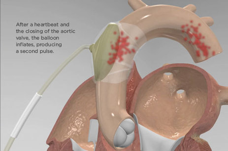
C-Pulse device designed to help patients with advanced heart failure

Reporter: Aviva Lev-Ari, PhD, RN


See on Scoop.it – Cardiovascular and vascular imaging

A Minnesota medical device company is developing a device to help people with class three and four heart failure without increasing the risk of blood clots.

See on medcitynews.com

Read Full Post »

Curation, HealthCare System in the US, and Calcium Signaling Effects on Cardiac Contraction, Heart Failure, and Atrial Fibrillation, and the Relationship of Calcium Release at the Myoneural Junction to Beta Adrenergic Release

Curation, HealthCare System in the US, and Calcium Signaling Effects on Cardiac Contraction, Heart Failure, and Atrial Fibrillation, and the Relationship of Calcium Release at the Myoneural Junction to Beta Adrenergic Release

Curator and e-book Contributor: Larry H. Bernstein, MD, FCAP
Curator and BioMedicine e-Series Editor-in-Chief: Aviva Lev Ari, PhD, RN

and 

Content Consultant to Six-Volume e-SERIES A: Cardiovascular Diseases: Justin Pearlman, MD, PhD, FACC

This portion summarises what we have covered and is now familiar to the reader.  There are three related topics, and an extension of this embraces other volumes and chapters before and after this reading.  This approach to the document has advantages over the multiple authored textbooks that are and have been pervasive as a result of the traditional publication technology.  It has been stated by the founder of ScoopIt, that amount of time involved is considerably less than required for the original publications used, but the organization and construction is a separate creative process.  In these curations we amassed on average five articles in one curation, to which, two or three curators contributed their views.  There were surprises, and there were unfulfilled answers along the way.  The greatest problem that is being envisioned is the building a vision that bridges and unmasks the hidden “dark matter” between the now declared “OMICS”, to get a more real perspective on what is conjecture and what is actionable.  This is in some respects unavoidable because the genome is an alphabet that is matched to the mino acid sequences of proteins, which themselves are three dimensional drivers of sequences of metabolic reactions that can be altered by the accumulation of substrates in critical placements, and in addition, the proteome has functional proteins whose activity is a regulatory function and not easily identified.  In the end, we have to have a practical conception, recognizing the breadth of evolutionary change, and make sense of what we have, while searching for more.

We introduced the content as follows:

1. We introduce the concept of curation in the digital context, and it’s application to medicine and related scientific discovery.

Topics were chosen were used to illustrate this process in the form of a pattern, which is mostly curation, but is significantly creative, as it emerges in the context of this e-book.

  • Alternative solutions in Treatment of Heart Failure (HF), medical devices, biomarkers and agent efficacy is handled all in one chapter.
  • PCI for valves vs Open heart Valve replacement
  • PDA and Complications of Surgery — only curation could create the picture of this unique combination of debate, as exemplified of Endarterectomy (CEA) vs Stenting the Carotid Artery (CAS), ischemic leg, renal artery stenosis.

2. The etiology, or causes, of cardiovascular diseases consist of mechanistic explanations for dysfunction relating to the heart or vascular system. Every one of a long list of abnormalities has a path that explains the deviation from normal. With the completion of the analysis of the human genome, in principle all of the genetic basis for function and dysfunction are delineated. While all genes are identified, and the genes code for all the gene products that constitute body functions, there remains more unknown than known.

3. Human genome, and in combination with improved imaging methods, genomics offers great promise in changing the course of disease and aging.

4. If we tie together Part 1 and Part 2, there is ample room for considering clinical outcomes based on individual and organizational factors for best performance. This can really only be realized with considerable improvement in information infrastructure, which has miles to go.

Curation

Curation is an active filtering of the web’s  and peer reviewed literature found by such means – immense amount of relevant and irrelevant content. As a result content may be disruptive. However, in doing good curation, one does more than simply assign value by presentation of creative work in any category. Great curators comment and share experience across content, authors and themes.
Great curators may see patterns others don’t, or may challenge or debate complex and apparently conflicting points of view.  Answers to specifically focused questions comes from the hard work of many in laboratory settings creatively establishing answers to definitive questions, each a part of the larger knowledge-base of reference. There are those rare “Einstein’s” who imagine a whole universe, unlike the three blindmen of the Sufi tale.  One held the tail, the other the trunk, the other the ear, and they all said this is an elephant!
In my reading, I learn that the optimal ratio of curation to creation may be as high as 90% curation to 10% creation. Creating content is expensive. Curation, by comparison, is much less expensive.  The same source says “Scoop.it is my content marketing testing “sandbox”. In sharing, he says that comments provide the framework for what and how content is shared.

Healthcare and Affordable Care Act

We enter year 2014 with the Affordable Care Act off to a slow start because of the implementation of the internet signup requiring a major repair, which is, unfortunately, as expected for such as complex job across the US, and with many states unwilling to participate.  But several states – California, Connecticut, and Kentucky – had very effective state designed signups, separate from the federal system.  There has been a very large rush and an extension to sign up. There are many features that we can take note of:

1. The healthcare system needed changes because we have the most costly system, are endowed with advanced technology, and we have inexcusable outcomes in several domains of care, including, infant mortality, and prenatal care – but not in cardiology.

2. These changes that are notable are:

  • The disparities in outcome are magnified by a large disparity in highest to lowest income bracket.
  • This is also reflected in educational status, and which plays out in childhood school lunches, and is also affected by larger class size and cutbacks in school programs.
  • This is not  helped by a large paralysis in the two party political system and the three legs of government unable to deal with work and distraction.
  • Unemployment is high, and the banking and home construction, home buying, and rental are in realignment, but interest rates are problematic.

3.  The  medical care system is affected by the issues above, but the complexity is not to be discounted.

  •  The medical schools are unable at this time to provide the influx of new physicians needed, so we depend on a major influx of physicians from other countries
  • The technology for laboratories, proteomic and genomic as well as applied medical research is rejuvenating the practice in cardiology more rapidly than any other field.
  • In fields that are imaging related the life cycle of instruments is shorter than the actual lifetime use of the instruments, which introduces a shortening of ROI.
  • Hospitals are consolidating into large consortia in order to maintain a more viable system for referral of specialty cases, and also is centralizing all terms of business related to billing.
  • There is reduction in independent physician practices that are being incorporated into the hospital enterprise with Part B billing under the Physician Organization – as in Partners in Greater Boston, with the exception of “concierge” medical practices.
  • There is consolidation of specialty laboratory services within state, with only the most specialized testing going out of state (Quest, LabCorp, etc.)
  • Medicaid is expanded substantially under the new ACA.
  • The federal government as provider of services is reducing the number of contractors for – medical devices, diabetes self-testing, etc.
  • The current rearrangements seeks to provide a balance between capital expenses and fixed labor costs that it can control, reduce variable costs (reagents, pharmaceutical), and to take in more patients with less delay and better performance – defined by outside agencies.

Cardiology, Genomics, and calcium ion signaling and ion-channels in cardiomyocyte function in health and disease – including heart failure, rhythm abnormalities, and the myoneural release of neurotransmitter at the vesicle junction.

This portion is outlined as follows:

2.1 Human Genome: Congenital Etiological Sources of Cardiovascular Disease

2.2 The Role of Calcium in Health and Disease

2.3 Vasculature and Myocardium: Diagnosing the Conditions of Disease

Genomics & Genetics of Cardiovascular Disease Diagnoses

actin cytoskeleton

wall stress, ventricular workload, contractile reserve

Genetic Base of Atherosclerosis and Loss of Arterial Elasticity with Aging

calcium and actin skeleton, signaling, cell motility

hypertension & vascular compliance

Genetics of Conduction Disease

Ca+ stimulated exostosis: calmodulin & PKC (neurotransmitter)

complications & MVR

disruption of Ca2+ homeostasis cardiac & vascular smooth muscle

synaptotagmin as Ca2+ sensor & vesicles

atherosclerosis & ion channels


It is increasingly clear that there are mutations that underlie many human diseases, and this is true of the cardiovascular system.  The mutations are mistakes in the insertion of a purine nucleotide, which may or may not have any consequence.  This is why the associations that are being discovered in research require careful validation, and even require demonstration in “models” before pursuing the design of pharmacological “target therapy”.  The genomics in cardiovascular disease involves very serious congenital disorders that are asserted early in life, but the effects of and development of atherosclerosis involving large and medium size arteries has a slow progression and is not dominated by genomic expression.  This is characterized by loss of arterial elasticity. In addition there is the development of heart failure, which involves the cardiomyocyte specifically.  The emergence of regenerative medical interventions, based on pleuripotent inducible stem cell therapy is developing rapidly as an intervention in this sector.

Finally, it is incumbent on me to call attention to the huge contribution that research on calcium (Ca2+) signaling has made toward the understanding of cardiac contraction and to the maintenance of the heart rhythm.  The heart is a syncytium, different than skeletal and smooth muscle, and the innervation is by the vagus nerve, which has terminal endings at vesicles which discharge at the myocyte junction.  The heart specifically has calmodulin kinase CaMK II, and it has been established that calmodulin is involved in the calcium spark that triggers contraction.  That is only part of the story.  Ion transport occurs into or out of the cell, the latter termed exostosis.  Exostosis involves CaMK II and pyruvate kinase (PKC), and they have independent roles.  This also involves K+-Na+-ATPase.  The cytoskeleton is also discussed, but the role of aquaporin in water transport appears elsewhere, as the transport of water between cells.  When we consider the Gibbs-Donnan equilibrium, which precedes the current work by a century, we recall that there is an essential balance between extracellular Na+ + Ca2+ and the intracellular K+ + Mg2+, and this has been superceded by an incompletely defined relationship between ions that are cytoplasmic and those that are mitochondrial.  The glass is half full!


Read Full Post »

St. Jude’s CEO is still betting on EnligHTN IV Study Renal Denervation System, despite Medtronic’s setback related to SYMPLICITY Phase IV

Reporter: Aviva Lev-Ari, PhD, RN

Article ID #92: St. Jude’s CEO is still betting on EnligHTN IV Study Renal Denervation System, despite Medtronic’s setback related to SYMPLICITY Phase IV. Published on 12/10/2013

WordCloud Image Produced by Adam Tubman

UPDATED on 1/14/2014

This is in continuation to our 1/9/2014 article:

Market Impact on Global Suppliers of Renal Denervation Systems by Pivotal US Trial: Metronics’ Symplicity Renal Denervation System FAILURE at Efficacy Endpoint

http://pharmaceuticalintelligence.com/2014/01/09/market-impact-on-global-suppliers-of-renal-denervation-systems-by-pivotal-us-trial-metronics-symplicity-renal-denervation-system-failure-at-efficacy-endpoint/

In the short term, the company, St. Jude suspended enrollment in three other related trials and announced plans to gather an independent panel of experts to plot a future course. 

St. Jude Medical won a CE mark for its next-gen EnligHTN IV renal denervation system over the summer, but halted its own U.S. trial in December. Minnesota-based St. Jude said it had struggled to recruit viable candidates for the 590-patient trial, and was concerned that Medtronic’s Symplicity might siphon off viable patients needed for its own study. At the time, St. Jude said it would work to develop a new protocol to address trial enrollment challenges.

According to Mark Hollmer:

Symplicity’s setbacks may make that job much easier for St. Jude. They also create new opportunities for other rivals focused on developing similar technology, such as Boston Scientific ($BSX) and Covidien ($COV).

Curator of this article has expressed a different view in 

http://pharmaceuticalintelligence.com/2014/01/09/market-impact-on-global-suppliers-of-renal-denervation-systems-by-pivotal-us-trial-metronics-symplicity-renal-denervation-system-failure-at-efficacy-endpoint/

St. Jude’s CEO is still betting on renal denervation, despite Medtronic’s setback

By Mark Hollmer

Medtronic’s Symplicity renal denervation device

If Medtronic’s ($MDT) recent U.S. clinical trial failure for its Symplicity renal denervation device throws the door wide open for its competitors, St. Jude Medical ($STJ) will likely waste no time walking through.

As MassDevice reported, St. Jude CEO Daniel Starks told an audience at the JPMorgan Healthcare conference in San Francisco that the company plans to keep plowing ahead with the development of its own technology.

“The fact that we had and that there have been favorable early clinical results in numerous other experiences is still valid,” MassDevice quoted Starks as saying. “There is open surgical data dating back several decades that was favorable to the impact of surgical renal denervation to treat hypertension.

That said, he also expressed genuine surprise about Medtronic’s trial setback.

“This was unexpected for us, to have a negative result from Medtronic’s trial, and it’s too soon for us to know what to make of that,” Starks said in the story.

Medtronic’s Symplicity device, which has a CE mark and is a market leader, had done well in previous trials in patients with different forms of hypertension and was on track to win FDA approval in 2015–potentially the first renal denervation device to reach that point.

The Minnesota device giant announced a few days ago that Symplicity proved safe in a 535-patient trial but failed to significantly lower blood pressure for drug-resistant hypertension.

This was a trial that mattered, designed to help fuel FDA approval.

Related Articles:

Medtronic’s flunked trial throws its hypertension program into doubt

St. Jude Medical will try again another day for a U.S. renal denervation study

St. Jude bags EU approval for next-gen renal denervation

 SOURCE

From: FierceMedicalDevices <editors@fiercemedicaldevices.com>
Date: Tue, 14 Jan 2014 18:14:06 +0000 (GMT)
To: <avivalev-ari@alum.berkeley.edu>

Renal Denervation: EnligHTN IV Study Called Off and Potential Novel Indications – Diastolic Heart Failure

Reporter: Aviva Lev-Ari, PhD, RN

This Open Access Scientific Journal has covered all the major developments reported on Renal Denervation since its inception

The Archive for Renal denervation

http://pharmaceuticalintelligence.com/category/cardiac-and-cardiovascular-surgical-procedures/renal-denervation/

Search Results for Renal Denervation

http://pharmaceuticalintelligence.com/?s=Renal+denervation

Heartwire Reported on December 09, 2013

EnligHTN IV Renal Denervation Study Called Off

ST PAUL, MN – The EnligHTN IV study (St Jude Medical, St Paul, MN), testing the multielectrode renal-denervation system in patients with resistant hypertension, has been stopped almost before it even began. The trial, which was announced in June and began enrolling a small number of patients this fall, was canceled because of concerns about slow enrollment.

To heartwire ,

Denise Perkins-Landry, a spokesperson for St Jude Medical, said the decision to discontinue the study was based on “anticipated recruitment challenges” and is not the result of any safety or efficacy issues with the device. “A US clinical trial for EnligHTN remains a very high priority for St Jude Medical, and we will be working with the FDA to develop a new protocol that will address anticipated enrollment challenges,” she stated in an email.

The full results of the SYMPLICITY HTN-3 trial, a study similar to the EnligHTN IV study, which is sponsored by Medtronic (Minneapolis, MN), are expected in early 2014. While neither device is approved for clinical use in the US, it is expected that the Medtronic renal-denervation system will be first. As a result, it might be difficult to enroll patients to a sham procedure if there is another commercially available system for treating resistant hypertension, according to St Jude.

EnligHTN IV was to be a randomized, single-blind, controlled study with patients randomized from as many as 80 clinical centers in the US and Canada. It was intended to show the safety and effectiveness of the renal-denervation system in the reduction of systolic blood pressure in 590 patients with an office blood pressure >160 mm Hg despite taking three or more antihypertensive medications, including a diuretic.

Abrupt Decision to Stop the Study

Dr William White (University of Connecticut Medical Center, Farmington), one of the cochairs of the EnligHTN IV steering committee, told heartwire the decision to cancel the study was made just last week. In fact, there were clinical centers already up and running in terms of enrollment, although these were pilot centers where any “bugs could be worked out.” The meeting for training investigators participating in the trial was scheduled for Saturday in Chicago, IL, although that is now off.

“In the grander scheme of things, I can tell you that the decision was not made because of any problem with the catheter or any safety issues,” said White. “Outside the US, the development is continuing as planned, but the biggest concern is that it might be very difficult, if not impossible, to continue doing a sham-controlled study a year from now if there were a commercially available renal-denervation catheter in the US, which there very well could be with Medtronic.”

Still, White said that it’s not even known if the SYMPLICITY HTN-3 study is positive or if the FDA will be satisfied with the trial and its outcomes. He said a lot of assumptions are being made by St Jude Medical in stopping the trial, and it could turn out to be a bad decision. In addition, reimbursement remains an open question.

“Commercially available doesn’t mean there’s going to be a payer,” White told heartwire . “If that happens, it will be doubly unfortunate for the patients that are out there because they might be willing and able to go into a clinical trial but they won’t have that opportunity.”

As a scientist, White said that he would have preferred the trial be pursued, regardless of what happens with Medtronic. He believes the EnligHTN catheter is “outstanding,” as it has multiple electrodes to enable physicians to thoroughly ablate the renal arteries within a couple of minutes. “The clinical scientist in me would have preferred that we go ahead as planned,” he said. “I think we would have gained a great deal of knowledge. And just because one study shows a p value of 0.05 doesn’t mean a second study will.”

The Medtronic SYMPLICITY renal-denervation system was launched in 2010 and is available in parts of Europe, Asia, Africa, and South America. The first-generation EnligHTN device has had CE Mark approval in Europe since 2012 while the updated second-generation system with multiple electrodes received European approval this past summer.

News of the halting of EnligHTN IV was first reported by Wells Fargo analyst Larry Biegelsen.

SOURCE

http://www.medscape.com/viewarticle/817482?nlid=41903_2105&src=wnl_edit_medp_card&uac=93761AJ&spon=2

On December 06, 2013 Marlene Busko reported to Heartwire on

Renal Denervation’s Structural, Functional Heart Benefits May Be Independent of BP

HAMBURG, GERMANY — In a small study of patients undergoing renal denervation for resistant hypertension, left-ventricular hypertrophy and diastolic function improved independently of changes in blood pressure and heart rate [1].

“The novelty of our findings is the independence of morphologic improvements [regression of LV hypertrophy] from hemodynamic changes (reduction of blood pressure and heart rate),” Dr Stephan H Schirmer (University of Saarland, Hamburg, Germany) told heartwire in an email. “If this is confirmed in larger trials, it might open up novel indications for the use of renal denervation, for example, in [diastolic] heart-failure patients, independent of blood pressure.”

Dr Deepak L Bhatt (Harvard Medical School, Boston, MA) told heartwire that the study observations are “provocative” and “exciting” but stressed that they need to be confirmed in a blinded, larger, multicenter study before they could be accepted into clinical practice. Bhatt and Dr George Bakris (University of Chicago, IL) are co–principal investigators for the ongoing SYMPLICITY HTN-3 trial of bilateral renal denervation in patients with uncontrolled hypertension.

The study was published online December 4, 2013 in the Journal of the American College of Cardiology.

Are Renal Denervation Effects Always Tied to BP Change?

Renal denervation reduces heart rate and blood pressure in patients with resistant hypertension, and as reported by heartwire , a recent small study suggested that the procedure also reduces left-ventricular mass and improves diastolic function in such patients, Schirmer and colleagues write.

They hypothesized that renal denervation might affect cardiac structure and function, independent of the effect on blood pressure.

They enrolled 66 consecutive patients who underwent renal denervation using the Flex catheter system (Medtronic) at their center during 2010 and 2011 for treatment of resistant hypertension (office systolic blood pressure >140 mm Hg). Patients had a mean age of 64 years, and 55% were men. They were on a mean of 4.3 antihypertensive drugs. All were taking a diuretic, 89% were taking a beta-blocker, and 55% were taking an angiotensin-receptor blocker.

Six months after renal denervation,

  • Mean blood pressure decreased from 172.9/92.5 to 151.3/85.5 mm Hg, confirmed by 24-hour ambulatory monitoring, if available (n=50).
  • Mean heart rate decreased from 67.7 to 60.5 bpm.
  • Mean left-ventricular mass index decreased from 61.5 to 53.4 g/m2.
  • Measures of diastolic function also improved.

The changes in cardiac function and ventricular size were not tied to the magnitude of the blood-pressure reduction, which “suggest[s] a direct effect of the sympathetic nervous system on myocardial morphology and function,” Schirmer and colleagues write. They call for further research to investigate functional cardiovascular benefits of renal denervation beyond blood-pressure reduction.

Promising Early Benefit, Needs Confirmation

In an accompanying editorial [2], Bakris and Dr Sandeep Nathan (University of Chicago) commend Schirmer and colleagues “for providing promising early benefit of catheter-based renal denervation and for highlighting a possible blood-pressure–independent facet of this technique.” However, they caution that although the findings are “intriguing,” the study’s limitations include that it was

  • relatively small,
  • conducted at a single center,
  • lacked a sham control, and
  • relied on echocardiography rather than magnetic resonance imaging.

Therefore, “these observations need confirmation before acceptance in clinical practice . . . and can only be applied to those with inclusion criteria used in their study,” the editorialists conclude.

“It’s an exciting, provocative result, and there’s a good chance that it will stand the test of time, but I still think in general, it’s best to be cautious about new technologies and relatively small studies, because time typically shows that they provide an overestimate of what the true effects will be,” Bhatt commented when interviewed.

“Whether the reduction in left-ventricular mass is beyond what would be anticipated with blood-pressure and heart-rate reduction—certainly this analysis suggests that is a possibility—needs to be confirmed in larger studies,” he added, echoing the authors and editorialists.

Potentially referring physicians, in the United States where the procedure is investigational, and even in Europe where it’s approved, appear to be waiting for the results of SYMPLICITY HTN-3, Bhatt said. This blinded, randomized, multicenter trial will provide a clearer picture of what sort of blood-pressure reductions are achievable in patients with resistant hypertension who undergo renal denervation. Results are expected by mid-2014.

References

  1. Schirmer SH, Sayed M, Reil J-C, et al. Improvements of left-ventricular hypertrophy and diastolic function following renal denervation – Effects beyond blood pressure and heart rate reduction. J Am Coll Cardiol 2013; DOI:10.1016/j.jacc.2013.10.073. Abstract
  2. Bakris G, Nathan S. Renal denervation and left ventricular mass regression: A benefit beyond blood pressure reduction? J Am Coll Cardiol 2013; DOI:10.1016/j.jacc.2013.11.015. Editorial

SOURCE

This Open Access Scientific Journal has covered all the major developments reported on Renal Denervation since its inception

The Archive for Renal denervation

http://pharmaceuticalintelligence.com/category/cardiac-and-cardiovascular-surgical-procedures/renal-denervation/

Search Results for Renal Denervation

http://pharmaceuticalintelligence.com/?s=Renal+denervation

For the ORIGINAL work on 

Renal Sympathetic Denervation: Updates on the State of Medicine

the Readers is called to go to the ORIGINAL SOURCES listed below:

Intravascular Stimulation of Autonomics: A Letter from Dr. Michael Scherlag

http://pharmaceuticalintelligence.com/2012/09/02/intravascular-stimulation-of-autonomics-a-letter-from-dr-michael-scherlag/

Imbalance of Autonomic Tone: The Promise of Intravascular Stimulation of Autonomics

http://pharmaceuticalintelligence.com/2012/09/02/imbalance-of-autonomic-tone-the-promise-of-intravascular-stimulation-of-autonomics/

Interaction of Nitric Oxide and Prostacyclin in Vascular Endothelium

http://pharmaceuticalintelligence.com/2012/09/14/interaction-of-nitric-oxide-and-prostacyclin-in-vascular-endothelium/

Absorb™ Bioresorbable Vascular Scaffold: An International Launch by Abbott Laboratories

http://pharmaceuticalintelligence.com/2012/09/29/absorb-bioresorbable-vascular-scaffold-an-international-launch-by-abbott-laboratories/

The Molecular Biology of Renal Disorders: Nitric Oxide – Part III

http://pharmaceuticalintelligence.com/2012/11/26/the-molecular-biology-of-renal-disorders/

Treatment of Refractory Hypertension via Percutaneous Renal Denervation

http://pharmaceuticalintelligence.com/2012/06/13/treatment-of-refractory-hypertension-via-percutaneous-renal-denervation/

Renal Denervation Technology of Vessix Vascular, Inc. been acquired by Boston Scientific Corporation (BSX) to pay up to $425 Million

http://pharmaceuticalintelligence.com/2012/11/08/renal-denervation-technology-of-vessix-vascular-inc-been-acquired-by-boston-scientific-corporation-bsx-to-pay-up-to-425-million/

Read Full Post »

Mitral Valve Repair: Who is a Patient Candidate for a Non-Ablative Fully Non-Invasive Procedure?

Author, and Content Consultant to e-SERIES A: Cardiovascular Diseases: Justin Pearlman, MD, PhD, FACC

and

Article Curator: Aviva Lev-Ari, PhD, RN


UPDATED on 9/24/2018

TCT: MitraClip Saves Lives in Functional Mitral Regurgitation

Positive COAPT results may overwrite neutral MITRA-FR findings
https://www.medpagetoday.com/meetingcoverage/tct/75260?xid=nl_mpt_ACC_Reporter_2018-09-23&eun=g5099207d2r

UPDATED on 8/31/2018

Don’t Ignore the Many Lessons of the MitraClip Failure

John M. Mandrola, MD

August 30, 2018

Comments

It would be wrong to say that use of this device for this indication provided no benefit to patients. The more accurate conclusion is that the MitraClip caused net harm. That’s because in addition to no benefit in the efficacy endpoints, patients in the device group endured a procedural complication rate of more than 10%, including a sevenfold higher rate of stroke.

Once again, we can learn both specific lessons about the treatment of people with heart failure and more general lessons on the acceptance of untested therapeutics.

Other comments

  • Dr. MIGUEL QUINTANA|  Cardiology, General

A bad day for interventional cardiology. I do believe that interventional cardiologist are aware about the mechanisms of severe MR in dilated hearts, however when a point of no return in those ventricles is reached, something has to be done.

I do agree with Dr. Mandrola regarding the behaviour of the industry to drive new devices in medical practice without performing RCT. However the main responsible  are the institutions approving the devices (FDA and European Commission for drugs and devices).

I wish to hear some comments of Dr. Mandrola regarding the new trends in performing RCT using just the non-inferiority criteria and the growing trends of using the “big data” of mega data registries to establish guidelines for clinical treatments and not only to test new hypothesis.

  • Dr. James Rittelmeyer|  Cardiology, Interventional

When the point of no return has been reached palliative care is a great plan. It causes no harm and is relatively inexpensive.

  • Dr. Johannes Schaar|  Cardiology, General

Right!!!!!! We should stay away from interventional cardiologists, who have know idea what they do and are on the payroll of the industry

  • Dr. Steve Soldo|  Cardiology, General

Are you being sincere or cynical?

SOURCE

https://www.medscape.com/viewarticle/901378?nlid=124808_3802&src=WNL_mdplsnews_180831_mscpedit_card&uac=93761AJ&spon=2&impID=1727175&faf=1

https://www.medscape.com/viewarticle/901378?nlid=124808_3802&src=WNL_mdplsnews_180831_mscpedit_card&uac=93761AJ&spon=2&impID=1727175&faf=1#vp_2


UPDATED on 8/30/2018

From European Society of Cardiology, Aug 28, 2018, Munich

MITRA-FR: No Benefit of MitraClip in Functional MR

https://www.tctmd.com/news/mitra-fr-no-benefit-mitraclip-functional-mr

This link suggests that the only FDA approved device may have a more limited indication, but is still helping the very sickest patients, with the ‘negative’ outcome for the quoted study.   Functional Mitral Regurgitation (6 M patients in the US) still has no approved viable transcatheter therapy, and the interpretations of the latest study results suggest a more restricted patient selection for the MitraClip® device. 

  • Edward Hlozek, Chairman and CEO, ValveCure, LLC, on 8/30/2018

www.valvecure.com

BarrelEye portends to be available to all classes of patients with Functional MR and significantly improve quality of life and extend lives, offering future non-invasive repeatability of the therapy, without an implant. 


UPDATED on 8/20/2018

In a new study

Gammie J.S., Chikwe J., Badhwar V., et al. “Isolated Mitral Valve Surgery: The Society of Thoracic Surgeons Adult Cardiac Surgery Database Analysis.” Annals of Thoracic Surgery, published online July 18, 2018. https://doi.org/10.1016/j.athoracsur.2018.03.086

Earlier Intervention for Mitral Valve Disease May Lead to Improved Outcomes

Slow progression of disease may mask symptoms until damage cannot be fully repaired

In this study, the data showed that the overall repair rate was 65.6 percent (57,244) and the replacement rate was 34.4 percent (29,970). Overall operative mortality was 2 percent (1,762).

“We found that the number of operations performed for mitral valve disease is growing faster than any other category of heart operation and that the results were excellent with low risks of death and complications,” said Gammie.

The researchers also revealed that while the prevalence of mitral valve disease and the number of mitral valve operations performed per year are increasing, overall aortic valve operations were performed 1.6 times more commonly than mitral valve operations during the study period.

“This may suggest important under-referral and under-treatment of mitral valve disease, which may be related to the slower progression of signs and symptoms of mitral compared to aortic disease, as well as potential lack of adherence to guidelines for intervention,” said Gammie. “So although contemporary outcomes are excellent, there remains an important and substantial opportunity to improve results for patients with mitral valve disease by following established guidelines and encouraging earlier referral for operation.”

For more information: www.annalsthoracicsurgery.org


UPDATED on 4/8/2017

Percutaneous repair or replacement for mitral regurgitation?

by Ted E. Feldman, MD

  • by Nicole Lou
    Contributing Writer, MedPage TodayApril 04, 2017

Mitral repair is still a relatively youthful field at 14 years, but now operators are taking it further and developing methods for mitral valve replacement, says Ted E. Feldman, MD, of Evanston Hospital in Illinois, where mitral repair first got its start.

In this exclusive MedPage Today video, the interventionist shares his insight into the limitations of the device synonymous with mitral repair, the MitraClip, and discusses the current challenges of outright percutaneous replacement of the valve.

“100 patients underwent Mitral valve repair vs 1000s of Aortic valve the TAVR.”

WATCH VIDEO

http://www.medpagetoday.com/cardiology/chf/64332

Voice of Edward Hlozek, CEO, ValveCure:

MV repair via transcatheter valve implant (TMVR) will be extremely difficult to get right because of the complex nature of the anatomy versus the simple circle that is the aortic valve (TAVR)…and MitraClip is limited because leaflets sometime cannot be caught right for the device to be implanted.  Think of ValveCure’s platform device that is not an implant and tightens up the valve biologically.

Aortic and Pulmonic are basically planar circular shapes.  The shape of the Mitral and Tricuspid are parabolic ellipsoidal.  This unique shape makes designing a transcatheter mitral valve implant challenging, especially considering that most are of a unique shape and dimension.  And the aortic is more calcified, which lends to a better attachment of a transcatheter implant because it is more rigid and planar.

MitraClip Issues, Outcomes Come to Fore in US Registry Experience

 Patrice Wendling

March 23, 2017

http://www.medscape.com/viewarticle/877629?nlid=113592_3802&src=WNL_mdplsnews_170324_mscpedit_card&spon=2&impID=1314983&faf=1


UPDATED on 12/6/2016

Edwards To Acquire Transcatheter Mitral, Tricuspid Valve Repair Company Valtech Cardio

Acquisition enables entry into transcatheter valve repair segment of interventional structural heart

NEWS | HEART VALVE TECHNOLOGY | DECEMBER 02, 2016

http://www.dicardiology.com/content/edwards-acquire-transcatheter-mitral-tricuspid-valve-repair-company-valtech-cardio

The Cardioband System is not approved for sale in the United States.

Read the related article “Advances and Future Directions for Transcatheter Valves – Mitral and tricuspid valve repair technologies now in development.”

For more information: www.Edwards.com, www.valtechcardio.com

  • can used as a non-surgical form on annuloplasty repair.


Cardioband, valtech, Edwards Lifesciences, transcatheter mitral repair, transcatheter tricuspid valve repair, transcatheter annuloplasty

Cardioband, valtech, Edwards Lifesciences, transcatheter mitral repair, transcatheter tricuspid valve repair, transcatheter annuloplasty

An illustration of how the transcatheter Cardioband System can used as a non-surgical form on annuloplasty repair.

SOURCE

http://www.dicardiology.com/content/edwards-acquire-transcatheter-mitral-tricuspid-valve-repair-company-valtech-cardio


UPDATED On 11/28/2016

Edwards Lifesciences to acquire Valtech Cardio in $690m deal

NOVEMBER 28, 2016 BY BRAD PERRIELLO

Valtech makes the Cardioband device, which is designed to reshape the mitral valve using specially designed anchors. The Or Yehuda, Israel-based company was the target of a previous takeover attempt by HeartWare International that was spiked early this year after a proxy war. (HeartWare itself was acquired by Medtronic (NYSE:MDT) for $1.1 billion in August.) Valtech won CE Mark approval in the European Union for Cardioband in September 2015 but the device is not approved for the U.S. market.

http://www.massdevice.com/edwards-lifesciences-acquire-valtech-cardio-690m-deal/?utm_source=newsletter-161128&utm_medium=email&utm_campaign=newsletter-161128&spMailingID=9950276&spUserID=MTU0MTAzNjIxODMyS0&spJobID=1042200257&spReportId=MTA0MjIwMDI1NwS2


UPDATED on 10/4/2016

Novel Mitral Valve-Cinching Device Slashes Backflow Without Surgery – Promising feasibility results for Cardioband, but survival effect still unclear

0 6 Google +0 0 0 Valtech‘s new Cardioband technology may eliminate the need for open-heart surgeries to repair leaky mitral and tricuspid valves. The Cardioband can be implanted transfemorally and is guided via fluoroscopy and ultrasound.

by Nicole Lou
Reporter, MedPage Today/CRTonline.org

10.03.2016

  • Reviewed by F. Perry Wilson, MD, MSCE Assistant Professor, Section of Nephrology, Yale School of Medicine and Dorothy Caputo, MA, BSN, RN, Nurse Planner

Cardioband is a product of Valtech acquired by Heartware in 2015. Medtronic completed acquisition of Heartware in August 2016. See Updates, below

A novel surgical-style transcatheter device showed promise for the treatment of functional mitral regurgitation, investigators reported.

The Cardioband direct annuloplasty device was associated with no periprocedural deaths and had a 1-month mortality rate of 5%, according to Georg Nickenig, MD, of Heart Center Bonn in Germany, and colleagues in their study published online in JACC: Cardiovascular Interventions. By 6 months, the death rate had climbed to 9.6%.

Annular septolateral dimensions fell from 3.7 cm at baseline to 2.5 cm at 1 month and 2.4 cm after 6 months (P<0.001) with the device, which is implanted in a transvenous, transseptal procedure to encircle the valve annulus and, secured with small anchors, cinch it until the valve closes fully again.
In addition, the proportion of patients with grade 3 or worse mitral regurgitation also dropped from 77.4% to 10.7% at 1 month (P<0.001) and was recorded at 13.6% after 6 months (P<0.001). The proportion still categorized as being in New York Heart Association functional class III or IV dropped from 95.5% at baseline to 18.2% (P<0.001).

Over the 6-month follow-up in the study, exercise capacity generally improved (332 m in a 6-minute walking test versus 250 m at baseline, P<0.001), as did patients’ quality of life (Minnesota Living With Heart Failure Questionnaire score 18.1 versus 38.2 at baseline,P<0.001).

SOURCE

http://www.medpagetoday.com/Cardiology/PCI/60589?xid=nl_mpt_DHE_2016-10-04&eun=g99985d0r&pos=2

https://www.sciencedaily.com/releases/2016/09/160926100000.htm


UPDATED on 10/4/2016

Medtronic Completes Acquisition of HeartWare International

Broadens Heart Failure Leadership Into Growing Circulatory Support Sector

DUBLIN – Aug. 23, 2016 – Medtronic plc (NYSE: MDT), the global leader in medical technology, has completed its acquisition of HeartWare International, Inc., a leading innovator of less-invasive, miniaturized, mechanical circulatory support technologies (MCS) for treating patients with advanced heart failure. HeartWare will become part of the Heart Failure business within the Medtronic Cardiac Rhythm and Heart Failure division. Under the terms of the transaction, each outstanding share of HeartWare common stock has been converted into the right to receive $58.00 in cash, without interest, subject to any required withholding of taxes.

HeartWare develops and manufactures miniaturized implantable heart pumps, or ventricular assist devices (VAD), to treat patients around the world suffering from advanced heart failure. Its flagship product, the HVAD® System, features the world’s smallest full-support VAD and is indicated for refractory end-stage left-ventricular heart failure patients in the U.S. who are awaiting a heart transplant, as well as approved in Europe for long-term use in patients at risk of death from refractory, end-stage heart failure.

Medtronic estimates that the global VAD market is approximately $800 million currently, and worldwide is expected to grow in the mid-to-high single digits for calendar years 2016-17, and accelerate to high-single/low-double digits beyond calendar year 2017.

“Not only does the current HeartWare portfolio expand Medtronic leadership across the heart failure continuum, its product pipeline – when married with our expertise – can result in progressively less-invasive heart pumps that have the potential to benefit even more patients,” said David Steinhaus, M.D., vice president and general manager of the Heart Failure business, and medical director for the Cardiac Rhythm and Heart Failure division at Medtronic. “Today, Medtronic offers the industry’s leading cardiac resynchronization therapy devices, including MR-conditional CRT-defibrillators; MCS therapy for advanced heart failure patients; heart failure diagnostics; and meaningful expert analysis through Medtronic Care Management Services, including the recently launched Beacon Heart Failure Management Service.”

The acquisition of HeartWare broadens the Medtronic portfolio of therapies, diagnostic tools and services for patients suffering from heart failure, aligning with Medtronic’s Mission of alleviating pain, restoring health and extending life. The acquisition is part of the Company’s therapy innovation strategy to surround the physician with innovative products while focusing on patients and disease states.

“This is an exciting moment, as more than 600 HeartWare employees are now part of the broader Medtronic organization,” said Doug Godshall, who served as president and chief executive of HeartWare for the past decade. “HeartWare has delivered incredible advancements for patients suffering from heart failure, through the commercialization of the HVAD system and pipeline development, and I am convinced that being part of Medtronic will allow us to accelerate meaningful innovations even more quickly.”

Heart failure, also known as congestive heart failure, is a condition in which the heart isn’t pumping enough blood to meet the body’s needs. Heart failure usually develops slowly after an injury to the heart. Some injuries may include a progressive deterioration of the heart muscle, heart attack, untreated high blood pressure, or heart valve disease. Heart failure remains a leading cause of hospitalization and death in the United States, and its prevalence continues to increase, affecting more than 5 million people in the U.S. alone. The cost of heart failure is high. Healthcare expenditures in the U.S. on heart failure are estimated to be approximately $39 billion per year, making it one of the largest expenses to the healthcare system. With the aging of the population, Medtronic estimates that the number of patients with heart failure could exceed 8 million by 2030.

This transaction is expected to meet Medtronic’s long-term financial metrics for acquisitions. Medtronic does not intend to modify its fiscal year 2017 revenue outlook or earnings per share (EPS) guidance as a result of this transaction, although it is expected to provide increased confidence in the company’s ability to deliver on its FY17 revenue growth outlook. In addition, Medtronic expects minimal to no net EPS dilution from this transaction for the first two years as the company intends to offset the expected dilutive impact. The acquisition is expected to be earnings accretive in year three.

In collaboration with leading clinicians, researchers and scientists worldwide, Medtronic offers the broadest range of innovative medical technology for the interventional and surgical treatment of cardiovascular disease and cardiac arrhythmias. The company strives to offer products and services of the highest quality that deliver clinical and economic value to healthcare consumers and providers around the world.

The Tender Offer and Merger
The tender offer for all of the outstanding shares of HeartWare common stock expired as scheduled immediately after 11:59 p.m. Eastern time on August 22, 2016. Computershare Trust Company, N.A., the depositary and paying agent for the tender offer, has advised Medtronic that 14,952,817 shares of HeartWare common stock were validly tendered and not properly withdrawn in the tender offer, representing approximately 85.15% of the outstanding shares. All of the conditions to the tender offer have been satisfied, and on August 23, 2016, Medtronic Acquisition Corp., a subsidiary of Medtronic, accepted for payment and will promptly pay for all shares validly tendered and not properly withdrawn in the tender offer.

Following acceptance of the tendered shares, Medtronic completed its acquisition of HeartWare through the merger of Medtronic Acquisition Corp. with and into HeartWare without a vote of HeartWare’s stockholders pursuant to Section 251(h) of the Delaware General Corporation Law. As a result of the merger, HeartWare became a wholly-owned subsidiary of Medtronic. In connection with the merger, all HeartWare shares not validly tendered into the tender offer (other than shares (i) owned by HeartWare as treasury stock or owned by Medtronic, Inc. or Medtronic Acquisition Corp., which shares were cancelled and retired and cease to exist or (ii) held by any person who was entitled to and has properly demanded statutory appraisal of his or her shares) have been cancelled and converted into the right to receive the same $58.00 per share in cash, without interest, subject to any required withholding of taxes, as will be paid for all shares that were validly tendered and not properly withdrawn in the tender offer. HeartWare common stock will cease to be traded on The NASDAQ Stock Market LLC.

About Medtronic
Medtronic plc (www.medtronic.com), headquartered in Dublin, Ireland, is among the world’s largest medical technology, services and solutions companies – alleviating pain, restoring health and extending life for millions of people around the world. Medtronic employs more than 85,000 people worldwide, serving physicians, hospitals and patients in approximately 160 countries. The company is focused on collaborating with stakeholders around the world to take healthcare Further, Together.

SOURCE

http://newsroom.medtronic.com/phoenix.zhtml?c=251324&p=irol-newsArticle&ID=2196837


UPDATED 11/11/2015

SOURCE

http://www.medscape.com/viewarticle/854107?nlid=91384_2562&src=wnl_edit_medp_card&uac=93761AJ&spon=2&impID=884540&faf=1#vp_1

Two-year outcomes from the National Institutes of Health (NIH)–sponsored Cardiac Surgery Clinical Research Network (CTSN) trial suggest that patients with severe ischemic mitral regurgitation (MR) fare just as well when the valve is repaired or replaced, at least when it comes to measures of left ventricular reverse remodeling and survival, but that replacing the mitral valve provides a more durable correction of MR[1].

Presenting the results of the CTSN trial here at the American Heart Association (AHA) 2015 Scientific Sessions, the researchers reported no significant difference in the mean left ventricular end-systolic volume index (LVESVI) among 251 patients randomized to mitral-valve repair or chordal-sparing mitral-valve replacement.

In addition, there was no mortality advantage with either approach. The 2-year mortality rate was 19.0% in the repair arm and 23.2% in the replacement group, a difference that was not statistically significant (hazard ratio 0.79; 95% CI 0.46–1.35).

Despite the equivocal results, investigators, including lead researcher Dr Daniel Goldstein (Montefiore Medical Center/Albert Einstein College of Medicine, New York), did observe significantly higher recurrence rates among patients who underwent surgical repair. At 2 years, 59% of patients in the repair arm and 3.8% in the replacement arm were diagnosed with moderate or severe MR (P<0.001).

“Recurrence was rather striking,” said Goldstein during a press conference announcing the results. “Interestingly, most of the recurrences were moderate, were not severe.”

This difference in MR translated into a significantly increased risk of heart failure at 2 years among patients undergoing mitral-valve repair (24.0% vs 15.2% in the repair and replacement arms, respectively; P=0.05) as well as an increased readmission rate to hospital for cardiovascular causes (48.3% vs 32.2%, respectively;P=0.01).

Dr Daniel Goldstein

“There was no difference in the total readmissions to the hospital between groups,” said Goldstein. “However, if you look at just cardiovascular readmissions, there was a striking difference, with repair patients requiring many more heart-failure readmissions than replacement patients. What were those heart-failure readmissions for? They were for true heart failure or for the placement of an ICD or biventricular pacers, which in essence are also heart-failure readmissions because the people who are getting those technologies are people with advanced heart failure.”

The bottom line, say investigators, is that the 2-year data reveal a divergence in clinical outcomes not evident at 1 year. The deficiency in the durability of correction of MR with surgical repair is “disconcerting,” they add, noting that MR recurrence predisposes patients to heart failure, atrial fibrillation, increased hospitalizations, and other adverse outcomes.

The 2-year results are published November 9, 2015 in the New England Journal of Medicine to coincide with the late-breaking clinical-trials presentation. One-year outcomes presented at the AHA 2013 meeting and reported by heartwire from Medscape at that time.

Who Should Get Repair? Who Replacement?

Dr Alain Carpentier (Descartes University, Paris, France), one of the world leaders in mitral-valve repair, said the findings are particularly important for younger, less experienced surgeons. “If these results are confirmed, it means that the young surgeon with little experience in valve repair shouldn’t feel guilty for replacing a valve because he or she will be certain that the result will be as good.”

Valve repair, added Carpentier, is a “question of experience” and should be done only by surgeons with a large amount of clinical practice in the surgical technique. The present study is unique as the surgeons performing the procedure in BEAT-HF were experienced surgeons, a component of the trial that partially explains why repair and replace both fared as well in terms of the primary end point.

Speaking with the media, Goldstein said physicians who support valve repair believe it is associated with lower morbidity and mortality, noting that it results in the preservation of the entire mitral subvalvular apparatus. MR recurrence is a known problem, however, and this can lead to functional mitral stenosis if the ring is very small. Replacement, on the other hand, is associated with higher perioperative morbidity and mortality, but it does provide a more durable correction of MR.

Goldstein said that even though there was no difference in LVESVI at 2 years or in mortality either, recurrence is a factor that will weigh in a decision over whether or not to repair or replace the mitral valve. Right now, he is comfortable performing a mitral-valve replacement as first-line treatment in a majority of patients. “I think we still need to follow these patients a little longer, because you have to remember you have a prosthesis in there,” he said. “The prosthesis can give you problems. There’s thromboembolic complications, it can get infected, it can deteriorate and need rereplacement, so the balance of those issues awaits more time.”

That said, in the absence of reliable predictors of a successful mitral-valve repair, surgical replacement of the mitral valve is a viable option. “Based on experience, I think a lot of people want to start thinking a little more liberally about replacing the valve in general just because of these data,” he said. Optimal valve-replacement candidates would include individuals with a basal aneurysm or basal dyskinesia, he noted.

Goldstein reports grant support from the National Institutes of Health and consulting fees from Medtronic. Disclosures for the coauthors are listed on the journal website.

SOURCE

UPDATED 9/18/2015

HeartWare pauses MVAD trial

September 9, 2015 by Brad Perriello

UPDATED Sept. 10, 2015, with details on MVAD trial pause, expanded field action and Valtech acquisition.

HeartWare International (NSDQ:HTWR) today said it’s pausing enrollment in a clinical trial of its next-generation MVAD heart pump while it looks to fix an issue with the manufacturing process for the left ventricular assist device’s controller.

“Feeding frenzy” drove Valtech buy

The pending acquisition of mitral valve replacement maker Valtech, which pushed HTWR shares down -21% after it was announced last week, was HeartWare’s only shot at the red-hot mitral valve market, Godshall said.

“There was a feeding frenzy starting to develop around Valtech. We agreed with them that we would put in a 2nd investment earlier this year that would buy us an exclusivity period that expired mid-September. It was quite clear from the communications we were getting from the company that they were having to fend off interest from others. It was also quite clear from the company that they are an R&D powerhouse that doesn’t really want to build a commercial organization,” he said. “Frankly if we couldn’t do Valtech, we weren’t going to do mitral because we believe we need the ability to repair surgically and repair interventionally and we believe we need a portfolio.”

Interest in the mitral space was fueled by a pair of recent acquisitions, with Edwards Lifesciences (NYSE:EW) last month closing the $400 million buyout of CardiAQ Valve Technologies and Medtronic (NYSE:MDT) agreeing to pony up as much as $458 million for Twelve Inc.


UPDATED on 9/6/2015

  • VIEW VIDEO on Mitral Annual Calcification – Nonextirpative, Infra-annular Mitral valve Replacement with Medtronic’s ring

Mitral Valve Replacement: How to Handle the Big MAC. Arie Blitz, MD – YouTube


  • VIEW VIDEO on ValveCure.com – “Platform device in an animation that will change repairs completely.” ValveCure’s CEO, Edward Hlozek on 9/5/2015

Mitral Valve Transcatheter Repair using ValveCure RF technology – Barrel Eye

https://drive.google.com/file/d/0B_L5zN_6WU0yczctRXFuVFhOUDg/view

 Barrel Eye Animation Final.mov

  • Valtech Cardio’s mitral and tricuspid valves bought by HeartWare – Israeli Start Up was acquired by MA Medical Devices Company

HeartWare inks $929m deal for Valtech Cardio’s mitral and tricuspid valves ­ by MassDevice

http://www.massdevice.com/heartware-inks-929m-deal-for-valtech-cardios-mitral-and-tricuspid-valves/

The Voice of Aviva Lev-Ari, PhD, RN – Key Opinion Leader


Implications of this M&A on the Global EcoSystem for Carviovascular Repair Tools Segment

September 6, 2015

It is my strong belief that HeartWare inked $929m deal for Valtech Cardio’s mitral and tricuspid valves Is creating a new constellation of concentration among players, thus M&A could be the optimal solution as a fallout from the new reality of $1Billion investment in Israeli Valtech, for many Early stage Start Ups in the Mitral Valve Repair and Replacement Segment.

What implications this deal has on the Mitral Valve Repair Technology Start Ups vs Mitral Valve Replacement OEM of Artificial Valves?

Percutaneous Annuloplasty May Offer Safe, Effective Alternative to Surgery for HF Patients With MR

http://www.medscape.com/viewarticle/845676

What are the Market implications of this announcement on

  • Medtronic
  • Edwards LifeSciences
  • St. Jude (new announcement)
  • Abbot

In addition,

Lev-Ari, A. 6/22/2012 Competition in the Ecosystem of Medical Devices in Cardiac and Vascular Repair: Heart Valves, Stents, Catheterization Tools and Kits for Open Heart and Minimally Invasive Surgery (MIS)

http://pharmaceuticalintelligence.com/2012/06/22/competition-in-the-ecosystem-of-medical-devices-in-cardiac-and-vascular-repair-heart-valves-stents-catheterization-tools-and-kits-for-open-heart-and-minimally-invasive-surgery-mis/

Lev-Ari, A. 6/19/2012 Executive Compensation and Comparator Group Definition in the Cardiac and Vascular Medical Devices Sector: A Bright Future for Edwards Lifesciences Corporation in the Transcatheter Heart Valve Replacement Market

http://pharmaceuticalintelligence.com/2012/06/19/executive-compensation-and-comparator-group-definition-in-the-cardiac-and-vascular-medical-devices-sector-a-bright-future-for-edwards-lifesciences-corporation-in-the-transcatheter-heart-valve-replace/

Lev-Ari, A. 6/22/2012 Global Supplier Strategy for Market Penetration & Partnership Options (Niche Suppliers vs. National Leaders) in the Massachusetts Cardiology & Vascular Surgery Tools and Devices Market for Cardiac Operating Rooms and Angioplasty Suites

http://pharmaceuticalintelligence.com/2012/06/22/global-supplier-strategy-for-market-penetration-partnership-options-niche-suppliers-vs-national-leaders-in-the-massachusetts-cardiology-vascular-surgery-tools-and-devices-market-for-car/

UPDATED on 8/30/2015

TMVI heats up: Medtronic to drop $458m on Twelve’s mitral valve

MedtronicMedtronic (NYSE:MDT) said today that it agreed to pony up as much as $458 million for Twelve Inc. and its transcatheter mitral valve implant, as the race to get a TMVI device to market heats up.

Twelve, a spinout from the Foundry incubator that’s based in Redwood City, Calif., is backed by Domain Associates, Versant Ventures, Morgenthaler Ventures, Longitude Capital, Emergent Medical Partners, Vertex Venture Management, and Capital Group, Fridley, Minn.-based Medtronic said.

The deal calls for a $408 million payment once the deal closes, expected in October, and another $50 million pegged to CE Mark approval in the European Union for the Twelve TMVI device.

“Upon close, this acquisition will strategically augment our existing capabilities in the transcatheter mitral space, which represents an important growth opportunity for Medtronic,” coronary & structural heart president Sean Salmon said in prepared remarks. “We have followed the transcatheter mitral valve space closely and firmly believe that Twelve has the most novel technology along with a strong, proven team. The combined strengths of our organizations will significantly accelerate our ability to deliver an exciting and differentiated therapy to patients, physicians and healthcare systems around the world.”

http://www.massdevice.com/tmvi-heats-up-medtronic-to-drop-458m-on-twelves-mitral-valve/


UPDATED on 7/14/2015

Edwards Lifesciences to drop $400m on CardiAQ Valve

Edwards Lifesciences acquires CardiAQ Valve TechnologiesEdwards Lifesciences (NYSE:EW) last week said it agreed to pay $400 million for CardiAQ Valve Technologies and its transcatheter mitral valve implant, saying it also reached a deal to revise the protocol for restarting a trial of its own Fortis mitral valve.

The deal for CardiAQ Valve, which like Edwards is based in Irvine, Calif., calls for an up-front payment of $350 million in cash and another $50 million pegged to “achievement of a European regulatory milestone,” Edwards said. The deal is expected to be “slightly dilutive” to 2015 earnings, the company said.

“Edwards’ primary strategy is to create valuable therapies that transform patient care. We believe the acquisition and integration of CardiAQ will advance our development of a transformational therapy for patients with mitral valve disease who aren’t well-served today,” chairman & CEO Michael Mussallem said in prepared remarks. “While still early in the development of this therapy, the progress of the team of employees and clinicians working on our Fortis mitral replacement system has reinforced our confidence in a catheter-based approach. We believe the experiences and technologies of Fortis and CardiAQ are complementary and that this combination will enable important advancements for patients.”

“CardiAQ is proud of our pioneering efforts in the early development of this transcatheter mitral valve therapy conceived by cardiac surgeon Dr. Arshad Quadri. We believe our technology, which incorporates multiple delivery approaches with a single valve, shows great promise for patients,” added CardiAQ CEO Rob Michiels.

In April, CardiAQ won an investigational device exemption from the FDA for a 20-patient feasibility trial of its as-yet-unnamed TMVI candidate, with a protocol calling for 10 subjects to be treated transfemorally and another 10 treated via the transapical approach.

“We look forward to joining Edwards, whose experience and leadership as a developer of breakthrough therapies for heart valve disease will advance our work,” co-founder, president & COO Brent Ratz said in a statement.

Edwards also said it reached a deal with the investigators in its Fortis trial for changes to study’s protocol, after blood clots in some of the 20 patients implanted with the device prompted a temporary halt for the trial.

SOURCE

http://www.massdevice.com/edwards-lifesciences-to-drop-400m-on-cardiaq-valve/?utm_source=newsletter-150714&utm_medium=email&utm_campaign=newsletter-150714


UPDATED on 5/19/2015

Abbott’s percutaneous MitraClip mitral valve repair device SUPERIOR to Pacemaker or Implantable Cardioverter Defibrillator (ICD) for reduction of Ventricular Tachyarrhythmia (VT) episodes

http://pharmaceuticalintelligence.com/2015/05/19/abbotts-percutaneous-mitraclip-mitral-valve-repair-device-superior-to-pacemaker-or-implantable-cardioverter-defibrillator-for-reduction-of-ventricular-tachyarrhythmia-vt-episodes/


UPDATED on 7/14/2014

  • Website

    http://www.harpoonmedical.com

  • Industry

    Medical Devices

  • Type

    Privately Held

  • Headquarters

    198 Log Canoe Circle Stevensville,MD 21666 United States

  • Company Size

    1-10 employees

  • Founded

    2013

SOURCE

https://www.linkedin.com/company/3619218?trk=vsrp_companies_res_name&trkInfo=VSRPsearchId%3A875971405362109390%2CVSRPtargetId%3A3619218%2CVSRPcmpt%3Aprimary


UPDATED on 2/4/2014

Mitral-Valve Repair versus Replacement for Severe Ischemic Mitral Regurgitation

Michael A. Acker, M.D., Michael K. Parides, Ph.D., Louis P. Perrault, M.D., Alan J. Moskowitz, M.D., Annetine C. Gelijns, Ph.D., Pierre Voisine, M.D., Peter K. Smith, M.D., Judy W. Hung, M.D., Eugene H. Blackstone, M.D., John D. Puskas, M.D., Michael Argenziano, M.D., James S. Gammie, M.D., Michael Mack, M.D., Deborah D. Ascheim, M.D., Emilia Bagiella, Ph.D., Ellen G. Moquete, R.N., T. Bruce Ferguson, M.D., Keith A. Horvath, M.D., Nancy L. Geller, Ph.D., Marissa A. Miller, D.V.M., Y. Joseph Woo, M.D., David A. D’Alessandro, M.D., Gorav Ailawadi, M.D., Francois Dagenais, M.D., Timothy J. Gardner, M.D., Patrick T. O’Gara, M.D., Robert E. Michler, M.D., and Irving L. Kron, M.D. for the CTSN

N Engl J Med 2014; 370:23-32 January 2, 2014DOI: 10.1056/NEJMoa1312808

BACKGROUND

Ischemic mitral regurgitation is associated with a substantial risk of death. Practice guidelines recommend surgery for patients with a severe form of this condition but acknowledge that the supporting evidence for repair or replacement is limited.

METHODS

We randomly assigned 251 patients with severe ischemic mitral regurgitation to undergo either mitral-valve repair or chordal-sparing replacement in order to evaluate efficacy and safety. The primary end point was the left ventricular end-systolic volume index (LVESVI) at 12 months, as assessed with the use of a Wilcoxon rank-sum test in which deaths were categorized below the lowest LVESVI rank.

RESULTS

At 12 months, the mean LVESVI among surviving patients was 54.6±25.0 ml per square meter of body-surface area in the repair group and 60.7±31.5 ml per square meter in the replacement group (mean change from baseline, −6.6 and −6.8 ml per square meter, respectively). The rate of death was 14.3% in the repair group and 17.6% in the replacement group (hazard ratio with repair, 0.79; 95% confidence interval, 0.42 to 1.47; P=0.45 by the log-rank test). There was no significant between-group difference in LVESVI after adjustment for death (z score, 1.33; P=0.18). The rate of moderate or severe recurrence of mitral regurgitation at 12 months was higher in the repair group than in the replacement group (32.6% vs. 2.3%, P<0.001). There were no significant between-group differences in the rate of a composite of major adverse cardiac or cerebrovascular events, in functional status, or in quality of life at 12 months.

CONCLUSIONS

We observed no significant difference in left ventricular reverse remodeling or survival at 12 months between patients who underwent mitral-valve repair and those who underwent mitral-valve replacement. Replacement provided a more durable correction of mitral regurgitation, but there was no significant between-group difference in clinical outcomes.

(Funded by the National Institutes of Health and the Canadian Institutes of Health; ClinicalTrials.gov number, NCT00807040.)

 SOURCE

UPDATED on 1/9/2014

Minnesota surgeons use MitraClip for the first time to do a heart valve repair without open heart surgery

December 28, 2013 9:15 am by 

NeoChord mitral valve repair simulation


Verna Hoy knew something wasn’t right; she was coughing a lot and running out of breath. Both her mother and a sister had heart murmurs — which doctors heard in Hoy’s chest, too — so she wasn’t surprised to be referred to a cardiologist.What cardiologists found would not be so simple to fix, however. At least, it didn’t use to be. Hoy had two problems: a leaky mitral valve in her heart, which caused blood to back up into her left atria, and something called hypertrophic cardiomyopathy (HCM) that obstructed blood flow in her heart. And the only way to fix it, before, was risky and invasive open heart surgery. But doctors didn’t want to do that to the 87-year-old from Richfield.Instead, her cardiologist turned to a just-approved device called a MitraClip that could be deployed via a catheter snaked up to her heart through a vein in her leg.On Dec. 11, Hoy became the first patient in Minnesota to receive the MitraClip to repair a leaky mitral valve. Turns out, Hoy also is the first person in the world to also have her HCM treated with the same device.“They decided they would try this procedure to see if it would work,” Hoy said recently. Its seems to be working just fine. A week after her procedure, Hoy was washing clothes, running errands to the grocery store and drugstore and heading out to lunch.”We’re all very excited about it,” said Dr. Paul ?Sorajja, an interventional cardiologist at the Minneapolis Heart Institute Foundation and Abbott Northwestern Hospital. “This is a new advance in the management of patients with HCM.”The combination of HCM and a faulty mitral valve affects 400,000 Americans. The MitraClip, developed by Abbott Laboratories, won approval from the Food and Drug Administration in October. It has been available in Europe for several years.

The MitraClip is the only commercially available mitral valve repair device that can be placed into the heart through a blood vessel, a much less-invasive process that speeds patient healing.

Sorajja and Dr. Wes Pedersen, director of the Transcatheter Valve Therapy Program at the Minneapolis Heart Institute, were investigators into the safety and effectiveness of the procedure during clinical trials.

“The device has proved its effectiveness in research studies and we are excited to see this device commercially available and improving and extending the lives of thousands of people,” Sorajja said. “When we looked at how this device can be used to treat mitral regurgitation, we felt that it could also be used to simultaneously treat obstruction due to HCM.”

HCM is a condition in which the walls of the heart thicken, interfering with the heart’s activities. In Hoy’s case, a thick wall in her left ventricle slowed the flow of blood out of the ventricle. At the same time, the thickening caused the mitral valve in her heart to leak blood into her left atrium — called mitral regurgitation. That combination was hurting Hoy.

For patients with HCM, doctors usually open the chest to remove part of the thickened heart wall. In some cases, they inject alcohol into the tissue to kill it, causing a small heart attack. But the MitraClip, which essentially clips the middle of the leaky mitral valve, also keeps that valve from further obstructing blood flow, Sorajja said. One device, two problems solved.

According to the FDA, repairing the valve during open heart surgery still is the preferred method. But MitraClip is now acceptable for patients who are not considered healthy enough for the surgery.

Sorajja, who came to Abbott Northwestern from the Mayo Clinic, said, “We had our suspicions that this would work. It was a great day. It was a really great day for us. We are so happy.”

Hoy, who was discharged from the hospital just two days after the procedure, said she still gets a little breathless.

“I seem to be OK,” she said. “I was told not to lift anything over 10 pounds and I watch it.”

She said trying a new device didn’t worry her. Besides, she likes the idea of maybe helping others with what doctors learn from her.

“There are a lot of people on this Earth,” she said. “If it is my time, so be it. But I thought if it would help other people, I would take a shot.” ___

(c)2013 the Star Tribune (Minneapolis)

Visit the Star Tribune (Minneapolis) at www.startribune.com

Distributed by MCT Information Services

SOURCE
http://medcitynews.com/2013/12/minnesota-surgeons-use-mitraclip-first-time-heart-valve-repair-without-open-heart-surgery/#ixzz2puilblos


This article has the following structure:

Part 1 – Mitral Valve Repair: Non-Ablative Fully Non-Invasive Procedure

A. Who is a Patient Candidate for a Non-Ablative Fully Non-Invasive Procedure?

B. The Market

B.1 Market size for Mitral Valve Repair

B.2 Percutaneous MVR and MVRepair Technologies

B.3 Percutaneous MVR Technologies

B.4 Percutaneous MVRepair Technologies

C. Pearlman – Lev-Ari, aka

“LPBI Proposals for Precision Mitral Annuloplasty: Extensions to RF Solutions and MRI Methods and Devices”

Part 2 – Current Frontier in Invasive Mitral Valve Repair: Ring Implantation

A. Making the Diagnosis

B. Outcomes of Mitral Valve Repair

Part 3 – Alternative Treatments

A.  Approaches in “Minimally Invasive Surgery”

B.  Non-surgical Management

Part 1

Mitral Valve Repair:

Non-Ablative Fully Non-Invasive Procedure

A. Who is a Patient Candidate for a Non-Ablative Fully Non-Invasive Procedure?

A.1 Patient Segments by Medical Diagnosis

If a patient is disqualified for CABG then the patient is likely to be disqualified for Open Heart Surgery for Mitral Valve Repair and Replacement.

For all cases that a percutaneous Transcatheter for Mitral Valve Repair is deemed to be non indicated – the patient’s SOLE choice is the proposed Non-Ablative Fully Non-Invasive Procedure – a novel technology under development by Dr. Pearlman.

Special Patient Subsets

A. Elderly Patients

Elderly patients being considered for CABG have a higher average risk for mortality and morbidity in a direct relation to age, LV function, extent of coronary disease, and comorbid conditions and whether the procedure is urgent, emergent, or a reoperation. Nonetheless, functional recovery and sustained improvement in the quality of life can be achieved in the majority of such patients. The patient and physician together must explore the potential benefits of improved quality of life with the attendant risks of surgery versus alternative therapies that take into account baseline functional capacities and patient preferences. Age alone should not be a contraindication to CABG if it is thought that long-term benefits outweigh the procedural risk.

B. Women

A number of earlier reports had suggested that female sex was an independent risk factor for mortality and morbidity after CABG. More recent studies have suggested that women on average have a disadvantageous, preoperative clinical profile that accounts for much of this perceived difference. Thus, the issue is not necessarily sex itself but the comorbid conditions that are particularly associated with the later age at which women present for coronary surgery. Thus, CABG should not be delayed in or denied to women who have appropriate indications.

C. Diabetic Patients

D. Patients With Chronic Obstructive Pulmonary Disease

E. Patients With End-Stage Renal Disease

F. Reoperative Patients

G. Concomitant Peripheral Vascular Disease

H. Poor LV Function

I. CABG in Acute Coronary Syndromes

SOURCE

ACC/AHA Practice Guidelines, ACC/AHA Guidelines for Coronary Artery Bypass Graft Surgery: Executive Summary and Recommendations. A Report of the American College of Cardiology/American Heart Association Task Force on Practice Guidelines (Committee to Revise the 1991 Guidelines for Coronary Artery Bypass Graft Surgery)

http://circ.ahajournals.org/content/100/13/1464.long

Summary

  • No pharmacological therapy in existence for Mitral Valve Disease
  • Elderly patients,
  • Patients with very diseased arteries, and
  • Patients with a very weakly contracting heart
  • Patients with CHF or in Heart Failure

Research Results on the Patients diagnosed with Mitral Regorgitation

SOURCE

http://www.mitralvalverepair.org/content/view/72/

  • Current existing guidelines do not recommend surgery for asymptomatic or mildy symptomatic patients1, there is a large cohort of patients with significant mitral regurgitation that do not undergo surgery, thus allowing for observational studies of outcomes in non-surgically treated patients.
  • Before expanded application of mitral valve repair in the 1990s, cohorts of symptomatic patients with mitral valve prolapse were followed on medical therapy allowing determination of natural history of mitral regurgitation.
  • Mitral valve prolapse with severe regurgitation reduces long-term survival irrespective of medical therapy. It appears that the prolapse itself is not the cause of mortality or morbidity (cardiac event rates are extremely low for the entire population with prolapse), but it is
  • severe regurgitation and consequent left ventricular dilatation that results in morbidity2, 3. Heart failure, arrhythmia, endocarditis and stroke are the leading causes of death.
  • Enriquez-Sarano and colleagues have performed analyses to define which group of patients with mitral regurgitation are at greatest risk of cardiac events4, 5, 6.
  • Notably, when considering asymptomatic patients, the greater the severity of mitral regurgitation (preferably determined by quantitative echocardiography), the higher the frequency of cardiac events irrespective of a normal ventricular function (Figure 1).
  • Other risk factors for cardiovascular morbidity include
  1. atrial fibrillation,
  2. left atrial enlargement,
  3. age > 50 years and
  4. thickening of mitral leaflets7
  • Presence of these factors implies a reduced life expectancy if mitral regurgitation is uncorrected. Current evidence from surgical cohorts, suggests that mitral valve repair (assuming an operative mortality below 1%) yields a better outcome (survival and freedom from cardiac events) compared to the outcomes observed in non-surgically treated patients with severe regurgitation. For example
  • Mitral valve repair in patients with good ventricular function has a long term survival similar to expected survival in age matched cohorts5, 8, 9, whereas long term follow-up of patients with mitral valve prolapse treated medically shows a reduced survival compared to expected survival10 (Figure 2).

B. The Market

B.1 Market size for Mitral Valve Repair

In the U.S. over 5 million patients are estimated to suffer from moderate to severe mitral regurgitation with an additional 300,000+ new patients diagnosed annually.  In Western Europe the number is comparable and other medically advanced countries around the world add to this addressable patient population. The rest of the world market has been assumed to be equal to twice the size of the US market.

Of these over 5 million patients in the US, about 130,000 have annuloplasty surgeries every year (about 65% repair and 35% replacement). Another 700,000 are deemed high risk. These high risk patients represent a non-served market because there is no non-implantable device/simpler surgical procedure available.

Due to the surgical probe and lateral device’s inherent simplicity of application compared to current implantable techniques, ValveCure forecasts that in addition to capturing some of the current annuloplasty procedures, a large number of currently unserved mitral regurgitation patients will avail themselves of this new technology.

Addressable Long-Term Annual Market

Surgical Probe

Lateral Device

US

Rest of World

World

US

Rest of World

World

Procedures

50,000

100,000

150,000

250,000

500,000

750,000

SOURCE

ValveCure, LLC (www.valvecure.com)

B.2 Percutaneous MVR and MVRepair Technologies

State-Of-The-Art Paper | January 2011

Percutaneous Transcatheter Mitral Valve RepairA Classification of the Technology

Paul T.L. Chiam, MBBS?; Carlos E. Ruiz, MD, PhD†
J Am Coll Cardiol Intv. 2011;4(1):1-13. doi:10.1016/j.jcin.2010.09.023

Surgical treatment of mitral regurgitation (MR) has evolved from mitral valve replacement (MVR) to repair (MVRe), because MVRe produces superior long-term outcomes. In addition, MVRe can be achieved through minimally invasive approaches. This desire for less invasive approaches coupled with the fact that a significant proportion of patients—especially elderly persons or those with significant comorbidities or severe left ventricular (LV) dysfunction, are not referred for surgery, has driven the field of percutaneous MVRe. Various technologies have emerged and are at different stages of investigation. A classification of percutaneous MVRe technologies on the basis of functional anatomy is proposed that groups the devices into those targeting the leaflets (percutaneous leaflet plication, percutaneous leaflet coaptation, percutaneous leaflet ablation), the annulus (indirect: coronary sinus approach or an asymmetrical approach; direct: true percutaneous or a hybrid approach), the chordae (percutaneous chordal implantation), or the LV (percutaneous LV remodeling). The percutaneous edge-to-edge repair technology has been shown to be noninferior to open repair in a randomized clinical trial (EVEREST II [Endovascular Valve Edge-to-Edge REpair Study]). Several other technologies employing the concepts of direct and indirect annuloplasty and LV remodeling have achieved first-in-man results. Most likely a combination of these technologies will be required for satisfactory MVRe. However, MVRe is not possible for many patients, and MVR will be required. Surgical MVR is the standard of care in such patients, although percutaneous options are under development.

SOURCE

J Am Coll Cardiol Intv 2011;4:1–13

B.3 Percutaneous MVR Technologies

SIte of Action: Valve implants

Mechanism of Action:

  • Right mini-thoracotomy

Device:

  • Endovalve-Herrmann prosthesis

Status:

  • Animal models

Major Limitations:

Anchoring challenges. LV outflow obstruction. Paravalvular leaks.

Mechanism of Action:

  • Transapical  – Lutter prosthesis  Animal models  As above
  • Transseptal  – CardiaQ prosthesis  Pre-clinical development  As above


B.4 Percutaneous MVRepair Technologies

Site of Action

1. Leaflets

Mechanism of Action: Edge-to-Edge plication

– MitraClip,

– MitraFlex

Minnesota surgeons use MitraClip for the first time to do a heart valve repair without open heart surgery, December 28, 2013 9:15 am by James Walsh

SOURCE

http://medcitynews.com/2013/12/minnesota-surgeons-use-mitraclip-first-time-heart-valve-repair-without-open-heart-surgery/#ixzz2pulJC8
dY

1.1 Space occupier (leaflet coaptation)

– Percu-Pro

1.2 Leaflet ablation

– Thermocool

2. Annulus

2.1     Indirect Annuloplasty

2.1.1   Coronary Sinus Approach (CS Reshaping)

–  Monarc,

–  Carilon,

–  Viacor

2.1.2   Asymmetrical approach

–  St. Jude,
–  NIH-Cerclage Technology

2.2    Direct Annuloplasty

2.2.1 Percutaneous mechanical clinching

–
Mitraline (FIM)
–  Accuclinch GDS
(FIM)
–  Millipede ring system
(Pre-Clinical)

2.2.1   Percutaneous Energy-Mediated Clinching

– QuantumCor (Animal Models)

Major Limitations
: Scarring not precise. Possible residual MR or iatragenic MS. Risk of cardiac structure perforation

– Recor (pre-clinical development)

Major Limitations: Scarring not precise. Possible residual MR or iatragenic MS. Risk of cardiac structure perforation

Hybrid – all in pre-clinical development

– Recor,

– Mitral Solutions,

– MiCardia

3. Chordal Inplants

Transapical
– Artificial Chord
–
Neochord, MitraFlex – both in pre-clinical

Transapical-Transeptal – Artificial Chord
–
Babic (pre-clinical)

4. LV – LV (and Mitral Annulus) remodeling

–
Mardil-BACE –Temporary Human Implant

SOURCE

J Am Coll Cardiol Intv 2011;4:1–13

C. Pearlman – Lev-Ari, aka

“LPBI Proposals for Precision Mitral Annuloplasty: Extensions to RF Solutions and MRI Methods and Devices”

Inventor and Author:  Justin D Pearlman, MD, PhD, FACC


The primary goal for therapy of mitral regurgitation is reduction in the leakage without causing stenosis (excessive flow resistance), prior to the development of fibrosis of heart muscle secondary to abnormal workload. The specific treatment can be adapted to the specific mechanism of the valve leakage. Mechanisms of mitral regurgitation include prolapse (leaflet inversion) due to a large excessively flexible leaflet and/or excessive length of chordae, malcoaptation/incomplete valve closure assocated with relatively large or dilated annular support, or rarely, perforation of a leaflet. Shrinkage of excessive tissue can be achieved surgically or non-surgically. Under non-disclosure we can elaborate on proprietary methods that can achieve these goals surgically or non-surgically, either with direct contact (invasive) but without requiring cardiac bypass, robotic catheter (minimally invasive) or with no skin breach at all (completely non-invasive).

C.1 Three extensions of ValveCure Non-Hardware Surgical Mitral Annuloplasty

C.2 Three non-Surgical Alternatives to RF Mitral Annuloplasty: Response Modulated Excitation – MRi Methods and Devices

C.3 Features of Novel Technology: MRI Methods and Devices

  • Three extensions of Current Non-Hardware Surgical Mitral Annuloplasty
  • Three Less Invasive methods

For the full presentation go to the link, below and request access for the PASSWORD PROTECTED Article by e-mailing to the inventor

jdpmdphd@gmail.com

Part 2

Current Frontier in Invasive Mitral Valve Repair:

Ring Implantation

Dr. David H. Adams is the Marie-Josée and Henry R. Kravis Professor and Chairman of the Department of Cardiothoracic Surgery at The Mount Sinai Medical Center. Dr. Adams is a leader in the field of mitral valve reconstruction and heart valve surgery. As Program Director of Mount Sinai’s Mitral Valve Repair Reference Center, he has set national benchmarks for the specialty with a repair success rate of more than 99 percent in patients with degenerative mitral valve disease, while running one of the largest and most respected valve programs in the United States.

Dr. Adams’ impact extends far beyond his own operating room. As the holder of multiple patents, he carries out a prodigious research agenda to develop new techniques and tools to push frontiers in complex valve surgeries and make procedures safer for patients. He is the co-inventor of two mitral valve annuloplasty repair rings (the Carpentier-Edwards Physio II Annuloplasty Ring and the Carpentier-McCarthy-Adams IMR ETlogix Ring), and inventor of a new tricuspid annuloplasty ring (Medtronic Tri-Ad Tricuspid Annuloplasty Ring) and has royalty agreements with Edwards Lifesciences andMedtronic. Dr. Adams has performed the first successful implantations of the IMR ETlogix, Physio II, and Tri-Ad Rings in the United States. All of these rings are now used extensively throughout the world.

He is a co-author with Professor Alain Carpentier of the internationally acclaimed textbook Carpentier’s Reconstructive Valve Surgery, and is a Co-Director of the annual American College of Cardiology/American Association for Thoracic Surgery (AATS) Heart Valve Summit and the Director of the new biennial AATS Mitral Conclave, the largest meeting focused on mitral valve disease held in the world.

In 2009 Dr. David H. Adams received the American Heart Association Award for Achievement in Cardiovascular Science and Medicine.

Dr. Adams is a much sought after speaker both nationally and internationally and has given over 300 invited lectures and operated on patients in multiple teaching courses throughout the world. His desire to share knowledge and collaborate with other cardiac surgeons led him to develop one of the world’s largest video libraries of techniques in valve reconstruction. He is the author of over 200 publications, and is recognized as a leading surgeon scientist and medical expert, serving on the Editorial Boards of several medical journals, including Cardiology and The Annals of Thoracic Surgery. He is currently the Co-Editor of Seminars in Thoracic and Cardiovascular Surgery. Dr. Adams has served in an advisory capacity to essentially all industry leaders in cardiovascular surgery. He also serves as the National Co-Principal Investigator of the United States FDA pivotal trial of the Medtronic CoreValve Transcatheter Aortic Valve replacement device.

 VIEW VIDEO

http://www.mitralvalverepair.org/content/view/17/
Dr. David Adams and Professor Alain Carpentier performing mitral valve surgery.

Dr. Adams’ clinical interests include all aspects of heart valve surgery, with a special emphasis on mitral valve reconstruction and multiple valve surgery. His major research interests include outcomes related to mitral valve repair, novel mitral valve repair strategies, and percutaneous valve replacement. Past research honors include the Alton Ochsner Research Scholarship from the American Association for Thoracic Surgery and the Paul Dudley White Research Fellowship from the American Heart Association. He has also received honorary Professorships from Capital University in Beijing and Keio University in Tokyo. In 2009, he received the New York American Heart Association Award for Achievement in Cardiovascular Science and Medicine.

Dr. Adams received his undergraduate and medical education at Duke University and completed his internship and residency in general and cardiothoracic surgery at Brigham and Women’s Hospital and at Harvard Medical School. Dr. Adams followed that with a fellowship in London under Professor Sir Magdi Yacoub. In addition, he completed a two-year research fellowship under Professor Morris Karnovsky in the Department of Pathology at Harvard Medical School. He later served at Brigham and Women’s Hospital as the Associate Chief of Cardiac Surgery. He has been Chairman of the Department of Cardiothoracic Surgery at The Mount Sinai Medical Center since 2002.

 David H. Adams, MD
Marie-Josée and Henry R. Kravis
Professor and Chairman
Department of Cardiothoracic Surgery
The Mount Sinai Medical Center
New York, NY 10029
212-659-6820

http://www.mitralvalverepair.org/content/view/17/

A. Making the Diagnosis

SOURCE for Part 2

http://www.mitralvalverepair.org/content/view/58/

Echocardiography with Doppler

Essentially, all degenerative mitral valves are repairable. By matching echocardiographic findings to the appropriate surgical skill level required to consistently deliver a repair, valve replacement for degenerative mitral valve disease should be infrequent.

Most patients with mitral regurgitation remain asymptomatic for long periods of time. The most common presenting signs and symptoms include fatigue, decreased exercise capacity, shortness of breath, and palpitations or supra-ventricular arrhythmias such as atrial fibrillation. Auscultatory examination usually reveals a high-pitched systolic murmur radiating from the apex to the axilla. A holosytolic murmur suggests prolapse simultaneous with ejection typical of chordal rupture, whereas a murmur beginning in mid- or late systole favors billowing or chordal elongation. Radiographic findings may include left atrial and ventricular dilatation and prominent pulmonary vasculature in patients with long standing severe mitral regurgitation. The electrocardiogram may be normal, or show evidence of left atrial enlargement or atrial fibrillation.

Selected ranges for grading severity of mitral regurgitation.
Table 1: Selected ranges for grading severity of mitral regurgitation. Rvol, regurgitation volume, ERO, effective regurgitant orifice1, 2.

Two dimensional echocardiography with doppler is essential to determine the mechanism (dysfunction) and severity of mitral regurgitation. Semi-quantitative assessment of regurgitant flow using maximal jet length, area, and ratio of jet to left atrial area is recommended to assess the severity of mitral regurgitation1. Regurgitant jet geometry and area are assessed in multiple views and mitral regurgitation severity is graded typically as a rank order variable (e.g. 1+ trace, 2+ mild, 3+ moderate and 4+ severe mitral regurgitation). The direction of the jet provides evidence of segmental involvement as it is typically opposite to the prolapsing segment. Quantitative doppler grading of mitral regurgitation is gaining in popularity and is based on the calculation of regurgitant volume (the difference between the mitral and aortic stroke volumes) and effective regurgitant orifice (ratio of regurgitant volume to regurgitant time velocity integral). Table 1 shows the correlation between the semi-quantitative and quantitative grading of mitral regurgitation in degenerative mitral disease. Transesophageal echocardiography (TEE) is a useful adjunct to confirm the diagnosis and understand the mechanism of degenerative valve disease in the case of a non-definitive transthoracic examination. Experience is also gaining with three dimensional echocardiography in the assessment of annular geometry and leaflet dysfunction in the setting of mitral regurgitation, and can be predicted to have a more significant role in planning reparative procedures in the future.

(1)  Zoghbi WA, Enriquez-Sarano M, Foster E et al. Recommendations for evaluation of the severity of native valvular regurgitation with two-dimensional and Doppler echocardiography. J Am Soc Echocardiogr 2003 July;16(7):777-802.
(2)  Dujardin KS, Enriquez-Sarano M, Bailey KR, et al: Grading of mitral regurgitation by quantitative Doppler echocardiography: calibration by left ventricular angiography in routine clinical practice. Circulation 96(10):3409-15 1997.

SOURCE

http://www.mitralvalverepair.org/content/view/59/

A.1 Degenerative Mitral Valve Disease

In the hands of reference mitral valve-repair surgeons, 95–100% of degenerative valves are repairable, regardless of etiology; however, in the general cardiac surgical community, the repair rates are around 50%. In contrast to fibroelastic deficiency, Barlow’s valves have more complex pathology and require advanced techniques to effect a repair.

Mitral valve regurgitation is present when the valve does not close completely, causing blood to leak back into the left atrium. Mitral valve stenosis is present when the valve does not open completely, causing a relative obstruction to blood flow. Both of these conditions increase the workload on the heart and are very serious conditions. If left untreated, they can lead to debilitating symptoms including cardiac arrhythmia, congestive heart failure, and irreversible heart damage.

Carpentier's functional classification
Figure 1: Carpentier’s functional classification. Type I, normal leaflet motion; Type II, increased leaflet motion (leaflet prolapse); Type IIIa restricted leaflet motion during diastole and systole; Type IIIb restricted leaflet motion predominantly during systole.*

Carpentier refers to the confusion surrounding classification and description of mitral valve disease as “the Babel Syndrome,” in reference to the Biblical story of what happens when workers do not speak the same language1. Degenerative mitral valve disease is the best example of this phenomenon, where terms such as prolapse, flail, partial flail, billowing, Barlow’s disease, floppy valve, and myxomatous valve disease are often used inter-changeably by different specialists, blurring the distinction between valve dysfunction and disease.  Carpentier’s pathophysiologic triad1describes the inter-relationship between etiology (the cause of the disease), lesions (the result of the disease) and leaflet motion dysfunction (which results from the lesions). Carpentier’s classification of dysfunction is based on the opening and closing motions of the mitral leaflets in relation to the annular plane (Figure 1).   It is in this context that degenerative mitral valve disease is best understood.

Degenerative mitral valve disease
Figure 2: Degenerative mitral valve disease. A, Barlow’s disease; B, fibroelastic deficiency.*

The most common leaflet dysfunction in degenerative valve disease is Type II, excess motion of the margin of the leaflet in relation to the annular plane.  The lesions in degenerative valve disease that result in the Type II dysfunction are usually chordae elongation or rupture.  Annular dilatation is almost always an associated finding.  The most common diseases that cause degenerative mitral valve disease are Barlow’s disease and fibroelastic deficiency (Figure 2).  Barlow’s disease, first  described in the 1960s2, is characterized by several distinguishing features.  Excess leaflet tissue with large billowing and thickened leaflets is a hallmark of Barlow’s disease, and the annular size is quite large.  The chordae tendinae tend to be thickened and have a mesh type appearance in their insertion in the body of the leaflets.  Chordal elongation is the most common cause of prolapse, and multiple leaflet segments are usually involved.   It generally occurs in younger patients (aged

In contrast, fibroelastic deficiency is a degenerative disease of older individuals (usually >60 years of age), with a shorter history of valve regurgitation3.   Rupture, often of a single chord, is the most common cause of leaflet dysfunction in fibroelastic deficiency, and in most cases the only abnormal leaflet tissue is found in the prolapsing segment.  The other leaflet segments are often thin and translucent, and of normal height. The posterior annulus may be dilated, but the size of the anterior leaflet and valve are most often normal.

Type I dysfunction with normal leaflet motion and pure annular dilatation is a less common form of degenerative valve disease.  It may be associated with conditions that result in significant atrial dilatation such as long-standing atrial fibrillation, or may occur in patients with connective tissue disorders.

(*) Modified from Carpentier A, Adams DH, Filsoufi F. Carpentier’s Reconstructive Valve Surgery. From Valve Analysis to Valve Reconstruction. 2010 Saunders Elsevier.

(1)  Carpentier A. Cardiac valve surgery–the “French correction”. J Thorac Cardiovasc Surg 1983 September;86(3):323-37.
(2) Barlow JB, Pocock WA. The significance of late systolic murmurs and mid-late systolic clicks. Md State Med J1963 February;12:76-7.
(3) Carpentier A, Chauvaud S, Fabiani JN et al. Reconstructive surgery of mitral valve incompetence: ten-year appraisal. J Thorac Cardiovasc Surg 1980 March;79(3):338-48.
Portions excerpted, with permission, Adams DH, Anyanwu AC. The cardiologist’s role in increasing the rate of mitral valve repair in degenerative disease. Current Opinion in Cardiology 2008, 23:105–110.

A.2  Barlow Mitral Valve Disease

The syndrome of mid-systolic click accompanying a systolic murmur was first described in the late 1800s, but it was in the early 1960s that its association with mitral regurgitation was demonstrated by Barlow and colleagues using cine-ventriculography1. Criley et al.2 correctly identified the mechanism of the regurgitation as posterior leaflet prolapse due to excess leaflet motion, coining the phrase ‘mitral valve prolapse’. Carpentier and co-workers later characterized the surgical lesions resulting from the myxoid degeneration present in Barlow’s disease, which included leaflet thickening, large redundant leaflets, chordal elongation or rupture, and annular dilatation. As the myxoid degenerative process often affects the entire valve, patients with Barlow’s disease generally have complex valve pathology and dysfunction, which is most often multisegmental (i.e. involves more than one segment of the posterior or anterior leaflet).

Clinical Presentation

Figure 1: ((a) Transesophageal echocardiography 4 chamber view showing bileaflet billowing with prolapse, large valve size, and thickened leaflet, all hallmarks of Barlow’s disease. (b) Surgical view of the same valve shows thickened tall prolapsing leaflets with excess tissue. (c) Valve has been successfully repaired after ‘complex’ bi-leaflet plasty. Repairs of this nature can only be reproducibly undertaken by reference mitral surgeons – in nonreference settings this valve would generally be replaced.

Patients with Barlow mitral-valve disease are generally adults around the age of 50 years who have known for a long period of time, often decades, that they ‘have a murmur’. Often asymptomatic, patients may have been followed by an internist for years, and referral to a cardiologist and subsequently to a cardiac surgeon is usually triggered by the development of symptoms or signs such as atrial fibrillation, shortness of breath and fatigue, or echocardiographic documentation of ventricular or atrial enlargement, or a decline in ventricular function, often accompanied by varying degrees of pulmonary hypertension. Physical examination most often reveals the presence of a mid-systolic click and a mid to late systolic murmur, which reflects the timing of prolapse in the setting of excess tissue and chordal elongation without chordal rupture (i.e. flail leaflet)2.

Echocardiographic Findings

Echocardiography is a sensitive tool in the differentiation of degenerative mitral valve disease. A striking feature of the patient with Barlow’s disease is the size of the valve apparatus – the leaflets are usually thick, bulky, elongated, and distended; the chords thickened and elongated, often mesh-like in nature; and the annulus dilated and enlarged, often times greater than 36mm in the intercommissural distance (Figure 1). The prolapse is often multisegmental, and involves both leaflets in up to 40% of patients3. The insertion of the posterior leaflet is often displaced toward the left atrium away from its normal insertion in the atrio-ventricular groove, creating a cul-de-sac at the base of the leaflet. The bodies of distended leaflet segments often billow above the plane of the annulus, and the margin of the leaflet segments prolapse in mid-systole in the setting of chordal elongation, or in early systole if chordal rupture has occurred. Calcification of the annulus and papillary muscles may be present. Real time three-dimensional echocardiography is allowing additional clarity of the segmental nature of the billowing, as well as prolapse, in Barlow’s disease4,5,6 and may play a critical role in the preoperative work up of these patients in the future.

Surgical Considerations

The complexity of surgical lesions in Barlow mitral-valve disease is consistent with the echocardiographic findings (Figure 1). Lesions include excessively thick and billowing leaflet segments, chordal elongation and chordal rupture, calcification of the papillary muscles and/or annulus with chordae restriction, and severe annular dilatation with giant valve size. It is important that the cardiologist as well as the surgeon has an appreciation for these lesions, as the complexity of techniques required to achieve a successful repair then becomes obvious in this subset of degenerative mitral-disease patients. Dealing with excess tissue height is an important consideration to reduce the likelihood of postoperative systolic anterior motion. Repair of Barlow valves is thus more complicated and, in our experience, often requires multiple different techniques and 2–3 hours to remove all of the diseased tissue, and reconstruct the leaflets to a normal configuration3.

Table 1: Targeting referral pattern to optimize repair rates.

To achieve a Barlow repair, the surgeon therefore needs to be well versed with various advanced mitral repair techniques, such as extensive leaflet resection, sliding leaflet plasty, chordal transfer, neochordoplasty, commissuroplasty, annular decalcification and use of large annuloplasty rings. Patients with advanced forms of Barlow’s disease will therefore likely have a high probability of successful valve repair only if done in reference centers by mitral subspecialists (Table 1).

Excerpted, with permission, Adams DH, Anyanwu AC. The cardiologist’s role in increasing the rate of mitral valve repair in degenerative disease. Current Opinion in Cardiology 2008, 23:105–110.
(1)  Barlow JB, Pocock WA, Marchand P, Denny M. The significance of late systolic murmurs. Am Heart J 1963; 66:443–452.
(2)  Criley JM, Lewis KB, Humphries JO, Ross RS. Prolapse of the mitral valve: clinical and cine-angiocardiographic findings. Br Heart J 1966; 28:488–496.
(3)  Adams DH, Anyanwu AC, Rahmanian PB, et al. Large annuloplasty rings facilitate mitral valve repair in Barlow’s disease. Ann Thorac Surg 2006; 82:2096–2100.
(4)  Sharma R, Mann J, Drummond L, et al. The evaluation of real-time 3-dimensional transthoracic echocardiography for the preoperative functional assessment of patients with mitral valve prolapse: a comparison with 2-dimensional transesophageal echocardiography. J Am Soc Echocardiogr 2007; 20:934– 940.
(5)  Patel V, Hsiung MC, Nanda NC, et al. Usefulness of live/real time threedimensional transthoracic echocardiography in the identification of individual segment/scallop prolapse of the mitral valve. Echocardiography2006; 23:513–518. (6)  Muller S, Muller L, Laufer G, et al. Comparison of three-dimensional imaging to transesophageal echocardiography for preoperative evaluation in mitral valve prolapse. Am J Cardiol 2006; 98:243–248.

A.3 Fibroelastic Deficiency

In contrast to Barlow’s disease, patients with mitral regurgitation due to fibroelastic deficiency have a lack of connective tissue as the pathological mechanism that triggers leaflet and chordal thinning and eventual chordal rupture1. Carpentier’s group characterized the typical findings in fibroelastic deficiency, noting that the chordal rupture resulting in mitral valve prolapse was often isolated, usually leading to prolapse of a single leaflet segment2.

Clinical Presentation

Figure 1: (a) Transesophageal echocardiography 4 chamber view shows single segment prolapse in a normal sized valve with isolated ruptured chord. The leaflets do not billow. (b) Valve analysis shows an otherwise normal-looking valve with a single chordal rupture to the P2 segment. (c) This valve was easily repaired with a limited triangular resection and ring annuloplasty, techniques that can be reproducibly performed by most experienced cardiac surgeons.

The typical patient with fibroelastic deficiency is over the age of 60 years, and does not have a long history of a heart murmur. Often asymptomatic until the time of chordal rupture, the patient often presents with palpitations or shortness of breath of limited duration. Patients may remain asymptomatic after chordal rupture, and present as a new-onset murmur or abnormal echocardiogram, but this is less frequent than in the setting of Barlow’s disease. Physical examination is remarkable for a holosystolic murmur, often harsh in nature.

Echocardiographic Findings

In contrast to Barlow’s disease, echocardiographic signatures of fibroelastic deficiency include normal or near-normal valve size, thin leaflets and chordae, and typically single segment prolapse, most commonly of the middle scallop of the posterior leaflet (P2) (Figure 1). The prolapsing segment may appear to be distended, thickened, and elongated, while the adjacent segments appear normal in height and consistency. Billowing of nonprolapsing segments is not observed, and bi-leaflet dysfunction is uncommon.

Surgical Considerations

In contradistinction to Barlow’s disease, patients with fibroelastic deficiency often present with minimal, as opposed to excess, tissue (Figure 1), so extensive leaflet resection or complex leaflet remodeling procedures are rarely indicated. In general, a limited quadrangular or triangular resection, or simple leaflet resuspension with a chordal transfer or artificial chord, is all that is required to correct leaflet prolapse. For posterior leaflet prolapse, although the prolapsing segment may look very abnormal, the remainder of the valve is relatively unaffected, so that the surgeon does not usually require advanced techniques to achieve a successful mitral valve reconstruction.

Table 1: Targeting referral pattern to optimize repair rates.

It should, however, be noted that ‘complex’ prolapse can occur in fibroelastic deficiency, usually involving an anterior leaflet segment or a commissural segment, and in this setting more advanced techniques and surgical skill are generally required to perform a successful reconstruction. Otherwise, simple fibroelastic deficiency with P2 prolapse is a condition associated with high repair rates in most experienced surgeons’ hands, and a virtually 100% repair rate within a reference center setting with a mitral repair subspecialist (Table 1).

Excerpted, with permission, Adams DH, Anyanwu AC. The cardiologist’s role in increasing the rate of mitral valve repair in degenerative disease. Current Opinion in Cardiology 2008, 23:105–110.
(1)  Anyanwu AC, Adams DH. Etiologic Classification of Degenerative Mitral Valve Disease: Barlow’s Disease and Fibroelastic Deficiency. Semin Thorac Cardiovasc Surg 2007; 19:90–96.
(2)  Carpentier A, Chauvaud S, Fabiani JN, et al. Reconstructive surgery of mitral valve incompetence: ten-year appraisal. J Thorac Cardiovasc Surg 1980; 79:338–348.

B. Outcomes of Mitral Valve Repair

SOURCE for B

http://www.mitralvalverepair.org/content/view/72/

B.1 Mitral Valve Repair vs. Replacement Rates

Numerous studies that have compared long term-survival of patients undergoing mitral valve repair or replacement have consistently shown a survival benefit with mitral valve repair. The ‘repair rate’ is thus an important variable. The ideal repair technique should be applicable to over 90% of cases. Repair rate statistics are not integral to the technique and vary from surgeon to surgeon. Unfortunately, most series do not include repair rates and prospective databases generally do not differentiate etiologies of mitral disease, such that it is not possible to accurately define repair rates for degenerative disease. We believe that the overall replacement rate in degenerative disease may be as high as 50%. In a review of United States practice in 1999 and 2000, 42.4% of patients having isolated mitral valve surgery for valve regurgitation had a valve repair (all etiologies of mitral disease)1. Similarly, in the United Kingdom, 35% of mitral procedures were repairs in 2000-20012. In the United Kingdom, more mechanical mitral valve replacements were performed than mitral repairs (ratio 6:5). We believe that as degenerative disease often occurs in young patients (who are the usual candidates for mechanical valves), and as the incidence of rheumatic disease has declined substantially in western countries, a considerable number of these mechanical mitral valve replacements are likely performed for degenerative disease. Indeed a review of contemporary mitral valve replacement literature shows substantial proportions of replacements for degenerative disease. For example, Bouchard and associates3 in a series examining outcomes of mitral valve replacement, include 213 replacements for degenerative disease over a ten-year period. In another recent study, Yun et al4 randomized 47 patients over two years to two forms of chordal sparing valve replacement; 31 of these patients had degenerative disease. Finally in a series of 154 bioprosthetic implants reported by Rizzoli et al, 34 were performed for degenerative disease5. Repair rates in large published series generally range from 85% to 90%, although most include historical patients from the 1980s. Our philosophy is that repair should be attempted in all degenerative valves. Using this approach we have achieved a 99.5% repair rate over a 4 year period. For mitral valve repair to be the standard of care for degenerative disease, it should be available and applicable to all patients. Certainly any surgeon performing surgery for asymptomatic degenerative disease should have > 95% repair rate for the lesion present, as mitral repair is the only therapy currently advisable in this group6. Current national repair rates, however, suggest that there remains a considerable body of surgical practice that has not embraced systematic repair of degenerative valves.


(1)  Savage EB, Ferguson TB, Jr., DiSesa VJ. Use of mitral valve repair: analysis of contemporary United States experience reported to the Society of Thoracic Surgeons National Cardiac Database. Ann Thorac Surg 2003 March;75(3):820-5.
(2)  Keogh BE, Kinsman R. Fifth National Adult Cardiac Surgical Database Report 2003. Henley-on-Thames: Dendrite; 2004.
(3)  Bouchard D, Pellerin M, Carrier M et al. Results following valve replacement for ischemic mitral regurgitation.Can J Cardiol 2001 April;17(4):427-31.
(4)  Yun KL, Sintek CF, Miller DC et al. Randomized trial comparing partial versus complete chordal-sparing mitral valve replacement: effects on left ventricular volume and function. J Thorac Cardiovasc Surg 2002 April;123(4):707-14.
(5)  Rizzoli G, Bottio T, Vida V et al. Intermediate results of isolated mitral valve replacement with a Biocor porcine valve. J Thorac Cardiovasc Surg 2005 February;129(2):322-9.
(6)  Hayek E, Gring CN, Griffin BP. Mitral valve prolapse. Lancet 2005 February 5;365(9458):507-18.

B.2 Long Term Survival

When interpreting data on long-term survival, it should be appreciated that available data refer to the outcomes of mitral repair and cardiac surgery as practiced 10 to 20 years previously1. Cardiac surgery has, however, since improved in several ways; for example, the widespread adoption of blood cardioplegia has likely reduced the ventricular damage during surgery which in turn will impact long-term survival (as left ventricular function is a major determinant of long-term survival). There is therefore no way of knowing the long-term survival outcomes of mitral valve surgery as currently practiced. Based on existing data, it appears that if surgery is undertaken before onset of symptoms and where left ventricular function is preserved, the life expectancy should be similar to that of the general population2, 3, 4. When significant symptoms of heart failure have developed (NYHA III – IV) before mitral valve surgery is undertaken, the long term survival is significantly reduced (Figure 1), regardless of the left ventricular function5. Similarly, patients with an impaired left ventricular ejection fraction at time of surgery have a reduced long-term survival (Figure 2).

Figure 1: Comparison of observed and expected survival after mitral valve surgery in patients in NYHA classes I-II (left) and classes III-IV (right).Figure 1: Comparison of observed and expected survival after mitral valve surgery in patients in NYHA classes I-II (left) and classes III-IV (right). Numbers underneath indicate percentage of expected survival achieved.*
Figure 2: Survival after mitral valve surgery according to preoperative echocardiographic ejection fractionFigure 2: Survival after mitral valve surgery according to preoperative echocardiographic ejection fraction (EF). Numbers at bottom indicate patients at risk.**

(1)  Adams DH, Anyanwu A. Pitfalls and limitations in measuring and interpreting the outcomes of mitral valve repair. J Thorac Cardiovasc Surg 2006 March;131(3):523-9.
(2)  Enriquez-Sarano M. Timing of mitral valve surgery. Heart 2002 January;87(1):79-85.
(3)  Mohty D, Orszulak TA, Schaff HV, Avierinos JF, Tajik JA, Enriquez-Sarano M. Very long-term survival and durability of mitral valve repair for mitral valve prolapse. Circulation 2001 September 18;104(12 Suppl 1):I1-I7.
(4)  Braunberger E, Deloche A, Berrebi A et al. Very long-term results (more than 20 years) of valve repair with carpentier’s techniques in nonrheumatic mitral valve insufficiency. Circulation 2001 September 18;104(12 Suppl 1):I8-11.
(5)  Tribouilloy CM, Enriquez-Sarano M, Schaff HV et al. Impact of preoperative symptoms on survival after surgical correction of organic mitral regurgitation: rationale for optimizing surgical indications. Circulation 1999 January 26;99(3):400-5.
(*)  Modified from Tribouilloy CM, Enriquez-Sarano M, Schaff HV, et al: Impact of preoperative symptoms on survival after surgical correction of organic mitral regurgitation: rationale for optimizing surgical indications.Circulation 99 (3):400-5, 1999. Lippincott Williams & Wilkins
(**)  Modified from Enriquez-Sarano M, Tajik AJ, Schaff HV, et al: Echocardiographic prediction of survival after surgical correction of organic mitral regurgitation. Circulation; 90(2):830-7, 1994. Lippincott Williams & Wilkins

B.3 Failures and Re-operations

Figure 1: Outcome after mitral valve repair
Figure 1: Outcome after mitral valve repair. A,freedom from reoperation in patients with posterior, anterior and bileaflet prolapse. B,freedom from recurrent moderate (3+) or severe (4+) MR according to prolapsing leaflet. AL, anterior leaflet, PL, posterior leaflet, BL, bileaflet prolapse.*

Failure of repair, defined by recurrence of moderate or severe mitral regurgitation, or re-operation for mitral regurgitation are principal endpoints to evaluate the long-term outcomes of mitral valve repair. Failure rates of mitral valve repair are determined principally by the original dysfunction (posterior leaflet, anterior leaflet and bi leaflet) and by repair technique. The longest term follow-up available is for conventional ‘Carpentier’ techniques. Braunberger and colleagues1reported in 2001 on the long term outcomes of 162 non-rheumatic patients (of whom 90% were degenerative) who underwent a Carpentier repair between 1970 and 1984. They observed that 97% of patients with posterior leaflet, 86% with anterior leaflet and 83% of patients with bileaflet prolapse were free of re-operation at 20 years (Figure 1a). They also found 74% were free from cardiac events at 20 years. The difference between freedom from reoperation and freedom from cardiac event rates, however highlights the limitations of re-operation rate as an outcome measure for mitral repair. Because the decision to undergo reoperation is physician and patient dependent, at least some of those patients with cardiac symptoms had recurrent mitral regurgitation, but never underwent reoperation. In the absence of echocardiographic follow-up, there is no way of quantifying the true long-term failure rate. David and colleagues2 also presented 20 year follow-up for patients (operated between 1981 and 2001) using a variety of repair techniques, including conventional Carpentier techniques and gortex neochordoplasty, and found 96%, 88% and 94% freedom from re-operation rates at 12 years for posterior, anterior and bileaflet prolapse respectively. They also reported on freedom from moderate or severe mitral regurgitation – 80%, 65% and 67% respectively at 12 years (Figure 1b) – however, follow-up echocardiographic data was available for only half of the patients. The lack of systematic echocardiographic follow-up is the major limiting factor in determining the true durability of all mitral repair techniques3; most series have focused on survival and re-operation rates which may not necessarily be reflective of the durability of repair.

Figure 2: Freedom from recurrent mitral regurgitation after mitral valve repair.
Figure 2: Freedom from recurrent mitral regurgitation after mitral valve repair. Kaplan-Meier estimates of freedom from non-trivial MR (MR>1/4) and failing repair (MR>2/4). A linearized recurrence rate per year of 8.3% is found for MR grade >1/4. The rate grade >2/4 is 3.4%.**

The most complete and elaborate follow-up for mitral repair in contemporary literature is probably the series of Flameng and associates4 who report a series of 242 consecutive mitral repairs with serial follow-up echocardiography done at 6 month intervals. They found a freedom from moderate or severe mitral regurgitation of 71% at 7 years and found that new recurrent mitral regurgitation appeared at a rate of 3.7% per year (Figure 2). The data of Flameng and colleagues4 suggest that durability of many mitral repairs is limited; the linear recurrence rate implies that recurrent mitral regurgitation is likely a reflection of progression of underlying valve disease. This hypothesis is supported by data from mitral re-operations after previous repair, as the previous repairs are found to be intact in two-thirds of patients, with recurrent regurgitation usually due to new valve lesions (chordal rupture, fibrosis, calcification, leaflet perforation)5. Technical failure can be a major cause of recurrence, particularly with early failures6, but should be minimal in experienced hands. Some surgical factors that predispose to recurrence of mitral regurgitation include the non-use of an annuloplasty ring, and the technique of chordal shortening.

The edge-to-edge technique is a relatively new repair strategy with limited follow-up compared to Carpentier techniques. One large published series from De Bonis and colleagues7 included 133 patients, followed for a median of 3 years, in whom anterior leaflet prolapse was treated with the edge-to-edge technique; they estimated a 10 year freedom from re-operation of 96.5%, but do not include data that allow computation of the freedom from mitral regurgitation rate.


(1)  Braunberger E, Deloche A, Berrebi A et al. Very long-term results (more than 20 years) of valve repair with carpentier’s techniques in nonrheumatic mitral valve insufficiency. Circulation 2001 September 18;104(12 Suppl 1):I8-11.
(2)  David TE, Ivanov J, Armstrong S, Christie D, Rakowski H. A comparison of outcomes of mitral valve repair for degenerative disease with posterior, anterior, and bileaflet prolapse. J Thorac Cardiovasc Surg 2005 November;130(5):1242-9.
(3)  Adams DH, Anyanwu A. Pitfalls and limitations in measuring and interpreting the outcomes of mitral valve repair. J Thorac Cardiovasc Surg 2006 March;131(3):523-9.
(4)  Flameng W, Herijgers P, Bogaerts K. Recurrence of mitral valve regurgitation after mitral valve repair in degenerative valve disease. Circulation 2003 April 1;107(12):1609-13.
(5)  Cerfolio RJ, Orzulak TA, Pluth JR, Harmsen WS, Schaff HV. Reoperation after valve repair for mitral regurgitation: early and intermediate results. J Thorac Cardiovasc Surg 1996 June;111(6):1177-83.
(6)  Shekar PS, Couper GS, Cohn LH. Mitral valve re-repair. J Heart Valve Dis 2005 September;14(5):583-7.
(7)  De BM, Lorusso R, Lapenna E et al. Similar long-term results of mitral valve repair for anterior compared with posterior leaflet prolapse. J Thorac Cardiovasc Surg 2006 February;131(2):364-70.
(*)  Modified from A, Braunberger E, Deloche A, Berrebi A, et al: Very long-term results (more than 20 years) of valve repair with carpentier’s techniques in nonrheumatic mitral valve insufficiency. Circulation 104(12 Suppl 1):I8-11 2001 Lippincott Williams & Wilkins and B, Reprinted from J Thorac Cardiovasc Surg 130(5), David TE, Ivanov J, Armstrong S, et al, A comparison of outcomes of mitral valve repair for degenerative disease with posterior, anterior, and bileaflet prolapse, 1242-9, Copyright 2005, with permission from the American Association for Thoracic Surgery.
(**)  Modified from Flameng W, Herijgers P, Bogaerts K: Recurrence of mitral valve regurgitation after mitral valve repair in degenerative valve disease. Circulation 107(12):1609-13 2003. Lippincott Williams & Wilkins

B.4 Operative Mortality and Morbidity

The operative mortality rate for mitral valve surgery has steadily declined over the past decade, with the current mortality rates reported to the Society of Thoracic Surgery Database in the region of 1.5% for mitral valve repair and 5.5% for mitral valve replacement. There is a suggestion that centers doing large numbers of repairs for degenerative mitral valve disease deliver especially low mortality. For example, David and colleagues1 had only five operative deaths in a series of 701 repairs over 20 years, De Bonis and associates2 reported 2 deaths in a series of 738 repairs over 13 years, while Gillinov and colleagues reported 3 deaths in 1072 repairs for degenerative disease over a-12 year period3. Performing a tricuspid repair at time of mitral valve repair does not appear to increase mortality risk4, but mortality rises to above 3% with concomitant coronary artery bypass surgery5. Complications rates are low for elective mitral valve repair for degenerative valve disease. In our series of 67 consecutive Barlow patients we observed one patient with mediastinitis, one re-operation for bleeding and no strokes6. Major neurological complications should be uncommon in the 1% range, although there are recent data suggesting that patients having surgery via minimally invasive approaches may have a higher incidence of stroke7. Meticulous myocardial preservation is imperative to obtaining good results as the period of aortic clamping is lengthy for complex repairs (in our Barlow’s series we had a mean cardiopulmonary bypass time of 191 minutes)6.

(1)  David TE, Ivanov J, Armstrong S, Christie D, Rakowski H. A comparison of outcomes of mitral valve repair for degenerative disease with posterior, anterior, and bileaflet prolapse. J Thorac Cardiovasc Surg 2005 November;130(5):1242-9.
(2)  De BM, Lorusso R, Lapenna E et al. Similar long-term results of mitral valve repair for anterior compared with posterior leaflet prolapse. J Thorac Cardiovasc Surg 2006 February;131(2):364-70.
(3)  Gillinov AM, Cosgrove DM, Blackstone EH et al. Durability of mitral valve repair for degenerative disease. J Thorac Cardiovasc Surg 1998 November;116(5):734-43.
(4)  Dreyfus GD, Corbi PJ, Chan KM, Bahrami T. Secondary tricuspid regurgitation or dilatation: which should be the criteria for surgical repair? Ann Thorac Surg 2005 January;79(1):127-32.
(5)  Gillinov AM, Blackstone EH, Rajeswaran J et al. Ischemic versus degenerative mitral regurgitation: does etiology affect survival? Ann Thorac Surg 2005 September;80(3):811-9.
(6)  Adams DH, Anyanwu A, Rahmanian PB, Abascal V, Salzberg SP, Filsoufi F. Larger Annuloplasty Rings Facilitate Mitral Valve Repair in Barlow’s Syndrome. Ann Thorac Surg. 2006;82:2096-2101.
(7)  Cheema FH, Martens TP, Duong JK et al. Comparison of Minimally Invasive Versus standard Approach to Mitral Valve Surgery: Results from an Audited State-Wide mandatory Database. Ann Thorac Surg. In press 2006.

Part 3

Alternative Treatments

SOURCES  for Part 3

http://www.mitralvalverepair.org/content/view/16/

http://www.mitralvalverepair.org/content/view/76/

A.  Approaches in “Minimally Invasive Surgery”

Most complex mitral valve repair surgery can be performed through a 4 inch sternotomy.

Our Minimally Invasive Heart Surgery Center offers minimally invasive heart valve surgery to selected patients. Not all patients are suitable for minimally invasive surgery. Patients who require additional cardiac procedures like coronary artery bypass surgery, elderly patients, patients with very diseased arteries, and patients with a very weakly contracting heart will not be suitable for this approach. Our paramount objective is to ensure a good valve repair, with no residual leakage, at a low operative risk.Our surgeons will only perform a repair through a small incision when they believe they can do a good quality valve repair at a low risk to the patient; if the valve disease is complicated (as assessed by the echocardiogram) then we recommend a full incision as we believe a larger scar is preferable to an imperfect repair.

Ask the surgeon if this is an option for you.

Different Approaches to Minimally Invasive Heart Surgery

Dr. David Adams and Dr. Ani Anyanwu use special instruments to perform minimally invasive heart valve surgery.

The term “minimally invasive surgery” covers a spectrum of approaches. The goal is to perform surgery through a smaller incision without compromising the safety and long-term results of conventional mitral valve repair. The advantage of a small incision is mainly cosmetic (the scars are smaller and less visible). In some patients, the pain after surgery may be reduced and recovery from surgery is faster when surgery is done through a smaller incision. Operating through small incisions is however more technically demanding and in some cases could reduce the safety of the procedure. This page describes the various incisions, and you can read more about the associated benefits and disadvantages of each.

Lower Sternotomy

Dr. David Adams with Mary D., five weeks after surgery, whose minimally invasive valve repair was performed with a sternotomy.

In this approach the surgeon makes a 4 inch incision over the lower aspect of the midline of the chest and divides only the lower portion of the breast bone to gain access to the valve. This limits the actual amount of opening, and thus chest wall trauma. Through this incision we can easily access the heart and all the major vessels and can perform most complex mitral valve repairs along with aortic valve replacement or coronary artery bypass grafting. This incision has the advantage that if the surgeon encounters problems, he or she can easily extend the incision and divide the remaining breast-bone and convert to the standard approach. When fully healed the lower sternotomy scar is concealed by clothing, even when the patient wears low-necked clothing. In some women the scar is well concealed by their brassiere. It is the most flexible approach to the heart, and it is the approach we use in most patients.

Mini-Thoracotomy

The mini-thoracotomy is a 2-3 inch incision, usually under the right breast.

Mitral valve surgery can be carried out through a 2-3 inch incision, usually under the right breast, which allows the surgical team to see and work on the mitral valve directly. The patient is placed on the heart-lung machine either through the same chest incision or through the vessels in the groin via a 1 inch incision. Durable, simple and more complex mitral repairs can be performed, eliminating mitral regurgitation in a wide range of patients.

Thoracotomy

Dr. David Adams shows a thoracotomy incision from a minimally invasive heart valve repair, eight days after surgery.

In this approach the surgeon makes a 4 to 6 inch incision in the right side (instead of middle) of the chest and gains access to the heart by going through the ribs. Some women prefer this incision because the scar may be placed underneath the breast crease and is therefore largely concealed. Access to the heart may be difficult in some cases making it more difficult to achieve a perfect repair.

Low Skin Incisions

Patients who are concerned about cosmesis, but who are not suitable for minimally invasive surgery, can request a low incision. The surgeon can make the standard skin incision start an inch lower and yet perform full division of the breastbone. The scar will therefore not be visible when wearing normal clothing. Patients who cannot have a minimally invasive operation, but who are concerned about the scar, can also request the services of our plastic surgeon to cosmetically close the incision.

Robotic Surgery and Endoscopic Surgery

In these approaches the surgeon performs the operation through several mini-incisions or “port sites”, the largest being about 2 inches. Robotic mitral valve repair is performed using the assistance of a ‘robot’ and specially designed instruments to perform the operation. The surgeon sits at a console and controls the instruments which are mounted on the arms of a robot by another surgeon. Endoscopic mitral valve repair is performed using long instruments placed through the port sites. The patient is placed on the heart-lung machine via blood vessels in the groin. In both cases, the surgeon uses video cameras to see inside the chest cavity.

Although cosmetically superior, these approaches limit the complexity of repair that can be undertaken by the surgeon, and in some cases may compromise on the quality of repair. For this reason, we do not offer these two approaches at Mount Sinai as we cannot guarantee the same high standards of mitral valve repair as we can with other approaches.

B. Non-surgical Management

  • Asymptomatic mitral regurgitation and
  • Medical management according to the effective regurgitant orifice (ERO)

Figure 1: Cardiac events among patients with asymptomatic mitral regurgitation and medical management according to the effective regurgitant orifice (ERO).
Figure 1: Cardiac events among patients with asymptomatic mitral regurgitation and medical management according to the effective regurgitant orifice (ERO). Kaplan-Meier estimates of means ± standard deviation. Cardiac events were defined as death due cardiac causes, congestive heart failure, or new onset of atrial fibrillation.*

As current existing guidelines do not recommend surgery for asymptomatic or mildy symptomatic patients1, there is a large cohort of patients with significant mitral regurgitation that do not undergo surgery, thus allowing for observational studies of outcomes in non-surgically treated cohorts. Additionally, before expanded application of mitral valve repair in the 1990s, cohorts of symptomatic patients with mitral valve prolapse were followed on medical therapy allowing determination of natural history of mitral regurgitation. Mitral valve prolapse with severe regurgitation reduces long-term survival irrespective of medical therapy. It appears that the prolapse itself is not the cause of mortality or morbidity (cardiac event rates are extremely low for the entire population with prolapse), but it is severe regurgitation and consequent left ventricular dilatation that results in morbidity2, 3. Heart failure, arrhythmia, endocarditis and stroke are the leading causes of death. Enriquez-Sarano and colleagues have performed analyses to define which group of patients with mitral regurgitation are at greatest risk of cardiac events4, 5, 6. Notably, when considering asymptomatic patients, the greater the severity of mitral regurgitation (preferably determined by quantitative echocardiography), the higher the frequency of cardiac events irrespective of a normal ventricular function (Figure 1). Other risk factors for cardiovascular morbidity include atrial fibrillation, left atrial enlargement, age > 50 years and thickening of mitral leaflets7 – presence of these factors implies a reduced life expectancy if mitral regurgitation is uncorrected. Current evidence from surgical cohorts, suggests that mitral valve repair (assuming an operative mortality below 1%) yields a better outcome (survival and freedom from cardiac events) compared to the outcomes observed in non-surgically treated patients with severe regurgitation. For example mitral valve repair in patients with good ventricular function has a long term survival similar to expected survival in age matched cohorts5, 8, 9, whereas long term follow-up of patients with mitral valve prolapse treated medically shows a reduced survival compared to expected survival10 (Figure 2).

Figure 2: Long-term survival with medical treatment compared with expected durations of survival for patients with mitral regurgitation due to flailing leaflets.Figure 2: Long-term survival with medical treatment compared with expected durations of survival for patients with mitral regurgitation due to flailing leaflets. Kaplan-Meier curve of survival.**

It should be emphasized that the alternative to surgical therapy is, strictly speaking, not medical therapy, but observation, as there are no pharmacological options for treatment of severe mitral regurgitation. Data supporting the role of any medical treatment – particularly vasodilators – in the management of severe regurgitation due to degenerative mitral valve disease is scant11. Indeed it has been suggested that vasodilator therapy can lead to paradoxical worsening in mitral regurgitation by shifting the prolapse earlier in the cardiac cycle12. Vasodilator therapy can also mask left ventricular dysfunction and result in (potentially deleterious) delay to mitral valve surgery. According to current guidelines, there is little role for pharmacological treatment in the management of severe mitral regurgitation1.


(1)  Bonow RO, Carabello B, de Leon AC et al. ACC/AHA Guidelines for the Management of Patients With Valvular Heart Disease. Executive Summary. A report of the American College of Cardiology/American Heart Association Task Force on Practice Guidelines (Committee on Management of Patients With Valvular Heart Disease). J Heart Valve Dis 1998 November;7(6):672-707.
(2)  St John SM, Weyman AE. Mitral valve prolapse prevalence and complications: an ongoing dialogue.Circulation 2002 September 10;106(11):1305-7.
(3)  Enriquez-Sarano M, Tajik AJ. Natural history of mitral regurgitation due to flail leaflets. Eur Heart J 1997 May;18(5):705-7.
(4)  Zoghbi WA, Enriquez-Sarano M, Foster E et al. Recommendations for evaluation of the severity of native valvular regurgitation with two-dimensional and Doppler echocardiography. J Am Soc Echocardiogr 2003 July;16(7):777-802.
(5)  Enriquez-Sarano M. Timing of mitral valve surgery. Heart 2002 January;87(1):79-85.
(6)  Enriquez-Sarano M, Avierinos JF, Messika-Zeitoun D et al. Quantitative determinants of the outcome of asymptomatic mitral regurgitation. N Engl J Med 2005 March 3;352(9):875-83.
(7)  Avierinos JF, Gersh BJ, Melton LJ, III et al. Natural history of asymptomatic mitral valve prolapse in the community. Circulation 2002 September 10;106(11):1355-61.
(8)  Mohty D, Orszulak TA, Schaff HV, Avierinos JF, Tajik JA, Enriquez-Sarano M. Very long-term survival and durability of mitral valve repair for mitral valve prolapse. Circulation 2001 September 18;104(12 Suppl 1):I1-I7.
(9)  Braunberger E, Deloche A, Berrebi A et al. Very long-term results (more than 20 years) of valve repair with carpentier’s techniques in nonrheumatic mitral valve insufficiency. Circulation 2001 September 18;104(12 Suppl 1):I8-11.
(10)  Ling LH, Enriquez-Sarano M, Seward JB et al. Clinical outcome of mitral regurgitation due to flail leaflet. N Engl J Med 1996 November 7;335(19):1417-23.
(11)  Hayek E, Gring CN, Griffin BP. Mitral valve prolapse. Lancet 2005 February 5;365(9458):507-18.
(12)  Kizilbash AM, Willett DL, Brickner ME, Heinle SK, Grayburn PA. Effects of afterload reduction on vena contracta width in mitral regurgitation. J Am Coll Cardiol 1998 August;32(2):427-31.
(*)  Modified from Enriquez-Sarano M, Avierinos JF, Messika-Zeitoun D, et al: Quantitative determinants of the outcome of asymptomatic mitral regurgitation. New Engl J Med 352(9):875-83 2005. Copyright © 2005 Massachusetts Medical Society. All rights reserved.
(**)  Modified from Ling LH, Enriquez-Sarano M.M, Long-term outcomes of patients with flail mitral valve leaflets. Coron Artery Dis. 2000 Feb;11(1):3-9. Review. Lippincott Williams & Wilkins

Read Full Post »

The Biomedical Engineering Society (BMES) Boston Industry Chapter, Boston University Biomedical Engineering Department & Healthcare PioneersTM

Reporter: Aviva Lev-Ari, PhD, RN

present:

Medical Device Innovation – Logistics of Innovation and Case Studies 

Thursday, October 24th, 2013 5:30 – 9:00 PM

Boston University- LSEB B01, 24 Cummington Street, Boston, MA 02215

New Opportunity for Exhibitors – get a booth at this evening’s conference directly in the auditorium for just $100. Purchase Tickets below.

Medical device innovation is a bridge between formal biomedical engineering education and a professional career, while translating scientific research to market. As we all know, bringing medical devices from concept to commercialization can be difficult. The Biomedical Engineering Society (BMES) Boston Industry Chapter and the Healthcare PioneersTM group are presenting an evening Forum (with a live video broadcast option for registered members) to address key success factors and discuss case studies in medical device innovation. Here are few of the questions we will be addressing:

• Intellectual property and how to protect it

• Business development

• Engaging doctors in medical device innovation

Panel Speakers:

Ms. Karin Gregory, MPH, managing partner at Furman Gregory Deptula LLC, will share her experience of over 30 years in the healthcare industry in addressing tips and pitfalls in bringing medical innovation to market.

Dominic J. F. Tong, M.D. CEO and Principal at Del Mar Medical & Radiology Services.  Dr. Tong will talk about his experience as a medical director for multiple medical technology companies across the country, as well as about starting an academic spinoff in the field of medical imaging.

Gabriel Gruionu, PhD, co-owner and manager, Restore Surgical LLC and Medical Product Development. LLC. Dr. Gruionu will address the innovation social network in academia, how we can connect and align different key players for successful medical innovation inside and outside universities. He will illustrate his concepts with examples from three academic start-up companies from the US and Europe.

By sharing thoughtful process and best practices in presentation and question & answer formats, the speakers will provide insights into the wide range of issues any biomedical engineer should consider as they contribute as team members and leaders of new device and drug development programs.

When:  Thursday, October 24th

Time:    5:30 pm – 9:00 pm

Where:   Boston University- LSEB B01, 24 Cummington Street, Boston, MA 02215

Agenda:

5:30 – 6:00pm                     Registration/Networking/Refreshments

6:00 – 6:15 pm                    Opening Remarks, BMES and HCP Leadership Team

6:15 – 7:30 pm                    Panel: “Medical Device Innovation”

7:30 – 8:00 pm                    Question & Answer Segment

8:00- 9:00 pm                     Networking/Refreshments


For SPONSORSHIP OPPORTUNITIES please call (347) 903-4362


Read Full Post »

Innovation: Drug Discovery, Medical Devices and Digital Health

Curator:  Larry H. Bernstein, MD, FCAP

The following discussuions are related to postings presenting on innovation by Dr. Aviva Lav-Ari.   It is painfull on this week that the Federal Funding for research necessary for maintaining a fruitful and dominant position of US universities and scientific organizations is hanging on the vine.  What resources will be available to ripen the fruit?  Despite the serious fracturing of serious issues debated in the republican “Tea Parrty” led House of Representatives, The actual productivity of scientific discovery has increased with falling budgets since the Vietnam War, mainly because of great postdocs and great mentoring – in both “ivy league”, fluorishing non-ivy league (Duke, Vanderbilt, University of Chicago),  and strong state and land-grant universities.  The difference now is that states are struggling with budgets and the decline of municipalities, and research is no longer an individual exploring an idea because of the need for many scientists with different technologies and different approaches to collaborate, across worldwide and state borders.  Michelangelo as an example.  3-D printing revolution.

This Will Save Us Years — Lean LaunchPad for Life Science Oct 14, 2013

Steve Blank
Part 1 of this post described the issues in the drug discovery. Part 2 covered medical devices and digital health. Part 3 described what we’re going to do about it.

This is post is a brief snapshot of our progress.

Vitruvian is one of the 28 teams in the class. The team members are:

Dr. Hobart Harris Chief of General Surgery, Vice-Chair of the Department of Surgery, and a Professor of Surgery at UCSF. Dr. Harris is also a Principal Investigator in the UCSF Surgical Research Laboratory at San Francisco General Hospital.
Dr. David Young, Professor of Plastic Surgery at UCSF. His area of expertise includes wound healing, microsurgery, and reconstruction after burns and trauma. His research interests include the molecular mechanisms of wound healing and the epidemiology and treatment of soft tissue infections.
Sarah Seegal is at One Medical.  Sarah is interested in increasing the quality and accessibility of healthcare services. Sarah worked with Breakthrough.com to connect individuals with professional therapists for online sessions.
Cindy Chang is an Enzymologist investigating novel enzymes involved in biofuel and chemical synthesis in microbes at LS9

Vitruvian’s first product, MyoSeal, promotes wound repair via biocompatible microparticles plus a fibrin tissue sealant that has been shown to prevent incisional hernias through enhanced wound healing. The team believed that surgeons would embrace the product and pay thousands to use it. In week 2 of the class 14 of their potential customers (surgeons) told the team otherwise.
Watch and find out how the Lean LaunchPad class saved them years.
https://media.licdn.com/mpr/mpr/shrink_80_80/p/8/000/1c3/112/01bd323.jpg

10d0de1 Vitruvian Man by Leonardo da Vinci
Image: A derived drawing from Vitruvian Man by Leonardo da Vinci, via Wikimedia Commons

Lessons Learned – Get out of the building
https://www.linkedin.com/today/post/article/20131014134545-95015-this-will-save-us-years-lean-launchpad-for-life-science?trk=cha-feed-art-title-217
Read more Steve Blank posts at http://www.steveblank.com

What Michelangelo Can Teach Us about Innovation and Competition

Daniel Burrus  Oct 14, 2013

On a recent trip to Italy I had the opportunity to visit both Florence and Rome, and to see the work of some of history’s greatest artists, including Michelangelo.
In Florence, I saw David, Michelangelo’s amazing sculpture. I also refreshed my memory about the history of that sculpture which is a great story of innovation, courage, and reinvention. Historians have well documented the fact that Michelangelo was very competitive with other artists. When other sculptures looked at the large piece of marble that was selected for this sculpture that was being commissioned, they decided it was not a good piece of marble and would be too difficult to work with. So they passed on it.
But not Michelangelo. He said he could do it and he took it on. At that moment, he began to separate himself from the competition and he began his strategy to redefine sculpting. Therefore, he became the competition.
And that’s what business needs to do. In Michelangelo’s case, all of the depictions of David in the David and Goliath story, up to that point, depicted David as a very young boy. And, of course, he was clothed. Additionally, all of the sculptures up to that point were human-sized or slightly bigger. They weren’t overly large.
So Michelangelo did something very different from his peers. He did the opposite and created a 17-foot tall David, made him an adult, and kept him unclothed. The only thing he had with him was his slingshot to get Goliath.
After working each day on David, he would study cadavers to learn more of how the human body worked. Taking what he learned and applying it to his work, he became the first sculptor to show veins and arteries and detailed muscle structures.
The result, of course, was absolute mastery. Anyone who has ever seen David understands that.
Michelangelo changed everyone’s view. He redefined what sculpting was about and set a new standard. In other words, he went beyond the competition.
Years passed and Michelangelo had done some drawings and some paintings, but he considered himself, first and foremost, a sculptor. However, the Pope decided that he wanted Michelangelo to paint the ceiling of the Sistine Chapel. Interestingly, Michelangelo didn’t want to do it because he considered himself a sculptor. In a note to the Pope, Michelangelo even signed it, “The Sculptor, Michelangelo,” pointing out the fact that he wasn’t a painter; he was a sculptor. When the Pope wouldn’t take “no” for an answer, Michelangelo left Rome.
The Pope sent guards to get him and bring him back, essentially forcing him into painting the Sistine Chapel. So Michelangelo reluctantly agreed.
At that time, all of his competition was painting pictures in 2D. In other words, paintings were flat with no depth to them.
Anyone who has ever seen the ceiling of the Sistine Chapel knows that Michelangelo, once again, redefined what art was by putting in amazing—even by today’s standards—depth and 3D effects. Essentially, he once again went beyond the competition. As a matter of fact, while he was working on the Sistine Chapel, other great artists of the day would sneak in during Michelangelo’s breaks just to look at his techniques. They were floored, literally, by what he was doing. And from that point on, other artists started to incorporate depth and 3D techniques into their paintings.
So what’s the moral of the story? Look at what your competition is doing … and don’t do that. Why? Because they are already doing it.
Instead, raise the bar. Look at what the best of the best are doing … and then go beyond them. Think bigger. Don’t compete. Create. Innovate.
*****
DANIEL BURRUS is considered one of the world’s leading technology forecasters and innovation experts, and is the founder and CEO of Burrus Research, a research and consulting firm that monitors global advancements in technology driven trends to help clients understand how technological, social and business forces are converging to create enormous untapped opportunities. He is the author of six books including The New York Times best seller Flash Foresight.

3D Printing Is Turning the Impossible Into the Possible

Daniel Burrus      Aug 22, 2013

1299592  3-D Printing

Thanks to 3D Printing, you can!
I have been covering 3D Printing (also called Additive Manufacturing) for over 20 years in my Technotrends Newsletter,and at first the technology was used for rapid prototyping. Over the past few years, however, rapid advances in processing power, storage, and bandwidth have catapulted this technology into a tool for manufacturing finished products that include jewelry, shoes, dresses, car dashboards, parts for jet engines, jawbones for humans, replacement parts for synthesizers, and much more.
When people first hear that you can manufacture something by printing it, they have a hard time visualizing it. Think of it this way:
  • 3D printers build things by depositing material, typically plastic or metal, layer by layer, until the prototype or final product is finished.
  • When the design is downloaded into the printer, a laser creates a layer of material and fuses it.
  • Then it adds another layer and fuses it…and then another and another…until the object is completed.
For example, a Belgian company, LayerWise, used 3D printing to create a jawbone that was recently implanted into an 83-year-old woman. An Australian company, Inventech, has created what they call their 3D BioPrinters to print tissue structures using human tissue. And Bespoke Innovations is using 3D printing to create prosthetic limb castings.
This amazing technology can also be used for on-demand printing of spare parts—something the U.S. military is already doing in the field. Knowing this,
  • it is not hard to see that in the future, a manufacturer could sell a machine or system to a company, and as part of their maintenance and support contract they can put their 3D printer on-site with the licensed software to print replacement parts as needed.
On a smaller level, it is easy to see that service mechanics will have portable 3D printers in their vans or at their main office. Original equipment manufacturers (OEM) will most likely sell and license these printers to their dealer network.
In addition, there are already a number of companies including Shapeways and Quirky that will use their 3D printers to print the design you send them, and then they’ll ship the final product to you. It’s not hard to see that at some point Amazon will provide this service too.
3D printing will definitely become more commonplace in the near future thanks to its many benefits, including the ability to print the complete part without assembly and the ability to print complex inner structures too difficult to be machined. Additionally, the entire process produces much less waste than traditional manufacturing where large amounts of material have to be trimmed away from the usable part.
Whether you call it 3D Printing or Additive Manufacturing, it is advancing quickly on a global level and offers something that up until recently was impossible: On-demand, anytime, anywhere, by anyone manufacturing.

Related references at Pharmaceutical Intelligence:

Healthcare Startups Accelerator is Reaching Out: Deadline November 11, 2013
Reporter: Aviva Lev-Ari, PhD, RN
24 New MacArthur Fellows: 13 men and 11 women — Now so-called “Geniuses”
Reporter: Aviva Lev-Ari, PhD, RN
Biopharma Industry: The Leaders are Massachusetts-based
Reporter: Aviva Lev-Ari, PhD, RN
Stent Design and Thrombosis: Bifurcation Intervention, Drug Eluting Stents (DES) and Biodegrable Stents
Curator: Aviva Lev-Ari, PhD, RN
Cardiovascular Original Research: Cases in Methodology Design for Content Curation and Co-Curation
Author: Aviva Lev-Ari, PhD, RN
Emerging Clinical Applications for Cardiac CT: Plaque Characterization, SPECT Functionality, Angiogram’s and Non-Invasive FFR
Curators: Justin D Pearlman, MD, PhD, FACC and Aviva Lev-Ari, PhD, RN
Fractional Flow Reserve (FFR) & Instantaneous wave-free ratio (iFR): An Evaluation of Catheterization Lab Tools for Ischemic Assessment
Reporters: Justin D Pearlman, MD, PhD, FACC and Aviva Lev-Ari, PhD, RN
Precision Medicine: The Future of Medicine?
Reporter: Aviva Lev-Ari, PhD, RN

Read Full Post »

The importance of spatially-localized and quantified image interpretation in cancer management

Writer & reporter: Dror Nir, PhD

I became involved in the development of quantified imaging-based tissue characterization more than a decade ago. From the start, it was clear to me that what clinicians needs will not be answered by just identifying whether a certain organ harbors cancer. If imaging devices are to play a significant role in future medicine, as a complementary source of information to bio-markers and gene sequencing the minimum value expected of them is accurate directing of biopsy needles and treatment tools to the malignant locations in the organ.  Therefore, the design goal of the first Prostate-HistoScanning (“PHS”) version I went into the trouble of characterizing localized volume of tissue at the level of approximately 0.1cc (1x1x1 mm). Thanks to that, the imaging-interpretation overlay of PHS localizes the suspicious lesions with accuracy of 5mm within the prostate gland; Detection, localisation and characterisation of prostate cancer by prostate HistoScanning(™).

I then started a more ambitious research aiming to explore the feasibility of identifying sub-structures within the cancer lesion itself. The preliminary results of this exploration were so promising that it surprised not only the clinicians I was working with but also myself. It seems, that using quality ultrasound, one can find Imaging-Biomarkers that allows differentiation of inside structures of a cancerous lesions. Unfortunately, for everyone involved in this work, including me, this scientific effort was interrupted by financial constrains before reaching maturity.

My short introduction was made to explain why I find the publication below important enough to post and bring to your attention.

I hope for your agreement on the matter.

Quantitative Imaging in Cancer Evolution and Ecology

Robert A. Gatenby, MD, Olya Grove, PhD and Robert J. Gillies, PhD

From the Departments of Radiology and Cancer Imaging and Metabolism, Moffitt Cancer Center, 12902 Magnolia Dr, Tampa, FL 33612. Address correspondence to  R.A.G. (e-mail: Robert.Gatenby@Moffitt.org).

Abstract

Cancer therapy, even when highly targeted, typically fails because of the remarkable capacity of malignant cells to evolve effective adaptations. These evolutionary dynamics are both a cause and a consequence of cancer system heterogeneity at many scales, ranging from genetic properties of individual cells to large-scale imaging features. Tumors of the same organ and cell type can have remarkably diverse appearances in different patients. Furthermore, even within a single tumor, marked variations in imaging features, such as necrosis or contrast enhancement, are common. Similar spatial variations recently have been reported in genetic profiles. Radiologic heterogeneity within tumors is usually governed by variations in blood flow, whereas genetic heterogeneity is typically ascribed to random mutations. However, evolution within tumors, as in all living systems, is subject to Darwinian principles; thus, it is governed by predictable and reproducible interactions between environmental selection forces and cell phenotype (not genotype). This link between regional variations in environmental properties and cellular adaptive strategies may permit clinical imaging to be used to assess and monitor intratumoral evolution in individual patients. This approach is enabled by new methods that extract, report, and analyze quantitative, reproducible, and mineable clinical imaging data. However, most current quantitative metrics lack spatialness, expressing quantitative radiologic features as a single value for a region of interest encompassing the whole tumor. In contrast, spatially explicit image analysis recognizes that tumors are heterogeneous but not well mixed and defines regionally distinct habitats, some of which appear to harbor tumor populations that are more aggressive and less treatable than others. By identifying regional variations in key environmental selection forces and evidence of cellular adaptation, clinical imaging can enable us to define intratumoral Darwinian dynamics before and during therapy. Advances in image analysis will place clinical imaging in an increasingly central role in the development of evolution-based patient-specific cancer therapy.

© RSNA, 2013


Introduction

Cancers are heterogeneous across a wide range of temporal and spatial scales. Morphologic heterogeneity between and within cancers is readily apparent in clinical imaging, and subjective descriptors of these differences, such as necrotic, spiculated, and enhancing, are common in the radiology lexicon. In the past several years, radiology research has increasingly focused on quantifying these imaging variations in an effort to understand their clinical and biologic implications (1,2). In parallel, technical advances now permit extensive molecular characterization of tumor cells in individual patients. This has led to increasing emphasis on personalized cancer therapy, in which treatment is based on the presence of specific molecular targets (3). However, recent studies (4,5) have shown that multiple genetic subpopulations coexist within cancers, reflecting extensive intratumoral somatic evolution. This heterogeneity is a clear barrier to therapy based on molecular targets, since the identified targets do not always represent the entire population of tumor cells in a patient (6,7). It is ironic that cancer, a disease extensively and primarily analyzed genetically, is also the most genetically flexible of all diseases and, therefore, least amenable to such an approach.

Genetic variations in tumors are typically ascribed to a mutator phenotype that generates new clones, some of which expand into large populations (8). However, although identification of genotypes is of substantial interest, it is insufficient for complete characterization of tumor dynamics because evolution is governed by the interactions of environmental selection forces with the phenotypic, not genotypic, properties of populations as shown, for example, by evolutionary convergence to identical phenotypes among cave fish even when they are from different species (9–11). This connection between tissue selection forces and cellular properties has the potential to provide a strong bridge between medical imaging and the cellular and molecular properties of cancers.

We postulate that differences within tumors at different spatial scales (ie, at the radiologic, cellular, and molecular [genetic] levels) are related. Tumor characteristics observable at clinical imaging reflect molecular-, cellular-, and tissue-level dynamics; thus, they may be useful in understanding the underlying evolving biology in individual patients. A challenge is that such mapping across spatial and temporal scales requires not only objective reproducible metrics for imaging features but also a theoretical construct that bridges those scales (Fig 1).

P1a

Figure 1a: Computed tomographic (CT) scan of right upper lobe lung cancer in a 50-year-old woman.

P1b

Figure 1b: Isoattenuation map shows regional heterogeneity at the tissue scale (measured in centimeters).

 cd

Figure 1c & 1d: (c, d)Whole-slide digital images (original magnification, ×3) of a histologic slice of the same tumor at the mesoscopic scale (measured in millimeters) (c) coupled with a masked image of regional morphologic differences showing spatial heterogeneity (d). 

p1e

Figure 1e: Subsegment of the whole slide image shows the microscopic scale (measured in micrometers) (original magnification, ×50).

p1f

Figure 1f: Pattern recognition masked image shows regional heterogeneity. In a, the CT image of non–small cell lung cancer can be analyzed to display gradients of attenuation, which reveals heterogeneous and spatially distinct environments (b). Histologic images in the same patient (c, e) reveal heterogeneities in tissue structure and density on the same scale as seen in the CT images. These images can be analyzed at much higher definition to identify differences in morphologies of individual cells (3), and these analyses reveal clusters of cells with similar morphologic features (d, f). An important goal of radiomics is to bridge radiologic data with cellular and molecular characteristics observed microscopically.

To promote the development and implementation of quantitative imaging methods, protocols, and software tools, the National Cancer Institute has established the Quantitative Imaging Network. One goal of this program is to identify reproducible quantifiable imaging features of tumors that will permit data mining and explicit examination of links between the imaging findings and the underlying molecular and cellular characteristics of the tumors. In the quest for more personalized cancer treatments, these quantitative radiologic features potentially represent nondestructive temporally and spatially variable predictive and prognostic biomarkers that readily can be obtained in each patient before, during, and after therapy.

Quantitative imaging requires computational technologies that can be used to reliably extract mineable data from radiographic images. This feature information can then be correlated with molecular and cellular properties by using bioinformatics methods. Most existing methods are agnostic and focus on statistical descriptions of existing data, without presupposing the existence of specific relationships. Although this is a valid approach, a more profound understanding of quantitative imaging information may be obtained with a theoretical hypothesis-driven framework. Such models use links between observable tumor characteristics and microenvironmental selection factors to make testable predictions about emergent phenotypes. One such theoretical framework is the developing paradigm of cancer as an ecologic and evolutionary process.

For decades, landscape ecologists have studied the effects of heterogeneity in physical features on interactions between populations of organisms and their environments, often by using observation and quantification of images at various scales (12–14). We propose that analytic models of this type can easily be applied to radiologic studies of cancer to uncover underlying molecular, cellular, and microenvironmental drivers of tumor behavior and specifically, tumor adaptations and responses to therapy (15).

In this article, we review recent developments in quantitative imaging metrics and discuss how they correlate with underlying genetic data and clinical outcomes. We then introduce the concept of using ecology and evolutionary models for spatially explicit image analysis as an exciting potential avenue of investigation.


Quantitative Imaging and Radiomics

In patients with cancer, quantitative measurements are commonly limited to measurement of tumor size with one-dimensional (Response Evaluation Criteria in Solid Tumors [or RECIST]) or two-dimensional (World Health Organization) long-axis measurements (16). These measures do not reflect the complexity of tumor morphology or behavior, and in many cases, changes in these measures are not predictive of therapeutic benefit (17). In contrast, radiomics (18) is a high-throughput process in which a large number of shape, edge, and texture imaging features are extracted, quantified, and stored in databases in an objective, reproducible, and mineable form (Figs 1, 2). Once transformed into a quantitative form, radiologic tumor properties can be linked to underlying genetic alterations (the field is called radiogenomics) (19–21) and to medical outcomes (22–27). Researchers are currently working to develop both a standardized lexicon to describe tumor features (28,29) and a standard method to convert these descriptors into quantitative mineable data (30,31) (Fig 3).

p2

Figure 2: Contrast-enhanced CT scans show non–small cell lung cancer (left) and corresponding cluster map (right). Subregions within the tumor are identified by clustering pixels based on the attenuation of pixels and their cumulative standard deviation across the region. While the entire region of interest of the tumor, lacking the spatial information, yields a weighted mean attenuation of 859.5 HU with a large and skewed standard deviation of 243.64 HU, the identified subregions have vastly different statistics. Mean attenuation was 438.9 HU ± 45 in the blue subregion, 210.91 HU ± 79 in the yellow subregion, and 1077.6 HU ± 18 in the red subregion.


p3

Figure 3: Chart shows the five processes in radiomics.

Several recent articles underscore the potential power of feature analysis. After manually extracting more than 100 CT image features, Segal and colleagues found that a subset of 14 features predicted 80% of the gene expression pattern in patients with hepatocellular carcinoma (21). A similar extraction of features from contrast agent–enhanced magnetic resonance (MR) images of glioblastoma was used to predict immunohistochemically identified protein expression patterns (22). Other radiomic features, such as texture, can be used to predict response to therapy in patients with renal cancer (32) and prognosis in those with metastatic colon cancer (33).

These pioneering studies were relatively small because the image analysis was performed manually, and the studies were consequently underpowered. Thus, recent work in radiomics has focused on technical developments that permit automated extraction of image features with the potential for high throughput. Such methods, which rely heavily on novel machine learning algorithms, can more completely cover the range of quantitative features that can describe tumor heterogeneity, such as texture, shape, or margin gradients or, importantly, different environments, or niches, within the tumors.

Generally speaking, texture in a biomedical image is quantified by identifying repeating patterns. Texture analyses fall into two broad categories based on the concepts of first- and second-order spatial statistics. First-order statistics are computed by using individual pixel values, and no relationships between neighboring pixels are assumed or evaluated. Texture analysis methods based on first-order statistics usually involve calculating cumulative statistics of pixel values and their histograms across the region of interest. Second-order statistics, on the other hand, are used to evaluate the likelihood of observing spatially correlated pixels (34). Hence, second-order texture analyses focus on the detection and quantification of nonrandom distributions of pixels throughout the region of interest.

The technical developments that permit second-order texture analysis in tumors by using regional enhancement patterns on dynamic contrast-enhanced MR images were reviewed recently (35). One such technique that is used to measure heterogeneity of contrast enhancement uses the Factor Analysis of Medical Image Sequences (or FAMIS) algorithm, which divides tumors into regions based on their patterns of enhancement (36). Factor Analysis of Medical Image Sequences–based analyses yielded better prognostic information when compared with region of interest–based methods in numerous cancer types (19–21,37–39), and they were a precursor to the Food and Drug Administration–approved three-time-point method (40). A number of additional promising methods have been developed. Rose and colleagues showed that a structured fractal-based approach to texture analysis improved differentiation between low- and high-grade brain cancers by orders of magnitude (41). Ahmed and colleagues used gray level co-occurrence matrix analyses of dynamic contrast-enhanced images to distinguish benign from malignant breast masses with high diagnostic accuracy (area under the receiver operating characteristic curve, 0.92) (26). Others have shown that Minkowski functional structured methods that convolve images with differently kernelled masks can be used to distinguish subtle differences in contrast enhancement patterns and can enable significant differentiation between treatment groups (42).

It is not surprising that analyses of heterogeneity in enhancement patterns can improve diagnosis and prognosis, as this heterogeneity is fundamentally based on perfusion deficits, which generate significant microenvironmental selection pressures. However, texture analysis is not limited to enhancement patterns. For example, measures of heterogeneity in diffusion-weighted MR images can reveal differences in cellular density in tumors, which can be matched to histologic findings (43). Measures of heterogeneity in T1- and T2-weighted images can be used to distinguish benign from malignant soft-tissue masses (23). CT-based texture features have been shown to be highly significant independent predictors of survival in patients with non–small cell lung cancer (24).

Texture analyses can also be applied to positron emission tomographic (PET) data, where they can provide information about metabolic heterogeneity (25,26). In a recent study, Nair and colleagues identified 14 quantitative PET imaging features that correlated with gene expression (19). This led to an association of metagene clusters to imaging features and yielded prognostic models with hazard ratios near 6. In a study of esophageal cancer, in which 38 quantitative features describing fluorodeoxyglucose uptake were extracted, measures of metabolic heterogeneity at baseline enabled prediction of response with significantly higher sensitivity than any whole region of interest standardized uptake value measurement (22). It is also notable that these extensive texture-based features are generally more reproducible than simple measures of the standardized uptake value (27), which can be highly variable in a clinical setting (44).


Spatially Explicit Analysis of Tumor Heterogeneity

Although radiomic analyses have shown high prognostic power, they are not inherently spatially explicit. Quantitative border, shape, and texture features are typically generated over a region of interest that comprises the entire tumor (45). This approach implicitly assumes that tumors are heterogeneous but well mixed. However, spatially explicit subregions of cancers are readily apparent on contrast-enhanced MR or CT images, as perfusion can vary markedly within the tumor, even over short distances, with changes in tumor cell density and necrosis.

An example is shown in Figure 2, which shows a contrast-enhanced CT scan of non–small cell lung cancer. Note that there are many subregions within this tumor that can be identified with attenuation gradient (attenuation per centimeter) edge detection algorithms. Each subregion has a characteristic quantitative attenuation, with a narrow standard deviation, whereas the mean attenuation over the entire region of interest is a weighted average of the values across all subregions, with a correspondingly large and skewed distribution. We contend that these subregions represent distinct habitats within the tumor, each with a distinct set of environmental selection forces.

These observations, along with the recent identification of regional variations in the genetic properties of tumor cells, indicate the need to abandon the conceptual model of cancers as bounded organlike structures. Rather than a single self-organized system, cancers represent a patchwork of habitats, each with a unique set of environmental selection forces and cellular evolution strategies. For example, regions of the tumor that are poorly perfused can be populated by only those cells that are well adapted to low-oxygen, low-glucose, and high-acid environmental conditions. Such adaptive responses to regional heterogeneity result in microenvironmental selection and hence, emergence of genetic variations within tumors. The concept of adaptive response is an important departure from the traditional view that genetic heterogeneity is the product of increased random mutations, which implies that molecular heterogeneity is fundamentally unpredictable and, therefore, chaotic. The Darwinian model proposes that genetic heterogeneity is the result of a predictable and reproducible selection of successful adaptive strategies to local microenvironmental conditions.

Current cross-sectional imaging modalities can be used to identify regional variations in selection forces by using contrast-enhanced, cell density–based, or metabolic features. Clinical imaging can also be used to identify evidence of cellular adaptation. For example, if a region of low perfusion on a contrast-enhanced study is necrotic, then an adaptive population is absent or minimal. However, if the poorly perfused area is cellular, then there is presumptive evidence of an adapted proliferating population. While the specific genetic properties of this population cannot be determined, the phenotype of the adaptive strategy is predictable since the environmental conditions are more or less known. Thus, standard medical images can be used to infer specific emergent phenotypes and, with ongoing research, these phenotypes can be associated with underlying genetic changes.

This area of investigation will likely be challenging. As noted earlier, the most obvious spatially heterogeneous imaging feature in tumors is perfusion heterogeneity on contrast-enhanced CT or MR images. It generally has been assumed that the links between contrast enhancement, blood flow, perfusion, and tumor cell characteristics are straightforward. That is, tumor regions with decreased blood flow will exhibit low perfusion, low cell density, and high necrosis. In reality, however, the dynamics are actually much more complex. As shown in Figure 4, when using multiple superimposed sequences from MR imaging of malignant gliomas, regions of tumor that are poorly perfused on contrast-enhanced T1-weighted images may exhibit areas of low or high water content on T2-weighted images and low or high diffusion on diffusion-weighted images. Thus, high or low cell densities can coexist in poorly perfused volumes, creating perfusion-diffusion mismatches. Regions with poor perfusion with high cell density are of particular clinical interest because they represent a cell population that is apparently adapted to microenvironmental conditions associated with poor perfusion. The associated hypoxia, acidosis, and nutrient deprivation select for cells that are resistant to apoptosis and thus are likely to be resistant to therapy (46,47).

p4

Figure 4: Left: Contrast-enhanced T1 image from subject TCGA-02-0034 in The Cancer Genome Atlas–Glioblastoma Multiforme repository of MR volumes of glioblastoma multiforme cases. Right: Spatial distribution of MR imaging–defined habitats within the tumor. The blue region (low T1 postgadolinium, low fluid-attenuated inversion recovery) is particularly notable because it presumably represents a habitat with low blood flow but high cell density, indicating a population presumably adapted to hypoxic acidic conditions.

Furthermore, other selection forces not related to perfusion are likely to be present within tumors. For example, evolutionary models suggest that cancer cells, even in stable microenvironments, tend to speciate into “engineers” that maximize tumor cell growth by promoting angiogenesis and “pioneers” that proliferate by invading normal issue and co-opting the blood supply. These invasive tumor phenotypes can exist only at the tumor edge, where movement into a normal tissue microenvironment can be rewarded by increased proliferation. This evolutionary dynamic may contribute to distinct differences between the tumor edges and the tumor cores, which frequently can be seen at analysis of cross-sectional images (Fig 5).

p5a

Figure 5a: CT images obtained with conventional entropy filtering in two patients with non–small cell lung cancer with no apparent textural differences show similar entropy values across all sections. 

p5b

Figure 5b: Contour plots obtained after the CT scans were convolved with the entropy filter. Further subdividing each section in the tumor stack into tumor edge and core regions (dotted black contour) reveals varying textural behavior across sections. Two distinct patterns have emerged, and preliminary analysis shows that the change of mean entropy value between core and edge regions correlates negatively with survival.

Interpretation of the subsegmentation of tumors will require computational models to understand and predict the complex nonlinear dynamics that lead to heterogeneous combinations of radiographic features. We have exploited ecologic methods and models to investigate regional variations in cancer environmental and cellular properties that lead to specific imaging characteristics. Conceptually, this approach assumes that regional variations in tumors can be viewed as a coalition of distinct ecologic communities or habitats of cells in which the environment is governed, at least to first order, by variations in vascular density and blood flow. The environmental conditions that result from alterations in blood flow, such as hypoxia, acidosis, immune response, growth factors, and glucose, represent evolutionary selection forces that give rise to local-regional phenotypic adaptations. Phenotypic alterations can result from epigenetic, genetic, or chromosomal rearrangements, and these in turn will affect prognosis and response to therapy. Changes in habitats or the relative abundance of specific ecologic communities over time and in response to therapy may be a valuable metric with which to measure treatment efficacy and emergence of resistant populations.


Emerging Strategies for Tumor Habitat Characterization

A method for converting images to spatially explicit tumor habitats is shown in Figure 4. Here, three-dimensional MR imaging data sets from a glioblastoma are segmented. Each voxel in the tumor is defined by a scale that includes its image intensity in different sequences. In this case, the imaging sets are from (a) a contrast-enhanced T1 sequence, (b) a fast spin-echo T2 sequence, and (c) a fluid-attenuated inversion-recovery (or FLAIR) sequence. Voxels in each sequence can be defined as high or low based on their value compared with the mean signal value. By using just two sequences, a contrast-enhanced T1 sequence and a fluid-attenuated inversion-recovery sequence, we can define four habitats: high or low postgadolinium T1 divided into high or low fluid-attenuated inversion recovery. When these voxel habitats are projected into the tumor volume, we find they cluster into spatially distinct regions. These habitats can be evaluated both in terms of their relative contributions to the total tumor volume and in terms of their interactions with each other, based on the imaging characteristics at the interfaces between regions. Similar spatially explicit analysis can be performed with CT scans (Fig 5).

Analysis of spatial patterns in cross-sectional images will ultimately require methods that bridge spatial scales from microns to millimeters. One possible method is a general class of numeric tools that is already widely used in terrestrial and marine ecology research to link species occurrence or abundance with environmental parameters. Species distribution models (48–51) are used to gain ecologic and evolutionary insights and to predict distributions of species or morphs across landscapes, sometimes extrapolating in space and time. They can easily be used to link the environmental selection forces in MR imaging-defined habitats to the evolutionary dynamics of cancer cells.

Summary

Imaging can have an enormous role in the development and implementation of patient-specific therapies in cancer. The achievement of this goal will require new methods that expand and ultimately replace the current subjective qualitative assessments of tumor characteristics. The need for quantitative imaging has been clearly recognized by the National Cancer Institute and has resulted in formation of the Quantitative Imaging Network. A critical objective of this imaging consortium is to use objective, reproducible, and quantitative feature metrics extracted from clinical images to develop patient-specific imaging-based prognostic models and personalized cancer therapies.

It is increasingly clear that tumors are not homogeneous organlike systems. Rather, they contain regional coalitions of ecologic communities that consist of evolving cancer, stroma, and immune cell populations. The clinical consequence of such niche variations is that spatial and temporal variations of tumor phenotypes will inevitably evolve and present substantial challenges to targeted therapies. Hence, future research in cancer imaging will likely focus on spatially explicit analysis of tumor regions.

Clinical imaging can readily characterize regional variations in blood flow, cell density, and necrosis. When viewed in a Darwinian evolutionary context, these features reflect regional variations in environmental selection forces and can, at least in principle, be used to predict the likely adaptive strategies of the local cancer population. Hence, analyses of radiologic data can be used to inform evolutionary models and then can be mapped to regional population dynamics. Ecologic and evolutionary principles may provide a theoretical framework to link imaging to the cellular and molecular features of cancer cells and ultimately lead to a more comprehensive understanding of specific cancer biology in individual patients.


Essentials

  • • Marked heterogeneity in genetic properties of different cells in the same tumor is typical and reflects ongoing intratumoral evolution.
  • • Evolution within tumors is governed by Darwinian dynamics, with identifiable environmental selection forces that interact with phenotypic (not genotypic) properties of tumor cells in a predictable and reproducible manner; clinical imaging is uniquely suited to measure temporal and spatial heterogeneity within tumors that is both a cause and a consequence of this evolution.
  • • Subjective radiologic descriptors of cancers are inadequate to capture this heterogeneity and must be replaced by quantitative metrics that enable statistical comparisons between features describing intratumoral heterogeneity and clinical outcomes and molecular properties.
  • • Spatially explicit mapping of tumor regions, for example by superimposing multiple imaging sequences, may permit patient-specific characterization of intratumoral evolution and ecology, leading to patient- and tumor-specific therapies.
  • • We summarize current information on quantitative analysis of radiologic images and propose future quantitative imaging must become spatially explicit to identify intratumoral habitats before and during therapy.

Disclosures of Conflicts of Interest: R.A.G. No relevant conflicts of interest to disclose. O.G. No relevant conflicts of interest to disclose.R.J.G. No relevant conflicts of interest to disclose.


Acknowledgments

The authors thank Mark Lloyd, MS; Joel Brown, PhD; Dmitry Goldgoff, PhD; and Larry Hall, PhD, for their input to image analysis and for their lively and informative discussions.

Footnotes

  • Received December 18, 2012; revision requested February 5, 2013; revision received March 11; accepted April 9; final version accepted April 29.
  • Funding: This research was supported by the National Institutes of Health (grants U54CA143970-01, U01CA143062; R01CA077575, andR01CA170595).

References

  1. ↵
    1. Kurland BF,
    2. Gerstner ER,
    3. Mountz JM,
    4. et al

    . Promise and pitfalls of quantitative imaging in oncology clinical trials. Magn Reson Imaging2012;30(9):1301–1312.

  2. ↵
    1. Levy MA,
    2. Freymann JB,
    3. Kirby JS,
    4. et al

    . Informatics methods to enable sharing of quantitative imaging research data. Magn Reson Imaging2012;30(9):1249–1256.

  3. ↵
    1. Mirnezami R,
    2. Nicholson J,
    3. Darzi A

    . Preparing for precision medicine. N Engl J Med 2012;366(6):489–491.

  4. ↵
    1. Yachida S,
    2. Jones S,
    3. Bozic I,
    4. et al

    . Distant metastasis occurs late during the genetic evolution of pancreatic cancer. Nature 2010;467(7319):1114–1117.

  5. ↵
    1. Gerlinger M,
    2. Rowan AJ,
    3. Horswell S,
    4. et al

    . Intratumor heterogeneity and branched evolution revealed by multiregion sequencing. N Engl J Med2012;366(10):883–892.

  6. ↵
    1. Gerlinger M,
    2. Swanton C

    . How Darwinian models inform therapeutic failure initiated by clonal heterogeneity in cancer medicine. Br J Cancer2010;103(8):1139–1143.

  7. ↵
    1. Kern SE

    . Why your new cancer biomarker may never work: recurrent patterns and remarkable diversity in biomarker failures. Cancer Res2012;72(23):6097–6101.

  8. ↵
    1. Nowell PC

    . The clonal evolution of tumor cell populations. Science1976;194(4260):23–28.

  9. ↵
    1. Greaves M,
    2. Maley CC

    . Clonal evolution in cancer. Nature2012;481(7381):306–313.

    1. Vincent TL,
    2. Brown JS

    . Evolutionary game theory, natural selection and Darwinian dynamics. Cambridge, England: Cambridge University Press, 2005.

  10. ↵
    1. Gatenby RA,
    2. Gillies RJ

    . A microenvironmental model of carcinogenesis. Nat Rev Cancer 2008;8(1):56–61.

  11. ↵
    1. Bowers MA,
    2. Matter SF

    . Landscape ecology of mammals: relationships between density and patch size. J Mammal 1997;78(4):999–1013.

    1. Dorner BK,
    2. Lertzman KP,
    3. Fall J

    . Landscape pattern in topographically complex landscapes: issues and techniques for analysis. Landscape Ecol2002;17(8):729–743.

  12. ↵
    1. González-García I,
    2. Solé RV,
    3. Costa J

    . Metapopulation dynamics and spatial heterogeneity in cancer. Proc Natl Acad Sci U S A2002;99(20):13085–13089.

  13. ↵
    1. Patel LR,
    2. Nykter M,
    3. Chen K,
    4. Zhang W

    . Cancer genome sequencing: understanding malignancy as a disease of the genome, its conformation, and its evolution. Cancer Lett 2012 Oct 27. [Epub ahead of print]

  14. ↵
    1. Jaffe CC

    . Measures of response: RECIST, WHO, and new alternatives. J Clin Oncol 2006;24(20):3245–3251.

  15. ↵
    1. Burton A

    . RECIST: right time to renovate? Lancet Oncol2007;8(6):464–465.

  16. ↵
    1. Lambin P,
    2. Rios-Velazquez E,
    3. Leijenaar R,
    4. et al

    . Radiomics: extracting more information from medical images using advanced feature analysis. Eur J Cancer 2012;48(4):441–446.

  17. ↵
    1. Nair VS,
    2. Gevaert O,
    3. Davidzon G,
    4. et al

    . Prognostic PET 18F-FDG uptake imaging features are associated with major oncogenomic alterations in patients with resected non-small cell lung cancer. Cancer Res2012;72(15):3725–3734.

    1. Diehn M,
    2. Nardini C,
    3. Wang DS,
    4. et al

    . Identification of noninvasive imaging surrogates for brain tumor gene-expression modules. Proc Natl Acad Sci U S A 2008;105(13):5213–5218.

  18. ↵
    1. Segal E,
    2. Sirlin CB,
    3. Ooi C,
    4. et al

    . Decoding global gene expression programs in liver cancer by noninvasive imaging. Nat Biotechnol 2007;25(6):675–680.

  19. ↵
    1. Tixier F,
    2. Le Rest CC,
    3. Hatt M,
    4. et al

    . Intratumor heterogeneity characterized by textural features on baseline 18F-FDG PET images predicts response to concomitant radiochemotherapy in esophageal cancer. J Nucl Med2011;52(3):369–378.

  20. ↵
    1. Pang KK,
    2. Hughes T

    . MR imaging of the musculoskeletal soft tissue mass: is heterogeneity a sign of malignancy? J Chin Med Assoc2003;66(11):655–661.

  21. ↵
    1. Ganeshan B,
    2. Panayiotou E,
    3. Burnand K,
    4. Dizdarevic S,
    5. Miles K

    . Tumour heterogeneity in non-small cell lung carcinoma assessed by CT texture analysis: a potential marker of survival. Eur Radiol 2012;22(4):796–802.

  22. ↵
    1. Asselin MC,
    2. O’Connor JP,
    3. Boellaard R,
    4. Thacker NA,
    5. Jackson A

    . Quantifying heterogeneity in human tumours using MRI and PET. Eur J Cancer2012;48(4):447–455.

  23. ↵
    1. Ahmed A,
    2. Gibbs P,
    3. Pickles M,
    4. Turnbull L

    . Texture analysis in assessment and prediction of chemotherapy response in breast cancer. J Magn Reson Imaging doi:10.1002/jmri.23971 2012. Published online December 13, 2012.

  24. ↵
    1. Kawata Y,
    2. Niki N,
    3. Ohmatsu H,
    4. et al

    . Quantitative classification based on CT histogram analysis of non-small cell lung cancer: correlation with histopathological characteristics and recurrence-free survival. Med Phys2012;39(2):988–1000.

  25. ↵
    1. Rubin DL

    . Creating and curating a terminology for radiology: ontology modeling and analysis. J Digit Imaging 2008;21(4):355–362.

  26. ↵
    1. Opulencia P,
    2. Channin DS,
    3. Raicu DS,
    4. Furst JD

    . Mapping LIDC, RadLex™, and lung nodule image features. J Digit Imaging 2011;24(2):256–270.

  27. ↵
    1. Channin DS,
    2. Mongkolwat P,
    3. Kleper V,
    4. Rubin DL

    . The Annotation and Image Mark-up project. Radiology 2009;253(3):590–592.

  28. ↵
    1. Rubin DL,
    2. Mongkolwat P,
    3. Kleper V,
    4. Supekar K,
    5. Channin DS

    . Medical imaging on the semantic web: annotation and image markup. Presented at the AAAI Spring Symposium Series, Semantic Scientific Knowledge Integration, Palo Alto, Calif, March 26–28, 2008.

  29. ↵
    1. Goh V,
    2. Ganeshan B,
    3. Nathan P,
    4. Juttla JK,
    5. Vinayan A,
    6. Miles KA

    . Assessment of response to tyrosine kinase inhibitors in metastatic renal cell cancer: CT texture as a predictive biomarker. Radiology 2011;261(1):165–171.

  30. ↵
    1. Miles KA,
    2. Ganeshan B,
    3. Griffiths MR,
    4. Young RC,
    5. Chatwin CR

    . Colorectal cancer: texture analysis of portal phase hepatic CT images as a potential marker of survival. Radiology 2009;250(2):444–452.

  31. ↵
    1. Haralick RM,
    2. Shanmugam K,
    3. Dinstein I

    . Textural features for image classification. IEEE Trans Syst Man Cybern 1973;3(6):610–621.

  32. ↵
    1. Yang X,
    2. Knopp MV

    . Quantifying tumor vascular heterogeneity with dynamic contrast-enhanced magnetic resonance imaging: a review. J Biomed Biotechnol 2011;2011:732848.

  33. ↵
    1. Frouin F,
    2. Bazin JP,
    3. Di Paola M,
    4. Jolivet O,
    5. Di Paola R

    . FAMIS: a software package for functional feature extraction from biomedical multidimensional images. Comput Med Imaging Graph 1992;16(2):81–91.

  34. ↵
    1. Frouge C,
    2. Guinebretière JM,
    3. Contesso G,
    4. Di Paola R,
    5. Bléry M

    . Correlation between contrast enhancement in dynamic magnetic resonance imaging of the breast and tumor angiogenesis. Invest Radiol 1994;29(12):1043–1049.

    1. Zagdanski AM,
    2. Sigal R,
    3. Bosq J,
    4. Bazin JP,
    5. Vanel D,
    6. Di Paola R

    . Factor analysis of medical image sequences in MR of head and neck tumors. AJNR Am J Neuroradiol 1994;15(7):1359–1368.

  35. ↵
    1. Bonnerot V,
    2. Charpentier A,
    3. Frouin F,
    4. Kalifa C,
    5. Vanel D,
    6. Di Paola R

    . Factor analysis of dynamic magnetic resonance imaging in predicting the response of osteosarcoma to chemotherapy. Invest Radiol 1992;27(10):847–855.

  36. ↵
    1. Furman-Haran E,
    2. Grobgeld D,
    3. Kelcz F,
    4. Degani H

    . Critical role of spatial resolution in dynamic contrast-enhanced breast MRI. J Magn Reson Imaging2001;13(6):862–867.

  37. ↵
    1. Rose CJ,
    2. Mills SJ,
    3. O’Connor JPB,
    4. et al

    . Quantifying spatial heterogeneity in dynamic contrast-enhanced MRI parameter maps. Magn Reson Med2009;62(2):488–499.

  38. ↵
    1. Canuto HC,
    2. McLachlan C,
    3. Kettunen MI,
    4. et al

    . Characterization of image heterogeneity using 2D Minkowski functionals increases the sensitivity of detection of a targeted MRI contrast agent. Magn Reson Med2009;61(5):1218–1224.

  39. ↵
    1. Lloyd MC,
    2. Allam-Nandyala P,
    3. Purohit CN,
    4. Burke N,
    5. Coppola D,
    6. Bui MM

    . Using image analysis as a tool for assessment of prognostic and predictive biomarkers for breast cancer: how reliable is it? J Pathol Inform2010;1:29–36.

  40. ↵
    1. Kumar V,
    2. Nath K,
    3. Berman CG,
    4. et al

    . Variance of SUVs for FDG-PET/CT is greater in clinical practice than under ideal study settings. Clin Nucl Med2013;38(3):175–182.

  41. ↵
    1. Walker-Samuel S,
    2. Orton M,
    3. Boult JK,
    4. Robinson SP

    . Improving apparent diffusion coefficient estimates and elucidating tumor heterogeneity using Bayesian adaptive smoothing. Magn Reson Med 2011;65(2):438–447.

  42. ↵
    1. Thews O,
    2. Nowak M,
    3. Sauvant C,
    4. Gekle M

    . Hypoxia-induced extracellular acidosis increases p-glycoprotein activity and chemoresistance in tumors in vivo via p38 signaling pathway. Adv Exp Med Biol 2011;701:115–122.

  43. ↵
    1. Thews O,
    2. Dillenburg W,
    3. Rösch F,
    4. Fellner M

    . PET imaging of the impact of extracellular pH and MAP kinases on the p-glycoprotein (Pgp) activity. Adv Exp Med Biol 2013;765:279–286.

  44. ↵
    1. Araújo MB,
    2. Peterson AT

    . Uses and misuses of bioclimatic envelope modeling. Ecology 2012;93(7):1527–1539.

    1. Larsen PE,
    2. Gibbons SM,
    3. Gilbert JA

    . Modeling microbial community structure and functional diversity across time and space. FEMS Microbiol Lett2012;332(2):91–98.

    1. Shenton W,
    2. Bond NR,
    3. Yen JD,
    4. Mac Nally R

    . Putting the “ecology” into environmental flows: ecological dynamics and demographic modelling. Environ Manage 2012;50(1):1–10.

  45. ↵
    1. Clark MC,
    2. Hall LO,
    3. Goldgof DB,
    4. Velthuizen R,
    5. Murtagh FR,
    6. Silbiger MS

    .Automatic tumor segmentation using knowledge-based techniques. IEEE Trans Med Imaging 1998;17(2):187–201.

Read Full Post »

Identifying Melanoma by Scent

Author: Tilda Barliya PhD


Researchers from the Monell Chemical Center developed a nano-sensor constructed of nano-size carbon nanotubes coated with DNA that could identify melanoma cells by scent (1).

The smell of cancer_MIT Technology

Currently, early detection of skin carcinoma is accomplished primarily through a visual exam,imaging techniques and biopsy of any suspected areas (1).

The biopsy is invasive and usually requires examination by a pathologist,  and the use of reflectance confocal microscopy and dermoscopy in situ for diagnosis of primary melanoma and other skin diseases, require specialized training.  Additionally, proteomics of caner-related biomarkers has also emerged in recent years. The discovery of cancer-related biomarkers using proteomic techniques has primarily focused on prognostic indicators of melanoma, i.e., examining the serum and plasma proteome for biomarkers indicative of metastases to distant sites (2,3).

Volatile cue however, such as those use to detect lung cancer (I), diabetes, COPD etc, have not yet been exploit in detecting melanoma. Rather volatile chemicals are released from melanoma tissues that can be differentiated from those of normal skin, posed a very interestingscientific questions.

Electronic nose that can sniff out cancer

It was no surprising that Dogs can identify by olfaction,melanoma on the skin of patients or melanoma samples hidden on healthy subjects, suggesting that volatile organic compounds (VOCs) from melanoma differ from those of normal skin. (4, 5).

D‘Amico et al. [6] employed gas chromatography/mass spectrometry (GC–MS) and a gas sensor array to investigate whether skin lesions of melanoma and nevi can be differentiated. In his paper, D’Amico had very promising results in which electronic nose sensors have been shown to have good sensitivity (with 80% accuracy) towards volatile organic compounds emitted by skin lesions, and the method seems to be effective for malign lesions identification (6).

Other attempts have been carried out to identify more closely the different VOCs of melanoma lesions compared to normal skin, however, environmental contamination rather than compounds from skin metabolism have failed to yield good detection methodology.

Kwak J et al, created an electronic nose (e-nose)  device employing functionalized DNA-coated carbon nanotube sensors, capable of sensitive and selective detection of compounds emitted from skin (1). These “single walled carbon nanotube field effect transistors (CNT FET’s), functionalized with single stranded DNA (DNACNT), have been shown to respond through a change in source drain current when exposed to VOCs” (7).

“The sensors show rapid response and recovery (seconds), very low signal drift, and chemical responses that are single strand DNA (ss-DNA) base sequence dependent. Single stranded-DNA is chosen for functionalization of the CNTs because it displays recognition for chemical vapors”.

The authors employed SPME and GC–MS to identify the VOCs that differentiate between human melanoma and normal melanocyte cells cultured in vitro, which may provide a model for in vivo human melanomas.  Same analysis were later conducted using GC–MS and DNACNT. Analysis of different normal melanocytes and melanoma cancer cell lines revealed: the growth media for normal melanocytes and cancer cells differed from each other in relative abundance of several compounds:

  • 3-hydroxy-2-butanone (acetoin),
  • 1-hexanol,
  • acetophenone,
  • phenylethyl alcohol
  • phenol

They also noted that dimethylsulfone, which has been reported, in preliminary fashion, as a significant indicator of basal cell carcinoma, was seen in significantly greater amounts in metastatic melanoma cells vs. normal cells.

Summary

It is well known that cancer cells have altered metabolisms, which are expected to yield a different profile of metabolites.The authors presented here suggest significant differences in the “volatile metabolome” of melanoma cells vs. normal melanocytes.

The authors posit that successful development of rapid screening techniques incorporating new e-nose technologies, fitted with nanosensors with high selectivity for endogenous melanoma biomarkers, may effectively scan the complex volatile fingerprints acquired from suspicious lesions and quickly provide an evaluation for the physician, regardless of their geographic location”.

Smelling a disease has also been examined in bladder cancer (8), ovarian cancer (9) as well as Parkinson and Alzheimer disease (10) and it may potentially be used to enable easy, fast and accurate method to scenting a disease.

Ref:

1. Kwak J, Gallagher M, Ozdener MH, Wysocki CJ, Goldsmith BR, Isamah A, Faranda A, Fakharzadeh SS, Herlyn M, Johnson AT, Preti G. Volatile biomarkers from human melanoma cells. Journal of Chromatography B, 931 (2013) 90–96. http://www.ncbi.nlm.nih.gov/pubmed/23770738

2. S.A. Hoffman, W.A. Joo, L.A. Echan, D.W. Speicher, Higher dimensional (Hi-D) separation strategies dramatically improve the potential for cancer biomarker detection in serum and plasma.  J. Chromatogr. B: Analyt. Technol. Biomed. Life Sci. 849 (2007) 43. http://www.ncbi.nlm.nih.gov/pubmed/17140865

3.  J. Solassol, A. Du-Thanh, T. Maudelonde, B. Guillot,  Serum proteomic profiling reveals potential biomarkers for cutaneous malignant melanoma. Int. J. Biol. Markers 26 (2011) 82. http://www.ncbi.nlm.nih.gov/pubmed/21607923

4. H. Williams, A. Pembroke, SNIFFER DOGS IN THE MELANOMA CLINIC? Lancet 333 (1989) 734.  http://www.sciencedirect.com/science/article/pii/S0140673689922575
5. J. Church, H. Williams, Another sniffer dog for the clinic?  Lancet 358 (2001) 930.  http://www.sciencedirect.com/science/article/pii/S0140673601060652

6. D’Amico A, Bono R, Pennazza G, Santonico M, Mantini G, Bernabei M, Zarlenga M, Roscioni C, Martinelli E, Paolesse R, Di Natale C.  Identification of melanoma with a gas sensor array. Skin Res Technol. 2008 May;14(2):226-36.  http://www.ncbi.nlm.nih.gov/pubmed/18412567

7. C. Staii, A.T. Johnson Jr., M. Chen, A. Gelperin, DNA-Decorated Carbon Nanotubes for Chemical Sensing. Nano Lett. 5 (2005) 1774. http://pubs.acs.org/doi/abs/10.1021/nl051261f

8. Written By: Ian Anglin. SMELLING CANCER: DEVICE DETECTS BLADDER CANCER FROM ODOR OF URINE. http://singularityhub.com/2013/07/31/smelling-cancer-device-detects-bladder-cancer-from-odor-of-urine/

9. Horvath G, Chilo J, Lindblad T. Different volatile signals emitted by human ovarian carcinoma and healthy tissue.Future Oncol. 2010 Jun;6(6):1043-1049. http://www.ncbi.nlm.nih.gov/pubmed/?term=Volatile+biomarkers+from+ovarian+cacner

10. Ulrike Tisch, Ilana Schlesinger, Radu Ionescu, Maria Nassar, Noa Axelrod, Dorina Robertman, Yael Tessler, Faris Azar, Abraham Marmur, Judith Aharon-Peretz and Hossam Haick. Detection of Alzheimer’s and Parkinson’s disease from exhaled breath using nanomaterial-based sensors. Nanomedicine January 2013, Vol. 8, No. 1, Pages 43-56   http://www.futuremedicine.com/doi/abs/10.2217/nnm.12.105?url_ver=Z39.88-2003&rfr_id=ori:rid:crossref.org&rfr_dat=cr_pub%3dpubmed&

Other open access article in Pharmaceutical Inteliigence

I. By: Tilda Barliya PhD. Diagnosing lung cancer in exhaled breath using gold nanoparticles.  http://pharmaceuticalintelligence.com/2012/12/01/diagnosing-lung-cancer-in-exhaled-breath-using-gold-nanoparticles/

Read Full Post »

Recent comprehensive review on the role of ultrasound in breast cancer management

Writer, reporter and curator: Dror Nir, PhD

Breast Cancer Imaging

Word Cloud Created by Noam Steiner Tomer 8/10/2020

The paper below by R Hooley is a beautifully written review on how ultrasound could (and should) be practiced to better support breast cancer screening, staging, and treatment. The authors went as well into the effort of describing the benefits from combining ultrasonography with the other frequently used imaging modalities; i.e. mammography, tomosynthesis and MRI. Post treatment use of ultrasound is not discussed although this is a major task for this modality.

I would like to recommend giving attention to two very small (but for me very important) paragraphs: “Speed of Sound Imaging” and “Lesion Annotation”

Enjoy…

Breast Ultrasonography: State of the Art

Regina J. Hooley, MD, Leslie M. Scoutt, MD and Liane E. Philpotts, MD

Department of Diagnostic Radiology, Yale University School of Medicine, 333 Cedar St, PO Box 208042, New Haven, CT 06520-8042.

Address correspondence to R.J.H. (e-mail: regina.hooley@yale.edu).

Ultrasonography (US) has become an indispensable tool in breast imaging. Breast US was first introduced in the 1950s by using radar techniques adapted from the U.S. Navy (1). Over the next several decades, US in breast imaging was primarily used to distinguish cystic from solid masses. This was clinically important, as a simple breast cyst is a benign finding that does not require further work-up. However, most solid breast lesions remained indeterminate and required biopsy, as US was not adequately specific in differentiating benign from malignant solid breast masses. However, recent advances in US technology have allowed improved characterization of solid masses.

In 1995, Stavros et al (2) published a landmark study demonstrating that solid breast lesions could be confidently characterized as benign or malignant by using high-resolution grays-cale US imaging. Benign US features include few (two or three) gentle lobulations, ellipsoid shape, and a thin capsule, as well as a homogeneously echogenic echotexture. Malignant US features include spiculation, taller-than-wide orientation, angular margins, microcalcifications, and posterior acoustic shadowing. With these sonographic features, a negative predictive value of 99.5% and a sensitivity of 98.4% for the diagnosis of malignancy were achieved. These results have subsequently been validated by others (3,4) and remain the cornerstone of US characterization of breast lesions today. These features are essential in the comprehensive US assessment of breast lesions, described by the Breast Imaging and Reporting Data System (BI-RADS) (5).

US is both an adjunct and a complement to mammography. Advances in US technology include harmonic imaging, compound imaging, power Doppler, faster frame rates, higher resolution transducers, and, more recently, elastography and three-dimensional (3D) US. Currently accepted clinical indications include evaluation of palpable abnormalities and characterization of masses detected at mammography and magnetic resonance (MR) imaging. US may also be used as an adjuvant breast cancer screening modality in women with dense breast tissue and a negative mammogram. These applications of breast US have broadened the spectrum of sonographic features currently assessed, even allowing detection of noninvasive disease, a huge advance beyond the early simplistic cyst-versus-solid assessment. In addition, US is currently the primary imaging modality recommended to guide interventional breast procedures.

The most subtle US features of breast cancers are likely to be best detected by physicians who routinely synthesize findings from multiple imaging modalities and clinical information, as well as perform targeted US to correlate with lesions detected at mammography or MR imaging. Having a strong understanding of the technical applications of US and image optimization, in addition to strong interpretive and interventional US skills, is essential for today’s breast imager.


Optimal Imaging Technique

US is operator dependent, and meticulous attention to scanning technique as well as knowledge of the various technical options available are imperative for an optimized and accurate breast US examination. US is an interactive, dynamic modality. Although breast US scanning may be performed by a sonographer or mammography technologist, the radiologist also benefits greatly from hands-on scanning (Fig 1). Berg et al (6) demonstrated that US interpretive performance was improved if the radiologist had direct experience performing breast US scanning, including rescanning after the technologist. Real-time scanning also provides the opportunity for thorough evaluation of lesions and permits detailed lesion analysis compared with analyzing static images on a workstation. Subtle irregular or indistinct margins, artifacts, and architectural distortions may be difficult to capture on static images. Real-time scanning also allows the operator to assess lesion mobility, location, and relationship to adjacent structures and allows direct assessment of palpable lesions and other clinical findings. Moreover, careful review of any prior imaging studies is imperative to ensure accurate lesion correlation.

Picture1

Picture1b

The US examination is generally well tolerated by the patient. Gentle but firm transducer pressure and optimal patient positioning are essential, with the patient’s arm relaxed and flexed behind the head. Medial lesions should generally be scanned in the supine position, and lateral lesions, including the axilla, should usually be scanned with the patient in the contralateral oblique position. This allows for elimination of potential artifact secondary to inadequate compression of breast tissue.


Gray-Scale Imaging

Typical US transducers used in breast imaging today have between 192 and 256 elements along the long axis. When scanning the breast, a linear 12–5-MHz transducer is commonly used. However, in small-breasted women (with breast thickness < 3 cm) or when performing targeted US to evaluate a superficial lesion, a linear 17–5-MHz transducer may be used. Such high-frequency transducers provide superb spatial and soft-tissue resolution, permitting substantially improved differentiation of subtle shades of gray, margin resolution, and lesion conspicuity in the background of normal breast tissue (Fig 2). However, the cost of such a high insonating frequency is decreased penetration due to attenuation of the ultrasound beam, making visualization of deep posterior tissue difficult (ie, greater than 3 cm in depth by using a linear 17–5-MHz transducer or greater than 5 cm in depth by using a linear 12–5-MHz transducer).

Picture2a

Picture2b

During the initial US survey of the region of interest in the breast, the depth should be set so that the pectoralis muscle is visualized along the posterior margin of the field of view. Initial gain settings should be adjusted so that fat at all levels is displayed as a midlevel gray. Simple cysts are anechoic. Compared with breast fat, most solid masses are hypoechoic, while the skin, Cooper ligaments, and fibrous tissue are echogenic. Time gain compensation, which adjusts image brightness at different depths from the skin to compensate for attenuation of the ultrasound beam as it penetrates into the breast tissue, may be set manually or, with appropriate equipment, may be adjusted automatically during real-time scanning or even during postprocessing of the image.

When searching for a lesion initially identified at mammography or MR imaging, careful correlation with lesion depth and surrounding anatomic structures is imperative. Lesion location may be affected by the patient’s position, which differs during mammography, US, and MR imaging examinations. Attention to surrounding background tissue may assist in accurate lesion correlation across multiple modalities. If a mass identified at mammography or MR imaging is surrounded entirely by fat or fibroglandular tissue, at US it should also be surrounded by hypoechoic fat or echogenic fibroglandular tissue, respectively. Similarly, careful attention to the region of clinical concern is necessary when scanning a palpable abnormality to ensure that the correct area is scanned. The examiner should place a finger on the palpable abnormality and then place the transducer directly over the region. Occasionally, the US examination may be performed in the sitting position if a breast mass can only be palpated when the patient is upright.

After a lesion is identified, or while searching for a subtle finding, the depth or field of view may be adjusted as needed. The depth should be decreased to better visualize more superficial structures or increased to better visualize deeper posterior lesions. The use of multiple focal zones also improves resolution at multiple depths simultaneously and should be used, if available. Although this reduces the frame rate, the reduction is typically negligible when scanning relatively superficial structures within the breast. If a single focal zone is selected to better evaluate a single lesion, the focal zone should be centered at the same level as the area of interest or minimally posterior to the area of interest, for optimal visualization.


Spatial Compounding, Speckle Reduction, and Harmonic Imaging

Spatial compound imaging and speckle reduction are available on most high-end US units and should be routinely utilized throughout the breast US examination. Unlike standard US imaging, in which ultrasound pulses are transmitted in a single direction perpendicular to the long axis of the transducer, spatial compounding utilizes electronic beam steering to acquire multiple images obtained from different angles within the plane of imaging (7–9). A single composite image is then obtained in real-time by averaging frames obtained by ultrasound beams acquired from these multiple angles (10). Artifactual echoes, including speckle and other spurious noise, as well as posterior acoustic patterns, including posterior enhancement (characteristic of simple cysts) and posterior acoustic shadowing (characteristic of some solid masses), are substantially reduced. However, returning echoes from real structures are enhanced, providing improved contrast resolution (9) so that ligaments, edge definition, and lesion margins, including spiculations, echogenic halos, posterior and lateral borders, as well as microcalcifications, are better defined. Speckle reduction is a real-time postprocessing technique that also enhances contrast resolution, improves border definition, is complementary to spatial compounding, and can be used simultaneously.

When a lesion is identified, harmonic imaging may also be applied—usually along with spatial compounding—to better characterize a cyst or a subtle solid mass. The simultaneous use of spatial compounding and harmonic imaging may decrease the frame rate, although this usually does not impair real-time evaluation. Harmonic imaging relies on filtering the multiple higher harmonic frequencies, which are multiples of the fundamental frequencies. All tissue is essentially nonlinear to sound propagation and the ultrasound pulse is distorted as it travels through breast tissue, creating harmonic frequencies (9). The returning ultrasound signal therefore contains both the original fundamental frequency and its multiples, or harmonics. Harmonic imaging allows the higher harmonic frequencies to be selected and used to create the gray-scale images (8, 9). Lower-frequency superficial reverberation echoes are thereby reduced, allowing improved characterization of simple cysts (particularly if small) through the elimination of artifactual internal echoes often seen in fluid. Harmonic imaging also improves lateral resolution (10) and may also improve contrast between fatty tissue and subtle lesions, allowing better definition of subtle lesion margins and posterior shadowing (Fig 3).


Picture3a

Picture3b


Speed of Sound Imaging

Conventional US systems set the speed of sound in tissue at a uniform 1540 m/sec (10). However, the speed of sound in tissues of different composition is variable and this variability may compromise US image quality. Breast tissue usually contains fat, and the speed of sound in fat, of approximately 1430–1470 m/sec, is slower than the assumed standard (11). Accurate speed of sound imaging, in which the US transducer may be optimized for the presence of fat within breast tissue, has been shown to improve lateral resolution (12). Additionally, it can be used to better characterize tissue interfaces, lesion margins, and microcalcifications (13) and may also be useful to identify subtle hypoechoic lesions surrounded by fatty breast tissue. Speed of sound imaging is available on most high-end modern US units and is an optional adjustment, depending on whether predominately fatty, predominately dense, or mixed breast tissue is being scanned.


 Lesion Annotation

When a mass is identified and the US settings are optimized, the mass should be scanned with US “sweeps” through the entire lesion in multiple planes. Images of the lesion in the radial and antiradial views should be captured and annotated with “right” or “left,” clock face position, and centimeters from the nipple. Radial and anti-radial scanning planes are preferred over standard transverse and sagittal scanning planes because scanning the breast along the normal axis of the mammary ducts and lobar tissues allows improved understanding of the site of lesion origin and better visualization of ductal extension and helps narrow the differential diagnosis (14). Images should be captured with and without calipers to allow margin assessment on static images. Lesion size should be measured in three dimensions, reporting the longest horizontal diameter first, followed by the anteroposterior diameter, then the orthogonal horizontal.


 Extended-Field-of-View Imaging

Advanced US technology permits extended-field-of-view imaging beyond the footprint of the transducer. By using a freehand technique, the operator slides the transducer along the desired region to be imaged. The resultant images are stored in real-time and, by applying pattern recognition, a single large-field-of-view image is obtained (7). This can be helpful in measuring very large lesions as well as the distance between multiple structures in the breast and for assessing the relationship of multifocal disease (located in the same quadrant as the index cancer or within 4–5 cm of the index cancer, along the same duct system) and/or multicentric disease (located in a different quadrant than the index cancer, or at a distance greater than 4–5 cm, along a different duct system).


 Doppler US

Early studies investigating the use of color, power, and quantitative spectral Doppler US in the breast reported that the presence of increased vascularity, as well as changes in the pulsatility and resistive indexes, showed that these Doppler findings could be used to reliably characterize malignant lesions (15,16). However, other investigators have demonstrated substantial overlap of many of these Doppler characteristics in both benign and malignant breast lesions (17). Gokalp et al (18) also demonstrated that the addition of power Doppler US and spectral analysis to BI-RADS US features of solid breast masses did not improve specificity. While the current BI-RADS US lexicon recommends evaluation of lesion vascularity, it is not considered mandatory (5).

Power Doppler is generally more sensitive than color Doppler to low-flow volumes typical of breast lesions. Light transducer pressure is necessary to prevent occlusion of slow flow owing to compression of the vessel lumen. Currently both power and color Doppler are complementary tools to gray-scale imaging, and power Doppler may improve sensitivity in detecting malignant breast lesions (18,19). Demonstration of irregular branching central or penetrating vascularity within a solid mass raises suspicion of malignant neovascularity (20). Recently, the parallel artery and vein sign has been described as a reliable feature that has the potential to enable prediction of benignity in solid masses so that biopsy may be avoided. In a single study, a paired artery and vein was present in 13.2% of over 1000 masses at US-guided CNB and although an infrequent finding, the specificity for benignity was 99.3% and the false-negative rate was only 1.4%, with two malignancies among 142 masses in which the parallel artery and vein sign was identified (21).

Color and power Doppler US are also useful to evaluate cysts and complex cystic masses that contain a solid component. High-grade invasive cancer and metastatic lymph nodes may occasionally appear anechoic. Demonstration of flow within an otherwise simple appearing cyst, a complicated cyst, or a complex mass confirms the presence of a suspicious solid component, which requires biopsy. In addition, twinkle artifact seen with color Doppler US is useful to identify a biopsy marker clip or subtle echogenic microcalcifications (Fig 4). This Doppler color artifact occurs secondary to the presence of a strong reflecting granular surface and results in a rapidly changing mix of color adjacent to and behind the reflector (22). Care must be taken to avoid mistaking twinkle artifact for true vascular flow and, if in doubt, a spectral Doppler tracing can be obtained, as a normal vascular waveform will not be seen with a twinkle artifact.


picture4a

Picture4b

Picture4c


Elastography

At physical examination, it has long been recognized that malignant tumors tend to feel hard when compared with benign lesions. US elastography can be used to measure tissue stiffness with the potential to improve specificity in the diagnosis of breast masses. There are two forms of US elastography available today: strain and shear wave. With either technique, acoustic information regarding lesion stiffness is converted into a black-and-white or color-scaled image that can also be superimposed on top of a B-mode gray-scale image.

Strain elastography requires gentle compression with a US probe or natural motion (such as heart beat, vascular pulsation, or respiration) and results in tissue displacement, or strain. Strain (ie, tissue compression and motion) is decreased in hard tissues compared with soft tissue (23). The information obtained with strain elastography provides qualitative information, although strain ratios may be calculated by comparing the strain of a lesion to the surrounding normal tissue. Benign breast lesions generally have lower ratios in comparison to malignant lesions (24,25).

Shear-wave elastography is based on the principle of acoustic radiation force. With use of light transducer pressure, transient automatic pulses can be generated by the US probe, inducing transversely oriented shear waves in tissue. The US system captures the velocity of these shear waves, which travel faster in hard tissue compared with soft tissue (26). Shear-wave elastography provides quantitative information because the elasticity of the tissue can be measured in meters per second or in kilopascals, a unit of pressure.

Elastography features such as strain ratios, size ratios, shape, homogeneity, and maximum lesion stiffness may complement conventional US in the analysis of breast lesions. Malignant masses evaluated with elastography tend to be more irregular, heterogeneous, and typically appear larger at elastography than at grayscale imaging (Fig 5) (27,28). Although malignant lesions generally also exhibit maximum stiffness greater than 80–100 kPa (28,29), caution is necessary when applying these numerical values to lesion analysis. Berg et al (28) reported three cancers among 115 masses with maximum stiffness between 20 and 30 kPa, for a 2.6% malignancy rate; 25 cancers among 281 masses with maximum stiffness between 30 and 80 kPa, for an 8.9% malignancy rate; and 61 cancers among 153 masses with maximum stiffness between 80 and 160 kPa, for a 39.9% malignancy rate (28). Invasive cancers with high histologic grade, large tumor size, nodal involvement, and vascular invasion have also been shown to be significantly correlated with high mean stiffness at shear-wave elastography (30).
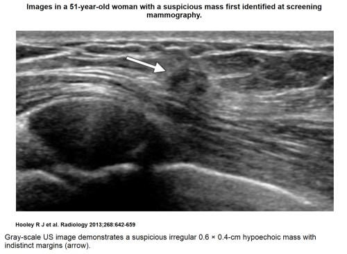
Picture5a

Picture5b

Elastography may be useful in improving the specificity of US evaluation of BI-RADS 3 and 4A lesions, including complicated cysts. Berg and colleagues (28) showed that by using qualitative shear-wave elastography and color assessment of lesion stiffness, oval shape, and a maximum elasticity value of less than 80 kPa, unnecessary biopsy of low-suspicion BI-RADS 4A masses could be reduced without a significant loss in sensitivity. Several investigators have proposed a variety of imaging classifications using strain elastography, mostly based on the color pattern (27,31,32). A “bull’s eye” artifact has also been described as a characteristic feature present in benign breast cysts, which may appear as a round or oval lesion with a stiff rim associated with two soft spots, one located centrally and the other posteriorly (33).

Despite these initial promising studies regarding the role of US elastography in the analysis of breast lesions, limitations do exist. Strain and shear-wave elastography are quite different methods of measuring breast tissue stiffness, and the application of these methods varies across different commercial manufacturers. Inter- and intraobserver variability may be relatively high because the elastogram may be affected by differences in degree and method of compression. With strain elastography, a quality indicator that is an associated color bar or numerical value may be helpful to ensure proper light compression. Shear-wave elastography has been shown to be less operator-dependent, as tissue compression is initiated by the US probe in a standard, reproducible fashion (34) and only light transducer pressure is necessary. In addition, there is currently no universal color-coding standard and, depending on the manufacturer and/or operator preference, stiff lesions may be arbitrarily coded to appear red while soft lesions appear blue, or vice versa. Some elastography features such as the “bull’s eye” artifact are only seen on specific US systems. Lesions deeper than 2 cm are less accurately characterized by means of elastography. Moreover, one must be aware that soft cancers and hard benign lesions exist. Therefore, careful correlation of elastography with B-mode US features and mammography is essential. Future studies and further technical advances, including the creation of more uniformity across different US manufacturers, will ultimately determine the usefulness of elastography in clinical practice.

Three-dimensional US

Both handheld and automated high-resolution linear 3D transducers are now available for use in breast imaging. With a single pass of the ultrasound beam, a 3D reconstructed image can be formed in the coronal, sagittal, and transverse planes, potentially allowing more accurate assessment of anatomic structures and tumor margins (Fig 6). Few studies regarding the performance of 3D US in the breast exist, but a preliminary study demonstrated improved characterization of malignant lesions (35). Automated supine whole-breast US using 3D technology is now widely available for use in the screening setting (see section on screening breast US). Three-dimensional US may also be used in addition to computed tomography for image-guided radiation therapy (36) and has a potential role in assessing tumor response to neoadjuvant chemotherapy.

Picture6


US Features of Benign and Malignant Breast Lesions

Cysts

Although for many years the main function of breast US was to differentiate cysts from solid masses, this differentiation can at times be problematic, particularly if the lesion is small or located deep in the breast. Simple cysts are defined as circumscribed, anechoic masses with a thin imperceptible wall and enhanced through transmission (provided spatial compounding is not used). By convention, simple cysts may also contain up to a single thin septation. Simple cysts are confidently characterized with virtually 100% accuracy at US (14,37), provided that they are not very small (< 5 mm in size) or not located in deep tissue. Complicated cysts are hypoechoic with no discernable Doppler flow, contain internal echoes, and may also exhibit indistinct margins, and/or lack posterior acoustic enhancement. Clustered microcysts consist of a cluster of tiny (<2–3 mm in size) anechoic foci with thin (< 0.5 mm in thickness) intervening septations.

Complicated cysts are very common sonographic findings and the majority are benign. In multiple studies, which evaluated over 1400 complicated cysts and microcysts, the malignancy rate ranged from 0% to 0.8% (38–44). Most complicated cysts and clustered microcysts with a palpable or mammographic correlate are classified as BI-RADS 3 and require short-interval imaging follow-up or, occasionally, US-guided aspiration. However, in the screening US setting, if multiple and bilateral complicated and simple cysts are present (ie, at least three cysts with at least one cyst in each breast), these complicated cysts can be assessed as benign, BI-RADS 2, requiring no additional follow-up (38).

Complicated cysts should never demonstrate internal vascularity at color Doppler interrogation. The presence of a solid component, mural nodule, thickened septation, or thickened wall within a cystic mass precludes the diagnosis of a benign complicated cyst. These complex masses require biopsy, as some cancers may have cystic components. The application of compound imaging and harmonics, color Doppler, and potentially elastography may help differentiate benign complicated cysts from malignant cystic-appearing masses and reduce the need for additional follow-up or biopsy.

Solid Masses

Sonographic features of benign-appearing solid masses include an oval or ellipsoid shape, “wider-than-tall” orientation parallel to the skin, circumscribed margins, gentle and smooth (less than three) lobulations, as well as absence of any malignant features (2,45) (Fig 2b). Lesions with these features are commonly fibroadenomas or other benign masses and can often be safely followed, even if the mass is palpable (46–48). Malignant features of solid masses include spiculations, angular margins, marked hypoechogenicity, posterior acoustic shadowing, microcalcifications, ductal extension, branching pattern, and 1–2-mm microlobulations (2,45) (Figs 1b,5, 6). These are also often taller-than-wide lesions with a nonparallel orientation to the skin and may occasionally be associated with thickened Cooper ligaments and/or or skin thickening. Most cancers have more than one malignant feature, spiculation being the most specific and angular margins the most common (2).

There is, however, considerable overlap between these benign and malignant US features and careful scanning technique, as well as direct correlation with mammography, is essential. For example, some high-grade invasive ductal carcinomas with central necrosis, as well as the well-differentiated mucinous and medullary subtypes, may present as circumscribed, oval, hypoechoic masses that may look like complicated cysts with low-level internal echoes at US. Benign focal fibrous breast tissue or postoperative scars can appear as irregular shadowing masses on US images. Furthermore, while echogenic lesions are often benign and frequently represent lipomas or fibrous tissue, echogenic cancers do rarely occur (Figs 7, 8) (49,50). The presence of a single malignant feature, despite the presence of multiple benign features, precludes a benign classification and mandates biopsy, with the exception of fat necrosis and postoperative scars exhibiting typical benign mammographic features. Likewise, a mass with a benign US appearance should be biopsied if it exhibits any suspicious mammographic features.

Picture7

Picture8


 Ductal Carcinoma in Situ

Ductal carcinoma in situ (DCIS) is characteristically associated with microcalcifications detected at mammography, but may also be detected at US since they are often associated with a subtle hypoechoic mass, which may indicate an invasive mammographically occult component. US features associated with DCIS most commonly include a hypoechoic mass with an irregular shape, microlobulated margins, no posterior acoustic features, and no internal vascularity. Ductal abnormalities, intracystic lesions, and architectural distortions may also be present (51–53). Noncalcified DCIS manifesting as a solid mass at US is more frequently found in non–high-grade than high-grade DCIS, which is more often associated with microcalcifications and ductal changes (54). US can depict microcalcifications, particularly those in clusters greater than 10 mm in size and located in a hypoechoic mass or a ductlike structure (Fig 9) (55). Malignant calcifications are more likely to be detected sonographically than are benign calcifications, which may be obscured by surrounding echogenic breast tissue (55,56). Although US is inferior to mammography in the detection of suspicious microcalcifications, the main benefit of US detection of DCIS is to identify the invasive component and guide biopsy procedures.

Picture9a

Picture9b


Breast US in Clinical Practice

Current indications for breast US as recommended by the American College of Radiology Practice Guidelines include the evaluation of palpable abnormalities or other breast symptoms, assessment of mammographic or MR imaging–detected abnormalities, and evaluation of breast implants (57). Additionally, US is routinely used for guidance during interventional procedures, treatment planning for radiation therapy, screening in certain groups of women, and evaluation of axillary lymph nodes. Much literature has been written on these uses and a comprehensive discussion is beyond the scope of this article. A few important and timely topics, however, will be reviewed.


BI-RADS US

The BI-RADS US lexicon was introduced in 2003, and subsequently, there have been several studies assessing the accuracy of BI-RADS US classification of breast lesions. Low to moderate interobserver agreement has been found in the description of margins (especially noncircumscribed margins), echogenicity, and posterior acoustic features. Abdullah et al (58) reported low interobserver agreement especially for small masses and for malignant masses. Given the importance of margin analysis in the characterization of benign and malignant lesions, this variability is potentially problematic. Studies have also shown variable results in the use of the final assessment categories. In clinical settings, Raza et al (46) showed inconsistent use of the BI-RADS 3 (probably benign) category in 14.0% of cases when biopsy was recommended. Abdullah et al also demonstrated fair and poor interobserver agreement for BI-RADS 4 (suspicious for malignancy) a, b, and c subcategories (58). However, Henig et al (59) reported more promising results, with malignancy rates in categories 3, 4, and 5 to be similar to those seen with mammographic categorization (1.2%, 17%, and 94%, respectively).


 Evaluation of Mammographic Findings

Targeted US is complementary to diagnostic mammography because of its ability to differentiate cystic and solid lesions.US is also useful in the work up of subtle asymmetries, as it can help identify or exclude the presence of an underlying mass. True hypoechoic lesions can often be differentiated from prominent fat lobules by scanning in multiple planes, because true lesions usually do not blend or elongate into adjacent tissue. With the introduction of digital breast tomosynthesis for mammographic imaging, US will play yet another important role. As mammographic lesions can often be detected, localized, and have adequate margin assessment on 3D images, patients with lesions detected on digital breast tomosynthesis images at screening may often be referred directly to US, avoiding additional mammographic imaging and its associated costs and radiation exposure (Fig 10). This will place an even greater importance on high-quality US.

Picture10a

Picture10b

Picture10c


Evaluation of the Symptomatic Patient:Palpable Masses, Breast Pain, and Nipple Discharge

US is essential in the evaluation of patients with the common clinical complaint of either a palpable mass or focal persistent breast pain. Unlike focal breast pain, which may be occasionally associated with benign or malignant lesions, diffuse breast pain (bilateral or unilateral), as well as cyclic breast pain, requires only clinical follow-up, as it is usually physiologic with an extremely low likelihood of malignancy (60,61). In patients with isolated focal breast pain, the role of sonography may be limited to patient reassurance (61). In women younger than 30 years of age, with a palpable lump or focal breast pain, US is the primary imaging test, with a sensitivity and negative predictive value of nearly 100% (62). Symptomatic women older than 30 years usually require both US and mammography, and in these patients, the negative predictive value approaches 100% (63,64). Lehman et al (65) demonstrated that in symptomatic women aged 30–39 years, the risk of malignancy was 1.9% and the added value of adjunct mammography in addition to US was low. Identification of a benign-appearing solid lesion at US in a symptomatic woman can negate the need for needle biopsy, as many of these masses can safely be monitored with short-interval follow-up US (46–48), usually performed at 6 months. A suspicious mass identified at US can promptly undergo biopsy with US guidance.

US can also be used as an alternative or an addition to ductography in patients who present with unilateral, spontaneous bloody, clear, or serosanguinous nipple discharge (66). Among women with worrisome nipple discharge, ductography can demonstrate an abnormality in 59%–82% of women (67,68), MR imaging may demonstrate a suspicious abnormality in 34% of women (68), and US has been shown to demonstrate a subareolar mass or an intraductal mass or filling defect in up to 14% of women (67). If US can be used to identify a retroareolar mass or an intraductal mass, US-guided biopsy can be performed and ductography may be avoided (Fig 11). US may be limited, however, as small peripherally located intraductal masses or masses without an associated dilated duct may not be identified. Therefore, galactography, MR imaging, and/or major duct excision may still be necessary in the symptomatic patient with a negative US examination.

Picture11

Finally, in the pregnant or lactating patient who presents with a palpable breast mass, focal breast pain, or bloody nipple discharge, US is also the initial imaging modality of choice. Targeted US examination in these patients can be used to identify most benign and malignant masses, including fibroadenomas, galactocoeles, lactating adenomas, abscesses, and invasive carcinomas. In a recent study by Robbins et al (69), a negative predictive value of 100% was found among 122 lesions evaluated with US in lactating, pregnant, or postpartum women. This is much higher than the pregnancy-associated breast cancer sensitivity of mammography, which has been reported in the range of 78%–87% (70,71). The diminished sensitivity of mammography is likely due to increased parenchymal density seen in these patients. However, since lactating breast parenchyma is more echogenic than most breast masses, hypoechoic breast cancers are more readily detected at US in pregnant patients.


Supplemental Screening Breast US

Because of the known limitations of mammography, particularly in women with dense breast tissue, supplemental screening with whole-breast US, in addition to mammography, is increasingly gaining widespread acceptance. Numerous independent studies have demonstrated that the addition of a single screening or whole-breast US examination in women with dense breast tissue at mammography will yield an additional 2.3–4.6 mammographically occult cancers per 1000 women (72–80). Mammographically occult cancers detected on US images are generally small node-negative invasive cancers (Fig 12) (81). However, few studies have investigated the performance of incident screening breast US, and the optimal screening US interval is unknown. Berg and colleagues (82) recently demonstrated that incident annual supplemental screening US in intermediate- and high-risk women with mammographically dense breast tissue enabled detection of an additional 3.7 cancers per 1000 women screened.

Picture12

Handheld screening breast US is highly operator-dependent and the majority of screening breast US studies have relied on physician-performed examinations. As per the ACRIN 6666 protocol, a normal screening US examination should consist of a minimum of one image in each quadrant and one behind the nipple (83). Two studies have also demonstrated that technologist-performed handheld screening breast US can achieve similar cancer detection rates (76,78).

Automated whole-breast US is a recently developed alternative to traditional handheld screening breast US, in which standardized, uniform image sets may be readily obtained by a nonradiologist. Automated whole-breast US systems may utilize a standard US unit and a linear-array transducer attached to a computer-guided mechanical arm or a dedicated screening US unit with a 15-cm wide transducer (84,85). With these systems, over 3000 overlapping sagittal, transverse, and coronal images are obtained and available for later review by the radiologist, with associated 3D reconstruction. The advantages include less operator dependence, increased radiologist efficiency, and increased reproducibility, which could aid in follow-up of lesions.

A multi-institutional study has shown that supplemental automated whole-breast US can depict an additional 3.6 cancer per 1000 women screened, similar to physician-performed handheld screening US (85). However, disadvantages include the limited ability to scan the entire breast, particularly posterior regions in large breasts, time-consuming review of a large number of images by the radiologist, and the need to recall patients for a second US examination to re-evaluate indeterminate findings. Moreover, few investigators have compared the use of handheld with automated breast US screening. A single small recent study by Chang et al (86) demonstrated that of 14 cancers initially detected at handheld screening, only 57%–79% were also detected by three separate readers on automated whole-breast US images, with the two cancers missed by all three readers at automated whole-breast US, each less than 1 cm in size.

The use of supplemental screening breast US, performed in addition to mammography, remains controversial despite proof of the ability to detect small mammographically occult cancers. US has limited value for the detection of small clustered microcalcifications without an associated mass lesion. Low positive predictive values of biopsies performed of less than 12% have been consistently reported (77,87). No outcome study has been able to demonstrate a direct decrease in patient mortality due to the detection of these additional small and mammographically occult cancers. This would require a long, randomized screening trial, which is not feasible. Rationally, however, the early detection and treatment of additional small breast cancers should improve outcomes and reduce overall morbidity and mortality. Many insurance companies will not reimburse for screening breast US and historically, this examination has not been widely accepted in the United States.

Nevertheless, because of both the known efficacy of supplemental screening breast US and overall increased breast cancer awareness, more patients and clinicians are requesting this examination. In fact, some states now mandate that radiologists inform women of their breast density and advise them to discuss supplemental screening with their doctors. Although supplemental screening breast MR imaging is usually preferred for women who are at very high risk for breast cancer (ie, women with a lifetime risk of over 20%, for example those women who are BRCA positive or have multiple first-degree relatives with a history of premenopausal breast cancer), screening breast US should be considered in women at very high risk for breast cancer who cannot tolerate breast MR imaging, as well as those women with dense breast tissue and intermediate risk (ie, lifetime risk of 15%–20%, for example those women whose only risk factor is a personal history of breast cancer or previous biopsy of a high-risk lesion), or even average risk. Future studies are needed to establish strategies to reduce false-positive results and continue to optimize both technologist-performed handheld screening US and automated whole-breast US in women with mammographically dense breast tissue.


 Use of US for MR Imaging–depicted Abnormalities

MR imaging of the breast is now an integral part of breast imaging, most commonly performed to screen high-risk women and to further assess the stage in patients with newly diagnosed breast cancers. While MR has a higher sensitivity than mammography for detecting breast cancer, the specificity is relatively low (88). Lesions detected on MR images are often mammographically occult, but many can be detected with targeted US (Fig 13). Besides further US characterization of an MR imaging–detected lesion, US may be used to guide intervention for lesions initially detected at MR imaging. US-guided biopsies are considerably less expensive, less time consuming, and more comfortable for the patient than MR imaging–guided biopsies.

Picture13a

Picture13b

Some suspicious lesions detected at MR imaging will represent invasive ductal or lobular cancers, but many may prove to be intraductal disease, which can be challenging to detect at US. Meticulous scanning technique is required for an MR imaging–directed US examination, with knowledge of subtle sonographic signs and close correlation with the MR imaging findings and location. Precontrast T1 images are helpful to facilitate localization of lesions in relation to fibroglandular tissue (89). Because MR imaging abnormalities tend to be vascular, increased vascularity may also assist in detection of a subtle sonographic correlate (90). Having the MR images available for simultaneous review while performing the US examination will ideally permit such associative correlation. At the authors’ facility, computer monitors displaying images from the picture archiving and communication system are available in all US rooms for this purpose.

Recent studies have shown that 46%–71% of lesions at MR imaging can be detected with focused US (90–94). Enhancing masses detected on MR images are identified on focused US images in 58%–65% of cases compared with nonmass enhancement, which is identified on focused US images in only 12%–32% of cases (90–92). Some studies have shown that US depiction of an MR imaging correlate was independent of size (91,93,95). However, Meissnitzer et al (92) showed that size dependence is also important: For masses 5 mm or smaller, only 50% were seen, versus 56% for masses 6–10 mm, 73% for masses 11–15 mm, and 86% for masses larger than 15 mm. Likewise, this study also demonstrated that for nonmass lesions, a US correlate was found for 13% of those measuring 6–10 mm, 25% of those 11–15 mm, and 42% of those larger than 15 mm (92). In addition, many of these studies determined that when a sonographic correlate was discovered, the probability of malignancy was increased (90–92). Since typical US malignant features such as spiculation and posterior shadowing may be absent and the pretest probability is higher for MR imaging–detected lesions, a lower threshold for biopsy should be considered when performing MR imaging–directed US compared with routine targeted US (90) or screening US.

Because lesions are often very subtle at MR-directed US examination and because of differences in patient positioning during the two examinations, careful imaging–histologic correlation is required when performing US-guided biopsy of MR imaging–detected abnormalities. For lesions sampled with a vacuum-assisted device and US guidance, Sakamoto et al (96) found a higher rate of false-negative biopsy results for MR imaging–detected lesions than for US-detected lesions, suggesting that precise US-MR imaging correlation may not have occurred. Meissnitzer et al (92) showed that although 91% of MR imaging–detected lesions had an accurate US correlate, 9% were found to be inaccurate. With ever-improving techniques and experience in breast US, the US visualization of MR imaging–detected abnormalities will likely continue to improve. Nevertheless, if a suspicious lesion is not identified sonographically, MR imaging–guided biopsy should still be performed, because the malignancy rate of sonographically occult MR imaging–detected lesions has been shown to range from 14% to 22% (91,95).


Preoperative Staging of Cancer with US

Breast MR imaging has been shown to be more sensitive than US in the detection of additional foci of mammographically occult disease in women with newly diagnosed breast cancer (97–99). Nevertheless, when a highly suspicious mass is identified at mammography and US, immediate US evaluation of the remainder of the ipsilateral breast, the contralateral breast, and the axilla should be considered. If additional lesions are identified, preoperative staging with MR imaging can be avoided and US-guided biopsy can be promptly performed, saving the patient valuable time and expense (100). In a study by Moon et al (101), of 201 patients with newly diagnosed breast cancer, staging US demonstrated mammographically occult multifocal or multicentric disease in 28 patients (14%) and contralateral breast cancers in eight patients (4%), resulting in a change in therapy in 32 patients (16%).

US can also be used to identify abnormal axillary, supraclavicular, and internal mammary lymph nodes. Abnormal lymph nodes characteristically demonstrate focal or diffuse cortical thickening (≥3 mm in thickness), a round (rather than oval or reniform) shape, loss of the echogenic fatty hilum and/or nonhilar, disorganized, irregular blood vessels (102,103) (Fig 14). A positive US-guided CNB or fine-needle aspiration of a clinically abnormal axillary lymph node in a patient with a known breast cancer can aid patient management, by avoiding the need for sentinel node biopsy and allowing the patient instead to proceed directly to axillary lymph node dissection or neoadjuvant chemotherapy.

Picture14


 Interventional Breast US

US-guided interventional procedures have increased in volume in recent years and US is now the primary biopsy guidance technique used in many breast imaging centers. Most palpable lesions, as well as lesions detected at mammography, MR imaging, or screening US, can be sampled with US. With current high-resolution transducers, even suspicious intraductal microcalcifications may be detected and sampled.

While US-guided procedures require technical skills that must be developed and can be challenging, once mastered this technique allows precise real-time sampling of the lesion, which is not possible with either stereotactic or MR imaging–guided procedures. US-guided procedures do not require ionizing radiation or intravenous contrast material. US procedures are more tolerable for patients than stereotactic (104) or MR imaging–guided procedures because US-guided procedures are faster and more comfortable, as breast compression and uncomfortable biopsy coils or tables are not necessary and the procedure may be performed with the patient supine (104–106).

Most literature has shown that automated 14-gauge CNB devices are adequate for the majority of US-guided biopsies (107–115). Image-guided CNB is preferable to fine-needle aspiration cytology of breast masses because of superior sensitivity, specificity, and diagnostic accuracy (116). DCIS, malignant invasion, and hormone receptor status of invasive breast cancers can be determined with CNB samples, but not with fine-needle aspiration cytology. Fine-needle aspiration may be performed, however, in complicated cysts and symptomatic simple cysts. In these cases, the cyst aspirate fluid can often be discarded; cytology is usually only necessary if the fluid is frankly bloody (117).

The choice of performing fine-needle aspiration or CNB of a suspicious axillary lymph node depends on radiologist preference and the availability of an experienced cytopathologist, although CNB is usually more accurate than fine-needle aspiration biopsy (118,119). Fine-needle aspiration may be preferred for suspicious deep lymph nodes in proximity to the axillary vessels, whereas CNB may be preferred in large nodes with thickened cortices, particularly if determination of hormone receptor status or immunohistochemistry is desired, since more tissue is required for these assays. If lymphoma is suspected, a core should be placed in saline and also in conventional formalin.

While the underestimation rate of malignancy can be considerable for high-risk lesions such as atypical hyperplasia, such histology is not commonly found in lesions undergoing US-guided CNB. Multiple studies have shown a false-negative rate for US CNB biopsy of around 2%–3% (107–115). Although the contiguous and larger samples obtained with a vacuum-assisted biopsy device undoubtedly reduce sampling error, the vacuum-assisted biopsy is a more expensive and more invasive procedure (109). In the authors’ experience, vacuum-assisted US biopsy is to be considered for small masses, intraductal or intracystic lesions, or lesions with subtle microcalcifications. These may be difficult to adequately sample with a spring-loaded automatic firing device. Alternatively, for more accurate sampling of such challenging cases, as well as some axillary lymph nodes and masses smaller than 1 cm in size, automated CNB needles designed to place the inner trough of the needle within a lesion before firing can be utilized (Fig 15). With this technique, the sampling trough of the CNB needle can be clearly visualized within the lesion before the overlying outer sheath is fired. Regardless of needle choice, a postbiopsy clip marker should be placed followed by a postbiopsy mammogram to document clip position. This will assist with follow-up imaging, facilitating mammography and/or MR imaging correlation.

Picture15a


Picture15b

There has been recent interest in the percutaneous removal of benign breast lesions by using US-guided vacuum-assisted biopsy. While in general, proved benign concordant lesions can safely remain in the breast, some patients desire removal. Percutaneous US-guided removal with a vacuum-assisted biopsy device can replace surgical removal in some cases, particularly for small lesions (1 cm in size or less). Several reports have shown promising results demonstrating rates of complete lesion excision, varying from 61% to 94% (120–124). Dennis et al (125) demonstrated that vacuum-assisted US-guided biopsy could be used to excise intraductal lesions resulting in resolution of problematic nipple discharge in 97% of patients. Even on long-term follow-up, most studies show low rates of residual masses, more commonly observed in larger fibroadenomas.


 Intraoperative Breast US

The use of two-dimensional and 3D intraoperative US may decrease the incidence of positive margins and decrease re-excision rates (126–130) particularly in the setting of lumpectomy for palpable cancers, when US is used to assess the adequacy of surgical margins to determine the need for additional tissue removal. Similarly, intraoperative US has also been utilized to improve detection and removal of metastatic lymph nodes during sentinel lymph node assessment (131).


Future Directions

Intravenous US microbubble contrast agents have been used to enhance US diagnosis by means of analysis, enhancement patterns, the rates of uptake and washout, and identification of tumor angiogenesis. In addition, preliminary research has shown that intravenous US contrast agents may be able to depict tissue function with the potential to deliver targeted gene therapy to selected tumor cells (132). However, there are currently no intravenous US contrast agents approved for use in breast imaging by the U.S. Food and Drug Administration. Other potential advances in breast US include fusion imaging, which involves the direct overlay of correlative MR imaging with targeted US. Another evolving area is that of US computer-aided detection, which may be of particular benefit when combined with automated whole-breast screening US.


 Summary

Technical advances in US now allow comprehensive US diagnosis, management, and treatment of breast lesions. Optimal use of US technology, meticulous scanning technique with careful attention to lesion morphology, and recognition and synthesis of findings from multiple imaging modalities are essential for optimal patient management. In the future, as radiologists utilize US for an ever-increasing scope of indications, become aware of the more subtle sonographic findings of breast cancer, and apply newly developing tools, the value of breast US will likely continue to increase and evolve.


Essentials

  • • Breast US is operator dependent; knowledge and understanding of the various technical options currently available are important for image optimization and accurate diagnosis.
  • • US is an interactive, dynamic modality and real-time scanning is necessary to assess subtle findings associated with malignancy.
  • • Ability to synthesize the information obtained from the breast US examination with concurrent mammography, MR imaging, and clinical breast examination is necessary for accurate diagnosis.
  • • The use of screening breast US in addition to mammography, particularly in women with dense breast tissue, is becoming more widely accepted in the United States.
  • • Breast US guidance is the primary biopsy method used in most breast imaging practices, and the radiologist should be familiar with various biopsy devices and techniques to adequately sample any breast mass identified at US.


Disclosures of Conflicts of Interest: R.J.H. No relevant conflicts of interest to disclose. L.M.S. Financial activities related to the present article: none to disclose. Financial activities not related to the present article: educational consultant in vascular US to Philips Healthcare; payment for lectures on breast US from Educational Symposia; payment for development of educational presentations from Philips Healthcare. Other relationships: none to disclose. L.E.P. Financial activities related to the present article: none to disclose. Financial activities not related to the present article: consultant to Hologic. Other relationships: none to disclose.

Abbreviations:

BI-RADS = Breast Imaging and Reporting Data System

CNB = core needle biopsy

DCIS = ductal carcinoma in situ

3D = three dimensional

References

  1. ↵
    1. Dempsey PJ

    . The history of breast ultrasound. J Ultrasound Med2004;23(7):887–894.

  2. ↵
    1. Stavros AT,
    2. Thickman D,
    3. Rapp CL,
    4. Dennis MA,
    5. Parker SH,
    6. Sisney GA

    . Solid breast nodules: use of sonography to distinguish between benign and malignant lesions. Radiology 1995;196(1):123–134.

  3. ↵
    1. Mainiero MB,
    2. Goldkamp A,
    3. Lazarus E,
    4. et al

    . Characterization of breast masses with sonography: can biopsy of some solid masses be deferred? J Ultrasound Med 2005;24(2):161–167.

  4. ↵
    1. Graf O,
    2. Helbich TH,
    3. Hopf G,
    4. Graf C,
    5. Sickles EA

    . Probably benign breast masses at US: is follow-up an acceptable alternative to biopsy? Radiology2007;244(1):87–93.

  5. ↵
    1. Mendelson EB,
    2. Baum JK,
    3. Berg WA,
    4. Merritt CR,
    5. Rubin E

    . Breast Imaging Reporting Data System. BI-RADS: Ultrasound. Reston, Va: American College of Radiology, 2003.

  6. ↵
    1. Berg WA,
    2. Blume JD,
    3. Cormack JB,
    4. Mendelson EB

    . Training the ACRIN 6666 Investigators and effects of feedback on breast ultrasound interpretive performance and agreement in BI-RADS ultrasound feature analysis. AJR Am J Roentgenol 2012;199(1):224–235.

  7. ↵
    1. Stafford RJ,
    2. Whitman GJ

    . Ultrasound physics and technology in breast imaging. Ultrasound Clin 2011;6(3):299–312.

  8. ↵
    1. Weinstein SP,
    2. Conant EF,
    3. Sehgal C

    . Technical advances in breast ultrasound imaging. Semin Ultrasound CT MR 2006;27(4):273–283.

  9. ↵
    1. Athanasiou A,
    2. Tardivon A,
    3. Ollivier L,
    4. Thibault F,
    5. El Khoury C,
    6. Neuenschwander S

    . How to optimize breast ultrasound. Eur J Radiol2009;69(1):6–13.

  10. ↵
    1. Kremkau FW

    . Sonography principles and instruments. 8th ed. St Louis, Mo: Elsevier-Saunders, 2011.

  11. ↵
    1. Goss SA,
    2. Johnston RL,
    3. Dunn F

    . Comprehensive compilation of empirical ultrasonic properties of mammalian tissues. J Acoust Soc Am1978;64(2):423–457.

  12. ↵
    1. Napolitano D,
    2. Chou CH,
    3. McLaughlin G,
    4. et al

    . Sound speed correction in ultrasound imaging. Ultrasonics 2006;44(Suppl 1):e43–e46.

  13. ↵
    1. Barr RG,
    2. Rim A,
    3. Graham R,
    4. Berg W,
    5. Grajo JR

    . Speed of sound imaging: improved image quality in breast sonography. Ultrasound Q2009;25(3):141–144.

  14. ↵
    1. Stavros AT

    . Breast ultrasound. Philadelphia, Pa: Lippincott, Williams & Wilkins, 2004.

  15. ↵
    1. Cosgrove DO,
    2. Kedar RP,
    3. Bamber JC,
    4. et al

    . Breast diseases: color Doppler US in differential diagnosis. Radiology 1993;189(1):99–104.

  16. ↵
    1. Sehgal CM,
    2. Arger PH,
    3. Rowling SE,
    4. Conant EF,
    5. Reynolds C,
    6. Patton JA

    .Quantitative vascularity of breast masses by Doppler imaging: regional variations and diagnostic implications. J Ultrasound Med 2000;19(7):427–440;quiz 441–442.

  17. ↵
    1. Birdwell RL,
    2. Ikeda DM,
    3. Jeffrey SS,
    4. Jeffrey RB Jr.

    . Preliminary experience with power Doppler imaging of solid breast masses. AJR Am J Roentgenol1997;169(3):703–707.

  18. ↵
    1. Gokalp G,
    2. Topal U,
    3. Kizilkaya E

    . Power Doppler sonography: anything to add to BI-RADS US in solid breast masses? Eur J Radiol 2009;70(1):77–85.

  19. ↵
    1. Tozaki M,
    2. Fukuma E

    . Does power Doppler ultrasonography improve the BI-RADS category assessment and diagnostic accuracy of solid breast lesions?Acta Radiol 2011;52(7):706–710.

  20. ↵
    1. Mehta TS,
    2. Raza S,
    3. Baum JK

    . Use of Doppler ultrasound in the evaluation of breast carcinoma. Semin Ultrasound CT MR 2000;21(4):297–307.

  21. ↵
    1. Horvath E,
    2. Silva C,
    3. Fasce G,
    4. et al

    . Parallel artery and vein: sign of benign nature of breast masses. AJR Am J Roentgenol 2012;198(1):W76–W82.

  22. ↵
    1. Campbell SC,
    2. Cullinan JA,
    3. Rubens DJ

    . Slow flow or no flow? Color and power Doppler US pitfalls in the abdomen and pelvis. RadioGraphics2004;24(2):497–506.

  23. ↵
    1. Schaefer FK,
    2. Heer I,
    3. Schaefer PJ,
    4. et al

    . Breast ultrasound elastography: results of 193 breast lesions in a prospective study with histopathologic correlation. Eur J Radiol 2011;77(3):450–456.

  24. ↵
    1. Zhao QL,
    2. Ruan LT,
    3. Zhang H,
    4. Yin YM,
    5. Duan SX

    . Diagnosis of solid breast lesions by elastography 5-point score and strain ratio method. Eur J Radiol2012;81(11):3245–3249.

  25. ↵
    1. Stachs A,
    2. Hartmann S,
    3. Stubert J,
    4. et al

    . Differentiating between malignant and benign breast masses: factors limiting sonoelastographic strain ratio.Ultraschall Med 2013;34(2):131–136.

  26. ↵
    1. Bercoff J,
    2. Tanter M,
    3. Fink M

    . Supersonic shear imaging: a new technique for soft tissue elasticity mapping. IEEE Trans Ultrason Ferroelectr Freq Control2004;51(4):396–409.

  27. ↵
    1. Itoh A,
    2. Ueno E,
    3. Tohno E,
    4. et al

    . Breast disease: clinical application of US elastography for diagnosis. Radiology 2006;239(2):341–350.

  28. ↵
    1. Berg WA,
    2. Cosgrove DO,
    3. Doré CJ,
    4. et al

    . Shear-wave elastography improves the specificity of breast US: the BE1 multinational study of 939 masses.Radiology 2012;262(2):435–449.

  29. ↵
    1. Athanasiou A,
    2. Tardivon A,
    3. Tanter M,
    4. et al

    . Breast lesions: quantitative elastography with supersonic shear imaging—preliminary results. Radiology2010;256(1):297–303.

  30. ↵
    1. Evans A,
    2. Whelehan P,
    3. Thomson K,
    4. et al

    . Invasive breast cancer: relationship between shear-wave elastographic findings and histologic prognostic factors.Radiology 2012;263(3):673–677.

  31. ↵
    1. Fleury Ede F,
    2. Fleury JC,
    3. Piato S,
    4. Roveda D Jr.

    . New elastographic classification of breast lesions during and after compression. Diagn Interv Radiol 2009;15(2):96–103.

  32. ↵
    1. Tozaki M,
    2. Fukuma E

    . Pattern classification of ShearWave™ Elastography images for differential diagnosis between benign and malignant solid breast masses. Acta Radiol 2011;52(10):1069–1075.

  33. ↵
    1. Barr RG,
    2. Lackey AE

    . The utility of the “bull’s-eye” artifact on breast elasticity imaging in reducing breast lesion biopsy rate. Ultrasound Q2011;27(3):151–155.

  34. ↵
    1. Cosgrove DO,
    2. Berg WA,
    3. Doré CJ,
    4. et al

    . Shear wave elastography for breast masses is highly reproducible. Eur Radiol 2012;22(5):1023–1032.

  35. ↵
    1. Kalmantis K,
    2. Dimitrakakis C,
    3. Koumpis C,
    4. et al

    . The contribution of three-dimensional power Doppler imaging in the preoperative assessment of breast tumors: a preliminary report. Obstet Gynecol Int 2009;2009:530579.

  36. ↵
    1. Chadha M,
    2. Young A,
    3. Geraghty C,
    4. Masino R,
    5. Harrison L

    . Image guidance using 3D-ultrasound (3D-US) for daily positioning of lumpectomy cavity for boost irradiation. Radiat Oncol 2011;6:45.

  37. ↵
    1. Hilton SV,
    2. Leopold GR,
    3. Olson LK,
    4. Willson SA

    . Real-time breast sonography: application in 300 consecutive patients. AJR Am J Roentgenol1986;147(3):479–486.

  38. ↵
    1. Berg WA,
    2. Sechtin AG,
    3. Marques H,
    4. Zhang Z

    . Cystic breast masses and the ACRIN 6666 experience. Radiol Clin North Am 2010;48(5):931–987.

    1. Kolb TM,
    2. Lichy J,
    3. Newhouse JH

    . Occult cancer in women with dense breasts: detection with screening US—diagnostic yield and tumor characteristics.Radiology 1998;207(1):191–199.

    1. Buchberger W,
    2. DeKoekkoek-Doll P,
    3. Springer P,
    4. Obrist P,
    5. Dünser M

    . Incidental findings on sonography of the breast: clinical significance and diagnostic workup. AJR Am J Roentgenol 1999;173(4):921–927.

    1. Berg WA,
    2. Campassi CI,
    3. Ioffe OB

    . Cystic lesions of the breast: sonographic-pathologic correlation. Radiology 2003;227(1):183–191.

    1. Chang YW,
    2. Kwon KH,
    3. Goo DE,
    4. Choi DL,
    5. Lee HK,
    6. Yang SB

    . Sonographic differentiation of benign and malignant cystic lesions of the breast. J Ultrasound Med 2007;26(1):47–53.

    1. Daly CP,
    2. Bailey JE,
    3. Klein KA,
    4. Helvie MA

    . Complicated breast cysts on sonography: is aspiration necessary to exclude malignancy? Acad Radiol2008;15(5):610–617.

  39. ↵
    1. Venta LA,
    2. Kim JP,
    3. Pelloski CE,
    4. Morrow M

    . Management of complex breast cysts. AJR Am J Roentgenol 1999;173(5):1331–1336.

  40. ↵
    1. Hong AS,
    2. Rosen EL,
    3. Soo MS,
    4. Baker JA

    . BI-RADS for sonography: positive and negative predictive values of sonographic features. AJR Am J Roentgenol2005;184(4):1260–1265.

  41. ↵
    1. Raza S,
    2. Chikarmane SA,
    3. Neilsen SS,
    4. Zorn LM,
    5. Birdwell RL

    . BI-RADS 3, 4, and 5 lesions: value of US in management—follow-up and outcome. Radiology2008;248(3):773–781.

    1. Harvey JA,
    2. Nicholson BT,
    3. Lorusso AP,
    4. Cohen MA,
    5. Bovbjerg VE

    . Short-term follow-up of palpable breast lesions with benign imaging features: evaluation of 375 lesions in 320 women. AJR Am J Roentgenol 2009;193(6):1723–1730.

  42. ↵
    1. Graf O,
    2. Helbich TH,
    3. Fuchsjaeger MH,
    4. et al

    . Follow-up of palpable circumscribed noncalcified solid breast masses at mammography and US: can biopsy be averted? Radiology 2004;233(3):850–856.

  43. ↵
    1. Linda A,
    2. Zuiani C,
    3. Lorenzon M,
    4. et al

    . Hyperechoic lesions of the breast: not always benign. AJR Am J Roentgenol 2011;196(5):1219–1224.

  44. ↵
    1. Soon PS,
    2. Vallentine J,
    3. Palmer A,
    4. Magarey CJ,
    5. Schwartz P,
    6. Morris DL

    .Echogenicity of breast cancer: is it of prognostic value? Breast2004;13(3):194–199.

  45. ↵
    1. Moon WK,
    2. Myung JS,
    3. Lee YJ,
    4. Park IA,
    5. Noh DY,
    6. Im JG

    . US of ductal carcinoma in situ. RadioGraphics 2002;22(2):269–280; discussion 280–281.

    1. Yang WT,
    2. Tse GM

    . Sonographic, mammographic, and histopathologic correlation of symptomatic ductal carcinoma in situ. AJR Am J Roentgenol2004;182(1):101–110.

  46. ↵
    1. Izumori A,
    2. Takebe K,
    3. Sato A

    . Ultrasound findings and histological features of ductal carcinoma in situ detected by ultrasound examination alone. Breast Cancer 2010;17(2):136–141.

  47. ↵
    1. Park JS,
    2. Park YM,
    3. Kim EK,
    4. et al

    . Sonographic findings of high-grade and non-high-grade ductal carcinoma in situ of the breast. J Ultrasound Med2010;29(12):1687–1697.

  48. ↵
    1. Moon WK,
    2. Im JG,
    3. Koh YH,
    4. Noh DY,
    5. Park IA

    . US of mammographically detected clustered microcalcifications. Radiology 2000;217(3):849–854.

  49. ↵
    1. Soo MS,
    2. Baker JA,
    3. Rosen EL

    . Sonographic detection and sonographically guided biopsy of breast microcalcifications. AJR Am J Roentgenol2003;180(4):941–948.

  50. ↵
    ACR Practice Guideline for the Performance of a Breast Ultrasound Examination. American College of Radiology. http://www.acr.org/Quality-Safety/Standards-Guidelines./Practice-Guidelines-by-Modality/Ultrasound. Published 2011.
  51. ↵
    1. Abdullah N,
    2. Mesurolle B,
    3. El-Khoury M,
    4. Kao E

    . Breast imaging reporting and data system lexicon for US: interobserver agreement for assessment of breast masses. Radiology 2009;252(3):665–672.

  52. ↵
    1. Heinig J,
    2. Witteler R,
    3. Schmitz R,
    4. Kiesel L,
    5. Steinhard J

    . Accuracy of classification of breast ultrasound findings based on criteria used for BI-RADS.Ultrasound Obstet Gynecol 2008;32(4):573–578.

  53. ↵
    1. Mansel R

    . Management of breast pain. In: Harris JR, Lippman ME, MorrowM, Osborne CK, eds. Diseases of the breast. 4th ed. Philadelphia, Pa:Lippincott Williams & Wilkins, 2010; 52.

  54. ↵
    1. Leung JW,
    2. Kornguth PJ,
    3. Gotway MB

    . Utility of targeted sonography in the evaluation of focal breast pain. J Ultrasound Med 2002;21(5):521–526; quiz 528–529.

  55. ↵
    1. Loving VA,
    2. DeMartini WB,
    3. Eby PR,
    4. Gutierrez RL,
    5. Peacock S,
    6. Lehman CD

    .Targeted ultrasound in women younger than 30 years with focal breast signs or symptoms: outcomes analyses and management implications. AJR Am J Roentgenol 2010;195(6):1472–1477.

  56. ↵
    1. Soo MS,
    2. Rosen EL,
    3. Baker JA,
    4. Vo TT,
    5. Boyd BA

    . Negative predictive value of sonography with mammography in patients with palpable breast lesions. AJR Am J Roentgenol 2001;177(5):1167–1170.

  57. ↵
    1. Tumyan L,
    2. Hoyt AC,
    3. Bassett LW

    . Negative predictive value of sonography and mammography in patients with focal breast pain. Breast J2005;11(5):333–337.

  58. ↵
    1. Lehman CD,
    2. Lee CI,
    3. Loving VA,
    4. Portillo MS,
    5. Peacock S,
    6. DeMartini WB

    .Accuracy and value of breast ultrasound for primary imaging evaluation of symptomatic women 30-39 years of age. AJR Am J Roentgenol2012;199(5):1169–1177.

  59. ↵
    1. Ballesio L,
    2. Maggi C,
    3. Savelli S,
    4. et al

    . Role of breast magnetic resonance imaging (MRI) in patients with unilateral nipple discharge: preliminary study.Radiol Med (Torino) 2008;113(2):249–264.

  60. ↵
    1. Sabel MS,
    2. Helvie MA,
    3. Breslin T,
    4. et al

    . Is duct excision still necessary for all cases of suspicious nipple discharge? Breast J 2012;18(2):157–162.

  61. ↵
    1. Morrogh M,
    2. Morris EA,
    3. Liberman L,
    4. Borgen PI,
    5. King TA

    . The predictive value of ductography and magnetic resonance imaging in the management of nipple discharge. Ann Surg Oncol 2007;14(12):3369–3377.

  62. ↵
    1. Robbins J,
    2. Jeffries D,
    3. Roubidoux M,
    4. Helvie M

    . Accuracy of diagnostic mammography and breast ultrasound during pregnancy and lactation. AJR Am J Roentgenol 2011;196(3):716–722.

  63. ↵
    1. Liberman L,
    2. Giess CS,
    3. Dershaw DD,
    4. Deutch BM,
    5. Petrek JA

    . Imaging of pregnancy-associated breast cancer. Radiology 1994;191(1):245–248.

  64. ↵
    1. Ahn BY,
    2. Kim HH,
    3. Moon WK,
    4. et al

    . Pregnancy- and lactation-associated breast cancer: mammographic and sonographic findings. J Ultrasound Med2003;22(5):491–497; quiz 498–499.

  65. ↵
    1. Kolb TM,
    2. Lichy J,
    3. Newhouse JH

    . Comparison of the performance of screening mammography, physical examination, and breast US and evaluation of factors that influence them: an analysis of 27,825 patient evaluations. Radiology 2002;225(1):165–175.

    1. Buchberger W,
    2. Niehoff A,
    3. Obrist P,
    4. DeKoekkoek-Doll P,
    5. Dünser M

    . Clinically and mammographically occult breast lesions: detection and classification with high-resolution sonography. Semin Ultrasound CT MR 2000;21(4):325–336.

    1. Crystal P,
    2. Strano SD,
    3. Shcharynski S,
    4. Koretz MJ

    . Using sonography to screen women with mammographically dense breasts. AJR Am J Roentgenol2003;181(1):177–182.

    1. Gordon PB,
    2. Goldenberg SL

    . Malignant breast masses detected only by ultrasound: a retrospective review. Cancer 1995;76(4):626–630.

  66. ↵
    1. Kaplan SS

    . Clinical utility of bilateral whole-breast US in the evaluation of women with dense breast tissue. Radiology 2001;221(3):641–649.

  67. ↵
    1. Berg WA,
    2. Blume JD,
    3. Cormack JB,
    4. et al

    . Combined screening with ultrasound and mammography vs mammography alone in women at elevated risk of breast cancer. JAMA 2008;299(18):2151–2163.

  68. ↵
    1. Hooley RJ,
    2. Greenberg KL,
    3. Stackhouse RM,
    4. Geisel JL,
    5. Butler RS,
    6. Philpotts LE

    . Screening US in patients with mammographically dense breasts: initial experience with Connecticut Public Act 09-41. Radiology2012;265(1):59–69.

    1. Leconte I,
    2. Feger C,
    3. Galant C,
    4. et al

    . Mammography and subsequent whole-breast sonography of nonpalpable breast cancers: the importance of radiologic breast density. AJR Am J Roentgenol 2003;180(6):1675–1679.

  69. ↵
    1. Corsetti V,
    2. Houssami N,
    3. Ferrari A,
    4. et al

    . Breast screening with ultrasound in women with mammography-negative dense breasts: evidence on incremental cancer detection and false positives, and associated cost. Eur J Cancer2008;44(4):539–544.

  70. ↵
    1. Bae MS,
    2. Han W,
    3. Koo HR,
    4. et al

    . Characteristics of breast cancers detected by ultrasound screening in women with negative mammograms. Cancer Sci2011;102(10):1862–1867.

  71. ↵
    1. Berg WA,
    2. Zhang Z,
    3. Lehrer D,
    4. et al

    . Detection of breast cancer with addition of annual screening ultrasound or a single screening MRI to mammography in women with elevated breast cancer risk. JAMA 2012;307(13):1394–1404.

  72. ↵
    ACRIN 6666: Screening Breast Ultrasound in High-Risk Women. American College of Radiology Imaging Network.http://acrin.org/Portals/0/Protocols/6666/Protocol-ACRIN%206666%20Admin%20Update%2011.30.07.pdf. Published November 9, 2007.
  73. ↵
    1. Kelly KM,
    2. Richwald GA

    . Automated whole-breast ultrasound: advancing the performance of breast cancer screening. Semin Ultrasound CT MR2011;32(4):273–280.

  74. ↵
    1. Kelly KM,
    2. Dean J,
    3. Lee SJ,
    4. Comulada WS

    . Breast cancer detection: radiologists’ performance using mammography with and without automated whole-breast ultrasound. Eur Radiol 2010;20(11):2557–2564.

  75. ↵
    1. Chang JM,
    2. Moon WK,
    3. Cho N,
    4. Park JS,
    5. Kim SJ

    . Breast cancers initially detected by hand-held ultrasound: detection performance of radiologists using automated breast ultrasound data. Acta Radiol 2011;52(1):8–14.

  76. ↵
    1. Berg WA

    . Supplemental screening sonography in dense breasts. Radiol Clin North Am 2004;42(5):845–851, vi.

  77. ↵
    1. Morrow M,
    2. Waters J,
    3. Morris E

    . MRI for breast cancer screening, diagnosis, and treatment. Lancet 2011;378(9805):1804–1811.

  78. ↵
    1. Hashimoto BE,
    2. Morgan GN,
    3. Kramer DJ,
    4. Lee M

    . Systematic approach to difficult problems in breast sonography. Ultrasound Q 2008;24(1):31–38.

  79. ↵
    1. Abe H,
    2. Schmidt RA,
    3. Shah RN,
    4. et al

    . MR-directed (“second-look”) ultrasound examination for breast lesions detected initially on MRI: MR and sonographic findings. AJR Am J Roentgenol 2010;194(2):370–377.

  80. ↵
    1. Demartini WB,
    2. Eby PR,
    3. Peacock S,
    4. Lehman CD

    . Utility of targeted sonography for breast lesions that were suspicious on MRI. AJR Am J Roentgenol 2009;192(4):1128–1134.

  81. ↵
    1. Meissnitzer M,
    2. Dershaw DD,
    3. Lee CH,
    4. Morris EA

    . Targeted ultrasound of the breast in women with abnormal MRI findings for whom biopsy has been recommended. AJR Am J Roentgenol 2009;193(4):1025–1029.

  82. ↵
    1. Candelaria R,
    2. Fornage BD

    . Second-look US examination of MR-detected breast lesions. J Clin Ultrasound 2011;39(3):115–121.

  83. ↵
    1. Carbognin G,
    2. Girardi V,
    3. Calciolari C,
    4. et al

    . Utility of second-look ultrasound in the management of incidental enhancing lesions detected by breast MR imaging. Radiol Med (Torino) 2010;115(8):1234–1245.

  84. ↵
    1. LaTrenta LR,
    2. Menell JH,
    3. Morris EA,
    4. Abramson AF,
    5. Dershaw DD,
    6. Liberman L

    .Breast lesions detected with MR imaging: utility and histopathologic importance of identification with US. Radiology 2003;227(3):856–861.

  85. ↵
    1. Sakamoto N,
    2. Tozaki M,
    3. Higa K,
    4. Abe S,
    5. Ozaki S,
    6. Fukuma E

    . False-negative ultrasound-guided vacuum-assisted biopsy of the breast: difference with US-detected and MRI-detected lesions. Breast Cancer 2010;17(2):110–117.

  86. ↵
    1. Hlawatsch A,
    2. Teifke A,
    3. Schmidt M,
    4. Thelen M

    . Preoperative assessment of breast cancer: sonography versus MR imaging. AJR Am J Roentgenol2002;179(6):1493–1501.

    1. Zhang Y,
    2. Fukatsu H,
    3. Naganawa S,
    4. et al

    . The role of contrast-enhanced MR mammography for determining candidates for breast conservation surgery.Breast Cancer 2002;9(3):231–239.

  87. ↵
    1. Berg WA,
    2. Gutierrez L,
    3. NessAiver MS,
    4. et al

    . Diagnostic accuracy of mammography, clinical examination, US, and MR imaging in preoperative assessment of breast cancer. Radiology 2004;233(3):830–849.

  88. ↵
    1. Yang W

    . Staging of breast cancer with ultrasound. Semin Ultrasound CT MR 2011;32(4):331–341.

  89. ↵
    1. Moon WK,
    2. Noh DY,
    3. Im JG

    . Multifocal, multicentric, and contralateral breast cancers: bilateral whole-breast US in the preoperative evaluation of patients.Radiology 2002;224(2):569–576.

  90. ↵
    1. Mainiero MB,
    2. Cinelli CM,
    3. Koelliker SL,
    4. Graves TA,
    5. Chung MA

    . Axillary ultrasound and fine-needle aspiration in the preoperative evaluation of the breast cancer patient: an algorithm based on tumor size and lymph node appearance. AJR Am J Roentgenol 2010;195(5):1261–1267.

  91. ↵
    1. Bedi DG,
    2. Krishnamurthy R,
    3. Krishnamurthy S,
    4. et al

    . Cortical morphologic features of axillary lymph nodes as a predictor of metastasis in breast cancer: in vitro sonographic study. AJR Am J Roentgenol 2008;191(3):646–652.

  92. ↵
    1. Mainiero MB,
    2. Gareen IF,
    3. Bird CE,
    4. Smith W,
    5. Cobb C,
    6. Schepps B

    . Preferential use of sonographically guided biopsy to minimize patient discomfort and procedure time in a percutaneous image-guided breast biopsy program. J Ultrasound Med 2002;21(11):1221–1226.

    1. Philpotts LE

    . Percutaneous breast biopsy: emerging techniques and continuing controversies. Semin Roentgenol 2007;42(4):218–227.

  93. ↵
    1. Harvey JA,
    2. Moran RE,
    3. DeAngelis GA

    . Technique and pitfalls of ultrasound-guided core-needle biopsy of the breast. Semin Ultrasound CT MR2000;21(5):362–374.

  94. ↵
    1. Parker SH,
    2. Jobe WE,
    3. Dennis MA,
    4. et al

    . US-guided automated large-core breast biopsy. Radiology 1993;187(2):507–511.

    1. Liberman L,
    2. Drotman M,
    3. Morris EA,
    4. et al

    . Imaging-histologic discordance at percutaneous breast biopsy. Cancer 2000;89(12):2538–2546.

  95. ↵
    1. Philpotts LE,
    2. Hooley RJ,
    3. Lee CH

    . Comparison of automated versus vacuum-assisted biopsy methods for sonographically guided core biopsy of the breast.AJR Am J Roentgenol 2003;180(2):347–351.

    1. Shah VI,
    2. Raju U,
    3. Chitale D,
    4. Deshpande V,
    5. Gregory N,
    6. Strand V

    . False-negative core needle biopsies of the breast: an analysis of clinical, radiologic, and pathologic findings in 27 concecutive cases of missed breast cancer. Cancer2003;97(8):1824–1831.

    1. Crystal P,
    2. Koretz M,
    3. Shcharynsky S,
    4. Makarov V,
    5. Strano S

    . Accuracy of sonographically guided 14-gauge core-needle biopsy: results of 715 consecutive breast biopsies with at least two-year follow-up of benign lesions.J Clin Ultrasound 2005;33(2):47–52.

    1. Dillon MF,
    2. Hill AD,
    3. Quinn CM,
    4. O’Doherty A,
    5. McDermott EW,
    6. O’Higgins N

    . The accuracy of ultrasound, stereotactic, and clinical core biopsies in the diagnosis of breast cancer, with an analysis of false-negative cases. Ann Surg 2005;242(5):701–707.

    1. Povoski SP,
    2. Jimenez RE,
    3. Wang WP

    . Ultrasound-guided diagnostic breast biopsy methodology: retrospective comparison of the 8-gauge vacuum-assisted biopsy approach versus the spring-loaded 14-gauge core biopsy approach. World J Surg Oncol 2011;9:87.

    1. Bolívar AV,
    2. Alonso-Bartolomé P,
    3. García EO,
    4. Ayensa FG

    . Ultrasound-guided core needle biopsy of non-palpable breast lesions: a prospective analysis in 204 cases. Acta Radiol 2005;46(7):690–695.

  96. ↵
    1. Youk JH,
    2. Kim EK,
    3. Kim MJ,
    4. Kwak JY,
    5. Son EJ

    . Analysis of false-negative results after US-guided 14-gauge core needle breast biopsy. Eur Radiol2010;20(4):782–789.

  97. ↵
    1. Garg S,
    2. Mohan H,
    3. Bal A,
    4. Attri AK,
    5. Kochhar S

    . A comparative analysis of core needle biopsy and fine-needle aspiration cytology in the evaluation of palpable and mammographically detected suspicious breast lesions. Diagn Cytopathol2007;35(11):681–689.

  98. ↵
    1. Ciatto S,
    2. Cariaggi P,
    3. Bulgaresi P

    . The value of routine cytologic examination of breast cyst fluids. Acta Cytol 1987;31(3):301–304.

  99. ↵
    1. Rao R,
    2. Lilley L,
    3. Andrews V,
    4. Radford L,
    5. Ulissey M

    . Axillary staging by percutaneous biopsy: sensitivity of fine-needle aspiration versus core needle biopsy. Ann Surg Oncol 2009;16(5):1170–1175.

  100. ↵
    1. Gong JZ,
    2. Snyder MJ,
    3. Lagoo AS,
    4. et al

    . Diagnostic impact of core-needle biopsy on fine-needle aspiration of non-Hodgkin lymphoma. Diagn Cytopathol2004;31(1):23–30.

  101. ↵
    1. Ko ES,
    2. Han H,
    3. Lee BH,
    4. Choe H

    . Sonographic changes after removing all benign breast masses with sonographically guided vacuum-assisted biopsy.Acta Radiol 2009;50(9):968–974.

    1. Slanetz PJ,
    2. Wu SP,
    3. Mendel JB

    . Percutaneous excision: a viable alternative to manage benign breast lesions. Can Assoc Radiol J 2011;62(4):265–271.

    1. Yom CK,
    2. Moon BI,
    3. Choe KJ,
    4. Choi HY,
    5. Park YL

    . Long-term results after excision of breast mass using a vacuum-assisted biopsy device. ANZ J Surg2009;79(11):794–798.

    1. Kim MJ,
    2. Park BW,
    3. Kim SI,
    4. et al

    . Long-term follow-up results for ultrasound-guided vacuum-assisted removal of benign palpable breast mass. Am J Surg2010;199(1):1–7.

  102. ↵
    1. Wang ZL,
    2. Liu G,
    3. Li JL,
    4. et al

    . Sonographically guided percutaneous excision of clinically benign breast masses. J Clin Ultrasound 2011;39(1):1–5.

  103. ↵
    1. Dennis MA,
    2. Parker S,
    3. Kaske TI,
    4. Stavros AT,
    5. Camp J

    . Incidental treatment of nipple discharge caused by benign intraductal papilloma through diagnostic Mammotome biopsy. AJR Am J Roentgenol 2000;174(5):1263–1268.

  104. ↵
    1. Bouton ME,
    2. Wilhelmson KL,
    3. Komenaka IK

    . Intraoperative ultrasound can facilitate the wire guided breast procedure for mammographic abnormalities.Am Surg 2011;77(5):640–646.

    1. Fisher CS,
    2. Mushawah FA,
    3. Cyr AE,
    4. Gao F,
    5. Margenthaler JA

    . Ultrasound-guided lumpectomy for palpable breast cancers. Ann Surg Oncol2011;18(11):3198–3203.

    1. Krekel NM,
    2. Lopes Cardozo AM,
    3. Muller S,
    4. Bergers E,
    5. Meijer S,
    6. van den Tol MP

    .Optimising surgical accuracy in palpable breast cancer with intra-operative breast ultrasound: feasibility and surgeons’ learning curve. Eur J Surg Oncol2011;37(12):1044–1050.

    1. Olsha O,
    2. Shemesh D,
    3. Carmon M,
    4. et al

    . Resection margins in ultrasound-guided breast-conserving surgery. Ann Surg Oncol 2011;18(2):447–452.

  105. ↵
    1. DeJean P,
    2. Brackstone M,
    3. Fenster A

    . An intraoperative 3D ultrasound system for tumor margin determination in breast cancer surgery. Med Phys2010;37(2):564–570.

  106. ↵
    1. Hsu GC,
    2. Ku CH,
    3. Yu JC,
    4. Hsieh CB,
    5. Yu CP,
    6. Chao TY

    . Application of intraoperative ultrasound to nonsentinel node assessment in primary breast cancer. Clin Cancer Res 2006;12(12):3746–3753.

  107. ↵
    1. Kiessling F,
    2. Fokong S,
    3. Koczera P,
    4. Lederle W,
    5. Lammers T

    . Ultrasound microbubbles for molecular diagnosis, therapy, and theranostics. J Nucl Med2012;53(3):345–348.

Read Full Post »

« Newer Posts - Older Posts »