Feeds:
Posts
Comments

Archive for the ‘Cell Biology’ Category

Benefits of Fiber in Diet

Reporter and Curator: Dr. Sudipta Saha, Ph.D.


UPDATED on 1/15/2019

This is How Much Daily Fiber to Eat for Better Health – More appears better in meta-analysis — as in more than 30 g/day

by Ashley Lyles, Staff Writer, MedPage Today

In the systematic review, observational data showed a 15% to 30% decline in cardiovascular-related death, all-cause mortality, and incidence of stroke, coronary heart disease, type 2 diabetes, and colorectal cancer among people who consumed the most dietary fiber compared to those consuming the lowest amounts.

Whole grain intake yielded similar findings.

Risk reduction associated with a range of critical outcomes was greatest when daily intake of dietary fibre was between 25 g and 29 g. Dose-response curves suggested that higher intakes of dietary fibre could confer even greater benefit to protect against cardiovascular diseases, type 2 diabetes, and colorectal and breast cancer.

https://www.thelancet.com/pdfs/journals/lancet/PIIS0140-6736(18)31809-9.pdf

Eating more dietary fiber was linked with lower risk of disease and death, a meta-analysis showed.

According to observational studies, risk was reduced most for a range of critical outcomes from all-cause mortality to stroke when daily fiber consumption was between 25 grams and 29 grams, reported Jim Mann, PhD, of University of Otago in Dunedin, New Zealand, and colleagues in The Lancet.

By upping daily intake to 30 grams or more, people had even greater prevention of certain conditions: colorectal and breast cancer, type 2 diabetes, and cardiovascular diseases, according to dose-response curves the authors created.

Quantitative guidelines relating to dietary fiber have not been available, the researchers said. With the GRADE method, they determined that there was moderate and low-to-moderate certainty of evidence for the benefits of dietary fiber consumption and whole grain consumption, respectively.

Included in the systematic review were 58 clinical trials and 185 prospective studies for a total of 4,635 adult participants with 135 million person-years of information (one trial in children was included, but analyzed separately from adults). Trials and prospective studies assessing weight loss, supplement use, and participants with a chronic disease were excluded.


Food is digested by bathing in enzymes that break down its molecules. Those molecular fragments then pass through the gut wall and are absorbed in our intestines. But our bodies make a limited range of enzymes, so that we cannot break down many of the tough compounds in plants. The term “dietary fiber” refers to those indigestible molecules. These dietary fibers are indigestible only to us. The gut is coated with a layer of mucus, on which sits a carpet of hundreds of species of bacteria, part of the human microbiome. Some of these microbes carry the enzymes needed to break down various kinds of dietary fibers.


Scientists at the University of Gothenburg in Sweden are running experiments that are yielding some important new clues about fiber’s role in human health. Their research indicates that fiber doesn’t deliver many of its benefits directly to our bodies. Instead, the fiber we eat feeds billions of bacteria in our guts. Keeping them happy means our intestines and immune systems remain in good working order. The scientists have recently reported that the microbes are involved in the benefits obtained from the fruits-and-vegetables diet. Research proved that low fiber diet decreases the gut bacteria population by tenfold.


Along with changes to the microbiome there were also rapid changes observed in the experimental mice. Their intestines got smaller, and its mucus layer thinner. As a result, bacteria wound up much closer to the intestinal wall, and that encroachment triggered an immune reaction. After a few days on the low-fiber diet, mouse intestines developed chronic inflammation. After a few weeks, they started putting on fat and developing higher blood sugar levels. Inflammation can help fight infections, but if it becomes chronic, it can harm our bodies. Among other things, chronic inflammation may interfere with how the body uses the calories in food, storing more of it as fat rather than burning it for energy.


In a way fiber benefits human health is by giving, indirectly, another source of food. When bacteria finished harvesting the energy in the dietary fiber, they cast off the fragments as waste. That waste — in the form of short-chain fatty acids — is absorbed by intestinal cells, which use it as fuel. But the gut’s microbes do more than just make energy. They also send messages. Intestinal cells rely on chemical signals from the bacteria to work properly. The cells respond to the signals by multiplying and making a healthy supply of mucus. They also release bacteria-killing molecules. By generating these responses, gut bacteria help to maintain a peaceful coexistence with the immune system. They rest on the gut’s mucus layer at a safe distance from the intestinal wall. Any bacteria that wind up too close get wiped out by antimicrobial poisons.


A diet of fiber-rich foods, such as fruits and vegetables, reduces the risk of developing diabetes, heart disease and arthritis. Eating more fiber seems to lower people’s mortality rate, whatever be the cause. Researchers hope that they will learn more about how fiber influences the microbiome to use it as a way to treat disorders. Lowering inflammation with fiber may also help in the treatment of immune disorders such as inflammatory bowel disease. Fiber may also help reverse obesity. They found that fiber supplements helped obese people to lose weight. It’s possible that each type of fiber feeds a particular set of bacteria, which send their own important signals to our bodies.


References:


https://www.nytimes.com/2018/01/01/science/food-fiber-microbiome-inflammation.html


https://www.ncbi.nlm.nih.gov/pubmed/29276171


https://www.ncbi.nlm.nih.gov/pubmed/29276170


https://www.ncbi.nlm.nih.gov/pubmed/29486139


https://www.mayoclinic.org/healthy-lifestyle/nutrition-and-healthy-eating/in-depth/fiber/art-20043983


https://nutritiouslife.com/eat-empowered/high-fiber-diet/


http://www.eatingwell.com/article/287742/10-amazing-health-benefits-of-eating-more-fiber/


http://www.cookinglight.com/eating-smart/nutrition-101/what-is-a-high-fiber-diet


https://www.helpguide.org/articles/healthy-eating/high-fiber-foods.htm


https://www.gicare.com/diets/high-fiber-diet/
 

Read Full Post »

Reporter and Curator: Dr. Sudipta Saha, Ph.D.


Hepatitis B virus can cause serious, long-term health problems, such as liver disease and cancer, and can spread from mother-to-child during delivery. According to the latest estimates from the World Health Organization (WHO), approximately 257 million people in 2015 were living with the virus. Countries in Asia have a high burden of hepatitis B. There is no cure, and antiviral drugs used to treat the infection usually need to be taken for life.


To prevent infection, WHO recommends that all newborns receive their first dose of hepatitis B vaccine within 24 hours of delivery. Infants born to hepatitis B-infected mothers are also given protective antibodies called hepatitis B immune globulin (HBIG). However, mother-to-child transmission can still occur in women with high levels of virus in their blood, as well as those with mutated versions of the virus.


Tenofovir disoproxil fumarate (TDF), an antiviral drug commonly prescribed to treat hepatitis B infection, does not significantly reduce mother-to-child transmission of hepatitis B virus when taken during pregnancy and after delivery, according to a phase III clinical trial in Thailand funded by the National Institutes of Health. The study tested TDF therapy in addition to the standard preventative regimen — administration of hepatitis B vaccine and protective antibodies at birth — to explore the drug’s potential effects on mother-to-child transmission rates. The results appear in the New England Journal of Medicine.


The present study was conducted at 17 hospitals of the Ministry of Public Health in Thailand. It screened more than 2,500 women for eligibility and enrolled 331 pregnant women with hepatitis B. The women received placebo (163) or TDF (168) at intervals from 28 weeks of pregnancy to two months after delivery. All infants received standard hepatitis B preventatives given in Thailand, which include HBIG at birth and five doses of the hepatitis B vaccine by age 6 months (which differs from the three doses given in the United States). A total of 294 infants (147 in each group) were followed through age 6 months.


Three infants in the placebo group had hepatitis B infection at age 6 months, compared to zero infants in the TDF treatment group. Given the unexpectedly low transmission rate in the placebo group, the researchers concluded that the addition of TDF to current recommendations did not significantly reduce mother-to-child transmission of the virus.


According to the study, the clinical trial had enough participants to detect statistical differences if the transmission rate in the placebo group reached at least 12 percent, a rate observed in previous studies. Though the reasons are unknown, the researchers speculate that the lower transmission rate seen in the study may relate to the number of doses of hepatitis B vaccine given to infants in Thailand, lower rates of amniocentesis and Cesarean section deliveries in this study, or the lower prevalence of mutated viruses that result in higher vaccine efficacy in Thailand compared to other countries.


References:


https://www.nih.gov/news-events/news-releases/antiviral-drug-not-beneficial-reducing-mother-child-transmission-hepatitis-b-when-added-existing-preventatives


https://www.ncbi.nlm.nih.gov/pubmed/29514030


https://www.ncbi.nlm.nih.gov/pubmed/29514035


https://www.ncbi.nlm.nih.gov/pubmed/25240752


https://www.ncbi.nlm.nih.gov/pubmed/28188612


Read Full Post »

Reporter and Curator: Dr. Sudipta Saha, Ph.D.


Biologists may have been building a more nuanced view of sex, but society has yet to catch up. True, more than half a century of activism from members of the lesbian, gay, bisexual and transgender community has softened social attitudes to sexual orientation and gender. Many societies are now comfortable with men and women crossing conventional societal boundaries in their choice of appearance, career and sexual partner. But when it comes to sex, there is still intense social pressure to conform to the binary model.


This pressure has meant that people born with clear DSDs (difference/disorder of sex development) often undergo surgery to ‘normalize’ their genitals. Such surgery is controversial because it is usually performed on babies, who are too young to consent, and risks assigning a sex at odds with the child’s ultimate gender identity — their sense of their own gender. Intersex advocacy groups have therefore argued that doctors and parents should at least wait until a child is old enough to communicate their gender identity, which typically manifests around the age of three, or old enough to decide whether they want surgery at all.


As many as 1 person in 100 has some form of “DSD” with or without external manifestation. Diagnoses of DSDs previously relied on hormone tests, anatomical inspections and imaging, followed by painstaking tests of one gene at a time. Now, advances in genetic techniques mean that teams can analyze multiple genes at once, aiming straight for a genetic diagnosis and making the process less stressful for families. Children with DSDs are treated by multidisciplinary teams that aim to tailor management and support to each individual and their family, but this usually involves raising a child as male or female even if no surgery is done.


The simple scenario that all learn is that two X chromosomes make someone female, and an X and a Y chromosome make someone male. These are simplistic ways of thinking about what is scientifically very complex. Anatomy, hormones, cells, and chromosomes (and also personal identity convictions) are actually not usually aligned with this binary classification.


More than 25 genes that affect sex development have now been identified, and they have a wide range of variations that affect people in subtle ways. Many differences aren’t even noticed until incidental medical encounters, such as a forty-six-year-old woman pregnant with her third child, found after amniocentesis that half her cells carry male chromosomes. Or a seventy-year-old father of three who learns during a hernia repair that he has a uterus.


Furthermore, scientists now understood that everyone’s body is made up of a patchwork of genetically distinct cells, some of which may have a different sex than the rest. This “mosaicism” can have effects ranging from undetectable to extraordinary, such as “identical” twins of different sexes. An extremely common instance of mosaicism comes from cells passing over the placental barrier during pregnancy. Men often carry female cells from their mothers, and women carry male cells from their sons. Research has shown that these cells remain present for decades, but what effects they have on disease and behavior is an essentially unstudied question.


References:


https://www.theguardian.com/science/2017/mar/02/cambridge-scientists-create-first-self-developing-embryo-from-stem-cells


https://www.ncbi.nlm.nih.gov/pubmed/25693544


http://onlinelibrary.wiley.com/doi/10.1002/ajmg.a.34123/abstract;jsessionid=A330AD995EE25C7A0AD5EA478694ADD8.f04t01


https://www.ncbi.nlm.nih.gov/pubmed/25091731


https://www.ncbi.nlm.nih.gov/pubmed/1695712


Read Full Post »

Highlighted Progress in Science – 2017

Reporter: Sudipta Saha, PhD


  1. Lungs can supply blood stem cells and also produce platelets: Lungs, known primarily for breathing, play a previously unrecognized role in blood production, with more than half of the platelets in a mouse’s circulation produced there. Furthermore, a previously unknown pool of blood stem cells has been identified that is capable of restoring blood production when bone marrow stem cells are depleted.


  1. A new drug for multiple sclerosis: A new multiple sclerosis (MS) drug, which grew out of the work of UCSF (University of California, San Francisco) neurologist was approved by the FDA. Ocrelizumab, the first drug to reflect current scientific understanding of MS, was approved to treat both relapsing-remitting MS and primary progressive MS.


  1. Marijuana legalized – research needed on therapeutic possibilities and negative effects: Recreational marijuana will be legal in California starting in January, and that has brought a renewed urgency to seek out more information on the drug’s health effects, both positive and negative. UCSF scientists recognize marijuana’s contradictory status: the drug has proven therapeutic uses, but it can also lead to tremendous public health problems.


  1. Source of autism discovered: In a finding that could help unlock the fundamental mysteries about how events early in brain development lead to autism, researchers traced how distinct sets of genetic defects in a single neuronal protein can lead to either epilepsy in infancy or to autism spectrum disorders in predictable ways.


  1. Protein found in diet responsible for inflammation in brain: Ketogenic diets, characterized by extreme low-carbohydrate, high-fat regimens are known to benefit people with epilepsy and other neurological illnesses by lowering inflammation in the brain. UCSF researchers discovered the previously undiscovered mechanism by which a low-carbohydrate diet reduces inflammation in the brain. Importantly, the team identified a pivotal protein that links the diet to inflammatory genes, which, if blocked, could mirror the anti-inflammatory effects of ketogenic diets.


  1. Learning and memory failure due to brain injury is now restorable by drug: In a finding that holds promise for treating people with traumatic brain injury, an experimental drug, ISRIB (integrated stress response inhibitor), completely reversed severe learning and memory impairments caused by traumatic brain injury in mice. The groundbreaking finding revealed that the drug fully restored the ability to learn and remember in the brain-injured mice even when the animals were initially treated as long as a month after injury.


  1. Regulatory T cells induce stem cells for promoting hair growth: In a finding that could impact baldness, researchers found that regulatory T cells, a type of immune cell generally associated with controlling inflammation, directly trigger stem cells in the skin to promote healthy hair growth. An experiment with mice revealed that without these immune cells as partners, stem cells cannot regenerate hair follicles, leading to baldness.


  1. More intake of good fat is also bad: Liberal consumption of good fat (monounsaturated fat) – found in olive oil and avocados – may lead to fatty liver disease, a risk factor for metabolic disorders like type 2 diabetes and hypertension. Eating the fat in combination with high starch content was found to cause the most severe fatty liver disease in mice.


  1. Chemical toxicity in almost every daily use products: Unregulated chemicals are increasingly prevalent in products people use every day, and that rise matches a concurrent rise in health conditions like cancers and childhood diseases, Thus, researcher in UCSF is working to understand the environment’s role – including exposure to chemicals – in health conditions.


  1. Cytomegalovirus found as common factor for diabetes and heart disease in young women: Cytomegalovirus is associated with risk factors for type 2 diabetes and heart disease in women younger than 50. Women of normal weight who were infected with the typically asymptomatic cytomegalovirus, or CMV, were more likely to have metabolic syndrome. Surprisingly, the reverse was found in those with extreme obesity.


References:


https://www.ucsf.edu/news/2017/12/409241/most-popular-science-stories-2017


https://www.ucsf.edu/news/2017/03/406111/surprising-new-role-lungs-making-blood


https://www.ucsf.edu/news/2017/03/406296/new-multiple-sclerosis-drug-ocrelizumab-could-halt-disease


https://www.ucsf.edu/news/2017/06/407351/dazed-and-confused-marijuana-legalization-raises-need-more-research


https://www.ucsf.edu/news/2017/01/405631/autism-researchers-discover-genetic-rosetta-stone


https://www.ucsf.edu/news/2017/09/408366/how-ketogenic-diets-curb-inflammation-brain


https://www.ucsf.edu/news/2017/07/407656/drug-reverses-memory-failure-caused-traumatic-brain-injury


https://www.ucsf.edu/news/2017/05/407121/new-hair-growth-mechanism-discovered


https://www.ucsf.edu/news/2017/06/407536/go-easy-avocado-toast-good-fat-can-still-be-bad-you-research-shows


https://www.ucsf.edu/news/2017/06/407416/toxic-exposure-chemicals-are-our-water-food-air-and-furniture


https://www.ucsf.edu/news/2017/02/405871/common-virus-tied-diabetes-heart-disease-women-under-50


Read Full Post »

Reporter and Curator: Dr. Sudipta Saha, Ph.D.


The trillions of microbes in the human gut are known to aid the body in synthesizing key vitamins and other nutrients. But this new study suggests that things can sometimes be more adversarial.


Choline is a key nutrient in a range of metabolic processes, as well as the production of cell membranes. Researchers identified a strain of choline-metabolizing E. coli that, when transplanted into the guts of germ-free mice, consumed enough of the nutrient to create a choline deficiency in them, even when the animals consumed a choline-rich diet.


This new study indicate that choline-utilizing bacteria compete with the host for this nutrient, significantly impacting plasma and hepatic levels of methyl-donor metabolites and recapitulating biochemical signatures of choline deficiency. Mice harboring high levels of choline-consuming bacteria showed increased susceptibility to metabolic disease in the context of a high-fat diet.


DNA methylation is essential for normal development and has been linked to everything from aging to carcinogenesis. This study showed changes in DNA methylation across multiple tissues, not just in adult mice with a choline-consuming gut microbiota, but also in the pups of those animals while they developed in utero.


Bacterially induced reduction of methyl-donor availability influenced global DNA methylation patterns in both adult mice and their offspring and engendered behavioral alterations. This study reveal an underappreciated effect of bacterial choline metabolism on host metabolism, epigenetics, and behavior.


The choline-deficient mice with choline-consuming gut microbes also showed much higher rates of infanticide, and exhibited signs of anxiety, with some mice over-grooming themselves and their cage-mates, sometimes to the point of baldness.


Tests have also shown as many as 65 percent of healthy individuals carry genes that encode for the enzyme that metabolizes choline in their gut microbiomes. This work suggests that interpersonal differences in microbial metabolism should be considered when determining optimal nutrient intake requirements.


References:


https://news.harvard.edu/gazette/story/2017/11/harvard-research-suggests-microbial-menace/


http://www.cell.com/cell-host-microbe/fulltext/S1931-3128(17)30304-9


https://www.ncbi.nlm.nih.gov/pubmed/23151509


https://www.ncbi.nlm.nih.gov/pubmed/25677519


http://mbio.asm.org/content/6/2/e02481-14


Read Full Post »

Energy dysfunction detected in skin cells a possible additional explanation of the Alzheimer’s disease’s hallmark Dementia

Reporter: Aviva Lev-Ari, PhD, RN

A team at Harvard-affiliated McLean Hospital tested the cells of late-onset Alzheimer’s patients and found malfunctions in their energy production, including problems with the health of their mitochondria, the cellular power plants that provide most of their energy.

The brain, because it is the body’s most energy-hungry organ, demanding as much as 20 times the energy of other tissues. Such a malfunction, he said, could damage or kill nerve cells and help explain the cognitive decline associated with the disease.

McLean researchers detect dysfunction in cells’ energy production in late-onset patients

“Although people hope with a lot of these conditions we study — normal or abnormal — that there are going to be simple answers … it’s never simple, it’s always all kinds of factors interacting to determine whether you get lucky or not, whether you get sick or not,” Cohen said.

The next step, Cohen said, will be to do a similar study on the neurons and other brain cells of Alzheimer’s patients, to see whether the energy dysfunction detected in skin cells is replicated there. Even if medical understanding of the disease remains imperfect, Cohen said the ultimate hope is to find an intervention that interrupts Alzheimer’s most devastating effects.

“You don’t have to fix everything to keep somebody from getting sick,” Cohen said. “The reason somebody gets sick is you’re unlucky five different ways and it all combines to tip you over the edge. Maybe you only need to fix one of them and you don’t tip over the edge anymore.”

SOURCE

https://news.harvard.edu/gazette/story/2017/11/new-clues-to-alzheimers-disease/

Other related articles on Mitochondria’s functions published in this Open Access Online Scientific Journal include the following:

Search all +5,200 Journal articles for “Mitochondria”

https://pharmaceuticalintelligence.com/?s=Mitochondria

Proteomics, Metabolomics, Signaling Pathways, and Cell Regulation – Articles of Note, LPBI Group’s Scientists @ http://pharmaceuticalintelligence.com

https://www.linkedin.com/pulse/proteomics-metabolomics-signaling-pathways-cell-lev-ari-phd-rn/

Read Full Post »

UPDATED on 3/17/2019

https://www.medpagetoday.com/cardiology/prevention/78202?xid=nl_mpt_SRCardiology_2019-02-25&eun=g99985d0r&utm_source=Sailthru&utm_medium=email&utm_campaign=CardioUpdate_022519&utm_term=NL_Spec_Cardiology_Update_Active

Transgender hormone therapy appears to increase cardiovascular risk. (Circulation)

A mobile app with a step-by-step guide to prepping vasoactive drugs for CPR of children in the emergency room substantially cut medication errors, drug preparation time, and delivery time compared with using infusion-rate tables in a study using manikins. (The Lancet Child & Adolescent Health)


Artificial ovary instead of conventional hormone replacement

Reporter and Curator: Dr. Sudipta Saha, Ph.D.

During menopause a woman’s ovaries stop working—leading to hot flashes, sleep problems, weight gain, and worse, bone deterioration. Now scientists are exploring whether transplanting lab-made ovaries might stop those symptoms. In one of the first efforts to explore the potential of such a technique, researchers say they used tissue engineering to construct artificial rat ovaries able to supply female hormones like estrogen and progesterone. A research carried out at Wake Forest Baptist Medical Center, suggests a potential alternative to the synthetic hormones millions of women take after reaching middle age. A paper describing the findings was published in Nature Communications.


Women going through menopause, as well as those who have undergone cancer treatment or had their ovaries removed for medical purposes, lose the ability to produce important hormones, including estrogen and progesterone. Lower levels of these hormones can affect a number of different body functions. To counteract unpleasant symptoms, many women turn to combinations of hormone replacement medications—synthetic estrogen and progestin. Pharmacologic hormone replacement therapy (pHRT) with estrogen alone or estrogen and progestogens is known to effectively ameliorate the unpleasant symptoms. But hormone replacement carries an increased risk of heart disease and breast cancer, so it’s not recommended for long-term use. In these circumstances artificial ovaries could be safer and more effective.


Regenerative medicine approaches that use cell-based hormone replacement therapy (cHRT) offer a potential solution to temporal control of hormone delivery and the ability to restore the HPO (Hypothalamo-Pituitary-Ovarian) axis in a way not possible with pHRT. Scientists have previously described an approach to achieve microencapsulation of ovarian cells that results in bioengineered constructs that replicate key structure-function relationships of ovarian follicles as an approach to cHRT. In the present study the scientists have adapted an isogeneic cell-based construct to provide a proof-of-concept for the potential benefits of cHRT.


Tissue or cell encapsulation may offer effective strategies to fabricate ovarian constructs for the purpose of fertility and/or hormone replacement. Approaches using segmental ovarian tissue or whole-follicle implantation (typically with a focus on cryopreservation of the tissue for reproductive purposes) have resulted in detectable hormone levels in the blood after transplantation. Previous studies have also shown that autotransplantation of frozen-thawed ovarian tissue can lead to hormone secretion for over 5 years in humans.


Although these approaches can be used to achieve the dual purpose of fertility and hormone replacement in premenopausal women undergoing premature ovarian failure, they would have limited application in postmenopausal women who only need hormone replacement to manage menopausal symptoms and in whom fertility is not desirable. In full development, the technology described in this research is focused on hormone replacement, would meet the needs of the latter group of women that is the postmenopausal women.


The cell-based system of hormone replacement described in this report offers an attractive alternative to traditional pharmacological approaches and is consistent with current guidelines in the U.S. and Europe recommending the lowest possible doses of hormone for replacement therapy. In the present research sustained stable hormone release over the course of 90 days of study was demonstrated. The study also demonstrated the effective end-organ outcomes in body fat composition, uterine health, and bone health. However, additional studies will be required to determine the sustainability of the hormone secretion of the constructs by measuring hormone levels from implanted constructs for periods longer than 3 months in the rat model.


This study highlights the potential utility of cHRT for the treatment and study of conditions associated with functional loss of the ovaries. Although longer-term studies would be of future interest, the 90-day duration of this rodent model study is consistent with others investigating osteoporosis in an ovariectomy model. However, this study provides a proof-of-concept for cHRT, it suffers the limitation that it is only an isogeneic-based construct implantation. Scientists think that further studies in either allogeneic or xenogeneic settings would be required with the construct design described in this report in the path towards clinical translation given that patients who would receive this type of treatment are unlikely to have sufficient autologous ovarian cells for transplantation.


Researchers from Copenhagen, Denmark, were recently able to isolate viable, early stage follicles in ovarian tissue. They have successfully stripped ovarian tissue from its cancerous cells and used the remaining scaffold to support the growth and survival of human follicles. This “artificial ovary” may help y to help women who have become infertile due to cancer and chemotherapy. But, the research is presently at a very preliminary stage and much research is still required to ensure that cancer cells are not reintroduced during the grafting process.


References:


https://www.technologyreview.com/the-download/609677/will-artificial-ovaries-mean-no-more-menopause/


https://www.nature.com/articles/s41467-017-01851-3


https://www.ncbi.nlm.nih.gov/pubmed/23274068


https://www.ncbi.nlm.nih.gov/pubmed/26210678


https://www.ncbi.nlm.nih.gov/pubmed/21954279


http://www.frontlinegenomics.com/news/24423/artificial-ovaries-hope-to-help-infertile-women-conceive-following-chemotherapy/


Read Full Post »

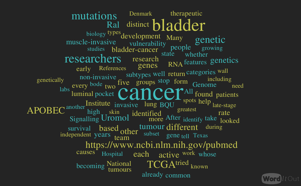
Knowing the genetic vulnerability of bladder cancer for therapeutic intervention, Volume 2 (Volume Two: Latest in Genomics Methodologies for Therapeutics: Gene Editing, NGS and BioInformatics, Simulations and the Genome Ontology), Part 1: Next Generation Sequencing (NGS)

Knowing the genetic vulnerability of bladder cancer for therapeutic intervention

Reporter and Curator: Dr. Sudipta Saha, Ph.D.


A mutated gene called RAS gives rise to a signalling protein Ral which is involved in tumour growth in the bladder. Many researchers tried and failed to target and stop this wayward gene. Signalling proteins such as Ral usually shift between active and inactive states.


So, researchers next tried to stop Ral to get into active state. In inacvtive state Ral exposes a pocket which gets closed when active. After five years, the researchers found a small molecule dubbed BQU57 that can wedge itself into the pocket to prevent Ral from closing and becoming active. Now, BQU57 has been licensed for further development.


Researchers have a growing genetic data on bladder cancer, some of which threaten to overturn the supposed causes of bladder cancer. Genetics has also allowed bladder cancer to be reclassified from two categories into five distinct subtypes, each with different characteristics and weak spots. All these advances bode well for drug development and for improved diagnosis and prognosis.


Among the groups studying the genetics of bladder cancer are two large international teams: Uromol (named for urology and molecular biology), which is based at Aarhus University Hospital in Denmark, and The Cancer Genome Atlas (TCGA), based at institutions in Texas and Boston. Each team tackled a different type of cancer, based on the traditional classification of whether or not a tumour has grown into the muscle wall of the bladder. Uromol worked on the more common, earlier form, non-muscle-invasive bladder cancer, whereas TCGA is looking at muscle-invasive bladder cancer, which has a lower survival rate.


The Uromol team sought to identify people whose non-invasive tumours might return after treatment, becoming invasive or even metastatic. Bladder cancer has a high risk of recurrence, so people whose non-invasive cancer has been treated need to be monitored for many years, undergoing cystoscopy every few months. They looked for predictive genetic footprints in the transcriptome of the cancer, which contains all of a cell’s RNA and can tell researchers which genes are turned on or off.


They found three subgroups with distinct basal and luminal features, as proposed by other groups, each with different clinical outcomes in early-stage bladder cancer. These features sort bladder cancer into genetic categories that can help predict whether the cancer will return. The researchers also identified mutations that are linked to tumour progression. Mutations in the so-called APOBEC genes, which code for enzymes that modify RNA or DNA molecules. This effect could lead to cancer and cause it to be aggressive.


The second major research group, TCGA, led by the National Cancer Institute and the National Human Genome Research Institute, that involves thousands of researchers across USA. The project has already mapped genomic changes in 33 cancer types, including breast, skin and lung cancers. The TCGA researchers, who study muscle-invasive bladder cancer, have looked at tumours that were already identified as fast-growing and invasive.


The work by Uromol, TCGA and other labs has provided a clearer view of the genetic landscape of early- and late-stage bladder cancer. There are five subtypes for the muscle-invasive form: luminal, luminal–papillary, luminal–infiltrated, basal–squamous, and neuronal, each of which is genetically distinct and might require different therapeutic approaches.


Bladder cancer has the third-highest mutation rate of any cancer, behind only lung cancer and melanoma. The TCGA team has confirmed Uromol research showing that most bladder-cancer mutations occur in the APOBEC genes. It is not yet clear why APOBEC mutations are so common in bladder cancer, but studies of the mutations have yielded one startling implication. The APOBEC enzyme causes mutations early during the development of bladder cancer, and independent of cigarette smoke or other known exposures.


The TCGA researchers found a subset of bladder-cancer patients, those with the greatest number of APOBEC mutations, had an extremely high five-year survival rate of about 75%. Other patients with fewer APOBEC mutations fared less well which is pretty surprising.


This detailed knowledge of bladder-cancer genetics may help to pinpoint the specific vulnerabilities of cancer cells in different people. Over the past decade, Broad Institute researchers have identified more than 760 genes that cancer needs to grow and survive. Their genetic map might take another ten years to finish, but it will list every genetic vulnerability that can be exploited. The goal of cancer precision medicine is to take the patient’s tumour and decode the genetics, so the clinician can make a decision based on that information.


References:


https://www.ncbi.nlm.nih.gov/pubmed/29117162


https://www.ncbi.nlm.nih.gov/pubmed/27321955


https://www.ncbi.nlm.nih.gov/pubmed/28583312


https://www.ncbi.nlm.nih.gov/pubmed/24476821


https://www.ncbi.nlm.nih.gov/pubmed/28988769


https://www.ncbi.nlm.nih.gov/pubmed/28753430


Read Full Post »

Reporter and Curator: Dr. Sudipta Saha, Ph.D.


Anti-Müllerian Hormone (AMH), is secreted by growing follicles that contains the egg or ovum. According to regular practice low AMH and high Follicle Stimulating Hormone (FSH) are generally considered as indicators of diminished egg quantity in a female. But, there are several cases the female conceived absolutely normally without any support even after low AMH was reported.


Therefore, a new research published in the Journal of the American Medical Association declares that AMH doesn’t dictate a woman’s reproductive potential. Although AMH testing is one of the most common ways that doctors assess a woman’s fertility. Present research says that all it takes is one egg each cycle and AMH is not a marker of whether a female can or cannot become pregnant. So, for women who haven’t yet tried to get pregnant and who are wondering whether they are fertile, an AMH value isn’t going to be helpful in that context. In addition, AMH is not necessarily a good marker to predict that whether one has to cryopreserve her eggs. So, practically doctors don’t yet have a way to definitively predict egg quality or a woman’s long-term ability to conceive, but age is obviously one of the most important factors.


The above mentioned study followed 750 women between the ages of 30 and 44 who had been trying to conceive for three months or less. During the 12-month observation period, those with low AMH values of less than 0.7 were not less likely to conceive than those who had normal AMH values. The study had various limitations, however, that are worth noting. The researchers only included women who did not have a history of infertility. Women who sought fertility treatments (about 6 percent) were withdrawn. And only 12 percent of the women were in the 38-to-44 age range. In addition, the number of live births was unavailable.


Among women aged 30 to 44 years without a history of infertility who had been trying to conceive for 3 months or less, biomarkers indicating diminished ovarian reserve compared with normal ovarian reserve were not associated with reduced fertility. These findings do not support the use of urinary or blood FSH tests or AMH levels to assess natural fertility for women with these characteristics. The researchers’ next want to see whether low AMH is associated with a higher risk of miscarriage among the women who conceived.


Although AMH testing isn’t designed to be an overall gauge of a woman’s fertility, it can still provide valuable information, especially for women who are infertile and seeking treatment. It can assist in diagnosing polycystic ovarian syndrome, and identify when a woman is getting closer to menopause. Previous research also showed that AMH is good predictor of a woman’s response to ovarian stimulation for in vitro fertilization and therefore it can predict the probability of conceiving via in vitro fertilization (I.V.F.).


References:


https://jamanetwork.com/journals/jama/article-abstract/2656811?JamaNetworkReader=True


https://www.nytimes.com/2017/10/16/health/fertility-test-ovarian-reserve.html


https://academic.oup.com/humrep/article/26/11/2925/656065


https://www.ncbi.nlm.nih.gov/pmc/articles/PMC3339896/


https://www.ncbi.nlm.nih.gov/pubmed/27179263


Read Full Post »

Development of 3D Human Tissue Models Awarded NIH Grants Worth $15M

Reporter: Irina Robu, PhD

NIH has awarded $15 million for Tissue Chip for Disease Modeling and Efficacy Testing to develop 3D human tissue models. The 3D platforms, also called tissue chips support living cells and human tissues, it mimics the complex biological functions of organs/tissues and at the same time provide a new way to test potential drugs and their effectiveness. The awards will allow scientists to study and understand diseases mechanism and forecast how patients respond and is part of the first phase of a five-year program.  According to NCATS Director, Dr. Christopher P. Austin “these tissue chips to provide more accurate platforms to understand diseases, and to be more predictive of the human response to drugs than current research models, thereby improving the success rate of candidate drugs in human clinical trials”.

The awards will be used to study common and rare diseases including rheumatoid arthritis, influenza A, kidney disease, amyotrophic lateral sclerosis, or ALS, arrhythmogenic cardiomyopathy, and hemorrhagic telangiectasia. Award recipients are Brigham and Women’s Hospital, Cedars-Sinai Medical Center, Columbia University, Duke University, Harvard University, Northwestern University, University of California Davis, University of California Irvine, University of Pittsburgh, University of Rochester, University of Washington Seattle and Vanderbilt University.

SOURCE

https://www.mdtmag.com/news/2017/09/nih-grants-15m-development-3d-human-tissue-models


Read Full Post »

« Newer Posts - Older Posts »