Healthcare analytics, AI solutions for biological big data, providing an AI platform for the biotech, life sciences, medical and pharmaceutical industries, as well as for related technological approaches, i.e., curation and text analysis with machine learning and other activities related to AI applications to these industries.
A Multicenter, Randomized Prospective Study in Consecutive Patients.
This study is sponsored by St. Jude Medical.
Coordinating Clinical Investigator: Bernard De Bruyne, OLV Ziekenhuis, Aalst, Belgium
Fractional Flow Reserve–Guided PCI versus Medical Therapy in Stable Coronary Disease
Bernard De Bruyne, M.D., Ph.D., Nico H.J. Pijls, M.D., Ph.D., Bindu Kalesan, M.P.H., Emanuele Barbato, M.D., Ph.D., Pim A.L. Tonino, M.D., Ph.D., Zsolt Piroth, M.D., Nikola Jagic, M.D., Sven Möbius-Winkler, M.D., Gilles Rioufol, M.D., Ph.D., Nils Witt, M.D., Ph.D., Petr Kala, M.D., Philip MacCarthy, M.D., Thomas Engström, M.D., Keith G. Oldroyd, M.D., Kreton Mavromatis, M.D., Ganesh Manoharan, M.D., Peter Verlee, M.D., Ole Frobert, M.D., Nick Curzen, B.M., Ph.D., Jane B. Johnson, R.N., B.S.N., Peter Jüni, M.D., and William F. Fearon, M.D. for the FAME 2 Trial Investigators
The preferred initial treatment for patients with stable coronary artery disease is the best available medical therapy. We hypothesized that in patients with functionally significant stenoses, as determined by measurement of fractional flow reserve (FFR), percutaneous coronary intervention (PCI) plus the best available medical therapy would be superior to the best available medical therapy alone.
METHODS
In patients with stable coronary artery disease for whom PCI was being considered, we assessed all stenoses by measuring FFR. Patients in whom at least one stenosis was functionally significant (FFR, ≤0.80) were randomly assigned to FFR-guided PCI plus the best available medical therapy (PCI group) or the best available medical therapy alone (medical-therapy group). Patients in whom all stenoses had an FFR of more than 0.80 were entered into a registry and received the best available medical therapy. The primary end point was a composite of death, myocardial infarction, or urgent revascularization.
RESULTS
Recruitment was halted prematurely after enrollment of 1220 patients (888 who underwent randomization and 332 enrolled in the registry) because of a significant between-group difference in the percentage of patients who had a primary end-point event: 4.3% in the PCI group and 12.7% in the medical-therapy group (hazard ratio with PCI, 0.32; 95% confidence interval [CI], 0.19 to 0.53; P<0.001). The difference was driven by a lower rate of urgent revascularization in the PCI group than in the medical-therapy group (1.6% vs. 11.1%; hazard ratio, 0.13; 95% CI, 0.06 to 0.30; P<0.001); in particular, in the PCI group, fewer urgent revascularizations were triggered by a myocardial infarction or evidence of ischemia on electrocardiography (hazard ratio, 0.13; 95% CI, 0.04 to 0.43; P<0.001). Among patients in the registry, 3.0% had a primary end-point event.
CONCLUSIONS
In patients with stable coronary artery disease and functionally significant stenoses, FFR-guided PCI plus the best available medical therapy, as compared with the best available medical therapy alone, decreased the need for urgent revascularization. In patients without ischemia, the outcome appeared to be favorable with the best available medical therapy alone. (Funded by St. Jude Medical; ClinicalTrials.gov number, NCT01132495.)
Supported by St. Jude Medical.
Disclosure forms provided by the authors are available with the full text of this article at NEJM.org.
This article was published on August 28, 2012, and updated on October 18, 2012, at NEJM.org.
SOURCE INFORMATION
From the Cardiovascular Center Aalst, Onze-Lieve-Vrouw Clinic, Aalst, Belgium (B.D.B., E.B.); Department of Cardiology, Catharina Hospital, and Department of Biomedical Engineering, Eindhoven University of Technology — both in Eindhoven, the Netherlands (N.H.J.P., P.A.L.T.); Division of Clinical Epidemiology and Biostatistics, Institute of Social and Preventive Medicine and Clinical Trials Unit Bern, University of Bern, Bern, Switzerland (B.K., P.J.); Hungarian Institute of Cardiology, Budapest (Z.P.); Clinical Center Kragujevac, Kragujevac, Serbia (N.J.); Heart Center Leipzig, Leipzig, Germany (S.M.-W.); Cardiovascular Hospital, Lyon, France (G.R.); Södersjukhuset, Stockholm (N.W.), and Karolinska Institutet at Örebro University Hospital, Örebro (O.F.) — both in Sweden; Department of Internal Medicine and Cardiology, University Hospital Brno, Brno, Czech Republic (P.K.); King’s College Hospital, London (P.M.), Golden Jubilee National Hospital, Glasgow (K.G.O.), Royal Victoria Hospital, Belfast (G.M.), and Department of Cardiology, Southampton University Hospital Trust, Southampton (N.C.) — all in the United Kingdom; Department of Cardiology, Rigshospitalet University Hospital, Copenhagen (T.E.); Atlanta Veterans Affairs Medical Center, Atlanta (K.M.); Northeast Cardiology Associates, Bangor, ME (P.V.); St. Jude Medical, Plymouth, MN (J.B.J.); and Stanford University Medical Center, Stanford, CA (W.F.F.).
Address reprint requests to Dr. De Bruyne at the Cardiovascular Centre Aalst, OLV-Clinic, Moorselbaan 164, B-9300 Aalst, Belgium, or atbernard.de.bruyne@olvz-aalst.be.
The investigators in the Fractional Flow Reserve versus Angiography for Multivessel Evaluation 2 (FAME 2) trial are listed in the Supplementary Appendix, available at NEJM.org.
Conclusions
The FAME II study results show that FFR-guided PCI compared to medical management alone significantly reduces patients’ risk for unplanned hospital readmission with urgent revascularization.
The new data support the paradigm of “Functionally Complete Revascularization,” that is, stenting of ischemic lesions and medical treatment of non-ischemic ones.
Routine Use of Ffr Significantly Improves the Outcome of Treatment in Stable CAD Patients. St. Jude Medical is focused on reducing risk by continuously finding ways to put more control into the hands of those who save and enhance lives.
SUMMARY OF KEY FINDINGS
FFR significantly improves outcome of treatment in stable patients. The results of FAME II, a St. Jude Medical sponsored clinical trial, show a significant benefit in using FFR-guided intervention. In patients with stable coronary artery disease undergoing PressureWireTM-guided intervention, PCI plus medical therapy was found to improve outcomes compared to medical therapy alone.
FFR Significantly Reduces Risk of Unplanned Hospital Readmission for Urgent Revascularization
Background
In patients with clinically stable coronary disease, PCI has not been shown to affect clinical outcomes such as death, nonfatal myocardial infarction and the need for urgent revascularization. In previous trials on revascularization, treatment has been guided by the angiographic appearance of the lesions. It is likely that in all previous trials dealing with patients with nonacute coronary artery disease (CAD), a sizable proportion of patients did not have ischemia.
Objectives
The overall purpose of the FAME II study was to compare the clinical outcomes of FFR-guided contemporary PCI plus medical therapy versus medical therapy alone in patients with stable coronary disease.
Methods
The FAME II trial is a prospective, multicenter randomized clinical trial with an all comers design. All consecutive patients with stable clinical condition and angiographically defined one-, two- or three-vessel coronary artery disease and amenable for PCI were screened and considered for participation in the study. Patients with at least one hemodynamically significant lesion were randomized into PCI (drug-eluting stents [DES] were recommended) plus medical therapy or medical therapy alone. It was expected that in approximately 20% of patients no stenoses would be hemodynamically significant.
Patients without hemodynamically significant lesions were enrolled in the registry portion of the study and treated with medical therapy. For prospectively collected data in the randomized study and the registry, an independent clinical events committee (CEC) adjudicated all clinical endpoints.
For patients with one or more significant lesions, there was an 86% relative reduction in the risk for unplanned hospital readmission with urgent revascularization for patients who received FFR-guided PCI plus medical therapy.
These findings support FFR-guided PCI compared to medical management alone to improve outcomes in the treatment of stable patients with single-vessel or multivessel coronary artery disease.
Study Endpoints
Original FAME II Study Flow Chart Primary Endpoint Composite of:
all-cause death
nonfatal myocardial infarction
unplanned hospitalization with urgent revascularization
As adjudicated by an independent CEC.
Secondary Endpoints
Individual components of the primary endpoint*
Cardiac death*
Nonurgent revascularization procedures*
Angina class
*As adjudicated by an independent CEC.
Stable patients scheduled for 1, 2 or 3 vessel DES stenting
Fractional Flow Reserve Measurements Intracoronary pressure measurements were obtained with a guiding catheter (fluid-filled) and the St. Jude Medical PressureWire CertusTM or AerisTM guidewire.
Results
The independent Data and Safety Monitoring Board recommended halting patient recruitment due to a significantly increased patient risk of major adverse cardiac events among patients randomized to medical therapy alone compared to patients randomized to medical therapy plus PCI.
The enrollment goal in FAME II was approximately 1,800 patients (randomized study and registry combined). The data sample presented here is the same data on which the decision to halt enrollment was based (January 15, 2012).
A total of 888 (73%) patients with ischemic lesions had been successfully randomized, and an additional 332 (27%) patients were enrolled in the registry because no ischemic lesions were detected. In total, 1,220 patients were enrolled in the FAME II trial, including 1,054 who were assigned to follow-up.
Medical Therapy
Actual FAME II Study Flow Chart
* Note that 6 patients had total occlusions supplying akinetic myocardium and were therefore not considered for PCI; 1 patient had 2 FFR negative lesions and was therefore included in the registry, however, a subsequently detected total occlusion was eventually treated with DES.
Randomized (n = 888)
Underwent FFR (n = 1220)
FFR >0.80 in all lesions included in registry
(n = 332)*
Allocated to medical therapy alone (n = 441)
Received allocated intervention (n = 439)
Did not receive allocated intervention (n = 2)
Erroneously received DES (n = 2)
Randomly selected to receive follow-up (n = 166)
Received medical therapy alone (n = 165)
Received DES (n = 1)
Allocated to PCI+medical therapy (n = 447)
Received allocated intervention (n = 435)
Did not receive allocated intervention (n = 12)
Treated with balloon angioplasty (n = 3)
Underwent CABG rather than PCI (n = 4)
Received medical therapy, planned for staged
procedure (n = 3)
Received medical therapy, unsuccessful PCI (n = 1)
**Differences between the two randomized groups were not significant with the exception of left ventricular ejection fraction<50% (p<0.05). Data are mean±SD or number of patients assessed (%). P-value using chi square test;
when cells are small Fisher’s test is used. Data for ejection fraction were available for 423 in PCI&medical therapy, 410 in medical therapy and 150 in registry. Data for history of MI were available for 442 in PCI&medical therapy,
436 in medical therapy and 295 in registry. CCS=Canadian Cardiovascular Society functional classification of angina pectoris; Data available in 447 in PCI&medical therapy, 440 in medical therapy, and 166 in registry. **5 totally
occluded arteries supplied infarcted areas and therefore not considered for revascularization using PCI. In patient level analysis p-value calculated using chi-square test, in case of cells <15 Fisher’s test. In lesion level analysis,
mixed maximum-likelihood logistic regression models were used for comparisons between groups for dichotomous variables and mixed maximum-likelihood linear regression models for continuous variables to account for the
correlation of multiple lesions within patients.
Conclusions
The FAME II study results show that FFR-guided PCI compared to medical management alone significantly reduces patients’ risk for unplanned hospital readmission with urgent revascularization.
The new data support the paradigm of “Functionally Complete Revascularization,” that is, stenting of ischemic lesions and medical treatment of non-ischemic ones.
Routine Use of Ffr Significantly Improves the Outcome of Treatment in Stable CAD Patients. St. Jude Medical is focused on reducing risk by continuously finding ways to put more control into the hands of those who save and enhance lives.
SJMprofessional.com
SJMknowledgecenter.com
Product referenced is approved for CE Mark.
Rx Only
Please review the Instructions for Use prior to using these devices for a complete listing of indications, contraindications, warnings, precautions,
potential adverse events, and directions for use.
PressureWire Aeris and PressureWire Certus are designed, developed and manufactured by St. Jude Medical Systems AB. PressureWire, Aeris,
Certus, ST. JUDE MEDICAL, the nine-squares symbol and MORE CONTROL. LESS RISK. are registered and unregistered trademarks and service
marks of St. Jude Medical, Inc. and its related companies.
The most common breast cancer screening method used today is called dual-view digital mammography, but it isn’t always successful in identifying tumors, said Jianwei (John) Miao, a UCLA professor of physics and astronomy and researcher with the California NanoSystems Institute at UCLA.
“While commonly used, the limitation is that it provides only two images of the breast tissue, which can explain why 10 to 20 percent of breast tumors are not detectable on mammograms,” Miao said. “A three-dimensional view of the breast can be generated by a CT scan, but this is not frequently used clinically, as it requires a larger dose of radiation than a mammogram. It is very important to keep the dose low to prevent damage to this sensitive tissue during screening.”
Recognizing these limitations, the scientists went in a new direction. In collaboration with the European Synchrotron Radiation Facility in France and Germany’s Ludwig Maximilians University, Miao’s international colleagues used a special detection method known as phase contrast tomography to X-ray a human breast from multiple angles.
They then applied equally sloped tomography, or EST — a breakthrough computing algorithm developed by Miao’s UCLA team that enables high-quality image-reconstruction — to 512 of these images to produce 3-D images of the breast at a higher resolution than ever before. The process required less radiation than a mammogram.
In a blind evaluation, five independent radiologists from Ludwig Maximilians University ranked these images as having a higher sharpness, contrast and overall image quality than 3-D images of breast tissue created using other standard methods.
“Even small details of the breast tumor can be seen using this technique,” said Maximilian Reiser, director of the radiology department at Ludwig Maximilians University, who contributed his medical expertise to the research.
The technology commonly used today for mammograms or imaging a patient’s bones measures the difference in an X-ray’s intensity before and after it passes through the body. But the phase contrast X-ray tomography used in this study measures the difference in the way an X-ray oscillates through normal tissue rather than through slightly denser tissue like a tumor or bone. While a very small breast tumor might not absorb many X-rays, the way it changes the oscillation of an X-ray can be quite large, Miao said. Phase contrast tomography captures this difference in oscillation, and each image made using this technique contributes to the overall 3-D picture.
Like cleaning the lenses of a foggy pair of glasses, scientists are now able to use a technique developed by UCLA researchers and their European colleagues to produce three-dimensional images of breast tissue that are two to three times sharper than those made using current CT scanners at hospitals. The technique also uses a lower dose of X-ray radiation than a mammogram.
The computational algorithm EST developed by Miao’s UCLA team is a primary driver of this advance. Three-dimensional reconstructions, like the ones created in this research, are produced using sophisticated software and a powerful computer to combine many images into one 3-D image, much like various slices of an orange can be combined to form the whole. By rethinking the mathematic equations of the software in use today, Miao’s group developed a more powerful algorithm that requires fewer “slices” to get a clearer overall 3-D picture.
“The technology used in mammogram screenings has been around for more than 100 years,” said Paola Coan, a professor of X-ray imaging at Ludwig Maximilians University. “We want to see the difference between healthy tissue and the cancer using X-rays, and that difference can be very difficult to see, particularly in the breast, using standard techniques. The idea we used here was to combine phase contrast tomography with EST, and this combination is what gave us much higher quality 3-D images than ever before.”
While this new technology is like a key in a lock, the door will only swing open — bringing high-resolution 3-D imaging from the synchrotron facility to the clinic — with further technological advances, said Alberto Bravin, managing physicist of the biomedical research laboratory at the European Synchrotron Radiation Facility. He added that the technology is still in the research phase and will not be available to patients for some time.
“A high-quality X-ray source is an absolute requirement for this technique,” Bravin said. “While we can demonstrate the power of our technology, the X-ray source must come from a small enough device for it to become commonly used for breast cancer screening. Many research groups are actively working to develop this smaller X-ray source. Once this hurdle is cleared, our research is poised to make a big impact on society.”
These results represent the collaborative efforts of senior authors Miao, Bravin and Coan. Significant contributions were provided by co-first authors Yunzhe Zhao, a recent UCLA doctoral graduate in Miao’s laboratory, and Emmanuel Brun, a scientist working with Bravin and Coan. Other co-authors included Zhifeng Huang of UCLA and Aniko Sztrókay, Paul Claude Diemoz, Susanne Liebhardt, Alberto Mittone and Sergei Gasilov of Ludwig Maximilians University.
The research was funded by UC Discovery/Tomosoft Technologies; the National Institute of General Medical Sciences, a division of the National Institutes of Health; and the Deutsche Forschungsgemeinschaft-Cluster of Excellence Munich-Centre for Advanced Photonics.
Abstract:
Mammography is the primary imaging tool for screening and diagnosis of human breast cancers, but ∼10–20% of palpable tumors are not detectable on mammograms and only about 40% of biopsied lesions are malignant. Here we report a high-resolution, low-dose phase contrast X-ray tomographic method for 3D diagnosis of human breast cancers. By combining phase contrast X-ray imaging with an image reconstruction method known as equally sloped tomography, we imaged a human breast in three dimensions and identified a malignant cancer with a pixel size of 92 μm and a radiation dose less than that of dual-view mammography. According to a blind evaluation by five independent radiologists, our method can reduce the radiation dose and acquisition time by ∼74% relative to conventional phase contrast X-ray tomography, while maintaining high image resolution and image contrast. These results demonstrate that high-resolution 3D diagnostic imaging of human breast cancers can, in principle, be performed at clinical compatible doses.
NEW YORK (GenomeWeb News) – An international team led by investigators in Australia has linked mutations in a sodium-gated potassium channel subunit gene to a subset of severe nocturnalfrontal lobeepilepsy cases.
As they reported online yesterday in Nature Genetics, the researchers began by testing a family with autosomal dominant nocturnal frontal lobe epilepsy, or ADNFLE. Affected members of the family often had not only typical ADNFLE symptoms, but also intellectual and/or psychiatric features that don’t usually characterize the disorder.
After narrowing in on a chromosome 9 region via linkage analyses in the family, the team identified ADNFLE-associated missense mutations in the sodium-gated potassium channel subunit gene KCNT1 by whole-exome sequencing in two affected family members. Follow-up testing on more than 100 other unrelated individuals with nocturnal frontal lobe epilepsy indicated that both inherited and de novo mutations in the gene can cause severe forms of the conditions that tend to include other co-morbidities.
“KCNT1 mutations were identified in two additional families and a sporadic case with severe ADNFLE and psychiatric features,” University of South Australia researcher Leanne Dibbens and the University of Melbourne’s Ingrid Scheffer, the study’s co-corresponding authors, and their colleagues wrote.
“These findings implicate the sodium-gated potassium channel complex in ADNFLE, and, more broadly, in the pathogenesis of focal epilepsies,” they added.
As the name suggests, ADNFLE is inherited in an autosomal dominant manner in affected families. Symptoms of the condition — including seizures that occur while individuals are asleep — generally appear in childhood, the researchers explained. And previous studies have implicated mutations to nicotinic acetylcholine receptor subunit genes in a subset of ADNFLE cases.
For the current study, the team focused on a multi-generational family with an especially severe form of ADNFLE that was accompanied by other symptoms such as intellectual disability and psychiatric disorders.
Genome-wide linkage analyses within the family led to a suspicious 2.36 million base stretch of sequence on chromosome 9, which housed almost 100 genes. Among them: two ion channel-coding genes, KCNT1 and GRIN1.
For two of the affected family members, the team turned to whole-exome sequencing to try to track down the most likely cause of ADNFLE. Indeed, missense mutations in KCNT1 that were predicted to be pathogenic turned up in one of the two exome sequences.
The mutation was not initially identified in the other family member’s exome sequence data, owing to low coverage, researchers explained. But it was subsequently shown to be present in both individuals by Sanger sequencing.
Consistent with the notion that this KCNT1 mutation could be related to ADNFLE pathogenesis, the investigators did not find it when they tested 111 unaffected, ancestry-matched individuals. Nor did it turn up in the dbSNP database, they reported, or in data generated for the 1000 Genomes Project or through the National Heart, Lung, and Blood Institute’s Exome Sequencing Project.
On the other hand, the team did find mutations in KCNT1 when it assessed another 108 unrelated individuals who either had ADNFLE or sporadically occurring nocturnal frontal lobe epilepsy.
That analysis helped the investigators track down two more ADNFLE-affected families with KCNT1 mutations that co-segregated with the disease, along with one case of sporadic nocturnal frontal lobe epilepsy including psychiatric features that seemed to stem from de novo mutations to KCNT1.
“[T]he phenotype associated with KNCT1 mutations is both more severe and more penetrant than that typically found with mutations affecting [nicotinic acetylcholine receptors],” the study’s authors noted.
In addition to showing more pronounced ADNFLE symptoms, they explained, the disease appears to manifest itself at a younger age in the cases linked to KCNT1 mutations.
Moreover, several cases that appear to be caused by alterations to KCNT1 also included intellectual disability, psychiatric, and/or behavioral features. The severity of such symptoms varied from one individual to the next — a pattern that the researchers speculated might be due to differences in the nature and extent of the KCNT1 mutation involved.
In addition to providing clues to help classify ADNFLE cases and offer genetic counseling for families affected by it, those involved in the study say the results should also prove useful for understanding — and potentially targeting — the processes that underlie this type of epilepsy.
“[T]his finding should provide new insights into the biological mechanisms underlying the pathogenesis of ADNFLE,” they concluded, “which may lead to targeted therapies addressing the serious co-morbidities as well as the debilitating seizure disorder.”
Mining the Unknown: A Systems Approach to Metabolite Identification Combining Genetic and Metabolic Information
Jan Krumsiek1, Karsten Suhre1,2, Anne M. Evans3, Matthew W. Mitchell3, Robert P. Mohney3, Michael V. Milburn3, Brigitte Wägele1,4, Werner Römisch-Margl1, Thomas Illig5,6, Jerzy Adamski7,8, Christian Gieger9, Fabian J. Theis1,10, Gabi Kastenmüller1*
1 Institute of Bioinformatics and Systems Biology, Helmholtz Zentrum München, Neuherberg, Germany, 2 Department of Physiology and Biophysics, Weill Cornell Medical College in Qatar, Education City, Qatar Foundation, Doha, Qatar, 3 Metabolon, Research Triangle Park, North Carolina, United States of America, 4 Department of Genome-Oriented Bioinformatics, Life and Food Science Center Weihenstephan, Technische Universität München, Freising, Germany, 5 Research Unit of Molecular Epidemiology, Helmholtz Zentrum München, Neuherberg, Germany, 6 Biobank of the Hanover Medical School, Hanover Medical School, Hanover, Germany, 7 Institute of Experimental Genetics, Genome Analysis Center, Helmholtz Zentrum München, Neuherberg, Germany, 8 Lehrstuhl für Experimentelle Genetik, Technische Universität München, Freising-Weihenstephan, Germany, 9 Institute of Epidemiology, Helmholtz Zentrum München, Neuherberg, Germany, 10 Department of Mathematics, Technische Universität München, Garching, Germany
Abstract
Recent genome-wide association studies (GWAS) with metabolomics data linked genetic variation in the human genome to differences in individual metabolite levels. A strong relevance of this metabolic individuality for biomedical and pharmaceutical research has been reported. However, a considerable amount of the molecules currently quantified by modern metabolomics techniques are chemically unidentified. The identification of these “unknown metabolites” is still a demanding and intricate task, limiting their usability as functional markers of metabolic processes. As a consequence, previous GWAS largely ignored unknown metabolites as metabolic traits for the analysis. Here we present a systems-level approach that combines genome-wide association analysis and Gaussian graphical modeling with metabolomics to predict the identity of the unknown metabolites. We apply our method to original data of 517 metabolic traits, of which 225 are unknowns, and genotyping information on 655,658 genetic variants, measured in 1,768 human blood samples. We report previously undescribed genotype–metabotype associations for six distinct gene loci (SLC22A2, COMT, CYP3A5, CYP2C18, GBA3, UGT3A1) and one locus not related to any known gene (rs12413935). Overlaying the inferred genetic associations, metabolic networks, and knowledge-based pathway information, we derive testable hypotheses on the biochemical identities of 106 unknown metabolites. As a proof of principle, we experimentally confirm nine concrete predictions. We demonstrate the benefit of our method for the functional interpretation of previous metabolomics biomarker studies on liver detoxification, hypertension, and insulin resistance. Our approach is generic in nature and can be directly transferred to metabolomics data from different experimental platforms.
Introduction
Recently, genome-wide association studies (GWAS) on metabolic quantitative traits have proven valuable tools to uncover the genetically determined metabolic individuality in the general population [1]–[5]. Interestingly, a great portion of the genetic loci that were found to significantly associate with levels of specific metabolites are within or in close proximity to metabolic enzymes or transporters with known disease or pharmaceutical relevance. Moreover, compared to GWAS with clinical endpoints the effect sizes of the genotypes are exceptionally high.
The number and type of the metabolic features that went into these GWAS was mainly defined by the metabolomics techniques used: Gieger et al. [1] and Illig et al. [2] used a targeted mass spectrometry (MS)-based approach giving access to the concentrations of 363 and 163 metabolites, respectively. Suhre et al. [3] and Nicholson et al. [4] applied untargeted nuclear magnetic resonance (NMR) based metabolomics techniques, yielding 59 metabolites that had been identified in the spectra prior to the GWAS and 579 manually selected peaks from the spectra, respectively. In Suhre et al. [5], 276 metabolites from an untargeted MS-based approach were analyzed.
While these previous GWAS focused on metabolic features with known identity, untargeted metabolomics approaches additionally provide quantifications of so-called “unknown metabolites”. An unknown metabolite is a small molecule that can reproducibly be detected and quantified in a metabolomics experiment, but whose chemical identity has not been elucidated yet. In an experiment using liquid chromatography (LC) coupled to MS, such an unknown would be defined by a specific retention time, one or multiple masses (e.g. from adducts), and a characteristic fragmentation pattern of the primary ion(s). An unknown observed by NMR spectroscopy would correspond to a pattern in the chemical shifts. Unknowns may constitute previously undocumented small molecules, such as rare xenobiotics or secondary products of metabolism, or they may represent molecules from established pathways which could not be assigned using current libraries of MS fragmentation patterns [6], [7] or NMR reference spectra [8].
The impact of unknown metabolites for biomedical research has been shown in recent metabolomics-based discovery studies of novel biomarkers for diseases and various disease-causing conditions. This includes studies investigating altered metabolite levels in blood for insulin resistance [9], type 2 diabetes [10], and heart disorders [11]. A considerable number of high-ranking hits reported in these biomarker studies represent unknown metabolites. As long as their chemical identities are not clarified the usability of unknown metabolites as functional biomarkers for further investigations and clinical applications is rather limited.
In mass-spectrometry-based metabolomics approaches, the assignment of chemical identity usually involves the interpretation and comparison of experiment-specific parameters, such as accurate masses, isotope distributions, fragmentation patterns, and chromatography retention times [12]–[14]. Various computer-based methods have been developed to automate this process. For example, Rasche and colleagues [15] elucidated structural information of unknown metabolites in a mass-spectrometry setup using a graph-theoretical approach. Their approach attempts to reconstruct the underlying fragmentation tree based on mass-spectra at varying collision energies. Other authors excluded false candidates for a given unknown by comparing observed and predicted chromatography retention times [16], [17], or by the automatic determination of sum formulas from isotope distributions [18]. Furthermore, Gipson et al. [19] and Weber et al. [20] integrated public metabolic pathway information with correlating peak pairs in order to facilitate metabolite identification. However, these methods might not be applicable for high-throughput metabolomics datasets that have been produced in a fee-for-service manner, since the mass spectra as such might not be readily available.
Approaching the problem from a conceptually different perspective, we here present a novel functional metabolomics method to predict the identities of unknown metabolites using a systems biological framework. By combining high-throughput genotyping data, metabolomics data, and literature-derived metabolic pathway information, we generate testable hypotheses on the metabolite identities based solely on the obtained metabolite quantifications (Figure 1). No further experiment-specific data such as retention times, isotope patterns and fragmentation patterns are required for this analysis.
Figure 1. Data integration workflow for the systematic classification of unknown metabolites.
We combine high-throughput metabolomics and genotyping data in Gaussian graphical models (GGMs) [21] and in genome-wide association studies (GWAS) [5] in order to produce testable predictions of the unknown metabolites’ identities. These hypotheses are then subject to experimental verification by mass-spectrometry. Six such cases have been fully worked through and are presented in Table 3. doi:10.1371/journal.pgen.1003005.g001
We developed and validated a novel integrative approach for the biochemical characterization of “unknown metabolites” from high-throughput metabolomics and genotyping datasets. Our method allows for the functional annotation of previously unidentified metabolites and, as a consequence, enhances the interpretability of metabolomics data in genome-wide association studies and biomarker discovery. For the first time, we systematically evaluated genetic associations of unknown metabolites, thereby discovering seven new loci of metabolic individuality. By classifying a series of unknown metabolites, we gained new insights into the functional interplay between genetic variation and the metabolome both for previously reported and new loci. Furthermore, several of the unknown compounds that we identified as well as their newly associated loci were independently reported in disease-related studies. In the following, we discuss three genetic loci and their associated phenotypes.
COMT and hepatic detoxification
The first example is a recent biomarker study, where Milburn et al. [34] reported an association of X-11593 with hepatic detoxification. In our GWAS, we find a strong association of X-11593 with the COMT locus, which encodes the catechol-O-methyltransferase enzyme. COMT is responsible for the inactivation of catecholamines such as L-dopa and various neuroactive drugs by O-methylation [35]. Following our identification approach, we experimentally confirmed the identity of X-11593 as O-methylascorbate. Notably, O-methylascorbate is a known product of ascorbate (vitamin C) O-methylation by COMT [36], [37]. Thus, our observations establish a link between O-methylascorbate blood levels, common genetic variation in the COMT locus and COMT-mediated liver detoxification processes.
ACE and hypertension
The second example relates to the ACE gene locus, which is a known risk locus for cardiovascular disease, hypertension and kidney failure. The protein encoded by the ACE locus, angiotensin-converting enzyme, is an exopeptidase which cleaves dipeptides from vasoactive oligopeptides, and plays a central role in the blood pressure-controlling renin-angiotensin system [38]. Moreover, the ACE protein is a target for various pharmaceuticals (ACE inhibitors), especially in the treatment of hypertension [39]. In our study, we identified three unknowns as dipeptides (X-14205, X-14208 and X-14478), two of which also associated with the ACE locus. These dipeptides could thus represent novel, interesting biomarkers for the activity of ACE. Moreover, Steffens et al. [11] reported a connection between heart failure and X-11805, which is in close proximity to angiontensin-related peptides in the GGM. This connection might be revisited after a successful identification of X-11805 in a future study.
UGT1A/ACADM and insulin resistance
The third example is an explorative study to detect biomarkers for insulin sensitivity. Gall et al. [9] reported several known metabolites (most prominently α-hydroxybutyrate) as biomarkers for insulin resistance. They also reported a series of unknown metabolites among their top hits. In the present study, we investigated three of these unknowns: X-11793 associates with UGT1A (UDP glucuronosyltransferase 1) and represents a bilirubin-related substance. Moreover, we experimentally validated X-11421 and X-13431, which display a strong association with ACADM (acyl-Coenzyme A dehydrogenase, C-4 to C-12 straight chain), as acylcarnitines containing 10 and 9 carbon atoms, respectively. The identification of these latter two unknown metabolites as medium-chain length acylcarnitines is coherent with reports by Adams et al. [40]. The authors found elevated blood plasma acylcarnitine levels in women with type 2 diabetes. Functionally, they attributed this finding to incomplete β-oxidation. Thus, our identification of X-11421 and X-13431 now suggests incomplete β-oxidation as an explanation for the associations found by Gall et al. and implies that acylcarnitines containing 10 and 9 carbon atoms are potential biomarkers for insulin resistance.
Conclusion
In summary, we integrated high-throughput metabolomics and genotyping data from a large population cohort for elucidating the biochemical identities of unknown metabolites. To this end, we applied metabolomics genome-wide association studies and Gaussian graphical modeling in order to link these unknown metabolites with known metabolic classes and biological processes. For six specific scenarios, we went from systematic hypothesis generation over detailed investigation and identity prediction to direct experimental confirmation. Similar validations may now be undertaken for the remaining predictions that we report in Table S1. Finally, we demonstrated the benefit of our method by discussing several of these newly identified metabolites in the context of existing biomarker discovery studies on liver detoxification, hypertension and insulin resistance.
It is to be noted that our method does not specifically require genotyping data. Even metabolomics measurements alone, analyzed through the GGMs, may provide sufficient information for the classification and even precise identity prediction. The unknowns with GGM evidence but without GWAS hits in Figure 4 as well as the HETE scenario represent examples for this approach.
One limitation of our approach is the requirement for associations with functionally described loci or known metabolites. Certain metabolite groups might thus systematically not be identifiable. For instance, if the identity of a whole class of biochemically related molecules is unknown (which might be due to experimental reasons), then the GGM associations between those compounds will not aid in identity elucidation. The 118 unknown compounds for which we could not derive any classification might represent such cases. Thus, our functionally oriented method should be regarded as a complementary extension to the existing identity determination methods.
Accordingly, our approach can be extended in several directions. It can be combined with method-specific, automated techniques that further exclude sets of metabolites. Previously mentioned methods relying on mass-spectra [15] or chromatographic properties [17] are suitable candidates here. Moreover, the method can be directly transferred to other types of metabolomics datasets not specifically originating from MS experiments, such as NMR-based metabolomics.
Beyond the application to metabolite identification, our study demonstrates the general potential of functional metabolomics in the context of genome-wide association studies. The comprehensive metabolic picture provided by GGMs in combination with GWAS allows for the detailed analysis of metabolic functions, chemical classes, enzyme-metabolite relationships and metabolic pathways.
Author Contributions
Conceived and designed the experiments: JK KS FJT GK. Performed the experiments: AME MWM RPM MVM. Analyzed the data: JK GK. Contributed reagents/materials/analysis tools: BW WR-M TI JA CG. Wrote the paper: JK KS FJT GK.
References
Gieger C, Geistlinger L, Altmaier E, de MH, Kronenberg F, et al. (2008) Genetics meets metabolomics: a genome-wide association study of metabolite profiles in human serum. PLoS Genet 4: e1000282 doi:10.1371/journal.pgen.1000282.
Illig T, Gieger C, Zhai G, Römisch-Margl W, Wang-Sattler R, et al. (2010) A genome-wide perspective of genetic variation in human metabolism. Nat Genet 42: 137–141. FIND THIS ARTICLE ONLINE
Suhre K, Wallaschofski H, Raffler J, Friedrich N, Haring R, et al. (2011) A genome-wide association study of metabolic traits in human urine. Nat Genet 43: 565–569. FIND THIS ARTICLE ONLINE
Nicholson G, Rantalainen M, Li JV, Maher AD, Malmodin D, et al. (2011) A genome-wide metabolic QTL analysis in Europeans implicates two loci shaped by recent positive selection. PLoS Genet 7: e1002270 doi:10.1371/journal.pgen.1002270.
Suhre K, Shin S-Y, Petersen A-K, Mohney RP, Meredith D, et al. (2011) Human metabolic individuality in biomedical and pharmaceutical research. Nature 477: 54–60. FIND THIS ARTICLE ONLINE
Gall WE, Beebe K, Lawton KA, Adam K-P, Mitchell MW, et al. (2010) alpha-hydroxybutyrate is an early biomarker of insulin resistance and glucose intolerance in a nondiabetic population. PLoS ONE 5: e10883 doi:10.1371/journal.pone.0010883.
Fiehn O, Garvey WT, Newman JW, Lok KH, Hoppel CL, et al. (2010) Plasma metabolomic profiles reflective of glucose homeostasis in non-diabetic and type 2 diabetic obese African-American women. PLoS ONE 5: e15234 doi:10.1371/journal.pone.0015234.
Mihaleva VV, Verhoeven HA, de Vos RCH, Hall RD, van Ham RCHJ (2009) Automated procedure for candidate compound selection in GC-MS metabolomics based on prediction of Kovats retention index. Bioinformatics 25: 787–794.FIND THIS ARTICLE ONLINE
Creek DJ, Jankevics A, Breitling R, Watson DG, Barrett MP, et al. (2011) Towards Global Metabolomics Analysis with Liquid Chromatography-Mass Spectrometry: Improved Metabolite Identification by Retention Time Prediction. Anal Chem FIND THIS ARTICLE ONLINE
Mittelstrass K, Ried JS, Yu Z, Krumsiek J, Gieger C, et al. (2011) Discovery of Sexual Dimorphisms in Metabolic and Genetic Biomarkers. PLoS Genet 7: e1002215 doi:10.1371/journal.pgen.1002215.
Nayak RR, Kearns M, Spielman RS, Cheung VG (2009) Coexpression network based on natural variation in human gene expression reveals gene interactions and functions. Genome Res 19: 1953–1962.FIND THIS ARTICLE ONLINE
Szklarczyk D, Franceschini A, Kuhn M, Simonovic M, Roth A, et al. (2011) The STRING database in 2011: functional interaction networks of proteins, globally integrated and scored. Nucleic Acids Res 39: D561–D568.FIND THIS ARTICLE ONLINE
Holle R, Happich M, Löwel H, Wichmann HE (2005) Group MKS (2005) KORA–a research platform for population based health research. Gesundheitswesen 67 Suppl 1: S19–S25. FIND THIS ARTICLE ONLINE
Hindorff L, MacArthur J, Wise A, Junkins H, Hall P, et al. A Catalog of Published Genome-Wide Association Studies.
Takeuchi F, McGinnis R, Bourgeois S, Barnes C, Eriksson N, et al. (2009) A genome-wide association study confirms VKORC1, CYP2C9, and CYP4F2 as principal genetic determinants of warfarin dose. PLoS Genet 5: e1000433 doi:10.1371/journal.pgen.1000433.
Chung CM, Wang RY, Chen JW, Fann CS, Leu HB, et al. (2010) A genome-wide association study identifies new loci for ACE activity: potential implications for response to ACE inhibitor. Pharmacogenomics J 10: 537–544. FIND THIS ARTICLE ONLINE
GTEx (Genotype-Tissue Expression) eQTL Browser.
Otterness DM, Wieben ED, Wood TC, Watson WG, Madden BJ, et al. (1992) Human liver dehydroepiandrosterone sulfotransferase: molecular cloning and expression of cDNA. Mol Pharmacol 41: 865–872.FIND THIS ARTICLE ONLINE
Berg JM, Tymoczko JL, Stryer L (2006) Biochemistry: W. H. Freeman.
Milburn M, Guo L, WULFF JE, Lawton KA (2010) DETERMINATION OF THE LIVER TOXICITY OF AN AGENT.
Männistö PT, Kaakkola S (1999) Catechol-O-methyltransferase (COMT): biochemistry, molecular biology, pharmacology, and clinical efficacy of the new selective COMT inhibitors. Pharmacol Rev 51: 593–628.FIND THIS ARTICLE ONLINE
Bowers-Komro DM, McCormick DB, King GA, Sweeny JG, Iacobucci GA (1982) Confirmation of 2-O-methyl ascorbic acid as the product from the enzymatic methylation of L-ascorbic acid by catechol-O-methyltransferase. Int J Vitam Nutr Res 52: 186–193.FIND THIS ARTICLE ONLINE
Butterworth M, Lau SS, Monks TJ (1996) 17 beta-Estradiol metabolism by hamster hepatic microsomes. Implications for the catechol-O-methyl transferase-mediated detoxication of catechol estrogens. Drug Metab Dispos 24: 588–594. FIND THIS ARTICLE ONLINE
Adams SH, Hoppel CL, Lok KH, Zhao L, Wong SW, et al. (2009) Plasma acylcarnitine profiles suggest incomplete long-chain fatty acid beta-oxidation and altered tricarboxylic acid cycle activity in type 2 diabetic African-American women. J Nutr 139: 1073–1081. FIND THIS ARTICLE ONLINE
Purcell S, Neale B, Todd-Brown K, Thomas L, R MA, et al. (2007) PLINK: a tool set for whole-genome association and population-based linkage analyses. Am J Hum Genet 81: 559–575. FIND THIS ARTICLE ONLINE
Ma H, Sorokin A, Mazein A, Selkov A, Selkov E, et al. (2007) The Edinburgh human metabolic network reconstruction and its functional analysis. Mol Syst Biol 3: 135.FIND THIS ARTICLE ONLINE
Ashburner M, Ball CA, Blake JA, Botstein D, Butler H, et al. (2000) Gene ontology: tool for the unification of biology. The Gene Ontology Consortium. Nat Genet 25: 25–29.FIND THIS ARTICLE ONLINE
Bielinski SJ, Chai HS, Pathak J, Talwalkar JA, Limburg PJ, et al. (2011) Mayo Genome Consortia: a genotype-phenotype resource for genome-wide association studies with an application to the analysis of circulating bilirubin levels. Mayo Clin Proc 86: 606–614. FIND THIS ARTICLE ONLINE
Chambers JC, Zhang W, Lord GM, van der Harst P, Lawlor DA, et al. (2010) Genetic loci influencing kidney function and chronic kidney disease. Nat Genet 42: 373–375.FIND THIS ARTICLE ONLINE
Kottgen A, Pattaro C, Boger CA, Fuchsberger C, Olden M, et al. (2010) New loci associated with kidney function and chronic kidney disease. Nat Genet 42: 376–384.FIND THIS ARTICLE ONLINE
Zhai G, Teumer A, Stolk L, B JR, Vandenput L, et al. (2011) Eight common genetic variants associated with serum DHEAS levels suggest a key role in ageing mechanisms. PLoS Genet 7: e1002025 doi:10.1371/journal.pgen.1002025.
Sanna S, Busonero F, Maschio A, McArdle PF, Usala G, et al. (2009) Common variants in the SLCO1B3 locus are associated with bilirubin levels and unconjugated hyperbilirubinemia. Hum Mol Genet 18: 2711–2718.FIND THIS ARTICLE ONLINE
Jylhava J, Lyytikainen LP, Kahonen M, Hutri-Kahonen N, Kettunen J, et al. (2012) A genome-wide association study identifies UGT1A1 as a regulator of serum cell-free DNA in young adults: The Cardiovascular Risk in Young Finns Study. PLoS ONE 7: e35426 doi:10.1371/journal.pone.0035426.
More recently, the mapping of the human genome, completed in 2003, is driving revolutionary advances in science. The Human Genome Project, also fueled by NIH funding, has created new private-sector technologies including gene sequencing, consumer genomics, and personalized medicine. A study by Battelle calculates that the Human Genome Project has helped drive $796 billion in economic activity and supported 310,000 jobs in 2010 alone.
Today, the continued vibrancy of the biomedical industry in California and nationwide depends on many things, including a predictable and consistent regulatory review process, sufficient and appropriate coverage and payment policies and intellectual property protections. But the industry would not exist without the essential investments in basic research the federal government makes through the NIH.
Unfortunately, now, automatic federal spending cutbacks known as sequestration threaten the future of research and development and our nation’s global competitiveness in the fields of drugs, medical device and diagnostics. While China and South Korea have committed to government funding increases of 10 percent year over year, U.S. federal funding for research and development during the past decade has stalled.
A $2.5 billion cut to the NIH budget next year, which is what the blunt instrument of sequestration requires, would result in 2,000 fewer funded research grants, according to the Congressional Budget Office. This would mean fewer research teams working on the cures and treatments of tomorrow, as well as canceled or postponed purchases from companies that manufacture research tools like flow cytometers, mass spectrometers and gene sequencers used by scientists in their labs. A recent study conducted by United for Medical Research estimates that NIH funding cuts under sequestration would lead to 33,000 fewer jobs nationwide — 5,000 in California alone — and an overall $4.5 billion decrease in economic activity.
California is the worldwide leader in biomedical investment, research and development, with more than 2,300 biomedical companies, along with public and private research institutions, advancing scientific knowledge and developing new diagnostics tools, treatments, and technologies addressing diseases and illnesses like cancer, diabetes, HIV/AIDS, chronic pain, and cardiovascular, respiratory and infectious diseases.
California’s life sciences industry is also an important engine of economic growth, employing nearly 268,000 workers statewide, paying more than $20 billion in annual wages and accounting for $18.6 billion in exports to markets around the world. Venture capital investment has been important, but private investment builds upon inventions that originate from federal research funded by the NIH and National Science Foundation, which totaled $4.5 billion in California last year. Together, industry, research universities and institutions, venture capital and the NIH comprise one of the most successful and important public-private partnerships in our country.
It is essential that Congress funds the kind of critical research needed to meet patient and public health needs of tomorrow.
We urge legislators in Washington to safeguard and sustain this essential public-private partnership that produces improved public health, economic growth and job creation.
David Gollaher, Ph.D., is president and chief executive officer of the California Healthcare Institute (CHI). Based in La Jolla, CHI is a non-profit public policy research organization representing more than 250 leading medical device, biotechnology, diagnostics and pharmaceutical companies and public and private academic biomedical research organizations.
Advances in Separations Technology for the “OMICs” and Clarification of Therapeutic Targets
Curator, Reporter, EAW: Larry H Bernstein, MD, FCAP
Article 1.5 Advances in Separations Technology for the “OMICs” and Clarification of Therapeutic Targets
This discussion is a continuation of an earlier piece on the technologic framework for , proteomics, nutrigenomics, and translational medicine. The last decade has seen the emergence of a genomic science that is changing the trajectory of biological sciences and medicine. It has not resolved all of our problems by any means, but it has begun to redraw the map, which began with the elucidation of major metabolic pathways in the first half of the 20th century, was then captured by the transformation of genetics with the discovery of the “Watson-Crick Model”, and then later was recharged with the discovery of the Toll-like receptor and the drawing of “signaling pathways”. What we have seen in an unraveling of protein-genome interactions, small peptide regulators, and dynamic changes in pathway dominance, bloackage, and reentry, depending on genetic, dietary, and environmental conditions, mostly expressed in what we refer to as “oxidative stress”.
Unraveling the multitude of nutrigenomic, proteomic, and metabolomic patterns that arise from the ingestion of foods or their bioactive food components will not be simple but is likely to provide insights into a tailored approach to diet and health. The use of new and innovative technologies, such as microarrays, RNA interference, and nanotechnologies, will provide needed insights into molecular targets for specific bioactive food components and how they harmonize to influence individual phenotypes. A challenging aspect of omic technologies is the refined analysis of quantitative dynamics in biological systems.
In recent years, nutrition research has moved from classical epidemiology and physiology to molecular biology and genetics. The new era of nutrition research translates empirical knowledge to evidence-based molecular science. Following this trend, Nutrigenomics has emerged as a novel and multidisciplinary research field in nutritional science that aims to elucidate how diet can influence human health. It is already well known that bioactive food compounds can interact with genes affecting transcription factors, protein expression and metabolite production. The study of these complex interactions requires the development of advanced analytical approaches combined with bioinformatics. The Institute of Medicine recently convened a workshop to review the state of the various domains of nutritional genomics research and policy and to provide guidance for further development and translation of this knowledge into nutrition practice and policy. Nutritional genomics holds the promise to revolutionize both clinical and public health nutrition practice and facilitate the establishment of
genome-informed nutrient and food-based dietary guidelines for disease prevention and healthful aging,
individualized medical nutrition therapy for disease management, and
better targeted public health nutrition interventions (including micronutrient fortification and supplementation) that maximize benefit and minimize adverse outcomes within genetically diverse human populations.
For metabolomics, gas and liquid chromatography coupled to mass spectrometry are well suited for coping with high sample numbers in reliable measurement times with respect to both technical accuracy and the identification and quantitation of small-molecular-weight metabolites. This potential is a prerequisite for the analysis of dynamic systems. Thus, metabolomics is a key technology for systems biology. The bioavailability of bioactive food constituents as well as dose-effect correlations are key information to understand the impact of food on defined health outcomes. Both strongly depend on appropriate analytical tools to identify and quantify minute amounts of individual compounds in highly complex matrices–food or biological fluids–and to monitor molecular changes in the body in a highly specific and sensitive manner. Based on these requirements, mass spectrometry has become the analytical method of choice with broad applications throughout all areas of nutrition research.
Dynamic Construct of the –Omics
Metabolomics is a term that encompasses several types of analyses, including
metabolic fingerprinting, which measures a subset of the whole profile with little differentiation or quantitation of metabolites;
metabolic profiling, the quantitative study of a group of metabolites, known or unknown, within or associated with a particular metabolic pathway; and
target isotope-based analysis, which focuses on a particular segment of the metabolome by analyzing only a few selected metabolites that comprise a specific biochemical pathway.
Any unifying concept of the metabolome was incomplete or debatable in the first 30 years of the 20th century. It was only known that insulin is anabolic and that insulin deficiency (or resistance) would have consequences in the point of entry into the citric acid cycle, which generates 28-32 ATPs. In fat catabolism, triglycerides are hydrolyzed to break them into fatty acids and glycerol. In the liver the glycerol can be converted into glucose via dihydroxyacetone phosphate and glyceraldehyde-3-phosphate by way of gluconeogenesis. In the case of this cycle there is a tie in with both catabolism and anabolism.
For bypass of the Pyruvate Kinase reaction of Glycolysis, cleavage of 2 ~P bonds is required. The free energy change associated with cleavage of one ~P bond of ATP is insufficient to drive synthesis of phosphoenolpyruvate (PEP), since PEP has a higher negative DG of phosphate hydrolysis than ATP. The two enzymes that catalyze the reactions for bypass of the Pyruvate Kinase reaction are the following:
Pyruvate Carboxylase (Gluconeogenesis) catalyzes pyruvate + HCO3- + ATP — oxaloacetate + ADP + Pi
PEP Carboxykinase (Gluconeogenesis) catalyzes: oxaloacetate + GTP —- phosphoenolpyruvate + GDP + CO2
Many high throughput methods have been employed to get some insight into the whole process and several examples of successful research. Proteomics and metabolomics need to encompass large numbers of experiments and linked data. Due to the nature of the proteins, as well as due to the properties of various metabolites, experimental approaches require the use of comprehensive high throughput methods and a sufficiency of analysed tissue or body fluids.
An important and revolutionary aspect of ‘The 2010 Project’ is that it implicitly endorses the allocation of resources to attempts to assign function to genes that have no known function. This represents a significant departure from the common practice of defining and justifying a scientific goal based on the biological phenomena. The rationale for endorsing this radical change is that for the first time it is feasible to envision a whole-systems approach to gene and protein function. I shall not discuss the emerging field of bioinformatics that makes this possible. In this review, the end-of-the line “detector will be considered having been covered. The entire focus proceeds to a discussion of separation methods. Separation methods have always been tricky, time consuming, and a multiple step process that depended on using anionic and cationic resins as intermediate steps in bulk separation, and then molecular size separation. Therapeutic Targets will be identified as they are seen.
Affinity Chromatography The rapid development of biotechnology and biomedicine requires more reliable and efficient separation technologies for the isolation and purification of biopolymers such as therapeutic proteins, antibodies, enzymes and nucleic acids. In particular, monoclonal antibodies are centrally important as therapeutics for the treatment of cancer and other diseases, leading to recombinant monoclonal antibodies that dominate today’s biopharmaceutical pipeline. The large-scale production of therapeutic biopolymers requires
a manufacturing process that delivers reliability and in high-yield, as well as
an effective purification process affording extremely pure products.
Because of its high selectivity, affinity chromatography has been used extensively to isolate a variety of biopolymers. The retention of solutes is based on specific, reversible interactions found in biological systems, such as the binding of an enzyme with an inhibitor or an antibody with an antigen. These interactions are exploited in affinity chromatography by immobilizing an affinity ligand onto a support, and using this as a stationary phase. Non-porous particles having an average diameter of 2.1 mm were prepared by co-polymerization of styrene, methyl methacrylate and glycidyl methacrylate, which was abbreviated as P(S–MMA–GMA). The particles were mechanically stable due to the presence of benzene rings in the backbone of polymer chains, and could withstand high pressures when a column packed with these particles was operated in the HPLC mode.
The polymer particles were advantaged by immobilization of ligands via the epoxy groups on the particle surface that were introduced by one of the monomers, glycidyl methacrylate. As a model system, Cibacron Blue 3G-A was covalently immobilized onto the non-porous copolymer beads. The dye-immobilized P(S–MMA–GMA) particles were slurry packed into a 1.0 cm30.46 cm I.D. column. This affinity column was effective for the separation of turkey egg white lysozyme from a protein mixture. The bound lysozyme could be eluted to yield a sharp peak by using a phosphate buffer containing 1 M NaCl. For a sample containing up to 8 mg of lysozyme, the retained portion of proteins could be completely eluted without any slit peak. Due to the use of a shorter column, the analysis time was shorter in comparison with other affinity systems reported in the literature. The retention time could be reduced significantly by increasing the flow-rate, while the capacity factor remained at the same level. CH Chen, WC Lee. Affinity chromatography of proteins on non-porous copolymerized particles of styrene, methyl methacrylate and glycidyl methacrylate. Journal of Chromatography A 2001; 921: 31–37.
Affinity separation membranes, consisting of electrospun nanofibers, have been developed recently. Affinity ligands are attached to the surface of the constituent fibers, offering a potential solution to some of the problems of traditional, column-based, affinity chromatography. Electrospun fibers are good candidates for use in affinity separation because of their
unique characteristics of high surface area to volume ratio, resulting in
high ligand loading, and
their large porosity, resulting in
high throughput operation.
A number of polymers have been used for electrospun fiber mesh-based affinity membrane separations including poly (ether-urethane-urea), cellulose, poly(ethylene terephthalate, polysulphone, and polyacrlonitrile. Typically, very thin electrospun fiber meshes are produced by electrostatically collecting negatively charged fibers on a collector electrode. These very thin 2D electrospun fiber mesh mats provide excellent solution permeability as compared to 3D column packed with affinity beads. M Miyauchi, J Miao, TJ Simmons, JS Dordick and RJ Linhardt. Flexible Electrospun Cellulose Fibers as an Affinity Packing Material for the Separation of Bovine Serum Albumin. J Chromatograph Separat Techniq 2011; 2:2 http://dx.doi.org/10.4172/2157-7064.1000110
Dye Affinity Chromatography Biomimetic Dyes Affinity adsorbents based on immobilized triazine dyes offer important advantages circumventing many of the problems associated with biological ligands. The main drawback of dyes is their moderate selectivity for proteins. Rational attempts to tackle this problem are realized through the biomimetic dye concept according to which new dyes, the biomimetic dyes, are designed to mimic natural ligands. Biomimetic dyes are expected to exhibit increased affinity and purifying ability for the targeted proteins.
Biocomputing offers a powerful approach to biomimetic ligand design. The successful exploitation of contemporary computational techniques in molecular design requires the knowledge of the three-dimensional structure of the target protein, or at least, the amino acid sequence of the target protein and the three-dimensional structure of a highly homologous protein. From such information one can then design, on a graphics workstation,
the model of the protein and also
a number of suitable synthetic ligands which mimic natural biological ligands of the protein.
There are several examples of enzyme purifications
Interactions between Cibacron Blue F3GA (CB F3GA), as a model of triazine dye, and 2-hydroxypropyl-b-cyclodextrin (HP-b-CD), as a model of cyclodextrin, were investigated by monitoring the spectral shift that accompanies the binding phenomena. Matrix analysis of the difference spectral titration of CB F3GA with HP-b-CD revealed only two absorbing species, indicating a host–guest ratio of 1:1. The dissociation constant for this HP-b-CD–CB F3GA complex, K , was found d to be 0.43 mM. The data for HP-b-CD forming inclusion complexes with CB F3GA were used to develop the concept of competitive elution by inclusion complexes in dye-affinity chromatography. When this concept was applied to the elution of L-lactate dehydrogenase from a CB F3GA affinity matrix, it was shown to be an effective elution strategy. It provided a 15-fold purification factor with 89% recovery and sharp elution profile (0.8 column volumes for 80% recovery), which is as good as that obtained by specific elution with NADH (16-fold, 78% recovery and 1.8 column volumes). In addition, the new elution strategy showed a better purification factor and sharper elution profile than traditional non-specific. JA Lopez-Mas, SA Streitenberger, F Garcıa-Carmona, AA Sanchez-Ferrer. Cyclodextrin biospecific-like displacement in dye-affinity chromatography. Journal of Chromatography A 2001; 911: 47–53.
Affinity chromatography uses biospecific binding usually between an antibody and an antigen, an enzyme and a substrate or other pairs of key-lock type of matching molecules. Due to its high selectivity, it is able to purify proteins and other macromolecules from very dilute solutions. In this work, a general rate model for affinity chromatography was used for scale-up studies. Parameters for the model were estimated from existing correlations, or from experimental results obtained on a small column with the same packing material. As anexample, Affi-Gel with 4.5mol cm−3 Cibacron Blue F-3GA as immobilized ligands covalently attached to cross-linked 6% agarose was used for column packing. Cibacron Blue F-3GA was also used as a soluble ligand in the elution stage. Satisfactory scale-up predictions were obtained for a 98.2 ml column and a 501 ml column based on a few experimental data obtained on a 7.85 ml small column. T. Gu, K.-H. Hsu and M.-J. Syu, “Scale-Up of Affinity Chromatography for Purification of Enzymes and Other Proteins.” Enzyme and Microbial Technology 2003; 33:433-437.
Affinity Column with AAAA as a Model Sense Ligand The degeneracy of antisense peptides was studied by high-performance affinity chromatography. A model sense peptide (AAAA) and its antisense peptides (CGGG, GGGG, RGGG, SGGG) were designed and synthesized according to the degeneracy of genetic codes. An affinity column with AAAA as the ligand was prepared. The affinity chromatographic behaviors of antisense peptides on the column were evaluated. The results indicated that model antisense peptides have clear retention on the immobilized AAAA affinity column. RGGG showed the strongest affinity interaction. R Zhao, X Yu, H Liu, L Zhai, S Xiong, et al. Study on the degeneracy of antisense peptides using affinity chromatography. Journal of Chromatography A 2001; 913: 421–428.
Frontal AC for Biomolecular Interactions Frontal affinity chromatography is a method for quantitative analysis of biomolecular interactions. We reinforced it by incorporating various merits of a contemporary liquid chromatography system. As a model study, the interaction between an immobilized Caenorhabditis elegans galectin (LEC-6) and fluorescently labeled oligosaccharides (pyridylaminated sugars) was analyzed. LEC-6 was coupled to N-hydroxysuccinimide-activated Sepharose 4 Fast Flow (100 mm diameter), and packed into a miniature column (e.g., 1034.0 mm, 0.126 ml). The volume of the elution front (V) determined graphically for each sample was compared with that obtained in the presence of an excess amount of hapten saccharide, lactose (V ); and the dissociation constant, K , was calculated according to the literature. This system also proved to be useful for an inverse confirmation; that is, application of galectins to an immobilized glycan column (in the present case, asialofetuin was immobilized on Sepharose 4 Fast Flow), and the elution profiles were monitored by fluorescence based on tryptophan. The newly constructed system proved to be extremely versatile. It enabled rapid (analysis time 12 min/ cycle) and sensitive (20 nM for pyridylaminated derivatives, and 1 mg/ml for protein) analyses of lectin–carbohydrate interactions. J Hirabayashi, Y Arata, K Kasai. Reinforcement of frontal affinity chromatography for effective analysis of lectin–oligosaccharide interactions. Journal of Chromatography A 2000; 890:261–271.
Immobilized Metal Ion Affinity New immobilized metal ion affinity chromatography (IMAC) matrices containing a high concentration of metal–chelate moieties and completely coated with inert flexible and hydrophilic dextrans are here proposed to improve the purification of polyhistidine (poly-His) tagged proteins. The purification of an interesting recombinant multimeric enzyme (a thermoresistant b-galactosidase from Thermus sp. strain T2) has been used to check the performance of these new chromatographic media.
IMAC supports with a high concentration (and surface density) of metal chelate groups promote a rapid adsorption of poly-His tagged proteins during IMAC. However, these supports also favor the promotion of undesirable multi-punctual adsorptions and problems may arise for the simple and effective purification of poly-His tagged proteins. For example, desorption of the pure enzyme from the support may become quite difficult (e.g., it is not fully desorbed from the support even using 200 mM of imidazole).
The coating of these IMAC supports with dextrans greatly reduces these undesired multi-point adsorptions. However, this dextran coating of chromatographic matrices seems to allow the formation of strong one-point adsorptions that involve small areas of the protein and support surface, but the dextran coating seems to have dramatic effects for the prevention of weak or strong multipoint interactions that should involve a high geometrical congruence between the enzyme and the support surface. C Mateo , G Fernandez-Lorente , BCC Pessela , A Vian, et al. Affinity chromatography of polyhistidine tagged enzymes. New dextran-coated immobilized metal ion affinity chromatography matrices for prevention of undesired multipoint adsorptions. Journal of Chromatography A 2001; 915:97–106. The underlying principle of immobilized metal ion affinity chromatography (IMAC) of proteins is the coordination between the electron donor groupings on a protein surface (histidine, tryptophan, cysteine) and chelated (iminodiacetate; IDA) transition metal ions [IDA-M(II)]. This principle of immobilized metal ion affinity (IMA) has been presented by now in some detail. The practice of IMAC in the purification of proteins has had its empirical phase. There is now a need, from the body of data, to establish somewhat more detailed ground rules that would allow for the use of IMAC in a more predictive manner. Immobilized metal ion affinity chromatography (IMAC) has been explored as a probe into the topography of histidyl residues of a protein molecule. An evaluation of the chromatographic behavior of selected model proteins-
thioredoxin
ubiquitin
calmodulin
lysozyme
cytochrome c
myoglobin
on immobilized transition metal ions
Co2+
Ni2+
Cu2+
Zn2
-allows establishment of the following facets of the histidyl side chain distribution:
either interior or surface;
when localized on the surface, accessible or unaccessible for coordination;
single or multiple;
When multiple, either distant or vicinal.
Moreover, proteins displaying single histidyl side chains on their surfaces may, in some instances, be resolved by IMAC; apparently, the microenvironments of histidyl residues are sufficiently diverse to result in different affinities for the immobilized metal ions. IMAC, previously introduced as an approach to the fractionation of proteins, has become also, upon closer examination, a facile probe into the topography of histidyl residues. This is possible because of the inherent versatility of IMAC; an appropriate metal ion (M2+) can be selected to suit the analytical purpose and a particular chromatographic protocol can be applied (isocratic pH, falling pH, and imidazole elution). We now report that IMAC may be exploited as an analytical tool in addition to its use as a protein purification technique. IMAC can be used to ascertain several facets of the status of a histidyl residue(s) in a protein molecule:
localization (interior vs. surface)
coordination potential as defined by the steric accessibility and the state of protonation
A novel, two-step preparative technique is described for the purification of authentic recombinant human prolactin (rhPRL) secreted into the periplasm of transformed Escherichia coli cells. The first step is based on immobilized metal ion affinity chromatography of periplasmic extract, using Ni(II) as a relatively specific ligand for hPRL in this system. It gives superior resolution and yield than established ion-exchange chromatography. Size-exclusion chromatography is used for further purification to .99.5% purity. The methodology is reproducible, leading to 77% recovery. Identity and purity of the rhPRL were demonstrated using sodium dodecylsulphate–polyacrylamide electrophoresis, isoelectric focusing, mass spectrometry (matrix-assisted laser desorption ionization time-of-flight), radioimmunoassay, RP-HPLC and high-performance size-exclusion chromatography. In the Nb2 bioassay, the hormone showed a bioactivity of 40.9 IU/mg.
Adenosine Affinity Ligand for Glutamine Synthase Glutamine synthetase has been purified from both procaryotic and eucaryotic sources using various types of affinity chromatography. For example, ADP-agarose has been used to purify glutamine synthetase from photosynthetic bacteria, while the related “Blue” chromatography media (e.g. Affigel Blue) have been used to purify glutamine synthetases from a variety of sources. In addition, 2’,5’-ADPSepharose 4B has been used to purify glutamine synthetase from procaryotes, plants and insects. However, this latter affinity ligand resembles NADP more than ADP, particularly with respect to the position of the phosphate moieties. This is reflected in the more general use of this affinity ligand in the purification of NADPH-dependent enzymes. In the present report, we characterize the ability of glutamine synthetase to be purified by three different adenosine-affinity ligands: 5’-ADP-agarose (an ADP analogue), 2’,5’-ADP-Sepharose 4B (an NADP analogue) and 3’,5’-ADP-agarose (a cyclic AMP analogue). We report conditions for the successful purification of insect flight muscle glutamine synthetase using each of these three different affinity ligands. The enzyme bound most strongly to the
ADP analogue (S-ADP-agarose),
followed by the NADPH analogue (2’,5’-ADP-Sepharose 4B), and least strongly to
the cyclic AMP analogue (3’J’-ADP-agarose).
In all cases, binding was strongest in the presence of Mn2+ when compared to Mg”. These results suggest that the binding of glutamine synthetase to adenosine-affinity media is related to the participation of Mn. ADP in the y-glutamyl transferase reaction that is catalyzed by glutamine synthetase. M Dowton, IR Kennedy. Purification of glutamine synthetase by adenosine-affinity chromatography. Journal of Chromatography A 1994; 664: 280-283
Aptamer Based Stationary Phase An anti-adenosine aptamer was evaluated as a stationary phase in packed capillary liquid chromatography. Using an 21 aqueous mobile phase containing 20 mM Mg , adenosine was strongly retained on the column. A gradient of increasing 21 Ni (to 18 mM), which is presumed to complex with nitrogen atoms in adenosine involved in binding to the aptamer, eluted adenosine in a narrow zone. The adenosine assay, which required no sample preparation, was used on microdialysis samples. Total analysis times were short so samples could be injected every 5 min. Q Deng, CJ Watson, RT Kennedy. Aptamer affinity chromatography for rapid assay of adenosine in microdialysis samples collected in vivo. Journal of Chromatography A 2003; 1005:123–130.
We will realize the full power of proteomics only when we can measure and compare the proteomes of many individuals to identify biomarkers of human health and disease and track the blood-based proteome of an individual over time. Because the human proteome contains an estimated 20,000 proteins – plus splicing and post-translational variants – that span a concentration range of ,12 logs, identifying and quantifying valid biomarkers is a great technical challenge. Proteomic measurements demand
extreme sensitivity
specificity
dynamic range
accurate quantification.
We describe a new class of DNA-based aptamers enabled by a versatile chemistry technology that endows nucleotides with protein-like functional groups. These modifications greatly expand the repertoire of targets accessible to aptamers. The resulting technology provides efficient, large-scale selection of exquisite protein-binding reagents selected specifically for use in highly multiplexed proteomics arrays. Aptamers are a class of nucleic acid-based molecules discovered twenty years ago, and have since been employed in diverse applications including
therapeutics
catalysis
proteomics
Aptamers are short single-stranded oligonucleotides, which fold into diverse and intricate molecular structures that bind with high affinity and specificity to
proteins
peptides
small molecules.
Aptamers are selected in vitro from enormously large libraries of randomized sequences by the process of Systematic Evolution of Ligands by EXponential enrichment (SELEX). A SELEX library with 40 random sequence positions has 440 (,1024) possible combinations and a typical selection screens 1014–1015 unique molecules. This is on the order of 105 times larger than standard peptide or protein combinatorial molecular libraries.
The interrogation of proteomes (‘‘proteomics’’) in a highly multiplexed and efficient manner remains a coveted and challenging goal in biology and medicine. We present a new aptamer-based proteomic technology for biomarker discovery capable of simultaneously measuring thousands of proteins from small sample volumes (15 mL of serum or plasma).
Our current assay measures 813 proteins with low limits of detection (1 pM median), 7 logs of overall dynamic range (,100 fM–1 mM), and 5% median coefficient of variation. This technology is enabled by a new generation of aptamers that contain chemically modified nucleotides, which greatly expand the physicochemical diversity of the large randomized nucleic acid libraries from which the aptamers are selected. Proteins in complex matrices such as plasma are measured with a process that transforms a signature of protein concentrations into a corresponding signature of DNA aptamer concentrations, which is quantified on a DNA microarray.
Our assay takes advantage of the dual nature of aptamers as both folded protein-binding entities with defined shapes and unique nucleotide sequences recognizable by specific hybridization probes.
This is a versatile and powerful tool that allows large-scale comparison of proteome profiles among discrete populations. This unbiased and highly multiplexed search engine will enable the discovery of novel biomarkers in a manner that is unencumbered by our incomplete knowledge of biology, thereby helping to advance the next generation of evidence-based medicine. L Gold, D Ayers, J Bertino, Christopher Bock, et al. Aptamer-Based Multiplexed Proteomic Technology for Biomarker Discovery. PlosONE 2010; 5 (12): e15004
Biomarker Discovery, Diagnostics, and Therapeutics Progression from health to disease is accompanied by complex changes in protein expression in both the circulation and affected tissues. Large-scale comparative interrogation of the human proteome can offer insights into disease biology as well as lead to
the discovery of new biomarkers for diagnostics
new targets for therapeutics
can identify patients most likely to benefit from treatment.
Although genomic studies provide an increasingly sharper understanding of basic biological and pathobiological processes, they ultimately only offer a prediction of relative disease risk, whereas proteins offer an immediate assessment of “real-time” health and disease status. We have recently developed a new proteomic technology, based on modified aptamers, for biomarker discovery that is capable of simultaneously measuring more than a thousand proteins from small volumes of biological samples such as plasma, tissues, or cells. Our technology is enabled by SOMAmers (Slow Off-rate Modified Aptamers), a new class of protein binding reagents that contain chemically modified nucleotides that greatly expand the physicochemical diversity of nucleic acid-based ligands. Such modifications introduce functional groups that are absent in natural nucleic acids but are often found in protein-protein, small molecule-protein, and antibody-antigen interactions. The use of these modifications expands the range of possible targets for SELEX (Systematic Evolution of Ligands by EXponential Enrichment), results in improved binding properties, and facilitates selection of SOMAmers with slow dissociation rates. Our assay works by transforming protein concentrations in a mixture into a corresponding DNA signature, which is then quantified on current commercial DNA microarray platforms. In essence, we take advantage of the dual nature of SOMAmers as
both folded binding entities with defined shapes and
unique nucleic acid sequences recognizable by specific hybridization probes.
Aptamers and Smart Drug delivery Targeting In this review, the strategies for using functional nucleic acids in creating smart drug delivery devices will be explained, as their has been very recent progress in controlled drug release based on molecular gating achieved with aptamers. Aptamers are functional nucleic acid sequences which can bind specific targets. An artificial combinatorial methodology can identify aptamer sequences for any target molecule, from ions to whole cells. Drug delivery systems seek to increase efficacy and reduce side-effects by concentrating the therapeutic agents at specific disease sites in the body. This is generally achieved by specific targeting of inactivated drug molecules. Aptamers which can bind to various cancer cell types selectively and with high affinity have been exploited in a variety of drug delivery systems for therapeutic purposes. Recent progress in selection of cell-specific aptamers has provided new opportunities in targeted drug delivery. Especially functionalization of nanoparticles with such aptamers has drawn major attention in the biosensor and biomedical areas.
Nucleic acids are recognized as attractive building materials in nanomachines because of their unique molecular recognition properties and structural features. An active controlled delivery of drugs once targeted to a disease site is a major research challenge. Stimuli-responsive gating is one way of achieving controlled release of nanoparticle cargoes. Recent reports incorporate the structural properties of aptamers in controlled release systems of drug delivering nanoparticles.
Nanoparticle-encapsulated drug delivery aims to deliver the active therapeutic ingredients to the disease site in stable compartments in order to reduce premature release. This ensures that the effects of drug are maximized and the side effects are reduced. An encapsulated nanoparticle system requires a specific targeting mechanism and at the same time the retention of drugs inside the container should be high. The balance between specificity of targeting and the extent of premature leakage determines the success of a given delivery system.
Nanotechnology research approaches in drug delivery include a wide variety of nanomaterials ranging from soft hydrogels to solid polymeric particles. Large surface area, high drug loading efficiency and potential combination with other organic/inorganic materials are the main properties of hollow nanostructures that are attractive for biomedical applications.
Packaging of small-molecule drugs
improves their availability
compatibility
reduces toxicity
Controlling the drug release profile is the main challenge in drug delivery development when the drug is to be successfully targeted to a specific site. Stimuli-responsive materials have been created by using biological, physical and chemical properties of materials for heat-activated, light-activated or pH-activated delivery. Nucleic acids are utilized to construct rationally designed nanostructures at molecular levels for nanotechnology applications. Integration of the properties of nucleic acids can offer many opportunities for drug delivery systems, including stimuli-responsive nanogates for nanocarriers and molecular sensors. Favorable drug release kinetics can be achieved at the target sites by aptamer-based capping systems.
Activity Based Profiling Powerful strategies for the gel-free analysis of proteomes have emerged, including isotope-coded affinity tagging (ICAT) for quantitative proteomics and multidimensional protein identification technology (MudPIT) for comprehensive proteomics, both of which utilize liquid chromatography (LC) and MS for protein separation and detection, respectively. Nonetheless, these methods, like 2DE-MS, still focus on measuring changes in protein abundance and, therefore, provide only an indirect estimate of dynamics in protein function. Indeed, several important forms of post-translational regulation, including protein–protein and protein–small-molecule interactions, may elude detection by abundance-based proteomic methods. To facilitate the analysis of protein function, several proteomic methods have been introduced to characterize the activity of proteins on a global scale. These include large-scale yeast two-hybrid screens and epitope tagging immunoprecipitation experiments, which aim to construct comprehensive maps of protein–protein interactions, and protein microarrays, which aim to provide an assay platform for the rapid assessment of protein activities. A chemical proteomic strategy referred to as activity-based protein profiling (ABPP) has emerged that utilizes active site-directed probes to profile the functional state of enzyme families directly in complex proteomes.
Recent advances in genomic and proteomic technologies have begun to address the challenge of assigning molecular and cellular functions to the numerous protein products encoded by prokaryotic and eukaryotic genomes. In particular, chemical strategies for proteome analysis have emerged that enable profiling of protein activity on a global scale. Herein, we highlight these chemical proteomic methods and their application to the discovery and characterization of disease-related enzyme activities.
Cells with fundamental metabolic alterations commonly arise during tumorigenesis, and it is these types of changes that help to establish a biochemical foundation for disease progression and malignancy. A seminal example of this was discovered in the 1920s when Otto Warburg found that cancer cells consume higher levels of glucose and secrete most of the glucose carbon as lactate rather than oxidizing it completely. Since then, studies by multiple groups have uncovered a diverse array of metabolic changes in cancer, including alterations in
glycolytic pathways
the citric acid cycle
glutaminolysis
lipogenesis
lipolysis
proteolysis
These in turn modulate the levels of cellular building blocks
lipids, nucleic acids and amino acids,
cellular energetics,
oncogenic signaling molecules
the extracellular environment to confer protumorigenic and malignant properties.
Despite these advances, our current understanding of cancer metabolism is far from complete and would probably benefit from experimental strategies that are capable of profiling enzymatic pathways on a global scale. To this end, conventional genomic and proteomic methods, which comparatively quantify the expression levels of transcripts and proteins, respectively, have yielded many useful insights. These platforms are, however, limited in their capacity to identify changes in protein activity that are caused by posttranslational mechanisms.
Annotating biochemical pathways in cancer is further complicated by the potential for enzymes to carry out distinct metabolic activities in tumor cells that might not be mirrored in normal physiology. In addition, a substantial proportion of the human proteome remains functionally uncharacterized, and it is likely that at least some of these poorly understood proteins also have roles in tumorigenesis. These challenges require new proteomic technologies that can accelerate the assignment of protein function in complex biological systems, such as cancer cells and tumors.
Metabolomics has emerged as a powerful approach for investigating enzyme function in living systems. Metabolomic experiments in the context of enzyme studies typically start with
the extraction of metabolites from control and enzyme-disrupted biological systems,
followed by metabolite detection and comparative data analysis.
For example, lipophilic metabolites can be enriched from cells or tissues by organic extraction. Mass spectrometry (MS) has become a primary analytical method for surveying metabolites in complex biological samples, with upfront separation accomplished by liquid chromatography (LC–MS) or gas chromatography (GC–MS). MS experiments can be carried out using
targeted or untargeted approaches,
depending on whether the objective is
to profile and quantitate known metabolites or
to broadly scan for metabolites across a large mass range, respectively.
As metabolomic experiments generate a large amount of data, powerful software tools are needed for identification and quantitation of ions in LC–MS data sets (see the figure; the mass to charge ratio (m/z) is indicated). One such program is XCMS95, which
aligns,
quantifies and
statistically ranks ions that are altered between two sets of metabolomic data.
This program can be used to rapidly identify metabolomic signatures of various disease states or to assess metabolic networks that are regulated by an enzyme using pharmacological or genetic tools that modulate enzyme function. Additional databases assist in metabolite structural characterization, such as HMDB96,97, METLIN98,99 and LIPID MAPS100. In this Review, we discuss one such proteomic platform, termed activity based protein profiling (ABPP), and its implementation in the discovery and functional characterization of deregulated enzymatic pathways in cancer. We discuss the evidence that, when coupled with other large scale profiling methods, such as metabolomics and proteomics, ABPP can provide a compelling, systems level understanding of biochemical networks that are important for the development and progression of cancer.
Large-scale profiling methods have uncovered numerous gene and protein expression changes that correlate with tumorigenesis. However, determining the relevance of these expression changes and which biochemical pathways they affect has been hindered by our incomplete understanding of the proteome and its myriad functions and modes of regulation. Activity-based profiling platforms enable both the discovery of cancer-relevant enzymes and selective pharmacological probes to perturb and characterize these proteins in tumour cells. When integrated with other large-scale profiling methods, activity-based proteomics can provide insight into the metabolic and signaling pathways that support cancer pathogenesis and illuminate new strategies for disease diagnosis and treatment.
Representative activity-based probes and their application to cancer research
enzyme class applications in cancer
Serine hydrolases increased KIAA1363 and MAGL
aggressive human cancer lines
uPA and tPA serine protease aggressive cancers
RBBP9 activity in pancreatic carcinoma
Metalloproteinases neprilysin activity in melanoma cell lines
Cysteine proteases cathepsin cysteine protease in pancreatic islet tumours
Kinases Inhibitor selectivity profiling of kinase inhibitors
Caspases visualization of apoptosis in colon tumour-bearing mice treated with Apomab
Deubiquitylases Identified increased carboxy-terminal hydrolase UCHL3 and UCH37 activity in HPV cervical carcinomas
Cytochrome P450s Identified the aromatase inhibitor anastrazole as an inducer of CYP1A2
Serine hydrolases KIaa1363 and MaGL regulate lipid metabolic pathways that support cancer pathogenesis. Activity-based protein profiling (ABPP) identified
KIAA1363 and
monoacylglycerol (MAG) lipase (MAGL)
as being increased in aggressive human cancer cells from multiple tumour types. Pharmacological and/or RNA interference ablation of KIAA1363 and MAGL coupled with metabolomic analysis revealed specific roles for KIAA1363 and MAGL in cancer metabolism. Disruption of KIAA1363 by the small-molecule inhibitor AS115 lowered monoalkylglycerol ether (MAGE), alkyl lysophosphatidic acid (alkyl LPA) and alkyl lysophosphatidyl choline (alkyl LPC) levels in cancer cells. Disruption of MAGL by the small-molecule inhibitor JZL184 raised MAG levels and reduced free fatty acid, lysophosphatidic acid (LPA) and prostaglandin E2 (PGE2) levels in cancer cells. Disruption of KIAA1363 and MAGL leads to impairments in cancer cell aggressiveness and tumour growth, PAF, platelet-activating factor.
At a glance
• Activity-based protein profiling (ABPP) facilitates the discovery of deregulated enzymes in cancer. • Competitive ABPP yields selective inhibitors for functional characterization of cancer enzymes. • ABPP can be integrated with metabolomics to map deregulated enzymatic pathways in cancer. • ABPP can be integrated with other proteomic methods to map proteolytic pathways in cancer. • ABPP probes can be used to image tumour development in living animals.
Activity-based protein profiling (ABPP), the use of active site-directed chemical probes to monitor enzyme function in complex biological systems, is emerging as a powerful post-genomic technology. ABPP probes have been developed for several enzyme classes and have been used to inventory enzyme activities en masse for a range of (patho)physiological processes.
ABPP uses active site–directed, small molecule–based covalent probes to report on the functional state of enzyme activities directly in native biological systems. ABPP probes are designed or selected to target a subset of the proteome based on shared principles of binding and/or reactivity and have been successfully developed for many enzyme classes, including
serine
cysteine,
aspartyl
metallo hydrolases
kinases
glycosidases
histone deacetylases and
oxidoreductases.
These probes have been shown to selectively label active enzymes but not their inactive precursor (zymogen) or inhibitor-bound forms, thus allowing researchers to capture functional information that is beyond the scope of standard proteomic methods. By presenting specific examples, we show here that ABPP provides researchers with a distinctive set of chemical tools to embark on the assignment of functions to many of the uncharacterized enzymes that populate eukaryotic and prokaryotic proteomes.
Vinyl-methylester UL from HSV-1 Deubiquitinating enzyme (DUB)
Aryl 2-deoxy-2-fluoro glycoside Cfx from C. fimi Glycosidase (β-1-4-glycanase) Fluorophosphonate SAE Serine hydrolase
Examples of enzymes assigned to specific mechanistic classes by ABPP
ABPP can also be implemented as a direct assay for inhibitor discovery, allowing researchers to develop potent and selective pharmacological probes for uncharacterized enzymes.
Examples of enzymes assigned to specific mechanistic classes by ABPP.
Probe Leu-Asp-αCA probe selectively labeled Upβ
Substrate the endogenous Upβ substrate, N-carbamoyl-β-alanine
Substrate mimicry of an ABPP probe.
Multidimensional profiling strategy for the annotation of the cancer-related enzyme KIAA1363. ABPP using fluorophosphonate probes identified KIAA1363 as a highly elevated enzyme activity in aggressive cancer cells. Competitive ABPP was then used to develop a selective KIAA1363 inhibitor (AS115). Metabolomic analysis of cancer cells treated with AS115 determined a role for this enzyme in the regulation of MAGE lipids in cancer cells. Biochemical studies confirmed that KIAA1363 acts as 2-acetyl MAGE hydrolase in a metabolic network that bridges the platelet activating factor and lysophosphatidic acid classes of signaling lipids. Assignment of enzyme mechanism by ABPP
There are multiple levels of annotation for enzymes. The most basic level is assignment to a specific mechanistic class based on the general chemical reaction catalyzed by the enzyme (for example, hydrolase, kinase, oxidoreductase and others). Additional annotation involves determining the endogenous substrates and products for the enzyme. Finally, complete annotation requires an understanding of how the specific chemical transformation(s) catalyzed by an enzyme integrate into larger metabolic and signaling pathways to influence cell physiology and behavior.
Many of the predicted enzymes uncovered by genome sequencing projects can be assigned to a mechanistic class or ascribed a putative biochemical function based on sequence homology to well-characterized enzymes. But some enzymes have insufficient sequence relatedness for class assignment or have a function different from that predicted by sequence comparisons. ABPP has facilitated class annotation for several such uncharacterized enzymes.
A principal goal of modern biomedical research is to discover, assemble, and experimentally manipulate molecular pathways in cells and organisms to reveal new disease mechanisms.
Toward this end, complete genome sequences for numerous bacteria and higher organisms, including humans, have laid the fundamental groundwork for understanding the molecular basis of life in its many forms. However, the information content of DNA sequences is limited and, on its own, cannot describe most physiological and pathological processes.
Unlike oligonucleotides, proteins are a very diverse group of biomolecules that display a wide range of chemical and biophysical features, including
membrane-binding,
hetero/homo-oligomerization, and
posttranslational modification.
The biochemical complexity intrinsic to protein science intimates that several complementary analytical strategies will be needed to achieve the ultimate goal of proteomics – a comprehensive characterization of the expression, modification state, interaction map, and activity of all proteins in cells and tissues.
A powerful LC-MS strategy for proteomics involves the use of isotope-coded affinity tags (ICAT). This approach enables the comparison of protein expression in proteomes by treating samples with isotopically distinct forms of a chemical labeling reagent. ICAT methods provide superior resolving power compared to gel-based methods and improve access to membrane-associated proteins. More recently, isotope-free MS methods for quantitative proteomics have emerged.
Reverse protein microarrays have also been described in which proteomes themselves are arrayed and the antibodies used for detection in a format analogous to Western blotting. In addition to increasing the throughput of proteomic experiments by integrating the protein separation and detection steps, microarrays consume much less material than conventional proteomic methods. Still, the general application of microarrays for proteomics is currently limited by the availability of high-quality capture reagents (e.g., antibodies, aptamers, etc).
These approaches, by measuring protein abundance provide, like genomics, only an indirect assessment of protein activity and may fail to detect important posttranslational events that regulate protein function, such as protein–protein or protein–small-molecule interactions. To address these limitations, complementary strategies for the functional analysis of proteins have been introduced. Prominent among these functional proteomic efforts is the use of chemistry for the design of active site-directed probes that measure enzyme activity in samples of high biological complexity.
Many post-translational modes of enzyme regulation share a common mechanistic foundation – they perturb the active site such that catalytic power and/or substrate recognition is impaired. Accordingly, it was hypothesized that chemical probes capable of reporting on the integrity of enzyme active sites directly in cells and tissues might serve as effective functional proteomic tools. These activity based protein profiling (ABPP) probes consist of at least two general elements:
a reactive group for binding and covalently modifying the active sites of many members of a given enzyme class or classes
a reporter tag for the detection, enrichment, and identification of probe-labeled proteins
ABPP probes have been successfully developed for more than a dozen enzyme classes, including
all major classes of proteases
kinases
phosphatases
glycosidases
GSTs
oxidoreductases.
Post-translational regulation of enzyme activity. Many enzymes are produced as inactive precursors, or zymogens, which require proteolytic processing for activation. Enzyme activity can be further regulated by interactions with endogenous protein inhibitors. The field of proteomics aims to develop and apply technologies for the characterization of protein function on a global scale. Toward this end, synthetic chemistry has played a major role by providing new reagents to profile segments of the proteome based on activity rather than abundance. Small molecule probes for activity-based protein profiling have been created for more than a dozen enzyme classes and used to discover several enzyme activities elevated in disease states. These innovations have inspired complementary advancements in analytical chemistry, where new platforms have been introduced to augment the information content achievable in chemical proteomics experiments. Here, we will review these analytical platforms and discuss how they have exploited the versatility of chemical probes to gain unprecedented insights into the function of proteins in biological samples of high complexity.
Advanced analytical platforms utilize a range of separation and detection strategies, including LC-MS, CELIF, and antibody microarrays, to achieve an unprecedented breadth and depth of proteome coverage in ABPP investigations. The complementary strengths and weaknesses of each of these methods suggest that the selection of an appropriate analytical platform should be guided by the specific experimental question being addressed. SA Sieber and BF Cravatt. Analytical platforms for activity-based protein profiling – exploiting the versatility of chemistry for functional proteomics. Chem. Commun. 2006, 2311–2319. http://www.rsc.org/chemcomm
Diagnostic Therapeutics in Activity Based Probes Activity-based chemical proteomics-an emerging field involving a combination of organic synthesis, biochemistry, cell biology, biophysics and bioinformatics-allows the detection, visualisation and activity quantification of whole families or selected sub-sets of proteases based upon their substrate specificity. This approach can be applied for drug target/lead identification and validation, the fundamentals of drug discovery. The activity-based probes discussed in this review contain three key features;
a ‘warhead’ (binds irreversibly but selectively to the active site),
a ‘tag’ (allowing enzyme ‘handling’, with a combination of fluorescent, affinity and/or radio labels),
a linker region between warhead and tag.
From the design and synthesis of the linker arise some of the latest developments discussed here; not only can the physical properties (e.g., solubility, localisation) of the probe be tuned, but the inclusion of a cleavable moiety allows selective removal of tagged enzyme from affinity beads etc. Heal WP, Wickramasinghe SR, Tate EW. Activity based chemical proteomics: profiling proteases as drug targets. Curr Drug Discov Technol 2008; 5(3):200-12. PMID: 18690889
The genomic revolution has created a wealth of information regarding the fundamental genetic code that defines the inner workings of a cell. However, it has become clear that analyzing genome sequences alone will not lead to new therapies to fight human disease. Rather, an understanding of protein function within the context of complex cellular networks will be required to facilitate the discovery of novel drug targets and, subsequently, new therapies directed against them. The past ten years has seen a dramatic increase in technologies that allow large-scale, systems-based methods for analysis of global biological processes and disease states.
In the field of proteomics, several well-established methods persist as a means to resolve and analyze complex mixtures of proteins derived from cells and tissues. However, the resolving power of these methods is often challenged by the diverse and dynamic nature of the proteome. The field of activity-based proteomics, or chemical proteomics, has been established in an attempt to focus proteomic efforts on subsets of physiologically important protein targets. This new approach to proteomics is centered around the use of small molecules termed activity-based probes (ABPs) as a means to tag, enrich, and isolate, distinct sets of proteins based on their enzymatic activity. Berger AB, Vitorino PM, Bogyo M. Activity-based protein profiling: applications to biomarker discovery, in vivo imaging and drug discovery. Am J Pharmacogenomics. 2004;4(6):371-81.
Recent advances in global genomic and proteomic methods have led to a greater understanding of how genes and proteins function in complex networks within a cell. One of the major limitations in these methodologies is their inability to provide information on the dynamic, post-translational regulation of enzymatic proteins. In particular proteases are often synthesized as inactive zymogens that need to be activated in order to carry out specific biological processes. Thus, methods that allow direct monitoring of protease activity in the context of a living cell or whole animal will be required to begin to understand the systems-wide functional roles of proteases. In this review, we discuss the development and applications of activity based probes (ABPs) to study proteases and their role in pathological processes. Specifically we focus on application of this technique for biomarker discovery, in vivo imaging and drug screening.
Proteases, in particular, are known for their multilayered post-translational activity regulation that can lead to a significant difference between protease abundance levels and their enzyme activity. To address these issues, the field of activity-based proteomics has been established in order to characterize protein activity and monitor the functional regulation of enzymes in complex proteomes.
As a result of the recent enormous technological progress, experimental structure determination has become an integral part of the development of drugs against disease-related target proteins. The post-translational modification of proteins is an important regulatory process in living organisms; one such example is lytic processing by peptidases. Many different peptidases represent disease targets and are being used in structure-based drug design approaches. The development of drugs such as aliskiren and tipranavir, which inhibit renin and HIV protease, respectively, testifies to the success of this approach.
Presenilin is the catalytic component of γ-secretase, a complex aspartyl protease and a founding member of intramembrane-cleaving proteases. γ-Secretase is involved in the pathogenesis of Alzheimer’s disease and a top target for therapeutic intervention. However, the protease complex processes a variety of transmembrane substrates, including the Notch receptor, raising concerns about toxicity. Nevertheless, γ-secretase inhibitors and modulators have been identified that allow Notch processing and signaling to continue, and promising compounds are entering clinical trials.
Molecular and biochemical studies offer a model for how this protease hydrolyzes transmembrane domains in the confines of the lipid bilayer. Progress has also been made toward structure elucidation of presenilin and the γ-secretase complex by electron microscopy as well as by studying cysteine-mutant presenilins. The signal peptide peptidase (SPP) family of proteases are distantly related to presenilins. However, the SPPs work as single polypeptides without the need for cofactors and otherwise appear to be simple model systems for presenilin in the γ-secretase complex.
Critical clues to the identity of γ-secretase included: (1) Genes encoding the multi-pass membrane proteins presenilin-1 and presenilin-2 are, like APP, associated with familial, early-onset Alzheimer’s disease. The disease-causing missense mutations were found to alter how γ-secretase cuts APP, leading to increased proportions of longer, more aggregation-prone forms of Aβ. (2) Knockout of presenilin genes eliminates γ-secretase cleavage of APP. (3) Peptidomimetics that inhibit γ-secretase contain moieties typically found in aspartyl protease inhibitors. These findings led to the identification of two conserved transmembrane aspartates in the multi-pass presenilins that are critical for γ-secretase cleavage of APP, evidence that presenilins are aspartyl proteases. Presenilin is endoproteolytically cleaved into two polypeptides, an N-terminal fragment (NTF) and a C-terminal fragment (CTF), the formation of which is
regulated
metabolically stable
part of a high-molecular weight complex
suggesting that the NTF-CTF heterodimer is the biologically active form. NTF and CTF each contribute one of the essential and conserved aspartates, and transition-state analogue inhibitors of γ-secretase, compounds designed to interact with the active site of the protease, bind directly to presenilin NTF and CTF. Presenilins are also required for Notch signaling (Levitan and Greenwald, 1995), a pathway essential for cell differentiation during development and beyond.
The highly conserved role of γ-secretase in Notch signalling and its importance in development led to genetic screens in Caenorhabditis elegans that identified three other integral membrane proteins besides presenilin that modify Notch signaling. Designed inhibitors have proven to be useful tools in understanding the mechanism of γ-secretase and substrate recognition – affinity labelling with transition-state analogue inhibitors showed binding at the interface between the presenilin NTF and CTF subunits, consistent with the active site residing at this interface, with each presenilin subunit contributing one of the essential aspartates. The concept of presenilin as the catalytic component for γ-secretase was considerably strengthened when
signal peptide peptidase (SPP) was found to be a similar intramembrane aspartyl protease
SPP is exploited by the hepatitis C virus for the maturation of its core protein, suggesting that this protease may be a suitable target for antiviral therapy
SPP was identified by affinity labeling with a peptidomimetic inhibitor, and the protein sequence displayed similarities with presenilin.
SPP contains two conserved aspartates, each predicted to lie in the middle of a transmembrane domain, and the aspartate-containing sequences resemble those found in presenilins.
SPP appears to be less complicated than γ-secretase.
Expression of human SPP in yeast reconstituted the protease activity, suggesting that the protein has activity on its own and does not require other mammalian protein cofactors.
Aspartyl I-CLiPs are found in all forms of life and play essential roles in biology and disease. How these enzymes carry out hydrolysis in the membrane is a fascinating question that is not entirely resolved, but evidence suggests an initial substrate docking site and a lateral gate into a pore where water and the active site aspartates reside. Designed inhibitors have been critical in elucidating these mechanisms, but inhibitors targeting γ-secretase for the treatment of Alzheimer’s disease must avoid interfering with Notch signaling.
MS Wolfe. Structure, Mechanism and Inhibition of γ-Secretase and Presenilin-Like Proteases. Biol Chem. 2010 August; 391(8): 839–847. doi: 10.1515/BC.2010.086. PMCID: PMC2997569. NIHMSID: NIHMS254540 Study Suggests Expanding the Genetic Alphabet May Be Easier than Previously Thought Genomics Monday, June 4, 2012 A new study led by scientists at The Scripps Research Institute suggests that the replication process for DNA—the genetic instructions for living organisms that is composed of four bases (C, G, A and T)—is more open to unnatural letters than had previously been thought.
An expanded “DNA alphabet” could carry more information than natural DNA, potentially coding for a much wider range of molecules and enabling a variety of powerful applications, from precise molecular probes and nanomachines to useful new life forms. The new study, which appears in the June 3, 2012 issue of Nature Chemical Biology, solves the mystery of how a previously identified pair of artificial DNA bases can go through the DNA replication process almost as efficiently as the four natural bases. “We now know that the efficient replication of our unnatural base pair isn’t a fluke, and also that the replication process is more flexible than had been assumed,” said Floyd E. Romesberg, principal developer of the new DNA bases.
Adding to the DNA Alphabet Romesberg and his lab have been trying to find a way to extend the DNA alphabet since the late 1990s. In 2008, they developed the efficiently replicating bases NaM and 5SICS, which come together as a complementary base pair within the DNA helix, much as, in normal DNA, the base adenine (A) pairs with thymine (T), and cytosine (C) pairs with guanine (G).
The following year, Romesberg and colleagues showed that NaM and 5SICS could be efficiently transcribed into RNA. But these bases’ lack the ability to form the hydrogen bonds that join natural base pairs in DNA. Such bonds had been thought to be an absolute requirement for successful DNA replication‑—a process in which a large enzyme, DNA polymerase, moves along a single, unwrapped DNA strand and stitches together the opposing strand, one complementary base at a time.
An early structural study of a very similar base pair in double-helix DNA added to Romesberg’s concerns. The data strongly suggested that NaM and 5SICS do not even approximate the edge-to-edge geometry of natural base pairs—termed the Watson-Crick geometry, after the co-discoverers of the DNA double-helix. Instead, they join in a looser, overlapping, “intercalated” fashion. “Their pairing resembles a ‘mispair,’ such as two identical bases together, which normally wouldn’t be recognized as a valid base pair by the DNA polymerase.” Yet in test after test, the NaM-5SICS pair was efficiently replicable.
Edge to Edge The NaM-5SICS pair maintain an abnormal, intercalated structure within double-helix DNA—but remarkably adopt the normal, edge-to-edge, “Watson-Crick” positioning when gripped by the polymerase during the crucial moments of DNA replication. “The DNA polymerase apparently induces this unnatural base pair to form a structure that’s virtually indistinguishable from that of a natural base pair.” NaM and 5SICS, lacking hydrogen bonds, are held together in the DNA double-helix by “hydrophobic” forces, which cause certain molecular structures to be repelled by water molecules, and thus to cling together in a watery medium. “It’s very possible that these hydrophobic forces have characteristics that enable the flexibility and thus the replicability of the NaM-5SICS base pair.”
An Arbitrary Choice? The finding suggests that NaM-5SICS and potentially other, hydrophobically bound base pairs could some day be used to extend the DNA alphabet. It also hints that Evolution’s choice of the existing four-letter DNA alphabet—on this planet—may have been somewhat arbitrary. “It seems that life could have been based on many other genetic systems.” Source: The Scripps Research Institute
DNA damage response (DDR) network
Eukaryotic cells have evolved an intricate system to resolve DNA damage to prevent its transmission to daughter cells. This system, collectively known as the DNA damage response (DDR) network, includes many proteins that detect DNA damage, promote repair, and coordinate progression through the cell cycle. Because defects in this network can lead to cancer, this network constitutes a barrier against tumorigenesis. The modular BRCA1 carboxyl-terminal (BRCT) domain is frequently present in proteins involved in the DDR, can exist either as an individual domain or as tandem domains (tBRCT), and can bind phosphorylated peptides. We performed a systematic analysis of protein-protein interactions involving tBRCT in the DDR.
We identified 23 proteins containing conserved BRCT domains and generated a human protein-protein interaction network for seven proteins with tBRCT. This study also revealed previously unknown components in DNA damage signaling, such as COMMD1 and the target of rapamycin complex mTORC2. Additionally, integration of tBRCT domain interactions with DDR phosphoprotein studies and analysis of kinase-substrate interactions revealed signaling subnetworks that may aid in understanding the involvement of tBRCT in disease and DNA repair.
Mitochondria have various essential functions in metabolism and in determining cell fate during apoptosis. In addition, mitochondria are also important nodes in a number of signaling pathways. For example, mitochondria can modulate signals transmitted by second messengers such as calcium. Because mitochondria are also major sources of reactive oxygen species (ROS), they can contribute to redox signaling—for example, by the production of ROS such as hydrogen peroxide that can reversibly modify cysteine residues and thus the activity of target proteins. Mitochondrial ROS production is thought to play a role in hypoxia signaling by stabilizing the oxygen-sensitive transcription factor hypoxia-inducible factor–1α. New evidence has extended the mechanism of mitochondrial redox signaling in cellular responses to hypoxia in interesting and unexpected ways. Hypoxia altered the microtubule-dependent transport of mitochondria so that the organelles accumulated in the perinuclear region, where they increased the intranuclear concentration of ROS. The increased ROS in turn enhanced the expression of hypoxia-sensitive genes such as VEGF (vascular endothelial growth factor) not by reversibly oxidizing a protein, but by oxidizing DNA sequences in the hypoxia response element of the VEGF promoter. This paper and other recent work suggest a new twist on mitochondrial signaling: that the redistribution of mitochondria within the cell can be a component of regulatory pathways.
A challenge in the treatment of lung cancer is the lack of early diagnostics. Here, we describe the application of monoclonal antibody proteomics for discovery of a panel of biomarkers for early detection (stage I) of non-small cell lung cancer (NSCLC). We produced large monoclonal antibody libraries directed against the natural form of protein antigens present in the plasma of NSCLC patients. Plasma biomarkers associated with the presence of lung cancer were detected via high throughput ELISA. Differential profiling of plasma proteomes of four clinical cohorts, totaling 301 patients with lung cancer and 235 healthy controls, identified 13 lung cancer-associated (p < 0.05) monoclonal antibodies. The monoclonal antibodies recognize five different cognate proteins identified using immunoprecipitation followed by mass spectrometry. Four of the five antigens were present in non-small cell lung cancer cells in situ.
NEW YORK–(BUSINESS WIRE)–Pfizer Inc. (NYSE: PFE) today announced its intention to acquire NextWave Pharmaceuticals, a privately held, specialty pharmaceutical company focused on the development and commercialization of unique products for the treatment of attention deficit/hyperactivity disorder (ADHD) and related central nervous system (CNS)disorders.
“NextWave has been focused on helping patients and families who struggle with the challenges of ADHD. We are excited to partner with Pfizer, one of the most respected pharmaceutical organizations in the world, to bring the unique benefits of Quillivant XR to those affected by this challenging condition”
NextWave is the developer of Quillivant XR™ (methylphenidate hydrochloride) for extended-release oral suspension, CII, the first once-daily liquid medication approved in the U.S. for the treatment of ADHD, and holds exclusive North American commercialization rights to Quillivant XR. Quillivant XR received approval from the U.S. Food and Drug Administration on September 27, 2012, and is expected to be available in pharmacies in the U.S. in January 2013.
Quillivant XR was developed in collaboration with Tris Pharma, NextWave’s technology and manufacturing partner. Quillivant XR is built on Tris’ OralXR+ platform.
Pfizer had previously entered into an option and merger agreement with NextWave during the second quarter 2012 and made an option payment of $20 million. Today, Pfizer is exercising its option to acquire NextWave. Under the terms of the agreement, Pfizer will make a payment to of $255 million to NextWave’s shareholders at the closing of the transaction, and NextWave’s shareholders are eligible to receive additional payments of up to $425 million based on certain sales milestones.
“This agreement demonstrates our focused expansion of the Established Products U.S. brands business to offer a diverse portfolio of high-quality medicines that meet patients’ needs,” said Albert Bourla, president and general manager in Pfizer’s Established Products Business Unit. “By combining the advantages of Quillivant XR with Pfizer’s commercialization expertise, we will be able to provide ADHD patients and their caregivers a new treatment option.”
“NextWave has been focused on helping patients and families who struggle with the challenges of ADHD. We are excited to partner with Pfizer, one of the most respected pharmaceutical organizations in the world, to bring the unique benefits of Quillivant XR to those affected by this challenging condition,” said Jay P. Shepard, president and chief executive officer of NextWave.
The transaction is expected to close during the fourth-quarter 2012, subject to regulatory approval in the United States, and other customary closing conditions.
Pfizer’s financial advisor for the transaction was Jefferies & Company, Inc. Pfizer Legal Alliance (PLA) firms Kaye Scholer LLP and Ropes & Gray LLP acted as legal counsel. The PLA is a collaborative partnership between Pfizer and 19 law firms. NextWave’s financial advisor for the transaction was Aquilo Partners, L.P., while Cooley LLP served as its legal advisor.
About Quillivant XR
IMPORTANT SAFETY INFORMATION
Quillivant XR is a federally controlled substance (CII) because it can be abused or lead to dependence. Keep Quillivant XR in a safe place to prevent misuse and abuse. Selling or giving away Quillivant XR may harm others and is against the law.
Tell your doctor if you or your child have (or have a family history of) ever abused or been dependent on alcohol, prescription medicines, or street drugs.
Quillivant XR should not be taken if you or your child are allergic to methylphenidate hydrochloride, or any of the ingredients in Quillivant XR, or are taking or have taken within the past 14 days an antidepression medicine called a monoamine oxidase inhibitor or MAOI.
Heart-related problems have been reported with CNS stimulant medications:
Sudden death in patients who have heart problems or heart defects
Stroke and heart attack in adults
Increased blood pressure and heart rate
Mental (psychiatric) problems can be caused or worsened by CNS stimulant medications:
New or worsening bipolar symptoms
New or worsening psychotic symptoms (such as hearing voices, believing things that are not true, are suspicious)
Call your doctor right away if you or your child have any heart-related symptoms or new or worsening mental (psychiatric) symptoms while taking Quillivant XR.
Quillivant XR may not be right for you. Tell your doctor if:
You or your child have heart problems, heart defects, or high blood pressure
You or your child have mental problems including psychosis (hearing voices, believing things that are not true, suspicious), mania, bipolar illness, or depression
You are pregnant or plan to become pregnant. It is not known if Quillivant XR will harm your unborn baby. Talk to your doctor if you are pregnant or plan to become pregnant
You are breastfeeding or plan to breast feed. Quillivant XR passes into your breast milk. You and your doctor should decide if you will take Quillivant XR or breast feed
Possible serious side effects of Quillivant XR are heart-related problems and mental problems, as well as slowing of growth (height and weight) in children.
Common side effects include:
Decreased appetite
Weight loss
Nausea
Stomach pain
Dry mouth
Vomiting
Trouble sleeping
Anxiety
Nervousness
Restlessness
Mood swings
Agitation
Irritability
Dizziness
Shaking (tremor)
Blurred vision
Increased blood pressure
Fast heart beat
Increased sweating
Fever
Talk to your doctor if you or your child have side effects that are bothersome or do not go away.
This is not a complete list of possible side effects. Ask your doctor or pharmacist for more information.
INDICATION
Quillivant XR is a central nervous system (CNS) stimulant prescription medicine. Quillivant XR is used for the treatment of Attention Deficit Hyperactivity Disorder (ADHD). Quillivant XR may help increase attention and decrease impulsiveness and hyperactivity in patients with ADHD.
Please see full Prescribing Information and Medication Guide, including BOXED WARNING regarding Abuse and Dependence, at http://www.quillivantxr.com.
You are encouraged to report negative side effects of prescription drugs to the FDA. Visit or call 1-800-FDA-1088.
About ADHD
ADHD is one of the most common neurobehavioral disorders in the United States. According to the Centers for Disease Control and Prevention (CDC) 2009 report, almost one in 10 (9.5 percent) children aged 4–17 in the U.S. have at some time received a diagnosis of ADHD.1 The condition often lasts into adulthood, with adult ADHD affecting an estimated 4 percent of Americans.2 ADHD is characterized by symptoms that include difficulty paying attention, impulsive behaviors and, in some cases, patients being overly active.3
Pfizer Inc.: Working together for a healthier world®
At Pfizer (NYSE: PFE), we apply science and our global resources to improve health and well-being at every stage of life. We strive to set the standard for quality, safety and value in the discovery, development and manufacturing of medicines for people and animals. Our diversified global health care portfolio includes human and animal biologic and small molecule medicines and vaccines, as well as nutritional products and many of the world’s best-known consumer products. Every day, Pfizer colleagues work across developed and emerging markets to advance wellness, prevention, treatments and cures that challenge the most feared diseases of our time. Consistent with our responsibility as the world’s leading biopharmaceutical company, we also collaborate with health care providers, governments and local communities to support and expand access to reliable, affordable health care around the world. For more than 150 years, Pfizer has worked to make a difference for all who rely on us. To learn more about our commitments, please visit us at http://www.pfizer.com.
About NextWave Pharmaceuticals
NextWave is an emerging specialty pharmaceutical company primarily focused on the development and commercialization of unique products for the treatment of ADHD and related CNS disorders. More information about NextWave is available at http://www.nextwavepharma.com.
References
1. Centers for Disease Control and Prevention. Increasing prevalence of parent-reported attention deficit/hyperactivity disorder among children – United States, 2003 and 2007. MMWR. 2010;59(44):1439–43.
2. Kessler RC, Adler L, Barkley R, et al. The prevalence and correlates of adult ADHD in the United States: results from the National Comorbidity Survey Replication. Am J Psychiatry. 2006;163(4):716–23.
3. American Psychiatric Association. Diagnostic and statistical manual of mental disorders: DSM-IV-TR. Washington: American Psychiatric Association; 2000.
PFIZER DISCLOSURE NOTICE: The information contained in this release is as of October 22, 2012. Pfizer assumes no obligation to update forward-looking statements contained in this release as a result of new information or future events or developments.
This release contains forward-looking information about an agreement by Pfizer to acquire NextWave Pharmaceuticals, Inc., including NextWave’s product portfolio and research and development pipeline, and the potential benefits thereof, as well as about the anticipated timing of the closing of the transaction and of the availability of Quillivant XR in pharmacies in the U.S. Such information involves substantial risks and uncertainties, including, among other things, the satisfaction of conditions to closing the agreement, including obtaining regulatory approval in the U.S. ; the uncertainties inherent in research and development activities; decisions by regulatory authorities regarding whether and when to approve any drug applications that may be filed for product candidates in NextWave’s research and development pipeline as well as their decisions regarding labeling and other matters that could affect the availability or commercial potential of such product candidates; and competitive developments.
A further list and description of risks and uncertainties can be found in Pfizer’s Annual Report on Form 10-K for the fiscal year ended December 31, 2011 and in its reports on Form 10-Q and Form 8-K.
The unfortunate ending of the Tower of Babel construction project and its effect on modern imaging-based cancer patients’ management
Curator: Dror Nir, PhD
The story of the city of Babel is recorded in the book of Genesis 11 1-9. At that time, everyone on earth spoke the same language.
Picture: Pieter Bruegel the Elder: The Tower of Babel_(Vienna)
It is probably safe to assume that medical practitioners at that time were reporting the status of their patients in a standard manner. Although not mentioned, one might imagine that, at that time, ultrasound or MRI scans were also reported in a standard and transferrable manner. The people of Babel noticed the potential in uniform communication and tried to build a tower so high that it would reach the gods. Unfortunately, God did not like that, so he went down (in person) and confounded people’s speech, so that they could not understand each another. Genesis 11:7–8.
This must be the explanation for our inability to come to a consensus on reporting of patients’ imaging-outcome. Progress in development of efficient imaging protocols and in clinical management of patients is withheld due to high variability and subjectivity of clinicians’ approach to this issue.
Clearly, a justification could be found for not reaching a consensus on imaging protocols: since the way imaging is performed affects the outcome, (i.e. the image and its interpretation) it takes a long process of trial-and-error to come up with the best protocol. But, one might wonder, wouldn’t the search for the ultimate protocol converge faster if all practitioners around the world, who are conducting hundreds of clinical studies related to imaging-based management of cancer patients, report their results in a standardized and comparable manner?
Is there a reason for not reaching a consensus on imaging reporting? And I’m not referring only to intra-modality consensus, e.g. standardizing all MRI reports. I’m referring also to inter-modality consensus to enable comparison and matching of reports generated from scans of the same organ by different modalities, e.g. MRI, CT and ultrasound.
As developer of new imaging-based technologies, my personal contribution to promoting standardized and objective reporting was the implementation of preset reporting as part of the prostate-HistoScanning product design. For use-cases, as demonstrated below, in which prostate cancer patients were also scanned by MRI a dedicated reporting scheme enabled matching of the HistoScanning scan results with the prostate’s MRI results.
The MRI reporting scheme used as a reference is one of the schemes offered in a report by Miss Louise Dickinson on the following European consensus meeting : Magnetic Resonance Imaging for the Detection, Localisation, and Characterisation of Prostate Cancer: Recommendations from a European Consensus Meeting,Louise Dickinson a,b,c,*, Hashim U. Ahmed a,b, Clare Allen d, Jelle O. Barentsz e, Brendan Careyf, Jurgen J. Futterer e, Stijn W. Heijmink e, Peter J. Hoskin g, Alex Kirkham d, Anwar R. Padhani h, Raj Persad i, Philippe Puech j, Shonit Punwani d, Aslam S. Sohaib k, Bertrand Tomball,Arnauld Villers m, Jan van der Meulen c,n, Mark Emberton a,b,c,
Image of MRI reporting scheme taken from the report by Miss Louise Dickinson
The corresponding HistoScanning report is following the same prostate segmentation and the same analysis plans:
Preset reporting enabling matching of HistoScanning and MRI reporting of the same case.
It is my wish that already in the near-future, the main radiology societies (RSNA, ESR, etc..) will join together to build the clinical Imaging’s “Tower of Babel” to effectively address the issue of standardizing reporting of imaging procedures. This time it will not be destroyed…:-)
Blood vessels arise from endothelial precursors that are thin, flat cells lining the inside of blood vessels forming a monolayer throughout the circulatory system. ECs are defined by specific cell surface markers including CD31, CD34, CD105, VE-cadherin, vascular endothelial growth factor receptor 1 [VEGFR-1], VEGFR-2, Tie-1, Tie-2) that characterize their phenotype. Angiogenesis is the growth of new blood vessels from preexisting ones and is required for growth and repair. Malignancy is a pathological scenario that requires angiogenesis. The definite cellular origin of adult blood vessel-forming cells necessary for neoangiogenesis has been unknown. Weissman and fellow coworkers in their previous work indicated that the address of these cells might be local, residing in non-circulating tissue. Also, very low numbers of cells with endothelial characteristics and high proliferative potential have been reported in umbilical cord blood or in peripheral blood. The function of circulating endothelial progenitor cells and pharmacotherapy targeted at the endogenous augmentation of these cells for their use in cardiovascular repair has been discussed in detail in a post authored by Aviva Lev-Ari on August 28, 2012.
Research
Scientists at the University of Helsinki, Finland, wanted to find out if there exists a rare vascular endothelial stem cell (VESC) population that is capable of producing very high numbers of endothelial daughter cells, and can lead to neovascular growth in adults. They were not only able to define the characteristic cells responsible for giving rise of blood vessels in adults, but took a leap forward by generating blood vessels from a single cells from the VESC population. (Figure: VESCs discovered that reside at the blood vessel wall endothelium. These are a small population of CD117+ ECs capable of self-renewal. Image Courtesy: Fang et al, 2012).
The VESCs, as explained by the Fang and coworkers, reside in the blood vessel wall endothelium and constitute a small subpopulation within CD117+ (c-kit+) endothelial cells (ECs). These cells are capable of undergoing clonal expansion unlike the surrounding ECs that bear limited proliferating potential. VESC discovered in this study were found to a have a certain characteristic phenotype defined by the presence of a few surface proteins. The authors utilized the technique of FACS (Fluorescence Activated Cell Sorting) to isolate the cells capable of undergoing clonal expansion. The sorting was performed against endothelial-specific protein markers CD31 and CD15, and against CD117 and Sca-1 molecules that are expressed by many adult stem cell types including hematopoietic stem cells (HSCs) and prostate and mammary gland stem cells. The experimental results defined the surface characteristics or the phenotype of the isolated cells to be lin2CD31+CD105+Sca1+CD117+A. A single VESC cell isolated from the endothelial population was able to generate functional blood vessels that connected to host circulation after transplantation in mouse. In cell culture, these cells were shown to generate tens of millions of daughter endothelial cells. Also, within cell culture, the isolated VESCs showed long-term self-renewal properties, bearing similarity to adult stem cells. The self-renewal capacity of VESCs was evident even in vivo, when the ‘isolated’ ECs containing VESCs retained the capacity to generate functional blood vessels during serial transplantations. The transplanted ECs were monitored with the help of Green Fluorescent protein (GFP). Fluorescent blood vessels were observed in secondary, tertiary, and quaternary transplants providing direct evidence that the GFP-tagged ECs contained VESCs with self-renewal capacity.
Furthermore, the cell culture and animal experiment results were supported by the observation that abundant CD117+ ECs were discovered in human malignant melanomas and invasive breast cancer samples.
Research relevance
The discovery of VESCs is seminal and could be of tremendous therapeutic potential. It could be useful in the following ways leading way for related research endeavors including-
Cell-based therapies: VESCs could be used in cell-based therapies for cardiovascular repair to restore tissue vascularization i.e., the daughter cells arising from VESCs at the target site could assist in repair by generation of neoangiogenic ECs required for the formation of blood vessels.
Therapeutic target: VESCs could serve as a possible cellular and molecular target to restrain angiogenesis by inhibiting endothelial-cell proliferation thereby blocking cancer progression.
Sources:
Fang S et al, Generation of Functional Blood Vessels from a Single c- kit + Adult Vascular Endothelial Stem Cell. PLoS Biol. 2012;10(10):e1001407. http://www.ncbi.nlm.nih.gov/pubmed/23091420
Nitric Oxide and Sepsis, Hemodynamic Collapse, and the Search for Therapeutic Options
Curator, Reporter, EAW: Larry H Bernstein, MD, FCAP
This document explores the current understanding of sepsis as a cascade of events that involves the microcirculation unevenly because of a differential effect on the large and contiguous intestinal epithelium, secondary effects on cardiopulmonary blood flows and cardiac output, and the role of Nitric Oxide in the emergence of beneficial and potentially deleterious effects. This leads to a substantial body of work on therapeutic targets, either aimed at total inhibition or selective inhibition of NO synthase, and the special role of iNOS. This is another of a series of discussions on the metabolic and regulatory role of NO in health and disease.
Introduction
Antioxidants are essential, and are involved in several important biological processes such as immunity, protection against tissue damage, reproduction, growth and development. Antioxidants preserve adequate function of cells against homeostatic disturbances such as those caused by septic shock, aging and, in general, processes involving oxidative stress. This review focuses on the involvement of reactive oxygen and nitrogen species.
The presence of free radicals in biological materials was discovered about 50 years ago. Today, there is a large body of evidence indicating that patients in hospital intensive care units (ICUs) are exposed to excessive free radicals from drugs and other substances that alter cellular reduction -oxidation (redox) balance, and disrupt normal biological functions. However, low levels of free radicals are also vital for many cell signaling events and are essential for proper cell function.
Excess free radicals can result from a variety of conditions such as tissue damage and hypoxia (limiting oxygen levels), overexposure to environmental factors (tobacco smoke, ultraviolet radiation, and pollutants), a lack of antioxidants, or destruction of free radical scavengers. When the production of damaging free radicals exceeds the capacity of the body’s antioxidant defenses to detoxify them, a condition known as oxidative stress occurs.
A free radical can be described as any atom or a group of atoms or molecules in which there is at least one unpaired electron in the outermost shell . These free radicals are very reactive with adjacent molecules such as lipids, proteins, and carbohydrates and can cause cellular damage. Paradoxically, free radicals can also be produced by many cells as a protective mechanism, for example neutrophils produce free radicals to attack and destroy pathogens, while the liver uses free radicals for detoxification. However, the presence of free radicals within the body can also have a significant role in the development and progression of many disease processes for example heart disease, hypertension, cerebrovascular accidents, and diabetic complications. Any free radical involving O2 is referred to as a reactive oxygen species (ROS).
Normal cellular metabolism involves the production of ROS, and in humans, superoxide (O2 -) is the most commonly produced free radical. Phagocytic cells such as macrophages and neutrophils are prominent sources of O2 -. During an inflammatory response, these cells generate free radicals that attack invading pathogens such as bacteria and, because of this, the production of O2- by activated phagocytic cells in response to inflammation is one of the most studied free radical producing systems. The majority of the H2O2 is broken down to O2 and water by the antioxidant enzyme catalase. In addition to catalase, glutathione peroxidase can also break down H2O2 and also any peroxides that form on lipids within the body. When O2 – reacts with nitric oxide (NO), the toxic product peroxynitrite (ONOO-) is formed.
Cellular ROS originate from O2- generated as a by-product of oxidative phosphorylation (mitochondrial respiration), they differ in their mechanism of production, necessary cofactors, diffusion range, hydrophobicity, biological targets, detoxification pathways and breakdown products. O-2 damaging reactions largely involve disassembly of iron-sulphur clusters in proteins. H2O2 or O-2 alone lacked reactivity toward iron regulatory protein-1 (IRP-1), but a combined action of the two species induced reversible inactivation of IRP-1. Such an effect was attributed to direct interactions of O-2 and H2O2 with a preformed pool of IRP-1, resulting in reversible modifications of -SH residues; in fact, its action would be limited to removing only iron atoms, an effect sufficient to abolish enzyme activity.
The hydroxyl radical (.OH) is the most reactive of the free radical molecules. OH- damages cell membranes and lipoproteins by a process termed lipid peroxidation. In fact, lipid peroxidation can be defined as the process whereby free radicals “steal” electrons from the lipids in our cell membranes, resulting in cell damage and increased production of ROS. This process takes place in 3 stages:
Initiation: In a peroxide-free lipid system, the initiation of a peroxidation sequence refers to the attack of an ROS (with sufficient reactivity) able to abstract a hydrogen (H) atom from a methylene group (- CH2-).
Propagation: A peroxyl radical is able to abstract H from another lipid molecule (adjacent fatty acid), especially in the presence of metals such as copper or iron, thus causing an autocatalytic chain reaction. The peroxyl radical combines with H to give a lipid hydroperoxide (or peroxide).
Termination: formation of a hydroperoxide. Lipid peroxidative damage to lipids in low-density lipoprotein (LDL) plays an important role in atherosclerosis [9]. To protect against oxidative damage, organisms have developed a variety of antioxidant defenses that include proteins, compounds such as vitamins, and specialized antioxidant enzymes.
Lipid-soluble antioxidants are located in the cellular membranes and lipoproteins, whereas the water-soluble antioxidants are present in the aqueous environments, such as fluids inside cells and in the blood. Preventative antioxidant enzymes inside the cell are an important defense against free radicals.
In humans, the highest levels of SOD are found in the liver, adrenal gland, kidney, and spleen. Catalase and glutathione peroxidase both work to detoxify O2-reactive radicals by catalyzing the formation of H2O2 derived from O2 -. The liver, kidney, and red blood cells possess high levels of catalase, which helps to detoxify chemicals in the body. The water-soluble tripeptide-thiol glutathione also plays an important role in a variety of detoxification processes. Glutathione is found in millimolar concentrations in the cell cytosol and other aqueous phases, and readily interacts with free radicals, especially the hydroxyl radical, by donating a hydrogen atom.
Sepsis and Signaling Pathways
Serious infections trigger systemic inflammatory response and can result in sepsis. It is believed that sepsis and therefore septic shock are due to the inappropriate increase in the innate immune response via circulating and tissue inflammatory cells, such as monocytes/macrophages and neutrophils. These cells normally exist in a nonactivated state but are rapidly activated in response to bacteria. Sepsis induces a dysfunction in immune cells that contributes to the development of injuries by producing mediators such as cytokines and ROS.
Lipopolysaccharide (Lps) Signaling
LPS of Gram-negative organisms induces macrophages to secrete cytokines, which in turn activate T, and B cells to upregulate the adaptive immune responses. Toll-like receptor 4 (TLR4) is the LPS receptor and its stimulation induces nuclear factor kB (NF-kB) activation. The activation of NF-kB involves phosphorylation and degradation of IkB, an inhibitor of NF-kB. The NF-kB/IkB system exerts transcriptional regulation on proinflammatory genes encoded for various adhesion molecules and cytokines. Activation of NF-kB leads to the induction of NF-kB binding elements in their promoter regions and also leads to the induction of NF-kB dependent effector genes, which produce modifications in blood flow, and aggregation of neutrophils, and platelets. This results in damaged endothelium and also coagulation abnormalities often seen in patients with sepsis and septic shock. Therefore, NF-kB is reported to be an O2 sensor in LPS-induced endotoxemia.
Free Radicals and Antioxidants In Sepsis
The sources of ROS during sepsis are:
the mitochondrial respiratory chain.
the metabolic cascade of arachidonic acid.
the protease-mediated enzyme xanthine oxidase.
granulocytes and other phagocytes activated by complement, bacteria, endotoxin, lysosomal enzymes, etc.
Other oxidases mainly NADPH oxidase.
Under normal physiological conditions, the majority of ROS are formed during cellular respiration and by activated phagocytic cells, including neutrophils, involved in the inflammatory response. ROS have physiologically essential roles in mitochondrial respiration, prostaglandin production pathways and host defense . The electron reduction of O2 occurs in the mitochondrial electron transport system of all aerobically respiring cells. The enzyme catalyzing this transition metals iron and copper in its active site. These ions can be paramagnetic and contain stable unpaired electrons. By using the unpaired electrons in these transition metals to control the O2 reactions, mitochondria prevent the unwanted release of ROS.
In sepsis, there are several potential sources of ROS, including the mitochondrial respiratory electron transport chain, xanthine oxidase activation as a result of ischemia and reperfusion, the respiratory burst associated with immune cell activation, arachidonic acid metabolism and NADPH oxidase.
In fact, activated immune cells produce O2 – as a cytotoxic agent as part of the respiratory burst via the action of membrane-bound NADPH oxidase on O2.
The increase of ROS after LPS challenge has been demonstrated in different models of septic shock in peritoneal macrophages and lymphocytes.
This disturbance in the balance between pro-oxidants (ROS) and antioxidants in favor of the former is characteristic of oxidative stress in immune cells in response to endotoxin. In this context,
a typical behavior of these cells under an oxidative stress situation implies changes in different immune functions such as an increase in adherence and phagocytosis and a decrease in chemotaxis.
Neutrophils play a crucial role in the primary immune defense against infectious agents,which includes phagocytosis and the production of ROS.
Antioxidant Defenses
Antioxidants are central to the redox balance in the human body. They do not act in isolation, but synergistically with other classes of molecules. Primary antioxidants prevent oxygen radical formation, by either removing free radical precursors or by inhibiting catalysis, e.g. the enzymes glutathione peroxidase and catalase. Secondary antioxidants react with ROS which have already been formed, either to remove or inhibit them, e.g. vitamins C and E. Endogenous antioxidant defenses exist in a number of locations, namely intracellularly, on the cell membrane and extracellularly. The immune system is highly reliant on accurate cell-cell communication for optimal function, and any damage to the signaling systems involved will result in an impaired immune responsiveness.
Oxidant-mediated tissue injury is a particular hazard to the immune system, since phagocyte cells produce ROS as part of the defense against infection.
Therefore, adequate amounts of neutralizing antioxidants are required to prevent damage to the immune cells themselves.
The SOD enzymes are a family of metalloenzymes which rapidly promote the conversion of O2- to H2O2. Three forms of SOD are recognized to be important: copper-zinc SOD (cytoplasmic-located), manganese SOD (mitochondrial-located) and extracellular SOD (extracellular matrix-located). Catalase and glutathione peroxidase, a selenium containig enzyme which requires the presence of reduced GSH for its action, both catalyze the conversion of H2O2 to H 2O. GSH also has direct antioxidant activity, through donation of hydrogen ions, to repair damaged DNA. Oxidative stress and modulation on GSH/GSSG (GSSG=oxidized GSH) levels also up-regulate gene expression of several other antioxidant proteins, such as manganese SOD, glutathione peroxidase, thioredoxin (Trx) and metallothionein.
Effects of Nitric Oxide
NO is synthesized from L-arginine by different isoenzymes of (NOS), and is implicated in a wide range of disease processes, exerting both detrimental and beneficial effects at the cellular and vascular levels. To date, three main isoforms of NOS are known:
neuronal NOS (NOS-1 or nNOS),
inducible NOS (NOS-2 or iNOS), and
endothelial NOS (NOS-3 or eNOS).
NO has been shown to play a key role in the pathogenesis of septic shock
Hyperproduction of NO induces
excessive vasodilation,
changes in vascular permeability, and
inhibition of noradrenergic nerve transmission,
all characteristics of human septic shock.
The recogniton of NO production by activated macrophages as part of the inflammatory process was an important milestone for assesing both the biological production of NO and the phenomenon of induction of NOS activity. The observation has been extended to neutrophils, lymphocytes, and other cell types. The role of NO in the pathophysiology of endotoxic shock was advanced by Thiemermann and Vane, who observed that administration of the specific NOS inhibitor N-methyl-L-arginine (L-NMMA) decreased the severe hypotension produced by administration of LPS. Other groups simultaneously reported similar results indicating that endotoxin increases NO production and prompted the idea that pharmacological inhibition of NOS may be useful in the treatment of inflammation and septic shock. However, clinical trials using L-NMMA failed to show a beneficial effect in septic shock patient. The major limitation for the use of NOS inhibitors in clinical studies is the development of pulmonary hypertension as a side effect of NOS blockade, which can be alleviated by the use of inhaled NO.
However, several compounds which modulate NO synthesis have been patented in recent years, such as various inflammatory mediators that have been implicated in the induction and activation of iNOS, particularly IFNg, TNFa, IL-1b, and platelet-activating factor (PAF) alone or synergistically. In addition to the activation of iNOS, cytokines and endotoxin may increase NO release by increasing arginine availability through the opening of the specific y+ channels and the expression of the cationic amino acid transporter (CAT), or by increasing tetrahydrobiopterin levels, a key cofactor in NO synthesis. Several experimental studies have demonstrated a decrease in NOS activity resulting in an impairment in endothelial-dependent relaxation during endotoxemia and experimental sepsis, possibly as the result of a cytokine-or hypoxia-induced shortened half-life of NOS mRNA, or of altered calcium mobilization.
NO exerts in vitro toxic effects including nuclear damage, protein and membrane phospholipid alterations, and the inhibition of mitochondrial respiration in several cell types. Mitochondrial impairment could also be considered as an adaptive phenomenon, decreasing cellular metabolism when the energy supply is limited. The toxicity of NO itself may be enhanced by the formation of ONOO- from the reaction of NO with O-2. Therefore, the multiple organ failure syndrome (MOFS) that often accompanies severe sepsis may be related to the cellular effects of excess NO or ONOO-.
Involvement of Nitrogen Species
NO reacts rapidly with ferrous iron, and at physiological concentrations, NO also binds to soluble guanylate cyclase and to another hemoprotein, cytochrome c oxidase (Complex IV), the terminal enzyme of the mitochondrial respiratory chain. NO can therefore control cellular functions via the reversible inhibition of respiration. There are a number of reactive NO species, such as
N2O3 and
ONOO-
that can also alter critical cellular components.
During the first hours after injury, iNOS-mediated NO production is upregulated, producing a burst of NO that far exceeds basal levels. This overabundance of NO produces significant cellular injury via several mechanisms.
NO may
directly promote overwhelming peripheral vasodilation, resulting in vascular decomposition;
NO may upregulate the transcription NF-kB initiating an inflammatory signaling pathway that, in turn,
triggers numerous inflammatory cytokines.
NO also interacts with the O-2 to yield ONOO-, a highly reactive compound that exacerbates the injury produced by either O-2 alone or NO alone.
The ONOO- generation which occurs during fluid resuscitation in the injured subject produces cellular death by enhancing DNA single strand breakage, activates the nuclear enzyme polyADP ribose synthetase (PARS), leading to cellular energy depletion and cellular necrosis. The detrimental effects of ONOO- in shock and resuscitation have been attributed to oxidation of sulfhydryl groups, the nitration of tyrosine, tryptophane, and guanine, as well as inhibition of the membrane sodium-potassium adenosine triphosphatase. PARS activation depletes NAD and thus alters electron transport, ATP synthesis, and glycolysis; and leads to DNA fragmentation and cellular apoptosis.
The activation of monocytes, macrophages and endothelial cells by LPS results in the expression of iNOS, and consequently increases the transformation of L-arginine to NO, which can combine with O2- to form ONOO-, causing tissue injury during shock, inflammation and ischemia reperfusion. NO stimulates H2O2 and O-2 production by mitochondria, increasing leakage of electrons from the respiratory chain. H2O2, in turn, participates in the upregulation of iNOS expression via NFkB activation. ONOO- has been shown to stimulate H2O2 production by isolated mitochondria. On the other hand, NO can decrease ROS-produced damage that occurs at physiological levels of NO. The high reactivity of NO with radicals might be beneficial in vivo by scavenging peroxyl radicals and inhibiting peroxidation. ONOO- may also be a signal transmitter and can mediate vasorelaxation, similarly to NO.
Local generation of RNS contributes to tissue injury. Recent studies have demonstrated that activation of the nuclear enzyme poly(ADP-ribose) polymerase-1 by RNS-mediated DNA damage is an important pathway of tissue injury in conditions associated with oxidative stress. Increased formation of RNS in response to endotoxin challenge is organ specific.
In sepsis, NO may exert direct and indirect effects on cardiac function. Sustained generation of NO occurs in systemic inflammatory reactions, such as septic shock with involvement in circulatory failure. In fact, myocardial iNOS activity has been reported in response to endotoxin and cytokines and inversely correlated with myocardial performance. Low-to-moderate doses of iNOS inhibitors restore myocardial contractility in hearts exposed to proinflammatory cytokines, whereas at higher doses, the effects are reversed. This finding may indicate that small amounts of NO produced by iNOS may be necessary to maintain contractility and can be cardio-protective in experimental sepsis.
Nitric oxide in Septic Shock
A list of effects of NO in sepsis is as follows.
Inhibition of nitric oxide synthesis causes myocardial ischemia in endotoxemic rats
Nitric oxide causes dysfunction of coronary autoregulation in endotoxemic rats
Prolonged inhibition of nitric oxide synthesis in severe septic shock
Effect of L-NAME, an inhibitor of nitric oxide synthesis, on cardiopulmonary function in human septic shock
Pulmonary hypertension and reduced cardiac output during inhibition of nitric oxide synthesis in human septic shock
Effect of L-NAME, an inhibitor of nitric oxide synthesis, on plasma levels of IL-6, IL-8, TNF-u and nitrite/nitrate in human septic shock
Endothelin-1 and blood pressure after inhibition of nitric oxide synthesis in human septic shock
Distribution and metabolism of NO-nitro-L-arginine methyl ester in patients with septic shock
The possible involvement of the L-arginine-NO pathway in both the vascular and cellular processes seen in sepsis has been supported by numerous in vitro and in vivo studies. iNOS appears to be expressed in a wide array of cell types during sepsis, including immune cells (such as macrophages, neutrophils, T lymphocytes), as well as cells outside the classical immune system (for example, hepatocytes, Kuppfer cells, vascular smooth muscle cells, endothelial cells, and fibroblasts). Expression of iNOS is regulated, both positively and negatively, by a number of mediators present during infection and inflammation. The main stimuli for iNOS induction indude lipopolysaccharide (LPS), interferon-y, interleukin (IL)-10, and tumor necrosis factor (TNF)-a; inhibitory cytokines, such as transforming growth factor-5, IL-4 and IL-10, as well as glucocorticoids, can prevent this induction. The expression of iNOS in response to these agents differs among cell types, but a maximal inducing effect is generally obtained by the combination of microbial products and cytokines acting synergistically.
iNOS activity is also regulated by substrate and cofactor availability. Tetrahydrobiopterin (BH4), an essential cofactor for the enzyme, is coinduced with iNOS in cytokine-stimulated vascular smooth muscle cells.
NO is a simple molecule, but its widespread production in sepsis, coupled with its effects on a variety of intracellular and extracellular target molecules, results in a complex array of biologic roles. Interaction of NO with the metalloproteins in a number of key enzymes can modulate their activity. Many of the signaling actions of ‘NO are mediated by soluble guanylate cyclase. By binding the iron on the heme component of soluble guanylate cyclase, NO is able to activate the enzyme leading to cyclic guanosine monophosphate (cGMP) formation.
Increased cGMP levels account for several of the important cellular actions of NO, including
smooth muscle relaxation,
platelet aggregation and adherence, as well as
neutrophil chemotaxis.
However, *NO can adversely affect cellular metabolism through its disruption of iron-sulfur clusters in essential energy-generating enzymes involved in mitochondrial electron transport, glycolysis, and the Krebs cycle. Further, high concentrations of induced macrophage produced NO can directly interfere with DNA in target cells, resulting in fragmentation.
Another critical reaction that ‘NO undergoes during inflammation is with the superoxide anion radical (02j, yielding peroxynitrite (OONO-). OONO- is a potent oxidant that can decay under acidic conditions to produce a powerful hydoxyl-like free radical. This reaction between *NO and O2 can have a protective or damaging consequence, depending on the individual sites and rates of production of the free radicals, and the redox status of both the generating cells as well as the target cells. OONO- formation can initiate adverse effects such as lipid peroxidation of membranes, and modification of structural proteins through nitration of tyrosine residues (14). Indeed, increased levels of 3-nitrotyrosine have been detected in the lungs of patients with sepsis and animals with acute lung injury. However, OONO- can also S-nitrosylate glutathione and other thiol-containing substances to form S-nitrosothiols, which have marked cardioprotective and cytoprotective effects.
The damaging effect of NOS inhibition may be, in part, mediated by oxygen radicals and platelet deposition, suggesting a cytoprotective role of NO in preventing microvascular thrombosis and as a free radical scavenger. In addition, ‘NO has a protective role in hepatic microcirculatory dysfunction during sepsis through its effect on leukocyte adherence to sinusoidal walls. ‘NO may also protect against circulatory vasoconstrictors during inflammation, as enhanced ‘NO synthesis counteracted phenylephrine-induced increases in intrahepatic resistance in endotoxin-treated rats. Finally, we have recently demonstrated that different types of NOS inhibitors resulted in detectable apoptosis in the liver following LPS injection. This increase in apoptosis was present even with L-N-iminoethyl-lysine (L-NIL), a rather specific inhibitor of iNOS, revealing another important protective role of NO as an antiapoptotic agent in sepsis.
Even though overproduction of *NO in the vasculature contributes to the vasodilatation seen in septic shock, iNOS expression during inflammation also represents a beneficial, adaptive response in some organ systems. Moreover, different tissues can react dissimilarly to the effects of ‘NO cytotoxicity. In this setting, global nonselective inhibition of NOS, including the potentially undesirable consequences of eNOS inhibition, would be harmful. If confirmed, this would suggest that use of isoform-specific inhibitors of NOS within the vascular bed would be more appropriate.
Pulmonary Hypertension and Reduced Cardiac Output
Pulmonary hypertension and reduced cardiac output can be major side effects of continuous NO synthase inhibition. Pulmonary vasoconstriction is undesirable because it may compromise pulmonary gas exchange and because it increases the workload on the right ventricle. In cases where strain already exists on the right ventricle (e.g. sepsis or PEEP ventilation) or in cases where right sided cardiac reserve is minimal, such increase in workload may lead to right ventricular failure, reduced cardiac output and compromised tissue perfusion.
Blood pressure and systemic vascular resistance increased during infusion of the NO synthase inhibitor L-NAME, and the dosage of catecholamines was reduced. The vasoconstrictive response to L-NAME most likely was the result of blocking the NO system . In addition to the systemic effects of L-NAME, severe pulmonary vasoconstriction was observed with L-NAME. Analogous to these findings, in patients with Adult Respiratory Distress Syndrome (ARDS), inhalation of NO is reported to be beneficial by causing local vasodilation in bronchial and pulmonary circulation which results in reduced pulmonary vascular resistance and improved oxygenation. This suggests that the pulmonary circulation is sensitive to the vasodilating effects of both endogenous and exogenous NO. Pulmonary vasoconstriction is not, therefore, unexpected with systemic inhibition of NO synthesis. With a continuous infusion of L-NAME, pulmonary vascular resistance increased five-fold, whereas systemic vascular resistance “only” doubled. pulmonary hypertension was reversible after stopping L-NAME infusion. In prior experiments with a lower dose of LNAME, pulmonary vasoconstriction was less pronounced and did not result in pulmonary hypertension.’ Thus, pulmonary hypertension is a dose-related effect of L-NAME that can probably be attributed to overdosing of the drug. Reduced cardiac output may have directly resulted from the extreme increase in pulmonary vascular resistance compromising venous return and left ventricular preload and/or a reflex reduction in heart rate by the increase in vascular resistance and blood pressure. S-methyl-isothiourea, a relatively selective inhibitor of iNOS activity, decreased pulmonary leak and improved survival in endotoxemia. However, because of the tissue-protective and antiapoptotic effects of NO, even selective iNOS inhibitors may be detrimental in certain tissues during sepsis.Combining the salutary effects of site-specific local donors that exploit the cytoprotective actions of ‘NO with specific agents that combat the deleterious hypotensive and tissue-damaging effects of ‘NO overproduction may be needed to treat septic shock. In this regard, inhaled ‘NO gas has shown promise as a selective pulmonary vasodilator.
Role of Nitric Oxide in Inflammation and Tissue Injury
Since the discovery that nitric oxide (‘NO) accounts for the biologic activity of endothelial-derived relaxing factor, a torrent of research over the last decade has focused on its role, protective or detrimental, in myriad pathophysiologic conditions. Recently, increasing attention has focused on ‘NO as a possible mediator of the severe hypotension and impaired vasoreactivity characteristic of circulatory failure. Experimental and clinical studies have suggested NOS inhibition might have therapeutic potential in circulatory shock, and other studies have demonstrated the beneficial nature of iNOS expression in modulating tissue perfusion and mediating cytotoxicity. However, inhibition of ‘NO synthesis in experimental and clinical studies of shock has yielded mixed, sometimes contradictory, results. Overproduction of ‘NO in the vasculature may result in systemic vasodilatation, but still ‘NO synthesis has a beneficial role in regulating organ perfusion and mediating cytotoxicity.
Diminished *NO Production Occurs with Hemorrhage
These findings are consistent with those in trauma patients, where nitrite and nitrate levels were reduced for prolonged periods after injury. This impairment of ‘NO production in victims of hemorrhagic hypotension may be due to impairment of eNOS, and indeed, several investigators have demonstrated decreased vasodilatory activity in vascular rings taken from hemorrhaged animals in response to agonists that stimulate endothelial ‘NO production. In studies of hemorrhagic shock no iNOS expression could be detected until the very late irreversible phase of HS. The hemodynamic instability associated with decompensation occurred well before NOS induction.
Using either the selective inhibitor L-NIL or iNOS knockout mice, iNOS inhibition or deficiency not only prevented the upregulation of the inflammatory cytokines IL-6 and granulocyte colony-stimulating factor following resuscitation from HS but also produced a marked reduction in lung and liver injury. Furthermore, the activation of the proinflammatory transcriptional factors nuclear factor kappa B and signal transducer and activator of transcription 3 was also reduced, suggesting iNOS upregulation has a key role in proinflammatory signaling and the subsequent activation of inflammatory cascades. Recent studies have implicated a possible redox-sensitive mechanism. ‘NO activates the critical signaling enzyme p21ras through S-nitrosylation.
Vascular quenching of ‘NO using scavengers may again provide an alternative to NOS inhibition as a means to achieve the goal of reducing ‘NO levels. Use of ‘NO scavengers after HS and resuscitation may serve to supplement a possibly depleted antioxidant defense system and limit the harmful effects of free radicals such as OONO- and hydoxyl radicals. Removal of ‘NO by this method is complicated by the extreme rapidity of the reaction between ‘NO and 02’- .
iNOS upregulation also has a beneficial protective role in several organ systems. In conditions where excess NO production results in maladaptive damaging consequences with disruption of homeostasis, the therapeutic strategy should be to remove this surplus ‘NO without adversely affecting the cytoprotective actions of *NO. Interfering with the physiologic and microcirculatory role of eNOS through nonselective, global inhibition of NOS is undesirable in shock.
Effects of nitric oxide in endotoxemia and hemorrhagic shock and proposed therapeutic strategies for manipulation of nitric oxide production.
Endotoxemia Hemorrhagic shock
Effects of NO Beneficial Beneficial
by eNOS -maintains perfusion; -maintains perfusion;
cytoprotective cytoprotective
iNOS Beneficial and toxic- Beneficial and toxic-
depending on site can induce tissue damage and of production and promote inflammation with microenvironment sustained shock
Therapeutic strategy
Inhibition of eNOS Avoid Avoid
Inhibition of iNOS Possibly desirable- Probably desirable-
to reduce to limit exaggerated inflammatory
cytotoxicity and response and development of
combat hypotension MODS
NO scavengers Probably desirable-quench Probably desirable-quench
extracellular NO without extracellular NO without
inhibition of eNOS or iNOS; inhibition of eNOS or iNOS;
supplement antioxidant defenses supplement antioxidant defenses
NO donors Possibly desirable-site- Possibly desirable-site-
specific donors without adverse specific donors without adverse
systemic side effects; systemic side effects;
limited availability limited availability
Therapeutic Outlook
LINCS: L-NAME (a NO synthase inhibitor)
Patients were randomized to supportive care alone (n=15, control group) or to supportive care in addition to L-NAME (1 mg/Kg bolus and 1 mg/Kg/h continuous IV drip for 5 h n=15). Death at one month was 27% in the L-NAME group vs. 67% in the control group (p=0.008). Time on IABP and time on mechanical ventilation were significantly shorter in the L-NAME group. The results of this study indicate that NO synthase inhibitors are beneficial in the treatment of patients with refractory cardiogenic shock. Inducible Nitric Oxide Synthase Inhibitors
Inducible nitric oxide synthase (iNOS)-dependent production of nitric oxide (NO) plays an important role in inflammation. The effects of various naturally occurring furanocoumarins on NO production in lipopolysaccharide (LPS)-activated RAW 264.7 macrophage cells were evaluated in vitro. The results showed that angelicin, pimpinellin, sphondin, byakangelicol, oxypeucedanin, oxypeucedanin hydrate, xanthotoxin, and cnidilin are potential NO production inhibitors, and their IC50 values for inhibition of nitrite production were 19.5, 15.6, 9.8, 16.9, 16.8, 15.8, 16.6, and 17.7 mg/mL, respectively.
Distinct structure activity relationships were also revealed for the NO production inhibitory activities of these furanocoumarins. Activities of the angelicin type such as pimpinellin and sphondin were more potent than those of the psoralen type. Presence of a methoxy at the C6 position in the angelicin type seemed to be essential to augment the activity. Western blot analysis demonstrated that only sphondin dose-dependently inhibited the expression of the iNOS protein at 2.5±20 mg/mL. However, iNOS enzyme activity was stimulated with LPS for 12 h and sphondin was administered (20 mg/mL) for 24 h, which did not reasonably inhibit iNOS enzyme activity. l-NAME (100 mM), a known specific inhibitor of iNOS, was employed as a positive control with the same protocol and showed more than 50% inhibition activity. The results demonstrate that the NO production inhibitory activity of sphondin is due to the effect of iNOS expression, but not by direct inhibition of iNOS enzyme activity. Thus, sphondin may act as a potent inhibitor of NO production under tissue-damaging inflammatory conditions. S-Methylisothiourea Sulfate, A Potent And Selective Inhibitor Of Inducible Nitric Oxide Synthase
Non-isoform-selective inhibition of NO formation, however, may lead to side effects by inhibiting the constitutive isoform of NOS and, thus, the various physiological actions of NO. S-Methylisothiourea sulfate (SMT) is at least 10- to 30-fold more potent as an inhibitor of inducible NOS (iNOS) in immuno-stimulated cultured macrophages (EC50, 6 ,AM) and vascular smooth muscle cells (EC50, 2 ,uM) than NG-methyl-L-arginine (MeArg) or any other NOS inhibitor yet known. The effect of SMT on iNOS activity can be reversed by excess L-arginine in a concentration-dependent manner.
Enhanced formation of NO following the induction of iNOS contributes importantly to the circulatory failure (hypotension and vascular hyporeactivity to vasoconstrictor agents) in circulatory shock of various etiologies.
SMT dose-dependently reverses (0.01-3 mg/kg) the hypotension and the vascular hyporeactivity to vasoconstrictor agents caused by endotoxin [bacterial lipopolysaccharide]
SMT, a potent and selective inhibitor of iNOS, may have considerable value in the therapy of circulatory shock of various etiologies and other pathophysiological conditions associated with induction of iNOS. SMT, or other iNOS-selective inhibitors, are likely to have fewer side effects which are related to the inhibition of eNOS, such as excessive vasoconstriction and organ ischemia), increased platelet and neutrophil adhesion and accumulation, and microvascular leakage.
Iron Chelates Bind Nitric Oxide
Nitric oxide (NO), a short-lived potent vasodilator, was first described as the endothelium-derived relaxation factor (EDRF). The formation of NO from the guanidine nitrogen group of L-arginine is catalyzed by a group of enzymes termed constitutive (cNOs) and inducible (iNOs) NO synthases. The inducible form is not present constitutively in mammalian cells but is induced by proinflammatory stimuli such as bacterial lipopolysaccharide (LPS), Corynebacterium parvum, and the cytokines tumor necrosis factor-a, interleukin-1, or interferon-y, individually or in combination. Excess production of NO is reported to be associated with the development of hypotension associated with endotoxemia and sepsis.
Electrochemical studies show that FeIII-(DTPA)2- binds NO stoichiometrically upon reduction to iron(II) at biologically relevant potentials to form a stable NO adduct. In contrast, FeI”I(HDFB)+ is a stable and efficient electrocatalyst for the reduction of NO to N20 at biologically relevant potentials. These results suggest that the mechanism of protection against death by septic shock involves NO scavenging and that particularly effective drugs that operate a low dosages may be designed based on the principle of redox catalysis. These complexes constitute a new family of drugs that rely on the special ability of transition metals to activate small molecules.
Iron complexes could act as general NO scavengers and provide protection against septic shock. Iron complexes are capable of forming relatively stable NO adducts. Metal complexes, and in particular iron chelators, could act as “molecular sponges,” mopping up the excess NO produced during septic shock. Iron chelators can sequester and (as for 2) catalyze conversion of NO to benign products. Demonstration of mechanistic aspects of septic shock protection in vivo, including interaction with other free radicals, may be hampered by the detection limits of current analytical techniques. To detect the NO Fe-DETC complex formation in livers of LPS-treated mice by the electron paramagnetic resonance.
After screening a library of metal chelators and chelates [Fe(III)(H2DTPA)] and [Fe(III)(HDFB)]+ offered the highest mortality decrease in an experimental model of septic shock. The Fe(II) form of both complexes can bind NO, which appears to be related to their biological function.
Survival was greatly enhanced by the administration of 4 or 2 either 2 h before and at the time of or 30 min after LPS. In contrast, the Fe3+-free ligands of these compounds, 3 and 1, were less protective when administered before and at the time of LPS and virtually ineffective when administered after LPS. The clear advantage of 4 over 2 when administered after LPS was observed over a large number of experiments [76% survival with 4 (n = 102 mice) and 38% survival with 2 (n =64 mice)].
The hydroxamic acid siderophore ferrioxamine B [Fe”‘(HDFB)+] and the iron complex of diethylene-triamine-pentaacetic acid [FeI”(DTPA)2i] protected mice against death by septic shock induced by Corynebacterium parvum + lipopolysaccharide. Although Fem(DTPA)2- was somewhat more effective than FeI”(HDFB)+, the iron-free ligand H4DFB+ was significantly more effective than DTPA. The hydroxamic acid chelator has a much higher iron affinity than the amine carboxylate, allowing for more efficient formation of the FeI”(HDFB)+ complex upon administration of the iron-free ligand.
Efficacy of Treatment With the Iron (III) Complex of Diethylenetriamine Pentaacetic Acid
Bacteremia and septic shock also are associated with overproduction of free radicals such as hydroxyl, superoxide, and carbon- and oxygen-centered radicals. In addition, nitric oxide (NO) overproduction is at least partly responsible for the vasodilation that causes a reduction in mean systemic arterial pressure (MSAP) and organ perfusion pressure during septic shock. This overproduction of NO likely results from early activation of the endothelial constitutive form of NO synthase followed by induction of the inducible form of NO synthase via TNF and IL-1.
The simultaneous increase and further reaction of NO with superoxide, which yields the oxidant peroxynitrite anion, occurs in cellular systems in response to inflammatory mediators. In addition, in sepsis-associated adult respiratory distress syndrome (ARDS), the presence of nitrotyrosine residues (formed by reaction of peroxynitrite and the tyrosine residues of proteins) are apparent throughout the lung.
Administration of the iron (III) complex of diethylenetriamine pentaacetic acid (DTPA iron (III), prevented death in Corynebacterium parvum 1 LPS-treated mice. Using electrochemistry, the binding of NO to DTPA iron (II) is confirmed. The DTPA iron (II) form can be easily formed by common biological reductants, because the potential for the iron (III/II) couple is E = 0.22.
Treatment with DTPA iron (III) resulted in a significant decrease in mortality compared to the untreated controls. The efficacy of DTPA iron (III) increased when given to mice 2 h or more after infection. The best results were observed when DTPA iron (III) was given 5 h after infection.
The iron (III) complex of diethylenetriamine pentaacetic acid (DTPA iron [III]) protected mice and baboons from the lethal effects of an infusion with live LD 100 Escherichia coli. In mice, optimal results were obtained when DTPA iron (III) was administered two or more hours after infection. Prevention of death occurred in spite of the fact that the adverse effects of TNFa were well underway in the mouse model.
In septic baboons, survival was observed after administration of two doses of DTPA iron (III) at 2.125 mg/kg, the first one given before, or as late as 2 h after, severe hypotension. Administration of DTPA iron (III) did not alter mean systemic arterial pressure, but did protect baboons in the presence of high levels of TNFa and free radical overproduction. Furthermore, exaggerated production of nitric oxide was attenuated. Because of its ability to interact in vitro with free radicals, its poor cell permeability, and its short half-life, we postulate that DTPA iron (III) and/or its reduced form may have protected the mice and baboons by sequestration and subsequent elimination of free radicals (including nitric oxide) from their systems. (J. Clin. Invest.1996. 98:192–198.) Inhibitor of Poly(Adenosine 5′-Diphosphate-Ribose) Synthetase
Poly(adenosine 5′-diphosphate [ADP]-ribose) synthetase (PARS) is a nuclear enzyme which, when activated by DNA singlestrand breaks, initiates an energy-consuming, inefficient metabolic cycle by transferring ADP-ribose units to nuclear proteins. The result of this process is a rapid depletion of intracellular oxidized nicotinamide adenine dinucleotide and adenosine 5′-triphosphate energetic pools, which slows the rate of glycolysis and mitochondrial respiration, leading to cellular dysfunction and death.
Reactive oxygen-centered radicals (superoxide, hydroxyl radicals, singlet oxygen, and hydrogen peroxide) and peroxynitrite (a reactive oxidant produced from the reaction of superoxide and nitric oxide) are powerful triggers of DNA single strand breakage, and they induce activation of a cell suicide cycle governed by PARS in various cell types in vitro.
Multiple reports implicated a role of PARS activation in the pathophysiology of endotoxic shock, hemorrhagic shock, and various forms of ischemia-reperfusion injury.
Twenty pigs were chronically instrumented with intracardiac transducers to measure left ventricular pressure, sonomicrometer crystals in the left ventricle to measure short axis diameter, an ultrasonic flow meter to measure cardiac output, and catheters in the pulmonary artery and aorta to measure blood pressures and collect samples. By using a randomized study design, either the novel potent PARS inhibitor PJ34 (10 mg/kg for 1 hr, 2 mg·kg 1·hr 1 for 96 hrs) or placebo to pigs immediately before intraperitoneal implantation of Escherichia coli 0111.B4 (2.3 0.1 1010 colony-forming units/kg)-laden fibrin clots to produce peritonitis and bacteremia.
PJ34 treatment significantly attenuated this cytokine response. The formation of peroxynitrite and the activation of PARS were confirmed in hearts and lungs of the septic pigs by the immunohistochemical detection of nitrotyrosine and poly(ADP-ribose), respectively. Inhibition of PARS with PJ34 abolished poly(ADP-ribose) formation in septic animals.
Cardiac inotropicity was evaluated by analysis of percentage of short axis diameter shortening (one-dimensional ejection fraction). Bacteremia induced a rapid and progressive loss of inotropy until death in vehicle-treated pigs. A similar decline was observed in the first 6 hrs in PJ34 pigs. This decline was reversed on all subsequent days. Control pigs exhibited rapid and significant increases in systemic vascular (SVR) and pulmonary vascular (PVR) resistances.
This experimental model mimics many aspects of the human sepsis syndrome. Therefore, the positive survival benefit of PARS inhibition suggests a potential utility of PARS inhibitors in human sepsis management. PARS activation is triggered by DNA single-strand breakage.
The current work, demonstrating increased poly(ADP-ribose) staining in the heart of septic pigs, may point toward the importance of a myocardial, PARS dependent cardiodepressive mechanism in the current model of shock. This hypothesis is supported by the following findings:
free radicals cause myocardial dysfunction and injury in a PARS dependent fashion in vitro;
in the current study, pharmacologic inhibition of PARS with PJ34 markedly improved myocardial function; and
in prior studies, pharmacologic inhibition of PARS markedly improved myocardial contractile function in hypoxic-reoxygenated hearts as well as in a porcine model of hemorrhagic shock.
Treatment with a potent PARS inhibitor improved survival and cardiovascular status and attenuated an important mediator component of the inflammatory response in a lethal porcine model of sepsis. (Crit Care Med 2002; 30:974 –980).
Decrease of the inflammatory response and induction of the Akt/protein kinase B pathway by poly-(ADP-ribose) polymerase 1 inhibitor
The lack of efficacy of anti-inflammatory drugs, anti-coagulants, anti-oxidants, etc. in critically ill patients has shifted interest towards developing alternative treatments. Since inhibitors of the nuclear enzyme poly-(ADP-ribose) polymerase (PARP) were found to be beneficial in many pathophysiological conditions associated with oxidative stress and PARP-1 knock-out mice proved to be resistant to bacterial lipopolysaccharide (LPS)-induced septic shock, PARP. The mechanism of the protective effect of a potent PARP-1 inhibitor, PJ34 was studied in LPS-induced (20 mg/kg, i.p.) septic shock in mice.
We demonstrated a significant inflammatory response by magnetic resonance imaging in the dorsal subcutaneous region, in the abdominal regions around the kidneys and in the inter-intestinal cavities. We have found necrotic and apoptotic histological changes as well as obstructed blood vessels in the liver and small intestine. Additionally, we have detected elevated tumor necrosis factor-a levels in the serum and nuclear factor kappa B activation in liver of LPS-treated mice.
Pre-treating the animals with PJ34 (10 mg/kg, i.p.), before the LPS challenge, besides rescuing the animals from LPS-induced death, attenuated all these changes presumably by activating the phosphatidylinositol 3-kinase–Akt/protein kinase B cytoprotective pathway.
PJ34, a novel, potent PARP-1 inhibitor was found to protect against LPS induced tissue damage. PARP inhibitors protected Langendorff-perfused hearts against ischemia-reperfusion induced damages by activating the PI3-kinase–Akt pathway. The importance of the PI3-kinase–Akt pathway in LPS induced inflammatory mechanisms has gained support, raising the question whether this pathway was involved in the effect of PJ34 on LPS-induced septic shock.
Among all the observed LPS-induced inflammatory responses, we found the most characteristic and most pronounced increases in the gastro-intestinal tract, but no signal increase could be observed inside the kidneys and in skeletal muscle, in the paravertebral or in the femoral muscle. All increases in signal intensities were significantly attenuated in mice treated with PJ34.
Effect of PJ34 on survival of LPS-treated mice
PARP-1 inhibitor significantly protected the animals against LPS-induced death, with 86 and 43% surviving mice, respectively.
PJ34 treatment itself did not induce death or any obvious damage.
Effect of PJ34 on LPS-induced NF-kB activation
LPS treatment in the lung caused a significant increase in NF-kB activation that was slightly but not statistically significantly attenuated by PJ34 pre-treatment.
in contrast to the lung, NF-kB activation in the liver was prevented by PJ34 pre-treatment
The other tissue with observable LPS-induced pathological changes was the small intestine. Atrophy of villi may reflect the diarrhea observed in the LPS-treated animals and is in agreement with the results of Abreu et al. who found a Fas-mediated apoptosis in intestinal epithelial cells that was sensitised by inhibitors of PI3-kinase and opposed by expressing constitutively active Akt.
Pre-treatment of the animals with a novel, potent PARP-1 inhibitor, PJ34, diminished the thoracic and abdominal inflammatory responses as revealed by T2 imaging, and abolished the above mentioned pathological changes.
The protective role of PARP inhibitors in septic shock is likely to be more complex than merely the regulation of NF-kB/Rel-dependent gene expression.
Activation of the PI3-kinase–Akt/protein kinase B cytoprotective pathway is likely to contribute to the protective effects of PARP inhibitors in shock and inflammation.
Carboxy-PTIO On Hemodynamic And Blood Gas Changes
Infusion of LPS caused a marked decrease in mean arterial pressure (MAP), metabolic acidosis, and hypoxia. These effects were reversed by co-administration of carboxy-PTIO, without affecting other hemodynamic parameters. In control animals, neither hemodynamic nor blood gas parameters changed with or without carboxy-PTIO.
These results indicate that carboxy-PTIO attenuates
LPS-induced hypotension,
metabolic acidosis, and
hypoxia
by scavenging excess NO from the circulation without affecting NO synthase (NOS) activity. An NO scavenger, carboxy-PTIO, may be preferable to non-selective NOS inhibitors for the treatment of human septic shock.
Asymmetrical Dimethyl Arginine Levels
Overwhelming infection with resultant multiple organ failure, which has been termed the ‘sepsis syndrome’ , is a devastating illness with an incidence of 3 per 1,000 population per annum. It has been characterised as a dysregulation of inflammation in response to infection attributable to a combination of
excessive inflammation,
disseminated coagulopathy and
disruption of the integrity of microvascular endothelium.
Asymmetrical dimethyl arginine (ADMA) is an endogenous non-selective inhibitor of nitric oxide synthase that may influence the severity of organ failure and the occurrence of shock secondary to an infectious insult. Levels may be genetically determined by a promoter polymorphism in a regulatory gene encoding dimethylarginine dimethylaminohydrolase II (DDAH II).
A prospective observational study was designed, and 47 intensive care unit (ICU) patients with severe sepsis and 10 healthy controls were enrolled. Serum ADMA and IL-6 were assayed on admission to the ICU and seven days later. Allelic variation for a polymorphism at position -449 in the DDAH II gene was assessed in each patient.
ADMA levels and Sequential Organ Failure Assessment scores were directly associated on day one (p = 0.0001) and day seven (p = 0.002). The degree of acidaemia and lactaemia was directly correlated with ADMA levels at both time points (p < 0.01). On day seven, IL-6 was directly correlated with ADMA levels (p = 0.006). The variant allele with G at position -449 in the DDAH II gene was associated with increased ADMA concentrations at both time points (p < 0.05). Severity of organ failure, inflammation and presence of early shock in severe sepsis are associated with increased ADMA levels. ADMA concentrations may be influenced by a polymorphism in the DDAH II gene.
Several studies have added to the confusion surrounding the role of NO by demonstrating no effect of NO or NOS inhibition on the myocardium or on b-adrenergic responsiveness. Nevertheless, in most studies,
low-to-moderate doses of iNOS inhibitors restore myocardial contractility in hearts exposed to proinflammatory cytokines, whereas
at higher doses, the effects are reversed.
This finding may indicate that small amounts of NO produced by iNOS may be necessary to maintain contractility and can be cardio-protective in experimental sepsis.
Major cellular sources of ROS in living cells. Novo and Parola Fibrogenesis & Tissue Repair 2008 1:5 doi:10.1186/1755-1536-1-5 (Photo credit: Wikipedia)
endogenously produced VIP and PACAP are participants of the natural anti-inflammatory machinery.
VIP and PACAP are two attractive candidates for the development of therapies against acute and chronic inflammatory diseases, septic shock, and autoimmune diseases

")


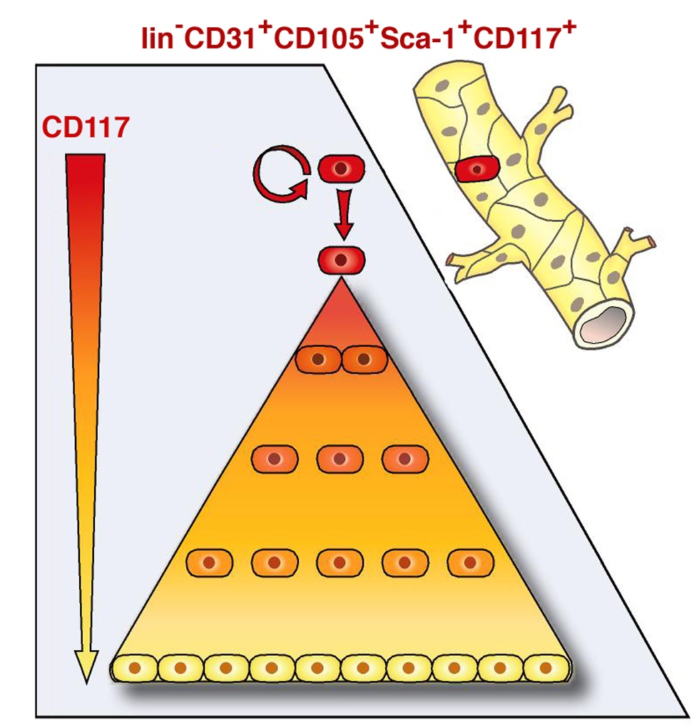
