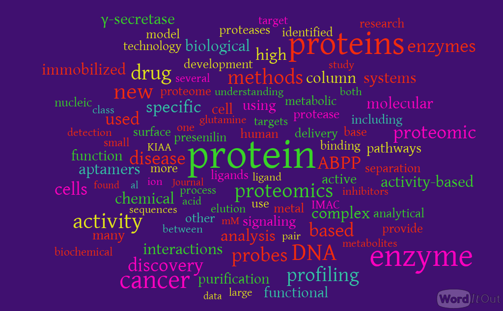
Advances in Separations Technology for the “OMICs” and Clarification of Therapeutic Targets
Curator, Reporter, EAW: Larry H Bernstein, MD, FCAP
This discussion is a continuation of an earlier piece on the technologic framework for , proteomics, nutrigenomics, and translational medicine. The last decade has seen the emergence of a genomic science that is changing the trajectory of biological sciences and medicine. It has not resolved all of our problems by any means, but it has begun to redraw the map, which began with the elucidation of major metabolic pathways in the first half of the 20th century, was then captured by the transformation of genetics with the discovery of the “Watson-Crick Model”, and then later was recharged with the discovery of the Toll-like receptor and the drawing of “signaling pathways”. What we have seen in an unraveling of protein-genome interactions, small peptide regulators, and dynamic changes in pathway dominance, bloackage, and reentry, depending on genetic, dietary, and environmental conditions, mostly expressed in what we refer to as “oxidative stress”.
Unraveling the multitude of nutrigenomic, proteomic, and metabolomic patterns that arise from the ingestion of foods or their bioactive food components will not be simple but is likely to provide insights into a tailored approach to diet and health. The use of new and innovative technologies, such as microarrays, RNA interference, and nanotechnologies, will provide needed insights into molecular targets for specific bioactive food components and how they harmonize to influence individual phenotypes. A challenging aspect of omic technologies is the refined analysis of quantitative dynamics in biological systems.
In recent years, nutrition research has moved from classical epidemiology and physiology to molecular biology and genetics. The new era of nutrition research translates empirical knowledge to evidence-based molecular science. Following this trend, Nutrigenomics has emerged as a novel and multidisciplinary research field in nutritional science that aims to elucidate how diet can influence human health. It is already well known that bioactive food compounds can interact with genes affecting transcription factors, protein expression and metabolite production. The study of these complex interactions requires the development of advanced analytical approaches combined with bioinformatics.
The Institute of Medicine recently convened a workshop to review the state of the various domains of nutritional genomics research and policy and to provide guidance for further development and translation of this knowledge into nutrition practice and policy. Nutritional genomics holds the promise to revolutionize both clinical and public health nutrition practice and facilitate the establishment of
- genome-informed nutrient and food-based dietary guidelines for disease prevention and healthful aging,
- individualized medical nutrition therapy for disease management, and
- better targeted public health nutrition interventions (including micronutrient fortification and supplementation) that maximize benefit and minimize adverse outcomes within genetically diverse human populations.
For metabolomics, gas and liquid chromatography coupled to mass spectrometry are well suited for coping with high sample numbers in reliable measurement times with respect to both technical accuracy and the identification and quantitation of small-molecular-weight metabolites. This potential is a prerequisite for the analysis of dynamic systems. Thus, metabolomics is a key technology for systems biology.
The bioavailability of bioactive food constituents as well as dose-effect correlations are key information to understand the impact of food on defined health outcomes. Both strongly depend on appropriate analytical tools to identify and quantify minute amounts of individual compounds in highly complex matrices–food or biological fluids–and to monitor molecular changes in the body in a highly specific and sensitive manner. Based on these requirements, mass spectrometry has become the analytical method of choice with broad applications throughout all areas of nutrition research.
Dynamic Construct of the –Omics
Metabolomics is a term that encompasses several types of analyses, including
- metabolic fingerprinting, which measures a subset of the whole profile with little differentiation or quantitation of metabolites;
- metabolic profiling, the quantitative study of a group of metabolites, known or unknown, within or associated with a particular metabolic pathway; and
- target isotope-based analysis, which focuses on a particular segment of the metabolome by analyzing only a few selected metabolites that comprise a specific biochemical pathway.
Any unifying concept of the metabolome was incomplete or debatable in the first 30 years of the 20th century. It was only known that insulin is anabolic and that insulin deficiency (or resistance) would have consequences in the point of entry into the citric acid cycle, which generates 28-32 ATPs. In fat catabolism, triglycerides are hydrolyzed to break them into fatty acids and glycerol. In the liver the glycerol can be converted into glucose via dihydroxyacetone phosphate and glyceraldehyde-3-phosphate by way of gluconeogenesis. In the case of this cycle there is a tie in with both catabolism and anabolism.
See Aerobic glucose and acetate metabolism. (from dos Santos MM, et al. EUKARYOTIC CELL 2003; 2:599–608)
For bypass of the Pyruvate Kinase reaction of Glycolysis, cleavage of 2 ~P bonds is required. The free energy change associated with cleavage of one ~P bond of ATP is insufficient to drive synthesis of phosphoenolpyruvate (PEP), since PEP has a higher negative DG of phosphate hydrolysis than ATP.
The two enzymes that catalyze the reactions for bypass of the Pyruvate Kinase reaction are the following:
- Pyruvate Carboxylase (Gluconeogenesis) catalyzes pyruvate + HCO3- + ATP — oxaloacetate + ADP + Pi
- PEP Carboxykinase (Gluconeogenesis) catalyzes: oxaloacetate + GTP —- phosphoenolpyruvate + GDP + CO2
Many high throughput methods have been employed to get some insight into the whole process and several examples of successful research. Proteomics and metabolomics need to encompass large numbers of experiments and linked data. Due to the nature of the proteins, as well as due to the properties of various metabolites, experimental approaches require the use of comprehensive high throughput methods and a sufficiency of analysed tissue or body fluids.
Ovesná J, Slabý O, Toussaint O, Kodícek M, et al. High throughput ‘omics’ approaches to assess the effects of phytochemicals in human health studies. Br J Nutr. 2008;99 E Suppl 1:ES127-34.
An important and revolutionary aspect of ‘The 2010 Project’ is that it implicitly endorses the allocation of resources to attempts to assign function to genes that have no known function. This represents a significant departure from the common practice of defining and justifying a scientific goal based on the biological phenomena. The rationale for endorsing this radical change is that for the first time it is feasible to envision a whole-systems approach to gene and protein function. I shall not discuss the emerging field of bioinformatics that makes this possible.
In this review, the end-of-the line “detector will be considered having been covered. The entire focus proceeds to a discussion of separation methods. Separation methods have always been tricky, time consuming, and a multiple step process that depended on using anionic and cationic resins as intermediate steps in bulk separation, and then molecular size separation. Therapeutic Targets will be identified as they are seen.
Affinity Chromatography
The rapid development of biotechnology and biomedicine requires more reliable and efficient separation technologies for the isolation and purification of biopolymers such as therapeutic proteins, antibodies, enzymes and nucleic acids. In particular, monoclonal antibodies are centrally important as therapeutics for the treatment of cancer and other diseases, leading to recombinant monoclonal antibodies that dominate today’s biopharmaceutical pipeline. The large-scale production of therapeutic biopolymers requires
- a manufacturing process that delivers reliability and in high-yield, as well as
- an effective purification process affording extremely pure products.
Because of its high selectivity, affinity chromatography has been used extensively to isolate a variety of biopolymers. The retention of solutes is based on specific, reversible interactions found in biological systems, such as the binding of an enzyme with an inhibitor or an antibody with an antigen. These interactions are exploited in affinity chromatography by immobilizing an affinity ligand onto a support, and using this as a stationary phase.
Non-porous particles having an average diameter of 2.1 mm were prepared by co-polymerization of styrene, methyl methacrylate and glycidyl methacrylate, which was abbreviated as P(S–MMA–GMA). The particles were mechanically stable due to the presence of benzene rings in the backbone of polymer chains, and could withstand high pressures when a column packed with these particles was operated in the HPLC mode.
The polymer particles were advantaged by immobilization of ligands via the epoxy groups on the particle surface that were introduced by one of the monomers, glycidyl methacrylate. As a model system, Cibacron Blue 3G-A was covalently immobilized onto the non-porous copolymer beads. The dye-immobilized P(S–MMA–GMA) particles were slurry packed into a 1.0 cm30.46 cm I.D. column. This affinity column was effective for the separation of turkey egg white lysozyme from a protein mixture. The bound lysozyme could be eluted to yield a sharp peak by using a phosphate buffer containing 1 M NaCl. For a sample containing up to 8 mg of lysozyme, the retained portion of proteins could be completely eluted without any slit peak. Due to the use of a shorter column, the analysis time was shorter in comparison with other affinity systems reported in the literature. The retention time could be reduced significantly by increasing the flow-rate, while the capacity factor remained at the same level.
CH Chen, WC Lee. Affinity chromatography of proteins on non-porous copolymerized particles of styrene, methyl methacrylate and glycidyl methacrylate. Journal of Chromatography A 2001; 921: 31–37.
Affinity separation membranes, consisting of electrospun nanofibers, have been developed recently. Affinity ligands are attached to the surface of the constituent fibers, offering a potential solution to some of the problems of traditional, column-based, affinity chromatography. Electrospun fibers are good candidates for use in affinity separation because of their
- unique characteristics of high surface area to volume ratio, resulting in
- high ligand loading, and
- their large porosity, resulting in
- high throughput operation.
A number of polymers have been used for electrospun fiber mesh-based affinity membrane separations including poly (ether-urethane-urea), cellulose, poly(ethylene terephthalate, polysulphone, and polyacrlonitrile. Typically, very thin electrospun fiber meshes are produced by electrostatically collecting negatively charged fibers on a collector electrode. These very thin 2D electrospun fiber mesh mats provide excellent solution permeability as compared to 3D column packed with affinity beads.
M Miyauchi, J Miao, TJ Simmons, JS Dordick and RJ Linhardt. Flexible Electrospun Cellulose Fibers as an Affinity Packing Material for the Separation of Bovine Serum Albumin. J Chromatograph Separat Techniq 2011; 2:2 http://dx.doi.org/10.4172/2157-7064.1000110
Dye Affinity Chromatography
Biomimetic Dyes
Affinity adsorbents based on immobilized triazine dyes offer important advantages circumventing many of the problems associated with biological ligands. The main drawback of dyes is their moderate selectivity for proteins. Rational attempts to tackle this problem are realized through the biomimetic dye concept according to which new dyes, the biomimetic dyes, are designed to mimic natural ligands. Biomimetic dyes are expected to exhibit increased affinity and purifying ability for the targeted proteins.
Biocomputing offers a powerful approach to biomimetic ligand design. The successful exploitation of contemporary computational techniques in molecular design requires the knowledge of the three-dimensional structure of the target protein, or at least, the amino acid sequence of the target protein and the three-dimensional structure of a highly homologous protein. From such information one can then design, on a graphics workstation,
- the model of the protein and also
- a number of suitable synthetic ligands which mimic natural biological ligands of the protein.
There are several examples of enzyme purifications
- trypsin
- urokinase
- kallikrein
- alkaline phosphatase
- malate dehydrogenase
- formate dehydrogenase
- oxaloacetate decarboxylase
- lactate dehydrogenase
where synthetic biomimetic dyes have been used successfully as affinity chromatography tools.
YD Clonis, NE Labrou, VPh Kotsira, C Mazitsos, et al. Biomimetic dyes as affinity chromatography tools in enzyme purification. Journal of Chromatography A 2000; 891: 33–44.
Interactions between Cibacron Blue F3GA (CB F3GA), as a model of triazine dye, and 2-hydroxypropyl-b-cyclodextrin (HP-b-CD), as a model of cyclodextrin, were investigated by monitoring the spectral shift that accompanies the binding phenomena. Matrix analysis of the difference spectral titration of CB F3GA with HP-b-CD revealed only two absorbing species, indicating a host–guest ratio of 1:1. The dissociation constant for this HP-b-CD–CB F3GA complex, K , was found d to be 0.43 mM. The data for HP-b-CD forming inclusion complexes with CB F3GA were used to develop the concept of competitive elution by inclusion complexes in dye-affinity chromatography.
When this concept was applied to the elution of L-lactate dehydrogenase from a CB F3GA affinity matrix, it was shown to be an effective elution strategy. It provided a 15-fold purification factor with 89% recovery and sharp elution profile (0.8 column volumes for 80% recovery), which is as good as that obtained by specific elution with NADH (16-fold, 78% recovery and 1.8 column volumes). In addition, the new elution strategy showed a better purification factor and sharper elution profile than traditional non-specific.
JA Lopez-Mas, SA Streitenberger, F Garcıa-Carmona, AA Sanchez-Ferrer. Cyclodextrin biospecific-like displacement in dye-affinity chromatography. Journal of Chromatography A 2001; 911: 47–53.
Affinity chromatography uses biospecific binding usually between an antibody and an antigen, an enzyme and a substrate or other pairs of key-lock type of matching molecules. Due to its high selectivity, it is able to purify proteins and other macromolecules from very dilute solutions. In this work, a general rate model for affinity chromatography was used for scale-up studies. Parameters for the model were estimated from existing correlations, or from experimental results obtained on a small column with the same packing material. As anexample, Affi-Gel with 4.5mol cm−3 Cibacron Blue F-3GA as immobilized ligands covalently attached to cross-linked 6% agarose was used for column packing. Cibacron Blue F-3GA was also used as a soluble ligand in the elution stage. Satisfactory scale-up predictions were obtained for a 98.2 ml column and a 501 ml column based on a few experimental data obtained on a 7.85 ml small column.
T. Gu, K.-H. Hsu and M.-J. Syu, “Scale-Up of Affinity Chromatography for Purification of Enzymes and Other Proteins.” Enzyme and Microbial Technology 2003; 33:433-437.
Affinity Column with AAAA as a Model Sense Ligand
The degeneracy of antisense peptides was studied by high-performance affinity chromatography. A model sense peptide (AAAA) and its antisense peptides (CGGG, GGGG, RGGG, SGGG) were designed and synthesized according to the degeneracy of genetic codes. An affinity column with AAAA as the ligand was prepared. The affinity chromatographic behaviors of antisense peptides on the column were evaluated. The results indicated that model antisense peptides have clear retention on the immobilized AAAA affinity column. RGGG showed the strongest affinity interaction.
R Zhao, X Yu, H Liu, L Zhai, S Xiong, et al. Study on the degeneracy of antisense peptides using affinity chromatography. Journal of Chromatography A 2001; 913: 421–428.
Frontal AC for Biomolecular Interactions
Frontal affinity chromatography is a method for quantitative analysis of biomolecular interactions. We reinforced it by incorporating various merits of a contemporary liquid chromatography system. As a model study, the interaction between an immobilized Caenorhabditis elegans galectin (LEC-6) and fluorescently labeled oligosaccharides (pyridylaminated sugars) was analyzed. LEC-6 was coupled to N-hydroxysuccinimide-activated Sepharose 4 Fast Flow (100 mm diameter), and packed into a miniature column (e.g., 1034.0 mm, 0.126 ml). The volume of the elution front (V) determined graphically for each sample was compared with that obtained in the presence of an excess amount of hapten saccharide, lactose (V ); and the dissociation constant, K , was calculated according to the literature. This system also proved to be useful for an inverse confirmation; that is, application of galectins to an immobilized glycan column (in the present case, asialofetuin was immobilized on Sepharose 4 Fast Flow), and the elution profiles were monitored by fluorescence based on tryptophan. The newly constructed system proved to be extremely versatile. It enabled rapid (analysis time 12 min/ cycle) and sensitive (20 nM for pyridylaminated derivatives, and 1 mg/ml for protein) analyses of lectin–carbohydrate interactions.
J Hirabayashi, Y Arata, K Kasai. Reinforcement of frontal affinity chromatography for effective analysis of lectin–oligosaccharide interactions. Journal of Chromatography A 2000; 890:261–271.
Immobilized Metal Ion Affinity
New immobilized metal ion affinity chromatography (IMAC) matrices containing a high concentration of metal–chelate moieties and completely coated with inert flexible and hydrophilic dextrans are here proposed to improve the purification of polyhistidine (poly-His) tagged proteins. The purification of an interesting recombinant multimeric enzyme (a thermoresistant b-galactosidase from Thermus sp. strain T2) has been used to check the performance of these new chromatographic media.
IMAC supports with a high concentration (and surface density) of metal chelate groups promote a rapid adsorption of poly-His tagged proteins during IMAC. However, these supports also favor the promotion of undesirable multi-punctual adsorptions and problems may arise for the simple and effective purification of poly-His tagged proteins. For example, desorption of the pure enzyme from the support may become quite difficult (e.g., it is not fully desorbed from the support even using 200 mM of imidazole).
The coating of these IMAC supports with dextrans greatly reduces these undesired multi-point adsorptions. However, this dextran coating of chromatographic matrices seems to allow the formation of strong one-point adsorptions that involve small areas of the protein and support surface, but the dextran coating seems to have dramatic effects for the prevention of weak or strong multipoint interactions that should involve a high geometrical congruence between the enzyme and the support surface.
C Mateo , G Fernandez-Lorente , BCC Pessela , A Vian, et al. Affinity chromatography of polyhistidine tagged enzymes. New dextran-coated immobilized metal ion affinity chromatography matrices for prevention of undesired multipoint adsorptions. Journal of Chromatography A 2001; 915:97–106.
The underlying principle of immobilized metal ion affinity chromatography (IMAC) of proteins is the coordination between the electron donor groupings on a protein surface (histidine, tryptophan, cysteine) and chelated (iminodiacetate; IDA) transition metal ions [IDA-M(II)]. This principle of immobilized metal ion affinity (IMA) has been presented by now in some detail. The practice of IMAC in the purification of proteins has had its empirical phase. There is now a need, from the body of data, to establish somewhat more detailed ground rules that would allow for the use of IMAC in a more predictive manner.
Immobilized metal ion affinity chromatography (IMAC) has been explored as a probe into the topography of histidyl residues of a protein molecule. An evaluation of the chromatographic behavior of selected model proteins-
- thioredoxin
- ubiquitin
- calmodulin
- lysozyme
- cytochrome c
- myoglobin
on immobilized transition metal ions
- Co2+
- Ni2+
- Cu2+
- Zn2
-allows establishment of the following facets of the histidyl side chain distribution:
- either interior or surface;
- when localized on the surface, accessible or unaccessible for coordination;
- single or multiple;
- When multiple, either distant or vicinal.
Moreover, proteins displaying single histidyl side chains on their surfaces may, in some instances, be resolved by IMAC; apparently, the microenvironments of histidyl residues are sufficiently diverse to result in different affinities for the immobilized metal ions. IMAC, previously introduced as an approach to the fractionation of proteins, has become also, upon closer examination, a facile probe into the topography of histidyl residues.
This is possible because of the inherent versatility of IMAC; an appropriate metal ion (M2+) can be selected to suit the analytical purpose and a particular chromatographic protocol can be applied (isocratic pH, falling pH, and imidazole elution). We now report that IMAC may be exploited as an analytical tool in addition to its use as a protein purification technique. IMAC can be used to ascertain several facets of the status of a histidyl residue(s) in a protein molecule:
- localization (interior vs. surface)
- coordination potential as defined by the steric accessibility and the state of protonation
- single vs. multiple
- surface density.
ES Hemdan, YJ Zhao, E Sulkowski, J Porath. Surface topography of histidine residues: A facile probe by immobilized metal ion affinity chromatography. Proc. Natl. Acad. Sci. USA 1989; 86: 1811-1815. Biochemistry.
A novel, two-step preparative technique is described for the purification of authentic recombinant human prolactin (rhPRL) secreted into the periplasm of transformed Escherichia coli cells. The first step is based on immobilized metal ion affinity chromatography of periplasmic extract, using Ni(II) as a relatively specific ligand for hPRL in this system. It gives superior resolution and yield than established ion-exchange chromatography. Size-exclusion chromatography is used for further purification to .99.5% purity. The methodology is reproducible, leading to 77% recovery. Identity and purity of the rhPRL were demonstrated using sodium dodecylsulphate–polyacrylamide electrophoresis, isoelectric focusing, mass spectrometry (matrix-assisted laser desorption ionization time-of-flight), radioimmunoassay, RP-HPLC and high-performance size-exclusion chromatography. In the Nb2 bioassay, the hormone showed a bioactivity of 40.9 IU/mg.
EKM Ueda, PW Gout, L Morgantia. Ni(II)-based immobilized metal ion affinity chromatography of recombinant human prolactin from periplasmic Escherichia coli extracts. Journal of Chromatography A 2001; 922:165–175.
Adenosine Affinity Ligand for Glutamine Synthase
Glutamine synthetase has been purified from both procaryotic and eucaryotic sources using various types of affinity chromatography. For example, ADP-agarose has been used to purify glutamine synthetase from photosynthetic bacteria, while the related “Blue” chromatography media (e.g. Affigel Blue) have been used to purify glutamine synthetases from a variety of sources. In addition, 2’,5’-ADPSepharose 4B has been used to purify glutamine synthetase from procaryotes, plants and insects. However, this latter affinity ligand resembles NADP more than ADP, particularly with respect to the position of the phosphate moieties. This is reflected in the more general use of this affinity ligand in the purification of NADPH-dependent enzymes.
In the present report, we characterize the ability of glutamine synthetase to be purified by three different adenosine-affinity ligands: 5’-ADP-agarose (an ADP analogue), 2’,5’-ADP-Sepharose 4B (an NADP analogue) and 3’,5’-ADP-agarose (a cyclic AMP analogue). We report conditions for the successful purification of insect flight muscle glutamine synthetase using each of these three different affinity ligands.
The enzyme bound most strongly to the
- ADP analogue (S-ADP-agarose),
- followed by the NADPH analogue (2’,5’-ADP-Sepharose 4B), and least strongly to
- the cyclic AMP analogue (3’J’-ADP-agarose).
In all cases, binding was strongest in the presence of Mn2+ when compared to Mg”. These results suggest that the binding of glutamine synthetase to adenosine-affinity media is related to the participation of Mn. ADP in the y-glutamyl transferase reaction that is catalyzed by glutamine synthetase.
M Dowton, IR Kennedy. Purification of glutamine synthetase by adenosine-affinity chromatography. Journal of Chromatography A 1994; 664: 280-283
Aptamer Based Stationary Phase
An anti-adenosine aptamer was evaluated as a stationary phase in packed capillary liquid chromatography. Using an 21 aqueous mobile phase containing 20 mM Mg , adenosine was strongly retained on the column. A gradient of increasing 21 Ni (to 18 mM), which is presumed to complex with nitrogen atoms in adenosine involved in binding to the aptamer, eluted adenosine in a narrow zone. The adenosine assay, which required no sample preparation, was used on microdialysis samples. Total analysis times were short so samples could be injected every 5 min.
Q Deng, CJ Watson, RT Kennedy. Aptamer affinity chromatography for rapid assay of adenosine in microdialysis samples collected in vivo. Journal of Chromatography A 2003; 1005:123–130.
We will realize the full power of proteomics only when we can measure and compare the proteomes of many individuals to identify biomarkers of human health and disease and track the blood-based proteome of an individual over time. Because the human proteome contains an estimated 20,000 proteins – plus splicing and post-translational variants – that span a concentration range of ,12 logs, identifying and quantifying valid biomarkers is a great technical challenge.
Proteomic measurements demand
- extreme sensitivity
- specificity
- dynamic range
- accurate quantification.
We describe a new class of DNA-based aptamers enabled by a versatile chemistry technology that endows nucleotides with protein-like functional groups. These modifications greatly expand the repertoire of targets accessible to aptamers.
The resulting technology provides efficient, large-scale selection of exquisite protein-binding reagents selected specifically for use in highly multiplexed proteomics arrays.
Aptamers are a class of nucleic acid-based molecules discovered twenty years ago, and have since been employed in diverse applications including
- therapeutics
- catalysis
- proteomics
Aptamers are short single-stranded oligonucleotides, which fold into diverse and intricate molecular structures that bind with high affinity and specificity to
- proteins
- peptides
- small molecules.
Aptamers are selected in vitro from enormously large libraries of randomized sequences by the process of Systematic Evolution of Ligands by EXponential enrichment (SELEX). A SELEX library with 40 random sequence positions has 440 (,1024) possible combinations and a typical selection screens 1014–1015 unique molecules. This is on the order of 105 times larger than standard peptide or protein combinatorial molecular libraries.
The interrogation of proteomes (‘‘proteomics’’) in a highly multiplexed and efficient manner remains a coveted and challenging goal in biology and medicine. We present a new aptamer-based proteomic technology for biomarker discovery capable of simultaneously measuring thousands of proteins from small sample volumes (15 mL of serum or plasma).
Our current assay measures 813 proteins with low limits of detection (1 pM median), 7 logs of overall dynamic range (,100 fM–1 mM), and 5% median coefficient of variation. This technology is enabled by a new generation of aptamers that contain chemically modified nucleotides, which greatly expand the physicochemical diversity of the large randomized nucleic acid libraries from which the aptamers are selected. Proteins in complex matrices such as plasma are measured with a process that transforms a signature of protein concentrations into a corresponding signature of DNA aptamer concentrations, which is quantified on a DNA microarray.
Our assay takes advantage of the dual nature of aptamers as both folded protein-binding entities with defined shapes and
unique nucleotide sequences recognizable by specific hybridization probes.
This is a versatile and powerful tool that allows large-scale comparison of proteome profiles among discrete populations. This unbiased and highly multiplexed search engine will enable the discovery of novel biomarkers in a manner that is unencumbered by our incomplete knowledge of biology, thereby helping to advance the next generation of evidence-based medicine.
L Gold, D Ayers, J Bertino, Christopher Bock, et al. Aptamer-Based Multiplexed Proteomic Technology for Biomarker Discovery. PlosONE 2010; 5 (12): e15004
Biomarker Discovery, Diagnostics, and Therapeutics
Progression from health to disease is accompanied by complex changes in protein expression in both the circulation and affected tissues. Large-scale comparative interrogation of the human proteome can offer insights into disease biology as well as lead to
- the discovery of new biomarkers for diagnostics
- new targets for therapeutics
- can identify patients most likely to benefit from treatment.
Although genomic studies provide an increasingly sharper understanding of basic biological and pathobiological processes, they ultimately only offer a prediction of relative disease risk, whereas proteins offer an immediate assessment of “real-time” health and disease status.
We have recently developed a new proteomic technology, based on modified aptamers, for biomarker discovery that is capable of simultaneously measuring more than a thousand proteins from small volumes of biological samples such as plasma, tissues, or cells. Our technology is enabled by SOMAmers (Slow Off-rate Modified Aptamers), a new class of protein binding reagents that contain chemically modified nucleotides that greatly expand the physicochemical diversity of nucleic acid-based ligands. Such modifications introduce functional groups that are absent in natural nucleic acids but are often found in protein-protein, small molecule-protein, and antibody-antigen interactions. The use of these modifications expands the range of possible targets for SELEX (Systematic Evolution of Ligands by EXponential Enrichment), results in improved binding properties, and facilitates selection of SOMAmers with slow dissociation rates. Our assay works by transforming protein concentrations in a mixture into a corresponding DNA signature, which is then quantified on current commercial DNA microarray platforms. In essence, we take advantage of the dual nature of SOMAmers as
- both folded binding entities with defined shapes and
- unique nucleic acid sequences recognizable by specific hybridization probes.
Mehan MR, Ostroff R, Wilcox SK, Steele F, et al. Highly multiplexed proteomic platform for biomarker discovery, diagnostics, and therapeutics. Adv Exp Med Biol. 2013; 734:283-300.
Aptamers and Smart Drug delivery Targeting
In this review, the strategies for using functional nucleic acids in creating smart drug delivery devices will be explained, as their has been very recent progress in controlled drug release based on molecular gating achieved with aptamers. Aptamers are functional nucleic acid sequences which can bind specific targets.
An artificial combinatorial methodology can identify aptamer sequences for any target molecule, from ions to whole cells. Drug delivery systems seek to increase efficacy and reduce side-effects by concentrating the therapeutic agents at specific disease sites in the body. This is generally achieved by specific targeting of inactivated drug molecules.
Aptamers which can bind to various cancer cell types selectively and with high affinity have been exploited in a variety of drug delivery systems for therapeutic purposes. Recent progress in selection of cell-specific aptamers has provided new opportunities in targeted drug delivery. Especially functionalization of nanoparticles with such aptamers has drawn major attention in the biosensor and biomedical areas.
Nucleic acids are recognized as attractive building materials in nanomachines because of their unique molecular recognition properties and structural features. An active controlled delivery of drugs once targeted to a disease site is a major research challenge. Stimuli-responsive gating is one way of achieving controlled release of nanoparticle cargoes. Recent reports incorporate the structural properties of aptamers in controlled release systems of drug delivering nanoparticles.
Nanoparticle-encapsulated drug delivery aims to deliver the active therapeutic ingredients to the disease site in stable compartments in order to reduce premature release. This ensures that the effects of drug are maximized and the side effects are reduced. An encapsulated nanoparticle system requires a specific targeting mechanism and at the same time the retention of drugs inside the container should be high. The balance between specificity of targeting and the extent of premature leakage determines the success of a given delivery system.
Nanotechnology research approaches in drug delivery include a wide variety of nanomaterials ranging from soft hydrogels to solid polymeric particles. Large surface area, high drug loading efficiency and potential combination with other organic/inorganic materials are the main properties of hollow nanostructures that are attractive for biomedical applications.
Packaging of small-molecule drugs
- improves their availability
- compatibility
- reduces toxicity
Controlling the drug release profile is the main challenge in drug delivery development when the drug is to be successfully targeted to a specific site. Stimuli-responsive materials have been created by using biological, physical and chemical properties of materials for heat-activated, light-activated or pH-activated delivery. Nucleic acids are utilized to construct rationally designed nanostructures at molecular levels for nanotechnology applications. Integration of the properties of nucleic acids can offer many opportunities for drug delivery systems, including stimuli-responsive nanogates for nanocarriers and molecular sensors. Favorable drug release kinetics can be achieved at the target sites by aptamer-based capping systems.
VC Ozalp, F Eyidogan and HA Oktem. Aptamer-Gated Nanoparticles for Smart Drug Delivery.
Pharmaceuticals 2011, 4, 1137-1157; doi:10.3390/ph4081137. ISSN 1424-8247. http://www.mdpi.com/journal/pharmaceuticals
Activity Based Profiling
Powerful strategies for the gel-free analysis of proteomes have emerged, including isotope-coded affinity tagging (ICAT) for quantitative proteomics and multidimensional protein identification technology (MudPIT) for comprehensive proteomics, both of which utilize liquid chromatography (LC) and MS for protein separation and detection, respectively.
Nonetheless, these methods, like 2DE-MS, still focus on measuring changes in protein abundance and, therefore, provide only an indirect estimate of dynamics in protein function. Indeed, several important forms of post-translational regulation, including protein–protein and protein–small-molecule interactions, may elude detection by abundance-based proteomic methods.
To facilitate the analysis of protein function, several proteomic methods have been introduced to characterize the activity of proteins on a global scale. These include large-scale yeast two-hybrid screens and epitope tagging immunoprecipitation experiments, which aim to construct comprehensive maps of protein–protein interactions, and protein microarrays, which aim to provide an assay platform for the rapid assessment of protein activities. A chemical proteomic strategy referred to as activity-based protein profiling (ABPP) has emerged that utilizes active site-directed probes to profile the functional state of enzyme families directly in complex proteomes.
Recent advances in genomic and proteomic technologies have begun to address the challenge of assigning molecular and cellular functions to the numerous protein products encoded by prokaryotic and eukaryotic genomes. In particular, chemical strategies for proteome analysis have emerged that enable profiling of protein activity on a global scale. Herein, we highlight these chemical proteomic methods and their application to the discovery and characterization of disease-related enzyme activities.
N Jessani and BF Cravatt. The development and application of methods for activity-based protein profiling. Current Opinion in Chemical Biology 2004; 8:54–59. In Proteomics and genomics, M Snyder and J Yates III, eds. 2003 Elsevier Ltd. DOI: 10.1016/ j.cbpa.2003.11.004
Cells with fundamental metabolic alterations commonly arise during tumorigenesis, and it is these types of changes that help to establish a biochemical foundation for disease progression and malignancy. A seminal example of this was discovered in the 1920s when Otto Warburg found that cancer cells consume higher levels of glucose and secrete most of the glucose carbon as lactate rather than oxidizing it completely.
Since then, studies by multiple groups have uncovered a diverse array of metabolic changes in cancer, including
alterations in
- glycolytic pathways
- the citric acid cycle
- glutaminolysis
- lipogenesis
- lipolysis
- proteolysis
These in turn modulate the levels of cellular building blocks
- lipids, nucleic acids and amino acids,
- cellular energetics,
- oncogenic signaling molecules
- the extracellular environment to confer protumorigenic and malignant properties.
Despite these advances, our current understanding of cancer metabolism is far from complete and would probably benefit from experimental strategies that are capable of profiling enzymatic pathways on a global scale. To this end, conventional genomic and proteomic methods, which comparatively quantify the expression levels of transcripts and proteins, respectively, have yielded many useful insights. These platforms are, however, limited in their capacity to identify changes in protein activity that are caused by posttranslational mechanisms.
Annotating biochemical pathways in cancer is further complicated by the potential for enzymes to carry out distinct metabolic activities in tumor cells that might not be mirrored in normal physiology. In addition, a substantial proportion of the human proteome remains functionally uncharacterized, and it is likely that at least some of these poorly understood proteins also have roles in tumorigenesis. These challenges require new proteomic technologies that can accelerate the assignment of protein function in complex biological systems, such as cancer cells and tumors.
Metabolomics has emerged as a powerful approach for investigating enzyme function in living systems. Metabolomic experiments in the context of enzyme studies typically start with
- the extraction of metabolites from control and enzyme-disrupted biological systems,
- followed by metabolite detection and comparative data analysis.
For example, lipophilic metabolites can be enriched from cells or tissues by organic extraction.
Mass spectrometry (MS) has become a primary analytical method for surveying metabolites in complex biological samples, with upfront separation accomplished by liquid chromatography (LC–MS) or gas chromatography (GC–MS). MS experiments can be carried out using
- targeted or untargeted approaches,
- depending on whether the objective is
- to profile and quantitate known metabolites or
- to broadly scan for metabolites across a large mass range, respectively.
As metabolomic experiments generate a large amount of data, powerful software tools are needed for identification and quantitation of ions in LC–MS data sets (see the figure; the mass to charge ratio (m/z) is indicated). One such program is XCMS95, which
- aligns,
- quantifies and
- statistically ranks ions that are altered between two sets of metabolomic data.
This program can be used to rapidly identify metabolomic signatures of various disease states or to assess metabolic networks that are regulated by an enzyme using pharmacological or genetic tools that modulate enzyme function. Additional databases assist in metabolite structural characterization, such as HMDB96,97, METLIN98,99 and LIPID MAPS100.
In this Review, we discuss one such proteomic platform, termed activity based protein profiling (ABPP), and its implementation in the discovery and functional characterization of deregulated enzymatic pathways in cancer. We discuss the evidence that, when coupled with other large scale profiling methods, such as metabolomics and proteomics, ABPP can provide a compelling, systems level understanding of biochemical networks that are important for the development and progression of cancer.
Large-scale profiling methods have uncovered numerous gene and protein expression changes that correlate with tumorigenesis. However, determining the relevance of these expression changes and which biochemical pathways they affect has been hindered by our incomplete understanding of the proteome and its myriad functions and modes of regulation. Activity-based profiling platforms enable both the discovery of cancer-relevant enzymes and selective pharmacological probes to perturb and characterize these proteins in tumour cells. When integrated with other large-scale profiling methods, activity-based proteomics can provide insight into the metabolic and signaling pathways that support cancer pathogenesis and illuminate new strategies for disease diagnosis and treatment.
Representative activity-based probes and their application to cancer research
- enzyme class applications in cancer
- Serine hydrolases increased KIAA1363 and MAGL
- aggressive human cancer lines
- uPA and tPA serine protease aggressive cancers
- RBBP9 activity in pancreatic carcinoma
- Metalloproteinases neprilysin activity in melanoma cell lines
- Cysteine proteases cathepsin cysteine protease in pancreatic islet tumours
- Kinases Inhibitor selectivity profiling of kinase inhibitors
- Caspases visualization of apoptosis in colon tumour-bearing mice treated with Apomab
- Deubiquitylases Identified increased carboxy-terminal hydrolase UCHL3 and UCH37 activity in HPV cervical carcinomas
- Cytochrome P450s Identified the aromatase inhibitor anastrazole as an inducer of CYP1A2
Serine hydrolases KIaa1363 and MaGL regulate lipid metabolic pathways that support cancer pathogenesis. Activity-based protein profiling (ABPP) identified
- KIAA1363 and
- monoacylglycerol (MAG) lipase (MAGL)
as being increased in aggressive human cancer cells from multiple tumour types. Pharmacological and/or RNA interference ablation of KIAA1363 and MAGL coupled with metabolomic analysis revealed specific roles for KIAA1363 and MAGL in cancer metabolism. Disruption of KIAA1363 by the small-molecule inhibitor AS115 lowered monoalkylglycerol ether (MAGE), alkyl lysophosphatidic acid (alkyl LPA) and alkyl lysophosphatidyl choline (alkyl LPC) levels in cancer cells. Disruption of MAGL by the small-molecule inhibitor JZL184 raised MAG levels and reduced free fatty acid, lysophosphatidic acid (LPA) and prostaglandin E2 (PGE2) levels in cancer cells. Disruption of KIAA1363 and MAGL leads to impairments in cancer cell aggressiveness and tumour growth, PAF, platelet-activating factor.
At a glance
• Activity-based protein profiling (ABPP) facilitates the discovery of deregulated enzymes in cancer.
• Competitive ABPP yields selective inhibitors for functional characterization of cancer enzymes.
• ABPP can be integrated with metabolomics to map deregulated enzymatic pathways in cancer.
• ABPP can be integrated with other proteomic methods to map proteolytic pathways in cancer.
• ABPP probes can be used to image tumour development in living animals.
DK Nomura, MM Dix and BF Cravatt. Activity-based protein profiling for biochemical pathway discovery in cancer. Nature Reviews. Cancer. 2010; 10: 630-638.
Activity-based protein profiling (ABPP), the use of active site-directed chemical probes to monitor enzyme function in complex biological systems, is emerging as a powerful post-genomic technology. ABPP probes have been developed for several enzyme classes and have been used to inventory enzyme activities en masse for a range of (patho)physiological processes.
ABPP uses active site–directed, small molecule–based covalent probes to report on the functional state of enzyme activities directly in native biological systems. ABPP probes are designed or selected to target a subset of the proteome based on shared principles of binding and/or reactivity and have been successfully developed for many enzyme classes, including
- serine
- cysteine,
- aspartyl
- metallo hydrolases
- kinases
- glycosidases
- histone deacetylases and
- oxidoreductases.
These probes have been shown to selectively label active enzymes but not their inactive precursor (zymogen) or inhibitor-bound forms, thus allowing researchers to capture functional information that is beyond the scope of standard proteomic methods.
By presenting specific examples, we show here that ABPP provides researchers with a distinctive set of chemical tools to embark on the assignment of functions to many of the uncharacterized enzymes that populate eukaryotic and prokaryotic proteomes.
Reactive group Enzyme Enzyme class
Benzophenone Presenilins Aspartyl protease (γ-secretase )
Bromoethyl HSPC263 (OTU domain) Deubiquitinating enzyme (DUB)
Vinyl-methylester UL from HSV-1 Deubiquitinating enzyme (DUB)
Aryl 2-deoxy-2-fluoro glycoside Cfx from C. fimi Glycosidase (β-1-4-glycanase)
Fluorophosphonate SAE Serine hydrolase
Examples of enzymes assigned to specific mechanistic classes by ABPP
ABPP can also be implemented as a direct assay for inhibitor discovery, allowing researchers to develop potent and selective pharmacological probes for uncharacterized enzymes.
Examples of enzymes assigned to specific mechanistic classes by ABPP.
- Probe Leu-Asp-αCA probe selectively labeled Upβ
- Substrate the endogenous Upβ substrate, N-carbamoyl-β-alanine
- Substrate mimicry of an ABPP probe.
Multidimensional profiling strategy for the annotation of the cancer-related enzyme KIAA1363. ABPP using fluorophosphonate probes identified KIAA1363 as a highly elevated enzyme activity in aggressive cancer cells. Competitive ABPP was then used to develop a selective KIAA1363 inhibitor (AS115). Metabolomic analysis of cancer cells treated with AS115 determined a role for this enzyme in the regulation of MAGE lipids in cancer cells. Biochemical studies confirmed that KIAA1363 acts as 2-acetyl MAGE hydrolase in a metabolic network that bridges the platelet activating factor and lysophosphatidic acid classes of signaling lipids.
Assignment of enzyme mechanism by ABPP
There are multiple levels of annotation for enzymes. The most basic level is assignment to a specific mechanistic class based on the general chemical reaction catalyzed by the enzyme (for example, hydrolase, kinase, oxidoreductase and others). Additional annotation involves determining the endogenous substrates and products for the enzyme. Finally, complete annotation requires an understanding of how the specific chemical transformation(s) catalyzed by an enzyme integrate into larger metabolic and signaling pathways to influence cell physiology and behavior.
Many of the predicted enzymes uncovered by genome sequencing projects can be assigned to a mechanistic class or ascribed a putative biochemical function based on sequence homology to well-characterized enzymes. But some enzymes have insufficient sequence relatedness for class assignment or have a function different from that predicted by sequence comparisons. ABPP has facilitated class annotation for several such uncharacterized enzymes.
KT Barglow & BF Cravatt. Activity-based protein profiling for the functional annotation of enzymes. Nature Methods 2007; 4(10): 822- 827. DOI:10.1038/NMETH1092
A principal goal of modern biomedical research is to discover, assemble, and experimentally manipulate molecular pathways in cells and organisms to reveal new disease mechanisms.
Toward this end, complete genome sequences for numerous bacteria and higher organisms, including humans, have laid the fundamental groundwork for understanding the molecular basis of life in its many forms. However, the information content of DNA sequences is limited and, on its own, cannot describe most physiological and pathological processes.
Unlike oligonucleotides, proteins are a very diverse group of biomolecules that display a wide range of chemical and biophysical features, including
- membrane-binding,
- hetero/homo-oligomerization, and
- posttranslational modification.
The biochemical complexity intrinsic to protein science intimates that several complementary analytical strategies will be needed to achieve the ultimate goal of proteomics – a comprehensive characterization of the expression, modification state, interaction map, and activity of all proteins in cells and tissues.
A powerful LC-MS strategy for proteomics involves the use of isotope-coded affinity tags (ICAT). This approach enables the comparison of protein expression in proteomes by treating samples with isotopically distinct forms of a chemical labeling reagent. ICAT methods provide superior resolving power compared to gel-based methods and improve access to membrane-associated proteins. More recently, isotope-free MS methods for quantitative proteomics have emerged.
Reverse protein microarrays have also been described in which proteomes themselves are arrayed and the antibodies used for detection in a format analogous to Western blotting. In addition to increasing the throughput of proteomic experiments by integrating the protein separation and detection steps, microarrays consume much less material than conventional proteomic methods. Still, the general application of microarrays for proteomics is currently limited by the availability of high-quality capture reagents (e.g., antibodies, aptamers, etc).
These approaches, by measuring protein abundance provide, like genomics, only an indirect assessment of protein activity and may fail to detect important posttranslational events that regulate protein function, such as protein–protein or protein–small-molecule interactions. To address these limitations, complementary strategies for the functional analysis of proteins have been introduced. Prominent among these functional proteomic efforts is the use of chemistry for the design of active site-directed probes that measure enzyme activity in samples of high biological complexity.
Many post-translational modes of enzyme regulation share a common mechanistic foundation – they perturb the active site such that catalytic power and/or substrate recognition is impaired. Accordingly, it was hypothesized that chemical probes capable of reporting on the integrity of enzyme active sites directly in cells and tissues might serve as effective functional proteomic tools. These activity based protein profiling (ABPP) probes consist of at least two general elements:
- a reactive group for binding and covalently modifying the active sites of many members of a given enzyme class or classes
- a reporter tag for the detection, enrichment, and identification of probe-labeled proteins
ABPP probes have been successfully developed for more than a dozen enzyme classes, including
- all major classes of proteases
- kinases
- phosphatases
- glycosidases
- GSTs
- oxidoreductases.
Post-translational regulation of enzyme activity. Many enzymes are produced as inactive precursors, or zymogens, which require proteolytic processing for activation. Enzyme activity can be further regulated by interactions with endogenous protein inhibitors.
The field of proteomics aims to develop and apply technologies for the characterization of protein function on a global scale. Toward this end, synthetic chemistry has played a major role by providing new reagents to profile segments of the proteome based on activity rather than abundance. Small molecule probes for activity-based protein profiling have been created for more than a dozen enzyme classes and used to discover several enzyme activities elevated in disease states. These innovations have inspired complementary advancements in analytical chemistry, where new platforms have been introduced to augment the information content achievable in chemical proteomics experiments. Here, we will review these analytical platforms and discuss how they have exploited the versatility of chemical probes to gain unprecedented insights into the function of proteins in biological samples of high complexity.
Advanced analytical platforms utilize a range of separation and detection strategies, including LC-MS, CELIF, and antibody microarrays, to achieve an unprecedented breadth and depth of proteome coverage in ABPP investigations. The complementary strengths and weaknesses of each of these methods suggest that the selection of an appropriate analytical platform should be guided by the specific experimental question being addressed.
SA Sieber and BF Cravatt. Analytical platforms for activity-based protein profiling – exploiting the versatility of chemistry for functional proteomics. Chem. Commun. 2006, 2311–2319. http://www.rsc.org/chemcomm
Diagnostic Therapeutics in Activity Based Probes
Activity-based chemical proteomics-an emerging field involving a combination of organic synthesis, biochemistry, cell biology, biophysics and bioinformatics-allows the detection, visualisation and activity quantification of whole families or selected sub-sets of proteases based upon their substrate specificity. This approach can be applied for drug target/lead identification and validation, the fundamentals of drug discovery. The activity-based probes discussed in this review contain three key features;
- a ‘warhead’ (binds irreversibly but selectively to the active site),
- a ‘tag’ (allowing enzyme ‘handling’, with a combination of fluorescent, affinity and/or radio labels),
- a linker region between warhead and tag.
From the design and synthesis of the linker arise some of the latest developments discussed here; not only can the physical properties (e.g., solubility, localisation) of the probe be tuned, but the inclusion of a cleavable moiety allows selective removal of tagged enzyme from affinity beads etc.
Heal WP, Wickramasinghe SR, Tate EW. Activity based chemical proteomics: profiling proteases as drug targets. Curr Drug Discov Technol 2008; 5(3):200-12. PMID: 18690889
The genomic revolution has created a wealth of information regarding the fundamental genetic code that defines the inner workings of a cell. However, it has become clear that analyzing genome sequences alone will not lead to new therapies to fight human disease. Rather, an understanding of protein function within the context of complex cellular networks will be required to facilitate the discovery of novel drug targets and, subsequently, new therapies directed against them. The past ten years has seen a dramatic increase in technologies that allow large-scale, systems-based methods for analysis of global biological processes and disease states.
In the field of proteomics, several well-established methods persist as a means to resolve and analyze complex mixtures of proteins derived from cells and tissues. However, the resolving power of these methods is often challenged by the diverse and dynamic nature of the proteome. The field of activity-based proteomics, or chemical proteomics, has been established in an attempt to focus proteomic efforts on subsets of physiologically important protein targets. This new approach to proteomics is centered around the use of small molecules termed activity-based probes (ABPs) as a means to tag, enrich, and isolate, distinct sets of proteins based on their enzymatic activity.
Berger AB, Vitorino PM, Bogyo M. Activity-based protein profiling: applications to biomarker discovery, in vivo imaging and drug discovery. Am J Pharmacogenomics. 2004;4(6):371-81.
Recent advances in global genomic and proteomic methods have led to a greater understanding of how genes and proteins function in complex networks within a cell. One of the major limitations in these methodologies is their inability to provide information on the dynamic, post-translational regulation of enzymatic proteins. In particular proteases are often synthesized as inactive zymogens that need to be activated in order to carry out specific biological processes. Thus, methods that allow direct monitoring of protease activity in the context of a living cell or whole animal will be required to begin to understand the systems-wide functional roles of proteases. In this review, we discuss the development and applications of activity based probes (ABPs) to study proteases and their role in pathological processes. Specifically we focus on application of this technique for biomarker discovery, in vivo imaging and drug screening.
Fonović M, Bogyo M. Activity based probes for proteases: applications to biomarker discovery, molecular imaging and drug screening. Curr Pharm Des. 2007;13(3):253-61.
Proteases, in particular, are known for their multilayered post-translational activity regulation that can lead to a significant difference between protease abundance levels and their enzyme activity. To address these issues, the field of activity-based proteomics has been established in order to characterize protein activity and monitor the functional regulation of enzymes in complex proteomes.
Fonović M, Bogyo M. Activity-based probes as a tool for functional proteomic analysis of proteases. Expert Rev Proteomics. 2008; 5(5):721-30. PMID: 18937562. PMCID: PMC2997944
As a result of the recent enormous technological progress, experimental structure determination has become an integral part of the development of drugs against disease-related target proteins. The post-translational modification of proteins is an important regulatory process in living organisms; one such example is lytic processing by peptidases. Many different peptidases represent disease targets and are being used in structure-based drug design approaches. The development of drugs such as aliskiren and tipranavir, which inhibit renin and HIV protease, respectively, testifies to the success of this approach.
Mittl PR, Grütter MG. Opportunities for structure-based design of protease-directed drugs.
Curr Opin Struct Biol 2006; 16(6):769-75. Epub 2006 Nov 16. PMID: 17112720
Presenilin is the catalytic component of γ-secretase, a complex aspartyl protease and a founding member of intramembrane-cleaving proteases. γ-Secretase is involved in the pathogenesis of Alzheimer’s disease and a top target for therapeutic intervention. However, the protease complex processes a variety of transmembrane substrates, including the Notch receptor, raising concerns about toxicity. Nevertheless, γ-secretase inhibitors and modulators have been identified that allow Notch processing and signaling to continue, and promising compounds are entering clinical trials.
Molecular and biochemical studies offer a model for how this protease hydrolyzes transmembrane domains in the confines of the lipid bilayer. Progress has also been made toward structure elucidation of presenilin and the γ-secretase complex by electron microscopy as well as by studying cysteine-mutant presenilins. The signal peptide peptidase (SPP) family of proteases are distantly related to presenilins. However, the SPPs work as single polypeptides without the need for cofactors and otherwise appear to be simple model systems for presenilin in the γ-secretase complex.
Critical clues to the identity of γ-secretase included:
(1) Genes encoding the multi-pass membrane proteins presenilin-1 and presenilin-2 are, like APP, associated with familial, early-onset Alzheimer’s disease. The disease-causing missense mutations were found to alter how γ-secretase cuts APP, leading to increased proportions of longer, more aggregation-prone forms of Aβ.
(2) Knockout of presenilin genes eliminates γ-secretase cleavage of APP.
(3) Peptidomimetics that inhibit γ-secretase contain moieties typically found in aspartyl protease inhibitors.
These findings led to the identification of two conserved transmembrane aspartates in the multi-pass presenilins that are critical for γ-secretase cleavage of APP, evidence that presenilins are aspartyl proteases.
Presenilin is endoproteolytically cleaved into two polypeptides, an N-terminal fragment (NTF) and a C-terminal fragment (CTF), the formation of which is
- regulated
- metabolically stable
- part of a high-molecular weight complex
suggesting that the NTF-CTF heterodimer is the biologically active form. NTF and CTF each contribute one of the essential and conserved aspartates, and transition-state analogue inhibitors of γ-secretase, compounds designed to interact with the active site of the protease, bind directly to presenilin NTF and CTF.
Presenilins are also required for Notch signaling (Levitan and Greenwald, 1995), a pathway essential for cell differentiation during development and beyond.
The highly conserved role of γ-secretase in Notch signalling and its importance in development led to genetic screens in Caenorhabditis elegans that identified three other integral membrane proteins besides presenilin that modify Notch signaling.
Designed inhibitors have proven to be useful tools in understanding the mechanism of γ-secretase and substrate recognition – affinity labelling with transition-state analogue inhibitors showed binding at the interface between the presenilin NTF and CTF subunits, consistent with the active site residing at this interface, with each presenilin subunit contributing one of the essential aspartates.
The concept of presenilin as the catalytic component for γ-secretase was considerably strengthened when
- signal peptide peptidase (SPP) was found to be a similar intramembrane aspartyl protease
- SPP is exploited by the hepatitis C virus for the maturation of its core protein, suggesting that this protease may be a suitable target for antiviral therapy
- SPP was identified by affinity labeling with a peptidomimetic inhibitor, and the protein sequence displayed similarities with presenilin.
- SPP contains two conserved aspartates, each predicted to lie in the middle of a transmembrane domain, and the aspartate-containing sequences resemble those found in presenilins.
- SPP appears to be less complicated than γ-secretase.
Expression of human SPP in yeast reconstituted the protease activity, suggesting that the protein has activity on its own and does not require other mammalian protein cofactors.
Aspartyl I-CLiPs are found in all forms of life and play essential roles in biology and disease. How these enzymes carry out hydrolysis in the membrane is a fascinating question that is not entirely resolved, but evidence suggests an initial substrate docking site and a lateral gate into a pore where water and the active site aspartates reside. Designed inhibitors have been critical in elucidating these mechanisms, but inhibitors targeting γ-secretase for the treatment of Alzheimer’s disease must avoid interfering with Notch signaling.
MS Wolfe. Structure, Mechanism and Inhibition of γ-Secretase and Presenilin-Like Proteases.
Biol Chem. 2010 August; 391(8): 839–847. doi: 10.1515/BC.2010.086. PMCID: PMC2997569. NIHMSID: NIHMS254540
Study Suggests Expanding the Genetic Alphabet May Be Easier than Previously Thought
Genomics Monday, June 4, 2012
A new study led by scientists at The Scripps Research Institute suggests that the replication process for DNA—the genetic instructions for living organisms that is composed of four bases (C, G, A and T)—is more open to unnatural letters than had previously been thought.
An expanded “DNA alphabet” could carry more information than natural DNA, potentially coding for a much wider range of molecules and enabling a variety of powerful applications, from precise molecular probes and nanomachines to useful new life forms.
The new study, which appears in the June 3, 2012 issue of Nature Chemical Biology, solves the mystery of how a previously identified pair of artificial DNA bases can go through the DNA replication process almost as efficiently as the four natural bases.
“We now know that the efficient replication of our unnatural base pair isn’t a fluke, and also that the replication process is more flexible than had been assumed,” said Floyd E. Romesberg, principal developer of the new DNA bases.
Adding to the DNA Alphabet
Romesberg and his lab have been trying to find a way to extend the DNA alphabet since the late 1990s. In 2008, they developed the efficiently replicating bases NaM and 5SICS, which come together as a complementary base pair within the DNA helix, much as, in normal DNA, the base adenine (A) pairs with thymine (T), and cytosine (C) pairs with guanine (G).
The following year, Romesberg and colleagues showed that NaM and 5SICS could be efficiently transcribed into RNA. But these bases’ lack the ability to form the hydrogen bonds that join natural base pairs in DNA. Such bonds had been thought to be an absolute requirement for successful DNA replication‑—a process in which a large enzyme, DNA polymerase, moves along a single, unwrapped DNA strand and stitches together the opposing strand, one complementary base at a time.
An early structural study of a very similar base pair in double-helix DNA added to Romesberg’s concerns. The data strongly suggested that NaM and 5SICS do not even approximate the edge-to-edge geometry of natural base pairs—termed the Watson-Crick geometry, after the co-discoverers of the DNA double-helix. Instead, they join in a looser, overlapping, “intercalated” fashion. “Their pairing resembles a ‘mispair,’ such as two identical bases together, which normally wouldn’t be recognized as a valid base pair by the DNA polymerase.” Yet in test after test, the NaM-5SICS pair was efficiently replicable.
Edge to Edge
The NaM-5SICS pair maintain an abnormal, intercalated structure within double-helix DNA—but remarkably adopt the normal, edge-to-edge, “Watson-Crick” positioning when gripped by the polymerase during the crucial moments of DNA replication. “The DNA polymerase apparently induces this unnatural base pair to form a structure that’s virtually indistinguishable from that of a natural base pair.” NaM and 5SICS, lacking hydrogen bonds, are held together in the DNA double-helix by “hydrophobic” forces, which cause certain molecular structures to be repelled by water molecules, and thus to cling together in a watery medium. “It’s very possible that these hydrophobic forces have characteristics that enable the flexibility and thus the replicability of the NaM-5SICS base pair.”
An Arbitrary Choice?
The finding suggests that NaM-5SICS and potentially other, hydrophobically bound base pairs could some day be used to extend the DNA alphabet. It also hints that Evolution’s choice of the existing four-letter DNA alphabet—on this planet—may have been somewhat arbitrary. “It seems that life could have been based on many other genetic systems.” Source: The Scripps Research Institute
DNA damage response (DDR) network
Eukaryotic cells have evolved an intricate system to resolve DNA damage to prevent its transmission to daughter cells. This system, collectively known as the DNA damage response (DDR) network, includes many proteins that detect DNA damage, promote repair, and coordinate progression through the cell cycle. Because defects in this network can lead to cancer, this network constitutes a barrier against tumorigenesis. The modular BRCA1 carboxyl-terminal (BRCT) domain is frequently present in proteins involved in the DDR, can exist either as an individual domain or as tandem domains (tBRCT), and can bind phosphorylated peptides. We performed a systematic analysis of protein-protein interactions involving tBRCT in the DDR.
We identified 23 proteins containing conserved BRCT domains and generated a human protein-protein interaction network for seven proteins with tBRCT. This study also revealed previously unknown components in DNA damage signaling, such as COMMD1 and the target of rapamycin complex mTORC2. Additionally, integration of tBRCT domain interactions with DDR phosphoprotein studies and analysis of kinase-substrate interactions revealed signaling subnetworks that may aid in understanding the involvement of tBRCT in disease and DNA repair.
NT Woods, RD Mesquita, M Sweet, MA. Carvalho, et al. Charting the Landscape of Tandem BRCT Domain–Mediated Protein Interactions. Sci. Signal 2012; 5(242): rs6. DOI: 10.1126/ scisignal.2002255.
Mitochondrial ROS production
Mitochondria have various essential functions in metabolism and in determining cell fate during apoptosis. In addition, mitochondria are also important nodes in a number of signaling pathways. For example, mitochondria can modulate signals transmitted by second messengers such as calcium. Because mitochondria are also major sources of reactive oxygen species (ROS), they can contribute to redox signaling—for example, by the production of ROS such as hydrogen peroxide that can reversibly modify cysteine residues and thus the activity of target proteins. Mitochondrial ROS production is thought to play a role in hypoxia signaling by stabilizing the oxygen-sensitive transcription factor hypoxia-inducible factor–1α. New evidence has extended the mechanism of mitochondrial redox signaling in cellular responses to hypoxia in interesting and unexpected ways. Hypoxia altered the microtubule-dependent transport of mitochondria so that the organelles accumulated in the perinuclear region, where they increased the intranuclear concentration of ROS. The increased ROS in turn enhanced the expression of hypoxia-sensitive genes such as VEGF (vascular endothelial growth factor) not by reversibly oxidizing a protein, but by oxidizing DNA sequences in the hypoxia response element of the VEGF promoter. This paper and other recent work suggest a new twist on mitochondrial signaling: that the redistribution of mitochondria within the cell can be a component of regulatory pathways.
M. P. Murphy. Modulating Mitochondrial Intracellular Location as a Redox Signal. Sci Signal 2012; 5(242): p re39. DOI: 10.1126/scisignal.2002858
A challenge in the treatment of lung cancer is the lack of early diagnostics. Here, we describe the application of monoclonal antibody proteomics for discovery of a panel of biomarkers for early detection (stage I) of non-small cell lung cancer (NSCLC). We produced large monoclonal antibody libraries directed against the natural form of protein antigens present in the plasma of NSCLC patients. Plasma biomarkers associated with the presence of lung cancer were detected via high throughput ELISA. Differential profiling of plasma proteomes of four clinical cohorts, totaling 301 patients with lung cancer and 235 healthy controls, identified 13 lung cancer-associated (p < 0.05) monoclonal antibodies. The monoclonal antibodies recognize five different cognate proteins identified using immunoprecipitation followed by mass spectrometry. Four of the five antigens were present in non-small cell lung cancer cells in situ.
Guergova-Kuras M, Kurucz I, Hempel W, et al. Discovery of lung cancer biomarkers by profiling the plasma proteome with monoclonal antibody libraries. Mol Cell Proteomics. 2011 (12): M111.010298. Epub 2011 Sep 26.
Related articles
- Genedata’s Refiner MS Used by Max Planck Institute will Demo at Metabolomics Conference 2010 (prweb.com)
- Expanding database enables discoveries in emerging field of metabolomics (esciencenews.com)
- Expanding database enables discoveries in emerging field of metabolomics (fssalerts.wordpress.com)
- Expanding database enables discoveries in emerging field of metabolomics (phys.org)
- Endothelin Receptors in Cardiovascular Diseases: The Role of eNOS Stimulation. pharmaceuticalintelligence.com
- Inhibition of ET-1, ETA and ETA-ETB, Induction of NO production, stimulation of eNOS and Treatment Regime with PPAR-gamma agonists (TZD): cEPCs Endogenous Augmentation for Cardiovascular Risk Reduction – A Bibliography
- Building a Drug-Delivery System(DDS): choice of polymers and drugs
- NO Nutritional remedies for hypertension and atherosclerosis. It’s 12 am: do you know where your electrons are?
- Introducing smart-imaging into radiologists’ daily practice
- Mediterranean Diet is BEST for patients with established Heart Disorders
- Targeting Glucose Deprived Network Along with Targeted Cancer Therapy Can be a Possible Method of Treatment
- Chemo Regimens Selected by MDs: SNP Test to Predict Side Effects
- Congestive Heart Failure & Personalized Medicine: Two-gene Test predicts response to Beta Blocker Bucindolol
- Transthyretin and Lean Body Mass in Stable and Stressed State
- Proteomics and Biomarker Discovery
- Nitric Oxide has a ubiquitous role in the regulation of glycolysis -with a concomitant influence on mitochondrial function
- Is the Warburg Effect the cause or the effect of cancer: A 21st Century View?