Feeds:
Posts
Comments

Archive for the ‘Immunotherapy’ Category


Leaders in the CAR-T Field Are Proceeding With Cautious Hope

Reporter: Stephen J. Williams, Ph.D.

It wasn’t a long time ago, in fact the May 26, 2014 Cover Story in Forbes entitled “Is This How We’ll Cure Cancer” with cover photo of Novartis CEO Joseph Jimenez and subtitle “Will This man Cure Cancer?” highlighted the promise of CAR-T therapy as the ‘magic bullet’ therapy which will eventually cure all cancer. However, over the years, the pioneers of such therapy, while offering impressive clinical results, caution not to get to eager in calling CAR-T as the end-all-be-all cure but insist there are many issues that need be resolved.

The Allogenic Approach

In an interview for LabBiotech.eu Phillip Hemme had a discussion (and wonderful writeup) with André Choulika, the CEO of the French CAR-T miracle Cellectis on the current state of CAR-T therapy for cancer. Below is the interview in full as ther ae multiple important point Dr. Choulika mentioned, including how much is needed to be done in the field.

Cellectis’ CEO: “I’m just trying to be realistic, CAR-T is not THE miracle cure for Cancer”


Cellectis

CAR-T is solidifying in everybody’s mind as the next revolution in Cancer treatment. But there is still a lot to do…That’s basically what came out from my discussion with André Choulika, the CEO of the French CAR-T miracle Cellectis.

Cellectis is probably the most successful Biotech in France. It was founded in 1999 by Choulika himself (not alone though), following the discovery of meganucleases ability to change gene editing. Today, Cellectis is a well-known Biotech company counting over 100 employees end of October and having a market cap north of 1 Billion euros.

The company is now focused on the development of allogenic CAR-T (from generic donors  – i.e. not from the patient themself). With these universal CAR-T candidates (UCARTs). Cellectis has signed a massive partnership with the French pharma Servier, as well as Pfizer (which owns 8% of Cellectis), and has just announced two big milestones for the company within the last few weeks.

It is now able to produce it’s allogenic CAR-T in a GMP settings and it releases results from the “miracle” treatment of a 11-month old girl from the UK with multi-resistant leukemia.


Let’s start directly with the latest news…People seemed over-enthusiastic about UCART19…even the New York Times wrote about it. What do you think?

It’s a great news for Cellectis even though it’s still a very early result, in a single patient only. What’s important for us is that the first human patient received our treatment without showing any adverse effects (such as no cytokinetic storm) and our CAR-T cells were still active in the body 3 months after the injections.

Now, we have to expand the clinical trials to several patients and showing data from a cohort of patients. We are now on track to file the clinical trial application by the end of the year.

Your approach in the CAR-T is pretty unique. You are using donor’s cells to treat many different patients, whereas most CAR-T approaches are autologous (i.e. engineered the patient’s own cells).  Is the future in CAR-T the allogenic approach alone?

When we started to move into the CAR-T field we were pretty reluctant because there are not many examples of commercial success in the field yet. But CAR-T has still attracted many big players such as Novartis, Celgene, Juno or Kite. These each have a strong involvement in making autologous therapies work commercially (Celgene especially, which makes most of its revenue from groundbreaking and pricey cancer drugs).

On our side, we want to make this therapy accessible to a larger population and have good market access at the end. We have already pretty good reason to think it could work out well for us. We’ll see though…

Comment: Reuters published a report a few weeks ago estimating the cost of autologous CAR-T could be above $450K per treatment, which would make it economically not realistic for the healthcare payers.

CAR-T seems to be extremely hype right now. At BIO-Europe 2015, I had the impression everybody was talking about CAR-T. Do you think it could have the same impact as monoclonal antibodies?

What’s interesting with CAR-T is that you can target cells which expresses less receptors (10k receptors instead of 100k for monoclonal antibodies). This increases the targets for CAR-T and the possibilities linked.

But there are also downsides. Tissues with low expressions can become targets too and CAR-T cells would start attacking healthy cells.

People should not overemphasise CAR-T. We are still at the beginning of the beginning of this technology. And it will probably have to be combined with surgery or checkpoint inhibitors.


You seem pessimistic about CAR-T…?

I am just trying to be more realistic, even though I am super positive about the technology. It will bring something really great to Haematology field, but is not a cure for Cancer. It’s more of a long-haul race in the right direction as opposed to fast results, and we expect great things perhaps 20 years down the line as opposed to 2016.

But yes, it will probably not be the miracle product some people are talking about.

As for every early technology, there are many challenges associated with its development. What are the main ones worth discussing?

I would say you have four main challenges…

The administration of the cells will be challenging. We have to find way of injecting repeated doses of the product (to ensure the therapy is fully effective seeing as CAR-T cells have a limited lifespan). This is difficult because of immunogenicty against the therapy.

Secondly, combination will play an essential role too and checkpoint inhibitors should be involved.

The last two are linked to the targets.

As I mentioned before, CAR-T can be too sensitive and one way to control that would be to induce “logic gates” where the cells would only act if a combination of receptors would be present. The last challenge is to find other antigens.

Most of the CAR-T cells today target the same antigen: CD19+. We should find new antigens and many companies are on the track, including us.


CD19 CART

An anti-CD19 CAR-expressing T cell recognizing a CD19+ (Source: Kochenderfer et al., Nature Reviews Clinical Oncology 10, 267-276, doi: 10.1038/nrclinonc.2013.46)

Autologous CART therapy

Dr. Carl June of University of Pennsylvania, who has helped pioneer the field of CAR-T therapy for leukemia, has also been cautiously hopeful on the progress of the therapy. In his 2015 AACR National Meeting address, he highlighted some achievements they had with CAR-T therapy in both hematologic as well as solid tumors however it was stressed that there is much work to do with regards to optimization of the system, characterization of new tumor antigens for diverse tumor types, as well as the need to develop optimal treatment strategies to mitigate toxicities. Indeed many of the pioneers in the field have been proactive in helping to develop pharmacovigilance, safety, and regulatory strategies (highlighted in a post found here: NIH Considers Guidelines for CAR-T therapy: Report from Recombinant DNA Advisory Committee and mitigating toxicities in a post Steroids, Inflammation, and CAR-T Therapy) and much credit should be given to these researchers.

https://youtu.be/1sA_oz_1P5E

Cancer Research Institute’s Breakthroughs in Cancer Immunotherapy Webinar Series are offered free to the public and feature informative updates from leaders in cancer immunotherapy, followed by a moderated Q&A. On June 10, 2013, Carl H. June, M.D., a specialist in T cell biology and lymphocyte activation at the Perelman School of Medicine, University of Pennsylvania, discussed his groundbreaking work that has led to remarkable remissions of advanced cancer. He focused on recent and ongoing successes in developing treatments with T cells that have been genetically engineered to target cancer. Called chimeric antigen receptor T cells (CAR T cells), these modified immune cells have proven effective at eliminating cancer in some patients, and offer great hope for this emerging strategy in cancer immunotherapy. For more information on this webinar, or to register for upcoming webinars, please visit www.cancerresearch.org/webinars.

Below are reports from the 2015 American Society of Hematology Conference by Novartis on results from CTL109 CART therapy trials. One trial is on response rate in B-cell lymphomas and follicular cell lymphomas while the second report is ongoing trial results in childhood refractory ALL, both conducted at University of Pennsylvania.

Novartis presents response rate data for CART therapy CTL019 in lymphoma

(Ref: Global Post, NASDAQ, PR Newswire)

posted on FirstWorldPharma.com December 6th, 2015

By: Matthew Dennis

Novartis announced Sunday data from an ongoing Phase IIa study demonstrating that the experimental chimaeric antigen receptor T-cell (CART) therapy CTL019 led to an overall response rate (ORR) at three months of 47 percent in adults with relapsed or refractory diffuse large B-cell lymphoma (DLBCL) and an ORR of 73 percent in adults with follicular lymphoma. The results of the trial were presented at the American Society of Hematology annual meeting.

Findings from the study, which was conducted by the University of Pennsylvania’s Perelman School of Medicine, include 15 adults with DLBCL and 11 with follicular lymphoma who were evaluable for response. Results showed that three patients with DLBCL who achieved a partial response (PR) to treatment at three months converted to complete response (CR) by six months. In addition, three patients with follicular lymphoma who achieved a PR at three months converted to CR by six months.

Novartis added that one DLBCL patient with a PR at three months experienced disease progression at six months after treatment. Further, one follicular lymphoma patient with a PR at three months who remained in PR at nine months experienced disease progression at approximately 12 months after treatment. The company indicated that median progression-free survival was 11.9 months for patients with follicular lymphoma and 3 months for those with DLBCL.

In the study, four patients developed cytokine release syndrome (CRS) of grade 3 or higher. Novartis noted that during CRS, patients typically experience varying degrees of influenza-like symptoms with high fevers, nausea, muscle pain, and in some cases, low blood pressure and breathing difficulties. Meanwhile, neurologic toxicity occurred in two patients in the trial, including one grade three episode of delirium and one possibly related grade five encephalopathy.

“These data add to the growing body of clinical evidence on CTL019 and illustrate its potential benefit in the treatment of relapsed and refractory non-Hodgkin lymphoma,” commented lead investigator Stephen Schuster. Novartis indicated that the findings keep CTL019 on track for submission to the FDA in 2017. Usman Azam, global head of Novartis’ cell and gene therapies unit, said “we remain consistent again with the data set.”

“It’s an attractive population, it’s a population that continues to have a huge unmet need, it’s a cornerstone of our investments,” Azam remarked. Analysts expect CART therapies, once approved, to cost up to $450 000 per patient. Novartis acknowledged that prices will be high, but declined to give further details. “With any disruptive innovation that comes, initially, cost of goods is very challenging,” Azam said, adding “as time goes on, and more patients are treated, we will simplify that cost base.”

Source: http://www.firstwordpharma.com/node/1338217?tsid=28&region_id=2#axzz3tfDVaT1f


Novartis AG (NVS)’s Experimental Therapy Wipes Out Blood Cancer in 93 Percent of Patients

Reported in Biospace.com (for full article see here)

Novaritis and University of Pennsylvania reported results of the CTL019 CART trials for the treatment of children with relapsed/refractory acute lymphoblastic leukemia at the 2015 Annual Hemotologic Society Meeting. 55 of 59 patients, or 93 percent, experienced complete remissions with CTL019. The study did show that at the end of one year, 55 percent of patients had a remission-free survival rate and that 18 patients continued to show complete remission following one year


Other posts on the Open Access Journal on CAR-T therapy include


CAR-T therapy in leukemia

Steroids, Inflammation, and CAR-T Therapy

NIH Considers Guidelines for CAR-T therapy: Report from Recombinant DNA Advisory Committee


Read Full Post »

Pharmacy International Conference

Larry H. Bernstein, MD, FCAP, Curator

LPBI


3rd Nirma Institute of Pharmacy International Conference
NIPiCON – 2016
January 21 – 23, 2016 ………….http://www.nipicon.org/.

Anthony Melvin Crasto   https://www.facebook.com/groups/worlddrugtracker/permalink/1170816792946389/

The pharmaceutical sciences is a dynamic and interdisciplinary field that combines a broad range of scientific disciplines that are critical to the discovery and development of new drugs and therapies. Over the years, pharmaceutical scientists have been instrumental in discovering and developing innovative drugs that save people’s lives and improve the quality of life.

NIPiCON was initiated in a year 2013 to offer a common platform for academicians, researchers, industrialists, clinical practitioners and young budding pharmacists to share their ideas and research work and finally emerge with new concepts, innovations and novel strategies for various challenges in the pharmaceutical field.

The 3 International Conference, NIPiCON 2016 aims to provide a knowledge sharing experience in the area of “Global Challenges in Drug Discovery, Development and Regulatory Affairs”.

Pharmaceutical innovation is a complex creative process that harnesses the application of knowledge and creativity for discovering, developing and bringing to clinical use, new medicinal products that extend or improve the lives of patients.A successful pharmaceutical R&D process is one that minimizes the time and cost needed to bring a compound from the scientific ‘idea’, through discovery and clinical development, to final regulatory approval and delivery to the patient. This conference will provide an open forum for the academicians, researchers, clinicians and professionals of pharmaceutical industry to enrich their knowledge in the area of drug discovery, development and its regulatory requirements.

The conference features plenary sessions which will be delivered by eminent national and international speakers from different disciplines of pharmaceutical field. In addition, there will be invited lectures and sessions delivered by distinguished and young researchers in their respective fields during parallel technical sessions. The conference willalso provide the opportunity to scientists and research scholars from various organizations to put forth their innovative ideas and research findings by means of deliberations, discussions and poster presentations.


NIPiCON was initiated in a year 2013 to offer a common platform for academicians, researchers, industrialists, clinical practitioners and young budding pharmacists to share their ideas and research work and finally emerge with new concepts, innovations and novel strategies for various challenges in the pharmaceutical field.

The 3 International Conference, NIPiCON 2016 aims to provide a knowledge sharing experience in the area of “Global Challenges in Drug Discovery, Development and Regulatory Affairs”.

Read Full Post »

Diabetic Retinopathy

Larry H. Bernstein, MD, FCAP, Curator

LPBI


Lucentis effective for proliferative diabetic retinopathy

NIH-funded clinical trial marks first major advance in therapy in 40 years.

http://www.nih.gov/news-events/news-releases/lucentis-effective-proliferative-diabetic-retinopathy

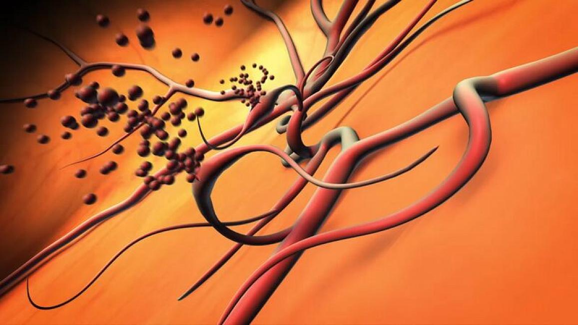
Illustration of ruptured blood vessels

http://www.nih.gov/sites/default/files/styles/featured_media_breakpoint-large-extra/public/news-events/news-releases/2015/20151116-eye-blood-vessels.jpg

Abnormal blood vessels bleeding into the center of the eye due to proliferative diabetic retinopathy.

https://youtu.be/jPoCIa0_1po

A clinical trial funded by the National Institutes of Health has found that the drug ranibizumab (Lucentis) is highly effective in treating proliferative diabetic retinopathy. The trial, conducted by the Diabetic Retinopathy Clinical Research Network (DRCR.net) compared Lucentis with a type of laser therapy called panretinal or scatter photocoagulation, which has remained the gold standard for proliferative diabetic retinopathy since the mid-1970s. The findings demonstrate the first major therapy advance in nearly 40 years.

“These latest results from the DRCR Network provide crucial evidence for a safe and effective alternative to laser therapy against proliferative diabetic retinopathy,” said Paul A. Sieving, M.D., Ph.D., director of NIH’s National Eye Institute (NEI), which funded the trial.  The results were published online today in the Journal of the American Medical Association.

Treating abnormal retinal blood vessels with laser therapy became the standard treatment for proliferative diabetic retinopathy after the NEI announced results of the Diabetic Retinopathy Study in 1976. Although laser therapy effectively preserves central vision, it can damage night and side vision; so, researchers have sought therapies that work as well or better than laser but without such side effects.

A complication of diabetes, diabetic retinopathy can damage blood vessels in the light-sensitive retina in the back of the eye. As the disease worsens, blood vessels may swell, become distorted and lose their ability to function properly. Diabetic retinopathy becomes proliferative when lack of blood flow in the retina increases production of a substance called vascular endothelial growth factor, which can stimulate the growth of new, abnormal blood vessels. These new vessels are prone to bleeding into the center of the eye, often requiring a surgical procedure called a vitrectomy to clear the blood. The abnormal blood vessels can also cause scarring and retinal detachment. Lucentis is among several drugs that block the effects of vascular endothelial growth factor.

About 7.7 million U.S. residents have diabetic retinopathy, a leading cause of blindness among working-age Americans. Among these, about 1.5 percent have PDR.

The DRCR.net enrolled 305 participants (394 eyes) with proliferative diabetic retinopathy in one or both eyes at 55 clinical sites across the country. Eyes were assigned randomly to treatment with Lucentis or laser. For participants who enrolled both eyes in the study, one eye was assigned to the laser group and the other was assigned to the Lucentis group. About half of the eyes assigned to the laser group required more than one round of laser treatment. In the other group, Lucentis (0.5 mg/0.05 ml) was given via injections into the eye once per month for three consecutive months, and then as needed until the disease resolved or stabilized.

Because Lucentis is commonly used to treat diabetic macular edema—the build-up of fluid in the central area of the retina—the study permitted the use of Lucentis for diabetic macular edema in the laser group, if necessary. Slightly more than half (53 percent) of eyes in the laser group received Lucentis injections to treat diabetic macular edema. About 6 percent of eyes in the Lucentis group received laser therapy, mostly to treat retinal detachment or bleeding.

At two years, vision in the Lucentis group improved by about half a line on an eye chart compared with virtually no change in the laser group. There was little change in side vision with injection (average worsening of 23 decibels) but a substantial loss of side vision with laser (average worsening of 422 decibels).   The vitrectomy rate was lower in the Lucentis group (8 of 191 eyes) than in the laser group (30 of 203 eyes).

Rates of serious systemic adverse events, including cardiac arrest and stroke, were similar between the two groups. One patient in the Lucentis group developed endophthalmitis, an infection in the eye. Other side effects were low, with little difference between treatment groups.

“Lucentis should be considered a viable treatment option for people with proliferative diabetic retinopathy, especially for individuals needing anti-vascular endothelial growth factor for diabetic macular edema,” said Jeffrey G. Gross, M.D., of the Carolina Retina Center in Columbia, South Carolina, who chaired the study. Dr. Gross presented results November 13, 2015, at the annual meeting of the American Academy of Ophthalmology in Las Vegas.

In addition to treating proliferative diabetic retinopathy, the report suggests Lucentis may even help prevent diabetic macular edema from occurring. Among people without diabetic macular edema at the start of the study, only 9 percent of Lucentis-treated eyes developed diabetic macular edema during the study, compared with 28 percent in the laser group. The DRCR.net will continue to follow patients in this study for a total of five years.

The DRCR.net is dedicated to facilitating multicenter clinical research of diabetic eye disease. The network formed in 2002 and comprises more than 350 physicians practicing at more than 140 clinical sites across the country. For more information, visit the DRCR.net website at http://drcrnet.jaeb.org/(link is external).

The study was funded by NEI grants EY14231, EY23207, EY18817.

Lucentis was provided by Genentech. Additional research funding for this study was provided by the National Institute of Diabetes and Digestive and Kidney Diseases, also a part of the NIH.

The study is registered as NCT01489189 at ClinicalTrials.gov(link is external).

The NEI provides information about diabetic eye disease at http://www.nei.nih.gov/health/diabetic/.

Information about diabetes is available through the National Diabetes Education Program, www.ndep.nih.gov/.

View an NEI video about the study at https://youtu.be/jPoCIa0_1po(link is external).

Read Full Post »

Variability of Gene Expression and Drug Resistance, Volume 2 (Volume Two: Latest in Genomics Methodologies for Therapeutics: Gene Editing, NGS and BioInformatics, Simulations and the Genome Ontology), Part 1: Next Generation Sequencing (NGS)

Variability of Gene Expression and Drug Resistance

Larry H. Bernstein, MD, FCAP, Curator

LPBI


New Data Suggest Extreme Genetic Diversity of Tumors May Impart Drug Resistance

NEW YORK (GenomeWeb) – Researchers from the University of Chicago and the Beijing Institute of Genomics have undertaken one of the most extensive analyses of the genome of a single tumor and found far greater genetic diversity than anticipated. Such variation, they said, may enable even small tumors to resist treatment.

“With 100 million mutations, each capable of altering a protein in some way, there is a high probability that a significant minority of tumor cells will survive, even after aggressive treatment,” Chung-I Wu, a University of Chicago researcher and senior author of the study, said in a statement. “In a setting with so much diversity, those cells could multiply to form new tumors, which would be resistant to standard treatments.”


Extremely high genetic diversity in a single tumor points to prevalence of non-Darwinian cell evolution

Shaoping Linga,1, Zheng Hua,1, Zuyu Yanga,1, Fang Yanga,1, Yawei Lia, Pei Linb, Ke Chena, Lili Donga, Lihua Caoa, Yong Taoa, Lingtong Haoa, Qingjian Chenb, Qiang Gonga, et al.

Shaoping Ling,  PNAS   http://dx.doi.org:/10.1073/pnas.1519556112      http://www.pnas.org/content/early/2015/11/11/1519556112

A tumor comprising many cells can be compared to a natural population with many individuals. The amount of genetic diversity reflects how it has evolved and can influence its future evolution. We evaluated a single tumor by sequencing or genotyping nearly 300 regions from the tumor. When the data were analyzed by modern population genetic theory, we estimated more than 100 million coding region mutations in this unexceptional tumor. The extreme genetic diversity implies evolution under the non-Darwinian mode. In contrast, under the prevailing view of Darwinian selection, the genetic diversity would be orders of magnitude lower. Because genetic diversity accrues rapidly, a high probability of drug resistance should be heeded, even in the treatment of microscopic tumors.

The prevailing view that the evolution of cells in a tumor is driven by Darwinian selection has never been rigorously tested. Because selection greatly affects the level of intratumor genetic diversity, it is important to assess whether intratumor evolution follows the Darwinian or the non-Darwinian mode of evolution. To provide the statistical power, many regions in a single tumor need to be sampled and analyzed much more extensively than has been attempted in previous intratumor studies. Here, from a hepatocellular carcinoma (HCC) tumor, we evaluated multiregional samples from the tumor, using either whole-exome sequencing (WES) (n = 23 samples) or genotyping (n = 286) under both the infinite-site and infinite-allele models of population genetics. In addition to the many single-nucleotide variations (SNVs) present in all samples, there were 35 “polymorphic” SNVs among samples. High genetic diversity was evident as the 23 WES samples defined 20 unique cell clones. With all 286 samples genotyped, clonal diversity agreed well with the non-Darwinian model with no evidence of positive Darwinian selection. Under the non-Darwinian model,MALL (the number of coding region mutations in the entire tumor) was estimated to be greater than 100 million in this tumor. DNA sequences reveal local diversities in small patches of cells and validate the estimation. In contrast, the genetic diversity under a Darwinian model would generally be orders of magnitude smaller. Because the level of genetic diversity will have implications on therapeutic resistance, non-Darwinian evolution should be heeded in cancer treatments even for microscopic tumors.

Semantically Related Articles


The findings, which appeared in the Proceedings of the National Academy of Sciences this week, also call into question the widely held view that evolution at the cellular level is driven by Darwinian selection, revealing a level of rapid and extensive genetic diversity beyond what would be expected under this model.

In the study, the researchers focused on a single hepatocellular carcinoma tumor, roughly the size of a ping pong ball. They sampled 286 regions from a single slice of the tumor, studying each one with either whole-exome sequencing or genotyping under both the infinite-site and infinite-allele models of population genetics.

Based on their analyses, the team estimated more than 100 million coding region mutations in what they called an “unexceptional” tumor — more mutations than would ordinarily be expected by orders of magnitude, according to Wu.

This extreme genetic diversity, the study’s authors wrote, implies evolution under the non-Darwinian mode, which is driven by random mutations largely unaffected by natural selection. It also raises the question of why there is so little apparent Darwinian selection in the tumor.

The scientists speculated that in solid tumors, cells remain together and do not migrate, “so that when an advantageous mutation indeed emerges, cells carrying it are competing mostly with themselves. These mutations may confer advantages in fighting for space or extracting nutrients, but they are stifled by their own advantages,” they wrote.

Beneficial mutations may emerge on occasion, but in solid tumors the cell populations are “so structured that selection may often be blunted,” they stated. “The physiological effect has to be very strong to overcome those constraints.” Cancer drugs could remove those constraints, loosening up a cell population and allowing competition to occur, the investigators added.

Wu and his colleagues see the presence of so many mutations in a tumor as creating problems when it comes to treatment. “It almost guarantees that some cells will be resistant,” study co-author and University of Chicago oncologist Daniel Catenacci said in the statement. “But it also suggests that aggressive treatment could push tumor cells into a more Darwinian mode.”

Overall, the findings highlight the need to consider non-Darwinian evolution and the vast genetic diversity it can confer as factors when developing treatment strategies, even for small tumors, the researchers concluded.

Read Full Post »

Amyloid-Targeting Immunotherapy

Curator: Larry H. Bernstein, MD, FCAP

Possible Reasons Found for Failure of Alzheimer’s Treatment

By Staff Editor

http://www.healthnewsdigest.com/news/Alzheimer_Issues_680/Possible-Reasons-Found-for-Failure-of-Alzheimer-s-Treatment.shtml

(HealthNewsDigest.com) – Agglutinated proteins in the brain, known as amyloid-β plaques, are a key characteristic of Alzheimer’s. One treatment option uses special antibodies to break down these plaques. This approach yielded good results in the animal model, but for reasons that are not yet clear, it has so far been unsuccessful in patient studies. Scientists at the Technical University of Munich (TUM) have now discovered one possible cause: they noticed that, in mice that received one antibody treatment, nerve cell disorders did not improve and were even exacerbated.

Immunotherapies with antibodies that target amyloid-β were long considered promising for treating Alzheimer’s. Experiments with animals showed that they reduced plaques and reversed memory loss. In clinical studies on patients, however, it has not yet been possible to confirm these results. A team of researchers working with Dr. Dr. Marc Aurel Busche, a scientist at the TUM hospital Klinikum rechts der Isar Klinik und Poliklinik für Psychiatrie und Psychotherapie and at the TUM Institute of Neuroscience, and Prof. Arthur Konnerth from the Institute of Neuroscience has now clarified one possible reason for this. The findings were published in Nature Neuroscience.

Immunotherapy Increases Number of Hyperactive Nerve Cells

The researchers used Alzheimer’s mice models for their study. These animals carry a transgene for the amyloid-β precursor protein, which, as in humans, leads to the formation of amyloid-β plaques in the brain and causes memory disorders. The scientists treated the animals with immunotherapy antibodies and then analyzed nerve cell activity using high-resolution two-photon microscopy. They found that, while the plaques disappeared, the number of abnormally hyperactive neurons rose sharply.

“Hyperactive neurons can no longer perform their normal functions and, after some time, wear themselves out. They then fall silent and, later, possibly die off,” says Busche, explaining the significance of their discovery. “This could explain why patients who received the immunotherapy experienced no real improvement in their condition despite the decrease in plaques,” he adds.

Released Oligomers Potential Reason for Hyperactivity

Even in young Alzheimer’s mice, when no plaques were yet detectable in the brain, the antibody treatment led to increased development of hyperactive nerve cells. “Looking at these findings, even using the examined immunotherapies at an early stage, before the plaques appear, would offer little chance of success. As the scientist explains, the treatment already exhibits these side effects here, too.

“We suspect that the mechanism is as follows: The antibodies used in treatment release increasing numbers of soluble oligomers. These are precursors of the plaques and have been considered problematic for some time now. This could cause the increase in hyperactivity,” says Busche.

The work was funded by an Advanced ERC grant to Prof. Arthur Konnerth, the EU FP7 program (Project Corticonic) and the Deutsche Forschungsgemeinschaft (IRTG 1373 and SFB870). Marc Aurel Busche was supported by the Hans und Klementia Langmatz Stiftung.

Publication
Marc Aurel Busche, Christine Grienberger, Aylin D. Keskin, Beomjong Song, Ulf Neumann, Matthias Staufenbiel, Hans Förstl and Arthur Konnerth, Decreased amyloid-β and increased neuronal hyperactivity by immunotherapy in Alzheimer’s models, Nature Neuroscience, November 9, 2015.
DOI: 10.1038/nn.4163
http://www.nature.com/neuro/journal/vaop/ncurrent/full/nn.4163.html

Amyloid-Targeting Immunotherapy Disrupts Neuronal Function

Some antibodies designed to eliminate the plaques prominent in Alzheimer’s disease can aggravate neuronal hyperactivity in mice.

By Karen Zusi | November 9, 2015  http://www.the-scientist.com//?articles.view/articleNo/44435/title/Amyloid-Targeting-Immunotherapy-Disrupts-Neuronal-Function/

http://www.the-scientist.com/images/News/November2015/10_alzheimerbrain_b.jpg

Removing built-up plaques of amyloid-β in the brain is a long-sought therapy for patients with Alzheimer’s disease, but for a variety of reasons, few treatments have succeeded in alleviating symptoms once they reach clinical trials. In a study published today (November 9) in Nature Neuroscience, an international team examined the effects of two amyloid-β antibodies on neuronal activity in a mouse model, finding that the antibodies in fact led to an increase in neuronal dysfunction.

Decreased amyloid-β and increased neuronal hyperactivity by immunotherapy in Alzheimer’s models

Marc Aurel Busche, Christine Grienberger, Aylin D Keskin, Beomjong Song, Ulf Neumann, Matthias Staufenbiel, Hans Förstl & Arthur Konnerth
Nature Neuroscience (2015)
    http://dx.doi.org:/10.1038/nn.4163

Among the most promising approaches for treating Alzheimer´s disease is immunotherapy with amyloid-β (Aβ)-targeting antibodies. Using in vivo two-photon imaging in mouse models, we found that two different antibodies to Aβ used for treatment were ineffective at repairing neuronal dysfunction and caused an increase in cortical hyperactivity. This unexpected finding provides a possible cellular explanation for the lack of cognitive improvement by immunotherapy in human studies.

Marc Busche, a psychiatrist at Technical University of Munich in Germany, and others had previously found that neuronal hyperactivity is common in mouse models of Alzheimer’s disease. The chronically rapid-firing neurons can interfere with normal brain function in mice. “There’s evidence from human fMRI [functional magnetic resonance imaging] studies that humans will show hyperactivation early in the disease, followed by hypoactivation later on,” Busche told The Scientist. “It’s an early stage of neuronal dysfunction that can later turn into neural silencing.”

To investigate whether certain antibodies would alleviate this Alzheimer’s disease-associated phenotype, Busche and his colleagues first turned to bapineuzumab—a human monoclonal antibody that initially showed promise in treating mice modeling Alzheimer’s disease, but failed in human clinical trials. The dominant hypothesis for bapineuzumab’s failure is that it was administered too late in the disease progression, said Busche. “But it’s still a hypothesis,” he added. “There’s no real explanation for why these antibodies failed.”

The team’s latest experimenters used mice with a genetic mutation that caused them to overexpress the human amyloid-β protein; these engineered mice also displayed neuronal hyperactivity. The researchers injected 3D6, the mouse version of bapineuzumab, into the engineered mice, as well as into wild-type mice that had normal expression levels of the mouse amyloid-β protein. The team observed the effects using two-photon calcium imaging in a blinded study.

As expected, 3D6 decreased the amount of amyloid-β plaques in the engineered mice, while the control mice displayed no reaction to the injected antibodies. However, the mice engineered to overexpress human amyloid-β showed increased neuronal hyperactivity in response to the antibody, regardless of what stage of plaque development they were in. Even mice too young to have developed plaques showed aggravated hyperactive neurons. The team observed the same phenomenon when it tested a second antibody, β1, which went through early stages of drug development but was never used in human clinical trials.

As expected, 3D6 decreased the amount of amyloid-β plaques in the engineered mice, while the control mice displayed no reaction to the injected antibodies. However, the mice engineered to overexpress human amyloid-β showed increased neuronal hyperactivity in response to the antibody, regardless of what stage of plaque development they were in. Even mice too young to have developed plaques showed aggravated hyperactive neurons. The team observed the same phenomenon when it tested a second antibody, β1, which went through early stages of drug development but was never used in human clinical trials.

The results surprised Busche. “When it turned out that the antibody group was worse than the control group, it was unbelievable. But we checked many times and there was no mistake,” he said. “We don’t see this effect in wild-type mice so it must be dependent on the interaction between the antibody and amyloid-β.”

Busche was quick to point out that the mouse model is not the same as a human Alzheimer’s patient. However, he said, “it gives a sense that we don’t understand the antibody’s action, and this might go on in the human brain as well.”

“I fully believe in their results, but I have some hesitation in saying that this result explains the failed clinical trials for amyloid-β immunotherapy,” said Cynthia Lemere, a neurologist and Alzheimer’s disease researcher at the Brigham and Women’s Hospital in Boston. “I think the major reason for clinical trials failing for immunotherapy is that up until now, they’ve been done in people with moderate-to-severe Alzheimer’s disease, and then mild-to-moderate. Now the studies are going further to include people with very early stages of clinical symptoms—and to my knowledge, they haven’t been stopped because patients are getting worse.”

Thomas Wisniewski, a cognitive neurologist at New York University, voiced a similar perspective. “I don’t think this is an explanation for why immunotherapy isn’t working—I think there are other more plausible reasons for that,” he said, citing clinical trials that treated patients during later stages of Alzheimer’s disease progression, as well as those that haven’t addressed tau-related pathologies, or didn’t target the key types of amyloid-β. “[The neuronal hyperactivity] is an interesting phenomenon to be studied,” he added, “but I think it’s a separate issue.”

M.A. Busche et al., “Decreased amyloid-β and increased neuronal hyperactivity by immunotherapy in Alzheimer’s models,” Nature Neuroscience, doi:10.1038/nn.4163, 2015.

Figure 2: Worsening of neuronal dysfunction by anti-Aβ antibodies can occur independently of the effects on Aβ pathology.

Worsening of neuronal dysfunction by anti-A[beta] antibodies can occur independently of the effects on A[beta] pathology.

(a) Top, representative in vivo activity maps in WT (left) as well as isotype-treated (middle) and β1-treated (right) Tg2576 mice. Bottom, Ca2+ transients of neurons indicated above. The further aggravation of neuronal hyperactivity (mi…

http://www.nature.com/neuro/journal/vaop/ncurrent/carousel/nn.4163-F2.jpg

Anti-Aβ treatment aggravates abnormal brain activity in a mouse model of Alzheimer’s disease

Nature Neuroscience   Nov 10, 2015

http://www.natureasia.com/en/research/highlight/10316

Therapies that reduce deposits of amyloid-β (Aβ) in the brain are ineffective at repairing neuronal impairment in mice and actually increase it, finds a study published online in Nature Neuroscience. Aβ deposits aggregate into clumps in the brain which are a pathological hallmark of Alzheimer’s disease.

Expression of mutant human amyloid protein in animals results in deposits of Aβ plaques that induce abnormal increases in neuronal activity and impair the normal function of neuronal circuits.

Arthur Konnerth, Marc Busche and colleagues explored whether they could reverse these impairments by treating mice that overexpress the human mutant amyloid precursor protein with either of two different antibodies targeting Aβ (14 mice) or a control antibody (19 mice). They found that, although treatment with the Aβ targeting antibodies reduced the amount of plaques in the animals’ brains, it also increased the amount of hyperactive neurons.

This was true whether the treatment was given to older mice (14 treated, 19 control) or younger mice in which the accumulation of Aβ had yet to occur (10 treated, 13 control). The same therapies had no effect on neuronal activity in a group of normal mice (5 treated, 3 control), suggesting that the observed exacerbation in mutant mice is dependent on the presence of Aβ and cannot be explained by incidental effects of inflammation in response to the antibodies.

The authors note that, although other research has shown that anti-Aβ treatment can prevent the weakening of neuronal connections and memory impairments in animal models of Alzheimer’s disease, these benefits are not enough to repair neuronal dysfunction.

They suggest that their findings provide a cellular mechanism that may explain, in part, why treatments targeting Aβ in human clinical trials have failed to improve cognitive deficits. However, the authors point out that future studies are needed to determine whether the increase in abnormal neural activity seen in their animal models is related to the poor efficacy of Aβ therapy in patients.


ANAVEX™ 2-73

ANAVEX™ 2-73 is an orally available drug candidate developed to potentially modify Alzheimer’s disease rather than temporarily address its symptoms. It has a clean Phase 1 data profile and shows reversal of memory loss (anti-amnesic properties) and neuroprotection in several models of Alzheimer’s disease.

Successful Phase 1 Clinical Trial

A Phase 1 single ascending dose human clinical trial of ANAVEX 2-73 was successfully completed in healthy human volunteers. It was a randomized, placebo-controlled study. Healthy male volunteers aged 18 to 55 received single, ascending oral doses over the course of the trial. The trial objectives were to define the maximum tolerated dose, assess pharmacokinetics (PK), clinical and lab safety.

Results:

  • Dosing from 1-60 mg.
  • Maximum tolerated dose 55-60 mg; above the equivalent dose shown to have positive effects in mouse models of Alzheimer’s disease.
  • Well tolerated below the 55-60 mg dose with only mild adverse events in some volunteers.
  • Observed adverse events at doses above the maximum tolerated single dose included headache and dizziness, which were moderate in severity and reversible. These side effects are often seen with drugs that target central nervous system (CNS) conditions, including Alzheimer’s disease.
  • No significant changes in blood safety measurements.
  • No changes in ECG.
  • Favorable PK profile.
    • Rapid absorption into blood.
    • Dose proportional kinetics.

The trial was conducted in Germany by ABX-CRO in collaboration with the Technical University of Dresden. ABX-CRO and the Technical University of Dresden are well regarded for their experience with clinical trials and CNS compounds.


ANAVEX 2-73,

Clinical-stage biopharmaceutical company Anavex Life Sciences Corp. is working on an investigational oral treatment for Alzheimer’s disease called ANAVEX 2-73, with full PART A data and preliminary PART B data from its ongoing Phase 2a clinical trial to be presented during the Clinical Trials on Alzheimer’s Disease (CTAD) conference, November 5 and 7 in Barcelona, Spain.

The trial’s Principal Investigator, Stephen Macfarlane, who also serves as director and associate professor at Aged Psychiatry, Caulfield Hospital in Melbourne, Australia, will represent the company and host a late-breaking oral session entitled “New Exploratory Alzheimer’s Drug ANAVEX 2-73: Assessment of Safety and Cognitive Performance in a Phase 2a Study in mild-to-moderate Alzheimer’s Patients.” During the presentation, which will take place Saturday, November 7, at 9:45 a.m. CET, at the Gran Hotel Princesa Sofia, in Barcelona, Macfarlane will focus on the the multicenter Phase 2a clinical trial of ANAVEX 2-73. The study includes two separate phases and includes 32 mild-to-moderate Alzheimer’s patients. While PART A is a simple randomized, open-label, two-period, cross-over, adaptive trial of up to 36 days, PART B is an open-label extension trial for an additional 52 weeks.

The research intends to assess the maximum dose of treatment tolerated by patients, and to explore cognitive efficacy using mini-mental state examination score (MMSE), dose response, bioavailability, Cogstate and electroencephalographic (EEG) activity, including event-related potentials (EEG/ERP), as well as the preformance of ANAVEX 2-73 as an add-on therapy to donepezil (Aricept).

ANAVEX 2-73 is Anavex’s lead investigational treatment for Alzheimer’s disease, in line with the company’s goal of finding effective therapies for Alzheimer’s disease, other central nervous system (CNS) diseases, pain, and various types of cancer. The novel drug targets sigma-1 and muscarinic receptors, which are thought to decrease the amount of protein misfolding, beta amyloid tau and inflammation through upstream actions.

Last November, the biopharmaceutical company presented encouraging results from their phase 1 clinical trial for Anavex 2-73, during the CNS Summit 2014 in Boca Raton, Florida. The phase 1 study demonstrated that the treatment is safe and well tolerated, suggesting a favorable pharmacokinetics profile. During the randomized, double-blind, placebo-controlled study no severe adverse events were registered, while the adverse events reported included moderate and reversible headache and dizziness, which are common symptoms associated with drugs that target central nervous system (CNS) conditions, such as Alzheimer’s.

New Exploratory Alzheimer’s Drug ANAVEX 2-73: Assessment of Safety and Cognitive Performance in a Phase 2a Study in mild-to-moderate Alzheimer’s Patients

Steve Macfarlane, MD1 , Paul Maruff, PhD2 , Marco Cecchi, PhD3 , Dennis Moore, PhD3 , Anastasios Zografidis, PhD4 , Christopher Missling, PhD4 (1)

Caulfield Hospital, Melbourne, Australia (2), Cogstate, Melbourne, Australia (3), Neuronetrix, KY, USA (4), Anavex Life Sciences, Corp., New York, NY, USA

Background: Despite major efforts aimed at finding a treatment for Alzheimer’s disease (AD), progress in developing compounds that can relieve cognitive deficits associated with the disease has been slow. ANAVEX 2-73 is a sigma-1 and muscarinic receptor agonist that in preclinical studies has shown memory-preserving and neuroprotective effects. In our ongoing phase 2a clinical study we are assessing ANAVEX 2-73 safety in subjects with mild-to-moderated AD, and measuring drug effects on MMSE, EEG and Event Related Potentials (ERP) cognitive measures, and Cogstate test batteries to optimize dosing.

Methods: Thirty-two subjects that meet NINCDS-ADRDA criteria for probable AD are being recruited at up to seven clinical sites in Melbourne, Australia. Subjects are between 55 and 85 years of age, and have an MMSE of 16 to 28. In PART A of the study, participants are administered ANAVEX 2-73 orally and IV in an open-label, 2-period, cross-over trial with adaptive study design lasting up to 36 days for each participant. In PART B of the study, all participants are administered ANAVEX 2-73 daily orally. MMSE, EEG/ERP (P300) and Cogstate tests are performed at baseline and subsequently at weeks 12, 26, 38 and 52 of the PART B open label extension.

Results: The primary outcome of the study is safety, and ANAVEX 2-73 was well tolerated. In the secondary outcome endpoints preliminary analysis of data from subjects shows an average improvement of the MMSE score at week 5. A majority of all patients tested so far improved their respective MMSE score. The average EEG/ERP (P300 amplitude) signal also improved and also the average Cogstate test improved across the test batteries.

Conclusions: Data collected so far indicate that ANAVEX 2-73 is safe and well tolerated. Interim results also show improved cognitive performance after drug administration in subjects with mild-to-moderate AD. The current results seem to justify a prospective comparison with current standard of care in a larger clinical trial study. A more complete set of results will be available at the time of the conference.

Read Full Post »

palivizumab prophylaxis for children with bronchiolitis

Larry H. Bernstein, MD, FCAP, Curator

LPBI


Eligibility for palivizumab prophylaxis in a cohort of children with severe bronchiolitis
Kohei Hasegawa,1 Jonathan M. Mansbach,2 Pedro A. Piedra,3 Michelle B. Dunn,4 Sunday Clark,5 Ashley F. Sullivan1 and Carlos A. Camargo Jr1

Pediatrics International (2015) 57, 1031–1034          http://dx.doi.org:/10.1111/ped.12760


In 2014, the American Academy of Pediatrics (AAP) updated their recommendations for palivizumab prophylaxis for children who are at high risk for severe respiratory syncytial virus (RSV) infection. To investigate the potential impact of the more restrictive 2014 criteria on the eligibility for palivizumab prophylaxis, we applied the 2012 and 2014 AAP recommendations for palivizumab prophylaxis to a multicenter cohort of 2207 US children hospitalized for bronchiolitis. According to the 2012 AAP recommendations, 215 children (9.7%) were eligible for palivizumab prophylaxis, while 140 children (6.3%) would have been eligible based on the 2014 updated recommendations (34.9% relative decrease; 95% CI: 28.5–41.7%).  The  decrease was largely driven by the restriction of eligibility to preterm infants with gestational age < 29weeks. Further development of and refinement of cost-effective approaches for the prevention of severe RSV infection are needed.


Bronchiolitis remains an important public health problem in the USA. It is the leading cause of hospitalization in infants, accounting for 18% of all infant hospitalizations, with a total direct cost of $545m in 2009.1 Although many viruses are known to cause bronchiolitis, respiratory syncytial virus (RSV) is the most common etiology among children requiring hospitalization.2 Palivizumab, a humanized monoclonal antibody against the RSV Fglycoprotein, is licensed for the prevention of serious lower respiratory infection caused by RSV in high-risk children. Since palivizumab was first licensed, professional organizations have sought more precise guidance for determining who is at high risk.3

In 2014, the American Academy of Pediatrics (AAP) updated and replaced their recommendations for palivizumab prophylaxis from 2012.4  The updated guidelines support a more restrictive use of palivizumab:3 for example, they recommend against the use in infants born ≥ 29 weeks’ gestation who have no additional risk factors for severe RSV disease. Despite these substantial changes to the guideline recommendations, there are no publications that assess the potential impact on the eligibility for palivizumab prophylaxis in US children.

To address the knowledge gap in the literature, we investigate the potential impact of the more restrictive 2014 criteria on the eligibility for palivizumab prophylaxis in a well-characterized national cohort of children hospitalized for bronchiolitis.


Over the 3year study period, we enrolled 2207 children hospitalized for bronchiolitis to one of the 16 sites. Demographic characteristics, medical history, and clinical course are summarized in Table 1. Overall, the median age was 4months (IQR, 2–9 months) and 1311 (59.4%) were male. Additionally, 285 children (12.9%) were born at gestational age <35 weeks; 460 (20.8%) had one or more major comorbid medical disorders.

Table 1 Bronchiolitis patient characteristics vs AAP palivizumab recommendations

Table 2 Eligibility for palivizumab prophylaxis vs 2012 and 2014 AAP recommendations

According to the 2012 AAP recommendations, 215 children (9.7%) were eligible for palivizumab prophylaxis (Table 2), while 140 children (6.3%) would have been eligible based on the 2014 updated recommendations. Applying the more restrictive 2014 criteria would have led to 75 fewer children (34.9% relative decrease; 95%CI: 28.5–41.7%) being eligible for palivizumab prophylaxis. The most frequent reason for the loss of eligibility was the 2014 criterion for prematurity that restricts eligibility to infantswithgestationalage<29weeks;thischangeledto45fewer children being eligible (40.9% relative decrease; 95%CI: 31.6–50.7%). The next most frequent reason was the 2014 criteria that limit eligibility to infants with chronic lung disease or congenital heart disease in the first year of life;this change led to 22 fewer children being eligible for palivizumab prophylaxis (22.9%relative decrease; 95%CI: 15.0–32.6%).

Among the 2207 children in the cohort, 207 children (9.4%) had received palivizumab prophylaxis prior to the index hospitalization. Among 215 children eligible for prophylaxis based on the 2012 recommendations, 117 (54.4%) had received palivizumab prophylaxis. Among 140 children eligible for prophylaxis based on the 2014 recommendations, 72 (51.4%) had received palivizumab prophylaxis (Table 1).


In this analysis of a large multicenter cohort of children hospitalized for bronchiolitis, we found that approximately 10% of children were eligible for palivizumab prophylaxis based on the 2012 AAP recommendations. When applying the more restrictive criteria of the 2014 updated recommendations, one-third of these children would have become ineligible for palivizumab prophylaxis. To thebestofourknowledge,thisisthe firststudytoreportthepotential impact of the change in the AAP recommendations on the eligibility for palivizumab prophylaxis in young children, a finding of public health and research importance.

In 1998, palivizumab was licensed by the US Food and Drug Administration (FDA) for prevention of severe RSV diseases in children at high risk, but the FDA did not issue more specific recommendations, nor define high risk.This absence of a specific definition has led several groups to attempt to identify children at high risk who would be eligible for palivizumab prophylaxis.3,6 The AAP published the first policy statement on the use of palivizumab in 1998.7 On the basis of the availability of additional data, the AAP has updated the guidelines in 2003, 2006, 2009, 2012,4 and 2014.3 Since the last update of the AAP recommendations, some studies have reported a high cost but limited benefit from palivizumab prophylaxis.8 In this context, the 2014 AAP guidelines recommended a more restrictive use.3 In particular, preterm infants with gestational age ≥29 weeks without additional risk factors became ineligible for palivizumab.

In parallel with this change in recommendations, within the present high risk population, the most frequent reason for the loss of eligibility was the use of the restrictive criterion for prematurity: that is, preterm infants born from 29t o35 weeks’ gestation with no additional risk factors became ineligible. This specific group of preterm infants accounts fo ra large number of births in the US:approximately 10% of US births in 2012.9 Thus, one may argue that the use of this restrictive criterion would result in an increase in the number of preventable severe RSV infections,10 even considering the potentially limited efficacy of palivizumab in this population. As described in the technical report of the 2014 AAP recommendations, however, it is challenging to define an optimal threshold of gestational age in preterm infants for which palivizumab prophylaxis may be indicated. The present observations should facilitate further investigations that seek high-quality and cost-effective preventive strategies for a large number of vulnerable children.

This study has several potential limitations. First, the analysis was not designed to examine the efficacy or effectiveness of palivizumabprophylaxis. Rather, we sought to examinethe potential impact of the updated recommendations on the eligibility for palivizumab in a well-characterized national cohort of children hospitalized for bronchiolitis. Second, the present study investigated only children hospitalized for bronchiolitis; thus, those with other types of severe respiratory infection (e.g. pneumonia) were not examined. Inclusion of these populations may yield different inferences. Nevertheless, the present findings are directly relevant to >120 000 US children hospitalized for bronchiolitis (and their families) each year.1 Finally, the study participants were those who were hospitalized in academic centers. Therefore, the present inferences may not be generalizable to the US population as a whole. Children hospitalized at academic centers, however, have disproportionately high morbidity; it is in precisely this population for which targeted preventive measures are needed.

In conclusion, we found that 10% of children hospitalized for bronchiolitis were eligible for palivizumab prophylaxis based on the 2012 AAP recommendations. When we applied the more restrictive 2014criteria,one-third of these children were ineligible. The decrease was largely driven by the restriction of eligibility to preterminfantswithgestationalage <29weeks.Forpolicymakers and researchers, because bronchiolitis continues to be a substantial societal burden in an already-stressed health-care system,1 the present findings support further development and refinement of cost effective approaches for the prevention of severe RSV infection.


References

1 Hasegawa K,Tsugawa Y,Brown DF,Mansbach JM,Camargo CA Jr. Trends in bronchiolitis hospitalizations in the United States, 2000–2009. Pediatrics 2013; 132: 28 –36.

2 Hasegawa K, Mansbach JM,Camargo CAJr.Infectious pathogens and bronchiolitis outcomes. Expert Rev. Anti Infect. Ther. 2014; 12: 817 –28.

3 American Academy of Pediatrics Committee on Infectious Diseases and Bronchiolitis Guidelines Committee. Policy statement. Updated guidance for palivizumab prophylaxis among infants and young children at increased risk of hospitalization for respiratory syncytial virus infection. Pediatrics 2014; 134: 415 –20.

4 American Academy of Pediatrics. Respiratory syncytial virus. In: Pickering LK, Baker CJ, Kimberlin DW, Long SS (eds). Red Book: 2012. Report of the Committee on Infectious Diseases. American Academy of Pediatrics, Elk Grove Village, IL, 2012; 609–18.

5 Hasegawa K, Jartti T, Mansbach JM etal.Respiratory syncytial virus genomic load and disease severity among children hospitalized with bronchiolitis: Multicenter cohort studies in the United States and Finland. J. Infect. Dis. 2015; 211: 1550 –9.

6 NHS Commissioning Board. Clinical Commissioning Policy: Palivizumab to reduce the risk of RSV in high risk infants. 2012. Accessed 13 May 2015. Available from URL: http://www.england.nhs.uk/.

7 American Academy of P

ediatrics Committee on Infectious Diseases and Committee of Fetus and Newborn. Prevention of respiratory syncytial virus infections: Indications for the use of palivizumab and update on the use of RSV-IGIV. Pediatrics 1998; 102: 1211 –6.

8 Andabaka T, Nickerson JW, Rojas-Reyes MX, Rueda JD,  Bacic Vrca V, Barsic B. Monoclonal antibody for reducing the risk of respiratory syncytial virus infection in children. Cochrane Database Syst. Rev. 2013; 4 CD 006602.

9 American Academy of Pediatrics Committee on Infectious Diseases and Bronchiolitis Guidelines Committee. Technical report. Updated guidance for palivizumab prophylaxis among infants and young children at increased risk of hospitalization for respiratory syncytial virus infection. Pediatrics 2014; 134 (2): e620–38.

Appendix I. Principal Investigators at the 16 participating sites in MARC-30
Besh Barcega, MD Loma Linda University Children’s Hospital, Loma Linda, CA, USA

John Cheng, MD Children’s Healthcare of Atlanta at Egleston, Atlanta, GA, USA

Carlos Delgado, MD Children’s Healthcare of Atlanta at Egleston, Atlanta, GA, USA

Haitham Haddad, MD Rainbow Babies and Children’s Hospital, Cleveland, OH, USA

Frank LoVecchio, MD Maricopa Medical Center, Phoenix, AZ, USA

Eugene Mowad, MD Akron Children’s Hospital, Akron, OH, USA

Brian Pate, MD Children’s Mercy Hospital and Clinics, Kansas City, MO, USA

Mark Riederer, MD Monroe Carell Jr. Children’s Hospital at Vanderbilt, Nashville, TN, USA

Paul Hain, MD Monroe Carell Jr. Children’s Hospital at Vanderbilt, Nashville, TN, USA M

Jason Sanders, MD Children’s Memorial Hermann Hospital, Houston, TX, USA

Nikhil Shah, MD New York Presbyterian Hospital, New York, NY, USA

Dorothy Damore, MD New York Presbyterian Hospital, New York, NY, USA

Michelle Stevenson, MD Kosair Children’s Hospital, Louisville, KY, USA

Erin Stucky Fisher, MD Rady Children’s Hospital, San Diego, CA, USA

Stephen Teach, MD, MPH Children’s National Medical Center, Washington, DC, USA

Lisa Zaoutis, MD Children’s Hospital of Philadelphia, Philadelphia, PA, USA


Read Full Post »

Thymus vs Bone Marrow, Two Cell Types in Human Immunology: B- and T-cell differences

Reporter: Larry H. Bernstein, MD, FCAP

Differences Between B-Cells and T-Cells

http://www.microbiologyinfo.com/differences-between-b-cells-and-t-cells/

Aryal Sagar

Although mature lymphocytes all look pretty much alike, they are extraordinarily diverse in their functions. The most abundant lymphocytes are:

B lymphocytes (often simply called B cells) and
T lymphocytes (likewise called T cells)

Differences between B-Cells and T-Cells

http://www.microbiologyinfo.com/wp-content/uploads/2015/11/Differences-between-B-Cells-and-T-Cells.jpg

S.N.

Properties

B-Cells

T-Cells

1 Name B lymphocytes T lymphocytes
2 Origin Bone Marrow Thymus
3 Position Outside Lymph Node Interior of Lymph Node
4 Membranereceptor BCR (= immunoglobulin) for antigen TCR for antigen
5 Connections B-cells can connect to antigens right on the surface of the invading virus or bacteria. T-cells can only connect to virus antigens on the outside of infected cells.
6 Tissue Distribution Germinal centres of lymph nodes, spleen, gut, respiratory tract; also subcapsular and medullary cords of lymph nodes Parafollicular areas of cortex innodes, periarteriolar in spleen
7 Life Span Life span is short Life span is long
8 Surface Antibodies Surface Antibodies present Absence of surface antibodies
9 Secretion They secrete antibodies They secrete Lymphokines
10 Function В-cells form humoral or antibody-mediated immune system (AMI). T-cells form cell-mediated immune system (CMI).
11 Blood 20% of lymphocytes 80% of lymphocytes; CD4 > CD8
12 Formation They form plasma cells and memory cells. They form killer, helper and suppressor cells.
13 Movement to Infection Site Plasma cells do not move to the site of infection. Lymphoblasts move to the site of infection.
14 Function Plasma cells do not react against transplants and cancer cells. Killer cells react against transplants and cancer cells.
15 Function Plasma cells have no inhibitory effect on immune system. Suppressor cells inhibit immune system.
16 Function They defend against viruses and bacteria that enter the blood and lymph. They defend against pathogens including protists and fungi that enter the cells.
Differences Between B-Cells and T-Cells

Read Full Post »

Management of Follicular Lymphoma

Curator: Larry H. Bernstein, MD, FCAP

Article ID #193: Management of Follicular Lymphoma. Published on 11/5/2015

WordCloud Image Produced by Adam Tubman


Targeted Approaches to the Management of Follicular Lymphoma  

By Chaitra Ujjani, MD

http://www.cancernetwork.com/oncology-journal/targeted-approaches-management-follicular-lymphoma


Despite high rates of response to initial chemoimmunotherapy, patients with follicular lymphoma experience frequent relapses, and better treatment options are needed. Several novel biologic agents have been developed based on a greater understanding of the intrinsic factors driving the development of this heterogeneous disease. Such therapies target extracellular surface proteins and intracellular signaling pathways, as well as manipulate and engage the tumor microenvironment. Many of these agents have shown great promise in early-phase studies and are the focus of ongoing clinical investigations.


Introduction As the second most common form of non-Hodgkin lymphoma (NHL), follicular lymphoma affects thousands of new patients in the United States each year. Although follicular lymphoma is considered an indolent disease, its clinical course is highly variable. Asymptomatic patients with low tumor burden can be monitored closely with the “watch and wait” strategy, given that the early intervention of chemotherapy or immunotherapy has not demonstrated a survival benefit.[1,2] The most widely accepted indications for treatment are one or more of the following criteria from the Groupe d’Etude des Lymphomes Folliculaires (GELF): a single lesion > 7 cm, three nodal sites > 3 cm, splenomegaly, effusions, threat or evidence of organ compression, or constitutional symptoms.[3] Whereas patients with limited-stage disease have several treatment options—including single-agent rituximab, radiation, and chemoimmunotherapy, those with advanced-stage disease typically receive chemoimmunotherapy.[4,5] Both the German Study Group Indolent Lymphomas (StiL) NHL-2 study and the pharmaceutical company–sponsored Bendamustine Rituximab Investigational Non-Hodgkin’s Trial (BRIGHT) have established the front-line role of combination therapy with bendamustine and rituximab in the treatment of follicular lymphoma, based on comparable efficacy and better tolerability than standard regimens such as R-CHOP (rituximab, cyclophosphamide, doxorubicin, vincristine, and prednisone).[6,7] Despite high response rates with initial therapy, follicular lymphoma is characterized by frequent relapses, and patients need improved treatment options.

Oncology (Williston Park). 29(10):760-768.


http://www.cancernetwork.com/sites/default/files/styles/figures_diagrams/public/figures_diagrams/1510ujjaniTable.png

Table: Targeted Therapies in Development and FDA-Approved for the Treatment of Follicular Lymphomas


Since the discovery of rituximab, there has been significant innovation in drug development, based on a greater understanding of the pathogenesis of the disease. The multistep process leading to follicular lymphoma is theorized to begin with an initial genetic insult, the hallmark t(14;18) translocation, which results in overexpression of the anti-apoptotic B-cell lymphoma protein, BCL-2.[8] This translocation is not the sole factor in malignant transformation, as it is a naturally occurring anomaly, often identified in healthy individuals. Furthermore, preclinical studies have indicated a positive correlation between increasing numbers of genetic alterations and the progression from follicular lymphoma in situ to grade 3A follicular lymphoma.[9] The B-cell receptor is a critical cellular factor in the development of the disease. Its active tonic signaling leads to recruitment of the spleen tyrosine kinase (SYK) and activation of multiple downstream pathways, including phosphatidylinositol 3-kinase (PI3K), nuclear factor kappa-light-chain-enhancer of activated B cells (NFκB), and mitogen-activated protein kinase (MAPK). Activation of these pathways ultimately results in the maturation, proliferation, and survival of malignant lymphocytes.[10] Other key components include immune cells such as T cells, dendritic cells, and reticular cells, which infiltrate among the centrocytes.[11] In addition to stimulating the B-cell receptor, these immune cells induce exhaustion of cytotoxic T cells and allow for T-cell evasion via disruption of synapses and secretion of interleukin-12.[12] Expression of the inhibitory receptor programmed cell death 1 (PD-1) is another contributing factor, as its presence is believed to affect the ability of T cells to mount appropriate antitumor responses.[13] Several novel therapies have been developed to target these various aspects of the disease in the hope of identifying more effective, yet tolerable, treatment options for patients with follicular lymphoma. (See the Table for a summary of therapies approved and in development for treatment of follicular lymphoma.)


Since the approval of rituximab, there has been significant investigation into the development of a superior anti-CD20 monoclonal antibody. Second- and third-generation versions vary in their structures and mechanisms of action. Two anti-CD20 antibodies, ofatumumab and obinutuzumab, have been approved by the US Food and Drug Administration (FDA) for indications in chronic lymphocytic leukemia (CLL). Ofatumumab, a type I human monoclonal antibody, was initially approved for patients whose disease is refractory to fludarabine and alemtuzumab.[14] Obinutuzumab was approved in combination with chlorambucil for patients with preexisting comorbidities that preclude conventional chemoimmunotherapy.[15] Although designed to induce stronger complement-dependent cytotoxicity than rituximab, ofatumumab demonstrated minimal activity in rituximab-refractory follicular lymphoma (overall response rate [ORR], 10% to 13%; median progression-free survival [PFS], 5.8 months).[16] In phase II trials of ofatumumab in combination with chemotherapy, including bendamustine and CHOP, results appeared comparable to those achieved with rituximab-based regimens, although no direct comparisons have been made.[17,18]

Obinutuzumab, a type II glycoengineered humanized antibody, is further in development for use in NHL. This agent, which works primarily by triggering antibody-dependent cellular cytotoxicity (ADCC) and apoptosis, has demonstrated superiority to rituximab in preclinical studies, including those employing whole blood B cell–depletion assays, human lymphoma xenograft mice models, and nonhuman primates.[19] When compared with rituximab in patients with indolent B-cell NHL, obinutuzumab produced a higher ORR by independent radiology review (45% vs 27%; P = .01); however, complete response (CR) and PFS rates were similar.[20] Like ofatumumab, treatment with obinutuzumab is associated with a higher rate of infusion-related reactions than rituximab (grade 1–4, 72% vs 49%; and grade 3/4, 11% vs 5%, respectively). In contrast to ofatumumab, it has efficacy in rituximab-refractory indolent NHL, producing an ORR of 50% and median PFS of 12 months among 10 patients.[21] The phase III GADOLIN study evaluated obinutuzumab in combination with bendamustine followed by maintenance obinutuzumab in the same disease setting (N = 413).[22] While the response rates by independent review were similar to those observed in the comparative arm of patients randomized to single-agent bendamustine (bendamustine-obinutuzumab ORR, 69% [CR, 11%] vs bendamustine alone ORR, 63% [CR, 12%]), the median PFS was significantly higher with the combination (not reached vs 14.9 months; P = .00011). Treatment with the combination of bendamustine and obinutuzumab was associated with a similar incidence of grade ≥ 3 adverse events compared with bendamustine monotherapy (68% vs 62%), which included neutropenia (33% vs 26.3%) and infusion-related reactions (9% vs 3.5%). While there was no difference in overall survival (OS) noted, the study did demonstrate the clinical benefit of obinutuzumab in rituximab-refractory disease. The role of the drug in this setting is becoming less clear, as more patients now receive bendamustine-rituximab as front-line therapy. In the phase Ib GAUDI study, bendamustine-obinutuzumab and obinutuzumab-CHOP produced similar response rates in patients with previously untreated follicular lymphoma, with ORRs of 93% (CR, 39%) and 95% (CR, 35%), respectively.[23] The incidences of grade 3/4 neutropenia and infection were similar to historical data on rituximab chemotherapy. These data prompted the front-line phase III GALLIUM study of chemotherapy (CHOP, CVP [cyclophosphamide, vincristine, and prednisone], or bendamustine) with obinutuzumab or rituximab followed by maintenance obinutuzumab or rituximab in advanced-stage indolent B-cell NHL (ClinicalTrials.gov identifier: NCT01332968). Newer monoclonal antibodies directed against CD20, such as ublituximab, and rituximab biosimilars are also in development.

Monoclonal antibodies to alternative targets

Monoclonal antibodies directed against other B-cell antigens have also been developed. Galiximab, a chimeric human-macaque anti-CD80 antibody, and epratuzumab, a humanized anti-CD22 antibody, were two of the first antibodies directed against these targets to be explored in follicular lymphoma. Both antibodies have shown activity as single agents and in combination with rituximab in follicular lymphoma. However, neither is being studied further due to the availability of newer, more promising therapies.[24-28] MEDI-551, an afucosylated humanized anti-CD19 antibody, induces cell death via ADCC and cytotoxic T-cell response. The lack of a fucose moiety on the Fc portion of the antibody is believed to enhance the activity of ADCC. Phase I studies of MEDI-551 in heavily pretreated follicular lymphoma have reported ORRs ranging from 31% to 82%.[29,30] The median PFS from the earlier study was nearly 10 months. MEDI-551 is currently being evaluated in aggressive lymphomas in combination with rituximab and salvage chemoimmunotherapy (ClinicalTrials.gov identifiers: NCT00983619 and NCT01453205). Also targeting CD19 is MOR208, a humanized monoclonal antibody that has been engineered to have a higher affinity to FcγRIIIa and FcγRIIa, resulting in stronger ADCC. In a phase II study of patients with relapsed and refractory B-cell NHL who had received a median of two prior therapies, the ORR was 26% among those with follicular lymphoma (n = 31).[31] The median duration of response (DOR) was 2.6 months; however, the longest DOR was 15.4 months. Upcoming trials with MOR208 include combination studies with lenalidomide in diffuse large B-cell lymphoma (DLBCL) and CLL (ClinicalTrials.gov identifiers: NCT02399085 and NCT02005289).

Radioimmunotherapy

One of the first attempts to improve upon the efficacy of the naked monoclonal antibody was radioimmunotherapy, which produced ORRs of 65% to 74% in patients with relapsed and refractory indolent B-cell lymphomas.[32,33] 90Y-ibritumomab tiuxetan, the first radioimmunotherapy to receive FDA approval, was approved in February 2002 for relapsed or refractory low-grade, follicular, or transformed B-cell NHL. In 2009, it was granted expanded approval as consolidation therapy in previously untreated follicular lymphoma patients with a partial or complete response to first-line chemotherapy. 131I-tositumomab was approved in June 2003 (along with tositumomab) for CD20-positive follicular NHL, with and without transformation, in relapsed rituximab-refractory patients with relapse following chemotherapy. The use of 131I-tositumomab and 90Y-ibritumomab tiuxetan has declined significantly over the past several years; the manufacture and sale of 131I-tositumomab (marketed in the United States and Canada as Bexxar) was stopped in February 2014. Radioimmunotherapy can be difficult; there are strict hematologic criteria (< 25% lymphomatous marrow involvement, platelet count > 100 × 109, leukocyte count > 1.5 × 10⁹), and its administration requires a certified nuclear medicine physician. In addition, the patient must not have had prior radiation to > 25% of the bone marrow nor undergone stem cell transplantation.[34]

Antibody-drug conjugates (ADCs)

Recent efforts in augmenting antibody-based therapy include the use of ADCs. Once bound to its target antigen, the ADC is engulfed via endocytosis, trafficked to the lysosome for degradation, and ultimately released, whereupon it causes damage to tubulin and DNA. The calicheamicin-bound anti-CD22, inotuzumab ozogamicin, was one of the first to be studied in patients with follicular lymphoma who were refractory to CD20-targeted therapy, yielding an ORR of 66%.[35] The ORR increased to 87% when inotuzumab ozogamicin was combined with rituximab in patients with relapsed and refractory follicular lymphoma, prompting a trial in which it was compared with combination treatment with rituximab plus chemotherapy.[36] The trial was closed early due to poor accrual. Inotuzumab ozogamicin is currently being studied in combination with the mammalian target of rapamycin (mTOR) inhibitor temsirolimus in relapsed and refractory CD22-expressing NHL (ClinicalTrials.gov identifier: NCT01535989). Pinatuzumab vedotin and polatuzumab vedotin, which target CD22 and CD79b, respectively, are ADCs linked to the anti-tubulin molecule monomethyl auristatin E. While both agents have demonstrated activity in indolent NHL (with reported ORRs of 50% and 47%, respectively), polatuzumab vedotin is being taken further in development.[37,38] When polatuzumab vedotin was administered at a higher dose (2.4 mg/kg) with rituximab in patients with relapsed and refractory follicular lymphoma (n = 25), the ORR was 76% (CR, 44%) and median PFS was 15 months.[39] The cohort of 20 patients treated at the lower rituximab dose (1.8 mg/kg) had a similar response rate (ORR, 70%; CR, 40%), and median PFS and DOR were not reached. Peripheral neuropathy, a common toxicity with ADCs, occurred less frequently with the lower dose of polatuzumab vedotin and was ameliorated in some patients by dose delay and reduction. Ongoing studies with polatuzumab vedotin include phase I/II combinations with bendamustine-rituximab or obinutuzumab-bendamustine in relapsed and refractory follicular lymphoma (ClinicalTrials.gov identifier: NCT02257567) and R-CHOP in B-cell NHL patients who have received less than one prior therapy (ClinicalTrials.gov identifier: NCT01992653). Coltuximab ravtansine (formerly SAR3419) is an anti-CD19 ADC that has also been associated with neurologic complications, primarily dose-limiting ocular toxicity.[40] IMGN529, which targets the overexpressed CD37 protein, is another B-cell–directed ADC in development.[41]

Despite high rates of response to initial chemoimmunotherapy, patients with follicular lymphoma experience frequent relapses, and better treatment options are needed. Several novel biologic agents have been developed based on a greater understanding of the intrinsic factors driving the development of this heterogeneous disease. Such therapies target extracellular surface proteins and intracellular signaling pathways, as well as manipulate and engage the tumor microenvironment. Many of these agents have shown great promise in early-phase studies and are the focus of ongoing clinical investigations.

Small-Molecule Inhibitors – PI3K inhibitors

In contrast to the various antibody-based therapies under investigation for treatment of follicular lymphoma, several small molecules have been designed to inhibit key intracellular pathways of the malignant B cell. The majority of these agents are directed against kinases downstream of the B-cell receptor, and many have been combined with bendamustine-rituximab, given this combination’s efficacy and tolerability. Idelalisib, a potent PI3K-δ inhibitor, was the first PI3K inhibitor to be approved by the FDA for follicular lymphoma. It received an indication for patients who have received at least two prior systemic therapies, based on results of a phase II study in rituximab-refractory indolent NHL[42] As reported at the 2015 American Society of Clinical Oncology Annual Meeting, of the 72 patients in the study who had follicular lymphoma, 54% were considered high-risk by the Follicular Lymphoma International Prognostic Index.[43] The patients had received a median of four prior therapies, and 86% had disease that was refractory to the last regimen they received. The ORR was 56% and the median PFS was 11 months. The median PFS for the 10 patients who achieved a CR was 27 months. Notable grade 3/4 adverse events included neutropenia (27% of all patients), diarrhea/colitis (16%), elevations in hepatic transaminases (13%), and pneumonia (7%).
Idelalisib was subsequently administered with rituximab, bendamustine, and bendamustine-rituximab in a phase I study of patients with relapsed (n = 79) and refractory (n = 59) indolent NHL, the majority of whom had follicular lymphoma.[44] The ORRs were similar between the arms (75% to 88%); however, treatment with bendamustine-rituximab-idelalisib was associated with the highest CR (43%) and longest median PFS (37.1 months). A phase III trial of bendamustine-rituximab with or without idelalisib in relapsed and refractory indolent B-cell NHL is ongoing (ClinicalTrials.gov identifier: NCT01732926). In phase I investigations, combined treatment with idelalisib and lenalidomide plus the second-generation SYK inhibitor entospletinib has demonstrated considerable toxicity, including hepatic transaminitis, sepsis, and refractory pneumonitis.[45,46] Second-generation PI3K inhibitors, including duvelisib (IPI-145), TGR-1202, and INCB040093, are in development. Duvelisib, a dual inhibitor of the delta and gamma isoforms of PI3K, has demonstrated an ORR of 69% (CR, 38%) in a heavily pretreated follicular lymphoma cohort (n = 13).[47] Based on these encouraging data, duvelisib is being administered with rituximab or obinutuzumab in patients with previously untreated follicular lymphoma (ClinicalTrials.gov identifier: NCT02391545) and with bendamustine and/or rituximab in those with relapsed B-cell malignancies (ClinicalTrials.gov identifier: NCT01871675).

Bruton tyrosine kinase (BTK) inhibitors

Ibrutinib, a selective and irreversible inhibitor of BTK, may also have some impact on the tumor microenvironment via cytokine and chemokine inhibition.[48] Approved in CLL, mantle cell lymphoma, and Waldenström macroglobulinemia, it has demonstrated activity in a number of B-cell malignancies.[49-53] In a phase II study of relapsed and refractory follicular lymphoma, ibrutinib yielded an ORR of 30% (with one CR) and a median PFS of 9.9 months. The 40 enrolled patients had received a median of three prior therapies, and 36% were considered refractory to treatment. Common adverse events included mild diarrhea, rash, and fatigue; rare events included atrial fibrillation and bleeding.[54] Like idelalisib, ibrutinib has been combined with bendamustine-rituximab in the treatment of B-cell NHL.[55] This triplet produced an ORR of 90% (CR, 50%) in a cohort of 10 patients with previously treated follicular lymphoma. At the time these results were reported, the median PFS had not been reached. Notable grade 3/4 adverse events included neutropenia (33%), rash (25%), and thrombocytopenia (19%). Results from SELENE, a randomized phase III trial of ibrutinib with bendamustine-rituximab or R-CHOP in previously treated follicular lymphoma and marginal zone lymphoma, will provide more insight into the role of ibrutinib in the management of indolent NHL (ClinicalTrials.gov identifier: NCT01974440). Phase I trials of combinations with targeted agents include the Alliance for Clinical Trials in Oncology study of rituximab, lenalidomide, and ibrutinib in previously untreated follicular lymphoma[56] and the pharmaceutical-sponsored trial of combination therapy with ublituximab, TGR-1202, and ibrutinib in relapsed and refractory B-cell malignancies.[57] Second-generation BTK inhibitors, including ACP-196 and ONO-4059, are also in development.

B-cell lymphoma–2 (BCL-2) inhibitors

The chromosomal translocation t(14;18) allows for dysregulation of the BCL-2 oncogene and overexpression of the anti-apoptotic BCL-2 family of proteins, contributing to development of follicular lymphoma. Venetoclax, formerly known as ABT-199, is a second-generation selective BCL-2 inhibitor in the early stages of clinical investigation. When this agent was administered at a dose greater than 600 mg daily to six patients with relapsed and refractory follicular lymphoma, three patients achieved a response.[58] Common toxicities reported included mild nausea (34%) and diarrhea (25%), and grade 3/4 myelosuppression occurred in less than 15% of patients. Two patients (one with DLBCL and one with mantle cell lymphoma) in the entire NHL cohort (of 44 patients then enrolled in the study) developed laboratory evidence of grade 3 tumor lysis syndrome. When venetoclax was combined with bendamustine-rituximab in 21 patients with relapsed and refractory follicular lymphoma, the ORR was 71% (CR, 29%).[59] While a maximum tolerated dose was not reached, dose-limiting toxicities included thrombocytopenia, neutropenia, and Stevens-Johnson syndrome. Venetoclax is being evaluated in relapsed and refractory follicular lymphoma, in a three-arm phase II study of bendamustine-rituximab vs rituximab-venetoclax vs bendamustine-rituximab-venetoclax (ClinicalTrials.gov identifier: NCT02187861). It will be studied with ibrutinib in a phase I/II trial of relapsed follicular and marginal zone lymphoma (Ujjani C, principal investigator). Small-molecule inhibitors aimed at less well known targets are also under investigation, including selinexor (a selective inhibitor of nuclear export), MK-2206 (an AKT inhibitor), alisertib (an Aurora-A kinase inhibitor), and cerdulatinib (a dual SYK/Janus tyrosine kinase [JAK] inhibitor).

TO PUT THAT INTO CONTEXT

Loretta J. Nastoupil, MD
Department of Lymphoma/Myeloma, Division of Cancer Medicine, The University of Texas MD Anderson Cancer Center Houston, Texas

What Are the Challenges of Treating Follicular Lymphoma?

Follicular lymphoma, the most common indolent lymphoma, is characterized by high rates of initial response to chemoimmunotherapy, but it is not curable with standard therapy. The clinical course of the disease is highly variable and somewhat unpredictable. As a result, the optimal management of follicular lymphoma, including the most effective sequencing of therapy, is undefined. Identifying the subsets of patients at risk for early failure and those with indolent disease that remains quiescent would assist clinicians in tailoring therapy for individual patients. Given the heterogeneity of treatment options and possible clinical outcomes, improvement in risk stratification and personalization in follicular lymphoma is needed, particularly given the expanding treatment options outlined by Dr. Ujjani.

What Can We Expect in the Future?

Historically, prognostication for patients with follicular lymphoma has relied primarily on clinical characteristics. The Follicular Lymphoma International Prognostic Index (FLIPI) can distinguish patients with low or intermediate risk from those at high risk, but it is not routinely used to guide risk-adapted therapy. More recently, the development of m7-FLIPI, a multivariable risk model incorporating the mutation status of seven genes with established clinical relevance in follicular lymphoma, improved the ability to predict early treatment failure in patients receiving front-line chemoimmunotherapy. Identifying the follicular lymphoma patients at highest risk for early treatment failure with standard therapy allows for their prioritization to a clinical trial assessing some of the novel therapies outlined by Dr. Ujjani.

Given the number of therapeutic agents under investigation in follicular lymphoma, and the vast combinatorial possibilities, consideration of toxicity is as imperative as the need to conduct correlational studies to unravel the complexity of this disease.

Tumor Microenvironment
Immunomodulatory agents

As stated previously, the tumor microenvironment plays a critical role in the pathogenesis of follicular lymphoma. Approaches to promoting a functional immune system have allowed for effective treatment of the disease. The second-generation immunomodulatory agent lenalidomide has been the most extensively studied of these therapies. Its mechanisms of action include activation of natural killer cells and T cells, stimulation of apoptosis, and inhibition of tumor necrosis factor (TNF)-α and vascular endothelial growth factor (VEGF).[60] In follicular lymphoma cell lines, lenalidomide has been shown to restore immunologic synapses between malignant lymphocytes and T cells and augment rituximab-mediated ADCC.[61] Lenalidomide has demonstrated modest activity as a single agent in relapsed or refractory follicular lymphoma (with ORRs ranging from 27% to 49%); however, when lenalidomide was added to rituximab the ORR improved to 76% and the median event-free survival time was 2 years.[62,63] The doublet (dubbed “R2,” for Revlimid [lenalidomide] plus rituximab) was evaluated by the Alliance study as a front-line regimen, producing an ORR of 93% (CR, 72%) and 2-year PFS of 89% (n = 65).[64] Minimal toxicity was noted with the regimen; common grade 3/4 adverse events included neutropenia (in 19% of patients), rash (8%), and infection (8%). In a similar study from the University of Texas MD Anderson Cancer Center, 35 of the 50 patients with follicular lymphoma achieved a CR (70%) and 5 had an unconfirmed CR.[65] The Swiss Group for Clinical Cancer Research and the Nordic Lymphoma Group reported an ORR of 78% (CR, 61%) with the R2 combination (n = 77).[66] The impressive activity noted in the Alliance and MD Anderson studies prompted the pharmaceutical-sponsored phase III RELEVANCE trial of R2 vs rituximab with chemotherapy (CVP, CHOP, or bendamustine) in previously untreated advanced-stage follicular lymphoma (ClinicalTrials.gov identifier: NCT01650701). R2 has been studied in combination with other regimens such as CHOP, producing an ORR of 94% (CR/unconfirmed CR, 74%) in patients with previously untreated follicular lymphoma.[67] These data are relatively comparable to previously reported results with R2 and call into question the need for CHOP. The Alliance has conducted subsequent studies of R2 with targeted agents including ibrutinib and idelalisib. Results with ibrutinib are pending; however, the trial of idelalisib was closed owing to considerable toxicity.[45,56] Lenalidomide has also been combined with obinutuzumab in the treatment of relapsed and refractory disease, yielding an ORR of 68% (CR, 35%) among the 20 patients enrolled in the phase I portion of a phase I/II study.[68]

Immune checkpoint modulators

In patients with follicular lymphoma, the overexpression of PD-1 in the intratumoral T cells results in an impairment in antitumor immune surveillance. Inhibition of PD-1 or its ligands, PD-L1 and PD-L2, has shown promise in the treatment of follicular lymphoma. Pidilizumab, a humanized PD-1 monoclonal antibody, was the first PD-1 inhibitor to be explored. Although minimally active as a single agent, when pidilizumab was administered in conjunction with rituximab in the setting of relapsed follicular lymphoma, the ORR was 66% and median PFS was 18.8 months (n = 29).[69,70] The majority of the responses were complete (52%), and the median PFS had not been reached for those who achieved a response. Nivolumab, a fully human monoclonal antibody approved for the treatment of melanoma and squamous non–small-cell lung cancer, has also demonstrated activity in relapsed and refractory follicular lymphoma. A phase I evaluation has reported an ORR of 40% (CR, 10%) in 10 patients.[71] Nivolumab is currently being studied in combination with ibrutinib in patients with relapsed B-cell malignancies (ClinicalTrials.gov identifier: NCT02329847). Pembrolizumab and MEDI-0680 are humanized PD-1 antibodies also under clinical investigation in CLL and other low-grade B-cell NHLs, as well as in relapsed and refractory aggressive B-cell lymphomas (ClinicalTrials.gov identifiers: NCT02332980 and NCT02271945, respectively). MEDI4736, a human anti–PD-L1 antibody, is also being studied with ibrutinib in patients with relapsed lymphoma (ClinicalTrials.gov identifier: NCT02401048). Similar to PD-1, cytotoxic T-lymphocyte-associated antigen 4 (CTLA-4) is a negative regulator of T-cell function. Inhibition of CTLA-4 with ipilimumab, also approved in melanoma, has demonstrated some activity in lymphoid malignancies, including in one patient with follicular lymphoma.[72]

Bispecific T-cell engager (BiTE)

The BiTE is a unique form of immunotherapy that stimulates T-cell function via binding simultaneously to CD3 on the surface of the T cell and a specific marker on the malignant B cell, resulting in caspase-mediated apoptosis. Blinatumomab, a CD19-specific BiTE approved for treatment of relapsed and refractory B-cell acute lymphoblastic leukemia (ALL), has demonstrated activity in other CD19-positive lymphoid diseases.[73] It produced an ORR of 100% in a phase I study of 13 patients with relapsed indolent NHL, the majority of whom had a follicular or mantle cell histology.[74] Four patients achieved a CR, and eight remained in remission at 13 months. A single cycle of blinatumomab requires a 4-week continuous IV infusion and is associated with significant, yet reversible, neurologic toxicity. The current focus of clinical investigations with this agent is ALL, and its role in follicular lymphoma is unclear.

Chimeric antigen receptor (CAR)-modified T cells

CAR-modified T cells are one of the newest, most intriguing, forms of immunotherapy. These autologous T cells have been genetically transduced using lentiviral vectors to express tumor cell–specific antigen receptors. Having demonstrated activity in CLL and ALL, CAR-modified T cells are now being explored in CD19-positive NHL. A phase II study at the University of Pennsylvania demonstrated an ORR of 100% among seven patients with relapsed and refractory follicular lymphoma who lacked curative treatment options.[75] Six patients achieved a CR by 6 months, and responses appeared to be durable. In all seven patients there was evidence of severe cytokine release syndrome (cytokine storm); in two of the patients this was a grade 3/4 toxicity. One patient developed grade 5 encephalopathy 2 months after completing therapy. Although quite promising, further investigation is necessary to fully understand this new method.

Conclusion
The treatment of follicular lymphoma has changed dramatically over the past several years. The availability of newer, novel forms of therapy has enabled the field to continue to evolve. In addition to having tumor-specific activity, these newer agents provide the possibility of a more favorable toxicity profile than conventional chemotherapy. Although chemoimmunotherapy has been the traditional front-line induction for patients with advanced-stage disease, this concept is being challenged by the remarkable efficacy of the R2 regimen (with ORR > 90%; CR, 61% to 72%).[64,66] If the phase III RELEVANCE trial demonstrates results with R2 that are even equivalent to those achieved with standard regimens such as R-CHOP or bendamustine-rituximab, a major paradigm shift will occur; R2 would then be the first chemotherapy-free option for the initial treatment of follicular lymphoma. Given that the attainment of a CR has been associated with a survival benefit in this setting, there is still room for improvement.[76] Although approved as a single agent, idelalisib is being studied in combination with rituximab in previously untreated and relapsed patients (ClinicalTrials.gov identifiers: NCT02258529 and NCT01732913). Ongoing clinical investigations, such as the Alliance phase I trial of R2 plus ibrutinib in previously untreated patients, are exploring the benefit of multitargeted agents in this population. Studies such as the phase II trial of bendamustine-rituximab vs rituximab-venetoclax vs bendamustine-rituximab-venetoclax are exploring the utility of other targeted agents in comparison to standard chemoimmunotherapy.

While the concept of multitargeted therapy is quite appealing, these regimens must be explored with caution. Early-phase investigations of idelalisib with R2 and entospletinib produced significant adverse events, requiring study closures.[45,46] In addition to understanding how to combine treatment with these agents safely and efficaciously, research efforts must incorporate sound correlative science. Through whole-exome sequencing, Woyach et al have already discovered mutations associated with resistance to ibrutinib in CLL.[77] The identification of other predictive biomarkers is imperative to tailor therapy effectively and to develop superior regimens for individual patients. Furthermore, this information may enable us to provide appropriate treatment options that are also financially prudent. Given the lengthy follow-up period required to achieve the traditional objectives of clinical trials, it is important to explore earlier, yet meaningful, surrogate endpoints. Residual positron emission tomography activity on post-induction imaging, the presence of minimal residual disease, and relapse within 2 years of chemoimmunotherapy have been associated with an inferior PFS and OS outcome; in contrast, the presence of a CR at 30 months has been correlated with a significantly reduced risk of progression in patients with follicular lymphoma.[78-81] By incorporating novel therapies into innovative clinical investigations, we may achieve significantly better outcomes and improve the outlook for patients with this incurable disease.

Financial Disclosure: Dr. Ujjani has served on advisory boards for Genentech, Inc., and Pharmacyclics, Inc.


Read Full Post »

GEN Tech Focus: Rethinking Gene Expression Analysis, Volume 2 (Volume Two: Latest in Genomics Methodologies for Therapeutics: Gene Editing, NGS and BioInformatics, Simulations and the Genome Ontology), Part 1: Next Generation Sequencing (NGS)

GEN Tech Focus: Rethinking Gene Expression Analysis

Larry H. Bernstein, MD, FCAP, Curator

LPBI


Quantitating gene expression is essential for researchers to answer important biological questions about basic cellular functions, as well as disease states. In the following articles you will discover the multitude of advances investigators have made to accurately measure and quantitate genetic transcripts within the cell.

Diverse Pathways to Drug Targets

A great deal of research on pathway analysis is currently focusing on RNA rather than proteins, and the complex RNA networks that regulate gene expression. With the realization that more than 90% of the genome that is transcribed into RNA is not translated into protein, and the growing numbers of naturally occurring microRNAs (miRNAs) and long noncoding RNAs (lncRNAs) being identified and characterized, the important role these RNAs play in normal biological processes and across human diseases is becoming increasingly clear.


The Gene-Expression Undergrowth Have Been Well Trodden, but RNA Paths Want Wear, Too

  • Hepatitis C virus depends on a functional interaction between its genome and miR-122 for viral stability and replication. Researchers recently used an antisense oligonucleotide that targets the liver-specific microRNA miR-122, blocking its function. [Bluebay2014/Fotolia]

    A great deal of research on pathway analysis is currently focusing on RNA rather than proteins, and the complex RNA networks that regulate gene expression.

    With the realization that more than 90% of the genome that is transcribed into RNA is not translated into protein, and the growing numbers of naturally occurring microRNAs (miRNAs) and long noncoding RNAs (lncRNAs) being identified and characterized, the important role these RNAs play in normal biological processes and across human diseases is becoming increasingly clear.

    This knowledge—combined with the available technology and strategies to decipher RNA pathways and link alterations in the levels or activity of miRNAs or lncRNAs to gene expression, epigenetic mechanisms, and protein activity in normal and disease phenotypes—is driving the development and clinical testing of novel drug targets and therapeutics that target regulatory RNAs.

    For example, a microRNA was targeted in a Phase II clinical study that assessed the effect of miravirsen, an antisense oligonucleotide, in patients with hepatitis C. The study, which was described in 2013 in the New England Journal of Medicine, indicated that miravirsen sequesters the liver-specific microRNA miR-122 in a highly stable heteroduplex, thereby inhibiting its function.

    Hepatitis C virus (HCV) depends on a functional interaction between its genome and miR-122 for viral stability and replication. According to the study, inhibition of miR-122 in HCV-infected patients was associated with decreased levels of HCV RNA that continued beyond the treatment period, without evidence of viral resistance.

    The therapeutic potential of regulatory RNAs is also being assessed in other conditions such as cancer. Specifically, miRNAs and other ncRNAs in cancer initiation, progression, and metastasis are being studied by George Calin, M.D., Ph.D., a professor of experimental therapeutics, MD Anderson Cancer Center, University of Texas. Dr. Calin’s group is scouring the “microRNAome” to identify miRNAs of about 21–22 nucleotides that can serve as reliable biomarkers for cancer diagnosis and to guide decision-making in patient management, including as predictors of survival and response to drug therapy.

    miRNAs are involved in every aspect of tumorigenesis, cancer progression, and dissemination. Not only are they expressed in tumor cells, they are also stably expressed in exosomes and are present in various bodily fluids, where they can act like hormones and signaling molecules. Comparative profiling of these fluids for differences in miRNA levels between patients with and without cancer could identify relevant biomarkers.


    Analyzing RNA Pathways

  • Using Qiagen’s Ingenuity Pathway Analysis, researchers can analyze relationships between molecules and diseases of interest by modeling how gene expression patterns affect functional outcomes or disease processes.


    Dr. Calin and colleagues have described the significance of miRNA signatures obtained in recent studies involving miRNA profiling of human tumors. An overview appeared 2014 in CA: A Cancer Journal for Clinicians (“MicroRNAome genome: a treasure for cancer diagnosis and therapy”). Also, last February, Dr. Calin gave an account of his group’s work at the Molecular Med Tri Conference in San Francisco.

    Technology is not holding back advances in the field of RNA pathway analysis according to Dr. Calin. The main bottleneck at present is in the design of prospective studies needed to confirm the predictive value of miRNA-based biomarkers.

    Dr. Calin points to two other key challenges that scientists currently face in translating research findings into diagnostic, prognostic, and therapeutic tools. One is the difficulty in selecting an miRNA target, mainly because an individual miRNA could have a role in regulating tens, hundreds, or even thousands of protein-coding genes. For drug discovery, the aim is to identify miRNAs that affect a single pathway of interest to help limit off-target effects. The need for novel delivery systems for RNA-targeted drugs is another key challenge.

    At the Molecular Med Tri Conference, Jean-Noel Billaud, Ph.D., principal scientist at Qiagen Bioinformatics, presented a case study demonstrating how the company’s Ingenuity Pathway Analysis technology can be used to conduct a systems biology analysis to identify the pathways, potential upstream regulators, and downstream outcomes involved in the host response to West Nile Virus (WNV) infection. Dr. Billaud also discussed how to interpret the results from a biological perspective.

    In his presentation, Dr. Billaud described the first step in this analytical process as the acquisition of RNA sequence data using next-generation sequencing techniques for the purpose of characterizing and quantifying differential gene expression between an infected and uninfected cell. The CLC Cancer Research Workbench tool is used to process the sequence data, and the results are imported directly into the IPA system.

    Analysis of differential gene expression aims to answer a series of key questions, including the following: What metabolic and/or signaling pathway(s) is activated or inhibited? Is there an overlap of the genes or pathways that are activated or inhibited? What are the potential upstream, downstream, functional, and phenotypic implications of this pathway activation or inhibition?

    Dr. Billaud described other questions researchers might attempt to answer through the use of IPA: What are the identifying the underlying transcriptional programs? Which biological processes are involved and in what way? Are there splice variants of interest? What type of regulation is involved?

    In the WNV case study, IPA predicted activation of the interferon signaling pathway and added statistically and functionally relevant biological processes to the WNV-related biochemical network the system developed. IPA is able to simulate the effects of interferon pathway activation on neighboring molecules and processes, which enables broader modeling of antiviral responses, prediction of the effects on viral replication, and identification of upstream transcriptional regulators of antiviral and related anti-inflammatory processes, for example.

    These data and analytical capabilities may allow researchers to propose new hypotheses that connect molecules in regulatory networks to disease-related pathways in a predictive way, leading to the identification of a “master regulator” that could serve as a disease-specific drug target, according to Dr. Billaud.

    In the WNV example, he described the use of the Molecule Activity Predictor (MAP) function in IPA to test the hypothesis that CLEC7A is a host susceptibility factor required by WNV to stimulate an immune response in the brains of infected patients, contributing to the development of life-threatening encephalitis. The MAP function simulates the inhibition or downregulation of CLEC7A, showing how it would likely reduce the risk of WNV-associated encephalitis. These types of hypotheses would then need to be tested and validated.

    Pathways Driving B-Cell Differentiation

    • Robert C. Rickert, Ph.D., professor and director of the Tumor Microenvironment and Metastasis Program at Sanford-Burnham Medical Research Institute, is using conditional gene targeting to identify the genes and biochemical pathways that play a role at specific stages of B-cell differentiation. With this approach, it is possible to knock out targeted genes in a mouse at different stages of B-cell development, and to do so in an inducible fashion, allowing you “to look at how it affects different signal transduction pathways in a context-specific manner,” says Dr. Rickert.

      When applied to a relevant mouse model of disease—such as a B-cell lymphoma—this inducible genetic system should yield effects similar to those that could be obtained with a drug capable of blocking the activity of the targeted gene product. Dr. Rickert and colleagues are exploring the similarity between the effects achieved with conditional gene targeting and those of recently approved drugs to treat chronic lymphocytic leukemia (CLL) and some forms of lymphoma such as idelalisib and ibrutinib, which are both inhibitors of the B-cell receptor pathway via blocking of PI3K or Bruton’s tyrosine kinase (BTK), respectively.

      Dr. Rickert presented his group’s latest research at a Keystone Symposium Conference, PI 3-Kinase Signaling Pathways in Disease, which took place last January in Vancouver. In his talk, Dr. Rickert emphasized that the phosphatidyl inositol-3 kinase (PI3K) pathway is a major regulator B lymphocyte differentiation and function.

      Dr. Rickert has also applied conditional gene targeting to compare the roles of the NFκB and PI3K pathways in B-cell maturation. He has shown that while both pathways are essential at some stages of B-cell differentiation, only one pathway may be necessary for B-cell maintenance and survival.

      “Ultimately we want to gain more insight at the biochemical level into single cells and the heterogeneity of the cell populations we’re interested in,” says Dr. Rickert. Tumors and cancer cell populations are quite heterogeneic, and better biochemical tools are needed to be able to sort through these populations of cells and “look at some of the more interesting, rogue cells, such as cancer stem cells,” he adds.

    An Evolutionary Approach

    In his laboratory at Hebrew University of Jerusalem, researcher Yuval Tabach, Ph.D., is using computational tools to analyze and compare the genomes and proteins of hundreds of species to identify evolutionary patterns of conservation and loss that point to connections between molecular pathways and disease.

    “The main power of this phylogenetic profiling approach is that if you look at proteins across evolution, some are lost at certain points in certain species,” says Dr. Tabach. For example, proteins involved in the tricarboxylic acid (TCA) cycle have been highly conserved across some species, but have disappeared in others because those species have lost their mitochondria.

    Dr. Tabach and colleagues have shown that sets of genes associated with particular diseases have similar phylogenetic profiles. They are also using this approach to identify genes associated with longevity, cancer resistance, and various extreme environmental conditions.

    Phylogenetic profiling to connect patterns of conservation and loss across millions of years of evolution can be applied to entire proteins, protein domains, and RNA molecules such as microRNAs. The potential applicability of this approach to drug discovery and development is multifaceted.

    For example, given a gene known to be related to a certain disease, the ability to identify other genes with a similar phylogenetic profile might reveal genetic factors that could explain incomplete penetrance or the variability of disease severity in different affected individuals. Alternatively, identification of a candidate gene in one patient could serve as the basis for identifying other key factors in other patients with the same disease using the phylogenetic profile.

    Compared to strategies such as gene expression analysis or protein-protein interaction mapping for identifying disease-related genes, phylogenetic profiling “is much faster” and will become an increasingly powerful tool as the genome sequences of more species become available, explains Dr. Tabach.

    The Israeli start-up company ReThink Pharmaceuticals is using the molecular networks generated through this phylogenetic profiling work for the purpose of drug repositioning. “If you know that a certain drug targets a gene, we can build a network to find other genes/proteins that interact with the drug target,” asserts Dr. Tabach, citing preliminary results that demonstrate the ability to predict additional effects of a drug candidate.


Measuring siRNA-mediated Knockdown of the IL-8 gene Using the QuantiGene Singleplex Assay

A critical component of RNA interference (RNAi) studies is the validation of gene expression inhibition. RNAi experiments have many sources of variation that make accurate quantitation of target mRNA difficult when qPCR is used. Variation in the potency and stability of short interfering RNA (siRNA), coupled with differences in transfection efficiency and protein turnover, results in varying gene knockdown efficiency.


The RNA World Expands

Over the past 10 years, scientists say new methods, including deep sequencing and DNA tiling arrays, have enabled the identification and characterization of the human transcriptome. These techniques completely changed our understanding of genome organization and content and revealed that a much larger part of the human genome is transcribed into RNA than was previously assumed—about 70%.

The RNA World Expands  

Long noncoding RNAs mean more than HOTAIR.

The RNA World Expands

Long noncoding RNA (lncRNAs) can regulate gene expression at epigenetic, transcriptional, and post-transcriptional levels. [© Alila Medicinal Media – Fotolia.com]

  • Over the past 10 years, scientists say new methods, including deep sequencing and DNA tiling arrays, have enabled the identification and characterization of the human transcriptome. These techniques completely changed our understanding of genome organization and content and revealed that a much larger part of the human genome is transcribed into RNA than was previously assumed—about 70%.

    Last year researchers, including Tim Mercer, Ph.D., at the Institute for Molecular Bioscience-University of Queensland, Roche Nimblegen, and John Rinn, Ph.D., and his team in the department of stem cell and regenerative biology at Harvard, reported that “transcriptomic analyses have revealed an ‘unexpected complexity’ to the human transcriptome, the depth and breadth of which exceeds current RNA sequencing capability.”

    These scientists used these techniques to identify and characterize unannotated transcripts whose rare or transient expression is below the detection limits of conventional sequencing approaches. The data also show that intermittent sequenced reads observed in conventional RNA sequencing datasets, previously dismissed as noise, are indicative of unassembled rare transcripts. Collectively, they say these results reveal the range, depth, and complexity of a human transcriptome that is far from fully characterized.

    Noncoding transcripts are RNA molecules that include classical “housekeeping” RNAs such as transfer RNAs (tRNAs), ribosomal RNAs (rRNAs), small nuclear RNAs (snRNAs), and small nucleolar RNAs (snoRNAs), which are constitutively expressed and play critical roles in protein biosynthesis.

    Among these noncoding RNAs are numerous long noncoding RNAs (lncRNAs), which are defined as endogenous cellular RNAs of more than 200 nucleotides in length that lack an open reading frame of significant length (less than 100 amino acids). The RNA molecules constitute a heterogeneous group, allowing them, scientists point out, to cover a broad spectrum of molecular and cellular functions by implementing different modes of action. lncRNAs are roughly classified based on their position relative to protein-coding genes as intergenic (between genes), intragenic/intronic (within genes), and antisense. Initial efforts to characterize these molecules demonstrated that they function in cis, regulating their immediate genomic neighbors.

    Regulatory Levels

  • lncRNAs can regulate gene expression at epigenetic, transcriptional, and post-transcriptional levels and take part in various physiological and pathological processes, such as cell development, immunity, oncogenesis, clinical disease processes, and more. A classic lncRNA, HOTAIR, was originally identified through work done by Howard Chang, M.D., Ph.D., at Stanford, and Dr. Rinn. Their research eventually led to the discovery of this 2.2 kilobase spliced RNA transcript that interacts with Polycomb group proteins to modify chromatin and repress transcription of the human HOX genes, which regulate development. It remains unclear as to exactly this is accomplished.

    HOTAIR, it was found, originates from the HOXC locus and represses transcription across 40 kb of that locus by altering the chromatin trimethylation state. Hox genes, a highly conserved subgroup of the homeobox superfamily, regulate numerous processes including apoptosis, receptor signaling, differentiation, motility, and angiogenesis. Aberrations in Hox gene expression have been reported in abnormal development and malignancy.

    HOTAIR works to repress Hox gene expression by directing the action of Polycomb chromatin remodeling complexes in trans to govern the cells’ epigenetic state and subsequent gene expression.HOTAIR expression is increased in primary breast tumors and metastases and its expression level in primary tumors can predict eventual metastasis and death. The recent discovery that lncRNA HOTAIRcan link chromatin changes to cancer metastasis furthers the relevance of lncRNAs to human disease.

    Dr. Chang and his colleagues say that the finding that several lncRNAs can control transcriptional alteration implies that the difference in lncRNA profiling between normal and cancer cells is not merely the secondary effect of cancer transformation, and that lncRNAs are strongly associated with cancer progression. The researchers showed that lncRNAs in the HOX loci become systematically dysregulated during breast cancer progression.

    They further demonstrated that enforced expression of HOTAIR in epithelial cancer cells induced genome-wide retargeting of polycomb repressive complex 2 (PRC2) to an occupancy pattern more resembling embryonic fibroblasts, leading to altered histone H3 lysine 27 methylation, gene expression, and increased cancer invasiveness and metastasis in a manner dependent on PRC2.

    On the other hand they noted loss of HOTAIR can inhibit cancer invasiveness, particularly in cells that possess excessive PRC2 activity. These findings indicate that lncRNAs have active roles in modulating the cancer epigenome and may be important targets for cancer diagnosis and therapy. Thus, the investigators say, differential expression of lncRNAs may be profiled to aid in cancer diagnosis and prognosis and in the selection of potential therapeutics.

    Two years ago the GENCODE consortium, within the framework of the ENCODE project, presented, and analyzed the most complete human lncRNA annotation to date. The data comprise 9,277 manually annotated genes producing 14,880 transcripts. The identification and annotation of this wealth of lncRNAs leaves scientists with a lot of research to do to fully characterize the varied functions of these unusual RNAs. Their identification also challenges technology developers to produce the tools to necessary for these analyses.


Transcript Regulation of 18 ADME Genes by Prototypical Inducers in Human Hepatocytes

Drug-drug interactions (DDIs) are of particular concern for regulatory agencies and the pharmaceutical industry for drug safety. Induction of drug metabolizing enzymes by pharmaceuticals, nutraceuticals, and lifestyle influences is one type of DDI in which the influence of a perpetrator molecule increases the enzyme capacity that can metabolize a victim molecule, rendering it ineffective as a therapy. To evaluate this potential, screening assays have been developed, such as the use…


Biomarkers Reshape Drug Development

Biomarkers defining specific phenotypes are becoming increasingly important for developing new drugs for specific patient subpopulations. The value of a new biomarker is measured by its ability to reduce risk. Ideally, the biomarker should be developed in parallel with the new drug, as nearly 50% of the projected development costs can be saved by…

Biomarkers Reshape Drug Development

  • Imanova takes a structured approach to the development of imaging biomarkers, or i-biomarkers.

    Biomarkers defining specific phenotypes are becoming increasingly important for developing new drugs for specific patient subpopulations. The value of a new biomarker is measured by its ability to reduce risk.

    Ideally, the biomarker should be developed in parallel with the new drug, as nearly 50% of the projected development costs can be saved by shutting down a development program before it enters Phase II. A meaningful risk-benefit analysis of a biomarker requires estimates of its cost and accuracy, as well as the consequences of decisions that it will enable.
    For the biomarker to be of value, the cost of its development has to be less than the projected costs of development from Phase II onwards, discounted to present time. While multiple competing business considerations affect a pharmaceutical company’s decision to proceed with a biomarker program, the skyrocketing market for biomarker discovery underscores the pharmaceutical industry’s hope that biomarkers will bolster the success rates of pipeline products.
    “Imaging biomarkers have been Ideally, the biomarker should be developed in parallel with the new drug, as nearly 50% of the projected development costs can be saved by shutting down a development program before it enters Phase II. A meaningful risk-benefit analysis of a biomarker requires estimates of its cost and accuracy, as well as the consequences of decisions that it will enable.

    Ideally, the biomarker should be developed in parallel with the new drug, as nearly 50% of the projected development costs can be saved by shutting down a development program before it enters Phase II. A meaningful risk-benefit analysis of a biomarker requires estimates of its cost and accuracy, as well as the consequences of decisions that it will enable.

    For the biomarker to be of value, the cost of its development has to be less than the projected costs of development from Phase II onwards, discounted to present time. While multiple competing business considerations affect a pharmaceutical company’s decision to proceed with a biomarker program, the skyrocketing market for biomarker discovery underscores the pharmaceutical industry’s hope that biomarkers will bolster the success rates of pipeline products.

    “Imaging biomarkers have been largely underutilized in drug development,” says Kevin Cox, Ph.D., CEO of London-based Imanova. “But we believe that molecular imaging has the power to assist in successful translation of molecules by reducing the risk of several specific causes of failure in Phase II clinical studies. Imaging biomarkers, or i-biomarkers, are especially valuable in giving confidence of tissue delivery, determination of target engagement, and the evaluation of a drug’s pharmacodynamic effects.”

    While imaging is routinely used in clinical diagnostics for cancer, its acceptance in drug development has been slow. “This is a highly specialized area of knowledge,” Dr. Cox observes. “Designing imaging experiments to answer the right questions is not trivial. Combined with the perceived high costs and dearth of well-equipped facilities, this has slowed down the adoption of imaging as an integral step in drug development.”

    Imanova presents an innovative and highly integrated solution in reducing the barriers for use of molecular imaging. Located in the former GlaxoSmithKline imaging center, Imanova’s staff applies the knowledge needed for translational application of imaging science.

    “Another historical barrier for use of molecular imaging has been the lack of versatile PET tracers for key therapeutic targets,” remarks Dr. Cox. Together with its pharmaceutical clients, Imanova develops proprietary tracers that can answer critical questions about target engagement directly after drug administration. A structured approach for i-biomarker development takes the novel tracer from the candidate pool to clinical validation.

    Uniquely, Imanova utilizes in silico biomathematical modeling to predict a candidate with ideal physicohemical characteristics. “The i-biomarker development pipeline adheres to a strict quality system,” continues Dr. Cox. “We not only provide candidate selection and labeling, but also rigorous preclinical evaluation in several species, combined with blood chemistry or other physiological measurements.”

    The resulting biomarker provides quantitative information to make informed go/no-go decisions. Imanova hopes to develop an open innovation approach to i-biomarker research, and to encourage pharmaceutical companies to collaborate on tracer development.

    “By collaborating in this pre-competitive space, a pharma-academic consortium can de-risk i-biomarker development programs and generate new tools to eliminate costs associated with futile activities downstream,” concludes Dr. Cox. “Most tracers need to be utilized early in the drug development process. Used at the right time, imaging biomarkers are able to inform the design of Phase II studies, including dose ranging and possibly patient selection, saving many months in development and millions of dollars in costs.”

    Answers from Big Data

  • “Clinical bioinformatics is the application of a data-driven, high-tech approach in clinical setting,” says Jerome Wojcik, Ph.D., CEO of Quartz Bio, a clinical bioinformatics service provider located in Plan-Les-Ouates, Switzerland. “We use clinical bioinformatics to adapt treatment to patients, that is, to identify cohorts that respond to the drug in a predictable manner,” says Dr. Wojcik.

    Pharmaceutical partners supply Quartz Bio with data collected in a course of clinical trials. The data (which may include information from protein and RNA expression, genotyping, molecular diagnostics, and flow cytometry studies) often exists in silos within a pharma company. To make sense of the data, Quartz Bio integrates heterogeneously formatted data, analyzes it for consistency, and identifies gaps and outliers.

    Dr. Wojcik’s team dedicates over 40% of the overall analysis time to the biomarker data management. This key step is crucial for the quality of the overall analysis. According to Quartz Bio, all the data-management processes are documented, auditable, and reproducible.

    Once the “Big Data” horde is adequately cleaned up, the team applies adaptive statistical methods to generate multiple hypotheses linking the drug action with subpopulations of patients. “Our challenge is to generate reliable hypotheses on a fairly small statistical patient sample, for example, a thousand patients, but using millions of biomarker datapoints,” continues Dr. Wojcik. “We do not rely on statistics alone. Graphical visualization adapted to the objectives of the study is necessary for interpretation of results.”

    In a recent project, Quartz Bio analyzed multiple oncology biomarkers, such as gene expression, circulating tumor cells, and immunohistochemistry, to identify patient cohorts that would most likely benefit from a novel treatment. Biomarker analysis revealed a subpopulation whose survival rate increased significantly over the population average, bringing a potential application of personalized medicine closer to reality.


Read Full Post »

Kite and Alpine Immune Sciences Join Forces to Deliver Personalised Cancer Treatments

Curator: Rosalind Codrington, PhD

This curation was attributed to Stephen J. Williams, PhD as a result of 12/7/2022 e-mail:

From: Rosalind Codrington <rcods@hotmail.co.uk>
Date: Wednesday, December 7, 2022 at 8:32 AM
To: Aviva Lev-Ari <aviva.lev-ari@comcast.net>
Subject: Website

Hello Aviva,

How are you? I hope that you remember me. I used to be a content writer (Rosalind Codrington) at LPBI. Would you be able to remove my profile from your website, please because I am not in science anymore.

Thank you, best regards

Rosalind


Kite Pharma is joining forces with Alpine Immune Sciences to target the immune synapse, the communications area between the antigen presenting cell and the T lymphocyte (FierceBiotech). Their approach is to specifically modify the T cells in the patient’s peripheral blood so that these T cells will target the patient’s tumour. Their engineered Autologous Cell Therapy (eACT) platform, allows them to modify in vitro the patient’s T cells so that they will express either chimeric antigen receptors (CAR) or T cell receptors (TCR).

They have devised single chain antibodies linked to intracellular T-cell activating domains and TCR to specifically target the tumour antigen in the patient. These modifications are introduced into the T-cells via a viral vector to express the CAR and TCR on these cells.

The CAR products are specifically engineered to target cell membrane antigens on the tumour cells, whilst the TCR products are able to target both the cell membrane and the intracellular antigens, giving these products a well rounded approach to targeting both solid tumours and haemtalogical malignancies.

Kite and Alpine Immune Science’s potential for delivering personalised tumour therapy is now being tested in clinical trials.

Kite Pharma

Alpine Immune Sciences

Read Full Post »

« Newer Posts - Older Posts »