Feeds:
Posts
Comments

Archive for the ‘Cell Biology’ Category

Treatments other than Chemotherapy for Leukemias and Lymphomas

Author, Curator, Editor: Larry H. Bernstein, MD, FCAP

2.5.1 Radiation Therapy 

http://www.lls.org/treatment/types-of-treatment/radiation-therapy

Radiation therapy, also called radiotherapy or irradiation, can be used to treat leukemia, lymphoma, myeloma and myelodysplastic syndromes. The type of radiation used for radiotherapy (ionizing radiation) is the same that’s used for diagnostic x-rays. Radiotherapy, however, is given in higher doses.

Radiotherapy works by damaging the genetic material (DNA) within cells, which prevents them from growing and reproducing. Although the radiotherapy is directed at cancer cells, it can also damage nearby healthy cells. However, current methods of radiotherapy have been improved upon, minimizing “scatter” to nearby tissues. Therefore its benefit (destroying the cancer cells) outweighs its risk (harming healthy cells).

When radiotherapy is used for blood cancer treatment, it’s usually part of a treatment plan that includes drug therapy. Radiotherapy can also be used to relieve pain or discomfort caused by an enlarged liver, lymph node(s) or spleen.

Radiotherapy, either alone or with chemotherapy, is sometimes given as conditioning treatment to prepare a patient for a blood or marrow stem cell transplant. The most common types used to treat blood cancer are external beam radiation (see below) and radioimmunotherapy.
External Beam Radiation

External beam radiation is the type of radiotherapy used most often for people with blood cancers. A focused radiation beam is delivered outside the body by a machine called a linear accelerator, or linac for short. The linear accelerator moves around the body to deliver radiation from various angles. Linear accelerators make it possible to decrease or avoid skin reactions and deliver targeted radiation to lessen “scatter” of radiation to nearby tissues.

The dose (total amount) of radiation used during treatment depends on various factors regarding the patient, disease and reason for treatment, and is established by a radiation oncologist. You may receive radiotherapy during a series of visits, spread over several weeks (from two to 10 weeks, on average). This approach, called dose fractionation, lessens side effects. External beam radiation does not make you radioactive.

2.5.2  Bone marrow (BM) transplantation

http://www.nlm.nih.gov/medlineplus/ency/article/003009.htm

There are three kinds of bone marrow transplants:

Autologous bone marrow transplant: The term auto means self. Stem cells are removed from you before you receive high-dose chemotherapy or radiation treatment. The stem cells are stored in a freezer (cryopreservation). After high-dose chemotherapy or radiation treatments, your stems cells are put back in your body to make (regenerate) normal blood cells. This is called a rescue transplant.

Allogeneic bone marrow transplant: The term allo means other. Stem cells are removed from another person, called a donor. Most times, the donor’s genes must at least partly match your genes. Special blood tests are done to see if a donor is a good match for you. A brother or sister is most likely to be a good match. Sometimes parents, children, and other relatives are good matches. Donors who are not related to you may be found through national bone marrow registries.

Umbilical cord blood transplant: This is a type of allogeneic transplant. Stem cells are removed from a newborn baby’s umbilical cord right after birth. The stem cells are frozen and stored until they are needed for a transplant. Umbilical cord blood cells are very immature so there is less of a need for matching. But blood counts take much longer to recover.

Before the transplant, chemotherapy, radiation, or both may be given. This may be done in two ways:

Ablative (myeloablative) treatment: High-dose chemotherapy, radiation, or both are given to kill any cancer cells. This also kills all healthy bone marrow that remains, and allows new stem cells to grow in the bone marrow.

Reduced intensity treatment, also called a mini transplant: Patients receive lower doses of chemotherapy and radiation before a transplant. This allows older patients, and those with other health problems to have a transplant.

A stem cell transplant is usually done after chemotherapy and radiation is complete. The stem cells are delivered into your bloodstream usually through a tube called a central venous catheter. The process is similar to getting a blood transfusion. The stem cells travel through the blood into the bone marrow. Most times, no surgery is needed.

Donor stem cells can be collected in two ways:

  • Bone marrow harvest. This minor surgery is done under general anesthesia. This means the donor will be asleep and pain-free during the procedure. The bone marrow is removed from the back of both hip bones. The amount of marrow removed depends on the weight of the person who is receiving it.
  • Leukapheresis. First, the donor is given 5 days of shots to help stem cells move from the bone marrow into the blood. During leukapheresis, blood is removed from the donor through an IV line in a vein. The part of white blood cells that contains stem cells is then separated in a machine and removed to be later given to the recipient. The red blood cells are returned to the donor.

Why the Procedure is Performed

A bone marrow transplant replaces bone marrow that either is not working properly or has been destroyed (ablated) by chemotherapy or radiation. Doctors believe that for many cancers, the donor’s white blood cells can attach to any remaining cancer cells, similar to when white cells attach to bacteria or viruses when fighting an infection.

Your doctor may recommend a bone marrow transplant if you have:

Certain cancers, such as leukemia, lymphoma, and multiple myeloma

A disease that affects the production of bone marrow cells, such as aplastic anemia, congenital neutropenia, severe immunodeficiency syndromes, sickle cell anemia, thalassemia

Had chemotherapy that destroyed your bone

2.5.3 Autologous stem cell transplantation

Phase II trial of 131I-B1 (anti-CD20) antibody therapy with autologous stem cell transplantation for relapsed B cell lymphomas

O.W Press,  F Appelbaum,  P.J Martin, et al.
http://www.thelancet.com/journals/lancet/article/PIIS0140-6736(95)92225-3/abstract

25 patients with relapsed B-cell lymphomas were evaluated with trace-labelled doses (2·5 mg/kg, 185-370 MBq [5-10 mCi]) of 131I-labelled anti-CD20 (B1) antibody in a phase II trial. 22 patients achieved 131I-B1 biodistributions delivering higher doses of radiation to tumor sites than to normal organs and 21 of these were treated with therapeutic infusions of 131I-B1 (12·765-29·045 GBq) followed by autologous hemopoietic stem cell reinfusion. 18 of the 21 treated patients had objective responses, including 16 complete remissions. One patient died of progressive lymphoma and one died of sepsis. Analysis of our phase I and II trials with 131I-labelled B1 reveal a progression-free survival of 62% and an overall survival of 93% with a median follow-up of 2 years. 131I-anti-CD20 (B1) antibody therapy produces complete responses of long duration in most patients with relapsed B-cell lymphomas when given at maximally tolerated doses with autologous stem cell rescue.

Autologous (Self) Transplants

http://www.leukaemia.org.au/treatments/stem-cell-transplants/autologous-self-transplants

An autologous transplant (or rescue) is a type of transplant that uses the person’s own stem cells. These cells are collected in advance and returned at a later stage. They are used to replace stem cells that have been damaged by high doses of chemotherapy, used to treat the person’s underlying disease.

In most cases, stem cells are collected directly from the bloodstream. While stem cells normally live in your marrow, a combination of chemotherapy and a growth factor (a drug that stimulates stem cells) called Granulocyte Colony Stimulating Factor (G-CSF) is used to expand the number of stem cells in the marrow and cause them to spill out into the circulating blood. From here they can be collected from a vein by passing the blood through a special machine called a cell separator, in a process similar to dialysis.

Most of the side effects of an autologous transplant are caused by the conditioning therapy used. Although they can be very unpleasant at times it is important to remember that most of them are temporary and reversible.

Procedure of Hematopoietic Stem Cell Transplantation

Hematopoietic stem cell transplantation (HSCT) is the transplantation of multipotent hematopoietic stem cells, usually derived from bone marrow, peripheral blood, or umbilical cord blood. It may be autologous (the patient’s own stem cells are used) or allogeneic (the stem cells come from a donor).

Hematopoietic Stem Cell Transplantation

Author: Ajay Perumbeti, MD, FAAP; Chief Editor: Emmanuel C Besa, MD
http://emedicine.medscape.com/article/208954-overview

Hematopoietic stem cell transplantation (HSCT) involves the intravenous (IV) infusion of autologous or allogeneic stem cells to reestablish hematopoietic function in patients whose bone marrow or immune system is damaged or defective.

The image below illustrates an algorithm for typically preferred hematopoietic stem cell transplantation cell source for treatment of malignancy.

An algorithm for typically preferred hematopoietic stem cell transplantation cell source for treatment of malignancy: If a matched sibling donor is not available, then a MUD is selected; if a MUD is not available, then choices include a mismatched unrelated donor, umbilical cord donor(s), and a haploidentical donor.

Supportive Therapies

2.5.4  Blood transfusions – risks and complications of a blood transfusion

  • Allogeneic transfusion reaction (acute or delayed hemolytic reaction)
  • Allergic reaction
  • Viruses Infectious Diseases

The risk of catching a virus from a blood transfusion is very low.

HIV. Your risk of getting HIV from a blood transfusion is lower than your risk of getting killed by lightning. Only about 1 in 2 million donations might carry HIV and transmit HIV if given to a patient.

Hepatitis B and C. The risk of having a donation that carries hepatitis B is about 1 in 205,000. The risk for hepatitis C is 1 in 2 million. If you receive blood during a transfusion that contains hepatitis, you’ll likely develop the virus.

Variant Creutzfeldt-Jakob disease (vCJD). This disease is the human version of Mad Cow Disease. It’s a very rare, yet fatal brain disorder. There is a possible risk of getting vCJD from a blood transfusion, although the risk is very low. Because of this, people who may have been exposed to vCJD aren’t eligible blood donors.

  • Fever
  • Iron Overload
  • Lung Injury
  • Graft-Versus-Host Disease

Graft-versus-host disease (GVHD) is a condition in which white blood cells in the new blood attack your tissues.

2.5.5 Erythropoietin

Erythropoietin, (/ɨˌrɪθrɵˈpɔɪ.ɨtɨn/, UK /ɛˌrɪθroʊ.poʊˈiːtɪn/) also known as EPO, is a glycoprotein hormone that controls erythropoiesis, or red blood cell production. It is a cytokine (protein signaling molecule) for erythrocyte (red blood cell) precursors in the bone marrow. Human EPO has a molecular weight of 34 kDa.

Also called hematopoietin or hemopoietin, it is produced by interstitial fibroblasts in the kidney in close association with peritubular capillary and proximal convoluted tubule. It is also produced in perisinusoidal cells in the liver. While liver production predominates in the fetal and perinatal period, renal production is predominant during adulthood. In addition to erythropoiesis, erythropoietin also has other known biological functions. For example, it plays an important role in the brain’s response to neuronal injury.[1] EPO is also involved in the wound healing process.[2]

Exogenous erythropoietin is produced by recombinant DNA technology in cell culture. Several different pharmaceutical agents are available with a variety ofglycosylation patterns, and are collectively called erythropoiesis-stimulating agents (ESA). The specific details for labelled use vary between the package inserts, but ESAs have been used in the treatment of anemia in chronic kidney disease, anemia in myelodysplasia, and in anemia from cancer chemotherapy. Boxed warnings include a risk of death, myocardial infarction, stroke, venous thromboembolism, and tumor recurrence.[3]

2.5.6  G-CSF (granulocyte-colony stimulating factor)

Granulocyte-colony stimulating factor (G-CSF or GCSF), also known as colony-stimulating factor 3 (CSF 3), is a glycoprotein that stimulates the bone marrow to produce granulocytes and stem cells and release them into the bloodstream.

There are different types, including

  • Lenograstim (Granocyte)
  • Filgrastim (Neupogen, Zarzio, Nivestim, Ratiograstim)
  • Long acting (pegylated) filgrastim (pegfilgrastim, Neulasta) and lipegfilgrastim (Longquex)

Pegylated G-CSF stays in the body for longer so you have treatment less often than with the other types of G-CSF.

2.5.7  Plasma Exchange (plasmapheresis)

http://emedicine.medscape.com/article/1895577-overview

Plasmapheresis is a term used to refer to a broad range of procedures in which extracorporeal separation of blood components results in a filtered plasma product.[1, 2] The filtering of plasma from whole blood can be accomplished via centrifugation or semipermeable membranes.[3] Centrifugation takes advantage of the different specific gravities inherent to various blood products such as red cells, white cells, platelets, and plasma.[4] Membrane plasma separation uses differences in particle size to filter plasma from the cellular components of blood.[3]

Traditionally, in the United States, most plasmapheresis takes place using automated centrifuge-based technology.[5] In certain instances, in particular in patients already undergoing hemodialysis, plasmapheresis can be carried out using semipermeable membranes to filter plasma.[4]

In therapeutic plasma exchange, using an automated centrifuge, filtered plasma is discarded and red blood cells along with replacement colloid such as donor plasma or albumin is returned to the patient. In membrane plasma filtration, secondary membrane plasma fractionation can selectively remove undesired macromolecules, which then allows for return of the processed plasma to the patient instead of donor plasma or albumin. Examples of secondary membrane plasma fractionation include cascade filtration,[6] thermofiltration, cryofiltration,[7] and low-density lipoprotein pheresis.

The Apheresis Applications Committee of the American Society for Apheresis periodically evaluates potential indications for apheresis and categorizes them from I to IV based on the available medical literature. The following are some of the indications, and their categorization, from the society’s 2010 guidelines.[2]

  • The only Category I indication for hemopoietic malignancy is Hyperviscosity in monoclonal gammopathies

2.5.8  Platelet Transfusions

Indications for platelet transfusion in children with acute leukemia

Scott Murphy, Samuel Litwin, Leonard M. Herring, Penelope Koch, et al.
Am J Hematol Jun 1982; 12(4): 347–356
http://onlinelibrary.wiley.com/doi/10.1002/ajh.2830120406/abstract;jsessionid=A6001D9D865EA1EBC667EF98382EF20C.f03t01
http://dx.doi.org:/10.1002/ajh.2830120406

In an attempt to determine the indications for platelet transfusion in thrombocytopenic patients, we randomized 56 children with acute leukemia to one of two regimens of platelet transfusion. The prophylactic group received platelets when the platelet count fell below 20,000 per mm3 irrespective of clinical events. The therapeutic group was transfused only when significant bleeding occurred and not for thrombocytopenia alone. The time to first bleeding episode was significantly longer and the number of bleeding episodes were significantly reduced in the prophylactic group. The survival curves of the two groups could not be distinguished from each other. Prior to the last month of life, the total number of days on which bleeding was present was significantly reduced by prophylactic therapy. However, in the terminal phase (last month of life), the duration of bleeding episodes was significantly longer in the prophylactic group. This may have been due to a higher incidence of immunologic refractoriness to platelet transfusion. Because of this terminal bleeding, comparison of the two groups for total number of days on which bleeding was present did not show a significant difference over the entire study period.

Clinical and Laboratory Aspects of Platelet Transfusion Therapy
Yuan S, Goldfinger D
http://www.uptodate.com/contents/clinical-and-laboratory-aspects-of-platelet-transfusion-therapy

INTRODUCTION — Hemostasis depends on an adequate number of functional platelets, together with an intact coagulation (clotting factor) system. This topic covers the logistics of platelet use and the indications for platelet transfusion in adults. The approach to the bleeding patient, refractoriness to platelet transfusion, and platelet transfusion in neonates are discussed elsewhere.

Pooled Platelets – A single unit of platelets can be isolated from every unit of donated blood, by centrifuging the blood within the closed collection system to separate the platelets from the red blood cells (RBC). The number of platelets per unit varies according to the platelet count of the donor; a yield of 7 x 10¹⁰ platelets is typical [1]. Since this number is inadequate to raise the platelet count in an adult recipient, four to six units are pooled to allow transfusion of 3 to 4 x 10¹¹ platelets per transfusion [2]. These are called whole blood-derived or random donor pooled platelets.

Advantages of pooled platelets include lower cost and ease of collection and processing (a separate donation procedure and pheresis equipment are not required). The major disadvantage is recipient exposure to multiple donors in a single transfusion and logistic issues related to bacterial testing.

Apheresis (single donor) Platelets – Platelets can also be collected from volunteer donors in the blood bank, in a one- to two-hour pheresis procedure. Platelets and some white blood cells are removed, and red blood cells and plasma are returned to the donor. A typical apheresis platelet unit provides the equivalent of six or more units of platelets from whole blood (ie, 3 to 6 x 10¹¹ platelets) [2]. In larger donors with high platelet counts, up to three units can be collected in one session. These are called apheresis or single donor platelets.

Advantages of single donor platelets are exposure of the recipient to a single donor rather than multiple donors, and the ability to match donor and recipient characteristics such as HLA type, cytomegalovirus (CMV) status, and blood type for certain recipients.

Both pooled and apheresis platelets contain some white blood cells (WBC) that were collected along with the platelets. These WBC can cause febrile non-hemolytic transfusion reactions (FNHTR), alloimmunization, and transfusion-associated graft-versus-host disease (ta-GVHD) in some patients.

Platelet products also contain plasma, which can be implicated in adverse reactions including transfusion-related acute lung injury (TRALI) and anaphylaxis. (See ‘Complications of platelet transfusion’ .)

Read Full Post »

Hematological Malignancy Diagnostics

Author and Curator: Larry H. Bernstein, MD, FCAP


2.4.3 Diagnostics

2.4.3.1 Computer-aided diagnostics

Back-to-Front Design

Robert Didner
Bell Laboratories

Decision-making in the clinical setting
Didner, R  Mar 1999  Amer Clin Lab

Mr. Didner is an Independent Consultant in Systems Analysis, Information Architecture (Informatics) Operations Research, and Human Factors Engineering (Cognitive Psychology),  Decision Information Designs, 29 Skyline Dr., Morristown, NJ07960, U.S.A.; tel.: 973-455-0489; fax/e-mail: bdidner@hotmail.com

A common problem in the medical profession is the level of effort dedicated to administration and paperwork necessitated by various agencies, which contributes to the high cost of medical care. Costs would be reduced and accuracy improved if the clinical data could be captured directly at the point they are generated in a form suitable for transmission to insurers or machine transformable into other formats. Such a capability could also be used to improve the form and the structure of information presented to physicians and support a more comprehensive database linking clinical protocols to outcomes, with the prospect of improving clinical outcomes. Although the problem centers on the physician’s process of determining the diagnosis and treatment of patients and the timely and accurate recording of that process in the medical system, it substantially involves the pathologist and laboratorian, who interact significantly throughout the in-formation-gathering process. Each of the currently predominant ways of collecting information from diagnostic protocols has drawbacks. Using blank paper to collect free-form notes from the physician is not amenable to computerization; such free-form data are also poorly formulated, formatted, and organized for the clinical decision-making they support. The alternative of preprinted forms listing the possible tests, results, and other in-formation gathered during the diagnostic process facilitates the desired computerization, but the fixed sequence of tests and questions they present impede the physician from using an optimal decision-making sequence. This follows because:

  • People tend to make decisions and consider information in a step-by-step manner in which intermediate decisions are intermixed with data acquisition steps.
  • The sequence in which components of decisions are made may alter the decision outcome.
  • People tend to consider information in the sequence it is requested or displayed.
  • Since there is a separate optimum sequence of tests and questions for each cluster of history and presenting symptoms, there is no one sequence of tests and questions that can be optimal for all presenting clusters.
  • As additional data and test results are acquired, the optimal sequence of further testing and data acquisition changes, depending on the already acquired information.

Therefore, promoting an arbitrary sequence of information requests with preprinted forms may detract from outcomes by contributing to a non-optimal decision-making sequence. Unlike the decisions resulting from theoretical or normative processes, decisions made by humans are path dependent; that is, the out-come of a decision process may be different if the same components are considered in a different sequence.

Proposed solution

This paper proposes a general approach to gathering data at their source in computer-based form so as to improve the expected outcomes. Such a means must be interactive and dynamic, so that at any point in the clinical process the patient’s presenting symptoms, history, and the data already collected are used to determine the next data or tests requested. That de-termination must derive from a decision-making strategy designed to produce outcomes with the greatest value and supported by appropriate data collection and display techniques. The strategy must be based on the knowledge of the possible outcomes at any given stage of testing and information gathering, coupled with a metric, or hierarchy of values for assessing the relative desirability of the possible outcomes.

A value hierarchy

  • The numbered list below illustrates a value hierarchy. In any particular instance, the higher-numbered values should only be considered once the lower- numbered values have been satisfied. Thus, a diagnostic sequence that is very time or cost efficient should only be considered if it does not increase the likelihood (relative to some other diagnostic sequence) that a life-threatening disorder may be missed, or that one of the diagnostic procedures may cause discomfort.
  • Minimize the likelihood that a treatable, life-threatening disorder is not treated.
  • Minimize the likelihood that a treatable, discomfort-causing disorder is not treated.
  • Minimize the likelihood that a risky procedure(treatment or diagnostic procedure) is inappropriately administered.
  • Minimize the likelihood that a discomfort-causing procedure is inappropriately administered.
  • Minimize the likelihood that a costly procedure is inappropriately administered.
  • Minimize the time of diagnosing and treating thepatient.8.Minimize the cost of diagnosing and treating the patient.

The above hierarchy is relative, not absolute; for many patients, a little bit of testing discomfort may be worth a lot of time. There are also some factors and graduations intentionally left out for expository simplicity (e.g., acute versus chronic disorders).This value hierarchy is based on a hypothetical patient. Clearly, the hierarchy of a health insurance carrier might be different, as might that of another patient (e.g., a geriatric patient). If the approach outlined herein were to be followed, a value hierarchy agreed to by a majority of stakeholders should be adopted.

Efficiency

Once the higher values are satisfied, the time and cost of diagnosis and treatment should be minimized. One way to do so would be to optimize the sequence in which tests are performed, so as to minimize the number, cost, and time of tests that need to be per-formed to reach a definitive decision regarding treatment. Such an optimum sequence could be constructed using Claude Shannon’s information theory.

According to this theory, the best next question to ask under any given situation (assuming the question has two possible outcomes) is that question that divides the possible outcomes into two equally likely sets. In the real world, all tests or questions are not equally valuable, costly, or time consuming; therefore, value(risk factors), cost, and time should be used as weighting factors to optimize the test sequence, but this is a complicating detail at this point.

A value scale

For dynamic computation of outcome values, the hierarchy could be converted into a weighted value scale so differing outcomes at more than one level of the hierarchy could be readily compared. An example of such a weighted value scale is Quality Adjusted Life Years (QALY).

Although QALY does not incorporate all of the factors in this example, it is a good conceptual starting place.

The display, request, decision-making relationship

For each clinical determination, the pertinent information should be gathered, organized, formatted, and formulated in a way that facilitates the accuracy, reliability, and efficiency with which that determination is made. A physician treating a patient with high cholesterol and blood pressure (BP), for example, may need to know whether or not the patient’s cholesterol and BP respond to weight changes to determine an appropriate treatment (e.g., weight control versus medication). This requires searching records for BP, certain blood chemicals (e.g., HDLs, LDLs, triglycerides, etc.), and weight from several

sources, then attempting to track them against each other over time. Manually reorganizing this clinical information each time it is used is extremely inefficient. More important, the current organization and formatting defies principles of human factors for optimally displaying information to enhance human information-processing characteristics, particularly for decision support.

While a discussion of human factors and cognitive psychology principles is beyond the scope of this paper, following are a few of the system design principles of concern:

  • Minimize the load on short-term memory.
  • Provide information pertinent to a given decision or component of a decision in a compact, contiguous space.
  • Take advantage of basic human perceptual and pat-tern recognition facilities.
  • Design the form of an information display to com-plement the decision-making task it supports.

F i g u re 1 shows fictitious, quasi-random data from a hypothetical patient with moderately elevated cholesterol. This one-page display pulls together all the pertinent data from six years of blood tests and related clinical measurements. At a glance, the physician’s innate pattern recognition, color, and shape perception facilities recognize the patient’s steadily increasing weight, cholesterol, BP, and triglycerides as well as the declining high-density lipoproteins. It would have taken considerably more time and effort to grasp this information from the raw data collection and blood test reports as they are currently presented in independent, tabular time slices.

Design the formulation of an information display to complement the decision-making task.

The physician may wish to know only the relationship between weight and cardiac risk factors rather than whether these measures are increasing or decreasing, or are within acceptable or marginal ranges. If so, Table 1 shows the correlations between weight and the other factors in a much more direct and simple way using the same data as in Figure 1. One can readily see the same conclusions about relations that were drawn from Figure 1.This type of abstract, symbolic display of derived information also makes it easier to spot relationships when the individual variables are bouncing up and down, unlike the more or less steady rise of most values in Figure 1. This increase in precision of relationship information is gained at the expense of other types of information (e.g., trends). To display information in an optimum form then, the system designer must know what the information demands of the task are at the point in the task when the display is to be used.

Present the sequence of information display clusters to complement an optimum decision-making strategy.

Just as a fixed sequence of gathering clinical, diagnostic information may lead to a far from optimum outcome, there exists an optimum sequence of testing, considering information, and gathering data that will lead to an optimum outcome (as defined by the value hierarchy) with a minimum of time and expense. The task of the information system designer, then, is to provide or request the right information, in the best form, at each stage of the procedure. For ex-ample, Figure 1 is suitable for the diagnostic phase since it shows the current state of the risk factors and their trends. Table 1, on the other hand, might be more appropriate in determining treatment, where there may be a choice of first trying a strict dietary treatment, or going straight to a combination of diet plus medication. The fact that Figure 1 and Table 1 have somewhat redundant information is not a problem, since they are intended to optimally provide information for different decision-making tasks. The critical need, at this point, is for a model of how to determine what information should be requested, what tests to order, what information to request and display, and in what form at each step of the decision-making process. Commitment to a collaborative relationship between physicians and laboratorians and other information providers would be an essential requirement for such an undertaking. The ideal diagnostic data-collection instrument is a flexible, computer-based device, such as a notebook computer or Personal Digital Assistant (PDA) sized device.

Barriers to interactive, computer-driven data collection at the source

As with any major change, it may be difficult to induce many physicians to change their behavior by interacting directly with a computer instead of with paper and pen. Unlike office workers, who have had to make this transition over the past three decades, most physicians’ livelihoods will not depend on converting to computer interaction. Therefore, the transition must be made attractive and the changes less onerous. Some suggestions follow:

  1. Make the data collection a natural part of the clinical process.
  2. Ensure that the user interface is extremely friendly, easy to learn, and easy to use.
  3. Use a small, portable device.
  4. Use the same device for collection and display of existing information (e.g., test results and his-tory).
  5. Minimize the need for free-form written data entry (use check boxes, forms, etc.).
  6. Allow the entry of notes in pen-based free-form (with the option of automated conversion of numeric data to machine-manipulable form).
  7. Give the physicians a more direct benefit for collecting data, not just a means of helping a clerk at an HMO second-guess the physician’s judgment.
  8. Improve administrative efficiency in the office.
  9. Make the data collection complement the clinical decision-making process.
  10. Improve information displays, leading to better outcomes.
  11. Make better use of the physician’s time and mental effort.

Conclusion

The medical profession is facing a crisis of information. Gathering information is costing a typical practice more and more while fees are being restricted by third parties, and the process of gathering this in-formation may be detrimental to current outcomes. Gathered properly, in machine-manipulable form, these data could be reformatted so as to greatly improve their value immediately in the clinical setting by leading to decisions with better outcomes and, in the long run, by contributing to a clinical data warehouse that could greatly improve medical knowledge. The challenge is to create a mechanism for data collection that facilitates, hastens, and improves the outcomes of clinical activity while minimizing the inconvenience and resistance to change on the part of clinical practitioners. This paper is intended to provide a high-level overview of how this may be accomplished, and start a dialogue along these lines.

References

  1. Tversky A. Elimination by aspects: a theory of choice. Psych Rev 1972; 79:281–99.
  2. Didner RS. Back-to-front design: a guns and butter approach. Ergonomics 1982; 25(6):2564–5.
  3. Shannon CE. A mathematical theory of communication. Bell System Technical J 1948; 27:379–423 (July), 623–56 (Oct).
  4. Feeny DH, Torrance GW. Incorporating utility-based quality-of-life assessment measures in clinical trials: two examples. Med Care 1989; 27:S190–204.
  5. Smith S, Mosier J. Guidelines for designing user interface soft-ware. ESD-TR-86-278, Aug 1986.
  6. Miller GA. The magical number seven plus or minus two. Psych Rev 1956; 65(2):81–97.
  7. Sternberg S. High-speed scanning in human memory. Science 1966; 153: 652–4.

Table 1

Correlation of weight with other cardiac risk factors

Cholesterol 0.759384
HDL 0.53908
LDL 0.177297
BP-syst. 0.424728
BP-dia. 0.516167
Triglycerides 0.637817

Figure 1  Hypothetical patient data.

(not shown)

Realtime Clinical Expert Support

http://pharmaceuticalintelligence.com/2015/05/10/realtime-clinical-expert-support/

Regression: A richly textured method for comparison and classification of predictor variables

http://pharmaceuticalintelligence.com/2012/08/14/regression-a-richly-textured-method-for-comparison-and-classification-of-predictor-variables/

Converting Hematology Based Data into an Inferential Interpretation

Larry H. Bernstein, Gil David, James Rucinski and Ronald R. Coifman
In Hematology – Science and Practice
Lawrie CH, Ch 22. Pp541-552.
InTech Feb 2012, ISBN 978-953-51-0174-1
https://www.researchgate.net/profile/Larry_Bernstein/publication/221927033_Converting_Hematology_Based_Data_into_an_Inferential_Interpretation/links/0fcfd507f28c14c8a2000000.pdf

A model for Thalassemia Screening using Hematology Measurements

https://www.researchgate.net/profile/Larry_Bernstein/publication/258848064_A_model_for_Thalassemia_Screening_using_Hematology_Measurements/links/0c9605293c3048060b000000.pdf

2.4.3.2 A model for automated screening of thalassemia in hematology (math study).

Kneifati-Hayek J, Fleischman W, Bernstein LH, Riccioli A, Bellevue R.
Lab Hematol. 2007; 13(4):119-23. http://dx.doi.org:/10.1532/LH96.07003.

The results of 398 patient screens were collected. Data from the set were divided into training and validation subsets. The Mentzer ratio was determined through a receiver operating characteristic (ROC) curve on the first subset, and screened for thalassemia using the second subset. HgbA2 levels were used to confirm beta-thalassemia.

RESULTS: We determined the correct decision point of the Mentzer index to be a ratio of 20. Physicians can screen patients using this index before further evaluation for beta-thalassemia (P < .05).

CONCLUSION: The proposed method can be implemented by hospitals and laboratories to flag positive matches for further definitive evaluation, and will enable beta-thalassemia screening of a much larger population at little to no additional cost.

Measurement of granulocyte maturation may improve the early diagnosis of the septic state.

2.4.3.3 Bernstein LH, Rucinski J. Clin Chem Lab Med. 2011 Sep 21;49(12):2089-95.
http://dx.doi.org:/10.1515/CCLM.2011.688.

2.4.3.4 The automated malnutrition assessment.

David G, Bernstein LH, Coifman RR. Nutrition. 2013 Jan; 29(1):113-21.
http://dx.doi.org:/10.1016/j.nut.2012.04.017

2.4.3.5 Molecular Diagnostics

Genomic Analysis of Hematological Malignancies

Acute lymphoblastic leukemia (ALL) is the most common hematologic malignancy that occurs in children. Although more than 90% of children with ALL now survive to adulthood, those with the rarest and high-risk forms of the disease continue to have poor prognoses. Through the Pediatric Cancer Genome Project (PCGP), investigators in the Hematological Malignancies Program are identifying the genetic aberrations that cause these aggressive forms of leukemias. Here we present two studies on the genetic bases of early T-cell precursor ALL and acute megakaryoblastic leukemia.

  • Early T-Cell Precursor ALL Is Characterized by Activating Mutations
  • The CBFA2T3-GLIS2Fusion Gene Defines an Aggressive Subtype of Acute Megakaryoblastic Leukemia in Children

Early T-cell precursor ALL (ETP-ALL), which comprises 15% of all pediatric T-cell leukemias, is an aggressive disease that is typically resistant to contemporary therapies. Children with ETP-ALL have a high rate of relapse and an extremely poor prognosis (i.e., 5-year survival is approximately 20%). The genetic basis of ETP-ALL has remained elusive. Although ETP-ALL is associated with a high burden of DNA copy number aberrations, none are consistently found or suggest a unifying genetic alteration that drives this disease.

Through the efforts of the PCGP, Jinghui Zhang, PhD (Computational Biology), James R. Downing, MD (Pathology), Charles G. Mullighan, MBBS(Hons), MSc, MD (Pathology), and colleagues analyzed the whole-genome sequences of leukemic cells and matched normal DNA from 12 pediatric patients with ETP-ALL. The identified genetic mutations were confirmed in a validation cohort of 52 ETP-ALL specimens and 42 non-ETP T-lineage ALLs (T-ALL).

In the journal Nature, the investigators reported that each ETP-ALL sample carried an average of 1140 sequence mutations and 12 structural variations. Of the structural variations, 51% were breakpoints in genes with well-established roles in hematopoiesis or leukemogenesis (e.g., MLH2,SUZ12, and RUNX1). Eighty-four percent of the structural variations either caused loss of function of the gene in question or resulted in the formation of a fusion gene such as ETV6-INO80D. The ETV6 gene, which encodes a protein that is essential for hematopoiesis, is frequently mutated in leukemia. Among the DNA samples sequenced in this study, ETV6 was altered in 33% of ETP-ALL but only 10% of T-ALL cases.

Next-generation sequencing in hematologic malignancies: what will be the dividends?

Jason D. Merker, Anton Valouev, and Jason Gotlib
Ther Adv Hematol. 2012 Dec; 3(6): 333–339.
http://dx.doi.org:/10.1177/2040620712458948

The application of high-throughput, massively parallel sequencing technologies to hematologic malignancies over the past several years has provided novel insights into disease initiation, progression, and response to therapy. Here, we describe how these new DNA sequencing technologies have been applied to hematolymphoid malignancies. With further improvements in the sequencing and analysis methods as well as integration of the resulting data with clinical information, we expect these technologies will facilitate more precise and tailored treatment for patients with hematologic neoplasms.

Leveraging cancer genome information in hematologic malignancies.

Rampal R1, Levine RL.
J Clin Oncol. 2013 May 20; 31(15):1885-92.
http://dx.doi.org:/10.1200/JCO.2013.48.7447

The use of candidate gene and genome-wide discovery studies in the last several years has led to an expansion of our knowledge of the spectrum of recurrent, somatic disease alleles, which contribute to the pathogenesis of hematologic malignancies. Notably, these studies have also begun to fundamentally change our ability to develop informative prognostic schema that inform outcome and therapeutic response, yielding substantive insights into mechanisms of hematopoietic transformation in different tissue compartments. Although these studies have already had important biologic and translational impact, significant challenges remain in systematically applying these findings to clinical decision making and in implementing new technologies for genetic analysis into clinical practice to inform real-time decision making. Here, we review recent major genetic advances in myeloid and lymphoid malignancies, the impact of these findings on prognostic models, our understanding of disease initiation and evolution, and the implication of genomic discoveries on clinical decision making. Finally, we discuss general concepts in genetic modeling and the current state-of-the-art technology used in genetic investigation.

p53 mutations are associated with resistance to chemotherapy and short survival in hematologic malignancies

E Wattel, C Preudhomme, B Hecquet, M Vanrumbeke, et AL.
Blood, (Nov 1), 1994; 84(9): pp 3148-3157
http://www.bloodjournal.org/content/bloodjournal/84/9/3148.full.pdf

We analyzed the prognostic value of p53 mutations for response to chemotherapy and survival in acute myeloid leukemia (AML), myelodysplastic syndrome (MDS), and chronic lymphocytic leukemia (CLL). Mutations were detected by single-stranded conformation polymorphism (SSCP) analysis of exons 4 to 10 of the P53 gene, and confirmed by direct sequencing. A p53 mutation was found in 16 of 107 (15%) AML, 20 of 182 (11%) MDS, and 9 of 81 (11%) CLL tested. In AML, three of nine (33%) mutated cases and 66 of 81 (81%) nonmutated cases treated with intensive chemotherapy achieved complete remission (CR) (P = .005) and none of five mutated cases and three of six nonmutated cases treated by low-dose Ara C achieved CR or partial remission (PR) (P = .06). Median actuarial survival was 2.5 months in mutated cases, and 15 months in nonmutated cases (P < lo-‘). In the MDS patients who received chemotherapy (intensive chemotherapy or low-dose Ara C), 1 of 13 (8%) mutated cases and 23 of 38 (60%) nonmutated cases achieved CR or PR (P = .004), and median actuarial survival was 2.5 and 13.5 months, respectively (P C lo-’). In all MDS cases (treated and untreated), the survival difference between mutated cases and nonmutated cases was also highly significant. In CLL, 1 of 8 (12.5%) mutated cases treated by chemotherapy (chlorambucil andlor CHOP andlor fludarabine) responded, as compared with 29 of 36 (80%) nonmutated cases (P = .02). In all CLL cases, survival from p53 analysis was significantly shorter in mutated cases (median 7 months) than in nonmutated cases (median not reached) (P < IO-’). In 35 of the 45 mutated cases of AML, MDS, and CLL, cytogenetic analysis or SSCP and sequence findings showed loss of the nonmutated P53 allele. Our findings show that p53 mutations are a strong prognostic indicator of response to chemotherapy and survival in AML, MDS, and CLL. The usual association of p53 mutations to loss of the nonmutated P53 allele, in those disorders, ie, to absence of normal p53 in tumor cells, suggests that p53 mutations could induce drug resistance, at least in part, by interfering with normal apoptotic pathways in tumor cells.

Genomic approaches to hematologic malignancies

Benjamin L. Ebert and Todd R. Golub
Blood. 2004; 104:923-932
https://www.broadinstitute.org/mpr/publications/projects/genomics/Review%20Genomics%20of%20Heme%20Malig,%20Blood%202004.pdf

In the past several years, experiments using DNA microarrays have contributed to an increasingly refined molecular taxonomy of hematologic malignancies. In addition to the characterization of molecular profiles for known diagnostic classifications, studies have defined patterns of gene expression corresponding to specific molecular abnormalities, oncologic phenotypes, and clinical outcomes. Furthermore, novel subclasses with distinct molecular profiles and clinical behaviors have been identified. In some cases, specific cellular pathways have been highlighted that can be therapeutically targeted. The findings of microarray studies are beginning to enter clinical practice as novel diagnostic tests, and clinical trials are ongoing in which therapeutic agents are being used to target pathways that were identified by gene expression profiling. While the technology of DNA microarrays is becoming well established, genome-wide surveys of gene expression generate large data sets that can easily lead to spurious conclusions. Many challenges remain in the statistical interpretation of gene expression data and the biologic validation of findings. As data accumulate and analyses become more sophisticated, genomic technologies offer the potential to generate increasingly sophisticated insights into the complex molecular circuitry of hematologic malignancies. This review summarizes the current state of discovery and addresses key areas for future research.

2.4.3.6 Flow cytometry

Introduction to Flow Cytometry: Blood Cell Identification

Dana L. Van Laeys
https://www.labce.com/flow_cytometry.aspx

No other laboratory method provides as rapid and detailed analysis of cellular populations as flow cytometry, making it a valuable tool for diagnosis and management of several hematologic and immunologic diseases. Understanding this relevant methodology is important for any medical laboratory scientist.

Whether you have no previous experience with flow cytometry or just need a refresher, this course will help you to understand the basic principles, with the help of video tutorials and interactive case studies.

Basic principles include:

  1. Immunophenotypic features of various types of hematologic cells
  2. Labeling cellular elements with fluorochromes
  3. Blood cell identification, specifically B and T lymphocyte identification and analysis
  4. Cell sorting to isolate select cell population for further analysis
  5. Analyzing and interpreting result reports and printouts

Read Full Post »

Hematological Cancer Classification

Author and Curator: Larry H. Bernstein, MD, FCAP


Introduction to leukemias and lymphomas


2.4.1 Ontogenesis of the blood elements: hematopoiesis

http://www.britannica.com/EBchecked/topic/69747/blood-cell-formation

Blood cells are divided into three groups: the red blood cells (erythrocytes), the white blood cells (leukocytes), and the blood platelets (thrombocytes). The white blood cells are subdivided into three broad groups: granulocytes, lymphocytes, and monocytes.

Blood cells do not originate in the bloodstream itself but in specific blood-forming organs, notably the marrow of certain bones. In the human adult, the bone marrow produces all of the red blood cells, 60–70 percent of the white cells (i.e., the granulocytes), and all of the platelets. The lymphatic tissues, particularly the thymus, the spleen, and the lymph nodes, produce the lymphocytes (comprising 20–30 percent of the white cells). The reticuloendothelial tissues of the spleen, liver, lymph nodes, and other organs produce the monocytes (4–8 percent of the white cells). The platelets, which are small cellular fragments rather than complete cells, are formed from bits of the cytoplasm of the giant cells (megakaryocytes) of the bone marrow.

In the human embryo, the first site of blood formation is the yolk sac. Later in embryonic life, the liver becomes the most important red blood cell-forming organ, but it is soon succeeded by the bone marrow, which in adult life is the only source of both red blood cells and the granulocytes. Both the red and white blood cells arise through a series of complex, gradual, and successive transformations from primitive stem cells, which have the ability to form any of the precursors of a blood cell. Precursor cells are stem cells that have developed to the stage where they are committed to forming a particular kind of new blood cell.

In a normal adult the red cells of about half a liter (almost one pint) of blood are produced by the bone marrow every week. Almost 1 percent of the body’s red cells are generated each day, and the balance between red cell production and the removal of aging red cells from the circulation is precisely maintained.

Cells-in-the-Bone-Marrow-1024x747

http://interactive-biology.com/wp-content/uploads/2012/07/Cells-in-the-Bone-Marrow-1024×747.png

Erythropoiesis

http://www.interactive-biology.com/3969/erythropoiesis-formation-of-red-blood-cells/

Erythropoiesis – Formation of Red Blood Cells

Because of the inability of erythrocytes (red blood cells) to divide to replenish their own numbers, the old ruptured cells must be replaced by totally new cells. They meet their demise because they don’t have the usual specialized intracellular machinery, which controls cell growth and repair, leading to a short life span of 120 days.

This short life span necessitates the process erythropoiesis, which is the formation of red blood cells. All blood cells are formed in the bone marrow. This is the erythrocyte factory, which is soft, highly cellar tissue that fills the internal cavities of bones.

Erythrocyte differentiation takes place in 8 stages. It is the pathway through which an erythrocyte matures from a hemocytoblast into a full-blown erythrocyte. The first seven all take place within the bone marrow. After stage 7 the cell is then released into the bloodstream as a reticulocyte, where it then matures 1-2 days later into an erythrocyte. The stages are as follows:

  1. Hemocytoblast, which is a pluripotent hematopoietic stem cell
  2. Common myeloid progenitor, a multipotent stem cell
  3. Unipotent stem cell
  4. Pronormoblast
  5. Basophilic normoblast also called an erythroblast.
  6. Polychromatophilic normoblast
  7. Orthochromatic normoblast
  8. Reticulocyte

These characteristics can be seen during the course of erythrocyte maturation:

  • The size of the cell decreases
  • The cytoplasm volume increases
  • Initially there is a nucleus and as the cell matures the size of the nucleus decreases until it vanishes with the condensation of the chromatin material.

Low oxygen tension stimulates the kidneys to secrete the hormone erythropoietin into the blood, and this hormone stimulates the bone marrow to produce erythrocytes.

Rarely, a malignancy or cancer of erythropoiesis occurs. It is referred to as erythroleukemia. This most likely arises from a common myeloid precursor, and it may occur associated with a myelodysplastic syndrome.

Summary of erythrocyte maturation

White blood cell series: myelopoiesis

http://www.nlm.nih.gov/medlineplus/ency/presentations/100151_3.htm

http://www.nlm.nih.gov/medlineplus/ency/images/ency/fullsize/15220.jpg

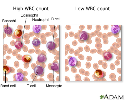
There are various types of white blood cells (WBCs) that normally appear in the blood: neutrophils (polymorphonuclear leukocytes; PMNs), band cells (slightly immature neutrophils), T-type lymphocytes (T cells), B-type lymphocytes (B cells), monocytes, eosinophils, and basophils. T and B-type lymphocytes are indistinguishable from each other in a normal slide preparation. Any infection or acute stress will result in an increased production of WBCs. This usually entails increased numbers of cells and an increase in the percentage of immature cells (mainly band cells) in the blood. This change is referred to as a “shift to the left” People who have had a splenectomy have a persistent mild elevation of WBCs. Drugs that may increase WBC counts include epinephrine, allopurinol, aspirin, chloroform, heparin, quinine, corticosteroids, and triamterene. Drugs that may decrease WBC counts include antibiotics, anticonvulsants, antihistamine, antithyroid drugs, arsenicals, barbiturates, chemotherapeutic agents, diuretics and sulfonamides.   (Updated by: David C. Dugdale, III, MD)

https://www.med-ed.virginia.edu/courses/path/innes/nh/wcbmaturation.cfm

Note that the mature forms of the myeloid series (neutrophils, eosinophils, basophils), all have lobed (segmented) nuclei. The degree of lobation increases as the cells mature.

The earliest recognizable myeloid cell is the myeloblast (10-20m dia) with a large round to oval nucleus. There is fine diffuse immature chromatin (without clumping) and a prominant nucleolus.

The cytoplasm is basophilic without granules. Although one may see a small golgi area adjacent to the nucleus, granules are not usually visible by light microscopy. One should not see blast cells in the peripheral blood.

myeloblast x100b

https://www.med-ed.virginia.edu/courses/path/innes/images/nhjpeg/nh%20myeloblast%20x100b.jpeg

The promyelocyte (10-20m) is slightly larger than a blast. Its nucleus, although similar to a myeloblast shows slight chromatin condensation and less prominent nucleoli. The cytoplasm contains striking azurophilic granules or primary granules. These granules contain myeloperoxidase, acid phosphatase, and esterase enzymes. Normally no promyelocytes are seen in the peripheral blood.

At the point in development when secondary granules can be recognized, the cell becomes a myelocyte.

promyelocyte x100

https://www.med-ed.virginia.edu/courses/path/innes/images/nhjpeg/nh%20promyelocyte%20×100%20a.jpeg

Myelocytes (10-18m) are not normally found in the peripheral blood. Nucleoli may not be seen in the late myelocyte. Primary azurophilic granules are still present, but secondary granules predominate. Secondary granules (neut, eos, or baso) first appear adjacent to the nucleus. In neutrophils this is the “dawn” of neutrophilia.

Metamyelocytes (10-18m) have kidney shaped indented nuclei and dense chromatin along the nuclear membrane. The cytoplasm is faintly pink, and they have secondary granules (neutro, eos, or baso). Zero to one percent of the peripheral blood white cells may be metamyelocytes (juveniles).

metamyelocyte x100

https://www.med-ed.virginia.edu/courses/path/innes/images/nhjpeg/nh%20metamyelocyte%20×100.jpeg

Bands, slightly smaller than juveniles, are marked by a U-shaped or deeply indented nucleus.

band neutrophilx100a

https://www.med-ed.virginia.edu/courses/path/innes/images/nhjpeg/nh%20band%20x100a.jpeg

Segmented (segs) or polymorphonuclear (PMN) leukocytes (average 14 m dia) are distinguished by definite lobation with thin thread-like filaments of chromatin joining the 2-5 lobes. 45-75% of the peripheral blood white cells are segmented neutrophils.

https://www.med-ed.virginia.edu/courses/path/innes/images/nhjpeg/nh%20neutrophil%20×100%20d.jpeg

Thrombocytogenesis

The incredible journey: From megakaryocyte development to platelet formation

Kellie R. Machlus1,2 and Joseph E. Italiano Jr
JCB 2013; 201(6): 785-796
http://dx.doi.org:/10.1083/jcb.201304054

Large progenitor cells in the bone marrow called megakaryocytes (MKs) are the source of platelets. MKs release platelets through a series of fascinating cell biological events. During maturation, they become polyploid and accumulate massive amounts of protein and membrane. Then, in a cytoskeletal-driven process, they extend long branching processes, designated proplatelets, into sinusoidal blood vessels where they undergo fission to release platelets.

megakaryocyte production of platelets

http://dm5migu4zj3pb.cloudfront.net/manuscripts/26000/26891/medium/JCI0526891.f4.jpg

platelets and the immune continuum nri2956-f3

http://www.nature.com/nri/journal/v11/n4/images/nri2956-f3.jpg

2.4.2 Classification of hematological malignancies
Practical Diagnosis of Hematologic Disoreders. 4th edition. Vol 2.
Kjeldsberg CR, Ed.  ASCP Press.  2006. Chicago, IL.

2.4.2.1 Primary Classification

Acute leukemias

Myelodysplastic syndromes

Acute myeloid leukemia

Acute lymphoblastic leukemia

Myeloproliferative Disorders

Chronic myeloproliferative disorders

Chronic myelogenous leukemia and related disorders

Myelofibrosis, including chronic idiopathic

Polycythemia, including polycythemia rubra vera

Thrombocytosis, including essential thrombocythemia

Chronic lymphoid leukemia and other lymphoid leukemias

Lymphomas

Non-Hodgkin Lymphoma

Hodgkin lymphoma

Lymphoproliferative disorders associated with immunodeficiency

Plasma Cell dyscrasias

Mast cell disease and Histiocytic neoplasms

2.4.2.2 Secondary Classification

2.4.2.3 Nuance – PathologyOutlines
Nat Pernick, Ed.

Leukemia – Acute

Primary references acute leukemia-general AML general AML classification transient abnormal myelopoiesis

Recurrent genetic abnormalities: AML with t(6;9) AML with t(8;21) AML with 11q23 abnormalities AML with inv(16) or t(16;16) AML with Down syndrome AML with FLT3 mutations AML with myelodysplastic related changes AML therapy related APL microgranular variant APL with t(15;17) APL with t(V;17) APL therapy related

AML not otherwise categorized: minimally differentiated (M0) without maturation (M1) with maturation (M2) M3 myelomonocytic monoblastic and monocytic erythroid megakaryoblastic CD13/CD33 negative basophilic myeloid sarcoma acute panmyelosis with myelofibrosis with Philadelphia chromosome with pseudo Chediak-Higashi anomaly hypocellular

ALL: general WHO classification with eosinophilia

PreB ALL: general t(9;22) t(v;11q23) t(1;19) t(5;14) t(12;21) hyperdiploidy hypodiploidy mature B ALL/Burkitt

Other ALL: T ALL ambiguous lineage mixed phenotype

AML and related malignancies

Acute myeloid leukemias with recurrent genetic abnormalities:

  • AML with t(8;21)(q22;q22); RUNX1-RUNX1T1
  • AML with inv(16)(p13.1;q22) or t(16;16)(p13.1;q22); CBF&beta-MYH11
  • Acute promyelocytic leukemia with t(15;17)(q22;q12); PML/RAR&alpha and variants
  • AML with t(9;11)(p22;q23); MLLT3-MLL
  • AML with t(6;9)(p23;q34); DEK-NUP214
  • AML with inv(3)(q21q26.2) or t(3;3)(q21;q26.2); RPN1-EVI1
  • AML (megakaryoblastic) with t(1;22)(p13;q13); RBM15-MKL1
  • AML with mutated NPM1*
  • AML with mutated CEBPA*

* provisional

Acute myeloid leukemia with myelodysplasia related changes

Therapy related acute myeloid leukemia

  • Alkylating agent related
  • Topoisomerase II inhibitor related (some maybe lymphoid)

Acute myeloid leukemia not otherwise categorized:

  • AML minimally differentiated (M0)
  • AML without maturation (M1)
  • AML with maturation (M2)
  • Acute myelomonocytic leukemia (M4)
  • Acute monoblastic and monocytic leukemia (M5a, M5b)
  • Acute erythroid leukemia (M6)
  • Acute megakaryoblastic leukemia (M7)
  • Acute basophilic leukemia
  • Acute panmyelosis with myelofibrosis

Myeloid Sarcoma

Myeloid proliferations related to Down syndrome:

  • Transient abnormal myelopoeisis
  • Myeloid leukemia associated with Down syndrome

Blastic plasmacytoid dentritic cell neoplasm:

Acute leukemia of ambiguous lineage:

  • Acute undifferentiated leukemia
  • Mixed phenotype acute leukemia with t(9;22)(q34;q11.2); BCR-ABL1
  • Mixed phenotype acute leukemia with t(v;11q23); MLL rearranged
  • Mixed phenotype acute leukemia, B/myeloid, NOS
  • Mixed phenotype acute leukemia, T/myeloid, NOS
  • Mixed phenotype acute leukemia, NOS, rare types
  • Other acute leukemia of ambiguous lineage
  • References: WHO Classification of Tumours of Haematopoietic and Lymphoid Tissue (IARC, 2008), Discovery Medicine 2010, eMedicine

Acute lymphocytic leukemia

General
=================================================================

  • WHO classification system includes former FAB classifications ALL-L1 and L2
    ● FAB L3 is now considered Burkitt lymphoma

WHO classification of acute lymphoblastic leukemia
=================================================================

Precursor B lymphoblastic leukemia / lymphoblastic lymphoma:
● ALL with t(9;22)(q34;q11.2); BCR-ABL (Philadelphia chromosome)
● ALL with t(v;11q23) (MLL rearranged)
● ALL with t(1;19)(q23;p13.3); TCF3-PBX1 (E2A-PBX1)
● ALL with t(12;21)(p13;q22); ETV6-RUNX1 (TEL-AML1)
● Hyperdiploid > 50
● Hypodiploid
● t(5;14)(q31;q32); IL3-IGH

Precursor T lymphoblastic leukemia / lymphoma

Additional references
=================================================================

Mixed phenotype acute leukemia (MPAL)

General
=================================================================

  • De novo acute leukemia containing separate populations of blasts of more than one lineage (bilineal or bilineage), or a single population of blasts co-expressing antigens of more than one lineage (biphenotypic)Excludes:
    ● Acute myeloid leukemia (AML) with recurrent translocations t(8;21), t(15;17) or inv(16)
    ● Leukemias with FGFR1 mutations
    ● Chronic myelogenous leukemia (CML) in blast crisis
    ● Myelodysplastic syndrome (MDS)-related AML and therapy-related AML, even if they have MPAL immunophenotypeCriteria for biphenotypic leukemia:
    ● Score of 2 or more for each of two separate lineages:The European Group for the Immunological Classification of Leukemias (EGIL) scoring system2008 WHO classification of acute leukemias of ambiguous lineage 

Prognosis
=================================================================

  • Poor, overall survival of 18 months
    ● Young age, normal karyotype and ALL induction therapy are associated with favorable survival
    ● Ph+ is a predictor for poor prognosis
    ● Bone marrow transplantation should be considered in first remission

Major Categories

MPAL with t(9;22)(q34;q11.2); BCR-ABL1
=================================================================

  • 20% of all MPAL
    ● Blasts with t(9;22)(q34;q11.2) translocation or BCR-ABL1 rearrangement (Ph+) without history of CML
    ● Majority in adults
    ● High WBC counts● Most of the cases B/myeloid phenotype
    ● Rare T/myeloid, B and T lineage, or trilineage leukemiasMorphology:
    ● Many cases show a dimorphic blast population, one resembling myeloblasts and the other lymphoblastsCytogenetic abnormalities:
    ● Conventional karyotyping for t(9;22), FISH or PCR for BCR-ABL1 translocation
    ● Additional complex karyotypes
    ● Ph+ is a poor prognostic factor for MPAL, with a reported median survival of 8 months
    ● Worse than patients of all other types of MPAL

MPAL with t(v;11q23); MLL rearranged
=================================================================

  • Meeting the diagnostic criteria for MPAL with blasts bearing a translocation involving the 11q23 breakpoint (MLL gene)
    ● MPAL with MLL rearranged rare
    ● More often seen in children and relatively common in infancy
    ● High WBC counts
    ● Poor prognosis
    ● Dimorphic blast population, with one resembling monoblasts and the other resembling lymphoblasts
    ● Lymphoblast population often shows a CD19+, CD10- B precursor immunophenotype, frequently CD15+
    ● Expression of other B markers usually weak
    ● Translocations involving MLL gene include t(4;11)(q21;q23), t(11;19)(q23;p13), and t(9;11)(p22;q23)
    ● Cases with chromosome 11q23 deletion should not be classified in this category

B cell acute lymphoblastic leukemia (ALL) / lymphoblastic lymphoma (LBL)

General

=================================================================

  • Current 2008 WHO classification: B lymphoblastic leukemia / lymphoma, NOS or B lymphoblastic leukemia / lymphoma with recurrent genetic abnormalities
  • See also lymphomas: B cell chapter
  • Also called B cell acute lymphoblastic leukemia / lymphoblastic lymphoma, pre B ALL / LBL
  • Usually children
  • B acute lymphoblastic leukemia presents with pancytopenia due to extensive marrow involvement, stormy onset of symptoms, bone pain due to marrow expansion, hepatosplenomegaly due to neoplastic infiltration, CNS symptoms due to meningeal spread and testicular involvement
  • B acute lymphoblastic lymphoma often presents with cutaneous nodules, bone or nodal involvement, < 25% lymphoblasts in bone marrow and peripheral blood; aleukemic cases are usually asymptomatic
  • Depending on specific leukemia, arises in either hematopoietic stem cell or B-cell progenitor
  • Tumors are derived from pre-germinal center naive B cells with unmutated VH region genes
  • Have multiple immunophenotyping aberrancies relative to normal B cell precursors (hematogones); at relapse, 73% show loss of 1+ aberrance and 60% show new aberrancies (Am J Clin Pathol 2007;127:39)

Prognostic features

=================================================================

  • Favorable prognosis: age 1-10 years, female, white; preB phenotype, hyperdiploidy>50, t(12,21), WBC count at presentation <50×108/L, non-traumatic tap with no blasts in CNS, rapid response to chemotherapy < 5% blasts on morphology on day 15, remission status after induction <5% blasts on morphology and <0.01% blast on flow or PCR, CD10+
  • Intermediate prognosis: hyperdiploidy 47-50, diploid, 6q- and rearrangements of 8q24
  • Unfavorable prognosis: under age 1 (usually have 11q23 translocations) or over age 10; t(9;22) (but not if age 59+ years, Am J Clin Pathol 2002;117:716); male, > 50×108/L WBC at presentation, hypodiploidy, near tetraploidy, 17p- and MLL rearrangements t(v;11q23); CD10-; non-traumatic tap with > 5% blasts or traumatic tap (7%); also increased microvessel staining using CD105 in children (Leuk Res 2007;31:1741), MDR1 expression in children (Oncol Rep 2004;12:1201) and adults (Blood 2002;100:974), 25%+ blasts on morphology on day 15, remission status after induction ≥ 5% blasts on morphology and ≥ 0.1% blasts on flow or PCR

Case reports

=================================================================

  • 12 month old girl and 13 month old boy with mature phenotype but no translocations (Arch Pathol Lab Med 2003;127:1340)
  • 56 year old man with ALL arising from follicular lymphoma (Arch Pathol Lab Med 2002;126:997)
  • 76 year old man with basal cell carcinoma (Diagn Pathol 2007;2:32)
  • With hemophagocytic lymphohistiocytosis (Pediatr Blood Cancer 2008;50:381)

Treatment

================================================================

  • Chemotherapy cures more children than adults; adolescents benefit from intensive regimens (Hematology Am Soc Hematol Educ Program 2005:123)

Micro description

=================================================================

  • Bone marrow smears: small to intermediate blast-like cells with scant, variably basophilic cytoplasm, round / oval or convoluted nuclei, fine chromatin and indistinct nucleoli; frequent mitotic figures; may have “starry sky” appearance similar to Burkitt lymphoma; may have large lymphoblasts with 1-4 prominent nucleoli resembling myeloblasts; usually no sclerosis
  • Bone marrow biopsy: usually markedly hypercellular with reduction of trilinear maturation; cells have minimal cytoplasm, medium sized nuclei that are often convoluted, moderately dense chromatin and indistinct nucleoli, brisk mitotic activity
  • Other tissues: may have “starry sky” appearance similar to Burkitt lymphoma; collagen dissection, periadipocyte growth pattern and single cell linear filing

Chronic Leukemia

Chronic Myeloid Neoplasms

Myelodysplastic syndromes (MDS): general, WHO classification, childhood, refractory anemia, refractory anemia with ringed sideroblasts, refractory cytopenia with multilineage dysplasia, refractory anemia with excess blasts, 5q-syndrome, therapy related, unclassified, arsenic toxicity

Myeloproliferative neoplasms (MPN): general, WHO classification, chronic eosinophilic leukemia, chronic myelogenous leukemia, chronic neutrophilic leukemia, essential thrombocythemia, hypereosinophilic syndrome, mast cell disease, polycythemia vera, primary myelofibrosis, unclassifiable

MDS/MPN: general, WHO classification, atypical CML, chronic myelomonocytic leukemia (CMML), chronic myelomonocytic leukemia with eosinophilia, juvenile myelomonocytic leukemia, unclassifiable

Myeloid neoplasms associated with eosinophilia and abnormalities of PDGFRA, PDGFRB, or FGFR1: PDGFRA, PDGFRB, FGFR1

Miscellaneous: transient myeloproliferative disorder of Downís syndrome

Lymphoma and plasma cell neoplasms

Lymph nodes: normal development-general B cells T cells NK cells normal histology grossing lymph nodes features to report

Molecular testing: theory FISH northern blot PCR southern blot

Non-Hodgkin lymphoma: general cytogenetics staging staging-pediatric morphologic clues hemophagocytic syndrome chemotherapeutic atypia

B cell disorders: general post-rituximab bone marrow biopsy classification-historical WHO classification

B cell lymphoma subtypes: age-related EBV-associated ALK positive large cell Burkitt unclassifiable-intermediate between Burkitt and diffuse large B cell lymphoma CLL
diffuse large B cell: 
diffuse-NOS CD5+ T cell / histiocyte rich primary cutaneous-general primary cutaneous-leg primary sites-other
follicular: 
general childhood cutaneous GI
hairy cell leukemia HCL variant intravascular large B cell lymphomatoid granulomatosis lymphoplasmacytic mantle cell-classic mantle cell-blastoid marginal zone-general marginal zone-MALT MALT-primary sites marginal zone-nodal mediastinal (thymic) plasmablastic pre B lymphoblastic leukemia/lymphoma primary effusion prolymphocytic leukemia pyothorax associated SLL splenic marginal zone splenic lymphoma with villous lymphocytes

Plasma cell neoplasms: general myeloma plasmacytoma heavy chain disease primary amyloidosis MGUS Osteosclerotic myeloma (POEMS) cryoglobulinemia

T/NK cell disorders: general WHO classification adult T cell aggressive NK cell leukemia anaplastic large cell ALK+ ALK- angioimmunoblastic T cell blastic plasmacytoid chronic lymphoproliferative disorders of NK cells cutaneous CD4+ small/medium sized T cell lymphoma cutaneous CD30 positive T cell lymphoproliferative disorders cutaneous gamma delta T cell lymphoma enteropathy epidermotropic CD8+ T cell lymphoma hepatosplenic indolent T cell proliferations mycosis fungoides NK/T cell lymphoma-nasal type nodal CD8+ cytotoxic T cell nonB nonT lymphoblastic peripheral T cell lymphoma, NOS primary effusion lymphoma Sezary syndrome staging subcutaneous panniculitis-like T cell large granular lymphocytic leukemia T cell lymphoblastic leukemia/lymphoma T cell prolymphocytic leukemia

Hodgkin lymphoma: general/staging classic lymphocyte depleted lymphocyte rich classical mixed cellularity nodular lymphocyte predominant nodular sclerosis

Post-transplantation: general WHO classification plasmacytic hyperplasia/IM-like lesions polymorphic B cell lymphoproliferative disorders monomorphic B cell lymphoproliferative disorders other graft versus host disease

Other: AIDS associated-general AIDS associated-examples EBV+ T cell lymphoproliferative disorders of childhood primary immune disorders related

Myeloproliferative neoplasms (MPN)

WHO 2008 – Myeloproliferative neoplasms (MPN) 

General
=================================================================

  • Chronic myelogenous leukemia
    ● Polycythemia vera
    ● Essential thrombocythemia
    ● Primary myelofibrosis
    ● Chronic neutrophilic leukemia
    ● Chronic eosinophilic leukemia, not otherwise categorized
    ● Mast cell disease
    ● MPNs, unclassifiable

WHO 2001 – Chronic myeloproliferative diseases 

Definition
=================================================================

  • Chronic myelogenous leukemia (Philadelphia chromosome, t(9;22)(q34;q11), BCR-ABL positive)
    ● Chronic neutrophilic leukemia
    ● Chronic eosinophilic leukemia (and the hypereosinophilic syndrome)
    ● Polycythemia vera
    ● Chronic idiopathic myelofibrosis (with extramedullary hematopoiesis)
    ● Essential thrombocythemia
    ● Chronic myeloproliferative disease, unclassifiable

Additional references
=================================================================

The World Health Organization (WHO) classification of the myeloid neoplasms  James W. Vardiman, Nancy Lee Harris, and Richard D. Brunning
Blood 2002; 100(7)  http://dx.doi.org/10.1182/blood-2002-04-1199

Lymphoma – Non B cell neoplasms

T/NK cell disorders/WHO classification (2008)

Principles of classification
=================================================================

  • Based on all available information (morphology, immunophenotype, genetics, clinical)
    ● No one antigenic marker is specific for any neoplasm (except ALK1)
    ● Immune profiling less helpful in subclassification of T cell lymphomas then B cell lymphomas
    ● Certain antigens commonly associated with specific disease entities but not entirely disease specific
    ● CD30: common in anaplastic large cell lymphoma but also classic Hodgkin lymphoma and other B and T cell lymphomas
    ● CD56: characteristic for nasal NK/T cell lymphoma, but also other T cell neoplasms and plasma cell disorders
    ● Variation of immunophenotype within a given disease (hepatosplenic T cell lymphoma: usually γδ but some are αβ)
    ● Recurrent genetic alterations have been identified for many B cell lymphomas but not for most T cell lymphomas
    ● No attempt to stratify lymphoid malignancies by grade
    ● Recognize the existence of grey zone lymphomas
    ● This multiparameter approach has been validated in international studies as highly reproducible

WHO 2008 classification of tumors of hematopoietic and lymphoid tissues (T/NK)
=================================================================

Precursor T-lymphoid neoplasms
● T lymphoblastic leukemia/lymphoma, 9837/3

Mature T cell and NK cell neoplasms
● T cell prolymphocytic leukemia, 9834/3
● T cell large granular lymphocytic leukemia, 9831/3
● Chronic lymphoproliferative disorder of NK cells, 9831/3
● Aggressive NK cell leukemia, 9948/3
● Systemic EBV-positive T cell lymphoproliferative disease of childhood, 9724/3
● Hydroa vacciniforme-like lymphoma, 9725/3
● Adult T cell leukemia/lymphoma, 9827/3
● Extranodal NK/T cell lymphoma, nasal type, 9719/3
● Enteropathy-associated T cell lymphoma, 9717/3
● Hepatosplenic T cell lymphoma, 9716/3
● Subcutaneous panniculitis-like T cell lymphoma, 9708/3
● Mycosis fungoides, 9700/3
● Sézary syndrome, 9701/3
● Primary cutaneous CD30-positive T cell lymphoproliferative disorders
● Lymphomatoid papulosis, 9718/1
● Primary cutaneous anaplastic large cell lymphoma, 9718/3
● Primary cutaneous gamma-delta T cell lymphoma, 9726/3
● Primary cutaneous CD8-positive aggressive epidermotropic cytotoxic T cell lymphoma, 9709/3
● Primary cutaneous CD4-positive small/medium T cell lymphoma, 9709/3
● Peripheral T cell lymphoma, NOS, 9702/3
● Angioimmunoblastic T cell lymphoma, 9705/3
● Anaplastic large cell lymphoma, ALK-positive, 9714/3
● Anaplastic large cell lymphoma, ALK-negative, 9702/3

Chronic Lymphocytic Leukemia

Chronic Lymphocytic Leukemia Staging
Author: Sandy D Kotiah, MD; Chief Editor: Jules E Harris, MD
Medscape Sep 6, 2013
http://emedicine.medscape.com/article/2006578-overview

General considerations in the staging of chronic lymphocytic leukemia (CLL) and the revised Rai (United States) and Binet (Europe) staging systems for CLL are provided below.[1, 2, 3]

See Chronic Leukemias: 4 Cancers to Differentiate, a Critical Images slideshow, to help detect chronic leukemias and determine the specific type present.

General considerations

  • CLL and small lymphocytic lymphoma (SLL) are different manifestations of the same disease; SLL is diagnosed when the disease is mainly nodal, and CLL is diagnosed when the disease is seen in the blood and bone marrow
  • CLL is diagnosed by > 5000 monoclonal lymphocytes/mm3 for longer than 3mo; the bone marrow usually has more than 30% monoclonal lymphocytes and is either normocellular or hypercellular
  • Monoclonal B lymphocytosis is a precursor form of CLL that is defined by a monoclonal B cell lymphocytosis < 5000 monoclonal lymphocytes/mm3; all lymph nodes smaller than 1.5 cm; no anemia; and no thrombocytopenia

Revised Rai staging system (United States)

Low risk (formerly stage 0)[1] :

  • Lymphocytosis, lymphocytes in blood > 15000/mcL, and > 40% lymphocytes in the bone marrow

Intermediate risk (formerly stages I and II):

  • Lymphocytosis as in low risk with enlarged node(s) in any site, or splenomegaly or hepatomegaly or both

High risk (formerly stages III and IV):

  • Lymphocytosis as in low risk and intermediate risk with disease-related anemia (hemoglobin level < 11.0 g/dL or hematocrit < 33%) or platelets < 100,000/mcL

Binet staging system (Europe)

Stage A:

  • Hemoglobin ≥ 10 g/dL, platelets ≥ 100,000/mm3, and < 3 enlarged areas

Stage B:

  • Hemoglobin ≥ 10 g/dL, platelets ≥ 100,000/mm3, and ≥ 3 enlarged areas

Stage C:

  • Hemoglobin < 10 g/dL, platelets < 100,000/mm3, and any number of enlarged areas

Read Full Post »

New Insights on the Warburg Effect [2.2]

Larry H. Bernstein, MD, FCAP, Curator, Writer

UPDATED on 7/18/2021

It is the 100 year anniversary of Warburg. He was nominated for the Nobel Prize 34 times.

There is a big resurgence of work related to his work in the last two decades!

Protein networks linking Warburg and Reverse Warburg effects to cancer cell metabolism


Dina Johar1*, Ahmed O. Elmehrath2, Rania M. Khalil3, Mostafa H. Elberry4, Samy Zaky5, Samy A. Shalabi6, Larry H. Bernstein7

1Biochemistry and Nutrition Department, Faculty of Women for Arts, Sciences and Education, Heliopolis, Cairo, Egypt  

2Faculty of Medicine, Cairo University, Cairo, Egypt

3Department of Biochemistry, Faculty of Pharmacy, Delta University for Science and Technology, Gamasa city, Mansoura, Dakahleya, Egypt

4Virology and Immunology Unit, Cancer Biology Department, National Cancer Institute, Cairo University, Egypt

5Hepatogastroenterology and infectious diseases, Faculty of Medicine, Al-Azhar University, Cairo, Egypt

6Clinical Pathology Department, Faculty of Medicine, Cairo University, Giza, Egypt, & consultant pathologist, Kuwait

7Triplex Consulting Pharmaceuticals, MA, USA

*Dina Johar, MSc., PhD.

Department of Biochemistry and Nutrition, Faculty of Women for Arts, Sciences and Education, Ain Shams University, Heliopolis, Cairo, Egypt  

dinajohar@gu.edu.eg

Ahmed O. Elmehrath

Faculty of Medicine, Cairo university, Cairo, Egypt

Ahmedo.elmehrath@gmail.com

Rania M. Khalil, PhD

Department of Biochemistry, Faculty of Pharmacy, Delta University for Science and Technology, Gamasa city, Mansoura, Dakahleya, Egypt

Rania.khalil@deltauniv.edu.eg

Mostafa H. Elberry, MSc., MD.

Virology and Immunology Unit, Cancer Biology Department, National Cancer Institute, Cairo University, Egypt

mostafa.elberry@nci.cu.edu.eg

Samy Zaky, MD

Hepatogastroenterology and infectious diseases, Faculty of Medicine, Al-Azhar University, Cairo, Egypt

samyzs55@azhar.edu.eg   |    samyzs55@yahoo.com

Samy A. Shalabi, MD

Emeritus Prof. of Surgical, anatomical and oncopatholoy, Faculty of Medicine, Cairo University, Cairo Egypt, and consultant pathologist, Kuwait

dr.samypath@gmail.com

Larry H. Bernstein, MD

Emeritus Prof. Department of Pathology, Yale University, USA

Triplex Consulting Pharmaceuticals, MA, USA

larry.bernstein@gmail.com

*To whom correspondence should be addressed   

 Abstract


Background: It was 80 years after the Otto Warburg discovery of aerobic glycolysis, a major hallmark in the understanding of cancer. The Warburg effect is the preference of cancer cell for glycolysis that produce lactate even when sufficient oxygen is provided. “Reverse Warburg effect” refers to the interstitial tissue communications with adjacent epithelium, that in the process of carcinogenesis, is needed to be explored. Among these cell-cell communications, the contact between epithelial cells, between epithelial cells and matrix, and between fibroblasts and inflammatory cells in the underlying matrix. Cancer involves dysregulation of Warburg and Reverse Warburg cellular metabolic pathways. Aim: How these gene and protein-based regulatory mechanisms have functioned has been the basis for this review. Method: the importance of the Warburg in oxidative phosphorylation suppression, with increased glycolysis in cancer growth and proliferation are emphasized. Studies that are directed at pathways that would be expected to shift cell metabolism to an increased oxidation and to a decrease in glycolysis are emphasized. Key enzymes required for oxidative phosphorylation, and affect the inhibition of fatty acid metabolism and glutamine dependence are conferred. Discussion: The findings are of special interest to cancer pharmacotherapy. Studies described in this review are concerned with the effects of therapeutic modalities that are intimately related to the Warburg effect. These interactions described may be helpful as adjuvant therapy in controlling the process of proliferation and metastasis.

Keywords: Warburg, tumorigenesis, aerobic, anaerobic, glycolysis, cancer, proliferation, metastasis

Published on 7/13/2021 in:

Cancer therapeutic modalities based on Warburg effect

New Insights on the Warburg Effect [2.2]

Defective Mitochondria Transform Normal Cells into Tumors

GEN News Jul 9, 2015

Ninety-one years ago Otto Warburg demonstrated that cancer cells have impaired respiration, which became known as the Warburg Effect. The interest in this and related work was superceded in the last quarter of the twentieth century by work on the genetic code. Now there is renewed interest.

An international research team reports that a specific defect in mitochondria plays a key role in the transition from normal cells to cancerous ones. The scientists disrupted a key component of mitochondria of otherwise normal cells and the cells took on characteristics of malignant cells.

Their study (“Disruption of cytochrome c oxidase function induces the Warburg effect and metabolic reprogramming”) is published Oncogene and was led by members of the lab of Narayan G. Avadhani, Ph.D., the Harriet Ellison Woodward Professor of Biochemistry in the department of biomedical sciences in the school of veterinary medicine at the University of Pennsylvania. Satish Srinivasan, Ph.D., a research investigator in Dr. Avadhani’s lab, was the lead author.

This is consistent with the 1924 observation by Warburg that cancerous cells consumed glucose at a higher rate than normal cells (Meyerhof ratio) and had defects in their grana, the organelles that are now known as mitochondria. He postulated that these defects led to problems in the process by which the cell produces energy. But the process called oxidative phosphorylation was not yet known. Further work in his laboratory was carried out by Hans Krebs and by Albert Szent Gyorgyi elucidating the tricarboxylic acid cycle.  The discovery of the importance of cytochrome c and adenosine triphosphate in oxidative phosphorylation was made in the post World War II period by Fritz Lippman, with an important contribution by Nathan Kaplan. All of the name scientists, except Kaplan, received Nobel Prizes. The last piece of the puzzle became the demonstation of a sequence of hydrogen transfers on the electron transport chain. The researchers above have now shown that mitochondrial defects indeed contributed to the cells becoming cancerous.

“The first part of the Warburg hypothesis has held up solidly in that most proliferating tumors show high dependence on glucose as an energy source and they release large amounts of lactic acid,” said Dr. Avadhani. “But the second part, about the defective mitochondrial function causing cells to be tumorigenic, has been highly contentious.”

To see whether the second part of Warburg’s postulation was correct, the researchers took cell lines from the skeleton, kidney, breast, and esophagus and used RNA molecules to silence the expression of select components of mitochondrial cytochrome oxidase C, or CcO, a critical enzyme involved in oxidative phosphorylation. CcO uses oxygen to make water and set up a transmembrane potential that is used to synthesize ATP, the molecule used for energy by the body’s cells.

The biologists observed that disrupting only a single protein subunit of cytochrome oxidase C led to major changes in the mitochondria and in the cells themselves. “These cells showed all the characteristics of cancer cells,” noted Dr. Avadhani.

The normal cells that converted to cancerous cells displayed changes in their metabolism, becoming more reliant on glucose by utilization of the glycolytic pathway. They reduced their synthesis of ATP.  Oxidative phosphorylation was reduced in concert with the ATP reduction. The large switch to glycolysis as primary energy source is a less efficient means of making ATP that is common in cancer cells.

The cells lost contact inhibition and gained an increased ability to invade distant tissues, both hallmarks of cancer cells. When they were grown in a 3D medium, which closely mimics the natural environment in which tumors grow in the body, the cells with disrupted mitochondria formed large, long-lived colonies, akin to tumors.

The researchers also silenced cytochrome oxidase C subunits in breast and esophageal cancer cell lines. They found that the cells became even more invasive, according to Dr. Srinivasan. The team then looked at actual tumors from human patients and found that the most oxygen-starved regions, which are common in tumors, contained defective versions of CcO.

“That result alone couldn’t tell us whether that was the cause or effect of tumors, but our cell system clearly says that mitochondrial dysfunction is a driving force in tumorigenesis,” explained Dr. Avadhani.

The researchers observed that disrupting CcO triggered the mitochondria to activate a stress signal to the nucleus, akin to an SOS alerting the cell that something was wrong. Dr. Avadhani and his colleagues had previously seen a similar pathway activated in cells with depleted mitochondrial DNA, which is also linked to cancer.

Building on these findings, Dr. Avadhani and members of his lab will examine whether inhibiting components of this mitochondrial stress signaling pathway might be a strategy for preventing cancer progression.

“We are targeting the signaling pathway, developing a lot of small molecules and antibodies,” said Dr. Avadhani. “Hopefully if you block the signaling the cells will not go into the so called oncogenic mode and instead would simply die.”

In addition, they noted that looking for defects in CcO could be a biomarker for cancer screening.

Who controls the ATP supply in cancer cells? Biochemistry lessons to understand cancer energy metabolism

Rafael Moreno-Sánchez, Alvaro Marín-Hernández, Emma Saavedra, Juan P. Pardo, Stephen J. Ralph, Sara Rodríguez-Enríquez
Intl J Biochem Cell Biol 7 Feb 2014; 50:10-23
http://dx.doi.org/10.1016/j.biocel.2014.01.025

The supply of ATP in mammalian and human cells is provided by glycolysis and oxidative phosphorylation (OxPhos). There are no other pathways or processes able to synthesize ATP at sufficient rates to meet the energy demands of cells. Acetate thiokinase or acetyl-CoA synthetase, a ubiquitous enzyme catalyzing the synthesis of ATP and acetate from acetyl-CoA, PPi and AMP, might represent an exception under hypoxia in cancer cells, although the flux through this branch is negligible (≤10%) when compared to the glycolytic flux (Yoshii et al., 2009).

Glycolysis in human cells can be defined as the metabolic process that transforms 1 mol of glucose (or other hexoses) into 2 moles of lactate plus 2 moles of ATP. These stoichiometric values represent a maximum and due to the several reactions branching off glycolysis, they will be usually lower under physiological conditions, closer to 1.3–1.9 for the lactate/glucose ratio (Travis et al., 1971; Jablonska and Bishop, 1975; Suter and Weidemann, 1975; Hanson and Parsons, 1976; Wu and Davis, 1981; Pick-Kober and Schneider, 1984; Sun et al., 2012). OxPhos is the metabolic process that oxidizes several substrates through the Krebs cycle to produce reducing equivalents (NADH, FADH2), which feed the respiratory chain to generate an H+.

Applying basic biochemical principles, this review analyzes data that contrasts with the Warburg hypothesis that glycolysis is the exclusive ATP provider in cancer cells. Although disregarded for many years, there is increasing experimental evidence demonstrating that oxidative phosphorylation (OxPhos) makes a significant contribution to ATP supply in many cancer cell types and under a variety of conditions.

Substrates oxidized by normal mitochondria such as amino acids and fatty acids are also avidly consumed by cancer cells. In this regard, the proposal that cancer cells metabolize glutamine for anabolic purposes without the need for a functional respiratory chain and OxPhos is analyzed considering thermodynamic and kinetic aspects for the reductive carboxylation of 2-oxoglutarate catalyzed by isocitrate dehydrogenase.

In addition, metabolic control analysis (MCA) studies applied to energy metabolism of cancer cells are reevaluated. Regardless of the experimental/environmental conditions and the rate of lactate production, the flux-control of cancer glycolysis is robust in the sense that it involves the same steps:

  • glucose transport,
  • hexokinase,
  • hexosephosphate isomerase, and
  • glycogen degradation,

all at the beginning of the pathway; these steps together with phosphofructokinase 1 also control glycolysis in normal cells.

The respiratory chain complexes exert significantly higher flux-control on OxPhos in cancer cells than in normal cells. Thus, determination of the contribution of each pathway to ATP supply and/or the flux-control distribution of both pathways in cancer cells is necessary in order to identify differences from normal cells which may lead to the design of rational alternative therapies that selectively target cancer energy metabolism.

Fig. 1. Labeling patterns of 13C-glutamate or 13C-glutamine mitochondrial metabolism in cancer cells.

Fig. 2. Survey in PubMed of papers published in the field of tumor mitochondrial metabolism from 1951 to September 2013.

Emerging concepts in bioenergetics and cancer research: Metabolic flexibility, coupling, symbiosis, switch, oxidative tumors, metabolic remodeling, signaling and bioenergetic therapy

Emilie Obre, Rodrigue Rossignol
Intl J Biochem Cell Biol 2015; 59:167-181
http://dx.doi.org/10.1016/j.biocel.2014.12.008

The field of energy metabolism dramatically progressed in the last decade, owing to a large number of cancer studies, as well as fundamental investigations on related transcriptional networks and cellular interactions with the microenvironment. The concept of metabolic flexibility was clarified in studies showing the ability of cancer cells to remodel the biochemical pathways of energy transduction and linked anabolism in response to glucose, glutamine or oxygen deprivation.

A clearer understanding of the large scale bioenergetic impact of C-MYC, MYCN, KRAS and P53 was obtained, along with its modification during the course of tumor development. The metabolic dialog between different types of cancer cells, but also with the stroma, also complexified the understanding of bioenergetics and raised the concepts of metabolic symbiosis and reverse Warburg effect.

Signaling studies revealed the role of respiratory chain derived reactive oxygen species for metabolic remodeling and metastasis development. The discovery of oxidative tumors in human and mice models related to chemoresistance also changed the prevalent view of dysfunctional mitochondria in cancer cells. Likewise, the influence of energy metabolism-derived oncometabolites emerged as a new means of tumor genetic regulation. The knowledge obtained on the multi-site regulation of energy metabolism in tumors was translated to cancer preclinical studies, supported by genetic proof of concept studies targeting LDHA, HK2, PGAM1, or ACLY.

Here, we review those different facets of metabolic remodeling in cancer, from its diversity in physiology and pathology, to the search of the genetic determinants, the microenvironmental regulators and pharmacological modulators.

Pyruvate kinase M2: A key enzyme of the tumor metabolome and its medical relevance

Mazurek, S.
Biomedical Research 2012; 23(SPEC. ISSUE): Pages 133-142

Tumor cells are characterized by an over expression of the glycolytic pyruvate kinase isoenzyme
type M2 (abbreviations: M2-PK or PKM2). In tumor metabolism the quaternary structure of M2-PK (tetramer/dimer ratio) determines whether glucose is used for glycolytic energy regeneration (highly active tetrameric form, Warburg effect) or synthesis of cell building blocks (nearly inactive dimeric form) which are both prerequisites for cells with a high proliferation rate. In tumor cells the nearly inactive dimeric form of M2- PK is predominant due to direct interactions with different oncoproteins. Besides its key functions in tumor metabolism recent studies revealed that M2-PK may also react as protein kinase as well as co activator of transcription factors. Of medical relevance is the quantification of the dimeric form of M2-PK with either an ELISA or point of care rapid test in plasma and stool that is used for follow-up studies during therapy (plasma M2-PK) and colorectal cancer (CRC) screening (fecal M2-PK; mean sensitivity for CRC in 12 independent studies with altogether 704 samples: 80% ± 7%). An intervention in the regulation mechanisms of the expression, activity and tetramer: dimer ratio of M2-PK has significant consequences for the proliferation rate and tumorigenic capacity of the tumor cells, making this enzyme an intensively

Read Full Post »

We Celebrate Our Top Authors: 4/2012  to  7/2015 @Leaders in Pharmaceutical Business Intelligence

Curators:

Adam Sonnenberg, BSc, Content Digitization

and

Stephen J Williams, PhD, Senior Editor – Series A: Volume 1, Series B, Volume 1, Series C, Volumes 1 & 2

The Journal Articles are written by Experts, Authors, Writers. The results of Original Research are gaining value added for the e-Reader by the Methodology of Curation. The e-Book’s articles have been published on the Open Access Online Scientific Journal, since April 2012.  All new articles on this subject, will continue to be incorporated, as published with periodical updates.

Open Access Online Journal

http://www.pharmaceuticalIntelligence.com

The Journal is a scientific, medical and business, multi-expert authoring environment for information syndication in several domains of

  • Life Sciences,
  • Medicine,
  • Pharmaceutical and Healthcare Industries,
  • BioMedicine,
  • Medical Technologies & Devices.

Scientific critical interpretations and original articles are written by PhDs, MDs, MD/PhDs, PharmDs, Technical MBAs as Experts, Authors, Writers (EAWs) on an Equity Sharing basis.

Top Authors for all days ending 2015-07-31 (Summarized)

Author Name

Author NameTotal Views

4/2012 to 7/2015

# of Articles

Aviva Lev-Ari, PhD, RN 207,592 2,004
Larry H Bernstein, MD, FCAP 114,265 620
Tilda Barliya, PhD 32,651 46
Stephen J Williams, PhD 20,508 77
Dror Nir, PhD 17,372 53
Sudipta Saha, PhD 16,301 58
Ritu Saxena, PhD 12,664 40
Demet Sag, PhD 7,160 24
Aviral Vatsa, PhD 6,922 20
Ziv Raviv 4,905 9
Anamika Sarhar, PhD 2,901 7
Prabod Kandala, PhD 1,998 43
Alan F Kaul, PharmD, FCCP 1,730 4
Asshir Awan, PhD 1,366 4
Meg Baker, PhD 1,266 4
Justin D Pearlman, MD, PhD, FACC 773 3

BioMedical e-Books e-Series:

Cardiovascular, Genomics, Cancer, BioMed, Patient Centered Medicine

http://pharmaceuticalintelligence.com/biomed-e-books/

2013 e-Book on Amazon.com

http://www.amazon.com/dp/B00DINFFYC

2015 e-Books on Amazon.com

1. Metabolomics, on Amazon since 7/21/2015

http://www.amazon.com/dp/B012BB0ZF0

Forthcoming in 2015

2. Cancer Volume One

3. Genomics Volume One

4. Physiology, Genomics and Pharmaceutical

5. Cardiovascular, Volume Two: Co-Curation, Methodology

6. Cardiovascular Diseases, Volume Three: Etiology, Genetics and Genomics

7. Cardiovascular Diseases, Volume Four: Regenerative and Translational Medicine

8. Cancer Volume Two: Immunotherapy, Nanotechnology in drug delivery

2016 Plan

2016 we need to FIND Editors for Two e-Books

– Genomics Volume Two on NGS – Dr. Williams and another Editor

– Patient Voice on Invasive Procedures – New Editor

2016, We will continue with

– Aviva will work with Larry on CVD 5: Pharmacology-Therapy, 

– With Adam and Dr. Pearlman, on CVD 6: Cardiac Surgery and Interventional

– With Adam and Dr. Pearlman, on CVD 7: CV Imaging

– Dr. Pearlman alone: 

Three CVD Hardcopy Volumes, 1-Etiology, 2-Diagnostics, 3-Therapies

 onepageCVDseriesAflyervol1-4

onepage series BCDE covers

CancerandOncologyseriesCcoverII-page2

Read Full Post »

UNDERSTANDING CANCER AT THE GENETIC LEVEL: STANFORD GENETICS AND GENOMICS CERTIFICATE

Reporter: Aviva Lev-Ari, PhD, RN


UNDERSTANDING CANCER AT THE GENETIC LEVEL

VIEW VIDEO

http://online.stanford.edu/course/understanding-cancer-genetic-level


Date:
Wednesday, January 14, 2015
Course topic:

COURSE DESCRIPTION

Genetic research has been instrumental in developing leading methods used to understand the nature and scope of cancer syndromes. With improved cancer diagnosis and targeted therapies for treatment, the discovery of changes in genes and their expression characteristics will continue to advance as a field.

Study the signaling pathways of common and rare genetic mutations involved in cancer. This course will expose you to the cutting-edge research that offers attractive development for new anti-cancer drugs and therapeutic strategies. From the differences between sporadic and familial cancers to systemic and targeted level treatment studies, you will learn the history of cancer and how it shaped society and research today.

YOU WILL LEARN

  • Current methods involved in cancer research
  • Differences between multiple classes of genetic mutations
  • Progressions of tumorigenesis in minor genome changes
  • Targeted therapies for the latest cancer treatments

PLEASE NOTE: THIS COURSE IS OFFERED TOWARDS THE STANFORD GENETICS AND GENOMICS CERTIFICATE.*

Take online courses in genetics and genomics and gain a greater understanding of biology, human health and personalized medicine. Tap into the world-class research of Stanford faculty and industry experts to acquire the skills and knowledge you need to succeed in the rapidly evolving genetics industry.

*This certificate neither substitutes for, nor leads to, being board certified as a genetic counselor (ABGC) or clinical geneticist (ABMG)

SOURCE

http://online.stanford.edu/course/understanding-cancer-genetic-level

Read Full Post »

Genetic Testing for Hereditary Cancer Risk @Stanford

Reporter: Aviva Lev-Ari, PhD, RN


Genetic Testing for Hereditary Cancer Risk

With James Ford, MD – 50 Min.

VIEW VIDEO

https://cancer.stanford.edu/media/video/genetic_testing.html

Read Full Post »

Severe IBS Symptoms: Clinical Trial results on delivery of peppermint oil to the small intestine through a system (IBgard®)

Reporter: Aviva Lev-Ari, PhD, RN


Written by Freelance medical writer Walter Alexander


IBgard® Delivery of Mint Oil Effective in Patients with Severe IBS Symptoms
WASHINGTON DC, May 17, 2015—In IBSREST™ (Irritable Bowel Syndrome Reduction Evaluation and Safety Trial), sustained delivery of peppermint oil to the small intestine through a system (IBgard®) that minimizes release in the stomach and colon, resulted in significant reductions in severe or unbearable irritable bowel syndrome (IBS) symptoms over four weeks as compared with placebo. Other formulations of mint oil tend to remain in the stomach or pass all the way to the colon, potentially causing cause heartburn, nausea and anal burning.

About 25 percent of IBS patients, said Brooks D. Cash, MD, professor of medicine at University of South Alabama, Mobile, AB, at his poster at the Digestive Disease Week 2015 annual meeting, describe their symptoms as severe. Dr. Cash noted that derangements in gut immunity, microbiota, sensation, motility, secretion, and digestion have all been proposed as possible etiologies of IBS. L-menthol, the main constituent of peppermint oil, has anti-spasmodic, anti-carminative, topical analgesic, anti-infective and 5-HT3 receptor antagonism properties. IBSREST™ evaluated the efficacy and tolerability of IBgard, an ultra-purified peppermint oil, in a population enriched with severe/unbearable symptoms.High symptom severity reflects higher intensity and frequency of individual symptoms, leading in IBS patients to lower quality of life, work disruptions, and frequent physician visits (more than one per month). Dr. Cash noted also that among treatments for the three IBS subtypes (M:mixed/alternating; D: diarrhea; C:constipation), approved products are lacking for IBS-M and options for IBS-D are limited. “Our study was selective for patients with severe symptoms because that is where the unmet need is,” Dr. Cash said in an interview. He pointed out further that in studies of other treatments, more severe patients tend to respond less well. The targeted delivery of mint oil in solid microspheres to the small intestine was expected to address the unmet need, according to Dr. Cash.

The randomized, placebo-controlled trial included 72 patients (mean age ~41 years) who met Rome III criteria for IBS-D or IBS-M, had average daily IBS-related abdominal pain of ≥4 on a 0-10 scale, and a Total IBS Symptom Score (TISS) of ≥2 on a 0-4 scale. The TISS scale encompasses 8 symptoms (abdominal pain or discomfort, bloating or distention, pain at evacuation, urgency, constipation, diarrhea, mucus or gas and sense of incomplete evacuation). After a 3-week period for symptom severity assessment and prohibited medication washout, subjects were randomly allocated to receive IBgard 180 mg TID or identical placebo for 4 weeks.

After 28 days, the reduction from baseline in number of severe and unbearable symptoms (average of frequency and intensity ≥3) was -66 percent for IBgardas compared with -42 percent with placebo (P=0.0212). The reduction in patient-reported severe or unbearable abdominal pain intensity at 28 days was -79.4 percent for IBgard and-40.5 percent for placebo (P=0.0009). Trends in percent reduction in severe or unbearable individual intensity scores were favorable for IBgard across all 8 severe/unbearable parameters (from 71 percent to 90 percent), more than at 24 hour assessment (30 percent to 47 percent).“Severe symptom patients responded as well as those with less severe symptom patients. That’s very reassuring,” Dr. Cash said.

IBgard was well tolerated and safe. No patients withdrew from the study on account of treatment emergent adverse events.

Dr. Cash concluded, “Over 4 weeks, IBgard was effective at improving the composite IBS symptom score, and the individual IBS symptom components, including severe or unbearable abdominal pain intensity at 4 weeks.”


SOURCE

From: Gail Thornton <gailsthornton@yahoo.com>

Reply-To: Gail Thornton <gailsthornton@yahoo.com>

Date: Wednesday, July 8, 2015 at 11:23 AM

To: Aviva Lev-Ari <avivalev-ari@alum.berkeley.edu>

Subject: Article for Pharmaceutical Business Intelligence on IBgard


Read Full Post »

Therapeutic Implications for Targeted Therapy from the Resurgence of Warburg ‘Hypothesis’

Writer and Curator: Larry H. Bernstein, MD, FCAP 

(Note that each portion of the discussion is followed by a reference)

It is now a time to pause after almost a century of a biological scientific discoveries that have transformed the practice of medicine and impacted the lives of several generations of young minds determined to probe the limits of our knowledge.  In the century that we have entered into the scientific framework of medicine has brought together a difficult to grasp evolution of the emergence of human existence from wars, famine, droughts, storms, infectious diseases, and insect born pestilence with betterment of human lives, only unevenly divided among societal classes that have existed since time immemorial. In this short time span there have emerged several generations of physicians who have benefited from a far better medical education that their forebears could have known. In this expansive volume on cancer, we follow an incomplete and continuing challenge to understand cancer, a disease that has become associated with longer life spans in developed nations.

While there are significant improvements in the diagnosis and treatment of cancers, there is still a personal as well as locality factor in the occurrence of this group of diseases, which has been viewed incorrectly as a “dedifferentiation” of mature tissue types and the emergence of a cell phenotype that is dependent on glucose, reverts to a cancer “stem cell type” (loss of stemness), loses cell to cell adhesion, loses orderly maturation, and metastasizes to distant sites. At the same time, physician and nurses are stressed in the care of patients by balancing their daily lives and maintaining a perspective.

The conceptual challenge of cancer diagnosis and management has seemed insurmountable, but owes much to the post World War I activities of Otto Heinrich Warburg. It was Warburg who made the observation that cancer cells metabolize glucose by fermentation in much the way Pasteur 60 years earlier observed fermentation of yeast cells. This metabolic phenomenon occurs even in the presence of an oxygen supply, which would provide a huge deficit in ATP production compared with respiration. The cancer cell is “addicted to glucose” and produced lactic acid. Warburg was awarded the Nobel Prize in Medicine for this work in 1931.

In the last 15 years there has been a resurgence of work on the Warburg effect that sheds much new light on the process that was not previously possible, with significant therapeutic implications.  In the first place, the metabolic mechanism for the Warburg effect was incomplete even at the beginning of the 21st century.  This has been partly rectified with the enlightening elucidation of genome modifications, cellular metabolic regulation, and signaling pathways.

The following developments have become central to furthering our understanding of malignant transformation.

  1. There is usually an identifiable risk factor, such as, H. pylori, or of a chronic inflammatory state, as in the case of Barrett’s esophagus.
  2. There are certain changes in glucose metabolism that have been unquestionably been found in the evolution of this disease. The changes are associated with major changes in metabolic pathways, miRN signaling, and the metabolism geared to synthesis of cells with an impairment of the cell death cycle. In these changes, mitochondrial function is central to both the impaired respiration and the autophagy geared to the synthesis of cancer cells.

The emergence of this cell prototype is characterized by the following, again related to the Warburg effect:

  1. Cancer cells oxidize a decreased fraction of the pyruvate generated from glycolysis
  2. The mitochondrial pyruvate carrier (MPC), composed of the products of the MPC1 and MPC2 genes, modulates fractional pyruvate oxidation. MPC1 is deleted or underexpressed in multiple cancers and correlates with poor prognosis.
  3. Cancer cells tend to express a partially inhibited splice variant of pyruvate kinase (PK-M2), leading to decreased pyruvate production.
  4. The two proteins that mediate pyruvate conversion to lactate and its export, M-type lactate dehydrogenase and the monocarboxylate transporter MCT-4, are commonly upregulated in cancer cells leading to decreased pyruvate oxidation.
  5. The enzymatic step following mitochondrial entry is the conversion of pyruvate to acetyl-CoA by the pyruvate dehydrogenase (PDH) complex. Cancer cells frequently exhibit increased expression of the PDH kinase PDK1, which phosphorylates and inactivates PDH. This PDH regulatory mechanism is required for oncogene induced transformation and reversed in oncogene-induced senescence.
  6. The PDK inhibitor dichloroacetate has shown some clinical efficacy, which correlates with increased pyruvate oxidation. One of the simplest mechanisms to explain decreased mitochondrial pyruvate oxidation in cancer cells, a loss of mitochondrial pyruvate import, has been observed repeatedly over the past 40 years. This process has been impossible to study at a molecular level until recently, however, as the identities of the protein(s) that mediate mitochondrial pyruvate uptake were unknown.
  7. The mitochondrial pyruvate carrier (MPC) as a multimeric complex that is necessary for efficient mitochondrial pyruvate uptake. The MPC contains two distinct proteins, MPC1 and MPC2; the absence of either leads to a loss of mitochondrial pyruvate uptake and utilization in yeast, flies, and mammalian cells.

A Role for the Mitochondrial Pyruvate Carrier as a Repressor of the Warburg Effect and Colon Cancer Cell Growth

John C. Schell, Kristofor A. Olson, Lei Jiang, Amy J. Hawkins, et al.
Molecular Cell Nov 6, 2014; 56: 400–413.
http://dx.doi.org/10.1016/j.molcel.2014.09.026

In addition to the above, the following study has therapeutic importance:

Glycolysis has become a target of anticancer strategies. Glucose deprivation is sufficient to induce growth inhibition and cell death in cancer cells. The increased glucose transport in cancer cells has been attributed primarily to the upregulation of glucose transporter 1 (Glut1),  1 of the more than 10 glucose transporters that are responsible for basal glucose transport in almost all cell types. Glut1 has not been targeted until very recently due to the lack of potent and selective inhibitors.

First, Glut1 antibodies were shown to inhibit cancer cell growth. Other Glut1 inhibitors and glucose transport inhibitors, such as fasentin and phloretin, were also shown to be effective in reducing cancer cell growth. A group of inhibitors of glucose transporters has been recently identified with IC50 values lower than 20mmol/L for inhibiting cancer cell growth. However, no animal or detailed mechanism studies have been reported with these inhibitors.

Recently, a small molecule named STF-31 was identified that selectively targets the von Hippel-Lindau (VHL) deficient kidney cancer cells. STF-31 inhibits VHL deficient cancer cells by inhibiting Glut1. It was further shown that daily intraperitoneal injection of a soluble analogue of STF-31 effectively reduced the growth of tumors of VHL-deficient cancer cells grafted on nude mice. On the other hand, STF-31 appears to be an inhibitor with a narrow cell target spectrum.

These investigators recently reported the identification of a group of novel small compounds that inhibit basal glucose transport and reduce cancer cell growth by a glucose deprivation–like mechanism. These compounds target Glut1 and are efficacious in vivo as anticancer agents. A novel representative compound WZB117 not only inhibited cell growth in cancer cell lines but also inhibited cancer growth in a nude mouse model. Daily intraperitoneal injection of WZB117 resulted in a more than 70% reduction in the size of human lung cancer of A549 cell origin. Mechanism studies showed that WZB117 inhibited glucose transport in human red blood cells (RBC), which express Glut1 as their sole glucose transporter. Cancer cell treatment with WZB117 led to decreases in levels of Glut1 protein, intracellular ATP, and glycolytic enzymes. All these changes were followed by increase in ATP sensing enzyme AMP-activated protein kinase (AMPK) and declines in cyclin E2 as well as phosphorylated retinoblastoma, resulting in cell-cycle arrest, senescence, and necrosis. Addition of extracellular ATP rescued compound-treated cancer cells, suggesting that the reduction of intracellular ATP plays an important role in the anticancer mechanism of the molecule.

A Small-Molecule Inhibitor of Glucose Transporter 1 Downregulates Glycolysis, Induces Cell-Cycle Arrest, and Inhibits Cancer Cell Growth In Vitro and In Vivo

Yi Liu, Yanyan Cao, Weihe Zhang, Stephen Bergmeier, et al.
Mol Cancer Ther Aug 2012; 11(8): 1672–82
http://dx.doi.org://10.1158/1535-7163.MCT-12-0131

Alterations in cellular metabolism are among the most consistent hallmarks of cancer. These investigators have studied the relationship between increased aerobic lactate production and mitochondrial physiology in tumor cells. To diminish the ability of malignant cells to metabolize pyruvate to lactate, M-type lactate dehydrogenase levels were knocked down by means of LDH-A short hairpin RNAs. Reduction in LDH-A activity resulted in stimulation of mitochondrial respiration and decrease of mitochondrial membrane potential. It also compromised the ability of these tumor cells to proliferate under hypoxia. The tumorigenicity of the LDH-A-deficient cells was severely diminished, and this phenotype was reversed by complementation with the human ortholog LDH-A protein. These results demonstrate that LDH-A plays a key role in tumor maintenance.

The results are consistent with a functional connection between alterations in glucose metabolism and mitochondrial physiology in cancer. The data also reflect that the dependency of tumor cells on glucose metabolism is a liability for these cells under limited-oxygen conditions. Interfering with LDH-A activity as a means of blocking pyruvate to lactate conversion could be exploited therapeutically. Because individuals with complete deficiency of LDH-A do not show any symptoms under ordinary circumstances, the genetic data suggest that inhibition of LDH-A activity may represent a relatively nontoxic approach to interfere with tumor growth.

Attenuation of LDH-A expression uncovers a link between glycolysis, mitochondrial physiology, and tumor maintenance

Valeria R. Fantin Julie St-Pierre and Philip Leder
Cancer Cell Jun 2006; 9: 425–434.
http://dx.doi.org:/10.1016/j.ccr.2006.04.02

The widespread clinical use of positron-emission tomography (PET) for the detection of aerobic glycolysis in tumors and recent findings have rekindled interest in Warburg’s theory. Studies on the physiological changes in malignant conversion provided a metabolic signature for the different stages of tumorigenesis; during tumorigenesis, an increase in glucose uptake and lactate production have been detected. The fully transformed state is most dependent on aerobic glycolysis and least dependent on the mitochondrial machinery for ATP synthesis.

Tumors ferment glucose to lactate even in the presence of oxygen (aerobic glycolysis; Warburg effect). The pentose phosphate pathway (PPP) allows glucose conversion to ribose for nucleic acid synthesis and glucose degradation to lactate. The nonoxidative part of the PPP is controlled by transketolase enzyme reactions. We have detected upregulation of a mutated transketolase transcript (TKTL1) in human malignancies, whereas transketolase (TKT) and transketolase-like-2 (TKTL2) transcripts were not upregulated. Strong TKTL1 protein expression was correlated to invasive colon and urothelial tumors and to poor patients outcome. TKTL1 encodes a transketolase with unusual enzymatic properties, which are likely to be caused by the internal deletion of conserved residues. We propose that TKTL1 upregulation in tumors leads to enhanced, oxygen-independent glucose usage and a lactate based matrix degradation. As inhibition of transketolase enzyme reactions suppresses tumor growth and metastasis, TKTL1 could be the relevant target for novel anti-transketolase cancer therapies. We suggest an individualized cancer therapy based on the determination of metabolic changes in tumors that might enable the targeted inhibition of invasion and metastasis.

Other important links between cancer-causing genes and glucose metabolism have been already identified. Activation of the oncogenic kinase Akt has been shown to stimulate glucose uptake and metabolism in cancer cells and renders these cells susceptible to death in response to glucose withdrawal. Such tumor cells have been shown to be dependent on glucose because the ability to induce fatty acid oxidation in response to glucose deprivation is impaired by activated Akt. In addition, AMP-activated protein kinase (AMPK) has been identified as a link between glucose metabolism and the cell cycle, thereby implicating p53 as an essential component of metabolic cell-cycle control.

Expression of transketolase TKTL1 predicts colon and urothelial cancer patient survival: Warburg effect reinterpreted

S Langbein, M Zerilli, A zur Hausen, W Staiger, et al.
British Journal of Cancer (2006) 94, 578–585.
http://dx.doi.org:/10.1038/sj.bjc.6602962

The unique metabolic profile of cancer (aerobic glycolysis) might confer apoptosis resistance and be therapeutically targeted. Compared to normal cells, several human cancers have high mitochondrial membrane potential (DJm) and low expression of the K+ channel Kv1.5, both contributing toapoptosis resistance. Dichloroacetate (DCA) inhibits mitochondrial pyruvate dehydrogenase kinase (PDK), shifts metabolism from glycolysis to glucose oxidation, decreases DJm, increases mitochondrial H2O2, and activates Kv channels in all cancer, but not normal, cells; DCA upregulates Kv1.5 by an NFAT1-dependent mechanism. DCA induces apoptosis, decreases proliferation, and inhibits tumor growth, without apparent toxicity. Molecular inhibition of PDK2 by siRNA mimics DCA. The mitochondria-NFAT-Kv axis and PDK are important therapeutic targets in cancer; the orally available DCA is a promising selective anticancer agent.

Cancer progression and its resistance to treatment depend, at least in part, on suppression of apoptosis. Although mitochondria are recognized as regulators of apoptosis, their importance as targets for cancer therapy has not been adequately explored or clinically exploited. In 1930, Warburg suggested that mitochondrial dysfunction in cancer results in a characteristic metabolic phenotype, that is, aerobic glycolysis (Warburg, 1930). Positron emission tomography (PET) imaging has now confirmed that most malignant tumors have increased glucose uptake and metabolism. This bioenergetic feature is a good marker of cancer but has not been therapeutically pursued..

The small molecule DCA is a metabolic modulator that has been used in humans for decades in the treatment of lactic acidosis and inherited mitochondrial diseases. Without affecting normal cells, DCA reverses the metabolic electrical remodeling that we describe in several cancer lines (hyperpolarized mitochondria, activated NFAT1, downregulated Kv1.5), inducing apoptosis and decreasing tumor growth. DCA in the drinking water at clinically relevant doses for up to 3 months prevents and reverses tumor growth in vivo, without apparent toxicity and without affecting hemoglobin, transaminases, or creatinine levels. The ease of delivery, selectivity, and effectiveness  make DCA an attractive candidate for proapoptotic cancer therapy which can be rapidly translated into phase II–III clinical trials.

A Mitochondria-K+ Channel Axis Is Suppressed in Cancer and Its Normalization Promotes Apoptosis and Inhibits Cancer Growth

Sebastien Bonnet, Stephen L. Archer, Joan Allalunis-Turner, et al.

Cancer Cell Jan 2007; 11: 37–51.
http://dx.doi.org:/10.1016/j.ccr.2006.10.020

Tumor cells, just as other living cells, possess the potential for proliferation, differentiation, cell cycle arrest, and apoptosis. There is a specific metabolic phenotype associated with each of these conditions, characterized by the production of both energy and special substrates necessary for the cells to function in that particular state. Unlike that of normal living cells, the metabolic phenotype of tumor cells supports the proliferative state. Aim: To present the metabolic hypothesis that (1) cell transformation and tumor growth are associated with the activation of metabolic enzymes that increase glucose carbon utilization for nucleic acid synthesis, while enzymes of the lipid and amino acid synthesis pathways are activated in tumor growth inhibition, and (2) phosphorylation and allosteric and transcriptional regulation of intermediary metabolic enzymes and their substrate availability together mediate and sustain cell transformation from one condition to another. Conclusion: Evidence is presented that demonstrates opposite changes in metabolic phenotypes induced by TGF-β, a cell transforming agent, and tumor growth-inhibiting phytochemicals such as genistein and Avemar, or novel synthetic antileukemic drugs such as STI571 (Gleevec).  Intermediary metabolic enzymes that mediate the growth signaling pathways and promote malignant cell transformation may serve as high efficacy nongenetic novel targets for cancer therapies.

A Metabolic Hypothesis of Cell Growth and Death in Pancreatic Cancer

Laszlo G. Boros, Wai-Nang Paul Lee, and Vay Liang W. Go
Pancreas 2002; 24(1):26–33

Clear cell renal cell carcinoma (ccRCC) is the most common pathological subtype of kidney cancer. Here, we integrated an unbiased genome-wide RNA interference screen for ccRCC survival regulators with an analysis of recurrently overexpressed genes in ccRCC to identify new therapeutic targets in this disease. One of the most potent survival regulators, the monocarboxylate transporter MCT4 (SLC16A3), impaired ccRCC viability in all eight ccRCC lines tested and was the seventh most overexpressed gene in a meta-analysis of five ccRCC expression datasets.

MCT4 silencing impaired secretion of lactate generated through glycolysis and induced cell cycle arrest and apoptosis. Silencing MCT4 resulted in intracellular acidosis, and reduction in intracellular ATP production together with partial reversion of the Warburg effect in ccRCC cell lines. Intra-tumoral heterogeneity in the intensity of MCT4 protein expression was observed in primary ccRCCs.

MCT4 protein expression analysis based on the highest intensity of expression in primary ccRCCs was associated with poorer relapse-free survival, whereas modal intensity correlated with Fuhrman nuclear grade. Consistent with the potential selection of subclones enriched for MCT4 expression during disease progression, MCT4 expression was greater at sites of metastatic disease. These data suggest that MCT4 may serve as a novel metabolic target to reverse the Warburg effect and limit disease progression in ccRCC.

Clear cell carcinoma (ccRCC) is the commonest subtype of renal cell carcinoma, accounting for 80% of cases. These tumors are highly resistant to cytotoxic chemotherapy and until recently, systemic treatment options for advanced ccRCC were limited to cytokine based therapies, such as interleukin-2 and interferon-α. Recently, anti-angiogenic drugs and mTOR inhibitors, all targeting the HIF–VEGF axis which is activated in up to 91% of ccRCCs through loss of the VHL tumor suppressor gene [1], have been shown to be effective in metastatic ccRCC [2–5]. Although these drugs increase overall survival to more than 2 years [6], resistance invariably occurs, making the identification of new molecular targets a major clinical need to improve outcomes in patients with metastatic ccRCC.

Genome-wide RNA interference analysis of renal carcinoma survival regulators identifies MCT4 as a Warburg effect metabolic target

Marco Gerlinger, Claudio R Santos, Bradley Spencer-Dene, et al.
J Pathol 2012; 227: 146–156
http://dx.doi.org:/10.1002/path.4006

Hypoxia-inducible factor 1 (HIF-1) plays a key role in the reprogramming of cancer metabolism by activating transcription of genes encoding glucose transporters and glycolytic enzymes, which take up glucose and convert it to lactate; pyruvate dehydrogenase kinase 1, which shunts pyruvate away from the mitochondria; and BNIP3, which triggers selective mitochondrial autophagy. The shift from oxidative to glycolytic metabolism allows maintenance of redox homeostasis and cell survival under conditions of prolonged hypoxia. Many metabolic abnormalities in cancer cells increase HIF-1 activity. As a result, a feed-forward mechanism can be activated that drives HIF-1 activation and may promote tumor progression.

Metastatic cancer is characterized by reprogramming of cellular metabolism leading to increased uptake of glucose for use as both an anabolic and a catabolic substrate. Increased glucose uptake is such a reliable feature that it is utilized clinically to detect metastases by positron emission tomography using 18F-fluorodeoxyglucose (FDG-PET) with a sensitivity of >90% [1]. As with all aspects of cancer biology, the details of metabolic reprogramming differ widely among individual tumors. However, the role of specific signaling pathways and transcription factors in this process is now understood in considerable detail. This review will focus on the involvement of hypoxia-inducible factor 1 (HIF-1) in both mediating metabolic reprogramming and responding to metabolic alterations. The placement of HIF-1 both upstream and downstream of cancer metabolism results in a feed-forward mechanism that may play a major role in the development of the invasive, metastatic, and lethal cancer phenotype.

O2 concentrations are significantly reduced in many human cancers compared with the surrounding normal tissue. The median PO2 in breast cancers is 10 mmHg, as compared with65 mmHg in normal breast tissue. Reduced O2 availability induces HIF-1, which regulates the transcription of hundreds of genes that encode proteins involved in every aspect of cancer biology, including: cell immortalization and stem cell maintenance; genetic instability; glucose and energy metabolism; vascularization; autocrine growth factor signaling; invasion and metastasis; immune evasion; and resistance to chemotherapy and radiation therapy.

HIF-1 is a transcription factor that consists of an O2 regulated HIF-1a and a constitutively expressed HIF-1b subunit. In well-oxygenated cells, HIF-1a is hydroxylated on proline residue 402 (Pro-402) and/or Pro-564 by prolyl hydroxylase domain protein 2 (PHD2), which uses O2 and a-ketoglutarate as substrates in a reaction that generates CO2 and succinate as byproducts. Prolylhydroxylated HIF-1a is bound by the von Hippel–Lindau tumor suppressor protein (VHL), which recruits an E3-ubiquitin ligase that targets HIF-1a for proteasomal degradation (Figure 1a). Asparagine 803 in the transactivation domain is hydroxylated in well-oxygenated cells by factor inhibiting HIF-1 (FIH-1), which blocks the binding of the coactivators p300 and CBP. Under hypoxic conditions, the prolyl and asparaginyl hydroxylation reactions are inhibited by substrate (O2) deprivation and/or the mitochondrial generation of reactive oxygen species (ROS), which may oxidize Fe(II) present in the catalytic center of the hydroxylases.

The finding that acute changes in PO2 increase mitochondrial ROS production suggests that cellular respiration is optimized at physiological PO2 to limit ROS generation and that any deviation in PO2 – up or down – results in increased ROS generation. If hypoxia persists, induction of HIF-1 leads to adaptive mechanisms to reduce ROS and re-establish homeostasis, as described below. Prolyl and asparaginyl hydroxylation provide a molecular mechanism by which changes in cellular oxygenation can be transduced to the nucleus as changes in HIF-1 activity.

HIF-1: upstream and downstream of cancer metabolism

Gregg L Semenza
Current Opinion in Genetics & Development 2010, 20:51–56

This review comes from a themed issue on Genetic and cellular mechanisms of oncogenesis Edited by Tony Hunter and Richard Marais

http://dx.doi.org:/10.1016/j.gde.2009.10.009

Hypoxia-inducible factor 1 (HIF-1) regulates the transcription of many genes involved in key aspects of cancer biology, including immortalization, maintenance of stem cell pools, cellular dedifferentiation, genetic instability, vascularization, metabolic reprogramming, autocrine growth factor signaling, invasion/metastasis, and treatment failure. In animal models, HIF-1 overexpression is associated with increased tumor growth, vascularization, and metastasis, whereas HIF-1 loss-of-function has the opposite effect, thus validating HIF-1 as a target. In further support of this conclusion, immunohistochemical detection of HIF-1a overexpression in biopsy sections is a prognostic factor in many cancers. A growing number of novel anticancer agents have been shown to inhibit HIF-1 through a  variety of molecular mechanisms. Determining which combination of drugs to administer to any given patient remains a major obstacle to improving cancer treatment outcomes.

Intratumoral hypoxia The majority of locally advanced solid tumors contain regions of reduced oxygen availability. Intratumoral hypoxia results when cells are located too far from a functional blood vessel for diffusion of adequate amounts of O2 as a result of rapid cancer cell proliferation and the formation of blood vessels that are structurally and functionally abnormal. In the most extreme case, O2 concentrations are below those required for survival, resulting in cell death and establishing a selection for cancer cells in which apoptotic pathways are inactivated, anti-apoptotic pathways are activated, or invasion/metastasis pathways that promote escape from the hypoxic microenvironment are activated. This hypoxic adaptation may arise by alterations in gene expression or by mutations in the genome or both and is associated with reduced patient survival.

Hypoxia-inducible factor 1 (HIF-1) The expression of hundreds of genes is altered in each cell exposed to hypoxia. Many of these genes are regulated by HIF-1. HIF-1 is a heterodimer formed by the association of an O2-regulated HIF1a subunit with a constitutively expressed HIF-1b subunit. The structurally and functionally related HIF-2a protein also dimerizes with HIF-1b and regulates an overlapping battery of target genes. Under nonhypoxic conditions, HIF-1a (as well as HIF-2a) is subject to O2-dependent prolyl hydroxylation and this modification is required for binding of the von Hippel–Lindau tumor suppressor protein (VHL), which also binds to Elongin C and thereby recruits a ubiquitin ligase complex that targets HIF-1a for ubiquitination and proteasomal degradation. Under hypoxic conditions, the rate of hydroxylation and ubiquitination declines, resulting in accumulation of HIF-1a. Immunohistochemical analysis of tumor biopsies has revealed high levels of HIF-1a in hypoxic but viable tumor cells surrounding areas of necrosis.

Genetic alterations in cancer cells increase HIF-1 activity In the majority of clear-cell renal carcinomas, VHL function is lost, resulting in constitutive activation of HIF-1. After re-introduction of functional VHL, renal carcinoma cell lines are no longer tumorigenic, but can be made tumorigenic by expression of HIF2a in which the prolyl residues that are subject to hydroxylation have been mutated. In addition to VHL loss-of-function, many other genetic alterations that inactivate tumor suppressors

Evaluation of HIF-1 inhibitors as anticancer agents

Gregg L. Semenza
Drug Discovery Today Oct 2007; 12(19/20).
http://dx.doi.org:/10.1016/j.drudis.2007.08.006

Hypoxia-inducible factor-1 (HIF-1), which is present at high levels in human tumors, plays crucial roles in tumor promotion by upregulating its target genes, which are involved in anaerobic energy metabolism, angiogenesis, cell survival, cell invasion, and drug resistance. Therefore, it is apparent that the inhibition of HIF-1 activity may be a strategy for treating cancer. Recently, many efforts to develop new HIF-1-targeting agents have been made by both academic and pharmaceutical industry laboratories. The future success of these efforts will be a new class of HIF-1-targeting anticancer agents, which would improve the prognoses of many cancer patients. This review focuses on the potential of HIF-1 as a target molecule for anticancer therapy, and on possible strategies to inhibit HIF-1 activity. In addition, we introduce YC-1 as a new anti-HIF-1, anticancer agent. Although YC-1 was originally developed as a potential therapeutic agent for thrombosis and hypertension, recent studies demonstrated that YC-1 suppressed HIF-1 activity and vascular endothelial growth factor expression in cancer cells. Moreover, it halted tumor growth in immunodeficient mice without serious toxicity during the treatment period. Thus, we propose that YC-1 is a good lead compound for the development of new anti-HIF-1, anticancer agents.

Although many anticancer regimens have been introduced to date, their survival benefits are negligible, which is the reason that a more innovative treatment is required. Basically, the identification of the specific molecular features of tumor promotion has allowed for rational drug discovery in cancer treatment, and drugs have been screened based upon the modulation of specific molecular targets in tumor cells. Target-based drugs should satisfy the following two conditions.

First, they must act by a described mechanism.

Second, they must reduce tumor growth in vivo, associated with this mechanism.

Many key factors have been found to be involved in the multiple steps of cell growth signal-transduction pathways. Targeting these factors offers a strategy for preventing tumor growth; for example, competitors or antibodies blocking ligand–receptor interaction, and receptor tyrosine kinase inhibitors, downstream pathway inhibitors (i.e., RAS farnesyl transferase inhibitors, mitogen-activated protein kinase and mTOR inhibitors), and cell-cycle arresters (i.e., cyclin-dependent kinase inhibitors) could all be used to inhibit tumor growth.

In addition to the intracellular events, tumor environmental factors should be considered to treat solid tumors. Of these, hypoxia is an important cancer-aggravating factor because it contributes to the progression of a more malignant phenotype, and to the acquisition of resistance to radiotherapy and chemotherapy. Thus, transcription factors that regulate these hypoxic events are good targets for anticancer therapy and in particular HIF-1 is one of most compelling targets. In this paper, we introduce the roles of HIF-1 in tumor promotion and provide a summary of new anticancer strategies designed to inhibit HIF-1 activity.

New anticancer strategies targeting HIF-1

Eun-Jin Yeo, Yang-Sook Chun, Jong-Wan Park
Biochemical Pharmacology 68 (2004) 1061–1069
http://dx.doi.org:/10.1016/j.bcp.2004.02.040

Classical work in tumor cell metabolism focused on bioenergetics, particularly enhanced glycolysis and suppressed oxidative phosphorylation (the ‘Warburg effect’). But the biosynthetic activities required to create daughter cells are equally important for tumor growth, and recent studies are now bringing these pathways into focus. In this review, we discuss how tumor cells achieve high rates of nucleotide and fatty acid synthesis, how oncogenes and tumor suppressors influence these activities, and how glutamine metabolism enables macromolecular synthesis in proliferating cells.

Otto Warburg’s demonstration that tumor cells rapidly use glucose and convert the majority of it to lactate is still the most fundamental and enduring observation in tumor metabolism. His work, which ushered in an era of study on tumor metabolism focused on the relationship between glycolysis and cellular bioenergetics, has been revisited and expanded by generations of tumor biologists. It is now accepted that a high rate of glucose metabolism, exploited clinically by 18FDGPET scanning, is a metabolic hallmark of rapidly dividing cells, correlates closely with transformation, and accounts for a significant percentage of ATP generated during cell proliferation. A ‘metabolic transformation’ is required for tumorigenesis. Research over the past few years has reinforced this idea, revealing the conservation of metabolic activities among diverse tumor types, and proving that oncogenic mutations can promote metabolic autonomy by driving nutrient uptake to levels that often exceed those required for cell growth and proliferation.

In order to engage in replicative division, a cell must duplicate its genome, proteins, and lipids and assemble the components into daughter cells; in short, it must become a factory for macromolecular biosynthesis. These activities require that cells take up extracellular nutrients like glucose and glutamine and allocate them into metabolic pathways that convert them into biosynthetic precursors (Figure 1). Tumor cells can achieve this phenotype through changes in the expression of enzymes that determine metabolic flux rates, including nutrient transporters and enzymes [8– 10]. Current studies in tumor metabolism are revealing novel mechanisms for metabolic control, establishing which enzyme isoforms facilitate the tumor metabolic phenotype, and suggesting new targets for cancer therapy.

The ongoing challenge in tumor cell metabolism is to understand how individual pathways fit together into the global metabolic phenotype of cell growth. Here we discuss two biosynthetic activities required by proliferating tumor cells: production of ribose-5 phosphate for nucleotide biosynthesis and production of fatty acids for lipid biosynthesis. Nucleotide and lipid biosynthesis share three important characteristics.

  • First, both use glucose as a carbon source.
  • Second, both consume TCA cycle intermediates, imposing the need for a mechanism to replenish the cycle.
  • Third, both require reductive power in the form of NADPH.

In this Essay, we discuss the possible drivers, advantages, and potential liabilities of the altered metabolism of cancer cells (Figure 1, not shown). Although our emphasis on the Warburg effect reflects the focus of the field, we would also like to encourage a broader approach to the study of cancer metabolism that takes into account the contributions of all interconnected small molecule pathways of the cell.

The Tumor Microenvironment Selects for Altered Metabolism One compelling idea to explain the Warburg effect is that the altered metabolism of cancer cells confers a selective advantage for survival and proliferation in the unique tumor microenvironment. As the early tumor expands, it outgrows the diffusion limits of its local blood supply, leading to hypoxia and stabilization of the hypoxia-inducible transcription factor, HIF. HIF initiates a transcriptional program that provides multiple solutions to hypoxic stress (reviewed in Kaelin and Ratcliffe, 2008). Because a decreased dependence on aerobic respiration becomes advantageous, cell metabolism is shifted toward glycolysis by the increased expression of glycolytic enzymes, glucose transporters, and inhibitors of mitochondrial metabolism. In addition, HIF stimulates angiogenesis (the formation of new blood vessels) by upregulating several factors, including most prominently vascular endothelial growth factor (VEGF).

Blood vessels recruited to the tumor microenvironment, however, are disorganized, may not deliver blood effectively, and therefore do not completely alleviate hypoxia (reviewed in Gatenby and Gillies, 2004). The oxygen levels within a tumor vary both spatially and temporally, and the resulting rounds of fluctuating oxygen levels potentially select for tumors that constitutively upregulate glycolysis. Interestingly, with the possible exception of tumors that have lost the von Hippel-Lindau protein (VHL), which normally mediates degradation of HIF, HIF is still coupled to oxygen levels, as evident from the heterogeneity of HIF expression within the tumor microenvironment. Therefore, the Warburg effect—that is, an uncoupling of glycolysis from oxygen levels—cannot be explained solely by upregulation of HIF. Other molecular mechanisms are likely to be important, such as the metabolic changes induced by oncogene activation and tumor suppressor loss.

Oncogene Activation Drives Changes in Metabolism Not only may the tumor microenvironment select for a deranged metabolism, but oncogene status can also drive metabolic changes. Since Warburg’s time, the biochemical study of cancer metabolism has been overshadowed by efforts to identify the mutations that contribute to cancer initiation and progression. Recent work, however, has demonstrated that the key components of the Warburg effect—

  • increased glucose consumption,
  • decreased oxidative phosphorylation, and
  • accompanying lactate production—
  • are also distinguishing features of oncogene activation.

The signaling molecule Ras, a powerful oncogene when mutated, promotes glycolysis (reviewed in Dang and Semenza, 1999; Ramanathan et al., 2005). Akt kinase, a well-characterized downstream effector of insulin signaling, reprises its role in glucose uptake and utilization in the cancer setting (reviewed in Manning and Cantley, 2007), whereas the Myc transcription factor upregulates the expression of various metabolic genes (reviewed in Gordan et al., 2007). The most parsimonious route to tumorigenesis may be activation of key oncogenic nodes that execute a proliferative program, of which metabolism may be one important arm. Moreover, regulation of metabolism is not exclusive to oncogenes.

Cancer Cell Metabolism: Warburg & Beyond

Hsu PP & Sabatini DM
Cell  Sep 5, 2008; 134, 703-705
http://dx.doi.org:/10.1016/j.cell.2008.08.021

Tumor cells respond to growth signals by the activation of protein kinases, altered gene expression and significant modifications in substrate flow and redistribution among biosynthetic pathways. This results in a proliferating phenotype with altered cellular function. These transformed cells exhibit unique anabolic characteristics, which includes increased and preferential utilization of glucose through the non-oxidative steps of the pentose cycle for nucleic acid synthesis but limited de novo fatty  acid   synthesis   and   TCA   cycle   glucose   oxidation. This  primarily nonoxidative anabolic profile reflects an undifferentiated highly proliferative aneuploid cell phenotype and serves as a reliable metabolic biomarker to determine cell proliferation rate and the level of cell transformation/differentiation in response to drug treatment.

Novel drugs effective in particular cancers exert their anti-proliferative effects by inducing significant reversions of a few specific non-oxidative anabolic pathways. Here we present evidence that cell transformation of various mechanisms is sustained by a unique disproportional substrate distribution between the two branches of the pentose cycle for nucleic acid synthesis, glycolysis and the TCA cycle for fatty acid synthesis and glucose oxidation. This can be demonstrated by the broad labeling and unique specificity of [1,2-13C2]glucose to trace a large number of metabolites in the metabolome. Stable isotope-based dynamic metabolic profiles (SIDMAP) serve the drug discovery process by providing a powerful new tool that integrates the metabolome into a functional genomics approach to developing new drugs. It can be used in screening kinases and their metabolic targets, which can therefore be more efficiently characterized, speeding up and improving drug testing, approval and labeling processes by saving trial and error type study costs in drug testing.

Metabolic Biomarker and Kinase Drug Target Discovery in Cancer Using Stable Isotope-Based Dynamic Metabolic Profiling (SIDMAP)

László G. Boros, Daniel J. Brackett and George G. Harrigan
Current Cancer Drug Targets, 2003, 3, 447-455 447

Pyruvate constitutes a critical branch point in cellular carbon metabolism. We have identified two proteins, Mpc1 and Mpc2, as essential for mitochondrial pyruvate transport in yeast, Drosophila, and humans. Mpc1 and Mpc2 associate to form an ~150 kilodalton complex in the inner mitochondrial membrane. Yeast and Drosophila mutants lacking MPC1 display impaired pyruvate metabolism, with an accumulation of upstream metabolites and a depletion of tricarboxylic acid cycle intermediates. Loss of yeast Mpc1 results in defective mitochondrial pyruvate uptake, while silencing of MPC1 or MPC2 in mammalian cells impairs pyruvate oxidation. A point mutation in MPC1 provides resistance to a known inhibitor of the mitochondrial pyruvate carrier. Human genetic studies of three families with children suffering from lactic acidosis and hyperpyruvatemia revealed a causal locus that mapped to MPC1, changing single amino acids that are conserved throughout eukaryotes. These data demonstrate that Mpc1 and Mpc2 form an essential part of the mitochondrial pyruvate carrier.

A Mitochondrial Pyruvate Carrier Required for Pyruvate Uptake in Yeast, Drosophila , and Humans

Daniel K. Bricker, Eric B. Taylor, John C. Schell, Thomas Orsak, et al.
Science Express 24 May 2012
http://dx.doi.org:/10.1126/science.1218099

Adenosine deaminase acting on RNA (ADAR) enzymes convert adenosine (A) to inosine (I) in double-stranded (ds) RNAs. Since Inosine is read as Guanosine, the biological consequence of ADAR enzyme activity is an A/G conversion within RNA molecules. A-to-I editing events can occur on both coding and non-coding RNAs, including microRNAs (miRNAs), which are small regulatory RNAs of ~20–23 nucleotides that regulate several cell processes by annealing to target mRNAs and inhibiting their translation. Both miRNA precursors and mature miRNAs undergo A-to-I RNA editing, affecting the miRNA maturation process and activity. ADARs can also edit 3′ UTR of mRNAs, further increasing the interplay between mRNA targets and miRNAs. In this review, we provide a general overview of the ADAR enzymes and their mechanisms of action as well as miRNA processing and function. We then review the more recent findings about the impact of ADAR-mediated activity on the miRNA pathway in terms of biogenesis, target recognition, and gene expression regulation.

Review ADAR Enzyme and miRNA Story: A Nucleotide that Can Make the Difference 

Sara Tomaselli, Barbara Bonamassa, Anna Alisi, Valerio Nobili, Franco Locatelli and Angela Gallo
Int. J. Mol. Sci. 19 Nov 2013; 14, 22796-22816 http://dx.doi.org:/10.3390/ijms141122796

The fermented wheat germ extract (FWGE) nutraceutical (Avemar™), manufactured under “good manufacturing practice” conditions and, fulfilling the self-affirmed “generally recognized as safe” status in the United States, has been approved as a “dietary food for special medical purposes for cancer patients” in Europe. In this paper, we report the adjuvant use of this nutraceutical in the treatment of high-risk skin melanoma patients. Methods: In a randomized, pilot, phase II clinical trial, the efficacy of dacarbazine (DTIC)-based adjuvant chemotherapy on survival parameters of melanoma patients was compared to that of the same treatment supplemented with a 1-year long administration of FWGE. Results: At the end of an additional 7-year-long follow-up period, log-rank analyses (Kaplan-Meier estimates) showed significant differences in both progression-free (PFS) and overall survival (OS) in favor of the FWGE group. Mean PFS: 55.8 months (FWGE group) versus 29.9 months (control group), p  0.0137. Mean OS: 66.2 months (FWGE group) versus 44.7 months (control group), p < 0.0298. Conclusions: The inclusion of Avemar into the adjuvant protocols of high-risk skin melanoma patients is highly recommended.

Adjuvant Fermented Wheat Germ Extract (Avemar™) Nutraceutical Improves Survival of High-Risk Skin Melanoma Patients: A Randomized, Pilot, Phase II Clinical Study with a 7-Year Follow-Up

LV Demidov, LV Manziuk, GY Kharkevitch, NA Pirogova, and EV Artamonova
Cancer Biotherapy & Radiopharmaceuticals 2008; 23(4)
http://dx.doi.org:/10.1089/cbr.2008.0486

Cancer cells possess unique metabolic signatures compared to normal cells, including shifts in aerobic glycolysis, glutaminolysis, and de novo biosynthesis of macromolecules. Targeting these changes with agents (drugs and dietary components) has been employed as strategies to reduce the complications associated with tumorigenesis. This paper highlights the ability of several food components to suppress tumor-specific metabolic pathways, including increased expression of glucose transporters, oncogenic tyrosine kinase, tumor-specific M2-type pyruvate kinase, and fatty acid synthase, and the detection of such effects using various metabonomic technologies, including liquid chromatography/mass spectrometry (LC/MS) and stable isotope-labeled MS. Stable isotope-mediated tracing technologies offer exciting opportunities for defining specific target(s) for food components. Exposures, especially during the early transition phase from normal to cancer, are critical for the translation of knowledge about food components into effective prevention strategies. Although appropriate dietary exposures needed to alter cellular metabolism remain inconsistent and/or ill-defined, validated metabonomic biomarkers for dietary components hold promise for establishing effective strategies for cancer prevention.

Bioactive Food Components and Cancer-Specific Metabonomic Profiles

Young S. Kim and John A. Milner
Journal of Biomedicine and Biotechnology 2011, Art ID 721213, 9 pages
http://dx.doi.org:/10.1155/2011/721213

This reviewer poses the following observation.  The importance of the pyridine nucleotide reduced/oxidized ratio has not been alluded to here, but the importance cannot be understated. It has relevance to the metabolic functions of anabolism and catabolism of the visceral organs.  The importance of this has ties to the pentose monophosphate pathway. The importance of the pyridine nucleotide transhydrogenase reaction remains largely unexplored.  In reference to the NAD-redox state, the observation was made by Nathan O. Kaplan that the organs may be viewed with respect to their primary functions in anabolic or high energy catabolic activities. Thus we find that the endocrine organs are largely tied to anabolic functioning, and to NADP, whereas cardiac and skeletal muscle are highly dependent on NAD. The consequence of this observed phenomenon appears to be related to a difference in the susceptibility to malignant transformation.  In the case of the gastrointestinal tract, the rate of turnover of the epithelium is very high. However, with the exception of the liver, there is no major activity other than cell turnover. In the case of the liver, there is a major commitment to synthesis of lipids, storage of fuel, and synthesis of proteins, which is largely anabolic, but there is also a major activity in detoxification, which is not.  In addition, the liver has a double circulation. As a result, a Zahn infarct is uncommon.  Now we might also consider the heart.  The heart is a muscle syncytium with a high need for oxygen.  Cutting of the oxygen supply makes the myocytes vulnerable to ischemic insult and abberant rhythm abnormalities.  In addition, the cardiomyocyte can take up lactic acid from the circulation for fuel, which is tied to the utilization of lactate from vigorous skeletal muscle activity.  The skeletal muscle is tied to glycolysis in normal function, which has a poor generation of ATP, so that the recycling of excess lactic acid is required by cardiac muscle and hepatocytes.  This has not been a part of the discussion, but this reviewer considers it important to remember in considering the organ-specific tendencies to malignant transformation.

Comment (Aurelian Udristioiu):

Otto Warburg observed that many cancers lose their capacity for mitochondrial respiration, limiting ATP production to anaerobic glycolytic pathways. The phenomenon is particularly prevalent in aggressive malignancies, most of which are also hypoxic [1].
Hypoxia induces a stochastic imbalance between the numbers of reduced mitochondrial species vs. available oxygen, resulting in increased reactive oxygen species (ROS) whose toxicity can lead to apoptotic cell death.
Mechanism involves inhibition of glycolytic ATP production via a Randle-like cycle while increased uncoupling renders cancers unable to produce compensatory ATP from respiration-.generation in the presence of intact tricarboxylic acid (TCA) enzyme.
One mitochondrial adaptation to increased ROS is over-expression of the uncoupling protein 2 (UCP2) that has been reported in multiple human cancer cell lines [2-3]. Increased UCP2 expression was also associated with reduced ATP production in malignant oxyphilic mouse leukemia and human lymphoma cell lines [4].
Hypoxia reduces the ability of cells to maintain their energy levels, because less ATP is obtained from glycolysis than from oxidative phosphorylation. Cells adapt to hypoxia by activating the expression of mutant genes in glycolysis.
-Severe hypoxia causes a high mutation rate, resulting in point mutations that may be explained by reduced DNA mismatch repairing activity.
The most direct induction of apoptosis caused by hypoxia is determined by the inhibition of the electron carrier chain from the inner membrane of the mitochondria. The lack of oxygen inhibits the transport of protons and thereby causes a decrease in membrane potential. Cell survival under conditions of mild hypoxia is mediated by phosphoinositide-3 kinase (PIK3) using severe hypoxia or anoxia, and then cells initiate a cascade of events that lead to apoptosis [5].
After DNA damage, a very important regulator of apoptosis is the p53 protein. This tumor suppressor gene has mutations in over 60% of human tumors and acts as a suppressor of cell division. The growth-suppressive effects of p53 are considered to be mediated through the transcriptional trans-activation activity of the protein. In addition to the maturational state of the clonal tumor, the prognosis of patients with CLL is dependent of genetic changes within the neoplastic cell population.

1.Warburg O. On the origin of cancer cells. Science 1956; 123 (3191):309-314
PubMed Abstract ; Publisher Full Text

2.Giardina TM, Steer JH, Lo SZ, Joyce DA. Uncoupling protein-2 accumulates rapidly in the inner mitochondrial membrane during mitochondrial reactive oxygen stress in macrophages. Biochim Biophys Acta 2008, 1777(2):118-129. PubMed Abstract | Publisher Full Text

3. Horimoto M, Resnick MB, Konkin TA, Routhier J, Wands JR, Baffy G. Expression of uncoupling protein-2 in human colon cancer. Clin Cancer Res 2004; 10 (18 Pt1):6203-6207. PubMed Abstract | Publisher Full Text

4. Randle PJ, England PJ, Denton RM. Control of the tricarboxylate cycle and it interactions with glycolysis during acetate utilization in rat heart. Biochem J 1970; 117(4):677-695. PubMed Abstract | PubMed Central Full Text

5. Gillies RJ, Robey I, Gatenby RA. Causes and consequences of increased glucose metabolism of cancers. J Nucl Med 2008; 49(Suppl 2):24S-42S. PubMed Abstract | Publisher Full Text

Shortened version of Comment –

Hypoxia induces a stochastic imbalance between the numbers of reduced mitochondrial species vs. available oxygen, resulting in increased reactive oxygen species (ROS) whose toxicity can lead to apoptotic cell death.
Mechanism involves inhibition of glycolytic ATP production via a Randle-like cycle while increased uncoupling renders cancers unable to produce compensatory ATP from respiration-.generation in the presence of intact tricarboxylic acid (TCA) enzyme.
One mitochondrial adaptation to increased ROS is over-expression of the uncoupling protein 2 (UCP2) that has been reported in multiple human cancer cell lines. Increased UCP2 expression was also associated with reduced ATP production in malignant oxyphilic mouse leukemia and human lymphoma cell lines.
Severe hypoxia causes a high mutation rate, resulting in point mutations that may be explained by reduced DNA mismatch repairing activity.

Read Full Post »

Endometrial Cancer: Mutations, Molecular Types and Immune Responses Evoked by Mutation-prone Endometrial, Ovarian Cancer Subtypes

Curator: Aviva Lev-Ari, PhD, RN


This Open Access Online Scientific Journal represents a repository of curated scientific literature on the following types of cancer of relevance to the subject matter of this article. See below the FRONTIER of Research on:

Breast Cancer

http://pharmaceuticalintelligence.com/?s=Breast+Cancer

Ovarian Cancer

http://pharmaceuticalintelligence.com/?s=Ovarian+Cancer

Genomics of Endometriosis

http://pharmaceuticalintelligence.com/?s=Endometriosis+

Reproductive Genomics

http://pharmaceuticalintelligence.com/?s=Reproductive+Genomics

Genomic Endocrinology

http://pharmaceuticalintelligence.com/?s=Endocrinology+Genomic


Endometrial Cancer: Mutations, Molecular Types and Immune Responses Evoked by Mutation-prone Endometrial, Ovarian Cancer Subtypes – New Findings


CONCLUSIONS

  • the team saw an apparent jump in PD-1 representation in the lymphocytes that were infiltrating neighboring tumors in the BRCA1/2-mutated tumors relative to the other ovarian cancers, though staining for the immune checkpoint contributors within tumors themselves appeared similar regardless of the subtype considered.
  • Strickland and his colleagues reasoned that such a feature may partly explain the relatively high progression-free survival and overall survival rates reported in BRCA1/2-mutated ovarian cancers, though they are continuing to study the relationship between BRCA mutations and tumor features.
  • Memorial Sloan Kettering medical oncologist Alexandra Snyder Charen discussed potential implications of the endometrial and ovarian cancer studies, noting that distinct mutation signatures in different tumor types could also affect immune response.
  • While she expressed enthusiasm about potential treatment clues provided by more-or-less mutated endometrial and ovarian cancers, Snyder Charen noted that additional research is needed on additional forms of the disease, since the work described was done using primary tumors.


Recurrent somatic mutations in chromatin-remodeling and ubiquitin ligase complex genes in serous endometrial tumors

http://pharmaceuticalintelligence.com/2012/11/19/recurrent-somatic-mutations-in-chromatin-remodeling-and-ubiquitin-ligase-complex-genes-in-serous-endometrial-tumors/

Testing for Multiple Genetic Mutations via NGS for Patients: Very Strong Family History of Breast & Ovarian Cancer, Diagnosed at Young Ages, & Negative on BRCA Test

http://pharmaceuticalintelligence.com/2013/05/20/testing-for-multiple-genetic-mutations-via-ngs-for-patients-very-strong-family-history-of-breast-ovarian-cancer-diagnosed-at-young-ages-negative-on-brca-test/

TCGA Analysis Uncovers Four Molecular Subtypes for Endometrial Cancer

For tumors from the high-risk serous subtype, meanwhile, they saw genetic and genomic features that resemble those found in serous ovarian cancer and basal-like breast cancer, albeit with more frequent mutations to genes such as PIK3CA, FBXW7, PPP2R1A, and ARID1A.

These and other molecular features identified in the current study could have prognostic and treatment implications, they noted. For instance, survival patterns in the POLE subtype seem to be favorable, despite the rampant mutations present in the genomes of those tumors.

In contrast, individuals with serous or serous-like tumors appear to do much worse, Levine noted.

That, in turn, suggests that there could be a benefit to offering more aggressive treatment to individuals with endometrioid cases falling in the new serous-like category, which is characterized by a higher-than-usual burden of copy number changes coupled with frequent mutations in the TP53 gene, a common cancer culprit.

“Clinicians should carefully consider treating copy-number-altered endometrioid patients with chemotherapy rather than adjuvant radiation,” Levine and his co-authors wrote, “and formally test such hypotheses in prospective clinical trials.”

For their part, researchers involved in the current study recently completed the accrual process for a clinical trial that will involve a little more than 300 endometrial cancer patients being treated with various chemotherapy regimens.

By prospectively collecting and tumors and determining their molecular subtypes, Levine said, it should be possible to track outcomes in relation to the four subtypes identified in the current study and, eventually, to get a better sense of treatment response and survival patterns in each group.

https://www.genomeweb.com/clinical-genomics/tcga-analysis-uncovers-four-molecular-subtypes-endometrial-cancer


Immune Responses Evoked by Mutation-prone Endometrial, Ovarian Cancer Subtypes

researchers from Brigham and Women’s Hospital, Harvard Medical School, and the Dana-Farber Cancer Institute characterized tumor-infiltrating immune cell activity and immune checkpoint contributor expression in dozens of archival endometrial tumors samples from mutation-heavy polymerase epsilon (POLE) mutations and microsatellite instability subtypes, comparing them with patterns in more mutation-light microsatellite stable tumors.

The results suggest endometrial cancers from subtypes prone to more widespread mutation also trigger stronger immune responses that might be further enhanced by drugs that inhibit cancer cells’ immune checkpoints, explained Brooke Howitt, a pathologist at the Brigham and Women’s Hospital, who presented the research at ASCO.

Howitt and her colleagues used immunohistochemistry to profile tumor infiltrating lymphocyte and expression of the immune checkpoint players PD-1 and PD-L1 in four POLE-mutated endometrial cancers, 28 endometrial cancers with microsatellite instability, and 32 microsatellite stable endometrial cancers.

Indeed, their results pointed to more pronounced tumor infiltration by CD3+ , CD4+, and CD8+ lymphocytes in the group of POLE-mutated and microsatellite unstable tumors than in the microsatellite stable subtype, consistent with T-cell activity against the mutation-rich tumors.

When they compared the more mutated endometrial cancer subtypes to the microsatellite stable group, the researchers saw signs of enhanced PD-L1 and PD-1 expression in both infiltrating lymphocytes and in the tumors themselves, hinting that the oft-mutated subtypes may respond to immune-targeting PD-1 inhibitor drugs.

Dana-Farber Cancer Institute researcher Kyle Strickland provided evidence for a similar pattern of bolstered immune activity against extensively mutated tumors.

For their part, though, Strickland and his team focused on BRCA1- and/or BRCA2-mutated, high-grade serous ovarian cancers, using immunohistochemistry to see if the high mutational load found in tumors with hampered BRCA-mediated DNA repair activity might also be a flag for the immune system.

There, researchers compared tumor infiltrating lymphocyte patterns in 37 BRCA1/2-mutated tumor samples and in samples from 16 tumors lacking germline or somatic mutations in BRCA1, BRCA2, or related mutations or expression changes — features verified by high-throughput sequencing.

Results from that comparison suggested that all of the ovarian cancers had comparable levels of certain tumor infiltrating lymphocytes, such as the CD3+ or CD20+ lymphocytes.

But compared with the “homologous recombination intact” tumors, the BRCA1/2-mutated ovarian cancers appeared to be marked by higher CD8+ tumor infiltrating lymphocytes and lower CD4+ tumor infiltrating lymphocytes, Strickland explained.

https://www.genomeweb.com/cancer/asco-session-explores-immune-responses-evoked-mutation-prone-endometrial-ovarian-cancer


Read Full Post »

« Newer Posts - Older Posts »