Healthcare analytics, AI solutions for biological big data, providing an AI platform for the biotech, life sciences, medical and pharmaceutical industries, as well as for related technological approaches, i.e., curation and text analysis with machine learning and other activities related to AI applications to these industries.
In(3D)ustry –>> 3D BioPrinting in Medicine: Neurosurgeon Bartolomé Oliver – 3D Prosthetics in Facial Reconstruction Procedures, Dr Lucas Kruel, Hospital of Sant Joan de Déu – 3D Printing in Paediatric Oncology
Reporter: Aviva Lev-Ari, PhD, RN
World-class Architects, Designers and Engineers to Unveil the Future of 3D at In(3D)ustry
Subject: World-class Architects, Designers and Engineers to Unveil the Future of 3D at In(3D)ustry
World-class Architects, Designers and Engineers to Unveil the Future of 3D at In(3D)ustry
BARCELONA, Spain, June 9, 2016 /PRNewswire/ — Can you print the remaining parts of the Sagrada Familia, a car or your own attire? Some of the world’s top professionals fromfour majorindustries-Health, Automotive and Aeronautics, Consumer and Retail, and Architecture and Habitat- will be showing us how it’s done at the first edition ofIn(3D)ustry From Needs to Solutionswhich takes place on 21-23 June at the Italian Pavilion of Fira de Barcelona’s Montjuïcvenue.
Featured speakers include Mark Burry, New Zealand architect who is using 3D designs to complete the Sagrada Família in Barcelona; Belgian architect Xavier de Kestelier, from Foster + Partners who is leading a project aiming to provide housing in the moon with 3D printers; and designers such as Danit Peleg and New Francis Bitonti, who will explain how they are taking advantage of these technologies to manufacture anything from clothes to chairs. American scientist Vinton Cerf, widely regarded as one of the ‘fathers of the internet’, will deliver the opening keynote.
Among the speakers are also German architect Patrik Schumacher, a partner of the late Iranian architect Zaha Hadid; Greek architect Areti Markopoulou, Academic Director of the Institute of Advanced Architecture of Catalonia; neurosurgeon Bartolomé Oliver, who uses 3D prosthetics in his facial reconstruction procedures, and Dr Lucas Kruel from the Hospital of Sant Joan de Déu, a pioneer in the use of 3D printing in paediatric oncology.
The automotive and aeronautical industries will also be present through José Manuel Martín Vázquez, engineer who leads the manufacturing of metal parts for Airbus Defence and Space, and representatives of leading companies such as SEAT and Aciturri Aeronáutica. The event will be rounded off by the Maker Pro space coordinated by Venezuelan urban planner Tomás Díez, the co-founder of Fab Lab Barcelona, which will show how individual users, known as makers, are the key elements in developing advanced additive manufacturing.
In(3D)ustry From Needs to Solutions will gather companies such as HP, Hofmann, Renishaw, Arburg and Nexeo Solutions, who will take advantage of the event’s Arena Area premiere their latest products and solutions, among which is HP’s new Jet Fusion 3D printer featuring 3D colour printing technology. Other companies taking part includes Asidek CT Solutions, Axis Prototypes, Biotech Dental, Ineo Prototipos, Mastertec, Olivetti, Pantur, Poly-Shape, Repro 3D and Ultimaker.
Novel Discoveries in Molecular Biology and Biomedical Science, Volume 2 (Volume Two: Latest in Genomics Methodologies for Therapeutics: Gene Editing, NGS and BioInformatics, Simulations and the Genome Ontology), Part 1: Next Generation Sequencing (NGS)
Novel Discoveries in Molecular Biology and Biomedical Science
Curator: Larry H. Bernstein, MD, FCAP
UPDATED on 6/1/2016
The following is a collection of current articles on noncoding DNA, synthetic genome engineering, protein regulation of apoptosis, drug design, and geometrics.
No longer ‘junk DNA’ — shedding light on the ‘dark matter’ of the genome
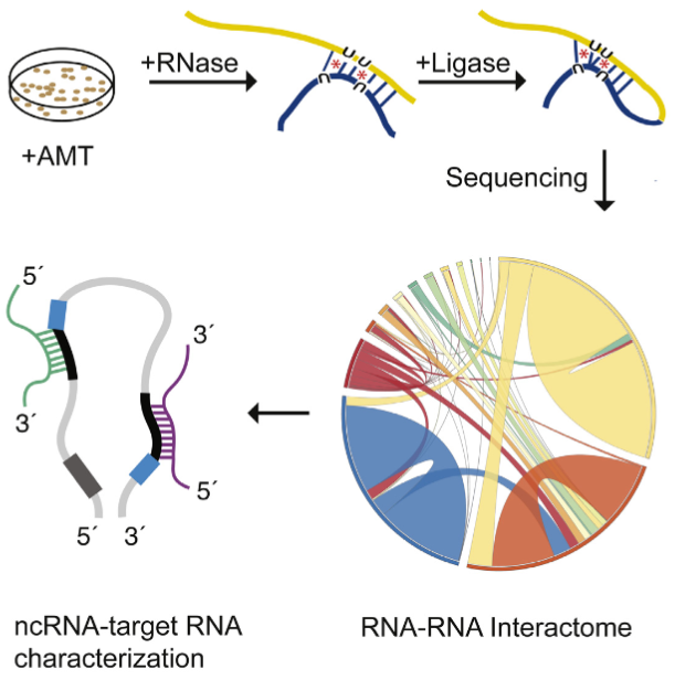
A new tool called “LIGR-Seq” enables scientists to explore in depth what non-coding RNAs actually do in human cells May 23, 2016
he LIGR-seq method for global-scale mapping of RNA-RNA interactions in vivo to reveal unexpected functions for uncharacterized RNAs that act via base-pairing interactions (credit: University of Toronto)
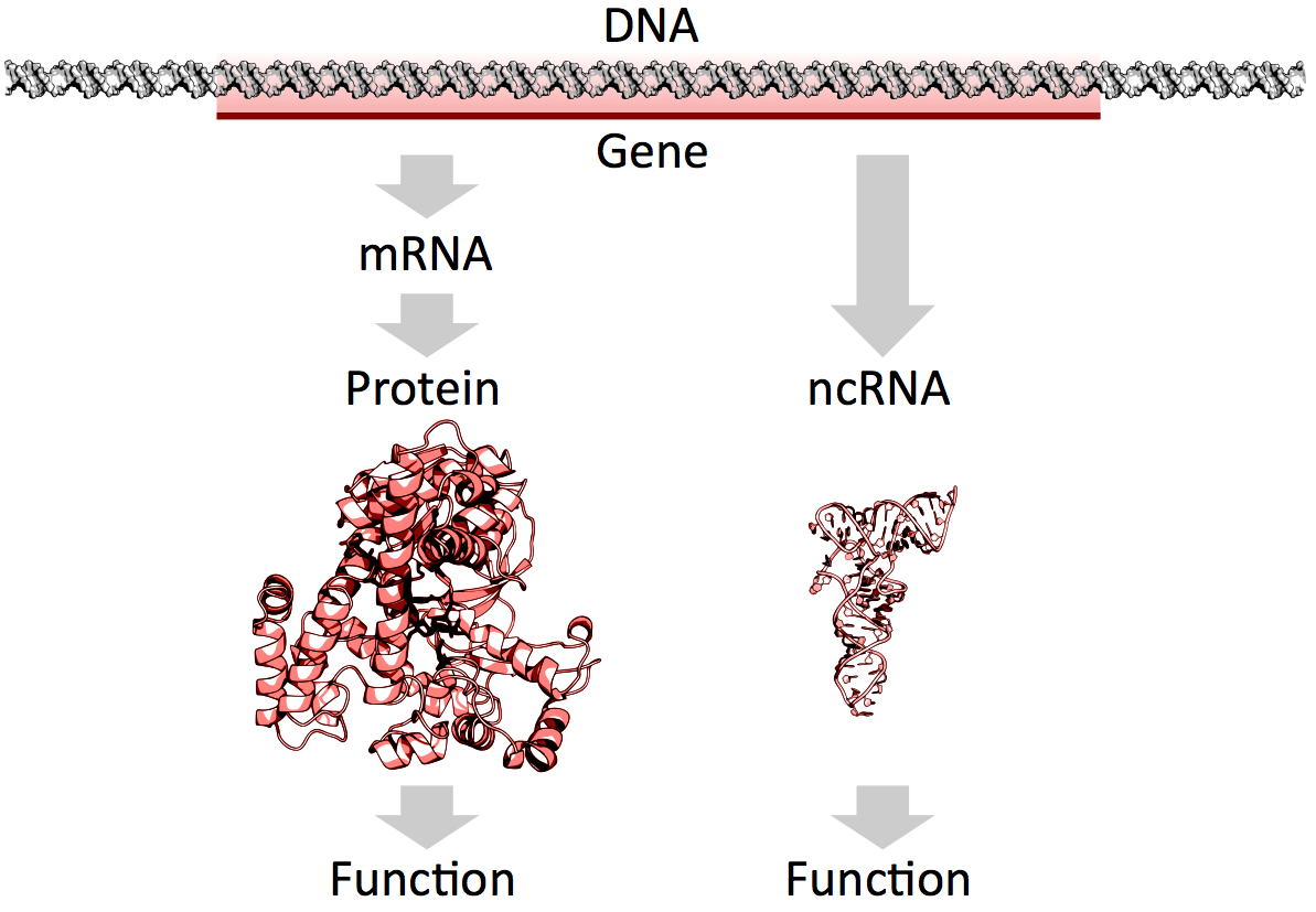
What used to be dismissed by many as “junk DNA” has now become vitally important, as accelerating genomic data points to the importance of non-coding RNAs (ncRNAs) — a genome’s messages that do not specifically code for proteins — in development and disease.
But our progress in understanding these molecules has been slow because of the lack of technologies that allow for systematic mapping of their functions.
Now, professor Benjamin Blencowe’s team at the University of Toronto’s Donnelly Centre has developed a method called “LIGR-seq” that enables scientists to explore in depth what ncRNAs do in human cells.
The study, described in Molecular Cell, was published on May 19, along with two other papers, in Molecular Cell and Cell, respectively, from Yue Wan’s group at the Genome Institute of Singapore and Howard Chang’s group at Stanford University in California, who developed similar methods to study RNAs in different organisms.
Of the 3 billion letters in the human genome, only two per cent make up the protein-coding genes. The genes are copied, or transcribed, into messenger RNA (mRNA) molecules, which provide templates for building proteins that do most of the work in the cell. Much of the remaining 98 per cent of the genome was initially considered by some as lacking in functional importance. However, large swaths of the non-coding genome — between half and three quarters of it — are also copied into RNA.
So then what might the resulting ncRNAs do? That depends on whom you ask. Some researchers believe that most ncRNAs have no function, that they are just a by-product of the genome’s powerful transcription machinery that makes mRNA. However, it is emerging that many ncRNAs do have important roles in gene regulation — some ncRNAs act as carriages for shuttling the mRNAs around the cell, or provide a scaffold for other proteins and RNAs to attach to and do their jobs.
But the majority of available data has trickled in piecemeal or through serendipitous discovery. And with emerging evidence that ncRNAs could drive disease progression, such as cancer metastasis, there was a great need for a technology that would allow a systematic functional analysis of ncRNAs.
“Up until now, with existing methods, you had to know what you are looking for because they all require you to have some information about the RNA of interest. The power of our method is that you don’t need to preselect your candidates; you can see what’s occurring globally in cells, and use that information to look at interesting things we have not seen before and how they are affecting biology,” says Eesha Sharma, a PhD candidate in Blencowe’s group who, along with postdoctoral fellow Tim Sterne-Weiler, co-developed the method.
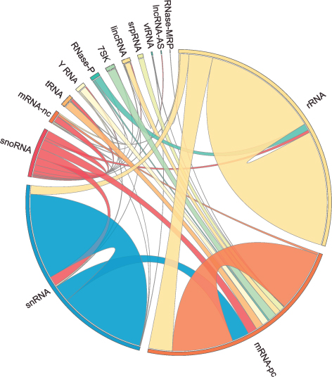
The human RNA-RNA interactome, showing interactions detected by LIGR-seq (credit: University of Toronto)
The new ‘‘LIGation of interacting RNA and high-throughput sequencing’’ (LIGR-seq) tool captures interactions between different RNA molecules. When two RNA molecules have matching sequences — strings of letters copied from the DNA blueprint — they will stick together like Velcro. With LIGR-seq, the paired RNA structures are removed from cells and analyzed by state-of-the-art sequencing methods to precisely identify the RNAs that are stuck together.
“Most researchers in the life sciences agree that there’s an urgent need to understand what ncRNAs do. This technology will open the door to developing a new understanding of ncRNA function,” says Blencowe, who is also a professor in the Department of Molecular Genetics.
Not having to rely on pre-existing knowledge will boost the discovery of RNA pairs that have never been seen before. Scientists can also, for the first time, look at RNA interactions as they occur in living cells, in all their complexity, unlike in the juices of mashed up cells that they had to rely on before. This is a bit like moving on to explore marine biology from collecting shells on the beach to scuba-diving among the coral reefs, where the scope for discovery is so much bigger.
Actually, ncRNAs come in multiple flavors: there’s rRNA, tRNA, snRNA, snoRNA, piRNA, miRNA, and lncRNA, to name a few, where prefixes reflect the RNA’s place in the cell or some aspect of its function. But the truth is that no one really knows the extent to which these ncRNAs control what goes on in the cell, or how they do this.
Discoveries
Nonetheless, the new technology developed by Blencowe’s group has been able to pick up new interactions involving all classes of RNAs and has already revealed some unexpected findings.
The team discovered new roles for small nucleolar RNAs (snoRNAs), which normally guide chemical modifications of other ncRNAs. It turns out that some snoRNAs can also regulate stability of a set of protein-coding mRNAs. In this way, snoRNAs can also directly influence which proteins are made, as well as their abundance, adding a new level of control in cell biology.
And this is only the tip of the iceberg; the researchers plan to further develop and apply their technology to investigate the ncRNAs in different settings.
“We would like to understand how ncRNAs function during development. We are particularly interested in their role in the formation of neurons. But we will also use our method to discover and map changes in RNA-RNA interactions in the context of human diseases,” says Blencowe.
Abstract of Global Mapping of Human RNA-RNA Interactions
The majority of the human genome is transcribed into non-coding (nc)RNAs that lack known biological functions or else are only partially characterized. Numerous characterized ncRNAs function via base pairing with target RNA sequences to direct their biological activities, which include critical roles in RNA processing, modification, turnover, and translation. To define roles for ncRNAs, we have developed a method enabling the global-scale mapping of RNA-RNA duplexes crosslinked in vivo, “LIGation of interacting RNA followed by high-throughput sequencing” (LIGR-seq). Applying this method in human cells reveals a remarkable landscape of RNA-RNA interactions involving all major classes of ncRNA and mRNA. LIGR-seq data reveal unexpected interactions between small nucleolar (sno)RNAs and mRNAs, including those involving the orphan C/D box snoRNA, SNORD83B, that control steady-state levels of its target mRNAs. LIGR-seq thus represents a powerful approach for illuminating the functions of the myriad of uncharacterized RNAs that act via base-pairing interactions.
Understanding the unknown functions of these genes may lead to the creation of new diagnostic tests for clinical laboratories and anatomic pathology groups
Once again, J. Craig Venter, PhD, is charting new ground in gene sequencing andgenomic science. This time his research team has built upon the first synthetic cell they created in 2010 to build a more sophisticated synthetic cell. Their findings from this work may give pathologists and medical laboratory scientists new tools to diagnose disease.
Recently the research team at the J. Craig Venter Institute (JCVI) and Synthetic Genomics, Inc. (SGI) published their latest findings. Among the things they learned is that science still does not understand the functions of about a third of the genes required for their synthetic cells to function.
JCVI-syn3.0 Could Radically Alter Understanding of Human Genome
Based in La Jolla, Calif., and Rockville, Md., JCVI is a not-for-profit research institute aiming to advance genomics. Building upon its first synthetic cell—Mycoplasma mycoides (M. mycoides) JCVI-syn1.0, which JCVI constructed in 2010—the same team of scientists created the first minimal synthetic bacterial cell, which they calledJCVI-syn3.0. This new artificial cell contains 531,560 base pairs and just 473 genes, which means it is the smallest genome of any organism that can be grown in laboratory media, according to a JCVI-SGI statement.
For pathologists and medical laboratory leaders, the creation of a synthetic life form is a milestone toward better understanding genome sequencing and how this new knowledge may help advance both diagnostics and therapeutics.
“What we’ve done is important because it is a step toward completely understanding how a living cell works,” Clyde Hutchison III, PhD, told New Scientist. “If we can really understand how the cell works, then we will be able to design cells efficiently for the production of pharmaceutical and other useful products.” Hutchison is Professor Emeritus of Microbiology and Immunology at the University of North Carolina at Chapel Hill, Distinguished Professor at the J. Craig Venter Institute, a member of the National Academy of Sciences, and a fellow of the American Academy of Arts and Sciences.
Clyde Hutchison, III, PhD (above), Professor Emeritus of Microbiology and Immunology at the University of North Carolina at Chapel Hill and Distinguished Professor at the J. Craig Venter Institute, stated that his team’s “goal is to have a cell for which the precise biological function of every gene is known.” (Photo credit: JCVI.)
Understanding a Gene’s True Purpose
According to the JCVI researchers, 149 genes have no known purpose. They are, however, necessary for life and health.
“We know about two-thirds of the essential biology, and we’re missing a third,” stated J. Craig Venter, PhD, Founder and CEO of JCVI, in a story published by MedPage Today.
This knowledge is based upon decades of research. JCVI seeks to create a minimal cell operating system to understand biology, while also providing what the JCVI statement called a “chassis for use in industrial applications.”
What Do these Genes Do Anyway?
The JCVI team found that among most genes’ biological functions:
“JCVI-syn3.0 is a working approximation of a minimal cellular genome—a compromise between a small genome size and a workable growth rate for an experimental organism. It retains almost all the genes that are involved in the synthesis and processing of macromolecules. Unexpectedly, it also contains 149 genes with unknown biological functions, suggesting the presence of undiscovered functions that are essential for life,” the researchers told the journal Science.
More research is needed, the scientists say, into the 149 genes that appear to lack specific biologic functions.
Unlocking Mystery of the 149 Genes Could Lead to Advances in Genomic Science
“Finding so many genes without a known function is unsettling, but it’s exciting because it’s left us with much still to learn. It’s like the ‘dark matter’ of biology,” said Alistair Elfick, PhD, Chair of Synthetic Biological Engineering, University of Edinburgh, UK, in the New Scientist article.
Studies such as JCVI’s research is key to broadening understanding and framing appropriate questions about scientific, ethical, and economic implications of synthetic biology.
The creation of a synthetic cell will have a profound and positive impact on understanding of biology and how life works, JCVI said.
Such research may inspire new whole genome synthesis tools and semi-automated processes that could dramatically affect clinical laboratory procedures. It also could lead to new techniques and tools for advanced vaccine and pharmaceuticals, JCVI pointed out.
No single technique has set the molecular biology field ablaze with excitement and potential like the CRISPR-Cas9 genome editing system has following its introduction only a few short years ago. The following articles represent the flexibility of this technique to potentially treat a host of genetic disorders and possibly even prevent the onset of disease.
Scientists recently convened at the CRISPR Precision Gene Editing Congress, held in Boston, to discuss the new technology. As with any new technique, scientists have discovered that CRISPR comes with its own set of challenges, and the Congress focused its discussion around improving specificity, efficiency, and delivery.
With a staggering number of papers published in the past several years involving the characterization and use of the CRISPR/Cas9 gene editing system, it is surprising that researchers are still finding new features of the versatile molecular scissor enzyme.
If a Cas9 nuclease variant could be engineered that was less grabby, it might loosen its grip on DNA sequences throughout the genome—except those sequences representing on-target sites. That’s the assumption that guided a new investigation by researchers at Massachusetts General Hospital.
The gene-editing technology known as CRISPR-Cas9 is starting to raise expectations in the therapeutic realm. In fact, CRISPR-Cas9 and other CRISPR systems are moving so close to therapeutic uses that the technology’s ethical implications are starting to attract notice.
Published: Tuesday, May 24, 2016 A comparison of synthetic gene-activating Cas9 proteins can help guide research and development of therapeutic approaches.
The CRISPR-Cas9 system has come to be known as the quintessential tool that allows researchers to edit the DNA sequences of many organisms and cell types. However, scientists are also increasingly recognizing that it can be used to activate the expression of genes. To that end, they have built a number of synthetic gene activating Cas9 proteins to study gene functions or to compensate for insufficient gene expression in potential therapeutic approaches.
“The possibility to selectively activate genes using various engineered variants of the CRISPR-Cas9 system left many researchers questioning which of the available synthetic activating Cas9 proteins to use for their purposes. The main challenge was that all had been uniquely designed and tested in different settings; there was no side-by-side comparison of their relative potentials,” said George Church, Ph.D., who is Core Faculty Member at the Wyss Institute for Biologically Inspired Engineering at Harvard University, leader of its Synthetic Biology Platform, and Professor of Genetics at Harvard Medical School. “We wanted to provide that side-by-side comparison to the biomedical research community.”
In a study published on 23 May in Nature Methods, the Wyss Institute team reports how it rigorously compared and ranked the most commonly used artificial Cas9 activators in different cell types from organisms including humans, mice and flies. The findings provide a valuable guide to researchers, allowing them to streamline their endeavors.
The team also included Wyss Core Faculty Member James Collins, Ph.D., who also is the Termeer Professor of Medical Engineering & Science and Professor of Biological Engineering at the Massachusetts Institute of Technology (MIT)’s Department of Biological Engineering and Norbert Perrimon, Ph.D., a Professor of Genetics at Harvard Medical School.
Gene activating Cas9 proteins are fused to variable domains borrowed from proteins with well-known gene activation potentials and engineered so that the DNA editing ability is destroyed. In some cases, the second component of the CRISPR-Cas9 system, the guide RNA that targets the complex to specific DNA sequences, also has been engineered to bind gene-activating factors.
“We first surveyed seven advanced Cas9 activators, comparing them to each other and the original Cas9 activator that served to provide proof-of-concept for the gene activation potential of CRISPR-Cas9. Three of them, provided much higher gene activation than the other candidates while maintaining high specificities toward their target genes,” said Marcelle Tuttle, Research Fellow at the Wyss and a co-lead author of the study.
The team went on to show that the three top candidates were comparable in driving the highest level of gene expression in cells from humans, mice and fruit flies, irrespective of their tissue and developmental origins. The researchers also pinpointed ways to further maximize gene activation employing the three leading candidates.
“In some cases, maximum possible activation of a target gene is necessary to achieve a cellular or therapeutic effect. We managed to cooperatively enhance expression of specific genes when we targeted them with three copies of a top performing activator using three different guide RNAs,” said Alejandro Chavez, Ph.D., a Postdoctoral Fellow and the study’s co-first author.
“The ease of use of CRISPR-Cas9 offers enormous potential for development of genome therapeutics. This study provides valuable new design criteria that will help enable synthetic biologists and bioengineers to develop more effective targeted genome engineering technologies in the future,” said Wyss Institute Founding Director Donald Ingber, M.D., Ph.D., who is the Judah Folkman Professor of Vascular Biology at Harvard Medical School and the Vascular Biology Program at Boston Children’s Hospital, and also Professor of Bioengineering at the Harvard John A. Paulson School of Engineering and Applied Sciences.
Engineering T Cells to Functionally Cure HIV-1 Infection
Despite the ability of antiretroviral therapy to minimize human immunodeficiency virus type 1 (HIV-1) replication and increase the duration and quality of patients’ lives, the health consequences and financial burden associated with the lifelong treatment regimen render a permanent cure highly attractive. Although T cells play an important role in controlling virus replication, they are themselves targets of HIV-mediated destruction. Direct genetic manipulation of T cells for adoptive cellular therapies could facilitate a functional cure by generating HIV-1–resistant cells, redirecting HIV-1–specific immune responses, or a combination of the two strategies. In contrast to a vaccine approach, which relies on the production and priming of HIV-1–specific lymphocytes within a patient’s own body, adoptive T-cell therapy provides an opportunity to customize the therapeutic T cells prior to administration. However, at present, it is unclear how to best engineer T cells so that sustained control over HIV-1 replication can be achieved in the absence of antiretrovirals. This review focuses on T-cell gene-engineering and gene-editing strategies that have been performed in efforts to inhibit HIV-1 replication and highlights the requirements for a successful gene therapy–mediated functional cure.
Automated top-down design technique simplifies creation of DNA origami nanostructures
Nanoparticles for drug delivery and cell targeting, nanoscale robots, custom-tailored optical devices, and DNA as a storage medium are among the possible applications
May 27, 2016
The boldfaced line, known as a spanning tree, follows the desired geometric shape of the target DNA origami design method, touching each vertex just once. A spanning tree algorithm is used to map out the proper routing path for the DNA strand. (credit: Public Domain)
MIT, Baylor College of Medicine, and Arizona State University Biodesign Institute researchers have developed a radical new top-down DNA origami* design method based on a computer algorithm that allows for creating designs for DNA nanostructures by simply inputting a target shape.
DNA origami (using DNA to design and build geometric structures) has already proven wildly successful in creating myriad forms in 2- and 3- dimensions, which conveniently self-assemble when the designed DNA sequences are mixed together. The tricky part is preparing the proper DNA sequence and routing design for scaffolding and staple strands to achieve the desired target structure. Typically, this is painstaking work that must be carried out manually.
The new algorithm, which is reported together with a novel synthesis approach in the journal Science, promises to eliminate all that and expands the range of possible applications of DNA origami in biomolecular science and nanotechnology. Think nanoparticles for drug delivery and cell targeting, nanoscale robots in medicine and industry, custom-tailored optical devices, and most interesting: DNA as a storage medium, offering retention times in the millions of years.**
Shape-shifting, top-down software
Unlike traditional DNA origami, in which the structure is built up manually by hand, the team’s radical top-down autonomous design method begins with an outline of the desired form and works backward in stages to define the required DNA sequence that will properly fold to form the finished product.
“The Science paper turns the problem around from one in which an expert designs the DNA needed to synthesize the object, to one in which the object itself is the starting point, with the DNA sequences that are needed automatically defined by the algorithm,” said Mark Bathe, an associate professor of biological engineering at MIT, who led the research. “Our hope is that this automation significantly broadens participation of others in the use of this powerful molecular design paradigm.”
The algorithm, which is known as DAEDALUS (DNA Origami Sequence Design Algorithm for User-defined Structures) after the Greek craftsman and artist who designed labyrinths that resemble origami’s complex scaffold structures, can build any type of 3-D shape, provided it has a closed surface. This can include shapes with one or more holes, such as a torus.
A simplified version of the top-down procedure used to design scaffolded DNA origami nanostructures. It starts with a polygon corresponding to the target shape. Software translates a wireframe version of this structure into a plan for routing DNA scaffold and staple strands. That enables a 3D DNA-based atomic-level structural model that is then validated using 3D cryo-EM reconstruction. (credit: adapted from Biodesign Institute images)
With the new technique, the target geometric structure is first described in terms of a wire mesh made up of polyhedra, with a network of nodes and edges. A DNA scaffold using strands of custom length and sequence is generated, using a “spanning tree” algorithm — basically a map that will automatically guide the routing of the DNA scaffold strand through the entire origami structure, touching each vertex in the geometric form once. Complementary staple strands are then assigned and the final DNA structural model or nanoparticle self-assembles, and is then validated using 3D cryo-EM reconstruction.
The software allows for fabricating a variety of geometric DNA objects, including 35 polyhedral forms (Platonic, Archimedean, Johnson and Catalan solids), six asymmetric structures, and four polyhedra with nonspherical topology, using inverse design principles — no manual base-pair designs needed.
To test the method, simpler forms known as Platonic solids were first fabricated, followed by increasingly complex structures. These included objects with nonspherical topologies and unusual internal details, which had never been experimentally realized before. Further experiments confirmed that the DNA structures produced were potentially suitable for biological applications since they displayed long-term stability in serum and low-salt conditions.
Biological research uses
The research also paves the way for designing nanoscale systems mimicking the properties of viruses, photosynthetic organisms, and other sophisticated products of natural evolution. One such application is a scaffold for viral peptides and proteins for use as vaccines. The surface of the nanoparticles could be designed with any combination of peptides and proteins, located at any desired location on the structure, in order to mimic the way in which a virus appears to the body’s immune system.
The researchers demonstrated that the DNA nanoparticles are stable for more than six hours in serum, and are now attempting to increase their stability further.
The nanoparticles could also be used to encapsulate the CRISPR-Cas9 gene editing tool. The CRISPR-Cas9 tool has enormous potential in therapeutics, thanks to its ability to edit targeted genes. However, there is a significant need to develop techniques to package the tool and deliver it to specific cells within the body, Bathe says.
This is currently done using viruses, but these are limited in the size of package they can carry, restricting their use. The DNA nanoparticles, in contrast, are capable of carrying much larger gene packages and can easily be equipped with molecules that help target the right cells or tissue.
The most exciting aspect of the work, however, is that it should significantly broaden participation in the application of this technology, Bathe says, much like 3-D printing has done for complex 3-D geometric models at the macroscopic scale.
* DNA origami brings the ancient Japanese method of paper folding down to the molecular scale. The basics are simple: Take a length of single-stranded DNA and guide it into a desired shape, fastening the structure together using shorter “staple strands,” which bind in strategic places along the longer length of DNA. The method relies on the fact that DNA’s four nucleotide letters—A, T, C, & G stick together in a consistent manner — As always pairing with Ts and Cs with Gs.
The DNA molecule in its characteristic double stranded form is fairly stiff, compared with single-stranded DNA, which is flexible. For this reason, single stranded DNA makes for an ideal lace-like scaffold material. Further, its pairing properties are predictable and consistent (unlike RNA).
** A single gram of DNA can store about 700 terabytes of information — an amount equivalent to 14,000 50-gigabyte Blu-ray disks — and could potentially be operated with a fraction of the energy required for other information storage options.
Essential role of miRNAs in orchestrating the biology of the tumor microenvironment
MicroRNAs (miRNAs) are emerging as central players in shaping the biology of the Tumor Microenvironment (TME). They do so both by modulating their expression levels within the different cells of the TME and by being shuttled among different cell populations within exosomes and other extracellular vesicles. This review focuses on the state-of-the-art knowledge of the role of miRNAs in the complexity of the TME and highlights limitations and challenges in the field. A better understanding of the mechanisms of action of these fascinating micro molecules will lead to the development of new therapeutic weapons and most importantly, to an improvement in the clinical outcome of cancer patients. Keywords: Exosomes, microRNAs, Tumor microenvironment, Cancer
While cancer treatment and survival have improved worldwide, the need for further understanding of the underlying tumor biology remains. In recent years, there has been a significant shift in scientific focus towards the role of the tumor microenvironment (TME) on the development, growth, and metastatic spread of malignancies. The TME is defined as the surrounding cellular environment enmeshed around the tumor cells including endothelial cells, lymphocytes, macrophages, NK cells, other cells of the immune system, fibroblasts, mesenchymal stem cells (MSCs), and the extracellular matrix (ECM). Each of these components interacts with and influences the tumor cells, continually shifting the balance between pro- and anti-tumor phenotype. One of the predominant methods of communication between these cells is through extracellular vesicles and their microRNA (miRNA) cargo. Extracellular vesicles (EVs) are between 30 nm to a few microns in diameter, are surrounded by a phospholipid bilayer membrane, and are released from a variety of cell types into the local environment. There are three well characterized groups of EVs: 1) exosomes, typically 30–100 nm, 2) microvesicles (or ectosomes), typically 100–1000 nm, and 3) large oncosomes, typically 1–10 μm. Each of these categories has a distinctly unique biogenesis and purpose in cellcell communication despite the fact that current laboratory methods do not always allow precise differentiation. EVs are found to be enriched with membrane-bound proteins, lipid raft-associated and cytosolic proteins, lipids, DNA, mRNAs, and miRNAs, all of which can be transferred to the recipient cell upon fusion to allow cell-cell communications [1]. Of these, miRNAs have been of particular interest in cancer research, both as modifiers of transcription and translation as well as direct inhibitors or enhancers of key regulatory proteins. These miRNAs are a large family of small non-coding RNAs (19–24 nucleotides) and are known to be aberrantly expressed, both in terms of content as well as number, in both the tumor cells and the cells of the TME. Synthesis of these mature miRNA is a complex process, starting with the transcription of long, capped, and polyadenylated pri-miRNA by RNA polymerase II. These are cropped into a 60–100 nucleotide hairpinstructure pre-miRNA by the microprocessor, a heterodimer of Drosha (a ribonuclease III enzyme) and DGCR8 (DiGeorge syndrome critical region gene 8). The premiRNA is then exported to the cytoplasm by exportin 5, cleaved by Dicer, and separated into single strands by helicases. The now mature miRNA are incorporated into the RNA-induced silencing complex (RISC), a cytoplasmic effector machine of the miRNA pathway. The primary mechanism of action of the mature miRNA-RISC complex is through their binding to the 3’ untranslated region, or less commonly the 5’ untranslated region, of target mRNA, leading to protein downregulation either via translational repression or mRNA degradation. More recently, it has been shown that miRNAs can also upregulate the expression of target genes [2]. MiRNA genes are mostly intergenic and are transcribed by independent promoters [3] but can also be encoded by introns, sharing the same promoter of their host gene [4]. MiRNAs undergo the same regulatory mechanisms of any other protein coding gene (promoter methylation, histone modifications, etc.…) [5, 6]. Interestingly, each miRNA may have contradictory effects both within varying tumor cell lines and within different cells of the TME. In this review, we provide a state-of-the-art description of the key role that miRNAs have in the communication between tumor cells and the TME and their subsequent effects on the malignant phenotype. Finally, this review has made every effort to clarify, whenever possible, whether the reference is to the −3p or the -5p miRNA. Whenever such clarification has not been provided, this indicates that it was not possible to infer such information from the cited bibliography.
Angiogenesis and miRNAs Cellular plasticity, critical in the development of malignancy, includes the many diverse mechanisms elicited by cancer cells to increase their malignant potential and develop increasing treatment resistance. One such mechanism, angiogenesis, is critical to the development of metastatic disease, affecting both the growth of malignant cells locally and their survival at distant sites. In the last ten years, miRNAs, often packaged in tumor cell-derived exosomes, have emerged as important contributors to the complicated regulation and balance of pro- and anti-angiogenic factors.
Most commonly, miRNAs derived from cancer cells have oncogenic activity, promoting angiogenesis and tumor growth and survival. The most-well characterized of the pro-angiogenic miRNAs, the miR-17-92 cluster encoding six miRNAs (miR-17, −18a, −19a, −19b, −20a, and −92a), is found on chromosome 13, and is highly conserved among vertebrates [7]. The complex and multifaceted functions of the miR-17-92 cluster are summarized in Fig. 1. Amplification, both at the genetic and RNA level, of miR-17-92 was initially found in several lymphoma cell lines and has subsequently been observed in multiple mouse tumor models [7].
Central role of the miR-17-92 cluster in the biology of the TME. The miR-17-92 cluster encoding miR-17, −18a, −19b, −20a, and -92a is upregulated in multiple tumor types and interacts with various components of the TME to finely “tune” the TME through a complex combination of pro- and anti-tumoral effects
Most commonly, miRNAs derived from cancer cells have oncogenic activity, promoting angiogenesis and tumor growth and survival. The most-well characterized of the pro-angiogenic miRNAs, the miR-17-92 cluster encoding six miRNAs (miR-17, −18a, −19a, −19b, −20a, and −92a), is found on chromosome 13, and is highly conserved among vertebrates [7]. The complex and multifaceted functions of the miR-17-92 cluster are summarized in Fig. 1. Amplification, both at the genetic and RNA level, of miR-17-92 was initially found in several lymphoma cell lines and has subsequently been observed in multiple mouse tumor models [7]. Up-regulation of this particular locus has further been confirmed in miRnome analysis across multiple different tumor types, including lung, breast, stomach, prostate, colon, and pancreatic cancer [8]. The miR-17-92 cluster is directly activated by Myc and modulates a variety of downstream transcription factors important in cell cycle regulation and apoptosis including activation of E2F family and Cyclin-dependent kinase inhibitor (CDKN1A) and downregulation of BCL2L11/BIM and p21 [7]. In addition to promoting cell cycle progression and inhibiting apoptosis, the miR-17-92 cluster also downregulates thrombospondin-1 (Tsp1) and connective tissue growth factor (CTGF), important antiangiogenic proteins [7]. Similarly, microvesicles from colorectal cancer cells contain miR-1246 and TGF-β which are transferred to endothelial cells to silence promyelocytic leukemia protein (PML) and activate Smad 1/5/8 signaling promoting proliferation and migration [9]. Likewise, lung cancer cell line derived microvesicles contain miR-494, in response to hypoxia, which targets PTEN in the endothelial cells promoting angiogenesis through the Akt/eNOS pathway [10]. Lastly, exosomal miR-135b from multiple myeloma cells suppresses the HIF-1/FIH-1 pathway in endothelial cells, increasing angiogenesis [11]. A summary of the studies showing the functions of exosomal miRNAs in shaping the biology of the TME is provided in Table 1.
Table 1
Actions of exosomal miRNAs exchanged between cells of the TME
The most common target of anti-angiogenic therapy is VEGF, and not unsurprisingly, multiple miRNAs (including miR-9, miR-20b, miR-130, miR-150, and miR-497) promote angiogenesis through the induction of the VEGF pathway. The most studied of these is the up-regulation of miR-9 which has been linked to a poor prognosis in multiple tumor types, including breast cancer, non-small cell lung cancer, and melanoma [12]. The two oncogenes MYC and MYCN activate miR-9 and cause E-cadherin downregulation resulting in the upregulated transcription of VEGF [13]. In addition, miR-9 has been shown to upregulate the JAK-STAT pathway, supporting endothelial cell migration and tumor angiogenesis [13]. Both amplification of miR-20b and miR-130 as well as miR-497 suppression regulate VEGF through hypoxia inducible factor 1α (HIF-1α) supporting increased angiogenesis [14, 15, 16, 17]. …..
The pivotal discovery in 2012 by Mitra et al. laid the ground-work for our current knowledge on the interactions between tumor-derived miRNAs and fibroblasts. In combination, the down-regulation of miR-214 and miR-31 and the up-regulation of miR-155 trigger the reprogramming of quiescent fibroblasts to CAFs [32]. As expected, the reverse regulation of these miRNAs reduced the migration and invasion of co-cultured ovarian cancer cells [32]. While the pathway of miR-155’s involvement in CAF biology is still being elucidated, the pathways of miR-214 and miR-31 have been established. In endometrial cancer, miR-31 was found to target the homeobox gene SATB2, leading to enhanced tumor cell migration and invasion [33]. MiR-214 similarly has an inverse correlation with its chemokine target, C-C motif Ligand 5 (CCL5) [32]. CCL5 secretion has been associated with enhanced motility, invasion, and metastatic potential through NF-κB-mediated MMP9 activation and through generation and differentiation of myeloid-derived suppressor cells (MDSCs) [34, 35, 36]. Furthermore, miR-210 and miR-133b overexpression and miR-149 suppression have been subsequently found to independently trigger the conversion to CAFs, possibly through paracrine stimulation, and to additionally promote EMT in prostate and gastric cancer, respectively [37, 38,39]. MiR-210 additionally enlists monocytes and encourages angiogenesis [37]. …
Another function of CAFs is the destruction of the ECM and its remodeling with a tumor-supportive composition and structure which includes modulation of specific integrins and metalloproteinases as some of the most studied miRNA targets. The 23 matrix metalloproteinases (MMPs) are critical in the ECM degradation, disruption of the growth signal balance, resistance to apoptosis, establishment of a favorable metastatic niche, and promotion of angiogenesis [54]. As expected, miRNAs have been found to regulate the actions of MMPs, together working to promote cancer cell growth, invasiveness, and metastasis. In HCC, MMP2 and 9 expression is up-regulated by miR-21 via PTEN pathway downregulation. Similarly, in cholangiocarcinoma it was observed that reduced levels of miR-138 induced up-regulation of RhoC, leading to increased levels of the same two MMPs [55, 56]. ….
As has been shown throughout this review, miRNAs have an important and varied effect on human carcinogenesis by shaping the biology of the TME towards a more permissive pro-tumoral phenotype. The complex events leading to such an outcome are currently quite universally defined as the “educational” process of cancer cells on the surrounding TME. While the initial focus was on the direction from the cancer cell to the surrounding TME, increasingly interest is centered on the implications of a more dynamic bidirectional exchange of genetic information. MiRNAs represent only part of the cargo of the extracellular vesicles, but an increasing scientific literature points towards their pivotal role in creating the micro-environmental conditions for cancer cell growth and dissemination. The nearby future will have to address several questions still unanswered. First, it is absolutely necessary to clarify which miRNAs and to what extent they are involved in this process. The contradictory results of some studies can be explained by the differences in tumor-types and by different concentrations of miRNAs used for functional studies. Understanding whether different concentrations of the same miRNA elicit different target effects and therefore changes the biology of the TME, will represent a significant consideration in the development of this field. It is certainly very attractive (especially in an attempt to develop new and desperately needed better cancer biomarkers) to think that concentrations of miRNAs within the TME are reflected systemically in the circulating levels of that same miRNA, however this has not yet been irrefutably demonstrated. Moreover, the study of the paracrine interactions among different cell populations of the TME and their reciprocal effects has been limited to two, maximum three cell populations. This is still way too far from describing the complexity of the TME and only the development of new tridimensional models of the TME will be able to cast a more conclusive light on such complexity. Finally, the pharmacokinetics of miRNA-containing vesicles is in its infancy at best, and needs to be further developed if the goal is development of new therapies based on the use of exosomic miRNAs. Therefore, the future of miRNA research, particularly in its role in the TME, holds still a lot of questions that need answering. However, for these exact same reasons, this is an incredibly exciting time for research in this field. We can envision a not too far future in which these concerns will be satisfactorily addressed and our understanding of the role of miRNAs within the TME will allow us to use them as new therapeutic weapons to successfully improve the clinical outcome of cancer patients.
Researchers at the Walter and Eliza Hall Institute in Australia have discovered a new way to trigger cell death that could lead to drugs to treat cancer and autoimmune disease.
Programmed cell death (a.k.a. apoptosis) is a natural process that removes unwanted cells from the body. Failure of apoptosis can allow cancer cells to grow unchecked or immune cells to inappropriately attack the body.
The protein known as Bak is central to apoptosis. In healthy cells, Bak sits in an inert state but when a cell receives a signal to die, Bak transforms into a killer protein that destroys the cell.
Triggering the cancer-apoptosis trigger
Institute researchers Sweta Iyer, PhD, Ruth Kluck, PhD, and colleagues unexpectedly discovered that an antibody they had produced to study Bak actually bound to the Bak protein and triggered its activation. They hope to use this discovery to develop drugs that promote cell death.
The researchers used information about Bak’s three-dimensional structure to find out precisely how the antibody activated Bak. “It is well known that Bak can be activated by a class of proteins called ‘BH3-only proteins’ that bind to a groove on Bak. We were surprised to find that despite our antibody binding to a completely different site on Bak, it could still trigger activation,” Kluck said. “The advantage of our antibody is that it can’t be ‘mopped up’ and neutralized by pro-survival proteins in the cell, potentially reducing the chance of drug resistance occurring.”
Drugs that target this new activation site could be useful in combination with other therapies that promote cell death by mimicking the BH3-only proteins. The researchers are now working with collaborators to develop their antibody into a drug that can access Bak inside cells.
Their findings have just been published in the open-access journal Nature Communications. The research was supported by the National Health and Medical Research Council, the Australian Research Council, the Victorian State Government Operational Infrastructure Support Scheme, and the Victorian Life Science Computation Initiative.
Abstract of Identification of an activation site in Bak and mitochondrial Bax triggered by antibodies
During apoptosis, Bak and Bax are activated by BH3-only proteins binding to the α2–α5 hydrophobic groove; Bax is also activated via a rear pocket. Here we report that antibodies can directly activate Bak and mitochondrial Bax by binding to the α1–α2 loop. A monoclonal antibody (clone 7D10) binds close to α1 in non-activated Bak to induce conformational change, oligomerization, and cytochrome c release. Anti-FLAG antibodies also activate Bak containing a FLAG epitope close to α1. An antibody (clone 3C10) to the Bax α1–α2 loop activates mitochondrial Bax, but blocks translocation of cytosolic Bax. Tethers within Bak show that 7D10 binding directly extricates α1; a structural model of the 7D10 Fab bound to Bak reveals the formation of a cavity under α1. Our identification of the α1–α2 loop as an activation site in Bak paves the way to develop intrabodies or small molecules that directly and selectively regulate these proteins.
“Cure” is a word that’s dominated the rhetoric in the war on cancer for decades. But it’s a word that medical professionals tend to avoid. While the American Cancer Society reports that cancer treatment has improved markedly over the decades and the five-year survival rate is impressively high for many cancers, oncologists still refrain from declaring their cancer-free patients cured. Why?
Patients are declared cancer-free (also called complete remission) when there are no more signs of detectable disease.
However, minuscule clusters of cancer cells below the detection level can remain in a patient’s body after treatment. Moreover, such small clusters of straggler cells may undergo metastasis, where they escape from the initial tumor into the bloodstream and ultimately settle in a distant site, often a vital organ such as the lungs, liver or brain.
When a colony of these metastatic cells reaches a detectable size, the patient is diagnosed with recurrent metastatic cancer. About one in three breast cancer patients diagnosed with early-stage cancer later develop metastatic disease, usually within five years of initial remission.
By the time metastatic cancer becomes evident, it is much more difficult to treat than when it was originally diagnosed.
What if these metastatic cells could be detected earlier, before they established a “foothold” in a vital organ? Better yet, could these metastatic cancer cells be intercepted, preventing them them from lodging in a vital organ in the first place?
The implant is a tiny porous polymer disc (basically a miniature sponge, no larger than a pencil eraser) that can be inserted just under a patient’s skin. Implantation triggers the immune system’s “foreign body response,” and the implant starts to soak up immune cells that travel to it. If the implant can catch mobile immune cells, then why not mobile metastatic cancer cells?
We gave implants to mice specially bred to model metastatic breast cancer. When the mice had palpable tumors but no evidence of metastatic disease, the implant was removed and analyzed.
Cancer cells were indeed present in the implant, while the other organs (potential destinations for metastatic cells) still appeared clean. This means that the implant can be used to spot previously undetectable metastatic cancer before it takes hold in an organ.
For patients with cancer in remission, an implant that can detect tumor cells as they move through the body would be a diagnostic breakthrough. But having to remove it to see if it has captured any cancer cells is not the most convenient or pleasant detection method for human patients.
Detecting cancer cells with noninvasive imaging
There could be a way around this, though: a special imaging method under development at Northwestern University called Inverse Spectroscopic Optical Coherence Tomography (ISOCT). ISOCT detects molecular-level differences in the way cells in the body scatter light. And when we scan our implant with ISOCT, the light scatter pattern looks different when it’s full of normal cells than when cancer cells are present. In fact, the difference is apparent when even as few as 15 out of the hundreds of thousands of cells in the implant are cancer cells.
There’s a catch – ISOCT cannot penetrate deep into tissue. That means it is not a suitable imaging technology for finding metastatic cells buried deep in internal organs. However, when the cancer cell detection implant is located just under the skin, it may be possible to detect cancer cells trapped in it using ISOCT. This could offer an early warning sign that metastatic cells are on the move.
This early warning could prompt doctors to monitor their patients more closely or perform additional tests. Conversely, if no cells are detected in the implant, a patient still in remission could be spared from unneeded tests.
The ISOCT results show that noninvasive imaging of the implant is feasible. But it’s a method still under development, and thus it’s not widely available. To make scanning easier and more accessible, we’re working to adapt more ubiquitous imaging technologies like ultrasound to detect tiny quantities of tumor cells in the implant.
Besides providing a way to detect tiny numbers of cancer cells before they can form new tumors in other parts of the body, our implant offers an even more intriguing possibility: diverting metastatic cells away from vital organs, and sequestering them where they cannot cause any damage.
In our mouse studies, we found that metastatic cells got caught in the implant before they were apparent in vital organs. When metastatic cells eventually made their way into the organs, the mice with implants still had significantly fewer tumor cells in their organs than implant-free controls. Thus, the implant appears to provide a therapeutic benefit, most likely by taking the metastatic cells it catches out of the circulation, preventing them from lodging anywhere vital.
Interestingly, we have not seen cancer cells leave the implant once trapped, or form a secondary tumor in the implant. Ongoing work aims to learn why this is. Whether the cells can stay safely immobilized in the implant or if it would need to be removed periodically will be important questions to answer before the implant could be used in human patients.
What the future may hold
For now, our work aims to make the implant more effective at drawing and detecting cancer cells. Since we tested the implant with metastatic breast cancer cells, we also want to see if it will work on other types of cancer. Additionally, we’re studying the cells the implant traps, and learning how the implant interacts with the body as a whole. This basic research should give us insight into the process of metastasis and how to treat it.
In the future (and it might still be far off), we envision a world where recovering cancer patients can receive a detector implant to stand guard for disease recurrence and prevent it from happening. Perhaps the patient could even scan their implant at home with a smartphone and get treatment early, when the disease burden is low and the available therapies may be more effective. Better yet, perhaps the implant could continually divert all the cancer cells away from vital organs on its own, like Iron Man’s electromagnet that deflects shrapnel from his heart.
This solution is still not a “cure.” But it would transform a formidable disease that one out of three cancer survivors would otherwise ultimately die from into a condition with which they could easily live.
New PSA Test Examines Protein Structures to Detect Prostate Cancers
5/16/2016 by Cleveland Clinic
A promising new test is detecting prostate cancer more precisely than current tests, by identifying molecular changes in the prostate specific antigen (PSA) protein, according to Cleveland Clinic research presented today at the American Urological Association annual meeting.
The study – part of an ongoing multicenter prospective clinical trial – found that the IsoPSATM test can also differentiate between high-risk and low-risk disease, as well as benign conditions.
Although widely used, the current PSA test relies on detection strategies that have poor specificity for cancer – just 25 percent of men who have a prostate biopsy due to an elevated PSA level actually have prostate cancer, according to the National Cancer Institute – and an inability to determine the aggressiveness of the disease.
The IsoPSA test, however, identifies prostate cancer in a new way. Developed by Cleveland Clinic, in collaboration with Cleveland Diagnostics, Inc., IsoPSA identifies the molecular structural changes in protein biomarkers. It is able to detect cancer by identifying these structural changes, as opposed to current tests that simply measure the protein’s concentration in a patient’s blood.
“While the PSA test has undoubtedly been one of the most successful biomarkers in history, its limitations are well known. Even currently available prostate cancer diagnostic tests rely on biomarkers that can be affected by physiological factors unrelated to cancer,” said Eric Klein, M.D., chair of Cleveland Clinic’s Glickman Urological & Kidney Institute. “These study results show that using structural changes in PSA protein to detect cancer is more effective and can help prevent unneeded biopsies in low-risk patients.”
The clinical trial involves six healthcare institutions and 132 patients, to date. It examined the ability of IsoPSA to distinguish patients with and without biopsy-confirmed evidence of cancer. It also evaluated the test’s precision in differentiating patients with high-grade (Gleason = 7) cancer from those with low-grade (Gleason = 6) disease and benign findings after standard ultrasound-guided biopsy of the prostate.
Substituting the IsoPSA structure-based composite index for the standard PSA resulted in improvement in diagnostic accuracy. Compared with serum PSA testing, IsoPSA performed better in both sensitivity and specificity.
“We took an ‘out of the box’ approach that has shown success in detecting prostate cancer but also has the potential to address other clinically important questions such as clinical surveillance of patients after treatment,” said Mark Stovsky, M.D., staff member, Cleveland Clinic Glickman Urological & Kidney Institute’s Department of Urology. Stovsky has a leadership position (Chief Medical Officer) and investment interest in Cleveland Diagnostics, Inc. “In general, the clinical utility of prostate cancer early detection and screening tests is often limited by the fact that biomarker concentrations may be affected by physiological processes unrelated to cancer, such as inflammation, as well as the relative lack of specificity of these biomarkers to the cancer phenotype. In contrast, clinical research data suggests that the IsoPSA assay can interrogate the entire PSA isoform distribution as a single stand-alone diagnostic tool which can reliably identify structural changes in the PSA protein that correlate with the presence or absence and aggressiveness of prostate cancer.”
Point of Care, Highly Accurate Cervical Cancer Screening
Fifty-five million times a year, American women go to their gynecologist for a Pap Smear. After waiting a few weeks for the results, more than 3.5 million of them are called back to the physician for a follow up visualization of the cervix. Beyond the stress related to possibly having cancer, the women are then subjected to a colposcopic exam, and all too often, a painful biopsy. Then more stressful waiting for a final diagnosis from the pathologist.
Cervical cancer develops slowly, allowing for successful treatment, when identified on time. Regions with high screening compliancy have low mortality rates from this cancer. In the US, for instance, where screening rates are close to 90%, only 4,200 women die from cervical cancer, annually, or 2.6 women per 100,000. However, the screening process in the developed world is long, complicated and not optimized.
In developing regions however, cervical cancer is a leading cause of women death. Over 85% of the total deaths from this cancer are in developing countries. Regions suffering from low screening rates include not only Africa, India and China, but many Eastern European countries as well. According to an OECD report from 2014, the cervical cancer screening rates in Romania and Hungary are as low as 14.6% and 36.7% respectively. The mortality rates in these countries are high, 16 in 100,000 women in Romania and 7.7 in 100,000 in Hungary.
The current screening process for cervical cancer detection is long, beginning with a Pap or HPV test. Cytology results take weeks to receive. A positive result requires follow-up testing by colposcopy and often biopsy. In countries where there is little access to medical care, or where screening compliancy is low, the chances of successful detection via this multi-step process are small. Developing regions and non-compliant countries require a point of care diagnostic method, which eliminates the need for return visits.
Additional limitations to cervical cancer screening are the low sensitivity and specificity rates of Pap tests and the high false positive rates of HPV test, leading to unnecessary colposcopies. Both cytology and colposcopy testing are highly dependent on operator proficiency for accurate diagnosis.
Biop has developed a new technology for the optimization of this process, into one, three minute, painless optical scan. The vaginal probe uses advanced optical, imaging and non-imaging technologies to identify and classify epithelium based cancers and pre-cancerous lesions. The probe is inserted into the vaginal canal, and scans the entire cervix. The resulting images and optical signatures created from the light, and captured by the sensors, are analyzed by the proprietary algorithm. The result is two pictures, on the physician’s screen; a high resolution photograph of the patient’s cervix, immediately next to a hot/cold map indicating a precise classification and location of any diseased lesions.
Deep learning applied to drug discovery and repurposing
Deep neural networks for drug discovery (credit: Insilico Medicine, Inc.)
Scientists from Insilico Medicine, Inc. have trained deep neural networks (DNNs) to predict the potential therapeutic uses of 678 drugs, using gene-expression data obtained from high-throughput experiments on human cell lines from Broad Institute’s LINCS databases and NIH MeSH databases.
The supervised deep-learning drug-discovery engine used the properties of small molecules, transcriptional data, and literature to predict efficacy, toxicity, tissue-specificity, and heterogeneity of response.
“We used LINCS data from Broad Institute to determine the effects on cell lines before and after incubation with compounds, co-author and research scientist Polina Mamoshina explained to KurzweilIAI.
“We used gene expression data of total mRNA from cell lines extracted and measured before incubation with compound X and after incubation with compound X to identify the response on a molecular level. The goal is to understand how gene expression (the transcriptome) will change after drug uptake. It is a differential value, so we need a reference (molecular state before incubation) to compare.”
The research is described in a paper in the upcoming issue of the journal Molecular Pharmaceutics.
Helping pharmas accelerate R&D
Alex Zhavoronkov, PhD, Insilico Medicine CEO, who coordinated the study, said the initial goal of their research was to help pharmaceutical companies significantly accelerate their R&D and increase the number of approved drugs. “In the process we came up with more than 800 strong hypotheses in oncology, cardiovascular, metabolic, and CNS spaces and started basic validation,” he said.
The team measured the “differential signaling pathway activation score for a large number of pathways to reduce the dimensionality of the data while retaining biological relevance.” They then used those scores to train the deep neural networks.*
“This study is a proof of concept that DNNs can be used to annotate drugs using transcriptional response signatures, but we took this concept to the next level,” said Alex Aliper, president of research, Insilico Medicine, Inc., lead author of the study.
Via Pharma.AI, a newly formed subsidiary of Insilico Medicine, “we developed a pipeline for in silico drug discovery — which has the potential to substantially accelerate the preclinical stage for almost any therapeutic — and came up with a broad list of predictions, with multiple in silico validation steps that, if validated in vitro and in vivo, can almost double the number of drugs in clinical practice.”
Despite the commercial orientation of the companies, the authors agreed not to file for intellectual property on these methods and to publish the proof of concept.
Deep-learning age biomarkers
According to Mamoshina, earlier this month, Insilico Medicine scientists published the first deep-learned biomarker of human age — aiming to predict the health status of the patient — in a paper titled “Deep biomarkers of human aging: Application of deep neural networks to biomarker development” by Putin et al, in Aging; and an overview of recent advances in deep learning in a paper titled “Applications of Deep Learning in Biomedicine” by Mamoshina et al., also in Molecular Pharmaceutics.
Insilico Medicine is located in the Emerging Technology Centers at Johns Hopkins University in Baltimore, Maryland, in collaboration with Datalytic Solutions and Mind Research Network.
* In this study, scientists used the perturbation samples of 678 drugs across A549, MCF-7 and PC-3 cell lines from the Library of Integrated Network-Based Cellular Signatures (LINCS) project developed by the National Institutes of Health (NIH) and linked those to 12 therapeutic use categories derived from MeSH (Medical Subject Headings) developed and maintained by the National Library of Medicine (NLM) of the NIH.
To train the DNN, scientists utilized both gene level transcriptomic data and transcriptomic data processed using a pathway activation scoring algorithm, for a pooled dataset of samples perturbed with different concentrations of the drug for 6 and 24 hours. Cross-validation experiments showed that DNNs achieve 54.6% accuracy in correctly predicting one out of 12 therapeutic classes for each drug.
One peculiar finding of this experiment was that a large number of drugs misclassified by the DNNs had dual use, suggesting possible application of DNN confusion matrices in drug repurposing. FutureTechnologies Media Group | Video presentation Insilico medicine
Abstract of Deep learning applications for predicting pharmacological properties of drugs and drug repurposing using transcriptomic data
Deep learning is rapidly advancing many areas of science and technology with multiple success stories in image, text, voice and video recognition, robotics and autonomous driving. In this paper we demonstrate how deep neural networks (DNN) trained on large transcriptional response data sets can classify various drugs to therapeutic categories solely based on their transcriptional profiles. We used the perturbation samples of 678 drugs across A549, MCF-7 and PC-3 cell lines from the LINCS project and linked those to 12 therapeutic use categories derived from MeSH. To train the DNN, we utilized both gene level transcriptomic data and transcriptomic data processed using a pathway activation scoring algorithm, for a pooled dataset of samples perturbed with different concentrations of the drug for 6 and 24 hours. When applied to normalized gene expression data for “landmark genes,” DNN showed cross-validation mean F1 scores of 0.397, 0.285 and 0.234 on 3-, 5- and 12-category classification problems, respectively. At the pathway level DNN performed best with cross-validation mean F1 scores of 0.701, 0.596 and 0.546 on the same tasks. In both gene and pathway level classification, DNN convincingly outperformed support vector machine (SVM) model on every multiclass classification problem. For the first time we demonstrate a deep learning neural net trained on transcriptomic data to recognize pharmacological properties of multiple drugs across different biological systems and conditions. We also propose using deep neural net confusion matrices for drug repositioning. This work is a proof of principle for applying deep learning to drug discovery and development.
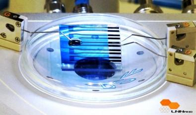
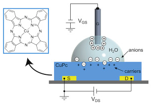
A novel nanoscale organic transistor-based biosensor that can detect molecules associated with neurodegenerative diseases and some types of cancer has been developed by researchers at the National Nanotechnology Laboratory (LNNano) in Brazil.
The transistor, mounted on a glass slide, contains the reduced form of the peptide glutathione (GSH), which reacts in a specific way when it comes into contact with the enzyme glutathione S-transferase (GST), linked to Parkinson’s, Alzheimer’s and breast cancer, among other diseases.
Sensitive water-gated copper phthalocyanine (CuPc) thin-film transistor (credit: Rafael Furlan de Oliveira et al./Organic Electronics)
“The device can detect such molecules even when they’re present at very low levels in the examined material, thanks to its nanometric sensitivity,” explained Carlos Cesar Bof Bufon, Head of LNNano’s Functional Devices & Systems Lab (DSF).
Bufon said the system can be adapted to detect other substances by replacing the analytes (detection compounds). The team is working on paper-based biosensors to further lower the cost, improve portability, and facilitate fabrication and disposal.
The research is published in the journal Organic Electronics.
Abstract of Water-gated phthalocyanine transistors: Operation and transduction of the peptide–enzyme interaction
The use of aqueous solutions as the gate medium is an attractive strategy to obtain high charge carrier density (1012 cm−2) and low operational voltages (<1 V) in organic transistors. Additionally, it provides a simple and favorable architecture to couple both ionic and electronic domains in a single device, which is crucial for the development of novel technologies in bioelectronics. Here, we demonstrate the operation of transistors containing copper phthalocyanine (CuPc) thin-films gated with water and discuss the charge dynamics at the CuPc/water interface. Without the need for complex multilayer patterning, or the use of surface treatments, water-gated CuPc transistors exhibited low threshold (100 ± 20 mV) and working voltages (<1 V) compared to conventional CuPc transistors, along with similar charge carrier mobilities (1.2 ± 0.2) x 10−3 cm2 V−1 s−1. Several device characteristics such as moderate switching speeds and hysteresis, associated with high capacitances at low frequencies upon bias application (3.4–12 μF cm−2), indicate the occurrence of interfacial ion doping. Finally, water-gated CuPc OTFTs were employed in the transduction of the biospecific interaction between tripeptide reduced glutathione (GSH) and glutathione S-transferase (GST) enzyme, taking advantage of the device sensitivity and multiparametricity.
The study offers understanding of potential therapeutic targets.
Building on data from The Cancer Genome Atlas (TCGA) project, a multi-institutional team of scientists have completed the first large-scale “proteogenomic” study of breast cancer, linking DNA mutations to protein signaling and helping pinpoint the genes that drive cancer. Conducted by members of the National Cancer Institute’s Clinical Proteomic Tumor Analysis Consortium (CPTAC), including Baylor College of Medicine, Broad Institute of MIT and Harvard, Fred Hutchinson Cancer Research Center, New York University Langone Medical Center, and Washington University School of Medicine, the study takes aim at proteins, the workhorses of the cell, and their modifications to better understand cancer.
Appearing in the Advance Online Publication of Nature, the study illustrates the power of integrating genomic and proteomic data to yield a more complete picture of cancer biology than either analysis could do alone. The effort produced a broad overview of the landscape of the proteome (all the proteins found in a cell) and the phosphoproteome (the sites at which proteins are tagged by phosphorylation, a chemical modification that drives communication in the cell) across a set of 77 breast cancer tumors that had been genomically characterized in the TCGA project. Although the TCGA produced an extensive catalog of somatic mutations found in cancer, the effects of many of those mutations on cellular functions or patients’ outcomes are unknown.
In addition, not all mutated genes are true “drivers” of cancer — some are merely “passenger” mutations that have little functional consequence. And some mutations are found within very large DNA regions that are deleted or present in extra copies, so winnowing the list of candidate genes by studying the activity of their protein products can help identify therapeutic targets. “We don’t fully understand how complex cancer genomes translate into the driving biology that causes relapse and mortality,” said Matthew Ellis, director of the Lester and Sue Smith Breast Center at Baylor College of Medicine and a senior author of the paper.
“These findings show that proteogenomic integration could one day prove to be a powerful clinical tool, allowing us to traverse the large knowledge gap between cancer genomics and clinical action.” In this study, the researchers at the Broad Institute analyzed breast tumors using accurate mass, high-resolution mass spectrometry, a technology that extends the coverage of the proteome far beyond the coverage that can be achieved by traditional antibody-based methods. This allowed them to scale their efforts and quantify more than 12,000 proteins and 33,000 phosphosites, an extremely deep level of coverage.
Scripps scientists have designed a drug candidate that decreases growth of breast cancer cells.
In a development that could lead to a new generation of drugs to precisely treat a range of diseases, scientists from the Florida campus of The Scripps Research Institute (TSRI) have for the first time designed a drug candidate that decreases the growth of tumor cells in animal models in one of the hardest to treat cancers—triple negative breast cancer.
“This is the first example of taking a genetic sequence and designing a drug candidate that works effectively in an animal model against triple negative breast cancer,” said TSRI Professor Matthew Disney. “The study represents a clear breakthrough in precision medicine, as this molecule only kills the cancer cells that express the cancer-causing gene—not healthy cells. These studies may transform the way the lead drugs are identified—by using the genetic makeup of a disease.”
The study, published by the journal Proceedings of the National Academy of Sciences, demonstrates that the Disney lab’s compound, known as Targaprimir-96, triggers breast cancer cells to kill themselves via programmed cell death by precisely targeting a specific RNA that ignites the cancer.
Short-Cut to Drug Candidates
While the goal of precision medicine is to identify drugs that selectively affect disease-causing biomolecules, the process has typically involved time-consuming and expensive high-throughput screens to test millions of potential drug candidates to identify those few that affect the target of interest. Disney’s approach eliminates these screens.
The new study uses the lab’s computational approach called Inforna, which focuses on developing designer compounds that bind to RNA folds, particularly microRNAs.
MicroRNAs are short molecules that work within all animal and plant cells, typically functioning as a “dimmer switch” for one or more genes, binding to the transcripts of those genes and preventing protein production. Some microRNAs have been associated with diseases. For example, microRNA-96, which was the target of the new study, promotes cancer by discouraging programmed cell death, which can rid the body of cells that grow out of control.
In the new study, the drug candidate was tested in animal models over a 21-day course of treatment. Results showed decreased production of microRNA-96 and increased programmed cell death, significantly reducing tumor growth. Since targaprimir-96 was highly selective in its targeting, healthy cells were unaffected.
In contrast, Disney noted, a typical cancer therapeutic targets and kills cells indiscriminately, often leading to side effects that can make these drugs difficult for patients to tolerate.
Benjamin Zealley and Aubrey D.N.J. de Grey Commentary on Some Recent Theses Relevant to Combating Aging: June 2015
Cancer Autoantibody Biomarker Discovery and Validation Using Nucleic Acid Programmable Protein Array Jie Wang, PhD, Arizona State University
Currently in the United States, many patients with cancer do not benefit from population-based screening due to challenges associated with the existing cancer screening scheme. Blood-based diagnostic assays have the potential to detect diseases in a non-invasive way. Proteins released from small early tumors may only be present intermittently and are diluted to tiny concentrations in the blood, making them difficult to use as biomarkers. However, they can induce autoantibody (AAb) responses, which can amplify the signal and persist in the blood even if the antigen is gone. Circulating autoantibodies are a promising class of molecules that have the potential to serve as early detection biomarkers for cancers. This PhD thesis aims to screen for autoantibody biomarkers for the early detection of two deadly cancers, basal-like breast cancer and lung adenocarcinoma. First, a method was developed to display proteins in both native and denatured conformations on a protein array. This method adopted a novel protein tag technology, called a HaloTag, to immobilize proteins covalently on the surface of a glass slide. The covalent attachment allowed these proteins to endure harsh treatment without becoming dissociated from the slide surface, which enabled the profiling of antibody responses against both conformational and linear epitopes. Next, a plasma screening protocol was optimized to increase significantly the signal-to-noise ratio of protein array–based AAb detection. Following this, the AAb responses in basal-like breast cancer were explored using nucleic acid programmable protein arrays (NAPPA) containing 10,000 full-length human proteins in 45 cases and 45 controls. After verification in a large sample set (145 basal-like breast cancer cases, 145 controls, 70 non-basal breast cancer) by enzyme-linked immunosorbent assay (ELISA), a 13-AAb classifier was developed to differentiate patients from controls with a sensitivity of 33% at 98% specificity. A similar approach was also applied to the lung cancer study to identify AAbs that distinguished lung cancer patients from computed tomography–positive benign pulmonary nodules (137 lung cancer cases, 127 smoker controls, 170 benign controls). In this study, two panels of AAbs were discovered that showed promising sensitivity and specificity. Six out of eight AAb targets were also found to have elevated mRNA levels in lung adenocarcinoma patients using TCGA data. These projects as a whole provide novel insights into the association between AAbs and cancer, as well as general B cell antigenicity against self-proteins.
Comment: There are two widely supported models for cancer development and progression—the clonal evolution (CE) model and the cancer stem cell (CSC) model. Briefly, the former claims that most or all cells in a tumor contribute to its maintenance; as newer and more aggressive clones develop by random mutation, they become responsible for driving growth. The range of different mutational profiles generated is assumed to be large enough to account for disease recurrence after therapy (due to rare resistant clones) and metastasis (clones arising with the ability to travel to distant sites). The CSC model instead asserts that a small number of mutated stem cells are the origin of the primary cell mass, drive metastasis through the intermittent release of undifferentiated, highly mobile progeny, and account for recurrence due to a generally quiescent metabolic profile conferring potent resistance to chemotherapy. In either case, the immunological visibility of an early tumor may be highly sporadic. Clones arising early in CE differ little in proteomic terms from healthy host cells; those that do trigger a response are unlikely to have acquired robust resistance to immune attack, so are destroyed quickly in favor of their stealthier brethren. Likewise, CSCs share some of the immune privilege of normal stem cells and, due to their inherent ability to produce differentiated progeny with distinct proteomic signatures, are partially protected from attacks on their descendants. Consequently, such well-hidden cells may remain in the body for years to decades. The autoantibody panel developed in this study for basal-like breast cancer exhibits exceptional specificity despite a comparatively small training set. Given its ease of application, this suggests great promise for a more exhaustively trained classifier as a populationlevel screening tool.
Condition-Specific Differential Sub-Network Analysis for Biological Systems Deepali Jhamb, PhD, Indiana University
Biological systems behave differently under different conditions. Advances in sequencing technology over the last decade have led to the generation of enormous amounts of condition-specific data. However, these measurements often fail to identify low-abundance genes and proteins that can be biologically crucial. In this work, a novel textmining system was first developed to extract condition-specific proteins from the biomedical literature. The literaturederived data was then combined with proteomics data to construct condition-specific protein interaction networks. Furthermore, an innovative condition-specific differential analysis approach was designed to identify key differences, in the form of sub-networks, between any two given biological systems. The framework developed here was implemented to understand the differences between limb regenerationcompetent Ambystoma mexicanum and regeneration-deficient Xenopus laevis. This study provides an exhaustive systems-level analysis to compare regeneration competent and deficient sub-networks to show how different molecular entities inter-connect with each other and are rewired during the formation of an accumulation blastema in regenerating axolotl limbs. This study also demonstrates the importance of literature-derived knowledge, specific to limb regeneration, to augment the systems biology analysis. Our findings show that although the proteins might be common between the two given biological conditions, they can have a high dissimilarity based on their biological and topological properties in the sub-network. The knowledge gained from the distinguishing features of limb regeneration in amphibians can be used in future to induce regeneration chemically in mammalian systems. The approach developed in this dissertation is scalable and adaptable to understanding differential sub-networks between any two biological systems. This methodology will not only facilitate the understanding of biological processes and molecular functions that govern a given system, but will also provide novel intuitions about the pathophysiology of diseases/conditions.
Comment: We have long advocated a principle of directly comparing young and old bodies as a means to identify the classes of physical damage that accumulate in the body during aging. This approach circumvents our ignorance of the full etiology of each particular disease manifestation, a phenomenally difficult question given the ethical issues of experimenting on human subjects, the lengthy ‘‘incubation time’’ of aging-related diseases, and the complex interconnections between their risk factors—innate and environmental. Repairing such damage has the potential to prevent pathology before symptoms appear, an approach now becoming increasingly mainstream.11 However, a naı¨ve comparison faces a number of difficulties, even given a sufficiently large sample set to compensate for inter-individual variation. Most importantly, the causal significance of a given species cannot be reliably determined from its simple prevalence.12 The catalytic nature of cell biology means that those entities whose abundance changes the most profoundly in absolute terms are quite unlikely to be the drivers of that change and may even spontaneously revert to baseline levels in the absence of on-going stimulation. Meanwhile, functionality is often heavily influenced independently of abundance by post-translational modifications that may escape direct detection. Sub-network analysis uses computational means to identify groups of genes and/or proteins that vary in a synchronized way with some parameter, indicating functional connectivity. The application of methods such as those developed here to the comparison of a wide range of younger and older conditions will facilitate the identification of processes—not merely individual factors—that are impaired with age, and thus will help greatly in identifying the optimal points for intervention.
Development of a Light Actuated Drug Delivery-on-Demand System Chase Linsley, PhD, University of California, Los Angeles
The need for temporal–spatial control over the release of biologically active molecules has motivated efforts to engineer novel drug delivery-on-demand strategies actuated via light irradiation. Many systems, however, have been limited to in vitro proof-of-concept due to biocompatibility issues with the photo-responsive moieties or the light wavelength, intensity, and duration. To overcome these limitations, the objective of this dissertation was to design a light-actuated drug delivery-on-demand strategy that uses biocompatible chromophores and safe wavelengths of light, thereby advancing the clinical prospects of light-actuated drug delivery-on-demand systems. This was achieved by: (1) Characterizing the photothermal response of biocompatible visible light and near-infrared-responsive chromophores and demonstrating the feasibility and functionality of the light actuated on-demand drug delivery system in vitro; and (2) designing a modular drug delivery-on-demand system that could control the release of biologically active molecules over an extended period of time. Three biocompatible chromophores—Cardiogreen, Methylene Blue, and riboflavin—were identified and demonstrated significant photothermal response upon exposure to near-infrared and visible light, and the amount of temperature change was dependent upon light intensity, wavelength, as well as chromophore concentration. As a proof-of-concept, pulsatile release of a model protein from a thermally responsive delivery vehicle fabricated from poly(N-isopropylacrylamide) was achieved over 4 days by loading the delivery vehicle with Cardiogreen and irradiating with near-infrared light. To extend the useful lifetime of the light-actuated drug delivery-on-demand system, a modular, reservoir-valve system was designed. Using poly(ethylene glycol) as a reservoir for model small molecule drugs combined with a poly(N-isopropylacrylamide) valve spiked with chromophore-loaded liposomes, pulsatile release was achieved over 7 days upon light irradiation. Ultimately, this drug delivery strategy has potential for clinical applications that require explicit control over the presentation of biologically active molecules. Further research into the design and fabrication of novel biocompatible thermally responsive delivery vehicles will aid in the advancement of the light-actuated drug delivery-on-demand strategy described here. Comment: Our combined comments on this thesis and the next one appear after the next abstract.
Light-Inducible Gene Regulation in Mammalian Cells Lauren Toth, PhD, Duke University
The growing complexity of scientific research demands further development of advanced gene regulation systems. For instance, the ultimate goal of tissue engineering is to develop constructs that functionally and morphologically resemble the native tissue they are expected to replace. This requires patterning of gene expression and control of cellular phenotype within the tissue-engineered construct. In the field of synthetic biology, gene circuits are engineered to elucidate mechanisms of gene regulation and predict the behavior of more complex systems. Such systems require robust gene switches that can quickly turn gene expression on or off. Similarly, basic science requires precise genetic control to perturb genetic pathways or understand gene function. Additionally, gene therapy strives to replace or repair genes that are responsible for disease. The safety and efficacy of such therapies require control of when and where the delivered gene is expressed in vivo.
Unfortunately, these fields are limited by the lack of gene regulation systems that enable both robust and flexible cellular control. Most current gene regulation systems do not allow for the manipulation of gene expression that is spatially defined, temporally controlled, reversible, and repeatable. Rather, they provide incomplete control that forces the user to choose to control gene expression in either space or time, and whether the system will be reversible or irreversible. The recent emergence of the field of optogenetics—the ability to control gene expression using light—has made it possible to regulate gene expression with spatial, temporal, and dynamic control. Light-inducible systems provide the tools necessary to overcome the limitations of other gene regulation systems, which can be slow, imprecise, or cumbersome to work with. However, emerging light-inducible systems require further optimization to increase their efficiency, reliability, and ease of use.
Initially, we engineered a light-inducible gene regulation system that combines zinc finger protein technology and the light-inducible interaction between Arabidopsis thaliana plant proteins GIGANTEA (GI) and the light oxygen voltage (LOV) domain of FKF1. Zinc finger proteins (ZFPs) can be engineered to target almost any DNA sequence through tandem assembly of individual zinc finger domains that recognize a specific 3-bp DNA sequence. Fusion of three different ZFPs to GI (GI-ZFP) successfully targeted the fusion protein to the specific DNA target sequence of the ZFP. Due to the interaction between GI and LOV, co-expression of GI-ZFP with a fusion protein consisting of LOV fused to three copies of the VP16 transactivation domain (LOV-VP16) enabled blue-light dependent recruitment of LOV-VP16 to the ZFP target sequence. We showed that placement of three to nine copies of a ZFP target sequence upstream of a luciferase or enhanced green fluorescent protein (eGFP) transgene enabled expression of the transgene in response to blue light. Gene activation was both reversible and tunable on the basis of duration of light exposure, illumination intensity, and the number of ZFP binding sites upstream of the transgene. Gene expression could also be patterned spatially by illuminating the cell culture through photomasks containing various patterns.
Although this system was useful for controlling the expression of a transgene, for many applications it is useful to control the expression of a gene in its natural chromosomal position. Therefore, we capitalized on recent advances in programmed gene activation to engineer an optogenetic tool that could easily be targeted to new, endogenous DNA sequences without re-engineering the light inducible proteins. This approach took advantage of CRISPR/Cas9 technology, which uses a gene-specific guide RNA (gRNA) to facilitate Cas9 targeting and binding to a desired sequence, and the light-inducible heterodimerizers CRY2 and CIB1 from Arabidopsis thaliana to engineer a lightactivated CRISPR/Cas9 effector (LACE) system. We fused the full-length (FL) CRY2 to the transcriptional activator VP64 (CRY2FL-VP64) and the amino-terminal fragment of CIB1 to the amino, carboxyl, or amino and carboxyl terminus of a catalytically inactive Cas9. When CRY2-VP64 and one of the CIBN/dCas9 fusion proteins are expressed with a gRNA, the CIBN/dCas9 fusion protein localizes to the gRNA target. In the presence of blue light, CRY2FL binds to CIBN, which translocates CRY2FL-VP64 to the gene target and activates transcription. Unlike other optogenetic systems, the LACE system can be targeted to new endogenous loci by solely manipulating the specificity of the gRNA without having to re-engineer the light-inducible proteins. We achieved light-dependent activation of the IL1RN, HBG1/2, or ASCL1 genes by delivery of the LACE system and four gene-specific gRNAs per promoter region. For some gene targets, we achieved equivalent activation levels to cells that were transfected with the same gRNAs and the synthetic transcription factor dCas9-VP64. Gene activation was also shown to be reversible and repeatable through modulation of the duration of blue light exposure, and spatial patterning of gene expression was achieved using an eGFP reporter and a photomask.
Finally, we engineered a light-activated genetic ‘‘on’’ switch (LAGOS) that provides permanent gene expression in response to an initial dose of blue light illumination. LAGOS is a lentiviral vector that expresses a transgene only upon Cre recombinase–mediated DNA recombination. We showed that this vector, when used in conjunction with a light-inducible Cre recombinase system, could be used to express MyoD or the synthetic transcription factor VP64- MyoD in response to light in multiple mammalian cell lines, including primary mouse embryonic fibroblasts. We achieved light-mediated up-regulation of downstream myogenic markers myogenin, desmin, troponin T, and myosin heavy chains I and II as well as fusion of C3H10T1/2 cells into myotubes that resembled a skeletal muscle cell phenotype. We also demonstrated LAGOS functionality in vivo by engineering the vector to express human VEGF165 and human ANG1 in response to light. HEK 293T cells stably expressing the LAGOS vector and transiently expressing the light-inducible Cre recombinase proteins were implanted into mouse dorsal window chambers. Mice that were illuminated with blue light had increased micro-vessel density compared to mice that were not illuminated. Analysis of human vascular endothelial growth factor (VEGF) and human ANG1 levels by enzyme-linked immunosorbent assay (ELISA) revealed statistically higher levels of VEGF and ANG1 in illuminated mice compared to non-illuminated mice.
In summary, the objective of this work was to engineer robust light-inducible gene regulation systems that can control genes and cellular fate in a spatial and temporal manner. These studies combine the rapid advances in gene targeting and activation technology with natural light-inducible plant protein interactions. Collectively, this thesis presents several optogenetic systems that are expected to facilitate the development of multicellular cell and tissue constructs for use in tissue engineering, synthetic biology, gene therapy, and basic science both in vitro and in vivo.
Comment: Although it is easy to characterize technological progress as following in the wake of scientific discoveries, the reverse is almost equally true; advances in technique open the door to types of experiment previously intractable or impossible. Such is currently the case for the field of optically controlled biotechnology, which has exploded into prominence, particularly over the last half-decade. Light of an appropriate wavelength can penetrate mammalian tissues to a depth of up to a couple of centimeters, rendering much of the living body accessible to optical study and control—still more if the detector/source is integrated into an endoscopic or fiber optic probe. Techniques borrowed from the semiconductor industry allow patterns of illumination to be controlled down to the nanometer scale, ideal for addressing individual cells. The highly controlled time course of such experiments, as compared to traditional means of gene activation, such as the addition of a chemical agent to the medium, eliminates confounding variables, and simplifies data analysis. Furthermore, this level of immediate control opens the door to closed-loop systems where the activity of entities under optical control can be continuously tuned in relation to some parameter(s). In the first of these two illuminating theses, a vehicle is developed that permits light-driven release of a small molecule. Such a system could be employed to target a systemically administered antibiotic or anti-neoplastic agent to a site of infection or cancer while sparing other bodily tissues from toxicity. Because most modern drugs cannot be produced in the body, even given arbitrarily good control of cellular biochemistry, this technique will have lasting value in numerous clinical contexts. In the second thesis, the level of precision achieved is even more profound; the CRISPR/Cas9 system has received much recent attention13 in its own right for its capacity to target arbitrary genetic sequences without an arduous protein-engineering step. The LACE system described stands to permit genetic manipulation with almost arbitrarily good spatial, temporal, and genomic site-specific control, using only means available to a typical university laboratory.
Targeting T Cells for the Immune-Modulation of Human Diseases Regina Lin, PhD, Duke University
Dysregulated inflammation underlies the pathogenesis of a myriad of human diseases ranging from cancer to autoimmunity. As coordinators, executers, and sentinels of host immunity, T cells represent a compelling target population for immune-modulation. In fact, the antigen-specificity, cytotoxicity, and promise of long-lived of immune-protection make T cells ideal vehicles for cancer immunotherapy. Interventions for autoimmune disorders, on the other hand, aim to dampen T cell–mediated inflammation and promote their regulatory functions. Although significant strides have been made in targeting T cells for immune modulation, current approaches remain less than ideal and leave room for improvement. In this dissertation, I seek to improve on current T cell-targeted immunotherapies, by identifying and pre-clinically characterizing their mechanisms of action and in vivo therapeutic efficacy.
CD8+ cytotoxic T cells have potent anti-tumor activity and therefore are leading candidates for use in cancer immunotherapy. The application of CD8+ T cells for clinical use has been limited by the susceptibility of ex vivo– expanded CD8+ T cells to become dysfunctional in response to immunosuppressive microenvironments. To enhance the efficacy of adoptive cell transfer therapy (ACT), we established a novel microRNA (miRNA)-targeting approach that augments CTL cytotoxicity and preserves immunocompetence. Specifically, we screened for miRNAs that modulate cytotoxicity and identified miR-23a as a strong functional repressor of the transcription factor Blimp-1, which promotes CTL cytotoxicity and effector cell differentiation. In a cohort of advanced lung cancer patients, miR- 23a was up-regulated in tumor-infiltrating CD8+ T cells, and its expression correlated with impaired anti-tumor potential of patient CD8+ T cells. We determined that tumor-derived transforming growth factor-b (TGF-b) directly suppresses CD8+ T cell immune function by elevating miR-23a and down-regulating Blimp-1. Functional blockade of miR-23a in human CD8+ T cells enhanced granzyme B expression; and in mice with established tumors, immunotherapy with just a small number of tumor-specific CD8+ T cells in which miR-23a was inhibited robustly hindered tumor progression. Together, our findings provide a miRNA-based strategy that subverts the immunosuppression of CD8+ T cells that is often observed during adoptive cell transfer tumor immunotherapy and identify a TGF-bmediated tumor immune-evasion pathway
Having established that miR-23a-inhibition can enhance the quality and functional resilience of anti-tumor CD8+ T cells, especially within the immune-suppressive tumor microenvironment, we went on to interrogate the translational applicability of this strategy in the context of chimeric antigen receptor (CAR)-modified CD8+ T cells. Although CAR T cells hold immense promise for ACT, CAR T cells are not completely curative due to their in vivo functional suppression by immune barriers—such as TGF-b—within the tumor microenvironment. Because TGF-b poses a substantial immune barrier in the tumor microenvironment, we sought to investigate whether inhibiting miR-23a in CAR T cells can confer immune competence to afford enhanced tumor clearance. To this end, we retrovirally transduced wild-type and miR-23a–deficient CD8+ T cells with the EGFRvIII-CAR, which targets the PepvIII tumorspecific epitope expressed by glioblastomas (GBM). Our in vitro studies demonstrated that while wild-type EGFRvIIICAR T cells were vulnerable to functional suppression by TGF-b, miR-23a abrogation rendered EGFRvIII-CAR T cells immune-resistant to TGF-b. Rigorous preclinical studies are currently underway to evaluate the efficacy of miR-23adeficient EGFRvIII-CAR T cells for GBM immunotherapy.
Last, we explored novel immune-suppressive therapies by the biological characterization of pharmacological agents that could target T cells. Although immune-suppressive drugs are classical therapies for a wide range of autoimmune diseases, they are accompanied by severe adverse effects. This motivated our search for novel immunesuppressive agents that are efficacious and lack undesirable side effects. To this end, we explored the potential utility of subglutinol A, a natural product isolated from the endophytic fungus Fusarium subglutinans. We showed that subglutinol A exerts multimodal immune-suppressive effects on activated T cells in vitro. Subglutinol A effectively blocked T cell proliferation and survival, while profoundly inhibiting pro-inflammatory interferon-c (IFN-c) and interleukin-17 (IL-17) production by fully differentiated effector Th1 and Th17 cells. Our data further revealed that subglutinol A might exert its anti-inflammatory effects by exacerbating mitochondrial damage in T cells, but not in innate immune cells or fibroblasts. Additionally, we demonstrated that subglutinol A significantly reduced lymphocytic infiltration into the footpad and ameliorated footpad swelling in the mouse model of Th1-driven delayed-type hypersensitivity. These results suggest the potential of subglutinol A as a novel therapeutic for inflammatory diseases.
Comment: Immunotherapy is among the most promising approaches to cancer treatment, having the specificity and scope to selectively target transformed cells wherever they may reside within the body and the potential to install a permanent defense against disease recurrence. By the time a typical cancer is clinically diagnosed, however, it has already found means to survive a prolonged period of potential immune attack. The mechanisms by which tumors evade immune surveillance are beginning to be elucidated,15,16 and include both direct suppression of effector cells and progressive editing of the host’s immune repertoire to disfavor future attack. It is inherently difficult to interfere with these defenses directly, due to the selection pressures in genetically heterogeneous neoplastic tissue. Much effort is thus being focused on methods for rendering therapeutically delivered immune cells resistant to their effects. The cytokine TGF-b is paradoxically known to function as both a tumor suppressor in healthy tissue and as a tumorderived species associated with multiple cancer-promoting activities, including enhanced immune evasion. This work identifies the pathway by which TGF-b compromises cytotoxic T cell function in the tumor microenvironment, and demonstrates an effective method for blocking this signal. In many clinical cases, however, editing of the patient’s immune repertoire has already removed or rendered anergic those immune cells able to recognize their cancer. Thus, the finding that blocking TGF-b signaling also appears to enhance the effectiveness of CAR-modified T cells— engineered with an antibody fragment targeting them with high affinity to a particular tumor-associated epitope—is a welcome addition to these already promising results.
Novel Fibonacci and non-Fibonacci structure in the sunflower: results of a citizen science experiment
Jonathan Swinton, Erinma Ochu, The MSI Turing’s Sunflower Consortium
This citizen science study evaluates the occurrence of Fibonacci structure in the spirals of sunflower (Helianthus annuus) seedheads. This phenomenon has competing biomathematical explanations, and our core premise is that observation of both Fibonacci and non-Fibonacci structure is informative for challenging such models. We collected data on 657 sunflowers. In our most reliable data subset, we evaluated 768 clockwise or anticlockwise parastichy numbers of which 565 were Fibonacci numbers, and a further 67 had Fibonacci structure of a predefined type. We also found more complex Fibonacci structures not previously reported in sunflowers. This is the third, and largest, study in the literature, although the first with explicit and independently checkable inclusion and analysis criteria and fully accessible data. This study systematically reports for the first time, to the best of our knowledge, seedheads without Fibonacci structure. Some of these are approximately Fibonacci, and we found in particular that parastichy numbers equal to one less than a Fibonacci number were present significantly more often than those one more than a Fibonacci number. An unexpected further result of this study was the existence of quasi-regular heads, in which no parastichy number could be definitively assigned.
Introduction
Fibonacci structure can be found in hundreds of different species of plants [1]. This has led to a variety of competing conceptual and mathematical models that have been developed to explain this phenomenon. It is not the purpose of this paper to survey these: reviews can be found in [1–4], with more recent work including [5–10]. Instead, we focus on providing empirical data useful for differentiating them.
These models are in some ways now very mathematically satisfying in that they can explain high Fibonacci numbers based on a small number of plausible assumptions, though they are not so satisfying to experimental scientists [11]. Despite an increasingly detailed molecular and biophysical understanding of plant organ positioning [12–14], the very parsimony and generality of the mathematical explanations make the generation and testing of experimental hypotheses difficult. There remains debate about the appropriate choice of mathematical models, and whether they need to be central to our understanding of the molecular developmental biology of the plant. While sunflowers provide easily the largest Fibonacci numbers in phyllotaxis, and thus, one might expect, some of the stronger constraints on any theory, there is a surprising lack of systematic data to support the debate. There have been only two large empirical studies of spirals in the capitulum, or head, of the sunflower: Weisse [15] and Schoute [16], which together counted 459 heads; Schoute found numbers from the main Fibonacci sequence 82% of the time and Weise 95%. The original motivation of this study was to add a third replication to these two historical studies of a widely discussed phenomenon. Much more recently, a study of a smaller sample of 21 seedheads was carried out by Couder [17], who specifically searched for non-Fibonacci examples, whereas Ryan et al. [18] studied the arrangement of seeds more closely in a small sample of Helianthus annuus and a sample of 33 of the related perennial H. tuberosus.
Neither the occurrence of Fibonacci structure nor the developmental biology leading to it are at all unique to sunflowers. As common in other species, the previous sunflower studies found not only Fibonacci numbers, but also the occasional occurrence of the double Fibonacci numbers, Lucas numbers and F4 numbers defined below [1]. It is worth pointing out the warning of Cooke [19] that numbers from these sequences make up all but three of the first 17 integers. This means that it is particularly valuable to look at specimens with large parastichy numbers, such as the sunflowers, where the prevalence of Fibonacci structure is at its most striking.
Neither Schoute nor Weisse reported their precise technique for assigning parastichy numbers to their samples, and it is noteworthy that neither author reported any observation of non-Fibonacci structure. One of the objectives of this study was to rigorously define Fibonacci structure in advance and to ensure that the assignment method, though inevitably subjective, was carefully documented.
This paper concentrates on the patterning of seeds towards the outer rim of sunflower seedheads. The number of ray florets (the ‘petals’, typically bright yellow) or the green bracts behind them tends to have a looser distribution around a Fibonacci number. In the only mass survey of these, Majumder & Chakravarti [20] counted ray florets on 1002 sunflower heads and found a distribution centred on 21.
This citizen science study evaluates the occurrence of Fibonacci structure in the spirals of sunflower (Helianthus annuus) seedheads. This phenomenon has competing biomathematical explanations, and our core premise is that observation of both Fibonacci and non-Fibonacci structure is informative for challenging such models. We collected data on 657 sunflowers. In our most reliable data subset, we evaluated 768 clockwise or anticlockwise parastichy numbers of which 565 were Fibonacci numbers, and a further 67 had Fibonacci structure of a predefined type. We also found more complex Fibonacci structures not previously reported in sunflowers. This is the third, and largest, study in the literature, although the first with explicit and independently checkable inclusion and analysis criteria and fully accessible data. This study systematically reports for the first time, to the best of our knowledge, seedheads without Fibonacci structure. Some of these are approximately Fibonacci, and we found in particular that parastichy numbers equal to one less than a Fibonacci number were present significantly more often than those one more than a Fibonacci number. An unexpected further result of this study was the existence of quasi-regular heads, in which no parastichy number could be definitively assigned.
Incorporation of irregularity into the mathematical models of phyllotaxis is relatively recent: [17] gave an example of a disordered pattern arising directly from the deterministic model while more recently the authors have begun to consider the effects of stochasticity [10,21]. Differentiating between these models will require data that go beyond capturing the relative prevalence of different types of Fibonacci structure, so this study was also designed to yield the first large-scale sample of disorder in the head of the sunflower.
The Fibonacci sequence is the sequence of integers 1,2,3,5,8,13,21,34,55,89,144… in which each member after the second is the sum of the two preceding. The Lucas sequence is the sequence of integers 1,3,4,7,11,18,29,47,76,123… obeying the same rule but with a different starting condition; the F4 sequence is similarly 1,4,5,9,14,23,37,60,97,…. The double Fibonacci sequence 2,4,6,10,16,26,42,68,110,… is double the Fibonacci sequence. We say that a parastichy number which is any of these numbers has Fibonacci structure. The sequencesF5=1,5,6,11,17,28,45,73,… and F8=1,8,9,17,26,43,69,112… also arise from the same rule, but as they had not been previously observed in sunflowers we did not include these in the pre-planned definition of Fibonacci structure for parsimony. One example of adjacent pairs from each of these sequences was, in fact, observed but both examples are classified as non-Fibonacci below. A parastichy number which is any of 12,20,33,54,88,143 is also not classed as having Fibonacci structure but is distinguished as a Fibonacci number minus one in some of the analyses, and similarly 14,22,35,56,90,145 as Fibonacci plus one.
When looking at a seedhead such as in figure 1 the eye naturally picks out at least one family of parastichies or spirals: in this case, there is a clockwise family highlighted in blue in the image on the right-hand side.
Figure 5 plots the individual pairs observed. On the reference line, the ratio of the numbers is equal to the golden ratio so departures from the line mark departures from Fibonacci structure, which are less evident in the more reliable photoreviewed dataset. It can be seen from table 3 that Fibonacci pairings dominate the dataset.
Observed pairings of Fibonacci types of clockwise and anticlockwise parastichy numbers. Other means any parastichy number which neither has Fibonacci structure nor is Fibonacci ±1. Of all the Fibonacci ±1/Fibonacci pairs, only sample 191, a (21,20) pair, was not close to an adjacent Fibonacci pair.
One typical example of a Fibonacci pair is shown in figure 6, with a double Fibonacci case infigure 1 and a Lucas one in figure 7. There was no photoreviewed example of an F4 pairing. The sole photoreviewed assignment of a parastichy number to the F4 sequence was the anticlockwise parastichy number 37 in sample 570, which was relatively disordered. The clockwise parastichy number was 55, lending support to the idea this may have been a perturbation of a (34,55) pattern. We also found adjacent members of higher-order Fibonacci series. Figures 8 and 9 each show well-ordered examples with parastichy counts found adjacent in the F5 and F8 series, respectively: neither of these have been previously reported in the sunflower.
Sunflower 095. An (89,55) example with 89 clockwise parastichies and 55 anticlockwise ones, extending right to the rim of the head. Because these are clear and unambiguous, the other parastichy families which are visible towards the centre are not counted here.
Sunflower 667. Anticlockwise parastichies only, showing competing parastichy families which are distinct but in some places overlapping.
Our core results are twofold. First, and unsurprisingly, Fibonacci numbers, and Fibonacci structure more generally, are commonly found in the patterns in the seedheads of sunflowers. Given the extent to which Fibonacci patterns have attracted pseudo-scientific attention [33], this substantial replication of limited previous studies needs no apology. We have also published, for the first time, examples of seedheads related to the F5 and F8 sequences but by themselves they do not add much to the evidence base. Our second core result, though, is a systematic survey of cases where Fibonacci structure, defined strictly or loosely, did not appear. Although not common, such cases do exist and should shed light on the underlying developmental mechanisms. This paper does not attempt to shed that light, but we highlight the observations that any convincing model should explain. First, the prevalence of Lucas numbers is higher than those of double Fibonacci numbers in all three large datasets in the literature, including ours, and there are sporadic appearances of F4, F5 and F8 sequences. Second, counts near to but not exactly equal to Fibonacci structure are also observable: we saw a parastichy count of 54 more often than the most common Lucas count of 47. Sometimes, ambiguity arises in the counting process as to whether an exact Fibonacci-structured number might be obtained instead, but there are sufficiently many unambiguous cases to be confident this is a genuine phenomenon. Third, among these approximately Fibonacci counts, those which are a Fibonacci number minus one are significantly more likely to be seen than a Fibonacci number plus one. Fourth, it is not uncommon for the parastichy families in a seedhead to have strong departures from rotational symmetry: this can have the effect of yielding parastichy numbers which have large departures from Fibonacci structure or which are completely uncountable. This is related to the appearance of competing parastichy families. Fifth, it is common for the parastichy count in one direction to be more orderly and less ambiguous than that in the other. Sixth, seedheads sometimes possess completely disordered regions which make the assignment of parastichy numbers impossible. Some of these observations are unsurprising, some can be challenged by different counting protocols, and some are likely to be easily explained by the mathematical properties of deformed lattices, but taken together they pose a challenge for further research.
It is in the nature of this crowd-sourced experiment with multiple data sources that it is much easier to show variability than it is to find correlates of that variability. We tried a number of cofactor analyses that found no significant effect of geography, growing conditions or seed type but if they do influence Fibonacci structure, they are likely to be much easier to detect in a single-experimenter setting.
We have been forced by our results to extend classifications of seedhead patterns beyond structured Fibonacci to approximate Fibonacci ones. Clearly, the more loose the definition of approximate Fibonacci, the easier it is to explain away departures from model predictions. Couder [17] found one case of a (54,87) pair that he interpreted as a triple Lucas pair 3×(18,29). While mathematically true, in the light of our data, it might be more compellingly be thought of as close to a (55,89) ideal than an exact triple Lucas one. Taken together, this need to accommodate non-exact patterns, the dominance of one less over one more than Fibonacci numbers, and the observation of overlapping parastichy families suggest that models that explicitly represent noisy developmental processes may be both necessary and testable for a full understanding of this fascinating phenomenon. In conclusion, this paper provides a testbed against which a new generation of mathematical models can and should be built.
A 3-D printed vessel-like lumen made from living cells as part of the research at The South Carolina Project for Organ Biofabrication.
Science fiction offers a lot of ideas for creating new body parts on demand, and the advancement of 3-D printing (also called additive manufacturing) is slowly translating this idea into science fact. Today, the 3-D printed anatomic models created from patient computed tomography (CT), magnetic resonance imaging (MRI) or 3-D ultrasound imaging datasets are used for education and to plan and navigate complex procedures. These models are used to teach about complex or rare cardiac or congenital conditions that up until recently could only be seen using examples extracted from cadavers. Today, anatomical models of rare cardiac anatomy can be printed on demand from CT scans of surviving patients. That concept can now be translated into 3-D printing of implantable devices customized to a specific patient using their imaging. Experts at several medical conferences are also saying printing functional biological replacement tissues is already in development.
Video interview with Dee Dee Wang, M.D., FACC, FASE, Henry Ford Hospital, explaining the use of 3-D printing to aid procedural planning and guidance in complex structural heart cases.
Early Experience Printing Implantable Devices Printed 3-D models are currently used for surgical planning in complex cases, especially in pediatric congenital heart procedures, said Richard G. Ohye, M.D., professor of cardiac surgery, head, section of pediatric cardiovascular surgery, surgical director, pediatric cardiovascular transplant program, co-director, Michigan Congenital Heart Center, C.S. Mott Children’s Hospital, Ann Arbor, Mich. However, he explained 3-D printing will soon allow the creation of customized implantable medical devices, including actual tissue or vessel replacements. In fact, 3-D printed devices are already being used on a small scale.
He presented a case of a three-month-old patient whose airway was underdeveloped and required a splint to hold it open. The patient underwent a CT scan and a 3-D reconstruction of the airway allowed doctors to create a virtual airway splint implant customized to fit into the small anatomy. The design included a “C”-shaped tube that had numerous holes to use as suture anchor points. The shape was designed to allow it to expand outward as the patient grew. They then 3-D printed the splint from bioresorbable plastic and implanted it in the patient. He said the material it was made from is expected to dissolve within three to four years.
The Finnish dental equipment maker Planmeca recently introduced a 3-D printer that allows dental laboratories and large clinics to create dental splints, models and surgical guides. In the near future, the Planmeca Creo printer will also support the creation of intricate, customized temporary fillings. The jump to printing full organs to transplant is much more complex, but the groundwork is being laid today. Ohye said engineered heart tissue created using cardiac stem cells has already been created, but it is limited to a size of about 200 microns. Anything larger requires blood vessels to keep the cells alive, he explained.
3-D Printing of Biological Tissue Implants Research is being conducted to enable 3-D printing of blood vessels, where cells are deposited by the robotically driven printer in patterns that build up layer-by-layer to create a lumen. That same concept is being tested at a few centers to create 3-D print heart valves. Ohye said the process currently being investigated used a printed matrix of biocompatible material, in which stem cells can then be deposited. If the process can be worked out to create engineered, printed organs, these might be used to create benchtop model organs for new drug testing in the next few years. Implantable 3-D printed living organs for transplant into human patients are also a very real possibility.
“Bioprinting is likely to be a huge field for the future of medicine,” said Roger Markwald, Ph.D., director, Cardiovascular Developmental Biology Center, Medical University of South Carolina. He is involved with The South Carolina Project for Organ Biofabrication, one of the groups at the forefront of 3-D bioprinting research. He explained there are too few organ donors to meet demand and there is an even greater need for soft tissues for reconstructive surgeries for things such as injuries, burns, infections, tumor resections and congenital malformations. “There are too few organ donors to meet the needs,” Markwald said. “At least 21 people die each day because of the lack of implants.” This organ shortage might be solved in the future by bioprinting organs on demand.
Biomaterials can be printed using current technology, but there is a fatal flaw. “The Achilles heel of tissue engineering today is the need to create vascularity in the structure, and that has been the focus of what we have been trying to do,” Markwald said. The key to printing vascularizable micro-organs may involve chemical modifications of alginate hydrogels. Markwald’s lab created an oxidized alginate, which is biodegradable and provides stability for 3-D bioprinting. It also is bioactive, allowing cells to migrate and remodel. They created “plug and play” molds to prepare micro-organ constructs for surgical implantation. These are made with the biodegradable alginate, which contain small molecules to promote host vascular in-growth and suppress inflammatory responses.
Bioprinting is enabled using a “biopaper” made of bioresorbable hydrogels. These allow printing of the cells against gravity and allow the cells to grow, interact and function physiologically. Markwald said research is leading to the development of hydrogels specific to each type of organ tissue. The “bioink” is made from 300 micron diameter spheroids that contain between 8,000-12,000 autologous adipose-derived stem cells. He said it takes about 7 million cells to make 840 spheroids, and it takes thousands of these spheroids to print a 1 mm cube.
Just as 3-D printing allows simultaneous printing of several different colors of materials to build a color 3-D model, bioprinting is being developed to allow use of several different cell types to create complex tissue units. “Eventually we will be able to make functional hearts or livers,” Markwald said. “What we can print right now are cardiac patches and small- to medium-sized blood vessels, skin tissue, soft tissue (adipose, muscle) for reconstructive surgery, and vascularized micro-organs that can be grown in a bioreactor and used to supplement the function of a diseased organ like the liver.”
Creating 3-D Printable Files Creating files for 3-D printing from medical imaging datasets starts with good imaging, said Shuai Leng, Ph.D., associate professor of medical physics, Mayo Clinic, Rochester, Minn. “If you start with garbage in, you get garbage out, so you need good image quality,” he stressed. To create a usable 3-D file, he suggests using 0.6 mm thin imaging slices. This allows for very smooth surfaces. By comparison, he said use of 6 mm slices will make the printed object very rough and textured, appearing pixelated, when it is printed in 3-D. He said dual-energy CT is great for 3-D printing because it can easily exclude bone so only blood vessels or soft tissue remain in the image area.
Metal implants commonly cause problems when creating 3-D printing files, but dual-energy systems have metal artifact reduction software to separate the metal and artifacts from the anatomy to allow creation of better models. When using 3-D models for procedural planning and navigation, you need to ensure the precision of the model by using U.S. Food and Drug Administration (FDA)-cleared 3-D printing software, such as programs offered by Stratasys or Materialise. The resulting printed models also should be compared to the original images to ensure quality control. Before printing, images should be checked in three planes and approved by a radiologist or the ordering physician. The final imaging files are converted into STL/CAD files that can be read by the 3-D printers and translated into the final 3-D object.
Legal Considerations Regarding 3-D printing The field of 3-D printing comes with a new set of legal questions hospitals using the technology will need to consider, said Bruce Kline, a technology licensing manager who oversees patents for new technology developed at Mayo Clinic. For starters, he said the STL files printers use are a lot like MP3 music files, in that they can be protected under copyright and require licensing to use. Copyright violations can occur if a purchased STL anatomical model file for rare disease is illegally shared with another institution that did not purchase the file from the vendor that created the file. Under the law, if a device has a functional use it falls under patent law. If it is not functional, it falls under copyright law. Kline said most medical 3-D printing for educational models and complex anatomy evaluation currently falls under copyright. But, he said that will rapidly change in the coming years as customizable 3-D printable medical devices see wider use. Additive manufacturing allows the creation of patient-specific devices at the point of care. Kline said an interesting fact is that these devices are FDA 510(k)-exempt if produced by a hospital instead of a medical device vendor. He said this blurs the lines between traditional vendor relationships, since the hospital can now become the manufacturer. However, if a hospital makes a device, it also becomes liable for it.
He advised that it might be better for a commercial vendor to make the device for the hospital so the vendor assumes the liability of the device. Custom-made medical devices are also exempt under FDA regulations, Kline said. So, if a physician creates or modifies a device to meet the clinical needs of a specific patient’s anatomy, he said it is acceptable to use under current FDA rules. This may leave the door wide open for use of 3-D printed devices that are customized for each patient using their own 3-D imaging datasets. It is possible printable device files may become available in the next few years to customize and print on demand. However, Kline said it will be much more difficult to enforce patents on these types of devices. He explained if someone makes one or two devices, there is no economical way for the creator of those device files to go after the user/maker of unlicensed copies of the device to claim lost profits. Currently, Kline said surgical planning models created with 3-D printing are not reimbursable. No CPT code exists for their use, because he said CPT codes are based on clinical trial data showing clinical efficacy to justify reimbursement.
Proposed FDA Guidance for 3-D Printing In May, the FDA released the draft guidance “Technical Considerations for Additive Manufactured Devices,” for public comment. It is a leapfrog guidance document to provide FDA’s initial thoughts on technical considerations specific to 3-D printed devices. Specifically, this draft guidance outlines technical considerations associated with additive manufacturing processes, and the testing and characterization for final finished devices fabricated using 3-D printing. It is intended to serve as a mechanism by which the agency can share initial thoughts regarding the content of premarket submissions for emerging technologies and new clinical applications that are likely to be of public health importance very early in product development. The draft document was created following a fall 2014 workshop where 3-D printing experts discussed all the facets of 3-D printing and attempted to anticipate the issues and questions that will be raised as 3-D printable devices begin to come before the FDA for review in the coming years.
The FDA notes that in medical device applications, 3-D printing has the advantage of facilitating the creation of anatomically matched devices and surgical instrumentation by using a patient’s own medical imaging. The FDA said another advantage is the ease in fabricating complex geometric structures, allowing the creation of engineered open lattice structures, tortuous internal channels and internal support structures that would not be easily made or possible using traditional manufacturing approaches. However, the FDA stated the unique aspects of the printing process, such as the layer-wise fabrication and the relative lack of history of medical devices manufactured using 3-D printing techniques, pose challenges in determining optimal characterization and assessment methods for the final finished device. There are also questions as to the optimal process validation and verification methods for these devices. The FDA is gathering public feedback on the draft document through August, 2016. The draft document can be found online at www.fda.gov/ucm/groups/fdagov-public/@fdagov-meddev-gen/documents/document/ucm499809.pdf
Partnerships Make 3-D More Accessible The setup and maintenance costs for 3-D printing are more involved than many hospitals want to get involved with. This is especially true at centers where there is very limited application. This has led to partnerships between advanced imaging vendors and 3-D printer vendors to create contract services for one-off printing projects. Advanced visualization software company Vital Images announced a partnership with 3-D printer company Stratasys at the Radiological Society of North America (RSNA) 2015 annual meeting. They created the industry’s first print-on-demand service using Vital’s Vitrea advanced visualization software and Stratasys’ 3-D printing services. Vital Images’ software takes patient scans and converts them into STL files that can be sent directly to a 3-D printer, improving workflow efficiency and 3-D printing accessibility.
GE Healthcare is working with 3-D printer vendor Materialise to develop a software package that will allow the easy creation of 3-D printable files from GE 3-D ultrasound sound systems. GE hopes to have commercial product launch for this technology later in 2016. Materialise already offers its Mimics Innovation Suite software to create 3-D printer files from medical imaging. Its latest version includes the ability to create images not only from MRI and CT datasets, but also from fluoroscopic imaging from C-arms. It also includes a virtual X-ray tool to allow engineers to create projects to find the optimal angle for 2-D/3-D registration. This allows for an evaluation of the 3-D position of bones and implants without a post-operative CT or MRI scan. It has an automated heart segmentation tool to easily separate the cardiovascular anatomy for advanced research and analyses. The vendor said on a good quality dataset, segmentation now requires only a few mouse clicks rather than several hours of tedious work.
This industrial 3D printing white paper explores the properties of thermoplastic and metal materials available with direct metal laser sintering, selective laser sintering and stereolithography technologies. It also includes a quick-reference guide of material attributes that can steer you toward the proper grade.
The 16th annual EmTech MIT – A Place of Inspiration, October 18-20, 2016, Cambridge, MA, Volume 2 (Volume Two: Latest in Genomics Methodologies for Therapeutics: Gene Editing, NGS and BioInformatics, Simulations and the Genome Ontology), Part 1: Next Generation Sequencing (NGS)
The 16th annual EmTech MIT – A Place of Inspiration, October 18-20, 2016, Cambridge, MA
Conference Location: Entire 6th floor of Building E14
EmTech MIT Brings The Award-Winning Journalism of MIT Technology Review To Life
The 16th annual EmTech MIT gathers preeminent thought leaders, researchers and business leaders to examine the most significant themes in emerging technologies, including:
Although first originated in 2003, the world of bioprinting is still very new and ambiguous. Nevertheless, as the need for organ donation continues to increase worldwide, and organ and tissue shortages prevail, a handful of scientists have started utilizing this cutting-edge science and technology for various areas of regenerative medicine to possibly fill that organ-shortage void.
Among these scientists is Ibrahim Tarik Ozbolat, an associate professor of Engineering Science and Mechanics Department and the Huck Institutes of the Life Sciences at Penn State University, who’s been studying bioprinting and tissue engineering for years.
While Ozbolat is not the first to originate 3D bioprinting research, he’s the first one at Penn State University to spearhead the studies at Ozbolat Lab, Leading Bioprinting Research.
“Tissue engineering is a big need. Regenerative medicine, biofabrication of tissues and organs that can replace the damage or diseases is important,” Ozbolat told R&D Magazine after his seminar presentation at Interphex last week in New York City, titled 3D Bioprinting of Living Tissues & Organs.”
3D bioprinting is the process of creating cell patterns in a confined space using 3D-printing technologies, where cell function and viability are preserved within the printed construct.
Recent progress has allowed 3D printing of biocompatible materials, cells and supporting components into complex 3D functional living tissues. The technology is being applied to regenerative medicine to address the need for tissues and organs suitable for transplantation. Compared with non-biological printing, 3D bioprinting involves additional complexities, such as the choice of materials, cell types, growth and differentiation factors, and technical challenges related to the sensitivities of living cells and the construction of tissues. Addressing these complexities requires the integration of technologies from the fields of engineering, biomaterials science, cell biology, physics and medicine, according to nature.com.
“If we’re able to make organs on demand, that will be highly beneficial to society,” said Ozbolat. “We have the capability to pattern cells, locate them and then make the same thing that exists in the body.”
Additive manufacturing, otherwise known as three-dimensional (3D) printing, is driving major innovations in many areas, such as engineering, manufacturing, art, education and medicine. Recent advances have enabled 3D printing of biocompatible materials, cells and supporting components into complex 3D functional living tissues. 3D bioprinting is being applied to regenerative medicine to address the need for tissues and organs suitable for transplantation. Compared with non-biological printing, 3D bioprinting involves additional complexities, such as the choice of materials, cell types, growth and differentiation factors, and technical challenges related to the sensitivities of living cells and the construction of tissues. Addressing these complexities requires the integration of technologies from the fields of engineering, biomaterials science, cell biology, physics and medicine. 3D bioprinting has already been used for the generation and transplantation of several tissues, including multilayered skin, bone, vascular grafts, tracheal splints, heart tissue and cartilaginous structures. Other applications include developing high-throughput 3D-bioprinted tissue models for research, drug discovery and toxicology.
3D printing is increasingly permitting the direct digital manufacture (DDM) of a wide variety of plastic and metal items. While this in itself may trigger a manufacturing revolution, far more startling is the recent development of bioprinters. These artificially construct living tissue by outputting layer-upon-layer of living cells. Currently all bioprinters are experimental. However, in the future, bioprinters could revolutionize medical practice as yet another element of the New Industrial Convergence.
Bioprinters may be constructed in various configurations. However, all bioprinters output cells from a bioprint head that moves left and right, back and forth, and up and down, in order to place the cells exactly where required. Over a period of several hours, this permits an organic object to be built up in a great many very thin layers.
In addition to outputting cells, most bioprinters also output a dissolvable gel to support and protect cells during printing. A possible design for a future bioprinter appears below and in the sidebar, here shown in the final stages of printing out a replacement human heart. Note that you can access larger bioprinter images on the Future Visions page. You may also like to watch my bioprinting video.
Bioprinting Pioneers
Several experimental bioprinters have already been built. For example, in 2002 Professor Makoto Nakamura realized that the droplets of ink in a standard inkjet printer are about the same size as human cells. He therefore decided to adapt the technology, and by 2008 had created a working bioprinter that can print out biotubing similar to a blood vessel. In time, Professor Nakamura hopes to be able to print entire replacement human organs ready for transplant. You can learn more about this groundbreaking work here or read this message from Professor Nakamura. The movie below shows in real-time the biofabrication of a section of biotubing using his modified inkjet technology.
Another bioprinting pioneer is Organovo. This company was set up by a research group lead by Professor Gabor Forgacs from the University of Missouri, and in March 2008 managed to bioprint functional blood vessels and cardiac tissue using cells obtained from a chicken. Their work relied on a prototype bioprinter with three print heads. The first two of these output cardiac and endothelial cells, while the third dispensed a collagen scaffold — now termed ‘bio-paper’ — to support the cells during printing.
Since 2008, Organovo has worked with a company called Invetech to create a commercial bioprinter called the NovoGen MMX. This is loaded with bioink spheroids that each contain an aggregate of tens of thousands of cells. To create its output, the NovoGen first lays down a single layer of a water-based bio-paper made from collagen, gelatin or other hydrogels. Bioink spheroids are then injected into this water-based material. As illustrated below, more layers are subsequently added to build up the final object. Amazingly, Nature then takes over and the bioink spheroids slowly fuse together. As this occurs, the biopaper dissolves away or is otherwise removed, thereby leaving a final bioprinted body part or tissue.
As Organovo have demonstrated, using their bioink printing process it is not necessary to print all of the details of an organ with a bioprinter, as once the relevant cells are placed in roughly the right place Nature completes the job. This point is powerfully illustrated by the fact that the cells contained in a bioink spheroid are capable of rearranging themselves after printing. For example, experimental blood vessels have been bioprinted using bioink spheroids comprised of an aggregate mix of endothelial, smooth muscle and fibroblast cells. Once placed in position by the bioprint head, and with no technological intervention, the endothelial cells migrate to the inside of the bioprinted blood vessel, the smooth muscle cells move to the middle, and the fibroblasts migrate to the outside.
In more complex bioprinted materials, intricate capillaries and other internal structures also naturally form after printing has taken place. The process may sound almost magical. However, as Professor Forgacs explains, it is no different to the cells in an embryo knowing how to configure into complicated organs. Nature has been evolving this amazing capability for millions of years. Once in the right places, appropriate cell types somehow just know what to do.
In December 2010, Organovo create the first blood vessels to be bioprinted using cells cultured from a single person. The company has also successfully implanted bioprinted nerve grafts into rats, and anticipates human trials of bioprinted tissues by 2015. However, it also expects that the first commercial application of its bioprinters will be to produce simple human tissue structures for toxicology tests. These will enable medical researchers to test drugs on bioprinted models of the liver and other organs, thereby reducing the need for animal tests.
In time, and once human trials are complete, Organovo hopes that its bioprinters will be used to produce blood vessel grafts for use in heart bypass surgery. The intention is then to develop a wider range of tissue-on-demand and organs-on-demand technologies. To this end, researchers are now working on tiny mechanical devices that can artificially exercise and hence strengthen bioprinted muscle tissue before it is implanted into a patient.
Organovo anticipates that its first artificial human organ will be a kidney. This is because, in functional terms, kidneys are one of the more straight-forward parts of the body. The first bioprinted kidney may in fact not even need to look just like its natural counterpart or duplicate all of its features. Rather, it will simply have to be capable of cleaning waste products from the blood. You can read more about the work of Organovoand Professor Forgac’s in this article from Nature.
Regenerative Scaffolds and Bones
A further research team with the long-term goal of producing human organs-on-demand has created the Envisiontec Bioplotter. Like Organovo’s NovoGen MMX, this outputs bio-ink ’tissue spheroids’ and supportive scaffold materials including fibrin and collagen hydrogels. But in addition, the Envisontech can also print a wider range of biomaterials. These include biodegradable polymers and ceramics that may be used to support and help form artificial organs, and which may even be used as bioprinting substitutes for bone.
Talking of bone, a team lead by Jeremy Mao at the Tissue Engineering and Regenerative Medicine Lab at Columbia University is working on the application of bioprinting in dental and bone repairs. Already, a bioprinted, mesh-like 3D scaffold in the shape of an incisor has been implanted into the jaw bone of a rat. This featured tiny, interconnecting microchannels that contained ‘stem cell-recruiting substances’. In just nine weeks after implantation, these triggered the growth of fresh periodontal ligaments and newly formed alveolar bone. In time, this research may enable people to be fitted with living, bioprinted teeth, or else scaffolds that will cause the body to grow new teeth all by itself. You can read more about this development in this article from The Engineer.
In another experient, Mao’s team implanted bioprinted scaffolds in the place of the hip bones of several rabbits. Again these were infused with growth factors. As reported inThe Lancet, over a four month period the rabbits all grew new and fully-functional joints around the mesh. Some even began to walk and otherwise place weight on their new joints only a few weeks after surgery. Sometime next decade, human patients may therefore be fitted with bioprinted scaffolds that will trigger the grown of replacement hip and other bones. In a similar development, a team from Washington State University have also recently reported on four years of work using 3D printers to create a bone-like material that may in the future be used to repair injuries to human bones.
In Situ Bioprinting
The aforementioned research progress will in time permit organs to be bioprinted in a lab from a culture of a patient’s own cells. Such developments could therefore spark a medical revolution. Nevertheless, others are already trying to go further by developing techniques that will enable cells to be printed directly onto or into the human body in situ. Sometime next decade, doctors may therefore be able to scan wounds and spray on layers of cells to very rapidly heal them.
Already a team of bioprinting researchers lead by Anthony Alata at the Wake Forrest School of Medicine have developed a skin printer. In initial experiments they have taken 3D scans of test injuries inflicted on some mice and have used the data to control a bioprint head that has sprayed skin cells, a coagulant and collagen onto the wounds. The results are also very promising, with the wounds healing in just two or three weeks compared to about five or six weeks in a control group. Funding for the skin-printing project is coming in part from the US military who are keen to develop in situ bioprinting to help heal wounds on the battlefield. At present the work is still in a pre-clinical phase with Alata progressing his research usig pigs. However, trials of with human burn victims could be a little as five years away.
The potential to use bioprinters to repair our bodies in situ is pretty mind blowing. In perhaps no more than a few decades it may be possible for robotic surgical arms tipped with bioprint heads to enter the body, repair damage at the cellular level, and then also repair their point of entry on their way out. Patients would still need to rest and recuperate for a few days as bioprinted materials fully fused into mature living tissue. However, most patients could potentially recover from very major surgery in less than a week.
Conference Registration, Materials Pick-Up and Continental Breakfast in the Exhibit Hall
Session Title: Opening Plenary Session
09:00
Keynote Presentation
Organ-on-a-Chip Technologies: Advances and Challenges Martin Yarmush, Paul and Mary Monroe Chair, Distinguished Professor of Biomedical Engineering, Rutgers University; Director, Center for Engineering in Medicine, Massachusetts General Hospital, United States of America
09:30
Keynote Presentation
Move Over, Mice — How the Fusion of Systems Biology with Organs on Chips May Humanize Drug Development Linda Griffith, Professor, Massachusetts Institute of Technology (MIT), United States of America
“Mice are not little people” – a refrain becoming louder as the strengths and weaknesses of rodent models of human physiology and disease become more apparent. At the same time, three emerging approaches are headed toward integration: powerful systems biology analysis of intra- and inter-cellular signaling networks in patient samples; 3D tissue engineered models of human organ systems, often made from stem cells; and microfluidic devices that enable living systems to be sustained and used for models like cancer metastasis. This talk will highlight the integration of these rapidly moving fields to understand difficult clinical problems, and will especially highlight intellectual and technical challenges in transitioning “organs on chips” platforms from academic publications into practical use.
10:00
Keynote Presentation
Creating Vascularized Tissue Constructs in Microfluidic Assays Roger Kamm, Cecil and Ida Green Distinguished Professor of Biological and Mechanical Engineering, Massachusetts Institute of Technology (MIT), United States of America
Vascularization is critical to most tissues, yet developing a perfusable microvascular network within an on-chip tissue construct has proved challenging. Several approaches have been developed in recent years including the casting of networks within a hydrogel matrix that can subsequently be lined with vascular cells, and the growth of networks from cells seeded either on the surface of a hydrogel by angiogenesis, or from cells suspended in gel by a process akin to vasculogenesis. Our previous work has followed the second path in producing networks within microfluidic platforms that can be perfused within several days of seeding. These networks can be grown in various matrices either in co-culture with other cell types such as fibroblasts, myoblasts or osteoblasts, or in isolation. To date, the best results have been obtained by co-culture with normal lung fibroblasts in separate gel regions, using a fibrin-based extracellular matrix. Recently, these systems have been scaled up to mm-sized regions and the fibroblasts are co-seeded with the endothelial cells, leading to vascularized and perfusable networks that are perfusable for three weeks with potential applications for in vitro organ-on-chip systems.
10:30
Coffee Break and Networking in the Exhibit Hall
11:15
Keynote Presentation
Organs-On-Chips for Advancing Drug Development and Personal Health Geraldine A Hamilton, President/Chief Scientific Officer, Emulate Inc, United States of America
This talk discusses emulating living human biology to understand how different diseases, medicines, chemicals and foods affect human health. These systems are being used to advance product innovation, design and safety across a range of applications – including drug development, agriculture, cosmetics, chemical-based products and personalized health.
11:45
Keynote Presentation
Using Human “Body-on-a-Chip” Devices to Aid Drug Development Michael Shuler, Samuel B. Eckert Professor of Engineering, Cornell University; President & CEO, Hesperos, Inc., United States of America
Effective human surrogates constructed from a combination of human tissue engineered constructs, microfabricated devices, and PBPK (physiologically based pharmacokinetic) models offer a potential alternative or supplement to animal studies to make better decisions on which drug candidates to move into clinical trials. These systems have been called microphysiological systems. We have constructed “pumpless” systems that provide a low cost, relatively simple-to-use platform to evaluate potential drugs for human response. In addition to measuring viability and metabolic responses, we can measure functional outputs such as electrical activity and force generation (in collaboration with J. Hickman, University of Central Florida). We will focus our discussion on development of key organ modules and their integration into a model of the whole body.
12:15
Keynote Presentation
Microengineered Systems for Recapitulating Intestinal Function Nancy Allbritton, Professor and Chair, University of North Carolina, United States of America
Technical advances are making it possible to create tissue microenvironments on platforms that are compatible with high-content screening strategies. We have developed a microfabricated device to enable culture of organized cellular structures possessing much of the complexity and function of intact intestinal tissue. Single stem cells or crypts isolated from primary mouse intestine grow and persist indefinitely as organotypic structures containing all of the expected lineages of the intestinal epithelium. Our microengineered arrays and fluidic devices allow prolonged culture and experimental manipulation of these intestine-on-chip systems. Millimeter-scale primary intestinal epithelium can be formed closely mimicking the polarized 3D in vivo microarchitecture of primary tissue. These systems can also be interrogated by a variety of techniques including fluorescence, immunohistochemistry and genetic analyses. The bioanalytical platforms are envisioned as next generation systems for high-throughput assays of drug- and toxin-interactions with the intestinal epithelia.
12:45
Networking Lunch, Discussions with Exhibitors and View Posters
Session Title: Organs-on-Chips for Drug Discovery, Toxicity Screening and Drug Development
14:00
Organs on Chips for Drug development, Toxicology, and Systems Biology: A Distributed yet Interconnected Modular Approach John Wikswo, A.B. Learned Professor of Living State Physics; Founding Director, Vanderbilt Institute for Integrative Biosystems, Vanderbilt University, United States of America
Organs-on-chips are undergoing rapid development to better recapitulate in vitro the function of human organs in vivo. Their application to a variety of fields suggests the need to create large numbers of different organs and operate them either independently or interconnected, or both. This places economic and topological constraints on the physical hardware systems that keep these organs alive, monitors their performance, and controls their interaction. A promising approach is to utilize different low-cost modules, made from a simple set of common components, to provide these functions in a plug-and-play manner, thereby spanning the range of applications of organs on chips pursued from organizations as small as a single-investigator biology lab to government toxicology screening programs to pharmaceutical groups evaluating the efficacy and toxicity of drugs and drug cocktails.
Organ-on-a-chip (OOC) technologies offer the promise of in vitro models which more closely recapitulate the in vivo. Current OOCs typically have low to medium throughput, offer high information content and are relatively complex when compared to 2D cultures, making it more likely that these technologies could replace certain animal tests than supplant high throughput screens. An initial wave of activity focused on drug metabolism and safety applications of OOCs however an animal-centric regulatory framework, particularly for safety testing, and a disconnect between the cost and benefit that OOCs can provide in ADME/Tox make this a challenging area in which to achieve widespread adoption. Disease modelling provides a more compelling opportunity for OOC technologies: a number of diseases are poorly recapitulated in animal models so OOCs offering improved translational relevance would be highly desirable. A small airway-on-a-chip to model COPD and asthma, and a hepatitis B infected liver-on-a-chip model are two examples of disease OOCs already being exploited by pharmaceutical companies in the development of new drugs. This presentation will describe the development of models for viral hepatitis, malaria and non-alcoholic fatty liver disease (NAFLD) building on a microfluidic OOC platform, LiverChip®. Liver disease represents a significant and growing global health burden, being the only one of the top five causes of death to have increased in the UK in the last decade. The liver is a complex organ, comprised of multiple cell types undertaking hundreds of essential functions and is a major site of drug metabolism and toxicity. The liver is also a unique immunological organ in which liver resident macrophage (Kupffer) cells and lymphocytes play critical roles in first line immune defence against invading pathogens, modulation of liver injury and recruitment of circulating lymphocytes.
Liver dysfunction is among the earliest events in sepsis-related multi-organ-failure. To date mostly animal models of sepsis are used to study the underlying molecular mechanisms. However, a controversial debate about usefulness of animal models was raised, questioning for their transferability to human conditions. We therefor developed a new microfluidically supported Biochip as scaffold for an in vitro organoid model of the human liver sinusoid, comprising vascular and hepatic cell layers. The biochip-based liver organoid integrates hepatocytes and non-parenchymal cells aligned in a bio-inspired fashion enabling physiological cross communication. In multi-layer approach of co-cultured vascular and hepatic cell layers we mimicked the in vivo situation in the human liver. The biochip design allows to study interaction of circulating immune cells with different layers of the liver organoid in real-time under flow conditions. The cellular events observed in the liver organoid thereby closely resembled to pathophysiological processes found in sepsis models of mice and in clinical observations of human sepsis. We therefore conclude that our liver-biochip organoid model is a valuable tool for studies of inflammation-related liver dysfunction. It will improve current research strategies on liver failure and contribute to fill the gap between animal experimentation and clinical human studies.
15:30
The Patent Landscape of Organs-On-A-Chip Robert Esmond, Director, Sterne, Kessler, Goldstein & Fox P.L.L.C, United States of America
Organs-on-a-chip have present-day real-world uses such as for the preclinical testing of drugs. Thousands of patents have been filed on various aspects of organs-on-a-chip, including their manufacture. A company seeking to make, use or sell an organ-on-a-chip must consider the patent landscape. This talk will focus on ways to protect organs-on-a-chip innovation, patent filings directed to organs-on-a-chip technology, certain exceptions to patent infringement, as well as the possibility of future litigation.
16:00
Keynote Presentation
The NIH Tissue Chip Program: Current Progress and Future Initiatives Kristin Fabre, Scientific Program Manager, NIH National Center for Advancing Translational Science (NCATS), United States of America
The Tissue Chip Program is comprised of several academic and government entities, aimed to bioengineer micro-physiological platforms (or chips) that mimic human organ systems. These MPS platforms would be utilized for predicting efficacy and toxicity of candidate compounds faster, cheaper and with less use of animal models compared with current methods. Now approaching the final year of the program, organ platform integration, rigorous testing and building public-private partnerships are the primary goals, setting the stage for future proposed initiatives including disease modeling.
16:30
Coffee Break and Networking
17:00
Panel Discussion Focusing on Challenges and Opportunities in the Organs-on-Chip Space Moderated by Roumteen Keshe, CN Bio Innovations Ltd.
18:00
Networking Cocktail Reception for All Delegates, Speakers, Sponsors and Exhibitors: Enjoy Beer, Wine, Appetizers and a View of the Charles River and Boston from the 15th Floor Conference Center
19:30
Close of Day 1 of the Conference
AGENDA for July 8, 2016
Friday, 8 July 2016
07:30
Morning Coffee, Breakfast Pastries, and Networking
Session Title: Deriving Value from Organs-on-Chips and 3D-Cultures
Raman images and beat profiles of neonatal rat cardiomyocytes trapped in a centrifugal microfluidic chip applied for pharmacological reaction study.
08:30
Combining Microtissue Spheroids and Integrated Microfluidic Technology for Parallelized Micro-physiological Systems Olivier Frey, Group leader, Eidgenössische Technische Hochschule (ETH) Zürich, Switzerland
The presentation will focus on recent developments and experimental results combining spherical microtissues with integrated microfluidic technology for creating microphysiological multi-tissue systems. I will highlight, why spheroids as a 3D cell culturing model are suitable for such application and how we implemented their integration into microfludic culturing systems for automated parallelized assays bearing high flexibility in application, robustness and usability. Further, the implementation of microsensors and pumps will be presented and discussed.
09:00
Inflammation on a Chip Daniel Irimia, Associate Professor, Associate Director, BioMEMS Resource Center, Massachusetts General Hospital & Harvard Medical School, United States of America
09:30
Digital Magnetophoresis for Rare Cell Collection and their Logical Manipulation Cheolgi Kim, Professor, Daegu Gyeongbuk Institute of Science and Technology (DGIST), Korea South
The ability to manipulate individual cells has profound applications for gene sequencing, single cell analysis for their heterogeneity based on cell separation technology. Even though, various single cell platforms are exit, it is still challenge to collect rare cells and their digital manipulation in large-scale. Here, we demonstrate a class of integrated magnetic track circuits for executing sequential and parallel, timed operations on an ensemble of single particles and cells. The magnetic tracks are fabricated by lithographic technology. These magnetic tracks used for the passive control of cells/particles similar to electrical conductor, diodes and capacitor. When the magnetic tracks are combined into arrays and driven by rotating magnetic field, the single cells are precisely control for multiplexed analysis. In addition, the concentric cell translocation and separation were performed by the assembly of this magnetic track into a novel architecture, resembled with spider web network, where all the cells are concentrated into one position and then transported to apartments for the single cell analysis.
10:00
3D Printing in Biocompatible Polymers Richard Gray, Regional Director, Dolomite Microfluidics, United Kingdom
3D printers must overcome two significant challenges to address applications in Life Science – first, being able to print using biocompatible FDA approved materials, and second, being able to print leak-proof microfluidic channels and structures. This presentation describes development of Dolomite’s Fluidic Factory, which can print fluidically sealed parts in COC in minutes. Key product technologies will be described, and examples of applications in life sciences will be given.
One of the primary limitations in drug discovery and toxicology research is the lack of good model systems between the single cell level and animal or human systems. In addition, with the banning of animals for toxicology testing in the EU, the development of body-on-a-chip systems to replace animals with human mimics is essential for product development and safety testing. Our research focus is on the establishment of functional in vitro systems to address this deficit where we seek to create organ mimics and their subsystems to model motor control, muscle function, myelination and cognitive function, as well as cardiac conduction and force generation. The idea is to integrate microsystems fabrication technology and surface modifications with protein and cellular components, for initiating and maintaining self-assembly and growth into biologically, mechanically and electronically interactive functional multi-component systems in a circulating serum-free medium. Our philosophy is to start with 2D systems and only add complexity as needed to address biological questions to keep cost of the system at a minimum. We are using this ability to manipulate biological systems and integrate them with silicon-based systems to create body-on-a-chip systems for high content drug discovery. Examples will be given of some of the more advanced body-on-a-chip systems including a recent 4-organ system, neuromuscular junction system, and integrated cancer subsystems that are being developed as well as the results of five workshops held at NIH to explore what is needed for validation and qualification of these platforms.
11:30
Keynote Presentation
Multi-Organ-Chip Developments: Towards a Paradigm Shift in Drug Development Reyk Horland, Vice President, TissUse GmbH, Germany
Present in vitro and animal tests for drug development do not reliable predict the human outcomes of tested drugs or substances because they are failing to emulate the organ complexity of the human body, leading to high attrition rates in clinical studies. Here, Multi-Organ-Chips provide high potential for the in vitro combination of different cell types and organoids to realize a better understanding of their physiological in vivo crosstalk. The expectation is that such tests would predict, for example, toxicity, immunogenicity, ADME profiles and efficacy in vitro, reducing and replacing laboratory animal testing and streamlining human clinical trials. The ultimate aim for microphysiological systems in drug development is to recapitulate the various stages of a disease and even understand the stages before the disease is clinically manifested, which may open the way for new treatment paradigms. This talk will present examples of current Multi-Organ combinations and how the advanced knowledge and experience acquired will eventually enable the development of a Body-on-a-chip system. In addition, the question of how to qualify and validate these systems will be addressed.
12:00
Advanced Integrated Optical Oxygen and pH Sensors for Organ-on-Chip Applications Torsten Mayr, Associate Professor, Leader-Applied Sensors, Graz University of Technology Austria, Austria
Integrated optical oxygen and pH sensors are presented enabling monitoring of oxygen content and pH changes in microfluidic cell cultures or organ on chips with a high precision read-out.
12:30
Enjoy Lunch on Your Own
Session Title: Emerging Technologies Session
13:30
Keynote Presentation
Bioprinting 3D Tissues on Perfusable Chips Jennifer Lewis, Hansjörg Wyss Professor, Harvard University, United States of America
The advancement of tissue and, ultimately, organ engineering requires the ability to pattern human tissues composed of cells, extracellular matrix, and vasculature with controlled microenvironments that can be sustained over prolonged time periods. To date, bioprinting methods have yielded thin tissues that only survive for short durations. To improve their physiological relevance, we report a method for bioprinting 3D tissues on perfusable chips. Specifically, we will describe the bioprinting of thick vascularized, stem-cell laden tissues that can be controllably perfused and differentiated along an osteogenic lineage on chip. We will then describe how this approach can be extended to create other functional tissue constructs, such as 3D proximal tubules, on chip for drug screening. Our approach combines bioprinting, 3D cell culture, and tissue-on-chip concepts opening new avenues for drug screening, disease models, and ultimately tissue repair and regeneration.
14:00
Keynote Presentation
Bioprinting In vitro Tissue Models: Challenges and Opportunities Wei Sun, National “Thousand-Talent” Distinguished Professor, Tsinghua University; Albert Soffa Chair Professor, Drexel University, China
3D Bio-Printing uses cells, biologics and/or biomaterials as building block to fabricate personalized 3D structures or functional in vitro biological models for regenerative medicine, disease study and drug discovery. This presentation will review some recent advances on 3D Bioprinting. Applications of using bioprinting technology for fabrication of tissue engineering model, drug testing model and cancer tumor model will be given, along with discussions for challenges and opportunities in the field of Bio-3D Printing.
14:30
Reconstitution and Visualization of Tumors-on-Chip for Precision Medicine Maria Carla Parrini, Senior Research Engineer, Institut Curie, France
Tumor microenvironments are ecosystems composed of a variety of cell types positioned in complex physicochemical contexts. We generated microfluidic devices in which highly-controlled cell co-cultures reconstituted a breast tumor ecosystem (Her2+ sub-type), containing cancer cells, cancer-associated fibroblasts, endothelial cells, and immune cells. We succeeded in recapitulating on these tumors-on-chip the immune-dependent systemic response to a drug used in clinic, the Trastuzumab monoclonal antibody (Herceptin®). This approach provides a novel experimental system to understand the complex effects of Trastuzumab on the tumor microenvironment and, very importantly, to investigate its resistance mechanisms. We are currently working toward the personalization of these tumors-on-chip by the introduction of patient-derived cells in the perspective of personalized diagnostic and precision medicine.
SELECTBIO US is organizing its Second Annual Organ-on-a-Chip World Congress & 3D-Culture Conference, July 7-8, 2016, Boston. This conference draws upon the success of the inaugural conference and brings forth the latest technology trends and innovations in these two interconnected fields.
The Congress is composed of two concurrent, co-located tracks – agendas are live:
1. Organ-on-a-Chip and Body-on-a-Chip: In Vitro Systems Mimicking In Vivo Functions
2. Organs-on-Chips and 3D-Cultures: Technologies and Approaches
Registered Delegates Received Access to Both Tracks for Maximal Learning and Networking
Topics Addressed at this Congress
•
3D-Culture of Cells: Drivers and Approaches
•
3D-Microtissues
•
Body-on-a-Chip
– Artery-on-a-Chip for Cardiovascular Disease Research
– Bone-on-a-chip
– Brain/Neuron-on-a-Chip
– Disease-on-a-Chip/Cancer-on-a-Chip
– Gut-on-a-Chip
– Hematological Systems-on-a-Chip
– Immunotherapy on-a-Chip
– Liver-on-a-Chip for Drug Discovery/Toxicity Screening/Toxicology Studies
– Lung-on-a-Chip
– Medical Device-on-a-Chip
– Prostate-on-a-Chip
– Tumor-on-a-Chip
– Vagina-on-a-Chip
•
Microfluidics/Lab-on-a-Chip (LOAC) Technologies for Constructing Organ-on-a-Chip/Tissue-on-a-Chip/Body-on-a-Chip
•
Organoid Assembly
•
Sensor Integration-on-a-Chip
•
Technologies for Organ-on-a-Chip Biofabrication
Confirmed Keynote Speakers
•
David Hughes, Chief Technical Officer, CN Bio Innovations Ltd. LiverChip – Development of Organ-on-a-chip Liver Disease Models
•
George Truskey, R. Eugene and Susie E. Goodson Professor of Biomedical Engineering, Duke University Circulatory System and Integrated Muscle Tissue for Drug and Toxicity Testing
•
Geraldine A. Hamilton, President & Chief Scientific Officer, Emulate; Formerly the Lead Senior Staff Scientist, Wyss Institute for Biologically Inspired Engineering, Harvard University Organs-On-Chips for Advancing Drug Development and Personal Health
•
James Hickman, Professor, University of Central Florida Integrated Functional in vitro Systems for Toxicology and Drug Discovery Applications
•
Kristin Fabre, Scientific Program Manager, NIH National Center for Advancing Translational Science (NCATS) The NIH Tissue Chip Program: Current Progress and Future Initiatives
•
Linda Griffith, Professor of Biological and Mechanical Engineering, Massachusetts Institute of Technology (MIT) Move Over, Mice — How the Fusion of Systems Biology with Organs on Chips May Humanize Drug Development
•
Martin Yarmush, Director, Center for Engineering in Medicine, Massachusetts General Hospital (MGH) Organ-on-a-Chip Technologies: Advances and Challenges
•
Michael Shuler, Samuel B. Eckert Professor of Engineering, Cornell University Using Human “Body-on-a-Chip” Devices to Aid Drug Development
•
Nancy Allbritton, Professor and Chair, University of North Carolina Microengineered Systems for Recapitulating Intestinal Function
•
Olivier Guenat, Head, Organs-on-Chip Technologies, ARTORG Center for Biomedical Engineering Research, University of Bern-Switzerland Lung-on-Chip Models for Drug Discovery Applications
•
Paul Vulto, Managing Director, MIMETAS96 Kidney Tubules On-a-Chip For Real-Time Toxicity Testing
•
Reyk Horland, Vice President, TissUse GmbH Multi-Organ-Chip Developments: Towards a Paradigm Shift in Drug Development
•
Roger Kamm, Professor, Massachusetts Institute of Technology (MIT) Creating Vascularized Tissue Constructs in Microfluidic Assays
•
Wei Sun, Professor, Tsinghua University and Albert Soffa Chair Professor of Mechanical Engineering, Drexel University Bioprinting In vitro Tissue Models: Challenges and Opportunities
Confirmed Speakers Include
•
Alexander Mosig, Lab Head, Biochip-based Organ Models, Jena University Hospital
•
Cheolgi Kim, Professor, Daegu Gyeongbuk Institute of Science and Technology (DGIST)
•
Daniel Irimia, Assistant Professor, Associate Director, BioMEMS Resource Center, Massachusetts General Hospital & Harvard Medical School
•
Hee-Gyeong Yi, Researcher, Pohang University of Science and Technology
•
Herman Blok, Business Development Manager, Micronit Microfluidics BV
•
Hiroshi Kimura, Associate Professor, Tokai University
Third-Harmonic Generation Microscopy Provides In Situ Brain Tumor Imaging
AMSTERDAM, Netherlands, April 25, 2015 — A technique involving third-harmonic generation microscopy could allow neurosurgeons to image and assess brain tumor boundaries during surgery, providing optical biopsies in near-real time and increasing the accuracy of tissue removal.
Pathologists typically use staining methods, in which chemicals like hematoxylin and eosin turn different tissue components blue and red, revealing its structure and whether there are any tumor cells. A definitive diagnosis can take up to 24 hours, meaning surgeons may not realize some cancerous tissue has escaped from their attention until after surgery — requiring a second operation and more risk.
Tissue from a patient diagnosed with low-grade glioma. The green image is taken with the new method, while the pink uses conventional hematoxylin and eosin staining. From the upper left to the lower right, both images show increasing cell density due to more tumor tissue. The insets reveal the high density of tumor cells. Courtesy of N.V. Kuzmin et al./VU University Amsterdam.
Brain tumors — specifically glial brain tumors — are often spread out and mixed in with the healthy tissue, presenting a particular challenge. Surgery, irradiation and chemotherapy often cause substantial collateral damage to the surrounding brain tissue.
Now researchers from VU University Amsterdam, led by professor Marloes Groot, have demonstrated a label-free optical method for imaging cancerous brain tissue. They were able to produce most images in under a minute; smaller ones took <1 s, while larger images of a few square millimeters took 5 min.
The study involved firing short, 200-fs, 1200-nm laser pulses into the tissue. When three photons converged at the same time and place, the photons interacted with the nonlinear optical properties of the tissue. Through the phenomena of third harmonic generation, the interactions produced a single 400- or 600-nm photon (in the case of third or second harmonic generation, respectively).
The shorter-wavelength photon scatters in the tissue, and when it reaches a detector — in this case a high-sensitivity GaAsP photomultiplier tube — it reveals what the tissue looks like inside. The resulting images enabled clear recognition of cellularity, nuclear pleomorphism and rarefaction of neuropil in the tissue.
While this technique has been used in other applications — to image insects and fish embryos, for example — the researchers said this is the first time it’s been used to analyze glial brain tumors.
Groot and her team are now developing a handheld device for tumor border detection during surgery. The incoming laser pulses can only reach a depth of about 100 μm into the tissue currently; to reach further, Groot envisions attaching a needle that can pierce the tissue and deliver photons deeper.
The research was published in Biomedical Optics Express, a publication of The Optical Society (OSA) (doi: 10.1364/boe.7.001889).
Third harmonic generation imaging for fast, label-free pathology of human brain tumors
N. V. Kuzmin, P. Wesseling, P. C. de Witt Hamer, D. P. Noske, G. D. Galgano, H. D. Mansvelder, J. C. Baayen, and M. L. Groot
In brain tumor surgery, recognition of tumor boundaries is key. However, intraoperative assessment of tumor boundaries by the neurosurgeon is difficult. Therefore, there is an urgent need for tools that provide the neurosurgeon with pathological information during the operation. We show that third harmonic generation (THG) microscopy provides label-free, real-time images of histopathological quality; increased cellularity, nuclear pleomorphism, and rarefaction of neuropil in fresh, unstained human brain tissue could be clearly recognized. We further demonstrate THG images taken with a GRIN objective, as a step toward in situ THG microendoscopy of tumor boundaries. THG imaging is thus a promising tool for optical biopsies.
Glial tumors (gliomas) account for almost 80% of the tumors originating from brain tissue. The vast majority of these tumors are so-called ‘diffuse gliomas’ as they show very extensive (‘diffuse’) growth into the surrounding brain parenchyma. With surgical resection, irradiation, and/or chemotherapy it is impossible to eliminate all glioma cells without serious damage to the brain tissue. As a consequence, until now, patients with a diffuse glioma have had a poor prognosis, a situation which strongly contributes to the fact that brain tumor patients experience more years of life lost than patients with any other type of cancer [1,2].
Meanwhile it has also been demonstrated that the prognosis of patients with a diffuse glioma correlates with the extent of resection [3–5]. During brain surgery, however, it is extremely difficult for the neurosurgeon to determine the boundary of the tumor, i.e. whether a brain area contains tumor cells or not. If the neurosurgeon could have histopathological information on the tumor boundaries during brain surgery, then recognition of these tumor boundaries and with that, the surgical resection, could be significantly improved.
Occasionally, intra-operative analysis using hematoxylin-and-eosin (H&E) stained sections of snap-frozen material or smear preparations is performed by the pathologist to help establish brain tumor boundaries, but this procedure only allows analysis of small, selected regions, can only be performed on tissue fragments that are already resected, and is rather time consuming (frozen section diagnosis) or does not allow analysis of tumor in the histological context (smear preparations). Fluorescence imaging techniques are increasingly used during surgery [6,7] but are associated with several drawbacks, such as heterogeneous delivery and nonspecific staining [8,9]. In particular, low-grade gliomas and normal brain tissue have an intact blood-brain barrier and take up little circulating dye [10–12]. Alternative techniques are therefore required, that can detect the presence of tumor cells in tissue without fluorescent labels and with a speed that enables ‘live’ feedback to the surgeon while he/she operates.
The past year has seen exciting new developments in which optical coherence tomography [13] and stimulated Raman microscopy [14,15] were reported to reliably detect tumor tissue in the brain of human glioma patients, and a handheld Raman spectroscopy device was even implemented intra-surgical to assess brain tissue prior to excision [16]. These techniques are especially sensitive in densely tumor-infiltrated areas, and for the Raman spectroscopy device study a sensitivity limit of 17 tumor cells in an area of 150 × 150 μm2 was reported. The discriminating power of the Raman techniques is based on subtle differences in the vibrational spectra of tumor tissue and healthy tissue, and they require extensive comparison of experimental spectra against libraries of reference spectra. A technique capable of directly visualizing the classical histopathological hallmark criteria currently used by pathologists for classification of tumor tissue could potentially be even more reliable and make the transition from the current practice—histopathological analysis of fixated tissue—to in situ optical biopsy easier. Diffuse gliomas are histopathologically characterized by variably increased cellularity, nuclear pleomorphism and—especially in higher-grade neoplasms—brisk mitotic activity, microvascular proliferation, and necrosis. To visualize these features in live tissue, a technique that elucidates the morphology of tissue is required. In this context, third harmonic generation (THG) microscopy is a promising tool because of its capacity to visualize almost the full morphology of tissue. THG is a nonlinear optical process that relies on spatial variations of the third-order non-linear susceptibility χ(3) intrinsic to the tissue and (in the case of brain tissue) mainly arises from interfaces with lipid-rich molecules [17–27]. SHG signals arise from an optical nonlinear process involving non-centrosymmetric molecules present in, for example, microtubules and collagen. THG has been successfully applied to image unstained samples such as insect embryos, plant seeds and intact mammalian tissue [28], epithelial tissues [29–31], zebra fish embryos [32], and the zebra fish nervous system [33]. In brain tissue of mice, augmented by co-recording of SHG signals, THG was shown to visualize cells, nuclei, the inner and outer contours of axons, blood cells, and vessels, resulting in the visualization of both gray and white matter (GM and WM) as well as vascularization, up to a depth of 350 μm [24,26]. Here, we explore the potential of THG and SHG imaging for real time analysis of ex-vivo human brain tissue in the challenging cases of diffuse tumor invasion in low-grade brain tumors as well as of high-grade gliomas and structurally normal brain tissues.
Multiphoton imaging
THG and SHG are nonlinear optical processes that may occur in tissue depending on the nonlinear susceptibility coefficients χ(3) and χ(2) of the tissue and upon satisfying phase matching conditions [17–19,21,23–27]. In the THG process, three incident photons are converted into one photon with triple energy and one third of the wavelength (Fig. 1(A)). In the SHG process, signals result from the conversion of an incident photon pair into one photon with twice the energy and half the wavelength. Two- and three photon excited fluorescence signals (2PF, 3PF) may simultaneously be generated by intrinsic proteins (Fig. 1(B)). As a result, a set of distinct (harmonic) and broadband (autofluorescence) spectral peaks is generated in the visible range. The imaging setup (Fig. 1(C)) to generate and collect these signals consisted of a commercial two-photon laser-scanning microscope (TriMScope I, LaVision BioTec GmbH) and a femtosecond laser source. The laser source was an optical parametric oscillator (Mira-OPO, APE) pumped at 810 nm by a Ti-sapphire oscillator (Coherent Chameleon Ultra II). The OPO generates 200 fs pulses at 1200 nm with a repetition rate of 80 MHz. We selected this wavelength as it falls in the tissue transparency window, providing deeper penetration and reduced photodamage compared to the 700–1000 nm range, as well as harmonic signals generated in the visible wavelength range, facilitating their collection and detection with conventional objectives and detectors. We focused the OPO beam on the sample using a 25 × /1.10 (Nikon APO LWD) water-dipping objective (MO). The 1200 nm beam focal spot size on the sample was dlateral ~0.7 μm and daxial ~4.1 μm. It was measured with 0.175 μm fluorescent microspheres (see Section 3.4) yielding two- and three-photon resolution values Δ2P,lateral ~0.5 μm, Δ2P,axial ~2.9 μm, Δ3P,lateral ~0.4 μm, and Δ3P,axial ~2.4 μm. Two high-sensitivity GaAsP photomultiplier tubes (PMT, Hamamatsu H7422-40) equipped with narrowband filters at 400 nm and 600 nm were used to collect the THG and SHG signals, respectively, as a function of position of the focus in the sample. The signals were filtered from the 1200 nm fundamental photons by a dichroic mirror (Chroma T800LPXRXT, DM1), split into SHG and THG channels by a dichroic mirror (Chroma T425LPXR, DM2), and passed through narrow-band interference filters (F) for SHG (Chroma D600/10X) and THG (Chroma Z400/10X) detection. The efficient back-scattering of the harmonic signals allowed for their detection in epi-direction. The laser beam was transversely scanned over the sample by a pair of galvo mirrors (GM). THG and SHG modalities are intrinsically confocal and therefore provide direct depth sectioning. We obtained a full 3D image of the tissue volume by scanning the microscope objective with a stepper motor in the vertical (z) direction. The mosaic imaging of the sample was performed by transverse (xy) scanning of the motorized translation stage. Imaging data was acquired with the TriMScope I software (“Imspector Pro”); image stacks were stored in 16-bit tiff-format and further processed and analyzed with “ImageJ” software (ver. 1.49m, NIH, USA). All images were processed with logarithmic contrast enhancement.
Fig. 1THG/SHG microscopy for brain tissue imaging. (A) Energy level diagram of the second (SHG) and third (THG) harmonic generation process. (B) Energy level diagram of the two- (2PF) and three-photon (3PF) excited auto-fluorescence process. (C) Multiphoton microscope setup: Laser producing 200 fs pulses at 1200 nm; GM – X-Y galvo-scanner mirrors; SL – scan lens; TL – tube lens; MO – microscope objective; DM1 – dichroic mirror reflecting back-scattered THG/SHG photons to the PMT detectors; DM2 – dichroic mirror splitting SHG and THG channels; F – narrow-band SHG and THG interference filters; L – focusing lenses; PMT – photomultiplier tube detectors. (D) Infrared photons (white arrow) are focused deep in the brain tissue, converted to THG (green) and SHG (red) photons, scattered back (green/red arrows) and epi-detected. The nonlinear optical processes result in label-free contrast images with sub-cellular resolution and intrinsic depth sectioning. (E and F) Freshly-excised low-grade (E) and high-grade (F) glioma tissue samples in artificial cerebrospinal fluid (ACSF) in a Petri dish with a millimeter paper underneath for scale. (G) An agar-embedded tumor tissue sample under 0.17 mm glass cover slip with the microscope objective (MO) on top. Download Full Size | PPT Slide
Endomicroscopy imaging
For endomicroscopic imaging we used a commercial high-numerical-aperture (NA) multi-element micro-objective lens (GT-MO-080-018-810, GRINTECH) composed of a plano-convex lens and two GRaded INdex (GRIN) lenses with aberration compensation, object NA = 0.80 and object working distance 200 µm (in water), image NA = 0.18 and image working distance 200 µm (in air), magnification × 4.8 and field-of-view diameter of 200 μm. The GRIN lenses and the plano-convex lens were mounted in a waterproof stainless steel housing with an outer diameter of 1.4 mm and a total length of 7.5 mm. Originally designed for a wavelength range of 800–900 nm [36–41], this micro-objective lens was used for focusing of 1200 nm femtosecond pulses and collection of back-scattered harmonic and fluorescence photons. A coupling lens with f = 40 mm (NA = 0.19, Qioptiq, ARB2 NIR, dia. 25 mm) focused the scanned laser beam in the image plane of the micro-objective lens and forwarded the epi-detected harmonic and fluorescence photons to the PMTs.
We characterized the lateral (x) and axial (z) resolution of the micro-objective lens by 3D imaging of fluorescence microspheres (PS-Speck Microscope Point Source Kit, P7220, Molecular Probes). We used “blue” and “deep red” microspheres, 0.175 ± 0.005 μm in diameter, with excitation/emission maxima at 360/440 nm and 630/660 nm to obtain three-photon (3P) and two-photon (2P) point spread function (PSF) profiles. The excitation wavelength was 1200 nm, and fluorescence signals were detected in the 400 ± 5 nm (3P) and 600 ± 5 nm (2P) spectral windows, just as in the brain tissue imaging experiments. 1 μL of “blue” and “deep red” sphere suspensions were applied to a propanol-cleaned 75 × 26 × 1 mm3 glass slide. The mixed microsphere suspension was left to dry for 20 min and was then imaged with the micro-objective lens via a water immersion layer. The assembly of the coupling lens and the micro-objective lens was vertically (z) scanned with a step of 0.5 μm, and stacks of two-/three-photon images were recorded. The line profiles were then taken over the lateral (xy) images of the fluorescent spheres with maximal intensity (in focus), and fluorescence counts were plotted as function of the lateral coordinate (x). The axial (z) scan values of the two- and three-photon fluorescence signals were acquired by averaging of the total fluorescence counts of the corresponding spheres and were plotted as function of the axial coordinate (z). Lateral (x) and axial (z) 2P/3P points were then fitted with Gaussian functions and full width at half-maximum (FWHM) values were measured.
……. Results…. Conclusions
The results shown here provide the first evidence that—by applying the same microscopic criteria that are used by the pathologist, i.e. increased cellularity, nuclear pleomorphism, and rarefaction of neuropil—THG/SHG ex-vivo microscopy can be used to recognize the presence of diffuse infiltrative glioma in fresh, unstained human brain tissue. Images and a first diagnosis can be provided in seconds, with the ‘inspection mode’, by moving the sample under the scanning microscope (see Visualization 4 and Visualization 5), or in about 5 minutes if an area has to be inspected with sub-cellular detail. The sensitivity of THG to interfaces provides images with excellent contrast in which cell-by-cell variations are visualized. The quality of the images and the speed with which they can be recorded make THG a promising tool for quick assessment of the nature of excised tissue. Importantly, because THG/SHG images are very close to those of histological slides, we expect that the surgeon (or pathologist) will need very little additional training for adequate interpretation of the images. We are planning to construct a THG/SHG ex-vivo tabletop device consisting of a compact laser source and a laser-scanning microscope requiring a physical footprint of only 1 m2, to be placed in an operating room, enabling immediate feedback to the surgeon on the nature of excised tissue, during the operation. With this device, we will perform a quantitative study of the added value of rapid THG/SHG pathological feedback during surgery for the final success of the neurosurgery. Finally, we note that THG/SHG imaging does not induce artifacts associated with fixation, freezing, and staining; therefore, tissue fragments examined ex-vivo can still be used for subsequent immunochemical and/or molecular analysis.
The microendoscopy THG/SHG imaging results represent an important step toward the development of a THG/SHG-based bioptic needle, and show that the use of such a needle for in situ optical sampling for optimal resection of gliomas is indeed a viable prospect, as has been demonstrated also before for multi-photon microscopies [38,49–54]. Although there are several issues associated with the operation of a needle-like optical device, such as the fact that blood in the surgical cavity may obscure the view, and the fact that only small areas can be biopsied with a needle, it may be a valuable tool in cases where sparing healthy tissue is of such vital importance as in brain surgery. Therefore, the reasonably good quality of the THG images taken with the GRIN micro-objective shown here, together with the developments in the field of microendoscopy, warrant further development of THG/SHG into a true handheld device. This next step, a true handheld bioptic needle, requires an optical fiber to transport the light from a small footprint laser to the GRIN micro-objective, and a small 2D scanner unit, to enable placing the laser at a sufficient distance from the patient. Patient-safe irradiation levels for THG imaging will have to be determined but are expected to lie in the 10–50 mW range [55–58]. This implies that only minor optimization of signal collection efficiency needs to be achieved, because the images of Fig. 10 were measured with 50 mW incident power.
THG/SHG imaging thus holds great promise for improving surgical procedures, thereby reducing the need for second surgeries and the loss of function by excising non-infiltrated brain tissue, as well as improving survival and quality of life of the patients. In addition, the success in the challenging case of diffuse gliomas promises great potential of THG/SHG-based histological analysis for a much wider spectrum of diagnostic applications.
References and links
1. N. G. Burnet, S. J. Jefferies, R. J. Benson, D. P. Hunt, and F. P. Treasure, “Years of life lost (YLL) from cancer is an important measure of population burden–and should be considered when allocating research funds,” Br. J. Cancer 92(2), 241–245 (2005). [PubMed]
2. J. A. Schwartzbaum, J. L. Fisher, K. D. Aldape, and M. Wrensch, “Epidemiology and molecular pathology of glioma,” Nat. Clin. Pract. Neurol. 2(9), 494–516 (2006). [CrossRef] [PubMed]
3. J. S. Smith, E. F. Chang, K. R. Lamborn, S. M. Chang, M. D. Prados, S. Cha, T. Tihan, S. Vandenberg, M. W. McDermott, and M. S. Berger, “Role of extent of resection in the long-term outcome of low-grade hemispheric gliomas,” J. Clin. Oncol. 26(8), 1338–1345 (2008). [CrossRef] [PubMed]
4. N. Sanai and M. S. Berger, “Glioma extent of resection and its impact on patient outcome,” Neurosurgery 62(4), 753–766 (2008). [CrossRef] [PubMed]
5. I. Y. Eyüpoglu, M. Buchfelder, and N. E. Savaskan, “Surgical resection of malignant gliomas-role in optimizing patient outcome,” Nat. Rev. Neurol. 9(3), 141–151 (2013). [CrossRef] [PubMed]
6. U. Pichlmeier, A. Bink, G. Schackert, and W. Stummer, “Resection and survival in glioblastoma multiforme: An RTOG recursive partitioning analysis of ALA study patients,” Neuro-oncol. 10(6), 1025–1034 (2008). [CrossRef] [PubMed]
7. W. Stummer, J. C. Tonn, C. Goetz, W. Ullrich, H. Stepp, A. Bink, T. Pietsch, and U. Pichlmeier, “5-Aminolevulinic Acid-Derived Tumor Fluorescence: The Diagnostic Accuracy of Visible Fluorescence Qualities as Corroborated by Spectrometry and Histology and Postoperative Imaging,” Neurosurgery 74(3), 310–320 (2014). [CrossRef] [PubMed]
….. more
Tables (1)
Table 1 Pre-operative diagnoses and cell densities observed in the studied brain tissue samples by THG imaging and corresponding H&E histopathology.
Cardiovascular Diseases, Volume Four: Regenerative and Translational Medicine: The Therapeutics Promise for Cardiovascular Diseases, on Amazon since 12/26/2015
Cancer Therapies: Metabolic, Genomics, Interventional, Immunotherapy and Nanotechnology in Therapy Delivery (Series C Book 2) – on Amazon since 5/18/2017
The Immune System, Stress Signaling, Infectious Diseases and Therapeutic Implications: VOLUME 2: Infectious Diseases and Therapeutics and VOLUME 3: The Immune System and Therapeutics (Series D: BioMedicine & Immunology) Kindle Edition – on Amazon.com since September 4, 2017
The VOICES of Patients, Hospitals CEOs, Health Care Providers, Caregivers and Families: Personal Experience with Critical Care and Invasive Medical Procedures – on Amazon.com since 10/16/2017
Volume 1: The VOICES of Patients, HealthCare Providers, Care Givers and Families: Personal Experience with Critical Care and Invasive Medical Procedures – COMPLETED
Author, Curator and Editor: Larry H Bernstein, MD, FCAP and Gail Thornton, PhD(c)
Imaging of Cancer Cells, Volume 2 (Volume Two: Latest in Genomics Methodologies for Therapeutics: Gene Editing, NGS and BioInformatics, Simulations and the Genome Ontology), Part 1: Next Generation Sequencing (NGS)
Imaging of Cancer Cells
Larry H. Bernstein, MD, FCAP, Curator
LPBI
Microscope uses nanosecond-speed laser and deep learning to detect cancer cells more efficiently
April 13, 2016
Scientists at the California NanoSystems Institute at UCLA have developed a new technique for identifying cancer cells in blood samples faster and more accurately than the current standard methods.
In one common approach to testing for cancer, doctors add biochemicals to blood samples. Those biochemicals attach biological “labels” to the cancer cells, and those labels enable instruments to detect and identify them. However, the biochemicals can damage the cells and render the samples unusable for future analyses. There are other current techniques that don’t use labeling but can be inaccurate because they identify cancer cells based only on one physical characteristic.
Time-stretch quantitative phase imaging (TS-QPI) and analytics system
The new technique images cells without destroying them and can identify 16 physical characteristics — including size, granularity and biomass — instead of just one.
The new technique combines two components that were invented at UCLA:
A “photonic time stretch” microscope, which is capable of quickly imaging cells in blood samples. Invented by Barham Jalali, professor and Northrop-Grumman Optoelectronics Chair in electrical engineering, it works by taking pictures of flowing blood cells using laser bursts (similar to how a camera uses a flash). Each flash only lasts nanoseconds (billionths of a second) to avoid damage to cells, but that normally means the images are both too weak to be detected and too fast to be digitized by normal instrumentation. The new microscope overcomes those challenges by using specially designed optics that amplify and boost the clarity of the images, and simultaneously slow them down enough to be detected and digitized at a rate of 36 million images per second.
A deep learning computer program, which identifies cancer cells with more than 95 percent accuracy. Deep learning is a form of artificial intelligence that uses complex algorithms to extract patterns and knowledge from rich multidimenstional datasets, with the goal of achieving accurate decision making.
The study was published in the open-access journal Nature Scientific Reports. The researchers write in the paper that the system could lead to data-driven diagnoses by cells’ physical characteristics, which could allow quicker and earlier diagnoses of cancer, for example, and better understanding of the tumor-specific gene expression in cells, which could facilitate new treatments for disease.
The research was supported by NantWorks, LLC.
Abstract of Deep Learning in Label-free Cell Classification
Label-free cell analysis is essential to personalized genomics, cancer diagnostics, and drug development as it avoids adverse effects of staining reagents on cellular viability and cell signaling. However, currently available label-free cell assays mostly rely only on a single feature and lack sufficient differentiation. Also, the sample size analyzed by these assays is limited due to their low throughput. Here, we integrate feature extraction and deep learning with high-throughput quantitative imaging enabled by photonic time stretch, achieving record high accuracy in label-free cell classification. Our system captures quantitative optical phase and intensity images and extracts multiple biophysical features of individual cells. These biophysical measurements form a hyperdimensional feature space in which supervised learning is performed for cell classification. We compare various learning algorithms including artificial neural network, support vector machine, logistic regression, and a novel deep learning pipeline, which adopts global optimization of receiver operating characteristics. As a validation of the enhanced sensitivity and specificity of our system, we show classification of white blood T-cells against colon cancer cells, as well as lipid accumulating algal strains for biofuel production. This system opens up a new path to data-driven phenotypic diagnosis and better understanding of the heterogeneous gene expressions in cells.
references:
Claire Lifan Chen, Ata Mahjoubfar, Li-Chia Tai, Ian K. Blaby, Allen Huang, Kayvan Reza Niazi & Bahram Jalali. Deep Learning in Label-free Cell Classification. Scientific Reports 6, Article number: 21471 (2016); doi:10.1038/srep21471 (open access)
Supplementary Information
Deep Learning in Label-free Cell Classification
Claire Lifan Chen, Ata Mahjoubfar, Li-Chia Tai, Ian K. Blaby, Allen Huang,Kayvan Reza Niazi & Bahram Jalali
Deep learning extracts patterns and knowledge from rich multidimenstional datasets. While it is extensively used for image recognition and speech processing, its application to label-free classification of cells has not been exploited. Flow cytometry is a powerful tool for large-scale cell analysis due to its ability to measure anisotropic elastic light scattering of millions of individual cells as well as emission of fluorescent labels conjugated to cells1,2. However, each cell is represented with single values per detection channels (forward scatter, side scatter, and emission bands) and often requires labeling with specific biomarkers for acceptable classification accuracy1,3. Imaging flow cytometry4,5 on the other hand captures images of cells, revealing significantly more information about the cells. For example, it can distinguish clusters and debris that would otherwise result in false positive identification in a conventional flow cytometer based on light scattering6.
In addition to classification accuracy, the throughput is another critical specification of a flow cytometer. Indeed high throughput, typically 100,000 cells per second, is needed to screen a large enough cell population to find rare abnormal cells that are indicative of early stage diseases. However there is a fundamental trade-off between throughput and accuracy in any measurement system7,8. For example, imaging flow cytometers face a throughput limit imposed by the speed of the CCD or the CMOS cameras, a number that is approximately 2000 cells/s for present systems9. Higher flow rates lead to blurred cell images due to the finite camera shutter speed. Many applications of flow analyzers such as cancer diagnostics, drug discovery, biofuel development, and emulsion characterization require classification of large sample sizes with a high-degree of statistical accuracy10. This has fueled research into alternative optical diagnostic techniques for characterization of cells and particles in flow.
Recently, our group has developed a label-free imaging flow-cytometry technique based on coherent optical implementation of the photonic time stretch concept11. This instrument overcomes the trade-off between sensitivity and speed by using Amplified Time-stretch Dispersive Fourier Transform12,13,14,15. In time stretched imaging16, the object’s spatial information is encoded in the spectrum of laser pulses within a pulse duration of sub-nanoseconds (Fig. 1). Each pulse representing one frame of the camera is then stretched in time so that it can be digitized in real-time by an electronic analog-to-digital converter (ADC). The ultra-fast pulse illumination freezes the motion of high-speed cells or particles in flow to achieve blur-free imaging. Detection sensitivity is challenged by the low number of photons collected during the ultra-short shutter time (optical pulse width) and the drop in the peak optical power resulting from the time stretch. These issues are solved in time stretch imaging by implementing a low noise-figure Raman amplifier within the dispersive device that performs time stretching8,11,16. Moreover, warped stretch transform17,18can be used in time stretch imaging to achieve optical image compression and nonuniform spatial resolution over the field-of-view19. In the coherent version of the instrument, the time stretch imaging is combined with spectral interferometry to measure quantitative phase and intensity images in real-time and at high throughput20. Integrated with a microfluidic channel, coherent time stretch imaging system in this work measures both quantitative optical phase shift and loss of individual cells as a high-speed imaging flow cytometer, capturing 36 million images per second in flow rates as high as 10 meters per second, reaching up to 100,000 cells per second throughput.
Box 1: The pulse train is spatially dispersed into a train of rainbow flashes illuminating the target as line scans. The spatial features of the target are encoded into the spectrum of the broadband optical pulses, each representing a one-dimensional frame. The ultra-short optical pulse illumination freezes the motion of cells during high speed flow to achieve blur-free imaging with a throughput of 100,000 cells/s. The phase shift and intensity loss at each location within the field of view are embedded into the spectral interference patterns using a Michelson interferometer. Box 2: The interferogram pulses were then stretched in time so that spatial information could be mapped into time through time-stretch dispersive Fourier transform (TS-DFT), and then captured by a single pixel photodetector and an analog-to-digital converter (ADC). The loss of sensitivity at high shutter speed is compensated by stimulated Raman amplification during time stretch. Box 3: (a) Pulse synchronization; the time-domain signal carrying serially captured rainbow pulses is transformed into a series of one-dimensional spatial maps, which are used for forming line images. (b) The biomass density of a cell leads to a spatially varying optical phase shift. When a rainbow flash passes through the cells, the changes in refractive index at different locations will cause phase walk-off at interrogation wavelengths. Hilbert transformation and phase unwrapping are used to extract the spatial phase shift. (c) Decoding the phase shift in each pulse at each wavelength and remapping it into a pixel reveals the protein concentration distribution within cells. The optical loss induced by the cells, embedded in the pulse intensity variations, is obtained from the amplitude of the slowly varying envelope of the spectral interferograms. Thus, quantitative optical phase shift and intensity loss images are captured simultaneously. Both images are calibrated based on the regions where the cells are absent. Cell features describing morphology, granularity, biomass, etc are extracted from the images. (d) These biophysical features are used in a machine learning algorithm for high-accuracy label-free classification of the cells.
On another note, surface markers used to label cells, such as EpCAM21, are unavailable in some applications; for example, melanoma or pancreatic circulating tumor cells (CTCs) as well as some cancer stem cells are EpCAM-negative and will escape EpCAM-based detection platforms22. Furthermore, large-population cell sorting opens the doors to downstream operations, where the negative impacts of labels on cellular behavior and viability are often unacceptable23. Cell labels may cause activating/inhibitory signal transduction, altering the behavior of the desired cellular subtypes, potentially leading to errors in downstream analysis, such as DNA sequencing and subpopulation regrowth. In this way, quantitative phase imaging (QPI) methods24,25,26,27 that categorize unlabeled living cells with high accuracy are needed. Coherent time stretch imaging is a method that enables quantitative phase imaging at ultrahigh throughput for non-invasive label-free screening of large number of cells.
In this work, the information of quantitative optical loss and phase images are fused into expert designed features, leading to a record label-free classification accuracy when combined with deep learning. Image mining techniques are applied, for the first time, to time stretch quantitative phase imaging to measure biophysical attributes including protein concentration, optical loss, and morphological features of single cells at an ultrahigh flow rate and in a label-free fashion. These attributes differ widely28,29,30,31 among cells and their variations reflect important information of genotypes and physiological stimuli32. The multiplexed biophysical features thus lead to information-rich hyper-dimensional representation of the cells for label-free classification with high statistical precision.
We further improved the accuracy, repeatability, and the balance between sensitivity and specificity of our label-free cell classification by a novel machine learning pipeline, which harnesses the advantages of multivariate supervised learning, as well as unique training by evolutionary global optimization of receiver operating characteristics (ROC). To demonstrate sensitivity, specificity, and accuracy of multi-feature label-free flow cytometry using our technique, we classified (1) OT-IIhybridoma T-lymphocytes and SW-480 colon cancer epithelial cells, and (2) Chlamydomonas reinhardtii algal cells (herein referred to as Chlamydomonas) based on their lipid content, which is related to the yield in biofuel production. Our preliminary results show that compared to classification by individual biophysical parameters, our label-free hyperdimensional technique improves the detection accuracy from 77.8% to 95.5%, or in other words, reduces the classification inaccuracy by about five times. ……..
Feature Extraction
The decomposed components of sequential line scans form pairs of spatial maps, namely, optical phase and loss images as shown in Fig. 2 (see Section Methods: Image Reconstruction). These images are used to obtain biophysical fingerprints of the cells8,36. With domain expertise, raw images are fused and transformed into a suitable set of biophysical features, listed in Table 1, which the deep learning model further converts into learned features for improved classification.
The new technique combines two components that were invented at UCLA:
A “photonic time stretch” microscope, which is capable of quickly imaging cells in blood samples. Invented by Barham Jalali, professor and Northrop-Grumman Optoelectronics Chair in electrical engineering, it works by taking pictures of flowing blood cells using laser bursts (similar to how a camera uses a flash). Each flash only lasts nanoseconds (billionths of a second) to avoid damage to cells, but that normally means the images are both too weak to be detected and too fast to be digitized by normal instrumentation. The new microscope overcomes those challenges by using specially designed optics that amplify and boost the clarity of the images, and simultaneously slow them down enough to be detected and digitized at a rate of 36 million images per second.
A deep learning computer program, which identifies cancer cells with more than 95 percent accuracy. Deep learning is a form of artificial intelligence that uses complex algorithms to extract patterns and knowledge from rich multidimenstional datasets, with the goal of achieving accurate decision making.
The study was published in the open-access journal Nature Scientific Reports. The researchers write in the paper that the system could lead to data-driven diagnoses by cells’ physical characteristics, which could allow quicker and earlier diagnoses of cancer, for example, and better understanding of the tumor-specific gene expression in cells, which could facilitate new treatments for disease.
The optical loss images of the cells are affected by the attenuation of multiplexed wavelength components passing through the cells. The attenuation itself is governed by the absorption of the light in cells as well as the scattering from the surface of the cells and from the internal cell organelles. The optical loss image is derived from the low frequency component of the pulse interferograms. The optical phase image is extracted from the analytic form of the high frequency component of the pulse interferograms using Hilbert Transformation, followed by a phase unwrapping algorithm. Details of these derivations can be found in Section Methods. Also, supplementary Videos 1 and 2 show measurements of cell-induced optical path length difference by TS-QPI at four different points along the rainbow for OT-II and SW-480, respectively.
Table 1: List of extracted features.
Feature Name Description Category
Figure 3: Biophysical features formed by image fusion.
(a) Pairwise correlation matrix visualized as a heat map. The map depicts the correlation between all major 16 features extracted from the quantitative images. Diagonal elements of the matrix represent correlation of each parameter with itself, i.e. the autocorrelation. The subsets in box 1, box 2, and box 3 show high correlation because they are mainly related to morphological, optical phase, and optical loss feature categories, respectively. (b) Ranking of biophysical features based on their AUCs in single-feature classification. Blue bars show performance of the morphological parameters, which includes diameter along the interrogation rainbow, diameter along the flow direction, tight cell area, loose cell area, perimeter, circularity, major axis length, orientation, and median radius. As expected, morphology contains most information, but other biophysical features can contribute to improved performance of label-free cell classification. Orange bars show optical phase shift features i.e. optical path length differences and refractive index difference. Green bars show optical loss features representing scattering and absorption by the cell. The best performed feature in these three categories are marked in red.
Figure 4: Machine learning pipeline. Information of quantitative optical phase and loss images are fused to extract multivariate biophysical features of each cell, which are fed into a fully-connected neural network.
The neural network maps input features by a chain of weighted sum and nonlinear activation functions into learned feature space, convenient for classification. This deep neural network is globally trained via area under the curve (AUC) of the receiver operating characteristics (ROC). Each ROC curve corresponds to a set of weights for connections to an output node, generated by scanning the weight of the bias node. The training process maximizes AUC, pushing the ROC curve toward the upper left corner, which means improved sensitivity and specificity in classification.
…. How to cite this article: Chen, C. L. et al. Deep Learning in Label-free Cell Classification.
To better characterize the functional context of genomic variations in cancer, researchers developed a new computer algorithm called REVEALER. [UC San Diego Health]
Scientists at the University of California San Diego School of Medicine and the Broad Institute say they have developed a new computer algorithm—REVEALER—to better characterize the functional context of genomic variations in cancer. The tool, described in a paper (“Characterizing Genomic Alterations in Cancer by Complementary Functional Associations”) published in Nature Biotechnology, is designed to help researchers identify groups of genetic variations that together associate with a particular way cancer cells get activated, or how they respond to certain treatments.
REVEALER is available for free to the global scientific community via the bioinformatics software portal GenePattern.org.
“This computational analysis method effectively uncovers the functional context of genomic alterations, such as gene mutations, amplifications, or deletions, that drive tumor formation,” said senior author Pablo Tamayo, Ph.D., professor and co-director of the UC San Diego Moores Cancer Center Genomics and Computational Biology Shared Resource.
Dr. Tamayo and team tested REVEALER using The Cancer Genome Atlas (TCGA), the NIH’s database of genomic information from more than 500 human tumors representing many cancer types. REVEALER revealed gene alterations associated with the activation of several cellular processes known to play a role in tumor development and response to certain drugs. Some of these gene mutations were already known, but others were new.
For example, the researchers discovered new activating genomic abnormalities for beta-catenin, a cancer-promoting protein, and for the oxidative stress response that some cancers hijack to increase their viability.
REVEALER requires as input high-quality genomic data and a significant number of cancer samples, which can be a challenge, according to Dr. Tamayo. But REVEALER is more sensitive at detecting similarities between different types of genomic features and less dependent on simplifying statistical assumptions, compared to other methods, he adds.
“This study demonstrates the potential of combining functional profiling of cells with the characterizations of cancer genomes via next-generation sequencing,” said co-senior author Jill P. Mesirov, Ph.D., professor and associate vice chancellor for computational health sciences at UC San Diego School of Medicine.
Characterizing genomic alterations in cancer by complementary functional associations
Jong Wook Kim, Olga B Botvinnik, Omar Abudayyeh, Chet Birger, et al.
Systematic efforts to sequence the cancer genome have identified large numbers of mutations and copy number alterations in human cancers. However, elucidating the functional consequences of these variants, and their interactions to drive or maintain oncogenic states, remains a challenge in cancer research. We developed REVEALER, a computational method that identifies combinations of mutually exclusive genomic alterations correlated with functional phenotypes, such as the activation or gene dependency of oncogenic pathways or sensitivity to a drug treatment. We used REVEALER to uncover complementary genomic alterations associated with the transcriptional activation of β-catenin and NRF2, MEK-inhibitor sensitivity, and KRAS dependency. REVEALER successfully identified both known and new associations, demonstrating the power of combining functional profiles with extensive characterization of genomic alterations in cancer genomes
Figure 2: REVEALER results for transcriptional activation of β-catenin in cancer.close
(a) This heatmap illustrates the use of the REVEALER approach to find complementary genomic alterations that match the transcriptional activation of β-catenin in cancer. The target profile is a TCF4 reporter that provides an estimate of…
An imaging-based platform for high-content, quantitative evaluation of therapeutic response in 3D tumour models
Jonathan P. Celli, Imran Rizvi, Adam R. Blanden, Iqbal Massodi, Michael D. Glidden, Brian W. Pogue & Tayyaba Hasan
While it is increasingly recognized that three-dimensional (3D) cell culture models recapitulate drug responses of human cancers with more fidelity than monolayer cultures, a lack of quantitative analysis methods limit their implementation for reliable and routine assessment of emerging therapies. Here, we introduce an approach based on computational analysis of fluorescence image data to provide high-content readouts of dose-dependent cytotoxicity, growth inhibition, treatment-induced architectural changes and size-dependent response in 3D tumour models. We demonstrate this approach in adherent 3D ovarian and pancreatic multiwell extracellular matrix tumour overlays subjected to a panel of clinically relevant cytotoxic modalities and appropriately designed controls for reliable quantification of fluorescence signal. This streamlined methodology reads out the high density of information embedded in 3D culture systems, while maintaining a level of speed and efficiency traditionally achieved with global colorimetric reporters in order to facilitate broader implementation of 3D tumour models in therapeutic screening.
The attrition rates for preclinical development of oncology therapeutics are particularly dismal due to a complex set of factors which includes 1) the failure of pre-clinical models to recapitulate determinants of in vivo treatment response, and 2) the limited ability of available assays to extract treatment-specific data integral to the complexities of therapeutic responses1,2,3. Three-dimensional (3D) tumour models have been shown to restore crucial stromal interactions which are missing in the more commonly used 2D cell culture and that influence tumour organization and architecture4,5,6,7,8, as well as therapeutic response9,10, multicellular resistance (MCR)11,12, drug penetration13,14, hypoxia15,16, and anti-apoptotic signaling17. However, such sophisticated models can only have an impact on therapeutic guidance if they are accompanied by robust quantitative assays, not only for cell viability but also for providing mechanistic insights related to the outcomes. While numerous assays for drug discovery exist18, they are generally not developed for use in 3D systems and are often inherently unsuitable. For example, colorimetric conversion products have been noted to bind to extracellular matrix (ECM)19 and traditional colorimetric cytotoxicity assays reduce treatment response to a single number reflecting a biochemical event that has been equated to cell viability (e.g. tetrazolium salt conversion20). Such approaches fail to provide insight into the spatial patterns of response within colonies, morphological or structural effects of drug response, or how overall culture viability may be obscuring the status of sub-populations that are resistant or partially responsive. Hence, the full benefit of implementing 3D tumour models in therapeutic development has yet to be realized for lack of analytical methods that describe the very aspects of treatment outcome that these systems restore.
Motivated by these factors, we introduce a new platform for quantitative in situ treatment assessment (qVISTA) in 3D tumour models based on computational analysis of information-dense biological image datasets (bioimage-informatics)21,22. This methodology provides software end-users with multiple levels of complexity in output content, from rapidly-interpreted dose response relationships to higher content quantitative insights into treatment-dependent architectural changes, spatial patterns of cytotoxicity within fields of multicellular structures, and statistical analysis of nodule-by-nodule size-dependent viability. The approach introduced here is cognizant of tradeoffs between optical resolution, data sampling (statistics), depth of field, and widespread usability (instrumentation requirement). Specifically, it is optimized for interpretation of fluorescent signals for disease-specific 3D tumour micronodules that are sufficiently small that thousands can be imaged simultaneously with little or no optical bias from widefield integration of signal along the optical axis of each object. At the core of our methodology is the premise that the copious numerical readouts gleaned from segmentation and interpretation of fluorescence signals in these image datasets can be converted into usable information to classify treatment effects comprehensively, without sacrificing the throughput of traditional screening approaches. It is hoped that this comprehensive treatment-assessment methodology will have significant impact in facilitating more sophisticated implementation of 3D cell culture models in preclinical screening by providing a level of content and biological relevance impossible with existing assays in monolayer cell culture in order to focus therapeutic targets and strategies before costly and tedious testing in animal models.
Using two different cell lines and as depicted in Figure 1, we adopt an ECM overlay method pioneered originally for 3D breast cancer models23, and developed in previous studies by us to model micrometastatic ovarian cancer19,24. This system leads to the formation of adherent multicellular 3D acini in approximately the same focal plane atop a laminin-rich ECM bed, implemented here in glass-bottom multiwell imaging plates for automated microscopy. The 3D nodules resultant from restoration of ECM signaling5,8, are heterogeneous in size24, in contrast to other 3D spheroid methods, such as rotary or hanging drop cultures10, in which cells are driven to aggregate into uniformly sized spheroids due to lack of an appropriate substrate to adhere to. Although the latter processes are also biologically relevant, it is the adherent tumour populations characteristic of advanced metastatic disease that are more likely to be managed with medical oncology, which are the focus of therapeutic evaluation herein. The heterogeneity in 3D structures formed via ECM overlay is validated here by endoscopic imaging ofin vivo tumours in orthotopic xenografts derived from the same cells (OVCAR-5).
Figure 1: A simplified schematic flow chart of imaging-based quantitative in situ treatment assessment (qVISTA) in 3D cell culture.
(This figure was prepared in Adobe Illustrator® software by MD Glidden, JP Celli and I Rizvi). A detailed breakdown of the image processing (Step 4) is provided in Supplemental Figure 1.
A critical component of the imaging-based strategy introduced here is the rational tradeoff of image-acquisition parameters for field of view, depth of field and optical resolution, and the development of image processing routines for appropriate removal of background, scaling of fluorescence signals from more than one channel and reliable segmentation of nodules. In order to obtain depth-resolved 3D structures for each nodule at sub-micron lateral resolution using a laser-scanning confocal system, it would require ~ 40 hours (at approximately 100 fields for each well with a 20× objective, times 1 minute/field for a coarse z-stack, times 24 wells) to image a single plate with the same coverage achieved in this study. Even if the resources were available to devote to such time-intensive image acquisition, not to mention the processing, the optical properties of the fluorophores would change during the required time frame for image acquisition, even with environmental controls to maintain culture viability during such extended imaging. The approach developed here, with a mind toward adaptation into high throughput screening, provides a rational balance of speed, requiring less than 30 minutes/plate, and statistical rigour, providing images of thousands of nodules in this time, as required for the high-content analysis developed in this study. These parameters can be further optimized for specific scenarios. For example, we obtain the same number of images in a 96 well plate as for a 24 well plate by acquiring only a single field from each well, rather than 4 stitched fields. This quadruples the number conditions assayed in a single run, at the expense of the number of nodules per condition, and therefore the ability to obtain statistical data sets for size-dependent response, Dfrac and other segmentation-dependent numerical readouts.
We envision that the system for high-content interrogation of therapeutic response in 3D cell culture could have widespread impact in multiple arenas from basic research to large scale drug development campaigns. As such, the treatment assessment methodology presented here does not require extraordinary optical instrumentation or computational resources, making it widely accessible to any research laboratory with an inverted fluorescence microscope and modestly equipped personal computer. And although we have focused here on cancer models, the methodology is broadly applicable to quantitative evaluation of other tissue models in regenerative medicine and tissue engineering. While this analysis toolbox could have impact in facilitating the implementation of in vitro 3D models in preclinical treatment evaluation in smaller academic laboratories, it could also be adopted as part of the screening pipeline in large pharma settings. With the implementation of appropriate temperature controls to handle basement membranes in current robotic liquid handling systems, our analyses could be used in ultra high-throughput screening. In addition to removing non-efficacious potential candidate drugs earlier in the pipeline, this approach could also yield the additional economic advantage of minimizing the use of costly time-intensive animal models through better estimates of dose range, sequence and schedule for combination regimens.
Microscope Uses AI to Find Cancer Cells More Efficiently
Scientists at the California NanoSystems Institute at UCLA have developed a new technique for identifying cancer cells in blood samples faster and more accurately than the current standard methods.
In one common approach to testing for cancer, doctors add biochemicals to blood samples. Those biochemicals attach biological “labels” to the cancer cells, and those labels enable instruments to detect and identify them. However, the biochemicals can damage the cells and render the samples unusable for future analyses.
There are other current techniques that don’t use labeling but can be inaccurate because they identify cancer cells based only on one physical characteristic.
The new technique images cells without destroying them and can identify 16 physical characteristics — including size, granularity and biomass — instead of just one. It combines two components that were invented at UCLA: a photonic time stretch microscope, which is capable of quickly imaging cells in blood samples, and a deep learning computer program that identifies cancer cells with over 95 percent accuracy.
Deep learning is a form of artificial intelligence that uses complex algorithms to extract meaning from data with the goal of achieving accurate decision making.
The study, which was published in the journal Nature Scientific Reports, was led by Barham Jalali, professor and Northrop-Grumman Optoelectronics Chair in electrical engineering; Claire Lifan Chen, a UCLA doctoral student; and Ata Mahjoubfar, a UCLA postdoctoral fellow.
Photonic time stretch was invented by Jalali, and he holds a patent for the technology. The new microscope is just one of many possible applications; it works by taking pictures of flowing blood cells using laser bursts in the way that a camera uses a flash. This process happens so quickly — in nanoseconds, or billionths of a second — that the images would be too weak to be detected and too fast to be digitized by normal instrumentation.
The new microscope overcomes those challenges using specially designed optics that boost the clarity of the images and simultaneously slow them enough to be detected and digitized at a rate of 36 million images per second. It then uses deep learning to distinguish cancer cells from healthy white blood cells.
“Each frame is slowed down in time and optically amplified so it can be digitized,” Mahjoubfar said. “This lets us perform fast cell imaging that the artificial intelligence component can distinguish.”
Normally, taking pictures in such minuscule periods of time would require intense illumination, which could destroy live cells. The UCLA approach also eliminates that problem.
“The photonic time stretch technique allows us to identify rogue cells in a short time with low-level illumination,” Chen said.
The researchers write in the paper that the system could lead to data-driven diagnoses by cells’ physical characteristics, which could allow quicker and earlier diagnoses of cancer, for example, and better understanding of the tumor-specific gene expression in cells, which could facilitate new treatments for disease. ….. see also http://www.nature.com/article-assets/npg/srep/2016/160315/srep21471/images_hires/m685/srep21471-f1.jpg



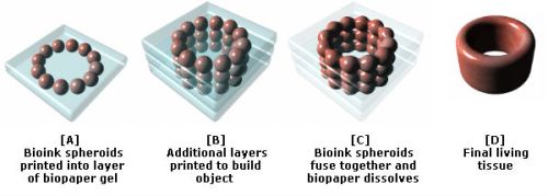


 2012pharmaceutical
 sjwilliamspa