Feeds:
Posts
Comments

Archive for the ‘Translational Science’ Category

Cardiac Hypertrophy-associated transcript as a potential long noncoding RNAs (lncRNAs) candidate that influences Cardiomyocyte Hypertrophy


Reporter: Aviva Lev-Ari, PhD


Sci Transl Med. 2016 Feb 17;8(326):326ra22. doi: 10.1126/scitranslmed.aaf1475.

Long noncoding RNA Chast promotes cardiac remodeling.

Abstract

Recent studies highlighted long noncoding RNAs (lncRNAs) to play an important role in cardiac development. However, understanding of lncRNAs in cardiac diseases is still limited. Global lncRNA expression profiling indicated that several lncRNA transcripts are deregulated during pressure overload-induced cardiac hypertrophy in mice. Using stringent selection criteria, we identified Chast (cardiac hypertrophy-associated transcript) as a potential lncRNA candidate that influences cardiomyocyte hypertrophy. Cell fractionation experiments indicated that Chast is specifically up-regulated in cardiomyocytes in vivo in transverse aortic constriction (TAC)-operated mice. In accordance, CHAST homolog in humans was significantly up-regulated in hypertrophic heart tissue from aortic stenosis patients and in human embryonic stem cell-derived cardiomyocytes upon hypertrophic stimuli. Viral-based overexpression of Chast was sufficient to induce cardiomyocyte hypertrophy in vitro and in vivo. GapmeR-mediated silencing of Chast both prevented and attenuated TAC-induced pathological cardiac remodeling with no early signs on toxicological side effects. Mechanistically, Chast negatively regulated Pleckstrin homology domain-containing protein family M member 1 (opposite strand of Chast), impeding cardiomyocyte autophagy and driving hypertrophy. These results indicate that Chast can be a potential target to prevent cardiac remodeling and highlight a general role of lncRNAs in heart diseases.

Copyright © 2016, American Association for the Advancement of Science.

Read Full Post »

BioPrinting Basics

Curator: Larry H. Bernstein, MD, FCAP


The ABCs of 3D Bioprinting of Living Tissues, Organs   5/06/2016 

(Credit: Ozbolat Lab/Penn State University)

Although first originated in 2003, the world of bioprinting is still very new and ambiguous. Nevertheless, as the need for organ donation continues to increase worldwide, and organ and tissue shortages prevail, a handful of scientists have started utilizing this cutting-edge science and technology for various areas of regenerative medicine to possibly fill that organ-shortage void.

Among these scientists is Ibrahim Tarik Ozbolat, an associate professor of Engineering Science and Mechanics Department and the Huck Institutes of the Life Sciences at Penn State University, who’s been studying bioprinting and tissue engineering for years.

While Ozbolat is not the first to originate 3D bioprinting research, he’s the first one at Penn State University to spearhead the studies at Ozbolat Lab, Leading Bioprinting Research.

“Tissue engineering is a big need. Regenerative medicine, biofabrication of tissues and organs that can replace the damage or diseases is important,” Ozbolat told R&D Magazine after his seminar presentation at Interphex last week in New York City, titled 3D Bioprinting of Living Tissues & Organs.”

3D bioprinting is the process of creating cell patterns in a confined space using 3D-printing technologies, where cell function and viability are preserved within the printed construct.

Recent progress has allowed 3D printing of biocompatible materials, cells and supporting components into complex 3D functional living tissues. The technology is being applied to regenerative medicine to address the need for tissues and organs suitable for transplantation. Compared with non-biological printing, 3D bioprinting involves additional complexities, such as the choice of materials, cell types, growth and differentiation factors, and technical challenges related to the sensitivities of living cells and the construction of tissues. Addressing these complexities requires the integration of technologies from the fields of engineering, biomaterials science, cell biology, physics and medicine, according to nature.com.

“If we’re able to make organs on demand, that will be highly beneficial to society,” said Ozbolat. “We have the capability to pattern cells, locate them and then make the same thing that exists in the body.”

3D bioprinting of tissues and organs

Sean V Murphy & Anthony Atala
Nature Biotechnology 32,773–785(2014)       doi:10.1038/nbt.2958


Additive manufacturing, otherwise known as three-dimensional (3D) printing, is driving major innovations in many areas, such as engineering, manufacturing, art, education and medicine. Recent advances have enabled 3D printing of biocompatible materials, cells and supporting components into complex 3D functional living tissues. 3D bioprinting is being applied to regenerative medicine to address the need for tissues and organs suitable for transplantation. Compared with non-biological printing, 3D bioprinting involves additional complexities, such as the choice of materials, cell types, growth and differentiation factors, and technical challenges related to the sensitivities of living cells and the construction of tissues. Addressing these complexities requires the integration of technologies from the fields of engineering, biomaterials science, cell biology, physics and medicine. 3D bioprinting has already been used for the generation and transplantation of several tissues, including multilayered skin, bone, vascular grafts, tracheal splints, heart tissue and cartilaginous structures. Other applications include developing high-throughput 3D-bioprinted tissue models for research, drug discovery and toxicology.


Future Technologies : Bioprinting
Bioprinting

3D printing is increasingly permitting the direct digital manufacture (DDM) of a wide variety of plastic and metal items. While this in itself may trigger a manufacturing revolution, far more startling is the recent development of bioprinters. These artificially construct living tissue by outputting layer-upon-layer of living cells. Currently all bioprinters are experimental. However, in the future, bioprinters could revolutionize medical practice as yet another element of the New Industrial Convergence.

Bioprinters may be constructed in various configurations. However, all bioprinters output cells from a bioprint head that moves left and right, back and forth, and up and down, in order to place the cells exactly where required. Over a period of several hours, this permits an organic object to be built up in a great many very thin layers.

In addition to outputting cells, most bioprinters also output a dissolvable gel to support and protect cells during printing. A possible design for a future bioprinter appears below and in the sidebar, here shown in the final stages of printing out a replacement human heart. Note that you can access larger bioprinter images on the Future Visions page. You may also like to watch my bioprinting video.

bioprinter


Bioprinting Pioneers

Several experimental bioprinters have already been built. For example, in 2002 Professor Makoto Nakamura realized that the droplets of ink in a standard inkjet printer are about the same size as human cells. He therefore decided to adapt the technology, and by 2008 had created a working bioprinter that can print out biotubing similar to a blood vessel. In time, Professor Nakamura hopes to be able to print entire replacement human organs ready for transplant. You can learn more about this groundbreaking work here or read this message from Professor Nakamura. The movie below shows in real-time the biofabrication of a section of biotubing using his modified inkjet technology.


Another bioprinting pioneer is Organovo. This company was set up by a research group lead by Professor Gabor Forgacs from the University of Missouri, and in March 2008 managed to bioprint functional blood vessels and cardiac tissue using cells obtained from a chicken. Their work relied on a prototype bioprinter with three print heads. The first two of these output cardiac and endothelial cells, while the third dispensed a collagen scaffold — now termed ‘bio-paper’ — to support the cells during printing.

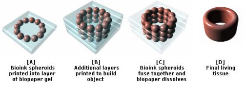
Since 2008, Organovo has worked with a company called Invetech to create a commercial bioprinter called the NovoGen MMX. This is loaded with bioink spheroids that each contain an aggregate of tens of thousands of cells. To create its output, the NovoGen first lays down a single layer of a water-based bio-paper made from collagen, gelatin or other hydrogels. Bioink spheroids are then injected into this water-based material. As illustrated below, more layers are subsequently added to build up the final object. Amazingly, Nature then takes over and the bioink spheroids slowly fuse together. As this occurs, the biopaper dissolves away or is otherwise removed, thereby leaving a final bioprinted body part or tissue.


bioprinting stages

As Organovo have demonstrated, using their bioink printing process it is not necessary to print all of the details of an organ with a bioprinter, as once the relevant cells are placed in roughly the right place Nature completes the job. This point is powerfully illustrated by the fact that the cells contained in a bioink spheroid are capable of rearranging themselves after printing. For example, experimental blood vessels have been bioprinted using bioink spheroids comprised of an aggregate mix of endothelial, smooth muscle and fibroblast cells. Once placed in position by the bioprint head, and with no technological intervention, the endothelial cells migrate to the inside of the bioprinted blood vessel, the smooth muscle cells move to the middle, and the fibroblasts migrate to the outside.

In more complex bioprinted materials, intricate capillaries and other internal structures also naturally form after printing has taken place. The process may sound almost magical. However, as Professor Forgacs explains, it is no different to the cells in an embryo knowing how to configure into complicated organs. Nature has been evolving this amazing capability for millions of years. Once in the right places, appropriate cell types somehow just know what to do.

In December 2010, Organovo create the first blood vessels to be bioprinted using cells cultured from a single person. The company has also successfully implanted bioprinted nerve grafts into rats, and anticipates human trials of bioprinted tissues by 2015. However, it also expects that the first commercial application of its bioprinters will be to produce simple human tissue structures for toxicology tests. These will enable medical researchers to test drugs on bioprinted models of the liver and other organs, thereby reducing the need for animal tests.

In time, and once human trials are complete, Organovo hopes that its bioprinters will be used to produce blood vessel grafts for use in heart bypass surgery. The intention is then to develop a wider range of tissue-on-demand and organs-on-demand technologies. To this end, researchers are now working on tiny mechanical devices that can artificially exercise and hence strengthen bioprinted muscle tissue before it is implanted into a patient.

Organovo anticipates that its first artificial human organ will be a kidney. This is because, in functional terms, kidneys are one of the more straight-forward parts of the body. The first bioprinted kidney may in fact not even need to look just like its natural counterpart or duplicate all of its features. Rather, it will simply have to be capable of cleaning waste products from the blood. You can read more about the work of Organovoand Professor Forgac’s in this article from Nature.

Regenerative Scaffolds and Bones

A further research team with the long-term goal of producing human organs-on-demand has created the Envisiontec Bioplotter. Like Organovo’s NovoGen MMX, this outputs bio-ink ’tissue spheroids’ and supportive scaffold materials including fibrin and collagen hydrogels. But in addition, the Envisontech can also print a wider range of biomaterials. These include biodegradable polymers and ceramics that may be used to support and help form artificial organs, and which may even be used as bioprinting substitutes for bone.

Talking of bone, a team lead by Jeremy Mao at the Tissue Engineering and Regenerative Medicine Lab at Columbia University is working on the application of bioprinting in dental and bone repairs. Already, a bioprinted, mesh-like 3D scaffold in the shape of an incisor has been implanted into the jaw bone of a rat. This featured tiny, interconnecting microchannels that contained ‘stem cell-recruiting substances’. In just nine weeks after implantation, these triggered the growth of fresh periodontal ligaments and newly formed alveolar bone. In time, this research may enable people to be fitted with living, bioprinted teeth, or else scaffolds that will cause the body to grow new teeth all by itself. You can read more about this development in this article from The Engineer.

In another experient, Mao’s team implanted bioprinted scaffolds in the place of the hip bones of several rabbits. Again these were infused with growth factors. As reported inThe Lancet, over a four month period the rabbits all grew new and fully-functional joints around the mesh. Some even began to walk and otherwise place weight on their new joints only a few weeks after surgery. Sometime next decade, human patients may therefore be fitted with bioprinted scaffolds that will trigger the grown of replacement hip and other bones. In a similar development, a team from Washington State University have also recently reported on four years of work using 3D printers to create a bone-like material that may in the future be used to repair injuries to human bones.

In Situ Bioprinting

The aforementioned research progress will in time permit organs to be bioprinted in a lab from a culture of a patient’s own cells. Such developments could therefore spark a medical revolution. Nevertheless, others are already trying to go further by developing techniques that will enable cells to be printed directly onto or into the human body in situ. Sometime next decade, doctors may therefore be able to scan wounds and spray on layers of cells to very rapidly heal them.

Already a team of bioprinting researchers lead by Anthony Alata at the Wake Forrest School of Medicine have developed a skin printer. In initial experiments they have taken 3D scans of test injuries inflicted on some mice and have used the data to control a bioprint head that has sprayed skin cells, a coagulant and collagen onto the wounds. The results are also very promising, with the wounds healing in just two or three weeks compared to about five or six weeks in a control group. Funding for the skin-printing project is coming in part from the US military who are keen to develop in situ bioprinting to help heal wounds on the battlefield. At present the work is still in a pre-clinical phase with Alata progressing his research usig pigs. However, trials of with human burn victims could be a little as five years away.

The potential to use bioprinters to repair our bodies in situ is pretty mind blowing. In perhaps no more than a few decades it may be possible for robotic surgical arms tipped with bioprint heads to enter the body, repair damage at the cellular level, and then also repair their point of entry on their way out. Patients would still need to rest and recuperate for a few days as bioprinted materials fully fused into mature living tissue. However, most patients could potentially recover from very major surgery in less than a week.

Cosmetic Applications …

Bioprinting Implications …

More information on bioprinting can be found in my books 3D Printing: Second Editionand The Next Big Thing. There is also a bioprinting section in my 3D Printing Directory. Oh, and there is also a great infographic about bioprinting here. Enjoy!


How to print out a blood vessel

New work moves closer to the age of organs on demand.

http://www.nature.com/news/2008/080320/images/news.2008.675.jpg

Blood vessels can now be ‘printed out’ by machine. Could bigger structures be in the future?SUSUMU NISHINAGA / SCIENCE PHOTO LIBRARY

Read Full Post »

Natural Killer Cell Response: Treatment of Cancer

Curator: Larry H. Bernstein, MD, FCAP


Molecular mechanisms of natural killer cell activation in response to cellular stress

C J Chan1,2,3, M J Smyth1,2,3,4,5 and L Martinet1,2,4,5        Edited by M Piacentini

Cell Death and Differentiation (2014) 21, 5–14;    http://www.nature.com/cdd/journal/v21/n1/full/cdd201326a.htm

Protection against cellular stress from various sources, such as nutritional, physical, pathogenic, or oncogenic, results in the induction of both intrinsic and extrinsic cellular protection mechanisms that collectively limit the damage these insults inflict on the host. The major extrinsic protection mechanism against cellular stress is the immune system. Indeed, it has been well described that cells that are stressed due to association with viral infection or early malignant transformation can be directly sensed by the immune system, particularly natural killer (NK) cells. Although the ability of NK cells to directly recognize and respond to stressed cells is well appreciated, the mechanisms and the breadth of cell-intrinsic responses that are intimately linked with their activation are only beginning to be uncovered. This review will provide a brief introduction to NK cells and the relevant receptors and ligands involved in direct responses to cellular stress. This will be followed by an in-depth discussion surrounding the various intrinsic responses to stress that can naturally engage NK cells, and how therapeutic agents may induce specific activation of NK cells and other innate immune cells by activating cellular responses to stress.


  • Stress induces specific intrinsic and extrinsic physiological mechanisms within cells that lead to their identification as functionally abnormal
  • Sources of cellular stress can be nutritional, physical, pathogenic, or oncogenic
  • Intrinsic responses to cellular stress include activation of the DNA-damage response, tumor-suppressor genes, and senescence
  • The extrinsic response to cellular stress is activation of the immune system, such as natural killer cells
  • Intrinsic responses to cellular stress can directly upregulate factors that can activate the immune system, and the immune system been shown to be indispensable for the efficacy of some chemotherapy

Further critical determinants of intrinsic responses to stress and cell death that can activate the immune system must be identified

  • Identification of the different cellular pathways and molecular determinants controlling the immunogenicity of different cancer therapies is required
  • How can we harness the ability of therapeutic agents to activate both the intrinsic and extrinsic responses to cellular stress to achieve more specific and safer approaches to cancer treatment?

Any insult to a cell that leads to its abnormal behavior or premature death can be defined as a source of stress. As the turnover and maintenance of cells in all multi-cellular organisms is tightly regulated, it is essential that stressed cells be rapidly identified to avoid widespread tissue damage and to maintain tissue homeostasis. Various intrinsic cellular mechanisms exist within cells that become activated when they are exposed to stress. These include activation of DNA-damage response proteins, senescence programs, and tumor-suppressor genes.1 Extrinsic mechanisms also exist that combat cellular stress, through the upregulation of mediators that can activate different components of the immune system.2 Although frequently discussed separately, much recent evidence has indicated that intrinsic and extrinsic responses to cellular stress are intimately linked.3

As the link between cell intrinsic and extrinsic responses to stress have been uncovered, these observations are now being harnessed therapeutically, particularly in the context of cancer.4 Indeed, various chemotherapeutic agents and radiotherapy are critically dependent on the immune system to elicit their full therapeutic benefit.5, 6 The mechanisms by which this occurs may be twofold: (i) the induction of intrinsic cellular stress mechanisms activates innate immunity and (ii) the release and presentation of tumor-specific antigens engages an inflammatory adaptive immune response.

NK cells are the major effector lymphocyte of innate immunity found in all the primary and secondary immune compartments as well as various mucosal tissues.7 Through their ability to induce direct cytotoxicity of target cells and produce pro-inflammatory cytokines such as interferon-gamma, NK cells are critically involved in the immune surveillance of tumors8, 9, 10 and microbial infections.11, 12 The major mechanism that regulates NK cell contact-dependent functions (such as cytotoxicity and recognition of targets) is the relative contribution of inhibitory and activating receptors that bind to cognate ligands.

Under normal physiological conditions, NK cell activity is inhibited through the interaction of their inhibitory receptors with major histocompatibility complex (MHC) class I.13, 14 However, upon instances of cellular stress that are frequently associated with viral infection and malignant transformation, ligands for activating receptors are often upregulated and MHC class I expression may be downregulated. The upregulation of these activating ligands and downregulation of MHC class I thus provides a signal for NK cells to become activated and display effector functions. Activating receptors are able to provide NK cells with a strong stimulus in the absence of co-stimulation due to the presence of adaptor molecules such as DAP10, DAP12, FcRγ, and CD3ζ that contain immunoreceptor tyrosine-based activating motifs (ITAMs).15, 16,17 By contrast, inhibitory receptors contain inhibitory motifs (ITIMs) within their cytoplasmic tails that can activate downstream targets such as SHP-1 and SHP-2 and directly antagonize those signaling pathways activated through ITAMs.18, 19, 20 The specific details of individual classes of inhibitory and activating receptors and their ligands are summarized in Figure 1 and have been extensively reviewed elsewhere.14, 21 Instead, this review will more focus on the relevant activating receptors that are primarily involved in the direct regulation of NK cell-mediated recognition of cellular stress: natural killer group 2D (NKG2D) and DNAX accessory molecule-1 (DNAM-1).

Figure 1.

Figure 1 - Unfortunately we are unable to provide accessible alternative text for this. If you require assistance to access this image, please contact help@nature.com or the authorNK cell receptors and their cognate ligands. Major inhibitory and activating receptors on NK cells and their cognate ligands on targets are depicted. BAT3, human leukocyte antigen (HLA)-B-associated transcript 3; CRTAM, class I-restricted T-cell-associated molecule; HA, hemagglutinin; HLA-E, HLA class I histocompatibility antigen, alpha chain E; IgG, immunoglobulin G; LFA-1, leukocyte function-associated antigen-1; LLT1, lectin-like transcript 1; TIGIT, T cell immunoglobulin and ITIM domain

Full figure and legend (185K)

NK Cell-Mediated Recognition of Cellular Stress by NKG2D and DNAM-1

NKG2D is a lectin-like type 2 transmembrane receptor expressed as a homodimer in both mice and humans by virtually all NK cells.22, 23 Upon interaction with its ligands, NKG2D can trigger NK cell-mediated cytotoxicity against their targets. The ligands for NKG2D are self proteins related to MHC class I molecules.24 In humans, these ligands consist of the MHC class I chain-related protein (MIC) family (e.g., MICA and MICB) and the UL16-binding protein (ULBP1-6) family.25, 26 In mice, ligands for NKG2D include the retinoic acid early inducible (Rae) gene family, the H60 family, and mouse ULBP-like transcript-1 (MULT-1).27, 28, 29 NKG2D ligands are generally absent on the cell surface of healthy cells but are frequently upregulated upon cellular stress associated with viral infection and malignant transformation.3, 30 Indeed, NKG2D ligand expression has been found on many transformed cell lines, and NKG2D-dependent elimination of tumor cells expressing NKG2D ligands has been well documented in vitro and in tumor transplant experiments.25, 30, 31, 32, 33 In humans, NKG2D ligands have been described on different primary tumors34, 35 and specific NKG2D gene polymorphisms are associated with susceptibility to cancer.36 Finally, blocking NKG2D through gene inactivation or monoclonal antibodies leads to an increased susceptibility to tumor development in mouse models,37, 38demonstrating the key role played by NKG2D in immune surveillance of tumors. NKG2D can also contribute to shape tumor immunogenicity, a process called immunoediting, as demonstrated by the frequent ability of tumor cells to avoid NKG2D-mediated recognition through NKG2D ligand shedding, as discussed later in this review.38, 39, 40

DNAM-1 is a transmembrane adhesion molecule constitutively expressed on T cells, NK cells, macrophages, and a small subset of B cells in mice and humans.41, 42, 43 DNAM-1 contains an extracellular region with two IgV-like domains, a transmembrane region and a cytoplasmic region containing tyrosine- and serine-phosphorylated sites that is able to initiate downstream activation cascades.41, 44 There is accumulating evidence showing that DNAM-1 not only promotes adhesion of NK cells and CTLs but also greatly enhances their cytotoxicity toward ligand-expressing targets.41, 45, 46, 47, 48, 49, 50 The ligands for DNAM-1 are the nectin/nectin-like family members CD155 (PVR, necl-5) and CD112 (PVRL2, nectin-2).45, 46 Like NKG2D ligands, DNAM-1 ligands are frequently expressed on virus-infected and transformed cells.51, 52DNAM-1 ligands, especially CD155, are overexpressed by many types of solid and hematological malignancies and blocking DNAM-1 interactions with its ligands reduces the ability of NK cells to kill tumor cells in vitro.41, 49, 53, 54, 55, 56, 57 Further evidence of the role of DNAM-1 in tumor immune surveillance is provided by studies using experimental and spontaneous models of cancer in vivo showing enhanced tumor spread in the absence of DNAM-1.47, 48, 49, 50, 58

As NKG2D and DNAM-1 ligands are frequently expressed on stressed cells, many studies have sought to determine the mechanisms that underpin these observations. The guiding hypothesis for these studies is that cell-intrinsic responses to stress are directly linked to cell-extrinsic responses that can trigger rapid NK cell surveillance and elimination of stressed cells. Indeed, major cell-intrinsic responses to cellular stress can directly lead to NK cell-activating ligand upregulation and are outlined in the following sections.

The DNA-Damage Response

Cellular stress caused by the activation of the DNA-damage response leads to downstream apoptosis or cell-cycle arrest. The activation of DNA-damage checkpoints occurs when there are excessive DNA strand breaks and replication errors, thereby representing an important tumorigenesis barrier that can slow or inhibit the progression of malignant transformation.59, 60 Two major transducers of the DNA-damage response are the PI3-kinase-related protein kinases ATM (ataxia telangiectasia mutated) and ATR (ATM and Rad3-related). ATM and ATR can modulate numerous signaling pathways such as checkpoint kinases (Chk1 and Chk2, which inhibit cell-cycle progression and promote DNA repair) and p53 (which mediates cell-cycle arrest and apoptosis).61

In addition to the induction of cell-cycle arrest and apoptosis, activation of the DNA-damage response has been shown to promote the expression of several activating ligands that are specific for NK cell receptors, primarily those of the NKG2D receptor. These findings have shown a critical direct link between cellular transformation, apoptosis, and surveillance by the immune system.62 The first evidence of this link between DNA damage and immune cell activation was provided by Raulet and colleagues who showed that NKG2D ligands were upregulated by genotoxic stress and stalled DNA replication conditions known to activate either ATM or ATR.63 These observations have now been extended by several other studies that have defined further DNA-damaging conditions (e.g., genotoxic drugs/chemotherapy, deregulated proliferation, or oxidative stress) that can promote NKG2D ligand upregulation.64, 65, 66, 67

The role of the DNA-damage response in controlling NKG2D ligand expression and subsequent NK cell activation has also been demonstrated in the context of anti-viral immunity, specifically in Abelson murine leukemia virus infection.68 This pathogen was shown to induce activation-induced cytidine deaminase (AID) expression outside the germinal center, resulting in generalized hypermutation, DNA-damage checkpoint activation, and Chk1 phosphorylation. The genotoxic activity of virally induced AID not only restricted the proliferation of infected cells but also induced the expression of NKG2D ligands. More recently, another member of APOBEC-AID family of cytidine deaminases, A3G, has been shown to promote the recognition of HIV-infected cells by NK cells after DNA-damage response activation.69 In this study, viral protein Vpr-mediated repair processes, which generate nicks, gaps, and breaks of DNA, activate an ATM/ATR DNA-damage response that leads to NKG2D ligand expression.

The DNA-damage sensors ATM and ATR have also been shown to regulate other key NK cell-activating ligands such as the DNAM-1 ligand, CD155.58, 65, 70 For example, in the Eμ-myc spontaneous B-cell lymphoma model, activation of the DNA-damage response leads to the upregulation of CD155 in the early-stage transformed B cells, subsequently activating spontaneous tumor regression in an NK cell- and T-cell-dependent manner.58 The DNA-damage response can also regulate the expression of the death receptor DR5.71 The engagement of DR5 by the effector molecule TRAIL, which is expressed by NK cells and T cells, can induce apoptosis of target cells and has been shown to have a key role in immune surveillance against tumors.72 Collectively, these results suggest that the detection of DNA damage, primarily through ATM and ATR, may represent a conserved protection mechanism governing the immunogenicity of infected or transformed cells, leading to direct recognition by NK cells (Figure 2).

Figure 2.

Figure 2 - Unfortunately we are unable to provide accessible alternative text for this. If you require assistance to access this image, please contact help@nature.com or the authorOverview of the molecular pathways leading to NK cell recognition of intrinsic cellular stress. Oncogenic transformation and viral infection can activate intrinsic cellular responses to stress. These responses include activation of the DNA-damage response, senescence, tumor suppressors, and the presentation and/or release of HSPs that, in turn, can activate NK cells through various receptor–ligand interactions. Senescent cells can also release pro-inflammatory cytokines that can recruit NK cells and other innate immunity, such as macrophages. CCL2, C-C motif chemokine ligand 2; CXCL11, C-X-C motif chemokine ligand 11; DR, death receptor 5; IFN, interferon; IL, interleukin; LFA-1, leukocyte function-associated antigen-1; TRAIL, tumor necrosis factor-related apoptosis-inducing ligand

Full figure and legend (146K)

As a result of these studies, many therapeutic agents known to induce DNA damage have been evaluated for their ability to increase the immunogenicity of cancer cells for a more targeted therapeutic approach using NK cells.64, 65 For example, treatment of multiple myeloma cells with doxorubicin, melphalan, or bortezomib can lead to DNAM-1 and NKG2D ligand upregulation.65Indeed, many chemotherapeutic agents commonly used, especially in hematological malignancies, can trigger the DNA-damage pathway. Therefore, it is reasonable to speculate that there is a general role of ATM and ATR in the induction of NK cell activation as a therapeutic effect of these agents.

Senescence

Cellular senescence is generally defined as a growth-arrest program in mammalian cells that limits their lifespan.73 The major type of cellular senescence is replicative senescence that occurs due to telomere shortening. However, it is now generally accepted that premature senescence can also occur due to oncogene activation (oncogene-induced senescence) and/or the loss/gain of tumor-suppressor gene function, in the absence of telomere shortening.74 Thus, premature senescence is an important barrier against malignant transformation.59 Upon engagement of the senescence program, although cells are in growth arrest, they remain metabolically active and can produce many pro-inflammatory cytokines, as well as upregulate adhesion molecules and activating ligands to alert the immune system.75, 76, 77Activation of the immune system, in particular innate immunity, has a critical role in the clearance of senescent cells.78, 79, 80, 81More specifically, in a model of hepatocellular carcinoma, it has been shown that reactivation of p53 can induce a senescence program, resulting in tumor regression through the activation of NK cells, macrophages, and neutrophils. Of note, intercellular adhesion molecule (ICAM)-1, which can trigger both adhesion and cytotoxicity of NK cells,82 and interleukin-15, a cytokine that can promote NK cell effector function,83 were both upregulated in senescent tumors. More recently, the potential contribution of NK cells was also shown in the clearance of senescent hepatic stellate cells, a mechanism important in limiting liver fibrosis in response to a fibrogenic agent.80 ICAM-1, NKG2D ligands (MICA and ULPB2), and DNAM-1 ligands (CD155) were all upregulated on senescent hepatic stellate cells.

The specific mechanisms linking the senescence program to immune activation are not yet fully understood. However, the intracellular molecular mechanisms that govern induction of senescence may provide possible indications. Both replicative senescence and premature senescence (e.g., oncogene-induced senescence) have been shown to have common molecular determinants, such as the activation of the DNA-damage response pathway (e.g., ATM and ATR) and downstream activation of p53 and p16INK4A.1, 59, 84, 85, 86 Activation of the DNA-damage response would presumably initiate the upregulation of NK cell-activating ligands as previously discussed. However, how senescence may be linked to the induction of pro-inflammatory cytokine release is a more compelling question and requires further investigation (Figure 2). Nevertheless, induction of pro-inflammatory cytokines is an important protective mechanism in order to recruit immune cells that can rapidly recognize and remove senescent cells. Interestingly, activation of NK cells by senescent cells has been observed in a clinical context when multiple myeloma cells were treated with chemotherapy and genotoxic agents.65 In this setting, NKG2D and DNAM-1 ligands were both upregulated through a mechanism that required activation of the DNA-damage pathway initiated by ATM and ATR.65

Tumor Suppressors: p53

p53 is a potent tumor suppressor and central regulator of apoptosis, DNA repair, and cell proliferation, that is activated in response to DNA damage, oncogene activation, and other cellular stress.87 The number of identified cellular functions that p53 regulates has greatly increased over the past few years, and there is now a vast array of evidence that shows that p53 can be induced by viral infection88 to limit pathogen spread by inducing apoptosis.89, 90 Furthermore, p53 not only acts as an intrinsic barrier against tumorigenesis or pathogenic spread but can also lead to increased cellular immunogenicity. For example, p53 reactivation in a hepatocellular carcinoma can promote tumor regression mediated by innate immunity.78 A direct link between p53 expression and immune cell recognition was recently provided by Textor et al.91 where expression of p53 in lung cancer cell lines strongly upregulated the NKG2D ligands ULBP1 and 2, resulting in NK cell activation. Subsequently, p53-responsive elements were found to directly regulate ULBP1 and 2 expression, the deletion of which abolished the capacity of p53 to mediate ULBP1 and 2 upregulation. Another recent report that used a pharmacological activator of p53 confirmed the ability of p53 to directly induce ULBP2 expression that was independent of ATM/ATR.92 However, it has also been shown that miR34a and miR34C microRNAs (miRNAs) induced by p53 can target ULBP2 mRNA and reduce its cell-surface expression, suggesting that p53 may have a dual role in regulating ULBP2 expression.93 Finally, early work showed that NKG2D ligands can be upregulated by ATR/ATM in the total absence of p53 in tumor cell lines,62, 63 suggesting the existence of ATM/ATR-dependent and p53-independent pathways that regulate NKG2D ligand expression in response to cellular stress.

In addition to regulating NK cell ligand expression, genetic reactivation of p53 in tumors can also induce a wide array of pro-inflammatory mediators ranging from adhesion receptor (ICAM-1) expression to the production of various chemokines (CXCL11 and monocyte chemoattractant protein-1) and cytokines (interleukin-15).78 Furthermore, recent studies in anti-viral immunity indicate that several interferon-inducible genes and Toll-like receptor-3 expression are direct transcriptional targets of p53 and that p53 contributes to production of type I interferon by virally infected cells.94, 95, 96 All together, these studies suggest that p53 accumulation could represent a key determinant of the immunogenicity of stressed cells that are infected or undergoing malignant transformation through its ability to regulate innate immune activation.

Oncogenes

Malignant transformation is a complex process that frequently involves the activation of one or more oncogenes in addition to the inactivation or mutation of tumor-suppressor genes (e.g., p53). Oncogene activation is a powerful inducer of cellular stress that is able to activate intrinsic cellular programs that lead to cell apoptosis or senescence (e.g., activation of the DNA-damage response and p53).1 In addition, many recent reports have also shown that major oncogenes can activate extrinsic responses to cellular stress through inducing the upregulation of NK cell-activating ligands.63, 97, 98 This suggests that oncogene activation can represent a key cellular event in alerting the immune system to ongoing cellular transformation (Figure 3).

Figure 3.

Figure 3 - Unfortunately we are unable to provide accessible alternative text for this. If you require assistance to access this image, please contact help@nature.com or the authorMolecular mechanisms that regulate the cell surface expression of NKG2D ligands. The major group of NK cell-activating ligands that are upregulated by intrinsic cellular responses to stress are those that bind the NKG2D receptor. Activation of the DNA-damage response, senescence, oncogenes, tumor suppressors, or sensing of deregulated proliferation can induce NKG2D ligand gene transcription and increase mRNA translation, leading to extracellular protein expression. MMP, matrix metalloproteases

Full figure and legend (183K)

The enhanced expression of the proto-oncogene Myc has been described as a critical event leading to cellular transformation and is a frequently found genetic alteration in cancer.99 In a recent study, again using the Eμ-myc model, Medzhitov and colleagues demonstrated the ability of c-Myc to alert NK cells to early oncogenic transformation through the upregulation of Rae-1.97 In this study, the induction of Rae-1 was dependent on the direct regulation of Rae-1 transcription by Myc through its interaction with the Raet1 epsilon gene. Collectively, these results provide a possible direct molecular mechanism to explain the increased susceptibility of NKG2D gene-targeted mice to lymphoma development in the Eμ-myc model.38

Recent evidence suggests that several oncogenic mutations of Ras (H-Ras, N-Ras, and K-Ras) can also regulate NKG2D ligand expression in both mice and humans.98 Interestingly, in this case, NKG2D ligands were regulated through MAPK/MEK and PI3K pathways downstream of oncogenic H-RasV12. The activation of PI3K pathways, and more particularly the p110α subunits by virus-encoded proteins, has also been shown to induce the Rae-1 family of ligands.100 As many viruses can manipulate the PI3K pathway101 and tumors often bear Ras and p110α oncogene mutations,102 collectively, this data suggests that there is the existence of a common molecular mechanism by which NK cells sense cellular stress mediated by PI3K-dependent regulation of NKG2D ligands.

Interestingly, whereas Myc was involved in the transcriptional regulation of NKG2D ligands, PI3K can increase NKG2D ligand expression by increasing the translation of Rae-1 mRNA.98 This involved the induction of eIF4E, a protein that enhances the translation of mRNA.103 As number of tumors and viruses can upregulate host translation initiation machinery through the overexpression of eIF4E,104, 105 this may represent an important means by which NK cells can discriminate tumor- and virus-infected cells from normal cells.

Heat-Shock Proteins (HSPs)

HSPs are highly conserved intracellular chaperone molecules that are present in most prokaryotic and eukaryotic cells that mediate protection against cellular damage under conditions of stress. HSPs are distributed in most intracellular compartments of cells where they support the correct folding of nascent polypeptides, prevent protein aggregation, and assist in protein transport across membranes.106 Many tumors display overexpression of HSPs as a response to cellular stress induced by oncogenic transformation.107, 108 HSPs can also be mobilized to the plasma membrane, or even released from cells, under conditions of stress.109

Although intracellular HSPs can promote cell survival by interfering with different apoptosis components, many studies have reported that membrane-bound or soluble HSPs can directly stimulate innate immunity.110 A major immunostimulatory function of HSPs is to promote the presentation of tumor-specific antigens by MHC class I to CD8 T cells.111, 112, 113 Soluble and membrane-bound HSPs can also induce antigen-presenting cell maturation and the resultant secretion of pro-inflammatory cytokines.114, 115, 116Finally, HSPs may directly activate NK cells as HSP70, when overexpressed on tumor cells, can induce a selective dose-dependent increase in NK cell-mediated cytotoxicity in vitro.117 NK cells may directly recognize HSP70 through a 14-amino-acid oligomer (TKD) that is localized in the C-terminal domain of the protein through CD94.118, 119 Tumor-specific HSP70 that is either presented at the cell surface or secreted on exosomes can also enhance NK cell activity against diverse types of cancer in vivo.120, 121 Most importantly, hepatocellular carcinoma cells that are treated with various chemotherapeutic agents can become more susceptible to NK cell-mediated cytotoxicity through their release of HSP-containing exosomes, giving the aforementioned findings a therapeutic context.122 Collectively, these results suggest that HSP translocation to the plasma membrane or secretion during cellular stress may represent a potent danger signal that can stimulate NK cell activity, particularly in the context of cancer.


Read Full Post »

Alzheimer Disease Developments – Spring 2015

Larry H. Bernstein, MD, FCAP, Curator

LPBI


Cognitive Stimulation Modulates Platelet Total Phospholipases A2 Activity in Subjects with Mild Cognitive Impairment


JNK: A Putative Link Between Insulin Signaling and VGLUT1 in Alzheimer’s Disease

Omega-3 Fatty Acid Status Enhances the Prevention of Cognitive Decline by B Vitamins in Mild Cognitive Impairment – Openly Available
Oulhaj, Abderrahim | Jernerén, Fredrik | Refsum, Helga | Smith, A. David | de Jager, Celeste A.

Preliminary Study of Plasma Exosomal Tau as a Potential Biomarker for Chronic Traumatic Encephalopathy – Openly Available
Stern, Robert A. | Tripodis, Yorghos | Baugh, Christine M. | Fritts, Nathan G. | Martin, Brett M. | Chaisson, Christine | Cantu, Robert C. | Joyce, James A. | Shah, Sahil | Ikezu, Tsuneya | Zhang, Jing | Gercel-Taylor, Cicek | Taylor, Douglas D

AZD3293: A Novel, Orally Active BACE1 Inhibitor with High Potency and Permeability and Markedly Slow Off-Rate Kinetics – Openly Available
Eketjäll, Susanna | Janson, Juliette | Kaspersson, Karin | Bogstedt, Anna | Jeppsson, Fredrik | Fälting, Johanna | Haeberlein, Samantha Budd | Kugler, Alan R. | Alexander, Robert C. | Cebers, Gvido

Predictive Value of Cerebrospinal Fluid Visinin-Like Protein-1 Levels for Alzheimer’s Disease Early Detection and Differential Diagnosis in Patients with Mild Cognitive Impairment
Babić Leko, Mirjana | Borovečki, Fran | Dejanović, Nenad | Hof, Patrick R. | Šimić, Goran

Plasma Phospholipid and Sphingolipid Alterations in Presenilin1 Mutation Carriers: A Pilot Study
Chatterjee, Pratishtha | Lim, Wei L.F. | Shui, Guanghou | Gupta, Veer B. | James, Ian | …… | Wenk, Marcus R. | Bateman, Randall J. | Morris, John C. | Martins, Ralph N.

Cognitive reserve in ageing and Alzheimer’s disease / Stern Y / Lancet Neurol. 2012 Nov; 11(11):1006-12. PMID: 23079557.

A mutation in APP protects against Alzheimer’s disease and age-related cognitive decline/ Jonsson T, Atwal JK, Steinberg S, Snaedal J, Jonsson PV, Bjornsson S, Stefansson H, Sulem P, Gudbjartsson D, Maloney J, et al. / Nature. 2012 Aug 2; 488(7409):96-9. PMID: 22801501.

 Propagation of tau pathology in a model of early Alzheimer’s disease / de Calignon A, Polydoro M, Suárez-Calvet M, William C, Adamowicz DH, Kopeikina KJ, Pitstick R, Sahara N, Ashe KH, Carlson GA, et al. / Neuron. 2012 Feb 23; 73(4):685-97. PMID: 22365544.

Stages of the pathologic process in Alzheimer disease: age categories from 1 to 100 years/ Braak H, Thal DR, Ghebremedhin E, Del Tredici K / J Neuropathol Exp Neurol. 2011 Nov; 70(11):960-9. PMID: 22002422.

Neuroinflammation in Alzheimer’s disease and mild cognitive impairment: a field in its infancy / McGeer EG, McGeer PL / J Alzheimers Dis. 2010; 19(1):355-61. PMID: 20061650.

Metallothioneins in Prion- and Amyloid-Related Diseases

MICROGLIA

Microglia are the immune cells of the CNS and account for approximately 10% of the CNS cellpopulation, with regional variation in density [27, 28]. During embryonic development, microglia originate from yolk sac progenitor cells that migrate into the developing CNS during early embryogenesis [29,30].Following construction of the blood-brain barrier (BBB), microglia are renewed by local turnover [31]. In the healthy brain, microglia actively support neurons through the release of insulin-like growth factor 1, nerve growth factor, ciliary neurotrophic factor, and brain-derived neurotrophic factor (BDNF) [32–34]. Microglia also provide indirect support to neurons by clearance of debris to maintain the extracellular environment, and phagocytosis of apoptotic cells to facilitate neurogenesis [35, 36]. In the adult brain, microglia coordinate much of their activity with astrocytes and activate in response to similar stimuli [37, 38]. Dysfunctional signaling between microglia and astrocytes often results in chronic inflammation, a characteristic of many neurodegenerative diseases [39, 40].

Historically, it has been thought that microglia ‘rest’ when not responding to inflammatory stimuli or damage [41, 42]. However, this notion is being increasingly recognized as inaccurate [43]. When not involved in active inflammatory signaling, microglia constantly patrol the neuropil by extension and retraction of their finely branched processes [44]. Microglial activation is often broadly classified into two states; pro-inflammatory (M1) or anti-inflammatory (M2) [36, 45], based on similar phenotypes in peripheral macrophages [46]. M1 activated microglia are characterized by increased expression of pro-inflammatory mediators and cytokines, including inducible nitric oxide synthase, tumor necrosis factor-α, and interleukin-1β, often under the control of the transcription factor nuclear factor-κB [45]. Pro-inflammatory microglia rapidly retract their processes and adopt an amoeboid morphology and often migrate closer to the site of injury [47]. Anti-inflammatory M2 activation of microglia, often referred to as alternative activation, represents the other side of microglial behavior. Anti-inflammatory activation is characterized by increased expression of cytokines including arginase 1 and interleukin-10, and is associated with increased ramification of processes [45]. The polarization of microglia into M1 or M2 throughout the brain is well characterized, especially in neurodegenerative diseases [48]. In the AD brain, microglia expressing markers of M1 activation are typically localized to brain regions such as the hippocampus that are most heavily affected in the disease [49]. However, it is important to note that M1 and M2 classifications of microglia may over-simplify microglial phenotypes and may only represent the extremes of microglial activation [50]. It has been more recently proposed that microglia likely occupy a continuum between these phenotypes [39, 51].

Do microglia have multiple roles in AD?

Classical pro-inflammatory activation of microglia has long been associated with AD [39, 49]. Samples taken from late-stage AD brains contain characteristic signs of inflammation, including amoeboid morphology of microglia, high levels of pro-inflammatory cytokines in the cerebrospinal fluid, and evidence of neuronal damage due to chronic exposure to pro-inflammatory cytokines and oxidative stress [52, 53]. The cause of this inflammation may be in response to direct toxicity of Aβ to neurons resulting in activation of nearby microglia and astrocytes [53, 54]. However, Aβ may also induce inflammatory activation of microglia and astrocytes. Activated immune cells are typically present surrounding amyloid plaques [55–57], with such peri-plaque cells exhibiting strong evidence of pro-inflammatory activation [56, 58–60]. The presence of undigested Aβ particles within these activated microglia may suggest that the Aβ peptide itself is a pro-inflammatory signal for microglia [61–64]. In vitro experiments provide supporting evidence for the in vivo studies, with Aβ promoting pro-inflammatory microglial activation [65, 66], and also acting as a potent chemotactic signal [67].

However, it is important to note that although widespread inflammation is characteristic of late-stage AD, it remains unclear what role inflammation could play in early stages of the disease. Some evidence suggests that reducing inflammation through the long-term use of some non-steroidal anti-inflammatory drugs (NSAIDs) can reduce the risk of AD [68]. However, these findings have not yet been verified in clinical trials [69, 70]. Little is understood about how NSAIDs and related compounds affect the delicate balance of pro- versus anti-inflammatory microglial activity within the brain. Although there is considerable evidence to suggest that chronic inflammation may contribute to pathology in the later stages of AD, it is important to note that inflammation normally only represents a small aspect of microglial function. The non-inflammatory functions of microglia may play a more important role in early disease; specifically, microglial functions relating to maintenance of the CNS.

Phagocytosis: A vital role of microglia that may be lost in AD    

SYNAPTIC PRUNING: MICROGLIA CAN REGULATE NETWORK ACTIVITY

Recently, a new function has been proposed for microglia. A number of studies have provided evidence that microglia prune synapses throughout life. Microglia are known to remove extraneous synapses during development to ensure that only meaningful connections remain [43]. It was, however, thought that differentiated astrocytes performed the majority of synaptic pruning in the adult brain [91]. The discovery that microglial processes are constantly active within the brain and are often positioned near synapses raised the question of whether microglial synaptic pruning continued throughout life [44, 47, 92–94]. This question was answered in 2014 in a study that demonstrated that microglia do prune synapses into adulthood, and that this activity is important for normal brain function [95]. These findings supported those found a year earlier in a study reporting that ablation of microglia from brain slices increases synapse density and results in abnormal firing of hippocampalneurons [96].

Altered microglial behavior may underlie altered neuronal firing in AD  

Altered neuronal activity is an early phenomenon in AD

The cause of DMN hypoactivity in AD is not yet clear; however studies performed in cohorts that are genetically predisposed to AD suggest that DMN hypoactivity is preceded by a period of hyperactivity and increased functional connectivity [123, 136], often manifesting as an absence of normal DMN deactivation during external tasks [137–140]. DMN hyperactivity may interfere with hippocampal memory encoding, leading to the memory deficits that are present in mild cognitive impairment [141, 142]. It has been proposed that hippocampal hyperexcitability in AD may develop as a protective mechanism against increased input from the DMN [142–144]. As AD progresses, the initial hyperexcitability of the DMN and hippocampus may result in hypoactivity due to exhaustion of compensatory mechanisms [123, 136]. Evidence from both transgenic AD mice and longitudinal human studies supports an exhaustion model of hyperactivation leading to later hypoactivation [143, 145–147]. Interestingly, a number of studies report a lower incidence of AD among those who regularly practice meditation which specifically ‘calms’ the DMN [148].

Our understanding of AD as a disease is changing. Historically considered to be primarily a disease of neuronal degeneration, this neurocentric view has widened to encompass non-neuronal cells such as astrocytes into our understanding of the disease process and pathogenesis. A proposed model for microglia in AD is shown in Fig. 2. Microglia perform a wide range of functions in the CNS and although this includes induction of an inflammatory reaction in response to damage, they also have critical roles for maintaining normal function in the brain. Recent evidence shows that microglia regulate neuronal activity through synaptic pruning throughout life as an extension on their normal phagocytosis behavior. The discovery of a large number of AD risk genes associated with reduced immune cell function suggests that perturbed microglial phagocytosis could lead to AD. In our model, altered microglial phagocytosis of synapses results in network dysfunction and onset of AD, occurring downstream of Aβ.

The immune system and microglia represent a novel target for intervention in AD. Importantly, a large number of anti-inflammatory drugs are already in use for other conditions. What is important to know at this stage is exactly how to best target immune cell function. The studies outlined here provide evidence that an indiscriminate dampening down of all microglial activity may result in a worse outcome for individuals by suppressing normal microglial regulatory functions. We currently do not know whether future microglial-based therapies should focus on reducing chronic inflammation or conversely, whether they should be aimed at boosting microglial phagocytosis. It is also likely that future treatment strategies may use a combination of approaches to target Aβ, immune cell phagocytosis and network activity. An increasing view in the AD field is that any drug or therapy needs to be provided very early in the disease process to maximize its beneficial effects. Although we are currently unable to effectively target those at risk of AD at such an early stage, advances in neuroimaging for subtle changes in network activity, or in assays for immune cell function, may provide new avenues for identification of early damage and risk of disease.

REFERENCES

[1]

Selkoe DJ ((2011) ) Alzheimer’s disease. Cold Spring Harb Perspect Biol 3: , pii: a004457.

[2]

Masters CL , Simms G , Weinman NA , Multhaup G , McDonald BL , Beyreuther K ((1985) ) Amyloid plaque core protein in Alzheimer disease and Down syndrome. Proc Natl Acad Sci U S A 82: , 4245–4249.

[3]

Glenner GG , Wong CW ((1984) ) Alzheimer’s disease: Initial report of the purification and characterization of a novel cerebrovascular amyloid protein. Biochem Biophys Res Commun 120: , 885–890.

[4]

Goldgaber D , Lerman MI , McBride OW , Saffiotti U , Gajdusek DC ((1987) ) Characterization and chromosomal localization of a cDNA encoding brain amyloid of Alzheimer’s disease. Science 235: , 877–880.

[5]

Kang J , Lemaire HG , Unterbeck A , Salbaum JM , Masters CL , Grzeschik KH , Multhaup G , Beyreuther K , Muller-Hill B ((1987) ) The precursor of Alzheimer’s disease amyloid A4 protein resembles a cell-surface receptor. Nature 325: , 733–736.

[6]

Robakis NK , Ramakrishna N , Wolfe G , Wisniewski HM ((1987) ) Molecular cloning and characterization of a cDNA encoding the cerebrovascular and the neuritic plaque amyloid peptides. Proc Natl Acad Sci U S A 84: , 4190–4194.

[7]

Levy E , Carman MD , Fernandez-Madrid IJ , Power MD , Lieberburg I , van Duinen SG , Bots GT , Luyendijk W , Frangione B ((1990) ) Mutation of the Alzheimer’s disease amyloid gene in hereditary cerebral hemorrhage, Dutch type. Science 248: , 1124–1126.

[8]

Levy-Lahad E , Wasco W , Poorkaj P , Romano DM , Oshima J , Pettingell WH , Yu CE , Jondro PD , Schmidt SD , Wang K , et al ((1995) ) Candidate gene for the chromosome 1 familial Alzheimer’s disease locus. Science 269: , 973–977.

[9]

Rogaev EI , Sherrington R , Rogaeva EA , Levesque G , Ikeda M , Liang Y , Chi H , Lin C , Holman K , Tsuda T , et al ((1995) ) Familial Alzheimer’s disease in kindreds with missense mutations in a gene on chromosome 1 related to the Alzheimer’s disease type 3 gene. Nature 376: , 775–778.

[10]

Sherrington R , Rogaev EI , Liang Y , Rogaeva EA , Levesque G , Ikeda M , Chi H , Lin C , Li G , Holman K , Tsuda T , Mar L , Foncin JF , Bruni AC , Montesi MP , Sorbi S , Rainero I , Pinessi L , Nee L , Chumakov I , Pollen D , Brookes A , Sanseau P , Polinsky RJ , Wasco W , Da Silva HA , Haines JL , Perkicak-Vance MA , Tanzi RE , Roses AD , Fraser PE , Rommens JM , St George-Hyslop PH ((1995) ) Cloning of a gene bearing missense mutations in early-onset familial Alzheimer’s disease. Nature 375: , 754–760.


Late-Onset Metachromatic Leukodystrophy with Early Onset Dementia Associated with a Novel Missense Mutation in the Arylsulfatase A Gene

Microbes and Alzheimer’s Disease – Openly Available
Itzhaki, Ruth F. | Lathe, Richard | Balin, Brian J. | Ball, Melvyn J. | Bearer, Elaine L. | Braak, Heiko | Bullido, Maria J. | Carter, Chris | Clerici, Mario | Cosby, S. Louise | Del Tredici, Kelly | Field, Hugh | Fulop, Tamas | Grassi, Claudio | Griffin, W. Sue T. | Haas, Jürgen | Hudson, Alan P. | Kamer, Angela R. | Kell, Douglas B. | Licastro, Federico | Letenneur, Luc | Lövheim, Hugo | Mancuso, Roberta | Miklossy, Judith | Otth, Carola | Palamara, Anna Teresa | Perry, George | Preston, Christopher | Pretorius, Etheresia | Strandberg, Timo | Tabet, Naji | Taylor-Robinson, Simon D. | Whittum-Hudson, Judith A.

Longitudinal Relationships between Caloric Expenditure and Gray Matter in the Cardiovascular Health Study – Openly Available
Raji, Cyrus A. | Merrill, David A. | Eyre, Harris | Mallam, Sravya | Torosyan, Nare | Erickson, Kirk I. | Lopez, Oscar L. | Becker, James T. | Carmichael, Owen T. | Gach, H. Michael | Thompson, Paul M. | Longstreth Jr., W.T. | Kuller, Lewis H.

Preliminary Study of Plasma Exosomal Tau as a Potential Biomarker for Chronic Traumatic Encephalopathy – Openly Available
Stern, Robert A. | Tripodis, Yorghos | Baugh, Christine M. | Fritts, Nathan G. | Martin, Brett M. | Chaisson, Christine | Cantu, Robert C. | Joyce, James A. | Shah, Sahil | Ikezu, Tsuneya | Zhang, Jing | Gercel-Taylor, Cicek | Taylor, Douglas D.

Unraveling Alzheimer’s: Making Sense of the Relationship between Diabetes and Alzheimer’s Disease1 – Openly Available
Schilling, Melissa A.

Pain Assessment in Elderly with Behavioral and Psychological Symptoms of Dementia –Openly Available
Malara, Alba | De Biase, Giuseppe Andrea | Bettarini, Francesco | Ceravolo, Francesco | Di Cello, Serena | Garo, Michele | Praino, Francesco | Settembrini, Vincenzo | Sgrò, Giovanni | Spadea, Fausto | Rispoli, Vincenzo

Editor’s Choice from Volume 50, Number 4 / 2016

Post Hoc Analyses of ApoE Genotype-Defined Subgroups in Clinical Trials
Kennedy, Richard E. | Cutter, Gary R. | Wang, Guoqiao | Schneider, Lon S.

Protective Effect of Amyloid-β Peptides Against Herpes Simplex Virus-1 Infection in a Neuronal Cell Culture Model
Bourgade, Karine | Le Page, Aurélie | Bocti, Christian | Witkowski, Jacek M. | Dupuis, Gilles | Frost, Eric H. | Fülöp, Tamás

Association Between Serum Ceruloplasmin Specific Activity and Risk of Alzheimer’s Disease
Siotto, Mariacristina | Simonelli, Ilaria | Pasqualetti, Patrizio | Mariani, Stefania | Caprara, Deborah | Bucossi, Serena | Ventriglia, Mariacarla | Molinario, Rossana | Antenucci, Mirca | Rongioletti, Mauro | Rossini, Paolo Maria | Squitti, Rosanna

Effects of Hypertension and Anti-Hypertensive Treatment on Amyloid-β (Aβ) Plaque Load and Aβ-Synthesizing and Aβ-Degrading Enzymes in Frontal Cortex
Ashby, Emma L. | Miners, James S. | Kehoe , Patrick G. | Love, Seth

AZD3293: A Novel, Orally Active BACE1 Inhibitor with High Potency and Permeability and Markedly Slow Off-Rate Kinetics – Openly Available
Eketjäll, Susanna | Janson, Juliette | Kaspersson, Karin | Bogstedt, Anna | Jeppsson, Fredrik | Fälting, Johannad | Haeberlein, Samantha Budd | Kugler, Alan R. | Alexander, Robert C. | Cebers, Gvido

Read Full Post »

Brain Biobank and studies of disease structure correlates

Larry H. Bernstein, MD, FCAP, Curator

LPBI


Unveiling Psychiatric Diseases

Researchers create neuropsychiatric cellular biobank

Image: iStock/mstroz
Researchers from Harvard Medical School and Massachusetts General Hospital have completed the first stage of an important collaboration aimed at understanding the intricate variables of neuropsychiatric disease—something that currently eludes clinicians and scientists.

The research team, led by Isaac Kohane at HMS and Roy Perlis at Mass General, has created a neuropsychiatric cellular biobank—one of the largest in the world.

It contains induced pluripotent stem cells, or iPSCs, derived from skin cells taken from 100 people with neuropsychiatric diseases such as schizophrenia, bipolar disorder and major depression, and from 50 people without neuropsychiatric illness.

In addition, a detailed profile of each patient, obtained from hours of in-person assessment as well as from electronic medical records, is matched to each cell sample.

As a result, the scientific community can now for the first time access cells representing a broad swath of neuropsychiatric illness. This enables researchers to correlate molecular data with clinical information in areas such as variability of drug reactions between patients. The ultimate goal is to help treat, with greater precision, conditions that often elude effective management.

The cell collection and generation was led by investigators at Mass General, who in collaboration with Kohane and his team are working to characterize the cell lines at a molecular level. The cell repository, funded by the National Institutes of Health, is housed at Rutgers University.

“This biobank, in its current form, is only the beginning,” said Perlis, director of the MGH Psychiatry Center for Experimental Drugs and Diagnostics and HMS associate professor of psychiatry. “By next year we’ll have cells from a total of four hundred patients, with additional clinical detail and additional cell types that we will share with investigators.”

A current major limitation to understanding brain diseases is the inability to access brain biopsies on living patients. As a result, researchers typically study blood cells from patients or examine post-mortem tissue. This is in stark contrast with diseases such as cancer, for which there are many existing repositories of highly characterized cells from patients.

The new biobank offers a way to push beyond this limitation.


A Big Step Forward

While the biobank is already a boon to the scientific community, researchers at MGH and the HMS Department of Biomedical Informatics will be adding additional layers of molecular data to all of the cell samples. This information will include whole genome sequencing and transcriptomic and epigenetic profiling of brain cells made from the stem cell lines.

Collaborators in the HMS Department of Neurobiology, led by Michael Greenberg, department chair and Nathan Marsh Pusey Professor of Neurobiology,  will also work to examine characteristics of other types of neurons derived from these stem cells.

“This can potentially alter the entire way we look at and diagnose many neuropsychiatric conditions,” said Perlis.

One example may be to understand how the cellular responses to medication correspond to the patient’s documented responses, comparing in vitro with in vivo. “This would be a big step forward in bringing precision medicine to psychiatry,” Perlis said.

“It’s important to recall that in the field of genomics, we didn’t find interesting connections to disease until we had large enough samples to really investigate these complex conditions,” said Kohane, chair of the HMS Department of Biomedical Informatics.

“Our hypothesis is that here we will require far fewer patients,” he said. “By measuring the molecular functioning of the cells of each patient rather than only their genetic risk, and combining that all that’s known of these people in terms of treatment response and cognitive function, we will discover a great deal of valuable information about these conditions.”

Added Perlis, “In the early days of genetics, there were frequent false positives because we were studying so few people. We’re hoping to avoid the same problem in making cellular models, by ensuring that we have a sufficient number of cell lines to be confident in reporting differences between patient groups.”

The generation of stem cell lines and characterization of patients and brain cell lines is funded jointly by the the National Institute of Mental Health, the National Human Genome Research Institute and a grant from the Centers of Excellence in Genomic Science program.


On C.T.E. and Athletes, Science Remains in Its Infancy

Se Hoon Choi, Young Hye Kim, Matthias Hebisch, et al.

http://www.nature.com/articles/nature13800.epdf

Alzheimer’s disease is the most common form of dementia, characterized by two pathological hallmarks: amyloid-β plaques and neurofibrillary tangles1. The amyloid hypothesis of Alzheimer’s disease posits that the excessive accumulation of amyloid-β peptide leads to neurofibrillary tangles composed of aggregated hyperphosphorylated tau2, 3. However, to date, no single disease model has serially linked these two pathological events using human neuronal cells. Mouse models with familial Alzheimer’s disease (FAD) mutations exhibit amyloid-β-induced synaptic and memory deficits but they do not fully recapitulate other key pathological events of Alzheimer’s disease, including distinct neurofibrillary tangle pathology4, 5. Human neurons derived from Alzheimer’s disease patients have shown elevated levels of toxic amyloid-β species and phosphorylated tau but did not demonstrate amyloid-β plaques or neurofibrillary tangles6, 7, 8, 9, 10, 11. Here we report that FAD mutations in β-amyloid precursor protein and presenilin 1 are able to induce robust extracellular deposition of amyloid-β, including amyloid-β plaques, in a human neural stem-cell-derived three-dimensional (3D) culture system. More importantly, the 3D-differentiated neuronal cells expressing FAD mutations exhibited high levels of detergent-resistant, silver-positive aggregates of phosphorylated tau in the soma and neurites, as well as filamentous tau, as detected by immunoelectron microscopy. Inhibition of amyloid-β generation with β- or γ-secretase inhibitors not only decreased amyloid-β pathology, but also attenuated tauopathy. We also found that glycogen synthase kinase 3 (GSK3) regulated amyloid-β-mediated tau phosphorylation. We have successfully recapitulated amyloid-β and tau pathology in a single 3D human neural cell culture system. Our unique strategy for recapitulating Alzheimer’s disease pathology in a 3D neural cell culture model should also serve to facilitate the development of more precise human neural cell models of other neurodegenerative disorders.


Figure 2: Robust increases of extracellular amyloid-β deposits in 3D-differentiated hNPCs with FAD mutations.close

Robust increases of extracellular amyloid-[bgr] deposits in 3D-differentiated hNPCs with FAD mutations.

a, Thin-layer 3D culture protocol. HC, histochemistry; IF, immunofluorescence; IHC, immunohistochemistry. b, Amyloid-β deposits in 6-week differentiated control and FAD ReN cells in 3D Matrigel (green, GFP; blue, 3D6; scale bar, …


Stem Cell-Based Spinal Cord Repair Enables Robust Corticospinal Regeneration


Novel use of EPR spectroscopy to study in vivo protein structure

http://www.news-medical.net/whitepaper/20160315/Novel-use-of-EPR-spectroscopy-to-study-in-vivo-protein-structure.aspx

α-synuclein

α-synuclein is a protein found abundantly throughout the brain. It is present mainly at the neuron ends where it is thought to play a role in ensuring the supply of synaptic vesicles in presynaptic terminals, which are required for the release of neurotransmitters to relay signals between neurons. It is critical for normal brain function.

However, α-synuclein is also the primary protein component of the cerebral amyloid deposits characteristic of Parkinson’s disease and its precursor is found in the amyloid plaques of Alzheimer’s disease. Although α-synuclein is present in all areas of the brain, these disease-state amyloid plaques only arise in distinct areas.

Alpha-synuclein protein. May play role in Parkinson’s and Alzheimer’s disease.  © molekuul.be / Shutterstock.com

Imaging of isolated samples of α-synuclein in vitro indicate that it does not have the precise 3D folded structure usually associated with proteins. It is therefore classed as an intrinsically disordered protein. However, it was not known whether the protein also lacked a precise structure in vivo.

There have been reports that it can form helical tetramers. Since the 3D structure of a biological protein is usually precisely matched to the specific function it performs, knowing the structure of α-synuclein within a living cell will help elucidate its role and may also improve understanding of the disease states with which it is associated.

If α-synuclein remains disordered in vivo, it may be possible for the protein to achieve different structures, and have different properties, depending on its surroundings.

Techniques for determining protein structure

It has long been known that elucidating the structure of a protein at an atomic level is fundamental for understanding its normal function and behavior. Furthermore, such knowledge can also facilitate the development of targeted drug treatments. Unfortunately, observing the atomic structure of a protein in vivo is not straightforward.

X-ray diffraction is the technique usually adopted for visualizing structures at atomic resolution, but this requires crystals of the molecule to be produced and this cannot be done without separating the molecules of interest from their natural environment. Such processes can modify the protein from its usual state and, particularly with complex structures, such effects are difficult to predict.

The development of nuclear magnetic resonance (NMR) spectroscopy improved the situation by making it possible for molecules to be analyzed under in vivo conditions, i.e. same pH, temperature and ionic concentration.

More recently, increases in the sensitivity of NMR and the use of isotope labelling have enabled determinations of the atomic level structure and dynamics of proteins to be determined within living cells1. NMR has been used to determine the structure of a bacterial protein within living cells2 but it is difficult to achieve sufficient quantities of the required protein within mammalian cells and to keep the cells alive for NMR imaging to be conducted.

Electron paramagnetic resonance (EPR) spectroscopy for determining protein structure

Recently, researchers have managed to overcome these obstacles by using in-cell NMR and electron paramagnetic resonance (EPR) spectroscopy. EPR spectroscopy is a technique that is similar to NMR spectroscopy in that it is based on the measurement and interpretation of the energy differences between excited and relaxed molecular states.

In EPR spectroscopy it is electrons that are excited, whereas in NMR signals are created through the spinning of atomic nuclei. EPR was developed to measure radicals and metal complexes, but has also been utilized to study the dynamic organization of lipids in biological membranes3.

EPR has now been used for the first time in protein structure investigations and has provided atomic-resolution information on the structure of α-synuclein in living mammalians4,5.

Bacterial forms of the α-synuclein protein labelled with 15N isotopes were introduced into five types of mammalian cell using electroporation. Concentrations of α-synuclein close to those found in vivo were achieved and the 15N isotopes allowed the protein to be clearly defined from other cellular components by NMR. The conformation of the protein was then determined using electron paramagnetic resonance (EPR).

The results showed that within living mammalian cells α-synuclein remains as a disordered and highly dynamic monomer. Different intracellular environments did not induce major conformational changes.

Summary

The novel use of EPR spectroscopy has resolved the mystery surrounding the in vivo conformation of α-synuclein. It showed that α-synuclein maintains its disordered monomeric form under physiological cell conditions. It has been demonstrated for the first time that even in crowded intracellular environments α-synuclein does not form oligomers, showing that intrinsic structural disorder can be sustained within mammalian cells.

References

  1. Freedberg DI and Selenko P. Live cell NMR Annu. Rev. Biophys. 2014;43:171–192.
  2. Sakakibara D, et al. Protein structure determination in living cells by in-cell NMR spectroscopy. Nature 2009;458:102–105.
  3. Yashroy RC. Magnetic resonance studies of dynamic organisation of lipids in chloroplast membranes. Journal of Biosciences 1990;15(4):281.
  4. Alderson TA and Bax AD. Parkinson’s Disease. Disorder in the court. Nature 2016; doi:10.1038/nature16871.
  5. Theillet FX, et al. Structural disorder of monomeric α-synuclein persists in mammalian cells. Nature 2016; doi:10.1038/nature16531.


Read Full Post »

Minimal genome

Larry H. Bernstein, MD, FCAP, Curator

LPBI


Minimal Genome Created

Scientists build a living cellular organism with a genome smaller than any known in nature.

By Ruth Williams | March 24, 2016

By stripping down the genome of a mycoplasma bacterium to the minimal genes required for life,Craig Venter and colleagues have created a new organism with the smallest genome of any known cellular life form. The work, published in Sciencetoday (March 24), is the closest scientists have come to creating a cell in which every gene and protein is fully understood—but they are not quite there yet.

“In biology, as we’ve been trying to do genetic and biological engineering, we’re frustrated by the fact that . . . evolution has given us a real mess—it’s really just bubble gum and sticks, piecing together whatever works,” said biomedical engineer Chris Voigt of MIT who was not involved in the study. “This [work] is one of the first attempts at a grand scale to go in and try to clean up some of the mess . . . so that we can better understand the genetics.”

The quest to synthesize a minimal genome with only the essential genes for life is one researchers at the J. Craig Venter Institute (JCVI) in San Diego have been doggedly pursuing for the better part of two decades. Clyde Hutchison, an investigator at JCVI and lead author of the new study, explained the motivation: “We want to understand at a mechanistic level how a living cell grows and divides,” he told The Scientist, and yet, “there is no cell that exists where the function of every gene is known.” Possession of such fundamental knowledge, he added, would also put researchers “in a better position to engineer cells to make specific products,” like pharmaceuticals, Hutchinson said.

The team’s starting point was the bacterium Mycoplasma genitalium, which has the smallest known genome of any living cell with just 525 genes. However, it also has a very slow growth rate, making it difficult to work with. To practice synthesizing genomes and building new organisms, the team therefore turned to M. genitalium’s cousins, M. mycoides and M. capricolum, which have bigger genomes and faster growth rates. In 2010, Venter’s team successfully synthesized a version of the M. mycoides genome (JCVI-syn1.0) and placed it into the cell of a M. capricolum that had had its own genome removed. This was the first cell to contain a fully synthetic genome capable of supporting replicative life.

With the genome synthesis and transfer skills mastered, the next step was to make the genome smaller, explained Hutchison. One approach would be to delete the genes one by one and see which the cells could live without. But “we thought we knew enough, that it would be that much faster to design the genome, build it, and install it in a cell,” said Hutchison. The problem was, “we weren’t completely right about that,” he said. “It took quite a bit longer than we thought.”

Using JCVI-syn1.0 as their starting material, the researchers initially designed a minimal genome based on information from the literature and from mutagenesis studies that suggested which genes were likely essential. They divided this genome into eight overlapping segments and tested each one in combination with the complementary seven-eighths of the standard JCVI-syn1.0 genome. All but one of the designed segments failed to sustain viable cells.

Going back to the drawing board, the team decided to perform mutagenesis experiments on JCVI-syn1.0 to determine, categorically, which genes were required for life. Their experiments revealed that the genes fell into three groups: essential, nonessential, and quasiessential—those that aren’t strictly required, but without which growth is severely impaired. The failure to include these quasiessential genes in the initial design explained in large part why it had failed, explained Hutchison. “The concept of a minimal genome seems simple, but when you get into it, it’s a little more complicated,” he said. “There’s a trade-off between genome size and growth rate.”

Equipped with this knowledge, the team redesigned, synthesized, and tested new genome segments retaining the quasiessential genes. Three iterative cycles of testing later, the team had a genome that successfully supported life.

“This is a really pioneering next step in the use of synthetic biology,” said Leroy Hood, president of the Institute for Systems Biology in Seattle who also did not participate in the research.

Ultimately the team removed 428 genes from the JCVI-syn1.0 genome to create JCVI-syn3.0 with 473 genes (438 protein-coding genes and 35 RNA genes)—considerably fewer than the 525 genes of M. genitalium. Interestingly, the functions of around one-third of the genes (149) remain unknown. “I was surprised it was that high,” said Hood, “but I also think we kid ourselves about how much we know about the genomes of organisms. There’s still an enormous amount of dark matter.”

Some of these genes of unknown function appear to be conserved in higher eukaryotes, said Hutchison. “Those, in a way, are the most exciting,” he said, “because they might represent some new undescribed function that has spread through other life forms.”

C.A. Hutchison III et al., “Design and synthesis of a minimal bacterial genome,” Science, 351: 1414, 2016.


Design and synthesis of a minimal bacterial genome
Designing and building a minimal genome

A goal in biology is to understand the molecular and biological function of every gene in a cell. One way to approach this is to build a minimal genome that includes only the genes essential for life. In 2010, a 1079-kb genome based on the genome of Mycoplasma mycoides (JCV-syn1.0) was chemically synthesized and supported cell growth when transplanted into cytoplasm. Hutchison IIIet al. used a design, build, and test cycle to reduce this genome to 531 kb (473 genes). The resulting JCV-syn3.0 retains genes involved in key processes such as transcription and translation, but also contains 149 genes of unknown function.

Science, this issue p. 10.1126/science.aad6253

Structured Abstract

INTRODUCTION   In 1984, the simplest cells capable of autonomous growth, the mycoplasmas, were proposed as models for understanding the basic principles of life. In 1995, we reported the first complete cellular genome sequences (Haemophilus influenza, 1815 genes, and Mycoplasma genitalium, 525 genes). Comparison of these sequences revealed a conserved core of about 250 essential genes, much smaller than either genome. In 1999, we introduced the method of global transposon mutagenesis and experimentally demonstrated that M. genitalium contains many genes that are nonessential for growth in the laboratory, even though it has the smallest genome known for an autonomously replicating cell found in nature. This implied that it should be possible to produce a minimal cell that is simpler than any natural one. Whole genomes can now be built from chemically synthesized oligonucleotides and brought to life by installation into a receptive cellular environment. We have applied whole-genome design and synthesis to the problem of minimizing a cellular genome.   RATIONALE    Since the first genome sequences, there has been much work in many bacterial models to identify nonessential genes and define core sets of conserved genetic functions, using the methods of comparative genomics. Often, more than one gene product can perform a particular essential function. In such cases, neither gene will be essential, and neither will necessarily be conserved. Consequently, these approaches cannot, by themselves, identify a set of genes that is sufficient to constitute a viable genome. We set out to define a minimal cellular genome experimentally by designing and building one, then testing it for viability. Our goal is a cell so simple that we can determine the molecular and biological function of every gene.

RESULTS   Whole-genome design and synthesis were used to minimize the 1079–kilobase pair (kbp) synthetic genome of M. mycoides JCVI-syn1.0.  An initial design, based on collective knowledge of molecular biology in combination with limited transposon mutagenesis data, failed to produce a viable cell. Improved transposon mutagenesis methods revealed a class of quasi-essential genes that are needed for robust growth, explaining the failure of our initial design. Three more cycles of design, synthesis, and testing, with retention of quasi-essential genes, produced JCVI-syn3.0 (531 kbp, 473 genes). Its genome is smaller than that of any autonomously replicating cell found in nature. JCVI-syn3.0 has a doubling time of ~180 min, produces colonies that are morphologically similar to those of JCVI-syn1.0, and appears to be polymorphic when examined microscopically.   CONCLUSION   The minimal cell concept appears simple at first glance but becomes more complex upon close inspection. In addition to essential and nonessential genes, there are many quasi-essential genes, which are not absolutely critical for viability but are nevertheless required for robust growth. Consequently, during the process of genome minimization, there is a trade-off between genome size and growth rate. JCVI-syn3.0 is a working approximation of a minimal cellular genome, a compromise between small genome size and a workable growth rate for an experimental organism. It retains almost all the genes that are involved in the synthesis and processing of macromolecules. Unexpectedly, it also contains 149 genes with unknown biological functions, suggesting the presence of undiscovered functions that are essential for life. JCVI-syn3.0 is a versatile platform for investigating the core functions of life and for exploring whole-genome design.

Four design-build-test cycles produced JCVI-syn3.0.

(A) The cycle for genome design, building by means of synthesis and cloning in yeast, and testing for viability by means of genome transplantation. After each cycle, gene essentiality is reevaluated by global transposon mutagenesis. (B) Comparison of JCVI-syn1.0 (outer blue circle) with JCVI-syn3.0 (inner red circle), showing the division of each into eight segments. The red bars inside the outer circle indicate regions that are retained in JCVI-syn3.0. (C) A cluster of JCVI-syn3.0 cells, showing spherical structures of varying sizes (scale bar, 200 nm).

Abstract

We used whole-genome design and complete chemical synthesis to minimize the 1079–kilobase pair synthetic genome of Mycoplasma mycoides JCVI-syn1.0. An initial design, based on collective knowledge of molecular biology combined with limited transposon mutagenesis data, failed to produce a viable cell. Improved transposon mutagenesis methods revealed a class of quasi-essential genes that are needed for robust growth, explaining the failure of our initial design. Three cycles of design, synthesis, and testing, with retention of quasi-essential genes, produced JCVI-syn3.0 (531 kilobase pairs, 473 genes), which has a genome smaller than that of any autonomously replicating cell found in nature. JCVI-syn3.0 retains almost all genes involved in the synthesis and processing of macromolecules. Unexpectedly, it also contains 149 genes with unknown biological functions. JCVI-syn3.0 is a versatile platform for investigating the core functions of life and for exploring whole-genome design.

Read Full Post »

The late Cambridge Mayor Alfred Vellucci welcomed Life Sciences Labs to Cambridge, MA – June 1976

Reporter: Aviva Lev-Ari, PhD, RN

How Cambridge became the Life Sciences Capital

Worth watching is the video below, which captures the initial Cambridge City Council hearing on recombinant DNA research from June 1976. The first speaker is the late Cambridge mayor Alfred Vellucci.

Vellucci hoped to pass a two-year moratorium on gene splicing in Cambridge. Instead, the council passed a three-month moratorium, and created a board of nine Cambridge citizens — including a nun and a nurse — to explore whether the work should be allowed, and if so, what safeguards would be necessary. A few days after the board was created, the pro and con tables showed up at the Kendall Square marketplace.

At the time, says Phillip Sharp, an MIT professor, Cambridge felt like a manufacturing town that had seen better days. He recalls being surrounded by candy, textile, and leather factories. Sharp hosted the citizens review committee at MIT, explaining what the research scientists there planned to do. “I think we built a relationship,” he says.

By early 1977, the citizens committee had proposed a framework to ensure that any DNA-related experiments were done under fairly stringent safety controls, and Cambridge became the first city in the world to regulate research using genetic material.


WATCH VIDEO

http://www.betaboston.com/news/2016/03/17/how-cambridge-became-the-life-sciences-capital/

Scott Kirsner can be reached at kirsner@pobox.com. Follow him on Twitter@ScottKirsner and on betaboston.com.

SOURCE

How Cambridge became the life sciences capital

http://www.betaboston.com/news/2016/03/17/how-cambridge-became-the-life-sciences-capital/

Read Full Post »

Cardiomyocytes from mesenchmal stem cells?

Larry H. Bernstein, MD, FCAP, Curator

LPBI

Introduction: A just published article from the Gladstone Institute establishes that cardiac muscle can be generated from inducible explandable  cardiovascular progenitor cells.  However, while the study has validity, it leaves much to be explained, especially in light of the references to many previous studies to generate cardiomycytes for heart failure.

Skin Cells Opening the Door to the Possibility of Personalized Medicine for Heart Attack Patients

iceC-Figure6

https://beyondthedish.files.wordpress.com/2016/03/iecpcs-give-rise-to-cms-ecs-and-smcs-in-vivo-and-improve-cardiac-function-after-mi.jpg?w=652


ieCPCs Give Rise to CMs, ECs, and SMCs In Vivo and Improve Cardiac Function after MI

(A–E) Immunofluorescence analyses of RFP and CM (A), EC (B and C), and SMC (D and E) markers in tissue sections collected 2 weeks after transplanting RFP-labeled ieCPCs at passage 10 into infarcted hearts of immunodeficient mice. Scale bars represent 100 μm.

(F and G) Ejection fraction and fractional shortening of the left ventricle (LV) quantified by echocardiography. Results from two independent experiments were shown. D, days; W, weeks.

(H–J) Cardiac fibrosis was evaluated at eight levels (L1–L8) by Masson’s trichrome staining 12 weeks after coronary ligation. The ligation site is marked as X. Sections of representative hearts are shown in (I) with quantification in (J). Scar tissue (%) = (the sum of fibrotic area or length at L1–L8/the sum of LV area or circumference at L1–L8) × 100. Scale bars represent 500 μm.

(K) Quantification of LV circumference of mouse hearts 12 weeks after transplantation of 2nd MEFs or ieCPCs. Data were summarized from 48 sections for each group. Data are mean ± SE. ∗p < 0.05.

“Cardiac progenitor cells could be ideal for heart regeneration,” said senior author Sheng Ding, PhD, a senior investigator at Gladstone. “They are the closest precursor to functional heart cells, and, in a single step, they can rapidly and efficiently become heart cells, both in a dish and in a live heart. With our new technology, we can quickly create billions of these cells in a dish and then transplant them into damaged hearts to treat heart failure.”


Discussion:  The study raises some important questions.

  1. How are the cultured cells different than those used in previous studies?
  2. Cardiomyocytes and fibroblasts are both of mesodermal origin.  What determines which way the stem cell line will differentiate?
  3. What is the difference, if any, between the cell culture environment and the in vivo environment into which they are placed?
  4. There is a difference between chronic hypoxemia with congestive heart failure and acute coronary syndrome.  The experiment performed would be more apt to apply to post-ACS than to chronic heart failure.


Functional heart muscle regenerated in decellularized human hearts

March 11, 2016    http://snip.ly/txc6j#http://medicalxpress.com/news/2016-03-functional-heart-muscle-regenerated-decellularized.html

A partially recellularized human whole-heart cardiac scaffold, reseeded with human cardiomyocytes derived from induced pluripotent stem cells, being cultured in a bioreactor that delivers a nutrient solution and replicates some of the environmental conditions around a living heart. Credit: Bernhard Jank, MD, Ott Lab, Center for Regenerative Medicine, Massachusetts General Hospital


Massachusetts General Hospital (MGH) researchers have taken some initial steps toward the creation of bioengineered human hearts using donor hearts stripped of components that would generate an immune response and cardiac muscle cells generated from induced pluripotent stem cells (iPSCs), which could come from a potential recipient. The investigators described their accomplishments – which include developing an automated bioreactor system capable of supporting a whole human heart during the recellularization process—earlier this year in Circulation Research.

“Generating functional cardiac tissue involves meeting several challenges,” says Jacques Guyette, PhD, of the MGH Center for Regenerative Medicine (CRM), lead author of the report. “These include providing a structural scaffold that is able to support cardiac function, a supply of specialized cardiac , and a supportive environment in which cells can repopulate the scaffold to form mature tissue capable of handling complex cardiac functions.”

The research team is led by Harald Ott, MD, of the MGH CRM and the Department of Surgery, senior author of the paper. In 2008, Ott developed a procedure for stripping the living cells from a donor organ with a detergent solution and then repopulating the remaining extracellular matrix scaffold with organ-appropriate types of cells. Since then his team has used the approach to generate functional rat kidneys and lungs and has decellularized large-animal hearts, lungs and kidneys. This report is the first to conduct a detailed analysis of the matrix scaffold remaining after decellularization of whole human hearts, along with recellularization of the cardiac matrix in three-dimensional and whole-heart formats.

The study included 73 human hearts that had been donated through the New England Organ Bank, determined to be unsuitable for transplantation and recovered under research consent. Using a scaled-up version of the process originally developed in rat hearts, the team decellularized hearts from both brain-dead donors and from those who had undergone . Detailed characterization of the remaining cardiac scaffolds confirmed a high retention of matrix proteins and structure free of cardiac cells, the preservation of coronary vascular and microvascular structures, as well as freedom from human leukocyte antigens that could induce rejection. There was little difference between the reactions of organs from the two donor groups to the complex decellularization process.

Instead of using genetic manipulation to generate iPSCs from , the team used a newer method to reprogram skin cells with messenger RNA factors, which should be both more efficient and less likely to run into regulatory hurdles. They then induced the  to differentiate into or cardiomyocytes, documenting patterns of gene expression that reflected developmental milestones and generating cells in sufficient quantity for possible clinical application. Cardiomyocytes were then reseeded into three-dimensional matrix tissue, first into thin matrix slices and then into 15 mm fibers, which developed into spontaneously contracting tissue after several days in culture.

The last step reflected the first regeneration of human heart muscle from within a cell-free, human whole-heart matrix. The team delivered about 500 million iPSC-derived cardiomyocytes into the left ventricular wall of decellularized hearts. The organs were mounted for 14 days in an automated bioreactor system developed by the MGH team that both perfused the organ with nutrient solution and applied environmental stressors such as ventricular pressure to reproduce conditions within a living heart. Analysis of the regenerated tissue found dense regions of iPSC-derived cells that had the appearance of immature cardiac muscle tissue and demonstrated functional contraction in response to electrical stimulation.

“Regenerating a whole heart is most certainly a long-term goal that is several years away, so we are currently working on engineering a functional myocardial patch that could replace cardiac tissue damaged due a heart attack or heart failure,” says Guyette. “Among the next steps that we are pursuing are improving methods to generate even more – recellularizing a whole heart would take tens of billions—optimizing bioreactor-based culture techniques to improve the maturation and function of engineered cardiac tissue, and electronically integrating regenerated tissue to function within the recipient’s heart.”

Team leader Ott, an assistant professor of Surgery at Harvard Medical School, adds, “Generating personalized functional myocardium from patient-derived cells is an important step towards novel device-engineering strategies and will potentially enable patient-specific disease modeling and therapeutic discovery. Our team is excited to further develop both of these strategies in future projects.”

Explore further: A tool for isolating progenitor cells from human heart tissue could lead to heart repair

More information: Jacques P. Guyette et al. Bioengineering Human Myocardium on Native Extracellular MatrixNovelty and Significance, Circulation Research (2016). DOI: 10.1161/CIRCRESAHA.115.306874


Stem cell study in mice offers hope for treating heart attack patients

February 15, 2012  http://medicalxpress.com/news/2012-02-stem-cell-mice-heart-patients.html


Stem cell study in mice offers hope for treating heart attack patients

Cardiac stem cells, pictured here, give hope to patients who have suffered a heart attack. Credit: UCSF

A UCSF stem cell study conducted in mice suggests a novel strategy for treating damaged cardiac tissue in patients following a heart attack. The approach potentially could improve cardiac function, minimize scar size, lead to the development of new blood vessels – and avoid the risk of tissue rejection.

In the investigation, reported online in the journal PLoS ONE, the researchers isolated and characterized a novel type of cardiac stem cell from the tissue of middle-aged mice following a .

Then, in one experiment, they placed the in the culture dish and showed they had the ability to differentiate into cardiomyocytes, or “beating heart cells,” as well as endothelial cells and smooth muscle cells, all of which make up the heart.

In another, they made copies, or “clones,” of the cells and engrafted them in the tissue of other of the same genetic background who also had experienced heart attacks. The cells induced angiogenesis, or blood vessel growth, or differentiated, or specialized, into endothelial and smooth muscle cells, improving .

“These findings are very exciting,” said first author Jianqin Ye, PhD, MD, senior scientist at UCSF’s Translational Cardiac Stem Cell Program. First, “we showed that we can isolate these cells from the heart of middle-aged animals, even after a heart attack.” Second, he said, “we determined that we can return these cells to the animals to induce repair.”

Importantly, the stem cells were identified and isolated in all four chambers of the heart, potentially making it possible to isolate them from patients’ hearts by doing right ventricular biopsies, said Ye. This procedure is “the safest way of obtaining cells from the heart of live patients, and is relatively easy to perform,” he said.

“The finding extends the current knowledge in the field of native cardiac progenitor cell therapy,” said senior author Yerem Yeghiazarians, MD, director of UCSF’s Translational Cardiac Stem Cell Program and an associate professor at the UCSF Division of Cardiology. “Most of the previous research has focused on a different subset of cardiac progenitor cells. These novel cardiac precursor cells appear to have great therapeutic potential.”

The hope, he said, is that patients who have severe heart failure after a heart attack or have cardiomyopathy would be able to be treated with their own cardiac stem cells to improve the overall health and function of the heart. Because the cells would have come from the patients, themselves, there would be no concern of cell rejection after therapy.

The cells, known as Sca-1+ stem enriched in Islet (Isl-1) expressing cardiac precursors, play a major role in cardiac development. Until now, most of the research has focused on a different subset of cardiac progenitor, or early stage, cells known as, c-kit cells.

The Sca-1+ cells, like the c-kit cells, are located within a larger clump of cells called cardiospheres.

The UCSF researchers used special culture techniques and isolated Sca-1+ cells enriched in the Isl-1expressing cells, which are believed to be instrumental in the heart’s development. Since Isl-1 is expressed in the cell nucleus, it has been difficult to isolate them but the new technique enriches for this cell population.

The findings suggest a potential treatment strategy, said Yeghiazarians. “Heart disease, including heart attack and heart failure, is the number one killer in advanced countries. It would be a huge advance if we could decrease repeat hospitalizations, improve the quality of life and increase survival.” More studies are being planned to address these issues in the future.

An estimated 785,000 Americans will have a new heart attack this year, and 470,000 who will have a recurrent attack. Heart disease remains the number one killer in the United States, accounting for one out of every three deaths, according to the American Heart Association.

Medical costs of cardiovascular disease are projected to triple from $272.5 billion to $818.1 billion between now and 2030, according to a report published in the journal Circulation.


Sca-1+ Cardiosphere-Derived Cells Are Enriched for Isl1-Expressing Cardiac Precursors and Improve Cardiac Function after Myocardial Injury

Jianqin Ye , Andrew Boyle , Henry Shih , Richard E. Sievers , Yan Zhang , William Grossman , Harold S. Bernstein , Yerem Yeghiazarians
http://dx.doi.org:/10.1371/journal.pone.0030329

Background

Endogenous cardiac progenitor cells are a promising option for cell-therapy for myocardial infarction (MI). However, obtaining adequate numbers of cardiac progenitors after MI remains a challenge. Cardiospheres (CSs) have been proposed to have cardiac regenerative properties; however, their cellular composition and how they may be influenced by the tissue milieu remains unclear.

Methodology/Principal Finding

Using “middle aged” mice as CSs donors, we found that acute MI induced a dramatic increase in the number of CSs in a mouse model of MI, and this increase was attenuated back to baseline over time. We also observed that CSs from post-MI hearts engrafted in ischemic myocardium induced angiogenesis and restored cardiac function. To determine the role of Sca-1+CD45– cells within CSs, we cloned these from single cell isolates. Expression of Islet-1 (Isl1) in Sca-1+CD45– cells from CSs was 3-fold higher than in whole CSs. Cloned Sca-1+CD45– cells had the ability to differentiate into cardiomyocytes, endothelial cells and smooth muscle cells in vitro. We also observed that cloned cells engrafted in ischemic myocardium induced angiogenesis, differentiated into endothelial and smooth muscle cells and improved cardiac function in post-MI hearts.

Conclusions/Significance  

These studies demonstrate that cloned Sca-1+CD45– cells derived from CSs from infarcted “middle aged” hearts are enriched for second heart field (i.e., Isl-1+) precursors that give rise to both myocardial and vascular tissues, and may be an appropriate source of progenitor cells for autologous cell-therapy post-MI.


Incorporation of Mg particles into PDLLA regulates mesenchymal stem cell and macrophage responses

Sandra C. Cifuentes1, Fátima Bensiamar2,3, Amparo M. Gallardo-Moreno3,4, Tim A. Osswald5, José L. González-Carrasco1,3, et al.
J Biomed Materials Res Part A  104(4), pages 866–878, April 2016                    http://dx.doi.org:/10.1002/jbm.a.35625

In this work, we investigated a new approach to incorporate Mg particles within a PDLLA matrix using a solvent-free commercially available process. PDLLA/Mg composites were manufactured by injection moulding and the effects of Mg incorporated into PDLLA on MSC and macrophage responses were evaluated. Small amounts of Mg particles (≤1 wt %) do not cause thermal degradation of PDLLA, which retains its mechanical properties. PDLLA/Mg composites release hydrogen, alkaline products and Mg2+ ions without changing pH of culture media. Mg-containing materials provide a noncytotoxic environment that enhances MSC viability. Concentration of Mg2+ ions in extracts of MSCs increases with the increment of Mg content in the composites. Incorporation of Mg particles into PDLLA stimulates FN production, ALP activity, and VEGF secretion in MSCs, an effect mediated by degradation products dissolved from the composites. Degradation products of PDLLA induce an increase in MCP-1, RANTES, and MIP-1α secretion in macrophages while products of composites have minimal effect on these chemokines. Regulation of MSC behavior at the biomaterial’s interface and macrophage-mediated inflammatory response to the degradation products is related to the incorporation of Mg in the composites. These findings suggest that including small amounts of Mg particles into polymeric devices can be a valuable strategy to promote osseointegration and reduce host inflammatory response. © 2015 Wiley Periodicals, Inc. J Biomed Mater Res Part A: 104A: 866–878, 2016.

Read Full Post »

Dopamine-β-Hydroxylase Functional Variants

Curator: Larry H. Bernstein, MD, FCAP


Deep sequencing identifies novel regulatory variants in the distal promoter region of the dopamine-β-hydroxylase gene.

OBJECTIVE:

Dopamine-β-hydroxylase (DBH), an enzyme that converts dopamine into norepinephrine, is a drug target in cardiovascular and neuropsychiatric disorders. We aimed to identify functional variants in this gene by deep sequencing and enzyme phenotyping in an Indian cohort.

MATERIALS AND METHODS:

Targeted resequencing of 12 exons and 10 kb upstream sequences of DBH in healthy volunteers (n=50) was performed using the Ion Personal Genome Machine System. Enzyme quantity and activity in their sera samples were determined by ELISA and ultra performance liquid chromatography, respectively. The association of markers with phenotypes was determined using Matrix eQTL. Global P-values for haplotypes generated using UNPHASED 3.1.5 were graphed using GrASP v.082 beta.

RESULTS:

Of the 49 variants identified, nine were novel (minor allele frequency≥0.01). Though individual markers associated with enzyme quantity did not withstand multiple corrections, a novel distal promoter block driven by rs113249250 (global P=1.5×10) was associated. Of the nine single nucleotide polymorphisms (SNPs) associated with enzyme activity, rs3025369, rs1076151 and rs1611115, all from the upstream region, withstood false discovery rate correction (false discovery rate=0.03, 0.03 and 2.9×10, respectively). Conditioning for rs1611115 identified rs1989787 also to affect activity. Importantly, we report an association of a novel haplotype block distal to rs1076151 driven by rs3025369 (global P=8.9×10) with enzyme activity. This regulatory SNP explained 4.9% of the total 46.1% of variance in DBH activity caused by associated SNPs.

CONCLUSION:

This first study combining deep sequencing and enzyme phenotyping identified yet another regulatory SNP suggesting that regulatory variants may be central in the physiological or metabolic role of this gene of therapeutic and pharmacological relevance.


Correlation of plasma dopamine beta-hydroxylase activity with polymorphisms in DBH gene: a study on Eastern Indian population.

Plasma dopamine beta-hydroxylase activity (plDbetaH) is tightly regulated by the DBH gene and several genetic polymorphisms have been found to independently exert their influence. In the present investigation, association of four DBH polymorphisms, DBH-STR, rs1611115, rs1108580, and rs2519152 with plDbetaH was examined in blood samples from 100 unrelated individuals belonging to the state of West Bengal, Eastern India. Genotypes obtained after PCR amplification and restriction digestion were used for statistical analyses. plDbetaH was measured using a photometric assay and its correlation with the genetic polymorphisms was analyzed using analysis of variance and linear regression. Moderate linkage disequilibrium (LD) was observed between DBH-STR and rs1611115, while rs1108580 and rs2519152 were in strong LD. ‘T’ allele of rs1611115 showed strong negative correlation with plDbetaH, whereas DBH-STR, rs1108580 and rs2519152 had no major effect. Four haplotypes showed significant influence on plDbetaH. This is the first report on the effect of genetic polymorphisms on plDbetaH from the Indian sub-continent. rs1611115 was the only polymorphism that showed substantial control over plDbetaH. Other polymorphisms which did not show individual effects could possibly be part of larger haplotype blocks that carry the functional polymorphisms controlling plDbetaH.
Polymorphisms and low plasma activity of dopamine-beta-hydroxylase in ADHD children.
Attention-deficit Hyperactivity disorder (ADHD) is a multifactorial disorder clinically characterized by inattentiveness, impulsivity and hyperactivity. The occurrence of this disorder is between 3 and 6% of the children population, with boys predominating over girls at a ratio of 3:1 or more. The research of some candidate genes (DRD4, DAT, DRD5, DBH, 5HTT, HTR1B and SNAP25) brought consistent results confirming the heredity of ADHD syndromes. Dopamine-beta-hydroxylase (DBH) is an enzyme responsible for the conversion of dopamine into noradrenaline. Alteration of the dopamine/noradrenaline levels can result in hyperactivity. The DBH protein is released in response to stimulation. DBH activity, derived largely from sympathetic nerves, can be measured in human plasma. Patients with ADHD showed decreased activities of DBH in serum and urine. Low DBH levels correlate indirectly with the seriousness of the hyperkinetic syndrome in children [19,20]. In the DBH gene, the G444A, G910T, C1603T, C1912T, C-1021T, 5 -ins/del and TaqI polymorphisms occur frequently and may affect the function of gene products or modify gene expression and thus influence the progression of ADHD. This article reviews the DBH itself and polymorphisms in the DBH gene that influence the DBH activity in the serum and the CSF level of DBH. All those are evaluated in connection with ADHD.
Candidate gene studies of attention-deficit/hyperactivity disorder.
A growing body of behavioral and molecular genetics literature has indicated that the development of attention-deficit/hyperactivity disorder (ADHD) may be attributed to both genetic and environmental factors. Family, twin, and adoption studies provide compelling evidence that genes play a strong role in mediating susceptibility to ADHD. Molecular genetic studies suggest that the genetic architecture of ADHD is complex, while the handful of genome-wide scans conducted thus far is not conclusive. In contrast, the many candidate gene studies of ADHD have produced substantial evidence implicating several genes in the etiology of the disorder. For the 8 genes for which the same variant has been studied in 3 or more case-control or family-based studies, 7 show statistically significant evidence of association with ADHD based on pooled odds ratios across studies: the dopamine D4 receptor gene (DRD4), the dopamine D5 receptor gene (DRD5), the dopamine transporter gene (DAT), the dopamine beta-hydroxylase gene (DBH), the serotonin transporter gene (5-HTT), the serotonin receptor 1B gene (HTR1B), and the synaptosomal-associated protein 25 gene (SNAP25). Recent pharmacogenetic studies have correlated treatment nonresponse with particular gene markers, while preclinical studies have increased our understanding of gene expression paradigms and potential analogs for human trials. This literature review discusses the relevance and implications of genetic associations with ADHD for clinical practice and future research
Lack of significant association between -1021C–>T polymorphism in the dopamine beta hydroxylase gene and attention deficit hyperactivity disorder.
Recent trends in medications for attention deficit hyperactivity disorder (ADHD) suggest that norepinephrine (NE) deficiency may contribute to the disease etiology. Dopamine beta hydroxylase (DBH) is the key enzyme which converts dopamine to NE and since DBH gene is considered a major quantitative trait locus for plasma DBH activity, genetic polymorphism may lead to altered NE neurotransmission. Several polymorphisms including a 5′ flanking -1021C–>T polymorphism, was reported to be associated with changed DBH activity and an association between -1021C–>T polymorphism with ADHD was observed in Han Chinese children. We have carried out family-based studies with three polymorphisms in the DBH gene, -1021C–>T polymorphism, exon 2*444g/a and intron 5 TaqI RFLP, to explore their association with Indian ADHD cases. Allele and genotype frequency of these polymorphisms in ADHD cases were compared with that of their parents and a control group. Haplotypes obtained were analyzed for linkage disequilibrium (LD). Haplotype-based haplotype relative risk analysis and transmission disequilibrium test showed lack of significant association between transmission of the polymorphisms and ADHD. A haplotype comprising of allele 1 of all polymorphisms showed a slight positive trend towards transmission from parents to ADHD probands. Strong LD was observed between *444g/a and TaqI RFLP in all the groups. However, low D’ values and corresponding log of odds scores in the control group as compared to the ADHD families indicated that, the incidence of the two polymorphisms being transmitted together could be higher in ADHD families.
Association of the dopamine beta hydroxylase gene with attention deficit hyperactivity disorder: genetic analysis of the Milwaukee longitudinal study.
Attention deficit hyperactivity disorder (ADHD) is a highly heritable and common disorder that partly reflects disturbed dopaminergic function in the brain. Recent genetic studies have shown that candidate genes involved in dopamine signaling and metabolism contribute to ADHD susceptibility. We have initiated genetic studies in a unique cohort of 158 ADHD and 81 control adult subjects who have been followed longitudinally since childhood in the Milwaukee study of ADHD. From this cohort, genetic analysis was performed in 105 Caucasian subjects with ADHD and 68 age and ethnicity-matched controls for the DRD4 exon 3 VNTR, the SLC6A3 (DAT1) 3′ UTR VNTR, dopamine beta hydroxylase (DBH) TaqI A polymorphism, and the DBH GT microsatellite repeat polymorphism that has been quantitatively associated with serum levels of DBH activity, but not previously studied in ADHD. Results indicate a significant association between the DBH TaqI A1 allele and ADHD (P = 0.018) with a relative risk of 1.33. The DBH GT repeat 4 allele, which is associated with high serum levels of DBH, occurred more frequently in the ADHD group than controls, but the difference did not reach statistical significance. Associations were not found with the SLC6A3 10 repeat or DRD4 7 repeat alleles. These results indicate that the DBH TaqI A allele, or another polymorphism in linkage disequilibrium with this allele, may confer increased susceptibility towards ADHD.
Polymorphisms of the dopamine transporter gene: influence on response to methylphenidate in attention deficit-hyperactivity disorder.
Attention deficit-hyperactivity disorder (ADHD) is a very common and heterogeneous childhood-onset psychiatric disorder, affecting between 3% and 5% of school age children worldwide. Although the neurobiology of ADHD is not completely understood, imbalances in both dopaminergic and noradrenergic systems have been implicated in the origin and persistence of core symptoms, which include inattention, hyperactivity, and impulsivity. The role of a genetic component in its etiology is strongly supported by genetic studies, and several investigations have suggested that the dopamine transporter gene (DAT1; SLC6A3 locus) may be a small-effect susceptibility gene for ADHD. Stimulant medication has a well-documented efficacy in reducing ADHD symptoms. Methylphenidate, the most prescribed stimulant, seems to act mainly by inhibiting the dopamine transporter protein and dopamine reuptake. In fact, its effect is probably related to an increase in extracellular levels of dopamine, especially in brain regions enriched in this protein (i.e. striatum). It is also important to note that dopamine transporter densities seem to be particularly elevated in the brain of ADHD patients, decreasing after treatment with methylphenidate. Altogether, these observations suggest that the dopamine transporter does play a major role in ADHD. Among the several polymorphisms already described in the SLC6A3 locus, a 40 bp variable number of tandem repeats (VNTR) polymorphism has been extensively investigated in association studies with ADHD. Although there are some negative results, the findings from these reports indicate the allele with ten copies of the 40 bp sequence (10-repeat allele) as the risk allele for ADHD. Some investigations have suggested that this polymorphism can be implicated in dopamine transporter gene expression in vitro and dopamine transporter density in vivo, even though it is located in a non-coding region of the SLC6A3 locus. Despite all these data, few studies have addressed the relationship between genetic markers (specifically the VNTR) at the SLC6A3 locus and response to methylphenidate in ADHD patients. A significant effect of the 40 bp VNTR on response to methylphenidate has been detected in most of these reports. However, the findings are inconsistent regarding both the allele (or genotype) involved and the direction of this influence (better or worse response). Thus, further investigations are required to determine if genetic variation due to the VNTR in the dopamine transporter gene is able to predict different levels of clinical response and palatability to methylphenidate in patients with ADHD, and how this information would be useful in clinical practice.
Pharmacogenomics in psychiatry: the relevance of receptor and transporter polymorphisms.
The treatment of severe mental illness, and of psychiatric disorders in general, is limited in its efficacy and tolerability. There appear to be substantial interindividual differences in response to psychiatric drug treatments that are generally far greater than the differences between individual drugs; likewise, the occurrence of adverse effects also varies profoundly between individuals. These differences are thought to reflect, at least in part, genetic variability. The action of psychiatric drugs primarily involves effects on synaptic neurotransmission; the genes for neurotransmitter receptors and transporters have provided strong candidates in pharmacogenetic research in psychiatry. This paper reviews some aspects of the pharmacogenetics of neurotransmitter receptors and transporters in the treatment of psychiatric disorders. A focus on serotonin, catecholamines and amino acid transmitter systems reflects the direction of research efforts, while relevant results from some genome-wide association studies are also presented. There are many inconsistencies, particularly between candidate gene and genome-wide association studies. However, some consistency is seen in candidate gene studies supporting established pharmacological mechanisms of antipsychotic and antidepressant response with associations of functional genetic polymorphisms in, respectively, the dopamine D2 receptor and serotonin transporter and receptors. More recently identified effects of genes related to amino acid neurotransmission on the outcome of treatment of schizophrenia, bipolar illness or depression reflect the growing understanding of the roles of glutamate and γ-aminobutyric acid dysfunction in severe mental illness. A complete understanding of psychiatric pharmacogenomics will also need to take into account epigenetic factors, such as DNA methylation, that influence individual responses to drugs.
Pharmacogenetics of psychotropic drug response.

OBJECTIVE:

Molecular genetic approaches provide a novel method of dissecting the heterogeneity of psychotropic drug response. These pharmacogenetic strategies offer the prospect of identifying biological predictors of psychotropic drug response and could provide the means of determining the molecular substrates of drug efficacy and drug-induced adverse events.

METHOD:

The authors discuss methods issues in executing pharmacogenetic studies, review the first generation of pharmacogenetic studies of psychotropic drug response, and consider future directions for this rapidly evolving field.

RESULTS:

Pharmacogenetics has been most commonly used in studies of antipsychotic drug efficacy, antidepressant drug response, and drug-induced adverse effects. Data from antipsychotic drug studies indicate that polymorphisms within the serotonin 2A and dopamine receptor 2 genes may influence drug efficacy in schizophrenia. Moreover, a growing body of data suggests a relationship between the serotonin transporter gene and clinical effects of the selective serotonin reuptake inhibitors used to treat depression. A significant relationship between genetic variation in the cytochrome P450 system and drug-induced adverse effects may exist for certain medications. Finally, a number of independent studies point to a significant effect of a dopamine D(3) receptor polymorphism on susceptibility to tardive dyskinesia.

CONCLUSIONS:

Initial research into the pharmacogenetics of psychotropic drug response suggests that specific genes may influence phenotypes associated with psychotropic drug administration. These results remain preliminary and will require further replication and validation. New developments in molecular biology, human genomic information, statistical methods, and bioinformatics are ongoing and could pave the way for the next generation of pharmacogenetic studies in psychiatry.

OBJECTIVE: Molecular genetic approaches provide a novel method of dissecting the heterogeneity of psychotropic drug response. These pharmacogenetic strategies offer the prospect of identifying biological predictors of psychotropic drug response and could provide the means of determining the molecular substrates of drug efficacy and drug-induced adverse events. METHOD: The authors discuss methods issues in executing pharmacogenetic studies, review the first generation of pharmacogenetic studies of psychotropic drug response, and consider future directions for this rapidly evolving field. RESULTS: Pharmacogenetics has been most commonly used in studies of antipsychotic drug efficacy, antidepressant drug response, and drug-induced adverse effects. Data from antipsychotic drug studies indicate that polymorphisms within the serotonin 2A and dopamine receptor 2 genes may influence drug efficacy in schizophrenia. Moreover, a growing body of data suggests a relationship between the serotonin transporter gene and clinical effects of the selective serotonin reuptake inhibitors used to treat depression. A significant relationship between genetic variation in the cytochrome P450 system and drug-induced adverse effects may exist for certain medications. Finally, a number of independent studies point to a significant effect of a dopamine D3 receptor polymorphism on susceptibility to tardive dyskinesia. CONCLUSIONS: Initial research into the pharmacogenetics of psychotropic drug response suggests that specific genes may influence phenotypes associated with psychotropic drug administration. These results remain preliminary and will require further replication and validation. New developments in molecular biology, human genomic information, statistical methods, and bioinformatics are ongoing and could pave the way for the next generation of pharmacogenetic studies in psychiatry.

Read Full Post »

High blood pressure can damage the retina’s blood vessels and limit the retina’s function. It can also put pressure on the optic nerve.

Sourced through Scoop.it from: www.healthline.com

See on Scoop.it – Cardiovascular Disease: PHARMACO-THERAPY

Read Full Post »

« Newer Posts - Older Posts »