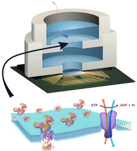
Healthcare analytics, AI solutions for biological big data, providing an AI platform for the biotech, life sciences, medical and pharmaceutical industries, as well as for related technological approaches, i.e., curation and text analysis with machine learning and other activities related to AI applications to these industries.
Though they appear to be distinct, diabetes and Alzheimer’s disease have much in common at the molecular level. In fact, recent findings indicate that either disease can worsen the other by disrupting the same chemical process—S-nitrosylation, a form of post-translational modification that is necessary for the proper functioning of multiple enzymes.
S-nitrosylation, it turns out, can be disrupted by excess sugar or β-amyloid protein, either of which can wreak havoc by increasing the levels of nitric oxide and other free radical species. Once S-nitrosylation is disturbed and poorly functioning enzymes are produced, the downstream effects include abnormal increases in both insulin and β-amyloid protein.
Thus, diabetes and Alzheimer’s can drive, and be driven by, the same vicious cycle. Furthermore, either can contribute to the other’s progress. These results emerged from a study completed by researchers based at the Sanford Burnham Prebys Medical Discovery Institute and the Scintillon Institute. The research team was led by Stuart A. Lipton, M.D., Ph.D., a physician-scientist affiliated with both institutions.
“This work points to a new common pathway to attack both type 2 diabetes, along with its harbinger, metabolic syndrome, and Alzheimer’s disease,” stated Dr. Lipton.
The researchers published their work January 8 in the journal Nature Communications in an article entitled, “Elevated glucose and oligomeric β-amyloid disrupt synapses via a common pathway of aberrant protein S-nitrosylation.” This article describes how the scientists used a so-called “disease-in-a-dish” model to discover molecular pathways that are in common in both diabetes and Alzheimer’s.
Specifically, the scientists genetically reprogrammed the skin of human patients to make induced pluripotent stem cells, which were then used to derive nerve cells. They also used mouse models of each disease to analyze the combined effects of high blood sugar and β-amyloid protein in living animals.
“[We] report in human and rodent tissues that elevated glucose, as found in [metabolic syndrome and type 2 diabetes] and oligomeric β-amyloid (Aβ) peptide, thought to be a key mediator of [Alzheimer’s disease], coordinately increase neuronal Ca2+ and nitric oxide (NO) in an NMDA receptor-dependent manner,” wrote the authors of the Nature Communications article. “The increase in NO results in S-nitrosylation of insulin-degrading enzyme (IDE) and dynamin-related protein 1 (Drp1), thus inhibiting insulin and Aβ catabolism as well as hyperactivating mitochondrial fission machinery.”
The scientists also found that the changes in enzyme activity led to damage of synapses, the region where nerve cells communicate with one another in the brain. The combination of high sugar and β-amyloid protein caused the greatest loss of synapses. Since loss of synapses correlates with cognitive decline in Alzheimer’s, high sugar and β-amyloid coordinately contribute to memory loss.
“The NMDA receptor antagonist memantine attenuates these effects,” the authors continued. “Our studies show that redox-mediated posttranslational modification of brain proteins link Aβ and hyperglyaemia to cognitive dysfunction in [metabolic syndrome/type 2 diabetes] and [Alzheimer’s disease].”
“[Our work] means that we now know these diseases are related on a molecular basis, and hence, they can be treated with new drugs on a common basis,” stated Dr. Ambasudhan, a senior author of the study and an assistant professor at Scintillon.
Gene Editing: The Role of Oligonucleotide Chips, Volume 2 (Volume Two: Latest in Genomics Methodologies for Therapeutics: Gene Editing, NGS and BioInformatics, Simulations and the Genome Ontology), Part 1: Next Generation Sequencing (NGS)
Gene Editing: The Role of Oligonucleotide Chips
Curators: Larry H Bernstein, MD, FCAP and Aviva Lev-Ari, PhD, RN
VIEW VIDEOS
2015 Breakthrough Prize: Jennifer Doudna, Emmanuelle Charpentier, Dick Costolo, Cameron Diaz
Gene editing is a method that has several revisions over the last decade, and in the current form has used an enzyme from a bacteria that breaks the DNA of an attacking virus. Thus, it is an evolutionary method for cellular defense. The discovery and development of the method has been recognized by
SCIENCE Selects CRISPR-Cas9 as
2015 Annual Breakthrough
2015 Breakthrough Prize: Jennifer Doudna, Emmanuelle Charpentier, Dick Costolo, Cameron Diaz
We use the method to remove a part of the genetic material that is tied to expression of an unwanted phenotypic expression. This is more complicated than it may appear. The main problem is how accurately the portion of the genomic target reflects the phenome expressed. This assumes that common diseases, which might be attributed to both genomic expression and environmental conditions, can be easily reduced.
1. Classic view of genetics is that gene expression follows a predictable pathway that is as follows:
DNA—>>> RNA—>>> Protein
This is not the case when we consider the numerous interactions that we call feedback regulatory mechanisms.
Pictograph of the DNA strands
The slide illustrates the double stranded helical DNA strands that are made of linear paired nucleotides linked at 3’ and 5’ positions. The strands separate and unwind and each strand is replicated as “bases” are paired forming a replicated strand, and the new strands combine and rewind.
This concept has undergone many challenges that has been mentioned and will not be addressed.
3. Types of RNA
a. There are many RNAs – mRNA, tRNA, cRNA, ncRNA, siRNA, mtRNA
b. The RNA role in gene expression has an impact on DNA regulation
c. The protein that is produced in the endoplasmic reticulum has modifiable action on the DNA function through action on chromatin, and through pathway activation or inactivation by protein folding and related “allostericity”
Phosphorodiamidate morpholino oligomers – or PMOs – are assembled in precise sequences that correspond to the specific target RNA. These therapeutics bind selectively to the target RNA and modulate its function to increase or decrease the production of a protein involved in a disease.
PMO-based drug development has the potential to address countless diseases, including serious and life-threatening diseases that otherwise could not be treated with traditional small molecule or biologic drugs. The human genome of about 22,000 genes is the basis for more than 250,000 RNA transcripts and about 150,000 proteins, a universe rich with potential targets for PMO-based therapies.
Sarepta Therapeutics Announces FDA Has Filed Eteplirsen NDA for the Potential Treatment of Duchenne Muscular Dystrophy for Patients Amenable to Exon 51 Skipping
FDA Grants Priority Review Status
PDUFA Date is February 26, 2016
August 25, 2015 05:17 PM Eastern Daylight Time
CAMBRIDGE, Mass.–(BUSINESS WIRE)–Sarepta Therapeutics, Inc. (NASDAQ:SRPT), a developer of innovative RNA-targeted therapeutics, today announced that the U.S. Food and Drug Administration (FDA) has filed the New Drug Application (NDA) for eteplirsen for the treatment of Duchenne muscular dystrophy (DMD) amenable to exon 51 skipping. Approximately 13% of people with Duchenne muscular dystrophy are estimated to have a mutation addressable by Eteplirsen/exon 51 skipping.
The FDA has completed its filing review and has determined that our application is sufficiently complete to permit a substantive review. The Prescription Drug User Fee Act (PDUFA) action date for a decision on the application is February 26, 2015. The FDA has granted eteplirsen Priority Review status, which is designated to drugs that offer benefit over existing therapies, or provide a treatment where no adequate therapy exists.
3d. Why CRISPR might be better than exon skipping for DMD
By Lauren Martz, Senior Writer
Published on Thursday, January 21, 2016
As if to preempt the regulatory setbacks in Duchenne muscular dystrophy (DMD) that last week disappointed the field, a trio of preclinical studies emerged two weeks earlier showing that cutting out DMD mutations with gene editing might offer a viable alternative to the exon-skipping strategies that have dominated the pipeline. Now, the question is whether there’s reason to believe the mouse studies will translate any better to the clinic.
The studies, published Dec. 31 in Science, provide in vivo proof of concept for the first time that CRISPR-Cas9 used postnatally can have a disease-modifying effect. Despite the hype around its therapeutic promise, the technology has so far proved itself primarily in research applications, for example, in modifying cells for in vitro screening or creating animal models of disease.
3.2 Sarepta Therapeutics, Corvallis, Oregon, has been assigned a patent (9,238,042) – “antisense modulation of interleukins 17 and 23 signaling.”
01/21/2016 | 07:08am US/Eastern
By Targeted News Service
ALEXANDRIA, Va., Jan. 21 — Sarepta Therapeutics, Corvallis, Oregon, has been assigned a patent (9,238,042) developed by four co-inventors for “antisense modulation of interleukins 17 and 23 signaling.” The co-inventors are Frederick J. Schnell, Corvallis, Oregon, Patrick L. Iversen, Corvallis, Oregon, Dan V. Mourich, Albany, Oregon, and Gunnar J. Hanson, Bothell, Washington.
The patent application was filed on May 13, 2011 (13/107,528). The full-text of the patent can be found at
Antisense modulation of interleukins 17 and 23 signaling
AbstractProvided are antisense oligonucleotides and other agents that target and modulate IL-17 and/or IL-23 signaling activity in a cell, compositions that comprise the same, and methods of use thereof. Also provided are animal models for identifying agents that modulate 17 and/or IL-23 signaling activity.
5. Oligonucleotide sequence composed of introns and exons
Pictograph of an oligonucleotide sequence composed of introns and exons (sequences) from which the introns are removed, creating a new sequence of exons that are translated into AA chain
The picture shows a DNA sequence composed of two types of oligonucleotide sequences colored red and blue (introns and exon). The splicing removes the red colored introns from the nucleotide chain, leaving a chain composed of recombined blue exons. The resulting chain is translated into an amino acid chain based on an obligatory pairing of nucleotide to amino acid. The resulting chain, a posttranslational modification, is the primary sequence of a protein. The finished protein has secondary and tertiary structure.
6. Gene editing to change a value in a linear sequence in an oligonucleotide
Then it is safe to say that we do gene editing to change a value in a linear sequence in an oligonucleotide that is translated into an amino acid through RNA which may be expected to have functional significance. A sequence of 3 nucleotides codes for each of 23 amino acids. However, there have been constructs proposed for a triplex model that might account for three additional metabolites.
7. Mutation and Transcription Errors
There are many mutations that occur having no functional significance, which results in our not needing to pay attention to the change.
There are mutations that have functional significance as a result of expression that we identify as pathological.
8. Gene Expression
A pictorial image of gene expression is shown with a structural translation and a functional portion. The functional portion is really a diversity of methods involving proteomics and metabolomics.
The genome function directly addressed by editing is in the structure – the oligonucleotide sequence.
The pictograph show major aspects of functional genomics, which is largely dictated by 3-D structure built on amino acid sequencing, and acts through subtrates and cofactors. The amino acid sequence of a protein is determined by triplet oligonucleotide portions.
9. Phenotypic Variation
amino acid nucleotide sequences are related to phenotypic variation
Most of human genetic variation is represented by SNPs (Single Nucleotide Polymorphisms) and many of them are believed to cause phenotypic differences between human individuals.
PolyPhen (= Polymorphism Phenotyping) is an automatic tool for prediction of possible impact of an amino acid substitution on the structure and function of a human protein. This prediction is based on straightforward empirical rules which are applied to the sequence, phylogenetic and structural information characterizing the substitution.
oligonucleotide triplets define amino acids in sequence
10. Codons specify which amino acid will be added next during protein synthesis.
The code defines how sequences of nucleotide triplets, called codons, specify which amino acid will be added next during protein synthesis.
A series of codons in part of a messenger RNA (mRNA) molecule. Each codon consists of three nucleotides, usually corresponding to a single amino acid. The nucleotides are abbreviated with the letters A, U, G and C.
mRNA, which uses U (uracil). This mRNA molecule will instruct a ribosome to synthesize a protein according to this code
11. Amino acid composition of proteins and Amino acid frequency
Genetic information contained in mRNA is in the form of codons, sequences of three nucleotides, which are translated into amino acids which then combine to form proteins. At certain sites in a protein’s structure, amino acid composition is not critical. Yet certain amino acids occur at such sites up to six times more often than other amino acids.
The amino acid composition of a nonredundant set of about 550,000 proteins was determined and it was observed that, in the range of 50–200 residues, the percentage of occurrence of most of the residue types significantly depends on protein dimension.
The percentage of occurrence pcaa,i of the amino acid aa in the ith protein was computed for each of the 20 types of amino acids in each protein as
where naa,i and nresi are the number of residues of type aa observed in protein I and the total number of residues in protein i, respectively. Then, the pcaa,i values were averaged for all protein that contains the same number of residues R, by computing
The frequencies of DNA bases in nature are 22.0% uracil, 30.3% adenine, 21.7% cytosine, and 26.1% guanine. The expected frequency of a particular codon can then be calculated by multiplying the frequencies of each DNA base comprising the codon. The expected frequency of the amino acid can then be calculated by adding the frequencies of each codon that codes for that amino acid.
By plotting the expected frequency against the observed frequency, we can see if some amino acids are occurring more or less often than expected by chance. If the observed and expected frequencies are close to equal, we would expect a line with a slope = 1
14. Amino acid composition of proteins reduces deleterious impact of mutations
It is proposed that the inferred amino acid composition of proteins in the LUA probably reflects historical events in the establishment of the genetic code
16. 1:1 relationship between genetic codons and amino acid composition
The classical dogma infers that there is a 1:1 relationship between genetic codons and amino acid composition. However, …..
the regulation of cellular pathways and of gene expression is also dependent on the functional portion, which accounts for the activities of pathways and interactions with the environment. These interactions are very fast. The posttranslational results interact with one another defining specific pathways and also are affected by pathway omissions that may be a result of interactions between mRNA or protein and chromatin of the DNA.
17. The Importance of Gene Editing
is the critical need to change the code for which there are 3 nucleotide combinations for each of 21 amino acids that form the primary protein structure. Critical mutant changes may cause the deletion of a vital amino acid in a key functional location. Most notable is a sulfhydryl group, as in methionine or in cysteine, which is involved in disulfide bonds, necessary for protein folding. Other changes may be in a histidyl residue – a ring structure that has importance
18. Predicting the Genetic Code
The genetic code used by a genome can be predicted by identifying the genes encoded on that genome, and comparing the codons on the DNA to the amino acids in homologous proteins in other genomes. The evolutionary conservation of protein sequences makes it possible to predict the amino acid translation for each codon as the one that is most often aligned to that codon. The program FACIL[41] allows the automated prediction of the genetic code, searching which amino acids in homologous protein domains are most often aligned to every codon. The resulting amino acid probabilities for each codon are displayed in a genetic code logo, that also shows the support for a stop codon.
19. Amino Acids and Proteins
Which of the following has the most significant influence on the characteristics of an individual protein?
the amino-acid sequence
the amino-acid composition
the location of its encoding gene within the genome
The microarrays that are now necessary to break down gene expression are not limited to DNA microarrays. The discussion here is limited to DNA microarrays for gene editing.
The remarkable advances in oligonucleotides analysis, proteomics and metabolomics are large advances in science analytics, but they are disjointed steps in dissecting the large puzzle that we wish to realize as life shaping.
A missing portion of explaining life may not ever be known by the static methods we own because there is a time dimension that we have no comprehension of. This was predicted by the Nobel Physicist Irwin Schroedinger in his seminal book “What is Life?”
21. Chip for Detection of Reverse Transcribed cDNA to labelled cRNA
This is a pictograph of the sequence of steps in biotinylation of the reverse transcribed cDNA to labelled cRNA, and detection on the chip.
The bacterial enzyme Cas9 is the engine of RNA-programmed genome engineering in human cells. Graphic by Jennifer Doudna/UC Berkeley.
“The ability to modify specific elements of an organism’s genes has been essential to advance our understanding of biology, including human health,” said Doudna, a professor of molecular and cell biology and of chemistry and a Howard Hughes Medical Institute Investigator at UC Berkeley. “However, the techniques for making these modifications in animals and humans have been a huge bottleneck in both research and the development of human therapeutics.
“This is going to remove a major bottleneck in the field, because it means that essentially anybody can use this kind of genome editing or reprogramming to introduce genetic changes into mammalian or, quite likely, other eukaryotic systems.”
“I think this is going to be a real hit,” said George Church, professor of genetics at Harvard Medical School and principal author of one of the Science Express papers. “There are going to be a lot of people practicing this method because it is easier and about 100 times more compact than other techniques.”
“Based on the feedback we’ve received, it’s possible that this technique will completely revolutionize genome engineering in animals and plants,” said Doudna, who also holds an appointment at Lawrence Berkeley National Laboratory. “It’s easy to program and could potentially be as powerful as the Polymerase Chain Reaction (PCR).”
The latter technique made it easy to generate millions of copies of small pieces of DNA and permanently altered biological research and medical genetics.
An RNA-based complex guides the DNA-cutting enzyme Cas9 to specific sites in the genome. The natural RNA-programmed DNA cleavage system is shown on the left, with the engineered system on the right.
CRISPR/Cas9, a defense system that bacteria use to protect against invading genetic elements, was adapted by scientists in 2012 as tool to edit genomes in the lab. The new technology has been adopted enthusiastically by the research community because of its ability to cleave target sequences of DNA with remarkable precision and efficiency. Now, after directly watching individual Cas9 enzymes explore DNA inside living cells, Howard Hughes Medical Institute (HHMI) scientists report that they have a better understanding of how Cas9 speeds through this task, testing out potential targets but quickly moving on from those that are not an exact match.
The research, published November 13, 2015, in the journal Science, was co-led by Robert Tjian, president of HHMI; Zhe Liu, a group leader at the Janelia Research Campus; and Jennifer Doudna, an HHMI investigator at the University of California, Berkeley, who is one of the inventors of the CRISPR-Cas9 genome-editing tool.
“CRISPR-based genome engineering offers exciting prospects for curing human disease as well as for advancing agriculture and synthetic biology,” said Doudna. “These studies provide a detailed mechanistic picture of the CRISPR machinery as it operates in living cells, helping to advance both fundamental understanding as well as application of the technology in mammalian cells.”
In both the naturally occurring bacterial defense system and in the CRISPR genome-editing tool, the DNA-cutting Cas9 enzyme is guided to its target by a 20-letter piece of RNA that matches a sequence of DNA embedded within the genome. When Cas9 and its guide find the complementary sequence, they bind to the target and cut. In the laboratory, Knight says, target sites are cut within a few hours of introducing the Cas9 complex to cells.
Previous experiments had demonstrated that Cas9 doesn’t bind exclusively to its true targets, however. A match of just 5-8 letters of genetic code is enough to cause what the scientists call off-target Cas9 binding. Off-target cutting, however, is much rarer. If a sequence that Cas9 binds to does not fully match its 20-letter guide, the enzyme almost always releases the DNA and renews its search.
“A longstanding question was how can Cas9 scan vast eukaryotic genomes, which have billions of DNA bases, and find a target in a timely manner if it’s binding at every off target?” says Spencer Knight, a graduate student in the Doudna and Tjian labs who is the first author of the Science paper.
It is for many biological investigation very desirable to analyse the protein status of a cell. Today the common approach is to separate all the proteins by 2D gel electrophoresis, to stain the proteins and to analyze the obtained spots by mass spectrometry. This give not only the molecular weight but also a fragmentation pattern can be obtained that goes towards mass spec sequencing. This approach called proteomics is still difficult and requires sophisticated mass spectrometry equipment. A way around is to analyze not the proteins but the mRNA content of the cell, although this may be only a rough approximation of the protein content. In any case, already a comparison of the mRNA status of a healthy cell with the status of a cell featuring a disorder may be very informative. These difference allow in some cases to recognize genes which are up or down regulated.
The massively parallel analysis of the mRNA content of cells is today performed with DNA arrays (DNA chips), These are mostly glass plates which are devided in small segments. Each segment contains a specific oligonucleotide attached to the surface either covalently or by absorption. A typical chip is 2.5 x 2.5 cm large and may contain up to 50’000 segments with different oligonucleotide sequences.
The mRNA of the cells to be analysed is first reverse transcribed into cDNA. Then the DNA is amplified using PCR. The DANN is afterwards cut into smaller pieces using hydrolytic enzymes. A ligase such as the RNA ligase which attached unspecifically nucleotides to DNA and RNA is used to attach to each piece of DNA a fluorophore. The so prepared DNA is finally added onto the chip. If the fluorescence labelled oligonucleotide pieces find a complementary piece of DNA on the chip they will form the double strand. The conditions for these hybridizations are carefully controlled so that not matching oligonucleotides are not binding. Under certain conditions one can discriminate between a fully complementary oligonucleotide (binding) and one having a single mismatch (not binding). All the segment were matching oligonucleotides formed a double strand will fluoresce. This can be analyzed so that in a single experiment 50’000 DNA sequences can be checked.
The major problem is to prepare the chips and to deposite the oligonucleotides onto the chips. This is currently achieved in two different ways:
Synthesis of the 50’000 oligonucleotudes using a photolithographic approach directly on the chip (the known phosphoramidite DNA synthesis with light sensitive protecting groups is used)
Synthesis of the oligonucleotides in solution and printing of the DNA strand onto the surface using a modified ink jet printer.
Schematic representation of the DNA array technology
SOURCE
Course Chemical Biology I Nucleic Acids, Established 2004 in Haifa at the Technion
Oligonucleotide chips are microarrays of oligonucleotides.[3] They can be used for detection of mutations and expression monitoring, and gene discovery and mapping.[22]The main methods for creating an oligonucleotide microarray are by gel pads (Motorola), microelectrodes (Nanogen), photolithography (Affymetrix), and inkjet technology (Agilent).[22]
Using gel pads, prefabricated oligonucleotides are attached to patches of activatedpolyacrylamide[22]
Using microelectrodes, negatively charged DNA and molecular probes can be concentrated on energized electrodes for interaction[23]
Using photolithography, a light exposure pattern is created on the substrate using a photomask or virtual photomask projected from a digital micromirror device.[3][6] The light removes photoliabile protecting groups from the selected exposure areas.[6] Following de-protection, nucleotides with a photolabile protecting group are exposed to the entire surface and the chemical coupling process only occurs where light was exposed in the previous step.[6] This process can be repeated to synthesize oligonucleotides of relatively short lengths on the surface, nucleotide by nucleotide.[6]
Using inkjet technology, nucleotides are printed onto a surface drop by drop to form oligonucleotides[22]
cDNA microarrays are often used for large-scale screening and expression studies.[22] In cDNA microarrays, mRNA from cells are collected and converted into cDNA by reverse transcription.[3] Subsequently, cDNA molecules (each corresponding to one gene) are immobilized as ~100 µm diameter spots on a membrane, glass, or silicon chip by metallic pins.[3][22] For detection, fluorescently-labelled single strand cDNA from cells hybridize to the molecules on the microarray and a differential comparison between a treated sample (labelled red, for example) and an untreated sample (labelled in another color such as green) is used for analysis.[3] Red dots mean that the corresponding gene was expressed at a higher level in the treated sample. Conversely, green dots mean that the corresponding gene was expressed at a higher level in the untreated sample. Yellow dots, as a result of the overlap between red and green dots, mean that the corresponding gene was expressed at relatively the same level in both samples, whereas dark spots indicate no or negligible expression in either sample.
Amount of RNA in biopsy extraction, Efficiencies of RNA extraction, reverse transcription, labeling, photodetection, GC content of probes Similar effect on many measurements Corrections can be estimated from data Calibration corrections
Stochastic
PCR yield, DNA quality, Spotting efficiency, spot size, Non-specific hybridization, Stray signal Too random to be explicitly accounted for in a model Noise components
Why Normalize ?
Ensure that differences in intensities are truly due to differential expression, not printing, hybridization, or scanning artifacts Must be done before an analysis which involves comparison of intensities within or between slides Procedures depend on the array technology
Analysis Tasks
Identify up- and down-regulated genes. Find groups of genes with similar expression profiles. Find groups of experiments (tissues) with similar expression profiles. Find genes that explain observed differences among tissues (feature selection)
The “Big Four” algorithms for correcting, normalizing, and summarizing probe level data for analysis of Gene Expression and identification of Over Expressed Genes
Microarray Analysis Suite 5.0 (MAS5 – Affymetrix, 2001, 2003)
Model Based Expression Index (MBEI – Li and Wong, 2001a,b)
KEYNOTE PRESENTATION
Protein Microarrays for Studies in Biomarkers and Post Translational Modification
Joshua LaBaer, M.D., Ph.D., Director, Virginia G. Piper Center for Personalized Diagnostics, The Biodesign Institute, Arizona State University
Self-assembling protein microarrays can be used to study protein-protein interactions, protein-drug interactions, search for enzyme substrates, and as tools to search for disease biomarkers. In particular, recent experiments have focused on using these protein microarrays to search for autoantibody responses in cancer patients. These experiments show promise in finding antibody responses that appear in only cancer patients. New methods using click chemistry-based reagents also allow the application of these arrays for discovering new substrates of post translational modification.
Dr. LaBaer was an early initiator and leader of the effort to build fully sequence-verified recombination-based clone sets for human genes and other model organisms now managed in an automated repository with more than 250,000 samples, which are openly shared with the scientific community. His laboratory has developed a number of methods to employ these clones, including HT protein expression and purification, and HT screens of ectopic protein expression in mammalian cells for relevant phenotypes. In addition, his group invented a novel protein microarray technology, Nucleic Acid Programmable Protein Array, which has been used widely for biomedical research, including the recent discovery of a panel of 28 autoantibody biomarkers that may aid the early diagnosis of breast cancer. Formerly founder and director of the Harvard Institute of Proteomics, LaBaer was recruited to ASU’s Biodesign Institute as the first Piper Chair in Personalized Medicine in 2009.
Investigators at the Scripps Research Institute (TSRI) and St. Jude Children’s Research Hospital say they have found how a protein involved in cancer twists and morphs into different structures.
“We’re studying basic biophysics, but we believe the complexity and rules we uncover for the physics of protein disorder and folding could one day also be used for better designs of therapeutics,” said Ashok Deniz, Ph.D., associate professor at TSRI.
The study (“Asymmetric Modulation of Protein Order-Disorder Transitions by Phosphorylation and Partner Binding”), published in Angewandte Chemie, focuses on the nucleophosmin (NPM1) protein, which has many functions and, when mutated, has been shown to interfere with cells’ normal tumor suppressing ability. NPM1 has been implicated in cancers such as non-Hodgkin lymphoma and acute myelogenous leukemia.
Previous research led by study collaborators Richard Kriwacki, Ph.D., and Diana Mitrea, Ph.D., at St. Jude had demonstrated that a section of NPM1, called the N-terminal domain (Npm-N), doesn’t have a defined, folded structure. Instead, the protein morphs between two forms: a one-subunit disordered monomer and a five-subunit folded pentamer.
Until now, the mechanism behind this transformation was unknown, but scientists believed this monomer-pentamer equilibrium could be important for the protein’s location and functioning in the cell. To shed light on how this transformation occurred, Dr. Deniz and his colleagues used a combination of three techniques—single-molecule biophysics, fluorescence resonance energy transfer (FRET), and circular dichroism, which enabled them to study individual molecules and collections of molecules. Single-molecule methods are especially useful for such studies because they can uncover important information that remains hidden in conventional studies.
The researchers found that the transformation can proceed through more than one pathway. In one pathway, the transformation begins when the cell sends signals to attach phosphoryl groups to NPM1. Such phosphorylation prompts the ordered pentamer to become disordered and likely causes NPM1 to shuttle outside the cell’s nucleus. A meeting with a binding partner can mediate the reverse transformation to a pentamer.
When NPM1 does become a pentamer again under these conditions, which likely causes it to move back to the nucleolus, it takes a different path instead of just retracing its earlier steps.
Priya Banerjee, Ph.D., an American Heart Association-supported postdoctoral research associate at TSRI and the first author of the study, compared these complicated transitions to the morphing of a “Transformers” toy, where a robot can become a car and then a jet. “Phosphorylation and partner-binding are like different cellular switches driving these changes,” said Dr. Banerjee.
According to Dr. Banerjee, the new study also reveals many intermediate states between monomer and pentamer structures and that these states can be manipulated or “tuned” by changing conditions such as salt levels, phosphorylation, and partner binding, which may explain how cells regulate the protein’s multiple functions. The researchers said future studies could shed more light on the biological functions of these different structures and how they might be used in future cancer therapies.
The team added that combining the three techniques used in this study, plus a novel protein-labeling technique for single-molecule fluorescence, could be a useful strategy for studying other unstructured, “intrinsically disordered proteins” (IDPs), which are involved in a host of cellular functions, as well as neurodegenerative disease, heart disease, infectious disease, type 2 diabetes and other conditions.
“We’re studying basic biophysics, but we believe the complexity and rules we uncover for the physics of protein disorder and folding could one day also be used for better designs of therapeutics,” said TSRI Associate Professor Ashok Deniz, senior author of the new study along with Richard Kriwacki, faculty member at St. Jude.
The study focuses on a protein called nucleophosmin (NPM1). This protein has many functions and, when mutated, has been shown to interfere with cells’ normal tumor-suppressing ability. NPM1 has been implicated in cancers such as non-Hodgkin lymphoma and acute myelogenous leukemia.
Previous research led by study collaborators Kriwacki and Diana Mitrea at St. Jude had shown that a section of NPM1, called the N-terminal domain (Npm-N), doesn’t have a defined, folded structure. Instead, the protein morphs between two forms: a one-subunit disordered monomer and a five-subunit folded pentamer.
Until now, the mechanism behind this transformation was unknown, but scientists believed this monomer-pentamer equilibrium could be important for the protein’s location and functioning in the cell.
To shed light on how this transformation occurred, Deniz and his colleagues used an innovative combination of three techniques—single-molecule biophysics, fluorescence resonance energy transfer (FRET) and circular dichroism—which enabled them to study individual molecules and collections of molecules. Single-molecule methods are especially useful for such studies because they can uncover important information that remains hidden in conventional studies.
Remarkably, the researchers found that the transformation can proceed through more than one pathway. In one pathway, the transformation begins when the cell sends signals to attach phosphoryl groups to NPM1. This modification, called phosphorylation, prompts the ordered pentamer to become disordered and likely causes NPM1 to shuttle outside the cell’s nucleus. A meeting with a binding partner can mediate the reverse transformation to a pentamer.
Interestingly, when NPM1 does become a pentamer again under these conditions, which likely causes it to move back to the nucleolus, it takes a different path instead of just retracing its earlier steps.
Priya Banerjee, an American Heart Association-supported postdoctoral research associate at TSRI and the first author of the study, compared these complicated transitions to the morphing of a “Transformers” toy, where a robot can become a car and then a jet. “Phosphorylation and partner-binding are like different cellular switches driving these changes,” said Banerjee.
Banerjee said the new study also reveals many intermediate states between monomer and pentamer structures—and that these states can be manipulated or “tuned” by changing conditions such as salt levels, phosphorylation and partner binding, which may explain how cells regulate the protein’s multiple functions. The researchers said future studies could shed more light on the biological functions of these different structures and how they might be used in future cancer therapies.
The researchers added that combining the three techniques used in this study, plus a novel protein-labeling technique for single-molecule fluorescence, could be a useful strategy for studying other unstructured, “intrinsically disordered proteins” (IDPS). IDPS are involved in a host of cellular functions, as well as neurodegenerative disease, heart disease, infectious disease, type 2 diabetes and other conditions.
TSRI and St. Jude Scientists Study Physics of Single ‘Transformer’ Proteins with Role in Cancer
December 21, 2015 – A new study led by scientists at The Scripps Research Institute (TSRI) and St. Jude Children’s Research Hospital shows how a protein involved in cancer twists and morphs into different structures.
“We’re studying basic biophysics, but we believe the complexity and rules we uncover for the physics of protein disorder and folding could one day also be used for better designs of therapeutics,” said TSRI Associate Professor Ashok Deniz, senior author of the new study along with Richard Kriwacki, faculty member at St. Jude.
The study, published recently in the journal Angewandte Chemie, focuses on a protein called nucleophosmin (NPM1). This protein has many functions and, when mutated, has been shown to interfere with cells’ normal tumor suppressing ability. NPM1 has been implicated in cancers such as non-Hodgkin lymphoma and acute myelogenous leukemia.
Previous research led by study collaborators Kriwacki and Diana Mitrea at St. Jude had shown that a section of NPM1, called the N-terminal domain (Npm-N), doesn’t have a defined, folded structure. Instead, the protein morphs between two forms: a one-subunit disordered monomer and a five-subunit folded pentamer.
Until now, the mechanism behind this transformation was unknown, but scientists believed this monomer-pentamer equilibrium could be important for the protein’s location and functioning in the cell.
To shed light on how this transformation occurred, Deniz and his colleagues used an innovative combination of three techniques—single-molecule biophysics, fluorescence resonance energy transfer (FRET) and circular dichroism—which enabled them to study individual molecules and collections of molecules. Single-molecule methods are especially useful for such studies because they can uncover important information that remains hidden in conventional studies.
Remarkably, the researchers found that the transformation can proceed through more than one pathway. In one pathway, the transformation begins when the cell sends signals to attach phosphoryl groups to NPM1. This modification, called phosphorylation, prompts the ordered pentamer to become disordered and likely causes NPM1 to shuttle outside the cell’s nucleus. A meeting with a binding partner can mediate the reverse transformation to a pentamer.
Interestingly, when NPM1 does become a pentamer again under these conditions, which likely causes it to move back to the nucleolus, it takes a different path instead of just retracing its earlier steps.
Asymmetric Modulation of Protein Order-Disorder Transitions by Phosphorylation and Partner Binding
Priya R. Banerjee, Diana M. Mitrea, Richard W. Kriwacki, Ashok, A. Deniz
As for many intrinsically disordered proteins, order–disorder transitions in the N-terminal oligomerization domain of the multifunctional nucleolar protein nucleophosmin (Npm-N) are central to its function, with phosphorylation and partner binding acting as regulatory switches. However, the mechanism of this transition and its regulation remain poorly understood. In this study, single-molecule and ensemble experiments revealed pathways with alternative sequences of folding and assembly steps for Npm-N. Pathways could be switched by altering the ionic strength. Phosphorylation resulted in pathway-specific effects, and decoupled folding and assembly steps to facilitate disorder. Conversely, binding to a physiological partner locked Npm-N in ordered pentamers and counteracted the effects of phosphorylation. The mechanistic plasticity found in the Npm-N order–disorder transition enabled a complex interplay of phosphorylation and partner-binding steps to modulate its folding landscape.
Conditionally and Transiently Disordered Proteins: Awakening Cryptic Disorder To Regulate Protein Function
Although cell migration plays a central role in development and disease, the underlying molecular mechanism is not fully understood. Here we report that a phosphorylation-mediated molecular switch comprising deleted in liver cancer 1 (DLC1), tensin-3 (TNS3), phosphatase and tensin homologue (PTEN) and phosphoinositide-3-kinase (PI3K) controls the spatiotemporal activation of the small GTPases, Rac1 and RhoA, thereby initiating directional cell migration induced by growth factors. On epidermal growth factor (EGF) or platelet-derived growth factor (PDGF) stimulation, TNS3 and PTEN are phosphorylated at specific Thr residues, which trigger the rearrangement of the TNS3–DLC1 and PTEN–PI3K complexes into the TNS3–PI3K and PTEN–DLC1 complexes. Subsequently, the TNS3–PI3K complex translocates to the leading edge of a migrating cell to promote Rac1 activation, whereas PTEN–DLC1 translocates to the posterior for localized RhoA activation. Our work identifies a core signalling mechanism by which an external motility stimulus is coupled to the spatiotemporal activation of Rac1 and RhoA to drive directional cell migration.
Protein Phosphorylation is an Important Tool… Chapter 13. in Protein Phosphorylation in Human Health
Roberta Fraschini, Erica Raspelli and Corinne Cassani
Protein phosphorylation is a reversible posttranslational modification that can modulate protein role in several physiological processes in almost every possible way. These include modification of its intrinsic biological activity, subcellular location, half-life and binding with other proteins. Protein phosphorylation is particularly important for the regulation of key proteins involved in the control of cell cycle progression.
Protein phosphorylation is the covalent binding of a phosphate group to some critical residues of the polypeptide. The phosphorylation state of a protein is given by a balance between the activity of protein kinases and protein phosphatases. Eukaryotic protein kinases transfer phosphate groups (PO43-) from ATP to an hydroxyl group of the lateral chain of specific serine, threonine or tyrosine residues on peptide substrates. In simple eukaryotic cells, like yeasts, Ser/Thr kinases are more common, while more complex eukaryotic cells, like human cells, have many Tyr kinases. Protein kinases recognize their substrates specifically and their active site consists of an activation loop and a catalytic loop between which substrates bind. Protein kinases differ from each other in the structure of their catalitic domain. A second class of enzymes, protein phosphatases, is responsible for the reverse reaction in which phosphate groups are removed from a protein. Phosphatases gain specificity by binding protein cofactors which facilitate binding to specific phosphoproteins. The active phosphatase often consists of a complex of the phosphatase catalytic subunit and a regulatory subunit.
The phosphorylation of specific residues induces structural changes that regulate protein functions by modulating protein folding, substrate affinity, stability and activity. For example, phosphorylation can cause switch-like changes in protein function, which can also lead to major modifications in the catalytic function of enzymes, including kinases and phosphatases. In addition, protein phosphorylation often leads to rearrangement in the structure of the protein that can induce changes in interacting partners or subcellular localization.
Phosphorylation acts as a molecular switch for many regulatory events in signalling pathways that drive cell division, proliferation, differentiation and apoptosis. In order to ensure an appropriate balance of protein phosphorylation, the cell can compartmentalize both protein kinases and phosphatases. Another kind of regulation can be achieved by the spatial distribution of kinases and phosphatases, that creates a gradient of phosphorylated substrates across different subcellular compartments. This spatial separation can also control the activity of other proteins or enzymes and the occurrence of other posttranslational modifications.
We have seen that each type of protein consists of a precise sequence of amino acids that allows it to fold up into a particular three-dimensional shape, or conformation. But proteins are not rigid lumps of material. They can have precisely engineered moving parts whose mechanical actions are coupled to chemical events. It is this coupling of chemistry and movement that gives proteins the extraordinary capabilities that underlie the dynamic processes in living cells.
In this section, we explain how proteins bind to other selected molecules and how their activity depends on such binding. We show that the ability to bind to other molecules enables proteins to act as catalysts, signal receptors, switches, motors, or tiny pumps. The examples we discuss in this chapter by no means exhaust the vast functional repertoire of proteins. However, the specialized functions of many of the proteins you will encounter elsewhere in this book are based on similar principles.
All Proteins Bind to Other Molecules
The biological properties of a proteinmolecule depend on its physical interaction with other molecules. Thus, antibodies attach to viruses or bacteria to mark them for destruction, the enzyme hexokinase binds glucose and ATP so as to catalyze a reaction between them, actin molecules bind to each other to assemble into actin filaments, and so on. Indeed, all proteins stick, or bind, to other molecules. In some cases, this binding is very tight; in others, it is weak and short-lived. But the binding always shows great specificity, in the sense that each protein molecule can usually bind just one or a few molecules out of the many thousands of different types it encounters. The substance that is bound by the protein—no matter whether it is an ion, a small molecule, or a macromolecule— is referred to as a ligand for that protein (from the Latin word ligare, meaning “to bind”).
The ability of a protein to bind selectively and with high affinity to a ligand depends on the formation of a set of weak, noncovalent bonds—hydrogen bonds, ionic bonds, and van der Waals attractions—plus favorable hydrophobic interactions (see Panel 2-3, pp. 114–115). Because each individual bond is weak, an effective binding interaction requires that many weak bonds be formed simultaneously. This is possible only if the surface contours of the ligandmolecule fit very closely to the protein, matching it like a hand in a glove (Figure 3-37).
The selective binding of a protein to another molecule. Many weak bonds are needed to enable a protein to bind tightly to a second molecule, which is called aligand for the protein. A ligand must therefore fit precisely into a protein’s binding (more…)
The region of a protein that associates with a ligand, known as the ligand’s binding site, usually consists of a cavity in the protein surface formed by a particular arrangement of amino acids. These amino acids can belong to different portions of the polypeptide chain that are brought together when the protein folds (Figure 3-38). Separate regions of the protein surface generally provide binding sites for different ligands, allowing the protein’s activity to be regulated, as we shall see later. And other parts of the protein can serve as a handle to place the protein in a particular location in the cell—an example is the SH2 domain discussed previously, which is often used to move a protein containing it to sites in theplasma membrane in response to particular signals.
The binding site of a protein. (A) The folding of the polypeptide chain typically creates a crevice or cavity on the protein surface. This crevice contains a set of amino acid side chains disposed in such a way that they can make noncovalent bonds only (more…)
Although the atoms buried in the interior of the protein have no direct contact with the ligand, they provide an essential scaffold that gives the surface its contours and chemical properties. Even small changes to the amino acids in the interior of a protein molecule can change its three-dimensional shape enough to destroy a binding site on the surface.
The Details of a Protein’s Conformation Determine Its Chemistry
Proteins have impressive chemical capabilities because the neighboring chemical groups on their surface often interact in ways that enhance the chemical reactivity of amino acid side chains. These interactions fall into two main categories.
First, neighboring parts of the polypeptide chain may interact in a way that restricts the access of water molecules to aligandbinding site. Because water molecules tend to form hydrogen bonds, they can compete with ligands for sites on the protein surface. The tightness of hydrogen bonds (and ionic interactions) between proteins and their ligands is therefore greatly increased if water molecules are excluded. Initially, it is hard to imagine a mechanism that would exclude a molecule as small as water from a protein surface without affecting the access of the ligand itself. Because of the strong tendency of water molecules to form water–water hydrogen bonds, however, water molecules exist in a large hydrogen-bonded network (see Panel 2-2, pp. 112–113). In effect, a ligand binding site can be kept dry because it is energetically unfavorable for individual water molecules to break away from this network, as they must do to reach into a crevice on a protein’s surface.
Second, the clustering of neighboring polaramino acid side chains can alter their reactivity. If a number of negatively charged side chains are forced together against their mutual repulsion by the way the protein folds, for example, the affinity of the site for a positively charged ion is greatly increased. In addition, when amino acid side chains interact with one another through hydrogen bonds, normally unreactive side groups (such as the –CH2OH on the serine shown inFigure 3-39) can become reactive, enabling them to enter into reactions that make or break selected covalent bonds.
An unusually reactive amino acid at the active site of an enzyme. This example is the “catalytic triad” found in chymotrypsin, elastase, and other serine proteases (see Figure 3-14). The aspartic acid side chain (Asp 102) induces the histidine (more…)
The surface of each proteinmolecule therefore has a unique chemical reactivity that depends not only on which amino acid side chains are exposed, but also on their exact orientation relative to one another. For this reason, even two slightly different conformations of the same protein molecule may differ greatly in their chemistry.
Sequence Comparisons Between Protein Family Members Highlight Crucial Ligand Binding Sites
As we have described previously, many of the domains in proteins can be grouped into families that show clear evidence of their evolution from a common ancestor, and genome sequences reveal large numbers of proteins that contain one or more common domains. The three-dimensional structures of the members of the same domain family are remarkably similar. For example, even when the amino acid sequence identity falls to 25%, the backbone atoms in a domain have been found to follow a common protein fold within 0.2 nanometers (2 Å).
These facts allow a method called “evolutionary tracing” to be used to identify those sites in a protein domain that are the most crucial to the domain’s function. For this purpose, those amino acids that are unchanged, or nearly unchanged, in all of the known protein family members are mapped onto a structural model of the three-dimensional structure of one family member. When this is done, the most invariant positions often form one or more clusters on the protein surface, as illustrated in Figure 3-40A for the SH2 domain described previously (see Panel 3-2, pp. 138–139). These clusters generally correspond to ligand binding sites.
The evolutionary trace method applied to the SH2 domain. (A) Front and back views of a space-filling model of the SH2 domain, with evolutionarily conserved amino acids on the protein surface colored yellow, and those more toward the protein interior colored (more…)
The SH2 domain is a module that functions in protein–protein interactions. It binds the protein containing it to a second protein that contains a phosphorylated tyrosine side chain in a specific amino acid sequence context, as shown in Figure 3-40B. The amino acids located at the binding site for the phosphorylated polypeptide have been the slowest to change during the long evolutionary process that produced the large SH2 family of peptide recognition domains. Becausemutation is a random process, this result is attributed to the preferential elimination during evolution of all organisms whose SH2 domains became altered in a way that inactivated the SH2-binding site, thereby destroying the function of the SH2 domain.
In this era of extensive genome sequencing, many new protein families have been discovered whose functions are unknown. By identifying the critical binding sites on a three-dimensional structure determined for one family member, the above method of evolutionary tracing is being used to help determine the functions of such proteins.
Proteins Bind to Other Proteins Through Several Types of Interfaces
Proteins can bind to other proteins in at least three ways. In many cases, a portion of the surface of one protein contacts an extended loop of polypeptide chain (a “string”) on a second protein (Figure 3-41A). Such a surface–string interaction, for example, allows the SH2 domain to recognize a phosphorylated polypeptide as a loop on a second protein, as just described, and it also enables a protein kinase to recognize the proteins that it will phosphorylate (see below).
Three ways in which two proteins can bind to each other. Only the interacting parts of the two proteins are shown. (A) A rigid surface on one protein can bind to an extended loop of polypeptide chain (a “string”) on a second protein. (B)(more…)
A second type of protein–protein interface is formed when two α helices, one from each protein, pair together to form acoiled-coil (Figure 3-41B). This type of protein interface is found in several families of gene regulatory proteins, as discussed in Chapter 7.
The most common way for proteins to interact, however, is by the precise matching of one rigid surface with that of another (Figure 3-41C). Such interactions can be very tight, since a large number of weak bonds can form between two surfaces that match well. For the same reason, such surface–surface interactions can be extremely specific, enabling aprotein to select just one partner from the many thousands of different proteins found in a cell.
Molecular Motors
Perhaps the most fascinating proteins that associate with the cytoskeleton are the molecular motors called motor proteins. These remarkable proteins bind to a polarized cytoskeletal filament and use the energy derived from repeated cycles of ATP hydrolysis to move steadily along it. Dozens of different motor proteins coexist in every eucaryotic cell. They differ in the type of filament they bind to (either actin or microtubules), the direction in which they move along the filament, and the “cargo” they carry. Many motor proteins carry membrane-enclosed organelles—such as mitochondria, Golgi stacks, or secretory vesicles—to their appropriate locations in the cell. Other motor proteins cause cytoskeletal filaments to slide against each other, generating the force that drives such phenomena as muscle contraction, ciliary beating, and cell division.
Cytoskeletal motor proteins that move unidirectionally along an oriented polymer track are reminiscent of some other proteins and protein complexes discussed elsewhere in this book, such as DNA and RNA polymerases, helicases, and ribosomes. All of these have the ability to use chemical energy to propel themselves along a linear track, with the direction of sliding dependent on the structural polarity of the track. All of them generate motion by coupling nucleosidetriphosphate hydrolysis to a large-scale conformational change in a protein, as explained in Chapter 3.
The cytoskeletal motor proteins associate with their filament tracks through a “head” region, or motor domain, that binds and hydrolyzes ATP. Coordinated with their cycle of nucleotide hydrolysis and conformational change, the proteins cycle between states in which they are bound strongly to their filament tracks and states in which they are unbound. Through a mechanochemical cycle of filament binding, conformational change, filament release, conformational relaxation, and filament rebinding, the motor protein and its associated cargo move one step at a time along the filament (typically a distance of a few nanometers). The identity of the track and the direction of movement along it are determined by the motor domain (head), while the identity of the cargo (and therefore the biological function of the individual motor protein) is determined by the tail of the motor protein.
In this section, we begin by describing the three groups of cytoskeletal motor proteins. We then describe how they work to transport membrane-enclosed organelles or to change the shape of structures built from cytoskeletal filaments. We end by describing their action in muscle contraction and in powering the whiplike motion of structures formed from microtubules.
Actin-based Motor Proteins Are Members of the Myosin Superfamily
The first motor protein identified was skeletal muscle myosin, which is responsible for generating the force for muscle contraction. This myosin, called myosin II (see below) is an elongated protein that is formed from two heavy chains and two copies of each of two light chains. Each of the heavy chains has a globular head domain at its N-terminus that contains the force-generating machinery, followed by a very long amino acid sequence that forms an extended coiled-coil that mediates heavy chain dimerization (Figure 16-51). The two light chains bind close to the N-terminal head domain, while the long coiled-coil tail bundles itself with the tails of other myosin molecules. These tail-tail interactions result in the formation of large bipolar “thick filaments” that have several hundred myosin heads, oriented in opposite directions at the two ends of the thick filament (Figure 16-52).
Myosin II. (A) A myosin II molecule is composed of two heavy chains (each about 2000 amino acids long (green) and four light chains (blue). The light chains are of two distinct types, and one copy of each type is present on each myosin head. Dimerization (more…)
The myosin II bipolar thick filament. (A) Electron micrograph of a myosin II thick filament isolated from frog muscle. Note the central bare zone, which is free of head domains. (B) Schematic diagram, not drawn to scale. The myosin II molecules aggregate (more…)
Each myosin head binds and hydrolyses ATP, using the energy of ATP hydrolysis to walk toward the plus end of anactin filament. The opposing orientation of the heads in the thick filament makes the filament efficient at sliding pairs of oppositely oriented actin filaments past each other. In skeletal muscle, in which carefully arranged actin filaments are aligned in “thin filament” arrays surrounding the myosin thick filaments, the ATP-driven sliding of actin filaments results in muscle contraction (discussed later). Cardiac and smooth muscle contain myosins that are similarly arranged, although they are encoded by different genes.
When a muscle myosin is digested by chymotrypsin and papain, the head domain is released as an intact fragment (called S1). The S1 fragment alone can generate filament sliding in vitro, proving that the motor activity is contained completely within the head (Figure 16-53).
Direct evidence for the motor activity of the myosin head. In this experiment, purified S1 myosin heads were attached to a glass slide, and then actin filaments labeled with fluorescent phalloidin were added and allowed to bind to the myosin heads. (A) (more…)
It was initially thought that myosin was present only in muscle, but in the 1970’s, researchers found that a similar two-headed myosin protein was also present in nonmuscle cells, including protozoan cells. At about the same time, other researchers found a myosin in the freshwater amoeba Acanthamoeba castellanii that was unconventional in having a motor domain similar to the head of muscle myosin but a completely different tail. This molecule seemed to function as a monomer and was named myosin I (for one-headed); the conventional myosin was renamed myosin II (for two-headed).
Subsequently, many other myosin types were discovered. The heavy chains generally start with a recognizable myosin motor domain at the N-terminus, and then diverge widely with a variety of C-terminal tail domains (Figure 16-54). The new types of myosins include a number of one-headed and two-headed varieties that are approximately equally related to myosin I and myosin II, and the nomenclature now reflects their approximate order of discovery (myosin III through at least myosin XVIII). The myosin tails (and the tails of motor proteins generally) have apparently diversified during evolution to permit the proteins to dimerize with other subunits and to interact with different cargoes.
Myosin superfamily tree. (A) A family tree for a few of the many known members of the myosin superfamily. The length of the lines separating individual family members indicates the amount of difference in the amino acid sequence of the motor domain. Groups (more…)
Some myosins (such as VIII and XI) have been found only in plants, and some have been found only in vertebrates (IX). Most, however, are found in all eucaryotes, suggesting that myosins arose early in eucaryotic evolution. The yeastSaccharomyces cerevisiae contains five myosins: two myosin Is, one myosin II, and two myosin Vs. One can speculate that these three types of myosins are necessary for a eucaryotic cell to survive and that other myosins perform more specialized functions in multicellular organisms. The nematode C. elegans, for example, has at least 15 myosin genes, representing at least seven structural classes; the human genome includes about 40 myosin genes.
All of the myosins except one move toward the plus end of an actin filament, although they do so at different speeds. The exception is myosin VI, which moves toward the minus end.
The exact functions for most of the myosins remain to be determined. Myosin II is always associated with contractile activity in muscle and nonmuscle cells. It is also generally required for cytokinesis, the pinching apart of a dividing cell into two daughters (discussed in Chapter 18), as well as for the forward translocation of the body of a cell during cell migration. The myosin I proteins contain a second actin–binding site or a membrane-binding site in their tails, and they are generally involved in intracellular organization and the protrusion of actin-rich structures at the cell surface. Myosin V is involved in vesicle and organelle transport. Myosin VII is found in the inner ear in vertebrates, and certain mutations in the gene coding for myosin VII cause deafness in mice and humans.
There Are Two Types of Microtubule Motor Proteins: Kinesins and Dyneins
Kinesin is a motor protein that moves along microtubules. It was first identified in the giant axon of the squid, where it carries membrane-enclosed organelles away from the neuronal cell body toward the axon terminal by walking toward the plus end of microtubules. Kinesin is similar structurally to myosin II in having two heavy chains and two light chains per active motor, two globular head motor domains, and an elongated coiled-coil responsible for heavy chain dimerization. Like myosin, kinesin is a member of a large protein superfamily, for which the motor domain is the only common element (Figure 16-55). The yeastSaccharomyces cerevisiae has six distinct kinesins. The nematode C. elegans has 16 kinesins, and humans have about 40.
Kinesin and kinesin-related proteins. (A) Structures of four kinesin superfamily members. As in the myosin superfamily, only the motor domains are conserved. Conventional kinesin has the motor domain at the N-terminus of the heavy chain. The middle domain (more…)
There are at least ten families of kinesin-related proteins, or KRPs, in the kinesin superfamily. Most of them have the motor domain at the N-terminus of the heavy chain and walk toward the plus end of the microtubule. A particularly interesting family has the motor domain at the C-terminus and walks in the opposite direction, toward the minus end of the microtubule. Some KRP heavy chains lack a coiled-coil sequence and seem to function as monomers, analogous to myosin I. Some others are homodimers, and yet others are heterodimers. At least one KRP (BimC) can self-associate through the tail domain, forming a bipolar motor that slides oppositely oriented microtubules past one another, much as a myosin II thick filament does for actin filaments. Most kinesins carry a binding site in the tail for either a membrane-enclosed organelle or another microtubule. Many of the kinesin superfamily members have specific roles in mitotic and meiotic spindle formation and chromosome separation during cell division.
The dyneins are a family of minus-end-directed microtubule motors, but they are unrelated to the kinesin superfamily. They are composed of two or three heavy chains (that include the motor domain) and a large and variable number of associated light chains. The dynein family has two major branches (Figure 16-56). The most ancient branch contains thecytoplasmic dyneins, which are typically heavy-chain homodimers, with two large motor domains as heads. Cytoplasmic dyneins are probably found in all eucaryotic cells, and they are important for vesicle trafficking, as well as for localization of the Golgi apparatus near the center of the cell. Axonemal dyneins, the other large branch, include heterodimers and heterotrimers, with two or three motor-domain heads, respectively. They are highly specialized for the rapid and efficient sliding movements of microtubules that drive the beating of cilia and flagella (discussed later). A third, minor, branch shares greater sequence similarity with cytoplasmic than with axonemal dyneins but seems to be involved in the beating of cilia.
Dyneins. Freeze-etch electron micrographs of a molecule of cytoplasmic dynein and a molecule of ciliary (axonemal) dynein. Like myosin II and kinesin, cytoplasmic dynein is a two-headed molecule. The ciliary dynein shown has three heads. Note that the (more…)
Dyneins are the largest of the known molecular motors, and they are also among the fastest: axonemal dyneins can move microtubules in a test tube at the remarkable rate of 14 μm/sec. In comparison, the fastest kinesins can move their microtubules at about 2–3 μm/sec.
The Structural Similarity of Myosin and Kinesin Indicates a Common Evolutionary Origin
The motor domain of myosins is substantially larger than that of kinesins, about 850 amino acids compared with about 350. The two classes of motor proteins track along different filaments and have different kinetic properties, and they have no identifiable amino acid sequence similarities. However, determination of the three-dimensional structure of the motor domains of myosin and kinesin has revealed that these two motor domains are built around nearly identical cores (Figure 16-57). The central force-generating element that the two types of motor proteins have in common includes the site of ATP binding and the machinery necessary to translate ATP hydrolysis into an allosteric conformational change. The differences in domain size and in the choice of track can be attributed to large loops extending outward from this central core. These loops include the actin-binding and microtubule-binding sites, respectively.
X-ray crystal structures of myosin and kinesin heads. The central nucleotide-binding domains of myosin and kinesin (shaded in yellow) are structurally very similar. The very different sizes and functions of the two motors are due to major differences (more…)
An important clue to how the central core is involved in force generation has come from the observation that the motor core also bears some structural resemblance to the nucleotidebinding site of the small GTPases of the Ras superfamily. As discussed in Chapter 3 (see Figure 3-74), these proteins exhibit distinct conformations in their GTP-bound (active) and GDP-bound (inactive) forms: mobile “switch” loops in the nucleotide-binding site are in close contact with the γ-phosphate in the GTP-bound state, but these loops swing out when the hydrolyzed γ-phosphate is released. Although the details of the movement are different for the two motor proteins, and ATP rather than GTP is hydrolyzed, the relatively small structural change in the active site—the presence or absence of a terminal phosphate—is similarly amplified to cause a rotation of a different part of the protein. In kinesin and myosin, a switch loop interacts extensively with those regions of the protein involved in microtubule and actin binding, respectively, allowing the structural transitions caused by the ATP hydrolysis cycle to be relayed to the polymer-binding interface. The relay of structural changes between the polymer-binding site and the nucleotide hydrolysis site seems to work in both directions, since theATPase activity of motor proteins is strongly activated by binding to their filament tracks.
Motor Proteins Generate Force by Coupling ATP Hydrolysis to Conformational Changes
Although the cytoskeletal motor proteins and GTP-binding proteins both use structural changes in their nucleoside-triphosphate-binding sites to produce cyclic interactions with a partner protein, the motor proteins have a further requirement: each cycle of binding and release must propel them forward in a single direction along a filament to a newbinding site on the filament. For such unidirectional motion, a motor protein must use the energy derived from ATP binding and hydrolysis to force a large movement in part of the protein molecule. For myosin, each step of the movement along actin is generated by the swinging of an 8.5-nm-long α helix, or lever arm (see Figure 16-57), which is structurally stabilized by the binding of light chains. At the base of this lever arm next to the head, there is a piston-like helix that connects movements at the ATP-binding cleft in the head to small rotations of the so-called converter domain. A small change at this point can swing the helix like a long lever, causing the far end of the helix to move by about 5.0 nm. These changes in the conformation of the myosin are coupled to changes in its binding affinity for actin, allowing the myosin head to release its grip on the actin filament at one point and snatch hold of it again at another. The full mechanochemical cycle of nucleotide binding, nucleotide hydrolysis, and phosphate release (which causes the “power stroke”) produces a single step of movement (Figure 16-58). In the myosin VI subfamily of myosins, which move backward (toward the minus end of the actin filament), the converter domain probably lies in a different orientation, so that the same piston-like movement of the small helix causes the lever arm to rotate in the opposite direction.
In kinesin, instead of the rocking of a lever arm, the small movements of switch loops at the nucleotide–binding siteregulate the docking and undocking of the motor head domain to a long linker region that connects this motor head at one end to the coiled-coil dimerization domain at the other end. When the front (leading) kinesin head is bound to amicrotubule before the power stroke, its linker region is relatively unstructured. On the binding of ATP to this bound head, its linker region docks along the side of the head, which throws the second head forward to a position where it will be able to bind a new attachment site on the protofilament, 8 nm closer to the microtubule plus end than the binding site for the first head. The nucleotide hydrolysis cycles in the two heads are closely coordinated, so that this cycle of linker docking and undocking can allow the two-headed motor to move in a hand-over-hand (or head-over-head) stepwise manner (Figure 16-59A).
Comparison of the mechanochemical cycles of kinesin and myosin II. The shading in the two circles representing the hydrolysis cycle indicates the proportion of the cycle spent in attached and detached states for each motor protein. (A) Summary of the (more…)
The coiled-coildomain seems both to coordinate the mechanochemical cycles of the two heads (motor domains) of thekinesin dimer and to determine its directionality of movement. Recall that whereas most members of the kinesin superfamily, with their motor domains at the N-terminus, move toward the plus end of the microtubule, a few superfamily members have their motor domains at the C-terminus and move toward the minus end. Since the motor domains of these two types of kinesins are essentially identical, how can they move in opposite directions? The answer seems to lie in the way in which the heads are connected. In high-resolution images of forward-walking and backward-walking members of the kinesin superfamily bound to microtubules, the heads that are attached to the microtubule are essentially indistinguishable, but the second, unattached heads are oriented very differently. This difference in tilt apparently biases the next binding site for the second head, and thereby determines the directionality of motor movement (Figure 16-60).
Orientation of forward- and backward-walking kinesin superfamily proteins bound to microtubules. These images were generated by fitting the structures of the free motor-protein dimers (determined by x-ray crystallography) onto a lower resolution image (more…)
Although both myosin and kinesin undergo analogous mechanochemical cycles, the exact nature of the coupling between the mechanical and chemical cycles is different in the two cases (see Figure 16-60). For example, myosin without any nucleotide is tightly bound to its actin track, in a so-called “rigor” state, and it is released from this track by the association of ATP. In contrast, kinesin forms a rigor-like tight association with a microtubule when ATP is bound to the kinesin, and it is hydrolysis of ATP that promotes release of the motor from its track.
Thus, cytoskeletal motor proteins work in a manner highly analogous to GTP-binding proteins, except that in motor proteins the small protein conformational changes (a few tenths of a nanometer) associated with nucleotide hydrolysis are amplified by special protein domains—the lever arm in the case of myosin and the linker in the case of kinesin—to generate large-scale (several nanometers) conformational changes that move the motor proteins stepwise along their filament tracks. The analogy between the GTPases and the cytoskeletal motor proteins has recently been extended by the observation that one of the GTP-binding proteins—the bacterial elongation factorG—translates the chemical energy of GTP hydrolysis into directional movement of the mRNAmolecule on the ribosome.
Motor Protein Kinetics Are Adapted to Cell Functions
The motor proteins in the myosin and kinesin superfamilies exhibit a remarkable diversity of motile properties, well beyond their choice of different polymer tracks. Most strikingly, a single dimer of conventional kinesin moves in a highly processive fashion, traveling for hundreds of ATPase cycles along a microtubule without dissociating. Skeletal muscle myosin II, in contrast, cannot move processively and makes just one or a few steps along an actin filament before letting go. These differences are critical for the motors’ various biological roles. A small number of kinesin molecules must be able to transport a mitochondrion all the way down a nerve cellaxon, and therefore require a high level of processivity. Skeletal muscle myosin, in contrast, never operates as a single molecule but rather as part of a huge array of myosin II molecules. Here processivity would actually inhibit biological function, since efficient muscle contraction requires that each myosin head perform its power stroke and then quickly get out of the way, to avoid interfering with the actions of the other heads attached to the same actin filament.
There are two reasons for the high degree of processivity of kinesin movement. The first is that the mechanochemical cycles of the two motor heads in a kinesin dimer are coordinated with each other, so that one kinesin head does not let go until the other is poised to bind. This coordination allows the motor protein to operate in a hand-over-hand fashion, never allowing the organelle cargo to diffuse away from the microtubule track. There is no apparent coordination between the myosin heads in a myosin II dimer. The second reason for the high processivity of kinesin movement is that kinesin spends a relatively large fraction of its ATPase cycle tightly bound to the microtubule. For both kinesin and myosin, the conformational change that produces the force-generating working stroke must occur while the motor protein is tightly bound to its polymer, and the recovery stroke in preparation for the next step must occur while the motor is unbound. But as we have seen in Figure 16-59, myosin spends only about 5% of its ATPase cycle in the tightly bound state and is unbound the rest of the time.
What myosin loses in processivity it gains in speed; in an array in which many motor heads are interacting with the sameactin filament, a set of linked myosins can move their filament a total distance equivalent to 20 steps during a single cycle time, while kinesins can move only two. Thus, myosins can typically drive filament sliding much more rapidly than kinesins, even though they hydrolyze ATP at comparable rates and take molecular steps of comparable length.
Within each motor protein class, movement speeds vary widely, from about 0.2 to 60 μm/sec for myosins, and from about 0.02 to 2 μm/sec for kinesins. These differences arise from a fine-tuning of the mechanochemical cycle. The number of steps that an individual motor molecule can take in a given time, and thereby the velocity, can be increased by either increasing the motor protein’s intrinsic ATPase rate or decreasing the proportion of cycle time spent bound to the filament track. Moreover, the size of each step can be changed by either changing the length of the lever arm (for example, the lever arm of myosin V is about three times longer than the lever arm of myosin II) or the angle through which the helix swings (Figure 16-61). Each of these parameters varies slightly among different members of the myosin and kinesin families, corresponding to slightly different protein sequences and structures. It is assumed that the behavior of each motor protein, whose function is determined by the identity of the cargo attached through its tail-domain, has been fine-tuned during evolution for speed and processivity according to the specific needs of the cell.
The effect of lever arm length on the step size for a motor protein. The lever arm of myosin II is much shorter than the lever arm of myosin V. The power stroke in the head swings their lever arms through the same angle, so myosin V is able to take a (more…)
Motor Proteins Mediate the Intracellular Transport of Membrane-enclosed Organelles
A major function of cytoskeletal motors in interphase cells is the transport and positioning of membrane-enclosed organelles. Kinesin was originally identified as the protein responsible for fast axonal transport, the rapid movement of mitochondria, secretory vesicle precursors, and various synapse components down the microtubule highways of theaxon to the distant nerve terminals. Although organelles in most cells need not cover such long distances, their polarized transport is equally necessary. A typical microtubule array in an interphase cell is oriented with the minus ends near the center of the cell at the centrosome, and the plus ends extending to the cell periphery. Thus, centripetal movements of organelles toward the cell center require the action of minus-end-directed motor proteins such as cytoplasmic dynein, whereas centrifugal movements toward the periphery require plus-end-directed motors such as kinesins.
The role of microtubules and microtubule motors in the behavior of intracellular membranes is best exemplified by the part they play in organizing the endoplasmic reticulum (ER) and the Golgi apparatus. The network of ER membranetubules aligns with microtubules and extends almost to the edge of the cell, whereas the Golgi apparatus is located near the centrosome. When cells are treated with a drug that depolymerizes microtubules, such as colchicine or nocodazole, the ER collapses to the center of the cell, while the Golgi apparatus fragments and disperses throughout the cytoplasm(Figure 16-62). In vitro, kinesins can tether ER-derived membranes to preformed microtubule tracks, and walk toward the microtubule plus ends, dragging the ER membranes out into tubular protrusions and forming a membranous web very much like the ER in cells. Likewise, the outward movement of ER tubules toward the cell periphery is associated with microtubule growth in living cells. Conversely, dyneins are required for positioning the Golgi apparatus near the cell center, moving Golgi vesicles along microtubule tracks toward minus ends at the centrosome.
Effect of depolymerizing microtubules on the Golgi apparatus. (A) In this endothelial cell, the microtubules are labeled in red, and the Golgi apparatus is labeled in green (using an antibody against a Golgi protein). As long as the system of microtubules (more…)
The different tails and their associated light chains on specific motor proteins allow the motors to attach to their appropriate organelle cargo. For example, there is evidence for membrane-associated motor receptors, sorted to specific membrane-enclosed compartments, that interact directly or indirectly with the tails of the appropriate kinesin family members. One of these receptors seems to be the amyloid precursor protein, APP, which binds directly to a light chainon the tail of kinesin-I and is proposed to be a transmembrane motor proteinreceptormolecule in nerve-cell axons. It is the abnormal processing of this protein that gives rise to Alzheimer’s disease, as discussed in Chapter 15.
For dynein, attachment to membranes is known to be mediated by a large macromolecular assembly. Cytoplasmic dynein is itself a huge proteincomplex, and it requires association with a second large protein complex called dynactinto translocate organelles effectively. The dynactin complex includes a short actinlike filament that is made of the actin-related protein Arp1 (distinct from Arp2 and Arp3, the components of the ARP complex involved in the nucleation of conventional actin filaments). Membranes of the Golgi apparatus are coated with the proteins ankyrin and spectrin, which have been proposed to associate with the Arp1 filament in the dynactin complex to form a planar cytoskeletal array reminiscent of the erythrocyte membranecytoskeleton (see Figure 10-31). The spectrin array probably gives structural stability to the Golgi membrane, and—via the Arp1 filament—it may mediate the regulatable attachment of dynein to the organelle (Figure 16-63).
A model for the attachment of dynein to a membrane-enclosed organelle. Dynein requires the presence of a large number of accessory proteins to associate with membrane-enclosed organelles. Dynactin is a large complex (red)that includes components that (more…)
Motor proteins also have a significant role in organelle transport along actin filaments. The first myosin shown to mediate organelle motility was myosin V, a two-headed myosin with a large step size (see Figure 16-61). In mice, mutations in the myosin V gene result in a “dilute” phenotype, in which fur color looks faded. In mice (and humans),membrane-enclosed pigment granules, called melanosomes, are synthesized in cells called melanocytes beneath the skin surface. These melanosomes move out to the ends of dendritic processes in the melanocytes, from where they are delivered to the overlying keratinocytes that form the skin and fur. Myosin V is associated with the surface of melanosomes, and it is able to mediate their actin-based movement in a test tube (Figure 16-64). In dilute mutant mice, the melanosomes are not delivered to the keratinocytes efficiently, and pigmentation is defective. Other myosins, including myosin I, are associated with endosomes and a variety of other organelles.
Myosin V on melanosomes. (A) Phase-contrast image of a portion of a melanocyte isolated from a mouse. The black spots are melanosomes, which are membrane-enclosed organelles filled with the skin pigment melanin. (B) The same cell labeled with a fluorescent (more…)
The cell can regulate the activity of motor proteins, allowing it to change either the positioning of its membrane-enclosed organelles or its whole-cell movements. One of the most dramatic examples is provided by fish melanocytes. These giant cells, which are responsible for rapid changes in skin coloration in several species of fish, contain large pigment granules that can alter their location in response to neuronal or hormonal stimulation (Figure 16-65). These pigment granules aggregate or disperse by moving along an extensive network of microtubules. The minus ends of these microtubules are nucleated by the centrosome and are located in the center of the cell, while the plus ends are distributed around the cell periphery. The tracking of individual pigment granules reveals that the inward movement is rapid and smooth, while the outward movement is jerky, with frequent backward steps (Figure 16-66). Both dynein and kinesinare associated with the pigment granules. The jerky outward movements apparently result from a tug-of-war between the two motor proteins, with the stronger kinesin winning out overall. When the kinesin light chains become phosphorylated after a hormonal stimulation that signals skin color change, kinesin is inactivated, leaving dynein free to drag the pigment granules rapidly toward the cell center, changing the fish’s color. In a similar way, the movement of other membrane organelles coated with particular motor proteins is controlled by a complex balance of competing signals that regulate both motor protein attachment and activity.
Regulated melanosome movements in fish pigment cells. These giant cells, which are responsible for changes in skin coloration in several species of fish, contain large pigment granules, or melanosomes (brown). The melanosomes can change their location (more…)
Bidirectional movement of a melanosome on a microtubule. An isolated melanosome (yellow) moves along a microtubule on a glass slide, from the plus end toward the minus end. Halfway through the video sequence, it abruptly switches direction and moves from (more…)
Myosin activity can also be regulated by phosphorylation. In nonmuscle cells, myosin II can be phosphorylated on a variety of sites on both heavy and light chains, affecting both motor activity and thick filament assembly. The myosin II can exist in two different conformational states in such cells, an extended state that is capable of forming bipolar filaments, and a bent state in which the tail domain apparently interacts with the motor head. Phosphorylation of the regulatory light chain by the calcium-dependent myosin light-chain kinase (MLCK) causes the myosin II to preferentially assume the extended state, which promotes its assembly into a bipolar filament and leads to cell contraction (Figure 16-67). Myosin light-chain phosphorylation is an indirect target of activated Rho, the small GTPasediscussed previously whose activation causes a reorganization of the actincytoskeleton into contractile stress fibers. The MLCK is also activated during mitosis, causing myosin II to assemble into the contractile ring that is responsible for dividing the mitotic cell into two. Regulation of other members of the myosin superfamily is not as well understood, but control of these myosins is also likely to involve site-specific phosphorylations.
Light-chain phosphorylation and the regulation of the assembly of myosin II into thick filaments. (A) The controlled phosphorylation by the enzyme myosin light-chain kinase (MLCK) of one of the two light chains (the so-called regulatory light chain, shown (more…)
Summary
Motor proteins use the energy of ATP hydrolysis to move along microtubules or actin filaments. They mediate the sliding of filaments relative to one another and the transport of membrane-enclosed organelles along filament tracks. All known motor proteins that move on actin filaments are members of the myosin superfamily. The motor proteins that move on microtubules are members of either the kinesin superfamily or the dynein family. The myosin and kinesin superfamilies are diverse, with about 40 genes encoding each type of protein in humans. The only structural element shared among all members of each superfamily is the motor “head” domain. These heads can be attached to a wide variety of “tails,” which attach to different types of cargo and enable the various family members to perform different functions in the cell. Although myosin and kinesin walk along different tracks and use different mechanisms to produce force and movement by ATP hydrolysis, they share a common structural core, suggesting that they are derived from a common ancestor.
Two types of specialized motility structures in eucaryotic cells consist of highly ordered arrays of motor proteins that move on stabilized filament tracks. The myosin-actin system of the sarcomere powers the contraction of various types of muscle, including skeletal, smooth, and cardiac muscle. The dynein–microtubule system of the axoneme powers the beating of cilia and the undulations of flagella.
By agreement with the publisher, this book is accessible by the search feature, but cannot be browsed.
SerpinB1 Promotes Pancreatic β Cell Proliferation: Implications for Treatment of Diabetes
Reporter: Aviva Lev-Ari, PhD, RN
A recent publication in the journal Cell has shown that the SerpinB1 liver protein stimulates the growth of insulin producing pancreatic beta cells (also known as Islets of Langerhans). In mice and fish SerpinB1 stimulates the growth and even creation of Islets, boosting insulin production. In humans a lack of Serpin1B leads to insulin resistance, suggesting the same mechanism is active. These insights could lead to new and very effective therapies against Diabetes.
Using the Euretos Gene Expression Analysis application, all the main components of the mechanism described in the study, although never described before, were predicted to be likely associations including
insulin resistance (85%),
insulin secretion (99%) and
pancreatic elastase (91%).
This groundbreaking ability provides researchers with a unique capability to evaluate hypotheses before spending effort and resources on lab or clinical investigations.
In order to assess the scientific evidence of these predicted interactions, the researcher is provided with a very detailed analysis of the underlying biological mechanisms. In this case, hundreds of interactions were found such as gene-gene, RNA expression, protein-protein, chemical and pathway interactions. These indirect interactions would be very difficult and time consuming to find, one by one, using traditional search approaches and they provide excellent angles for further research into this promising mechanism.
While the etiopathogenesis of type 1 and type 2 diabetes is different (Boitard, 2012, Muoio and Newgard, 2008), a paucity of functional β cell mass is a central feature in both diseases (Butler et al., 2003, Henquin and Rahier, 2011, Lysy et al., 2013). Currently there is considerable interest in developing safe approaches to replenish bioactive insulin in patients with diabetes by deriving insulin-producing cells from pluripotent cells (D’Amour et al., 2006, Kroon et al., 2008, Pagliuca et al., 2014, Rezania et al., 2014) or promoting proliferation of pre-existing β cells (Dor et al., 2004, El Ouaamari et al., 2013, Yi et al., 2013). While the former approach continues to evolve, several groups have focused on identifying growth factors, hormones, and/or signaling proteins to promote β cell proliferation (cited in El Ouaamari et al., 2013 and Dirice et al., 2014). Compared to rodents, adult human β cells are contumacious to proliferation and have been suggested to turnover very slowly, with the β cell mass reaching a peak by early adulthood (Butler et al., 2003, Gregg et al., 2012, Kassem et al., 2000). Attempts to enhance human β cell proliferation have also been hampered by poor knowledge of the signaling pathways that promote cell-cycle progression (Bernal-Mizrachi et al., 2014, Kulkarni et al., 2012, Stewart et al., 2015). While two recent studies have reported the identification of a small molecule, harmine (Wang et al., 2015), and denosumab, a drug approved for the treatment of osteoporosis (Kondegowda et al., 2015) to increase human β cell proliferation, the identification of endogenous circulating factors that have the ability to replenish insulin-secreting cells is attractive for therapeutic purposes. We previously reported (Flier et al., 2001) that compensatory β cell growth in response to insulin resistance is mediated, in part, by liver-derived circulating factors in the liver-specific insulin receptor knockout (LIRKO) mouse, a model that exhibits significant hyperplasia of islets without compromising β cell secretory responses to metabolic or hormonal stimuli (El Ouaamari et al., 2013). Here we report the identification of serpinB1 as a liver-derived secretory protein that promotes proliferation of human, mouse, and zebrafish β cells.
Discussion
Identification of molecules that have the ability to enhance proliferation of terminally differentiated cells is a desirable goal in regenerative medicine, particularly in diabetes where β cell numbers are reduced. Here, we identified serpinB1 as an endogenous liver-derived secretory protein that stimulates human, mouse, and zebrafish β cell proliferation.
One interesting aspect of serpinB1 viewed as a secretory molecule is its lack of the classical hydrophobic signal peptide. Our data indicate that inflammation stimulates unconventional secretion of serpinB1 in a caspase-1-dependent manner. It is important to note, however, that the levels of several circulating cytokines in the LIRKO model are comparable to those observed in age-matched controls (El Ouaamari et al., 2013) and hence excludes systemic inflammation as a physiological factor triggering serpinB1 release in vivo. It is possible that the absence of insulin signaling in the liver interferes with caspase-1 activation and thus serpinB1 release. This notion is compatible with a previous report suggesting the suppressive role of insulin/IGF-1 in caspase-1 processing (Jung et al., 1996) and is consistent with increased levels of active caspase-1 in LIRKO-derived hepatocytes that are blind to insulin.
Since inhibition of proteases is SerpinB1’s reported biochemical function to date (Cooley et al., 2001), we postulated that the enhancing effect of SerpinB1 on β cell proliferation involves the intermediacy of a protease. Indeed, recombinant SerpinB1 proteins lacking the ability to inhibit protease activity were unable to enhance β cell proliferation in vitro. This observation suggests that SerpinB1 neutralizes a protease that would otherwise interfere with proliferation. In fact, the small-molecule inhibitors of elastases, GW311616A and sivelestat, directly enhanced proliferation of mouse and human insulin-producing cells. The parallel findings for GW311616A, sivelestat, and SerpinB1 make elastases strong candidates. While SerpinB1 action could be explained by its ability to modulate phosphorylation of key molecules (e.g., MAPK3, GSK3β/α, and PKA) of the insulin/IGF-1 growth/survival pathways, it is unclear how SerpinB1 precisely regulates these pathways. One possibility is that these pathways are activated through SerpinB1-mediated protease inhibition, particularly inhibition of elastase molecules known to be expressed in pancreatic β cells (Kutlu et al., 2009). This idea is consistent with previous reports suggesting the role for neutrophil elastase in modulating proteins in the insulin/IGF-1 signaling pathway (Bristow et al., 2008, Houghton et al., 2010, Talukdar et al., 2012). Elucidation of interactions with other proteases such as proteinase-3 and cathepsin G in the β cell and its potential role in regulating insulin sensitivity will further assist in deciphering the signaling pathways activated by SerpinB1. Alternative possibilities that require further investigation include interactions with protease-activated receptors (PARs), which are expressed in islets (J.S., A.E.O., and R.N.K., unpublished data).
Using zebrafish, we determined that serpinB1’s ability to potentiate β cell proliferation is conserved from fish to mammals. Moreover, in zebrafish we showed that serpinB1 can potentiate β cell proliferation in vivo analogous to the in vivo effects we observed in mouse and human islets. By ablating the β cells in zebrafish, we also observed that serpinB1 can stimulate β cell regeneration and warrants studies to examine its role during β cell development.
In sum, the identification of SerpinB1 as a conserved endogenous secretory protein that promotes proliferation of β cells across species constitutes an important step to achieve regeneration of functional β cells. While it is likely that additional factors will be identified, the next challenge will be to explore whether one or a combination of these factors can safely, specifically, and reversibly enhance human β cell mass with the long-term goal of restoring normoglycemia in patients with diabetes.
Jump to Section
Introduction
Results
Identification of SerpinB1 as a Hepatocyte-Derived Circulating Protein in LIRKO Mice
SerpinB1 and Its Partial Mimics Promote Proliferation of Pancreatic β Cells in Multiple Species
SerpinB1 Deficiency Leads to Maladaptive β Cell Proliferation in Insulin-Resistant States
SerpinB1 Activates Proteins in the Growth Factor Signaling Pathway
Discussion
Experimental Procedures
Animals
LECM and HCM Preparation
LC-MS/MS-Based Proteomics
Mouse Islet Studies
Human Islet Studies
Immunostaining Studies
Phosphoproteomics Analysis
RT-PCR
Statistical Analysis
Author Contributions
Accession Numbers
Supplemental Information
References
Illustration depicting a biocell attached to a CMOS integrated circuit with a membrane containing sodium-potassium pumps in pores. Energy is stored chemically in ATP molecules. When the energy is released as charged ions (which are then converted to electrons to power the chip at the bottom of the experimental device), the ATP is converted to ADP + inorganic phosphate. (credit: Trevor Finney and Jared Roseman/Columbia Engineering)
Columbia Engineering researchers have combined biological and solid-state components for the first time, opening the door to creating entirely new artificial biosystems.
In this experiment, they used a biological cell to power a conventional solid-state complementary metal-oxide-semiconductor (CMOS) integrated circuit. An artificial lipid bilayer membrane containing adenosine triphosphate (ATP)-powered ion pumps (which provide energy for cells) was used as a source of ions (which were converted to electrons to power the chip).
The study, led by Ken Shepard, Lau Family Professor of Electrical Engineering and professor of biomedical engineering at Columbia Engineering, was published online today (Dec. 7, 2015) in an open-access paper in Nature Communications.
How to build a hybrid biochip
Living systems achieve this functionality with their own version of electronics based on lipid membranes and ion channels and pumps, which act as a kind of “biological transistor.” Charge in the form of ions carry energy and information, and ion channels control the flow of ions across cell membranes.
Solid-state systems, such as those in computers and communication devices, use electrons; their electronic signaling and power are controlled by field-effect transistors.
To build a prototype of their hybrid system, Shepard’s team packaged a CMOS integrated circuit (IC) with an ATP-harvesting “biocell.” In the presence of ATP, the system pumped ions across the membrane, producing an electrical potential (voltage)* that was harvested by the integrated circuit.
“We made a macroscale version of this system, at the scale of several millimeters, to see if it worked,” Shepard notes. “Our results provide new insight into a generalized circuit model, enabling us to determine the conditions to maximize the efficiency of harnessing chemical energy through the action of these ion pumps. We will now be looking at how to scale the system down.”
While other groups have harvested energy from living systems, Shepard and his team are exploring how to do this at the molecular level, isolating just the desired function and interfacing this with electronics. “We don’t need the whole cell,” he explains. “We just grab the component of the cell that’s doing what we want. For this project, we isolated the ATPases because they were the proteins that allowed us to extract energy from ATP.”
The capability of a bomb-sniffing dog, no Alpo required
Next, the researchers plan to go much further, such as recognizing specific molecules and giving chips the potential to taste and smell.
The ability to build a system that combines the power of solid-state electronics with the capabilities of biological components has great promise, they believe. “You need a bomb-sniffing dog now, but if you can take just the part of the dog that is useful — the molecules that are doing the sensing — we wouldn’t need the whole animal,” says Shepard.
The technology could also provide a power source for implanted electronic devices in ATP-rich environments such as inside living cells, the researchers suggest.
* “In general, integrated circuits, even when operated at the point of minimum energy in subthreshold, consume on the order of 10−2 W mm−2 (or assuming a typical silicon chip thickness of 250 μm, 4 × 10⁻² W mm−3). Typical cells, in contrast, consume on the order of 4 × 10⁻⁶ W mm−3. In the experiment, a typical active power dissipation for the IC circuit was 92.3 nW, and the active average harvesting power was 71.4 fW for the biocell (the discrepancy is managed through duty-cycled operation of the IC).” — Jared M. Roseman et al./Nature Communications
There is enormous potential in combining the capabilities of the biological and the solid state to create hybrid engineered systems. While there have been recent efforts to harness power from naturally occurring potentials in living systems in plants and animals to power complementary metal-oxide-semiconductor integrated circuits, here we report the first successful effort to isolate the energetics of an electrogenic ion pump in an engineered in vitro environment to power such an artificial system. An integrated circuit is powered by adenosine triphosphate through the action of Na+/K+ adenosine triphosphatases in an integrated in vitro lipid bilayer membrane. The ion pumps (active in the membrane at numbers exceeding 2 × 106mm−2) are able to sustain a short-circuit current of 32.6pAmm−2 and an open-circuit voltage of 78mV, providing for a maximum power transfer of 1.27pWmm−2 from a single bilayer. Two series-stacked bilayers provide a voltage sufficient to operate an integrated circuit with a conversion efficiency of chemical to electrical energy of 14.9%.
(a) Illustration depicting biocell attached to CMOS integrated circuit. (b) Illustration of membrane in pore containing sodium–potassium pumps. (c) Circuit model of equivalent stacked membranes, =2.1pA, =98.6GΩ, =575GΩ and =75pF, Ag/AgCl electrode equivalent resistance RWE+RCE<20kΩ, energy-harvesting capacitor CSTOR=100nF combined with switch as an impedance transformation network (only one switch necessary due to small duty cycle), and CMOS IC voltage doubler and resistor representing digital switching load. RL represents the four independent ring oscillator loads. (d) Equivalent circuit detail of stacked biocell. (e) Switched-capacitor voltage doubler circuit schematic.
The energetics of living systems are based on electrochemical membrane potentials that are present in cell plasma membranes, the inner membrane of mitochondria, or the thylakoid membrane of chloroplasts1. In the latter two cases, the specific membrane potential is known as the proton-motive force and is used by proton adenosine triphosphate (ATP) synthases to produce ATP. In the former case, Na+/K+-ATPases hydrolyse ATP to maintain the resting potential in most cells.
While there have been recent efforts to harness power from some naturally occurring potentials in living systems that are the result of ion pump action both in plants2 and animals3, 4 to power complementary metal-oxide semiconductor (CMOS) integrated circuits (ICs), this work is the first successful effort to isolate the energetics of an electrogenic ion pump in an engineered in vitroenvironment to power such an artificial system. Prior efforts to harness power from in vitromembrane systems incorporating ion-pumping ATPases5, 6, 7, 8, 9 and light-activated bacteriorhodopsin9, 10, 11 have been limited by difficulty in incorporating these proteins in sufficient quantity to attain measurable current and in achieving sufficiently large membrane resistances to harness these currents. Both problems are solved in this effort to power an IC from ATP in an in vitro environment. The resulting measurements provide new insight into a generalized circuit model, which allows us to determine the conditions to maximize the efficiency of harnessing chemical energy through the action of electrogenic ion pumps.
ATP-powered IC
Figure 1a shows the complete hybrid integrated system, consisting of a CMOS IC packaged with an ATP-harvesting ‘biocell’. The biocell consists of two series-stacked ATPase bearing suspended lipid bilayers with a fluid chamber directly on top of the IC. Series stacking of two membranes is necessary to provide the required start-up voltage for IC and eliminates the need for an external energy source, which is typically required to start circuits from low-voltage supplies2, 3. As shown inFig. 1c, a matching network in the form of a switched capacitor allows the load resistance of the IC to be matched to that presented by the biocell. In principle, the switch S can be implicit. The biocell charges CSTOR until the self start-up voltage, Vstart, is reached. The chip then operates until the biocell voltage drops below the minimum supply voltage for operation, Vmin. Active current draw from the IC stops at this point, allowing the charge to build up again on CSTOR. In our case, however, the IC leakage current exceeds 13.5nA at Vstart, more than can be provided by the biocell. As a result, an explicit transistor switch and comparator (outside of the IC) are used for this function in the experimental results presented here, which are not powered by the biocell and not included in energy efficiency calculations (see Supplementary Discussion for additional details). The energy from the biocell is used to operate a voltage converter (voltage doubler) and some simple inverter-based ring oscillators in the IC, which receive power from no other sources.
…….. Prior to the addition of ATP, the membrane produces no electrical power and has an Rm of 280GΩ. A 1.7-pA short-circuit (SC) current (Fig. 2b) through the membrane is observed upon the addition of ATP (final concentration 3mM) to the cis chamber where functional, properly oriented enzymes generate a net electrogenic pump current. To perform these measurements, currents through each membrane of the biocell are measured using a voltage-clamp amplifier (inset of Fig. 2b) with a gain of 500GΩ with special efforts taken to compensate amplifier leakage currents. Each ATPase transports three Na+ ions from the cis chamber to the trans chamber and two K+ ions from thetrans chamber to the cis chamber (a net charge movement of one cation) for every molecule of ATP hydrolysed. At a rate of 100 hydrolysis events per second under zero electrical (SC) bias13, this results in an electrogenic current of ~16aA. The observed SC current corresponds to about 105 active ATPases in the membrane or a concentration of about 2 × 106mm−2, about 5% of the density of channels occurring naturally in mammalian nerve fibres14. It is expected that half of the channels inserted are inactive because they are oriented incorrectly.
(a)…Pre-ATP data linear fit (black line) slope yield Rm=280GΩ. Post ATP data fit to a Boltzmann curve, slope=0.02V (blue line). Post-ATP linear fit (red line) yields Ip=−1.8pA and Rp=61.6GΩ, which corresponds to a per-ATP source resistance of 6.16 × 10¹⁵. The current due to membrane leakage through R_{m} is subtracted in the post-ATP curve…. (b)…
Current–voltage characteristics of the ATPases
Figure 2a shows the complete measured current–voltage (I–V) characteristic of a single ATPase-bearing membrane in the presence of ATP. The current due to membrane leakage through Rm is subtracted in the post-ATP curve. The I–V characteristic fits a Boltzmann sigmoid curve, consistent with sodium–potassium pump currents measured on membrane patches at similar buffer conditions13, 15, 16. This nonlinear behaviour reflects the fact that the full ATPase transport cycle (three Na+ ions from cis to trans and two K+ ions from trans to cis) time increases (the turn-over rate, kATP, decreases) as the membrane potential increases16. No effect on pump current is expected from any ion concentration gradients produced by the action of the ATPases (seeSupplementary Discussion). Using this Boltzmann fit, we can model the biocell as a nonlinear voltage-controlled current source IATPase (inset Fig. 2a), in which the current produced by this source varies as a function of Vm. In the fourth quadrant, where the cell is producing electrical power, this model can be linearized as a Norton equivalent circuit, consisting of a DC current source (Ip) in parallel with a current-limiting resistor (Rp), which acts to limit the current delivered to the load at increasing bias (IATPase~Ip−Vm/Rp). Figure 2c shows the measured and simulated charging of Cm for a single membrane (open-circuited voltage). A custom amplifier with input resistance Rin>10TΩ was required for this measurement (see Electrical Measurement Methods).
Reconciling operating voltage differences
The electrical characteristics of biological systems and solid-state systems are mismatched in their operating voltages. The minimum operating voltage of solid-state systems is determined by the need for transistors to modulate a Maxwell–Boltzmann (MB) distribution of carriers by several orders of magnitude through the application of a potential that is several multiples of kT/q (where kis Boltzmann’s constant, T is the temperature in degrees Kelvin and q is the elementary charge). Biological systems, while operating under the same MB statistics, have no such constraints for operating ion channels since they are controlled by mechanical (or other conformational) processes rather than through modulation of a potential barrier. To bridge this operating voltage mismatch, the circuit includes a switched-capacitor voltage doubler (Fig. 1d) that is capable of self-startup from voltages as low Vstart=145mV (~5.5kT/q) and can be operated continuously from input voltages from as low as Vmin=110mV (see Supplementary Discussion)…..
Maximizing the efficiency of harvesting energy from ATP
Solid-state systems and biological systems are also mismatched in their operating impedances. In our case, the biocell presents a source impedance, =84.2GΩ, while the load impedance presented by the complete integrated circuit (including both the voltage converter and ring oscillator loads) is approximately RIC=200kΩ. (The load impedance, RL, of the ring oscillators alone is 305kΩ.) This mismatch in source and load impedance is manifest in large differences in power densities. In general, integrated circuits, even when operated at the point of minimum energy in subthreshold, consume on the order of 10−2Wmm−2 (or assuming a typical silicon chip thickness of 250μm, 4 × 10⁻²Wmm−3) (ref. 17). Typical cells, in contrast, consume on the order of 4 × 10⁻⁶Wmm−3 (ref. 18). In our case, a typical active power dissipation for our circuit is 92.3nW, and the active average harvesting power is 71.4fW for the biocell. This discrepancy is managed through duty-cycled operation of the IC in which the circuit is largely disabled for long periods of time (Tcharge), integrating up the power onto a storage capacitor (CSTOR), which is then expended in a very brief period of activity (Trun), as shown in Fig. 3a.
The overall efficiency of the system in converting chemical energy to the energy consumed in the load ring oscillator (η) is given by the product of the conversion efficiency of the voltage doubler (ηconverter) and the conversion efficiency of chemical energy to electrical energy in the biocell (ηbiocell), η=ηconverter × ηbiocell. ηconverter is relatively constant over the range of input voltages at ~59%, as determined by various loading test circuits included in the chip design (Supplementary Figs 1–6). ηbiocell, however, varies with transmembrane potential Vm. η is the efficiency in transferring power to the power ring oscillator loads from the ATP harvested by biocell.
…….
To first order, the energy made available to the Na+/K+-ATPase by the hydrolysis of ATP is independent of the chemical or electric potential of the membrane and is given by |ΔGATP|/(qNA), where ΔGATP is the Gibbs free energy change due to the ATP hydrolysis reaction per mole of ATP at given buffer conditions and NA is Avogadro’s number. Since every charge that passes through IATPase corresponds to a single hydrolysis event, we can use two voltage sources in series with IATPase to independently account for the energy expended by the pumps both in moving charge across the electric potential difference and in moving ions across the chemical potential difference. The dependent voltage source Vloss in this branch fixes the voltage across IATPase, and the total power produced by the pump current source is (|ΔGATP|/NA)(NkATP), which is the product of the energy released per molecule of ATP, the number of active ATPases and the ATP turnover rate. The power dissipated in voltage source Vchem models the work performed by the ATPases in transporting ions against a concentration gradient. In the case of the Na+/K+ ATPase,Vchem is given by . The power dissipated in this source is introduced back into the circuit in the power generated by the Nernst independent voltage sources, and . The power dissipated in the dependent voltage source Vloss models any additional power not used to perform chemical or electrical work. ……
Integration of ATP-harvesting ion pumps could provide a means to power future CMOS microsystems scaled to the level of individual cells22. In molecular diagnostics, the integration of pore-forming proteins such as alpha haemolysin23 or MspA porin24 with CMOS electronics is already finding application in DNA sequencing25. Exploiting the large diversity of function available in transmembrane proteins in these hybrid systems could, for example, lead to highly specific sensing platforms for airborne odorants or soluble molecular entities26, 27. Heavily multiplexed platforms could become high-throughput in vitro drug-screening platforms against this diversity of function. In addition, integration of transmembrane proteins with CMOS may become a convenient alternative to fluorescence for coupling to synthetic biological systems28.
Himes, C., Carlson, E., Ricchiuti, R. J., Otis, B. P. & Parviz, B. A.Ultralow voltage nanoelectronics powered directly, and solely, from a tree. IEEE Trans. Nanotechnol.9, 2–5(2010).
Mercier, P. P., Lysaght, A. C., Bandyopadhyay, S., Chandrakasan, A. P. & Stankovic, K. M.Energy extraction from the biologic battery in the inner ear. Nat. Biotechnol.30, 1240–1243(2012).
A new periodic table presents a systematic, ordered view of protein assembly, providing a visual tool for understanding biological function. [EMBL-EBI / Spencer Phillips]
Move over Mendeleev, there’s a new periodic table in science. Unlike the original periodic table, which organized the chemical elements, the new periodic table organizes protein complexes, or more precisely, quaternary structure topologies. Though there are other differences between the old and new periodic tables, they share at least one important feature—predictive power.
When Mendeleev introduced his periodic table, he predicted that when new chemical elements were discovered, they would fill his table’s blank spots. Analogous predictions are being ventured by the scientific team that assembled the new periodic table. This team, consisting of scientists from the Wellcome Genome Campus and the University of Cambridge, asserts that its periodic table reveals the regions of quaternary structure space that remain to be populated.
The periodic table of protein complexes not only offers a new way of looking at the enormous variety of structures that proteins can build in nature, it also indicates which structures might be discovered next. Moreover, it could point protein engineers toward entirely novel structures that never occurred in nature, but could be engineered.
The new table appeared December 11 in the journal Science, in an article entitled, “Principles of assembly reveal a periodic table of protein complexes.” The “principles of assembly” referenced in this title amount to three basic assembly types: dimerization, cyclization, and heteromeric subunit addition. In dimerization, one protein complex subunit doubles, and becomes two; in cyclization, protein complex subunits from a ring of three or more; and in heteromeric subunit addition, two different proteins bind to each other.
These steps, repeated in different combinations, gives rise to enormous number of proteins of different kinds. “Evolution has given rise to a huge variety of protein complexes, and it can seem a bit chaotic,” explained Joe Marsh, Ph.D., formerly of the Wellcome Genome Campus and now of the MRC Human Genetics Unit at the University of Edinburgh. “But if you break down the steps proteins take to become complexes, there are some basic rules that can explain almost all of the assemblies people have observed so far.”
The authors of the Science article noted that many protein complexes assemble spontaneously via ordered pathways in vitro, and these pathways have a strong tendency to be evolutionarily conserved. “[There] are strong similarities,” the authors added, “between protein complex assembly and evolutionary pathways, with assembly pathways often being reflective of evolutionary histories, and vice versa. This suggests that it may be useful to consider the types of protein complexes that have evolved from the perspective of what assembly pathways are possible.”
To explore this rationale, the authors examined the fundamental steps by which protein complexes can assemble, using electrospray mass spectrometry experiments, literature-curated assembly data, and a large-scale analysis of protein complex structures. Ultimately, they derived their approach to explaining the observed distribution of known protein complexes in quaternary structure space. This approach, they insist, provides a framework for understanding their evolution.
“In addition, it can contribute considerably to the prediction and modeling of quaternary structures by specifying which topologies are most likely to be adopted by a complex with a given stoichiometry, potentially providing constraints for multi-subunit docking and hybrid methods,” the authors concluded. “Lastly, it could help in the bioengineering of protein complexes by identifying which topologies are most likely to be stable, and thus which types of essential interfaces need to be engineered.”
The rows and columns of the periodic table of the elements, called periods and groups, were originally determined by each element’s atomic mass and chemical properties, later by atomic number and electron configuration. In contrast, the rows and columns of the periodic table of protein complexes correspond to the number of different subunit types and the number of times these subunits are repeated. The new table is not, it should be noted, periodic in the same sense as the periodic table of the elements. It is in principle open-ended.
Although there are no theoretical limitations to quaternary structure topology space in either dimension, the abridged version of the table presented in the Science article can accommodate the vast majority of known structures. Moreover, when the table’s creators compared the large variety of countenanced topologies to observed structures, they found that about 92% of known protein complex structures were compatible with their model.
“Despite its strong predictive power, the basic periodic table model does not account for about 8% of known protein complex structures,” the authors conceded. “More than half of these exceptions arise as a result of quaternary structure assignment errors.
“A benefit of this approach is that it highlights likely quaternary structure misassignments, particularly by identifying nonbijective complexes with even subunit stoichiometry. However, this still leaves about 4% of known structures that are correct but are not compatible with the periodic table.” The authors added that the exceptions to their model are interesting in their own right, and are the subject of ongoing studies.
The Periodic Table of Protein Complexes, published today in Science, offers a new way of looking at the enormous variety of structures that proteins can build in nature, which ones might be discovered next, and predicting how entirely novel structures could be engineered. Created by an interdisciplinary team led by researchers at the Wellcome Genome Campus and the University of Cambridge, the Table provides a valuable tool for research into evolution and protein engineering.
Different ballroom dances can be seen as an endless combination of a small number of basic steps. Similarly, the ‘dance’ of protein complex assembly can be seen as endless variations on dimerization (one doubles, and becomes two), cyclisation (one forms a ring of three or more) and subunit addition (two different proteins bind to each other). Because these happen in a fairly predictable way, it’s not as hard as you might think to predict how a novel protein would form.
“We’re bringing a lot of order into the messy world of protein complexes,” explains Sebastian Ahnert of the Cavendish Laboratory at the University of Cambridge, a physicist who regularly tangles with biological problems. “Proteins can keep go through several iterations of these simple steps, , adding more and more levels of complexity and resulting in a huge variety of structures. What we’ve made is a classification based on these underlying principles that helps people get a handle on the complexity.”
The exceptions to the rule are interesting in their own right, adds Sebastian, as are the subject of on-going studies.
“By analysing the tens of thousands of protein complexes for which three-dimensional structures have already been experimentally determined, we could see repeating patterns in the assembly transitions that occur – and with new data from mass spectrometry we could start to see the bigger picture,” says Joe.
“The core work for this study is in theoretical physics and computational biology, but it couldn’t have been done without the mass spectrometry work by our colleagues at Oxford University,” adds Sarah Teichmann, Research Group Leader at the European Bioinformatics Institute (EMBL-EBI) and the Wellcome Trust Sanger Institute. “This is yet another excellent example of how extremely valuable interdisciplinary research can be.”
The assembly of proteins into complexes is crucial for most biological processes. The three-dimensional structures of many thousands of homomeric and heteromeric protein complexes have now been determined, and this has had a broad impact on our understanding of biological function and evolution. Despite this, the organizing principles that underlie the great diversity of protein quaternary structures observed in nature remain poorly understood, particularly in comparison with protein folds, which have been extensively classified in terms of their architecture and evolutionary relationships.
RATIONALE
In this work, we sought a comprehensive understanding of the general principles underlying quaternary structure organization. Our approach was to consider protein complexes in terms of their assembly. Many protein complexes assemble spontaneously via ordered pathways in vitro, and these pathways have a strong tendency to be evolutionarily conserved. Furthermore, there are strong similarities between protein complex assembly and evolutionary pathways, with assembly pathways often being reflective of evolutionary histories, and vice versa. This suggests that it may be useful to consider the types of protein complexes that have evolved from the perspective of what assembly pathways are possible.
RESULTS
We first examined the fundamental steps by which protein complexes can assemble, using electrospray mass spectrometry experiments, literature-curated assembly data, and a large-scale analysis of protein complex structures. We found that most assembly steps can be classified into three basic types: dimerization, cyclization, and heteromeric subunit addition. By systematically combining different assembly steps in different ways, we were able to enumerate a large set of possible quaternary structure topologies, or patterns of key interfaces between the proteins within a complex. The vast majority of real protein complex structures lie within these topologies. This enables a natural organization of protein complexes into a “periodic table,” because each heteromer can be related to a simpler symmetric homomer topology. Exceptions are mostly the result of quaternary structure assignment errors, or cases where sequence-identical subunits can have different interactions and thus introduce asymmetry. Many of these asymmetric complexes fit the paradigm of a periodic table when their assembly role is considered. Finally, we implemented a model based on the periodic table, which predicts the expected frequencies of each quaternary structure topology, including those not yet observed. Our model correctly predicts quaternary structure topologies of recent crystal and electron microscopy structures that are not included in our original data set.
CONCLUSION
This work explains much of the observed distribution of known protein complexes in quaternary structure space and provides a framework for understanding their evolution. In addition, it can contribute considerably to the prediction and modeling of quaternary structures by specifying which topologies are most likely to be adopted by a complex with a given stoichiometry, potentially providing constraints for multi-subunit docking and hybrid methods. Lastly, it could help in the bioengineering of protein complexes by identifying which topologies are most likely to be stable, and thus which types of essential interfaces need to be engineered.
Protein assembly steps lead to a periodic table of protein complexes and can predict likely quaternary structure topologies.
Three main assembly steps are possible: cyclization, dimerization, and subunit addition. By combining these in different ways, a large set of possible quaternary structure topologies can be generated. These can be arranged on a periodic table that describes most known complexes and that can predict previously unobserved topologies.
Classification of protein structure has had a broad impact on our understanding of biological function and evolution, yet this work has largely focused on individual protein domains and their pairwise interactions. In contrast, the assembly of individual polypeptides into protein complexes, which are ubiquitous in cells, has received comparatively little attention. The periodic table of protein complexes is a new framework for analysis of complexes based on the principles of self-assembly. This reveals that sequence-identical subunits almost always have identical assembly roles within a complex and allows us to unify the vast majority of complexes of known structure (~32,000) into about 120 topologies. This facilitates the exhaustive enumeration of unobserved protein complex topologies and has significant practical applications for quaternary structure prediction, modelling and engineering.
Chloroplast genomes encode ∼37 proteins that integrate into the thylakoid membrane. The mechanisms that target these proteins to the membrane are largely unexplored. We used ribosome profiling to provide a comprehensive, high-resolution map of ribosome positions on chloroplast mRNAs in separated membrane and soluble fractions in maize seedlings. The results show that translation invariably initiates off the thylakoid membrane and that ribosomes synthesizing a subset of membrane proteins subsequently become attached to the membrane in a nucleaseresistant fashion. The transition from soluble to membraneattached ribosomes occurs shortly after the first transmembrane segment in the nascent peptide has emerged from the ribosome. Membrane proteins whose translation terminates before emergence of a transmembrane segment are translated in the stroma and targeted to the membrane posttranslationally. These results indicate that the first transmembrane segment generally comprises the signal that links ribosomes to thylakoid membranes for cotranslational integration. The sole exception is cytochrome f, whose cleavable N-terminal cpSecA-dependent signal sequence engages the thylakoid membrane cotranslationally. The distinct behavior of ribosomes synthesizing the inner envelope protein CemA indicates that sorting signals for the thylakoid and envelope membranes are distinguished cotranslationally. In addition, the fractionation behavior of ribosomes in polycistronic transcription units encoding both membrane and soluble proteins adds to the evidence that the removal of upstream ORFs by RNA processing is not typically required for the translation of internal genes in polycistronic chloroplast mRNAs.
Significance Proteins in the chloroplast thylakoid membrane system are derived from both the nuclear and plastid genomes. Mechanisms that localize nucleus-encoded proteins to the thylakoid membrane have been studied intensively, but little is known about the analogous issues for plastid-encoded proteins. This genome-wide, high-resolution analysis of the partitioning of chloroplast ribosomes between membrane and soluble fractions revealed that approximately half of the chloroplast encoded thylakoid proteins integrate cotranslationally and half integrate posttranslationally. Features in the nascent peptide that underlie these distinct behaviors were revealed by analysis of the position on each mRNA at which elongating ribosomes first become attached to the membrane.
Structures of the HIN Domain:DNA Complexes Reveal Ligand Binding and Activation Mechanisms of the AIM2 Inflammasome and IFI16 Receptor
Electrostatic attraction underlies innate dsDNA recognition by the HIN domains
Both OB folds and the linker between them engage the dsDNA backbone
An autoinhibited state of AIM2 is activated by DNA that liberates the PYD domain
DNA serves as an oligomerization platform for the inflammasome assembly
Summary
Recognition of DNA by the innate immune system is central to antiviral and antibacterial defenses, as well as an important contributor to autoimmune diseases involving self DNA. AIM2 (absent in melanoma 2) and IFI16 (interferon-inducible protein 16) have been identified as DNA receptors that induce inflammasome formation and interferon production, respectively. Here we present the crystal structures of their HIN domains in complex with double-stranded (ds) DNA. Non-sequence-specific DNA recognition is accomplished through electrostatic attraction between the positively charged HIN domain residues and the dsDNA sugar-phosphate backbone. An intramolecular complex of the AIM2 Pyrin and HIN domains in an autoinhibited state is liberated by DNA binding, which may facilitate the assembly of inflammasomes along the DNA staircase. These findings provide mechanistic insights into dsDNA as the activation trigger and oligomerization platform for the assembly of large innate signaling complexes such as the inflammasomes.
Rhizen Pharmaceuticals is developing RP-6530, a PI3K delta and gamma dual inhibitor, for the potential oral treatment of cancer and inflammation In November 2013, a phase I trial in patients with hematologic malignancies was initiated in Italy ]\. In September 2015, a phase I/Ib study was initiated in the US, in patients with relapsed and refractory T-cell lymphoma. At that time, the study was expected to complete in December 2016
PI3K delta/gamma inhibitor RP6530 An orally active, highly selective, small molecule inhibitor of the delta and gamma isoforms of phosphoinositide-3 kinase (PI3K) with potential immunomodulating and antineoplastic activities. Upon administration, PI3K delta/gamma inhibitor RP6530 inhibits the PI3K delta and gamma isoforms and prevents the activation of the PI3K/AKT-mediated signaling pathway. This may lead to a reduction in cellular proliferation in PI3K delta/gamma-expressing tumor cells. In addition, this agent modulates inflammatory responses through various mechanisms, including the inhibition of both the release of reactive oxygen species (ROS) from neutrophils and tumor necrosis factor (TNF)-alpha activity. Unlike other isoforms of PI3K, the delta and gamma isoforms are overexpressed primarily in hematologic malignancies and in inflammatory and autoimmune diseases. By selectively targeting these isoforms, PI3K signaling in normal, non-neoplastic cells is minimally impacted or not affected at all, which minimizes the side effect profile for this agent. Check for active clinical trials using this agent. (NCI Thesaurus)
Company
Rhizen Pharmaceuticals S.A.
Description
Dual phosphoinositide 3-kinase (PI3K) delta and gamma inhibitor
Dual PI3Kδ/γ Inhibition By RP6530 Induces Apoptosis and Cytotoxicity In B-Lymphoma Cells
Swaroop Vakkalanka, PhD*,1, Srikant Viswanadha, Ph.D.*,2, Eugenio Gaudio, PhD*,3, Emanuele Zucca, MD4, Francesco Bertoni, MD5, Elena Bernasconi, B.Sc.*,3, Davide Rossi, MD, Ph.D.*,6, and Anastasios Stathis, MD*,7
1Rhizen Pharmaceuticals S A, La Chaux-de-Fonds, Switzerland, 2Incozen Therapeutics Pvt. Ltd., Hyderabad, India, 3Lymphoma & Genomics Research Program, IOR-Institute of Oncology Research, Bellinzona, Switzerland, 4IOSI Oncology Institute of Southern Switzerland, Bellinzona, Switzerland, 5Lymphoma Unit, IOSI-Oncology Institute of Southern Switzerland, Bellinzona, Switzerland, 6Italian Multiple Myeloma Network, GIMEMA, Italy, 7Oncology Institute of Southern Switzerland, Bellinzona, Switzerland
RP6530 is a potent and selective dual PI3Kδ/γ inhibitor that inhibited growth of B-cell lymphoma cell lines with a concomitant reduction in the downstream biomarker, pAKT. Additionally, the compound showed cytotoxicity in a panel of lymphoma primary cells. Findings provide a rationale for future clinical trials in B-cell malignancies.
Blood 2013 122:4411; published ahead of print December 6, 2013
Swaroop Vakkalanka, Srikant Viswanadha, Eugenio Gaudio, Emanuele Zucca, Francesco Bertoni, Elena Bernasconi, Davide Rossi, Anastasios Stathis
Dual PI3K delta/gamma Inhibition By RP6530 Induces Apoptosis and Cytotoxicity
RP6530, a novel, small molecule PI3K delta/gamma
Activity and selectivity of RP6530 for PI3K delta and gamma isoforms
Introduction Activation of the PI3K pathway triggers multiple events including cell growth, cell cycle entry, cell survival and motility. While α and β isoforms are ubiquitous in their distribution, expression of δ and γ is restricted to cells of the hematopoietic system. Because these isoforms contribute to the development, maintenance, transformation, and proliferation of immune cells, dual targeting of PI3Kδ and γ represents a promising approach in the treatment of lymphomas. The objective of the experiments was to explore the therapeutic potential of RP6530, a novel, small molecule PI3Kδ/γ inhibitor, in B-cell lymphomas.
Methods Activity and selectivity of RP6530 for PI3Kδ and γ isoforms and subsequent downstream activity was determined in enzyme and cell-based assays. Additionally, RP6530 was tested for potency in viability, apoptosis, and Akt phosphorylation assays using a range of immortalized B-cell lymphoma cell lines (Raji, TOLEDO, KG-1, JEKO, OCI-LY-1, OCI-LY-10, MAVER, and REC-1). Viability was assessed using the colorimetric MTT reagent after incubation of cells for 72 h. Inhibition of pAKT was estimated by Western Blotting and bands were quantified using ImageJ after normalization with Actin. Primary cells from lymphoid tumors [1 chronic lymphocytic leukemia (CLL), 2 diffuse large B-cell lymphomas (DLBCL), 2 mantle cell lymphoma (MCL), 1 splenic marginal zone lymphoma (SMZL), and 1 extranodal MZL (EMZL)] were isolated, incubated with 4 µM RP6530, and analyzed for apoptosis or cytotoxicity by Annexin V/PI staining.
Results RP6530 demonstrated high potency against PI3Kδ (IC50=24.5 nM) and γ (IC50=33.2 nM) enzymes with selectivity over α (>300-fold) and β (>100-fold) isoforms. Cellular potency was confirmed in target-specific assays, namely anti-FcεR1-(EC50=37.8 nM) or fMLP (EC50=39.0 nM) induced CD63 expression in human whole blood basophils, LPS induced CD19+ cell proliferation in human whole blood (EC50=250 nM), and LPS induced CD45R+ cell proliferation in mouse whole blood (EC50=101 nM). RP6530 caused a dose-dependent inhibition (>50% @ 2-7 μM) in growth of immortalized (Raji, TOLEDO, KG-1, JEKO, REC-1) B-cell lymphoma cells. Effect was more pronounced in the DLBCL cell lines, OCI-LY-1 and OCI-LY-10 (>50% inhibition @ 0.1-0.7 μM), and the reduction in viability was accompanied by corresponding inhibition of pAKT with EC50 of 6 & 70 nM respectively. Treatment of patient-derived primary cells with 4 µM RP6530 caused an increase in cell death. Fold-increase in cytotoxicity as evident from PI+ staining was 1.6 for CLL, 1.1 for DLBCL, 1.2 for MCL, 2.2 for SMZL, and 2.3 for EMZL. Cells in early apotosis (Annexin V+/PI-) were not different between the DMSO blank and RP6530 samples.
Conclusions RP6530 is a potent and selective dual PI3Kδ/γ inhibitor that inhibited growth of B-cell lymphoma cell lines with a concomitant reduction in the downstream biomarker, pAKT. Additionally, the compound showed cytotoxicity in a panel of lymphoma primary cells. Findings provide a rationale for future clinical trials in B-cell malignancies.
While alpha and beta isoforms are ubiquitous in their distribution, expression of delta and gamma is restricted to circulating hematogenous cells and endothelial cells. Unlike PI3K-alpha or beta, mice lacking expression of gamma or delta do not show any adverse phenotype indicating that targeting of these specific isoforms would not result in overt toxicity. Dual delta/gamma inhibition is strongly implicated as an intervention strategy in allergic and non-allergic inflammation of the airways and other autoimmune diseases. Scientific evidence for PI3K-delta and gamma involvement in various cellular processes underlying asthma and COPD stems from inhibitor studies and gene-targeting approaches. Also, resistance to conventional therapies such as corticosteroids in several COPD patients has been attributed to an up-regulation of the PI3K delta/gamma pathway. Disruption of PI3K-delta/gamma signalling therefore provides a novel strategy aimed at counteracting the immuno-inflammatory response. Due to the pivotal role played by PI3K-delta and gamma in mediating inflammatory cell functionality such as leukocyte migration and activation, and mast cell degranulation, blocking these isoforms may also be an effective strategy for the treatment of rheumatoid arthritis as well.
Given the established criticality of these isoforms in immune surveillance, inhibitors specifically targeting the delta and gamma isoforms would be expected to attenuate the progression of immune response encountered in airway inflammation and rheumatoid arthritis.
Rhizen has identified an orally active Lead Molecule, RP-6530, that has an excellent pre-clinical profile. RP-6530 is currently in non-GLP Tox studies and is expected to enter Clinical Development in H2 2013.
In December 2013, Rhizen announced the start of a Phase I clinical trial. The study entitled A Phase-I, Dose Escalation Study to Evaluate Safety and Efficacy of RP6530, a dual PI3K delta /gamma inhibitor, in patients with Relapsed or Refractory Hematologic Malignancies is designed primarily to establish the safety and tolerability of RP6530. Secondary objectives include clinical efficacy assessment and biomarker response to allow dose determination and potential patient stratification in subsequent expansion studies.
Partners by Region
Rhizen’s pipeline consists of internally discovered (with 100% IP ownership) novel small molecule programs aimed at high value markets of Oncology, Immuno-inflammtion and Metabolic Disorders. Rhizen has been successful in securing critical IP space in these areas and efforts are on for further expansion in to several indications. Rhizen seeks partnerships to unlock the potential of these valuable assets for further development from global pharmaceutical partners. At present global rights on all programs are available and Rhizen is flexible to consider suitable business models for licensing/collaboration.
In 2012, Rhizen announced a joint venture collaboration with TG Therapeutics for global development and commercialization of Rhizen’s Novel Selective PI3K Kinase Inhibitors. The selected lead RP5264 (hereafter, to be developed as TGR-1202) is an orally available, small molecule, PI3K specific inhibitor currently being positioned for the treatment of hematological malignancies.
PATENT
WO2014195888, DUAL SELECTIVE PI3 DELTA AND GAMMA KINASE INHIBITORS
This scheme provides a synthetic route for the preparation of compound of formula wherein all the variables are as described herein in above
15 14 10 12 12a
REFERENCES
April 2015, preclinical data were presented at the 106th AACR Meeting in Philadelphia, PA. RP-6530 had GI50 values of 17,028 and 22,014 nM, respectively
December 2014, data were presented at the 56th ASH Meeting in San Francisco, CA.
December 2013, preclinical data were presented at the 55th ASH Meeting in New Orleans, LA.
June 2013, preclinical data were presented at the 18th Annual EHA Congress in Stockholm, Sweden. RP-6530 inhibited PI3K delta and gamma isoforms with IC50 values of 24.5 and 33.2 nM, respectively.
01 Sep 2015 Phase-I clinical trials in Hematological malignancies (Second-line therapy or greater) in USA (PO) (NCT02567656)
18 Nov 2014 Preclinical trials in Multiple myeloma in Switzerland (PO) prior to November 2014
18 Nov 2014 Early research in Multiple myeloma in Switzerland (PO) prior to November 2014
Process for preparation of optically pure and optionally substituted 2- (1 -hydroxy- alkyl) – chromen – 4 – one derivatives and their use in preparing pharmaceuticals
The paper
E.W.J. Wallace et al., “Reversible, specific, active aggregates of endogenous proteins assemble upon heat stress,” Cell, 162:1286-98, 2015.
Like cooking an egg, heating up a purified protein enough will denature it, destroying the 3-D structure key to its functionality. The protein unfolds in a one-way trip to a fried state. Previous studies of this phenomenon in cells often used exogenous proteins, which clumped together in response to heat and were largely degraded by the cell’s internal cleanup machinery—a set of molecular chaperones known as heat-shock proteins—if the cell survived.
“That, and other examples, had convinced people that what they were seeing inside cells, the clumps of proteins, represented a disaster—these giant piles of damaged proteins shoved together inside the cell so they can ultimately be cleaned up,” reflects Allan Drummond, a molecular biologist at the University of Chicago. “Nobody had really looked systematically at what happens to the proteins that are native to the cell.”
To do so, Drummond and colleagues tagged proteins in yeast cells with a set of stable isotope labels. They subjected the cells to temperatures that would stress, but not kill, them and then flash-froze them within minutes to capture a snapshot of protein aggregation at different intervals. The researchers then analyzed the clumped proteins using mass spectrometry.
Drummond’s group identified 982 proteins in the yeast cells, 177 of which aggregated in the cytosol and nucleus after heat shock. However, the researchers’ isotope labels revealed something unexpected: aggregated proteins were unclumping and returning to their previous states during the cell’s recovery period. “We had no cases that we were able to detect where proteins go into aggregates and then are degraded,” says Drummond.
“It’s really challenging this long-held assumption that heat stress causes terminal aggregation. They’ve shown that we get these aggregates or assemblies that are reversible and that may actually be pro-survival,” says Kevin Morano, a microbiologist at the University of Texas Health Science Center at Houston who studies cellular responses to stress. “It is paradigm-shifting in a sense. We thought they were destroyed.” But in fact, the researchers found, the yeast cells began growing again without making substantial numbers of new proteins, suggesting that the proteins coming out of the aggregates were still functional.
Drummond’s group figures that heat stress, and the ensuing aggregation, may trigger the cell to produce more of its molecular chaperones to aid in recovery. The team observed a three-protein complex, needed to synthesize chaperones, that was active even while clumped with other proteins. “There are many different things that aggregation seems to be doing,” says Drummond. “It’s stopping the synthesis of most proteins, but promoting synthesis of a small set of proteins that are called in response to heat shock.”
Postdoc Edward Wallace, the lead author on the study, says halting the production of most other proteins may protect newly synthesized ones. Before they have folded into their mature shapes, new proteins are more susceptible to heat shock—and may still be degraded following stress. “We speculate that [the aggregated complexes] are remodeling protein synthesis, stopping the majority of new proteins from being made, and thus preventing the aggregation of these newly synthesized proteins,” says Wallace.
Editor’s Note (December 2): The sub-headline for this article was changed to emphasize the finding that most proteins regain functionality.
Reversible, Specific, Active Aggregates of Endogenous Proteins Assemble upon Heat Stress
Edward W.J. Wallace, Jamie L. Kear-Scott, Evgeny V. Pilipenko, Michael H. Schwartz,…, Edoardo M. Airoldi, Tao Pan, Bogdan A. Budnik, D. Allan Drummond
Cell 10 Sept 2015; 162(6):1286–1298 DOI: http://dx.doi.org/10.1016/j.cell.2015.08.041
•Mass spectrometry quantifies aggregation of endogenous proteins during heat stress
•Aggregates form rapidly in specific subcellular compartments
•Endogenous protein aggregates are disassembled without degradation during recovery
•In vitro, a heat-aggregated enzyme complex retains activity and fidelity
Summary
Heat causes protein misfolding and aggregation and, in eukaryotic cells, triggers aggregation of proteins and RNA into stress granules. We have carried out extensive proteomic studies to quantify heat-triggered aggregation and subsequent disaggregation in budding yeast, identifying >170 endogenous proteins aggregating within minutes of heat shock in multiple subcellular compartments. We demonstrate that these aggregated proteins are not misfolded and destined for degradation. Stable-isotope labeling reveals that even severely aggregated endogenous proteins are disaggregated without degradation during recovery from shock, contrasting with the rapid degradation observed for many exogenous thermolabile proteins. Although aggregation likely inactivates many cellular proteins, in the case of a heterotrimeric aminoacyl-tRNA synthetase complex, the aggregated proteins remain active with unaltered fidelity. We propose that most heat-induced aggregation of mature proteins reflects the operation of an adaptive, autoregulatory process of functionally significant aggregate assembly and disassembly that aids cellular adaptation to thermal stress.
As discussed in the forum (see video here), there are many cellular pathways which are believed to be perturbed in Alzheimer’s Disease. Recent work has suggested that deficits in retromer complex function may underlie impairment of endosomal trafficking in neurons and may contribute to AD pathogenesis. This recent review illustrates the function of the retromer complex and discusses how its dysfunction may contribute to neurodegeneration.
Retromer is a protein assembly that has a central role in endosomal trafficking, and retromer dysfunction has been linked to a growing number of neurological disorders. First linked to Alzheimer disease, retromer dysfunction causes a range of pathophysiological consequences that have been shown to contribute to the core pathological features of the disease. Genetic studies have established that retromer dysfunction is also pathogenically linked to Parkinson disease, although the biological mechanisms that mediate this link are only now being elucidated. Most recently, studies have shown that retromer is a tractable target in drug discovery for these and other disorders of the nervous system.
Yeast has proved to be an informative model organism in cell biology and has provided early insight into much of the molecular machinery that mediates the intracellular transport of proteins1,2. Indeed, the term ‘retromer’ was first introduced in a yeast study in 1998 (Ref. 3). In this study, retromer was referred to as a complex of proteins that was dedicated to transporting cargo in a retrograde direction, from the yeast endosome back to the Golgi.
By 2004, a handful of studies had identified the molecular4 and the functional5, 6 homologies of the mammalian retromer, and in 2005 retromer was linked to its first human disorder, Alzheimer disease (AD)7. At the time, the available evidence suggested that the mammalian retromer might match the simplicity of its yeast homologue. Since then, a dramatic and exponential rise in research focusing on retromer has led to more than 300 publications. These studies have revealed the complexity of the mammalian retromer and its functional diversity in endosomal transport, and have implicated retromer in a growing number of neurological disorders.
New evidence indicates that retromer is a ‘master conductor’ of endosomal sorting and trafficking8. Synaptic function heavily depends on endosomal trafficking, as it contributes to the presynaptic release of neurotransmitters and regulates receptor density in the postsynaptic membrane, a process that is crucial for neuronal plasticity9. Therefore, it is not surprising that a growing number of studies are showing that retromer has an important role in synaptic biology10, 11, 12, 13. These observations may account for why the nervous system seems particularly sensitive to genetic and other defects in retromer. In this Progress article, we briefly review the molecular organization and the functional role of retromer, before discussing studies that have linked retromer dysfunction to several neurological diseases — notably, AD and Parkinson disease (PD).
The endosome is considered a hub for intracellular transport. From the endosome, transmembrane proteins can be actively sorted and trafficked to various intracellular sites via distinct transport routes (Fig. 1a). Studies have shown that the mammalian retromer mediates two of the three transport routes out of endosomes. First, retromer is involved in the retrieval of cargos from endosomes and in their delivery, in a retrograde direction, to the trans-Golgi network (TGN)5,6. Retrograde transport has many cellular functions but, as we describe, it is particularly important for the normal delivery of hydrolases and proteases to the endosomal–lysosomal system. The second transport route in which retromer functions is the recycling of cargos from endosomes back to the cell surface14, 15 (Fig. 1a). It is this transport route that is particularly important for neurons, as it mediates the normal delivery of glutamate and other receptors to the plasma membrane during synaptic remodelling and plasticity10, 11, 12, 13.
Figure 1: Retromer’s endosomal transport function and molecular organization.
a| Retromer mediates two transport routes out of endosomes via tubules that extend out of endosomal membranes. The first is the retrograde pathway in which cargo is retrieved from the endosome and trafficked to the trans-Golgi network (TGN). The second is the recycling pathway in which cargo is trafficked back from the endosome to the cell surface. The degradation pathway, which is not mediated by retromer, involves the trafficking of cargo from endosomes to lysosomes for degradation. b| The retromer assembly of proteins can be organized into distinct functional modules, all of which work together as part of retromer’s transport role. The ‘cargo-recognition core’ is the central module of the retromer assembly and comprises a trimer of proteins, in which vacuolar protein sorting-associated protein 26 (VPS26) and VPS29 bind VPS35. The ‘tubulation’ module includes protein complexes that bind the cargo-recognition core and aid in the formation and stabilization of tubules that extend out of endosomes, directing the transport of cargos towards their final destinations. The ‘membrane-recruiting’ proteins recruit the cargo-recognition core to the endosomal membrane. The WAS protein family homologue (WASH) complex of proteins also binds the cargo-recognition core and is involved in endosomal ‘actin remodelling’ to form actin patches, which are important for directing cargos towards retromer’s transport pathways. Retromer cargos includes a range of receptors — which bind the cargo-recognition core — and their ligands. PtdIns3P, phosphatidylinositol-3-phosphate.
As well as extending the endosomal transport routes, recent studies have considerably expanded the number of molecular constituents and what is known about the functional organization of the mammalian retromer. Following this expansion in knowledge of the molecular diversity and organizational complexity, retromer might be best described as a multimodular protein assembly. The protein or group of proteins that make up each module can vary, but each module is defined by its distinct function, and the modules work in unison in support of retromer’s transport role.
Two modules are considered central to the retromer assembly. First and foremost is a trimeric complex that functions as a ‘cargo-recognition core’, which selects and binds to the transmembrane proteins that need to be transported and that reside in endosomal membranes5, 6. This trimeric core comprises vacuolar protein sorting-associated protein 26 (VPS26), VPS29 and VPS35; VPS35 functions as the core’s backbone to which the other two proteins bind16. VPS26 is the only member of the core that has been found to have two paralogues, VPS26a and VPS26b17,18, and studies suggest that VPS26b might be differentially expressed in the brain19, 20. Some studies suggest that VPS26a and VPS26b are functionally redundant21, whereas others suggest that they might form distinct cargo-recognition cores20, 22.
The second central module of the retromer assembly is the ‘tubulation’ module, which is made up of proteins that work together in the formation and the stabilization of tubules that extend out of endosomes and that direct the transport of cargo towards its final destination (Fig. 1b). The proteins in this module, which directly binds the cargo-recognition core, are members of the subgroup of the sorting nexin (SNX) family that are characterized by the inclusion of a carboxy-terminal BIN–amphiphysin–RVS (BAR) domain23. These members include SNX1, SNX2, SNX5 and SNX6 (Refs 24,25). As part of the tubulation module, these SNX-BAR proteins exist in different dimeric combinations, but typically SNX1 interacts with SNX5 or SNX6, and SNX2 interacts with SNX5 or SNX6 (Refs 26,27). The EPS15-homology domain 1 (EHD1) protein can be included in this module, as it is involved in stabilizing the tubules formed by the SNX-BAR proteins28.
A third module of the retromer assembly functions to recruit the cargo-recognition core to endosomal membranes and to stabilize the core once it is there (Fig. 1b). Proteins that are part of this ‘membrane-recruiting’ module include SNX3 (Ref. 29), the RAS-related protein RAB7A30, 31,32 and TBC1 domain family member 5 (TBC1D5), which is a member of the TRE2–BUB2–CDC16 (TBC) family of RAB GTPase-activating proteins (GAPs)28. In addition, the lipid phosphatidylinositol-3-phosphate (PtdIns3P), which is found on endosomal membranes, contributes to recruiting most of the retromer-related SNXs through their phox homology domains33. Interestingly, another SNX with a phox homology domain, SNX27, was recently linked to retromer and its function15, 34. SNX27 functions as an adaptor for binding to PDZ ligand-containing cargos that are destined for transport to the cell surface via the recycling pathway. Thus, according to the functional organization of the retromer assembly, SNX27 belongs to the module that engages in cargo recognition and selection.
Recent studies have identified a fourth module of the retromer assembly. The five proteins in this module — WAS protein family homologue 1 (WASH1), FAM21, strumpellin, coiled-coil domain-containing protein 53 (CCDC53) and KIAA1033 (also known as WASH complex subunit 7) — form the WASH complex and function as an ‘actin-remodelling’ module28, 35, 36 (Fig. 1b). Specifically, the WASH complex functions in the rapid polymerization of actin to create patches of actin filaments on endosomal membranes. The complex is recruited to endosomal membranes by binding VPS35 (Ref. 28), and together they divert cargo towards retromer transport pathways and away from the degradation pathway.
The cargos that are transported by retromer include the receptors that directly bind the cargo-recognition core and the ligands of these receptors that are co-transported with the receptors. The receptors that are transported by retromer that have so far been identified to be the most relevant to neurological diseases are the family of VPS10 domain-containing receptors (including sortilin-related receptor 1 (SORL1; also known as SORLA), sortilin, and SORCS1, SORCS2 and SORCS3)7; the cation-independent mannose-6-phosphate receptor (CIM6PR)6, 5; glutamate receptors10; and phagocytic receptors that mediate the clearing function of microglia37. The most disease-relevant ligand to be identified that is trafficked as retromer cargo is the β-amyloid precursor protein (APP)7, 38, 39, 40, 41, which binds SORL1 and perhaps other VPS10 domain-containing receptors42 at the endosomal membrane.
Guided by retromer’s established function, and on the basis of empirical evidence, there are three well-defined pathophysiological consequences of retromer dysfunction that have proven to be relevant to AD and nervous system disorders. First, retromer dysfunction can cause cargos that typically transit rapidly through the endosome to reside in the endosome for longer than normal durations, such that they can be pathogenically processed into neurotoxic fragments (for example, APP, when stalled in the endosome, is more likely to be processed into amyloid-β, which is implicated in AD43 (Fig. 2a)). Second, by reducing endosomal outflow via impairment of the recycling pathway, retromer dysfunction can lead to a reduction in the number of cell surface receptors that are important for brain health (for example, microglia phagocytic receptors37 (Fig. 2b)).
Figure 2: The pathophysiology of retromer dysfunction.
Retromer dysfunction has three established pathophysiological consequences. In the examples shown, the left graphic represents a cell with normal retromer function and the right graphic represents a cell with a deficit in retromer function. a| Retromer dysfunction causes increased levels of cargo to reside in endosomes. For example, in primary neurons, retromer transports the β-amyloid precursor protein (APP) out of endosomes. Accordingly, retromer dysfunction increases APP levels in endosomes, leading to accelerated APP processing, resulting in an accumulation of neurotoxic fragments of APP (namely, β-carboxy-terminal fragment (βCTF) and amyloid-β) that are pathogenic in Alzheimer disease. b| Retromer dysfunction causes decreased cargo levels at the cell surface. For example, in microglia, retromer mediates the transport of phagocytic receptors to the cell surface and retromer dysfunction results in a decrease in the delivery of these receptors. Studies suggest that this cellular phenotype might have a pathogenic role in Alzheimer disease. c| Retromer dysfunction causes decreased delivery of proteases to the endosome. Retromer is required for the normal retrograde transport of the cation-independent mannose-6-phosphate receptor (CIM6PR) from the endosome back to the trans-Golgi network (TGN). It is in the TGN that this receptor binds cathepsin D and other proteases, and transports them to the endosome, to support the normal function of the endosomal–lysosomal system. By impairing the retrograde transport of the receptor, retromer dysfunction ultimately leads to reduced delivery of cathepsin D to this system. Cathepsin D deficiency has been shown to disrupt the endosomal–lysosomal system and to trigger tau pathology either within endosomes or secondarily in the cytosol.
The third consequence (Fig. 2c) is a result of the established role that retromer has in the retrograde transport of receptors, such as CIM6PR5, 6 or sortilin44, after these receptors transport proteases from the TGN to the endosome. Once at the endosome, the proteases disengage from the receptors, are released into endosomes and migrate to lysosomes. These proteases function in the endosomal–lysosomal system to degrade proteins, protein oligomers and aggregates45. Retromer functions to transfer the ‘naked’ receptor from the endosome back to the TGN via the retrograde pathway5, 6, allowing the receptors to continue in additional rounds of protease delivery. Accordingly, by reducing the normal retrograde transport of these receptors, retromer dysfunction has been shown to reduce the proper delivery of proteases to the endosomal–lysosomal system5,6, which, as discussed below, is a pathophysiological state linked to several brain disorders.
Although requiring further validation, recent studies suggest that retromer dysfunction might be involved in two other mechanisms that have a role in neurological disease. One study suggested that retromer might be involved in trafficking the transmembrane protein autophagy-related protein 9A (ATG9A) to recycling endosomes, from where it can then be trafficked to autophagosome precursors — a trafficking step that is crucial in the formation and the function of autophagosomes46. Autophagy is an important mechanism by which neurons clear neurotoxic aggregates that accumulate in numerous neurodegenerative diseases47. A second study has suggested that retromer dysfunction might enhance the seeding and the cell-to-cell spread of intracellular neurotoxic aggregates48, which have emerged as novel pathophysiological mechanisms that are relevant to AD49, PD50 and other neurodegenerative diseases.
Retromer was first implicated in AD in a molecular profiling study that relied on functional imaging observations in patients and animal models to guide its molecular analysis7. Collectively, neuroimaging studies confirmed that the entorhinal cortex is the region of the hippocampal circuit that is affected first in AD, even in preclinical stages, and suggested that this effect was independent of ageing (as reviewed in Ref. 51). At the same time, neuroimaging studies identified a neighbouring hippocampal region, the dentate gyrus, that is relatively unaffected in AD52. Guided by this information, a study was carried out in which the two regions of the brain were harvested post mortem from patients with AD and from healthy individuals, intentionally covering a broad range of ages. A statistical analysis was applied to the determined molecular profiles of the regions that was designed to address the following question: among the thousands of profiled molecules, which are the ones that are differentially affected in the entorhinal cortex versus the dentate gyrus, in patients versus controls, but that are not affected by age? The final results led to the determination that the brains of patients with AD are deficient in two core retromer proteins — VPS26 and VPS35 (Ref. 7).
Little was known about the receptors of the neuronal retromer, so to understand how retromer deficiency might be mechanistically linked to AD, an analysis was carried out on the molecular data set that looked for transmembrane molecules for which expression levels correlated with VPS35 expression. The top ‘hit’ was the transcript encoding the transmembrane protein SORL1 (Ref. 43). As SORL1 belongs to the family of VPS10-containing receptors and as VPS10 is the main retromer receptor in yeast3, it was postulated that SORL1 and the family of other VPS10-containing proteins (sortillin, SORCS1, SORCS2 and SORCS3) might function as retromer receptors in neurons7. In addition, SORL1 had recently been reported to bind APP53, so if SORL1 was assumed to be a receptor that is trafficked by retromer, then APP might be the cargo that is co-trafficked by retromer. This led to a model in which retromer traffics APP out of endosomes7, which are the organelles in which APP is most likely to be cleaved by βAPP-cleaving enzyme 1 (BACE1; also known as β-secretase 1)43; this is the initial enzymatic step in the pathogenic processing of APP.
Subsequent studies were required to further establish the pathogenic link between retromer and AD, and to test the proposed model. The pathogenic link was further supported by human genetic studies. First, a genetic study investigating the association between AD, the genes encoding the components of the retromer cargo-recognition core and the family of VPS10-containing receptors found that variants of SORL1 increase the risk of developing AD38. This finding was confirmed by numerous studies, including a recent large-scale AD genome-wide association study54. Other genetic studies identified AD-associated variants in genes encoding proteins that are linked to nearly all modules of the retromer assembly55, including genes encoding proteins of the retromer tubulation module (SNX1), genes encoding proteins of the retromer membrane-recruiting module (SNX3 and RAB7A) and genes encoding proteins of the retromer actin-remodelling module (KIAA1033). In addition, nearly all of the genes encoding the family of VPS10-containing retromer receptors have been found to have variants that associate with AD56. Finally, a study found that brain regions that are differentially affected in AD are deficient in PtdIns3P, which is the phospholipid required for recruiting many sorting nexins to endosomal membranes57. Thus, together with the observation that the brains of patients with AD are deficient in VPS26a and VPS35 (Refs 7,37), all modules in the retromer assembly are implicated in AD.
Studies in mice39, 58, 59, flies39 and cells in culture34, 40, 41, 60, 61 have investigated how retromer dysfunction leads to the pathogenic processing of APP. Although rare discrepancies have been observed among these studies62, when viewed in total, the most consistent findings are that retromer dysfunction causes increased pathogenic processing of APP by increasing the time that APP resides in endosomes. Moreover, these studies have confirmed that SORL1 and other VPS10-containing proteins function as APP receptors that mediate APP trafficking out of endosomes.
Retromer has unexpectedly been linked to microglial abnormalities37 — another core feature of AD — which, on the basis of recent genetic findings, seem to have an upstream role in disease pathogenesis54, 63. A recent study found that microglia harvested from the brains of individuals with AD are deficient in VPS35 and provided evidence suggesting that retromer’s recycling pathway regulates the normal delivery of various phagocytic receptors to the cell surface of microglia37, including the phagocytic receptor triggering receptor expressed on myeloid cells 2 (TREM2) (Fig. 2b). Mutations in TREM2 have been linked to AD63, and a recent study indicates that these mutations cause a reduction in its cell surface delivery and accelerate TREM2 degradation, which suggests that the mutations are linked to a recycling defect64. While they are located at the microglial cell surface, these phagocytic receptors function in the clearance of extracellular proteins and other molecules from the extracellular space65. Taken together, these recent studies suggest that defects in the retromer’s recycling pathway can, at least in part, account for the microglial defects observed in the disease.
The microtubule-associated protein tau is the key element of neurofibrillary tangles, which are the other hallmark histological features of AD. Although a firm link between retromer dysfunction and tau toxicity remains to be established, recent insight into tau biology suggests several plausible mechanisms that are worth considering. Tau is a cytosolic protein, but nonetheless, through mechanisms that are still undetermined, it is released into the extracellular space from where it gains access to neuronal endosomes via endocytosis66, 67. In fact, recent studies suggest that the pathogenic processing of tau is triggered after it is endocytosed into neurons and while it resides in endosomes67. Of note, it still remains unknown which specific tau processing step — its phosphorylation, cleavage or aggregation — is an obligate step towards tau-related neurotoxicity. Accordingly, if defects in microglia or in other phagocytic cells reduce their capacity to clear extracellular tau, this would accelerate tau endocytosis in neurons and its pathogenic processing.
A second possibility comes from the established role retromer has in the proper delivery of cathepsin D and other proteases to the endosomal–lysosomal system via CIM6PR or sortilin (Fig. 2c). Studies in sheep, mice and flies68 have shown that cathepsin D deficiency can enhance tau toxicity and that this is mediated by a defective endosomal–lysosomal system68. Whether this mechanism leads to abnormal processing of tau within endosomes or in the cytosol via caspase activation68 remains unclear. As discussed above, retromer dysfunction will lead to a decrease in the normal delivery of cathepsin D to the endosome and will result in endosomal–lysosomal system defects. Retromer dysfunction can therefore be considered as a functional phenocopy of cathepsin D deficiency, which suggests a plausible link between retromer dysfunction and tau toxicity. Nevertheless, although these recent insights establish plausibility and support further investigation into the link between retromer and tau toxicity, whether this link exists and how it may be mediated remain open and outstanding questions.
The pathogenic link between retromer and PD is singular and straightforward: exome sequencing has identified autosomal-dominant mutations in VPS35 that cause late-onset PD69, 70, one of a handful of genetic causes of late-onset disease. However, the precise mechanism by which these mutations cause the disease is less clear.
Among a group of recent studies, all46, 48, 71, 72, 73, 74, 75, 76 but one77 strongly suggest that these mutations cause a loss of retromer function. At the molecular level, the mutations do not seem to disrupt mutant VPS35 from interacting normally with VPS26 and VPS29, and from forming the cargo-recognition core. Rather, two studies suggest that the mutations have a restricted effect on the retromer assembly but reduce the ability of VPS35 to associate with the WASH complex46, 75. Studies disagree about the pathophysiological consequences of the mutations. Four studies suggest that the mutations affect the normal retrograde transport of CIM6PR71, 73, 75, 76 from the endosome back to the TGN (Fig. 2c). In this scenario, the normal delivery of cathepsin D to the endosomal–lysosomal system should be reduced and this has been empirically shown73. Cathepsin D has been shown to be the dominant endosomal–lysosomal protease for the normal processing of α-synuclein76, and mutations could therefore lead to abnormal α-synuclein processing and to the formation of α-synuclein aggregates, which are thought to have a key pathogenic role in PD.
A separate study suggested that the mutation might cause a mistrafficking of ATG9, and thereby, as discussed above, reduce the formation and the function of autophagosomes46. Autophagosomes have also been implicated as an intracellular site in which α-synuclein aggregates are cleared. Thus, although future studies are needed to resolve these discrepant findings (which may in fact not be mutually exclusive), these studies are generally in agreement that retromer defects will probably increase the neurotoxic levels of α-synuclein aggregates48.
Several studies in flies71, 74 and in rat neuronal cultures71 provide strong evidence that increasing retromer function by overexpressing VPS35 rescues the neurotoxic effects of the most common PD-causing mutations in leucine-rich repeat kinase 2 (LRRK2). Moreover, a separate study has shown that increasing retromer levels rescues the neurotoxic effect of α-synuclein aggregates in a mouse model48. These findings have immediate therapeutic implications for drugs that increase VPS35 and retromer function, as discussed in the next section, but they also offer mechanistic insight. LRRK2 mutations were found to phenocopy the transport defects caused either by theVPS35 mutations or by knocking down VPS35 (Ref. 71). Together, this and other studies78suggest that LRRK2 might have a role in retromer-dependent transport, but future studies are required to clarify this role.
Besides AD and PD, in which a convergence of findings has established a strong pathogenic link, retromer is being implicated in an increasing number of other neurological disorders. Below, we briefly review three disorders for which the evidence of the involvement of retromer in their pathophysiology is currently the most compelling.
The first of these disorders is Down syndrome (DS), which is caused by an additional copy of chromosome 21. Given the hundreds of genes that are duplicated in DS, it has been difficult to identify which ones drive the intellectual impairments that characterize this condition. A recent elegant study provides strong evidence that a deficiency in the retromer cargo-selection protein SNX27 might be a primary driver for some of these impairments79. This study found that the brains of individuals with DS were deficient in SNX27 and that this deficiency may be caused by an extra copy of a microRNA (miRNA) encoded by human chromosome 21 (the miRNA is produced at elevated levels and thereby decreases SNX27 expression). Consistent with the known role of SNX27 in retromer function, decreased expression of this protein in mice disrupted glutamate receptor recycling in the hippocampus and led to dendritic dysfunction. Importantly, overexpression of SNX27 rescued cognitive and other defects in animal models79, which not only strengthens the causal link between retromer dysfunction and cognitive impairment in DS but also has important therapeutic implications.
Hereditary spastic paraplegia (HSP) is another disorder linked to retromer. HSP is caused by genetic mutations that affect upper motor neurons and is characterized by progressive lower limb spasticity and weakness. Although there are numerous mutations that cause HSP, most are unified by their effects on intracellular transport80. One HSP-associated gene in particular encodes strumpellin81, which is a member of the WASH complex.
The third disorder linked to retromer is neuronal ceroid lipofuscinosis (NCL). NCL is a young-onset neurodegenerative disorder that is part of a larger family of lysosomal storage diseases and is caused by mutations in one of ten identified genes — nine neuronal ceroid lipofuscinosis (CLN) genes and the gene encoding cathepsin D82. Besides cathepsin D, for which the link to retromer has been discussed above, CLN3 seems to function in the normal trafficking of CIM6PR83. However, the most direct link to retromer has been recently described for CLN5, which seems to function, at least in part, as a retromer membrane-recruiting protein84.
As suggested by the first study implicating retromer in AD7, and in several subsequent studies71,85, increasing the levels of retromer’s cargo-recognition core enhances retromer’s transport function. Motivated by this observation and after a decade-long search86, we identified a novel class of ‘retromer pharmacological chaperones’ that can bind and stabilize retromer’s cargo-recognition core and increase retromer levels in neurons61.
Validating the motivating hypothesis, the chaperones were found to enhance retromer function, as shown by the increased transport of APP out of endosomes and a reduction in the accumulation of APP-derived neurotoxic fragments61. Although there are numerous other pharmacological approaches for enhancing retromer function, this success provides the proof-of-principle that retromer is a tractable therapeutic target.
As retromer functions in all cells, a general concern is whether enhancing its function will have toxic adverse effects. However, studies have found that in stark contrast to even mild retromer deficiencies, increasing retromer levels has no obvious negative consequences in yeast, neuronal cultures, flies or mice40, 48, 61, 71. This might make sense because unlike drugs that, for example, function as inhibitors, simply increasing the normal flow of transport through the endosome might not be cytotoxic.
If retromer drugs are safe and can effectively enhance retromer function in the nervous system — which are still outstanding issues — there are two general indications for considering their clinical application. One rests on the idea that these agents will only be efficacious in patients who have predetermined evidence of retromer dysfunction. The most immediate example is that of individuals with PD that is caused by LRRK2 mutations. As discussed above, several ‘preclinical’ studies in flies and neuronal cultures have already established that increasing retromer levels71, 74can reverse the neurotoxic effects of such mutations and, thus, if this approach is proven to be safe, LRRK2-linked PD might be an appropriate indication for clinical trials.
Alternatively, the pathophysiology of a disease might be such that retromer-enhancing drugs would be efficacious regardless of whether there is documented evidence of retromer dysfunction. AD illustrates this point. As reviewed above, current evidence suggests that retromer-enhancing drugs will, at the very least, decrease pathogenic processing of APP in neurons and enhance microglial function, even if there are no pre-existing defects in retromer.
More generally, histological studies comparing the entorhinal cortex of patients with sporadic AD to age-matched controls have documented that enlarged endosomes are a defining cellular abnormality in AD87, 88. Importantly, enlarged endosomes are uniformly observed in a broad range of patients with sporadic AD, which suggests that enlarged endosomes reflect an intracellular site at which molecular aetiologies converge87. In addition, because they are observed in early stages of the disease in regions of the brain without evidence of amyloid pathology87, enlarged endosomes are thought to be an upstream event. Mechanistically, the most likely cause of enlarged endosomes is either too much cargo flowing into endosomes — as occurs, for example, with apolipoprotein E4 (APOE4), which has been shown to accelerate endocytosis89, 90 — or too little cargo flowing out, as observed in retromer dysfunction40, 61 and related transport defects57. By any mechanism, retromer-enhancing drugs might correct this unifying cellular defect and might be expected to be beneficial regardless of the specific aetiology.
The fact that retromer defects, including those derived from bona fide genetic mutations, seem to differentially target the nervous system suggests that the nervous system is differentially dependent on retromer for its normal function. We think that this reflects the unique cellular properties of neurons and how synaptic biology heavily depends on endosomal transport and trafficking. Although plausible, future studies are required to confirm and to test the details of this hypothesis.
However, currently, it is the clinical rather than the basic neuroscience of retromer that is much better understood, with the established pathophysiological consequences of retromer dysfunction providing a mechanistic link to the disorders in which retromer has been implicated. Nevertheless, many questions remain. The two most interesting questions, which are in fact inversions of each other, relate to regional vulnerability in the nervous system. First, why does retromer dysfunction target specific neuronal populations? Second, how can retromer dysfunction cause diseases that target different regions of the nervous system? Recent evidence hints at answers to both questions, which must somehow be rooted in the functional and molecular diversity of retromer.
The type and the extent of retromer defects linked to different disorders might provide pathophysiological clues as well as reasons for differential vulnerability. As discussed, in AD there seem to be across-the-board defects in retromer, such that each module of the retromer assembly as well as multiple retromer cargos have been pathogenically implicated. By contrast, the profile of retromer defects in PD seems to be more circumscribed, involving selective disruption of the interaction between VPS35 and the WASH complex. These insights might agree with histological87, 88 and large-scale genetic studies54 that suggest that endosomal dysfunction is a unifying focal point in the cellular pathogenesis of AD. In contrast, genetics and other studies91suggest that the cellular pathobiology of PD is more distributed, implicating the endosome but other organelles as well, in particular the mitochondria.
Interestingly, studies suggest that the entorhinal cortex — a region that is differentially vulnerable to AD — has unique dendritic structure and function92, which are highly dependent on endosomal transport. We speculate that it is the unique synaptic biology of the entorhinal cortex that can account for why it might be particularly sensitive to defects in endosomal transport in general and retromer dysfunction in particular, and for why this region is the early site of disease. Future studies are required to investigate this hypothesis, as well as to understand why the substantia nigra or other regions that are differentially vulnerable to PD would be particularly sensitive to the more circumscribed defect in retromer.
Perhaps the most important observation for clinical neuroscience is the now well-established fact that increasing levels of retromer proteins enhances retromer function and has already proved capable of reversing defects associated with AD, PD and DS in either cell culture or in animal models. The relationships between protein levels and function are not always simple, but emerging pharmaceutical technologies that selectively and safely increase protein levels are now a tractable goal in drug discovery93. With the evidence mounting that retromer has a pathogenic role in two of the most common neurodegenerative diseases, we think that targeting retromer to increase its functional activity is an important goal that has strong therapeutic promise.
Seaman, M. N., McCaffery, J. M. & Emr, S. D.A membrane coat complex essential for endosome-to-Golgi retrograde transport in yeast. J. Cell Biol.142, 665–681 (1998).
Haft, C. R.et al. Human orthologs of yeast vacuolar protein sorting proteins Vps26, 29, and 35: assembly into multimeric complexes. Mol. Biol. Cell11, 4105–4116 (2000).
Sensory cilia are populated by a select group of signaling proteins that detect environmental stimuli. How these molecules are delivered to the sensory cilium and whether they rely on one another for specific transport remains poorly understood. Here, we investigated whether the visual pigment, rhodopsin, is critical for delivering other signaling proteins to the sensory cilium of photoreceptor cells, the outer segment. Rhodopsin is the most abundant outer segment protein and its proper transport is essential for formation of this organelle, suggesting that such a dependency might exist. Indeed, we demonstrated that guanylate cyclase-1, producing the cGMP second messenger in photoreceptors, requires rhodopsin for intracellular stability and outer segment delivery. We elucidated this dependency by showing that guanylate cyclase-1 is a novel rhodopsin-binding protein. These findings expand rhodopsin’s role in vision from being a visual pigment and major outer segment building block to directing trafficking of another key signaling protein.
Photoreceptor cells transform information entering the eye as photons into patterns of neuronal electrical activity. This transformation takes place in the sensory cilium organelle, the outer segment. Outer segments are built from a relatively small set of structural and signaling proteins, including components of the classical GPCR phototransduction cascade. Such a distinct functional and morphological specialization allow outer segments to serve as a nearly unmatched model system for studying general principles of GPCR signaling (Arshavsky et al., 2002) and, in more recent years, a model for ciliary trafficking (Garcia-Gonzalo and Reiter, 2012; Nemet et al., 2015; Pearring et al., 2013; Schou et al., 2015; Wang and Deretic, 2014). Despite our deep understanding of visual signal transduction, little is known how the outer segment is populated by proteins performing this function. Indeed, nearly all mechanistic studies of outer segment protein trafficking were devoted to rhodopsin (Nemet et al., 2015; Wang and Deretic, 2014), which is a GPCR visual pigment comprising the majority of the outer segment membrane protein mass (Palczewski, 2006). The mechanisms responsible for outer segment delivery of other transmembrane proteins remain essentially unknown. Some of them contain short outer segment targeting signals, which can be identified through site-specific mutagenesis (Deretic et al., 1998; Li et al., 1996; Pearring et al., 2014; Salinas et al., 2013; Sung et al., 1994; Tam et al., 2000; Tam et al., 2004). A documented exception is retinal guanylate cyclase 1 (GC-1), whose exhaustive mutagenesis did not yield a distinct outer segment targeting motif (Karan et al., 2011).
GC-1 is a critical component of the phototransduction machinery responsible for synthesizing the second messenger, cGMP (Wen et al., 2014). GC-1 is the only guanylate cyclase isoform expressed in the outer segments of cones and the predominant isoform in rods (Baehr et al., 2007; Yang et al., 1999). GC-1 knockout in mice is characterized by severe degeneration of cones and abnormal light-response recovery kinetics in rods (Yang et al., 1999). Furthermore, a very large number of GC-1 mutations found in human patients cause one of the most severe forms of early onset retinal dystrophy, called Leber’s congenital amaurosis (Boye, 2014; Kitiratschky et al., 2008). Many of these mutations are located outside the catalytic site of GC-1, which raises great interest to understanding the mechanisms of its intracellular processing and trafficking.
In this study, we demonstrate that, rather than relying on its own targeting motif, GC-1 is transported to the outer segment in a complex with rhodopsin. We conducted a comprehensive screen of outer segment protein localization in rod photoreceptors of rhodopsin knockout (Rho-/- ) mice and found that GC-1 was the only protein severely affected by this knockout. We next showed that this unique property of GC-1 is explained by its interaction with rhodopsin, which likely initiates in the biosynthetic membranes and supports both intracellular stability and outer segment delivery of this enzyme. These findings explain how GC-1 reaches its specific intracellular destination and also expand the role of rhodopsin in supporting normal vision by showing that it guides trafficking of another key phototransduction protein.
GC-1 is the outer segment-resident protein severely down-regulated in rhodopsin knockout rods
GC-1 stability and trafficking require the transmembrane core of rhodopsin but not its outer 119 segment targeting domain
GC-1 is a rhodopsin-interacting protein
The findings reported in this study expand our understanding of how the photoreceptor’s sensory cilium is populated by its specific membrane proteins. We have found that rhodopsin serves as an interacting partner and a vehicle for ciliary delivery of a key phototransduction protein, GC-1. This previously unknown function adds to the well-established roles of rhodopsin as a GPCR visual pigment and a major building block of photoreceptor membranes. We further showed that GC-1 is unique in its reliance on rhodopsin, as the other nine proteins tested in this study were expressed in significant amounts and faithfully localized to rod outer segments in the absence of rhodopsin.
Our data consolidate a number of previously published observations, including a major puzzle related to GC-1: the lack of a distinct ciliary targeting motif encoded in its sequence. The shortest recombinant fragment of GC-1 which localized specifically to the outer segment was found to be very large and contain both transmembrane and cytoplasmic domains (Karan et al., 2011). Our study shows that GC-1 delivery requires rhodopsin and, therefore, can rely on specific targeting information encoded in the rhodopsin molecule. Interestingly, we also found that this information can be replaced by an alternative ciliary targeting sequence from a GPCR not endogenous to photoreceptors. This suggests that the functions of binding/stabilization of GC-1 and ciliary targeting are performed by different parts of the rhodopsin molecule. Our findings also shed new light on the report that both rhodopsin and GC-1 utilize intraflagellar transport (IFT) for their ciliary trafficking and co-precipitate with IFT proteins (Bhowmick et al., 2009). The authors hypothesized that GC-1 plays a primary role in assembling cargo for the IFT particle bound for ciliary delivery. Our data suggest that it is rhodopsin that drives this complex, at least in photoreceptor cells where these proteins are specifically expressed. Unlike GC-1’s reliance on rhodopsin for its intracellular stability or outer segment trafficking, rhodopsin does not require GC-1 as its expression level and localization remain normal in rods of GC-1 knockout mice ((Baehr et al., 2007) and this study). The outer segment trafficking of cone opsins is not affected by the lack of GC-1 either (Baehr et al., 2007; Karan et al., 2008), although GC-1 knockout cones undergo rapid degeneration, likely because they do not express GC-2 – an enzyme with redundant function. The primary role of rhodopsin in guiding GC-1 to the outer segment is further consistent with rhodopsin directly interacting with IFT20, a mobile component of the IFT complex responsible for recruiting IFT cargo at the Golgi network (Crouse et al., 2014; Keady et al., 2011).
It was also reported that GC-1 trafficking requires participation of chaperone proteins, most importantly DnaJB6 (Bhowmick et al., 2009). Our data suggest that GC-1 interaction with DnaJB6 is transient, most likely in route to the outer segment, since we were not able to co-precipitate DnaJB6 with GC-1 from whole retina lysates (Figure 5). In contrast, the majority of GC-1 co-precipitates with rhodopsin from these same lysates, suggesting that these proteins remain in a complex after being delivered to the outer segment. Although our data do not exclude that the mature GC-1-rhodopsin complex may contain additional protein component(s), our attempts to identify such components by mass spectrometry have not yielded potential candidates.
Interestingly, GC-1 was previously shown to stably express in cell culture where it localizes to either ciliary or intracellular membranes (Bhowmick et al., 2009; Peshenko et al., 2015). This strikes at the difference between the composition of cellular components supporting membrane protein stabilization and transport in cell culture models versus functional photoreceptors. The goal of future experiments is to determine whether these protein localization patterns would be affected by co-expressing GC-1 with rhodopsin, thereby gaining further insight into the underlying intracellular trafficking mechanisms.
Finally, GC-1 trafficking was reported to depend on the small protein, RD3, thought to stabilize both guanylate cyclase isoforms, GC-1 and GC-2, in biosynthetic membranes (Azadi et al., 2010; Zulliger et al., 2015). In the case of GC-1, this stabilization would be complementary to that by rhodopsin and potentially could take place at different stages of GC-1 maturation and trafficking in photoreceptors. Another proposed function of RD3 is to inhibit the activity of guanylate cyclase isoforms outside the outer segment in order to prevent undesirable cGMP synthesis in other cellular compartments (Peshenko et al., 2011a).
In summary, this study explains how GC-1 reaches its intracellular destination without containing a dedicated targeting motif, expands our understanding of the role of rhodopsin in photoreceptor biology and extends the diversity of signaling proteins found in GPCR complexes to a member of the guanylate cyclase family. Provided that the cilium is a critical site of GPCR signaling in numerous cell types (Schou et al., 2015), it would be interesting to learn whether other ciliary GPCRs share rhodopsin’s ability to stabilize and deliver fellow members of their signaling pathways



![doudna20120628[1]](https://i0.wp.com/pharmaceuticalintelligence.com/wp-content/uploads/2016/01/doudna201206281.jpg?resize=475%2C280&ssl=1)


 . The power dissipated in this source is introduced back into the circuit in the power generated by the Nernst independent voltage sources,
  and
and  . The power dissipated in the dependent voltage source Vloss models any additional power not used to perform chemical or electrical work. ……

 Thursday 15 October 2015, 12:00–13:00


 2012pharmaceutical
 sjwilliamspa