Feeds:
Posts
Comments

Archive for the ‘Proteomics’ Category

brown adipocyte protein CIDEA promotes lipid droplet fusion

Larry H. Bernstein, MD, FCAP, Curator

LPBI


The brown adipocyte protein CIDEA promotes lipid droplet fusion via a phosphatidic acid-binding

Parker, Nicholas T Ktistakis, Ann M Dixon, Judith Klein-Seetharaman, Susan Henry, Mark Christian Dirk Dormann, Gil-Soo Han, Stephen A Jesch, George M Carman, Valerian Kagan, et al.

eLife 2015;10.7554/eLife.07485     http://dx.doi.org/10.7554/eLife.07485


Maintenance of energy homeostasis depends on the highly regulated storage and release of triacylglycerol primarily in adipose tissue and excessive storage is a feature of common metabolic disorders. CIDEA is a lipid droplet (LD)-protein enriched in brown adipocytes promoting the enlargement of LDs which are dynamic, ubiquitous organelles specialized for storing neutral lipids. We demonstrate an essential role in this process for an amphipathic helix in CIDEA, which facilitates embedding in the LD phospholipid monolayer and binds phosphatidic acid (PA). LD pairs are docked by CIDEA trans-complexes through contributions of the N-terminal domain and a C-terminal dimerization region. These complexes, enriched at the LD-LD contact site, interact with the cone-shaped phospholipid PA and likely increase phospholipid barrier permeability, promoting LD fusion by transference of lipids. This physiological process is essential in adipocyte differentiation as well as serving to facilitate the tight coupling of lipolysis and lipogenesis in activated brown fat.


Evolutionary pressures for survival in fluctuating environments that expose organisms to times of both feast and famine have selected for the ability to efficiently store and release energy in the form of triacyclglycerol (TAG). However, excessive or defective lipid storage is a key feature of common diseases such as diabetes, atherosclerosis and the metabolic syndrome (1). The organelles that are essential for storing and mobilizing intracellular fat are lipid droplets (LDs) (2). They constitute a unique cellular structure where a core of neutral lipids is stabilized in the hydrophilic cytosol by a phospholipid monolayer embedding LD-proteins. While most mammalian 46 cells present small LDs (<1 Pm) (3), white (unilocular) adipocytes contain a single giant LD occupying most of their cell volume. In contrast, brown (multilocular) adipocytes hold multiple LDs of lesser size, increasing the LD surface/volume ratio which facilitates the rapid consumption of lipids for adaptive thermogenesis (4).

The exploration of new approaches for the treatment of metabolic disorders has been stimulated by the rediscovery of active brown adipose tissue (BAT) in adult humans (5, 6) and by the induction of multilocular brown-like cells in white adipose tissue (WAT) (7). The multilocular morphology of brown adipocytes is a defining characteristic of these cells along with expression of genes such as Ucp1. The acquisition of a unilocular or multilocular phenotype is likely to be controlled by the regulation of LD growth. Two related proteins, CIDEA and CIDEC promote LD enlargement in adipocytes (8-10), with CIDEA being specifically found in BAT. Together with CIDEB, they form the CIDE (cell death-inducing DFF45-like effector) family of LD-proteins, which have emerged as important metabolic regulators (11).

Different mechanisms have been proposed for LD enlargement, including in situ neutral lipid synthesis, lipid uptake and LD-LD coalescence (12-14). The study of CIDE 62 proteins has revealed a critical role in the LD fusion process in which a donor LD progressively transfers its content to an acceptor LD until it is completely absorbed (15). However, the underlying mechanism by which CIDEC and CIDEA facilitate the interchange of triacylglycerol (TAG) molecules between LDs is not understood. In the present study, we have obtained a detailed picture of the different steps driving this LD enlargement process, which involves the stabilization of LD pairs, phospholipid binding, and the permeabilization of the LD monolayer to allow the transference of lipids.


CIDEA expression mimics the LD dynamics observed during the differentiation of brown adipocytes

Phases of CIDEA activity: LD targeting, LD-LD docking and LD growth

A cationic amphipathic helix in C-term drives LD targeting

The amphipathic helix is essential for LD enlargement

LD-LD docking is induced by the formation of CIDEA complexes

CIDEC differs from CIDEA in its dependence on the N-term domain

CIDEA interacts with Phosphatidic Acid

PA is required for LD enlargement


The Cidea gene is highly expressed in BAT, induced in WAT following cold exposure (46), and is widely used by researchers as a defining marker to discriminate brown or brite adipocytes from white adipocytes (7, 28). As evidence indicated a key role in the LD biology (47) we have characterized the mechanism by which CIDEA promotes LD enlargement, which involves the targeting of LDs, the docking of LD pairs and the transference of lipids between them. The lipid transfer step requires the interaction of CIDEA and PA through a cationic amphipathic helix. Independently of PA-binding, this helix is also responsible for anchoring CIDEA in the LD membrane. Finally, we demonstrate that the docking of LD pairs is driven by the formation of CIDEA complexes involving the N-term domain and a C-term interaction site.

CIDE proteins appeared during vertebrate evolution by the combination of an ancestor N-term domain and a LD-binding C-term domain (35). In spite of this, the full process of LD enlargement can be induced in yeast by the sole exogenous expression of 395 CIDEA, indicating that in contrast to SNARE-triggered vesicle fusion, LD fusion by lipid transference does not require the coordination of multiple specific proteins (48). Whereas vesicle fusion implicates an intricate restructuring of the phospholipid bilayers, LD fusion is a spontaneous process that the cell has to prevent by tightly controlling their phospholipid composition (23). However, although phospholipid-modifying enzymes have been linked with the biogenesis of LDs (49, 50), the implication of phospholipids in physiologic LD fusion processes has not been previously described.

Complete LD fusion by lipid transfer can last several hours, during which the participating LDs remain in contact. Our results indicate that both the N-term domain and a C-term dimerization site (aa 126-155) independently participate in the docking of LD pairs by forming trans interactions (Fig. 7). Certain mutations in the dimerization sites that do not eliminate the interaction result in a decrease on the TAG transference efficiency, reflected on the presence of small LDs docked to enlarged LDs. This suggests that in addition to stabilizing the LD-LD interaction, the correct conformation of the 409 CIDEA complexes is necessary for optimal TAG transfer. Furthermore, the formation of stable LD pairs is not sufficient to trigger LD fusion by lipid transfer. In fact, although LDs can be tightly packed in cultured adipocytes, no TAG transference across neighbour LDs is observed in the absence of CIDE proteins (15), showing that the phospholipid monolayer acts as a barrier impermeable to TAG. Our CG-MD simulations indicate that certain TAG molecules can escape the neutral lipid core of the LD and be integrated within the aliphatic chains of the phospholipid monolayer. This could be a transition state 416 prior to the TAG transference and our data indicates that the docking of the amphipathic helix in the LD membrane could facilitate this process. However, the infiltrated TAGs in LD membranes in the presence of mutant helices, or even in the absence of docking, suggests that this is not enough to complete the TAG transference.

To be transferred to the adjacent LD, the TAGs integrated in the hydrophobic region of the LD membrane should cross the energy barrier defined by the phospholipid polar heads, and the interaction of CIDEA with PA could play a role in this process, as suggested by the disruption of LD enlargement by the mutations preventing PA-binding (K167E/R171E/R175E) and the inhibition of CIDEA after PA depletion. The minor effects observed with more conservative substitutions in the helix, suggests that the presence of positive charges is sufficient to induce TAG transference by attracting anionic phospholipids present in the LD membrane. PA, which requirement is indicated by our PA-depletion experiments, is a cone-shaped anionic phospholipid which could locally destabilize the LD monolayer by favoring a negative membrane curvature incompatible with the spherical LD morphology (51). Interestingly, while the zwitterion PC, the main component of the monolayer, stabilizes the LD structure (23), the negatively charged PA promote their coalescence (29). This is supported by our CD-MD results which resulted in a deformation of the LD shape by the addition of PA. We propose a model in which the C-term amphipathic helix positions itself in the LD monolayer and interacts with PA molecules in its vicinity, which might include trans interactions with PA in the adjacent LD. The interaction with PA disturbs the integrity of the phospholipid barrier at the LD-LD interface, allowing the LD to LD transference of TAG molecules integrated in the LD membrane (Fig. 7). Additional alterations in the LD composition could be facilitating TAG transference, as differentiating adipocytes experience a reduction in saturated fatty acids in the LD phospholipids (52), and in their PC/PE ratio (53) which could increase the permeability of the LD membranes, and we previously observed that a change in the molecular structures of TAG results in an altered migration pattern to the LD surface (32).

During LD fusion by lipid transfer, the pressure gradient experienced by LDs favors TAG flux from small to large LDs (15). However, the implication of PA, a minor component of the LD membrane, could represent a control mechanism, as it is plausible that the cell could actively influence the TAG flux direction by differently regulating the levels of PA in large and small LDs, which could be controlled by the activity of enzymes such as AGPAT3 and LIPIN-1J (13, 30). This is a remarkable possibility, as a switch in the favored TAG flux direction could promote the acquisition of a multilocular phenotype and facilitate the browning of WAT (24). Interestingly, Cidea mRNA is the LD protein- encoding transcript that experiences the greatest increase during the cold-induced process by which multilocular BAT-like cells appear in WAT (24). Furthermore, in BAT, cold exposure instigates a profound increase in CIDEA protein levels that is independent of transcriptional regulation (54). The profound increase in CIDEA is coincident with elevated lipolysis and de novo lipogenesis that occurs in both brown and white adipose tissues after E-adrenergic receptor activation (55). It is likely that CIDEA has a central role in coupling these processes to package newly synthesized TAG in LDs for subsequent lipolysis and fatty acid oxidation. Importantly, BAT displays high levels of glycerol kinase activity (56, 57) that facilitates glycerol recycling rather than release into the blood stream, following induction of lipolysis (58), which occurs in WAT. Hence, the reported elevated glycerol released from cells depleted of CIDEA (28) is likely to be a result of decoupling lipolysis from the ability to efficiently store the products of lipogenesis in LDs and therefore producing a net increase in detected extracellular glycerol. This important role of CIDEA is supported by the marked depletion of TAG in the BAT of Cidea null mice following overnight exposure to 4 °C (28) and our findings that CIDEA-dependent LD enlargement is maintained in a lipase negative yeast strain.

Cidea and the genes that are required to facilitate high rates of lipolysis and lipogenesis are associated with the “browning” of white fat either following cold exposure (46) or in genetic models such as RIP140 knockout WAT (59). The induction of a brown- like phenotype in WAT has potential benefits in the treatment and prevention of metabolic disorders (60). Differences in the activity and regulation of CIDEC and CIDEA could also be responsible for the adoption of unilocular or multilocular phenotypes. In addition to their differential interaction with PLIN1 and 5, we have observed that CIDEC is more resilient to the deletion of the N-term than CIDEA, indicating that it may be less sensitive to regulatory posttranslational modifications of this domain. This robustness of CIDEC activity together with its potentiation by PLIN1, could facilitate the continuity of the LD enlargement in white adipocytes until the unilocular phenotype is achieved. In contrast, in brown adipocytes expressing CIDEA the process would be stopped at the multilocular stage for example due to post-translational modifications that modulate the function or stability of the protein or alteration of the PA levels in LDs.

Read Full Post »

Malaria Protein Anti-cancer Activity

Larry H. Bernstein, MD, FCAP, Curator

LPBI


Malaria vaccine may cure cancer

http://www.genscript.com/protein_news.htm

Researchers trying to develop an anti-malaria vaccine in pregnant women have stumbled upon an unexpected discovery that could potentially transform cancer therapy. They revealed that the carbohydrate that the malaria parasite attaches itself to – an oncofetal chondroitin sulfate – which is expressed in the placenta is the same as the one displayed on proteoglycans in cancerous cells.

By conjugating diphtheria toxin to recombinant malaria protein VAR2CSA (rVAR2), the researchers were able to inhibit in vivo tumor growth and metastasis. The study demonstrates how a parasite-derived protein can be exploited to target a common but complex, cancer-associated modification. The researchers have already tested thousands of samples from brain tumors to leukemia and found that the malaria protein is able to inhibit more than 90% of all types of tumors. While this approach seems very promising a major drawback is that this treatment cannot be applied to pregnant women. Since the rVAR2-conjugated toxin cannot distinguish placenta from tumor, it would inhibit placenta growth as well. The research team wants to conduct human testing but the earliest possible scenario they predict would be in 4 years.

Malaria vaccine may cure cancer


Targeting Human Cancer by a Glycosaminoglycan Binding Malaria Protein.
Cancer Cell. 2015 Oct 12;28(4):500-14.    http://dx.doi.org:/ 10.1016/j.ccell.2015.09.003.
Plasmodium falciparum engineer infected erythrocytes to present the malarial protein, VAR2CSA, which binds a distinct type chondroitin sulfate (CS) exclusively expressed in the placenta. Here, we show that the same CS modification is present on a high proportion of malignant cells and that it can be specifically targeted by recombinant VAR2CSA (rVAR2). In tumors, placental-like CS chains are linked to a limited repertoire of cancer-associated proteoglycans including CD44 and CSPG4. The rVAR2 protein localizes to tumors in vivo and rVAR2 fused to diphtheria toxin or conjugated to hemiasterlin compounds strongly inhibits in vivo tumor cell growth and metastasis. Our data demonstrate how an evolutionarily refined parasite-derived protein can be exploited to target a common, but complex, malignancy-associated glycosaminoglycan modification.


Identification of glycosaminoglycan binding regions in the Plasmodium falciparum encoded placental sequestration ligand, VAR2CSA.
Malar J. 2008 Jun 6;7:104. doi: 10.1186/1475-2875-7-104.
BACKGROUND:

Pregnancy malaria is caused by Plasmodium falciparum-infected erythrocytes binding the placental receptor chondroitin sulfate A (CSA). This results in accumulation of parasites in the placenta with severe clinical consequences for the mother and her unborn child. Women become resistant to placental malaria as antibodies are acquired which specifically target the surface of infected erythrocytes binding in the placenta. VAR2CSA is most likely the parasite-encoded protein which mediates binding to the placental receptor CSA. Several domains have been shown to bind CSA in vitro; and it is apparent that a VAR2CSA-based vaccine cannot accommodate all the CSA binding domains and serovariants. It is thus of high priority to define minimal ligand binding regions throughout the VAR2CSA molecule.

METHODS:

To define minimal CSA-binding regions/peptides of VAR2CSA, a phage display library based on the entire var2csa coding region was constructed. This library was screened on immobilized CSA and cells expressing CSA resulting in a limited number of CSA-binding phages. Antibodies against these peptides were affinity purified and tested for reactivity against CSA-binding infected erythrocytes.

RESULTS:

The most frequently identified phages expressed peptides residing in the parts of VAR2CSA previously defined as CSA binding. In addition, most of the binding regions mapped to surface-exposed parts of VAR2CSA. The binding of a DBL2X peptide to CSA was confirmed with a synthetic peptide. Antibodies against a CSA-binding DBL2X peptide reacted with the surface of infected erythrocytes indicating that this epitope is accessible for antibodies on native VAR2CSA on infected erythrocytes.

CONCLUSION:

Short continuous regions of VAR2CSA with affinity for multiple types of CSA were defined. A number of these regions localize to CSA-binding domains and to surface-exposed regions within these domains and a synthetic peptide corresponding to a peptide sequence in DBL2 was shown to bind to CSA and not to CSC. It is likely that some of these epitopes are involved in native parasite CSA adhesion. However, antibodies directed against single epitopes did not inhibit parasite adhesion. This study supports phage display as a technique to identify CSA-binding regions of large proteins such as VAR2CSA.


AM (Pregnancy Associated Malaria) is a major health problem in malaria-endemic areas and on a world basis it affects millions of pregnant women and their offspring. The presence of parasites in the placenta of pregnant women can have serious consequences for both mother and child including: maternal anaemia, premature delivery, low birth weight and increased infant mortality [1]. In malaria endemic areas, children acquire clinical immunity after multiple infections, and adults are in general protected against malaria. Women who have acquired immunity against malaria during childhood become susceptible to malaria during pregnancy due to novel parasite phenotypes expressing unique antigens not encountered during childhood infections [2,3]. In areas of high parasite transmission PAM mainly affects primigravidae since immunity is acquired as a function of gravidity [1]. Protective antibodies target proteins expressed on the surface of infected erythrocytes (IE), which mediate binding to syncytiotrophoblasts. By this process, the parasite is not filtered through the spleen and thus avoids exposure to effector mechanisms, which clear erythrocytes infected with late blood stage parasite from circulation [4]. The best characterized surface protein is thePlasmodium falciparum erythrocyte membrane protein 1 (PfEMP1) [5,6], which is encoded by the polymorphic var gene family containing 50–60 copies per parasite genome [7]. The PfEMP1 family constitutes high-molecular proteins of 200–400 kDa, which are highly polymorphic. Different PfEMP1 molecules have different receptor specificities, therefore switching between expression of various var genes in a mutually exclusive manner allows the parasite to modify its adhesion properties (reviewed in [8]). PfEMP1 proteins include three to seven Duffy-binding-like (DBL) domains, which belong to a parasite adhesion-domain super-family present in erythrocyte invasion ligands called: erythrocyte-binding ligands (EBL). Antibodies against PfEMP1 can interfere with parasite binding and the successive acquisition of a broad range of PfEMP1 antibodies is important for the acquisition of immunity during childhood [9–13]. Several molecules such as ICAM-1 [14], VCAM-1 [15], thrombospondin [16], CD36 [17], and chondroitin sulfate A (CSA) [18,19] have been identified as host receptors for PfEMP1. In the placenta IE exclusively bind to the glycosaminoglycan CSA [19,20]. The parasite protein mediating IE adhesion to CSA in the placenta is a distinct member of the PfEMP1 protein family, named VAR2CSA [21]. High levels of anti-VAR2CSA antibodies are correlated with favourable birth outcome and they are acquired as a function of parity [22]. Disruption of the var2csa gene causes the loss of the IE’s ability to bind CSA [23]. VAR2CSA is a large IE surface-expressed antigen consisting of six DBL domains with a total estimated molecular mass of 350 kDa. The ultimate aim of PAM vaccine development is to define a VAR2CSA construct capable of eliciting antibodies that inhibit binding of IEs to CSA. However, several of the VAR2CSA domains have in vitro affinity to CSA [24–26] and this complicates vaccine design. It is thus of high priority to define the minimal epitopes within each domain and inter-domains that have affinity to CSA.

Phage display is a strong and widely used tool for mapping protein ligand interactions and has in several studies been used to define adhesive parts of proteins present on the surface of different organisms causing infectious diseases (reviewed in [27]). Phage display has also been extensively used in malaria research. For vaccine development Casey and others [28], used phage display to isolate a phage-derived peptide that mimic an important epitope of AMA-1 and had the ability to induce functionally protective antibodies. Lanzillotti and others [29], used a phage display library to search for P. falciparum encoded motifs involved in erythrocyte invasion, and identified regions in EBA-175 and Ebl-1 like proteins binding to receptors on the human erythrocyte. EBA and Ebl belong to the same super family of Duffy-binding-like proteins as the DBL domains from VAR2CSA. We were thus encouraged to use this technique to search for CSA-binding motifs in VAR2CSA. In this study a phage display library was constructed based on the exon1 coding region of VAR2CSA. The library was biopanned on different sources of glycosaminoglycans (GAG) including: immobilized bovine CSA; immobilized proteoglycans purified from placentas – CSPG; CSA-expressing CHO cells, and BeWo cells derived from the human placental syncytiotrophoblasts. Five regions of VAR2CSA potentially involved in in vivo parasite sequestration were identified and are thus potential candidate components of a multivalent PAM vaccine.

An external file that holds a picture, illustration, etc.
Object name is 1475-2875-7-104-3.jpg

http://www.ncbi.nlm.nih.gov/pmc/articles/PMC2430714/bin/1475-2875-7-104-3.jpg

A DBL2 peptide binds to CSA and not to CSC. (A) Peptide binding assay to CSA: P2b (red) binds to CSA (■) in a peptide concentration-specific manner and not to the plate (▲). The control peptide (blue) does not bind to neither the plate (×) nor CSA (◆). (B) Peptide binding assay to CSC: P2b (red) does not bind to either CSC (■) or to the plate (▲). The control peptide (blue) does not bind to neither the plate (×) nor CSC (◆).

Multiple linear VAR2CSA regions have affinity for proteoglycans

The exon1 of var2csa has 9171 bp and was PCR amplified and cloned into the T7select 415-1b phage vector. The plasmid was propagated and the insert containing the whole var2csa exon1 was cut out and digested with DNase1 to generate 200 bp fragments. The var2csa fragments were used to create a T7 Phage display library as described in the material and methods section. This vector is described to display 415 copies of peptides on the surface of the T7 capsid [31].

To ensure that all parts of VAR2CSA exon1 were present in this constructed library a number of clones were sequenced before biopanning. No sequences were overrepresented and sequences belonging to all six DBL domains were present in the library (Figure ​(Figure1A,1A, black). The var2csa phage display library was biopanned four rounds on: CHO cells (two independent assays); human placental choriocarcinoma cell line BeWo (four independent assays); bovine CSA (two independent assays); biotinylated CSA (two independent assays) and chondroitin sulfate proteoglycans of human placenta (CSPG) (two independent assays). Control biopannings were done on ELISA plates coated with BSA (Figure ​(Figure1A,1A, Blocking Bf, blue). From each assay 15 clones were sequenced. Figure ​Figure11 shows the frequency by which different phages expressing particular VAR2CSA regions were identified after the different types of biopanning. Biopanning on CSA resulted in enrichment of a single region (20% of all phages) corresponding to a peptide sequence in DBL3 (Figure ​(Figure1A,1A, green). CSA coated directly on ELISA plates might not be very efficient; therefore the biopanning was subsequently repeated using biotinylated CSA (bCSA). This resulted in enrichment of phages representing sequences present in DBL1, DBL2 and DBL4 (Figure ​(Figure1A,1A, red). In addition, biopanning on human placental CSPG resulted in enrichment of phages representing DBL4 and DBL5 sequences (Figure ​(Figure1A,1A, yellow). However, the DBL4 sequence was also present in 18% of the control BSA biopannings (Figure ​(Figure1A,1A, blue). To further identify proteoglycan-binding regions, the VAR2CSA phage display library was biopanned on BeWo and CHO cells expressing CSPG and CSA, respectively (Figure ​(Figure1B).1B). Enrichment of phages expressing peptides from DBL2, DBL3, DBL4 and DBL5 was seen. Again the DBL4 sequence was also detected in the phages from the control biopanning. DBL2 and DBL3 phages binding to the GAG expressing cells contained VAR2CSA sequences that overlapped with the sequences identified by the CSA biopanning. The DBL5 sequence overlapped with the clone identified by CSPG biopanning. In summary, five relatively short stretches of VAR2CSA appeared to bind soluble GAGs as well as GAGs expressed on cells. No none-DBL regions (i.e. NTS, ID1 or ID2) were identified as GAG binding.

Frequency of identifed phages sorted according to the identity of the VAR2CSA region and the method of biopanning. The var2csaphage display library was biopanned four rounds on each of the following: biotinylated CSA (A, red); Chondroitin sulfate proteoglycans …

Mapping the phage display selected regions on models of VAR2CSA DBL domains and comparing with the previous described surface-expressed epitopes

Structural models of VAR2CSA 3D7 DBL domains have previously been produced using the solved DBL structures in EBA-175 and Pkα-DBL as templates [36]. As part of previous work we identified regions on native VAR2CSA, which were accessible to antibodies [26,36]. In the current study, the CSA-binding regions defined by the phage display screening were mapped onto the models and compared to the previous findings (Figure ​(Figure2).2). Interestingly, in DBL2, DBL3 and DBL5 there was a high degree of overlap between the CSA-binding regions and the surface-exposed regions (Figure ​(Figure2,2, green). Peptides residing in DBL1 and DBL4 did not map to the predicted surface exposed regions. All mapped regions, except the DBL1 region, mapped to the S2 subdomain of the DBL domains. The CSA-binding regions mapped in DBL2 and DBL5 are in close vicinity to the chemokine-binding site region of Pkα-DBL Duffy [37].

An external file that holds a picture, illustration, etc.
Object name is 1475-2875-7-104-2.jpg

Structural models of VAR2CSA DBL domains showing surface-exposed regions and GAG binding regions. The surface-exposed epitopes previously determined by depleting female IgG plasma on parasites expressing VAR2CSA are shown in blue. The GAG binding regions identified by phage display assays are shown in yellow. The overlap of surface-exposed regions and GAG binding regions is shown in green.

……..

Malaria parasites causing PAM have been shown to bind to glycosaminoglycans in the intevillous space of the placenta. These parasites also bind specifically to bovine CSA [18], CSPG [38] as well as BeWo [39] and CHO cells having CSPG on the surface [18]. The binding between the parasite and the placental CSPG is most likely mediated through the parasite expressed protein, VAR2CSA. VAR2CSA is a large multidomain protein and for vaccine development it is important to define which regions of VAR2CSA are responsible for the interaction with CSPG. In the present study a var2csa phage display library was biopanned on five different CSA containing targets (bovine CSA, bovine bCSA, human placental CSPG, BeWo and CHO cells) in 12 independent experiments. Five regions of VAR2CSA repeatedly showed affinity for the different CSA preparations. The CSA-binding peptides identified with the phage display approach were based on the linear sequence of VAR2CSA. However, the CSA binding region might be conformational and involve peptides from several domains. It is preferable that results obtained by phage display assays are confirmed by showing that peptides corresponding to the identified regions also possess binding capacity. The var2csa phage display library used in this study was constructed from DNA fragments of 100–150 bp, and the corresponding peptides are thus 34–75 aa. These long peptides were difficult to synthetize and were unstable in solution and we thus had to divide some of the phage regions into several synthetic peptides. Furthermore, the structure of peptides in solution might be very different from peptides bound to a phage. These factors could explain why only one out of seven synthetic peptides could have its binding to CSA confirmed.

DBL2, DBL3 and DBL5 domains of VAR2CSA have previously been shown to bind to CSA [24–26] and the surface-exposed regions within these domains have been mapped [26,36]. Three of the five peptides are located on surface-exposed parts of the previously described CSA-binding domains and two of these peptides map to regions on the DBL domains, which are in close proximity to the ligand-binding region of Pk-alfa-DBL [37]. These findings show an agreement between two independent approaches, which strengthens the present results. No CSA-binding epitopes were found in the highly polymorphic DBL6 domain, which previously has been shown to bind CSA indicating the presence of conformational CSA binding regions in this domain [25].


Phage display was used to identify GAG binding linear regions of VAR2CSA. Five regions located in five different domains were found to have affinity for both immobilized CSA and CSA expressed on the surface of cells. The most frequently observed GAG binding phages mapped to DBL2, 3, 4 and DBL5, and except DBL4 all these domains have been shown to bind CSA in vitro. These results are supported by data published by Andersen and others [36], demonstrating that the phage display defined CSA-binding regions in DBL2, 3, 5 all locate to areas of VAR2CSA that appear to be exposed on the native molecule. The DBL2 CSA binding peptide showed specific binding to CSA and affinity-purified antibodies against the same phage display identified region reacted with the surface of infected erythrocytes. This work is the first step in defining small regions of VAR2CSA, which can be used in an adhesion blocking sub-unit vaccine protecting pregnant women against PAM.

  • Brabin BJ. An analysis of malaria in pregnancy in Africa. Bull World Health Organ. 1983;61:1005–1016. [PMC free article] [PubMed]
  • Fried M, Nosten F, Brockman A, Brabin BJ, Duffy PE. Maternal antibodies block malaria. Nature.1998;395:851–852. doi: 10.1038/27570. [PubMed] [Cross Ref]
  • Ricke CH, Staalsoe T, Koram K, Akanmori BD, Riley EM, Theander TG, Hviid L. Plasma antibodies from malaria-exposed pregnant women recognize variant surface antigens on Plasmodium falciparum-infected erythrocytes in a parity-dependent manner and block parasite adhesion to chondroitin sulfate A. J Immunol. 2000;165:3309–3316. [PubMed]
  • David PH, Hommel M, Miller LH, Udeinya IJ, Oligino LD. Parasite sequestration in Plasmodium falciparum malaria: spleen and antibody modulation of cytoadherence of infected erythrocytes. Proc Natl Acad Sci USA. 1983;80:5075–5079. doi: 10.1073/pnas.80.16.5075. [PMC free article][PubMed] [Cross Ref]
  • Smith JD, Chitnis CE, Craig AG, Roberts DJ, Hudson-Taylor DE, Peterson DS, Pinches R, Newbold CI, Miller LH. Switches in expression of Plasmodium falciparum var genes correlate with changes in antigenic and cytoadherent phenotypes of infected erythrocytes. Cell. 1995;82:101–110. doi: 10.1016/0092-8674(95)90056-X. [PMC free article] [PubMed] [Cross Ref]

Read Full Post »

Microtubule-Associated Protein Assembled on Polymerized Microtubules

Larry H. Bernstein, MD, FCAP, Curator

LPBI


Magic-Angle-Spinning NMR Enables First-Ever Determination of Atomic-Resolution Structure of a Microtubule-Associated Protein Assembled on Polymerized Microtubules

http://www.bioquicknews.com/node/3201


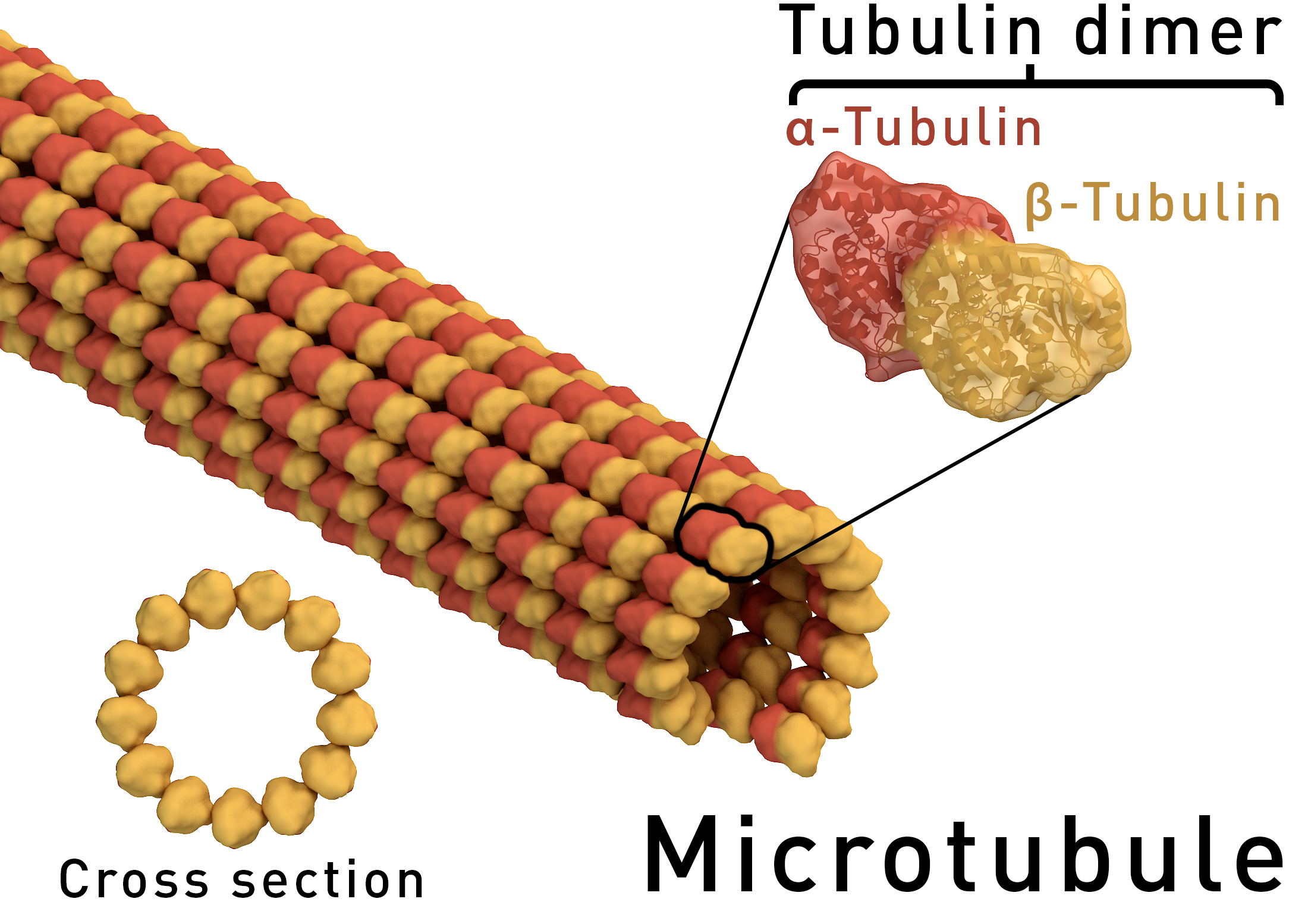
https://upload.wikimedia.org/wikipedia/commons/0/06/Microtubule_structure.png


A latticework of tiny tubes called microtubules gives your cells their shape and also acts like a railroad track that essential proteins travel on. But if there is a glitch in the connection between train and track, diseases can occur. In the November 24, 2015 issue of PNAS, Tatyana Polenova, Ph.D., Professor of Chemistry and Biochemistry, and her team at the University of Delaware (UD), together with John C. Williams, Ph.D., Associate Professor at the Beckman Research Institute of City of Hope in Duarte, California, reveal for the first time — atom by atom — the structure of a protein bound to a microtubule. The protein of focus, CAP-Gly, short for “cytoskeleton-associated protein-glycine-rich domains,” is a component of dynactin, which binds with the motor protein dynein to move cargoes of essential proteins along the microtubule tracks. Mutations in CAP-Gly have been linked to such neurological diseases and disorders as Perry syndrome and distal spinal bulbar muscular dystrophy. The research team used magic-angle-spinning nuclear magnetic resonance spectrometry (NMR) in the Department of Chemistry and Biochemistry at UD to unveil the structure of the CAP-Gly protein assembled on polymerized microtubules. The CAP-Gly protein has 1,329 atoms, and each tubulin dimer, which is a building block for microtubules, has nearly 14,000 atoms. “This is the first time anyone has been able to get an atomic-resolution structure of any microtubule-associated protein assembled on polymerized microtubules,” Dr. Polenova says. “With magic-angle-spinning NMR, we can look into the structure of this and other assemblies of microtubules and their associated proteins and gain critical insights into their function and dynamics, as well as begin to gather clues as to how mutations cause disease.” In magic-angle-spinning NMR, a sample is placed in the NMR’s small, tube-like rotor, which is then spun inside the NMR magnet at an angle of 54.74 degrees — called the “magic angle” because it suppresses the atoms from interacting magnetically. The result is a high-resolution protein fingerprint, a graph of hundreds of peaks representing the frequencies of two or more interacting atoms. These data are then used to calculate the 3-D structures.

The 3-D structures of CAP-Gly, which show the spatial arrangement of atoms in the protein molecule, are different between the free state of the protein and its bound state to the microtubule. These structures reveal how the protein interacts with microtubules, predominantly through its loop regions, which adopt specific conformations upon binding.

However, static structures of CAP-Gly do not tell the whole story about the protein.

“Just as we are always moving our arms and legs about, proteins are very dynamic. They do not stand still,” Dr. Polenova says.

“These motions are essential to their biological function, and NMR spectroscopy is the only technique that can record such movements, with atomic resolution, on a variety of time scales, from picoseconds to arbitrarily long time scales — seconds, days, weeks — to help us understand the protein’s function.”

“We know from our prior studies that CAP-Gly is dynamic on timescales from nano- to milliseconds, and this mobility is essential for the protein’s ability to interact with microtubules and with multiple other binding partners.”

The research, which has been ongoing since 2008 when the first data sets were collected, required the development of new protocols for preparing the samples, new NMR experiments to gather various information on structure and dynamics, and new protocols for data analysis.

In the future, Dr. Polenova and her team envision using NMR in combination with cryo-electron microscopy, in which samples are studied at extremely low temperatures, typically below -200 degrees Fahrenheit, to look at even more complex systems in a highly preserved form.

Dr. Polenova’s research team at UD included Dr. Si Yan, who received her doctorate from the University in 2014, current doctoral student Changmiao Guo, NMR spectroscopist Guangjin Hou, and postdoctoral researchers Dr. Huilan Zhang and Dr. Xingyu Lu. Dr. Williams, at Beckman Research Institute, was also a co-author of the study.


Atomic-resolution structure of the CAP-Gly domain of dynactin on polymeric microtubules determined by magic angle spinning NMR spectroscopy


Fig. 1.

http://www.pnas.org/content/112/47/14611/F1.small.gif


Significance

Microtubules and their associated proteins are central to most cellular functions. They have been extensively studied at multiple levels of resolution; however, significant knowledge gaps remain. Structures of microtubule-associated proteins bound to microtubules are not known at atomic resolution. We used magic angle spinning NMR to solve a structure of dynactin’s cytoskeleton-associated protein glycine-rich (CAP-Gly) domain bound to microtubules and to determine the intermolecular interface, the first example, to our knowledge, of the atomic-resolution structure of a microtubule-associated protein on polymeric microtubules. The results reveal remarkable structural plasticity of CAP-Gly, which enables CAP-Gly’s binding to microtubules and other binding partners. This approach offers atomic-resolution information of microtubule-binding proteins on microtubules and opens up the possibility to study critical parameters such as protonation states, strain, and dynamics on multiple time scales.


Microtubules and their associated proteins perform a broad array of essential physiological functions, including mitosis, polarization and differentiation, cell migration, and vesicle and organelle transport. As such, they have been extensively studied at multiple levels of resolution (e.g., from structural biology to cell biology). Despite these efforts, there remain significant gaps in our knowledge concerning how microtubule-binding proteins bind to microtubules, how dynamics connect different conformational states, and how these interactions and dynamics affect cellular processes. Structures of microtubule-associated proteins assembled on polymeric microtubules are not known at atomic resolution. Here, we report a structure of the cytoskeleton-associated protein glycine-rich (CAP-Gly) domain of dynactin motor on polymeric microtubules, solved by magic angle spinning NMR spectroscopy. We present the intermolecular interface of CAP-Gly with microtubules, derived by recording direct dipolar contacts between CAP-Gly and tubulin using double rotational echo double resonance (dREDOR)-filtered experiments. Our results indicate that the structure adopted by CAP-Gly varies, particularly around its loop regions, permitting its interaction with multiple binding partners and with the microtubules. To our knowledge, this study reports the first atomic-resolution structure of a microtubule-associated protein on polymeric microtubules. Our approach lays the foundation for atomic-resolution structural analysis of other microtubule-associated motors.

Read Full Post »

Protein regulator of HIV replication

Larry H. Bernstein, MD, FCAP, Curator

LPBI


Updated 11/26/2015


Closing the loop on an HIV escape mechanism

University of Delaware

http://www.rdmag.com/news/2015/11/closing-loop-hiv-escape-mechanism


Tatyana Polenova, professor of chemistry and biochemistry at UD (background, left), with her UD research team involved in the HIV study. Next to her is Huilan Zhang. In the foreground, from left, are Guangjin Hou and Manman Lu.

http://www.rdmag.com/sites/rdmag.com/files/newsletter-ads/CHEM-Polenova_Research_Groups-111015-015.jpg

Tatyana Polenova, professor of chemistry and biochemistry at UD (background, left), with her UD research team involved in the HIV study. Next to her is Huilan Zhang. In the foreground, from left, are Guangjin Hou and Manman Lu.


Nearly 37 million people worldwide are living with HIV. When the virus destroys so many immune cells that the body can’t fight off infection, AIDS will develop. The disease took the lives of more than a million people last year.

For the past three and a half years, a team of researchers from six universities, led by the University of Delaware and funded by the National Institutes of Health and the National Science Foundation, has been working to uncover new information about a protein that regulates HIV’s capability to hijack a cell and start replicating. Their findings, reported recently in the Proceedings of the National Academy of Sciences, point to a new avenue for developing potential strategies to thwart the virus.

The team included scientists from UD, the University of Pittsburgh School of Medicine, University of Illinois at Urbana-Champaign, Carnegie Mellon University, the National High Magnetic Field Laboratory at Florida State University and Vanderbilt University School of Medicine. They used a combination of high-tech tools and techniques, including magic-angle-spinning nuclear magnetic resonance (NMR) spectroscopy and computer simulations of molecules, to examine the interactions between HIV and the host-cell protein cyclophilin A (CypA), right down to the movement of individual atoms.

“In a nutshell, we found that the infectivity of HIV is regulated by the motions of these proteins,” says Tatyana Polenova, professor of chemistry and biochemistry at the University of Delaware, who led the study. “It’s a subtle regulation strategy that does not involve major structural changes in the virus.”

Sixty times smaller than a red blood cell, HIV contains a cone-shaped shell, or capsid, made of protein, which surrounds two strands of RNA and the enzymes the virus needs for replication. Like any virus, HIV can only produce copies of itself once it has invaded a host organism. Then it will begin directing certain host cells to begin producing the virus.

But how does HIV invade a cell? In humans, the protein CypA can either promote or inhibit viral infection through interactions with the HIV capsid, although the exact mechanism is not yet known. A portion of the HIV capsid protein, called the CypA loop, is responsible for binding to the CypA in the human host cell. Once this occurs, the virus typically becomes infectious.

However, a change of just one amino acid in the CypA loop can cause the virus to operate opposite from how it does normally, allowing the virus to become non-infectious when CypA is present, and to become infectious when there is no CypA present. Such changes are called “escape mutations,” Polenova says, because they allow the virus to “escape” from its dependence on CypA.

To home in on this escape mechanism, the research team examined assemblies of different variants of HIV capsid protein complexed with CypA. Using magic-angle-spinning NMR, they recorded the motions in these assemblies, atom by atom, on time scales ranging from nanoseconds to milliseconds, from a billionth of a second to a thousandth of a second.

The team found that a reduction in the naturally occurring motions in the binding region due to the mutations allowed the virus to escape from CypA dependence. Magic-angle-spinning NMR experiments provided a direct probe of these motions, recording the changes in the magnetic interactions between nuclei. Computer simulations allowed the team to visualize the motions.

Some portions of the capsid protein do not move at all or move only a little while other portions undergo large-amplitude motions distributed over a wide range of time scales, with the most dynamic region being the CypA loop. Polenova says it is rather surprising that such extensive motions are present in the assembled capsid, and that these dynamics could be detected by both NMR and computer simulations.

“It is the first time that quantitative agreement between experiment and computation was achieved in a dynamics study, and it’s particularly exciting that this was attained for such a complex system,” Polenova says. “We hope this work may guide the development of new therapeutic interventions, such as small molecules that would serve as interactors with the HIV capsid and inhibit these dynamics.”

Polenova says the diverse team of researchers, with expertise in HIV virology, structural biology, biophysics and biochemistry, was critical to the study’s success, along with access to national high-field NMR facilities through the National High Magnetic Field Laboratory. The team was assembled through the NIH-funded Pittsburgh Center for HIV Protein Interactions. Led by Prof. Angela Gronenborn, the center brings together high-caliber scientists and facilities to elucidate the interactions of HIV proteins with host cell factors.


Atomic-resolution structure of the CAP-Gly domain of dynactin on polymeric microtubules determined by magic angle spinning NMR spectroscopy


Significance

Microtubules and their associated proteins are central to most cellular functions. They have been extensively studied at multiple levels of resolution; however, significant knowledge gaps remain. Structures of microtubule-associated proteins bound to microtubules are not known at atomic resolution. We used magic angle spinning NMR to solve a structure of dynactin’s cytoskeleton-associated protein glycine-rich (CAP-Gly) domain bound to microtubules and to determine the intermolecular interface, the first example, to our knowledge, of the atomic-resolution structure of a microtubule-associated protein on polymeric microtubules. The results reveal remarkable structural plasticity of CAP-Gly, which enables CAP-Gly’s binding to microtubules and other binding partners. This approach offers atomic-resolution information of microtubule-binding proteins on microtubules and opens up the possibility to study critical parameters such as protonation states, strain, and dynamics on multiple time scales.


Microtubules and their associated proteins perform a broad array of essential physiological functions, including mitosis, polarization and differentiation, cell migration, and vesicle and organelle transport. As such, they have been extensively studied at multiple levels of resolution (e.g., from structural biology to cell biology). Despite these efforts, there remain significant gaps in our knowledge concerning how microtubule-binding proteins bind to microtubules, how dynamics connect different conformational states, and how these interactions and dynamics affect cellular processes. Structures of microtubule-associated proteins assembled on polymeric microtubules are not known at atomic resolution. Here, we report a structure of the cytoskeleton-associated protein glycine-rich (CAP-Gly) domain of dynactin motor on polymeric microtubules, solved by magic angle spinning NMR spectroscopy. We present the intermolecular interface of CAP-Gly with microtubules, derived by recording direct dipolar contacts between CAP-Gly and tubulin using double rotational echo double resonance (dREDOR)-filtered experiments. Our results indicate that the structure adopted by CAP-Gly varies, particularly around its loop regions, permitting its interaction with multiple binding partners and with the microtubules. To our knowledge, this study reports the first atomic-resolution structure of a microtubule-associated protein on polymeric microtubules. Our approach lays the foundation for atomic-resolution structural analysis of other microtubule-associated motors.


How Viruses Commandeer Human Proteins

http://www.technologynetworks.com/Proteomics/news.aspx?ID=185156


Researchers have produced the first image of an important human protein as it binds with ribonucleic acid (RNA), a discovery that could offer clues to how some viruses, including HIV, control expression of their genetic material.


RNA is one of three macromolecules — along with DNA and proteins — essential to all forms of life. By understanding how hnRNP A1 binds to RNA, the scientists may find ways to jam up components of the replication machinery when the protein is coopted by disease.

The team of scientists reveals the mechanism used by the protein, hnRNP A1 to link to the section of RNA, called the ‘hairpin loop.’

They found that hnRNP A1, a protein essential to cell function and virus replication, has a significantly different structure than its only previously known form: binding to DNA.

“We solved the three-dimensional structure of the protein bound to an RNA hairpin derived from the HIV virus,” said Blanton Tolbert, a chemistry professor at Case Western Reserve. “But because the hairpin loop is found in other viruses and throughout healthy cells, our findings may help explain how the protein connects to the other hairpin targets.”

Tolbert began this research six years ago, frustrated that the only information available was the structure of the protein bound to a synthetic DNA, which isn’t its natural target.

Proteins that bind hairpins sense both the structure and the sequence information presented in the loop. The structure of the DNA complex did not demonstrate the molecular recognition that must take place to bind RNA hairpins.

The process

To discover the structure bound to RNA, the researchers combined three techniques: X-ray crystallography, nuclear magnetic resonance spectroscopy and small angle x-ray scattering. Each technique yielded a piece of the puzzle.

To bind to RNA, hnRNP A1 has two domains, RRM1 and RRM2, which are akin to hands. Scientists already knew both hands are needed to connect to RNA.

But the researchers found that, instead of each domain grabbing a section of the loop, only RRM1 makes contact with the RNA. RRM2 acts as support, helping organize RRM1 into the structure needed to conform to a certain section of the loop.

To confirm that the structures are key to binding, the researchers inserted mutations by changing amino acids on the surface of the domains.

Surprisingly, mutations on the far side of RRM1 — the surface not in contact with the RNA but with the RRM2 — caused decoupling at that site and substantially weakened the affinity for RNA.

Without the normal connection between the two domains, RRM1 fails to adopt the geometric shape that conforms to the RNA hairpin loop.

The researchers are further investigating how the protein transmits the effects of RRM2 to RRM1 and bind. They are also exploring the development of antagonistic agents that would disrupt the interaction of the protein with viruses.


Natural defense protein against HIV discovered

HIV-1, ERManI, antiretroviral, defense protein

Earlier research had shown that it was possible to interfere with HIV spread but the exact molecular mechanisms had not been identified. For the first time, scientists have identified ERManI (Endoplasmic Reticulum Class I α-Mannosidase) as the essential host protein that slows the spread of HIV-1. Scientists investigated how the four ER-associated glycoside hydrolase family 47 (GH47) α-mannosidases, ERManI, and ER-degradation enhancing α-mannosidase-like (EDEM) proteins 1, 2, and 3, are involved in the HIV-1 envelope (Env) degradation process. Ectopic expression of these four α-mannosidases uncovered that only ERManI inhibited HIV-1 Env expression in a dose-dependent manner. Basically, ERManI is a host enzyme that adds sugars to proteins. The Env glycoprotein is targeted to the endoplasmic reticulum-associated protein degradation pathway for degradation after infecting cells. And ERManI was found to interact with the Env and initiate this degradation pathway.

With this discovery, ERManI has the potential as a new antiretroviral treatment option. Currently there is no cure for HIV-1 and once patients are infected, they have it for life. Current antiretroviral therapies can prolong life but cannot fully cure a patient. ERManI is different from current treatments in the sense that it can help the body protect itself.


ERManI (Endoplasmic Reticulum Class I α-Mannosidase) Is Required for HIV-1 Envelope Glycoprotein Degradation via Endoplasmic Reticulum-associated Protein Degradation Pathway (Sep 2015)

ERManI (Endoplasmic Reticulum Class I α-Mannosidase) Is Required for HIV-1 Envelope Glycoprotein Degradation via Endoplasmic Reticulum-associated Protein Degradation Pathway.

Previously, we reported that the mitochondrial translocator protein (TSPO) induces HIV-1 envelope (Env) degradation via the endoplasmic reticulum (ER)-associated protein degradation (ERAD) pathway, but the mechanism was not clear. Here we investigated how the four ER-associated glycoside hydrolase family 47 (GH47) α-mannosidases, ERManI, and ER-degradation enhancing α-mannosidase-like (EDEM) proteins 1, 2, and 3, are involved in the Env degradation process. Ectopic expression of these four α-mannosidases uncovers that only ERManI inhibits HIV-1 Env expression in a dose-dependent manner. In addition, genetic knock-out of the ERManI gene MAN1B1 using CRISPR/Cas9 technology disrupts the TSPO-mediated Env degradation. Biochemical studies show that HIV-1 Env interacts with ERManI, and between the ERManI cytoplasmic, transmembrane, lumenal stem, and lumenal catalytic domains, the catalytic domain plays a critical role in the Env-ERManI interaction. In addition, functional studies show that inactivation of the catalytic sites by site-directed mutagenesis disrupts the ERManI activity. These studies identify ERManI as a critical GH47 α-mannosidase in the ER-associated protein degradation pathway that initiates the Env degradation and suggests that its catalytic domain and enzymatic activity play an important role in this process.


T cell editing using CRISPR/Cas9 could revolutionize HIV therapeutics
September 15, 2015   

T cell therapy, HIV

Reinforcing the immune system by engineering lymphocytes to target and destroy viruses has the potential to be an effective therapy for many diseases. One potential approach to this strategy is to alter the genome of lymphocytes so that proteins that are typically hijacked by viruses are no longer present. While conceptually feasible, editing T cells has been challenging in practice; however, with the advent of mammalian cell editing using CRISPR/Cas9, T-cell editing is closer to becoming a reality.

How can CRISPR/Cas9 bring us closer to finding a cure for HIV?

In a study recently published in PNAS, scientists have optimized a protocol to introduce nucleotide replacements that would inhibit CXCR4 expression. The authors streamlined the CRISPR/Cas9 editing process by electroporating Cas9 ribonucleoproteins (RNPs) into CD4+ T cells. The RNPs, consisting of both a recombinant Cas9 enzyme and guide RNA, vastly improved editing efficiency, ultimately promoting knock-out of the CXCR4 cell-surface receptor. Taken together, these result suggest the potential of a new cell therapy approach for the fight against HIV.

Generation of knock-in primary human T cells using Cas9 ribonucleoproteins
Kathrin Schumann a , b , 1 , Steven Lin c , 1 , Eric Boyer a , b , Dimitre R. Simeonov a , b , d , Meena Subramaniam e , f , Rachel E. Gate e , f , et al.  PNAS. 2015; 112(33): 10437-10442. http://dx.doi.org:/10.1073/pnas.1512503112

Significance

T-cell genome engineering holds great promise for cancer immunotherapies and cell-based therapies for HIV, primary immune deficiencies, and autoimmune diseases, but genetic manipulation of human T cells has been inefficient. We achieved efficient genome editing by delivering Cas9 protein pre-assembled with guide RNAs. These active Cas9 ribonucleoproteins (RNPs) enabled successful Cas9-mediated homology-directed repair in primary human T cells. Cas9 RNPs provide a programmable tool to replace specific nucleotide sequences in the genome of mature immune cells—a longstanding goal in the field. These studies establish Cas9 RNP technology for diverse experimental and therapeutic genome engineering applications in primary human T cells.


T-cell genome engineering holds great promise for cell-based therapies for cancer, HIV, primary immune deficiencies, and autoimmune diseases, but genetic manipulation of human T cells has been challenging. Improved tools are needed to efficiently “knock out” genes and “knock in” targeted genome modifications to modulate T-cell function and correct disease-associated mutations. CRISPR/Cas9 technology is facilitating genome engineering in many cell types, but in human T cells its efficiency has been limited and it has not yet proven useful for targeted nucleotide replacements. Here we report efficient genome engineering in human CD4+ T cells using Cas9:single-guide RNA ribonucleoproteins (Cas9 RNPs). Cas9 RNPs allowed ablation of CXCR4, a coreceptor for HIV entry. Cas9 RNP electroporation caused up to ∼40% of cells to lose high-level cell-surface expression of CXCR4, and edited cells could be enriched by sorting based on low CXCR4 expression. Importantly, Cas9 RNPs paired with homology-directed repair template oligonucleotides generated a high frequency of targeted genome modifications in primary T cells. Targeted nucleotide replacement was achieved in CXCR4 and PD-1 (PDCD1), a regulator of T-cell exhaustion that is a validated target for tumor immunotherapy. Deep sequencing of a target site confirmed that Cas9 RNPs generated knock-in genome modifications with up to ∼20% efficiency, which accounted for up to approximately one-third of total editing events. These results establish Cas9 RNP technology for diverse experimental and therapeutic genome engineering applications in primary human T cells.

Read Full Post »

Muscular dystrophy has deficient stem cell dystrophin

Larry H. Bernstein, MD, FCAP, Curator

LPBI

Article ID #198: Muscular dystrophy has deficient stem cell dystrophin. Published on 11/21/2015

WordCloud Image Produced by Adam Tubman

Dystrophin Deficient Stem Cell Pathology

Muscular Dystrophy is a Stem Cell-Based Disease

Because DMD results from mutations in the dystrophin gene, the vast majority of muscular dystrophy research was based on a simple model in which the Dystrophin protein played a structural role in the structural integrity of muscle fibers. Abnormal versions of the Dystrophin protein caused the muscle fibers to become damaged and die as a result of contraction.  Dystrophin anchors the cytoskeleton of the muscle fibers, which are essential for muscle contraction, to the muscle cell membrane, and then to the extracellular matrix outside the cell that serves as a foundation upon which the muscle cells are built.

gb-2001-2-4-reviews3006-3

However in this current study, Rudnicki and his team discovered that muscle stem cells also express the dystrophin protein. This is a revelation because Dystrophin was thought to be protein that ONLY appeared in mature muscle. However, in this study, it became exceedingly clear that in the absence of Dystrophin, muscle stem cells generated ten-fold fewer muscle precursor cells, and, consequently, far fewer functional muscle fibers. Dystrophin is also a component of a signal transduction pathway that allows muscle stem cells to properly ascertain if they need to replace dead or dying muscle.  Muscle stem cells repair the muscle in response to injury or exercise by dividing to generate precursor cells that differentiate into muscle fibers.

Even though Rudnicki used mice as a model system in these experiments, the Dystrophin protein is highly conserved in most vertebrate animals. Therefore, it is highly likely that these results will also apply to human muscle stem cells.

Gene therapy experiments and trials are in progress and even show some promise, but Rudnicki’s work tells us that gene therapy approaches must target muscle stem cells as well as muscle fibers if they are to work properly.

“We’re already looking at approaches to correct this problem in muscle stem cells,” said Dr. Rudnicki.

This paper has received high praise from the likes of Ronald Worton, who was one of the co-discovers of the dystrophin gene with Louis Kunkel in 1987.

Early pathogenesis of Duchenne muscular dystrophy modelled in patient-derived human induced pluripotent stem cells

Emi Shoji, Hidetoshi Sakurai, Tokiko Nishino, Tatsutoshi Nakahata, Toshio Heike, Tomonari Awaya, Nobuharu Fujii, Yasuko Manabe, Masafumi Matsuo & Atsuko Sehara-Fujisawa

Scientific Reports 5, Article number: 12831 (2015)   http://dx.doi.org:/10.1038/srep12831

Duchenne muscular dystrophy (DMD) is a progressive and fatal muscle degenerating disease caused by a dystrophin deficiency. Effective suppression of the primary pathology observed in DMD is critical for treatment. Patient-derived human induced pluripotent stem cells (hiPSCs) are a promising tool for drug discovery. Here, we report an in vitro evaluation system for a DMD therapy using hiPSCs that recapitulate the primary pathology and can be used for DMD drug screening. Skeletal myotubes generated from hiPSCs are intact, which allows them to be used to model the initial pathology of DMD in vitro. Induced control and DMD myotubes were morphologically and physiologically comparable. However, electric stimulation of these myotubes for in vitro contraction caused pronounced calcium ion (Ca2+) influx only in DMD myocytes. Restoration of dystrophin by the exon-skipping technique suppressed this Ca2+ overflow and reduced the secretion of creatine kinase (CK) in DMD myotubes. These results suggest that the early pathogenesis of DMD can be effectively modelled in skeletal myotubes induced from patient-derived iPSCs, thereby enabling the development and evaluation of novel drugs.

Duchenne muscular dystrophy (DMD) is characterised by progressive muscle atrophy and weakness that eventually leads to ambulatory and respiratory deficiency from early childhood1. It is an X-linked recessive inherited disease with a relatively high frequency of 1 in 3500 males1,2.DMD, which is responsible for DMD, encodes 79 exons and produces dystrophin, which is one of the largest known cytoskeletal structural proteins3. Most DMD patients have various types of deletions or mutations in DMD that create premature terminations, resulting in a loss of protein expression4. Several promising approaches could be used to treat this devastating disease, such as mutation-specific drug exon-skipping5,6, cell therapy7, and gene therapy1,2.

Myoblasts from patients are the most common cell sources for assessing the disease phenotypes of DMD11,12. …Previous reports have shown that muscle cell differentiation from DMD patient myoblasts is delayed and that these cells have poor proliferation capacity compared to those of healthy individuals11,12. Our study revealed that control and DMD myoblasts obtained by activating tetracycline-dependent MyoD transfected into iPS cells (iPStet-MyoD cells) have comparable growth and differentiation potential and can produce a large number of intact and homogeneous myotubes repeatedly.

The pathogenesis of DMD is initiated and progresses with muscle contraction. The degree of muscle cell damage at the early stage of DMD can be evaluated by measuring the leakage of creatine kinase (CK) into the extracellular space15. Excess calcium ion (Ca2+) influx into skeletal muscle cells, together with increased susceptibility to plasma membrane injury, is regarded as the initial trigger of muscle damage in DMD19,20,21,22,23,24. Targeting these early pathogenic events is considered essential for developing therapeutics for DMD.

In this study, we established a novel evaluation system to analyse the cellular basis of early DMD pathogenesis by comparing DMD myotubes with the same clone but with truncated dystrophin-expressing DMD myotubes, using the exon-skipping technique. We demonstrated through in vitro contraction that excessive Ca2+ influx is one of the earliest events to occur in intact dystrophin-deficient muscle leading to extracellular leakage of CK in DMD myotubes.

Generation of tetracycline-inducible MyoD-transfected DMD patient-derived iPSCs (iPStet-MyoD cells)

Figure 1: Generation and characterization of control and DMD patient-derived Tet-MyoD-transfected hiPS cells.   Full size image

Morphologically and physiologically comparable intact myotubes differentiated from control and DMD-derived hiPSCs

Figure 2: Morphologically and physiologically comparable skeletal muscle cells differentiated from Control-iPStet-MyoD and DMD-iPStet-MyoD.   Full size image

Exon-skipping with AO88 restored expression of Dystrophin in DMD myotubes differentiated from DMD-iPStet-MyoD cells


Figure 3: Restoration of dystrophin protein expression by AO88.   Full size image

 
Restored dystrophin expression attenuates Ca2+ overflow in DMD-Myocytes


Figure 4: Restored expression of dystrophin diminishes Ca2+ influx in DMD muscle in response to electric stimulation.   Full size image


Ca2+ influx provokes skeletal muscle cellular damage in DMD muscle


Figure 5: Ca2+ influx induces prominent skeletal muscle cellular damage in DMD-Myocytes.   Full size image


Skeletal muscle differentiation in myoblasts from DMD patients is generally delayed compared to that in healthy individuals11,36,37.  Our differentiation system successfully induced the formation of myotubes from DMD patients, and the myotubes displayed analogous morphology and maturity compared with control myotubes (Fig. 2a–c).  Comparing myotubes generated from patient-derived iPS cells with those derived from the same DMD clones but expressing dystrophin by application of the exon-skipping technique enabled us to demonstrate the primary cellular phenotypes in skeletal muscle solely resulting from the loss of the dystrophin protein (Fig. 4b).  Our results demonstrate that truncated but functional dystrophin protein expression improved the cellular phenotype of DMD myotubes.

In DMD, the lack of dystrophin induces an excess influx of Ca2+ , leading to pathological dystrophic changes22. We consistently observed excess Ca2+ influx in DMD-Myocytes compared to Control-Myocytes (Supplementary Figure S3a and S3b) in response to electric stimulation. TRP channels, which are mechanical stimuli-activated Ca2+ channels40that are expressed in skeletal muscle cells41, can account for this pathogenic Ca2+ influx…

In conclusion, our study revealed that the absence of dystrophin protein induces skeletal muscle damage by allowing excess Ca2+ influx in DMD myotubes. Our experimental system recapitulated the early phase of DMD pathology as demonstrated by visualisation and quantification of Ca2+ influx using intact myotubes differentiated from hiPS cells.  This evaluation system significantly expands prospective applications with regard to assessing the effectiveness of exon-skipping drugs and also enables the discovery of drugs that regulate the initial events in DMD.

Duchenne muscular dystrophy affects stem cells, University of Ottawa study finds  

New treatments could one day be available for the most common form of muscular dystrophy after a study suggests the debilitating genetic disease affects the stem cells that produce healthy muscle fibres.

The findings are based on research from the University of Ottawa and The Ottawa Hospital, published Monday in the journal Nature Medicine.

For nearly two decades, doctors had thought the muscular weakness that is the hallmark of the disease was due to problems with human muscle fibers, said Dr. Michael Rudnicki, the study’s senior author.

The new research shows the specific protein characterized by its absence in Duchenne muscular dystrophy normally exists in stem cells.

Dystrophin protein found in stem cells

“The prevailing notion was that the protein that’s missing in Duchenne muscular dystrophy — a protein called dystrophin — was not involved at all in the function of the stem cells.”

http://soundcloud.com/cbcottawa1

When the genetic mutations caused by Duchenne muscular dystrophy inhibit the production of dystrophin in stem cells, those stem cells produce significantly fewer precursor cells — and thus fewer properly functioning muscle fibres.  Further, stem cells need dystrophin to sense their environment to figure out if they need to divide to produce more stem cells or perform muscle repair work.

Genetic repair might treat Duchenne muscular dystrophy

July 25, 2011|By Thomas H. Maugh II, Los Angeles Times
 

A genetic technique that allows the body to work around a crucial mutation that causes Duchenne muscular dystrophy increased the mass and function of muscles in a small group of patients with the devastating disease, paving the way for larger clinical trials of the drug. The study in a handful of boys age 5 to 15 showed that patients receiving the highest level of the drug, called AVI-4658 or eteplirsen, had a significant increase in production of a missing protein and increases in muscle fibers. The study demonstrated that the drug is safe in the short term. Results were reported Sunday in the journal Lancet.

Duchenne muscular dystrophy affects about one in every 3,500 males worldwide. It is caused by any one of several different mutations that affect production of a protein called dystrophin, which is important for the production and maintenance of muscle fibers. Affected patients become unable to walk and must use a wheelchair by age 8 to 12. Deterioration continues through their teens and 20s, and the condition typically proves fatal as muscle failure impairs their ability to breathe.

This study is designed to assess the efficacy, safety, tolerability, and pharmacokinetics (PK) of AVI-4658 (eteplirsen) in both 50.0 mg/kg and 30.0 mg/kg doses administered over 24 weeks in subjects diagnosed with Duchenne muscular dystrophy (DMD).


Condition Intervention Phase
Duchenne Muscular Dystrophy Drug: AVI-4658 (Eteplirsen)
Other: Placebo
Phase 2
Study Type:Interventional
Study Design:Allocation: Randomized
Endpoint Classification: Safety/Efficacy Study
Intervention Model: Parallel Assignment
Masking: Double Blind (Subject, Caregiver, Investigator, Outcomes Assessor)
Primary Purpose: Treatment
Official Title:A Randomized, Double-Blind, Placebo-Controlled, Multiple Dose Efficacy, Safety, Tolerability and Pharmacokinetics Study of AVI-4658(Eteplirsen),in the Treatment of Ambulant Subjects With Duchenne Muscular Dystrophy
 
 
Resource links provided by NLM:
 
 
Dystrophin expression in muscle stem cells regulates their polarity and asymmetric division

Nature Medicine(2015)   http://dx.doi.org:/10.1038/nm.3990

Dystrophin is expressed in differentiated myofibers, in which it is required for sarcolemmal integrity, and loss-of-function mutations in the gene that encodes it result in Duchenne muscular dystrophy (DMD), a disease characterized by progressive and severe skeletal muscle degeneration. Here we found that dystrophin is also highly expressed in activated muscle stem cells (also known as satellite cells), in which it associates with the serine-threonine kinase Mark2 (also known as Par1b), an important regulator of cell polarity. In the absence of dystrophin, expression of Mark2 protein is downregulated, resulting in the inability to localize the cell polarity regulator Pard3 to the opposite side of the cell. Consequently, the number of asymmetric divisions is strikingly reduced in dystrophin-deficient satellite cells, which also display a loss of polarity, abnormal division patterns (including centrosome amplification), impaired mitotic spindle orientation and prolonged cell divisions. Altogether, these intrinsic defects strongly reduce the generation of myogenic progenitors that are needed for proper muscle regeneration. Therefore, we conclude that dystrophin has an essential role in the regulation of satellite cell polarity and asymmetric division. Our findings indicate that muscle wasting in DMD not only is caused by myofiber fragility, but also is exacerbated by impaired regeneration owing to intrinsic satellite cell dysfunction.

Read Full Post »

Telomere length

Larry H. Bernstein, MD, FCAP, Curator

LPBI


New Enzyme Discovered for Sustaining Telomere Length

http://www.genengnews.com/gen-news-highlights/new-enzyme-discovered-for-sustaining-telomere-length/81251981/


http://www.genengnews.com/Media/images/GENHighlight/Picture18124516425.jpg

Researchers developed a novel assay to identify telomere length regulators and showed that ATM inhibition shortens telomeres, whereas ATM activation elongates telomeres. [Lee et al., 2015, Cell Reports 13, 1–10]


In the early years of molecular biology research, scientists studying chromosomal structure and composition noticed that the terminal ends of chromosomes, called telomeres, would gradually become shorter with each successive round of cellular replication. This process would continue until the chromosome reached a certain length, ultimately becoming unstable and causing the cell to die. Conversely, the scientists noticed that for certain genetic disorders, such as cancer, an abnormally long telomere length led to genome anomalies that were closely associated with the cancer phenotype.

In 1984, researchers Elizabeth Blackburn, Ph.D., and Carol Greider, Ph.D., who was at the time a graduate student in Dr. Blackburn’s laboratory, discovered the telomerase enzyme, which was responsible for maintaining the appropriate length of telomerase after chromosomal replication. Drs. Blackburn and Greider would go on to be awarded the 2009 Nobel Prize in Physiology and Medicine, along with Jack Szostak, Ph.D. for their work on molecular mechanisms of the telomerase enzyme.

Yet, even during their seminal work, the investigators quickly realized that other molecules besides telomerase must be involved in maintaining the protective caps at the end of chromosomes. Now, researchers at Johns Hopkins report uncovering the role of an additional enzyme crucial to telomere length and say the novel method they could be used to speed discovery of other proteins and processes that are involved in telomere stability.

“We’ve known for a long time that telomerase doesn’t tell the whole story of why chromosomes’ telomeres are a given length, but with the tools we had, it was difficult to figure out which proteins were responsible for getting telomerase to do its work,” explained Dr. Greider, professor, and director of molecular biology and genetics in the Johns Hopkins Institute for Basic Biomedical Sciences.

The findings from this study were published recently in Cell Reports through an article entitled “ATM Kinase Is Required for Telomere Elongation in Mouse and Human Cells.”

Understanding the mechanisms that are needed to lengthen telomeres has broad health implications, since shortened telomeres have been implicated in aging and diseases as diverse as lung and bone marrow disorders, while overly long telomeres are linked to cancer. Cells need a well-tuned process to keep adding the right number of building blocks back onto telomeres over an organism’s lifetime.

Unfortunately, until recently, the methods researchers used to study telomere length were extremely time-consuming, often taking months of work to study cells grown in vitro, searching for detectable differences in telomere length. However, Dr. Greider’s team developed a new tool for measuring telomere length in yeast. The idea was to artificially cut mammalian cells’ telomeres and then detect elongation by telomerase—a test that would take less than a day, and could be performed even if the blocked proteins were needed for cells to divide.

The new test, dubbed addition of de novo initiated telomeres (ADDIT) was used to observe an enzyme long suspected to be involved in telomere maintenance, ATM kinase. “ATM kinase was known to be involved in DNA repair, but there were conflicting reports about whether it had a role in telomere lengthening,” noted Dr. Greider.

The Hopkins researchers blocked the enzyme in lab-grown mouse cells and used ADDIT to find that it was indeed needed to lengthen telomeres. They confirmed their result by using the old, three-month-long telomere test, which lead to the same outcome.

Additionally, the team also found that in normal mouse cells, a drug that blocks an enzyme called PARP1 would activate ATM kinase and spur telomere lengthening. This finding has the potential to impact drug-based telomere elongation for treating short-telomere diseases, such as bone marrow failure.

Dr. Greider and her team were excited by their findings and plan to use ADDIT to find out more about the telomere-lengthening biochemical pathway that ATM kinase participates.

“The potential applications are very exciting,” stated lead author Stella Lee, Ph.D., postdoctoral fellow in Dr. Greider’s laboratory. “Ultimately ADDIT can help us understand how cells strike a balance between aging and the uncontrolled cell growth of cancer, which is very intriguing.”


Nobel Laureate Blackburn Named Salk Institute’s New President


Elizabeth H. Blackburn, Ph.D., a 2009 Nobel laureate who has specialized in telomere and telomerase research, was named the new president of the Salk Institute for Biological Studies today, effective January 1, 2016.

“The Salk is full of absolutely terrific people and brimming with great science. Building on its distinguished history and current success, I am delighted to be playing a role in continuing and growing its major contributions to science and health research,” Dr. Blackburn said in a statement. “I am truly honored to be asked to be the next president of the Salk Institute.”

Dr. Blackburn has been a non-resident fellow at the institute since 2001, where she has been one of a group of investigators that advise the institute’s leadership and play key decision-making roles in appointing and promoting Salk professors. In addition, she has been the Morris Herzstein Professor of Biology and Physiology in the department of biochemistry and biophysics at the University of California San Francisco (UCSF).

In 2009, she was named one of three co-winners of the Nobel Prize in Physiology or Medicine “for the discovery of how chromosomes are protected by telomeres and the enzyme telomerase.” Dr. Blackburn discovered the molecular nature of telomeres—the ends of eukaryotic chromosomes that serve as protective caps essential for preserving the genetic information—and co-discovered the ribonucleoprotein enzyme telomerase.

Those discoveries helped launch new research around telomeres and telomerase, both believed to play central roles in aging and diseases that include cancer.

In addition to the Nobel Prize, Dr. Blackburn has received nearly every major award in science, including the Lasker, Gruber, and Gairdner prizes. In 2007, she was named to the TIME 100 yearly list of the world’s most influential people. Dr. Blackburn is also a member of the National Academy of Sciences, the National Academy of Medicine, and the Royal Society of London.

Dr. Blackburn was born in Hobart, Tasmania, Australia, to a family of doctors and scientists. Her parents were both family physicians, while her grandfather and great-grandfather were geologists.

Inspired by her fascination with animals and a biography of Marie Curie, Dr. Blackburn chose to also pursue a career in science. She earned her B.Sc. degree in 1970 and her M.Sc. degree in biochemistry, both from the University of Melbourne. She earned her Ph.D. in molecular biology from the University of Cambridge in 1975, then conducted postdoctoral research in molecular and cellular biology at Yale University from 1975 to 1977.


ATM Kinase Is Required for Telomere Elongation in Mouse and Human Cells

Stella Suyong Lee, Craig Bohrson, Alexandra Mims Pike, Sarah Jo Wheelan, Carol Widney Greider
  • •ADDIT assay measures telomerase-mediated addition at a single telomere
  • •De novo telomere addition in mouse cells requires ATM kinase
  • •ATM inhibition blocks bulk telomere elongation in both mouse and human cells
  • •Excess activation of ATM by inhibition of PARP1 increases telomere addition

Short telomeres induce a DNA damage response, senescence, and apoptosis, thus maintaining telomere length equilibrium is essential for cell viability. Telomerase addition of telomere repeats is tightly regulated in cells. To probe pathways that regulate telomere addition, we developed the ADDIT assay to measure new telomere addition at a single telomere in vivo. Sequence analysis showed telomerase-specific addition of repeats onto a new telomere occurred in just 48 hr. Using the ADDIT assay, we found that ATM is required for addition of new repeats onto telomeres in mouse cells. Evaluation of bulk telomeres, in both human and mouse cells, showed that blocking ATM inhibited telomere elongation. Finally, the activation of ATM through the inhibition of PARP1 resulted in increased telomere elongation, supporting the central role of the ATM pathway in regulating telomere addition. Understanding this role of ATM may yield new areas for possible therapeutic intervention in telomere-mediated disease.


Vertebrate telomeres are repetitive TTAGGG DNA sequences located at the ends of chromosomes, which protect the coding regions of DNA. In mammalian germline cells and ∼85% of cancers, telomere length is maintained by the dimeric ribonucleoprotein telomerase, which catalyzes the addition of TTAGGG repeats to counteract telomere shortening and cellular senescence (Shay and Bacchetti, 1997, Kim et al., 1994, Wenz et al., 2001). The minimal catalytic core of human telomerase consists of the telomerase reverse transcriptase protein (hTERT), telomerase RNA (hTR), and the protein dyskerin (Cohen et al., 2007).

The differentiation of telomeres from broken chromosome ends is conferred by a family of six telomere-specific binding proteins collectively termed “shelterin” (de Lange, 2005). This complex consists of the double-stranded binding proteins TRF1 and TRF2, the single-stranded binding proteins POT1 and TPP1, the bridging protein TIN2 that links these two groups of proteins, and Rap1 (reviewed in Palm and de Lange, 2008). TRF1 protects the telomere and negatively regulates telomerase-mediated telomere lengthening (van Steensel and de Lange, 1997, Smogorzewska et al., 2000, Ancelin et al., 2002, Karlseder et al., 2002). TRF1 also facilitates the progression of the replication machinery; deletion of TRF1 increases replication fork stalling, leading to ATR kinase activation and a “fragile telomere” phenotype (Sfeir et al., 2009, Martínez et al., 2009). The TRF1-mediated repression of the ATR response requires recruitment of the shelterin components TIN2 and the TPP1/POT1 heterodimer (Zimmermann et al., 2014).

TPP1 and POT1 also have roles in mediating telomere-length regulation. A surface on the N-terminal oligonucleotide/oligosaccharide-binding (OB) domain of TPP1 termed the TEL patch activates telomerase by stimulating telomerase processivity and providing a direct binding site for telomerase recruitment to telomeres; mutation of the TEL patch can lead to telomere shortening syndromes characterized by bone marrow failure (Abreu et al., 2010, Nandakumar et al., 2012, Zhong et al., 2012, Kocak et al., 2014, Guo et al., 2014, Dalby et al., 2015). Additionally, mutation analyses at sites independent of the TEL patch have implicated TPP1 as part of a telomere-length-dependent feedback loop that regulates telomere-length homeostasis (Sexton et al., 2014). A mutant form of POT1 that abrogates binding to single-stranded DNA (POT1ΔOB) deregulated telomere-length control (Loayza and De Lange, 2003), indicating that the DNA-binding capability of POT1 is vital as a negative regulator of telomere length. The impact of human POT1 on telomere length is complex, since both depletion and overexpression of POT1 lead to telomere lengthening (Ye et al., 2004, Veldman et al., 2004, Colgin et al., 2003, Armbruster et al., 2004). POT1 function as a positive or negative regulator of telomerase activity at the telomere depends on its position of binding relative to the DNA 3′ end and is also modulated by its binding partner, TPP1 (Zaug et al., 2005, Wang et al., 2007, Lei et al., 2005, Kelleher et al., 2005).

Telomerase action at the telomere is highly regulated; it preferentially elongates the shortest telomeres, and recruitment of the enzyme complex to the telomere occurs in mid-S phase of the cell cycle (Bianchi and Shore, 2007, Britt-Compton et al., 2009,Teixeira et al., 2004, Hemann et al., 2001, Tomlinson et al., 2006, Jády et al., 2006). In both budding and fission yeasts, the preference of telomerase to extend the shortest telomeres requires the activity of Tel1, the yeast homolog of human ATM (Sabourin et al., 2007, Hector et al., 2007, Arnerić and Lingner, 2007). ATM and ATR are kinases within the phosphatidylinositol-3 kinase-related kinase (PIKK) family, which regulates cellular responses to DNA damage, mRNA decay, and nutrient-dependent signaling (Lovejoy and Cortez, 2009). Activation of these DNA damage pathways is dampened at telomeres; in mammalian cells, TRF2 represses activation of ATM while POT1 represses ATR (Karlseder et al., 2004, Celli and de Lange, 2005, Denchi and de Lange, 2007, Guo et al., 2007, Okamoto et al., 2013). Nevertheless, there is a large amount of evidence that their yeast homologs play a positive role in facilitating telomere extension by telomerase (Moser et al., 2009, Moser et al., 2011, Yamazaki et al., 2012, Churikov et al., 2013).

It is not known whether the role of the ATM and ATR pathways in recruiting telomerase is conserved in mammals. Although ATM deficiency or ATR mutations can induce telomere shortening or instability in human and mouse cells (Metcalfe et al., 1996, Smilenov et al., 1997, Wong et al., 2003, Wu et al., 2007, Pennarun et al., 2010), these kinases were reported to be dispensable for elongation of the shortest telomeres in mouse models (Feldser et al., 2006, McNees et al., 2010). Also, immortalized cell lines from human patients with ATM mutations are able to maintain their telomeres with telomerase, albeit at short lengths (Sprung et al., 1997). Nonetheless, there is evidence that TRF1-mediated telomere-length regulation in human cells involves ATM. Inhibition of human ATM resulted in increased TRF1 at the telomere, and phosphorylation of TRF1 on serine 367, an ATM/ATR target site, reduced the interaction of TRF1 with telomeres and abrogated its ability to inhibit telomere lengthening (McKerlie et al., 2012, Wu et al., 2007).

In this study, we report that both ATM and ATR are required for the recruitment of human telomerase to telomeres.

ATM and ATR Are Both Required for the Presence of Human Telomerase at Telomeres

Figure 1

ATM and ATR Are Both Required for the Presence of Human Telomerase at Telomeres

(A) Representative images of hTR/telomere FISH in 293T cells treated with the indicated siRNAs or kinase inhibitors. Cells were synchronized to mid-S phase of the cell cycle and probed with hTR probes (green) or a telomere probe (red). Co-localizations are indicated by white arrows in the merge row. Scale bar, 10 μm.

(B) Immunoblot of 293T cells with either ATM (left panel) or ATR (right panel) siRNA-mediated knockdown, using the respective antibodies, with vinculin as a control.

(C) Average co-localizations between telomerase and telomeres in unsynchronized 293T cells treated with control siRNA (gray), ATM siRNA (red) (∗p = 0.012), or ATR siRNA (purple) (∗∗p = 0.0095).

(D) Quantitation of decrease in telomerase recruitment in S phase synchronized 293T cells following treatment with two independent ATM and ATR siRNAs; ∗∗p < 0.01. Cells were synchronized with a thymidine/aphidicolin block (ATM) or sorted into cell-cycle phases by FACS based on DNA content (ATR).

(E) Average telomerase co-localization with telomeres in S phase synchronized 293T cells after treatment with DMSO vehicle (gray), 1.5 μM KU-55933 (light yellow), or 500 nM VE-822 (dark yellow); ∗p < 0.05.

(F) Telomerase co-localization with telomeres in 293T cells at the indicated number of hours after release from a thymidine/aphidicolin block, treated with control (gray) or ATM (red) siRNA.

(G) Telomerase co-localization with telomeres in 293T cells treated with control (gray) or ATM (red) siRNA. Cells were stained with the DNA dye VyBrant DyeCycle Violet and isolated into cell-cycle phases with FACS. Enrichment of cells in the indicated phases was confirmed by flow cytometry of sorted cells (Figure S1A).

(H) Telomerase co-localization with telomeres in 293T cells treated with control (gray) or ATR (purple) siRNA and sorted by FACS as in Figure 1G.

In all panels, data are presented as the mean of three independent experiments ± SD.

See also Figure S1.

ATM Effect on Telomerase Recruitment Is Partially Mediated by TRF1

Figure 2

ATM Effect on Telomerase Recruitment Is Partially Mediated by TRF1

(A) Immunoblot of 293T cells treated with control or two different TRF1 siRNAs. All lanes are from the same immunoblot, which was cropped to remove intervening irrelevant lanes. Actin was probed as a control.

(B) Average recruitment of telomerase to telomeres in asynchronous 293T cells treated with control (gray) or TRF1 (blue) siRNA;∗∗p = 0.0011.

(C) Average co-localization of telomerase with telomeres in 293T cells synchronized to G2/M of the cell cycle by release from a thymidine/aphidicolin block; cells were treated with control (gray) or two different TRF1 siRNAs (blue); ∗p < 0.05.

(D) Telomerase co-localization with telomeres in thymidine/aphidicolin synchronized 293T cells, treated with control (gray) or TRF1 (blue) siRNA. The values along the x axis represent the number of hours since release of cells from G1/S boundary. The control data are the same as those in Figure 1F, since these experiments were performed simultaneously.

(E) Telomerase co-localization with telomeres in 293T cells, treated with control (gray) or TRF1 (blue) siRNA; cells were stained with the DNA dye VyBrant DyeCycle Violet and isolated into the cell-cycle phases with FACS. The control data are the same as those in Figure 1G, since these experiments were performed simultaneously.

(F) FISH for hTR (green) and telomeres (red) in 293T cells treated with control or combined ATM and TRF1 siRNAs. Cells were either asynchronous or synchronized with thymidine and aphidicolin and harvested 3–4 hr (S phase) or 7 hr (G2/M) after release from the G1/S boundary. Co-localizations are indicated by the white arrows in the merge row. Scale bar, 10 μm.

(G) Quantitation of (F); average telomerase-telomere co-localizations after control (gray), ATM only (red), TRF1 only (blue), and ATM/TRF1 (green) siRNAs, in asynchronous, S phase, or G2/M phase cell populations; n = 4; ∗∗p < 0.01.

Data are presented as the mean of three independent experiments except where indicated otherwise, ± SD.

See also Figure S2.

Large image of Figure 7.

http://www.cell.com/cms/attachment/2040375994/2053862726/gr7.jpg

Figure 7

Model for ATM and ATR Involvement in Human Telomerase Recruitment to Telomeres

(A) During S phase, ATM and/or other PIKKs phosphorylates TRF1 at S367, which leads to partial dissociation of TRF1 from telomeres (McKerlie et al., 2012, Wu et al., 2007). Depletion of TRF1, together with its protein partners Tin2, TPP1, and Pot1, causes telomeric replication forks to stall, leading to recruitment of RPA and ATR (Sfeir et al., 2009, Martínez et al., 2009, Zimmermann et al., 2014). ATR phosphorylates an unknown substrate to mediate telomerase recruitment. Replication fork stalling caused by aphidicolin treatment also leads to telomerase recruitment (this study) and telomere elongation (Sfeir et al., 2009).

(B) An independent role of ATM and ATR involves stimulation of telomerase assembly, which is a prerequisite for telomerase localization to telomeres. This model does not preclude involvement of other unidentified substrates of ATM, ATR, and other PIKKs.

……..

We demonstrated that one TRF1-independent function of ATM is its impact upon the ability of hTR and hTERT to assemble into a functional enzyme complex (Figure 6), which is a prerequisite for localization of hTR to telomeres (Tomlinson et al., 2008). ATR also plays a role in assembly of human telomerase; we do not know if the substrates of these two kinases in this process are the same. This role is reflected in a substantial decrease in the amount of hTR recovered after hTERT immunoprecipitation and in the total immunoprecipitated telomerase activity following ATM and ATR knockdown. The specific activity of telomerase remains unchanged, demonstrating that both ATM and ATR have no effect on telomerase catalytic activity, consistent with results in yeast (Chan et al., 2001). No consensus PIKK phosphorylation motifs exist in the RNA-binding domain of hTERT, implying either that ATM or ATR can mediate telomerase assembly by targeting regions not in the RNA-binding domain or that they can regulate telomerase assembly by phosphorylating unknown substrates (Figure 7B).

Our data support a model incorporating multiple roles for ATM and ATR in the presence of human telomerase at telomeres (Figure 7). One pathway involving both ATM and ATR is mediated by phosphorylation of TRF1 and its removal from the telomere, leading to replication fork stalling in telomeric DNA, which acts as a trigger for telomerase recruitment. A second pathway involves the role of ATM and ATR in facilitating telomerase assembly; additional phosphorylation targets of ATM, ATR, and other PIKKs in the telomerase recruitment process may remain to be identified. These data reveal that although it is important for telomeres to repressDNA damage signaling in order to avoid deleterious fusions, telomeres have also evolved the ability to carefully exploit aspects of DNA damage signaling pathways to regulate telomerase presence at the telomere. Increased understanding of regulation of telomerase assembly and access to the telomere may provide valuable insight in the process of developing highly specific cancer therapeutics.

Read Full Post »

Biochemistry and Dysmetabolism of Aging and Serious Illness, Volume 2 (Volume Two: Latest in Genomics Methodologies for Therapeutics: Gene Editing, NGS and BioInformatics, Simulations and the Genome Ontology), Part 1: Next Generation Sequencing (NGS)

Biochemistry and Dysmetabolism of Aging and Serious Illness

Curator: Larry H. Bernstein, MD, FCAP


White Matter Lipids as a Ketogenic Fuel Supply in Aging Female Brain: Implications for Alzheimer’s Disease

Lauren P. Klosinski, Jia Yao, Fei Yin, Alfred N. Fonteh, Michael G. Harrington, Trace A. Christensen, Eugenia Trushina, Roberta Diaz Brinton
http://www.ebiomedicine.com/article/S2352-3964(15)30192-4/abstract      DOI: http://dx.doi.org/10.1016/j.ebiom.2015.11.002
Highlights
  • •Mitochondrial dysfunction activates mechanisms for catabolism of myelin lipids to generate ketone bodies for ATP production.
  • •Mechanisms leading to ketone body driven energy production in brain coincide with stages of reproductive aging in females.
  • •Sequential activation of myelin catabolism pathway during aging provides multiple therapeutic targets and windows of efficacy.

The mechanisms underlying white matter degeneration, a hallmark of multiple neurodegenerative diseases including Alzheimer’s, remain unclear. Herein we provide a mechanistic pathway, spanning multiple transitions of aging, that links mitochondrial dysfunction early in aging with later age white matter degeneration. Catabolism of myelin lipids to generate ketone bodies can be viewed as an adaptive survival response to address brain fuel and energy demand. Women are at greatest risk of late-onset-AD, thus, our analyses in female brain address mechanisms of AD pathology and therapeutic targets to prevent, delay and treat AD in the sex most affected with potential relevance to men.


White matter degeneration is a pathological hallmark of neurodegenerative diseases including Alzheimer’s. Age remains the greatest risk factor for Alzheimer’s and the prevalence of age-related late onset Alzheimer’s is greatest in females. We investigated mechanisms underlying white matter degeneration in an animal model consistent with the sex at greatest Alzheimer’s risk. Results of these analyses demonstrated decline in mitochondrial respiration, increased mitochondrial hydrogen peroxide production and cytosolic-phospholipase-A2 sphingomyelinase pathway activation during female brain aging. Electron microscopic and lipidomic analyses confirmed myelin degeneration. An increase in fatty acids and mitochondrial fatty acid metabolism machinery was coincident with a rise in brain ketone bodies and decline in plasma ketone bodies. This mechanistic pathway and its chronologically phased activation, links mitochondrial dysfunction early in aging with later age development of white matter degeneration. The catabolism of myelin lipids to generate ketone bodies can be viewed as a systems level adaptive response to address brain fuel and energy demand. Elucidation of the initiating factors and the mechanistic pathway leading to white matter catabolism in the aging female brain provides potential therapeutic targets to prevent and treat demyelinating diseases such as Alzheimer’s and multiple sclerosis. Targeting stages of disease and associated mechanisms will be critical.

3. Results

  1. 3.1. Pathway of Mitochondrial Deficits, H2O2 Production and cPLA2 Activation in the Aging Female Brain
  2. 3.2. cPLA2-sphingomyelinase Pathway Activation in White Matter Astrocytes During Reproductive Senescence
  3. 3.3. Investigation of White Matter Gene Expression Profile During Reproductive Senescence
  4. 3.4. Ultra Structural Analysis of Myelin Sheath During Reproductive Senescence
  5. 3.5. Analysis of the Lipid Profile of Brain During the Transition to Reproductive Senescence
  6. 3.6. Fatty Acid Metabolism and Ketone Generation Following the Transition to Reproductive Senescence


4. Discussion

Age remains the greatest risk factor for developing AD (Hansson et al., 2006, Alzheimer’s, 2015). Thus, investigation of transitions in the aging brain is a reasoned strategy for elucidating mechanisms and pathways of vulnerability for developing AD. Aging, while typically perceived as a linear process, is likely composed of dynamic transition states, which can protect against or exacerbate vulnerability to AD (Brinton et al., 2015). An aging transition unique to the female is the perimenopausal to menopausal conversion (Brinton et al., 2015). The bioenergetic similarities between the menopausal transition in women and the early appearance of hypometabolism in persons at risk for AD make the aging female a rational model to investigate mechanisms underlying risk of late onset AD.

Findings from this study replicate our earlier findings that age of reproductive senescence is associated with decline in mitochondrial respiration, increased H2O2 production and shift to ketogenic metabolism in brain (Yao et al., 2010, Ding et al., 2013, Yin et al., 2015). These well established early age-related changes in mitochondrial function and shift to ketone body utilization in brain, are now linked to a mechanistic pathway that connects early decline in mitochondrial respiration and H2O2 production to activation of the cPLA2-sphingomyelinase pathway to catabolize myelin lipids resulting in WM degeneration (Fig. 12). These lipids are sequestered in lipid droplets for subsequent use as a local source of ketone body generation via astrocyte mediated beta-oxidation of fatty acids. Astrocyte derived ketone bodies can then be transported to neurons where they undergo ketolysis to generate acetyl-CoA for TCA derived ATP generation required for synaptic and cell function (Fig. 12).

Thumbnail image of Fig. 12. Opens large image

http://www.ebiomedicine.com/cms/attachment/2040395791/2053874721/gr12.sml

Fig. 12

Schematic model of mitochondrial H2O2 activation of cPLA2-sphingomyelinase pathway as an adaptive response to provide myelin derived fatty acids as a substrate for ketone body generation: The cPLA2-sphingomyelinase pathway is proposed as a mechanistic pathway that links an early event, mitochondrial dysfunction and H2O2, in the prodromal/preclinical phase of Alzheimer’s with later stage development of pathology, white matter degeneration. Our findings demonstrate that an age dependent deficit in mitochondrial respiration and a concomitant rise in oxidative stress activate an adaptive cPLA2-sphingomyelinase pathway to provide myelin derived fatty acids as a substrate for ketone body generation to fuel an energetically compromised brain.

Biochemical evidence obtained from isolated whole brain mitochondria confirms that during reproductive senescence and in response to estrogen deprivation brain mitochondria decline in respiratory capacity (Yao et al., 2009, Yao et al., 2010, Brinton, 2008a, Brinton, 2008b, Swerdlow and Khan, 2009). A well-documented consequence of mitochondrial dysfunction is increased production of reactive oxygen species (ROS), specifically H2O2 (Boveris and Chance, 1973, Beal, 2005, Yin et al., 2014, Yap et al., 2009). While most research focuses on the damage generated by free radicals, in this case H2O2 functions as a signaling molecule to activate cPLA2, the initiating enzyme in the cPLA2-sphingomyelinase pathway (Farooqui and Horrocks, 2006, Han et al., 2003, Sun et al., 2004). In AD brain, increased cPLA2 immunoreactivity is detected almost exclusively in astrocytes suggesting that activation of the cPLA2-sphingomyelinase pathway is localized to astrocytes in AD, as opposed to the neuronal or oligodendroglial localization that is observed during apoptosis (Sun et al., 2004, Malaplate-Armand et al., 2006, Di Paolo and Kim, 2011, Stephenson et al., 1996,Stephenson et al., 1999). In our analysis, cPLA2 (Sanchez-Mejia and Mucke, 2010) activation followed the age-dependent rise in H2O2 production and was sustained at an elevated level.

Direct and robust activation of astrocytic cPLA2 by physiologically relevant concentrations of H2O2 was confirmed in vitro. Astrocytic involvement in the cPLA2-sphingomyelinase pathway was also indicated by an increase in cPLA2 positive astrocyte reactivity in WM tracts of reproductively incompetent mice. These data are consistent with findings from brains of persons with AD that demonstrate the same striking localization of cPLA2immunoreactivity within astrocytes, specifically in the hippocampal formation (Farooqui and Horrocks, 2004). While neurons and astrocytes contain endogenous levels of cPLA2, neuronal cPLA2 is activated by an influx of intracellular calcium, whereas astrocytic cPLA2 is directly activated by excessive generation of H2O2 (Sun et al., 2004, Xu et al., 2003, Tournier et al., 1997). Evidence of this cell type specific activation was confirmed by the activation of cPLA2 in astrocytes by H2O2 and the lack of activation in neurons. These data support that astrocytic, not neuronal, cPLA2 is the cellular mediator of the H2O2 dependent cPLA2-sphingomyelinase pathway activation and provide associative evidence supporting a role of astrocytic mitochondrial H2O2 in age-related WM catabolism.

The pattern of gene expression during the shift to reproductive senescence in the female mouse hippocampus recapitulates key observations in human AD brain tissue, specifically elevation in cPLA2, sphingomyelinase and ceramidase (Schaeffer et al., 2010, He et al., 2010, Li et al., 2014). Further, up-regulation of myelin synthesis, lipid metabolism and inflammatory genes in reproductively incompetent female mice is consistent with the gene expression pattern previously reported from aged male rodent hippocampus, aged female non-human primate hippocampus and human AD hippocampus (Blalock et al., 2003, Blalock et al., 2004, Blalock et al., 2010, Blalock et al., 2011, Kadish et al., 2009, Rowe et al., 2007). In these analyses of gene expression in aged male rodent hippocampus, aged female non-human primate hippocampus and human AD hippocampus down regulation of genes related to mitochondrial function, and up-regulation in multiple genes encoding for enzymes involved in ketone body metabolism occurred (Blalock et al., 2003, Blalock et al., 2004, Blalock et al., 2010, Blalock et al., 2011, Kadish et al., 2009, Rowe et al., 2007). The comparability across data derived from aging female mouse hippocampus reported herein and those derived from male rodent brain, female nonhuman brain and human AD brain strongly suggest that cPLA2-sphingomyelinase pathway activation, myelin sheath degeneration and fatty acid metabolism leading to ketone body generation is a metabolic adaptation that is generalizable across these naturally aging models and are evident in aged human AD brain. Collectively, these data support the translational relevance of findings reported herein.

Data obtained via immunohistochemistry, electron microscopy and MBP protein analyses demonstrated an age-related loss in myelin sheath integrity. Evidence for a loss of myelin structural integrity emerged in reproductively incompetent mice following activation of the cPLA2-sphingomyelinase pathway. The unraveling myelin phenotype observed following reproductive senescence and aging reported herein is consistent with the degenerative phenotype that emerges following exposure to the chemotherapy drug bortezomib which induces mitochondrial dysfunction and increased ROS generation (Carozzi et al., 2010, Cavaletti et al., 2007,Ling et al., 2003). In parallel to the decline in myelin integrity, lipid droplet density increased. In aged mice, accumulation of lipid droplets declined in parallel to the rise in ketone bodies consistent with the utilization of myelin-derived fatty acids to generate ketone bodies. Due to the sequential relationship between WM degeneration and lipid droplet formation, we posit that lipid droplets serve as a temporary storage site for myelin-derived fatty acids prior to undergoing β-oxidation in astrocytes to generate ketone bodies.

Microstructural alterations in myelin integrity were associated with alterations in the lipid profile of brain, indicative of WM degeneration resulting in release of myelin lipids. Sphingomyelin and galactocerebroside are two main lipids that compose the myelin sheath (Baumann and Pham-Dinh, 2001). Ceramide is common to both galactocerebroside and sphingomyelin and is composed of sphingosine coupled to a fatty acid. Ceramide levels increase in aging, in states of ketosis and in neurodegeneration (Filippov et al., 2012, Blazquez et al., 1999, Costantini et al., 2005). Specifically, ceramide levels are elevated at the earliest clinically recognizable stage of AD, indicating a degree of WM degeneration early in disease progression (Di Paolo and Kim, 2011,Han et al., 2002, Costantini et al., 2005). Sphingosine is statistically significantly elevated in the brains of AD patients compared to healthy controls; a rise that was significantly correlated with acid sphingomyelinase activity, Aβ levels and tau hyperphosphorylation (He et al., 2010). In our analyses, a rise in ceramides was first observed early in the aging process in reproductively incompetent mice. The rise in ceramides was coincident with the emergence of loss of myelin integrity consistent with the release of myelin ceramides from sphingomyelin via sphingomyelinase activation. Following the rise in ceramides, sphingosine and fatty acid levels increased. The temporal sequence of the lipid profile was consistent with gene expression indicating activation of ceramidase for catabolism of ceramide into sphingosine and fatty acid during reproductive senescence. Once released from ceramide, fatty acids can be transported into the mitochondrial matrix of astrocytes via CPT-1, where β-oxidation of fatty acids leads to the generation of acetyl-CoA (Glatz et al., 2010). It is well documented that acetyl-CoA cannot cross the inner mitochondrial membrane, thus posing a barrier to direct transport of acetyl-CoA generated by β-oxidation into neurons. In response, the newly generated acetyl-CoA undergoes ketogenesis to generate ketone bodies to fuel energy demands of neurons (Morris, 2005,Guzman and Blazquez, 2004, Stacpoole, 2012). Because astrocytes serve as the primary location of β-oxidation in brain they are critical to maintaining neuronal metabolic viability during periods of reduced glucose utilization (Panov et al., 2014, Ebert et al., 2003, Guzman and Blazquez, 2004).

Once fatty acids are released from myelin ceramides, they are transported into astrocytic mitochondria by CPT1 to undergo β-oxidation. The mitochondrial trifunctional protein HADHA catalyzes the last three steps of mitochondrial β-oxidation of long chain fatty acids, while mitochondrial ABAD (aka SCHAD—short chain fatty acid dehydrogenase) metabolizes short chain fatty acids. Concurrent with the release of myelin fatty acids in aged female mice, CPT1, HADHA and ABAD protein expression as well as ketone body generation increased significantly. These findings indicate that astrocytes play a pivotal role in the response to bioenergetic crisis in brain to activate an adaptive compensatory system that activates catabolism of myelin lipids and the metabolism of those lipids into fatty acids to generate ketone bodies necessary to fuel neuronal demand for acetyl-CoA and ATP.

Collectively, these findings provide a mechanistic pathway that links mitochondrial dysfunction and H2O2generation in brain early in the aging process to later stage white matter degeneration. Astrocytes play a pivotal role in providing a mechanistic strategy to address the bioenergetic demand of neurons in the aging female brain. While this pathway is coincident with reproductive aging in the female brain, it is likely to have mechanistic translatability to the aging male brain. Further, the mechanistic link between bioenergetic decline and WM degeneration has potential relevance to other neurological diseases involving white matter in which postmenopausal women are at greater risk, such as multiple sclerosis. The mechanistic pathway reported herein spans time and is characterized by a progression of early adaptive changes in the bioenergetic system of the brain leading to WM degeneration and ketone body production. Translationally, effective therapeutics to prevent, delay and treat WM degeneration during aging and Alzheimer’s disease will need to specifically target stages within the mechanistic pathway described herein. The fundamental initiating event is a bioenergetic switch from being a glucose dependent brain to a glucose and ketone body dependent brain. It remains to be determined whether it is possible to prevent conversion to or reversal of a ketone dependent brain. Effective therapeutic strategies to intervene in this process require biomarkers of bioenergetic phenotype of the brain and stage of mechanistic progression. The mechanistic pathway reported herein may have relevance to other age-related neurodegenerative diseases characterized by white matter degeneration such as multiple sclerosis.

Blood. 2015 Oct 15;126(16):1925-9.    http://dx.doi.org:/10.1182/blood-2014-12-617498. Epub 2015 Aug 14.
Targeting the leukemia cell metabolism by the CPT1a inhibition: functional preclinical effects in leukemias.
Cancer cells are characterized by perturbations of their metabolic processes. Recent observations demonstrated that the fatty acid oxidation (FAO) pathway may represent an alternative carbon source for anabolic processes in different tumors, therefore appearing particularly promising for therapeutic purposes. Because the carnitine palmitoyl transferase 1a (CPT1a) is a protein that catalyzes the rate-limiting step of FAO, here we investigated the in vitro antileukemic activity of the novel CPT1a inhibitor ST1326 on leukemia cell lines and primary cells obtained from patients with hematologic malignancies. By real-time metabolic analysis, we documented that ST1326 inhibited FAO in leukemia cell lines associated with a dose- and time-dependent cell growth arrest, mitochondrial damage, and apoptosis induction. Data obtained on primary hematopoietic malignant cells confirmed the FAO inhibition and cytotoxic activity of ST1326, particularly on acute myeloid leukemia cells. These data suggest that leukemia treatment may be carried out by targeting metabolic processes.
Oncogene. 2015 Oct 12.   http://dx.doi.org:/10.1038/onc.2015.394. [Epub ahead of print]
Tumour-suppression function of KLF12 through regulation of anoikis.
Suppression of detachment-induced cell death, known as anoikis, is an essential step for cancer metastasis to occur. We report here that expression of KLF12, a member of the Kruppel-like family of transcription factors, is downregulated in lung cancer cell lines that have been selected to grow in the absence of cell adhesion. Knockdown of KLF12 in parental cells results in decreased apoptosis following cell detachment from matrix. KLF12 regulates anoikis by promoting the cell cycle transition through S phase and therefore cell proliferation. Reduced expression levels of KLF12 results in increased ability of lung cancer cells to form tumours in vivo and is associated with poorer survival in lung cancer patients. We therefore identify KLF12 as a novel metastasis-suppressor gene whose loss of function is associated with anoikis resistance through control of the cell cycle.
Mol Cell. 2015 Oct 14. pii: S1097-2765(15)00764-9. doi: 10.1016/j.molcel.2015.09.025. [Epub ahead of print]
PEPCK Coordinates the Regulation of Central Carbon Metabolism to Promote Cancer Cell Growth.
Phosphoenolpyruvate carboxykinase (PEPCK) is well known for its role in gluconeogenesis. However, PEPCK is also a key regulator of TCA cycle flux. The TCA cycle integrates glucose, amino acid, and lipid metabolism depending on cellular needs. In addition, biosynthetic pathways crucial to tumor growth require the TCA cycle for the processing of glucose and glutamine derived carbons. We show here an unexpected role for PEPCK in promoting cancer cell proliferation in vitro and in vivo by increasing glucose and glutamine utilization toward anabolic metabolism. Unexpectedly, PEPCK also increased the synthesis of ribose from non-carbohydrate sources, such as glutamine, a phenomenon not previously described. Finally, we show that the effects of PEPCK on glucose metabolism and cell proliferation are in part mediated via activation of mTORC1. Taken together, these data demonstrate a role for PEPCK that links metabolic flux and anabolic pathways to cancer cell proliferation.
Mol Cancer Res. 2015 Oct;13(10):1408-20.   http://dx.doi.org:/10.1158/1541-7786.MCR-15-0048. Epub 2015 Jun 16.
Disruption of Proline Synthesis in Melanoma Inhibits Protein Production Mediated by the GCN2 Pathway.
Many processes are deregulated in melanoma cells and one of those is protein production. Although much is known about protein synthesis in cancer cells, effective ways of therapeutically targeting this process remain an understudied area of research. A process that is upregulated in melanoma compared with normal melanocytes is proline biosynthesis, which has been linked to both oncogene and tumor suppressor pathways, suggesting an important convergent point for therapeutic intervention. Therefore, an RNAi screen of a kinase library was undertaken, identifying aldehyde dehydrogenase 18 family, member A1 (ALDH18A1) as a critically important gene in regulating melanoma cell growth through proline biosynthesis. Inhibition of ALDH18A1, the gene encoding pyrroline-5-carboxylate synthase (P5CS), significantly decreased cultured melanoma cell viability and tumor growth. Knockdown of P5CS using siRNA had no effect on apoptosis, autophagy, or the cell cycle but cell-doubling time increased dramatically suggesting that there was a general slowdown in cellular metabolism. Mechanistically, targeting ALDH18A1 activated the serine/threonine protein kinase GCN2 (general control nonderepressible 2) to inhibit protein synthesis, which could be reversed with proline supplementation. Thus, targeting ALDH18A1 in melanoma can be used to disrupt proline biosynthesis to limit cell metabolism thereby increasing the cellular doubling time mediated through the GCN2 pathway.  This study demonstrates that melanoma cells are sensitive to disruption of proline synthesis and provides a proof-of-concept that the proline synthesis pathway can be therapeutically targeted in melanoma tumors for tumor inhibitory efficacy. Mol Cancer Res; 13(10); 1408-20. ©2015 AACR.
SDHB-Deficient Cancers: The Role of Mutations That Impair Iron Sulfur Cluster Delivery.
BACKGROUND:  Mutations in the Fe-S cluster-containing SDHB subunit of succinate dehydrogenase cause familial cancer syndromes. Recently the tripeptide motif L(I)YR was identified in the Fe-S recipient protein SDHB, to which the cochaperone HSC20 binds.
METHODS:   In order to characterize the metabolic basis of SDH-deficient cancers we performed stable isotope-resolved metabolomics in a novel SDHB-deficient renal cell carcinoma cell line and conducted bioinformatics and biochemical screening to analyze Fe-S cluster acquisition and assembly of SDH in the presence of other cancer-causing SDHB mutations.

RESULTS:

We found that the SDHB(R46Q) mutation in UOK269 cells disrupted binding of HSC20, causing rapid degradation of SDHB. In the absence of SDHB, respiration was undetectable in UOK269 cells, succinate was elevated to 351.4±63.2 nmol/mg cellular protein, and glutamine became the main source of TCA cycle metabolites through reductive carboxylation. Furthermore, HIF1α, but not HIF2α, increased markedly and the cells showed a strong DNA CpG island methylator phenotype (CIMP). Biochemical and bioinformatic screening revealed that 37% of disease-causing missense mutations in SDHB were located in either the L(I)YR Fe-S transfer motifs or in the 11 Fe-S cluster-ligating cysteines.

CONCLUSIONS:

These findings provide a conceptual framework for understanding how particular mutations disproportionately cause the loss of SDH activity, resulting in accumulation of succinate and metabolic remodeling in SDHB cancer syndromes.


SR4 Uncouples Mitochondrial Oxidative Phosphorylation, Modulates AMPK-mTOR Signaling, and Inhibits Proliferation of HepG2 Hepatocarcinoma Cells

  1. L. Figarola, J. Singhal, J. D. Tompkins, G. W. Rogers, C. Warden, D. Horne, A. D. Riggs, S. Awasthi and S. S. Singhal.

J Biol Chem. 2015 Nov 3, [epub ahead of print]


CD47 Receptor Globally Regulates Metabolic Pathways That Control Resistance to Ionizing Radiation

  1. W. Miller, D. R. Soto-Pantoja, A. L. Schwartz, J. M. Sipes, W. G. DeGraff, L. A. Ridnour, D. A. Wink and D. D. Roberts.

J Biol Chem. 2015 Oct 9, 290 (41): 24858-74.


Knockdown of PKM2 Suppresses Tumor Growth and Invasion in Lung Adenocarcinoma

  1. Sun, A. Zhu, L. Zhang, J. Zhang, Z. Zhong and F. Wang.

Int J Mol Sci. 2015 Oct 15, 16 (10): 24574-87.


EglN2 associates with the NRF1-PGC1alpha complex and controls mitochondrial function in breast cancer

  1. Zhang, C. Wang, X. Chen, M. Takada, C. Fan, X. Zheng, H. Wen, Y. Liu, C. Wang, R. G. Pestell, K. M. Aird, W. G. Kaelin, Jr., X. S. Liu and Q. Zhang.

EMBO J. 2015 Oct 22, [epub ahead of print]


Mitochondrial Genetics Regulate Breast Cancer Tumorigenicity and Metastatic Potential.

Current paradigms of carcinogenic risk suggest that genetic, hormonal, and environmental factors influence an individual’s predilection for developing metastatic breast cancer. Investigations of tumor latency and metastasis in mice have illustrated differences between inbred strains, but the possibility that mitochondrial genetic inheritance may contribute to such differences in vivo has not been directly tested. In this study, we tested this hypothesis in mitochondrial-nuclear exchange mice we generated, where cohorts shared identical nuclear backgrounds but different mtDNA genomes on the background of the PyMT transgenic mouse model of spontaneous mammary carcinoma. In this setting, we found that primary tumor latency and metastasis segregated with mtDNA, suggesting that mtDNA influences disease progression to a far greater extent than previously appreciated. Our findings prompt further investigation into metabolic differences controlled by mitochondrial process as a basis for understanding tumor development and metastasis in individual subjects. Importantly, differences in mitochondrial DNA are sufficient to fundamentally alter disease course in the PyMT mouse mammary tumor model, suggesting that functional metabolic differences direct early tumor growth and metastatic efficiency. Cancer Res; 75(20); 4429-36. ©2015 AACR.


Cancer Lett. 2015 Oct 29. pii: S0304-3835(15)00656-4.    http://dx.doi.org:/10.1016/j.canlet.2015.10.025. [Epub ahead of print]
Carboxyamidotriazole inhibits oxidative phosphorylation in cancer cells and exerts synergistic anti-cancer effect with glycolysis inhibition.

Targeting cancer cell metabolism is a promising strategy against cancer. Here, we confirmed that the anti-cancer drug carboxyamidotriazole (CAI) inhibited mitochondrial respiration in cancer cells for the first time and found a way to enhance its anti-cancer activity by further disturbing the energy metabolism. CAI promoted glucose uptake and lactate production when incubated with cancer cells. The oxidative phosphorylation (OXPHOS) in cancer cells was inhibited by CAI, and the decrease in the activity of the respiratory chain complex I could be one explanation. The anti-cancer effect of CAI was greatly potentiated when being combined with 2-deoxyglucose (2-DG). The cancer cells treated with the combination of CAI and 2-DG were arrested in G2/M phase. The apoptosis and necrosis rates were also increased. In a mouse xenograft model, this combination was well tolerated and retarded the tumor growth. The impairment of cancer cell survival was associated with significant cellular ATP decrease, suggesting that the combination of CAI and 2-DG could be one of the strategies to cause dual inhibition of energy pathways, which might be an effective therapeutic approach for a broad spectrum of tumors.


Cancer Immunol Res. 2015 Nov;3(11):1236-47.    http://dx.doi.org:/10.1158/2326-6066.CIR-15-0036. Epub 2015 May 29.
Inhibition of Fatty Acid Oxidation Modulates Immunosuppressive Functions of Myeloid-Derived Suppressor Cells and Enhances Cancer Therapies.

Myeloid-derived suppressor cells (MDSC) promote tumor growth by inhibiting T-cell immunity and promoting malignant cell proliferation and migration. The therapeutic potential of blocking MDSC in tumors has been limited by their heterogeneity, plasticity, and resistance to various chemotherapy agents. Recent studies have highlighted the role of energy metabolic pathways in the differentiation and function of immune cells; however, the metabolic characteristics regulating MDSC remain unclear. We aimed to determine the energy metabolic pathway(s) used by MDSC, establish its impact on their immunosuppressive function, and test whether its inhibition blocks MDSC and enhances antitumor therapies. Using several murine tumor models, we found that tumor-infiltrating MDSC (T-MDSC) increased fatty acid uptake and activated fatty acid oxidation (FAO). This was accompanied by an increased mitochondrial mass, upregulation of key FAO enzymes, and increased oxygen consumption rate. Pharmacologic inhibition of FAO blocked immune inhibitory pathways and functions in T-MDSC and decreased their production of inhibitory cytokines. FAO inhibition alone significantly delayed tumor growth in a T-cell-dependent manner and enhanced the antitumor effect of adoptive T-cell therapy. Furthermore, FAO inhibition combined with low-dose chemotherapy completely inhibited T-MDSC immunosuppressive effects and induced a significant antitumor effect. Interestingly, a similar increase in fatty acid uptake and expression of FAO-related enzymes was found in human MDSC in peripheral blood and tumors. These results support the possibility of testing FAO inhibition as a novel approach to block MDSC and enhance various cancer therapies. Cancer Immunol Res; 3(11); 1236-47. ©2015 AACR.


Ionizing radiation induces myofibroblast differentiation via lactate dehydrogenase

  1. L. Judge, K. M. Owens, S. J. Pollock, C. F. Woeller, T. H. Thatcher, J. P. Williams, R. P. Phipps, P. J. Sime and R. M. Kottmann.

Am J Physiol Lung Cell Mol Physiol. 2015 Oct 15, 309 (8): L879-87.


Vitamin C selectively kills KRAS and BRAF mutant colorectal cancer cells by targeting GAPDH

  1. Yun, E. Mullarky, C. Lu, K. N. Bosch, A. Kavalier, K. Rivera, J. Roper, Chio, II, E. G. Giannopoulou, C. Rago, A. Muley, J. M. Asara, J. Paik, O. Elemento, Z. Chen, D. J. Pappin, L. E. Dow, N. Papadopoulos, S. S. Gross and L. C. Cantley.

Science. 2015 Nov 5, [epub ahead of print]


Down-regulation of FBP1 by ZEB1-mediated repression confers to growth and invasion in lung cancer cells

  1. Zhang, J. Wang, H. Xing, Q. Li, Q. Zhao and J. Li.

Mol Cell Biochem. 2015 Nov 6, [epub ahead of print]


J Mol Cell Cardiol. 2015 Oct 23. pii: S0022-2828(15)30073-0.     http://dx.doi.org:/10.1016/j.yjmcc.2015.10.002. [Epub ahead of print]
GRK2 compromises cardiomyocyte mitochondrial function by diminishing fatty acid-mediated oxygen consumption and increasing superoxide levels.

The G protein-coupled receptor kinase-2 (GRK2) is upregulated in the injured heart and contributes to heart failure pathogenesis. GRK2 was recently shown to associate with mitochondria but its functional impact in myocytes due to this localization is unclear. This study was undertaken to determine the effect of elevated GRK2 on mitochondrial respiration in cardiomyocytes. Sub-fractionation of purified cardiac mitochondria revealed that basally GRK2 is found in multiple compartments. Overexpression of GRK2 in mouse cardiomyocytes resulted in an increased amount of mitochondrial-based superoxide. Inhibition of GRK2 increased oxygen consumption rates and ATP production. Moreover, fatty acid oxidation was found to be significantly impaired when GRK2 was elevated and was dependent on the catalytic activity and mitochondrial localization of this kinase. Our study shows that independent of cardiac injury, GRK2 is localized in the mitochondria and its kinase activity negatively impacts the function of this organelle by increasing superoxide levels and altering substrate utilization for energy production.


Br J Pharmacol. 2015 Oct 27. doi: 10.1111/bph.13377. [Epub ahead of print]
All-trans retinoic acid protects against doxorubicin-induced cardiotoxicity by activating the Erk2 signalling pathway.
BACKGROUND AND PURPOSE:

Doxorubicin (Dox) is a powerful antineoplastic agent for treating a wide range of cancers. However, doxorubicin cardiotoxicity of the heart has largely limited its clinical use. It is known that all-trans retinoic acid (ATRA) plays important roles in many cardiac biological processes, however, the protective effects of ATRA on doxorubicin cardiotoxicity remain unknown. Here, we studied the effect of ATRA on doxorubicin cardiotoxicity and underlying mechanisms.

EXPERIMENTAL APPROACHES:

Cellular viability assays, western blotting and mitochondrial respiration analyses were employed to evaluate the cellular response to ATRA in H9c2 cells and primary cardiomyocytes. Quantitative PCR (Polymerase Chain Reaction) and gene knockdown were performed to investigate the underlying molecular mechanisms of ATRA’s effects on doxorubicin cardiotoxicity.

KEY RESULTS:

ATRA significantly inhibited doxorubicin-induced apoptosis in H9c2 cells and primary cardiomyocytes. ATRA was more effective against doxorubicin cardiotoxicity than resveratrol and dexrazoxane. ATRA also suppressed reactive oxygen species (ROS) generation, and restored the expression level of mRNA and proteins in phase II detoxifying enzyme system: Nrf2 (nuclear factor-E2-related factor 2), MnSOD (manganese superoxide dismutase), HO-1 (heme oxygenase1) as well as mitochondrial function (mitochondrial membrane integrity, mitochondrial DNA copy numbers, mitochondrial respiration capacity, biogenesis and dynamics). Both Erk1/2 (extracellular signal-regulated kinase1/2) inhibitor (U0126) and Erk2 siRNA, but not Erk1 siRNA, abolished the protective effect of ATRA against doxorubicin-induced toxicity in H9c2 cells. Remarkably, ATRA did not compromise the anticancer efficacy of doxorubicin in gastric carcinoma cells.

CONCLUSION AND IMPLICATION:

ATRA protected cardiomyocytes against doxorubicin-induced toxicity by activating the Erk2 pathway without compromising the anticancer efficacy of doxorubicin. Therefore, ATRA may be a promising candidate as a cardioprotective agent against doxorubicin cardiotoxicity.


Proteomic and Biochemical Studies of Lysine Malonylation Suggest Its Malonic Aciduria-associated Regulatory Role in Mitochondrial Function and Fatty Acid Oxidation

  1. Colak, O. Pougovkina, L. Dai, M. Tan, H. Te Brinke, H. Huang, Z. Cheng, J. Park, X. Wan, X. Liu, W. W. Yue, R. J. Wanders, J. W. Locasale, D. B. Lombard, V. C. de Boer and Y. Zhao.

Mol Cell Proteomics. 2015 Nov 1, 14 (11): 3056-71.


Foxg1 localizes to mitochondria and coordinates cell differentiation and bioenergetics

  1. Pancrazi, G. Di Benedetto, L. Colombaioni, G. Della Sala, G. Testa, F. Olimpico, A. Reyes, M. Zeviani, T. Pozzan and M. Costa.

Proc Natl Acad Sci U S A. 2015 Oct 27, 112(45): 13910-5.


Evidence of Mitochondrial Dysfunction within the Complex Genetic Etiology of Schizophrenia

  1. E. Hjelm, B. Rollins, F. Mamdani, J. C. Lauterborn, G. Kirov, G. Lynch, C. M. Gall, A. Sequeira and M. P. Vawter.

Mol Neuropsychiatry. 2015 Nov 1, 1 (4): 201-219.


Metabolic Reprogramming Is Required for Myofibroblast Contractility and Differentiation

  1. Bernard, N. J. Logsdon, S. Ravi, N. Xie, B. P. Persons, S. Rangarajan, J. W. Zmijewski, K. Mitra, G. Liu, V. M. Darley-Usmar and V. J. Thannickal.

J Biol Chem. 2015 Oct 16, 290 (42): 25427-38.


J Biol Chem. 2015 Oct 23;290(43):25834-46.    http://dx.doi.org:/10.1074/jbc.M115.658815. Epub 2015 Sep 4.
Kinome Screen Identifies PFKFB3 and Glucose Metabolism as Important Regulators of the Insulin/Insulin-like Growth Factor (IGF)-1 Signaling Pathway.

The insulin/insulin-like growth factor (IGF)-1 signaling pathway (ISP) plays a fundamental role in long term health in a range of organisms. Protein kinases including Akt and ERK are intimately involved in the ISP. To identify other kinases that may participate in this pathway or intersect with it in a regulatory manner, we performed a whole kinome (779 kinases) siRNA screen for positive or negative regulators of the ISP, using GLUT4 translocation to the cell surface as an output for pathway activity. We identified PFKFB3, a positive regulator of glycolysis that is highly expressed in cancer cells and adipocytes, as a positive ISP regulator. Pharmacological inhibition of PFKFB3 suppressed insulin-stimulated glucose uptake, GLUT4 translocation, and Akt signaling in 3T3-L1 adipocytes. In contrast, overexpression of PFKFB3 in HEK293 cells potentiated insulin-dependent phosphorylation of Akt and Akt substrates. Furthermore, pharmacological modulation of glycolysis in 3T3-L1 adipocytes affected Akt phosphorylation. These data add to an emerging body of evidence that metabolism plays a central role in regulating numerous biological processes including the ISP. Our findings have important implications for diseases such as type 2 diabetes and cancer that are characterized by marked disruption of both metabolism and growth factor signaling.


FASEB J. 2015 Oct 19.    http://dx.doi.org:/pii: fj.15-276360. [Epub ahead of print]
Perm1 enhances mitochondrial biogenesis, oxidative capacity, and fatigue resistance in adult skeletal muscle.

Skeletal muscle mitochondrial content and oxidative capacity are important determinants of muscle function and whole-body health. Mitochondrial content and function are enhanced by endurance exercise and impaired in states or diseases where muscle function is compromised, such as myopathies, muscular dystrophies, neuromuscular diseases, and age-related muscle atrophy. Hence, elucidating the mechanisms that control muscle mitochondrial content and oxidative function can provide new insights into states and diseases that affect muscle health. In past studies, we identified Perm1 (PPARGC1- and ESRR-induced regulator, muscle 1) as a gene induced by endurance exercise in skeletal muscle, and regulating mitochondrial oxidative function in cultured myotubes. The capacity of Perm1 to regulate muscle mitochondrial content and function in vivo is not yet known. In this study, we use adeno-associated viral (AAV) vectors to increase Perm1 expression in skeletal muscles of 4-wk-old mice. Compared to control vector, AAV1-Perm1 leads to significant increases in mitochondrial content and oxidative capacity (by 40-80%). Moreover, AAV1-Perm1-transduced muscles show increased capillary density and resistance to fatigue (by 33 and 31%, respectively), without prominent changes in fiber-type composition. These findings suggest that Perm1 selectively regulates mitochondrial biogenesis and oxidative function, and implicate Perm1 in muscle adaptations that also occur in response to endurance exercise.-Cho, Y., Hazen, B. C., Gandra, P. G., Ward, S. R., Schenk, S., Russell, A. P., Kralli, A. Perm1 enhances mitochondrial biogenesis, oxidative capacity, and fatigue resistance in adult skeletal muscle.


A conserved MADS-box phosphorylation motif regulates differentiation and mitochondrial function in skeletal, cardiac, and smooth muscle cells.
Exposure to metabolic disease during fetal development alters cellular differentiation and perturbs metabolic homeostasis, but the underlying molecular regulators of this phenomenon in muscle cells are not completely understood. To address this, we undertook a computational approach to identify cooperating partners of the myocyte enhancer factor-2 (MEF2) family of transcription factors, known regulators of muscle differentiation and metabolic function. We demonstrate that MEF2 and the serum response factor (SRF) collaboratively regulate the expression of numerous muscle-specific genes, including microRNA-133a (miR-133a). Using tandem mass spectrometry techniques, we identify a conserved phosphorylation motif within the MEF2 and SRF Mcm1 Agamous Deficiens SRF (MADS)-box that regulates miR-133a expression and mitochondrial function in response to a lipotoxic signal. Furthermore, reconstitution of MEF2 function by expression of a neutralizing mutation in this identified phosphorylation motif restores miR-133a expression and mitochondrial membrane potential during lipotoxicity. Mechanistically, we demonstrate that miR-133a regulates mitochondrial function through translational inhibition of a mitophagy and cell death modulating protein, called Nix. Finally, we show that rodents exposed to gestational diabetes during fetal development display muscle diacylglycerol accumulation, concurrent with insulin resistance, reduced miR-133a, and elevated Nix expression, as young adult rats. Given the diverse roles of miR-133a and Nix in regulating mitochondrial function, and proliferation in certain cancers, dysregulation of this genetic pathway may have broad implications involving insulin resistance, cardiovascular disease, and cancer biology.


Read Full Post »

Spectroscopy Advances

Larry H. Bernstein, MD, FCAP, Curator

LPBI


Learn the basics of fluorescence compensation’s role in flow cytometry

http://www.bioopticsworld.com/articles/2015/09/learn-the-basics-of-fluorescence-compensation-s-role-in-flow-cytometry.html


Figure-1

http://www.bioopticsworld.com/content/bow/en/articles/2015/09/learn-the-basics-of-fluorescence-compensation-s-role-in-flow-cytometry/_jcr_content/leftcolumn/article/headerimage.img.jpg/1441219383478.jpg


Compensation is a critical topic for flow cytometry, yet it is poorly understood. Within a flow cytometer, the appropriate ranges of excitation and emission wavelengths are selected by bandpass filters. But when emission spectra overlap, fluorescence from more than one fluorochrome may be detected. To correct for this spectral overlap, the fluorescence compensation process ensures that the fluorescence detected in a particular detector derives from the fluorochrome that is being measured.

This article discusses the principles of compensation to help those who use flow cytometry to master it for research experiments.


Instrumentation advances add flexibility and quantitation to flow cytometry

http://www.bioopticsworld.com/articles/print/volume-8/issue-1/features/cell-biology-flow-cytometry-instrumentation-advances-add-flexibility-and-quantitation-to-flow-cytometry.html

Giacomo Vacca and Jessica P. Houston

Despite its usefulness for bioimaging, fluorescence tagging has critical limitations. But a variant called fluorescence lifetime inherently solves longstanding problems and offers additional benefits that allow the use of more parameters. With new instrumentation developments, these capabilities are now enabling quantitative flow cytometry, which promises to advance cell biology in important ways.

Fluorescence labels have advanced cell biology and cytometry by enabling the study of cellular function and structure. The ability to attach a wide range of fluorophores to different antibodies has made it possible to classify and map the diverse populations of white blood cells that make up the human immune system, for instance. And the stunning images produced by confocal fluorescence microscopy have not only elucidated the relationships of subcellular structures previously only guessed at, but have also captured the popular imagination (witness the 2014 Nobel Prize in chemistry for super-resolved fluorescence microscopy). Fluorescence-based tools are here to stay.

For all its successes, however, fluorescence tagging seems to have hit a ceiling—even as applications demand ever-greater performance.

One challenge is that fluorophores, the molecules responsible for fluorescent behavior, are not always as well-behaved as we would like them to be. Overwhelmingly organic in nature, their synthesis can be complex, their stability is not guaranteed, and unlike the on-off states of digital switches, their fluorescence performance is governed by absorption and emission spectra-mechanisms firmly rooted in the analog world and subject to nonlinearities. The net effect is considerable uncertainty in the amount of light one can expect a given fluorophore to emit, even with a fixed excitation. When fluorophores are combined—for example, in applications of fluorescence resonance energy transfer (FRET)—the uncertainties add, preventing the results from being reliable quantitatively.

Another problem is that most fluorophores have relatively broad emission spectra—on the order of 30–50 nm. The availability of light sources and detectors (combined with the absorption curve of water) constrains the usable portion of the spectrum to some 400 nm, spanning the visible range and little more. To make the most of this limited range, microscopists and cytometrists have pushed for the development of large libraries of fluorophores with almost any peak emission wavelength in the visible spectrum. But while hundreds of choices exist, only a handful can be used simultaneously, with their spectra spread sufficiently to allow for differential detection. This forces experiments to be run sequentially instead of in parallel—a nuisance in microscopy and a severe limitation in flow cytometry, where sequential interrogation of a single cell is incompatible with the nature of the technique. In other words, the multiplexing capability of fluorescence-based approaches is capped at a low level.

Why fluorescence lifetime?

Detection of fluorescence lifetime offers a solution to both problems. When a fluorophore absorbs an incoming excitation photon, it spends a certain amount of time in the excited state before emitting a longer-wavelength photon and returning to its ground state. A collection of identical fluorophores will generate a distribution of time intervals between absorption and emission; this statistical distribution takes the shape of an exponential decay curve (see Fig. 1a), and can be characterized by its 1/e point τ—the lifetime of this fluorescent transition. Critically, the lifetime value τ is completely independent of intensity (see Fig. 1b). This characteristic makes lifetime measurements insensitive to fluctuations in the intensity of the light source, as well as to variations in the amount of light ultimately absorbed by the fluorophores.


http://www.bioopticsworld.com/content/dam/bow/print-articles/2015/01/1501BOWvaccaF1.jpg

FIGURE 1. A collection of fluorophores, excited by a short optical pulse (thick line), produces collective fluorescence emission that follows an exponential decay curve with 1/e lifetime τ (a). Regardless of the intensity of the excitation pulse, the decays from identical fluorophores all have the same characteristic lifetime τ (b).

By virtue of its insensitivity to intensity fluctuations, fluorescence lifetime lends itself particularly well to quantitative studies. In the case of FRET, the difference between intensity-based and lifetime-based measurements is even more critical, as FRET assays rely on the interaction between two fluorophores. Not only do intensity fluctuations cause uncertainty in the absorption process, but the energy transfer process itself, between the two fluorophores in the FRET pair, is affected by intensity variations—further confounding the results. Measuring fluorescence lifetime makes it possible to eliminate these spurious effects and carry out quantitative FRET applications; for instance, measurements on protein folding and conformation, and on protein-protein interactions.

Fluorescence lifetime has some additional benefits. The lifetime of a fluorescent transition can be influenced by the local molecular environment of the fluorophore. For example, certain fluorophores exhibit a dependence of their lifetime on the pH of the medium surrounding them;1 or they can be sensitive to the concentration of certain ions, such as Ca++.2 This sensitivity can be exploited to use lifetime as a local, molecular-scale probe of environmental conditions.

Measurements of fluorescence lifetime have been implemented successfully on imaging platforms. In particular, a technique called time-correlated single-photon counting (TCSPC) has been widely used to generate fluorescence lifetime analysis on advanced microscopes. Developers of TCSPC detection systems, such as Becker & Hickl GmbH and PicoQuant GmbH (both of Berlin, Germany), offer a rich portfolio of products aimed at imaging applications of fluorescence lifetime. The plethora of results3 made possible by their technologies is a testament to the power of fluorescence lifetime imaging microscopy (FLIM) techniques. FLIM-FRET in particular has enjoyed growing popularity.4

However, the slow data acquisition rates of TCSPC, added to the inherent low throughput of microscopy, severely limit the efficiency of analysis. Processing a field of view with perhaps 50 cells can take minutes; while FLIM provides spatially resolved information on each cell, it comes at a steep price in terms of throughput trade-offs. FLIM is a great technique for detailed analysis of handfuls of cells, but it is poorly suited to the rapid analysis or screening of thousands to millions of cells.

For those applications where localization information is not critical, it is highly desirable to trade spatial resolution for analysis throughput. Flow cytometry does just that: By interrogating single cells in a narrow stream flowing at high speeds past a laser interrogation point, it can routinely deliver analysis rates on the order of 10,000 cells per second-three to four orders of magnitude faster than is possible with microscopy-based tools. And while one of the trade-offs is imaging information, flow cytometry offers multiplexed analysis to compensate, in part, for that.

Fluorescence lifetime flow cytometry

It is therefore natural to want to combine the benefits of fluorescence lifetime with the high throughput of flow cytometry. Early work in this direction was spearheaded by John Steinkamp and collaborators at Los Alamos National Laboratory (LANL) in the 1990s,5-8 and briefly explored commercially by BD Biosciences (San Jose, CA) through the work of Robert Hoffman and collaborators9 around the same time. Those undertakings were largely based on analog-electronics implementations of a fluorescence lifetime technique referred to as “phase lifetime” or “frequency-domain lifetime.” More recently, researchers at New Mexico State University (NMSU) have revitalized the field with the introduction of new digital fluorescence lifetime technologies for cell sorting and analysis.10 These efforts also involve frequency modulation approaches, and have led to phasor analysis and non-modulated approaches to lifetime.11,12

The early pioneering experiments were crucial for establishing the viability of performing fluorescence lifetime measurements in flow, but traditional instrumentation and signal processing tools used to perform the measurements were complex, expensive, and did not lend themselves to commercialization. For this reason, the new digital approaches are appealing. The frequency-domain approaches, however, can reliably report only a single lifetime component (an area NMSU researchers are now exploring), whereas for the technique to have practical value, flow cytometrists would need it to resolve multiple lifetime components within any single cell.

An academic/commercial collaboration (between NMSU and Kinetic River Corp.) supported by the NSF CAREER DBI 1150202 project has produced a new approach that, like its TCSPC FLIM counterpart, is based on the time domain: Fluorescence lifetime values are extracted directly from the fluorescence decay curves emitted in response to pulsed excitation. This has allowed resolution of multiple components of fluorescence lifetime within individual decay curves13—to our knowledge, the first such feat performed at the speeds of particle flow typical of flow cytometry (see Fig. 2).

http://www.bioopticsworld.com/content/dam/bow/print-articles/2015/01/1501BOWvaccaF2.jpg

FIGURE 2. Time-domain fluorescence lifetime flow cytometry results from ethidium bromide (EB) in Chinese Hamster Ovary cells (CHO-K1). The multiexponential decay curve in this case comprises two distinct lifetimes (τ1 and τ2), each contributing to the overall curve in proportion to the corresponding population of fluorophores (unbound and DNA-bound EB, respectively).

The first instrument to emerge from this collaboration, the Danube I, proved that our approach is feasible. It has been in use at NMSU since late 2012. That prototype is capable of multi-exponential decay measurements; it has a lifetime resolution of around 2 ns—sufficient for proving the concept and even for certain applications, but not enough to measure the shorter lifetimes of many fluorophores of interest. A second-generation instrument, the Danube II (completed in the summer of 2014), delivers shorter pulses and higher sensitivity, and the NMSU lab plans to introduce a range of laser modulation and digital signal processing approaches onto this instrument for more novel ways to measure fluorescence lifetimes. We expect this system to yield sub-nanosecond lifetime resolution.

……..

The short distance between, and proper alignment of, donor and acceptor ensure that, upon excitation by light in the donor absorption spectrum, efficient FRET takes place, transferring excitation energy to the acceptor molecule. As the donor produces fluorescence emission, the lifetime of the transition is reduced by the presence of the acceptor. Figure 4b shows how, when a protease severs the linker, the donor and acceptor molecules no longer undergo FRET, and the lifetime of the donor emission returns to its longer native value.

http://www.bioopticsworld.com/content/dam/bow/print-articles/2015/01/1501BOWvaccaF4.jpg

FIGURE 4. Fluorescence lifetime can study apoptosis mediated by proteases. See text for details.12

More broadly, the subject of fluorescence lifetime flow cytometry is enjoying a veritable renaissance. At the last Annual Congress of Cytometry (CYTO, May 2014, Ft. Lauderdale, FL), a new workshop on fluorescence lifetime,15 co-chaired by Silas Leavesley, of South Alabama University (Mobile, AL), featured a distinguished panel (see Fig. 3) that included Prof. Suzuki, who gave the latest results on her work on FRET bioprobes; Patrick Jenkins of NMSU and the Fred Hutchinson Cancer Research Center (Seattle, WA), who talked about using fluorescence lifetime to study cell signaling in yeast cells using FRET; János Szöll“osi of Debrecen University (Hungary), an expert in applying FRET-FLIM to cell signaling pathways in cancer research; and Ralph Jimenez of JILA/NIST and the University of Colorado (Boulder), who presented his work on the relationship of fluorescence lifetime and photostability in fluorescent proteins. Jenkins also demonstrated sorting of cells based on differences in fluorescence lifetime alone.16 And at the most recent Cytometry Development Workshop (October 30–November 2, 2014; La Jolla, CA), fluorescence lifetime flow cytometry was the topic area with the largest number of presented talks, according to the workshop summary by Morgan Richert of the Scintillon Institute.

The early interest in multi-exponential fluorescence lifetime flow cytometry has focused on quantitative FRET—a broad set of applications where the ability to accurately resolve multiple fluorescence decay components can make the most immediate difference (see Fig. 4). As researchers become more familiar with fluorescence lifetime, other kinds of applications will follow suit. Researchers at NMSU are pursuing measurements of autofluorescence to differentiate normal from cancerous cells. By measuring the different lifetimes of endogenously fluorescent compound NADH in its free and bound states, one can distinguish cells with normal metabolism and ones with abnormally high metabolic rates—a hallmark of cancer. Being able to do so on a flow cytometry platform would allow rapid discrimination of cancer cells out of large samples of normal background cells. NMSU researchers are also now sorting cells using the fluorescence lifetime as a single parameter.12 Therefore, sorting out cells with altered metabolism based on fluorescence decay times is near.

http://www.bioopticsworld.com/content/dam/bow/print-articles/2015/01/1501BOWvaccaF5.jpg

FIGURE 5. (a) Traditional multiplexing schemes for fluorescence in cytometry and microscopy have relied on tight packing of fluorescence emission bands (colored curves) into the limited spectral range of visible light (wavelength axis λ). This approach is constrained by the breadth of the emission spectra and their overlaps (shaded areas), which cause unwanted spillover and require burdensome compensation. (b) By using fluorescence lifetime, one is able to open up an entire new dimension in multiplexing (vertical axis τ), and for each fluorescence spectral band (FL1,FL2, etc.) exploit several lifetime bands (e.g., τ1, τ2, τ3). Each intersection point (like the thick black square indicated for FL2 and τ2) represents a unique combination (a “channel”) of wavelength and lifetime. By spreading out the fluorescence bands, the spectral spillover problem is reduced or eliminated, while still increasing the number of effective channels available.

An even broader goal is to use fluorescence lifetime as a separate dimension to massively increase the multiplexing capabilities of flow cytometers (see Fig. 5b). By using fluorescence lifetime as a parameter, spectrally overlapping emissions can be distinguished. Even just doubling the number of channels available for detection—a rather conservative estimate of the potential benefit of multiplexing with fluorescence lifetime—would be a huge boon to immunologists and other cell biology researchers currently forced to work with a limited number of tags on the single cramped dimension of the visible spectrum.

….. more


In vivo spectroscopy technique could advance medical diagnostics and research

A team of researchers at Purdue University (West Lafayette, IN) has demonstrated that an in vivo spectroscopy technique can reveal the chemical composition of living tissue for medical diagnostics and cellular studies.
The development is potentially important because knowing the chemical content of tissue is needed for early detection of disease, and the system also can be used to study molecular dynamics in living cells as they are occurring, according toJi-Xin Cheng, a professor in Purdue’s Weldon School of Biomedical Engineering and Department of Chemistry and Scientific Director of the Label-free Imaging lab at Purdue’s Discovery Park. Conventional imaging technologies such as magnetic resonance imaging (MRI) and computed tomography (CT) do not reveal the chemical composition of tissues, he says.

Although optical spectroscopy has been routinely used to study molecules in a sample cell, it is currently not practical to perform in vivo spectroscopy, or the analysis of how light interacts with molecules in living tissue. This is because photons strongly scatter when light shines through tissues, making detection of the signal through a spectrometer inefficient, Cheng says.

The new technique works by “coding” individual photons from a pulsing laser with a megahertz radio frequency and then collecting those photons with a detector after they have interacted with tissue. The system was demonstrated in human breast cancer detection. Ordinarily, the cancer tissue samples would have to be processed for histological examination, which could take up to a week. The new technology yields results in about 2 s.

Ji-Xin Cheng leads a Purdue team demonstrating an in vivo spectroscopy technique developed with a $1 million W. M. Keck Foundation grant. The technology could bring advanced medical diagnostics. (Vincent Walter/Purdue University image)
The technique also was used to map vitamin E in the skin of laboratory mice. “People use vitamin E on their skin as a topical treatment, and, like any drug, we would like to know where it goes after it is applied to study drug delivery mechanisms,” Cheng says. “We converted Raman spectroscopy, which is generally used to study molecules in solutions or fixed tissues, to an in vivo imaging platform that is able to monitor how a living cell executes its functions in real time. It’s a proof of concept. The approach allows us to get a spectrum of individual molecules, revealing the chemical composition of the tissues.”

The innovation offers “label-free” detection, or imaging that does not require the use of fluorescent dyes or other preparations to detect structures and features. Because processing kills the tissue and is time-consuming, Cheng says, the label-free technology allows the study of unaltered living tissues and cells, making for more rapid and accurate studies.

Flow Cytometry: Small, inexpensive flow cytometer automates fast tumor cell counting
Cancerous growths release circulating tumor cells (CTCs) into the bloodstream, and their number indicates the effectiveness of therapy: A decrease during treatment means success. Traditional flow cytometers can quantify tumor cells from a blood sample, but they are as large as a washer/dryer combo, can cost more than $300,000, and produce results only after several hours. By contrast, the inexpensive, shoebox-size PoCyton, by the Fraunhofer Institute for Chemical Technology (ICT-IMM; Mainz, Germany), works about 20 times faster and its automated measurements need no calibration.
The measuring channel (visible at right) forms the key component of the PoCyton flow cytometer. (Image courtesy of Fraunhofer ICT-IMM)

Flow cytometry involves injecting fluorescent dye into the blood; the dye molecules bind to tumor cells, leaving all other cells unmarked. While adding dye to a blood sample is typically a manual process, the PoCyton automates it. Within the system, the tagged blood is funneled through a narrow focal area, causing suspended cells to pass in front of a laser spot detector one by one. The light causes the cancer cells to fluoresce, enabling the device to detect and count them. This narrow passage is the key to the PoCyton process: its geometry ensures that every object flowing past the detector is registered, and no cell is hidden behind another. This is critical because even in a severely sick patient, only five in approximately one billion suspended objects in a 10 mL sample of blood is a CTC. The researchers report that the device provides adequate sensitivity, and they are now working to create a fully functional prototype.

Besides counting of CTCs, the PoCyton could enable detection of legionella bacteria in drinking water, which can cause Legionnaire’s disease.


Spectroscopy method to help develop motion-sensitive human-computer technologies
The Fraunhofer Institute of Industrial Engineering (IAO) opened its NeuroLab test environment, where researchers are applying neuroscience knowledge — including a spectroscopy method — to ergonomic workplace design issues, with focus on assistance systems in vehicles, in human-robot collaboration, and in knowledge work. View more >>


Spectroscopy helps show relationship between male brain activity and fitness
An optical neuroimaging technique called functional near-infrared spectroscopy (fNIRS) helped to show the direct relationship between brain activity, brain function, and physical fitness in a group of older Japanese men. View more


Read Full Post »

Functional Human Cardiac Progenitor Cells

Larry H. Bernstein, MD, FCAP, Curator

LPBI


Generation of Functional Human Cardiac Progenitor Cells by High-Efficiency Protein Transduction


The reprogramming of fibroblasts to induced pluripotent stem cells raises the possibility that somatic cells could be directly reprogrammed to cardiac progenitor cells (CPCs). The present study aimed to assess highly efficient protein-based approaches to reduce or eliminate the genetic manipulations to generate CPCs for cardiac regeneration therapy. A combination of QQ-reagent-modified Gata4, Hand2, Mef2c, and Tbx5 and three cytokines rapidly and efficiently reprogrammed human dermal fibroblasts (HDFs) into CPCs. This reprogramming process enriched trimethylated histone H3 lysine 4, monoacetylated histone H3 lysine 9, and Baf60c at the Nkx2.5 cardiac enhancer region by the chromatin immunoprecipitation quantitative polymerase chain reaction assay. Protein-induced CPCs transplanted into rat hearts after myocardial infarction improved cardiac function, and this was related to differentiation into cardiomyocyte-like cells. These findings demonstrate that the highly efficient protein-transduction method can directly reprogram HDFs into CPCs. This protein reprogramming strategy lays the foundation for future refinements both in vitro and in vivo and might provide a source of CPCs for regenerative approaches.

Significance

The findings from the present study have demonstrated an efficient protein-transduction method of directly reprogramming fibroblasts into cardiac progenitor cells. These results have great potential in cell-based therapy for cardiovascular diseases.


Read Full Post »

Cells Rhythmically Regulate Their Genes

Larry H. Bernstein, MD, FCAP, Curator

LPBI


Cells Rhythmically Regulate Their Genes

http://www.technologynetworks.com/Genotyping/news.aspx?ID=184486

Study led by researchers at Caltech shows that pulsing can allow two proteins to interact with each other in a rhythmic fashion that allows them to control genes.


Even in a calm, unchanging environment, cells are not static. Among other actions, cells activate and then deactivate some types of transcription factors—proteins that control the expression of genes—in a series of unpredictable and intermittent pulses. Since discovering this pulsing phenomenon, scientists have wondered what functions it could provide for cells.

Now, a new study from Caltech researchers shows that pulsing can allow two proteins to interact with each other in a rhythmic fashion that allows them to control genes. Specifically, when the expression of the transcription factors goes in and out of sync, gene expression also goes up and down. These rhythms of activation, the researchers say, may also underlie core processes in the cells of organisms from across the kingdoms of life.

“The way transcription factor pulses sync up with one another in time could play an important role in allowing cells to process information, communicate with other cells, and respond to stress,” says paper coauthor Michael Elowitz, a professor of biology and biological engineering and an investigator with the Howard Hughes Medical Institute.


Msn2_green_Mig1_red-NEWS-WEB.jpg

http://www.technologynetworks.com/images/videos/News%20Images/PT/Msn2_green_Mig1_red-NEWS-WEB.jpg


The research was led by Caltech postdoctoral scholar Yihan Lin. Other Caltech authors of the paper are Assistant Professor of Chemistry Long Cai; Chang Ho Sohn, a staff scientist in the Cai lab; and Elowitz’s former graduate student Chiraj K. Dalal (PhD ’10), now at UC San Francisco.

Cai, Dalal, and Elowitz reported a functional role for transcription factor pulsing in 2008. In the meantime, researchers worldwide have been steadily uncovering similar surges of protein activity across diverse cell types and genetic systems.

Realizing that many different factors are pulsing in the same cell even in unchanging conditions, the Caltech scientists began to wonder if cells might adjust the relative timing of these pulses to enable a novel sort of time-based regulation. To find out, they set up time-lapse movies to follow two pulsing proteins and a target gene in real time in individual yeast cells.

The team tagged two central transcription factors named Msn2 and Mig1 with green and red fluorescent proteins, respectively. When the transcription factors are activated, they move into the nucleus, where they influence gene expression. This movement—as well as the activation of the factors—can be visualized because the fluorescent markers concentrate within the small volume of the nucleus, causing it to glow brightly, either green, red, or both. The color choice for the fluorescent tags was symbolic: Msn2 serves as an activator, and Mig1 as a repressor. “Msn2, the green factor, steps on the gas and turns up gene expression, while Mig1, the red factor, hits the brakes,” says Elowitz.

When the scientists stressed the yeast cells by adding heat, for example, or restricting food, the pulses of Msn2 and Mig1 changed their timing with respect to one another, with more or less frequent periods of overlap between their pulses, depending upon the stressing stimulus.

Generally, when the two transcription factors pulsed in synchrony, the repressor blocked the ability of the activator to turn on genes. “It’s like someone simultaneously pumping the gas and brake pedals in a car over and over again,” says Elowitz.

But when they were off-beat, with the activator pulsing without the repressor, gene expression increased. “When the cell alternates between the brake and the gas—the Msn2 transcription factor in this case—the car can move,” says Elowitz. As a result of these stress-altered rhythms, the cells successfully produced more (or fewer) copies of certain proteins that helped the yeast cope with the unpleasant situation.

Previously, researchers have thought that the relative concentrations of multiple transcription factors in the nucleus determine how they regulate a common gene target—a phenomenon known as combinatorial regulation. But the new study suggests that the relative timing of the pulses of transcription factors may be just as important as their concentration.

“Most genes in the cell are regulated by several transcription factors in a combinatorial fashion, as parts of a complex network,” says Cai. “What we’re now seeing is a new mode of regulation that controls the pulse timing of transcription factors, and this could be critical to understanding the combinatorial regulation in genetic networks.”

“There appears to be a layer of time-based regulation in the cell that, because it can only be observed with movies of individual cells, is still largely unexplored,” says Lin. “We look forward to learning more about this intriguing and underappreciated form of gene regulation.”

In future research, the scientists will try to understand how prevalent this newfound mode of time-based regulation is in a variety of cell types and will examine its involvement in gene regulation systems. In the context of synthetic biology—the harnessing and modification of biological systems for human technological applications—the researchers also hope to develop methods to control such pulsing to program new cellular behaviors.


Combinatorial gene regulation by modulation of relative pulse timing.
Nature. Nov 5, 2015; 527(7576):54-8. doi: 10.1038/nature15710. Epub 2015 Oct 14.
Studies of individual living cells have revealed that many transcription factors activate in dynamic, and often stochastic, pulses within the same cell. However, it has remained unclear whether cells might exploit the dynamic interaction of these pulses to control gene expression. Here, using quantitative single-cell time-lapse imaging of Saccharomyces cerevisiae, we show that the pulsatile transcription factors Msn2 and Mig1 combinatorially regulate their target genes through modulation of their relative pulse timing. The activator Msn2 and repressor Mig1 showed pulsed activation in either a temporally overlapping or non-overlapping manner during their transient response to different inputs, with only the non-overlapping dynamics efficiently activating target gene expression. Similarly, under constant environmental conditions, where Msn2 and Mig1 exhibit sporadic pulsing, glucose concentration modulated the temporal overlap between pulses of the two factors. Together, these results reveal a time-based mode of combinatorial gene regulation. Regulation through relative signal timing is common in engineering and neurobiology, and these results suggest that it could also function broadly within the signalling and regulatory systems of the cell.

Pulsatile Dynamics in the Yeast Proteome
Chiraj K. Dalal,1,2 Long Cai,1,2 Yihan Lin,1 Kasra Rahbar,1 and Michael B. Elowitz1, * 1
Howard Hughes Medical Institute, Division of Biology and Bioengineering, California Institute of Technology, Pasadena, CA 91125, USA

http://dx.doi.org/10.1016/j.cub.2014.07.076

Highlights

  • •Pulsing is prevalent in the yeast proteome
  • •Pulsing is specific to transcription factors
  • •Pulsing regulates a large fraction of the genome

The activation of transcription factors in response to environmental conditions is fundamental to cellular regulation. Recent work has revealed that some transcription factors are activated in stochastic pulses of nuclear localization, rather than at a constant level, even in a constant environment [ 1–12 ]. In such cases, signals control the mean activity of the transcription factor by modulating the frequency, duration, or amplitude of these pulses. Although specific pulsatile transcription factors have been identified in diverse cell types, it has remained unclear how prevalent pulsing is within the cell, how variable pulsing behaviors are between genes, and whether pulsing is specific to transcriptional regulators or is employed more broadly. To address these issues, we performed a proteome-wide movie-based screen to systematically identify localization-based pulsing behaviors in Saccharomyces cerevisiae. The screen examined all genes in a previously developed fluorescent protein fusion library of 4,159 strains [ 13 ] in multiple media conditions. This approach revealed stochastic pulsing in ten proteins, all transcription factors. In each case, pulse dynamics were heterogeneous and unsynchronized among cells in clonal populations. Pulsing is the only dynamic localization behavior that we observed, and it tends to occur in pairs of paralogous and redundant proteins. Taken together, these results suggest that pulsatile dynamics play a pervasive role in yeast and may be similarly prevalent in other eukaryotic species.

Since most pulsing proteins are members of a pair of paralogous or functionally redundant transcription factors, one explanation for the evolution of pulsing is one in which pulsing is ancient and existed prior to the whole-genome duplication (estimated to be w80 million years ago [20]). Since then, pulsing appears to have been lost only in some proteins (Mig3 and Rtg3), and the paralogs that have retained the ability to pulse have changed in their dynamics (Figure 3). Alternatively, paralogs that both pulse could have acquired pulsatile regulation through shared regulatory inputs that later became pulsatile. Further work analyzing whether proteins orthologous to the pulsing transcription factors described here also pulse, specifically in species that diverged prior to the whole-genome duplication, will distinguish between these hypotheses.

Recent work shows that pulsatile regulation occurs in diverse mammalian systems including NF-AT [9], p53 [10], Erk signaling [11], TGF-b signaling [12], and NF-kB [22–24]. Moreover, many bacterial systems, such as persistence in Mycobacterium smegmatis [25] and bacterial competence [26], sporulation [27], and stress response in Bacillus subtilis [28], employ pulsing. The presence of pulsing in so many systems across a wide range of species suggests that pulsing may be a common solution to many biological problems. For example, pulsing has already been shown to proportionally regulate entire regulons of target genes [2, 7], implement transient differentiation [26, 29], enable a multi-cell-cycle timer [27], and promote bet-hedging [25]. Pulsing may provide a time-based mode of regulation that facilitates these and other functions [1].

Figure 3. Pulsing Is Variable Single-cell traces show that pulses vary from cell to cell (different colors on the same trace), from paralog to paralog (across columns) and from protein to protein (A–L). All traces are from the same movie that generated corresponding filmstrips in Figure 2. All traces have been smoothed. See also Figure S2 and Movie S1. pulsing may be a common solution to many biological problems. For example, pulsing has already been shown to proportionally regulate entire regulons of target genes [2, 7], implement transient differentiation [26, 29], enable a multi-cell-cycle timer [27], and promote bet-hedging [25]. Pulsing may provide a time-based mode of regulation that facilitates these and other functions [1].

Taken together, these observations reveal that pulsatility is surprisingly pervasive in cells. It will now be critical to determine its mechanisms and functions and understand how these dynamics are integrated into the core functions of living cells. Although recent work has provided new insights into Msn2 pulsing [3, 4, 7, 8, 30, 31] and other work has provided a mechanism for pulsatile activation of a sigma factor in bacteria [28], we still lack a full understanding of the mechanisms of pulse generation and modulation for any yeast transcription factor. Do different pulsing systems use a common type of mechanism for pulsing, or are there many distinct mechanisms that can generate similar pulse dynamics? Pulsatility appears to be a core regulatory mechanism in yeast and most likely in other cell types as well [9]. The pulsatile proteins identified here should provide a starting point for understanding the roles that this dynamic regulatory mechanism plays in diverse cell types.


The title places scientific facts in the correct direction. However, for fast regulatory responses (those that keeps cells alive), no change in gene expression is required.

Read Full Post »

« Newer Posts - Older Posts »