Feeds:
Posts
Comments

Archive for the ‘Proteomics’ Category

Continuous diffraction crystallographics analysis

Larry H. Bernstein, MD, FCAP, Curator

LPBI


Biomolecular Structure Emerges from the Crystallographic Shadows

http://www.genengnews.com/gen-news-highlights/biomolecular-structure-emerges-from-the-crystallographic-shadows/81252359/


http://www.genengnews.com/Media/images/GENHighlight/thumb_Feb11_2016_DESY_DisorderedCrystals7411016594.jpg

Here’s the caption/credit for the image: Slightly disordered crystals of complex biomolecules like that of the photosystem II molecule shown here produce a complex continous diffraction pattern (right) under X-ray light that contains far more information than the so-called Bragg peaks of a strongly ordered crystal alone (left). The degree of disorder is greatly exaggerated in the crystal on the right. [Eberhard Reimann/DESY]


In keeping with the adage, “If life gives you lemons, make lemonade,” an international team of scientists has shown that if X-crystallography relies on low-quality crystals, it can still derive high-quality structural information. In fact, resolutions can be achieved that surpass the Bragg diffraction limit.

The key, it turns out, is to make the most out of continuous diffraction data, which is ordinarily considered a nuisance in crystallographic analysis. Continuous diffraction data could be obtained from a single molecule, but would be too weak to yield any kind of analysis. But if such data could be combined from a collection of molecules, analyses would be possible. Each of the molecules in the collection, however, would have to be misaligned only in the translational sense. That is, the molecules could not be misaligned rotationally or differ intramolecularly.

With these limitations in mind, scientists based at the Center for Free-Electron Laser Science, DESY, in Germany “read” the atomic structure of complex biomolecules by crystallography without the usual need for prior knowledge and chemical insight. “This discovery has the potential to become a true revolution for the crystallography of complex matter,” said the chairman of DESY’s board of directors, Professor Helmut Dosch.

The work of the DESY-led scientific team appeared February 10 in Nature, in an article entitled “Macromolecular diffractive imaging using imperfect crystals.” The article described how the scientists took advantage of a phenomenon called continuous diffraction.

Protein crystals, particularly imperfect protein crystals, do not always “diffract,” in the traditional Bragg sense. A proper, perfect crystal scatters X-rays in many different directions, producing an intricate and characteristic pattern of numerous bright spots, called Bragg peaks (named after the British crystallography pioneers William Henry and William Lawrence Bragg). The positions and strengths of these spots contain information about the structure of the crystal and of its constituents. Using this approach, researchers have already determined the atomic structures of tens of thousands of proteins and other biomolecules.

“Continuous” scattering arises when crystals become disordered. Usually, this non-Bragg continuous diffraction is not used to derive structural information. Instead, it is used to provide insights into vibrations and dynamics of molecules. But when the disorder consists only of displacements of the individual molecules from their ideal positions in the crystal, the “background” takes on a much more complex character—and its rich structure is anything but diffuse. It then offers a much bigger prize than the analysis of the Bragg peaks: The continuously modulated “background” fully encodes the diffracted waves from individual “single” molecules.

The possibility of using continuous diffraction for structural determinations leads to a paradigm shift in crystallography—the most ordered crystals are no longer the best to analyze with the novel method. “For the first time we have access to single molecule diffraction—we have never had this in crystallography before,” explained DESY’s Professor Henry Chapman. “But we have long known how to solve single-molecule diffraction if we could measure it.” The field of coherent diffractive imaging, spurred by the availability of laser-like beams from X-ray free-electron lasers, has developed powerful algorithms to directly solve the phase problem in this case, without having to know anything at all about the molecule.

“We show for crystals of the integral membrane protein complex photosystem II that lattice disorder increases the information content and the resolution of the diffraction pattern well beyond the 4.5-ångström limit of measurable Bragg peaks, which allows us to phase the pattern directly,” wrote the authors of the Nature article. “Using the molecular envelope conventionally determined at 4.5 ångströms as a constraint, we obtain a static image of the photosystem II dimer at a resolution of 3.5 ångströms. This result shows that continuous diffraction can be used to overcome what have long been supposed to be the resolution limits of macromolecular crystallography.”

Read Full Post »

Regulatory DNA Engineered, Volume 2 (Volume Two: Latest in Genomics Methodologies for Therapeutics: Gene Editing, NGS and BioInformatics, Simulations and the Genome Ontology), Part 2: CRISPR for Gene Editing and DNA Repair

Regulatory DNA engineered

Larry H. Bernstein, MD, FCAP, Curator

LPBI


New Type of CRISPR Screen Probes the Regulatory Genome

Aaron Krol    http://www.bio-itworld.com/2016/2/8/new-type-crispr-screen-probes-regulatory-genome.html

February 8, 2016 | When a geneticist stares down the 3 billion DNA base pairs of the human genome, searching for a clue to what’s gone awry in a single patient, it helps to narrow the field. One of the most popular places to look is the exome, the tiny fraction of our DNA―less than 2%―that actually codes for proteins. For patients with rare genetic diseases, which might be fully explained by one key mutation, many studies sequence the whole exome and leave all the noncoding DNA out. Similarly, personalized cancer tests, which can help bring to light unexpected treatment options, often sequence the tumor exome, or a smaller panel of protein-coding genes.

Unfortunately, we know that’s not the whole picture. “There are a substantial number of noncoding regions that are just as effective at turning off a gene as a mutation in the gene itself,” says Richard Sherwood, a geneticist at Brigham and Women’s Hospital in Boston. “Exome sequencing is not going to be a good proxy for what genes are working.”

Sherwood studies regulatory DNA, the vast segment of the genome that governs which genes are turned on or off in any cell at a given time. It’s a confounding area of genetics; we don’t even know how much of the genome is made up of these regulatory elements. While genes can be recognized by the presence of “start” and “stop” codons―sequences of three DNA letters that tell the cell’s molecular machinery which stretches of DNA to transcribe into RNA, and eventually into protein―there are no definite signs like this for regulatory DNA.

Instead, studies to discover new regulatory elements have been somewhat trial-and-error. If you suspect a gene’s activity might be regulated by a nearby DNA element, you can inhibit that element in a living cell, and see if your gene shuts down with it.

With these painstaking experiments, scientists can slowly work their way through potential regulatory regions―but they can’t sweep across the genome with the kind of high-throughput testing that other areas of genetics thrive on. “Previously, you couldn’t do these sorts of tests in a large form, like 4,000 of them at once,” says David Gifford, a computational biologist at MIT. “You would really need to have a more hypothesis-directed methodology.”

Recently, Gifford and Sherwood collaborated on a paper, published in Nature Biotechnology, which presents a new method for testing thousands of DNA loci for regulatory activity at once. Their assay, called MERA (multiplexed editing regulatory assay), is built on the recent technology boom in CRISPR-Cas9 gene editing, which lets scientists quickly and easily cut specific sequences of DNA out of the genome.

So far, their team, including lead author Nisha Rajagopal from Gifford’s lab, has used MERA to study the regulation of four genes involved in the development of embryonic stem cells. Already, the results have defied the accepted wisdom about regulatory DNA. Many areas of the genome flagged by MERA as important factors in gene expression do not fall into any known categories of regulatory elements, and would likely never have been tested with previous-generation methods.

“Our approach allows you to look away from the lampposts,” says Sherwood. “The more unbiased you can be, the more we’ll actually know.”

A New Kind of CRISPR Screen

In the past three years, CRISPR-Cas9 experiments have taken all areas of molecular biology by storm, and Sherwood and Gifford are far from the first to use the technology to run large numbers of tests in parallel. CRISPR screens are an excellent way to learn which genes are involved in a cellular process, like tumor growth or drug resistance. In these assays, scientists knock out entire genes, one by one, and see what happens to cells without them.

This kind of CRISPR screen, however, operates on too small a scale to study the regulatory genome. For each gene knocked out in a CRISPR screen, you have to engineer a strain of virus to deliver a “guide RNA” into the cellular genome, showing the vicelike Cas9 molecule which DNA region to cut. That works well if you know exactly where a gene lies and only need to cut it once—but in a high-throughput regulatory test, you would want to blanket vast stretches of DNA with cuts, not knowing which areas will turn out to contain regulatory elements. Creating a new virus for each of these cuts is hugely impractical.

The insight behind MERA is that, with the right preparation, most of the genetic engineering can be done in advance. Gifford and Sherwood’s team used a standard viral vector to put a “dummy” guide RNA sequence, one that wouldn’t tell Cas9 to cut anything, into an embryonic stem cell’s genome. Then they grew plenty of cells with this prebuilt CRISPR system inside, and attacked each one with a Cas9 molecule targeted to the dummy sequence, chopping out the fake guide.

Normally, the result would just be a gap in the CRISPR system where the guide once was. But along with Cas9, the researchers also exposed the cells to new, “real” guide RNA sequences. Through a DNA repair mechanism called homologous recombination, the cells dutifully patched over the gaps with new guides, whose sequences were very similar to the missing dummy code. At the end of the process, each cell had a unique guide sequence ready to make cuts at a specific DNA locus—just like in a standard CRISPR screen, but with much less hands-on engineering.

By using a large enough library of guide RNA molecules, a MERA screen can include thousands of cuts that completely tile a broad region of the genome, providing an agnostic look at anywhere regulatory elements might be hiding. “It’s a lot easier [than a typical CRISPR screen],” says Sherwood. “The day the library comes in, you just perform one PCR reaction, and the cells do the rest of the work.”

In the team’s first batch of MERA screens, they created almost 4,000 guide RNAs for each gene they studied, covering roughly 40,000 DNA bases of the “cis-regulatory region,” or the area surrounding the gene where most regulatory elements are thought to lie. It’s unclear just how large any gene’s cis-regulatory region is, but 40,000 bases is a big leap from the highly targeted assays that have come before.

“We’re now starting to do follow-up studies where we increase the number of guide RNAs,” Sherwood adds. “Eventually, what you’d like is to be able to tile an entire chromosome.”

Far From the Lampposts

Sherwood and Gifford tried to focus their assays on regions that would be rich in regulatory elements. To that end, they made sure their guide RNAs covered parts of the genome with well-known signs of regulatory activity, like histone markers and transcription factor binding sites. For many of these areas, Cas9 cuts did, in fact, shut down gene expression in the MERA screens.

But the study also targeted regions around each gene that were empty of any known regulatory features. “We tiled some other regions that we thought might serve as negative controls,” explains Gifford. “But they turned out not to be negative at all.”

The study’s most surprising finding was that several cuts to seemingly random areas of the genome caused genes to become nonfunctional. The authors named these DNA regions “unmarked regulatory elements,” or UREs. They were especially prevalent around the genes Tdgf1 and Zfp42, and in many cases, seemed to be every bit as necessary to gene activity as more predictable hits on the MERA screen.

These results caught the researchers so off guard that it was natural to wonder if MERA screens are prone to false positives. Yet follow-up experiments strongly supported the existence of UREs. Switching the guide RNAs from aTdgf1 MERA screen and a Zfp42 screen, for example, produced almost no positive results: the UREs’ regulatory effects were indeed specific to the genes near them.

In a more specific test, the researchers chose a particular URE connected to Tdgf1, and cut it out of a brand new population of cells for a closer look. “We showed that, if we deleted that region from the genome, the cells lost expression of the gene,” says Sherwood. “And then when we put it back in, the gene became expressed again. Which was good proof to us that the URE itself was responsible.”

From these results, it seems likely that follow-up MERA screens will find even more unknown stretches of regulatory DNA. Gifford and Sherwood’s experiments didn’t try to cover as much ground around their target genes as they might have, because the researchers assumed that MERA would mostly confirm what was already known. At best, they hoped MERA would rule out some suspected regulatory regions, and help show which regulatory elements have the biggest effect on gene expression.

“We tended to prioritize regions that had been known before,” Sherwood says. “Unfortunately, in the end, our datasets weren’t ideally suited to discovering these UREs.”

Getting to Basic Principles

MERA could open up huge swaths of the regulatory genome to investigation. Compared to an ordinary CRISPR screen, says Sherwood, “there’s only upside,” as MERA is cheaper, easier, and faster to run.

Still, interpreting the results is not trivial. Like other CRISPR screens, MERA makes cuts at precise points in the genome, but does not tell cells to repair those cuts in any particular way. As a result, a population of cells all carrying the same guide RNA can have a huge variety of different gaps and scars in their genomes, typically deletions in the range of 10 to 100 bases long. Gifford and Sherwood created up to 100 cells for each of their guides, and sometimes found that gene expression was affected in some but not all of them; only sequencing the genomes of their mutated cells could reveal exactly what changes had been made.

By repeating these experiments many times, and learning which mutations affect gene expression, it will eventually be possible to pin down the exact DNA bases that make up each regulatory element. Future studies might even be able to distinguish between regulatory elements with small and large effects on gene expression. In Gifford and Sherwood’s MERA screens, the target genes were altered to produce a green fluorescent protein, so the results were read in terms of whether cells gave off fluorescent light. But a more precise, though expensive, approach would be to perform RNA sequencing, to learn which cuts reduced the cell’s ability to transcribe a gene into RNA, and by how much.

A MERA screen offers a rich volume of data on the behavior of the regulatory genome. Yet, as with so much else in genetics, there are few robust principles to let scientists know where they should be focusing their efforts. Histone markers provide only a very rough sketch of regulatory elements, often proving to be red herrings on closer examination. And the existence of UREs, if confirmed by future experiments, shows that we don’t yet even know which areas of the genome to rule out in the hunt for regulatory regions.

“Every dataset we get comes closer and closer to computational principles that let us predict these regions,” says Sherwood. As more studies are conducted, patterns may emerge in the DNA sequences of regulatory elements that link UREs together, or reveal which histone markers truly point toward regulatory effects. There might also be functional clues hidden in these sequences, hinting at what is happening on a molecular level as regulatory elements turn genes on and off in the course of a cell’s development.

For now, however, the data is still rough and disorganized. For better and for worse, high-throughput tools like MERA are becoming the foundation for most discoveries in genetics—and that means there is a lot more work to do before the regulatory genome begins to come into focus.

CORRECTED 2/9/16: Originally, this story incorrectly stated that only certain cell types could be assayed with MERA for reasons related to homologous recombination. In fact, the authors see no reason MERA could not be applied to any in vitro cell line, and hope to perform screens in a wide range of cell types. The text has been edited to correct the error.


Read Full Post »

How Cancer Cells “Grab” Neighbors and “Reel them in”

Larry H. Bernstein, MD, FCAP, Curator

LPBI


How cancer cells form tumors by reaching out with ‘cables’ and grabbing cells

A cancer riddle solved; counters “cancer stem cell” explanation
January 27, 2016  http://www.kurzweilai.net/how-cancer-cells-form-tumors-by-reaching-out-with-cables-and-grabbing-cells

https://youtu.be/5JToZT_xSyo

University of Iowa | Cancer cells’ motion and accretion into tumors

Two University of Iowa studies have recorded the movements of cancerous human breast tissue cells in real time and in 3D — the first time cancer cells’ motion and accretion into tumors has been continuously tracked, the researchers believe.

The team discovered that cancerous cells, moving at move at 92 micrometers per hour (about twice the speed of healthy cells), actively recruit healthy cells into tumors by extending a kind of cable to grab their neighbors — both cancerous and healthy — and reel them in. Surprisingly, as little as five percent of cancerous cells are needed to form the tumors, a ratio previously unknown.

“It’s not like things sticking to each other,” said David Soll, biology professor at the UI and corresponding author on the open-access paper, published in the American Journal of Cancer Research. “It’s that these cells go out and actively recruit. It’s complicated stuff, and it’s not passive. No one had a clue that there were specialized cells in this process, and that it’s a small number that pulls all the rest in.”

The findings could lead to a more precise identification of tumorigenic cells (those that form tumors) and to testing which antibodies would be best equipped to eliminate them.*

How cancer cells “know” what to do

The question is: how do these cells know what to do. Soll hypothesizes they’re reaching back to a primitive past, when these cells were programmed to form embryos. If true, perhaps the cancerous cells — masquerading as embryo-forming cells — recruit other cells to make tissue that then forms the layered, self-sustaining architecture needed for a tumor to form and thrive. “It’s as if it’s building its own defenses against the body’s efforts to defeat them.”

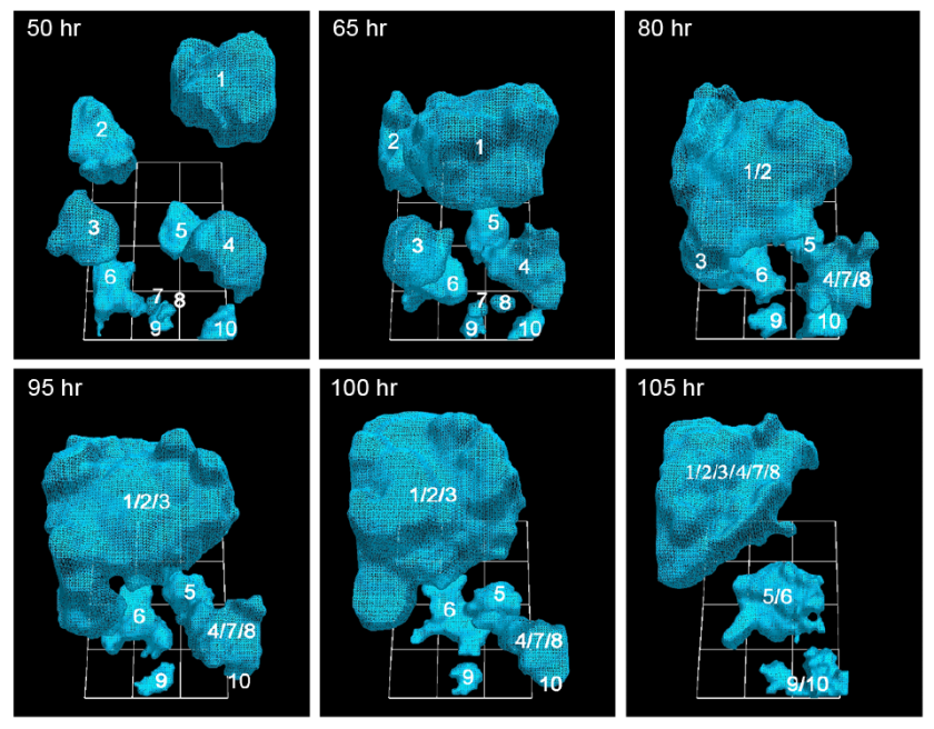
http://www.kurzweilai.net/images/Cancer-Cells-50-105-hours-stacked.png

University of Iowa researchers have documented how cancerous tumors form by tracking in real time the movement of individual cells in 3-D. They report that just 5 percent of cancer cells are needed to form tumors, a ratio that heretofore had been unknown. (credit: Soll Laboratory)

In the AJCR paper, the researchers found support for their previous observation that tumorigenic cell lines and fresh tumor cells possess the unique capacity to form tumors by the active formation of cellular cables.

The finding lends more weight to the idea that tumors are created concurrently, in multiple locations, by individual clusters of cells that employ the cancer-cell cables to draw in more cells and enlarge themselves. Some have argued that tumors come about more by cellular changes within the masses, known as the “cancer stem cell theory.”

The Developmental Studies Hybridoma Bank funded the study.

* Soll’s Monoclonal Antibody Research Institute and the Developmental Studies Hybridoma Bank, created by the National Institutes of Health as a national resource, directed by Soll and housed at the UI, together contain one of the world’s largest collections of antibodies that could be used for the anti-cancer testing, based on the new findings.


Abstract of Mediated coalescence: a possible mechanism for tumor cellular heterogeneity

Recently, we demonstrated that tumorigenic cell lines and fresh tumor cells seeded in a 3D Matrigel model, first grow as clonal islands (primary aggregates), then coalesce through the formation and contraction of cellular cables. Non-tumorigenic cell lines and cells from normal tissue form clonal islands, but do not form cables or coalesce. Here we show that as little as 5% tumorigenic cells will actively mediate coalescence between primary aggregates of majority non-tumorigenic or non-cancerous cells, by forming cellular cables between them. We suggest that this newly discovered, specialized characteristic of tumorigenic cells may explain, at least in part, why tumors contain primarily non-tumorigenic cells.


Read Full Post »

Aptamers and Scaffolds

Curator: Larry H. Bernstein, MD, FCAP


Antibody Alternatives

Nucleic acid aptamers and protein scaffolds could change the way researchers study biological processes and treat disease.

By Jane McLeod and Paul Ko Ferrigno | February 1, 2016

http://www.the-scientist.com/?articles.view/articleNo/45134/title/Antibody-Alternatives/

http://www.the-scientist.com/images/February2016/Antibody.jpg

THE NEW Y: Antibodies, classically depicted as Y-shape molecules, are central elements of the mammalian immune system, flagging bacteria (green) for destruction by phagocytes (large, round cells). Antibodies have also served as key research tools and life-saving therapeutics, but new alternatives are becoming available.© GUNILLA ELAM/SCIENCE SOURCE

There is a growing reproducibility problem across the life sciences. The retraction rate of published papers has increased tenfold over the past decade, and researchers have reported only being able to replicate published results in 11 percent1 or 25 percent2 of attempts. It’s become known as the “reproducibility crisis,” and science is in place to fix it.

One major factor contributing to this problem is the use of poorly validated research antibodies. Lot to lot, antibodies can vary wildly. Some may not bind specifically to their target, or they may bind a different cellular protein altogether. According to one estimate, researchers around the world spend $800 million each year on poorly performing antibodies.3 (See “Exercises for Your Abs” here.)

While many researchers debate the best way to weed out the good antibodies from the bad, others are developing alternatives. Nucleic acid aptamers and protein scaffolds are increasingly being used to detect proteins of interest. Although they currently constitute only a fraction of affinity reagents, with the lion’s share of the market still going to traditional antibodies, these newer options offer an opportunity to rectify the problems stemming from using poorly validated antibodies in research. Researchers can engineer RNA or DNA aptamers and protein scaffolds to a specific target and function, the molecules are consistent from batch to batch, and they can be produced at a fraction of the cost of antibodies. These new reagents can target proteins that remain inaccessible to antibodies. And researchers have designed them to be functional in a wider range of conditions, including intracellular environments that degrade the antibody structure, opening up applications such as super-resolution microscopy and intracellular live-cell imaging to investigate the molecular dynamics of diverse cellular processes.

So, rather than complain about the poor performance of antibodies, perhaps the scientific community should embrace the new antibody alternatives designed to overcome this problem—and, by doing so, begin to resolve the ongoing reproducibility crisis.

The rise—and pitfalls—of antibodies

http://www.the-scientist.com/images/February2016/antibody2.jpg

A CLASSIC FIT: Antibodies are large proteins, weighing in at about 150 kDa. Four polypeptides—two heavy chains and two light chains—are linked by disulfide bonds to form a Y-shape molecule. The amino acid sequences at tips of the short ends of the Y vary greatly between antibodies produced by different B cells, while the rest of the molecule is relatively consistent. The variable portion of the antibody binds in a specific region (epitope) on a foreign protein (antigen) and signals the immune system to the presence of an invader.© STEVE GRAEPEL

Antibodies are large protein molecules composed of two heavy and two light chains linked by disulfide bonds. They play a crucial part in the immune system’s ongoing battle to keep our bodies from falling prey to deadly diseases. Through the diversification of gene segments in the antibody sequence, the mammalian immune system produces different combinations of heavy and light chains to bind a wide variety of foreign proteins. When an invader is detected, those B cells that produce the most specific antibodies undergo hypersomatic mutation to fine-tune the antibody’s affinity to a particular antigen, then differentiate into plasma cells that generate the targeted antibody molecules by the million to mark the disease-causing target for destruction. It has been estimated that the human body can create enough different antibodies to recognize 1012 distinct pathogens.4

For decades, life-science researchers have taken advantage of this natural process to develop tags and assays for a wide array of proteins. In the early 1900s, researchers began to cultivate protein-specific antibodies by immunizing rabbits, chickens, goats, donkeys, and other animals with a desired target protein. B cells within the animal host generate antibodies to different antigenic areas (epitopes) on the protein of interest. The antibodies targeting the desired protein can then be isolated and purified for use in biochemical and cell-based assays to document protein expression under different conditions or to identify potential disease biomarkers. But the reliance on an animal host system for production meant lot-to-lot heterogeneity for such polyclonal antibodies. (See illustration adjacent.)

In 1975, Argentine biochemist César Milstein and German biologist Georges Köhler discovered how to generate batches of individual antibodies, produced by a single B cell to target a specific antigen. Once an animal host produces antibodies to a target, the antibody-producing B cells are isolated from the spleen or lymph nodes and fused with tumor cells to generate immortal hybridoma lines. These lines are then screened to identify clones producing antibodies that bind with a high affinity to a specific epitope on the target protein. These cells are then cultured in large-scale bioreactors.

While heterogeneity can arise from drift in the cell line’s antibody expression and downstream production processes, monoclonal antibodies exhibit far less lot-to-lot variation than polyclonal antibodies, and have become the affinity tool of choice in modern research laboratories. Monoclonal antibodies are now routinely employed to localize proteins within tissues, determine protein network interactions, and analyze protein function. They are now being pushed to the limits of their performance in applications such as nanoimmunoassays and in vivo cell imaging. In medicine, antibody therapeutics represents the fastest growing sector of pharmaceutical sales, with 47 monoclonal antibodies currently on the market and a further 300 in clinical trials.5


http://www.the-scientist.com/images/February2016/FebAntibody2_640px.jpg

with 47 monoclonal antibodies currently on the market and 300 more in clinical trials. But facing problems of inconsistent, time-consuming, and costly antibody production, some researchers are turning to alternatives—nucleic acid aptamers and protein scaffolds—to target specific proteins of interest, in the lab and in the clinic.
See full infographic: WEB | PDF
© STEVE GRAEPEL


But there are many examples where the use of antibodies has actually hindered scientific progress, by providing misleading or inaccurate results. Antibodies have evolved to execute their biological function perfectly, but this does not make them foolproof as investigative tools or therapeutic agents. In fact, many of the very characteristics that aid in antibodies’ function as part of the immune system limit their use in research and medicine.

In the context of an immune reaction, for example, not all B cells produce antibodies that are exquisitely specific. So long as the antibodies exceed a certain threshold of binding affinity for the target, they remain part of the immune system’s defense. In the body, this is a good thing: these less-specific antibodies cross-react with a variety of related antigens, making the antibody defense force more versatile.6 If an invading pathogen mutates or a similar pathogen invades, potentially effective antibodies may already be in circulation. As part of an assay to specifically identify a particular protein, however, such cross-reactivity can be the downfall of the experiment or therapy.

Examined in this light, it is easy to see why taking a molecule that is derived for one purpose and applying it to another may not yield the best results. A clear example of the shortcomings of antibody use in life-science research comes from the Human Protein Atlas project. Mathias Uhlén of the Royal Institute of Technology in Stockholm, Sweden, and colleagues set out to catalog protein expression and localization data across 44 normal human tissue types, 20 different cancers, and 46 cell lines. The team sourced antibodies from 51 different commercial vendors for validation. Of the 5,436 antibodies received, about half failed to detect their target in a Western blot or standard immunohistochemistry assay.7

The development of novel antibodies that bind new protein targets continues to face several challenges. For example, using the conventional route of immunizing lab animals to produce an antibody against a toxic target molecule will often kill the host animal prior to the generation of sufficiently specific antibodies. Conversely, if a protein target is highly homologous to a host protein, the immune system may not recognize the target as foreign in order to generate antibodies against it.

Given the rapid pace at which molecular biology proceeds in the modern era, new protein-binding reagents are desperately needed. A review of 20 million published research articles from 1950 to 2009 showed that three-quarters of the research focused on just 10 percent of the proteins that were known prior to the mapping of the human genome.8 Rational design of antibody alternatives will allow us to target a broader swath of proteins and function across more platforms to better investigate the scientific questions at hand.

Nucleic acid aptamers and pro­tein scaffolds can target proteins that remain inaccessible to antibod­ies, and researchers have designed them to be functional in a wider range of con­ditions.


http://www.the-scientist.com/images/February2016/FebAntibody4_310px1.jpg

NUCLEIC ACID APTAMERS: Aptamers are short molecules of single-stranded DNA or RNA, typically less than 100 nucleotides in length, that form specific 3-D structures capable of binding target proteins.© STEVE GRAEPEL


Engineering a solution

NUCLEIC ACID APTAMERS: Aptamers are short molecules of single-stranded DNA or RNA, typically less than 100 nucleotides in length, that form specific 3-D structures capable of binding target proteins.© STEVE GRAEPELAlternative affinity reagents developed over the past few decades include both nucleic acid– and protein-based molecules. Aptamers are short molecules of single-stranded DNA or RNA, typically less than 100 nucleotides in length, that form 3-D structures capable of binding specific target proteins. Protein scaffolds, formed from polypeptide fragments or whole proteins, have similarly precise interactions with target molecules. Both types of affinity reagents are produced entirely in vitro, so in principle they are not subject to the limitations of antibody production by animal immune systems, allowing researchers to study proteins for which it is impossible to generate antibodies. And even when antibodies do exist, aptamers and protein scaffolds offer more-precise targeting, because they have been engineered for a specific purpose.

These novel affinity reagents also offer other benefits over antibodies. Both nucleic acid aptamers and protein scaffolds are much smaller than natural antibodies, which typically weigh about 150 kDa. Aptamers and scaffolds are as little as one-tenth that size. This means that their distribution is not restricted in the same manner as that of antibodies, opening up new targets that were previously inaccessible, such as epitopes hidden inside molecular grooves and pockets where antibodies simply can’t fit. Labeling target proteins with these smaller tags in cytochemistry experiments reduces the chance of the target protein being dragged around the cell according to the tag’s biochemistry, and increases the chances of identifying the correct protein localization. Additionally, their smaller size increases these affinity reagents’ tissue penetration, enhancing access to epitopes within tissue sections and decreasing false negative immunohistochemistry results. Smaller molecules are also cleared more rapidly from the body, especially when their size is below the renal cut-off of 45 kDa, making these molecules ideal as imaging agents in the clinic.

Researchers first developed nucleic acid aptamers in 1990 as RNA-based molecules, though DNA variants quickly followed to deal with the low stability of the RNA backbone. Aptamers offer simple chemistry that can be easily functionalized, but they lack stability across a range of temperatures and pH, and in the presence of common buffer components or DNases and RNases found in many media and cell environments. Researchers have used various chemical modifications to increase aptamers’ resistance to nuclease activity, to improve aptamer binding, and to increase their structural diversity, but others have turned to yet another option: protein scaffolds.

Developed around the same time as aptamers, protein scaffold affinity reagents were originally designed to identify potential therapeutic targets. Researchers soon began to apply this technology to screening for binders to completely novel proteins, by presenting a random sequence as the binding surface. Because protein scaffolds lack the disulfide bonds of antibodies, they retain their structure in a greater variety of cell culture and assay environments, without being attacked by other proteins that break these bonds and cause antibodies to fall apart. Scaffolds maintain function and target affinity at temperatures up to 80 °C and in solutions with a pH as low as 2 and as high as 13.

Because protein scaffolds can be delivered to the inside of the cell, researchers can use them in live-cell imaging, ultimately allowing use of the same reagent in both biochemical and cell biology assays. Additionally, protein scaffolds could help to deliver drugs directly into cells, improving targeting of pharmaceutical payloads and reducing side effects. And as new protein scaffolds are often engineered to lack cysteine residues, aberrant folding during their production within the cell factory is unlikely, increasing reproducibility within the reagents.

Importantly, both nucleic acid aptamers and protein scaffolds are far easier to consistently produce than antibodies. In addition to requiring animal hosts to provide an antibody-producing B cell, functional antibodies can only be expressed in higher eukaryotic cell systems. Antibodies are extensively glycosylated with a complex range of sugars that are critical to their function. Lower eukaryotic organisms, such as insects and yeast, and prokaryotic cells are not capable of the full range of complex glycosylation. As a result of these complexities, production times for monoclonal antibodies are six months on average, often making the generation of new antibodies the rate-limiting step in the advance of new research. This production process is also extremely expensive, and the use of such intractable biological systems breeds batch-to-batch inconsistencies.


http://www.the-scientist.com/images/February2016/FebAntibody5_310px.jpg

PROTEIN SCAFFOLDS: Protein scaffolds, formed from polypeptide fragments or whole proteins, have similarly specific interactions with desired target molecules.© STEVE GRAEPEL

Aptamers and protein scaffolds can be made without a host immune system. Now that robust and scalable methodologies for creating custom DNA and RNA molecules exist, effective aptamer binders can be chemically synthesized at a fraction of the cost of producing protein-based affinity molecules. And because protein scaffolds do not contain any posttranslational modifications, they can be expressed in bacterial cells, which are cheaper and easier to control than the eukaryotic systems used for antibody production. Both aptamers and scaffolds can often be available to researchers in a matter of weeks. (See illustration above)

Alternatives at work


http://www.the-scientist.com/images/February2016/antibody4.jpg

A PROTEIN BUILT FOR YOU: Protein scaffolds (orange) can be developed to bind specific protein targets (purple) and can be produced in a matter of weeks. Once the scaffolds bind the protein of interest, fluorescent or color reporter tags (yellow ball) can be used to label and isolate them.© AVACTA LIFE SCIENCES. May not be reproduced without express written permission from the copyright holder.

So far, the majority of the industry attention for antibody alternatives has largely focused on the therapeutic development of antibody alternatives. Many companies now have initiated Phase 2 and 3 clinical trials of candidate molecules to treat conditions from vision problems to cancer, and two such molecules have already been approved for therapeutic use. In 2004, the RNA aptamer–based therapeutic pegaptanib (Macugen), originally developed by NeXstar Pharmaceuticals, became the first antibody alternative to gain US Food and Drug Administration (FDA) approval for the treatment of neovascular age-related macular degeneration. Pegaptanib is a 28-base-long RNA oligonucleotide with modifications to protect the aptamer from endogenous nucleases and extend its half-life in vivo to 10 days.9 Administered directly into the eye, this aptamer selectively binds the most common isoform of vascular endothelial growth factor (VEGF), preventing angiogenesis and the increased permeability of the blood vessels within the eye associated with neovascular age-related macular degeneration.10 Five years later, in 2009, the FDA approved a protein scaffold called ecallantide (Kalbitor) for the treatment of sudden hereditary angioedema attacks.

In a therapeutic context, the most important characteristic of aptamers and scaffolds is that they lack immunogenicity, thus avoiding harmful immune responses in patients. One way to ensure that protein scaffolds do not trigger host immunity is to model one’s scaffolds after proteins found in the human body. For example, the FDA-approved Kalbitor is based on the common Kunitz domain of protease inhibitors, and clinical trial participants have not suffered immunogenic responses. When brought to market, Kalbitor was one of only two approved therapies to treat cardiovascular attacks of this sort, which can cause rapid and serious swelling of the face or other parts of the body that may result in permanent disfigurement, disability, or death; the other is a protein therapeutic derived from human blood.

A major factor holding back the field of antibody alternatives as therapeutics is their small size. While this improves their intracellular function and use in research applications, their low molecular weight means that they are rapidly cleared from the body via the kidneys, reducing their potential therapeutic impact. Various strategies have been employed by the industry to overcome this rapid renal clearance, such as adding an antibody domain or an albumin-binding domain to the scaffold, or increasing the molecular weight of the protein scaffolds (though they still remain significantly smaller than a corresponding antibody). The fusion of an antibody domain to a protein scaffold can also help engage the immune system for improved therapeutic benefit.

While their small size can be a hurdle in developing antibody alternatives in a clinical setting, it is a big advantage in their use as laboratory tools, allowing them to penetrate bodily tissues that are inaccessible to antibodies and offering more-precise molecular labeling. In 2012, for example, Silvio Rizzoli of the European Neuroscience Institute and Center for Molecular Physiology of the Brain in Göttingen, Germany, and colleagues used 15 kDa aptamers to capture the dynamics of endosomal trafficking in live cells using super-resolution imaging.11 Doubling the molecular weight of the aptamer resulted in a substantial reduction in image quality, showing the importance of the small size of intracellular labels in accurate imaging of the intracellular space.

The increased intracellular stability of protein scaffolds as compared with antibodies is also critical to their function as research tools and offers potential therapeutic benefit for intracellular targets. For instance, protein scaffolds have been used to investigate the function of the small intracellular domain of a matrix metalloproteinase, which was shown to determine protein turnover to help regulate protein function in cell movement.12 Using antibodies in the reducing environment of the cell interior in such a study would be impossible.

When developing antibody alternatives for research, scientists are purposefully mimicking natural proteins to avoid interference by the immune system. For example, Janssen produces protein scaffolds called Centyrins that are based on the fibronectin glycoprotein of the extracellular matrix. Our own company, Avacta Life Sciences, recently introduced Affimer scaffolds, which are based on the cystatin protein family of common protease inhibitors. The use of consensus sequences from a number of species may allow these reagents to be used across a variety of different model systems.

Nucleic acid aptamers and protein scaffolds may also help fight emerging outbreaks of acute infectious disease. Examples of recent outbreaks that have caused considerable social, economic, and political stress are not hard to come by—SARS in 2003, the H1N1 flu pandemic in 2009, the Ebola crisis of recent years, and the continued emerging threat of MERS. It is impossible to predict such episodes, and alternative affinity reagents could be crucial tools in quickly stemming the spread of pandemic diseases. Screening libraries of 10 billion sequences can take as little as 7 weeks. And while the processes required for optimization, scale-up, and subsequent culture and validation of substantial quantities of the required affinity reagent remain to be explored, this all may take only a matter of months.

Over the past few decades, the rate of advancement of genomic technologies has outpaced proteomics. Yet it is the expressed protein within a cell, not the underlying genetic blueprint, that executes correct or aberrant function. In order to unify and make sense of the numerous data sets being produced, scientists need tools that enable the unraveling of proteomics. This requires affinity reagents that can specifically target individual protein isoforms and glycoforms, and that can tag all the proteins within a cell or organism. While the concerns over antibody irreproducibility are increasing, the solution may already be available.

Jane McLeod is a science writer at Avacta Life Sciences, where Paul Ko Ferrigno is the chief scientific officer. Avacta Life Sciences sells peptide aptamers, one of the main forms of antibody alternative.

References

  1. C.G. Begley, L.M. Ellis, “Drug development: Raise standards for preclinical cancer research,” Nature, 483:531-33, 2012.
  2. F. Prinz et al., “Believe it or not: How much can we rely on published data on potential drug targets?” Nat Rev Drug Discov, 10:712, 2011.
  3. A. Bradbury, A. Plückthun, “Reproducibility: Standardize antibodies used in research,” Nature, 518:27-29, 2015.
  4. B. Alberts et al., Molecular Biology of the Cell, 4th edition (New York: Garland Science, 2002).
  5. D.M. Ecker et al., “The therapeutic monoclonal antibody market,” mAbs, 7:9-14, 2015.
  6. N. Baumgarth, “How specific is too specific? B-cell responses to viral infection reveal the importance of breadth over depth,” Immunol Rev, 255:82-94, 2013.
  7. L. Berglund et al., “A genecentric Human Protein Atlas for expression profiles based on antibodies,” Mol Cell Proteomics, 7:2019-27, 2008.
  8. A.M. Edwards et al., “Too many roads not taken,” Nature, 470:163-65, 2011.
  9. J. Ruckman et al., “2’-Fluoropyrimidine RNA-based aptamers to the 165-amino acid form of vascular endothelial growth factor (VEGF165): Inhibition of receptor binding and VEGF-induced vascular permeability through interactions requiring the exon 7-encoded domain,” J Biol Chem, 273:20556-67, 1998.
  10. P. Sundaram et al., “Therapeutic RNA aptamers in clinical trials,” Eur J Pharm Sci, 48:259-71, 2013.
  11. F. Opazo et al., “Aptamers as potential tools for super-resolution microscopy,” Nat Methods, 9:938-39, 2012.
  12. R.D. Wickramasinghe et al., “Peptide aptamers as new tools to modulate clathrin-mediated internalisation—inhibition of MT1-MMP internalisation,” BMC Cell Biol, 11:58, 2010.

February 4, 2016

Nice article. Your statement in the end identifies correctly a potential conflict of interest. It should probably be a bit more clear and obvious than that though. Perhaps at the top of the page, near the title of the article. My 2 cents.


Read Full Post »

Reengineering Therapeutics

Larry H. Bernstein, MD, FCAP, Curator

LPBI

The synNotch solution: UCSF scientists engineer a next-gen T-cell immunotherapy

Sunday, January 31, 2016 | By John Carroll

2.2.9

2.2.9   Reengineering Therapeutics, Volume 2 (Volume Two: Latest in Genomics Methodologies for Therapeutics: Gene Editing, NGS and BioInformatics, Simulations and the Genome Ontology), Part 2: CRISPR for Gene Editing and DNA Repair

CAR-T has been all the rage in cancer R&D for several years now as a slate of biotech upstarts pursue highly promising work reengineering T cells into attack weapons by adding a chimeric antigen receptor that can zero in on particular cancer cells. The approach has been highly effective in acute lymphoblastic leukemia, triggering an attack on B cells by homing in on the CD19 antigen, a breakthrough that has inspired a race to the regulatory finish line with the first CAR-Ts.

That approach, though, has run into some major obstacles when researchers move from the blood cancer to solid tumors. But now a group of scientists working with UC San Francisco’s Wendell Lim say they’ve come up with a new therapeutic model for T-cell engineering that promises to overcome that hurdle and make it a more precise weapon that can tackle solid tumors while avoiding off-target reactions that threaten patients.

The key to this new approach is a new receptor: synNotch. Taking a cue from nature, which relies on a sensor called Notch to perform key functions, the synthetic biology engineers say they can add a receptor that includes one section that sticks out from the cell with one that lies inside. By tinkering with synNotch they can reengineer the immune cell to run down a particular cancer cell target and then issue instructions to turn genes on or off to set up the other half of the therapeutic equation.

In a project described in Cell, the team says they created a synNotch that recognized an antigen on the surface of the cancer cell while the internal mechanism contributed a chimeric antigen receptor that recognized a different antigen. They then tested it on a mouse model that included two different tumor cells: one with both targets recognized by the external synNotch sensor and the CAR and one with just the CAR target.

The newly programmed attack weapon zeroed in on the two targets (which required synNotch activation for it to work) while leaving the other alone, providing preclinical proof-of-concept evidence that they could create a much more efficient tumor cell killing vehicle.

“The kinds of engineered T cells that we can now construct give us the exciting potential to create precision cancer therapeutics that take advantage of all the genomic and proteomic information we are currently gathering on disease,” said Lim, the senior author of the study. “This genomic information now becomes actionable.”

The team was led by Leonardo Morsut and Kole Roybal.

Lim added that it is possible to reengineer a T cell with multiple synNotches to make it even more precise. And there are added applications for autoimmune disease, regenerative medicine, and more.

Engineering Customized Cell Sensing and Response Behaviors Using Synthetic Notch Receptors

Leonardo  Morsut, Kole T. Roybal, Xin Xiong, Russell M. Gordley, Scott M. Coyle, Matthew Thomson, Wendell A. Lim

Tricked-Out Immune Cells Could Attack Cancer, Spare Healthy Cells  

New Cell-Engineering Technique May Lead To Precision Immunotherapies

New CRISPR/Cas9 delivery method could offer a clinical pathway

Read Full Post »

Nanosensors for Protein Recognition and Gene Proteome Interaction

Larry H Bernstein, MD, FCAP, Curator

LPBI


Synthetic Antibody Detects Proteins

http://www.technologynetworks.com/Proteomics/news.aspx?ID=187242

Research could lead to nanosensors that recognize fibrinogen, insulin, or other biomarkers

Using carbon nanotubes, MIT chemical engineers have devised a new method for detecting proteins, including fibrinogen, one of the coagulation factors critical to the blood-clotting cascade.

This approach, if developed into an implantable sensor, could be useful for monitoring patients who are taking blood thinners, allowing doctors to make sure the drugs aren’t interfering too much with blood clotting.

The new method is the first to create synthetic recognition sites (similar to natural antibodies) for proteins and to couple them directly to a powerful nanosensor such as a carbon nanotube. The researchers have also made significant progress on a similar recognition site for insulin, which could enable better monitoring of patients with diabetes. It may also be possible to use this approach to detect proteins associated with cancer or heart disease, says Michael Strano, the Carbon P. Dubbs Professor in Chemical Engineering at MIT.

A targeted search

The new sensor is the latest example of a method developed in Strano’s lab, known asCorona Phase Molecular Recognition (CoPhMoRe).

This technique takes advantage of the interactions between a given polymer and a nanoparticle surface such as that of a fluorescent single-walled carbon nanotube, when the polymer is wrapped around the nanotube.

Certain regions of the polymers latch onto the nanoparticle surface like anchors, while other regions extend outwards into their environment. This outward-facing region, also known as the adsorbed phase or corona, has a 3-D structure that depends on the composition of the polymer.

CoPhMoRe works when a specific polymer adsorbs to the nanoparticle surface and creates a corona that recognizes the target molecule. These interactions are very specific, just like the binding between an antibody and its target. Binding of the target alters the carbon nanotubes’ natural fluorescence, allowing the researchers to measure how much of the target molecule is present.

Strano’s lab has previously used this approach to find recognition sites and develop nansensors for estradiol and riboflavin, among other molecules. The new paper represents their first attempt to identify corona phases that can detect proteins, which are larger, more complex, and more fragile than the molecules identified by their previous sensors.

For this study, Bisker began by screening carbon nanotubes wrapped in 20 different polymers including DNA, RNA, and polyethylene glycol (PEG), a polymer often added to drugs to increase their longevity in the bloodstream.

On their own, none of the polymers had any affinity for the 14 proteins tested, all taken from human blood. However, when the researchers tested polymer-wrapped nanotubes against the same proteins, they turned up a match between one of the modified nanotubes and fibrinogen.

“A chemist or a biologist would not be able to predict ahead of time that there should be any kind of affinity between fibrinogen and this corona phase,” Strano says. “It really is a new kind of molecular recognition.”

Fibrinogen, one of the most abundant proteins in human blood, is part of the blood-clotting cascade. When a blood vessel is damaged, an enzyme called thrombin converts fibrinogen into fibrin, a stringy protein that forms clots to seal the wound.

A sensor for fibrinogen could help doctors determine if patients who are taking blood thinners still have enough clotting capability to protect them from injury, and could allow doctors to calculate more finely tuned dosages. It could also be used to test patients’ blood clotting before they go into surgery, or to monitor wound healing, Bisker says.

Synthetic antibodies

The researchers believe their synthetic molecular recognition agents are an improvement over existing natural systems based on antibodies or DNA sequences known as aptamers, which are more fragile and tend to degrade over time.

“One of the advantages of this is that it’s a completely synthetic system that can have a much longer lifetime within the body,” Bisker says.

In 2013, researchers in Strano’s lab demonstrated that carbon nanotube sensors can remain active in mice for more than a year after being embedded in a polymer gel and surgically implanted under the skin.

In addition to insulin, the researchers are also interested in detecting troponin, a protein that is released by dying heart cells, or detecting proteins associated with cancer, which would be useful for monitoring the success of chemotherapy. These and other protein sensors could become critical components of devices that deliver drugs in response to a sign of illness.

“By measuring therapeutic markers in the human body in real time, we can enable drug delivery systems that are much smarter, and release drugs in precise quantities,” Strano says. “However, measurement of those biomarkers is the first step.”


New Device Uses Carbon Nanotubes to Snag Molecules
Nanotube “forest” in a microfluidic channel may help detect rare proteins and viruses.
Tuesday, December 22, 2015

Nanotube “forest” in a microfluidic channel may help detect rare proteins and viruses.

Engineers at MIT have devised a new technique for trapping hard-to-detect molecules, using forests of carbon nanotubes.

The team modified a simple microfluidic channel with an array of vertically aligned carbon nanotubes — rolled lattices of carbon atoms that resemble tiny tubes of chicken wire. The researchers had previously devised a method for standing carbon nanotubes on their ends, like trees in a forest. With this method, they created a three-dimensional array of permeable carbon nanotubes within a microfluidic device, through which fluid can flow.

Now the researchers have given the nanotube array the ability to trap certain particles. To do this, the team coated the array, layer by layer, with polymers of alternating electric charge.

“You can think of each nanotube in the forest as being concentrically coated with different layers of polymer,” says Brian Wardle, professor of aeronautics and astronautics at MIT. “If you drew it in cross-section, it would be like rings on a tree.”

Depending on the number of layers deposited, the researchers can create thicker or thinner nanotubes and thereby tailor the porosity of the forest to capture larger or smaller particles of interest.

The nanotubes’ polymer coating may also be chemically manipulated to bind specific bioparticles flowing through the forest. To test this idea, the researchers applied an established technique to treat the surface of the nanotubes with antibodies that bind to prostate specific antigen (PSA), a common experimental target. The polymer-coated arrays captured 40 percent more antigens, compared with arrays lacking the polymer coating.

Wardle says the combination of carbon nanotubes and multilayer coatings may help finely tune microfluidic devices to capture extremely small and rare particles, such as certain viruses and proteins.

“There are smaller bioparticles that contain very rich amounts of information that we don’t currently have the ability to access in point-of-care [medical testing] devices like microfluidic chips,” says Wardle, who is a co-author on the paper. “Carbon nanotube arrays could actually be a platform that could target that size of bioparticle.”

The paper’s lead author is Allison Yost, a former graduate student who is currently an engineer at Accion Systems. Others on the paper include graduate student Setareh Shahsavari; postdoc Roberta Polak; School of Engineering Professor of Teaching Innovation Gareth McKinley; professor of materials science and engineering Michael Rubner, and Raymond A. And Helen E. St. Laurent Professor of Chemical Engineering Robert Cohen.

A porous forest

Carbon nanotubes have been a subject of intense scientific study, as they possess exceptional electrical, mechanical, and optical properties. While their use in microfluidics has not been well explored, Wardle says carbon nanotubes are an ideal platform because their properties may be manipulated to attract certain nanometer-sized molecules. Additionally, carbon nanotubes are 99 percent porous, meaning a nanotube is about 1 percent carbon and 99 percent air.

“Which is what you need,” Wardle says. “You need to flow quantities of fluid through this material to shed all the millions of particles you don’t want to find and grab the one you do want to find.”

What’s more, Wardle says, a three-dimensional forest of carbon nanotubes would provide much more surface area on which target molecules may interact, compared with the two-dimensional surfaces in conventional microfluidics.

“The capture efficiency would scale with surface area,” Wardle notes.

A versatile array

The team integrated a three-dimensional array of carbon nanotubes into a microfluidic device by using chemical vapor deposition and photolithography to grow and pattern carbon nanotubes onto silicon wafers. They then grouped the nanotubes into a cylinder-shaped forest, measuring about 50 micrometers tall and 1 millimeter wide, and centered the array within a 3 millimeter-wide, 7-millimeter long microfluidic channel.

The researchers coated the nanotubes in successive layers of alternately charged polymer solutions in order to create distinct, binding layers around each nanotube. To do so, they flowed each solution through the channel and found they were able to create a more uniform coating with a gap between the top of the nanotube forest and the roof of the channel. Such a gap allowed solutions to flow over, then down into the forest, coating each individual nanotube. In the absence of a gap, solutions simply flowed around the forest, coating only the outer nanotubes.

After coating the nanotube array in layers of polymer solution, the researchers demonstrated that the array could be primed to detect a given molecule, by treating it with antibodies that typically bind to prostate specific antigen (PSA). They pumped in a solution containing small amounts of PSA and found that the array captured the antigen effectively, throughout the forest, rather than just on the outer surface of a typical microfluidic element.

Wardle says that the nanotube array is extremely versatile, as the carbon nanotubes may be manipulated mechanically, electrically, and optically, while the polymer coatings may be chemically altered to capture a wide range of particles. He says an immediate target may be biomarkers called exosomes, which are less than 100 nanometers wide and can be important signals of a disease’s progression.

“Science is really picking up on how much information these particles contain, and they’re sort of everywhere, but really hard to find, even with large-scale equipment,” Wardle says. “This type of device actually has all the characteristics and functionality that would allow you to go after bioparticles like exosomes and things that really truly are nanometer scale.”

This research was funded, in part, by the National Science Foundation.


A Natural Light Switch

MIT scientists identify and map the protein behind a light-sensing mechanism.

MIT scientists, working with colleagues in Spain, have discovered and mapped a light-sensing protein that uses vitamin B12 to perform key functions, including gene regulation.

The result, derived from studying proteins from the bacterium Thermus thermophilus, involves at least two findings of broad interest. First, it expands our knowledge of the biological role of vitamin B12, which was already understood to help convert fat into energy, and to be involved in brain formation, but has now been identified as a key part of photoreceptor proteins — the structures that allow organisms to sense and respond to light.

Second, the research describes a new mode of gene regulation, in which the light-sensing proteins play a key role. In so doing, the scientists observe, the bacteria have repurposed existing protein structures that use vitamin B12, and put them to work in new ways.

MIT-Proteins-Light-1_0.jpg

http://www.technologynetworks.com/images/videos/News%20Images/CR/MIT-Proteins-Light-1_0.jpg

“Nature borrowed not just the vitamin, but really the whole enzyme unit, and modified it … and made it a light sensor,” says Catherine Drennan, a professor of chemistry and biology at MIT


The paper describes the photoreceptors in three different states: in the dark, bound to DNA, and after being exposed to light.

“It’s wonderful that we’ve been able to get all the series of structures, to understand how it works at each stage,” Drennan says.

The paper has nine co-authors, including Drennan; graduate students Percival Yang-Ting Chen, Marco Jost, and Gyunghoon Kang of MIT; Jesus Fernandez-Zapata and S. Padmanabhan of the Institute of Physical Chemistry Rocasolano, in Madrid; and Monserrat Elias-Arnanz, Juan Manuel Ortiz-Guerreo, and Maria Carmen Polanco, of the University of Murcia, in Murcia, Spain.

The researchers used a combination of X-ray crystallography techniques and in-vitro analysis to study the bacteria. Drennan, who has studied enzymes that employ vitamin B12 since she was a graduate student, emphasizes that key elements of the research were performed by all the co-authors.

Jost performed crystallography to establish the shapes of the structures, while the Spanish researchers, Drennan notes, “did all of the control experiments to show that we were really thinking about this right,” among other things.

MIT-Proteins-Light-2.jpg

By studying the structures of the photoreceptor proteins in their three states, the scientists developed a more thorough understanding of the structures, and their functions, than they would have by viewing the proteins in just one state.

Microbes, like many other organisms, benefit from knowing whether they are in light or darkness. The photoreceptors bind to the DNA in the dark, and prevent activity pertaining to the genes of Thermus thermophilus. When light hits the microbes, the photoreceptor structures cleave and “fall apart,” as Drennan puts it, and the bacteria start producing carotenoids, which protect the organisms from negative effects of sunlight, such as DNA damage.

The research also shows that the exact manner in which the photoreceptors bind to the DNA is novel. The structures contain tetramers, four subunits of the protein, of which exactly three are bound to the genetic material — something Drennan says surprised her.

“That’s the best part about science,” Drennan says. “You see something novel, then you think it’s not really going to be that novel, but you do the experiments [and it is].”

Other scientists say the findings are significant. “It’s a very exciting development,” says Rowena Matthews, a professor emerita of biological chemistry at the University of Michigan, who has read the paper. Of the newly discovered use of vitamin B12 and a derivative of it, adenosylcobalamin, Matthews adds, “There was very limited knowledge of its versatility.”

Drennan adds that in the long run, the finding could have practical applications, such as the engineering of light-directed control of DNA transcription, or the development of controlled interactions between proteins.

“I would be very interested in … thinking about whether there could be practical applications of this,” Drennan says.


HIV Protein Manipulates Hundreds of Human Genes

Findings search for new or improved treatments for patients with AIDS.

UT Southwestern Medical Center researchers have deciphered how a small protein made by the human immunodeficiency virus (HIV) that causes AIDS manipulates human genes to further its deadly agenda.

The findings, published in the online journal eLife, could aid in the search for new or improved treatments for patients with AIDS, or to the development of preventive strategies.

“We have identified the molecular mechanisms by which the Tat protein made by HIV interacts with the host cell to activate or repress several hundred human genes,” said Dr. Iván D’Orso, Assistant Professor of Microbiology at UT Southwestern and senior author of the study. “The findings clearly suggest that blocking Tat activity may be of therapeutic value to HIV patients.”

It has long been known that HIV causes AIDS by hijacking the body’s immune cells, transforming them into HIV factories and killing other immune cells that normally fight disease. HIV also hides in cells and continues to undermine the host’s immune system despite antiretroviral therapy that has improved the outlook of those with AIDS.

The latest data from the Centers for Disease Control and Prevention (CDC), in 2012, estimated 1.2 million Americans were living with HIV, including 156,300 whose infections had not been diagnosed. About 50,000 people in the U.S. are newly infected with HIV annually, the CDC projects. In 2013, the CDC estimated that over 26,000 Americans had the advanced form of HIV infection, AIDS.

Like all retroviruses, HIV has very few genes of its own and must take over the host’s cellular machinery in order to propagate and spread throughout the body. Although the broad aspects of that cellular hijacking were known, the nuances remain to be explored, Dr. D’Orso said.

“We observed that HIV methodically and precisely manipulates the host’s genes and cellular machinery. We also observed that HIV rewires cellular defensive pathways to benefit survival of the virus,” he added.

The study provides insights into HIV’s ability to survive despite antiretroviral therapy, findings that could lead to new therapeutic targets or ways to make current therapies more effective, he said.

“Our study indicates that this small viral protein, Tat, directly binds to about 400 human genes to generate an environment in which HIV can thrive. Then, this protein precisely turns off the body’s immune defense. It is striking that such a small viral protein has such a large impact,” Dr. D’Orso said. “The human genes and pathways that Tat manipulates correlate well with symptoms observed in these patients, such as immune system hyperactivation, then weakening, and accelerated aging,” Dr. D’Orso said, describing the situation in which HIV infection leads to AIDS.

Italy’s National Institute of Health in Rome recently completed a phase II clinical trial of an experimental vaccine that targets the Tat protein. That trial, which followed 87 HIV-positive patients for up to three years, reported that the vaccine was well-tolerated without significant side effects. However, it will take several years to determine if the vaccine works, Dr. D’Orso said.

Although someone can have HIV for years without showing symptoms, AIDS occurs when HIV blocks the body’s ability to fight off illness. The person then becomes overrun by the opportunistic infections and specific cancers that are hallmarks of AIDS.


New Light Shed on Genetic Regulation

A team of scientists has uncovered greater intricacy in protein signaling than was previously understood, shedding new light on the nature of genetic production.

Christine Vogel, an assistant professor in New York University’s Department of Biology and one of the study’s senior authors, explains that “to make a protein, we need to make a messenger RNA molecule from the gene encoded in the DNA, and then, in a second process, make proteins from these RNA molecules. Both processes are highly regulated and coupled.”

This coupling is similar to the coupling between a moving escalator and a person walking on it at the same time.

The research takes a closer look at how the two coupled processes change in the cell responding to an outside stimulus.

“Until recently, it has been very difficult to study these systems and researchers have thought that the movement of the escalator is most important during the cellular response,” Vogel explains. “We now show that is not necessarily the case, and under some circumstances, the person’s walking determines the overall outcome.”

In biology, this means that both of the processes—to make RNAs and proteins—play important roles, but with different patterns.

In their study, the scientists, who also included researchers from National University Singapore and Berlin’s Max Delbruck Center, took a closer look at how the two processes exactly respond over time.

Their results showed notable distinctions between DNA and mRNA in the nature of their signaling. Notably, the process of making RNA from DNA was pulse-like—a brief messaging over the studied period that returned to the normal levels by the end of the measurements. By contrast, the process of making a protein from RNA was akin to an on/off switch: once started, levels remained constant for consistent periods before reverting back to long stretches of dormancy.

While the reasons for these differences in cell behavior remain unknown, the researchers believe the answer may lie in the nature of the two tasks.

“It is very costly for the cell to make proteins, but making RNA messages from DNA is a relatively low-energy and simple process, so it makes sense that we see frequent, or pulsating, signaling at this stage,” observes Vogel. “By contrast, creating proteins is an intricate undertaking, requiring a great deal of time and energy. This may be why, once you decided to stop production of proteins, you do not turn it back on that easily—and the other way around.”


Where Cancer Cells May Begin

Scientists use fruit fly genetics to understand how things could go wrong in cancer.

Cancer cells are normal cells that go awry by making bad developmental decisions during their lives. In a study involving the fruit fly equivalent of an oncogene implicated in many human leukemias, Northwestern University researchers have gained insight into how developing cells normally switch to a restricted, or specialized, state and how that process might go wrong in cancer.

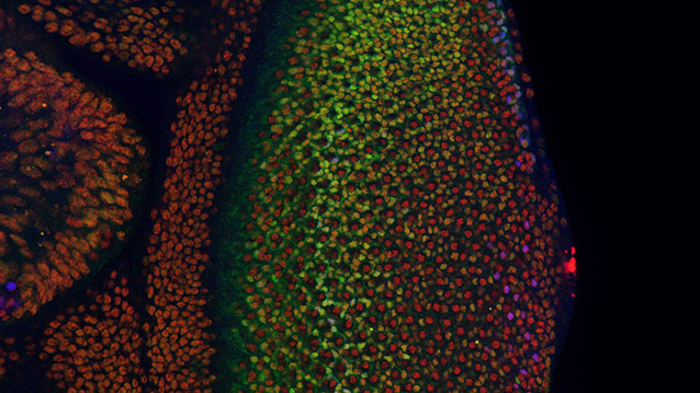
The fruit fly’s eye is an intricate pattern of many different specialized cells, such as light-sensing neurons and cone cells. Because flies share with humans many of the same cancer-causing genes, scientists use the precisely made compound eye of Drosophila melanogaster (the common fruit fly) as a workhorse to study what goes wrong in human cancer.

retina638.jpg

http://www.technologynetworks.com/images/videos/News%20Images/GT/retina638.jpg

A multidisciplinary team co-led by biologist Richard W. Carthew and engineer Luís A.N. Amaral studied normal cell behavior in the developing eye. The researchers were surprised to discover that the levels of an important protein called Yan start fluctuating wildly when the cell is switching from a more primitive, stem-like state to a more specialized state. If the levels don’t or can’t fluctuate, the cell doesn’t switch and move forward.

“This mad fluctuation, or noise, happens at the time of cell transition,” said Carthew, professor of molecular biosciences in Northwestern’s Weinberg College of Arts and Sciences. “For the first time, we see there is a brief time period as the developing cell goes from point A to point B. The noise is a state of ‘in between’ and is important for cells to switch to a more specialized state. This limbo might be where normal cells take a cancerous path.”

The researchers also found that a molecular signal received by a cell receptor called EGFR is important for turning the noise off. If that signal is not received, the cell remains in an uncontrolled state.

By pinpointing this noise and its “off” switch as important points in the normal process of cell differentiation, the Northwestern researchers provide targets for scientists studying how cells can go out of control and transform into cancer cells.

The “noisy” protein the Northwestern researchers studied is called Yan in the fly and Tel-1 in humans. (The protein is a transcription factor.) The Tel-1 protein instructs cells to turn into white blood cells; the gene that produces the protein, oncogene Tel-1, is frequently mutated in leukemia.

The EGFR protein that turns off the noise in flies is called Her-2 in humans. Her-2 is an oncogene that plays an important role in human breast cancer.

“On the surface, flies and humans are very different, but we share a remarkable amount of infrastructure,” said Carthew, a member of the Robert H. Lurie Comprehensive Cancer Center of Northwestern University. “We can use fruit fly genetics to understand how humans work and how things go wrong in cancer and other diseases.”

Fruit fly cells are small and closely packed together, making study of them challenging. Carthew and Amaral’s team of biologists, chemical and biological engineers, computer scientists and chemists together figured out how to identify and analyze thousands and thousands of individual cells in the flies’ eyes.

“In the past, people have built models of regulatory networks that control cell differentiation mostly by genetically perturbing one or two components of the network at a time and then compiling those results into models,” said Amaral, professor of chemical and biological engineering at the McCormick School of Engineering. “We instead measured the retina as it developed and found the unexpected behavior of the key regulatory factors Yan and EGFR.”

Nicolás Peláez, first author of the study and a Ph.D. candidate in interdisciplinary biological sciences working with Amaral and Carthew, built new tools to study this strange feature of noise in developing flies. His methods enabled the researchers to easily measure both the concentration of the Yan protein and its fluctuation (noise).

It takes 15 to 20 hours for a fruit fly cell to go from being an unrestricted cell to a restricted cell, Carthew said. Peláez determined the Yan protein is noisy, or fluctuating, for six to eight of those hours.

“Studying the dynamics of molecules regulating fly-eye patterning can inform us about human disease,” Peláez said. “Using model organisms such as fruit flies will help us understand quantitatively the basic biological principles governing differentiation in complex animals.”


Mechanism of Tumor Suppressing Gene Uncovered

The most commonly mutated gene in cancer,p53, works to prevent tumor formation by keeping mobile elements in check that otherwise lead to genomic instability, UT Southwestern Medical Center researchers have found.

The p53 gene long has been known to suppress tumor formation, but the mechanisms behind this function – and why disabling the gene allows tumors to form – were not fully understood.

Findings from the study answer some of these questions and could one day lead to new ways of diagnosing and treating cancer, said the study’s senior author, Dr. John Abrams, Professor of Cell Biology at UT Southwestern.

The investigators found that normal p53 gene action restrains transposons, mobile genetic elements called retroelements that can make copies of themselves and move to different positions on chromosomes. But, they discovered, when p53 is disabled by mutation, dramatic eruptions of these mobile elements occur. The study revealed that in mice with cancer and in human samples of two types of cancer (Wilms’ tumors and colon tumors) disabled for p53, transposons became very active.

In a healthy state, certain mechanisms work to keep these retroelements quiet and inactive, explained Dr. Abrams. One of those mechanisms is p53 action. Conversely, when p53 is mutated, retroelements can erupt.

“If you take the gene away, transposons can wreak havoc throughout the genome by causing it to become highly dysregulated, which can lead to disease,” Dr. Abrams said. “Our findings help explain why cancer genomes are so much more fluid and destabilized than normal genomes. They also provide a novel framework for understanding how normal cells become tumors.”

Although much more research is needed, Dr. Abrams said, the potential clinical implications of the team’s findings are significant.

“Understanding how p53 prevents tumors raises the prospect of therapeutic interventions to correct cases in which p53 is disabled,” Dr. Abrams said. “If retroelements are at the heart of certain p53-driven cancers, finding ways to suppress them could potentially allow us to prevent those cancers or intervene to keep them from progressing.”

This understanding also could lead to advances in diagnosing some cancers through biomarkers related to p53 and transposon activity.

“One possibility is that perhaps blood or urine tests could detect dysregulated retroelements that could be indicative of certain types of cancer,” Dr. Abrams said.

Read Full Post »

Deciphering the Epigenome

Curator: Larry H. Bernstein, MD, FCAP

UPDATED on 1/29/2016

2.2.10

2.2.10   Deciphering the Epigenome, Volume 2 (Volume Two: Latest in Genomics Methodologies for Therapeutics: Gene Editing, NGS and BioInformatics, Simulations and the Genome Ontology), Part 2: CRISPR for Gene Editing and DNA Repair

RNA Epigenetics

DNA isn’t the only decorated nucleic acid in the cell. Modifications to RNA molecules are much more common and are critical for regulating diverse biological processes.

By Dan Dominissini, Chuan He and Gidi Rechavi | January 1, 2016

RNA SOUP: Newly transcribed messenger RNA exiting the nucleus via nuclear pores
© BENJAMIN CAMPILLO/SCIENCE SOURCE
http://www.the-scientist.com/January2016/feature2.jpg

For years, researchers described DNA and RNA as linear chains of four building blocks—the nucleotides A, G, C, and T for DNA; and A, G, C, and U for RNA. But these information molecules are much more than their core sequences. A variety of chemical modifications decorate the nucleic acids, increasing the alphabet of DNA to about a dozen known nucleotide variants. The alphabet of RNA is even more impressive, consisting of at least 140 alternative nucleotide forms. The different building blocks can affect the complementarity of the RNA molecules, alter their structure, and enable the binding of specific proteins that mediate various biochemical and cellular outcomes.

The large size of RNA’s vocabulary relative to that of DNA’s is not surprising. DNA is involved mainly with genetic information storage, while RNA molecules—mRNA, rRNA, tRNA, miRNA, and others—are engaged in diverse structural, catalytic, and regulatory activities, in addition to translating genes into proteins. RNA’s multitasking prowess, at the heart of the RNA World hypothesis implicating RNA as the first molecule of life, likely spurred the evolution of numerous modified nucleotides. This enabled the diversified complementarity and secondary structures that allow RNA species to specifically interact with other components of the cellular machinery such as DNA and proteins.

Methylating RNA

The nucleotide building blocks of RNA contain pyrimidine or purine rings, and each position of these rings can be chemically altered by the addition of various chemical groups. Most frequently, a methyl (–CH3) group is tacked on to the outside of the ring. Other chemical additions such as acetyl, isopentenyl, and threonylcarbamoyl are also found added to RNA bases.

Among the 140 modified RNA nucleotide variants identified, methylation of adenosine at the N6 position (m6A) is the most prevalent epigenetic mark in eukaryotic mRNA. Identified in bacterial rRNAs and tRNAs as early as the 1950s, this type of methylation was subsequently found in other RNA molecules, including mRNA, in animal and plant cells as well. In 1984, researchers identified a site that was specifically methylated—the 3′ untranslated region (UTR) of bovine prolactin mRNA.1 As more sites of m6A modification were identified, a consistent pattern emerged: the methylated A is preceded by A or G and followed by C (A/G—methylated A—C).

The alphabet of RNA consists of at least 140 alternative nucleotide forms.

Although the identification of m6A in RNA is 40 years old, until recently researchers lacked efficient molecular mapping and quantification methods to fully understand the functional implications of the modification. In 2012, we (D.D. and G.R.) combined the power of next-generation sequencing (NGS) with traditional antibody-mediated capture techniques to perform high-resolution transcriptome-wide mapping of m6A, an approach we termed m6A-seq.2 Briefly, the transcriptome is randomly fragmented and an anti-m6A antibody is used to fish out the methylated RNA fragments; the m6A-containing fragments are then sequenced and aligned to the genome, thus allowing us to locate the positions of methylation marks.

Analyzing the human transcriptome in this way, we identified more than 12,000 methylated sites in mRNA molecules derived from approximately 7,000 protein-coding genes. The transcripts of most expressed genes, in a variety of cell types, were shown to be methylated, indicating that m6A modifications are widespread. In addition, about 250 noncoding RNA sequences—including well-characterized long noncoding RNAs (lncRNAs), such as the XIST transcripts that have a key role in X-chromosome inactivation—are decorated by m6A. In almost all cases, the epigenetic mark was found on adenosines embedded in the predicted A/G—methylated A—C sequence. We found that this pattern was consistently preceded by an additional purine (A or G) and followed by a uracil (U), extending the known consensus sequence to A/G—A/G—methylated A—C—U.2

At the macro level, we found that m6A methylation sites were enriched at two distinct landmarks. The highest relative representation of m6A was found in the stop codon–3′ UTR segment of the RNA, with nearly a third of such methylation found in this sequence just beyond a gene’s coding region. Within the coding regions of the RNA molecules, m6A enrichment mapped to unusually long internal exons; 87 percent of the exonic methylation peaks were found in exons longer than 400 nucleotides. (The average human exon is only 145 nucleotides in length). This pattern of decoration of transcribed RNA suggests that m6A is involved in the mediation of splicing of long-exon transcripts. RNAs transcribed from single­-isoform genes were found to be relatively undermethylated, while transcripts that are known to have multiple isoforms, determined by alternative splicing patterns, were hypermethylated.2 Moreover, specific alternative splicing types, such as intron retention, exon skipping, and alternative first or last exon usage, were highly correlated with m6A decoration. And silencing the m6A methylating protein METTL3 affected global gene expression and alternative splicing patterns in both human and mouse cells.2

These findings clearly indicate the importance of m6A decoration in regulating the expression of diverse transcripts. Moreover, our parallel study of the human and mouse methylome by m6A-seq has uncovered a remarkable degree of conservation in both consensus sequence and areas of enrichment, further supporting the importance of m6A function.2 But research into understanding how m6A marks themselves are regulated, and how this affects various cellular processes, is only just beginning.

Writers, erasers, and readers

The accumulating findings regarding the cellular consequences of m6A transcriptome decoration led to the search for the mediators that enable m6A to exert its influence. Epigenetic marks are introduced by enzymes and cofactors known as “writers,” and m6A is no exception. This mark is added to RNA by a large protein complex that includes three well-characterized components: METTL3, METTL14, and WTAP.3,4 (See illustration on opposite page.)

The transcripts of most expressed genes, in a variety of cell types, were shown to be methylated.

The reverse process of RNA demethylation is performed by “erasers.” In 2011, one of us (C.H.) and an international group of colleagues identified the first m6A eraser: the fat mass and obesity–associated protein (FTO).5 Four years earlier, three independent studies had discovered that a single-nucleotide polymorphism in the first intron of Fto was strongly associated with body mass index and obesity risk, and studies of mouse models where Fto was deleted or overexpressed further demonstrated its link with altered body weight. The research from the C.H. group showed that silencing the Fto gene or protein increased total m6A levels, while overexpression decreased levels of the epigenetic mark.5 C.H.’s group later discovered that another protein from the same protein family as FTO, ALKBH5, behaves as an active m6A demethylase.6 In contrast to the ubiquitous expression of Fto in all tissues, the highest expression level of Alkbh5 was demonstrated in mouse testes. Indeed, Alkbh5-null male mice exhibit aberrant spermatogenesis, probably a result of m6A-mediated altered expression of spermatogenesis-related genes.6

RNA METHYLATION DYNAMICS: At least 140 alternative RNA nucleotide forms exist. On mRNA, the most common is the methylation of adenosine on the N6 position (m6A). This epigenetic mark is laid down by a “writer” protein complex that includes three well-characterized components: METTL3, METTL14, and WTAP. The reverse process of RNA demethylation is performed by “erasers,” such as the enzymes FTO and ALKBH5.

http://www.the-scientist.com/January2016/methylation2.jpg

See full infographic: WEB | PDF THE SCIENTIST STAFF
These writers and erasers facilitate the dynamic nature of m6A methylation, which was shown when we (D.D. and G.R.) demonstrated changes in response to environmental stimuli, such as UV irradiation, heat shock, and exposure to interferon gamma or hepatocyte growth factor.2 Once RNA epigenetic modifications are laid down, they are recognized by specific “reader” proteins that bind to the modified nucleotide and mediate enhancement or inhibition of gene expression. In 2012, the G.R. group used methylated and nonmethylated versions of synthetic RNA baits that include the m6A consensus sequence to identify such readers of m6A.2 By preferential binding to the methylated bait, we isolated several specific m6A-binding proteins, including members of the RNA-binding YTH domain family, whose function was previously unknown.2

The finding of the first m6A-binding reader proteins has accelerated the deciphering of the various molecular and cellular processes mediated by m6A marking. In 2014, for example, we (C.H. and colleagues) showed that the human YTH domain family 2 (YTHDF2) reader protein selectively recognizes m6A and mediates mRNA degradation.7 We identified more than 3,000 cellular RNA targets of YTHDF2, most of which are mRNAs. Binding of YTHDF2 to m6A in mRNA results in the translocation of bound mRNA from the translatable pool to mRNA decay sites, such as processing bodies in the cytoplasm where mRNA turnover is regulated.

Recently, C.H. and colleagues identified another m6A reader protein, YTHDF1, with a very different function—stimulating protein synthesis by ramping up the efficiency of translation machinery.8 The dueling functions of YTHDF2 and YTHDF1 provide a mechanism by which cells can adjust gene expression promptly and precisely to environmental stimuli. Finally, G.R. and his group have identified an additional reader protein, the RNA-binding protein heterogeneous nuclear ribonucleoprotein A2B1 (HNRNPA2B1),2 which directly binds a set of m6A decorated transcripts and mediates alternative splicing.9

Clearly, m6A plays diverse roles in regulating cellular function, starting with basic processes such as gene expression, translation, and alternative splicing. As work on this epigenetic mark continues, we will undoubtedly link m6A to numerous phenotypes, and its dysregulation may undergird various diseases and syndromes.

RNA epigenetics in action

Understanding the molecular mechanisms by which m6A regulation controls RNA stability, translation efficiency, and alternative splicing is helping researchers decipher the importance of this new epigenetic mark in physiological and pathological processes. For example, researchers recently showed that translation increases in stressed mice thanks to m6A decoration. In 2015, two studies from Cornell University and Weill Cornell Medical College found increased m6A methylation of specific 5′ UTR adenosines in newly transcribed mRNAs as a result of stress-induced nuclear localization of the m6A YTHDF2 reader. The researchers suggested that the nuclear YTHDF2 preserves the unique 5′ UTR m6A methylation of stress-induced transcripts by limiting the demethylation activity of the FTO eraser. Increased 5′ UTR m6A methylation in turn promotes translation of specific transcripts, such as those for the heat shock protein Hsp70. While conventional mRNA translation starts by binding of the ribosome components to a region of the 5′ UTR marked by the unusual nucleotide 7meG (the “cap”), under stress conditions initiation of translation can start farther downstream.10

DECIDING CELL FATE: Among its many roles in the cell, m6A methylation helps regulate the expression of RNA transcripts that mediate the transition from pluripotency to differentiation. The presence of m6A appears to decrease the stability of transcripts important for maintaining pluripotency, priming the cells for future differentiation. The loss of METTL3, an m6A methlyase component, in mouse embryonic stem cells leads to the cells’ inability to exit the pluripotent state, a lethal outcome in the early embryos.
http://www.the-scientist.com/January2016/feature2_21.jpg

See full infographic: WEB THE SCIENTIST STAFF

In a second study, Weill Cornell Medical College’s Samie Jaffrey, who collaborated on the previous study, led a team that showed m6A-methylated mRNAs can be translated in a cap-independent manner. The researchers showed that a specific 5′ UTR m6A binds the eukaryotic initiation factor 3 (eIF3), which recruits the ribosomal 43S complex and initiates cap-independent translation. This study also demonstrated increased m6A levels in the Hsp70 mRNA that enhanced its cap-independent translation following heat-shock stress.11

Other work has hinted at m6A’s role in the regulation of circadian rhythms. Researchers identified m6A sites on many transcripts of genes involved in the regulation of daily cycles. Inhibition of m6A methylation by silencing of the METTL3 writer led to circadian period elongation, with altered distribution and processing of the transcripts of the clock genes Per2 and Arntl.12

It’s quickly becoming clear that m6A decoration has diverse cellular and physiological functions. But perhaps the best illustration of its critical ability to precisely control processes at the cellular level is its involvement in early embryogenesis. Cell-fate decisions are coordinated by alterations in global gene expression, which are orchestrated by epigenetic regulation. Well-established epigenetic marks, such as DNA methylation and histone modifications, are known to mediate embryonic stem cell (ESC) cell-fate decisions, and it turns out that m6A modification is no different.

Dynamic m6A RNA markings, the new kid on the epigenetic block, herald the era of tripartite epigenetics where modifications of DNA, RNA, and proteins join hands to fine-tune gene expression and to execute prompt and precise responses to environmental stimuli and stresses.

We (G.R. and collaborators) and other groups recently demonstrated that the m6A writer METTL3 is also an essential regulator for termination of mouse embryonic stem cell pluripotency. Knocking out Mettl3 in preimplantation murine epiblasts and in undifferentiated ESCs led to depletion of m6A in mRNAs. Cell viability was not affected, suggesting that m6A decoration is not essential for the maintenance of the ESC naive state, but m6A marks were critical for early differentiation. The loss of this modification led to aberrant and restricted lineage priming at the post-implantation stage, resulting in early embryonic lethality.13 The presence of m6A also decreased mRNA stability, including in those transcripts important for maintaining pluripotency. These findings demonstrated, for the first time, an essential function for an mRNA modification in vivo.14

Beyond mRNA

While m6A methylation is most prevalent on mRNAs, this mark also decorates other RNA species. It is well established, for example, that m6A is abundant on rRNAs, tRNAs, and small nuclear RNAs (snRNAs), which mediate splicing and other RNA processing and protein synthesis reactions.

More recently, researchers found that the reader protein HNRNPA2B1 binds to m6A marks in a subset of primary microRNA (miRNA) transcripts, recruiting the miRNA-microprocessor complex and promoting primary miRNA processing that is essential for mature miRNA biogenesis.9 Not only is the biogenesis of miRNA regulated by m6A marking and recruitment of HNRNPA2B1, miRNAs themselves appear to play a role in the placement of the m6A epigenetic marks. MiRNAs regulate m6A modification in specific transcript sites using a sequence-pairing mechanism where the “seed” sequence of a specific miRNA binds a complementary target sequence in the 3′ UTR of mRNA and directs methylation.15 The interaction is bidirectional: manipulation of miRNA sequence or expression affects m6A modification also by reducing binding of the METTL3 writer to the target mRNA sites.

Similarly, m6A appears to be involved in structural alterations of mRNAs and lncRNAs to facilitate binding of heterogeneous nuclear ribonucleoprotein C (HNRNPC), an abundant RNA-binding protein responsible for mRNA processing. This novel mechanism, termed m6A-switch, was shown to affect alternative splicing and abundance of multiple target mRNAs.16 Taken together, these results demonstrate that m6A is an important mark on diverse RNA species.

Dynamic m6A RNA markings, the new kid on the epigenetic block, herald the era of tripartite epigenetics where modifications of DNA, RNA, and proteins join hands to fine-tune gene expression and to execute prompt and precise responses to environmental stimuli and stresses. Indeed, m6A is just one of 140 modified RNA nucleotides that likely affect the function of the nucleic acid messenger and key cellular actor in diverse ways. Molecular approaches are paving the way for the study of additional RNA modifications.

As the list of RNA epigenetic marks continues to expand, researchers will gain a clearer picture of how diverse cellular processes are regulated. The extremely large repertoire of such modifications is expected to reveal various RNA marks analogous to the known DNA and histone epigenetic marks, and the various modifications of DNA, RNA, and proteins can enrich the language that allows the development, adaptation, and diversity of complex organisms.

Dan Dominissini is a postdoctoral fellow in Chuan He’s group at the University of Chicago. Gidi Rechavi is a pediatric hematologist-oncologist and a researcher in genetics and genomics at the Chaim Sheba Medical Center in Tel Hashomer, Israel, and a Professor of Hematology at the Sackler School of Medicine at Tel Aviv University. Sharon Moshitch-Moshkovitz, a senior researcher in RNA biology at the Chaim Sheba Medical Center, also contributed to this article.

References

  1. S. Horowitz et al., “Mapping of N6-methyladenosine residues in bovine prolactin mRNA,” PNAS, 81:5667-71, 1984.
  2. D. Dominissini et al., “Topology of the human and mouse m6A RNA methylomes revealed by m6A-seq,” Nature, 485:201-06, 2012.
  3. Y. Fu et al., “Gene expression regulation mediated through reversible m6A RNA methylation,” Nat Rev Genet, 15:293-306, 2014.
  4. K.D. Meyer, S.R. Jaffrey, “The dynamic epitranscriptome: N6-methyladenosine and gene expression control,” Nat Rev Mol Cell Biol, 15:313-26, 2014.
  5. G. Jia et al., “N6-methyladenosine in nuclear RNA is a major substrate of the obesity-associated FTO,” Nat Chem Biol, 7:885-87, 2011.
  6. G. Zheng et al., “ALKBH5 is a mammalian RNA demethylase that impacts RNA metabolism and mouse fertility,” Mol Cell, 49:18-29, 2013.
  7. X. Wang et al., “N6-methyladenosine-dependent regulation of messenger RNA stability,” Nature, 505:117-20, 2014.
  8. X. Wang et al., “N6-methyladenosine modulates messenger RNA translation efficiency,” Cell, 161:1388-99, 2015.
  9. C.R. Alarcón et al., “HNRNPA2B1 is a mediator of m6A-dependent nuclear RNA processing events,”Cell, 162:1299-308, 2015.
  10. J. Zhou et al., “Dynamic m6A mRNA methylation directs translational control of heat shock response,” Nature, 526:591-94, 2015.
  11. K.D. Meyer et al. “5′ UTR m6A promotes cap-independent translation,” Cell, 163:999-1010, 2015.
  12. J.-M. Fustin et al., “RNA-methylation-dependent RNA processing controls the speed of the circadian clock,” Cell, 155:793-806, 2013.
  13. S. Geula et al., “m6A mRNA methylation facilitates resolution of naive pluripotency toward differentiation,” Science, 347:1002-06, 2015.
  14. P.J. Batista et al., “m6A RNA modification controls cell fate transition in mammalian embryonic stem cells,” Cell Stem Cell, 15:707-19, 2014.
  15. T. Chen et al., “m6A RNA methylation is regulated by microRNAs and promotes reprogramming to pluripotency,” Cell Stem Cell, 16:289-301, 2015.
  16. N. Liu et al., “N6-methyladenosine-dependent RNA structural switches regulate RNA-protein interactions,” Nature, 518:560-64, 2015.

Tags

RNA methylation, RNA epigenetics, rna, methylation, epigenetics and epigenetic regulation

Telomerase Overdrive

Two mutations in a gene involved in telomere extension reverse the gene’s epigenetic silencing.

By Ashley P. Taylor | January 1, 2016

http://www.the-scientist.com//?articles.view/articleNo/44768/title/Telomerase-Overdrive/

EPIGENETIC ACTIVATION: A single base-pair mutation (lower allele) leads to epigenetic changes that promote expression of a telomerase gene.COURTESY OF JOSH STERN
http://www.the-scientist.com/January2016/shortlit2.jpg

EDITOR’S CHOICE IN GENETICS & GENOMICS

The paper
J.L. Stern et al., “Mutation of the TERT promoter, switch to active chromatin, and monoallelic TERTexpression in multiple cancers,” Genes Dev, doi:10.1101/gad.269498, 2015.

The foundation
Chromosome ends are slightly shortened with each DNA replication. Terminal repetitive sequences called telomeres buffer coding DNA from this fate. In stem cells, telomerase extends the telomeres so that cell division can continue, perhaps indefinitely. In somatic cells, telomerase is inactive in part because the gene encoding telomerase’s catalytic sub­unit, telomerase reverse transcriptase (TERT), is epigenetically silenced. In most cancers, however, telomerase is again turned on and aids proliferation.

The mutations
In 2013, researchers found two mutations in the TERT promoter that occur frequently in cancer cell lines and are tied with TERT expression.

Regulation
To probe the mechanism of TERT activation, Josh Stern, a postdoctoral fellow in the lab of Thomas Cech at the University of Colorado Boulder, studied cancer cell lines that were heterozygous for one of these TERTmutations. Stern and his colleagues determined that the mutant TERT allele had histone methylation marks associated with gene activation and was transcribed, whereas the wild-type allele bore other histone methylation marks characteristic of gene silencing and was not transcribed.
“It’s very nice biochemical work to show that a single-base-pair mutation in the cancer genome activates the expression of the telomerase gene,” says Dana-Farber Cancer Institute’s Franklin Huang.

Application
“Telomerase is a fantastic therapeutic target for cancers because so many cancers are absolutely reliant on telomerase,” says Stern. “These TERT promoter mutations only occur in cancer, so if we can understand the mechanism, then we can potentially develop a highly specific cancer therapeutic.”

Tags

transcription, telomeres, telomerase, mutation, literature, genetics & genomics, epigenetics and cancer

CRISPR Fixes Stem Cells Harboring Blindness-Causing Defect

http://www.genengnews.com/gen-news-highlights/crispr-fixes-stem-cells-harboring-blindness-causing-defect/81252293/

Marking yet another CRISPR-related first, scientists have replaced a defective gene associated with a sensory disease in stem cells that were derived from a patient’s tissue. The disease, retinitis pigmentosa (RP), is an inherited condition that degrades the retina and leads to blindness. A patient with the disease supplied a skin sample that was used to generate the stem cells, which were manipulated by means of the CRISPR/Cas9 gene-editing system.

CRISPR/Cas9, which zeroed in on a single disease-causing mutation in the RGPR gene, was able to make the necessary correction in 13% of the stem cells. This correction rate, according to the Columbia University and University of Iowa scientists who announced the results, is indicative of a practical approach—albeit one that still needs work. The Columbia/Iowa team added that they are working to show that their technique does not introduce any unintended genetic modifications in human cells, and that the corrected cells are safe for transplantation.

While the scientists freely acknowledge that their technique needs additional development before any cures are possible, they basked in the success of having accomplished a difficult genetic fix. The RGPR mutation that needed to be repaired sits in a highly repetitive sequence of the gene where it can be tricky to discriminate one region from another. In fact, it was not clear that CRISPR/Cas9 would be able to home in on and correct the point mutation.

The scientists described their work January 27 in the journal Scientific Reports in an article entitled “Precision Medicine: Genetic Repair of Retinitis Pigmentosa in Patient-Derived Stem Cells.”

“Fibroblasts cultured from a skin-punch biopsy of an XLRP patient were transduced to produce [induced pluripotent stem cells (iPSCs)] carrying the patient’s c.3070G > T mutation,” the authors wrote. “The iPSCs were transduced with CRISPR guide RNAs, Cas9 endonuclease, and a donor homology template. Despite the gene’s repetitive and GC-rich sequences, 13% of RPGR gene copies showed mutation correction and conversion to the wild-type allele.”

The authors asserted that theirs was the first report of CRISPR/Cas9 being used to correct a pathogenic mutation in iPSCs derived from a patient with photoreceptor degeneration. This proof-of-concept finding, they added, supports the development of personalized iPSC-based transplantation therapies for retinal disease.

The authors also emphasized that because the corrections are made in cells derived from the patient’s own tissue, doctors can retransplant the cells with fewer fears of rejection by the immune system. Previous clinical trials have shown that generating retinal cells from embryonic stem cells and using them for transplantation is a safe and potentially effective procedure.

Recently, another group has used CRISPR to ablate a disease-causing mutation in rats with retinitis pigmentosa. Going forward, the first clinical use of CRISPR could be for treating an eye disease because compared to other body parts, the eye is easy to access for surgery, readily accepts new tissue, and can be noninvasively monitored.

Edited stem cells offer hope of precision therapy for blindness

http://www.rdmag.com/news/2016/01/edited-stem-cells-offer-hope-precision-therapy-blindness

http://www.rdmag.com/sites/rdmag.com/files/newsletter-ads/RD_stemcells.jpg

Skin cells from a patient with X-linked Retinitis Pigmentosa were transformed into induced pluripotent stem cells and the blindness-causing point mutation in the RPGR gene was corrected using CRISPR/Cas9. Credit: Vinit Mahajan, Univ.of Iowa Health Care

Using a new technology for repairing disease genes–the much-talked-about CRISPR/Cas9 gene editing–Univ. of Iowa researchers working together with Columbia Univ. Medical Center ophthalmologists have corrected a blindness-causing gene mutation in stem cells derived from a patient. The result offers hope that eye diseases might one day be treated by personalized, precision medicine in which patients’ own cells are used to grow replacement tissue.

With the aim of repairing the deteriorating retina in patients with an inherited blinding disease, X-linked Retinitis Pigmentosa (XLRP), Alexander Bassuk, MD, PhD, and Vinit Mahajan, MD, PhD, led a team of researchers who generated stem cells from patient skin cells and then repaired the damaged gene. The editing technique is so precise it corrected a single DNA change that had damaged the RPGR gene. More importantly, the corrected tissue had been derived from the patient’s own stem cells, and so could potentially be transplanted without the need for harmful drugs to prevent tissue rejection. The research was published Jan. 27 in the journal Scientific Reports.

“With CRISPR gene editing of human stem cells, we can theoretically transplant healthy new cells that come from the patient after having fixed their specific gene mutation, ” says Mahajan, clinical assistant professor of ophthalmology and visual sciences in the UI Carver College of Medicine. “And retinal diseases are a perfect model for stem cell therapy, because we have the advanced surgical techniques to implant cells exactly where they are needed.”

The study was a “proof-of-concept” experiment showing it is possible not only to repair a rare gene mutation, but that it can be done in patient stem cells. Use of stem cells is key because they can be re-programmed into retinal cells.

The CRISPR technology was able to correct the RPGR mutation in 13 percent of the stem cells, which is a practically workable correction rate.

Bassuk notes this result is particularly encouraging because the gene mutation sits in a highly repetitive sequence of the RPGR gene where it can be tricky to discriminate one region from another. In fact, initially determining the DNA sequence in this part of the gene was challenging. It was not clear that CRISPR/Cas9 would be able to home in on and correct the “point mutation.”

“We didn’t know before we started if we were going to be able to fix the mutation,” says Bassuk, associate professor in the Stead Family Department of Pediatrics at University of Iowa Children’s Hospital.

Epigenetics Research Reveals a Range of Clinical Possibilities

Advantageously Epigenetic Analyses Can Capture both Genetic Factors and Environmental Exposures

Richard A. Stein, M.D., Ph.D.

http://www.genengnews.com/gen-articles/epigenetics-research-reveals-a-range-of-clinical-possibilities/5650/

  • Over half a century ago, Conrad Hal Waddington introduced his model of the epigenetic landscape. He depicted a differentiating cell as a ball rolling down a landscape of bifurcating valleys and ridges, with each valley representing an alternative developmental path. Just as a ball may roll from valley to valley until it reaches the bottom of the landscape, a cell may progress from one developmental alternative to another until it reaches its fully differentiated state.

The model’s original purpose was to integrate concepts from genetics and developmental biology and to describe mechanisms that connect the genotype to the phenotype. Today, the model remains a compelling metaphor for epigenetics, which has developed into one of the most vibrant biomedical fields. Epigenetics has become indispensable for exploring development, differentiation, homeostasis, and diseases that span virtually every clinical discipline.

  • Analyzing Methylation Patterns

“Modern efforts toward explaining human disease purely based upon sequencing cannot possibly succeed in isolation,” says Andrew P. Feinberg, M.D., professor of medicine and director of the Center for Epigenetics at Johns Hopkins University School of Medicine. “At least half of human disease is caused by exposure to the environment.”

While the contribution of genetic factors to disease is more predictable and easier to study in the case of highly penetrant Mendelian disorders, most medical conditions involve multiple genes that may interact with one another and with environmental factors. Particularly for these conditions, capturing epigenetic changes becomes a crucial aspect of understanding pathogenesis and designing prophylactic and therapeutic interventions.

“In these cases,” notes Dr. Feinberg, “an approach not including epigenetics will be severely limited in what it can accomplish.”

In a recent study, Dr. Feinberg and colleagues reported that large blocks of the human genome are hypomethylated in the epidermis as a result of sun exposure, which together with aging represents a known risk factor for skin cancer. These hypomethylated regions overlap with regions that have methylation changes in patients with squamous cell carcinoma.

This overlap could explain the causal link between sun exposure and the increased risk of malignancy found in many epidemiological studies. Most of the methylation changes were observed in the epidermis, not in the dermis, pointing toward the combination between the genotype and exposure, acting on specific cell types, as a key factor in shaping disease.

“One of the advantages of epigenetic analyses is that they capture both genetic factors and environmental exposures,” explains Dr. Feinberg. In the study of complex diseases, the existence of many distinct genetic variants identified in different individuals makes it challenging to understand their roles in pathogenesis. “But if genetic variants converge on gene regulatory loci, then measuring methylation can still be informative about these variants,” continues Dr. Feinberg, “even if genetic changes are inconsistent across the patients.”

In combining data from genome-wide association analysis and epigenome-wide analysis, Dr. Feinberg and colleagues revealed that two single-nucleotide polymorphisms on human chromosome 11, located 100 kb apart and involved in different aspects of lipid metabolism, controlled DNA methylation at two CpG sites in a bidirectional promoter situated between two genes encoding the fatty acid desaturases FADS1 and FADS2. Genome-wide association studies alone would not capture the convergence of these two single-nucleotide polymorphisms as they regulate DNA methylation in the shared promoter region.

“Measuring DNA methylation,” concludes Dr. Feinberg, “can pick up the fact that these single nucleotide polymorphisms act through DNA methylation to regulate the genes.”

The image shows a cleavage-stage human embryo. This is around the same stage that DNA methylation is ‘set’ at metastable epialleles. [Instituto Bernabeu]

http://www.genengnews.com/Media/images/Article/Jan15_2016_RobertWaterland_CleavageStageHumanEmbryo3202193100.jpg

Identifying Metastable Epialleles

Over the years, genome-wide association studies provided opportunities to establish links between genetic variation and phenotypic changes. For these analyses, genetic material from any of an individual’s cells, such as a peripheral white blood cell, is informative about the individual’s genotype. However, for epigenetic changes, which vary across tissues and within the same tissue among different cells, it is much more challenging to examine associations with disease.

Robert A. Waterland, Ph.D., associate professor of pediatrics and molecular and human genetics at Baylor College of Medicine, thinks that identifying human metastable epialleles will help circumvent some of these challenges. “Getting investigators and the field interested in metastable epialleles is going to be an important first step in helping us understand how epigenetic dysregulation contributes to human disease,” says Dr. Waterland.

The term metastable epialleles refers to genomic loci with differential epigenetic regulation that are variably expressed in genetically identical individuals, and where the epigenetic state is established stochastically in the very early embryo, before gastrulation, and subsequently maintained. This leads to systemic (non-tissue-specific) interindividual epigenetic differences that are not genetically mediated.

The fact that DNA methylation at metastable epialleles is particularly sensitive to environmental influences makes these loci valuable in mechanistically exploring the developmental origins hypothesis, the concept that environmental exposures during critical periods of prenatal and early postnatal development can have long-term implications in the risk of disease. Previous studies have implicated epigenetic modifications as a mechanism by which environmental changes during pregnancy may lead to epigenetic changes that influence health later in life.

In the most recent genome-wide screen meant to identify metastable epialleles in humans, Dr. Waterland teamed up with Dr. Andrew Prentice and colleagues at the London School of Hygiene and Tropical Medicine and used two independent and complementary experimental approaches to identify DNA methylation changes that occur in the cleavage-stage embryo (shortly after the time of conception). The first approach involved a genome-wide screen for DNA methylation in multiple tissues from two healthy Caucasian adults. In parallel, genome-wide DNA methylation profiling was performed in a rural population from The Gambia to examine the link between the season of conception (a proxy for maternal nutritional status) and DNA methylation in the offspring and sought to capture the effect of maternal nutritional status on the epigenetic profile of the offspring.

“We identified the same genomic locus as the top hit in both screens, suggesting that this is likely to be a key indicator of early environmental influences on the epigenome,” explains Dr. Waterland. Both approaches identified VTRNA2-1 as the lead candidate for an environmentally-responsive epiallele.

VTRNA2-1, a genomically imprinted small noncoding RNA and a putative tumor suppressor gene, is preferentially methylated on the maternally inherited allele, and loss of imprinting at this locus promises to link the early embryonic environment to epigenetic changes that shape disease risk later in life. Besides VTRNA2-1, over 100 metastable epialleles were identified in the study.

“At metastable epialleles such as VTRNA2-1, DNA methylation in peripheral blood or in any easily accessible tissue can give an indication about the epigenetic regulation throughout the body,” concludes Dr. Waterland. “That is what is really different.”

http://www.genengnews.com/Media/images/Article/thumb_Jan15_2016_DavidBazettJones_ElectronSpectroscopic1381311078.jpg

Electron spectroscopic image of a region of the nucleus of a mouse embryonic fibroblast. Phosphorus and nitrogen maps allow chromatin (yellow) to be distinguished from protein-based structures (cyan). The arrow indicates the nuclear envelope. The large structure in the middle of the field, a chromocentre, is an accumulation of pericentric heterochromatin. It is surrounded by dispersed chromatin fibers. The heterochromatin mark, trimethlated H3K9, is immunolabelled and visualized with gold tags (white foci). [David Bazett-Jones]

Mapping Heterochromatin Domains

Electron spectroscopic image of a region of the nucleus of a mouse embryonic fibroblast. Phosphorus and nitrogen maps allow chromatin (yellow) to be distinguished from protein-based structures (cyan). The arrow indicates the nuclear envelope. The large structure in the middle of the field, a chromocentre, is an accumulation of pericentric heterochromatin. It is surrounded by dispersed chromatin fibers. The heterochromatin mark, trimethlated H3K9, is immunolabelled and visualized with gold tags (white foci). [David Bazett-Jones]

“For the first time, we found that a histone chaperone is implicated in organizing chromatin at a large scale,” says David Bazett-Jones, Ph.D., professor of biochemistry at the University of Toronto and senior scientist at the Hospital for Sick Children. The discovery and characterization of histone variants has been a vital facet of understanding chromatin organization and dynamics.

One of the most extensively studied histone variants is H3.3. Although H3.3 is 96% identical at the amino acid level to histone H3.1, histones H3.3 and H3.1 are functionally distinct. Histone H3.3 is expressed throughout the cell cycle, and it is enriched in transcriptionally active chromatin and in certain types of post-translational modifications. The death domain-associated protein DAXX, one of the proteins associated with histone H3.3 deposition, was recently identified as its chaperone.

Dr. Bazett-Jones and colleagues, including his graduate student Lindsy Rapkin, revealed that the loss of DAXX led to a global structural change in the chromatin landscape, characterized by genomic regions enriched in the trimethylated H3K9 epigenetic mark that were juxtaposed to large chromatin domains devoid of this modification.

“These major changes probably occur because the boundaries between heterochromatin domains and other regions were not being respected, leading to the inappropriate insertions of histone H3.3, and this exerted quite profound effects,” explains Dr. Bazett-Jones. The loss of DAXX led to the uncoupling of the epigenetic marks from the global chromatin architecture. “This shows that a major global reorganization of the chromatin was taking place,” Dr. Bazett-Jones continues.

To visualize chromatin changes that result from the loss of DAXX, Dr. Bazett-Jones and colleagues used electron spectroscopic imaging, an experimental approach that is based on the principle of electron energy loss spectroscopy. When a biological specimen is targeted with electrons and its atoms become ionized, the ionization energy is equal to the energy that is lost by the incident electrons that generated the event.

The electron microscope technique generates nitrogen and phosphorus maps, which are used to discriminate between nucleic-acid-rich and protein-rich cellular structures. These maps offer high-contrast images of chromatin and its three-dimensional organization in intact cells.

Another component of the DAXX deletion phenotype included the loss of nucleolar structural integrity, resulting in an increased number of cells containing mini-nucleoli, and the dispersal of ribosomal DNA genes outside the nucleolus. Collectively, these findings pointed toward a novel role that DAXX plays in the subnuclear organization of chromatin and in maintaining nucleolar structural integrity.

“Historically, we thought that the well-known epigenetic modifications dictate the compact character of heterochromatin,” notes Dr. Bazett-Jones. “But our findings, and those from other groups, reveal that a heterochromatin domain epigenetically marked with H3K9 trimethylation, for example, can be found in a structurally ‘open’ state, similar to euchromatin.”

This indicates that the boundaries between heterochromatin and euchromatin are much more fluid than previously envisioned, a concept that is crucial for understanding factors that dynamically shape the three-dimensional interaction between epigenetic changes. A key implication of these findings is that the epigenetic marks at a specific genomic locus depend on both the local environment and the three-dimensional context.

“We need to look at what loci come together in specific regions of the nucleus in three dimensions and how they affect each other,” concludes Dr. Bazett-Jones. “This is on top of capturing epigenetic marks, which are on top of the genomic sequences that we need to explore.”

Identifying Druggable Epigenetic Processes

“There is a big gap in understanding the biology of epigenetics,” says Chris J. Burns, Ph.D., laboratory head, Division of Chemical Biology, Walter and Eliza Hall Institute of Medical Research, Melbourne. “And this goes hand in hand with the need to learn how to generate small molecule probes or drugs.”

When interrogating epigenetic processes, researchers find it useful to integrate biological and chemical perspectives. For example, researchers have generated a large body of literature demonstrating that many epigenetic processes involve highly complicated protein complexes.

Historically, genetics studies have typically relied on knocking down or knocking out a gene and its protein product to examine the resulting phenotype. “In contrast, knocking down a protein that is part of a protein complex fundamentally alters that complex, and the phenotype could be quite different from the one that can be seen with a small molecule inhibition of a catalytic component of the protein complex,” notes Dr. Burns. This opens an acute need to identify small molecules that can selectively impact just one particular aspect of these protein complexes.

A major effort in Dr. Burns’ lab is focusing on identifying therapeutic agents that could target epigenetic processes. “Epigenetics in terms of drug discovery and development is still in an early stage,” explains Dr. Burns. While several drugs that target epigenetic processes have become available in recent years—drugs such as HDAC inhibitors and DNA methyl transferase inhibitors—many other drugs are still at early stages of development.

“Some epigenetic processes have not yet been drugged,” Dr. Burns points out. “For some of them, there may not be any therapeutic agents that are particularly good.”

Dr. Burns’ lab has collaborated with investigators led by Carl Walkley, Ph.D., joint head, Stem Cell Regulation Unit, St. Vincent’s Institute of Medical Research, Melbourne. Together, the research teams revealed that several bromodomain inhibitors exert powerful antitumor activity in human osteosarcoma cell lines and in osteosarcoma primary cells from mouse models of the disease.

The researchers’ findings were surprising. JQ1, one the bromodomain inhibitors tested, exerted its antiproliferative activity by inducing apoptosis, and not by mediating cell cycle arrest, as expected. Moreover, even though previous studies identified MYC as an oncogenic driver in osteosarcoma, the activity of JQ1 was exerted independently of MYC downregulation.

At the same time, this work revealed that downregulation of FOSL1, a gene previously implicated in osteoblast differentiation, is an important contributor to the effects of JQ1, marking the first time when this gene was implicated in osteosarcoma.

“Because we used primary cell from animals, these findings reflect the disease process better than cell lines, which may take on a number of other mutations,” concludes Dr. Burns. “This explains why our findings are contrary to previous reports in the literature.”

“We have shown that epigenetic drugs may work not only on protein-coding genes but also on the noncoding part of the genome,” says Claes Wahlestedt, M.D., Ph.D., professor and associate dean for therapeutic innovation at the University of Miami Miller School of Medicine.

A therapeutically promising class of epigenetic compounds consists of bromodomain inhibitors. These compounds have received increasing attention in recent years, and several leads have entered clinical trials for malignancies, atherosclerosis, and type 2 diabetes.

”One of our interests is to see if bromodomain inhibitors could be used for diseases of the nervous system,” notes Dr. Wahlestedt.

Using in vitro and in vivo approaches, investigators in Dr. Wahlestedt’s group, in collaboration with investigators led by Nagi Ayad, Ph.D., found that BET bromodomain inhibitors can inhibit glioblastoma cell proliferation by inducing a cyclin-dependent kinase inhibitor. These findings set the stage for subsequent experiments that used single molecule sequencing to profile long noncoding RNAs (lncRNAs) differentially expressed in glioblastoma multiforme. This helped identify a set of transcripts that are specific for this malignancy and could be regulated by bromodomain inhibitors.

In glioblastoma multiforme cells, the I-BET151 bromodomain inhibitor localized to the promoter of HOTAIR, a tumor-promoting lncRNA that acts as an epigenetic silencer and has been implicated in several cancers, decreased its expression, and restored the expression of several lncRNA species that are downregulated in this malignancy.

In another collaborative endeavor, Dr. Wahlestedt and colleagues conducted a semi-high-throughput gene-expression-based screen to identify small molecules that could increase the expression of C9ORF72. A GGGGCC hexanucleotide repeat expansion in the noncoding region of the C9ORF72 gene is the most common genetic cause for amyotrophic lateral sclerosis. Individuals without this condition harbor 2 to 25 of these repeats, but their number can reach up to several hundreds in ALS patients, reducing C9ORF72 expression, which has been implicated in the pathogenesis of this condition.

The gene-expression-based screen identified, in fibroblasts from affected and unaffected individuals, small interfering RNAs against the BRD3 bromodomain protein and several small molecule bromodomain inhibitors that were able to increase C9ORF72 expression. This effect occurred without changes in promoter CpG hypermethylation and trimethylated H3K9 marks, which are heterochromatin markers of the expanded C9ORF72 alleles.

“The mechanism of action of these compounds is probably broader than we thought before,” concludes Dr. Wahlestedt.

CRISPR Works Well but Needs Upgrades

More Effective and Reliable CRISPR Tools Will Have To Be Developed

MaryAnn Labant

http://www.genengnews.com/gen-articles/crispr-works-well-but-needs-upgrades/5652/

http://www.genengnews.com/Media/images/Article/thumb_UnivIllinois_Cas9DSB1921455173.jpg

In this image, which comes from the University of Illinois at Urbana-Champaign, Cas9 (green) is shown cutting DNA (white and brown) at the target sequence specified by the single guide RNA (red). The image was created from the Protein Data Bank file 4un3.pdb using Pymol, and it was enhanced using Photoshop.

The gene-editing technology known as CRISPR-Cas9 went through a disruptive phase when it first took the research world by storm.

Now, thousands of research articles later, it is starting to raise expectations in the therapeutic realm. In fact, CRISPR-Cas9 and other CRISPR systems are moving so close to therapeutic uses that the technology’s ethical implications are starting to attract notice. For example, people worry that CRISPR could be used to alter human germline cells, introducing genomic changes that could impact future generations.

Before any of that can happen, however, CRISPR will have to overcome a number of practical obstacles. If CRISPR is to be harnessed effectively and leveraged to its full potential, it will have to be better understood. Also, more effective and reliable CRISPR tools will have to be developed.

For example, little progress has been made in the area of targeted integration. “We effectively have the tools to cut, yet we lack efficient tools to paste. How the cells repair the double-strand break created by the RNA-guided nucleases, or RGNs, depends almost exclusively on the cells themselves in that there is no control over the repair mechanism. In addition to the RGNs, we deliver a vector that can function as a repair template, and hope the cells will use it,” explained Pablo Perez-Pinera, M.D., Ph.D., assistant professor, department of bioengineering, University of Illinois at Urbana-Champaign.

Fun with Lego (molecules)

http://www.rdmag.com/news/2016/01/fun-lego-molecules

http://www.rdmag.com/sites/rdmag.com/files/newsletter-ads/RD_legos.jpg

Depending on the relative amounts of different building-block molecules, it is possible to create different sandwich and wheel topologies (shown above in micrographs and below as models). Credit: American Chemical Society. Copyright 2016

A great childhood pleasure is playing with Legos and marveling at the variety of structures you can create from a small number of basic elements. Such control and variety of superstructures is a goal of polymer chemists, but it is hard to regulate their specific size and how the pieces fit together. This week in ACS Central Science, researchers report a simple system to make different nano-architectures with precision.

Using a variety of highly efficient chemical transformations and other techniques to ensure high yields and purity, Stephen Z. D. Cheng, Yiwen Li, Wen-Bin Zhang and coworkers designed systems to create giant molecules with ‘orthogonal’ ends, meaning that they only fit together with a specific partner just like Legos. Depending on the relative amounts of different building-block molecules, these molecules come together in different superstructures — ranging from cubes to wheels and sandwiches. Eventually, they could be employed in device-creation, where it is crucial to have precise control over the positions of the components.

Protein Expression Systems Proliferate

Bioprocessing Assembly Lines Are Being Retooled, Often At the Genomic Scale

Angelo DePalma, Ph.D.

http://www.genengnews.com/Media/images/Article/thumb_iStock_32784234_eColi1930160883.jpg

Despite some bells and whistles, most E. coli production systems have been the same. Now, new systems are being introduced that purport to express proteins more efficiently. [iStock/Scharvik]

Biomanufacturers enjoy a host of tools to optimize the production of therapeutic proteins, including expression systems, media, feeds, and gene-editing tools. Suffice it to say that protein expression is a growth industry.

Industry research firm Future Market Insights (FMI) breaks down the protein expression market into four product areas: competent cells, expression vectors, instruments, and reagents serving demand for research-grade and therapeutic proteins.

FMI has identified noteworthy growth drivers: the rising significance of biologics; innovations in proteomics; and patent expirations among small-molecule drugs. “These demands will boost the overall protein expression market in the coming future,” FMI literature states. “However, [attempts to contain rising costs] in various R&D activities in the fields of biotechnology and pharmaceutical industry as well as market consolidation of a high degree are some restraining factors for this market.”

The largest market for protein expression is expected to emerge in North America, given this region’s “well-established healthcare infrastructure.” North America is followed by Europe, and the Asia-Pacific region shows the highest growth. This information was derived from an FMI report (“Protein Expression Market: Global Industry Analysis and Opportunity Assessment 2015–2025”) that was issued last December.

Landmark Year

Through the efforts of scientists at Thermo Fisher Scientific, 2015 was a landmark year for transient protein production in CHO cells. The company’s ExpiCHO™ transient expression system achieved multiple g/L levels of protein expression previously thought possible only in stable cell lines, according to Jonathan Zmuda, Ph.D., associate director of cell biology at Thermo Fisher Scientific’s Gibco business unit.

“ExpiCHO allows drug developers to obtain meaningful quantities of protein from CHO cells at the very earliest stages of biologics development,” Dr. Zmuda asserts. “It allows CHO-derived protein to be used from discovery day one through the transition to stable cell lines, bioproduction, clinical trials, and product licensing.”

This has had the effect of streamlining drug development by eliminating the risk of starting a program with HEK 293-derived drug candidates, while also providing an alternative high-expressing system for proteins that are difficult to express in HEK 293.

New E. Coli Expression System

http://www.genengnews.com/Media/images/Article/NewEnglandBiolabsFigure28614117612.jpg

New England BioLabs says that its SHuffle T7 E. coli expression system is able to express non-di-sulfide bonded proteins more efficiently than wild-type E. coli. The actual SHuffle strain expressing GFP is shown here.

Since E. coli was recruited for service around 1950, hundreds of thousands of publications have sung the praises of this bedrock expression system. But Mehmet Berkmen, Ph.D., staff scientist at New England BioLabs, notes that no more than a dozen distinct protein production strains exist. When production strains are examined closely, all are found to belong to just two basic strains, E. coli K-12 and E. coli B.

“Some strains have ‘bells and whistles,’ but the basic platform is the same,” Dr. Berkmen points out. “People are still looking for engineered lines that express protein more efficiently.”

Most expression systems are based on E. coli B, but that strain is not engineered specifically for protein production. The B strain is somewhat less domesticated than K-12, which has gone through numerous generations of selection for DNA manipulation. “E. coli B is more wild and tends to make protein better,” Dr. Berkmen notes. “But if you ask people why that is the case, they can’t provide an answer.”

New England BioLabs claims that its SHuffle® T7 E. coli expression system represents a breakthrough for microbial fermentation. The bacteria, which are chemically competent E. coli K-12 cells engineered to form proteins containing disulfide bonds in the cytoplasm, are suitable for T7-promoter-driven protein expression. The company has recently produced full-length antibodies, complete with disulfide bonds, in SHuffle organisms, which Dr. Berkmen calls “a significant step toward engineering and developing novel antibody formats and tools.”

New England BioLabs manufactures more than 500 proteins, 98% of them in E. coli. Perhaps even more interesting is the SHuffle system’s ability to express non-disulfide-bonded proteins more efficiently than wild-type E. coli. “SHuffle,” insists Dr. Berkmen, “represents a new chassis for protein production.”

The E. coli bacterium does not form disulfide bonds in its cytoplasm because two reducing pathways maintain the cytoplasmic proteome in its reduced state. Dr. Berkmen’s group knocked out those pathways and inserted a gene for a disulfide bond isomerase that increases fidelity of disulfide bond formation.

In addition to benefits already mentioned, SHuffle has a greatly diminished reducing capacity, permitting the formation of disulfide bonds for proteins that require it for folding and activity. Additionally, the cells, which are under oxidative stress, produce chaperones that also improve folding. For example, the activity of green fluorescent protein (GFP) expressed in SHuffle is much higher than protein produced in wild-type E. coli B.

It should be noted that a lack of glycosylation machinery persists in SHuffle cells. This problem, however, can be circumvented, as demonstrated in a seminal study carried out by Dr. Berkmen and colleagues. This study, which appeared last year in Nature Communications, described how IgG could be produced in SHuffle cells. Specifically, the investigators introduced mutations into the Fc portion of IgG. This resulted in efficient binding of aglycosylated IgG to its cognate receptor FcγRI.

Even in the absence of such ingenuity, E. coli remains a valuable expression system. It can be used to produce diagnostic and reagent proteins, or proteins for which glycosylation is noncritical.

“A Matter of Trying”

The principal advantages of using E. coli. are time and cost. “It takes basically one day, more or less, to obtain enough protein to suit many applications,” says David Chereau, Ph.D., CSO at Biozilla, a biotechnology contract research organization. As previously noted, the main disadvantages are lack of glycosylation apparatus and inability to support disulfide bond formation.

Workaround strategies can achieve stable disulfide bonds for some proteins. One strategy involves the following steps: Express the protein as an inclusion body, in insoluble form. Isolate the insoluble fraction. Solubilize this fraction with urea or some other suitable agent. Refold the protein.

“The process is relatively straightforward,” observes Dr. Chereau. “It’s much more difficult to find refolding conditions, which are normally determined empirically.” Refolding requires just the right buffer, salt concentrations, and additives. Also, refolding must be done in an oxidizing environment if disulfide bonds are to be achieved or maintained.

Dr. Chereau is philosophical about CHO cells’ inability to glycosylate: “Lack of glycosylation can be seen as an advantage or an inconvenience, depending.” E. coli is definitely out where glycosylation is a sine qua non. “But for the many applications where glycosylation isn’t needed, E. coli can be advantageous,” comments Dr. Chereau. Diagnostics and reagents are two such products. Additionally, obtaining a crystal structure during protein characterization is easier with glycans absent.

As part of its proof-of-concept services, Biozilla performs rapid screens to determine if E. coli is the right expression system for a particular product. Screening resembles design-of-experiment for mammalian cells, varying plasmids and vectors, as well as expression conditions.

Due to the success of CHO cells, bioprocessors tend to dismiss microbial fermentation, particularly for large proteins. “A lot of people think that expressing large proteins in E coli is difficult,” Dr. Chereau states, “but it’s often just a matter of trying. We have recently expressed a protein of 215 kDa in E. coli, which most people will tell you cannot be done. And we achieved it in very high yield.”

Rapid Prototyping

In June 2015, Invenra, a preclinical stage biotech company specializing in next-generation antibodies and antibody derivatives, entered an agreement with Oxford BioTherapeutics (OBT) to identify and characterize fully human therapeutic monoclonal antibodies (mAbs) against a novel cancer target that OBT has identified.

Invenra’s protein expression platform, through which it is capable of producing hundreds of thousands of full-length antibodies, uses cell-free expression to multiplex up to 10,000 protein variants simultaneously.

“We think of our technology as a rapid prototyping tool for proteins,” says Bryan Glaser, Ph.D., Invenra’s R&D director. “Once we have DNA, we can get protein in less than a day.” Invenra’s expression platform is suitable mainly for discovery and rapid protein prototyping. Yields are quite good: up to 500 μg/mL.

Other firms, such as Sutro Biopharma, are working on cell-free expression at much larger scales. Sutro claims that its Express CF™ technology can produce g/L yields in eight hours.

Cell-free expression involves E. coli extracts, typically S30 (used by most cell-free expression systems) and S12. The numbers reflect centrifugation speed. “Our system is based on S12, which is spun at lower speed than S30,” informs Dr. Glaser. “Our extract also does not undergo dialysis. We think of it as a ‘whole grain’ version.”

In addition to E. coli extracts, additives contain varying quantities of supplemental energy sources, nucleotides, and other small molecules that facilitate in vitro transcription and translation. Every vendor has its own unique blend.

Invenra’s standard mix, which is similar to off-the-shelf products from most commercial sources, is optimized for less complex molecules that don’t require disulfide bonds. Another mix has been optimized to include chaperones formulated to help expression and folding of IgGs and IgG-like molecules.

The upshot: fully functional, correctly folded IgGs and some bispecific antibodies, scFvs, and Fabs. More complex molecules are also possible, but each must be investigated independently. It is possible those could be made, but they would need to be optimized structure by structure. Dr. Glaser says expression capability depends to a large extent on amino acid sequence.

“We can fine-tune expression and folding conditions better than is possible in E. coli,” Dr. Glaser asserts. “We have better control over redox environment to facilitate disulfide bond formation, and we can add chaperones that are not present in E. coli organisms.” Still, the more disulfides the more complex the structure, and the lower the yield.

Dr. Glaser adds that antibody frameworks that express well in E. coli express well in cell-free systems, and ones that don’t express well in bacteria or mammalian cells tend not to express well cell-free. “It could be a framework sequence dependency,” he speculates. “It could be how well that framework folds. Perhaps the best-expressing molecules are those that do not require as much assistance from various chaperones and isomerases.”

Invenra’s expression system lends itself well to large-scale parallelism. The company has developed a credit-card-sized nanowell platform that expresses up to 10,000 unique antibodies per nanowell array. Cell-free expression of IgG using the Invenra nanowell platform system enables the incorporation of functional screening very early into the discovery process.

The ability to screen in excess of 100,000 IgG molecules can reduce the antibody display selection steps and preserve a larger diversity of epitope coverage. In addition, large combinations of binding partners can be empirically tested in various bispecific formats with relevant functional assays to identify the best pair and format for activity.

Getting the Bugs Out

Interest is growing for insect cell expression systems transiently transfected through the baculovirus expression vector system (BEVS). More and more clinical candidates are being generated in insect cells, including development-stage products for respiratory syncytial virus, Ebola virus, and norovirus.

A good deal of BEVS’ success is the ability of insect cells to produce multivalent, multisubunit vaccines through virus-like particles. These proteins can be made at large scale with BEVS for structural studies or to elucidate protein function.

Additionally, insect cells are ideal for making proteins that are toxic to mammalian or E. coli expression systems. BEVS shows its flexibility by providing rapid development cycles for treatments like seasonal influenza or pandemic infection vaccines. Because it is a transient system, BEVS allows for rapid turnaround times compared with mammalian cells, from identification of vaccine candidates to production.

Progress toward Therapeutic Epigenetics    

Epigenetic Targets Are Plentiful but Well Camouflaged

Angelo DePalma, Ph.D.

GEN  Jan 15, 2016 (Vol. 36, No. 2)   http://www.genengnews.com/gen-articles/progress-toward-therapeutic-epigenetics/5664/

  • Epigenetics is poised to become a cornerstone of drug development in oncology, diabetes, inflammation, developmental and metabolic disorders, cardiovascular and autoimmune diseases, pain, and neurological disorders.

    According to citations from PubMed Epigenetics, 40% year-on-year increases in epigenetics-related scientific publications occurred during the last decade, accompanied by a substantial increase in research funding. Data from ClinicalTrials.gov indicate that more than 40 different epigenetics-related drugs are undergoing clinical trials. Epigenetics will also likely affect developments in animal, plant, and environmental health.

    Jim Corbett, president of the human health business at PerkinElmer, notes that epigenetics research is currently limited by the number and availability of fully validated targets and preclinical disease models. “Another limitation stems from the relative dearth of fully selective antibodies for some of the writer and eraser targets to elucidate these complex signaling and modification events,” he points out. “Epigenetics research also suffers from a lack of a translational continuum for specific applications and for solutions from bench to bedside.”

    Nevertheless, the field is characterized by a high level of optimism. Research by Mordor Intelligence (“North America and Europe Epigenetics Market Growth, Trends and Forecasts, 2014–2020”) estimates that epigenetics will grow in market reach from approximately $2.9 billion in 2012 to about $12 billion in 2018.

    “I anticipate the development of second-generation epigenetic inhibitors with increased selectivity and targeting potential, standardization of epigenetic assays, and the validation of preclinical disease models leading to an improved understanding of epigenetic targets and mechanisms,” Corbett ventures. “The emergence of selective genome-editing technologies such as CRISPR will also apply in epigenetics and epigenome editing. I envision the future the emergence of personalized epigenetic profiles in patients.”

  • Computer Analogy

    Randy L. Jirtle, Ph.D., professor of epigenetics at North Carolina State University, describes epigenetics as a type of biological software. He explains an embryo’s combination of paternal and maternal genetic information, and eventual differentiation into 200–300 cell types, on the basis of cells running different programs.

    “The cell can be thought of as a programmable computer where the hardware is DNA and the software is the epigenome,” says Dr. Jirtle. “Very shortly after fertilization, this computer tells the cell how to work. And as with actual computers, things can go wrong because of viruses or—in the case of cells—mutations.”

    Dr. Jirtle demonstrated in 2003 that epigenetic modifications in utero may determine adult disease susceptibility, a notion that was not welcomed enthusiastically. “If you think of life as hardware, no known mechanism would explain this [connection],” asserts Dr. Jirtle. “But when you consider the ‘software,’ it becomes understandable.”

    Epigenetics can bring about positive effects as well. Through a process known as hormesis, low doses of a toxic agent or low doses of radiation can be administered strategically to improve an organism’s subsequent health. For example, mice exposed to low levels of ionizing radiation experienced a positive adaptive effect, which flies in the face of prevailing “no safe dosage” logic.

    In one strain of an experimental mouse bred to develop human-like diseases, 1 cGy of exposure—about the dose received from five X-rays—resulted in a decidedly positive hypermethylation of the epigenome. Exposed mice developed obesity, diabetes, and cancer at significantly lower rates than nonexposed mice. Negative effects occur at significantly higher doses as expected.

    Similarly positive epigenetic effects have been observed in plants exposed to very low doses of herbicides.

    Dr. Jirtle believes that the characterization of the repertoire of genes imprinted in humans, and their regulatory elements, the imprintome, will guide epigenome-based therapies. Imprinting is the process by which one parental copy of a gene is silenced. Thus, depending on the effectiveness of silencing, one could have two copies of a gene or none, either of which could potentially be deadly. In some cancers, for example, the inability to silence one parental gene for IGF2, which influences apoptosis, allows cancer cells to grow out of control.

    “There are probably around 150–500 disease-influencing genes that are regulated this way,” Dr. Jirtle points out.

  • Implementation Hurdles

    The connection between dysregulated DNA methylation and cancer is well established. Keith Booher, Ph.D., epigenetic service projects manager at Zymo Research, believes that modifying how methylation patterns change could allow a reset. Essentially, cells destined to become cancerous could be returned to a normal state.

    But significant hurdles block straightforward implementation. For example, getting drugs into cells, particularly solid tumors, is not easy. “It’s no coincidence that DNA methylation inhibitors have proved most successful for blood-based cancers, which are easier to target,” Dr. Booher tells GEN.

    Another hurdle is drug resistance, an issue with nearly all oncology agents. Moreover, drugs that alter the activity of the ubiquitous DNA methyl transferase will have broad activity on normal as well as abnormal cell processes.

    “Normal cells show low and high methylation levels,” Dr. Booher explains. “DNA methylation tends to limit gene expression, so you want to shut down those genes. And where DNA methylation is absent, genes tend to be expressed.

    “Methylation will change across the genome at different development stages, but in adult cells or developing blood cell you want methylation patterns to change in a regulated way. It’s difficult to limit the effect to diseased cells.”

    Finally, the way methylation inhibitors interact with DNA is in itself harmful. The original epigenetics-based drugs tested as broad chemotherapeutics, but their toxicology was high. It was only later, after the understanding the relationship between DNA methylation and carcinogenesis was better established, that the potential to use these agents at much lower doses became possible.

    Diagnostic Relevance

    This Circos plot from Swift Biosciences represents the methylation status of 1 Mb bins across chromo­somes 1–22 for Sample 8 (Metastatic colorectal adenocarcinoma with liver metastasis, 2 cm primary).

    One of the most important advances in epigenetic research is the ability to obtain comprehensive, per-base methylation status of the methylome using next-generation sequencing (NGS). The significant drop in sequencing costs enables both whole-genome bisulfite sequencing and hybridization capture for targeted enrichment of the methylome.

    Initially, notes Laurie Kurihara, Ph.D., director of R&D at Swift Biosciences, these techniques were developed for microgram inputs of genomic DNA that undergo standard NGS library preparation followed by bisulfite conversion, a chemical process that converts nonmethylated cytosines to uracil. Subsequently, the polymerase chain reaction (PCR) process can be used to convert uracil to thymidine. “But the methylated cytosines are protected, thus demarcating methylation status when the DNA sequence is determined,” Dr. Kurihara observes. “The drawback is that bisulfite-induced DNA fragmentation destroys the bulk of the prepared NGS library. Hence the requirement for microgram DNA inputs.”

    To enable lower DNA inputs and improved methylome coverage and uniformity, Swift Biosciences has developed an NGS library preparation performed on bisulfite-converted DNA fragments. The underlying technology, Adaptase, is a proprietary NGS adapter attachment chemistry for single-stranded DNA.

    “By significantly improving sample recovery from bisulfite-converted DNA,” explains Dr. Kurihara, “more complete analysis of clinical samples is possible, particularly cell-free DNA from plasma that is limited to low-nanogram quantities of DNA.”

    Dr. Kurihara cites an example provided by Dennis Lo, M.D., Ph.D., professor of chemical pathology at the Chinese University of Hong Kong. Dr. Lo developed a noninvasive test for cancer by detection of genome-wide hypomethylation of cell-free DNA from patient plasma. Although this “liquid biopsy” does not uncover actionable cancer mutations, it may prove to be a sensitive blood test for early cancer detection as well as treatment monitoring.

    More recently, Dr. Lo’s group mapped the tissue of origin for cell-free plasma DNA using genome-wide bisulfite sequencing after mapping tissue-specific methylation patterns. Such noninvasive testing from blood may identify tissue- or organ-specific pathologies, including cancer, stroke, myocardial infarction, autoimmune disorders, and transplant rejection.

    “Given that advances in epigenetic technologies have enabled per-base methylation status from low DNA input clinical samples, proof of concept has been established that ‘liquid biopsy’ testing of patient blood may be a universal screen for a variety of diseases that may be pinpointed to individual organs or tissues,” Dr. Kurihara tells GEN. “Such universal testing could be particularly advantageous for early detection of cancer and other diseases where noninvasive screening has not previously been possible.”

  • NGS: An Enabling Technology

    The widespread adoption of next-generation genomic sequencing means that for the first time scientists can sequence large numbers of cancer patient genomes. Thus far, these studies have demonstrated that a large proportion of mutated cancer genes may be classified as epigenetic modifying factors.

    “Chromatin remodeling and modifying factors are involved in the regulation of gene expression,” says Ali Shilatifard, Ph.D., chairman of the department of biochemistry and molecular genetics at Northwestern University’s Feinberg School of Medicine. “The DNA methylation factors are highly mutated in most cancers characterized thus far.”

    Dr. Shilatifard provides the example of a family of mixed-lineage leukemia genes within the complexes known as COMPASS (complex proteins associated with Set1), which are highly mutated in a large number of cancers. “We’ve shown that MLL3/4, two members of the COMPASS family, modify regulatory elements known as enhancers,” notes Dr. Shilatifard. “The job of this COMPASS family member is to regulate these cis-regulatory elements during development.”

    It has been shown that MLL3/4 and another component of COMPASS, UTX, are some of the most mutated genes in cancer. “We propose that perhaps these mutations function through enhancer malfunction,” Dr. Shilatifard continues. “And enhancer malfunction through these family members could result in miscommunication of the regulatory elements and promoters and mis-regulation of the expression pattern, resulting in tissue-specific cancers. It’s now very clear that epigenetic regulation and enhancer malfunction are key events in cancer pathogenesis.”

    Dr. Shilatifard believes that over the next several years, academic labs and pharmaceutical companies will increasingly rely on agents that intervene epigenetically. For example, a recent study indicated that approximately 75% of patients with diffuse intrinsic pontine glioma (DIPG), a rare brain cancer in children, carried a single point mutation on histone H3, transforming lysine 27 into methionine. Many copies of histone H3 exist in these patients, but mutation in just one copy is sufficient to cause DIPG.

    After modeling this mutation in Drosophila, Dr. Shilatifard’s laboratory discovered that a single point mutation on one histone was associated with a global loss of histone methylation and an increase in histone acetylation.

    “Epigenetic regulators could be central for treating this disease,” comments Dr. Shilatifard. “Numerous examples in the literature suggest that inhibition of epigenetic regulators and interactors could be very important for treating cancer, and this may work for the treatment of DIPG through the inhibition of factors that bind to hyper-acetylated histones.”

Read Full Post »

The transport of molecules across membranes

Larry H. Bernstein, MD, FCAP, Curator

LPBI


Cellular Transport and the Nobel Prize for Medicine

Extracted  from October 8, 2013 | by

The 2013 Nobel Prize in Physiology or Medicine was  awarded to  to Randy W. Schekman, at the University of California at Berkeley; James E. Rothman,  at Yale University in New Haven, Connecticut; and Thomas C. Südhof,  at Stanford University, for their discoveries of machinery regulating vesicle traffic, a major transport system in cells.three U.S. scientists for their work on how the cell coordinates its transport system to shuttle proteins and other molecules from one location to another.

The organization and transport of molecules across cellular mmembranes is accomplished via vesicles that shuttle cargo between organelles or fuse to other structures to release their cargo outside the cell. The vesicle transport system is critical for a variety of physiological processes, ranging from signaling in the brainto release of hormones and immune cytokines.

Schekman identified three classes of genes that control different facets of the cell’s transport system.

Vesicle fusion

http://www.highlighthealth.com/wp-content/uploads/2013/10/vesicle-fusion.jpg

This was followed by James Rothman’s discovery that a protein complex enables vesicles to fuse with their target membranes (pictured in orange above). This lock and key mechanism ensures that the vesicle fuses at the right location and that cargo molecules are delivered to the correct destination.

Also in the 1990s, Thomas Südhof was studying how nerve cells communicate in the brain. Calcium ions were known to be involved in vesicle cargo release, and Südhof searched for calcium sensitive proteins in nerve cells. He identified the molecular machinery (pictured in purple above) that responds to an influx of calcium ions (Ca2+) and triggers vesicle fusion.

Extracellular vesicles are  participate in the pathogenesis of various diseases, most notably neurodegenerative disorders, and extracellular vesicles are likely to have therapeutic applications in large-molecule drug delivery.

References

  1. The Nobel Prize in Physiology or Medicine 2013 – Press Release. Nobelprize.org. 7 Oct 2013.
  2. Andaloussi et al. Extracellular vesicles: biology and emerging therapeutic opportunities. Nature Reviews Drug Discovery 2013 Vol: 12(5):347-357. DOI: 10.1038/nrd3978
    View abstract
  3. Anderson et al. Role of extracellular membrane vesicles in the pathogenesis of various diseases, including cancer, renal diseases, atherosclerosis, and arthritis. Lab Invest. 2010 Nov;90(11):1549-57. DOI: 10.1038/labinvest.2010.152. Epub 2010 Aug 30.
    View abstract


Machinery Regulating Vesicle Traffic, A Major Transport System in our Cells

http://www.nobelprizemedicine.org/wp-content/uploads/2013/10/Scientific-Background-Zierath-and-Lindahl.pdf

Together, Rothman, Schekman and Südhof have transformed the way we view transport of molecular cargo to specific destinations inside and outside the cell. Their discoveries explain a long-standing enigma in cell biology and also shed new light on how disturbances in this machinery can have deleterious effects and contribute to conditions such as neurological diseases, diabetes, and immunological disorders.

Eukaryotic cells differ from prokaryotic cells by their more complex intracellular organization. In eukaryotes, specific cellular functions are compartmentalized into the cell nucleus and organelles surrounded by intracellular membranes. This compartmentalization vastly improves the efficiency of many cellular functions and prevents potentially dangerous molecules from roaming freely within the cell. But when distinct cellular processes are compartmentalized, a problem emerges. Different compartments need to exchange specific molecules (Figure 1). Furthermore, certain molecules need to be exported to the cell exterior. Most molecules are too large to directly pass through membranes, thus a mechanism that ensures specific delivery of this molecular cargo is required.

Figure 1: Each cell in the body has a complex organization where specific cellular functions are separated into different compartments called organelles. Molecules produced in the cell are packaged in vesicles and transported with special and temporal precision to the correct locations within and outside the cell.

Mysteries of cellular compartmentalization have long intrigued scientists. Improved light microscopy techniques aided in the understanding of intracellular organization in eukaryotic cells, but the advent of electron microscopy and new staining techniques, combined with subcellular fractionation assays using differential ultracentrifugation procedures, led to a deeper understanding of the cell’s inner life. Albert Claude, George Palade and Christian de Duve, who received the Nobel Prize in Physiology or Medicine 1974*, were pioneers in this area and have shed light on how the cell is organized and compartmentalized. Secretory proteins were shown to be produced on ribosomes in the endoplasmic reticulum (ER) and trafficked to the Golgi complex (named after the 1906 Nobel Laureate Camillo Golgi) (Figure 1). Progress was also made in deciphering how proteins find their appropriate destination. Günter Blobel was awarded the 1999 Nobel Prize in Physiology or Medicine* for his discoveries that proteins have intrinsic signals that govern their transport and localization in the cell. Yet, a lingering question remained. How are molecules, including hormones, transport proteins, and neurotransmitters, correctly routed to their appropriate destination? From the work of Palade, the traffic of secretory proteins from the ER was understood to be carried out using small membrane-surrounded vesicles that bud from one membrane and fuse with another, but how precision could be acquired in this process remained enigmatic.

The work of  Rothman, Schekman and Südhof represents a paradigm shift in our understanding of how the eukaryotic cell, with its complex internal compartmentalization, organizes the routing of molecules packaged in vesicles to various intracellular destinations, as well as to the outside of the cell. Specificity in the delivery of molecular cargo is essential for cell function and survival. This specificity is required for the release of neurotransmitters into the presynaptic region of a nerve cell to transmit a signal to a neighboring nerve cell. Likewise, specificity is required for the export of hormones such as insulin to the cell surface. While vesicles within the cell were long known to be critical components of this transportation scheme, the precise mechanism by which these vesicles found their correct destination and how they fused with organelles or the plasma membrane to deliver the cargo remained mysterious. The work of the three 2013 Laureates radically altered our understanding of this aspect of cell physiology. Randy W. Schekman used yeast genetics to identify a set of genes critical for vesicular trafficking. He showed that these genes were essential for life and could be classified into three categories regulating different aspects of vesicle transport. James E. Rothman embarked on a biochemical approach and identified proteins that form a functional complex controlling cell fusion. Proteins on the vesicle and target membrane sides bind in specific combinations, ensuring precise delivery of molecular cargo to the right destination. Thomas C. Südhof became interested in how vesicle fusion machinery was controlled. He unraveled the mechanism by which calcium ions trigger release of neurotransmitters, and identified key regulatory components in the vesicle fusion machinery.

Schekman discovered genes encoding proteins that are key regulators of vesicle traffic. Comparing normal with genetically mutated yeast cells in which vesicle traffic was disturbed, he identified genes that control transport to different compartments and to the cell surface

Rothman published a series of papers where he reconstituted the intracellular transport of the VSV-G protein within the Golgi complex. He then used the assay to study both vesicle budding and fusion, and purified proteins from the cytoplasm that were required for transport. The first protein to be purified was the Nethylmaleimide-sensitive factor (NSF). Rothman’s discovery of NSF paved the way for the subsequent identification of other proteins important for the control of vesicle fusion, and the next one in line was SNAP (soluble NSFattachment protein). SNAPs bind to membranes and assist in the recruitment of NSF.

One of the yeast mutants, sec18, corresponded to NSF, which also revealed that the vesicle fusion machinery was evolutionarily ancient. Furthermore, Rothman and Schekman collaboratively cloned sec17 and provided evidence of its functional equivalence to SNAP. Other sec genes were shown to correspond to genes encoding fusion proteins were identified by other methods.

Using the NSF and SNAP proteins as bait, Rothman next turned to brain tissue, from which he purified proteins that he later named SNAREs (soluble NSF-attachment protein receptors). Intriguingly, three SNARE proteins, VAMP/Synaptobrevin, SNAP-25 and syntaxin, were found in stoichiometric amounts, which suggested to Rothman that they functioned together in the vesicle and target membranes. The three proteins had previously been identified by several scientists, including Richard Scheller, Kimio Akagawa, Reinhard Jahn and Pietro de Camilli, and localized to the presynaptic region, but their function was largely unknown. VAMP/Synaptobrevin resided on the vesicle, whereas SNAP-25 and syntaxin were found at the plasma membrane. This prompted Rothman to propose a hypothesis – the SNARE hypothesis – which stipulated that target and vesicle SNAREs (t-SNAREs and v-SNAREs) were critical for vesicle fusion through a set of sequential steps of synaptic docking, activation and fusion.

Thomas C. Südhof originally trained at the Georg-August-Universität and the Max-Planck Institute for Biophysical Sciences in Göttingen, Germany, and was a postdoctoral fellow with Michael Brown and Joseph Goldstein (Nobel Prize 1985) at University of Texas Southwestern Medical School in Dallas. As a junior group leader, he set out to study how synaptic vesicle fusion was controlled. Rothman and Schekman had provided fundamental machinery for vesicle fusion, but how vesicle fusion was temporally controlled remained enigmatic. Vesicular fusions in the body need to be kept carefully in check, and in some cases vesicle fusion has to be executed with high precision in response to specific stimuli. This is the case for example for neurotransmitter release in the brain and for insulin secretion from the endocrine pancreas.

The neurophysiology field was electrified by the discoveries of Bernard Katz, Ulf von Euler and Julius Axelrod who received the Nobel Prize in Physiology or Medicine 1970* for their discoveries concerning the humoral transmittors in the nerve terminals and the mechanism for their storage, release and inactivation. Südhof was intrigued by the rapid exocytosis of synaptic vesicles, which is under tight temporal control and regulated by the changes in the cytoplasmic free calcium concentration. Südhof elucidated how calcium regulates neurotransmitter release in neurons and discovered that complexin and synaptotagmin are two critical proteins in calcium-mediated vesicle fusion.

Read Full Post »

Signaling of Immune Response in Colon Cancer, Volume 2 (Volume Two: Latest in Genomics Methodologies for Therapeutics: Gene Editing, NGS and BioInformatics, Simulations and the Genome Ontology), Part 1: Next Generation Sequencing (NGS)

Signaling of Immune Response in Colon Cancer

Larry H. Bernstein, MD, FCAP, Curator

LPBI


Revised 1/13/2016

STING Protein May Serve as Biomarker for Colorectal and Other Cancers

http://www.genengnews.com/gen-news-highlights/sting-protein-may-serve-as-biomarker-for-colorectal-and-other-cancers/81252165/


Scientists at University of Miami Miller School of Medicine’s Sylvester Comprehensive Cancer Center say they have discovered how the stimulator of interferon genes (STING) signaling pathway may play an important role in alerting the immune system to cellular transformation. They believe their finding will shed further light on the immune system’s response to cancer development.

In 2008, Glen N. Barber, Ph.D., leader of the viral oncology program at Sylvester, and professor and chairman of cell biology at the Miller School of Medicine, and colleagues published in Nature (“STING is an endoplasmic reticulum adaptor that facilitates innate immune signalling”) the discovery of STING as a new cellular molecule that recognizes virus and bacteria infection to initiate host defense and immune responses. In the new study, published in Cell Reports (“Deregulation of STING Signaling in Colorectal Carcinoma Constrains DNA Damage Responses and Correlates With Tumorigenesis”), they describe STING’s role in the potential suppression of colorectal cancer.

“Since 2008 we’ve known that STING is crucial for antiviral and antibacterial responses,” said Dr. Barber. “But until now, little had been known about its function in human tumors. In this study we show, for the first time, that STING signaling is repressed in colorectal carcinoma and other cancers, an event which may enable transformed cells to evade the immune system.”

Colorectal cancer currently affects around 1.2 million people in the U.S. and 150,000 new cases are diagnosed every year, making it the third most common cancer in both men and women. Since most colon cancers develop from benign polyps, they can be treated successfully when detected early. However, if the tumor has already spread, survival rates are generally low.

Using disease models of colorectal cancer, the team of Sylvester scientists showed that loss of STING signaling negatively affected the body’s ability to recognize DNA-damaged cells. In particular, certain cytokines that facilitate tissue repair and antitumor priming of the immune system were not sufficiently produced to initiate a significant immune response to eradicate the colorectal cancer.

“We were able to show that impaired STING responses may enable damaged cells to elude the immune system,” continued Dr. Barber. “And if the body doesn’t recognize and attack cancer cells, they will multiply and, ultimately, spread to other parts of the body.”

He and his colleagues suggest evaluating STING signaling as a prognostic marker for the treatment of colorectal as well as other cancers. For example, Dr. Barber’s study showed that cancer cells with defective STING signaling were particularly prone to attack by oncolytic viruses presently being used as cancer therapies.

“Impaired STING responses may enable damaged cells to evade host immunosurveillance processes, although they provide a critical prognostic measurement that could help predict the outcome of effective oncoviral therapy,” wrote the investigators.

STING Protein Could be Used for Cancer Diagnosis

http://www.technologynetworks.com/Proteomics/news.aspx?ID=186674


This is the first detailed examination of how the stimulator of interferon genes (STING) signaling pathway, discovered by Glen N. Barber, Ph.D., Leader of the Viral Oncology Program at Sylvester Comprehensive Cancer Center, may play an important role in alerting the immune system to cellular transformation.

In 2008, Barber, who is also Professor and Chairman of Cell Biology at the University of Miami Miller School of Medicine, and colleagues published in Nature the discovery of STINGas a new cellular molecule that recognizes virus and bacteria infection to initiate host defense and immune responses. In the new study they describe STING’s role in the potential suppression of colorectal cancer.

“Since 2008 we’ve known that STING is crucial for antiviral and antibacterial responses,” said Barber. “But until now, little had been known about its function in human tumors. In this study we show, for the first time, that STING signaling is repressed in colorectal carcinoma and other cancers, an event which may enable transformed cells to evade the immune system.”

Colorectal cancer currently affects around 1.2 million people in the United States and 150.000 new cases are diagnosed every year, making it the third most common cancer in both men and women. Since most colon cancers develop from benign polyps, they can be treated successfully when detected early. However, if the tumor has already spread, survival rates are generally low.

Using disease models of colorectal cancer, the team of Sylvester scientists showed that loss of STING signaling negatively affected the body’s ability to recognize DNA-damaged cells. In particular, certain cytokines – small proteins important for cell signaling – that facilitate tissue repair and anti-tumor priming of the immune system were not sufficiently produced to initiate a significant immune response to eradicate the colorectal cancer.

“We were able to show that impaired STING responses may enable damaged cells to elude the immune system,” added Barber. “And if the body doesn’t recognize and attack cancer cells, they will multiply and, ultimately, spread to other parts of the body.”

Barber and his colleagues suggest evaluating STING signaling as a prognostic marker for the treatment of colorectal as well as other cancers. For example, Barber’s study showed that cancer cells with defective STING signaling were particularly prone to attack by oncolytic viruses presently being used as cancer therapies. Alternate studies with colleagues have also shown that activators of STING signaling are potent stimulators of anti-tumor immune responses. Collectively, the control of STING signaling may have important implications for cancer development as well as cancer treatment.


Every step you take: STING pathway key to tumor immunity

http://sciencelife.uchospitals.edu/2014/11/20/every-step-you-take-sting-pathway-key-to-tumor-immunity/

A recently discovered protein complex known as STING plays a crucial role in detecting the presence of tumor cells and promoting an aggressive anti-tumor response by the body’s innate immune system, according to two separate studies published in the Nov. 20 issue of the journal Immunity.

The studies, both from University of Chicago-based research teams, have major implications for the growing field of cancer immunotherapy. The findings show that when activated, the STING pathway triggers a natural immune response against the tumor. This includes production of chemical signals that help the immune system identify tumor cells and generate specific killer T cells. The research also found that targeted high-dose radiation therapy dials up the activation of this pathway, which promotes immune-mediated tumor control.

These findings could “enlarge the fraction of patients who respond to immunotherapy with prolonged control of the tumor,” according to a commentary on the papers by the University of Verona’s Vincenzo Bronte, MD. “Enhancing the immunogenicity of their cancers might expand the lymphocyte repertoire that is then unleashed by interference with checkpoint blockade pathways,” such as anti-PD-1.

STING, short for STimulator of INterferon Genes complex, is a crucial part of the process the immune system relies on to detect threats — such as infections or cancer cells — that are marked by the presence of DNA that is damaged or in the wrong place, inside the cell but outside the nucleus.

Detection of such “cytosolic” DNA initiates a series of interactions that lead to the STING pathway. Activating the pathway triggers the production of interferon-beta, which in turn alerts the immune system to the threat, helps the system detect cancerous or infected cells, and ultimately sends activated T cells into the battle.

“We have learned

“Innate immune sensing via the host STING pathway is critical for tumor control by checkpoint blockade,” Gajewski’s team noted in their paper. They found promising drugs known as checkpoint inhibitors — such as anti PD-1 or anti PD-L1, which can take the brakes off of an immune response — were not effective in STING-deficient mice. New agents that stimulate the STING pathway are being developed as potential cancer therapeutics.

A second University of Chicago team, led by cancer biologistYang-Xin Fu, MD, PhD, professor of pathology, and Ralph Weichselbaum, MD, chairman of radiation and cellular oncology and co-director of the Ludwig Center for Metastasis Research, found that high-dose radiation therapy not only kills targeted cancer cells but the resulting DNA damage drives a systemic immune response.

a great deal recently about what we call checkpoints, the stumbling blocks that prevent the immune system from ultimately destroying cancers,” said Thomas Gajewski, MD, PhD, professor of medicine and pathology at the University of Chicago and senior author of one of the studies. “Blockade of immune checkpoints, such as with anti-PD-1, is therapeutic in a subset of patients, but many individuals still don’t respond. Understanding the role of the STING pathway provides insights into how we can ‘wake up’ the immune response against tumors. This can be further boosted by checkpoint therapies.”

The two published studies, he said, help move this approach forward.

In a series of experiments in mice, both research teams found tumor cell-derived DNA could initiate an immune response against cancers. But when tested in mice that lacked a functional gene for STING, the immune system did not effectively respond.

“This result unifies traditional studies of DNA damage with newly identified DNA sensing of immune responses,” Fu said.

“This is a previously unknown mechanism,” Weichselbaum added.

In mice that lacked STING, however, there was no therapeutic immune response. The induction of interferons by radiation and consequent cancer cell killing, they conclude, depends on STING-pathway signaling.

They did find that combining cyclic guanosine monophosphate-adenosine monophosphate (cGAMP), an earlier step in the STING pathway, with radiation, could greatly enhance the antitumor efficacy of radiation.

“This opens a new avenue to develop STING-related agonists for patients with radiation-resistant cancers,” Fu said.


STING-Dependent Cytosolic DNA Sensing Mediates Innate Immune Recognition of Immunogenic Tumors

Seng-Ryong Woo1, Mercedes B. Fuertes1, Leticia Corrales1, …., Maria-Luisa Alegre2, Thomas F. Gajewski1, 2   

Immunity 20 Nov 2014; 41(5): 830–842    http://dx.doi.org:/10.1016/j.immuni.2014.10.017


Highlights

• Spontaneous T cell responses against tumors require the host STING pathway in vivo
• Tumor-derived DNA can induce type I interferon production via STING
• Tumor DNA can be identified in host APCs in the tumor microenvironment in vivo

Summary

Spontaneous T cell responses against tumors occur frequently and have prognostic value in patients. The mechanism of innate immune sensing of immunogenic tumors leading to adaptive T cell responses remains undefined, although type I interferons (IFNs) are implicated in this process. We found that spontaneous CD8+ T cell priming against tumors was defective in mice lacking stimulator of interferon genes complex (STING), but not other innate signaling pathways, suggesting involvement of a cytosolic DNA sensing pathway. In vitro, IFN-β production and dendritic cell activation were triggered by tumor-cell-derived DNA, via cyclic-GMP-AMP synthase (cGAS), STING, and interferon regulatory factor 3 (IRF3). In the tumor microenvironment in vivo, tumor cell DNA was detected within host antigen-presenting cells, which correlated with STING pathway activation and IFN-β production. Our results demonstrate that a major mechanism for innate immune sensing of cancer occurs via the host STING pathway, with major implications for cancer immunotherapy.


Image for unlabelled figure

http://ars.els-cdn.com/content/image/1-s2.0-S1074761314003938-fx1.jpg


Immunity Erratum STING-Dependent Cytosolic DNA Sensing Mediates Innate Immune Recognition of Immunogenic Tumors

Seng-Ryong Woo, Mercedes B. Fuertes, Leticia Corrales, Stefani Spranger, Michael J. Furdyna, Michael Y.K. Leung, Ryan Duggan, Ying Wang, Glen N. Barber, Katherine A. Fitzgerald, Maria-Luisa Alegre, and Thomas F. Gajewski* *Correspondence: tgajewsk@medicine.bsd.uchicago.edu http://dx.doi.org/10.1016/j.immuni.2014.12.015 (Immunity 41, 830–842; November 20, 2014)

The original Figure 3C accidentally contained a duplicated panel in the bright-field column, third row down, and this has now been replaced with the correct data. The change does not alter the conclusions of the paper. This mistake has now been corrected online, and the authors regret the error.


Cytosolic DNA Sensors (CDSs): a STING in the tail – Review

November 2012   http://www.invivogen.com/review-cds-ligands

The innate immune system provides the first line of defense against infectious pathogens and serves to limit their early proliferation. It is also vital in priming and activating the adaptive immune system.

Innate immune detection of intracellular DNA derived from viruses and invasive bacteria is important to initiate an effective protective response. This crucial step depends on cytosolic DNA sensors (CDSs), which upon activation trigger the production of type I interferons (IFNs) and the induction of IFN-responsive genes and proinflammatory chemokines.
Although the identity of these CDSs is not fully uncovered, much progress has been made in understanding the signaling pathways triggered by these sensors.

Cytosolic DNA-mediated production of type I IFNs (mainly IFN-β) requires the transcription factor IFN regulatory factor 3 (IRF3), which is activated upon phosphorylation by TANK-binding-kinase-1 (TBK1) [1].

STING in DNA sensing

Recently, a new molecule, STING (stimulator of IFN genes), has been shown to be essential for the TBK1-IRF3- dependent induction of IFN-β by transfected DNA ligands and intracellular DNA produced by pathogens after infection [2, 3].
STING (also known as MITA, MPYS and ERIS) is a transmembrane protein that resides in the endoplasmic reticulum (ER) [2-6]. In response to cytosolic DNA, STING forms dimers and translocates from the ER to the Golgi then to punctate cytosolic structures where it colocalizes with TBK-1, leading to the phosphorylation of IRF3.
How STING stimulates TBK1-dependent IRF3 activation was recently elucidated by Tanaka and Chen. They found that, upon cytosolic DNA sensing, the C-terminal tail of STING acts as a scaffold protein to promote the phosphorylation of IRF3 by TBK1 [7].

STING in the host response to intracellular pathogens. Linking type I IFN response and autophagy for better defense

STING in the host response to intracellular pathogens

http://www.invivogen.com/images/STING-autophagy.png


STING activates the IFN response

Until very recently, STING in addition to its role as an adaptor protein was also thought to function as a sensor of cyclic dinucleotides.
Burdette et al. first demonstrated that STING binds directly to the bacterial molecule cyclic diguanylate monophosphate (c-di-GMP) [8]. This finding was confirmed by several teams who examined the structure of STING bound to c-di-GMP [9-11], including Cheng and colleagues, however their data suggest that STING is not the primary sensor of c-di-GMP [12]. Rather, they indicate that DDX41, an identified CDS, functions as a direct receptor for cyclic dinucleotides upstream of STING. The authors hypothesized that DDX41 binds to c-di-GMP then forms a complex with STING to activate the IFN response.

STING induces autophagy

Exciting new developments reveal that STING participates in another aspect of innate immunity, autophagy.
Autophagy plays a critical role in host defense responses to pathogens by promoting the elimination of microbes that enter into the cytosol by their sequestration into autophagosomes and their delivery to the lysosome.


CDS pathway

http://www.invivogen.com/images/STING-CDS_pathway_small.jpg

Recent studies have reported that DNA viruses and intracellular bacteria induce autophagy and that this process is dependent on cytosolic genomic DNA and STING [13-15]. Robust induction of autophagy was also observed after transfection of various double stranded (ds) DNA species, such as poly(dA:dT), poly(dG:dC) or plasmid DNA, but not single stranded (ss) DNA, dsRNA or ssRNA [16].

Interestingly, activated STING was shown to relocate to unidentified membrame-bound compartments where it colocalizes with LC3, a hallmark of autophagy, and ATg9a. The latter protein was reported to regulate the interaction between STING and TBK1 after dsDNA stimulation [16]. The E3 ubiquitin ligases TRIM56 and TRIM32
were also found to regulate STING by mediating its dimerization through K63-linked ubiquitination [17, 18].

Several cytosolic DNA sensors upstream of STING have been proposed.
DNA-dependent activator of IRFs (DAI) was the first CDS discovered based on the ability of transfected poly(dA:dT) to induce IFN-β [19]. However, the role of DAI has been shown to be very cell-type specific and cells derived from DAI-deficient mice responded normally to dsDNA ligands [20].

While analyzing immune responses to dsDNA regions derived from vaccinia virus (VACV-70) or Herpes simplex virus 1 (HSV-60) genomes, Unterholzner et al. identified IFI16 as a DNA binding protein mediating IFN-β induction [21]. Interestingly, IFI16 belongs to a new family of pattern recognition receptors that contain the pyrin and HIN domain (PYHIN), termed AIM2-like receptors (ALRs).

AIM2 is a STING-independent cytosolic DNA sensor that forms an inflammasome with ASC to trigger caspase-1 activation and the secretion of the proinflammatory cytokines IL-1β and IL-18 [20].

Members of the DExD/H-box helicase superfamily have also been reported to function as cytosolic DNA sensors. While DHX36 and DHX9 were identified as STING-independent but MyD88-dependent sensors of CpG-containing DNA in plasmacytoid dendritic cells, DDX41 was found to bind various dsDNA ligands and localize with STING to promote IFN-β expression [22]. Other CDSs have been reported to function independently of STING: RNA Pol III, LRRFIP1 and Ku70 [20].

Unlike cytosolic RNA sensors (RIG-I, MDA-5), which detect structural moieties specific to pathogen RNA, such as 5’-triphosphates, it is not clear whether cytosolic DNA sensors can recognize any particular structural motif of DNA that would discriminate between self and non-self. This suggests that CDSs may have a role not only in anti-microbial innate immune responses but also in autoimmunity. A multitude of CDSs have been described but whether they are all true receptors remains an open question.

1. Stetson DB & Medzhitov R. 2006. Recognition of cytosolic DNA activates an IRF3-dependent innate immune response. Immunity. 24(1):93-103.
2. Ishikawa H. & Barber GN., 2008. STING is an endoplasmic reticulum adaptor that facilitates innate immune signalling. Nature. 455(7213):674-8.
3. Ishikawa H. et al., 2009. STING regulates intracellular DNA-mediated, type I interferon-dependent innate immunity. Nature. 461(7265):788-92.
4. Zhong B. et al., 2008. The adaptor protein MITA links virus-sensing receptors to IRF3 transcription factor activation. Immunity. 29(4):538-50.
5. Jin L. et al., 2008. MPYS, a novel membrane tetraspanner, is associated with major histocompatibility complex class II and mediates transduction of apoptotic signals. Mol Cell Biol. 28(16):5014-26.


UV Light Potentiates STING (Stimulator of Interferon Genes)-dependent Innate Immune Signaling through Deregulation of ULK1 (Unc51-like Kinase 1).

 J Biol Chem. 2015 May 8;290(19):12184-94.  http://dx.doi.org:/10.1074/jbc.M115.649301. Epub 2015 Mar 19.

The mechanism by which ultraviolet (UV) wavelengths of sunlight trigger or exacerbate the symptoms of the autoimmune disorder lupus erythematosus is not known but may involve a role for the innate immune system. Here we show that UV radiation potentiates STING (stimulator of interferon genes)-dependent activation of the immune signaling transcription factor interferon regulatory factor 3 (IRF3) in response to cytosolic DNA and cyclic dinucleotides in keratinocytes and other human cells. Furthermore, we find that modulation of this innate immune response also occurs with UV-mimetic chemical carcinogens and in a manner that is independent of DNA repair and several DNA damage and cell stress response signaling pathways. Rather, we find that the stimulation of STING-dependent IRF3 activation by UV is due to apoptotic signaling-dependent disruption of ULK1 (Unc51-like kinase 1), a pro-autophagic protein that negatively regulates STING. Thus, deregulation of ULK1 signaling by UV-induced DNA damage may contribute to the negative effects of sunlight UV exposure in patients with autoimmune disorders.


STING and the innate immune response to nucleic acids in the cytosol

Dara L Burdette & Russell E Vance

https://mcb.berkeley.edu/labs/vance/Resources/Burdette%20(2013)%20review.pdf

Cytosolic detection of pathogen-derived nucleic acids is critical for the initiation of innate immune defense against diverse bacterial, viral and eukaryotic pathogens. Conversely, inappropriate responses to cytosolic nucleic acids can produce severe autoimmune pathology. The host protein STING has been identified as a central signaling molecule in the innate immune response to cytosolic nucleic acids. STING seems to be especially critical for responses to cytosolic DNA and the unique bacterial nucleic acids called ‘cyclic dinucleotides’. Here we discuss advances in the understanding of STING and highlight the many unresolved issues in the field.

The detection of pathogen-derived nucleic acids is a central strategy by which the innate immune system senses microbes to then initiate protective responses1. Conversely, inappropriate recognition of self nucleic acids can result in debilitating autoimmune diseases such as systemic lupus erythematosus2. It is therefore important to understand the molecular basis of the detection of nucleic acids by the innate immune system. Studies have established that nucleic acids derived from extracellular sources are sensed mainly by endosomal Toll-like receptors (TLRs), such as TLR3, TLR7 and TLR9, whereas cytosolic nucleic acids are detected independently of TLRs by a variety of less-well-characterized mechanisms1.

Studies have identified STING (‘stimulator of interferon genes’; also known as TMEM173, MPYS, MITA and ERIS) as a critical signaling molecule in the innate response to cytosolic nucleic-acid ligands. STING was first described as a protein that interacts with major histocompatibility complex class II molecules3, but the relevance of this interaction remains unclear. Subsequent studies have instead focused on the role of STING in the transcriptional induction of type I interferons and coregulated genes in response to nucleic acids in the cytosol. Several groups have independently isolated STING by screening for proteins able to induce interferon-B (IFN-B) when overexpressed4–6. Studies of STING-deficient mice have subsequently confirmed the essential role of STING in innate responses to cytosolic nucleic-acid ligands, particularly double-stranded DNA (dsDNA) and unique bacterial nucleic acids called ‘cyclic dinucleotides’7–9. Several studies have also linked STING to the interferon response to cytosolic RNA5–7, but this has not been found consistently7,8,10,11; thus, we focus here on the role of STING in response to DNA and cyclic dinucleotides.


Protein Stimulator of interferon genes protein
Gene TMEM173
Organism Homo sapiens (Human)
Facilitator of innate immune signaling that acts as a sensor of cytosolic DNA from bacteria and viruses and promotes the production of type I interferon (IFN-alpha and IFN-beta). Innate immune response is triggered in response to non-CpG double-stranded DNA from viruses and bacteria delivered to the cytoplasm. Acts by recognizing and binding cyclic di-GMP (c-di-GMP), a second messenger produced by bacteria, and cyclic GMP-AMP (cGAMP), a messenger produced in response to DNA virus in the cytosol: upon binding of c-di-GMP or cGAMP, autoinhibition is alleviated and TMEM173/STING is able to activate both NF-kappa-B and IRF3 transcription pathways to induce expression of type I interferon and exert a potent anti-viral state. May be involved in translocon function, the translocon possibly being able to influence the induction of type I interferons. May be involved in transduction of apoptotic signals via its association with the major histocompatibility complex class II (MHC-II). Mediates death signaling via activation of the extracellular signal-regulated kinase (ERK) pathway. Essential for the induction of IFN-beta in response to human herpes simplex virus 1 (HHV-1) infection. Exhibits 2′,3′ phosphodiester linkage-specific ligand recognition. Can bind both 2′-3′ linked cGAMP and 3′-3′ linked cGAMP but is preferentially activated by 2′-3′ linked cGAMP (PubMed:26300263)
Stimulator of interferon genes protein (IPR029158)
Transmembrane protein 173, also known as stimulator of interferon genes protein (STING) or endoplasmic reticulum interferon stimulator (ERIS), is a transmembrane adaptor protein which is involved in innate immune signalling processes. It induces expression of type I interferons (IFN-alpha and IFN-beta) via the NF-kappa-B and IRF3, pathways in response to non-self cytosolic RNA and dsDNA [PMID: 18724357, PMID: 19776740,PMID: 18818105, PMID: 19433799].

Read Full Post »

Periodic table of protein complexes, Volume 2 (Volume Two: Latest in Genomics Methodologies for Therapeutics: Gene Editing, NGS and BioInformatics, Simulations and the Genome Ontology), Part 1: Next Generation Sequencing (NGS)

Periodic table of protein complexes

Larry H. Bernstein, MD, FCAP, Curator

LPBI


Periodic Table of Protein Complexes

http://www.technologynetworks.com/Proteomics/news.aspx?ID=186248

New tool helps to visualise, understand and predict how proteins combine to drive biological processes

A new ‘periodic table’ of protein complexes has been developed that provides a unified way to classify and visualise protein complexes, providing a valuable tool for biotechnology and the engineering of novel complexes.

This study also provides insights into evolutionary distribution of different types of existing protein complexes.

The Periodic Table of Protein Complexes offers a new way of looking at the enormous variety of structures that proteins can build in nature, which ones might be discovered next, and predicting how entirely novel structures could be engineered. Created by an interdisciplinary team led by researchers at the Wellcome Genome Campus and the University of Cambridge, the Table provides a valuable tool for research into evolution and protein engineering.

Almost every biological process depends on proteins interacting and assembling into complexes in a specific way, and many diseases are associated with problems in complex assembly. The principles underpinning this organisation are not yet fully understood, but by defining the fundamental steps in the evolution of protein complexes, the new ‘periodic table’ presents a systematic, ordered view on protein assembly, providing a visual tool for understanding biological function.

“Evolution has given rise to a huge variety of protein complexes, and it can seem a bit chaotic. But if you break down the steps proteins take to become complexes, there are some basic rules that can explain almost all of the assemblies people have observed so far.”


Dr Joe Marsh, formerly of the Wellcome Genome Campus and now of the MRC Human Genetics Unit at the University of Edinburgh.

Different ballroom dances can be seen as an endless combination of a small number of basic steps. Similarly, the ‘dance’ of protein complex assembly can be seen as endless variations on dimerization (one doubles, and becomes two), cyclisation (one forms a ring of three or more) and subunit addition (two different proteins bind to each other). Because these happen in a fairly predictable way, it’s not as hard as you might think to predict how a novel protein would form.

“We’re bringing a lot of order into the messy world of protein complexes. Proteins can keep go through several iterations of these simple steps, adding more and more levels of complexity and resulting in a huge variety of structures. What we’ve made is a classification based on these underlying principles that helps people get a handle on the complexity.”

Dr Sebastian Ahnert of the Cavendish Laboratory at the University of Cambridge

The exceptions to the rule are interesting in their own right, as are the subject of on-going studies.

“By analysing the tens of thousands of protein complexes for which three-dimensional structures have already been experimentally determined, we could see repeating patterns in the assembly transitions that occur – and with new data from mass spectrometry we could start to see the bigger picture.”

Dr Joe Marsh

“The core work for this study is in theoretical physics and computational biology, but it couldn’t have been done without the mass spectrometry work by our colleagues at Oxford University. This is yet another excellent example of how extremely valuable interdisciplinary research can be.”

Dr Sarah Teichmann, Research Group Leader at the Wellcome Trust Sanger Institute and the European Bioinformatics Institute (EMBL-EBI)

Read Full Post »

« Newer Posts - Older Posts »