Healthcare analytics, AI solutions for biological big data, providing an AI platform for the biotech, life sciences, medical and pharmaceutical industries, as well as for related technological approaches, i.e., curation and text analysis with machine learning and other activities related to AI applications to these industries.
Immunotherapy may help in glioblastoma survival, Volume 2 (Volume Two: Latest in Genomics Methodologies for Therapeutics: Gene Editing, NGS and BioInformatics, Simulations and the Genome Ontology), Part 1: Next Generation Sequencing (NGS)
Reporter and Curator: Dr. Sudipta Saha, Ph.D.
Glioblastoma is the most common primary malignant brain tumor in adults and is associated with poor survival. But, in a glimmer of hope, a recent study found that a drug designed to unleash the immune system helped some patients live longer. Glioblastoma powerfully suppresses the immune system, both at the site of the cancer and throughout the body, which has made it difficult to find effective treatments. Such tumors are complex and differ widely in their behavior and characteristics.
A small randomized, multi-institution clinical trial was conducted and led by researchers at the University of California at Los Angeles involved patients who had a recurrence of glioblastoma, the most common central nervous system cancer. The aim was to evaluate immune responses and survival following neoadjuvant and/or adjuvant therapy with pembrolizumab (checkpoint inhibitor) in 35 patients with recurrent, surgically resectable glioblastoma. Patients who were randomized to receive neoadjuvant pembrolizumab, with continued adjuvant therapy following surgery, had significantly extended overall survival compared to patients that were randomized to receive adjuvant, post-surgical programmed cell death protein 1 (PD-1) blockade alone.
Neoadjuvant PD-1 blockade was associated with upregulation of T cell– and interferon-γ-related gene expression, but downregulation of cell-cycle-related gene expression within the tumor, which was not seen in patients that received adjuvant therapy alone. Focal induction of programmed death-ligand 1 in the tumor microenvironment, enhanced clonal expansion of T cells, decreased PD-1 expression on peripheral blood T cells and a decreasing monocytic population was observed more frequently in the neoadjuvant group than in patients treated only in the adjuvant setting. These findings suggest that the neoadjuvant administration of PD-1 blockade enhanced both the local and systemic antitumor immune response and may represent a more efficacious approach to the treatment of this uniformly lethal brain tumor.
Immunotherapy has not proved to be effective against glioblastoma. This small clinical trial explored the effect of PD-1 blockade on recurrent glioblastoma in relation to the timing of administration. A total of 35 patients undergoing resection of recurrent disease were randomized to either neoadjuvant or adjuvant pembrolizumab, and surgical specimens were compared between the two groups. Interestingly, the tumoral gene expression signature varied between the two groups, such that those who received neoadjuvant pembrolizumab displayed an INF-γ gene signature suggestive of T-cell activation as well as suppression of cell-cycle signaling, possibly consistent with growth arrest. Although the study was not powered for efficacy, the group found an increase in overall survival in patients receiving neoadjuvant pembrolizumab compared with adjuvant pembrolizumab of 13.7 months versus 7.5 months, respectively.
In this small pilot study, neoadjuvant PD-1 blockade followed by surgical resection was associated with intratumoral T-cell activation and inhibition of tumor growth as well as longer survival. How the drug works in glioblastoma has not been totally established. The researchers speculated that giving the drug before surgery prompted T-cells within the tumor, which had been impaired, to attack the cancer and extend lives. The drug didn’t spur such anti-cancer activity after the surgery because those T-cells were removed along with the tumor. The results are very important and very promising but would need to be validated in much larger trials.
Tumor Ammonia Recycling: How Cancer Cells Use Glutamate Dehydrogenase to Recycle Tumor Microenvironment Waste Products for Biosynthesis
Reporter: Stephen J. Williams, PhD
A feature of the tumorigenic process is the rewiring of the metabolic processes that provides a tumor cell the ability to grow and thrive in conditions of limiting nutrients as well as the ability to utilize waste products in salvage pathways for production of new biomass (amino acids, nucleic acids etc.) required for cellular growth and division 1-8. A Science article from Spinelli et al. 9 (and corresponding Perspective article in the same issue by Dr. Chi V. Dang entitled Feeding Frenzy for Cancer Cells10) describes the mechanism by which estrogen-receptor positive (ER+) breast cancer cells convert glutamine to glutamate, release ammonia into the tumor microenvironment, diffuses into tumor cells and eventually recycle this ammonia by reductive amination of a-ketoglutarate by glutamate dehydrogenase (GDH) to produce glutamic acid and subsequent other amino acids needed for biomass production. Ammonia can accumulate in the tumor microenvironment in poorly vascularized tumor. Thus ammonia becomes an important nitrogen source for tumor cells.
Mammalian cells have a variety of mechanisms to metabolize ammonia including
Glutamate synthetase (GS) in the liver can incorporate ammonia into glutamate to form glutamine
glutamate dehydrogenase (GDH) converts glutamate to a-ketoglutarate and ammonia under allosteric regulation (discussed in a post on this site by Dr. Larry H. Berstein; subsection Drugging Glutaminolysis)
the reverse reaction of GDH, which was found to occur in ER+ breast cancer cells, a reductive amination of a-ketoglutarate to glutamate11, is similar to the reductive carboxylation of a-ketoglutarate to citrate by isocitrate dehydrogenase (IDH) for fatty acid synthesis (IDH is overexpressed in many tumor types including cancer stem cells 12-15), and involved in immune response and has been developed as a therapeutic target for various cancers. IDH mutations were shown to possess the neomorphic activity to generate the oncometabolite, 2-hydroxyglutarate (2HG) 16-18. With a single codon substitution, the kinetic properties of the mutant IDH isozyme are significantly altered, resulting in an obligatory sequential ordered reaction in the reverse direction 19.
In the Science paper, Spinelli et al. report that ER+ breast cancer cells have the ability to utilize ammonia sources from their surroundings in order to produce amino acids and biomass as these ER+ breast cancer cells have elevated levels of GS and GDH with respect to other breast cancer histotypes.
GDH was elevated in ER+ luminal cancer cells and the quiescent epithelial cells in organoid culture
However proliferative cells were dependent on transaminases, which transfers nitrogen from glutamate to pyruvate or oxaloacetate to form a-ketoglutarate and alanine or aspartate. a-ketoglutarate is further metabolized in the citric acid cycle.
Spinelli et al. showed GDH is necessary for ammonia reductive incorporation into a-ketoglutarate and also required for ER+ breast cancer cell growth in immunocompromised mice.
In addition, as commented by Dr. Dang in his associated Perspectives article, (quotes indent)
The metabolic tumor microenvironment produced by resident cells, such as fibroblasts and macrophages, can create an immunosuppressive environment 20. Hence, it will be of great interest to further understand whether products such as ammonia could affect tumor immunity or induce autophagy (end quote indent)
Figure 2. Tumor ammonia recycling. Source: From Chi V. Dang Feeding Frenzy for cancer cells. Rights from RightsLink (copyright.com)
Science 17 Nov 2017:Vol. 358, Issue 6365, pp. 941-946 DOI: 10.1126/science.aam9305
Abstract
Ammonia is a ubiquitous by-product of cellular metabolism; however, the biological consequences of ammonia production are not fully understood, especially in cancer. We found that ammonia is not merely a toxic waste product but is recycled into central amino acid metabolism to maximize nitrogen utilization. In our experiments, human breast cancer cells primarily assimilated ammonia through reductive amination catalyzed by glutamate dehydrogenase (GDH); secondary reactions enabled other amino acids, such as proline and aspartate, to directly acquire this nitrogen. Metabolic recycling of ammonia accelerated proliferation of breast cancer. In mice, ammonia accumulated in the tumor microenvironment and was used directly to generate amino acids through GDH activity. These data show that ammonia is not only a secreted waste product but also a fundamental nitrogen source that can support tumor biomass.
References
1 Strickaert, A. et al. Cancer heterogeneity is not compatible with one unique cancer cell metabolic map. Oncogene36, 2637-2642, doi:10.1038/onc.2016.411 (2017).
2 Hui, S. et al. Glucose feeds the TCA cycle via circulating lactate. Nature551, 115-118, doi:10.1038/nature24057 (2017).
3 Mashimo, T. et al. Acetate is a bioenergetic substrate for human glioblastoma and brain metastases. Cell159, 1603-1614, doi:10.1016/j.cell.2014.11.025 (2014).
4 Sousa, C. M. et al. Erratum: Pancreatic stellate cells support tumour metabolism through autophagic alanine secretion. Nature540, 150, doi:10.1038/nature19851 (2016).
5 Sousa, C. M. et al. Pancreatic stellate cells support tumour metabolism through autophagic alanine secretion. Nature536, 479-483, doi:10.1038/nature19084 (2016).
6 Commisso, C. et al. Macropinocytosis of protein is an amino acid supply route in Ras-transformed cells. Nature497, 633-637, doi:10.1038/nature12138 (2013).
7 Hanahan, D. & Weinberg, R. A. The hallmarks of cancer. Cell100, 57-70 (2000).
8 Hanahan, D. & Weinberg, R. A. Hallmarks of cancer: the next generation. Cell144, 646-674, doi:10.1016/j.cell.2011.02.013 (2011).
9 Spinelli, J. B. et al. Metabolic recycling of ammonia via glutamate dehydrogenase supports breast cancer biomass. Science358, 941-946, doi:10.1126/science.aam9305 (2017).
10 Dang, C. V. Feeding frenzy for cancer cells. Science358, 862-863, doi:10.1126/science.aaq1070 (2017).
11 Smith, T. J. & Stanley, C. A. Untangling the glutamate dehydrogenase allosteric nightmare. Trends in biochemical sciences33, 557-564, doi:10.1016/j.tibs.2008.07.007 (2008).
12 Metallo, C. M. et al. Reductive glutamine metabolism by IDH1 mediates lipogenesis under hypoxia. Nature481, 380-384, doi:10.1038/nature10602 (2011).
13 Garrett, M. et al. Metabolic characterization of isocitrate dehydrogenase (IDH) mutant and IDH wildtype gliomaspheres uncovers cell type-specific vulnerabilities. Cancer & metabolism6, 4, doi:10.1186/s40170-018-0177-4 (2018).
14 Calvert, A. E. et al. Cancer-Associated IDH1 Promotes Growth and Resistance to Targeted Therapies in the Absence of Mutation. Cell reports19, 1858-1873, doi:10.1016/j.celrep.2017.05.014 (2017).
15 Sciacovelli, M. & Frezza, C. Metabolic reprogramming and epithelial-to-mesenchymal transition in cancer. The FEBS journal284, 3132-3144, doi:10.1111/febs.14090 (2017).
16 Dang, L. et al. Cancer-associated IDH1 mutations produce 2-hydroxyglutarate. Nature462, 739-744, doi:10.1038/nature08617 (2009).
17 Gross, S. et al. Cancer-associated metabolite 2-hydroxyglutarate accumulates in acute myelogenous leukemia with isocitrate dehydrogenase 1 and 2 mutations. The Journal of experimental medicine207, 339-344, doi:10.1084/jem.20092506 (2010).
18 Ward, P. S. et al. The common feature of leukemia-associated IDH1 and IDH2 mutations is a neomorphic enzyme activity converting alpha-ketoglutarate to 2-hydroxyglutarate. Cancer cell17, 225-234, doi:10.1016/j.ccr.2010.01.020 (2010).
19 Rendina, A. R. et al. Mutant IDH1 enhances the production of 2-hydroxyglutarate due to its kinetic mechanism. Biochemistry52, 4563-4577, doi:10.1021/bi400514k (2013).
20 Zhang, X. et al. IDH mutant gliomas escape natural killer cell immune surveillance by downregulation of NKG2D ligand expression. Neuro-oncology18, 1402-1412, doi:10.1093/neuonc/now061 (2016).
Other articles on this Open Access Journal on Cancer Metabolism Include:
2018 Nobel Prize in Physiology or Medicine for contributions to Cancer Immunotherapy to James P. Allison, Ph.D., of the University of Texas, M.D. Anderson Cancer Center, Houston, Texas. Dr. Allison shares the prize with Tasuku Honjo, M.D., Ph.D., of Kyoto University Institute, Japan
NIH grantees win 2018 Nobel Prize in Physiology or Medicine.
The Nobel Prize medallion.Nobel Foundation
The 2018 Nobel Prize in Physiology or Medicine has been awarded to National Institutes of Health grantee James P. Allison, Ph.D., of the University of Texas, M.D. Anderson Cancer Center, Houston, Texas. Dr. Allison shares the prize with Tasuku Honjo, M.D., Ph.D., of Kyoto University Institute, Japan, for their discovery of cancer therapy by inhibition of negative immune regulation.
The Royal Swedish Academy of Sciences said, “by stimulating the inherent ability of our immune system to attack tumor cells this year’s Nobel Laureates have established an entirely new principle for cancer therapy.”
Dr. Allison discovered that a particular protein (CTLA-4) acts as a braking system, preventing full activation of the immune system when a cancer is emerging. By delivering an antibody that blocks that protein, Allison showed the brakes could be released. The discovery has led to important developments in cancer drugs called checkpoint inhibitors and dramatic responses to previously untreatable cancers. Dr. Honjo discovered a protein on immune cells and revealed that it also operates as a brake, but with a different mechanism of action.
“Jim’s work was pivotal for cancer therapy by enlisting our own immune systems to launch an attack on cancer and arrest its development,” said NIH Director Francis S. Collins, M.D., Ph.D. “NIH is proud to have supported this groundbreaking research.”
Dr. Allison has received continuous funding from NIH since 1979, receiving more than $13.7 million primarily from NIH’s National Cancer Institute (NCI) and National Institute of Allergy and Infectious Diseases (NIAID).
“This work has led to remarkably effective, sometime curative, therapy for patients with advanced cancer, who we were previously unable to help,” said NCI Director Ned Sharpless, M.D. “Their findings have ushered in the era of cancer immunotherapy, which along with surgery, radiation and cytotoxic chemotherapy, represents a ‘fourth modality’ for treating cancer. A further understanding of the biology underlying the immune system and cancer has the potential to help many more patients.”
“Dr. Allison’s elegant and groundbreaking work in basic immunology over four decades and its important applicability to cancer is a vivid demonstration of the critical nature of interdisciplinary biomedical research supported by NIH,” says NIAID Director Anthony S. Fauci, M.D.
About the National Institutes of Health (NIH): NIH, the nation’s medical research agency, includes 27 Institutes and Centers and is a component of the U.S. Department of Health and Human Services. NIH is the primary federal agency conducting and supporting basic, clinical, and translational medical research, and is investigating the causes, treatments, and cures for both common and rare diseases. For more information about NIH and its programs, visit www.nih.gov.
Dr. Lev-Ari covered in person the following curated articles about James Allison, PhD since his days at University of California, Berkeley, including the prizes awarded prior to the 2018 Nobel Prize in Physiology.
2018 Albany Medical Center Prize in Medicine and Biomedical Research goes to NIH’s Dr. Rosenberg and fellow immunotherapy researchers James P. Allison, Ph.D., and Carl H. June, M.D.
New Class of Immune System Stimulants: Cyclic Di-Nucleotides (CDN): Shrink Tumors and bolster Vaccines, re-arm the Immune System’s Natural Killer Cells, which attack Cancer Cells and Virus-infected Cells
Immunologist James P. Allison today shared the 2018 Nobel Prize in Physiology or Medicine for groundbreaking work he conducted on cancer immunotherapy at UC Berkeley during his 20 years as director of the campus’s Cancer Research Laboratory.
James Allison, who for 20 years was a UC Berkeley immunologist conducting fundamental research on cancer, is now at the M.D. Anderson Cancer Center in Houston, Texas.
Now at the University of Texas M.D. Anderson Cancer Center in Houston, Allison shared the award with Tasuku Honjo of Kyoto University in Japan “for their discovery of cancer therapy by inhibition of negative immune regulation.”
Allison, 70, conducted basic research on how the immune system – in particular, a cell called a T cell – fights infection. His discoveries led to a fundamentally new strategy for treating malignancies that unleashes the immune system to kill cancer cells. A monoclonal antibody therapy he pioneered was approved by the Food and Drug Administration in 2011 to treat malignant melanoma, and spawned several related therapies now being used against lung, prostate and other cancers.
“Because this approach targets immune cells rather than specific tumors, it holds great promise to thwart diverse cancers,” the Lasker Foundation wrote when it awarded Allison its 2015 Lasker-DeBakey Clinical Medical Research Award.
Allison’s work has already benefited thousands of people with advanced melanoma, a disease that used to be invariably fatal within a year or so of diagnosis. The therapy he conceived has resulted in elimination of cancer in a significant fraction of patients for a decade and counting, and it appears likely that many of these people are cured.
“Targeted therapies don’t cure cancer, but immunotherapy is curative, which is why many consider it the biggest advance in a generation,” Allison said in a 2015 interview. “Clearly, immunotherapy now has taken its place along with surgery, chemotherapy and radiation as a reliable and objective way to treat cancer.”
“We are thrilled to see Jim’s work recognized by the Nobel Committee,” said Russell Vance, the current director of the Cancer Research Laboratory and a UC Berkeley professor of molecular and cell biology. “We congratulate him on this highly deserved honor. This award is a testament to the incredible impact that the fundamental research Jim conducted at Berkeley has had on the lives of cancer patients”
“I don’t know if I could have accomplished this work anywhere else than Berkeley,” Allison said. “There were a lot of smart people to work with, and it felt like we could do almost anything. I always tell people that it was one of the happiest times of my life, with the academic environment, the enthusiasm, the students, the faculty.”
In this video about UC Berkeley’s new Immunotherapeutics and Vaccine Research Initiative (IVRI), Allison discusses his groundbreaking work on cancer immunotherapy.
In fact, Allison was instrumental in creating the research environment of the current Department of Molecular and Cell Biology at UC Berkeley as well as the department’s division of immunology, in which he served stints as chair and division head during his time at Berkeley, said David Raulet, director of Berkeley’s Immunotherapeutics and Vaccine Research Initiative (IVRI).
“His actions helped create the superb research environment here, which is so conducive to making the fundamental discoveries that will be the basis of the next generation of medical breakthroughs,” Raulet said.
Self vs. non-self
Allison joined the UC Berkeley faculty as a professor of molecular and cell biology and director of the Cancer Research Laboratory in 1985. An immunologist with a Ph.D. from the University of Texas, Austin, he focused on a type of immune system cell called the T cell or T lymphocyte, which plays a key role in fighting off bacterial and viral infections as well as cancer.
Supercharging the immune system to cure disease: immunotherapy research at UC Berkeley. (UC Berkeley video by Roxanne Makasdjian and Stephen McNally)
At the time, most doctors and scientists believed that the immune system could not be exploited to fight cancer, because cancer cells look too much like the body’s own cells, and any attack against cancer cells would risk killing normal cells and creating serious side effects.
“The community of cancer biologists was not convinced that you could even use the immune system to alter cancer’s outcome, because cancer was too much like self,” said Matthew “Max” Krummel, who was a graduate student and postdoctoral fellow with Allison in the 1990s and is now a professor of pathology and a member of the joint immunology group at UCSF. “The dogma at the time was, ‘Don’t even bother.’ ”
“What was heady about the moment was that we didn’t really listen to the dogma, we just did it,” Krummel added. Allison, in particular, was a bit “irreverent, but in a productive way. He didn’t suffer fools easily.” This attitude rubbed off on the team.
Trying everything they could in mice to tweak the immune system, Krummel and Allison soon found that a protein receptor called CTLA-4 seemed to be holding T cells back, like a brake in a car.
Postdoctoral fellow Dana Leach then stepped in to see if blocking the receptor would unleash the immune system to actually attack a cancerous tumor. In a landmark paper published in Science in 1996, Allison, Leach and Krummel showed not only that antibodies against CTLA-4 released the brake and allowed the immune system to attack the tumors, but that the technique was effective enough to result in long-term disappearance of the tumors.
“When Dana showed me the results, I was really surprised,” Allison said. “It wasn’t that the anti-CTLA-4 antibodies slowed the tumors down. The tumors went away.”
After Allison himself replicated the experiment, “that’s when I said, OK, we’ve got something here.”
Checkpoint blockade
The discovery led to a concept called “checkpoint blockade.” This holds that the immune system has many checkpoints designed to prevent it from attacking the body’s own cells, which can lead to autoimmune disease. As a result, while attempts to rev up the immune system are like stepping on the gas, they won’t be effective unless you also release the brakes.
James Allison in 1993, when he was conducting research at UC Berkeley on a promising immunotherapy now reaching fruition. (Jane Scherr photo)
“The temporary activation of the immune system though ‘checkpoint blockade’ provides a window of opportunity during which the immune system is mobilized to attack and eliminate tumors,” Vance said.
Allison spent the next few years amassing data in mice to show that anti-CTLA-4 antibodies work, and then, in collaboration with a biotech firm called Medarex, developed human antibodies that showed promise in early clinical trials against melanoma and other cancers. The therapy was acquired by Bristol-Myers Squibb in 2011 and approved by the FDA as ipilimumab (trade name Yervoy), which is now used to treat skin cancers that have metastasized or that cannot be removed surgically.
Meanwhile, Allison left UC Berkeley in 2004 for Memorial Sloan Kettering research center in New York to be closer to the drug companies shepherding his therapy through clinical trials, and to explore in more detail how checkpoint blockade works.
“Berkeley was my favorite place, and if I could have stayed there, I would have,” he said. “But my research got to the point where all the animal work showed that checkpoint blockade had a lot of potential in people, and working with patients at Berkeley wasn’t possible. There’s no hospital, no patients.”
Thanks to Allison’s doggedness, anti-CTLA-4 therapy is now an accepted therapy for cancer and it opened the floodgates for a slew of new immunotherapies, Krummel said. There now are several hundred ongoing clinical trials involving monoclonal antibodies to one or more receptors that inhibit T cell activity, sometimes combined with lower doses of standard chemotherapy.
Antibodies against one such receptor, PD-1, which Honjo discovered in 1992, have given especially impressive results. Allison’s initial findings can be credited for prompting researchers, including Allison himself, to carry out the studies that have demonstrated the potent anti-cancer effects of PD-1 antibodies. In 2015, the FDA approved anti-PD-1 therapy for malignant melanoma, and has since approved it for non-small-cell lung, gastric and several other cancers.
Science magazine named cancer immunotherapy its breakthrough of 2013 because that year, “clinical trials … cemented its potential in patients and swayed even the skeptics. The field hums with stories of lives extended: the woman with a grapefruit-size tumor in her lung from melanoma, alive and healthy 13 years later; the 6-year-old near death from leukemia, now in third grade and in remission; the man with metastatic kidney cancer whose disease continued fading away even after treatment stopped.”
Allison pursued more clinical trials for immunotherapy at Sloan-Kettering and then in 2012 returned to his native Texas.
Born in Alice, Texas, on Aug. 7, 1948, Allison earned a B.S. in microbiology in 1969 and a Ph.D. in biological science in 1973 from the University of Texas, Austin.
Original Tweets Re-Tweets and Likes by @pharma_BI and @AVIVA1950 at #kisymposium for 17th annual Summer Symposium: Breakthrough Cancer Nanotechnologies: Koch Institute, MIT Kresge Auditorium, June 15, 2018, 9AM-4PM
2.1.3.6 Original Tweets Re-Tweets and Likes by @pharma_BI and @AVIVA1950 at #kisymposium for 17th annual Summer Symposium: Breakthrough Cancer Nanotechnologies: Koch Institute, MIT Kresge Auditorium, June 15, 2018, 9AM-4PM, Volume 2 (Volume Two: Latest in Genomics Methodologies for Therapeutics: Gene Editing, NGS and BioInformatics, Simulations and the Genome Ontology), Part 2: CRISPR for Gene Editing and DNA Repair
Awesome interview with @NoubarAf. Can’t wait to see him on our panel at next week’s #KISymposium@kochinstitute@KI_Nanomedicine@MIThttps://twitter.com/MrJeffKarp/status/1005176997063086081…
SG: First micro bubble targeting KDR for human cancer detection to assess clinical safety in breast and ovarian lesions and feasibility to detect via ultrasound. Molecular imaging to histology #kisymposium
RW: Describing extracellular vessicles which are shed from cancer cells, contain RNA and Perrin reflecting cell of origin and the analysis tools to study #kisymposium
AB: amazing to see data on using the M13 phage with carbon nanotube homing to tumors prior to surgery allowing localization of tumors <1mm in size! #kisymposium
Great visualization in Angie Belcher’s presentation
Aviva Lev-Ari added,
Anne E Deconinck@AEDeconinck
Angie Belcher @kochinstitute@KI_Nanomedicine@MIT and her model of bacteriophage for detecting tiny tumors (<2mm) #KISymposiumhttps://twitter.com/milospm1206/status/1007627821634740224…
DA: wow, fascinating delivery of gene editing machinery to liver or lungs to correct gene defects, AAV for sgRNA and repair DNA plus nanoparticle containing Cas9 mRNA – and it worked in animal model to correct metabolic defect. #Kisymposium
Sangeeta Bhatia stresses the importance of #nano for early detection of #cancer especially in low-resource setting. Also promoting the launch of #MITnano this year #KIsymposium
Director of @mit nano Vladimir Bulovic says that nanoscale technology will advance every area of research at MIT through scientific collaboration. Looking forward to the discoveries it will enable! @kochinstitute …
Koch Institute at MITVerified account@kochinstitute
Holography and nanoplasmodics are just two examples of what the @WeisslederLab is working on to make cancer detection and diagnosis more accurate, affordable, and accessible, not to mention less invasive. Stay tuned for #KIsymposium videos later this summer!
My first panel since #BIO2018 and VERY glad it had diverse members and opinions. It was better as a result! Thanks to @kochinstitute and my co-panelists for a great discussion! #KIsymposiumhttps://twitter.com/kochinstitute/status/1007686581753405440…
Super excited about today’s #kisymposium panel discussion w/ @JMaraganore@Alnylam, @NoubarAf@FlagshipPioneer, Paula Hammond & Bob Langer @kochinstitute, Rijcken @cristaltherap, Bradbury …
IP and patents really matter for translation, valley of death still a problem, but look @MIT & @Stanford: Not about short term gains or royalties. It’s about IMPACT. Bob Langer @kochinstitute (doing good in the world) #kisymposium
Koch Institute at MITVerified account@kochinstitute
Time to kick off Session III: Nanosystems & Devices with @snbhatia, director of @KI_Nanomedicine. “Protease Nanosensors for Cancer Detection, Classification and Monitoring” Sounds like a sensitive subject! …
Paula Hammond: One of my main roles as a PI @kochinstitute is creating an environment in which creativity can thrive, and new ideas result in new platforms & new solutions #kisymposium
Koch Institute at MITVerified account@kochinstitute
Might need a tissue for this next talk, but that’s ‘cule. All will be (micro)well. Rashid Bashir from @eceILLINOIS presents “Micro and Nanotechnologies for Analysis of Tissues and Molecules” So fab! #KIsymposium
Na no na no, na no na no, hey hey, goodbye! Thanks to @KI_Nanomedicine, our amazing speakers, our wonderful sponsors, and everyone who came out to learn about Breakthrough Cancer Nanotechnologies at #KIsymposium 2018. See you on June 14, 2019 for AI/machine learning in cancer!
Koch Institute at MITVerified account@kochinstitute
Vladimir Bulović, founding director of #MITnano, presents “Nanoscale Discoveries for Transformative Breakthroughs” in the final session of #KIsymposium 2018. Through the ages, the lesson is clear: Dream…
This next one’s going to get personal. James Heath of @ISBUSA is taking “A Molecular View of Immuno-Oncology” with insight on how to personalize the field. We’re T-sold! (past tense of T-cell) #KIsymposium
Angela Belcher (@MITdeptofBE@KI_Nanomedicine) is sharing the stage with her portable, light-up bacteriophage to share “New Approaches for Finding Tiny Tumors: Towards Early Detection and Treatment of #ovariancancer” Ready, set, glow! #KIsymposium
Attending the 17th annual Cancer research symposium organized by @kochinstitute. Interesting talk by Angela Belcher on finding tiny tumors. #KIsymposium@MIT
(gene) Silence, everyone! It’s time for @KI_Nanomedicine member Dan Anderson (@MITChemE) to tell us about “Nanoparticle Formulations for RNA Therapy and Gene Editing.” #KIsymposium
@ben_ouyang and @TheChanLab taught me the importance of TAMs in tumour growth and NP delivery. The peptide developed by Suzie Pun that specifically targets M2 Mø has the potential to increase the efficiency of cancer nanotherapies #KIsymposium
Session II: Therapeutics continues with Suzie Pun (@UW researcher and @KI_Nanomedicine scientific advisor) presenting on “Modulating Tumor-Associated Macrophages.” (Wait, was that a “macro” in the title? We thought #Kisymposium was all nano all the time this year!)
S.S. Gambhir from @StanfordMed uses ultrasound imaging of nanobubbles (with gas core and active targeting) to image cancer at molecular level. Q: Do changes in pressure in body affect quality of imaging? #KIsymposium
Heads up! @Caltech‘s Mark Davis is talking about “Designing Nanoparticles to Safely Cross the Blood-Brain Barrier for Treating Brain Cancers” #KIsymposium
Yup, definite “props” to Angie for a dynamic engaging talk. Good science too. Very promising technology, data. Lots to discuss during the break. #KIsymposium
MD: Now moving into combo delivery with nanoparticle. Topo I and PARP toxicity so high when delivered in drug form, toxicity so high dosing had to be reduced to sub-efficacious levels. Now NCI study using delivery with nanoparticles to avoid tox #KISymposium
Great talk from Prof. Weissleder on the development of cancer diagnostics with actionable consequences @harvardmed @WeisslederLab @kochinstitute @EBioMedicine
Our first speaker, Sanjiv Sam Gambhir @Stanford talks Bubble Based Nanodiagnostics. Early detection = Prevention = deserves intense focus so we can save lives #kisymposium
Knowing the genetic vulnerability of bladder cancer for therapeutic intervention, Volume 2 (Volume Two: Latest in Genomics Methodologies for Therapeutics: Gene Editing, NGS and BioInformatics, Simulations and the Genome Ontology), Part 1: Next Generation Sequencing (NGS)
Knowing the genetic vulnerability of bladder cancer for therapeutic intervention
Reporter and Curator: Dr. Sudipta Saha, Ph.D.
A mutated gene called RAS gives rise to a signalling protein Ral which is involved in tumour growth in the bladder. Many researchers tried and failed to target and stop this wayward gene. Signalling proteins such as Ral usually shift between active and inactive states.
So, researchers next tried to stop Ral to get into active state. In inacvtive state Ral exposes a pocket which gets closed when active. After five years, the researchers found a small molecule dubbed BQU57 that can wedge itself into the pocket to prevent Ral from closing and becoming active. Now, BQU57 has been licensed for further development.
Researchers have a growing genetic data on bladder cancer, some of which threaten to overturn the supposed causes of bladder cancer. Genetics has also allowed bladder cancer to be reclassified from two categories into five distinct subtypes, each with different characteristics and weak spots. All these advances bode well for drug development and for improved diagnosis and prognosis.
Among the groups studying the genetics of bladder cancer are two large international teams: Uromol (named for urology and molecular biology), which is based at Aarhus University Hospital in Denmark, and The Cancer Genome Atlas (TCGA), based at institutions in Texas and Boston. Each team tackled a different type of cancer, based on the traditional classification of whether or not a tumour has grown into the muscle wall of the bladder. Uromol worked on the more common, earlier form, non-muscle-invasive bladder cancer, whereas TCGA is looking at muscle-invasive bladder cancer, which has a lower survival rate.
The Uromol team sought to identify people whose non-invasive tumours might return after treatment, becoming invasive or even metastatic. Bladder cancer has a high risk of recurrence, so people whose non-invasive cancer has been treated need to be monitored for many years, undergoing cystoscopy every few months. They looked for predictive genetic footprints in the transcriptome of the cancer, which contains all of a cell’s RNA and can tell researchers which genes are turned on or off.
They found three subgroups with distinct basal and luminal features, as proposed by other groups, each with different clinical outcomes in early-stage bladder cancer. These features sort bladder cancer into genetic categories that can help predict whether the cancer will return. The researchers also identified mutations that are linked to tumour progression. Mutations in the so-called APOBEC genes, which code for enzymes that modify RNA or DNA molecules. This effect could lead to cancer and cause it to be aggressive.
The second major research group, TCGA, led by the National Cancer Institute and the National Human Genome Research Institute, that involves thousands of researchers across USA. The project has already mapped genomic changes in 33 cancer types, including breast, skin and lung cancers. The TCGA researchers, who study muscle-invasive bladder cancer, have looked at tumours that were already identified as fast-growing and invasive.
The work by Uromol, TCGA and other labs has provided a clearer view of the genetic landscape of early- and late-stage bladder cancer. There are five subtypes for the muscle-invasive form: luminal, luminal–papillary, luminal–infiltrated, basal–squamous, and neuronal, each of which is genetically distinct and might require different therapeutic approaches.
Bladder cancer has the third-highest mutation rate of any cancer, behind only lung cancer and melanoma. The TCGA team has confirmed Uromol research showing that most bladder-cancer mutations occur in the APOBEC genes. It is not yet clear why APOBEC mutations are so common in bladder cancer, but studies of the mutations have yielded one startling implication. The APOBEC enzyme causes mutations early during the development of bladder cancer, and independent of cigarette smoke or other known exposures.
The TCGA researchers found a subset of bladder-cancer patients, those with the greatest number of APOBEC mutations, had an extremely high five-year survival rate of about 75%. Other patients with fewer APOBEC mutations fared less well which is pretty surprising.
This detailed knowledge of bladder-cancer genetics may help to pinpoint the specific vulnerabilities of cancer cells in different people. Over the past decade, Broad Institute researchers have identified more than 760 genes that cancer needs to grow and survive. Their genetic map might take another ten years to finish, but it will list every genetic vulnerability that can be exploited. The goal of cancer precision medicine is to take the patient’s tumour and decode the genetics, so the clinician can make a decision based on that information.
Immunomodulatory Therapeutic Antibodies for Cancer
Scientific Strategies for Discovering and Developing Novel Immunotherapies and Agents to Improve the Efficacy and Toxicology Profiles of T Cell-Targeted Biotherapeutics August 28-29, 2017 Sheraton Boston Hotel | Boston, MA
Checkpoint inhibitors and other immune-oncology agents have shown significant promise in the treatment of a variety of cancers. However, many of these agents are only effective when an existing host immune response has already been induced by other therapeutic approaches. I will discuss strategies that may be used to effectively set the stage for immune-oncology treatments including Eleven BioTherapeutics’ Targeted Protein Therapeutics.
9:00 Immunomodulatory Antibodies – Potentiation by Fc Receptor Engagement
Rony Dahan, Ph.D., Principal Investigator, Immunology, Weizmann Institute of Science, Israel
Immunomodulatory mAbs are revolutionizing cancer treatment due to their clinical effective stimulation of therapeutic anti-cancer immunity. Recent studies demonstrated the importance of the Fc domain of these types of mAbs. Their optimal activity can be critically depended on their ability to engage defined FcgR pathways. I will discuss our recent characterization of these FcgR-dependent mechanisms, and how they can be exploited for introducing second generation Fc-optimized immunomodulatory mAbs.
9:30 Coffee Break
MECHANISMS OF ACTION
10:00 The Role of Metabolism in Immune Response in Tumors: Merging the Past and the Present of Tumor Microenvironment
Allison S. Betof, M.D., Ph.D., Medical Oncology Fellow, Memorial Sloan Kettering Cancer Center
Tumors are not simply collections of cancer cells that arise in a vacuum; they are instead complex structures composed of blood vessels, immune cells, and other supporting structures that interact, consume oxygen and other nutrients, and produce waste. Tumor metabolism has long been viewed as a therapeutic target. I will discuss recent data on how metabolism influences immunobiology and our group’s approach to harness these interactions to improve therapeutic outcomes.
10:30 PI3Kgamma Is a Molecular Switch that Controls Immune Suppression
Megan M. Kaneda, Ph.D., Assistant Project Scientist, University of California, San Diego
Macrophages play critical but opposite roles in inflammation and cancer. We have found that the predominant isoform of PI3K in myeloid cells, PI3Kgamma, controls the switch between immune stimulation and immune suppression. Inhibition of macrophage PI3Kgamma activity promotes an immunostimulatory transcriptional program that restores CD8+ T cell activation and cytotoxicity and synergizes with checkpoint inhibitor therapy to promote tumor regression and extend survival in mouse models of cancer.
11:00 Avelumab (hIgG1 Anti-human PD-L1) Mediates the anti-Tumor Efficacy via Multiple Pathways in Preclinical Models
Yan Qu, Ph.D., Senior Principal Scientist, Pfizer
Analysis of PD-L1 expression on various immune subpopulations in human patient samples showed that PD-L1 is enriched on non-T cells. In tumor-bearing mice, the percentage of splenic NK cells was increased with WT avelumab treatment but not with the Fc isotype variant. Avelumab-induced tumor shrinkage, tumor-infiltrating CD8+ T cell increase, and tumor PD-L1+ immature myeloid cell decrease appear to require NK cells, as such changes were abolished upon NK depletion.
11:30 Epitope Identification and Clinical Immune Monitoring in Immune Oncology Programs
TARGET DISCOVERY FOR NEXT GENERATION IMMUNOTHERAPIES
1:25 Chairperson’s Remarks
Stephen Beers, Ph.D., Associate Professor, Cancer Immunology and Immunotherapy, University of Southampton, United Kingdom
1:30 Functional Characterization of Macaque Fcr and IgG Subtypes
Margie Ackerman, Ph.D., Assistant Professor, Engineering, Dartmouth College
A number of antibody therapies rely on Fc receptor (FcR)-mediated effector functions for optimal activity, prompting the need to understand how native and IgG domains engineered to differentially bind to the human receptors translate in non-human primate (NHP) models. We report characterization of the affinity between an IgG Fc variant panel (including subclass, Fc mutants and glycosylation) and major human and rhesus FcR allotypic variants.
2:00 Utilizing Patient-Derived Organoids and High-Content Imaging for Screening and Characterization of Bispecific Antibodies
Mark Throsby, Ph.D., EVP & CSO, Merus N.V., The Netherlands
This presentation will provide a case study on how panels of patient-derived organoids grown ex-vivo in 3D culture combined with high-content imaging can be applied to bispecific antibody screening. Lead candidate bispecifics were selected targeting the wnt pathway with novel modes of action including immunomodulation.
2:30 Discovery and Development Strategies for New Small Molecule Immunotherapies
Small molecules are of interest as immunotherapies as both single agent and combinations, offering the possibility of modulating different aspects of the immune system to biologics. We are exploring targeting a number of different immunomodulatory mechanisms with small molecules derived using fragment-based drug design and will describe examples in this presentation.
3:30 STING Adjuvants for Immune System Priming for Antibody Therapy
Stephen Beers, Ph.D., Associate Professor, Cancer Immunology and Immunotherapy, University of Southampton, United Kingdom
Successful tumor-targeting antibody approaches appear to rely predominantly on the effector function of Fcγ receptor (FcγR) expressing macrophages. Unfortunately, tumor-associated macrophages (TAM) are frequently poorly cytotoxic, contribute to immune suppression and have suboptimal FcγR expression making treatment less effective. Here we show that STING agonists are able to overcome immunosuppression in the tumour microenvironment effectively reversing the TAM inhibitory FcγR profile and provided strong adjuvant effects to antibody therapy.
4:00 Next-Generation Cancer Vaccines
Daniel L. Levey, Ph.D., Senior Director, Vaccine Research, Agenus
Agenus is advancing two fully synthetic cancer vaccine platforms. The first is based on identification of mutations encoded in the tumor genome while the second relates to a novel class of tumor specific neo-epitopes arising from inappropriate phosphorylation of various proteins in malignant cells. The platforms support the manufacture of both individualized and off-the-shelf cancer vaccines against a range of tumor antigens, increasing the likelihood of immune recognition of tumors.
4:30 Oral T Cell Vaccines Targeting Immune Organs of the Gut for Generating Systemic Antigen Specific T Cells
Marc Mansour, Ph.D., Chief Business Officer, Vaximm AG
We use attenuated Salmonella typhi Ty21 as a vector to deliver a plasmid encoding antigens of interest via the oral route to Peyer’s patches. The bacteria have built in adjuvant properties and induce cross presentation to produce a systemic T cell response. Monotherapy with a candidate targeting VEGFR2 produced clinical responses in GBM, highlighting the unique properties of this T cell vaccine approach.
7:25 am Breakout Discussion Groups with Continental Breakfast
Join a breakout discussion group. These are informal, moderated discussions with brainstorming and interactive problem solving, allowing participants from diverse backgrounds to exchange ideas and experiences and develop future collaborations around a focused topic. Details on the topics and moderators are below.
New Understandings of the Mechanisms of Action for Immunomodulatory Antibodies
Moderator: Stephen Beers, Ph.D., Associate Professor, Cancer Immunology and Immunotherapy, University of Southampton, United Kingdom
What are we learning about MOA from clinical trial data?
Optimizing MOA in next generation immunomodulators
The role of effector and receptor engagement
MOA and bispecific antibody design
Overcoming resistance mechanisms
Target Discovery for Next Generation Immunotherapies
Marc Mansour, Ph.D., Chief Business Officer, Vaximm AG
Tumor antigen identification: strengths and weaknesses of different methodologies
Drugable IO targets- using macromolecules versus small molecule
Novel targets in the tumor microenvironment
NON-RESPONDERS, SIDE EFFECTS AND TOXICOLOGY
8:25 Chairperson’s Opening Remarks
Adam J. Adler, Ph.D., Professor, Immunology, University of Connecticut
8:30 Cancer Immunotherapy with Live-attenuated, Double Deleted Listeria Monocytogenes (LADD) Combination Strategies for the Treatment of Malignant Pleural Mesothelioma
Chan C. Whiting, Ph.D., Director, Immune Monitoring and Biomarker Development, Aduro Biotech
We are advancing CRS-207, a clinical LADD strain engineered to express mesothelin, in combinations with various modalities for the treatment of malignant pleural mesothelioma. Data from a Phase 1b study combining CRS-207 with standard chemotherapy demonstrating encouraging clinical and immune responses will be discussed. An overview of the Phase 2 study design and progress of the CRS-207/Pembrolizumab combination study will also be highlighted.
9:00 Tumor and Class-Specific Patterns of Immune-Related Adverse Events of Immune Checkpoint Inhibitors: A Systematic Review
Aaron Hansen, M.D., Ph.D., Assistant Professor, Department of Medicine, University of Toronto; Medical Oncologist, Princess Margaret Cancer Center
Through a systematic review, we identified distinct immune related adverse event (irAE) profiles based on tumor type and immune checkpoint inhibitor class (CTLA-4 and PD-1). CTLA-4 inhibitors have a higher frequency of grade 3/4 irAEs. Furthermore, for patients treated with PD-1 inhibitors, those with melanoma had a higher frequency of gastrointestinal and skin irAEs, and lower rate of pneumonitis compared with patients with NSCLC and RCC. Different immune microenvironments may drive histology-specific irAE patterns.
PROTEIN ENGINEERING
9:30 Combination Therapy with PD-1 Blockade Enhances the Antitumor Potency of T Cells Redirected by Novel Bispecific Antibodies
Ken Chang, Ph.D., Vice President, Research and Development, Immunomedics
Novel bispecific antibodies that bind bivalently to tumor antigens and monovalently to CD3 can redirect T cells to kill Trop-2- or CEACAM5-expressing solid cancer cells grown in monolayer cultures at low picomolar concentrations. The antitumor efficacy was demonstrated also in a humanized mouse model and in 3D spheroids generated with cells from TNBC and colonic cancers. Combining anti-PD-1 increased cell death in 3D spheroids and prolonged survival of tumor-bearing animals.
10:00 Accelerated Production of Immunomodulatory Therapeutic Antibodies & Bispecific Molecules Using Scalable Cell Engineering
Antibodies and antibody-like molecules are a proven means of modulating effective anti-tumor immune responses. MaxCyte’s delivery platform facilitates rapid, fully scalable, high quality transient protein production in the cell line-of-choice, as well as streamlined stable pool and cell line generation enabling accelerated development of relevant immunomodulatory candidates. Case studies will illustrate the identification and development of antibodies, tribodies & bi-specific T cell engaging molecules (BiTEs) using the MaxCyte platform.
10:30 Grand Opening Coffee Break in the Exhibit Hall with Poster Viewing
11:15 A Novel, Dual-Specific Antibody Conjugate Targeting CD134 and CD137 Costimulates T Cells and Elicits Antitumor Immunity
Adam J. Adler, Ph.D., Professor, Immunology, University of Connecticut
Combining agonists to different costimulatory receptors can be more effective in controlling tumors compared to individual agonists, but presents logistical challenges and increases the potential for adverse events. We developed a novel immunotherapeutic agent by fusing agonists to CD134 and CD137 into a single biologic, OrthomAb, that potentiates cytokine secretion from TCR-stimulated T cells more potently than non-conjugated CD134 + CD137 agonists in vitro, and reduces tumor growth in vivo.
11:45 Targeted Tissue Delivery Using Caveolae Technology Improves Drug Efficacy
Ruchi Gupta, Ph.D., Team Lead Scientist, MedImmune
Current biotherapeutics focus on the molecular targets expressed on cells/tumors. However, less than 10% of the IV administrated biologics can reach the diseased tissues. Tissue targeting using caveolae proteins can allow for specific delivery to organs of interest. This talk will focus on caveolae technology that shows specific delivery to lungs and kidneys and improves drug efficacy. This targeting holds potential for several diseases including fibrosis, COPD, Infections as well as tumors.
12:15 pm Close of Immunomodulatory Therapeutic Antibodies for Cancer
Cancer cells didn’t need as much oxygen to metabolize sugar as normal cells.
Not correct. Cancer cells metabolize glucose by aerobic glycolysis (4 ATP) with an impaired mitochondrial oxygen utilization (36 ATP).
There is a reverse Warburg effect in which the underlying stromal cell carries out crosstalk with the epithelial cell.
There is also a 3rd dimension. Cells undergo a series of adaptive changes tied to proteostasis. This involves the sulfur amino acid cysteine and disulfide bonds, which is involved with protein oligomerization in the ER, and also signaling in the mitochondria with mDNA and the nucleus.
Outsource a part of the T cell’s immune value chain, propose cancer immunotherapy researchers, from patient T cells to donor T cells. The novel allogeneic approach could rely on T-cell receptor gene transfer to generate broad and tumor-specific T-cell immune responses. [NIAID]
A new cancer immunotherapy approach could essentially outsource a crucial T-cell function. This function, T-cell reactivity to specific cancer antigens, is sometimes lacking in cancer patients. Yet, according to a new proof-of-principle study, these patients could benefit from T cells provided by healthy donors. Specifically, the healthy donors’ T cells could be used to broaden the T-cell receptor repertoires of the cancer patients’ T cells.
Ultimately, this approach relies on a cancer immunotherapy technique called T-cell receptor (TCR) transfer, or the genetic transfer of TCR chains. TCR transfer can be used to outsource the T cell’s learning function, the process by which a T cell acquires the ability to recognize foreign antigens—in this case, the sort of proteins that can be expressed on the surface of cancer cells. Because cancer cells harbor faulty proteins, they can also display foreign protein fragments, also known as neoantigens, on their surface, much in the way virus-infected cells express fragments of viral proteins.
The approach was detailed in a paper that appeared May 19 in the journal Science, in an article entitled, “Targeting of Cancer Neoantigens with Donor-Derived T Cell Receptor Repertoires.” This article, by scientists based at the Netherlands Cancer Institute and the University of Oslo, describes a novel strategy to broaden neoantigen-specific T-cell responses. Such a strategy would be useful in overcoming a common limitation seen in the immune response to cancer: Neoantigen-specific T-cell reactivity is generally limited to just a few mutant epitopes, even though the number of predicted epitopes is large.
“We demonstrate that T cell repertoires from healthy donors provide a rich source of T cells that specifically recognize neoantigens present on human tumors,” the study’s authors wrote. “Responses to 11 epitopes were observed, and for the majority of evaluated epitopes, potent and specific recognition of tumor cells endogenously presenting the neoantigens was detected.”
First, the researchers mapped all possible neoantigens on the surface of melanoma cells from three different patients. In all three patients, the cancer cells seemed to display a large number of different neoantigens. But when the researchers tried to match these to the T cells derived from within the patient’s tumors, most of these aberrant protein fragments on the tumor cells went unnoticed.
Next, the researchers tested whether the same neoantigens could be seen by T cells derived from healthy volunteers. Strikingly, these donor-derived T cells could detect a significant number of neoantigens that had not been seen by the patients’ T cells.
“Many of the T cell reactivities [among donor T cells] involved epitopes that in vivo were neglected by patient autologous tumor-infiltrating lymphocytes,” the authors of the Science article continued. “T cells re-directed with T cell receptors identified from donor-derived T cells efficiently recognized patient-derived melanoma cells harboring the relevant mutations, providing a rationale for the use of such ‘outsourced’ immune responses in cancer immunotherapy.”
“In a way, our findings show that the immune response in cancer patients can be strengthened; there is more on the cancer cells that makes them foreign that we can exploit. One way we consider doing this is finding the right donor T cells to match these neoantigens,” said Ton Schumacher, Ph.D., a principal investigator at the Netherlands Cancer Institute. “The receptor that is used by these donor T cells can then be used to genetically modify the patient’s own T cells so these will be able to detect the cancer cells.”
“Our study shows that the principle of outsourcing cancer immunity to a donor is sound,” added Johanna Olweus, M.D., Ph.D., who heads a research group at the University of Oslo. “However, more work needs to be done before patients can benefit from this discovery. Thus, we need to find ways to enhance the throughput.”
“We are currently exploring high-throughput methods to identify the neoantigens that the T cells can ‘see’ on the cancer and isolate the responding cells. But the results showing that we can obtain cancer-specific immunity from the blood of healthy individuals are already very promising.”
Targeting of cancer neoantigens with donor-derived T cell receptor repertoires
Accumulating evidence suggests that clinically efficacious cancer immunotherapies are driven by T cell reactivity against DNA mutation-derived neoantigens. However, among the large number of predicted neoantigens, only a minority is recognized by autologous patient T cells, and strategies to broaden neoantigen specific T cell responses are therefore attractive. Here, we demonstrate that naïve T cell repertoires of healthy blood donors provide a source of neoantigen-specific T cells, responding to 11/57 predicted HLA-A2-binding epitopes from three patients. Many of the T cell reactivities involved epitopes that in vivo were neglected by patient autologous tumor-infiltrating lymphocytes. Finally, T cells re-directed with T cell receptors identified from donor-derived T cells efficiently recognized patient-derived melanoma cells harboring the relevant mutations, providing a rationale for the use of such “outsourced” immune responses in cancer immunotherapy.
Metabolic maintenance of cell asymmetry following division in activated T lymphocytes.
Asymmetric cell division, the partitioning of cellular components in response to polarizing cues during mitosis, has roles in differentiation and development. It is important for the self-renewal of fertilized zygotes in Caenorhabditis elegans and neuroblasts in Drosophila, and in the development of mammalian nervous and digestive systems. T lymphocytes, upon activation by antigen-presenting cells (APCs), can undergo asymmetric cell division, wherein the daughter cell proximal to the APC is more likely to differentiate into an effector-like T cell and the distal daughter is more likely to differentiate into a memory-like T cell. Upon activation and before cell division, expression of the transcription factor c-Myc drives metabolic reprogramming, necessary for the subsequent proliferative burst. Here we find that during the first division of an activated T cell in mice, c-Myc can sort asymmetrically. Asymmetric distribution of amino acid transporters, amino acid content, and activity of mammalian target of rapamycin complex 1 (mTORC1) is correlated with c-Myc expression, and both amino acids and mTORC1 activity sustain the differences in c-Myc expression in one daughter cell compared to the other. Asymmetric c-Myc levels in daughter T cells affect proliferation, metabolism, and differentiation, and these effects are altered by experimental manipulation of mTORC1 activity or c-Myc expression. Therefore, metabolic signalling pathways cooperate with transcription programs to maintain differential cell fates following asymmetric T-cell division.
T cell acute lymphoblastic leukemia (T-ALL) is an aggressive malignancy associated with Notch pathway mutations. While both normal activated and leukemic T cells can utilize aerobic glycolysis to support proliferation, it is unclear to what extent these cell populations are metabolically similar and if differences reveal T-ALL vulnerabilities. Here we show that aerobic glycolysis is surprisingly less active in T-ALL cells than proliferating normal T cells and that T-ALL cells are metabolically distinct. Oncogenic Notch promoted glycolysis but also induced metabolic stress that activated 5′ AMP-activated kinase (AMPK). Unlike stimulated T cells, AMPK actively restrained aerobic glycolysis in T-ALL cells through inhibition of mTORC1 while promoting oxidative metabolism and mitochondrial Complex I activity. Importantly, AMPK deficiency or inhibition of Complex I led to T-ALL cell death and reduced disease burden. Thus, AMPK simultaneously inhibits anabolic growth signaling and is essential to promote mitochondrial pathways that mitigate metabolic stress and apoptosis in T-ALL.
Obesity and diabetes are associated with excessive inflammation and impaired wound healing. Increasing evidence suggests that macrophage dysfunction is responsible for these inflammatory defects. In the setting of excess nutrients, particularly dietary saturated fatty acids (SFAs), activated macrophages develop lysosome dysfunction, which triggers activation of the NLRP3 inflammasome and cell death. The molecular pathways that connect lipid stress to lysosome pathology are not well understood, but may represent a viable target for therapy. Glutamine uptake is increased in activated macrophages leading us to hypothesize that in the context of excess lipids glutamine metabolism could overwhelm the mitochondria and promote the accumulation of toxic metabolites. To investigate this question we assessed macrophage lipotoxicity in the absence of glutamine using LPS-activated peritoneal macrophages exposed to the SFA palmitate. We found that glutamine deficiency reduced lipid induced lysosome dysfunction, inflammasome activation, and cell death. Under glutamine deficient conditions mTOR activation was decreased and autophagy was enhanced; however, autophagy was dispensable for the rescue phenotype. Rather, glutamine deficiency prevented the suppressive effect of the SFA palmitate on mitochondrial respiration and this phenotype was associated with protection from macrophage cell death. Together, these findings reveal that crosstalk between activation-induced metabolic reprogramming and the nutrient microenvironment can dramatically alter macrophage responses to inflammatory stimuli.
Immunoregulatory Protein B7-H3 Reprograms Glucose Metabolism in Cancer Cells by ROS-Mediated Stabilization of HIF1α
CD8(+) T cells can respond to unrelated infections in an Ag-independent manner. This rapid innate-like immune response allows Ag-experienced T cells to alert other immune cell types to pathogenic intruders. In this study, we show that murine CD8(+) T cells can sense TLR2 and TLR7 ligands, resulting in rapid production of IFN-γ but not of TNF-α and IL-2. Importantly, Ag-experienced T cells activated by TLR ligands produce sufficient IFN-γ to augment the activation of macrophages. In contrast to Ag-specific reactivation, TLR-dependent production of IFN-γ by CD8(+) T cells relies exclusively on newly synthesized transcripts without inducing mRNA stability. Furthermore, transcription of IFN-γ upon TLR triggering depends on the activation of PI3K and serine-threonine kinase Akt, and protein synthesis relies on the activation of the mechanistic target of rapamycin. We next investigated which energy source drives the TLR-induced production of IFN-γ. Although Ag-specific cytokine production requires a glycolytic switch for optimal cytokine release, glucose availability does not alter the rate of IFN-γ production upon TLR-mediated activation. Rather, mitochondrial respiration provides sufficient energy for TLR-induced IFN-γ production. To our knowledge, this is the first report describing that TLR-mediated bystander activation elicits a helper phenotype of CD8(+) T cells. It induces a short boost of IFN-γ production that leads to a significant but limited activation of Ag-experienced CD8(+) T cells. This activation suffices to prime macrophages but keeps T cell responses limited to unrelated infections.
The bidirectional interaction between the immune system and whole-body metabolism has been well recognized for many years. Via effects on adipocytes and hepatocytes, immune cells can modulate whole-body metabolism (in metabolic syndromes such as type 2 diabetes and obesity) and, reciprocally, host nutrition and commensal-microbiota-derived metabolites modulate immunological homeostasis. Studies demonstrating the metabolic similarities of proliferating immune cells and cancer cells have helped give birth to the new field of immunometabolism, which focuses on how the cell-intrinsic metabolic properties of lymphocytes and macrophages can themselves dictate the fate and function of the cells and eventually shape an immune response. We focus on this aspect here, particularly as it relates to regulatory T cells.
Figure 1: Proposed model for the metabolic signatures of various Treg cell subsets.
(a) Activated CD4+ T cells that differentiate into the Teff cell lineage (green) (TH1 or TH17 cells) are dependent mainly on carbon substrates such as glucose and glutamine for their anabolic metabolism. In contrast to that, pTreg cells…
T-bet is a key modulator of IL-23-driven pathogenic CD4+ T cell responses in the intestine
IL-23 is a key driver of pathogenic Th17 cell responses. It has been suggested that the transcription factor T-bet is required to facilitate IL-23-driven pathogenic effector functions; however, the precise role of T-bet in intestinal T cell responses remains elusive. Here, we show that T-bet expression by T cells is not required for the induction of colitis or the differentiation of pathogenic Th17 cells but modifies qualitative features of the IL-23-driven colitogenic response by negatively regulating IL-23R expression. Consequently, absence of T-bet leads to unrestrained Th17 cell differentiation and activation characterized by high amounts of IL-17A and IL-22. The combined increase in IL-17A/IL-22 results in enhanced epithelial cell activation and inhibition of either IL-17A or IL-22 leads to disease amelioration. Our study identifies T-bet as a key modulator of IL-23-driven colitogenic responses in the intestine and has important implications for understanding of heterogeneity among inflammatory bowel disease patients.
Th17 cells are enriched at mucosal sites, produce high amounts of IL-17A, IL-17F and IL-22, and have an essential role in mediating host protective immunity against a variety of extracellular pathogens1. However, on the dark side, Th17 cells have also been implicated in a variety of autoimmune and chronic inflammatory conditions, including inflammatory bowel disease (IBD)2. Despite intense interest, the cellular and molecular cues that drive Th17 cells into a pathogenic state in distinct tissue settings remain poorly defined.
The Th17 cell programme is driven by the transcription factor retinoid-related orphan receptor gamma-t (RORγt) (ref. 3), which is also required for the induction and maintenance of the receptor for IL-23 (refs 4, 5). The pro-inflammatory cytokine IL-23, composed of IL-23p19 and IL-12p40 (ref. 6), has been shown to be a key driver of pathology in various murine models of autoimmune and chronic inflammatory disease such as experimental autoimmune encephalomyelitis (EAE)7, collagen induced arthritis8 and intestinal inflammation9, 10, 11, 12. Several lines of evidence, predominantly derived from EAE, suggest that IL-23 promotes the transition of Th17 cells to pathogenic effector cells9, 10, 11, 12. Elegant fate mapping experiments of IL-17A-producing cells during EAE have shown that the majority of IL-17A+IFN-γ+ and IL-17A−IFN-γ+ effector cells arise from Th17 cell progeny13. This transition of Th17 cells into IFN-γ-producing ‘ex’ Th17 cells required IL-23 and correlated with increased expression of T-bet. The T-box transcription factor T-bet drives the Th1 cell differentiation programme14 and directly transactivates the Ifng gene by binding to its promoter as well as multiple enhancer elements15. Indeed, epigenetic analyses have revealed that the loci for T-bet and IFN-γ are associated with permissive histone modifications in Th17 cells suggesting that Th17 cells are poised to express T-bet which could subsequently drive IFN-γ production16, 17.
A similar picture is emerging in the intestine where IL-23 drives T-cell-mediated intestinal pathology which is thought to be dependent on expression of T-bet18 and RORγt (ref. 19) by T cells. In support of this we have recently shown that IL-23 signalling in T cells drives the emergence of IFN-γ producing Th17 cells in the intestine during chronic inflammation20. Collectively these studies suggest a model whereby RORγt drives differentiation of Th17 cells expressing high amounts of IL-23R, and subsequently, induction of T-bet downstream of IL-23 signalling generates IL-17A+IFN-γ+ T cells that are highly pathogenic. Indeed, acquisition of IFN-γ production by Th17 cells has been linked to their pathogenicity in several models of chronic disease13, 21, 22, 23, 24 and a population of T cells capable of producing both IL-17A and IFN-γ has also been described in intestinal biopsies of IBD patients25, 26.
However, in the context of intestinal inflammation, it remains poorly defined whether the requirement for RORγt and T-bet reflects a contribution of Th17 and Th1 cells to disease progression or whether Th17 cells require T-bet co-expression to exert their pathogenic effector functions. Here, we use two distinct models of chronic intestinal inflammation and make the unexpected finding that T-bet is dispensable for IL-23-driven colitis. Rather the presence of T-bet serves to modify the colitogenic response restraining IL-17 and IL-22 driven pathology. These data identify T-bet as a key modulator of IL–23-driven colitogenic effector responses in the intestine and have important implications for understanding of heterogeneous immune pathogenic mechanisms in IBD patients.
Figure 1: IL-23 signalling is required for bacteria-driven T-cell-dependent colitis and the emergence of IL-17A+IFN-γ+ T cells.
C57BL/6 WT and Il23r−/− mice were infected orally with Hh and received weekly i.p. injections of IL-10R blocking antibody. Mice were killed at 4 weeks post infection and assessed for intestinal inflammation. (a) Colitis scores. (b) Typhlitis sores. (c) Representative photomicrographs of colon and caecum (× 10 magnification; scale bars, 200μM). (d) Representative flow cytometry plots of colonic lamina propria gated on viable CD4+ T cells. (e) Frequencies of IL-17A+ and/or IFN-γ+ CD4+ T cells present in the colon. Data represent pooled results from two independent experiments (n=12 for WT, n=10 for Il23r−/−). Bars are the mean and each symbol represents an individual mouse. *P<0.05, ***P<0.001 as calculated by Mann–Whitney U test.
C57BL/6 Rag1−/− mice were injected i.p. with 4 × 10⁵ CD4+CD25−CD45RBhi T cells from C57BL/6 WT,Rorc−/− or Tbx21−/− donors. Mice were killed when recipients of Tbx21−/− T cells developed clinical signs of disease (4–6 weeks) and assessed for intestinal inflammation. (a) Colitis scores. (b) Representative photomicrographs of proximal colon sections (× 10 magnification; scale bars, 200μM). (c) Concentration of cytokines released from colon explants into the medium after overnight culture. Data represent pooled results from two independent experiments (n=14 for WT, n=11 for Rorc−/−, n=14 forTbx21−/−). Bars are the mean and each symbol represents an individual mouse. Bars are the mean and error bars represent s.e.m. *P<0.05, **P<0.01, ***P<0.001 as calculated by Kruskal–Wallis one-way ANOVA with Dunn’s post-test.
C57BL/6 Rag1−/− mice were injected i.p. with 4×10⁵ CD4+CD25−CD45RBhi T cells from C57BL/6 WT,Rorc−/− or Tbx21−/− donors. Mice were killed when recipients of Tbx21−/−T cells developed clinical signs of disease (4–6 weeks). (a) Representative plots of IL-17A and IFN-γ expression in colonic CD4+ T cells. (b) Frequencies of IL-17A+ and/or IFN-γ+ cells among colonic CD4+ T cells. (c) Total numbers of IL-17A+and/or IFN-γ+ CD4+ T cells present in the colon. Data represent pooled results from three independent experiments (n=20 for WT, n=18 for Tbx21−/−, n=12 for Rorc−/−). Bars are the mean and each symbol represents an individual mouse. *P<0.05, **P<0.01, ***P<0.001 as calculated by Kruskal–Wallis one-way ANOVA with Dunn’s post-test.
T-bet deficiency promotes an exacerbated Th17-type response
Our transfer of Tbx21−/− T cells revealed a striking increase in the frequency of IL-17A+IFN-γ−cells (Fig. 3) and we reasoned that T-bet-deficiency could impact on Th17 cell cytokine production. Therefore, we transferred WT or Tbx21−/− CD4+ T cells into Rag1−/− recipients and measured the expression of RORγt, IL-17A, IL-17F and IL-22 by CD4+ T cells isolated from the colon. In agreement with our earlier findings, Tbx21−/− T cells gave rise to significantly increased frequencies of RORγt-expressing T cells capable of producing IL-17A (Fig. 4a). Furthermore, T-bet deficiency also led to a dramatic expansion of IL-17F and IL-22-expressing cells, which constituted only a minor fraction in WT T cells (Fig. 4a,b). By contrast, the frequency of granulocyte-macrophage colony-stimulating factor (GM-CSF) and IFN-γ producing cells was significantly reduced in T-bet-deficient T cells as compared with WT T cells. When analysed in more detail we noted that the production of IL-17A, IL-17F and IL-22 increased specifically in T-bet-deficient IL-17A+IFN-γ+ T cells as compared with WT T cells whereas IFN-γ production decreased overall in the absence of T-bet as expected (Supplementary Fig. 4A). Similarly, GM-CSF production was also generally reduced in Tbx21−/− CD4+ T cells further suggesting a shift in the qualitative nature of the T cell response.
Figure 4: T-bet-deficient CD4+ T cells promote an exacerbated Th17-type inflammatory response.
C57BL/6 Rag1−/− mice were injected i.p. with 4×10⁵ CD4+CD25−CD45RBhi T cells from C57BL/6 WT orTbx21−/− donors. Mice were killed when recipients of Tbx21−/−T cells developed clinical signs of disease (4–6 weeks). (a) Representative plots of cytokines and transcription factors in WT or Tbx21−/− colonic CD4+ T cells. (b) Frequency of IL-17A+, IL-17F+, IL-22+, GM-CSF+ or IFN-γ+ colonic T cells in WT orTbx21−/−. (c) quantitative reverse transcription PCR (qRT-PCR) analysis of mRNA levels of indicated genes in colon tissue homogenates. (d) Total number of neutrophils (CD11b+ Gr1high) in the colon. (e) Primary epithelial cells were isolated from the colon of steady state C57BL/6 Rag1−/− mice and stimulated with 10ngml−1 cytokines for 4h after which cells were harvested and analysed by qRT-PCR for the indicated genes. Data in b–d represent pooled results from two independent experiments (n=14 for WT, n=11 for Tbx21−/−). Bars are the mean and error bars represent s.e.m. Data in e are pooled results from four independent experiments, bars are the mean and error bars represent s.e.m. *P<0.05, **P<0.01,***P<0.001 as calculated by Mann–Whitney U test.
T-bet-deficient colitis depends on IL-23, IL-17A and IL-22
In the present study we show that bacteria-driven colitis is associated with the IL-23-dependent emergence of IFN-γ-producing Th17 cells co-expressing RORγt and T-bet. Strikingly, while RORγt is required for the differentiation of IFN-γ-producing Th17 cells and induction of colitis, T-bet is dispensable for the emergence of IL-17A+IFN-γ+ T cells and intestinal pathology. Our results show that instead of a mandatory role in the colitogenic response, the presence of T-bet modulates the qualitative nature of the IL-23-driven intestinal inflammatory response. In the presence of T-bet, IL-23-driven colitis is multifunctional in nature and not functionally dependent on either IL-17A or IL-22. By contrast, in the absence of T-bet a highly polarized colitogenic Th17 cell response ensues which is functionally dependent on both IL-17A and IL-22. T-bet-deficient T cells are hyper-responsive to IL-23 resulting in enhanced STAT3 activation and downstream cytokine secretion providing a mechanistic basis for the functional changes. These data newly identify T-bet as a key modulator of IL-23-driven colitogenic CD4+ T cell responses.
Contrary to our expectations T-bet expression by CD4 T cells was not required for their pathogenicity. In keeping with the negative effect of T-bet on Th17 differentiation40, 41, 42, we observed highly polarized Th17 responses in T-bet-deficient intestinal T cells. Early studies demonstrated that IFN-γ could suppress the differentiation of Th17 cells40 and thus the reduced IFN-γ production by Tbx21−/−T cells could facilitate Th17 cell generation. However, our co-transfer studies revealed unrestrained Th17 differentiation of Tbx21−/− T cells even in the presence of WT T cells, suggesting a cell autonomous role for T-bet-mediated suppression of the Th17 programme. Indeed, the role of T-bet as a transcriptional repressor of the Th17 cell fate has been described recently. For example, T-bet physically interacts with and sequesters Runx1, thereby preventing Runx1-mediated induction of RORγt and Th17 cell differentiation43. In addition, T-bet binds directly to and negatively regulates expression of many Th17-related genes15, 34 and we identified IL23r to be repressed in a T-bet-dependent manner. In line with this we show here that T-bet-deficient intestinal T cells express higher amounts of Il23r as well as Rorc. This resulted in enhanced IL-23-mediated STAT3 activation and increased production of IL-17A and IL-22. It has also been suggested that T-bet activation downstream of IL-23R signalling is required for pathogenic IL-23-driven T cell responses43, 44. However, we did not find a role for IL-23 in the induction and/or maintenance of T-bet expression and colitis induced by T-bet-deficient T cells was IL-23 dependent. Collectively, these findings demonstrate that T-bet deficiency leads to unrestrained expansion of colitogenic Th17 cells, which is likely mediated through enhanced activation of the IL-23R-STAT3 pathway.
The observation that T-bet-deficient T cells retain their colitogenic potential is in stark contrast to earlier studies. Neurath et al.18 convincingly showed that adoptive transfer of Tbx21−/− CD4+ T cells into severe combined immunodeficiency (SCID) recipients failed to induce colitis and this correlated with reduced IFN-γ and increased IL-4 production. Another study revealed that IL-4 plays a functional role in inhibiting the colitogenic potential of Tbx21−/− T cells, as recipients ofStat6−/−Tbx21−/− T cells developed severe colitis37. Importantly, the intestinal inflammation that developed in recipients of Stat6−/−Tbx21−/− T cells could be blocked by administration of IL-17A neutralizing antibody, suggesting that the potent inhibitory effect of IL-4/STAT6 signals on Th17 differentiation normally prevent colitis induced by Tbx21−/− T cells37. Various explanations could account for the discrepancy between our study and those earlier findings. First, in contrast to the published reports, we used naïve Tbx21−/− CD4+ T cells from C57BL/6 mice instead of BALB/c mice. An important difference between Tbx21−/− CD4+ T cells from these genetic backgrounds appears to be their differential susceptibility to suppression by IL-4/STAT6 signals. We found that transfer of Tbx21−/− T cells induced IL-17A-dependent colitis despite increased frequencies of IL-4-expressing cells in the intestine. This discrepancy may be due to higher amounts of IL-4 produced by activated CD4+ T cells from BALB/c versus C57BL/6 mice45, leading to the well-described Th2-bias of the BALB/c strain45. Second, differences in the composition of the intestinal microbiota between animal facilities can have a substantial effect on skewing CD4+ T cells responses. In particular, the Clostridium-related segmented filamentous bacteria (SFB) have been shown to drive the emergence of IL-17 and IL-22 producing CD4+ T cells in the intestine46. Importantly, the ability of naïve CD4+ T cells to induce colitis is dependent on the presence of intestinal bacteria, as germ-free mice do not develop pathology upon T cell transfer47. In line with this, we previously described that colonization of germ-free mice with intestinal microbiota containing SFB was necessary to restore the development of colitis47. Since our Rag1−/− colony is SFB+ and the presence of SFB was not reported in the previous studies, it is possible that differences in SFB colonization status contributed to the observed differences in pathogenicity ofTbx21−/− T cells.
It is important to note that T-bet-deficient T cells did not induce more severe colitis than WT T cells but rather promoted a distinct mucosal inflammatory response. Colitis induced by WT T cells is characterized by a multifunctional response with high amounts of IFN-γ and GM-CSF and a lower IL-17A and IL-22 response. Consistent with this, we have shown that blockade of GM-CSF abrogates T cell transfer colitis48 as well as bacteria-driven intestinal inflammation49 in T-bet sufficiency whereas blockade of IL-17A or IL-22 fails to do so. By contrast T-bet deficiency leads to production of high amounts of IL-17A and IL-22 in the colon and neutralization of either was sufficient to reduce intestinal pathology. Our in vitro experiments suggest that IL-17A and IL-22 synergise to promote intestinal epithelial cell responses, which may in part explain the efficacy of blocking IL-17A or IL-22 in colitis induced by T-bet-deficient T cells. A similar synergistic interplay has been described in the lung where IL-22 served a tissue protective function in homeostasis but induced airway inflammation in the presence of IL-17A (ref. 50). This highlights the complexity of the system in health and disease, and the need for a controlled production of both cytokines. We describe here only one mechanism of how IL-17A/IL-22 induce a context-specific epithelial cell response that potentially impacts on the order or composition of immune cell infiltration. Overall, these results provide a new perspective on T-bet, revealing its role in shaping the qualitative nature of the IL-23-driven colitogenic T cell response.
We also describe here the unexpected finding that a substantial proportion of T-bet-deficient intestinal T cells retain the ability to express IFN-γ. To investigate the potential mechanisms responsible for T-bet-independent IFN-γ production by intestinal CD4+ T cells we focused on two transcription factors, Runx3 and Eomes. Runx3 has been shown to promote IFN-γ expression directly through binding to the Ifng promoter38 and Eomes is known to compensate for IFN-γproduction in T-bet-deficient Th1 cells37. We found IL-23-mediated induction of Runx3 protein in WT and Tbx21−/− T cells isolated from the intestine, thus identifying Runx3 downstream of IL-23R signalling. By contrast, we could only detect Eomes protein and its induction by IL-23 in T-bet-deficient but not WT T cells. Thus, Runx3 and Eomes are activated in response to IL-23 in T-bet-deficient cells and are likely to be drivers of T-bet-independent IFN-γ production. In support of this we found that the majority of T-bet-deficient IL-17A−IFN-γ+ T cells expressed Eomes. However, only a minor population of IL-17A+IFN-γ+ T cells stained positive for Eomes, suggesting the existence of alternative pathways for IFN-γ production by Th17 cells. Intriguingly, a recent study identified Runx3 and Runx1 as the transcriptional regulators critical for the differentiation of IFN-γ-producing Th17 cells51. The author’s demonstrated that ectopic expression of Runx transcription factors was sufficient to induce IFN-γ production by Th17 cells even in the absence of T-bet. These findings, combined with our data on Runx3 activation downstream of IL-23R signalling strongly suggest that Runx3 rather than Eomes is driving IFN-γ expression by intestinal Th17 cells.
We have not formally addressed the role of IFN-γ in colitis driven by T-bet-deficient T cells. A recent report by Zimmermann et al.52 found that antibody-mediated blockade of IFN-γ ameliorates colitis induced by WT or T-bet-deficient T cells suggesting IFN-γ also contributes to the colitogneic response mediated by T-bet-deficient T cells as originally described for WT T cells53, 54. By contrast with our results the Zimmerman study found that IL-17A blockade exacerbated colitis following transfer of Tbx21−/− T cells. The reason for the differential role of IL-17A in the two studies is not clear but it is notable that the Zimmerman study was performed in the presence of co-infection with SFB and Hh, and this strong inflammatory drive may alter the pathophysiological role of particular cytokines. Together the data indicate that T-bet deficiency in T cells does not impede their colitogenic activity but that the downstream effector cytokines of the response are context dependent.
In conclusion, our data further underline the essential role for IL-23 in intestinal inflammation and demonstrate that T-bet is an important modulator of the IL–23-driven effector T cell response. The colitogenic T cell response in a T-bet sufficient environment is multifunctional with a dominant GM-CSF and IFN-γ response. By contrast T-bet-deficient colitogenic responses are dominated by IL-17A and IL-22-mediated immune pathology. These results may have significant bearing on human IBD where it is now recognized that differential responsiveness to treatment may reflect considerable disease heterogeneity. As such, identification of suitable biomarkers such as immunological parameters, that allow stratification of patient groups, is becoming increasingly important55. Genome-wide association studies have identified polymorphisms in loci related to innate and adaptive immune arms that confer increased susceptibility to IBD. Among these are Th1 (STAT4, IFNG and STAT1) as well as Th17-related genes (RORC, IL23R and STAT3) (refs56, 57). Thus, detailed profiling of the T cell response in IBD patients may help identify appropriate patient groups that are most likely to benefit from therapeutic blockade of certain effector cytokines. Finally, our studies highlight the importance of IL-23 in the intestinal inflammatory hierarchy and suggest that IL-23 could be an effective therapeutic target across a variety of patient groups.
Yale study: How antibodies access neurons to fight infection
Yale scientists have solved a puzzle of the immune system: how antibodies enter the nervous system to control viral infections. Their finding may have implications for the prevention and treatment of a range of conditions, including herpes and Guillain-Barre syndrome, which has been linked to the Zika virus.
Many viruses — such as West Nile, Zika, and the herpes simplex virus — enter the nervous system, where they were thought to be beyond the reach of antibodies. Yale immunobiologists Akiko Iwasaki and Norifumi Iijima used mice models to investigate how antibodies could gain access to nerve tissue in order to control infection.
In mice infected with herpes, they observed a previously under-recognized role of CD4 T cells, a type of white blood cell that guards against infection by sending signals to activate the immune system. In response to herpes infection, CD4 T cells entered the nerve tissue, secreted signaling proteins, and allowed antibody access to infected sites. Combined, CD4 T cells and antibodies limited viral spread.
“This is a very elegant design of the immune system to allow antibodies to go to the sites of infection,” said Iwasaki. “The CD4 T cells will only go to the site where there is a virus. It’s a targeted delivery system for antibodies.”
Access of protective antiviral antibody to neuronal tissues requires CD4 T-cell help
Circulating antibodies can access most tissues to mediate surveillance and elimination of invading pathogens. Immunoprivileged tissues such as the brain and the peripheral nervous system are shielded from plasma proteins by the blood–brain barrier1 and blood–nerve barrier2, respectively. Yet, circulating antibodies must somehow gain access to these tissues to mediate their antimicrobial functions. Here we examine the mechanism by which antibodies gain access to neuronal tissues to control infection. Using a mouse model of genital herpes infection, we demonstrate that both antibodies and CD4 T cells are required to protect the host after immunization at a distal site. We show that memory CD4 T cells migrate to the dorsal root ganglia and spinal cord in response to infection with herpes simplex virus type 2. Once inside these neuronal tissues, CD4 T cells secrete interferon-γ and mediate local increase in vascular permeability, enabling antibody access for viral control. A similar requirement for CD4 T cells for antibody access to the brain is observed after intranasal challenge with vesicular stomatitis virus. Our results reveal a previously unappreciated role of CD4 T cells in mobilizing antibodies to the peripheral sites of infection where they help to limit viral spread.
T Cells Help Reverse Ovarian Cancer Drug Resistance
T cells (red) attack ovarian cancer cells (green). [University of Michigan Health System]
Researchers at the University of Michigan have recently published the results from a new study that they believe underscores why so many ovarian tumors develop resistance to chemotherapy. The tumor microenvironment is made up of an array of cell types, yet effector T cells and fibroblasts constitute the bulk of the tissue. The investigators believe that understanding the interplay between these two cell types holds the key to how ovarian cancer cells develop resistance.
The new study suggests that the fibroblasts surrounding the tumor work to block chemotherapy, which is why nearly every woman with ovarian cancer becomes resistant to treatment. Conversely, the scientists published evidence that T cells in the microenvironment can reverse the resistance phenotype—suggesting a whole different way of thinking about chemotherapy resistance and the potential to harness immunotherapy drugs to treat ovarian cancer.
“Ovarian cancer is often diagnosed at late stages, so chemotherapy is a key part of treatment,” explained co-senior study author J. Rebecca Liu, M.D., associate professor of obstetrics and gynecology at the University of Michigan. “Most patients will respond to it at first, but everybody develops chemoresistance. And that’s when ovarian cancer becomes deadly.”
Dr. Liu continued, stating that “in the past, we’ve thought the resistance was caused by genetic changes in tumor cells. But we found that’s not the whole story.”
The University of Michigan team looked at tissue samples from ovarian cancer patients and separated the cells by type to study the tumor microenvironment in vitro and in mice. More importantly, the scientists linked their findings back to actual patient outcomes.
The results of this study were published recently in Cell through an article entitled “Effector T Cells Abrogate Stroma-Mediated Chemoresistance in Ovarian Cancer.”
Ovarian cancer is typically treated with cisplatin, a platinum-based chemotherapy. The researchers found that fibroblasts blocked platinum. These cells prevented platinum from accumulating in the tumor and protected tumor cells from being killed off by cisplatin.
Diagram depicting how T cells can reverse chemotherapeutic resistance. [Cell, Volume 165, Issue 5, May 19, 2016]
“We show that fibroblasts diminish the nuclear accumulation of platinum in ovarian cancer cells, resulting in resistance to platinum-based chemotherapy,” the authors wrote. “We demonstrate that glutathione and cysteine released by fibroblasts contribute to this resistance.”
T cells, on the other hand, overruled the protection of the fibroblasts. When researchers added the T cells to the fibroblast population, the tumor cells began to die off.
“CD8+ T cells abolish the resistance by altering glutathione and cystine metabolism in fibroblasts,” the authors explained. “CD8+ T-cell-derived interferon (IFN)γ controls fibroblast glutathione and cysteine through upregulation of gamma-glutamyltransferases and transcriptional repression of system xc−cystine and glutamate antiporter via the JAK/STAT1 pathway.”
By boosting the effector T cell numbers, the researchers were able to overcome the chemotherapy resistance in mouse models. Moreover, the team used interferon, an immune cell-secreted cytokine, to manipulate the pathways involved in cisplatin.
“T cells are the soldiers of the immune system,” noted co-senior study author Weiping Zou, M.D., Ph.D., professor of surgery, immunology, and biology at the University of Michigan. “We already know that if you have a lot of T cells in a tumor, you have better outcomes. Now we see that the immune system can also impact chemotherapy resistance.”
The researchers suggest that combining chemotherapy with immunotherapy may be effective against ovarian cancer. Programmed death ligand 1 (PD-L1) and PD-1 pathway blockers are currently FDA-approved treatments for some cancers, although not ovarian cancer.
“We can imagine re-educating the fibroblasts and tumor cells with immune T cells after chemoresistance develops,” Dr. Zou remarked.
“Then we could potentially go back to the same chemotherapy drug that we thought the patient was resistant to. Only now we have reversed that, and it’s effective again,” Dr. Liu concluded.
Effector T Cells Abrogate Stroma-Mediated Chemoresistance in Ovarian Cancer
•Fibroblasts diminish platinum content in cancer cells, resulting in drug resistance
•GSH and cysteine released by fibroblasts contribute to platinum resistance
•T cells alter fibroblast GSH and cystine metabolism and abolish the resistance
•Fibroblasts and CD8+ T cells associate with patient chemotherapy response
Summary
Effector T cells and fibroblasts are major components in the tumor microenvironment. The means through which these cellular interactions affect chemoresistance is unclear. Here, we show that fibroblasts diminish nuclear accumulation of platinum in ovarian cancer cells, resulting in resistance to platinum-based chemotherapy. We demonstrate that glutathione and cysteine released by fibroblasts contribute to this resistance. CD8+ T cells abolish the resistance by altering glutathione and cystine metabolism in fibroblasts. CD8+ T-cell-derived interferon (IFN)γ controls fibroblast glutathione and cysteine through upregulation of gamma-glutamyltransferases and transcriptional repression of system xc− cystine and glutamate antiporter via the JAK/STAT1 pathway. The presence of stromal fibroblasts and CD8+ T cells is negatively and positively associated with ovarian cancer patient survival, respectively. Thus, our work uncovers a mode of action for effector T cells: they abrogate stromal-mediated chemoresistance. Capitalizing upon the interplay between chemotherapy and immunotherapy holds high potential for cancer treatment.
Activation of effect or T cells leads to increased glucose uptake, glycolysis, and lipid synthesis to support growth and proliferation. Activated T cells were identified with CD7, CD5, CD3, CD2, CD4, CD8 and CD45RO. Simultaneously, the expression of CD95 and its ligand causes apoptotic cells death by paracrine or autocrine mechanism, and during inflammation, IL1-β and interferon-1α.. The receptor glucose, Glut 1, is expressed at a low level in naive T cells, and rapidly induced by Myc following T cell receptor (TCR) activation. Glut1 trafficking is also highly regulated, with Glut1 protein remaining in intracellular vesicles until T cell activation. CD28 co-stimulation further activates the PI3K/Akt/mTOR pathway in particular, and provides a signal for Glut1 expression and cell surface localization. Mechanisms that control T cell metabolic reprogramming are now coming to light, and many of the same oncogenes importance in cancer metabolism are also crucial to drive T cell metabolic transformations, most notably Myc, hypoxia inducible factor (HIF)1a, estrogen-related receptor (ERR) a, and the mTOR pathway. The proto-oncogenic transcription factor, Myc, is known to promote transcription of genes for the cell cycle, as well as aerobic glycolysis and glutamine metabolism. Recently, Myc has been shown to play an essential role in inducing the expression of glycolytic and glutamine metabolism genes in the initial hours of T cell activation. In a similar fashion, the transcription factor (HIF)1a can up-regulate glycolytic genes to allow cancer cells to survive under hypoxic conditions
UPDATE 6/11/2021
Bispecific Antibodies Emerging as Effective Cancer Therapeutics
Science 28 May 2021: Vol. 372, Issue 6545, pp. 916-917 DOI: 10.1126/science.abg1209
Bispecific antibodies (bsAbs) bind two different epitopes on the same or different antigens. Through this dual specificity for soluble or cell-surface antigens, bsAbs exert activities beyond those of natural antibodies, offering numerous opportunities for therapeutic applications. Although initially developed for retargeting T cells to tumors, with a first bsAb approved in 2009 (catumaxomab, withdrawn in 2017), exploring new modes of action opened the door to many additional applications beyond those of simply combining the activity of two different antibodies within one molecule. Examples include agonistic “assembly activities” that mimic the activity of natural ligands and cofactors (for example, factor VIII replacement in hemophilia A), inactivation of receptors or ligands, and delivery of payloads to cells or tissues or across biological barriers. Over the past years, the bsAb field transformed from early research to clinical applications and drugs. New developments offer a glimpse into the future promise of this exciting and rapidly progressing field.
Monoclonal antibodies (mAbs) comprise antigen-binding sites formed by the variable domains of the heavy and light chain and an Fc region that mediates immune responses. BsAbs, produced through genetic engineering, combine the antigen-binding sites of two different antibodies within one molecule, with a plethora of formats available (1). Conceptually, one can discriminate between bsAbs with combinatorial modes of action where the antigen-binding sites act independently from each other, and bsAbs with obligate modes of action where activity needs binding of both, either in a sequential (temporal) way or dependent on the physical (spatial) linkage of both (see the figure) (2). BsAbs approved as drugs are so far in the obligate dual-binding category: A T cell recruiter (blinatumomab) against cancer and a factor VIIIa mimetic to treat hemophilia A (emicizumab). Most but not all of the more than 100 bsAbs in clinical development address cancers. Some are in late stage (such as amivantamab, epcoritamab, faricimab, and KNO46), but most are still in early stages (2). Most of these entities enable effector cell retargeting to induce target cell destruction.
An increasing number of programs also explore alternative modes of action. This includes bsAbs that target pathways involved in tumor proliferation (such as amivantamab), invasion, ocular angiogenesis (such as faricimab), or immune regulation by blocking receptors and/or ligands, mainly in a combinatorial manner. Challenges for all of these entities are potential adverse effects, toxicity in normal tissues, and overshooting and systemic immune responses, especially with T cell retargeting or immune-modulating or activating entities. Such issues need to be carefully addressed.
Most of the bispecific T cell engagers comprise a binding site for a tumor-associated antigen and CD3 [a component of the T cell receptor (TCR) activation complex] as trigger molecule on T cells. To prevent or ameliorate “on-target, off-tumor” effects of T cell recruiters, approaches currently investigated include the modulation of target affinities and mechanisms to allow conditional activation upon target cell binding. Thus, a reduced affinity for CD3 increased tolerability by reducing peripheral cytokine concentrations that are associated with nonspecific or overshooting immune reactions (3). Similarly, reduced affinity for the target antigen was shown to ameliorate cytokine release and damage of target-expressing tissues (4). Tumor selectivity can be further increased by implementing avidity effects—for example, by using 2+1 bsAb formats with two low-affinity binding sites for target antigens and monovalent binding to CD3 (4).
In further approaches, binders to CD3 were identified that efficiently trigger target cell destruction without inducing undesired release of cytokines, demonstrating the importance of epitope specificity to potentially uncouple efficacy from cytokine release (5). Complementing these T cell–recruiting principles, the nonclassical T cell subset of γ9d2 T cells with strong cytotoxic activity emerged as potent effectors, which can be retargeted with bsAbs binding to the γ9d2 TCR. Thereby, global activation of all T cells, including inhibitory regulatory T cells (Treg cells), through CD3 binding, may be avoided (6). However, even these approaches might result in a narrow therapeutic window to treat solid tumors because of T cell activation in normal tissues.
Consequently, there are several approaches to conditionally activate T cells within tumors, including a local liberation of the CD3-binding sites or triggering local assembly of CD3-binding sites from two half-molecules. For example, CD3-binding sites have been masked by fusing antigen binding or blocking moieties—such as peptides, aptamers, or anti-idiotypic antibody fragments—to one or both variable domains. These moieties are released within the tumor by tumor-associated proteases, or through biochemical responses to hypoxia or low pH (7). This approach can also be applied to confer specific binding of antibody therapeutics, including bsAbs, to antigens on tumor cells (8).
An on-target restoration of CD3-binding sites requires application of two target-binding entities, each comprising parts of the CD3-binding site, which assemble into functional binding sites upon close binding of both half-antibodies. The feasibility of this approach was recently shown, for example, for a split T cell–engaging antibody derivative (Hemibody) that targets a cell surface antigen (9). Such approaches can also be applied to half-antibodies that recognize two different targets expressed on the same cell, further increasing tumor selectivity.
Regarding T cell engagers, increasing efforts are made to target not only cell-surface antigens expressed on tumor cells but also human leukocyte antigen (HLA)–presented tumor-specific peptides. This expands the target space of bsAbs toward tumor-specific intracellular antigens and can be achieved by using either recombinant TCRs or antibodies with TCR-like specificities combined with, for example, CD3-binding arms to engage T cell responses. A first TCR–anti-CD3 bispecific molecule is in phase I and II trials to treat metastatic melanoma (10). A challenge of this approach is the identification of TCRs or TCR-like antibodies that bind the peptide in the context of HLA with high affinity and specificity, without cross-reacting with related peptides to reduce or avoid off-target activities. Comprehensive screening tools and implementation of computational approaches are being developed to achieve this task.
A rapidly growing area of bsAbs in cancer therapy is their use to foster antitumor immune responses. Here, they are especially applied for dual inhibition of checkpoints that prevent immune responses—for example, programmed cell death protein 1 (PD-1) and its ligand (PD-L1), cytotoxic T lymphocyte–associated antigen 4 (CTLA-4), or lymphocyte activation gene 3 (LAG-3; for example, KNO46). Tumor-targeted bsAbs can also target costimulatory factors such as CD28 or 4-1BB ligand (4-1BBL) to enhance T cell responses when combined with PD-1 blockade or to provide an activity-enhancing costimulatory signal in combination with CD3-based bsAbs (11). Furthermore, bsAbs are being developed for local effects by targeting one arm to antigens that are expressed by tumor cells or cells of the tumor microenvironment (2).
Clinical application of bsAbs now expands to other therapeutic areas, including chronic inflammatory, autoimmune, and neurodegenerative diseases; vascular, ocular, and hematologic disorders; and infections. In contrast to mAbs, bsAbs can inactivate the signaling of different cytokines with one molecule to treat inflammatory diseases (12). Simultaneous dual-target binding is not essential to elicit activity for bsAbs against combinations of proinflammatory cytokines, such as tumor necrosis factor (TNF), interleukin-1α (IL-1α), IL-1β, IL-4, IL-13, IL-17, inducible T cell costimulator ligand (ICOSL), or B cell–activating factor (BAFF). This presumably also applies to blockade of immune cell receptors, although dual targeting might confer increased efficacy due to avidity effects and increased selectivity through simultaneous binding of two different receptors.
A further application of combinatorial dual targeting is in ophthalmology. Loss of vision in wet age-related macular degeneration (AMD) results from abnormal proliferation and leakiness of blood vessels in the macula. This can be treated with antibodies that bind and inactivate factors that stimulate their proliferation (13). In contrast to mAbs or fragments that recognize individual factors, bsAbs bind two such factors. For example, faricimab that binds vascular endothelial growth factor A (VEGF-A) and angiopoietin-2 (ANG2), demonstrated dual efficacy in preclinical studies, and is currently in phase 3 trials.
BsAbs with obligate modes of action that mandate simultaneous dual-target binding are “assemblers” that replace the function of factors necessary to form functional protein complexes. One of these bsAbs with an assembly role (emicizumab, approved in 2018) replaces factor VIIIa in the clotting cascade. Deficiency of factor VIII causes hemophilia A, which can be overcome by substitution with recombinant factor VIII. However, a proportion of patients develop factor VIII–neutralizing immune responses and no longer respond to therapy. To overcome this, a bsAb was developed with binding sites that recognize and physically connect factors IXa and X, a process normally mediated by factor VIIIa. Extensive screening of a large set of bsAbs was required to identify those that combine suitable epitopes with optimized affinities and geometry to serve as functional factor VIIIa mimetics (14). This exemplifies the complexity of identifying the best bsAb for therapeutic applications.
A mode of action requiring sequential binding of two targets is the transport of bsAbs across the blood-brain barrier (BBB). This is a tight barrier of brain capillary endothelial cells that controls the transport of substances between the blood and the cerebrospinal fluid—the brain parenchyma. Passage of large molecules, including antibodies, across the BBB is thereby restricted. Some proteins, such as transferrin or insulin, pass through the BBB by way of transporters on endothelial cells. Antibodies that bind these shuttle molecules, such as the transferrin receptor (TfR), can hitchhike across the BBB. BsAbs that recognize brain targets (such as β-amyloid for Alzheimer’s disease) and TfR with optimized affinities, epitopes, and formats can thereby enter the brain. Such bsAbs are currently in clinical evaluation to treat neurodegenerative diseases (15).
In the past years, there has been a transition from a technology-driven phase, solving hurdles to generate bsAbs with defined composition, toward exploring and extending the modes of action for new therapeutic options. The challenge of generating bsAbs is not only to identify suitable antigen pairs to be targeted in a combined manner. It is now recognized that the molecular composition has a profound impact on bsAb functionality (13). That more than 30 different bsAb formats are in clinical trials proves that development is now driven by a “fit for purpose” or “format defines function” rationale. Many candidates differ in their composition, affecting valency, geometry, flexibility, size, and half-life (1). Not all members of this “zoo of bsAb formats” qualify to become drugs. Strong emphasis is therefore on identifying candidates that exhibit drug-like properties and fulfill safety, developability, and manufacturability criteria. There is likely to be an exciting new wave of bsAb therapeutics available in the coming years.
Antibiotics that target mitochondria effectively eradicate cancer stem cells, across multiple tumor types: Treating cancer like an infectious disease
Reporter: Aviva Lev-Ari, PhD, RN
Antibiotics that target mitochondria effectively eradicate cancer stem cells, across multiple tumor types: Treating cancer like an infectious disease.
A group of scientists has proposed a new strategy for the treatment of early cancerous lesions and advanced metastatic disease, via the selective targeting of cancer stem cells (CSCs), a.k.a., tumor-initiating cells (TICs). They searched for a global phenotypic characteristic that was highly conserved among cancer stem cells, across multiple tumor types, to provide a mutation-independent approach to cancer therapy. This would allow doctors to target cancer stem cells specifically, effectively treating cancer as a single disease of “stemness”, independently of the tumor tissue type.
Using this approach, the researchers identified a conserved phenotypic weak point – a strict dependence on mitochondrial biogenesis for the clonal expansion and survival of cancer stem cells. Interestingly, several classes of FDA-approved antibiotics inhibit mitochondrial biogenesis as a known “side-effect”, which could be harnessed instead as a “therapeutic effect”.
Based on this analysis, they were able to show that 4-to-5 different classes of FDA-approved drugs can be used to eradicate cancer stem cells, in 12 different cancer cell lines, across 8 different tumor types (breast, DCIS, ovarian, prostate, lung, pancreatic, melanoma, and glioblastoma (brain)). These five classes of mitochondrially-targeted antibiotics include: erythromycins, tetracyclines, glycylcyclines, an anti-parasitic drug, and chloramphenicol. Within their work, functional data are presented for one antibiotic in each drug class: azithromycin, doxycycline, tigecycline, pyrvinium pamoate, as well as chloramphenicol, as proof-of-concept. Importantly, many of these drugs are non-toxic for normal cells, likely reducing the side effects of anti-cancer therapy.
Based on these results, the researchers propose to treat cancer like an infectious disease, by repurposing FDA-approved antibiotics for anti-cancer therapy, across multiple tumor types. These drug classes should also be considered for prevention studies, specifically focused on the prevention of tumor recurrence and distant metastasis.
Recent clinical trials with doxycycline and azithromycin (intended to target cancer-associated infections, but not cancer cells) have already shown positive therapeutic effects in cancer patients, although their ability to eradicate cancer stem cells was not yet evaluated and fully appreciated.
Cyclophosphamide is one of several clinically important cancer drugs whose therapeutic efficacy is due in part to their ability to stimulate antitumor immune responses. Studying mouse models, we demonstrate that cyclophosphamide alters the composition of microbiota in the small intestine and induces the translocation of selected species of Gram-positive bacteria into secondary lymphoid organs. There, these bacteria stimulate the generation of a specific subset of “pathogenic” T helper 17 (pTH17) cells and memory TH1 immune responses. Tumor-bearing mice that were germ-free or that had been treated with antibiotics to kill Gram-positive bacteria showed a reduction in pTH17 responses, and their tumors were resistant to cyclophosphamide. Adoptive transfer of pTH17 cells partially restored the antitumor efficacy of cyclophosphamide. These results suggest that the gut microbiota help shape the anticancer immune response.


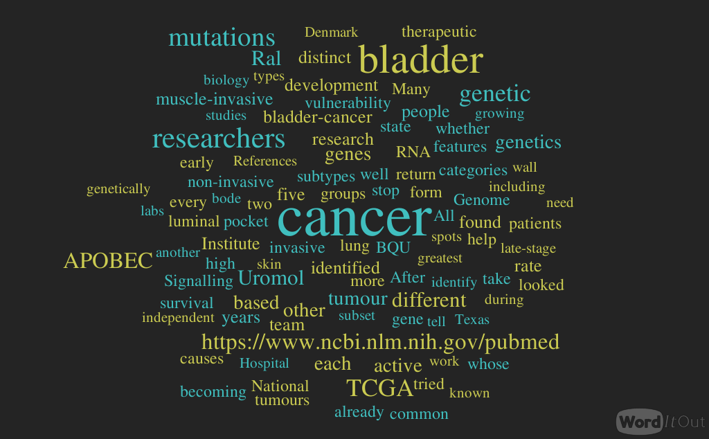


