Feeds:
Posts
Comments

Archive for the ‘Healthcare costs and reimbursement’ Category

The Cost to Value Conundrum in Cardiovascular Healthcare Provision

The Cost to Value Conundrum in Cardiovascular Healthcare Provision

Author: Larry H. Bernstein, MD, FCAP

Article ID #98: The Cost to Value Conundrum in Cardiovascular Healthcare Provision. Published on 1/1/2014

WordCloud Image Produced by Adam Tubman

I write this introduction to Volume 2 of the e-series on Cardiovascular Diseases, which curates the basic structure and physiology of the heart, the vasculature, and related structures, e.g., the kidney, with respect to:

1. Pathogenesis
2. Diagnosis
3. Treatment

Curation is an introductory portion to Volume Two, which is necessary to introduce the methodological design used to create the following articles. More needs not to be discussed about the methodology, which will become clear, if only that the content curated is changing based on success or failure of both diagnostic and treatment technology availability, as well as the systems needed to support the ongoing advances.  Curation requires:

  • meaningful selection,
  • enrichment, and
  • sharing combining sources and
  • creation of new synnthesis

Curators have to create a new perspective or idea on top of the existing media which supports the content in the original. The curator has to select from the myriad upon myriad options available, to re-share and critically view the work. A search can be overwhelming in size of the output, but the curator has to successfully pluck the best material straight out of that noise.

Part 1 is a highly important treatment that is not technological, but about the system now outdated to support our healthcare system, the most technolog-ically advanced in the world, with major problems in the availability of care related to economic disparities.  It is not about technology, per se, but about how we allocate healthcare resources, about individuals’ roles in a not full list of lifestyle maintenance options for self-care, and about the important advances emerging out of the Affordable Care Act (ACA), impacting enormously on Medicaid, which depends on state-level acceptance, on community hospital, ambulatory, and home-care or hospice restructuring, which includes the reduction of management overhead by the formation of regional healthcare alliances, the incorporation of physicians into hospital-based practices (with the hospital collecting and distributing the Part B reimbursement to the physician, with “performance-based” targets for privileges and payment – essential to the success of an Accountable Care Organization (AC)).  One problem that ACA has definitively address is the elimination of the exclusion of patients based on preconditions.  One problem that has been left unresolved is the continuing existence of private policies that meet financial capabilities of the contract to provide, but which provide little value to the “purchaser” of care.  This is a holdout that persists in for-profit managed care as an option.  A physician response to the new system of care, largely fostered by a refusal to accept Medicaid, is the formation of direct physician-patient contracted care without an intermediary.

In this respect, the problem is not simple, but is resolvable.  A proposal for improved economic stability has been prepared by Edward Ingram. A concern for American families and businesses is substantially addressed in a macroeconomic design concept, so that financial services like housing, government, and business finance, savings and pensions, boosting confidence at every level giving everyone a better chance of success in planning their personal savings and lifetime and business finances.

http://macro-economic-design.blogspot.com/p/book.html

Part 2 is a collection of scientific articles on the current advances in cardiac care by the best trained physicians the world has known, with mastery of the most advanced vascular instrumentation for medical or surgical interventions, the latest diagnostic ultrasound and imaging tools that are becoming outdated before the useful lifetime of the capital investment has been completed.  If we tie together Part 1 and Part 2, there is ample room for considering  clinical outcomes based on individual and organizational factors for best performance. This can really only be realized with considerable improvement in information infrastructure, which has miles to go.  Why should this be?  Because for generations of IT support systems, they are historically focused on billing and have made insignificant inroads into the front-end needs of the clinical staff.

Read Full Post »

The red tape challenge

reporter and curator: Dror Nir, PhD

Large part of the time and cost for developing a new medical device or a new drug is allocated for achieving regulatory compliance. While quality and safety are desired, having to continually spend additional time and  money throughout the product’s life cycle just on the proof of its quality and safety is painful to all, especially for the health systems which eventually have to pay for it.
On this issue, I bring you the following post:
It has almost become routine: under narratives of increased patient safety and improved efficiency new regulatory requirements are developed, resulting in increased requirements on the industry. The new European pharmacovigilance legislation and the upcoming European medical device regulatory updates are only two examples. Being part of the industry you have very limited impact on the regulations but have to comply with them anyway. That is – if you were to continue marketing your device or drug. Under certain circumstances the cost of meeting legal requirements is so great it may bring into question the viability of continuing certain business activities. This is especially the case for smaller companies or niche products.
R1
It is clear, thus, that you have a huge incentive to try to achieve compliance with minimal effort. If we take a bird’s eye view on the challenge of reaching compliance, two major elements become evident:
  1. The quality system is, in itself, a high maintenance object which consumes ongoing resources:
    • It needs to be revisited often due to changes in the regulatory system or in the business environment.
    • Each change may affect many components of the system and a quick modification may cause inconsistency.
    • Each modification needs to be accepted, signed-off formally by several people and be disseminated via formally recorded training.
    • The organization should withstand audits and inspections in regards to the quality system.
  2. Living with the quality system: Each SOP and work instruction has to be followed, and typically forms need to be filled, signed and filed.
Information Overload

Young companies which are just embarking on the regulatory path often do not realize these two characteristics of the quality system. Quick fixes in the form of SOP texts copied from other organizations or generic templates are being used to get the initial certification. However, as the organization evolves it realizes that a quality system is not a one-time effort and cannot be glued on from external sources.  It has to be streamlined and become part of the way that the organization lives and does business. Companies are enjoying the benefits of improved process design and automation on a large scale every day, in many areas. When recently did you see a delivery person arriving to a pickup without a Barcode reader, so that he does not need to fill any form manually? When was the last time that a software package was released without an automatic consistency check? So too your quality system and related processes may be dramatically engineered to serve you better.

Better efficiency in quality compliance should thus be achieved through careful analysis and optimization of two types of processes:
How do we better maintain the quality system? How do we make it easier to change the system, keep it consistent, train in it, etc.
The SOPs and work instructions: SOPs cannot be just imported from outside or suggested by a QA/RA consultant who does not know the organization very well. SOPs should be a true marriage between the legal and business requirements and should be the result of a careful consideration by all stakeholders. From my experience, the best SOPs are written by the process owner, with the guidance of the regulatory expert. For example: the R&D manager should be the one drafting the design control SOP, with input of the regulatory expert. Such a SOP is much more likely to fit the business needs, and also more likely to be followed by the process owner.
Yes, I realize that thinking this way is very often not what companies do when they rush compliance. I insist that this is what has to be done to achieve sustainable compliance. The good news is that, when companies do look at their quality system in this way, they see many opportunities for significant improvement. Some of those improvements are achieved through use of better IT tools. These tools would typically be in the area of document management and versioning, workflow automation, improved collaboration and electronic signatures. Like any other change, this also requires a vision and a certain effort. However, the long term business impact may be as significant as the difference between business success or failure.

Read Full Post »

Stanford Dropout is Already Drawing Comparisons with Steve Jobs

Larry H Bernstein, MD, Reporter

Article ID #89: Stanford Dropout is Already Drawing Comparisons with Steve Jobs. Published on 11/26/2013

WordCloud Image Produced by Adam Tubman

An interview by Eric Topol on Medscape of a 29 year-old Stanford University dropout is fascinating.

Editor’s Note:

If 29-year-old Elizabeth Holmes has her way, patients will no longer have to go to physicians’ offices, hospitals or laboratories to get high-complexity diagnostic blood tests. Nor will vial after vial of blood draws be necessary to do these tests.

Barely out of the gate after a decade of secrecy, the Stanford dropout is already drawing comparisons with Steve Jobs (she often wears the same black turtleneck). And her company, Theranos, Inc., which emerged from the shadows in September, just might be healthcare’s answer to Apple.[1] The so-called disruptive technology that Ms. Holmes, a former engineering major, and Theranos have created is said to have the potential to shake up and forever change the way laboratory medicine is conducted. Since forgoing college at 19, Ms. Holmes has secured millions of dollars in funding for her new venture, including $45 million in private equity funding in 2010.[2] The board of directors of her company is a Who’s Who of distinguished former and current technology, academic, and government officials.[2,3]

In an exclusive interview, Ms. Holmes talks to Medscape Editor-in-Chief Eric J. Topol, MD, about the decade she spent building her company; plans for the present and the future, including a recent deal with Walgreens drugstores; and whether she’s on the path to the creative destruction of laboratory medicine.

Leaving Stanford at Age 19

Dr. Topol: Hello. I’m Dr. Eric Topol, Editor-in-Chief of Medscape. Joining me today for Medscape One-on-One is Elizabeth Holmes, Founder, President, and CEO of Theranos.  We are here in Palo Alto, California, at the company’s headquarters. Elizabeth, welcome. This is going to be a fascinating discussion.

Ms. Holmes: Thank you. It’s wonderful to be here and have you here.

Dr. Topol: This is a story that has been brewing for a long time. You were at Stanford University, and at age 19 you decided to change your path. Is that right?

Ms. Holmes: Yes.

Dr. Topol: What made you think, “I’m on to something, and I don’t want to do college; I’ve got something else that’s  probably bigger than that”?

Ms. Holmes: I knew that I wanted to do something that could make a difference in the world.

To me, there was nothing greater that I could build than something that would change the reality in our healthcare system today, which is that when someone you love gets really, really sick, usually by the time you find that out, it’s too late to be able to do something about it. And in those moments it’s heartbreaking, because there is nothing you wouldn’t do.


Read Full Post »

The importance of spatially-localized and quantified image interpretation in cancer management

Writer & reporter: Dror Nir, PhD

I became involved in the development of quantified imaging-based tissue characterization more than a decade ago. From the start, it was clear to me that what clinicians needs will not be answered by just identifying whether a certain organ harbors cancer. If imaging devices are to play a significant role in future medicine, as a complementary source of information to bio-markers and gene sequencing the minimum value expected of them is accurate directing of biopsy needles and treatment tools to the malignant locations in the organ.  Therefore, the design goal of the first Prostate-HistoScanning (“PHS”) version I went into the trouble of characterizing localized volume of tissue at the level of approximately 0.1cc (1x1x1 mm). Thanks to that, the imaging-interpretation overlay of PHS localizes the suspicious lesions with accuracy of 5mm within the prostate gland; Detection, localisation and characterisation of prostate cancer by prostate HistoScanning(™).

I then started a more ambitious research aiming to explore the feasibility of identifying sub-structures within the cancer lesion itself. The preliminary results of this exploration were so promising that it surprised not only the clinicians I was working with but also myself. It seems, that using quality ultrasound, one can find Imaging-Biomarkers that allows differentiation of inside structures of a cancerous lesions. Unfortunately, for everyone involved in this work, including me, this scientific effort was interrupted by financial constrains before reaching maturity.

My short introduction was made to explain why I find the publication below important enough to post and bring to your attention.

I hope for your agreement on the matter.

Quantitative Imaging in Cancer Evolution and Ecology

Robert A. Gatenby, MD, Olya Grove, PhD and Robert J. Gillies, PhD

From the Departments of Radiology and Cancer Imaging and Metabolism, Moffitt Cancer Center, 12902 Magnolia Dr, Tampa, FL 33612. Address correspondence to  R.A.G. (e-mail: Robert.Gatenby@Moffitt.org).

Abstract

Cancer therapy, even when highly targeted, typically fails because of the remarkable capacity of malignant cells to evolve effective adaptations. These evolutionary dynamics are both a cause and a consequence of cancer system heterogeneity at many scales, ranging from genetic properties of individual cells to large-scale imaging features. Tumors of the same organ and cell type can have remarkably diverse appearances in different patients. Furthermore, even within a single tumor, marked variations in imaging features, such as necrosis or contrast enhancement, are common. Similar spatial variations recently have been reported in genetic profiles. Radiologic heterogeneity within tumors is usually governed by variations in blood flow, whereas genetic heterogeneity is typically ascribed to random mutations. However, evolution within tumors, as in all living systems, is subject to Darwinian principles; thus, it is governed by predictable and reproducible interactions between environmental selection forces and cell phenotype (not genotype). This link between regional variations in environmental properties and cellular adaptive strategies may permit clinical imaging to be used to assess and monitor intratumoral evolution in individual patients. This approach is enabled by new methods that extract, report, and analyze quantitative, reproducible, and mineable clinical imaging data. However, most current quantitative metrics lack spatialness, expressing quantitative radiologic features as a single value for a region of interest encompassing the whole tumor. In contrast, spatially explicit image analysis recognizes that tumors are heterogeneous but not well mixed and defines regionally distinct habitats, some of which appear to harbor tumor populations that are more aggressive and less treatable than others. By identifying regional variations in key environmental selection forces and evidence of cellular adaptation, clinical imaging can enable us to define intratumoral Darwinian dynamics before and during therapy. Advances in image analysis will place clinical imaging in an increasingly central role in the development of evolution-based patient-specific cancer therapy.

© RSNA, 2013


Introduction

Cancers are heterogeneous across a wide range of temporal and spatial scales. Morphologic heterogeneity between and within cancers is readily apparent in clinical imaging, and subjective descriptors of these differences, such as necrotic, spiculated, and enhancing, are common in the radiology lexicon. In the past several years, radiology research has increasingly focused on quantifying these imaging variations in an effort to understand their clinical and biologic implications (1,2). In parallel, technical advances now permit extensive molecular characterization of tumor cells in individual patients. This has led to increasing emphasis on personalized cancer therapy, in which treatment is based on the presence of specific molecular targets (3). However, recent studies (4,5) have shown that multiple genetic subpopulations coexist within cancers, reflecting extensive intratumoral somatic evolution. This heterogeneity is a clear barrier to therapy based on molecular targets, since the identified targets do not always represent the entire population of tumor cells in a patient (6,7). It is ironic that cancer, a disease extensively and primarily analyzed genetically, is also the most genetically flexible of all diseases and, therefore, least amenable to such an approach.

Genetic variations in tumors are typically ascribed to a mutator phenotype that generates new clones, some of which expand into large populations (8). However, although identification of genotypes is of substantial interest, it is insufficient for complete characterization of tumor dynamics because evolution is governed by the interactions of environmental selection forces with the phenotypic, not genotypic, properties of populations as shown, for example, by evolutionary convergence to identical phenotypes among cave fish even when they are from different species (911). This connection between tissue selection forces and cellular properties has the potential to provide a strong bridge between medical imaging and the cellular and molecular properties of cancers.

We postulate that differences within tumors at different spatial scales (ie, at the radiologic, cellular, and molecular [genetic] levels) are related. Tumor characteristics observable at clinical imaging reflect molecular-, cellular-, and tissue-level dynamics; thus, they may be useful in understanding the underlying evolving biology in individual patients. A challenge is that such mapping across spatial and temporal scales requires not only objective reproducible metrics for imaging features but also a theoretical construct that bridges those scales (Fig 1).

P1a

Figure 1a: Computed tomographic (CT) scan of right upper lobe lung cancer in a 50-year-old woman.

P1b

Figure 1b: Isoattenuation map shows regional heterogeneity at the tissue scale (measured in centimeters).

 cd

Figure 1c & 1d: (c, d)Whole-slide digital images (original magnification, ×3) of a histologic slice of the same tumor at the mesoscopic scale (measured in millimeters) (c) coupled with a masked image of regional morphologic differences showing spatial heterogeneity (d). 

p1e

Figure 1e: Subsegment of the whole slide image shows the microscopic scale (measured in micrometers) (original magnification, ×50).

p1f

Figure 1f: Pattern recognition masked image shows regional heterogeneity. In a, the CT image of non–small cell lung cancer can be analyzed to display gradients of attenuation, which reveals heterogeneous and spatially distinct environments (b). Histologic images in the same patient (c, e) reveal heterogeneities in tissue structure and density on the same scale as seen in the CT images. These images can be analyzed at much higher definition to identify differences in morphologies of individual cells (3), and these analyses reveal clusters of cells with similar morphologic features (d, f). An important goal of radiomics is to bridge radiologic data with cellular and molecular characteristics observed microscopically.

To promote the development and implementation of quantitative imaging methods, protocols, and software tools, the National Cancer Institute has established the Quantitative Imaging Network. One goal of this program is to identify reproducible quantifiable imaging features of tumors that will permit data mining and explicit examination of links between the imaging findings and the underlying molecular and cellular characteristics of the tumors. In the quest for more personalized cancer treatments, these quantitative radiologic features potentially represent nondestructive temporally and spatially variable predictive and prognostic biomarkers that readily can be obtained in each patient before, during, and after therapy.

Quantitative imaging requires computational technologies that can be used to reliably extract mineable data from radiographic images. This feature information can then be correlated with molecular and cellular properties by using bioinformatics methods. Most existing methods are agnostic and focus on statistical descriptions of existing data, without presupposing the existence of specific relationships. Although this is a valid approach, a more profound understanding of quantitative imaging information may be obtained with a theoretical hypothesis-driven framework. Such models use links between observable tumor characteristics and microenvironmental selection factors to make testable predictions about emergent phenotypes. One such theoretical framework is the developing paradigm of cancer as an ecologic and evolutionary process.

For decades, landscape ecologists have studied the effects of heterogeneity in physical features on interactions between populations of organisms and their environments, often by using observation and quantification of images at various scales (1214). We propose that analytic models of this type can easily be applied to radiologic studies of cancer to uncover underlying molecular, cellular, and microenvironmental drivers of tumor behavior and specifically, tumor adaptations and responses to therapy (15).

In this article, we review recent developments in quantitative imaging metrics and discuss how they correlate with underlying genetic data and clinical outcomes. We then introduce the concept of using ecology and evolutionary models for spatially explicit image analysis as an exciting potential avenue of investigation.


Quantitative Imaging and Radiomics

In patients with cancer, quantitative measurements are commonly limited to measurement of tumor size with one-dimensional (Response Evaluation Criteria in Solid Tumors [or RECIST]) or two-dimensional (World Health Organization) long-axis measurements (16). These measures do not reflect the complexity of tumor morphology or behavior, and in many cases, changes in these measures are not predictive of therapeutic benefit (17). In contrast, radiomics (18) is a high-throughput process in which a large number of shape, edge, and texture imaging features are extracted, quantified, and stored in databases in an objective, reproducible, and mineable form (Figs 12). Once transformed into a quantitative form, radiologic tumor properties can be linked to underlying genetic alterations (the field is called radiogenomics) (1921) and to medical outcomes (2227). Researchers are currently working to develop both a standardized lexicon to describe tumor features (28,29) and a standard method to convert these descriptors into quantitative mineable data (30,31) (Fig 3).

p2

Figure 2: Contrast-enhanced CT scans show non–small cell lung cancer (left) and corresponding cluster map (right). Subregions within the tumor are identified by clustering pixels based on the attenuation of pixels and their cumulative standard deviation across the region. While the entire region of interest of the tumor, lacking the spatial information, yields a weighted mean attenuation of 859.5 HU with a large and skewed standard deviation of 243.64 HU, the identified subregions have vastly different statistics. Mean attenuation was 438.9 HU ± 45 in the blue subregion, 210.91 HU ± 79 in the yellow subregion, and 1077.6 HU ± 18 in the red subregion.


p3

Figure 3: Chart shows the five processes in radiomics.

Several recent articles underscore the potential power of feature analysis. After manually extracting more than 100 CT image features, Segal and colleagues found that a subset of 14 features predicted 80% of the gene expression pattern in patients with hepatocellular carcinoma (21). A similar extraction of features from contrast agent–enhanced magnetic resonance (MR) images of glioblastoma was used to predict immunohistochemically identified protein expression patterns (22). Other radiomic features, such as texture, can be used to predict response to therapy in patients with renal cancer (32) and prognosis in those with metastatic colon cancer (33).

These pioneering studies were relatively small because the image analysis was performed manually, and the studies were consequently underpowered. Thus, recent work in radiomics has focused on technical developments that permit automated extraction of image features with the potential for high throughput. Such methods, which rely heavily on novel machine learning algorithms, can more completely cover the range of quantitative features that can describe tumor heterogeneity, such as texture, shape, or margin gradients or, importantly, different environments, or niches, within the tumors.

Generally speaking, texture in a biomedical image is quantified by identifying repeating patterns. Texture analyses fall into two broad categories based on the concepts of first- and second-order spatial statistics. First-order statistics are computed by using individual pixel values, and no relationships between neighboring pixels are assumed or evaluated. Texture analysis methods based on first-order statistics usually involve calculating cumulative statistics of pixel values and their histograms across the region of interest. Second-order statistics, on the other hand, are used to evaluate the likelihood of observing spatially correlated pixels (34). Hence, second-order texture analyses focus on the detection and quantification of nonrandom distributions of pixels throughout the region of interest.

The technical developments that permit second-order texture analysis in tumors by using regional enhancement patterns on dynamic contrast-enhanced MR images were reviewed recently (35). One such technique that is used to measure heterogeneity of contrast enhancement uses the Factor Analysis of Medical Image Sequences (or FAMIS) algorithm, which divides tumors into regions based on their patterns of enhancement (36). Factor Analysis of Medical Image Sequences–based analyses yielded better prognostic information when compared with region of interest–based methods in numerous cancer types (1921,3739), and they were a precursor to the Food and Drug Administration–approved three-time-point method (40). A number of additional promising methods have been developed. Rose and colleagues showed that a structured fractal-based approach to texture analysis improved differentiation between low- and high-grade brain cancers by orders of magnitude (41). Ahmed and colleagues used gray level co-occurrence matrix analyses of dynamic contrast-enhanced images to distinguish benign from malignant breast masses with high diagnostic accuracy (area under the receiver operating characteristic curve, 0.92) (26). Others have shown that Minkowski functional structured methods that convolve images with differently kernelled masks can be used to distinguish subtle differences in contrast enhancement patterns and can enable significant differentiation between treatment groups (42).

It is not surprising that analyses of heterogeneity in enhancement patterns can improve diagnosis and prognosis, as this heterogeneity is fundamentally based on perfusion deficits, which generate significant microenvironmental selection pressures. However, texture analysis is not limited to enhancement patterns. For example, measures of heterogeneity in diffusion-weighted MR images can reveal differences in cellular density in tumors, which can be matched to histologic findings (43). Measures of heterogeneity in T1- and T2-weighted images can be used to distinguish benign from malignant soft-tissue masses (23). CT-based texture features have been shown to be highly significant independent predictors of survival in patients with non–small cell lung cancer (24).

Texture analyses can also be applied to positron emission tomographic (PET) data, where they can provide information about metabolic heterogeneity (25,26). In a recent study, Nair and colleagues identified 14 quantitative PET imaging features that correlated with gene expression (19). This led to an association of metagene clusters to imaging features and yielded prognostic models with hazard ratios near 6. In a study of esophageal cancer, in which 38 quantitative features describing fluorodeoxyglucose uptake were extracted, measures of metabolic heterogeneity at baseline enabled prediction of response with significantly higher sensitivity than any whole region of interest standardized uptake value measurement (22). It is also notable that these extensive texture-based features are generally more reproducible than simple measures of the standardized uptake value (27), which can be highly variable in a clinical setting (44).


Spatially Explicit Analysis of Tumor Heterogeneity

Although radiomic analyses have shown high prognostic power, they are not inherently spatially explicit. Quantitative border, shape, and texture features are typically generated over a region of interest that comprises the entire tumor (45). This approach implicitly assumes that tumors are heterogeneous but well mixed. However, spatially explicit subregions of cancers are readily apparent on contrast-enhanced MR or CT images, as perfusion can vary markedly within the tumor, even over short distances, with changes in tumor cell density and necrosis.

An example is shown in Figure 2, which shows a contrast-enhanced CT scan of non–small cell lung cancer. Note that there are many subregions within this tumor that can be identified with attenuation gradient (attenuation per centimeter) edge detection algorithms. Each subregion has a characteristic quantitative attenuation, with a narrow standard deviation, whereas the mean attenuation over the entire region of interest is a weighted average of the values across all subregions, with a correspondingly large and skewed distribution. We contend that these subregions represent distinct habitats within the tumor, each with a distinct set of environmental selection forces.

These observations, along with the recent identification of regional variations in the genetic properties of tumor cells, indicate the need to abandon the conceptual model of cancers as bounded organlike structures. Rather than a single self-organized system, cancers represent a patchwork of habitats, each with a unique set of environmental selection forces and cellular evolution strategies. For example, regions of the tumor that are poorly perfused can be populated by only those cells that are well adapted to low-oxygen, low-glucose, and high-acid environmental conditions. Such adaptive responses to regional heterogeneity result in microenvironmental selection and hence, emergence of genetic variations within tumors. The concept of adaptive response is an important departure from the traditional view that genetic heterogeneity is the product of increased random mutations, which implies that molecular heterogeneity is fundamentally unpredictable and, therefore, chaotic. The Darwinian model proposes that genetic heterogeneity is the result of a predictable and reproducible selection of successful adaptive strategies to local microenvironmental conditions.

Current cross-sectional imaging modalities can be used to identify regional variations in selection forces by using contrast-enhanced, cell density–based, or metabolic features. Clinical imaging can also be used to identify evidence of cellular adaptation. For example, if a region of low perfusion on a contrast-enhanced study is necrotic, then an adaptive population is absent or minimal. However, if the poorly perfused area is cellular, then there is presumptive evidence of an adapted proliferating population. While the specific genetic properties of this population cannot be determined, the phenotype of the adaptive strategy is predictable since the environmental conditions are more or less known. Thus, standard medical images can be used to infer specific emergent phenotypes and, with ongoing research, these phenotypes can be associated with underlying genetic changes.

This area of investigation will likely be challenging. As noted earlier, the most obvious spatially heterogeneous imaging feature in tumors is perfusion heterogeneity on contrast-enhanced CT or MR images. It generally has been assumed that the links between contrast enhancement, blood flow, perfusion, and tumor cell characteristics are straightforward. That is, tumor regions with decreased blood flow will exhibit low perfusion, low cell density, and high necrosis. In reality, however, the dynamics are actually much more complex. As shown in Figure 4, when using multiple superimposed sequences from MR imaging of malignant gliomas, regions of tumor that are poorly perfused on contrast-enhanced T1-weighted images may exhibit areas of low or high water content on T2-weighted images and low or high diffusion on diffusion-weighted images. Thus, high or low cell densities can coexist in poorly perfused volumes, creating perfusion-diffusion mismatches. Regions with poor perfusion with high cell density are of particular clinical interest because they represent a cell population that is apparently adapted to microenvironmental conditions associated with poor perfusion. The associated hypoxia, acidosis, and nutrient deprivation select for cells that are resistant to apoptosis and thus are likely to be resistant to therapy (46,47).

p4

Figure 4: Left: Contrast-enhanced T1 image from subject TCGA-02-0034 in The Cancer Genome Atlas–Glioblastoma Multiforme repository of MR volumes of glioblastoma multiforme cases. Right: Spatial distribution of MR imaging–defined habitats within the tumor. The blue region (low T1 postgadolinium, low fluid-attenuated inversion recovery) is particularly notable because it presumably represents a habitat with low blood flow but high cell density, indicating a population presumably adapted to hypoxic acidic conditions.

Furthermore, other selection forces not related to perfusion are likely to be present within tumors. For example, evolutionary models suggest that cancer cells, even in stable microenvironments, tend to speciate into “engineers” that maximize tumor cell growth by promoting angiogenesis and “pioneers” that proliferate by invading normal issue and co-opting the blood supply. These invasive tumor phenotypes can exist only at the tumor edge, where movement into a normal tissue microenvironment can be rewarded by increased proliferation. This evolutionary dynamic may contribute to distinct differences between the tumor edges and the tumor cores, which frequently can be seen at analysis of cross-sectional images (Fig 5).

p5a

Figure 5a: CT images obtained with conventional entropy filtering in two patients with non–small cell lung cancer with no apparent textural differences show similar entropy values across all sections. 

p5b

Figure 5b: Contour plots obtained after the CT scans were convolved with the entropy filter. Further subdividing each section in the tumor stack into tumor edge and core regions (dotted black contour) reveals varying textural behavior across sections. Two distinct patterns have emerged, and preliminary analysis shows that the change of mean entropy value between core and edge regions correlates negatively with survival.

Interpretation of the subsegmentation of tumors will require computational models to understand and predict the complex nonlinear dynamics that lead to heterogeneous combinations of radiographic features. We have exploited ecologic methods and models to investigate regional variations in cancer environmental and cellular properties that lead to specific imaging characteristics. Conceptually, this approach assumes that regional variations in tumors can be viewed as a coalition of distinct ecologic communities or habitats of cells in which the environment is governed, at least to first order, by variations in vascular density and blood flow. The environmental conditions that result from alterations in blood flow, such as hypoxia, acidosis, immune response, growth factors, and glucose, represent evolutionary selection forces that give rise to local-regional phenotypic adaptations. Phenotypic alterations can result from epigenetic, genetic, or chromosomal rearrangements, and these in turn will affect prognosis and response to therapy. Changes in habitats or the relative abundance of specific ecologic communities over time and in response to therapy may be a valuable metric with which to measure treatment efficacy and emergence of resistant populations.


Emerging Strategies for Tumor Habitat Characterization

A method for converting images to spatially explicit tumor habitats is shown in Figure 4. Here, three-dimensional MR imaging data sets from a glioblastoma are segmented. Each voxel in the tumor is defined by a scale that includes its image intensity in different sequences. In this case, the imaging sets are from (a) a contrast-enhanced T1 sequence, (b) a fast spin-echo T2 sequence, and (c) a fluid-attenuated inversion-recovery (or FLAIR) sequence. Voxels in each sequence can be defined as high or low based on their value compared with the mean signal value. By using just two sequences, a contrast-enhanced T1 sequence and a fluid-attenuated inversion-recovery sequence, we can define four habitats: high or low postgadolinium T1 divided into high or low fluid-attenuated inversion recovery. When these voxel habitats are projected into the tumor volume, we find they cluster into spatially distinct regions. These habitats can be evaluated both in terms of their relative contributions to the total tumor volume and in terms of their interactions with each other, based on the imaging characteristics at the interfaces between regions. Similar spatially explicit analysis can be performed with CT scans (Fig 5).

Analysis of spatial patterns in cross-sectional images will ultimately require methods that bridge spatial scales from microns to millimeters. One possible method is a general class of numeric tools that is already widely used in terrestrial and marine ecology research to link species occurrence or abundance with environmental parameters. Species distribution models (4851) are used to gain ecologic and evolutionary insights and to predict distributions of species or morphs across landscapes, sometimes extrapolating in space and time. They can easily be used to link the environmental selection forces in MR imaging-defined habitats to the evolutionary dynamics of cancer cells.

Summary

Imaging can have an enormous role in the development and implementation of patient-specific therapies in cancer. The achievement of this goal will require new methods that expand and ultimately replace the current subjective qualitative assessments of tumor characteristics. The need for quantitative imaging has been clearly recognized by the National Cancer Institute and has resulted in formation of the Quantitative Imaging Network. A critical objective of this imaging consortium is to use objective, reproducible, and quantitative feature metrics extracted from clinical images to develop patient-specific imaging-based prognostic models and personalized cancer therapies.

It is increasingly clear that tumors are not homogeneous organlike systems. Rather, they contain regional coalitions of ecologic communities that consist of evolving cancer, stroma, and immune cell populations. The clinical consequence of such niche variations is that spatial and temporal variations of tumor phenotypes will inevitably evolve and present substantial challenges to targeted therapies. Hence, future research in cancer imaging will likely focus on spatially explicit analysis of tumor regions.

Clinical imaging can readily characterize regional variations in blood flow, cell density, and necrosis. When viewed in a Darwinian evolutionary context, these features reflect regional variations in environmental selection forces and can, at least in principle, be used to predict the likely adaptive strategies of the local cancer population. Hence, analyses of radiologic data can be used to inform evolutionary models and then can be mapped to regional population dynamics. Ecologic and evolutionary principles may provide a theoretical framework to link imaging to the cellular and molecular features of cancer cells and ultimately lead to a more comprehensive understanding of specific cancer biology in individual patients.


Essentials

  • • Marked heterogeneity in genetic properties of different cells in the same tumor is typical and reflects ongoing intratumoral evolution.
  • • Evolution within tumors is governed by Darwinian dynamics, with identifiable environmental selection forces that interact with phenotypic (not genotypic) properties of tumor cells in a predictable and reproducible manner; clinical imaging is uniquely suited to measure temporal and spatial heterogeneity within tumors that is both a cause and a consequence of this evolution.
  • • Subjective radiologic descriptors of cancers are inadequate to capture this heterogeneity and must be replaced by quantitative metrics that enable statistical comparisons between features describing intratumoral heterogeneity and clinical outcomes and molecular properties.
  • • Spatially explicit mapping of tumor regions, for example by superimposing multiple imaging sequences, may permit patient-specific characterization of intratumoral evolution and ecology, leading to patient- and tumor-specific therapies.
  • • We summarize current information on quantitative analysis of radiologic images and propose future quantitative imaging must become spatially explicit to identify intratumoral habitats before and during therapy.

Disclosures of Conflicts of Interest: R.A.G. No relevant conflicts of interest to disclose. O.G. No relevant conflicts of interest to disclose.R.J.G. No relevant conflicts of interest to disclose.


Acknowledgments

The authors thank Mark Lloyd, MS; Joel Brown, PhD; Dmitry Goldgoff, PhD; and Larry Hall, PhD, for their input to image analysis and for their lively and informative discussions.

Footnotes

  • Received December 18, 2012; revision requested February 5, 2013; revision received March 11; accepted April 9; final version accepted April 29.
  • Funding: This research was supported by the National Institutes of Health (grants U54CA143970-01, U01CA143062; R01CA077575, andR01CA170595).

References

    1. Kurland BF,
    2. Gerstner ER,
    3. Mountz JM,
    4. et al

    . Promise and pitfalls of quantitative imaging in oncology clinical trials. Magn Reson Imaging2012;30(9):1301–1312.

    1. Levy MA,
    2. Freymann JB,
    3. Kirby JS,
    4. et al

    . Informatics methods to enable sharing of quantitative imaging research data. Magn Reson Imaging2012;30(9):1249–1256.

    1. Mirnezami R,
    2. Nicholson J,
    3. Darzi A

    . Preparing for precision medicine. N Engl J Med 2012;366(6):489–491.

    1. Yachida S,
    2. Jones S,
    3. Bozic I,
    4. et al

    . Distant metastasis occurs late during the genetic evolution of pancreatic cancer. Nature 2010;467(7319):1114–1117.

    1. Gerlinger M,
    2. Rowan AJ,
    3. Horswell S,
    4. et al

    . Intratumor heterogeneity and branched evolution revealed by multiregion sequencing. N Engl J Med2012;366(10):883–892.

    1. Gerlinger M,
    2. Swanton C

    . How Darwinian models inform therapeutic failure initiated by clonal heterogeneity in cancer medicine. Br J Cancer2010;103(8):1139–1143.

    1. Kern SE

    . Why your new cancer biomarker may never work: recurrent patterns and remarkable diversity in biomarker failures. Cancer Res2012;72(23):6097–6101.

    1. Nowell PC

    . The clonal evolution of tumor cell populations. Science1976;194(4260):23–28.

    1. Greaves M,
    2. Maley CC

    . Clonal evolution in cancer. Nature2012;481(7381):306–313.

    1. Vincent TL,
    2. Brown JS

    . Evolutionary game theory, natural selection and Darwinian dynamics. Cambridge, England: Cambridge University Press, 2005.

    1. Gatenby RA,
    2. Gillies RJ

    . A microenvironmental model of carcinogenesis. Nat Rev Cancer 2008;8(1):56–61.

    1. Bowers MA,
    2. Matter SF

    . Landscape ecology of mammals: relationships between density and patch size. J Mammal 1997;78(4):999–1013.

    1. Dorner BK,
    2. Lertzman KP,
    3. Fall J

    . Landscape pattern in topographically complex landscapes: issues and techniques for analysis. Landscape Ecol2002;17(8):729–743.

    1. González-García I,
    2. Solé RV,
    3. Costa J

    . Metapopulation dynamics and spatial heterogeneity in cancer. Proc Natl Acad Sci U S A2002;99(20):13085–13089.

    1. Patel LR,
    2. Nykter M,
    3. Chen K,
    4. Zhang W

    . Cancer genome sequencing: understanding malignancy as a disease of the genome, its conformation, and its evolution. Cancer Lett 2012 Oct 27. [Epub ahead of print]

    1. Jaffe CC

    . Measures of response: RECIST, WHO, and new alternatives. J Clin Oncol 2006;24(20):3245–3251.

    1. Burton A

    . RECIST: right time to renovate? Lancet Oncol2007;8(6):464–465.

    1. Lambin P,
    2. Rios-Velazquez E,
    3. Leijenaar R,
    4. et al

    . Radiomics: extracting more information from medical images using advanced feature analysis. Eur J Cancer 2012;48(4):441–446.

    1. Nair VS,
    2. Gevaert O,
    3. Davidzon G,
    4. et al

    . Prognostic PET 18F-FDG uptake imaging features are associated with major oncogenomic alterations in patients with resected non-small cell lung cancer. Cancer Res2012;72(15):3725–3734.

    1. Diehn M,
    2. Nardini C,
    3. Wang DS,
    4. et al

    . Identification of noninvasive imaging surrogates for brain tumor gene-expression modules. Proc Natl Acad Sci U S A 2008;105(13):5213–5218.

    1. Segal E,
    2. Sirlin CB,
    3. Ooi C,
    4. et al

    . Decoding global gene expression programs in liver cancer by noninvasive imaging. Nat Biotechnol 2007;25(6):675–680.

    1. Tixier F,
    2. Le Rest CC,
    3. Hatt M,
    4. et al

    . Intratumor heterogeneity characterized by textural features on baseline 18F-FDG PET images predicts response to concomitant radiochemotherapy in esophageal cancer. J Nucl Med2011;52(3):369–378.

    1. Pang KK,
    2. Hughes T

    . MR imaging of the musculoskeletal soft tissue mass: is heterogeneity a sign of malignancy? J Chin Med Assoc2003;66(11):655–661.

    1. Ganeshan B,
    2. Panayiotou E,
    3. Burnand K,
    4. Dizdarevic S,
    5. Miles K

    . Tumour heterogeneity in non-small cell lung carcinoma assessed by CT texture analysis: a potential marker of survival. Eur Radiol 2012;22(4):796–802.

    1. Asselin MC,
    2. O’Connor JP,
    3. Boellaard R,
    4. Thacker NA,
    5. Jackson A

    . Quantifying heterogeneity in human tumours using MRI and PET. Eur J Cancer2012;48(4):447–455.

    1. Ahmed A,
    2. Gibbs P,
    3. Pickles M,
    4. Turnbull L

    . Texture analysis in assessment and prediction of chemotherapy response in breast cancer. J Magn Reson Imaging doi:10.1002/jmri.23971 2012. Published online December 13, 2012.

    1. Kawata Y,
    2. Niki N,
    3. Ohmatsu H,
    4. et al

    . Quantitative classification based on CT histogram analysis of non-small cell lung cancer: correlation with histopathological characteristics and recurrence-free survival. Med Phys2012;39(2):988–1000.

    1. Rubin DL

    . Creating and curating a terminology for radiology: ontology modeling and analysis. J Digit Imaging 2008;21(4):355–362.

    1. Opulencia P,
    2. Channin DS,
    3. Raicu DS,
    4. Furst JD

    . Mapping LIDC, RadLex™, and lung nodule image features. J Digit Imaging 2011;24(2):256–270.

    1. Channin DS,
    2. Mongkolwat P,
    3. Kleper V,
    4. Rubin DL

    . The Annotation and Image Mark-up project. Radiology 2009;253(3):590–592.

    1. Rubin DL,
    2. Mongkolwat P,
    3. Kleper V,
    4. Supekar K,
    5. Channin DS

    . Medical imaging on the semantic web: annotation and image markup. Presented at the AAAI Spring Symposium Series, Semantic Scientific Knowledge Integration, Palo Alto, Calif, March 26–28, 2008.

    1. Goh V,
    2. Ganeshan B,
    3. Nathan P,
    4. Juttla JK,
    5. Vinayan A,
    6. Miles KA

    . Assessment of response to tyrosine kinase inhibitors in metastatic renal cell cancer: CT texture as a predictive biomarker. Radiology 2011;261(1):165–171.

    1. Miles KA,
    2. Ganeshan B,
    3. Griffiths MR,
    4. Young RC,
    5. Chatwin CR

    . Colorectal cancer: texture analysis of portal phase hepatic CT images as a potential marker of survival. Radiology 2009;250(2):444–452.

    1. Haralick RM,
    2. Shanmugam K,
    3. Dinstein I

    . Textural features for image classification. IEEE Trans Syst Man Cybern 1973;3(6):610–621.

    1. Yang X,
    2. Knopp MV

    . Quantifying tumor vascular heterogeneity with dynamic contrast-enhanced magnetic resonance imaging: a review. J Biomed Biotechnol 2011;2011:732848.

    1. Frouin F,
    2. Bazin JP,
    3. Di Paola M,
    4. Jolivet O,
    5. Di Paola R

    . FAMIS: a software package for functional feature extraction from biomedical multidimensional images. Comput Med Imaging Graph 1992;16(2):81–91.

    1. Frouge C,
    2. Guinebretière JM,
    3. Contesso G,
    4. Di Paola R,
    5. Bléry M

    . Correlation between contrast enhancement in dynamic magnetic resonance imaging of the breast and tumor angiogenesis. Invest Radiol 1994;29(12):1043–1049.

    1. Zagdanski AM,
    2. Sigal R,
    3. Bosq J,
    4. Bazin JP,
    5. Vanel D,
    6. Di Paola R

    . Factor analysis of medical image sequences in MR of head and neck tumors. AJNR Am J Neuroradiol 1994;15(7):1359–1368.

    1. Bonnerot V,
    2. Charpentier A,
    3. Frouin F,
    4. Kalifa C,
    5. Vanel D,
    6. Di Paola R

    . Factor analysis of dynamic magnetic resonance imaging in predicting the response of osteosarcoma to chemotherapy. Invest Radiol 1992;27(10):847–855.

    1. Furman-Haran E,
    2. Grobgeld D,
    3. Kelcz F,
    4. Degani H

    . Critical role of spatial resolution in dynamic contrast-enhanced breast MRI. J Magn Reson Imaging2001;13(6):862–867.

    1. Rose CJ,
    2. Mills SJ,
    3. O’Connor JPB,
    4. et al

    . Quantifying spatial heterogeneity in dynamic contrast-enhanced MRI parameter maps. Magn Reson Med2009;62(2):488–499.

    1. Canuto HC,
    2. McLachlan C,
    3. Kettunen MI,
    4. et al

    . Characterization of image heterogeneity using 2D Minkowski functionals increases the sensitivity of detection of a targeted MRI contrast agent. Magn Reson Med2009;61(5):1218–1224.

    1. Lloyd MC,
    2. Allam-Nandyala P,
    3. Purohit CN,
    4. Burke N,
    5. Coppola D,
    6. Bui MM

    . Using image analysis as a tool for assessment of prognostic and predictive biomarkers for breast cancer: how reliable is it? J Pathol Inform2010;1:29–36.

    1. Kumar V,
    2. Nath K,
    3. Berman CG,
    4. et al

    . Variance of SUVs for FDG-PET/CT is greater in clinical practice than under ideal study settings. Clin Nucl Med2013;38(3):175–182.

    1. Walker-Samuel S,
    2. Orton M,
    3. Boult JK,
    4. Robinson SP

    . Improving apparent diffusion coefficient estimates and elucidating tumor heterogeneity using Bayesian adaptive smoothing. Magn Reson Med 2011;65(2):438–447.

    1. Thews O,
    2. Nowak M,
    3. Sauvant C,
    4. Gekle M

    . Hypoxia-induced extracellular acidosis increases p-glycoprotein activity and chemoresistance in tumors in vivo via p38 signaling pathway. Adv Exp Med Biol 2011;701:115–122.

    1. Thews O,
    2. Dillenburg W,
    3. Rösch F,
    4. Fellner M

    . PET imaging of the impact of extracellular pH and MAP kinases on the p-glycoprotein (Pgp) activity. Adv Exp Med Biol 2013;765:279–286.

    1. Araújo MB,
    2. Peterson AT

    . Uses and misuses of bioclimatic envelope modeling. Ecology 2012;93(7):1527–1539.

    1. Larsen PE,
    2. Gibbons SM,
    3. Gilbert JA

    . Modeling microbial community structure and functional diversity across time and space. FEMS Microbiol Lett2012;332(2):91–98.

    1. Shenton W,
    2. Bond NR,
    3. Yen JD,
    4. Mac Nally R

    . Putting the “ecology” into environmental flows: ecological dynamics and demographic modelling. Environ Manage 2012;50(1):1–10.

    1. Clark MC,
    2. Hall LO,
    3. Goldgof DB,
    4. Velthuizen R,
    5. Murtagh FR,
    6. Silbiger MS

    .Automatic tumor segmentation using knowledge-based techniques. IEEE Trans Med Imaging 1998;17(2):187–201.

Read Full Post »

Recent comprehensive review on the role of ultrasound in breast cancer management

Writer, reporter and curator: Dror Nir, PhD

Breast Cancer Imaging

Word Cloud Created by Noam Steiner Tomer 8/10/2020

The paper below by R Hooley is a beautifully written review on how ultrasound could (and should) be practiced to better support breast cancer screening, staging, and treatment. The authors went as well into the effort of describing the benefits from combining ultrasonography with the other frequently used imaging modalities; i.e. mammography, tomosynthesis and MRI. Post treatment use of ultrasound is not discussed although this is a major task for this modality.

I would like to recommend giving attention to two very small (but for me very important) paragraphs: “Speed of Sound Imaging” and “Lesion Annotation”

Enjoy…

Breast Ultrasonography: State of the Art

Regina J. Hooley, MDLeslie M. Scoutt, MD and Liane E. Philpotts, MD

Department of Diagnostic Radiology, Yale University School of Medicine, 333 Cedar St, PO Box 208042, New Haven, CT 06520-8042.

Address correspondence to R.J.H. (e-mail: regina.hooley@yale.edu).

Ultrasonography (US) has become an indispensable tool in breast imaging. Breast US was first introduced in the 1950s by using radar techniques adapted from the U.S. Navy (1). Over the next several decades, US in breast imaging was primarily used to distinguish cystic from solid masses. This was clinically important, as a simple breast cyst is a benign finding that does not require further work-up. However, most solid breast lesions remained indeterminate and required biopsy, as US was not adequately specific in differentiating benign from malignant solid breast masses. However, recent advances in US technology have allowed improved characterization of solid masses.

In 1995, Stavros et al (2) published a landmark study demonstrating that solid breast lesions could be confidently characterized as benign or malignant by using high-resolution grays-cale US imaging. Benign US features include few (two or three) gentle lobulations, ellipsoid shape, and a thin capsule, as well as a homogeneously echogenic echotexture. Malignant US features include spiculation, taller-than-wide orientation, angular margins, microcalcifications, and posterior acoustic shadowing. With these sonographic features, a negative predictive value of 99.5% and a sensitivity of 98.4% for the diagnosis of malignancy were achieved. These results have subsequently been validated by others (3,4) and remain the cornerstone of US characterization of breast lesions today. These features are essential in the comprehensive US assessment of breast lesions, described by the Breast Imaging and Reporting Data System (BI-RADS) (5).

US is both an adjunct and a complement to mammography. Advances in US technology include harmonic imaging, compound imaging, power Doppler, faster frame rates, higher resolution transducers, and, more recently, elastography and three-dimensional (3D) US. Currently accepted clinical indications include evaluation of palpable abnormalities and characterization of masses detected at mammography and magnetic resonance (MR) imaging. US may also be used as an adjuvant breast cancer screening modality in women with dense breast tissue and a negative mammogram. These applications of breast US have broadened the spectrum of sonographic features currently assessed, even allowing detection of noninvasive disease, a huge advance beyond the early simplistic cyst-versus-solid assessment. In addition, US is currently the primary imaging modality recommended to guide interventional breast procedures.

The most subtle US features of breast cancers are likely to be best detected by physicians who routinely synthesize findings from multiple imaging modalities and clinical information, as well as perform targeted US to correlate with lesions detected at mammography or MR imaging. Having a strong understanding of the technical applications of US and image optimization, in addition to strong interpretive and interventional US skills, is essential for today’s breast imager.


Optimal Imaging Technique

US is operator dependent, and meticulous attention to scanning technique as well as knowledge of the various technical options available are imperative for an optimized and accurate breast US examination. US is an interactive, dynamic modality. Although breast US scanning may be performed by a sonographer or mammography technologist, the radiologist also benefits greatly from hands-on scanning (Fig 1). Berg et al (6) demonstrated that US interpretive performance was improved if the radiologist had direct experience performing breast US scanning, including rescanning after the technologist. Real-time scanning also provides the opportunity for thorough evaluation of lesions and permits detailed lesion analysis compared with analyzing static images on a workstation. Subtle irregular or indistinct margins, artifacts, and architectural distortions may be difficult to capture on static images. Real-time scanning also allows the operator to assess lesion mobility, location, and relationship to adjacent structures and allows direct assessment of palpable lesions and other clinical findings. Moreover, careful review of any prior imaging studies is imperative to ensure accurate lesion correlation.

Picture1

Picture1b

The US examination is generally well tolerated by the patient. Gentle but firm transducer pressure and optimal patient positioning are essential, with the patient’s arm relaxed and flexed behind the head. Medial lesions should generally be scanned in the supine position, and lateral lesions, including the axilla, should usually be scanned with the patient in the contralateral oblique position. This allows for elimination of potential artifact secondary to inadequate compression of breast tissue.


Gray-Scale Imaging

Typical US transducers used in breast imaging today have between 192 and 256 elements along the long axis. When scanning the breast, a linear 12–5-MHz transducer is commonly used. However, in small-breasted women (with breast thickness < 3 cm) or when performing targeted US to evaluate a superficial lesion, a linear 17–5-MHz transducer may be used. Such high-frequency transducers provide superb spatial and soft-tissue resolution, permitting substantially improved differentiation of subtle shades of gray, margin resolution, and lesion conspicuity in the background of normal breast tissue (Fig 2). However, the cost of such a high insonating frequency is decreased penetration due to attenuation of the ultrasound beam, making visualization of deep posterior tissue difficult (ie, greater than 3 cm in depth by using a linear 17–5-MHz transducer or greater than 5 cm in depth by using a linear 12–5-MHz transducer).

Picture2a

Picture2b

During the initial US survey of the region of interest in the breast, the depth should be set so that the pectoralis muscle is visualized along the posterior margin of the field of view. Initial gain settings should be adjusted so that fat at all levels is displayed as a midlevel gray. Simple cysts are anechoic. Compared with breast fat, most solid masses are hypoechoic, while the skin, Cooper ligaments, and fibrous tissue are echogenic. Time gain compensation, which adjusts image brightness at different depths from the skin to compensate for attenuation of the ultrasound beam as it penetrates into the breast tissue, may be set manually or, with appropriate equipment, may be adjusted automatically during real-time scanning or even during postprocessing of the image.

When searching for a lesion initially identified at mammography or MR imaging, careful correlation with lesion depth and surrounding anatomic structures is imperative. Lesion location may be affected by the patient’s position, which differs during mammography, US, and MR imaging examinations. Attention to surrounding background tissue may assist in accurate lesion correlation across multiple modalities. If a mass identified at mammography or MR imaging is surrounded entirely by fat or fibroglandular tissue, at US it should also be surrounded by hypoechoic fat or echogenic fibroglandular tissue, respectively. Similarly, careful attention to the region of clinical concern is necessary when scanning a palpable abnormality to ensure that the correct area is scanned. The examiner should place a finger on the palpable abnormality and then place the transducer directly over the region. Occasionally, the US examination may be performed in the sitting position if a breast mass can only be palpated when the patient is upright.

After a lesion is identified, or while searching for a subtle finding, the depth or field of view may be adjusted as needed. The depth should be decreased to better visualize more superficial structures or increased to better visualize deeper posterior lesions. The use of multiple focal zones also improves resolution at multiple depths simultaneously and should be used, if available. Although this reduces the frame rate, the reduction is typically negligible when scanning relatively superficial structures within the breast. If a single focal zone is selected to better evaluate a single lesion, the focal zone should be centered at the same level as the area of interest or minimally posterior to the area of interest, for optimal visualization.


Spatial Compounding, Speckle Reduction, and Harmonic Imaging

Spatial compound imaging and speckle reduction are available on most high-end US units and should be routinely utilized throughout the breast US examination. Unlike standard US imaging, in which ultrasound pulses are transmitted in a single direction perpendicular to the long axis of the transducer, spatial compounding utilizes electronic beam steering to acquire multiple images obtained from different angles within the plane of imaging (79). A single composite image is then obtained in real-time by averaging frames obtained by ultrasound beams acquired from these multiple angles (10). Artifactual echoes, including speckle and other spurious noise, as well as posterior acoustic patterns, including posterior enhancement (characteristic of simple cysts) and posterior acoustic shadowing (characteristic of some solid masses), are substantially reduced. However, returning echoes from real structures are enhanced, providing improved contrast resolution (9) so that ligaments, edge definition, and lesion margins, including spiculations, echogenic halos, posterior and lateral borders, as well as microcalcifications, are better defined. Speckle reduction is a real-time postprocessing technique that also enhances contrast resolution, improves border definition, is complementary to spatial compounding, and can be used simultaneously.

When a lesion is identified, harmonic imaging may also be applied—usually along with spatial compounding—to better characterize a cyst or a subtle solid mass. The simultaneous use of spatial compounding and harmonic imaging may decrease the frame rate, although this usually does not impair real-time evaluation. Harmonic imaging relies on filtering the multiple higher harmonic frequencies, which are multiples of the fundamental frequencies. All tissue is essentially nonlinear to sound propagation and the ultrasound pulse is distorted as it travels through breast tissue, creating harmonic frequencies (9). The returning ultrasound signal therefore contains both the original fundamental frequency and its multiples, or harmonics. Harmonic imaging allows the higher harmonic frequencies to be selected and used to create the gray-scale images (89). Lower-frequency superficial reverberation echoes are thereby reduced, allowing improved characterization of simple cysts (particularly if small) through the elimination of artifactual internal echoes often seen in fluid. Harmonic imaging also improves lateral resolution (10) and may also improve contrast between fatty tissue and subtle lesions, allowing better definition of subtle lesion margins and posterior shadowing (Fig 3).


Picture3a

Picture3b


Speed of Sound Imaging

Conventional US systems set the speed of sound in tissue at a uniform 1540 m/sec (10). However, the speed of sound in tissues of different composition is variable and this variability may compromise US image quality. Breast tissue usually contains fat, and the speed of sound in fat, of approximately 1430–1470 m/sec, is slower than the assumed standard (11). Accurate speed of sound imaging, in which the US transducer may be optimized for the presence of fat within breast tissue, has been shown to improve lateral resolution (12). Additionally, it can be used to better characterize tissue interfaces, lesion margins, and microcalcifications (13) and may also be useful to identify subtle hypoechoic lesions surrounded by fatty breast tissue. Speed of sound imaging is available on most high-end modern US units and is an optional adjustment, depending on whether predominately fatty, predominately dense, or mixed breast tissue is being scanned.


 Lesion Annotation

When a mass is identified and the US settings are optimized, the mass should be scanned with US “sweeps” through the entire lesion in multiple planes. Images of the lesion in the radial and antiradial views should be captured and annotated with “right” or “left,” clock face position, and centimeters from the nipple. Radial and anti-radial scanning planes are preferred over standard transverse and sagittal scanning planes because scanning the breast along the normal axis of the mammary ducts and lobar tissues allows improved understanding of the site of lesion origin and better visualization of ductal extension and helps narrow the differential diagnosis (14). Images should be captured with and without calipers to allow margin assessment on static images. Lesion size should be measured in three dimensions, reporting the longest horizontal diameter first, followed by the anteroposterior diameter, then the orthogonal horizontal.


 Extended-Field-of-View Imaging

Advanced US technology permits extended-field-of-view imaging beyond the footprint of the transducer. By using a freehand technique, the operator slides the transducer along the desired region to be imaged. The resultant images are stored in real-time and, by applying pattern recognition, a single large-field-of-view image is obtained (7). This can be helpful in measuring very large lesions as well as the distance between multiple structures in the breast and for assessing the relationship of multifocal disease (located in the same quadrant as the index cancer or within 4–5 cm of the index cancer, along the same duct system) and/or multicentric disease (located in a different quadrant than the index cancer, or at a distance greater than 4–5 cm, along a different duct system).


 Doppler US

Early studies investigating the use of color, power, and quantitative spectral Doppler US in the breast reported that the presence of increased vascularity, as well as changes in the pulsatility and resistive indexes, showed that these Doppler findings could be used to reliably characterize malignant lesions (15,16). However, other investigators have demonstrated substantial overlap of many of these Doppler characteristics in both benign and malignant breast lesions (17). Gokalp et al (18) also demonstrated that the addition of power Doppler US and spectral analysis to BI-RADS US features of solid breast masses did not improve specificity. While the current BI-RADS US lexicon recommends evaluation of lesion vascularity, it is not considered mandatory (5).

Power Doppler is generally more sensitive than color Doppler to low-flow volumes typical of breast lesions. Light transducer pressure is necessary to prevent occlusion of slow flow owing to compression of the vessel lumen. Currently both power and color Doppler are complementary tools to gray-scale imaging, and power Doppler may improve sensitivity in detecting malignant breast lesions (18,19). Demonstration of irregular branching central or penetrating vascularity within a solid mass raises suspicion of malignant neovascularity (20). Recently, the parallel artery and vein sign has been described as a reliable feature that has the potential to enable prediction of benignity in solid masses so that biopsy may be avoided. In a single study, a paired artery and vein was present in 13.2% of over 1000 masses at US-guided CNB and although an infrequent finding, the specificity for benignity was 99.3% and the false-negative rate was only 1.4%, with two malignancies among 142 masses in which the parallel artery and vein sign was identified (21).

Color and power Doppler US are also useful to evaluate cysts and complex cystic masses that contain a solid component. High-grade invasive cancer and metastatic lymph nodes may occasionally appear anechoic. Demonstration of flow within an otherwise simple appearing cyst, a complicated cyst, or a complex mass confirms the presence of a suspicious solid component, which requires biopsy. In addition, twinkle artifact seen with color Doppler US is useful to identify a biopsy marker clip or subtle echogenic microcalcifications (Fig 4). This Doppler color artifact occurs secondary to the presence of a strong reflecting granular surface and results in a rapidly changing mix of color adjacent to and behind the reflector (22). Care must be taken to avoid mistaking twinkle artifact for true vascular flow and, if in doubt, a spectral Doppler tracing can be obtained, as a normal vascular waveform will not be seen with a twinkle artifact.


picture4a

Picture4b

Picture4c


Elastography

At physical examination, it has long been recognized that malignant tumors tend to feel hard when compared with benign lesions. US elastography can be used to measure tissue stiffness with the potential to improve specificity in the diagnosis of breast masses. There are two forms of US elastography available today: strain and shear wave. With either technique, acoustic information regarding lesion stiffness is converted into a black-and-white or color-scaled image that can also be superimposed on top of a B-mode gray-scale image.

Strain elastography requires gentle compression with a US probe or natural motion (such as heart beat, vascular pulsation, or respiration) and results in tissue displacement, or strain. Strain (ie, tissue compression and motion) is decreased in hard tissues compared with soft tissue (23). The information obtained with strain elastography provides qualitative information, although strain ratios may be calculated by comparing the strain of a lesion to the surrounding normal tissue. Benign breast lesions generally have lower ratios in comparison to malignant lesions (24,25).

Shear-wave elastography is based on the principle of acoustic radiation force. With use of light transducer pressure, transient automatic pulses can be generated by the US probe, inducing transversely oriented shear waves in tissue. The US system captures the velocity of these shear waves, which travel faster in hard tissue compared with soft tissue (26). Shear-wave elastography provides quantitative information because the elasticity of the tissue can be measured in meters per second or in kilopascals, a unit of pressure.

Elastography features such as strain ratios, size ratios, shape, homogeneity, and maximum lesion stiffness may complement conventional US in the analysis of breast lesions. Malignant masses evaluated with elastography tend to be more irregular, heterogeneous, and typically appear larger at elastography than at grayscale imaging (Fig 5) (27,28). Although malignant lesions generally also exhibit maximum stiffness greater than 80–100 kPa (28,29), caution is necessary when applying these numerical values to lesion analysis. Berg et al (28) reported three cancers among 115 masses with maximum stiffness between 20 and 30 kPa, for a 2.6% malignancy rate; 25 cancers among 281 masses with maximum stiffness between 30 and 80 kPa, for an 8.9% malignancy rate; and 61 cancers among 153 masses with maximum stiffness between 80 and 160 kPa, for a 39.9% malignancy rate (28). Invasive cancers with high histologic grade, large tumor size, nodal involvement, and vascular invasion have also been shown to be significantly correlated with high mean stiffness at shear-wave elastography (30).

Picture5a

Picture5b

Elastography may be useful in improving the specificity of US evaluation of BI-RADS 3 and 4A lesions, including complicated cysts. Berg and colleagues (28) showed that by using qualitative shear-wave elastography and color assessment of lesion stiffness, oval shape, and a maximum elasticity value of less than 80 kPa, unnecessary biopsy of low-suspicion BI-RADS 4A masses could be reduced without a significant loss in sensitivity. Several investigators have proposed a variety of imaging classifications using strain elastography, mostly based on the color pattern (27,31,32). A “bull’s eye” artifact has also been described as a characteristic feature present in benign breast cysts, which may appear as a round or oval lesion with a stiff rim associated with two soft spots, one located centrally and the other posteriorly (33).

Despite these initial promising studies regarding the role of US elastography in the analysis of breast lesions, limitations do exist. Strain and shear-wave elastography are quite different methods of measuring breast tissue stiffness, and the application of these methods varies across different commercial manufacturers. Inter- and intraobserver variability may be relatively high because the elastogram may be affected by differences in degree and method of compression. With strain elastography, a quality indicator that is an associated color bar or numerical value may be helpful to ensure proper light compression. Shear-wave elastography has been shown to be less operator-dependent, as tissue compression is initiated by the US probe in a standard, reproducible fashion (34) and only light transducer pressure is necessary. In addition, there is currently no universal color-coding standard and, depending on the manufacturer and/or operator preference, stiff lesions may be arbitrarily coded to appear red while soft lesions appear blue, or vice versa. Some elastography features such as the “bull’s eye” artifact are only seen on specific US systems. Lesions deeper than 2 cm are less accurately characterized by means of elastography. Moreover, one must be aware that soft cancers and hard benign lesions exist. Therefore, careful correlation of elastography with B-mode US features and mammography is essential. Future studies and further technical advances, including the creation of more uniformity across different US manufacturers, will ultimately determine the usefulness of elastography in clinical practice.

Three-dimensional US

Both handheld and automated high-resolution linear 3D transducers are now available for use in breast imaging. With a single pass of the ultrasound beam, a 3D reconstructed image can be formed in the coronal, sagittal, and transverse planes, potentially allowing more accurate assessment of anatomic structures and tumor margins (Fig 6). Few studies regarding the performance of 3D US in the breast exist, but a preliminary study demonstrated improved characterization of malignant lesions (35). Automated supine whole-breast US using 3D technology is now widely available for use in the screening setting (see section on screening breast US). Three-dimensional US may also be used in addition to computed tomography for image-guided radiation therapy (36) and has a potential role in assessing tumor response to neoadjuvant chemotherapy.

Picture6


US Features of Benign and Malignant Breast Lesions

Cysts

Although for many years the main function of breast US was to differentiate cysts from solid masses, this differentiation can at times be problematic, particularly if the lesion is small or located deep in the breast. Simple cysts are defined as circumscribed, anechoic masses with a thin imperceptible wall and enhanced through transmission (provided spatial compounding is not used). By convention, simple cysts may also contain up to a single thin septation. Simple cysts are confidently characterized with virtually 100% accuracy at US (14,37), provided that they are not very small (< 5 mm in size) or not located in deep tissue. Complicated cysts are hypoechoic with no discernable Doppler flow, contain internal echoes, and may also exhibit indistinct margins, and/or lack posterior acoustic enhancement. Clustered microcysts consist of a cluster of tiny (<2–3 mm in size) anechoic foci with thin (< 0.5 mm in thickness) intervening septations.

Complicated cysts are very common sonographic findings and the majority are benign. In multiple studies, which evaluated over 1400 complicated cysts and microcysts, the malignancy rate ranged from 0% to 0.8% (3844). Most complicated cysts and clustered microcysts with a palpable or mammographic correlate are classified as BI-RADS 3 and require short-interval imaging follow-up or, occasionally, US-guided aspiration. However, in the screening US setting, if multiple and bilateral complicated and simple cysts are present (ie, at least three cysts with at least one cyst in each breast), these complicated cysts can be assessed as benign, BI-RADS 2, requiring no additional follow-up (38).

Complicated cysts should never demonstrate internal vascularity at color Doppler interrogation. The presence of a solid component, mural nodule, thickened septation, or thickened wall within a cystic mass precludes the diagnosis of a benign complicated cyst. These complex masses require biopsy, as some cancers may have cystic components. The application of compound imaging and harmonics, color Doppler, and potentially elastography may help differentiate benign complicated cysts from malignant cystic-appearing masses and reduce the need for additional follow-up or biopsy.

Solid Masses

Sonographic features of benign-appearing solid masses include an oval or ellipsoid shape, “wider-than-tall” orientation parallel to the skin, circumscribed margins, gentle and smooth (less than three) lobulations, as well as absence of any malignant features (2,45) (Fig 2b). Lesions with these features are commonly fibroadenomas or other benign masses and can often be safely followed, even if the mass is palpable (4648). Malignant features of solid masses include spiculations, angular margins, marked hypoechogenicity, posterior acoustic shadowing, microcalcifications, ductal extension, branching pattern, and 1–2-mm microlobulations (2,45) (Figs 1b,56). These are also often taller-than-wide lesions with a nonparallel orientation to the skin and may occasionally be associated with thickened Cooper ligaments and/or or skin thickening. Most cancers have more than one malignant feature, spiculation being the most specific and angular margins the most common (2).

There is, however, considerable overlap between these benign and malignant US features and careful scanning technique, as well as direct correlation with mammography, is essential. For example, some high-grade invasive ductal carcinomas with central necrosis, as well as the well-differentiated mucinous and medullary subtypes, may present as circumscribed, oval, hypoechoic masses that may look like complicated cysts with low-level internal echoes at US. Benign focal fibrous breast tissue or postoperative scars can appear as irregular shadowing masses on US images. Furthermore, while echogenic lesions are often benign and frequently represent lipomas or fibrous tissue, echogenic cancers do rarely occur (Figs 78) (49,50). The presence of a single malignant feature, despite the presence of multiple benign features, precludes a benign classification and mandates biopsy, with the exception of fat necrosis and postoperative scars exhibiting typical benign mammographic features. Likewise, a mass with a benign US appearance should be biopsied if it exhibits any suspicious mammographic features.

Picture7

Picture8


 Ductal Carcinoma in Situ

Ductal carcinoma in situ (DCIS) is characteristically associated with microcalcifications detected at mammography, but may also be detected at US since they are often associated with a subtle hypoechoic mass, which may indicate an invasive mammographically occult component. US features associated with DCIS most commonly include a hypoechoic mass with an irregular shape, microlobulated margins, no posterior acoustic features, and no internal vascularity. Ductal abnormalities, intracystic lesions, and architectural distortions may also be present (5153). Noncalcified DCIS manifesting as a solid mass at US is more frequently found in non–high-grade than high-grade DCIS, which is more often associated with microcalcifications and ductal changes (54). US can depict microcalcifications, particularly those in clusters greater than 10 mm in size and located in a hypoechoic mass or a ductlike structure (Fig 9) (55). Malignant calcifications are more likely to be detected sonographically than are benign calcifications, which may be obscured by surrounding echogenic breast tissue (55,56). Although US is inferior to mammography in the detection of suspicious microcalcifications, the main benefit of US detection of DCIS is to identify the invasive component and guide biopsy procedures.

Picture9a

Picture9b


Breast US in Clinical Practice

Current indications for breast US as recommended by the American College of Radiology Practice Guidelines include the evaluation of palpable abnormalities or other breast symptoms, assessment of mammographic or MR imaging–detected abnormalities, and evaluation of breast implants (57). Additionally, US is routinely used for guidance during interventional procedures, treatment planning for radiation therapy, screening in certain groups of women, and evaluation of axillary lymph nodes. Much literature has been written on these uses and a comprehensive discussion is beyond the scope of this article. A few important and timely topics, however, will be reviewed.


BI-RADS US

The BI-RADS US lexicon was introduced in 2003, and subsequently, there have been several studies assessing the accuracy of BI-RADS US classification of breast lesions. Low to moderate interobserver agreement has been found in the description of margins (especially noncircumscribed margins), echogenicity, and posterior acoustic features. Abdullah et al (58) reported low interobserver agreement especially for small masses and for malignant masses. Given the importance of margin analysis in the characterization of benign and malignant lesions, this variability is potentially problematic. Studies have also shown variable results in the use of the final assessment categories. In clinical settings, Raza et al (46) showed inconsistent use of the BI-RADS 3 (probably benign) category in 14.0% of cases when biopsy was recommended. Abdullah et al also demonstrated fair and poor interobserver agreement for BI-RADS 4 (suspicious for malignancy) a, b, and c subcategories (58). However, Henig et al (59) reported more promising results, with malignancy rates in categories 3, 4, and 5 to be similar to those seen with mammographic categorization (1.2%, 17%, and 94%, respectively).


 Evaluation of Mammographic Findings

Targeted US is complementary to diagnostic mammography because of its ability to differentiate cystic and solid lesions.US is also useful in the work up of subtle asymmetries, as it can help identify or exclude the presence of an underlying mass. True hypoechoic lesions can often be differentiated from prominent fat lobules by scanning in multiple planes, because true lesions usually do not blend or elongate into adjacent tissue. With the introduction of digital breast tomosynthesis for mammographic imaging, US will play yet another important role. As mammographic lesions can often be detected, localized, and have adequate margin assessment on 3D images, patients with lesions detected on digital breast tomosynthesis images at screening may often be referred directly to US, avoiding additional mammographic imaging and its associated costs and radiation exposure (Fig 10). This will place an even greater importance on high-quality US.

Picture10a

Picture10b

Picture10c


Evaluation of the Symptomatic Patient:Palpable Masses, Breast Pain, and Nipple Discharge

US is essential in the evaluation of patients with the common clinical complaint of either a palpable mass or focal persistent breast pain. Unlike focal breast pain, which may be occasionally associated with benign or malignant lesions, diffuse breast pain (bilateral or unilateral), as well as cyclic breast pain, requires only clinical follow-up, as it is usually physiologic with an extremely low likelihood of malignancy (60,61). In patients with isolated focal breast pain, the role of sonography may be limited to patient reassurance (61). In women younger than 30 years of age, with a palpable lump or focal breast pain, US is the primary imaging test, with a sensitivity and negative predictive value of nearly 100% (62). Symptomatic women older than 30 years usually require both US and mammography, and in these patients, the negative predictive value approaches 100% (63,64). Lehman et al (65) demonstrated that in symptomatic women aged 30–39 years, the risk of malignancy was 1.9% and the added value of adjunct mammography in addition to US was low. Identification of a benign-appearing solid lesion at US in a symptomatic woman can negate the need for needle biopsy, as many of these masses can safely be monitored with short-interval follow-up US (4648), usually performed at 6 months. A suspicious mass identified at US can promptly undergo biopsy with US guidance.

US can also be used as an alternative or an addition to ductography in patients who present with unilateral, spontaneous bloody, clear, or serosanguinous nipple discharge (66). Among women with worrisome nipple discharge, ductography can demonstrate an abnormality in 59%–82% of women (67,68), MR imaging may demonstrate a suspicious abnormality in 34% of women (68), and US has been shown to demonstrate a subareolar mass or an intraductal mass or filling defect in up to 14% of women (67). If US can be used to identify a retroareolar mass or an intraductal mass, US-guided biopsy can be performed and ductography may be avoided (Fig 11). US may be limited, however, as small peripherally located intraductal masses or masses without an associated dilated duct may not be identified. Therefore, galactography, MR imaging, and/or major duct excision may still be necessary in the symptomatic patient with a negative US examination.

Picture11

Finally, in the pregnant or lactating patient who presents with a palpable breast mass, focal breast pain, or bloody nipple discharge, US is also the initial imaging modality of choice. Targeted US examination in these patients can be used to identify most benign and malignant masses, including fibroadenomas, galactocoeles, lactating adenomas, abscesses, and invasive carcinomas. In a recent study by Robbins et al (69), a negative predictive value of 100% was found among 122 lesions evaluated with US in lactating, pregnant, or postpartum women. This is much higher than the pregnancy-associated breast cancer sensitivity of mammography, which has been reported in the range of 78%–87% (70,71). The diminished sensitivity of mammography is likely due to increased parenchymal density seen in these patients. However, since lactating breast parenchyma is more echogenic than most breast masses, hypoechoic breast cancers are more readily detected at US in pregnant patients.


Supplemental Screening Breast US

Because of the known limitations of mammography, particularly in women with dense breast tissue, supplemental screening with whole-breast US, in addition to mammography, is increasingly gaining widespread acceptance. Numerous independent studies have demonstrated that the addition of a single screening or whole-breast US examination in women with dense breast tissue at mammography will yield an additional 2.3–4.6 mammographically occult cancers per 1000 women (7280). Mammographically occult cancers detected on US images are generally small node-negative invasive cancers (Fig 12) (81). However, few studies have investigated the performance of incident screening breast US, and the optimal screening US interval is unknown. Berg and colleagues (82) recently demonstrated that incident annual supplemental screening US in intermediate- and high-risk women with mammographically dense breast tissue enabled detection of an additional 3.7 cancers per 1000 women screened.

Picture12

Handheld screening breast US is highly operator-dependent and the majority of screening breast US studies have relied on physician-performed examinations. As per the ACRIN 6666 protocol, a normal screening US examination should consist of a minimum of one image in each quadrant and one behind the nipple (83). Two studies have also demonstrated that technologist-performed handheld screening breast US can achieve similar cancer detection rates (76,78).

Automated whole-breast US is a recently developed alternative to traditional handheld screening breast US, in which standardized, uniform image sets may be readily obtained by a nonradiologist. Automated whole-breast US systems may utilize a standard US unit and a linear-array transducer attached to a computer-guided mechanical arm or a dedicated screening US unit with a 15-cm wide transducer (84,85). With these systems, over 3000 overlapping sagittal, transverse, and coronal images are obtained and available for later review by the radiologist, with associated 3D reconstruction. The advantages include less operator dependence, increased radiologist efficiency, and increased reproducibility, which could aid in follow-up of lesions.

A multi-institutional study has shown that supplemental automated whole-breast US can depict an additional 3.6 cancer per 1000 women screened, similar to physician-performed handheld screening US (85). However, disadvantages include the limited ability to scan the entire breast, particularly posterior regions in large breasts, time-consuming review of a large number of images by the radiologist, and the need to recall patients for a second US examination to re-evaluate indeterminate findings. Moreover, few investigators have compared the use of handheld with automated breast US screening. A single small recent study by Chang et al (86) demonstrated that of 14 cancers initially detected at handheld screening, only 57%–79% were also detected by three separate readers on automated whole-breast US images, with the two cancers missed by all three readers at automated whole-breast US, each less than 1 cm in size.

The use of supplemental screening breast US, performed in addition to mammography, remains controversial despite proof of the ability to detect small mammographically occult cancers. US has limited value for the detection of small clustered microcalcifications without an associated mass lesion. Low positive predictive values of biopsies performed of less than 12% have been consistently reported (77,87). No outcome study has been able to demonstrate a direct decrease in patient mortality due to the detection of these additional small and mammographically occult cancers. This would require a long, randomized screening trial, which is not feasible. Rationally, however, the early detection and treatment of additional small breast cancers should improve outcomes and reduce overall morbidity and mortality. Many insurance companies will not reimburse for screening breast US and historically, this examination has not been widely accepted in the United States.

Nevertheless, because of both the known efficacy of supplemental screening breast US and overall increased breast cancer awareness, more patients and clinicians are requesting this examination. In fact, some states now mandate that radiologists inform women of their breast density and advise them to discuss supplemental screening with their doctors. Although supplemental screening breast MR imaging is usually preferred for women who are at very high risk for breast cancer (ie, women with a lifetime risk of over 20%, for example those women who are BRCA positive or have multiple first-degree relatives with a history of premenopausal breast cancer), screening breast US should be considered in women at very high risk for breast cancer who cannot tolerate breast MR imaging, as well as those women with dense breast tissue and intermediate risk (ie, lifetime risk of 15%–20%, for example those women whose only risk factor is a personal history of breast cancer or previous biopsy of a high-risk lesion), or even average risk. Future studies are needed to establish strategies to reduce false-positive results and continue to optimize both technologist-performed handheld screening US and automated whole-breast US in women with mammographically dense breast tissue.


 Use of US for MR Imaging–depicted Abnormalities

MR imaging of the breast is now an integral part of breast imaging, most commonly performed to screen high-risk women and to further assess the stage in patients with newly diagnosed breast cancers. While MR has a higher sensitivity than mammography for detecting breast cancer, the specificity is relatively low (88). Lesions detected on MR images are often mammographically occult, but many can be detected with targeted US (Fig 13). Besides further US characterization of an MR imaging–detected lesion, US may be used to guide intervention for lesions initially detected at MR imaging. US-guided biopsies are considerably less expensive, less time consuming, and more comfortable for the patient than MR imaging–guided biopsies.

Picture13a

Picture13b

Some suspicious lesions detected at MR imaging will represent invasive ductal or lobular cancers, but many may prove to be intraductal disease, which can be challenging to detect at US. Meticulous scanning technique is required for an MR imaging–directed US examination, with knowledge of subtle sonographic signs and close correlation with the MR imaging findings and location. Precontrast T1 images are helpful to facilitate localization of lesions in relation to fibroglandular tissue (89). Because MR imaging abnormalities tend to be vascular, increased vascularity may also assist in detection of a subtle sonographic correlate (90). Having the MR images available for simultaneous review while performing the US examination will ideally permit such associative correlation. At the authors’ facility, computer monitors displaying images from the picture archiving and communication system are available in all US rooms for this purpose.

Recent studies have shown that 46%–71% of lesions at MR imaging can be detected with focused US (9094). Enhancing masses detected on MR images are identified on focused US images in 58%–65% of cases compared with nonmass enhancement, which is identified on focused US images in only 12%–32% of cases (9092). Some studies have shown that US depiction of an MR imaging correlate was independent of size (91,93,95). However, Meissnitzer et al (92) showed that size dependence is also important: For masses 5 mm or smaller, only 50% were seen, versus 56% for masses 6–10 mm, 73% for masses 11–15 mm, and 86% for masses larger than 15 mm. Likewise, this study also demonstrated that for nonmass lesions, a US correlate was found for 13% of those measuring 6–10 mm, 25% of those 11–15 mm, and 42% of those larger than 15 mm (92). In addition, many of these studies determined that when a sonographic correlate was discovered, the probability of malignancy was increased (9092). Since typical US malignant features such as spiculation and posterior shadowing may be absent and the pretest probability is higher for MR imaging–detected lesions, a lower threshold for biopsy should be considered when performing MR imaging–directed US compared with routine targeted US (90) or screening US.

Because lesions are often very subtle at MR-directed US examination and because of differences in patient positioning during the two examinations, careful imaging–histologic correlation is required when performing US-guided biopsy of MR imaging–detected abnormalities. For lesions sampled with a vacuum-assisted device and US guidance, Sakamoto et al (96) found a higher rate of false-negative biopsy results for MR imaging–detected lesions than for US-detected lesions, suggesting that precise US-MR imaging correlation may not have occurred. Meissnitzer et al (92) showed that although 91% of MR imaging–detected lesions had an accurate US correlate, 9% were found to be inaccurate. With ever-improving techniques and experience in breast US, the US visualization of MR imaging–detected abnormalities will likely continue to improve. Nevertheless, if a suspicious lesion is not identified sonographically, MR imaging–guided biopsy should still be performed, because the malignancy rate of sonographically occult MR imaging–detected lesions has been shown to range from 14% to 22% (91,95).


Preoperative Staging of Cancer with US

Breast MR imaging has been shown to be more sensitive than US in the detection of additional foci of mammographically occult disease in women with newly diagnosed breast cancer (9799). Nevertheless, when a highly suspicious mass is identified at mammography and US, immediate US evaluation of the remainder of the ipsilateral breast, the contralateral breast, and the axilla should be considered. If additional lesions are identified, preoperative staging with MR imaging can be avoided and US-guided biopsy can be promptly performed, saving the patient valuable time and expense (100). In a study by Moon et al (101), of 201 patients with newly diagnosed breast cancer, staging US demonstrated mammographically occult multifocal or multicentric disease in 28 patients (14%) and contralateral breast cancers in eight patients (4%), resulting in a change in therapy in 32 patients (16%).

US can also be used to identify abnormal axillary, supraclavicular, and internal mammary lymph nodes. Abnormal lymph nodes characteristically demonstrate focal or diffuse cortical thickening (≥3 mm in thickness), a round (rather than oval or reniform) shape, loss of the echogenic fatty hilum and/or nonhilar, disorganized, irregular blood vessels (102,103) (Fig 14). A positive US-guided CNB or fine-needle aspiration of a clinically abnormal axillary lymph node in a patient with a known breast cancer can aid patient management, by avoiding the need for sentinel node biopsy and allowing the patient instead to proceed directly to axillary lymph node dissection or neoadjuvant chemotherapy.

Picture14


 Interventional Breast US

US-guided interventional procedures have increased in volume in recent years and US is now the primary biopsy guidance technique used in many breast imaging centers. Most palpable lesions, as well as lesions detected at mammography, MR imaging, or screening US, can be sampled with US. With current high-resolution transducers, even suspicious intraductal microcalcifications may be detected and sampled.

While US-guided procedures require technical skills that must be developed and can be challenging, once mastered this technique allows precise real-time sampling of the lesion, which is not possible with either stereotactic or MR imaging–guided procedures. US-guided procedures do not require ionizing radiation or intravenous contrast material. US procedures are more tolerable for patients than stereotactic (104) or MR imaging–guided procedures because US-guided procedures are faster and more comfortable, as breast compression and uncomfortable biopsy coils or tables are not necessary and the procedure may be performed with the patient supine (104106).

Most literature has shown that automated 14-gauge CNB devices are adequate for the majority of US-guided biopsies (107115). Image-guided CNB is preferable to fine-needle aspiration cytology of breast masses because of superior sensitivity, specificity, and diagnostic accuracy (116). DCIS, malignant invasion, and hormone receptor status of invasive breast cancers can be determined with CNB samples, but not with fine-needle aspiration cytology. Fine-needle aspiration may be performed, however, in complicated cysts and symptomatic simple cysts. In these cases, the cyst aspirate fluid can often be discarded; cytology is usually only necessary if the fluid is frankly bloody (117).

The choice of performing fine-needle aspiration or CNB of a suspicious axillary lymph node depends on radiologist preference and the availability of an experienced cytopathologist, although CNB is usually more accurate than fine-needle aspiration biopsy (118,119). Fine-needle aspiration may be preferred for suspicious deep lymph nodes in proximity to the axillary vessels, whereas CNB may be preferred in large nodes with thickened cortices, particularly if determination of hormone receptor status or immunohistochemistry is desired, since more tissue is required for these assays. If lymphoma is suspected, a core should be placed in saline and also in conventional formalin.

While the underestimation rate of malignancy can be considerable for high-risk lesions such as atypical hyperplasia, such histology is not commonly found in lesions undergoing US-guided CNB. Multiple studies have shown a false-negative rate for US CNB biopsy of around 2%–3% (107115). Although the contiguous and larger samples obtained with a vacuum-assisted biopsy device undoubtedly reduce sampling error, the vacuum-assisted biopsy is a more expensive and more invasive procedure (109). In the authors’ experience, vacuum-assisted US biopsy is to be considered for small masses, intraductal or intracystic lesions, or lesions with subtle microcalcifications. These may be difficult to adequately sample with a spring-loaded automatic firing device. Alternatively, for more accurate sampling of such challenging cases, as well as some axillary lymph nodes and masses smaller than 1 cm in size, automated CNB needles designed to place the inner trough of the needle within a lesion before firing can be utilized (Fig 15). With this technique, the sampling trough of the CNB needle can be clearly visualized within the lesion before the overlying outer sheath is fired. Regardless of needle choice, a postbiopsy clip marker should be placed followed by a postbiopsy mammogram to document clip position. This will assist with follow-up imaging, facilitating mammography and/or MR imaging correlation.

Picture15a


Picture15b

There has been recent interest in the percutaneous removal of benign breast lesions by using US-guided vacuum-assisted biopsy. While in general, proved benign concordant lesions can safely remain in the breast, some patients desire removal. Percutaneous US-guided removal with a vacuum-assisted biopsy device can replace surgical removal in some cases, particularly for small lesions (1 cm in size or less). Several reports have shown promising results demonstrating rates of complete lesion excision, varying from 61% to 94% (120124). Dennis et al (125) demonstrated that vacuum-assisted US-guided biopsy could be used to excise intraductal lesions resulting in resolution of problematic nipple discharge in 97% of patients. Even on long-term follow-up, most studies show low rates of residual masses, more commonly observed in larger fibroadenomas.


 Intraoperative Breast US

The use of two-dimensional and 3D intraoperative US may decrease the incidence of positive margins and decrease re-excision rates (126130) particularly in the setting of lumpectomy for palpable cancers, when US is used to assess the adequacy of surgical margins to determine the need for additional tissue removal. Similarly, intraoperative US has also been utilized to improve detection and removal of metastatic lymph nodes during sentinel lymph node assessment (131).


Future Directions

Intravenous US microbubble contrast agents have been used to enhance US diagnosis by means of analysis, enhancement patterns, the rates of uptake and washout, and identification of tumor angiogenesis. In addition, preliminary research has shown that intravenous US contrast agents may be able to depict tissue function with the potential to deliver targeted gene therapy to selected tumor cells (132). However, there are currently no intravenous US contrast agents approved for use in breast imaging by the U.S. Food and Drug Administration. Other potential advances in breast US include fusion imaging, which involves the direct overlay of correlative MR imaging with targeted US. Another evolving area is that of US computer-aided detection, which may be of particular benefit when combined with automated whole-breast screening US.


 Summary

Technical advances in US now allow comprehensive US diagnosis, management, and treatment of breast lesions. Optimal use of US technology, meticulous scanning technique with careful attention to lesion morphology, and recognition and synthesis of findings from multiple imaging modalities are essential for optimal patient management. In the future, as radiologists utilize US for an ever-increasing scope of indications, become aware of the more subtle sonographic findings of breast cancer, and apply newly developing tools, the value of breast US will likely continue to increase and evolve.


Essentials

  • • Breast US is operator dependent; knowledge and understanding of the various technical options currently available are important for image optimization and accurate diagnosis.
  • • US is an interactive, dynamic modality and real-time scanning is necessary to assess subtle findings associated with malignancy.
  • • Ability to synthesize the information obtained from the breast US examination with concurrent mammography, MR imaging, and clinical breast examination is necessary for accurate diagnosis.
  • • The use of screening breast US in addition to mammography, particularly in women with dense breast tissue, is becoming more widely accepted in the United States.
  • • Breast US guidance is the primary biopsy method used in most breast imaging practices, and the radiologist should be familiar with various biopsy devices and techniques to adequately sample any breast mass identified at US.


Disclosures of Conflicts of Interest: R.J.H. No relevant conflicts of interest to disclose. L.M.S. Financial activities related to the present article: none to disclose. Financial activities not related to the present article: educational consultant in vascular US to Philips Healthcare; payment for lectures on breast US from Educational Symposia; payment for development of educational presentations from Philips Healthcare. Other relationships: none to disclose. L.E.P. Financial activities related to the present article: none to disclose. Financial activities not related to the present article: consultant to Hologic. Other relationships: none to disclose.

Abbreviations:

BI-RADS = Breast Imaging and Reporting Data System

CNB = core needle biopsy

DCIS = ductal carcinoma in situ

3D = three dimensional

References

    1. Dempsey PJ

    . The history of breast ultrasound. J Ultrasound Med2004;23(7):887–894.

    1. Stavros AT,
    2. Thickman D,
    3. Rapp CL,
    4. Dennis MA,
    5. Parker SH,
    6. Sisney GA

    . Solid breast nodules: use of sonography to distinguish between benign and malignant lesions. Radiology 1995;196(1):123–134.

    1. Mainiero MB,
    2. Goldkamp A,
    3. Lazarus E,
    4. et al

    . Characterization of breast masses with sonography: can biopsy of some solid masses be deferred? J Ultrasound Med 2005;24(2):161–167.

    1. Graf O,
    2. Helbich TH,
    3. Hopf G,
    4. Graf C,
    5. Sickles EA

    . Probably benign breast masses at US: is follow-up an acceptable alternative to biopsy? Radiology2007;244(1):87–93.

    1. Mendelson EB,
    2. Baum JK,
    3. Berg WA,
    4. Merritt CR,
    5. Rubin E

    . Breast Imaging Reporting Data System. BI-RADS: Ultrasound. Reston, Va: American College of Radiology, 2003.

    1. Berg WA,
    2. Blume JD,
    3. Cormack JB,
    4. Mendelson EB

    . Training the ACRIN 6666 Investigators and effects of feedback on breast ultrasound interpretive performance and agreement in BI-RADS ultrasound feature analysis. AJR Am J Roentgenol 2012;199(1):224–235.

    1. Stafford RJ,
    2. Whitman GJ

    . Ultrasound physics and technology in breast imaging. Ultrasound Clin 2011;6(3):299–312.

    1. Weinstein SP,
    2. Conant EF,
    3. Sehgal C

    . Technical advances in breast ultrasound imaging. Semin Ultrasound CT MR 2006;27(4):273–283.

    1. Athanasiou A,
    2. Tardivon A,
    3. Ollivier L,
    4. Thibault F,
    5. El Khoury C,
    6. Neuenschwander S

    . How to optimize breast ultrasound. Eur J Radiol2009;69(1):6–13.

    1. Kremkau FW

    . Sonography principles and instruments. 8th ed. St Louis, Mo: Elsevier-Saunders, 2011.

    1. Goss SA,
    2. Johnston RL,
    3. Dunn F

    . Comprehensive compilation of empirical ultrasonic properties of mammalian tissues. J Acoust Soc Am1978;64(2):423–457.

    1. Napolitano D,
    2. Chou CH,
    3. McLaughlin G,
    4. et al

    . Sound speed correction in ultrasound imaging. Ultrasonics 2006;44(Suppl 1):e43–e46.

    1. Barr RG,
    2. Rim A,
    3. Graham R,
    4. Berg W,
    5. Grajo JR

    . Speed of sound imaging: improved image quality in breast sonography. Ultrasound Q2009;25(3):141–144.

    1. Stavros AT

    . Breast ultrasound. Philadelphia, Pa: Lippincott, Williams & Wilkins, 2004.

    1. Cosgrove DO,
    2. Kedar RP,
    3. Bamber JC,
    4. et al

    . Breast diseases: color Doppler US in differential diagnosis. Radiology 1993;189(1):99–104.

    1. Sehgal CM,
    2. Arger PH,
    3. Rowling SE,
    4. Conant EF,
    5. Reynolds C,
    6. Patton JA

    .Quantitative vascularity of breast masses by Doppler imaging: regional variations and diagnostic implications. J Ultrasound Med 2000;19(7):427–440;quiz 441–442.

    1. Birdwell RL,
    2. Ikeda DM,
    3. Jeffrey SS,
    4. Jeffrey RB Jr.

    . Preliminary experience with power Doppler imaging of solid breast masses. AJR Am J Roentgenol1997;169(3):703–707.

    1. Gokalp G,
    2. Topal U,
    3. Kizilkaya E

    . Power Doppler sonography: anything to add to BI-RADS US in solid breast masses? Eur J Radiol 2009;70(1):77–85.

    1. Tozaki M,
    2. Fukuma E

    . Does power Doppler ultrasonography improve the BI-RADS category assessment and diagnostic accuracy of solid breast lesions?Acta Radiol 2011;52(7):706–710.

    1. Mehta TS,
    2. Raza S,
    3. Baum JK

    . Use of Doppler ultrasound in the evaluation of breast carcinoma. Semin Ultrasound CT MR 2000;21(4):297–307.

    1. Horvath E,
    2. Silva C,
    3. Fasce G,
    4. et al

    . Parallel artery and vein: sign of benign nature of breast masses. AJR Am J Roentgenol 2012;198(1):W76–W82.

    1. Campbell SC,
    2. Cullinan JA,
    3. Rubens DJ

    . Slow flow or no flow? Color and power Doppler US pitfalls in the abdomen and pelvis. RadioGraphics2004;24(2):497–506.

    1. Schaefer FK,
    2. Heer I,
    3. Schaefer PJ,
    4. et al

    . Breast ultrasound elastography: results of 193 breast lesions in a prospective study with histopathologic correlation. Eur J Radiol 2011;77(3):450–456.

    1. Zhao QL,
    2. Ruan LT,
    3. Zhang H,
    4. Yin YM,
    5. Duan SX

    . Diagnosis of solid breast lesions by elastography 5-point score and strain ratio method. Eur J Radiol2012;81(11):3245–3249.

    1. Stachs A,
    2. Hartmann S,
    3. Stubert J,
    4. et al

    . Differentiating between malignant and benign breast masses: factors limiting sonoelastographic strain ratio.Ultraschall Med 2013;34(2):131–136.

    1. Bercoff J,
    2. Tanter M,
    3. Fink M

    . Supersonic shear imaging: a new technique for soft tissue elasticity mapping. IEEE Trans Ultrason Ferroelectr Freq Control2004;51(4):396–409.

    1. Itoh A,
    2. Ueno E,
    3. Tohno E,
    4. et al

    . Breast disease: clinical application of US elastography for diagnosis. Radiology 2006;239(2):341–350.

    1. Berg WA,
    2. Cosgrove DO,
    3. Doré CJ,
    4. et al

    . Shear-wave elastography improves the specificity of breast US: the BE1 multinational study of 939 masses.Radiology 2012;262(2):435–449.

    1. Athanasiou A,
    2. Tardivon A,
    3. Tanter M,
    4. et al

    . Breast lesions: quantitative elastography with supersonic shear imaging—preliminary results. Radiology2010;256(1):297–303.

    1. Evans A,
    2. Whelehan P,
    3. Thomson K,
    4. et al

    . Invasive breast cancer: relationship between shear-wave elastographic findings and histologic prognostic factors.Radiology 2012;263(3):673–677.

    1. Fleury Ede F,
    2. Fleury JC,
    3. Piato S,
    4. Roveda D Jr.

    . New elastographic classification of breast lesions during and after compression. Diagn Interv Radiol 2009;15(2):96–103.

    1. Tozaki M,
    2. Fukuma E

    . Pattern classification of ShearWave™ Elastography images for differential diagnosis between benign and malignant solid breast masses. Acta Radiol 2011;52(10):1069–1075.

    1. Barr RG,
    2. Lackey AE

    . The utility of the “bull’s-eye” artifact on breast elasticity imaging in reducing breast lesion biopsy rate. Ultrasound Q2011;27(3):151–155.

    1. Cosgrove DO,
    2. Berg WA,
    3. Doré CJ,
    4. et al

    . Shear wave elastography for breast masses is highly reproducible. Eur Radiol 2012;22(5):1023–1032.

    1. Kalmantis K,
    2. Dimitrakakis C,
    3. Koumpis C,
    4. et al

    . The contribution of three-dimensional power Doppler imaging in the preoperative assessment of breast tumors: a preliminary report. Obstet Gynecol Int 2009;2009:530579.

    1. Chadha M,
    2. Young A,
    3. Geraghty C,
    4. Masino R,
    5. Harrison L

    . Image guidance using 3D-ultrasound (3D-US) for daily positioning of lumpectomy cavity for boost irradiation. Radiat Oncol 2011;6:45.

    1. Hilton SV,
    2. Leopold GR,
    3. Olson LK,
    4. Willson SA

    . Real-time breast sonography: application in 300 consecutive patients. AJR Am J Roentgenol1986;147(3):479–486.

    1. Berg WA,
    2. Sechtin AG,
    3. Marques H,
    4. Zhang Z

    . Cystic breast masses and the ACRIN 6666 experience. Radiol Clin North Am 2010;48(5):931–987.

    1. Kolb TM,
    2. Lichy J,
    3. Newhouse JH

    . Occult cancer in women with dense breasts: detection with screening US—diagnostic yield and tumor characteristics.Radiology 1998;207(1):191–199.

    1. Buchberger W,
    2. DeKoekkoek-Doll P,
    3. Springer P,
    4. Obrist P,
    5. Dünser M

    . Incidental findings on sonography of the breast: clinical significance and diagnostic workup. AJR Am J Roentgenol 1999;173(4):921–927.

    1. Berg WA,
    2. Campassi CI,
    3. Ioffe OB

    . Cystic lesions of the breast: sonographic-pathologic correlation. Radiology 2003;227(1):183–191.

    1. Chang YW,
    2. Kwon KH,
    3. Goo DE,
    4. Choi DL,
    5. Lee HK,
    6. Yang SB

    . Sonographic differentiation of benign and malignant cystic lesions of the breast. J Ultrasound Med 2007;26(1):47–53.

    1. Daly CP,
    2. Bailey JE,
    3. Klein KA,
    4. Helvie MA

    . Complicated breast cysts on sonography: is aspiration necessary to exclude malignancy? Acad Radiol2008;15(5):610–617.

    1. Venta LA,
    2. Kim JP,
    3. Pelloski CE,
    4. Morrow M

    . Management of complex breast cysts. AJR Am J Roentgenol 1999;173(5):1331–1336.

    1. Hong AS,
    2. Rosen EL,
    3. Soo MS,
    4. Baker JA

    . BI-RADS for sonography: positive and negative predictive values of sonographic features. AJR Am J Roentgenol2005;184(4):1260–1265.

    1. Raza S,
    2. Chikarmane SA,
    3. Neilsen SS,
    4. Zorn LM,
    5. Birdwell RL

    . BI-RADS 3, 4, and 5 lesions: value of US in management—follow-up and outcome. Radiology2008;248(3):773–781.

    1. Harvey JA,
    2. Nicholson BT,
    3. Lorusso AP,
    4. Cohen MA,
    5. Bovbjerg VE

    . Short-term follow-up of palpable breast lesions with benign imaging features: evaluation of 375 lesions in 320 women. AJR Am J Roentgenol 2009;193(6):1723–1730.

    1. Graf O,
    2. Helbich TH,
    3. Fuchsjaeger MH,
    4. et al

    . Follow-up of palpable circumscribed noncalcified solid breast masses at mammography and US: can biopsy be averted? Radiology 2004;233(3):850–856.

    1. Linda A,
    2. Zuiani C,
    3. Lorenzon M,
    4. et al

    . Hyperechoic lesions of the breast: not always benign. AJR Am J Roentgenol 2011;196(5):1219–1224.

    1. Soon PS,
    2. Vallentine J,
    3. Palmer A,
    4. Magarey CJ,
    5. Schwartz P,
    6. Morris DL

    .Echogenicity of breast cancer: is it of prognostic value? Breast2004;13(3):194–199.

    1. Moon WK,
    2. Myung JS,
    3. Lee YJ,
    4. Park IA,
    5. Noh DY,
    6. Im JG

    . US of ductal carcinoma in situ. RadioGraphics 2002;22(2):269–280; discussion 280–281.

    1. Yang WT,
    2. Tse GM

    . Sonographic, mammographic, and histopathologic correlation of symptomatic ductal carcinoma in situ. AJR Am J Roentgenol2004;182(1):101–110.

    1. Izumori A,
    2. Takebe K,
    3. Sato A

    . Ultrasound findings and histological features of ductal carcinoma in situ detected by ultrasound examination alone. Breast Cancer 2010;17(2):136–141.

    1. Park JS,
    2. Park YM,
    3. Kim EK,
    4. et al

    . Sonographic findings of high-grade and non-high-grade ductal carcinoma in situ of the breast. J Ultrasound Med2010;29(12):1687–1697.

    1. Moon WK,
    2. Im JG,
    3. Koh YH,
    4. Noh DY,
    5. Park IA

    . US of mammographically detected clustered microcalcifications. Radiology 2000;217(3):849–854.

    1. Soo MS,
    2. Baker JA,
    3. Rosen EL

    . Sonographic detection and sonographically guided biopsy of breast microcalcifications. AJR Am J Roentgenol2003;180(4):941–948.

  1. ACR Practice Guideline for the Performance of a Breast Ultrasound Examination. American College of Radiology. http://www.acr.org/Quality-Safety/Standards-Guidelines./Practice-Guidelines-by-Modality/Ultrasound. Published 2011.
    1. Abdullah N,
    2. Mesurolle B,
    3. El-Khoury M,
    4. Kao E

    . Breast imaging reporting and data system lexicon for US: interobserver agreement for assessment of breast masses. Radiology 2009;252(3):665–672.

    1. Heinig J,
    2. Witteler R,
    3. Schmitz R,
    4. Kiesel L,
    5. Steinhard J

    . Accuracy of classification of breast ultrasound findings based on criteria used for BI-RADS.Ultrasound Obstet Gynecol 2008;32(4):573–578.

    1. Mansel R

    . Management of breast pain. In: Harris JR, Lippman ME, MorrowM, Osborne CK, eds. Diseases of the breast. 4th ed. Philadelphia, Pa:Lippincott Williams & Wilkins, 2010; 52.

    1. Leung JW,
    2. Kornguth PJ,
    3. Gotway MB

    . Utility of targeted sonography in the evaluation of focal breast pain. J Ultrasound Med 2002;21(5):521–526; quiz 528–529.

    1. Loving VA,
    2. DeMartini WB,
    3. Eby PR,
    4. Gutierrez RL,
    5. Peacock S,
    6. Lehman CD

    .Targeted ultrasound in women younger than 30 years with focal breast signs or symptoms: outcomes analyses and management implications. AJR Am J Roentgenol 2010;195(6):1472–1477.

    1. Soo MS,
    2. Rosen EL,
    3. Baker JA,
    4. Vo TT,
    5. Boyd BA

    . Negative predictive value of sonography with mammography in patients with palpable breast lesions. AJR Am J Roentgenol 2001;177(5):1167–1170.

    1. Tumyan L,
    2. Hoyt AC,
    3. Bassett LW

    . Negative predictive value of sonography and mammography in patients with focal breast pain. Breast J2005;11(5):333–337.

    1. Lehman CD,
    2. Lee CI,
    3. Loving VA,
    4. Portillo MS,
    5. Peacock S,
    6. DeMartini WB

    .Accuracy and value of breast ultrasound for primary imaging evaluation of symptomatic women 30-39 years of age. AJR Am J Roentgenol2012;199(5):1169–1177.

    1. Ballesio L,
    2. Maggi C,
    3. Savelli S,
    4. et al

    . Role of breast magnetic resonance imaging (MRI) in patients with unilateral nipple discharge: preliminary study.Radiol Med (Torino) 2008;113(2):249–264.

    1. Sabel MS,
    2. Helvie MA,
    3. Breslin T,
    4. et al

    . Is duct excision still necessary for all cases of suspicious nipple discharge? Breast J 2012;18(2):157–162.

    1. Morrogh M,
    2. Morris EA,
    3. Liberman L,
    4. Borgen PI,
    5. King TA

    . The predictive value of ductography and magnetic resonance imaging in the management of nipple discharge. Ann Surg Oncol 2007;14(12):3369–3377.

    1. Robbins J,
    2. Jeffries D,
    3. Roubidoux M,
    4. Helvie M

    . Accuracy of diagnostic mammography and breast ultrasound during pregnancy and lactation. AJR Am J Roentgenol 2011;196(3):716–722.

    1. Liberman L,
    2. Giess CS,
    3. Dershaw DD,
    4. Deutch BM,
    5. Petrek JA

    . Imaging of pregnancy-associated breast cancer. Radiology 1994;191(1):245–248.

    1. Ahn BY,
    2. Kim HH,
    3. Moon WK,
    4. et al

    . Pregnancy- and lactation-associated breast cancer: mammographic and sonographic findings. J Ultrasound Med2003;22(5):491–497; quiz 498–499.

    1. Kolb TM,
    2. Lichy J,
    3. Newhouse JH

    . Comparison of the performance of screening mammography, physical examination, and breast US and evaluation of factors that influence them: an analysis of 27,825 patient evaluations. Radiology 2002;225(1):165–175.

    1. Buchberger W,
    2. Niehoff A,
    3. Obrist P,
    4. DeKoekkoek-Doll P,
    5. Dünser M

    . Clinically and mammographically occult breast lesions: detection and classification with high-resolution sonography. Semin Ultrasound CT MR 2000;21(4):325–336.

    1. Crystal P,
    2. Strano SD,
    3. Shcharynski S,
    4. Koretz MJ

    . Using sonography to screen women with mammographically dense breasts. AJR Am J Roentgenol2003;181(1):177–182.

    1. Gordon PB,
    2. Goldenberg SL

    . Malignant breast masses detected only by ultrasound: a retrospective review. Cancer 1995;76(4):626–630.

    1. Kaplan SS

    . Clinical utility of bilateral whole-breast US in the evaluation of women with dense breast tissue. Radiology 2001;221(3):641–649.

    1. Berg WA,
    2. Blume JD,
    3. Cormack JB,
    4. et al

    . Combined screening with ultrasound and mammography vs mammography alone in women at elevated risk of breast cancer. JAMA 2008;299(18):2151–2163.

    1. Hooley RJ,
    2. Greenberg KL,
    3. Stackhouse RM,
    4. Geisel JL,
    5. Butler RS,
    6. Philpotts LE

    . Screening US in patients with mammographically dense breasts: initial experience with Connecticut Public Act 09-41. Radiology2012;265(1):59–69.

    1. Leconte I,
    2. Feger C,
    3. Galant C,
    4. et al

    . Mammography and subsequent whole-breast sonography of nonpalpable breast cancers: the importance of radiologic breast density. AJR Am J Roentgenol 2003;180(6):1675–1679.

    1. Corsetti V,
    2. Houssami N,
    3. Ferrari A,
    4. et al

    . Breast screening with ultrasound in women with mammography-negative dense breasts: evidence on incremental cancer detection and false positives, and associated cost. Eur J Cancer2008;44(4):539–544.

    1. Bae MS,
    2. Han W,
    3. Koo HR,
    4. et al

    . Characteristics of breast cancers detected by ultrasound screening in women with negative mammograms. Cancer Sci2011;102(10):1862–1867.

    1. Berg WA,
    2. Zhang Z,
    3. Lehrer D,
    4. et al

    . Detection of breast cancer with addition of annual screening ultrasound or a single screening MRI to mammography in women with elevated breast cancer risk. JAMA 2012;307(13):1394–1404.

  2. ACRIN 6666: Screening Breast Ultrasound in High-Risk Women. American College of Radiology Imaging Network.http://acrin.org/Portals/0/Protocols/6666/Protocol-ACRIN%206666%20Admin%20Update%2011.30.07.pdf. Published November 9, 2007.
    1. Kelly KM,
    2. Richwald GA

    . Automated whole-breast ultrasound: advancing the performance of breast cancer screening. Semin Ultrasound CT MR2011;32(4):273–280.

    1. Kelly KM,
    2. Dean J,
    3. Lee SJ,
    4. Comulada WS

    . Breast cancer detection: radiologists’ performance using mammography with and without automated whole-breast ultrasound. Eur Radiol 2010;20(11):2557–2564.

    1. Chang JM,
    2. Moon WK,
    3. Cho N,
    4. Park JS,
    5. Kim SJ

    . Breast cancers initially detected by hand-held ultrasound: detection performance of radiologists using automated breast ultrasound data. Acta Radiol 2011;52(1):8–14.

    1. Berg WA

    . Supplemental screening sonography in dense breasts. Radiol Clin North Am 2004;42(5):845–851, vi.

    1. Morrow M,
    2. Waters J,
    3. Morris E

    . MRI for breast cancer screening, diagnosis, and treatment. Lancet 2011;378(9805):1804–1811.

    1. Hashimoto BE,
    2. Morgan GN,
    3. Kramer DJ,
    4. Lee M

    . Systematic approach to difficult problems in breast sonography. Ultrasound Q 2008;24(1):31–38.

    1. Abe H,
    2. Schmidt RA,
    3. Shah RN,
    4. et al

    . MR-directed (“second-look”) ultrasound examination for breast lesions detected initially on MRI: MR and sonographic findings. AJR Am J Roentgenol 2010;194(2):370–377.

    1. Demartini WB,
    2. Eby PR,
    3. Peacock S,
    4. Lehman CD

    . Utility of targeted sonography for breast lesions that were suspicious on MRI. AJR Am J Roentgenol 2009;192(4):1128–1134.

    1. Meissnitzer M,
    2. Dershaw DD,
    3. Lee CH,
    4. Morris EA

    . Targeted ultrasound of the breast in women with abnormal MRI findings for whom biopsy has been recommended. AJR Am J Roentgenol 2009;193(4):1025–1029.

    1. Candelaria R,
    2. Fornage BD

    . Second-look US examination of MR-detected breast lesions. J Clin Ultrasound 2011;39(3):115–121.

    1. Carbognin G,
    2. Girardi V,
    3. Calciolari C,
    4. et al

    . Utility of second-look ultrasound in the management of incidental enhancing lesions detected by breast MR imaging. Radiol Med (Torino) 2010;115(8):1234–1245.

    1. LaTrenta LR,
    2. Menell JH,
    3. Morris EA,
    4. Abramson AF,
    5. Dershaw DD,
    6. Liberman L

    .Breast lesions detected with MR imaging: utility and histopathologic importance of identification with US. Radiology 2003;227(3):856–861.

    1. Sakamoto N,
    2. Tozaki M,
    3. Higa K,
    4. Abe S,
    5. Ozaki S,
    6. Fukuma E

    . False-negative ultrasound-guided vacuum-assisted biopsy of the breast: difference with US-detected and MRI-detected lesions. Breast Cancer 2010;17(2):110–117.

    1. Hlawatsch A,
    2. Teifke A,
    3. Schmidt M,
    4. Thelen M

    . Preoperative assessment of breast cancer: sonography versus MR imaging. AJR Am J Roentgenol2002;179(6):1493–1501.

    1. Zhang Y,
    2. Fukatsu H,
    3. Naganawa S,
    4. et al

    . The role of contrast-enhanced MR mammography for determining candidates for breast conservation surgery.Breast Cancer 2002;9(3):231–239.

    1. Berg WA,
    2. Gutierrez L,
    3. NessAiver MS,
    4. et al

    . Diagnostic accuracy of mammography, clinical examination, US, and MR imaging in preoperative assessment of breast cancer. Radiology 2004;233(3):830–849.

    1. Yang W

    . Staging of breast cancer with ultrasound. Semin Ultrasound CT MR 2011;32(4):331–341.

    1. Moon WK,
    2. Noh DY,
    3. Im JG

    . Multifocal, multicentric, and contralateral breast cancers: bilateral whole-breast US in the preoperative evaluation of patients.Radiology 2002;224(2):569–576.

    1. Mainiero MB,
    2. Cinelli CM,
    3. Koelliker SL,
    4. Graves TA,
    5. Chung MA

    . Axillary ultrasound and fine-needle aspiration in the preoperative evaluation of the breast cancer patient: an algorithm based on tumor size and lymph node appearance. AJR Am J Roentgenol 2010;195(5):1261–1267.

    1. Bedi DG,
    2. Krishnamurthy R,
    3. Krishnamurthy S,
    4. et al

    . Cortical morphologic features of axillary lymph nodes as a predictor of metastasis in breast cancer: in vitro sonographic study. AJR Am J Roentgenol 2008;191(3):646–652.

    1. Mainiero MB,
    2. Gareen IF,
    3. Bird CE,
    4. Smith W,
    5. Cobb C,
    6. Schepps B

    . Preferential use of sonographically guided biopsy to minimize patient discomfort and procedure time in a percutaneous image-guided breast biopsy program. J Ultrasound Med 2002;21(11):1221–1226.

    1. Philpotts LE

    . Percutaneous breast biopsy: emerging techniques and continuing controversies. Semin Roentgenol 2007;42(4):218–227.

    1. Harvey JA,
    2. Moran RE,
    3. DeAngelis GA

    . Technique and pitfalls of ultrasound-guided core-needle biopsy of the breast. Semin Ultrasound CT MR2000;21(5):362–374.

    1. Parker SH,
    2. Jobe WE,
    3. Dennis MA,
    4. et al

    . US-guided automated large-core breast biopsy. Radiology 1993;187(2):507–511.

    1. Liberman L,
    2. Drotman M,
    3. Morris EA,
    4. et al

    . Imaging-histologic discordance at percutaneous breast biopsy. Cancer 2000;89(12):2538–2546.

    1. Philpotts LE,
    2. Hooley RJ,
    3. Lee CH

    . Comparison of automated versus vacuum-assisted biopsy methods for sonographically guided core biopsy of the breast.AJR Am J Roentgenol 2003;180(2):347–351.

    1. Shah VI,
    2. Raju U,
    3. Chitale D,
    4. Deshpande V,
    5. Gregory N,
    6. Strand V

    . False-negative core needle biopsies of the breast: an analysis of clinical, radiologic, and pathologic findings in 27 concecutive cases of missed breast cancer. Cancer2003;97(8):1824–1831.

    1. Crystal P,
    2. Koretz M,
    3. Shcharynsky S,
    4. Makarov V,
    5. Strano S

    . Accuracy of sonographically guided 14-gauge core-needle biopsy: results of 715 consecutive breast biopsies with at least two-year follow-up of benign lesions.J Clin Ultrasound 2005;33(2):47–52.

    1. Dillon MF,
    2. Hill AD,
    3. Quinn CM,
    4. O’Doherty A,
    5. McDermott EW,
    6. O’Higgins N

    . The accuracy of ultrasound, stereotactic, and clinical core biopsies in the diagnosis of breast cancer, with an analysis of false-negative cases. Ann Surg 2005;242(5):701–707.

    1. Povoski SP,
    2. Jimenez RE,
    3. Wang WP

    . Ultrasound-guided diagnostic breast biopsy methodology: retrospective comparison of the 8-gauge vacuum-assisted biopsy approach versus the spring-loaded 14-gauge core biopsy approach. World J Surg Oncol 2011;9:87.

    1. Bolívar AV,
    2. Alonso-Bartolomé P,
    3. García EO,
    4. Ayensa FG

    . Ultrasound-guided core needle biopsy of non-palpable breast lesions: a prospective analysis in 204 cases. Acta Radiol 2005;46(7):690–695.

    1. Youk JH,
    2. Kim EK,
    3. Kim MJ,
    4. Kwak JY,
    5. Son EJ

    . Analysis of false-negative results after US-guided 14-gauge core needle breast biopsy. Eur Radiol2010;20(4):782–789.

    1. Garg S,
    2. Mohan H,
    3. Bal A,
    4. Attri AK,
    5. Kochhar S

    . A comparative analysis of core needle biopsy and fine-needle aspiration cytology in the evaluation of palpable and mammographically detected suspicious breast lesions. Diagn Cytopathol2007;35(11):681–689.

    1. Ciatto S,
    2. Cariaggi P,
    3. Bulgaresi P

    . The value of routine cytologic examination of breast cyst fluids. Acta Cytol 1987;31(3):301–304.

    1. Rao R,
    2. Lilley L,
    3. Andrews V,
    4. Radford L,
    5. Ulissey M

    . Axillary staging by percutaneous biopsy: sensitivity of fine-needle aspiration versus core needle biopsy. Ann Surg Oncol 2009;16(5):1170–1175.

    1. Gong JZ,
    2. Snyder MJ,
    3. Lagoo AS,
    4. et al

    . Diagnostic impact of core-needle biopsy on fine-needle aspiration of non-Hodgkin lymphoma. Diagn Cytopathol2004;31(1):23–30.

    1. Ko ES,
    2. Han H,
    3. Lee BH,
    4. Choe H

    . Sonographic changes after removing all benign breast masses with sonographically guided vacuum-assisted biopsy.Acta Radiol 2009;50(9):968–974.

    1. Slanetz PJ,
    2. Wu SP,
    3. Mendel JB

    . Percutaneous excision: a viable alternative to manage benign breast lesions. Can Assoc Radiol J 2011;62(4):265–271.

    1. Yom CK,
    2. Moon BI,
    3. Choe KJ,
    4. Choi HY,
    5. Park YL

    . Long-term results after excision of breast mass using a vacuum-assisted biopsy device. ANZ J Surg2009;79(11):794–798.

    1. Kim MJ,
    2. Park BW,
    3. Kim SI,
    4. et al

    . Long-term follow-up results for ultrasound-guided vacuum-assisted removal of benign palpable breast mass. Am J Surg2010;199(1):1–7.

    1. Wang ZL,
    2. Liu G,
    3. Li JL,
    4. et al

    . Sonographically guided percutaneous excision of clinically benign breast masses. J Clin Ultrasound 2011;39(1):1–5.

    1. Dennis MA,
    2. Parker S,
    3. Kaske TI,
    4. Stavros AT,
    5. Camp J

    . Incidental treatment of nipple discharge caused by benign intraductal papilloma through diagnostic Mammotome biopsy. AJR Am J Roentgenol 2000;174(5):1263–1268.

    1. Bouton ME,
    2. Wilhelmson KL,
    3. Komenaka IK

    . Intraoperative ultrasound can facilitate the wire guided breast procedure for mammographic abnormalities.Am Surg 2011;77(5):640–646.

    1. Fisher CS,
    2. Mushawah FA,
    3. Cyr AE,
    4. Gao F,
    5. Margenthaler JA

    . Ultrasound-guided lumpectomy for palpable breast cancers. Ann Surg Oncol2011;18(11):3198–3203.

    1. Krekel NM,
    2. Lopes Cardozo AM,
    3. Muller S,
    4. Bergers E,
    5. Meijer S,
    6. van den Tol MP

    .Optimising surgical accuracy in palpable breast cancer with intra-operative breast ultrasound: feasibility and surgeons’ learning curve. Eur J Surg Oncol2011;37(12):1044–1050.

    1. Olsha O,
    2. Shemesh D,
    3. Carmon M,
    4. et al

    . Resection margins in ultrasound-guided breast-conserving surgery. Ann Surg Oncol 2011;18(2):447–452.

    1. DeJean P,
    2. Brackstone M,
    3. Fenster A

    . An intraoperative 3D ultrasound system for tumor margin determination in breast cancer surgery. Med Phys2010;37(2):564–570.

    1. Hsu GC,
    2. Ku CH,
    3. Yu JC,
    4. Hsieh CB,
    5. Yu CP,
    6. Chao TY

    . Application of intraoperative ultrasound to nonsentinel node assessment in primary breast cancer. Clin Cancer Res 2006;12(12):3746–3753.

    1. Kiessling F,
    2. Fokong S,
    3. Koczera P,
    4. Lederle W,
    5. Lammers T

    . Ultrasound microbubbles for molecular diagnosis, therapy, and theranostics. J Nucl Med2012;53(3):345–348.

Read Full Post »

The Affordable Care Act: A Considered Evaluation.
Part III. Final Implementation of the Affordable Care Act and a Patient and Community Outcomes Focus

Author and Curator: Larry H Bernstein, MD, FCAP


UPDATED on 3/2/2018

Physicians’ Broader Vision For The Center For Medicare And Medicaid Innovation’s Future: Look Upstream

MARCH 2, 2018

https://www.healthaffairs.org/do/10.1377/hblog20180227.703418/full/


Introduction

This is the third discussion of a three part series on the Affordable Care Act, which is enacted and has passed review by the US Supreme Court with respect to Constitutional Legality. As a result, there is a requirement for States to implement the ACA by forming Accountable Care Organizations as a major mandate to provide an insurance safety net for the unemployed, the indigent children of unemployed or underemployed, and the highest risk population of our citizens.  The implementation of the law will take time, will need tweaking, and is already accompanied by significant reorganization of the insurance industry, which has been dominated by for-profit-organizations with a label ‘managed-care’, by the alignment of hospitals into large networks to gain leverage in negotiation of annual budget allocations, and reorganization of physicians either into very large ‘institutional providers’, or into groups of independent physicians into a ‘contract managed’ concierge group, or the persistent independent practice with assigned privileges in a department on the Medical Staff.  In any case, these arrangements are clearly matters of managing risk.  The current sequestration is an unneeded confounding factor is the matter of managing financial risk.

There are at least three issues that have surfaced:

[1] The formation of alliances of hospitals, not necessarily within one state, and the provision of care by maybe two hospitals in a community.  One interesting case is the existence of two hospitals in Erie, PA.  The Catholic Hospital has an assigned medical staff, and the other hospital is managed by University of Pittsburgh Healthcare Alliance, which is also a health insurance entity on its own.  The consequence of this arrangement is that there is no crossover of medical staff and patient choice of a physician is no longer an issue for choice.

[2] I have already mentioned where the physician is in this reorganization.  Young physicians coming into practice will choose an established group, or they might become an employee of the hospital with the ‘Part B’ payment coming through the organization’s finance (to the Medical Practice Organization), and the facilities and equipment costs taken care of by the organization.

[3] The hospital’s negotiate the insurance rates as a large network of organizations.  One risk for some members of the organization is the siphoning of cases to the strongest members of the group.  This would mean that smaller, non-metropolitan hospitals would have to refer any cases with moderate-high complexity.   That could present a problem of fairness in allocation of resources, and possibly a problem of access over large distances.

infographic The healthcare and life sciences industry is experiencing unparalleled disruption and consolidation while converging on new business models

mHealth: Managing Data on the Go

Follow the Connecting the Continuum series
By John Morrissey   Hospitals & Health News

The continuum of care requires continual communication and information sharing to tie it together, and that involves computerized equipment that clinicians and patients understand, are familiar with and will gladly use. The proliferation of cellphones, their morphing into miniature computers and the addition of wireless tablet computers have become a ready base for health-related information interchange.

The challenge for health care CEOs is to bring that potential into the particular realm of care delivery, surrounding it with reliable infrastructure and fostering policies on IT support and data security that keep a beneficial but strongly decentralizing force from getting out of corporate control, experts say.
http://www.hhnmag.com/hhnmag/images/pdf/ATTgate_july2013.pdf

A smartphone or tablet is engaging to clinicians “because it’s intuitive, it’s got the good battery life, it’s got the accessibility, fairly good speed; it brings everything to your fingertips,” says David Collins, who heads up mobile health activities with the Healthcare Information and Management Systems Society.

In contrast to interfaces for electronic record systems, which take some time to get to know and love, the intrinsic enthusiasm for mobile devices has required reining in physicians’ ambitions to use them beyond what may be practical or supportable.

An interdisciplinary committee for mobile-health policy — deciding not just device issues, but also the clinical issues of working them into health care operations — is the first step in developing a sensible rather than haphazard approach, says Collins.

Being HIPAA Compliant is a Journey

By Mike Semel

Here are a few simple things you can do to maintain a HIPAA compliant environment.

1.      HIPAA Compliant Human Resource Department

Make sure HIPAA stays on the radar of your HR staff. Be sure that HIPAA training is on the checklist for all employees. The next time a new employee is hired, ask to see the evidence that the person was trained prior to being given access to patient data. If it was done, document it as part of your internal auditing program to stay HIPAA compliant.

2.      HIPAA Compliant Employees

Audit your employees to make sure they are HIPAA compliant. Check work areas to ensure that passwords are not visible. Check the documentation for the tasks they perform. Observe them while they do their jobs. Let everyone know you are looking and conduct random HIPAA audits regularly.

3.      HIPAA Compliant Risk Analysis

Being HIPAA compliant means you will review it at least once a year. Immediately document any significant changes, like moving to a new location, relocating IT equipment to a new data center; or implementing a new EHR system. If nothing changes in a year, just make a note, and sign and date it.

4.      HIPAA Compliant Business Associates

A bigger challenge to being HIPAA compliant than your employees are your vendors. They can cause a data breach that could cost you millions of dollars. Demand evidence that they are HIPAA compliant, and their subcontractors are HIPAA compliant.

5.      Scheduling HIPAA Compliant Management

How can you remember everything needed to be HIPAA compliant?  Use your computer to schedule reminders to audit HR, your employees, and schedule reviews of the biggest threat to you staying HIPAA compliant— usually your IT company, cloud software vendor, data center, or online backup company.

ACP Concerns with Meaningful Use Program

Letter to: Sebelius, Ms. Tavenner, and Dr. Mostashari    Sep 12, 2013

On behalf of the American College of Physicians, I am writing to share our views on what has been released for Stage 2 and what we have been told to expect for Stage 3 of Meaningful Use.

ACP applauds ONC and CMS, as well as the Health IT Policy Committee and Standards Committee for their diligence and hard work in developing Stage 2 of the EHR Incentive Program. However, we are concerned that the very aggressive timeline combined with overly ambitious objectives may unnecessarily limit the success of the entire EHR Incentive program. Further, the reliance on evolving and draft standards, technologies for which integration is not yet completely tested, developing infrastructure, and upcoming regulatory requirements (i.e., ICD-10) add complexity and uncertainty to the situations faced by physicians and their teams.

As you work to transform the recommendations for Stage 3 into ambitious yet broadly achievable measures, we urge you to keep in mind the original guiding principles of the program – to position physicians and other healthcare providers to deliver excellent, patient-centered care focused on improving clinical outcomes.

While we support the goals represented by the Meaningful Use (MU) objectives, we are concerned about the appropriateness, focus and feasibility of some of the proposed measures, as well as the potential unintended consequences and additional costs to the practices of these well-intended efforts.

Return on Investment in EHRs

Meaningful Use Is Only the Beginning: Efficiency and More-Appropriate Coding Bring Savings and Increase Revenues

Today, the hope of receiving “Meaningful Use” rewards is motivating some physicians to begin using electronic health records sooner rather than later. But the government incentives will not cover all of their EHR-related costs, and there are many other reasons to get an EHR now.

Properly implemented, an EHR system with supe-rior features can:

•            Improve practice efficiency. By replacing paper records with EHRs, for example, practices can reduce record handling and access data more quickly for both clinical and billing purposes.

•            Help improve quality of care. Decision-support features can help avoid medical errors, while reporting and registry functions allow practices to track and reach out to patients who need preventive or chronic care.

•            Be a building block for a medical home. Many payers are now giving incentives to encourage physicians to create patient-centered medical homes, which require EHRs.

•            Prepare practices for accountable care: EHRs in interoperable networks are essential to accountable care organizations (ACOs).

•            Help recruit new physicians. Young doctors who trained on EHRs in residency want to work in computerized practices.

Sources Of Return On Investment (ROI)

According to experts, the incentives for Meaningful Use — up to $44,000 per provider through Medicare or nearly $64,000 through Medicaid — will cover only a portion of the long-term cost of an EHR system. Estimates of the five-year cost of EHR hardware and software range from $30,000 to $80,000 per physician, depend¬ing partly on practice size. And that doesn’t include the cost of training, interfaces, patient portals and conversions from other systems.

So a business plan for an EHR system acquisition must include sources of ROI that go beyond Meaningful Use rewards. A short list of these would include:

•            Tax write-offs (in 2011 and 2012)

•            Savings in labor and supplies

•            More accurate and complete coding, which usually results in higher revenue

•            Improved accounts receivable (A/R) manage-ment

•            Conversion of space currently used for chart storage

•            Rewards from Medicare’s Physician Quality Reporting Initiative (PQRI)

•            Pay for performance and medical home incen-tives

Except for depreciation, all of these ROI sources can be facilitated by the use of an integrated EHR and practice management (PM) system with a single database. The government’s regulations also allow physicians to show Meaningful Use by employing a combination of certified EHR modules — for example, electronic prescribing, document management, and charting systems. But if these systems are from unrelated vendors, it will be very difficult and expensive to con¬nect them with a single interface so they can work together. So, even though these modules may enable some practices to meet the Stage 1 Meaningful Use requirements, they will slow physicians down and make practices less, rather than more, efficient.

Government Incentives

To obtain financial incentives, physicians must demonstrate Meaningful Use of an EHR system certified by a government-approved certification body. In Stage 1 of Meaningful Use, a physician or other eligible professional (EP) may attest to Meaningful Use for a 90-day period in either 2011 or 2012. That attestation will entitle the EP to a payment of $18,000. Further payments fol¬low over the next four years if the EP meets the Stage 2 and 3 criteria for Meaningful Use.

EHR as a Powerful Tool in ICD-10 Conversion

The U.S. Department of Health and Human Services has mandated all health care providers begin use of ICD-10 on October 1, 2014. The conversion to the new coding set will demand incredible effort from the medical community and, if not proactively addressed, could cause major disruptions for health organizations. To complicate matters, the conversion comes at a time of other significant changes including the implementation of EHR (electronic health records). Although EHR and ICD-10 may seem like separate issues, adopting the right EHR system will help you prepare for the ICD-10 conversion. AdvancedMD EHR and integrated billing are powerful tools in the ICD-10 conversion. With over 60 years of experience, ADP is a trusted company with the knowledge and resources to give your practice the advantage in ICD-10 conversion and EHR implementation.

Getting ready for ICD-10

The conversion to ICD-10 has caused uneasiness in the health care community. The coding changes come at a time when healthcare providers are already grappling with other reforms, including the implementation of electronic health records (EHR). Recent regulations to implement ICD-10 and EHR are intended to streamline information sharing and create a more efficient national healthcare system. However, the changes can seem overwhelming for a busy private practice. Physicians are scrambling to purchase software and make upgrades before the quickly-approaching deadlines. You can’t afford to wait any longer to develop your EHR and ICD-10 implementation plans.

Although ICD-10 and EHRs may seem like separate issues, carefully designing a plan that address both your needs will save you time, money and energy. Selecting the right EHR system can aid in your conversion to ICD-10.

Today’s EHR systems are more powerful than ever. They have been designed to reflect regulatory changes to record-keeping, documentation, and coding. But not all systems are created equal— choosing an EHR system may be one of the biggest decisions you make for your practice’s financial health. EHR software should reduce the disruptions of ICD-10 conversion, not compound them.

Five things you should consider when selecting an EHR system

1. Invest in an EHR system that will be fully utilized by staff.

When you are selecting an EHR system, be sure that it will meet the specific needs of your practice. In order to reach Meaningful Use (MU) requirements and facilitate the ICD-10 conversion, your EHR system must be accessible to both clinical and administrative staff. An EHR system should meet the following standards:

•            Simple chart note creation
•            Minimal steps to access information
•            Easy-to-learn and easy-to-use interface
•            Intuitive workflow
•            Interoperability with internal and external systems

2. Choose an EHR designed to reduce ICD-10 transition challenges.

ICD-10 requires physicians and clinical staff to capture more specific patient data. With nearly nine times as many codes as ICD-9, the new coding set aims to record a higher level of medical data to use in patient care, billing, and reporting.

Additionally, EHR should aid in creating complete, detailed patient documentation. Physicians have always strove to create accurate patient charts, but the task may seem daunting with new ICD-10 codes and an expectation of increased specificity. EHR systems should provide point-and-click options to apply treatment codes and make chart notes.

3.           Ensure EHR software facilitates clinical information exchange.

When the federal government passed legislation to reform health care information technology, the reporting and exchange of patient information was a primary focus. An important consideration is how EHR technology will manage the data from other providers and health information exchanges (HIE).

Powerful EHR software makes this data an invaluable asset to patient care by intelligently organizing shared information. A private practice’s technology should present clinicians with applicable information in an easy-to-use format.

4.           Check for intelligent mapping and prompting.

An EHR system should enhance the patience experience, not complicate it. Systems that provide intelligent mapping and prompting will allow the provider to easily code and chart. Based on a patient’s history, current findings, and documentation, EHR software should suggest proposed ICD-10 codes.

Physicians can focus on engaging with the patients rather than worrying about coding proficiency or manually hunting through data screens. Intelligent mapping and prompting will reduce the time spent manually updating a patient’s chart or charge slip.

5.           Select an EHR system that will support future requirement updates.

An EHR system can be a powerful tool during the ICD-10 conversion; it can also be a hindrance. Selecting an EHR system that is capable of supporting the ICD-10 transition may be one of the most important decisions you make—but that is just a start. Be sure it will accommodate future regulatory changes.

EHR systems must be adaptable to new requirements through simple upgrades. A powerful EHR system can be updated without causing major disruption to your daily operations or to patient care. When evaluating a new system, be sure it can be modified to address future needs.

Expect more from your EHR. The EHR must provide tools that meet Meaningful Use requirements, maximize practice efficiency, and aid you in the ICD-10 conversion.

Closing Points:

•            Smoothly migrate to ICD-10 compliance with minimal disruption
•            Eliminate the costs and hassles of server-based software and hardware
•            Provide high-quality of care with access to shared health information
•            Increase proficiency and accuracy with an easy-to-learn, easy-to-use interface

Lower Health Insurance Premiums to Come at Cost of Fewer Choices

By         New York Times  Sep 22, 2013

From California to Illinois to New Hampshire, and in many states in between, insurers are driving down premiums by restricting the number of providers who will treat patients in their new health plans.WASHINGTON — Federal officials often say that health insurance will cost consumers less than expected under President Obama’s health care law. But they rarely mention one big reason: many insurers are significantly limiting the choices of doctors and hospitals available to consumers.

When insurance marketplaces open on Oct. 1, most of those shopping for coverage will be low- and moderate-income people for whom price is paramount. To hold down costs, insurers say, they have created smaller networks of doctors and hospitals than are typically found in commercial insurance. And those health care providers will, in many cases, be paid less than what they have been receiving from commercial insurers.

Some consumer advocates and health care providers are increasingly concerned. Decades of experience with Medicaid, the program for low-income people, show that having an insurance card does not guarantee access to specialists or other providers.

Consumers should be prepared for “much tighter, narrower networks” of doctors and hospitals, said Adam M. Linker, a health policy analyst at the North Carolina Justice Center, a statewide advocacy group.

“That can be positive for consumers if it holds down premiums and drives people to higher-quality providers,” Mr. Linker said. “But there is also a risk because, under some health plans, consumers can end up with astronomical costs if they go to providers outside the network
.

ED Use Could Surge Under ACA, Study Suggests

Sep 17, 2013  By Cole Petrochko,    MedPage Today

Action Points

[1] Note that this study of California registry data suggested an increase in ED visits among those insured by Medicaid from 2005-2010.

[2] Be aware that the authors speculate that the high use of the ED by Medicaid participants is due to poor access to primary care.

[3] Increases in California emergency department (ED) use were driven in large part by Medicaid patients, presaging increased burdens after the Affordable Care Act kicks in completely, researchers found.

From 2005 to 2010, the number of visits to California emergency departments rose by 13.2% from 5.4 million to 6.1 million annually, with a significant 35% increase in the number of patients insured through Medi-Cal (as Medicaid is known in California) driving this rise (P<0.001), according to Renee Hsia, MD, MSc, of the University of California San Francisco, and colleagues.

Medicaid patients also had the highest usage burden for ambulatory-care-sensitive conditions (54.76 per 1,000 patients on average) compared with those who had private insurance (10.93 per 1,000 patients) or none at all (16.6 per 1,000 patients), they wrote online in a research letter in the Journal of the American Medical Association.

According to previous research, many patients who will soon be insured under the ACA will be enrolled in Medicaid. While these people are generally healthier than current Medicaid enrollees, they may introduce a new and vast additional burden to treat undiagnosed and uncontrolled conditions.

The largest increase in visits occurred in 2009, most likely because of the “H1N1 pandemic and the influence of the economic downturn on coverage transitions and access to care,” the authors explained. Total visits per 1,000 adults living in California increased by 8.3% from 252 to 274 between 2005 and 2010.

Will healthcare reform drive up ED use?

By Alicia Caramenico
Medicaid patients use the emergency department more frequently than uninsured patients, as they still have trouble accessing primary care, according to a research letter in today’s issue of JAMA.

Researchers conducted a retrospective analysis of California ED visits by adults 19 to 64 years of age from 2005 to 2010, and found the number of visits to EDs increased by 13.2 percent to 6.1 million per year.

The largest increase in ED visit rates occurred among adult Medicaid beneficiaries, who had higher rates than both uninsured and privately insured patients.

Moreover, Medicaid patients’ high and growing ED use for ambulatory care sensitive conditions suggests the trend will continue with Medicaid expansion under healthcare reform, according to the research announcement.

Echoing those concerns, James McCarthy, M.D., of the University of Texas Health Science Center at Houston told MedPage Today the Affordable Care Act’s expansions to Medicaid “will certainly increase [ED visits] as Medicaid beneficiaries will have the most difficulty getting into primary care clinics.”

To prevent Medicaid patients from making frequent visits to the ED, hospitals could replicate efforts in Washington state that improve communication and care coordination between the ED and primary care providers, the article noted. The program in Washington educates Medicaid patients about appropriate care settings and involves case managers identifying and tracking frequent ED users, Michael Lee, M.D. of the Alpert Medical School at Brown University in Providence, R.I., told MedPage.

Hospitals should target Medicaid “super-utilizers,” using early intervention and primary care, to save money while improving the health outcomes of these complex patients, according to The Center for Medicaid and CHIP Services.

But despite concerns that high ED use by Medicaid patients stems from poor access to primary care, previous research has found most Medicaid patients go to the ED because they have to, seeking emergency or urgent care for serious medical problems, FierceHealthcare previously reported.

State Politics and the Fate of the Safety Net

K Neuhausen, M Spivey, and AL Kellermann
Sep 18, 2013       http://dx.doi.org/10.1056/NEJMp1310572             http://www.nejm.org/doi/full/10.1056/NEJMp1310572

Only 2% of acute care hospitals nationwide are safety-net facilities, but they provide 20% of uncompensated care to the uninsured. Because most are in low-income communities, they typically generate scant revenue from privately insured patients. The Medicaid Disproportionate Share Hospital (DSH) program was established to help defray their costs for uncompensated care.

Currently, Medicaid DSH disburses $11.5 billion annually to the states, which have considerable latitude in allocating these funds. Some states carefully target their DSH payments to hospitals providing large volumes of uncompensated care, but others, such as Ohio and Georgia, spread their payments broadly, transforming the program into a de facto subsidy of their hospital industry.

Because the Affordable Care Act (ACA) was expected to dramatically expand insurance coverage, safety-net hospitals were expected to need less DSH money. Therefore, to reduce the cost of expanding Medicaid, the ACA reduced Medicaid DSH funding by $18.1 billion between fiscal years 2014 and 2020. To allow time for coverage expansion to take effect, the cuts are back-loaded — starting at $500 million (4% of current national DSH spending) in 2014 but reaching $5.6 billion (49% of current spending) in 2019.

The DSH cuts are so deep in part because Congress assumed that all states would expand Medicaid, providing coverage for 17 million low-income people and sharply reducing uncompensated care. The anticipated increased revenue from Medicaid was considered sufficient to compensate hospitals for lost DSH funds. The fiscal math changed when the Supreme Court ruled that states could opt out of Medicaid expansion. Now, only 24 states and the District of Columbia plan to expand Medicaid in 2014; 22 states, including Texas and Florida, will not, and the rest are undecided. Thus, at least 6 million Americans who were expected to obtain coverage will remain uninsured. Because many states that won’t expand Medicaid currently receive large DSH payments, their safety-net hospitals will be hit hard when the DSH cuts kick in.

Even states that expand Medicaid will need some DSH support. After Massachusetts implemented its health care reform law, uncompensated-care costs at its hospitals dropped by 40% but soon climbed again. In 2011, Massachusetts hospitals required $440 million to offset their costs for uncompensated care.

Recently, the Centers for Medicare and Medicaid Services (CMS) issued a proposed rule allocating reductions in DSH payments across states for the first 2 years, on the basis of three equally weighted factors:

  1. the percentage of uninsured people in the state,
  2. how well the state targets its DSH payments to hospitals with high percentages of Medicaid inpatients,
  3. how well it targets DSH payments to hospitals with high levels of uncompensated care.

If the rule is adopted as written, states with lower percentages of uninsured citizens will receive steeper cuts, but the biggest reductions will hit states that don’t target DSH payments to hospitals providing large amounts of Medicaid and uncompensated care.

We believe the proposed rule moves DSH policy in the right direction by providing incentives to states to focus their remaining DSH funds on the hospitals that need it most. The proposed rule does not change states’ authority to use DSH funds for a broad hospital subsidy, but those that do will get less money.

States that refuse to expand Medicaid and to target DSH payments more carefully will not only forfeit billions of dollars for covering their poorest residents; they will also forgo hundreds of millions more when DSH cuts are ramped up in 2017. If politics continue to trump economic self-interest in these states, the consequences for their safety-net hospitals could be dire.

http://www.nejm.org/na101/home/literatum/publisher/mms/journals/content/nejm/0/nejm.ahead-of-print/nejmp1310572/20130918/images/small/nejmp1310572_t1.gif

If properly enforced, the proposed rule will help sustain the safety net. But if the state governments that refused to expand Medicaid also refuse to rethink their approach to allocating DSH funds, there will be little money left to sustain their safety-net hospitals when the cuts deepen in 2017. The cascade of service reductions and facility closures that this could trigger would have sweeping consequences.

Total Patient Engagement

AT Brooks, L Silverman and GR Wallen
Shared Decision Making: A Fundamental Tenet in a Conceptual Framework of Integrative Healthcare Delivery
Integrative Medicine Insights 2013:8 29–36   http://dx.doi.org/10.4137/IMI.S12783

With the increased usage of complementary and alternative medicine (CAM) in the US comes a need for evidence-based and integrated care systems which encourage open communication between patients and providers. This paper introduces a conceptual framework for integrative care delivery, with shared decision making being the “connecting force” between holistic treatment and improved health outcomes for patients.

The use of complementary and alternative medicine (CAM) is increasing. The National Center for Complementary and Alternative Medicine (NCCAM) defines CAM as “a group of diverse medical and health care … practices and products that are not generally considered part of conventional medicine” (referring to Western medicine). “Conventional” medicine is oft-referred to as allopathic, or biology-based medicine, which has emerged as the Western medical model. However, CAM is utilized by nearly half of all industrialized countries and similar or higher rates exist in many developing countries.2 These practices can be implemented together with conventional medicine, known as “complementary,” or in place of conventional medicine, known as “alternative”. Particularly in the United States, we are experiencing a shift toward combining the physiologic and technologic dimensions of curing with the spiritual dimensions of healing. The World Health Organization (WHO) recently launched a global strategy on traditional and alternative medicine, focusing on policy, safety, efficacy, and quality.4 Standardization across these dimensions has the potential to increase both access to and knowledge about CAM.

Potential barriers to CAM use and implications.

Despite developments in the field of CAM, certain barriers may inhibit its widespread adoption and integration. These potential barriers are engendered by lack of knowledge about CAM therapies, and difficulty incorporating CAM into daily routines. For treatments which require accessing a health care provider (as opposed to self-care), lack of accessibility may be an issue. Among younger individuals, the approval of family members and significant others can be important factors in individuals’ decision to use CAM.

Despite advances in technology and the power of emerging genetic and genomic discov¬eries, patients around the world are still seeking holistic, individualized care that is focused on health of both the mind and the body. Despite advances in technology and the power of emerging genetic and genomic discoveries, patients around the world are still seeking holistic, individualized care that is focused on health of both the mind and the body. Currently in the US, most patients who present to a primary care provider are scheduled into fifteen-minute visits, even though varying levels in acuity and complexity of conditions may require more intensive attention and longer visits. Expressing concern about patient needs and teaching patients how to control their symptoms are important and necessary in caring for patients in a holistic manner and require focused time and attention on the part of the health care provider. Ben-Arye and colleagues (2012) conducted a study in northern Israel and identified that patients expect that their primary care providers refer them to CAM treatments and participate in building a CAM treatment plan. Some studies suggest that making provider visits more patient-centered should be focused on “improving dialogue quality” and “efficient use of time” instead of lengthening the visits.

Patients have expressed concern about quality of care in general both in the US and internationally. Satisfaction with the care and performance delivered by our health care system is lower in the US than many other countries internationally, and health disparities within the US remain cause for concern because our current model of health care delivery is not adequate.  Experts in the field propose training more integrative health care providers to ensure that healthcare is both “high tech and high touch”.

Shared Decision-Making and CAM

The paradigm shift from “CAM” to integrative medicine reflects a need for open dialogue between patients and their providers, both conventional and CAM. Shared decision-making (SDM) between patients and providers is ethical, can preserve patient autonomy, considers patient values and preferences, and may lead to improved health outcomes. The conceptual framework introduced in this paper suggests that SDM is a vehicle that can help achieve implementation of integrative health care delivery. In a shared decision making model of care, the patient-provider relationship is interactional in nature, in that both the patient and provider are invested and actively involved in treatment decisions. Incorporating patient desires through shared decision-making (SDM) is considered to be ethical by promoting truthfulness and openness while encouraging patient autonomy. Most importantly, SDM has been associated with improved health outcomes across a range of illnesses.

The Challenge and Opportunity of ACOs: Insights from ACO Pioneers

By D Gentile, and T Samo

  1. What is an ACO?
  2. What is Clinical Integration?
  3. What is the role of Information Technology in an ACO?

How can healthcare organizations that were built on volume adapt to the arrival of a value-based reimbursement system? American providers, as well as payers, are struggling to find an answer to that critical question. When it comes to the Accountable Care Organization (ACO), the struggle generally takes two forms: either to jump in with both feet via a model such as the Medicare Pioneer ACO program, or to sit back and take a wait-and-see approach.

1.  What is an ACO?

Accountable Care Organizations are groups of physicians, hospitals and other healthcare providers in a specific geographic area who come together voluntarily to provide coordinated high quality care to their patients. The goal of coordinated care is to ensure that patients, especially the chronically ill, get the right care at the right time, while avoiding unnecessary duplication of services and preventing medical errors. When an ACO succeeds both in delivering high-quality care and spending healthcare dollars more wisely, its members share in the savings achieved for payers, whether Medicare or commercial insurers.

Medicare offers three ACO programs:

•            Medicare Shared Savings Program—a program that helps Medicare fee-for-service providers become an ACO

•            Advance Payment Initiative—a supplementary incentive program for selected participants in the Shared Savings Program

•            Pioneer ACO Model—a program designed for early adopters of coordinated care who already contract for defined populations on a risk basis

Many commercial payers have also entered into ACOs with providers, expanding on the long-standing concept of capitated reimbursement, a per-member, per-month advance payment model. In commercial ACO programs, capitated or value-based reimbursement is typically overlaid with targets for overall costs and incentive provisions for meeting cost goals and various quality metrics. Yet many commercial models are more tentative, providing arrangements such as traditional fee-for-service overlaid with shared savings and a care management fee.

2. What is Clinical Integration?

A concept that has been around for many years, clinical integration is the foundation of any ACO. Clinical integration is the means by which ACOs foster collaboration among independent physicians and hospitals to increase the quality and efficiency of patient care. Providers will need to achieve a significant level of clinical integration before they can contract with health plans, or participate in a shared savings incentives program, whether it is funded by Medicare or by commercial payers.

There are three key components of clinical integration: 1) an active, ongoing collaboration between hospitals and physicians; 2) a coordinated effort, informed by information technology, to improve the quality and efficiency of care through the use of evidence-based practices and data-driven performance improvement; and 3) an agreement with a payer that aligns the financial incentives of physicians and hospitals to accomplish these goals. In the Medicare ACO program, as well as a small but growing number of commercial programs, #3 is achieved using the shared savings approach.

3. What is the role of Information Technology in an ACO?

Successful ACOs will be those that best coordinate treatment of chronic diseases, which can, if left unchecked, balloon into expensive hospital stays. Accomplishing this requires all caregivers who treat these conditions to be in the same information loop. For most provider organizations, that means making a significant investment in information technology.

A robust IT infrastructure is required to plug the many gaps that impede the coordination of care across inpatient, outpatient and home care settings. Four basic IT components are needed: 1) a health information exchange to ensure providers across the community have access to the same patient information; 2) an interoperable Electronic Health Record (EHR) that can be accessed in multiple settings, both inpatient and outpatient, to coordinate care; 3) personal health records to help engage patients in their own health; and 4) data analytics tools to profile physicians and at-risk patients alike. Each of these technologies are now in use, but not often in a coordinated manner.

Besides these core technologies, important IT contributors to the success of an ACO include advanced utilization management functions, such as disease management, complex case management, preauthorization services, specialty referral management and other analytic tools, as well as the financial and actuarial modeling typically performed by health plans.

Four categories mirror the key constituents of an ACO: physicians, payers, hospitals and health systems and patients. A fifth category describes an ACO’s organizational imperative – helping these groups to work together by building a shared identity.

Physician:
•            Physician leadership is critical
•            Local governance advances shared goals
•            Equip physicians with infrastructure to succeed
•            Work to engage independent physicians
•            Use both local and global incentives
•            Educate and train on a schedule
•            Monitor physician performance

The ACO flips the traditional adversarial relationship between hospitals and physicians on its head. To be successful, an ACO requires shared, consensual leadership between hospitals and physicians, who come to the table as fully equal partners in the new organization.

Use of Clinical Analytics in the World of Meaningful Use

Feb 2011  Sponsored by Anvita Health

In June 2010, HIMSS Analytics released a white paper that addressed the use of clinical analytics in the marketplace. At that time, most of the respondents participating in this research indicated that they were actively engaged in collecting and/or leveraging both clinical and claims data to enhance patient care cost, safety, efficiency and reducing healthcare costs. It was noted that none of the applications in the EMR suite had reached market saturation. And, while utilization of each of these applications has increased in the past year, that is still the case.

It is this growth in EMR adoption which is one of the principal drivers of the increased use of clinical analytics, since it is the patient data captured by these applications that is the primary source of the information that healthcare organizations analyze using clinical analytics tools. Spurred by Title XIII of the American Recovery and Reinvestment Act (ARRA) adoption of these technologies is expected to continue to accelerate in the future. In July 2010, the Centers for Medicare and Medicaid Services (CMS) published the final rules on the Electronic Health Record Incentive Program. According to the Federal Register, “The HITECH Act statutorily requires the use of health information technology in improving the quality of care, reducing medical errors, reducing health disparities, increasing prevention and improving the continuity of care among health settings”. In order to meet the goals of this statement and receive incentive payments, CMS identified a core set of 14 meaningful use objectives on which eligible hospitals need to focus to qualify for incentive funds provided through the new CMS Medicare and Medicaid incentive program. Additionally, eligible hospitals must achieve five of 10 menu set objectives to qualify for incentive funds.

In addition to a focus on meaningful use measures, the industry’s shift to the use of ICD-10 (International Statistical Classification of Diseases and Related Health Problems-10th revision), mandated for the coding of all inpatient and outpatient claims beginning in October 20132, will also impact the use of clinical analytics.

1 HIMSS http://www.himss.org/content/files/MU Final Rule.pdf 
2 Centers for Medicare & Medicaid Services https://www.cms.gov/ICD10/

The increased granularity from ICD-10, combined with the increased electronic capture of clinical data will yield volumes of new data for which healthcare organizations will have the opportunity to translate into information that can be used to improve the delivery of healthcare in the United States. However, for this to be successful, healthcare organizations will need both the tools to review and analyze data and an environment, such as a data warehouse in which to store and stage the data for efficient analysis.

Drivers for Using Clinical Analytics

In the research conducted in 2010, two key drivers for using clinical analytics to translate data into information were identified. These were achieving a high quality of care and patient safety and increasing awareness about the costs associated with the provision of care. These two factors continue to be the principal drivers in the market, as respondents indicated that they are continuing to try to provide a high level of care to individuals in their service area, while carefully monitoring and managing costs.

One way in which organizations are framing the quality of care issues is within the context of meaningful use, which has become a powerful industry driver. Because of the financial carrot of incentives when meaningful use criteria are met, many healthcare organizations (HCOs) are evaluating how they are capturing and analyzing data. All of the respondents noted that they are carefully analyzing the data that is being generated during the care delivery process and mapping that data against the process measures, such as capturing flow sheet data and changes in vital signs that have been identified in the meaningful use criteria or entering orders using computerized practitioner order entry (CPOE). And, because organizations will be required to report on multiple measures to achieve the meaningful use incentives, they are driven to find ways to be able to capture and report successfully on all measures rather than focusing on only a handful of measures.

Cost control also continues to be a key driver for these organizations, and has become an area of heightened concern over the course of the past year. Healthcare organizations are under pressure to meet increased demands for services, while at the same time containing costs. Additionally, as HCOs shift to an environment in which Patient Centered Medical Homes (PCMH) and Accountable Care Organizations (ACOs) are being touted as key solutions for the future, HCOs are looking for ways to limit their financial risk and provide care in a smarter, more efficient and more cost-effective fashion. As such, both payer and provider respondents in this research suggested that they look at data that had the potential to allow them to improve the financial bottom line at their organizations.

Current Use of Clinical Analytics

Most of the respondents participating in the June 2010 research reported that they are collecting and/or leveraging clinical and/or claims data to enhance patient care cost, safety and efficiency. The respondents from the current research cited similar approaches. To ensure that they are able to understand trends emerging within their patient population, respondents from the HCOs represented in this study reported analyzing data from wide variety of departments within their organizations. Some of the data sources identified by the respondents from provider organizations included OR, other procedural suites and the emergency department (ED). They also noted that medication, laboratory, billing and claims data were also analyzed. A number of respondents are also looking at data captured in ambulatory environments. The payer respondents in this research are also analyzing data from a wide variety of sources, including laboratory data, pharmacy data and claims (i.e. UB92) data.

Data Sharing

In addition to patient data that is captured at the HCO that is providing care, respondents reported sharing data with other organizations such as Midas, United Hospital Consortium (UHC), Premier and Health Plan Employer Data and Information Set (HEDIS). In conjunction with their own data, these external data sources allow HCOs to create a series of benchmarking reports that help them identify and analyze variances on their performance compared to other organizations of similar size and composition on key metrics such as length of stay, case costs and outcomes measures. Respondents from payer organizations are also relying on external metrics such as HEDIS and CAHPS (Consumer Assessment of Healthplan Providers and Systems) to direct their analysis.

A 3-Year M.D. — Accelerating Careers, Diminishing Debt

SB Abramson, D Jacob, M Rosenfeld, et al.
It’s been more than 100 years since Abraham Flexner proposed the current model for medical education in North America: 2 years of basic science instruction followed by 2 years of clinical experience.1 Over the past several decades, major changes have caused the medical community to reconsider current educational models. These changes include increasing education costs, shifts in health care needs, the demographics of the applicant pool, and many scientific, pharmacologic, and technological advances resulting in increased specialization of physicians.

Oversight of U.S. medical education is compartmentalized, with standards independently set for undergraduate and graduate accreditation by the Liaison Committee on Medical Education (LCME) and the Accreditation Council for Graduate Medical Education (ACGME), respectively. This system results in rigid, time-based, non–learner-centered training. Recognizing this limitation, the Carnegie Foundation recently recommended that education should “provide options for individualizing the learning process for students and residents, such as offering the possibility of fast tracking within and across levels.”

In the past 30 years, the required training period after medical school has increased substantially,2 but the time spent in medical school has not been shortened. The average age of physicians entering practice has therefore increased. Since 1975, the percentage of physicians who are younger than 35 years of age has decreased from 28% to 15% (see graph), as the prolongation of specialty training has delayed entry into the workforce, reducing the productive years of clinicians and physician scientists. Compounding the effect of the increased duration of training is the growing number of entering medical students who have taken “gap” years between college and medical school. National data indicate that the average age of first-year medical students is 24. At the New York University School of Medicine (NYUSOM), 55% of this year’s entering medical students have taken 1 or more gap years.

http://www.nejm.org/na101/home/literatum/publisher/mms/journals/content/nejm/2013/nejm_2013.369.issue-12/nejmp1304681/20130918/images/small/nejmp1304681_f1.gif

Percentage of Physicians in the United States Who Are Younger Than 35 Years of Age, 1975–2011.

The Challenge and Opportunity of ACOs: Insights from ACO Pioneers

Djen Linji    http://bit.ly/acochallenges
How can healthcare organizations that were built on volume adapt to the arrival of a value-based reimbursement system? American providers, as well as payers, are struggling to find an answer to that critical question. When it comes to the Accountable Care Organization (ACO), the struggle generally takes two forms: either to jump in with both feet via a model such as the Medicare Pioneer ACO program, or to sit back and take a wait-and-see approach.

Related Articles in Pharmaceutical Intelligence.com

The Affordable Care Act: A Considered Evaluation.
Part I.  The legislative act (ACA) and the model for implementation (Insurance Gateways).

Larry H. Bernstein, and Aviva Lev-Ari

http://pharmaceuticalintelligence.com/2013/09/13/the-affordable-care-act-a-considered-evaluation-the-legislative-act-aca-and-the-model-for-implementation-insurance-gateways/

The Affordable Care Act: A Considered Evaluation.
Part II: The Implementation of the ACA, Impact on Physicians and Patients, and the Dis-Ease of the Accountable Care Organizations.

Larry H. Bernstein, and Aviva Lev-Ari

http://pharmaceuticalintelligence.com/2013/09/13/the-affordable-care-act-a-considered-evaluation-the-implementation-of-the-aca-impact-on-physicians-and-patients-and-the-dis-ease-of-the-accountable-care-organizations/

Innovators-Prescription-New-Wave-of-Disruptive-Models-in-Healthcare

hhs_medicare_docs participating in and billing Medicare

healthprices time price of HC over 50 years

NHEbyDCforHS1 NHE annual growth rate of 4%

Read Full Post »

AACR announces AACR Progress Report 2013

Stephen J. Williams: Curator

Article ID #79: AACR announces AACR Progress Report 2013. Published on 9/19/2013

WordCloud Image Produced by Adam Tubman

The American Association for Cancer Research (AACR) presented a webinar of the highlights of their yearly progress report (released yesterday and available on the AACR website) on the recent advances and current status of cancer research and cancer research’s impact on health outcomes in the United States.  This report, compiled by staff of AACR, with special thanks to the efforts of Dr. Karen Honey, Ph.D, reports on the current achievements in cancer research including developments in immunotherapies, new drug approvals, health outcomes, newly approved imaging modalities, and the current state of affairs of funding for cancer research and clinical trials.  The report also describes the impact and timeline of discoveries leading to the use of genomics and personalized medicine in cancer treatment.  The last portion of the report is an “AACR Call to Action”, imploring cancer patient activists, scientists, and citizens to write their representatives in Washington for increased funding for cancer research and clinical trials.  The report and presentation will be given to lawmakers on Capital Hill on Spetmeber 19, 2013 as part of Hill Day’s Rally for Medical Research.

The presentation, given on September 18, 2013 at the National Press Club in Washington DC) was headed by AACR CEO Dr. Marge Foti, M.D., Ph.D. with presentations given by

  • Dr. Charles Sawyers, M.D. (Memorial Sloan Kettering)
  • Dr. Drew M. Pardoll, M.D., Ph.D. (Sidney Kimmel Cancer Center, Johns Hopkins)
  • 3 cancer survivors

Below is a brief summary of each of their talks.  The downloadable AACR Progress Report 2013 can be found here and a link to the video can also be found at the AACR website.

Marge Foti, M.D., Ph.D. (Chief Executive Officer, American Association Cancer Research)

Although Dr. Foti mentioned the grim statistic in the US 580,000 this year will die of cancer, she gave multiple statistics on the great progress the US has achieved since staring the “War on Cancer” in 1971 and the future progress which lies ahead.  Notably (from the report)

  • From 1990 to 2012 over 1 million cancer patients lives have been saved
  • There are over 13 million cancer survivors today
  • For the year 2012-2013 FDA has approved
  1. 11 new cancer drugs
  2. 3 new uses of previously approved drugs
  3. 3 new imaging modalities and protocols for cancer detection

However Dr. Foti also stressed the speed of progress is being pressured by diminishing federal funds for cancer research and clinical trials.  Dr. Foti noted:

  • In mid 90’s there was a doubling of federal funds to the NCI
  • Since 2003 however funding has not kept up with “biomedical inflation” (not risen adjusted for current inflation)
  • Sequester has been a big pressure on biomedical and cancer research capacity
  • Funding cuts also decrease the number of patients that can enroll in clinical trials

Charles Sawyers, M.D. (Howard Hughes Medical Institute investigator and Director at Memorial Sloan-Lettering Cancer Center)

Dr. Sawyers’s research work involves the signaling pathways involved in conferring growth advantage to cancerous cells.  His work led to the development of numerous targeted therapies such as imatinib (Gleevec) for CML (chronic myeloid leukemia).  He referred to these therapies as “precision medicine” and noted there were only 5 such therapies 10 years ago but now 17 such precision medicines five years ago for cancer, “ a complex host of diseases”.

Dr. Sawyers reflected this is the “most serious funding crisis in decades” and we are “already losing momentum” due to the current funding crisis.

Drew M. Pardoll, M.D. Ph.D. (Professor, Co-Director Division Immunology, Johns Hopkins)

Dr. Pardoll is a leader in the fielod of immunotherapy for cancer and his work is pioneering a new clas of immunotherapies, such as PD1 inhibitors, which supports the cancer patient’s own immune system to fight and kill the patient’s own cancer cells.  Dr. Pardoll had mentioned early work on immunotherapy had revealed its potential but researchers are now realize this is the “5th pillar of cancer therapy”.  Because of research done in the early 2000’s, cancer researchers such as Dr. Pardoll figured out mechanisms how to make these immunotherapies more reproducible in clinical trials.  This led to the discovery of CTLA4 and PD1 as major regulators of the immune tolerance to cancer cells (see post Combined anti-CTLA4 and anti-PD1 immunotherapy shows promising results against advanced melanoma).

Dr. Pardoll also mentioned how he, and others, noticed that the pharmaceutical industry is now looking to academia to keep driving the science and that patient advocates are very important partner in the discovery process.

Moving presentation were also given by three cancer survivors (breast cancer, ovarian cancer, and  childhood leukemia) which all attested that without ground-breaking clinical research they might not have survived their deadly cancer.

Please see the following website below about the Rally for Medical Research to see how you can get involved in supporting cancer research in the US, and contacting your representative.

Rally for Medical Research Hill Day

September 18, 2013

Federal funding for medical research is in jeopardy, threatening the health of Americans. On September 18, 2013, a broad coalition of groups from the medical research advocacy community will meet with House and Senate offices in Washington, D.C., to urge Congress to invest in the National Institutes of Health for the health and economic security of our nation.

Sponsoring organizations will join the Rally for Medical Research Hill Day to raise awareness during a critical time about the urgent need for a sustained investment in the NIH to improve health, spur more progress, inspire more hope and save more lives.

More articles on Progress on the War on Cancer from this site include:

2013 Perspective on “War on Cancer” on December 23, 1971

2013 American Cancer Research Association Award for Outstanding Achievement in Chemistry in Cancer Research: Professor Alexander Levitzki

Read Full Post »

The Affordable Care Act: A Considered Evaluation.

Part I.  The legislative act (ACA) and the model for implementation (Insurance Gateways).

Writer and Curator: Larry H. Bernstein, MD, FCAP
and
Curator and Editor: Aviva Lev-Ari, PhD, RN 
This discussion is composed as two distinct chapters.  The first is a clarification of what is contained in the Affordable Care Act (ACA), the model of care it is crafted from, the insurance mandate, the inclusion of groups considered high risk and uninsured, the inclusion of groups low risk and uninsured, and the economics involved in going from a fractured for profit health care industry to a more stable coverage for patients.  The second is taken from selected articles on the care process and the cost and consequences for improving quality at lower cost.   There are inherent problems at looking at this from a systems point of view, mainly impacted by the relationship of providers to hospitals and clinics, and by the relationships of insurers to the patients and providers in an Accountable Care Organization (ACO) model.
This article has the following two parts:

Part I. The legislative act (ACA) and the model for implementation (Insurance Gateways).

Part II.  The Implementation of the ACA, Impact on Physicians and Patients, and the Dis-Ease of the Accountable Care Organizations.

Part I

The legislative act (ACA) and the model for implementation (Insurance Gateways)

A. Access and Coverage of Healthcare Reform Mandate

About 2.5 million young adults from age 19 to 25 attained health coverage as a result of the Affordable Care Act, which took effect in September 2010, according to the U.S. Department of Health and Human Services. Prior to the law’s approval, some 13.7 million young adults were uninsured, nearly one-third of the nation’s total uninsured population, according to the nonprofit Kaiser Family Foundation.
Employer-sponsored health insurance forms the backbone of our health insurance system. This leaves small businesses difficult to provide their workers with comprehensive coverage. In 2007, only 25 percent of employees in small businesses had coverage through their own employers, compared with 74 percent of workers in large firms. Moreover, there are few sources of affordable coverage outside the employer-based system, leaving millions of employees in small businesses uninsured or with inadequate health insurance. In 2007, half as many workers in small businesses were uninsured or underinsured compared to employees in large businesses. Congressional health reform bills to reform the health system include provisions specifically aimed at helping small businesses and their employees gain access to affordable, comprehensive coverage.  Then there is another issue since the “Great Recession” of 2008, that there is no stable coverage for an unemployed workforce and indigent families with competing needs for food and health.  (Kaiser Health News, 2009; 67).
The law created insurance exchanges to close the gap.  Employer interest in insurance exchanges is growing. The Kaiser Family Foundation found that 29% of employers with 5,000 or more employees are considering private exchanges as an option for buying healthcare coverage for their employees. A day later, consulting firm Towers Watson released its Health Care Changes Ahead survey, which found that 37% of employers think private exchanges are a reasonable alternative to traditional employer coverage in 2014.
See Figure.  M. M. Doty, S. R. Collins, S. D. Rustgi, and J. L. Nicholson, Out of Options: Why So Many Workers in Small Businesses Lack Affordable Health Insurance, and How Health Care Reform Can Help, The Commonwealth Fund, September 2009.

Changes in Health Insurance Coverage in the Great Recession, 2007-2010

This issue brief examines changes in health insurance coverage over the last decade, with a focus on how changes in the economy, particularly during the “Great Recession” of 2007 to 2009, have affected coverage and the number of uninsured. The paper finds that the number of uninsured grew substantially during the first recession of the decade, increasing by 5 million people from 2000 to 2004; increased more slowly during the brief recovery, growing by 2.1 million people from 2004 to 2007; and then again rose significantly during the Great Recession, rising by 5.7 million people since 2007.
The paper also finds that coverage, especially for children, through the Medicaid and Children’s Health Insurance Programs helped to prevent even more people from being uninsured. While the number of uninsured children declined in recent years, the number of uninsured adults rose. The only notable drop in uninsured adults was for young adults ages 19-25 in 2010, most likely due to the provision of the health reform law that permits young adults to stay on their parents’ insurance. The paper also considers trends in coverage by work status, race and ethnicity, citizenship status and geographical region.
http://kff.org/medicaid/issue-brief/changes-in-health-insurance-coverage-in-the/

Uninsured adults with chronic conditions or disabilities: gaps in public insurance programs.

Pizer SD, Frakt AB, Iezzoni LI. US Department of Veterans Affairs in Boston, MA. 

Health Aff (Millwood). 2009;28(6):w1141-50. http://dx.doi.org/10.1377/hlthaff.28.6.w1141
http://www.ncbi.nlm.nih.gov/pubmed/19843552
Among nonelderly U.S. adults (ages 25-61), uninsurance rates increased from 13.7 percent in 2000 to 16.0 percent in 2005. Despite the existence of public insurance programs, rates remained high for low-income people reporting serious health conditions (25 percent across years) or disabilities (15 percent). Previous research has established that low-income workers, those facing more stringent Medicaid eligibility requirements, and people employed by smaller firms are more likely than others to lack health insurance. Residents of southern states had even higher rates (32 percent with health conditions, 22 percent with disabilities). Those who did not belong to a federally mandated Medicaid eligibility category were about twice as likely as others to be uninsured overall, and uninsurance among this group increased more rapidly over time.
To address this growing problem, President Barack Obama and leaders in Congress passed health insurance reform legislation that is still taking shape. A common feature of the major proposals at this point is that coverage would be expanded by building on existing arrangements. This approach allows people to keep their current insurance if they wish to do so. The Medicaid program is particularly complicated because it is jointly financed and operated by the federal and state governments and because each state has implemented it differently.
See Table 1.

Ultimately, if Congress decides not to eliminate categorical eligibility restrictions, our results indicate that the preservation of eligibility expansions for people with disabilities or chronic conditions would target a population that is particularly vulnerable to uninsurance and its deleterious effects on health.

How Many Are Underinsured? Trends Among U.S. Adults, 2003 And 2007

Cathy Schoen, Sara R. Collins, Jennifer L. Kriss and Michelle M. Doty
Health Aff 2008; 27(4) w298-w309  http://dx.doi.org/10.1377/hlthaff.27.4.w298
With health insurance moving toward greater patient cost sharing, this study finds a sharp increase in the number of underinsured people. Based on indicators of cost exposure relative to income, as of 2007 an estimated twenty-five million insured people ages 19–64 were underinsured—a 60 percent increase since 2003. The rate of increase was steepest among those with incomes above 200 percent of poverty, where underinsurance rates nearly tripled. In total, 42 percent of U.S. adults were underinsured or uninsured. The underinsured report high levels of access problems and financial stress. The findings underscore the need for policy attention to benefit design, to assure care and affordability.
See Table 1 and Table 2
About seven in ten underinsured adults had annual incomes below $40,000 or below 300 percent of poverty—similar to the income distribution of the uninsured. In contrast, nearly two-thirds of those with more adequate insurance had incomes above $40,000. Underinsured adults were more likely than either of the other two groups to have health problems.
Based on a composite access indicator that included going without at least one of four needed medical care services, more than half of the underinsured and two-thirds of the uninsured reported cost-related access problems during the year. Among adults with at least one chronic health problem, half of uninsured adults and two in five underinsured adults said that they skipped doses of or did not fill a prescription for their condition because of cost—double to triple the rate reported by those insured all year, not underinsured.

Healthcare Costs: Another Top 1% Issue

By Chris Kaiser, Cardiology Editor, MedPage Today  Sep 11, 2013  http://www.medpagetoday.com/TheGuptaGuide/PublicHealth/41539

In the U.S., the top 1% of patients ranked by their healthcare expenses accounted for 21% of total healthcare expenditures in 2010, with an annual mean expenditure of $87,570, according to 2010 Medical Expenditure Panel Survey from the Agency for Healthcare Research and Quality in Rockville, Md.  In addition, the top 5% of the U.S. population ranked by healthcare expenses accounted for half of the total of healthcare expenditures, with an annual mean expenditure of $40,876, wrote Steven B. Cohen, PhD, and Namrata Uberoi, MPH, in the Statistical Brief No. 421.  Both of these figures are down from 1996, when the top 1% accounted for 28% of the total healthcare expenditures and the top 5% accounted for slightly more than half.  The total healthcare expenditures for 2010 were $1.26 trillion.

It is important that policy makers are aware of the the “concentration of healthcare expenditures … to help discern the factors most likely to drive healthcare spending and the characteristics of the individuals who incur them,” the authors noted.

Overall, there was a huge divide between the top and bottom 50% of the population in terms of total healthcare expenses. The top 50% accounted for 97% of total healthcare costs, while the lower 50% accounted for only 3% of the total healthcare expenditures.  In terms of income status,

  • the top 5% of those designated as poor accounted for 57% of the total healthcare expenditures, with an annual mean expenditure of $46,600, while
  • the top 5% of those in the highest income group accounted for 45% of the total healthcare expenditures, with an annual mean expenditure of $40,800.

The report also broke down healthcare spending by the number of chronic conditions, age, race/ethnicity, sex, and insurance. The survey found that chronic diseases take a big chunk of healthcare dollars.

The top 5% of those with four or more chronic conditions accounted for 30% of all healthcare expenditures, with an annual mean of $82,000 — a figure that is

  • seven times higher than those in the top 5% with no chronic diseases and nearly
  • three times higher than the top 5% with one chronic condition.

A report from 2012 found that Medicare could cut up to 10% of its spending if it focused on chronic disease prevention and coordinated care for those with chronic conditions.   Conditioned on insurance coverage status, the uninsured had the most concentrated levels of healthcare expenditures and the lowest annual mean expenses. Regarding public insurance, the top 5% accounted for 56% of the total healthcare expenditures.

Virtually every state experienced deteriorating access to care for adults over the past decade

GM Kenney, S Zuckerman, D Goin, S McMorrow, Urban Institute  May 2012

We use the Behavioral Risk Factor Surveillance System (BRFSS) to examine state-level changes in three key access indicators over the past decade. Specifically, we explore changes in the likelihood of having unmet medical needs due to cost, receiving a routine checkup, and receiving a dental visit for all nonelderly adults and for the subgroup of uninsured adults. We also consider differentials in access between uninsured and insured adults within each state in 2010, and how these differences are reflected in the relationship between access to care and state-level uninsurance rates.

We find that the deterioration in access to care observed in national trends during the past decade was evident in virtually every state in the country. Similarly, consistent with the national trends, the situation deteriorated more for the uninsured than for other adults in most states, which exacerbated the differentials in access and use between the insured and uninsured that had prevailed at the beginning of the previous decade. At the end of the decade, the uninsured in every state were at a dramatic disadvantage relative to the insured across the three access measures we examined. This analysis suggests that the potential benefits of the coverage expansion in the Affordable Care Act (ACA) are large and exist in every state.

We also found that states with higher uninsurance rates have worse access to care for all three measures, which implies that these states have the most to gain from the ACA. In particular, the ACA coverage expansion has the potential to reduce unmet needs due to costs and other cost-related barriers, problems that are more severe in states with high uninsurance rates.

DOCUMENTATION ON THE URBAN INSTITUTE’S AMERICAN COMMUNITY SURVEY-HEALTH INSURANCE POLICY SIMULATION MODEL (ACS-HIPSM)

Matthew Buettgens, Dean Resnick, Victoria Lynch, and Caitlin Carroll    May 21, 2013

We use the Urban Institute’s American Community Survey – Health Insurance Policy Simulation Model (ACS-HIPSM) to estimate the effects of the Affordable Care Act on the non-elderly at the state and local level. This model builds off of the Urban Institute’s base HIPSM, which uses the Current Population Survey (CPS) as its core data set, matched to several other data sets including the Medical Expenditure Panel Survey-Household Component (MEPS-HC), to simulate changes under ACA. To create HIPSM-ACS, we apply the core behavioral components of the base HIPSM to ACS records to exploit the much larger sample size for more precise estimates at the state and sub-state level. The modeling on the ACS-HIPSM produces projections of coverage changes related to state Medicaid expansions, new health insurance options, subsidies for the purchase of health insurance, and insurance market reforms (see Appendix 1 for more detail on HIPSM).

We simulate eligibility for Medicaid/CHIP and subsidies using the Urban Institute Health Policy Center’s ACS Medicaid/CHIP Eligibility Simulation Model, which builds on the model developed for the CPS ASEC by Dubay and Cook.  (Dubay, L. and A. Cook. 2009. “How Will the Uninsured be Affected by Health Reform?” Washington, DC: Kaiser Commission on Medicaid and the Uninsured.)

We simulate both pre-ACA eligibility and the MAGI-based eligibility introduced by the ACA. This allows us to simulate different scenarios for Medicaid maintenance-of-eligibility under the ACA. The distinction between pre-ACA eligible and newly eligible is also important in determining the share of a beneficiary’s costs paid by the federal government.

Using the three-year pooled sample, the model simulates eligibility for comprehensive Medicaid and CHIP coverage or subsidy using available information on the regulations for implementing the ACA, including the amount and extent of income disregards for eligibility pathways that do not change under the ACA and for maintenance-of-eligibility for each program and state in place as of approximately June 2010.

Under the ACA income eligibility is based on the IRS tax definition of modified adjusted gross income (MAGI), which includes the following types of income for everyone who is not a tax-dependent child: wages, business income, retirement income, investment income, Social Security, alimony, unemployment compensation, and financial and educational assistance (see Modeling Unemployment Compensation in the appendix). MAGI also includes the income of any dependent children9 required to file taxes, which for 2009 is wage income greater than $5,700 and investment income greater than $950. To compute family income as a ratio of the poverty level, we sum the person-level MAGI across the tax unit.

Current eligibility is determined based on state rules for 2010. State rules include income thresholds for the appropriate family7 size, asset tests, parent/family status, and the amount and extent of disregards8, for each program and state in place as of the middle of 2010 .

we estimate two separate probit models, each with the following covariates:

  1. Age Category: 0 – 5, 6 – 18, 19 – 44, 45 – 64.
  2. Health Status
  3. Worker Status (Household Level)
  4. Wage (Logarithmic Transformation)
  5. HIU Income to Poverty Threshold Ratio
  6. Number of Children
  7. Presence of a child in Public Coverage
  8. Citizenship Status
  9. Number of Adults in the Family

The dependent variable is an indicator of non-group non-exchange policy holder status. Again we compare each respondent’s predicted probability to a standard uniform random number and assign enrollment in the non-group non-exchange to those observations with probabilities that exceed the random number. Appendix Table 5 shows the overall new enrollment in the non-group non-exchange coming out of our model. It shows that the large majority of non-group enrollees outside the exchange are expected to come from single-person policyholders.

We develop a model, again based on HIPSM output, to predict which single ESI policy holders in the ACS are likely to switch to a family plan. We restrict our model to HIUs in which there is at least one single policy holder and at least one other member of the HIU that could potentially be covered by an ESI family plan. The eligible dependents include those with baseline non-group or uninsurance that had not already taken up coverage in a previous model. Note that we only model moving from an individual plan to a family plan; we did not model adding a dependent to a current family plan. Within the eligible group of single ESI policy holders, we use the following covariates to estimate the probability that they will switch to a family ESI policy:

  1. HIU Type: Individual, Unmarried with child, Married without Child, or married with children
  2. Age Category: 0 – 5, 6 – 18, 19 – 44, 45 – 64.
  3. Health Status
  4.  Worker Status (Individual Level)
  5. •Wage (Logarithmic Transformation)
  6. •HIU Income to Poverty Threshold Ratio
  7. •HIU Income to Poverty Threshold Categories (<138% FPL, 138% – 200% FPL, 200% – 300% FPL, 300% – 400% FPL, 400%+ FPL)
  8. •Number of Children
  9.  Presence of a child in Public Coverage
  10.  Citizenship Status
  11.  Firm Size
  12.  Education Status

These estimates assume that the ACA is fully implemented with the Medicaid expansion in all states and that the same basic implementation decisions are made across the states. At the time of writing, even states such as Massachusetts which have been on the forefront of ACA implementation had not finalized their plans, so any modeling of variation in state decisions would necessarily involve a lot of guesswork. Also, it will take several years for enrollment in new programs such as the exchanges and Medicaid expansion to ramp up so the full effects that are estimated under the simulation model would not be felt until 2016 or later. Enrollment in the initial years would also be affected by state and federal decisions. For example, in the proposed rules released by HHS in January 2012, the deadline for establishing unified eligibility and enrollment between Medicaid and the exchange was pushed back to 2015.

Health insurance status change and emergency department use among US adults.

Ginde AA, Lowe RA, Wiler JL.
Department of Emergency Medicine, University of Colorado School of Medicine, Aurora, CO.   http://www.ncbi.nlm.nih.gov/pubmed/22450213 
Arch Intern Med. 2012 Apr 23;172(8):642-7.   http://dx.doi.org/10.1001/archinternmed
Recent events have increased the instability of health insurance coverage. We compared emergency department (ED) use by newly insured vs continuously insured adults and by newly uninsured vs continuously uninsured adults. Overall, 20.7% of insured adults and 20.0% of uninsured adults had at least 1 ED visit. However, 29.5% of newly insured adults compared with 20.2% of continuously insured adults had at least 1 ED visit. Similarly, 25.7% of newly uninsured adults compared with 18.6% of continuously uninsured adults had at least 1 ED visit. After adjusting for demographics, socioeconomic status, and health status, recent health insurance status change was independently associated with greater ED use for newly insured adults (incidence rate ratio [IRR], 1.32; 95% CI, 1.22-1.42 vs continuously insured adults) and for newly uninsured adults (IRR, 1.39; 95% CI, 1.26-1.54 vs continuously uninsured adults). Among newly insured adults, this association was strongest for Medicaid beneficiaries (IRR, 1.45) but was attenuated for those with private insurance (IRR, 1.24) (P < .001 for interaction). Recent changes in health insurance status for newly insured adults and for newly uninsured adults were associated with greater ED use.

Health Insurance and Access to Health Care in the United States

Catherine Hoffman, Julia Paradise
Annals of the New York Academy of Sciences 2008; 1136.    http://dx.doi.org/10.1196/annals.1425.007 
Reducing the Impact of Poverty on Health and Human Development: Scientific Approaches pages 149–160, June 2008

In the United States, where per capita health care costs are the highest in the world and continue to escalate, health insurance has become nearly essential. Having reasonable access to health care rests on many factors: the availability of health services in a community and personal care-seeking behavior, for example. However, these and other factors are often trumped by whether a person can afford the costs of needed care. Health insurance enables access to care by protecting individuals and families against the high and often unexpected costs of medical care, as well as by connecting them to networks and systems of health care providers.
Health insurance, poverty, and health are all interconnected in the United States. This article synthesizes a large and compelling body of health services research, finding a strong association between health insurance coverage and access to primary and preventive care, the treatment of acute and traumatic conditions, and the medical management of chronic illness. Moreover, by improving access to care, health insurance coverage is also fundamentally important to better health care and health outcomes. Research connects being uninsured with adverse health outcomes, including declines in health and function, preventable health problems, severe disease at the time of diagnosis, and premature mortality.
Most working-age adults obtain health coverage for themselves and their dependents as a benefit of employment. However, this benefit has been gradually eroding as health premiums, in tandem with higher health care costs, grow at a rate far outpacing rates of general inflation and wages. In 2005, 61% of the nonelderly had insurance through an employer, down from 66% in 2000.1 Low-wage workers are far less likely than higher-wage workers to have access to job-based coverage. In 2005, more than half of workers in poor families and more than a third of those in near-poor families had no offer of job-based coverage in the family.2 When it is available, health insurance is often unaffordable for low-income people, whose household budgets are strained to meet food, housing, and other basic needs.

Figure 1. Health insurance coverage of the nonelderly population, 2006.

http://onlinelibrary.wiley.com/store/10.1196/annals.1425.007/asset/image_n/NYAS_1136007_f1.gif    Source: Kaiser Commission on Medicaid and the Uninsured/Urban Institute analysis of Current Population Survey, March 2007.
Those with Medicaid coverage are the most likely to be in fair or poor health because the program’s eligibility requirements include being severely disabled and/or low-income (fig. 2).

Figure 2. Percentage of U.S. nonelderly population reporting fair or poor health, by income and insurance status, 2006.

http://onlinelibrary.wiley.com/store/10.1196/annals.1425.007/asset/image_t/NYAS_1136007_f2_thumb.gif       Source: Kaiser Commission on Medicaid and the Uninsured/Urban Institute analysis of Current Population Survey, March 2007.
The model for healthcare reform was selected from that enacted in Massachusetts. Important statements from the Massachusetts Act are as follows:
to promote patient-centeredness by, including, but not limited to, establishing

  • 1137 mechanisms to conduct patient outreach and education on the necessity and benefits of care
  • 1138 coordination, including group visits and chronic disease self-management programs;
  • 1139 demonstrating an ability to effectively involve patients in care transitions to improve the
  • 1140 continuity and quality of care across settings,
  • 1146 establishing mechanisms to protect patient provider choice,

Individual Mandate

A provision called the individual mandate, requires all Americans to buy some form of health insurance. Whether it is constitutional was in question before the Supreme Court. While the mandate is separate from the provision allowing young adults up to the age of 26 to be covered under their parents’ policies, the court could have decided to scrap the entire law — instead of just the mandate — leaving millions of young adults in the lurch. The mandate was upheld.

For many young adults, affording health insurance on their own will be particularly difficult.  The unemployment rate for young adults age 16 to 24 was 16.4% in March, twice the national average for the population as a whole.  And many of those who do find jobs, often aren’t being offered health benefits.  Less than a quarter, or 24%, of workers between the ages of 19 and 25 were offered health insurance by their employers in 2010, down from 34% in 2000, according to the Employee Benefit Research Institute, an independent public policy organization. Meanwhile, nearly 57% of the rest of the working population between the ages of 26 and 64 were covered.

B. Economics of Universal Delivery of Care – Stakeholders’ Trade offs

There is no question that repealing the Affordable Care Act would cause health costs to skyrocket, particularly for seniors who rely on Medicare to help pay for their healthcare.
According to a new report released by the Kaiser Family Foundation, a healthcare analysis non-profit, repealing the Affordable Care Act would be disastrous for seniors, who would be forced to pay higher premiums, prescription drug costs, and copayments.
According to the report, if health care reform is repealed:
  • Medicare Part A deductibles and copayments would increase.
  • Part B premiums would go up.
  • Savings from closing the Part D donut hole would be eliminated, and the gap in prescription drug coverage would be reopened; under the Affordable Care Act, an estimated 3.6 million Medicare Part D beneficiaries saved an average of $600 each in 2011 once they hit the donut hole, and the donut hole will be closed by 2020.
  • Free preventive services would be eliminated; under the Affordable Care Act, seniors can now get many preventive services for free, including an annual wellness visit, mammograms and other cancer screenings, and other important health services.

U.S. Faces Crisis in Cancer Care

http://www.biosciencetechnology.com/videos/2013/09/us-faces-crisis-cancer-care?et_cid=3474892&et_rid=442219320

Wed, 09/11/2013

Delivery of cancer care in the U.S. is facing a crisis stemming from a combination of factors—a growing demand for such care, a shrinking oncology work force, rising costs of cancer care, and the complexity of the disease and its treatment, says a new report from the Institute of Medicine. The report recommends ways to respond to these challenges and improve cancer care delivery, including by strengthening clinicians’ core competencies in caring for patients with cancer, shifting to team-based models of care, and communicating more effectively with patients.

Adding to stresses on the system is the complexity of cancer and its treatment, which has grown in recent years with the development of new therapies targeting specific abnormalities often present only in subsets of patients. Incorporating this new information into clinical care is challenging, the report says. Given the disease’s complexity, clinicians, patients, and patients’ families can find it difficult to formulate care plans with the necessary speed, precision, and quality; as a result, decisions about cancer care are often not sufficiently evidence-based.

Another challenge is the cost of cancer care, which is rising faster than other sectors of medicine, having increased from $72 billion in 2004 to $125 billion in 2010, says the report.  The single largest insurer for those over 65, the Centers for Medicare and Medicaid Services (CMS), is struggling financially.

The report recommends strategies for improving the care of cancer patients, grounded in six components of high-quality cancer care. The components are ordered based on the priority level with which they should be addressed.

  1. Engaged patients. The cancer care system should support patients in making informed medical decisions that are consistent with their needs, values, and preferences. Cancer care teams should provide patients and their families with understandable information about the cancer prognosis and the benefits, harms, and costs of treatments. The National Cancer Institute, the Centers for Medicare and Medicaid Services, and other stakeholders should improve the develop­ment and dissemination of this critical informa­tion, using decision aids when possible.  Patients with advanced cancer face specific communication and decision-making needs, and cancer care teams need to discuss their options, such as revisiting and implementing advance care plans. However, these difficult conversations do not occur as often as they should; recent studies found that 65 percent to 80 percent of cancer patients with poor prognoses incorrectly believed their treatment could result in a cure.
  2. An adequately staffed, trained, and coordinated work force. New models of team-based care are an effective way to promote coordinated cancer care and to respond to existing work-force shortages and demographic changes. And to achieve high-quality cancer care, the work force must include enough clinicians with essential core competencies for treating patients with cancer. Professional organizations that represent those who care for patients with cancer should define these core competencies, and organizations that deliver cancer care should ensure their clinicians have those skills.
  3. Evidence-based cancer care. A high-quality cancer care delivery system uses results from scientific research to inform medical decisions, but currently many medical decisions are not supported by sufficient evidence, the report says. Clinical research should gather evidence of the benefits and harms of various treatment options so that patients and their cancer care teams can make more informed treatment decisions. Research should also capture the impacts of treatment regimens on quality of life, symptoms, and patients’ overall experience with the disease. Additional research is needed on cancer interventions for older adults and those with multiple chronic diseases. The current system is poorly prepared to address the complex care needs of these patients.
  4. A learning health care information technology system for cancer care. A system is needed that can “learn” by enabling real-time analysis of data from cancer patients in a variety of care settings to improve knowledge and inform medical decisions. Professional organizations and the U.S. Department of Health and Human Services should develop and implement the learning health care system, and payers should create incentives for clinicians to participate as it develops.
  5. Translation of evidence into practice, quality measurement, and performance improvement. Tools and initiatives should be delivered to help clinicians quickly incorporate new medical knowledge into routine care. And quality measures are needed to provide a standardized way to assess the quality of cancer care delivered. These measures have the potential to drive improvements in care, inform patients, and influence clinician behavior and reimbursement.
  6. Accessible and affordable cancer care. Currently there are major disparities in access to cancer care among individuals who are of lower socio-economic status, are racial or ethnic minorities, lack health insurance coverage, and are older. HHS should develop a national strategy that leverages existing commu­nity interventions to provide accessible and afford­able cancer care, the report says. To improve the affordability of care, professional societies should publicly disseminate evidence-based information about cancer care practices that are unnecessary or where the harm may outweigh the benefits. CMS and other payers should design and evaluate new payment models that incentivize cancer care teams to provide care based on the best available evidence and that aligns with their patients’ needs. The current fee-for-service reimbursement system encourages a high volume of care, but fails to reward the provision of high-quality care.

Institute of Medicine Calls for Immediate Reforms in Health Care (2012)

By Kimberly Scott, Managing Editor, G2 Intelligence
A new report from the Institute of Medicine released Sept. 6 calls for a broad range of reforms to make timely changes to the U.S. health care system that would provide high-quality care at lower cost. “Unmanageable” complexity in the science and administration of health care, coupled with costs that have increased at a greater rate than the economy as a whole for 31 of the past 40 years, make the status quo “untenable,” said Best Care at Lower Cost: The Path to Continuously Learning Health Care in America.
“If unaddressed, the current shortfalls in the performance of the nation’s health care system will deepen on both quality and cost dimensions, challenging the well-being of Americans now and potentially far into the future,” the report said.
The report, which follows a series of IOM studies on various aspects of the U.S. health care system, was written by the IOM’s 18-member Committee on the Learning Healthcare System in America. It was sponsored by the Blue Shield of California Foundation, the Charina Endowment Fund, and the Robert Wood Johnson Foundation.
A theme of the report is that “health care now must be a team sport,” Smith said. Physicians in private practice interact with as many as 229 other physicians in 117 practices for their Medicare patients, he said. An elderly patient with multiple chronic diseases can be on up to 19 medications a day, he said. About 30 percent of health care spending in 2009, an estimated $750 billion, was wasted on
  • unnecessary services,
  • excessive administrative costs,
  • fraud, and other problems, the report said.
An estimated 75,000 deaths might have been avoided in 2005 if every state had delivered care at the quality of the best-performing state, it said.
The report is available at http://www.iom.edu

Graphical Excursion into National Healthcare Expenditures

Dan Munro, Forbes
According to the Deloitte Center for Health Solutions, this number has been historically underreported – by a significant amount. In their report (The Hidden Costs of U.S. Health Care), they cite two important components that have not been included in tradtional calculations. The first is out-of-pocket spending by consumers on professional services and the second is the “imputed value of supervisory care provided to a friend or family member.” Using a conservative annual growth rate of 4% (from Deloitte’s baseline year of 2010), here’s what Deloitte suggests is our real NHE.

 NHEbyDCforHS1  NHE annual growth rate of 4%

http://blogs-images.forbes.com/danmunro/files/2012/12/NHEbyDCforHS1.png

The Kaiser Family Foundation also provided a comparison of cumulative increases in health insurance premiums – relative to Workers’ Contributions, Inflation and Workers’ Earnings (from 2000 to 2012).

percentageincreasekff  % increase in HI premiums

http://blogs-images.forbes.com/danmunro/files/2012/12/percentageincreasekff.png

Another annual chart is Medscape’s Physician Compensation Report: 2012 Results (slide #2 – 2011 data).

salaries1  physician compensation  (Medscape)

For those that may be relying exclusively on the transformative effects of PPACA (Obamacare) – this chart highlights the nominal impact of PPACA reform on our National Healthcare Expenditure. It’s from a Commonwealth Fund Issue Brief (May, 2010) – The Impact of Health Reform on Health System Spending (Exhibit #3 – page 5).
NHE-BeforeAfter   nominal impact of PPACA reform on our National Healthcare Expenditure  (Commonwealth Fund)
This last one from Mary Meeker’s landmark report – USA, Inc. (slide #111) – is definitely not new but it is foundational. It compares per capita costs and life expectancy across all 34 OECD member countries using OECD data from 2009.
cost1  per capita costs and life expectancy across all 34 OECD member countries using OECD data from 2009.

C. Political Divisions – Destiny of Healthcare Reform

An Oncology Perspective on the Supreme Courts Pending Decision Regarding the Affordable Care Act

By SK Stranne, MG Halgren, P Shughart. Washington, DC.
Beginning on March 26, 2012, the Supreme Court of the United States heard oral arguments regarding challenges to the recent federal health care reform legislation. The Court scheduled this unusually lengthy series of arguments to last for three days—a reflection of both the high stakes and the complexity of the legal issues involved.  We provide a summary of the questions under consideration by the Supreme Court regarding the health care reform legislation, and we explore how the pending decision on this high-profile matter may impact the oncology community.
Congress enacted the reforms through two separate bills. The two laws, the Patient Protection and Affordable Care Act[1] and the Health Care and Education Reconciliation Act of 2010,[2] have become known collectively as the Affordable Care Act (ACA). The Court is not charged with deciding whether the ACA is good health care policy, only constitutionality.

Issues Before the Court

[1] whether Congress has exceeded its powers with respect to two specific provisions of the ACA
One of these provisions is the law’s requirement that individuals maintain a minimum level of health insurance, which is often referred to as the “minimum coverage requirement” or the “individual mandate.” The other contested provision is the law’s expansion of eligibility and financial support for the Medicaid program, through which the federal government provides grants to state governments to help fund health insurance for the poor.
[2] the Obama administration contended that two powers delegated to Congress each provide sufficient authority for the minimum coverage requirement
[a] Immediately preceding the minimum coverage requirement in the text of the ACA itself, Congress offered its own lengthy justification of why the Commerce Clause, which is a provision in the Constitution that delegates to Congress the power of regulating commerce among the states, authorizes this individual mandate.
[b] the problem is … as much as they say, ‘Well, we are not in the market,’ … [the uninsured] haven’t been able to meet the bill for cancer, and the rest of us end up paying because these people are getting cost-free health care.” Ruth Bader Ginsberg.
[c] the Constitution’s Taxing and Spending Clause also gives Congress authority to enact the minimum coverage requirement and collect a penalty for noncompliance via federal income tax returns.
The arguments in favor of the ACA’s Medicaid expansion relied on the Taxing and Spending Clause and also on the Appropriations Clause, both of which are generally regarded as giving Congress significant discretion in dictating how federal funds are spent. However, the Court has previously indicated that Congress may not use its spending power to unduly coerce the states. The ACA’s opponents argued that the Medicaid expansion is unconstitutionally coercive because it attaches new terms (ie, the requirement to cover more people) to substantial existing funds (ie, the grants the federal government already gives to the states for the original Medicaid program and its various pre-ACA expansions). Due to the size of the Medicaid program, the argument goes, the states have no real alternative but to continue participating in Medicaid under the ACA’s terms.
The severability discussion concerns whether the Court would strike only the provision in question, only the provision in question plus some closely related provisions, or the entire ACA. The arguments on this issue mainly addressed the minimum coverage requirement and focused on the degree to which certain provisions of the ACA are linked with that provision and what Congress would have intended to occur if the provision were found unconstitutional.

Convergence is Coming: A Brave New World

KPMG Report  by Liam Walsh
Healthcare payers, providers and life sciences companies should be thinking beyond transformation and focus more on convergence and the implications of operating in a collaborative and integrated healthcare delivery model.  This has come about because
  • the business of healthcare is changing to an ‘outcomes-based’ system
  • that compensates organizations based on the effectiveness of a product or service, not as a consumable.
The result is a driver of consolidation, and participants will fall substantially over the next decade. It is expected that the evolving system will bring about significant benefits with a more effective system when the dust settles.  However,patients will have less choice in the market, either due to services having been consolidated with one provider or because payer incentives drive patients to more cost-effective options. But the rapid development of a digitalized data handling with introduction of superior analytics, and moving more information onto ‘smart devices’ is already beginning to transform the way we source, deliver and pay for healthcare services.  The restructuring is transforming the healthcare business models.

Transforming Healthcare: From Volume to Value

KPMG Healthcare & Pharmaceuticals  Sept 2012
Over the next decade, all parts of the healthcare services and life sciences industry will need to change, from revenue based on volume to revenue based on value, to be sustainable and cost effective.  The emphasis on sustainability requires
  • contracting for healthcare value and
  • improving the productivity of the healthcare workforce.
Given the current high costs and variable outcomes, the U.S. healthcare system is undergoing an unprecedented transformation.

Bundle with Care — Rethinking Medicare Incentives for Post–Acute Care Services

Judith Feder, Ph.D.

n engl j med  2013; 369(5):400
Although health policy experts disagree on many issues, they largely agree on the shortcomings of fee-for-service payment. The inefficiency of a payment method that rewards increases in service volume, regardless
of health benefit, has become practically indefensible. But replacing discrete payments for each service with bundled payment for a set of services does not simply promote efficiency; it also potentially promotes
skimping on care or avoidance of costly patients.
The Medicare program already has considerable experience not only with capitation payments to health plans for the full range of Medicare services but also with bundled payments for sets of services: inpatient hospital services are bundled into “stays,” skilled-nursing-facility (SNF) services are bundled into “days,” and home-health-agency (HHA) services are bundled into “episodes.”
The tip-off to the risk involved in offering powerful incentives for these providers to keep costs low is the presence of extremely high and varied profits, in a service area devoid of standards for high-quality care. In 2010, SNFs and HHAs earned profits of 19%, on average, and the top quarter earned in excess of 27%.
In theory these high and widely varying profits might reflect variations in efficiency. But two factors other than relative efficiency probably explain these margins. First is that classification of patients into payment categories for rate-setting purposes
  • is not sufficiently precise to eliminate variation in expected costs among the patients within a category.
Second is the long history of patient selection in nursing homes and recent evidence that the HHAs with the highest profit margins
  • provide fewer visits, despite serving patients with greater measured care needs.
Given the weakness of patient classification and quality norms, policymakers would do well to heed previous advice that, in these circumstances, a hybrid approach better balances efficiency and appropriate care.
Rather than replace fee for service with a single-payment system, I believe we should rely ona hybrid approach in which both savings and risk are shared. Providers would receive a share, rather than the full amount, of any excess payments over the actual costs incurred. Similarly, Medicare would pay a share of any provider costs that exceeded the amount of prospective payments. To encourage efficiency, the system would ensure that providers could earn a sufficient share of profits but would also bear the larger share of losses.
Sharing savings and risk would essentially produce for Medicare, which sets payment rates administratively, profit levels similar to those a competitive market would provide. When some providers are earning  excessive profits in a market, others will offer services at lower prices (earning lower profits) to attract more business. Sharing savings and risk gives Medicare a means of keeping profits high enough to maintain access for beneficiaries, while narrowing the range of profit levels closer to those a competitive market would produce.

Study: Bigger hospitals drive cost increases

By MATT DOBIAS | 5/7/12
For everyone out there worried that President Barack Obama’s health reform law will spur monopolies and make it easier for hospitals to raise their prices, a new study says it’s already happening, and it’s not because of the health law.
A study in the May edition of Health Affairs finds that hospitals’ power to win steep payment increases — and insurers’ relative inability to resist — varies quite a bit from one market to another and from one kind of hospital or hospital network to another. Reputation, location and the type of medical services provided play a role.

State Laws Hinder Obamacare Effort To Enroll Uninsured

President Barack Obama has set aside $67 million to make it easier to enroll in his health-care overhaul. Laws pushed by Republicans in 12 states may keep that from happening. Under the Affordable Care Act, the U.S. government plans to pay a network of local groups known as navigators to explain the law’s new coverage options to the uninsured and guide them through its online insurance markets (Bloomberg News: Nussbaum and Wayne, 8/23/2013).

Modern Healthcare: Reform Update: Employers Take Closer Look At Private Insurance Exchanges

With public small-business insurance exchanges opening Oct. 1, two studies released this week show employer interest in private insurance exchanges is growing. …
  1. the Kaiser Family Foundation found that 29% of employers with 5,000 or more employees are considering private exchanges as an option for buying healthcare coverage for their employees.
  2. A day later, consulting firm Towers Watson released its Health Care Changes Ahead survey, which found that 37% of employers think private exchanges are a reasonable alternative to traditional employer coverage in 2014 (Block, 8/22).

D. Looking in on the ACOs

ObamaCare’s Health-Insurance Sticker Shock

By Merrill Matthews and Mark E Litow, Forbes
Thanks to mandates that take effect in 2014, premiums in individual markets will shoot up.
Central to ObamaCare are requirements that

  1. (1) health insurers accept everyone who applies (guaranteed issue),
  2. (2) cannot charge more based on serious medical conditions (modified community rating), and
  3. (3) include numerous coverage mandates that force insurance to pay for many often uncovered medical conditions.

Guaranteed issue incentivizes people to forgo buying a policy until they get sick and need coverage (and then drop the policy after they get well).  While ObamaCare imposes a financial penalty—

  • —to discourage people from gaming the system,
  • it is too low to be a real disincentive.

The result will be insurance pools that are smaller and sicker, and therefore more expensive.
How do we know these requirements will have such a negative impact on premiums? Eight states—New Jersey, New York, Maine, New Hampshire, Washington, Kentucky, Vermont and Massachusetts—enacted guaranteed issue and community rating in the mid-1990s and wrecked their individual (i.e., non-group) health-insurance markets.
States won’t experience equal increases in their premiums under ObamaCare.  Ironically, citizens in states that have acted responsibly over the years by adhering to standard actuarial principles and limiting the (often politically motivated) mandates will see the biggest increases, because their premiums have typically been the lowest.
While ObamaCare won’t take full effect until 2014, health-insurance premiums in the individual market are already rising, and not just because of routine increases in medical costs. Insurers are adjusting premiums now in anticipation of the guaranteed-issue and community-rating mandates starting next year. There are newly imposed mandates, such as the coverage for children up to age 26, and what qualifies as coverage is much more comprehensive and expensive. Consolidation in the hospital system has been accelerated by ObamaCare and its push for Accountable Care Organizations.
Unlike the federal government, health insurers can’t run perpetual deficits. Something will have to give, which will likely open the door to making health insurance a public utility completely regulated by the government.

Health Insurance Premiums Will Rise

Merrill Matthews, Resident Scholar at Institute for Policy Innovation, Forbes
Subsidies cover a portion of the cost of health insurance, up to a maximum out of pocket for the family. The amount of the subsidy is based both on the cost of coverage and income. There has been a lot of head scratching over how to deal with the fact

  • that a family’s income can vary significantly within a year, up or down, in ways no one predicted at the beginning of the year.

So how does the government determine the correct level of subsidy? The PPACA has so many unknowns in the mix that actuaries don’t know how much to charge. This is a problem for setting annual rates.

Traditionally in the individual market, where people buy their own (i.e., non-group) health coverage, applicants sign a contract and the insurance company guarantees that premium for a year. No more. Health insurers started sending out notices in January informing insurance brokers and agents that

  • the companies will no longer guarantee that premium rate.

After carefully evaluating its individual market and rates, Aetna decided to discontinue its offer of an initial 12-month rate guarantee. This change applies to policies with a January 15, 2013 or later effective date, in all states where plans are sold. Existing members who are currently in a rate guarantee period will not be affected. Aetna published a notice saying in part, “While the policies will not have a 12-month rate guarantee, we fully expect the rates to stay the same until December 31, 2013.” While that announcement may alleviate the concerns of some, Aetna is not the only company ending the rate guarantee.
While the individual market has been relatively small (about 19 million people, according to the Employee Benefit Research Institute) compared to those with employer-based coverage (about 156 million), most honest analysts expect millions of employers to drop coverage and dump their employees into the individual market.

ACOs Can Save Medicare $$$, Study Finds

By David Pittman, Washington Correspondent, MedPage Today. Aug 27, 2013
An accountable care organization (ACO) established by a private insurer reduced costs of care for Medicare enrollees, a study in Massachusetts found.  Providers participating in the Alternative Quality Contract (AQC) — an early commercial ACO backed by Blue Cross Blue Shield of Massachusetts — reduced spending on Medicare beneficiaries by 3.4% after 2 years compared with enrollee costs at nonparticipating providers, ( Journal of the American Medical Association).

Medicare enrollees served by 11 provider groups in the AQC from 2007-2010 were compared with Medicare patients served by non-AQC providers. The study looked at quarterly medical spending and five quality measures, such as avoidable hospitalizations and 30-day readmissions. The AQC started in 2009 with providers bearing a financial risk for spending in excess of a global budget, gaining from spending below the budget, and receiving rewards for meeting performance targets.
Per-enrollee spending was $150 higher for patients of AQC providers than for those of non-AQC providers before the ACO took effect in 2009. Year-1 savings weren’t significant (P=0.18), but

  • by year 2, the AQC lowered Medicare beneficiary spending by 3.4% and the difference in spending between the AQC and non-AQC providers had dropped to $51 (P=0.02)

Savings came from reductions in outpatient services, including

  • office visits,
  • emergency department visits,
  • minor procedures,
  • imaging, and lab tests.

Also, savings were greater in patients with five or more conditions (P=0.002). Previous research showed the AQC reduced quarterly spending on Blue Cross patients by $27 per enrollee in year two.
ACOs have received sour press of late as nine of 32 pioneer ACOs — Medicare’s first and most advanced ACO provider groups — told the agency last month they want to leave the program. Despite that outlook and ACOs’ struggles to achieve consistent cost savings, Medicare-led ACOs (253) now outnumber commercial ACOs (235), according to a recent report from the consulting group Leavitt Partners.

New Care Models Look at Social Factors in Health

By David Pittman, Washington Correspondent, MedPage Today. Aug 22, 2013
Models such as PCMHs, ambulatory intensive care units, and medical neighborhoods should thrive on connecting patients’ clinical care with broader social services that can help provide better housing and other benefits. (ReportingOnHealth.org)
“The medical neighborhood coordinates care for patients at a community level, working with organizations in the community that can help expand the impact of healthcare and, more specifically, focus on the social determinants,” Manchanda (founder and president of HealthBegins) said. “And this fits more into the model of community-centered health home.” (Medicaid Medical Home)

  • Lack of access to good housing, places to exercise, safe neighborhoods, and health food sources make people more vulnerable to heart disease, diabetes, obesity, and other diseases.

Evidence is growing linking people’s physical environments and social conditions to their health. Three in four doctors wished the healthcare system would pay for the cost associated with connecting patients with needed social services. That aspect of the situation is improving with advent of PCMHs and other delivery models which pay for the care coordination of the most at-risk patients. This will be addressed by electronic medical records (EMRs) will help collect social history if EMR vendors provide an avenue for it to be requested and stored. The facilitation of internet communications will allow clinicians to share data with social services about their patients, and connect with patients themselves.

What Do Employers Want From Hospitals? The Rules of the Road

Aegis Health Group. 2013; 5(7).
Corporate America has long viewed the healthcare system as one of the biggest drains on the economy—and on the profitability of businesses nationwide. With the advent of Accountable Care Organizations as the model of the future for managing overall population health, hospitals are ideally positioned to harness this opportunity

  • to build profitable partnerships with employers.

In this paper hospital executives will learn about new approaches to this challenge along with some simple, tried-and-true rules of the road for attaining mutually beneficial partnerships with employers.

Why does Corporate America think the current state of healthcare is a quagmire – and that they are in the middle of it?

COST OF POOR HEALTH IN BILLIONS

 Medical & Pharmaceutical     $227
Wage Replacement                  $117
Lost Productivity                        $232

They are ready to take control of the issues and turn them from business detractors to business advantages. Consider this:

  • »» According to the 17th annual Towers Watson Employer survey on “Purchasing Value in Healthcare,” employee healthcare costs have increased 42 percent since 2007.
  • »» Total costs average more than $11,600 per employee each year, with employers paying out 34 percent more compared to just five years ago.
  • »» Healthcare now costs employers $576 billion annually.
  • »» These dollars relate not only to insurance premiums and the actual cost of care provided, but absenteeism and lost productivity when workers either do not show up or perform marginally on the job due to illness.

On the flip side workers have felt the sting as well. With more employers scaling back benefits or selecting higher-deductible plans, employee out-of-pocket expenses and payroll deductions for premiums increased 82 percent, averaging $5,000 per year according to the same Towers Watson survey. The escalation of healthcare costs almost mirrors the increasingly poor health of U.S. adults. Only one in seven workers are of a normal weight and free from any chronic health conditions, such as diabetes, hypertension or heart disease.
A full 62 percent of employers want to increase employee wellness and preventive health programs. Hospitals are well positioned to provide

  • the medical talent, best practices and expertise required for a comprehensive workforce health initiative (WHI).

As the country moves toward an accountable care model of healthcare delivery, the timing has never been better for hospitals to take a leadership role in developing population health programs in the workplace and beyond.

Employee View: Who provides the greatest value in healthcare?

Primary Care                  60%
Prescription Drugs        50%
Hospitals                         47%
Specialty Care               46%
Wellness Programs      43%
Health Insurance
Plans                               39%
Retail Clinics                  31%
In a Deloitte Center for Health Solutions survey in 2012, employers ranked primary care and hospitals as providing the most value to the healthcare system. Yet it is not unusual for 30 percent of employees to report they have no primary care physician. These are consumers who may be at significant risk for hidden health problems that may become chronic conditions later on. Employers have a vested interest in linking these employees with a primary care doctor sooner rather than later.

What are the Six Sigma Elements of an Effective Workforce Health Initiative?

The most effective workforce health initiatives take a data-driven approach to enhancing the health of a defined population. The five key steps in the Six Sigma process actually reflect the major tactics of a WHI and population health strategy.

 48-Graph-4-30_2012  Age-Adjusted Prevalence of Cardiovascular Disease Risk Factors in Adults, U.S., 1961–2011

49-Graph-4-31_2012  hypertension, treated awareness

52-Graph-4-35_2012  Total Economic Costs of the Leading Diagnostic Groups, U.S., 2009

278px-Preventable_causes_of_death

8443-exhibit-2-7  nonelderly population uninsured

8443-exhibit-2-8  nonelderly uninsured under ACA with all states expanding Medicaid

8443-exhibit-2-3  increase in medicaid_CHIP all states expanding medicaid

Causes_of_death_by_age_group

correlates of in-hospital mortality

healthprices  time price of HC over 50 years

fs310_graph3  leading causes of death by income class worldwide

FUSA_INFOGRAPHIC_50-state-medicaid-expansion_rev_06-27-13_FACEBOOKCOVER

milliman1   2012 Milliman Medical Index

hhs_medicare_docs   participating in and billing Medicare

Read Full Post »

The Affordable Care Act: A Considered Evaluation. The Implementation of the ACA, Impact on Physicians and Patients, and the Dis-Ease of the Accountable Care Organizations.

The Affordable Care Act: A Considered Evaluation. Part II: The Implementation of the ACA, Impact on Physicians and Patients, and the Dis-Ease of the Accountable Care Organizations.

Writer and Curator: Larry H. Bernstein, MD, FCAP

and

Curator and Editor: Aviva Lev-Ari, PhD, RN 

Article ID #78: The Affordable Care Act: A Considered Evaluation. The Implementation of the ACA, Impact on Physicians and Patients, and the Dis-Ease of the Accountable Care Organizations. Published on 9/13/2013

WordCloud Image Produced by Adam Tubman

INTRODUCTION

This discussion is the second of two distinct chapters. The first is a clarification of what is contained in the Accountable Care Act (ACA), the model of care it is crafted from, the insurance mandate, the inclusion of groups considered high risk and uninsured, the inclusion of groups low risk and uninsured, and the economics involved in going from a fractured for profit health care industry to a more stable coverage for patients with problems in creating a new workable model from an actuarial standpoint, with the built in complexity of not just age, but education, achievement in the workforce, and a consolidating hospital and eldercare industry, the unpredictability of disease evolution, and add on the multicultural and social structures, as well as rapidly evolving communications and computational platforms needed to transform the U.S. Healthcare system.. The second is taken from selected articles on the care process in the New England Journal of Medicine about the cost and consequences for improving quality at lower cost. Dr. Justin Pearlman has chosen this topic to become as the Second Chapter in the Cardiovascular Disease Volume and Dr. Aviva Lev Ari has selected the sub-universe of sources been elaborated on in this Chapter

There are inherent problems at looking at this from a systems point of view, mainly impacted by the relationship of providers to hospitals and clinics, and by the relationships of insurers to the patients and providers in an Accountable Care Organization (ACO) model. These relationships have been evolving for many decades, first with the increased availability of highly skilled medical specialists trained in numerous university-based programs funded by Training Grants from the National Institutes of Health, then a high concentration of these skilled physicians in metropolitan locations, where there was an adequate patient-base for developing groups of refering physicians. Prior to WWII, there were many Asian physicians receiving their postgraduate training in the U.K. The number of foreign graduates coming to the U.S. Increased enormously with the opportunities that opened up in U.S. The first change in medical education that created a science-based professional came after the Flexner Report in 1910, sponsored by the Carnegie Endowment. Many aspects of the present-day American medical profession stem from the Flexner Report and its aftermath.The Report (also called Carnegie Foundation Bulletin Number Four) called on American medical schools to enact higher admission and graduation standards, and to adhere strictly to the protocols of mainstream science in their teaching and research. Joseph Goldberger discovered the cause of pellagra in 1916.  When the 1918 influenza pandemic struck Washington, physicians from the then PHS laboratory were pressed into service treating patients in the District of Columbia because so many local doctors fell ill.

goldberger 1916 Pellagra

http://www.nih.gov/about/lmedia/goldberger.jpg

In 1930, the Ransdell Act changed the name of the Hygienic Laboratory to National Institute (singular) of Health (NIH) and authorized the establishment of fellowships for research into basic biological and medical problems. The roots of this act extended to 1918, when chemists who had worked with the Chemical Warfare Service in World War I sought to establish an institute in the private sector to apply fundamental knowledge in chemistry to problems of medicine. In 1926, after no philanthropic patron could be found to endow such an institute, the proponents joined with Louisiana Senator Joseph E. Ransdell to seek federal sponsorship. The truncated form in which the bill was finally enacted in 1930 reflected the harsh economic realities imposed by the Great Depression. Nonetheless, this legislation marked a change in the attitude of the U.S. scientific community toward public funding of medical research.

bengston_lg nurse in bacteriology lab of NIH

http://history.nih.gov/exhibits/history/assets/images/bengston_lg.jpg

cholera_sm cholera epidemic of 19th century (Koch bacillus)

http://history.nih.gov/exhibits/history/assets/images/cholera_sm.jpg

Vaccines and therapies to deal with tropical diseases were also critically important to the WWII war effort by the PHS. At the NIH’s Rocky Mountain Laboratory in Hamilton, Montana, yellow fever and typhus vaccines were prepared for military forces. In Bethesda as well as through grants to investigators at universities a synthetic substitute for quinine was sought to treat malaria.  Research in the Division of Chemotherapy revealed that sodium deficiency was the critical element leading to death after burns or traumatic shock. This led to the widespread use of oral saline therapy as a first-aid measure on the battlefield. NIH and military physiologists collaborated on research into problems related to high altitude flying. As the war drew to a close, PHS officials guided through Congress the 1944 Public Health Service Act, which defined the shape of medical research in the post-war world. Two provisions in particular had an impact on the NIH. First, in 1946 the successful grants program of the NCI was expanded to the entire NIH. From just over $4 million in 1947, the program grew to more than $100 million in 1957 and to $1 billion in 1974. The entire NIH budget expanded from $8 million in 1947 to more than $1 billion in 1966. Between 1955 and 1968. In this period, there was expansion of the NIH extramural budget, as well, and the grants dispursed were in support of developing the medical faculty of the future. It has nothing to do with then organization of the practice of medicine, but it has contributed much to the widespread quality of american medical education.

flowchart_sm NIH 1949

http://history.nih.gov/exhibits/history/assets/images/flowchart_sm.jpg

 As the cost of healthcare was increasing, mainly after the Korean and Vietnam War periods, there was a medically initiated concept of a National not-for-profit health maintenance organization (HMO), which would be modeled after the likes of Mayo Clinic, Cleveland Clinic, the Kaiser Permanente Plan, and Geisinger. But the insurance industry was already mature, and the hospitals were closely tied to Aetna, CIGNA, and Blue Cross Blue Shields, which had the actuarial pieces needed. Then an HMO industry emerged with a for-profit motive. As the U.S. Became enmesshed in two military engagements in Iraq and Afganistan for a full decade, there was a fierce competition between the need to support military requirements and the need to support the welfare of the community, with brilliant accelerated achievements that brought the Human Genome Project to a successful conclusion in 2003, and from that emerged advances in both clinical laboratory diagnostics and imaging, and which portends to continuing significant advances in treatments in cardiology, surgery, endocrinology, and cancer. In order to succeed, there has been a redesign or rearrangement of how these services are delivered, with a business model intended to – in time – bring down costs, and to also improve quality. Ironically, there is an insufficiency of primary care physicians, even considering internal medicine, pediatrics, obstetrics, and general surgery, as well as osteopathic physicians.

Part I. The Establishment, Structure, and Nature of the Accountable Care Act (ACA)

Part II. The Implementation of the ACA, Impact on Physicians and Patients, and the Dis-Ease of the Accountable Care
Organizations.

Failure to Launch? The Independent Payment Advisory Board’s Uncertain Prospects

Jonathan Oberlander, Ph.D., and Marisa Morrison, B.A.
N Engl J Med 2013; 369:105-107July 11, 2013 http://dx.doi.org/10.1056/NEJMp1306051

The Affordable Care Act (ACA) established the IPAB as a 15-member, nonelected board. Among other duties, the IPAB is empowered to recommend changes to Medicare if projected per-beneficiary spending growth exceeds specified targets. If Congress does not enact legislation containing those proposals or alternative policies that achieve the same savings, the IPAB’s recommendations are to be implemented by the secretary of health and human services. President Obama has proposed strengthening the board’s role by lowering the Medicare spending targets that would trigger IPAB action.

Because the board is prohibited by law from making recommendations that raise revenues, increase cost sharing of Medicare beneficiaries, or restrict benefits and eligibility, it is expected to focus on savings from medical providers. In January 2013, the GOP adopted a House rule declaring that the IPAB “shall not apply” in the current Congress, thereby rejecting the special procedures that the ACA had established for congressional consideration of IPAB recommendations.

On April 30, the chief actuary of the Centers for Medicare and Medicaid Services released a report projecting Medicare spending growth during 2011–2015. According to the report, per-person Medicare spending will grow at an average rate of 1.15% during that period, far below the target growth rate set by the ACA — the average of the Consumer Price Index (CPI) and the Medical CPI (see graph).

8443-exhibit-2-3 increase in medicaid_CHIP all states expanding medicaid50-Graph-4-33_2012 Hospitalization Rates for Heart Failure, Ages 45–64 and 65 and Older, U.S., 1971–2010

8443-exhibit-2-7 nonelderly population uninsured52-Graph-4-35_2012 Total Economic Costs of the Leading Diagnostic Groups, U.S., 2009

http://www.nejm.org/na101/home/literatum/publisher/mms/journals/content/nejm/2013/nejm_2013.369.issue-2/nejmp1306051/20130708/images/small/nejmp1306051_f1.gif

Projected Growth in Medicare Per Capita Spending, the Consumer Price Index (CPI), and the Medical CPI, 2011–2015.

       healthprices time price of HC over 50 yearsjournal.pmed.0020133.g001 Global Mortality and Burden of Disease Attributable to Cardiovascular Diseases and Their Major Risk Factors for People 30 y of Age and Older

NHEbyDCforHS1 NHE annual growth rate of 4%      percentageincreasekff % increase in HI premiums

journal.pmed.0020133.t001 Risk and Socioeconomic Variables Used in the Analysis     T1.large uninsured by health and disability by region 2000-2005

T3.large uninsured by medicaid eligibility        T5 Characteristics Of Insurance, By Insurance Adequacy, Among Insured Adults Ages 19–64, 2007

The rate of increase in Medicare expenditures per enrollee has slowed since 2006, and because Medicare spending growth has moderated, the IPAB will be irrelevant to cost containment. 3 years after the ACA’s enactment, the IPAB still has no members. If no members are appointed, the power to recommend changes to Medicare when spending targets are exceeded does not disappear: it reverts to the secretary of health and human services.

The board’s appeal lies largely in its aspiration to remove politics from Medicare — to create a policymaking process that is informed by experts and insulated from pressures outside their professional overview. If Medicare spending growth accelerates, the IPAB’s role could expand. But that future is uncertain.

Causes_of_death_by_age_group

The Road Ahead for the Affordable Care Act

John E. McDonough, Dr.P.H.
N Engl J Med 2012; 367:199-201 http://dx.doi.org/10.1056/NEJMp1206845
http://www.nejm.org/doi/full/10.1056/NEJMp1206845

The Affordable Care Act (ACA), the U.S. health care reform law enacted in 2010, was upheld as constitutional by the U.S. Supreme Court on June 28, 2012. As a result of the Court’s ruling –

  • the individual responsibility requirement (the individual mandate to obtain insurance coverage),
  • insurance reforms such as the elimination of coverage exclusions for preexisting conditions,
  • the establishment of state health insurance exchanges, and
  • the provision of private health insurance subsidies

stand unaltered despite the Court-ordered switch in the basis for constitutional legitimacy from the Commerce Clause to Congress’s taxing authority.

One consequential outcome of the ruling is the continuing benefit, and harm averted, for millions of Americans from ACA provisions that have already been implemented. Those benefiting include more than 6 million young adults enrolled in their parents’ insurance plans, 5.2 million Medicare enrollees who have saved on prescription-drug costs because of the shrinking Part D “doughnut hole,” 600,000 new adult Medicaid enrollees in seven states that have already expanded Medicaid eligibility, 12.8 million consumers who will receive more than $1 billion in insurance-premium rebates, and many others.

Also undisturbed are the ACA’s numerous system reforms, such as accountable care organizations, patient-centered medical homes, the Prevention and Public Health Fund, and the Patient-Centered Outcomes Research Institute. Since the ACA’s passage, health system innovation has surged — a dynamic that would have been undermined by a negative Court ruling.

The biggest change involves Medicaid. The ACA required that Medicaid serve nearly all legal residents with incomes below 138% of the federal poverty level. As a result, there is a new inequity in the health system: by 2014, all Americans will have guaranteed access to affordable health insurance except adults with incomes below the poverty level who were previously ineligible for Medicaid (those with incomes between 100 and 138% of the poverty level will be allowed to obtain coverage through insurance exchanges). States have strong economic incentives to expand Medicaid, since the federal government will pay 100% of expansion costs between 2014 and 2016. By 2020, the federal share will drop to no less than 90% — much more generous than the 50 to 83% that the federal government contributes for traditional Medicaid and the Children’s Health Insurance Plan.

The current implementation queue includes writing definitions and rules for private health insurance markets, clarifying rules for determining required “essential health benefits,” explaining how employer-responsibility provisions will be devised, and much more. The ACA is the first U.S. law to attempt comprehensive reform touching nearly every aspect of our health system. The law addresses far more than coverage, including health system quality and efficiency, prevention and wellness, the health care workforce, fraud and abuse, long-term care, biopharmaceuticals, elder abuse and neglect, the Indian Health Service, and other matters.

Encouraging competition among health plans, even if one of them is “public,” will also fail to solve the cost problem. With the exception of highly integrated organizations, such as Kaiser Permanente, health plans have only two tools to control costs: financial disincentives for patients and fee reductions for providers. Acceptable out-of-pocket maximums, however, vitiate economic incentives to restrain use, particularly for expensive care such as inpatient care. Unable to alter provider behavior, health plans primarily try to avoid enrolling people who are likely to need costly care.

Budget Sequestration and the U.S. Health Sector

McDonough J.E.N Engl J Med 2013; 368:1269-1271 http://dx.doi.org/10.1056/NEJMp1303266

In August 2011, in an agreement to raise the nation’s debt ceiling, bipartisan majorities in the House and Senate approved the Budget Control Act of 2011 (BCA) to reduce the deficit by $1.2 trillion between 2013 and 2021. The BCA established a threat of across-the-board cuts, or “sequestration,” if the Joint Select Committee on Deficit Reduction failed to approve, and Congress to enact, alternative reductions. Sequestration became operational on March 1. Of the $1.2 trillion in cuts, $216 billion will be reductions in debt-service payments, and the remaining $984 billion will be split evenly over 9 years at $109 billion per year, and further adjusted and split evenly between cuts to national defense and nondefense functions at $42.667 billion each.

T2.large Adults Ages 19–64 Who Were Uninsured And Underinsured, By Various Characteristics, 2003 And 2007   T3.large uninsured by medicaid eligibility

The $42.667 billion per year in nondefense cuts will not fall equally on all health-related government programs. Nonexempt and nondefense discretionary funding faces reductions of 7.6 to 8.2% in this fiscal year; certain programs such as Medicare and community health centers will have 2% reductions; and certain programs such as Medicaid and the Veterans Health Administration are exempt.

nejmp1303266_t1 Impact of Budget Sequestration on Key Federal Health and Safety Programs,

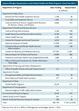
Impact of Budget Sequestration on Key Federal Health and Safety Programs, Fiscal Year 2013.

http://www.nejm.org/na101/home/literatum/publisher/mms/journals/content/nejm/2013/nejm_2013.368.issue-14/nejmp1303266/20130618/images/small/nejmp1303266_t1.gif

Medicare funding will be cut by 2% ($11.08 billion) through reductions in payments to hospitals, physicians, and other health care providers, as well as insurers participating in Medicare Advantage (Part C). The BCA prohibits cuts affecting premiums for Medicare Parts B and D, cost sharing, Part D subsidies, and Part A trust-fund revenues. The sequestration cuts arrive just as Medicare is beginning to fully implement the savings and cuts required by the Affordable Care Act (ACA), which the Congressional Budget Office estimates will slow Medicare’s rate of growth by $716 billion between 2013 and 2022. The National Institutes of Health (NIH) faces an 8.2% across-the-board reduction for the 7 months remaining in fiscal 2013, equaling cuts of $1.55 billion.

The Centers for Disease Control and Prevention (CDC), which is still recovering from major budget reductions in 2011, anticipates effective reductions of 8 to 10% for the remainder of the year. The American Public Health Association has projected that the reductions could result in 424,000 fewer HIV tests (the CDC funded 3.26 million in 2010) and 50,000 fewer immunizations for adults and children (from a baseline of about 300 million), elimination of tuberculosis programs in 11 states, and shutting down of the National Healthcare Safety Network.

Unaffected for all 9 years of the sequester are most expenses associated with the ACA. Medicaid is exempt, as is funding for its expansion, beginning next January, to all lower-income Americans in states that choose to participate. Also exempt are private insurance subsidies that will be available next January through new health insurance exchanges, because they were designed as refundable tax credits, another BCA-exempt category. Finally, the Children’s Health Insurance Plan, the Supplemental Nutrition Assistance Program, Temporary Assistance to Needy Families, and Supplemental Security Income are all exempt.

Threading the Needle ‹ Medicaid and the 113th Congress

fs310_graph3 leading causes of death by income class worldwideFUSA_INFOGRAPHIC_50-state-medicaid-expansion_rev_06-27-13_FACEBOOKCOVER

Rosenbaum S.N Engl J Med 2012; 367:2368-2369 http://dx.doi.org/10.1056/NEJMp1213901

Medicaid is a veteran of decades of warfare over its size and cost. Nevertheless, the program now plays a vital role in the U.S. health care system and a foundational role in health care reform. The central question, as we approach a major debate over U.S. spending and federal deficits, is how to preserve this role and shield Medicaid from crippling spending reductions. The Budget Control Act, which provides the initial framework for this debate, insulates Medicaid from sequestration. Budgetary protections for Medicaid date to the 1980s, but today’s politics are less tolerant of programs for poor and vulnerable populations. Medicaid is also at a deep political disadvantage. Medicaid is unequaled among federal grant programs: more than 60 million children and adults rely on the program, and it’s projected to grow to 80 million beneficiaries by 2020 if all states adopt the eligibility expansion in the Affordable Care Act (ACA). Medicaid’s cost is driven by high enrollment, not excessive per capita spending.2 As a result, there’s very little money to wring out of Medicaid without shaking its structure in ways that reduce basic coverage. Medicaid is part of the base on which health care reform rests; if it is not expanded per the ACA, the nation will lose its chance at near-universal health insurance coverage, which is essential to achieving systemwide savings and halting a $50 billion annual cost shift to insurers and patients. Deep federal spending reductions could lead states to abandon Medicaid expansion as a result of a confluence of factors —

  • the still-fragile nature of many state economies,
  • the continuing ideological opposition to Medicaid expansion, and
  • the Supreme Court decision to permit states to opt out of such expansion altogether.

Considerable evidence shows its effectiveness: most recently, a study by Sommers et al. documented its positive effects on health and health care. Experts in Medicaid spending also acknowledge the program’s operational efficiencies, achieved by states through the aggressive use of managed care and strict controls on spending for long-term care. Much of the health care that Medicaid beneficiaries receive is furnished through safety-net providers such as community health centers, which are highly efficient and accustomed to operating on tight budgets with only limited access to costly specialty care. Furthermore, Medicaid’s physician payments are substantially lower than those from commercial insurers and Medicare — a disparity that unfortunately limits provider participation even as it helps to keep per capita spending low. Indeed, the CBO has found that insuring the poor through Medicaid will cost 50% less per capita than doing so through tax-subsidized private insurance plans offered through state health insurance exchanges.

nejmp1306051_f1 Projected Growth in Medicare Per Capita Spending, the Consumer Price Index (CPI), and the Medical CPI, 2011–2015

The essential task is to thread the needle by accelerating efficiency reforms in health care payment and organization that, in turn, can generate savings over time while not damaging Medicaid’s role as a pillar of health care reform. Of particular importance is a heightened focus, begun under the ACA, on reforms that emphasize community care for millions of severely disabled children and adults, including patients who are dually enrolled in Medicare and Medicaid and who rely heavily on long-term institutional care.

The Shortfalls of ‘Obamacare’

Wilensky G.R. N Engl J Med 2012; 367:1479-1481 http://dx.doi.org/10.1056/NEJMp1210763

U.S. health care suffers from three major problems: millions of people go without insurance, health care costs are rising at unaffordable rates, and the quality of care is not what it should be. The Affordable Care Act (ACA) primarily addresses the first — and easiest — of these problems by expanding coverage to a substantial number of the uninsured. Solutions to the other two remain aspirations. The ACA’s primary accomplishment is that approximately 30 million previously uninsured people may end up with coverage — about half with subsidized private coverage purchased in the mostly yet-to-be-formed state insurance exchanges and the other half through Medicaid expansions. The law’s most controversial provision remains the individual mandate, which requires people either to have insurance coverage or to pay a penalty. The penalty for not having insurance is very small, particularly for younger people with modest incomes. It would have been smarter to mimic Medicare’s policies: seniors who don’t purchase the voluntary parts of Medicare covering physician services and outpatient prescription drugs during the first year in which they lack comparable coverage must pay a penalty for every month they have gone without coverage whenever they finally do purchase it.

Despite widespread recognition that fee-for-service reimbursement rewards providers for the quantity and complexity of services and encourages fragmentation in care delivery, the ACA retains all the predominantly fee-for-service reimbursement strategies currently used in Medicare. Much of the coverage expansion is financed through Medicare budget savings, which are produced by reducing the fees paid by Medicare to institutional providers such as hospitals, home care agencies, and nursing homes — but using the same perverse reimbursement system currently in place. Reducing payments to institutional providers should not be confused with lowering the cost of providing care.

The ACA also provides Medicare “productivity adjustments,” which assume that inflation adjustments can be reduced over time because institutions will become more productive, whether or not hospitals and other providers actually find ways to increase their productivity. Unless these institutions find ways to reduce costs, lower Medicare reimbursements will force providers to bargain for higher payments from private insurers. And eventually, seniors’ access to services will be threatened. The Medicare actuary expects that 15% of institutional providers will lose money on their Medicare business by 2019, and the proportion will increase to 25% by 2030 — a situation that he calls unsustainable

Most troubling, the ACA contains no reform of the way physicians are paid, which is the most dysfunctional part of the Medicare program. Through the Resource-Based Relative Value Scale, physicians are reimbursed on the basis of service codes, and payment for each physician service is reduced whenever aggregate spending on physician services exceeds a prespecified limit. This system disregards whether clinicians are providing low-cost, high-value care for patients. Given physicians’ key role in providing patient care, it’s impossible to imagine a reformed delivery system without one that rewards them for providing clinically appropriate care efficiently.

What is needed are reforms that create clear financial incentives that promote value over volume, with active engagement by both consumers and the health care sector. Market-friendly reforms require empowering individuals, armed with good information and nondistorting subsidies, to choose the type of Medicare delivery system they want. Being market-friendly means allowing seniors to buy more expensive plans if they wish, by paying the extra cost out of pocket, or to buy coverage in health plans with more tightly structured delivery systems at lower prices if that’s what suits them. 

Financing Graduate Medical Education — Mounting Pressure for Reform

John K. Iglehart N Engl J Med 2012; 366:1562-1563 http:dx.doi.org/10.1056/NEJMp1114236
http://www.nejm.org/doi/full/10.1056/NEJMp1114236

Disparate voices from the White House, a national fiscal commission, Congress, a Medicare advisory body, private foundations, and academic medical leaders are advocating changes to Medicare’s investment in graduate medical education (GME), which currently totals $9.5 billion annually. They offer various prescriptions, including reducing federal support, developing new achievement measures for which GME programs should be held accountable, and seeking independent assessment of the governance and financing of training programs.

The influential GME community has withstood most past efforts to change Medicare’s GME policies. But recognizing today’s more challenging political environment, the Association of American Medical Colleges (AAMC) has begun discussing alternative methods of financing GME that could better align training with the future health care delivery system and address U.S. workforce needs. The association is also examining the influence of student debt on the enrollment of a diverse student body.

When Congress enacted Medicare in 1965, it assigned to the program functions that reached well beyond its mission of financing health care for the elderly. One function was supporting GME, at least until the society at large undertook “to bear such education costs in some other way.” Almost 50 years later, Medicare remains the largest supporter of GME, providing both direct payments to hospitals that cover medical education expenses related to the care of Medicare patients (about $3 billion per year) and an indirect medical education (IME) adjustment to teaching hospitals for the added patient-care costs associated with training (about $6.5 billion).

In its 2013 budget, unveiled on February 13, 2012, the Obama administration proposed reducing Medicare’s IME adjustment by $9.7 billion over 10 years, beginning in 2014, citing a report from the Medicare Payment Advisory Commission (MedPAC) indicating that Medicare’s IME adjustments “significantly exceed the actual added patient care costs these hospitals incur.” The administration also proposed that the secretary of health and human services be granted the authority to assess GME programs’ performance in instilling in residents the necessary skills to promote high-quality health care. Similarly, MedPAC had recommended redirecting about half the IME adjustments ($3.5 billion) into “incentive payments” that GME programs could earn by meeting performance standards. The Obama budget would also eliminate coverage of the IME expenses of free-standing children’s hospitals with pediatric residency programs — which do not treat Medicare patients — reducing their federal support by 66% (to $88 million). Moreover, Congress has revealed its uncertainty over how to change federal workforce policy. In the Affordable Care Act (ACA), Congress emphasized the importance of expanding the primary care workforce. But legislators rejected the AAMC’s call to expand the number of Medicare-funded GME positions by 15% in response to reported physician shortages in some specialties.

On December 21, seven senators — Democrats Michael Bennet (CO), Jeff Bingaman (NM), Mark Udall (CO), and Tom Udall (NM) and Republicans Mike Crapo (ID), Chuck Grassley (IA), and Jon Kyl (AZ) — sent a letter to the Institute of Medicine (IOM) encouraging it to “conduct an independent review of the governance and financing of our system of [GME].” They urged the IOM to explore subjects including accreditation; reimbursement policy; the use of GME to better predict and ensure adequate workforce supply in terms of type of provider, specialty, and demographic mix; GME’s role in care of the underserved; and use of GME to ensure the creation of a workforce with the skills necessary for addressing future health care needs. The senators emphasized their interest “in IOM’s observations about the uneven distribution of GME funding across states based on need and capacity, and how to address this inequity.” In an interview, Bingaman said he initiated the letter for the same reasons he had championed creation of a National Health Care Workforce Commission as part of the ACA: to strengthen the government’s resolve to do “a more credible job of assessing workforce shortages” and because he believes Medicare’s GME policies are “outmoded.”

The priorities cited in the IOM letter parallel some of the recommendations of a group of academic medical leaders who gathered at two conferences underwritten by the Josiah Macy Jr. Foundation. At the first conference, in October 2010, the top recommendation was that “an independent external review of the goals, governance, and financing of the GME system should be undertaken by the Institute of Medicine, or a similar body.”3 George Thibault, president of the Macy Foundation, says the group concluded that “because GME is a public good and is significantly financed with public dollars, the GME system must be accountable to the needs of the public.” Acknowledging that some people in academic medicine “favor a behind-the-scenes discussion of GME reform alternatives,” Thibault noted, “I believe we should be upfront, providing examples of change that could influence the thinking of policymakers.” The foundation awarded the IOM $750,000 — about half the support it needs for the GME study.

Among subjects under discussion are the collection of more data highlighting the importance of the safety-net functions and unique services of academic medical centers and the creation of a long-term vision for GME financing that is more closely aligned with emerging care delivery models, such as accountable care organizations. The association is also revisiting a potential financial model under which all health care payers would explicitly cover GME expenses. Private insurers maintain that they accomplish this implicitly by paying teaching hospitals more for clinical services than they pay most other hospitals. GME leaders think one possibility would be to include the costs of residency training when calculating premium amounts for products sold through health insurance exchanges. Similarly, a recent Carnegie Foundation report asserted that “GME redesign demands . . . a more broad-based, less politicized flow of funds.”

Dr. Darrell Kirch noted, CEO of AAMC, “A significant step forward is the announcement by the ACGME [Accreditation Council for Graduate Medical Education] describing major changes in how the nation’s residency programs will be accredited in the future, putting in place an outcomes-based evaluation system by which new physicians will be measured for their competency in performing the essential tasks necessary for clinical practice in the 21st century.”

Achieving Health Care Reform — How Physicians Can Help

Elliott S. Fisher, M.D., M.P.H., Donald M. Berwick, M.D., M.P.P., and Karen Davis, Ph.D.
N Engl J Med 2009; 360:2495-2497 http://dx.doi.org/10.1056/NEJMp0903923
http://www.nejm.org/doi/full/10.1056/NEJMp0903923

The recent commitment by several major stakeholders — including the American Medical Association — to slowing the growth of health care spending is a promising development. But the controversy about whether the organizations actually agreed to a 1.5-percentage-point reduction in annual spending growth is just one indication that success is still far from assured.

Two threats in particular put reform at risk: conflicting doctrines (regarding the creation of a new public insurance option and government support for comparative-effectiveness studies) and opposition to change among some current stakeholders. In the face of this uncertainty, physicians have a choice: to wait and see what happens or to lead the change our country needs. We’d prefer the latter.

The first level is aims. For health care reform, we propose that physicians, through their advocacy, help lead the country to embrace the so-called triple aim: better experience of care (safe, effective, patient-centered, timely, efficient, and equitable), better health for the population, and lower total per capita costs.

The second level is the design of the care processes that affect the patient — clinical “microsystems.” Health care microsystems are famously unreliable, variable in costs, and often unsafe. Physicians, through their participation in quality-improvement initiatives in their practices and hospitals, can and should lead the needed changes in the systems of care in which they work, to make them safer, more reliable, more patient-centered, and more affordable.

However, neither physicians nor anyone else on the front lines can improve care much on their own. Their most important source of support for improvement is the third level described by the IOM — the health care organizations that house almost all clinical microsystems and can ensure coordination among them. We need organizations large enough to be accountable for the full continuum of patients’ care as well as for achieving the triple aim. We will create a high-performing health care system only if integrated delivery systems become the mainstay of organizational design. Organizations could be virtually integrated, such as networks of independent physicians sharing electronic health records and administrative and clinical support for care management and quality improvement, or structurally integrated, such as multispecialty group practices or staff-model health maintenance organizations. Fostering the development of such accountable care organizations need not be disruptive to patients or providers: almost all physicians already work within natural referral networks that provide the vast majority of care to patients seen by the primary care physicians within the network.

Innovators-Prescription-New-Wave-of-Disruptive-Models-in-Healthcare

The IOM’s fourth level is the environment, which includes the payment, regulatory, legal, and educational systems. On this front, too, we need physician advocacy. The United States cannot achieve the triple aim without health insurance for everyone. Integrated delivery systems that are accountable for populations won’t thrive unless payment systems encourage their development and unless we change the laws and regulations — including proscriptions of gainsharing and anti-kickback rules — that prevent cooperation among health care professionals and organizations.

If stakeholders can agree on such a vision of health care reform, perhaps we could shift our focus from the conflict over whether a new public plan should be created to a more constructive insistence that all health plans, whether public or private, focus on the development of professionally led, integrated systems.

If health care providers and suppliers could actually achieve this reduction in growth rates, the federal government would harvest about $1.1 trillion in savings over the 11-year period — enough, perhaps, to close the deal on affordable health insurance for all. Others would also see savings: $497 billion for employers, $529 billion for state and local governments, and $671 billion for households. One simple way for physicians to start contributing to this goal is by reassessing and scaling back, where appropriate, their use of clinical practices now listed as “overused” by the National Quality Forum’s National Priorities Partnership.

Editor-in-Chief Eric J. Topol, MD, interviews Secretary of Health and Human Services (HHS) Kathleen Sebelius

Medscape

Editor’s Note: On the eve of the first anniversary of the Supreme Court’s ruling to uphold most provisions of the Affordable Care Act (ACA), Medscape Editor-in-Chief Eric J. Topol, MD, questioned Secretary of Health and Human Services (HHS) Kathleen Sebelius about the act’s effect on medical technology, clinical trial participation, genetic testing, primary care, and patient safety.

Introduction

Dr. Topol: We are experiencing a digital revolution in which technological advances are putting healthcare where it should be: in the hands of patients. How is the ACA helping to foster medical innovation?
Secretary Sebelius: A recent New York Times column, “Obamacare’s Other Surprise,”[1] by Thomas L. Friedman, echoes what we’ve been hearing from healthcare providers and innovators: Data that support medical decision-making and collaboration, dovetailing with new tools in the Affordable Care Act, are spurring the innovation necessary to deliver improved healthcare for more people at affordable prices.
Today we are focused on driving a smarter healthcare system with an emphasis on the quality — not quantity — of care. The healthcare law includes many tools to increase transparency, avoid costly mistakes and hospital readmissions, keep patients healthy, and test new payment and care delivery models, like Accountable Care Organizations (ACOs). Health information technology is a critical underpinning to this larger strategy.
In May we reached an important milestone in the adoption of health information technology. More than half of all doctors and other eligible providers, and nearly 80% of hospitals, are using electronic health records (EHRs) to improve care, an increase of at least 200% since 2008. Also in May, we announced a $1 billion challenge to help jump-start innovative projects that test creative ways to deliver high-quality medical care and lower costs to people enrolled in Medicare and Medicaid, following 81 Health Care Innovation Awards that HHS awarded last year.
Dr. Topol: Physicians have long lamented the lack of participation by patients in clinical trials, but the ACA is opening the door for greater participation by allowing patients to keep their health insurance while participating in clinical research. Are patients even aware that this provision now exists? How do you see it affecting clinical trial participation in the future?
Secretary Sebelius: In 2014, thanks to the ACA, insurance companies will no longer be able to deny patients from participating in an approved clinical trial for treatment of cancer or another life-threatening disease or condition, nor can they deny or limit the coverage of routine patient costs for items or services in connection with trial participation. For many patients, access to cutting-edge medicine available through clinical trials can increase their likelihood of survival. This is an important protection for patients that not only could have a life-altering impact, but it’s also one that serves to facilitate participation in research that is critical to expanding our knowledge base and finding cures and treatments for those illnesses that threaten the lives of Americans each day.
Dr. Topol: One of the intentions of the ACA is to increase the primary care workforce. This is critical as we approach 2014, when more Americans than ever will have either private insurance or Medicaid. Have you seen any movement in the primary care workforce? Are there concerns that there aren’t enough clinicians available to meet the forthcoming patient load?
Secretary Sebelius: Primary care providers are critical to ensuring better coordinated care and better health outcomes for all Americans. To meet the health needs of Americans, the Obama Administration has made the recruitment, training, and retention of primary care professionals a top priority.
Together, the ACA, the American Recovery and Reinvestment Act of 2009, and ongoing federal investments in the healthcare workforce have led to significant progress in training new primary care providers — such as physicians, nurse practitioners, and physician assistants — and encouraging primary care providers to practice in underserved areas, including:
Nearly tripling the National Health Service Corps;
Increasing the number of medical residents, nurse practitioners, and physician assistants trained in primary care, including placing over 1500 new primary care providers in underserved areas;
Creating primary care payment incentives for providers; and Redistributing unused residency positions and directing those slots for the training of primary care physicians.
Additionally, the ACA is modernizing the primary care training infrastructure, creating new primary care clinical training opportunities, supporting primary care practice, and improving payment and financial incentives for coordinated care.
Improving Hospital Safety
Dr. Topol: George Orwell once said that the hospital is the antechamber to the tomb. That was written decades ago, and unfortunately there’s still truth to that today. One in 4 hospital patients in America have a problem with medical mistakes, contract hospital-acquired infections, and experience medication errors. The ACA last year began linking Medicare payments to quality of patient care, offering financial incentives to hospitals that improve patient care. How is this working? Have there been any meaningful care improvements over the past year?
Secretary Sebelius: The ACA includes steps to improve the quality of healthcare and, in so doing, lowers costs for taxpayers and patients. This means avoiding costly mistakes and readmissions, keeping patients healthy, rewarding quality instead of quantity, and creating the health information technology infrastructure that enables new payment and delivery models to work. These reforms and investments will build a healthcare system that will ensure quality care for generations to come.
Already we have made significant progress:
Healthcare Spending Is Slowing
Secretary Sebelius: Medicare spending per beneficiary grew just 0.4% per capita in fiscal year 2012, continuing the pattern of very low growth in 2010 and 2011. Medicaid spending per beneficiary also decreased 0.9% in 2011, compared with 0.6% growth in 2010. Average annual increases in family premiums for employer-sponsored insurance were 6.2% from 2004 to 2008, 5.6% from 2009 to 2012, and 4.5% in 2012 alone.
Health Outcomes Are Improving and Adverse Events Are Decreasing
Secretary Sebelius: Several programs tie Medicare reimbursement for hospitals to their readmission rates, when patients have to come back into the hospital within 30 days of being discharged. Additionally, as part of a new ACA initiative, clinicians at some hospitals have reduced their early elective deliveries to close to zero, meaning fewer at-risk newborns and fewer admissions to the NICU.
Providers Are Engaged
Secretary Sebelius: In 2012, we debuted the Medicare Shared Savings Program and the Pioneer Accountable Care Organization Model. These programs encourage providers to invest in redesigning care for higher-quality and more efficient service delivery, without restricting patients’ freedom to go to the Medicare provider of their choice.
Over 250 organizations are participating in Medicare ACOs, serving approximately 4 million, or 8%, of Medicare beneficiaries. As existing ACOs choose to add providers and as more organizations join the program, participation in ACOs is expected to grow. ACOs are estimated to save up to $940 million in the first 4 years.
Bundle with Care ‹ Rethinking Medicare Incentives for Post­Acute Care Services

Feder J. N Engl J Med 2013; 369:400-401

A Medicare payment approach in which savings and risk are shared may achieve a better balance of cost, quality, and access than a system of single bundled payments, at least until our capacity to measure patients’ care needs and outcomes is sufficiently robust.

Healthcare Reform 2014: Mandated Coverage, Insurance Exchanges, and Employer Requirements

3 of 5 in Series: The Essentials of Healthcare Reform
http://www.dummies.com/how-to/content/healthcare-reform-2014-mandated-coverage-insurance.html

The Affordable Care Act federal and state officials are working with leaders in the health and insurance industries to restructure our nation’s healthcare system. That restructuring means most Americans will be required to have health insurance and most businesses will be required to offer it to their employees. It also means the creation of another kind of insurance plan called a health insurance exchange.

The government will require most Americans to have health insurance by 2014. The government has enacted this provision as a way to get healthy people who don’t feel the need to pay for coverage to buy insurance. That way, the healthy people can help fund the cost of people who require more medical care.

Several states filed, and lost, a suit against the federal government saying that it is unconstitutional to make individual citizens to buy health insurance.

If you don’t have coverage and you’re not in one of the groups that is an exception to the rule, you’ll pay a penalty. You may not be required to purchase health insurance if you

  • Face financial hardships.
  • Have been uninsured for less than three months.
  • Have religious objections.
  • Are American Indian.
  • Are a prison inmate.
  • Are an undocumented immigrant.

If you’re penalized, the amount you’ll be fined will go up each year for the first three years. In 2014, you’ll pay $95 or 1 percent of your taxable income, whichever is greater. In 2015, the fine will be $325 or 2 percent of taxable income, and in 2016 the penalty will be $695 or 2.5 percent of income. Each year after 2016, the government will refigure the fine based on a cost-of-living adjustment.

To help you meet the cost of mandated insurance, the government will offer premium credits and cost sharing subsidies if you and your family meet certain income guidelines and if you enroll in one of the new state-run insurance exchanges.

If your income falls between 133 and 400 percent of the federal poverty level (FPL), you could receive premium credits that will lower the maximum amount of premium you have to pay for your coverage.

  • There will be a catastrophic plan for people under 30 and for those who are exempt from mandated coverage.

States don’t have to set up the exchanges. If a state chooses not to, the federal government can come in and create them. States that do opt for exchanges will decide whether they’ll be run by a government or not-for-profit entity.

Health Care Reform — Why So Much Talk and So Little Action?

Victor R. Fuchs, Ph.D
N Engl J Med 2009; 360:208-209 http://dx.doi.org/10.1056/NEJMp0809733
http://www.nejm.org/doi/full/10.1056/NEJMp0809733

First, many organizations and individuals prefer the status quo. This category includes health insurance companies; manufacturers of drugs, medical devices, and medical equipment; companies that employ mostly young, healthy workers and therefore have lower health care costs than they would if required to help subsidize care for the poor and the sick; high-income employees, whose health insurance is heavily subsidized through a tax exemption for the portion of their compensation spent on health insurance; business leaders and others who are ideologically opposed to a larger role of government; highly paid physicians in some surgical and medical specialties; and workers who mistakenly believe that their employment-based insurance is a gift from their employer rather than an offset to their potential take-home pay.

Second, as Niccoló Machiavelli presciently wrote in 1513, “There is nothing more difficult to manage, more dubious to accomplish, nor more doubtful of success . . . than to initiate a new order of things. The reformer has enemies in all those who profit from the old order and only lukewarm defenders in all those who would profit from the new order.”

Third, our country’s political system renders Machiavelli’s Law of Reform particularly relevant in the United States, where many potential “choke points” offer opportunities to stifle change. The problem starts in the primary elections in so-called safe congressional districts, where special-interest money can exert a great deal of influence because of low voter turnout. The fact that Congress has two houses increases the difficulty of passing complex legislation, especially when several committees may claim jurisdiction over portions of a bill. Also, a supermajority of 60% may be needed to force a vote in the filibuster-prone Senate.

Fourth, reformers have failed to unite behind a single approach. Disagreement among reformers has been a major obstacle to substantial reform since early in the last century. According to historian Daniel Hirshfield, “Some saw health insurance primarily as an educational and public health measure, while others argued that it was an economic device to precipitate a needed reorganization of medical practice. . . . Some saw it as a device to save money for all concerned, while others felt sure that it would increase expenditures significantly.” These differences in objectives persist to this day.

Health insurers are opening stores alongside department stores, other typical mall tenants.

Jayne O’Donnell , USA TODAY
 http://www.usatoday.com/story/news/nation/2013/09/12/health-insurance-sales-retail-stores-malls/2789897/

,The new health law known as the Affordable Care Act means most uninsured Americans are required to have insurance beginning March 31 or pay a penalty at tax time in 2015.

Insurers need to sign up as many healthy, younger people as they can to pay for all of the older, sick customers they will be taking on. The law prohibits insurers from denying people insurance because of pre-existing health problems and limits how much more they can charge older than younger people.

So, for the first time, insurers are fiercely competing to attract individual consumers and turning to traditional retail marketing techniques to do so, luring them into stores with special events and using splashy advertising. As any retailer knows, they have the greatest chance of converting shoppers to customers once they have them in their retail locations or on their sites.

The Medical Breakthrough Nobody’s Talking About

Toby CosgroveCEO and President at Cleveland Clinic

http://www.linkedin.com/today/post/article/20130912184535-205372152-the-medical-breakthrough-nobody-s-talking-about

The latest medical breakthrough hasn’t gotten much press, but it’s changing medicine even as we speak. It’s the dawning realization that healthcare is not about how many patients you can see, how many tests and procedures you can order, or how much you can charge for these things. The breakthrough is the understanding that healthcare is a value proposition, which means getting patients the right care, at the right time, in the right place. It’s a matter of focusing on outcomes and cost, so that more Americans will start getting what they pay for in healthcare dollars.

Value-based care focuses on two targets: outcomes and cost. Until recently, providers pursued these goals separately, with doctors concentrating on outcomes and the administrators trying to control costs. Value-based care does something different. It works to bring these targets into alignment. The caregivers in a value-based provider work with cost-experts as a team to simultaneously improve outcomes and lower expenses.

Doctors, hospitals and payers are partners in the move to value-based care. The Affordable Care Act includes incentives for providers to improve outcomes and lower costs. But this is one breakthrough that will take time for implementation nationwide. Providers who make the transition early will be rewarded with more satisfied patients, lower expenses and pride in a job well done.

Six-Month Enforcement Delay After Guidance

According to AAMC, the language in the final rule requires that the order to admit a patient be written by a practitioner “who has admitting privileges at the hospital,” something that few residents have as they are not considered members of the hospital’s medical staff.

AAMC said it brought the issue to CMS’s attention during an Open Door Forum call Aug. 15. The agency acknowledged it did not intend to prohibit residents from admitting patients, and said it would be issuing a Q&A. However, AAMC said until the issue can be resolved “to the satisfaction of the teaching hospital community,” CMS should make clear to all contractors that no inpatient admission should be denied because it was ordered by a resident while under the supervision of an attending physician.

AAMC said CMS should delay enforcing the new requirements for at least six months following the release of the guidance so hospitals will have sufficient time to understand the rules, educate physicians and others, and ensure that they have put in place the mechanisms that are needed to comply with the new requirements.

“As short inpatient stays have been a focus of audits by [Recovery Audit Contractors], hospitals feel especially at risk for failure to properly implement CMS requirements,” AAMC said.

The letter is available at http://op.bna.com/hl.nsf/r?Open=nwel-9auqls.

Read Full Post »

« Newer Posts