Feeds:
Posts
Comments

Posts Tagged ‘Cardiovascular Disorders’

Cardiotoxicity and Cardiomyopathy Related to Drugs Adverse Effects

Curator: Larry H Bernstein, MD, FCAP

Introduction
This is the second part of a series on toxicities of therapeutic medications, the first being on the impact on drug development of early phase failure to identify toxicities that are found in late stage trials and result in withdrawal.  This portion will go into details of identifying the effects clinically and give some examples.  In the future, the design of therapies, the identification of high probability successful genomic targets, and more accurate patient selection will transform the approach to development, clinical trials, and clinical use of pharmaceuticals in patients.

Cardiotoxicity and Cardiomyopathy

refer to: What are cardiotoxicity and cardiomyopathy?

The Scott Hamilton Cares Initiative
Cardiotoxicity is a condition of heart muscle functional impairment from toxicity as an
  • adverse secondary effect of taking an essential medication, or
  • as a result of interactions between prescribed medications that result in heart damage,
  • usually dose and time related.
If severe, the adverse effect of chemotherapy may lead to cardiomyopathy.  While cardiomyopathy might be a result of treatments, such as chemotherapeutic medications, it may also caused by a group of diseases or disorders, leading to
  • damaged myocardiocytes, and the injury leads to
  • insufficient cardiac output, referred to as
  • heart failure.
Cardiomyopathy has many causes, singly or in combination:
  • viruses – such as,
    • coxsackie B,
    • human immunodeficiency virus (HIV)
  • systemic inflammatory disorder
    • systemic lupus erythematosis
  • Amyloidosis – amyloid protein deposits in the myocardium alone, and/or other organs
  • Infection –
    • bacterial (tetanus),
    • parasitic (Chaga’s disease)
    • Rheumatic fever
  •  high blood pressure
  • Chronic or long-term alcohol use (B vitamin deficiency)
  • Endocrine disease, such as hyperthyroidism
  • Thiamine and Vitamin B deficiency
  • Radiation therapy
  • Medications – anthracyclines.

Anthracyclines may be used to treat leukemia, lymphoma, multiple myeloma, breast cancer, and sarcoma. A commonly used anthracycline is called doxorubicin (Adriamycin®).

  • cardiomyopathy may also result from genetic defects
  • illegal drugs and toxic substances, cocaine, may also produce serious myocardial damage

With certain drugs, such as doxorubicin, there is a dose at which these cardiotoxic effects on the heart may occur.
An echocardiogram, or a radionuclide ventriculography scan, is performed

  • prior to initiating a cardiotoxic medication
  • to determine baseline cardiac function., and
  • repeated at intervals to monitor heart function while receiving cardiotoxic medications.

The ejection fraction (EF) is a percentage of blood pumped out into the body during each heartbeat. An EF of 50%-75% is considered normal.

  • The lower the ejection fraction, the more severe the heart failure may be.
This may determine if the cardiotoxic drug has caused cardiomyopathy.

Symptoms of cardiomyopathy:

  • fatigue
  •  shortness of breath
  •  fever and aching of the joints,
      • all characteristic of a flu-like illness.
  • Or, sudden heart failure or sudden cardiac death without any prior symptoms.
    • swollen feet and ankles
    • distended neck veins
    • tachycardia
    • dyspnea while reclining

Diagnosis:

  • history & physical examination
  • laboratory tests
  • EKG
  • Chest x-ray
  • Echocardiography
  • Cardiac cath
  • Angiography

Treatment:

  • Dexraoxane HCL –  doxorubicin
  • ACE inhibitors
  • Beta-blockers
  • Diuretics
  • Digoxin

 Biomolecular Screening for Drug Toxicity

Multiparameter In Vitro Assessment of Compound Effects on Cardiomyocyte Physiology Using iPSC Cells

O Sirenko, C Crittenden, N Callamaras, J Hesley, Yen-Wen Chen, et al.
 A sufficient percentage of drugs fail in clinical studies due to cardiac toxicity that the development of new, sensitive in vitro assays that can evaluate potential adverse effects on cardiomyocytes is needed. Cell-based models are more clinically relevant than those used in practice. Human-induced pluripotent stem cell–derived cardiomyocytes are especially attractive because
  • they express ion channels and
  • demonstrate spontaneous mechanical and electrical activity
    • similar to adult cardiomyocytes.
This study introduces techniques for measuring the impact of pharmacologic compounds on the beating rate of cardiomyocytes with ImageXpress Micro and FLIPR Tetra systems. The assays employ
calcium-sensitive dyes to monitor changes in Ca2+ fluxes
  • synchronous with cell beating,
This method allows monitoring of the
  • beat rate
  • amplitude, and
  • other parameters.
The system detects
  • concentration-dependent atypical patterns caused by
  • hERG inhibitors and other ion channel blockers.
In addition,
  • both positive and negative chronotropic effects on cardiac rate can be observed and
  • IC50 values determined.
This methodology is well suited for safety testing and can be used to estimate efficacy and dosing of drug candidates prior to clinical studies.
J Biomol Screen Jan 2013;18(1): 39-53  http://dx.doi.org/10.1177/1087057112457590

Estimating the risk of drug-induced proarrhythmia using human induced pluripotent stem cell-derived cardiomyocytes.

L Guo, RMC Abrams, JE Babiarz, JD Cohen, S Kameoka, et al.
 Early prediction of drug-induced toxicity is needed in the pharmaceutical and biotechnology industries to decrease late-stage drug attrition.
  • Cardiotoxicity accounts for about one third of safety-based withdrawn pharmaceuticals.
This study reports a high-throughput functional assay,  detailing a model that accurately detects
  • drug-induced cardiac abnormalities.
It employs
  • induced pluripotent stem cell-derived cardiomyocytes (iPSC-CMs).
  • Using 96-well plates with interdigitated electrode arrays  detect
    • assess impedance,
    • the rhythmic, synchronous contractions of the iPSC-CMs
Treatment of the iPSC-CMs with 28 different compounds with known cardiac effects resulted in
  • compound-specific changes in the beat rate and/or
  • the amplitude of the impedance measurement.
Changes in impedance for the compounds tested were comparable with the results from a related technology,
  • electric field potential assessment obtained from microelectrode arrays.
Using the results from the set of compounds,
  • an index of drug-induced arrhythmias was calculated,
  • which enabled the determination of a drug’s proarrhythmic potential.
This system of interrogating human cardiac function in vitro opens new opportunities for predicting cardiac toxicity and studying cardiac biology.
Toxicol Sci. Sep 2011; 123 (1):281-9  21693436

Determination of the Human Cardiomyocyte mRNA and miRNA Differentiation Network by Fine-Scale Profiling.

JE Babiarz, M Ravon, S Sridhar, P Ravindran, B Swanson,  et al.
This study is a detailed comparison of the mRNA and miRNA transcriptomes
  • across differentiating human-induced pluripotent stem cell (hiPSC)-derived cardiomyocytes and
  • biopsies from fetal, adult, and hypertensive human hearts
Gene ontology analysis of the mRNA expression levels of the hiPSCs differentiating into cardiomyocytes
revealed 3 distinct groups of genes:
  • pluripotent specific,
  • transitional cardiac specification, and
  • mature cardiomyocyte specific.
Hierarchical clustering of the mRNA data revealed that the transcriptome of hiPSC cardiomyocytes
  • stabilizes 20 days after initiation of differentiation.
But analysis of cells continuously cultured for 120 days indicated that
  • the cardiomyocytes continued to mature toward a more adult-like gene expression pattern.
Analysis of cardiomyocyte-specific miRNAs (miR-1, miR-133a/b, and miR-208a/b) revealed
  • an miRNA pattern indicative of stem cell to cardiomyocyte specification.
A biostatistitical approach integrated the miRNA and mRNA expression profiles revealing a
  • cardiomyocyte differentiation miRNA network and
  • identified putative mRNAs targeted by multiple miRNAs.
Together, these data reveal the
miRNA network in human heart development and
  • support the notion that overlapping miRNA networks
  • re-enforce transcriptional control during developmental specification.
Stem Cells Dev. 2012 Jul 20;21 (11):1956-65  22050602

Comparative Gene Expression Profiling in Human Induced Pluripotent Stem Cell Derived Cardiocytes and Human and Cynomolgus Heart Tissue.

D Puppala, LP Collis, SZ Sun, V Bonato, X Chen, B Anson, et al.  Compound Safety Prediction.
Cardiotoxicity is one of the leading causes of drug attrition. Current in vitro models insufficiently predict cardiotoxicity.  The authors describe
  • the gene expression profile of human induced pluripotent stem cell derived cardiocytes (iCC)
  • post-thaw over a period of 42 days in culture and
  • compare this profile to human fetal and adult as well as
  • adult cynomolgus nonhuman primate (NHP: Macaca fascicularis) heart tissue.
The results indicate that iCC express relevant cardiac markers such as
  • ion channels (SCN5A, KCNJ2, CACNA1C, KCNQ1 and KCNH2),
  • tissue specific structural markers (MYH6, MYLPF, MYBPC3, DES, TNNT2 and TNNI3),
  • transcription factors (NKX2.5, GATA4 and GATA6), and
  • lack the expression of stem cell markers (FOXD3, GBX2, NANOG, POU5F1, SOX2, and ZFP42).

A functional evaluation of contractility of the iCC showed

  • functional and pharmacological correlations with myocytes isolated from adult NHP hearts.
The results suggest that stem cell derived cardiocytes may represent
  • a novel in vitro model to study human cardiac toxicity with potential ex vivo and in vivo translation.
Toxicol Sci. Sep 14 2012;:   22982684

Characterization of Human Induced Pluripotent Stem Cell Derived Cardiomyocytes:
Bioenergetics and Utilization in Safety Screening.

P Rana, B Anson, S Engle, Y Will.   Compound Safety Prediction. Pfizer Global R&D, Groton CT.
Cardiotoxicity remains the number one reason for drug withdrawal from the market and FDA issued black box warnings; thus
  • demonstrating the need for more predictive preclinical safety screening,
  • especially early in the drug discovery process.
Whereas hERG screening has become routine to mitigate proarrhythmic risk,
  • the development of in vitro assays predicting additional on- and off-target biochemical toxicities
  • will benefit from cellular models exhibiting true cardiomyocyte characteristics
    • such as, native tissue-like mitochondrial activity.
An hypothesis was tested  for using human stem cell derived tissue cells by using a combination of
  • flux analysis,
  • gene and protein expression, and
  • toxicity-profiling techniques
    • to characterize mitochondrial function
    • in induced pluripotent stem cell (iPSC)-derived human cardiomyocytes
      • in the presence of differing carbon sources
      • over extended periods in cell culture.
Functional analyses demonstrate that iPSC-derived cardiomyocytes:
1) are capable of utilizing anaerobic or aerobic respiration depending upon the available carbon substrate,
2) are bioenergetically closest to adult heart tissue cells when cultured in galactose or galactose supplemented with fatty acids, and
3) show a dose dependent toxicity profile to a variety of kinase inhibitors with known clinical cardiac liabilities.
Furthermore, gene and protein expression analyses revealed that in comparison to adult cardiac tissue,
  • iPSCs-derived cardiomyocytes possess a qualitatively similar expression pattern of mitochondrial genes,
  • an up-regulation of apoptotic and antioxidant genes, and
  • a mitochondrial transcription pattern that is similar across different carbon substrates
  • despite showing changes in protein levels and functional bioenergetic adaptation.
Toxicol Sci. 2012 Jul 27;:   22843568

Decreasing cardiac chamber sizes and associated heart dysfunction in COPD – role of hyperinflation.

H Watz, B Waschki, T Meyer, G Kretschmar, A Kirsten, M Claussen, H Magnussen
This study examined the relationship of
  • lung function with heart size and heart dysfunction and
  • associated consequences for 6-minute walk distance (6MWD)
    • in patients with COPD of different severity.
METHODS:   138 patients with COPD (GOLD I-IV)
  • the size of all cardiac chambers,
  • left ventricular diastolic dysfunction (relaxation and filling), and
  • global right ventricular dysfunction (Tei-index)
    • were measured by echocardiography .
  • lung function (spirometry, bodyplethysmography, diffusion capacity) and
  • 6MWD …. were measured.
RESULTS: Size of all cardiac chambers decreased with GOLD stages. Overall,
moderate relationships existed between
  • variables of lung function and cardiac chamber sizes.
  • Static hyperinflation (inspiratory-to-total lung capacity ratio [IC/TLC],
  • functional residual capacity, and residual volume)
showed stronger associations with
  • cardiac chamber sizes than
  • airway obstruction or diffusion capacity.
IC/TLC ratio correlated best with cardiac chamber sizes and was
  • an independent predictor of cardiac chamber sizes
    • after adjustment for body surface area.
Patients with an IC/TLC ratio <!–= 0.25 had a significantly–>
  • impaired left ventricular diastolic filling pattern and
  • a significantly impaired Tei-index
    • compared to patients with an IC/TLC ratio > 0.25.
An impaired left ventricular diastolic filling pattern was independently associated with
  • a reduced 6MWD.
An increasing severity of COPD is associated with a decreasing heart size.
Hyperinflation in patients with COPD might have an important role with respect to
  • heart size and
  • cardiac dysfunction
Chest. Feb 26 2010;:   20190002  Cit:4

Cardiovascular Events After Clarithromycin Use in Lower Respiratory Tract Infections
Analysis of Two Prospective Cohort Studies

S Schembri, PA Williamson, PM Short, A Singanayagam, A Akram, et al. British Medical Journal
Acute exacerbations of chronic obstructive pulmonary disease and community acquired pneumonia are common causes of admission to a hospital.
Antibiotics, including clarithromycin, are commonly prescribed during acute exacerbations of chronic obstructive pulmonary disease, especially
  • in the presence of increased breathlessness,
  • sputum volume, and
  • purulence.
 Use of macrolide antibiotics in community acquired pneumonia has been consistently associated with improved short term mortality in observational studies,
and national and international guidelines therefore recommend their use in combination with β lactams for patients admitted to hospital.

Widespread use of macrolide antibiotics has been accompanied by concerns about adverse effects on cardiovascular morbidity and mortality.
A retrospective study of erythromycin use in 1,249,943 patients identified an increase in deaths from cardiovascular disease.
Azithromycin was shown to have a similar association with increased cardiovascular deaths

  • during the time of administration.

CLARICOR (Effect of Clarithromycin on Mortality and Morbidity in Patients with Ischemic Heart Disease trial) was a double blind, placebo controlled trial
showing that a two week course of clarithromycin administered to patients with coronary heart disease 

  • increased cardiovascular and all cause mortality
The increased mortality rate (clear of pulmonary infection)
  • persisted for three years after discontinuation of the drug.

A recent meta-analysis of 17 trials of antibiotics in coronary heart disease showed

  • increased long term mortality after macrolides, primarily due
  • to increased deaths from cardiovascular disease.

However, no studies have examined the long term effect of clarithromycin on cardiovascular events and mortality in patients

  • after acute exacerbations of chronic obstructive pulmonary disease or community acquired pneumonia.

Therefore, this prospective cohort study was undertaken to examine the association of clarithromycin with cardiovascular events

  • in the setting of acute exacerbations of chronic obstructive pulmonary disease and community acquired pneumonia.

Population.

  • 1343 patients admitted to hospital with acute exacerbations of chronic obstructive pulmonary disease
  • and 1631 patients admitted with community acquired pneumonia.

Main Outcome Measures.

Hazard ratios for cardiovascular events at one year (defined as hospital admissions with

  • acute coronary syndrome, decompensated cardiac failure, serious arrhythmia, or sudden cardiac death) and
  • admissions for acute coronary syndrome (acute ST elevation myocardial infarction, non-ST elevation myocardial infarction, and unstable angina).

Secondary outcomes were all cause and cardiovascular mortality at one year.

Results.

  •  268 cardiovascular events occurred in the acute exacerbations of chronic obstructive pulmonary disease cohort and
  • 171 in the community acquired pneumonia cohort over one year.

After multivariable adjustment, clarithromycin use in acute exacerbations of chronic obstructive pulmonary disease

  • was associated with an increased risk of cardiovascular events and acute coronary syndrome—
  • hazard ratios 1.50 (95% confidence interval 1.13 to 1.97) and 1.67 (1.04 to 2.68).
After multivariable adjustment, clarithromycin use in community acquired pneumonia
  •  was associated with increased risk of cardiovascular events (hazard ratio 1.68, 1.18 to 2.38)
  • but not acute coronary syndrome (1.65, 0.97 to 2.80).
This association was found between clarithromycin use in acute exacerbations of COPD and
  • cardiovascular mortality (adjusted hazard ratio 1.52, 1.02 to 2.26)
  • but not all cause mortality (1.16, 0.90 to 1.51) .

No association was found between clarithromycin use in community acquired pneumonia and all cause mortality or cardiovascular mortality.
Use of β lactam antibiotics or doxycycline was not associated with increased cardiovascular events in patients with

  • acute exacerbations of chronic obstructive pulmonary disease, suggesting an effect specific to clarithromycin.

 Timing of Cardiovascular Events

The study found no significantly increased risk of cardiovascular events while patients were taking clarithromycin in the COPD cohort
(hazard ratio 1.73, 0.71 to 4.25), but
  • an increased risk was present after the clarithromycin course ended (1.41, 1.05 to 1.89).
In the community acquired pneumonia cohort, the hazard ratio for association between clarithromycin use and cardiovascular events
was 1.84 (0.75 to 4.51) during clarithromycin use and 1.66 (1.14 to 2.43) after the antibiotic was stopped.

Association With Duration of Antibiotic Use

Longer courses of clarithromycin were associated with more cardiovascular events. The median duration of treatment was seven days in both cohorts.
Less than three days of clarithromycin treatment was not associated with cardiovascular events in the chronic obstructive pulmonary disease cohort
(hazard ratio 0.89, 0.50 to 1.57) or the community acquired pneumonia cohort (0.63, 0.15-2.65), compared with patients who did not receive clarithromycin.

Effect of Age and Cardiovascular Risk Status

The hazard ratios of the effect of clarithromycin on cardiovascular events in such patients were
  • 1.35 (0.94 to 1.95) in those with a high cardiovascular risk and 0.88 (0.20 to 3.96) in those with a low risk.

The lowest hazard ratios for cardiovascular events were in patients aged 60 or below (1.01, 0.36 to 2.91).
The hazard ratio was 1.47 (1.01 to 2.14) for patients aged 60-79, and a higher risk was associated with

  • clarithromycin use in patients aged over 80 (hazard ratio 1.68, 1.05 to 2.69).

Use of Other Antibiotics

Use of β lactam or doxycycline was not associated with increased cardiovascular events
  • (hazard ratios 1.06 (0.83 to 1.37) and 0.96 (0.61 to 1.51), respectively)
in the chronic obstructive pulmonary disease cohort compared with patients not receiving antibiotics.

Possible Explanations for Findings

There was a strong association between prolonged (more than seven days) courses of clarithromycin and
  • cardiovascular events,
    • which strengthens the case for a true biological cause.

The association between duration of antibiotic treatment and cardiovascular events

  • could also represent residual confounding by severity of illness.

How do the results point to the effect on outcome after cessation of the drug?  The authors support an ischaemic mechanism.
Clarithromycin may activate macrophages, leading to an inflammatory cascade resulting in more vulnerable plaques that

  • over time may lead to acute coronary syndromes or sudden cardiac death by plaque rupture.

 Conclusion

Prolonged courses of clarithromycin (more than seven days) may be associated with

  • increased risk of cardiovascular events,
  • especially in patients with a pre-existing history of coronary heart disease.

This may be of particular importance given recent data supporting long term macrolide use

  • to prevent exacerbations of chronic obstructive pulmonary disease.

Biomarkers Role in Drug Development

Biomarkers: An indispensible addition to the drug development toolkit

Biomarkers are becoming an essential part of clinical development. In this white paper, Thomson Reuters explores
the role of biomarkers as evaluative tools in improving clinical research and the challenges this presents.
The potential of biomarkers to

  • improve decision making,
  • accelerate drug development and
  • reduce development costs

is discussed with insights into a faster alternative to the conventional drug development approach and the promise of

  • safer drugs,
  • in greater numbers,
  • approved more quickly.
The attrition rate for drugs in clinical development is high: the percentage of tested products
  • entering phase I trials that eventually gain regulatory approval has been estimated at a paltry 8%.
Many of these failures happen late in clinical trials, with the consequence that expenditure in clinical drug development is increasing.
One study calculated that the cost of developing a drug increased by over 50% between 2002 and 2007. The related concern is that
very few drugs are making it out of the clinical research pipeline.
In 2007, the FDA approved just 17 new molecular entities and 2 biologic licenses, the lowest number since 1983.
The problem is mainly due to a gap in the industry’s ability to predict a drug candidate’s performance early, and with a large degree of certainty.
The convention in clinical research has been to measure the performance of novel therapies using clinical outcomes. This approach is
laborious, inexact and, as the US Food and Drug Administration (FDA ) puts it, decades old.

Why and what kinds of biomarkers do we determine are ESSENTIAL?

Biomarkers — a measure of
  • a normal biological process in the body,
  • a pathological process, or
  • the response of the body to a therapy —
may offer information about
  • the mechanism of action of the drug,
  • its efficacy, its safety and
  • its metabolic profile.
They feature heavily in the FDA ’s Critical Path Opportunities List for their potential
  • to speed the development and approval of medical products.
  • Moreover, they can predict drug efficacy more quickly than conventional clinical endpoints.
The first three examples are measures of drug efficacy and treatment response, but are not indicators of TOXICITY.

In 1960, researchers discovered that some patients with chronic myelogenous leukemia (CML), a form of adult leukemia

  • in which there is a proliferation of myeloid cells in the bone marrow,
  • have a specific genetic change associated with their cancer, a shortened version of chromosome.

The Philadelphia chromosome is caused by a translocation between chromosomes 9 and 22. The consequence of this genetic swap

  • is the creation of the BCR-ABL ‘oncogene’;
  • this cancer-causing gene produces a protein with elevated tyrosine kinase activity
  • that induces the onset of leukemia.

Researchers were able to use the Philadelphia chromosome as a biomarker

  • to indicate which patients would benefit from drug candidates (tyrosine-kinase inhibitors)
    • specifically targeting the rogue protein.

The drug imatinib (Gleevec) is a Tyr kinase inhibitor and

  • decreases the proliferation of Philadelphia chromosome+ cells,
  • slowing the progression of the disease.

Specific mutations in the BCR–ABL gene were biomarkers that predicted resistance to imatinib,

  • leading to the development of newer tyrosine-kinase inhibitors dasatinib and nilotinib.

In the late 1980’s, scientists discovered that HIV viral load could be used as a marker of disease progression
Viral load was used to show that patients receiving combination therapy had

  • a higher reduction in viral load than those on monotherapy.

Eventually, the viral load biomarker was used in the development and assessment of Highly Active Antiretroviral Therapy (HAART)
treatment regimens taken by many people living with HIV today.

The HER-2 gene and receptor was also discovered in the mid 1980’s. Between 20–30% of breast cancer patients show an

  • over-expression of the HER-2 receptor on their cancer cells (usually postmenopausal).

This biomarker indicates a higher risk of adverse outcomes, but it gave clinicians a new target for novel therapies, and

  • the antibody trastuzumab (Herceptin) was developed
  • to target HER-2 receptors in these ‘overexpressing’ patients.

 Preventing Drug Development Disasters

The need for biomarkers to guide clinical research is perhaps best highlighted in the stories of recent drug development failures.
Between 1995 and 2005, at least 34 drugs were withdrawn from the market, mainly as a result of hepatotoxic or cardiotoxic effects.

Many of us are familiar with the withdrawal in 2004 of the anti-inflammatory drug rofecoxib (Vioxx) due to concerns about its

  • increased risk of heart attack and stroke, and more recently with
  • the extremely serious adverse effects in the phase I clinical trial and subsequent failure of the monoclonal antibody, TGN1412.

TG N1412, a ‘superagonist’, produced by the firm TeGenero, stimulates an immune response. While originally intended to treat B cell
chronic lymphocytic leukemia and rheumatoid arthritis, it had been tested pre-clinically with no toxic or pro-inflammatory effects.
In 2006, six healthy male volunteers took part in a phase I clinical trial to test the safety of the candidate. Within 90 minutes of receiving the drug,

  • all six men were experiencing the beginnings of a ‘cytokine storm’, a term that describes
  • a cascade of proinflammatory cytokine release
  • leading to organ failure due to hypotension.

Although all the men survived, they required weeks of hospitalization. The cost of a failure, such as TGN1412,

  • in terms of patient health and lost resources is huge.

The TGN1412 trial failure highlighted a need for improved preclinical safety testing. It has been suggested that had procedures using safety biomarkers to

  • guide dosing and predict the toxicity of this drug been used, the disaster may not have occurred.

Biomarkers today

Today you “would not even conceive” of developing a new drug without simultaneously looking for biomarkers for
  • efficacy,
  • safety, and
  • to measure the pharmacodynamics of the drug,
says Dr Jeffrey Ross, Head of Pathology at the Albany Medical Center in New York, involved in the original work on HER-2.
The field of oncology is leading the way in the use of biomarkers in drug development. “Clinical trials are designed upon biomarker assays,”
“abstracts of phase II and III cancer trials talk about what biomarkers were selected.
  • In vivo biomarkers,
  • imaging biomarkers,
  • blood and tissue based biomarkers,

One example of a biomarker in use in oncology is circulating tumor cells (CTCs), a biomarker present in the blood of cancer patients.
At the moment, CTCs are used in the development of anti-cancer drugs as

  • an objective and direct measurement of the response of the cancer to a novel agent.

The way that clinical trials had been done previously was to enroll all patients

  • with a given disease independent of gene or phenotypic makers.
  • By selecting a population with the particular gene which is predicted
  • to be important for response to a novel therapeutic, then
    • a smaller clinical trial should be sufficient to see whether it works or not.

The chemotherapy drug irinotecan (Camptosar) is an example of personalized medicine,

  • using a biomarker to guide both clinical practice and subsequent clinical trials.

Irinotecan is used to treat advanced colorectal cancer. Once administered, irinotecan is

  • activated to the metabolite SN-38, and then
  • eventually inactivated in the body by the UGT1A1 enzyme.

In 2005, the US Food and Drug Administration added a warning to the label of the drug, stating that patients

  • homozygous for a particular a version of the UGT1A1 gene — the UGT1A1*28 allele,
  • associated with decreased UGT1A1 enzyme activity —
    • should be given a reduced dose.
Because patients with this allele clear the drug less quickly from their body than the rest of the population,
  • they effectively receive a greater exposure to the drug from the same dose.
As a consequence, they are at higher risk of potentially life-threatening side effects such as neutropenia (a decrease in white blood cells) and diarrhea.
The toxicity of irinotecan has long been a concern, and this biomarker now allows clinicians to better identify those patients who are at high risk of
  • serious side-effects (about 10% of the population are homozygous for UGT1A1*28).
And while this pharmacogenomics information has helped improve the clinical use and efficacy of irinotecan, it has also fed back into
the development of other drugs; this new understanding prompted the use of the UGT1A1 biomarker to guide other studies,
including several new irinotecan and oxaliplatin-based chemotherapy regimens.

Using preclinical biomarkers as evidence of efficacy

  • biomarkers can accelerate research by substituting for clinical symptoms as a measure of efficacy.
  • biomarkers can also replace clinical symptoms when it comes to measuring drug safety
  • an efficacy biomarker plus a safety biomarker will define not just whether a drug will work, but also what kind of dose might be relevant in humans

 Improving efficacy in cardiology

Consider the role of inflammatory marker C-reactive-protein (CRP) in cardiovascular disease. CRP is released by inflamed atherosclerotic plaques in the arteries
of individuals with coronary heart disease, and increased levels of CRP are associated with a greater risk of plaque rupture, but also of a silent heart attack.
CRP is being used as a biomarker to measure drug efficacy, in particular whether rosuvastatin (Crestor)

  • reduces the risk of cardiovascular morbidity and mortality
  • in apparently healthy individuals with low LDL-cholesterol levels but elevated CRP.

The JUPITER study (Justification for the Use of Statins in Primary Prevention: an Intervention Trial Evaluating Rosuvastatin) was halted in March 2008

  • due to firm evidence that the drug is indeed more beneficial than placebo and
  • improves the prognosis of individuals with high CRP levels.
A related biomarker of cardiovascular risk called neopterin. Just as CRP is produced by inflamed atherosclerotic plaques at risk of rupture,
neopterin is produced by activated macrophages in this inflammatory process. Circulating neopterin levels are higher in patients with ACS and may be
a marker of coronary disease activity. In addition, “Neopterin could also potentially be a marker of drug efficacy because
if you reduce the number of active macrophages in the plaque or the circulation, the levels of neopterin also decrease,” says Dr Juan Carlos Kaski,
Professor of Cardiovascular Science and Director of the Cardiovascular Biology Research Centre at St George’s University of London.

Other uses of biomarkers

These types of biomarkers can be used to drive critical ‘go/no go’ decision in drug development
Mechanistic or ‘target’ biomarkers can be used in pre-clinical or phase I trials to measure the pharmacological effect of the drug, i.e.
  • whether the drug interacts with its receptor, enzyme, or protein target,
  • whether it is distributed to the site where it needs to act,whether there is some
    • form of downstream pharmacology, and
  • the dose ranges in which the drug is pharmacologically active.
Drugs such as 5-HT4 receptor agonists (e.g. cisapride, mosapride), used in gastro-esophageal reflux disease (GERD), stimulate
  • the secretion of aldosterone as a side-effect.
Although aldosterone is not linked to GERD (and can’t be used as a biomarker of the disease), the hormone can be used
  • as a mechanistic biomarker in drug development to assess whether
    • novel 5-HT4 agonists in development have a pharmacological effect.

Discovering new biomarkers

The fundamental issue we have to deal with, both with target selection and developing better biomarkers,
  • is a better understanding of pathophysiology.
The clinical need is huge, not least in diseases like chronic obstructive pulmonary disease (COPD), an illness about which we know very little.

“COPD has very few markers to indicate severity and disease progression,” says Dr Trevor Hansel, Medical Director of the National Heart &
Lung Institute Clinical Studies Unit in London. Many pharmaceutical companies have begun to invest in ‘omics’ —

  • genomics,
  • proteomics,
  • metabonomics —

to begin to sort through this mountain of molecules and characterize biomarkers based on a molecular understanding of disease.

The ‘omics’ approach enables
  • the detection of small changes in tissue composition through protein profiling technologies such as
  • mass spectrometry and gel electrophoresis.

Essentially, it is about capturing a molecular profile from a clinical sample and converting this into

  • information about a clinical condition — for example the stage of disease or
  • what players are involved in the disease pathways.
“We will be able to look at diseases and catagorize them based on
  • biochemical or physiological findings, rather than just on symptoms” …  David Roblin, Pfizer

Companion Diagnostics and the Drug–Diagnostic Codevelopment Model

 Jan Trøst Jørgensen   Drug Development Research Nov 2012; 73(7):390-397.   http://dx.doi.org/10.1002/ddr.21029

The concept of using a predictive or selective diagnostic assay in relation to drug development goes back to the 1970s when

  • the selective estrogen receptor modulator, tamoxifen (AstraZeneca) was developed for metastatic breast cancer.
Clinical data showed that the estrogen receptor status correlated well with the clinical outcome when the patients were treated with tamoxifen.
It is only within the last decade that this model has gained widespread acceptance. The drug and the diagnostic are interdependent, and
if the development project proves successful, the companion diagnostic assay (CoDx) will end up determining the conditions for the use of the drug.
This gatekeeper role obviously requires that the CoDx assays adhere to the same strict rules and regulations that are known from the development of drugs.
For any CoDx assay, it must be documented that it is robust and reliable and that it possesses clinical utility. The article focus on some of the most important
aspects of the CoDx development process with emphasis on the clinical validation and clinical utility but also other critical issues, such as,
  • the biomarker selection process,
  • determination of the cut-off value, and
  • the analytical validation.

 Detecting Potential Toxicity in Mitochondria

 Brad Larson, Principal Scientist; Peter Banks, Scientific Director; BioTek Instruments, Winooski, Vt.
Mitochondrial dysfunction may be
  • inherited,
  • arise spontaneously, or
  • develop as a result of drug toxicity.
Mitochondrial toxicity as a result of pharmaceutical use may damage key organs, such as the liver and heart. For example,
  • nefazodone—a depression treatment—was withdrawn from the U.S. market after it was shown to
significantly inhibit mitochondrial respiration in liver cells, leading to liver failure. Troglitazone, an anti-diabetic and anti-inflammatory,
was withdrawn from all markets after research concluded that it caused acute mitochondrial membrane depolarization, also leading to liver failure.
Drug recalls are costly to a manufacturer’s bottom line and reputation, and more importantly, can be harmful or even fatal to users. As drug
discovery continues to evolve, much lead compound research now includes careful review of its interaction and potential toxicity with mitochondria.
Cytotoxicity and ATP production are measured in cancerous and normal hepatocytes using a known inducer of cellular necrosis. (All figures: BioTek Instruments)
Cell-based mitochondrial assays in microplate format may include
  • mitochondrial membrane potential,
  • total energy metabolism,
  • oxygen consumption, and
  • metabolic activity;
and offer a truer environment for mitochondrial function in the presence of drug compounds compared to isolated mitochondria-based tests.
Combining more than one assay in a multiplex format increases the amount of data per well while decreasing data variability arising from running the assays separately.
The aggregated data also provides a more encompassing analysis of the drug’s effect on mitochondria than a single test.
Human cardiac muscle

Human cardiac muscle (Photo credit: Carolina Biological Supply Company)

English: Non-sustained run of ventricular tach...

English: Non-sustained run of ventricular tachycardia on telemonitoring from a patient with chemotherapy-induced cardiomyopathy. (Photo credit: Wikipedia)

English: Doxorubicin 3D model Русский: Трёхмер...

English: Doxorubicin 3D model Русский: Трёхмерная модель молекулы доксорубицина (Photo credit: Wikipedia)
Read Full Post »

Treatment for Infective Endocarditis

Curator: Larry H Bernstein, MD, FACP

UPDATED on 3/4/2019

WATCH VIDEO

https://consultqd.clevelandclinic.org/tricuspid-valve-reconstruction-for-infective-endocarditis-operative-highlights-video/amp/?__twitter_impression=true

Tricuspid Valve Reconstruction for Infective Endocarditis: Operative Highlights (Video)

There are no easy solutions for acute infective tricuspid valve endocarditis in IV drug users, as the risk of prosthetic endocarditis in this population is high. Complete valve resection without replacement is feasible but leads to progressive right-sided heart failure. Reconstruction of the tricuspid valve with autologous pericardium is an alternative option, as demonstrated in the video case study below.

A 29-year-old female drug abuser with fever, hemoptysis and MRSA bacteremia was started on IV antibiotics. She looked frail and had prominent jugular venous pressure as well as 95 percent saturation on 2 liters of nasal cannula oxygen. She was not on inotropes and had a pulmonary artery pressure of 40/20 mmHg with a good cardiac index. Chest CT showed a large left pleural effusion with associated atelectasis of the left lung. The right lung had manifestations of septic emboli and a smaller pleural effusion.

A Cleveland Clinic surgical team led by cardiothoracic surgeon Faisal Bakaeen, MD, proceeded to excise the patient’s extensive infected and devitalized tissue around the tricuspid valve, leaving only a portion of the anterior leaflet to serve as a reference for reconstruction using autologous pericardium. Dr. Bakaeen walks us through the essential surgical steps — and their underlying rationale — in the narrated operative video below.

SOURCE

https://consultqd.clevelandclinic.org/tricuspid-valve-reconstruction-for-infective-endocarditis-operative-highlights-video/amp/?__twitter_impression=true


An article that appeared in NEJM compares early surgery versus conventional treatment for infective endocarditis.
Early Surgery versus Conventional Treatment for Infective Endocarditis
Duk-Hyun Kang, Yong-Jin Kim, Sung-Han Kim, Byung Joo Sun, et al.

N Engl J Med June 28, 2012; 366:2466-2473. http://doi.org/10.1056/NEJMoa1112843

Background and Purpose: While current guidelines advocate surgical management for complicated left-sided infective endocarditis and early surgery for patients with infective endocarditis and congestive heart failure, the indications for surgical intervention to prevent systemic embolism remain unclear. Surgery is favored by experience with complete excision of infected tissue and valve repair, and low operative mortality, but it does not remove concerns about residual active infection, which results in two sets of guidelines, the 2006 ACC-AHA for class IIa indication only for recurrent emboli and persistent vegetation, and the 2009 ESC guidelines for class IIb indication for very large, isolated vegetations. The Early Surgery versus Conventional Treatment in Infective Endocarditis (EASE) trial was conducted to determine whether early surgical intervention woulddecrease rate of death or embolic events.

Patient Enrollment: The study enrolled 76 consecutive patients, 18 years of age or older, with left-sided, native-valve infective endocarditis and a high risk of embolism. For all patients with suspected infective endocarditis, blood cultures were obtained and transthoracic echocardiography was performed within 24 hours after hospitalization. Patients were only eligible for enrollment if they had received a diagnosis of definite infective endocarditis and had severe mitral valve or aortic valve disease and vegetation with a diameter greater than 10 mm. Patients were excluded if they had moderate-to-severe congestive heart failure, infective endocarditis complicated by heart block, annular or aortic abscess, destructive penetrating lesions requiring urgent surgery, or fungal endocarditis, or were over 80 years age, or coexisting major embolic stroke with a risk of hemorrhagic transformation at the time of diagnosis, and a serious coexisting condition. Patients were also excluded if they had infective endocarditis involving a prosthetic valve, right-sided vegetations, or small vegetations (diameter, ≤10 mm) or had been referred from another hospital more than 7 days after the diagnosis of infective endocarditis.
The protocol specified that patients who were assigned to the early-surgery group should undergo surgery within 48 hours after randomization. Patients assigned to the conventional-treatment group were treated according to the AHA guidelines, and surgery was performed only if complications requiring urgent surgery developed during medical treatment or if symptoms persisted after the completion of antibiotic therapy. Details of the study procedures are provided in the Supplementary Appendix, available at NEJM.org.

Study End Points: The primary end point was a composite of in-hospital death or clinical embolic events that occurred within 6 weeks after randomization. An embolic event was defined as a systemic embolism fulfilling both prespecified criteria: the acute onset of clinical symptoms or signs of embolism and the occurrence of new lesions, as confirmed by follow-up imaging studies. Prespecified secondary end points, at 6 months of follow-up, included death from any cause, embolic events, recurrence of infective endocarditis, and repeat hospitalization due to the development of congestive heart failure.

Clinical and Echocardiographic Characteristics of the Patients at Baseline, According to Treatment Group:

The mean age of the patients was 47 years, and 67% were men. The mitral valve was involved in 45 patients, the aortic valve in 22, and both valves in 9. Severe mitral regurgitation was observed in 45 patients, severe aortic regurgitation in 23, severe aortic stenosis in 3, severe mitral regurgitation and stenosis in 1, and both severe mitral regurgitation and aortic regurgitation in 4. The median diameter of vegetation was 12 mm (interquartile range, 11 to 17). All patients met the Duke criteria for definite endocarditis; the most common pathogens in both groups were viridans streptococci (in 30% of all patients), other streptococci (in 30%), and Staphylococcus aureus (in 11%). Characteristics of Antibiotic Therapy, According to Treatment Group: There were no significant between-group differences in terms of control of the underlying infection, the antibiotic regimen used, or the duration of antibiotic therapy.

Surgical Procedures: All patients in the early-surgery group underwent valve surgery within 48 hours after randomization; the median time between randomization and surgery was 24 hours (interquartile range, 7 to 45). Of the 22 patients with involvement of the mitral valve, 8 patients underwent mitral-valve repair and 14 underwent mitral-valve replacement with a mechanical valve. Of the 15 patients with involvement of the aortic valve or both the mitral and aortic valves, 14 underwent mechanical-valve replacement and 1 underwent valve replacement with a biologic prosthesis. Concomitant coronary-artery bypass grafting at the time of valve surgery was performed in 2 patients (5%).

Conventional Therapy: Of the 39 patients assigned to the conventional-treatment group, 30 (77%) underwent surgery during the initial hospitalization (27 patients) or during follow-up (3). The surgical procedures included 11 mitral-valve repairs, 6 mitral-valve replacements (with 5 patients receiving a mechanical valve and 1 a biologic prosthesis), 11 aortic-valve replacements (with 9 patients receiving a mechanical valve and 2 a biologic prosthesis), and 2 combined aortic-valve replacements (with 1 patient receiving a mechanical valve and 1 a biologic prosthesis) and mitral-valve repairs. In 8 patients (21%), indications for urgent surgery developed during hospitalization (median time to surgery after randomization, 6.5 days [interquartile range, 6 to 10]). Elective surgery was performed in an additional 22 patients owing to symptoms or left ventricular dysfunction more than 2 weeks after randomization. Surgical results are shown in the Supplementary Appendix.

Primary End Point: The primary end point of in-hospital death or embolic events within the first 6 weeks after randomization occurred in one patient (3%) in the early-surgery group, as compared with nine (23%) in the conventional-treatment group (hazard ratio, 0.10; 95% confidence interval [CI], 0.01 to 0.82; P=0.03). In the early-surgery group, one patient died in the hospital and no patients had embolic events; in the conventional-treatment group, one patient died in the hospital and eight patients had embolic events (Table 3TABLE 3).
http://www.nejm.org/na101/home/literatum/publisher/mms/journals/content/nejm/2012/nejm_2012.366.issue-26/nejmoa1112843/production/images/small/nejmoa1112843_t3.gif

At 6 weeks after randomization, the rate of embolism was 0% in the early-surgery group, as compared with 21% in the conventional-treatment group (P=0.005). No patient in either group had an embolic event or was hospitalized for congestive heart failure during follow-up. Recurrence of infective endocarditis within 6 months after discharge was not observed in any patient in the early-surgery group but was reported in 1 patient in the conventional-treatment group. Among the 11 patients (28%) in the conventional-treatment group who were treated medically and discharged without undergoing surgery, 1 (3%) died suddenly, 7 (18%) had symptoms related to severe valve disease or recurrence of infective endocarditis (3 of whom underwent surgery during follow-up), and 3 (8%) had no symptoms or embolic events (Table S3 in the Supplementary Appendix).
There was no significant difference between the early-surgery and conventional-treatment groups in all-cause mortality at 6 months (3% and 5%, respectively; hazard ratio, 0.51; 95% CI, 0.05 to 5.66; P=0.59) (Figure 2AFIGURE 2).
http://www.nejm.org/na101/home/literatum/publisher/mms/journals/content/nejm/2012/nejm_2012.366.issue-26/nejmoa1112843/production/images/small/nejmoa1112843_f2.gif
Kaplan–Meier Curves for the Cumulative Probabilities of Death and of the Composite End Point at 6 Months, According to Treatment Group.

At 6 months, the rate of the composite of death from any cause, embolic events, recurrence of infective endocarditis, or repeat hospitalization due to the development of congestive heart failure was 3% in the early-surgery group, as compared with 28% in the conventional-treatment group (hazard ratio, 0.08; 95% CI, 0.01 to 0.65; P=0.02). The estimated actuarial rate of end points was significantly lower in the early-surgery group than in the conventional-treatment group (P=0.009 by the log-rank test) (Figure 2B).

Conclusion: Early surgery performed within 48 hours after diagnosis reduced the composite primary end point of death from any cause or embolic events by effectively reducing the risk of systemic embolism. Moreover, these improvements in clinical outcomes were achieved without an increase in operative mortality or recurrence of infective endocarditis.

Read Full Post »

Arterial Hypertension in Young Adults: An Ignored Chronic Problem

Author: Manuela   Stoicescu, MD, PhD

Original research

Manuela   Stoicescu, MD, PhD

Consultant Internal Medicine, Assistant Professor

Faculty of Medicine and Pharmacy, Medical Disciplines Department

University of Oradea,  Romania


ABSTRACT

Introduction:

Don’t ignore the young patients: being young does not necessary mean being healthy.

Objectives:

The objectives in this study was to analyzed the principal clinical aspects and conduct laboratory investigations with young people in group of age 18-35 years. Attracting attention to the diagnosis of hypertension in the young in the early stages of life. I choice this topic because high blood pressure in the young, in particularly, in this group age was insufficiently studied while a high frequency of cases presented every day was continuously  increasing.

Material and method:

The study was performed in the Ambulatory Specialty of the Internal Diseases Department in the County Hospital in Oradea, Romania. Study period was  1 October 2006 to 31 July 2009. Included in the study were 321 patients with hypertension exceeding 140/90 mmHg which was maintained higher after three consecutive determinations in intervals of one week to exclude the “white coat phenomenon”, an effect noted very frequent in young people, especially in young women, because a young persons have hyperactivity of simpatico nervous systemic, or the value of blood pressure was higher more than 170/110 mmHg from first determination.

Results

1. Importance of the genetic factors in the etiologies of disease was suggests that family prevalence of hypertension in the young people and another family diseases like hyperaldosteronism, polycystic kidney and multiple endocrine neoplasias MEN2a.

2. Importance of personal pathologic antecedents demonstrated in my study that repetitive Streptococcus angina with Streptococcus β hemolytic group A originated in the first place as a cause in hypertension in the young people in context of acute streptococcal renal parenchymatous diseases.

3. Renin plasmatic level is a very important marker of high blood pressure in the young. It was high in 121 cases (37.69%). This situation suggests that hypertension in the young is hyperreninemic hypertension in many cases because a young person has a systemic hyperactivity of simpatico nervous.

4. Left ventricular hypertrophy is been detected in X-ray, ECG and echocardiography. In my studied I detected left ventricular hypertrophy in 35 patients representing 10.49%.

5. Proteinuria was represented in 96 cases (29.90%) has two meanings:

  • nephropathy complication of hypertension  or
  • acute glomerulonephritis or
  • nephritis syndrome accompaniment with hematuria 38 cases (11.83%).

6. The eye ground findings of young people with hypertension are frequently normal. In the absence of prior readings, one needs to look for evidence of target organ damage that may suggest chronicity. In my study this changes appeared for 86 cases 23.3 %, hemorrhages and exudates I rarely found in 9 cases represent 2.8% and papilla edema was presented in 2 cases even when hypertension was very severe more than 200/120 mmHg and complicate with hypertensive encephalopathy.

Conclusions: Guidelines for hypertension treatment with young patients group of ages 18-35 was developed, which I hope will help the activity of physicians in general specialties in their practice, to use for diagnosis and easy work. This is new and hypertension in the young in this group of ages was insufficiently studied.

Key words: hypertension diagnosis, young adults.


INTRODUCTION

Prior to the last twenty years it was impossible to accept the idea that hypertension and atherosclerosis begin in adolescence and even earlier in childhood. Current concepts concerning the nature of hypertension in the young are changing. Earlier clinical impressions indicated that hypertension in the young was secondary and the essential hypertension occurred only rarely.

In my recent study, involving young people, of the age group of age 18-35 year old, indicated that young with high levels of blood pressure often the cause is known and often is unknown. When high BP (HBP) is found in the young the young compared with their peers, it is  likely that the HBP will continue to Adulthood. My study has indicated that the level of blood pressure in young is closely related to the occurrence of hypertension in adulthood. Thus, changing concepts suggest that essential hypertension begins in early life. Considerable information is now known about the clinical and pathologic features of hypertension in adults.

We understand clinical diagnoses, the pathophysiology and humoral background, and the consequences of end stage renal disease (ESRD). We are even beginning to consider that essential hypertension may represent more than one disease. By contrast, little is known about the early natural development of essential hypertension. For example, how can hypertension in young be defined? We cannot equate level of blood pressure with cardiovascular damage as in adults (cardiovascular, cerebral, and renal disease). Furthermore, there is little specific information that can be used to predict development of adult hypertension. As a beginning, descriptive studies of the early natural development of essential hypertension are needed. It is logical to assume that prevention would be most successful if the disease process could be understood and treated in its earliest phase.

OBJECTIVES

      Don’t ignore the young adult patient. Being young does not necessary means being healthy. Key objectives in my study was to analyze the principal clinical aspects and laboratory tests performed on  young adults in the group age 18-35 year old, to advocate for the attention needed for diagnosis of hypertension in the young adults in the early stages of the disease.                                                                                                         

MATERIAL AND METHOD

     The study was performed in the Ambulatory Specialty of the Internal Diseases Department at the County Hospital in Oradea, Romana. Study period was  1 October 2006 to 31 July 2009. Study participants:

  • 321 young patients,
  • group of ages 18-35, patients with high blood pressure more than 140/90 mmHg
  • after three consecutive determinations in interval one week maintain higher than 140/90 mmHg to exclude the “white coat phenomenon”, effect very frequently encountered with young adults especially with young women, because young person have a hyperactivity of sympathetic nervous system, or
  • the value of blood pressure was high more than 170/110mmHg from first determination.

The patients had a comprehensive physical examination (clinical and par clinical) and diagnosed with hypertension in different stages.

The study consideration was done after having confirmed the diagnosis of hypertension and the standardization according to the phenomenon of high blood pressure and the classification of OMS.

The patients agreed to participate after being introduced in the study after they have been explained the deontological and preserving of the confidentiality criteria.

For statistics data I has been used the EPIINFO application, 6.0 version, a program of The Center of Disease Control and Prevention- Atlanta, with the Student method (test t) and χ²

RESULTS AND DISCUSSIONS

We observed that a 1/5 of the patients studied have in family antecedents of young adults hypertensive member of the family:hypertension in 70 cases (21.80%), stroke in 46 cases  (14.33%), myocardial infarction in 55 cases (17.13%), peripheral vascular disease in 23 cases ( 7.16%)  obesity 38 (11.83%), pre-eclamptic toxemia in 31 cases(9.65% ), hyperaldosteronism in 18 cases (5.60%),  polycystic kidney in 26 cases (8.09%), multiple endocrine neoplasias MEN2a in 14 cases (4.36%) Distribution of cases according to family history.  See, Table 1.

Table 1: Distribution of Cases according to Family History

Consideration

No. of cases

Percent

Hypertension for parents, grandparents, aunts, uncles and cousins

70

21.80%

Family antecedents of stroke

46

14.33 %

Family antecedents of myocardial infarction

55

17.13%

Family antecedents of peripheral vascular disease

23

7.16%

Family antecedents of obesity

38

11.83%

Pre-eclamptic toxemia

31

9.65%

Hyperaldosteronism

18

5.60%

Polycystic kidney

26

8.09%

Multiple endocrine neoplasias MEN2a

14

4.36%

A significant numbers of patients in my studies did not have any  antecedents of hypertension in their family history. That fact demonstrates that not only genetic factors have an important role in the etiology of the disease.ENvironmental factors count.

Significant number of  hypertensive young  patients had diseases in their personal history: Scarlatti in 27 (8.41%), repetitive angina with Streptococcus β hemolytic group A in 88 (27.41%), chronic ORAL infection focus in 35 (10.90%) chronic stomathological focus infections in 19 (5.91%), nephritis in 34(10.59%), endocrine disorders in 16 (4.98% ), physical and psychological in 22 (6.85%), head trauma in 11 (3.42%), therapy with corticosteroids secondary to another disease (for example erythematous systemic lupus) in 5 (1.55%),  therapy with AINS  drugs in 21 (6.54%), use decongestion nasal in 4 (1.24%) repetitive urinary tract infection in 28 (8.72%), syphilis in 11 (3.42%). See, Table II.

Table II: Distribution of Cases by Illnesses in  Personal History

Consideration

No. of cases

Percent

Scarlatti

27

8.41%

Repetitive Streptococcus angina with Streptococcus β hemolytic group A

88

27.41%

Chronic ORAL infection focus

35

10.90%

Chronic stomathological infection focus

19

5.91%

Nephritis

34

10.59%

Endocrine disorders

16

4.98%

Physical and psychical suprademanding

22

6,85%

Head  trauma

11

3,42%

Therapy with corticosteroids

5

1.55%

Therapy with AINS

21

6.54%

Use decongestion at nasal

4

1.24%

Repetitive urinary tract infections

28

8.72%

Syphilis

11

3.42%

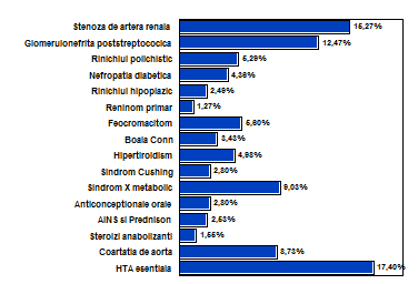
Fig.1: Principal Diseases Etiology for Young Hypertensive Patients

HY1

Table III: Laboratory Results

Hemoglobin value ↑

18

5.60%

Hematocrit ↑

18

5.60%

Value of glucose ↑

68

21.18%

Cholesterol  ↑

78

24.29%

HDL cholesterol  ↑

86

26.79%

LDL cholesterol  ↑

77

23.67%

Triglycerides ↑

105

32.71%

Uric acid  ↑

57

17.75%

Creatinina ↑

38

11.83%

Urea ↑

36

11.21%

Serum sodium ↑

42

13.08%

Serum potassium↓

42

13.08%

Urinalysis -albuminuria+

-hematuria+

96

29.90%

38

11.83%

Urine culture with female +

104

32.29%

Table IV: Laboratory Special Tests

Rennin plasmatic↑

121

37.69%

Vanillyl Mandelic Acid testing (VMA): in urine↑

18

5.60%

Catecholamine urine↑

18

5.60%

Cortisol urine ↑

9

2.80%

Cortisolemia  ↑

9

2.80%

TSH ↑

16

4.98%

T3    ↑

16

4.98%

T4   ↑

16

4.98%

CT abdominal

114

35.51%

RMN abdominal

158

49.92%

Intravenous urogrography

102

31.77%

Observations on Eye Exam and Retinopathy [The eye ground (eye ground findings)]

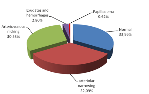
Clearly, the most helpful information to have when one is attempting to establish the chronicity of hypertension is past blood pressure readings. Unfortunately, these are by no means always available since routine blood pressure measurement in young adults is not yet uniformly obtained. In the absence of prior readings, one needs to look for evidence of target organ damage that may suggest chronicity. In adolescent with even severe chronic hypertension or hypertensive encephalopathy. In my study this changes appear for the optic fund may show no more than retinal arteriolar narrowing in 103 cases represent 32.09% and arterio-venous nicking in 98 cases represent 30.53%, hemorrhages and exudates I rarely found in 9 cases represent 2.8%, papilla edema may be absent except in 2 cases even when hypertension was very severe more than 200/120 mmHg with complications of encephalopathy and in 109 cases represent 33.96 was normal result of eye ground examination. See Table V and Fig. 2

Just as there may be minimal eye ground findings, there are infrequently cardiac findings that suggest chronicity.

Table V  Distribution of Cases by Eye Exam Findings

Normal

109

33.96%

Arteriolar narrowing

103

32.09%

Arterio-venous nicking

98

30.53%

Exudates and hemorrhages

9

2.80%

Papilla edema

2

0.62%

HY2

Fig. 2: Distribution of Cases by Changes of Eye Ground Findings

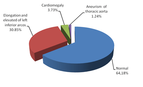
The heart morphology was not clinically enlarged in many cases and the ECG and chest X-ray were usually unhelpful in detecting left ventricular hypertrophy unless hypertension has been prolonged and severe. In my studies left ventricular hypertrophy was present in 35 cases (10.49%), they are helpful in determining chronicity of hypertension and in 206 cases (64.18%) left ventricular hypertrophy was absent.  If negative suggesting nothing about the duration of hypertension. See Table VI and Fig 3

Table VI: Distribution of Cases by Changes in Chest X-Ray

Normal

206

64.18%

Elongation and elevated of left inferior arcos

99

30.85%

Cardiomegaly

12

3.73%

Aneurism of thoracic aorta

4

1.24%

HY3

Fig. 3: Distribution of Cases by Changes of chest X-Ray

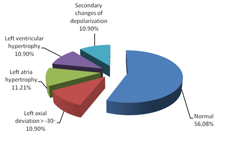
The echocardiography seems to be more sensitive for evaluating chamber size and wall thickness than the ECG and can be helpful. Left atria hypertrophy and left ventricular hypertrophy (Sokolow -Lyon index) and left axial deviation was possible to detect. In my studies I found 35 cases (10.49%) with LVH, 36 cases (11.21%) with LAH and 35 cases (10.49%) with left axial deviation. Secondary changes of depolarization like ST segment sub elevated and negative T wave I found in 35 cases represent 10.49%. See Table VII and Fig. 4

Table 7: Distribution of Cases by Changes in ECG

Normal

180

56.07%

Left axial deviation> -30

35

10.90%

Left atria hypertrophy

36

11.21%

Left  ventricular hypertrophy

Sokolov -Lyon index(SV1+RV5/V6>35mm)

35

10.90%

Secondary  changes of depolarization – ST segment sub elevated  and T wave negative

35

10.90%

HY4

Fig. 4: Distribution of Cases by Changes in ECG

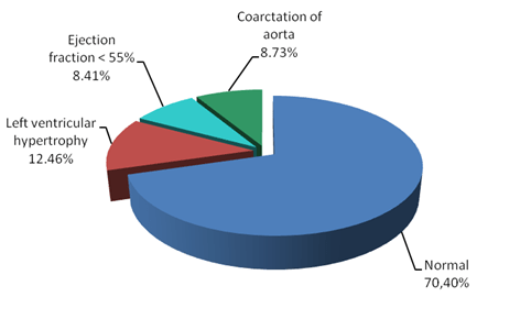
Table VIII:  Distribution of cases by Echocardiography of Hearth Examination

Normal

226

70.40%

Left ventricular hypertrophy

40

12.46%

Ejection fraction(FE) of left ventricular<55%

27

8.41%

Aortic coarctation

28

8.72%

HY5

Fig. 5: Distribution of cases by Changes in Echocardiography

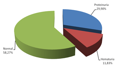
Table IX: Distribution of Cases by Urine Analysis Results

Normal

187

58.27%

Proteinuria

96

29.90%

Hematuria

38

11.83 %

Proteinuria I detect in 96 cases (29.90%) and hematuria in 38 cases (11.83%). See Fig. 6

HY6

Fig.6: Distribution of cases by Urine Analysis Results

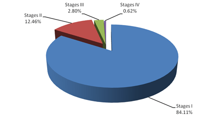
OMS stadialization classification high blood pressure in three stages. In my study about hypertension in the young adults,  the results are as follows:

  • Stages I:  270 cases represents 84.11%,
  • Stages II:  40 cases represents 12.46%,
  • Stages III:  9 cases (2.80%) and
  • Stage IV: malign hypertension 2 cases represents 0.62%.

See, Table IX  and Fig.7

Table IX: Distribution of Cases by Stadialization

Stages I

270

84.11%

Stages II

40

12.46%

Stages III

9

2.81%

Stages IV

2

0.62%


HY7

Fig. 7: Distribution of Cases by Stadialization

DISCUSSION

1.   A 1/5 of group of patients studied have in family antecedents of young hypertensive family member with the following diseases:

  • stroke 46 cases  (14.33%),
  • myocardial infarction 55 cases (17.13%),
  • peripheral vascular disease 23 cases (7.16%),
  • obesity 38 (11.83%),
  • pre-eclamptic toxemia 31 cases (9.65%),
  • hiperaldosteronism in 18 cases (5.60%),
  • polycystic kidney 26cases (8.09%),
  • multiple endocrine diseases II 14 cases (4.36%).

These results  are in concordance with observations of Kotchen JM [1] which in a studies about young hypertensive patients concluded that family aggregation of hypertension was very frequent 20.2% (p<0,001) suggesting the importance of a genetic factor in the etiology of hypertension in the young adults.

    2.   An important number of  hypertensive young  patients were present in personal pathological antecedents: Scarlatti 27 (8.41%), repetitive angina with Streptococcus β hemolytic group A 88 (27.41%),chronic ORL infection focus 35 (10.90%) chronic stomathological focus infections 19 (5.91%), nephritis 34 (10.59%), endocrine disorders 16 (4.98% ), physical and psychical supra solicitation 22 (6.85%), head trauma 11 ( 3.42% ), therapy with corticosteroizi  from another disease (for example erithematous systemic lupus) 5 (1.55%) therapy with AINS  drugs 21 (6.54%), use decongestion nasal 4 (1.24%) repetitive urinary tract infection 28 (8.72%), syphilis 11 (3.42%)(p<0,001). Loggie JMH [2] in a studies with  hypertension in the young reported the streptococcus infection with Streptococcus β hemolytic group A was 18.2% , chronic ORAL infection focus was 8.9%, chronic stomathological focus infections was 3.98%, glomerulonephritis was 6.2% and  physical and psychical supra solicitation was 12.43% in personal pathological antecedents.

3.   Changes of retinal vascular were insufficiently studied in young adults. In my study this changes appear for the optic fund may show no more than retinal arteriolar narrowing 103 cases represent 32.09% and arterio venous nicking 98 cases represent 30.53% , hemorrhages and exudates I rarely found from 9 cases represent 2.8%, papilla edema may be absent except 2 cases even with hypertension was very severe more than 200/120mmHg and complicate with encephalopathy and 109 cases represent 33.96% was normal result of funduoscopic examination (p<0.001).

Skalina MEL et al. [3] observations: 281 hypertensive young patients 140 have changes for the optic fund arterio venous nicking 93 cases, hemorrhages found from 7 cases and exudates appear from 40 patients. 141 patients have the optic fund examination normal.

4.   The heart is not often clinically enlarged and the ECG and chest X-ray are usually unhelpful in detecting left ventricular hypertrophy unless hypertension has been prolonged and severe. In my studies left ventricular hypertrophy was present in 35 cases (10.49%), they are helpful in determining chronicity of hypertension and from 206 cases (64.174%) left ventricular hypertrophy was absent, that suggest that if negative, they tell one nothing about the duration of hypertension. The results are in concordance with observation with Laird WP and Fixler DE [4] who reports after performing chest X-ray for 210 young hypertensive, 103 have normal results, 78 have elongation and elevated of left inferior arcos and 29 present’s cardiomegaly.

5.   The echocardiogram seems to be more sensitive for evaluating chamber size and wall thickness than the ECG and can be helpful. Left atria hypertrophy and left ventricular hypertrophy (Sokolow-Lyon index) and left axial deviation it’s possible to detect. In my studies I found 35 cases (10.49%) with LVH, 36 cases (11.21%) with LAH and 35 cases (10.49%) with left axial deviation. Secondary changes of depolarization like ST segment sub elevated and negative T wave I found from 35 cases represent 10.49% (p<0,001).The results are in concordance with observation with Laird WP and Fixler DE [4], who reports than 18% from young hypertensive subject, presents left ventricular hypertrophy detected after echocardiography examination, end in concordance with observation with Schieken RM et al. [5] in Muscatine studies who reports more than 14% from young hypertensive subjects have left ventricular hypertrophy after make echocardiography examination.

6.   Proteinuria I detect in 96 cases (29.90%) and hematuria in 38 cases (11.83%).This changes appear in context of acute  glomerulonephritis and hypertension was secondary renal.

Schmider et al. [6] sustained that glomerular hyperfiltration is a early marker for nefroangiosclerosis and a sign for subclinical organ affected.

7. OMS stadialization classification high blood pressure in three stages. In my study about hypertension in young adults the results are: in stages I found 270 cases represents 84.11%, in stages II 40 cases represents 12.46%, in stages III 9 cases (2.80%) and malign hypertension 2 cases represents 0.62%.

CONCLUSIONS

  • 1. Importance of genetic factors in etiologies of disease is suggested that family aggregation of hypertension in young adults and another familial diseases like hyperaldosteronism, polycystic kidney and multiple endocrine diseases II.
  • 2. Importance of personal pathologic antecedents demonstrated in my study that repetitive Streptococcus angina with Streptococcus β hemolytic group A was found in the first place as a cause of hypertension in the young people in context of acute streptococcal renal parenchymatous diseases.
  • 3. Except nonspecific symptoms of high blood pressure exist specifically symptoms who suggest etiology of hypertension with young people.
  • 4. The fundoscopic findings in the young adult with hypertension are frequently normal. In the absence of prior readings, one needs to look for evidence of target organ damage that may suggest chronicity. In my study this changes appear for 86 cases 23.3 %  and hemorrhages and exudates I rarely found from 9 cases represent 2.8% and papilla edema may be absent except 2 cases even with hypertension is very severe more than 200/120mmHg and complication of encephalopathy.
  • 5. Left ventricular hypertrophy is possible to detect X-ray, ECG and echocardiography. In my studied I detected left ventricular hypertrophy from 35 patients represent 10.49%.
  • 6. Proteinuria 96 cases (29.90%) have two significance:  nephropathy complication of high blood pressure, or etiology in context or glomerulonephritis alone or accompaniment with hematuria 38 cases (11.83%) in nephritis syndrome.
  • 7. Renin plasmatic level is very important marker of high blood pressure in the young. Was high in 121 cases (37.69%).This situation suggests that hypertension in the young is hiperreninemic hypertension in many cases because young adults have hyperactivity of sympatetic nervous system.
  • 8. OMS classification evaluated stages I 270 cases (84.11%), stages II 40 cases (12.46%) in stages III 9 cases (2.80%) and malign hypertension (stages IV) 2 cases (0.62%)
  • 9. Finally I make a small guideline about hypertension with young patients group of ages 18-35, which I hope to help activity of every physician indifferent specialties in your practice, to use for diagnosis and easy work.

REFERENCES

1. Kotchen JM “Effect of relative weight on familial blood pressure aggregations“ Am J Epidemiol.1987 105-214.

2. Loggie JMH. “The diagnostic evaluation of adolescents with hypertension.” In Hunt JC, Dreifus LS, Dustan HP et al, eds Dialogues in Hypertension Update II vol 1. Lyndhurst, NJ: Health Learning Systems 1984:43-56.

3. Skalina MEL et al.:  Annable WL, Kleigman RM, Fanaroff AA “ Hypertensive retinopathy in the adolescent“ J Adolesc. 1983:103:781-6.

4. Laird WP and Fixler DE. “Left ventricular hypertrophy in adolescents with elevated blood pressure: assessment by chest roentgenography, electrocardiography and echocardiography.” Adolescents 2001;67:255-9.

5.Schieken RM and coauthors: Clarke WR, Lauer RM, “Left ventricular hypertrophy in the young with blood pressures in the upper quin-tile of the distribution: the Muscatine study.” Hypertenesion 2004;3:669-75.

6. Schmider and coauthors: Messerli FH, Garavaglia GE, Nunez BD “Glomerular hyperfiltration indicates target organ disease in essential hypertension.” Circulation 2003; 76: III-273.


Read Full Post »

Introducing Dr. Tim Wu – Interventional Cardiologist, Inventor and Entrepreneur


Author: Ed Kislauskis, PhD

Article ID #18: Introducing Dr. Tim Wu – Interventional Cardiologist, Inventor and Entrepreneur. Published on 1/14/2013

WordCloud Image Produced by Adam Tubman


Welcome readers to the first in a series of interviews with future scientific leaders in biotechnology and medicine.  In this post I interview a close colleague and clinical scientist who appears to be on a fast-track to achieving his vision for the future of interventional cardiology – at the very vanguard of applied nanotechnology.

Tim (Tiangen) Wu, M.D has graciously accepted my invitation to answer a few questions about how his career path and primary goal to develop and commercialize his first product, a fully-biodegradable drug-eluting stent he calls the PowerStent® Absorb (see insert).  This technology combines three especially innovations:  a unique balloon-expandable stent design (PowerStent®), a bioabsorbable nanoparticle composition (BioDe®), and a formulation of two commercially-available anti-restenosis drugs (Combo®).

Stent

About the Subject

Dr. Wu received his clinical education in China and research training in the USA. In 1988, he graduated with an MD from the prestigious Linyli Medical School and completed a fellowship in clinical cardiology at the Tonji Medical University.  In 1993, presented with an opportunity to travel to the US, he uprooted to accept a position as visiting scholar, and ultimately post-doctoral fellow,in Jeffrey Isner’s lab at St. Elizabeth Hospital (Tufts University) and the Beth Israel Medical Center (Harvard Medical).  There he investigated the biology of stenosis, and directed sponsored research projects to evaluate the safety and efficacy of the latest commercially-developed drug-coated stents (DES) in animals.

After  a decade in academia, Dr. Wu made the successful transition to industry and joined Nitromed Inc. as a Research Scientist.  His next stop was as a Research Director at Biomedical Research Models, Inc (2000-2006) where we met and collaborated on developing and characterizing macrovascular disease in an inbred, type 2 diabetic rat model.  After a 20 year career, and upon gaining additional qualification in Mechanical Engineering (Wentworth Institute), Business Administration (MIT), Clinical Research Affairs (Mass. Biotech Council), and Medical Device Regulatory Affairs (North Eastern Univ.), he was ready to take the entrepreneurial leap.  His first company, VasoTech would aim to re-engineer the clinical standards of stent design and drug delivery.

In 2007, Dr. Wu founded VasoTech, Inc. from inside his home garage. Less than a year later, VasoTech received a $1.5M SBIR fast-track grant award from the NIH.  With funding, VasoTech joined the newly announced M2D2 facility on the University of Massachusetts Lowell campus, and expanded operations in China.  With the support of one of his closest advisors, Dr. Stephen McCarthy and other research faculty, Dr. Wu was appointed as an adjunct faculty in the Dept. of BioMedical Engineering at the UMass/Lowell where he mentored a number of talented graduate students.  Dr. Wu is recognized as a senior reviewer on the NIH Bioengineering, Surgical Science and Technology Study Section, and Biomaterials, Delivery Systems and Nanotechnology Special Emphasis Panels servicing the  Small Business Innovation Research (SBIR) grant program.

Dr. Wu’s work at Vasotech is devoted to developing a 3rd generation of fully biodegradable DES coronary stents to solve two major complications associated with stenting, restenosis and late-stage thrombosis. Thusfar, his ideas have attracted well over $1.5 Million (USD) in Small Business Innovation Research (SBIR) grant awards from the National Institute of Diabetes and Digestive and Kidney Diseases, and $1million (USD) from China Innovative Talent Leadership Program.  Through his efforts VasoTech is well positioned to attract the strategic partnerships and venture capital investments necessary to translate his research through clinical stages of development both in China and the US.

The Interview

Kislauskis:  Please help our readers understand the current clinical approach to CAD.

Wu:  Most patients with advanced atherosclerosis diseases are at risk for occlusive coronary arterial disease and stroke. Consequently, it is recommended they undergo a percutaneous intervention (PCI); essentially, balloon angioplasty followed by instillation of one or more expandable metal stents. A properly expanded stent will dilate the vessel and increase blood flow to cardiac muscle tissue. Current 2nd generation drug-eluting-stents (DES) release drugs to inhibit the process of vascular remodeling leading to restenosis. Because the DES approach is remarkably successful and lowers the rate of restenosis to < 10%, DESs is now performed in 85% of the 2 million percutaneous coronary interventions (PCI) procedures annually in the U.S.

Kislauskis:  What is your impression of the recent 5 yr update of the FREEDOM trial comparing effectiveness of coronary artery bypass grafting (CABG) to PCI among diabetics? 1

Wu:  It makes perfect sense. There are other reports evaluating PCI in patients within high risk categories, including those with small diameter vessels, diabetes, and extensive, systemic vascular disease, showing unacceptably high rates of restenosis with bare metal stents (30%-60%) and DESs (6%-18%) 2-4.  We also know first-hand using an inbred rat strain that develops macrovascular disease 4 months after onset of spontaneous diabetes.  In our experiment model, just 4weeks following balloon-induced injury to the coratid artery (PTCA),  we observed 2x greater restenosis in female obese rats, and 4x greater stenosis in obese, diabetic rats  littermates (syndrome X) relative to the non-obese, non-diabetic littermates.  These results predicted that obesity (dyslipidemia) and diabetes (severe hyperglycemia) were major risk factors promoting the complication of restenosis (Wu and Kislauskis, unpublished).

Kislauskis: Can you tell our readers a bit more about the significance of restenosis and thrombosis and the concept behind your approach.

Wu: Two significant drawbacks to conventional PCI are the need for costly, long-term anti-platelet therapy; and having a metal artifact within the coronary vessel. In fact, once installed, the purpose of DES is to maintain patency and provide a scaffold until remodeling is complete, maybe 6 months.  The period of drug elution is typically shorter in duration.  In the event of restenosis, a second DES procedure is recommended and performed with satisfactory results.  However, leaving another metal artifact is problematic.

Most concerning to PCI patients, however, should be an increased risk of sudden death from heart attack from a clot (thrombosis) and tissue ischemia (myocardial infarction).  No available DES technology (eg. Cypher®or Taxus® DES) demonstrates any advantage over bare metal stents in this regard 5-7.  So the thinking is a metal artifact create an irregular vessel surface and micro-eddys in blood flow which ultimately result in late-stage thrombosis, particularly in patients who go off anti-their platelet therapy too soon 8.  Therefore and conceptually, by combining potent DES technology with a fully-biodegradable scaffold, designed to be absorbed fully into the tissue, likely will reduce the rate in-stent stenosis and prevents late-stage thrombosis.

Kislauskis: How did you come up with your unique polymer formulation?

Wu: It turns out that through a process of trial and error in the lab I was able to identify a biodegradable formulation which reduces the local inflammatory response common to all DES formulations while improving the stent’s radial strength.  With a stable drug delivery platform (BioDe®), the process of remodeling will contribute far less to restenosis.  Furthermore, and unlike all prior art, my BioDe® formulation can neutralize acidic intermediates generated during stent degradation that induce inflammation.  The combination of anti-restenosis drugs (Combo®) also is effective at inhibiting signaling pathways that contribute to restenosis.

Kislauskis:  How did you come to design the PowerStent®?

Wu: Again, a long process of trial and error, initially using computer applied design (CAD) principals I learned while earning attending a mechanical engineering certificate program at Wentworth Institute of Technology in Boston. Elements behind my concept for BioDe® came to me while I was involved in a home renovation project, working with grout.  Although the formulation is simple and may be duplicated, the process of manufacturing is complicated.

Kislauskis: So it’s your trade secret.

Wu: Absolutely.

Kislauskis: Can you summary its other advantages and your plans to commercialize the PowerStent®?

Wu: Preclinical, short duration (30 day) studies in porcine models with the PowerStent® Absorb deployed indicate that it will be non-inferior to the current metal DES and competing biodegradable stent technologies. Important functional attributes of the BioDe® polymer include better biocompatibility (less inflammatory), excellent radial strength, potent anti-restenosis activity, and a unique microporous surface that promotes integration into neointimal layer of stented vessel.  Ongoing and much longer duration studies may also support our contention that this design can reduce risks of late-stage in-stent thrombosis.

Kislauskis: What path and difficulties to you foresee in obtaining a regulatory approval to conduct clinical trials with the PowerStent® Absorb?

Wu:  FDA Guidance to commercialize conventional DES technology is available. Unfortunately, no guidance is published for a fully-biodegradable stent.  Therefore, I anticipate seeking advice from the regulatory bodies prior to petitioning for approval to perform clinical trials.  It will no doubt be a complicated process as this technology involves a novel drug combination (albeit FDA-approved drugs), and a novel formulation (albeit FDA-approved components), and a novel indwelling and bioabsorbable medical device (stent).  We are presently completing several required engineering studies for the final phase of pre-clinical safety and efficacy testing, in China. The goals are to obtain FDA pre-market and NDA approvals, and to receive a CE mark from major international markets including Europe and the BRICK nations.

Kislauskis: How will you commercialize this 3rd generation, fully-biodegradable stent?

Wu: There are likely 3 scenarios to complete development and commercialization.  One involves securing bridge funding from the NIH SBIR program, supplemented with angel financing to complete preclinical program. I project that a minimum of $6 Million (USD) will be required to complete regulatory approval and pivotal clinical trials.  Therefore, it is conceivable that a Series A round of equity financing from venture capitalists, in either US or China, will be required. A third scenario is to partner or sell the technology to a major player in this space to complete clinical testing and commercialization. Potential partners include Boston Scientific Company, J&J, etc. Any of these partners could facilitate the processes of regulatory approval, manufacturing, global distribution and marketing.  Discussions are underway with one such prospective partner and with several VC groups.

Kislauskis: What is its likely impact of this product on patient care and the field of interventional cardiology?

Wu: According to US statistics, approximately 14 million Americans suffer from CAD, and 500,000 people die from acute myocardial infarction. One million more survive but with a 1.5 to 15 times greater risk of mortality or morbidity than the rest of the population each year.  In the U.S., the annual health care costs of CAD are estimated to be in excess of $112 billion, and the estimated annual total direct cost associated with PCI with stents is over $2 billion.  I anticipate that our PowerStent® Absorb stent will be competitive in a marketplace estimated to be over $5 billion in 2010. Although CAD patients are the primary market, other related applications for our PowerStent Absorb technology include peripheral arteries, intracerebral vascular and small vessels which are also significant.

Kislauskis:  Thank you for your contribution to this site.  For more information about MMG, LLC and Dr. Wu’s technology please refer to his publications 9-13 or contact him directly at tiangenwu@yahoo.com.

REFERENCES

1.   Mark A. Hlatky, M.D. Compelling Evidence for Coronary-Bypass Surgery in Patients with Diabetes.   N Engl J Med 2012; 367:2437-2438.

2.  Stamler, J. (1989) Epidemiology.  Established major risk factors, and the primary prevention of coronary heart disease. In: Chatterjee K, Karliner J, Rapaport E, Cheitlin MD, Parmlee WW, Sheinman, M eds. Cardiology, Philadelphia Penn: JB Lippincott, 1991, 7.2-7.35. (volume 2).

3. Tanabe, K, Regar, E et al.  Sirolimus-eluting stent for treatment of in-stentrestenosis: One-year angiographic and intravascular ultrasound follow-up. J. Am Col.Cardi.   (2003) 41: 12A.

4. Grube, Eberhard;  Silber, Sigmund.  Six- and twelve-month results from a randomized, double-blind trial on a slow-release paclitaxel-eluting stent for de novo coronary lesions. Circulation 2003: 107, 38-42.

5.  Iakovou I, Schmidt T, Bonizzoni E, et al. Incidence, predictors, and outcome of thrombosis after successful implantation of drug-eluting stents. JAMA 2005;293:2126–2130.

6.  Ong AT, McFadden EP, Regar E, et al. Late angiographic stent thrombosis (LAST) events with drug-eluting stents. J Am Coll Cardiol 2005;45:2088–2092.

7. Wang F, Stouffer GA, Waxman S, et al. Late coronary stent thrombosis: Early vs late stent thrombosis in the stent era. Catheter Cardiovasc Interven 2002;55:142–147.

8. McFadden EP, Stabile E, Regar E, et al. Late thrombosis in drug-eluting coronary stents after discontinuation of antiplatelet therapy. Lancet 2004;364:1519–1521.

9. Ma X, Oyamada S, Wu T, Robich MP, Wu H, Wang X, Buchholz B, McCarthy S, Bianchi CF, Sellke FW, Laham R. In vitro and in vivo degradation of poly(D, L-lactide-co-glycolide)/amorphous calcium phosphate copolymer coated on metal stents. J Biomed Mater Res A. 2011 Mar 15;96(4):632-8. doi: 10.1002/jbm.a.33016. Epub 2011 Jan 25.

10. Oyamada S, Ma X, Wu T, Robich MP, Wu H, Wang X, Buchholz B, McCarthy S, Bianchi CF, Sellke FW, Laham R. Trans-iliac rat aorta stenting: a novel high throughput preclinical stent model for restenosis and thrombosis. J Surg Res. 2011 Mar;166(1):e91-5. Erratum in: J Surg Res. 2012 May 1;174(1):184.

11. Ma X, Oyamada S, Gao F, Wu T, Robich MP, Wu H, Wang X, Buchholz B, McCarthy S, Gu Z, Bianchi CF, Sellke FW, Laham R Paclitaxel/sirolimus combination coated drug-eluting stent: in vitro and in vivo drug release studies. J Pharm Biomed Anal. 2011 Mar 25;54(4):807-11. Erratum in: J Pharm Biomed Anal. 2012 Feb 5;59:217.

12. Ma X, Wu T, Robich MP, Wang X, Wu H, Buchholz B, McCarthy S. Drug-eluting stents. Int J Clin Exp Med. 2010 Jul 15;3(3):192-201.

Other articles related to this subject were published in this Open Access OnlIne Scientific Journal:

Lev-Ari, A. (2012aa). Renal Sympathetic Denervation: Updates on the State of Medicine

http://pharmaceuticalintelligence.com/2012/12/31/renal-sympathetic-denervation-updates-on-the-state-of-medicine/


Lev-Ari, A. (2012U). Imbalance of Autonomic Tone: The Promise of Intravascular Stimulation of Autonomics

http://pharmaceuticalintelligence.com/2012/09/02/imbalance-of-autonomic-tone-the-promise-of-intravascular-stimulation-of-autonomics/

Lev-Ari, A. (2012R). Coronary Artery Disease – Medical Devices Solutions: From First-In-Man Stent Implantation, via Medical Ethical Dilemmas to Drug Eluting Stents http://pharmaceuticalintelligence.com/2012/08/13/coronary-artery-disease-medical-devices-solutions-from-first-in-man-stent-implantation-via-medical-ethical-dilemmas-to-drug-eluting-stents/


Lev-Ari, A. (2012K). Percutaneous Endocardial Ablation of Scar-Related Ventricular Tachycardia

http://pharmaceuticalintelligence.com/2012/07/18/percutaneous-endocardial-ablation-of-scar-related-ventricular-tachycardia/


Lev-Ari, A. (2012C). Treatment of Refractory Hypertension via Percutaneous Renal Denervation

http://pharmaceuticalintelligence.com/2012/06/13/treatment-of-refractory-hypertension-via-percutaneous-renal-denervation/

Lev-Ari, A. (2012D). Competition in the Ecosystem of Medical Devices in Cardiac and Vascular Repair: Heart Valves, Stents, Catheterization Tools and Kits for Open Heart and Minimally Invasive Surgery (MIS)

http://pharmaceuticalintelligence.com/2012/06/22/competition-in-the-ecosystem-of-medical-devices-in-cardiac-and-vascular-repair-heart-valves-stents-catheterization-tools-and-kits-for-open-heart-and-minimally-invasive-surgery-mis/

Lev-Ari, A. (2012E). Executive Compensation and Comparator Group Definition in the Cardiac and Vascular Medical Devices Sector: A Bright Future for Edwards Lifesciences Corporation in the Transcatheter Heart Valve Replacement Market

http://pharmaceuticalintelligence.com/2012/06/19/executive-compensation-and-comparator-group-definition-in-the-cardiac-and-vascular-medical-devices-sector-a-bright-future-for-edwards-lifesciences-corporation-in-the-transcatheter-heart-valve-replace/


Lev-Ari, A. (2012F). Global Supplier Strategy for Market Penetration & Partnership Options (Niche Suppliers vs. National Leaders) in the Massachusetts Cardiology & Vascular Surgery Tools and Devices Market for Cardiac Operating Rooms and Angioplasty Suites

http://pharmaceuticalintelligence.com/2012/06/22/global-supplier-strategy-for-market-penetration-partnership-options-niche-suppliers-vs-national-leaders-in-the-massachusetts-cardiology-vascular-surgery-tools-and-devices-market-for-car/


Lev-Ari, A. (2012G).  Heart Remodeling by Design: Implantable Synchronized Cardiac Assist Device: Abiomed’s Symphony

http://pharmaceuticalintelligence.com/2012/07/23/heart-remodeling-by-design-implantable-synchronized-cardiac-assist-device-abiomeds-symphony/


Read Full Post »

Telling NO to Cardiac Risk

DDAH Says NO to ADMA(1); The DDAH/ADMA/NOS Pathway(2)

Author-Writer-Reporter:  Stephen J. Williams, PhD

Endothelium-derived nitric oxide (NO) has been shown to be vasoprotective.  Nitric oxide enhances endothelial cell survival, inhibits excessive proliferation of vascular smooth muscle cells, regulates vascular smooth muscle tone, and prevents platelets from sticking to the endothelial wall.  Together with evidence from preclinical and human studies, it is clear that impairment of the NOS pathway increases risk of cardiovascular disease (3-5).

This post contains two articles on the physiological regulation of nitric oxide (NO) by an endogenous NO synthase inhibitor asymmetrical dimethylarginine (ADMA) and ADMA metabolism by the enzyme DDAH(1,2).  Previous posts on nitric oxide, referenced at the bottom of the page, provides excellent background and further insight for this posting. In summary plasma ADMA levels are elevated in patients with cardiovascular disease and several large studies have shown that plasma ADMA is an independent biomarker for cardiovascular-related morbidity and mortality(6-8).

admacardiacrisk

admaeffects

Figure 1 A. Cardiac risks of ADMA B. Effects of ADMA (Photo credit: Wikipedia)

ADMA Production and Metabolism

Nuclear proteins such as histones can be methylated on arginine residues by protein-arginine methyltransferases, enzymes which use S-adenosylmethionine as methyl groups.  This methylation event is thought to regulate protein function, much in the way of protein acetylation and phosphorylation (9).  And much like phosphorylation, these modifications are reversible through methylesterases.   The proteolysis of these arginine-methyl modifications lead to the liberation of free guanidine-methylated arginine residues such as L-NMMA, asymmetric dimethylarginine (ADMA) and symmetrical methylarginine (SDMA).

The first two, L-NMMA and ADMA, have been shown to inhibit the activity of the endothelial NOS.  This protein turnover is substantial: for instance the authors note that each day 40% of constitutive protein in adult liver is newly synthesized protein. And in several diseases, such as muscular dystrophy, ischemic heart disease, and diabetes, it has been known since the 1970’s that protein catabolism rates are very high, with corresponding increased urinary excretion of ADMA(10-13).  Methylarginines are excreted in the urine by cationic transport.  However, the majority of ADMA and L-NMMA are degraded within the cell by dimethylaminohydrolase (DDAH), first cloned and purified in rat(14).

endogenous NO inhibitors from pubchem

Figure 2.  Endogenous inhibitors of NO synthase.  Chemical structures generated from PubChem.

DDAH

DDAH specifically hydrolyzes ADMA and L-NMMA to yield citruline and demethylamine and usually shows co-localization with NOS. Pharmacologic inhibition of DDAH activity causes accumulation of ADMA and can reverse the NO-mediated bradykinin-induced relaxation of human saphenous vein.

Two isoforms have been found in human:

  • DDAH1 (found in brain and kidney and associated with nNOS) and
  • DDAH2 (highly expressed in heart, placenta, and kidney and associated with eNOS).

DDAH2 can be upregulated by all-trans retinoic acid (atRA can increase NO production).  Increased reactive oxygen species and possibly homocysteine, a risk factor for cardiovascular disease, can decrease DDAH activity(15,16).

  • The importance of DDAH activity can also be seen in transgenic mice which overexpress DDAH, exhibiting increased NO production, increased insulin sensitivity, and reduced vascular resistance  (17).  Likewise,
  • Transgenic mice, null for the DDAH1, showed increase in blood pressure, decreased NO production, and significant increase in tissue and plasma ADMA and L-NMMA.

amdanosfigure

Figure 3.  The DDAH/ADMA/NOS cycle. Figure adapted from Cooke and Ghebremarian (1).

As mentioned in the article by Cooke and Ghebremariam, the authors state: the weight of the evidence indicates that DDAH is a worthy therapeutic target. Agents that increase DDAH expression are known, and 1 of these, a farnesoid X receptor agonist, is in clinical trials

http://www.interceptpharma.com

An alternate approach is to

  • develop an allosteric activator of the enzyme.  Although
  • development of an allosteric activator is not a typical pharmaceutical approach, recent studies indicate that this may be achievable aim(18).

References:

1.            Cooke, J. P., and Ghebremariam, Y. T. : DDAH says NO to ADMA.(2011) Arteriosclerosis, thrombosis, and vascular biology 31, 1462-1464

2.            Tran, C. T., Leiper, J. M., and Vallance, P. : The DDAH/ADMA/NOS pathway.(2003) Atherosclerosis. Supplements 4, 33-40

3.            Niebauer, J., Maxwell, A. J., Lin, P. S., Wang, D., Tsao, P. S., and Cooke, J. P.: NOS inhibition accelerates atherogenesis: reversal by exercise. (2003) American journal of physiology. Heart and circulatory physiology 285, H535-540

4.            Miyazaki, H., Matsuoka, H., Cooke, J. P., Usui, M., Ueda, S., Okuda, S., and Imaizumi, T. : Endogenous nitric oxide synthase inhibitor: a novel marker of atherosclerosis.(1999) Circulation 99, 1141-1146

5.            Wilson, A. M., Shin, D. S., Weatherby, C., Harada, R. K., Ng, M. K., Nair, N., Kielstein, J., and Cooke, J. P. (2010): Asymmetric dimethylarginine correlates with measures of disease severity, major adverse cardiovascular events and all-cause mortality in patients with peripheral arterial disease. Vasc Med 15, 267-274

6.            Kielstein, J. T., Impraim, B., Simmel, S., Bode-Boger, S. M., Tsikas, D., Frolich, J. C., Hoeper, M. M., Haller, H., and Fliser, D. : Cardiovascular effects of systemic nitric oxide synthase inhibition with asymmetrical dimethylarginine in humans.(2004) Circulation 109, 172-177

7.            Kielstein, J. T., Donnerstag, F., Gasper, S., Menne, J., Kielstein, A., Martens-Lobenhoffer, J., Scalera, F., Cooke, J. P., Fliser, D., and Bode-Boger, S. M. : ADMA increases arterial stiffness and decreases cerebral blood flow in humans.(2006) Stroke; a journal of cerebral circulation 37, 2024-2029

8.            Mittermayer, F., Krzyzanowska, K., Exner, M., Mlekusch, W., Amighi, J., Sabeti, S., Minar, E., Muller, M., Wolzt, M., and Schillinger, M. : Asymmetric dimethylarginine predicts major adverse cardiovascular events in patients with advanced peripheral artery disease.(2006) Arteriosclerosis, thrombosis, and vascular biology 26, 2536-2540

9.            Kakimoto, Y., and Akazawa, S.: Isolation and identification of N-G,N-G- and N-G,N’-G-dimethyl-arginine, N-epsilon-mono-, di-, and trimethyllysine, and glucosylgalactosyl- and galactosyl-delta-hydroxylysine from human urine. (1970) The Journal of biological chemistry 245, 5751-5758

10.          Inoue, R., Miyake, M., Kanazawa, A., Sato, M., and Kakimoto, Y.: Decrease of 3-methylhistidine and increase of NG,NG-dimethylarginine in the urine of patients with muscular dystrophy. (1979) Metabolism: clinical and experimental 28, 801-804

11.          Millward, D. J.: Protein turnover in skeletal muscle. II. The effect of starvation and a protein-free diet on the synthesis and catabolism of skeletal muscle proteins in comparison to liver. (1970) Clinical science 39, 591-603

12.          Goldberg, A. L., and St John, A. C.: Intracellular protein degradation in mammalian and bacterial cells: Part 2. (1976) Annual review of biochemistry 45, 747-803

13.          Dice, J. F., and Walker, C. D.: Protein degradation in metabolic and nutritional disorders. (1979) Ciba Foundation symposium, 331-350

14.          Ogawa, T., Kimoto, M., and Sasaoka, K.: Purification and properties of a new enzyme, NG,NG-dimethylarginine dimethylaminohydrolase, from rat kidney. (1989) The Journal of biological chemistry 264, 10205-10209

15.          Ito, A., Tsao, P. S., Adimoolam, S., Kimoto, M., Ogawa, T., and Cooke, J. P.: Novel mechanism for endothelial dysfunction: dysregulation of dimethylarginine dimethylaminohydrolase. (1999) Circulation 99, 3092-3095

16.          Stuhlinger, M. C., Tsao, P. S., Her, J. H., Kimoto, M., Balint, R. F., and Cooke, J. P. : Homocysteine impairs the nitric oxide synthase pathway: role of asymmetric dimethylarginine.(2001) Circulation 104, 2569-2575

17.          Sydow, K., Mondon, C. E., Schrader, J., Konishi, H., and Cooke, J. P.: Dimethylarginine dimethylaminohydrolase overexpression enhances insulin sensitivity. (2008) Arteriosclerosis, thrombosis, and vascular biology 28, 692-697

18.          Zorn, J. A., and Wells, J. A.: Turning enzymes ON with small molecules. (2010) Nature chemical biology 6, 179-188

Other research papers on Nitric Oxide and Cardiac Risk  were published on this Scientific Web site as follows:

The Nitric Oxide and Renal is presented in FOUR parts:

Part I: The Amazing Structure and Adaptive Functioning of the Kidneys: Nitric Oxide

Part II: Nitric Oxide and iNOS have Key Roles in Kidney Diseases

Part III: The Molecular Biology of Renal Disorders: Nitric Oxide

Part IV: New Insights on Nitric Oxide donors

Cardiac Arrhythmias: A Risk for Extreme Performance Athletes

What is the role of plasma viscosity in hemostasis and vascular disease risk?

Cardiovascular Risk Inflammatory Marker: Risk Assessment for Coronary Heart Disease and Ischemic Stroke – Atherosclerosis.

Endothelial Dysfunction, Diminished Availability of cEPCs, Increasing CVD Risk for Macrovascular Disease – Therapeutic Potential of cEPCs

Biochemistry of the Coagulation Cascade and Platelet Aggregation – Part I

Nitric Oxide Function in Coagulation

Read Full Post »

Author and Reporter: Anamika Sarkar, Ph.D.

Nitric Oxide (NO) is highly regulated in the blood such that it can be released as vasodilator when needed. The importance and pathway of Nitric Oxide has been nicely reviewed by. “Discovery of NO and its effects of vascular biology”. Other articles which are good readings for the importance of NO are  – a) regulation of glycolysis b) NO in cardiovascular disease c) NO and Immune responses Part I and Part II d) NO signaling pathways. The  effects of NO in diseased states have been reviewed by the articles – “Crucial role of Nitric Oxide in Cancer”, “Nitric Oxide and Sepsis, Hemodynamic Collapse, and the Search for Therapeutic Options”.. (Also, please see Source for more articles on NO and its significance).

Computational models are very efficient tools to understand complex reactions like NO towards physiological conditions. Among them wall shear stress is one of the major factors which is reviewed in the article – “Differential Distribution of Nitric Oxide – A 3-D Mathematical Model”.

Moreover, decrease in availability of NO can lead to many complications like pulmonary hypertension. Some of the causes of decrease in NO have been identified as clinical hypertension, right ventricular overload which can lead to cardiac heart failure, low levels of zinc and high levels of cardiac necrosis.

Sickle Cell disease patients, a hereditary disease, are also known to have decreased levels of NO which can become physiologically challenging. In USA alone, there are 90,000 people who are affected by Sickle cell disease.

Sickle cell disease is breakage of red blood cells (RBC) membrane and resulting release of the hemoglobin (Hb) into blood plasma. This process is also known as Hemolysis. Sickle cell disease is caused by single mutation of Hb which changes RBC from round shape to sickle or crescent shapes (Figure 1).

Image

Figure 1 (A) shows normal red blood cells flowing freely through veins. The inset shows a cross section of a normal red blood cell with normal hemoglobin. Figure 1 (B) shows abnormal, sickled red blood cells The inset image shows a cross-section of a sickle cell with long polymerized HbS strands stretching and distorting the cell shape. Image Source: http://en.wikipedia.org/wiki/Sickle-cell_disease

Sickle Cell RBCs has much shorter life span of 10-20 days when compared with normal RBCs 100-120 days lifespan. Shorter life span of Sickle cell disease RBC’s are compensated by bone marrow generation of new RBCs. However, many times new blood generation cannot cope with the small life span of Sickle cell RBCs and causes pathological condition of Anemia.

RBCs generally breakdown and release Hbs in blood plasma after they reach their end of life span. Thus, in case of Sickle cell disease, there is more cell free Hb than normal. Furthermore, it is known that NO has a very high affinity towards Hbs, which is one of the ways free NO is regulated in blood. As a result presence of larger amounts of cell free Hb in Sickle cell disease lead to less availability of NO.

However, the question remained “what is the quantitative relationship between cell free Hb and depletion of NO. Deonikar and Kavdia (J. Appl. Physiol., 2012) addressed this question by developing a 2 dimensional Mathematical Model of a single idealized arteriole, with different layers of blood vessels diffusing nutrients to tissue layers (Figure 2:  Deonikar and Kavdia Figure 1).

Image

cell free Hb in 2 dimensional representations of blood vessels.

The authors used steady state partial differential equation of circular geometry to represent diffusion of NO in blood and in tissues. They used first and second order biochemical reactions to represent the reactions between NO and RBC and NO autooxidation processes. Some of their reaction model parameters were obtained from literature, rest of them were fitted to experimental results from literature. The model and its parameters are explained in the previously published paper by same authors Deonikar and Kavdia, Annals of Biomed., 2010. The authors found that the reaction rate between NO and RBC is 0.2 x 10⁵, M-1 s-1 than 1.4 x 10⁵, M-1 s-1 as reported before by Butler et.al., Biochim. Biophys. Acta, 1998.

Their results show that even small increase in cell free Hb, 0.5uM, can decrease NO concentrations by 3-7 folds approximately (comparing Fig1(b) and 1(d) of Deonikar and Kavdia, 2012, as shown in Figure 2 of this article). Moreover, their mathematical analysis shows that the increase in diffusion resistance of NO from vascular lumen to cell free zone has no effect on NO distribution and concentration with available levels of cell free Hb.

Deonikar and Kavdia’s mathematical model is a simple representation of actual physiological scenario. However, their model results show that for Sickle cell disease patients, decrease in levels of bioavailable NO is an attribute to cell free Hb, which is in abundant for these patients. Their results show that small increase by 0.5 uM in cell free Hb can cause large decrease in NO concentrations.

These interesting insights from the model can help in further understanding in the context of physiological conditions, by replicating experiments in-vivo and then relating them to other known diseases of Sickle cell disease patients like Anemia, Pulmonary Hypertension. Further, drugs can be targeted towards decreasing free cell Hbs to keep balance in availability of NO, which in turn may help in other related disease like Pulmonary Hypertension of Sickle Cell disease patients.

Sources:

Deonikar and Kavdia (2012) :http://www.ncbi.nlm.nih.gov/pubmed/22223452

Previous model explaining mathematical representation and parameters used in the model :Deonikar and Kavdia, Annals of Biomed., 2010.

Previous paper stating reaction rate of Hb and NO: Butler et.al., Biochim. Biophys. Acta, 1998.

Causes of decrease in NO

Clinical Hypertension : http://www.ncbi.nlm.nih.gov/pubmed/11311074

Right ventricular overload : http://www.ncbi.nlm.nih.gov/pubmed/9559613

Low levels of zinc and high levels of cardiac necrosis : http://www.ncbi.nlm.nih.gov/pubmed/11243421

Sickle Cell Source:

http://en.wikipedia.org/wiki/Sickle-cell_disease

http://www.nhlbi.nih.gov/health/health-topics/topics/sca/

NO Source:

Differential Distribution of Nitric Oxide – A 3-D Mathematical Model:

Discovery of NO and its effects of vascular biology

Nitric Oxide has a ubiquitous role in the regulation of glycolysis -with a concomitant influence on mitochondrial function

Nitric oxide: role in Cardiovascular health and disease

NO signaling pathways

Nitric Oxide and Immune Responses: Part 1

Nitric Oxide and Immune Responses: Part 2

Statins’ Nonlipid Effects on Vascular Endothelium through eNOS Activation

http://pharmaceuticalintelligence.com/2012/10/08/statins-nonlipid-effects-on-vascular-endothelium-through-enos-activation/

Inhibition of ET-1, ETA and ETA-ETB, Induction of NO production, stimulation of eNOS and Treatment Regime with PPAR-gamma agonists (TZD): cEPCs Endogenous Augmentation for Cardiovascular Risk Reduction – A Bibliography

Nitric Oxide, Platelets, Endothelium and Hemostasis

Crucial role of Nitric Oxide in Cancer

The rationale and use of inhaled NO in Pulmonary Artery Hypertension and Right Sided Heart Failure

Nitric Oxide and Sepsis, Hemodynamic Collapse, and the Search for Therapeutic Options

NO Nutritional remedies for hypertension and atherosclerosis. It’s 12 am: do you know where your electrons are?

Clinical Trials Results for Endothelin System: Pathophysiological role in Chronic Heart Failure, Acute Coronary Syndromes and MI – Marker of Disease Severity or Genetic Determination?

Endothelial Function and Cardiovascular Disease

Interaction of Nitric Oxide and Prostacyclin in Vascular Endothelium

Endothelial Dysfunction, Diminished Availability of cEPCs,  Increasing  CVD Risk – Macrovascular Disease – Therapeutic Potential of cEPCs

Cardiovascular Disease (CVD) and the Role of agent alternatives in endothelial Nitric Oxide Synthase (eNOS) Activation and Nitric Oxide Production


Read Full Post »

Author and Reporter: Anamika Sarkar, Ph.D.

Among many important roles of Nitric oxide (NO), one of the key actions is to act as a vasodilator and maintain cardiovascular health. Induction of NO is regulated by signals in tissue as well as endothelium.

Importance of NO has been nicely reviewed in the article  “Discovery of NO and its effects of vascular biology”. Other articles which are good readings for the importance of NO are  – a) regulation of glycolysis b) NO in cardiovascular disease c) NO and Immune responses Part I and Part II d) NO signaling pathways (Also, please see Source for more articles on NO and its significance).

The rate of production of NO has been established to be dependent on Wall Shear Stress (WSS) (Mashour and Broock, Brain Res., 1999) . Many mathematical models have been developed as 2D diffusion models to predict distribution of NO transport in single vessels, eg. arterioles (Please see Sources for references ).

Chen et. al. (Med. Biol. Eng. Comp., 2011) developed a 3-D model consisting of two branched arterioles and nine capillaries surrounding the vessels. Their model not only takes into account of the 3-D volume, but also branching effects on blood flow (Please see Fig 1 and Fig 2 from Chen et. al. 2011 ).

Image

Fig. 1 Blood phase separation with vascular branching. RBC
fractional flow in daughter branch alpha is not necessarily equal
to that in branch beta

Image

The mathematical model considers dynamic characteristics related to blood flow, blood vessel structures and transport mechanism in the wall. The authors have considered effects of branching and ratio of diameters between blood vessels of parent and children to determine the fractional blood flow which gets distributed in the network. These branching effects of the vessels will also affect the blood volume or RBC (Red Blood Cell), hence NO consumption in the blood. Parameters in the model are either obtained or fitted with experimental results from literature. Their model assumes a linear relationship of NO production with wall shear stress which in turn will be regulated by blood flow determined by branching characteristics of blood vessels. Moreover, the mathematical model includes transport of NO through the blood vessels in the tissue (in the defined volume of the model) as diffusion model,. The model was solved using Finite Elements method using the software COMSOL.

Their model results show that wall shear stress changes depending upon the distribution of RBC in the microcirculations of blood vessels, which leads to differential production of NO along the vascular network. Levels of NO at vascular walls can be less in branches which receive more blood flow, due to the balance between higher consumption of NO by RBC and production of NO due to high wall stress.  Their 3-D simulations showed the importance of capillaries such that NO can be concentrated in tissues far away in distance from arterioles facilitating much controlled NO regulation.

Though, the 3-D model developed by Chen et. al., (2011) is an idealized mathematical model of blood flow with production and consumption of NO, depending upon WSS, yet it shows importance of structure of blood vessels in distributions of NO in vessels and tissues. Such a model with proper extension to larger network can give more insights into differential distributions of NO as a function of blood flow and wall shear stress. As nano-medicine become sophisticated in years to come, information of distribution of NO in tissues and blood vessels can help the medicine to be more targeted.

Sources:

Chen et.al. (2011) : http://www.ncbi.nlm.nih.gov/pubmed/21431938

Mashour and Broock, Brain Res., 1999: http://www.ncbi.nlm.nih.gov/pubmed?term=10526117

Mathematical Modes of NO Distribution in 2-D

Other research on Nitric Oxide and Vascular Biology on this Scientific Web Site include the following:

Nitric Oxide and Immune Responses: Part 1

Curator and Reporter: Aviral Vatsa, 10/18/2012

http://pharmaceuticalintelligence.com/2012/10/18/nitric-oxide-and-immune-responses-part-1/

Clinical Trials Results for Endothelin System: Pathophysiological role in Chronic Heart Failure, Acute Coronary Syndromes and MI – Marker of Disease Severity or Genetic Determination?

Curator: Aviva Lev-Ari, 10/19/2012

http://pharmaceuticalintelligence.com/2012/10/19/clinical-trials-results-for-endothelin-system-pathophysiological-role-in-chronic-heart-failure-acute-coronary-syndromes-and-mi-marker-of-disease-severity-or-genetic-determination/

Nitric Oxide and Sepsis, Hemodynamic Collapse, and the Search for Therapeutic Options

Curator and Reporter: Larry Bernstein, MD, 10/20/2012

http://pharmaceuticalintelligence.com/2012/10/20/nitric-oxide-and-sepsis-hemodynamic-collapse-and-the-search-for-therapeutic-options/

Mitochondrial Damage and Repair under Oxidative Stress

Curator: Larry H Bernstein, MD, FCAP, 10/28/2012

http://pharmaceuticalintelligence.com/2012/10/28/mitochondrial-damage-and-repair-under-oxidative-stress/

Nitric Oxide and Immune Responses: Part 2

Curator: Aviral Vatsa, PhD, MBBS, 10/28/2012

http://pharmaceuticalintelligence.com/2012/10/28/nitric-oxide-and-immune-responses-part-2/

Differential Distribution of Nitric Oxide – A 3-D Mathematical Model

Author: Anamika Sarkar, PhD, 10/28/2012

http://pharmaceuticalintelligence.com/2012/10/28/differential-distribution-of-nitric-oxide-a-3-d-mathematical-model/

Statins’ Nonlipid Effects on Vascular Endothelium through eNOS Activation

Curator, EAW: Larry Bernstein, 10/8/2012

http://pharmaceuticalintelligence.com/2012/10/08/statins-nonlipid-effects-on-vascular-endothelium-through-enos-activation/

Nitric Oxide Nutritional remedies for hypertension and atherosclerosis. It’s 12 am: do you know where your electrons are?

Author and Reporter: Meg Baker, 10/7/2012.

http://pharmaceuticalintelligence.com/2012/10/07/no-nutritional-remedies-for-hypertension-and-atherosclerosis-its-12-am-do-you-know-where-your-electrons-are/

Inhibition of ET-1, ETA and ETA-ETB, Induction of NO production, stimulation of eNOS and Treatment Regime with PPAR-gamma agonists (TZD): cEPCs Endogenous Augmentation for Cardiovascular Risk Reduction – A Bibliography

Curator: Aviva Lev-Ari, 10/4/2012.

http://pharmaceuticalintelligence.com/2012/10/04/inhibition-of-et-1-eta-and-eta-etb-induction-of-no-production-and-stimulation-of-enos-and-treatment-regime-with-ppar-gamma-agonists-tzd-cepcs-endogenous-augmentation-for-cardiovascular-risk-reduc/

Coronary Artery Disease – Medical Devices Solutions: From First-In-Man Stent Implantation, via Medical Ethical Dilemmas to Drug Eluting Stents August 13, 2012

Author: Aviva Lev-Ari, PhD, RN, 8/13/2012

http://pharmaceuticalintelligence.com/2012/08/13/coronary-artery-disease-medical-devices-solutions-from-first-in-man-stent-implantation-via-medical-ethical-dilemmas-to-drug-eluting-stents/

Vascular Medicine and Biology: CLASSIFICATION OF FAST ACTING THERAPY FOR PATIENTS AT HIGH RISK FOR MACROVASCULAR EVENTS Macrovascular Disease – Therapeutic Potential of cEPCs

Curator; Aviva Lev-Ari, PhD, RN, 8/24/2012

http://pharmaceuticalintelligence.com/2012/08/24/vascular-medicine-and-biology-classification-of-fast-acting-therapy-for-patients-at-high-risk-for-macrovascular-events-macrovascular-disease-therapeutic-potential-of-cepcs/


Read Full Post »

Atrial Fibrillation: The Latest Management Strategies

Reporter: Aviva Lev-Ari, PhD, RN

UPDATED on 8/5/2013

Ischemic strokes are the most common type of AFib-related stroke5 and can be extremely debilitating.6,7 It’s important to help your patients understand the risk of ischemic stroke and how you can help lower that risk.

Nearly 9 out of 10 AFib-related strokes are ischemic, and most are cardioembolic5,8,9

  • Cardioembolic strokes are most commonly caused by AFib9,10
  • Hemorrhagic strokes account for approximately 10% of AFib-related strokes5
  • AFib-related ischemic strokes are primarily caused by an embolus formed in the left atrial appendage of the heart11

Ischemic strokes can be devastating, often resulting in irreversible brain damage2

  • Debilitating effects of a stroke include paralysis, slurred speech, and memory loss12
    • Every second, ≈32,000 brain cells can die due to hypoxia from lack of blood flow4
    • In 1 minute, nearly 2 million brain cells can die—increasing the risk of disability or death2-4
  • Severely disabling stroke is frequently rated by patients as equivalent to or worse than death13

Strokes are a leading cause of disability in the US14

The good news is you can significantly reduce your AFib patients’ risk of ischemic stroke with anticoagulation therapy.11,15,16 By keeping them appropriately anticoagulated, you can help your patients avoid the devastation of ischemic stroke.11

AFib=atrial fibrillation.

References

  1. Types of stroke. Johns Hopkins Medicine Web site. http://www.hopkinsmedicine.org/healthlibrary/printv.aspx?d=85,P00813. Accessed August 9, 2012.
  2. Maas MB, Safdieh JE. Ischemic stroke: pathophysiology and principles of localization. Hospital Physician Neurology Board Review Manual. 2009;13:1-16.http://www.turner-white.com/pdf/brm_Neur_V13P1.pdf. Accessed February 1, 2013.
  3. Rosamond WD, Folsom AR, Chambless LE, et al. Stroke incidence and survival among middle-aged adults: 9-year follow-up of the Atherosclerosis Risk in Communities (ARIC) cohort. Stroke. 1999;30:736-743.
  4. Saver JL. Time is brain—quantified. Stroke. 2006;37:263-266.
  5. Mercaldi CJ, Ciarametaro M, Hahn B, et al. Cost efficiency of anticoagulation with warfarin to prevent stroke in Medicare beneficiaries with nonvalvular atrial fibrillation. Stroke. 2011;42:112-118.
  6. Vemmos KN, Tsivgoulis G, Spengos K, et al. Anticoagulation influences long-term outcome in patients with nonvalvular atrial fibrillation and severe ischemic stroke. Am J Geriatr Pharmacother. 2004;2:265-273.
  7. Lin HJ, Wolf PA, Kelly-Hayes M, et al. Stroke severity in atrial fibrillation. The Framingham Study. Stroke. 1996;27:1760-1764.
  8. Grau AJ, Weimar C, Buggle F, et al. Risk factors, outcome, and treatment in subtypes of ischemic stroke: the German Stroke Data Bank. Stroke. 2001;32:2559-2566.
  9. Bogousslavsky J, Van Melle G, Regli F, Kappenberger L. Pathogenesis of anterior circulation stroke in patients with nonvalvular atrial fibrillation: the Lausanne Stroke Registry. Neurology. 1990;40:1046-1050.
  10. Freeman WD, Aguilar MI. Prevention of cardioembolic stroke. Neurotherapeutics. 2011;8:488-502.
  11. Fuster V, Rydén LE, Cannom DS, et al. ACC/AHA/ESC 2006 Guidelines for the Management of Patients With Atrial Fibrillation—executive summary: a report of the American College of Cardiology/American Heart Association Task Force on Practice Guidelines and the European Society of Cardiology Committee for Practice Guidelines (Writing Committee to Revise the 2001 Guidelines for the Management of Patients With Atrial Fibrillation): developed in collaboration with the European Heart Rhythm Association and the Heart Rhythm SocietyCirculation. 2006;114:700-752.
  12. Effects of stroke. American Stroke Association Web site. http://www.strokeassociation.org/STROKEORG/AboutStroke/EffectsofStroke/Effects-of-Stroke_UCM_308534_SubHomePage.jsp. Accessed December 8, 2012.
  13. Gage BF, Cardinalli AB, Owens DK. The effect of stroke and stroke prophylaxis with aspirin or warfarin on quality of life. Arch Intern Med. 1996;156:1829-1836.
  14. Centers for Disease Control and Prevention (CDC). Prevalence of Stroke—United States, 2006-2010. MMWR Morb Mortal Wkly Rep. 2012;61:379-382.
  15. Singer DE, Chang Y, Fang MC, et al. The net clinical benefit of warfarin anticoagulation in atrial fibrillation. Ann Intern Med. 2009;151:297-305.
  16. Lip GYH, Andreotti F, Fauchier L, et al. Bleeding risk assessment and management in atrial fibrillation patients: a position document from the European Heart Rhythm Association, endorsed by the European Society of Cardiology Working Group on Thrombosis. Europace. 2011;13:723-746.

SOURCE

http://www.medscape.com/infosite/afib/public/

Straightforward, informed answers to your most important questions about living

with atrial fibrillation – the most common sustained cardiac arrhythmia.

Written by

Hugh G. Calkins, M.D., Director of the Arrhythmia Service

and Electrophysiology Lab at The Johns Hopkins Hospital,

and Ronald Berger, M.D.,

If you’ve ever run up a flight of stairs, chased a tennis ball across the court, or reacted in fright at a scary movie, you know what a pounding heart feels like…

But for the 2.3 million Americans who suffer from atrial fibrillation (AF or AFib), a racing heart is a way of life. Simple tasks like getting out of bed in the morning or rising from a chair can cause dizziness, weakness, shortness of breath, or heart palpitations. For these people, AF severely impairs quality of life – and even when symptoms stemming from AF are mild, the disorder can seriously impact health, increasing the risk of stroke and heart failure.

AF can be a debilitating even deadly condition. Fortunately, it can be successfully managed – but there are various approaches for treating AF or preventing a recurrence. How do you and your doctor choose which approach is right for you?

If you or a loved one has AF, there are so many questions: Do I need an anticoagulant… should I be taking medication to control my heart rate… will my symptoms respond to cardioversion… if I need an antiarrhythmic drug to control AF episodes, which one should I take… when is an ablation procedure appropriate… and more.

It’s critically important to learn everything you can now — so you can partner with your doctor effectively, ask the right questions, and understand the answers.

To help you, we asked two eminent experts at Johns Hopkins to share their expertise and hands-on experience with arrhythmia patients in an important new report, Atrial Fibrillation: The Latest Management Strategies.

Dr. Hugh Calkins and Dr. Ronald Berger are ideally positioned to help you understand and manage your AF. Together with their colleagues at Johns Hopkins, they perform approximately 2,000 electrophysiology procedures and 200 pulmonary vein isolation procedures for atrial fibrillation each year.

Hugh Calkins, M.D. is the Nicholas J. Fortuin, M.D. Professor of Cardiology, Professor of Pediatrics, and Director of the Arrhythmia Service, the Electrophysiology Lab, and the Tilt Table Diagnostic Lab at The Johns Hopkins Hospital. He has clinical and research interests in the treatment of cardiac arrhythmias with catheter ablation, the role of device therapy for treating ventricular arrhythmias, the evaluation and management of syncope, and the study of arrhythmogenic right ventricular dysplasia.

Ronald Berger, M.D., Ph.D., a Professor of Medicine and Biomedical Engineering at Johns Hopkins, is Director of the Electrophysiology Fellowship Program at The Johns Hopkins Hospital. He serves on the editorial board for two major journals in the cardiovascular field and has written and coauthored more than 100 articles and book chapters.

Atrial Fibrillation: The Latest Management Strategies is now available to you in a digital PDF download and print version.

“I feel like my heart is going to jump out of my chest…” 

An arrhythmia is an abnormality in the timing or pattern of the heartbeat, causing the heart to beat too rapidly, too slowly, or irregularly. Sounds pretty straightforward, but there’s a lot we don’t know about why the heart rhythm goes awry… or the best way to treat it.

In Atrial Fibrillation: The Latest Management Strategies, we focus on what we DO know. In page after page of this comprehensive report, we address your most serious concerns about living with AF, such as:

  • I don’t have any symptoms. Is my problem definitely AF?
  • Can drinking alcohol trigger or worsen AF?
  • Is every person who has AF at risk for a stroke?
  • If my doctor suspects AF, will I have to wear an implantable or event monitor to be sure?
  • Why does AF often show up later in life?
  • What would you recommend to the older patient – 75 and older – who has AF but no bothersome symptoms?
  • What do you recommend for the person with longstanding persistent AF?
  • Is the AF experienced by an otherwise healthy person different from that of a person with underlying heart disease or other health issues?
  • What are the differences among: paroxysmal AF, persistent AF, and longstanding persistent AF?
  • What is the “pill-in-the-pocket” approach to AF?

Anticoagulation Therapy: What You Should Know

While AF is generally not life threatening, for some patients it can increase the likelihood of blood clots forming in the heart. And if a clot travels to the brain, a stroke will result. Anticoagulation therapy is used to prevent blood clot formation in people with AF…

  • Why is anticoagulation therapy with warfarin (Coumadin) needed for some people with AF?
  • How is the use of warfarin monitored?
  • How does a doctor determine if a patient with AF needs to take warfarin?
  • What’s the CHADS2 score and how is it used?
  • If a patient’s CHADS2 score is 1, how do you decide between aspirin and warfarin, or nothing at all?
  • Why is it so difficult to keep within therapeutic range with warfarin?
  • Can I test my INR (a test measuring how long it takes blood to clot) at home?
  • What happens if my INR is too high?
  • What options are available if a patient cannot take warfarin?
  • What are the benefits of dabigatran, a new blood-thinning alternative to warfarin therapy?

Symptom Control: The Art of Rate and Rhythm Control

For many patients and their doctors, it’s difficult to achieve and maintain heart rhythm. Two key management strategies are used: heart rate and heart rhythm control. In Atrial Fibrillation: The Latest Management Strategies, you’ll read an in-depth discussion of the benefits of rate versus rhythm control for AF:

  • What have we learned from the AFFIRM study, and how has this knowledge affected the management of AF?
  • What is catheter ablation of the AV (atrioventricular) node?
  • Why is cardioversion needed?
  • Are there different types of cardioversion?
  • What is chemical cardioversion? What is electrical cardioversion?
  • Can medication be used to convert the heart back to normal sinus rhythm?
  • Which antiarrhythimic drugs are used to treat AF?
  • How is catheter ablation for AF performed?
  • What is pulmonary vein antrum isolation (PVAI) and how is it performed?
  • Who are the best candidates for PVAI?

There’s more to Atrial Fibrillation: The Latest Management Strategies, much more.

We explain surgical ablation of AF, a procedure performed through small incisions in the chest wall… discuss when it’s appropriate to seek a second opinion… take a close look at strokes and explain the warning signs and differences among ischemic, thrombotic, embolic, and hemorrhagic strokes… and provide an arrhythmia glossary of key AF terms used by electrophysiologists and cardiologists.

Direct to You From Johns Hopkins

Atrial Fibrillation: The Latest Management Strategies is designed to give you unprecedented access to the expertise of the hospital ranked #1 of America’s Best Hospitals for 21 consecutive years 1991-2011 by U.S. News & World Report. You simply won’t find a more knowledgeable and trustworthy source of the medical information you require. A tradition of discovery and medical innovation is the hallmark of Johns Hopkins research. Since its founding in 1889, The Johns Hopkins Hospital has led the way transferring the discoveries made in the laboratory to the administration of effective patient care. No one institution has done more to earn the trust of the men and women diagnosed with AF and other cardiovascular conditions.

Read Full Post »

Curated/reported by : Aviral Vatsa PhD, MBBS

Based on : S Moncada et al

It was in 1980 that Furchgott & Zawadzki first described endothelium- dependent relaxation of the blood vessels by acetylcholine. Further studies in 1984 revealed that other factors such as bradykinin, histamine and 5-hydroxytryptamine release endothelium derived relaxing factor (EDRF), which can modulate vessel tone. EDRF was shown to stimulate soluble guanylate cyclase and was inhibited by haemoglobin. In 1986 it was demonstrated that superoxide (O2) anions mediated EDRF inactivation and that the inhibitors of EDRF generated superoxide (O2) anions in solution as a mean to inhibit EDRF. It was later established that all compounds that inhibit EDRF have one property in common, redox activity, which accounted for their inhibitory action on EDRF. One exception was haemoglobin, which inactivates EDRF by binding to it. In 1987 Furchgott proposed that EDRF might be nitric oxide (NO) based on a study of the transient relaxations of endothelium-denuded rings of rabbit aorta to ‘acidified’ inorganic nitrite (NO) solutions and the observations that superoxide dismutase (SOD, which removes O2) protected EDRF. Till then NO was not known to be produced in mammalian cells. In 1988 Palmer et al could detect NO production both biologically and chemically by chemiluminescence. The following year in 1989 the enzyme responsible for NO production, NO synthase, was discovered and L-arginine:NO pathway was proposed.

Roles of L-arginine:NO pathway

By 1987 it was proposed that NO is generated in tissues other than endothelium. Hibbs et al and Marletta et al proposed that NO was generated by macrophages. Moreover release of EDRF was demonstrated in cerebellar cells following activation with N-methyl-D- aspartate (NMDA ). Both noradrenergic and cholinergic responses are ‘controlled’ by the nitrergic system so that the release of NO (e.g., during electrical field stimulation) counteracts and dominates the response to the noradrenergic or cholinergic stimulus (Cellek & Moncada, 1997). Mechanism of penile erection was unveiled by the studies on nitrergic neurotransmission that led to therapeutic intervention. Selective damage of nitrergic nerves in disease states was proposed as a potent mechanism of pathophysiology. Broadly three areas of research based on three isoforms of NOS came into being;

  • cardiovascular
  • nervous
  • immunology

Identification of NG-monomethyl-L-arginine (L-NMMA) as an inhibitor of the synthesis of NO lay the basis of future research into investigating the role of NO in biological systems. In 1989 it was demonstrated that intravenous infusion of L-NMMA resulted in increase in blood pressure that was reversible by infusing L-arginine. NO was thus implicated in constantly maintaining blood vessel tone. eNOS knockout studies showed a hypertensive phenotypes in the animal models and over expression of eNOS led to lowering of the blood pressure. Furthermore, eNOS activation was attributed to phosphorylation of a specific tyrosine residue in the enzyme.

NO and Mitochondria 

http://pharmaceuticalintelligence.com/2012/09/16/nitric-oxide-has-a-ubiquitous-role-in-the-regulation-of-glycolysis-with-a-concomitant-influence-on-mitochondrial-function/

NO reacts with some of the complexes of the respiratory chain, and inhibits mitochondrial respiration – this is a well accepted notion. Initially it was believed that the target for NO was soluble guanylate cyclase, which in vasculature would lead to elevation of cGMP that eventually results in NO mediated vasodilatation and platelet aggregation inhibition. In 1994, another potential target, cytochrome c oxidase, for inhibitory effects of NO was discovered. This was a reversible effect, in competition with oxygen concentrations. Increases in NO production were also shown to inhibit cellular respiration irreversibly by selectively inhibiting complex I . Hence in 2002 it was proposed that this might be a mechanism through which cell pathology was initiated in certain conditions. Furthermore, NO was proposed to be implicated in the activation of the grp78-dependent stress response , via modulating calcium-related interaction between mitochondria and endoplasmic reticulum . This host defence mechanism might also have role in vasculature. Further evidence was provided in 2003 to link the role of NO in mitochondrogenesis and thus indicating that NO might be involved in the regulation of the balance between glycolysis and oxidative phosphorylation in cells.

NO and Pathophysiology

Lack of NO: By 2000, NO was established as a haemostatic regulator in the vasculature. Its absence was implicated in pathological states such as hypertension and vasospasm. These pathophysiological states share a common beginning of endothelial dysfunction, which has low NO production as one of its characterstic features. This dysfunction has been observed prior to the appearance of cardiovascular disease in predisposed subjects with family history of essential hypertension and atherosclerosis. The most likely mechanism for endothelial dysfunction is that of a reduced bioavailability of NO . The mechanism of this aspect is discussed elsewhere on this site. Protection against reduction of NO bio-availability in the vasculature is a vital therapeutic target and is extensively explored. This can be achieved by the use of antioxidants and/or augmentation of eNOS expression. In 2003 statins were shown to increase the production of endothelial NO in endothelial cell cultures and in animals by the reduction of oxidative stress or by increasing the coupling of the eNOS. It was way back in 1994 that oestrogen was shown to increase both the activity and expression of eNOS. In addition, more recently in 2003, oestrogen was shown to reduce the breakdown of available NO.

Excess of NO: In 2000 it was shown that NO produced from iNOS in vasculature is involved in extensive vasodilatation in septic shock. Later it was demonstrated that inhibition of mitochondrial respiration is an important component of the NO-induced tissue damage. This inhibition of respiration, which is initially NO-dependent and reversible, becomes persistent with time as a result of oxidative stress . Such metabolic hypoxic states where in tissues cannot utilise available oxygen due to NO, could also contribute to other inflammatory and degenerative conditions. An obvious therapeutic target for reducing NO production in such conditions would be L-NMMA. L-NMM was tested in a clinical trial for septic shock in 2004. The results were however disappointing probably due to the blanket reduction in NO production from other NOS enzymes there by having deleterious effects on the treatment group. More specific inhibitors for NOS forms are being investigated for in different disease states.

In conclusion, the L-arginine: NO pathway has had a major impact in many areas of research, specially vascular biology. A lot has been understood about this pathway and its interactions, therapeutic targets are being aggressively investigated, but further investigations are required to delineate further the role of NO in human health and disease.

Further Reading

http://www.ncbi.nlm.nih.gov/pmc/articles/PMC1760731/?tool=pubmed

Nitric Oxide and Platelet Aggregation

Inhaled NO in Pulmonary Artery Hypertension and Right Sided Heart Failure

Cardiovascular Disease (CVD) and the Role of agent alternatives in endothelial Nitric Oxide Synthase (eNOS) Activation and Nitric Oxide Production

Nitric Oxide in bone metabolism

Nitric oxide and signalling pathways

Rationale of NO use in hypertension and heart failure

Interaction of Nitric Oxide and Prostacyclin in Vascular Endothelium

Nitric Oxide has a ubiquitous role in the regulation of glycolysis -with a concomitant influence on mitochondrial function

Read Full Post »

Reporter and Curator: Dr. Sudipta Saha, Ph.D.

Coronary artery bypass grafting (CABG) or percutaneous transluminal coronary angioplasty (PTCA) is used to reconstitute flow into post-stenotic, chronically underperfused myocardium. This post-stenotic myocardium consists of connective tissue scars, dying cardiomyocytes, and hypo-active or chronically hibernating myocardium with microvascular disturbances due to microthrombi and decreased capillary density. When bulk flow is successfully reconstituted by CABG or PTCA, the gradual recovery of microcirculation is considered as decisive for the recovery of mechanical function of surviving post-stenotic myocardium. Traditionally, neovascularization of disturbed microcirculation was considered to result exclusively from the proliferation, migration, and remodeling of fully differentiated endothelial cells (ECs) derived from pre-existing blood vessels. Recently, however, it was demonstrated that circulating, bone marrow-derived endothelial progenitor cells (EPCs) may home to sites of postnatal neovascularization and differentiate into ECs in situ, which is called “vasculogenesis”. Vascular trauma, as it occurs during surgical procedures, or inflammation leads to a cascade of events that result in the chemoattraction of inflammatory cells or other cell types to the site of injury. These blood-borne cells produce pro-angiogenic factors that, in turn, attract other cell types such as circulating EPCs. Systemic inflammatory responses have been described after cardiac surgery with cardiopulmonary bypass (CPB). Contact of the blood components with the artificial surface of the extracorporeal circuit, ischemia-reperfusion injury, endotoxemia, and operative trauma are possible causes for this phenomenon. Trauma has been considered as the critical stimulus for the mobilization of EPCs and proangiogenic vascular endothelial growth factor (VEGF) during CABG. It has been speculated that this mobilization may contribute to the revascularization of injured tissue, which would be of great clinical relevance for a successful outcome of CABG. Presently, there is a strong trend to perform CABG in patients of advanced age. However, experimental data indicate impaired neoangiogenesis in ischemic tissues and impaired re-endothelialization of vascular lesions as a function of advanced age. The mechanisms for this age-dependent impairment of vascular repair are largely unknown. Therefore, we analyzed the influence of age on CABG-induced mobilization of EPCs and cytochemokines with angiogenesis-modulating potential in a cohort of consecutive patients with stable coronary artery disease (CAD) scheduled for elective CABG. Probably, several types of endothelial precursor or progenitor cells have angiogenic potential after homing into traumatic tissue. Therefore, we used two phenotypic markers (CD34 and AC133 or CD133), which are expressed in all EPC types, but in the case of AC133, not in differentiated ECs. This was done to exclude from our analysis any mature or dying ECs with doubtful angiogenic capacity, potentially released from damaged vessels in old patients. In this analysis, we demonstrate that the preoperative number of circulating EPCs in patients with stable CAD is reduced with increasing age, together with decreased plasma VEGF levels. During CABG, mobilization of circulating EPCs could be detected in all patients, but this mobilization remained on a persistently lower level in the older patient group, suggesting that the responsiveness for mobilization of EPCs is impaired with age. Optimized strategies for ex-vivo expansion of those cells might be especially required in the elderly, if transplantation of these cells into poststenotic tissue will develop as a future co-therapy to existing interventions of revascularization.

This study demonstrated that the basal number of circulating EPCs in patients with stable CAD is decreased with increasing age. Furthermore, plasma VEGF levels are reduced with increasing age. This age-associated decrease could not be explained by higher prevalences of other risk factors, such as male gender, diabetes mellitus, hypertension, or hyperlipoproteinemia at older ages nor by any differences in left ventricular function or New York Heart Association classes. The operative trauma of complex cardiac surgery with CPB induced a mobilization in EPCs/ lymphocytes and EPCs/blood in all patients, but this mobilization remained on a persistently lower level in the older patient group, which could not be explained by any differences in the operative procedure (time on CPB, cross-clamping time, or number of grafts) or in the operation-induced increase in cytochemokines with a reported potency for modulation of angiogenesis (IL-6, IL-8, and IL-10). Similar age-associated losses in the number of circulating endothelial-related progenitor cells in patients undergoing CABG have not been reported so far. In 45 male subjects without a history of cardiovascular disease, the Framingham risk score and impairment of endothelium-mediated, flow-dependent brachial artery dilation were strong predictors of depressed numbers in circulating progenitor cells with colony-forming capacity. In a mixed group of healthy probands and CAD patients, Vasa et al. reported age-associated losses in circulating cells positive for CD34_ and KDR_, which may include progenitor cells and mobilized ECs. In their cohort, smoking was a strong predictor of lowered values in CD34_/KDR_ cells, independent of age. In the patients, self-reported smoking status, which is notoriously unreliable before cardiac surgery, could not be verified by interrogations of spouses or relatives. This may explain why we could not detect an effect of smoking on circulating EPCs. The reasons for the age-associated losses in circulating EPCs remain unknown at present. In this study, there was an age-independent correlation of circulating EPCs with plasma VEGF levels. Circulating or transplanted EPCs contribute to post-ischemic neovascularization in animal experiments and patients, and angiogenic factors like VEGF and PlGF are involved in this neovascularization. In animals, advanced age is associated with attenuated post-ischemic neovascularization and attenuated local induction of VEGF. Similarly, arterial re-endothelialization after vascular trauma is attenuated in old animals in which trauma-induced local VEGF expression is lower and local VEGF supplementation rescues vascular healing. Experimental elevation of plasma VEGF in mice by inoculation with adenoviral vectors induced rapid mobilization of endothelial precursor cells. Therefore, it is tempting to propose that lowered VEGF levels in the elderly patients are the reason for lowered circulating EPCs. However, this causality remains to be proven for basal steady-state levels, and the cause of depressed circulating VEGF levels in elderly patients remains unknown. In experimental studies, hypoxia-inducible factor-1 stabilization by hypoxia, which mediates hypoxic VEGF expression, is attenuated in cells from old animals. This might be relevant for the attenuated and retarded CABG-induced activation of plasma VEGF in older patients. However, this may be less relevant for the age-associated lowering in basal VEGF levels before surgery. Local and systemic inflammation by vascular trauma is considered an important contributor of post-ischemic neovascularization. In the patients, the operative trauma resulted in a substantial mobilization of cytochemokines with angiogenesis-modulating potential, except for IL-18 and PlGF. These observations are in agreement with previous reports. Although none of these activated factors could be directly correlated with the individual increase in EPCs during and after the operation in the patients, it is reasonable to assume that the complex spectrum of inflammatory activation is contributing to the mobilization of surgery-induced EPCs. Similar conclusions have been derived from observations on transient mobilization of KDR_/AC133_ cells in patients after burns or CABG. The kinetics of mobilization in that study differed somewhat from our observations, but the two studies are not directly comparable owing to differences in progenitor cell analysis. It is remarkable that the substantial inflammatory activation during surgery in our study could not abolish age-associated differences in EPC levels. The decline in the fraction of lymphocytes/leukocytes after CPB down to one-quarter that of the baseline value at 12 h after CPB most likely reflects substantial homing of lymphocytes into tissues in response to the systemic inflammatory activation induced by CPB. Homing must also contribute to the decline of circulating EPCs/ blood during this time. Therefore, circulating EPC levels underestimate the amount of EPC mobilization. However, quantification of lymphocyte or EPC homing could not be obtained in our patients. Application of different populations of EPCs or other mononuclear bone marrow cells improves postischemic organ function and microcirculation in animals and patients. However, it is not clear whether the surgery-induced mobilization of EPCs is sufficient for such a contribution. Furthermore, it is unclear whether the EPCs in elderly patients have the same angiogenic potential compared with those of younger patients. The EPCs collected from the circulation can be amplified in vitro. The co-application of amplified EPCs, together with native artery recanalization or bypass grafting, probably will develop as a therapeutic option in the future. The experimental data suggested that such co-therapy is especially desirable in elderly patients. The data suggested that mobilization of such cells for therapeutic application might be more difficult with an increasing age of patients. The inflammatory activation by complex CABG does not offset this age-associated lowering. Therefore, further studies are required for a better understanding of optimized strategies for recruitment, ex-vivo expansion, and retransplantation strategies involving EPCs in aging patients.

Abbreviations and Acronyms:

APC _ allophycocyanin

CABG _ coronary artery bypass grafting

CAD _ coronary artery disease

CPB _ cardiopulmonary bypass

EC _ endothelial cell

EPC _ endothelial progenitor cell

KDR _ kinase insert domain containing receptor

IL _ interleukin

PlGF _ placental growth factor

PTCA _ percutaneous transluminal coronary angioplasty

VEGF _ vascular endothelial growth factor

Source reference:

http://www.sciencedirect.com/science/article/pii/S0735109703012725

Read Full Post »

« Newer Posts - Older Posts »