Feeds:
Posts
Comments

Posts Tagged ‘Cardiovascular Disorders’

Heart Metabolism or Metabolic Cardiology: The Role of Ribose (D-ribose) for the Ischemic Heart -The Work of John St. Cyr, M.D., Ph.D.

Reporter: Aviva Lev-Ari, PhD, RN

REVIEW

An interview with John St. Cyr, M.D., Ph.D. on Ribose : A Key to Heart Health and Energy

By Richard A. Passwater, Ph.D.


© Whole Foods Magazine

January 2005

Ribose : A Key to Heart Health and Energy

An interview with John St. Cyr, M.D., Ph.D.

By Richard A. Passwater, Ph.D.

SOURCE

http://www.drpasswater.com/nutrition_library/John_St_Cyr.html


John St. Cyr, M.D., Ph.D. — PATENTS:

Issued:

Suture removal device, USP5250052

Double layer prophylactic incorporating pharmacological fluid and spiral barrier layer, USP5623945

Compositions for increasing energy in vivo, USP6159942

Method for determining viability of a myocardial segment, USP6339716

Method for raising the hypoxic threshold, USP6218366

Use of ribose to prevent cramping and soreness in muscles, USP6159943

Compositions for increasing athletic performance in mammals, USP6429198

Dual lumen adjustable length cannulae for liquid perfusion or lavage, USP6692473

Method for treating acute mountain sickness, USP6511964

Compositions for increasing energy in vivo, USP6534480

Compositions for the storage of platelets, USP6790603

Compositions for enhancing the immune response, USP6663859

Composition methods for improving cardiovascular function, USP7553817

Rejuvenation of stored blood, USP7687468


John St. Cyr, M.D., Ph.D. — Pending applications:

Method for improving ventilatory efficiency, SN20050277598

Storage of blood SN20070111191

Ventilatory benefits of ribose in COPD, smoking, SN

Use of ribose in recovery from anesthesia, SN20070105787

Use of ribose to alleviate rhabdomyolysis and the side effects of statin drugs, SN20060135440

Use of ribose in first response to acute myocardial infarction, SN20100055206

Compositions and methods for improving cardiovascular function, SN20100009924

Use of ribose in lessening the clinical symptoms of aberrant firing of neurons, SN20090286750

Compositions for indoor tanning, SN20090232750

Compositions for improving and repairing skin, SN20090197819

Use of ribose for recovery from anesthesia, SN20090197818

Cosmetic use of D-ribose, SN20080312169

Method for improving ventilator efficiency SN20100099630

Method and compositions for improving pulmonary hypertension, SN20080146514

Storage of blood, SN20070111191

Compositions and methods for feeding poultry, SN201100221446

Use of D-ribose for fatigued subjects, SN20100189785

Fibrin sealants and platelet concentrates applied to effect hemostasis in the interface of an implantable medical device with body tissue, SN20060190017

Compositions for reducing the deleterious effects of stress and aging, SN20120045426


John St. Cyr, M.D., Ph.D. — Provisional patents:

Use of ribose in pre-slaughtering of animals

Rescue therapy for acute decompensated heart failure

Combination of D-ribose plus caffeine

Role of ribose in reducing joint swelling in mammals

Role of D-ribose in cardiac remodeling

Role of D-ribose in cachexia

Use of ribose in stem cells

Use of ribose in cardioplegia

Use of ribose for doping blood for cardioplegia

Surgical adhesive for bleeding situations

Metabolic approach with EECP

Role of ribose in mitral regurgitation

Compositions for the preservation of morphology in stored blood

Methods and nutritional supplements for improving the quality of meat


John St. Cyr, M.D., Ph.D. — Publications 2011 to 2013

This list does not include Publication #1 to #219

220. Shecterle LM, Wagner S, St.Cyr JA.  A sugar for congestive heart failure patients.  Ther Adv Cardiovasc Dis 5(2):95-97, 2011.

221. Perkowski D, Wagner S, Schneider JR, St.Cyr JA.  A targeted metabolic protocol with D-ribose for off pump coronary artery bypass procedures: A retrospective analysis.  Ther Adv Cardiovasc Dis 5(4):185-192, 2011.

222. Foker J, Berry J, Harvey B, Befera N, Tveter K, St.Cyr J, Bianco R.  Heart failure is initiated by and progresses because of normal responses of energy metabolism to stress.  Circ Res   , 2011.

223. Rakow N, Barka N, Gerhart R, Rothstein P, Green M, Schu C, Grassl E, St.Cyr JA, Kopcak MW, Jr.  Chronic aortic root pressure-loading assessment model.  J Invest Surg 25(2):137, 2012.

224. Shecterle LM, St.Cyr JA.  Chapter 11; Myocardial Ischemia: Alterations in myocardial cellular energy and diastolic function, a potential role for D-ribose. In: Novel Strategies in Ischemia Heart Disease. Lakshmanadoss U(Ed). InTech, Croatia.  219-228, 2012.

225. Addis P, Shecterle LM, St.Cyr JA.  Cellular protection during oxidative stress: a potential role for D-ribose and antioxidants.  Journal of Dietary Supplements 9(3):178-182, 2012.

226. Holsworth R, Shecterle L, St.Cyr J, Sloop G.  Letter to the Editor.  Importance of monitoring blood viscosity during cardiopulmonary bypass.  Perfusion 28(1):91-2, 2013.

227. Seifert JG, Frost J, ST.Cyr JA.  Recovery benefits of a heat and moisture exchange mask when performing sprint exercise in cold temperature environments.  Aviation, Space and Environmental Medicine.    , 2013.

228. Seifert JG, McNair M, DeClercq P, St.Cyr JA.  A heat and moisture mask attenuates cardiovascular stress during cold air exposure.  Ther Adv Cardiovasc Dis 7(3):123-129, 2013.

229. Holsworth R, Cho Y, Weldman J, Sloop G, St.Cyr, J.  Cardiovascular benefits of phlebotomy: Relationship to changes in hemorheological variables.  Perfusion,   2013.


Read Full Post »

Carotid Ultrasound more sensitive for Detecting Subclinical Atherosclerosis in patients with rheumatoid arthritis (RA) than CT with calculation of Coronary Artery Calcification Scores

Reporter: Aviva Lev-Ari, PhD, RN

Ultrasound Predicts CVD Risk in Arthritis

Published: Oct 8, 2013 | Updated: Oct 8, 2013

By Nancy Walsh, Staff Writer, MedPage Today
Reviewed by Zalman S. Agus, MD; Emeritus Professor, Perelman School of Medicine at the University of Pennsylvania and Dorothy Caputo, MA, BSN, RN, Nurse Planner


Carotid ultrasound was more sensitive for detecting subclinical atherosclerosis in patients with rheumatoid arthritis (RA) than CT with calculation of coronary artery calcification scores, Spanish researchers found.

Among a group of 60 patients classified as being at moderate cardiovascular risk on a conventional scoring system, the presence of severe abnormalities on ultrasound reclassified 51 as being at high or very high risk, according to Miguel A. Gonzalez-Gay, MD, of Universitario Marques de Valdecilla in Santander, and colleagues.

And of those 51 reclassified patients, only 12 would have been reclassified as being at high or very high cardiovascular risk using a coronary artery calcification score,the researchers reported in the NovemberAnnals of the Rheumatic Diseases.

Patients with RA are at markedly increased risk for cardiovascular disease (CVD), both from conventional risk factors and the ongoing systemic inflammation associated with RA.

Comprehensive management of these patients therefore should include risk assessment and appropriate interventions, but “adequate stratification of the CV risk in patients with RA is still far from being completely established,” Gonzalez-Gay and colleagues noted.

The insensitivity of conventional risk assessments such as the Systematic Coronary Risk Evaluation (SCORE), even when modified by the European League Against Rheumatism(mSCORE) to account for the increased background risk in RA, has been confirmed byreports of ischemic heart disease among patients not considered to be at elevated risk on these measures.

These researchers previously suggested that carotid ultrasonography be added to the overall risk assessment of RA patients, particularly those with moderate SCORE risk, but whether other noninvasive approaches such as coronary artery calcification also could be useful has been uncertain.

Therefore, they enrolled 95 rheumatoid arthritis patients with no history of cardiovascular events and no diabetes or chronic renal disease.

Most were women, mean age was 59, and mean disease duration was 11 years.

Rheumatoid factor and/or anticyclic citrullinated peptide was present in 72%, and extra-articular manifestations in 16%.

All patients had carotid ultrasonography to assess for plaque and multi-detector CT scanning to detect coronary artery calcification.

Carotid intima-media thickness of 0.90 or the presence of plaque was considered predictive of CVD on ultrasound.

A coronary artery calcification score of zero was considered normal, and a score over 100 indicated a high likelihood of coronary artery disease.

Patients also were given conventional SCORE ratings, based on factors such as age, sex, smoking, blood pressure, and atherogenic index, as well as mSCORE ratings, to estimate the 10-year risk for a fatal cardiovascular event.

The mean SCORE was 2.30, and the mean mSCORE was 2.78.

Cardiovascular risk according to mSCORE was low in 21, moderate in 60, and high or very high in 14.

Most patients with low mSCOREs also had scores of zero for coronary artery calcification, and none of the low mSCORE patients had calcification scores above 100.

But 57% of patients with calcification scores of zero had carotid plaques identified on ultrasound, as did 76.3% of patients with calcification scores between 1 and 100.

While calcification scores above 100 weren’t much more sensitive than mSCOREs for detection of high risk (23.6% versus 19.4%), almost all (70 of 72) patients with high or very high risk were identified with carotid ultrasound, for a sensitivity of 97.2% (95% CI 90.3-99.7).

And when the ultrasound model of intima-media thickness above 0.9 mm and/or carotid plaque also included mSCOREs above 5%, all 72 were correctly identified, for a sensitivity of 100% (95% CI 95-100).

This lack of sensitivity for calcification scores likely reflects the finding that arterial calcification is a later vascular development, and its absence doesn’t rule out the presence of the more vulnerable noncalcified plaques, the researchers explained.

“These results support the use of carotid ultrasonography as the imaging technique of choice for detection of high/very high CV risk in RA patients with moderate mSCORE,” they said.

In an editorial accompanying the study, Patrick H. Dessein, MD, of the University of Witwatersrand in Johannesburg, South Africa, and Anne G. Semb, MD, of Diakonhjemmet Hospital in Oslo, Norway, noted that the use of ultrasound more than tripled the number of patients considered to be at high risk.

If only mSCORE was used for risk stratification, they pointed out, many patients “in routine clinical settings” would be unlikely to receive preventive treatments, “with the serious consequences this has.”

Dessein and Semb also noted that there were certain limitations to this study, including its cross-sectional design and inclusion of patients with long disease duration.

“It remains to be clarified whether carotid ultrasound is as helpful among patients with early disease versus those with longstanding disease in enhancing CVD risk stratification,” the editorialists wrote.

The authors reported no conflicting interests.

From the American Heart Association:

http://www.medpagetoday.com/Rheumatology/Arthritis/42138?xid=nl_mpt_DHE_2013-10-09&utm_content=&utm_medium=email&utm_campaign=DailyHeadlines&utm_source=WC&eun=g99985d0r&userid=99985&email=avivalev-ari@alum.berkeley.edu&mu_id=5099207


Read Full Post »

Pacemakers, Implantable Cardioverter Defibrillators (ICD) and Cardiac Resynchronization Therapy (CRT)

Curators: Justin D Pearlman, MD, PhD, FACC and Aviva Lev-Ari, PhD, RN

Updated on 2/16/2015

Mild, non-ischemic heart failure might be more deadly than thought, an Austrian group found, calling for broader ICD use.

SOURCE

http://www.medpagetoday.com/Cardiology/Strokes/50048?isalert=1&uun=g99985d3527R5099207u&utm_source=breaking-news&utm_medium=email&utm_campaign=breaking-news&xid=NL_breakingnews_2015-02-16


The voice of our Series A Content Consultant: Justin D Pearlman, MD, PhD, FACC

Pacemakers place one or more wires into heart muscle to trigger electro-mechanically coupled contraction. A single wire to the right atrium is called an AAI pacemaker (atrial sensing, atrial triggering, inhibit triggering if sensed). A single wire to the right ventricle is called a VVI pacemaker (ventricular sensing, ventricular triggering, inhibit if sensed). With two wires to the heart more combinations are possible, including atrial-ventricular sequential activation, a closer mimic to normal function (DDDR pacemaker: dual sensing, dual triggering, dual functions, and rate-responsive to mimic exercise adjustment of heart rate). Three wires are used for synchronization: one to the right atrium, one to the right ventricle apex, and a third lead into a distal branch of the coronary sinus to activate the far side of the left ventricle. Resynchronization is used to compensate for a dilated ventricle, especially one with conduction delays, where the timing of activation is so unbalanced that the heart contraction approaches a wobbling motion rather than a well coordinated contraction. Adjusting timing of activation of the right ventricle and left ventricle can offset dysynchrony (unbalanced timing) and thereby increase the amount of blood ejected by each heart beat contraction (ejection fraction). Patients with dilated cardiomyopathy and significant conduction delays can improve the ejection fraction by 10 or more percentage points, which offers a significant improvement in exertion tolerance and heart failure symptoms.

Patients with ejection fraction below 35%, among others, have an elevated risk of life-ending arrhythmias such as ventricular tachycardia. Ventricular tachycardia is an extreme example of a wobbling heart in which the electrical activation sequence circles around the heart sequentially activating a portion and blocking its ability to respond until the electric signal comes around again. Whenever a portion of the heart is activated, ions shift location, and further activation of that region is not possible until sufficient time passes so that the compartmentalized ion concentrations can be restored (repolarization). Pacing can interrupt ventricular tachycardia by depolarizing a region that supported the circular activation pattern. Failing that, an electric shock can stop an ineffective rhythm. After all regions stop activation, they will generally reactivate in the normal pulsatile synchronous manner. An implanted cardiac defibrillator is a device designed to apply an internal electric shock to pause all activation and thereby interrupt ventricular tachycardia.
UPDATED on 12/31/2013

Published on Friday, 27 December 2013

S-ICD – Subcutaneous Implantable Cardioverter Defibrillator – Boston Scientific

Boston Scientific Subcutaneous Implantable Cardiodefibrillator Device S-ICD

S-ICD – Subcutaneous Implantable Cardioverter Defibrillator – Boston Scientific

Boston Scientific Subcutaneous Implantable Cardiodefibrillator Device S-ICD

‘Regular’ Pacemaker/ICD with Leads and a ‘Can’
When we think of Pacemakers and ICD’s we naturally think of a ‘Can’ and Leads that track down into the heart. Whilst these devices work fantastically well and will continue to do so. Unfortunately the ‘lead’ part of the device opens the door for a few complications to possibly arise. Those who have a Pacemaker or ICD will probably be familiar with concerns over;
  1. Systemic Infection – Infections travelling down the Leads into the Heart
  2. Lead Displacement – The Lead moving away from the heart tissue and thus becoming pretty useless.
  3. Vascular/Organ Injury – Damage to the blood vessels being used for access or perforation of heart wall.
  4. Pneumothorax (damage to the lining around the Lung), Haemothorax (build up of blood in the chest cavity), and air embolism (air bubble trapped in a blood vessel).
These complications are one of the key motivations behind developing ‘leadless’ devices the first of which the St Jude Nanostim, a small VVI Pacemaker that fits directly into the heart.
Another device to address these issues is the Boston Scientific S-ICD

What is the Boston Scientific S-ICD?

The S-ICD is what is sometimes referred to as a ‘shock box’ it does not have the pacemaker functionality that many other ICD’s do have. It is ONLY there to terminate dangerous Arrhythmias.
*It does not have the pacing functionality of traditional ICD‘s because it DOES NOT HAVE A LEAD THAT ENTERS THE HEART.*
It is not a Pacemaker!
 
Without the lead(s) ENTERING the heart via a blood vessel there is a reduction in the risks mentioned previously that are associated traditional device. Another of the benefits is that the S-ICD is positioned and implanted using anatomical landmarks (visible parts of your body) and not Fluoroscopy (video X-Ray) which reduces radiation exposure to the patient.

Positioning of the S-ICD.

Boston Scientific Subcutaneous Implantable Cardiodefibrillator Device S-ICD

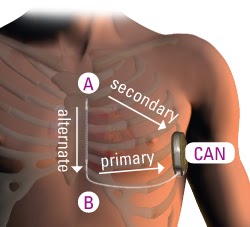
The ‘Can‘ (metal box that contains all the circuitry and battery), is buried under the skin on the outside of the ribs. Put your arms down by your sides, the device would go where your ribs meet the middle of your bicep. A lead is then run under the skin to the centre of your chest where its is anchored and then north, under the skin again until the tip of the lead is roughly at the top of the sternum.
For you physicians out there the ‘can’ is positioned at the mid-axillary line between the 5th and 6th intercostal spaces, the lead is then tunnelled to a small Xiphoid incision and then tunnelled north to a superior incision.

How is an S-ICD Implanted?

VIEW VIDEO
Having spoken to Boston Scientific it is becoming more apparent that the superior incision (cut at the top of the chest) may actually be removed from the procedure guidance as simply tunnelling the lead and ‘wedging’ the tip at that point is satisfactory – THIS IS NOT CONFIRMED AT THE MOMENT AND IS THEREFORE NOT PROCEDURE ADVICE.
Boston Scientific Subcutaneous Implantable Cardiodefibrillator Device S-ICD
Image Courtesy of
http://www.bostonscientific.com/

How does the S-ICD Work?

A ‘Shock Box’ basically needs to do 2 things. Firstly be able to SENSE if the heart has entered a Dangerous Arrhythmia and Secondly, be able to treat it.
The treatment part of the functionality is the easy bit – it delivers an electric shock across a ‘circuit’ that involves a large amount of the tissue in the heart. The lead has two ‘electrodes’ and the ‘Can’ is a third electrode allowing you different shocking ‘vectors’. By vectors we mean directions and area through which the electricity travels during a shock. This gives us extra options when implanting a device as some vectors will work better than others for the treatment of dangerous arrhythmias.

Shocking Vectors?

This is a concept you are familiar with without even thinking about it… when you are watching ER or another TV program and they Defibrillate the patient using the metal paddles, where do they position them? One either side of the heart? Precisely!! this is creating a ‘vector’ across the heart to involve the cardiac tissue. The paddles would be a lot less effective if you put one on the knee and one on the foot!

Boston Scientific Subcutaneous Implantable Cardiodefibrillator Device S-ICD

Now because the ‘Vectors’ used by the S-ICD are over a larger area than those with a traditional device – more energy has to be delivered to have the same desired affect. The upshot of this is that a larger battery is required to deliver the 80J! Bigger Battery = Bigger Box. This image shows a demo device but this is the exact size compared to a One Pound Coin! Now yes it is big but because of the extra room where they place the device it is pretty discrete and hidden in even slender patients.
STAT ATTACK!
The S-ICD System delivers up to 5 shocks per episode at 80 J with up to 128 seconds of ECG storage per episode and storage of up to 45 episodes.
The heart rate that the S-ICD is told to deliver therapy is programable between 170 and 250 bpm. Quite cleverly the device is able to also deliver a small amount of ‘pacing’ after a shock, when the heart can often run slowly. This is external pacing and will be felt!! It can run for 30s.

Sensing in an S-ICD.

 
The S-ICD uses its electrodes to produce an ECG similar to a surface ECG. 
 
Now the Sensing functionality is the devices ability to determine what Rhythm the heart is in! Without a lead in the heart to give us really accurate information the device is using a large area of heart, ribs and muscle. This means there is more potential for ‘artefact’. Artefact is the electrical interference and confusion – that could potentially lead to a patient being shocked when they do not require it – or not being shocked when they do…
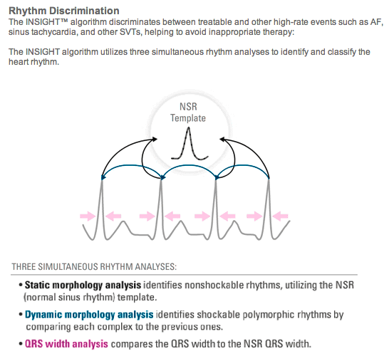
Boston Scientific have come up with a very clever software/algorithm called ‘Insight’. Insight uses 3 separate methods to determine the nature of a heart rhythm.
  • Normal Sinus Rhythm Template (Do your heart beats look as they should)
  • Dynamic Morphology Analysis (A live comparison of heart beat to previous heart beat, do they all look the same or do they keep changing?)
  • QRS Width analysis (Are the tall ‘peaks’ on your ECG, the QRS’, wider than they normally are?)
These questions (with some very complex maths) and the rate of a rhythm are used to decide whether to ‘shock’ or not.
Insight Algorithm S-ICD

Image Courtesy of  http://www.bostonscientific.com/

How does Insight and the S-ICD compare to other ICD Devices?

The statistics for treatment success and inappropriate shocks (an electrocuted patient that did not need to be) actually compare very similarly if not favourably compared to other devices on the market – these two studies are well worth a read if you have the time 🙂
1. Burke M, et al. Safety and Efficacy of a Subcutaneous Implantable-Debrillator (S-ICD System US IDE Study). Late-Breaking Abstract Session. HRS 2012.
2. Lambiase PD, et al. International Experience with a Subcutaneous ICD; Preliminary Results of the EFFORTLESS S-ICD Registry. Cardiostim 2012.
3. Gold MR, et al. Head-to-head comparison of arrhythmia discrimination performance of subcutaneous and transvenous ICD arrhythmia detection algorithms: the START study. J Cardiovasc Electrophysiol. 2012;23;4:359-366.
Who qualifies?
Template S-ICD Eligibility

Template used to assess eligibility!
Image Courtesy of
http://www.bostonscientific.com/
Well essentially anyone who qualifies for a normal ‘shock box’ ICD but with one other requirement. The Insight Software requires that a person has certain characteristics on their ECG. This is essentially showing that they have tall enough and narrow enough complexes to allow the algorithm to perform effectively. A simple 12 lead ECG Laying and Standing will be obtained and then a ‘Stencil’ is passed over the Print out – If the complexes fit within the boundaries marked on the ‘stencil’ then you potentially qualify. If your ECG does not meet requirements then it will not be recommended for you to have the S-ICD.

There you have it a quick overview of the Boston Scientific S-ICD.

Thanks for Reading

Cardiac Technician

SOURCE

http://www.thepad.pm/2013/12/boston-scientific-s-icd.html#!

UPDATED on 10/15/2013

Frequency and Determinants of Implantable Cardioverter Defibrillator Deployment Among Primary Prevention Candidates With Subsequent Sudden Cardiac Arrest in the Community

  1. Kumar Narayanan, MD;
  2. Kyndaron Reinier, PhD;
  3. Audrey Uy-Evanado, MD;
  4. Carmen Teodorescu, MD, PhD;
  5. Harpriya Chugh, BS;
  6. Eloi Marijon, MD;
  7. Karen Gunson, MD;
  8. Jonathan Jui, MD, MPH;
  9. Sumeet S. Chugh, MD

+Author Affiliations


  1. From The Heart Institute, Cedars-Sinai Medical Center, Los Angeles, CA (K.N., K.R., A.U.-E., C.T., H.C., E.M., S.S.C.); and Departments of Pathology (K.G.) and Emergency Medicine (J.J.), Oregon Health and Science University, Portland, OR.
  1. Correspondence to Sumeet S. Chugh, MD, Cedars-Sinai Medical Center, The Heart Institute, AHSP Suite A3100, 127 S. San Vicente Blvd., Los Angeles, CA 90048, Los Angeles, CA 90048. E-mail sumeet.chugh@cshs.org

Abstract

Background—The prevalence rates and influencing factors for deployment of primary prevention implantable cardioverter defibrillators (ICDs) among subjects who eventually experience sudden cardiac arrest in the general population have not been evaluated.

Methods and Results—Cases of adult sudden cardiac arrest with echocardiographic evaluation before the event were identified from the ongoing Oregon Sudden Unexpected Death Study (population approximately 1 million). Eligibility for primary ICD implantation was determined from medical records based on established guidelines. The frequency of prior primary ICD implantation in eligible subjects was evaluated, and ICD nonrecipients were characterized. Of 2093 cases (2003–2012), 448 had appropriate pre– sudden cardiac arrest left ventricular ejection fraction information available. Of these, 92 (20.5%) were eligible for primary ICD implantation, 304 (67.9%) were ineligible because of left ventricular ejection fraction >35%, and the remainder (52, 11.6%) had left ventricular ejection fraction ≤35% but were ineligible on the basis of clinical guideline criteria. Among eligible subjects, only 12 (13.0%; 95% confidence interval, 6.1%–19.9%) received a primary ICD. Compared with recipients, primary ICD nonrecipients were older (age at ejection fraction assessment, 67.1±13.6 versus 58.5±14.8 years, P=0.05), with 20% aged ≥80 years (versus 0% among recipients, P=0.11). Additionally, a subgroup (26%) had either a clinical history of dementia or were undergoing chronic dialysis.

Conclusions—Only one fifth of the sudden cardiac arrest cases in the community were eligible for a primary prevention ICD before the event, but among these, a small proportion (13%) were actually implanted. Although older age and comorbidity may explain nondeployment in a subgroup of these cases, other determinants such as socioeconomic factors, health insurance, patient preference, and clinical practice patterns warrant further detailed investigation.

Key Words:

  • Received March 11, 2013.
  • Accepted August 21, 2013

http://circ.ahajournals.org/content/128/16/1733.abstract

UPDATED on 9/15/2013

based on 9/6/2013 Trials and Fibrillations — The Heart.org

http://www.theheart.org/columns/trials-and-fibrillations-with-dr-john-mandrola/new-post-39.do#!

Echo-CRT trial: Most important study released at ESC 2013

Cardiac resynchronization therapy (CRT) is a multilead pacing device that can extend lives and improve the quality of life of selected patients who suffer from reduced performance of the heart due to adverse timing of contraction (wobbling motion from conduction delays that cause asynchrony or  delayed activation of one portion of the left ventricle compared to others reducing net blood ejection).

The degree of benefit in CRT responders depends not only on the degree of asynchrony, but also on the delayed activity location in relation to the available locations for lead placement. CRT is an adjustment in the timing of muscle activiation to improve the concerted impact on blood ejection. Only patients likely to improve should be exposed to the risks and costs of CRT.

The Echo-CRT trial, presented September 3, 2013 at the European Society of Cardiology (ESC) 2013 Congressand simultaneously published in the New England Journal of Medicine, helps identify which patients may benefit from CRT devices. (See Steve Stiles’ report on heartwire),

Echo-CRT trial summary

Background is important

Previous CRT studies enrolled patients with QRS duration >120 or >130 ms for synchronizing biventricular pacing. Additional work confirmed the greatest benefit occurred in patients with QRS durations >150 ms and typical left bundle branch block (LBBB). Conflicting observational and small randomized trials were less clear for patients with shorter QRS durations—the majority of heart-failure patients. What’s more, most cardiologists have seen patients with “modest” QRS durations respond to CRT. In theory, wide QRS is only expected if the axis of significant delay projects onto the standard ECG views, whereas significant opportunity for benefit can be missed if the axis of significant delay is not wide in the standard views. CRT implanters have heard of patients with normal-duration QRS where echo shows marked dyssynchrony. This raised the  question: Are there CHF patients with mechanical dyssynchrony (determined by echo) but no electrical delay (as measured by the ECG) benefit from CRT?Unfortunately, echo does not resolve the issue either. Thus there is the residual question of who should be evaluated by a true 3D syncrhony assessment by cardiac MRI.

Echocardiographic techniques held promise to identify mechanical dyssynchrony, but like the standard 12 lead ECG, they also utilize limited orientations of views of the heart and hence the directions in which delays can be detected. Cardiac MRI Research (not limited in view angle) by JDPearlman showed that the axis of maximal delay in patients with asynchrony is within 30 degrees of the ECG and echo views in a majority of patients with asynchrony, but it can be 70-110 degrees away from the views used by echocardiography and by ECG in 20% of cases. Hence some patients who may benefit can be missed by ECG or Echo criteria.

Methodology

Echo-CRT was an industry-sponsored (Biotronik) investigator-initiated prospective international randomized controlled trial. All patients had mechanical dyssynchrony by echo, QRS <130 ms, and an ICD indication. CRT-D devices were implanted in all patients. Blinded randomization to CRT-on (404 patients) vs CRT-off (405 patients) was performed after implantation. Programming in the CRT-off group was set to minimize RV pacing. The primary outcome was a composite of all-cause mortality or hospitalization.

Six key findings

1. Although entry criteria for the trial was a QRS duration <130 ms, the mean QRS duration of both groups was 105 ms.

2. The data safety monitoring board terminated the trial prematurely because of an increased death rate in the CRT group.

3. No differences were noted in the primary outcome.

4. More patients died in the CRT group (hazard ratio=1.8).

5. The higher death rate in the CRT group was driven by cardiovascular death.

6. More patients in the CRT group were hospitalized, due primarily to device-related issues.

These findings send clear and simple messages to all involved with treating patients with heart failure. My interpretation of Echo-CRT is as follows:

Do not implant CRT devices in patients with “narrow” QRS complexes.

The signal of increased death was strong. A hazard ratio of 1.8 translates to an almost doubling of the risk of death. This finding is unlikely to be a statistical anomaly, as it was driven by CV death. The risks of CRT in nonresponders are well-known and include: increased RV pacing, possible proarrhythmia from LV pacing, and the need for more device-related surgery. Patients who do not respond to CRT get none of the benefits but all the potential harms—an unfavorable ratio indeed.

Echo is not useful for assessing dyssynchrony in patients with narrow QRS complexes.

Dr Samuel Asirvatham explains the concept of electropathy in a review article in the Journal of Cardiovascular Electrophysiology. He teaches us that the later the LV lateral wall is activated relative to the RV, the more the benefit of preexciting the lateral wall with an LV lead. That’s why the benefit from CRT in many cases increases with QRS duration, because—in a majority—a wide QRS means late activation of the lateral LV.

Simple triumphs over complicated—CRT response best estimated with the old-fashioned ECG.

In a right bundle branch block, the left ventricle is activated first; in LBBB, the LV lateral wall is last, and with a nonspecific ICD, there’s delayed conduction in either the His-Purkinje system or in ventricular muscle. What does a normal QRS say? It says the wave front of activation as projected onto the electric views obtained activates the LV and RV simultaneously. If those views capture the worst delay then they can eliminate the  need for resynchrony.

CRT benefit with mild-moderate QRS prolongation still not settled

Dr Robert Myerburg (here and here) teaches us to make a distinction between trial entry criteria and the actual values of the cohort.

Consider how this applies to QRS duration:  COMPANION and CARE-HF are clinical trials that showed definitive CRT benefit. Entry required a QRS duration >120 ms (130 ms in CARE-HF). But the actual mean QRS duration of enrolled patients was 160 ms. A meta-analysis of CRT trials confirmed benefit at longer QRS durations and questioned it below 150 ms. CRT guideline recommendations incorporate study entry criteria, not the mean values of actual patients in the trial. Patients enrolled in Echo-CRT had very narrow QRS complexes (105 ms). What to recommend in the common situation when a patient with a typical LBBB has a QRS duration straddling 130 ms is not entirely clear. The results of Echo-CRT might have been different had the actual QRS duration values been closer to 130 ms.

Conclusion

Echo-CRT study reinforces expectations based on cardiac physiology. In the practice of medicine, it’s quite useful to know when not to do something.

The trial should not dampen enthusiasm for CRT. Rather, it should focus our attention to patient selection—and the value of the 12-lead ECG.

 References

Rethinking QRS Duration as an Indication for CRT

SMITA MEHTA M.D.1 and SAMUEL J. ASIRVATHAM M.D., F.A.C.C.2,3

Author Information

  1. Department of Pediatric Cardiology, Cleveland Clinic, Cleveland, Ohio, USA
  2. Division of Cardiovascular Diseases, Department of Medicine, Mayo Clinic, Rochester, Minnesota, USA
  3. Department of Pediatrics and Adolescent Medicine, Mayo Clinic, Rochester, Minnesota, USA

*Samuel J. Asirvatham, M.D., Division of Cardiovascular Diseases, Department of Internal Medicine and Division of Pediatric Cardiology, Department of Pediatric and Adolescent Medicine, Mayo Clinic College of Medicine, 200 First Street SW, Rochester, MN 55905, USA. E-mail: asirvatham.samuel@mayo.edu

J Cardiovasc Electrophysiol, Vol. 23, pp. 169-171, February 2012.

http://onlinelibrary.wiley.com/doi/10.1111/j.1540-8167.2011.02163.x/full

Indications for Implantable Cardioverter-Defibrillators Based on Evidence and Judgment FREE

Robert J. Myerburg, MD; Vivek Reddy, MD; Agustin Castellanos, MD
J Am Coll Cardiol. 2009;54(9):747-763. doi:10.1016/j.jacc.2009.03.078

Implantable Cardioverter–Defibrillators after Myocardial Infarction

Robert J. Myerburg, M.D.

Division of Cardiology, University of Miami Miller School of Medicine, Miami.

N Engl J Med 2008; 359:2245-2253 November 20, 2008DOI: 10.1056/NEJMra0803409

END OF UPDATE

Electrical conduction of the Human Heart

  • Physiology and
  • Genetics

were explained by us in the following articles:

Genetics of Conduction Disease: Atrioventricular (AV) Conduction Disease (block): Gene Mutations – Transcription, Excitability, and Energy Homeostasis

On Devices and On Algorithms: Prediction of Arrhythmia after Cardiac Surgery and ECG Prediction of an Onset of Paroxysmal Atrial Fibrillation

Dilated Cardiomyopathy: Decisions on implantable cardioverter-defibrillators (ICDs) using left ventricular ejection fraction (LVEF)

Reduction in Inappropriate Therapy and Mortality through ICD Programming

Below, we present the following complementary topics:

Options for Cardiac Resynchronization Therapy (CRT) to Arrhythmias:

  • Implantable Pacemaker
  • Insertable Programmable Cardioverter Defibrillator (ICD)

UPDATED 8/6/2013

Medtronic Pacemaker Recall


17/07/2013

Australia’s regulatory authority, the Therapeutic Goods Administration (TGA) has issued a hazard alert pertaining to one of Medtronic’s pacing devices, the Consulta® Cardiac Resynchronization Therapy Pacemaker (CRT-P). The alert coincides somewhat with Medtronic’s own issuance of a field safety notice concerning Consulta and Syncra® CRT-P devices.

Background

Consulta and Syncra CRT-Ps are implantable medical devices used to treat heart failure. The devices provide pacing to help coordinate the heart’s pumping action and improve blood flow.

The two devices are the subject of a global manufacturer recall after Medtronic had identified an issue with a subset of both during production, although as yet there had been no reported or confirmed device failures. However, because of the potential for malfunction, Medtronic is requiring the return of non-implanted devices manufactured between April 1 and May 13, 2013 for re-inspection.

Seemingly this manufacturing issue could compromise the sealing of the device. Should an out-of-spec weld fail this could result in body fluids entering the device, which could cause it to malfunction leading to loss of pacing output. This could potentially see the return of symptoms including

  • fainting or lightheadedness,
  • dyspnoea (shortness of breath),
  • fatigue and
  • oedema.

Medtronic’s recall is thought to relate to 265 devices, 44 of which have been implanted in the US.

The Australian warning letter, issued by the TGA states that only one “at risk” Consulta CRT-P device has been implanted in the country and there have been no reports of device failures or patient injuries relating to this issue.

Neither Medtronic nor the TGA are suggesting any specific patient management measures other than routine follow-up in accordance with labelling instructions.

Pacemaker/Implantable Cardioverter Defibrillator (ICD) Insertion

Procedure Overview

What is a pacemaker/implantable cardioverter defibrillator (ICD) insertion?

A pacemaker/implantable cardioverter defibrillator (ICD) insertion is a procedure in which a pacemaker and/or an ICD is inserted to assist in regulating problems with the heart rate (pacemaker) or heart rhythm (ICD).

Pacemaker

When a problem develops with the heart’s rhythm, such as a slow rhythm, a pacemaker may be selected for treatment. A pacemaker is a small electronic device composed of three parts: a generator, one or more leads, and an electrode on each lead. A pacemaker signals the heart to beat when the heartbeat is too slow.

Illustration of a single-chamber pacemaker
Click Image to Enlarge

A generator is the “brain” of the pacemaker device. It is a small metal case that contains electronic circuitry and a battery. The lead (or leads) is an insulated wire that is connected to the generator on one end, with the other end placed inside one of the heart’s chambers.

The electrode on the end of the lead touches the heart wall. In most pacemakers, the lead senses the heart’s electrical activity. This information is relayed to the generator by the lead.

If the heart’s rate is slower than the programmed limit, an electrical impulse is sent through the lead to the electrode and the pacemaker’s electrical impulse causes the heart to beat at a faster rate.

When the heart is beating at a rate faster than the programmed limit, the pacemaker will monitor the heart rate, but will not pace. No electrical impulses will be sent to the heart unless the heart’s natural rate falls below the pacemaker’s low limit.

Pacemaker leads may be positioned in the atrium or ventricle or both, depending on the condition requiring the pacemaker to be inserted. An atrial dysrhythmia/arrhythmia (an abnormal heart rhythm caused by a dysfunction of the sinus node or the development of another atrial pacemaker within the heart tissue that takes over the function of the sinus node) may be treated with an atrial pacemaker.

Illustration of a dual-chamber pacemaker
Click Image to Enlarge

A ventricular dysrhythmia/arrhythmia (an abnormal heart rhythm caused by a dysfunction of the sinus node, an interruption in the conduction pathways, or the development of another pacemaker within the heart tissue that takes over the function of the sinus node) may be treated with a ventricular pacemaker whose lead wire is located in the ventricle.

It is possible to have both atrial and ventricular dysrhythmias, and there are pacemakers that have lead wires positioned in both the atrium and the ventricle. There may be one lead wire for each chamber, or one lead wire may be capable of sensing and pacing both chambers.

A new type of pacemaker, called a biventricular pacemaker, is currently used in the treatment of congestive heart failure. Sometimes in heart failure, the two ventricles (lower heart chambers) do not pump together in a normal manner. When this happens, less blood is pumped by the heart.

A biventricular pacemaker paces both ventricles at the same time, increasing the amount of blood pumped by the heart. This type of treatment is called cardiac resynchronization therapy.

Implantable cardioverter defibrillator (ICD)

An implantable cardioverter defibrillator (ICD) looks very similar to a pacemaker, except that it is slightly larger. It has a generator, one or more leads, and an electrode for each lead. These components work very much like a pacemaker. However, the ICD is designed to deliver an electrical shock to the heart when the heart rate becomes dangerously fast, or €œfibrillates.”

An ICD senses when the heart is beating too fast and delivers an electrical shock to convert the fast rhythm to a normal rhythm. Some devices combine a pacemaker and ICD in one unit for persons who need both functions.

The ICD has another type of treatment for certain fast rhythms called anti-tachycardia pacing (ATP). When ATP is used, a fast pacing impulse is sent to correct the rhythm. After the shock is delivered, a “back-up” pacing mode is used if needed for a short while.

The procedure for inserting a pacemaker or an ICD is the same. The procedure generally is performed in an electrophysiology (EP) lab or a cardiac catheterization lab.

Other related procedures that may be used to assess the heart include resting and exercise electrocardiogram (ECG), Holter monitor, signal-averaged ECG, cardiac catheterization, chest x-ray, computed tomography (CT scan) of the chest, echocardiography, electrophysiology studies, magnetic resonance imaging (MRI) of the heart, myocardial perfusion scans, radionuclide angiography, and ultrafast CT scan.

The heart’s electrical conduction system

Illustration of the anatomy of the heart, view of the electrical system
Click Image to Enlarge

The heart is, in the simplest terms, a pump made up of muscle tissue. Like all pumps, the heart requires a source of energy in order to function. The heart’s pumping energy comes from an indwelling electrical conduction system.

An electrical stimulus is generated by the sinus node (also called the sinoatrial node, or SA node), which is a small mass of specialized tissue located in the right atrium (right upper chamber) of the heart.

The sinus node generates an electrical stimulus regularly at 60 to 100 times per minute under normal conditions. This electrical stimulus travels down through the conduction pathways (similar to the way electricity flows through power lines from the power plant to your house) and causes the heart’s chambers to contract and pump out blood.

The right and left atria (the two upper chambers of the heart) are stimulated first and contract a short period of time before the right and left ventricles (the two lower chambers of the heart).

The electrical impulse travels from the sinus node to the atrioventricular (AV) node, where it stops for a very short period, then continues down the conduction pathways via the “bundle of His” into the ventricles. The bundle of His divides into right and left pathways to provide electrical stimulation to both ventricles.

What is an ECG?

This electrical activity of the heart is measured by an electrocardiogram (ECG or EKG). By placing electrodes at specific locations on the body (chest, arms, and legs), a tracing of the electrical activity can be obtained. Changes in an ECG from the normal tracing can indicate one or more of several heart-related conditions.

Dysrhythmias/arrhythmias (abnormal heart rhythms) are diagnosed by methods such as EKG, Holter monitoring, signal-average EKG, or electrophysiological studies. These symptoms may be treated with medication or procedures such as a cardiac ablation (removal of a location in the heart that is causing a dysrhythmia by freezing or radiofrequency).

Reasons for the Procedure

A pacemaker may be inserted in order to provide stimulation for a faster heart rate when the heart is beating too slowly, and when other treatment methods, such as medication, have not improved the heart rate.

An ICD may be inserted in order to provide fast pacing (ATP), cardioversion (small shock), or defibrillation (larger shock) when the heart beats too fast.

Problems with the heart rhythm may cause difficulties because the heart is unable to pump an adequate amount of blood to the body. If the heart rate is too slow, the blood is pumped too slowly.

If the heart rate is too fast or too irregular, the heart chambers are unable to fill up with enough blood to pump out with each beat. When the body does not receive enough blood, symptoms such as fatigue, dizziness, fainting, and/or chest pain may occur.

Some examples of rhythm problems for which a pacemaker or ICD might be inserted include:

  • atrial fibrillation – occurs when the atria beat irregularly and too fast
  • ventricular fibrillation – occurs when the ventricles beat irregularly and too fast
  • bradycardia – occurs when the heart beats too slow
  • tachycardia – occurs when the heart beats too fast
  • heart block – occurs when the electrical signal is delayed after leaving the SA node; there are several types of heart blocks, and each one has a distinctive ECG tracing

There may be other reasons for your physician to recommend a pacemaker or ICD insertion.

Risks of the Procedure

Possible risks of pacemaker or ICD insertion include, but are not limited to, the following:

  • bleeding from the incision or catheter insertion site
  • damage to the vessel at the catheter insertion site
  • infection of the incision or catheter site
  • pneumothorax – air becomes trapped in the pleural space causing the lung to collapse

If you are pregnant or suspect that you may be pregnant, you should notify your physician. If you are lactating, or breastfeeding, you should notify your physician.

Patients who are allergic to or sensitive to medications or latex should notify their physician.

For some patients, having to lie still on the procedure table for the length of the procedure may cause some discomfort or pain.

There may be other risks depending upon your specific medical condition. Be sure to discuss any concerns with your physician prior to the procedure.

Before the Procedure

  • Your physician will explain the procedure to you and offer you the opportunity to ask any questions that you might have about the procedure.
  • You will be asked to sign a consent form that gives your permission to do the test. Read the form carefully and ask questions if something is not clear.
  • You will need to fast for a certain period of time prior to the procedure. Your physician will notify you how long to fast, usually overnight.
  • If you are pregnant or suspect that you are pregnant, you should notify your physician.
  • Notify your physician if you are sensitive to or are allergic to any medications, iodine, latex, tape, or anesthetic agents (local and general).
  • Notify your physician of all medications (prescription and over-the-counter) and herbal supplements that you are taking.
  • Notify your physician if you have heart valve disease, as you may need to receive an antibiotic prior to the procedure.
  • Notify your physician if you have a history of bleeding disorders or if you are taking any anticoagulant (blood-thinning) medications, aspirin, or other medications that affect blood clotting. It may be necessary for you to stop some of these medications prior to the procedure.
  • Your physician may request a blood test prior to the procedure to determine how long it takes your blood to clot. Other blood tests may be done as well.
  • You may receive a sedative prior to the procedure to help you relax. If a sedative is given, you will need someone to drive you home afterwards.
  • The upper chest may be shaved or clipped prior to the procedure.
  • Based upon your medical condition, your physician may request other specific preparation.

During the Procedure

Picture of a chest X-ray, showing a single-chamber implanted pacemaker
Chest X-ray with Implanted Pacemaker

A pacemaker or implanted cardioverter defibrillator may be performed on an outpatient basis or as part of your stay in a hospital. Procedures may vary depending on your condition and your physician’s practices.

Generally, a pacemaker or ICD insertion follows this process:

  1. You will be asked to remove any jewelry or other objects that may interfere with the procedure.
  2. You will be asked to remove your clothing and will be given a gown to wear.
  3. You will be asked to empty your bladder prior to the procedure.
  4. An intravenous (IV) line will be started in your hand or arm prior to the procedure for injection of medication and to administer IV fluids, if needed.
  5. You will be placed in a supine (on your back) position on the procedure table.
  6. You will be connected to an electrocardiogram (ECG or EKG) monitor that records the electrical activity of the heart and monitors the heart during the procedure using small, adhesive electrodes. Your vital signs (heart rate, blood pressure, breathing rate, and oxygenation level) will be monitored during the procedure.
  7. Large electrode pads will be placed on the front and back of the chest.
  8. You will receive a sedative medication in your IV before the procedure to help you relax. However, you will likely remain awake during the procedure.
  9. The pacemaker or ICD insertion site will be cleansed with antiseptic soap.
  10. Sterile towels and a sheet will be placed around this area.
  11. A local anesthetic will be injected into the skin at the insertion site.
  12. Once the anesthetic has taken effect, the physician will make a small incision at the insertion site.
  13. A sheath, or introducer, is inserted into a blood vessel, usually under the collarbone. The sheath is a plastic tube through which the pacer/ICD lead wire will be inserted into the blood vessel and advanced into the heart.
  14. It will be very important for you to remain still during the procedure so that the catheter placement will not be disturbed and to prevent damage to the insertion site.
  15. The lead wire will be inserted through the introducer into the blood vessel. The physician will advance the lead wire through the blood vessel into the heart.
  16. Once the lead wire is inside the heart, it will be tested to verify proper location and that it works. There may be one, two, or three lead wires inserted, depending on the type of device your physician has chosen for your condition. Fluoroscopy, (a special type of x-ray that will be displayed on a TV monitor), may be used to assist in testing the location of the leads.
  17. Once the lead wire has been tested, an incision will be made close to the location of the catheter insertion (just under the collarbone). You will receive local anesthetic medication before the incision is made.
  18. The pacemaker/ICD generator will be slipped under the skin through the incision after the lead wire is attached to the generator. Generally, the generator will be placed on the non-dominant side. (If you are right-handed, the device will be placed in your upper left chest. If you are left-handed, the device will be placed in your upper right chest).
  19. The ECG will be observed to ensure that the pacer is working correctly.
  20. The skin incision will be closed with sutures, adhesive strips, or a special glue.
  21. A sterile bandage/dressing will be applied.

After the Procedure

In the hospital

After the procedure, you may be taken to the recovery room for observation or returned to your hospital room. A nurse will monitor your vital signs for a specified period of time.

You should immediately inform your nurse if you feel any chest pain or tightness, or any other pain at the incision site.

After the specified period of bed rest has been completed, you may get out of bed. The nurse will assist you the first time you get up, and will check your blood pressure while you are lying in bed, sitting, and standing. You should move slowly when getting up from the bed to avoid any dizziness from the period of bedrest.

You will be able to eat or drink once you are completely awake.

The insertion site may be sore or painful, but pain medication may be administered if needed.

Your physician will visit with you in your room while you are recovering. The physician will give you specific instructions and answer any questions you may have.

Once your blood pressure, pulse, and breathing are stable and you are alert, you will be taken to your hospital room or discharged home.

If the procedure is performed on an outpatient basis, you may be allowed to leave after you have completed the recovery process. However, if there are concerns or problems with your ECG, you may stay in the hospital for an additional day (or longer) for monitoring of the ECG.

You should arrange to have someone drive you home from the hospital following your procedure.

At home

You should be able to return to your daily routine within a few days. Your physician will tell you if you will need to take more time in returning to your normal activities. In addition, you should not do any lifting or pulling on anything for a few weeks. You may be instructed not to lift your arms above your head for a period of time.

You will most likely be able to resume your usual diet, unless your physician instructs you differently.

It will be important to keep the insertion site clean and dry. Your physician will give you specific bathing instructions.

Your physician will give you specific instructions about driving. If you had an ICD, you will not be able to drive until your physician gives you approval. Your physician will explain these limitations to you, if they are applicable to your situation.

You will be given specific instructions about what to do if your ICD discharges a shock. For example, you may be instructed to dial 911 or go to the nearest emergency room in the event of a shock from the ICD.

Ask your physician when you will be able to return to work. The nature of your occupation, your overall health status, and your progress will determine how soon you may return to work.

Notify your physician to report any of the following:

  • fever and/or chills
  • increased pain, redness, swelling, or bleeding or other drainage from the insertion site
  • chest pain/pressure, nausea and/or vomiting, profuse sweating, dizziness and/or fainting
  • palpitations

Your physician may give you additional or alternate instructions after the procedure, depending on your particular situation.

Pacemaker/ICD precautions

The following precautions should always be considered. Discuss the following in detail with your physician, or call the company that made your device:

  • Always carry an ID card that states you are wearing a pacemaker or an ICD. In addition, you should wear a medical identification bracelet that states you have a pacemaker or ICD.
  • Use caution when going through airport security detectors. Check with your physician about the safety of going through such detectors with your type of pacemaker. In particular, you may need to avoid being screened by hand-held detector devices, as these devices may affect your pacemaker.
  • You may not have a magnetic resonance imaging (MRI) procedure. You should also avoid large magnetic fields.
  • Abstain from diathermy (the use of heat in physical therapy to treat muscles).
  • Turn off large motors, such as cars or boats, when working on them (they may temporarily €œconfuse” your device).
  • Avoid certain high-voltage or radar machinery, such as radio or television transmitters, electric arc welders, high-tension wires, radar installations, or smelting furnaces.
  • If you are having a surgical procedure performed by a surgeon or dentist, tell your surgeon or dentist that you have a pacemaker or ICD, so that electrocautery will not be used to control bleeding (the electrocautery device can change the pacemaker settings).
  • You may have to take antibiotic medication before any medically invasive procedure to prevent infections that may affect the pacemaker.
  • Always consult your physician if you have any questions concerning the use of certain equipment near your pacemaker.
  • When involved in a physical, recreational, or sporting activity, you should avoid receiving a blow to the skin over the pacemaker or ICD. A blow to the chest near the pacemaker or ICD can affect its functioning. If you do receive a blow to that area, see your physician.
  • Always consult your physician when you feel ill after an activity, or when you have questions about beginning a new activity.

SOURCE

http://stanfordhospital.org/healthLib/greystone/heartCenter/heartProcedures/pacemakerImplantableCardioverterDefibrillatorICDInsertion.html

In Summary: Who Needs a Pacemaker?

Doctors recommend pacemakers for many reasons. The most common reasons are bradycardia and heart block.

Bradycardia is a heartbeat that is slower than normal. Heart block is a disorder that occurs if an electrical signal is slowed or disrupted as it moves through the heart.

Heart block can happen as a result of aging, damage to the heart from a heart attack, or other conditions that disrupt the heart’s electrical activity. Some nerve and muscle disorders also can cause heart block, including muscular dystrophy.

Your doctor also may recommend a pacemaker if:

  • Aging or heart disease damages your sinus node’s ability to set the correct pace for your heartbeat. Such damage can cause slower than normal heartbeats or long pauses between heartbeats. The damage also can cause your heart to switch between slow and fast rhythms. This condition is called sick sinus syndrome.
  • You’ve had a medical procedure to treat an arrhythmia called atrial fibrillation. A pacemaker can help regulate your heartbeat after the procedure.
  • You need to take certain heart medicines, such as beta blockers. These medicines can slow your heartbeat too much.
  • You faint or have other symptoms of a slow heartbeat. For example, this may happen if the main artery in your neck that supplies your brain with blood is sensitive to pressure. Just quickly turning your neck can cause your heart to beat slower than normal. As a result, your brain might not get enough blood flow, causing you to feel faint or collapse.
  • You have heart muscle problems that cause electrical signals to travel too slowly through your heart muscle. Your pacemaker may provide cardiac resynchronization therapy (CRT) for this problem. CRT devices coordinate electrical signaling between the heart’s lower chambers.
  • You have long QT syndrome, which puts you at risk for dangerous arrhythmias.

Doctors also may recommend pacemakers for people who have certain types ofcongenital heart disease or for people who have had heart transplants. Children, teens, and adults can use pacemakers.

Before recommending a pacemaker, your doctor will consider any arrhythmia symptoms you have, such as dizziness, unexplained fainting, or shortness of breath. He or she also will consider whether you have a history of heart disease, what medicines you’re currently taking, and the results of heart tests.

Diagnostic Tests

Many tests are used to detect arrhythmias. You may have one or more of the following tests.

EKG (Electrocardiogram)

An EKG is a simple, painless test that detects and records the heart’s electrical activity. The test shows how fast your heart is beating and its rhythm (steady or irregular).

An EKG also records the strength and timing of electrical signals as they pass through your heart. The test can help diagnose bradycardia and heart block (the most common reasons for needing a pacemaker).

A standard EKG only records the heartbeat for a few seconds. It won’t detect arrhythmias that don’t happen during the test.

To diagnose heart rhythm problems that come and go, your doctor may have you wear a portable EKG monitor. The two most common types of portable EKGs are Holter and event monitors.

Holter and Event Monitors

A Holter monitor records the heart’s electrical activity for a full 24- or 48-hour period. You wear one while you do your normal daily activities. This allows the monitor to record your heart for a longer time than a standard EKG.

An event monitor is similar to a Holter monitor. You wear an event monitor while doing your normal activities. However, an event monitor only records your heart’s electrical activity at certain times while you’re wearing it.

For many event monitors, you push a button to start the monitor when you feel symptoms. Other event monitors start automatically when they sense abnormal heart rhythms.

You can wear an event monitor for weeks or until symptoms occur.

Echocardiography

Echocardiography (echo) uses sound waves to create a moving picture of your heart. The test shows the size and shape of your heart and how well your heart chambers and valves are working.

Echo also can show areas of poor blood flow to the heart, areas of heart muscle that aren’t contracting normally, and injury to the heart muscle caused by poor blood flow.

Electrophysiology Study

For this test, a thin, flexible wire is passed through a vein in your groin (upper thigh) or arm to your heart. The wire records the heart’s electrical signals.

Your doctor uses the wire to electrically stimulate your heart. This allows him or her to see how your heart’s electrical system responds. This test helps pinpoint where the heart’s electrical system is damaged.

Stress Test

Some heart problems are easier to diagnose when your heart is working hard and beating fast.

During stress testing, you exercise to make your heart work hard and beat fast while heart tests, such as an EKG or echo, are done. If you can’t exercise, you may be given medicine to raise your heart rate.

SOURCE

http://www.nhlbi.nih.gov/health/health-topics/topics/pace/whoneeds.html

What Are the Risks of Pacemaker Surgery?

Pacemaker surgery generally is safe. If problems do occur, they may include:

  • Swelling, bleeding, bruising, or infection in the area where the pacemaker was placed
  • Blood vessel or nerve damage
  • A collapsed lung
  • A bad reaction to the medicine used during the procedure

Talk with your doctor about the benefits and risks of pacemaker surgery.

How Does a Pacemaker Work?

A pacemaker consists of a battery, a computerized generator, and wires with sensors at their tips. (The sensors are called electrodes.) The battery powers the generator, and both are surrounded by a thin metal box. The wires connect the generator to the heart.

A pacemaker helps monitor and control your heartbeat. The electrodes detect your heart’s electrical activity and send data through the wires to the computer in the generator.

If your heart rhythm is abnormal, the computer will direct the generator to send electrical pulses to your heart. The pulses travel through the wires to reach your heart.

Newer pacemakers can monitor your blood temperature, breathing, and other factors. They also can adjust your heart rate to changes in your activity.

The pacemaker’s computer also records your heart’s electrical activity and heart rhythm. Your doctor will use these recordings to adjust your pacemaker so it works better for you.

Your doctor can program the pacemaker’s computer with an external device. He or she doesn’t have to use needles or have direct contact with the pacemaker.

Pacemakers have one to three wires that are each placed in different chambers of the heart.

  • The wires in a single-chamber pacemaker usually carry pulses from the generator to the right ventricle (the lower right chamber of your heart).
  • The wires in a dual-chamber pacemaker carry pulses from the generator to the right atrium (the upper right chamber of your heart) and the right ventricle. The pulses help coordinate the timing of these two chambers’ contractions.
  • The wires in a biventricular pacemaker carry pulses from the generator to an atrium and both ventricles. The pulses help coordinate electrical signaling between the two ventricles. This type of pacemaker also is called a cardiac resynchronization therapy (CRT) device.

Cross-Section of a Chest With a Pacemaker

The image shows a cross-section of a chest with a pacemaker. Figure A shows the location and general size of a double-lead, or dual-chamber, pacemaker in the upper chest. The wires with electrodes are inserted into the heart's right atrium and ventricle through a vein in the upper chest. Figure B shows an electrode electrically stimulating the heart muscle. Figure C shows the location and general size of a single-lead, or single-chamber, pacemaker in the upper chest.

The image shows a cross-section of a chest with a pacemaker. Figure A shows the location and general size of a double-lead, or dual-chamber, pacemaker in the upper chest. The wires with electrodes are inserted into the heart’s right atrium and ventricle through a vein in the upper chest. Figure B shows an electrode electrically stimulating the heart muscle. Figure C shows the location and general size of a single-lead, or single-chamber, pacemaker in the upper chest.

Types of Pacemaker Programming

The two main types of programming for pacemakers are

  • demand pacing and
  • rate-responsive pacing.

A demand pacemaker monitors your heart rhythm. It only sends electrical pulses to your heart if your heart is beating too slow or if it misses a beat.

A rate-responsive pacemaker will speed up or slow down your heart rate depending on how active you are. To do this, the device monitors your

  • sinus node rate,
  • breathing,
  • blood temperature, and
  • other factors to determine your activity level.

Your doctor will work with you to decide which type of pacemaker is best for you.

SOURCE

http://www.nhlbi.nih.gov/health/health-topics/topics/pace/howdoes.html

What To Expect During Pacemaker Surgery

Placing a pacemaker requires minor surgery. The surgery usually is done in a hospital or special heart treatment laboratory.

Before the surgery, an intravenous (IV) line will be inserted into one of your veins. You will receive medicine through the IV line to help you relax. The medicine also might make you sleepy.

Your doctor will numb the area where he or she will put the pacemaker so you don’t feel any pain. Your doctor also may give you antibiotics to prevent infection.

First, your doctor will insert a needle into a large vein, usually near the shoulder opposite your dominant hand. Your doctor will then use the needle to thread the pacemaker wires into the vein and to correctly place them in your heart.

An x-ray “movie” of the wires as they pass through your vein and into your heart will help your doctor place them. Once the wires are in place, your doctor will make a small cut into the skin of your chest or abdomen.

He or she will slip the pacemaker’s small metal box through the cut, place it just under your skin, and connect it to the wires that lead to your heart. The box contains the pacemaker’s battery and generator.

Once the pacemaker is in place, your doctor will test it to make sure it works properly. He or she will then sew up the cut. The entire surgery takes a few hours.

SOURCE

http://www.nhlbi.nih.gov/health/health-topics/topics/pace/during.html

What To Expect After Pacemaker Surgery

Expect to stay in the hospital overnight so your health care team can check your heartbeat and make sure your pacemaker is working well. You’ll likely have to arrange for a ride to and from the hospital because your doctor may not want you to drive yourself.

For a few days to weeks after surgery, you may have pain, swelling, or tenderness in the area where your pacemaker was placed. The pain usually is mild; over-the-counter medicines often can relieve it. Talk to your doctor before taking any pain medicines.

Your doctor may ask you to avoid vigorous activities and heavy lifting for about a month after pacemaker surgery. Most people return to their normal activities within a few days of having the surgery.

SOURCE

http://www.nhlbi.nih.gov/health/health-topics/topics/pace/after.html

How Will a Pacemaker Affect My Lifestyle?

Once you have a pacemaker, you have to avoid close or prolonged contact with electrical devices or devices that have strong magnetic fields. Devices that can interfere with a pacemaker include:

  • Cell phones and MP3 players (for example, iPods)
  • Household appliances, such as microwave ovens
  • High-tension wires
  • Metal detectors
  • Industrial welders
  • Electrical generators

These devices can disrupt the electrical signaling of your pacemaker and stop it from working properly. You may not be able to tell whether your pacemaker has been affected.

How likely a device is to disrupt your pacemaker depends on how long you’re exposed to it and how close it is to your pacemaker.

To be safe, some experts recommend not putting your cell phone or MP3 player in a shirt pocket over your pacemaker (if the devices are turned on).

You may want to hold your cell phone up to the ear that’s opposite the site where your pacemaker is implanted. If you strap your MP3 player to your arm while listening to it, put it on the arm that’s farther from your pacemaker.

You can still use household appliances, but avoid close and prolonged exposure, as it may interfere with your pacemaker.

You can walk through security system metal detectors at your normal pace. Security staff can check you with a metal detector wand as long as it isn’t held for too long over your pacemaker site. You should avoid sitting or standing close to a security system metal detector. Notify security staff if you have a pacemaker.

Also, stay at least 2 feet away from industrial welders and electrical generators.

Some medical procedures can disrupt your pacemaker. These procedures include:

  • Magnetic resonance imaging, or MRI
  • Shock-wave lithotripsy to get rid of kidney stones
  • Electrocauterization to stop bleeding during surgery

Let all of your doctors, dentists, and medical technicians know that you have a pacemaker. Your doctor can give you a card that states what kind of pacemaker you have. Carry this card in your wallet. You may want to wear a medical ID bracelet or necklace that states that you have a pacemaker.

Physical Activity

In most cases, having a pacemaker won’t limit you from doing sports and exercise, including strenuous activities.

You may need to avoid full-contact sports, such as football. Such contact could damage your pacemaker or shake loose the wires in your heart. Ask your doctor how much and what kinds of physical activity are safe for you.

Ongoing Care

Your doctor will want to check your pacemaker regularly (about every 3 months). Over time, a pacemaker can stop working properly because:

  • Its wires get dislodged or broken
  • Its battery gets weak or fails
  • Your heart disease progresses
  • Other devices have disrupted its electrical signaling

To check your pacemaker, your doctor may ask you to come in for an office visit several times a year. Some pacemaker functions can be checked remotely using a phone or the Internet.

Your doctor also may ask you to have an EKG (electrocardiogram) to check for changes in your heart’s electrical activity.

Battery Replacement

Pacemaker batteries last between 5 and 15 years (average 6 to 7 years), depending on how active the pacemaker is. Your doctor will replace the generator along with the battery before the battery starts to run down.

Replacing the generator and battery is less-involved surgery than the original surgery to implant the pacemaker. Your pacemaker wires also may need to be replaced eventually.

Your doctor can tell you whether your pacemaker or its wires need to be replaced when you see him or her for followup visits.

SOURCE

http://www.nhlbi.nih.gov/health/health-topics/topics/pace/lifestyle.html

Clinical Trial on Pace Makers

clinical trials related to pacemakers, talk with your doctor. You also can visit the following Web sites to learn more about clinical research and to search for clinical trials:

For more information about clinical trials for children, visit the NHLBI’s Children and Clinical Studies Web page.

SOURCE

http://www.nhlbi.nih.gov/health/health-topics/topics/pace/trials.html

RESOUCES on PaceMakers

Links to Other Information About Pacemakers

NHLBI Resources

Non-NHLBI Resources

Clinical Trials

SOURCE


Read Full Post »

aprotinin-sequence.Par.0001.Image.260

aprotinin-sequence.Par.0001.Image.260 (Photo credit: redondoself)

English: Protein folding: amino-acid sequence ...

Protein folding: amino-acid sequence of bovine BPTI (basic pancreatic trypsin inhibitor) in one-letter code, with its folded 3D structure represented by a stick model of the mainchain and sidechains (in gray), and the backbone and secondary structure by a ribbon colored blue to red from N- to C-terminus. 3D structure from PDB file 1BPI, visualized in Mage and rendered in Raster3D. (Photo credit: Wikipedia)

The Effects of Aprotinin on Endothelial Cell Coagulant Biology

Author: Demet Sag, PhD


The Effects of Aprotinin on Endothelial Cell Coagulant Biology

Demet Sag, PhD*†, Kamran Baig, MBBS, MRCS; James Jaggers, MD, Jeffrey H. Lawson, MD, PhD

Departments of Surgery and Pathology (J.H.L.) Duke University Medical Center Durham, NC  27710

Correspondence and Reprints:

                             Jeffrey H. Lawson, M.D., Ph.D.

                              Departments of Surgery & Pathology

                              DUMC Box 2622

                              Durham, NC  27710

                              (919) 681-6432 – voice

                              (919) 681-1094 – fax

                              lawso006@mc.duke.edu

*Current Address: Demet SAG, PhD

                          3830 Valley Centre Drive Suite 705-223, San Diego, CA 92130

Support:

Word Count: 4101 Journal Subject Heads:  CV surgery, endothelial cell activationAprotinin, Protease activated receptors,

Potential Conflict of Interest:         None

Abstract

Introduction:  Cardiopulmonary bypass is associated with a systemic inflammatory response syndrome, which is responsible for excessive bleeding and multisystem dysfunction. Endothelial cell activation is a key pathophysiological process that underlies this response. Aprotinin, a serine protease inhibitor has been shown to be anti-inflammatory and also have significant hemostatic effects in patients undergoing CPB. We sought to investigate the effects of aprotinin at the endothelial cell level in terms of cytokine release (IL-6), tPA release, tissue factor expression, PAR1 + PAR2 expression and calcium mobilization. Methods:  Cultured Human Umbilical Vein Endothelial Cells (HUVECS) were stimulated with TNFa for 24 hours and treated with and without aprotinin (200KIU/ml + 1600KIU/ml). IL-6 and tPA production was measured using ELISA. Cellular expression of Tissue Factor, PAR1 and PAR2 was measured using flow cytometry. Intracellular calcium mobilization following stimulation with PAR specific peptides and agonists (trypsin, thrombin, Human Factor VIIa, factor Xa) was measured using fluorometry with Fluo-3AM. Results: Aprotinin at the high dose (1600kIU/mL), 183.95 ± 13.06mg/mL but not low dose (200kIU/mL) significantly reduced IL-6 production from TNFa stimulated HUVECS (p=0.043). Aprotinin treatment of TNFa activated endothelial cells significantly reduce the amount of tPA released in a dose dependent manner (A200 p=0.0018, A1600 p=0.033). Aprotinin resulted in a significant downregulation of TF expression to baseline levels. At 24 hours, we found that aprotinin treatment of TNFa stimulated cells resulted in a significant downregulation of PAR-1 expression. Aprotinin significantly inhibited the effects of the protease thrombin upon PAR1 mediated calcium release. The effects of PAR2 stimulatory proteases such as human factor Xa, human factor VIIa and trypsin on calcium release was also inhibited by aprotinin. Conclusion:  We have shown that aprotinin has direct anti-inflammatory effects on endothelial cell activation and these effects may be mediated through inhibition of proteolytic activation of PAR1 and PAR2. Abstract word count: 297

INTRODUCTION   Each year it is estimated that 350,000 patients in the United States, and 650,000 worldwide undergo cardiopulmonary bypass (CPB). Despite advances in surgical techniques and perioperative management the morbidity and mortality of cardiac surgery related to the systemic inflammatory response syndrome(SIRS), especially in neonates is devastatingly significant. Cardiopulmonary bypass exerts an extreme challenge upon the haemostatic system as part of the systemic inflammatory syndrome predisposing to excessive bleeding as well as other multisystem dysfunction (1). Over the past decade major strides have been made in the understanding of the pathophysiology of the inflammatory response following CPB and the role of the vascular endothelium has emerged as critical in maintaining cardiovascular homeostasis (2).

CPB results in endothelial cell activation and initiation of coagulation via the Tissue Factor dependent pathway and consumption of important clotting factors. The major stimulus for thrombin generation during CPB has been shown to be through the tissue factor dependent pathway. As well as its effects on the fibrin and platelets thrombin has been found to play a role in a host of inflammatory responses in the vascular endothelium. The recent discovery of the Protease-Activated Receptors (PAR), one of which through which thrombin acts (PAR-1) has stimulated interest that they may provide a vital link between inflammation and coagulation (3).

Aprotinin is a nonspecific serine protease inhibitor that has been used for its ability to reduce blood loss and preserve platelet function during cardiac surgery procedures requiring cardiopulmonary bypass and thus the need for subsequent blood and blood product transfusions. However there have been concerns that aprotinin may be pro-thrombotic, especially in the context of coronary artery bypass grafting, which has limited its clinical use. These reservations are underlined by the fact that the mechanism of action of aprotinin has not been fully understood. Recently aprotinin has been shown to exert anti-thrombotic effects mediated by blocking the PAR-1 (4). Much less is known about its effects on endothelial cell activation, especially in terms of Tissue Factor but it has been proposed that aprotinin may also exert protective effects at the endothelial level via protease-activated receptors (PAR1 and PAR2). In this study we simulated in vitro the effects of endothelial cell activation during CPB by stimulating Human Umbilical Vein Endothelial Cells (HUVECs) with a proinflammatory cytokine released during CPB, Tumor Necrosis Factor (TNF-a) and characterize the effects of aprotinin treatment on TF expression, PAR1 and PAR2 expression, cytokine release IL-6 and tPA secretion.  In order to investigate the mechanism of action of aprotinin we studied its effects on PAR activation by various agonists and ligands.

These experiments provide insight into the effects of aprotinin on endothelial related coagulation mechanisms in terms of Tissue Factor expression and indicate it effects are mediated through Protease-Activated Receptors (PAR), which are seven membrane spanning proteins called G-protein coupled receptors (GPCR), that link coagulant and inflammatory pathways. Therefore, in this study we examine the effects of aprotinin on the human endothelial cell coagulation biology by different-dose aprotinin, 200 and 1600units.  The data demonstrates that aprotinin appears to directly alter endothelial expression of inflammatory cytokines, tPA and PAR receptor expression following treatment with TNF.  The direct mechanism of action is unknown but may act via local protease inhibition directly on endothelial cells.  It is hoped that with improved understanding of the mechanisms of action of aprotinin, especially an antithrombotic effect at the endothelial level the fears of prothrombotic tendency may be lessened and its use will become more routine.  

METHODS Human Umbilical Vein Endothelial Cells (HUVECS) used as our model to study the effects of endothelial cell activation on coagulant biology. In order to simulate the effects of cardiopulmonary bypass at the endothelial cell interface we stimulated the cells with the proinflammatory cytokine TNFa. In the study group the HUVECs were pretreated with low (200kIU/mL) and high (1600kIU/mL) dosages of aprotinin prior to stimulation with TNFa and complement activation fragments. The effects of TNFa stimulation upon endothelial Tissue Factor expression, PAR1 and PAR2 expression, and tPA and IL6 secretion were determined and compared between control and aprotinin treated cells. In order to delineate whether aprotinin blocks PAR activation via its protease inhibition properties we directly activated PAR1 and PAR2 using specific agonist ligands such thrombin (PAR1), trypsin, Factor VIIa, Factor Xa (PAR2) in the absence and presence of aprotinin.

Endothelial Cell Culture HUVECs were supplied from Clonetics. The cells were grown in EBM-2 containing 2MV bullet kit, including 5% FBS, 100-IU/ml penicillin, 0.1mg/mL streptomycin, 2mmol/L L-glutamine, 10 U/ml heparin, 30µg/mL EC growth supplement (ECGS). Before the stimulation cells were starved in 0.1%BSA depleted with FBS and growth factors for 24 hours. Cells were sedimented at 210g for 10 minutes at 4C and then resuspended in culture media. The HUVECs to be used will be between 3 and 5 passages.

Assay of IL-6 and tPA production Levels of IL-6 were measured with an ELISA based kit (RDI, MN) according to the manufacturers instructions. tPA was measured using a similar kit (American Diagnostica).

  Flow Cytometry The expression of transmembrane proteins PAR1, PAR2 and tissue factor were measured by single color assay as FITC labeling agent. Prepared suspension of cells disassociated trypsin free cell disassociation solution (Gibco) to be labeled. First well washed, and resuspended into “labeling buffer”, phosphate buffered saline (PBS) containing 0.5% BSA plus 0.1% NaN3, and 5% fetal bovine serum to block Fc and non-specific Ig binding sites. Followed by addition of 5mcl of antibody to approx. 1 million cells in 100µl labeling buffer and incubate at 4C for 1 hour. After washing the cells with 200µl with wash buffer, PBS + 0.1% BSA + 0.1% NaN3, the cells were pelletted at 1000rpm for 2 mins. Since the PAR1 and PAR2 were directly labeled with FITC these cells were fixed for later analysis by flow cytometry in 500µl PBS containing 1%BSA + 0.1% NaN3, then add equal volume of 4% formalin in PBS. For tissue factor raised in mouse as monoclonal primary antibody, the pellet resuspended and washed twice more as before, and incubated at 4C for 1 hour addition of 5µl donkey anti-mouse conjugated with FITC secondary antibody directly to the cell pellets at appropriate dilution in labeling buffer. After the final wash three times, the cell pellets were resuspended thoroughly in fixing solution. These fixed and labeled cells were then stored in the dark at 4C until there were analyzed. On analysis, scatter gating was used to avoid collecting data from debris and any dead cells. Logarithmic amplifiers for the fluorescence signal were used as this minimizes the effects of different sensitivities between machines for this type of data collection.  

Intracellular Calcium Measurement

Measured the intracellular calcium mobilization by Fluo-3AM. HUVECs were grown in calcium and phenol free EBM basal media containing 2MV bullet kit. Then the cell cultures were starved with the same media by 0.1% BSA without FBS for 24 hour with or without TNFa stimulation presence or absence of aprotinin (200 and 1600KIU/ml). Next the cells were loaded with Fluo-3AM 5µg/ml containing agonists, PAR1 specific peptide SFLLRN-PAR1 inhibitor, PAR2 specific peptide SLIGKV-PAR2 inhibitor, human alpha thrombin, trypsin, factor VIIa, factor Xa for an hour at 37C in the incubation chamber. Finally the media was replaced by Flou-3AM free media and incubated for another 30 minutes in the incubation chamber. The readings were taken at fluoromatic bioplate reader. For comparison purposes readings were taken before and during Fluo-3AM loading as well.  

RESULTS Aprotinin reduces IL-6 production from activated/stimulated HUVECS The effects of aprotinin analyzed on HUVEC for the anti-inflammatory effects of aprotinin at cultured HUVECS with high and low doses.  Figure 1 shows that TNF-a stimulated a considerable increase in IL-6 production, 370.95 ± 109.9 mg/mL.   If the drug is used alone the decrease of IL-6 at the low dose is 50% that is 183.95 ng/ml and with the high dose of 20% that is 338.92 from 370.95ng/ml being compared value.  TNFa-aprotinin results in reduction of the IL-6 expression from 370.95ng/ml to 58.6 (6.4fold) fro A200 and 75.85 (4.9 fold) ng/ml, for A1600.  After the treatment the cells reach to the below baseline limit of IL-6 expression. Aprotinin at the high dose (1600kIU/mL), 183.95 ± 13.06mg/mL but not low dose (200kIU/mL) significantly reduced IL-6 production from TNF-a stimulated HUVECS (p=0.043).  Therefore, the aprotinin prevents inflammation as well as loss of blood.  

Aprotinin reduces tPA production from stimulated HUVECS Whether aprotinin exerted part of its fibrinolytic effects through inhibition of tPA mediated plasmin generation examined by the effects on TNFa stimulated HUVECS. Figure 2 also demonstrates that the amount of tPA released from HUVECS under resting, non-stimulated conditions incubated with aprotinin are significantly different. Figure 2 represents that the resting level of tPA released from non-stimulated cells significantly, by 100%, increase following TNF-a stimulation for 24 hours.  After application of aprotinin alone at two doses the tPA level goes down 25% of TNFa stimulated cells.  However, aprotinin treatment of TNF-a activated endothelial cells significantly lower the amount of tPA release in a dose dependent manner that is low dose decreased 25 but high dose causes 50% decrease of tPA expression (A200 p=0.0018, A1600 p=0.033) This finding suggests that aprotinin exerts a direct inhibitory effect on endothelial cell tPA production.

Aprotinin and receptor expression on activated HUVECS

TF is expressed when the cell in under stress such as TNFa treatments. The stimulated HUVECs with TNF-a tested for the expression of PAR1, PAR2, and tissue factor by single color flow cytometry through FITC labeled detection antibodies at 1, 3, and 24hs.


Tissue Factor expression is reduced:

Figure 3 demonstrates that there is a fluctuation of TF expression from 1 h to 24h that the TF decreases at first hour after aprotinin application 50% and 25%, A1600 and A200 respectively.  Then at 3 h the expression come back up 50% more than the baseline.  Finally, at 24h the expression of TF becomes almost as same as baseline.  Moreover, TNFa stimulated cells remains 45% higher than baseline after at 3h as well as at 24h.

PAR1 decreased:
Figure 4 demonstrates that aprotinin reduces the PAR1 expression 80% at 24h but there is no affect at 1 and 3 h intervals for both doses.

During the treatment with aprotinin only high dose at 1 hour time interval decreases the PAR1 expression on the cells. This data explains that ECCB is affected due to the expression of PAR1 is lowered by the high dose of aprotinin.

PAR2 is decreased by aprotinin:

  Figure 5 shows the high dose of aprotinin reduces the PAR2 expression close to 25% at 1h, 50% at 3h and none at 24h.  This pattern is exact opposite of PAR1 expression.  Figure 5 demonstrates the 50% decrease at 3h interval only.  Does that mean aprotinin affecting the inflammation first and then coagulation?

This suggests that aprotinin may affect the PAR2 expression at early and switched to PAR1 reduction later time intervals.  This fluctuation can be normal because aprotinin is not a specific inhibitor for proteases.  This approach make the aprotinin work better the control bleeding and preventing the inflammation causing cytokine such as IL-6.

Aprotinin inhibits Calcium fluxes induced by PAR1/2 specific agonists

  The specificity of aprotinin’s actions upon PAR studied the effects of the agent on calcium release following proteolytic and non-proteolytic stimulation of PAR1 and PAR2. Figure 6A (Figure 6) shows the stimulation of the cells with the PAR1 specific peptide (SFLLRN) results in release of calcium from the cells. Pretreatment of the cells with aprotinin has no significant effect on PAR1 peptide stimulated calcium release. This suggests that aprotinin has no effect upon the non-proteolytic direct activation of the PAR 1 receptor. Yet, Figure 6B (Figure 6) demonstrates human alpha thrombin does interact with the drug as a result the calcium release drops below base line after high dose (A1600) aprotinin used to zero but low dose does not show significant effect on calcium influx. Figure 7 demonstrates the direct PAR2 and indirect PAR2 stimulation by hFVIIa, hFXa, and trypsin of cells.  Similarly, at Figure 7A aprotinin has no effect upon PAR2 peptide stimulated calcium release, however, at figures 7B, C, and D shows that PAR2 stimulatory proteases Human Factor Xa, Human Factor VIIa and Trypsin decreases calcium release. These findings indicate that aprotinin’s mechanism of action is directed towards inhibiting proteolytic cleavage and hence subsequent activation of the PAR1 and PAR2 receptor complexes.  The binding site of the aprotinin on thrombin possibly is not the peptide sequence interacting with receptors.

Measurement of calcium concentration is essential to understand the mechanism of aprotinin on endothelial cell coagulation and inflammation because these mechanisms are tightly controlled by presence of calcium.  For example, activation of PAR receptors cause activation of G protein q subunit that leads to phosphoinositol to secrete calcium from endoplasmic reticulum into cytoplasm or activation of DAG to affect Phospho Lipase C (PLC). In turn, certain calcium concentration will start the serial formation of chain reaction for coagulation.  Therefore, treatment of the cells with specific factors, thrombin receptor activating peptides (TRAPs), human alpha thrombin, trypsin, human factor VIIa, and human factor Xa, would shed light into the effect of aprotinin on the formation of complexes for pro-coagulant activity.    DISCUSSION   There are two fold of outcomes to be overcome during cardiopulmonary bypass (CPB):  mechanical stress and the contact of blood with artificial surfaces results in the activation of pro- and anticoagulant systems as well as the immune response leading to inflammation and systemic organ failure.  This phenomenon causes the “postperfusion-syndrome”, with leukocytosis, increased capillary permeability, accumulation of interstitial fluid, and organ dysfunction.  CPB is also associated with a significant inflammatory reaction, which has been related to complement activation, and release of various inflammatory mediators and proteolytic enzymes. CPB induces an inflammatory state characterized by tumor necrosis factor-alpha release. Aprotinin, a low molecular-weight peptide inhibitor of trypsin, kallikrein and plasmin has been proposed to influence whole body inflammatory response inhibiting kallikrein formation, complement activation and neutrophil activation (5, 6). But shown that aprotinin has no significant influence on the inflammatory reaction to CPB in men.  Understanding the endothelial cell responses to injury is therefore central to appreciating the role that dysfunction plays in the preoperative, operative, and postoperative course of nearly all cardiovascular surgery patients.  Whether aprotinin increases the risk of thrombotic complications remains controversial.   The anti-inflammatory properties of aprotinin in attenuating the clinical manifestations of the systemic inflammatory response following cardiopulmonary bypass are well known(15) 16)  However its mechanisms and targets of action are not fully understood. In this study we have investigated the actions of aprotinin at the endothelial cell level. Our experiments showed that aprotinin reduced TNF-a induced IL-6 release from cultured HUVECS. Thrombin mediates its effects through PAR-1 receptor and we found that aprotinin reduced the expression of PAR-1 on the surface of HUVECS after 24 hours incubation. We then demonstrated that aprotinin inhibited endothelial cell PAR proteolytic activation by thrombin (PAR-1), trypsin, factor VII and factor X (PAR-2) in terms of less release of Ca preventing the activation of coagulation.  So aprotinin made cells produce less receptor, PAR1, PAR2, and TF as a result there would be less Ca++ release.    Our findings provide evidence for anti-inflammatory as well as anti-coagulant properties of aprotinin at the endothelial cell level, which may be mediated through its inhibitory effects on proteolytic activation of PARs.   IL6   Elevated levels of IL-6 have been shown to correlate with adverse outcomes following cardiac surgery in terms of cardiac dysfunction and impaired lung function(Hennein et al 1992). Cardiopulmonary bypass is associated with the release of the pro-inflammatory cytokines IL-6, IL-8 and TNF-a.  IL-6 is produced by T-cells, endothelial cells as a result monocytes and plasma levels of this cytokine tend to increase during CPB (21, 22). In some studies aprotinin has been shown to reduce levels of IL-6 post CPB(23) Hill(5). Others have failed to demonstrate an inhibitory effect of aprotinin upon pro-inflammatory cytokines following CPB(24) (25).  Our experiments showed that aprotinin significantly reduced the release of IL-6 from TNF-a stimulated endothelial cells, which may represent an important target of its anti-inflammatory properties. Its has been shown recently that activation of HUVEC by PAR-1 and PAR-2 agonists stimulates the production of IL-6(26). Hence it is possible that the effects of aprotinin in reducing IL-6 may be through targeting activation of such receptors.   TPA   Tissue Plasminogen activator is stored, ready made, in endothelial cells and it is released at its highest levels just after commencing CPB and again after protamine administration. The increased fibrinolytic activity associated with the release of tPA can be correlated to the excessive bleeding postoperatively. Thrombin is thought to be the major stimulus for release of t-PA from endothelial cells. Aprotinin’s haemostatic properties are due to direct inhibition of plasmin, thereby reducing fibrinolytic activity as well as inhibiting fibrin degradation.  Aprotinin has not been shown to have any significant effect upon t-PA levels in patients post CPB(27), which would suggest that aprotinin reduced fibrinolytic effects are not the result of inhibition of t-PA mediated plasmin generation. Our study, however demonstrates that aprotinin inhibits the release of t-PA from activated endothelial cells, which may represent a further haemostatic mechanism at the endothelial cell level.   TF   Resting endothelial cells do not normally express tissue factor on their cell surface. Inflammatory mediators released during CPB such as complement (C5a), lipopolysaccharide, IL-6, IL-1, TNF-a, mitogens, adhesion molecules and hypoxia may induce the expression of tissue factor on endothelial cells and monocytes. The expression of TF on activated endothelial cells activates the extrinsic pathway of coagulation, ultimately resulting in the generation of thrombin and fibrin. Aprotinin has been shown to reduce the expression of TF on monocytes in a simulated cardiopulmonary bypass circuit (28).

We found that treatment of activated endothelial cells with aprotinin significantly reduced the expression of TF after 24 hours. This would be expected to result in reduced thrombin generation and represent an additional possible anticoagulant effect of aprotinin. In a previous study from our laboratory we demonstrated that there were two peaks of inducible TF activity on endothelial cells, one immediately post CPB and the second at 24 hours (29). The latter peak is thought to be responsible for a shift from the initial fibrinolytic state into a procoagulant state.  In addition to its established early haemostatic and coagulant effect, aprotinin may also have a delayed anti-coagulant effect through its inhibition of TF mediated coagulation pathway. Hence its effects may counterbalance the haemostatic derangements, i.e. first bleeding then thrombosis caused by CPB. The anti-inflammatory effects of aprotinin may also be related to inhibition of TF and thrombin generation. PARs  

It has been suggested that aprotinin may target PAR on other cells types, especially endothelial cells. We investigated the role of PARs in endothelial cell activation and whether they can be the targets for aprotinin.  In recent study by Day group(30) demonstrated that endothelial cell activation by thrombin and downstream inflammatory responses can be inhibited by aprotinin in vitro through blockade of protease-activated receptor 1. Our results provide a new molecular basis to help explain the anti-inflammatory properties of aprotinin reported clinically.    The finding that PAR-2 can also be activated by the coagulation enzymes factor VII and factor X indicates that PAR may represent the link between inflammation and coagulation.  PAR-2 is believed to play an important role in inflammatory response. PAR-2 are widely expressed in the gastrointestinal tract, pancreas, kidney, liver, airway, prostrate, ovary, eye of endothelial, epithelial, smooth muscle cells, T-cells and neutrophils. Activation of PAR-2 in vivo has been shown to be involved in early inflammatory processes of leucocyte recruitment, rolling, and adherence, possibly through a mechanism involving platelet-activating factor (PAF)   We investigated the effects of TNFa stimulation on PAR-1 and PAR-2 expression on endothelial cells. Through functional analysis of PAR-1 and PAR-2 by measuring intracellular calcium influx we have demonstrated that aprotinin blocks proteolytic cleavage of PAR-1 by thrombin and activation of PAR-2 by the proteases trypsin, factor VII and factor X.  This confirms the previous findings on platelets of an endothelial anti-thrombotic effect through inhibition of proteolysis of PAR-1. In addition, part of aprotinin’s anti-inflammatory effects may be mediated by the inhibition of serine proteases that activate PAR-2. There have been conflicting reports regarding the regulation of PAR-1 expression by inflammatory mediators in cultured human endothelial cells. Poullis et al first showed that thrombin induced platelet aggregation was mediated by via the PAR-1(4) and demonstrated that aprotinin inhibited the serine protease thrombin and trypsin induced platelet aggregation. Aprotinin did not block PAR-1 activation by the non-proteolytic agonist peptide, SFLLRN indicating that the mechanism of action was directed towards inhibiting proteolytic cleavage of the receptor. Nysted et al showed that TNF did not affect mRNA and cell surface protein expression of PAR-1 (35), whereas Yan et al showed downregulation of PAR-1 mRNA levels (36). Once activated PAR1 and PAR2 are rapidly internalized and then transferred to lysosomes for degradation.

Endothelial cells contain large intracellular pools of preformed receptors that can replace the cleaved receptors over a period of approximately 2 hours, thus restoring the capacity of the cells to respond to thrombin. In this study we found that after 1-hour stimulation with TNF there was a significant upregulation in PAR-1 expression. However after 3 hours and 24 hours there was no significant change in PAR-1 expression suggesting that cleaved receptors had been internalized and replenished. Aprotinin was interestingly shown to downregulate PAR-1 expression on endothelial cells at 1 hour and increasingly more so after 24 hours TNF stimulation. These findings may suggest an effect of aprotinin on inhibiting intracellular cycling and synthesis of PAR-1.    

Conclusions   Our study has identified the anti-inflammatory and coagulant effects of aprotinin at the endothelial cell level. All together aprotinin affects the ECCB by reducing the t-PA, IL-6, PAR1, PAR 2, TF expressions. Our data correlates with the previous foundlings in production of tPA (7, (8) 9) 10), and  decreased IL-6 levels (11) during coronary artery bypass graft surgery (12-14). We have importantly demonstrated that aprotinin may target proteolytic activation of endothelial cell associated PAR-1 to exert a possible anti-inflammatory effect. This evidence should lessen the concerns of a possible prothrombotic effect and increased incidence of graft occlusion in coronary artery bypass patients treated with aprotinin. Aprotinin may also inhibit PAR-2 proteolytic activation, which may represent a key mechanism for attenuating the inflammatory response at the critical endothelial cell level. Although aprotinin has always been known as a non-specific protease inhibitor we would suggest that there is growing evidence for a PAR-ticular mechanism of action.  

REFERENCES

1.         Levy, J. H., and Tanaka, K. A. Inflammatory response to cardiopulmonary bypass. Ann Thorac Surg. 75: S715-720, 2003.

2.         Verrier, E. D., and Morgan, E. N. Endothelial response to cardiopulmonary bypass surgery. Ann Thorac Surg. 66: S17-19; discussion S25-18, 1998.

3.         Cirino, G., Napoli, C., Bucci, M., and Cicala, C. Inflammation-coagulation network: are serine protease receptors the knot? Trends Pharmacol Sci. 21: 170-172, 2000. 4.         Poullis, M., Manning, R., Laffan, M., Haskard, D. O., Taylor, K. M., and Landis, R. C. The antithrombotic effect of aprotinin: actions mediated via the proteaseactivated receptor 1. J Thorac Cardiovasc Surg. 120: 370-378, 2000.

5.         Hill, G. E., Alonso, A., Spurzem, J. R., Stammers, A. H., and Robbins, R. A. Aprotinin and methylprednisolone equally blunt cardiopulmonary bypass-induced inflammation in humans. J Thorac Cardiovasc Surg. 110: 1658-1662, 1995.

6.         Hill, G. E., Pohorecki, R., Alonso, A., Rennard, S. I., and Robbins, R. A. Aprotinin reduces interleukin-8 production and lung neutrophil accumulation after cardiopulmonary bypass. Anesth Analg. 83: 696-700, 1996. 7.         Lu, H., Du Buit, C., Soria, J., Touchot, B., Chollet, B., Commin, P. L., Conseiller, C., Echter, E., and Soria, C. Postoperative hemostasis and fibrinolysis in patients undergoing cardiopulmonary bypass with or without aprotinin therapy. Thromb Haemost. 72: 438-443, 1994.

8.         de Haan, J., and van Oeveren, W. Platelets and soluble fibrin promote plasminogen activation causing downregulation of platelet glycoprotein Ib/IX complexes: protection by aprotinin. Thromb Res. 92: 171-179, 1998.

9.         Erhardtsen, E., Bregengaard, C., Hedner, U., Diness, V., Halkjaer, E., and Petersen, L. C. The effect of recombinant aprotinin on t-PA-induced bleeding in rats. Blood Coagul Fibrinolysis. 5: 707-712, 1994.

10.       Orchard, M. A., Goodchild, C. S., Prentice, C. R., Davies, J. A., Benoit, S. E., Creighton-Kemsford, L. J., Gaffney, P. J., and Michelson, A. D. Aprotinin reduces cardiopulmonary bypass-induced blood loss and inhibits fibrinolysis without influencing platelets. Br J Haematol. 85: 533-541, 1993.

11.       Tassani, P., Augustin, N., Barankay, A., Braun, S. L., Zaccaria, F., and Richter, J. A. High-dose aprotinin modulates the balance between proinflammatory and anti-inflammatory responses during coronary artery bypass graft surgery. J Cardiothorac Vasc Anesth.14: 682-686, 2000.

12.       Asehnoune, K., Dehoux, M., Lecon-Malas, V., Toueg, M. L., Gonieaux, M. H., Omnes, L., Desmonts, J. M., Durand, G., and Philip, I. Differential effects of aprotinin and tranexamic acid on endotoxin desensitization of blood cells induced by circulation through an isolated extracorporeal circuit. J Cardiothorac Vasc Anesth. 16: 447-451, 2002.

13.       Dehoux, M. S., Hernot, S., Asehnoune, K., Boutten, A., Paquin, S., Lecon-Malas, V., Toueg, M. L., Desmonts, J. M., Durand, G., and Philip, I. Cardiopulmonary bypass decreases cytokine production in lipopolysaccharide-stimulated whole blood cells: roles of interleukin-10 and the extracorporeal circuit. Crit Care Med. 28: 1721-1727, 2000.

14.       Greilich, P. E., Brouse, C. F., Rinder, C. S., Smith, B. R., Sandoval, B. A., Rinder, H. M., Eberhart, R. C., and Jessen, M. E. Effects of epsilon-aminocaproic acid and aprotinin on leukocyte-platelet adhesion in patients undergoing cardiac surgery. Anesthesiology. 100: 225-233, 2004.

15.       Mojcik, C. F., and Levy, J. H. Aprotinin and the systemic inflammatory response after cardiopulmonary bypass. Ann Thorac Surg. 71: 745-754, 2001.

16.       Landis, R. C., Asimakopoulos, G., Poullis, M., Haskard, D. O., and Taylor, K. M. The antithrombotic and antiinflammatory mechanisms of action of aprotinin. Ann Thorac Surg. 72: 2169-2175, 2001.

17.       Asimakopoulos, G., Kohn, A., Stefanou, D. C., Haskard, D. O., Landis, R. C., and Taylor, K. M. Leukocyte integrin expression in patients undergoing cardiopulmonary bypass. Ann Thorac Surg. 69: 1192-1197, 2000.

18.       Landis, R. C., Asimakopoulos, G., Poullis, M., Thompson, R., Nourshargh, S., Haskard, D. O., and Taylor, K. M. Effect of aprotinin (trasylol) on the inflammatory and thrombotic complications of conventional cardiopulmonary bypass surgery. Heart Surg Forum. 4 Suppl 1: S35-39, 2001.

19.       Asimakopoulos, G., Thompson, R., Nourshargh, S., Lidington, E. A., Mason, J. C., Ratnatunga, C. P., Haskard, D. O., Taylor, K. M., and Landis, R. C. An anti-inflammatory property of aprotinin detected at the level of leukocyte extravasation. J Thorac Cardiovasc Surg. 120: 361-369, 2000.

20.       Asimakopoulos, G., Lidington, E. A., Mason, J., Haskard, D. O., Taylor, K. M., and Landis, R. C. Effect of aprotinin on endothelial cell activation. J Thorac Cardiovasc Surg. 122: 123-128, 2001.

21.       Butler, J., Chong, G. L., Baigrie, R. J., Pillai, R., Westaby, S., and Rocker, G. M. Cytokine responses to cardiopulmonary bypass with membrane and bubble oxygenation. Ann Thorac Surg. 53: 833-838, 1992.

22.       Hennein, H. A., Ebba, H., Rodriguez, J. L., Merrick, S. H., Keith, F. M., Bronstein, M. H., Leung, J. M., Mangano, D. T., Greenfield, L. J., and Rankin, J. S. Relationship of the proinflammatory cytokines to myocardial ischemia and dysfunction after uncomplicated coronary revascularization. J Thorac Cardiovasc Surg. 108: 626-635, 1994.

23.       Diego, R. P., Mihalakakos, P. J., Hexum, T. D., and Hill, G. E. Methylprednisolone and full-dose aprotinin reduce reperfusion injury after cardiopulmonary bypass. J Cardiothorac Vasc Anesth. 11: 29-31, 1997.

24.       Ashraf, S., Tian, Y., Cowan, D., Nair, U., Chatrath, R., Saunders, N. R., Watterson, K. G., and Martin, P. G. “Low-dose” aprotinin modifies hemostasis but not proinflammatory cytokine release. Ann Thorac Surg. 63: 68-73, 1997.

25.       Schmartz, D., Tabardel, Y., Preiser, J. C., Barvais, L., d’Hollander, A., Duchateau, J., and Vincent, J. L. Does aprotinin influence the inflammatory response to cardiopulmonary bypass in patients? J Thorac Cardiovasc Surg. 125: 184-190, 2003.

26.       Chi, L., Li, Y., Stehno-Bittel, L., Gao, J., Morrison, D. C., Stechschulte, D. J., and Dileepan, K. N. Interleukin-6 production by endothelial cells via stimulation of protease-activated receptors is amplified by endotoxin and tumor necrosis factor-alpha. J Interferon Cytokine Res. 21: 231-240, 2001.

27.       Ray, M. J., and Marsh, N. A. Aprotinin reduces blood loss after cardiopulmonary bypass by direct inhibition of plasmin. Thromb Haemost. 78: 1021-1026, 1997.

28.       Khan, M. M., Gikakis, N., Miyamoto, S., Rao, A. K., Cooper, S. L., Edmunds, L. H., Jr., and Colman, R. W. Aprotinin inhibits thrombin formation and monocyte tissue factor in simulated cardiopulmonary bypass. Ann Thorac Surg. 68: 473-478, 1999.

29.       Jaggers, J. J., Neal, M. C., Smith, P. K., Ungerleider, R. M., and Lawson, J. H. Infant cardiopulmonary bypass: a procoagulant state. Ann Thorac Surg. 68: 513-520, 1999.

30.       Day, J. R., Taylor, K. M., Lidington, E. A., Mason, J. C., Haskard, D. O., Randi, A. M., and Landis, R. C. Aprotinin inhibits proinflammatory activation of endothelial cells by thrombin through the protease-activated receptor 1. J Thorac Cardiovasc Surg. 131: 21-27, 2006.

31.       Vergnolle, N. Proteinase-activated receptor-2-activating peptides induce leukocyte rolling, adhesion, and extravasation in vivo. J Immunol. 163: 5064-5069, 1999.

32.       Vergnolle, N., Hollenberg, M. D., Sharkey, K. A., and Wallace, J. L. Characterization of the inflammatory response to proteinase-activated receptor-2 (PAR2)-activating peptides in the rat paw. Br J Pharmacol. 127: 1083-1090, 1999.

33.       McLean, P. G., Aston, D., Sarkar, D., and Ahluwalia, A. Protease-activated receptor-2 activation causes EDHF-like coronary vasodilation: selective preservation in ischemia/reperfusion injury: involvement of lipoxygenase products, VR1 receptors, and C-fibers. Circ Res. 90: 465-472, 2002.

34.       Maree, A., and Fitzgerald, D. PAR2 is partout and now in the heart. Circ Res. 90: 366-368, 2002.

35.       Nystedt, S., Ramakrishnan, V., and Sundelin, J. The proteinase-activated receptor 2 is induced by inflammatory mediators in human endothelial cells. Comparison with the thrombin receptor. J Biol Chem. 271: 14910-14915, 1996.

36.       Yan, W., Tiruppathi, C., Lum, H., Qiao, R., and Malik, A. B. Protein kinase C beta regulates heterologous desensitization of thrombin receptor (PAR-1) in endothelial cells. Am J Physiol. 274: C387-395, 1998.

37.       Shinohara, T., Suzuki, K., Takada, K., Okada, M., and Ohsuzu, F. Regulation of proteinase-activated receptor 1 by inflammatory mediators in human vascular endothelial cells. Cytokine. 19: 66-75, 2002.

FIGURES

Figure 1: IL-6 production following TNF-a stimulation Figure 1

Figure 2:  tPA production following TNF-a stimulation Figure 2

Figure 3:  Tissue Factor Expression on TNF-a stimulated HUVECS Figure 3

Figure 4:  PAR-1 Expression on TNF-a stimulated HUVECS Figure 4

Figure 5:  PAR-2 Expression on TNF-a stimulated HUVECS Figure 5

Figure 6:  Calcium Fluxes following PAR1 Activation Figure 6

Figure 7:  Calcium Fluxes following PAR2 Activation Figure 7


Read Full Post »

Heart Transplantation: NHLBI’s Ten year Strategic Research Plan to Achieving Evidence-based Outcomes

Writer and Curator: Larry H Bernstein, MD, FCAP

and

Curator: Aviva Lev-Ari, PhD, RN 


Heart transplantation research in the next decade–a goal to achieving evidence-based outcomes: National Heart, Lung, And Blood Institute Working Group.

Shah MR, Starling RC, Schwartz Longacre L, Mehra MR; Working Group Participants.

The National Heart, Lung, and Blood Institute (NHLBI) convened a Working Group (WG) on August 5 to 6, 2010 in Bethesda, Maryland to discuss future directions of research in heart transplantation (HT). The WG was composed of researchers with expertise in the basic science, clinical science, and epidemiological aspects of advanced heart failure and HT.
These experts were asked to
  1. identify the highest priority research gaps in the field and
  2. make recommendations for future research strategies.
The WG was also asked to include approaches that capitalize on current scientific opportunities and focus on areas that required unique NHLBI leadership. Finally, the WG was charged with developing recommendations that would have short- and long-term impact on the field of HT. The WG participants reviewed key areas in HT and identified the most urgent knowledge gaps.
These gaps were then organized into the following 4 specific research directions:
1) enhanced phenotypic characterization of the pre-transplant population;
2) donor-recipient optimization strategies;
3) individualized immunosuppression therapy; and,
4) investigations of immune and non-immune factors affecting late cardiac allograft outcomes.
Finally, because the HT population is relatively small compared with other patient groups, the WG strongly urged concerted efforts to enroll every transplant recipient into a clinical study and to increase collaborative networks to optimize research in this field.

Other  related articles published on this Open Access Online Scientific Journal, include the following:

Heart Failure Treatment Improves, But Death Rate Remains High : NPR (A Lev-Ari)
http://pharmaceuticalintelligence.com/2013/05/29/heart-failure-treatment-improves-but-death-rate-remains-high-npr/

The Heart: Vasculature Protection – A Concept-based Pharmacological Therapy including THYMOSIN (Aviva Lev-Ari)
http://pharmaceuticalintelligence.com/2013/02/28/the-heart-vasculature-protection-a-concept-based-pharmacological-therapy-including-thymosin/

Resident-cell-based Therapy (Aviva Lev-Ari)
http://pharmaceuticalintelligence.com/2012/04/30/93/

Amyloidosis with Cardiomyopathy (larryhbern)
http://pharmaceuticalintelligence.com/2013/03/31/amyloidosis-with-cardiomyopathy/

Blood-vessels-generating stem cells discovered (ritu.saxena)
http://pharmaceuticalintelligence.com/2012/10/22/blood-vessel-generating-stem-cells-discovered/

Stem Cell Research — The Frontier is at the Technion in Israel (A Lev-Ari)
http://pharmaceuticalintelligence.com/2012/09/06/stem-cell-research-the-frontier-is-at-the-technion-in-israel/

Phrenic Nerve Stimulation in Patients with Cheyne-Stokes Respiration and Congestive Heart Failure (larryhbern)
http://pharmaceuticalintelligence.com/2013/06/20/phrenic-nerve-stimulation-in-patients-with-cheyne-stokes-respiration-and-congestive-heart-failure/

First drug to improve heart failure mortality in over a decade – HealthCanal.com (A Lev-Ari)
http://pharmaceuticalintelligence.com/2013/06/03/first-drug-to-improve-heart-failure-mortality-in-over-a-decade-healthcanal-com/

Meta-analysis: Heart Failure Worsens Short-term Prognosis of NSTE-ACS Patients – TCTMD
(Aviva Lev-Ari)
http://pharmaceuticalintelligence.com/2013/06/06/meta-analysis-heart-failure-worsens-short-term-prognosis-of-nste-acs-patients-tctmd/

Scientists prevent heart failure in mice (Aviva Lev-Ari)
http://pharmaceuticalintelligence.com/2013/05/29/scientists-prevent-heart-failure-in-mice/

Economic Toll of Heart Failure in the US: Forecasting the Impact of Heart Failure in the United States – A Policy Statement From the American Heart Association (Aviva Lev-Ari)
http://pharmaceuticalintelligence.com/2013/04/25/economic-toll-of-heart-failure-in-the-us-forecasting-the-impact-of-heart-failure-in-the-united-states-a-policy-statement-from-the-american-heart-association/

Stenosis, ischemia and heart failure (Aviva Lev-Ari)
http://pharmaceuticalintelligence.com/2013/05/16/stenosis-ischemia-and-heart-failure/

Congestive Heart Failure & Personalized Medicine: Two-gene Test predicts response to Beta Blocker Bucindolol (Aviva Lev-Ari)
http://pharmaceuticalintelligence.com/2012/10/17/chronic-heart-failure-personalized-medicine-two-gene-test-predicts-response-to-beta-blocker-bucindolol/

Heart Renewal by pre-existing Cardiomyocytes: Source of New Heart Cell Growth Discovered
Aviva Lev-Ari)
http://pharmaceuticalintelligence.com/2012/12/23/heart-renewal-by-pre-existing-cardiomyocytes-source-of-new-heart-cell-growth-discovered/

Heart Remodeling by Design – Implantable Synchronized Cardiac Assist Device: Abiomed’s Symphony (A lev-Ari)
http://pharmaceuticalintelligence.com/2012/07/23/heart-remodeling-by-design-implantable-synchronized-cardiac-assist-device-abiomeds-symphony/

First case in the US: Valve-in-Valve (Aortic and Mitral) Replacements with Transapical Transcatheter Implants – The Use of Transfemoral Devices (larryhbern)
http://pharmaceuticalintelligence.com/2013/06/23/valve-in-valve-replacements-with-transapical-transcatheter-implants/
Ventricular Assist Device (VAD): A Recommended Approach to the Treatment of Intractable

Cardiogenic Shock (larryhbern)
http://pharmaceuticalintelligence.com/2013/06/18/a-recommended-approach-to-the-treatmnt-of-intractable-cardiogenic-shock/

Trans-apical Transcatheter Aortic Valve Replacement in a Patient with Severe and Complex Left Main Coronary Artery Disease (LMCAD) (larryhbern)
http://pharmaceuticalintelligence.com/2013/06/17/management-of-difficult-trans-apical-transcatheter-aortic-valve-replacement-in-a-patient-with-severe-and-complex-arterial-disease/

Forrester-classification for classification of...

Forrester-classification for classification of Congestive heart failure ; Forrester-Klassifikation zur Einteilung einer akuten Herzinsuffizienz (Photo credit: Wikipedia)

Artificial heart: JARVIK-7 Heart, provided to ...

Artificial heart: JARVIK-7 Heart, provided to the National Heart, Lung and Blood Institute (NHLBI) by the University of Utah. (Photo credit: Wikipedia)

Schematic of a transplanted heart with native ...

Schematic of a transplanted heart with native lungs and the great vessels. (Photo credit: Wikipedia)

English: Ventricular assist device

English: Ventricular assist device (Photo credit: Wikipedia)

Read Full Post »

Orthotopic Heart Transplant (OHT): Effects of Autonomic Innervation / Denervation on Atrial Fibrillation (AF) Genesis and Maintenance

Author and Curator: Larry H. Bernstein, MD, FCAP

and

Curator: Aviva Lev-Ari, PhD, RN

Sympathetic stimulation increases heart rate (positive chronotropy), inotropy and conduction velocity (positive dromotropy), whereas parasympathetic stimulation of the heart has opposite effects.

Noheria A, Patel SM, Mirzoyev S, Madhavan M, Friedman PA, Packer DL, Daly RC, Kushwaha SS, Edwards BS, Asirvatham SJ.

Division of Cardiology, Cedars-Sinai Medical Center, Los Angeles, California.
Pacing Clin Electrophysiol. 2013 Jun;36(6):741-7. http://dx.doi.org/10.1111/pace.12102. Epub 2013 Feb 25.

http://www.cvphysiology.com/Blood%20Pressure/ANS-medulla.gif
ANS- autonomic innervation of heart

The medulla, located in the brainstem above the spinal cord, is the primary site in the brain for regulating sympathetic and parasympathetic (vagal) outflow to the heart and blood vessels. The nucleus tractus solitarius (NTS) of the medulla receives sensory input from different systemic and central receptors (e.g., baroreceptors and chemoreceptors).
The heart is innervated by vagal and sympathetic fibers. The right vagus nerve primarily innervates the SA node, whereas the left vagus innervates the AV node; however, there can be significant overlap in the anatomical distribution. Atrial muscle is also innervated by vagal efferents, whereas the ventricular myocardium is only sparsely innervated by vagal efferents. Sympathetic efferent nerves are present throughout the atria (especially in the SA node) and ventricles, including the conduction system of the heart.
Cardiac function is altered by neural activation. Sympathetic stimulation increases heart rate (positive chronotropy), inotropy and conduction velocity (positive dromotropy), whereas parasympathetic stimulation of the heart has opposite effects.  Sympathetic and parasympathetic effects on heart function are mediated by beta-adrenoceptors and muscarinic receptors, respectively.
The overall effect of sympathetic activation is to increase cardiac output, systemic vascular resistance (both arteries and veins), and arterial blood pressure. Enhanced sympathetic activity is particularly important during exercise, emotional stress, and during hemorrhagic shock.
The actions of autonomic nerves are mediated by the release of neurotransmitters that bind to specific cardiac receptors and vascular receptors. These receptors are coupled to signal transduction pathways that evoke changes in cellular function.

                                         Sympathetic                      Parasympathetic

Heart

Chronotropy (rate)

+ + +                                     − − −

Inotropy (contractility)

+ + +                                      − 1

 Lusitropy (relaxation)                              
                                             + + +                                     –  1 
Dromotropy (conduction velocity)

                                              + +                                       − − −

Vessels

Arterial constriction    + + +                                    0

Venous constriction      + + +                                    0

Relative magnitude of responses indicated by number of + or – signs.
1 More pronounced in atria than ventricles.

CV Physiology: Autonomic Innervation of the Heart and Vasculature
http://www.cvphysiology.com/Blood%20Pressure/BP008.htm

Ablation Therapy for Cardiac Arrhythmias

By Richard N. Fogoros, M.D., About.com Guide Updated November 18, 2011
The most common form of ablation is done during a specialized form of cardiac catheterization, performed by a type of doctor known as a cardiac electrophysiologist (heart rhythm specialist). These procedures are sometimes called “trans-catheter ablations.”
During trans-catheter ablation procedures, specialized electrode catheters are positioned inside the heart, and the cardiac electrical system is mapped, showing the abnormal electrical pathways that are often responsible for producing the rapid heart rate. If these abnormal pathways are identified, the tip of the catheter (a tube) is placed on the abnormal pathway and the pathway is ablated (eliminated). The ablation itself is accomplished by transmitting some form of energy through the catheter (heat energy, freezing energy, or microwave energy), in order to damage the tissue at the tip of the catheter.

Decreased postoperative atrial fibrillation following cardiac transplantation: the significance of autonomic denervation.

BACKGROUND:  Endocardial ablation approaches have been proposed to targeting the retroatrial cardiac ganglia to treat atrial fibrillation (AF) . The potential value using this approach is unknown. Disruption of the autonomic inputs with orthotropic heart transplant (OHT) provides a unique opportunity to study the effects of autonomic innervation on AF genesis and maintenance.
The investigators hypothesized that due to denervation, the risk of postoperative AF would be lower following OHT compared to surgical maze even though both groups get isolation of the pulmonary veins.
METHODS:  We reviewed 155 OHTs (mean age 52 ± 11 years, 72% males) and used 1:1 age-, sex-, and date-of-surgery-matched two control groups from patients undergoing surgical maze or only coronary artery bypass grafting (CABG). Using conditional logistic regression we compared the odds of AF within 2 weeks following OHT versus controls.
RESULTS: Postoperative AF occurred in 10/155 (6.5%) OHT patients.
  1. The conditional odds of postoperative AF were lower for OHT as compared to controls (vs maze: odds ratio [OR] 0.27 [95% confidence interval (CI) 0.13-0.57], vs CABG: OR 0.38 [0.17-0.81], P = 0.003; and
  2. on additional adjustment for left atrial enlargement, vs maze: OR 0.28 [0.13-0.60], vs CABG: OR 0.14 [0.04-0.47], P = 0.0009).
CONCLUSIONS:
Risk of postoperative AF is significantly lower with OHT as in comparison to surgical maze. As both surgeries entail isolation of the pulmonary veins but
  • only OHT causes disruption of autonomic innervation,
this observation supports a mechanistic role of autonomic nervous system in AF. The benefit of targeting the cardiac autonomic system to treat AF needs further investigation.

Other related articles published on this Open Access Online Scientific Journal, including the following:

Imbalance of Autonomic Tone: The Promise of Intravascular Stimulation of Autonomics

Aviva Lev-Ari, PhD, RN

http://pharmaceuticalintelligence.com/2012/09/02/imbalance-of-autonomic-tone-the-promise-of-intravascular-stimulation-of-autonomics/

Renal Sympathetic Denervation: Updates on the State of Medicine

Aviva Lev-Ari, PhD, RN

http://pharmaceuticalintelligence.com/2012/12/31/renal-sympathetic-denervation-updates-on-the-state-of-medicine/

On Devices and On Algorithms: Prediction of Arrhythmia after Cardiac Surgery and ECG Prediction of an Onset of Paroxysmal Atrial Fibrillation

Justin D Pearlman, MD, PhD, FACC and Aviva Lev-Ari, PhD, RN
http://pharmaceuticalintelligence.com/2013/05/07/on-devices-and-on-algorithms-arrhythmia-after-cardiac-surgery-prediction-and-ecg-prediction-of-paroxysmal-atrial-fibrillation-onset/

Xarelto (Rivaroxaban): Anticoagulant Therapy gains FDA New Indications and Risk Reduction for: (DVT) and (PE), while in use for Atrial fibrillation increase in Gastrointestinal (GI) Bleeding Reported

Aviva Lev-Ari, PhD, RN
http://pharmaceuticalintelligence.com/2012/11/04/xarelto-rivaroxaban-anticoagulant-therapy-gains-fda-new-indications-and-risk-reduction-for-dvt-and-pe-while-in-use-for-atrial-fibrillation-increase-in-gastrointestinal-gi-bleeding-reported/
Sustained Cardiac Atrial Fibrillation: Management Strategies by Director of the Arrhythmia Service and Electrophysiology Lab at The Johns Hopkins Hospital

Aviva Lev-Ari, PhD, RN
http://pharmaceuticalintelligence.com/2012/10/16/sustained-cardiac-atrial-fibrillation-management-strategies-by-director-of-the-arrhythmia-service-and-electrophysiology-lab-at-the-johns-hopkins-hospital/
Stroke and Bleeding in Atrial Fibrillation with Chronic Kidney Disease

Aviva Lev-Ari, PhD, RN
http://pharmaceuticalintelligence.com/2012/08/16/stroke-and-bleeding-in-atrial-fibrillation-with-chronic-kidney-disease/
Atrial Fibrillation: The Latest Management Strategies

Aviva Lev-Ari, PhD, RN
http://pharmaceuticalintelligence.com/2012/07/16/atrial-fibrillation-the-latest-management-strategies/
Genetics of Conduction Disease: Atrioventricular (AV) Conduction Disease (block): Gene Mutations – Transcription, Excitability, and Energy Homeostasis

Aviva Lev-Ari, PhD, RN
http://pharmaceuticalintelligence.com/2013/04/28/genetics-of-conduction-disease-atrioventricular-av-conduction-disease-block-gene-mutations-transcription-excitability-and-energy-homeostasis/

Minimally Invasive Structural CVD Repairs: FDA grants 510(k) Clearance to Philips’ EchoNavigator – X-ray and 3-D Ultrasound Image Fused

Aviva Lev-Ari, PhD, RN
http://pharmaceuticalintelligence.com/2013/03/21/minimally-invasive-structural-cvd-repairs-fda-grants-510k-to-philips-echonavigator-x-ray-and-3-d-ultrasound-image-fused/

Accurate Identification and Treatment of Emergent Cardiac Events

Larry Bernstein, MD, FCAP
http://pharmaceuticalintelligence.com/2013/03/15/accurate-identification-and-treatment-of-emergent-cardiac-events/

Percutaneous Endocardial Ablation of Scar-Related Ventricular Tachycardia

Aviva Lev-Ari, PhD, RN

http://pharmaceuticalintelligence.com/2012/07/18/percutaneous-endocardial-ablation-of-scar-related-ventricular-tachycardia/

Sympathetic (red) and parasympathetic (blue) n...

Sympathetic (red) and parasympathetic (blue) nervous system Русский: Аанатомия иннервации вегетативной нервной системы. Системы: симпатическая (красным) и парасимпатическая (синим) Українська: Аанатомія іннервації вегетативної нервової системи. Симпатична (червоним) та парасимпатична (синім) гілки Polski: Układ autonomiczny: czerwony – sympatyczny, niebieski – parasympatyczny. (Photo credit: Wikipedia)

Scheme of atrial fibrillation (top) and sinus ...

Scheme of atrial fibrillation (top) and sinus rhythm (bottom). The purple arrow indicates a P wave, which is lost in atrial fibrillation. (Photo credit: Wikipedia)

English: A graphical representation of the Ele...

English: A graphical representation of the Electrical conduction system of the heart showing the Sinoatrial node, Atrioventricular node, Bundle of His, Purkinje fibers, and Bachmann’s bundle (Photo credit: Wikipedia)

Read Full Post »

Ventricular Assist Device (VAD): A Recommended Approach to the Treatment of Intractable Cardiogenic Shock

Writer: Larry H Bernstein, MD, FCAP

 and

Curator: Aviva Lev-Ari, PhD, RN

A ventricular assist device (VAD) is an implantable mechanical pump that helps pump blood from the lower chambers of your heart (the ventricles) to the rest of your body. VADs are used in people who have weakened hearts or heart failure. Although VADs can be placed in the left, right or both ventricles of your heart, they are most frequently used in the left ventricle. When placed in the left ventricle they are called left ventricular assist devices (LVADs).

You may have a VAD implanted while you wait for a heart transplant or for your heart to become strong enough to effectively pump blood on its own. Your doctor may also recommend having a VAD implanted as a long-term treatment if you have heart failure and you’re not a good candidate for a heart transplant.

The procedure to implant a VAD requires open-heart surgery and has serious risks. However, a VAD can be lifesaving if you have severe heart failure.

http://www.mayoclinic.com/health/lvad/MY01077

This is an assessment of the development and progression of cardiogenic shock  and review of the use of ventricular assist devices in that setting.  It is another piece of the chapter on cardiothoracic surgical management at Columbia University Medical Center, New York, NY.

A stepwise progression in the treatment of cardiogenic shock.

Pollack AUriel NGeorge IKodali STakayama HNaka YJorde U.

Source

Department of Medicine, New York Presbyterian Hospital/Columbia University Medical Center, New York, New York, USA.

Abstract

Cardiogenic shock remains a deadly complication of acute myocardial infarction (MI). Early revascularization, inotropic support, and intraaortic balloon counterpulsation are the mainstays of treatment, but these are not always sufficient. New mechanical approaches, both percutaneous and surgical, are available in this high-risk population. We present a case of a young woman with a massive anterior wall MI and subsequent cardiogenic shock who was treated with advanced mechanical circulatory support. This case serves as an illustration of the stepwise escalation of mechanical support that can be applied in a patient with an acute MI complicated by refractory cardiogenic shock. We also review the literature with regard to the use of percutaneous left ventricular assist devices in the setting of cardiogenic shock.

Copyright © 2012 Elsevier Inc. All rights reserved.

PMID: 22608034

Care of the Critically Ill:  A Stepwise Progression in the Treatment of Cardiogenic Shock.

Pollack A, Uriel N, George I, Kodali S, Takayama H, Naka Y, Jorde U
J Heart & Lung 2012; 41:500-504.

Initial Presentation

 A 21-year-old woman with a history of migraine headaches was admitted to the hospital with nonradiating substernal chest pain onset that morning. When she presented to another hospital she had a normal electrocardiogram (EKG) and was discharged. When the patient’s chest discomfort became crushing  she presented again to the same hospital where her EKG revealed ST-segment elevations in an anterolateral distribution. Her peak (hs) troponin was 229 ng/mL and peak creatinine kinase was 6900 U/L.  This was an elevation of CK far out of proportion to the troponin increase (suggestive of decreased peripheral circulation with massive release of CK from muscle). There was no family history of early myocardial infarction (MI), sudden cardiac death, clotting disorders, or hypercholesterolemia. She had been taking amitriptyline for migraines and oral contraceptives for 3 years.  The patient developed significant hypotension, after she was given metoprolol and morphine, for which dobutamine and dopamine were administered. Medication was switched to norepinephrine because of excessive tachycardia. Cardiac catheterization was performed emergently approximately 12 hours after the onset of the patient’s chest pain.
Thrombectomy of an angiographically identified clot in the proximal portion of the left anterior descending artery was performed, followed by placement of a bare metal stent with no residual occlusion. An intraaortic balloon bump (IABP) was placed. The initial transthoracic echocardiogram revealed an ejection fraction of 25% and global hypokinesis with regional wall motion abnormalities, worst in the anterior, apical, and lateral walls. She was intubated and required significant hemodynamic support with norepinephrine. Her antiplatelet regimen consisted of oral aspirin, clopidogrel, and intravenous eptifibatide. The patient was transferred to the New York Presbyterian Hospital/Columbia University Medical Center approximately 12 hours after revascularization.

Transfer to  NY Presbyteran Columbia Hospital

On arrival, the patient was intubated and sedated. Her blood pressure was 80/51mmHg, pulse rate was 140 beats/min, and oral temperature was 101F. On examination, she was tachycardic with warm extremities. The jugular veins were not distended. Her lactate was 7.0 mmol/L. (If she was so severely hypotensive with lactic acidemia, possibly from impaired liver and/or muscle circulation with aerobic glycolysis, then why was the temperature 101 deg F?)  The patient was not tested for procalcitonin (Brahms, BioMerieux), but sepsis is now considered bacterial or abacterial.  Whether there was release of bacterial endotoxin secondary to poor decreased circulation in the superior mesenteric artery is not known, which complicates the situation more.  In a study of acute phase changes in liver proteins by Bernstein and associates [Transthyretin as a marker to predict outcome in critically ill patients. Devakonda A, George L, Raoof S, Esan A, Saleh A, Bernstein LH.   Clin Biochem 2008; 41(14-15):1126-1130. ICID: 939927], and another on  procalcitonin and sepsis [The role of procalcitonin in the diagnosis of sepsis and patient assignment to medical intensive care. Bernstein LH, Devakonda A, Engelman E, Pancer G,  Ferrar J, Rucinski J, Raoof S,  George L, Melniker L.  J Clin Ligand Assay] there was a notable case of negative bacterial culture in a patient with highly elevated procalcitonin, considered a reliable early indicator of sepsis.sepsis classification with PCT and MAP
Procalcitonin (PCT) is a sensitive and specific inflammation marker, which can be used to detect both inflammatory infections and noninflammatory complications in postsurgical monitoring of patients after cardiac surgery using extracorporeal circulation. The optimum cut-off value for PCT levels, as a predictor of postoperative complications, appears to be 1.2 ng/mL with a sensitivity of 80% and a specificity of 90%. PCT may be used to monitor response to therapy because blood concentrations increase in an inflammatory disease relapse. Importance of procalcitonin in post-cardiosurgical patients. Topolcan O, Bartunek L, Holubec Jr L,  Polivkova V, eta al. Journal of Clinical Ligand Assay 2008; 31(1-4): 57-60.]This might be expected to be associated with a CRP increase over 50-70 mg/ml.  In addition, the hemogram would have been of some interest, perhaps raising the question of whether the cardiovascular impairment triggered other events [Validation and Calibration of the Relationship between Granulocyte Maturation and the Septic State. Bernstein LH and Rucinski J.  Clin Chem Lab Med 2011; 49. Walter de Gruyter . http://dx.doi.org/10.1515cclm.2011.688Converting Hematology Based Data into an Inferential Interpretation. Bernstein LH, David G, Rucinski J and Coifman RR.  In Hematology – Science and Practice, 2012. Chapter 22, pp 541-552. InTech Open Access Publ. Croatia]. 
A chest radiograph showed pulmonary edema. Her EKG revealed sinus tachycardia at 121 beats/min with ST-segment elevation of 3 mm in leads V1 to V4 and poor R-wave progression throughout the precordial leads with pathologic Q waves in V1 to V6, I, and aVL. Eptifibatide (Integrilin, Merck & Co., Inc., Whitehouse Station, NJ) was stopped, and norepinephrine was continued at 20 mg/min. Dobutamine 2.5 mg/min and broad-spectrum antibiotics were administered. During the next 4 hours, the patient’s mean arterial pressure fluctuated between 60 and 70 mm Hg with a heart rate between 120 and 140 beats/min on 20 mg/min of norepinephrine, 2.5 mg/min of dobutamine, and the IABP. Rapid escalation of mechanical support with a left ventricular assist device (LVAD) was deemed necessary.  Right-sided heart catheterization after placement of an Impella 2.5 assist device (ABIOMED, Inc.) revealed a cardiac output of 3.3 L/min and a cardiac index (CI) of 2.1 L/min/m2, despite addition of 3 ug/min and 4 U/h of vassopressin.

Day 2

On the second day after transfer she was severely hyponatremic, but her plasma sodium stabilized at 131 to 138 mmol/L after discontinuing the vasopressin. She also developed significant bleeding at the site of the Impella and hemolysis requiring several blood transfusions. Her hemoglobin on transfer was 10.4 g/dL, which trended down to 7.8 g/dL after Impella placement. The patient’s lactate dehydrogenase was 1980 U/L (probably reflecting poor liver perfusion), and total bilirubin was 2.6 mg/dL on day 2 of her hospitalization compared with 1.1 mg/dL on transfer.

Day 3

After the Impella device was removed on day 3 because of persistent bleeding, the patient’s hemoglobin, bilirubin, and platelet count stabilized, but while the patient was able to maintain end-organ perfusion initially as manifested by a normal creatinine, as the day progressed, the patient’s systemic blood pressure trended downward and urine output decreased, and she could not tolerate discontinuation of the vasoactive agents being administered. Pulmonary hypertension developed with a rate-dependent cardiac output as manifested by persistent tachycardia, and had an ejection fraction of 20% with severe hypokinesis of all segments except the basal inferior and inferolateral walls. As a consequence of the enduring cardiogenic shock and the low likelihood for recovery of left ventricular function, it was evident the patient required long-term mechanical support. A continuous flow LVAD (HeartMate II; Thoratec Corporation) was implanted as a rescue therapy, and the patient was emergently listed for transplantation.

Recovery

A comprehensive heart failure regimen was introduced, and the patient was discharged with warfarin 25 days after her transfer. A comprehensive hypercoagulability workup performed while the patient was receiving anticoagulation with negative results. Aside from oral contraceptive use, no other obvious risk factor for an acute arterial thrombosis could be identified, which is not surprising given that up to 40% of all thrombotic events occur in patients without a recognizable risk factor. Early revascularization, inotropic support, and intraaortic balloon counterpulsation are the mainstays of treatment, but these are not always sufficient.  New mechanical approaches, both percutaneous and surgical, are available in this high-risk population. This case serves as an illustration of the stepwise escalation of mechanical support that can be applied in a patient with an acute MI complicated by refractory cardiogenic shock. We also review the literature with regard to the use of percutaneous left ventricular assist devices in the setting of cardiogenic shock.

Recommendation

The authors recommend the following protocol for patients with cardiogenic shock superimposed on acute MI.    Treatment of cardiogenic shock.  PCI, percutaneous coronary intervention; IABP, intraaortic balloon pump; VAD, ventricular assist device; VA-ECMO, venoarterial extracorporeal membrane oxygenation; OHT, orthotopic heart transplantation; pVAD, percutaneous ventricular assist device. It is important to note that it includes immediate revascularization in conjunction with IABP placement. In patients with refractory cardiogenic shock who are unable to be weaned from the IABP, mechanical circulatory support using a percutaneous or surgical device is the next essential measure to be taken. The type of mechanical support to be used depends on many factors, including the reversibility of the shock state, chances of ventricular recovery, and risk of bleeding. Mechanical circulatory support with left ventricular assists devices can improve cardiac performance and reduce myocardial ischemic injury. Principle mechanisms include unloading of the left ventricle, thereby decreasing myocardial oxygen demand and improvement of systemic hypotension, thus increasing coronary perfusion.
Although there were complications related to the use of the device, its deployment resulted in the improvement of the patient’s surgical candidacy by virtue of maintaining her end-organ function.  After the removal of the Impella device, we thought the left ventricle in this patient would not recover, and for this reason, we chose a definitive surgical procedure as opposed to alternative temporary support device.  Clinical studies focusing on the use of VA-ECMO in refractory cardiogenic shock after an acute MI are limited. Observational and retrospective series have thus far demonstrated a high mortality rate in these patients.  However, a recent retrospective study of 33 patients who received ECMO support for advanced refractory cardiogenic shock after an acute MI demonstrated a mortality rate of 46% and 52% at 30 days and 1 year, respectively. In addition to mny complications with VA-ECMO, the procedure also can lead to increased afterload from the retrograde flow of peripheral cannulation., which may to lead to increased left ventricular pressure and wall stress, thereby compromising myocardial recovery and worsening pulmonary edema, both of which were major concerns
in this patient.

Conclusions

This case demonstrates that a sequential approach using percutaneous mechanical support as a bridge to surgical mechanical support is feasible in this high-risk population (Figure ). Advantages of percutaneous mechanical support include its rapid and straightforward placement. Disadvantages include its limited cardiac output and bleeding. Future technology should focus on a device that is capable of providing significant cardiac output and that can be easily placed, like the Impella. Such a device could alter the natural history of intractable cardiogenic shock.

Other related articles published on this Open Access Online Scientific Journal include the following:

Implantable Synchronized Cardiac Assist Device Designed for Heart Remodeling: Abiomed’s Symphony

Aviva Lev-Ari, PhD, RN, 7/11/2012

http://pharmaceuticalintelligence.com/2012/07/11/implantable-synchronized-cardiac-assist-device-designed-for-heart-remodeling-abiomeds-symphony/

Biomaterials Technology: Models of Tissue Engineering for Reperfusion and Implantable Devices for Revascularization

Larry H Bernstein, MD, FCAP and Aviva Lev-Ari, PhD, RN

http://pharmaceuticalintelligence.com/5_04_2013/bernstein_lev-ari/Bioengineering_of_Vascular_and_Tissue_Models

Foreseen changes in Guideline of Treatment of Cardiogenic Shock with Intra-aortic Balloon counterPulsation (IABP)

Evidence for Overturning the Guidelines in Cardiogenic Shock

Clinical Indications for Use of Inhaled Nitric Oxide (iNO) in the Adult Patient Market: Clinical Outcomes after Use, Therapy Demand and Cost of Care

Aviva Lev-Ari, PhD, RN, 6/3/2013

English: Ventricular assist device

English: Ventricular assist device (Photo credit: Wikipedia)

English: Simulation of a wave pump human ventr...

English: Simulation of a wave pump human ventricular assist device (Photo credit: Wikipedia)

myocardial infarction - Myokardinfarkt - scheme

myocardial infarction – Myokardinfarkt – scheme (Photo credit: Wikipedia)

English: Graphic presentation of an LVAD, left...

English: Graphic presentation of an LVAD, left ventricular assist device. (Photo credit: Wikipedia)

Read Full Post »

Contributions to the Study of the Etiology of a Cardiovascular Disorder:

Congenital Heart Disease (CHD) at Birth and into Adulthood: The Role of Spontaneous Mutations

Curator: Aviva Lev-Ari, PhD, RN


THE ETIOLOGY OF Congenital Heart Disease (CHD)

Congenital heart disease is a problem with the heart’s structure and function that is present at birth.

Causes

Congenital heart disease (CHD) can describe a number of different problems affecting the heart. It is the most common type of birth defect. Congenital heart disease causes more deaths in the first year of life than any other birth defects.

Congenital heart disease is often divided into two types: cyanotic (blue skin color caused by a lack of oxygen) and non-cyanotic. The following lists cover the most common congenital heart diseases:

Cyanotic:

Non-cyanotic:

These problems may occur alone or together. Most children with congenital heart disease do not have other types of birth defects. However, heart defects can be part of genetic and chromosome syndromes. Some of these syndromes may be passed down through families.

Examples include:

Often, no cause for the heart disease can be found. Congenital heart diseases continue to be investigated and researched. Drugs such as retinoic acid for acne, chemicals, alcohol, and infections (such as rubella) during pregnancy can contribute to some congenital heart problems.

Poorly controlled blood sugar in women who have diabetes during pregnancy has also been linked to a high rate of congenital heart defects.

Symptoms

Symptoms depend on the condition. Although congenital heart disease is present at birth, the symptoms may not appear right away.

Defects such as coarctation of the aorta may not cause problems for many years. Other problems, such as a small ventricular septal defect (VSD), may never cause any problems. Some people with a VSD have a normal activity level and lifespan.

Exams and Tests

Most congenital heart defects are found during a pregnancy ultrasound. When a defect is found, a pediatric heart doctor, surgeon, and other specialists can be there when the baby is delivered. Having medical care ready at the delivery can mean the difference between life and death for some babies.

Which tests are done on the baby depend on the defect, and the symptoms.

Treatment

Which treatment is used, and how well the baby responds to it, depends on the condition. Many defects need to be followed carefully. Some will heal over time, while others will need to be treated.

Some congenital heart diseases can be treated with medication alone. Others need to be treated with one or more heart surgeries.

Prevention

Women who are expecting should get good prenatal care:

  • Avoid alcohol and illegal drugs during pregnancy.
  • Tell your doctor that you are pregnant before taking any new medicines.
  • Have a blood test early in your pregnancy to see if you are immune to rubella. If you are not immune, avoid any possible exposure to rubella and get vaccinated right after delivery.
  • Pregnant women who have diabetes should try to get good control over their blood sugar levels.

Certain genes may play a role in congenital heart disease. Many family members may be affected. Talk to your health care provider about genetic screening if you have a family history of congenital heart disease.

The Role of Spontaneous Mutations – The Genes and The Pathways:

Contributing Researchers’ Bio

Richard P. Lifton, M.D., Ph.D.
HHMI INVESTIGATOR
1994– Present
Yale School of Medicine
Education
bullet icon B.A., biological sciences, Dartmouth College
bullet icon M.D., Stanford University School of Medicine
bullet icon Ph.D., biochemistry, Stanford University
Member
bullet icon National Academy of Sciences
bullet icon Institute of Medicine
bullet icon Association of American Physicians
bullet icon Lasker Award Jury
bullet icon American Academy of Arts and Sciences
Awards
bullet icon Homer Smith Award, American Society of Nephrology
bullet icon Richard Bright Award, American Society of Hypertension
bullet icon The Basic Research Prize, American Heart Association
bullet icon Robert Tigerstedt Award, International Society of Hypertension
bullet icon A.N. Richards Award, International Society of Nephrology
bullet icon Wiley Prize in Biomedical Sciences
Richard P. Lifton, M.D., Ph.D.
Richard P. Lifton

Twenty years ago, when Richard Lifton first proposed using genetic methods to study the causes of high blood pressure, his approach was not uniformly accepted. Such a complicated condition, critics thought, would not lend itself to traditional genetic tactics, which try to link a disease to alterations in a single gene.

Since then, Lifton has proved his detractors wrong many times over. Lifton has identified more than 20 genes associated with blood pressure, cardiovascular disease, and bone density, and he has characterized mutations that cause either extreme hypertension (high blood pressure) or hypotension (low blood pressure) in people.

More significantly, he has shown that severe blood pressure problems can be caused by mutations in genes that regulate the amount of sodium chloride the kidney allows to flow into the blood. When these genes falter in severe hypertension cases, salt levels rise, blood volume increases, the heart pumps harder, and blood pressure surges. With excessive hypotension, the opposite occurs. Today, his findings have changed how doctors treat hypertension, which affects approximately 1 billion people worldwide and is the most prevalent cardiovascular disease risk factor.

At the time Lifton started looking for blood pressure genes, scientists and clinicians did not know if the brain, cardiovascular system, adrenal gland, or kidney was the primary source of the problem. Cardiologists tended to consider the heart or the vascular system as the blood pressure regulator. Others thought the adrenal gland hormone aldosterone, which regulates blood salt and potassium levels, was the master controller.

To better understand hypertension’s pathophysiology, Lifton borrowed the concept behind classic fruit fly genetics and applied it to humans. Scientists would treat insects with mutagens and see dramatic effects in progeny wing shape or eye color and then find the gene that caused the altered trait. Since mutagenesis experiments cannot be performed in humans, Lifton instead sought the most extreme cases of severe blood pressure disease. A person with hypertension needing treatment has blood pressure readings above 140/90. But Lifton was interested in rare individuals with both very high and low measurements.

Physicians and scientists throughout the world have contacted him. “Today, people even find me on the Internet,” he says. Lifton studies the families, determines inheritance patterns, takes blood samples, and ultimately localizes genes and mutations responsible for their conditions. He estimates he has collected blood samples from more than 10,000 people.

“I always have been struck by how willing people are to participate in research when a disease runs in their families,” Lifton said. “They know how the disease impacts their family and hope research might lead to benefits to future generations in their family and in others, too.”

In 1994, Lifton first showed that a mutation in the kidney (in a sodium channel) could cause severe hypertension. “It was the first paper to demonstrate a mutation intrinsic to the kidney was critical for blood pressure homeostasis,” Lifton said. Since then, he has found mutations in 10 kidney genes that raise blood pressure and mutations in 9 kidney genes that lower blood pressure. All the mutations affect how the kidney regulates salt levels in the blood.

Collectively, his work provided the scientific underpinnings for new national hypertension treatment guidelines. They recommend that most patients with hypertension take drugs called diuretics, which lower blood pressure by reducing kidney salt reabsorption. Reabsorption is when the kidney returns salt, glucose, and other plasma components back into the bloodstream after it has removed substances it will excrete in the urine.

“Before these recommendations, hypertension treatment used to be completely empiric,” Lifton said, with doctors choosing among 70 different drugs that acted on the heart, blood vessels, or elsewhere, and seeing what worked for individual patients. His research also revealed the reason for a major side effect of diuretics, which is that patients crave and inadvertently consume excess salt, defeating the drug’s purpose. Such patients now are given another drug that represses their desire to eat salt.

Although hypertension treatment has improved in the past two decades, less than a third of patients have their blood pressure adequately controlled because drugs do not work. As a result, they are more likely to have a heart attack or stroke. To bring better antihypertensive drugs to market, Lifton uses his knowledge about the kidney gene pathway and other novel cardiovascular disease genes he has discovered and collaborates with pharmaceutical industry scientists.

Meanwhile, utilizing the new tools of genomics, which analyze many genes simultaneously, Lifton is searching for variations in the genes he first identified in rare cases to determine their possible contributions to blood pressure problems in the general population. Such research could lead to individualized treatment based on a genetic profile. With these new technologies, it may also be possible to prevent hypertension before damage occurs.

Lifton pursued medicine and research because he was inspired as a boy by President John Kennedy’s call to public service. “Working with patients to understand human disease,” he said “and advancing knowledge and treatment is an enterprise of infinite fascination and reward.”

Dr. Lifton is also Sterling Professor of Genetics and Internal Medicine at Yale School of Medicine.


RESEARCH ABSTRACT SUMMARY:
Richard Lifton uses genetic approaches to identify the genes and pathways that contribute to common human diseases, including cardiovascular, renal, and bone disease.

View Research Abstractsmall arrow

Photo: Gayle Zucker

Christine E. Seidman, M.D. – Bio
HHMI INVESTIGATOR
1994– Present
Brigham and Women’s Hospital
Education
bullet icon B.S., biochemistry, Harvard University
bullet icon M.D., George Washington University
Member
bullet icon American Academy of Arts and Sciences
bullet icon American Heart Association Distinguished Scientists (Council on Basic Cardiovascular Science)
bullet icon Johns Hopkins University Society of Scholars
bullet icon Institute of Medicine, National Academy of Sciences
bullet icon National Academy of Sciences
Awards
bullet icon American Heart Association, Basic Science Prize
bullet icon American Society for Clinical Investigation Award
bullet icon Bristol-Myers Squibb Award for Distinguished Achievement in Cardiovascular Research
bullet icon Robert J. and Claire Pasarow Foundation Award in Cardiovascular Research
bullet icon Grand Prix Lefoulon-Delalande, Institute of France
bullet icon Schottenstein Prize in Cardiovascular Science, Ohio State University
Christine E. Seidman, M.D.Christine E. Seidman

Though no one in her family was a physician, Christine Seidman always wanted to be a doctor. But the word had a slightly different meaning for her than it does for most. “To me, that was a person who was medically trained and took care of sick people, but who also really understood why they got sick…Some people think you’re a physician or a scientist. To me, they’re synonymous. I still think that.”

Seidman—who goes by Kricket (thanks to a young cousin who couldn’t pronounce “Christine”)—met her husband and research partner, Jon, when they were both undergraduates at Harvard. “We had a lab research project that we had to design and have approved. My group’s project was not approved. So they split up our group and reassigned us to other projects, and I got assigned to Jon’s group.”

The two were married during Seidman’s junior year. After graduation and medical school, Seidman headed to Johns Hopkins for her residency and internship “because it spoke science to me.” She was there for three years before moving to Boston, where she did a cardiology fellowship at Massachusetts General Hospital before finishing her training in Baltimore.

At MGH, Seidman worked with a group led by the late Edgar Haber, trying to isolate and clone the genes for adrenergic receptors, which are important in cardiovascular physiology. She then became interested in atrial natriuretic peptide, or ANP. Released by the heart, ANP regulates salt and water in the bloodstream to reduce blood pressure. A partial amino acid sequence of this natriuretic peptide had just been published, and Seidman was intrigued; studying ANP had broad implications for treatment of high blood pressure. “As a cardiologist, you think this might cure hypertension.”

She moved to her husband’s lab at Harvard Medical School, where she cloned the ANP cDNA and gene. The two have worked together ever since, studying the effects of genetic variation in heart disease.

In 1998, she began studying disorders of heart muscle. Seidman’s work began with familial hypertrophic cardiomyopathy (HCM), which increases heart thickness and predisposes to the development of heart failure and sudden death. HCM is the most common cause of sudden death on the athletic field; it also affects many more people than originally thought. Seidman used genetic approaches to discover mutations that altered proteins involved in heart muscle contraction. This work enabled the development of models that can help researchers understand the mechanisms by which mutations cause disease. The work also allowed for gene-based diagnosis of HCM.

Seidman’s group also has identified gene mutations that cause dilated cardiomyopathy and congenital heart malformations.

To understand how gene mutations affect heart structure and function, Seidman’s laboratory does much of their work in mouse models. “If you know that a gene abnormality causes disease, you ought to be able to stick that gene into a cell and figure out the pathways it affects and what it does. But we don’t have any cell lines in cardiology. So we put the genes into mice and let them get heart disease and then study the heart.”

Most recently, Seidman used mice genetically destined for heart disease and a gene-sequencing technique called PMAGE to identify hundreds of early-acting genes that could be responsible for hypertrophic cardiomyopathy. This type of work could help scientists define the pathways that lead to cellular changes in this disease and other cardiac diseases, as well as identify targets for potential drug therapies.

“PMAGE represents an approach for mechanistic understanding of cardiac disease,” she says. “It’s a really in-depth way to look for genes that change early and cause responses that ultimately equal disease. We ought to be able to learn from these changes and perhaps alter them, so as to prevent or diminish the subsequent development of disease. While today this approach makes use of animal models, it will be equally powerful when applied to study diseased heart tissues from patients.”

The Seidmans have three children—14-year-old Gregor; 21-year-old Seth, a history major at Brown; and 25-year-old Nika, a medical student at Harvard. They live in Milton, Massachusetts, which Seidman likes because of its relatively rural flavor.

Outside the lab, “I am into heavy-duty gardening,” she says. “It’s more like landscape architecture. I think in my next life, I’ll be a botanist.”

Dr. Seidman is also Professor of Genetics and Medicine at Harvard Medical School and Director of the Cardiovascular Genetics Center at Brigham and Women’s Hospital, Boston.


RESEARCH ABSTRACT SUMMARY:
Christine Seidman is interested in understanding the genetic basis of human cardiovascular disorders such as cardiomyopathy (hypertrophic and dilated), heart failure, and congenital heart malformations. Using experimental models that are engineered to carry human mutations, her lab examines the consequences of mutations on cardiac biology that lead to clinical manifestations of disease. She hopes to combine knowledge of genetic etiologies and molecular mechanisms to improve therapeutic opportunities for patients.

View Research Abstractsmall arrowPhoto: Justin Knight

HOWARD HUGES MEDICAL INSTITUTE ANNOUNCEMENT:


MAY 12, 2013
Spontaneous Mutations Play a Key Role in Congenital Heart Disease

Every year, thousands of babies are born with severely malformed hearts, disorders known collectively as congenital heart disease. Many of these defects can be repaired though surgery, but researchers don’t understand what causes them or how to prevent them. New research shows that about 10 percent of these defects are caused by genetic mutations that are absent in the parents of affected children.

Although genetic factors contribute to congenital heart disease, many children born with heart defects have healthy parents and siblings, suggesting that new mutations that arise spontaneously—known as de novomutations—might contribute to the disease. “Until recently, we simply didn’t have the technology to test for this possibility,” says Howard Hughes Medical Institute (HHMI) investigator~Richard Lifton. Lifton, who is at Yale School of Medicine, together with Christine Seidman, an HHMI investigator at Brigham and Women’s Hospital and colleagues at Columbia, Mt. Sinai, and the University of Pennsylvania, collaborated to study congenital heart disease through the National Heart Lung and Blood Institute’s Pediatric Cardiac Genomics Consortium.


“The mutations in patients with congenital heart disease were found much more frequently in genes that are highly expressed in the developing heart.”
Christine E. Seidman

Using robust sequencing technologies developed in recent years, the researchers compared the protein-coding regions of the genomes of children with and without congenital heart disease and their parents, and found that new mutations could explain about 10 percent of severe cases. The results demonstrated that mutations in several hundred different genes contribute to this trait in different patients, but were concentrated in a pathway that regulates key developmental genes. These genes affect the epigenome, a system of chemical tags that modifies gene expression. The findings were published online in the journal Nature on May 12, 2013.

For the current study, the investigators began with 362 families consisting of two healthy parents with no family history of heart problems and a child with severe congenital heart disease. By comparing genomes within families, they could pinpoint mutations that were present in each child’s DNA, but not in his or her parents. The team also studied 264 healthy families to compare de novo mutations in the genomes of healthy children.

The team focused their gene-mutation search on the exome – the small fraction of each person’s genome that encodes proteins, where disease-causing mutations are most likely to occur. Children with and without congenital heart disease had about the same number of de novomutations — on average, slightly less than one protein-altering mutation each. However, the locations of those mutations were markedly different in the two groups. “The mutations in patients with congenital heart disease were found much more frequently in genes that are highly expressed in the developing heart,” Seidman says.

The differences became more dramatic when the researchers zeroed in on mutations most likely to impair protein function, such as those that would cause a protein to be cut short. Children with severe congenital heart disease were 7.5 times more likely than healthy children to have a damaging mutation in genes expressed in the developing heart.

The researchers found mutations in a variety of genes, but one cellular pathway was markedly enriched in the children with heart defects. That pathway helps regulate gene activity by affecting how DNA is packaged inside cells. The body’s DNA is wrapped around proteins called histones, and chemical tags called methyl groups are added to histones to control which genes are turned on and off. In children with congenital heart disease, the team found an excess of mutations in genes that affect histone methylation at two sites that are known to regulate key developmental genes.

Overall, the researchers found that de novo mutations contribute to 10 percent of cases of severe congenital heart disease. Roughly a third of this contribution is from the histone-methylation pathway, Lifton says. He also notes that a mutation in just one copy of a gene in this pathway was enough to markedly increase the risk of a heart defect.

Direct sequencing of protein-coding regions of the human genomes to hunt down de novo mutations has only been applied to one other common congenital disease—autism. In that analysis, Lifton and his colleagues at Yale, as well as HHMI investigator Evan Eichler and colleagues at University of Washington, found mutations in some of the same genes mutated in congenital heart disease, and the same histone modification pathway appears to play a major role in autism as well, raising the possibility that this pathway may be perturbed in a variety of congenital disorders, Lifton says.

Even if the disease can’t be prevented, identifying the mutations responsible for severe heart defects might help physicians better care for children with congenital heart disease. “After we repair the hearts of these children, some children do great and some do poorly,” Seidman says. Researchers have long suspected that this might be due to differences in the underlying causes of the disease. Understanding those variations might help doctors improve outcomes for their patients.

HARVARD MEDICAL SCHOOL NEWS:
Spontaneous Mutations – Findings clarify genetic puzzle in heart condition that affects thousands of newborns each year
May 15, 2013

3D computer generated image of chromosomes. Image: cdascher/iStock3D computer generated image of chromosomes. Image: cdascher/iStock

Every year, thousands of babies are born with severely malformed hearts, disorders known collectively as congenital heart disease. Many of these defects can be repaired though surgery, but researchers don’t understand what causes them or how to prevent them.

Although genetic factors contribute to congenital heart disease, new research shows that about 10 percent of these defects are caused by genetic mutations that are absent in the parents and siblings of affected children, suggesting that new mutations that arise spontaneously—known as de novo mutations—might contribute to the disease.

“Until recently, we simply didn’t have the technology to test for this possibility,” said Richard Lifton, chair of the department of genetics at Yale School of Medicine.

Lifton, who is also a Howard Hughes Medical Institute (HHMI) investigator, together with Christine Seidman, a Harvard Medical School professor of genetics at Brigham and Women’s Hospital, as well as colleagues at Columbia, Mt. Sinai and the University of Pennsylvania, collaborated to study congenital heart disease through the National Heart Lung and Blood Institute’s Pediatric Cardiac Genomics Consortium.

Overall, the researchers found that of the de novo mutations that contribute to 10 percent of severe congenital heart disease cases, roughly a third are from the histone-methylation pathway. Lifton noted that a mutation in just one copy of a gene in this pathway was enough to markedly increase the risk of a heart defect.

Direct sequencing of protein-coding regions of the human genomes to hunt down de novo mutations has only been applied to one other common congenital disease — autism. In that analysis, Lifton and his colleagues at Yale, as well as HHMI investigator Evan Eichler and colleagues at University of Washington, found mutations in some of the same genes mutated in congenital heart disease. The same histone modification pathway appears to play a major role in autism as well, raising the possibility that this pathway may be perturbed in a variety of congenital disorders, Lifton said.

Even if the disease can’t be prevented, identifying the mutations responsible for severe heart defects might help physicians better care for children with congenital heart disease.

“After we repair the hearts of these children, some children do great and some do poorly,” Seidman said.

Researchers have long suspected that this might be due to differences in the underlying causes of the disease. Understanding those variations might help doctors improve outcomes for their patients.

Histone-methylation pathway research

Using robust sequencing technologies developed in recent years, the researchers compared the protein-coding regions of the genomes of children with and without congenital heart disease and their parents, and found that new mutations could explain about 10 percent of severe cases.

The results demonstrated that mutations in several hundred different genes contribute to this trait in different patients, but were concentrated in a pathway that regulates key developmental genes. These genes affect the epigenome, a system of chemical tags that modifies gene expression. The findings were published online in the journal Nature on May 12, 2013.

For the current study, the investigators began with 362 families consisting of two healthy parents with no family history of heart problems and a child with severe congenital heart disease. By comparing genomes within families, they could pinpoint mutations that were present in each child’s DNA, but not in his or her parents.

The team also studied 264 healthy families to compare de novo mutations in the genomes of healthy children.

Christine SeidmanChristine SeidmanThe team focused their gene-mutation search on the exome — the small fraction of each person’s genome that encodes proteins, where disease-causing mutations are most likely to occur. Children with and without congenital heart disease had about the same number of de novomutations — on average, slightly less than one protein-altering mutation each. However, the locations of those mutations were markedly different in the two groups.

“The mutations in patients with congenital heart disease were found much more frequently in genes that are highly expressed in the developing heart,” said Seidman, who is also an HHMI investigator.

The differences became more dramatic when the researchers zeroed in on mutations most likely to impair protein function, such as those that would cause a protein to be cut short. Children with severe congenital heart disease were 7.5 times more likely than healthy children to have a damaging mutation in genes expressed in the developing heart.

The researchers found mutations in a variety of genes, but one cellular pathway was markedly enriched in the children with heart defects. That pathway helps regulate gene activity by affecting how DNA is packaged inside cells. The body’s DNA is wrapped around proteins called histones, and chemical tags called methyl groups are added to histones to control which genes are turned on and off.

In children with congenital heart disease, the team found an excess of mutations in genes that affect histone methylation at two sites that are known to regulate key developmental genes.

Adapted from HHMI news release.

 http://hms.harvard.edu/news/spontaneous-mutations-5-15-13

Read Full Post »

Reporter: Aviva Lev-Ari, PhD, RN


PCR is an organisation dedicated to education and information in the field of cardiovascular therapies, most notably for cardiolovascular intervention and interventional medicine.
Its activities cover a large spectrum, from the organisation of annual courses in Europe, Asia and the Middle East to editing a scientific journal, publishing textbooks as well as providing training seminars on thematic subjects.

[92] TUESDAY 21 MAY

Abstract & Case Corner
Complex and unusual interventions for structural heart disease 12:30 – 13:30
Congenital disease treatment in children and adults 13:30 – 15:00
Challenges during percutaneous balloon mitral valvuloplasty 15:00 – 16:30
Percutaneous treatment of mitral regurgitation 16:45 – 18:15
Interactive Case Corner
Interactive case corner #1 13:15 – 14:45
Interactive case corner #2 15:00 – 16:30
Interactive case corner #3 16:45 – 18:15
Main arena
Opening 10:00 – 13:00
2013 Great Debate: The burning issues – Bioresorbable scaffolds and dual antiplatelet therapy 
With an unrestricted educational grant from MEDTRONIC
13:00 – 14:30
Presentation of the 2013 Ethica award by Jean Fajadet & William Wijns 14:30 – 15:00
From late breaking trial to clinical practice 15:00 – 16:45
Moderated Poster Area
Moderated posters 1 16:45 – 18:15
PCR Sharing Centre
Understand what you see with the iPad Atlas of OCT – Interactive OCT image interpretation 14:00 – 15:30
Do you want to become comfortable with health economics? Practical example: is TAVI cost effective? 15:40 – 16:40
Peripheral Abstract & Case Corner
Renal artery stenting: what you cannot leave behind 12:30 – 14:00
Subclavian artery angioplasty: rare but real 14:00 – 15:30
In vascular disease, think global! 15:30 – 16:30
Room 241
Embolic stroke and cardiovascular interventions 13:30 – 15:00
RSICA@EuroPCR – Combined structural heart disease interventions 
With the collaboration of the Russian Scientific Society of Interventional Cardioangiology
15:00 – 16:30
Percutanous haemodynamic support in high-risk PCI and cardiogenic shock: your safety net in the cathlab 
With an unrestricted educational grant from ABIOMED
16:45 – 18:15
Room 242AB
How to decide between antegrade versus retrograde recanalisation of coronary chronic total occlusions? 12:30 – 13:30
Techniques for antegrade revascularisation of coronary chronic total occlusion 13:30 – 14:30
Techniques for retrograde coronary chronic total occlusion recanalisation 14:30 – 15:30
Coronary chronic total occlusion: from procedural success to long-term outcome 15:30 – 16:30
Coronary chronic total occlusion: set up your strategy to achieve success while keeping it simple 
With an unrestricted educational grant from ABBOTT VASCULAR
16:45 – 18:15
Room 243
A decade of experience with DES: insights from large registries and randomised clinical trials 12:30 – 14:00
DES: updated evidence from randomised clinical trials 14:00 – 15:00
Coronary perforation and interventional devices 15:00 – 16:30
Coronary dissection: management of rare and common cases 16:45 – 18:15
Room 251
Managing challenges during TAVI 12:30 – 14:00
Current and future technologies in the cathlab 14:00 – 15:30
TAVI update 15:30 – 16:30
Room 252AB
Renal denervation for resistant hypertension: procedural aspects, clinical effects and off-target indications 12:30 – 14:00
Selecting the right patient for catheter-based renal sympathetic denervation: a case-based discussion 14:00 – 15:30
Emerging technologies for transcatheter aortic valve therapies – Part I 
undefined
15:30 – 16:30
Catheter-based renal sympathetic denervation: long-term Symplicity clinical evidence, new data and future perspectives 
With an unrestricted educational grant from MEDTRONIC
16:45 – 18:15
Room 253
Interventional strategies for thrombus management in STEMI 12:30 – 14:00
Stent for Life and 2012 ESC STEMI guidelines implementation 14:00 – 15:30
Primary PCI for STEMI: prevention of thrombus embolism 15:30 – 16:30
Clot, too much clot, new clots: primary PCI for STEMI 16:45 – 18:15
Room 341
Outcome in contemporary coronary intervention 12:30 – 14:00
Cardiovascular Innovation Pipeline – New stents, scaffolds and drug-eluting balloons 14:00 – 15:30
Procedural factors determining outcome in high-risk patients 15:30 – 16:30
Novelties in peripheral interventions 16:45 – 17:45
Room 342A
Is there consensus in approach to coronary chronic total occlusion management? 
Under the auspices of the British Cardiovascular Intervention Society (BCIS) and the Cardiovascular Society of India
12:30 – 14:00
Patients in whom PCI is preferred over CABG 
Under the auspices of the Working Group on Interventional Cardiology of the Croatian Cardiac Society, the Working Group on Interventional Cardiology of the Cyprus Society of Cardiology, the South African Society of Cardiovascular Interventions (SASCI) and the Working Group on Interventional Cardiology of the Serbian Society of Cardiology
14:00 – 16:30
Mechanical device support during PCI: when, to whom and which device? 
With an unrestricted educational grant from MAQUET Cardiovascular GETINGE GROUP
16:45 – 18:15
Room 342B
Use of intravascular imaging during PCI 12:30 – 13:30
Impact of IVUS in a real-world practice 13:30 – 14:30
Use of adjunctive imaging during PCI in ACS 14:30 – 15:30
Use of adjunctive imaging during PCI 15:30 – 16:30
Unsettled issues with oral antiplatelet therapy: which one? How much? How long? 16:45 – 18:15
Room 343
Risk scores to aid decision making between CABG and PCI – Role of SYNTAX Score II 12:30 – 14:00
Intra-coronary haemodynamic parameters for evaluation of coronary lesion severity during cardiac catheterisation: how should we use them for clinical decision making? 
Under the auspices of the British Cardiovascular Intervention Society (BCIS) and the Working Group on Interventional Cardiology of the Netherlands Society of Cardiology (WIC)
14:00 – 15:30
Percutaneous interventions for congenital disease 15:30 – 16:30
Strategies in percutaneous management of left main stem stenosis 16:45 – 18:15
Room 351
TAVI results from worldwide registries 12:30 – 14:00
Overcoming TAVI challenges 14:00 – 15:30
Managing difficulties during TAVI 15:30 – 16:30
Transapical TAVI and other surgical transcatheter techniques 
With an unrestricted educational grant from EDWARDS LIFESCIENCES, JENAVALVE, MEDTRONIC and SYMETIS S.A.
16:45 – 18:15
Room 352A
You are facing a patient who needs a PCI: how to build your strategy and select your material? 14:00 – 15:30
Clinical impact of stent design – What’s new in 2013? 15:30 – 16:30
How to prevent distal embolisation during PCI of diseased saphenous vein graft 16:45 – 18:15
Room 352B
Various imaging techniques for TAVI procedures 12:30 – 14:00
TAVI nightmares 
Under the auspices of the British Cardiovascular Intervention Society (BCIS) and the Working Group on Interventional Cardiology (AGIK) of the German Society of Cardiology (DGK)
14:00 – 15:30
Percutaneous valve implantation for rare causes 15:30 – 16:30
TAVI: predictors of clinical outcomes 16:45 – 18:15
Room 353
Non-aortic transcatheter valvular interventions 12:30 – 13:30
All you need to know about interventions for mitral regurgitation 13:30 – 15:00
Percutaneous treatment options for degenerative mitral regurgitation 15:00 – 16:30
Atrial septal defect and left atrial appendage closure 16:45 – 18:15
Room Cordis
Training Village: Radial approach for coronary diagnostic and interventions – hands-on with the experts 
With an unrestricted educational grant from CORDIS CARDIAC & VASCULAR INSTITUTE
13:00 – 15:00
Training Village: Catheter-based renal sympathetic denervation: introduction to an irrigated technology 
With an unrestricted educational grant from CORDIS CARDIAC & VASCULAR INSTITUTE
15:30 – 16:30
Training Village: Catheter-based renal sympathetic denervation: introduction to an irrigated technology 
With an unrestricted educational grant from CORDIS CARDIAC & VASCULAR INSTITUTE
16:30 – 17:30
Training Village: Femoral artery access and haemostasis 
With an unrestricted educational grant from CORDIS CARDIAC & VASCULAR INSTITUTE
17:30 – 18:30
Room Maillot
Trials and innovations for peripheral interventions 13:00 – 14:00
Revascularisation strategies in patients with lower limb disease 14:00 – 16:30
Titanium-nitride-oxide active coated stents in renal applications: the true indications of renal stenting after ASTRAL and after the introduction of denervation 
With an unrestricted educational grant from HEXACATH
16:45 – 18:15
Room Medtronic Academia
Training Village: Hands-on introduction to the new Symplicity Spyral catheter-based renal sympathetic denervation system 
With an unrestricted educational grant from MEDTRONIC ACADEMIA
13:00 – 14:30
Training Village: Hands-on introduction to the new Symplicity Spyral catheter-based renal sympathetic denervation system 
With an unrestricted educational grant from MEDTRONIC ACADEMIA
14:45 – 16:15
Training Village: Hands-on introduction to the new Symplicity Spyral catheter-based renal sympathetic denervation system 
With an unrestricted educational grant from MEDTRONIC ACADEMIA
16:30 – 18:00
Room St Jude Medical
Training Village: PCI optimisation – Focus on FFR 
With an unrestricted educational grant from ST. JUDE MEDICAL
14:00 – 15:00
Training Village: PCI optimisation – Focus on FFR 
With an unrestricted educational grant from ST. JUDE MEDICAL
15:15 – 16:15
Training Village: Left atrial appendage 
With an unrestricted educational grant from ST. JUDE MEDICAL
15:45 – 17:15
Training Village: TAVI 
With an unrestricted educational grant from ST. JUDE MEDICAL
16:30 – 17:30
Theatre Bleu
NIC@EuroPCR – Interventional procedures complicated with fatal outcome 
With the collaboration of the National Intervention Council of India
14:00 – 16:30
Left main PCI using transradial approach 
With an unrestricted educational grant from TERUMO
16:45 – 18:45
Theatre Bordeaux
Expanding the indication for TAVI: who, why and when? 14:30 – 16:30
Tips and tricks on the four key steps of left atrial appendage closure: selection, planning, imaging, and guidance 
PHILIPS and ST JUDE MEDICAL
16:45 – 18:45
Theatre Havane
Learning bifurcations – How to successfully perform PCI in your patient presenting complex bifurcation lesions requiring two stents 14:00 – 15:30
Interactive case-based discussion on complex bifurcations 15:40 – 16:30
Incorporating bioresorbable vascular scaffolds in daily clinical practice: the time has come 
With an unrestricted educational grant from ABBOTT VASCULAR


[173] WEDNESDAY 22 MAY

Abstract & Case Corner
Left main treatment: dedicated stents, complex strategies and post-CABG situation 08:00 – 09:30
Left main PCI for left main disease intervention: outcome in 2013 09:45 – 10:45
Treatment of left main stem stenosis in high-risk patients 10:45 – 11:45
Fistula and haematoma during PCI 12:00 – 13:00
PCI challenges: just another day in the cathlab? 13:00 – 14:00
Retrieval techniques of lost ‘bits and pieces’ during PCI 14:10 – 15:40
Unusual causes of ACS 15:40 – 16:40
Stent deformation during PCI 16:45 – 18:15
Interactive Case Corner
Interactive case corner #4 08:00 – 09:30
Interactive case corner #5 09:45 – 11:15
Interactive case corner #6 12:00 – 13:30
Interactive case corner #7 14:10 – 15:40
Interactive case corner #8 16:45 – 18:15
Main arena
Complex cardiovascular interventions and new techniques – Master LIVE demonstrations by Jean Fajadet & Talib Majwal and expert panel discussion 08:00 – 11:45
Complex cardiovascular interventions and new techniques – Master LIVE demonstrations by Karl Heinz Kuck & Talib Majwal and expert panel discussion 14:10 – 16:45
Moderated Poster Area
Moderated posters 2 12:00 – 14:00
Moderated posters 3 16:45 – 18:15
Nurses and Technicians Corner
Moderated posters 12:00 – 13:00
PCR Sharing Centre
Do you want to become comfortable with pathophysiology? Practical example: hypertensive patients 08:00 – 09:00
Understand what you see with the iPad Atlas of OCT – Interactive OCT image interpretation 09:45 – 11:15
Do you want to become comfortable with health economics? Practical example: is renal denervation cost effective? 14:10 – 15:10
Do you want to become comfortable with data analysis? 15:40 – 16:40
Peripheral Abstract & Case Corner
Thoraco-abdominal aneurysm treatment 08:00 – 09:30
Endovascular aortic aneurysm repair: an evergrowing story 09:45 – 10:45
Aortic aneurysms: fundamentals to innovation 10:45 – 11:45
Renal artery stenting: challenging but rewarding cases 12:00 – 13:00
How to manage aorto-renal rupture and dissection 13:00 – 14:00
Aneurysm and false aneurysm management for superficial femoral artery and popliteal artery 14:10 – 15:40
Multilevel vascular interventions 15:40 – 16:40
Complications and great saves on carotid interventions 16:45 – 17:45
Room 241
Percutaneous mitral valve repair with the MitraClip system: determinants of outcome 08:00 – 09:30
Technical and approach issues in renal artery stenting 
Under the auspices of the Working Group on Interventional Cardiology of the Bulgarian Society of Cardiology, the Working Group on Interventional Cardiology of the Macedonian Society of Cardiology and the Working Group on Interventional Cardiology of the Romanian Society of Cardiology
09:45 – 11:45
Chronic total occlusion and multivessel disease: can novel imaging help to reduce risks? 
With an unrestricted educational grant from PHILIPS and INFRAREDX
12:00 – 13:00
Cardioprotective strategies to reduce ischaemic injury during PCI 
With an unrestricted educational grant from MENARINI
13:05 – 14:05
How to avoid patient-prosthesis mismatch and aortic regurgitation after aortic valve interventions 14:10 – 15:40
Hot Line – First-in-man in valvular heart disease 15:40 – 16:40
New frontiers – Exploring reduced contrast volume and fluoroscopy time with the GPSCath balloon dilatation catheter for complex percutaneous transluminal angioplasty procedures 
With an unrestricted educational grant from TELEFLEX
16:45 – 18:15
Room 242AB
Innovative stents and scaffolds 08:00 – 09:40
Emerging technologies for transcatheter aortic valve therapies – Part II 09:45 – 11:45
Real-world considerations for selecting antiplatelet therapy in high-risk ACS patients: putting evidence into clinical practice 
This educational programme is accredited by EBAC for one hour of External CME credit – Programme supported by an unrestricted educational grant from ASTRAZENECA
12:00 – 13:30
Hot Line – Trial updates and registries 14:10 – 15:10
Managing patients with unprotected left main coronary artery disease 
With the collaboration of China Interventional Therapeutics (CIT)
15:10 – 16:40
Catheter-based renal sympathetic denervation – Building momentum with the next generation Vessix system 
With an unrestricted educational grant from BOSTON SCIENTIFIC
16:45 – 18:15
Room 243
Challenging coronary artery intervention in ACS 
Under the auspices of the Iranian Society of Interventional Cardiology (ISOIC) and the Russian Society of Interventional Cardioangiology (RSICA)
08:00 – 09:30
Hot Line – First-in-man & novel DES and scaffolds 09:45 – 11:45
Management of complex coronary disease in Asia Pacific 
With an unrestricted educational grant from MEDTRONIC
12:00 – 13:30
Complex cardiovascular intervention in patients primarily reported as ACS 
Under the auspices of the Working Group on Interventional Cardiology of the Czech Society of Cardiology and the Working Group on Interventional Cardiology of the Slovak Society of Cardiology
14:10 – 15:40
How to improve the STEMI treatment in large territories like Russia? 15:40 – 16:40
Impact of thrombus aspiration device on the results of primary PCI 16:45 – 18:15
Room 251
Primary PCI in complex STEMI with cardiogenic shock 08:00 – 09:30
Learning FFR – Assisting for FFR measurement in the cathlab 09:45 – 11:15
Synchronising polymer absorption and drug elution with the Synergy stent. Implications for healing and dual antiplatelet therapy duration 
With an unrestricted educational grant from BOSTON SCIENTIFIC
12:00 – 13:00
The Direct Flow valve: innovation for improving outcomes in TAVI 
With an unrestricted educational grant from DIRECT FLOW MEDICAL
13:05 – 14:05
Assisting for PCI through radial approach 14:10 – 15:40
Pre-procedure risk assessment to prevent complications after/during PCI 15:40 – 16:40
Clinical value of anti-restenosis and pro-healing Combo stent 
With an unrestricted educational grant from ORBUSNEICH
16:45 – 18:15
Room 252AB
How to treat a patient with complex multivessel disease and/or left main disease 
Under the auspices of the Argentine College of Interventional Cardioangiologist (CACI) and the Atheroma Coronary and Interventional Cardiology Group (GACI)
08:00 – 09:30
GRCI@EuroPCR – Challenging cases in the catheterisation laboratory: international viewpoint Gestion de cas complexes en salle de cathétérisme: approche internationale 
Bilingual session in collaboration with the GRCI (Groupe de Réflexion sur la Cardiologie Interventionnelle) Session bilingue en collaboration avec le GRCI (Groupe de Réflexion sur la Cardiologie Interventionnelle)
09:45 – 11:15
Advancing innovations in catheter-based renal sympathetic denervation 
CORDIS, JOHNSON & JOHNSON
12:00 – 13:30
Device-based interventions in heart failure: targeting deleterious mechanisms of heart failure progression 14:10 – 15:40
Novel devices for acute or chronic heart failure 15:40 – 16:40
DES and dual antiplatelet therapy: customising treatment duration to your patient 
With an unrestricted educational grant from ABBOTT VASCULAR
16:45 – 18:15
Room 253
Transradial approach for complex coronary interventions in patients with ACS 
Under the auspices of the Working Group on Interventional Cardiology of the Hungarian Society of Cardiology and the Working Group on Interventional Cardiology of the Macedonian Society of Cardiology
08:00 – 09:30
How to write a scientific manuscript and get it published! 09:45 – 10:45
From bench to cathlab: clinical implication of stent design 10:45 – 11:45
Conduction disturbances after TAVI 12:00 – 13:00
Overcoming TAVI challenges 13:00 – 14:00
Planning is the key to avoiding TAVI complications 
Under the auspices of the Association of Cardiovascular Interventions (ACVI) of the Polish Cardiac Society and the South African Society of Cardiovascular Intervention (SASCI)
14:10 – 15:40
How to write a scientific abstract and get it accepted! 15:40 – 16:40
Use of DES in specific subsets of patients/lesions 16:45 – 18:15
Room 341
Tough calls in primary PCI: STEMI and multivessel disease 
Under the auspices of the Working Group on Interventional Cardiology of the Israeli Heart Society and the Working Group on Interventional Cardiology of the Slovenian Society of Cardiology
08:00 – 09:30
Antegrade or retrograde strategy for coronary chronic total occlusion recanalisation? 09:45 – 10:45
Complicated coronary chronic total occlusion recanalisation 10:45 – 11:45
Resistant hypertension and its treatment across the world 
With an unrestricted educational grant from TERUMO
12:00 – 13:00
Coronary intervention in the elderly population 13:00 – 14:00
PCI in the elderly: when to stop, when to intervene 
Under the auspices of the Working Group on Interventional Cardiology of the Dutch Society of Cardiology and the Working Group on Interventional Cardiology (GTCI) of the Tunisian Society of Cardiology and Cardiovascular Surgery
14:10 – 15:40
Percutaneous revascularisation from coronary chronic total occlusion: results from registries 15:40 – 16:40
Single-guide catheter techniques for retrograde recanalisations for coronary chronic total occlusions 16:45 – 18:15
Room 342A
Intravascular diagnostics – Does it really change our treatment strategy? 
Under the auspices of the Working Group on Interventional Cardiology of the Danish Society of Cardiology and the Working Group on Interventional Cardiology of the Norwegian Society of Cardiology
08:00 – 09:30
Multivessel disease: “a tale of two cities” 
Under the auspices of the British Cardiovascular Intervention Society (BCIS) and the Working Group on Interventional Cardiology of the Cyprus Society of Cardiology
09:45 – 11:15
Self-expanding stents: a NEW solution for patients presenting with atypical coronary anatomy 
With an unrestricted educational grant from STENTYS
12:00 – 13:30
Complex primary PCI in high-risk STEMI patients 14:10 – 15:40
Non-left main bifurcation stenting: tips and tricks 15:40 – 16:40
Ischaemia-driven revascularisation: the evolution of FFR in daily practice 
With an unrestricted educational grant from ST. JUDE MEDICAL
16:45 – 18:15
Room 342B
Unusual causes of STEMI in young women 08:00 – 09:30
Different approaches for thrombus removal during primary PCI 09:45 – 10:45
Primary PCI for STEMI when stent is not the solution 10:45 – 11:45
Revascularisation strategies for multivessel disease patients: stents, bypasses or both? 12:00 – 13:00
Complex PCI in patients with multivessel disease 13:00 – 14:00
Challenging cases from Turkey 
With the collaboration of the Turkish Society of Cardiology’s Association of Percutaneous Cardiovascular Interventions
14:10 – 15:10
Individualised antiplatelet therapy based on testing or genotyping: idea from the past or solution for the future 15:40 – 16:40
Real life use of bioabsorbable vascular scaffold in coronary disease 16:45 – 18:15
Room 343
Patent foramen ovale closure in patients with cryptogenic stroke – Timed out or role respected? 
Under the auspices of the British Cardiovascular Intervention Society (BCIS) and the Working Group on Interventional Cardiology of the Danish Society of Cardiology
08:00 – 09:30
Multislice computed tomography: emerging indication in interventional cardiology 09:45 – 10:45
The role of non-invasive imaging to guide percutaneous coronary revascularisation procedures 10:45 – 11:45
FFR in the real world 12:00 – 13:00
FFR are we working with the best threshold? 13:00 – 14:00
STEMI and multivessel disease 
Under the auspices of the Working Group on Interventional Cardiology of the Georgian Society of Cardiology and the Working Group on Interventional Cardiology of the Kazakhstanese Society of Cardiology
14:10 – 15:40
Coronary aneurysms and ACS 15:40 – 16:40
Left ventricular assistance devices in acute ischaemic heart failure 16:45 – 18:15
Room 351
All you need to know about TAVI 08:00 – 09:30
New devices for TAVI 09:45 – 10:45
Percutaneous valve implantation: new valves and new indications 10:45 – 11:45
Complex patients today and tomorrow: Medtronic DES solutions from Resolute Integrity to bioresorbable stents 
With an unrestricted educational grant from MEDTRONIC
12:00 – 13:30
All you need to know about OCT 14:10 – 15:40
Use of OCT during PCI 15:40 – 16:40
The Medtronic transcatheter valve programmes – Recapturability, transapical technology and mitral solutions 
With an unrestricted educational grant from MEDTRONIC
16:45 – 18:15
Room 352A
You are facing an elderly patient presenting with high risk NSTE-ACS: how do you successfully perform PCI? 08:00 – 09:30
Radial approach – Fundamental rules 09:45 – 10:40
Radial approach – Navigation from radial to brachial 10:50 – 11:45
Radial access: anything new? 12:00 – 13:00
When there is no access site, remember that the arteries lead to the heart 13:00 – 14:00
You are facing a patient presenting with an acute STEMI: how do you successfully perform PCI? 14:10 – 15:40
Radial approach – Navigation from brachial artery to ascending aorta 15:45 – 16:40
Radial access: a gold standard worldwide? 16:45 – 18:15
Room 352B
TAVI: typical and atypical complications 
Under the auspices of the Association of Cardiovascular Interventions (ACVI) of Polish Cardiac Society and the Saudi Arabia Cardiology Interventional Group (SACIG) of the Saudia Heart Association
08:00 – 09:30
AICT@EuroPCR – How Asia performs PCI of coronary chronic total occlusion 
With the collaboration of the Asian Interventional Cardiovascular Therapeutics (AICT)
09:45 – 11:15
Titanium-nitride-oxide bioactive stent: the evidence-based choice in STEMI and NSTEMI patients 
With an unrestricted educational grant from HEXACATH
12:00 – 13:30
TAVI and coronary artery disease: what is the best treatment strategy? 
Under the auspices of the Working Group on Interventional Cardiology of the Latvian Society of Cardiology and the Russian Society of Interventional Cardioangiology
14:10 – 15:40
TAVI and coronary artery disease 15:40 – 16:40
A new combination of factor Xa inhibition and standard antiplatelet therapy to prevent more recurrent cardiovascular events in ACS 
With an unrestricted educational grant from BAYER HEALTHCARE PHARMACEUTICALS
16:45 – 18:15
Room 353
Czech Republic shares its most educational cases 
Under the auspices of the Working Group on Interventional Cardiology of the Czech Society of Cardiology
08:00 – 08:45
India shares its most educational cases 
Under the auspices of the Cardiovascular Society of India
08:45 – 09:30
Hungary shares its most educational cases 
Under the auspices of the Working Group on Interventional Cardiology of the Hungarian Society of Cardiology
09:45 – 10:30
Saudi Arabia shares its most educational cases 
Under the auspices of the Saudi Arabia Cardiology Interventional Society (SACIS) of the Saudia Heart Association
10:30 – 11:15
South Africa shares its most educational cases 
Under the auspices of the South African Society of Cardiovascular Intervention (SASCI)
11:15 – 12:00
Diabetes and coronary artery disease: a bad association! 12:00 – 13:00
Renal function and clinical outcome after PCI 13:00 – 14:00
Switzerland shares its most educational cases 
Under the auspices of the Working Goup on Interventional Cardiology and ACS of the Swiss Society of Cardiology
14:10 – 14:55
United Kingdom shares its most educational cases 
Under the auspices of the British Cardiovascular Intervention Society (BCIS)
14:55 – 15:40
Serbia shares its most educational cases 
Under the auspices of the Working Group on Interventional Cardiology of the Serbian Society of Cardiology
15:40 – 16:25
Iran shares its most educational cases 
Under the auspices of the Iranian Society of Interventional Cardiology (ISOIC)
16:45 – 17:30
Germany shares its most educational cases 
Under the auspices of the Working Group on Interventional Cardiology (AGIK) of the German Society of Cardiology (DGK)
17:30 – 18:15
Room Cordis
Training Village: Radial approach for coronary diagnostic and interventions – hands-on with the experts 
With an unrestricted educational grant from CORDIS CARDIAC & VASCULAR INSTITUTE
09:00 – 11:00
Training Village: Catheter-based renal sympathetic denervation: introduction to an irrigated technology 
With an unrestricted educational grant from CORDIS CARDIAC & VASCULAR INSTITUTE
14:00 – 15:00
Training Village: Advanced tips and tricks: vessel preparation and post dilation 
With an unrestricted educational grant from CORDIS CARDIAC & VASCULAR INSTITUTE
15:30 – 16:30
Room Maillot
Carotid LIVE session: the “state-of-the-art” of stroke prevention 08:00 – 09:55
Access is key for carotid artery stenting in complex aortic arches 10:00 – 10:50
Embolic protection devices for carotid artery stenting 10:50 – 11:45
Access is critical 
With an unrestricted educational grant from COOK MEDICAL
12:00 – 13:30
Visceral and renal artery interventions 14:10 – 15:40
Guest lectures: how I survived the peripheral endovascular battle? 15:40 – 16:40
The evolving evidence of IN.PACT drug-eluting balloon in claudication and critical limb ischaemia 
With an unrestricted educational grant from MEDTRONIC
16:45 – 18:15
Room Medtronic Academia
Training Village: Hands-on introduction to the new Symplicity Spyral catheter-based renal sympathetic denervation system 
With an unrestricted educational grant from MEDTRONIC ACADEMIA
09:00 – 10:30
Training Village: Hands-on introduction to the new Symplicity Spyral catheter-based renal sympathetic denervation system 
With an unrestricted educational grant from MEDTRONIC ACADEMIA
10:30 – 12:00
Training Village: Hands-on introduction to the new Symplicity Spyral catheter-based renal sympathetic denervation system 
With an unrestricted educational grant from MEDTRONIC ACADEMIA
12:30 – 14:00
Training Village: Hands-on introduction to the new Symplicity Spyral catheter-based renal sympathetic denervation system 
With an unrestricted educational grant from MEDTRONIC ACADEMIA
14:15 – 15:45
Training Village: Hands-on introduction to the new Symplicity Spyral catheter-based renal sympathetic denervation system 
With an unrestricted educational grant from MEDTRONIC ACADEMIA
16:00 – 17:30
Room St Jude Medical
Training Village: Left atrial appendage 
With an unrestricted educational grant from ST. JUDE MEDICAL
09:00 – 10:30
Training Village: TAVI 
With an unrestricted educational grant from ST. JUDE MEDICAL
10:15 – 11:15
Training Village: Left atrial appendage 
With an unrestricted educational grant from ST. JUDE MEDICAL
10:45 – 12:15
Training Village: TAVI 
With an unrestricted educational grant from ST. JUDE MEDICAL
11:30 – 12:30
Training Village: TAVI 
With an unrestricted educational grant from ST. JUDE MEDICAL
14:00 – 15:00
Training Village: TAVI 
With an unrestricted educational grant from ST. JUDE MEDICAL
15:15 – 16:15
Training Village: Left atrial appendage 
With an unrestricted educational grant from ST. JUDE MEDICAL
15:45 – 17:15
Training Village: TAVI 
With an unrestricted educational grant from ST. JUDE MEDICAL
16:30 – 17:30
Talk ‘LIVE’ Corner
Talk ‘LIVE’ 17:00 – 18:30
Theatre Bleu
Structured care pathways for NSTE-ACS: best practice examples 08:00 – 09:30
Complex bifurcation stenting: LIVE demonstration of emerging techniques 09:45 – 11:45
Do we really need dedicated stents to treat bifurcation lesions? 
With an unrestricted educational grant from BIOSENSORS INTERNATIONAL
12:00 – 14:00
Bioresorbable coronary scaffolds in practice 14:10 – 16:10
Acurate positioning of transapical and transfemoral aortic valves with self-seating and self-sealing design 
With an unrestricted educational grant from SYMETIS S.A.
16:45 – 18:45
Theatre Bordeaux
The best way to diagnose ischaemia in my patient? Convince me! Personal views from interventional cardiologists 08:00 – 09:30
Can left atrial appendage or patent foramen ovale closure prevent embolic stroke? 09:45 – 11:45
Optimising PCI outcomes using OCT and FFR in patients with stable and acute coronary artery disease 
With an unrestricted educational grant from ST. JUDE MEDICAL
12:00 – 14:00
What to do with coronary artery disease in TAVI candidates? 14:10 – 16:10
The next frontier for catheter-based renal sympathetic denervation for patients with resistant hypertension 
With an unrestricted educational grant from COVIDIEN
16:45 – 18:45
Theatre Havane
Learning access for TAVI – Access options for TAVI 08:00 – 09:30
Learning transseptal puncture and mitral balloon valvuloplasty – Transseptal puncture and mitral balloon valvuloplasty made easy 10:15 – 11:45
An in-depth look into the BIOFLOW trials: a modern limus-eluting stent with bioabsorbable polymer 
With an unrestricted educational grant from BIOTRONIK
12:00 – 13:30
Learning atrial closure procedures – Patent foramen ovale and left atrial appendage closure made easy 14:10 – 15:40
Interactive case-based discussion – complications on atrial closure procedures 15:45 – 16:40
Interventional management of high-risk ACS and STEMI: don’t just do it… do it right! 
With an unrestricted educational grant from TERUMO and THE MEDICINES COMPANY
16:45 – 18:15

[172] THURSDAY 23 MAY

Abstract & Case Corner
FFR or IVUS to guide coronary revascularisation? Do you believe in morphology or function? 08:00 – 09:30
Role of imaging in in-stent restenosis 09:45 – 10:45
Diagnostics and management of stent fracture 10:45 – 11:45
PCI of totally occluded saphenous vein graft 12:00 – 13:00
Interventional management of unusual causes of angina 13:00 – 14:00
The role of drug-eluting balloons in contemporary coronary intervention 14:10 – 15:40
Management of late in-stent restenosis 15:40 – 16:40
Coronary perforation management 16:45 – 17:45
Interactive Case Corner
Interactive case corner #9 08:00 – 09:30
Interactive case corner #10 09:45 – 11:15
Interactive case corner #11 12:00 – 13:30
Interactive case corner #12 14:10 – 15:40
Interactive case corner #13 16:45 – 18:15
Main arena
Complex cardiovascular interventions and new techniques – Master LIVE demonstrations by Corrado Tamburino, Martyn Thomas & Simon Redwood and expert panel discussion 08:00 – 11:45
Complex cardiovascular interventions and new techniques – Master LIVE demonstrations by Christian Hamm & Corrado Tamburino and expert panel discussion 14:10 – 16:45
Moderated Poster Area
Moderated posters 4 12:00 – 14:00
Moderated posters 5 16:45 – 18:15
PCR Sharing Centre
Understand what you see with the iPad Atlas of OCT – Interactive OCT image interpretation 08:00 – 09:30
Do you want to be more confident when developing and delivering PowerPoint presentations? 14:10 – 15:10
Do you want to become comfortable with data analysis? 15:40 – 16:40
Peripheral Abstract & Case Corner
Tips and tricks in carotid artery stenting 08:00 – 09:30
Carotid artery stenting: clinical outcome 09:45 – 10:45
Acute procedural events in carotid artery stenting 10:45 – 11:45
Carotid artery stenting: novelties in risk assessment 12:00 – 13:00
Carotid artery stenting: challenging scenarios 13:00 – 14:00
Aorto-iliac angioplasty: what is new in 2013 14:10 – 15:40
Iliac angioplasty 15:40 – 16:40
Complex aortic interventions 
With the collaboration of the International Society of Endovascular Specialists
16:45 – 18:15
Room 241
How I treat complications after peripheral endovascular intervention 
Under the auspices of the Italian Society for Vascular and Endovascular Surgery (SICVE) and the Vascular Surgery Society of Southern Africa (VASSA)
08:00 – 09:30
Cardiovascular Innovation Pipeline – New valves and devices 09:45 – 10:45
Radiation safety during PCI 10:45 – 11:45
Innovating vascular restoration: paving the way for the DESolve scaffold platform 
With an unrestricted educational grant from ELIXIR MEDICAL
12:00 – 13:30
How to prevent and treat ilio-femoral complications of TAVI? 14:10 – 15:40
Emerging technologies for transcatheter mitral valve therapies 2013 – Part I: transcatheter mitral valve repair devices 15:40 – 16:40
Tryton growing clinical experience and data displacing provisional stenting? 
With an unrestricted educational grant from TRYTON MEDICAL
16:45 – 18:15
Room 242AB
Challenges in complex percutaneous valve treatment: the combination of aortic stenosis and significant functional mitral regurgitation 08:00 – 09:30
Preclinical studies of upcoming bioresorbable scaffolds 09:45 – 11:45
The Embolic Protection Stent – Beyond current techniques: a more effective solution in STEMI primary PCI 
With an unrestricted educational grant from INSPIRE MD
12:00 – 13:30
Effect of catheter-based renal sympathetic denervation: is there a role beyond resistant hypertension? 14:10 – 15:40
Contribution of renal denervation to the treatment of resistant hypertension: a health technology assessment perspective 15:40 – 16:40
Edwards TAVI: a predictable procedure with sustained clinical results 
With an unrestricted educational grant from EDWARDS LIFESCIENCES
16:45 – 18:15
Room 243
PCI of bifurcation lesions: results from registries and new dedicated stents 08:00 – 09:30
Non-left main bifurcation stenting: tips and tricks 09:45 – 10:45
Non-left main bifurcation lesions: tips and tricks 10:45 – 11:45
Stent thrombosis: management challenges 12:00 – 13:00
Very late stent thrombosis 13:00 – 14:00
Innovations in Cardiovascular Interventions@EuroPCR 2013 
With the collaboration of Innovations in Cardiovascular Interventions (ICI)
14:10 – 15:40
Intervention for prevention of stroke 15:40 – 16:40
Stent thrombosis: new evidence from clinical trials and registries 16:45 – 18:15
Room 251
Best clinical abstract presentations 08:00 – 09:30
Best nurse research abstract session 09:45 – 11:15
Nurses and Technicians best presentation award and closing ceremony 11:15 – 11:45
The Portico TAVI system – How new design translates into clinical results 
With an unrestricted educational grant from ST. JUDE MEDICAL
12:00 – 13:00
Emerging clinical use of drug-eluting balloons in challenging atherosclerotic lesions 
With an unrestricted educational grant from BIOTRONIK
13:05 – 14:05
Challenging cases from Taiwan 
With the collaboration of the Taiwan Society of Cardiovascular Interventions
14:10 – 15:40
Left main dissection during PCI 15:40 – 16:40
Cre8: welcome back confidence in short dual antiplatelet therapy with effective DES 
With an unrestricted educational grant from CID
16:45 – 18:15
Room 252AB
All you need to know about catheter-based renal sympathetic denervation 08:00 – 09:30
Antiplatelet and antithrombotic therapy in PCI: a balancing act 09:45 – 11:15
Clinical update on EnligHTN, the original multi-electrode catheter-based renal sympathetic denervation system 
With an unrestricted educational grant from ST. JUDE MEDICAL
12:00 – 13:30
Up-to-date primary PCI technique 14:10 – 15:40
GPIIbIII inhibitors : still useful in 2013? 15:40 – 16:40
What do YOU think? A case-based discussion on biodegradable versus durable polymer DES in complex patients 
With an unrestricted educational grant from BIOSENSORS INTERNATIONAL
16:45 – 18:15
Room 253
Restenosis after failure of CABG and PCI 
Under the auspices of the Working Group on Interventional Cardiology of the Danish Society of Cardiology and the Working Group on Interventional Cardiology of the Finnish Society of Cardiology
08:00 – 09:30
How to write a scientific manuscript and get it published! 09:45 – 10:45
ABC for biotechnology innovators@EuroPCR 
With the collaboration of Innovations in Cardiovascular Interventions (ICI)
10:45 – 11:45
Tools and techniques for PCI of coronary chronic total occlusion 12:00 – 13:00
How to treat coronary chronic total occlusion with limited resources and material? 13:00 – 14:00
New challenges for high-risk primary PCI in 2013 
Under the auspices of the Association of Cardiovascular Interventions (ACVI) of the Polish Cardiac Society and the Working Group of Acute Cardiology of the Slovenian Society of Cardiology
14:10 – 15:40
The unusual coronary chronic total occlusion: recanalisation in bypass patients, acute myocardial infarction and anomalous coronaries 15:40 – 16:40
New generation DES: comparison with older DES 16:45 – 18:15
Room 341
Prevention and management of complications after TAVI 
Under the auspices of the Portuguese Association for Interventional Cardiology (APIC) and the Working Group on Interventional Cardiology of the Spanish Society of Cardiology
08:00 – 09:30
Incidence and prevention of cerebrovascular events after TAVI 09:45 – 10:45
Challenges before, during and after TAVI 10:45 – 11:45
TAVI and bleeding complication 12:00 – 13:00
TAVI and kidney injury 13:00 – 14:00
TAVI with coronary artery disease 
Under the auspices of the Working Goup on Interventional Cardiology (EWGIC) of the Egyptian Society of Cardiology and the Working Group on Interventional Cardiology of the Lebanese Society of Cardiology
14:10 – 15:40
TAVI in unique clinical scenarios 15:40 – 16:40
TAVI technical issues 16:45 – 18:15
Room 342A
How to treat a patient with significant paravalvular leak after TAVI 
Under the auspices of the British Cardiovascular Intervention Society (BCIS) and the Atheroma Coronary and Interventional Cardiology Group (GACI)
08:00 – 09:30
Interventional treatment of acute ischaemic stroke: which role for STEMI networks? 09:45 – 11:45
Self-expanding stents: a NEW solution to optimise primary PCI beyond the open artery 
With an unrestricted educational grant from STENTYS
12:00 – 13:30
Cardiovascular Innovation Pipeline – Treatment of resistant hypertension 14:10 – 15:40
Hot Line – Registries and first-in-man for structural heart disease 15:40 – 16:40
Patient with STEMI: learn the best from East and West 
With an unrestricted educational grant from TERUMO
16:45 – 18:15
Room 342B
Determinants of outcome in STEMI patients 08:00 – 09:30
Resuscitated cardiac arrest – Burning interventional questions 09:45 – 11:45
Updates on contrast-induced nephropathy 12:00 – 13:00
Updates on myocardial revascularisation in patients with chronic kidney disease and haemodialysis 13:00 – 14:00
Unusual causes of STEMI 14:10 – 15:40
You cannot miss this great session on Rotablator! 15:40 – 16:40
Rotational atherectomy in complex coronary cases 16:45 – 18:15
Room 343
Challenges in acute myocardial infarction 
Under the auspices of the Working Group on Interventional Cardiology of the Austrian Society of Cardiology and the Working Goup on Interventional Cardiology and ACS of the Swiss Society of Cardiology
08:00 – 09:30
New methods for physiological assessment of coronary stenosis? 09:45 – 10:45
Complex PCI: which role for self-expanding stents? 10:45 – 11:45
Clinical value of IVUS during ACS: when you lose your way 12:00 – 13:00
Clinical value of IVUS: what others don’t tell 13:00 – 14:00
Challenging prosthetic mitral valve malfunction 
Under the auspices of the Working Group on Interventional Cardiology (AGIK) of the German Cardiac Society (DGK) and the Working Group on Invasive Cardiology of the Italian Society of Invasive Cardiology (SICI-GISE)
14:10 – 15:40
Clinical value of IVUS during coronary chronic total occlusion PCI: with a little help from your friend 15:40 – 16:40
Room 351
All you need to know about bioresorbable scaffolds 08:00 – 09:30
Hot Line – Evolving procedural strategies 09:45 – 11:45
Treating complex lesions and patients with bioresorbable vascular scaffolds 
With an unrestricted educational grant from ABBOTT VASCULAR
12:00 – 13:30
Bioresorbable vascular scaffolds in chronic total occlusions and calcified lesions 14:10 – 15:40
Bioresorbable scaffolds: clinical results 15:40 – 16:40
Complex cases of mitral regurgitation: how far can you go with MitraClip? 
With an unrestricted educational grant from ABBOTT VASCULAR
16:45 – 18:15
Room 352A
You are a practitioner who wishes to successfully start a peripheral percutaneous transluminal angioplasty (PTA) programme 08:00 – 09:30
Radial approach – Cannulation of the targeted vessels ostia 09:45 – 10:40
Forum on radial approach 10:50 – 11:45
Difficult diagnosis and management of ACS 12:00 – 13:00
Acute heart failure due to ACS 13:00 – 14:00
Cardiovascular Innovation Pipeline – Novel interventional approaches for heart failure 15:40 – 16:40
Radial access: problem or solution? 16:45 – 18:15
Room 352B
All you need to know about treatment of coronary chronic total occlusion 08:00 – 09:30
Renal denervation: novel approaches and first-in-man results 09:45 – 10:45
Management of intra-coronary thrombus during primary PCI 10:45 – 11:45
Provisional treatment approach of a distal left main and true bifurcation lesion: combination of a dedicated stent in the main branch and drug-eluting balloon in the side branch 
With an unrestricted educational grant from MINVASYS
12:00 – 13:30
EuroIntervention / European Heart Journal@EuroPCR 14:10 – 15:40
Unusual presentation of coronary aneurysms 15:40 – 16:40
How to treat aorto-ostial coronary dissection 16:45 – 18:15
Room 353
Tunisia shares its most educational cases 
Under the auspices of the Working Group on Interventional Cardiology (GTCI) of the Tunisian Society of Cardiology and Cardiovascular Surgery
08:00 – 08:45
Italy shares its most educational cases 
Under the auspices of the Working Group on Invasive Cardiology of the Italian Society of Invasive Cardiology (SICI-GISE)
08:45 – 09:30
Egypt shares its most educational cases 
Under the auspices of the Working Group on Interventional Cardiology (EWGIC) of the Egyptian Society of Cardiology
09:45 – 10:30
Kazakhstan shares its most educational cases 
Under the auspices of the Working Group on Interventional Cardiology of the Association of Cardiologists of Kazakhstan
11:15 – 12:00
Stent dislodgement during PCI 12:00 – 13:00
Aortic damage during percutaneous intervention 13:00 – 14:00
Spain shares its most educational cases 
Under the auspices of the Working Group on Interventional Cardiology of the Spanish Society of Cardiology
14:10 – 14:55
Sweden shares its most educational cases 
Under the auspices of the Working Group on Interventional Cardiology of the Swedish Society of Cardiology
14:55 – 15:40
Argentina shares its most educational cases 
Under the auspices of the Argentine College of Interventional Cardioangiologist (CACI)
15:40 – 16:25
Portugal shares its most educational cases 
Under the auspices of the Portuguese Association for Interventional Cardiology (APIC)
16:45 – 17:30
Greece shares its most educational cases 
Under the auspices of the Working Group on Interventional Cardiology of the Hellenic Cardiological Society
17:30 – 18:15
Room Cordis
Training Village: Endovascular complication management: renal access 
With an unrestricted educational grant from CORDIS CARDIAC & VASCULAR INSTITUTE
09:00 – 10:00
Training Village: Catheter-based renal sympathetic denervation: introduction to an irrigated technology 
With an unrestricted educational grant from CORDIS CARDIAC & VASCULAR INSTITUTE
10:30 – 11:30
Training Village: Radial approach for coronary diagnostic and interventions – hands-on with the experts 
With an unrestricted educational grant from CORDIS CARDIAC & VASCULAR INSTITUTE
13:00 – 15:00
Training Village: Advanced Exoseal: achieving haemostasis and managing access site complications 
With an unrestricted educational grant from CORDIS CARDIAC & VASCULAR INSTITUTE
15:30 – 16:30
Training Village: Importance of vessel pre- and post- dilatation for better patient outcomes 
With an unrestricted educational grant from CORDIS CARDIAC & VASCULAR INSTITUTE
16:45 – 18:00
Room Maillot
Solutions for complex abdominal aortic aneurysm 08:00 – 09:55
Therapeutic embolisation – Part I: tools and techniques for coronary and peripheral arteries 10:00 – 11:45
Left atrial appendage closure for stroke prevention: what every interventional cardiologist should know 
With an unrestricted educational grant from BOSTON SCIENTIFIC
12:00 – 13:00
New hopes for critical limb ischaemia 
With an unrestricted educational grant from TERUMO
13:05 – 14:05
Solutions for complex thoracic aortic disease 14:10 – 15:40
Therapeutic embolisation – Part II: clinical applications for coronary and peripheral arteries 15:40 – 16:40
Titanium-nitride-oxide active stent in ACS patients with or without bleeding risks 
With an unrestricted educational grant from HEXACATH
16:45 – 18:15
Room Medtronic Academia
Training Village: Hands-on introduction to the new Symplicity Spyral catheter-based renal sympathetic denervation system 
With an unrestricted educational grant from MEDTRONIC ACADEMIA
09:00 – 10:30
Training Village: Hands-on introduction to the new Symplicity Spyral catheter-based renal sympathetic denervation system 
With an unrestricted educational grant from MEDTRONIC ACADEMIA
10:30 – 12:00
Training Village: Hands-on introduction to the new Symplicity Spyral catheter-based renal sympathetic denervation system 
With an unrestricted educational grant from MEDTRONIC ACADEMIA
12:30 – 14:00
Training Village: Hands-on introduction to the new Symplicity Spyral catheter-based renal sympathetic denervation system 
With an unrestricted educational grant from MEDTRONIC ACADEMIA
14:15 – 15:45
Training Village: Hands-on introduction to the new Symplicity Spyral catheter-based renal sympathetic denervation system 
With an unrestricted educational grant from MEDTRONIC ACADEMIA
16:00 – 17:30
Room St Jude Medical
Training Village: PCI optimisation – Focus on OCT 
With an unrestricted educational grant from ST. JUDE MEDICAL
09:00 – 10:00
Training Village: PCI optimisation – Focus on OCT 
With an unrestricted educational grant from ST. JUDE MEDICAL
10:15 – 11:15
Training Village: Left atrial appendage 
With an unrestricted educational grant from ST. JUDE MEDICAL
10:45 – 12:15
Training Village: TAVI 
With an unrestricted educational grant from ST. JUDE MEDICAL
11:30 – 12:30
Training Village: TAVI 
With an unrestricted educational grant from ST. JUDE MEDICAL
14:00 – 15:00
Training Village: TAVI 
With an unrestricted educational grant from ST. JUDE MEDICAL
15:15 – 16:15
Training Village: Left atrial appendage 
With an unrestricted educational grant from ST. JUDE MEDICAL
15:45 – 17:15
Training Village: TAVI 
With an unrestricted educational grant from ST. JUDE MEDICAL
16:30 – 17:30
Talk ‘LIVE’ Corner
Talk ‘LIVE’ 17:00 – 18:30
Theatre Bleu
Revascularisation in a patient with ischaemic heart failure and reduced left ventricular function 08:00 – 09:30
Treatment of coronary chronic total occlusion: Japan meets Europe 
With the collaboration of Complex Cardiovascular Therapeutics (CCT)
09:45 – 11:45
Left main and complex bifurcation stenting 
With an unrestricted educational grant from TERUMO
12:00 – 14:00
Am I treating the right lesion? Angiography versus ischaemia-based coronary revascularisation in stable coronary artery disease patients 14:10 – 16:10
Catheter-based renal sympathetic denervation: introducing the new Symplicity Spyral and Flex systems 
With an unrestricted educational grant from MEDTRONIC
16:45 – 18:45
Theatre Bordeaux
Enabling technologies for TAVI 08:00 – 09:30
Percutaneous treatment options for functional mitral regurgitation 09:45 – 11:45
Optimising TAVI procedures and patients outcomes: Medtronic’s new technologies and valve-in-valve procedure with Evolut 
With an unrestricted educational grant from MEDTRONIC
12:00 – 14:00
Valve-in-valve 14:10 – 16:10
Physiological stenosis assessment with FFR and instant wave-free ratio: we need both! 
With an unrestricted educational grant from VOLCANO
16:45 – 18:45
Theatre Havane
Optimal management of your NSTE-ACS patient with complex multivessel disease 08:00 – 09:30
Learning renal denervation – Critical appraisal on device-based therapies targeting the sympathetic system 09:45 – 11:45
Contemporary ACS antithrombotic therapy 
With an unrestricted educational grant from THE MEDICINES COMPANY
12:00 – 13:30
Learning ostial PCI – How to successfully perform PCI in a patient presenting ostial left main and ostial right coronary artery 14:10 – 15:40
Interactive case-based discussion – complications on ostial PCI 15:45 – 16:40
Contemporary coronary chronic total occlusion PCI: integrating the hybrid approach to your practice 
With an unrestricted educational grant from BOSTON SCIENTIFIC
16:45 – 18:15


[59] FRIDAY 24 MAY

Abstract & Case Corner
How to close paravalvular leak 09:00 – 10:30
Cases you have never seen 10:45 – 11:45
Unusual cases in the cathlab: diagnostic challenges 11:45 – 12:45
Interactive Case Corner
Interactive case corner #14 09:00 – 10:30
Interactive case corner #15 10:45 – 12:15
Main arena
Complex cardiovascular interventions and new techniques – Master LIVE demonstrations by Farrel Hellig, Martyn Thomas & Simon Redwood and expert panel discussion 09:00 – 13:00
PCR Sharing Centre
Do you want to be more confident when developing and delivering PowerPoint presentations? 09:00 – 10:00
Peripheral Abstract & Case Corner
Femoro-popliteal angioplasty : could new devices improve mid-term follow-up? 09:00 – 10:30
Chronic total occlusion revascularisation for superficial femoral artery 10:45 – 12:15
Room 241
Bioresorbable versus durable polymer coatings for DES 09:00 – 10:30
All you need to know about drug-coated balloons in coronary and peripheral vascular disease 10:45 – 12:15
Room 242A
Fully-absorbable jacket, in-stent restenosis and bypasses: new avenues for bioabsorbable vascular scaffolds? 09:00 – 10:30
Bioresorbable scaffolds: lessons learned from intracoronary imaging 10:45 – 11:45
Managing difficult stent cases 11:45 – 12:45
Room 242B
Challenging cases of saphenous vein graft interventions 09:00 – 10:30
Overcoming challenges during PCI 10:45 – 11:45
Helpful techniques during “extreme” PCI 11:45 – 12:45
Room 243
Predictors of in-stent restenosis and stent thrombosis after DES implantation 09:00 – 10:30
Stent thrombosis: overcoming challenging scenarios 10:45 – 12:15
Room 251
Emerging technologies for transcatheter mitral valve therapies 2013 – Part II: transcatheter replacement technologies 09:00 – 11:00
Novel catheter-based therapies of mitral regurgitation 11:00 – 12:00
Room 252A
Developments in percutaneous closure of the left atrial appendage 09:00 – 10:30
Percutaneous treatment of complex coronary aneurysms 10:45 – 11:45
Percutaneous management of complex coronary aneurysms 11:45 – 12:45
Room 252B
Primary PCI when the left main is the culprit 09:00 – 10:30
Primary PCI when the left main is the culprit 10:45 – 11:45
Left main dissection during PCI 11:45 – 12:45
Room 253
Below-the-knee angioplasty: risk stratification and DES benefits 09:00 – 10:00
Severe aortic stenosis combined with coronary artery disease in high-risk patient 
Under the auspices of the Working Group on Interventional Cardiology of the Hellenic Cardiological Society and the Working Group on Interventional Cardiology of the Israeli Heart Society
10:45 – 12:15
Room 341
Slow flow and no flow in PCI, not only in ACS: how to prevent and how to treat it? 
Under the auspices of the Working Group on Interventional Cardiology of the Danish Society of Cardiology and the Working Group on Interventional Cardiology of the Swedish Society of Cardiology
09:00 – 10:30
Renal denervation for resistant hypertension 10:45 – 12:15
Room 342A
Cardiogenic shock and intra-aortic balloon pump 
Under the auspices of the Luxembourg Society of Cardiology and the Working Group on Interventional Cardiology of the Norwegian Society of Cardiology
09:00 – 10:30
PCI of bifurcation lesions: impact of procedural techniques on clinical outcome 10:45 – 11:45
Bifurcation lesion: problems and solutions 11:45 – 12:45
Room 342B
TAVI or not TAVI: that is the question 
Under the auspices of the British Cardiovascular Intervention Society (BCIS) and the Working Group on Interventional Cardiology of the Spanish Society of Cardiology
09:00 – 10:30
Unfrequent indications for TAVI 10:45 – 11:45
TAVI in patients with previous cardiac valve operations 11:45 – 12:45
Room 343
Insights from OCT 09:00 – 10:30
Importance of OCT during PCI today 10:45 – 12:15
Room 351
Primary PCI for STEMI 
Under the auspices of the Working Group on Interventional Cardiology (BWGIC) of the Belgium Society of Cardiology and the Working Group on Interventional Cardiology of the Scottish Cardiac Society
09:00 – 10:30
All you need to know about radial approach for PCI 10:45 – 12:15
Room 352A
Learning rotablator – How to easily and successfully use rotational atherectomy 09:00 – 10:30
Novel techniques using rotational atherectomy 10:45 – 12:15
Room 352B
All you need to know about antiplatelet and antithrombotic pharmacology for PCI: NSTEMI and STEMI 09:00 – 10:30
Management of acute coronary artery occlusion during PCI 10:45 – 11:45
Retrieval of ‘things’ left behind during PCI 11:45 – 12:45
Room 353
Israel shares its most educational cases 
Under the auspices of the Working Group on Interventional Cardiology of the Israeli Heart Society
09:00 – 09:45
Macedonia shares its most educational cases 
Under the auspices of the Working Group on Interventional Cardiology of the Macedonian Society of Cardiology
09:45 – 10:30
Cyprus shares its most educational cases 
Under the auspices of the Working Group on Interventional Cardiology of the Cyprus Society of Cardiology
10:45 – 11:30
Austria shares its most educational cases 
Under the auspices of the Working Group on Interventional Cardiology of the Austrian Society of Cardiology
11:30 – 12:15
Room Maillot
Hybrid angio suite 09:00 – 10:30
Large size percutaneous access for endoaortic procedures 10:45 – 12:45
Theatre Bleu
Coronary perforation: management and implications 09:00 – 10:30
Device-based left ventricular cavity reduction in heart failure 10:45 – 12:15
Theatre Bordeaux
SOLACI@EuroPCR 
With the collaboration of the Sociedad Latino Americana de Cardiologia Intervencionista (SOLACI)
09:00 – 10:30
Tips and tricks for a successful catheter-based renal sympathetic denervation in difficult anatomies 10:45 – 12:45
Theatre Havane
Optimal management of your patient with coronary chronic total occlusion 09:00 – 10:30
The ‘undefeatable’ coronary chronic total occlusion: warriors at work 10:45 – 11:45
Challenging retrograde recanalisations of coronary chronic total occlusion 11:45 – 12:45
 SOURCE:


Read Full Post »

Pros and Cons of Drug Stabilizers for Arterial  Elasticity as an Alternative or Adjunct to Diuretics and Vasodilators in the Management of Hypertension.

Author, and Content Consultant to e-SERIES A: Cardiovascular Diseases: Justin Pearlman, MD, PhD, FACC

and

Article Curator: Aviva Lev-Ari, PhD, RN

This article presents the 2013 Thought Frontier on Hypertension and Vascular Compliance.

Conceptual development of the subject is presented in the following nine parts:

1.        Physiology of Circulation and Role of Arterial Elasticity

2.      Isolated Systolic Hypertension caused by Arterial Stiffening may be inadequately treated by Diuretics or Vasodilatation Antihypertensive Medications

3.         Physiology of Circulation and Compensatory Mechanism of Arterial Elasticity

4.         Vascular Compliance – The Potential for Novel Therapies

  • Novel Mechanism for Disease Etiology: Modulation of Nuclear and Cytoskeletal Actin Polymerization.
  • Genetic Therapy targeting Vascular Conductivity 
  • Regenerative Medicine for Vasculature Function Protection

5.        In addition to curtailing high pressures, stabilizing BP variability is a potential target for management of hypertension

6.        Mathematical Modeling: Arterial stiffening  explains much of primary hypertension

7.         Classification of Blood Pressure and Hypertensive Treatment Best Practice of Care in the US

8.         Genetic Risk for High Blood Pressure

9.         Is it Hypertension or Physical Inactivity: Cardiovascular Risk and Mortality – New results in 3/2013.

Summary By Justin D. Pearlman MD ME PhD MA FACC

1.       Physiology of Circulation and Role of Arterial Elasticity

  • Simplistically, high blood pressure stems from too much volume (salt water) for the vascular space, or conversely, too little space for the volume. Biological signals, such as endothelin, hypoxia, acidosis, nitric oxide, can modify vascular volume by constricting muscles in blood vessel walls. Less simplistically the physics of circulation are governed by numerous factors, with essentials detailed below.
  • The vascular space has two major circuits: pulmonary (lungs) and systemic (body).
  • Compliance (C)  relates change in volume (ΔV) to change in pressure (ΔP) as a measure of the strength of elasticity, where elasticity summarizes the intrinsic forces that  return to original shape after deformation: C = ΔV/ΔP . Those values can be estimated by ultrasound imaging with Doppler blood velocity estimation, by MRI, or invasively. Related properties can also be measured, such as wave propagation time or fractional flow reserve.
  • The vascular system is dynamic, with frequency components and reactive elements. The fundamental frequency is governed by the heart rate delivering a stroke volume forward into the vasculature; a heart rate of 60/minute corresponds to the frequency of 1 Hertz (1 cycle/second). The pressure rise due to the ejection of stroke volume is called the pulse pressure.
  • Numerous factors affect blood flow, including blood composition (affected by anemia or blood dilution), leakiness of vessels, elasticity, wave propagation, streamlines, viscosity, osmotic pressure (affected by protein deficiency and other factors),
  • In a static system, the driving force relates linearly flow by way of resistance (R  in units of dyn·s·cm−5): V=IR (Ohm’s law).
    • Pulmonary:\frac {80 \cdot (mean\ pulmonary\ arterial\ pressure - mean \ pulmonary \ artery \ wedge \ pressure)} {cardiac\ output}
    • Systemic:\frac {80 \cdot (mean\ arterial\ pressure - mean \ right \ atrial \ pressure)} {cardiac\ output}
  • In a dynamic, reactive system, the relation between the driving potential (pressure gradient), and current (blood flow) is governed by a differential equation. However, use of complex numbers and exponentials recovers simplicity similar to Ohm’s law:
    • Variables take the form Ae^{st}, where t is time, s is a complex parameter, and A is a complex scalar. Complex values simply mean two dimensional, e.g., magnitude (as in resistance) plus phase shift (to account for reactive components).
    • Complex version of Ohm’s law: \boldsymbol{V} = \boldsymbol{I} \cdot \boldsymbol{Z} where V and I are the complex scalars in the voltage and current respectively and Z is the complex impedance.
    • Frequency dependent “resistance” is captured by the term impedance.
  • Breathing in increases the return of blood to the heart, adding to pulse variation.
  • Dynamic elastance  (Eadyn relates volume variation (VVS) to pressure variation (PPV): Eadyn=PPV/SVV
    • PPV(%) = 100% × (PPmax − PPmin)/[(PPmax + PPmin)/2)]
      • where PPmax and PPmin are the maximum and minimum pulse pressures determined during a single  respiratory cycle
    • SVV(%) = 100% × [(SVmax − SVmin)/SVmean]
      • where SVmax and SVmin  are the maximum and minimum standard deviation of arterial pressure about the mean arterial pressure during a single respiratory cycle
  • The nervous system provides both stimulants and inhibitors (sympathetic and vagal nerves) to regulate blood vessel wall muscle tone and also heart rate. Many medications, and anesthetic agents in particular, reduce those responses to stimuli, so the vessels dilate, vascular impedance lowers, pressures drop, and autoregulation is impaired.
  • Diuretics aim to decrease volume of circulating fluid, vasodilators aim to increase the vascular space, and elasticity treatments will aim to preserve or improve the ability to accommodate changes in volume of fluid.
    • Vessel dilation near the skin promotes heat loss.
  • Vascular elasticity is impaired by atherosclerosis, menopause, and endothelial dysfunction (impaired nitric oxide signals  response, impaired endothelin response).
  • Elastance in a cyclic pressure system of systole-diastole (contraction-dilation) presents impedance as a pulsatile load on the heart. Inotropy describes the generation of pressure by cardiac contraction, lusiotropy the compliance of the heart to accept filling with minimal back pressure to the lungs. Chronic exposure to elevated vascular impedance leads to impairment of lusiotropy (diastolic failure, stiff heart) and inotropy (systolic failure, weak heart).

2.      Isolated Systolic Hypertension caused by Arterial Stiffening may be inadequately treated by Diuretics or Vasodilatation Antihypertensive Medications

3. Physiology of Circulation and Compensatory Mechanism of Arterial Elasticity

Antihypertensive agents have focused on the following approaches:

  1. The most common prescriptions, a mild diuretic, hydrochlorothiazide (HCTZ), is known to improve blood vessel compliance by reducing cell turgor, which explains why its full onset of benefit as well as its slow offset when stopped can take more than one month.
  2. Chlorthalidone  – Some evidence suggests that chlorthalidone may be superior to hydrochlorothiazide for the treatment of hypertension. However, a recent study concluded: chlorthalidone in older adults was not associated with fewer adverse cardiovascular events or deaths than hydrochlorothiazide. However, it was associated with a greater incidence of electrolyte abnormalities, particularly hypokalemia.
  • Increased vascular space (vasodilation)

    • Alternatively, the pressure can be lowered by increasing the vascular space for a given vascular volume. Examples of mediators for arterial tone (degree of dilation) include nitric oxide, prostacyclin and endothelin.


Class

Description

Hyperpolarization mediated (Calcium channel blocker) Changes in the resting membrane potential of thecell affects the level of intracellular calciumthrough modulation of voltage sensitive calcium channelsin the plasma membrane.
cAMP mediated Adrenergic stimulation results in elevated levelsof cAMP and protein kinase A, which results inincreasing calcium removal from the cytoplasm.
cGMP mediated (Nitrovasodilator) Through stimulation of protein kinase G.Until 2002, the enzyme for this conversion wasdiscovered to be mitochondrial aldehyde dehydrogenase.Proc. Natl. Acad. Sci. USA 102 (34): 12159–12164. doi:10.1073/pnas.0503723102http://www.pnas.org/content/102/34/12159.long

Class

Example

Hyperpolarization mediated (Calcium channel blocker) adenosineamlodipine (Norvasc),diltiazem (Cardizem,Dilacor XR) andnifedipine (Adalat, Procardia).
cAMP mediated prostacyclin
cGMP mediated (Nitrovasodilator) nitric oxide
  • Reduced pulsatile force (beta blockers)

These work by blocking certain nerve and hormonal signals to the heart and blood vessels, thus lowering blood pressure. Frequently prescribed beta blockers include

  • metoprolol (Lopressor, Toprol XL)
  • carvedilol (Coreg)
  • nadolol (Corgard)
  • penbutolol (Levatol).
  • Metabolized nebivolol increases vascular NO production, involves endothelial ß2-adrenergic receptor ligation, with a subsequent rise in endothelial free [Ca2+]i and endothelial NO synthase–dependent NO production
  • Angiotensin-converting enzyme (ACE) inhibitors

These allow blood vessels to widen by preventing the hormone angiotensin from affecting blood vessels. Frequently prescribed ACE inhibitors include captopril (Capoten), lisinopril (Prinivil, Zestril) and ramipril (Altace).

  • Angiotensin II receptor blockers

These help blood vessels relax by blocking the action of angiotensin. Frequently prescribed angiotensin II receptor blockers include losartan (Cozaar), olmesartan (Benicar) and valsartan (Diovan).
Another very commonly prescribed drug class of medication counteracts hardening of arteries.

Atheroma lipids have enzyme systems that explicitly disassemble cholesterol esters and reconstruct them inside blood vessel walls,e.g.,  Anacetrapib, Genetic variants that improve cholesterol levels are stimulating development of additional medications.

We can propose that atheroma build up in arterial blood vessel walls constitutes a maladaptive defense against aneurysm and risk of vessel rupture from hypertension.

Arguably, HMG-CoA reductase inhibitors,  statin therapy is a second example of a medication that helps protect vascular elasticity, both by its lipid effects and its anti-inflammatory effects.

The best-selling statin is atorvastatin, marketed as Lipitor (manufactured by Pfizer) and Torvast. By 2003, atorvastatin became the best-selling pharmaceutical in history,[4] with Pfizer reporting sales of US$12.4 billion in 2008.[5] As of 2010, a number of statinsare on the market: atorvastatin (Lipitor and Torvast), fluvastatin (Lescol), lovastatin (Mevacor, Altocor, Altoprev), pitavastatin(Livalo, Pitava), pravastatin (Pravachol, Selektine, Lipostat), rosuvastatin (Crestor) and simvastatin (Zocor, Lipex).[6] Several combination preparations of a statin and another agent, such as ezetimibe/simvastatin, are also available.

References for Statins from:

http://en.wikipedia.org/wiki/Statin

Clinical Considerations of Statin Therapy’s manifold effects, in

http://pharmaceuticalintelligence.com/2012/10/08/statins-nonlipid-effects-on-vascular-endothelium-through-enos-activation/

Compensatory Effects in the Physiology of Circulation

Before declaring vessel elasticity a new and highly desirable treatment target, consider that it is not firmly established that hardening of arteries (loss of elasticity) is entirely maladaptive.

In parallel with any focus on increasing vascular elasticity or compliance, each of the issues discussed, below merits scrutiny and investigation.

Cardiac Circulation Dynamics

Endothelium morphology, rheological properties of intra vasculature fluid dynamics and blood viscosity provided explanation for shear stress of vessels under arterial pressure

http://pharmaceuticalintelligence.com/2012/11/28/special-considerations-in-blood-lipoproteins-viscosity-assessment-and-treatment/

and

http://pharmaceuticalintelligence.com/2012/11/28/what-is-the-role-of-plasma-viscosity-in-hemostasis-and-vascular-disease-risk/

Aging and Vasculature Diminished Elasticity

While among other reasons for Hypertension increasing prevalence with aging, arterial stiffening is one.

Yet, stiffer vessels are more efficient at transmitting pressure to distal targets. With aging, muscle mass diminishes markedly and the contribution to circulation from skeletal muscle tissue compressions combined with competent venous valves fades.

http://pharmaceuticalintelligence.com/2012/08/27/endothelial-dysfunction-diminished-availability-of-cepcs-increasing-cvd-risk-for-macrovascular-disease-therapeutic-potential-of-cepcs/

and

http://pharmaceuticalintelligence.com/2012/10/19/clinical-trials-results-for-endothelin-system-pathophysiological-role-in-chronic-heart-failure-acute-coronary-syndromes-and-mi-marker-of-disease-severity-or-genetic-determination/

and

http://pharmaceuticalintelligence.com/2012/11/13/peroxisome-proliferator-activated-receptor-ppar-gamma-receptors-activation-pparγ-transrepression-for-angiogenesis-in-cardiovascular-disease-and-pparγ-transactivation-for-treatment-of-dia/

Aging and Myocardial Diminished Contractility and Ejection Fraction

With aging heart contractility diminishes. These issues can cause under perfusion of tissues, inadequate nutrient blood delivery (ischemia), lactic acidosis, tissue dysfunction and multi-organ failure. Hardened arteries may compensate. Thus, pharmacotherapy to increase Arterial Elasticity may be counterindicated for patients with mild to progressive CHF.

http://pharmaceuticalintelligence.com/2013/05/05/bioengineering-of-vascular-and-tissue-models/

and

http://pharmaceuticalintelligence.com/2012/10/20/nitric-oxide-and-sepsis-hemodynamic-collapse-and-the-search-for-therapeutic-options/

and

http://pharmaceuticalintelligence.com/2012/10/17/chronic-heart-failure-personalized-medicine-two-gene-test-predicts-response-to-beta-blocker-bucindolol/
Our biosystems are highly interdependent, and we cannot leap to conclusions without careful thorough evidence. Increasing arterial elastance will lower vascular impedance and change the frequency components of our pulsatile perfusion system.

MOST comprehensive review of the Human Cardiac Conduction System presented to date:

http://pharmaceuticalintelligence.com/2013/04/28/genetics-of-conduction-disease-atrioventricular-av-conduction-disease-block-gene-mutations-transcription-excitability-and-energy-homeostasis/

Diminished contractility will increase the amount of energy needed to maintain circulation. It will change efficiency dramatically – consider the difference between periodically pushing someone sitting on a swing at the resonance frequency if the pendulum versus significantly off resonance.

http://pharmaceuticalintelligence.com/2013/04/14/mitochondrial-metabolism-and-cardiac-function/

and

http://pharmaceuticalintelligence.com/2012/10/28/mitochondrial-damage-and-repair-under-oxidative-stress/

Increased Arterial Elasticity – Potential Risk to Myocardium

The hypothesis that we should focus on cellular therapies to increase vascular compliance may decrease the circulation efficiency and result in worsening of cardiac right ventricular morphology and development of Dilated cardiomyopathy and hypertrophic cardiomyopathy (muscle thickening and diastolic failure), an undesirable outcome resulting from an attempt to treat the hypertension.

4. Vascular Compliance – The Potential of Noval Therapies

  • Novel Mechanism for Disease Etiology for the Cardiac Phenotype: Modulation of Nuclear and Cytoskeletal Actin Polymerization.

Lamin A/C and emerin regulate MKL1–SRF activity by modulating actin dynamics

Chin Yee Ho,

Diana E. Jaalouk,

Maria K. Vartiainen

Jan Lammerding

Nature (2013) doi:10.1038/nature12105

Published online 05 May 2013

Affiliations

Cornell University, Weill Institute for Cell and Molecular Biology/Department of Biomedical Engineering, Ithaca, New York 14853, USA

Chin Yee Ho &

Jan Lammerding

Brigham and Women’s Hospital/Harvard Medical School, Department of Medicine, Boston 02115, Massachusetts, USA

Chin Yee Ho,

Diana E. Jaalouk &

Jan Lammerding

Institute of Biotechnology, University of Helsinki, 00014 Helsinki, Finland

Maria K. Vartiainen

Present address: American University of Beirut, Department of Biology, Beirut 1107 2020, Lebanon.

Diana E. Jaalouk

Contributions

C.Y.H., D.E.J. and J.L. conceived and designed the overall project, with valuable help from M.K.V. C.Y.H. and D.E.J. performed the experiments. C.Y.H., D.E.J. and J.L. analysed data. C.Y.H. and J.L. wrote the paper.

Corresponding author Jan Lammerding

Laminopathies, caused by mutations in the LMNA gene encoding the nuclear envelope proteins lamins A and C, represent a diverse group of diseases that include Emery–Dreifuss muscular dystrophy (EDMD), dilated cardiomyopathy (DCM), limb-girdle muscular dystrophy, and Hutchison–Gilford progeria syndrome1. Most LMNA mutations affect skeletal and cardiac muscle by mechanisms that remain incompletely understood. Loss of structural function and altered interaction of mutant lamins with (tissue-specific) transcription factors have been proposed to explain the tissue-specific phenotypes1. Here we report in mice that lamin-A/C-deficient (Lmna/) and LmnaN195K/N195K mutant cells have impaired nuclear translocation and downstream signalling of the mechanosensitive transcription factor megakaryoblastic leukaemia 1 (MKL1), a myocardin family member that is pivotal in cardiac development and function2. Altered nucleo-cytoplasmic shuttling of MKL1 was caused by altered actin dynamics in Lmna/ and LmnaN195K/N195K mutant cells. Ectopic expression of the nuclear envelope protein emerin, which is mislocalized in Lmnamutant cells and also linked to EDMD and DCM, restored MKL1 nuclear translocation and rescued actin dynamics in mutant cells. These findings present a novel mechanism that could provide insight into the disease aetiology for the cardiac phenotype in many laminopathies, whereby lamin A/C and emerin regulate gene expression through modulation of nuclear and cytoskeletal actin polymerization.

 http://www.nature.com/nature/journal/vaop/ncurrent/full/nature12105.html

  • Genetic Therapy to Conductivity Disease

http://pharmaceuticalintelligence.com/2012/10/01/ngs-cardiovascular-diagnostics-long-qt-genes-sequenced-a-potential-replacement-for-molecular-pathology/

  • Regenerative Medicine for Vasculature Function Protection

http://pharmaceuticalintelligence.com/2012/08/29/positioning-a-therapeutic-concept-for-endogenous-augmentation-of-cepcs-therapeutic-indications-for-macrovascular-disease-coronary-cerebrovascular-and-peripheral/

and

http://pharmaceuticalintelligence.com/2012/08/28/cardiovascular-outcomes-function-of-circulating-endothelial-progenitor-cells-cepcs-exploring-pharmaco-therapy-targeted-at-endogenous-augmentation-of-cepcs/

and

http://pharmaceuticalintelligence.com/2013/02/28/the-heart-vasculature-protection-a-concept-based-pharmacological-therapy-including-thymosin/

5. Stabilizing BP Variability is the next Big Target in Hypertension Management

Hypertension caused by Arterial Stiffening is Ineffectively Treated by Diuretics and Vasodilatation Antihypertensives

Barcelona, Spain – An aging population grappling with rising rates of hypertension and other cardiometabolic risk factors should prompt an overhaul of how hypertension is diagnosed and monitored and should spur development of drugs with entirely new mechanisms of action, one expert says. Speaking here at the 2013 International Conference on Prehypertension and Cardiometabolic Syndrome, meeting cochair Dr Reuven Zimlichman (Tel Aviv University, Israel) argued that the definitions of hypertension, as well as the risk-factor tables used to guide treatment, are no longer appropriate for a growing number of patients.

Most antihypertensives today work by producing vasodilation or decreasing blood volume and so are ineffective treatments in ISH patients. In the future, he predicts, “we will have to start looking for a totally different medication that will aim to improve or at least to stabilize arterial elasticity: medication that might affect factors that determine the stiffness of the arteries, like collagen, like fibroblasts. Those are not the aim of any group of antihypertensive medications today.”

Zimlichman believes existing databases could be used to develop algorithms that take this progression of disease into account, in order to better guide hypertension management. He also points out that new ambulatory blood-pressure-monitoring devices also measure arterial elasticity. “Unquestionably, these will improve our ability to diagnose both the status of the arteries and the changes of the arteries with time as a result of our treatment. So if we treat the patient and we see no improvement in arterial elasticity, or the patient is worse, something is wrong, something is not working—either the patient is not taking the medication, or our choice of medication is not appropriate, or the dose is insufficient, etc.”

http://www.theheart.org/article/1502067.do

Oslo, Norway – New research that is only just starting to be digested by the hypertension community indicates that visit-to-visit variability in blood-pressure readings will likely become another way of looking for “at-risk” hypertensive patients and in fact is likely to be more reliable as an indicator of cardiovascular risk than the currently used mean BP.

The Goal of Stabilizing BP variability 

June 29, 2010  

Discussing the importance of this issue for guidelines and clinical practice, Dr Tony Heagerty (University of Manchester, UK) told the recent European Society of Hypertension (ESH) European Meeting on Hypertension 2010: “We are poking around in the dark, offering treatment blankly across a large community, and probably treating a lot of people who don’t need to be treated, while not necessarily treating the highest-risk patients. We should stop being reassured by ‘occasional’ normal BPs. The whole game now is, can we improve the identification of our ‘at-risk’ individuals?”

Heagerty was speaking at a special plenary session on late-breaking research discussing BP variability as a risk factor. This issue has emerged following new analyses reported at the ACC meeting and published in a number of papers in the Lancet and Lancet Neurology earlier this year, which showed that variability in blood pressure is a much stronger determinant of both stroke and coronary disease outcome than average blood pressure.

http://www.theheart.org/article/1093553.do

Three years later, 2/1/2013, Zimlichman also argued that definitions of essential and secondary hypertension have changed very little over the past few decades and have typically only been tweaked up or down related to other CV risk factors. Diastolic hypertension has been the primary goal of treatment, and treatment goals have not adequately taken patient age into account (in whom arterial stiffening plays a larger role), and they have typically relied too heavily on threshold cutoffs, rather than the “linear progression” of risk factors and their impact on organ damage.

6. Mathematical Modeling: Arterial stiffening provides sufficient explanation for primary hypertension

Klas H. PettersenScott M. BugenhagenJavaid NaumanDaniel A. BeardStig W. Omholt

(Submitted on 3 May 2013 (v1), last revised 6 May 2013 (this version, v2))

Hypertension is one of the most common age-related chronic diseases and by predisposing individuals for heart failure, stroke and kidney disease, it is a major source of morbidity and mortality. Its etiology remains enigmatic despite intense research efforts over many decades. By use of empirically well-constrained computer models describing the coupled function of the baroreceptor reflex and mechanics of the circulatory system, we demonstrate quantitatively that arterial stiffening seems sufficient to explain age-related emergence of hypertension. Specifically, the empirically observed chronic changes in pulse pressure with age, and the impaired capacity of hypertensive individuals to regulate short-term changes in blood pressure, arise as emergent properties of the integrated system. Results are consistent with available experimental data from chemical and surgical manipulation of the cardio-vascular system. In contrast to widely held opinions, the results suggest that primary hypertension can be attributed to a mechanogenic etiology without challenging current conceptions of renal and sympathetic nervous system function. The results support the view that a major target for treating chronic hypertension in the elderly is the reestablishment of a proper baroreflex response.

Klas H. Pettersen1, Scott M. Bugenhagen2, Javaid Nauman3, Daniel A. Beard2 & Stig W. Omholt3

1Department of Mathematical and Technological Sciences, Norwegian University of Life Science, Norway

2Department of Physiology, Medical College of Wisconsin, Milwaukee, Wisconsin, USA

3NTNU Norwegian University of Science and Technology, Department of Circulation and Medical Imaging, Cardiac Exercise Research Group, Trondheim, Norway

Correspondence should be addressed to: KHP (klas.pettersen@gmail.com)

Keywords: hypertension, mechanogenic, baroreceptor signaling, cardiovascular model, arterial stiffening

Author contributions: K.H.P. and S.W.O. designed the study. K.H.P. constructed the

integrated model and performed the numerical experiments with contributions from

D.A.B. and S.M.B.. J.N. extracted and compiled empirical test data from the HUNT2

Survey. S.W.O, K.H.P. and D.A.B. wrote the paper.

http://arxiv.org/abs/1305.0727v2

http://arxiv.org/pdf/1305.0727v2.pdf


7. Classification of Blood Pressure and Hypertensive Treatment:

Best Practice of Care in the US

8. Genetic Risk for High Blood Pressure

Hypertension.2013; 61: 931doi: 10.1161/​HYP.0b013e31829399b2

Blood Pressure Single-Nucleotide Polymorphisms and Coronary Artery Sisease (page 995)

Blood pressure (BP) is considered a major cardiovascular risk factor that is influenced by multiple genetic and environmental factors. However, the precise genetic underpinning influencing interindividual BP variation is not well characterized; and it is unclear whether BP-associated genetic variants also predispose to clinically apparent cardiovascular disease. Such an association of BP-related variants with cardiovascular disease would strengthen the concept of BP as a causal risk factor for cardiovascular disease. In this issue of Hypertension, analyses within the Coronary ARtery DIsease Genome-Wide Replication And Meta-Analysis consortium indicate that common genetic variants associated with BP in the population, indeed, contribute to the susceptibility for coronary artery disease (CAD). Lieb et al tested 30 single-nucleotide polymorphisms—that based on prior studies were known to affect BP—for their association with CAD. In total, data from 22 233 CAD cases and 64 762 controls were analyzed. The vast majority (88%) of BP-related single-nucleotide polymorphisms were also shown to increase the risk of CAD (as defined by an odds ratio for CAD >1; Figure). On average, each of the multiple BP-raising alleles was associated with a 3% (95% confidence interval, 1.8%–4.3%) risk increase for CAD.

Masked Hypertension in Diabetes Mellitus (page 964)

The first important finding in the IDACO study of masked hypertension (MH) in the population with diabetes mellitus and non–diabetes mellitus was that antihypertensive treatment converted some sustained hypertensives into sustained normotensives; this resulted in an increased cardiovascular disease risk in the treated versus untreated normotensive comparator group (Figure). Not surprisingly, normalization of blood pressure (BP) with treatment did not eliminate the lifetime cardiovascular disease burden associated with prior elevated BP nor did it correct other cardiometabolic risk factors that clustered with the hypertensive state.

The second important IDACO finding was that treatment increased the prevalence of MH by decreasing conventional BP versus daytime ambulatory BP (ABP) by a ratio of ≈3 to 2. The clinical implication of increased prevalence of MH with therapy in the population of both diabetes mellitus and non–diabetes mellitus was that these subjects did not receive sufficient antihypertensive therapy to convert MH into normalized ABP (ie, treated, normalized ABP being the gold standard for minimizing cardiovascular disease risk). Indeed, there is a transformation-continuum from sustained hypertension to MH and finally to sustained normotension with increasing antihypertensive therapy. These IDACO findings strongly suggest that many physicians mistakenly have their primary focus on normalizing in-office rather than out-of-office home BP and/or 24-hour ABP values and this results in an increased prevalence of MH. However, what constitutes optimal normalized ABP will remain empirical until established in randomized controlled trials.

Genetic Risk Score for Blood Pressure (page 987)

Elevated blood pressure (BP) is a strong, independent, and modifiable risk factor for stroke and heart disease. BP is a heritable trait, and genome-wide association studies have identified several genetic loci that are associated with systolic BP, diastolic BP, or both. Although the variants have modest effects on BP, typically 0.5 to 1.0 mm Hg, their presence may act over the entire life course and, therefore, lead to substantial increase in risk of cardiovascular disease (CVD). However, the independent impact of these variants on CVD risk has not been established in a prospective setting. Havulinna et al genotyped 32 common single-nucleotide polymorphisms in several Finnish cohorts, with up to 32 669 individuals after exclusion of prevalent CVD cases. The median follow-up was 9.8 years, during which 2295 incident CVD events occurred. Genetic risk scores were created for systolic BP and diastolic BP by multiplying the risk allele count of each single-nucleotide polymorphism by the effect size estimated in published genome-wide association studies on BP traits. The GRSs were strongly associated with baseline systolic BP, diastolic BP, and hypertension (all P<10–62). Hazard ratios for incident CVD increased roughly linearly by quintile of systolic BP or diastolic BP GRS (Figure). GRSs remained significant predictors of CVD risk after adjustment for traditional risk factors, even including BP and use of antihypertensive medication. These findings are consistent with a lifelong effect of these variants on BP and CVD risk.

Related Articles on Genetics and Blood Pressure

Genetic Predisposition to Higher Blood Pressure Increases Coronary Artery Disease Risk

  • Wolfgang Lieb,
  • Henning Jansen,
  • Christina Loley,
  • Michael J. Pencina,
  • Christopher P. Nelson,
  • Christopher Newton-Cheh,
  • Sekar Kathiresan,
  • Muredach P. Reilly,
  • Themistocles L. Assimes,
  • Eric Boerwinkle,
  • Alistair S. Hall,
  • Christian Hengstenberg,
  • Reijo Laaksonen,
  • Ruth McPherson,
  • Unnur Thorsteinsdottir,
  • Andreas Ziegler,
  • Annette Peters,
  • John R. Thompson,
  • Inke R. König,
  • Jeanette Erdmann,
  • Nilesh J. Samani,
  • Ramachandran S. Vasan,
  • andHeribert Schunkert
  • , on behalf of CARDIoGRAM

Hypertension. 2013;61:995-1001, published online before print March 11 2013,doi:10.1161/HYPERTENSIONAHA.111.00275

Masked Hypertension in Diabetes Mellitus: Treatment Implications for Clinical Practice

  • Stanley S. Franklin,
  • Lutgarde Thijs,
  • Yan Li,
  • Tine W. Hansen,
  • José Boggia,
  • Yanping Liu,
  • Kei Asayama,
  • Kristina Björklund-Bodegård,
  • Takayoshi Ohkubo,
  • Jørgen Jeppesen,
  • Christian Torp-Pedersen,
  • Eamon Dolan,
  • Tatiana Kuznetsova,
  • Katarzyna Stolarz-Skrzypek,
  • Valérie Tikhonoff,
  • Sofia Malyutina,
  • Edoardo Casiglia,
  • Yuri Nikitin,
  • Lars Lind,
  • Edgardo Sandoya,
  • Kalina Kawecka-Jaszcz,
  • Jan Filipovský,
  • Yutaka Imai,
  • Jiguang Wang,
  • Hans Ibsen,
  • Eoin O’Brien,
  • and Jan A. Staessen
  • , on behalf of the International Database on Ambulatory blood pressure in relation to Cardiovascular Outcomes (IDACO) Investigators

Hypertension. 2013;61:964-971, published online before print March 11 2013,doi:10.1161/HYPERTENSIONAHA.111.00289

A Blood Pressure Genetic Risk Score Is a Significant Predictor of Incident Cardiovascular Events in 32 669 Individuals

  • Aki S. Havulinna,
  • Johannes Kettunen,
  • Olavi Ukkola,
  • Clive Osmond,
  • Johan G. Eriksson,
  • Y. Antero Kesäniemi,
  • Antti Jula,
  • Leena Peltonen,
  • Kimmo Kontula,
  • Veikko Salomaa,
  • and Christopher Newton-Cheh

Hypertension. 2013;61:987-994, published online before print March 18 2013,doi:10.1161/HYPERTENSIONAHA.111.00649

9. Is it Hypertension or Physical Inactivity: Cardiovascular Risk and Mortality – New results in 3/2013.

Heart doi:10.1136/heartjnl-2012-303461

  • Epidemiology
  • Original article

Estimating the effect of long-term physical activity on cardiovascular disease and mortality: evidence from the Framingham Heart Study

  1. Susan M Shortreed1,2,
  2. Anna Peeters1,3,
  3. Andrew B Forbes1

+Author Affiliations


  1. 1Department of Epidemiology and Preventive Medicine, Monash University, Melbourne, Australia

  2. 2Biostatistics Unit, Group Health Research Institute, Seattle, Washington, USA

  3. 3Obesity and Population Health Unit, Baker IDI Heart and Diabetes Institute, Melbourne, Australia

Correspondence toDr Susan M Shortreed, Biostatistics Unit, Group Health Research Institute, 1730 Minor Avenue, Suite 1600, Seattle, WA 98101, USA; shortreed.s@ghc.org

  • Published Online First 8 March 2013

Abstract

Objective In the majority of studies, the effect of physical activity (PA) on cardiovascular disease (CVD) and mortality is estimated at a single time point. The impact of long-term PA is likely to differ. Our study objective was to estimate the effect of long-term adult-life PA compared with long-term inactivity on the risk of incident CVD, all-cause mortality and CVD-attributable mortality.

Design Observational cohort study.

Setting Framingham, MA, USA.

Patients 4729 Framingham Heart Study participants who were alive and CVD-free in 1956.

Exposures PA was measured at three visits over 30 years along with a variety of risk factors for CVD. Cumulative PA was defined as long-term active versus long-term inactive.

Main outcome measures Incident CVD, all-cause mortality and CVD-attributable mortality.

Results During 40 years of follow-up there were 2594 cases of incident CVD, 1313 CVD-attributable deaths and 3521 deaths. Compared with long-term physical inactivity, the rate ratio of long-term PA was 0.95 (95% CI 0.84 to 1.07) for CVD, 0.81 (0.71 to 0.93) for all-cause mortality and 0.83 (0.72 to 0.97) for CVD-attributable mortality. Assessment of effect modification by sex suggests greater protective effect of long-term PA on CVD incidence (p value for interaction=0.004) in men (0.79 (0.66 to 0.93)) than in women (1.15 (0.97 to 1.37)).

Conclusions

  • Cumulative long-term PA has a protective effect on incidence of all-cause and CVD-attributable mortality compared with long-term physical inactivity.
  • In men, but not women, long-term PA also appears to have a protective effect on incidence of CVD.

Summary – PENDING

REFERENCES
1. Kannel WB, Gordan T (1978) Evaluation of cardiovascular risk in the elderly: the Framingham study. Bull N Y Acad Med 54:573–591.
2. Franklin SS, Khan SA, Wong ND, Larson MG, Levy D (1999) Is pulse pressure useful in predicting risk for coronary heart disease?: The Framingham Heart Study. Circulation 100:354–360.
3. Mitchell GF et al. (2010) Hemodynamic Correlates of Blood Pressure Across the Adult Age Spectrum: Noninvasive Evaluation in the Framingham Heart Study. Circulation 122:1379–1386.
4. Khattar RS, Swales JD, Dore C, Senior R, Lahiri A (2001) Effect of Aging on the Prognostic Significance of Ambulatory Systolic, Diastolic, and Pulse Pressure in Essential Hypertension. Circulation 104:783–789.
5. Franklin SS et al. (1997) Hemodynamic patterns of age-related changes in blood pressure: the Framingham Heart Study. Circulation 96:308.
6. Guyenet PG (2006) The sympathetic control of blood pressure. Nat Rev Neurosci 7:335–346.
7. Monahan KD (2007) Effect of aging on baroreflex function in humans. Am J Physiol Regul Integr Comp Physiol 293:R3–R12.
8. Zieman SJ (2005) Mechanisms, Pathophysiology, and Therapy of Arterial Stiffness. Arterioscler Thromb Vasc Biol 25:932–943.
9. McVeigh GE, Bank AJ, Cohn JN (2007) Arterial compliance. Cardiovasc Med:1811–1831.
10. Guyton AC (1991) Blood pressure control–special role of the kidneys and body fluids. Science 252:1813–1816.
11. Smith BW, Chase JG, Nokes RI, Shaw GM, Wake G (2004) Minimal haemodynamic system model including ventricular interaction and valve dynamics. Med Eng Phys 26:131–139.
12. Smith BW, Geoffrey Chase J, Shaw GM, Nokes RI (2005) Experimentally verified minimal cardiovascular system model for rapid diagnostic assistance. Control Eng Pract 13:1183–1193.13. Bugenhagen SM, Cowley AW, Beard DA (2010) Identifying physiological origins of baroreflex dysfunction in salt-sensitive hypertension in the Dahl SS rat. Physiol Genomics 42:23–41.14. Beard DA et al. (2012) Multiscale Modeling and Data Integration in the Virtual Physiological Rat Project. Ann Biomed Eng.15. King AL (1946) Pressure-Volume Relation for Cylindrical Tubes with Elastomeric Walls: The Human Aorta. J Appl Phys 17:501.16. Dayan P, Abbott LF (2001) Theoretical Neuroscience: Computational and Mathematical Modeling of Neural Systems (Computational Neuroscience) (The MIT Press). 1st Ed.17. Andresen MC, Krauhs JM, Brown AM (1978) Relationship of aortic wall and baroreceptor properties during development in normotensive and spontaneously hypertensive rats. Circ Res 43:728–738.18. Hallock P, Benson IC (1937) Studies on the elastic properties of human isolated aorta. J Clin Invest 16:595–602.19. Coffman TM (2011) Under pressure: the search for the essential mechanisms of hypertension. Nat Med 17:1402–1409.20. Proctor DN et al. (1998) Influence of age and gender on cardiac output-V O 2 relationships during submaximal cycle ergometry. J Appl Physiol 84:599–605.21. Fagard R, Thijs L, AMERY A (1993) Age and the Hemodynamic Response to Posture and Exercise. Am J Geriatr Cardiol 2:23–40.22. Stratton JR, Levy WC, Cerqueira MD, Schwartz RS, Abrass IB (1994) Cardiovascular responses to exercise. Effects of aging and exercise training in healthy men. Circulation 89:1648–1655.23. Holmen J et al. (2003) The Nord-Trøndelag Health Study 1995–97 (HUNT 2): objectives, contents, methods and participation. Norsk epidemiologi 13:19–32.24. Chobanian AV et al. (2003) The Seventh Report of the Joint National Committee on Prevention, Detection, Evaluation, and Treatment of High Blood Pressure: the JNC 7 report. JAMA 289:2560–2572.25. Cowley AW, LIARD JF, Guyton AC (1973) Role of the Baroreceptor Reflex in Daily Control of Arterial Blood Pressure and Other Variables in Dogs. Circ Res 32:564–576.26. Schreihofer AM, Sved AF (1992) Nucleus tractus solitarius and control of blood pressure in chronic sinoaortic denervated rats. Am J Physiol 263:R258–66.27. Ito S, Sved AF (1997) Influence of GABA in the nucleus of the solitary tract on blood pressure in baroreceptor-denervated rats. Am J Physiol Regul Integr Comp Physiol 273:R1657–R1662.28. Thrasher TN (2004) Baroreceptors, baroreceptor unloading, and the long-term control of blood pressure. Am J Physiol Regul Integr Comp Physiol 288:R819– R827.29. Monahan KD et al. (2001) Age-associated changes in cardiovagal baroreflex sensitivity are related to central arterial compliance. Am J Physiol Heart Circ Physiol 281:H284–H289.30. Malpas S (2009) Editorial comment: Montani versus Osborn exchange of views. Experimental Physiology 94:381–382.31. Mori T et al. (2008) High Perfusion Pressure Accelerates Renal Injury in Salt-Sensitive Hypertension. Journal of the American Society of Nephrology 19:1472–1482.32. Beard DA, Mescam M (2012) Mechanisms of pressure-diuresis and pressurenatriuresis in Dahl salt-resistant and Dahl salt-sensitive rats. BMC Physiol 12:6.33. Iliescu R, Irwin ED, Georgakopoulos D, Lohmeier TE (2012) Renal Responses to Chronic Suppression of Central Sympathetic Outflow. Hypertension 60:749–756.34. Krum H et al. (2009) Catheter-based renal sympathetic denervation for resistant hypertension: a multicentre safety and proof-of-principle cohort study. Lancet 373:1275–1281.35. Mahfoud F et al. (2012) Renal Hemodynamics and Renal Function After Catheter-Based Renal Sympathetic Denervation in Patients With Resistant Hypertension. Hypertension 60:419–424.36. Vink EE, Blankestijn PJ (2012) Evidence and Consequences of the Central Role of the Kidneys in the Pathophysiology of Sympathetic Hyperactivity. Front Physio 3.37. Cowley A Jr (1992) Long-term control of arterial blood pressure. Physiol Rev 72:231–300.38. Mancia G, Ludbrook J, Ferrari A, Gregorini L, Zanchetti A (1978) Baroreceptor reflexes in human hypertension. Circ Res 43:170–177.39. Kaess BM et al. (2012) Aortic stiffness, blood pressure progression, and incident hypertension. JAMA 308:875–881.

40. Kirkwood TBL (1977) Evolution of ageing. Nature 270:301–304.

41. Nakayama Y et al. (2001) Heart Rate-Independent Vagal Effect on End-Systolic Elastance of the Canine Left Ventricle Under Various Levels of Sympathetic Tone. Circulation 104:2277–2279.

42. Cohen A (1991) A Padé approximant to the inverse Langevin function. Rheologic Acta 30:270–273.

43. Brown AM, Saum WR, Tuley FH (1976) A comparison of aortic baroreceptor discharge in normotensive and spontaneously hypertensive rats. Circ Res 39:488–496.

44. Smith H (2011) in Texts in Applied Mathematics, Texts in Applied Mathematics. (Springer New York, New York, NY), pp 119–130.

Other related articles were published on this Open Access Online Scientific Journal including the following:

Pearlman, JD and A. Lev-Ari 5/24/2013 Imaging Biomarker for Arterial Stiffness: Pathways in Pharmacotherapy for Hypertension and Hypercholesterolemia Management

http://pharmaceuticalintelligence.com/2013/05/24/imaging-biomarker-for-arterial-stiffness-pathways-in-pharmacotherapy-for-hypertension-and-hypercholesterolemia-management/

Lev-Ari, A. 5/17/2013 Synthetic Biology: On Advanced Genome Interpretation for Gene Variants and Pathways: What is the Genetic Base of Atherosclerosis and Loss of Arterial Elasticity with Aging

http://pharmaceuticalintelligence.com/2013/05/17/synthetic-biology-on-advanced-genome-interpretation-for-gene-variants-and-pathways-what-is-the-genetic-base-of-atherosclerosis-and-loss-of-arterial-elasticity-with-aging/

Bernstein, HL and A. Lev-Ari 5/15/2013 Diagnosis of Cardiovascular Disease, Treatment and Prevention: Current & Predicted Cost of Care and the Promise of Individualized Medicine Using Clinical Decision Support Systems

http://pharmaceuticalintelligence.com/2013/05/15/diagnosis-of-cardiovascular-disease-treatment-and-prevention-current-predicted-cost-of-care-and-the-promise-of-individualized-medicine-using-clinical-decision-support-systems-2/

Pearlman, JD and A. Lev-Ari 5/7/2013 On Devices and On Algorithms: Arrhythmia after Cardiac Surgery Prediction and ECG Prediction of Paroxysmal Atrial Fibrillation Onset

http://pharmaceuticalintelligence.com/2013/05/07/on-devices-and-on-algorithms-arrhythmia-after-cardiac-surgery-prediction-and-ecg-prediction-of-paroxysmal-atrial-fibrillation-onset/

Pearlman, JD and A. Lev-Ari 5/4/2013 Cardiovascular Diseases: Decision Support Systems for Disease Management Decision Making

http://pharmaceuticalintelligence.com/2013/05/04/cardiovascular-diseases-decision-support-systems-for-disease-management-decision-making/

Larry H Bernstein, MD, FACP, 12/10/2012

Genomics & Genetics of Cardiovascular DiseaseDiagnoses: A Literature Survey of AHA’s Circulation Cardiovascular Genetics, 3/2010 – 3/2013

Aviva Lev-Ari, PhD, RN and Larry H. Bernstein, MD, FACP, 3/7/2013

Mitochondrial Dysfunction and Cardiac Disorders

Curator: Larry H Bernstein, MD, FACP

Aviva Lev-Ari, PhD, RN, 4/7/2013


Read Full Post »

« Newer Posts - Older Posts »