Feeds:
Posts
Comments

Archive for the ‘Scientific Publishing’ Category

Novel Mechanisms of Resistance to Novel Agents


Curators: Larry H. Berstein, M.D. FACP & Stephen J. Williams, Ph.D.

For most of the history of chemotherapy drug development, predicting the possible mechanisms of drug resistance that ensued could be surmised from the drug’s pharmacologic mechanism of action. In other words, a tumor would develop resistance merely by altering the pathways/systems which the drug relied on for mechanism of action. For example, as elucidated in later chapters in this book, most cytotoxic chemotherapies like cisplatin and cyclophosphamide were developed to bind DNA and disrupt the cycling cell, thereby resulting in cell cycle arrest and eventually cell death or resulting in such a degree of genotoxicity which would result in great amount of DNA fragmentation. These DNA-damaging agents efficacy was shown to be reliant on their ability to form DNA adducts and lesions. Therefore increasing DNA repair could result in a tumor cell becoming resistant to these drugs. In addition, if drug concentration was merely decreased in these cells, by an enhanced drug efflux as seen with the ABC transporters, then there would be less drug available for these DNA adducts to be generated. A plethora of literature has been generated on this particular topic.

However in the era of chemotherapies developed against targets only expressed in tumor cells (such as Gleevec against the Bcr-Abl fusion protein in chronic myeloid leukemia), this paradigm had changed as clinical cases of resistance had rapidly developed soon after the advent of these compounds and new paradigms of resistance mechanisms were discovered.

speed of imitinib resistance

Imatinib resistance can be seen quickly after initiation of therapy

mellobcrablresistamplification

Speed of imatinib resistance a result of rapid gene amplification of BCR/ABL target, thereby decreasing imatinib efficacy


Although there are many other new mechanisms of resistance to personalized medicine agents (which are discussed later in the chapter) this post is a curation of cellular changes which are not commonly discussed in reviews of chemoresistance and separated in three main categories:

Cellular Diversity and Adaptation

Identifying Cancers and Resistance

Cancer Drug-Resistance Mechanism

p53 tumor drug resistance gene target

Variability of Gene Expression and Drug Resistance


Expression of microRNAs and alterations in RNA resulting in chemo-resistance

Drug-resistance Mechanism in Tumor Cells

Overexpression of miR-200c induces chemoresistance in esophageal cancers mediated through activation of the Akt signaling pathway


The miRNA–drug resistance connection: a new era of personalized medicine using noncoding RNA begins


Gene Duplication of Therapeutic Target


The advent of Gleevec (imatinib) had issued in a new era of chemotherapy, a personalized medicine approach by determining the and a lifesaver to chronic myeloid leukemia (CML) patients whose tumors displayed expression of the Bcr-Abl fusion gene. However it was not long before clinical resistance was seen to this therapy and, it was shown amplification of the drug target can lead to tumor outgrowth despite adequate drug exposure. le Coutre, Weisberg and Mahon23, 24, 25 all independently generated imatinib-resistant clones through serial passage of the cells in imatinib-containing media and demonstrated elevated Abl kinase activity due to a genetic amplification of the Bcr–Abl sequence. However, all of these samples were derived in vitro and may not represent a true mode of clinical resistance. Nevertheless, Gorre et al.26 obtained specimens, directly patients demonstrating imatinib resistance, and using fluorescence in situ hybridization analysis, genetic duplication of the Bcr–Abl gene was identified as one possible source of the resistance. Additional sporadic examples of amplification of the Bcr–Abl sequence have been clinically described, but the majority of patients presenting with either primary or secondary imatinib resistance fail to clinically demonstrate Abl amplification as a primary mode of treatment failure.

This is seen in the following papers:

Clinical resistance to STI-571 cancer therapy caused by BCR-ABL gene mutation or amplification.Gorre ME, Mohammed M, Ellwood K, Hsu N, Paquette R, Rao PN, Sawyers CL. Science. 2001 Aug 3;293(5531):876-80. Epub 2001 Jun 21.

and in another original paper by le Coutre et. al.

Induction of resistance to the Abelson inhibitor STI571 in human leukemic cells through gene amplification. le Coutre P1, Tassi E, Varella-Garcia M, Barni R, Mologni L, Cabrita G, Marchesi E, Supino R, Gambacorti-Passerini C. Blood. 2000 Mar 1;95(5):1758-66

The 2-phenylaminopyrimidine derivative STI571 has been shown to selectively inhibit the tyrosine kinase domain of the oncogenic bcr/abl fusion protein. The activity of this inhibitor has been demonstrated so far both in vitro with bcr/abl expressing cells derived from leukemic patients, and in vivo on nude mice inoculated with bcr/abl positive cells. Yet, no information is available on whether leukemic cells can develop resistance to bcr/abl inhibition. The human bcr/abl expressing cell line LAMA84 was cultured with increasing concentrations of STI571. After approximately 6 months of culture, a new cell line was obtained and named LAMA84R. This newly selected cell line showed an IC50 for the STI571 (1.0 microM) 10-fold higher than the IC50 (0.1 microM) of the parental sensitive cell line. Treatment with STI571 was shown to increase both the early and late apoptotic fraction in LAMA84 but not in LAMA84R. The induction of apoptosis in LAMA84 was associated with the activation of caspase 3-like activity, which did not develop in the resistant LAMA84R cell line. LAMA84R cells showed increased levels of bcr/abl protein and mRNA when compared to LAMA84 cells. FISH analysis with BCR- and ABL-specific probes in LAMA84R cells revealed the presence of a marker chromosome containing approximately 13 to 14 copies of the BCR/ABL gene. Thus, overexpression of the Bcr/Abl protein mediated through gene amplification is associated with and probably determines resistance of human leukemic cells to STI571 in vitro. (Blood. 2000;95:1758-1766)

This is actually the opposite case with other personalized therapies like the EGFR inhibitor gefinitib where actually the AMPLIFICATION of the therapeutic target EGFR is correlated with better response to drug in

Molecular mechanisms of epidermal growth factor receptor (EGFR) activation and response to gefitinib and other EGFR-targeting drugs.Ono M, Kuwano M. Clin Cancer Res. 2006 Dec 15;12(24):7242-51. Review.

Abstract

The epidermal growth factor receptor (EGFR) family of receptor tyrosine kinases, including EGFR, HER2/erbB2, and HER3/erbB3, is an attractive target for antitumor strategies. Aberrant EGFR signaling is correlated with progression of various malignancies, and somatic tyrosine kinase domain mutations in the EGFR gene have been discovered in patients with non-small cell lung cancer responding to EGFR-targeting small molecular agents, such as gefitinib and erlotinib. EGFR overexpression is thought to be the principal mechanism of activation in various malignant tumors. Moreover, an increased EGFR copy number is associated with improved survival in non-small cell lung cancer patients, suggesting that increased expression of mutant and/or wild-type EGFR molecules could be molecular determinants of responses to gefitinib. However, as EGFR mutations and/or gene gains are not observed in all patients who respond partially to treatment, alternative mechanisms might confer sensitivity to EGFR-targeting agents. Preclinical studies showed that sensitivity to EGFR tyrosine kinase inhibitors depends on how closely cell survival and growth signalings are coupled with EGFR, and also with HER2 and HER3, in each cancer. This review also describes a possible association between EGFR phosphorylation and drug sensitivity in cancer cells, as well as discussing the antiangiogenic effect of gefitinib in association with EGFR activation and phosphatidylinositol 3-kinase/Akt activation in vascular endothelial cells.


Mutant Variants of Therapeutic Target


resistant subclones in tissue samples and Tyrosine Kinase tumor activity


Mitochondrial Isocitrate Dehydrogenase and Variants

Mutational Landscape of Rare Childhood Brain Cancer: Analysis of 60 Intercranial Germ Cell Tumor Cases using NGS, SNP and Expression Array Analysis – Signaling Pathways KIT/RAS are affected by mutations in IGCTs


AND seen with the ALK inhibitors as well (as seen in the following papers

Acquisition of cancer stem cell-like properties in non-small cell lung cancer with acquired resistance to afatinib.

Hashida S, Yamamoto H, Shien K, Miyoshi Y, Ohtsuka T, Suzawa K, Watanabe M, Maki Y, Soh J, Asano H, Tsukuda K, Miyoshi S, Toyooka S. Cancer Sci. 2015 Oct;106(10):1377-84. doi: 10.1111/cas.12749. Epub 2015 Sep 30.

In vivo imaging models of bone and brain metastases and pleural carcinomatosis with a novel human EML4-ALK lung cancer cell line.

Nanjo S, Nakagawa T, Takeuchi S, Kita K, Fukuda K, Nakada M, Uehara H, Nishihara H, Hara E, Uramoto H, Tanaka F, Yano S. Cancer Sci. 2015 Mar;106(3):244-52. doi: 10.1111/cas.12600. Epub 2015 Feb 17.

Identification of a novel HIP1-ALK fusion variant in Non-Small-Cell Lung Cancer (NSCLC) and discovery of ALK I1171 (I1171N/S) mutations in two ALK-rearranged NSCLC patients with resistance to Alectinib. Ou SH, Klempner SJ, Greenbowe JR, Azada M, Schrock AB, Ali SM, Ross JS, Stephens PJ, Miller VA.J Thorac Oncol. 2014 Dec;9(12):1821-5

Reports of chemoresistance due to variants have also been seen with the BRAF inhibitors like vemurafenib and dabrafenib:

The RAC1 P29S hotspot mutation in melanoma confers resistance to pharmacological inhibition of RAF.

Watson IR, Li L, Cabeceiras PK, Mahdavi M, Gutschner T, Genovese G, Wang G, Fang Z, Tepper JM, Stemke-Hale K, Tsai KY, Davies MA, Mills GB, Chin L.Cancer Res. 2014 Sep 1;74(17):4845-52. doi: 10.1158/0008-5472.CAN-14-1232-T. Epub 2014 Jul 23


Read Full Post »

Signaling of Immune Response in Colon Cancer, Volume 2 (Volume Two: Latest in Genomics Methodologies for Therapeutics: Gene Editing, NGS and BioInformatics, Simulations and the Genome Ontology), Part 1: Next Generation Sequencing (NGS)

Signaling of Immune Response in Colon Cancer

Larry H. Bernstein, MD, FCAP, Curator

LPBI


Revised 1/13/2016

STING Protein May Serve as Biomarker for Colorectal and Other Cancers

http://www.genengnews.com/gen-news-highlights/sting-protein-may-serve-as-biomarker-for-colorectal-and-other-cancers/81252165/


Scientists at University of Miami Miller School of Medicine’s Sylvester Comprehensive Cancer Center say they have discovered how the stimulator of interferon genes (STING) signaling pathway may play an important role in alerting the immune system to cellular transformation. They believe their finding will shed further light on the immune system’s response to cancer development.

In 2008, Glen N. Barber, Ph.D., leader of the viral oncology program at Sylvester, and professor and chairman of cell biology at the Miller School of Medicine, and colleagues published in Nature (“STING is an endoplasmic reticulum adaptor that facilitates innate immune signalling”) the discovery of STING as a new cellular molecule that recognizes virus and bacteria infection to initiate host defense and immune responses. In the new study, published in Cell Reports (“Deregulation of STING Signaling in Colorectal Carcinoma Constrains DNA Damage Responses and Correlates With Tumorigenesis”), they describe STING’s role in the potential suppression of colorectal cancer.

“Since 2008 we’ve known that STING is crucial for antiviral and antibacterial responses,” said Dr. Barber. “But until now, little had been known about its function in human tumors. In this study we show, for the first time, that STING signaling is repressed in colorectal carcinoma and other cancers, an event which may enable transformed cells to evade the immune system.”

Colorectal cancer currently affects around 1.2 million people in the U.S. and 150,000 new cases are diagnosed every year, making it the third most common cancer in both men and women. Since most colon cancers develop from benign polyps, they can be treated successfully when detected early. However, if the tumor has already spread, survival rates are generally low.

Using disease models of colorectal cancer, the team of Sylvester scientists showed that loss of STING signaling negatively affected the body’s ability to recognize DNA-damaged cells. In particular, certain cytokines that facilitate tissue repair and antitumor priming of the immune system were not sufficiently produced to initiate a significant immune response to eradicate the colorectal cancer.

“We were able to show that impaired STING responses may enable damaged cells to elude the immune system,” continued Dr. Barber. “And if the body doesn’t recognize and attack cancer cells, they will multiply and, ultimately, spread to other parts of the body.”

He and his colleagues suggest evaluating STING signaling as a prognostic marker for the treatment of colorectal as well as other cancers. For example, Dr. Barber’s study showed that cancer cells with defective STING signaling were particularly prone to attack by oncolytic viruses presently being used as cancer therapies.

“Impaired STING responses may enable damaged cells to evade host immunosurveillance processes, although they provide a critical prognostic measurement that could help predict the outcome of effective oncoviral therapy,” wrote the investigators.

STING Protein Could be Used for Cancer Diagnosis

http://www.technologynetworks.com/Proteomics/news.aspx?ID=186674


This is the first detailed examination of how the stimulator of interferon genes (STING) signaling pathway, discovered by Glen N. Barber, Ph.D., Leader of the Viral Oncology Program at Sylvester Comprehensive Cancer Center, may play an important role in alerting the immune system to cellular transformation.

In 2008, Barber, who is also Professor and Chairman of Cell Biology at the University of Miami Miller School of Medicine, and colleagues published in Nature the discovery of STINGas a new cellular molecule that recognizes virus and bacteria infection to initiate host defense and immune responses. In the new study they describe STING’s role in the potential suppression of colorectal cancer.

“Since 2008 we’ve known that STING is crucial for antiviral and antibacterial responses,” said Barber. “But until now, little had been known about its function in human tumors. In this study we show, for the first time, that STING signaling is repressed in colorectal carcinoma and other cancers, an event which may enable transformed cells to evade the immune system.”

Colorectal cancer currently affects around 1.2 million people in the United States and 150.000 new cases are diagnosed every year, making it the third most common cancer in both men and women. Since most colon cancers develop from benign polyps, they can be treated successfully when detected early. However, if the tumor has already spread, survival rates are generally low.

Using disease models of colorectal cancer, the team of Sylvester scientists showed that loss of STING signaling negatively affected the body’s ability to recognize DNA-damaged cells. In particular, certain cytokines – small proteins important for cell signaling – that facilitate tissue repair and anti-tumor priming of the immune system were not sufficiently produced to initiate a significant immune response to eradicate the colorectal cancer.

“We were able to show that impaired STING responses may enable damaged cells to elude the immune system,” added Barber. “And if the body doesn’t recognize and attack cancer cells, they will multiply and, ultimately, spread to other parts of the body.”

Barber and his colleagues suggest evaluating STING signaling as a prognostic marker for the treatment of colorectal as well as other cancers. For example, Barber’s study showed that cancer cells with defective STING signaling were particularly prone to attack by oncolytic viruses presently being used as cancer therapies. Alternate studies with colleagues have also shown that activators of STING signaling are potent stimulators of anti-tumor immune responses. Collectively, the control of STING signaling may have important implications for cancer development as well as cancer treatment.


Every step you take: STING pathway key to tumor immunity

http://sciencelife.uchospitals.edu/2014/11/20/every-step-you-take-sting-pathway-key-to-tumor-immunity/

A recently discovered protein complex known as STING plays a crucial role in detecting the presence of tumor cells and promoting an aggressive anti-tumor response by the body’s innate immune system, according to two separate studies published in the Nov. 20 issue of the journal Immunity.

The studies, both from University of Chicago-based research teams, have major implications for the growing field of cancer immunotherapy. The findings show that when activated, the STING pathway triggers a natural immune response against the tumor. This includes production of chemical signals that help the immune system identify tumor cells and generate specific killer T cells. The research also found that targeted high-dose radiation therapy dials up the activation of this pathway, which promotes immune-mediated tumor control.

These findings could “enlarge the fraction of patients who respond to immunotherapy with prolonged control of the tumor,” according to a commentary on the papers by the University of Verona’s Vincenzo Bronte, MD. “Enhancing the immunogenicity of their cancers might expand the lymphocyte repertoire that is then unleashed by interference with checkpoint blockade pathways,” such as anti-PD-1.

STING, short for STimulator of INterferon Genes complex, is a crucial part of the process the immune system relies on to detect threats — such as infections or cancer cells — that are marked by the presence of DNA that is damaged or in the wrong place, inside the cell but outside the nucleus.

Detection of such “cytosolic” DNA initiates a series of interactions that lead to the STING pathway. Activating the pathway triggers the production of interferon-beta, which in turn alerts the immune system to the threat, helps the system detect cancerous or infected cells, and ultimately sends activated T cells into the battle.

“We have learned

“Innate immune sensing via the host STING pathway is critical for tumor control by checkpoint blockade,” Gajewski’s team noted in their paper. They found promising drugs known as checkpoint inhibitors — such as anti PD-1 or anti PD-L1, which can take the brakes off of an immune response — were not effective in STING-deficient mice. New agents that stimulate the STING pathway are being developed as potential cancer therapeutics.

A second University of Chicago team, led by cancer biologistYang-Xin Fu, MD, PhD, professor of pathology, and Ralph Weichselbaum, MD, chairman of radiation and cellular oncology and co-director of the Ludwig Center for Metastasis Research, found that high-dose radiation therapy not only kills targeted cancer cells but the resulting DNA damage drives a systemic immune response.

a great deal recently about what we call checkpoints, the stumbling blocks that prevent the immune system from ultimately destroying cancers,” said Thomas Gajewski, MD, PhD, professor of medicine and pathology at the University of Chicago and senior author of one of the studies. “Blockade of immune checkpoints, such as with anti-PD-1, is therapeutic in a subset of patients, but many individuals still don’t respond. Understanding the role of the STING pathway provides insights into how we can ‘wake up’ the immune response against tumors. This can be further boosted by checkpoint therapies.”

The two published studies, he said, help move this approach forward.

In a series of experiments in mice, both research teams found tumor cell-derived DNA could initiate an immune response against cancers. But when tested in mice that lacked a functional gene for STING, the immune system did not effectively respond.

“This result unifies traditional studies of DNA damage with newly identified DNA sensing of immune responses,” Fu said.

“This is a previously unknown mechanism,” Weichselbaum added.

In mice that lacked STING, however, there was no therapeutic immune response. The induction of interferons by radiation and consequent cancer cell killing, they conclude, depends on STING-pathway signaling.

They did find that combining cyclic guanosine monophosphate-adenosine monophosphate (cGAMP), an earlier step in the STING pathway, with radiation, could greatly enhance the antitumor efficacy of radiation.

“This opens a new avenue to develop STING-related agonists for patients with radiation-resistant cancers,” Fu said.


STING-Dependent Cytosolic DNA Sensing Mediates Innate Immune Recognition of Immunogenic Tumors

Seng-Ryong Woo1, Mercedes B. Fuertes1, Leticia Corrales1, …., Maria-Luisa Alegre2, Thomas F. Gajewski1, 2   

Immunity 20 Nov 2014; 41(5): 830–842    http://dx.doi.org:/10.1016/j.immuni.2014.10.017


Highlights

• Spontaneous T cell responses against tumors require the host STING pathway in vivo
• Tumor-derived DNA can induce type I interferon production via STING
• Tumor DNA can be identified in host APCs in the tumor microenvironment in vivo

Summary

Spontaneous T cell responses against tumors occur frequently and have prognostic value in patients. The mechanism of innate immune sensing of immunogenic tumors leading to adaptive T cell responses remains undefined, although type I interferons (IFNs) are implicated in this process. We found that spontaneous CD8+ T cell priming against tumors was defective in mice lacking stimulator of interferon genes complex (STING), but not other innate signaling pathways, suggesting involvement of a cytosolic DNA sensing pathway. In vitro, IFN-β production and dendritic cell activation were triggered by tumor-cell-derived DNA, via cyclic-GMP-AMP synthase (cGAS), STING, and interferon regulatory factor 3 (IRF3). In the tumor microenvironment in vivo, tumor cell DNA was detected within host antigen-presenting cells, which correlated with STING pathway activation and IFN-β production. Our results demonstrate that a major mechanism for innate immune sensing of cancer occurs via the host STING pathway, with major implications for cancer immunotherapy.


Image for unlabelled figure

http://ars.els-cdn.com/content/image/1-s2.0-S1074761314003938-fx1.jpg


Immunity Erratum STING-Dependent Cytosolic DNA Sensing Mediates Innate Immune Recognition of Immunogenic Tumors

Seng-Ryong Woo, Mercedes B. Fuertes, Leticia Corrales, Stefani Spranger, Michael J. Furdyna, Michael Y.K. Leung, Ryan Duggan, Ying Wang, Glen N. Barber, Katherine A. Fitzgerald, Maria-Luisa Alegre, and Thomas F. Gajewski* *Correspondence: tgajewsk@medicine.bsd.uchicago.edu http://dx.doi.org/10.1016/j.immuni.2014.12.015 (Immunity 41, 830–842; November 20, 2014)

The original Figure 3C accidentally contained a duplicated panel in the bright-field column, third row down, and this has now been replaced with the correct data. The change does not alter the conclusions of the paper. This mistake has now been corrected online, and the authors regret the error.


Cytosolic DNA Sensors (CDSs): a STING in the tail – Review

November 2012   http://www.invivogen.com/review-cds-ligands

The innate immune system provides the first line of defense against infectious pathogens and serves to limit their early proliferation. It is also vital in priming and activating the adaptive immune system.

Innate immune detection of intracellular DNA derived from viruses and invasive bacteria is important to initiate an effective protective response. This crucial step depends on cytosolic DNA sensors (CDSs), which upon activation trigger the production of type I interferons (IFNs) and the induction of IFN-responsive genes and proinflammatory chemokines.
Although the identity of these CDSs is not fully uncovered, much progress has been made in understanding the signaling pathways triggered by these sensors.

Cytosolic DNA-mediated production of type I IFNs (mainly IFN-β) requires the transcription factor IFN regulatory factor 3 (IRF3), which is activated upon phosphorylation by TANK-binding-kinase-1 (TBK1) [1].

STING in DNA sensing

Recently, a new molecule, STING (stimulator of IFN genes), has been shown to be essential for the TBK1-IRF3- dependent induction of IFN-β by transfected DNA ligands and intracellular DNA produced by pathogens after infection [2, 3].
STING (also known as MITA, MPYS and ERIS) is a transmembrane protein that resides in the endoplasmic reticulum (ER) [2-6]. In response to cytosolic DNA, STING forms dimers and translocates from the ER to the Golgi then to punctate cytosolic structures where it colocalizes with TBK-1, leading to the phosphorylation of IRF3.
How STING stimulates TBK1-dependent IRF3 activation was recently elucidated by Tanaka and Chen. They found that, upon cytosolic DNA sensing, the C-terminal tail of STING acts as a scaffold protein to promote the phosphorylation of IRF3 by TBK1 [7].

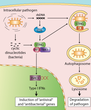
STING in the host response to intracellular pathogens. Linking type I IFN response and autophagy for better defense

STING in the host response to intracellular pathogens

http://www.invivogen.com/images/STING-autophagy.png


STING activates the IFN response

Until very recently, STING in addition to its role as an adaptor protein was also thought to function as a sensor of cyclic dinucleotides.
Burdette et al. first demonstrated that STING binds directly to the bacterial molecule cyclic diguanylate monophosphate (c-di-GMP) [8]. This finding was confirmed by several teams who examined the structure of STING bound to c-di-GMP [9-11], including Cheng and colleagues, however their data suggest that STING is not the primary sensor of c-di-GMP [12]. Rather, they indicate that DDX41, an identified CDS, functions as a direct receptor for cyclic dinucleotides upstream of STING. The authors hypothesized that DDX41 binds to c-di-GMP then forms a complex with STING to activate the IFN response.

STING induces autophagy

Exciting new developments reveal that STING participates in another aspect of innate immunity, autophagy.
Autophagy plays a critical role in host defense responses to pathogens by promoting the elimination of microbes that enter into the cytosol by their sequestration into autophagosomes and their delivery to the lysosome.


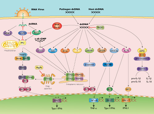
CDS pathway

http://www.invivogen.com/images/STING-CDS_pathway_small.jpg

Recent studies have reported that DNA viruses and intracellular bacteria induce autophagy and that this process is dependent on cytosolic genomic DNA and STING [13-15]. Robust induction of autophagy was also observed after transfection of various double stranded (ds) DNA species, such as poly(dA:dT), poly(dG:dC) or plasmid DNA, but not single stranded (ss) DNA, dsRNA or ssRNA [16].

Interestingly, activated STING was shown to relocate to unidentified membrame-bound compartments where it colocalizes with LC3, a hallmark of autophagy, and ATg9a. The latter protein was reported to regulate the interaction between STING and TBK1 after dsDNA stimulation [16]. The E3 ubiquitin ligases TRIM56 and TRIM32
were also found to regulate STING by mediating its dimerization through K63-linked ubiquitination [17, 18].

Several cytosolic DNA sensors upstream of STING have been proposed.
DNA-dependent activator of IRFs (DAI) was the first CDS discovered based on the ability of transfected poly(dA:dT) to induce IFN-β [19]. However, the role of DAI has been shown to be very cell-type specific and cells derived from DAI-deficient mice responded normally to dsDNA ligands [20].

While analyzing immune responses to dsDNA regions derived from vaccinia virus (VACV-70) or Herpes simplex virus 1 (HSV-60) genomes, Unterholzner et al. identified IFI16 as a DNA binding protein mediating IFN-β induction [21]. Interestingly, IFI16 belongs to a new family of pattern recognition receptors that contain the pyrin and HIN domain (PYHIN), termed AIM2-like receptors (ALRs).

AIM2 is a STING-independent cytosolic DNA sensor that forms an inflammasome with ASC to trigger caspase-1 activation and the secretion of the proinflammatory cytokines IL-1β and IL-18 [20].

Members of the DExD/H-box helicase superfamily have also been reported to function as cytosolic DNA sensors. While DHX36 and DHX9 were identified as STING-independent but MyD88-dependent sensors of CpG-containing DNA in plasmacytoid dendritic cells, DDX41 was found to bind various dsDNA ligands and localize with STING to promote IFN-β expression [22]. Other CDSs have been reported to function independently of STING: RNA Pol III, LRRFIP1 and Ku70 [20].

Unlike cytosolic RNA sensors (RIG-I, MDA-5), which detect structural moieties specific to pathogen RNA, such as 5’-triphosphates, it is not clear whether cytosolic DNA sensors can recognize any particular structural motif of DNA that would discriminate between self and non-self. This suggests that CDSs may have a role not only in anti-microbial innate immune responses but also in autoimmunity. A multitude of CDSs have been described but whether they are all true receptors remains an open question.

1. Stetson DB & Medzhitov R. 2006. Recognition of cytosolic DNA activates an IRF3-dependent innate immune response. Immunity. 24(1):93-103.
2. Ishikawa H. & Barber GN., 2008. STING is an endoplasmic reticulum adaptor that facilitates innate immune signalling. Nature. 455(7213):674-8.
3. Ishikawa H. et al., 2009. STING regulates intracellular DNA-mediated, type I interferon-dependent innate immunity. Nature. 461(7265):788-92.
4. Zhong B. et al., 2008. The adaptor protein MITA links virus-sensing receptors to IRF3 transcription factor activation. Immunity. 29(4):538-50.
5. Jin L. et al., 2008. MPYS, a novel membrane tetraspanner, is associated with major histocompatibility complex class II and mediates transduction of apoptotic signals. Mol Cell Biol. 28(16):5014-26.


UV Light Potentiates STING (Stimulator of Interferon Genes)-dependent Innate Immune Signaling through Deregulation of ULK1 (Unc51-like Kinase 1).

 J Biol Chem. 2015 May 8;290(19):12184-94.  http://dx.doi.org:/10.1074/jbc.M115.649301. Epub 2015 Mar 19.

The mechanism by which ultraviolet (UV) wavelengths of sunlight trigger or exacerbate the symptoms of the autoimmune disorder lupus erythematosus is not known but may involve a role for the innate immune system. Here we show that UV radiation potentiates STING (stimulator of interferon genes)-dependent activation of the immune signaling transcription factor interferon regulatory factor 3 (IRF3) in response to cytosolic DNA and cyclic dinucleotides in keratinocytes and other human cells. Furthermore, we find that modulation of this innate immune response also occurs with UV-mimetic chemical carcinogens and in a manner that is independent of DNA repair and several DNA damage and cell stress response signaling pathways. Rather, we find that the stimulation of STING-dependent IRF3 activation by UV is due to apoptotic signaling-dependent disruption of ULK1 (Unc51-like kinase 1), a pro-autophagic protein that negatively regulates STING. Thus, deregulation of ULK1 signaling by UV-induced DNA damage may contribute to the negative effects of sunlight UV exposure in patients with autoimmune disorders.


STING and the innate immune response to nucleic acids in the cytosol

Dara L Burdette & Russell E Vance

https://mcb.berkeley.edu/labs/vance/Resources/Burdette%20(2013)%20review.pdf

Cytosolic detection of pathogen-derived nucleic acids is critical for the initiation of innate immune defense against diverse bacterial, viral and eukaryotic pathogens. Conversely, inappropriate responses to cytosolic nucleic acids can produce severe autoimmune pathology. The host protein STING has been identified as a central signaling molecule in the innate immune response to cytosolic nucleic acids. STING seems to be especially critical for responses to cytosolic DNA and the unique bacterial nucleic acids called ‘cyclic dinucleotides’. Here we discuss advances in the understanding of STING and highlight the many unresolved issues in the field.

The detection of pathogen-derived nucleic acids is a central strategy by which the innate immune system senses microbes to then initiate protective responses1. Conversely, inappropriate recognition of self nucleic acids can result in debilitating autoimmune diseases such as systemic lupus erythematosus2. It is therefore important to understand the molecular basis of the detection of nucleic acids by the innate immune system. Studies have established that nucleic acids derived from extracellular sources are sensed mainly by endosomal Toll-like receptors (TLRs), such as TLR3, TLR7 and TLR9, whereas cytosolic nucleic acids are detected independently of TLRs by a variety of less-well-characterized mechanisms1.

Studies have identified STING (‘stimulator of interferon genes’; also known as TMEM173, MPYS, MITA and ERIS) as a critical signaling molecule in the innate response to cytosolic nucleic-acid ligands. STING was first described as a protein that interacts with major histocompatibility complex class II molecules3, but the relevance of this interaction remains unclear. Subsequent studies have instead focused on the role of STING in the transcriptional induction of type I interferons and coregulated genes in response to nucleic acids in the cytosol. Several groups have independently isolated STING by screening for proteins able to induce interferon-B (IFN-B) when overexpressed4–6. Studies of STING-deficient mice have subsequently confirmed the essential role of STING in innate responses to cytosolic nucleic-acid ligands, particularly double-stranded DNA (dsDNA) and unique bacterial nucleic acids called ‘cyclic dinucleotides’7–9. Several studies have also linked STING to the interferon response to cytosolic RNA5–7, but this has not been found consistently7,8,10,11; thus, we focus here on the role of STING in response to DNA and cyclic dinucleotides.


Protein Stimulator of interferon genes protein
Gene TMEM173
Organism Homo sapiens (Human)
Facilitator of innate immune signaling that acts as a sensor of cytosolic DNA from bacteria and viruses and promotes the production of type I interferon (IFN-alpha and IFN-beta). Innate immune response is triggered in response to non-CpG double-stranded DNA from viruses and bacteria delivered to the cytoplasm. Acts by recognizing and binding cyclic di-GMP (c-di-GMP), a second messenger produced by bacteria, and cyclic GMP-AMP (cGAMP), a messenger produced in response to DNA virus in the cytosol: upon binding of c-di-GMP or cGAMP, autoinhibition is alleviated and TMEM173/STING is able to activate both NF-kappa-B and IRF3 transcription pathways to induce expression of type I interferon and exert a potent anti-viral state. May be involved in translocon function, the translocon possibly being able to influence the induction of type I interferons. May be involved in transduction of apoptotic signals via its association with the major histocompatibility complex class II (MHC-II). Mediates death signaling via activation of the extracellular signal-regulated kinase (ERK) pathway. Essential for the induction of IFN-beta in response to human herpes simplex virus 1 (HHV-1) infection. Exhibits 2′,3′ phosphodiester linkage-specific ligand recognition. Can bind both 2′-3′ linked cGAMP and 3′-3′ linked cGAMP but is preferentially activated by 2′-3′ linked cGAMP (PubMed:26300263)
Stimulator of interferon genes protein (IPR029158)
Transmembrane protein 173, also known as stimulator of interferon genes protein (STING) or endoplasmic reticulum interferon stimulator (ERIS), is a transmembrane adaptor protein which is involved in innate immune signalling processes. It induces expression of type I interferons (IFN-alpha and IFN-beta) via the NF-kappa-B and IRF3, pathways in response to non-self cytosolic RNA and dsDNA [PMID: 18724357, PMID: 19776740,PMID: 18818105, PMID: 19433799].

Read Full Post »

Periodic table of protein complexes, Volume 2 (Volume Two: Latest in Genomics Methodologies for Therapeutics: Gene Editing, NGS and BioInformatics, Simulations and the Genome Ontology), Part 1: Next Generation Sequencing (NGS)

Periodic table of protein complexes

Larry H. Bernstein, MD, FCAP, Curator

LPBI


Periodic Table of Protein Complexes

http://www.technologynetworks.com/Proteomics/news.aspx?ID=186248

New tool helps to visualise, understand and predict how proteins combine to drive biological processes

A new ‘periodic table’ of protein complexes has been developed that provides a unified way to classify and visualise protein complexes, providing a valuable tool for biotechnology and the engineering of novel complexes.

This study also provides insights into evolutionary distribution of different types of existing protein complexes.

The Periodic Table of Protein Complexes offers a new way of looking at the enormous variety of structures that proteins can build in nature, which ones might be discovered next, and predicting how entirely novel structures could be engineered. Created by an interdisciplinary team led by researchers at the Wellcome Genome Campus and the University of Cambridge, the Table provides a valuable tool for research into evolution and protein engineering.

Almost every biological process depends on proteins interacting and assembling into complexes in a specific way, and many diseases are associated with problems in complex assembly. The principles underpinning this organisation are not yet fully understood, but by defining the fundamental steps in the evolution of protein complexes, the new ‘periodic table’ presents a systematic, ordered view on protein assembly, providing a visual tool for understanding biological function.

“Evolution has given rise to a huge variety of protein complexes, and it can seem a bit chaotic. But if you break down the steps proteins take to become complexes, there are some basic rules that can explain almost all of the assemblies people have observed so far.”


Dr Joe Marsh, formerly of the Wellcome Genome Campus and now of the MRC Human Genetics Unit at the University of Edinburgh.

Different ballroom dances can be seen as an endless combination of a small number of basic steps. Similarly, the ‘dance’ of protein complex assembly can be seen as endless variations on dimerization (one doubles, and becomes two), cyclisation (one forms a ring of three or more) and subunit addition (two different proteins bind to each other). Because these happen in a fairly predictable way, it’s not as hard as you might think to predict how a novel protein would form.

“We’re bringing a lot of order into the messy world of protein complexes. Proteins can keep go through several iterations of these simple steps, adding more and more levels of complexity and resulting in a huge variety of structures. What we’ve made is a classification based on these underlying principles that helps people get a handle on the complexity.”

Dr Sebastian Ahnert of the Cavendish Laboratory at the University of Cambridge

The exceptions to the rule are interesting in their own right, as are the subject of on-going studies.

“By analysing the tens of thousands of protein complexes for which three-dimensional structures have already been experimentally determined, we could see repeating patterns in the assembly transitions that occur – and with new data from mass spectrometry we could start to see the bigger picture.”

Dr Joe Marsh

“The core work for this study is in theoretical physics and computational biology, but it couldn’t have been done without the mass spectrometry work by our colleagues at Oxford University. This is yet another excellent example of how extremely valuable interdisciplinary research can be.”

Dr Sarah Teichmann, Research Group Leader at the Wellcome Trust Sanger Institute and the European Bioinformatics Institute (EMBL-EBI)

Read Full Post »

Reinforced disordered cell expression

Larry H. Bernstein, MD, FCAP, Curator

LPBI


Diabetes, Alzheimer’s Share Molecular Pathways, Part of Same Vicious Cycle

http://www.genengnews.com/gen-news-highlights/diabetes-alzheimer-s-share-molecular-pathways-part-of-same-vicious-cycle/81252206/

http://www.genengnews.com/Media/images/GENHighlight/thumb_Jan8_2016_Fotolia_30836005_JigsawPuzzleBrainAndHead1904910113.jpg

A molecular-level link has been found that helps explain the poorly understood association between diabetes and Alzheimer’s disease. Both disorders can drive and be driven by the same pathological process, the disruption of a particular kind of post-translational modification called S-nitrosylation. Thus, the disorders can reinforce each other. [© freshidea/Fotolia]


Though they appear to be distinct, diabetes and Alzheimer’s disease have much in common at the molecular level. In fact, recent findings indicate that either disease can worsen the other by disrupting the same chemical process—S-nitrosylation, a form of post-translational modification that is necessary for the proper functioning of multiple enzymes.

S-nitrosylation, it turns out, can be disrupted by excess sugar or β-amyloid protein, either of which can wreak havoc by increasing the levels of nitric oxide and other free radical species. Once S-nitrosylation is disturbed and poorly functioning enzymes are produced, the downstream effects include abnormal increases in both insulin and β-amyloid protein.

Thus, diabetes and Alzheimer’s can drive, and be driven by, the same vicious cycle. Furthermore, either can contribute to the other’s progress. These results emerged from a study completed by researchers based at the Sanford Burnham Prebys Medical Discovery Institute and the Scintillon Institute. The research team was led by Stuart A. Lipton, M.D., Ph.D., a physician-scientist affiliated with both institutions.

“This work points to a new common pathway to attack both type 2 diabetes, along with its harbinger, metabolic syndrome, and Alzheimer’s disease,” stated Dr. Lipton.

The researchers published their work January 8 in the journal Nature Communications in an article entitled, “Elevated glucose and oligomeric β-amyloid disrupt synapses via a common pathway of aberrant protein S-nitrosylation.” This article describes how the scientists used a so-called “disease-in-a-dish” model to discover molecular pathways that are in common in both diabetes and Alzheimer’s.

Specifically, the scientists genetically reprogrammed the skin of human patients to make induced pluripotent stem cells, which were then used to derive nerve cells. They also used mouse models of each disease to analyze the combined effects of high blood sugar and β-amyloid protein in living animals.

“[We] report in human and rodent tissues that elevated glucose, as found in [metabolic syndrome and type 2 diabetes] and oligomeric β-amyloid (Aβ) peptide, thought to be a key mediator of [Alzheimer’s disease], coordinately increase neuronal Ca2+ and nitric oxide (NO) in an NMDA receptor-dependent manner,” wrote the authors of the Nature Communications article. “The increase in NO results in S-nitrosylation of insulin-degrading enzyme (IDE) and dynamin-related protein 1 (Drp1), thus inhibiting insulin and Aβ catabolism as well as hyperactivating mitochondrial fission machinery.”

The scientists also found that the changes in enzyme activity led to damage of synapses, the region where nerve cells communicate with one another in the brain. The combination of high sugar and β-amyloid protein caused the greatest loss of synapses. Since loss of synapses correlates with cognitive decline in Alzheimer’s, high sugar and β-amyloid coordinately contribute to memory loss.

“The NMDA receptor antagonist memantine attenuates these effects,” the authors continued. “Our studies show that redox-mediated posttranslational modification of brain proteins link Aβ and hyperglyaemia to cognitive dysfunction in [metabolic syndrome/type 2 diabetes] and [Alzheimer’s disease].”

“[Our work] means that we now know these diseases are related on a molecular basis, and hence, they can be treated with new drugs on a common basis,” stated Dr. Ambasudhan, a senior author of the study and an assistant professor at Scintillon.

Read Full Post »

Antibody alternatives in specific aptamer 3-D scaffold binding

Curator: Larry H. Bernstein, MD, FCAP


New Proteomics Tools to Open Up Drug Discovery

Dr. Paul Ko Ferrigno, Chief Scientific Officer, and Dr. Jane McLeod, of Avacta Life Sciences

http://www.dddmag.com/articles/2016/01/new-proteomics-tools-open-drug-discovery


Size matters: Smaller molecules allow a tighter packing density on solid surfaces for improved signal-to-noise in assays, such as SPR or ELISA.

http://www.dddmag.com/sites/dddmag.com/files/AvactaAffimer%20stills_00006.jpg

Size matters: Smaller molecules allow a tighter packing density on solid surfaces for improved signal-to-noise in assays, such as SPR or ELISA.


In the current genomic era of very accurate DNA analyses by in situ hybridization, DNA chip analyses, and deep sequencing, it is often assumed that antibodies have an analogous ability to identify molecular targets accurately. Nothing could be further from the truth. Proteomics as a field is still lagging behind its genomic counterpart in the level of detail we can achieve, the level of data we can collect and the overall levels of accuracy and reliability that the collected data represent.

From the estimated 20,000 human genes 100,000 different possible proteins have been predicted. What is more, the variation achieved via the post-translational modification of these proteins brings another layer of complexity to cellular signalling. All this means that while studying the genetic blueprint can offer insights into the cell, it is only through examining the functional protein units that we can comprehensively map the dynamic interactions that occur within the cell to drive an organism or disease process.


Why not use antibodies?

Antibodies form the basis of molecular recognition in proteomics, whether this is to identify a protein within a complex mixture or label a specific protein within a cell. Both their target specificity and high binding capacity have made these molecules fantastically useful tools within diagnostics. However, the large majority of commercially available antibodies are for use as reagents in research and development, where they are simply not as well validated and issues with their manufacture have created problems that have hindered drug development.

It has proven next to impossible to develop antibodies to certain targets. This may be because of their high homology to the host protein, so the immune system fails to recognise them as different, or due to antigen processing resulting in the loss of post-translational modifications or discontinuous epitopes. However, without the necessary antibodies to investigate these targets the corresponding research avenues have remained closed and key drug targets may have been missed.

Almost worse than lacking the necessary research tools is the problem of antibody reproducibility. Matthias Uhlen revealed that of the 5,436 antibodies tested as part of the Protein Atlas project over 50 percent failed to recognize their target in at least one of two commonly used assays. Antibodies that are not specific to their target or do not recognize their target at all are responsible for increasing the cost of biological research, with an estimated $800 million spent globally every year on bad antibodies. Many published studies have had to be retracted due to antibody-derived error and those that remain unidentified in the literature will continue to lead researchers down blind alleys. This level of misinformation in the published literature is not just hindering the progression of the field, but possibly even sending it backwards and costing more than is needed — an issue not to be taken lightly when so many research budgets are coming under the knife.

More recently there has been a call from a number of leading scientists for the use of polyclonal antibodies, considered to be the worst offenders in terms of batch-to-batch irreproducibility, to be abandoned. They suggest a move towards recombinant systems of production, which would remove the restrictions of the immune system on antibody production. Yet, they state that $1 billion dollars investment would be required to re-route antibody production down this path and suggest that a period of five to ten years may be required to bring about these changes.

Simply producing recombinant antibodies rather than animal-derived affinity reagents will still leave us with a number of problems regarding their use. Antibodies are simply too large to target many smaller, hidden epitopes and the presence of key disulphide bonds within their structure makes them all too susceptible to reduction within the cell, rendering them useless for applications such as live cell imaging of molecules within the cytoplasm. Moreover, can we afford to wait another decade for a solution to this problem before we pursue protein targets for basic understanding and drug development?


Antibody alternatives for new targets and techniques

Antibody alternatives are already available to researchers in the life sciences field. They are produced either from nucleic acid or protein molecules. Aptamers are short, single-stranded RNA or DNA molecules that fold to form 3D scaffolds, which can present a specific interaction surface to allow specific binding to its target molecule. Protein scaffolds are formed from parts of or whole proteins modified to present a peptide sequence. This peptide sequence works in a similar manner to present a specific interaction surface for specific binding to a desired target.

These antibody alternatives are produced in recombinant systems, minimizing batch-to-batch variation and allowing them to be produced to theoretically any target. Additionally, as they do not use animals in their production they are generally less expensive to produce than traditional antibodies.


While companies such as Affibody and Avacta Life Sciences are aiming to open up the drug pipeline, by offering these alternative affinity reagents to previously inaccessible targets for use in research and development, many have moved into exploiting their therapeutic potential. Noxxon produce an RNA-based scaffold, Spiegelmers, which are currently in phase 2 clinical trials for diabetic nephropathy, and Molecular Partners have reached phase 3 clinical trials with their protein scaffold DARPins, for wet age-related macular degeneration.

These new antibody alternatives are smaller than traditional antibodies. This opens up the use of new technologies, such as super resolution microscopy. While the diffraction limit remained at about 250 nmm the length of the antibody at 15 nm was of little importance, tagging your molecule as accurately as necessary. Removing this limit in super resolution microscopy has meant that antibodies are now too large to provide the required level of accuracy. Instead, using an antibody alternative of only 2nm to tag your protein of interest opens up this new technique offering greater precision to intracellular localization.


As these scaffolds have been engineered to be fit-for-purpose many contain no intramolecular disulphide bonds and so are not sensitive to the reducing environment of the cell. This function enables their use as intracellular reporters of molecular conformation, as well as in standard assays like IHC or western blotting, so allowing scientists to use the same reagent across both intracellular and biochemical assays, thus bridging the gap between cell biology and biochemical studies.

Offering increased reproducibility, access to an increased range of applications, and the opportunity to hit previously inaccessible targets, antibody alternatives are opening up potential new avenues of drug discovery.


About the Authors

Dr. Paul Ko Ferrigno is Chief Scientific Officer at Avacta Life Sciences and a Visiting Professor at the University of Leeds. He has been working on peptide aptamers since 2001 in Leeds and at the MRC Cancer Cell Unit in Cambridge, UK where his team developed the Affimer scaffold technology.

Dr. Jane McLeod is a Scientific Writer at Avacta Life Sciences.


Read Full Post »

Transformer Proteins against Cancer

Larry H. Bernstein, MD, FCAP, Curator

LPBI


Revision  1/13/20165

GEN News Highlights:  “Transformer” Proteins with Role in Cancer

http://www.genengnews.com/gen-news-highlights/transformer-proteins-with-role-in-cancer/81252133/

Investigators at the Scripps Research Institute (TSRI) and St. Jude Children’s Research Hospital say they have found how a protein involved in cancer twists and morphs into different structures.

“We’re studying basic biophysics, but we believe the complexity and rules we uncover for the physics of protein disorder and folding could one day also be used for better designs of therapeutics,” said Ashok Deniz, Ph.D., associate professor at TSRI.

The study (“Asymmetric Modulation of Protein Order-Disorder Transitions by Phosphorylation and Partner Binding”), published in Angewandte Chemie, focuses  on the nucleophosmin (NPM1) protein, which has many functions and, when mutated, has been shown to interfere with cells’ normal tumor suppressing ability. NPM1 has been implicated in cancers such as non-Hodgkin lymphoma and acute myelogenous leukemia.

Previous research led by study collaborators Richard Kriwacki, Ph.D., and Diana Mitrea, Ph.D., at St. Jude had demonstrated that a section of NPM1, called the N-terminal domain (Npm-N), doesn’t have a defined, folded structure. Instead, the protein morphs between two forms: a one-subunit disordered monomer and a five-subunit folded pentamer.


Until now, the mechanism behind this transformation was unknown, but scientists believed this monomer-pentamer equilibrium could be important for the protein’s location and functioning in the cell. To shed light on how this transformation occurred, Dr. Deniz and his colleagues used a combination of three techniques—single-molecule biophysics, fluorescence resonance energy transfer (FRET), and circular dichroism, which enabled them to study individual molecules and collections of molecules. Single-molecule methods are especially useful for such studies because they can uncover important information that remains hidden in conventional studies.

The researchers found that the transformation can proceed through more than one pathway. In one pathway, the transformation begins when the cell sends signals to attach phosphoryl groups to NPM1. Such phosphorylation prompts the ordered pentamer to become disordered and likely causes NPM1 to shuttle outside the cell’s nucleus. A meeting with a binding partner can mediate the reverse transformation to a pentamer.

When NPM1 does become a pentamer again under these conditions, which likely causes it to move back to the nucleolus, it takes a different path instead of just retracing its earlier steps.


Priya Banerjee, Ph.D., an American Heart Association-supported postdoctoral research associate at TSRI and the first author of the study, compared these complicated transitions to the morphing of a “Transformers” toy, where a robot can become a car and then a jet. “Phosphorylation and partner-binding are like different cellular switches driving these changes,” said Dr. Banerjee.

According to Dr. Banerjee, the new study also reveals many intermediate states between monomer and pentamer structures and that these states can be manipulated or “tuned” by changing conditions such as salt levels, phosphorylation, and partner binding, which may explain how cells regulate the protein’s multiple functions. The researchers said future studies could shed more light on the biological functions of these different structures and how they might be used in future cancer therapies.

The team added that combining the three techniques used in this study, plus a novel protein-labeling technique for single-molecule fluorescence, could be a useful strategy for studying other unstructured, “intrinsically disordered proteins” (IDPs), which are involved in a host of cellular functions, as well as neurodegenerative disease, heart disease, infectious disease, type 2 diabetes and other conditions.


Single ‘Transformer’ Proteins

http://www.technologynetworks.com/Proteomics/news.aspx?ID=186996


“We’re studying basic biophysics, but we believe the complexity and rules we uncover for the physics of protein disorder and folding could one day also be used for better designs of therapeutics,” said TSRI Associate Professor Ashok Deniz, senior author of the new study along with Richard Kriwacki, faculty member at St. Jude.

The study  focuses on a protein called nucleophosmin (NPM1). This protein has many functions and, when mutated, has been shown to interfere with cells’ normal tumor-suppressing ability. NPM1 has been implicated in cancers such as non-Hodgkin lymphoma and acute myelogenous leukemia.

Previous research led by study collaborators Kriwacki and Diana Mitrea at St. Jude had shown that a section of NPM1, called the N-terminal domain (Npm-N), doesn’t have a defined, folded structure. Instead, the protein morphs between two forms: a one-subunit disordered monomer and a five-subunit folded pentamer.

Until now, the mechanism behind this transformation was unknown, but scientists believed this monomer-pentamer equilibrium could be important for the protein’s location and functioning in the cell.

To shed light on how this transformation occurred, Deniz and his colleagues used an innovative combination of three techniques—single-molecule biophysics, fluorescence resonance energy transfer (FRET) and circular dichroism—which enabled them to study individual molecules and collections of molecules. Single-molecule methods are especially useful for such studies because they can uncover important information that remains hidden in conventional studies.

Remarkably, the researchers found that the transformation can proceed through more than one pathway. In one pathway, the transformation begins when the cell sends signals to attach phosphoryl groups to NPM1. This modification, called phosphorylation, prompts the ordered pentamer to become disordered and likely causes NPM1 to shuttle outside the cell’s nucleus. A meeting with a binding partner can mediate the reverse transformation to a pentamer.

Interestingly, when NPM1 does become a pentamer again under these conditions, which likely causes it to move back to the nucleolus, it takes a different path instead of just retracing its earlier steps.

Priya Banerjee, an American Heart Association-supported postdoctoral research associate at TSRI and the first author of the study, compared these complicated transitions to the morphing of a “Transformers” toy, where a robot can become a car and then a jet. “Phosphorylation and partner-binding are like different cellular switches driving these changes,” said Banerjee.

Banerjee said the new study also reveals many intermediate states between monomer and pentamer structures—and that these states can be manipulated or “tuned” by changing conditions such as salt levels, phosphorylation and partner binding, which may explain how cells regulate the protein’s multiple functions. The researchers said future studies could shed more light on the biological functions of these different structures and how they might be used in future cancer therapies.

The researchers added that combining the three techniques used in this study, plus a novel protein-labeling technique for single-molecule fluorescence, could be a useful strategy for studying other unstructured, “intrinsically disordered proteins” (IDPS). IDPS are involved in a host of cellular functions, as well as neurodegenerative disease, heart disease, infectious disease, type 2 diabetes and other conditions.

TSRI and St. Jude Scientists Study Physics of Single ‘Transformer’ Proteins with Role in Cancer

http://www.scripps.edu/news/press/2015/20151221deniz.html

December 21, 2015 – A new study led by scientists at The Scripps Research Institute (TSRI) and St. Jude Children’s Research Hospital shows how a protein involved in cancer twists and morphs into different structures.

“We’re studying basic biophysics, but we believe the complexity and rules we uncover for the physics of protein disorder and folding could one day also be used for better designs of therapeutics,” said TSRI Associate Professor Ashok Deniz, senior author of the new study along with Richard Kriwacki, faculty member at St. Jude.

The study, published recently in the journal Angewandte Chemie, focuses on a protein called nucleophosmin (NPM1). This protein has many functions and, when mutated, has been shown to interfere with cells’ normal tumor suppressing ability. NPM1 has been implicated in cancers such as non-Hodgkin lymphoma and acute myelogenous leukemia.

Previous research led by study collaborators Kriwacki and Diana Mitrea at St. Jude had shown that a section of NPM1, called the N-terminal domain (Npm-N), doesn’t have a defined, folded structure. Instead, the protein morphs between two forms: a one-subunit disordered monomer and a five-subunit folded pentamer.

Until now, the mechanism behind this transformation was unknown, but scientists believed this monomer-pentamer equilibrium could be important for the protein’s location and functioning in the cell.

To shed light on how this transformation occurred, Deniz and his colleagues used an innovative combination of three techniques—single-molecule biophysics, fluorescence resonance energy transfer (FRET) and circular dichroism—which enabled them to study individual molecules and collections of molecules. Single-molecule methods are especially useful for such studies because they can uncover important information that remains hidden in conventional studies.

Remarkably, the researchers found that the transformation can proceed through more than one pathway. In one pathway, the transformation begins when the cell sends signals to attach phosphoryl groups to NPM1. This modification, called phosphorylation, prompts the ordered pentamer to become disordered and likely causes NPM1 to shuttle outside the cell’s nucleus. A meeting with a binding partner can mediate the reverse transformation to a pentamer.

Interestingly, when NPM1 does become a pentamer again under these conditions, which likely causes it to move back to the nucleolus, it takes a different path instead of just retracing its earlier steps.


Asymmetric Modulation of Protein Order-Disorder Transitions by Phosphorylation and Partner Binding  

Priya R. Banerjee, Diana M. Mitrea, Richard W. Kriwacki, Ashok, A. Deniz

     http://dx.doi.org:/10.1002/anie.201507728

As for many intrinsically disordered proteins, order–disorder transitions in the N-terminal oligomerization domain of the multifunctional nucleolar protein nucleophosmin (Npm-N) are central to its function, with phosphorylation and partner binding acting as regulatory switches. However, the mechanism of this transition and its regulation remain poorly understood. In this study, single-molecule and ensemble experiments revealed pathways with alternative sequences of folding and assembly steps for Npm-N. Pathways could be switched by altering the ionic strength. Phosphorylation resulted in pathway-specific effects, and decoupled folding and assembly steps to facilitate disorder. Conversely, binding to a physiological partner locked Npm-N in ordered pentamers and counteracted the effects of phosphorylation. The mechanistic plasticity found in the Npm-N order–disorder transition enabled a complex interplay of phosphorylation and partner-binding steps to modulate its folding landscape.


Conditionally and Transiently Disordered Proteins: Awakening Cryptic Disorder To Regulate Protein Function

Related articles:

Discovery of Small Molecules that Inhibit the Disordered Protein, p27(Kip1).

Iconaru LI, Ban D, Bharatham K, Ramanathan A, Zhang W, Shelat AA, Zuo J, Kriwacki RW.

Sci Rep. 2015 Oct 28;5:15686. doi: 10.1038/srep15686.

Select item 264270603.

Design, Synthesis and Evaluation of 2,5-Diketopiperazines as Inhibitors of the MDM2-p53 Interaction.

Pettersson M, Quant M, Min J, Iconaru L, Kriwacki RW, Waddell MB, Guy RK, Luthman K, Grøtli M.

PLoS One. 2015 Oct 1;10(10):e0137867. doi: 10.1371/journal.pone.0137867. eCollection 2015.

Select item 264113004.

Discoveries and controversies in BCL-2 protein-mediated apoptosis.

Zheng JH, Viacava Follis A, Kriwacki RW, Moldoveanu T.

FEBS J. 2015 Sep 28. doi: 10.1111/febs.13527. [Epub ahead of print] Review.

PMID:
26411300
Select item 262360135.

Pin1-Induced Proline Isomerization in Cytosolic p53 Mediates BAX Activation and Apoptosis.

Follis AV, Llambi F, Merritt P, Chipuk JE, Green DR, Kriwacki RW.

Mol Cell. 2015 Aug 20;59(4):677-84. doi: 10.1016/j.molcel.2015.06.029. Epub 2015 Jul 30.

PMID:
26236013


A phosphorylation switch controls the spatiotemporal activation of Rho GTPases in directional cell migration

Xuan Cao, Tomonori Kaneko, Jenny S. Li, An-Dong Liu, et al.

Nature Communications 2015; 6(7721)     http://dx.doi.org:/10.1038/ncomms8721

Although cell migration plays a central role in development and disease, the underlying molecular mechanism is not fully understood. Here we report that a phosphorylation-mediated molecular switch comprising deleted in liver cancer 1 (DLC1), tensin-3 (TNS3), phosphatase and tensin homologue (PTEN) and phosphoinositide-3-kinase (PI3K) controls the spatiotemporal activation of the small GTPases, Rac1 and RhoA, thereby initiating directional cell migration induced by growth factors. On epidermal growth factor (EGF) or platelet-derived growth factor (PDGF) stimulation, TNS3 and PTEN are phosphorylated at specific Thr residues, which trigger the rearrangement of the TNS3–DLC1 and PTEN–PI3K complexes into the TNS3–PI3K and PTEN–DLC1 complexes. Subsequently, the TNS3–PI3K complex translocates to the leading edge of a migrating cell to promote Rac1 activation, whereas PTEN–DLC1 translocates to the posterior for localized RhoA activation. Our work identifies a core signalling mechanism by which an external motility stimulus is coupled to the spatiotemporal activation of Rac1 and RhoA to drive directional cell migration.


Protein Phosphorylation is an Important Tool…  Chapter 13.  in Protein Phosphorylation in Human Health 

Roberta Fraschini, Erica Raspelli and Corinne Cassani 

http://cdn.intechopen.com/pdfs-wm/38809.pdf

Protein phosphorylation is a reversible posttranslational modification that can modulate protein role in several physiological processes in almost every possible way. These include modification of its intrinsic biological activity, subcellular location, half-life and binding with other proteins. Protein phosphorylation is particularly important for the regulation of key proteins involved in the control of cell cycle progression.

Protein phosphorylation is the covalent binding of a phosphate group to some critical residues of the polypeptide. The phosphorylation state of a protein is given by a balance between the activity of protein kinases and protein phosphatases. Eukaryotic protein kinases transfer phosphate groups (PO43-) from ATP to an hydroxyl group of the lateral chain of specific serine, threonine or tyrosine residues on peptide substrates. In simple eukaryotic cells, like yeasts, Ser/Thr kinases are more common, while more complex eukaryotic cells, like human cells, have many Tyr kinases. Protein kinases recognize their substrates specifically and their active site consists of an activation loop and a catalytic loop between which substrates bind. Protein kinases differ from each other in the structure of their catalitic domain. A second class of enzymes, protein phosphatases, is responsible for the reverse reaction in which phosphate groups are removed from a protein. Phosphatases gain specificity by binding protein cofactors which facilitate binding to specific phosphoproteins. The active phosphatase often consists of a complex of the phosphatase catalytic subunit and a regulatory subunit.

The phosphorylation of specific residues induces structural changes that regulate protein functions by modulating protein folding, substrate affinity, stability and activity. For example, phosphorylation can cause switch-like changes in protein function, which can also lead to major modifications in the catalytic function of enzymes, including kinases and phosphatases. In addition, protein phosphorylation often leads to rearrangement in the structure of the protein that can induce changes in interacting partners or subcellular localization.

Phosphorylation acts as a molecular switch for many regulatory events in signalling pathways that drive cell division, proliferation, differentiation and apoptosis. In order to ensure an appropriate balance of protein phosphorylation, the cell can compartmentalize both protein kinases and phosphatases. Another kind of regulation can be achieved by the spatial distribution of kinases and phosphatases, that creates a gradient of phosphorylated substrates across different subcellular compartments. This spatial separation can also control the activity of other proteins or enzymes and the occurrence of other posttranslational modifications.

Molecular Biology of the Cell. 4th edition.   Protein Function.   http://www.ncbi.nlm.nih.gov/books/NBK26911/

We have seen that each type of protein consists of a precise sequence of amino acids that allows it to fold up into a particular three-dimensional shape, or conformation. But proteins are not rigid lumps of material. They can have precisely engineered moving parts whose mechanical actions are coupled to chemical events. It is this coupling of chemistry and movement that gives proteins the extraordinary capabilities that underlie the dynamic processes in living cells.

In this section, we explain how proteins bind to other selected molecules and how their activity depends on such binding. We show that the ability to bind to other molecules enables proteins to act as catalysts, signal receptors, switches, motors, or tiny pumps. The examples we discuss in this chapter by no means exhaust the vast functional repertoire of proteins. However, the specialized functions of many of the proteins you will encounter elsewhere in this book are based on similar principles.

All Proteins Bind to Other Molecules

The biological properties of a protein molecule depend on its physical interaction with other molecules. Thus, antibodies attach to viruses or bacteria to mark them for destruction, the enzyme hexokinase binds glucose and ATP so as to catalyze a reaction between them, actin molecules bind to each other to assemble into actin filaments, and so on. Indeed, all proteins stick, or bind, to other molecules. In some cases, this binding is very tight; in others, it is weak and short-lived. But the binding always shows great specificity, in the sense that each protein molecule can usually bind just one or a few molecules out of the many thousands of different types it encounters. The substance that is bound by the protein—no matter whether it is an ion, a small molecule, or a macromolecule— is referred to as a ligand for that protein (from the Latin word ligare, meaning “to bind”).

The ability of a protein to bind selectively and with high affinity to a ligand depends on the formation of a set of weak, noncovalent bonds—hydrogen bonds, ionic bonds, and van der Waals attractions—plus favorable hydrophobic interactions (see Panel 2-3, pp. 114–115). Because each individual bond is weak, an effective binding interaction requires that many weak bonds be formed simultaneously. This is possible only if the surface contours of the ligandmolecule fit very closely to the protein, matching it like a hand in a glove (Figure 3-37).

Figure 3-37. The selective binding of a protein to another molecule.

Figure 3-37

The selective binding of a protein to another molecule. Many weak bonds are needed to enable a protein to bind tightly to a second molecule, which is called aligand for the protein. A ligand must therefore fit precisely into a protein’s binding (more…)

The region of a protein that associates with a ligand, known as the ligand’s binding site, usually consists of a cavity in the protein surface formed by a particular arrangement of amino acids. These amino acids can belong to different portions of the polypeptide chain that are brought together when the protein folds (Figure 3-38). Separate regions of the protein surface generally provide binding sites for different ligands, allowing the protein’s activity to be regulated, as we shall see later. And other parts of the protein can serve as a handle to place the protein in a particular location in the cell—an example is the SH2 domain discussed previously, which is often used to move a protein containing it to sites in theplasma membrane in response to particular signals.

Figure 3-38. The binding site of a protein.

Figure 3-38

The binding site of a protein. (A) The folding of the polypeptide chain typically creates a crevice or cavity on the protein surface. This crevice contains a set of amino acid side chains disposed in such a way that they can make noncovalent bonds only (more…)

Although the atoms buried in the interior of the protein have no direct contact with the ligand, they provide an essential scaffold that gives the surface its contours and chemical properties. Even small changes to the amino acids in the interior of a protein molecule can change its three-dimensional shape enough to destroy a binding site on the surface.

The Details of a Protein’s Conformation Determine Its Chemistry

Proteins have impressive chemical capabilities because the neighboring chemical groups on their surface often interact in ways that enhance the chemical reactivity of amino acid side chains. These interactions fall into two main categories.

First, neighboring parts of the polypeptide chain may interact in a way that restricts the access of water molecules to aligand binding site. Because water molecules tend to form hydrogen bonds, they can compete with ligands for sites on the protein surface. The tightness of hydrogen bonds (and ionic interactions) between proteins and their ligands is therefore greatly increased if water molecules are excluded. Initially, it is hard to imagine a mechanism that would exclude a molecule as small as water from a protein surface without affecting the access of the ligand itself. Because of the strong tendency of water molecules to form water–water hydrogen bonds, however, water molecules exist in a large hydrogen-bonded network (see Panel 2-2, pp. 112–113). In effect, a ligand binding site can be kept dry because it is energetically unfavorable for individual water molecules to break away from this network, as they must do to reach into a crevice on a protein’s surface.

Second, the clustering of neighboring polar amino acid side chains can alter their reactivity. If a number of negatively charged side chains are forced together against their mutual repulsion by the way the protein folds, for example, the affinity of the site for a positively charged ion is greatly increased. In addition, when amino acid side chains interact with one another through hydrogen bonds, normally unreactive side groups (such as the –CH2OH on the serine shown inFigure 3-39) can become reactive, enabling them to enter into reactions that make or break selected covalent bonds.

Figure 3-39. An unusually reactive amino acid at the active site of an enzyme.

Figure 3-39

An unusually reactive amino acid at the active site of an enzyme. This example is the “catalytic triad” found in chymotrypsin, elastase, and other serine proteases (see Figure 3-14). The aspartic acid side chain (Asp 102) induces the histidine (more…)

The surface of each protein molecule therefore has a unique chemical reactivity that depends not only on which amino acid side chains are exposed, but also on their exact orientation relative to one another. For this reason, even two slightly different conformations of the same protein molecule may differ greatly in their chemistry.

Sequence Comparisons Between Protein Family Members Highlight Crucial Ligand Binding Sites

As we have described previously, many of the domains in proteins can be grouped into families that show clear evidence of their evolution from a common ancestor, and genome sequences reveal large numbers of proteins that contain one or more common domains. The three-dimensional structures of the members of the same domain family are remarkably similar. For example, even when the amino acid sequence identity falls to 25%, the backbone atoms in a domain have been found to follow a common protein fold within 0.2 nanometers (2 Å).

These facts allow a method called “evolutionary tracing” to be used to identify those sites in a protein domain that are the most crucial to the domain’s function. For this purpose, those amino acids that are unchanged, or nearly unchanged, in all of the known protein family members are mapped onto a structural model of the three-dimensional structure of one family member. When this is done, the most invariant positions often form one or more clusters on the protein surface, as illustrated in Figure 3-40A for the SH2 domain described previously (see Panel 3-2, pp. 138–139). These clusters generally correspond to ligand binding sites.

Figure 3-40. The evolutionary trace method applied to the SH2 domain.

Figure 3-40

The evolutionary trace method applied to the SH2 domain. (A) Front and back views of a space-filling model of the SH2 domain, with evolutionarily conserved amino acids on the protein surface colored yellow, and those more toward the protein interior colored (more…)

The SH2 domain is a module that functions in protein–protein interactions. It binds the protein containing it to a second protein that contains a phosphorylated tyrosine side chain in a specific amino acid sequence context, as shown in Figure 3-40B. The amino acids located at the binding site for the phosphorylated polypeptide have been the slowest to change during the long evolutionary process that produced the large SH2 family of peptide recognition domains. Becausemutation is a random process, this result is attributed to the preferential elimination during evolution of all organisms whose SH2 domains became altered in a way that inactivated the SH2-binding site, thereby destroying the function of the SH2 domain.

In this era of extensive genome sequencing, many new protein families have been discovered whose functions are unknown. By identifying the critical binding sites on a three-dimensional structure determined for one family member, the above method of evolutionary tracing is being used to help determine the functions of such proteins.

Proteins Bind to Other Proteins Through Several Types of Interfaces

Proteins can bind to other proteins in at least three ways. In many cases, a portion of the surface of one protein contacts an extended loop of polypeptide chain (a “string”) on a second protein (Figure 3-41A). Such a surface–string interaction, for example, allows the SH2 domain to recognize a phosphorylated polypeptide as a loop on a second protein, as just described, and it also enables a protein kinase to recognize the proteins that it will phosphorylate (see below).

Figure 3-41. Three ways in which two proteins can bind to each other.

Figure 3-41

Three ways in which two proteins can bind to each other. Only the interacting parts of the two proteins are shown. (A) A rigid surface on one protein can bind to an extended loop of polypeptide chain (a “string”) on a second protein. (B)(more…)

A second type of protein–protein interface is formed when two α helices, one from each protein, pair together to form acoiled-coil (Figure 3-41B). This type of protein interface is found in several families of gene regulatory proteins, as discussed in Chapter 7.

The most common way for proteins to interact, however, is by the precise matching of one rigid surface with that of another (Figure 3-41C). Such interactions can be very tight, since a large number of weak bonds can form between two surfaces that match well. For the same reason, such surface–surface interactions can be extremely specific, enabling aprotein to select just one partner from the many thousands of different proteins found in a cell.


Molecular Motors

Perhaps the most fascinating proteins that associate with the cytoskeleton are the molecular motors called motor proteins. These remarkable proteins bind to a polarized cytoskeletal filament and use the energy derived from repeated cycles of ATP hydrolysis to move steadily along it. Dozens of different motor proteins coexist in every eucaryotic cell. They differ in the type of filament they bind to (either actin or microtubules), the direction in which they move along the filament, and the “cargo” they carry. Many motor proteins carry membrane-enclosed organelles—such as mitochondria, Golgi stacks, or secretory vesicles—to their appropriate locations in the cell. Other motor proteins cause cytoskeletal filaments to slide against each other, generating the force that drives such phenomena as muscle contraction, ciliary beating, and cell division.

Cytoskeletal motor proteins that move unidirectionally along an oriented polymer track are reminiscent of some other proteins and protein complexes discussed elsewhere in this book, such as DNA and RNA polymerases, helicases, and ribosomes. All of these have the ability to use chemical energy to propel themselves along a linear track, with the direction of sliding dependent on the structural polarity of the track. All of them generate motion by coupling nucleosidetriphosphate hydrolysis to a large-scale conformational change in a protein, as explained in Chapter 3.

The cytoskeletal motor proteins associate with their filament tracks through a “head” region, or motor domain, that binds and hydrolyzes ATP. Coordinated with their cycle of nucleotide hydrolysis and conformational change, the proteins cycle between states in which they are bound strongly to their filament tracks and states in which they are unbound. Through a mechanochemical cycle of filament binding, conformational change, filament release, conformational relaxation, and filament rebinding, the motor protein and its associated cargo move one step at a time along the filament (typically a distance of a few nanometers). The identity of the track and the direction of movement along it are determined by the motor domain (head), while the identity of the cargo (and therefore the biological function of the individual motor protein) is determined by the tail of the motor protein.

In this section, we begin by describing the three groups of cytoskeletal motor proteins. We then describe how they work to transport membrane-enclosed organelles or to change the shape of structures built from cytoskeletal filaments. We end by describing their action in muscle contraction and in powering the whiplike motion of structures formed from microtubules.


Actin-based Motor Proteins Are Members of the Myosin Superfamily

The first motor protein identified was skeletal muscle myosin, which is responsible for generating the force for muscle contraction. This myosin, called myosin II (see below) is an elongated protein that is formed from two heavy chains and two copies of each of two light chains. Each of the heavy chains has a globular head domain at its N-terminus that contains the force-generating machinery, followed by a very long amino acid sequence that forms an extended coiled-coil that mediates heavy chain dimerization (Figure 16-51). The two light chains bind close to the N-terminal head domain, while the long coiled-coil tail bundles itself with the tails of other myosin molecules. These tail-tail interactions result in the formation of large bipolar “thick filaments” that have several hundred myosin heads, oriented in opposite directions at the two ends of the thick filament (Figure 16-52).

Figure 16-51. Myosin II.

Figure 16-51

Myosin II. (A) A myosin II molecule is composed of two heavy chains (each about 2000 amino acids long (green) and four light chains (blue). The light chains are of two distinct types, and one copy of each type is present on each myosin head. Dimerization (more…)

Figure 16-52. The myosin II bipolar thick filament.

Figure 16-52

The myosin II bipolar thick filament. (A) Electron micrograph of a myosin II thick filament isolated from frog muscle. Note the central bare zone, which is free of head domains. (B) Schematic diagram, not drawn to scale. The myosin II molecules aggregate (more…)

Each myosin head binds and hydrolyses ATP, using the energy of ATP hydrolysis to walk toward the plus end of anactin filament. The opposing orientation of the heads in the thick filament makes the filament efficient at sliding pairs of oppositely oriented actin filaments past each other. In skeletal muscle, in which carefully arranged actin filaments are aligned in “thin filament” arrays surrounding the myosin thick filaments, the ATP-driven sliding of actin filaments results in muscle contraction (discussed later). Cardiac and smooth muscle contain myosins that are similarly arranged, although they are encoded by different genes.

When a muscle myosin is digested by chymotrypsin and papain, the head domain is released as an intact fragment (called S1). The S1 fragment alone can generate filament sliding in vitro, proving that the motor activity is contained completely within the head (Figure 16-53).

Figure 16-53. Direct evidence for the motor activity of the myosin head.

Figure 16-53

Direct evidence for the motor activity of the myosin head. In this experiment, purified S1 myosin heads were attached to a glass slide, and then actin filaments labeled with fluorescent phalloidin were added and allowed to bind to the myosin heads. (A) (more…)

It was initially thought that myosin was present only in muscle, but in the 1970’s, researchers found that a similar two-headed myosin protein was also present in nonmuscle cells, including protozoan cells. At about the same time, other researchers found a myosin in the freshwater amoeba Acanthamoeba castellanii that was unconventional in having a motor domain similar to the head of muscle myosin but a completely different tail. This molecule seemed to function as a monomer and was named myosin I (for one-headed); the conventional myosin was renamed myosin II (for two-headed).

Subsequently, many other myosin types were discovered. The heavy chains generally start with a recognizable myosin motor domain at the N-terminus, and then diverge widely with a variety of C-terminal tail domains (Figure 16-54). The new types of myosins include a number of one-headed and two-headed varieties that are approximately equally related to myosin I and myosin II, and the nomenclature now reflects their approximate order of discovery (myosin III through at least myosin XVIII). The myosin tails (and the tails of motor proteins generally) have apparently diversified during evolution to permit the proteins to dimerize with other subunits and to interact with different cargoes.

Figure 16-54. Myosin superfamily tree.

Figure 16-54

Myosin superfamily tree. (A) A family tree for a few of the many known members of the myosin superfamily. The length of the lines separating individual family members indicates the amount of difference in the amino acid sequence of the motor domain. Groups (more…)

Some myosins (such as VIII and XI) have been found only in plants, and some have been found only in vertebrates (IX). Most, however, are found in all eucaryotes, suggesting that myosins arose early in eucaryotic evolution. The yeastSaccharomyces cerevisiae contains five myosins: two myosin Is, one myosin II, and two myosin Vs. One can speculate that these three types of myosins are necessary for a eucaryotic cell to survive and that other myosins perform more specialized functions in multicellular organisms. The nematode C. elegans, for example, has at least 15 myosin genes, representing at least seven structural classes; the human genome includes about 40 myosin genes.

All of the myosins except one move toward the plus end of an actin filament, although they do so at different speeds. The exception is myosin VI, which moves toward the minus end.

The exact functions for most of the myosins remain to be determined. Myosin II is always associated with contractile activity in muscle and nonmuscle cells. It is also generally required for cytokinesis, the pinching apart of a dividing cell into two daughters (discussed in Chapter 18), as well as for the forward translocation of the body of a cell during cell migration. The myosin I proteins contain a second actin–binding site or a membrane-binding site in their tails, and they are generally involved in intracellular organization and the protrusion of actin-rich structures at the cell surface. Myosin V is involved in vesicle and organelle transport. Myosin VII is found in the inner ear in vertebrates, and certain mutations in the gene coding for myosin VII cause deafness in mice and humans.


There Are Two Types of Microtubule Motor Proteins: Kinesins and Dyneins

Kinesin is a motor protein that moves along microtubules. It was first identified in the giant axon of the squid, where it carries membrane-enclosed organelles away from the neuronal cell body toward the axon terminal by walking toward the plus end of microtubules. Kinesin is similar structurally to myosin II in having two heavy chains and two light chains per active motor, two globular head motor domains, and an elongated coiled-coil responsible for heavy chain dimerization. Like myosin, kinesin is a member of a large protein superfamily, for which the motor domain is the only common element (Figure 16-55). The yeastSaccharomyces cerevisiae has six distinct kinesins. The nematode C. elegans has 16 kinesins, and humans have about 40.

Figure 16-55. Kinesin and kinesin-related proteins.

Figure 16-55

Kinesin and kinesin-related proteins. (A) Structures of four kinesin superfamily members. As in the myosin superfamily, only the motor domains are conserved. Conventional kinesin has the motor domain at the N-terminus of the heavy chain. The middle domain (more…)

There are at least ten families of kinesin-related proteins, or KRPs, in the kinesin superfamily. Most of them have the motor domain at the N-terminus of the heavy chain and walk toward the plus end of the microtubule. A particularly interesting family has the motor domain at the C-terminus and walks in the opposite direction, toward the minus end of the microtubule. Some KRP heavy chains lack a coiled-coil sequence and seem to function as monomers, analogous to myosin I. Some others are homodimers, and yet others are heterodimers. At least one KRP (BimC) can self-associate through the tail domain, forming a bipolar motor that slides oppositely oriented microtubules past one another, much as a myosin II thick filament does for actin filaments. Most kinesins carry a binding site in the tail for either a membrane-enclosed organelle or another microtubule. Many of the kinesin superfamily members have specific roles in mitotic and meiotic spindle formation and chromosome separation during cell division.

The dyneins are a family of minus-end-directed microtubule motors, but they are unrelated to the kinesin superfamily. They are composed of two or three heavy chains (that include the motor domain) and a large and variable number of associated light chains. The dynein family has two major branches (Figure 16-56). The most ancient branch contains thecytoplasmic dyneins, which are typically heavy-chain homodimers, with two large motor domains as heads. Cytoplasmic dyneins are probably found in all eucaryotic cells, and they are important for vesicle trafficking, as well as for localization of the Golgi apparatus near the center of the cell. Axonemal dyneins, the other large branch, include heterodimers and heterotrimers, with two or three motor-domain heads, respectively. They are highly specialized for the rapid and efficient sliding movements of microtubules that drive the beating of cilia and flagella (discussed later). A third, minor, branch shares greater sequence similarity with cytoplasmic than with axonemal dyneins but seems to be involved in the beating of cilia.

Figure 16-56. Dyneins.

Figure 16-56

Dyneins. Freeze-etch electron micrographs of a molecule of cytoplasmic dynein and a molecule of ciliary (axonemal) dynein. Like myosin II and kinesin, cytoplasmic dynein is a two-headed molecule. The ciliary dynein shown has three heads. Note that the (more…)

Dyneins are the largest of the known molecular motors, and they are also among the fastest: axonemal dyneins can move microtubules in a test tube at the remarkable rate of 14 μm/sec. In comparison, the fastest kinesins can move their microtubules at about 2–3 μm/sec.


The Structural Similarity of Myosin and Kinesin Indicates a Common Evolutionary Origin

The motor domain of myosins is substantially larger than that of kinesins, about 850 amino acids compared with about 350. The two classes of motor proteins track along different filaments and have different kinetic properties, and they have no identifiable amino acid sequence similarities. However, determination of the three-dimensional structure of the motor domains of myosin and kinesin has revealed that these two motor domains are built around nearly identical cores (Figure 16-57). The central force-generating element that the two types of motor proteins have in common includes the site of ATP binding and the machinery necessary to translate ATP hydrolysis into an allosteric conformational change. The differences in domain size and in the choice of track can be attributed to large loops extending outward from this central core. These loops include the actin-binding and microtubule-binding sites, respectively.

Figure 16-57. X-ray crystal structures of myosin and kinesin heads.

Figure 16-57

X-ray crystal structures of myosin and kinesin heads. The central nucleotide-binding domains of myosin and kinesin (shaded in yellow) are structurally very similar. The very different sizes and functions of the two motors are due to major differences (more…)

An important clue to how the central core is involved in force generation has come from the observation that the motor core also bears some structural resemblance to the nucleotidebinding site of the small GTPases of the Ras superfamily. As discussed in Chapter 3 (see Figure 3-74), these proteins exhibit distinct conformations in their GTP-bound (active) and GDP-bound (inactive) forms: mobile “switch” loops in the nucleotide-binding site are in close contact with the γ-phosphate in the GTP-bound state, but these loops swing out when the hydrolyzed γ-phosphate is released. Although the details of the movement are different for the two motor proteins, and ATP rather than GTP is hydrolyzed, the relatively small structural change in the active site—the presence or absence of a terminal phosphate—is similarly amplified to cause a rotation of a different part of the protein. In kinesin and myosin, a switch loop interacts extensively with those regions of the protein involved in microtubule and actin binding, respectively, allowing the structural transitions caused by the ATP hydrolysis cycle to be relayed to the polymer-binding interface. The relay of structural changes between the polymer-binding site and the nucleotide hydrolysis site seems to work in both directions, since theATPase activity of motor proteins is strongly activated by binding to their filament tracks.

Motor Proteins Generate Force by Coupling ATP Hydrolysis to Conformational Changes

Although the cytoskeletal motor proteins and GTP-binding proteins both use structural changes in their nucleoside-triphosphate-binding sites to produce cyclic interactions with a partner protein, the motor proteins have a further requirement: each cycle of binding and release must propel them forward in a single direction along a filament to a newbinding site on the filament. For such unidirectional motion, a motor protein must use the energy derived from ATP binding and hydrolysis to force a large movement in part of the protein molecule. For myosin, each step of the movement along actin is generated by the swinging of an 8.5-nm-long α helix, or lever arm (see Figure 16-57), which is structurally stabilized by the binding of light chains. At the base of this lever arm next to the head, there is a piston-like helix that connects movements at the ATP-binding cleft in the head to small rotations of the so-called converter domain. A small change at this point can swing the helix like a long lever, causing the far end of the helix to move by about 5.0 nm. These changes in the conformation of the myosin are coupled to changes in its binding affinity for actin, allowing the myosin head to release its grip on the actin filament at one point and snatch hold of it again at another. The full mechanochemical cycle of nucleotide binding, nucleotide hydrolysis, and phosphate release (which causes the “power stroke”) produces a single step of movement (Figure 16-58). In the myosin VI subfamily of myosins, which move backward (toward the minus end of the actin filament), the converter domain probably lies in a different orientation, so that the same piston-like movement of the small helix causes the lever arm to rotate in the opposite direction.

Figure 16-58. The cycle of structural changes used by myosin to walk along an actin filament.

Figure 16-58

The cycle of structural changes used by myosin to walk along an actin filament. (Based on I. Rayment et al., Science 261:50–58, 1993. © AAAS.)

In kinesin, instead of the rocking of a lever arm, the small movements of switch loops at the nucleotide–binding siteregulate the docking and undocking of the motor head domain to a long linker region that connects this motor head at one end to the coiled-coil dimerization domain at the other end. When the front (leading) kinesin head is bound to amicrotubule before the power stroke, its linker region is relatively unstructured. On the binding of ATP to this bound head, its linker region docks along the side of the head, which throws the second head forward to a position where it will be able to bind a new attachment site on the protofilament, 8 nm closer to the microtubule plus end than the binding site for the first head. The nucleotide hydrolysis cycles in the two heads are closely coordinated, so that this cycle of linker docking and undocking can allow the two-headed motor to move in a hand-over-hand (or head-over-head) stepwise manner (Figure 16-59A).

Figure 16-59. Comparison of the mechanochemical cycles of kinesin and myosin II.

Figure 16-59

Comparison of the mechanochemical cycles of kinesin and myosin II. The shading in the two circles representing the hydrolysis cycle indicates the proportion of the cycle spent in attached and detached states for each motor protein. (A) Summary of the (more…)

The coiled-coildomain seems both to coordinate the mechanochemical cycles of the two heads (motor domains) of thekinesin dimer and to determine its directionality of movement. Recall that whereas most members of the kinesin superfamily, with their motor domains at the N-terminus, move toward the plus end of the microtubule, a few superfamily members have their motor domains at the C-terminus and move toward the minus end. Since the motor domains of these two types of kinesins are essentially identical, how can they move in opposite directions? The answer seems to lie in the way in which the heads are connected. In high-resolution images of forward-walking and backward-walking members of the kinesin superfamily bound to microtubules, the heads that are attached to the microtubule are essentially indistinguishable, but the second, unattached heads are oriented very differently. This difference in tilt apparently biases the next binding site for the second head, and thereby determines the directionality of motor movement (Figure 16-60).

Figure 16-60. Orientation of forward- and backward-walking kinesin superfamily proteins bound to microtubules.

Figure 16-60

Orientation of forward- and backward-walking kinesin superfamily proteins bound to microtubules. These images were generated by fitting the structures of the free motor-protein dimers (determined by x-ray crystallography) onto a lower resolution image (more…)

Although both myosin and kinesin undergo analogous mechanochemical cycles, the exact nature of the coupling between the mechanical and chemical cycles is different in the two cases (see Figure 16-60). For example, myosin without any nucleotide is tightly bound to its actin track, in a so-called “rigor” state, and it is released from this track by the association of ATP. In contrast, kinesin forms a rigor-like tight association with a microtubule when ATP is bound to the kinesin, and it is hydrolysis of ATP that promotes release of the motor from its track.

Thus, cytoskeletal motor proteins work in a manner highly analogous to GTP-binding proteins, except that in motor proteins the small protein conformational changes (a few tenths of a nanometer) associated with nucleotide hydrolysis are amplified by special protein domains—the lever arm in the case of myosin and the linker in the case of kinesin—to generate large-scale (several nanometers) conformational changes that move the motor proteins stepwise along their filament tracks. The analogy between the GTPases and the cytoskeletal motor proteins has recently been extended by the observation that one of the GTP-binding proteins—the bacterial elongation factorG—translates the chemical energy of GTP hydrolysis into directional movement of the mRNAmolecule on the ribosome.

Motor Protein Kinetics Are Adapted to Cell Functions

The motor proteins in the myosin and kinesin superfamilies exhibit a remarkable diversity of motile properties, well beyond their choice of different polymer tracks. Most strikingly, a single dimer of conventional kinesin moves in a highly processive fashion, traveling for hundreds of ATPase cycles along a microtubule without dissociating. Skeletal muscle myosin II, in contrast, cannot move processively and makes just one or a few steps along an actin filament before letting go. These differences are critical for the motors’ various biological roles. A small number of kinesin molecules must be able to transport a mitochondrion all the way down a nerve cellaxon, and therefore require a high level of processivity. Skeletal muscle myosin, in contrast, never operates as a single molecule but rather as part of a huge array of myosin II molecules. Here processivity would actually inhibit biological function, since efficient muscle contraction requires that each myosin head perform its power stroke and then quickly get out of the way, to avoid interfering with the actions of the other heads attached to the same actin filament.

There are two reasons for the high degree of processivity of kinesin movement. The first is that the mechanochemical cycles of the two motor heads in a kinesin dimer are coordinated with each other, so that one kinesin head does not let go until the other is poised to bind. This coordination allows the motor protein to operate in a hand-over-hand fashion, never allowing the organelle cargo to diffuse away from the microtubule track. There is no apparent coordination between the myosin heads in a myosin II dimer. The second reason for the high processivity of kinesin movement is that kinesin spends a relatively large fraction of its ATPase cycle tightly bound to the microtubule. For both kinesin and myosin, the conformational change that produces the force-generating working stroke must occur while the motor protein is tightly bound to its polymer, and the recovery stroke in preparation for the next step must occur while the motor is unbound. But as we have seen in Figure 16-59, myosin spends only about 5% of its ATPase cycle in the tightly bound state and is unbound the rest of the time.

What myosin loses in processivity it gains in speed; in an array in which many motor heads are interacting with the sameactin filament, a set of linked myosins can move their filament a total distance equivalent to 20 steps during a single cycle time, while kinesins can move only two. Thus, myosins can typically drive filament sliding much more rapidly than kinesins, even though they hydrolyze ATP at comparable rates and take molecular steps of comparable length.

Within each motor protein class, movement speeds vary widely, from about 0.2 to 60 μm/sec for myosins, and from about 0.02 to 2 μm/sec for kinesins. These differences arise from a fine-tuning of the mechanochemical cycle. The number of steps that an individual motor molecule can take in a given time, and thereby the velocity, can be increased by either increasing the motor protein’s intrinsic ATPase rate or decreasing the proportion of cycle time spent bound to the filament track. Moreover, the size of each step can be changed by either changing the length of the lever arm (for example, the lever arm of myosin V is about three times longer than the lever arm of myosin II) or the angle through which the helix swings (Figure 16-61). Each of these parameters varies slightly among different members of the myosin and kinesin families, corresponding to slightly different protein sequences and structures. It is assumed that the behavior of each motor protein, whose function is determined by the identity of the cargo attached through its tail-domain, has been fine-tuned during evolution for speed and processivity according to the specific needs of the cell.

Figure 16-61. The effect of lever arm length on the step size for a motor protein.

Figure 16-61

The effect of lever arm length on the step size for a motor protein. The lever arm of myosin II is much shorter than the lever arm of myosin V. The power stroke in the head swings their lever arms through the same angle, so myosin V is able to take a (more…)

Motor Proteins Mediate the Intracellular Transport of Membrane-enclosed Organelles

A major function of cytoskeletal motors in interphase cells is the transport and positioning of membrane-enclosed organelles. Kinesin was originally identified as the protein responsible for fast axonal transport, the rapid movement of mitochondria, secretory vesicle precursors, and various synapse components down the microtubule highways of theaxon to the distant nerve terminals. Although organelles in most cells need not cover such long distances, their polarized transport is equally necessary. A typical microtubule array in an interphase cell is oriented with the minus ends near the center of the cell at the centrosome, and the plus ends extending to the cell periphery. Thus, centripetal movements of organelles toward the cell center require the action of minus-end-directed motor proteins such as cytoplasmic dynein, whereas centrifugal movements toward the periphery require plus-end-directed motors such as kinesins.

The role of microtubules and microtubule motors in the behavior of intracellular membranes is best exemplified by the part they play in organizing the endoplasmic reticulum (ER) and the Golgi apparatus. The network of ER membranetubules aligns with microtubules and extends almost to the edge of the cell, whereas the Golgi apparatus is located near the centrosome. When cells are treated with a drug that depolymerizes microtubules, such as colchicine or nocodazole, the ER collapses to the center of the cell, while the Golgi apparatus fragments and disperses throughout the cytoplasm(Figure 16-62). In vitro, kinesins can tether ER-derived membranes to preformed microtubule tracks, and walk toward the microtubule plus ends, dragging the ER membranes out into tubular protrusions and forming a membranous web very much like the ER in cells. Likewise, the outward movement of ER tubules toward the cell periphery is associated with microtubule growth in living cells. Conversely, dyneins are required for positioning the Golgi apparatus near the cell center, moving Golgi vesicles along microtubule tracks toward minus ends at the centrosome.

Figure 16-62. Effect of depolymerizing microtubules on the Golgi apparatus.

Figure 16-62

Effect of depolymerizing microtubules on the Golgi apparatus. (A) In this endothelial cell, the microtubules are labeled in red, and the Golgi apparatus is labeled in green (using an antibody against a Golgi protein). As long as the system of microtubules (more…)

The different tails and their associated light chains on specific motor proteins allow the motors to attach to their appropriate organelle cargo. For example, there is evidence for membrane-associated motor receptors, sorted to specific membrane-enclosed compartments, that interact directly or indirectly with the tails of the appropriate kinesin family members. One of these receptors seems to be the amyloid precursor protein, APP, which binds directly to a light chainon the tail of kinesin-I and is proposed to be a transmembrane motor proteinreceptormolecule in nerve-cell axons. It is the abnormal processing of this protein that gives rise to Alzheimer’s disease, as discussed in Chapter 15.

For dynein, attachment to membranes is known to be mediated by a large macromolecular assembly. Cytoplasmic dynein is itself a huge proteincomplex, and it requires association with a second large protein complex called dynactinto translocate organelles effectively. The dynactin complex includes a short actinlike filament that is made of the actin-related protein Arp1 (distinct from Arp2 and Arp3, the components of the ARP complex involved in the nucleation of conventional actin filaments). Membranes of the Golgi apparatus are coated with the proteins ankyrin and spectrin, which have been proposed to associate with the Arp1 filament in the dynactin complex to form a planar cytoskeletal array reminiscent of the erythrocyte membranecytoskeleton (see Figure 10-31). The spectrin array probably gives structural stability to the Golgi membrane, and—via the Arp1 filament—it may mediate the regulatable attachment of dynein to the organelle (Figure 16-63).

Figure 16-63. A model for the attachment of dynein to a membrane-enclosed organelle.

Figure 16-63

A model for the attachment of dynein to a membrane-enclosed organelle. Dynein requires the presence of a large number of accessory proteins to associate with membrane-enclosed organelles. Dynactin is a large complex (red)that includes components that (more…)

Motor proteins also have a significant role in organelle transport along actin filaments. The first myosin shown to mediate organelle motility was myosin V, a two-headed myosin with a large step size (see Figure 16-61). In mice, mutations in the myosin V gene result in a “dilute” phenotype, in which fur color looks faded. In mice (and humans),membrane-enclosed pigment granules, called melanosomes, are synthesized in cells called melanocytes beneath the skin surface. These melanosomes move out to the ends of dendritic processes in the melanocytes, from where they are delivered to the overlying keratinocytes that form the skin and fur. Myosin V is associated with the surface of melanosomes, and it is able to mediate their actin-based movement in a test tube (Figure 16-64). In dilute mutant mice, the melanosomes are not delivered to the keratinocytes efficiently, and pigmentation is defective. Other myosins, including myosin I, are associated with endosomes and a variety of other organelles.

Figure 16-64. Myosin V on melanosomes.

Figure 16-64

Myosin V on melanosomes. (A) Phase-contrast image of a portion of a melanocyte isolated from a mouse. The black spots are melanosomes, which are membrane-enclosed organelles filled with the skin pigment melanin. (B) The same cell labeled with a fluorescent (more…)

Motor Protein Function Can Be Regulated

The cell can regulate the activity of motor proteins, allowing it to change either the positioning of its membrane-enclosed organelles or its whole-cell movements. One of the most dramatic examples is provided by fish melanocytes. These giant cells, which are responsible for rapid changes in skin coloration in several species of fish, contain large pigment granules that can alter their location in response to neuronal or hormonal stimulation (Figure 16-65). These pigment granules aggregate or disperse by moving along an extensive network of microtubules. The minus ends of these microtubules are nucleated by the centrosome and are located in the center of the cell, while the plus ends are distributed around the cell periphery. The tracking of individual pigment granules reveals that the inward movement is rapid and smooth, while the outward movement is jerky, with frequent backward steps (Figure 16-66). Both dynein and kinesinare associated with the pigment granules. The jerky outward movements apparently result from a tug-of-war between the two motor proteins, with the stronger kinesin winning out overall. When the kinesin light chains become phosphorylated after a hormonal stimulation that signals skin color change, kinesin is inactivated, leaving dynein free to drag the pigment granules rapidly toward the cell center, changing the fish’s color. In a similar way, the movement of other membrane organelles coated with particular motor proteins is controlled by a complex balance of competing signals that regulate both motor protein attachment and activity.

Figure 16-65. Regulated melanosome movements in fish pigment cells.

Figure 16-65

Regulated melanosome movements in fish pigment cells. These giant cells, which are responsible for changes in skin coloration in several species of fish, contain large pigment granules, or melanosomes (brown). The melanosomes can change their location (more…)

Figure 16-66. Bidirectional movement of a melanosome on a microtubule.

Figure 16-66

Bidirectional movement of a melanosome on a microtubule. An isolated melanosome (yellow) moves along a microtubule on a glass slide, from the plus end toward the minus end. Halfway through the video sequence, it abruptly switches direction and moves from (more…)

Myosin activity can also be regulated by phosphorylation. In nonmuscle cells, myosin II can be phosphorylated on a variety of sites on both heavy and light chains, affecting both motor activity and thick filament assembly. The myosin II can exist in two different conformational states in such cells, an extended state that is capable of forming bipolar filaments, and a bent state in which the tail domain apparently interacts with the motor head. Phosphorylation of the regulatory light chain by the calcium-dependent myosin light-chain kinase (MLCK) causes the myosin II to preferentially assume the extended state, which promotes its assembly into a bipolar filament and leads to cell contraction (Figure 16-67). Myosin light-chain phosphorylation is an indirect target of activated Rho, the small GTPasediscussed previously whose activation causes a reorganization of the actincytoskeleton into contractile stress fibers. The MLCK is also activated during mitosis, causing myosin II to assemble into the contractile ring that is responsible for dividing the mitotic cell into two. Regulation of other members of the myosin superfamily is not as well understood, but control of these myosins is also likely to involve site-specific phosphorylations.

Figure 16-67. Light-chain phosphorylation and the regulation of the assembly of myosin II into thick filaments.

Figure 16-67

Light-chain phosphorylation and the regulation of the assembly of myosin II into thick filaments. (A) The controlled phosphorylation by the enzyme myosin light-chain kinase (MLCK) of one of the two light chains (the so-called regulatory light chain, shown (more…)

Summary

Motor proteins use the energy of ATP hydrolysis to move along microtubules or actin filaments. They mediate the sliding of filaments relative to one another and the transport of membrane-enclosed organelles along filament tracks. All known motor proteins that move on actin filaments are members of the myosin superfamily. The motor proteins that move on microtubules are members of either the kinesin superfamily or the dynein family. The myosin and kinesin superfamilies are diverse, with about 40 genes encoding each type of protein in humans. The only structural element shared among all members of each superfamily is the motor “head” domain. These heads can be attached to a wide variety of “tails,” which attach to different types of cargo and enable the various family members to perform different functions in the cell. Although myosin and kinesin walk along different tracks and use different mechanisms to produce force and movement by ATP hydrolysis, they share a common structural core, suggesting that they are derived from a common ancestor.

Two types of specialized motility structures in eucaryotic cells consist of highly ordered arrays of motor proteins that move on stabilized filament tracks. The myosin-actin system of the sarcomere powers the contraction of various types of muscle, including skeletal, smooth, and cardiac muscle. The dynein–microtubule system of the axoneme powers the beating of cilia and the undulations of flagella.

By agreement with the publisher, this book is accessible by the search feature, but cannot be browsed.

Copyright © 2002, Bruce Alberts, Alexander Johnson, Julian Lewis, Martin Raff, Keith Roberts, and Peter Walter; Copyright © 1983, 1989, 1994, Bruce Alberts, Dennis Bray, Julian Lewis, Martin Raff, Keith Roberts, and James D. Watson .
Bookshelf ID: NBK26888

Read Full Post »

Neutrophil Serine Proteases in Disease and Therapeutic Considerations

Larry H. Bernstein, MD, FCAP, Curator

LPBI


SERPINB1 Regulates the activity of the neutrophil proteases elastase, cathepsin G, proteinase-3, chymase,
chymotrypsin, and kallikrein-3. Belongs to the serpin family. Ov-serpin subfamily. Note: This description may
include information from UniProtKB.
Chromosomal Location of Human Ortholog: 6p25
Cellular Component: extracellular space; membrane; cytoplasm
Molecular Function: serine-type endopeptidase inhibitor activity
Reference #:  P30740 (UniProtKB)
Alt. Names/Synonyms: anti-elastase; EI; ELANH2; ILEU; LEI; Leukocyte elastase inhibitor; M/NEI; MNEI; Monocyte/neutrophil elastase inhibitor; Peptidase inhibitor 2; PI-2; PI2; protease inhibitor 2 (anti-elastase), monocyte/neutrophil derived; serine (or cysteine) proteinase inhibitor, clade B (ovalbumin), member 1; Serpin B1; serpin peptidase inhibitor, clade B (ovalbumin), member 1; SERPINB1
Gene Symbols: SERPINB1
Molecular weight: 42,742 Da
 

SERPIN PEPTIDASE INHIBITOR, CLADE B (OVALBUMIN), MEMBER 1; SERPINB1

Alternative titles; symbols
PROTEASE INHIBITOR 2, MONOCYTE/NEUTROPHIL DERIVED; ELANH2
ELASTASE INHIBITOR, MONOCYTE/NEUTROPHIL; EI
HGNC Approved Gene Symbol: SERPINB1
Cloning and Expression
Monocyte/neutrophil elastase inhibitor (EI) is a protein of approximately 42,000 Mr with serpin-like functional properties.
Remold-O’Donnell et al. (1992) cloned EI cDNA and identified 3 EI mRNA species of 1.5, 1.9, and 2.6 kb in monocyte-like cells
and no hybridizing mRNA in lymphoblastoid cells lacking detectable EI enzymatic activity. The cDNA open reading frame encoded
a 379-amino acid protein. Its sequence established EI as a member of the serpin superfamily. Sequence alignment indicated that
the reactive center P1 residue is cys-344, consistent with abrogation of elastase inhibitory activity by iodoacetamide and making
EI a naturally occurring cys-serpin.


Mapping

In the course of studying 4 closely linked genes encoding members of the ovalbumin family of serine proteinase inhibitors
(Ov-serpins) located on 18q21.3, Schneider et al. (1995) investigated the mapping of elastase inhibitor. They prepared PCR
primer sets of the gene, and by using the NIGMS monochromosomal somatic cell hybrid panel, showed that the EI gene maps
to chromosome 6.

By amplifying DNA of a somatic cell hybrid panel, Evans et al. (1995) unambiguously localized ELANH2 to chromosome 6.
With the use of a panel of radiation and somatic cell hybrids specific for chromosome 6, they refined the localization to
the short arm telomeric of D6S89, F13A (134570), and D6S202 at 6pter-p24.

http://www.phosphosite.org/getImageAction.do?id=27292293


REFERENCES
Evans, E., Cooley, J., Remold-O’Donnell, E. Characterization and chromosomal localization of ELANH2, the gene encoding human
monocyte/neutrophil elastase inhibitor. Genomics 28: 235-240, 1995. [PubMed: 8530031, related citations] [Full Text]
Remold-O’Donnell, E., Chin, J., Alberts, M. Sequence and molecular characterization of human monocyte/neutrophil elastase inhibitor.
Proc. Nat. Acad. Sci. 89: 5635-5639, 1992. [PubMed: 1376927, related citations][Full Text]
Schneider, S. S., Schick, C., Fish, K. E., Miller, E., Pena, J. C., Treter, S. D., Hui, S. M., Silverman, G. A. A serine proteinase inhibitor locus at
18q21.3 contains a tandem duplication of the human squamous cell carcinoma antigen gene. Proc. Nat. Acad. Sci. 92: 3147-3151, 1995.
[PubMed: 7724531,related citations] [Full Text]


Leukocyte elastase inhibitor (serpin B1) (IPR015557)

Short name: Serpin_B1

Family relationships

  • Serpin family (IPR000215)
    • Leukocyte elastase inhibitor (serpin B1) (IPR015557)

Description

Leukocyte elastase inhibitor is also known as serpin B1. Serpins (SERine Proteinase INhibitors) belong to MEROPS inhibitor family I4 (clan ID)
[PMID: 14705960].

Serpin B1 regulates the activity of neutrophil serine proteases such as elastase, cathepsin G and proteinase-3 and may play a regulatory role to
limit inflammatory damage due to proteases of cellular origin [PMID: 11747453]. It also functions as a potent intracellular inhibitor of granzyme
H [PMID: 23269243]. In mouse, four different homologues of human serpin B1 have been described [PMID: 12189154].


The neutrophil serine protease inhibitor SerpinB1 protects against inflammatory lung injury and morbidity in influenza virus infection

Dapeng Gong1,2, Charaf Benarafa1,2, Kevan L Hartshorn3 and Eileen Remold-O’Donnell1,2
J Immunol April 2009; 182(Meeting Abstract Supplement) 43.10
http://www.jimmunol.org/cgi/content/meeting_abstract/182/1_MeetingAbstracts/43.10

SerpinB1 is an efficient inhibitor of neutrophil serine proteases. SerpinB1-/- mice fail to clear bacterial lung infection with increased inflammation and neutrophil death. Here, we investigated the role of serpinB1 in influenza virus infection, where infiltrating neutrophils and monocytes facilitate virus clearance but can also cause tissue injury. Influenza virus (H3N2 A/Phil/82) infection caused greater and more protracted body weight loss in serpinB1-/- vs. WT mice (20% vs. 15%; nadir on day 4 vs. day 3). Increased morbidity was not associated with defective virus clearance. Cytokines (IFN, TNF, IL-17, IFN, G-CSF) and chemokines (MIP-1, KC, MIP-2) were increased in serpinB1-/- mice vs. WT on days 2-7 post-infection but not on day 1. In WT mice, histology indicated large infiltration of neutrophils peaking on day 1 and maximal airway injury on day 2 that resolved on day 3 coincident with the influx of monocytes/macrophages. In serpinB1-/- mice, neutrophils also peaked on day 1; epithelial injury was severe and sustained with accumulation of dead cells on day 2 and 3. Immunophenotyping of lung digests on day 2 and 3 showed delayed recruitment of monocytes, macrophages and DC in serpinB1-/- mice, but increase of activated CD4 (day 2-3) and CD8 (day 3) T cells. Our findings demonstrate that serpinB1 protects against morbidity and inflammatory lung injury associated with influenza infection.


The neutrophil serine protease inhibitor serpinb1 preserves lung defense functions in Pseudomonas aeruginosainfection

Charaf Benarafa 1 , 2 ,  Gregory P. Priebe 3 , 4 , and Eileen Remold-O’Donnell 1 , 2
JEM July 30, 2007; 204(8): 1901-1909   http://dx.doi.org:/10.1084/jem.20070494

Neutrophil serine proteases (NSPs; elastase, cathepsin G, and proteinase-3) directly kill invading microbes. However, excess NSPs in the lungs play a central role in the pathology of inflammatory pulmonary disease. We show that serpinb1, an efficient inhibitor of the three NSPs, preserves cell and molecular components responsible for host defense against Pseudomonas aeruginosa. On infection, wild-type (WT) and serpinb1-deficient mice mount similar early responses, including robust production of cytokines and chemokines, recruitment of neutrophils, and initial containment of bacteria. However, serpinb1−/− mice have considerably increased mortality relative to WT mice in association with late-onset failed bacterial clearance. We found that serpinb1-deficient neutrophils recruited to the lungs have an intrinsic defect in survival accompanied by release of neutrophil protease activity, sustained inflammatory cytokine production, and proteolysis of the collectin surfactant protein–D (SP-D). Coadministration of recombinant SERPINB1 with the P. aeruginosa inoculum normalized bacterial clearance inserpinb1−/− mice. Thus, regulation of pulmonary innate immunity by serpinb1 is nonredundant and is required to protect two key components, the neutrophil and SP-D, from NSP damage during the host response to infection.


Neutrophils are the first and most abundant phagocytes mobilized to clear pathogenic bacteria during acute lung infection. Prominent among their antimicrobial weapons, neutrophils carry high concentrations of a unique set of serine proteases in their granules, including neu trophil elastase (NE), cathepsin G (CG), and proteinase-3. These neutrophil serine proteases (NSPs) are required to kill phagocytosed bacteria and fungi (1, 2). Indeed, neutrophils lacking NE fail to kill phagocytosed pathogens, and mice deficient for NE and/or CG have increased mortality after infection with pulmonary pathogens (3, 4). However, NSPs in the lung airspace can have a detrimental effect in severe inflammatory lung disease through degradation of host defense and matrix proteins (5–7). Thus, understanding of the mechanisms that regulate NSP actions during lung infections associated with neutrophilia will help identify strategies to balance host defense and prevent infection-induced tissue injury.


SERPINB1, also known as monocyte NE inhibitor (8), is an ancestral serpin super-family protein and one of the most efficient inhibitors of NE, CG, and proteinase-3 (9, 10). SERPINB1 is broadly expressed and is at particularly high levels in the cytoplasm of neutrophils (11, 12). SERPINB1 has been found complexed to neutro phil proteases in lung fluids of cystic fibrosis patients and in a baboon model of bronchopulmonary dysplasia (13, 14). Although these studies suggest a role for SERPINB1 in regulating NSP activity, it is unclear whether these complexes reflect an important physiological role for SERPINB1 in the lung air space.

RESULTS

To define the physiological importance of SERPINB1 in shaping the outcome of bacterial lung infection, we generated mice deficient for serpinb1 (serpinb1−/−) by targeted mutagenesis in embryonic stem (ES) cells (Fig. 1, A–C). Crossings of heterozygous mice produced WT (+/+), heterozygous (+/−), and KO (−/−) mice for serpinb1 at expected Mendelian ratios (25% +/+, 51% +/−, and 24% −/−; n = 225; Fig. 1 D), indicating no embryonic lethality. Bone marrow neutrophils of serpinb1−/− mice lacked expression of the protein, whereas heterozygous serpinb1+/− mice had reduced levels compared with WT mice (Fig. 1 E). Importantly, levels of the cognate neutrophil proteases NE and CG, measured as antigenic units, were not altered by deletion of serpinb1 (Fig. 1 F). When maintained in a specific pathogen-free environment, serpinb1−/− mice did not differ from WT littermates in growth, litter size, or life span (followed up to 12 mo), and no gross or histopathological defects were observed at necropsy in 8-wk-old mice.

6–8-wk-old animals were intranasally inoculated with the nonmucoid Pseudomonas aeruginosa strain PAO1. Using two infection doses (3 × 10⁶ and 7 × 10⁶ CFU/mouse),serpinb1−/− mice had a significantly lower survival probability and a shorter median survival time compared with WT mice (Fig. 2 A). Further groups of infected mice were used to evaluate bacterial clearance. At 6 h after infection, the bacteria were similarly restricted in mice of the two genotypes, suggesting that the serpinb1−/− mice have a normal initial response to infection. At 24 h, the median bacterial count in the lungs of serpinb1−/− mice was five logs higher than that of the WT mice (P < 0.001), and the infection had spread systemically in serpinb1−/− mice but not in WT mice, as shown by high median CFU counts in the spleen (Fig. 2 B). Histological examination at 24 h after infection revealed abundant neutrophil infiltration in the lungs of both WT and serpinb1−/− mice, and consistent with the bacteriological findings, numerous foci of bacterial colonies and large areas of alveolar exudates were found in serpinb1−/− mice only (Fig. 2 C). When challenged with the mucoid P. aeruginosa clinical strain PA M57-15 isolated from a cystic fibrosis patient, WT mice cleared >99.9% of the inoculum within 24 h, whereas serpinb1-deficient mice failed to clear the infection (Fig. 2 D). Thus, the NSP inhibitor serpinb1 is essential for maximal protection against pneumonia induced by mucoid and nonmucoid strains of P. aeruginosa.

Figure 2.

Serpinb1−/− mice fail to clear P. aeruginosalung infection. (A) Kaplan-Meier survival curves of WT (+/+) and serpinb1-deficient (−/−) mice intranasally inoculated with nonmucoid P. aeruginosa strain PAO1. Increased mortality of serpinb1−/− mice was statistically significant (P = 0.03 at 3 × 10⁶CFU/mouse; P < 0.0001 at 7 × 10⁶CFU/mouse). (B) CFUs per milligram of lung (left) and splenic (right) tissue determined 6 and 24 h after inoculation with 3 × 10⁶ CFUP. aeruginosa PAO1 in WT (+/+, filled circles) and serpinb1−/− (−/−, open circles) mice. Each symbol represents a value for an individual mouse. Differences between median values (horizontal lines) were analyzed by the Mann-Whitney U test. Data below the limit of detection (dotted line) are plotted as 0.5 CFU × dilution factor. (C) Lung sections stained with hematoxylin and eosin show bacterial colonies (arrowheads) and alveolar exudate in lungs of serpinb1−/− mice 24 h after infection with P. aeruginosa PAO1. Bars, 50 μm. (D) Total CFUs in the lung and spleen 24 h after inoculation with 2 × 10⁸ CFU of the mucoid P. aeruginosa strain PA M57-15 in WT (+/+, filled circles) and serpinb1−/− (−/−, open circles) mice. Differences between median values (horizontal lines) were analyzed by the Mann-Whitney U test.

To verify specificity of the gene deletion, we tested whether delivering rSERPINB1 would correct the defective phenotype. Indeed, intranasal instillation of rSERPINB1 to serpinb1−/− mice at the time of inoculation significantly improved clearance of P. aeruginosa PAO1 from the lungs assessed at 24 h and reduced bacteremia compared with infectedserpinb1−/− mice that received PBS instead of the recombinant protein (Fig. S1 A, available at http://www.jem.org/cgi/content/full/jem.20070494/DC1). We have previously demonstrated that rSERPINB1 has no effect on the growth of P. aeruginosa in vitro (15) and does not induce bacterial aggrega tion (16). Also, rSERPINB1 mixed with PAO1 had no effect on adherence of the bacteria to human bronchial epithelial and corneal epithelial cell lines (unpublished data). Therefore, the improved bacterial clearance in treated serpinb1−/− mice is not related to a direct antibacterial role for rSERPINB1 but rather to reducing injury induced by excess neutrophil proteases. In addition, previous in vivo studies in WT rats showed that rSERPINB1 can protect against elastase-induced lung injury (17) and accelerate bacterial clearance two- to threefold in the Pseudomonas agar bead model (15).

Evidence of excess NSP action was examined in the lungs of infected serpinb1−/− mice by measuring surfactant protein–D (SP-D). SP-D, a multimeric collagenous C-type lectin produced by alveolar epithelial cells, is highly relevant as a host defense molecule, because it functions as an opsonin in microbial clearance (18) and acts on alveolar macrophages to regulate pro- and antiinflammatory cytokine production (19). SP-D is also relevant as an NSP target because it is degraded in vitro by trace levels of each of the NSPs (16, 20). SP-D levels in lung homogenates of WT and serpinb1−/− mice were similar 6 h after P. aeruginosa infection. At 24 h, SP-D levels were reduced in the lungs ofserpinb1−/− mice compared with WT mice, as indicated by immunoblots. A lower molecular mass band indicative of proteolytic degradation is also apparent (Fig. 3 A). Densitometry analysis of the 43-kD SP-D band relative to β-actin indicated that the reduction of SP-D level was statistically significant (+/+, 45 ± 6 [n = 8]; −/−, 10 ± 2 [n = 8]; P < 0.0001 according to the Student’s t test). Furthermore, rSERPINB1 treatment ofP. aeruginosa–infected serpinb1−/− mice partly prevented the degradation of SP-D in lung homogenates compared with nontreated mice (Fig. S1 B). As a further test of the impact of serpinb1 deletion on NSP activity, isolated neutrophils of serpinb1−/− mice were treated with LPS and FMLP and tested for their ability to cleave recombinant rat SP-D (rrSP-D) in vitro. The extent of rrSP-D cleavage by serpinb1−/− neutrophils was fourfold greater than by WT neutrophils, as determined by densitometry. The cleavage was specific for NSPs because it was abrogated by rSERPINB1 and diisopropyl fluorophosphate (Fig. 3 B). Collectively, these findings indicate a direct role for serpinb1 in regulating NSP activity released by neutrophils and in preserving SP-D, an important-host defense molecule.

Efficient clearance of P. aeruginosa infection requires an early cytokine and chemokine response coordinated by both resident alveolar macrophages and lung parenchymal cells (21, 22). The IL-8 homologue keratinocyte-derived chemokine (KC) and the cytokines TNF-α, IL-1β, and G-CSF were measured in cell-free bronchoalveolar (BAL) samples. Although the tested cytokines were undetectable in sham-infected mice of both genotypes (unpublished data), comparable induc tion of these cytokines was observed in BAL of WT and serpinb1−/− mice at 6 h after infection, demonstrating that there is no early defect in cytokine production in serpinb1−/− mice. At 24 h, levels of TNF-α, KC, and IL-1β were sustained or increased in serpinb1−/− mice and significantly higher than cytokine levels in WT mice. G-CSF levels at 24 h were elevated to a similar extent in BAL of WT and KO mice (Fig. 3 C). However, G-CSF levels were significantly higher in the serum of serpinb1−/− mice (WT, 336 ± 80 ng/ml; KO, 601 ± 13 ng/ml; n = 6 of each genotype; P < 0.01). In addition, serpinb1−/− mice that were treated at the time of infection with rSERPINB1 had cytokine levels in 24-h lung homogenates that were indistinguishable from those of infected WT mice (Fig. S1 C). The increased cytokine production in the lungs of infected serpinb1−/− mice may be caused by failed bacterial clearance but also by excess NSPs, which directly induce cytokine and neutrophil chemokine production in pulmonary parenchymal cells and alveolar macrophages (23, 24).

Neutrophil recruitment to the lungs was next examined as a pivotal event of the response to P. aeruginosa infection (25). Lung homogenates were assayed for the neutrophil-specific enzyme myeloperoxidase (MPO) to quantify marginating, interstitial, and alveolar neutrophils. Neutrophils in BAL fluid were directly counted as a measure of neutrophil accumulation in the alveolar and airway lumen. MPO in lung homo genates was undetectable in uninfected mice and was comparably increased in mice of both genotypes at 6 h, suggesting normal early serpinb1−/− neutrophil margination and migration into the interstitium. However, by 24 h after infection, MPO levels in lung homogenates remained high in WT mice but were significantly decreased in serpinb1−/− mice (Fig. 4 A). Importantly, the content of MPO per cell was the same for isolated neutrophils of WT andserpinb1−/− mice (+/+, 369 ± 33 mU/106 cells; −/−, 396 ± 27 mU/106 cells). The numbers of neutrophils in BAL were negligible in uninfected mice and were similarly increased in WT and serpinb1−/− mice at 6 h after infection. Neutrophil counts in BAL further increased at 24 h, but the mean BAL neutrophil numbers were significantly lower in serpinb1−/− mice compared with WT mice (Fig. 4 B). The evidence from the 6-h quantitation of MPO in homogenates and neutrophils in BAL strongly suggests that neutrophil recruitment is not defective in infected serpinb1−/− mice. Moreover, the high levels of cytokines and neutrophil chemoattractant KC in serpinb1−/− mice at 24 h (Fig. 3 C) also suggest that, potentially, more neutrophils should be recruited. Therefore, to examine neutrophil recruitment in serpinb1−/− mice, we used a noninfectious model in which neutrophils are mobilized to migrate to the lung after intranasal delivery of P. aeruginosa LPS. MPO levels in lung homogenate and neutrophil numbers in BAL were not statistically different in WT and serpinb1−/− mice 24 h after LPS instillation (Fig. 4, C and D). Furthermore, the number of circulating blood neutrophils and recruited peritoneal neutrophils after injection of sterile irritants glycogen and thioglycollate did not differ in WT and serpinb1−/− mice (unpublished data). Alveolar macrophage numbers were similar in uninfected mice of both genotypes (∼5 × 10⁵ cells/mouse) and did not substantially change upon infection. Collectively, these findings show that neutrophil recruitment to the lungs in response to P. aeruginosa infection is not defective in serpinb1−/− mice, and therefore, the recovery of lower numbers of serpinb1−/− neutrophils at 24 h after infection suggests their decreased survival.

To examine the putative increased death of serpinb1−/− neutrophils in the lungs after P. aeruginosa infection, lung sections were analyzed by immunohistochemistry. Caspase-3–positive leukocytes were more relevant in the alveolar space of serpinb1−/− mice compared with WT mice at 24 h after infection, suggesting increased neutrophil apoptosis (Fig. 5 A). The positive cells were counted in 50 high power fields (hpf’s), and mean numbers of caspase-3–stained cells were increased in the lungs of serpinb1−/− mice (1.8 ± 0.2 cells/hpf) compared with WT mice (0.4 ± 0.1 cells/hpf; P < 0.0001). To characterize neutrophils in the alveoli and airways, neutrophils in BAL were identified in flow cytometry by forward scatter (FSC) and side scatter and were stained with annexin V (AnV) and propidium iodide (PI). At 24 h after infection, the proportion of late apoptotic/necrotic neutrophils (AnV+PI+) was increased at the expense of viable neutrophils (AnV−PI−) in the BAL of serpinb1−/− mice compared with WT mice (Fig. 5 B). Neutrophil fragments in BAL were also identified in flow cytometry by low FSC (FSClow) within the neutrophil population defined by the neutrophil marker Gr-1. The number of neutrophil fragments (FSClow, Gr-1+) relative to intact neutrophils was increased two- to threefold at 24 h after infection for serpinb1−/− compared with WT mice (Fig. 5 C). Moreover, free MPO in BAL supernatants was increased in serpinb1−/− mice compared with WT mice at 24 h after infection, indicating increased PMN lysis or degranulation (Fig. 5 D).

Finally, we questioned whether the enhanced death of serpinb1−/− pulmonary neutrophils was a primary effect of gene deletion or a secondary effect caused by, for example, bacteria or components of inflammation. To address this, neutrophils were collected using the noninfectious LPS recruitment model and were cultured in vitro to allow for spontaneous cell death. After 24 h, the percentages of apoptotic and necrotic neutrophils evaluated by microscopy were increased in serpinb1−/− neutrophils compared with WT neutrophils (Fig. 6, A–C). A similar increase in apoptotic cells was observed using AnV/PI staining and measurements of hypodiploid DNA (unpublished data). Moreover, live cell numbers from serpinb1−/− mice remaining in culture after 24 h were significantly decreased compared with WT mice (Fig. 6 D). The in vitro findings indicate that enhanced death of pulmonary neutrophils of infected serpinb1−/− mice is at least in part a cell-autonomous defect likely mediated by unchecked NSP actions.


In this paper, we have demonstrated that serpinb1, an intracellular serpin family member, regulates the innate immune response and protects the host during lung bacterial infection. Serpinb1 is among the most potent inhibitors of NSPs and is carried at high levels within neutrophils. Serpinb1-deficient mice fail to clear P. aeruginosa PAO1 lung infection and succumb from systemic bacterial spreading. The defective immune function in serpinb1−/− mice stems at least in part from an increased rate of neutrophil necrosis, reducing the number of phagocytes and leading to increased NSP activity in the lungs with proteolysis of SP-D. In addition, serpinb1-deficient mice also have impaired clearance of the mucoid clinical strain PA M57-15. Interestingly, mucoid strains of P. aeruginosa are cleared with a very high efficiency from the lungs of WT and cystic fibrosis transmembrane conductance regulator–deficient mice (26). The phenotype of serpinb1−/− mice reproduces major pathologic features of human pulmonary diseases characterized by excessive inflammation, massive neutrophil recruitment to the air space, and destruction of cellular and molecular protective mechanisms. Importantly, serpinb1 deficiency may be helpful as an alternative or additional model of the inflammatory lung pathology of cystic fibrosis.

The present study documents a key protective role for serpinb1 in regulating NSP actions in the lung. This role has previously been attributed to the NSP inhibitors α1-antitrypsin and secretory leukocyte protease inhibitor, which are found in the airway and alveolar lining fluid (27, 28). However, patients with α1-antitrypsin deficiency do not present with pulmonary infection secondary to innate immune defects despite increased NSP activity that leads to reduced lung elasticity and emphysema. Moreover, there is so far no evidence that deficiency in secretory leukocyte protease inhibitor results in failure to clear pulmonary infection. Because synthesis and storage of NSPs in granules is an event that exclusively takes place in bone marrow promyelocytes (29), the regulation of NSPs in the lung relies entirely on NSP inhibitors. Thus, the extent of the innate immune defect inserpinb1−/− mice and the normalization of bacterial clearance with topical rSERPINB1 treatment indicate that serpinb1 is required to regulate NSP activity in the airway fluids and that, during acute lung infection associated with high neutrophilic recruitment, there is insufficient compensation by other NSP inhibitors. The devastating effects of NSPs when released in the lungs by degranulating and necrotic neutrophils are well documented in human pulmonary diseases (5, 6, 30). Therefore, our findings clearly establish a physiological and nonredundant role for serpinb1 in regulating NSPs during pulmonary infection.

NSPs also cleave molecules involved in apoptotic cell clearance, including the surfactant protein SP-D and the phosphatidylserine receptor on macrophages (31, 32), thereby tipping the balance further toward a detrimental outcome. The increased numbers of leukocytes with active caspase-3 in the alveolar space of P. aeruginosa–infectedserpinb1−/− mice suggest that the removal of apoptotic cells may be inadequate during infection. SP-D has been shown to stimulate phagocytosis of P. aeruginosa by alveolar macrophages in vitro (33), and SP-D–deficient mice were found to have defective early (6-h) clearance of P. aeruginosa from the lung (34). Although the destruction of SP-D alone may not entirely account for the defective phenotype of serpinb1−/− mice, loss of SP-D likely diminishes bacterial clearance and removal of apop totic neutrophils.

Given that NSPs also mediate bacterial killing, why would NSP excess lead to a failed bacterial clearance? In the NE KO mice, the decreased killing activity of neutrophils is a direct consequence of the loss of the bactericidal activity of NE. The absence of an early bacterial clearance defect at 6 h after infection in serpinb1−/− mice suggests that there is initially normal bacterial killing. The current understanding is that the compartmentalization of the NSPs is crucial to the outcome of their actions: on the one hand, NSPs are protective when killing microbes within phagosomes, and on the other hand, extracellular NSPs destroy innate immune defense molecules such as lung collectins, immunoglobulins, and complement receptors. We have shown that the regulation of NSP activity is essential and that cytoplasmic serpinb1 provides this crucial shield. Neutrophils undergoing cell death gradually transition from apoptosis, characterized by a nonpermeable plasma membrane, to necrosis and lysis, where cellular and granule contents, including NSPs, are released. The increased pace of serpinb1−/− neutrophil cell death strongly suggests that unopposed NSPs may precipitate neutrophil demise and, therefore, reduce the neutrophil numbers leading to a late-onset innate immune defect. High levels of G-CSF, a prosurvival cytokine for neutrophils, also indicate that increased cell death is likely independent or downstream of G-CSF.

In conclusion, serpinb1 deficiency unleashes unbridled proteolytic activity during inflammation and thereby disables two critical components of the host response to bacterial infection, the neutrophil and the collectin SP-D. The phenotype of the infectedserpinb1-deficient mouse, characterized by a normal early antibacterial response that degenerates over time, highlights the delicate balance of protease–antiprotease systems that protect the host against its own defenses as well as invading microbes during infection-induced inflammation.


Proteinase 3 and neutrophil elastase enhance inflammation in mice by inactivating antiinflammatory progranulin

K Kessenbrock,1 LFröhlich,2 M Sixt,3 …., A Belaaouaj,5 J Ring,6,7 M Ollert,6 R Fässler,3 and DE. Jenne1
J Clin Invest. 2008 Jul 1; 118(7): 2438–2447.   http://dx.doi.org:/10.1172/JCI34694

Neutrophil granulocytes form the body’s first line of antibacterial defense, but they also contribute to tissue injury and noninfectious, chronic inflammation. Proteinase 3 (PR3) and neutrophil elastase (NE) are 2 abundant neutrophil serine proteases implicated in antimicrobial defense with overlapping and potentially redundant substrate specificity. Here, we unraveled a cooperative role for PR3 and NE in neutrophil activation and noninfectious inflammation in vivo, which we believe to be novel. Mice lacking both PR3 and NE demonstrated strongly diminished immune complex–mediated (IC-mediated) neutrophil infiltration in vivo as well as reduced activation of isolated neutrophils by ICs in vitro. In contrast, in mice lacking just NE, neutrophil recruitment to ICs was only marginally impaired. The defects in mice lacking both PR3 and NE were directly linked to the accumulation of antiinflammatory progranulin (PGRN). Both PR3 and NE cleaved PGRN in vitro and during neutrophil activation and inflammation in vivo. Local administration of recombinant PGRN potently inhibited neutrophilic inflammation in vivo, demonstrating that PGRN represents a crucial inflammation-suppressing mediator. We conclude that PR3 and NE enhance neutrophil-dependent inflammation by eliminating the local antiinflammatory activity of PGRN. Our results support the use of serine protease inhibitors as antiinflammatory agents.


Neutrophils belong to the body’s first line of cellular defense and respond quickly to tissue injury and invading microorganisms (1). In a variety of human diseases, like autoimmune disorders, infections, or hypersensitivity reactions, the underlying pathogenic mechanism is the formation of antigen-antibody complexes, so-called immune complexes (ICs), which trigger an inflammatory response by inducing the infiltration of neutrophils (2). The subsequent stimulation of neutrophils by C3b-opsonized ICs results in the generation of ROS and the release of intracellularly stored proteases leading to tissue damage and inflammation (3). It is therefore important to identify the mechanisms that control the activation of infiltrating neutrophils.

Neutrophils abundantly express a unique set of neutrophil serine proteases (NSPs), namely cathepsin G (CG), proteinase 3 (PR3; encoded by Prtn3), and neutrophil elastase (NE; encoded by Ela2), which are stored in the cytoplasmic, azurophilic granules. PR3 and NE are closely related enzymes, with overlapping and potentially redundant substrate specificities different from those of CG. All 3 NSPs are implicated in antimicrobial defense by degrading engulfed microorganisms inside the phagolysosomes of neutrophils (4–8). Among many other functions ascribed to these enzymes, PR3 and NE were also suggested to play a fundamental role in granulocyte development in the bone marrow (9–11).

While the vast majority of the enzymes is stored intracellularly, minor quantities of PR3 and NE are externalized early during neutrophil activation and remain bound to the cell surface, where they are protected against protease inhibitors (12, 13). These membrane presented proteases were suggested to act as path clearers for neutrophil migration by degrading components of the extracellular matrix (14). This notion has been addressed in a number of studies, which yielded conflicting results (15–17). Thus, the role of PR3 and NE in leukocyte extravasation and interstitial migration still remains controversial.

Emerging data suggest that externalized NSPs can contribute to inflammatory processes in a more complex way than by simple proteolytic tissue degradation (18). For instance, recent observations using mice double-deficient for CG and NE indicate that pericellular CG enhances IC-mediated neutrophil activation and inflammation by modulating integrin clustering on the neutrophil cell surface (19, 20). Because to our knowledge no Prtn3–/– mice have previously been generated, the role of this NSP in inflammatory processes has not been deciphered. Moreover, NE-dependent functions that can be compensated by PR3 in Ela2–/–animals are still elusive.

One mechanism by which NSPs could upregulate the inflammatory response has recently been proposed. The ubiquitously expressed progranulin (PGRN) is a growth factor implicated in tissue regeneration, tumorigenesis, and inflammation (21–23). PGRN was previously shown to directly inhibit adhesion-dependent neutrophil activation by suppressing the production of ROS and the release of neutrophil proteases in vitro (23). This antiinflammatory activity was degraded by NE-mediated proteolysis of PGRN to granulin (GRN) peptides (23). In contrast, GRN peptides may enhance inflammation (23) and have been detected in neutrophil-rich peritoneal exudates (24). In short, recent studies proposed PGRN as a regulator of the innate immune response, but the factors that control PGRN function are still poorly defined and its relevance to inflammation needs to be elucidated in vivo.

In the present study, we generated double-deficient Prtn3–/–Ela2–/– mice to investigate the role of these highly similar serine proteases in noninfectious neutrophilic inflammation. We established that PR3 and NE are required for acute inflammation in response to subcutaneous IC formation. The proteases were found to be directly involved in early neutrophil activation events, because isolated Prtn3–/–Ela2–/– neutrophils were poorly activated by ICs in vitro. These defects in Prtn3–/–Ela2–/– mice were accompanied by accumulation of PGRN. We demonstrated that PGRN represents a potent inflammation-suppressing factor that is cleaved by both PR3 and NE. Our data delineate what we believe to be a previously unknown proinflammatory role for PR3 and NE, which is accomplished via the local inactivation of antiinflammatory PGRN.


Generation of Prtn3–/–Ela2–/– mice.

To analyze the role of PR3 and NE in neutrophilic inflammation, we generated a Prtn3–/–Ela2–/– mouse line by targeted gene disruption in embryonic stem cells (see Supplemental Figure 1; supplemental material available online with this article; doi: 10.1172/JCI34694DS1). Positive recombination of the Prtn3/Ela2locus was proven by Southern blotting of embryonic stem cell clones (Figure ​(Figure1A).1A). Prtn3–/–Ela2–/– mice showed no expression of mRNA for PR3 and NE in bone marrow cells, as assessed by RT-PCR (Figure ​(Figure1B).1B). The successful elimination of PR3 and NE was confirmed at the level of proteolytic activity in neutrophil lysates using a PR3/NE-specific chromogenic substrate (Supplemental Figure 3) as well as by casein zymography (Figure ​(Figure1C).1C). The substantially reduced casein degradation by heterozygous neutrophils indicates gene-dosage dependence of PR3/NE activities. Furthermore, PR3 and NE deficiency was proven by Western blotting using cell lysates from bone marrow–derived neutrophils, while other enzymes stored in azurophilic granula, such as CG and myeloperoxidase (MPO), were normally detected (Figure ​(Figure1D).1D). Crossing of heterozygous Prtn3+/–Ela2+/– mice resulted in regular offspring of WT, heterozygous, and homozygous genotype according to the Mendelian ratio. Despite the absence of 2 abundant serine proteases, and in contrast to expectations based on previous reports (9–11), we found unchanged neutrophil morphology (Figure ​(Figure1E)1E) and regular neutrophil populations in the peripheral blood of the mutant mice, the latter as assessed via flow cytometry to determine the differentiation markers CD11b and Gr-1 (Figure ​(Figure1F)1F) (25, 26). Moreover, Prtn3–/–Ela2–/– mice demonstrated normal percentages of the leukocyte subpopulations in the peripheral blood, as determined by the Diff-Quick staining protocol and by hemocytometric counting (Supplemental Figure 2, A and B). Hence, the proteases are not crucially involved in granulopoiesis, and ablating PR3 and NE in the germ line represents a valid approach to assess their biological significance in vivo.


Figure 1

Generation and characterization of Prtn3–/–Ela2–/– mice.

PR3 and NE are dispensable for neutrophil extravasation and interstitial migration.

To examine neutrophil infiltration into the perivascular tissue, we applied phorbol esters (croton oil) to the mouse ears. At 4 h after stimulation, we assessed the neutrophil distribution in relation to the extravascular basement membrane (EBM) by immunofluorescence microscopy of fixed whole-mount specimens (Figure ​(Figure2A).2A). We found that Prtn3–/–Ela2–/– neutrophils transmigrated into the interstitium without retention at the EBM (Figure ​(Figure2B),2B), resulting in quantitatively normal and widespread neutrophil influx compared with WT mice (Figure ​(Figure2C).2C). Moreover, we analyzed chemotactic migration of isolated neutrophils through a 3-dimensional collagen meshwork in vitro (Supplemental Video 1) and found unhampered chemotaxis toward a C5a gradient, based on the directionality (Figure ​(Figure2D)2D) and velocity (Figure ​(Figure2E)2E) of Prtn3–/–Ela2–/–neutrophils. These findings led us to conclude that PR3 and NE are not principally required for neutrophil extravasation or interstitial migration.


Figure 2

PR3 and NE are not principally required for neutrophil extravasation and interstitial migration.

Reduced inflammatory response to ICs in Prtn3–/–Ela2–/– mice.

The formation of ICs represents an important trigger of neutrophil-dependent inflammation in many human diseases (2). To determine the role of PR3 and NE in this context, we induced a classic model of subcutaneous IC-mediated inflammation, namely the reverse passive Arthus reaction (RPA) (27). At 4 h after RPA induction, we assessed the cellular inflammatory infiltrates by histology using H&E-stained skin sections (Figure ​(Figure3A).3A). Neutrophils, which were additionally identified by Gr-1 immunohistochemistry, made up the vast majority of all cellular infiltrates (Figure ​(Figure3A).3A). We found that neutrophil infiltration to the sites of IC formation was severely diminished in Prtn3–/–Ela2–/– mice. Indeed, histological quantification revealed significantly reduced neutrophil influx in Prtn3–/–Ela2–/– mice compared with WT mice, while Ela2–/– mice showed marginally reduced neutrophil counts (Figure ​(Figure3B).3B). These results indicate that PR3 and NE fulfill an important proinflammatory function during IC-mediated inflammation.

http://www.ncbi.nlm.nih.gov/pmc/articles/PMC2430496/bin/JCI0834694.f3.jpg

Figure 3

Impaired inflammatory response to locally formed ICs inPrtn3–/–Ela2–/– mice.

(A) Representative photomicrographs of inflamed skin sections 4 h after IC formation. Neutrophils were identified morphologically (polymorphic nucleus) in H&E stainings and by Gr-1 staining (red). The cellular infiltrates were located to the adipose tissue next to the panniculus carnosus muscle (asterisks) and were primarily composed of neutrophil granulocytes. Scale bars: 200 μm. (B) Neutrophil infiltrates in lesions from Prtn3–/–Ela2–/– mice were significantly diminished compared with Ela2–/– mice and WT mice. Neutrophil influx in Ela2–/–mice was slightly, but not significantly, diminished compared with WT mice. Results are mean ± SEM infiltrated neutrophils per HPF. *P < 0.05.

PR3 and NE enhance neutrophil activation by ICs in vitro.

PR3 and NE enhance neutrophil activation by ICs in vitro.

Because PR3 and NE were required for the inflammatory response to IC (Figure ​(Figure3),3), but not to phorbol esters (Figure ​(Figure2),2), we considered the enzymes as enhancers of the neutrophil response to IC. We therefore assessed the oxidative burst using dihydrorhodamine as a readout for cellular activation of isolated, TNF-α–primed neutrophils in the presence of ICs in vitro. While both WT and Prtn3–/–Ela2–/– neutrophils showed a similar, approximately 20-min lag phase before the oxidative burst commenced, the ROS production over time was markedly reduced, by 30%–40%, in the absence of PR3 and NE (Figure ​(Figure4A).4A). In contrast, oxidative burst triggered by 25 nM PMA was not hindered in Prtn3–/–Ela2–/– neutrophils (Figure ​(Figure4B),4B), which indicated no general defect in producing ROS. We also performed a titration series ranging from 0.1 to 50 nM PMA and found no reduction in oxidative burst activity in Prtn3–/–Ela2–/– neutrophils at any PMA concentration used (Supplemental Figure 4). These data are consistent with our in vivo experiments showing that neutrophil influx to ICs was impaired (Figure ​(Figure3),3), whereas the inflammatory response to phorbol esters was normal (Figure ​(Figure2,2, A–C), in Prtn3–/–Ela2–/– mice. To compare neutrophil priming in WT and Prtn3–/–Ela2–/–neutrophils, we analyzed cell surface expression of CD11b after 30 min of incubation at various concentrations of TNF-α and found no difference (Supplemental Figure 5). Moreover, we observed normal neutrophil adhesion to IC-coated surfaces (Supplemental Figure 6A) and unaltered phagocytosis of opsonized, fluorescently labeled E. coli bacteria (Supplemental Figure 6, B and C) in the absence of both proteases. We therefore hypothesized that PR3 and NE enhance early events of adhesion-dependent neutrophil activation after TNF-α priming and binding of ICs. It is important to note that Ela2–/– neutrophils were previously shown to react normally in the same setup (20). Regarding the highly similar cleavage specificities of both proteases, we suggested that PR3 and NE complemented each other during the process of neutrophil activation and inflammation.

http://www.ncbi.nlm.nih.gov/pmc/articles/PMC2430496/bin/JCI0834694.f4.jpg

Figure 4

Impaired oxidative burst and PGRN degradation by IC-activatedPrtn3–/–Ela2–/– neutrophils.

Oxidative burst as the readout for neutrophil activation by ICs was measured over time. (A) While no difference was observed during the initial 20-min lag phase of the oxidative burst, Prtn3–/–Ela2–/– neutrophils exhibited diminished ROS production over time compared with WT neutrophils. (B) Bypassing receptor-mediated activation using 25 nM PMA restored the diminished oxidative burst of Prtn3–/–Ela2–/–neutrophils. Results are presented as normalized fluorescence in AU (relative to maximum fluorescence produced by WT cells). Data (mean ± SD) are representative of 3 independent experiments each conducted in triplicate. (C) Isolated mouse neutrophils were activated by ICs in vitro and tested for PGRN degradation by IB. In the cellular fraction, the PGRN (~80 kDa) signal was markedly increased in Prtn3–/–Ela2–/–cells compared with WT and Ela2–/– neutrophils. Intact PGRN was present in the supernatant (SN) of IC-activated Prtn3–/–Ela2–/–neutrophils only, not of WT or Ela2–/– cells. (D and E) Exogenous administration of 100 nM PGRN significantly reduced ROS production of neutrophils activated by ICs (D), but not when activated by PMA (E). Data (mean ± SD) are representative of 3 independent experiments each conducted in triplicate.

Antiinflammatory PGRN is degraded by PR3 and NE during IC-mediated neutrophil activation.

PGRN inhibits neutrophil activation by ICs in vitro.

Both PR3 and NE process PGRN in vitro.

Figure 5

PR3 and NE are major PGRN processing enzymes of neutrophils.

PGRN inhibits IC-mediated inflammation in vivo.

Figure 6

PGRN is a potent inhibitor of IC-stimulated inflammation in vivo.

PR3 and NE cleave PGRN during inflammation in vivo.

Finally, we aimed to demonstrate defective PGRN degradation in Prtn3–/–Ela2–/– mice during neutrophilic inflammation in vivo. For practical reasons, we harvested infiltrated neutrophils from the inflamed peritoneum 4 h after casein injection and subjected the lysates of these cells to anti-PGRN Western blot. Intact, inhibitory PGRN was detected in Prtn3–/–Ela2–/– neutrophils, but not in WT cells (Figure ​(Figure6D).6D). These data prove that neutrophilic inflammation is accompanied by proteolytic removal of antiinflammatory PGRN and that the process of PGRN degradation is essentially impaired in vivo in the absence of PR3 and NE.


Chronic inflammatory and autoimmune diseases are often perpetuated by continuous neutrophil infiltration and activation. According to the current view, the role of NSPs in these diseases is mainly associated with proteolytic tissue degradation after their release from activated or dying neutrophils. However, recent observations suggest that NSPs such as CG may contribute to noninfectious diseases in a more complex manner, namely as specific regulators of inflammation (18). Here, we demonstrate that PR3 and NE cooperatively fulfilled an important proinflammatory role during neutrophilic inflammation. PR3 and NE directly enhanced neutrophil activation by degrading oxidative burst–suppressing PGRN. These findings support the use of specific serine protease inhibitors as antiinflammatory agents.

Much attention has been paid to the degradation of extracellular matrix components by NSPs. We therefore expected that ablation of both PR3 and NE would cause impaired neutrophil extravasation and interstitial migration. Surprisingly, we found that the proteases were principally dispensable for these processes:Prtn3–/–Ela2–/– neutrophils migrated normally through a dense, 3-dimensional collagen matrix in vitro and demonstrated regular extravasation in vivo when phorbol esters were applied (Figure ​(Figure2).2). This finding is in agreement with recent reports that neutrophils preferentially and readily cross the EBM through regions of low matrix density in the absence of NE (28).

Conversely, we observed that PR3 and NE were required for the inflammatory response to locally formed ICs (Figure ​(Figure3).3). Even isolated Prtn3–/–Ela2–/– neutrophils were challenged in performing oxidative burst after IC stimulation in vitro (Figure ​(Figure4A),4A), showing that the proteases directly enhanced the activation of neutrophils also in the absence of extracellular matrix. However, when receptor-mediated signal transduction was bypassed by means of PMA, neutrophils from Prtn3–/–Ela2–/– mice performed normal oxidative burst (Figure ​(Figure4B),4B), indicating that the function of the phagocyte oxidase (phox) complex was not altered in the absence of PR3 and NE. These findings substantiate what we believe to be a novel paradigm: that all 3 serine proteases of azurophilic granules (CG, PR3, and NE), after their release in response to IC encounter, potentiate a positive autocrine feedback on neutrophil activation.

In contrast to CG, the highly related proteases PR3 and NE cooperate in the effacement of antiinflammatory PGRN, leading to enhanced neutrophil activation. Previous studies already demonstrated that PGRN is a potent inhibitor of the adhesion-dependent oxidative burst of neutrophils in vitro, which can be degraded by NE (23). Here, we showed that PR3 and NE play an equally important role in the regulation of PGRN function. Ela2–/– neutrophils were sufficiently able to degrade PGRN. Only in the absence of both PR3 and NE was PGRN degradation substantially impaired, resulting in the accumulation of antiinflammatory PGRN during neutrophil activation in vitro (Figure ​(Figure4C)4C) and neutrophilic inflammation in vivo (Figure ​(Figure6D).6D). Moreover, we provided in vivo evidence for the crucial role of PGRN as an inflammation-suppressing mediator, because administration of recombinant PGRN potently inhibited the neutrophil influx to sites of IC formation (Figure ​(Figure6,6, A–C). Hence, the cooperative degradation of PGRN by PR3 and NE is a decisive step for the establishment of neutrophilic inflammation.

The molecular mechanism of PGRN function is not yet completely understood, but it seems to interfere with integrin (CD11b/CD18) outside-in signaling by blocking the function of pyk2 and thus dampens adhesion-related oxidative burst even when added after the initial lag phase of oxidase activation (23). PGRN is produced by neutrophils and stored in highly mobile secretory granules (29). It was recently shown that PGRN can bind to heparan-sulfated proteoglycans (30), which are abundant components of the EBM and various cell surfaces, including those of neutrophils. Also, PR3 and NE are known to interact with heparan sulfates on the outer membrane of neutrophils, where the enzymes appear to be protected against protease inhibitors (12, 13, 31). These circumstantial observations support the notion that PGRN cleavage by PR3 and NE takes place at the pericellular microenvironment of the neutrophil cell surface.

Impaired outside-in signaling most likely reduced the oxidative burst in Prtn3–/–Ela2–/– neutrophils adhering to ICs. In support of this hypothesis, we excluded an altered response to TNF-α priming (Supplemental Figure 5) as well as reduced adhesion to immobilized ICs and defective endocytosis of serum-opsonized E. coli in Prtn3–/–Ela2–/– neutrophils (Supplemental Figure 6). MPO content and processing was also unchanged in Prtn3–/–Ela2–/– neutrophils (Figure ​(Figure1D);1D); hence, the previously discussed inhibitory effect of MPO on phox activity (32, 33) does not appear to be stronger in neutrophils lacking PR3 and NE. Because there was no difference in the lag phase of the oxidative burst, initial IC-triggered receptor activation was probably not affected by either PRGN or PR3/NE. Our concept is consistent with all these observations and takes into account that PGRN unfolds its suppressing effects in the second phase, when additional membrane receptors, endogenous PGRN, and some PR3/NE from highly mobile intracellular pools are translocated to the cell surface. The decline and cessation of ROS production suggested to us that outside-in signaling was not sustained and that active oxidase complexes were no longer replenished in the absence of PR3 and NE. Our present findings, however, do not allow us to exclude other potential mechanisms, such as accelerated disassembly of the active oxidase complex.


http://www.ncbi.nlm.nih.gov/pmc/articles/PMC2430496/bin/JCI0834694.f7.jpg

Proposed function of PR3 and NE in IC-mediated inflammation.

TNF-α–primed neutrophils extravasate from blood vessels, translocate PR3/NE to the cellular surface, and discharge PGRN to the pericellular environment (i). During transmigration of interstitial tissues (ii), neutrophil activation is initially suppressed by relatively high pericellular levels of antiinflammatory PGRN (green shading), which is also produced locally by keratinocytes and epithelial cells of the skin. Until IC depots are reached, neutrophil activation is inhibited by PGRN. Surface receptors (e.g., Mac-1) recognize ICs, which results in signal transduction (black dotted arrow) and activation of the phox. The molecular pathway of PGRN-mediated inhibition is not completely understood, but it may interfere with integrin signaling after IC encounter (green dotted line inside the cell). Adherence of neutrophils to ICs (iii) further increases pericellular PR3 and NE activity. PR3 and NE cooperatively degrade PGRN in the early stage of neutrophilic activation to facilitate optimal neutrophil activation (red shading), resulting in sustained integrin signaling (red arrow) and robust production of ROS by the phox system. Subsequently, neutrophils release ROS together with other proinflammatory mediators and chemotactic agents, thereby enhancing the recruitment of further neutrophils and establishing inflammation (iv). In the absence of PR3/NE, the switch from inflammation-suppressing (ii) to inflammation-enhancing (iii) conditions is substantially delayed, resulting in diminished inflammation in response to ICs (iv).


NSPs are strongly implicated as effector molecules in a large number of destructive diseases, such as emphysema or the autoimmune blistering skin disease bullous pemphigoid (14, 35–37). Normally, PR3/NE activity is tightly controlled by high plasma levels of α1-antitrypsin. This balance between proteases and protease inhibitors is disrupted in patients with genetic α1-antitrypsin deficiency, which represents a high risk factor for the development of emphysema and certain autoimmune disorders (38). The pathogenic effects of NSPs in these diseases have so far been associated with tissue destruction by the proteases after their release from dying neutrophils. Our findings showed that PR3 and NE were already involved in much earlier events of the inflammatory process, because the enzymes directly regulated cellular activation of infiltrating neutrophils by degrading inflammation-suppressing PGRN. This concept is further supported by previous studies showing increased inflammation in mice lacking serine protease inhibitors such as SERPINB1 or SLPI (39, 40). Blocking PR3/NE activity using specific inhibitors therefore represents a promising therapeutic strategy to treat chronic, noninfectious inflammation. Serine protease inhibitors as antiinflammatory agents can interfere with the disease process at 2 different stages, because they attenuate both early events of neutrophil activation and proteolytic tissue injury caused by released NSPs.


Editorial: Serine proteases, serpins, and neutropenia

David C. Dale

J Leuko Biol July 2011;  90(1): 3-4   http://dx.doi.org:/10.1189/jlb.1010592

Cyclic neutropenia and severe congenital neutropenia are autosomal-dominant diseases usually attributable to mutations in the gene for neutrophil elastase orELANE. Patients with these diseases are predisposed to recurrent and life-threatening infections [1]. Neutrophil elastase, the product of the ELANE gene, is a serine protease that is synthesized and packaged in the primary granules of neutrophils. These granules are formed at the promyelocytes stage of neutrophil development. Synthesis of mutant neutrophil elastase in promyelocytes triggers the unfolded protein response and a cascade of intracellular events, which culminates in death of neutrophil precursors through apoptosis [2]. This loss of cells causes the marrow abnormality often referred to as “maturation arrest” [3, 4].

Neutrophil elastase is one of the serine proteases normally inhibited by serpinB1. In this issue of JLB, Benarafa and coauthors [5] present their intriguing studies of serpinB1 expression in human myeloid cells and their extensive investigations ofSERPINB1−/− mice. They observed that serpinB1 expression parallels protease expression. The peak of serpinB1 expression occurs in promyelocytes. Benarafa et al. [5] found that SERPINB1−/− mice have a deficiency of postmitotic neutrophils in the bone marrow. This change was accompanied by an increase in the plasma levels of G-CSF. The decreased supply of marrow neutrophils reduced the number of neutrophils that could be mobilized to an inflammatory site. Using colony-forming cell assays, they determined that the early myeloid progenitor pool was intact. Separate assays showed that maturing myeloid cells were being lost through accelerated apoptosis of maturing neutrophils in the marrow. The authors concluded that serpinB1 is required for maintenance of a healthy reserve of marrow neutrophils and a normal acute immune response [5].

This paper provides new and fascinating insights for understanding the mechanism for neutropenia. It also suggests opportunities to investigate potential therapies for patients with neutropenia and prompts several questions. As inhibition of the activity of intracellular serine proteases is the only known function of serpinB1, the findings reported by Benarafa et al. [5] suggest that uninhibited serine proteases perturbed neutrophil production severely. The SERPINB1−/− mice used in their work have accelerated apoptosis of myeloid cells, a finding suggesting that uninhibited serine proteases or mutant neutrophil elastase perturb myelopoiesis by similar mechanisms. It is now important to determine whether the defect in the SERPINB1−/− mice is, indeed, attributable to uninhibited activity of normal neutrophil elastase, other neutrophil proteases, or another mechanism. ″Double-knockout″ studies in mice deficient in neutrophil elastase and serpinB1 might provide an answer.

This report provides evidence regarding the intracellular mechanisms for the apoptosis of myeloid cells and indicates that other studies are ongoing. The key antiapoptotic proteins, Mcl-1, Bcl-XL, and A1/Bfl-I, are apparently not involved. A more precise understanding of the mechanisms of cell death is important for development of targeted therapies for neutropenia. It is also important to discover whether only cells of the neutrophil lineage are involved or whether monocytes are also affected. In cyclic and congenital neutropenia, patients failed to produce neutrophils, but they can produce monocytes; in fact, they overproduce monocytes and have significantly elevated blood monocyte counts. Neutropenia with monocytosis is probably attributable to differences in the expression of ELANE in the two lineages. Benarafa et al. [5] reported that human bone marrow monocytes contain substantially less serpinB1 than marrow neutrophils, suggesting that the expression of serpinB1 and the serine proteases are closely coordinated.

This report shows the importance of the marrow neutrophil reserves in the normal response to infections. Compared with humans, healthy mice are always neutropenic, but they have a bigger marrow neutrophil reserve, and their mature neutrophils in the marrow and blood look like human band neutrophils. These differences are well known, but they are critical for considering the clinical inferences that can be made from this report. For example, although theSERPINB1−/− mice were not neutropenic, human SERPINB1−/− might cause neutropenia because of physiological differences between the species. If some but not all mutations in SERPINB1 cause neutropenia, we might gain a better understanding about how serpinB1 normally inhibits the neutrophil’s serine proteases.

We do not know if some or all of the mutant neutrophil elastases can be inhibited by serpinB1. We do not know whether cyclic or congenital neutropenia are attributable to defects in this interaction. However, we do know that there are chemical inhibitors of neutrophil elastase that can abrogate apoptosis of myeloid cells in a cellular model for congenital neutropenia [6]. It would be interesting to see if these chemical inhibitors can replace the natural inhibitor and normalize neutrophil production in the SERPINB1−/− mice. This would provide evidence to support use of chemical protease inhibitors as a treatment for cyclic and congenital neutropenia.

Concerns with the use of G-CSF for the treatment of cyclic and congenital neutropenia are how and why some of these patients are at risk of developing leukemia. Are the SERPINB1−/− mice with a hyperproliferative marrow and high G-CSF levels also at risk of developing myeloid leukemia?

This is a very provocative paper, and much will be learned from further studies of the SERPINB1−/− mice.


SerpinB1 is critical for neutrophil survival through cell-autonomous inhibition of cathepsin G

Mathias Baumann1,2, Christine T. N. Pham3, and Charaf Benarafa1

Blood May 9, 2013; 121(19)   http://www.bloodjournal.org/content/121/19/3900

Key Points

  • Serine protease inhibitor serpinB1 protects neutrophils by inhibition of their own azurophil granule protease cathepsin G.
  • Granule permeabilization in neutrophils leads to cathepsin G–mediated death upstream and independent of apoptotic caspases.

Abstract

Bone marrow (BM) holds a large reserve of polymorphonuclear neutrophils (PMNs) that are rapidly mobilized to the circulation and tissues in response to danger signals. SerpinB1 is a potent inhibitor of neutrophil serine proteases neutrophil elastase (NE) and cathepsin G (CG). SerpinB1 deficiency (sB1−/−) results in a severe reduction of the BM PMN reserve and failure to clear bacterial infection. Using BM chimera, we found that serpinB1 deficiency in BM cells was necessary and sufficient to reproduce the BM neutropenia ofsB1−/− mice. Moreover, we showed that genetic deletion of CG, but not NE, fully rescued the BM neutropenia in sB1−/− mice. In mixed BM chimera and in vitro survival studies, we showed that CG modulates sB1−/− PMN survival through a cell-intrinsic pathway. In addition, membrane permeabilization by lysosomotropic agent L-leucyl-L-leucine methyl ester that allows cytosolic release of granule contents was sufficient to induce rapid PMN death through a CG-dependent pathway. CG-mediated PMN cytotoxicity was only partly blocked by caspase inhibition, suggesting that CG cleaves a distinct set of targets during apoptosis. In conclusion, we have unveiled a new cytotoxic function for the serine protease CG and showed that serpinB1 is critical for maintaining PMN survival by antagonizing intracellular CG activity.

Introduction

Polymorphonuclear neutrophil (PMN) granulocytes are essential components of the innate immune response to infection. PMNs are relatively short-lived leukocytes that originate from hematopoietic stem cells in the bone marrow (BM) in a process called granulopoiesis. Granulopoiesis proceeds through a proliferative phase followed by a maturation phase. After maturation, the BM retains a large reserve of mature PMNs, which includes over 90% of the mature PMNs in the body while only a small proportion (1%-5%) is in the blood.1,2 Even in noninflammatory conditions, granulopoiesis is remarkable as >1011 PMNs are produced daily in an adult human, only to be disposed of, largely unused, a few hours later.3 There is evidence that the majority of PMNs produced never reach circulation and die within the BM.4 Congenital or acquired forms of neutropenia are associated with the highest risks of bacterial and fungal infection,5 indicating a strong evolutionary pressure to maintain granulopoiesis at high levels and sustain a large mobilizable pool of PMNs in the BM.

In steady state, PMNs die by apoptosis, a form of programmed cell death that allows for the safe disposal of aging PMNs and their potentially toxic cargo. Like in other cells, caspases participate in the initiation, amplification, and execution steps of apoptosis in PMNs.6,7 Interestingly, noncaspase cysteine proteases calpain and cathepsin D were reported to induce PMN apoptosis through activation of caspases.8⇓⇓–11 In addition, PMNs carry a unique set of serine proteases (neutrophil serine proteases [NSPs]) including elastase (NE), cathepsin G (CG), and proteinase-3 (PR3) stored active in primary granules. There is strong evidence for a role of NSPs in killing pathogens and inducing tissue injury when released extracellularly.12⇓–14 In contrast, the function of NSPs in PMN homeostasis and cell death remains elusive. In particular, no defects in granulopoiesis or PMN homeostasis have been reported in mice deficient in cathepsin G (CG−/−),15 neutrophil elastase (NE−/−),16,17 or dipeptidylpeptidase I (DPPI−/−), which lack active NSPs.18 We have recently shown that mice lacking the serine protease inhibitor serpinB1 (sB1−/−) have reduced PMN survival in the lungs following Pseudomonas infection and that these mice have a profound reduction in mature PMN numbers in the BM.19,20SerpinB1, also known as monocyte NE inhibitor, is expressed at high levels in the cytoplasm of PMNs and is one of the most potent inhibitors of NE, CG, and PR3.21,22 In this study, we tested the hypothesis that serpinB1 promotes PMN survival by inhibiting 1 or several NSPs, and we discovered a novel regulatory pathway in PMN homeostasis in vivo.


http://d3md5dngttnvbj.cloudfront.net/content/bloodjournal/121/19/3900/F1.medium.gif

Figure 1

Defective PMN reserve in BM chimera depends on serpinB1 deficiency in the hematopoietic compartment. Flow cytometry analysis of major BM leukocyte subsets of lethally irradiated mice was performed 8 to 10 weeks after BM transfer. (A) Irradiated WT (CD45.1) mice were transferred with WT (●) or sB1−/− (○) BM cells. (B) Irradiated WT (●) andsB1−/− (○) mice both CD45.2 were transferred with WT (CD45.1) BM cells. Each circle represents leukocyte numbers for 1 mouse and horizontal line indicates the median. Median subsets numbers were compared by the Mann-Whitney test (*P < .05; ***P < .001).

CG regulates neutrophil numbers in the BM

Because serpinB1 is an efficient inhibitor of NE, CG, and PR3, we then examined PMN numbers in mice deficient in 1 or several NSPs in combination with serpinB1 deletion. As expected, sB1−/− mice had significantly reduced numbers and percentage of mature PMNs in the BM compared with WT and heterozygous sB1+/− mice. In addition, PMN numbers were normal in mice deficient in either DPPI, NE, or CG (Figure 2A). DPPI is not inhibited by serpinB1 but is required for the activation of all NSPs, and no NSP activity is detectable in DPPI−/− mice.18,23 PMN counts in DPPI−/−.sB1−/− BM were significantly higher than in sB1−/− BM, suggesting that 1 or several NSPs contribute to the PMN survival defect. To examine the role of NSPs in this process, we crossed several NSP-deficient strains with sB1−/− mice. We found that NE.CG.sB1−/− mice had normal PMN numbers indicating that these NSPs play a key role in the defective phenotype of sB1−/− PMNs (Figure 2A). Furthermore, CG.sB1−/− mice showed normal PMN numbers whereasNE.sB1−/− mice retained the BM neutropenia phenotype indicating that CG, but not NE, plays a significant role in the death of sB1−/− PMNs (Figure 2A). In addition, the double-deficient NE.sB1−/− mice had significantly lower BM myelocyte numbers than sB1−/− mice while the myelocyte numbers in singly deficient NE−/− and sB1−/− BM were normal (Figure 2B). These results suggest that NE may promote myeloid cell proliferation, an activity that is revealed only when serpinB1 is absent. This complex interaction between sB1 and NE requires further investigation. On the other hand, B-cell and monocyte numbers and relative percentage in the BM were largely similar in all genotypes (supplemental Figure 2). Total numbers of blood leukocytes, erythrocytes, and platelets were normal in mice deficient in NSPs and/or serpinB1 (supplemental Figure 3). PMN numbers in blood were normal insB1−/− mice in steady state and combined deficiency of NSPs did not significantly alter these numbers (Figure 2C). Taken together, our results indicate that serpinB1 likely sustains the survival of postmitotic PMNs through its interaction with CG.

Figure 2

PMN and myelocyte numbers in BM and blood of mice deficient in NSPs and serpinB1.

http://d3md5dngttnvbj.cloudfront.net/content/bloodjournal/121/19/3900/F2.medium.gif

CG-mediated PMN death proceeds independent of caspase activity

Figure 4

sB1−/− PMN death mediated by CG does not require caspase activity

http://d3md5dngttnvbj.cloudfront.net/content/bloodjournal/121/19/3900/F4.medium.gif

Granule membrane permeabilization induces CG-mediated death in PMNs

To test whether granule disruption contributes to the serpinB1-regulated CG-dependent cell death, BM cells were treated with the lysosomotropic agent LLME. LLME accumulates in lysosomes where the acyl transferase activity of DPPI generates hydrophobic (Leu-Leu)n-OMe polymers that induce lysosomal membrane permeabilization (LMP) and cytotoxicity in granule-bearing cells such as cytotoxic T lymphocytes, NK cells, and myeloid cells.29,30

Figure 5

LMP induces CG-mediated death in PMNs

http://d3md5dngttnvbj.cloudfront.net/content/bloodjournal/121/19/3900/F5.medium.gif

G-CSF therapy increases sB1−/− PMN numbers via enhanced granulopoiesis

G-CSF therapy is an effective long-term treatment in many cases of severe congenital neutropenia and it is also used to prevent chemotherapy-induced febrile neutropenia by enhancing PMN production. In addition, G-CSF delays neutrophil apoptosis by differentially regulating proapoptotic and antiapoptotic factors.10 To test whether G-CSF could rescue sB1−/− PMN survival defect, WT and sB1−/− mice were treated with therapeutic doses of G-CSF or saline for 5 days and BM and blood PMNs were analyzed 24 hours after the last injection. Total counts of myelocytes and PMNs were significantly increased in the BM of treated mice compared with their respective untreated genotype controls (Figure 6A-B). The increase in myelocyte numbers was identical in G-CSF–treated WT and sB1−/− mice, indicating that G-CSF–induced granulopoiesis proceeds normally in sB1−/−myeloid progenitors (Figure 6B).

Figure 6

In vivo G-CSF therapy increases PMN numbers in BM of sB1−/− mice.


SerpinB1 is a member of the clade B serpins, a subfamily composed of leaderless proteins with nucleocytoplasmic localization. Clade B serpins are often expressed in cells that also carry target proteases, which led to the hypothesis that intracellular serpins protect against misdirected granule proteases and/or protect bystander cells from released proteases.31 We previously reported that deficiency in serpinB1 is associated with reduced PMN survival in the BM and at inflammatory sites.19,20 The evidence presented here demonstrates that the cytoprotective function of serpinB1 in PMNs is based on the inhibition of granule protease CG. Deficiency in CG was sufficient to rescue the defect of sB1−/− mice as illustrated by normal PMN counts in the BM of double knockout CG.sB1−/− mice. We also showed that the protease-serpin interaction occurred within PMNs. Indeed, WT PMNs had a greater survival over sB1−/− PMNs in mixed BM chimera, whereas the survival of CG.sB1−/− PMNs was similar to WT PMNs after BM transfer. SerpinB1 is an ancestral clade B serpin with a conserved specificity determining reactive center loop in all vertebrates.32 Furthermore, human and mouse serpinB1 have the same specificity for chymotrypsin-like and elastase-like serine proteases.21,22 Likewise, human and mouse CG have identical substrate specificities and the phenotype of CG−/− murine PMN can be rescued by human CG.33 Therefore, it is highly likely that the antagonistic functions of CG and serpinB1 in cellular homeostasis observed in mice can be extended to other species.

Extracellular CG was previously reported to promote detachment-induced apoptosis (anoikis) in human and mouse cardiomyocytes.34 This activity is mediated through the shedding and transactivation of epidermal growth factor receptor and downregulation of focal adhesion signaling.35,36 In our study, exogenous human CG also induced PMN death in vitro but these effects were not enhanced in sB1−/− PMNs and the neutropenia associated with serpinB1 deficiency was principally cell intrinsic. How intracellular CG induces PMN death remains to be fully investigated. However, our studies provide some indications on the potential pathways. Like other NSPs, the expression of CG is transcriptionally restricted to the promyelocyte stage during PMN development and NSPs are then stored in active form in primary azurophil granules.37 Because serpinB1 is equally efficient at inhibiting NE, CG, and PR3, it was surprising that deletion of CG alone was sufficient to achieve a complete reversal of the PMN survival defect in CG.sB1−/− mice. A possible explanation would be that CG gains access to targets more readily than other granule proteases. There is evidence that binding to serglycin proteoglycans differs between NE and CG resulting in altered sorting of NE but not CG into granules of serglycin-deficient PMNs.38 Different interactions with granule matrix may thus contribute to differential release of CG from the granules compared with other NSPs. However, because sB1−/− PMNs have similar levels of CG and NE as WT PMNs20 and because LLME-induced granule permeabilization likely releases all granule contents equally, we favor an alternative interpretation where CG specifically targets essential cellular components that are not cleaved by the other serpinB1-inhibitable granule proteases. Upon granule permeabilization, we found that CG can induce cell death upstream of caspases as well as independent of caspases. CG was previously shown to activate caspase-7 in vitro and it functions at neutral pH, which is consistent with a physiological role in the nucleocytoplasmic environment.39 Cell death induced by lysosomal/granule membrane permeabilization has previously been linked to cysteine cathepsins in other cell types. However, these proteases appear to depend on caspase activation to trigger apoptosis and they function poorly at neutral pH, questioning their potential role as regulators of cell death.40 In contrast, CG-mediated cell death is not completely blocked by caspase inhibition, which is a property reminiscent of granzymes in cytotoxic T cells.41 In fact, CG is phylogenetically most closely related to serine proteases granzyme B and H.42 Granzymes have numerous nuclear, mitochondrial, and cytoplasmic target proteins leading to cell death41 and we anticipate that this may also be the case for CG.

……

G-CSF therapy is successfully used to treat most congenital and acquired neutropenia through increased granulopoiesis, mobilization from the BM, and increased survival of PMNs. Prosurvival effects of G-CSF include the upregulation of antiapoptotic Bcl-2 family members, which act upstream of the mitochondria and the activation of effector caspases. In sB1−/− mice, G-CSF levels in serum are fourfold higher than in WT mice in steady state and this is accompanied by an upregulation of the antiapoptotic Bcl-2 family member Mcl-1 in sB1−/− PMNs.19 Here, G-CSF therapy significantly increased granulopoiesis in both WT and sB1−/− mice. However, the PMN numbers in treated sB1−/− BM and blood were significantly lower than those of treated WT mice, indicating only a partial rescue of the survival defect. This is consistent with our findings that CG-mediated death can proceed independent of caspases and can thus bypass antiapoptotic effects mediated by G-CSF.

CG has largely been studied in association with antimicrobial and inflammatory functions due to its presence in PMNs.12⇓–14,49 In this context, we have previously shown that serpinB1 contributes to prevent increased mortality and morbidity associated with production of inflammatory cytokines upon infection with Pseudomonas aeruginosa and influenza A virus.20,50 In this study, we demonstrate that serpinB1 inhibition of the primary granule protease CG in PMNs is essential for PMN survival and this ultimately regulates PMN numbers in vivo. Our findings also extend the roles of CG from antimicrobial and immunoregulatory functions to a novel role in inducing cell death.


Neutrophil Elastase, Proteinase 3, and Cathepsin G as Therapeutic Targets in Human Diseases

Brice Korkmaz, Marshall S. Horwitz, Dieter E. Jenne and Francis Gauthier
Pharma Rev Dec 2010; 62(4):726-759  http://dx.doi.org:/10.1124/pr.110.002733

Polymorphonuclear neutrophils are the first cells recruited to inflammatory sites and form the earliest line of defense against invading microorganisms. Neutrophil elastase, proteinase 3, and cathepsin G are three hematopoietic serine proteases stored in large quantities in neutrophil cytoplasmic azurophilic granules. They act in combination with reactive oxygen species to help degrade engulfed microorganisms inside phagolysosomes. These proteases are also externalized in an active form during neutrophil activation at inflammatory sites, thus contributing to the regulation of inflammatory and immune responses. As multifunctional proteases, they also play a regulatory role in noninfectious inflammatory diseases. Mutations in the ELA2/ELANE gene, encoding neutrophil elastase, are the cause of human congenital neutropenia. Neutrophil membrane-bound proteinase 3 serves as an autoantigen in Wegener granulomatosis, a systemic autoimmune vasculitis. All three proteases are affected by mutations of the gene (CTSC) encoding dipeptidyl peptidase I, a protease required for activation of their proform before storage in cytoplasmic granules. Mutations of CTSC cause Papillon-Lefèvre syndrome. Because of their roles in host defense and disease, elastase, proteinase 3, and cathepsin G are of interest as potential therapeutic targets. In this review, we describe the physicochemical functions of these proteases, toward a goal of better delineating their role in human diseases and identifying new therapeutic strategies based on the modulation of their bioavailability and activity. We also describe how nonhuman primate experimental models could assist with testing the efficacy of proposed therapeutic strategies.


Human polymorphonuclear neutrophils represent 35 to 75% of the population of circulating leukocytes and are the most abundant type of white blood cell in mammals (Borregaard et al., 2005). They are classified as granulocytes because of their intracytoplasmic granule content and are characterized by a multilobular nucleus. Neutrophils develop from pluripotent stem cells in the bone marrow and are released into the bloodstream where they reach a concentration of 1.5 to 5 × 10⁹ cells/liter. Their half-life in the circulation is only on the order of a few hours. They play an essential role in innate immune defense against invading pathogens and are among the primary mediators of inflammatory response. During the acute phase of inflammation, neutrophils are the first inflammatory cells to leave the vasculature, where they migrate toward sites of inflammation, following a gradient of inflammatory stimuli. They are responsible for short-term phagocytosis during the initial stages of infection (Borregaard and Cowland, 1997; Hampton et al., 1998; Segal, 2005). Neutrophils use complementary oxidative and nonoxidative pathways to defend the host against invading pathogens (Kobayashi et al., 2005).

The three serine proteases neutrophil elastase (NE1), proteinase 3 (PR3), and cathepsin G (CG) are major components of neutrophil azurophilic granules and participate in the nonoxidative pathway of intracellular and extracellular pathogen destruction. These neutrophil serine proteases (NSPs) act intracellularly within phagolysosomes to digest phagocytized microorganisms in combination with microbicidal peptides and the membrane-associated NADPH oxidase system, which produces reactive oxygen metabolites (Segal, 2005). An additional extracellular antimicrobial mechanism, neutrophil extracellular traps (NET), has been described that is made of a web-like structure of DNA secreted by activated neutrophils (Papayannopoulos and Zychlinsky, 2009) (Fig. 1). NETs are composed of chromatin bound to positively charged molecules, such as histones and NSPs, and serve as physical barriers that kill pathogens extracellularly, thus preventing further spreading. NET-associated NSPs participate in pathogen killing by degrading bacterial virulence factors extracellularly (Brinkmann et al., 2004;Papayannopoulos and Zychlinsky, 2009).

http://pharmrev.aspetjournals.org/content/62/4/726/F1.small.gif

Fig. 1.

Polymorphonuclear neutrophil. Quiescent (A) and chemically activated (B) neutrophils purified from peripheral blood. C, PMA-activated neutrophils embedded within NET and neutrophil spreading on insoluble elastin.

In addition to their involvement in pathogen destruction and the regulation of proinflammatory processes, NSPs are also involved in a variety of inflammatory human conditions, including chronic lung diseases (chronic obstructive pulmonary disease, cystic fibrosis, acute lung injury, and acute respiratory distress syndrome) (Lee and Downey, 2001; Shapiro, 2002; Moraes et al., 2003; Owen, 2008b). In these disorders, accumulation and activation of neutrophils in the airways result in excessive secretion of active NSPs, thus causing lung matrix destruction and inflammation. NSPs are also involved in other human disorders as a consequence of gene mutations, altered cellular trafficking, or, for PR3, autoimmune disease. Mutations in the ELA2/ELANE gene encoding HNE are the cause of human cyclic neutropenia and severe congenital neutropenia (Horwitz et al., 1999, 2007). Neutrophil membrane-bound proteinase 3 (mPR3) is the major target antigen of anti-neutrophil cytoplasmic autoantibodies (ANCA), which are associated with Wegener granulomatosis (Jenne et al., 1990). All three proteases are affected by mutation of the gene (CTSC) encoding dipeptidyl peptidase I (DPPI), which activates several granular hematopoietic serine proteases (Pham and Ley, 1999; Adkison et al., 2002). Mutations of CTSC cause Papillon-Lefèvre syndrome and palmoplantar keratosis (Hart et al., 1999; Toomes et al., 1999).

…….

Fully processed mature HNE, PR3, and CG isolated from azurophilic granules contain, respectively, 218 (Bode et al., 1986; Sinha et al., 1987), 222 (Campanelli et al., 1990b), and 235 (Salvesen et al., 1987; Hof et al., 1996) residues. They are present in several isoforms depending on their carbohydrate content, with apparent mass of 29 to 33 kDa upon SDS-polyacrylamide gel electrophoresis (Twumasi and Liener, 1977; Watorek et al., 1993). HNE and PR3 display two sites of N-glycosylation, whereas CG possesses only one. NSPs are stored mainly in neutrophil azurophilic granules, but HNE is also localized in the nuclear envelope, as revealed by immunostaining and electron microscopy (Clark et al., 1980;Benson et al., 2003), whereas PR3 is also found in secretory vesicles (Witko-Sarsat et al., 1999a). Upon neutrophil activation, granular HNE, PR3, and CG are secreted extracellularly, although some molecules nevertheless remain at the cell surface (Owen and Campbell, 1999; Owen, 2008a). The mechanism through which NSPs are sorted from the trans-Golgi network to the granules has not been completely defined, even though an intracellular proteoglycan, serglycin, has been identified as playing a role in elastase sorting and packaging into azurophilic granules (Niemann et al., 2007). Unlike HNE and CG, PR3 is constitutively expressed on the membranes of freshly isolated neutrophils (Csernok et al., 1990; Halbwachs-Mecarelli et al., 1995). Stimulation of neutrophils at inflammatory sites triggers intracytoplasmic granules to translocate to the phagosomes and plasma membrane, thereby liberating their contents. The first step of the translocation to the target membrane depends on cytoskeleton remodeling and microtubule assembly (Burgoyne and Morgan, 2003). This is followed by a second step of granule tethering and docking, which are dependent on the sequential intervention of SNARE proteins (Jog et al., 2007).

…….

Exposure of neutrophils to cytokines (TNF-α), chemoattractants (platelet-activating factor, formyl-Met-Leu-Phe, or IL-8), or bacterial lipopolysaccharide leads to rapid granule translocation to the cell surface with secretion of HNE, PR3, and CG into the extracellular medium (Owen and Campbell, 1999). A fraction of secreted HNE, PR3, and CG is detected at the surface of activated neutrophils (Owen et al., 1995a, 1997; Campbell et al., 2000). Resting purified neutrophils from peripheral blood express variable amounts of PR3 on their surface. A bimodal, apparently genetically determined, distribution has been observed with two populations of quiescent neutrophils that express or do not express the protease at their surface (Halbwachs-Mecarelli et al., 1995; Schreiber et al., 2003). The percentage of mPR3-positive neutrophils ranges from 0 to 100% of the total neutrophil population within individuals. Furthermore, the percentage of mPR3-positive neutrophils remains stable over time and is not affected by neutrophil activation (Halbwachs-Mecarelli et al., 1995).

The mechanism through which HNE and CG are associated with the outer surface of the plasma membrane of neutrophils mainly involves electrostatic interactions with the sulfate groups of chondroitin sulfate- and heparan sulfate-containing proteoglycans (Campbell and Owen, 2007). These two proteases are released from neutrophil cell surfaces by high concentrations of salt (Owen et al., 1995b, 1997;Korkmaz et al., 2005a) and after treatment with chondroitinase ABC and heparinase (Campbell and Owen, 2007). Membrane PR3 is not solubilized by high salt concentrations, which means that its membrane association is not charge dependant (Witko-Sarsat et al., 1999a; Korkmaz et al., 2009). Unlike HNE and CG, PR3 bears at its surface a hydrophobic patch formed by residues Phe166, Ile217, Trp218, Leu223, and Phe224 that is involved in membrane binding (Goldmann et al., 1999; Hajjar et al., 2008) (Fig. 3B). Several membrane partners of PR3 have been identified, including CD16/FcγRIIIb (David et al., 2005; Fridlich et al., 2006), phospholipid scramblase-1, a myristoylated membrane protein with translocase activity present in lipid rafts (Kantari et al., 2007), CD11b/CD18 (David et al., 2003), and human neutrophil antigen NB1/CD177 (von Vietinghoff et al., 2007; Hu et al., 2009), a 58- to 64-kDa glycosyl-phosphatidylinositol anchored surface receptor belonging to the urokinase plasminogen activator receptor superfamily (Stroncek, 2007). NB1 shows a bimodal distribution that superimposes with that of PR3 on purified blood neutrophils (Bauer et al., 2007). Active, mature forms of PR3 but not pro-PR3 can bind to the surface of NB1-transfected human embryonic kidney 293 cells (von Vietinghoff et al., 2008) and Chinese hamster ovary cells (Korkmaz et al., 2008b). Interaction involves the hydrophobic patch of PR3 because specific amino acid substitutions disrupting this patch in the closely related gibbon PR3 prevent binding to NB1-transfected cells (Korkmaz et al., 2008b). Decreased interaction of pro-PR3 with NB1-transfected cells is explained by the topological changes affecting the activation domain containing the hydrophobic patch residues. Together, these results support the hydrophobic nature of PR3-membrane interaction.

……..

Roles in Inflammatory Process Regulation

NSPs are abundantly secreted into the extracellular environment upon neutrophil activation at inflammatory sites. A fraction of the released proteases remain bound in an active form on the external surface of the plasma membrane so that both soluble and membrane-bound NSPs are able to proteolytically regulate the activities of a variety of chemokines, cytokines, growth factors, and cell surface receptors. Secreted proteases also activate lymphocytes and cleave apoptotic and adhesion molecules (Bank and Ansorge, 2001; Pham, 2006; Meyer-Hoffert, 2009). Thus, they retain pro- and anti-inflammatory activities, resulting in a modulation of the immune response at sites of inflammation.

…….

Processing of Cytokines, Chemokines, and Growth Factors.

Processing and Activation of Cellular Receptors.

Induction of Apoptosis by Proteinase 3.

Physiological Inhibitors of Elastase, Proteinase 3, and Cathepsin G

During phagocytosis and neutrophil turnover, HNE, PR3, and CG are released into the extracellular space as active proteases. The proteolytic activity of HNE, PR3, and CG seems to be tightly regulated in the extracellular and pericellular space to avoid degradation of connective tissue proteins including elastin, collagen, and proteoglycans (Janoff, 1985). Protein inhibitors that belong to three main families, the serpins, the chelonianins, and the macroglobulins, ultimately control proteolytic activity of HNE, PR3, and CG activities. The individual contributions of these families depend on their tissue localization and that of their target proteases. The main characteristics of HNE, PR3, and CG physiological inhibitors are presented in Table 2.


Serine Protease Inhibitors

Serpins are the largest and most diverse family of protease inhibitors; more than 1000 members have been identified in human, plant, fungi, bacteria, archaea, and certain viruses (Silverman et al., 2001; Mangan et al., 2008). They share a similar highly conserved tertiary structure and similar molecular weight of approximately 50 kDa. Human serpins belong to the first nine clades (A–I) of the 16 that have been described based on phylogenic relationships (Irving et al., 2000; Silverman et al., 2001; Mangan et al., 2008). For historical reasons, α1-protease inhibitor (α1-PI) was assigned to the first clade. Clade B, also known as the ov-serpin clan because of the similarity of its members to ovalbumin (a protein that belongs to the serpin family but lacks inhibitory activity), is the second largest clan in humans, with 15 members identified so far. Ov-serpin clan members are generally located in the cytoplasm and, to a lesser extent, on the cell surface and nucleus (Remold-O’Donnell, 1993).

Serpins play important regulatory functions in intracellular and extracellular proteolytic events, including blood coagulation, complement activation, fibrinolysis, cell migration, angiogenesis, and apoptosis (Potempa et al., 1994). Serpin dysfunction is known to contribute to diseases such as emphysema, thrombosis, angioedema, and cancer (Carrell and Lomas, 1997; Lomas and Carrell, 2002). Most inhibitory serpins target trypsin-/chymotrypsin-like serine proteases, but some, termed “cross-class inhibitors,” have been shown to target cysteine proteases (Annand et al., 1999). The crystal structure of the prototype plasma inhibitor α1-PI revealed the archetype native serpin fold (Loebermann et al., 1984). All serpins typically have three β-sheets (termed A, B, and C) and eight or nine α-helices (hA–hI) arranged in a stressed configuration. The so-called reactive center loop (RCL) of inhibitory molecules determines specificity and forms the initial encounter complex with the target protease (Potempa et al., 1994; Silverman et al., 2001). Serpins inhibit proteases by a suicide substrate inhibition mechanism. The protease initially recognizes the serpin as a potential substrate using residues of the reactive center loop and cleaves it between P1 and P1′ This cleavage allows insertion of the cleaved RCL into the β-sheet A of the serpin, dragging the protease with it and moving it over 71 Å to the distal end of the serpin to form a 1:1 stoichiometric covalent inhibitory complex (Huntington et al., 2000). Such cleavage generates a ∼4-kDa C-terminal fragment that remains noncovalently bound to the cleaved serpin. Displacement of the covalently attached active site serine residue from its catalytic partner histidine explains the loss of catalytic function in the covalent complex. The distortion of the catalytic site structure prevents the release of the protease from the complex, and the structural disorder induces its proteolytic inactivation (Huntington et al., 2000). Covalent complex formation between serpin and serine proteases triggers a number of conformational changes, particularly in the activation domain loops of the bound protease (Dementiev et al., 2006).

………

Pathophysiology of Elastase, Proteinase 3 and Cathepsin G in Human Diseases

In many instances, the initiation and propagation of lung damage is a consequence of an exaggerated inappropriate inflammatory response, which includes the release of proteases and leukocyte-derived cytotoxic products (Owen, 2008b;Roghanian and Sallenave, 2008). Inflammation is a physiological protective response to injury or infection consisting of endothelial activation, leukocyte recruitment and activation, vasodilation, and increased vascular permeability. Although designed to curtail tissue injury and facilitate repair, the inflammatory response sometimes results in further injury and organ dysfunction. Inflammatory chronic lung diseases, chronic obstructive pulmonary disease, acute lung injury, acute respiratory distress syndrome, and cystic fibrosis are syndromes of severe pulmonary dysfunction resulting from a massive inflammatory response and affecting millions of people worldwide. The histological hallmark of these chronic inflammatory lung diseases is the accumulation of neutrophils in the microvasculature of the lung. Neutrophils are crucial to the innate immune response, and their activation leads to the release of multiple cytotoxic products, including reactive oxygen species and proteases (serine, cysteine, and metalloproteases). The physiological balance between proteases and antiproteases is required for the maintenance of the lung’s connective tissue, and an imbalance in favor of proteases results in lung injury (Umeki et al., 1988; Tetley, 1993). A number of studies in animal and cell culture models have demonstrated a contribution of HNE and related NSPs to the development of chronic inflammatory lung diseases. Available preclinical and clinical data suggest that inhibition of NSP in lung diseases suppresses or attenuates the contribution of NSP to pathogenesis (Chughtai and O’Riordan, 2004; Voynow et al., 2008; Quinn et al., 2010). HNE could also participate in fibrotic lung remodeling by playing a focused role in the conversion of latent transforming growth factor-β into its biologically active form (Chua and Laurent, 2006; Lungarella et al., 2008).

Anti-Neutrophil Cytoplasmic Autoantibody-Associated Vasculitides

ANCA-associated vasculitides encompasses a variety of diseases characterized by inflammation of blood vessels and by the presence of autoantibodies directed against neutrophil constituents. These autoantibodies are known as ANCAs (Kallenberg et al., 2006). In Wegener granulomatosis (WG), antibodies are mostly directed against PR3. WG is a relatively uncommon chronic inflammatory disorder first described in 1931 by Heinz Karl Ernst Klinger as a variant of polyarteritis nodosa (Klinger, 1931). In 1936, the German pathologist Friedrich Wegener described the disease as a distinct pathological entity (Wegener, 1936, 1939). WG is characterized by necrotizing granulomatous inflammation and vasculitis of small vessels and can affect any organ (Fauci and Wolff, 1973; Sarraf and Sneller, 2005). The most common sites of involvement are the upper and lower respiratory tract and the kidneys. WG affects approximately 1 in 20,000 people; it can occur in persons of any age but most often affects those aged 40 to 60 years (Walton, 1958; Cotch et al., 1996). Approximately 90% of patients have cold or sinusitis symptoms that fail to respond to the usual therapeutic measures and that last considerably longer than the usual upper respiratory tract infection. Lung involvement occurs in approximately 85% of the patients. Other symptoms include nasal membrane ulcerations and crusting, saddle-nose deformity, inflammation of the ear with hearing problems, inflammation of the eye with sight problems, and cough (with or without hemoptysis).

Hereditary Neutropenias

Neutropenia is a hematological disorder characterized by an abnormally low number of neutrophils (Horwitz et al., 2007). The normal neutrophil count fluctuates across human populations and within individual patients in response to infection but typically lies in the range of 1.5 to 5 × 10⁹ cells/liter. Neutropenia is categorized as severe when the cell count falls below 0.5 × 10⁹ cells/liter. Hence, patients with neutropenia are more susceptible to bacterial infections and, without prompt medical attention, the condition may become life-threatening. Common causes of neutropenia include cancer chemotherapy, drug reactions, autoimmune diseases, and hereditary disorders (Berliner et al., 2004; Schwartzberg, 2006).

Papillon-Lefèvre Syndrome

……….

New Strategies for Fighting Neutrophil Serine Protease-Related Human Diseases

Administration of therapeutic inhibitors to control unwanted proteolysis at inflammation sites has been tested as a therapy for a variety of inflammatory and infectious lung diseases (Chughtai and O’Riordan, 2004). Depending on the size and chemical nature of the inhibitors, they may be administered orally, intravenously, or by an aerosol route. Whatever the mode of administration, the access of therapeutic inhibitors to active proteases is often hampered by physicochemical constraints in the extravascular space and/or by the partitioning of proteases between soluble and solid phases.

……….

Concluding Remarks

NSPs were first recognized as protein-degrading enzymes but have now proven to be multifunctional components participating in a variety of pathophysiological processes. Thus, they appear as potential therapeutic targets for drugs that inhibit their active site or impair activation from their precursor. Overall, the available preclinical and clinical data suggest that inhibition of NSPs using therapeutic inhibitors would suppress or attenuate deleterious effects of inflammatory diseases, including lung diseases. Depending on the size and chemical nature of inhibitors, those may be administered orally, intravenously, or by aerosolization. But the results obtained until now have not been fully convincing because of the poor knowledge of the biological function of each protease, their spatiotemporal regulation during the course of the disease, the physicochemical constraints associated with inhibitor administration, or the use of animal models in which NSP regulation and specificity differ from those in human. Two different and complementary approaches may help bypass these putative problems. One is to target active proteases by inhibitors at the inflammatory site in animal models in which lung anatomy and physiology are close to those in human to allow in vitro and in vivo assays of human-directed drugs/inhibitors. The other is to prevent neutrophil accumulation at inflammatory sites by impairing production of proteolytically active NSPs using an inhibitor of their maturation protease, DPPI. Preventing neutrophil accumulation at the inflammatory sites by therapeutic inhibition of DPPI represents an original and novel approach, the exploration of which has just started (Méthot et al., 2008). Thus pharmacological inactivation of DPPI in human neutrophils could well reduce membrane binding of PR3 and, as a consequence, neutrophil priming by pathogenic auto-antibodies in WG. In addition, it has been recognized that the intracellular level of NSPs depends on their correct intracellular trafficking. In the future, pharmacological targeting of molecules specifically involved in the correct intracellular trafficking of each NSP could possibly regulate their production and activity, a feature that could be exploited as a therapeutic strategy for inflammatory diseases.

…….


Read Full Post »

Contribution to Inflammatory Bowel Disease (IBD) of bacterial overgrowth in gut on a chip

Larry H. Bernstein, MD, FCAP, Curator

LPBI


Contributions of microbiome and mechanical deformation to intestinal bacterial overgrowth and inflammation in a 

human gut-on-a-chip 
Gut-On-a-Chip Holds Clues for Treating Inflammatory Bowel Diseases
Greg Watry
Human intestinal epithelial cells cultured in the Wyss Institute's human gut-on-a-chip form differentiated intestinal villi when cultured in the presence of lifelike fluid flow and rhythmic, peristalsis-like motions. Here the villi are visible using a traditional microscope (left) or a confocal microscope (right); when the same villi are stained with fluorescent antibodies, it clearly reveals the nuclei in the intestinal cells (blue) and their specialized apical membranes when they contact the intestinal lumen (green). Credit: Wyss Institute at Harvard University
Human intestinal epithelial cells cultured in the Wyss Institute’s human gut-on-a-chip form differentiated intestinal villi when cultured in the presence of lifelike fluid flow and rhythmic, peristalsis-like motions. Here the villi are visible using a traditional microscope (left) or a confocal microscope (right); when the same villi are stained with fluorescent antibodies, it clearly reveals the nuclei in the intestinal cells (blue) and their specialized apical membranes when they contact the intestinal lumen (green). Credit: Wyss Institute at Harvard University

Roughly the size of a computer memory stick and made of clear flexible polymer, the human gut-on-a-chip was created by Harvard Univ.’s Wyss Institute in 2012. Three years later, researchers are utilizing the technology in hopes of creating new therapies for inflammatory bowel diseases (IBD).

The Centers for Disease Control and Prevention estimates that between 1 and 1.3 million people suffer from IBD, including such diseases as ulcerative colitis and Crohn’s disease. With origins still mysterious, IBD is currently incurable.

“It has not been possible to study…human intestinal inflammatory diseases, because it is not possible to independently control these parameters in animal studies or in vitro models,” wrote the researchers in Proceedings of the National Academy of the Sciences. “In particular, given the recent recognition of the central role of the intestinal microbiome in human health and disease, including intestinal disorders, it is critical to incorporate commensal microbes into experimental models, however, this has not been possible using conventional culture systems.”

Additionally, static in vitro methods fail to replicate the pathophysiology of human IBD.

But the hollow-channeled microfluidic gut-on-a-chip successfully simulates the human intestine’s physical structure, microenvironment, peristalsis-like motion, and fluid flow.

“With our human gut-on-a-chip, we can not only culture the normal gut microbiome for extended times, but we can also analyze contributions of pathogens, immune cells, and vascular and lymphatic endothelium, as well as model specific diseases to understand the complex pathophysiological responses of the intestinal tract,” said Donald Ingber, founding director of the Wyss Institute.

The device was “used to co-culture multiple commensal microbes in contact with living human intestinal epithelial cells for more than a week in vitro and to analyze how gut microbiome, inflammatory cells, and peristalsis-associated mechanical deformations independently contribute to intestinal bacterial overgrowth and inflammation,” the researchers wrote.

Thus far, use of the device has yielded two interesting observations.

Four proteins—called cytokines—work together to trigger an inflammatory responses that exacerbate the bowel, the researchers found. Potentially, this new discovery could lead to the development of treatments that block the cytokine interaction.

Another observation, the researchers noted, is that “by ceasing peristalsis-like motions while maintaining luminal flow, lack of epithelial deformation was shown to trigger bacterial overgrowth similar to that observed in patients with ileus and inflammatory bowel disease,” according to the researchers.

The researchers believe the micro-device may one day be applicable to precision medicine. Eventually, a custom treatment may arise from scientists using a patient’s gut microbiota and cells on a human gut-on-a-chip.


Contributions of microbiome and mechanical deformation to intestinal bacterial overgrowth and inflammation in a human gut-on-a-chip
Hyun Jung Kima,1, Hu Lia,2, James J. Collinsa,b,c,d,e,f,3, and Donald E. Ingbera,g,h,
http://www.pnas.org/content/early/2015/12/09/1522193112.full.pdf

A human gut-on-a-chip microdevice was used to coculture multiple commensal microbes in contact with living human intestinal epithelial cells for more than a week in vitro and to analyze how gut microbiome, inflammatory cells, and peristalsis-associated mechanical deformations independently contribute to intestinal bacterial overgrowth and inflammation. This in vitro model replicated results from past animal and human studies, including demonstration that probiotic and antibiotic therapies can suppress villus injury induced by pathogenic bacteria. By ceasing peristalsis-like motions while maintaining luminal flow, lack of epithelial deformation was shown to trigger bacterial overgrowth similar to that observed in patients with ileus and inflammatory bowel disease. Analysis of intestinal inflammation on-chip revealed that immune cells and lipopolysaccharide endotoxin together stimulate epithelial cells to produce four proinflammatory cytokines (IL-8, IL-6, IL-1β, and TNF-α) that are necessary and sufficient to induce villus injury and compromise intestinal barrier function. Thus, this human gut-on-a-chip can be used to analyze contributions of microbiome to intestinal pathophysiology and dissect disease mechanisms in a controlled manner that is not possible using existing in vitro systems or animal models.


Significance The main advance of this study is the development of a microengineered model of human intestinal inflammation and bacterial overgrowth that permits analysis of individual contributors to the pathophysiology of intestinal diseases, such as ileus and inflammatory bowel disease, over a period of weeks in vitro. By studying living human intestinal epithelium, with or without vascular and lymphatic endothelium, immune cells, and mechanical deformation, as well as living microbiome and pathogenic microbes, we identified previously unknown contributions of specific cytokines, mechanical motions, and microbiome to intestinal inflammation, bacterial overgrowth, and control of barrier function. We provide proof-of-principle to show that the microfluidic gut-on-a-chip device can be used to create human intestinal disease models and gain new insights into gut pathophysiology.


Various types of inflammatory bowel disease (IBD), such as Crohn’s disease and ulcerative colitis, involve chronic inflammation of human intestine with mucosal injury and villus destruction (1), which is believed to be caused by complex interactions between gut microbiome (including commensal and pathogenic microbes) (2), intestinal mucosa, and immune components (3). Suppression of peristalsis also has been strongly associated with intestinal pathology, inflammation (4, 5), and small intestinal bacterial overgrowth (5, 6) in patients with Crohn’s disease (7) and ileus (8). However, it has not been possible to study the relative contributions of these different potential contributing factors to human intestinal inflammatory diseases, because it is not possible to independently control these parameters in animal studies or in vitro models. In particular, given the recent recognition of the central role of the intestinal microbiome in human health and disease, including intestinal disorders (2), it is critical to incorporate commensal microbes into experimental models; however, this has not been possible using conventional culture systems. Most models of human intestinal inflammatory diseases rely either on culturing an intestinal epithelial cell monolayer in static Transwell culture (9) or maintaining intact explanted human intestinal mucosa ex vivo (10) and then adding live microbes and immune cells to the apical (luminal) or basolateral (mucosal) sides of the cultures, respectively. These static in vitro methods, however, do not effectively recapitulate the pathophysiology of human IBD. For example, intestinal epithelial cells cultured in Transwell plates completely fail to undergo villus differentiation, produce mucus, or form the various specialized cell types of normal intestine. Although higher levels of intestinal differentiation can be obtained using recently developed 3D organoid cultures (11), it is not possible to expose these cells to physiological peristalsis-like motions or living microbiome in long-term culture, because bacterial overgrowth occurs rapidly (within ∼1 d) compromising the epithelium (12). This is a major limitation because establishment of stable symbiosis between the epithelium and resident gut microbiome as observed in the normal intestine is crucial for studying inflammatory disease initiation and progression (13), and rhythmical mechanical deformations driven by peristalsis are required to both maintain normal epithelial differentiation (14) and restrain microbial overgrowth in the intestine in vivo (15).

Thus, we set out to develop an experimental model that would overcome these limitations. To do this, we adapted a recently described human gut-on-a-chip microfluidic device that enables human intestinal epithelial cells (Caco-2) to be cultured in the presence of physiologically relevant luminal flow and peristalsislike mechanical deformations, which promotes formation of intestinal villi lined by all four epithelial cell lineages of the small intestine (absorptive, goblet, enteroendocrine, and Paneth) (12, 16). These villi also have enhanced barrier function, drug-metabolizing cytochrome P450 activity, and apical mucus secretion compared with the same cells grown in conventional Transwell cultures, which made it possible to coculture a probiotic gut microbe (Lactobacillus rhamnosus GG) in direct contact with the intestinal epithelium for more than 2 wk (12), in contrast to static Transwell cultures (17) or organoid cultures (11) that lose viability within hours under similar conditions. In the present study, we leveraged this human gut-on-a-chip to develop a disease model of small intestinal bacterial overgrowth (SIBO) and inflammation. We analyzed how probiotic and pathogenic bacteria, lipopolysaccharide (LPS), immune cells, inflammatory cytokines, vascular endothelial cells and mechanical forces contribute individually, and in combination, to intestinal inflammation, villus injury, and compromise of epithelial barrier function. We also explored whether we could replicate the protective effects of clinical probiotic and antibiotic therapies on-chip to demonstrate its potential use as an in vitro tool for drug development, as well as for dissecting fundamental disease mechanisms.


Fig. 1. The human gut-on-a-chip microfluidic device and changes in phenotype resulting from different culture conditions on-chip, as measured using genome-wide gene profiling. (A) A photograph of the device. Blue and red dyes fill the upper and lower microchannels, respectively. (B) A schematic of a 3D cross-section of the device showing how repeated suction to side channels (gray arrows) exerts peristalsis-like cyclic mechanical strain and fluid flow (white arrows) generates a shear stress in the perpendicular direction. (C) A DIC micrograph showing intestinal basal crypt (red arrow) and villi (white arrow) formed by human Caco-2 intestinal epithelial cells grown for ∼100 h in the gut-on-achip under medium flow (30 μL/h) and cyclic mechanical stretching (10%, 0.15 Hz). (Scale bar, 50 μm.) (D) A confocal immunofluorescence image showing a horizontal cross-section of intestinal villi similar to those shown in Fig. 1C, stained for F-actin (green) that labels the apical brush border of these polarized intestinal epithelial cells (nuclei in blue). (Scale bar, 50 μm.) (E) Hierarchical clustering analysis of genome-wide transcriptome profiles (Top) of Caco-2 cells cultured in the static Transwell, the gut-on-a-chip (with fluid flow at 30 μL/h and mechanical deformations at 10%, 0.15 Hz) (Gut Chip), or the mechanically active gut-on-a-chip cocultured with the VSL#3 formulation containing eight probiotic gut microbes (Gut Chip + VSL#3) for 72 h compared with normal human small intestinal tissues (Duodenum, Jejunum, and Ileum; microarray data from the published GEO database). The dendrogram was generated based on the averages calculated across all replicates, and all branches in the cluster have the approximately unbiased (AU) P value equal to 100. The y axis next to the dendrogram represents the metric for Euclidean distance between samples. Corresponding pseudocolored GEDI maps analyzing profiles of 650 metagenes between samples described above (Bottom).


Fig. 2. Reconstitution of pathological intestinal injury induced by interplay between nonpathogenic or pathogenic enteroinvasive E. coli bacteria or LPS endotoxin with immune cells. (A) DIC images showing that the normal villus morphology of the intestinal epithelium cultured on-chip (Control) is lost within 24 h after EIEC (serotype O124:NM) are added to the apical channel of the chip (+EIEC; red arrows indicate bacterial colonies). (B) Effects of GFP-EC, LPS (15 μg/mL), EIEC, or no addition (Control) on intestinal barrier function (Left). Right shows the TEER profiles in the presence of human PBMCs (+PBMC). GFP-EC, LPS, and EIEC were added to the apical channel (intestinal lumen) at 4, 12, and 35 h, respectively, and PBMCs were subsequently introduced through the lower capillary channel at 44 h after the onset of experiment (0 h) (n = 4). (C) Morphological analysis of intestinal villus damage in response to addition of GFP-EC, LPS, and EIEC in the absence (−PBMC) or the presence of immune components (+PBMC). Schematics (experimental setup), phase contrast images (horizontal view, taken at 57 h after onset), and fluorescence confocal micrographs (vertical cross-sectional views at 83 h after onset) were sequentially displayed. F-actin and nuclei were coded with magenta and blue, respectively. (D) Quantification of intestinal injury evaluated by measuring changes in lesion area (Top; n = 30) and the height of the villi (Bottom; n = 50) in the absence (white) or the presence (gray) of PBMCs. Intestinal villi were grown in the gut-on-a-chip under trickling flow (30 μL/h) with cyclic deformations (10%, 0.15 Hz) during the preculture period for ∼100 h before stimulation (0 h, onset). Asterisks indicate statistical significance compared with the control at the same time point (*P < 0.001, **P < 0.05). (Scale bars, 50 μm.)


Recapitulating Organ-Level Intestinal Inflammatory Responses. During inflammation in the intestine, pathophysiological recruitment of circulating immune cells is regulated via activation of the underlying vascular endothelium. To analyze this organ-level inflammatory response in our in vitro model, a monolayer of human microvascular endothelial cells (Fig. 3 C and D and Fig. S6 A and C) or lymphatic endothelial cells (Fig. S6 B and C) was cultured on the opposite (abluminal) side of the porous ECM-coated membrane in the lower microchannel of the device to effectively create a vascular channel (Fig. 3C). To induce intestinal inflammatory responses, LPS (Fig. 3 C and D) or TNF-α (Fig. S6) was flowed through the upper epithelial channel for 24 h, and then PBMCs were added to the vascular channel for 1 h without flow (Fig. 3 C and D). Treatment with both LPS (or TNF-α) and PBMCs resulted in the activation of intercellular adhesion molecule-1 (ICAM-1) expression on the surface of the endothelium (Fig. 3 C and D, Left, and Fig. S6) and a significant increase (P < 0.001) in the number of PBMCs that adhered to the surface of the capillary endothelium compared with controls (Fig. 3D). These results are consistent with our qPCR results, which also showed up-regulation of genes involved in immune cell trafficking (Fig. S5). Neither addition of LPS nor PBMCs alone was sufficient to induce ICAM-1 expression in these cells (Fig. 3D), which parallels the effects of LPS and PBMCs on epithelial production of inflammatory cytokines (Fig. 3A) as well as on villus injury (Fig. 2 B and D).

Evaluating Antiinflammatory Probiotic and Antibiotic Therapeutics On-Chip. To investigate how the gut microbiome modulates these inflammatory reactions, we cocultured the human intestinal villi with the eight strains of probiotic bacteria in the VSL#3 formulation that significantly enhanced intestinal differentiation (Fig. 1E and Fig. S1B). To mimic the in vivo situation, we colonized our microengineered gut on a chip with the commensal microbes (VSL#3) first and then subsequently added immune cells (PBMCs), pathogenic bacteria (EIEC), or both in combination. The VSL#3 microbial cells inoculated into the germ-free lumen of the epithelial channel primarily grew as discrete microcolonies in the spaces between adjacent villi (Fig. 4A and Movie S3) for more than a week in culture (Fig. S7A), and no planktonic growth was detected. These microbes did not overgrow like the EIEC (Fig. 2A and Movie S2), although occasional microcolonies also appeared at different spatial locations in association with the tips of the villi (Fig. S7 B and C). The presence of these living components of the normal gut microbiome significantly enhanced (P < 0.001) intestinal barrier function, producing more than a 50% increase in TEER relative to control cultures (Fig. 4B) without altering villus morphology (Fig. 4C). This result is consistent with clinical studies suggesting that probiotics, including VSL#3, can significantly enhance intestinal barrier function in vivo (18).

To mimic the effects of antibiotic therapies that are sometimes used clinically in patients with intestinal inflammatory disease (29), we identified a dose and combination of antibiotics (100 units per mL penicillin and 100 μg/mL streptomycin) that produced effective killing of both EIEC and VSL#3 microbes in liquid cultures (Fig. S9) and then injected this drug mixture into the epithelial channel of guton-a-chip devices infected with EIEC. When we added PBMCs to these devices 1 h later, intestinal barrier function (Fig. 4B) and villus morphology (Fig. 4C) were largely protected from injury, and there was a significant reduction in lesion area (Fig. 4D). Thus, the gut-on-a-chip was able to mimic suppression of injury responses previously observed clinically using other antibiotics that produce similar bactericidal effects.

Analyzing Mechanical Contributions to Bacterial Overgrowth. Finally, we used the gut-on-a-chip to analyze whether physical changes in peristalsis or villus motility contribute to intestinal pathologies, such as the small intestinal bacterial overgrowth (SIBO) (5, 6) observed in patients with ileus (8) and IBD (7). When the GFPEC bacteria were cultured on the villus epithelium under normal flow (30 μL/h), but in the absence of the physiological cyclic mechanical deformations, the number of colonized bacteria was significantly higher (P < 0.001) compared with gut chips that experienced mechanical deformations (Fig. 5A). Bacterial cell densities more than doubled within 21 h when cultured under conditions without cyclic stretching compared with gut chips that experienced physiological peristalsis-like mechanical motions, even though luminal flow was maintained constant (Fig. 5B). Thus, cessation of epithelial distortion appears to be sufficient to trigger bacterial overgrowth, and motility-induced luminal fluid flow is not the causative factor as assumed previously (7).


Discussion One of the critical prerequisites for mimicking the living human intestine in vitro is to establish a stable ecosystem containing physiologically differentiated intestinal epithelium, gut bacteria, and immune cells that can be cultured for many days to weeks. Here we leveraged a mechanically active gut-on-a-chip microfluidic device to develop an in vitro model of human intestinal inflammation that permits stable long-term coculture of commensal microbes of the gut microbiome with intestinal epithelial cells. The synthetic model of the human living intestine we built recapitulated the minimal set of structures and functions necessary to mimic key features of human intestinal pathophysiology during chronic inflammation and bacterial overgrowth including epithelial and vascular inflammatory processes and destruction of intestinal villi.

Read Full Post »

How Will FDA’s new precision FDA Science 2.0 Collaboration Platform Protect Data? Volume 2 (Volume Two: Latest in Genomics Methodologies for Therapeutics: Gene Editing, NGS and BioInformatics, Simulations and the Genome Ontology), Part 1: Next Generation Sequencing (NGS)

How Will FDA’s new precisionFDA Science 2.0 Collaboration Platform Protect Data?

Reporter: Stephen J. Williams, Ph.D.

As reported in MassDevice.com

FDA launches precisionFDA to harness the power of scientific collaboration

FDA VoiceBy: Taha A. Kass-Hout, M.D., M.S. and Elaine Johanson

Imagine a world where doctors have at their fingertips the information that allows them to individualize a diagnosis, treatment or even a cure for a person based on their genes. That’s what President Obama envisioned when he announced his Precision Medicine Initiative earlier this year. Today, with the launch of FDA’s precisionFDA web platform, we’re a step closer to achieving that vision.

PrecisionFDA is an online, cloud-based, portal that will allow scientists from industry, academia, government and other partners to come together to foster innovation and develop the science behind a method of “reading” DNA known as next-generation sequencing (or NGS). Next Generation Sequencing allows scientists to compile a vast amount of data on a person’s exact order or sequence of DNA. Recognizing that each person’s DNA is slightly different, scientists can look for meaningful differences in DNA that can be used to suggest a person’s risk of disease, possible response to treatment and assess their current state of health. Ultimately, what we learn about these differences could be used to design a treatment tailored to a specific individual.

The precisionFDA platform is a part of this larger effort and through its use we want to help scientists work toward the most accurate and meaningful discoveries. precisionFDA users will have access to a number of important tools to help them do this. These tools include reference genomes, such as “Genome in the Bottle,” a reference sample of DNA for validating human genome sequences developed by the National Institute of Standards and Technology. Users will also be able to compare their results to previously validated reference results as well as share their results with other users, track changes and obtain feedback.

Over the coming months we will engage users in improving the usability, openness and transparency of precisionFDA. One way we’ll achieve that is by placing the code for the precisionFDA portal on the world’s largest open source software repository, GitHub, so the community can further enhance precisionFDA’s features.Through such collaboration we hope to improve the quality and accuracy of genomic tests – work that will ultimately benefit patients.

precisionFDA leverages our experience establishing openFDA, an online community that provides easy access to our public datasets. Since its launch in 2014, openFDA has already resulted in many novel ways to use, integrate and analyze FDA safety information. We’re confident that employing such a collaborative approach to DNA data will yield important advances in our understanding of this fast-growing scientific field, information that will ultimately be used to develop new diagnostics, treatments and even cures for patients.

fda-voice-taha-kass-1x1Taha A. Kass-Hout, M.D., M.S., is FDA’s Chief Health Informatics Officer and Director of FDA’s Office of Health Informatics. Elaine Johanson is the precisionFDA Project Manager.


The opinions expressed in this blog post are the author’s only and do not necessarily reflect those of MassDevice.com or its employees.

So What Are the Other Successes With Such Open Science 2.0 Collaborative Networks?

In the following post there are highlighted examples of these Open Scientific Networks and, as long as

  • transparancy
  • equal contributions (lack of heirarchy)

exists these networks can flourish and add interesting discourse.  Scientists are already relying on these networks to collaborate and share however resistance by certain members of an “elite” can still exist.  Social media platforms are now democratizing this new science2.0 effort.  In addition the efforts of multiple biocurators (who mainly work for love of science) have organized the plethora of data (both genomic, proteomic, and literature) in order to provide ease of access and analysis.

Science and Curation: The New Practice of Web 2.0

Curation: an Essential Practice to Manage “Open Science”

The web 2.0 gave birth to new practices motivated by the will to have broader and faster cooperation in a more free and transparent environment. We have entered the era of an “open” movement: “open data”, “open software”, etc. In science, expressions like “open access” (to scientific publications and research results) and “open science” are used more and more often.

Curation and Scientific and Technical Culture: Creating Hybrid Networks

Another area, where there are most likely fewer barriers, is scientific and technical culture. This broad term involves different actors such as associations, companies, universities’ communication departments, CCSTI (French centers for scientific, technical and industrial culture), journalists, etc. A number of these actors do not limit their work to popularizing the scientific data; they also consider they have an authentic mission of “culturing” science. The curation practice thus offers a better organization and visibility to the information. The sought-after benefits will be different from one actor to the next.

Scientific Curation Fostering Expert Networks and Open Innovation: Lessons from Clive Thompson and others

  • Using Curation and Science 2.0 to build Trusted, Expert Networks of Scientists and Clinicians

Given the aforementioned problems of:

        I.            the complex and rapid deluge of scientific information

      II.            the need for a collaborative, open environment to produce transformative innovation

    III.            need for alternative ways to disseminate scientific findings

CURATION MAY OFFER SOLUTIONS

        I.            Curation exists beyond the review: curation decreases time for assessment of current trends adding multiple insights, analyses WITH an underlying METHODOLOGY (discussed below) while NOT acting as mere reiteration, regurgitation


      II.            Curation providing insights from WHOLE scientific community on multiple WEB 2.0 platforms


    III.            Curation makes use of new computational and Web-based tools to provide interoperability of data, reporting of findings (shown in Examples below)


Therefore a discussion is given on methodologies, definitions of best practices, and tools developed to assist the content curation community in this endeavor

which has created a need for more context-driven scientific search and discourse.

However another issue would be Individual Bias if these networks are closed and protocols need to be devised to reduce bias from individual investigators, clinicians.  This is where CONSENSUS built from OPEN ACCESS DISCOURSE would be beneficial as discussed in the following post:

Risk of Bias in Translational Science

As per the article

Risk of bias in translational medicine may take one of three forms:

  1. a systematic error of methodology as it pertains to measurement or sampling (e.g., selection bias),
  2. a systematic defect of design that leads to estimates of experimental and control groups, and of effect sizes that substantially deviate from true values (e.g., information bias), and
  3. a systematic distortion of the analytical process, which results in a misrepresentation of the data with consequential errors of inference (e.g., inferential bias).

This post highlights many important points related to bias but in summarry there can be methodologies and protocols devised to eliminate such bias.  Risk of bias can seriously adulterate the internal and the external validity of a clinical study, and, unless it is identified and systematically evaluated, can seriously hamper the process of comparative effectiveness and efficacy research and analysis for practice. The Cochrane Group and the Agency for Healthcare Research and Quality have independently developed instruments for assessing the meta-construct of risk of bias. The present article begins to discuss this dialectic.

  • Information dissemination to all stakeholders is key to increase their health literacy in order to ensure their full participation
  • threats to internal and external validity  represent specific aspects of systematic errors (i.e., bias)in design, methodology and analysis

So what about the safety and privacy of Data?

A while back I did a post and some interviews on how doctors in developing countries are using social networks to communicate with patients, either over established networks like Facebook or more private in-house networks.  In addition, these doctor-patient relationships in developing countries are remote, using the smartphone to communicate with rural patients who don’t have ready access to their physicians.

Located in the post Can Mobile Health Apps Improve Oral-Chemotherapy Adherence? The Benefit of Gamification.

I discuss some of these problems in the following paragraph and associated posts below:

Mobile Health Applications on Rise in Developing World: Worldwide Opportunity

According to International Telecommunication Union (ITU) statistics, world-wide mobile phone use has expanded tremendously in the past 5 years, reaching almost 6 billion subscriptions. By the end of this year it is estimated that over 95% of the world’s population will have access to mobile phones/devices, including smartphones.

This presents a tremendous and cost-effective opportunity in developing countries, and especially rural areas, for physicians to reach patients using mHealth platforms.

How Social Media, Mobile Are Playing a Bigger Part in Healthcare

E-Medical Records Get A Mobile, Open-Sourced Overhaul By White House Health Design Challenge Winners

In Summary, although there are restrictions here in the US governing what information can be disseminated over social media networks, developing countries appear to have either defined the regulations as they are more dependent on these types of social networks given the difficulties in patient-physician access.

Therefore the question will be Who Will Protect The Data?

For some interesting discourse please see the following post

Atul Butte Talks on Big Data, Open Data and Clinical Trials


Read Full Post »

Assessing effects of antimetastatic treatment

Larry H. Bernstein, MD, FCAP, Curator

LPBI


Combining Kinetic Ligand Binding and 3D Tumor Invasion Technologies to Assess Drug Residence Time and Anti-metastatic Effects of CXCR4 Inhibitors

Application Note 3D Cell Culture, ADME/Tox, Cell Imaging, Cell-Based Assays
BioTek Instruments, Inc. P.O. Box 998, Highland Park, Winooski, Vermont 05404-0998
Brad Larson and Leonie Rieger, BioTek Instruments, Inc., Winooski, VT
Nicolas Pierre, Cisbio US, Inc., Bedford, MA
Hilary Sherman, Corning Incorporated, Life Sciences, Kennebunk, ME

http://vertassets.blob.core.windows.net/download/ba9da411/ba9da411-a56c-42d3-a1a0-8c128224947f/cisbio_residence_time_app_note_final.pdf


Metastasis, the spread of cancer cells from the original tumor to secondary locations within the body, is linked to approximately 90% of cancer deaths1 . The expression of chemokine receptors, such as CXCR4 and CCR7, is tightly correlated with the metastatic properties of breast cancer cells. In vivo, neutralizing the interaction of CXCR4 and its known ligand, SDF1-α (CXCL12), significantly impaired the metastasis of breast cancer cells and cell migration2 . Traditionally, the discovery of novel agents has been guided by the affinity of the ligand for the receptor under equilibrium conditions, largely ignoring the kinetic aspects of the ligandreceptor interaction. However, awareness of the importance of binding kinetics has started to increase due to accumulating evidence3, 4, 5, 6 suggesting that the in vivo effectiveness of ligands may be attributed to the time a particular ligand binds to its receptor (drug-target residence time).

Similarly, appropriate in vitro cell models have also been lacking to accurately assess the ability of novel therapies to inhibit tumor invasion. Tumors in vivo exist as a three-dimensional (3D) mass of multiple cell types, including cancer and stromal cells7 . Therefore, incorporating a 3D spheroid-type cellular structure that includes co-cultured cell types forming a tumoroid, provides a more predictive model than the use of individual cancer cells cultured on the bottom of a well in traditional two-dimensional (2D) format.

Here we examine the drug-target residence time of various CXCR4 inhibitors using a direct, homogeneous ligand binding assay and CXCR4 expressing cell line in a kinetic format. This inhibitor panel was further tested in a 3D tumor invasion assay to determine whether there is a correlation between the molecule’s CXCR4 residence time and inhibition of the phenotypic effect of tumor invasion. MDA-MB-231 breast adenocarcinoma cells, known to be invasive, and metastasize to lung from primary mammary fat pad tumors8 , were included, in addition to primary human dermal fibroblasts. Cellular analysis algorithms provided accurate quantification of changes to the original tumoroid structure, as well as invadopodia development. The combination presents an accurate, yet easy-to-use method to assess target-based and phenotypic effects of new, potential anti-metastatic drugs.

……

Cytation™ 5 Cell Imaging Multi-Mode Reader Cytation 5 is a modular multi-mode microplate reader that combines automated digital microscopy and microplate detection. Cytation 5 includes filter- and monochromator-based microplate reading; the microscopy module provides high resolution microscopy in fluorescence, brightfield, color brightfield and phase contrast. With special emphasis on live-cell assays, Cytation 5 features temperature control to 65 °C, CO2 / O2 gas control and dual injectors for kinetic assays. Shaking and Gen5 software are also standard. The instrument was used to image spheroids, as well as individual cell invasion through the Matrigel matrix.

Tag-lite® Receptor Ligand Binding Assay

Figure 1. Tag-lite® Receptor Ligand Binding Assay Procedure. The Tag-lite CXCR4 assay relies on a fully functional SNAP-tag fused CXCR4 receptor and fluorescently labeled ligand SDF1-α. Being homogeneous, the binding assay allows for binding events to be precisely recorded in time. The assay can be used to derive the kinetic binding parameters of unlabeled compounds by application of the Motulsky and Mahan equations.

……

Results and Discussion

Drug-Target Residence Time

Determination Association Kinetics of SDF1-α-d2 Labeled Ligand

The final Drug-Target Residence Time value takes into account the observed on and off rates of the unlabeled inhibitors as well as the labeled SDF1-α-d2 ligand, and is computed by incorporation of the Motulsky and Mahan equation9 . The first step to calculate the final value was to perform an associative binding experiment using a concentration range of 0-100 nM of the d2 acceptor fluor labeled ligand. Binding was monitored kinetically over a period of 40 minutes.

Figure 2. Association binding graph of SDF1-α-d2. Observed associative binding curves calculated from HTRF ratios of wells containing SDF1-α-d2 ligand concentrations ranging from 0-100 nM. Non-specific binding values subtracted from total ratios to determine observed specific binding.

Binding increases over time until it plateaus after several minutes (Figure 2). The plateau in an association experiment depends on the concentration of labeled SDF1-α used. Higher plateaus will be obtained with higher concentrations. Fitting of the curves with Graph Pad Prism yields the observed association rate values for all concentrations tested or kobs.

The Kd value of the labeled ligand was also determined by plotting the HTRF ratios generated after a binding equilibrium was reached with the different concentrations of ligand tested.

Figure 3. SDF1-α-d2 saturation binding curve. HTRF ratios generated upon the achievement of binding equilibrium of tested [SDF1-α-d2].

In a saturation binding experiment, increasing concentrations of labeled SDF1-α result in increased binding. Saturation is obtained when no further binding can be recorded. The ligand concentration that binds to half the receptor sites at equilibrium or Kd was 29 nM.

An assessment of whether the labeled SDF1-α ligand follows the Law of Mass action can also be carried out. If the system does follow the Law of Mass action then kobs increases linearly with increasing concentrations of SDF1-α.

Due to the linear shape of the curve, and an R2 value >0.9, Law of Mass Action was proven for the labeled SDF1-α ligand. This allowed for the use of Graph Pad Prism software to derive association and dissociation rate constants from the linear regression line. The rate constant values experimentally found or mathematically derived are summarized in Table 1. kon,SDF1-α-d2 and koff ,SDF1-α-d2 were 0.001 nM-1.s-1 and 0.04 s-1, respectively

Table   SDF1-α-d2 Kinetic Binding Characterization

Association Kinetics of SDF1-α-d2 Labeled Ligand In the theory developed by Motulsky and Mahan, an unlabeled competitor is co-incubated with a labeled ligand during a kinetic association experiment. Here, a single concentration of the SDF1-α-d2 ligand, 25 nM, was co-incubated with multiple concentrations of the unlabeled SDF1-α competitors in the presence of the CXCR4 expressing cells. Kinetic binding of the labeled ligand was then monitored over time.

Figure 5. Kinetics of Competitive Binding. Plot of specific binding HTRF ratios over time for the SDF1-α-d2 ligand when in the presence of 100, 10, or 1 nM concentrations of (A.) AMD 3100, (B.) AMD 3465, or (C.) IT1t.

From the curve fitting of the observed SDF1-α-d2 kinetic binding, and incorporation of the Law of Mass Action linear regression line, k(off) (Min-1) values were then calculated. Final residence time (R) values could then be determined using the following formula:

R = 1/k(off)

Therefore, molecules having a lower k(off) rate reside at the target receptor for longer periods of time.

Table 2. SDF1-α Competitor Dissociation Rate and Residence Time Values.


From the shape of the curves in Figure 5, and a comparison of the residence time values generated for the labeled ligand and unlabeled competitors (Table 2), qualitative and quantitative assumptions regarding the various competitors can then be made. First, if the competitor dissociates faster from its target than the ligand (smaller R value), such as is seen with AMD 3100 (Figure 5A), the specific binding of the ligand will slowly and monotonically approach its equilibrium in time. However, when the competitor dissociates slower (larger R value), the association curve of the ligand consists of two phases, starting with a typical “overshoot” and then a decline until a new equilibrium is reached. Competitors whose residence times are greater than that of the SDF1-α-d2 ligand, such as AMD 3465 and IT1t (Figure 5B and C), may then exhibit a stronger inhibitory response when used in the confirmatory phenotypic 3D tumor invasion assay.

Interruption of Invasion via SDF1-α Ligand Binding Inhibition As stated previously, interruption of the interaction between CXCR4 and its known ligand, SDF1-α, impairs metastasis of breast cancer and cell migration2 . Therefore, a phenotypic assessment of the CXCR4 inhibitor panel was then performed to determine whether changes in the level of tumor migration could be detected, and more importantly, if compounds exhibiting longer residence times compared to SDF1-α-d2 exhibited a higher inhibitory effect on migration through the 3D matrix. MDA-MB-231 breast adenocarcinoma cells, co-cultured with human dermal fibroblasts, were used as the in vitro tumor model. This breast cancer cell line has been previously shown to express the CXCR4 receptor10.


Figure 6. Image-based Monitoring of MDA-MB-231/Fibroblast Tumor Invasion. Overlaid brightfield and fluorescent images captured using a 4x objective, after a 0 and 5 day incubation period with AMD 3465, IT1t, and CTCE 9908. Imaging channel representation: Brightfield – Total cells and invadopodia; GFP – MDA-MB-231 cells; RFP – Fibroblasts.

Figure 7. Quantification of Invasive Tumor Area. 4x overlaid images captured following 5 day (A.) 100 and (B.) 0 μM IT1t incubation with tumoroids. Object masks automatically drawn by Gen5 using the following criteria: Threshold: 5000 RFU; Min. Object Size: 400 μm; Max. Object Size: 1500 μm; Image Smoothing Strength: 0; Background Flattening Size: Auto.


Cellular analysis is performed with the Cytation 5 using the brightfield signal to quantify the extent of invasion. Minimum and maximum object sizes, as well as brightfield threshold values are set such that a precise object mask is automatically drawn around each tumoroid in its entirety (Figure 7A and B). The same criteria are used for all images evaluated during the experiment. This allows for a quantitative comparison of the area covered within each object mask to be completed.

Figure 8. Tumor Invasion Inhibition Determination. Graphs of individual tumoroid areas on day 0, and subsequent to five day invasion period in the presence of inhibitor concentrations.


The 4x images displayed (Figure 6), as well as the graphs in Figure 8, demonstrating total tumoroid area coverage before and after the incubation period illustrate the ability of CXCR4 inhibitors to interrupt tumor invasion consistent with the previously determined residence time. AMD 3465 and IT1t, which exhibit a residence time longer than SDF1-α-d2, effectively minimize tumor invasion in a dose dependent manner. The decrease in MDAMB-231 GFP and fibroblast RFP expression exhibited after a 5 day 100 μM IT1t incubation, also seen after a 7 day AMD 3465 incubation of the same concentration (data not shown), may also indicate the chronic cytotoxic effects that elevated dosing of these compounds can have on both cancer and stromal cells. All other compounds show little to no effect on the ability of the tumoroid to migrate through the 3D matrix. While AMD 3465 and ITt1 display the same sub-nanomolar potency, AMD3465 prevails as a CXCR4 inhibitor due to its greater residence time.


Conclusions The Tag-lite CXCR4 ligand binding assay provides a simple, yet robust cell-based approach to determine kinetic binding of known receptor ligands, as well as competitive binding of test molecules. The simultaneous dual emission capture and injection capabilities of the Synergy Neo allow accurate calculations of kinetic association and dissociation rates to be made when used in conjunction with the Tag-lite® assay. Corning Spheroid Microplates then provide an easy-to-use, consistent method to perform spheroid aggregation and confirmatory 3D tumor invasion assays. Imaging of spheroid formation, as well as invading structures can be performed by the Cytation™ 5 using brightfield or fluorescent channels to easily track tumoroid invasion. The flexible cellular analysis capacity of the Gen5™ Data Analysis Software also allows for accurate assessment of 3D tumor invasion during the entire incubation period. The combination of assay chemistry, cell model, kinetic microplate and image-based monitoring, in addition to cellular analysis provide an ideal method to better understand the target-based and phenotypic effects of potential inhibitors of tumor invasion and metastasis.

References

1. Saxe, Charles. ‘Unlocking The Mysteries Of Metastasis’. ExpertVoices 2013. http://www.cancer.org/ cancer/news/expertvoices/post/2013/01/23/unlockingthe-mysteries-of-metastasis.aspx. Accessed 16 Mar. 2015.

2. Müller, A., Homey, B., Soto, H., Ge, N., Catron, D., Buchanan, M., McClanahan, T., Mruphy, E., Yuan, W., Wagner, S., Barrera, J., Mohar, A., Verástegui, E., Zlotnik, A. Involvement of chemokine receptors in breast cancer metastasis. Nature. 2001, 410, 50-56.

3. Swinney, D. Biochemical mechanisms of drug action: what does it take for success? Nat Rev Drug Discov. 2004, 3, 801-808.

4. Copeland, R., Pompliano, D., Meek, T. Drugtarget residence time and its implications for lead optimization. Nat Rev Drug Discov. 2006,5, 730-739.

5. Tummino, P., Copeland, R. Residence time of receptor-ligand complexes and its effect on biological function. Biochemistry. 2008, 47, 5481-5492.

6. Zhang, R., Monsma, F. The importance of drug-target residence time. Curr Opin Drug Discov Devel. 2009, 12, 488-496.

7. Mao, Y., Keller, E., Garfield, D., Shen, K., Wang, J. Stromal cells in tumor microenvironment and breast cancer. Cancer Metast Rev. 2013, 32, 303-315.

8. Kamath, L., Meydani, A., Foss, F., Kuliopulos, A. Signaling from protease-activated receptor-1 inhibits migration and invasion of breast cancer cells. Cancer Res. 2001, 61, 5933-5940.

9. Motulsky, H., Mahan, L. The kinetics of competitive radioligand binding predicted by the law of mass action. Mol Pharmacol. 1984, 25, 1-9.

10. Sun, Y., Mao, X, Fan, C, Liu, C., Guo, A., Guan, S., Jin, Q., Li, B., Yao, F., Jin, F. CXCL12-CXCR4 axis promotes the natural selection of breast cancer cell metastasis. Tumor Biol. 2014, 35, 7765-7773.


Read Full Post »

« Newer Posts - Older Posts »