Feeds:
Posts
Comments

Archive for the ‘Scientific Publishing’ Category

Graphene Interaction with Neurons

Larry H. Bernstein, MD, FCAP, Curator

LPBI


Graphene Shown to Safely Interact with Neurons in the Brain

University of Cambridge

(Source: University of Cambridge)

http://www.biosciencetechnology.com/sites/biosciencetechnology.com/files/bt1601_cambridge_graphene.png


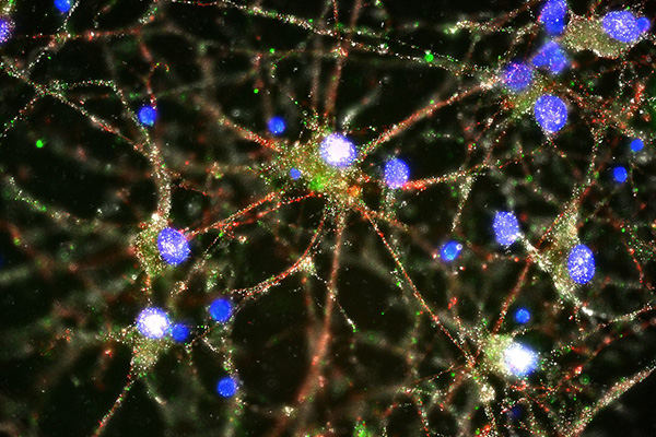
Researchers have successfully demonstrated how it is possible to interface graphene – a two-dimensional form of carbon – with neurons, or nerve cells, while maintaining the integrity of these vital cells. The work may be used to build graphene-based electrodes that can safely be implanted in the brain, offering promise for the restoration of sensory functions for amputee or paralyzed patients, or for individuals with motor disorders such as epilepsy or Parkinson’s disease.

The research, published in the journal ACS Nano, was an interdisciplinary collaboration coordinated by the University of Trieste in Italy and the Cambridge Graphene Centre.

Previously, other groups had shown that it is possible to use treated graphene to interact with neurons. However the signal to noise ratio from this interface was very low. By developing methods of working with untreated graphene, the researchers retained the material’s electrical conductivity, making it a significantly better electrode.

“For the first time we interfaced graphene to neurons directly,” said Professor Laura Ballerini of the University of Trieste in Italy. “We then tested the ability of neurons to generate electrical signals known to represent brain activities, and found that the neurons retained their neuronal signaling properties unaltered. This is the first functional study of neuronal synaptic activity using uncoated graphene based materials.”

Our understanding of the brain has increased to such a degree that by interfacing directly between the brain and the outside world we can now harness and control some of its functions. For instance, by measuring the brain’s electrical impulses, sensory functions can be recovered. This can be used to control robotic arms for amputee patients or any number of basic processes for paralyzed patients – from speech to movement of objects in the world around them. Alternatively, by interfering with these electrical impulses, motor disorders (such as epilepsy or Parkinson’s) can start to be controlled.

Scientists have made this possible by developing electrodes that can be placed deep within the brain. These electrodes connect directly to neurons and transmit their electrical signals away from the body, allowing their meaning to be decoded.

However, the interface between neurons and electrodes has often been problematic: not only do the electrodes need to be highly sensitive to electrical impulses, but they need to be stable in the body without altering the tissue they measure.

Too often the modern electrodes used for this interface (based on tungsten or silicon) suffer from partial or complete loss of signal over time. This is often caused by the formation of scar tissue from the electrode insertion, which prevents the electrode from moving with the natural movements of the brain due to its rigid nature.

Graphene has been shown to be a promising material to solve these problems, because of its excellent conductivity, flexibility, biocompatibility and stability within the body.

Based on experiments conducted in rat brain cell cultures, the researchers found that untreated graphene electrodes interfaced well with neurons. By studying the neurons with electron microscopy and immunofluorescence the researchers found that they remained healthy, transmitting normal electric impulses and, importantly, none of the adverse reactions which lead to the damaging scar tissue were seen.

According to the researchers, this is the first step towards using pristine graphene-based materials as an electrode for a neuro-interface. In future, the researchers will investigate how different forms of graphene, from multiple layers to monolayers, are able to affect neurons, and whether tuning the material properties of graphene might alter the synapses and neuronal excitability in new and unique ways. “Hopefully this will pave the way for better deep brain implants to both harness and control the brain, with higher sensitivity and fewer unwanted side effects,” said Ballerini.

“We are currently involved in frontline research in graphene technology towards biomedical applications,” said Professor Maurizio Prato from the University of Trieste. “In this scenario, the development and translation in neurology of graphene-based high-performance biodevices requires the exploration of the interactions between graphene nano- and micro-sheets with the sophisticated signalling machinery of nerve cells. Our work is only a first step in that direction.”

“These initial results show how we are just at the tip of the iceberg when it comes to the potential of graphene and related materials in bio-applications and medicine,” said Professor Andrea Ferrari, Director of the Cambridge Graphene Centre. “The expertise developed at the Cambridge Graphene Centre allows us to produce large quantities of pristine material in solution, and this study proves the compatibility of our process with neuro-interfaces.”

The research was funded by the Graphene Flagship, a European initiative which promotes a collaborative approach to research with an aim of helping to translate graphene out of the academic laboratory, through local industry and into society.

Source: University of Cambridge


Remembering to Remember Supported by Two Distinct Brain Processes

http://www.biosciencetechnology.com/news/2013/08/remembering-remember-supported-two-distinct-brain-processes

To investigate how prospective memory is processed in the brain, psychological scientist Mark McDaniel of Washington University in St. Louis and colleagues had participants lie in an fMRI scanner and asked them to press one of two buttons to indicate whether a word that popped up on a screen was a member of a designated category.  In addition to this ongoing activity, participants were asked to try to remember to press a third button whenever a special target popped up. The task was designed to tap into participants’ prospective memory, or their ability to remember to take certain actions in response to specific future events.

When McDaniel and colleagues analyzed the fMRI data, they observed that two distinct brain activation patterns emerged when participants made the correct button press for a special target.

When the special target was not relevant to the ongoing activity—such as a syllable like “tor”—participants seemed to rely on top-down brain processes supported by the prefrontal cortex. In order to answer correctly when the special syllable flashed up on the screen, the participants had to sustain their attention and monitor for the special syllable throughout the entire task. In the grocery bag scenario, this would be like remembering to bring the grocery bags by constantly reminding yourself that you can’t forget them.

When the special target was integral to the ongoing activity—such as a whole word, like “table”—participants recruited a different set of brain regions, and they didn’t show sustained activation in these regions. The findings suggest that remembering what to do when the special target was a whole word didn’t require the same type of top-down monitoring. Instead, the target word seemed to act as an environmental cue that prompted participants to make the appropriate response—like reminding yourself to bring the grocery bags by leaving them near the front door.

“These findings suggest that people could make use of several different strategies to accomplish prospective memory tasks,” says McDaniel.

McDaniel and colleagues are continuing their research on prospective memory, examining how this phenomenon might change with age.

Co-authors on this research include Pamela LaMontagne, Michael Scullin, Todd Braver of Washington University in St. Louis; and Stefanie Beck of Technische Universität Dresden.

This research was funded by the National Institute on Aging, the Washington University Institute of Clinical and Translation Sciences, the National Center for Advancing Translational Sciences, and the German Science Foundation.

Read Full Post »

Immunopathogenesis Advances in Diabetes and Lymphomas

Larry H Bernstein, MD, FCAP, Curator

LPBI


 Science team says they’ve taken another step toward a potential cure for diabetes

Wednesday, January 27, 2016 | By John Carroll
Building on years of work on developing new insulin-producing cells that could one day control glucose levels and cure diabetes, a group of investigators led by scientists at MIT and Boston Children’s Hospital say they’ve developed a promising new gel capsule that protected the cells from an immune system assault.

Dr. Jose Oberholzer, a professor of bioengineering at the University of Illinois at Chicago, tested a variety of chemically modified alginate hydrogel spheres to see which ones would be best at protecting the islet cells created from human stem cells.

The team concluded that 1.5-millimeter spheres of triazole-thiomorphine dioxide (TMTD) alginate were best at protecting the cells and allowing insulin to seep out without spurring an errant immune system attack or the development of scar tissue–two key threats to making this work in humans.

They maintained healthy glucose levels in the rodents for 174 days, the equivalent to decades for humans.

“While this is a very promising step towards an eventual cure for diabetes, a lot more testing is needed to ensure that the islet cells don’t de-differentiate back toward their stem-cell states or become cancerous,” said Oberholzer.

Millions of diabetics have effectively controlled the chronic disease with existing therapies, but there’s still a huge unmet medical need to consider. While diabetes companies like Novo ($NVO) like to cite the fact that a third of diabetics have the disease under control, a third are on meds but don’t control it well and a third haven’t been diagnosed. An actual cure for the disease, which has been growing by leaps and bounds all over the world, would be revolutionary.

Their study was published in Nature Medicine.

– here’s the release
– get the journal abstract


Long-term glycemic control using polymer-encapsulated human stem cell–derived beta cells in immune-competent mice

Arturo J Vegas, Omid Veiseh, Mads Gürtler,…, Robert Langer & Daniel G Anderson

Nature Medicine (2016)   http://dx.doi.org:/10.1038/nm.4030

The transplantation of glucose-responsive, insulin-producing cells offers the potential for restoring glycemic control in individuals with diabetes1. Pancreas transplantation and the infusion of cadaveric islets are currently implemented clinically2, but these approaches are limited by the adverse effects of immunosuppressive therapy over the lifetime of the recipient and the limited supply of donor tissue3. The latter concern may be addressed by recently described glucose-responsive mature beta cells that are derived from human embryonic stem cells (referred to as SC-β cells), which may represent an unlimited source of human cells for pancreas replacement therapy4. Strategies to address the immunosuppression concerns include immunoisolation of insulin-producing cells with porous biomaterials that function as an immune barrier5, 6. However, clinical implementation has been challenging because of host immune responses to the implant materials7. Here we report the first long-term glycemic correction of a diabetic, immunocompetent animal model using human SC-β cells. SC-β cells were encapsulated with alginate derivatives capable of mitigating foreign-body responses in vivo and implanted into the intraperitoneal space of C57BL/6J mice treated with streptozotocin, which is an animal model for chemically induced type 1 diabetes. These implants induced glycemic correction without any immunosuppression until their removal at 174 d after implantation. Human C-peptide concentrations and in vivo glucose responsiveness demonstrated therapeutically relevant glycemic control. Implants retrieved after 174 d contained viable insulin-producing cells.

Subject terms: Regenerative medicine  Type 1 diabetes

Figure 1: SC-β cells encapsulated with TMTD alginate sustain normoglycemia in STZ-treated immune-competent C57BL/6J mice.close

(a) Top, schematic representation of the last three stages of differentiation of human embryonic stem cells to SC-β cells. Stage 4 cells (pancreatic progenitors 2) co-express pancreatic and duodenal homeobox 1 (PDX-1) and NK6 homeobox 1…


Potential Cure for Diabetes Discovered  
http://www.rdmag.com/news/2016/01/potential-cure-diabetes-discovered   01/27/2016

Two new scientific papers published on Monday demonstrated tools that could result in potential therapies for patients diagnosed with type 1 diabetes, a condition in which the immune system limits the production of insulin, typically in adolescents.  See —

Bubble Technique Could Create Type 1 Diabetes Therapy

http://www.dddmag.com/news/2016/01/bubble-technique-could-create-type-1-diabetes-therapy

Two new scientific papers published on Monday demonstrated tools that could result in potential therapies for patients diagnosed with type 1 diabetes, a condition in which the immune system limits the production of insulin, typically in adolescents.

Previous treatments for this disease have involved injecting beta cells from dead donors into patients to help their pancreas generate healthy-insulin cells, writes STAT. However, this method has resulted in the immune system targeting these new cells as “foreign” so transplant recipients have had to take immune-suppressing medications for the rest of their lives.

The first paper published in the journal Nature Biotechnology explained how scientists analyzed a seaweed extract called alginate to gauge its effectiveness in supporting the flow of sugar and insulin between cells and the body. An estimated 774 variations were tested in mice and monkeys in which results indicated only a handful could reduce the body’s response to foreign invaders, explains STAT.

The other paper in the journal Nature Medicine detailed a process where scientists developed small capsules infused with alginate and embryonic stem cells. A six-month observation period revealed this “protective bubble” technique “began to produce insulin in response to blood glucose levels” after transplantation in mice subjects with a condition similar to type 1 diabetes, reports Gizmodo.

Essentially, this cured the mice of their diabetes, and the beta cells worked as well as the body’s own cells, according to the researchers. Human trials could still be a few years away, but this experiment could yield a safer alternative to insulin injections.


Combinatorial hydrogel library enables identification of materials that mitigate the foreign body response in primates

Arturo J Vegas, Omid Veiseh, Joshua C Doloff, et al.

Nature Biotechnology (2016)    http://dx.doi.org:/10.1038/nbt.3462

The foreign body response is an immune-mediated reaction that can lead to the failure of implanted medical devices and discomfort for the recipient1, 2, 3, 4, 5, 6. There is a critical need for biomaterials that overcome this key challenge in the development of medical devices. Here we use a combinatorial approach for covalent chemical modification to generate a large library of variants of one of the most widely used hydrogel biomaterials, alginate. We evaluated the materials in vivo and identified three triazole-containing analogs that substantially reduce foreign body reactions in both rodents and, for at least 6 months, in non-human primates. The distribution of the triazole modification creates a unique hydrogel surface that inhibits recognition by macrophages and fibrous deposition. In addition to the utility of the compounds reported here, our approach may enable the discovery of other materials that mitigate the foreign body response.


Video 1: Intravital imaging of 300 μm SLG20 microcapsules.

Video 2: Intravital imaging of 300 μm Z2-Y12 microcapsules.

Video 3: NHP Laparoscopic procedure for the retrieval of Z2-Y12 spheres.


Clinical Focus on Follicular Lymphoma: CAR T-Cells Active in Relapsed Blood Cancers

MedPage Today

CAR T-Cells Active in Relapsed Blood Cancers

Complete responses in half of patients

by Charles Bankhead

Patients with relapsed and refractory B-cell malignancies have responded to treatment with modified T-cells added to conventional chemotherapy, data from an ongoing Swedish study showed.

Six of the first 11 evaluable patients achieved complete responses with increasing doses of chimeric antigen receptor (CAR)-modified T-cells that target the CD19 antigen, although two subsequently relapsed.

Five of the six responding patients received preconditioning chemotherapy the day before CAR T-cell infusion, in addition to chemotherapy administered up to 90 days before T-cell infusion to reduce tumor-cell burden. The remaining five patients received only the earlier chemotherapy, according to a presentation at the inaugural International Cancer Immunotherapy Conference in New York City.

“The complete responses in lymphoma patients despite the fact that they received only low doses of preconditioning compared with other published data surprised us,” Angelica Loskog, PhD, of Uppsala University in Sweden, said in a statement. “The strategy of both providing tumor-reductive chemotherapy for weeks prior to CAR T-cell infusion combined with preconditioning just before CAR T-cell infusion seems to offer promise.

CAR T-cells have demonstrated activity in a variety of studies involving patients with B-cell malignancies. Much of the work has focused on patients with leukemia, including trials in the U.S. B-cell lymphomas have proven more difficult to treat with CAR T-cells because the diseases are associated with higher concentration of immunosuppressive cells that can inhibit CAR T-cell activity, said Loskog. Moreover, blood-vessel abnormalities and accumulation of fibrotic tissue can hinder tumor penetration by therapeutic T-cells.

Each laboratory has its own process for modifying T-cells. Loskog and colleagues in Sweden and at Baylor College of Medicine in Houston have developed third-generation CAR T-cells that contain signaling domains for CD28 and 4-1BB, which act as co-stimulatory molecules. In preclinical models, third-generation CAR T-cells have demonstrated increased activation and proliferation in response to antigen challenge. Additionally, they have chosen to experiment with tumor burden-reducing chemotherapy, a preconditioning chemotherapy to counter the higher immunosuppressive cell count in lymphoma patients.

Loskog reported details of an ongoing phase I/IIa clinical trial involving patients with relapsed or refractory CD19-positive B-cell malignancies. Altogether, investigators have treated 12 patients with increasing doses (2 x 10⁷ to 2 x 10⁸ cells/m2) of CAR T-cells. One patient (with mixed follicular/Burkitt lymphoma) has yet to be evaluated for response. The remaining 11 included three patients with diffuse large B-cell lymphoma (DLBCL), one with follicular lymphoma transformed to DLBCL, two with chronic lymphocytic leukemia, two with mantle cell lymphoma, and three with acute lymphoblastic leukemia.

All of the patients with lymphoma received standard tumor cell-reducing chemotherapy, beginning 3 to 90 days before administration of CAR T-cells. Beginning with the sixth patient in the cohort, patients also received preconditioning chemotherapy (cyclophosphamide/fludarabine) 1 to 2 days before T-cell infusion to reduce the number and activity of immunosuppressive cells.

Cytokine release syndrome is a common effect of CAR T-cell therapy and occurred in several patients treated. In general, the syndrome has been manageable and has not interfered with treatment or response to the modified T-cells.

On the basis of the data produced thus far, the investigators have proceeded with patient evaluation and enrollment. They have already begun cell production for the next patient that will be treated with autologous CAR T-cells.

Although laboratories have their own cell production techniques, the treatment strategy has broad applicability to the treatment of B-cell malignancies, said Loskog.

“The results using different CARs and different techniques for manufacturing them is very similar in the clinic, in terms of initial complete response,” she told MedPage Today. “By using 4-1BB as a co-stimulator in the CAR intracellular region, it seems possible to achieve long-term complete responses in some patients. However, preconditioning of the patients with chemotherapy to reduce the regulatory immune cells seems crucial for effect.”

In an effort to manage the effect of patients’ immunosuppressive cells, the investigators have begun studying each the immune profile before and after treatment. Preliminary results suggest that the population of immunosuppressive cells increases over time, which has the potential to interfere with CAR T-cell responses.

“Especially for lymphoma, it may be crucial to deplete such cells prior to CAR infusion,” said Loskog. “It may even be necessary with supportive treatment for some time after CAR T-cell infusion. A supportive treatment needs to specifically regulate the suppressive cells while sparing the effect of CARs.”

The immunotherapy conference is jointly sponsored by the American Association for Cancer Research, the Cancer Research Institute, the Association for Cancer Immunotherapy, and the European Academy of Tumor Immunology.


PET-CT Best for FL Response Assessment

PET-CT associated with better progression-free and overall survival rates in follicular lymphoma.

Kay Jackson

PET-CT (PET) rather than contrast-enhanced CT scanning should be considered the new gold standard for response assessment after first-line rituximab therapy for high-tumor burden follicular lymphoma (FL), a pooled analysis of a central review in three multicenter studies indicated.

Read Full Post »

Mindful Discoveries

Larry H. Bernstein, MD, FCAP, Curator

LPBI

Schizophrenia and the Synapse

Genetic evidence suggests that overactive synaptic pruning drives development of schizophrenia.

By Ruth Williams | January 27, 2016 … more follows)

http://www.the-scientist.com/?articles.view/articleNo/45189/title/Schizophrenia-and-the-Synapse/

3.2.4

3.2.4   Mindful Discoveries, Volume 2 (Volume Two: Latest in Genomics Methodologies for Therapeutics: Gene Editing, NGS and BioInformatics, Simulations and the Genome Ontology), Part 2: CRISPR for Gene Editing and DNA Repair

http://www.the-scientist.com/images/News/January2016/Schizophrenia.jpg

C4 (green) at synapses of human neurons

Compared to the brains of healthy individuals, those of people with schizophrenia have higher expression of a gene called C4, according to a paper published inNature today (January 27). The gene encodes an immune protein that moonlights in the brain as an eradicator of unwanted neural connections (synapses). The findings, which suggest increased synaptic pruning is a feature of the disease, are a direct extension of genome-wide association studies (GWASs) that pointed to the major histocompatibility (MHC) locus as a key region associated with schizophrenia risk.

“The MHC [locus] is the first and the strongest genetic association for schizophrenia, but many people have said this finding is not useful,” said psychiatric geneticist Patrick Sullivan of the University of North Carolina School of Medicine who was not involved in the study. “The value of [the present study is] to show that not only is it useful, but it opens up new and extremely interesting ideas about the biology and therapeutics of schizophrenia.”

Schizophrenia has a strong genetic component—it runs in families—yet, because of the complex nature of the condition, no specific genes or mutations have been identified. The pathological processes driving the disease remain a mystery.

Researchers have turned to GWASs in the hope of finding specific genetic variations associated with schizophrenia, but even these have not provided clear candidates.

“There are some instances where genome-wide association will literally hit one base [in the DNA],” explained Sullivan. While a 2014 schizophrenia GWAS highlighted the MHC locus on chromosome 6 as a strong risk area, the association spanned hundreds of possible genes and did not reveal specific nucleotide changes. In short, any hope of pinpointing the MHC association was going to be “really challenging,” said geneticist Steve McCarroll of Harvard who led the new study.

Nevertheless, McCarroll and colleagues zeroed in on the particular region of the MHC with the highest GWAS score—the C4 gene—and set about examining how the area’s structural architecture varied in patients and healthy people.

The C4gene can exist in multiple copies (from one to four) on each copy of chromosome 6, and has four different forms: C4A-short, C4B-short, C4A-long, and C4B-long. The researchers first examined the “structural alleles” of the C4 locus—that is, the combinations and copy numbers of the different C4 forms—in healthy individuals. They then examined how these structural alleles related to expression of both C4Aand C4B messenger RNAs (mRNAs) in postmortem brain tissues.From this the researchers had a clear picture of how the architecture of the C4 locus affected expression ofC4A and C4B. Next, they compared DNA from roughly 30,000 schizophrenia patients with that from 35,000 healthy controls, and a correlation emerged: the alleles most strongly associated with schizophrenia were also those that were associated with the highest C4A expression. Measuring C4A mRNA levels in the brains of 35 schizophrenia patients and 70 controls then revealed that, on average, C4A levels in the patients’ brains were 1.4-fold higher.C4 is an immune system “complement” factor—a small secreted protein that assists immune cells in the targeting and removal of pathogens. The discovery of C4’s association to schizophrenia, said McCarroll, “would have seemed random and puzzling if it wasn’t for work . . . showing that other complement components regulate brain wiring.” Indeed, complement protein C3 locates at synapses that are going to be eliminated in the brain, explained McCarroll, “and C4 was known to interact with C3 . . . so we thought well, actually, this might make sense.”McCarroll’s team went on to perform studies in mice that revealed C4 is necessary for C3 to be deposited at synapses. They also showed that the more copies of the C4 gene present in a mouse, the more the animal’s neurons were pruned.Synaptic pruning is a normal part of development and is thought to reflect the process of learning, where the brain strengthens some connections and eradicates others. Interestingly, the brains of deceased schizophrenia patients exhibit reduced neuron density. The new results, therefore, “make a lot of sense,” said Cardiff University’s Andrew Pocklington who did not participate in the work. They also make sense “in terms of the time period when synaptic pruning is occurring, which sort of overlaps with the period of onset for schizophrenia: around adolescence and early adulthood,” he added.

“[C4] has not been on anybody’s radar for having anything to do with schizophrenia, and now it is and there’s a whole bunch of really neat stuff that could happen,” said Sullivan. For one, he suggested, “this molecule could be something that is amenable to therapeutics.”

A. Sekar et al., “Schizophrenia risk from complexvariation of complement component 4,”Nature,   http://dx.doi.com:/10.1038/nature16549, 2016.     

Tags schizophrenia, neuroscience, gwas, genetics & genomics, disease/medicine and cell & molecular biology

Schizophrenia: From genetics to physiology at last

Ryan S. Dhindsa, & David B. Goldstein

Nature (2016)  http://dx.doi.org://10.1038/nature16874

The identification of a set of genetic variations that are strongly associated with the risk of developing schizophrenia provides insights into the neurobiology of this destructive disease.

http://www.nytimes.com/2016/01/28/health/schizophrenia-cause-synaptic-pruning-brain-psychiatry.html

Genetic study provides first-ever insight into biological origin of schizophrenia

Suspect gene may trigger runaway synaptic pruning during adolescence — NIH-funded study

NIH/NATIONAL INSTITUTE OF MENTAL HEALTH

IMAGE

http://media.eurekalert.org/multimedia_prod/pub/web/107629_web.jpg

The site in Chromosome 6 harboring the gene C4 towers far above other risk-associated areas on schizophrenia’s genomic “skyline,” marking its strongest known genetic influence. The new study is the first to explain how specific gene versions work biologically to confer schizophrenia risk.  CREDIT  Psychiatric Genomics Consortium

Versions of a gene linked to schizophrenia may trigger runaway pruning of the teenage brain’s still-maturing communications infrastructure, NIH-funded researchers have discovered. People with the illness show fewer such connections between neurons, or synapses. The gene switched on more in people with the suspect versions, who faced a higher risk of developing the disorder, characterized by hallucinations, delusions and impaired thinking and emotions.

“Normally, pruning gets rid of excess connections we no longer need, streamlining our brain for optimal performance, but too much pruning can impair mental function,” explained Thomas Lehner, Ph.D., director of the Office of Genomics Research Coordination of the NIH’s National Institute of Mental Health (NIMH), which co-funded the study along with the Stanley Center for Psychiatric Research at the Broad Institute and other NIH components. “It could help explain schizophrenia’s delayed age-of-onset of symptoms in late adolescence/early adulthood and shrinkage of the brain’s working tissue. Interventions that put the brakes on this pruning process-gone-awry could prove transformative.”

The gene, called C4 (complement component 4), sits in by far the tallest tower on schizophrenia’s genomic “skyline” (see graph below) of more than 100 chromosomal sites harboring known genetic risk for the disorder. Affecting about 1 percent of the population, schizophrenia is known to be as much as 90 percent heritable, yet discovering how specific genes work to confer risk has proven elusive, until now.

A team of scientists led by Steve McCarroll, Ph.D., of the Broad Institute and Harvard Medical School, Boston, leveraged the statistical power conferred by analyzing the genomes of 65,000 people, 700 postmortem brains, and the precision of mouse genetic engineering to discover the secrets of schizophrenia’s strongest known genetic risk. C4’s role represents the most compelling evidence, to date, linking specific gene versions to a biological process that could cause at least some cases of the illness.

“Since schizophrenia was first described over a century ago, its underlying biology has been a black box, in part because it has been virtually impossible to model the disorder in cells or animals,” said McCarroll. “The human genome is providing a powerful new way in to this disease. Understanding these genetic effects on risk is a way of prying open that block box, peering inside and starting to see actual biological mechanisms.”

McCarroll’s team, including Harvard colleagues Beth Stevens, Ph.D., Michael Carroll, Ph.D., and Aswin Sekar, report on their findings online Jan. 27, 2016 in the journal Nature.

A swath of chromosome 6 encompassing several genes known to be involved in immune function emerged as the strongest signal associated with schizophrenia risk in genome-wide analyses by the NIMH-funded Psychiatric Genomics Consortium over the past several years. Yet conventional genetics failed to turn up any specific gene versions there linked to schizophrenia.

To discover how the immune-related site confers risk for the mental disorder, McCarroll’s team mounted a search for “cryptic genetic influences” that might generate “unconventional signals.” C4, a gene with known roles in immunity, emerged as a prime suspect because it is unusually variable across individuals. It is not unusual for people to have different numbers of copies of the gene and distinct DNA sequences that result in the gene working differently.

The researchers dug deeply into the complexities of how such structural variation relates to the gene’s level of expression and how that, in turn, might relate to schizophrenia. They discovered structurally distinct versions that affect expression of two main forms of the gene in the brain. The more a version resulted in expression of one of the forms, called C4A, the more it was associated with schizophrenia. The more a person had the suspect versions, the more C4 switched on and the higher their risk of developing schizophrenia. Moreover, in the human brain, the C4 protein turned out to be most prevalent in the cellular machinery that supports connections between neurons.

Adapting mouse molecular genetics techniques for studying synaptic pruning and C4’s role in immune function, the researchers also discovered a previously unknown role for C4 in brain development. During critical periods of postnatal brain maturation, C4 tags a synapse for pruning by depositing a sister protein in it called C3. Again, the more C4 got switched on, the more synapses got eliminated.

In humans, such streamlining/pruning occurs as the brain develops to full maturity in the late teens/early adulthood – conspicuously corresponding to the age-of-onset of schizophrenia symptoms.

Future treatments designed to suppress excessive levels of pruning by counteracting runaway C4 in at risk individuals might nip in the bud a process that could otherwise develop into psychotic illness, suggest the researchers. And thanks to the head start gained in understanding the role of such complement proteins in immune function, such agents are already in development, they note.

“This study marks a crucial turning point in the fight against mental illness. It changes the game,” added acting NIMH director Bruce Cuthbert, Ph.D. “Thanks to this genetic breakthrough, we can finally see the potential for clinical tests, early detection, new treatments and even prevention.”

###

VIDEO: Opening Schizophrenia’s Black Box https://youtu.be/s0y4equOTLg

Reference: Sekar A, Biala AR, de Rivera H, Davis A, Hammond TR, Kamitaki N, Tooley K Presumey J Baum M, Van Doren V, Genovese G, Rose SA, Handsaker RE, Schizophrenia Working Group of the Psychiatric Genomics Consortium, Daly MJ, Carroll MC, Stevens B, McCarroll SA. Schizophrenia risk from complex variation of complement component 4.Nature. Jan 27, 2016. DOI: 10.1038/nature16549.

Schizophrenia risk from complex variation of complement component 4

Aswin Sekar, Allison R. Bialas, Heather de Rivera, Avery Davis, Timothy R. Hammond, …., Michael C. Carroll, Beth Stevens & Steven A. McCarroll

Nature(2016)   http://dx.doi.org:/10.1038/nature16549

Schizophrenia is a heritable brain illness with unknown pathogenic mechanisms. Schizophrenia’s strongest genetic association at a population level involves variation in the major histocompatibility complex (MHC) locus, but the genes and molecular mechanisms accounting for this have been challenging to identify. Here we show that this association arises in part from many structurally diverse alleles of the complement component 4 (C4) genes. We found that these alleles generated widely varying levels of C4A and C4B expression in the brain, with each common C4 allele associating with schizophrenia in proportion to its tendency to generate greater expression of C4A. Human C4 protein localized to neuronal synapses, dendrites, axons, and cell bodies. In mice, C4 mediated synapse elimination during postnatal development. These results implicate excessive complement activity in the development of schizophrenia and may help explain the reduced numbers of synapses in the brains of individuals with schizophrenia.

Figure 1: Structural variation of the complement component 4 (C4) gene.

http://www.nature.com/nature/journal/vaop/ncurrent/carousel/nature16549-f1.jpg

a, Location of the C4 genes within the major histocompatibility complex (MHC) locus on human chromosome 6. b, Human C4 exists as two paralogous genes (isotypes), C4A and C4B; the encoded proteins are distinguished at a key site

http://www.nature.com/nature/journal/vaop/ncurrent/carousel/nature16549-f3.jpg

http://www.nature.com/nature/journal/vaop/ncurrent/carousel/nature16549-sf8.jpg

Gene Study Points Toward Therapies for Common Brain Disorders

University of Edinburgh    http://www.dddmag.com/news/2016/01/gene-study-points-toward-therapies-common-brain-disorders

Scientists have pinpointed the cells that are likely to trigger common brain disorders, including Alzheimer’s disease, Multiple Sclerosis and intellectual disabilities.

It is the first time researchers have been able to identify the particular cell types that malfunction in a wide range of brain diseases.

Scientists say the findings offer a roadmap for the development of new therapies to target the conditions.

The researchers from the University of Edinburgh’s Centre for Clinical Brain Sciences used advanced gene analysis techniques to investigate which genes were switched on in specific types of brain cells.

They then compared this information with genes that are known to be linked to each of the most common brain conditions — Alzheimer’s disease, anxiety disorders, autism, intellectual disability, multiple sclerosis, schizophrenia and epilepsy.

Their findings reveal that for some conditions, the support cells rather than the neurons that transmit messages in the brain are most likely to be the first affected.

Alzheimer’s disease, for example, is characterised by damage to the neurons. Previous efforts to treat the condition have focused on trying to repair this damage.

The study found that a different cell type — called microglial cells — are responsible for triggering Alzheimer’s and that damage to the neurons is a secondary symptom of disease progression.

Researchers say that developing medicines that target microglial cells could offer hope for treating the illness.

The approach could also be used to find new treatment targets for other diseases that have a genetic basis, the researchers say.

Dr Nathan Skene, who carried out the study with Professor Seth Grant, said: “The brain is the most complex organ made up from a tangle of many cell types and sorting out which of these cells go wrong in disease is of critical importance to developing new medicines.”

Professor Seth Grant said: “We are in the midst of scientific revolution where advanced molecular methods are disentangling the Gordian Knot of the brain and completely unexpected new pathways to solving diseases are emerging. There is a pressing need to exploit the remarkable insights from the study.”

Quantitative multimodal multiparametric imaging in Alzheimer’s disease

Qian Zhao, Xueqi Chen, Yun Zhou      Brain Informatics  http://link.springer.com/article/10.1007/s40708-015-0028-9

Alzheimer’s disease (AD) is a progressive neurodegenerative disorder, causing changes in memory, thinking, and other dysfunction of brain functions. More and more people are suffering from the disease. Early neuroimaging techniques of AD are needed to develop. This review provides a preliminary summary of the various neuroimaging techniques that have been explored for in vivo imaging of AD. Recent advances in magnetic resonance (MR) techniques, such as functional MR imaging (fMRI) and diffusion MRI, give opportunities to display not only anatomy and atrophy of the medial temporal lobe, but also at microstructural alterations or perfusion disturbance within the AD lesions. Positron emission tomography (PET) imaging has become the subject of intense research for the diagnosis and facilitation of drug development of AD in both animal models and human trials due to its non-invasive and translational characteristic. Fluorodeoxyglucose (FDG) PET and amyloid PET are applied in clinics and research departments. Amyloid beta (Aβ) imaging using PET has been recognized as one of the most important methods for the early diagnosis of AD, and numerous candidate compounds have been tested for Aβ imaging. Besides in vivo imaging method, a lot of ex vivo modalities are being used in the AD researches. Multiphoton laser scanning microscopy, neuroimaging of metals, and several metal bioimaging methods are also mentioned here. More and more multimodality and multiparametric neuroimaging techniques should improve our understanding of brain function and open new insights into the pathophysiology of AD. We expect exciting results will emerge from new neuroimaging applications that will provide scientific and medical benefits.

Keywords –   Alzheimer’s disease Neuroimaging PET MRI Amyloid beta Multimodal

Alzheimer’s disease (AD) is a progressive neurodegenerative disorder that gradually destroys brain cells, causing changes in memory, thinking, and other dysfunction of brain functions [1]. AD is considered to a prolonged preclinical stage where neuropathological changes precede the clinical symptoms [2]. An estimation of 35 million people worldwide is living with this disease. If effective treatments are not discovered in a timely fashion, the number of AD cases is anticipated to rise to 113 million by 2050 [3].

Amyloid beta (Aβ) and tau are two of the major biomarkers of AD, and have important and different roles in association with the progression of AD pathophysiology. Jack et al. established hypothetical models of the major biomarkers of AD. By renewing and modifying the models, they found that the two major proteinopathies underlying AD biomarker changes, Aβ and tau, may be initiated independently in late onset AD where they hypothesize that an incident Aβ pathophysiology can accelerate an antecedent limbic and brainstem tauopathy [4]. MRI technique was used in the article, which revealed that the level of Aβ load was associated with a shorter time-to-progression of AD [5]. This warrants an urgent need to develop early neuroimaging techniques of AD neuropathology that can detect and predict the disease before the onset of dementia, monitor therapeutic efficacy in halting and slowing down progression in the earlier stage of the disease.

There have been various reports on the imaging assessments of AD. Some measurements reflect the pathology of AD directly, including positron emission tomography (PET) amyloid imaging and cerebrospinal fluid (CSF) beta-amyloid 42 (Aβ42), while others reflect neuronal injury associated with AD indirectly, including CSF tau (total and phosphorylated tau), fluorodeoxy-d-glucose (FDG)-PET, and MRI. AD Neuroimaging Initiative (ADNI) has been to establish the optimal panel of clinical assessments, MRI and PET imaging measures, as well as other biomarkers from blood and CSF, to inform clinical trial design for AD therapeutic development. At the same time, it has been highly productive in generating a wealth of data for elucidating disease mechanisms occurring during early stages of preclinical and prodromal AD [6].

Single neuroimaging often reflects limit information of AD. As a result, multimodal neuroimaging is widely used in neuroscience researches, as it overcomes the limitations of individual modalities. Multimodal multiparametric imaging mean the combination of different imaging techniques, such as PET, MRI, simultaneously or separately. The multimodal multiparametric imaging enables the visualization and quantitative analysis of the alterations in brain structure and function, such as PET/CT, and PET/MRI. [7]. In this review article, we summarize and discuss the main applications, findings, perspectives as well as advantages and challenges of different neuroimaging in AD, especially MRI and PET imaging.

2 Magnetic resonance imaging

MRI demonstrates specific volume loss or cortical atrophy patterns with disease progression in AD patients [8–10]. There are several MRI techniques and analysis methods used in clinical and scientific research of AD. Recent advances in MR techniques, such as functional MRI (fMRI) and diffusion MRI, depict not only anatomy and atrophy of the medial temporal lobe (MTL), but also microstructural alterations or perfusion disturbance within this region.

2.1 Functional MRI

Because of the cognitive reserve (CR), the relationship between severity of AD patients’ brain damage and corresponding clinical symptoms is not always paralleled [11, 12]. Recently, resting-state fMRI (RS-fMRI) is popular for its ability to map brain functional connectivity non-invasively [13]. By using RS-fMRI, Bozzali et al. reported that the CR played a role in modulating the effect of AD pathology on default mode network functional connectivity, which account for the variable clinical symptoms of AD [14]. Moreover, AD patients with higher educated experience were able to recruit compensatory neural mechanisms, which can be measured using RS-fMRI. Arterial spin-labeled (ASL) MRI is another functional brain imaging modality, which measures cerebral blood flow (CBF) by magnetically labeled arterial blood water following through the carotid and vertebral arteries as an endogenous contrast medium. Several studies have concluded the characteristics of CBF changes in AD patients using ASL-MRI [15–17].

At some point in time, sufficient brain damage accumulates to result in cognitive symptoms and impairment. Mild cognitive impairment (MCI) is a condition in which subjects are usually only mildly impaired in memory with relative preservation of other cognitive domains and functional activities and do not meet the criteria for dementia [18], or as the prodromal state AD [19]. MCI patients are at a higher risk of developing AD and up to 15 % convert to AD per year [18]. Binnewijzend et al. have reported the pseudocontinuous ASL could distinguish both MCI and AD from healthy controls, and be used in the early diagnosis of AD [20]. In their continuous study, they used quantitative whole brain pseudocontinuous ASL to compare regional CBF (rCBF) distribution patterns in different types of dementia, and concluded that ASL-MRI could be a non-invasive and easily accessible alternative to FDG-PET imaging in the assessment of CBF of AD patients [21].

2.2 Structure MRI

Structural MRI (sMRI) has already been a reliable imaging method in the clinical diagnosis of AD, characterized as gray matter reduction and ventricular enlargement in standard T1-weighted sequences [9]. Locus coeruleus (LC) and substantia nigra (SN) degeneration was seen in AD. By using new quantitative calculating method, Chen et al. presented a new quantitative neuromelanin MRI approach for simultaneous measurement of locus LC and SN of brainstem in living human subjects [22]. The approach they used demonstrated advantages in image acquisition, pre-processing, and quantitative analysis. Numerous transgenic animal models of amyloidosis are available, which can manipulate a lot of neuropathological features of AD progression from the deposition of β-amyloid [23]. Braakman et al. demonstrated the dynamics of amyloid plaque formation and development in a serial MRI study in a transgenic mouse model [24]. Increased iron accumulation in gray matter is frequently observed in AD. Because of the paramagnetic nature of iron, MRI shows nice potential in the investigating iron levels in AD [25]. Quantitative MRI was shown high sensitivity and specificity in mapping cerebral iron deposition, and helped in the research on AD diagnosis [26].

The imaging patterns are always associated with the pathologic changes, such as specific protein markers. Spencer et al. manifested the relationship between quantitative T1 and T2 relaxation time changes and three immunohistochemical markers: β-amyloid, neuron-specific nuclear protein (a marker of neuronal cell load), and myelin basic protein (a marker of myelin load) in AD transgenic mice [27].

High-field MRI has been successfully applied to imaging plaques in transgenic mice for over a decade without contrast agents [24, 28–30]. Sillerud et al. devised a method using blood–brain barrier penetrating, amyloid-targeted, superparamagnetic iron oxide nanoparticles (SPIONs) for better imaging of amyloid plaque [31]. Then, they successfully used this SPION-MRI to assess the drug efficacy on the 3D distribution of Aβ plaques in transgenic AD mouse [32].

2.3 Diffusion MRI

Diffusion-weighted imaging (DWI) is a sensitive tool that allows quantifying of physiologic alterations in water diffusion, which result from microscopic structural changes.

Diffusion tensor imaging (DTI) is a well-established and commonly employed diffusion MRI technique in clinical and research on neuroimaging studies, which is based on a Gaussian model of diffusion processes [33]. In general, AD is associated with widespread reduced fractional anisotropy (FA) and increased mean diffusivity (MD) in several regions, most prominently in the frontal and temporal lobes, and along the cingulum, corpus callosum, uncinate fasciculus, superior longitudinal fasciculus, and MTL-associated tracts than healthy controls [34–37]. Acosta-Cabronero et al. reported increased axial diffusivity and MD in the splenium, which were the earliest abnormalities in AD [38]. FA and radial diffusivity (DR) differences in the corpus callosum, cingulum, and fornix were found to separate individuals with MCI who converted to AD from non-converters [39]. DTI was also found to be a better predictor of AD-specific MTL atrophy when compared to CSF biomarkers [40]. These findings suggested the potential clinical utility of DTI as early biomarkers of AD and its progression. However, an increase in MD and DR and a decrease in FA with advancing age in selective brain regions have been previously reported [41, 42]. Diffusion MRI can be also used in the classifying of various stages of AD. Multimodal classification method, which combined fMRI and DTI, separated more MCI from healthy controls than single approaches [43].

In recent years, tau has emerged as a potential target for therapeutic intervention. Tau plays a critical role in the neurodegenerative process forming neurofibrillary tangles, which is a major hallmark of AD and correlates with clinical disease progression. Wells et al. applied multiparametric MRI, containing high-resolution structure MRI (sMRI), a novel chemical exchange saturation transfer (CEST) MRI, DTI, and ASL, and glucose CEST to measure changes of tau pathology in AD transgenic mouse [44].

Besides DWI MRI, perfusion-weighted imaging (PWI) is another advanced MR technique, which could measure the cerebral hemodynamics at the capillary level. Zimny et al. evaluated the correlation of MTL with both DWI and PWI in AD and MCI patients [45].

3 Positron emission tomography

PET is a specific imaging technique applying in researches of brain function and neurochemistry of small animals, medium-sized animals, and human subjects [46–48]. As a particular brain imaging technique, PET imaging has become the subject of intense research for the diagnosis and facilitation of drug development of AD in both animal models and human trials due to its non-invasive and translational characteristic. PET with various radiotracers is considered as a standard non-invasive quantitative imaging technique to measure CBF, glucose metabolism, and β-amyloid and tau deposition.

3.1 FDG-PET

To date, 18F-FDG is one of the best and widely used neuroimaging tracers of PET, which employed for research and clinical assessment of AD [49]. Typical lower FDG metabolism was shown in the precuneus, posterior cingulate, and temporal and parietal cortex with progression to whole brain reductions with increasing disease progress in AD brains [50, 51]. FDG-PET imaging reflects the cerebral glucose metabolism, neuronal injury, which provides indirect evidence on cognitive function and progression that cannot be provided by amyloid PET imaging.

Schraml et al. [52] identified a significant association between hypometabolic convergence index and phenotypes using ADNI data. Some researchers also used 18F-FDG-PET to analyze genetic information with multiple biomarkers to classify AD status, predicting cognitive decline or MCI to AD conversion [53–55]. Trzepacz et al. [56] reported multimodal AD neuroimaging study, using MRI, 11C-PiB PET, and 18F-FDG-PET imaging to predict MCI conversion to AD along with APOE genotype. Zhang et al. [57] compared the genetic modality single-nucleotide polymorphism (SNP) with sMRI, 18F-FDG-PET, and CSF biomarkers, which were used to differentiate healthy control, MCI, and AD. They found FDG-PET is the best modality in terms of accuracy.

3.2 Amyloid beta PET

Aβ, the primary constituent of senile plaques, and tau tangles are hypothesized to play a primary role in the pathogenesis of AD, but it is still hard to identify the fundamental mechanisms [58–60]. Aβ plaque in brain is one of the pathological hallmarks of AD [61,62]. Accumulation of Aβ peptide in the cerebral cortex is considered one cause of dementia in AD [63]. Numerous studies have involved in vivo PET imaging assessing cortical β-amyloid burden [64–66].

Aβ imaging using PET has been recognized as one of the most important methods for the early diagnosis of AD [67]. Numerous candidate compounds have been tested for Aβ imaging, such as 11C-PiB [68], 18F-FDDNP [69], 11C-SB-13 [70], 18F-BAY94-9172 [71], 18F-AV-45 [72], 18F-flutemetamol [73, 74], 11C-AZD2184 [75], and 18F-ADZ4694 [76], 11C-BF227 and 18F-FACT [77].

Several amyloid PET studies examined genotypes, phenotypes, or gene–gene interactions. Ramanan et al. [78] reported the GWAS results with 18F-AV-45 reflecting the cerebral amyloid metabolism in AD for the first time. Swaminathan et al. [79] revealed the association between plasma Aβ from peripheral blood and cortical amyloid deposition on 11C-PiB. Hohman et al. [80] reported the relationship between SNPs involved in amyloid and tau pathophysiology with 18F-AV-45 PET.

Among the PET tracers, 11C-PiB, which has a high affinity for fibrillar Aβ, is a reliable biomarker of underlying AD pathology [68, 81]. It shows cortical uptake well paralleled with AD pathology [82, 83], has recently been approved for use by the Food and Drug Administration (FDA, April 2012) and the European Medicines Agency (January 2013). 18F-GE-067 (flutemetamol) and 18F-BAY94-9172 (florbetaben) have also been approved by the US FDA in the last 2 years [84, 85].

18F-Florbetapir (also known as 18F-AV-45) exhibits high affinity specific binding to amyloid plaques. 18F-AV-45 labels Aβ plaques in sections from patients with pathologically confirmed AD [72].

It was reported in several research groups that 18F-AV-45 PET imaging showed a reliability of both qualitative and quantitative assessments in AD patients, and Aβ+ increased with diagnostic category (healthy control < MCI < AD) [82, 86, 87]. Johnson et al. used 18F-AV-45 PET imaging to evaluate the amyloid deposition in both MCI and AD patients qualitatively and quantitatively, and found that amyloid burden increased with diagnostic category (MCI < AD), age, and APOEε4 carrier status [88]. Payoux et al. reported the equivocal amyloid PET scans using 18F-AV-45 associated with a specific pattern of clinical signs in a large population of non-demented older adults more than 70 years old [89].

More and more researchers consider combination and comparison of multiple PET tracers targeting amyloid plaque imaging together. Bruck et al. compared the prognostic ability of 11C-PiB PET, 18F-FDG-PET, and quantitative hippocampal volumes measured with MR imaging in predicting MCI to AD conversion. They found that the FDG-PET and 11C-PiB PET imaging are better in predicting MCI to AD conversion [90]. Hatashita et al. used 11C-PiB and FDG-PET imaging to identify MCI due to AD, 11C-PiB showed a higher sensitivity of 96.6 %, and FDG-PET added diagnostic value in predicting AD over a short period [91].

Besides, new Aβ imaging agents were radiosynthesized. Yousefi et al. radiosynthesized a new Aβ imaging agent 18F-FIBT, and compared the three different Aβ-targeted radiopharmaceuticals for PET imaging, including 18F-FIBT, 18F-florbetaben, and 11C-PiB [92]. 11C-AZD2184 is another new PET tracer developed for amyloid senile plaque imaging, and the kinetic behavior of 11C-AZD2184 is suitable for quantitative analysis and can be used in clinical examination without input function [75,93, 94].

4 Multimodality imaging: PET/MRI

Several diagnostic techniques, including MRI and PET, are employed for the diagnosis and monitoring of AD [95]. Multimodal imaging could provide more information in the formation and key molecular event of AD than single method. It drives the progression of neuroimaging research due to the recognition of the clinical benefits of multimodal data [96], and the better access to hybrid devices, such as PET/MRI [97].

Maier et al. evaluated the dynamics of 11C-PiB PET, 15O-H2O-PET, and ASL-MRI in transgenic AD mice and concluded that the AD-related decline of rCBF was caused by the cerebral Aβ angiopathy [98]. Edison et al. systematically compared 11C-PiB PET and MRI in AD, MCI patients, and controls. They thought that 11C-PiB PET was adequate for clinical diagnostic purpose, while MRI remained more appropriate for clinical research [99]. Zhou et al. investigated the interactions between multimodal PET/MRI in elder patients with MCI, AD, and healthy controls, and confirmed the invaluable application of amyloid PET and MRI in early diagnosis of AD [100]. Kim et al. reported that Aβ-weighted cortical thickness, which incorporates data from both MRI and amyloid PET imaging, is a consistent and objective imaging biomarker in AD [101].

5 Other imaging modalities

Multiphoton non-linear optical microscope imaging systems using ultrafast lasers have powerful advantages such as label-free detection, deep penetration of thick samples, high sensitivity, subcellular spatial resolution, 3D optical sectioning, chemical specificity, and minimum sample destruction [102, 103]. Coherent anti-Stokes–Raman scattering (CARS), two-photon excited fluorescence (TPEF), and second-harmonic generation (SHG) microscopy are the most widely used biomedical imaging techniques [104–106].

Quantitative electroencephalographic and neuropsychological investigation of an alternative measure of frontal lobe executive functions: the Figure Trail Making Test

 Paul S. Foster, Valeria Drago, Brad J. Ferguson, Patti Kelly Harrison,David W. Harrison 

Brain Informatis    http://dx.doi.org:/10.1007/s40708-015-0025-z    http://link.springer.com/article/10.1007/s40708-015-0025-z/fulltext.html

The most frequently used measures of executive functioning are either sensitive to left frontal lobe functioning or bilateral frontal functioning. Relatively little is known about right frontal lobe contributions to executive functioning given the paucity of measures sensitive to right frontal functioning. The present investigation reports the development and initial validation of a new measure designed to be sensitive to right frontal lobe functioning, the Figure Trail Making Test (FTMT). The FTMT, the classic Trial Making Test, and the Ruff Figural Fluency Test (RFFT) were administered to 42 right-handed men. The results indicated a significant relationship between the FTMT and both the TMT and the RFFT. Performance on the FTMT was also related to high beta EEG over the right frontal lobe. Thus, the FTMT appears to be an equivalent measure of executive functioning that may be sensitive to right frontal lobe functioning. Applications for use in frontotemporal dementia, Alzheimer’s disease, and other patient populations are discussed.

Keywords – Frontal lobes, Executive functioning, Trail making test, Sequencing, Behavioral speed, Designs, Nonverbal, Neuropsychological assessment, Regulatory control, Effortful control

A recent survey indicated that the vast majority of neuropsychologists frequently assess executive functioning as part of their neuropsychological evaluations [1]. Surveys of neuropsychologists have indicated that the Trail Making Test (TMT), Controlled Oral Word Association Test (COWAT), Wisconsin Card Sorting Test (WCST), and the Stroop Color-Word Test (SCWT) are among the most commonly used instruments [1,2]. Further, the Rabin et al. [1] survey indicated that these same tests are among the most frequently used by neuropsychologists when specifically assessing executive or frontal lobe functioning. The frequent use of the TMT, WCST, and the SCWT, as well as the assumption that they are measures of executive functioning, led Demakis (2003–2004) to conduct a series of meta-analyses to determine the sensitivity of these test to detect frontal lobe dysfunction, particularly lateralized frontal lobe dysfunction. The findings indicated that the SCWT and Part A of the TMT [3], as well as the WCST [4], were all sensitive to frontal lobe dysfunction. However, only the SCWT differentiated between left and right frontal lobe dysfunction, with the worst performance among those with left frontal lobe dysfunction [3].

The finding of the Demakis [4] meta-analysis, that the WCST was not sensitive to lateralized frontal lobe dysfunction, is not surprising given the equivocal findings that have been reported. Whereas performance on the WCST is sensitive to frontal lobe dysfunction [5, 6], demonstration of lateralized frontal dysfunction has been quite problematic. Unilateral left or right dorsolateral frontal dysfunction has been associated with impaired performance on the WCST [6]. Fallgatter and Strik [7] found bilateral frontal lobe activation during performance of the WCST. However, other imaging studies have found right lateralized frontal lobe activation [8] and left lateralized frontal activation [9] in response to performance on the WCST. Further, left frontal lobe alpha power is negatively correlated with performance on the WCST [10]. Finally, patients with left frontal lobe tumors exhibit more impaired performance on the WCST than those with right frontal tumors [11].

Unlike the data for the WCST, more consistent findings have been reported regarding lateralized frontal lobe functioning for the other commonly used measures of executive functioning. For instance, as with the Demakis [3] study, many investigations have found the SCWT to be sensitive to left frontal lobe functioning, although the precise localization within the left frontal lobe has varied. Impaired performance on the SCWT results from left frontal lesions [12] and specifically from lesions localized to the left dorsolateral frontal lobe [13, 14], though bilateral frontal lesions have also yielded impaired performance [13, 14]. Further, studies using neuroimaging to investigate the neural basis of performance on the SCWT have indicated involvement of the left anterior cingulated cortex [15], left lateral prefrontal cortex [16], left inferior precentral sulcus [17], and the left dorsolateral frontal lobe [18].

Wide agreement exists among investigations of the frontal lateralization of verbal or lexical fluency to confrontation. Specifically, patients with left frontal lobe lesions are known to exhibit impaired performance on lexical fluency to confrontation tasks, relative to either patients with right frontal lesions [12, 19, 20] or controls [21]. A recent meta-analysis also indicated that the largest deficits in performance on measures of lexical fluency are associated with left frontal lobe lesions [22]. Troster et al. [23] found that, relative to patients with right pallidotomy, patients with left pallidotomy exhibited more impaired lexical fluency. Several neuroimaging investigations have further supported the role of the left frontal lobe in lexical fluency tasks [15, 24–27]. Performance on lexical fluency tasks also varies as a function of lateral frontal lobe asymmetry, as assessed by electroencephalography [28].

The Trail Making Test is certainly among the most widely used tests [1] and perhaps the most widely researched. Various norms exist for the TMT (see [29]), with Tombaugh [30] providing the most recent comprehensive set of normative data. Different methods of analyzing and interpreting the data have also been proposed and used, including error analysis [13, 14, 31–33], subtraction scores [13, 14, 34], and ratio scores [13, 14, 35].

Several different language versions of the test have been developed and reported, including Arabic [36], Chinese [37, 38], Greek [39], and Hebrew [40]. Numerous alternative versions of the TMT have been developed to address perceived shortcomings of the original TMT. For instance, the Symbol Trail Making Test [41] was developed to reduce the cultural confounds associated with the use of the Arabic numeral system and English alphabet in the original TMT. The Color Trails Test (CTT; [42]) was also developed to control for cultural confounds, although mixed results have been reported regarding whether the CTT is indeed analogous to the TMT [43–45]. A version of the TMT for preschool children, the TRAILS-P, has also been reported [46].

Additionally, the Comprehensive Trail Making Test [47] was developed to control for perceived psychometric shortcomings of the original TMT (for a review see [48] and the Oral Trail Making Test (OTMT; [49]) was developed to reduce confounds associated with motor speed and visual search abilities, with research supporting the OTMT as an equivalent measure [50, 51]. Alternate forms of the TMT have also been developed to permit successive administrations [32, 52] and to assess the relative contributions of the requisite cognitive skills [53].

Delis et al. [54] stated that the continued development of new instrumentation for improving diagnosis and treatment is a critical undertaking in all health-related fields. Further, in their view, the field of neuropsychology has recognized the importance of continually striving to develop new clinical measures. Delis and colleagues developed the extensive Delis-Kaplan Executive Functioning System (D-KEFS; [55]) in the spirit of advancing the instrumentation of neuropsychology. The D-KEFS includes a Trail Making Test consisting of five separate conditions. The Number-Letter Switching condition involves a sequencing procedure similar to that of the classic TMT. The other four conditions are designed to assess the component processes involved in completing the Number-Letter Switching condition so that a precise analysis of the nature of any underlying dysfunction may be accomplished. Specifically, these additional components include Visual Scanning, Number Sequencing, Letter Sequencing, and Motor Speed.

Given that the TMT comprises numbers and letters and is a measure of executive functioning, it may preferentially involve the left frontal lobe. Although the literature is somewhat controversial, neuropsychological and neuroimaging studies seem to provide support for the sensitivity of the TMT to detect left frontal dysfunction [56]. Recent clinically oriented studies investigating frontal lobe involvement of the TMT using transcranial magnetic stimulation (TMS) and near-infrared spectroscopy (NIRS) also support this localization [57]. Performance on Part B of the TMT improved following repetitive TMS applied to the left dorsolateral frontal lobe [57].

With 9–13-year-old boys performing TMT Part B, Weber et al. [58] found a left lateralized increase in the prefrontal cortex in deoxygenated hemoglobin, an indicator of increased oxygen consumption. Moll et al. [59] demonstrated increased activation specific to the prefrontal cortex, especially the left prefrontal region, in healthy controls performing Part B of the TMT. Foster et al. [60] found a significant positive correlation between performance on Part A of the TMT and low beta (13–21 Hz) magnitude (μV) at the left lateral frontal lobe, but not at the right lateral frontal lobe. Finally, Stuss et al. [13, 14] found that patients with left dorsolateral frontal dysfunction evidenced more errors than patients with lesions in other areas of the frontal lobes and those patients with left frontal lesions were the slowest to complete the test.

Taken together, the possibility exists that the aforementioned tests are largely associated with left frontal lobe activity and the TMT, in particular, provides information concerning mental processing speed as well as cognitive flexibility and set-shifting. While some studies have found that deficits in visuomotor set-shifting are specific to the frontal lobe damage [61], others investigators have reported such impairment in patients with posterior brain lesions and widespread cerebral dysfunctions, including cerebellar damage [62] and Alzheimer disease [63]. Thus, it remains unclear whether impairments in visuomotor set-shifting are specific to frontal lobe dysfunction or whether they are non-specific and can result from more posterior or widespread brain dysfunction.

Compared to the collective knowledge we have regarding the cognitive roles of the left frontal lobe, relatively little is known about right frontal lobe contributions to executive functioning. This is likely a result of the dearth of tests that are associated with right frontal activity. The Ruff Figural Fluency Test (RFFT; [64]) is among the few standardized tests of right frontal lobe functioning and was listed as the 14th most commonly used instrument to assess executive functioning in the Rabin et al. [1] survey. The RFFT is known to be sensitive to right frontal lobe functioning [65, 66]; see also [67] pp. 297–298), as is a measure based on the RFFT [19].

The present investigation, with the same intent and spirit as that reported by Delis et al. [54], sought to develop and initially validate a measure of right frontal lobe functioning in an effort to attain a greater understanding of right frontal contributions to executive functioning and to advance the instrumentation of neuropsychology. To meet this objective, a version of the Trail Making Test comprising figures, as opposed to numbers and letters, was developed. The TMT was used as a model for the new test, referred to as the Figure Trail Making Test (FTMT), due to the high frequency of use, the volume of research conducted, and the ease of administration of the TMT. Given that the TMT and the FTMT are both measuring executive functioning, we felt that a moderate correlation would exist between these two measures. Specifically, we hypothesized that performance on the FTMT would be positively correlated with performance on the TMT, in terms of the total time required to complete each part of the tests, an additive and subtractive score, and a ratio score. The total time required to complete each part of the FTMT was also hypothesized to be negatively correlated with the total number of unique designs produced on the RFFT and positively correlated with the number of perseverative errors committed on the RFFT and the perseverative error ratio. We also sought to determine whether the TMT and the FTMT were measuring different constructs by conducting a factor analysis, anticipating that the two tests would load on separate factors.

Additionally, we sought to obtain neurophysiological evidence that the FTMT is sensitive to right frontal lobe functioning. Specifically, we used quantitative electroencephalography (QEEG) to measure electrical activity over the left and right frontal lobes. A previous investigation we conducted found that performance on Part A of the TMT was related to left frontal lobe (F7) low beta magnitude [60]. For the present investigation, we predicted that significant negative correlations would exist between performance on Parts A and B of the TMT and both low and high beta magnitude at the F7 electrode site. We further predicted that significant negative correlations would exist between performance on Parts C and D of the FTMT and both low and high beta magnitude at the F8 electrode site.

3 Discussion

The need for additional measures of executive functions and especially instruments which may provide implications relevant to cerebral laterality is clear. There remains especially a void for neuropsychological instruments using a TMT format, which may provide information pertaining to the functional integrity of the right frontal region. Consistent with the hypotheses forwarded, significant correlations were found between performance on the TMT and the FTMT, in terms of the raw time required to complete each respective part of the tests as well as the additive and subtraction scores. The fact that the ratio scores were not significantly correlated is not surprising given that research has generally indicated a lack of clinical utility for this score [13, 14, 35]. Given the present findings, the TMT and the FTMT appear to be equivalent measures of executive functioning. Further, the present findings not only suggest that the FTMT may be a measure of executive functioning but also extend the realm of executive functioning to the sequencing and set-shifting of nonverbal stimuli.

However, the finding of significant correlations between the TMT and the FTMT represents somewhat of a caveat in that the TMT has been found to be sensitive to left frontal lobe functioning [13, 14, 57, 59]. This would seem to suggest the possibility that the FTMT is also sensitive to left frontal lobe functioning. The possibility that FTMT is related to left frontal lobe functioning is tempered, though, by the fact that the many of the hypothesized correlations between performance on the RFFT and the FTMT were also significant. Performance on the RFFT is related to right frontal lobe functioning [65,66]. Thus, the significant correlations between the RFFT and the FTMT suggest that the FTMT may also be sensitive to right frontal lobe functioning. Additionally, it should also be noted that the TMT was not significantly correlated with performance on the RFFT, with the exception of the significant correlation between performance on the TMT Part A and the total number of unique designs produced on the RFFT. Taken together, the results suggest that the FTMT may be a measure of right frontal executive functioning.

Additional support for the sensitivity of the FTMT to right frontal lobe functioning is provided by the finding of a significant negative correlation between performance on Part D of the FTMT and high beta magnitude. We have previously used QEEG to provide neurophysiological validation of the RFFT [65] and the Rey Auditory Verbal Learning Test [70] and the present findings provide further support for the use of QEEG in validating neuropsychological tests. The lack of significant correlations between the TMT and either low or high beta magnitude may be related to a restricted range of scores on the TMT. As a whole, performance on the FTMT was more variable than performance on the TMT and this relatively restricted range for the TMT may have impacted the obtained correlations. Given the present findings, together with those of the Foster et al. [65, 70] investigations, further support is also provided for the use of EEG in establishing neurophysiological validation for neuropsychological tests.

The results from the factor analysis provide support for the contention that the FMT may be a measure of right frontal lobe activity and also provide initial discriminant validity for the FTMT. Specifically, Parts C and D of the FTMT were found to load on the same factor as the number of designs generated on the RFFT, although the time required to complete Part A of the TMT is also included. Additionally, the number of errors committed on Parts C and D of the FTMT comprises a single factor, separate from either the TMT or the RFFT. Although these results support the FTMT as a measure of nonverbal executive functioning, it would be helpful to conduct an additional factor analysis including additional measures of right frontal functioning, and perhaps other measures of right hemisphere functioning as marker variables.

We sought to develop a measure sensitive to right frontal lobe functioning due to the paucity of such tests and the potentially important uses that right frontal lobe tests may have clinically. Tests of right frontal lobe functioning may, for instance, be useful in identifying and distinguishing left versus right frontotemporal dementia (FTD). Research has indicated that FTD is associated with cerebral atrophy at the right dorsolateral frontal and left premotor cortices [71]. Fukui and Kertesz [72] found right frontal lobe volume reduction in FTD relative to Alzheimer’s disease and progressive nonfluent aphasia. Some have suggested that FTD should not be considered as a unitary disorder and that neuropsychological testing may aid in differentially diagnosing left versus right FTD [73].

Whereas right FTD has been associated with more errors and perseverative responses on the Wisconsin Card Sorting Test (WCST), left FTD has been associated with significantly worse performance on the Boston Naming Test (BNT) and the Stroop Color-Word test [73]. Razani et al. [74] also distinguished between left and right FTD in finding that left FTD performed worse on the BNT and the right FTD patients performed worse on the WCST. However, as noted earlier, the WCST has been associated with left frontal activity [9], right frontal activation [8], and bilateral frontal activation [7]. Further, patients with left frontal tumors perform worse than those with right frontal tumors [11].

Patients with FTD that predominantly involves the right frontotemporal region have behavioral and emotional abnormalities and those with predominantly left frontotemporal region damage have a loss of lexical semantic knowledge. Patients, in whom neural degeneration begins on the left side, often present to the clinicians at an early stage of the disease due to the presence of language abnormalities, but maintain their emotion processing abilities, being preserved the right anterior temporal lobe. However, as this disease advances, the disease may progress to the right frontotemporal regions. Tests sensitive to right frontal lobe functioning may be useful tools to identify in advance the course of the disease, providing immediate and specific treatments and informing the caregivers on the possible prospective frame of the disease.

A potentially more important use of tests sensitive to right frontal lobe functioning, though, may be in predicting dementia patients that will develop significant and disruptive behavioral deficits. Research has found that approximately 92 % of right-sided FTD patients exhibit socially undesirable behaviors as their initial symptom, as compared to only 11 % of left-sided FTD patients [75]. Behavioral deficits in FTD are associated with gray matter loss at the dorsomedial frontal region, particularly on the right [76].

Alzheimer’s disease (AD) is also often associated with significant behavioral disturbances. Even AD patients with mild dementia are noted to exhibit behavioral deficits such as delusions, hallucinations, agitation, dysphoria, anxiety, apathy, and irritability [77]. Indeed, Shimabukuro et al. [77] found that regardless of dementia severity, over half of all AD patients exhibited apathy, delusions, irritability, dysphoria, and anxiety. Delusions in AD patients are associated with relative right frontal hypoperfusion as indicated by SPECT imaging [78, 79]. Further, positron emission tomography (PET) has indicated that AD patients exhibiting delusions exhibit hypometabolism at the right superior dorsolateral frontal and right inferior frontal pole [80].

Although research clearly implicates right frontal lobe dysfunction in the expression of behavioral deficits, data from neuropsychological testing are not as clear. Negative symptoms in patients with AD and FTD have been related to measures of nonverbal and verbal executive functioning as well as verbal memory [81]. Positive symptoms, in contrast, were related to constructional skills and attention. However, Staff et al. [78] failed to dissociate patients with delusions from those without delusions based on neuropsychological test performance, despite significant differences existing in right frontal and limbic functioning as revealed by functional imaging. The inclusion of other measures of right frontal lobe functioning may result in improved neuropsychological differentiation of dementia patients with and without significant behavioral disturbances. Further, it may be possible to predict early in the disease process those patients that will ultimately develop behavioral disturbances with improved measures of right frontal functioning. Predicting those that may develop behavioral problems will permit earlier treatment and will provide the family with more time to prepare for the potential emergence of such difficulties. Certainly, future research needs to be conducted that incorporates measures of right and left frontal lobe functioning in regression analyses to determine the plausibility of such prediction.

Tests sensitive to right frontal lobe functioning may also be useful in identifying more subtle right frontal lobe dysfunction and the cognitive and behavioral changes that follow. The right frontal lobe mediates language melody or prosody and forms a cohesive discourse, interprets abstract communication in spoken and written languages, and interprets the inferred relationships involved in communications. Subtle difficulties in interpreting abstract meaning in communication, comprehending metaphors, and even understanding jokes that are often seen in right frontal lobe stroke patients may not be detected by the family and may also be under diagnosed by clinicians [82]. Further, patients with right frontal lobe lesions are generally more euphoric and unconcerned, often minimizing their symptoms [82] or denying the illness, which may delay referral to a clinician and diagnosis.

Attention deficit hyperactivity disorder (ADHD) is a neurological disease characterized by motor inhibition deficit, problems with cognitive flexibility, social disruption, and emotional disinhibition [83, 84]. Functional MRI studies reveal reduced right prefrontal activation during “frontal tasks,” such as go/no go [85], Stroop [86], and attention task performance [87]. The right frontal lobe deficit hypothesis is further supported by structural studies [88, 89]. Tests of right frontal lobe functioning may be useful in further characterizing the nature of this deficit and in specifying the likely hemispheric locus of dysfunction.

To summarize, we feel that right frontal lobe functioning has been relatively neglected in neuropsychological assessment and that many uses for such tests exist. Our intent was to develop a test purportedly sensitive to right frontal functioning that would be easy and quick to administer in a clinical setting. However, we are certainly not meaning to assert that our FTMT would be applicable in all the aforementioned conditions. Additional research should be conducted to determine the precise clinical utility of the FTMT.

Further validation of the FTMT should also be undertaken. Establishing convergent validation may involve correlating tests measuring the same domain, such as executive functioning. This was initially accomplished in the present investigation through the significant correlations between the TMT and the FTMT. Additionally, convergent validation may also involve correlating tests that purportedly measure the same region of the brain. This was also initially accomplished in the present investigation through the significant correlations between the FTMT and the RFFT. However, additional convergent validation certainly needs to be obtained, as well as validation using patient populations and neurophysiological validation.

We are currently collecting data that hopefully will provide neurophysiological validation of the FTMT. Certainly, though, it is hoped that the present investigation will not only stimulate further research seeking to validate the FTMT and provide more comprehensive normative data, but also stimulate research investigating whether the FTMT or other measures of right frontal lobe functioning may be used to predict patients that will develop behavioral disturbances.

World’s Greatest Literature Reveals Multifractals, Cascades of Consciousness

http://www.scientificcomputing.com/news/2016/01/worlds-greatest-literature-reveals-multifractals-cascades-consciousness

http://www.scientificcomputing.com/sites/scientificcomputing.com/files/Worlds_Greatest_Literature_Reveals_Multifractals_Cascades_of_Consciousness_440.jpg

Multifractal analysis of Finnegan’s Wake by James Joyce. The ideal shape of the graph is virtually indistinguishable from the results for purely mathematical multifractals. The horizontal axis represents the degree of singularity, and the vertical axis shows the spectrum of singularity. Courtesy of IFJ PAN

Arthur Conan Doyle, Charles Dickens, James Joyce, William Shakespeare and JRR Tolkien. Regardless of the language they were working in, some of the world’s greatest writers appear to be, in some respects, constructing fractals. Statistical analysis, however, revealed something even more intriguing. The composition of works from within a particular genre was characterized by the exceptional dynamics of a cascading (avalanche) narrative structure. This type of narrative turns out to be multifractal. That is, fractals of fractals are created.

As far as many bookworms are concerned, advanced equations and graphs are the last things which would hold their interest, but there’s no escape from the math. Physicists from the Institute of Nuclear Physics of the Polish Academy of Sciences (IFJ) in Cracow, Poland, performed a detailed statistical analysis of more than one hundred famous works of world literature, written in several languages and representing various literary genres. The books, tested for revealing correlations in variations of sentence length, proved to be governed by the dynamics of a cascade. This means that the construction of these books is, in fact, a fractal. In the case of several works, their mathematical complexity proved to be exceptional, comparable to the structure of complex mathematical objects considered to be multifractal. Interestingly, in the analyzed pool of all the works, one genre turned out to be exceptionally multifractal in nature.

Fractals are self-similar mathematical objects: when we begin to expand one fragment or another, what eventually emerges is a structure that resembles the original object. Typical fractals, especially those widely known as the Sierpinski triangle and the Mandelbrot set, are monofractals, meaning that the pace of enlargement in any place of a fractal is the same, linear: if they at some point were rescaled x number of times to reveal a structure similar to the original, the same increase in another place would also reveal a similar structure.

Multifractals are more highly advanced mathematical structures: fractals of fractals. They arise from fractals ‘interwoven’ with each other in an appropriate manner and in appropriate proportions. Multifractals are not simply the sum of fractals and cannot be divided to return back to their original components, because the way they weave is fractal in nature. The result is that, in order to see a structure similar to the original, different portions of a multifractal need to expand at different rates. A multifractal is, therefore, non-linear in nature.

“Analyses on multiple scales, carried out using fractals, allow us to neatly grasp information on correlations among data at various levels of complexity of tested systems. As a result, they point to the hierarchical organization of phenomena and structures found in nature. So, we can expect natural language, which represents a major evolutionary leap of the natural world, to show such correlations as well. Their existence in literary works, however, had not yet been convincingly documented. Meanwhile, it turned out that, when you look at these works from the proper perspective, these correlations appear to be not only common, but in some works they take on a particularly sophisticated mathematical complexity,” says Professor Stanislaw Drozdz, IFJ PAN, Cracow University of Technology.

The study involved 113 literary works written in English, French, German, Italian, Polish, Russian and Spanish by such famous figures as Honore de Balzac, Arthur Conan Doyle, Julio Cortazar, Charles Dickens, Fyodor Dostoevsky, Alexandre Dumas, Umberto Eco, George Elliot, Victor Hugo, James Joyce, Thomas Mann, Marcel Proust, Wladyslaw Reymont, William Shakespeare, Henryk Sienkiewicz, JRR Tolkien, Leo Tolstoy and Virginia Woolf, among others. The selected works were no less than 5,000 sentences long, in order to ensure statistical reliability.

To convert the texts to numerical sequences, sentence length was measured by the number of words (an alternative method of counting characters in the sentence turned out to have no major impact on the conclusions). The dependences were then searched for in the data — beginning with the simplest, i.e. linear. This is the posited question: if a sentence of a given length is x times longer than the sentences of different lengths, is the same aspect ratio preserved when looking at sentences respectively longer or shorter?

“All of the examined works showed self-similarity in terms of organization of the lengths of sentences. Some were more expressive — here The Ambassadors by Henry James stood out — while others to far less of an extreme, as in the case of the French seventeenth-century romance Artamene ou le Grand Cyrus. However, correlations were evident and, therefore, these texts were the construction of a fractal,” comments Dr. Pawel Oswiecimka (IFJ PAN), who also noted that fractality of a literary text will, in practice, never be as perfect as in the world of mathematics. It is possible to magnify mathematical fractals up to infinity, while the number of sentences in each book is finite and, at a certain stage of scaling, there will always be a cut-off in the form of the end of the dataset.

Things took a particularly interesting turn when physicists from IFJ PAN began tracking non-linear dependence, which in most of the studied works was present to a slight or moderate degree. However, more than a dozen works revealed a very clear multifractal structure, and almost all of these proved to be representative of one genre, that of stream of consciousness. The only exception was the Bible, specifically the Old Testament, which has, so far, never been associated with this literary genre.

“The absolute record in terms of multifractality turned out to be Finnegan’s Wakeby James Joyce. The results of our analysis of this text are virtually indistinguishable from ideal, purely mathematical multifractals,” says Drozdz.

The most multifractal works also included A Heartbreaking Work of Staggering Genius by Dave Eggers, Rayuela by Julio Cortazar, The US Trilogy by John Dos Passos, The Waves by Virginia Woolf, 2666 by Roberto Bolano, and Joyce’sUlysses. At the same time, a lot of works usually regarded as stream of consciousness turned out to show little correlation to multifractality, as it was hardly noticeable in books such as Atlas Shrugged by Ayn Rand and A la recherche du temps perdu by Marcel Proust.

“It is not entirely clear whether stream of consciousness writing actually reveals the deeper qualities of our consciousness, or rather the imagination of the writers. It is hardly surprising that ascribing a work to a particular genre is, for whatever reason, sometimes subjective. We see, moreover, the possibility of an interesting application of our methodology: it may someday help in a more objective assignment of books to one genre or another,” notes Drozdz.

Multifractal analyses of literary texts carried out by the IFJ PAN have been published in Information Sciences, the journal of computer science. The publication has undergone rigorous verification: given the interdisciplinary nature of the subject, editors immediately appointed up to six reviewers.

Citation: “Quantifying origin and character of long-range correlations in narrative texts” S. Drożdż, P. Oświęcimka, A. Kulig, J. Kwapień, K. Bazarnik, I. Grabska-Gradzińska, J. Rybicki, M. Stanuszek; Information Sciences, vol. 331, 32–44, 20 February 2016; DOI: 10.1016/j.ins.2015.10.023

New Quantum Approach to Big Data could make Impossibly Complex Problems Solvable

David L. Chandler, MIT

http://www.scientificcomputing.com/news/2016/01/new-quantum-approach-big-data-could-make-impossibly-complex-problems-solvable

http://www.scientificcomputing.com/sites/scientificcomputing.com/files/New_Quantum_Approach_to_Big_Data_could_make_Impossibly_Complex_Problems_Solvable_440.jpg

This diagram demonstrates the simplified results that can be obtained by using quantum analysis on enormous, complex sets of data. Shown here are the connections between different regions of the brain in a control subject (left) and a subject under the influence of the psychedelic compound psilocybin (right). This demonstrates a dramatic increase in connectivity, which explains some of the drug’s effects (such as “hearing” colors or “seeing” smells). Such an analysis, involving billions of brain cells, would be too complex for conventional techniques, but could be handled easily by the new quantum approach, the researchers say. Courtesy of the researchers

From gene mapping to space exploration, humanity continues to generate ever-larger sets of data — far more information than people can actually process, manage or understand.

Machine learning systems can help researchers deal with this ever-growing flood of information. Some of the most powerful of these analytical tools are based on a strange branch of geometry called topology, which deals with properties that stay the same even when something is bent and stretched every which way.

Such topological systems are especially useful for analyzing the connections in complex networks, such as the internal wiring of the brain, the U.S. power grid, or the global interconnections of the Internet. But even with the most powerful modern supercomputers, such problems remain daunting and impractical to solve. Now, a new approach that would use quantum computers to streamline these problems has been developed by researchers at MIT, the University of Waterloo, and the University of Southern California.

The team describes their theoretical proposal this week in the journal Nature Communications. Seth Lloyd, the paper’s lead author and the Nam P. Suh Professor of Mechanical Engineering, explains that algebraic topology is key to the new method. This approach, he says, helps to reduce the impact of the inevitable distortions that arise every time someone collects data about the real world.

In a topological description, basic features of the data (How many holes does it have? How are the different parts connected?) are considered the same no matter how much they are stretched, compressed, or distorted. Lloyd explains that it is often these fundamental topological attributes “that are important in trying to reconstruct the underlying patterns in the real world that the data are supposed to represent.”

It doesn’t matter what kind of dataset is being analyzed, he says. The topological approach to looking for connections and holes “works whether it’s an actual physical hole, or the data represents a logical argument and there’s a hole in the argument. This will find both kinds of holes.”

Using conventional computers, that approach is too demanding for all but the simplest situations. Topological analysis “represents a crucial way of getting at the significant features of the data, but it’s computationally very expensive,” Lloyd says. “This is where quantum mechanics kicks in.” The new quantum-based approach, he says, could exponentially speed up such calculations.

Lloyd offers an example to illustrate that potential speedup: If you have a dataset with 300 points, a conventional approach to analyzing all the topological features in that system would require “a computer the size of the universe,” he says. That is, it would take 2300 (two to the 300th power) processing units — approximately the number of all the particles in the universe. In other words, the problem is simply not solvable in that way.

“That’s where our algorithm kicks in,” he says. Solving the same problem with the new system, using a quantum computer, would require just 300 quantum bits — and a device this size may be achieved in the next few years, according to Lloyd.

“Our algorithm shows that you don’t need a big quantum computer to kick some serious topological butt,” he says.

There are many important kinds of huge datasets where the quantum-topological approach could be useful, Lloyd says, for example understanding interconnections in the brain. “By applying topological analysis to datasets gleaned by electroencephalography or functional MRI, you can reveal the complex connectivity and topology of the sequences of firing neurons that underlie our thought processes,” he says.

The same approach could be used for analyzing many other kinds of information. “You could apply it to the world’s economy, or to social networks, or almost any system that involves long-range transport of goods or information,” Lloyd says. But the limits of classical computation have prevented such approaches from being applied before.

While this work is theoretical, “experimentalists have already contacted us about trying prototypes,” he says. “You could find the topology of simple structures on a very simple quantum computer. People are trying proof-of-concept experiments.”

Ignacio Cirac, a professor at the Max Planck Institute of Quantum Optics in Munich, Germany, who was not involved in this research, calls it “a very original idea, and I think that it has a great potential.” He adds “I guess that it has to be further developed and adapted to particular problems. In any case, I think that this is top-quality research.”

The team also included Silvano Garnerone of the University of Waterloo in Ontario, Canada, and Paolo Zanardi of the Center for Quantum Information Science and Technology at the University of Southern California. The work was supported by the Army Research Office, Air Force Office of Scientific Research, Defense Advanced Research Projects Agency, Multidisciplinary University Research Initiative of the Office of Naval Research, and the National Science Foundation.

Beyond Chess: Computer Beats Human in Ancient Chinese Game

http://www.rdmag.com/news/2016/01/beyond-chess-computer-beats-human-ancient-chinese-game

http://www.rdmag.com/sites/rdmag.com/files/rd1601_chess.jpg

A player places a black stone while his opponent waits to place a white one as they play Go, a game of strategy, in the Seattle Go Center, Tuesday, April 30, 2002. The game, which originated in China more than 2,500 years ago, involves two players who take turns putting markers on a grid. The object is to surround more area on the board with the markers than one’s opponent, as well as capturing the opponent’s pieces by surrounding them. A paper released Wednesday, Jan. 27, 2016 describes how a computer program has beaten a human master at the complex board game, marking significant advance for development of artificial intelligence. (AP Photo/Cheryl Hatch)

A computer program has beaten a human champion at the ancient Chinese board game Go, marking a significant advance for development of artificial intelligence.

The program had taught itself how to win, and its developers say its learning strategy may someday let computers help solve real-world problems like making medical diagnoses and pursuing scientific research.

The program and its victory are described in a paper released Wednesday by the journal Nature.

Computers previously have surpassed humans for other games, including chess, checkers and backgammon. But among classic games, Go has long been viewed as the most challenging for artificial intelligence to master.

Go, which originated in China more than 2,500 years ago, involves two players who take turns putting markers on a checkerboard-like grid. The object is to surround more area on the board with the markers than one’s opponent, as well as capturing the opponent’s pieces by surrounding them.

While the rules are simple, playing it well is not. It’s “probably the most complex game ever devised by humans,” Dennis Hassabis of Google DeepMind in London, one of the study authors, told reporters Tuesday.

The new program, AlphaGo, defeated the European champion in all five games of a match in October, the Nature paper reports.

In March, AlphaGo will face legendary player Lee Sedol in Seoul, South Korea, for a $1 million prize, Hassabis said.

Martin Mueller, a computing science professor at the University of Alberta in Canada who has worked on Go programs for 30 years but didn’t participate in AlphaGo, said the new program “is really a big step up from everything else we’ve seen…. It’s a very, very impressive piece of work.”

Biological Origin of Schizophrenia

Excessive ‘pruning’ of connections between neurons in brain predisposes to disease

http://hms.harvard.edu/sites/default/files/uploads/news/McCarroll_C4_600x400.jpg

Imaging studies showed C4 (in green) located at the synapses of primary human neurons. Image: Heather de Rivera, McCarroll lab

 PAUL GOLDSMITH    http://hms.harvard.edu/news/biological-origin-schizophrenia

The risk of schizophrenia increases if a person inherits specific variants in a gene related to “synaptic pruning”—the elimination of connections between neurons—according to a study from Harvard Medical School, the Broad Institute and Boston Children’s Hospital. The findings were based on genetic analysis of nearly 65,000 people.

The study represents the first time that the origin of this psychiatric disease has been causally linked to specific gene variants and a biological process.

Get more HMS news here

It also helps explain two decades-old observations: synaptic pruning is particularly active during adolescence, which is the typical period of onset for symptoms of schizophrenia, and the brains of schizophrenic patients tend to show fewer connections between neurons.

The gene, complement component 4 (C4), plays a well-known role in the immune system. It has now been shown to also play a key role in brain development and schizophrenia risk. The insight may allow future therapeutic strategies to be directed at the disorder’s roots, rather than just its symptoms.

The study, which appears online Jan. 27 in Nature, was led by HMS researchers at the Broad Institute’s Stanley Center for Psychiatric Research and Boston Children’s. They include senior author Steven McCarroll, HMS associate professor of genetics and director of genetics for the Stanley Center; Beth Stevens, HMS assistant professor of neurology at Boston Children’s and institute member at the Broad; Michael Carroll, HMS professor of pediatrics at Boston Children’s; and first author Aswin Sekar, an MD-PhD student at HMS.

The study has the potential to reinvigorate translational research on a debilitating disease. Schizophrenia afflicts approximately 1 percent people worldwide and is characterized by hallucinations, emotional withdrawal and a decline in cognitive function. These symptoms most frequently begin in patients when they are teenagers or young adults.

“These results show that it is possible to go from genetic data to a new way of thinking about how a disease develops—something that has been greatly needed.”

First described more than 130 years ago, schizophrenia lacks highly effective treatments and has seen few biological or medical breakthroughs over the past half-century.

In the summer of 2014, an international consortium led by researchers at the Stanley Center identified more than 100 regions in the human genome that carry risk factors for schizophrenia.

The newly published study now reports the discovery of the specific gene underlying the strongest of these risk factors and links it to a specific biological process in the brain.

“Since schizophrenia was first described over a century ago, its underlying biology has been a black box, in part because it has been virtually impossible to model the disorder in cells or animals,” said McCarroll. “The human genome is providing a powerful new way in to this disease. Understanding these genetic effects on risk is a way of prying open that black box, peering inside and starting to see actual biological mechanisms.”

“This study marks a crucial turning point in the fight against mental illness,” said Bruce Cuthbert, acting director of the National Institute of Mental Health. “Because the molecular origins of psychiatric diseases are little-understood, efforts by pharmaceutical companies to pursue new therapeutics are few and far between. This study changes the game. Thanks to this genetic breakthrough we can finally see the potential for clinical tests, early detection, new treatments and even prevention.”

The path to discovery

The discovery involved the collection of DNA from more than 100,000 people, detailed analysis of complex genetic variation in more than 65,000 human genomes, development of an innovative analytical strategy, examination of postmortem brain samples from hundreds of people and the use of animal models to show that a protein from the immune system also plays a previously unsuspected role in the brain.

Over the past five years, Stanley Center geneticists and collaborators around the world collected more than 100,000 human DNA samples from 30 different countries to locate regions of the human genome harboring genetic variants that increase the risk of schizophrenia. The strongest signal by far was on chromosome 6, in a region of DNA long associated with infectious disease. This caused some observers to suggest that schizophrenia might be triggered by an infectious agent. But researchers had no idea which of the hundreds of genes in the region was actually responsible or how it acted.

Based on analyses of the genetic data, McCarroll and Sekar focused on a region containing the C4 gene. Unlike most genes, C4 has a high degree of structural variability. Different people have different numbers of copies and different types of the gene.

McCarroll and Sekar developed a new molecular technique to characterize the C4 gene structure in human DNA samples. They also measured C4 gene activity in nearly 700 post-mortem brain samples.

They found that the C4 gene structure (DNA) could predict the C4 gene activity (RNA) in each person’s brain. They then used this information to infer C4 gene activity from genome data from 65,000 people with and without schizophrenia.

These data revealed a striking correlation. People who had particular structural forms of the C4 gene showed higher expression of that gene and, in turn, had a higher risk of developing schizophrenia.

Connecting cause and effect through neuroscience

But how exactly does C4—a protein known to mark infectious microbes for destruction by immune cells—affect the risk of schizophrenia?

Answering this question required synthesizing genetics and neurobiology.

Stevens, a recent recipient of a MacArthur Foundation “genius grant,” had found that other complement proteins in the immune system also played a role in brain development. These results came from studying an experimental model of synaptic pruning in the mouse visual system.

“This discovery enriches our understanding of the complement system in brain development and in disease, and we could not have made that leap without the genetics.”

Carroll had long studied C4 for its role in immune disease, and developed mice with different numbers of copies of C4.

The three labs set out to study the role of C4 in the brain.

They found that C4 played a key role in pruning synapses during maturation of the brain. In particular, they found that C4 was necessary for another protein—a complement component called C3—to be deposited onto synapses as a signal that the synapses should be pruned. The data also suggested that the more C4 activity an animal had, the more synapses were eliminated in its brain at a key time in development.

The findings may help explain the longstanding mystery of why the brains of people with schizophrenia tend to have a thinner cerebral cortex (the brain’s outer layer, responsible for many aspects of cognition) with fewer synapses than do brains of unaffected individuals. The work may also help explain why the onset of schizophrenia symptoms tends to occur in late adolescence.

The human brain normally undergoes widespread synapse pruning during adolescence, especially in the cerebral cortex. Excessive synaptic pruning during adolescence and early adulthood, due to increased complement (C4) activity, could lead to the cognitive symptoms seen in schizophrenia.

“Once we had the genetic findings in front of us we started thinking about the possibility that complement molecules are excessively tagging synapses in the developing brain,” Stevens said.

“This discovery enriches our understanding of the complement system in brain development and in disease, and we could not have made that leap without the genetics,” she said. “We’re far from having a treatment based on this, but it’s exciting to think that one day we might be able to turn down the pruning process in some individuals and decrease their risk.”

Opening a path toward early detection and potential therapies

Beyond providing the first insights into the biological origins of schizophrenia, the work raises the possibility that therapies might someday be developed that could turn down the level of synaptic pruning in people who show early symptoms of schizophrenia.

This would be a dramatically different approach from current medical therapies, which address only a specific symptom of schizophrenia—psychosis—rather than the disorder’s root causes, and which do not stop cognitive decline or other symptoms of the illness.

The researchers emphasize that therapies based on these findings are still years down the road. Still, the fact that much is already known about the role of complement proteins in the immune system means that researchers can tap into a wealth of existing knowledge to identify possible therapeutic approaches. For example, anticomplement drugs are already under development for treating other diseases.

“In this area of science, our dream has been to find disease mechanisms that lead to new kinds of treatments,” said McCarroll. “These results show that it is possible to go from genetic data to a new way of thinking about how a disease develops—something that has been greatly needed.”

This work was supported by the Broad Institute’s Stanley Center for Psychiatric Research and by the National Institutes of Health (grants U01MH105641, R01MH077139 and T32GM007753).

Adapted from a Broad Institute news release.


Scientists open the ‘black box’ of schizophrenia with dramatic genetic discovery

Amy Ellis Nutt    https://www.washingtonpost.com/news/speaking-of-science/wp/2016/01/27/scientists-open-the-black-box-of-schizophrenia-with-dramatic-genetic-finding/

Scientists Prune Away Schizophrenia’s Hidden Genetic Mechanisms

http://www.genengnews.com/gen-news-highlights/scientists-prune-away-schizophrenia-s-hidden-genetic-mechanisms/81252297/

https://youtu.be/s0y4equOTLg

A landmark study has revealed that a person’s risk of schizophrenia is increased if they inherit specific variants in a gene related to “synaptic pruning”—the elimination of connections between neurons. The findings represent the first time that the origin of this devastating psychiatric disease has been causally linked to specific gene variants and a biological process.

http://www.genengnews.com/Media/images/GENHighlight/thumb_107629_web2209513618.jpg

The site in Chromosome 6 harboring the gene C4 towers far above other risk-associated areas on schizophrenia’s genomic “skyline,” marking its strongest known genetic influence. The new study is the first to explain how specific gene versions work biologically to confer schizophrenia risk. [Psychiatric Genomics Consortium]

  • A new study by researchers at the Broad Institute’s Stanley Center for Psychiatric Research, Harvard Medical School, and Boston Children’s Hospital genetically analyzed nearly 65,000 people and revealed that an individual’s risk of schizophrenia is increased if they inherited distinct variants in a gene related to “synaptic pruning”—the elimination of connections between neurons. This new data represents the first time that the origin of this psychiatric disease has been causally linked to particular gene variants and a biological process.

The investigators discovered that versions of a gene commonly thought to be involved in immune function might trigger a runaway pruning of an adolescent brain’s still-maturing communications infrastructure. The researchers described a scenario where patients with schizophrenia show fewer such connections between neurons or synapses.

“Normally, pruning gets rid of excess connections we no longer need, streamlining our brain for optimal performance, but too much pruning can impair mental function,” explained Thomas Lehner, Ph.D., director of the Office of Genomics Research Coordination at the NIH’s National Institute of Mental Health (NIMH), which co-funded the study along with the Stanley Center for Psychiatric Research at the Broad Institute and other NIH components. “It could help explain schizophrenia’s delayed age-of-onset of symptoms in late adolescence and early adulthood and shrinkage of the brain’s working tissue. Interventions that put the brakes on this pruning process-gone-awry could prove transformative.”

The gene the research team called into question, dubbed C4 (complement component 4), was associated with the largest risk for the disorder. C4’s role represents some of the most compelling evidence, to date, linking specific gene versions to a biological process that could cause at least some cases of the illness.

The findings from this study were published recently in Nature through an article entitled “Schizophrenia risk from complex variation of complement component 4.”

“Since schizophrenia was first described over a century ago, its underlying biology has been a black box, in part because it has been virtually impossible to model the disorder in cells or animals,” noted senior study author Steven McCarroll, Ph.D., director of genetics for the Stanley Center and an associate professor of genetics at Harvard Medical School. “The human genome is providing a powerful new way into this disease. Understanding these genetic effects on risk is a way of prying open that block box, peering inside and starting to see actual biological mechanisms.”

Dr. McCarroll and his colleagues found that a stretch of chromosome 6 encompassing several genes known to be involved in immune function emerged as the strongest signal associated with schizophrenia risk in genome-wide analyses. Yet conventional genetics failed to turn up any specific gene versions there that were linked to schizophrenia.

In order to uncover how the immune-related site confers risk for the mental disorder, the scientists mounted a search for cryptic genetic influences that might generate unconventional signals. C4, a gene with known roles in immunity, emerged as a prime suspect because it is unusually variable across individuals.

Upon further investigation into the complexities of how such structural variation relates to the gene’s level of expression and how that, in turn, might link to schizophrenia, the team discovered structurally distinct versions that affect expression of two main forms of the gene within the brain. The more a version resulted in expression of one of the forms, called C4A, the more it was associated with schizophrenia. The greater number of copies an individual had of the suspect versions, the more C4 switched on and the higher their risk of developing schizophrenia. Furthermore, the C4 protein turned out to be most prevalent within the cellular machinery that supports connections between neurons.

“Once we had the genetic findings in front of us we started thinking about the possibility that complement molecules are excessively tagging synapses in the developing brain,” remarked co-author Beth Stevens, Ph.D. a neuroscientist and assistant professor of neurology at Boston Children’s Hospital and institute member at the Broad. “This discovery enriches our understanding of the complement system in brain development and disease, and we could not have made that leap without the genetics. We’re far from having a treatment based on this, but it’s exciting to think that one day we might be able to turn down the pruning process in some individuals and decrease their risk.”

“This study marks a crucial turning point in the fight against mental illness. It changes the game,” added acting NIMH director Bruce Cuthbert, Ph.D. “Because the molecular origins of psychiatric diseases are little-understood, efforts by pharmaceutical companies to pursue new therapeutics are few and far between. This study changes the game. Thanks to this genetic breakthrough, we can finally see the potential for clinical tests, early detection, new treatments, and even prevention.”

Connecting cause and effect through neuroscience

But how exactly does C4—a protein known to mark infectious microbes for destruction by immune cells—affect the risk of schizophrenia?

Answering this question required synthesizing genetics and neurobiology.

Stevens, a recent recipient of a MacArthur Foundation “genius grant,” had found that other complement proteins in the immune system also played a role in brain development. These results came from studying an experimental model of synaptic pruning in the mouse visual system.

“This discovery enriches our understanding of the complement system in brain development and in disease, and we could not have made that leap without the genetics.”

Carroll had long studied C4 for its role in immune disease, and developed mice with different numbers of copies of C4.

The three labs set out to study the role of C4 in the brain.

They found that C4 played a key role in pruning synapses during maturation of the brain. In particular, they found that C4 was necessary for another protein—a complement component called C3—to be deposited onto synapses as a signal that the synapses should be pruned. The data also suggested that the more C4 activity an animal had, the more synapses were eliminated in its brain at a key time in development.

The findings may help explain the longstanding mystery of why the brains of people with schizophrenia tend to have a thinner cerebral cortex (the brain’s outer layer, responsible for many aspects of cognition) with fewer synapses than do brains of unaffected individuals. The work may also help explain why the onset of schizophrenia symptoms tends to occur in late adolescence.

The human brain normally undergoes widespread synapse pruning during adolescence, especially in the cerebral cortex. Excessive synaptic pruning during adolescence and early adulthood, due to increased complement (C4) activity, could lead to the cognitive symptoms seen in schizophrenia.

“Once we had the genetic findings in front of us we started thinking about the possibility that complement molecules are excessively tagging synapses in the developing brain,” Stevens said.

“This discovery enriches our understanding of the complement system in brain development and in disease, and we could not have made that leap without the genetics,” she said. “We’re far from having a treatment based on this, but it’s exciting to think that one day we might be able to turn down the pruning process in some individuals and decrease their risk.”

Opening a path toward early detection and potential therapies

Beyond providing the first insights into the biological origins of schizophrenia, the work raises the possibility that therapies might someday be developed that could turn down the level of synaptic pruning in people who show early symptoms of schizophrenia.

This would be a dramatically different approach from current medical therapies, which address only a specific symptom of schizophrenia—psychosis—rather than the disorder’s root causes, and which do not stop cognitive decline or other symptoms of the illness.

The researchers emphasize that therapies based on these findings are still years down the road. Still, the fact that much is already known about the role of complement proteins in the immune system means that researchers can tap into a wealth of existing knowledge to identify possible therapeutic approaches. For example, anticomplement drugs are already under development for treating other diseases.

“In this area of science, our dream has been to find disease mechanisms that lead to new kinds of treatments,” said McCarroll. “These results show that it is possible to go from genetic data to a new way of thinking about how a disease develops—something that has been greatly needed.”

This work was supported by the Broad Institute’s Stanley Center for Psychiatric Research and by the National Institutes of Health (grants U01MH105641, R01MH077139 and T32GM007753).

Adapted from a Broad Institute news release.


https://img.washingtonpost.com/wp-apps/imrs.php?src=https://img.washingtonpost.com/rf/image_908w/2010-2019/WashingtonPost/2011/09/27/Production/Sunday/SunBiz/Images/mental2b.jpg&w=1484

This post has been updated.

For the first time, scientists have pinned down a molecular process in the brain that helps to trigger schizophrenia. The researchers involved in the landmark study, which was published Wednesday in the journal Nature, say the discovery of this new genetic pathway probably reveals what goes wrong neurologically in a young person diagnosed with the devastating disorder.

The study marks a watershed moment, with the potential for early detection and new treatments that were unthinkable just a year ago, according to Steven Hyman, director of the Stanley Center for Psychiatric Research at the Broad Institute at MIT. Hyman, a former director of the National Institute of Mental Health, calls it “the most significant mechanistic study about schizophrenia ever.”

“I’m a crusty, old, curmudgeonly skeptic,” he said. “But I’m almost giddy about these findings.”

The researchers, chiefly from the Broad Institute, Harvard Medical School and Boston Children’s Hospital, found that a person’s risk of schizophrenia is dramatically increased if they inherit variants of a gene important to “synaptic pruning” — the healthy reduction during adolescence of brain cell connections that are no longer needed.

[Schizophrenic patients have different oral bacteria than non-mentally ill individuals]

In patients with schizophrenia, a variation in a single position in the DNA sequence marks too many synapses for removal and that pruning goes out of control. The result is an abnormal loss of gray matter.

The genes involved coat the neurons with “eat-me signals,” said study co-author Beth Stevens, a neuroscientist at Children’s Hospital and Broad. “They are tagging too many synapses. And they’re gobbled up.

The Institute’s founding director, Eric Lander, believes the research represents an astonishing breakthrough. “It’s taking what has been a black box…and letting us peek inside for the first time. And that is amazingly consequential,” he said.

The timeline for this discovery has been relatively fast. In July 2014, Broad researchers published the results of the largest genomic study on the disorder and found more than 100 genetic locations linked to schizophrenia. Based on that research, Harvard and Broad geneticist Steven McCarroll analyzed data from about 29,000 schizophrenia cases, 36,000 controls and 700 post mortem brains. The information was drawn from dozens of studies performed in 22 countries, all of which contribute to the worldwide database called the Psychiatric Genomics Consortium.

[Influential government-appointed panel recommends depression screening for everyone]

One area in particular, when graphed, showed the strongest association. It was dubbed the “Manhattan plot” for its resemblance to New York City’s towering buildings. The highest peak was on chromosome 6, where McCarroll’s team discovered the gene variant. C4 was “a dark corner of the human genome,” he said, an area difficult to decipher because of its “astonishing level” of diversity.

C4 and numerous other genes reside in a region of chromosome 6 involved in the immune system, which clears out pathogens and similar cellular debris from the brain. The study’s researchers found that one of C4’s variants, C4A, was most associated with a risk for schizophrenia.

More than 25 million people around the globe are affected by schizophrenia, according to the World Health Organization, including 2 million to 3 million Americans. Highly hereditable, it is one of the most severe mental illnesses, with an annual economic burden in this country of tens of billions of dollars.

“This paper is really exciting,” said Jacqueline Feldman, associate medical director of the National Alliance on Mental Illness. “We as scientists and physicians have to temper our enthusiasm because we’ve gone down this path before. But this is profoundly interesting.”

There have been hundreds of theories about schizophrenia over the years, but one of the enduring mysteries has been how three prominent findings related to each other: the apparent involvement of immune molecules, the disorder’s typical onset in late adolescence and early adulthood, and the thinning of gray matter seen in autopsies of patients.

[A low-tech way to help treat young schizophrenic patients]

“The thing about this result,” said McCarroll, the lead author, ” it makes a lot of other things understandable. To have a result to connect to these observations and to have a molecule and strong level of genetic evidence from tens of thousands of research participants, I think that combination sets [this study] apart.”

The authors stressed that their findings, which combine basic science with large-scale analysis of genetic studies, depended on an unusual level of cooperation among experts in genetics, molecular biology, developmental neurobiology and immunology.

“This could not have been done five years ago,” said Hyman. “This required the ability to reference a very large dataset . …When I was [NIMH] director, people really resisted collaborating. They were still in the Pharaoh era. They wanted to be buried with their data.”

The study offers a new approach to schizophrenia research, which has been largely stagnant for decades.  Most psychiatric drugs seek to interrupt psychotic thinking, but experts agree that psychosis is just a single symptom — and a late-occurring one at that. One of the chief difficulties for psychiatric researchers, setting them apart from most other medical investigators, is that they can’t cut schizophrenia out of the brain and look at it under a microscope. Nor are there any good animal models.

All that now has changed, according to Stevens. “We now have a strong molecular handle, a pathway and a gene, to develop better models,” he said.

Which isn’t to say a cure is right around the corner.

“This is the first exciting  clue, maybe even the most important we’ll ever have, but it will be decades” before a true cure is found,” Hyman said. “Hope is a wonderful thing. False promise is not.”

Insight Pharma Report

Three neurodegenerative disorders that are heavily focused on in this report include: Alzheimer’s Disease/Mild Cognitive Impairment, Parkinson’s Disease, and Amyotrophic Lateral Sclerosis. Part II of the report will include all three of these disorders, highlighting specifics including background, history, and development of the disease. Deeper into the chapters, the report will unfold biomarkers under investigation, genetic targets, and an analysis of multiple studies investigating these elements.

Experts interviewed in these chapters include:

  • Dr. Jens Wendland, Head of Neuroscience Genetics, Precision Medicine, Clinical Research, Pfizer Worldwide R&D
  • Dr. Howard J. Federoff, Executive Vice President for Health Sciences, Georgetown University
  • Dr. Andrew West, Associate Professor of Neurology and Neurobiology and Co-Director, Center for Neurodegeneration and Experimental Therapeutics
  • Dr. Merit Ester Cudkowicz, Chief of Neurology at Massachusetts General Hospital

Part III of the report makes a shift from neurobiomarkers to neurodiagnostics. This section highlights several diagnostics in play and in the making from a number of companies, identifying company strategies, research underway, hypotheses, and institution goals. Elite researchers and companies highlighted in this part include:

  • Dr. Xuemei Huang, Professor and Vice Chair, Department of Neurology; Professor of Neurosurgery, Radiology,  Pharmacology, and Kinesiology Director; Hershey Brain Analysis Research Laboratory for Neurodegenerative Disorders, Penn State University-Milton, S. Hershey Medical Center Department of Neurology
  • Dr. Andreas Jeromin, CSO and President of Atlantic Biomarkers
  • Julien Bradley, Senior Director, Sales & Marketing, Quanterix
  • Dr. Scott Marshall, Head of Bioanalytics, and Dr. Jared Kohler, Head of Biomarker Statistics, BioStat Solutions, Inc.

Further analysis appears in Part IV. This section includes a survey exclusively conducted for this report. With over 30 figures and graphics and an in depth analysis, this part features insight into targets under investigation, challenges, advantages, and desired features of future diagnostic applications. Furthermore, the survey covers more than just the featured neurodegenerative disorders in this report, expanding to Multiple Sclerosis and Huntington’s Disease.

Finally, Insight Pharma Reports concludes this report with clinical trial and pipeline data featuring targets and products from over 300 companies working in Alzheimer’s Disease, Parkinson’s Disease and Amyotrophic Lateral Sclerosis.

Epigenome Tapped to Understand Rise of Subtype of Brain Medulloblastoma

http://www.genengnews.com/gen-news-highlights/epigenome-tapped-to-understand-rise-of-subtype-of-brain-medulloblastoma/81252294/

Scientists have identified the cells that likely give rise to the brain tumor subtype Group 4 medulloblastoma. [V. Yakobchuk/ Fotolia]

http://www.genengnews.com/Media/images/GENHighlight/thumb_Jan_28_2016_Fotolia_6761569_ColorfulBrain_4412824411.jpg

An international team of scientists say they have identified the cells that likely give rise to the brain tumor subtype Group 4 medulloblastoma. The believe their study (“Active medulloblastoma enhancers reveal subgroup-specific cellular origins”), published in Nature, removes a barrier to developing more effective targeted therapies against the brain tumor’s most common subtype.

Medulloblastoma occurs in infants, children, and adults, but it is the most common malignant pediatric brain tumor. The disease includes four biologically and clinically distinct subtypes, of which Group 4 is the most common. In children, about half of medulloblastoma patients are of the Group 4 subtype. Efforts to improve patient outcomes, particularly for those with high-risk Group 4 medulloblastoma, have been hampered by the lack of accurate animal models.

Evidence from this study suggests Group 4 tumors begin in neural stem cells that are born in a region of the developing cerebellum called the upper rhomic lip (uRL), according to the researchers.

“Pinpointing the cell(s) of origin for Group 4 medulloblastoma will help us to better understand normal cerebellar development and dramatically improve our chances of developing genetically faithful preclinical mouse models. These models are desperately needed for learning more about Group 4 medulloblastoma biology and evaluating rational, molecularly targeted therapies to improve patient outcomes,” said Paul Northcott, Ph.D., an assistant member of the St. Jude department of developmental neurobiology. Dr. Northcott, Stefan Pfister, M.D., of the German Cancer Research Center (DKFZ), and James Bradner, M.D., of Dana-Farber Cancer Institute, are the corresponding authors.

The discovery and other findings about the missteps fueling tumor growth came from studying the epigenome. Researchers used the analytic tool ChiP-seq to identify and track medulloblastoma subtype differences based on the activity of epigenetic regulators, which included proteins known as master regulator transcription factors. They bind to DNA enhancers and super-enhancers. The master regulator transcription factors and super-enhancers work together to regulate the expression of critical genes, such as those responsible for cell identity.

Those and other tools helped investigators identify more than 3,000 super-enhancers in 28 medulloblastoma tumors as well as evidence that the activity of super-enhancers varied by subtype. The super-enhancers switched on known cancer genes, including genes like ALK, MYC, SMO, and OTX2 that are associated with medulloblastoma, the researchers reported.

Knowledge of the subtype super-enhancers led to identification of the transcription factors that regulate their activity. Using computational methods, researchers applied that information to reconstruct the transcription factor networks responsible for medulloblastoma subtype diversity and identity, providing previously unknown insights into the regulatory landscape and transcriptional output of the different medulloblastoma subtypes.

The approach helped to discover and nominate Lmx1A as a master regulator transcription factor of Group 4 tumors, which led to the identification of the likely Group 4 tumor cells of origin. Lmx1A was known to play an important role in normal development of cells in the uRL and cerebellum. Additional studies performed in mice with and without Lmx1A in this study supported uRL cells as the likely source of Group 4 tumors.

“By studying the epigenome, we also identified new pathways and molecular dependencies not apparent in previous gene expression and mutational studies,” explained Dr. Northcott. “The findings open new therapeutic avenues, particularly for the Group 3 and 4 subtypes where patient outcomes are inferior for the majority of affected children.”

For example, researchers identified increased enhancer activity targeting the TGFbeta pathway. The finding adds to evidence that the pathway may drive Group 3 medulloblastoma, currently the subtype with the worst prognosis. The pathway regulates cell growth, cell death, and other functions that are often disrupted in cancer, but it’s role in medulloblastoma is poorly understood.

The analysis included samples from 28 medulloblastoma tumors representing the four subtypes. Researchers believe it is the largest epigenetic study yet for any single cancer type and, importantly, the first to use a large cohort of primary patient tumor tissues instead of cell lines grown in the laboratory. Previous studies have suggested that cell lines may be of limited use for studying the tumor epigenome. The three Group 3 medulloblastoma cell lines used in this study reinforced the observation, highlighting significant differences in epigenetic regulators at work in medulloblastoma cell lines versus tumor samples.

Read Full Post »

Deciphering the Epigenome

Curator: Larry H. Bernstein, MD, FCAP

UPDATED on 1/29/2016

2.2.10

2.2.10   Deciphering the Epigenome, Volume 2 (Volume Two: Latest in Genomics Methodologies for Therapeutics: Gene Editing, NGS and BioInformatics, Simulations and the Genome Ontology), Part 2: CRISPR for Gene Editing and DNA Repair

RNA Epigenetics

DNA isn’t the only decorated nucleic acid in the cell. Modifications to RNA molecules are much more common and are critical for regulating diverse biological processes.

By Dan Dominissini, Chuan He and Gidi Rechavi | January 1, 2016

RNA SOUP: Newly transcribed messenger RNA exiting the nucleus via nuclear pores
© BENJAMIN CAMPILLO/SCIENCE SOURCE
http://www.the-scientist.com/January2016/feature2.jpg

For years, researchers described DNA and RNA as linear chains of four building blocks—the nucleotides A, G, C, and T for DNA; and A, G, C, and U for RNA. But these information molecules are much more than their core sequences. A variety of chemical modifications decorate the nucleic acids, increasing the alphabet of DNA to about a dozen known nucleotide variants. The alphabet of RNA is even more impressive, consisting of at least 140 alternative nucleotide forms. The different building blocks can affect the complementarity of the RNA molecules, alter their structure, and enable the binding of specific proteins that mediate various biochemical and cellular outcomes.

The large size of RNA’s vocabulary relative to that of DNA’s is not surprising. DNA is involved mainly with genetic information storage, while RNA molecules—mRNA, rRNA, tRNA, miRNA, and others—are engaged in diverse structural, catalytic, and regulatory activities, in addition to translating genes into proteins. RNA’s multitasking prowess, at the heart of the RNA World hypothesis implicating RNA as the first molecule of life, likely spurred the evolution of numerous modified nucleotides. This enabled the diversified complementarity and secondary structures that allow RNA species to specifically interact with other components of the cellular machinery such as DNA and proteins.

Methylating RNA

The nucleotide building blocks of RNA contain pyrimidine or purine rings, and each position of these rings can be chemically altered by the addition of various chemical groups. Most frequently, a methyl (–CH3) group is tacked on to the outside of the ring. Other chemical additions such as acetyl, isopentenyl, and threonylcarbamoyl are also found added to RNA bases.

Among the 140 modified RNA nucleotide variants identified, methylation of adenosine at the N6 position (m6A) is the most prevalent epigenetic mark in eukaryotic mRNA. Identified in bacterial rRNAs and tRNAs as early as the 1950s, this type of methylation was subsequently found in other RNA molecules, including mRNA, in animal and plant cells as well. In 1984, researchers identified a site that was specifically methylated—the 3′ untranslated region (UTR) of bovine prolactin mRNA.1 As more sites of m6A modification were identified, a consistent pattern emerged: the methylated A is preceded by A or G and followed by C (A/G—methylated A—C).

The alphabet of RNA consists of at least 140 alternative nucleotide forms.

Although the identification of m6A in RNA is 40 years old, until recently researchers lacked efficient molecular mapping and quantification methods to fully understand the functional implications of the modification. In 2012, we (D.D. and G.R.) combined the power of next-generation sequencing (NGS) with traditional antibody-mediated capture techniques to perform high-resolution transcriptome-wide mapping of m6A, an approach we termed m6A-seq.2 Briefly, the transcriptome is randomly fragmented and an anti-m6A antibody is used to fish out the methylated RNA fragments; the m6A-containing fragments are then sequenced and aligned to the genome, thus allowing us to locate the positions of methylation marks.

Analyzing the human transcriptome in this way, we identified more than 12,000 methylated sites in mRNA molecules derived from approximately 7,000 protein-coding genes. The transcripts of most expressed genes, in a variety of cell types, were shown to be methylated, indicating that m6A modifications are widespread. In addition, about 250 noncoding RNA sequences—including well-characterized long noncoding RNAs (lncRNAs), such as the XIST transcripts that have a key role in X-chromosome inactivation—are decorated by m6A. In almost all cases, the epigenetic mark was found on adenosines embedded in the predicted A/G—methylated A—C sequence. We found that this pattern was consistently preceded by an additional purine (A or G) and followed by a uracil (U), extending the known consensus sequence to A/G—A/G—methylated A—C—U.2

At the macro level, we found that m6A methylation sites were enriched at two distinct landmarks. The highest relative representation of m6A was found in the stop codon–3′ UTR segment of the RNA, with nearly a third of such methylation found in this sequence just beyond a gene’s coding region. Within the coding regions of the RNA molecules, m6A enrichment mapped to unusually long internal exons; 87 percent of the exonic methylation peaks were found in exons longer than 400 nucleotides. (The average human exon is only 145 nucleotides in length). This pattern of decoration of transcribed RNA suggests that m6A is involved in the mediation of splicing of long-exon transcripts. RNAs transcribed from single­-isoform genes were found to be relatively undermethylated, while transcripts that are known to have multiple isoforms, determined by alternative splicing patterns, were hypermethylated.2 Moreover, specific alternative splicing types, such as intron retention, exon skipping, and alternative first or last exon usage, were highly correlated with m6A decoration. And silencing the m6A methylating protein METTL3 affected global gene expression and alternative splicing patterns in both human and mouse cells.2

These findings clearly indicate the importance of m6A decoration in regulating the expression of diverse transcripts. Moreover, our parallel study of the human and mouse methylome by m6A-seq has uncovered a remarkable degree of conservation in both consensus sequence and areas of enrichment, further supporting the importance of m6A function.2 But research into understanding how m6A marks themselves are regulated, and how this affects various cellular processes, is only just beginning.

Writers, erasers, and readers

The accumulating findings regarding the cellular consequences of m6A transcriptome decoration led to the search for the mediators that enable m6A to exert its influence. Epigenetic marks are introduced by enzymes and cofactors known as “writers,” and m6A is no exception. This mark is added to RNA by a large protein complex that includes three well-characterized components: METTL3, METTL14, and WTAP.3,4 (See illustration on opposite page.)

The transcripts of most expressed genes, in a variety of cell types, were shown to be methylated.

The reverse process of RNA demethylation is performed by “erasers.” In 2011, one of us (C.H.) and an international group of colleagues identified the first m6A eraser: the fat mass and obesity–associated protein (FTO).5 Four years earlier, three independent studies had discovered that a single-nucleotide polymorphism in the first intron of Fto was strongly associated with body mass index and obesity risk, and studies of mouse models where Fto was deleted or overexpressed further demonstrated its link with altered body weight. The research from the C.H. group showed that silencing the Fto gene or protein increased total m6A levels, while overexpression decreased levels of the epigenetic mark.5 C.H.’s group later discovered that another protein from the same protein family as FTO, ALKBH5, behaves as an active m6A demethylase.6 In contrast to the ubiquitous expression of Fto in all tissues, the highest expression level of Alkbh5 was demonstrated in mouse testes. Indeed, Alkbh5-null male mice exhibit aberrant spermatogenesis, probably a result of m6A-mediated altered expression of spermatogenesis-related genes.6

RNA METHYLATION DYNAMICS: At least 140 alternative RNA nucleotide forms exist. On mRNA, the most common is the methylation of adenosine on the N6 position (m6A). This epigenetic mark is laid down by a “writer” protein complex that includes three well-characterized components: METTL3, METTL14, and WTAP. The reverse process of RNA demethylation is performed by “erasers,” such as the enzymes FTO and ALKBH5.

http://www.the-scientist.com/January2016/methylation2.jpg

See full infographic: WEB | PDF THE SCIENTIST STAFF
These writers and erasers facilitate the dynamic nature of m6A methylation, which was shown when we (D.D. and G.R.) demonstrated changes in response to environmental stimuli, such as UV irradiation, heat shock, and exposure to interferon gamma or hepatocyte growth factor.2 Once RNA epigenetic modifications are laid down, they are recognized by specific “reader” proteins that bind to the modified nucleotide and mediate enhancement or inhibition of gene expression. In 2012, the G.R. group used methylated and nonmethylated versions of synthetic RNA baits that include the m6A consensus sequence to identify such readers of m6A.2 By preferential binding to the methylated bait, we isolated several specific m6A-binding proteins, including members of the RNA-binding YTH domain family, whose function was previously unknown.2

The finding of the first m6A-binding reader proteins has accelerated the deciphering of the various molecular and cellular processes mediated by m6A marking. In 2014, for example, we (C.H. and colleagues) showed that the human YTH domain family 2 (YTHDF2) reader protein selectively recognizes m6A and mediates mRNA degradation.7 We identified more than 3,000 cellular RNA targets of YTHDF2, most of which are mRNAs. Binding of YTHDF2 to m6A in mRNA results in the translocation of bound mRNA from the translatable pool to mRNA decay sites, such as processing bodies in the cytoplasm where mRNA turnover is regulated.

Recently, C.H. and colleagues identified another m6A reader protein, YTHDF1, with a very different function—stimulating protein synthesis by ramping up the efficiency of translation machinery.8 The dueling functions of YTHDF2 and YTHDF1 provide a mechanism by which cells can adjust gene expression promptly and precisely to environmental stimuli. Finally, G.R. and his group have identified an additional reader protein, the RNA-binding protein heterogeneous nuclear ribonucleoprotein A2B1 (HNRNPA2B1),2 which directly binds a set of m6A decorated transcripts and mediates alternative splicing.9

Clearly, m6A plays diverse roles in regulating cellular function, starting with basic processes such as gene expression, translation, and alternative splicing. As work on this epigenetic mark continues, we will undoubtedly link m6A to numerous phenotypes, and its dysregulation may undergird various diseases and syndromes.

RNA epigenetics in action

Understanding the molecular mechanisms by which m6A regulation controls RNA stability, translation efficiency, and alternative splicing is helping researchers decipher the importance of this new epigenetic mark in physiological and pathological processes. For example, researchers recently showed that translation increases in stressed mice thanks to m6A decoration. In 2015, two studies from Cornell University and Weill Cornell Medical College found increased m6A methylation of specific 5′ UTR adenosines in newly transcribed mRNAs as a result of stress-induced nuclear localization of the m6A YTHDF2 reader. The researchers suggested that the nuclear YTHDF2 preserves the unique 5′ UTR m6A methylation of stress-induced transcripts by limiting the demethylation activity of the FTO eraser. Increased 5′ UTR m6A methylation in turn promotes translation of specific transcripts, such as those for the heat shock protein Hsp70. While conventional mRNA translation starts by binding of the ribosome components to a region of the 5′ UTR marked by the unusual nucleotide 7meG (the “cap”), under stress conditions initiation of translation can start farther downstream.10

DECIDING CELL FATE: Among its many roles in the cell, m6A methylation helps regulate the expression of RNA transcripts that mediate the transition from pluripotency to differentiation. The presence of m6A appears to decrease the stability of transcripts important for maintaining pluripotency, priming the cells for future differentiation. The loss of METTL3, an m6A methlyase component, in mouse embryonic stem cells leads to the cells’ inability to exit the pluripotent state, a lethal outcome in the early embryos.
http://www.the-scientist.com/January2016/feature2_21.jpg

See full infographic: WEB THE SCIENTIST STAFF

In a second study, Weill Cornell Medical College’s Samie Jaffrey, who collaborated on the previous study, led a team that showed m6A-methylated mRNAs can be translated in a cap-independent manner. The researchers showed that a specific 5′ UTR m6A binds the eukaryotic initiation factor 3 (eIF3), which recruits the ribosomal 43S complex and initiates cap-independent translation. This study also demonstrated increased m6A levels in the Hsp70 mRNA that enhanced its cap-independent translation following heat-shock stress.11

Other work has hinted at m6A’s role in the regulation of circadian rhythms. Researchers identified m6A sites on many transcripts of genes involved in the regulation of daily cycles. Inhibition of m6A methylation by silencing of the METTL3 writer led to circadian period elongation, with altered distribution and processing of the transcripts of the clock genes Per2 and Arntl.12

It’s quickly becoming clear that m6A decoration has diverse cellular and physiological functions. But perhaps the best illustration of its critical ability to precisely control processes at the cellular level is its involvement in early embryogenesis. Cell-fate decisions are coordinated by alterations in global gene expression, which are orchestrated by epigenetic regulation. Well-established epigenetic marks, such as DNA methylation and histone modifications, are known to mediate embryonic stem cell (ESC) cell-fate decisions, and it turns out that m6A modification is no different.

Dynamic m6A RNA markings, the new kid on the epigenetic block, herald the era of tripartite epigenetics where modifications of DNA, RNA, and proteins join hands to fine-tune gene expression and to execute prompt and precise responses to environmental stimuli and stresses.

We (G.R. and collaborators) and other groups recently demonstrated that the m6A writer METTL3 is also an essential regulator for termination of mouse embryonic stem cell pluripotency. Knocking out Mettl3 in preimplantation murine epiblasts and in undifferentiated ESCs led to depletion of m6A in mRNAs. Cell viability was not affected, suggesting that m6A decoration is not essential for the maintenance of the ESC naive state, but m6A marks were critical for early differentiation. The loss of this modification led to aberrant and restricted lineage priming at the post-implantation stage, resulting in early embryonic lethality.13 The presence of m6A also decreased mRNA stability, including in those transcripts important for maintaining pluripotency. These findings demonstrated, for the first time, an essential function for an mRNA modification in vivo.14

Beyond mRNA

While m6A methylation is most prevalent on mRNAs, this mark also decorates other RNA species. It is well established, for example, that m6A is abundant on rRNAs, tRNAs, and small nuclear RNAs (snRNAs), which mediate splicing and other RNA processing and protein synthesis reactions.

More recently, researchers found that the reader protein HNRNPA2B1 binds to m6A marks in a subset of primary microRNA (miRNA) transcripts, recruiting the miRNA-microprocessor complex and promoting primary miRNA processing that is essential for mature miRNA biogenesis.9 Not only is the biogenesis of miRNA regulated by m6A marking and recruitment of HNRNPA2B1, miRNAs themselves appear to play a role in the placement of the m6A epigenetic marks. MiRNAs regulate m6A modification in specific transcript sites using a sequence-pairing mechanism where the “seed” sequence of a specific miRNA binds a complementary target sequence in the 3′ UTR of mRNA and directs methylation.15 The interaction is bidirectional: manipulation of miRNA sequence or expression affects m6A modification also by reducing binding of the METTL3 writer to the target mRNA sites.

Similarly, m6A appears to be involved in structural alterations of mRNAs and lncRNAs to facilitate binding of heterogeneous nuclear ribonucleoprotein C (HNRNPC), an abundant RNA-binding protein responsible for mRNA processing. This novel mechanism, termed m6A-switch, was shown to affect alternative splicing and abundance of multiple target mRNAs.16 Taken together, these results demonstrate that m6A is an important mark on diverse RNA species.

Dynamic m6A RNA markings, the new kid on the epigenetic block, herald the era of tripartite epigenetics where modifications of DNA, RNA, and proteins join hands to fine-tune gene expression and to execute prompt and precise responses to environmental stimuli and stresses. Indeed, m6A is just one of 140 modified RNA nucleotides that likely affect the function of the nucleic acid messenger and key cellular actor in diverse ways. Molecular approaches are paving the way for the study of additional RNA modifications.

As the list of RNA epigenetic marks continues to expand, researchers will gain a clearer picture of how diverse cellular processes are regulated. The extremely large repertoire of such modifications is expected to reveal various RNA marks analogous to the known DNA and histone epigenetic marks, and the various modifications of DNA, RNA, and proteins can enrich the language that allows the development, adaptation, and diversity of complex organisms.

Dan Dominissini is a postdoctoral fellow in Chuan He’s group at the University of Chicago. Gidi Rechavi is a pediatric hematologist-oncologist and a researcher in genetics and genomics at the Chaim Sheba Medical Center in Tel Hashomer, Israel, and a Professor of Hematology at the Sackler School of Medicine at Tel Aviv University. Sharon Moshitch-Moshkovitz, a senior researcher in RNA biology at the Chaim Sheba Medical Center, also contributed to this article.

References

  1. S. Horowitz et al., “Mapping of N6-methyladenosine residues in bovine prolactin mRNA,” PNAS, 81:5667-71, 1984.
  2. D. Dominissini et al., “Topology of the human and mouse m6A RNA methylomes revealed by m6A-seq,” Nature, 485:201-06, 2012.
  3. Y. Fu et al., “Gene expression regulation mediated through reversible m6A RNA methylation,” Nat Rev Genet, 15:293-306, 2014.
  4. K.D. Meyer, S.R. Jaffrey, “The dynamic epitranscriptome: N6-methyladenosine and gene expression control,” Nat Rev Mol Cell Biol, 15:313-26, 2014.
  5. G. Jia et al., “N6-methyladenosine in nuclear RNA is a major substrate of the obesity-associated FTO,” Nat Chem Biol, 7:885-87, 2011.
  6. G. Zheng et al., “ALKBH5 is a mammalian RNA demethylase that impacts RNA metabolism and mouse fertility,” Mol Cell, 49:18-29, 2013.
  7. X. Wang et al., “N6-methyladenosine-dependent regulation of messenger RNA stability,” Nature, 505:117-20, 2014.
  8. X. Wang et al., “N6-methyladenosine modulates messenger RNA translation efficiency,” Cell, 161:1388-99, 2015.
  9. C.R. Alarcón et al., “HNRNPA2B1 is a mediator of m6A-dependent nuclear RNA processing events,”Cell, 162:1299-308, 2015.
  10. J. Zhou et al., “Dynamic m6A mRNA methylation directs translational control of heat shock response,” Nature, 526:591-94, 2015.
  11. K.D. Meyer et al. “5′ UTR m6A promotes cap-independent translation,” Cell, 163:999-1010, 2015.
  12. J.-M. Fustin et al., “RNA-methylation-dependent RNA processing controls the speed of the circadian clock,” Cell, 155:793-806, 2013.
  13. S. Geula et al., “m6A mRNA methylation facilitates resolution of naive pluripotency toward differentiation,” Science, 347:1002-06, 2015.
  14. P.J. Batista et al., “m6A RNA modification controls cell fate transition in mammalian embryonic stem cells,” Cell Stem Cell, 15:707-19, 2014.
  15. T. Chen et al., “m6A RNA methylation is regulated by microRNAs and promotes reprogramming to pluripotency,” Cell Stem Cell, 16:289-301, 2015.
  16. N. Liu et al., “N6-methyladenosine-dependent RNA structural switches regulate RNA-protein interactions,” Nature, 518:560-64, 2015.

Tags

RNA methylation, RNA epigenetics, rna, methylation, epigenetics and epigenetic regulation

Telomerase Overdrive

Two mutations in a gene involved in telomere extension reverse the gene’s epigenetic silencing.

By Ashley P. Taylor | January 1, 2016

http://www.the-scientist.com//?articles.view/articleNo/44768/title/Telomerase-Overdrive/

EPIGENETIC ACTIVATION: A single base-pair mutation (lower allele) leads to epigenetic changes that promote expression of a telomerase gene.COURTESY OF JOSH STERN
http://www.the-scientist.com/January2016/shortlit2.jpg

EDITOR’S CHOICE IN GENETICS & GENOMICS

The paper
J.L. Stern et al., “Mutation of the TERT promoter, switch to active chromatin, and monoallelic TERTexpression in multiple cancers,” Genes Dev, doi:10.1101/gad.269498, 2015.

The foundation
Chromosome ends are slightly shortened with each DNA replication. Terminal repetitive sequences called telomeres buffer coding DNA from this fate. In stem cells, telomerase extends the telomeres so that cell division can continue, perhaps indefinitely. In somatic cells, telomerase is inactive in part because the gene encoding telomerase’s catalytic sub­unit, telomerase reverse transcriptase (TERT), is epigenetically silenced. In most cancers, however, telomerase is again turned on and aids proliferation.

The mutations
In 2013, researchers found two mutations in the TERT promoter that occur frequently in cancer cell lines and are tied with TERT expression.

Regulation
To probe the mechanism of TERT activation, Josh Stern, a postdoctoral fellow in the lab of Thomas Cech at the University of Colorado Boulder, studied cancer cell lines that were heterozygous for one of these TERTmutations. Stern and his colleagues determined that the mutant TERT allele had histone methylation marks associated with gene activation and was transcribed, whereas the wild-type allele bore other histone methylation marks characteristic of gene silencing and was not transcribed.
“It’s very nice biochemical work to show that a single-base-pair mutation in the cancer genome activates the expression of the telomerase gene,” says Dana-Farber Cancer Institute’s Franklin Huang.

Application
“Telomerase is a fantastic therapeutic target for cancers because so many cancers are absolutely reliant on telomerase,” says Stern. “These TERT promoter mutations only occur in cancer, so if we can understand the mechanism, then we can potentially develop a highly specific cancer therapeutic.”

Tags

transcription, telomeres, telomerase, mutation, literature, genetics & genomics, epigenetics and cancer

CRISPR Fixes Stem Cells Harboring Blindness-Causing Defect

http://www.genengnews.com/gen-news-highlights/crispr-fixes-stem-cells-harboring-blindness-causing-defect/81252293/

Marking yet another CRISPR-related first, scientists have replaced a defective gene associated with a sensory disease in stem cells that were derived from a patient’s tissue. The disease, retinitis pigmentosa (RP), is an inherited condition that degrades the retina and leads to blindness. A patient with the disease supplied a skin sample that was used to generate the stem cells, which were manipulated by means of the CRISPR/Cas9 gene-editing system.

CRISPR/Cas9, which zeroed in on a single disease-causing mutation in the RGPR gene, was able to make the necessary correction in 13% of the stem cells. This correction rate, according to the Columbia University and University of Iowa scientists who announced the results, is indicative of a practical approach—albeit one that still needs work. The Columbia/Iowa team added that they are working to show that their technique does not introduce any unintended genetic modifications in human cells, and that the corrected cells are safe for transplantation.

While the scientists freely acknowledge that their technique needs additional development before any cures are possible, they basked in the success of having accomplished a difficult genetic fix. The RGPR mutation that needed to be repaired sits in a highly repetitive sequence of the gene where it can be tricky to discriminate one region from another. In fact, it was not clear that CRISPR/Cas9 would be able to home in on and correct the point mutation.

The scientists described their work January 27 in the journal Scientific Reports in an article entitled “Precision Medicine: Genetic Repair of Retinitis Pigmentosa in Patient-Derived Stem Cells.”

“Fibroblasts cultured from a skin-punch biopsy of an XLRP patient were transduced to produce [induced pluripotent stem cells (iPSCs)] carrying the patient’s c.3070G > T mutation,” the authors wrote. “The iPSCs were transduced with CRISPR guide RNAs, Cas9 endonuclease, and a donor homology template. Despite the gene’s repetitive and GC-rich sequences, 13% of RPGR gene copies showed mutation correction and conversion to the wild-type allele.”

The authors asserted that theirs was the first report of CRISPR/Cas9 being used to correct a pathogenic mutation in iPSCs derived from a patient with photoreceptor degeneration. This proof-of-concept finding, they added, supports the development of personalized iPSC-based transplantation therapies for retinal disease.

The authors also emphasized that because the corrections are made in cells derived from the patient’s own tissue, doctors can retransplant the cells with fewer fears of rejection by the immune system. Previous clinical trials have shown that generating retinal cells from embryonic stem cells and using them for transplantation is a safe and potentially effective procedure.

Recently, another group has used CRISPR to ablate a disease-causing mutation in rats with retinitis pigmentosa. Going forward, the first clinical use of CRISPR could be for treating an eye disease because compared to other body parts, the eye is easy to access for surgery, readily accepts new tissue, and can be noninvasively monitored.

Edited stem cells offer hope of precision therapy for blindness

http://www.rdmag.com/news/2016/01/edited-stem-cells-offer-hope-precision-therapy-blindness

http://www.rdmag.com/sites/rdmag.com/files/newsletter-ads/RD_stemcells.jpg

Skin cells from a patient with X-linked Retinitis Pigmentosa were transformed into induced pluripotent stem cells and the blindness-causing point mutation in the RPGR gene was corrected using CRISPR/Cas9. Credit: Vinit Mahajan, Univ.of Iowa Health Care

Using a new technology for repairing disease genes–the much-talked-about CRISPR/Cas9 gene editing–Univ. of Iowa researchers working together with Columbia Univ. Medical Center ophthalmologists have corrected a blindness-causing gene mutation in stem cells derived from a patient. The result offers hope that eye diseases might one day be treated by personalized, precision medicine in which patients’ own cells are used to grow replacement tissue.

With the aim of repairing the deteriorating retina in patients with an inherited blinding disease, X-linked Retinitis Pigmentosa (XLRP), Alexander Bassuk, MD, PhD, and Vinit Mahajan, MD, PhD, led a team of researchers who generated stem cells from patient skin cells and then repaired the damaged gene. The editing technique is so precise it corrected a single DNA change that had damaged the RPGR gene. More importantly, the corrected tissue had been derived from the patient’s own stem cells, and so could potentially be transplanted without the need for harmful drugs to prevent tissue rejection. The research was published Jan. 27 in the journal Scientific Reports.

“With CRISPR gene editing of human stem cells, we can theoretically transplant healthy new cells that come from the patient after having fixed their specific gene mutation, ” says Mahajan, clinical assistant professor of ophthalmology and visual sciences in the UI Carver College of Medicine. “And retinal diseases are a perfect model for stem cell therapy, because we have the advanced surgical techniques to implant cells exactly where they are needed.”

The study was a “proof-of-concept” experiment showing it is possible not only to repair a rare gene mutation, but that it can be done in patient stem cells. Use of stem cells is key because they can be re-programmed into retinal cells.

The CRISPR technology was able to correct the RPGR mutation in 13 percent of the stem cells, which is a practically workable correction rate.

Bassuk notes this result is particularly encouraging because the gene mutation sits in a highly repetitive sequence of the RPGR gene where it can be tricky to discriminate one region from another. In fact, initially determining the DNA sequence in this part of the gene was challenging. It was not clear that CRISPR/Cas9 would be able to home in on and correct the “point mutation.”

“We didn’t know before we started if we were going to be able to fix the mutation,” says Bassuk, associate professor in the Stead Family Department of Pediatrics at University of Iowa Children’s Hospital.

Epigenetics Research Reveals a Range of Clinical Possibilities

Advantageously Epigenetic Analyses Can Capture both Genetic Factors and Environmental Exposures

Richard A. Stein, M.D., Ph.D.

http://www.genengnews.com/gen-articles/epigenetics-research-reveals-a-range-of-clinical-possibilities/5650/

  • Over half a century ago, Conrad Hal Waddington introduced his model of the epigenetic landscape. He depicted a differentiating cell as a ball rolling down a landscape of bifurcating valleys and ridges, with each valley representing an alternative developmental path. Just as a ball may roll from valley to valley until it reaches the bottom of the landscape, a cell may progress from one developmental alternative to another until it reaches its fully differentiated state.

The model’s original purpose was to integrate concepts from genetics and developmental biology and to describe mechanisms that connect the genotype to the phenotype. Today, the model remains a compelling metaphor for epigenetics, which has developed into one of the most vibrant biomedical fields. Epigenetics has become indispensable for exploring development, differentiation, homeostasis, and diseases that span virtually every clinical discipline.

  • Analyzing Methylation Patterns

“Modern efforts toward explaining human disease purely based upon sequencing cannot possibly succeed in isolation,” says Andrew P. Feinberg, M.D., professor of medicine and director of the Center for Epigenetics at Johns Hopkins University School of Medicine. “At least half of human disease is caused by exposure to the environment.”

While the contribution of genetic factors to disease is more predictable and easier to study in the case of highly penetrant Mendelian disorders, most medical conditions involve multiple genes that may interact with one another and with environmental factors. Particularly for these conditions, capturing epigenetic changes becomes a crucial aspect of understanding pathogenesis and designing prophylactic and therapeutic interventions.

“In these cases,” notes Dr. Feinberg, “an approach not including epigenetics will be severely limited in what it can accomplish.”

In a recent study, Dr. Feinberg and colleagues reported that large blocks of the human genome are hypomethylated in the epidermis as a result of sun exposure, which together with aging represents a known risk factor for skin cancer. These hypomethylated regions overlap with regions that have methylation changes in patients with squamous cell carcinoma.

This overlap could explain the causal link between sun exposure and the increased risk of malignancy found in many epidemiological studies. Most of the methylation changes were observed in the epidermis, not in the dermis, pointing toward the combination between the genotype and exposure, acting on specific cell types, as a key factor in shaping disease.

“One of the advantages of epigenetic analyses is that they capture both genetic factors and environmental exposures,” explains Dr. Feinberg. In the study of complex diseases, the existence of many distinct genetic variants identified in different individuals makes it challenging to understand their roles in pathogenesis. “But if genetic variants converge on gene regulatory loci, then measuring methylation can still be informative about these variants,” continues Dr. Feinberg, “even if genetic changes are inconsistent across the patients.”

In combining data from genome-wide association analysis and epigenome-wide analysis, Dr. Feinberg and colleagues revealed that two single-nucleotide polymorphisms on human chromosome 11, located 100 kb apart and involved in different aspects of lipid metabolism, controlled DNA methylation at two CpG sites in a bidirectional promoter situated between two genes encoding the fatty acid desaturases FADS1 and FADS2. Genome-wide association studies alone would not capture the convergence of these two single-nucleotide polymorphisms as they regulate DNA methylation in the shared promoter region.

“Measuring DNA methylation,” concludes Dr. Feinberg, “can pick up the fact that these single nucleotide polymorphisms act through DNA methylation to regulate the genes.”

The image shows a cleavage-stage human embryo. This is around the same stage that DNA methylation is ‘set’ at metastable epialleles. [Instituto Bernabeu]

http://www.genengnews.com/Media/images/Article/Jan15_2016_RobertWaterland_CleavageStageHumanEmbryo3202193100.jpg

Identifying Metastable Epialleles

Over the years, genome-wide association studies provided opportunities to establish links between genetic variation and phenotypic changes. For these analyses, genetic material from any of an individual’s cells, such as a peripheral white blood cell, is informative about the individual’s genotype. However, for epigenetic changes, which vary across tissues and within the same tissue among different cells, it is much more challenging to examine associations with disease.

Robert A. Waterland, Ph.D., associate professor of pediatrics and molecular and human genetics at Baylor College of Medicine, thinks that identifying human metastable epialleles will help circumvent some of these challenges. “Getting investigators and the field interested in metastable epialleles is going to be an important first step in helping us understand how epigenetic dysregulation contributes to human disease,” says Dr. Waterland.

The term metastable epialleles refers to genomic loci with differential epigenetic regulation that are variably expressed in genetically identical individuals, and where the epigenetic state is established stochastically in the very early embryo, before gastrulation, and subsequently maintained. This leads to systemic (non-tissue-specific) interindividual epigenetic differences that are not genetically mediated.

The fact that DNA methylation at metastable epialleles is particularly sensitive to environmental influences makes these loci valuable in mechanistically exploring the developmental origins hypothesis, the concept that environmental exposures during critical periods of prenatal and early postnatal development can have long-term implications in the risk of disease. Previous studies have implicated epigenetic modifications as a mechanism by which environmental changes during pregnancy may lead to epigenetic changes that influence health later in life.

In the most recent genome-wide screen meant to identify metastable epialleles in humans, Dr. Waterland teamed up with Dr. Andrew Prentice and colleagues at the London School of Hygiene and Tropical Medicine and used two independent and complementary experimental approaches to identify DNA methylation changes that occur in the cleavage-stage embryo (shortly after the time of conception). The first approach involved a genome-wide screen for DNA methylation in multiple tissues from two healthy Caucasian adults. In parallel, genome-wide DNA methylation profiling was performed in a rural population from The Gambia to examine the link between the season of conception (a proxy for maternal nutritional status) and DNA methylation in the offspring and sought to capture the effect of maternal nutritional status on the epigenetic profile of the offspring.

“We identified the same genomic locus as the top hit in both screens, suggesting that this is likely to be a key indicator of early environmental influences on the epigenome,” explains Dr. Waterland. Both approaches identified VTRNA2-1 as the lead candidate for an environmentally-responsive epiallele.

VTRNA2-1, a genomically imprinted small noncoding RNA and a putative tumor suppressor gene, is preferentially methylated on the maternally inherited allele, and loss of imprinting at this locus promises to link the early embryonic environment to epigenetic changes that shape disease risk later in life. Besides VTRNA2-1, over 100 metastable epialleles were identified in the study.

“At metastable epialleles such as VTRNA2-1, DNA methylation in peripheral blood or in any easily accessible tissue can give an indication about the epigenetic regulation throughout the body,” concludes Dr. Waterland. “That is what is really different.”

http://www.genengnews.com/Media/images/Article/thumb_Jan15_2016_DavidBazettJones_ElectronSpectroscopic1381311078.jpg

Electron spectroscopic image of a region of the nucleus of a mouse embryonic fibroblast. Phosphorus and nitrogen maps allow chromatin (yellow) to be distinguished from protein-based structures (cyan). The arrow indicates the nuclear envelope. The large structure in the middle of the field, a chromocentre, is an accumulation of pericentric heterochromatin. It is surrounded by dispersed chromatin fibers. The heterochromatin mark, trimethlated H3K9, is immunolabelled and visualized with gold tags (white foci). [David Bazett-Jones]

Mapping Heterochromatin Domains

Electron spectroscopic image of a region of the nucleus of a mouse embryonic fibroblast. Phosphorus and nitrogen maps allow chromatin (yellow) to be distinguished from protein-based structures (cyan). The arrow indicates the nuclear envelope. The large structure in the middle of the field, a chromocentre, is an accumulation of pericentric heterochromatin. It is surrounded by dispersed chromatin fibers. The heterochromatin mark, trimethlated H3K9, is immunolabelled and visualized with gold tags (white foci). [David Bazett-Jones]

“For the first time, we found that a histone chaperone is implicated in organizing chromatin at a large scale,” says David Bazett-Jones, Ph.D., professor of biochemistry at the University of Toronto and senior scientist at the Hospital for Sick Children. The discovery and characterization of histone variants has been a vital facet of understanding chromatin organization and dynamics.

One of the most extensively studied histone variants is H3.3. Although H3.3 is 96% identical at the amino acid level to histone H3.1, histones H3.3 and H3.1 are functionally distinct. Histone H3.3 is expressed throughout the cell cycle, and it is enriched in transcriptionally active chromatin and in certain types of post-translational modifications. The death domain-associated protein DAXX, one of the proteins associated with histone H3.3 deposition, was recently identified as its chaperone.

Dr. Bazett-Jones and colleagues, including his graduate student Lindsy Rapkin, revealed that the loss of DAXX led to a global structural change in the chromatin landscape, characterized by genomic regions enriched in the trimethylated H3K9 epigenetic mark that were juxtaposed to large chromatin domains devoid of this modification.

“These major changes probably occur because the boundaries between heterochromatin domains and other regions were not being respected, leading to the inappropriate insertions of histone H3.3, and this exerted quite profound effects,” explains Dr. Bazett-Jones. The loss of DAXX led to the uncoupling of the epigenetic marks from the global chromatin architecture. “This shows that a major global reorganization of the chromatin was taking place,” Dr. Bazett-Jones continues.

To visualize chromatin changes that result from the loss of DAXX, Dr. Bazett-Jones and colleagues used electron spectroscopic imaging, an experimental approach that is based on the principle of electron energy loss spectroscopy. When a biological specimen is targeted with electrons and its atoms become ionized, the ionization energy is equal to the energy that is lost by the incident electrons that generated the event.

The electron microscope technique generates nitrogen and phosphorus maps, which are used to discriminate between nucleic-acid-rich and protein-rich cellular structures. These maps offer high-contrast images of chromatin and its three-dimensional organization in intact cells.

Another component of the DAXX deletion phenotype included the loss of nucleolar structural integrity, resulting in an increased number of cells containing mini-nucleoli, and the dispersal of ribosomal DNA genes outside the nucleolus. Collectively, these findings pointed toward a novel role that DAXX plays in the subnuclear organization of chromatin and in maintaining nucleolar structural integrity.

“Historically, we thought that the well-known epigenetic modifications dictate the compact character of heterochromatin,” notes Dr. Bazett-Jones. “But our findings, and those from other groups, reveal that a heterochromatin domain epigenetically marked with H3K9 trimethylation, for example, can be found in a structurally ‘open’ state, similar to euchromatin.”

This indicates that the boundaries between heterochromatin and euchromatin are much more fluid than previously envisioned, a concept that is crucial for understanding factors that dynamically shape the three-dimensional interaction between epigenetic changes. A key implication of these findings is that the epigenetic marks at a specific genomic locus depend on both the local environment and the three-dimensional context.

“We need to look at what loci come together in specific regions of the nucleus in three dimensions and how they affect each other,” concludes Dr. Bazett-Jones. “This is on top of capturing epigenetic marks, which are on top of the genomic sequences that we need to explore.”

Identifying Druggable Epigenetic Processes

“There is a big gap in understanding the biology of epigenetics,” says Chris J. Burns, Ph.D., laboratory head, Division of Chemical Biology, Walter and Eliza Hall Institute of Medical Research, Melbourne. “And this goes hand in hand with the need to learn how to generate small molecule probes or drugs.”

When interrogating epigenetic processes, researchers find it useful to integrate biological and chemical perspectives. For example, researchers have generated a large body of literature demonstrating that many epigenetic processes involve highly complicated protein complexes.

Historically, genetics studies have typically relied on knocking down or knocking out a gene and its protein product to examine the resulting phenotype. “In contrast, knocking down a protein that is part of a protein complex fundamentally alters that complex, and the phenotype could be quite different from the one that can be seen with a small molecule inhibition of a catalytic component of the protein complex,” notes Dr. Burns. This opens an acute need to identify small molecules that can selectively impact just one particular aspect of these protein complexes.

A major effort in Dr. Burns’ lab is focusing on identifying therapeutic agents that could target epigenetic processes. “Epigenetics in terms of drug discovery and development is still in an early stage,” explains Dr. Burns. While several drugs that target epigenetic processes have become available in recent years—drugs such as HDAC inhibitors and DNA methyl transferase inhibitors—many other drugs are still at early stages of development.

“Some epigenetic processes have not yet been drugged,” Dr. Burns points out. “For some of them, there may not be any therapeutic agents that are particularly good.”

Dr. Burns’ lab has collaborated with investigators led by Carl Walkley, Ph.D., joint head, Stem Cell Regulation Unit, St. Vincent’s Institute of Medical Research, Melbourne. Together, the research teams revealed that several bromodomain inhibitors exert powerful antitumor activity in human osteosarcoma cell lines and in osteosarcoma primary cells from mouse models of the disease.

The researchers’ findings were surprising. JQ1, one the bromodomain inhibitors tested, exerted its antiproliferative activity by inducing apoptosis, and not by mediating cell cycle arrest, as expected. Moreover, even though previous studies identified MYC as an oncogenic driver in osteosarcoma, the activity of JQ1 was exerted independently of MYC downregulation.

At the same time, this work revealed that downregulation of FOSL1, a gene previously implicated in osteoblast differentiation, is an important contributor to the effects of JQ1, marking the first time when this gene was implicated in osteosarcoma.

“Because we used primary cell from animals, these findings reflect the disease process better than cell lines, which may take on a number of other mutations,” concludes Dr. Burns. “This explains why our findings are contrary to previous reports in the literature.”

“We have shown that epigenetic drugs may work not only on protein-coding genes but also on the noncoding part of the genome,” says Claes Wahlestedt, M.D., Ph.D., professor and associate dean for therapeutic innovation at the University of Miami Miller School of Medicine.

A therapeutically promising class of epigenetic compounds consists of bromodomain inhibitors. These compounds have received increasing attention in recent years, and several leads have entered clinical trials for malignancies, atherosclerosis, and type 2 diabetes.

”One of our interests is to see if bromodomain inhibitors could be used for diseases of the nervous system,” notes Dr. Wahlestedt.

Using in vitro and in vivo approaches, investigators in Dr. Wahlestedt’s group, in collaboration with investigators led by Nagi Ayad, Ph.D., found that BET bromodomain inhibitors can inhibit glioblastoma cell proliferation by inducing a cyclin-dependent kinase inhibitor. These findings set the stage for subsequent experiments that used single molecule sequencing to profile long noncoding RNAs (lncRNAs) differentially expressed in glioblastoma multiforme. This helped identify a set of transcripts that are specific for this malignancy and could be regulated by bromodomain inhibitors.

In glioblastoma multiforme cells, the I-BET151 bromodomain inhibitor localized to the promoter of HOTAIR, a tumor-promoting lncRNA that acts as an epigenetic silencer and has been implicated in several cancers, decreased its expression, and restored the expression of several lncRNA species that are downregulated in this malignancy.

In another collaborative endeavor, Dr. Wahlestedt and colleagues conducted a semi-high-throughput gene-expression-based screen to identify small molecules that could increase the expression of C9ORF72. A GGGGCC hexanucleotide repeat expansion in the noncoding region of the C9ORF72 gene is the most common genetic cause for amyotrophic lateral sclerosis. Individuals without this condition harbor 2 to 25 of these repeats, but their number can reach up to several hundreds in ALS patients, reducing C9ORF72 expression, which has been implicated in the pathogenesis of this condition.

The gene-expression-based screen identified, in fibroblasts from affected and unaffected individuals, small interfering RNAs against the BRD3 bromodomain protein and several small molecule bromodomain inhibitors that were able to increase C9ORF72 expression. This effect occurred without changes in promoter CpG hypermethylation and trimethylated H3K9 marks, which are heterochromatin markers of the expanded C9ORF72 alleles.

“The mechanism of action of these compounds is probably broader than we thought before,” concludes Dr. Wahlestedt.

CRISPR Works Well but Needs Upgrades

More Effective and Reliable CRISPR Tools Will Have To Be Developed

MaryAnn Labant

http://www.genengnews.com/gen-articles/crispr-works-well-but-needs-upgrades/5652/

http://www.genengnews.com/Media/images/Article/thumb_UnivIllinois_Cas9DSB1921455173.jpg

In this image, which comes from the University of Illinois at Urbana-Champaign, Cas9 (green) is shown cutting DNA (white and brown) at the target sequence specified by the single guide RNA (red). The image was created from the Protein Data Bank file 4un3.pdb using Pymol, and it was enhanced using Photoshop.

The gene-editing technology known as CRISPR-Cas9 went through a disruptive phase when it first took the research world by storm.

Now, thousands of research articles later, it is starting to raise expectations in the therapeutic realm. In fact, CRISPR-Cas9 and other CRISPR systems are moving so close to therapeutic uses that the technology’s ethical implications are starting to attract notice. For example, people worry that CRISPR could be used to alter human germline cells, introducing genomic changes that could impact future generations.

Before any of that can happen, however, CRISPR will have to overcome a number of practical obstacles. If CRISPR is to be harnessed effectively and leveraged to its full potential, it will have to be better understood. Also, more effective and reliable CRISPR tools will have to be developed.

For example, little progress has been made in the area of targeted integration. “We effectively have the tools to cut, yet we lack efficient tools to paste. How the cells repair the double-strand break created by the RNA-guided nucleases, or RGNs, depends almost exclusively on the cells themselves in that there is no control over the repair mechanism. In addition to the RGNs, we deliver a vector that can function as a repair template, and hope the cells will use it,” explained Pablo Perez-Pinera, M.D., Ph.D., assistant professor, department of bioengineering, University of Illinois at Urbana-Champaign.

Fun with Lego (molecules)

http://www.rdmag.com/news/2016/01/fun-lego-molecules

http://www.rdmag.com/sites/rdmag.com/files/newsletter-ads/RD_legos.jpg

Depending on the relative amounts of different building-block molecules, it is possible to create different sandwich and wheel topologies (shown above in micrographs and below as models). Credit: American Chemical Society. Copyright 2016

A great childhood pleasure is playing with Legos and marveling at the variety of structures you can create from a small number of basic elements. Such control and variety of superstructures is a goal of polymer chemists, but it is hard to regulate their specific size and how the pieces fit together. This week in ACS Central Science, researchers report a simple system to make different nano-architectures with precision.

Using a variety of highly efficient chemical transformations and other techniques to ensure high yields and purity, Stephen Z. D. Cheng, Yiwen Li, Wen-Bin Zhang and coworkers designed systems to create giant molecules with ‘orthogonal’ ends, meaning that they only fit together with a specific partner just like Legos. Depending on the relative amounts of different building-block molecules, these molecules come together in different superstructures — ranging from cubes to wheels and sandwiches. Eventually, they could be employed in device-creation, where it is crucial to have precise control over the positions of the components.

Protein Expression Systems Proliferate

Bioprocessing Assembly Lines Are Being Retooled, Often At the Genomic Scale

Angelo DePalma, Ph.D.

http://www.genengnews.com/Media/images/Article/thumb_iStock_32784234_eColi1930160883.jpg

Despite some bells and whistles, most E. coli production systems have been the same. Now, new systems are being introduced that purport to express proteins more efficiently. [iStock/Scharvik]

Biomanufacturers enjoy a host of tools to optimize the production of therapeutic proteins, including expression systems, media, feeds, and gene-editing tools. Suffice it to say that protein expression is a growth industry.

Industry research firm Future Market Insights (FMI) breaks down the protein expression market into four product areas: competent cells, expression vectors, instruments, and reagents serving demand for research-grade and therapeutic proteins.

FMI has identified noteworthy growth drivers: the rising significance of biologics; innovations in proteomics; and patent expirations among small-molecule drugs. “These demands will boost the overall protein expression market in the coming future,” FMI literature states. “However, [attempts to contain rising costs] in various R&D activities in the fields of biotechnology and pharmaceutical industry as well as market consolidation of a high degree are some restraining factors for this market.”

The largest market for protein expression is expected to emerge in North America, given this region’s “well-established healthcare infrastructure.” North America is followed by Europe, and the Asia-Pacific region shows the highest growth. This information was derived from an FMI report (“Protein Expression Market: Global Industry Analysis and Opportunity Assessment 2015–2025”) that was issued last December.

Landmark Year

Through the efforts of scientists at Thermo Fisher Scientific, 2015 was a landmark year for transient protein production in CHO cells. The company’s ExpiCHO™ transient expression system achieved multiple g/L levels of protein expression previously thought possible only in stable cell lines, according to Jonathan Zmuda, Ph.D., associate director of cell biology at Thermo Fisher Scientific’s Gibco business unit.

“ExpiCHO allows drug developers to obtain meaningful quantities of protein from CHO cells at the very earliest stages of biologics development,” Dr. Zmuda asserts. “It allows CHO-derived protein to be used from discovery day one through the transition to stable cell lines, bioproduction, clinical trials, and product licensing.”

This has had the effect of streamlining drug development by eliminating the risk of starting a program with HEK 293-derived drug candidates, while also providing an alternative high-expressing system for proteins that are difficult to express in HEK 293.

New E. Coli Expression System

http://www.genengnews.com/Media/images/Article/NewEnglandBiolabsFigure28614117612.jpg

New England BioLabs says that its SHuffle T7 E. coli expression system is able to express non-di-sulfide bonded proteins more efficiently than wild-type E. coli. The actual SHuffle strain expressing GFP is shown here.

Since E. coli was recruited for service around 1950, hundreds of thousands of publications have sung the praises of this bedrock expression system. But Mehmet Berkmen, Ph.D., staff scientist at New England BioLabs, notes that no more than a dozen distinct protein production strains exist. When production strains are examined closely, all are found to belong to just two basic strains, E. coli K-12 and E. coli B.

“Some strains have ‘bells and whistles,’ but the basic platform is the same,” Dr. Berkmen points out. “People are still looking for engineered lines that express protein more efficiently.”

Most expression systems are based on E. coli B, but that strain is not engineered specifically for protein production. The B strain is somewhat less domesticated than K-12, which has gone through numerous generations of selection for DNA manipulation. “E. coli B is more wild and tends to make protein better,” Dr. Berkmen notes. “But if you ask people why that is the case, they can’t provide an answer.”

New England BioLabs claims that its SHuffle® T7 E. coli expression system represents a breakthrough for microbial fermentation. The bacteria, which are chemically competent E. coli K-12 cells engineered to form proteins containing disulfide bonds in the cytoplasm, are suitable for T7-promoter-driven protein expression. The company has recently produced full-length antibodies, complete with disulfide bonds, in SHuffle organisms, which Dr. Berkmen calls “a significant step toward engineering and developing novel antibody formats and tools.”

New England BioLabs manufactures more than 500 proteins, 98% of them in E. coli. Perhaps even more interesting is the SHuffle system’s ability to express non-disulfide-bonded proteins more efficiently than wild-type E. coli. “SHuffle,” insists Dr. Berkmen, “represents a new chassis for protein production.”

The E. coli bacterium does not form disulfide bonds in its cytoplasm because two reducing pathways maintain the cytoplasmic proteome in its reduced state. Dr. Berkmen’s group knocked out those pathways and inserted a gene for a disulfide bond isomerase that increases fidelity of disulfide bond formation.

In addition to benefits already mentioned, SHuffle has a greatly diminished reducing capacity, permitting the formation of disulfide bonds for proteins that require it for folding and activity. Additionally, the cells, which are under oxidative stress, produce chaperones that also improve folding. For example, the activity of green fluorescent protein (GFP) expressed in SHuffle is much higher than protein produced in wild-type E. coli B.

It should be noted that a lack of glycosylation machinery persists in SHuffle cells. This problem, however, can be circumvented, as demonstrated in a seminal study carried out by Dr. Berkmen and colleagues. This study, which appeared last year in Nature Communications, described how IgG could be produced in SHuffle cells. Specifically, the investigators introduced mutations into the Fc portion of IgG. This resulted in efficient binding of aglycosylated IgG to its cognate receptor FcγRI.

Even in the absence of such ingenuity, E. coli remains a valuable expression system. It can be used to produce diagnostic and reagent proteins, or proteins for which glycosylation is noncritical.

“A Matter of Trying”

The principal advantages of using E. coli. are time and cost. “It takes basically one day, more or less, to obtain enough protein to suit many applications,” says David Chereau, Ph.D., CSO at Biozilla, a biotechnology contract research organization. As previously noted, the main disadvantages are lack of glycosylation apparatus and inability to support disulfide bond formation.

Workaround strategies can achieve stable disulfide bonds for some proteins. One strategy involves the following steps: Express the protein as an inclusion body, in insoluble form. Isolate the insoluble fraction. Solubilize this fraction with urea or some other suitable agent. Refold the protein.

“The process is relatively straightforward,” observes Dr. Chereau. “It’s much more difficult to find refolding conditions, which are normally determined empirically.” Refolding requires just the right buffer, salt concentrations, and additives. Also, refolding must be done in an oxidizing environment if disulfide bonds are to be achieved or maintained.

Dr. Chereau is philosophical about CHO cells’ inability to glycosylate: “Lack of glycosylation can be seen as an advantage or an inconvenience, depending.” E. coli is definitely out where glycosylation is a sine qua non. “But for the many applications where glycosylation isn’t needed, E. coli can be advantageous,” comments Dr. Chereau. Diagnostics and reagents are two such products. Additionally, obtaining a crystal structure during protein characterization is easier with glycans absent.

As part of its proof-of-concept services, Biozilla performs rapid screens to determine if E. coli is the right expression system for a particular product. Screening resembles design-of-experiment for mammalian cells, varying plasmids and vectors, as well as expression conditions.

Due to the success of CHO cells, bioprocessors tend to dismiss microbial fermentation, particularly for large proteins. “A lot of people think that expressing large proteins in E coli is difficult,” Dr. Chereau states, “but it’s often just a matter of trying. We have recently expressed a protein of 215 kDa in E. coli, which most people will tell you cannot be done. And we achieved it in very high yield.”

Rapid Prototyping

In June 2015, Invenra, a preclinical stage biotech company specializing in next-generation antibodies and antibody derivatives, entered an agreement with Oxford BioTherapeutics (OBT) to identify and characterize fully human therapeutic monoclonal antibodies (mAbs) against a novel cancer target that OBT has identified.

Invenra’s protein expression platform, through which it is capable of producing hundreds of thousands of full-length antibodies, uses cell-free expression to multiplex up to 10,000 protein variants simultaneously.

“We think of our technology as a rapid prototyping tool for proteins,” says Bryan Glaser, Ph.D., Invenra’s R&D director. “Once we have DNA, we can get protein in less than a day.” Invenra’s expression platform is suitable mainly for discovery and rapid protein prototyping. Yields are quite good: up to 500 μg/mL.

Other firms, such as Sutro Biopharma, are working on cell-free expression at much larger scales. Sutro claims that its Express CF™ technology can produce g/L yields in eight hours.

Cell-free expression involves E. coli extracts, typically S30 (used by most cell-free expression systems) and S12. The numbers reflect centrifugation speed. “Our system is based on S12, which is spun at lower speed than S30,” informs Dr. Glaser. “Our extract also does not undergo dialysis. We think of it as a ‘whole grain’ version.”

In addition to E. coli extracts, additives contain varying quantities of supplemental energy sources, nucleotides, and other small molecules that facilitate in vitro transcription and translation. Every vendor has its own unique blend.

Invenra’s standard mix, which is similar to off-the-shelf products from most commercial sources, is optimized for less complex molecules that don’t require disulfide bonds. Another mix has been optimized to include chaperones formulated to help expression and folding of IgGs and IgG-like molecules.

The upshot: fully functional, correctly folded IgGs and some bispecific antibodies, scFvs, and Fabs. More complex molecules are also possible, but each must be investigated independently. It is possible those could be made, but they would need to be optimized structure by structure. Dr. Glaser says expression capability depends to a large extent on amino acid sequence.

“We can fine-tune expression and folding conditions better than is possible in E. coli,” Dr. Glaser asserts. “We have better control over redox environment to facilitate disulfide bond formation, and we can add chaperones that are not present in E. coli organisms.” Still, the more disulfides the more complex the structure, and the lower the yield.

Dr. Glaser adds that antibody frameworks that express well in E. coli express well in cell-free systems, and ones that don’t express well in bacteria or mammalian cells tend not to express well cell-free. “It could be a framework sequence dependency,” he speculates. “It could be how well that framework folds. Perhaps the best-expressing molecules are those that do not require as much assistance from various chaperones and isomerases.”

Invenra’s expression system lends itself well to large-scale parallelism. The company has developed a credit-card-sized nanowell platform that expresses up to 10,000 unique antibodies per nanowell array. Cell-free expression of IgG using the Invenra nanowell platform system enables the incorporation of functional screening very early into the discovery process.

The ability to screen in excess of 100,000 IgG molecules can reduce the antibody display selection steps and preserve a larger diversity of epitope coverage. In addition, large combinations of binding partners can be empirically tested in various bispecific formats with relevant functional assays to identify the best pair and format for activity.

Getting the Bugs Out

Interest is growing for insect cell expression systems transiently transfected through the baculovirus expression vector system (BEVS). More and more clinical candidates are being generated in insect cells, including development-stage products for respiratory syncytial virus, Ebola virus, and norovirus.

A good deal of BEVS’ success is the ability of insect cells to produce multivalent, multisubunit vaccines through virus-like particles. These proteins can be made at large scale with BEVS for structural studies or to elucidate protein function.

Additionally, insect cells are ideal for making proteins that are toxic to mammalian or E. coli expression systems. BEVS shows its flexibility by providing rapid development cycles for treatments like seasonal influenza or pandemic infection vaccines. Because it is a transient system, BEVS allows for rapid turnaround times compared with mammalian cells, from identification of vaccine candidates to production.

Progress toward Therapeutic Epigenetics    

Epigenetic Targets Are Plentiful but Well Camouflaged

Angelo DePalma, Ph.D.

GEN  Jan 15, 2016 (Vol. 36, No. 2)   http://www.genengnews.com/gen-articles/progress-toward-therapeutic-epigenetics/5664/

  • Epigenetics is poised to become a cornerstone of drug development in oncology, diabetes, inflammation, developmental and metabolic disorders, cardiovascular and autoimmune diseases, pain, and neurological disorders.

    According to citations from PubMed Epigenetics, 40% year-on-year increases in epigenetics-related scientific publications occurred during the last decade, accompanied by a substantial increase in research funding. Data from ClinicalTrials.gov indicate that more than 40 different epigenetics-related drugs are undergoing clinical trials. Epigenetics will also likely affect developments in animal, plant, and environmental health.

    Jim Corbett, president of the human health business at PerkinElmer, notes that epigenetics research is currently limited by the number and availability of fully validated targets and preclinical disease models. “Another limitation stems from the relative dearth of fully selective antibodies for some of the writer and eraser targets to elucidate these complex signaling and modification events,” he points out. “Epigenetics research also suffers from a lack of a translational continuum for specific applications and for solutions from bench to bedside.”

    Nevertheless, the field is characterized by a high level of optimism. Research by Mordor Intelligence (“North America and Europe Epigenetics Market Growth, Trends and Forecasts, 2014–2020”) estimates that epigenetics will grow in market reach from approximately $2.9 billion in 2012 to about $12 billion in 2018.

    “I anticipate the development of second-generation epigenetic inhibitors with increased selectivity and targeting potential, standardization of epigenetic assays, and the validation of preclinical disease models leading to an improved understanding of epigenetic targets and mechanisms,” Corbett ventures. “The emergence of selective genome-editing technologies such as CRISPR will also apply in epigenetics and epigenome editing. I envision the future the emergence of personalized epigenetic profiles in patients.”

  • Computer Analogy

    Randy L. Jirtle, Ph.D., professor of epigenetics at North Carolina State University, describes epigenetics as a type of biological software. He explains an embryo’s combination of paternal and maternal genetic information, and eventual differentiation into 200–300 cell types, on the basis of cells running different programs.

    “The cell can be thought of as a programmable computer where the hardware is DNA and the software is the epigenome,” says Dr. Jirtle. “Very shortly after fertilization, this computer tells the cell how to work. And as with actual computers, things can go wrong because of viruses or—in the case of cells—mutations.”

    Dr. Jirtle demonstrated in 2003 that epigenetic modifications in utero may determine adult disease susceptibility, a notion that was not welcomed enthusiastically. “If you think of life as hardware, no known mechanism would explain this [connection],” asserts Dr. Jirtle. “But when you consider the ‘software,’ it becomes understandable.”

    Epigenetics can bring about positive effects as well. Through a process known as hormesis, low doses of a toxic agent or low doses of radiation can be administered strategically to improve an organism’s subsequent health. For example, mice exposed to low levels of ionizing radiation experienced a positive adaptive effect, which flies in the face of prevailing “no safe dosage” logic.

    In one strain of an experimental mouse bred to develop human-like diseases, 1 cGy of exposure—about the dose received from five X-rays—resulted in a decidedly positive hypermethylation of the epigenome. Exposed mice developed obesity, diabetes, and cancer at significantly lower rates than nonexposed mice. Negative effects occur at significantly higher doses as expected.

    Similarly positive epigenetic effects have been observed in plants exposed to very low doses of herbicides.

    Dr. Jirtle believes that the characterization of the repertoire of genes imprinted in humans, and their regulatory elements, the imprintome, will guide epigenome-based therapies. Imprinting is the process by which one parental copy of a gene is silenced. Thus, depending on the effectiveness of silencing, one could have two copies of a gene or none, either of which could potentially be deadly. In some cancers, for example, the inability to silence one parental gene for IGF2, which influences apoptosis, allows cancer cells to grow out of control.

    “There are probably around 150–500 disease-influencing genes that are regulated this way,” Dr. Jirtle points out.

  • Implementation Hurdles

    The connection between dysregulated DNA methylation and cancer is well established. Keith Booher, Ph.D., epigenetic service projects manager at Zymo Research, believes that modifying how methylation patterns change could allow a reset. Essentially, cells destined to become cancerous could be returned to a normal state.

    But significant hurdles block straightforward implementation. For example, getting drugs into cells, particularly solid tumors, is not easy. “It’s no coincidence that DNA methylation inhibitors have proved most successful for blood-based cancers, which are easier to target,” Dr. Booher tells GEN.

    Another hurdle is drug resistance, an issue with nearly all oncology agents. Moreover, drugs that alter the activity of the ubiquitous DNA methyl transferase will have broad activity on normal as well as abnormal cell processes.

    “Normal cells show low and high methylation levels,” Dr. Booher explains. “DNA methylation tends to limit gene expression, so you want to shut down those genes. And where DNA methylation is absent, genes tend to be expressed.

    “Methylation will change across the genome at different development stages, but in adult cells or developing blood cell you want methylation patterns to change in a regulated way. It’s difficult to limit the effect to diseased cells.”

    Finally, the way methylation inhibitors interact with DNA is in itself harmful. The original epigenetics-based drugs tested as broad chemotherapeutics, but their toxicology was high. It was only later, after the understanding the relationship between DNA methylation and carcinogenesis was better established, that the potential to use these agents at much lower doses became possible.

    Diagnostic Relevance

    This Circos plot from Swift Biosciences represents the methylation status of 1 Mb bins across chromo­somes 1–22 for Sample 8 (Metastatic colorectal adenocarcinoma with liver metastasis, 2 cm primary).

    One of the most important advances in epigenetic research is the ability to obtain comprehensive, per-base methylation status of the methylome using next-generation sequencing (NGS). The significant drop in sequencing costs enables both whole-genome bisulfite sequencing and hybridization capture for targeted enrichment of the methylome.

    Initially, notes Laurie Kurihara, Ph.D., director of R&D at Swift Biosciences, these techniques were developed for microgram inputs of genomic DNA that undergo standard NGS library preparation followed by bisulfite conversion, a chemical process that converts nonmethylated cytosines to uracil. Subsequently, the polymerase chain reaction (PCR) process can be used to convert uracil to thymidine. “But the methylated cytosines are protected, thus demarcating methylation status when the DNA sequence is determined,” Dr. Kurihara observes. “The drawback is that bisulfite-induced DNA fragmentation destroys the bulk of the prepared NGS library. Hence the requirement for microgram DNA inputs.”

    To enable lower DNA inputs and improved methylome coverage and uniformity, Swift Biosciences has developed an NGS library preparation performed on bisulfite-converted DNA fragments. The underlying technology, Adaptase, is a proprietary NGS adapter attachment chemistry for single-stranded DNA.

    “By significantly improving sample recovery from bisulfite-converted DNA,” explains Dr. Kurihara, “more complete analysis of clinical samples is possible, particularly cell-free DNA from plasma that is limited to low-nanogram quantities of DNA.”

    Dr. Kurihara cites an example provided by Dennis Lo, M.D., Ph.D., professor of chemical pathology at the Chinese University of Hong Kong. Dr. Lo developed a noninvasive test for cancer by detection of genome-wide hypomethylation of cell-free DNA from patient plasma. Although this “liquid biopsy” does not uncover actionable cancer mutations, it may prove to be a sensitive blood test for early cancer detection as well as treatment monitoring.

    More recently, Dr. Lo’s group mapped the tissue of origin for cell-free plasma DNA using genome-wide bisulfite sequencing after mapping tissue-specific methylation patterns. Such noninvasive testing from blood may identify tissue- or organ-specific pathologies, including cancer, stroke, myocardial infarction, autoimmune disorders, and transplant rejection.

    “Given that advances in epigenetic technologies have enabled per-base methylation status from low DNA input clinical samples, proof of concept has been established that ‘liquid biopsy’ testing of patient blood may be a universal screen for a variety of diseases that may be pinpointed to individual organs or tissues,” Dr. Kurihara tells GEN. “Such universal testing could be particularly advantageous for early detection of cancer and other diseases where noninvasive screening has not previously been possible.”

  • NGS: An Enabling Technology

    The widespread adoption of next-generation genomic sequencing means that for the first time scientists can sequence large numbers of cancer patient genomes. Thus far, these studies have demonstrated that a large proportion of mutated cancer genes may be classified as epigenetic modifying factors.

    “Chromatin remodeling and modifying factors are involved in the regulation of gene expression,” says Ali Shilatifard, Ph.D., chairman of the department of biochemistry and molecular genetics at Northwestern University’s Feinberg School of Medicine. “The DNA methylation factors are highly mutated in most cancers characterized thus far.”

    Dr. Shilatifard provides the example of a family of mixed-lineage leukemia genes within the complexes known as COMPASS (complex proteins associated with Set1), which are highly mutated in a large number of cancers. “We’ve shown that MLL3/4, two members of the COMPASS family, modify regulatory elements known as enhancers,” notes Dr. Shilatifard. “The job of this COMPASS family member is to regulate these cis-regulatory elements during development.”

    It has been shown that MLL3/4 and another component of COMPASS, UTX, are some of the most mutated genes in cancer. “We propose that perhaps these mutations function through enhancer malfunction,” Dr. Shilatifard continues. “And enhancer malfunction through these family members could result in miscommunication of the regulatory elements and promoters and mis-regulation of the expression pattern, resulting in tissue-specific cancers. It’s now very clear that epigenetic regulation and enhancer malfunction are key events in cancer pathogenesis.”

    Dr. Shilatifard believes that over the next several years, academic labs and pharmaceutical companies will increasingly rely on agents that intervene epigenetically. For example, a recent study indicated that approximately 75% of patients with diffuse intrinsic pontine glioma (DIPG), a rare brain cancer in children, carried a single point mutation on histone H3, transforming lysine 27 into methionine. Many copies of histone H3 exist in these patients, but mutation in just one copy is sufficient to cause DIPG.

    After modeling this mutation in Drosophila, Dr. Shilatifard’s laboratory discovered that a single point mutation on one histone was associated with a global loss of histone methylation and an increase in histone acetylation.

    “Epigenetic regulators could be central for treating this disease,” comments Dr. Shilatifard. “Numerous examples in the literature suggest that inhibition of epigenetic regulators and interactors could be very important for treating cancer, and this may work for the treatment of DIPG through the inhibition of factors that bind to hyper-acetylated histones.”

Read Full Post »

Amyloid and Alzheimer’s Disease

Larry H. Bernstein, MD, FCAP, Curator

LPBI


THE AMYLOID HYPOTHESIS​​

The case for rejecting the amyloid cascade hypothesis

Karl Herrup

Nature Neuroscience 794–799(2015)   http://dx.doi.org:/10.1038/nn.4017

Alzheimer’s disease (AD) is a biologically complex neurodegenerative dementia. Nearly 20 years ago, with the combination of observations from biochemistry, neuropathology and genetics, a compelling hypothesis known as the amyloid cascade hypothesis was formulated. The core of this hypothesis is that it is pathological accumulations of amyloid-β, a peptide fragment of a membrane protein called amyloid precursor protein, that act as the root cause of AD and initiate its pathogenesis. Yet, with the passage of time, growing amounts of data have accumulated that are inconsistent with the basically linear structure of this hypothesis. And while there is fear in the field over the consequences of rejecting it outright, clinging to an inaccurate disease model is the option we should fear most. This Perspective explores the proposition that we are over-reliant on amyloid to define and diagnose AD and that the time has come to face our fears and reject the amyloid cascade hypothesis.

For over 100 years, scientists have recognized a strong correlation between the clinical signs of late-life dementia and the presence in brain of abnormal protein deposits. In AD, these deposits contain aggregated peptide fragments of various proteins, including the amyloid precursor protein (APP), the microtubule-associated protein tau and others. With the discovery that APP mutations can act as fully penetrant AD genes, a compelling hypothesis known as the amyloid cascade hypothesis was put forward. This hypothesis states in essence that the APP fragments themselves are the root cause of AD. This view of the disease has obvious appeal. It suggests a relatively straightforward set of criteria by which the disease can be diagnosed and several equally clear paths by which it might be prevented if not cured. As seductive as this narrative might be, however, the dementing illness that we recognize as AD is associated with a complex biology and biochemistry, as well as a pattern of brain disintegration that cannot easily be explained by a simple linear disease model. Indeed, there are growing amounts of data, including a number of failed clinical trials, suggesting that the model is insufficient at best. While the amyloid cascade hypothesis has been exceptionally useful in galvanizing research in the field, continued acceptance of this disease model has led us to be over-reliant on amyloid to define and diagnose AD, as well as to measure the effectiveness of any potential new treatment. This Perspective explores the proposition that the time has come to formally reject the amyloid cascade hypothesis.

Alzheimer’s disease: an overview

By all measures AD is an enormous public health problem that will only grow in severity as the population of the world ages. Oft-cited figures suggest that an individual’s risk of developing AD doubles every 5 years after the age of 65 (ref. 1). More recent estimates of prevalence2 are slightly lower, but they still point to a twenty-first-century demographic where one person in nine over the age of 65, and about one in three over the age of 85, will have AD. The most prominent AD symptoms include difficulty remembering names and recent events as well as loss of executive functioning. There are also behavioral symptoms such as apathy and depression that form an integral part of the disease process. At later stages motor signs appear such as difficulty speaking, swallowing and walking3. Although the disease is widely viewed as originating in limbic regions, in particular entorhinal cortex4, at autopsy an affected brain shows a dramatic shrinkage in virtually all neocortical areas, with thinning of the mantle and expansion of the ventricles. Subcortical structures are lost as well, including 75% or more of the cells of the basal nucleus of Meynert, the dorsal raphe and the locus coeruleus5, 6, 7; other regions, such as the substantia nigra, are largely spared. On the basis of the pattern of phosphorylated tau deposits, it has recently been argued that AD pathology may actually originate in the brain stem8. In addition to the deposits of amyloid and tau, there are early signs of synaptic loss extending to a loss of spine density and dendritic complexity. A compelling case can and has been made that AD begins at the synapse9, 10, 11, 12,13, 14. By any measure, therefore, AD is a widespread neurodegenerative disease.

The genetics and biochemistry of Alzheimer’s disease

AD is fundamentally a disease of old age: well over 90% of all cases are first diagnosed after age 65. Earlier ages of onset are rare and are usually associated with a dominant genetic mutation. These mutations have identified the misprocessing of the type I membrane protein APP (amyloid precursor protein) as a potential driver of early onset AD15, 16, 17. Normally, APP is cleaved close to the membrane by an extracellular protease known as the α-secretase. This liberates a soluble extracellular fragment, sAPPα. A second cut is made within the membrane by a complex of proteins known as the γ-secretase. The catalytic subunit of this secretase is one of the presenilin proteins, encoded by either the PSEN1 or PSEN2 gene. This second cut liberates an intracellular peptide known as AICD (amyloid intracellular domain) and a small residual peptide between the α– and γ-secretase cuts. The pathway initiated by the α-secretase is apparently benign. In other situations, however, a pathogenic variation of this sequence occurs. The extracellular cut in APP is made farther from the membrane by a separate enzyme, an aspartyl protease known as the β-secretase, followed once again by γ-secretase cleavage. The 40- to 42-amino-acid fragment remaining between the β and γ cleavage sites is the amyloid-β (Aβ) peptide. It is this small fragment that aggregates to form oligomers and ultimately the macroscopic plaques that form one of the hallmarks of AD pathology. Much has been written on this topic, and the interested reader can consult any of a number of excellent reviews1, 15, 17, 18, 19, 20.

Note that three of the players in this sequence—APP, PSEN1 and PSEN2—are encoded by the only three identified genes leading to the early onset, familial form of AD (fAD). The congruence of AD genetics and APP processing forms a powerful argument in favor of the idea that Aβ is the cause of fAD. More evidence in favor of a direct role for Aβ is found in the observation that the fAD mutations in each of these three genes all tend to favor the increased production of the aggregation-prone 42-amino-acid form of Aβ (Aβ42) both in vivo and in vitro. This connection extends to the recent discovery of an APP mutation (A673T, an alanine-to-threonine mutation very near the β-secretase cleavage site) that significantly lowers Aβ production and is protective against AD as well as against cognitive decline in the non-AD population21. And yet, I would argue that the genetics by itself points only to the involvement of APP and its processing by presenilin. It does not directly address the question of whether the Aβ fragment itself contributes to fAD. Further, if Aβ were the direct link between the fAD mutations and disease symptoms, it is at least odd that no mutation or variant in either the β- or α-secretase has been found that either leads to fAD or protects against it. As will be discussed below, the linkage between Aβ and AD is probably indirect.

In contrast to the rarity of fAD, the sporadic form of AD (sAD) is quite prevalent. Sporadic AD first appears clinically after the age of 65. Over a dozen genes have been found to increase lifetime AD risk (a list is maintained at the Alzforum web site, http://www.alzgene.org/). The most important of these is the gene for apolipoprotein E (APOE)15, 16, 22. A pair of polymorphisms that leads to a two-amino-acid switch in the normal amino acid sequence produces the APOE4 variant of the protein. This variant has subtly altered lipid-binding properties and, when heterozygous, is associated with a fourfold increased risk of AD. Individuals homozygous for APOE4 have an approximately eightfold elevation in risk. The prototypical function of APOE is to transport lipids in the body; but it is known to transport Aβ as well. The other AD risk factor genes that have been identified in addition to APOE all have quantitative effects that are considerably less than that ofAPOE. Curiously, given the data supporting a role for APP and Aβ in fAD, nonfamilial forms of AD do not appear to involve genes for either APP or its processing genes (secretases) as risk factors.

Despite these promising insights into both fAD and sAD and evidence for the central role of APP and the γ-secretase in fAD, it is safe to say that we still have an incomplete picture of the biology underlying the devastating loss of brain mass and function that accompanies AD. This lack of precision begins with the diagnosis, the criteria for which have been recently laid out by McKhannet al.23. As they point out, “AD dementia is part of a continuum of clinical and biological phenomena … [and is] … fundamentally a clinical diagnosis.” And while they support the use of biomarkers, including amyloid detected either in cerebrospinal fluid or through positron emission tomography (PET), they state that “to make a diagnosis of AD dementia with biomarker support, the core clinical diagnosis of AD dementia must first be satisfied.” They go on to say that one might imagine that AD starts with Aβ pathophysiology initiating a hierarchical sequence in which other biomarkers are essentially downstream. But they urge caution for diagnostic purposes and assert quite directly that “the reliability of such a hierarchical scheme has not been sufficiently well established for use in AD dementia.”23

The amyloid cascade hypothesis

The hierarchical scheme that McKhann et al.23 refer to is known as the amyloid cascade hypothesis. The idea that amyloid deposits are the driving force in both familial and sporadic AD was proposed in the early 1990s (ref. 24). Since then the details have evolved25, 26, 27 but the core elements of the hypothesis have remained fairly constant. In a recent description28, it was summarized in the following way. “Over time, an imbalance in Aβ production and/or clearance leads to gradual accumulation and aggregation of the peptide in the brain, initiating a neurodegenerative cascade that involves amyloid deposition, inflammation, oxidative stress, and neuronal injury and loss. … Oligomeric and fibrillar forms of Aβ cause long-term potentiation impairment and synaptic dysfunction, and accelerate the formation of neurofibrillary tangles that eventually cause synaptic failure and neuronal death.” These and other restatements of the amyloid cascade hypothesis are more nuanced than the original, yet the basic structure of the hypothesis remains unchanged: a linear pathway that begins with Aβ formation and ends with the dementia we know as AD.

Testing the hypothesis

The amyloid cascade hypothesis, like all good hypotheses, makes clear, testable predictions. As it is currently stated, there are two basic types of experiments that should be done to test its validity. The first type would involve taking healthy people and adding amyloid to their brains. According to the hypothesis, they should get AD. The second test would be to take people who already have AD and remove the amyloid from their brains. According to the hypothesis, they should get better; or at least they should not get any worse.

The first test has been done in humans and in mice. Although the full interpretation of the findings in human brain is still being discussed29, there is evidence from autopsy studies and from live imaging using PET ligands such as PiB (the 11C-labeled Pittsburgh compound B)30 or its 18F-labeled cousins, florbetapir, flutemetamol, florbetaben and others31. These studies are all in substantial agreement with one another: individuals can present with few if any clinical symptoms of dementia and yet carry substantial amyloid burdens in their brains32, 33. That is basically an experiment of nature that fulfills the first test—adding amyloid to healthy people’s brains. They should have Alzheimer’s dementia, but they do not. Such individuals are not rare; rather, they account for a quarter to a third of all older individuals with normal or near-normal cognitive function. Having a detectable amyloid burden by PET scanning increases the risk that a healthy individual or a person with mild cognitive impairment will progress to AD by about fourfold34. But data are still accumulating on the question of how long amyloid deposits can persist without major cognitive illness. It is already clear, however, that the time will be measured in years, not in weeks35.

The existence of this group of individuals (healthy, but amyloid positive) is a substantial challenge to the amyloid cascade hypothesis. It is clearly possible to have amyloid deposits without dementia; therefore amyloid is not sufficient to cause disease. And since the deposits are the macroscopic result of a process that starts with smaller oligomeric aggregates, we may speculate that these plaque-positive individuals have been oligomer-positive for even longer periods of time; they should thus be well along the disease pathway. Yet the absence of any overt signs of dementia in 25% to 30% of such individuals suggests that they are not.

The situation in the mouse is even more dramatic. A variety of human APP constructs have been introduced into the mouse genome, with or without second or third AD-associated transgenes36, 37,38, 39, 40, 41. These lines of mice produce substantial deposits of amyloid in their brains beginning as early as 4 months of age. They tend to do poorly on the Morris water maze test of spatial memory and to show other modest cognitive symptoms; but most of the classic AD-associated pathologies never develop. No neurofibrillary tangles appear, and while there is synaptic loss, there is little or no neurodegeneration. Indeed, mice can live three-quarters of their lives with dense deposits of amyloid, yet while they suffer from behavioral symptoms , these symptoms bear little resemblance to those of people with even mild dementia. Indeed, recent evidence suggests that in transgenic mice that express the Aβ peptide only, in the absence of APP overexpression, plaques develop but virtually no cognitive deficits appear42. This finding resonates with the concerns raised above about the human genetics of AD and the extent to which they implicate APP processing or Aβ itself.

To be sure, the mice are only models of human fAD; tellingly, mice do not naturally develop any significant late-life Alzheimer-related pathology. While we can acknowledge these caveats, the mouse and human data validate each other. Simply stated, you cannot produce an Alzheimer’s-like dementia by exposing a mammalian brain to amyloid deposits. Note that this interpretation of the data does not imply that Aβ is not neurotoxic; it is43, 44, 45. But the data offer the strong suggestion that Aβ is not sufficient to cause the complex symptomatology of AD and that there is more to the AD story than Aβ alone.

The second test of the amyloid cascade hypothesis has also been done: amyloid has been removed from the brains of individuals with AD and from mice with engineered familial forms of the disease. Here the tests have been less definitive and the evidence is mixed. In mouse models of AD, a variety of different techniques have proven effective in preventing amyloid deposits, and in many situations macroscopic plaques can be removed after they have formed. Active and passive immunization against the Aβ peptide, as well as strategies that enhance Aβ clearance and treatments that reduce inflammation, have all been shown to be effective means of clearing plaques from the mouse brain46, 47. And in these cases, the behavior of the mice improves, most often to levels of performance approaching those of wild-type animals. The data, therefore, are consistent with the amyloid cascade hypothesis: remove amyloid from their brains and mice get better.

A closer look at the mouse data, however, raises questions of interpretation. Consider that while the plaque burdens in the mice were high, in study after study, the improvements that are seen after amyloid clearance approach 100%. Thus, in stark contrast to the human trials, the condition in the mouse can be fully cured. This reminds us that while our AD mice may have problems in their neural networks, their problems are reversible; none of the models involves appreciable (irreversible) neurodegeneration. They have behavioral abnormalities, but the rapid46, 48 and nearly complete46, 48, 49, 50 restoration of normal behavior makes it likely that there is little or no permanent damage associated with their conditions. These models may reproduce some of the early stages of AD, but they do not capture the full range of brain damage that occurs during the course of the human disease.

This second type of test has also been done in humans, where the results are not promising. On the basis of the success of the immunization protocols developed in mice, analogous studies were initiated in humans with early sAD. Unfortunately, adverse events required the termination of the initial trial51. Even with an abbreviated immunization schedule, however, several of the participants were found to have generated anti-amyloid antibodies. Follow-up studies in these ‘responders’ have shown that they reacted just as the mice did: their plaque burdens were substantially reduced52. Cognitive testing conducted over many years, however, now suggests that, despite a greatly reduced plaque load, their dementia has not improved and most likely is continuing to worsen53. Two recent reports of human trials using anti-amyloid antibody therapy also failed to meet their stated endpoints even after 80 weeks of therapy54, 55. These examples join a discouraging list of failures of advanced stage clinical trials based on the premises of the amyloid cascade hypothesis56. Thus in humans, removing plaques from the brain does not cure AD and may not prevent its continued advance. It is perhaps simplistic to characterize these findings as a definitive test. Nonetheless, at first pass the data are inconsistent with the amyloid cascade hypothesis: remove amyloid from their brains and people still have AD.

These findings deserve consideration beyond the question of whether they prove or disprove the amyloid cascade hypothesis. The individuals who entered into the vaccine trials were diagnosed with AD, and most would agree that even now, years after their immunization, they still have AD. But their plaque burden has been dramatically reduced. In this case, we know that their loss of Aβ was induced by the immunotherapy, but it is not impossible to imagine that a natural process (such as autoimmunity or exaggerated clearance) could spontaneously occur in the brain of someone with AD and also remove their plaques. The success of the human trials in reducing amyloid burden forces us to confront the fact that when we see an individual with dementia but no plaques, he or she might very well have AD. The implication is that just as there can be plaques without AD, there can also be AD without plaques.

Rejecting the amyloid cascade hypothesis

Note that none of these data argue that Aβ is not involved in AD. Along with APP and the secretases, it can and should remain a central part of our thinking on the pathophysiology of the disease. Further, even if Aβ proves to be correlated with AD and nothing more, the correlation is still robust. Its presence is pervasive in aging and in AD brains, and there are powerful genetic data arguing for its connection to some of the core mechanisms of fAD. Further, the amyloid cascade hypothesis continues to have many strengths as well as weaknesses (Table 1); thus, Aβ and APP should be included in any revised hypothesis of the origins of AD. Yet the weight of the evidence from sAD is fairly compelling that amyloid at any stage of aggregation is not by itself sufficient to cause AD. At this juncture, therefore, it would make sense to propose that it is time to reject the amyloid cascade hypothesis and search for alternative explanations for the cause(s) of human AD. I would emphasize that in proposing this rejection I am arguing only that a simple linear pathway tracing disease progression from Aβ to AD is inadequate as a formal hypothesis and that thus this specific disease model should be rejected.

Instead of rejecting the hypothesis, however, the field has essentially redefined the disease. The result is a dangerous circular logic that is holding back the field. It has been proposed that if people have plaques in their brain but are cognitively normal, they nonetheless have an early, ‘preclinical’ stage of AD57. Since amyloid deposits are integral to defining AD, and since we can detect amyloid before the onset of overt cognitive decline, the argument is that the amyloid pathophysiology must precede the clinical symptoms and therefore defines an early disease stage. This argument only makes sense, however, if we have complete confidence that Aβ directly causes AD. The evidence above argues that such confidence is not justified. The concept of a preclinical stage of AD is a useful one; but, as with the diagnosis of AD itself, to list amyloid deposits as a required part of the definition of its existence is supported neither by the data nor by the clinical experience. It is the equivalent of saying that once plaques are found in the coronary arteries, a person is having a heart attack and, if there are no plaques in the arteries, no myocardial event can be defined as a heart attack. This is not a useful concept. Rather, in both heart and brain, the plaques define risk, not disease. This is not merely a semantic point. If we use the deposits to define the disease but there can be plaques without AD, then we will include individuals in our clinical studies even if they are healthy in reality. Equally problematic, if there can be AD without plaques, we will exclude people from our studies (or include them as controls) erroneously.

Where to next? Alternative models of the disease process

Our goal for moving forward should not be to eliminate the various APP breakdown products from our thinking, but we do need to reposition them in our schema. I have argued before58 that since age is the single most accepted and most powerful risk factor of AD, it makes sense to start with age and keep it central to any hypothesis of AD pathogenesis. While age must be at the foundation of any theory of AD, a review of the literature suggests that there are a number of alternative ways of viewing the disease59. The dementia we know as AD evolves from a progressive loss of integrity in the brain’s neuronal networks, a gradual decrease in synaptic density, an increasing neuritic atrophy and eventually a widely dispersed cell loss. But what causes these degenerative changes? Without question, AD can be viewed as a disease of amyloid. Yet AD can also be viewed as a tauopathy. There is evidence supporting the view that AD represents a failure of autophagy60and/or lysosomal function61. A good argument can also be made that a loss of Ca2+ homeostasis, due perhaps to excitotoxic activity, lies at the heart of AD62, 63, 64, 65, 66, 67, 68. Several researchers have suggested that AD represents a failure of neuronal cell cycle control69, 70, 71, 72,73, 74, 75, 76, 77, 78. A strong case can be made for the central role of neuroinflammation79, 80, 81,82, 83, and this argument has been expanded58 to propose that AD requires three steps: (i) an injury that initiates a disease process distinct from normal aging, (ii) the establishment of a chronic inflammatory state and (iii) a cellular change of state that permanently alters the biology of the cells. A genetic etiology is plausible as well. For fAD, the situation is already clear, but perhaps the right combination of risk factor genes is all we need to establish sAD. Progressive oxidative damage84 that accumulates with age85 or DNA damage73, 86, 87, 88, 89, 90, 91, 92, 93 have both been argued to be root causes of the disease. And it has been proposed that the real problem in AD is a loss of mitochondrial function94, 95, 96, or a complex senescence phenotype97. Or maybe it is all about glucose metabolism98, 99 or a general metabolic compromise100.

I propose that it is the length of this list of alternatives that serves as the best explanation for our hesitancy to reject the amyloid cascade hypothesis—the heart of our fears. Were we to reject it, we would move from simplicity to complexity. We would instantly be faced with a long list of disease-causing options; yet we would have no clear guidance as to how to focus our quest to understand and treat AD. I submit, however, that the true risk lies precisely in not rejecting the hypothesis. The answer to the question of which option shall we choose is probably fairly simple: choose them all (Fig. 1). We can assume that there is a common final path to AD and still entertain the notion that there are many ways to access that path. Amyloid is a frequent contributor to the AD disease process, but the evidence suggests that it is neither necessary nor sufficient. Each of the processes listed above probably contributes in important ways to the development and progression of the disease.

Figure 1: The degenerative events that ultimately produce the clinical symptoms of AD are fed by numerous deficiencies.

The degenerative events that ultimately produce the clinical symptoms of AD are fed by numerous deficiencies.

The symptoms are shown in large bold type at the center, the deficiencies in bold around the periphery. Wedges indicate the paths leading from the deficiencies to the final spiral of degeneration. One of these deficiencies includes the many risk factor genes that have been identified. A partial listing is indicated over the star shape thus labeled. PSEN2, PSEN1 and APP are emphasized to indicate their status as fAD genes. A few of the downstream consequences of the primary degenerative events are also shown. These include the creation of β-amyloid from APP and tangles from tau via phosphorylation (P-tau). The causes of AD can be roughly grouped into three categories (shaded ovals): cellular events (light green), genetic events (blue) and molecular events (dark green). Missing entirely from the diagram are the many ways in which various elements interact with the others. Thus, for example, inflammation can enhance the deposition of Aβ and Aβ in turn can influence the deposition of tau and impair synaptic function, possibly also affecting Ca++ release.

Rejecting the hypothesis is not a defeat or an admission of failure. The biology of AD is perhaps one of the most complex systematic malfunctions of the nervous system that we know. Indeed, for a disease with the prevalence and complexity of AD, the real surprise would be if there were in fact a single, linear pathway that led from healthy brain aging to AD. In truth it is likely that we will need to address all of the listed options if we are to cure AD or completely prevent it. This is a daunting task, but it is likely that each treatment will make a difference, so that our victories will be small and incremental but frequent—a hopeful concept. Removing tau deposits from the brain may help some symptoms; rebalancing Ca2+ homeostasis may help with others. Returning autophagy to normal might add to the therapy and blocking further neuroinflammation or neuronal cell cycle activity might also help. Reducing oxidative or DNA damage might be useful. Removing amyloid will likely make a difference, but the odds are high that this will not be the end of the story. As the vaccine trials have shown, dementia can and does persist even when amyloid plaques are removed from our brain. Our circle of exploration has been focused for too long on a single disease hypothesis. It is time to listen to our own data, reject it and move forward.

References

  1. Querfurth, H.W. & LaFerla, F.M. Alzheimer’s disease. N. Engl. J. Med. 362, 329–344 (2010).
  2. Hebert, L.E., Weuve, J., Scherr, P.A. & Evans, D.A. Alzheimer disease in the United States (2010–2050) estimated using the 2010 census. Neurology 80, 1778–1783 (2013).
  3. Alzheimer’s Association. 2013 Alzheimer’s disease facts and figures. Alzheimers Dement. 9,208–245 (2013).
  4. Hyman, B.T., Van Hoesen, G.W., Damasio, A.R. & Barnes, C.L. Alzheimer’s disease: cell-specific pathology isolates the hippocampal formation. Science 225, 1168–1170 (1984).
  5. Zweig, R.M. et al. Neuropathology of aminergic nuclei in Alzheimer’s disease. Prog. Clin. Biol. Res. 317, 353–365 (1989).
  6. Zweig, R.M. et al. The neuropathology of aminergic nuclei in Alzheimer’s disease. Ann. Neurol. 24, 233–242 (1988).
  7. Whitehouse, P.J. et al. Alzheimer’s disease and senile dementia: loss of neurons in the basal forebrain. Science 215, 1237–1239 (1982).
  8. Braak, H. & Del Tredici, K. The pathological process underlying Alzheimer’s disease in individuals under thirty. Acta Neuropathol. 121, 171–181 (2011).
  9. Hamos, J.E., DeGennaro, L.J. & Drachman, D.A. Synaptic loss in Alzheimer’s disease and other dementias. Neurology 39, 355–361 (1989).
  10. DeKosky, S.T. & Scheff, S.W. Synapse loss in frontal cortex biopsies in Alzheimer’s disease: correlation with cognitive severity. Ann. Neurol. 27, 457–464 (1990).
  11. Terry, R.D. et al. Physical basis of cognitive alterations in Alzheimer’s disease: synapse loss is the major correlate of cognitive impairment. Ann. Neurol. 30, 572–580 (1991).
  12. Masliah, E., Mallory, M., Hansen, L., DeTeresa, R. & Terry, R.D. Quantitative synaptic alterations in the human neocortex during normal aging. Neurology 43, 192–197 (1993).
  13. Selkoe, D.J. Alzheimer’s disease is a synaptic failure. Science 298, 789–791 (2002).
  14. Arendt, T. Synaptic degeneration in Alzheimer’s disease. Acta Neuropathol. 118, 167–179(2009).
  15. Schellenberg, G.D. & Montine, T.J. The genetics and neuropathology of Alzheimer’s disease. Acta Neuropathol. 124, 305–323 (2012).

………..   100.

Author information


Three dimensions of the amyloid hypothesis: time, space and ‘wingmen’

Erik S Musiek and David M Holtzman

Nature Neuroscience 800–806(2015)   http://dx.doi.org:/10.1038/nn.4018

The amyloid hypothesis, which has been the predominant framework for research in Alzheimer’s disease (AD), has been the source of considerable controversy. The amyloid hypothesis postulates that amyloid-β peptide (Aβ) is the causative agent in AD. It is strongly supported by data from rare autosomal dominant forms of AD. However, the evidence that Aβ causes or contributes to age-associated sporadic AD is more complex and less clear, prompting criticism of the hypothesis. We provide an overview of the major arguments for and against the amyloid hypothesis. We conclude that Aβ likely is the key initiator of a complex pathogenic cascade that causes AD. However, we argue that Aβ acts primarily as a trigger of other downstream processes, particularly tau aggregation, which mediate neurodegeneration. Aβ appears to be necessary, but not sufficient, to cause AD. Its major pathogenic effects may occur very early in the disease process.


Read Full Post »

MDM2 inhibitor for the treatment of cancers

Larry H. Bernstein, MD, FCAP, Curator

LPBI


AM 7209

ANTHONY MELVIN CRASTO, PhD


AM 7209

STR1

Amgen Inc. INNOVATOR

MF 747.700043 g/mol, C37H41Cl2FN2O7S

cas 1623432-51-8

US8952036


p53 is a tumor suppressor and transcription factor that responds to cellular stress by activating the transcription of numerous genes involved in cell cycle arrest, apoptosis, senescence, and DNA repair. Unlike normal cells, which have infrequent cause for p53 activation, tumor cells are under constant cellular stress from various insults including hypoxia and pro-apoptotic oncogene activation. Thus, there is a strong selective advantage for inactivation of the p53 pathway in tumors, and it has been proposed that eliminating p53 function may be a prerequisite for tumor survival. In support of this notion, three groups of investigators have used mouse models to demonstrate that absence of p53 function is a continuous requirement for the maintenance of established tumors. When the investigators restored p53 function to tumors with inactivated p53, the tumors regressed.

p53 is inactivated by mutation and/or loss in 50% of solid tumors and 10% of liquid tumors. Other key members of the p53 pathway are also genetically or epigenetically altered in cancer. MDM2, an oncoprotein, inhibits p53 function, and it is activated by gene amplification at incidence rates that are reported to be as high as 10%. MDM2, in turn, is inhibited by another tumor suppressor, p14ARF. It has been suggested that alterations downstream of p53 may be responsible for at least partially inactivating the p53 pathway in p53WT tumors (p53 wildtype). In support of this concept, some p53WT tumors appear to exhibit reduced apoptotic capacity, although their capacity to undergo cell cycle arrest remains intact. One cancer treatment strategy involves the use of small molecules that bind MDM2 and neutralize its interaction with p53. MDM2 inhibits p53 activity by three mechanisms: 1) acting as an E3 ubiquitin ligase to promote p53 degradation; 2) binding to and blocking the p53 transcriptional activation domain; and 3) exporting p53 from the nucleus to the cytoplasm. All three of these mechanisms would be blocked by neutralizing the MDM2-p53 interaction. In particular, this therapeutic strategy could be applied to tumors that are p53WT, and studies with small molecule MDM2 inhibitors have yielded promising reductions in tumor growth both in vitro and in vivo. Further, in patients with p53-inactivated tumors, stabilization of wildtype p53 in normal tissues by MDM2 inhibition might allow selective protection of normal tissues from mitotic poisons.

The present invention relates to a compound capable of inhibiting the interaction between p53 and MDM2 and activating p53 downstream effector genes. As such, the compound of the present invention would be useful in the treatment of cancers, bacterial infections, viral infections, ulcers and inflammation. In particular, the compound of the present invention is useful to treat solid tumors such as: breast, colon, lung and prostate tumors; and liquid tumors such as lymphomas and leukemias. As used herein, MDM2 means a human MDM2 protein and p53 means a human p53 protein. It is noted that human MDM2 can also be referred to as HDM2 or hMDM2.


PATENT

US8952036

http://www.google.com/patents/US20140243372


Patent

WO 2015070224

Another particular MDM2 inhibitor is AM-7209 (Compound C herein), which is disclosed in U.S. provisional patent application number 61/770,901, filed February 28, 2013. (See Example No. 5 therein and below). AM-7209 has the following chemical name and structure: 4- (2-((3i?,5i?,65)-l-((5)-2-(tei’i-butylsulfonyl)-l-cyclopropylethyl)-6-(4-chloro-3-fluorophenyl)- 5-(3-chlorophenyl)-3-methyl-2-oxopiperidin-3-yl)acetamido)-2-methoxybenzoic acid


Discovery of AM-7209, a Potent and Selective 4-Amidobenzoic Acid Inhibitor of the MDM2–p53 Interaction

J. Med. Chem., 2014, 57 (24), pp 10499–10511    http://dx.doi.org:/10.1021/jm501550p
Abstract Image
Structure-based rational design and extensive structure–activity relationship studies led to the discovery of AMG 232 (1), a potent piperidinone inhibitor of the MDM2–p53 association, which is currently being evaluated in human clinical trials for the treatment of cancer. Further modifications of 1, including replacing the carboxylic acid with a 4-amidobenzoic acid, afforded AM-7209(25), featuring improved potency (KD from ITC competition was 38 pM, SJSA-1 EdU IC50 = 1.6 nM), remarkable pharmacokinetic properties, and in vivo antitumor activity in both the SJSA-1 osteosarcoma xenograft model (ED50 = 2.6 mg/kg QD) and the HCT-116 colorectal carcinoma xenograft model (ED50= 10 mg/kg QD). In addition, 25 possesses distinct mechanisms of elimination compared to 1
Yosup Rew, Principal Scientist,
Amgen

March 2013 – December 2014 (1 year 10 months)San Francisco Bay Area

Medicinal Chemistry (oncology)
1. Led optimization of small molecule inhibitors targeting protein-protein interactions in oncology programs
2. Discovered AM-7209, a back-up clinical candidate of AMG 232 featuring improved potency (KD from ITC competition = 38 pM), by replacing the carboxylic acid with an 4-amidobenzoic acid

Read Full Post »

Monoclonal antibody treatment of Multiple Myeloma

Larry H. Bernstein, MD, FCAP, Curator

LPBI


Elotuzumab

by DR ANTHONY MELVIN CRASTO Ph.D


str2

Elotuzumab

A SLAMF7-directed immunostimulatory antibody used to treat multiple myeloma.

(Empliciti®)

HuLuc-63;BMS-901608


http://media4.asco.org/156/9449/107994/107994_video_pvhr.jpg


Elotuzumab (brand name Empliciti, previously known as HuLuc63) is ahumanized monoclonal antibody used in relapsed multiple myeloma.[1] The package insert denotes its mechanism as a SLAMF7-directed (also known as CD 319) immunostimulatory antibody.[2]

Approvals and indications

In May 2014, it was granted “Breakthrough Therapy” designation by the FDA.[3] On November 30, 2015, FDA approved elotuzumab as a treatment for patients with multiple myeloma who have received one to three prior medications.[1] Elotuzumab was labeled for use with lenalidomide anddexamethasone. Each intravenous injection of elotuzumab should be premedicated with dexamethasone, diphenhydramine, ranitidine andacetaminophen.[2]


Elotuzumab is APPROVED for safety and efficacy in combination with lenalidomide and dexamethasone.

Monoclonal antibody therapy for multiple myeloma, a malignancy of plasma cells, was not very clinically efficacious until the development of cell surface glycoprotein CS1 targeting humanized immunoglobulin G1 monoclonal antibody – Elotuzumab. Elotuzumab is currently APPROVED in relapsed multiple myeloma.

Elotuzumab (HuLuc63) binds to CS1 antigens, highly expressed by multiple myeloma cells but minimally present on normal cells. The binding of elotuzumab to CS1 triggers antibody dependent cellular cytotoxicity in tumor cells expressing CS1. CS1 is a cell surface glycoprotein that belongs to the CD2 subset of immunoglobulin superfamily (IgSF). Preclinical studies showed that elotuzumab initiates cell lysis at high rates. The action of elotuzumab was found to be enhanced when multiple myeloma cells were pretreated with sub-therapeutic doses of lenalidomide and bortezomib. The impressive preclinical findings prompted investigation and analysis of elotuzumab in phase I and phase II studies in combination with lenalidomide and bortezomib.

Elotuzumab As Part of Combination Therapy: Clinical Trial Results

Elotuzumab showed manageable side effect profile and was well tolerated in a population of relapsed/refractory multiple myeloma patients, when treated with intravenous elotuzumab as single agent therapy. Lets’ take a look at how elotuzumab fared in combination therapy trials,

In phase I trial of elotuzumab in combination with Velcade/bortezomib in patients with relapsed/refractory myeloma, the overall response rate was 48% and activity was observed in patients whose disease had stopped responding to Velcade previously. The trial results found that elotuzumab enhanced Velcade activity.
A phase I/II trial in combination with lenalidomide and dexamethasone in refractory/relapsed multiple myeloma patients showed that 82% of patients responded to treatment with a partial response or better and 12% of patients showed complete response. Patients who had received only one prior therapy showed 91% response rate with elotuzumab in combination with lenalidomide and dexamethasone.

https://encrypted-tbn3.gstatic.com/images?q=tbn:ANd9GcTRz8CB6gsJ0JgMJ8Gu70Oia9i-Q3NsfRys52uoxiV5maIH785TVQ


Phase I/II trials of the antibody drug has been very impressive and the drug is currently into Phase III trials. Two phase III trials are investigating whether addition of elotuzumab with Revlimid and low dose dexamethasone would increase the time to disease progression. Another phase III trial (ELOQUENT 2) is investigating and comparing safety and efficacy of lenalidomide plus low dose dexamethasone with or without 10mg/kg of elotuzumab in patients with relapsed/refractory multiple myeloma.

Elotuzumab is being investigated in many other trials too. It is being evaluated in combination with Revlimid and low-dose dexamethasone in multiple myeloma patients with various levels of kidney functions, while another phase II study is investigating elotuzumab’s efficacy in patients with high-risk smoldering myeloma.

The main target of multiple myeloma drug development is to satisfy the unmet need for drugs that would improve survival rates. Elotuzumab is an example that mandates much interest in this area and should be followed with diligence.

https://www.dovepress.com/cr_data/article_fulltext/s49000/49780/img/fig2.jpgReferences

References

1 “Press Announcement—FDA approves Empliciti, a new immune-stimulating therapy to treat multiple myeloma”. U.S. Food and Drug Administration. Retrieved 3 December 2015.

2“Empliciti (elotuzumab) for Injection, for Intravenous Use. Full Prescribing Information” (PDF). Empliciti (elotuzumab) for US Healthcare Professionals. Bristol-Myers Squibb Company, Princeton, NJ 08543 USA.

3 “Bristol-Myers Squibb and AbbVie Receive U.S. FDA Breakthrough Therapy Designation for Elotuzumab, an Investigational Humanized Monoclonal Antibody for Multiple Myeloma” (Press release). Princeton, NJ & North Chicago, IL: Bristol-Myers Squibb. 2014-05-19. Retrieved 2015-02-05.

Read Full Post »

Pharmacotherapy for Opioids

Larry H. Bernstein, MD, FCAP, Curator

LPBI


Opioid-Dependence Implant: A New Treatment for Opioid Addiction

http://www.pharmpro.com/news/2016/01/opioid-dependence-implant-new-treatment-opioid-addiction


Probuphine is a small rod that contains the medication buprenorphine, which was approved by the FDA for opioid addiction in 2002. Developed by Titan Pharmaceuticals and Braeburn Pharmaceuticals, the rod is placed under the skin (usually in the upper arm) by a doctor in an office procedure. One implant provides patients with 6 months of continuous buprenorphine dosing.

By binding opioid receptors in the body, buprenorphine can:

  • Prevents physical withdrawal from opiates
  • Limit cravings for opiates
  • Block the effects of opiates

Buprenorphine is often taken in combination with a medication called naloxone. Acting as an antidote for overdoses, naloxone negates the effect of any additional opiates.

At present, buprenorphine is typically administered orally in daily doses or indissolvable strips.

On January 12, the FDA Psychopharmacologic Drugs Advisory Committee voted in favor of approving Probuphine, the first long-acting, subdermal buprenorphine implant for the maintenance treatment of opioid addiction in stable patients receiving 8 mg or less per day of buprenorphine.

Interestingly enough, however, the same committee previously rejected Probuphine in 2013—requesting new clinical data and additional information before the company could resubmit. The FDA sent a letter to Titan, which detailed recommendations on product labeling and ways to improve the company’s proposed risk evaluation and mitigation strategy for the candidate.

Based on results from the company’s Phase 3 trial of 177 patients in 2015, Titan narrowed the implant’s indication to patients receiving 8 mg or less per day of buprenorphine.

Acknowledging that the implant had advantages compared to other formulations—such as its being difficult to abuse and is less likely to be ingested accidentally by children—the FDA expressed concerns about the implantation and removal, as surgeons must be trained on these procedures.

Other complications to be considered with this new technology are:

  • The difficulty in changing a patient’s dosing
  • It is not ideal for patients who need a high dose of buprenorphine
  • How long a patient should have an implant, as some doctors feel that certain patients need to be on medicine indefinitely

Despite these challenges, the timing of the emergence of Titan’s implant could not be better—as opioid addiction has been deemed as anational epidemic. Since 1999, deaths from prescription painkillers have quadrupled, “killing more than 16,000 people in the U.S. in 2013,” according to the CDC. Even more alarming, theCDC reported that “nearly two million Americans, aged 12 or older, either abused or were dependent on opioids in 2013.”


Read Full Post »

Driver of DNA repair pathway

Larry H. Bernstein, MD, FCAP, Curator

LPBI


Molecule Driving DNA Repair Disorder Revealed    

GEN News    http://www.genengnews.com/gen-news-highlights/molecule-driving-dna-repair-disorder-revealed/81252230/


http://www.genengnews.com/Media/images/GENHighlight/thumb_106235_web1881582438.jpg

This image shows a teratoma formed by induced pluripotent stem cells with defective Fanconi anemia DNA repair. Even with defective FA DNA repair, the induced stem cells were able to start the process of forming specific tissues until the DNA repair defect killed them off. Teratomas are benign tumors containing a haphazard array of cells and tissues of different organ systems. [Cincinnati Children’s Hospital Medical Center]


The importance of maintaining genomic integrity becomes readily apparent when determining the underlying causes of various genetic diseases with mutations residing within DNA repair pathways. Researchers at the Cincinnati Children’s Hospital Medical Center believe they have identified a molecular target and experimental treatment strategy for DNA repair defects behind Fanconi anemia—a multifaceted genetic disorder responsible for birth anomalies, organ damage, anemia, and cancer.

“This study provides an experimental platform to test new therapies that could prevent pre- and post-natal Fanconi anemia (FA) conditions, which have no cure and limited treatment options,” explained senior study author Susanne Wells, Ph.D., director of the epithelial carcinogenesis and stem cell program at the Cincinnati Children’s Hospital Medical Center. “Our findings also raise a number of important questions, so there is a lot more to be done.”

The findings from this study were published recently in Stem Cell Reports through an article entitled “Overcoming Pluripotent Stem Cell Dependence on the Repair of Endogenous DNA Damage.”

In the current study, the investigators utilized induced pluripotent stem cells (iPSCs), which have the ability to be reprogrammed into any cell type in the body. The induced stem cells were donated by FA patients and contained the defective DAN repair pathway. The research team carefully studied the stem cells in laboratory cultures and cells injected into humanized mouse models, monitoring their genetic, molecular, and developmental progression.

Interestingly, even with defective FA DNA repair, the stem cells were still able to transform into different tissues. However, the researchers said the DNA repair defect eventually started to kill off the pluripotent stem cells by blocking cell division and causing programmed cell death.

“To determine the effect of failed repair of endogenous DNA lesions on PSC biology, we generated iPSCs harboring a conditional FA pathway,” the authors wrote. “Upon FA pathway loss, iPSCs maintained pluripotency but underwent profound G2 arrest and apoptosis, whereas parental fibroblasts grew normally.”

Upon subsequent examination, the researchers noticed an enzyme that serves as a DNA regulatory checkpoint during cell division (CHK1) showed a dramatic increase in activity within the stem cells—speeding up their deaths. Using existing pharmacologic inhibitors of CHK1 to block the hyperactive enzyme at a critical stage of the stem cell cycle, the investigators were able to override what usually are unfixable errors in the FA repair pathway.

“Our findings establish that the FA pathway is essential for proliferation and survival of iPSCs and implicates CHK1 as a crucial factor in their extreme sensitivity to accrued DNA damage,” the authors stated.

Surprisingly, after targeted treatment, FA-pathway-deficient pluripotent stem cells resumed dividing and expanding normally. Moreover, the scientists were amazed that the resumption of cell growth occurred without what they had expected to be massive chromosome abnormalities, leading the ranchers to postulate that a compensating DNA repair process is engaged in the reinvigorated cells.

“A key question for us is what type of DNA repair kicks in under these conditions—and is it error free or error prone?” noted Dr. Wells. “A novel mode of emergency DNA repair might indeed be discovered in the iPSC cells. We believe some type of compensatory DNA repair must be driven by CHK1 inhibition when cells have FA pathway loss. Otherwise, the cells would have died off very quickly.”

The scientists plan to follow up this study with additional testing in humanized and genetic mouse models, attempting to improve embryonic development and post-birth fitness in FA-pathway deficient mice with a uniform application of the CHK1 inhibitor. The researchers hope that their findings and approach may lead to treatments for all of the clinical manifestations of the disease.


Overcoming Pluripotent Stem Cell Dependence on the Repair of Endogenous DNA Damage

Timothy M. Chlon, Sonya Ruiz-Torres, Logan Maag, …., James M. Wells, Susanne I. Wellscorrespondence
Stem Cell Reports Jan 2016;  6(1), 44–54, 12  http://dx.doi.org/10.1016/j.stemcr.2015.12.001
Figure thumbnail fx1
Highlights
  • •Self-renewal but not pluripotency of iPSCs depends on FA pathway function
  • •Hyperactive CHK1 limits self-renewal in a conditional FA-deficient iPSC model
  • •CHK1 inhibition rescues long-term growth of FA-deficient iPSCs


Summary

Pluripotent stem cells (PSCs) maintain a low mutation frequency compared with somatic cell types at least in part by preferentially utilizing error-free homologous recombination (HR) for DNA repair. Many endogenous metabolites cause DNA interstrand crosslinks, which are repaired by the Fanconi anemia (FA) pathway using HR. To determine the effect of failed repair of endogenous DNA lesions on PSC biology, we generated iPSCs harboring a conditional FA pathway. Upon FA pathway loss, iPSCs maintained pluripotency but underwent profound G2 arrest and apoptosis, whereas parental fibroblasts grew normally. Mechanistic studies revealed that G2-phase FA-deficient iPSCs possess large γH2AX-RAD51 foci indicative of accrued DNA damage, which correlated with activated DNA-damage signaling through CHK1. CHK1 inhibition specifically rescued the growth of FA-deficient iPSCs for prolonged culture periods, surprisingly without stimulating excessive karyotypic abnormalities. These studies reveal that PSCs possess hyperactive CHK1 signaling that restricts their self-renewal in the absence of error-free DNA repair.

Read Full Post »

Healthcare conglomeration to access Big Data and lower costs, Volume 2 (Volume Two: Latest in Genomics Methodologies for Therapeutics: Gene Editing, NGS and BioInformatics, Simulations and the Genome Ontology), Part 1: Next Generation Sequencing (NGS)

Healthcare conglomeration to access Big Data and lower costs

Curator: Larry H. Bernstein, MD, FCAP 


UPDATED on 3/17/2019

https://www.medpagetoday.com/cardiology/prevention/78202?xid=nl_mpt_SRCardiology_2019-02-25&eun=g99985d0r&utm_source=Sailthru&utm_medium=email&utm_campaign=CardioUpdate_022519&utm_term=NL_Spec_Cardiology_Update_Active

Medicare Advantage plans may be driving up quality of care in terms of preventive treatment for coronary artery disease patients, but that has had little impact on outcomes compared with fee-for-service Medicare, researchers reported in JAMA Cardiology.


The expected benefits are not as easily realized as anticipated.   The problem of access to data sources is not as difficult as the content needed for evaluation.


Healthcare Big Data Drives a New Round of Collaborations between Hospitals, Health Systems, and Care Management Companies

DARK DAILY   DARK DAILY info@darkreport.com


January 13, 2016

Recently-announced partnerships want to use big data to improve patient outcomes and lower costs; clinical laboratory test data will have a major role in these efforts

In the race to use healthcare big data to improve patient outcomes, several companies are using acquisitions and joint ventures to beef up and gain access to bigger pools of data. Pathologists and clinical laboratory managers have an interest in this trend, because medical laboratory test data will be a large proportion of the information that resides in these huge healthcare databases.

For health systems that want to be players in the healthcare big data market, one strategy is to do arisk-sharing venture with third-party care-management companies. This allows the health systems to leverage their extensive amounts of patient data while benefiting from the expertise of their venture partners.

Cardinal Health Acquires 71% Interest in naviHealth

One company that wants to work with hospitals and health systems in these types of arrangements is Cardinal Health. It recently acquired a 71% interest in Nashville-based naviHealth. This company partners with health plans, health systems, physicians, and post-acute providers to manage the entire continuum of post-acute care (PAC), according to a news release on the naviHealth website. NaviHealth’s business model involves sharing the financial risk with its clients and leveraging big data to predict best outcomes and lower costs.

“We created an economic model to take on the entire post-acute-care episode,” declared naviHealth CEO and President Clay Richards in a company news release. “It’s leveraging the technology and analytics to create individual care protocols.”

Click here to see image

“The most basic, and the most important, thing is … they [Cardinal Health] share the same core values as we do, which is to be on the right side of healthcare,” naviHealth CEO Clay Richards told The Tennessean. “It’s about how you deliver better outcomes for patients with lower costs: How do you solve the problems [with growing costs]? That’s what we and Cardinal define as being on the right side of healthcare.” (Caption and image copyright: The Tennessean.)

Provider Investments Signal Continuation of Trend

Cardinal Health intends to combine its ability to reduce costs while providing effective care with naviHealth’s evidence-based, personalized post-acute-care plans. This is one approach to harness the power of big data to improve patient care. One goal is focus this expertise on post-acute care, which is one of Medicare’s quality measures.

Patients and their families often are unsure of what to expect after being discharged. And, according to an article published in Kaiser Health News, a 2013 Institutes of Medicine (IOM) report noted a link between the quality of post-acute care and healthcare spending following the discharge of Medicare patients.

However, maximizing the use of healthcare big data requires the participation of multiple stakeholders. Information scientists, hospital administrators, software developers, insurers, clinicians, and patients themselves must all perform a role in order for big data to reach its full potential. No single sector will be able to bring the benefits of big data to fruition; rather collaboration and partnerships will be necessary.

Other Collaborations and Alliances Target Healthcare Big Data

Two other organizations engaged in a similar collaboration are the Mayo Clinic andOptum360, a revenue management services company that focuses on simplifying and streamlining the revenue cycle process. In a press release, the companies announced that they were partnering to “develop new revenue management services capabilities aimed at improving patient experiences and satisfaction while reducing administrative costs for healthcare providers.” (See Dark Daily, “When It Comes to Mining Healthcare Big Data, Including Medical Laboratory Test Results, Optum Labs Is the Company to Watch,” December 14, 2015.)

In order to accomplish this, Mayo will have to share its revenue cycle management (RCM) data with Optum360, which will use the data to devise improved revenue cycle processes and systems.

“What we’re trying to find out, if we can, is what does healthcare cost, and what of that spend really adds value to a patient’s outcome over time, especially with these high-impact diseases,” stated Mayo Clinic President and CEO John Noseworthy, MD, in a story published by the Star Tribune. He was referencing another big data project Mayo is engaged in with UnitedHealth Group. “Ultimately, we as a country have to figure this out, so people can have access to high-quality care and it doesn’t bankrupt them or the country.”

Click here to see image

Mayo Clinic President and CEO John Noseworthy, MD, believes big data may be the key to transforming healthcare costs by informing clinical decision-making and altering patient outcomes. (Photo copyright: Mayo Clinic.)

Another interesting healthcare big data partnership is the Pittsburgh Health Data Alliance (The Alliance). It involves a collaboration between Carnegie Mellon University (CMU), the University of Pittsburgh (PITT), and the University of Pittsburgh Medical Center (UPMC). The aim of The Alliance is to take raw data from wearable devices, insurance records, medical appointments, as well as other common sources, and develop ways to improve the health of individuals and the wider community.

The common thread among all these collaborative efforts is a desire to improve outcomes while reducing costs. This is the promise of healthcare big data. And no matter which direction the effort takes, clinical laboratories, which generate a vast amount of critical health data, are in a good position to play important roles involving the contribution of lab test data and identifying ways to use healthcare big data projects to improve patient care.
—Dava Stewart

Read Full Post »

« Newer Posts - Older Posts »