Feeds:
Posts
Comments

Archive for the ‘Scientific Publishing’ Category

The Philosopher’s Stone?

Larry H. Bernstein, MD, FCAP, Curator

LPBI


Mitochondria trigger cell aging, researchers discover

How to rejuvenate or prevent aging in human and mice cells
February 5, 2016   http://www.kurzweilai.net/mitochondria-shown-to-trigger-cell-ageing

http://www.kurzweilai.net/images/mitochondria-clearing.jpg

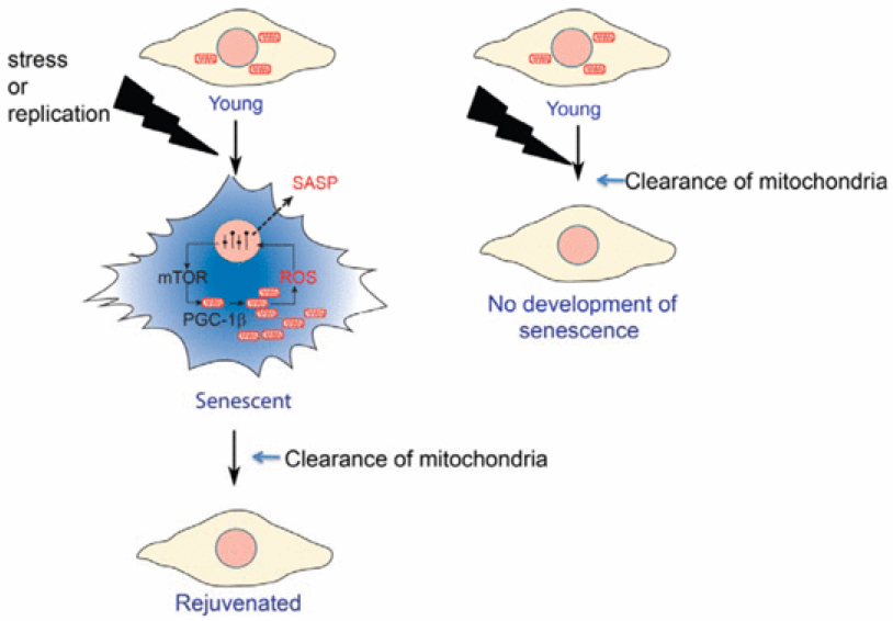
Preventing aging and rejuvenating human and mice cells in the lab (credit: Clara Correia‐Melo et al./EMBO Journal)

An international team of scientists led by João Passos at Newcastle University has for the first time shown thatmitochondria (the “batteries” of the cells) are major triggers for aging, and eliminating them upon the induction of senescence prevents senescence in the aging mouse liver.

As we grow old, cells in our bodies accumulate different types of damage and have increased inflammation, factors that are thought to contribute to the aging process.

As described Feb. 4 in an open-access paper in the EMBO Journal, the team carried out a series of genetic experiments involving human cells grown in the laboratory and succeeded in eliminating the majority, if not all, the mitochondria from aging cells.

Tricking mitochondria

http://www.kurzweilai.net/images/mitochondrion.jpg

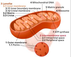
Components of a typical mitochondrion (credit: Kelvinsong/Creative Commons)

Cells can normally eliminate faulty mitochondria by a process called mitophagy. The scientists were able to “trick” the cells into inducing this process in a grand scale, until all the mitochondria within the cells were physically removed.

To their surprise, they observed that the aging cells, after losing their mitochondria, showed characteristics similar to younger cells — that is, they became rejuvenated. The levels of inflammatory molecules, oxygen free radicals and expression of genes, which are among the makers of cellular aging, dropped to the level that would be expected in younger cells.

“This is a very exciting and surprising discovery,” said Passos. “We already had some clues that mitochondria played a role in the aging of cells, but scientists around the world have struggled to understand exactly how and to what extent these were involved.”

The team, involving other universities in the UK and the U.S., also deciphered a new mechanism by which mitochondria contribute to aging: mitochondrial biogenesis, the complex process by which mitochondria replicate themselves, is a major driver of cellular aging.

This work was funded by the UK Biotechnology and Biological Sciences Research Council.


Abstract of Mitochondria are required for pro-ageing features of the senescent phenotype

Cell senescence is an important tumour suppressor mechanism and driver of ageing. Both functions are dependent on the development of the senescent phenotype, which involves an overproduction of pro‐inflammatory and pro‐oxidant signals. However, the exact mechanisms regulating these phenotypes remain poorly understood. Here, we show the critical role of mitochondria in cellular senescence. In multiple models of senescence, absence of mitochondria reduced a spectrum of senescence effectors and phenotypes while preserving ATP production via enhanced glycolysis. Global transcriptomic analysis by RNA sequencing revealed that a vast number of senescent‐associated changes are dependent on mitochondria, particularly the pro‐inflammatory phenotype. Mechanistically, we show that the ATM, Akt and mTORC1 phosphorylation cascade integrates signals from the DNA damage response (DDR) towards PGC‐1β‐dependent mitochondrial biogenesis, contributing to a ROS‐mediated activation of the DDR and cell cycle arrest. Finally, we demonstrate that the reduction in mitochondrial content in vivo, by either mTORC1 inhibition or PGC‐1β deletion, prevents senescence in the ageing mouse liver. Our results suggest that mitochondria are a candidate target for interventions to reduce the deleterious impact of senescence in ageing tissues.


Mayo Clinic researchers extend lifespan by up to 35 percent in mice

February 3, 2016   http://www.kurzweilai.net/mayo-clinic-researchers-extend-lifespan-by-up-to-35-percent-in-mice

Researchers at Mayo Clinic have discovered that senescent cells — cells that no longer divide and accumulate with age — shorten lifespan by as much as 35 percent in normal mice.

Removing these aging cells delays tumor formation, preserves tissue and organ function, and extends lifespan without observed adverse effects, the researchers found, writing Feb. 3 in Nature.

“Cellular senescence is a biological mechanism that functions as an ‘emergency brake’ used by damaged cells to stop dividing,” says Jan van Deursen, Ph.D., Chair of Biochemistry and Molecular biology at Mayo Clinic, and senior author of the paper. “While halting cell division of these cells is important for cancer prevention, it has been theorized that once the ‘emergency brake’ has been pulled, these cells are no longer necessary.”

As the immune system becomes less effective, senescent cells build up and damage adjacent cells, causing chronic inflammation, which is closely associated with frailty and age-related diseases.

Mayo Clinic researchers used a compound called AP20187 to remove senescent cells, which delayed tumor formation and reduced age-related deterioration of several organs, extending mediian lifespan of treated mice by 17 to 35 percent. The mice also had a healthier appearance and less inflammation in fat, muscle and kidney tissue.

The research was supported by the National Institutes of Health, the Paul F. Glenn Foundation, the Ellison Medical Foundation, the Noaber Foundation, and the Mayo Clinic Robert and Arlene Kogod Center on Aging.

Van Deursen is a co-inventor of the technology that has been licensed by Mayo Clinic to Unity Biotechnology. Mayo Clinic and Van Deursen have a financial interest in the technology.

https://youtu.be/w8UHzkXC4HQ

Mayo Clinic | Researchers Extend Lifespan by as Much as 35 Percent in Mice


Abstract of Naturally occurring p16Ink4a-positive cells shorten healthy lifespan

Cellular senescence, a stress-induced irreversible growth arrest often characterized by expression of p16Ink4a (encoded by the Ink4a/Arf locus, also known as Cdkn2a) and a distinctive secretory phenotype, prevents the proliferation of preneoplastic cells and has beneficial roles in tissue remodelling during embryogenesis and wound healing. Senescent cells accumulate in various tissues and organs over time, and have been speculated to have a role in ageing. To explore the physiological relevance and consequences of naturally occurring senescent cells, here we use a previously established transgene, INK-ATTAC, to induce apoptosis in p16Ink4a-expressing cells of wild-type mice by injection of AP20187 twice a week starting at one year of age. We show that compared to vehicle alone, AP20187 treatment extended median lifespan in both male and female mice of two distinct genetic backgrounds. The clearance of p16Ink4a-positive cells delayed tumorigenesis and attenuated age-related deterioration of several organs without apparent side effects, including kidney, heart and fat, where clearance preserved the functionality of glomeruli, cardio-protective KATP channels and adipocytes, respectively. Thus, p16Ink4a-positive cells that accumulate during adulthood negatively influence lifespan and promote age-dependent changes in several organs, and their therapeutic removal may be an attractive approach to extend healthy lifespan.

Read Full Post »

Reengineering Therapeutics

Larry H. Bernstein, MD, FCAP, Curator

LPBI

The synNotch solution: UCSF scientists engineer a next-gen T-cell immunotherapy

Sunday, January 31, 2016 | By John Carroll

2.2.9

2.2.9   Reengineering Therapeutics, Volume 2 (Volume Two: Latest in Genomics Methodologies for Therapeutics: Gene Editing, NGS and BioInformatics, Simulations and the Genome Ontology), Part 2: CRISPR for Gene Editing and DNA Repair

CAR-T has been all the rage in cancer R&D for several years now as a slate of biotech upstarts pursue highly promising work reengineering T cells into attack weapons by adding a chimeric antigen receptor that can zero in on particular cancer cells. The approach has been highly effective in acute lymphoblastic leukemia, triggering an attack on B cells by homing in on the CD19 antigen, a breakthrough that has inspired a race to the regulatory finish line with the first CAR-Ts.

That approach, though, has run into some major obstacles when researchers move from the blood cancer to solid tumors. But now a group of scientists working with UC San Francisco’s Wendell Lim say they’ve come up with a new therapeutic model for T-cell engineering that promises to overcome that hurdle and make it a more precise weapon that can tackle solid tumors while avoiding off-target reactions that threaten patients.

The key to this new approach is a new receptor: synNotch. Taking a cue from nature, which relies on a sensor called Notch to perform key functions, the synthetic biology engineers say they can add a receptor that includes one section that sticks out from the cell with one that lies inside. By tinkering with synNotch they can reengineer the immune cell to run down a particular cancer cell target and then issue instructions to turn genes on or off to set up the other half of the therapeutic equation.

In a project described in Cell, the team says they created a synNotch that recognized an antigen on the surface of the cancer cell while the internal mechanism contributed a chimeric antigen receptor that recognized a different antigen. They then tested it on a mouse model that included two different tumor cells: one with both targets recognized by the external synNotch sensor and the CAR and one with just the CAR target.

The newly programmed attack weapon zeroed in on the two targets (which required synNotch activation for it to work) while leaving the other alone, providing preclinical proof-of-concept evidence that they could create a much more efficient tumor cell killing vehicle.

“The kinds of engineered T cells that we can now construct give us the exciting potential to create precision cancer therapeutics that take advantage of all the genomic and proteomic information we are currently gathering on disease,” said Lim, the senior author of the study. “This genomic information now becomes actionable.”

The team was led by Leonardo Morsut and Kole Roybal.

Lim added that it is possible to reengineer a T cell with multiple synNotches to make it even more precise. And there are added applications for autoimmune disease, regenerative medicine, and more.

Engineering Customized Cell Sensing and Response Behaviors Using Synthetic Notch Receptors

Leonardo  Morsut, Kole T. Roybal, Xin Xiong, Russell M. Gordley, Scott M. Coyle, Matthew Thomson, Wendell A. Lim

Tricked-Out Immune Cells Could Attack Cancer, Spare Healthy Cells  

New Cell-Engineering Technique May Lead To Precision Immunotherapies

New CRISPR/Cas9 delivery method could offer a clinical pathway

Read Full Post »

Need for Protophysiology?

Larry H. Bernstein, MD, FCAP, Curator

LPBI


Comparison of fundamental physical properties of the model cells (protocells) and the living cells reveals the need in protophysiology

V.V. Matveev

International Journal of Astrobiology 2016. pp1-8.   http://dx.doi.org:/10.1017/S1473550415000476

A hypothesis is proposed about potassium ponds being the cradles of life enriches the gamut of ideas about the possible conditions of pre-biological evolution on the primeval Earth, but does not bring us closer to solving the real problem of the origin of life. The gist of the matter lies in the mechanism of making a delimitation between two environments – the intracellular environment and the habitat of protocells. Since the sodium–potassium pump (Na+ /K+-ATPase) was discovered, no molecular model has been proposed for a predecessor of the modern sodium pump. This has brought into life the idea of the potassium pond, wherein protocells would not need a sodium pump. However, current notions of the operation of living cells come into conflict with even physical laws when trying to use them to explain the origin and functioning of protocells. Thus, habitual explanations of the physical properties of living cells have become inapplicable to explain the corresponding properties of Sidney Fox’s microspheres. Likewise, existing approaches to solving the problem of the origin of life do not see the need for the comparative study of living cells and cell models, assemblies of biological and artificial small molecules and macromolecules under physical conditions conducive to the origin of life. The time has come to conduct comprehensive research into the fundamental physical properties of protocells and create a new discipline – protocell physiology or protophysiology – which should bring us much closer to solving the problem of the origin of life.


There is a statement we constantly come across in the scientific and popular-science literature: the ion composition of the internal environment of the body of humans and animals, in which all of its cells are immersed, is close to that of seawater. This observation appeared in the literature even 100 years ago, when it became possible to investigate the ion composition of biological liquids.

This similarity between the internal environment of the body and the sea is quite obvious: in both seawater and blood plasma there are one or two orders of magnitude more Na+ ions than K+. It is this composition that can make one think that life originated in the primeval ocean (the memory of which has since been sustained by the internal environment of the body), and the first cells delineated themselves from seawater using a weakly permeable membrane, so that their internal environment became special, suitable for chemical and physical processes needed to sustain life. Indeed, the ratio of the above cations in the cytoplasm is the exact reverse of their ratio in seawater: there is much more K+ in it than Na+ . In fact, physiological processes can only be possible in an environment where potassium prevails over sodium. Therefore, any theory of the origin of life must explain how such a deep delimitation (distinction) between the two environments could occur: the intracellular environment, wherein vitally important processes take course, and the external environment, which provides the cell with necessary materials and conditions.

For the protocell to separate from seawater, a mechanism must arise that creates and maintains the ion asymmetry between the primeval cell and its environs. We normally consider a mechanism of this kind as the isolating lipid membrane with a molecular ion pump, the Na+/K+ -ATPase, built into it. If life originated in seawater, the origin of the first cell inevitably comes down to the origin of the sodium pump and any structure supporting it – the lipid membrane – without which the work of any pump would make little sense. It seems that life is born in conditions that are really adverse to it and even ruinous.


The great basic question of science: Membrane compartment or non-membrane phase compartment (biophase) is a physical basis for origin of life?

1. If life originated in seawater, the origin of the first cell inevitably comes down to the origin of the sodium pump and any structure supporting it – the lipid membrane – without which the work of any pump would make little sense.

2. Since the sodium-potassium pump (Na+/K+-ATPase) was discovered, no molecular model has been proposed for a predecessor of the modern sodium pump. Neither Miller’s electrical charges, nor Fox’s amino-acid condensation, nor building ready-made biomolecules into coacervates; none of this has managed to lead to the self-origination of the progenitor of the ion pump even in favourable lab conditions.

3. In 2007, we saw the simultaneous release of two articles, in which it was posited that life originated not in seawater as previously thought, but in smaller bodies of water with a K+/Na+ ratio necessary to sustain life. In this conditions sodium pump is not needed and the pump can originate later. But why the pump is needed if K+/Na+ ratio is good? The origin of the sodium pump in conditions where there is no natural need for it may require the agency of Providence.

4. Potassium Big Bang on Earth instead of potassium ponds.

5. Fox’s microspheres do not need potassium ponds.

6. Despite the fact that Fox’s microspheres have no fully functional membrane with sodium pumps and specific ion channels, they generate action potentials similar to that by nerve cells and in addition have ion channels which open and close spontaneously. This ability of the microspheres contradicts to the generally accepted ideas about the mechanism of generation of biological electrical potentials.

7. Hodgkin-Huxley model of action potentials is similarly well-compatible with both the nerve cell and Fox’s microsphere.

8. Biophase as the main subject of protophysiology. In the past they considered the living cell as a non-membrane phase compartment with different physical properties in comparison to the surrounding medium, and this physical difference plays a key role in cell function. According to a new take on an old phase, non-membrane phase compartments play an important role in the functioning of the cell nucleus, nuclear envelope and then of cytoplasm. Somebody sees the compartments even as temporary organelles. According to available data, the phase compartments can play a key role in cell signaling. In this historical context, studies in recent years dedicated to non-membrane phase compartments in the living cells sound sensational.

9. It is essentially a Protocell World which weaves known RNA World, DNA World and Protein World into unity. 10. In the view of non-membrane phase approach, the usage of liposomes and other membrane (non-biophase) cell models to solve the issue of the origin of life is a deadlock way of the investigation. I would be grat

Read Full Post »

Tau and IGF1 in Alzheimer’s Disease

Larry H. Bernstein, MD, FCAP, Curator

LPBI

TAU links growth factor to development of Alzheimer’s disease

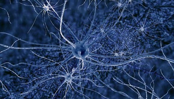
https://english.tau.ac.il/sites/default/files/styles/reaserch_main_image_580_x_330/public/alz2580.jpg


The mechanisms underlying the stability and plasticity of neural circuits in the hippocampus, the part of the brain responsible for spatial memory and the memory of everyday facts and events, has been a major focus of study in the field of neuroscience. Understanding precisely how a “healthy” brain stores and processes information is crucial to preventing and reversing the memory failures associated with Alzheimer’s disease (AD), the most common form of late-life dementia.


Hyperactivity of the hippocampus is known to be associated with conditions that confer risk for AD, including amnestic mild cognitive impairment. A new Tel Aviv University study finds that the insulin-like growth factor 1 receptor (IGF-1R), the “master” lifespan regulator, plays a vital role in directly regulating the transfer and processing of information in hippocampal neural circuits. The research reveals IGF-1R as a differential regulator of two different modes of transmission — spontaneous and evoked — in hippocampal circuits of the brain. The researchers hope their findings can be used to indicate a new direction for therapy used to treat patients in the early stages of Alzheimer’s disease.


The study was led by Dr. Inna Slutsky of TAU’s Sagol School of Neuroscience and Sackler School of Medicine and conducted by doctoral student Neta Gazit. It was recently published in the journal Neuron. “People who are at risk for AD show hyperactivity of the hippocampus, and our results suggest that IGF-1R activity may be an important contributor to this abnormality,” Dr. Slutsky concluded.


Resolving a controversy

“We know that IGF-1R signaling controls growth, development and lifespan, but its role in AD has remained controversial,” said Dr. Slutsky. “To resolve this controversy, we had to understand how IGF-1R functions physiologically in synaptic transfer and plasticity.”


Using brain cultures and slices, the researchers developed an integrated approach characterizing the brain system on different scales — from the level of protein interactions to the level of single synapses, neuronal connections and the entire hippocampal network. The team sought to address two important questions: whether IGF-1Rs are active in synapses and transduce signalling at rest, and how they affect synaptic function.


“We used fluorescence resonance energy transfer (FRET) to estimate the receptor activation at the single-synapse level,” said Dr. Slutsky. “We found IGF-1Rs to be fully activated under resting conditions, modulating release of neurotransmitters from synapses.”


While acute application of IGF-1 hormone was found to be ineffective, the introduction of various IGF-1R blockers produced robust dual effects — namely, the inhibition of a neurotransmitter release evoked by spikes, electrical pulses in the brain, while enhancement of spontaneous neurotransmitter release.


A test for Alzheimer’s?

“When we modified the level of IGF-1R expression, synaptic transmission and plasticity were altered at hippocampal synapses, and an increase in the IGF-1R expression caused an augmented release of glutamate, enhancing the activity of hippocampal neurons,” said Gazit.


“We suggest that IGF-1R small inhibitors, which are currently under development for cancer, be tested for reduction aberrant brain activity at early stages of Alzheimer’s disease,” said Dr. Slutsky.


The researchers are currently planning to study how IGF-1R signaling controls the stability of neural circuits over an extended timescale.


Dr. Irena Vertkin, Dr. Ilana Shapira, Edden Slomowitz, Maayan Sheiba and Yael Mor of Dr. Slutsky’s lab at TAU, and Martin Helm and Prof. Silvio Rizzoli of the University of Göttingen in Germany, contributed to this research.


This article was originally published by AFTAU.


“We know that IGF-1R signaling controls growth, development and lifespan, but its role in AD has remained controversial,” said Dr. Slutsky. “To resolve this controversy, we had to understand how IGF-1R functions physiologically in synaptic transfer and plasticity.”

Using brain cultures and slices, the researchers developed an integrated approach characterizing the brain system on different scales — from the level of protein interactions to the level of single synapses, neuronal connections and the entire hippocampal network. The team sought to address two important questions: whether IGF-1Rs are active in synapses and transduce signalling at rest, and how they affect synaptic function.

“We used fluorescence resonance energy transfer (FRET) to estimate the receptor activation at the single-synapse level,” said Dr. Slutsky. “We found IGF-1Rs to be fully activated under resting conditions, modulating release of neurotransmitters from synapses.”

While acute application of IGF-1 hormone was found to be ineffective, the introduction of various IGF-1R blockers produced robust dual effects — namely, the inhibition of a neurotransmitter release evoked by spikes, electrical pulses in the brain, while enhancement of spontaneous neurotransmitter release.

A test for Alzheimer’s?

“When we modified the level of IGF-1R expression, synaptic transmission and plasticity were altered at hippocampal synapses, and an increase in the IGF-1R expression caused an augmented release of glutamate, enhancing the activity of hippocampal neurons,” said Gazit.

“We suggest that IGF-1R small inhibitors, which are currently under development for cancer, be tested for reduction aberrant brain activity at early stages of Alzheimer’s disease,” said Dr. Slutsky.

The researchers are currently planning to study how IGF-1R signaling controls the stability of neural circuits over an extended timescale.


Read Full Post »

Fat, sleep and the gut

Larry H. Bernstein, MD, FCAP, Curator

LPBI


Hibernating Bear Microbiome May Hold the Key to Combating Obesity   

GEN News Highlights  http://www.genengnews.com/gen-news-highlights/hibernating-bear-microbiome-may-hold-the-key-to-combating-obesity/81252333/

http://www.genengnews.com/Media/images/GENHighlight/107756_web1581601731.jpg

This visual abstract depicts how the microbiota and serum metabolites in brown bears differ seasonally between hibernation and active phases. Colonization of mice with a bear microbiota promoted increased adiposity. These findings suggest that seasonal microbiota variation may contribute to metabolism of the hibernating brown bear. [Sommer et al./Cell Reports 2016]


Gorging oneself on high-calorie food to pack on fat only to fall asleep for a few months and have it all melt away isn’t the latest diet fad (at least not yet), but it is a regular part of life for bears as they prepare to hibernate for the winter. Yet, despite the rapid weight gain, the animals somehow avoid the health consequences so often associated with obesity in humans.

Researchers at the University of Gothenburg in Sweden have published results from a study wherein they show that bears’ shifting metabolic status is associated with significant changes in their gut microbes. The researchers were particularly surprised by the discovery that the bears’ summer gut microbiota included bacteria that take in more energy from the diet.

“The restructuring of the microbiota into a more avid energy harvester during summer, which potentially contributes to the increased adiposity gain without impairing glucose metabolism, is quite striking,” explained senior study author Fredrik Bäckhed, Ph.D., professor at The Wallenberg Laboratory for Cardiovascular and Metabolic Research within the University of Gothenburg.

The findings from this study were published recently in Cell Reports through an article entitled “The Gut Microbiota Modulates Energy Metabolism in the Hibernating Brown Bear Ursus arctos.”

Previous work done in Dr. Bäckhed’s laboratory showed that the composition of gut microbiota can influence the amount of energy harvested from the diet, and more recently the investigators found that the microbiota also shifts in people who are obese and in those with type 2 diabetes. This led the team to hypothesize whether changes to the microbiota might also be important in hibernating brown bears in the wild.

“We analyzed the microbiota of free-ranging brown bears during their active phase and hibernation. Compared to the active phase, hibernation microbiota had reduced diversity, reduced levels ofFirmicutes and Actinobacteria, and increased levels of Bacteroidetes,” the authors wrote. “Several metabolites involved in lipid metabolism, including triglycerides, cholesterol, and bile acids were also affected by hibernation.”

Dr. Bäckhed’s team took their investigation a step further by exploring whether the changes in the bears’ microbiota was driving the shift in metabolism by transferring the bears’ summer and winter microbiota into mice.

“Transplantation of the bear microbiota from summer and winter to germ-free mice transferred some of the seasonal metabolic features and demonstrated that the summer microbiota promoted adiposity without impairing glucose tolerance, suggesting that seasonal variation in the microbiota may contribute to host energy metabolism in the hibernating brown bear,” described the authors.

The researchers were encouraged by their findings and believe it’s possible that the findings in bears might suggest new strategies for managing obesity in humans; however, Dr. Bäckhed urged caution about over-interpreting the study results.

“I think it’s too early [to say], as I consider this being very basic science,” Dr. Bäckhed concluded. “However, if we learn more about which bacteria and the functions that promote and/or protect against obesity [in hibernating bears], we may identify new potential therapeutic targets.”


The Gut Microbiota Modulates Energy Metabolism in the Hibernating Brown Bear Ursus arctos

Felix Sommer, Marcus Stahlman, Olga Ilkayeva, …, Christopher B. Newgard, Ole Frobert, Frederik Backhed

Cell Reports Feb 2016;14: 1–7.

Highlights

Bear microbiota composition differs seasonally between hibernation and active phase

Blood metabolites differ seasonally in the brown bear

The bear gut microbiota promote energy storage during summer


Hibernation is an adaptation that helps many animals to conserve energy during food shortage in winter. Brown bears double their fat depots during summer and use these stored lipids during hibernation. Although bears seasonally become obese, they remain metabolically healthy. We analyzed the microbiota of free-ranging brown bears during their active phase and hibernation. Compared to the active phase, hibernation microbiota had reduced diversity, reduced levels of Firmicutes and Actinobacteria, and increased levels of Bacteroidetes. Several metabolites involved in lipid metabolism, including triglycerides, cholesterol, and bile acids, were also affected by hibernation. Transplantation of the bear microbiota from summer and winter to germ-free mice transferred some of the seasonal metabolic features and demonstrated that the summer microbiota promoted adiposity without impairing glucose tolerance, suggesting that seasonal variation in the microbiota may contribute to host energy metabolism in the hibernating brown bear.

Read Full Post »

Biomarkers of Cancer

Larry H. Bernstein, MD, FCAP, Curator

LPBI


Biomarker Discovery Gets a Fix on Cancer

GEN Feb 1, 2016 (Vol. 36, No. 3)   http://www.genengnews.com/gen-articles/biomarker-discovery-gets-a-fix-on-cancer/5674/

Just Because Cancer Is a Moving Target—Emerging Here, Eluding Treatment There—Doesn’t Mean It Can’t Be Tracked

http://www.genengnews.com/Media/images/Article/thumb_Epic_PatientARV71241597624.jpg

Epic Sciences’ “no cell left behind” platform can identify circulating tumor cells in an unbiased manner, without enrichment or depletion on any parameter. For example, as indicated in this image, it has been used to detect the AR-V7-positive cells among the cells collected in a simple blood draw. The AR-V7 splice variant is linked to resistance of androgen receptor-targeting drugs in metastatic castration-resistant prostate cancer patients.


  • Just as signposts provide information, direction, and guidance, so too cancer biomarkers can better reveal the complex, dynamic, and heterogeneous landscape of malignancies.

    Such information is critical for creating better cancer diagnostics, prognostics, and therapeutics, but the journey to find just the right biomarker is often a long and winding road.

    Biomarker discovery and utilization is being explored by means of various methods and technologies. These include the isolation and characterization of rare circulating cancer cells, the use of multiplexing to extract information from limited amounts of sample, and even the application of evolutionary biology to detect early cell changes.

    These approaches are all being developed by companies interested in improving cancer diagnostics, prognostics, and therapeutics. For example, one of the companies cited in this article is scrutinizing circulating tumor cells (CTCs) to predict resistance to cancer drugs. (This work was presented at a scientific retreat convened by the Prostate Cancer Foundation.) Other companies represented in this article are developing novel cancer diagnostic approaches. (These were detailed at the recent Cambridge Health Diagnostics Summit.)

    While a typical blood sample from a cancer patient can contain 30 million nucleated cells, it’s estimated that perhaps only 5 of those cells may be destined to form tumors. “Most present-day diagnostic technologies cannot precisely detect those rare CTCs,” observed Ryan Dittamore, vice president, translational research and clinical affairs, Epic Sciences. “Identifying robust predictive biomarkers that can be utilized in real-time with a test compatible with diagnostic workflows is one of the greatest challenges to precision medicine.

    “Epic Sciences is focused on developing sensitive diagnostic tests to characterize CTCs molecularly in order to match therapies to a patient’s cancer biology.” As Dittamore indicated at the Prostate Cancer Foundation event, the company evaluated its CTC approach in a study undertaken with Howard Scher, M.D., chief of the genitourinary oncology service at Memorial Sloan Kettering.

    “We focused on a form of prostate cancer called metastatic castration-resistant prostate cancer,” explained Dittamore. “In the study, we confirmed the value of the prostatic cancer biomarker AR-V7 in 193 patient samples. In men with this androgen receptor (AR) splice variant, treatment with taxanes may be more effective than treatment with AR-signaling-directed agents (enzalutamide or abiraterone).”

    In Epic Sciences’ “no cell left behindTM” approach, all nucleated cells from a blood sample are placed on a slide. CTCs are stained using biomarker monoclonal antibodies and assessed via immunofluorescence and high-resolution digital pathology scanning.

    “We’ve industrialized this process,” asserted Dittamore, “and can evaluate millions of patient cells in every assay. Once identified, we can then isolate individual CTCs for further single-cell evaluation, such as with next-generation sequencing.” Overall, the company’s approach can achieve the following tasks: quantifying the number of cancer cells, characterizing biomarker expression (specifying, for example, subcellular localization and genomic alterations), and assessing disease heterogeneity and clonality of cancer cell types.

    Commercializating the tests will come later, indicated Dittamore. “We will continue refining and expanding tests with our numerous collaborators in academia and pharma, many of whom are using our biorepository capabilities to bank samples for later study of biomarkers for other types of cancer.

    The ultimate goal of molecular diagnostics is to get as much information as possible, as quickly as possible, with as little sample as possible. Unfortunately, it is often necessary to make do with samples that are limited in number and extent—consider, for example, formalin-fixed, paraffin-embedded (FFPE) specimens—even though one would like to extract comprehensive information. To help solve this problem, Qiagen has developed a new platform that marries and automates two proven techniques: multiplex PCR and capillary electrophoresis.

    “We created a novel and automated clinical platform called Modaplex for real-time sampling, size separation of PCR products, amplification curve building, and Ct (cycle threshold) calculations,” summarized Lilly Kong, DVM, Qiagen’s senior director, assay development. “As a result, from a very limited amount of sample, we can utilize a multimodal, multiplex, essentially ‘all in one well’ means to quickly obtain actionable results.”

    Dr. Kong said that Qiagen has developed multiple oncological biomarker tests including RAS, cMET, FGFR, Cell Cycle, and DLBCL signatures. “We can multiplex up to 40 targets in a single reaction,” she asserted. “Traditional assays allow testing of only one or a few assays at a time. Further, this is an architecture that uses a modular approach.”

    Dr. Kong described development of an assay evaluating cMET and epidermal growth factor receptor (EGFR) expression and copy number variation. These two players are important because the MET proto-oncogene encodes for the receptor tyrosine kinase, cMET, which is widely expressed by many cells.

    Under pathological conditions, cMET confers survival and invasive properties of cancer cells while potentially blocking EGFR therapies. “This multimodal 21-plex assay,” Dr. Kong indicated, “succeeded in evaluateing nine mRNA targets and nine genomic DNA targets with appropriate standards and external process controls.”

    Qiagen also examined cMET simple nucleotide polymorphisms (SNPs) in a 16-plex reaction tube. “The modular approach enables combinations of important biomarker assays that can test for disparate target types, such as SNPs, expression biomarkers, miRNAs, and fusion genes,” Dr. Kong detailed. “Further, one FFPE slide is sufficient for about 10 assays with 10–50 ng input per reaction. The automation is user-friendly while sensitivity, specificity, and precision are equal to or exceed singleplex assays.”

    Although Modaplex started with oncological applications, many other panels are in development. For example, Qiagen is working on pathogen detection applications as well as in-process monitoring applications for biologicals manufacturing.

Read Full Post »

Cancer Causing Enzyme Activity

Larry H. Bernstein, MD, FCAP, Curator

LPBI


Cancer-Causing Enzyme Acts during DNA Replication

GEN News   http://www.genengnews.com/gen-news-highlights/cancer-causing-enzyme-acts-during-dna-replication/81252312/


Scientists at Indiana University (IU) have identified a genetic mechanism that is likely to drive mutations that can lead to cancer. Their E. coli study, published in the Proceedings of the National Academy of Sciences, finds the enzyme APOBEC3G, a known trigger for mutations that occur as benign tumor cells to transform into cancerous malignancies that spread throughout the body, appears to cause these harmful changes by mutating genes during DNA replication.

The study also received support from the Wayne State University School of Medicine, whose researchers provided expertise on APOBEC3G and helped analyze the data. All experiments were carried out at IU.

“Many tumors accumulate mutations during their growth, which lead to the subsequent characteristics that permit metastasis,” said Patricia Foster, Ph.D., the principal investigator on the grant and a professor in the IU Bloomington College of Arts and Sciences’ biology department, senior author on the study. “Based upon the results revealed in bacteria in our study, we believe that the APOBEC family of enzymes creates some of these mutations specifically during the rapid growth of these tumors.”

The results could have implications for personalized medicine. For example, because it is possible to identify tumors potentially vulnerable to the enzyme by using current DNA sequencing technology, a physician treating these tumors might want to explore temporarily suppressing expression of this enzyme, she said.

Normally, the APOBEC family of enzymes plays an important role in the human immune system by driving changes in immune cells that aid in defense against viruses, possibly including the HIV/AIDS virus. The IU scientists found the harmful influence of the enzyme family arises from the complex way that two halves of every double-stranded DNA molecule must unravel to replicate during cellular division—splitting into two temporarily single-stranded DNA chains thousands of links (the four nucleotides) in length to serve as templates for the new copy. As the nucleotides are split in half to be copied, one of the two single-stranded bits of DNA, known as the lagging strand template, is highly vulnerable to genetic mutation, according to Dr. Foster.

This “gap in the armor” occurs because DNA polymerase must repeatedly traverse the nucleobases in the lagging strand template thousands of times during the course of replication, stopping further down the chain from the base pair previously inserted on the loop along the chemical chain. Each of these polymerase “hops” creates a long stretch of DNA that temporarily remains as a single strand.
The complex process introduces more opportunities for errors in the lagging strand template compared to the continuous step-by-step process that replicates the other half of the split strand of DNA, called the leading strand template.

“We’re talking about thousands of bases exposed without a complimentary strand throughout the whole replication cycle,” noted Dr. Foster.  “If I were going to design an organism, I would make two types of copying enzymes. An important organism for studying genes, E. coli allows scientists to observe genetic changes over thousands of generations in a relatively short time span. The results apply to humans as well as bacteria since the basic mechanisms of DNA replication are the same across all species.”

The mechanism by which the APOBEC family of enzymes drives mutation is cytosine deamination, in which a cytosine, the C nucleotide, transforms into uracil, one of the four bases in RNA that doesn’t play a role in DNA replication. But the presence of uracil during DNA replication can cause an error when a thymine, the T nucleotide, replaces a cytosine. APOBEC enzymes specifically target the C’s in single-stranded DNA for deamination.

The disruptive effect of the enzyme on genetic replication in the study was observed in a strain of E. coli, whose ability to remove the dangerous uracils had been switched off. To conduct the experiment, Dr. Foster’s lab observed the effect of APOBEC3G on approximately 50 identical lineages of E. coliover the course of nearly 100 days, with each day encompassing 20 to 30 bacterial generations.

Over time, a unique pattern of nucleotides was detected in the mutated DNA, a chain of three cytosine molecules, the same genetic signature found in other studies of the enzyme family. And these mutations were four times more likely to be found on the lagging-strand template than on the leading-strand template.

“These results strongly suggest that these mutations occur as APOBEC3G attacks cytosines during DNA replication, while they’re most exposed on the lagging strand template,” Dr. Foster said. “This basic mechanism appears to be the same in bacteria and in human tumors cells.”


DNA’s ‘Gap in the Armor’ Allowing Cancer to Develop Pinpointed

Seth Augenstein, Digital Reporter  http://www.biosciencetechnology.com/news/2016/02/dnas-gap-armor-allowing-cancer-develop-pinpointed

Research on the effect of the enzyme APOBEC3G on DNA replication was conducted in the bacteria Escherichia coli. (Photo: Department of Defense)

Research on the effect of the enzyme APOBEC3G on DNA replication was conducted in the bacteria Escherichia coli. (Photo: Department of Defense)

A key group of enzymes could be the “gap in the armor” of all DNA, allowing cancer-causing mutations, according to a new study.

APOBEC3G, which is known to trigger benign mutations, also causes malignant mutations during the DNA replication process, according to the new findings, in the Proceedings of the National Academy of Sciences.

“Many tumors accumulate mutations during their growth, which leads to the subsequent characteristics that permit metastasis,” said Patricia Foster, professor at Indiana University, and senior author. “Based upon the results revealed in bacteria in our study, we believe that the APOBEC family of enzymes create some of these mutations specifically during the rapid growth of these tumors.”

The investigators created and observed the mutations in the bacteria Escherichia coli, which presented the advantage of watching thousands of generations in a relatively short time.

The key process is the movement of DNA polymerase along one of the two DNA single strands, known as the lagging strand template, during the replication process. The lagging strand becomes susceptible to errors. APOBEC can enter into this process, causing cytosine deamination, essentially replacing the intended cytosine on the strand with the thymine nucleobase, causing the mutations.

The scientists turned off the ability to regulate the cytosine deamination in the E. coli replication – and then observed an uptick in the harmful mutations confirming the culprit, they said.

“These results strongly suggest that these mutations occur as APOBEC3G attacks cytosines during DNA replication, while they’re most exposed on the lagging strand template,” said Foster. “This basic mechanism appears to be the same in bacteria and in human tumor cells.”

The study was supported in part of a $6.2 million grant from the U.S. Army Research Office to investigate bacterial evolution, according to the school.


Mouse Apolipoprotein B Editing Complex 3 (APOBEC3) Is Expressed in Germ Cells and Interacts with Dead-End (DND1)

The dead-end (Dnd1) gene is essential for maintaining the viability of germ cells. Inactivation ofDnd1 results in sterility and testicular tumors. The Dnd1 encoded protein, DND1, is able to bind to the 3′-untranslated region (UTR) of messenger RNAs (mRNAs) to displace micro-RNA (miRNA) interaction with mRNA. Thus, one function of DND1 is to prevent miRNA mediated repression of mRNA. We report that DND1 interacts specifically with APOBEC3. APOBEC3 is a multi-functional protein. It inhibits retroviral replication. In addition, recent studies show that APOBEC3 interacts with cellular RNA-binding proteins and to mRNA to inhibit miRNA-mediated repression of mRNA.


Re-editing the paradigm of Cytidine (C) to Uridine (U) RNA editing
Nicolas Fossatab* & Patrick P L Tam
RNA Biology  2014; Volume 11, Issue 10:1233-1237  http://dx.doi.org:/10.1080/15476286.2014.996054

Cytidine (C) to Uridine (U) RNA editing is a post-trancriptional modification that until recently was known to only affect Apolipoprotein b (Apob) RNA and minimally require 2 components of the C to U editosome, the deaminase APOBEC1 and the RNA-binding protein A1CF. Our latest work has identified a novel RNA-binding protein, RBM47, as a core component of the editosome, which can substitute A1CF for the editing of ApoB mRNA. In addition, new RNA species that are subjected to C to U editing have been identified. Here, we highlight these recent discoveries and discuss how they change our view of the composition of the C to U editing machinery and expand our knowledge of the functional attributes of C to U RNA editing.

Derepression of MicroRNA-mediated Protein Translation Inhibition by Apolipoprotein B mRNA-editing Enzyme Catalytic Polypeptide-like 3G (APOBEC3G) and Its Family Members*

Jialing Huang, Zhihui Liang, Bin Yang, Heng Tian, Jin Ma and Hui Zhang1
The Journal of Biological Chemistry, 2007; 282:33632-33640.
  http://dx.doi.org:/10.1074/jbc.M705116200

The apolipoprotein B mRNA-editing enzyme catalytic polypeptide-like 3G (APOBEC3G or A3G) and its fellow cytidine deaminase family members are potent restrictive factors for human immunodeficiency virus type 1 (HIV-1) and many other retroviruses. A3G interacts with a vast spectrum of RNA-binding proteins and is located in processing bodies and stress granules. However, its cellular function remains to be further clarified. Using a luciferase reporter gene and green fluorescent protein reporter gene, we demonstrate that A3G and other APOBEC family members can counteract the inhibition of protein synthesis by various microRNAs (miRNAs) such as mir-10b, mir-16, mir-25, and let-7a. A3G could also enhance the expression level of miRNA-targeted mRNA. Further, A3G facilitated the association of microRNA-targeted mRNA with polysomes rather than with processing bodies. Intriguingly, experiments with a C288A/C291A A3G mutant indicated that this function of A3G is separable from its cytidine deaminase activity. Our findings suggest that the major cellular function of A3G, in addition to inhibiting the mobility of retrotransposons and replication of endogenous retroviruses, is most likely to prevent the decay of miRNA-targeted mRNA in processing bodies.

MicroRNAs (miRNAs)2 are 20-22-nt regulatory RNAs that participate in the regulation of various biological functions in numerous eukaryotic lineages, including plants, insects, vertebrate, and mammals (1–3). More than 474 miRNAs have been identified in humans so far, and ∼30% of the genes in the human genome are predicted to be subject to miRNA regulation (4). The expression of many miRNAs is usually specific to a tissue or developmental stage, and the miRNA expression pattern is altered during the development of many diseases (3). Mature miRNAs are generated from RNA polymerase II-transcribed primary miRNAs that are processed sequentially by the nucleases Drosha and Dicer. Although miRNA can guide mRNA cleavage, the basic function of miRNA is to mediate inhibition of protein translation (1, 5–8) through miRNA-induced silencing complexes (miRISCs). The guiding strand of miRNA in a miRISC interacts with a complementary sequence in the 3′-untranslated region (3′-UTR) of its target mRNA by partial sequence complementarities, resulting in translational inhibition (1). A 7-nucleotide “seed” sequence (at positions 2-8 from the 5′-end) in miRNAs seems to be essential for this action (4). The composition of the miRISC is similar to that of the RNA-induced silencing complex (RISC), which is responsible for mRNA cleavage guided by small interfering RNAs (siRNAs) (1, 3, 7). Nevertheless, some differences exist between miRISCs and siRNA RISCs. For example, the major Argonaute protein in siRNA RISC is Ago-2, whereas all four of the Ago proteins (Ago1-4) are found in miRISC (3, 8). Further, the siRNA RISC may be associated with various RNA-binding proteins such as fragile-X mental retardation protein (FMRP), TAR RNA-binding protein (TRBP), and the human homolog of the Drosophilahelicase Armitage, Mov10, possibly in a cell type-specific manner (9–13).

The miRNA-mediated translational repression consistently correlates with an accumulation of miRNA-bound mRNAs at cytoplasmic foci known as processing bodies (P-bodies) (8). Several lines of evidence have indicated that P-bodies are actively involved in miRNA-mediated mRNA repression (14). The P-body-associated protein GW182 associates directly with Ago-1 (15, 16). Depletion of P-body components such as GW182 and Rck/p54 prevents translational repression of target mRNAs (8, 14–19). Furthermore, several miRISC-related components, such as miRNAs, mRNAs repressed by miRNAs, Ago-1, Ago-2, and Mov10, are found in P-bodies (14). P-body formation is a dynamic process that requires continuous accumulation of repressed mRNAs (20). However, P-bodies serve not only as sites for RNA degradation, but also for storage of repressed mRNAs (15). These mRNAs may later return to polysomes to synthesize new proteins (14). In fact, some cellular proteins can facilitate the exit of miRNA-bound mRNAs from P-bodies. For example, a stress situation may induce the relocation of HuR, an AU-rich element-binding protein, from the nucleus to P-bodies in the cytoplasm where it binds to the 3′-UTR of its target mRNA encoding CAT-1 (21). This binding increases the stability of the miR-122-bound mRNA by assisting it to egress from the P-body and return to polysomes. However, the mechanism underlying this reverse transport of miRNA-bound mRNA out of P-bodies remains to be further clarified.

The cellular apolipoprotein B mRNA-editing enzyme catalytic polypeptide-like 3G protein (APOBEC3G or A3G) is a potent antiretroviral factor that belongs to the cytidine deaminase family (22, 23). A3G can be incorporated into HIV-1 particles and cause extensive C to U conversion in the viral minus-stranded DNA during reverse transcription (24–26), which can trigger its degradation by virion-associated uracil DNA glycosylase-2 (UNG2) and apurinic/apyrimidinic endonucleases (APE) or lethal hypermutation in the HIV-1 genome (26, 27). However, accumulating evidence indicates that A3G protein carrying mutations in the catalytic domain of the cytidine deaminase retains substantial anti-HIV-1 activity (24, 28–31). Interestingly, A3G is found in P-bodies and stress granules (32, 33). It is associated with a high molecular mass structure (>700 kDa) in replicating cells, and this interaction is RNase-sensitive (34, 35). Further studies indicate that A3G interacts with many RNA-binding proteins, among which are several miRNA-related proteins, such as Ago1, Ago2, Mov10, and poly(A)-binding protein 1 (PABP1). These interactions are either partially or completely resistant to RNase A digestion (32, 35, 36).3 Aside from its inhibitory function in relation to endogenous retroviruses and other retrotransposons (37–41), the major cellular function of A3G seems to be related to P-body-related RNA processing and metabolism. As recent development has indicated that the function of P-body is closely related to miRNA activity, we therefore investigated the possibility of a connection between A3G and miRNA function.

A3G Counteracts miRNA-mediated Repression of Protein Translation—We first examined the effect of A3G on the expression of miRNAs. Using a miRNA microarray method, we did not find that A3G significantly changed the miRNA expression in 293T cells (supplemental Figs. S1 and S2). A3G also did not significantly change the expression of miRNA processors such as Drosha and Dicer1 or RISC components such as Ago2 and Mov10 (supplemental Fig. S3). Further, A3G also did not change the level of expression of P-body components such as GW182, Xrn1 and Lsm1 (supplemental Fig. S3). Nevertheless, the microarray data did indicate that several miRNAs, such as mir-16, mir-10b, mir-25, and let-7a, are abundant in 293T cells.

To study whether A3G affects the efficiency of miRNA-mediated translational repression, various 293T cell-enriched miRNA-binding sites with perfect or partial complementarity to their corresponding miRNAs were inserted into the 3′-UTR of luciferase (luc) or gfp (Fig. 1a). These plasmids were transfected into 293T cells, which naturally do not express A3G (22, 27), with or without an A3G-HA-expressing plasmid. Fig. 1b shows that the presence of mir-16, mir-10b, or mir-25 miRNA-binding sites in the 3′-UTR of luc gene remarkably inhibited the expression of luciferase. Interestingly, A3G significantly counteracted this inhibition. Similar phenomenon can be observed in HeLa cells (Fig. 1c). To verify this derepression, a dose dependence experiment was performed and derepression was found to correlate with the A3G expression level (Fig. 1d). Real-time PCR data showed that the expression level of luciferase mRNA also substantially increased concomitantly with the expression level of A3G (Fig. 1e). This derepression of miRNA-mediated translational inhibition still occurred when the reporter gene was changed to gfp (Fig. 1f).

FIGURE 1.

A3G counteracts miRNA-mediated repression of protein translation in 293T and HeLa cells. a, sequences of the miRNAs mir-16, mir-25, mir-10b, and let-7a and their target sites used for reporter gene constructs are shown. b and c, 293T cells (b) or HeLa cells (c) were co-transfected with a plasmid expressing A3G (pcDNA-A3G-HA) and a plasmid containing the luciferase reporter gene with binding sites for mir-16, mir-10b, or mir-25 in the 3′-UTR. pcDNA3 and pmir-REPORT were also transfected as controls. At 48-h post-transfection, luciferase activity was measured. d and e, 293T cells were co-transfected with different amounts of A3G-expressing plasmid (ranging from 0 to 0.4 μg) and pmir16-luc. At 48-h post-transfection, luciferase activity (d) was measured, and luciferase mRNA (e) was detected by real-time RT-PCR. The means ± S.D. are shown. f, 293T cells were co-transfected with pcDNA-A3G-HA and a plasmid containing a GFP reporter gene with a binding site for let-7a in the 3′-UTR, pEGFP-c1-let-7a, or pEGFP-c1 as a control. At 48-h post-transfection, GFP expression was analyzed by FACS and the mean fluorescence intensity (MFI) of GFP was determined. The data shown are representative of at least three replicates.

FIGURE 2.

A3G/F-specific siRNA restores miRNA-mediated repression of protein translation in A3G/F-rich T-lymphocytes and macrophages. PHA-activated CD4+ T cells (a) and H9 cells (b) were first transfected with A3G- and A3F-specific siRNAs via Nucleofector (AMAXA). A siRNA for luc was used as a control for transfection. After 48 h, the cells were transfected with pEGFP-c1-let-7a or pEGFP-c1. At 48-h post-transfection, GFP expression was analyzed by FACS. The MFI of GFP from pEGFP-c1 was set as 100%. The means ± S.D. are shown. c, primary monocyte-derived macrophages were first transfected with A3G- and A3F-specific siRNAs. A siRNA for luc was used as a control for transfection. After 48 h, the cells were transfected with pEGFP-c1-let-7a or pEGFP-c1. pcDNA3-A3G-HA (2 μg) was also cotransfected for overexpression experiment. At 48-h post-transfection, GFP expression was analyzed by Western blotting analysis via anti-GFP antibody. The expression of A3G and A3F were also examined by Western blotting.

FIGURE 3.

APOBEC3 family members inhibit miRNA-mediated repression of protein translation. 293T cells were co-transfected with plasmids expressing APOBEC3 family members and pmir16-luc (a) or with plasmids expressing various A3G mutants and pmir16-luc (b). At 48-h post-transfection, luciferase activity was measured. The 4C mutant represents an A3G mutant that has four point mutations: C97A/C100A/C288A/C291A. The means ± S.D. are shown.

Furthermore, to confirm this effect, H9 T-cells, PHA-activated primary CD4+ T-lymphocytes and macrophages, which naturally harbor significant amounts of A3G and another APO-BEC3 protein, A3F, were treated with A3G- and A3F-specific siRNAs. Western blotting showed that expression of A3G and A3F could be effectively decreased by these siRNAs (Fig. 2, a-c). The depletion of A3G and A3F enhanced the efficiency of let-7a miRNA-mediated translational repression in these A3G/F-enriched cells (Fig. 2, a-c). Conversely, overexpression of A3G/F in macrophages can substantially enhance the derepression of miRNA-mediated translational inhibition (Fig. 2c, lane 1).

Other APOBEC3 Family Members Also Inhibit miRNA-mediated Repression of Protein Translation—To test whether other APOBEC3 family members also regulate miRNA repression, vectors expressing the APOBEC3 family members A3B, A3C, and A3F were transfected into 293T cells. All the tested APOBEC3 family members were able to inhibit the miRNA-mediated translational repression (Fig. 3a). Interestingly, a synergistic effect was found between various APOBEC3 family members (Fig. 3a).

FIGURE 4.

A3G enhances the association of mir-16-targeted mRNA with polysomes. 293T cells were co-transfected with pMIR-REPORT and pcDNA3 (a), pMIR-REPORT and pcDNA3-A3G (b), pmir16-luc and pcDNA3 (c), pmir16-luc and pcDNA3-A3G (d), pmir16-luc and anti-mir16 inhibitors (e), or pmir16-luc and anti-mir28 inhibitors (f). At 48-h post-transfection, polysome profile analysis was performed and the distribution of luciferase mRNA and β-tubulin mRNA in the fractions was analyzed by RT-PCR. 293T cells were co-transfected with pmir16-luc and pcDNA3-A3G (g), or pMIR-REPORT alone (h). Prior to collection, the cells were treated with puromycin (0.3 mg/ml) for 30 min. At 48-h post-transfection, polysome profile analysis was performed, and the distribution of luciferase and β-tubulin mRNA in the fractions was analyzed by RT-PCR.

Given that A3G has cytidine deaminase activity, we examined whether this activity is responsible for the A3G inhibitory effect on miRNA translational repression. Mutation in the N-terminal zinc-binding domain of A3G important for virion incorporation and mutation in the C-terminal zinc-binding domain important for cytidine deaminase activity were examined for their possible influence on miRNA-mediated translational repression (28–31). The mutations that inactivate the N-terminal domain, C97A and C100A, had a modest effect on miRNA-mediated translational repression, whereas the C-terminal domain C288A and C291A mutations had no significant influence on the inhibitory effect of A3G (Fig. 3b), suggesting that the cytidine deaminase activity is unlikely involved in this effect.

A3G Enhances the Association of miRNA-targeted mRNA with Polysomes—To examine whether the A3G inhibitory effect on mir-16-mediated repression was at the level of translation, a polysome profile analysis was performed (Fig. 4). As shown in Fig. 4c, mir-16 decreased the association of its target mRNA with polysomes, which is consistent with previous reports (45, 46). However, A3G, as well as an antisense anti-mir-16 inhibitor, significantly enhanced the association of the target mRNA with polysomes (Fig. 4, d and e). Puromycin treatment can disrupt this association, further confirming the complex that luciferase mRNA bound with is polysome (Fig. 4g).

A3G Facilitates the Dissociation of miRNA-targeted mRNA from P-bodies—As A3G can be found in P-bodies (32, 33), and can increase the amount of miRNA-targeted mRNA (Fig. 1e), we then investigated whether A3G could be directly associated with GW182, a key component for P-body. We found that A3G can interact with GW182. This interaction is partially resistant to RNase digestion. Mutation at C-terminal catalytic domain of A3G (C288A/C291A) cannot eliminate this interaction (Fig. 5a). Further, we also confirmed that A3G co-localized with GW182 (Fig. 5b) (32, 33). Moreover, we have found that the depletion of GW182 with GW182-specific siRNA had a synergistic effect with A3G in counteracting miRNA-mediated translational repression (Fig. 5c), which is consistent with previous reports regarding the role of GW182 in miRNA function (15, 16).

We then examined whether A3G had any effect on the interaction between miRNA-targeted mRNA and P-bodies by performing in situ hybridization with confocal microscopy, as described (21). The location of luciferase mRNA was detected with a Cy3-conjugated oligonucleotide probe, and the location of P-bodies was visualized with GFP-GW182 (19). The mRNA without miRNA-binding sites did not associate with GW182 (Fig. 6, a and b). In the absence of A3G, mir-16-targeted luciferase mRNA was found associated with GW182 and in P-bodies (Fig. 6c), indicating that miRNAs such as mir-16 mediate the association of mRNA with P-bodies. However, in the presence of A3G, mir-16-targeted luciferase mRNA was not found in the P-body (Fig. 6d), suggesting that A3G either facilitates the exit of miRNA-bound mRNA from P-bodies or prevents miRNA-bound mRNA from entering P-bodies. As a control, an anti-mir-16 antisense inhibitor, which can specifically block the function of mir-16, but not an anti-mir28 inhibitor, also prevented the miRNA-targeted luciferase mRNA from associating with GW182 and P-bodies (Fig. 6, e and f).

FIGURE 5.

Interaction between A3G and GW182. a, 293T cells were transfected with pcDNA3-A3G-HA or pcDNA3-A3G-C97A/C100A-HA. At 48-h post-transfection, cells were collected and lysed. Lysates were treated with and without RNase A, followed by immunoprecipitation with mouse anti-GW182 antibody. The precipitated samples were then subjected to SDS-PAGE electrophoresis. After transferring, A3G was detected with rabbit anti-A3G antibody. b, HeLa cells were co-transfected with pcDNA-A3G-HA and pGFP-GW182delta1 (19). At 48-h post-transfection, the localization of GW182 was visualized with GFP-GW182 fluorescence (green) and A3G was detected with mouse anti-A3G and visualized with Texas Red-conjugated goat anti-mouse antibody (red). c, 293T cells were transfected with GW182-specific siRNA. At 48-h post-transfection, the cells were co-transfected with pcDNA-A3G-HA and pmir16-luc. After another 48 h, a luciferase assay was performed. The means ± S.D. are shown.

FIGURE 6.

A3G facilitates the dissociation of mir-16-targeted mRNA from P-bodies. HeLa cells were co-transfected with pcDNA-A3G-HA, pmir16-luc, pGFP-GW182delta1, or various antisense miRNA inhibitors, as indicated. At 48-h post-transfection, P-bodies were visualized with GFP-GW182 fluorescence (green), and luciferase mRNA was visualized by in situhybridization with Cy3-conjugated oligonucleotide probes (red). DAPI staining of the nuclei is shown inblue. A magnification of the regions enclosed by the boxes is shown in the insets at the upper left corners.

Discussion

Endogenous A3G can be found in various cells such as H9 T-cells, primary CD4 T-cells, macrophages, and many other normal tissues/organs such as spleen, thymus, testis, ovary, small intestine, mucosal lining of colon (22, 47). They can effectively inhibit the replication of vif-defective HIV-1 (22, 48, 49). Although miRNAs are still able to mediate translational inhibition in H9 T-cells, primary CD4 T-cells at a moderate level and in macrophage at a significant level, we believe that their activity has been restricted by endogenous A3G/A3F. As shown in Fig. 2, a-c, A3G/F-specific siRNAs, which effectively deplete A3G/F in these cells, can significantly further enhance the miRNA-mediated translational inhibition, indicating endogenous A3G or A3F are functional to prevent the activity of miRNA. Furthermore, overexpression of A3G/F can effectively counteract the miRNA-mediated inhibitory effect on translation, supporting this argument (Fig. 2c). Nevertheless, the result from overexpression of exogenous A3G/F also suggests that the either quantity or quality of endogenous A3G/F could need to be improved for an efficient counteraction to miRNA activity. Recently, we and others have found that interferon-(IFN)-α/β can significantly enhance the expression of A3G/F in various primary cells such as resting CD4 T-lymphocytes, macrophages, endothelial cells, hepatocytes, myeloid dendritic cells, and plasmacytoid dendritic cells (42, 50–54).3 Therefore, it is interesting to further investigate the correlation of IFN regulatory system and the miRNA activity in these primary cells.

Our data demonstrate that A3G facilitates recruitment of miRNA-targeted mRNA to polysomes to synthesize more proteins and drives dissociation of miRNA-targeted mRNA from P-bodies. Given that A3G is associated with mRNA, localizes to P-bodies and stress granules (32, 33, 36), and can substantially enhance the expression of miRNA-targeted mRNA (Fig. 1e), it is unlikely that A3G directly improves the interaction between mRNA and polysomes or inhibits the interaction between miRNA and its target mRNA in miRISC. Instead, A3G may block miRNA-targeted mRNA from entering P-bodies or stress granules, may prevent the miRNA-targeted mRNA from engaging the RNA degradation machinery in P-bodies, or may directly facilitate the egress of miRNA-targeted mRNA from P-bodies and stress granules. By one or more of these approaches, A3G may inhibit the degradation or storage of miRNA-targeted miRNA in P-bodies and stress granules. Subsequently, more of the mRNA could associate with polysomes, and the translation efficiency would therefore be enhanced. However, as the mechanism of the regulation of mRNA degradation and storage in P-bodies or stress granules remains to be clarified and the relationship between miRNA-mediated translational repression and P-bodies is still under intensive investigation, further experiments are required to demonstrate the exact mechanism underlying this cellular function of A3G.

Interestingly, the mutations C228A and C291A inactivated the cytidine deaminase activity of A3G, but A3G was still able to enhance the expression of luciferase when luc was controlled by miRNA (Fig. 3b). Therefore, the derepression of miRNA-mediated inhibition of protein translation by A3G is separable from its cytidine deaminase activity. As described in many reports, the cytidine deaminase activity of A3G is only partially responsible for viral infectivity (24, 28–31). It remains to be determined whether this cellular function of A3G in protein translation regulation is related to its cytidine deaminase-independent antiviral activity.

Footnotes
  • ↵2 The abbreviations used are: miRNA, microRNA; nt, nucleotide; miRISC, miRNA-induced silencing complexe; siRNA, small interfering RNA; UTR, untranslated region; HA, hemagglutinin; PBS, phosphate-buffered saline; RT, reverse transcriptase; FACS, fluorescence-activated cell sorter; PHA, phytohemagglutinin; DAPI, 4′,6-diamidino-2-phenylindole; GFP, green fluorescent protein; APOBEC3G, apolipoprotein B mRNA-editing enzyme catalytic polypeptide-like 3G; HIV-1, human immunodeficiency virus type 1; P-bodies, processing bodies.

  • ↵3 H. Zhang, unpublished data.

  • ↵* This work was supported in part by National Institutes of Health Grants AI058798 and AI052732 (to H. Z.). The costs of publication of this article were defrayed in part by the payment of page charges. This article must therefore be hereby marked “advertisement” in accordance with 18 U.S.C. Section 1734 solely to indicate this fact.

  • ↵Graphic The on-line version of this article (available athttp://www.jbc.org) contains supplemental Figs. S1-S3 and Table S1.

  1. ↵
  2. ↵
  3. ↵
  4. ↵
  5. ↵
  6. ↵
  7. ↵
  8. ↵
  9. ↵
  10. ↵
  11. ↵
  12. ↵
  13. ↵
  14. ↵
  15. ↵
  16. ↵
  17. ↵
  18. ↵
  19. ↵
  20. ↵
  21. ↵
  22. ↵
  23. ↵
  24. ↵
  25. ↵
  26. ↵
  27. ↵
  28. ↵
  29. ↵
  30. ↵
  31. ↵
  32. ↵
  33. ↵
  34. ↵
  35. ↵
  36. ↵
  37. ↵
  38. ↵

Articles citing this article

| Table of Contents

This Article

  1. November 16, 2007 The Journal of Biological Chemistry, 282,33632-33640.
  1. PDF including Supp Data
  2. Supplemental Data
  3. All Versions of this Article:

    1. M705116200v1
    2. 282/46/33632 most recent
 First Published on September 11, 2007, doi:10.1074/jbc.M705116200
November 16, 2007 The Journal of Biological Chemistry, 282, 33632-33640.
  1. PDF including Supp Data
  2. Supplemental Data
  3. All Versions of this Article:

    1. M705116200v1
    2. 282/46/33632 most recent

aurelianu2007 commented on Cancer Causing Enzyme Activity

Cancer Causing Enzyme Activity Larry H. Bernstein, MD, FCAP, Curator LPBI Cancer-Causing Enzyme Acts during DNA …

In a tumor cell, a mutation in the Bcl-2 gene results in increased expression will suppress the normal function of the pro-apoptotic proteins BAX and BAK, leading to malignancy. On the other hand, a mutation in the BAX or BAK genes can cause a down-regulation of expression, causing the cell to lose the ability to regulate apoptosis, once again, leading to cancer cells. The inhibitor of apoptosis (IAP) family genes, which encode negative regulatory proteins, can prevent apoptotic cell death.
In the normal cell, the p53 protein binds DNA, stimulating another gene to produce a protein called p21, which interacts with a cell division stimulating protein (cdk2) [11]. When p21 forms a complex with cdk2, the cell cannot pass through to the next stage of cell division, and remains arrested in G1 [7]. The p53 protein product of a TP53 mutant gene cannot bind DNA in an effective way, and as a consequence, the p21 protein is not made available to act as the stop signal for the cell cycle/cell division. Therefore, cells divide uncontrollably and form tumors [4] Not surprisingly, there is an increased frequency in the amplification of the ubiquitin ligases protein (MDM2) involved in the mechanism for the down regulation of p53 activity through ubiquitin-dependent proteosomal degradation of p53 [36].
P53 has been shown to promote hematopietic stem cells (HSCs) quiescence and self-renewal with recent studies showing that deficiency of p53 likely promotes acute myeloid leukemia (AML) by eliminating its ability to limit aberrant self-renewal in hematopoietic progenitors. Micro RNAs (miRNAs) are small non-protein-coding RNAs that regulate gene expression by inhibiting the translation or catalyzing the degradation of target mRNAs. Since the first miRNA, lin-4, was identified in 1993, miRNAs have been shown to play critical roles in the regulation of many biological processes including cell differentiation, proliferation, and apoptosis, with significant influences on normal and malignant hematopoiesis [32].

Read Full Post »

Novel Oncologic Approach by Drug Trapping

Larry H. Bernstein, MD, FCAP, Curator

LPBI


Tim Sandle, PhD just reported on this interesting and novel method of chemotherapy for cancer.

Fighting cancer by trapping drugs in tumors

By Tim Sandle     Jan 27, 2016 inScience

Read more: http://www.digitaljournal.com/science/fighting-cancer-by-trapping-drugs-in-tumors/article/455971#ixzz3yvjl23Mn

A research group have developed a novel means to fight cancer. Because anti-cancer drugs only work for a limited time, the new method succeeds in trapping the drug inside the tumor so it works for longer.

Cancer drugs vary in their mechanism of activity. Some are intended to attack the tumor from the outside whereas others are designed to attack the tumor from within. An example of the latter is the drug cilengitide which affects blood veseels and it is intended to cut-off the nutrient supply to cancerous cells. It is with medications designed to work from the inside that the new research as focused on.

 The problem is with drugs designed to work from the inside is they have a short life. The drugs are often absorbed into cancer cells and become ineffective. For this reason, researchers wanted to find a means to lock the drug into the tumor for longer.

The process of doing so involves creating pockets or ‘depots’, of microscopic sizes, to enable anti-cancer drugs to remain trapped inside tumors. To do this they developed nanocarriers, to wrap the anti-cancer drug into. The nanocarrier is covered with even smaller nanoscpasules composed of hyaluronic acid gel. The capsules contain an enzyme, and they are injected into the blood stream.

On reaching the tumor site, the capsules dissolve as a consequence of an enzyme located on the tumor surface. The carriers are then deposited inside the tumor. The depot is larger enough to prevent absorption by the cancer cell. The nanocarrier is designed to breakdown slowly and to produce a slow-release of the anti-cancer drug within.

The feat of biomedical engineering was tested out on mice. Here it was found the anti-cancer medication was 10 times more effective against tumors, increasing the shrinkage rate, when compared to the same drugs administered conventionally.

The study was designed as a “proof of concept.” Having established this, further studies will be set up to test out the effectiveness of the method.

The research was conducted at two centers: North Carolina State University and the University of North Carolina at Chapel Hill. The research findings are published in the journal NanoLetters, in a paper titled “Tumor Microenvironment-Mediated Construction and Deconstruction of Extracellular Drug-Delivery Depots.”

Read more: http://www.digitaljournal.com/science/fighting-cancer-by-trapping-drugs-in-tumors/article/455971#ixzz3yvkJodV2

SJ Williams, PhD

I wonder if they get the same effect as with the earlier attempts at producing either prodrugs linked to antibodies (like the ADEPT method) which did work except for some severe dose-limiting toxicities. The other phenomenon to consider is ‘bystander effect’ and if this approach produces such an effect or not. Irregardless increased distribution of drug is extremely important and would be nice to see in further studies if dose could be minimized.

Read Full Post »

Quantum dots target infections

Larry H. Bernstein, MD, FCAP, Curator

LPBI


Photoactivated QDs Kill Antibiotic-Resistant ‘Superbugs’

BOULDER, Colo., Jan. 20, 2016 — A technique for treating bacterial infections has successfully used light-activated quantum dots (QDs) to kill multiple multidrug-resistant strains.

http://www.photonics.com/Article.aspx?AID=58218

e coli

http://www.photonics.com/images/Web/Articles/2016/1/20/PIC_QD2.jpg

Modified atomic force micrograph of multidrug-resistant E. coli. Courtesy of the Nagpal Group/University of Colorado Boulder.


The approach is adaptive to constantly evolving drug-resistant bacteria and avoids damage to surrounding cells, an issue encountered in earlier attempts that deployed metal nanoparticles such as gold and silver to combat bacteria.

“By shrinking these [QD] semiconductors down to the nanoscale, we’re able to create highly specific interactions within the cellular environment that only target the infection,” said professor Prashant Nagpal of the University of Colorado Boulder.

The QDs — which are inactive in darkness — were tailored to target particular infections thanks to their light-activated properties. The researchers said that by modifying the wavelength of light applied, they could activate the QDs to alter and kill infected cells with specificity.

Napgal and his team tested the QD therapy on mammalian tissue containing bacterial cells in mono- and cocultures. The bacteria under investigation were ethicillin-resistant Staphylococcus aureus, carbapenem-resistant E. coli, and extended-spectrum ß-lactamase-producing Klebsiella pneumoniae and Salmonella typhimurium.

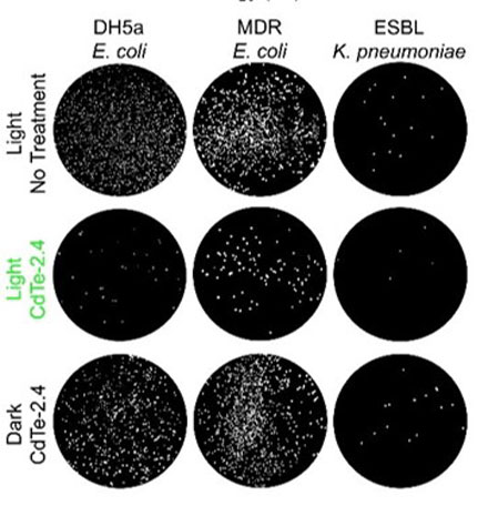
They reported 92 percent of bacterial cells were killed, while leaving mammalian cells intact. The QDs could also be tuned to increase bacterial proliferation.

qd

http://www.photonics.com/images/Web/Articles/2016/1/20/PIC_QD1.jpg

Plated antibiotic resistant ‘superbugs’ before and after treatment with nanoparticles. Courtesy of the Nagpal Group/University of Colorado Boulder.

The team said the killing effect was independent of the QD material used; rather, it was controlled by the redox potentials of the photogenerated charge carriers, which selectively altered cellular redox states. Photoexcited QDs could be used in the study of the effect of redox states on living systems, and lead to clinical phototherapy for the treatment of infections, the researchers said.

The specificity of the treatment could help reduce or eliminate the potential side effects of other treatment methods, as well as provide a path forward for future development and clinical trials.

“Antibiotics are not just a baseline treatment for bacterial infections, but HIV and cancer as well,” said professor Anushree Chatterjee. “Failure to develop effective treatments for drug-resistant strains is not an option, and that’s what this technology moves closer to solving.”

Nagpal and Chatterjee are the cofounders of Praan Biosciences Inc., a startup that can sequence genetic profiles using a single molecule, and have filed a patent on the QD therapy technology.

The research was published in Nature Materials (doi: 10.1038/nmat4542).


Photoexcited quantum dots for killing multidrug-resistant bacteria

Colleen M. Courtney, Samuel M. Goodman, Jessica A. McDaniel, Nancy E. Madinger, Anushree Chatterjee, Prashant Nagpal

Nature Materials(2016)      http://dx.doi.org:/10.1038/nmat4542

Multidrug-resistant bacterial infections are an ever-growing threat because of the shrinking arsenal of efficacious antibiotics1, 2, 3, 4. Metal nanoparticles can induce cell death, yet the toxicity effect is typically nonspecific5, 6, 7, 8. Here, we show that photoexcited quantum dots (QDs) can kill a wide range of multidrug-resistant bacterial clinical isolates, including methicillin-resistant Staphylococcus aureus, carbapenem-resistant Escherichia coli, and extended-spectrum β-lactamase-producingKlebsiella pneumoniae and Salmonella typhimurium. The killing effect is independent of material and controlled by the redox potentials of the photogenerated charge carriers, which selectively alter the cellular redox state. We also show that the QDs can be tailored to kill 92% of bacterial cells in a monoculture, and in a co-culture of E. coli and HEK 293T cells, while leaving the mammalian cells intact, or to increase bacterial proliferation. Photoexcited QDs could be used in the study of the effect of redox states on living systems, and lead to clinical phototherapy for the treatment of infections.

Figure 2: The effect of CdTe-2.4 is specific to the reduction and oxidation potentials.close

The effect of CdTe-2.4 is specific to the reduction and oxidation potentials.

a, Absorbance spectra for CdTe and CdSe of several sizes. Insets show transmission electron microscopy (TEM) images with colour-coded scale bars (50 nm except for CdTe-2.4, which is 25 nm). b, Scanning tunnelling spectroscopy (STS) meas…

Read Full Post »

Nanosensors for Protein Recognition and Gene Proteome Interaction

Larry H Bernstein, MD, FCAP, Curator

LPBI


Synthetic Antibody Detects Proteins

http://www.technologynetworks.com/Proteomics/news.aspx?ID=187242

Research could lead to nanosensors that recognize fibrinogen, insulin, or other biomarkers

Using carbon nanotubes, MIT chemical engineers have devised a new method for detecting proteins, including fibrinogen, one of the coagulation factors critical to the blood-clotting cascade.

This approach, if developed into an implantable sensor, could be useful for monitoring patients who are taking blood thinners, allowing doctors to make sure the drugs aren’t interfering too much with blood clotting.

The new method is the first to create synthetic recognition sites (similar to natural antibodies) for proteins and to couple them directly to a powerful nanosensor such as a carbon nanotube. The researchers have also made significant progress on a similar recognition site for insulin, which could enable better monitoring of patients with diabetes. It may also be possible to use this approach to detect proteins associated with cancer or heart disease, says Michael Strano, the Carbon P. Dubbs Professor in Chemical Engineering at MIT.

A targeted search

The new sensor is the latest example of a method developed in Strano’s lab, known asCorona Phase Molecular Recognition (CoPhMoRe).

This technique takes advantage of the interactions between a given polymer and a nanoparticle surface such as that of a fluorescent single-walled carbon nanotube, when the polymer is wrapped around the nanotube.

Certain regions of the polymers latch onto the nanoparticle surface like anchors, while other regions extend outwards into their environment. This outward-facing region, also known as the adsorbed phase or corona, has a 3-D structure that depends on the composition of the polymer.

CoPhMoRe works when a specific polymer adsorbs to the nanoparticle surface and creates a corona that recognizes the target molecule. These interactions are very specific, just like the binding between an antibody and its target. Binding of the target alters the carbon nanotubes’ natural fluorescence, allowing the researchers to measure how much of the target molecule is present.

Strano’s lab has previously used this approach to find recognition sites and develop nansensors for estradiol and riboflavin, among other molecules. The new paper represents their first attempt to identify corona phases that can detect proteins, which are larger, more complex, and more fragile than the molecules identified by their previous sensors.

For this study, Bisker began by screening carbon nanotubes wrapped in 20 different polymers including DNA, RNA, and polyethylene glycol (PEG), a polymer often added to drugs to increase their longevity in the bloodstream.

On their own, none of the polymers had any affinity for the 14 proteins tested, all taken from human blood. However, when the researchers tested polymer-wrapped nanotubes against the same proteins, they turned up a match between one of the modified nanotubes and fibrinogen.

“A chemist or a biologist would not be able to predict ahead of time that there should be any kind of affinity between fibrinogen and this corona phase,” Strano says. “It really is a new kind of molecular recognition.”

Fibrinogen, one of the most abundant proteins in human blood, is part of the blood-clotting cascade. When a blood vessel is damaged, an enzyme called thrombin converts fibrinogen into fibrin, a stringy protein that forms clots to seal the wound.

A sensor for fibrinogen could help doctors determine if patients who are taking blood thinners still have enough clotting capability to protect them from injury, and could allow doctors to calculate more finely tuned dosages. It could also be used to test patients’ blood clotting before they go into surgery, or to monitor wound healing, Bisker says.

Synthetic antibodies

The researchers believe their synthetic molecular recognition agents are an improvement over existing natural systems based on antibodies or DNA sequences known as aptamers, which are more fragile and tend to degrade over time.

“One of the advantages of this is that it’s a completely synthetic system that can have a much longer lifetime within the body,” Bisker says.

In 2013, researchers in Strano’s lab demonstrated that carbon nanotube sensors can remain active in mice for more than a year after being embedded in a polymer gel and surgically implanted under the skin.

In addition to insulin, the researchers are also interested in detecting troponin, a protein that is released by dying heart cells, or detecting proteins associated with cancer, which would be useful for monitoring the success of chemotherapy. These and other protein sensors could become critical components of devices that deliver drugs in response to a sign of illness.

“By measuring therapeutic markers in the human body in real time, we can enable drug delivery systems that are much smarter, and release drugs in precise quantities,” Strano says. “However, measurement of those biomarkers is the first step.”


New Device Uses Carbon Nanotubes to Snag Molecules
Nanotube “forest” in a microfluidic channel may help detect rare proteins and viruses.
Tuesday, December 22, 2015

Nanotube “forest” in a microfluidic channel may help detect rare proteins and viruses.

Engineers at MIT have devised a new technique for trapping hard-to-detect molecules, using forests of carbon nanotubes.

The team modified a simple microfluidic channel with an array of vertically aligned carbon nanotubes — rolled lattices of carbon atoms that resemble tiny tubes of chicken wire. The researchers had previously devised a method for standing carbon nanotubes on their ends, like trees in a forest. With this method, they created a three-dimensional array of permeable carbon nanotubes within a microfluidic device, through which fluid can flow.

Now the researchers have given the nanotube array the ability to trap certain particles. To do this, the team coated the array, layer by layer, with polymers of alternating electric charge.

“You can think of each nanotube in the forest as being concentrically coated with different layers of polymer,” says Brian Wardle, professor of aeronautics and astronautics at MIT. “If you drew it in cross-section, it would be like rings on a tree.”

Depending on the number of layers deposited, the researchers can create thicker or thinner nanotubes and thereby tailor the porosity of the forest to capture larger or smaller particles of interest.

The nanotubes’ polymer coating may also be chemically manipulated to bind specific bioparticles flowing through the forest. To test this idea, the researchers applied an established technique to treat the surface of the nanotubes with antibodies that bind to prostate specific antigen (PSA), a common experimental target. The polymer-coated arrays captured 40 percent more antigens, compared with arrays lacking the polymer coating.

Wardle says the combination of carbon nanotubes and multilayer coatings may help finely tune microfluidic devices to capture extremely small and rare particles, such as certain viruses and proteins.

“There are smaller bioparticles that contain very rich amounts of information that we don’t currently have the ability to access in point-of-care [medical testing] devices like microfluidic chips,” says Wardle, who is a co-author on the paper. “Carbon nanotube arrays could actually be a platform that could target that size of bioparticle.”

The paper’s lead author is Allison Yost, a former graduate student who is currently an engineer at Accion Systems. Others on the paper include graduate student Setareh Shahsavari; postdoc Roberta Polak; School of Engineering Professor of Teaching Innovation Gareth McKinley; professor of materials science and engineering Michael Rubner, and Raymond A. And Helen E. St. Laurent Professor of Chemical Engineering Robert Cohen.

A porous forest

Carbon nanotubes have been a subject of intense scientific study, as they possess exceptional electrical, mechanical, and optical properties. While their use in microfluidics has not been well explored, Wardle says carbon nanotubes are an ideal platform because their properties may be manipulated to attract certain nanometer-sized molecules. Additionally, carbon nanotubes are 99 percent porous, meaning a nanotube is about 1 percent carbon and 99 percent air.

“Which is what you need,” Wardle says. “You need to flow quantities of fluid through this material to shed all the millions of particles you don’t want to find and grab the one you do want to find.”

What’s more, Wardle says, a three-dimensional forest of carbon nanotubes would provide much more surface area on which target molecules may interact, compared with the two-dimensional surfaces in conventional microfluidics.

“The capture efficiency would scale with surface area,” Wardle notes.

A versatile array

The team integrated a three-dimensional array of carbon nanotubes into a microfluidic device by using chemical vapor deposition and photolithography to grow and pattern carbon nanotubes onto silicon wafers. They then grouped the nanotubes into a cylinder-shaped forest, measuring about 50 micrometers tall and 1 millimeter wide, and centered the array within a 3 millimeter-wide, 7-millimeter long microfluidic channel.

The researchers coated the nanotubes in successive layers of alternately charged polymer solutions in order to create distinct, binding layers around each nanotube. To do so, they flowed each solution through the channel and found they were able to create a more uniform coating with a gap between the top of the nanotube forest and the roof of the channel. Such a gap allowed solutions to flow over, then down into the forest, coating each individual nanotube. In the absence of a gap, solutions simply flowed around the forest, coating only the outer nanotubes.

After coating the nanotube array in layers of polymer solution, the researchers demonstrated that the array could be primed to detect a given molecule, by treating it with antibodies that typically bind to prostate specific antigen (PSA). They pumped in a solution containing small amounts of PSA and found that the array captured the antigen effectively, throughout the forest, rather than just on the outer surface of a typical microfluidic element.

Wardle says that the nanotube array is extremely versatile, as the carbon nanotubes may be manipulated mechanically, electrically, and optically, while the polymer coatings may be chemically altered to capture a wide range of particles. He says an immediate target may be biomarkers called exosomes, which are less than 100 nanometers wide and can be important signals of a disease’s progression.

“Science is really picking up on how much information these particles contain, and they’re sort of everywhere, but really hard to find, even with large-scale equipment,” Wardle says. “This type of device actually has all the characteristics and functionality that would allow you to go after bioparticles like exosomes and things that really truly are nanometer scale.”

This research was funded, in part, by the National Science Foundation.


A Natural Light Switch

MIT scientists identify and map the protein behind a light-sensing mechanism.

MIT scientists, working with colleagues in Spain, have discovered and mapped a light-sensing protein that uses vitamin B12 to perform key functions, including gene regulation.

The result, derived from studying proteins from the bacterium Thermus thermophilus, involves at least two findings of broad interest. First, it expands our knowledge of the biological role of vitamin B12, which was already understood to help convert fat into energy, and to be involved in brain formation, but has now been identified as a key part of photoreceptor proteins — the structures that allow organisms to sense and respond to light.

Second, the research describes a new mode of gene regulation, in which the light-sensing proteins play a key role. In so doing, the scientists observe, the bacteria have repurposed existing protein structures that use vitamin B12, and put them to work in new ways.

MIT-Proteins-Light-1_0.jpg

http://www.technologynetworks.com/images/videos/News%20Images/CR/MIT-Proteins-Light-1_0.jpg

“Nature borrowed not just the vitamin, but really the whole enzyme unit, and modified it … and made it a light sensor,” says Catherine Drennan, a professor of chemistry and biology at MIT


The paper describes the photoreceptors in three different states: in the dark, bound to DNA, and after being exposed to light.

“It’s wonderful that we’ve been able to get all the series of structures, to understand how it works at each stage,” Drennan says.

The paper has nine co-authors, including Drennan; graduate students Percival Yang-Ting Chen, Marco Jost, and Gyunghoon Kang of MIT; Jesus Fernandez-Zapata and S. Padmanabhan of the Institute of Physical Chemistry Rocasolano, in Madrid; and Monserrat Elias-Arnanz, Juan Manuel Ortiz-Guerreo, and Maria Carmen Polanco, of the University of Murcia, in Murcia, Spain.

The researchers used a combination of X-ray crystallography techniques and in-vitro analysis to study the bacteria. Drennan, who has studied enzymes that employ vitamin B12 since she was a graduate student, emphasizes that key elements of the research were performed by all the co-authors.

Jost performed crystallography to establish the shapes of the structures, while the Spanish researchers, Drennan notes, “did all of the control experiments to show that we were really thinking about this right,” among other things.

MIT-Proteins-Light-2.jpg

By studying the structures of the photoreceptor proteins in their three states, the scientists developed a more thorough understanding of the structures, and their functions, than they would have by viewing the proteins in just one state.

Microbes, like many other organisms, benefit from knowing whether they are in light or darkness. The photoreceptors bind to the DNA in the dark, and prevent activity pertaining to the genes of Thermus thermophilus. When light hits the microbes, the photoreceptor structures cleave and “fall apart,” as Drennan puts it, and the bacteria start producing carotenoids, which protect the organisms from negative effects of sunlight, such as DNA damage.

The research also shows that the exact manner in which the photoreceptors bind to the DNA is novel. The structures contain tetramers, four subunits of the protein, of which exactly three are bound to the genetic material — something Drennan says surprised her.

“That’s the best part about science,” Drennan says. “You see something novel, then you think it’s not really going to be that novel, but you do the experiments [and it is].”

Other scientists say the findings are significant. “It’s a very exciting development,” says Rowena Matthews, a professor emerita of biological chemistry at the University of Michigan, who has read the paper. Of the newly discovered use of vitamin B12 and a derivative of it, adenosylcobalamin, Matthews adds, “There was very limited knowledge of its versatility.”

Drennan adds that in the long run, the finding could have practical applications, such as the engineering of light-directed control of DNA transcription, or the development of controlled interactions between proteins.

“I would be very interested in … thinking about whether there could be practical applications of this,” Drennan says.


HIV Protein Manipulates Hundreds of Human Genes

Findings search for new or improved treatments for patients with AIDS.

UT Southwestern Medical Center researchers have deciphered how a small protein made by the human immunodeficiency virus (HIV) that causes AIDS manipulates human genes to further its deadly agenda.

The findings, published in the online journal eLife, could aid in the search for new or improved treatments for patients with AIDS, or to the development of preventive strategies.

“We have identified the molecular mechanisms by which the Tat protein made by HIV interacts with the host cell to activate or repress several hundred human genes,” said Dr. Iván D’Orso, Assistant Professor of Microbiology at UT Southwestern and senior author of the study. “The findings clearly suggest that blocking Tat activity may be of therapeutic value to HIV patients.”

It has long been known that HIV causes AIDS by hijacking the body’s immune cells, transforming them into HIV factories and killing other immune cells that normally fight disease. HIV also hides in cells and continues to undermine the host’s immune system despite antiretroviral therapy that has improved the outlook of those with AIDS.

The latest data from the Centers for Disease Control and Prevention (CDC), in 2012, estimated 1.2 million Americans were living with HIV, including 156,300 whose infections had not been diagnosed. About 50,000 people in the U.S. are newly infected with HIV annually, the CDC projects. In 2013, the CDC estimated that over 26,000 Americans had the advanced form of HIV infection, AIDS.

Like all retroviruses, HIV has very few genes of its own and must take over the host’s cellular machinery in order to propagate and spread throughout the body. Although the broad aspects of that cellular hijacking were known, the nuances remain to be explored, Dr. D’Orso said.

“We observed that HIV methodically and precisely manipulates the host’s genes and cellular machinery. We also observed that HIV rewires cellular defensive pathways to benefit survival of the virus,” he added.

The study provides insights into HIV’s ability to survive despite antiretroviral therapy, findings that could lead to new therapeutic targets or ways to make current therapies more effective, he said.

“Our study indicates that this small viral protein, Tat, directly binds to about 400 human genes to generate an environment in which HIV can thrive. Then, this protein precisely turns off the body’s immune defense. It is striking that such a small viral protein has such a large impact,” Dr. D’Orso said. “The human genes and pathways that Tat manipulates correlate well with symptoms observed in these patients, such as immune system hyperactivation, then weakening, and accelerated aging,” Dr. D’Orso said, describing the situation in which HIV infection leads to AIDS.

Italy’s National Institute of Health in Rome recently completed a phase II clinical trial of an experimental vaccine that targets the Tat protein. That trial, which followed 87 HIV-positive patients for up to three years, reported that the vaccine was well-tolerated without significant side effects. However, it will take several years to determine if the vaccine works, Dr. D’Orso said.

Although someone can have HIV for years without showing symptoms, AIDS occurs when HIV blocks the body’s ability to fight off illness. The person then becomes overrun by the opportunistic infections and specific cancers that are hallmarks of AIDS.


New Light Shed on Genetic Regulation

A team of scientists has uncovered greater intricacy in protein signaling than was previously understood, shedding new light on the nature of genetic production.

Christine Vogel, an assistant professor in New York University’s Department of Biology and one of the study’s senior authors, explains that “to make a protein, we need to make a messenger RNA molecule from the gene encoded in the DNA, and then, in a second process, make proteins from these RNA molecules. Both processes are highly regulated and coupled.”

This coupling is similar to the coupling between a moving escalator and a person walking on it at the same time.

The research takes a closer look at how the two coupled processes change in the cell responding to an outside stimulus.

“Until recently, it has been very difficult to study these systems and researchers have thought that the movement of the escalator is most important during the cellular response,” Vogel explains. “We now show that is not necessarily the case, and under some circumstances, the person’s walking determines the overall outcome.”

In biology, this means that both of the processes—to make RNAs and proteins—play important roles, but with different patterns.

In their study, the scientists, who also included researchers from National University Singapore and Berlin’s Max Delbruck Center, took a closer look at how the two processes exactly respond over time.

Their results showed notable distinctions between DNA and mRNA in the nature of their signaling. Notably, the process of making RNA from DNA was pulse-like—a brief messaging over the studied period that returned to the normal levels by the end of the measurements. By contrast, the process of making a protein from RNA was akin to an on/off switch: once started, levels remained constant for consistent periods before reverting back to long stretches of dormancy.

While the reasons for these differences in cell behavior remain unknown, the researchers believe the answer may lie in the nature of the two tasks.

“It is very costly for the cell to make proteins, but making RNA messages from DNA is a relatively low-energy and simple process, so it makes sense that we see frequent, or pulsating, signaling at this stage,” observes Vogel. “By contrast, creating proteins is an intricate undertaking, requiring a great deal of time and energy. This may be why, once you decided to stop production of proteins, you do not turn it back on that easily—and the other way around.”


Where Cancer Cells May Begin

Scientists use fruit fly genetics to understand how things could go wrong in cancer.

Cancer cells are normal cells that go awry by making bad developmental decisions during their lives. In a study involving the fruit fly equivalent of an oncogene implicated in many human leukemias, Northwestern University researchers have gained insight into how developing cells normally switch to a restricted, or specialized, state and how that process might go wrong in cancer.

The fruit fly’s eye is an intricate pattern of many different specialized cells, such as light-sensing neurons and cone cells. Because flies share with humans many of the same cancer-causing genes, scientists use the precisely made compound eye of Drosophila melanogaster (the common fruit fly) as a workhorse to study what goes wrong in human cancer.

retina638.jpg

http://www.technologynetworks.com/images/videos/News%20Images/GT/retina638.jpg

A multidisciplinary team co-led by biologist Richard W. Carthew and engineer Luís A.N. Amaral studied normal cell behavior in the developing eye. The researchers were surprised to discover that the levels of an important protein called Yan start fluctuating wildly when the cell is switching from a more primitive, stem-like state to a more specialized state. If the levels don’t or can’t fluctuate, the cell doesn’t switch and move forward.

“This mad fluctuation, or noise, happens at the time of cell transition,” said Carthew, professor of molecular biosciences in Northwestern’s Weinberg College of Arts and Sciences. “For the first time, we see there is a brief time period as the developing cell goes from point A to point B. The noise is a state of ‘in between’ and is important for cells to switch to a more specialized state. This limbo might be where normal cells take a cancerous path.”

The researchers also found that a molecular signal received by a cell receptor called EGFR is important for turning the noise off. If that signal is not received, the cell remains in an uncontrolled state.

By pinpointing this noise and its “off” switch as important points in the normal process of cell differentiation, the Northwestern researchers provide targets for scientists studying how cells can go out of control and transform into cancer cells.

The “noisy” protein the Northwestern researchers studied is called Yan in the fly and Tel-1 in humans. (The protein is a transcription factor.) The Tel-1 protein instructs cells to turn into white blood cells; the gene that produces the protein, oncogene Tel-1, is frequently mutated in leukemia.

The EGFR protein that turns off the noise in flies is called Her-2 in humans. Her-2 is an oncogene that plays an important role in human breast cancer.

“On the surface, flies and humans are very different, but we share a remarkable amount of infrastructure,” said Carthew, a member of the Robert H. Lurie Comprehensive Cancer Center of Northwestern University. “We can use fruit fly genetics to understand how humans work and how things go wrong in cancer and other diseases.”

Fruit fly cells are small and closely packed together, making study of them challenging. Carthew and Amaral’s team of biologists, chemical and biological engineers, computer scientists and chemists together figured out how to identify and analyze thousands and thousands of individual cells in the flies’ eyes.

“In the past, people have built models of regulatory networks that control cell differentiation mostly by genetically perturbing one or two components of the network at a time and then compiling those results into models,” said Amaral, professor of chemical and biological engineering at the McCormick School of Engineering. “We instead measured the retina as it developed and found the unexpected behavior of the key regulatory factors Yan and EGFR.”

Nicolás Peláez, first author of the study and a Ph.D. candidate in interdisciplinary biological sciences working with Amaral and Carthew, built new tools to study this strange feature of noise in developing flies. His methods enabled the researchers to easily measure both the concentration of the Yan protein and its fluctuation (noise).

It takes 15 to 20 hours for a fruit fly cell to go from being an unrestricted cell to a restricted cell, Carthew said. Peláez determined the Yan protein is noisy, or fluctuating, for six to eight of those hours.

“Studying the dynamics of molecules regulating fly-eye patterning can inform us about human disease,” Peláez said. “Using model organisms such as fruit flies will help us understand quantitatively the basic biological principles governing differentiation in complex animals.”


Mechanism of Tumor Suppressing Gene Uncovered

The most commonly mutated gene in cancer,p53, works to prevent tumor formation by keeping mobile elements in check that otherwise lead to genomic instability, UT Southwestern Medical Center researchers have found.

The p53 gene long has been known to suppress tumor formation, but the mechanisms behind this function – and why disabling the gene allows tumors to form – were not fully understood.

Findings from the study answer some of these questions and could one day lead to new ways of diagnosing and treating cancer, said the study’s senior author, Dr. John Abrams, Professor of Cell Biology at UT Southwestern.

The investigators found that normal p53 gene action restrains transposons, mobile genetic elements called retroelements that can make copies of themselves and move to different positions on chromosomes. But, they discovered, when p53 is disabled by mutation, dramatic eruptions of these mobile elements occur. The study revealed that in mice with cancer and in human samples of two types of cancer (Wilms’ tumors and colon tumors) disabled for p53, transposons became very active.

In a healthy state, certain mechanisms work to keep these retroelements quiet and inactive, explained Dr. Abrams. One of those mechanisms is p53 action. Conversely, when p53 is mutated, retroelements can erupt.

“If you take the gene away, transposons can wreak havoc throughout the genome by causing it to become highly dysregulated, which can lead to disease,” Dr. Abrams said. “Our findings help explain why cancer genomes are so much more fluid and destabilized than normal genomes. They also provide a novel framework for understanding how normal cells become tumors.”

Although much more research is needed, Dr. Abrams said, the potential clinical implications of the team’s findings are significant.

“Understanding how p53 prevents tumors raises the prospect of therapeutic interventions to correct cases in which p53 is disabled,” Dr. Abrams said. “If retroelements are at the heart of certain p53-driven cancers, finding ways to suppress them could potentially allow us to prevent those cancers or intervene to keep them from progressing.”

This understanding also could lead to advances in diagnosing some cancers through biomarkers related to p53 and transposon activity.

“One possibility is that perhaps blood or urine tests could detect dysregulated retroelements that could be indicative of certain types of cancer,” Dr. Abrams said.

Read Full Post »

« Newer Posts - Older Posts »