Feeds:
Posts
Comments

Archive for the ‘Scientific Publishing’ Category

Tracking metabolism of renal cell carcinoma

Larry H. Bernstein, MD, FCAP, Curator

LPBI


Charting Kidney Cancer Metabolism

http://www.technologynetworks.com/Metabolomics/news.aspx?ID=188237


Changes in cell metabolism are increasingly recognized as an important way tumors develop and progress, yet these changes are hard to measure and interpret. A new tool designed by MSK scientists allows users to identify metabolic changes in kidney cancer tumors that may one day be targets for therapy.


Much of what we know about cancer comes from studying genes. By sequencing genes in tumors, for example, scientists have learned what mutations are typically found in different cancer types. Genetic methods can also be used to survey which proteins are made in tumors.

Yet this information provides only an indirect measure of how cancer cells operate. To really capture that, you need to know about the dynamic chemical changes occurring in these cells; you need to know about cancer metabolism.

Tracing the products of cell metabolism, known as metabolites, is not easy to do. “Looking at metabolites in cancer has been very difficult because the technology was not available,” says James Hsieh, a physician-scientist at Memorial Sloan Kettering and an expert in kidney cancer. “Until recently, we didn’t have the capacity to look at hundreds, even thousands, of different metabolites inside of cells.”

But with advanced biochemical methods, these myriad metabolites are finally coming into focus. Dr. Hsieh’s team has used such methods to profile metabolic changes in hundreds of kidney cancer tumor samples. What’s more, they’ve developed a new online tool that will help researchers make sense of this vast data pool, highlighting previously unknown connections between metabolism and clear cell renal cell carcinoma — the most common, lethal form of the disease.

Metabolism Explained

Think of a cell as a factory. If genes provide the floor plan for the factory, and proteins make up the built environment, then metabolism is the movement of materials through the factory to make products.

For many years, investigators wanting to understand cancer metabolism looked at enzyme levels — proteins that catalyze chemical reactions. Publically accessible databases, such as those maintained by The Cancer Genome Atlas (TCGA), provide this information. The problem is that enzyme levels don’t necessarily tell you whether, and at what rate, metabolites are actually being made.

“There’s no good way to infer how changes in metabolite levels are connected to enzyme levels,” says Ed Reznik, a postdoctoral fellow in computational biology at the Sloan Kettering Institute who is a co-first author on the study. “You really have to go after the metabolites directly.” (To continue the factory analogy, just because a forklift is present on the shop floor doesn’t mean it’s being used.)

If genes provide the floor plan for the factory, and proteins make up the built environment, then metabolism is the movement of materials through the factory to make products.

To track metabolites, the team obtained samples of tumor tissue and normal tissue from 138 clear cell kidney cancer patients treated at MSK. A surgeon on the team and the paper’s other co-first author, Ari Hakimi, performed these operations.

The researchers then used mass spectrometry and liquid and gas chromatography to analyze the levels of more than 800 different metabolites in these samples. By comparing the levels of metabolites in tumors with those in normal tissues, they were able to chart the rise and fall of these chemicals.

There’s an App for That

Making sense of the metabolic data was challenging at first, since there was so much of it. “If you look at human metabolism, there are upward of 5,000 distinct biochemical reactions,” says Dr. Reznik. “It’s really hard to make sense of that in a way that humans can parse.”

So the team decided to build a tool that would help them visualize what was going on. Working with a team of programmers, Dr. Reznik developed what he calls a “metabologram,” which allows users to review the metabolite data for any number of different metabolic pathways, one pathway at a time. Users can compare metabolites between tumor samples and normal samples, or between lower-stage tumors and higher-stage tumors. They can also see how the metabolic data line up against the gene expression data obtained from TCGA.

With the help of their new tool, the team made some startling discoveries. They found that the genetic data from TCGA were not always reflective of what was happening to metabolites in kidney cancer cells, and that the metabolic data help to make better sense of the clinical behavior of kidney cancer tumors.

“Our data are actually much more consistent with the human data obtained from pathology,” Dr. Hsieh says.

Charting Aggressiveness

Taking a bird’s-eye view of the metabolic data, the team found four distinct groupings, or clusters, of tumor samples that they could distinguish based on levels of metabolites. The clusters differed in their level of tumor aggressiveness and highlighted who the high-risk patients were.

“You can use the metabologram to get a sense of what’s driving the aggressive tumors from a metabolic standpoint,” Dr. Hakimi says. Once you have that, you can then think about ways to target that altered metabolism.

The team hopes that the new tool, which is being made freely available online, will help researchers generate novel hypotheses about metabolism and kidney cancer, and even encourage other teams to create metabolograms for other cancer types.

“The goal is ultimately to use this information to improve clinical prediction for kidney cancer and to understand how best to treat it,” Dr. Hsieh says.

Read Full Post »

Flat, Ultralight Lens

Larry H. Bernstein, MD, FCAP, Curator

LPBI


Engineers Develop Flat, Ultralight Lens that could Change How Cameras are Designed

http://www.rdmag.com/news/2016/02/engineers-develop-flat-ultralight-lens-could-change-how-cameras-are-designed


Researchers have always thought that flat, ultrathin optical lenses for cameras or other devices were impossible because of the way all the colors of light must bend through them. Consequently, photographers have had to put up with more cumbersome and heavier curved lenses. But University of Utah electrical and computer engineering professor Rajesh Menon and his team have developed a new method of creating optics that are flat and thin yet can still perform the function of bending light to a single point, the basic step in producing an image.

His findings were published Friday, Feb. 12, in a new paper, “Chromatic-Aberration-Corrected Diffractive Lenses for Ultra-Broadband Focusing,” in the current issue of Scientific Reports. The study was co-authored by University of Utah doctoral students Peng Wang and Nabil Mohammad.

“Instead of the lens having a curvature, it can be very flat so you get completely new design opportunities for imaging systems like the ones in your mobile phone,” Menon says. “Our results correct a widespread misconception that flat, diffractive lenses cannot be corrected for all colors simultaneously.”

In order to capture a photographic image in a camera or for your eyes to focus on an image through eyeglasses, the different colors of light must pass through the lenses and converge to a point on the camera sensor or on the eye’s retina. How light bends through curved lenses is based on the centuries-old concept known as refraction, a principle that is similar to when you put a pencil in a glass of water and notice that it “bends” in the water. To do this, cameras typically will use a stack of multiple curved lenses in order to focus all of the colors of light to a single point. Multiple lenses are needed because different colors bend differently, and they are designed to ensure that all colors come to the same focus.

Menon and his team discovered a way to design a flat lens that can be 10 times thinner than the width of a human hair or millions of times thinner than a camera lens today. They do it through a principle known as diffraction in which light interacts with microstructures in the lens and bends.

“In nature, we see this when you look at certain butterfly wings. The color of the wings is from diffraction. If you look at a rainbow, it’s from diffraction,” he says. “What’s new is we showed that we could actually engineer the bending of light through diffraction in such a way that the different colors all come to focus at the same point. That is what people believed could not be done.”

Menon’s researchers use specially created algorithms to calculate the geometry of a lens so different colors can pass through it and focus to a single point. The resulting lens, called a “super-achromatic lens,” can be made of any transparent material such as glass or plastic.

Other applications of this potential lens system include medical devices in which thinner and lighter endoscopes can peer into the human body. It also could be used for drones or satellites with lighter cameras in which reducing weight is critical. Future smartphones could come with high-powered cameras that don’t require the lens jetting out from the phone’s thin body, such as the lens does now for the iPhone 6S.

Read Full Post »

New DNA replication mechanism

Curator: Larry H. Bernstein, MD, FCAP


Structural Study Points to New DNA Replication Mechanism


GEN  http://www.genengnews.com/gen-news-highlights/structural-study-points-to-new-dna-replication-mechanism/81252345/

https://youtu.be/EbXlPUfajCk

This movie shows the helicase protein complex from all angles, and reveals how its shape changes back and forth between two forms. The research team hypothesizes that the rocking action of this conformational change could help split the DNA double helix and move the helicase along one strand so it can be copied by DNA polymerase.

http://www.genengnews.com/Media/images/GENHighlight/108310_web1851822472.jpg

These are two images showing the structure of the helicase protein complex from above. (a) A surface-rendered three-dimensional electron density map as obtained by cryo-EM. (b) A computer-generated ‘ribbon diagram’ of the atomic model built based on the density map. The helicase has three major components: the Mcm2-7 hexamer ring in green, which encircles the DNA strand; the Cdc45 protein in magenta; and the GINS 4-protein complex in marine blue. Cdc45 and GINS recruit and tether other replisome components to the helicase, including the DNA polymerases that copy each strand of the DNA. [Brookhaven National Laboratory]


A collaborative team of researchers from the U.S. Department of Energy’s Brookhaven National Laboratory, Stony Brook University, Rockefeller University, and the University of Texas have just released detailed structural images of DNA helicase from yeast and are proposing a novel mechanism for how the molecular machinery functions. The scientists believe their new data could provide valuable insight into ways that DNA replication can go askew.

“DNA replication is a major source of errors that can lead to cancer,” explained senior study author Huilin Li, Ph.D., a professor with a joint appointment at Brookhaven Lab and Stony Brook University. “The entire genome, all 46 chromosomes, gets replicated every few hours in dividing human cells, so studying the details of how this process works may help us understand how errors occur.”

The findings from this study were published recently in Nature Structural & Molecular Biology through an article entitled “Structure of the eukaryotic replicative CMG helicase suggests a pumpjack motion for translocation.”

This current study builds upon previous work that produced the first-ever images of the complete DNA-copying protein complex, called the replisome. That study provided a surprising revelation about the location of the DNA-copying enzymes, DNA polymerases. This new study focuses on the atomic-level details for the helicase portion of the protein complex—the part that encircles and splits the DNA double helix so the polymerases can synthesize two new daughter strands.

As they had done in their previous work, Dr. Li and his colleagues produced high-resolution images of the helicase using cryo-electron microscopy (cryo-EM). This technique holds an advantage over other EM methods in that proteins can be studied in solution, closely replicating intracellular conditions.

“You don’t have to produce crystals that would lock the proteins in one position,” Dr. Li remarked. It’s an important point because helicase is a molecular machine made of 11 associated proteins that must be flexible to work. “You have to be able to see how the molecule moves to understand its function,” Dr. Li said.

Once the images of the replication machinery were assembled, the investigators were able to map out the locations of the individual amino acids that make up the helicase complex in each conformation. Then, combining those maps with existing biochemical knowledge, they came up with a mechanism for how the helicase works.

“One part binds and releases energy from a molecule called ATP. It converts the chemical energy into a mechanical force that changes the shape of the helicase,” Dr. Li stated. The molecule subsequently ejects the drained ATP and the helicase complex reverts to its original shape so a new ATP molecule can come in and begin the process again.

“It looks and operates similar to an old-style pumpjack oil rig, with one part of the protein complex forming a stable platform, and another part rocking back and forth,” Dr. Li noted. The researchers postulate that the rocking motion would nudge the DNA strands apart and move the helicase along the double helix in a linear fashion.

This direct translocation mechanism appears to be quite different from the way helicases are thought to operate in more primitive organisms such as bacteria, where the entire complex is believed to rotate around the DNA. However, there is biochemical evidence to support the idea of linear motion, including the fact that the helicase can still function even when the ATP hydrolysis activity of some, but not all, of the components is knocked out by mutation.

“We acknowledge that this proposal may be controversial, and it is not really proven at this point, but the structure gives an indication of how this protein complex works, and we are trying to make sense of it,” Dr. Li stated.


Decades Old DNA Replication Models Called into Question

GEN  http://www.genengnews.com/gen-news-highlights/decades-old-dna-replication-models-called-into-question/81251929/?kwrd=Replisome

Decades Old DNA Replication Models Called into Question

http://www.genengnews.com/media/images/GENHighlight/102252_web8123122217.jpg

A series of electron micrographs show the barrel-shaped helicase, which is the enzyme that separates the two DNA strands, along with other components of the replisome, including polymerase-epsilon (green).[Brookhaven National Laboratory]


Previously (left), the replisome’s two polymerases (green) were assumed to be below the helicase (tan), the enzyme that splits the DNA strands. The new images reveal one polymerase is located at the front of the helicase, causing one strand to loop backward as it is copied (right). [Brookhaven National Laboratory]


It may be time to update biology texts to reflect newly published data from a collaborative team of scientists at Rockefeller University, Stony Brook University, and the U.S. Department of Energy’s Brookhaven National Laboratory. Using cutting-edge electron microscopy (EM) techniques, the investigators gathered the first ever images of the fully assembled replisome, providing new insight into the molecular mechanisms of replication.

“Our finding goes against decades of textbook drawings of what people thought the replisome should look like,” remarked co-senior author Michael O’Donnell, Ph.D., professor and head of Rockefeller’s Laboratory of DNA Replication. “However, it’s a recurring theme in science that nature does not always turn out to work the way you thought it did.”

The researcher’s findings focused on the replisome found in eukaryotic organisms, a category that includes a broad swath of living things, including humans and other multicellular organisms. Over the past several decades, there has been an array of data describing the individual components comprising the complex nature of replisome. Yet, until now no pictures existed to show just how everything fit together.

“This work is a continuation of our long-standing research using electron microscopy to understand the mechanism of DNA replication, an essential function for every living cell,” explained co-senior author Huilin Li, Ph.D., biologist with joint appointments at Brookhaven Lab and Stony Brook University. “These new images show the fully assembled and fully activated ‘helicase’ protein complex—which encircles and separates the two strands of the DNA double helix as it passes through a central pore in the structure—and how the helicase coordinates with the two ‘polymerase’ enzymes that duplicate each strand to copy the genome.”

The image and implications from this study were described in a paper entitled “The architecture of a eukaryotic replisome,” published recently through Nature Structural & Molecular Biology.

Traditional models of DNA replication show the helicase enzyme moving along the DNA, separating the two strands of the double helix, with two polymerases located at the back where the DNA strand is split. In this configuration, the polymerases would add nucleotides to the side-by-side split ends as they move out of the helicase to form two new complete double helix DNA strands.

However, the images that the researchers collected of intact replisomes revealed that only one of the polymerases is located at the back of the helicase. The other is on the front side of the helicase, where the helicase first encounters the double-stranded helix. This means that while one of the two split DNA strands is acted on by the polymerase at the back end, the other has to thread itself back through or around the helicase to reach the front-side polymerase before having its new complementary strand assembled.

“DNA replication is one of the most fundamental processes of life, so it is every biochemist’s dream to see what a replisome looks like,” stated lead author Jingchuan Sun, EM biologist in Dr. Li’s laboratory. “Our lab has expertise and a decade of experience using electron microscopy to study DNA replication, which has prepared us well to tackle the highly mobile therefore very challenging replisome structure. Working together with the O’Donnell lab, which has done beautiful, functional studies on the yeast replisome, our two groups brought perfectly complementary expertise to this project.”

The positioning of one polymerase at the front of the helicase suggests that it may have an unforeseen function—the possibilities of which the collaborative group of scientists is continuing to study. Whatever the function the offset polymerase ends up having, Drs. Li and O’Donnell hope that it will not only provide them better insight into the replication machinery but that they may uncover useful information that can be exploited for disease intervention.

“Clearly, further studies will be required to understand the functional implications of the unexpected replisome architecture reported here,” the scientists concluded.


The architecture of a eukaryotic replisome

Jingchuan Sun, Yi Shi, Roxana E Georgescu, Zuanning Yuan, Brian T Chait, Huilin Li & Michael E O’Donnell

Nature Structural & Molecular Biology 2015; 22:976–982     http://dx.doi.org:/10.1038/nsmb.3113

At the eukaryotic DNA replication fork, it is widely believed that the Cdc45–Mcm2–7–GINS (CMG) helicase is positioned in front to unwind DNA and that DNA polymerases trail behind the helicase. Here we used single-particle EM to directly image a Saccharomyces cerevisiae replisome. Contrary to expectations, the leading strand Pol ε is positioned ahead of CMG helicase, whereas Ctf4 and the lagging-strand polymerase (Pol) α–primase are behind the helicase. This unexpected architecture indicates that the leading-strand DNA travels a long distance before reaching Pol ε, first threading through the Mcm2–7 ring and then making a U-turn at the bottom and reaching Pol εat the top of CMG. Our work reveals an unexpected configuration of the eukaryotic replisome, suggests possible reasons for this architecture and provides a basis for further structural and biochemical replisome studies.


Figure 4: Subunit proximities within CMGE determined by chemical cross-linking with mass spectrometry readout (CX-MS).close

Subunit proximities within CMGE determined by chemical cross-linking with mass spectrometry readout (CX-MS).

CMGE was cross-linked with a lysine-specific bifunctional cross-linker, then fragmented by proteolysis, and cross-linked peptides were identified by mass spectrometry. (a) Overview of cross-links observed within the region of Pol2 …


Structure of the eukaryotic replicative CMG helicase suggests a pumpjack motion for translocation

Zuanning Yuan, Lin Bai, Jingchuan Sun, Roxana Georgescu, Jun Liu, Michael E O’Donnell & Huilin Li

Nature Structural & Molecular Biology 8 Feb 2016    http://dx.doi.org:/10.1038/nsmb.3170

The CMG helicase is composed of Cdc45, Mcm2–7 and GINS. Here we report the structure of theSaccharomyces cerevisiae CMG, determined by cryo-EM at a resolution of 3.7–4.8 Å. The structure reveals that GINS and Cdc45 scaffold the N tier of the helicase while enabling motion of the AAA+ C tier. CMG exists in two alternating conformations, compact and extended, thus suggesting that the helicase moves like an inchworm. The N-terminal regions of Mcm2–7, braced by Cdc45–GINS, form a rigid platform upon which the AAA+ C domains make longitudinal motions, nodding up and down like an oil-rig pumpjack attached to a stable platform. The Mcm ring is remodeled in CMG relative to the inactive Mcm2–7 double hexamer. The Mcm5 winged-helix domain is inserted into the central channel, thus blocking entry of double-stranded DNA and supporting a steric-exclusion DNA-unwinding model.


Figure 1: Cryo-EM and overall structure of theS. cerevisiae CMG complex.

Cryo-EM and overall structure of the S. cerevisiae CMG complex.

(a) A typical motion-corrected raw image from ~8,000 images of frozen CMG particles recorded on a direct detector. (b) Six selected 2D averages representing the particles in different views. (c) 3D cryo-EM map of CMG, color-coded accord…

http://www.nature.com/nsmb/journal/vaop/ncurrent/carousel/nsmb.3170-F1.jpg


Figure 2: Structure and interactions of yeast GINS and Cdc45.

Structure and interactions of yeast GINS and Cdc45.

(a) The full-length GINS structure in top and side views. Domain A is shown in cartoon and domain B in surface. Top, schematic showing that all four subunits have a similar two-domain architecture, but domains A and B in Psf2 and Psf3 a…

http://www.nature.com/nsmb/journal/vaop/ncurrent/carousel/nsmb.3170-F2.jpg


Figure 3: Side-by-side comparison of conformer I and conformer II in the Mcm2–7 region of CMG helicase.

Side-by-side comparison of conformer I and conformer II in the Mcm2-7 region of CMG helicase.

(a,b) Comparison of the two conformations, shown in cartoon representation and viewed from the right side, from Cdc45 and GINS (which are both removed for clarity) with the CTD motor ring on top and the NTD ring at the bottom. The two b…

http://www.nature.com/nsmb/journal/vaop/ncurrent/carousel/nsmb.3170-F3.jpg


Figure 6: Pol2 footprint on the atomic model of CMG helicase.

Pol2 footprint on the atomic model of CMG helicase.

(a) The two-domain architecture of Pol2, the catalytic subunit of the Pol ε complex. The N-terminal half contains the polymerase and exonuclease activities. The C-terminal half is homologous to a B-family polymerase but lacks enzymatic…

Read Full Post »

Genomic expression carried over from Neanderthal DNA

Larry H. Bernstein, MD, FCAP, Curator

LPBI


Neanderthal DNA Shapes Clinical Traits for Modern Humans

GEN  http://www.genengnews.com/gen-news-highlights/neanderthal-dna-shapes-clinical-traits-for-modern-humans/81252363/

https://youtu.be/D8vYSiKE3E4

Modern humans have inherited many physical traits from the Neanderthals. John Capra, Ph.D., from Vanderbilt University, explains how many of these variants affect a variety of clinical disorders

This graphic shows some of the numerous Neanderthal-influenced traits. [Deborah Brewington, Vanderbilt University]

Today being the 207th birthday celebration of renowned naturalist and evolutionary biologist Charles Darwin, it seemed only appropriate to discuss the recent findings of how Neanderthal DNA has shaped and continues to shape human evolution.

Recent studies have identified that individuals of Eurasian origins inherited somewhere between one and four percent of their DNA from Neanderthals. These findings have led to numerous postulations about how these genetic variants may have affected physical characteristics or the behavior of modern humans, ranging from skin color to heightened allergies to fat metabolism.

Now, a new study from a team of scientists led by researchers at Vanderbilt University has directly compared Neanderthal DNA in the genomes of a large population of adults from European ancestry with their clinical records—confirming that this archaic genetic legacy has a subtle but significant effect on modern human biology.

“Our main finding is that Neanderthal DNA does influence clinical traits in modern humans: We discovered associations between Neanderthal DNA and a wide range of traits, including immunological, dermatological, neurological, psychiatric, and reproductive diseases,” explained senior study author John Capra, Ph.D., assistant professor in the department of biomedical informatics and an investigator in the Center for Human Genetics Research at Vanderbilt University Medical School.

The results of this study were published February 12 in Science through an article entitled “The phenotypic legacy of admixture between modern humans and Neanderthals.

Interestingly, Dr. Capra and his colleagues were able to confirm a few of the previous hypotheses about the influence of Neanderthal DNA on modern Homo sapiens. For instance, investigators found that Neanderthal DNA affects keratinocytes, which help protect the skin from environmental damage such as ultraviolet radiation and pathogens. The new analysis found Neanderthal DNA variants influence skin biology in modern humans, in particular, the risk of developing sun-induced skin lesions called keratosis, which are caused by abnormal keratinocytes.

Surprisingly, the research team found that some regions of Neanderthal DNA were associated with psychiatric and neurological effects. In one example, they found that a specific bit of Neanderthal DNA significantly increased the risk for nicotine addiction, while a separate set of variants influenced the risk for depression (positively and negatively).

“The brain is incredibly complex, so it’s reasonable to expect that introducing changes from a different evolutionary path might have negative consequences,” noted lead author and Vanderbilt doctoral student Corinne Simonti.

In the current study, the authors discussed that the pattern of associations they discovered suggests today’s population retains Neanderthal DNA that may have provided modern humans with some adaptive advantages 40,000 years ago as they migrated into regions outside of Africa with different pathogens and levels of sun exposure.

To study these associations, the scientists used a database containing 28,000 patients whose biological samples have been linked to anonymized versions of their electronic health records. The data came from eMERGE—the Electronic Medical Records and Genomics Network—which links digitized records from Vanderbilt University Medical Center’s BioVU databank and eight other hospitals around the country.

This massive amount of genomic data allowed the researchers to determine if each individual had ever been treated for a particular set of medical conditions, such as heart disease, arthritis, or depression. Subsequently, they analyzed the genomes of each individual to identify the unique set of Neanderthal DNA that each person carried. The comparison of each data set allowed the researchers to test whether each bit of Neanderthal DNA individually and in aggregate influences risk for the traits derived from the medical records.

“Vanderbilt’s BioVU and the network of similar databanks from hospitals across the country were built to enable discoveries about the genetic basis of disease,” Dr. Capra remarked. “We realized that we could use them to answer important questions about human evolution.”

While Dr. Capra and his colleagues were thrilled by their findings—this work establishes a new way to investigate questions about the effects of events in recent human evolution—the researcher team also realized that there is a lot of additional information contained in the medical records, such as lab tests, doctors’ notes, and medical images, that could be used in future analyses to refine their data.

Neanderthals’ Genetic Legacy

Ancient DNA in the genomes of modern humans influences a range of physiological traits.

By Ruth Williams | February 11, 2016

http://www.the-scientist.com/?articles.view/articleNo/45309/title/Neanderthals–Genetic-Legacy/

People of Eurasian origin are, genetically speaking, between 1 percent and 4 percent Neanderthal, and new research shows how this archaic DNA in their genomes may be impacting their health. The study, published today (February 11) in Science, utilized the electronic medical records and associated DNA data of more than 28,000 individuals to show that Neanderthal DNA had small but significant effects on the risks of developing—among other things—depression, skin lesions, and excessive blood clotting.

“They’ve looked at huge databases of medical records to see if there are traits that correlate with the presence of particular genes from Neanderthals and have found a number of them,” said anthropologist John Hawks of the University of Wisconsin who was not involved in the study. “The take-away is that these genes that we have from these ancient people have effects on our phenotypes, and that’s pretty cool. They are not just shadows that are not doing anything, they are actually participating in our biology.”

Sequencing of Neanderthal genomes isolated from fragments of bones has revealed that modern humans contain remnants of Neanderthal DNA—a result of interbreeding between the two subspecies. But while certain loci in human genomes have been found to contain an abundance of Neanderthal alleles, it has been unclear whether these alleles have actual functional effects on human traits and, if so, what those are.

Evolutionary and computational geneticist John Capra of Vanderbilt University in Nashville, Tennessee, and colleagues devised an ingenious way to investigate such functional effects on a genome-wide scale. “We realized that we had a great opportunity to answer these questions using large databases of anonymized versions of patient electronic health records linked to their genetic information,” Capra said in a statement.

“A number of previous studies have focused on individual genes,” said evolutionary geneticist Rasmus Neilsen of the University of California, Berkeley, who did not participate in the research. “But this is the first study that really systematically goes through and uses the knowledge we have about genetic variations in humans to answer the question: How much has integration of DNA from Neanderthals affected observable traits in humans?”

Within Neanderthal DNA found in humans, the researchers focused on the most common variants—single nucleotide polymorphisms (SNPs)—and asked, individually and en masse, whether  these variants were associated with any of the medical traits listed for the 28,000 patients.

Investigating the SNPs en masse through a genome-wide complex trait analysis (GCTA), the researchers discovered associations with depression, mood disorders, and a particular type of skin lesion caused by sun exposure. Investigating individual SNPs, on the other hand, the researchers picked out associations tied to tobacco use, urinary problems, and blood hypercoagulation.

Why have such apparently detrimental gene variants been maintained in the human genome? It is important to realize, said Hawks, that “when you look at people’s medical records, you don’t see the good stuff.”

Hawks also noted that “the [observed] associations are really, really small,” meaning that while the links between Neanderthal alleles and certain medical traits were statistically significant, they only represented a tiny percentage of the risk—1 percent to 2 percent in the case of depression, for example.

Further, “many genetic variants, regardless of evolutionary origin and temporal context, are beneficial in some respects but detrimental in others,” Capra added in the statement. For example, while hypercoagulation may increase a person’s thrombosis risk , coagulation is an early innate immune response that protects against injury and infection. As Neanderthals colonized new territories and were exposed to new pathogens, having a souped-up version of this response may therefore have been a favorable defense mechanism.

Capra’s team carried out further experiments to look at whether Neanderthal alleles were associated with classes of traits rather than individual ones, finding neurological and psychiatric traits were both over-represented.

Together with the findings that depression, mood disorders and tobacco use were individually associated with Neanderthal SNPs, this suggested to the researchers that the brains of modern humans have been particularly influenced by Neanderthal DNA. And this might overturn notions of Neanderthals as not-so-bright, said Hawks. “If you had the hypothesis that Neanderthals [died out] because they were stupid,” he said, “you have to explain why their genes are here doing stuff in our brains.”

C.N. Simonti et al., “The phenotypic legacy of admixture between modern humans and Neandertals,” Science, 351:737-41, 2016.


Capra Lab   Evolutionary and Computational Genomics at Vanderbilt University

We use the tools of computer science and statistics to address problems in genetics, evolution, and biomedicine.

Our group is located in the Department of Biological Sciences and affiliated with theVanderbilt Genetics Institute, the Center for Structural Biology, and the Department of Biomedical Informatics at Vanderbilt University.

Humans differ from one another and our closest living relatives, the chimpanzees, in a wide range of traits, including our susceptibility to many diseases. We model the evolutionary processes that have produced these novel traits and develop algorithms that compare genomes to predict the functional relevance of specific genetic differences between individuals and species.

Our research is motivated by several questions:

  • How have evolutionary processes produced the astonishing diversity of form and function present in the natural world?
  • How can better algorithms lead to a deeper understanding of biological systems and networks?
  • How do genomes encode and maintain the information necessary to produce life?
  • How can our increasing knowledge of genomic variation be translated into the treatment and prevention of disease?

We investigate these questions in a number of model systems, but our main focus is on the origins and recent evolution of human populations and their primate relatives.


Borrowing Immunity Through Interbreeding

Neanderthals and Denisovans contributed innate immune genes to modern humans, scientists show.

By Kate Yandell | January 7, 2016

http://www.the-scientist.com/?articles.view/articleNo/45001/title/Borrowing-Immunity-Through-Interbreeding/

The proportion of Neanderthal-derived toll-like receptors in populations, with Neanderthal alleles in orange and green and non-archaic alleles in blue.DANNEMANN ET AL./AJHG

Modern humans adopted innate immune genes responsible for recognizing invading microbes from Neanderthals and Denisovans, according to two studies published today (January 7) in The American Journal of Human Genetics. The two teams, based in France and Germany, independently concluded that humans picked up some versions of a cluster of toll-like receptors by interbreeding with archaic hominin relatives.

“Once humans came out of Africa and then encountered archaic species, they might also have encountered their pathogens,” said Rasmus Nielsen, an evolutionary biologist at the University of California, Berkeley, who was not involved in the studies. “There might have been pathogens that could affect Neanderthals and Denisovans that also could jump into modern humans.”

“At least partially, Neanderthals may have harbored already adaptive mutations, mutations that rendered them more resistant to infections,” said Lluis Quintana-Murci, an evolutionary geneticist at the Pasteur Institute in Paris and a coauthor of one of the new papers.

Previous studies have shown that modern humans interbred with Neanderthals and Denisovans. For instance, Nielsen and his colleagues showed that humans who migrated to Tibet likely picked up an allelecontrolling blood hemoglobin concentration from local Denisovans, allowing them to adapt to living at high altitudes. Another paper indicated that humans had picked up major histocompatibility genes from Denisovans and Neanderthals.

The authors of the two new studies approached the topic of ancient human evolution from different directions. Quintana-Murci and his colleagues decided to do a broad survey of innate immune genes and their variability among present-day humans around the world, using sequence data gathered through the1,000 Genomes Project. The team demonstrated that innate immune genes have been under stronger-than-average selective pressures. Some innate genes are highly conserved, with little tolerance for variability. Other protein-coding genes have picked up adaptive mutations, mostly occurring within the last 6,000 to 13,000 years after humans transitioned from a hunter-gatherer to agricultural society. The resulting increase in density of human settlements, cohabitation with animals, and increased exposure to sewage may have made humans easier targets for microbial disease, the researchers speculated.

Quintana-Murci and his colleagues also took advantage of a previously published map of areas of the human genome where Neanderthal genes are present, showing that innate immune genes are generally more likely to have been borrowed from Neanderthals than genes coding other types of proteins. Specifically, they noted that 126 innate immune genes in present-day Europeans, Asians, or both groups were among the top 5 percent of genes in the genome of each population most likely to have originated in Neanderthals. The cluster of toll-like receptor genes, encoding TLR 1, TLR 6, and TLR 10, both showed signs of having been borrowed from Neanderthals and having picked up adaptive mutations at various points in history.

Meanwhile, a group led by Janet Kelso of the Max Planck Institute for Evolutionary Anthropology in Leipzig, Germany, used both the same previously published Neanderthal introgression map that Quintana-Murci used and a second introgression map. The researchers searched for borrowed regions of the genome that were especially long and common in present-day humans, eventually zeroing in TLR6, TLR10, and TLR1. These receptors, which detect conserved microbial proteins such as flagellin, are all encoded along the same segment of DNA on chromosome four.

By looking at 1,000 Genomes Project data, Kelso and her colleagues were able to identify seven distinct versions of the TLR cluster. The researchers were able to match two of these versions to DNA from Neanderthals, and one version to DNA from Denisovans.

“There have been three potentially independent admixtures,” said Kelso. “We suspect it was two different Neanderthals and a Denisovan.”

Kelso and her colleagues then attempted to figure out the functional differences between the Neanderthal and Denisovan versions of the TLR cluster and the versions that likely originated with the modern humans who migrated from Africa to Europe and Asia later than these archaic hominids.

The changes in the Neanderthal and Denisovan TLR clusters do not lead to altered proteins. However, the researchers found that in white blood cells, the Neanderthal and Denisovan TLRs are more highly expressed than the non-borrowed human TLR clusters.

Kelso and her colleagues also did a survey of already-completed genome wide association studies, finding that present-day people who have the borrowed TLR clusters show lower levels of the bacteriumHelicobacter pylori in their bloodstreams than people descended from humans that did not pick up TLR clusters from Neanderthals or Denisovans. People with the borrowed TLR clusters also tend to have elevated allergies to dust and pollen.

Kelso hypothesized that the Denisovan and Neanderthal TLR clusters may have strengthened the human immune systems against novel pathogens they encountered in their new homes in Europe and Asia. This may have yielded an immune system both skilled at fighting off pathogens and slightly oversensitive, leading to the allergies people carrying the archaic TLRs sometimes have today.

But it is less clear exactly how the immune system was strengthened, or what pathogens ancient humans were trying to fight. “What the gene expression results tell us is that there is some kind of a functional effect for introgression,” said Sri Sankararaman, a statistical geneticist at University of California, Los Angeles, who was not involved in the studies but did help make one of the preexisting introgression maps used in the papers. “That’s basically what it has established. Going from there to making a claim about its fitness effect is less obvious.”

And the reduced H. pylori prevalence associated with the borrowed TLR alleles is simply a sign that the variants are associated with altered immunity, not necessarily an indication that breeding with Neanderthals helped humans avoid this particular pathogen. “We may not have the pathogens around today that selection was acting in response to,” said Nielsen.

The studies help confirm that interbreeding between humans, Neanderthals, and Denisovans shaped human evolution, sometimes offering key advantages people of combined lineage. “The things that modern humans took away from the interbreeding with the Neanderthals were regions of the genome involved in adaptation to the environment,” said Kelso.

M. Deschamps et al., “Genomic signatures of selective pressures and introgression from archaic hominins at human innate immunity genes,” The American Journal of Human Genetics, doi:10.1016/j.ajhg.2015.11.014, 2016.

M. Dannemann et al., “Introgression of Neandertal- and Denisovan-like haplotypes contributes to adaptive variation in human toll-like receptors,” The American Journal of Human Genetics,doi:10.1016/j.ajhg.2015.11.015


Genomic Signatures of Selective Pressures and Introgression from Archaic Hominins at Human Innate Immunity Genes

Matthieu Deschamps, Guillaume Laval, Maud Fagny, Yuval Itan, et al.
Am J Human Gen Jan 2016;  98(1):5–21, 7.   http://dx.doi.org/10.1016/j.ajhg.2015.11.014
Human genes governing innate immunity provide a valuable tool for the study of the selective pressure imposed by microorganisms on host genomes. A comprehensive, genome-wide study of how selective constraints and adaptations have driven the evolution of innate immunity genes is missing. Using full-genome sequence variation from the 1000 Genomes Project, we first show that innate immunity genes have globally evolved under stronger purifying selection than the remainder of protein-coding genes. We identify a gene set under the strongest selective constraints, mutations in which are likely to predispose individuals to life-threatening disease, as illustrated by STAT1 and TRAF3. We then evaluate the occurrence of local adaptation and detect 57 high-scoring signals of positive selection at innate immunity genes, variation in which has been associated with susceptibility to common infectious or autoimmune diseases. Furthermore, we show that most adaptations targeting coding variation have occurred in the last 6,000–13,000 years, the period at which populations shifted from hunting and gathering to farming. Finally, we show that innate immunity genes present higher Neandertal introgression than the remainder of the coding genome. Notably, among the genes presenting the highest Neandertal ancestry, we find the TLR6-TLR1-TLR10 cluster, which also contains functional adaptive variation in Europeans. This study identifies highly constrained genes that fulfill essential, non-redundant functions in host survival and reveals others that are more permissive to change—containing variation acquired from archaic hominins or adaptive variants in specific populations—improving our understanding of the relative biological importance of innate immunity pathways in natural conditions.

Read Full Post »

Intestinal inflammatory pharmaceutics

Curator: Larry H. Bernstein, MD, FCAP

AbbVie Invests in Synthetic Microbes for Treatment of Intestinal Disorders

Aaron Krol    http://www.bio-itworld.com/2016/2/10/abbvie-invests-synthetic-microbes-treatment-intestinal-disorders.html

2.1.2.6

Intestinal Inflammatory Pharmaceutics, Volume 2 (Volume Two: Latest in Genomics Methodologies for Therapeutics: Gene Editing, NGS and BioInformatics, Simulations and the Genome Ontology), Part 2: CRISPR for Gene Editing and DNA Repair

February 10, 2016 | This morning, AbbVie announced a partnership with Synlogic of Cambridge, Mass., to create microbiome-based therapies for the treatment of inflammatory bowel disease (IBD). The two companies have sketched out a suggested three-year timeline for preclinical research and development, after which AbbVie will take over advancing any drug candidates into clinical trials.

Drugs inspired by the microbes that live in the human gut are a hot topic in biotech. Companies like Seres Health and Vedanta Biosciences are pursuing the idea from a variety of angles, from making traditional small molecule drugs that interact with the microbiome, to creating probiotics or microbial cocktails that restore a healthy balance to the gut ecosystem. IBD, including Crohn’s disease and ulcerative colitis, is an especially popular target for these companies, thanks to strong suggestions that bacterial populations can affect the course of the disease. Already, Second Genome and Coronado Biosciences have taken prospective treatments into the clinic (though the latter has been dealt serious setbacks in Phase II trials).

But even among this peculiar batch of startups, Synlogic’s approach to drug design is exquisitely odd. The company calls its products “synthetic biotics”―in fact, they’re genetically engineered bacteria whose DNA contains intricately designed “gene circuits,” built to start producing therapeutic molecules when and only when the patient needs them.

“We are not looking at correcting the dysregulation of microbes in the gut, like other microbiome companies,” CEO José-Carlos Gutiérrez-Ramos tells Bio-IT World. “We have one bacterium, and it’s engineered to do different functions.”

Synlogic was founded in 2013 by two synthetic biologists at MIT, Timothy Lu and Jim Collins. (Bio-IT World has previously spoken with Lu about his academic work on bacterial gene circuits.) Gutiérrez-Ramos joined almost two years later, leaving a position as the head of Pfizer’s BioTherapeutics R&D group, where he had plenty of opportunity to turn emerging biotechnology ideas into drug candidates ready for submission to the FDA.

Still, synthetic biotics are a good deal more unusual than the biologic drugs he worked on at Pfizer.

His new company doesn’t quite spin functions for its microbes out of whole cloth. All the genes the company uses are copied either from the human genome, or from the bacteria living inside us. But by recombining those genes into circuits, Gutiérrez-Ramos believes Synlogic can finely control whether and when genes are expressed, giving its synthetic biotics the same dosage control as a traditional drug. Meanwhile, choosing the right bacterium to engineer―the current favorite is a strain called E. coli Nissle―ensures the biotics do not form stable colonies in the gut, but can be cleared out as soon as a patient stops treatment.

“We’re pharma guys,” he says. “What we want is to have pharmacologically well-defined products.”

The Molecular Circuit Board

Even before the partnership with AbbVie, Synlogic had a pipeline of drug candidates in development, all meant to treat rare genetic disorders caused by single mutations that shut down the activity of a crucial gene. In principle, there seems to be no reason that bacteria carrying the right genes couldn’t pick up the slack. “We know the patient is missing a function that is typically performed by the liver, or the kidney, or the pancreas,” says Gutiérrez-Ramos. “What we do is shift that function from an organ to a stable fraction of the microbiome.”

The approach is in some ways analogous to gene therapy, where a corrected version of a broken gene is inserted into a patient’s own DNA. “We don’t use that word, but the fact is it’s a non-somatic gene therapy,” Gutiérrez-Ramos says. “And if something goes wrong, you can control it just by stopping treatment.” The most advanced synthetic biotic in Synlogic’s pipeline targets urea cycle disorder, exactly the sort of disease that might otherwise be addressed by gene therapy: patients are missing a single enzyme that helps remove nitrogen from the body and prevent it from forming ammonia in the bloodstream. Synlogic will meet with the FDA this March to discuss whether and how this first product can be tested in humans.

Gutierrez Ramos

The new IBD program with AbbVie, however, adds a whole new level of complexity. Executives from the two companies have been in discussions for around six months, and both agree that no single mechanism will be enough to provide significant relief for patients. Crohn’s and ulcerative colitis are painful autoimmune diseases that involve both a weakening of the epithelial lining in the stomach, and a buildup of inflammatory molecules. The development plan that AbbVie and Synlogic have agreed on includes three separate methods of attack to relieve these symptoms.

“One approach AbbVie is very interested in is for our synthetic biotics to produce substances that could tighten the epithelial barrier,” says Gutiérrez-Ramos. “Another approach is to degrade pro-inflammatory molecules”―the same tack taken by AbbVie’s current leading IBD drug, Humira, which targets the inflammatory protein TNFα. “Finally, we can produce anti-inflammatory molecules.”

Uniquely, synthetic biotics can perform all three functions at once; it’s just a matter of inserting the right genes. But that alone might not be a decisive advantage over some sort of combination therapy. The biggest selling point of Synlogic’s microbes is not the genes they can be engineered to express―what you might call the “output” of their gene circuits―but the input, the DNA elements called “inducible promoters” that decide when those genes should be activated.

The core idea is that patients will have a constant population of synthetic biotics in their bodies, taken daily―but those microbes will only generate their therapeutic payloads when needed. In IBD, Gutiérrez-Ramos explains, “it’s not that the patient is always inflamed, but they have flares. Our vision, and AbbVie’s vision, is that the bacteria that you take every day sense when the flare is coming, and then trigger the genetic output.”

This would be a major improvement over a drug like Humira, which after all is constantly inhibiting a part of the immune system. Patients taking Humira, or one of the many other immunosuppressant drugs for IBD, are at a constantly heightened risk of infection; tuberculosis is a particular specter for these patients. If Synlogic can find a genetic “on-switch” that responds to a reliable indicator of IBD flares, it could potentially create a much more precisely administered treatment, while still giving patients the simple dosing schedule of one pill every day.

The company has leads on two inducible promoters that might do the trick: one that reacts to nitric oxide, and another tied to reactive oxygen species. Of course, there’s no guarantee that either will respond sensitively to IBD flares in a real clinical setting. “This is an early time for the technology,” says Gutiérrez-Ramos. “We have demonstrated this in animals, but we have to demonstrate it in humans.”

Although it’s far too early to say if synthetic biotics will become an ordinary part of the pharma toolkit, AbbVie’s decision to invest in the technology offers the means to test this approach on a large scale. Synlogic expects to raise its own funding for trials of its rare disease products, which the FDA does not expect to enroll huge numbers of patients, but IBD is a problem of a very different order.

“We are very honored to work with truly the leader in treatment of inflammatory bowel disease,” says Gutiérrez-Ramos. With the backing of big pharma, it will be possible to trial microbiome-based therapies for the kinds of common, chronic diseases that are the biggest drain on our healthcare system. What’s more, the AbbVie partnership is an important signal of the industry’s faith in synthetic biology as an approach to treating disease.

Read Full Post »

Continuous diffraction crystallographics analysis

Larry H. Bernstein, MD, FCAP, Curator

LPBI


Biomolecular Structure Emerges from the Crystallographic Shadows

http://www.genengnews.com/gen-news-highlights/biomolecular-structure-emerges-from-the-crystallographic-shadows/81252359/


http://www.genengnews.com/Media/images/GENHighlight/thumb_Feb11_2016_DESY_DisorderedCrystals7411016594.jpg

Here’s the caption/credit for the image: Slightly disordered crystals of complex biomolecules like that of the photosystem II molecule shown here produce a complex continous diffraction pattern (right) under X-ray light that contains far more information than the so-called Bragg peaks of a strongly ordered crystal alone (left). The degree of disorder is greatly exaggerated in the crystal on the right. [Eberhard Reimann/DESY]


In keeping with the adage, “If life gives you lemons, make lemonade,” an international team of scientists has shown that if X-crystallography relies on low-quality crystals, it can still derive high-quality structural information. In fact, resolutions can be achieved that surpass the Bragg diffraction limit.

The key, it turns out, is to make the most out of continuous diffraction data, which is ordinarily considered a nuisance in crystallographic analysis. Continuous diffraction data could be obtained from a single molecule, but would be too weak to yield any kind of analysis. But if such data could be combined from a collection of molecules, analyses would be possible. Each of the molecules in the collection, however, would have to be misaligned only in the translational sense. That is, the molecules could not be misaligned rotationally or differ intramolecularly.

With these limitations in mind, scientists based at the Center for Free-Electron Laser Science, DESY, in Germany “read” the atomic structure of complex biomolecules by crystallography without the usual need for prior knowledge and chemical insight. “This discovery has the potential to become a true revolution for the crystallography of complex matter,” said the chairman of DESY’s board of directors, Professor Helmut Dosch.

The work of the DESY-led scientific team appeared February 10 in Nature, in an article entitled “Macromolecular diffractive imaging using imperfect crystals.” The article described how the scientists took advantage of a phenomenon called continuous diffraction.

Protein crystals, particularly imperfect protein crystals, do not always “diffract,” in the traditional Bragg sense. A proper, perfect crystal scatters X-rays in many different directions, producing an intricate and characteristic pattern of numerous bright spots, called Bragg peaks (named after the British crystallography pioneers William Henry and William Lawrence Bragg). The positions and strengths of these spots contain information about the structure of the crystal and of its constituents. Using this approach, researchers have already determined the atomic structures of tens of thousands of proteins and other biomolecules.

“Continuous” scattering arises when crystals become disordered. Usually, this non-Bragg continuous diffraction is not used to derive structural information. Instead, it is used to provide insights into vibrations and dynamics of molecules. But when the disorder consists only of displacements of the individual molecules from their ideal positions in the crystal, the “background” takes on a much more complex character—and its rich structure is anything but diffuse. It then offers a much bigger prize than the analysis of the Bragg peaks: The continuously modulated “background” fully encodes the diffracted waves from individual “single” molecules.

The possibility of using continuous diffraction for structural determinations leads to a paradigm shift in crystallography—the most ordered crystals are no longer the best to analyze with the novel method. “For the first time we have access to single molecule diffraction—we have never had this in crystallography before,” explained DESY’s Professor Henry Chapman. “But we have long known how to solve single-molecule diffraction if we could measure it.” The field of coherent diffractive imaging, spurred by the availability of laser-like beams from X-ray free-electron lasers, has developed powerful algorithms to directly solve the phase problem in this case, without having to know anything at all about the molecule.

“We show for crystals of the integral membrane protein complex photosystem II that lattice disorder increases the information content and the resolution of the diffraction pattern well beyond the 4.5-ångström limit of measurable Bragg peaks, which allows us to phase the pattern directly,” wrote the authors of the Nature article. “Using the molecular envelope conventionally determined at 4.5 ångströms as a constraint, we obtain a static image of the photosystem II dimer at a resolution of 3.5 ångströms. This result shows that continuous diffraction can be used to overcome what have long been supposed to be the resolution limits of macromolecular crystallography.”

Read Full Post »

New anti-Malarial treatment

Larry H. Bernstein, MD, FCAP, Curator

LPBI


Malaria Proteasome Inhibitors Could Reverse Parasite Drug Resistance

http://www.genengnews.com/gen-news-highlights/malaria-proteasome-inhibitors-could-reverse-parasite-drug-resistance/81252358/


http://www.genengnews.com/Media/images/GENHighlight/thumb_108676_web2680362491.jpg

This structure (bottom left) of the malaria parasite’s proteasome, obtained using the revolutionary Cryo-Electron Microscopy technique, enabled the design of a specific inhibitor (front) against the mosquito-borne malaria parasite (pictured at back). [University of Melbourne]


  • With media attention recently focused on the spread of the Zika virus, it’s easy to forget about the mosquito-borne disease that has been credited with killing one out of every two people who have ever lived—malaria. Currently, close to 50 percent of the world’s population live in malaria-endemic areas, leading to between 200–500 million new cases and close to 500,000 deaths annually (mostly children under the age of five).

    Adding to the complexities of trying to control this disease is that resistance to the most effective antimalarial drug, artemisinin, has developed in Southeast Asia, with fears it will soon reach Africa. Artemisinin-resistant species have spread to six countries in five years.

    A collaborative team of scientists from Stanford University, University of California, San Francisco, University of Melbourne, and the MRC in Cambridge have used cutting-edge technology to design a smarter drug to combat the resistant strain.

    “Artemisinin causes damage to the proteins in the malaria parasite that kill the human cell, but the parasite has developed a way to deal with that damage. So new drugs that work against resistant parasites are desperately needed,” explained coauthor Leann Tilley, Ph.D., professor and deputy head of biochemistry and molecular biology in the Bio21 Molecular Science and Biotechnology Institute at The University of Melbourne.

    Malaria is caused by the protozoan parasite from the genus Plasmodium. Five different species are known to cause malaria in humans, with P. falciparum infection leading to the most deaths. The parasite is transmitted through the bite of the female mosquito and ultimately ends up residing within the host’s red blood cells (RBCs)—replicating and then bursting forth to invade more RBCs in a recurrently timed cycle.

    “This penetration/replication/breakout cycle is rapid—every 48 hours—providing the opportunity for large numbers of mutations that can produce drug resistance,” said senior study author Matthew Bogyo, Ph.D., professor in the department of pathology at Stanford Medical School. “Consequently, several generations of antimalarial drugs have long since been rendered useless.”

    The compound that investigators developed targets the parasites proteasome—a protein degradation pathway that removes surplus or damaged proteins through a cascade of proteolytic reactions.

    “The parasite’s proteasome is like a shredder that chews up damaged or used-up proteins. Malaria parasites generate a lot of damaged proteins as they switch from one life stage to another and are very reliant on their proteasome, making it an excellent drug target,” Dr. Tilley noted.

    The scientists purified the proteasome from the malaria parasite and examined its activity against hundreds of different peptide sequences. From this, they were able to design inhibitors that selectively targeted the parasite proteasome while sparing the human host enzymes.

    The findings from this study were published recently in Nature through an article titled “Structure- and function-based design of Plasmodium-selective proteasome inhibitors.”

    Additionally, scientists at the MRC used a new technique called Single-Particle Cryo-Electron Microscopy to generate a three-dimensional, high-resolution structure of a protein, based on thousands composite images.

    The researchers tested the new drug in red blood cells infected with parasites and found that it was as effective at killing the artemisinin resistant parasites as it was for the sensitive parasites.

    “The compounds we’ve derived can kill artemisinin-resistant parasites because those parasites have an increased need for highly efficient proteasomes,” Dr. Bogyo commented. “So, combining the proteasome inhibitor with artemisinin should make it possible to block the onset of resistance. That will, in turn, allow the continued use of that front-line malaria treatment, which has been so effective up until now.”

    “The new proteasome inhibitors actually complement artemisinin drugs,” Dr. Tilley added. “Artemisinins cause protein damage and proteasome inhibitors prevent the repair of protein damage. A combination of the two provides a double whammy and could rescue the artemisinins as antimalarials, restoring their activity against resistant parasites.”

    The scientists were excited by their results, as they may provide a much-needed strategy to combat the growing levels of resistance for this deadly pathogen. However, the researchers tempered their exuberance by noting that many more drug libraries needed to be screened before clinical trials can begin.

    “The current drug is a good start, but it’s not yet suitable for humans. It needs to be able to be administered orally and needs to last a long time in the blood stream,” Dr. Tilley concluded.

Read Full Post »

Regulatory DNA Engineered, Volume 2 (Volume Two: Latest in Genomics Methodologies for Therapeutics: Gene Editing, NGS and BioInformatics, Simulations and the Genome Ontology), Part 2: CRISPR for Gene Editing and DNA Repair

Regulatory DNA engineered

Larry H. Bernstein, MD, FCAP, Curator

LPBI


New Type of CRISPR Screen Probes the Regulatory Genome

Aaron Krol    http://www.bio-itworld.com/2016/2/8/new-type-crispr-screen-probes-regulatory-genome.html

February 8, 2016 | When a geneticist stares down the 3 billion DNA base pairs of the human genome, searching for a clue to what’s gone awry in a single patient, it helps to narrow the field. One of the most popular places to look is the exome, the tiny fraction of our DNA―less than 2%―that actually codes for proteins. For patients with rare genetic diseases, which might be fully explained by one key mutation, many studies sequence the whole exome and leave all the noncoding DNA out. Similarly, personalized cancer tests, which can help bring to light unexpected treatment options, often sequence the tumor exome, or a smaller panel of protein-coding genes.

Unfortunately, we know that’s not the whole picture. “There are a substantial number of noncoding regions that are just as effective at turning off a gene as a mutation in the gene itself,” says Richard Sherwood, a geneticist at Brigham and Women’s Hospital in Boston. “Exome sequencing is not going to be a good proxy for what genes are working.”

Sherwood studies regulatory DNA, the vast segment of the genome that governs which genes are turned on or off in any cell at a given time. It’s a confounding area of genetics; we don’t even know how much of the genome is made up of these regulatory elements. While genes can be recognized by the presence of “start” and “stop” codons―sequences of three DNA letters that tell the cell’s molecular machinery which stretches of DNA to transcribe into RNA, and eventually into protein―there are no definite signs like this for regulatory DNA.

Instead, studies to discover new regulatory elements have been somewhat trial-and-error. If you suspect a gene’s activity might be regulated by a nearby DNA element, you can inhibit that element in a living cell, and see if your gene shuts down with it.

With these painstaking experiments, scientists can slowly work their way through potential regulatory regions―but they can’t sweep across the genome with the kind of high-throughput testing that other areas of genetics thrive on. “Previously, you couldn’t do these sorts of tests in a large form, like 4,000 of them at once,” says David Gifford, a computational biologist at MIT. “You would really need to have a more hypothesis-directed methodology.”

Recently, Gifford and Sherwood collaborated on a paper, published in Nature Biotechnology, which presents a new method for testing thousands of DNA loci for regulatory activity at once. Their assay, called MERA (multiplexed editing regulatory assay), is built on the recent technology boom in CRISPR-Cas9 gene editing, which lets scientists quickly and easily cut specific sequences of DNA out of the genome.

So far, their team, including lead author Nisha Rajagopal from Gifford’s lab, has used MERA to study the regulation of four genes involved in the development of embryonic stem cells. Already, the results have defied the accepted wisdom about regulatory DNA. Many areas of the genome flagged by MERA as important factors in gene expression do not fall into any known categories of regulatory elements, and would likely never have been tested with previous-generation methods.

“Our approach allows you to look away from the lampposts,” says Sherwood. “The more unbiased you can be, the more we’ll actually know.”

A New Kind of CRISPR Screen

In the past three years, CRISPR-Cas9 experiments have taken all areas of molecular biology by storm, and Sherwood and Gifford are far from the first to use the technology to run large numbers of tests in parallel. CRISPR screens are an excellent way to learn which genes are involved in a cellular process, like tumor growth or drug resistance. In these assays, scientists knock out entire genes, one by one, and see what happens to cells without them.

This kind of CRISPR screen, however, operates on too small a scale to study the regulatory genome. For each gene knocked out in a CRISPR screen, you have to engineer a strain of virus to deliver a “guide RNA” into the cellular genome, showing the vicelike Cas9 molecule which DNA region to cut. That works well if you know exactly where a gene lies and only need to cut it once—but in a high-throughput regulatory test, you would want to blanket vast stretches of DNA with cuts, not knowing which areas will turn out to contain regulatory elements. Creating a new virus for each of these cuts is hugely impractical.

The insight behind MERA is that, with the right preparation, most of the genetic engineering can be done in advance. Gifford and Sherwood’s team used a standard viral vector to put a “dummy” guide RNA sequence, one that wouldn’t tell Cas9 to cut anything, into an embryonic stem cell’s genome. Then they grew plenty of cells with this prebuilt CRISPR system inside, and attacked each one with a Cas9 molecule targeted to the dummy sequence, chopping out the fake guide.

Normally, the result would just be a gap in the CRISPR system where the guide once was. But along with Cas9, the researchers also exposed the cells to new, “real” guide RNA sequences. Through a DNA repair mechanism called homologous recombination, the cells dutifully patched over the gaps with new guides, whose sequences were very similar to the missing dummy code. At the end of the process, each cell had a unique guide sequence ready to make cuts at a specific DNA locus—just like in a standard CRISPR screen, but with much less hands-on engineering.

By using a large enough library of guide RNA molecules, a MERA screen can include thousands of cuts that completely tile a broad region of the genome, providing an agnostic look at anywhere regulatory elements might be hiding. “It’s a lot easier [than a typical CRISPR screen],” says Sherwood. “The day the library comes in, you just perform one PCR reaction, and the cells do the rest of the work.”

In the team’s first batch of MERA screens, they created almost 4,000 guide RNAs for each gene they studied, covering roughly 40,000 DNA bases of the “cis-regulatory region,” or the area surrounding the gene where most regulatory elements are thought to lie. It’s unclear just how large any gene’s cis-regulatory region is, but 40,000 bases is a big leap from the highly targeted assays that have come before.

“We’re now starting to do follow-up studies where we increase the number of guide RNAs,” Sherwood adds. “Eventually, what you’d like is to be able to tile an entire chromosome.”

Far From the Lampposts

Sherwood and Gifford tried to focus their assays on regions that would be rich in regulatory elements. To that end, they made sure their guide RNAs covered parts of the genome with well-known signs of regulatory activity, like histone markers and transcription factor binding sites. For many of these areas, Cas9 cuts did, in fact, shut down gene expression in the MERA screens.

But the study also targeted regions around each gene that were empty of any known regulatory features. “We tiled some other regions that we thought might serve as negative controls,” explains Gifford. “But they turned out not to be negative at all.”

The study’s most surprising finding was that several cuts to seemingly random areas of the genome caused genes to become nonfunctional. The authors named these DNA regions “unmarked regulatory elements,” or UREs. They were especially prevalent around the genes Tdgf1 and Zfp42, and in many cases, seemed to be every bit as necessary to gene activity as more predictable hits on the MERA screen.

These results caught the researchers so off guard that it was natural to wonder if MERA screens are prone to false positives. Yet follow-up experiments strongly supported the existence of UREs. Switching the guide RNAs from aTdgf1 MERA screen and a Zfp42 screen, for example, produced almost no positive results: the UREs’ regulatory effects were indeed specific to the genes near them.

In a more specific test, the researchers chose a particular URE connected to Tdgf1, and cut it out of a brand new population of cells for a closer look. “We showed that, if we deleted that region from the genome, the cells lost expression of the gene,” says Sherwood. “And then when we put it back in, the gene became expressed again. Which was good proof to us that the URE itself was responsible.”

From these results, it seems likely that follow-up MERA screens will find even more unknown stretches of regulatory DNA. Gifford and Sherwood’s experiments didn’t try to cover as much ground around their target genes as they might have, because the researchers assumed that MERA would mostly confirm what was already known. At best, they hoped MERA would rule out some suspected regulatory regions, and help show which regulatory elements have the biggest effect on gene expression.

“We tended to prioritize regions that had been known before,” Sherwood says. “Unfortunately, in the end, our datasets weren’t ideally suited to discovering these UREs.”

Getting to Basic Principles

MERA could open up huge swaths of the regulatory genome to investigation. Compared to an ordinary CRISPR screen, says Sherwood, “there’s only upside,” as MERA is cheaper, easier, and faster to run.

Still, interpreting the results is not trivial. Like other CRISPR screens, MERA makes cuts at precise points in the genome, but does not tell cells to repair those cuts in any particular way. As a result, a population of cells all carrying the same guide RNA can have a huge variety of different gaps and scars in their genomes, typically deletions in the range of 10 to 100 bases long. Gifford and Sherwood created up to 100 cells for each of their guides, and sometimes found that gene expression was affected in some but not all of them; only sequencing the genomes of their mutated cells could reveal exactly what changes had been made.

By repeating these experiments many times, and learning which mutations affect gene expression, it will eventually be possible to pin down the exact DNA bases that make up each regulatory element. Future studies might even be able to distinguish between regulatory elements with small and large effects on gene expression. In Gifford and Sherwood’s MERA screens, the target genes were altered to produce a green fluorescent protein, so the results were read in terms of whether cells gave off fluorescent light. But a more precise, though expensive, approach would be to perform RNA sequencing, to learn which cuts reduced the cell’s ability to transcribe a gene into RNA, and by how much.

A MERA screen offers a rich volume of data on the behavior of the regulatory genome. Yet, as with so much else in genetics, there are few robust principles to let scientists know where they should be focusing their efforts. Histone markers provide only a very rough sketch of regulatory elements, often proving to be red herrings on closer examination. And the existence of UREs, if confirmed by future experiments, shows that we don’t yet even know which areas of the genome to rule out in the hunt for regulatory regions.

“Every dataset we get comes closer and closer to computational principles that let us predict these regions,” says Sherwood. As more studies are conducted, patterns may emerge in the DNA sequences of regulatory elements that link UREs together, or reveal which histone markers truly point toward regulatory effects. There might also be functional clues hidden in these sequences, hinting at what is happening on a molecular level as regulatory elements turn genes on and off in the course of a cell’s development.

For now, however, the data is still rough and disorganized. For better and for worse, high-throughput tools like MERA are becoming the foundation for most discoveries in genetics—and that means there is a lot more work to do before the regulatory genome begins to come into focus.

CORRECTED 2/9/16: Originally, this story incorrectly stated that only certain cell types could be assayed with MERA for reasons related to homologous recombination. In fact, the authors see no reason MERA could not be applied to any in vitro cell line, and hope to perform screens in a wide range of cell types. The text has been edited to correct the error.


Read Full Post »

How Cancer Cells “Grab” Neighbors and “Reel them in”

Larry H. Bernstein, MD, FCAP, Curator

LPBI


How cancer cells form tumors by reaching out with ‘cables’ and grabbing cells

A cancer riddle solved; counters “cancer stem cell” explanation
January 27, 2016  http://www.kurzweilai.net/how-cancer-cells-form-tumors-by-reaching-out-with-cables-and-grabbing-cells

https://youtu.be/5JToZT_xSyo

University of Iowa | Cancer cells’ motion and accretion into tumors

Two University of Iowa studies have recorded the movements of cancerous human breast tissue cells in real time and in 3D — the first time cancer cells’ motion and accretion into tumors has been continuously tracked, the researchers believe.

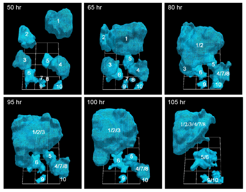
The team discovered that cancerous cells, moving at move at 92 micrometers per hour (about twice the speed of healthy cells), actively recruit healthy cells into tumors by extending a kind of cable to grab their neighbors — both cancerous and healthy — and reel them in. Surprisingly, as little as five percent of cancerous cells are needed to form the tumors, a ratio previously unknown.

“It’s not like things sticking to each other,” said David Soll, biology professor at the UI and corresponding author on the open-access paper, published in the American Journal of Cancer Research. “It’s that these cells go out and actively recruit. It’s complicated stuff, and it’s not passive. No one had a clue that there were specialized cells in this process, and that it’s a small number that pulls all the rest in.”

The findings could lead to a more precise identification of tumorigenic cells (those that form tumors) and to testing which antibodies would be best equipped to eliminate them.*

How cancer cells “know” what to do

The question is: how do these cells know what to do. Soll hypothesizes they’re reaching back to a primitive past, when these cells were programmed to form embryos. If true, perhaps the cancerous cells — masquerading as embryo-forming cells — recruit other cells to make tissue that then forms the layered, self-sustaining architecture needed for a tumor to form and thrive. “It’s as if it’s building its own defenses against the body’s efforts to defeat them.”

http://www.kurzweilai.net/images/Cancer-Cells-50-105-hours-stacked.png

University of Iowa researchers have documented how cancerous tumors form by tracking in real time the movement of individual cells in 3-D. They report that just 5 percent of cancer cells are needed to form tumors, a ratio that heretofore had been unknown. (credit: Soll Laboratory)

In the AJCR paper, the researchers found support for their previous observation that tumorigenic cell lines and fresh tumor cells possess the unique capacity to form tumors by the active formation of cellular cables.

The finding lends more weight to the idea that tumors are created concurrently, in multiple locations, by individual clusters of cells that employ the cancer-cell cables to draw in more cells and enlarge themselves. Some have argued that tumors come about more by cellular changes within the masses, known as the “cancer stem cell theory.”

The Developmental Studies Hybridoma Bank funded the study.

* Soll’s Monoclonal Antibody Research Institute and the Developmental Studies Hybridoma Bank, created by the National Institutes of Health as a national resource, directed by Soll and housed at the UI, together contain one of the world’s largest collections of antibodies that could be used for the anti-cancer testing, based on the new findings.


Abstract of Mediated coalescence: a possible mechanism for tumor cellular heterogeneity

Recently, we demonstrated that tumorigenic cell lines and fresh tumor cells seeded in a 3D Matrigel model, first grow as clonal islands (primary aggregates), then coalesce through the formation and contraction of cellular cables. Non-tumorigenic cell lines and cells from normal tissue form clonal islands, but do not form cables or coalesce. Here we show that as little as 5% tumorigenic cells will actively mediate coalescence between primary aggregates of majority non-tumorigenic or non-cancerous cells, by forming cellular cables between them. We suggest that this newly discovered, specialized characteristic of tumorigenic cells may explain, at least in part, why tumors contain primarily non-tumorigenic cells.


Read Full Post »

Reverse Engineering of Vision

Larry H. Bernstein, MD, FCAP, Curator

LPBI


CMU announces research project to reverse-engineer brain algorithms, funded by IARPA

A Human Genome Project-level plan to make computers learn like humans
February 5, 2016   http://www.kurzweilai.net/cmu-announces-research-project-to-reverse-engineer-brain-algorithms-funded-by-iarpa

http://www.kurzweilai.net/images/neural-network-CMU.jpg

Individual brain cells within a neural network are highlighted in this image obtained using a fluorescent imaging technique (credit: Sandra Kuhlman/CMU)

Carnegie Mellon University is embarking on a five-year, $12 million research effort to reverse-engineer the brain and “make computers think more like humans,” funded by the U.S. Intelligence Advanced Research Projects Activity (IARPA). The research is led by Tai Sing Lee, a professor in the Computer Science Department and the Center for the Neural Basis of Cognition (CNBC).

The research effort, through IARPA’s Machine Intelligence from Cortical Networks (MICrONS) research program, is part of the U.S. BRAIN Initiative to revolutionize the understanding of the human brain.

A “Human Genome Project” for the brain’s visual system

“MICrONS is similar in design and scope to the Human Genome Project, which first sequenced and mapped all human genes,” Lee said. “Its impact will likely be long-lasting and promises to be a game changer in neuroscience and artificial intelligence.”

The researchers will attempt to discover the principles and rules the brain’s visual system uses to process information. They believe this deeper understanding could serve as a springboard to revolutionize machine learning algorithms and computer vision.

In particular, the researchers seek to improve the performance of artificial neural networks — computational models for artificial intelligence inspired by the central nervous systems of animals. Interest in neural nets has recently undergone a resurgence thanks to growing computational power and datasets. Neural nets now are used in a wide variety of applications in which computers can learn to recognize faces, understand speech and handwriting, make decisions for self-driving cars, perform automated trading and detect financial fraud.

How neurons in one region of the visual cortex behave

“But today’s neural nets use algorithms that were essentially developed in the early 1980s,” Lee said. “Powerful as they are, they still aren’t nearly as efficient or powerful as those used by the human brain. For instance, to learn to recognize an object, a computer might need to be shown thousands of labeled examples and taught in a supervised manner, while a person would require only a handful and might not need supervision.”

To better understand the brain’s connections, Sandra Kuhlman, assistant professor of biological sciences at Carnegie Mellon and the CNBC, will use a technique called “two-photon calcium imaging microscopy” to record signaling of tens of thousands of individual neurons in mice as they process visual information, an unprecedented feat. In the past, only a single neuron, or tens of neurons, typically have been sampled in an experiment, she noted.

“By incorporating molecular sensors to monitor neural activity in combination with sophisticated optical methods, it is now possible to simultaneously track the neural dynamics of most, if not all, of the neurons within a brain region,” Kuhlman said. “As a result we will produce a massive dataset that will give us a detailed picture of how neurons in one region of the visual cortex behave.”

A multi-institution research team

Other collaborators are Alan Yuille, the Bloomberg Distinguished Professor of Cognitive Science and Computer Science at Johns Hopkins University, and another MICrONS team at the Wyss Institute for Biologically Inspired Engineering, led by George Church, professor of genetics at Harvard Medical School.

The Harvard-led team, working with investigators at Cold Spring Harbor Laboratory, MIT, and Columbia University, is developing revolutionary techniques to reconstruct the complete circuitry of the neurons recorded at CMU. The database, along with two other databases contributed by other MICrONS teams, unprecedented in scale, will be made publicly available for research groups all over the world.

In this MICrONS project, CMU researchers and their collaborators in other universities will use these massive databases to evaluate a number of computational and learning models as they improve their understanding of the brain’s computational principles and reverse-engineer the data to build better computer algorithms for learning and pattern recognition.

“The hope is that this knowledge will lead to the development of a new generation of machine learning algorithms that will allow AI machines to learn without supervision and from a few examples, which are hallmarks of human intelligence,” Lee said.

The CNBC is a collaborative center between Carnegie Mellon and the University of Pittsburgh. BrainHub is a neuroscience research initiative that brings together the university’s strengths in biology, computer science, psychology, statistics and engineering to foster research on understanding how the structure and activity of the brain give rise to complex behaviors.

The MICrONS team at CMU allso includes Abhinav Gupta, assistant professor of robotics; Gary Miller, professor of computer science; Rob Kass, professor of statistics and machine learning and interim co-director of the CNBC; Byron Yu, associate professor of electrical and computer engineering and biomedical engineering and the CNBC; Steve Chase, assistant professor of biomedical engineering and the CNBC; and Ruslan Salakhutdinov, one of the co-creators of the deep belief network, a new model of machine learning that was inspired by recurrent connections in the brain, who will join CMU as an assistant professor of machine learning in the fall.

Other members of the team include Brent Doiron, associate professor of mathematics at Pitt, and Spencer Smith, assistant professor of neuroscience and neuro-engineering at the University of North Carolina.

Not all machine-intelligence experts are on board with reverse-engineering the brain. In a Facebook post today, Yann LeCun, Director of AI Research at Facebook and a professor at New York University, asked the question in a recent lecture, “Should we copy the brain to build intelligent machines?” “My answer was ‘no, because we need to understand the underlying principles of intelligence to know what to copy. But we should draw inspiration from biology.’”


Read Full Post »

« Newer Posts - Older Posts »